Achieving High Signal-to-Noise Ratio in FISH: A Comprehensive Guide from Probe Design to Automated Analysis
This article provides a systematic framework for researchers and drug development professionals to maximize the signal-to-noise ratio (SNR) in Fluorescence in situ Hybridization (FISH) experiments.
Achieving High Signal-to-Noise Ratio in FISH: A Comprehensive Guide from Probe Design to Automated Analysis
Abstract
This article provides a systematic framework for researchers and drug development professionals to maximize the signal-to-noise ratio (SNR) in Fluorescence in situ Hybridization (FISH) experiments. Covering foundational principles to advanced applications, we detail how computational probe design with tools like TrueProbes enhances specificity, how signal amplification methods such as SABER and HCR boost sensitivity, and how optimized protocols and clearing techniques improve sample integrity. Furthermore, we explore the critical role of AI-powered spot detection with U-FISH and RS-FISH in accurate validation and quantification. This guide synthesizes the latest methodological advances and troubleshooting strategies to ensure precise, reliable, and quantifiable FISH data for spatial genomics and transcriptomics.
The Core Principles of FISH Signal and Noise
Defining Signal-to-Noise Ratio (SNR) in FISH Imaging
In fluorescence in situ hybridization (FISH) imaging, the signal-to-noise ratio (SNR) is a fundamental quantitative metric that defines the ability to distinguish specific probe-derived fluorescence (signal) from non-specific background interference (noise). Achieving a high SNR is paramount for the accuracy and reliability of FISH-based analyses, as it directly impacts sensitivity, resolution, and the validity of quantitative measurements [1]. Within the broader thesis on principles of high SNR in FISH research, this guide explores the core components of SNR, the methodologies for its quantification, and the comprehensive experimental strategies employed to enhance it. The pursuit of superior SNR is not merely a technical exercise; it is the cornerstone for generating biologically definitive data, particularly in advanced applications like spatial transcriptomics and single-molecule detection [2] [3].
The challenge of SNR is multifaceted. The "signal" originates from fluorophores bound to oligonucleotide probes that have hybridized to their target nucleic acid sequences. In contrast, "noise" is a composite of various sources, including autofluorescence from endogenous tissue components, non-specific binding of probes to off-target sites, and optical limitations of the imaging system itself [4] [3]. In plant tissues, for instance, high autofluorescence has historically prevented the detection of single mRNA molecules in intact tissues, a hurdle only recently overcome through optimized clearing protocols [3]. Similarly, in thick animal tissues, light scattering significantly degrades SNR, necessitating the use of optical clearing techniques [5]. This guide details the core principles and practical protocols essential for navigating these challenges and achieving the high SNR required for cutting-edge research and diagnostics.
Core Components and Quantification of SNR in FISH
The following table breaks down the primary contributors to signal and noise in a FISH experiment, which are critical for understanding where to focus optimization efforts.
Table 1: Core Components of Signal and Noise in FISH Imaging
| Component | Source | Impact on SNR |
|---|---|---|
| Specific Signal | Fluorescent probes bound to the target mRNA/DNA sequence. | The primary source of usable data. Amplification strategies (e.g., HCR, SABER) directly increase this component [4]. |
| Background Noise | Autofluorescence from lipids, proteins, and other endogenous molecules in the tissue [5] [3]. | A major source of noise, particularly in plant and brain tissues. Can obscure true signal, especially for low-abundance targets. |
| Technical Noise | Non-specific binding of probes to off-target sequences [1] [6]. | Creates false-positive spots and elevates background, directly reducing quantitative accuracy. |
| Optical Noise | Light scattering within the tissue and detector read noise [5]. | Reduces contrast and sharpness, limiting imaging depth and resolution. |
Quantitative Measures of SNR
Quantifying SNR is essential for objective protocol evaluation and validation. While specific calculations can vary, the underlying principle involves comparing the intensity of the specific signal against the variability of the background.
- Spot Intensity vs. Local Background: For single-molecule RNA-FISH (smFISH), where transcripts appear as distinct diffraction-limited spots, a common metric involves measuring the mean intensity of individual spots and dividing it by the standard deviation of the background intensity in a nearby region devoid of spots [1] [2]. This measure is crucial for the accurate detection and counting of individual RNA molecules.
- Algorithmic and Software-Based Quantification: Advanced analysis pipelines like FISH-quant and U-FISH incorporate robust algorithms to calculate SNR during automated spot detection [2] [3]. These tools often use a pixel-wise intensity comparison or a signal-to-background ratio to distinguish true transcripts from noise. The performance of these tools is frequently benchmarked using metrics like the F1 score (the harmonic mean of precision and recall), which indirectly reflects the effective SNR of the image, as a higher SNR allows for more precise and accurate spot detection [2].
Experimental Strategies for SNR Enhancement
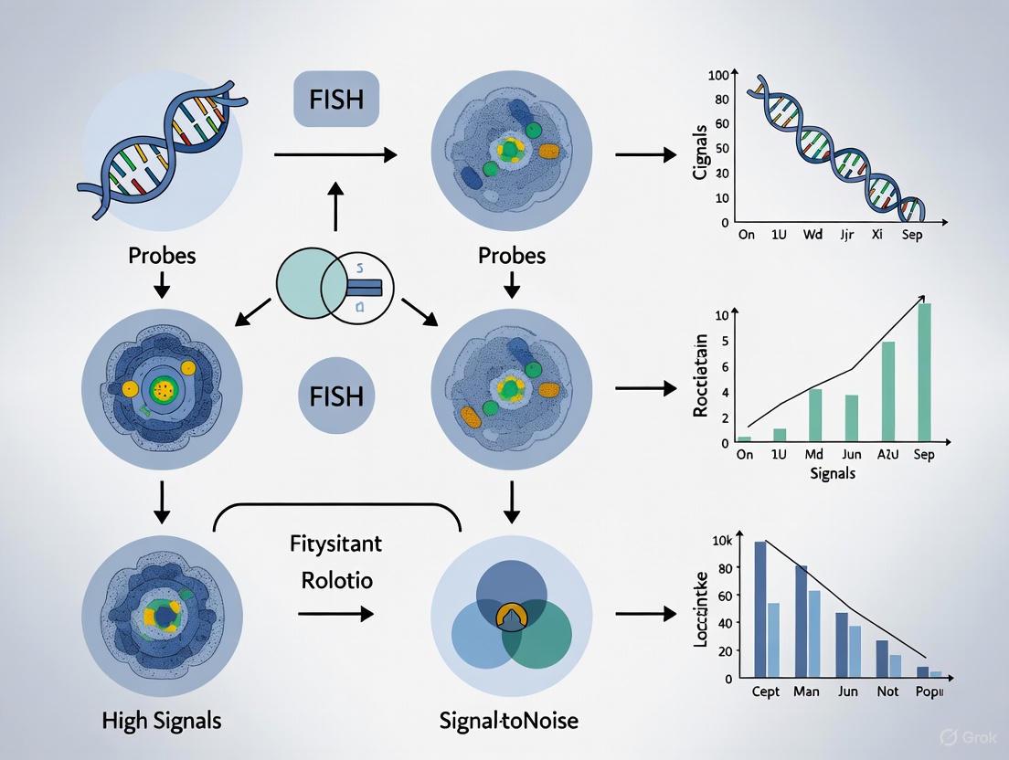
Achieving a high SNR requires a holistic approach, integrating optimized probe design, sophisticated tissue processing, and advanced imaging and computational techniques. The following diagram illustrates the interconnected strategies explored in this section.
Figure 1: A strategic framework for enhancing the Signal-to-Noise Ratio (SNR) in FISH imaging, spanning biochemical, optical, and computational methods.
Probe Design for Optimal Specificity
The foundation of a high SNR is laid at the probe design stage. Optimal probes exhibit maximal binding to the intended target and minimal interaction with off-target sequences.
- Computational Design and Off-Target Assessment: Modern tools like TrueProbes go beyond traditional filters for GC content and melting temperature. They perform a genome-wide BLAST-based analysis to quantify and minimize potential off-target binding, a major source of background noise. By ranking probes based on predicted binding affinity and specificity, these platforms generate probe sets with superior experimental performance [1].
- Incorporation of Expression Data: Advanced design pipelines can incorporate tissue- or cell-type-specific gene expression data. This allows the algorithm to penalize probes that have complementarity to off-target transcripts known to be expressed in the sample, further reducing the risk of non-specific background [1].
Tissue Processing and Optical Clearing
Tissue preparation is critical for probe accessibility and light penetration, directly influencing SNR.
- Fixation and Permeabilization: 10% Neutral Buffered Formalin (NBF) is the standard fixative for optimal RNA preservation in FFPE tissues. Fixation time must be carefully controlled; under-fixation leads to RNA degradation, while over-fixation requires harsher permeabilization that can damage morphology. Permeabilization with detergents like Triton X-100 or proteases like proteinase K is essential to allow probe entry [6].
- Optical Clearing with LIMPID: The LIMPID (Lipid-preserving refractive index matching for prolonged imaging depth) method is a single-step aqueous clearing protocol. It uses a solution of saline-sodium citrate, urea, and iohexol to match the refractive index of the tissue to that of the objective lens (e.g., 1.515). This dramatically reduces light scattering, minimizes spherical aberrations, and enables high-resolution imaging deep within thick tissues without damaging the tissue structure or fluorescent signals [5].
- Bleaching and Preservation: A hydrogen peroxide (Hâ‚‚Oâ‚‚) bleaching step can be incorporated to reduce autofluorescence, a significant noise component. Furthermore, omitting delipidation steps helps preserve tissue structure and the integrity of lipophilic dyes, providing a more native biological context [5].
Signal Amplification and Multiplexing
For low-abundance targets, simply increasing the number of fluorophores is a direct method to boost signal above the background noise level.
- Hybridization Chain Reaction (HCR): HCR is a powerful linear amplification scheme that builds long chains of fluorescently labeled probes upon initiation by a target-specific probe. This method is noted for its high specificity, low background, and, crucially, its quantitative nature, as the fluorescence intensity scales linearly with the target RNA quantity [5] [4]. This makes it ideal for quantifying gene expression levels.
- Barcoding for Multiplexing: Techniques like MERFISH (Multiplexed Error-robust FISH) use combinatorial barcoding to detect thousands of RNA species simultaneously. Each RNA is assigned a unique binary barcode, which is read out over multiple rounds of hybridization with fluorescent readout probes. The error-robust encoding schemes in MERFISH require multiple bits to be misread for a misidentification, thereby enhancing the effective SNR for accurate transcript identification in highly complex multiplexed experiments [7].
Computational and Deep Learning Enhancement
Post-acquisition computational methods have emerged as a powerful tool for enhancing SNR without altering wet-lab protocols.
- U-FISH: A Universal Deep Learning Model: U-FISH is a deep learning method (U-Net based) trained on a massive dataset of over 4000 images and 1.6 million signal spots. It acts as a universal image enhancer, transforming raw FISH images with variable characteristics into images with uniform spot properties and a drastically improved SNR. This allows for consistent and precise spot detection across diverse datasets without manual parameter tuning, outperforming many rule-based and other deep learning methods in accuracy and generalizability [2].
- Integrated Analysis Pipelines: For whole-mount samples, combining confocal imaging with analysis software is key. A typical workflow involves cell segmentation using Cellpose (based on cell wall staining), followed by mRNA dot counting with FISH-quant, and finally protein intensity measurement with CellProfiler. This integrated pipeline allows for the precise correlation of mRNA and protein levels at single-cell resolution, a process entirely dependent on a high initial SNR [3].
Detailed Experimental Protocol: 3D-LIMPID-FISH
The following protocol, adapted from a published whole-mount 3D FISH methodology, encapsulates multiple SNR enhancement strategies into a coherent workflow [5].
Table 2: Key Research Reagent Solutions for 3D-LIMPID-FISH
| Reagent / Solution | Function / Explanation |
|---|---|
| Paraformaldehyde (PFA) | Cross-linking fixative for tissue preservation and RNA integrity. |
| Hydrogen Peroxide (Hâ‚‚Oâ‚‚) | Chemical bleaching agent to reduce tissue autofluorescence. |
| HCR FISH Probes | Target-specific initiator probes for hybridization chain reaction, enabling sensitive, amplified signal. |
| Proteinase K | Protease for tissue permeabilization, enabling probe access to intracellular targets. |
| LIMPID Solution | Aqueous clearing solution (SSC, Urea, Iohexol) for refractive index matching and tissue transparency. |
| Iohexol | Key component of LIMPID; adjusts the refractive index of the solution to match that of the tissue and objective lens. |
Workflow Timetable:
- Sample Extraction & Fixation: Dissect tissue and fix immediately in 4% PFA for 24 hours at 4°C to preserve morphology and RNA.
- Bleaching (Optional): Incubate tissue in 3% Hâ‚‚Oâ‚‚ for several hours to reduce autofluorescence.
- Permeabilization: Treat tissue with Proteinase K (e.g., 10 μg/mL for 30 minutes) to digest proteins and allow probe penetration. Conditions must be optimized for each tissue type.
- Hybridization and Amplification:
- Hybridize with HCR initiator probes specific to the target mRNA(s) in a humidified chamber (overnight, 37°C).
- Wash stringently to remove unbound probes.
- Add HCR amplification hairpins (fluorescently labeled) for 2-6 hours to build the amplification polymer.
- Optical Clearing: Mount the tissue in the LIMPID solution. The clearing occurs in a single step through passive diffusion, typically within a few hours.
- Imaging: Image using a confocal microscope with a high-NA objective lens. The refractive index of the LIMPID solution should be calibrated to match that of the immersion oil (e.g., 1.515) for minimal aberration [5].
Defining and optimizing the signal-to-noise ratio is a multidimensional challenge at the heart of rigorous and quantitative FISH research. As this guide outlines, there is no single solution; instead, high SNR is achieved through a synergistic combination of intelligent probe design, gentle yet effective tissue processing, robust signal amplification, and powerful computational cleanup. The integration of these strategies, as exemplified by the 3D-LIMPID-FISH protocol, empowers researchers to push the boundaries of spatial biology. By systematically applying these principles, scientists can reliably extract single-molecule quantitative data from complex tissues, thereby unlocking deeper insights into gene expression and regulation with unparalleled spatial context.
In fluorescence in situ hybridization (FISH) research, achieving a high signal-to-noise ratio (SNR) is paramount for accurate biological interpretation. This ratio determines the confidence with which researchers can detect and quantify molecular targets, such as RNA transcripts or DNA sequences, within their native cellular context. Two fundamental pillars govern this critical parameter: probe binding affinity, which ensures specific localization to the intended target, and fluorophore efficiency, which dictates the brightness and detectability of the resulting signal. This technical guide delves into the core principles and advanced methodologies for optimizing these two sources of signal, providing researchers and drug development professionals with a framework for designing robust and reliable FISH-based assays.
Probe Binding Affinity: The Foundation of Specificity
Probe binding affinity refers to the strength and specificity with which an oligonucleotide probe hybridizes to its complementary nucleic acid target. High affinity is essential for maximizing the on-target signal while minimizing off-target binding, which contributes to background noise.
Key Factors Influencing Probe Binding Affinity
- Sequence Specificity: The degree of complementarity between the probe and its target is the primary determinant of binding affinity and specificity. Even minor mismatches can significantly reduce hybridization stability.
- Melting Temperature (Tm): This is the temperature at which half of the probe-target duplexes dissociate. Optimal FISH conditions require a Tm that allows for stable hybridization under stringent conditions that discourage off-target binding [1].
- Probe Length: The length of the targeting region modulates both affinity and specificity. While longer probes (e.g., 40-50 nt) can form more stable duplexes, they also have a higher probability of non-specific binding to related sequences. Shorter probes (e.g., 20 nt) offer higher specificity but may suffer from reduced brightness [8].
- GC Content: The proportion of guanine and cytosine bases in the probe sequence affects Tm, as GC base pairs form three hydrogen bonds compared to the two in AT pairs. Most design tools enforce a narrow GC content window (e.g., 40-60%) to ensure uniform Tm across a probe set [1].
- Secondary Structure: Intramolecular folding of either the probe or the target RNA can occlude binding sites, dramatically reducing effective binding affinity. Computational tools must account for these structures during design [1] [9].
Computational Design for Optimal Affinity
Advanced probe design software platforms, such as TrueProbes, have moved beyond simple filtering heuristics to integrated thermodynamic and kinetic modeling [1] [9]. These tools perform genome-wide BLAST analyses to comprehensively assess off-target binding potential and rank candidate probes based on a holistic score that incorporates:
- Predicted on-target binding energy
- Number and affinity of off-target binding sites
- Potential for self-hybridization or cross-dimerization with other probes in the set
- User-provided gene expression data to weight the impact of expressed off-targets [1]
This integrated approach consistently outperforms earlier methods like Stellaris and Oligostan-HT, generating probe sets with enhanced target selectivity and superior experimental performance [1] [9].
Table 1: Comparison of smFISH Probe Design Software
| Software | Design Approach | Key Features | Specificity Assessment |
|---|---|---|---|
| TrueProbes | Genome-wide BLAST + thermodynamic modeling | Ranks all candidates globally by specificity; incorporates expression data | Genome-wide off-target binding analysis |
| Stellaris | Sequential 5' to 3' tiling with filtering | Applies GC content filters and repetitive sequence masking | Limited off-target assessment with five masking levels [1] |
| MERFISH | Hash-based transcriptome screening | Filters oligos based on off-target index and rRNA binding | Computes off-target index using 15/17-mer hashing [1] |
| Oligostan-HT | Energy-based ranking | Ranks probes by Gibbs free energy (ΔG°) proximity to optimum | Applies GC and low-complexity screens [1] |
| PaintSHOP | Machine learning classification | Uses Bowtie2 alignment and ML classifier for deleterious duplexes | Eliminates oligos with many genomic matches [1] |
Experimental Optimization of Binding Conditions
Even optimally designed probes require careful experimental calibration. Key parameters to optimize include:
- Formamide Concentration: Acts as a denaturant to enable precise stringency control. Optimal concentrations must be determined empirically for different target region lengths [8].
- Hybridization Temperature: Typically performed 10-20°C below the average probe Tm to ensure specific binding while allowing access to structured regions.
- Hybridization Duration: Must balance complete target access with practical experimental timelines. For complex samples, hybridization may require 24-48 hours [8].
- Salt Concentration: Ionic strength affects duplex stability by shielding the negative charges on the phosphate backbones of hybridized nucleic acids.
Table 2: Experimental Parameters for Optimizing Probe Binding
| Parameter | Impact on Binding Affinity | Typical Optimization Range | Effect on SNR |
|---|---|---|---|
| Formamide Concentration | Reduces Tm; increases stringency | 0-40% in 5% increments [8] | Reduces background from off-target binding at optimal concentration |
| Hybridization Temperature | Must be below Tm for binding to occur | Tm -20°C to Tm -10°C | Higher temperature increases specificity but may reduce on-target signal |
| Hybridization Duration | Allows probes to reach and bind targets | 1-48 hours depending on sample and probe accessibility [8] | Longer duration increases signal until equilibrium is reached |
| Salt Concentration | Higher concentration stabilizes duplex | 100-900 mM monovalent cations | Insufficient salt reduces on-target signal; excess may increase background |
Fluorophore Efficiency: Maximizing Signal Output
Fluorophore efficiency encompasses the physical properties that determine how effectively a fluorophore converts excitation light into detectable emission. This directly impacts the signal intensity in FISH experiments.
Photophysical Properties Governing Efficiency
- Extinction Coefficient: A measure of how strongly a fluorophore absorbs light at a specific wavelength. Higher values indicate greater photon absorption capacity [10].
- Quantum Yield: The ratio of photons emitted to photons absorbed, representing the efficiency of the fluorescence process. Fluorophores with quantum yields closer to 1.0 are intrinsically brighter [10].
- Photostability: The resistance of a fluorophore to photobleaching, the irreversible destruction of the fluorophore under intense illumination. This is critical for time-lapse experiments or those requiring extended imaging sessions.
- Stokes Shift: The difference between the excitation and emission maxima. A larger Stokes shift facilitates spectral separation, reducing background from scattered excitation light [10].
Fluorescence Resonance Energy Transfer (FRET) Applications
FRET is a distance-dependent phenomenon where an excited donor fluorophore non-radiatively transfers energy to a nearby acceptor fluorophore [11] [12]. The efficiency of FRET decreases with the sixth power of the distance between the fluorophores, making it exquisitely sensitive to molecular-scale distances (1-10 nm) [11] [12] [13]. In FISH, FRET can be utilized in:
- Molecular Beacons: Hairpin probes where target binding separates a fluorophore-quencher pair, generating a signal increase.
- Biosensors: Probes designed to change FRET efficiency upon target binding or enzymatic activity.
- Protease Assays: FRET-based substrates where cleavage separates donor and acceptor fluorophores, altering the emission ratio [12] [13].
Advanced Signal Amplification Strategies
To overcome the inherent limitation of labeling individual transcripts with few fluorophores, several amplification strategies have been developed:
- SABER (Signal Amplification By Exchange Reaction): A method that uses primer exchange reaction to generate long concatemeric probes containing multiple binding sites for fluorescently labeled imager probes. This significantly increases the number of fluorophores per target molecule [14].
- HCR (Hybridization Chain Reaction): An enzyme-free method where metastable DNA hairpins self-assemble into long amplification polymers upon initiation by a specific probe [14].
- Tyramide Signal Amplification (TSA): An enzyme-based method where horseradish peroxidase (HRP) catalyzes the deposition of multiple fluorescent tyramide molecules near the probe binding site [14].
Table 3: Characteristics of Common Fluorophores and Applications
| Fluorophore | Excitation/Emission Max (nm) | Extinction Coefficient (Mâ»Â¹cmâ»Â¹) | Quantum Yield | Common FISH Applications |
|---|---|---|---|---|
| FITC | 495/519 | ~68,000 | 0.79 | Direct labeling; immuno-detection of haptens [15] |
| Cy3 | 550/570 | ~150,000 | 0.15 | Bright, photostable; common for smFISH |
| Alexa Fluor 488 | 495/519 | ~73,000 | 0.92 | High quantum yield; superior to FITC |
| Texas Red | 589/615 | ~85,000 | 0.90 | Red-emitting dye for multiplexing |
| Cy5 | 649/670 | ~250,000 | 0.28 | Far-red emission; low background in tissues |
Integrated Approaches for Maximizing Signal-to-Noise Ratio
The ultimate SNR in a FISH experiment emerges from the interplay between probe binding affinity and fluorophore efficiency. Strategic integration of both aspects is essential.
The Scientist's Toolkit: Essential Research Reagents
Table 4: Key Research Reagent Solutions for FISH Optimization
| Reagent / Material | Function | Application Note |
|---|---|---|
| Encoding Probes (e.g., MERFISH) | Unlabeled DNA probes with targeting and barcode regions | Enable massive multiplexing via two-step hybridization [8] |
| Readout Probes | Fluorescently labeled probes complementary to barcode regions | Allow rapid signal readout in sequential rounds [8] |
| Formamide | Chemical denaturant for stringency control | Concentration must be optimized for each probe set [8] |
| Dextran Sulfate | Crowding agent that increases effective probe concentration | Accelerates hybridization kinetics |
| Blocking DNA (e.g., salmon sperm DNA) | Competes with non-specific probe binding | Reduces background in complex samples |
| Antifade Reagents (e.g., ProLong Diamond) | Reduces photobleaching during imaging | Essential for preserving signal in 3D or time-lapse experiments |
| PER Catalytic Hairpin | Enables primer exchange reaction for SABER | Generates long concatemers for signal amplification [14] |
| Stachyose tetrahydrate | Stachyose tetrahydrate, CAS:10094-58-3, MF:C24H44O22, MW:684.6 g/mol | Chemical Reagent |
| Neocaesalpin L | Neocaesalpin L|For Research | Neocaesalpin L, a cassane diterpenoid from Caesalpinia minax. For research use only. Not for human or veterinary diagnostic or therapeutic use. |
Protocol: Multiplexed Error-Robust FISH (MERFISH) with Enhanced SNR
This protocol incorporates recent optimizations from systematic performance evaluations [8]:
Probe Design
- Design encoding probes with 30-50 nt target-specific regions for optimal binding affinity and signal brightness [8].
- Incorporate readout sequences for subsequent fluorescent detection.
- Use computational tools (e.g., TrueProbes) to filter probes with potential off-target binding.
Sample Preparation
- Fix cells or tissues with appropriate cross-linking agents (e.g., 4% PFA).
- Permeabilize with 0.1-0.5% Triton X-100 for 30 minutes.
- Include an RNase inhibitor in all solutions.
Hybridization Optimization
- Prepare hybridization buffer with 10-30% formamide, optimized for your specific probe set [8].
- Add encoding probes at 1-10 nM concentration.
- Hybridize for 24-48 hours at 37°C in a humidified chamber.
Signal Readout and Imaging
- Hybridize fluorescent readout probes at 5-20 nM concentration for 30 minutes at room temperature.
- Use optimized imaging buffers with photostabilizing components (e.g., Trolox, PCA/PCD) to enhance fluorophore efficiency.
- Image sequentially with appropriate excitation/emission filter sets, ensuring sufficient spectral separation to minimize bleed-through.
In the pursuit of high signal-to-noise ratio in FISH research, probe binding affinity and fluorophore efficiency represent two complementary frontiers for optimization. Advances in computational probe design, particularly those incorporating genome-wide specificity analysis and thermodynamic modeling, have dramatically improved our ability to generate probes with minimal off-target binding. Concurrently, innovations in fluorophore chemistry and signal amplification strategies have pushed the boundaries of detection sensitivity. By systematically addressing both domains—through careful probe design, experimental condition optimization, and strategic fluorophore selection—researchers can achieve the precise, quantitative measurements required for cutting-edge research and drug development. The integrated approaches outlined in this guide provide a pathway to maximizing SNR, ultimately enabling more confident biological discoveries through FISH-based spatial transcriptomics and genomics.
In Fluorescence In Situ Hybridization (FISH) research, the signal-to-noise ratio is the cornerstone of assay sensitivity, reliability, and quantitative capability. Achieving a high signal-to-noise ratio is paramount for the accurate detection of nucleic acid targets, whether for basic research, clinical diagnostics, or drug development. The three predominant sources of noise that compromise this ratio are off-target binding, autofluorescence, and general background fluorescence. Off-target binding introduces false-positive signals through non-specific probe hybridization, while autofluorescence arises from the innate fluorescent properties of biological samples and fixatives. Background fluorescence often stems from suboptimal assay conditions. This whitepaper provides an in-depth technical analysis of these noise sources, detailing their mechanisms and presenting validated experimental strategies for their mitigation, thereby providing a framework for optimizing FISH assays.
Off-Target Binding: Mechanisms and Mitigation
Off-target binding occurs when FISH probes hybridize to non-intended genomic sequences, producing false-positive signals that can lead to erroneous biological interpretations. Understanding and controlling this phenomenon is critical for quantitative accuracy.
Fundamental Mechanisms
The primary mechanism of off-target binding is the presence of short, perfectly repeated sequences, or k-mers, within longer probe sequences. Counterintuitively, very short perfect repeats of only 20–25 base pairs within probes that are several hundred nucleotides long can generate significant off-target signals [16]. The surprising extent of this noise is attributable to the signal amplification conferred by haptenylated probes and subsequent immunological detection, which can magnify even a single non-specific hybridization event into a detectable fluorescent signal [16]. Traditional control methods, such as using sense-strand probes, are inadequate for detecting this type of noise because they do not share the same sequence and thus the same repertoire of repetitive k-mers as the antisense probe [16].
Experimental Protocol for k-mer Uniqueness Analysis
A critical step in probe design is the computational assessment of sequence uniqueness to eliminate repetitive k-mers.
- Sequence Input: Input the candidate probe sequence(s) in FASTA format.
- k-mer Enumeration: The algorithm enumerates all possible sub-sequences of a defined length (k-mer, e.g., 20 nt) from the entire reference genome and the probe sequence.
- Sorting and Counting: All k-mers are sorted alphabetically. The algorithm then counts the frequency of each unique k-mer across the genome.
- Probe Interrogation: The probe sequence is scanned, and each of its constituent k-mers is queried against the generated database. K-mers with a frequency greater than one (i.e., present in more than one genomic location) are flagged as repetitive.
- Probe Optimization: Probe sequences are redesigned to eliminate or minimize the presence of flagged repetitive k-mers. This can be achieved by selecting alternative probe templates from different regions of the target gene (e.g., the 3'UTR) that are devoid of such repeats [16].
This process is facilitated by publicly available algorithms, such as the one provided at http://cbio.mskcc.org/∼aarvey/repeatmap, which generates genome-wide annotations of k-mer uniqueness [16].
Quantitative Impact of Probe Optimization
Table 1: Impact of Removing Repeated k-mers on FISH Signal-to-Noise Ratio
| Probe Target | Probe Type | Probe Length (bp) | Key Finding | Reference |
|---|---|---|---|---|
| Drosophila melanogaster Scr | Non-unique Probe | 357 | Produced significant off-target noise, obscuring true expression patterns | [16] |
| Drosophila melanogaster Scr | Unique Probe (repeat-free) | 359 | Increased signal-to-noise ratio by orders of magnitude; enabled quantitative RNA counting | [16] |
| Drosophila melanogaster abd-A | Non-unique Probe | ~1,600 | Generated off-target signals, reducing assay specificity | [16] |
| Drosophila melanogaster abd-A | Unique Probe (repeat-free) | ~1,470 | Drastically reduced background, allowing for high-confidence, quantitative detection | [16] |
Diagram 1: Workflow for optimizing probe specificity through k-mer analysis.
Autofluorescence: Characterization and Correction
Autofluorescence is the background fluorescence inherent to biological samples, which can mask specific FISH signals, particularly those from small or dim probes.
Autofluorescence originates from various intracellular components, including lipofuscin bodies, flavins, and cross-linked proteins induced by aldehyde-based fixation [17]. Its spectral signature is typically broader than that of synthetic fluorophores, often spanning a wide range of the visible spectrum. For example, lipofuscin can be detected from 360 nm to 650 nm [17]. Formaldehyde fixation tends to exacerbate autofluorescence by cross-linking fluorescent enzyme co-factors, whereas fixation with methanol/acetic acid can reduce it by washing these components away [17].
Strategic and Computational Mitigation
Several strategies can be employed to minimize or correct for autofluorescence:
- Fluorophore Selection: Using longer-wavelength fluorophores (e.g., Quasar 570/670, CAL Fluor Red 610) is highly recommended, as autofluorescence is more pronounced and intense in the green region of the spectrum (near 520 nm) [17].
- Enzymatic Reduction: A novel pretreatment protocol using elastase has been demonstrated to significantly reduce autofluorescence in non-small cell lung cancer (NSCLC) tissues. In a study of 120 samples, elastase pretreatment reduced the assay retest rate from 86.7% to 0% and enabled the detection of two additional ALK translocation cases that were indeterminable with standard pepsin pretreatment [18]. Elastase was identified as the most effective enzyme for this purpose while preserving nuclear morphology [18].
- Computational Image Correction: A pixel-by-pixel digital subtraction method can be applied. This technique leverages the fact that autofluorescence has broader excitation and emission spectra than FISH probes. The ratio of autofluorescence between multiple color channels is calculated and used to subtract the autofluorescence component from each image [19]. This method has been shown to enhance previously indistinguishable cosmid signals [19].
Protocol: Elastase Pretreatment for Autofluorescence Reduction
This protocol is adapted for formalin-fixed paraffin-embedded (FFPE) tissue sections [18].
- Dewaxing and Rehydration: Process FFPE sections through standard dewaxing and rehydration steps.
- Protease Digestion: Treat the tissue sections with a solution of elastase at an optimized concentration (determined empirically for each tissue type and fixation condition).
- Incubation: Incubate the slides at 37°C for a defined period, typically between 10-30 minutes.
- Washing: Rinse the slides thoroughly to stop the enzymatic reaction.
- Dehydration: Dehydrate the slides through an ethanol series and air-dry.
- FISH Assay: Proceed with the standard FISH denaturation, hybridization, and washing steps.
Background Fluorescence and Assay Optimization
Background fluorescence encompasses non-specific signals arising from suboptimal assay conditions, excluding autofluorescence and specific off-target binding. A systematic approach to the entire FISH workflow is essential for its reduction.
Integrated Workflow for Background Minimization
A robust FISH assay requires optimization at every stage, from sample preparation to final imaging. The following workflow integrates key mitigation strategies:
Diagram 2: Key stages in the FISH workflow and critical parameters for background control.
Detailed Mitigation Strategies
- Sample Preparation and Fixation: Fixation is a critical balance. Under-fixation leads to poor cellular preservation and increased non-specific probe binding, while over-fixation (particularly with formalin) causes excessive cross-linking, masking target sequences and increasing background [20]. For FFPE tissues, sections of 3-4 μm thickness are ideal for probe penetration and interpretation [20]. Always use freshly prepared fixative solutions.
- Pre-treatment and Digestion: The goal of pre-treatment is to unmask target nucleic acids. Insufficient pre-treatment leaves autofluorescent debris and non-specific binding sites, while over-digestion damages the sample and target DNA, reducing specific signal [20]. Use controlled heating (98–100°C) of a pre-treatment solution followed by enzymatic digestion (e.g., pepsin, or elastase for lung tissues) [20] [18].
- Hybridization and Denaturation: For FFPE tissues, denaturation conditions must be precisely controlled. A denaturation temperature that is too low prevents proper probe binding, whereas a temperature that is too high can promote non-specific binding [20]. Similarly, denaturation time must be optimized; short times reduce specific binding, and prolonged times increase background by unmasking non-target sites [20]. Probe volume should also be optimized to ensure sufficient coverage without excess.
- Post-Hybridization Washes: Stringency washes are crucial for removing unbound and weakly bound probes. Stringency is controlled by the pH, temperature, and salt concentration of the wash buffers [20]. Use freshly prepared wash buffers to prevent contamination or degradation that can contribute to background [20].
- Imaging and Filter Maintenance: The optical system itself can be a source of noise. Worn or damaged optical filters on fluorescence microscopes degrade over time (typically 2-4 years), producing a mottled appearance that weakens signal and increases background noise [20]. Regularly inspect and replace filters according to the manufacturer's guidelines.
Researcher's Toolkit: Essential Reagents for Noise Reduction
Table 2: Key Reagents and Their Functions in Optimizing FISH Assays
| Reagent / Material | Function / Purpose | Key Consideration for Noise Reduction |
|---|---|---|
| Elastase | Enzymatic pre-treatment to reduce autofluorescence | Particularly effective in lung tissues; preserves nuclear morphology better than pepsin [18]. |
| Methanol/Acetic Acid Fixative | Alternative to aldehyde-based fixation | Reduces autofluorescence by washing out fluorescent enzyme co-factors, unlike formaldehyde [17]. |
| Carnoy's Solution | Fixative for cytological preparations | Must be freshly prepared and stored at -20°C to maintain effectiveness and prevent moisture absorption [20]. |
| IntelliFISH Hybridization Buffer | Commercial hybridization buffer | Enables rapid hybridization (4 hours vs. 18 hours), improving workflow and maintaining good signal-to-noise [21]. |
| VECTASHIELD HardSet with DAPI | Antifade mounting medium with nuclear stain | Provides a hard-set mounting medium that reduces fading and is fast-setting for efficient workflow [21]. |
| CytoCell LPS 100 Tissue Pretreatment Kit | Standardized kit for tissue pre-treatment | Ensures consistent and optimized pre-treatment conditions for FFPE tissues, minimizing background [20]. |
| Long-wavelength Fluorophores(e.g., Quasar 670) | Label for FISH probes | Their emission spectra are in a region with lower inherent cellular autofluorescence, improving signal discernment [17]. |
| Mullilam diol | Mullilam diol, CAS:36150-04-6, MF:C10H20O3, MW:188.26 g/mol | Chemical Reagent |
| Eichlerianic acid | Eichlerianic acid, CAS:56421-13-7, MF:C30H50O4, MW:474.7 g/mol | Chemical Reagent |
The pursuit of a high signal-to-noise ratio is a fundamental principle in FISH research, directly determining the accuracy, sensitivity, and quantitative potential of the assay. As demonstrated, the major sources of noise—off-target binding, autofluorescence, and general background—can be systematically addressed through rigorous probe design, strategic use of reagents, and meticulous optimization of the entire experimental workflow. Employing computational tools to ensure probe uniqueness, integrating novel enzymatic treatments like elastase, and adhering to best practices in sample handling and imaging are not merely incremental improvements but essential steps for generating reliable, publication-quality data. By embracing this comprehensive approach to noise reduction, researchers and drug developers can fully leverage the power of FISH technology to uncover precise spatial gene expression patterns in complex biological systems.
The Critical Link Between High SNR and Accurate Single-Molecule Quantification
In the realm of molecular biology, single-molecule fluorescence in situ hybridization (smFISH) has emerged as a transformative technology that enables researchers to visualize, localize, and quantify individual RNA molecules within their native cellular environment with exceptional spatial resolution [22]. This technique provides unparalleled insights into gene expression regulation, transcriptional dynamics, and RNA localization patterns at the single-cell level, revealing cell-to-cell heterogeneity that bulk measurement techniques inevitably obscure [22] [23]. At the heart of this powerful methodology lies a critical technical parameter that fundamentally determines its accuracy and reliability: the signal-to-noise ratio (SNR).
The SNR in smFISH represents the quantitative relationship between the specific fluorescence signal emanating from target RNA molecules and the non-specific background noise originating from various sources, including autofluorescence, imperfect probe binding, and optical limitations of the imaging system [1]. A high SNR is not merely desirable but essential for accurate single-molecule quantification because it enables the unambiguous discrimination of individual transcripts from background fluorescence, ensuring that each counted spot genuinely represents a single RNA molecule [22] [24]. When SNR is compromised, the consequences for data integrity are severe: low signal intensity increases false negatives (missed transcripts), while elevated background noise introduces false positives (non-specific signals misclassified as transcripts), ultimately distorting quantitative conclusions about gene expression levels [1] [24].
This technical guide explores the fundamental principles governing SNR in smFISH experiments, detailing the experimental factors that influence this critical parameter and providing evidence-based strategies for its optimization. By examining the intricate relationship between SNR and quantification accuracy across diverse biological contexts—from model organisms to clinical samples—we aim to equip researchers with the knowledge necessary to design robust smFISH assays that yield biologically meaningful, reproducible results.
Theoretical Foundation: How SNR Directly Impacts Quantification Accuracy
The imperative for high SNR in smFISH stems from the fundamental physics of single-molecule detection and the statistical principles underlying transcript quantification. In a properly optimized smFISH experiment, each individual RNA molecule is tagged with multiple fluorescently labeled oligonucleotide probes, creating a diffraction-limited spot whose intensity is proportional to the number of bound probes [22]. The detection and enumeration of these spots through computational algorithms depend critically on the ability to distinguish true signals from background fluorescence with high confidence [25].
The quantitative relationship between SNR and detection accuracy can be expressed through statistical detection theory. When SNR is high, the intensity distribution of true RNA signals separates clearly from the intensity distribution of background noise, allowing detection algorithms to establish a threshold that captures nearly all true signals while excluding most noise [1] [25]. As SNR decreases, these distributions increasingly overlap, forcing a compromise between sensitivity (detection of true transcripts) and specificity (rejection of background), ultimately increasing both false negative and false positive rates [24].
This principle finds practical validation in the development of RollFISH, an advanced smFISH variant that incorporates rolling circle amplification to enhance signal intensity. In a direct comparison with conventional smFISH, RollFISH demonstrated that increased signal intensity directly translated to more accurate transcript counting, particularly in challenging samples like formalin-fixed, paraffin-embedded (FFPE) tissue sections where background fluorescence is typically elevated [24]. The method's signal amplification approach yielded a greater than two-fold improvement in SNR, which correspondingly enhanced the reliability of single-molecule detection and quantification across diverse tissue types [24].
Beyond mere transcript counting, high SNR is equally crucial for analyzing RNA subcellular localization patterns—a key application of smFISH. Low SNR can obscure the precise position of transcripts within cellular compartments, potentially leading to misinterpretation of localization patterns and their biological significance [25]. This is particularly critical when distinguishing between mature cytoplasmic mRNAs and nascent transcripts at transcription sites, or when identifying isoform-specific localization patterns that require multiple probe sets with different fluorophores [26] [23].
Table 1: Impact of SNR on Key smFISH Quantification Metrics
| Quantification Metric | High SNR Impact | Low SNR Consequences |
|---|---|---|
| Transcript Detection Efficiency | >90% detection of true transcripts [24] | Increased false negatives (>10% transcript loss) [1] |
| Detection Specificity | Minimal false positives (<5% background misclassification) [1] | Elevated false positive rates (>20% misclassification) [24] |
| Localization Accuracy | Precise subcellular positioning (<200nm resolution) [25] | Ambiguous localization patterns |
| Multi-Color Experiments | Clear signal separation for different targets [26] | Spectral bleed-through and cross-talk |
| Dynamic Range | Accurate counting across broad expression range (1-1000+ transcripts/cell) [22] | Limited to highly expressed genes |
Methodological Framework: Optimizing SNR in smFISH Workflows
Achieving high SNR in smFISH requires a systematic approach that addresses each stage of the experimental workflow, from probe design to image acquisition. The following diagram illustrates the integrated framework for SNR optimization, highlighting the interconnected factors that researchers must control throughout the smFISH procedure:
Probe Design Strategies for Enhanced Specificity and Signal
Probe design represents the foundational element in the pursuit of high SNR, as it directly determines both the intensity of the specific signal and the degree of non-specific background binding. Traditional smFISH employs multiple short DNA oligonucleotides (typically 20-mers) that tile the entire length of the target RNA, with each probe conjugated to a fluorescent dye [22]. The collective binding of these probes—typically 25-48 per target—generates a sufficiently bright signal to detect individual RNA molecules against the cellular background [26].
Modern computational approaches have significantly advanced probe design by systematically addressing the challenge of off-target binding. TrueProbes, a recently developed probe design platform, exemplifies this evolution by integrating genome-wide BLAST-based binding analysis with thermodynamic modeling to generate probe sets with enhanced specificity [1]. Unlike earlier tools that applied relatively simplistic heuristics, TrueProbes ranks and selects probes based on predicted binding affinity, target specificity, and structural constraints, effectively minimizing background fluorescence from cross-hybridization [1]. Experimental validations demonstrate that such sophisticated design approaches consistently outperform alternatives, with TrueProbes-designed probes showing up to 50% improvement in signal discrimination compared to other methods [1] [27].
Key parameters in probe design for optimal SNR include:
- Probe Length and GC Content: 20-mer probes with 45-55% GC content generally provide optimal hybridization kinetics and specificity [26]
- Probe Density: A minimum of 25-30 probes per transcript ensures sufficient signal amplification while minimizing self-quenching [26] [24]
- Sequence Specificity: Comprehensive off-target screening against the entire transcriptome prevents cross-hybridization [1]
- Fluorophore Selection: Bright, photostable dyes with minimal spectral overlap enable clear signal discrimination [26]
Sample Preparation and Hybridization Conditions
The integrity of sample preparation directly influences the ultimate SNR achievable in smFISH experiments. Proper fixation preserves cellular architecture and RNA localization while enabling sufficient probe accessibility. For yeast cells, an optimized protocol specifies fixation with 3% formaldehyde for 20 minutes at room temperature, followed by overnight fixation at 4°C for meiotic samples to enhance reproducibility [23]. Subsequent digestion with lyticase or zymolyase (15-30 minutes at 30°C) must be carefully calibrated to permeabilize cell walls without compromising structural integrity [26] [23].
Hybridization conditions represent another critical control point for SNR optimization. The standard smFISH hybridization buffer contains 10% formamide, which provides appropriate stringency to minimize non-specific probe binding while maintaining efficient on-target hybridization [22] [23]. Inclusion of ribonucleoside vanadyl complex (VRC) during digestion and hybridization inhibits RNase activity, preserving RNA integrity and consequently enhancing signal intensity [23]. Hybridization is typically performed overnight at 37°C with probe concentrations optimized through empirical testing—often employing serial dilutions (1:250 to 1:2000 from stock solutions) to identify the concentration yielding optimal SNR [26].
Table 2: Optimized smFISH Protocol Parameters for High SNR
| Protocol Step | Optimal Conditions | Impact on SNR |
|---|---|---|
| Fixation | 3% formaldehyde, 20min RT + 4°C overnight [23] | Preserves RNA integrity and cellular structure |
| Digestion | 15-30min at 30°C with zymolyase [26] | Balances permeability and structural preservation |
| Hybridization Buffer | 10% formamide, dextran sulfate [22] | Maximizes specific binding while minimizing background |
| Hybridization Time | Overnight (12-16 hours) at 37°C [26] | Ensures complete target accessibility |
| Probe Concentration | 1:250 dilution from 25μM stock (empirically determined) [26] | Optimizes binding saturation without increasing background |
| Wash Stringency | 10% formamide in 2× SSC [23] | Removes non-specifically bound probes |
Advanced Signal Amplification Strategies
For particularly challenging applications involving low-abundance transcripts or samples with high background fluorescence, advanced signal amplification methods can dramatically enhance SNR. RollFISH represents one such approach that combines the specificity of smFISH with the signal amplification power of rolling circle amplification [24]. In this method, specially designed smFISH probes contain docking sequences for padlock probes, which are subsequently circularized and amplified using Phi29 polymerase [24]. The resulting rolling circle products contain hundreds to thousands of copies of the complementary sequence, enabling detection with brightly fluorescent secondary probes.
The implementation of RollFISH has demonstrated remarkable improvements in SNR, particularly in formalin-fixed, paraffin-embedded (FFPE) tissue samples where conventional smFISH signals are often compromised by high background [24]. By enabling robust detection of individual transcripts at low magnification (20×), RollFISH facilitates the analysis of spatial heterogeneity across entire tissue sections—a capability that conventional smFISH struggles to provide due to SNR limitations [24]. Quantitative comparisons show that RollFISH maintains detection efficiency (~70%) comparable to conventional smFISH while significantly enhancing signal intensity, thereby improving the accuracy of transcript quantification in complex tissue environments [24].
Comparative Analysis of smFISH Methodologies and Their SNR Performance
The critical importance of SNR becomes evident when comparing the performance characteristics of different smFISH methodologies across various sample types and experimental contexts. The following table summarizes quantitative performance metrics for three established smFISH approaches, highlighting the direct relationship between methodological choices and SNR outcomes:
Table 3: SNR and Performance Comparison Across smFISH Methods
| Method | Probe Design Strategy | Optimal Probe Count | Detection Efficiency | Applications | SNR Limitations |
|---|---|---|---|---|---|
| Conventional smFISH | 20-mer oligos tiling transcript [26] | 25-48 probes [26] | ~67.5% [24] | Cultured cells, yeast [26] [23] | Dim signals in FFPE tissues [24] |
| TrueProbes-Enhanced smFISH | Genome-wide specificity screening [1] | 25-48 probes [1] | >70% [1] | Low-abundance targets, complex transcripts [1] | Computational complexity |
| RollFISH | smFISH probes with docking sequences [24] | 12-48 oligos [24] | ~70% [24] | FFPE tissues, spatial heterogeneity [24] | Additional amplification steps |
The data reveal that while all three methods achieve comparable detection efficiencies, they differ significantly in their SNR characteristics and associated applications. Conventional smFISH provides robust performance in standard laboratory models like cultured cells and yeast but struggles with the autofluorescence and preservation artifacts common in clinical FFPE samples [24]. TrueProbes-enhanced smFISH addresses the limitation of off-target binding through sophisticated computational design, thereby improving SNR by reducing background rather than increasing signal intensity [1]. RollFISH takes the complementary approach of dramatically boosting signal strength through enzymatic amplification, making it particularly suitable for high-background samples where conventional signals would be overwhelmed [24].
The choice between these methodologies should be guided by specific experimental needs. For high-throughput applications in well-characterized model systems, conventional smFISH offers simplicity and established protocols. When studying transcripts with extensive sequence homology or designing large probe panels, TrueProbes provides enhanced specificity. For clinical samples or when analyzing spatial heterogeneity across large tissue areas, RollFISH delivers the necessary SNR for reliable quantification [24].
Computational Analysis: Translating High SNR into Accurate Quantification
The benefits of high SNR can only be fully realized through sophisticated computational analysis pipelines that accurately detect, decompose, and quantify individual RNA molecules from fluorescence images. FISH-quant v2 represents a state-of-the-art solution that addresses the entire analysis workflow, from cell segmentation to RNA localization analysis [25]. This scalable, modular tool integrates machine learning approaches for robust cell segmentation with specialized algorithms for spot detection that effectively leverage high SNR data to maximize quantification accuracy [25].
A key challenge in smFISH quantification is the accurate detection of clustered transcripts—situations where multiple RNA molecules are positioned closer than the diffraction limit of light. Under conditions of high SNR, advanced decomposition algorithms can resolve these clusters into individual molecules by fitting multiple point spread functions, significantly improving counting accuracy in regions of high transcript density [25]. When SNR is compromised, this decomposition becomes increasingly error-prone, leading to undercounting of clustered transcripts and consequently biased expression measurements [25].
The analysis workflow in FISH-quant v2 exemplifies how computational methods exploit high SNR data:
- Cell and Nucleus Segmentation: Deep-learning-based segmentation identifies cellular boundaries, enabling single-cell resolution [25]
- Spot Detection: Radial symmetry or machine learning algorithms detect potential RNA signals [25]
- Cluster Decomposition: Dense RNA clusters are resolved into individual molecules through point spread function fitting [25]
- Subcellular Localization Analysis: Transcript positions are quantified relative to cellular compartments [25]
- Quality Control: Automated metrics assess detection reliability and potential artifacts [25]
This comprehensive approach demonstrates how computational analysis and experimental optimization form a virtuous cycle: high SNR data enables more accurate computational quantification, while sophisticated algorithms provide feedback for further experimental refinements.
The Scientist's Toolkit: Essential Reagents for High-SNR smFISH
Table 4: Essential Research Reagents for High-SNR smFISH Experiments
| Reagent Category | Specific Examples | Function in SNR Optimization |
|---|---|---|
| Probe Design Tools | TrueProbes [1], Stellaris Probe Designer [26] | Maximize specificity and minimize off-target binding |
| Fluorophores | Quasar570, Quasar670 [26] | Provide bright, photostable signals with minimal spectral overlap |
| Fixation Reagents | 3% formaldehyde [23], 32% paraformaldehyde [26] | Preserve cellular structure and RNA integrity |
| Permeabilization Enzymes | Lyticase [26], zymolyase [23] | Enable probe access while maintaining morphology |
| RNase Inhibitors | Vanadyl Ribonucleoside Complex (VRC) [23] | Protect RNA degradation during processing |
| Hybridization Buffer Components | Formamide [22], dextran sulfate [26] | Optimize stringency and hybridization efficiency |
| Signal Amplification Systems | RollFISH amplification system [24] | Enhance signal intensity for challenging samples |
| Mounting Media | ProLong Gold with DAPI [26] | Preserve signals and provide nuclear counterstain |
| Image Analysis Software | FISH-quant v2 [25] | Accurately detect and quantify single molecules |
| Dihydrosesamin | Dihydrosesamin|Synthetic Lignan for Research | Dihydrosesamin is a synthetic lignan for research. It serves as a key intermediate in organic synthesis. This product is for Research Use Only. |
| 1-O-Deacetylkhayanolide E | 1-O-Deacetylkhayanolide E, MF:C27H32O10, MW:516.5 g/mol | Chemical Reagent |
The critical link between high SNR and accurate single-molecule quantification establishes SNR optimization as a fundamental consideration throughout smFISH experimental design rather than merely a technical refinement. As this guide has demonstrated, achieving superior SNR requires an integrated approach that addresses probe design, sample preparation, hybridization conditions, and computational analysis in a coordinated manner. The quantitative evidence from methodological comparisons clearly indicates that investments in SNR optimization yield substantial returns in data quality, reliability, and biological insight.
Future developments in smFISH technology will likely continue to focus on SNR enhancement through both improved probe design algorithms and novel signal amplification strategies. The emergence of highly multiplexed smFISH applications—which simultaneously detect dozens or hundreds of RNA species—will place even greater demands on SNR, as spectral overlap and background accumulation become increasingly challenging limitations. By establishing a robust foundation in SNR principles and optimization strategies, researchers will be well-positioned to leverage these advancing technologies for increasingly sophisticated investigations of gene expression at the single-molecule level.
Ultimately, the pursuit of high SNR in smFISH represents more than technical excellence—it embodies the commitment to quantitative rigor and biological accuracy that underpins meaningful scientific discovery. By meticulously optimizing the signal-to-noise ratio, researchers ensure that each counted transcript reflects genuine biology rather than methodological artifact, enabling confident conclusions about the fundamental processes of gene expression that govern cellular function.
Advanced Strategies for SNR Enhancement: From Probe Design to Sample Preparation
Computational Probe Design with TrueProbes for Optimal Specificity and Affinity
Fluorescence in situ hybridization (FISH) has established itself as an indispensable technique for visualizing and quantifying nucleic acid molecules within their native cellular and tissue contexts. The core principle underpinning any successful FISH experiment is the attainment of a high signal-to-noise ratio (SNR), where the specific fluorescence from target binding unequivocally exceeds non-specific background. Achieving this hinges almost entirely on the performance of the oligonucleotide probes used. Probe specificity ensures that fluorescent signals originate from true target hybridization rather than off-target interactions, while probe affinity determines the efficiency and strength of the correct target binding. The design of these probes is, therefore, not merely a preliminary step but a decisive factor determining the success and quantitative accuracy of the entire assay [1] [28].
Computational probe design has emerged as a powerful approach to systematically address the multifaceted challenges of FISH. Traditional design tools often rely on simplified heuristics, such as narrow windows for melting temperature (Tm) and GC content, and may employ incomplete assessments of off-target binding potential. These limitations can result in probe sets prone to false positives, inadequate for short or low-abundance transcripts, or ineffective for genes with tissue-specific expression or shared sequence motifs [1]. This paper explores how TrueProbes, a sophisticated computational pipeline, overcomes these hurdles by integrating genome-wide binding analysis with thermodynamic modeling. Framed within the overarching principle of maximizing SNR in FISH research, this guide provides an in-depth technical examination of TrueProbes' methodology, benchmarks its performance against alternative tools, and details protocols for its application in developing probes with optimal specificity and affinity.
Core Computational Methodology of TrueProbes
TrueProbes distinguishes itself through a rigorous, ranking-based architecture that prioritizes predicted experimental utility over sequential, position-ordered selection. Its workflow is engineered to maximize the differential between on-target and off-target binding, the fundamental determinant of SNR.
Workflow Architecture and Specificity Ranking
The software operates via a multi-stage filtering and selection process, as illustrated below.
The cornerstone of TrueProbes is its specificity-first ranking system. Unlike tools that select the first oligo meeting basic criteria from the 5' to 3' end, TrueProbes evaluates all potential oligos tiling the transcript and ranks them globally based on a composite score that integrates [1]:
- Minimal expressed off-target binding, optionally weighted by user-provided gene expression data to contextualize off-target impact.
- Strong on-target binding affinity, derived from thermodynamic calculations.
- Weak off-target binding affinity, ensuring that any incidental off-target binding is unstable.
- Low self-hybridization and minimal cross-dimerization within the probe set to prevent signal loss.
This ranking allows for the assembly of a probe set from the best possible candidates across the entire transcript, rather than being constrained by their positional order.
Thermodynamic and Kinetic Modeling
TrueProbes incorporates advanced thermodynamic modeling to predict probe behavior under specific experimental conditions. For each candidate oligo, it calculates key parameters [1] [29]:
- Gibbs Free Energy (ΔG°): A negative ΔG° value indicates a thermodynamically favorable hybridization reaction. TrueProbes leverages this to ensure strong, stable on-target binding.
- Melting Temperature (Tm): The temperature at which 50% of the probe-target duplexes dissociate. Accurate Tm prediction is vital for determining the optimal hybridization temperature.
Beyond static calculations, TrueProbes features a thermodynamic-kinetic simulation model. This allows users to simulate expected smRNA-FISH outcomes under various user-defined conditions, including probe concentration, salt concentration, and hybridization temperature. This predictive capability is instrumental in optimizing wet-lab protocols computationally before costly and time-consuming experimental validation [1].
Comparative Analysis of Probe Design Tools
The landscape of computational probe design features several established tools, each with distinct strategies and limitations. A comparative analysis highlights TrueProbes' unique positioning.
Table 1: Comparative Analysis of smRNA-FISH Probe Design Software
| Software | Core Design Strategy | Specificity Assessment | Key Limitations |
|---|---|---|---|
| TrueProbes | Genome-wide BLAST, thermodynamic modeling, and global ranking by specificity. | Genome-wide BLAST; expressed off-target binding weighted by expression data. | Requires MATLAB runtime; more computationally intensive. |
| Stellaris | Sequential 5' to 3' tiling with GC/content filters and repeat masking. | Five-level masking for repetitive/non-species-specific sequences. | "First-pass" design; narrow heuristic filters; incomplete off-target assessment [1]. |
| MERFISH | GC/Tm filtering, hashing into k-mers for off-target indexing. | Off-target index based on 15/17-mer hashing against transcriptome and rRNA. | Greedy 5' to 3' selection; may not select globally optimal probes [1]. |
| Oligostan-HT | GC/low-complexity screens, ranks by Gibbs free energy (ΔG°). | Specificity filtering based on sequence alignment. | Energy-based ranking may not fully capture complex off-target binding [1]. |
| PaintSHOP | Thermodynamic filters, Bowtie2 alignment, and machine learning classifier. | Machine learning classifier predicts deleterious off-target duplexes. | Design process may be less integrated with expression-level data [1] [30]. |
TrueProbes consistently outperformed these alternatives across multiple computational metrics and experimental validation assays. Benchmarks revealed that probes designed with TrueProbes demonstrated enhanced target selectivity and superior experimental performance, directly contributing to a higher SNR by minimizing off-target mediated background fluorescence [1].
Quantitative Metrics for Probe Performance Evaluation
The performance of computationally designed probes can be quantified using several key metrics, which directly correlate with the observed SNR in experimental settings.
Table 2: Key Quantitative Metrics for Evaluating FISH Probe Performance
| Metric | Description | Impact on Signal-to-Noise Ratio | Optimal Range/Guideline |
|---|---|---|---|
| Probe Length | Number of nucleotides in the oligonucleotide. | Balances specificity (longer) with synthesis yield and accessibility (shorter). | 15-30 nucleotides for DNA probes [29]. 30-37 nt in TrueProbes 'newBalance' sets [30]. |
| GC Content | Percentage of guanine and cytosine bases. | Affects hybridization strength (GC bonds are stronger) and Tm. | Must be within a defined window; extremes can cause non-specific binding or low Tm [1] [29]. |
| Melting Temperature (Tm) | Temperature for 50% probe-target dissociation. | Critical for determining hybridization temperature; consistent Tm across a set ensures uniform performance. | TrueProbes uses a wide Tm window (41-72°C for newBalance) for flexibility [1] [30]. |
| Gibbs Free Energy (ΔG°) | Thermodynamic parameter indicating reaction favorability. | A more negative ΔG° indicates stronger, more stable on-target binding. | -13 to -20 kcal/mol for DNA probes to maximize efficiency without compromising specificity [29]. |
| Specificity Score | Composite score from genome-wide off-target analysis. | Directly minimizes false-positive signals from off-target binding. | TrueProbes ranks probes to minimize off-targets, ideally zero [1]. |
Experimental Protocol for Probe Validation
Computational design must be followed by rigorous experimental validation to confirm probe performance. The following protocol outlines a standard approach for validating TrueProbes-designed smRNA-FISH probe sets.
Knockout Cell Line Validation
A gold-standard method for assessing probe specificity involves the use of knockout (KO) cell lines where the target gene is deleted.
- Purpose: To directly quantify background intensity attributable to off-target binding. A significant reduction in signal in the KO cells compared to wild-type indicates high specificity [1].
- Procedure:
- Culture wild-type and target KO cells in parallel.
- Perform smRNA-FISH using the same hybridization protocol for both cell types.
- Image both samples under identical microscopy settings.
- Quantify the fluorescence intensity per cell or the number of detectable RNA spots.
- Data Interpretation: The signal in KO cells represents the noise floor. A high SNR is confirmed when the signal in wild-type cells vastly exceeds this noise floor. Note that compensatory shifts in off-target gene expression in KO cells can complicate interpretation [1].
Hybridization Condition Optimization
TrueProbes' simulations provide a starting point, but fine-tuning hybridization conditions is often necessary.
- Purpose: To empirically determine the salt and formamide concentrations, as well as temperature, that maximize SNR.
- Procedure:
- Prepare a series of hybridization buffers with varying stringencies (e.g., different formamide concentrations).
- Hybridize probes to wild-type cells using these different buffers.
- Image and quantify the signal intensity and background for each condition.
- Data Interpretation: The optimal condition is the one that yields the highest signal from the target while minimizing non-specific background. The thermodynamic parameters calculated by TrueProbes, such as Tm, guide this empirical optimization [1] [29].
The Scientist's Toolkit: Research Reagent Solutions
Implementing a computational and experimental pipeline for FISH requires a suite of key reagents and software tools.
Table 3: Essential Research Reagents and Tools for Computational Probe Design and Validation
| Item | Function/Description | Example/Note |
|---|---|---|
| TrueProbes Software | Command-line probe design platform for generating high-specificity probe sets. | Operates in MATLAB or as a standalone using MATLAB Runtime on macOS, Windows, Linux [1]. |
| Genome Sequence Files | Reference genome (FASTA format) and annotation files (GTF/GFF). | Required for genome-wide BLAST and accurate target sequence identification (e.g., from Ensembl or UCSC). |
| Gene Expression Data | Transcriptome data (e.g., from RNA-seq) for the specific cell or tissue type. | Optional input for TrueProbes to weight off-targets by their expression level, improving specificity prediction [1]. |
| Fluorophore-Labeled Nucleotides | Fluorescent dyes for probe labeling and signal detection. | Choice depends on instrument capabilities, absorption-emission spectra, and Stokes shift [28]. |
| Knockout Cell Line | Genetically engineered cell line with the target gene deleted. | Critical experimental control for definitively assessing probe specificity and off-target background [1]. |
| Hybridization Buffers | Solutions containing salts, formamide, and detergents for FISH assay. | Stringency must be optimized empirically based on the thermodynamic properties of the probe set [29]. |
| 3-O-Methyltirotundin | 3-O-Methyltirotundin, MF:C20H30O6, MW:366.4 g/mol | Chemical Reagent |
| Olean-12-ene-3,11-dione | Olean-12-ene-3,11-dione, CAS:2935-32-2, MF:C30H46O2, MW:438.7 g/mol | Chemical Reagent |
Advanced Applications and Future Directions
The principles of optimal specificity and affinity embodied by TrueProbes enable its application in sophisticated FISH-based methodologies. Its design flexibility allows for tailored probe sets for diverse applications, including mature RNA detection, intronic nascent RNA detection, isoform-specific or agnostic designs, and multi-fluorophore labeling [1].
The integration of tools like TrueProbes with other emerging computational platforms, such as PaintSHOP for creating ready-to-order probe libraries, is streamlining the path from genomic sequence to functional FISH assay [30]. Furthermore, the ability to incorporate cell-type-specific expression data positions TrueProbes at the forefront of developing precise diagnostic and research tools for complex tissues and disease states, ultimately advancing a fundamental thesis in molecular biology: that achieving clarity in observation—through a high SNR—is paramount to accurate discovery.
Fluorescence in situ hybridization (FISH) has evolved from a method for visualizing single RNA species to a cornerstone of spatial biology, enabling the multiplexed imaging of thousands of different transcripts within their native tissue context. A central challenge in this evolution has been achieving a high signal-to-noise ratio—the reliable detection of true positive signals against background noise—which is fundamental for accurate RNA quantification and localization. Signal amplification platforms directly address this challenge by enhancing specific hybridization events to generate detectable signals. This technical guide provides an in-depth examination of three pivotal signal amplification technologies: Signal Amplification By Exchange Reaction (SABER), Hybridization Chain Reaction (HCR), and Tyramide Signal Amplification (TSA). Each offers distinct mechanisms, advantages, and considerations for achieving high-fidelity RNA detection in FISH applications, forming a critical toolkit for advancing research in development, disease, and drug discovery.
Platform Fundamentals and Operational Mechanisms
SABER (Signal Amplification By Exchange Reaction)
The SABER platform utilizes a primer exchange reaction (PER) to synthesize long, single-stranded DNA concatemers in vitro that serve as modular scaffolds for signal amplification [14] [31]. The core innovation lies in its programmable amplification factor, which is controlled by the length of the concatemer.
Key Operational Steps:
- Probe Design: A pool of short ssDNA oligonucleotides (∼35-45 nt) is designed to be complementary to the target RNA. Each probe contains a 3' initiator sequence [14].
- In Vitro Concatemer Synthesis: The initiator sequences are extended using PER, a catalytic DNA hairpin system combined with a strand-displacing polymerase. This reaction repeatedly adds identical sequence units to the 3' end, generating long concatemeric probes. The length (and thus the amplification strength) is tunable by varying reaction time and conditions [14] [31].
- In Situ Hybridization: The extended concatemeric probes are hybridized to the fixed sample.
- Signal Readout: Short, fluorescently labeled "imager" strands complementary to the concatemer sequence are hybridized, resulting in a dense localization of fluorophores at the target site [31]. For multiplexing, orthogonal concatemer sequences can be read out by spectrally separated imagers, and DNA-Exchange Imaging (DEI) allows sequential imaging of multiple targets by stripping and re-hybridizing imager strands [31]. A branching strategy can further enhance signals by allowing multiple rounds of concatemer binding [32].
HCR (Hybridization Chain Reaction)
HCR is an enzyme-free, triggered amplification method that relies on the metastable configuration of DNA hairpins [33]. Upon initiation by a specific probe, it undergoes a controlled, cascading self-assembly to form a long DNA polymer.
Key Operational Steps:
- Probe Design and Hybridization: A primary "initiator" probe binds to the target RNA.
- Triggered Amplification: The initiator probe triggers the opening of the first fluorescently labeled DNA hairpin. This event exposes a sequence that opens the second hairpin, leading to a chain reaction of self-assembly that builds a long, branched nanowire [33].
- Signal Generation: The assembled polymer incorporates numerous fluorophores, creating a bright spot at the location of the target RNA. The latest version of HCR (v3.0) uses split probes to effectively suppress background signal [33].
TSA (Tyramide Signal Amplification)
TSA is an enzyme-catalyzed deposition method renowned for its high sensitivity [14]. It leverages the activity of horseradish peroxidase (HRP) to generate a localized, dense signal.
Key Operational Steps:
- Probe Hybridization: A probe labeled with a hapten (e.g., digoxigenin or fluorescein) is hybridized to the target.
- Enzyme Conjugation: An anti-hapten antibody conjugated to HRP is applied.
- Catalytic Deposition: Upon addition of fluorescently labeled tyramide substrates, HRP catalyzes the conversion of tyramide into a highly reactive radical that covalently binds to electron-rich residues of proteins (primarily tyrosine) in the immediate vicinity of the enzyme. This deposits numerous fluorophores at the target site, providing massive signal amplification [14].
Table 1: Comparative Analysis of Core Signal Amplification Platforms
| Feature | SABER | HCR | TSA |
|---|---|---|---|
| Amplification Mechanism | Enzymatic synthesis of DNA concatemers in vitro [14] [31] | Triggered, enzyme-free self-assembly of DNA hairpins in situ [33] | Enzyme-catalyzed (HRP) covalent deposition of tyramide [14] |
| Programmability | High (concatemer length and branching) [14] [32] | High (hairpin design) | Low (limited by antibody and tyramide chemistry) |
| Multiplexing Potential | High with orthogonal concatemers and DEI [31] | High with orthogonal hairpin systems | Low, typically one target per round due to enzyme inactivation needs [14] |
| Sensitivity | High and tunable [14] | High [33] | Exceptionally high [14] |
| Resolution Impact | Preserves resolution; concatemers are linear and penetrative [32] | Good resolution with v3.0 split probes [33] | Can reduce resolution due to tyramide diffusion [14] |
| Key Advantage | Unified "one probe fits all" platform; highly customizable amplification [14] | Isothermal, enzyme-free operation; minimal background | Gold-standard sensitivity for low-abundance targets [14] |
Quantitative Performance Data
Empirical studies directly comparing these platforms provide critical insights for selection based on performance metrics.
Table 2: Experimental Performance Metrics Across Amplification Platforms
| Platform | Model System | Key Performance Metric | Result | Citation |
|---|---|---|---|---|
| π-FISH (HCR-based) | HeLa cells (ACTB mRNA) | Signal spots per cell vs. smFISH | Significantly higher than smFISH and HCR [33] | |
| π-FISH (HCR-based) | HeLa cells (ACTB mRNA) | Fluorescence signal intensity vs. smFISH | Significantly higher than smFISH, HCR, and smFISH-FL [33] | |
| OneSABER | Formalin-fixed paraffin-embedded mouse intestinal sections | Demonstrated application in complex tissues | Effective multiplexed TSA and HCR FISH demonstrated [14] | |
| Immuno-SABER | Cultured cells, FFPE tonsil, retina cryosections | Signal amplification factor | 5 to 180-fold, tunable across targets [32] | |
| OneSABER | Macrostomum lignano flatworm | Platform unification | Single SABER probe set used with AP, TSA, and HCR detection [14] |
Integrated and Emerging Platforms
The field is advancing towards integrated and optimized systems that combine the strengths of multiple approaches.
- The OneSABER Framework: This "one probe fits all" approach exemplifies platform unification. It uses a single set of SABER DNA probes that can be paired with different detection methods—including canonical colorimetric AP, fluorescent TSA, and enzyme-free HCR—within a unified, open platform [14]. This flexibility allows researchers to choose the optimal signal development method for their specific application without redesigning core probes.
- π-FISH Rainbow: This robust method incorporates a π-shaped bond design in its target probes to increase stability and efficiency. It can be combined with HCR (in a format called π-FISH+) to detect challenging short nucleic acid targets like microRNA and specific splicing variants, overcoming a key limitation of many FISH methods [33].
- MERFISH Optimization: While MERFISH itself is a massive multiplexing platform, optimization studies highlight the importance of probe design and buffer composition for signal-to-noise ratio. Systematic exploration of encoding probe hybridization and buffer storage has led to protocol modifications that improve measurement quality in both cell culture and tissue samples [8].
The Scientist's Toolkit: Essential Research Reagents
Successful implementation of these platforms relies on a suite of specialized reagents.
Table 3: Key Research Reagent Solutions for Signal Amplification
| Reagent / Solution | Function | Example Application / Note |
|---|---|---|
| Primer Exchange Reaction (PER) Catalytic Hairpin | Enzymatically synthesizes long, repetitive DNA concatemers from an initiator sequence. | Core component for SABER probe amplification [14] [31]. |
| DNA-Barcoded Primary Antibodies | Primary antibodies conjugated to orthogonal DNA "bridge" strands. | Enables highly multiplexed protein imaging with Immuno-SABER [32]. |
| Fluorescent Imager Strands | Short, fluorophore-conjugated oligonucleotides that bind to complementary sequences on amplifiers. | Used for readout in SABER and MERFISH; enable multiplexing via DEI [31]. |
| Metastable DNA Hairpins | Fluorophore-labeled hairpins that remain stable until initiated by a specific probe. | Core components for HCR signal amplification [33]. |
| HRP-Conjugated Antibodies & Tyramide Reagents | Enzyme conjugate and its fluorescent substrate for catalytic signal deposition. | Essential for the highly sensitive TSA method [14]. |
| U-FISH Software | A deep learning-based tool for universal, accurate detection of fluorescent signal spots in diverse image data. | Improves accuracy and generalizability of spot detection across FISH methods [2]. |
| Norglaucine hydrochloride | Norglaucine hydrochloride, CAS:39945-41-0, MF:C20H24ClNO4, MW:377.9 | Chemical Reagent |
| Ligucyperonol | Ligucyperonol, CAS:105108-20-1, MF:C15H22O2, MW:234.33 g/mol | Chemical Reagent |
Experimental Workflow and Protocol Optimization
OneSABER for Whole-Mount RNA ISH
A detailed protocol for applying the unified OneSABER platform in whole-mount samples like Macrostomum lignano highlights key steps for success [14]:
- Fixation and Permeabilization: Samples are fixed and treated with proteinase K to allow probe penetration while preserving RNA integrity.
- Hybridization with PER-Extended Probes: SABER probes, pre-extended via PER to the desired concatemer length, are hybridized to the target RNA.
- Stringency Washes: Post-hybridization washes are critical for removing non-specifically bound probes and ensuring a high signal-to-noise ratio.
- Modular Signal Development: Based on the chosen method:
- For TSA: Samples are incubated with an anti-hapten antibody conjugated to HRP, followed by incubation with the appropriate fluorescent tyramide.
- For HCR: The sample is incubated with the metastable DNA hairpins specific to the concatemer's imager sequence.
- Imaging and Analysis: Samples are imaged, and signals can be quantified using tools like U-FISH for consistent spot detection [2].
Critical Optimization Parameters
- Probe Design and Specificity: Computational tools like TrueProbes have been developed to improve RNA detection by integrating genome-wide BLAST-based binding analysis with thermodynamic modeling. This generates high-specificity probe sets that minimize off-target binding, a key factor in optimizing the signal-to-noise ratio [1].
- Hybridization Conditions: For methods like MERFISH, signal brightness depends on factors such as target region length and formamide concentration. Empirical optimization of these parameters is essential for maximizing probe assembly efficiency and specificity [8].
- Signal Amplification Tuning: In SABER, the amplification level can be precisely controlled by varying the PER reaction time to adjust concatemer length, or by implementing a branched SABER strategy for additional signal enhancement [14] [32].
Workflow and Logical Diagrams
Diagram 1: Core signal amplification pathways in FISH. This figure illustrates the fundamental operational workflows for the SABER, HCR, and TSA platforms, highlighting their distinct mechanisms from target recognition to final signal output.
Diagram 2: The unified OneSABER framework. This diagram conceptualizes the "one probe fits all" approach, where a single set of SABER probes can be paired with multiple detection methods (AP, TSA, HCR), providing exceptional experimental flexibility [14].
Modular Systems like OneSABER for Flexible Signal Development
Achieving a high signal-to-noise ratio (SNR) is a fundamental objective in fluorescence in situ hybridization (FISH) research. A superior SNR is crucial for the precise localization and accurate quantification of gene expression, directly impacting the reliability of data in fields like developmental biology, cancer cytogenetics, and drug development [34] [35]. The core challenge lies in maximizing the specific signal from often rare mRNA transcripts while minimizing non-specific background fluorescence, a task complicated by sample autofluorescence, probe penetration issues, and off-target binding [34].
While many proprietary and method-locked platforms exist, they often force researchers to commit to a single detection chemistry, limiting experimental flexibility. This technical guide examines the OneSABER platform, a unified, open system that decouples probe design from signal development. This modularity provides researchers with an adaptable toolkit to fine-tune signal amplification strength and method, thereby offering a principled approach to optimizing the signal-to-noise ratio for diverse experimental conditions [14].
OneSABER: A Unified Framework for Modular ISH
Core Principles and Workflow
The OneSABER platform is designed as a "one probe fits all" system that connects canonical and modern single- and multiplex, colorimetric, and fluorescent ISH approaches [14]. Its core innovation lies in using a single type of DNA probe, adapted from the Signal Amplification By Exchange Reaction (SABER) method, which can be universally combined with various signal development techniques.
The system operates on the following principles [14]:
- Probe Design: A pool of 15-30 short (∼35-45 nt) single-stranded DNA (ssDNA) oligonucleotides is designed to be complementary to the target RNA.
- Primer Exchange Reaction (PER): Each probe is extended in vitro via a PER reaction, which uses a catalytic DNA hairpin and a strand-displacing polymerase to generate long concatemers of repeating sequences.
- Signal Amplification Control: The length of the concatemer—and thus the signal amplification strength—is tunable by simply adjusting the PER reaction time.
- Universal Landing Pads: These concatemers serve as universal binding sites for short secondary oligonucleotide probes, which are then modified according to the chosen detection method.
The following workflow diagram illustrates the modular process from probe design to final signal detection:
Comparative Analysis of OneSABER Against Established FISH Methods
The following table summarizes the core characteristics of OneSABER in direct comparison with other prominent ISH methodologies, highlighting its unique positioning as a modular system.
Table 1: Technical Comparison of OneSABER with RNAscope and HCR FISH
| Feature | OneSABER | RNAscope | HCR (Hybridization Chain Reaction) |
|---|---|---|---|
| Core Principle | Modular DNA concatemers via Primer Exchange Reaction (PER) [14] | Proprietary "Z-probe" design with Branched DNA (bDNA) amplification [36] | Enzyme-free, triggered self-assembly of DNA hairpin amplifiers [36] |
| Probe Design | User-defined short ssDNA oligos; single design for multiple methods [14] | Pre-validated, short oligonucleotide "Z-probes" [36] | Two sets of DNA hairpin probes (initiator & amplifier) [36] |
| Signal Amplification | Tunable concatemer length (PER time); secondary modular adapters [14] | Sequential bDNA hybridization for high signal amplification [36] | Linear hybridization chain reaction for signal amplification [36] |
| Key Advantage | Unprecedented flexibility; "one probe fits all" for cost-effective method switching [14] | High sensitivity & specificity; commercially available & validated probes [36] | Isothermal amplification; multiplexing potential without specialized equipment [36] |
| Primary Limitation | Relatively new method requiring broader user adoption & validation | Probe design constraints; higher cost for custom targets [36] | Susceptibility to background from non-specific hairpin opening [36] |
| Multiplexing Capability | Inherently suited for multiplexing via orthogonal concatemers [14] | Supported with different probe channels [36] | High-order multiplexing with orthogonal HCR systems [36] |
The Scientist's Toolkit: Essential Reagents for OneSABER
Table 2: Key Research Reagent Solutions for OneSABER Implementation
| Reagent / Component | Function & Technical Role |
|---|---|
| ssDNA Oligonucleotide Pool | A set of 15-30 short, user-defined probes complementary to the target RNA; forms the foundational, target-specific component of the system [14]. |
| PER Catalytic Hairpin & Polymerase | The enzyme and DNA catalyst for the Primer Exchange Reaction (PER); generates long, target-specific concatemers from the short ssDNA probes to provide tunable signal amplification [14]. |
| Secondary Probes / Adapters | Short (e.g., 20 nt) ssDNA oligonucleotides that bind the universal concatemer "landing pads"; they are hapten-labeled (e.g., DIG, Fluorescein) or conjugated for downstream detection [14]. |
| Anti-Hapten Antibodies (AP/HRP-conjugated) | For canonical colorimetric or TSA detection; binds to haptens on the secondary adapters to enable enzyme-mediated signal generation [14]. |
| HCR Hairpin Amplifiers | For enzyme-free fluorescent signal amplification; these hairpins are polymerized upon binding to an initiator sequence on the secondary adapter [14]. |
| Tyramide Reagents (for TSA) | HRP-catalyzed deposition of fluorescent tyramide provides high sensitivity; used after secondary probes conjugated to HRP are bound [14]. |
| 1beta-Hydroxytorilin | 1beta-Hydroxytorilin, MF:C22H32O6, MW:392.5 g/mol |
| Betulin caffeate | Betulin caffeate, CAS:89130-86-9, MF:C39H56O5, MW:604.9 g/mol |
Experimental Protocol: Implementing a OneSABER Workflow
Probe Design and Concatemer Generation
- Target Selection and Oligo Design: Identify a unique sequence within the target RNA. Design 15-30 ssDNA oligonucleotides (35-45 nt each) complementary to this region. Each oligo must be ordered with a specific 9 nt 3' initiator sequence for the subsequent PER reaction [14].
- Primer Exchange Reaction (PER): Incubate the pooled oligonucleotides with the PER catalytic hairpin and a strand-displacing polymerase (e.g., Bst DNA Polymerase). The length of the concatemerized product, which directly controls the signal amplification strength, is determined by the reaction time (e.g., from 30 minutes to several hours) [14].
Sample Preparation and Hybridization
- Fixation and Permeabilization: Fix samples (whole-mount M. lignano, planarians, or FFPE tissue sections) with an appropriate fixative like 4% paraformaldehyde. Permeabilize with Proteinase K or detergent to allow probe entry [14] [34].
- Hybridization: Apply the PER-generated SABER concatemer probes to the sample in a standardized hybridization buffer. Hybridize overnight at an optimized temperature (e.g., 37°C for M. lignano) to allow the concatemers to bind to the target RNA [14].
Modular Signal Development and Detection
The following workflow details the critical signal development phase, showcasing the platform's modularity.
Path A: Canonical Colorimetric Detection This path is ideal for achieving robust, high-signal-to-noise results with standard brightfield microscopy [14].
- Incubate samples with secondary adapters labeled with haptens like digoxigenin (DIG) or fluorescein.
- Apply an alkaline phosphatase (AP)- or horseradish peroxidase (HRP)-conjugated anti-hapten antibody.
- Develop the signal with a chromogenic substrate (e.g., NBT/BCIP for AP) that produces an insoluble precipitate at the site of the target RNA.
Path B: Enzyme-Free HCR FISH This path is optimal for multiplexed experiments, offering controlled amplification with lower background [14].
- Use secondary adapters that contain an HCR initiator sequence.
- Add fluorescently labeled DNA hairpin amplifiers. The initiator triggers a chain reaction of hybridization, self-assembling into a long polymer that tethers numerous fluorophores to the target site.
Path C: High-Sensitivity TSA FISH This path provides the highest level of sensitivity for detecting low-abundance targets [14].
- Bind secondary probes that are directly conjugated to HRP.
- Incubate with a fluorophore-labeled tyramide reagent. HRP catalyzes the deposition of activated tyramide, resulting in the covalent tagging of the surrounding tissue with numerous fluorophores per target.
Discussion: OneSABER and the Principles of Optimizing Signal-to-Noise
The OneSABER platform embodies several key principles for achieving a high signal-to-noise ratio in molecular detection. Its core innovation is the separation of probe design from signal amplification. This allows researchers to independently optimize specificity (through careful probe design) and sensitivity (through tunable concatemer length and choice of detection chemistry) [14]. This is a significant advantage over methods where the two are intrinsically linked.
Furthermore, the platform's flexibility enables context-aware SNR optimization. For dense, highly autofluorescent whole-mount samples, the powerful amplification of TSA (Path C) can overcome background noise. Conversely, for multiplexing in cell cultures, the cleaner, more discrete signals from HCR (Path B) prevent crosstalk and improve quantification accuracy [14] [36]. The ability to use the same probe set across these different methods drastically reduces the time and cost traditionally associated with such optimization, making sophisticated SNR tuning accessible to more laboratories.
OneSABER represents a paradigm shift in FISH technology, moving away from rigid, proprietary systems toward an open, modular framework. By providing a unified platform that leverages a single probe set for multiple signal development pathways, it empowers researchers to strategically manipulate experimental parameters to maximize the signal-to-noise ratio for their specific biological question and sample type. This flexibility, combined with its open-source nature and cost-effectiveness, makes it a powerful tool for advancing research in drug development, molecular pathology, and fundamental biology, where precise and reliable in situ validation is paramount.
The fundamental challenge in Fluorescence In Situ Hybridization (FISH) research lies in achieving an optimal signal-to-noise ratio (SNR)—a principle central to reliable nucleic acid detection. A high SNR enables accurate localization and quantification of target transcripts, which is particularly critical for single-molecule detection methods like smFISH and its advanced derivatives. This technical guide examines the core principles of FISH optimization through the lens of SNR enhancement, focusing on three interdependent levers: hybridization conditions, buffer composition, and formamide concentration.
The sensitivity and specificity of FISH are governed by the thermodynamic balance of nucleic acid hybridization. While signal intensity depends on efficient probe binding and fluorescence yield, noise arises from nonspecific probe retention, cellular autofluorescence, and probe-independent background. Contemporary FISH methodologies, including multiplexed error-robust FISH (MERFISH) and live-cell FISH variants, demand rigorous optimization of these parameters to achieve the precision required for modern transcriptomic analysis and drug development applications [8] [37].
The Role of Formamide: Denaturation and Specificity
Formamide serves as a chemical denaturant that lowers the melting temperature ((T_m)) of double-stranded nucleic acids, thereby controlling the stringency of probe-target hybridization. Its concentration directly influences the balance between signal intensity and background noise, making it one of the most critical variables in FISH protocol optimization.
Empirical Optimization of Formamide Concentration
Recent systematic investigations reveal that the relationship between formamide concentration and performance is more complex than traditionally assumed. Studies optimizing MERFISH demonstrate that signal brightness depends weakly on formamide concentration within an optimal range for a given target region length [8]. The table below summarizes findings from a probe design study that tested different target region lengths:
Table 1: Signal Performance Across Different Probe Design Parameters
| Target Region Length | Optimal Formamide Range | Key Performance Findings |
|---|---|---|
| 20 nt | Specific range not published in excerpt | Weak dependence on formamide within optimal range |
| 30 nt | Specific range not published in excerpt | Weak dependence on formamide within optimal range |
| 40 nt | Specific range not published in excerpt | Weak dependence on formamide within optimal range |
| 50 nt | Specific range not published in excerpt | Weak dependence on formamide within optimal range |
This research indicates that once within the appropriate formamide window for a specific probe design, substantial brightness improvements may require modifications beyond formamide concentration alone [8].
The Impact of Formamide on Structural Preservation
While formamide enhances hybridization specificity, its effects on structural integrity must be considered, particularly for chromatin organization studies. Research comparing 3D-FISH protocols found that formamide significantly alters chromatin structure at the sub-200 nm scale, potentially confounding structural interpretations [38]. Alternative methods such as RASER-FISH and CRISPR-Sirius that avoid formamide denaturation demonstrate minimal impact on three-dimensional organization, offering advantages for applications where structural preservation is paramount [38].
Hybridization Buffer Composition and Conditions
The hybridization buffer establishes the chemical environment for probe-target interaction, with its components directly influencing hybridization kinetics, specificity, and signal intensity.
Core Buffer Components and Their Functions
Table 2: Essential Hybridization Buffer Components and Their Functions
| Component | Primary Function | Optimization Considerations |
|---|---|---|
| Formamide | Denaturant that lowers melting temperature ((T_m)) | Concentration must be optimized for specific probe length and GC content [8] |
| SSC (Saline-Sodium Citrate) | Provides ionic strength for hybridization; counteracts electrostatic repulsion | Typical working concentration is 2X SSC; 20X SSC used as stock [39] |
| Dextran Sulfate | Molecular crowding agent that increases effective probe concentration | Enhances hybridization efficiency but increases viscosity [39] |
| Denhardt's Solution | Blocks nonspecific probe binding to cellular components | Reduces background noise in complex tissues [34] |
| SSDNA/RNA | Competes with nonspecific binding sites | Similar blocking function as Denhardt's solution [34] |
Advanced Buffer Formulations for Enhanced Performance
Protocol optimization has revealed that modifications to standard buffer recipes can significantly improve performance. For MERFISH applications, new buffer formulations have been developed that improve photostability and effective brightness for commonly used fluorophores [8]. These optimized buffers address the "aging" of reagents that can occur during extended imaging sessions, maintaining consistent performance throughout multi-day experiments.
The standard hybridization buffer typically includes:
- 1 mL formamide
- 1 mL 20X SSC
- 1 g dextran sulfate dissolved in nuclease-free water to a final volume of 10 mL [39]
This formulation can be aliquoted and stored at -20°C for extended periods while maintaining stability [39].
Experimental Protocols for Systematic Optimization
Formamide Concentration Gradient Protocol
To empirically determine the optimal formamide concentration for a specific probe set:
- Probe Design: Create encoding probe sets with varying target region lengths (20-50 nt) targeting mRNAs expressed at different levels [8]
- Hybridization Setup:
- Fix cells according to standard protocols (e.g., 4% formaldehyde for 10 minutes) [39]
- Permeabilize with 70% ethanol for at least 1 hour at room temperature
- Prepare hybridization buffers with formamide concentrations ranging from 0% to 50% in 10% increments
- Hybridization:
- Apply probes in respective hybridization buffers
- Incubate at 37°C for a standardized duration (e.g., 1 day for initial screening) [8]
- Imaging and Analysis:
- Identify fluorescent signals generated by single molecules
- Use brightness of these signals as a proxy for encoding probe assembly efficiency [8]
- Plot average single-molecule brightness versus formamide concentration to identify optimal range
Live-FISH Protocol for Bacterial Cell Sorting
The live-FISH technique demonstrates how hybridization conditions can be optimized for specialized applications where cell viability must be maintained:
- Cell Preparation:
- Wash cells three times with 1X PBS (avoid ethanol series to maintain viability) [37]
- Resuspend in 50 µL of 100 mM CaCl₂
- Probe Introduction:
- Incubate on ice for 15 minutes with fluorescent probe (4 ng/µL)
- Apply heat shock at 42°C for 60 seconds, then return to ice [37]
- Hybridization:
- Add 500 µL pre-warmed hybridization buffer (0.9 M NaCl, 20 mM Tris-HCl pH 7.4, 0.01% SDS, 35% formamide)
- Hybridize for 2 hours at 46°C with shaking at 200 rpm [37]
- Washing:
- Pellet cells at 10,000 × g for 5 minutes
- Resuspend in pre-warmed wash buffer (20 mM Tris-HCl, 5 mM EDTA, 0.01% SDS, 0.080M NaCl)
- Incubate at 48°C for 20 minutes
- Final Processing:
- Centrifuge twice in 500 µL ice-cold 1X PBS
- Maintain in PBS on ice until sorting and cultivation [37]
The Scientist's Toolkit: Essential Research Reagent Solutions
Table 3: Key Reagent Solutions for FISH Optimization
| Reagent/Solution | Composition/Preparation | Primary Function |
|---|---|---|
| Fixation Solution | 4% formaldehyde in 1X PBS | Preserves cellular structure and immobilizes nucleic acids [39] |
| Permeabilization Solution | 70% ethanol in nuclease-free water | Permeabilizes cell membranes for probe access [39] |
| Hybridization Buffer | 1g dextran sulfate, 1mL formamide, 1mL 20X SSC, NF Hâ‚‚O to 10mL | Creates optimal environment for specific probe-target hybridization [39] |
| Wash Buffer | 5mL 20X SSC, 5mL formamide, 40mL NF Hâ‚‚O | Removes unbound probes while maintaining specific hybrids [39] |
| Anti-fade Mounting Media | ProLong Diamond or GLOX anti-fade (glucose oxidase/catalase system) | Preserves fluorescence during imaging; reduces photobleaching [39] |
| Gynuramide II | Gynuramide II, CAS:295803-03-1, MF:C42H83NO5, MW:682.1 g/mol | Chemical Reagent |
| Diversoside | Diversoside, MF:C25H34O10, MW:494.5 g/mol | Chemical Reagent |
Visualization of FISH Optimization Workflow and SNR Principles
The following diagrams illustrate the core optimization workflow and the factors governing signal-to-noise ratio in FISH experiments.
Diagram 1: FISH Optimization Workflow
Diagram 2: SNR Principles in FISH Optimization
Optimizing FISH protocols requires a systematic approach that balances multiple interdependent parameters. The most effective strategy involves:
- Initial screening of formamide concentrations to establish the appropriate stringency window for specific probe designs
- Fine-tuning buffer composition to enhance fluorophore performance and stability throughout the experimental timeline
- Validating hybridization conditions against positive and negative controls to ensure specificity
- Considering application-specific requirements, such as structural preservation needs or viability maintenance
Protocol optimization is not a one-time exercise but an iterative process that must be adapted to specific experimental systems. By methodically addressing hybridization conditions, buffer composition, and formamide concentration within the framework of signal-to-noise ratio principles, researchers can develop robust FISH protocols capable of delivering high-precision results across diverse applications from basic research to drug development.
Optical Clearing with LIMPID for Deep-Tissue Imaging and Reduced Light Scattering
Achieving a high signal-to-noise ratio (SNR) is a fundamental objective in fluorescence in situ hybridization (FISH) research, as it directly impacts the sensitivity and reliability of gene expression analysis. A primary obstacle to this goal is light scattering in biological tissues, which diminishes signal intensity, increases background noise, and severely limits imaging depth. Tissue optical clearing techniques provide a powerful solution to this problem by reducing light scattering, thereby preserving the SNR necessary for high-quality, three-dimensional molecular imaging. Among these methods, the Lipid-preserving Index Matching for Prolonged Imaging Depth (LIMPID) technique stands out as a single-step, aqueous-based protocol that is particularly compatible with RNA FISH imaging. By homogenizing the refractive index within the tissue, LIMPID minimizes optical aberrations and enables high-resolution visualization of deep structures without the need for physical sectioning or advanced microscopy instrumentation [5]. This whitepaper details the application of LIMPID within the framework of maximizing SNR for FISH, providing a technical guide on its principles, protocols, and performance metrics.
Core Principles and Advantages of the LIMPID Method
The LIMPID method operates on the fundamental physical principle of refractive index (RI) matching. Biological tissue opacity arises from light scattering caused by RI mismatches between different cellular components, such as lipids, proteins, and interstitial fluid [40] [41]. LIMPID addresses this by immersing tissue in a carefully formulated aqueous solution that raises the average RI of the aqueous-based components to match that of the lipid- and protein-based structures. This RI homogenization significantly reduces scattering, increases optical transparency, and prolongs the imaging depth [5].
As an aqueous clearing technique, LIMPID offers several key advantages that make it exceptionally suitable for FISH and other molecular imaging applications:
- Lipid Preservation: Unlike harsh organic solvents or delipidation methods, LIMPID's mild chemical conditions preserve most native lipids. This maintains tissue integrity by minimizing swelling or shrinkage and is crucial for experiments involving lipophilic dyes or the study of lipid-rich cellular structures [5].
- Compatibility with Molecular Probes: The aqueous nature of LIMPID ensures excellent compatibility with sensitive molecular probes, including RNA FISH probes (such as those using Hybridization Chain Reaction, HCR) and fluorescent antibodies for immunohistochemistry (IHC). This allows for simultaneous 3D mapping of mRNA and protein within the same sample [5].
- Simplicity and Speed: The protocol is a single-step process that relies on passive diffusion, making it straightforward to implement without specialized equipment like electrophoresis devices. It uses readily accessible chemicals and reliably produces results for whole-mount tissues [5].
- Tunable Refractive Index: The RI of the LIMPID solution can be fine-tuned by adjusting the concentration of iohexol, a key component. This allows researchers to precisely match the RI to their specific high-numerical aperture (NA) objective lens (e.g., 1.515 for a 63x oil-immersion objective), thereby minimizing spherical aberrations and achieving high-resolution imaging across all optical sections [5].
Quantitative Performance of LIMPID Clearing
The effectiveness of LIMPID in enhancing deep-tissue imaging for FISH applications has been demonstrated through multiple quantitative metrics. The table below summarizes key performance data from experimental studies.
Table 1: Quantitative performance metrics of LIMPID for deep-tissue FISH imaging
| Performance Metric | Value / Outcome | Experimental Context |
|---|---|---|
| Compatible Tissue Thickness | >250 µm | Adult mouse brain slice [5] |
| Compatible Magnification | High-magnification objectives (63x) | Used with oil immersion lenses [5] |
| Imaging Resolution | Subcellular level, single RNA molecule visualization | Achieved with HCR single-molecule FISH protocol [5] |
| Multiplexing Capability | Simultaneous mRNA and protein co-labeling | Co-staining with FISH probes and anti-beta-tubulin III antibody [5] |
| Key Advantage | Preservation of tissue structure and lipids; compatibility with lipophilic dyes and antibodies | Contrasted with other methods that remove lipids or use toxic solvents [5] |
Detailed Experimental Protocol for 3D-LIMPID-FISH
The following section provides a detailed methodology for implementing the 3D-LIMPID-FISH workflow, from sample preparation to imaging.
The entire process, from a freshly extracted tissue sample to a final 3D image, follows a logical sequence of steps, which includes optional pathways for delipidation and signal amplification. The following diagram illustrates this integrated workflow.
Step-by-Step Methodology
1. Sample Extraction and Fixation
- Sample Extraction: Dissect the tissue of interest (e.g., mouse brain, quail embryo) following standard surgical procedures. For 3D imaging, whole-mount tissues or thick sections (e.g., 250 µm) are suitable [5].
- Fixation: Immerse the tissue in 4% paraformaldehyde (PFA) in phosphate-buffered saline (PBS) overnight at 4°C. This cross-links proteins and preserves cellular morphology. Note: Over-fixation can reduce FISH signal intensity; therefore, fixation time may need optimization for specific tissues [5].
2. Bleaching (Optional)
- To reduce tissue autofluorescence, which improves SNR, bleach the tissue by incubating in hydrogen peroxide (Hâ‚‚Oâ‚‚) solution. The concentration and duration should be optimized for the specific tissue type [5].
3. Delipidation (Optional)
- While LIMPID preserves lipids, some protocols may include a delipidation step for specific applications. If required, this can be performed using mild detergents. However, this step is optional and can be omitted to preserve lipids for other analyses [5].
4. Staining with FISH and/or Immunohistochemistry (IHC) Probes
- Permeabilization: Treat the tissue with a permeabilization buffer (e.g., containing 1% Triton X-100) for several hours to allow probe penetration [5] [42].
- Blocking: Incubate the tissue in a blocking buffer (e.g., containing normal goat serum and bovine serum albumin) to reduce non-specific binding [42].
- Probe Hybridization (FISH): Incubate the tissue with custom-designed FISH probes. For high sensitivity and quantifiable signal, HCR probes are recommended. The protocol for HCR single-molecule FISH involves limiting the amplification time to 2 hours, which allows individual RNA molecules to be visualized as distinct fluorescent dots [5].
- Antibody Staining (IHC): For co-labeling, incubate the tissue with primary antibodies (e.g., anti-beta-tubulin III for neurons) overnight, followed by fluorescently labeled secondary antibodies [5] [42].
5. Optical Clearing with LIMPID
- LIMPID Solution Preparation: Prepare the LIMPID clearing solution, which is a mixture of saline-sodium citrate (SSC), urea, and iohexol. The concentration of iohexol can be adjusted based on a calibration curve to achieve the desired refractive index (e.g., 1.515 for matching oil immersion objectives) [5].
- Clearing Process: Immerse the stained tissue in the LIMPID solution. Clearing occurs via passive diffusion. The time required depends on the size and density of the tissue but is generally fast compared to other methods.
6. Imaging and Analysis
- Mount the cleared tissue in the LIMPID solution for imaging.
- High-resolution 3D images can be acquired using conventional confocal microscopy with high-NA objectives. LIMPID's RI-matching properties enable the maintenance of image quality across hundreds of z-sections deep within the tissue [5].
- For single-molecule FISH, fluorescent dots can be counted within cell boundaries, defined by co-stained membrane markers, to provide quantifiable single-cell gene expression data.
The Scientist's Toolkit: Essential Research Reagents
Successful implementation of the 3D-LIMPID-FISH protocol relies on a specific set of reagents and materials. The following table details these essential components and their functions.
Table 2: Key research reagents and materials for the LIMPID protocol
| Reagent / Material | Function / Role in the Protocol |
|---|---|
| Iohexol | A key component of the LIMPID solution that increases the refractive index. The concentration is adjustable to precisely match the RI of the immersion oil of the microscope objective [5]. |
| Urea | Used in the LIMPID solution, it contributes to the refractive index matching and helps in the clearing process [5]. |
| Saline-Sodium Citrate (SSC) | A buffer component of the LIMPID solution that maintains a stable ionic environment during clearing [5]. |
| HCR FISH Probes | Custom-designed oligonucleotide probes that provide linear signal amplification, enabling quantitative, single-molecule RNA detection with high SNR and low background [5]. |
| Formamide | Can be added to the FISH hybridization buffer to increase the fluorescence intensity of the signal, thereby enhancing the final SNR [5]. |
| Paraformaldehyde (PFA) | A cross-linking fixative used to preserve tissue architecture and immobilize target biomolecules for subsequent probing [5] [42]. |
| Hydrogen Peroxide (Hâ‚‚Oâ‚‚) | Used in the optional bleaching step to reduce tissue autofluorescence, a significant source of background noise [5]. |
| Antibodies (Primary & Secondary) | For simultaneous protein detection via immunohistochemistry, allowing correlation of mRNA expression with protein localization [5]. |
| Songoroside A | Songoroside A, MF:C35H56O7, MW:588.8 g/mol |
Within the rigorous context of FISH research, where a high signal-to-noise ratio is paramount, the LIMPID optical clearing method emerges as a powerful and accessible tool. Its capacity to render tissues transparent through a simple, lipid-preserving, aqueous-based protocol directly addresses the core challenge of light scattering. By enabling high-resolution, multiplexed 3D imaging of RNA and protein deep within intact tissues using standard confocal microscopes, LIMPID empowers researchers to obtain quantifiable molecular data with exceptional clarity. This technical guide provides a foundation for scientists and drug development professionals to integrate LIMPID into their workflows, thereby advancing our understanding of complex gene expression patterns in their native spatial context.
Troubleshooting Low SNR and Optimizing FISH Protocols
In fluorescence in situ hybridization (FISH) research, the quality and interpretability of experimental data fundamentally depend on achieving a high signal-to-noise ratio (SNR). A strong, specific signal allows for precise localization and quantification of nucleic acid targets, while excessive noise can obscure critical data and lead to erroneous conclusions [43]. This technical guide systematically addresses the two primary challenges that compromise SNR: weak specific signal and high background fluorescence. Within the broader thesis of optimizing FISH methodologies, mastering the principles of SNR is paramount, as it directly impacts the accuracy, reliability, and reproducibility of findings in genomics, cancer biology, and drug development [44]. This guide provides researchers with a diagnostic framework, comparing the characteristics, root causes, and evidence-based solutions for these prevalent issues, supported by structured data and actionable protocols.
Defining the Problem: Characteristics of SNR Failures
A failed FISH experiment typically manifests in one of two ways: an inability to detect the target signal above the background, or a clear signal that is drowned out by non-specific fluorescence. Accurately diagnosing which problem is occurring is the first critical step toward remediation.
- Weak or Absent Signal: This issue is characterized by faint, sparse, or completely undetectable fluorescence at the target sites, even though the background may be dark. This makes quantification difficult or impossible and suggests a failure in the probe to hybridize effectively or to be detected [45].
- High Background Fluorescence: This problem presents as a diffuse, nonspecific glow throughout the sample, which obscures the genuine signal and complicates image analysis and interpretation [43]. The background can be uniform or speckled, and it often stems from incomplete washing or non-specific binding of probes.
The table below summarizes the key observational differences between these two common issues.
Table 1: Diagnostic Characteristics of Weak Signal vs. High Background
| Feature | Weak Signal | High Background |
|---|---|---|
| Visual Appearance | Faint or no distinct spots/signals; a dark but empty image [45] | Diffuse, uniform, or speckled glow across the sample, obscuring true signals [43] |
| Impact on Data | Inability to localize or quantify targets; false negatives [45] | Difficulty distinguishing true signals; obscured data and risk of false positives [43] |
| Primary Cause | Probe hybridization or detection failure | Non-specific binding and insufficient washing |
Troubleshooting Weak or Absent Signal
A weak specific signal often originates from problems related to the probe, the accessibility of the target, or the detection system itself.
Root Causes and Experimental Solutions
- Insufficient or Degraded Probe: Using a probe concentration that is too low will result in inadequate binding to the target. Furthermore, fluorescent probes are light-sensitive and can degrade if not stored and handled properly.
- Solution: Titrate the probe to determine the optimal concentration for your specific assay and target. Minimize probe exposure to light during all steps and ensure proper storage conditions [43].
- Inefficient Target Denaturation: For FISH, the double-stranded DNA target must be denatured into single strands to allow the probe to hybridize. This is particularly critical for FFPE samples with extensive cross-linking. Inadequate denaturation temperature or time will prevent probe access [43] [45].
- Solution: Ensure the denaturation step is performed at 95 ± 5°C for 5-10 minutes. Verify the temperature of your hot plate with a calibrated thermometer, as surface temperatures can be inaccurate [45].
- Over-digestion During Pre-treatment: Enzyme digestion (e.g., with pepsin) is used to break down proteins that mask the target nucleic acid. Over-digestion can damage the sample and the target sequence itself, leading to signal loss [43] [45].
- Solution: Optimize enzyme concentration and digestion time. For pepsin, a range of 3-10 minutes at 37°C is a common starting point. Adjust according to tissue type and fixation [45].
Protocol: Verifying Probe and Denaturation Efficiency
This protocol helps systematically isolate the cause of a weak signal.
- Probe Titration: Using a control sample with a known, abundant target, hybridize with a series of probe dilutions (e.g., 100%, 80%, 60% of the standard concentration) while keeping all other parameters constant. Image with identical microscope settings to determine the concentration yielding the strongest specific signal with the lowest background.
- Denaturation Check: Using the optimized probe concentration, perform the denaturation step at different temperatures (e.g., 90°C, 95°C, 100°C) for a fixed time (e.g., 5 minutes). Compare signal intensity.
- Microscope Control: Image a positive control slide provided by a probe vendor to rule out issues with your microscope's light source or filter set [43].
Troubleshooting High Background Fluorescence
High background is a pervasive issue that can arise from multiple steps in the FISH protocol, primarily related to sample preparation, hybridization stringency, and washing.
Root Causes and Experimental Solutions
- Incomplete Wash Steps / Low Stringency: The post-hybridization washes are critical for removing excess and non-specifically bound probes. Using buffers of incorrect pH, temperature, or ionic strength will fail to remove this background noise [43] [45].
- Non-specific Probe Binding: Probes containing repetitive sequences (like Alu elements) can bind non-specifically throughout the genome, elevating background [45].
- Solution: Block repetitive sequences by adding unlabeled COT-1 DNA to the hybridization mix [45].
- Suboptimal Sample Fixation: The fixation process is a delicate balance. Under-fixation leads to poor preservation of cellular structure and increased non-specific probe binding. Over-fixation, particularly with formalin, creates excessive cross-links that can trap probes non-specifically and mask targets [43].
- Solution: Adhere strictly to recommended fixation times. Use freshly prepared fixative solutions. For blood smears, using a hypotonic solution like potassium chloride during fixation can reduce background [43].
- Insufficient or Excessive Pre-treatment: Inadequate enzyme digestion leaves behind cellular debris and proteins that cause autofluorescence and provide non-specific binding sites. Conversely, as mentioned, over-digestion damages the sample [43].
- Solution: Visually monitor the sample during digestion. Optimize the digestion time so that the nucleus is clear but the tissue morphology is not compromised. Commercial kits like the CytoCell LPS 100 Tissue Pretreatment Kit can provide standardized results [43].
Protocol: Optimizing Wash Stringency to Reduce Background
This protocol provides a method to empirically determine the optimal stringency of your post-hybridization washes.
- Standardize the Hybridization: Use a control sample and a standardized hybridization protocol with an optimized probe concentration.
- Vary Wash Stringency: After hybridization, divide the slides and subject them to washes of varying stringency. This can be done by adjusting the temperature of the SSC buffer in increments (e.g., 70°C, 75°C, 80°C) while keeping the salt concentration and time constant.
- Quantify Results: Image all slides under identical settings. Calculate the signal-to-noise ratio for each by measuring the mean intensity of a specific signal and subtracting the mean intensity of a background region of the sample. The condition with the highest SNR is optimal. Note that excessive stringency can lead to loss of specific signal [43].
Quantitative Frameworks for SNR Analysis
Applying quantitative measures to SNR issues transforms troubleshooting from an art into a science. The following tables consolidate key thresholds and reagent functions to guide experimental design.
Table 2: Quantitative Thresholds for Key FISH Protocol Steps
| Parameter | Recommended Range | Effect if Too Low | Effect if Too High |
|---|---|---|---|
| Denaturation Temperature | 95 ± 5°C [45] | Weak signal (inefficient hybridization) [43] | High background (non-specific binding) / sample damage [43] |
| Denaturation Time | 5-10 minutes [45] | Weak signal (inefficient hybridization) [43] | High background (unmasking of non-specific sites) [43] |
| Enzyme Digestion Time | 3-10 minutes (tissue-dependent) [45] | High background (residual proteins/debris) [43] [45] | Weak signal (target degradation) [43] [45] |
| Stringent Wash Temperature | 75-80°C (in SSC buffer) [45] | High background (incomplete probe removal) [43] [45] | Weak signal (removal of specific hybrids) [43] |
| FFPE Section Thickness | 3-4 μm [43] | N/A | Probe penetration issues, difficult interpretation [43] |
Table 3: Research Reagent Solutions for SNR Optimization
| Reagent / Tool | Primary Function | Role in SNR Management |
|---|---|---|
| CytoCell LPS 100 Tissue Pretreatment Kit [43] | Standardized heat and enzyme pretreatment of FFPE tissues | Breaks down proteins masking target DNA, reducing background from autofluorescence and non-specific binding. |
| COT-1 DNA [45] | Unlabeled genomic DNA rich in repetitive sequences | Blocks non-specific hybridization of probes to repetitive genomic elements, dramatically reducing background. |
| Freshly Prepared Wash Buffers [43] | Removal of unbound probes post-hybridization | Prevents contamination or degradation that leads to ineffective washing and high background fluorescence. |
| Hypotonic Solution (e.g., KCl) [43] | Used during fixation of blood smear slides | Aids in reducing background fluorescence through osmotic effects during sample preparation. |
| FISH-quant v2 / Big-FISH [25] | Open-source Python-based image analysis package | Enables automated, quantitative decomposition of dense RNA clusters and SNR analysis in smFISH data. |
Advanced Tools: Image Analysis for SNR Quantification
For single-molecule FISH (smFISH) and other quantitative applications, software tools are essential for robust SNR analysis. FISH-quant v2 is a highly modular, open-source tool that addresses the entire analysis pipeline [25]. It integrates advanced algorithms for:
- Cell and Nucleus Segmentation: Using deep-learning-based methods for high accuracy [25].
- Spot Detection: Precisely localizing individual RNA molecules, even in dense clusters [25].
- Background Quantification: Allowing for the systematic measurement of noise levels within and around cells.
- Automated SNR Calculation: By assigning detected spots to segmented cells, the software can generate single-cell and population-level statistics on expression levels and localization, which are direct reflections of a successful high-SNR experiment [25].
Diagnosing and resolving SNR issues in FISH is a systematic process that requires careful attention to protocol details. Weak signals are most often addressed by verifying probe integrity, ensuring efficient target denaturation, and avoiding over-digestion. In contrast, high background is typically mitigated through optimized sample fixation, controlled pre-treatment, and, most critically, stringent washing protocols. As the field moves toward increasingly quantitative and high-throughput applications, leveraging standardized reagents and sophisticated image analysis software like FISH-quant v2 will be indispensable for ensuring that data is not only visible but also valid and reliable [25]. By adhering to these principles of high signal-to-noise ratio, researchers can ensure their FISH data provides clear, unambiguous insights into gene expression and regulation, thereby strengthening the foundation of biological and biomedical discovery.
Fluorescence in situ hybridization (FISH) has emerged as a cornerstone technique for spatial transcriptomics, enabling high-resolution visualization and quantification of RNA molecules within their native cellular and tissue contexts. The performance of FISH-based methods, particularly multiplexed error robust fluorescence in situ hybridization (MERFISH), critically depends on precise optimization of encoding probe parameters. This technical guide examines the fundamental relationship between target region length and melting temperature (Tm) in probe design, providing a systematic framework for maximizing signal-to-noise ratio. Through controlled experiments and thermodynamic modeling, we demonstrate that optimal probe design balances hybridization efficiency with specificity, ultimately enhancing molecular detection sensitivity while minimizing false-positive signals in both research and drug development applications.
Image-based approaches to single-cell transcriptomics have revolutionized our ability to identify and map cell types and states in native tissue contexts [8]. These methods rely on generating fluorescent signals from individual targeted molecules through unique optical barcodes read across multiple rounds of hybridization. Among these techniques, MERFISH has achieved particularly widespread adoption due to its high detection efficiency and capacity to profile hundreds to thousands of genes simultaneously [8]. The fundamental principle underlying MERFISH performance involves a two-step labeling process where unlabeled DNA "encoding probes" bind to cellular RNA, followed by hybridization with fluorescently labeled "readout probes" that recognize barcode sequences on the encoding probes.
The efficacy of this approach hinges on achieving optimal signal-to-noise ratio (SNR), a parameter dictated by the efficiency with which encoding and readout probes assemble onto target RNAs and the minimization of off-target binding events. Probe design parameters—particularly target region length and melting temperature—directly influence both hybridization kinetics and thermodynamic stability, thereby governing the balance between sensitivity and specificity. Unfortunately, many aspects of encoding probe design have not been systematically examined, suggesting potential for significant performance improvements through empirical optimization [8]. This technical guide synthesizes recent investigations into probe parameter optimization to establish evidence-based design principles for maximizing FISH performance across diverse experimental contexts.
Thermodynamic Foundations of Probe Design
Key Parameters in Probe-Target Hybridization
The hybridization process between FISH probes and their RNA targets follows well-established thermodynamic principles. Several critical parameters dictate hybridization efficiency and specificity:
Melting Temperature (Tm): The temperature at which 50% of the probe-target duplexes dissociate represents a crucial parameter for establishing optimal hybridization conditions. Tm depends on probe length, GC content, and hybridization buffer composition [29] [46].
Overall Gibbs Free Energy Change (ΔG°): This thermodynamic parameter indicates whether hybridization is energetically favorable. For DNA probes, ΔG° should typically range between -13 and -20 kcal/mol to maximize hybridization efficiency without compromising specificity [29].
GC Content: The guanine-cytosine percentage directly influences probe-target duplex stability due to the three hydrogen bonds in GC pairs compared to two in AT pairs. Probes with significantly skewed GC content may require length adjustments to maintain optimal Tm [47].
Thermodynamic Models for Predicting Probe Behavior
Advanced probe design platforms now incorporate sophisticated thermodynamic modeling to predict probe behavior under specific experimental conditions. For instance, the TrueProbes software integrates genome-wide BLAST-based binding analysis with thermodynamic modeling to generate high-specificity probe sets [1]. These models calculate binding energies for both on-target and off-target interactions, enabling quantitative predictions of specificity and sensitivity before experimental validation.
Experimental Analysis of Target Region Length
Systematic Investigation of Length Effects on Performance
To empirically determine the optimal target region length for encoding probes, researchers created a series of probe sets containing 80 different probes with target regions of 20, 30, 40, or 50 nucleotides in length [8]. These probes were designed for two different mRNAs (stearoyl-CoA desaturase [SCD] and chondroitin sulfate proteoglycan 4 [CSPG4]) to control for potential RNA-specific effects. Each probe set contained common readout sequences attached to the varying target regions.
Single-molecule FISH was performed on U-2 OS cells with these probe sets across a range of formamide concentrations at a fixed hybridization temperature of 37°C for 24 hours [8]. Researchers quantified the fluorescence brightness of individual RNA molecules as a proxy for encoding probe assembly efficiency, with the following results:
Table 1: Effect of Target Region Length on Single-Molecule Signal Brightness
| Target Region Length (nt) | Optimal Formamide Concentration | Relative Signal Brightness | Hybridization Specificity |
|---|---|---|---|
| 20 | 20-30% | ++ | ++++ |
| 30 | 20-30% | +++ | +++ |
| 40 | 10-20% | ++++ | ++ |
| 50 | 10-20% | ++++ | + |
Interpretation of Length Optimization Data
The experimental data revealed several key insights. First, signal brightness exhibited relatively weak dependence on formamide concentration within the optimal range for each target region length [8]. More significantly, probes with longer target regions (40-50 nt) generally produced brighter signals, suggesting higher assembly efficiencies. However, this brightness advantage must be balanced against potential reductions in specificity, as longer probes have increased probability of off-target binding.
The practical implication is that target regions between 30-40 nucleotides represent an optimal compromise, providing sufficient brightness while maintaining adequate specificity. This length range corresponds well with established FISH protocols that typically utilize target regions between 20-50 nucleotides [8].
Melting Temperature Optimization Strategies
Calculating and Adjusting Melting Temperature
Melting temperature serves as a critical guide for establishing proper hybridization conditions. Several strategies exist for Tm optimization:
Probe Length Adjustment: Varying probe length between 18-22 nucleotides provides a straightforward method for Tm optimization. High GC-content sequences are better targeted by shorter probes (18-19 mers), while AT-rich sequences require longer probes (21-22 mers) [47].
Mixed-Mer Probe Sets: For targets with non-uniform GC content, creating mixed-length probe sets by combining non-overlapping probes from different design sets (18-22 nt) can optimize overall performance across heterogeneous target regions [47].
Buffer Composition Modification: Incorporating formamide in hybridization buffers allows hybridization at temperatures lower than the actual Tm of probe-target hybrids, helping preserve sample morphology while maintaining stringency [46].
Experimental Tm Optimization Workflow
The following workflow provides a systematic approach to Tm optimization:
- Calculate Theoretical Tm: Use software tools like OligoCalc to determine preliminary Tm values based on probe sequence [29].
- Establish Hybridization Temperature: Set hybridization temperature approximately 5-10°C below the calculated Tm for DNA-DNA hybrids or 2-5°C below Tm for RNA-DNA hybrids [46].
- Adjust Stringency with Formamide: Fine-tune hybridization stringency by varying formamide concentration (typically 10-50%) while maintaining constant temperature [8].
- Validate Experimentally: Perform test hybridizations across a range of conditions and quantify signal-to-noise ratio to identify optimal parameters.
Table 2: Troubleshooting Guide for Tm-Related Hybridization Issues
| Problem | Potential Causes | Solutions |
|---|---|---|
| Weak or no signal | Tm too high, hybridization temperature too low | Reduce probe length, decrease formamide concentration, lower stringency |
| High background | Tm too low, insufficient stringency | Increase probe length, raise formamide concentration, increase temperature |
| Inconsistent cell-to-cell signal | Variable Tm within probe set | Design probes with uniform length and GC content, use mixed-mer approach |
| Poor morphology preservation | Temperature too high | Increase formamide concentration to lower effective hybridization temperature |
Integrated Experimental Protocols
Probe Design and Validation Workflow
Specific Protocol: Target Region Length Optimization
Based on the experimental approach described in Scientific Reports [8], the following protocol enables systematic evaluation of target region length effects:
Materials:
- DNA oligonucleotide libraries with target regions of 20, 30, 40, and 50 nt
- Fixed cell samples (e.g., U-2 OS cells)
- Hybridization buffer with variable formamide concentrations (10%, 20%, 30%, 40%)
- Fluorescently labeled readout probes
- Fluorescence microscope with camera quantification capabilities
Procedure:
- Design four encoding probe sets against your target RNA, with identical readout sequences but varying target region lengths (20, 30, 40, 50 nt).
- Hybridize each probe set to separate fixed cell samples using a standardized protocol (37°C for 24 hours) with varying formamide concentrations.
- Perform readout hybridization with fluorescent probes using standardized conditions.
- Image samples using consistent microscopy parameters across all conditions.
- Quantify single-molecule fluorescence intensity by identifying diffraction-limited spots and measuring their integrated intensity.
- Calculate background fluorescence from cell-free regions of the images.
- Compute signal-to-noise ratio for each condition as (signal mean - background mean) / background standard deviation.
Analysis:
- Plot signal brightness versus formamide concentration for each probe length.
- Identify the optimal formamide concentration for each length (peak of the curve).
- Compare maximum achievable brightness across different lengths.
- Balance brightness considerations with specificity requirements for your application.
Protocol: Melting Temperature Determination and Optimization
Materials:
- Oligonucleotide probes of varying lengths (18-22 nt)
- Hybridization buffer system with temperature control
- Formamide for stringency adjustment
- Fixed reference cell line with known expression of target RNA
Procedure:
- Design probe sets targeting the same RNA region with lengths of 18, 19, 20, 21, and 22 nt.
- Hybridize each probe set at temperatures ranging from 37°C to 65°C in 5°C increments [46].
- For each temperature, test formamide concentrations from 0% to 50% in 10% increments.
- Perform post-hybridization washes with decreasing salt concentrations to increase stringency.
- Image and quantify signal-to-noise ratio as described in Section 5.2.
- Identify the temperature and formamide combination yielding optimal SNR for each probe length.
Analysis:
- Construct a heat map of SNR versus temperature and formamide concentration for each probe length.
- Note conditions where signal is maximized while background is minimized.
- Select the probe length and hybridization conditions providing the most robust performance.
The Scientist's Toolkit: Essential Research Reagents
Table 3: Key Research Reagent Solutions for FISH Probe Optimization
| Reagent/Category | Function | Examples/Specifications |
|---|---|---|
| Encoding Probes | Bind target RNA and provide barcode sequences for readout | DNA oligonucleotides, 20-50 nt target regions [8] |
| Readout Probes | Fluorescently labeled probes that bind encoding probe barcodes | Fluorophore-conjugated DNA oligos, 15-20 nt [8] |
| Hybridization Buffer | Controls stringency and enables specific probe-target interaction | Variable formamide (10-50%), salts, denaturants [8] [46] |
| Proteinase K | Digests proteins to improve probe accessibility to RNA targets | 1-5 µg/mL for 10 minutes at room temperature [46] |
| Permeabilization Reagents | Enable probe entry into cells and tissues | Ethanol, detergents (Triton X-100, SDS) [48] |
| Fixation Reagents | Preserve cellular structure and maintain RNA localization | Paraformaldehyde (4%), glutaraldehyde, ethanol [48] |
| Mounting Media | Preserve fluorescence signals and provide refractive index matching | VECTASHIELD HardSet with DAPI [21] |
| Nuclease Protection | Prevent RNA degradation during processing | RNase inhibitors, DEPC-treated solutions |
Advanced Considerations for Specific Applications
Tissue-Specific Optimization
Performance of optimized probe sets can vary significantly between cell culture models and complex tissues. When applying probe sets to tissue samples, several additional factors require consideration:
- Prescreening Readout Probes: MERFISH readout probes can exhibit tissue-specific non-specific binding [8]. Prescreen individual readout probes against your target tissue to identify and replace those with high background.
- Enhanced Permeabilization: Dense extracellular matrix in tissues may require extended permeabilization or specialized enzymatic treatments (e.g., collagenase) for adequate probe penetration.
- Autofluorescence Management: Tissue autofluorescence can be mitigated using imaging buffer systems with antifading agents and spectral unmixing techniques.
Multiplexed Applications
For highly multiplexed FISH applications like MERFISH, additional design constraints apply:
- Cross-Hybridization Minimization: Ensure minimal sequence similarity between different probe sets within the panel to prevent cross-hybridization.
- Uniform Hybridization Properties: Design all probes to function optimally under standardized hybridization conditions, requiring tight clustering of Tm values.
- Barcode Design: Implement error-correction schemes in barcode design to ensure robust decoding despite occasional probe drop-out or misidentification.
Systematic optimization of encoding probe design parameters—particularly target region length and melting temperature—represents a critical pathway toward enhancing signal-to-noise ratio in FISH-based spatial transcriptomics. Experimental evidence indicates that target regions between 30-40 nucleotides provide an optimal balance between hybridization efficiency and specificity, though the exact optimum may vary based on specific application requirements. Melting temperature serves as a guiding parameter for establishing appropriate hybridization conditions, with sophisticated thermodynamic models now available to predict probe behavior before experimental validation.
By adopting the systematic optimization approaches outlined in this technical guide, researchers can significantly enhance the performance of FISH-based assays, enabling more sensitive and accurate detection of RNA molecules in diverse biological contexts. These advancements ultimately support more robust biological discoveries and enhance the translational potential of spatial transcriptomics in drug development applications.
Mitigating Reagent Aging and Maintaining Signal Stability in Multiplexed Rounds
In the pursuit of a high signal-to-noise ratio in fluorescence in situ hybridization (FISH), maintaining robust signal intensity and minimal background across multiple experimental rounds presents a significant challenge. Multiplexed FISH techniques, which enable the visualization of numerous distinct RNA or DNA targets within a single sample, are particularly vulnerable to signal degradation and increased noise from reagent aging. Factors such as fluorophore photobleaching, probe degradation, and accumulation of fluorescent background can drastically compromise data quality and quantification in later hybridization rounds [35] [49]. This technical guide details established and emerging strategies to mitigate these issues, ensuring consistent, high-fidelity spatial transcriptomic data throughout multiplexed workflows. The principles discussed are foundational to achieving the reproducibility and accuracy required in advanced research and drug development.
Core Challenges in Multiplexed FISH
Multiplexed FISH methods, including sequential hybridization and barcoding approaches, rely on the stability and specificity of reagents over multiple rounds of processing. The primary challenges to maintaining a high signal-to-noise ratio are:
- Reagent Degradation: Fluorescently labeled probes and amplification reagents are susceptible to decay over time, especially when subjected to multiple cycles of light exposure and buffer changes. This can lead to a progressive drop in signal intensity in later rounds of a multiplexed experiment [35].
- Signal Instability: Fluorophores are prone to photobleaching upon repeated exposure to excitation light, which is inherent to multi-round imaging. This directly reduces the detectable signal and can be misinterpreted as low expression [35].
- Background Accumulation: Incomplete stripping of probes or amplification components from previous rounds, combined with non-specific binding of reagents over time, contributes to elevated background noise, thereby lowering the signal-to-noise ratio [36] [50].
- Tissue Integrity: Repeated hybridization and washing steps can degrade sample morphology and antigenicity, particularly affecting the detection of proteins or other co-targeted biomolecules [33] [50].
Quantitative Comparison of FISH Method Performance
The table below summarizes key performance metrics for several advanced FISH methods, highlighting their capabilities in maintaining signal stability and enabling multiplexing.
Table 1: Performance Comparison of Advanced FISH Techniques
| Method | Key Feature | Maximum Multiplexing (Demonstrated) | Key Advantage for Signal Stability | Reported False-Positive Rate |
|---|---|---|---|---|
| π-FISH rainbow [33] | π-shaped target probes with U-shaped amplifiers | 21 genes in 2 rounds (theoretically 15 genes in 1 round) | High hybridization efficiency and signal intensity with low background | < 0.51% |
| HCR (Hybridization Chain Reaction) [36] | Enzyme-free, triggered polymerization of DNA hairpins | Highly multiplexible with sequential rounds | Signal amplification can be robust, but can suffer from background and pre-formation of hairpins | Not Specified |
| RNAscope [36] | Branched DNA (bDNA) amplification with Z-probes | Highly multiplexible with sequential rounds | High specificity and sensitivity, optimized commercial probes for consistent performance | Not Specified |
| MERFISH/SeqFISH+ [33] [50] | Combinatorial barcoding with sequential imaging | 10,000+ RNA targets | Error-robust encoding schemes mitigate impact of single-round failure | Not Specified |
Detailed Experimental Protocols for Signal Preservation
Protocol: π-FISH Rainbow for Robust Multiplexed Detection
The π-FISH rainbow method exemplifies a design-focused approach to achieving high signal stability [33].
- Sample Preparation: Fix cells or tissues (frozen, paraffin, or whole-mount) with fresh 4% Paraformaldehyde (PFA). Permeabilize using a detergent like 0.1%-4% Triton X-100, optimizing concentration and time for the sample type to ensure probe access without damaging morphology [33] [50].
- Probe Design and Hybridization:
- Design π-Target Probes: Design primary probes containing 2-4 complementary base pairs in the middle region, enabling the formation of a stable π-shaped bond. Use 10-15 such probes per mRNA target for optimal signal [33].
- Hybridization Buffer: Use a standard hybridization buffer containing formamide (to lower hybridization temperature), dextran sulfate (as a molecular crowing agent), and RNase inhibitors (e.g., Vanadyl-ribonucleoside complex) along with blocking agents like BSA or sheared salmon sperm DNA to reduce non-specific binding [50].
- Hybridization: Apply the probe set and incubate at the optimized temperature for 12-24 hours [33] [50].
- Signal Amplification:
- Apply secondary U-shaped amplification probes.
- Apply tertiary U-shaped amplification probes. This multi-layer bilateral amplification generates higher signal intensity compared to traditional L-shaped unilateral probes [33].
- Multiplexed Detection and Washing:
- Fluorescence Detection: Apply fluorescence signal probes. For multiplexing, use a combinatorial approach with 4 fluorophores to theoretically distinguish 15 different targets in one round.
- Stringent Washes: Perform post-hybridization washes moving from higher to lower salt concentrations and from lower to higher temperatures to remove weakly bound probes and minimize background [50].
- Storage Between Rounds (if sequential): For multi-round experiments, store the stained sample in a antifade mounting medium at 4°C in the dark. For subsequent hybridizations, carefully remove coverslips and re-hybridize following the same protocol.
Protocol: Hybridization Chain Reaction (HCR) with Enhanced Signal-to-Noise
HCR is an enzyme-free method that can be optimized for low background [33] [36].
- Sample Preparation and Permeabilization: As in protocol 4.1.
- Probe Hybridization: Hybridize with initiator probes complementary to the target RNA.
- HCR Amplification:
- Hairpin Design and Preparation: Use DNA hairpins (e.g., from Molecular Instruments) designed with optimal kinetics. Pre-anneal the hairpins separately by heating to 95°C for 90 seconds and cooling slowly to room temperature to dimerize and prevent self-assembly before application.
- Amplification Reaction: Apply the pre-annealed fluorophore-labeled hairpins to the sample. The initiator probe bound to the target triggers a chain reaction of hairpin self-assembly, leading to localized signal amplification.
- Washing and Imaging: Wash thoroughly to remove unamplified hairpins. Image with minimal light exposure to prevent photobleaching.
The Scientist's Toolkit: Essential Reagents for Stability
Table 2: Key Research Reagent Solutions and Their Functions
| Reagent | Function in Mitigating Aging/Instability | Key Considerations |
|---|---|---|
| Fresh Paraformaldehyde (PFA) [50] | Preserves tissue morphology and immobilizes biomolecules; aged PFA polymers reduce penetration and increase autofluorescence. | Prepare fresh or use sealed, single-use aliquots. |
| RNase Inhibitors (e.g., Vanadyl-ribonucleoside complex) [50] | Protects RNA targets and RNA-based probes from degradation during hybridization, maintaining signal integrity. | Include in hybridization and wash buffers. |
| Molecular Crowding Agents (e.g., Dextran Sulfate) [50] | Increases effective probe concentration, improving hybridization kinetics and signal strength. | Optimize concentration to prevent high viscosity and uneven staining. |
| Blocking Agents (BSA, Salmon Sperm DNA, tRNA) [50] | Reduces non-specific binding of probes, a major source of background noise that accumulates over rounds. | Use a combination for comprehensive blocking. |
| Antifade Mounting Medium | Slows photobleaching during imaging and storage, preserving signal for multiple imaging cycles. | Select a medium compatible with your fluorophores. |
| Structured Probe Designs (π-probes, Z-probes) [33] [36] | Engineered probes (e.g., π-FISH, RNAscope) enhance binding stability and specificity, reducing off-target signal and degradation. | Requires careful bioinformatic design and validation. |
Workflow and Stabilization Mechanisms
The following diagram illustrates a robust workflow for a multiplexed FISH experiment, integrating the stabilization strategies discussed in this guide.
Multiplexed FISH Stability Workflow
The mechanism of action for key stabilization reagents within a hybridization buffer can be visualized as follows.
Hybridization Buffer Stabilization Mechanisms
Prescreening Readout Probes to Minimize Tissue-Specific Non-Specific Binding
Achieving a high signal-to-noise ratio is a foundational objective in fluorescence in situ hybridization (FISH) research, critical for the accuracy and reliability of single-molecule RNA detection. A significant challenge in multiplexed FISH techniques, such as MERFISH, is non-specific background signal arising from the off-target binding of readout probes, which is often tissue-dependent [8]. This technical guide details a prescreening methodology for readout probes to identify and mitigate tissue-specific non-specific binding. We provide a comprehensive protocol for empirical validation, including quantitative metrics for assessment and strategies for probe set refinement, to enhance the performance of FISH assays in complex tissue environments.
The power of image-based spatial transcriptomics lies in its ability to localize and quantify individual RNA molecules with high specificity. The signal-to-noise ratio in these experiments is paramount, as it directly impacts the detection efficiency and the false positive rate [8]. Background noise in FISH can originate from two primary sources: cellular autofluorescence and the off-target binding of probes.
Recent systematic explorations of FISH protocols have revealed that a major source of background is the nonspecific binding of FISH probes to cellular components other than RNA, such as proteins and lipids [51]. Crucially, this off-target binding has been observed to exhibit tissue-specific and readout-specific patterns [8]. This means that a readout probe that performs with high specificity in one tissue type, or even in cell culture, may generate significant background in another due to differences in tissue composition, fixation, or the presence of endogenous biomolecules that interact with the probe.
Therefore, prescreening readout probes against the specific sample type of interest is not merely an optimization step but an essential component of experimental design to ensure data integrity. This guide outlines a principled approach to this prescreening process.
Experimental Protocol for Prescreening Readout Probes
The following section provides a detailed, step-by-step methodology for prescreening a library of readout probes to evaluate and minimize their non-specific binding in a target tissue.
Sample Preparation and Probe Hybridization
- Tissue Sectioning: Prepare thin sections (e.g., 5-10 µm) of the target tissue of interest using a cryostat or microtome, depending on the fixation and embedding method (e.g., fresh-frozen or FFPE). Mount sections on appropriately treated glass slides.
- Fixation and Permeabilization: Follow established fixation (e.g., 4% paraformaldehyde) and permeabilization protocols optimized for your tissue type. Consistency across samples is critical for comparative analysis.
- Divide the Sample: For a robust prescreen, divide the tissue sections into two groups:
- Test Group: This group will be hybridized with the readout probes.
- Control Group: This group will undergo the same procedure but without the addition of readout probes. This controls for autofluorescence and any background from the imaging process itself.
- Hybridization without Encoding Probes: Crucially, for the prescreen, do not hybridize the sample with encoding probes. The goal is to isolate the signal originating solely from the non-specific binding of the readout probes.
- Readout Probe Hybridization: Apply the fluorescently labeled readout probes to the test group using standard hybridization conditions (e.g., in a hybridization buffer at 37°C for 30 minutes). Use the same buffer composition and incubation time planned for the full multiplexed experiment [8].
- Washing and Mounting: Perform stringent washes according to your standard FISH protocol to remove unbound probes. Counterstain nuclei with DAPI and mount the slides with an anti-fading mounting medium.
Image Acquisition and Quantitative Analysis
- Microscopy: Acquire high-resolution images of both the test and control samples across all fluorescence channels representing your readout probes. Ensure imaging parameters (exposure time, laser power, gain) are identical for all samples and channels.
- Background Fluorescence Measurement: Use image analysis software (e.g., ImageJ, Fiji, or specialized spot detection tools like U-FISH [52]) to quantify the background signal.
- Mean Background Intensity: Measure the mean fluorescence intensity within the tissue region for each channel in both test and control groups. Avoid areas with obvious artifacts or tears.
- Punctate Background Spots: Use a spot-detection algorithm (e.g., from packages like RS-FISH or Big-FISH) to identify and count diffraction-limited fluorescent spots that resemble true RNA signals but are the result of non-specific binding [52].
- Data Normalization: Calculate the net background contribution from the readout probes by subtracting the background intensity (or spot count) of the control group from that of the test group for each channel.
The workflow for the prescreening protocol is outlined below.
Data Interpretation and Probe Set Refinement
The quantitative data gathered from the prescreen allows for informed decision-making. The following table summarizes key metrics and potential actions.
Table 1: Interpretation of Prescreening Data and Corresponding Actions
| Quantitative Metric | Result Interpretation | Recommended Action |
|---|---|---|
| High mean background intensity and/or high density of punctate spots in a specific channel. | Significant tissue-specific non-specific binding for the associated readout probe(s). | Replace the problematic readout probe with an alternative sequence. |
| Consistently high background across multiple readout probes. | The hybridization and/or wash stringency may be insufficient for the specific tissue type. | Increase formamide concentration in the hybridization buffer, increase wash temperature, or add competing nucleic acids like salmon sperm DNA. |
| Low, uniform background across all readout probes. | The probe set and protocol are well-suited for the tissue. | Proceed with the full multiplexed FISH experiment. |
Successful implementation of the prescreening protocol requires a set of key reagents and computational tools.
Table 2: Essential Research Reagents and Tools for Probe Prescreening
| Item | Function / Description | Considerations for Prescreening |
|---|---|---|
| Readout Probes | Fluorescently labeled oligonucleotides complementary to the readout sequences on encoding probes. | The direct subject of the prescreen. A library of alternative sequences should be available for problematic probes. |
| Hybridization Buffer | Aqueous solution containing salts, buffering agents, and denaturants (e.g., formamide) to facilitate and control probe binding. | Stringency must be optimized for the tissue. Formamide concentration is a key variable [8]. |
| Tissue Sections | The biological substrate of interest (e.g., FFPE, frozen). | The source of tissue-specific background. Must be representative of the final experimental samples. |
| DAPI (4',6-diamidino-2-phenylindole) | Nuclear counterstain. | Allows for the identification of cellular boundaries and contextualization of background signals. |
| Anti-fade Mounting Medium | Preserves fluorescence signal during microscopy by reducing photobleaching. | Essential for acquiring multiple, consistent image fields. |
| Spot Detection Software (e.g., U-FISH, RS-FISH) | Automated identification and quantification of punctate fluorescent signals in microscope images [52]. | Provides an objective, high-throughput measure of punctate background, which is a key source of false positives. |
| BLAST (Basic Local Alignment Search Tool) | A tool for comparing probe sequences against a reference database to check for off-target complementarity [47]. | Should be used in silico during probe design and for any probe showing high background to identify potential cross-hybridization targets. |
The logical relationship between probe design, prescreening, and final experimental outcomes is summarized in the following workflow.
In the pursuit of high-fidelity spatial transcriptomics data, the principle of maximizing the signal-to-noise ratio is foundational. As multiplexed FISH techniques push into increasingly complex tissue environments, the assumption that readout probes will perform uniformly across all contexts is untenable. The empirical prescreening of readout probes against the target tissue is a critical, non-negotiable step to identify and mitigate tissue-specific non-specific binding. The protocol detailed herein provides a robust framework for this process, enabling researchers to objectively evaluate probe performance, refine their reagent sets, and ultimately ensure that the signals they quantify are a true reflection of biological reality rather than technical artifact.
Balancing Fixation and Permeabilization to Preserve RNA Integrity and Probe Access
In fluorescence in situ hybridization (FISH) research, the ultimate measure of success is the signal-to-noise ratio—the clear detection of specific nucleic acid sequences against a minimal background. This ratio is fundamentally determined in the initial stages of specimen preparation, where fixation and permeabilization must be carefully balanced. Fixation preserves cellular architecture and protects RNA integrity by creating cross-links that prevent degradation, while permeabilization enables probe access by creating passages through cellular membranes and structures. Over-fixation can mask targets and reduce signal, whereas over-permeabilization risks RNA loss and morphological damage. This technical guide examines the principles and protocols that optimize this critical balance to achieve superior results in FISH experiments.
The Core Principles of Tissue Preparation
The conflicting requirements of fixation and permeabilization present a fundamental challenge in FISH protocol design. Effective fixation maintains RNA within its structural context and protects it from nucleases, but simultaneously creates a diffusion barrier that can prevent probe penetration. Permeabilization methods must therefore be sufficient to overcome this barrier without compromising the very targets the fixation sought to preserve [53].
The optimal balance is highly context-dependent, varying by tissue type, sample thickness, target abundance, and subcellular localization. Dense tissues and whole mounts require more aggressive permeabilization, while delicate structures like regeneration blastemas in planarians or the early epidermis in Drosophila ovaries demand gentler, more optimized approaches [53] [54]. Similarly, low-abundance transcripts necessitate protocols that maximize signal without increasing background noise.
Fixation Methods: Comparison and Applications
Paraformaldehyde (PFA) is the most widely used fixative for FISH applications due to its excellent preservation of morphology and nucleic acids. It works by creating protein-protein cross-links that stabilize cellular structures and immobilize RNA. Typical concentrations range from 3.7% to 4%, with fixation times from 20 minutes to several hours depending on tissue size and density [53] [55]. For delicate tissues, PFA is often combined with other agents like DMSO to enhance penetration without damage [53].
The recently developed NAFA (Nitric Acid/Formic Acid) protocol represents an innovative approach that eliminates the need for proteinase K permeabilization. This method preserves fragile structures like planarian epidermis and regeneration blastemas while still allowing sufficient probe penetration for effective hybridization. By avoiding protease digestion, NAFA maintains protein epitopes intact, enabling superior combined FISH and immunofluorescence applications [54].
Table 1: Fixation Methods for FISH Applications
| Fixation Method | Mechanism | Optimal Concentration | Incubation Time | Best Applications |
|---|---|---|---|---|
| Paraformaldehyde (PFA) | Protein-protein cross-linking | 3.7-4% | 20 min - 2 hours | Standard tissue sections, Drosophila ovaries, cell cultures |
| NAFA Protocol | Acid-based permeabilization & fixation | Nitric + Formic acid combination | 4 hours | Delicate tissues (planarians, regenerating fins), combined FISH/IF |
| Ethanol | Dehydration and precipitation | 50-100% series | Variable as part of protocol | Gram-positive bacteria, additional permeabilization |
Permeabilization Strategies: From Enzymes to Solvents
Permeabilization methods can be broadly categorized into enzymatic, detergent-based, and solvent-based approaches, each with distinct advantages and limitations.
Proteinase K is one of the most effective permeabilization agents, particularly for challenging tissues like Drosophila ovaries, where it significantly enhances probe penetration [53]. However, it can damage protein epitopes and delicate tissue structures, making it unsuitable for combined protein-RNA detection or fragile samples [53] [54]. Empirical optimization is essential, with concentrations typically ranging from 20-100 μg/mL for 15-60 minutes [53].
Detergent-based permeabilization using Triton X-100, Tween-20, or RIPA buffer provides a gentler alternative that better preserves protein antigens. While generally less effective for thick tissues, detergents work well for cell cultures and can be combined with other methods for enhanced permeabilization in dual RNA-protein detection protocols [53] [55].
Solvent-based methods using ethanol, xylenes, or acetone can effectively permeabilize tissues without damaging protein epitopes. Ethanol has demonstrated particular effectiveness for Gram-positive bacteria when combined with PFA [56] [57]. Xylenes have shown utility in IF/FISH protocols for Drosophila ovaries, producing strong protein signals while allowing sufficient RNA probe penetration [53].
Table 2: Permeabilization Method Efficacy Across Sample Types
| Permeabilization Method | Tissue Preservation | RNA Retention | Protein Antigen Preservation | Recommended For |
|---|---|---|---|---|
| Proteinase K (50μg/mL, 1h) | Moderate | High | Poor | RNA-only FISH in dense tissues |
| Xylenes + Detergents | Good | Moderate | Excellent | IF/FISH co-detection |
| Ethanol (50-100%) | Good | High | Good | Gram-positive bacteria, additional permeabilization |
| Triton X-100 (0.1-0.5%) | Excellent | Moderate | Excellent | Cell cultures, gentle permeabilization |
Optimized Protocols for Specific Applications
IF/FISH Co-detection for Drosophila Ovaries
This protocol exemplifies the reversed-order approach that preserves both RNA and protein targets. Perform complete immunofluorescence staining first, followed by a post-fixation step to cross-link antibodies, and then proceed to FISH. For permeabilization, substitute proteinase K with a combination of xylenes and detergent (RIPA buffer) to maintain protein epitope integrity [53].
Key steps: Initial fixation with 4% PFA + 1% DMSO (20 min), IF staining, post-fixation (30 min), permeabilization with xylanes and RIPA, then FISH with tyramide signal amplification. Total protocol time: 5 days [53].
NAFA Protocol for Delicate Tissues
The NAFA protocol is particularly valuable for fragile samples like regenerating planarians and killifish fins where conventional methods cause damage [54].
Key steps: Fix in NAFA solution (4 hours), gradual ethanol dehydration/rehydration, direct hybridization without protease treatment. This approach preserves delicate epidermis and blastema structures while allowing sufficient probe penetration for sensitive detection of both abundant and rare transcripts [54].
High-Throughput DNA FISH in Multi-Well Plates
This optimized protocol enables consistent FISH in 384-well plate format for high-content screening applications. Cells are grown directly on plates, fixed with 4% PFA (15 minutes), and permeabilized with appropriate detergents. The protocol includes critical optimization steps for determining optimal cell plating density and probe generation via nick translation [58].
The Scientist's Toolkit: Essential Research Reagents
Table 3: Key Reagents for Fixation and Permeabilization Optimization
| Reagent | Function | Example Applications | Considerations |
|---|---|---|---|
| Paraformaldehyde | Primary fixative, cross-links proteins | Universal application for RNA preservation | Concentration and time must be optimized per sample type |
| Proteinase K | Enzymatic permeabilization | Dense tissues, Drosophila ovaries | Damages protein epitopes; avoid for IF/FISH |
| Triton X-100 | Detergent-based permeabilization | Cell cultures, gentle permeabilization | Weaker on thick tissues |
| Ethanol | Solvent-based permeabilization | Gram-positive bacteria, additional permeabilization | Can be combined with PFA |
| Formamide | Denaturant in hybridization buffer | Standard component of FISH hybridization buffers | Concentration affects stringency |
| DNase/RNase-free Water | Preparation of nuclease-free solutions | All molecular biology applications | Essential for RNA integrity |
Troubleshooting Common Balance Issues
High Background Signal
Elevated background typically results from insufficient blocking or excessive probe retention. Solutions include: increasing stringency washes (higher temperature, lower salt concentration), adding acetylated blocking agents like salmon sperm DNA or tRNA to hybridization buffers, and incorporating an acetylation step after permeabilization to block positively charged amines [55].
Weak Specific Signal
Inadequate signal strength can stem from multiple factors including over-fixation, insufficient permeabilization, or probe degradation. Optimization approaches include: increasing permeabilization intensity (empirically testing proteinase K concentration or switching to harsher solvents), verifying RNA integrity before hybridization, and increasing probe concentration while monitoring for increased background [55].
Tissue Damage or Loss
Structural compromise often results from over-permeabilization, particularly with proteinase K on delicate samples. Solutions involve: reducing protease concentration or time, switching to gentler methods (detergents, solvents, or NAFA protocol), and implementing gradual permeabilization with multiple milder agents [53] [54].
Visualization of Workflows and Relationships
The balance between fixation and permeabilization represents a fundamental determinant of success in FISH experiments. Rather than seeking a universal solution, researchers should approach this balance as an optimization problem specific to their biological system, targets of interest, and experimental goals. The principles outlined in this guide—preserving RNA integrity while enabling probe access, matching method aggressiveness to tissue delicacy, and employing strategic workflow designs—provide a framework for developing optimized protocols. As FISH technologies evolve toward higher multiplexing and combined detection modalities, the precise calibration of these initial preparation steps will remain essential for achieving the high signal-to-noise ratio that underpins meaningful biological discovery.
Validating SNR and Benchmarking Detection Methods
Fluorescence in situ hybridization (FISH)-based methods are powerful techniques that extract spatially resolved genetic and epigenetic information from biological samples by detecting fluorescent spots in microscopy images [59]. The accurate identification of these diffraction-limited spots represents a fundamental image analysis challenge, particularly as methods evolve toward detecting individual RNA molecules and DNA loci with single-molecule sensitivity [2] [34]. The principles of high signal-to-noise ratio are central to this challenge, as variations in background fluorescence, probe efficiency, and imaging conditions can significantly impact detection accuracy. The development of robust spot detection software directly addresses these signal-to-noise challenges by enabling precise localization of individual transcripts or genomic loci amidst complex cellular backgrounds. This technical guide provides a comprehensive benchmarking analysis of contemporary spot detection tools, with particular focus on the recently introduced deep learning method U-FISH and the established Radial Symmetry-FISH (RS-FISH), contextualized within the broader landscape of available solutions.
Core Principles of FISH and Signal-to-Noise Challenges
Technical Foundations of FISH
FISH functions via the principles of nucleic acid thermodynamics, whereby complementary strands of nucleic acids anneal to each other under proper conditions to form a hybrid [34]. The technique has evolved significantly from its initial implementations that used radioactive probes, to modern fluorescence-based detection that allows visualization of individual mRNA molecules [34]. Single-molecule FISH (smFISH) methods employ multiple short oligonucleotide probes collectively spanning the length of target transcripts, with each probe tagged with a single fluorophore to yield a predictable number of fluorophores per transcript [34]. This approach provides the foundation for spatial transcriptomics and spatial genomics methods that now enable subcellular visualization of thousands of genes with single-molecule sensitivity in complex tissues [59] [2].
Signal-to-Noise Considerations in FISH Imaging
The fundamental challenge in FISH spot detection stems from the interplay between several technical factors that impact signal-to-noise ratio:
- Probe design and labeling efficiency: The number of fluorophores per transcript directly influences signal intensity [34]
- Background fluorescence: Cellular autofluorescence and non-specific binding contribute to background noise [36]
- Optical limitations: Diffraction limits, out-of-focus light, and camera noise affect spot detectability [35]
- Sample preparation: Tissue clearing, fixation methods, and permeability impact signal accessibility [34] [36]
These factors collectively determine the practical signal-to-noise ratio, which varies significantly across experiments and sample types, necessitating robust detection algorithms that can adapt to diverse imaging conditions.
Traditional and Rule-Based Approaches
Traditional spot detection methods typically rely on mathematical approaches for identifying point-like structures in images. RS-FISH (Radial Symmetry-FISH) uses an extension of the Radial Symmetry (RS) method to robustly and quickly identify single-molecule spots in both 2D and 3D images with high precision [59]. This method computes the intersection point of image gradients to localize spots, extended to support axis-aligned, ellipsoid objects to account for typical anisotropy in 3D microscopy datasets [59]. RS-FISH combines this approach with robust outlier removal using random sample consensus (RS-RANSAC) to identify sets of image gradients that support the same ellipsoid object, enabling discrimination of close detections and ignoring outlier pixels [59]. Other rule-based methods include FISH-quant, Big-FISH, and AIRLOCALIZE, each employing distinct mathematical approaches for spot identification [59] [2].
Deep Learning-Based Approaches
Deep learning methods have recently transformed spot detection by learning features directly from data rather than relying on predefined mathematical models:
- U-FISH: A deep learning method that employs a U-Net model to transform diverse raw FISH images into enhanced images with uniform signal spot characteristics and improved signal-to-noise ratio [2]
- deepBlink: A convolutional neural network-based approach for spot detection without manual parameter tuning [2]
- DetNet and SpotLearn: Other deep learning architectures adapted for FISH spot detection [2]
These methods fundamentally differ from rule-based approaches by learning the characteristics of FISH spots from training data, potentially offering better generalization across diverse imaging conditions.
Comparative Benchmarking of Detection Methods
Performance Metrics and Evaluation Framework
To quantitatively evaluate spot detection performance, researchers typically employ several key metrics:
- F1 score: The harmonic mean of precision and recall, providing a balanced measure of detection accuracy
- Localization error: The average distance between detected spots and ground truth positions
- Detection accuracy: The ability to correctly identify true spots while rejecting noise
- Computational efficiency: Processing time and resource requirements
- Scalability: Performance on large datasets and 3D image volumes
These metrics collectively characterize both the accuracy and practical utility of each detection method across diverse experimental scenarios.
Quantitative Performance Comparison
Table 1: Comparative Performance of Spot Detection Methods Based on Published Benchmarks
| Method | F1 Score | Localization Error (pixels) | Key Strengths | Limitations |
|---|---|---|---|---|
| U-FISH | 0.924 | 0.290 | Superior accuracy, generalizability across datasets, 3D capability with 2D network | Requires training data, potential overfitting to specific data types |
| RS-FISH | 0.888 | N/A | High precision, fast processing, scalable to large volumes, interactive parameter tuning | Performance decreases with very high noise, requires parameter adjustment |
| deepBlink | 0.901 | N/A | No manual parameter tuning, good performance on standard datasets | Lower performance on heterogeneous datasets |
| Big-FISH | 0.857 | N/A | Integrated analysis pipeline | Lower accuracy compared to newer methods |
| Starfish | 0.889 | N/A | Flexible pipeline for various assay types | Complex implementation for custom applications |
| TrackMate | 0.783 | N/A | User-friendly interface within ImageJ | Lower overall accuracy |
Data compiled from benchmark studies [2]
Performance Under Varying Signal-to-Noise Conditions
Table 2: Performance Across Different Signal-to-Noise Ratios
| Method | Low Noise Performance | High Noise Performance | Noise Resistance |
|---|---|---|---|
| U-FISH | Excellent localization accuracy (0.29 pixels error) | Superior detection accuracy maintained | Strong, due to image enhancement approach |
| RS-FISH | Excellent localization accuracy | Detection accuracy decreases but remains robust | Moderate, decreased performance in very high noise |
| deepBlink | Good performance | Variable performance across datasets | Moderate, dependent on training data diversity |
| Traditional methods | Good localization | Significant performance degradation | Generally poor without parameter optimization |
Data synthesized from performance evaluations [59] [2]
Detailed Methodologies and Experimental Protocols
RS-FISH Workflow and Implementation
RS-FISH employs a multi-stage processing workflow for precise spot localization:
Figure 1: RS-FISH Analytical Workflow
The key computational steps in the RS-FISH pipeline include:
- Seed point generation: Potential spot locations are identified by thresholding the difference-of-Gaussian (DoG) filtered image, with parameters adjusted to the average size (sigma) and intensity (threshold) of spots [59]
- Image gradient extraction: Gradients are extracted from local pixel patches around each spot, with optional correction for non-uniform fluorescence backgrounds [59]
- Anisotropy correction: For 3D datasets, gradients are rescaled along the axial dimension using an anisotropy factor that depends on pixel spacing, resolution, and point spread function [59]
- RS-RANSAC localization: The extended Radial Symmetry method with Random Sample Consensus identifies sets of image gradients that support the same ellipsoid object given a specific error for the gradient intersection point [59]
- Multi-consensus mode: Optional additional rounds of RANSAC filtering distinguish spots that were too close for the DoG detector to separate during seed point generation [59]
RS-FISH is implemented in ImgLib2 with RS fitting and RS-RANSAC implemented using the image transformation framework mpicbg [59]. All operations can be executed in blocks allowing straightforward parallelization, with compute effort scaling linearly with data size up to the petabyte range [59].
U-FISH Architecture and Processing Pipeline
U-FISH employs a deep learning approach centered on image enhancement before detection:
Figure 2: U-FISH Processing Pipeline
Key aspects of the U-FISH methodology include:
- Network architecture: U-FISH uses a compact U-Net model with only 163k parameters, making it computationally efficient and suitable for deployment on various hardware [2]
- Training data: The model was trained on a comprehensive dataset of over 4,000 images with more than 1.6 million verified targets from seven diverse sources [2]
- Image enhancement: The core functionality transforms raw FISH images with variable characteristics into enhanced images with uniform signal spot characteristics and improved signal-to-noise ratio [2]
- Fixed-parameter detection: Following enhancement, spot detection uses fixed parameters without need for manual adjustment across different datasets [2]
- 3D processing capability: U-FISH enables a 2D network to effectively process 3D FISH data through its enhancement approach [2]
- LLM integration: U-FISH is the first spot detection software integrated with large language models, facilitating image recognition through human-machine dialogue [2]
Experimental Protocol for Benchmarking Studies
To ensure fair and reproducible benchmarking of spot detection methods, the following experimental protocol is recommended:
Dataset preparation:
- Utilize both simulated and experimental FISH images with ground truth annotations
- Include datasets with varying signal-to-noise ratios, spot densities, and background characteristics
- Ensure representative sampling of different FISH methodologies (smFISH, spatial transcriptomics, etc.)
Performance evaluation:
- Calculate F1 scores, precision, and recall for detection accuracy
- Measure localization error against ground truth positions
- Assess computational efficiency via processing time and memory usage
- Evaluate scalability on large 2D and 3D datasets
Statistical analysis:
- Perform multiple runs with different initialization where applicable
- Use appropriate statistical tests to determine significant differences
- Report confidence intervals for performance metrics
This protocol mirrors approaches used in comprehensive benchmarking studies [59] [2].
The Scientist's Toolkit: Essential Research Reagents and Materials
Table 3: Key Research Reagent Solutions for FISH Experiments
| Reagent/Category | Function | Examples/Alternatives |
|---|---|---|
| Probe Types | Target-specific nucleic acid sequences for hybridization | Oligonucleotide probes (RS-FISH), riboprobes, Z-probes (RNAscope) [34] [36] |
| Signal Amplification Systems | Enhance detection sensitivity for low-abundance targets | Branched DNA (bDNA, RNAscope), Hybridization Chain Reaction (HCR) [36] |
| Fluorophores | Generate detectable signal upon illumination | Alexa dyes, Cy3, Cy5 [34] |
| Tissue Preparation Reagents | Preserve RNA integrity and maintain tissue morphology | Formalin, paraformaldehyde, methanol-acetic acid [34] |
| Permeabilization Agents | Enable probe access to intracellular targets | Triton X-100, proteinase K, pepsin [34] |
| Hybridization Buffers | Create optimal conditions for specific probe binding | Formamide-based buffers, saline-sodium citrate (SSC) [34] |
| Mounting Media | Preserve samples for microscopy | Antifade mounting media with DAPI [34] |
Implementation Considerations and Practical Guidance
Software Selection Criteria
Choosing the appropriate spot detection method depends on several experimental factors:
- Dataset characteristics: For large, relatively uniform datasets, RS-FISH offers excellent performance and scalability [59]. For heterogeneous data from multiple sources, U-FISH provides more consistent performance without parameter tuning [2]
- Computational resources: RS-FISH efficiently handles very large volumes (up to petabyte scale) and can run on workstations, clusters, or cloud services [59]. U-FISH's compact network architecture is efficient for standard workstations with GPUs [2]
- Expertise requirements: RS-FISH allows interactive parameter tuning in Fiji, making it accessible for researchers without programming expertise [59]. U-FISH requires no parameter adjustment but offers LLM integration for simplified use [2]
- Dimensionality: Both methods support 3D data, with RS-FISH employing explicit 3D localization and U-FISH using a 2D network applied to 3D data through enhancement [59] [2]
Optimization Strategies for Challenging Conditions
For experiments with particularly low signal-to-noise ratios or high background:
- RS-FISH: Utilize the RS-RANSAC component to identify sets of pixels that support the same ellipsoid object, ignoring outlier pixels that disturb localization [59]
- U-FISH: Leverage the image enhancement capability that effectively balances fluorescence intensity across channels and improves signal-to-noise ratio [2]
- General approaches: Consider probe redesign, increased amplification, or modified imaging parameters to improve fundamental signal quality [34] [36]
Future Directions and Emerging Trends
The field of FISH spot detection continues to evolve with several promising directions:
- Integration of large language models: U-FISH's pioneering integration with LLMs suggests future potential for natural language interaction with analysis software [2]
- Scalable processing: As FISH datasets grow to terabyte and petabyte scales, methods like RS-FISH that offer distributed processing on clusters and cloud services will become increasingly important [59]
- Multi-modal analysis: Combining spot detection with cellular segmentation and spatial analysis enables more comprehensive biological insights [59] [2]
- Generalizable models: The demonstrated value of diverse training datasets in U-FISH suggests continued improvement in model generalizability across experimental platforms [2]
Benchmarking analyses demonstrate that both U-FISH and RS-FISH offer compelling advantages for FISH spot detection, with the optimal choice dependent on specific experimental needs and resources. U-FISH provides superior accuracy and generalizability across diverse datasets without requiring parameter adjustment, making it particularly valuable for heterogeneous data or automated analysis pipelines [2]. RS-FISH delivers high precision, computational efficiency, and exceptional scalability to large datasets, maintaining strong performance with interactive parameter tuning capabilities [59]. Both methods significantly advance the fundamental goal of achieving high signal-to-noise ratio in FISH research by enabling accurate detection of individual transcripts and genomic loci amidst complex cellular backgrounds. As spatial-omics technologies continue to evolve, robust and accessible spot detection software will remain essential for extracting biologically meaningful information from increasingly complex and large-scale FISH datasets.
Using Knockout Controls to Quantify Off-Target Background
Achieving a high signal-to-noise ratio is a foundational challenge in fluorescence in situ hybridization (FISH) research. Off-target background signal can compromise data integrity, leading to inaccurate quantification and erroneous biological conclusions. This technical guide details the rigorous application of knockout (KO) controls as an essential experimental strategy for quantifying and minimizing off-target effects. We provide a comprehensive framework encompassing the underlying principles, detailed protocols for generating KO controls, quantitative metrics for assessment, and advanced probe design solutions to enhance the specificity and reliability of FISH assays in basic research and drug development.
The power of FISH to localize and quantify nucleic acids at the single-molecule level is unparalleled. However, this sensitivity is a double-edged sword; even minor non-specific interactions can generate significant background noise, obscuring genuine signals [16]. The pursuit of a high signal-to-noise ratio is therefore not merely an optimization step but a core principle governing the validity of FISH data.
A primary source of this noise is "off-target" hybridization, where probes bind to sequences other than the intended target. Surprisingly, even very short (e.g., 20 nt) perfect repeated sequences within much longer probes (e.g., 350–1500 nt) can produce significant off-target signals [16]. Traditional controls, such as sense probes, are insufficient as they do not control for sequence-based off-target hybridization [16]. The most direct and robust method to quantify this background is through the use of knockout controls—cells or tissues where the target gene is entirely absent—providing a definitive baseline for measuring off-target effects and validating probe specificity [1].
The Principle: Using Knockout Controls to Establish a Specificity Baseline
In a KO control, the gene targeted by the FISH probe is completely absent, meaning any remaining fluorescent signal following the FISH procedure originates from off-target probe binding or other non-specific interactions. The core function of the KO control is to differentiate the signal originating from the true, on-target binding from the background noise inherent to the experimental system.
Theoretical and computational models underscore the importance of this approach. As illustrated in Figure 1B, the accurate detection of true RNA molecules depends on a clear separation between the intensity distribution of on-target spots and the background fluorescence caused by off-target binding [1]. KO experiments allow researchers to directly measure this background intensity distribution (Figure 1D). Interpreting data from KO cells can be complex, as the removal of a gene might cause compensatory shifts in the expression of off-target genes. Nevertheless, the change in background intensity provides a direct measure of the probe set's specificity [1]. This quantitative baseline is indispensable for setting detection thresholds and for confirming that observed signals genuinely represent the target RNA.
Experimental Protocol: Implementing Knockout Controls
Generating the Knockout Control Model
The chosen method for creating the KO model should align with the experimental organism and available resources.
CRISPR-Cas9 for Rapid F0 Knockouts in Zebrafish: For rapid screening, a highly effective CRISPR-Cas9 method can convert >90% of injected embryos directly into F0 biallelic knockouts.
- Procedure: Inject one-cell stage embryos with a ribonucleoprotein (RNP) complex consisting of Cas9 protein and a set of three synthetic guide RNAs (gRNAs) targeting the gene of interest.
- Rationale: Using multiple gRNAs per gene maximizes the probability of introducing a frameshift mutation, effectively creating a functional null across the majority of cells. This method cuts the experimental time from gene to phenotype from months to one week [60].
- Validation: Phenotypic validation (e.g., lack of eye pigmentation for genes like slc24a5) at 2 days post-fertilization can confirm high penetrance of the knockout [60].
Alternative Models: Cultured cell lines with CRISPR-Cas9-mediated knockouts or naturally occurring null mutants are also suitable. The key is to confirm the absence of the target transcript via PCR or RNA-seq.
Parallel FISH Assay and Image Acquisition
The FISH protocol must be performed identically and in parallel on the KO model and the wild-type (WT) control samples to ensure a valid comparison.
Sample Preparation:
- Use formalin-fixed paraffin-embedded (FFPE) or frozen tissues/cells. For FFPE, sections should be 3–4 μm thick to ensure optimal probe penetration and interpretation [61].
- Fixation is critical. Both under-fixation and over-fixation can increase background. Under-fixation can lead to DNA degradation and non-specific binding, while over-fixation can create excessive cross-linking, masking target sequences and elevating background [61].
Pre-treatment and Hybridization:
- Pre-treatment with a tissue pretreatment kit (e.g., CytoCell LPS 100) is essential to remove cross-linked proteins and unmask target nucleic acids. Follow manufacturer instructions precisely, typically involving heat and enzyme digestion [61].
- Denaturation conditions must be optimized. Temperature and time are crucial; deviations can lead to weak signals or increased non-specific binding [61].
- Probe hybridization should be performed using a fast-working hybridization buffer according to the optimized protocol.
Post-Hybridization Washes:
- Stringency washes are paramount for reducing background. Carefully control the pH, temperature, and salt concentration of wash buffers to remove non-specifically bound probes without disrupting specific hybrids [61]. Always use freshly prepared wash buffers.
Image Acquisition:
- Acquire images of both WT and KO samples using identical microscope settings (e.g., laser power, exposure time, gain).
- Ensure optical filters are in good condition. Worn or damaged filters can produce weak signals and increased background noise and should be replaced every 2-4 years [61].
Quantitative Image Analysis for Off-Target Signal
The following workflow, implemented using image analysis software like Volocity, ImageJ, or specialized tools like U-FISH [52], quantifies the off-target signal.
Diagram 1: Workflow for quantifying FISH specificity using KO controls.
Data Interpretation and Quantification
The data extracted from the image analysis should be compiled to calculate key metrics of assay performance. The following table summarizes the core quantitative data obtained from KO control experiments.
Table 1: Key Quantitative Metrics for Assessing Off-Target Background Using KO Controls
| Metric | Definition | Interpretation | Target Value |
|---|---|---|---|
| Background Intensity | Mean fluorescence intensity per cell or unit area in the KO sample. | Direct measure of off-target hybridization and non-specific probe binding. | As low as possible. |
| Spot Count in KO | Number of punctate fluorescent objects detected per cell in the KO sample. | Represents false positive signals caused by probe binding to off-target transcripts. | 接近零。 |
| Signal-to-Noise Ratio (SNR) | (Mean Intensity_WT - Mean Intensity_KO) / Standard Deviation_KO |
Measures the distinguishability of the true signal from the background. A higher SNR indicates better specificity. | >5 is often desirable. |
| Signal-to-Background Ratio | Mean Intensity_WT / Mean Intensity_KO |
Simpler ratio indicating the fold-change of target signal over background. | As high as possible. |
The application of these metrics is illustrated in the following decision-making logic:
Diagram 2: A logic flow for interpreting KO control data and troubleshooting FISH specificity.
Advanced Probe Design: Minimizing Background at the Source
While KO controls are essential for validation, the most effective way to minimize background is through sophisticated probe design that preemptively avoids off-target binding.
The Challenge of Repeated Sequences: A fundamental finding is that small regions of perfect sequence repetition (as short as 20-25 bp) within a probe are a major source of off-target signal. Removing these small repeated regions from probes can increase the signal-to-noise ratio by orders of magnitude [16].
Computational Probe Design with TrueProbes: Next-generation probe design tools like TrueProbes directly address this challenge. Unlike earlier tools that use simple heuristics, TrueProbes employs a genome-wide BLAST-based binding analysis integrated with thermodynamic modeling [1]. Its key features include:
- Global Ranking: It ranks all potential probe candidates by predicted specificity before selecting a set, prioritizing probes with minimal expressed off-target binding.
- Expression-Weighted Off-Target Assessment: It can incorporate gene expression data to better predict which off-target interactions are likely to contribute to background in a specific cell or tissue type.
- Performance Simulation: It can simulate expected smRNA-FISH outcomes under user-defined hybridization conditions [1].
Benchmarking shows that probes designed with TrueProbes exhibit enhanced target selectivity and superior experimental performance compared to those from other common design tools [1].
The Scientist's Toolkit: Essential Research Reagents and Materials
Table 2: Key Research Reagent Solutions for FISH with Knockout Controls
| Item | Function/Description | Example/Note |
|---|---|---|
| CRISPR-Cas9 RNP Complex | Generates rapid F0 knockouts in animal models. | Use a set of 3 synthetic gRNAs with Cas9 protein for >90% biallelic knockout efficiency [60]. |
| Tissue Pretreatment Kit | Digests proteins and unmask target nucleic acids in FFPE samples, reducing background. | CytoCell LPS 100 Tissue Pretreatment Kit [61]. |
| High-Specificity FISH Probes | Fluorescently labeled oligonucleotides designed for minimal off-target binding. | Probes designed using TrueProbes [1] or with verified unique sequences [16]. |
| Stringent Wash Buffers | Removes non-specifically bound probes after hybridization. | Freshly prepared SSC buffers with detergents like NP-40; pH and temperature are critical [62] [61]. |
| Universal Spot Detection Software | Accurately identifies and quantifies signal spots from diverse FISH images with fixed parameters. | U-FISH, a deep learning tool that enhances images for consistent spot detection [52]. |
Within the framework of achieving a high signal-to-noise ratio in FISH research, the knockout control is not an optional control but a fundamental component of rigorous experimental design. It provides the only direct measurement of off-target background, enabling the quantitative validation of probe specificity and the accurate interpretation of FISH data. When combined with advanced probe design strategies that eliminate repetitive sequences and leverage comprehensive genomic analysis, researchers can significantly enhance the sensitivity, specificity, and reliability of their FISH assays. This integrated approach is essential for driving discoveries in molecular pathology, genomics, and targeted drug development.
Comparative Analysis of Detection Accuracy, Localization Error, and Processing Speed
This whitepaper provides a comparative analysis of key performance metrics—detection accuracy, localization error, and processing speed—across contemporary Fluorescence In Situ Hybridization (FISH) analysis methods. The drive for higher signal-to-noise ratios in FISH research has catalyzed the development of sophisticated computational tools, ranging from rule-based algorithms to deep learning models. We synthesize quantitative benchmarking data to demonstrate that deep learning approaches, such as U-FISH, consistently achieve superior accuracy (F1 score: 0.924) and low localization error (0.290 pixels), while radial symmetry-based methods like RS-FISH offer exceptional processing speeds, being 3.8-7.1 times faster than established methods. The integration of large language models and scalable processing frameworks is now paving the way for automated, high-throughput spatial-omics analysis, offering researchers a principled framework for tool selection based on experimental priorities.
The fundamental challenge in quantitative FISH analysis lies in the accurate identification of diffraction-limited signal spots against a background of experimental noise. This signal-to-noise ratio (SNR) dictates the performance ceiling for detection accuracy, localization error, and processing speed. Traditional methods often require laborious manual parameter tuning for different datasets, as varying imaging conditions and sample characteristics introduce distinct noise features [2]. The emergence of spatial transcriptomics and spatial genomics, which require the detection of thousands of RNA molecules with single-molecule sensitivity in complex tissues, has further intensified the need for robust, automated solutions [59]. This whitepaper frames the comparative performance of modern FISH detection tools within the overarching principle of maximizing SNR, examining how different computational strategies—from iterative processing to end-to-end deep learning—navigate the inherent trade-offs between detection fidelity and computational efficiency.
Methodologies and Experimental Protocols
Rule-Based Spot Detection with RS-FISH
RS-FISH (Radial Symmetry-FISH) employs a non-iterative, rule-based approach derived from the principle of radial symmetry for precise spot localization [59].
- Experimental Protocol: The method begins by filtering the raw image with a Difference-of-Gaussian (DoG) filter to generate seed points for potential spots. Image gradients are then computed in local patches around each seed. A 3D derivation of the Radial Symmetry algorithm computes the intersection points of these gradients to determine sub-pixel spot locations with high accuracy. To enhance robustness, RS-FISH incorporates Random Sample Consensus (RANSAC) to identify and retain only the image gradients that support the same spot centroid within a defined error tolerance. The algorithm includes an anisotropy correction factor to account for different pixel spacings and point spread functions in the axial (z) dimension compared to the lateral (x,y) dimensions. A final filtering step removes redundant detections that are within 0.5 pixels of each other [59].
- Key Advantages: This methodology provides high detection accuracy and low localization error across a wide range of SNRs without requiring extensive training data. Its computational efficiency allows it to scale to very large datasets, including terabyte-sized image volumes from cleared or expanded samples [59].
Deep Learning-Based Detection with U-FISH
U-FISH represents a deep learning approach that treats spot detection as an image enhancement problem, using a U-Net architecture to transform raw images with variable characteristics into a uniform output optimized for spot detection [2].
- Experimental Protocol and Dataset: The model is trained on a comprehensive and diverse dataset, the U-FISH dataset, which comprises over 4,000 images and more than 1.6 million verified signal spots curated from seven different spatial-omics methods. This diversity is critical for training a universal model. During operation, the raw FISH image is processed by the U-Net, which outputs an enhanced image where signal spots have uniform characteristics and a drastically improved SNR. A standard detection algorithm with fixed parameters is then applied to this enhanced image to finalize spot identification. The U-Net itself is a compact model with only 163,000 parameters, contributing to its computational efficiency [2].
- Key Advantages: This approach eliminates the need for manual parameter tuning across different datasets. The model demonstrates high generalizability and can be fine-tuned for applications beyond RNA detection, including DNA-FISH diagnostics and analysis of Hi-C data [2].
YOLO-based Detection for Clinical FISH
For the specific clinical application of detecting Circulating Genetically Abnormal Cells (CACs), a specialized deep learning model combining YOLO-V4 and MobileNet-V3 has been developed [63].
- Experimental Protocol: The process involves a two-stage analysis of peripheral blood mononuclear cell (PBMC) samples hybridized with a 4-color FISH probe set. First, cell nuclei are segmented from DAPI-stained images using a Mask R-CNN model. Second, the fluorescence signal images for each channel are cropped using the nuclear bounding boxes and fed into a modified YOLO-V4 model. The model's backbone is replaced with MobileNet-V3 to improve detection speed and prevent overfitting. An additional feature map layer is incorporated to enhance the detection of small targets like fluorescence signals. The model detects and counts signals in each channel, and cells are classified as normal, CAC, or other based on the pre-defined rules of signal count per channel [63].
- Key Advantages: This method automates a labor-intensive clinical screening process, achieving an accuracy of 93.86% in identifying CACs—a performance comparable to an expert pathologist—while operating approximately 500 times faster [63].
Comparative Performance Analysis
The quantitative benchmarking of modern FISH detection tools reveals a clear performance landscape, highlighting the strengths of different algorithmic approaches.
Table 1: Comparative Performance Metrics of FISH Spot Detection Tools
| Method | Type | Reported Detection Accuracy (F1 Score) | Localization Error (Pixels) | Processing Speed | Key Application Context |
|---|---|---|---|---|---|
| U-FISH [2] | Deep Learning (U-Net) | 0.924 (Median) | 0.290 (Median) | High (Compact network: 163k parameters) | Universal model for diverse spatial-omics data |
| RS-FISH [59] | Rule-based (Radial Symmetry) | 0.888 | Data Not Explicitly Shown | 3.8-7.1x faster than other established methods | Large volumes and high-throughput datasets |
| YOLO-V4/MobileNet [63] | Deep Learning (Object Detection) | CAC Identification Accuracy: 93.86% | Not Applicable | ~500x faster than manual review | Automated detection of CACs in clinical blood samples |
| deepBlink [2] | Deep Learning | 0.901 | >0.290 | Data Not Shown | General spot detection |
| DetNet [2] | Deep Learning | 0.886 | >0.290 | Data Not Shown | General spot detection |
The data demonstrates that U-FISH achieves state-of-the-art performance in both detection accuracy and localization precision, attributable to its robust image enhancement capability that effectively maximizes SNR across diverse data sources [2]. RS-FISH, while slightly less accurate than U-FISH in benchmark tests, offers unparalleled processing speed and scalability, making it particularly suitable for processing large image stacks and high-throughput screens [59]. The application-specific YOLO-based model excels in a clinical context, translating high accuracy into a tangible diagnostic outcome with a dramatic reduction in analysis time [63].
The Scientist's Toolkit: Essential Research Reagents and Software
Successful FISH experimentation and analysis relies on a suite of wet-lab reagents and computational tools.
Table 2: Key Research Reagent Solutions and Software Tools
| Item Name | Category | Function / Application | Representative Source |
|---|---|---|---|
| Stellaris FISH Probes | Reagent | Fluorescently labeled DNA oligonucleotides for smFISH | Biosearch Technologies [22] [1] |
| Csm Complex & crRNA Plasmids | Reagent | CRISPR-based system for live-cell RNA imaging (smLiveFISH) | Custom plasmid design [64] |
| Formamide, Dextran Sulfate | Buffer Component | Components of hybridization buffer to control stringency and reduce background | Standard protocol [22] |
| DAPI (4',6-diamidino-2-phenylindole) | Stain | Nuclear counterstain for cell segmentation | Sigma-Aldrich [22] [63] |
| U-FISH Software | Computational Tool | Deep learning-based universal spot detection | [2] |
| RS-FISH Software | Computational Tool | Fast, scalable spot detection based on radial symmetry | [59] |
| TrueProbes | Computational Tool | Probe design software for high-specificity smFISH | [1] |
| FISH-quant | Computational Tool | Analysis software for smFISH data | [22] |
Workflow and Pathway Visualizations
The core workflows for traditional smFISH analysis and modern computational detection can be summarized in the following diagrams:
Diagram 1: Core smFISH Wet-Lab and Analysis Workflow. This outlines the standard experimental pipeline from sample preparation to image acquisition.
Diagram 2: Computational Spot Detection Pathways. This illustrates the two primary algorithmic strategies for spot detection: deep learning-based image enhancement and rule-based radial symmetry, which converge on the final output of quantitative spot data.
The comparative analysis presented herein confirms that the pursuit of higher SNR is the central driver of innovation in FISH analysis. The current landscape offers a spectrum of tools where researchers can select an optimal solution based on their primary constraint: U-FISH for maximal accuracy and generalizability across diverse data, RS-FISH for high-speed processing of large volumes, and specialized deep learning models for targeted clinical applications. The emerging integration of foundational AI models with large language models, as pioneered by U-FISH, promises to further democratize access to sophisticated analysis by allowing natural language interaction [2]. As spatial-omics continues to generate increasingly complex and massive datasets, the principles of high SNR and computational efficiency will remain paramount, guiding the development of next-generation tools that are both powerfully accurate and universally accessible.
Leveraging AI and Large Language Models for Accessible and Automated Analysis
In biomedical research, particularly in molecular imaging techniques like Fluorescence in situ Hybridization (FISH), the challenge of distinguishing relevant signals from background noise is paramount. A high signal-to-noise ratio is fundamental for accurate diagnosis, reliable cell classification, and ultimately, proper patient management. Automated FISH enumeration systems are class II medical devices that aid in the detection, counting, and classification of cells based on the recognition of cellular color, size, and shape [65]. The core risk associated with these systems is error in the interpretation of results—such as false negatives or false positives—which can lead to misdiagnosis and improper treatment [65]. This whitepaper explores how Artificial Intelligence (AI) and Large Language Models (LLMs) are being leveraged to overcome these challenges, transforming noisy, complex biological data into clear, actionable insights and making sophisticated analysis more accessible to researchers and clinicians.
Quantitative Performance of AI and LLM-Based Models
The integration of AI, and more recently Multimodal Large Language Models (MLLMs), has led to significant, quantifiable improvements in the accuracy and generalization of biological image analysis. The following tables summarize key performance metrics from recent pioneering studies.
Table 1: Performance of FishDetectLLM on FishNet Dataset (17,357 species)
| Taxonomic Level | Classification Accuracy |
|---|---|
| Class | 99.06% |
| Order | 93.26% |
| Family | 87.61% |
| Genus | 69.78% |
Source: Adapted from Zhu et al. [66]
Table 2: Fish Detection Performance (mAP) of FishDetectLLM
| IoU Threshold | mAP (Mean Average Precision) |
|---|---|
| mAP50 | 84.2 |
| mAP60 | 80.5 |
| mAP70 | 72.5 |
| mAP80 | 55.5 |
| mAP90 | 25.5 |
Source: Adapted from Zhu et al. [66]. IoU: Intersection over Union.
Table 3: Comparison of Analysis Methods for Sturgeon Sex Determination
| Method | Accuracy | Key Characteristics |
|---|---|---|
| Traditional Ultrasound | (Baseline) | Requires extensive handling, trained staff, significant labor; stressful to fish [67] |
| Initial AI Model | 76% | Non-invasive; proof-of-concept [67] |
| Enhanced AI Model | 90% | Improved dataset and model; goal for early detection (<3 years of age) [67] |
Beyond biodiversity, AI applications in clinical FISH analysis show substantial efficacy. One automated FISH analysis scheme demonstrated high agreement with cytogeneticist's manual analysis, achieving between 92.7% to 98.7% agreement in cell segmentation and only a 4.4% to 11.0% difference in cell classification [68]. This level of performance is critical for clinical adoption, as it directly mitigates the risk of health misdiagnosis by reducing false-positive and false-negative signal counts [65].
Experimental Protocols and Methodologies
Protocol 1: Instruction Tuning an MLLM for Fish Detection (FishDetectLLM)
FishDetectLLM transforms the object detection task into a visual question-and-answer problem by leveraging the reasoning capabilities of LLMs [66].
1. Model Architecture Selection and Pre-training:
- Framework: Utilize the lightweight MLLM, TinyLLaVA.
- Components:
- Visual Encoder: SigLIP, responsible for learning image representations.
- Large Language Model: StableLM-2-1.6B, a pre-trained text model for generating responses.
- Projector: A two-layer multilayer perceptron (MLP) with GELU activation, acting as a bridge to align visual representations from the encoder with the text token space of the LLM.
- Pre-training Phase: Keep the visual encoder and LLM frozen. Train only the projector from scratch to achieve effective alignment between visual and textual information in the embedding space [66].
2. Instructional Conversation Dataset Construction:
- Base Dataset: Use the FishNet dataset, which contains 94,532 images of aquatic species across 17,357 different species.
- Data Annotation: For each image, create question-answer pairs that integrate:
- Classification Descriptions: Linked to fish taxonomy (Genus, Family, Order, Class).
- Location Descriptions: Linked to the coordinates of bounding boxes in the input image.
- Data Splitting: Allocate 75,631 images for training and 18,901 images for testing [66].
3. Model Fine-Tuning:
- Unfreeze the entire FishDetectLLM model (visual encoder, projector, and LLM).
- Fine-tune the model on the newly constructed instructional conversations.
- Objective: Enable the model to accurately output both fish classification results and the predicted bounding boxes in response to textual instructions [66].
4. Evaluation:
- Evaluate the model on the held-out test set using standard classification accuracy (for taxonomy) and mean Average Precision (mAP) at various IoU thresholds (for detection) [66].
Protocol 2: Automated FISH Analysis for Cervical Cancer Screening
This protocol outlines a two-stage automated scheme for detecting numerical changes of chromosomes 3 and X in interphase nuclei from Pap-smear specimens, a process that improves diagnostic accuracy and consistency [68].
1. Specimen Preparation and Image Acquisition:
- Pretreatment: Pre-treat Pap-smear slides in 2× SSC buffer, followed by 0.01% pepsin/0.01 M HCl at 37°C. Then wash in PBS, post-fix, and dehydrate through an ethanol series (70%, 85%, 100%) [68].
- Denature: Immerse slides in a denaturing solution and denature centromeric enumeration probes (CEP3 and CEPX) at 72°C [68].
- Hybridization: Apply the mixed probes to the slides and incubate in a 37°C moist chamber overnight [68].
- Post-hybridization: Wash slides in a post-hybridization buffer (0.3% NP-40/2× SSC) at 72°C, followed by a second wash (0.1% NP-40/2× SSC) at room temperature. Cover the slide with DAPI for counterstaining [68].
- Imaging: Use a fluorescence microscope with a 100x oil immersion objective and a cooled CCD camera to capture digital images of regions of interest (ROIs) at a spatial resolution of 0.2 μm × 0.2 μm [68].
2. Automated Image Analysis:
- Stage 1: Nuclei Segmentation and Identification
- Segmentation: Use an interactive multiple-threshold algorithm to segment potential interphase nuclei candidates distributed across different intensity levels.
- Classification: Implement a rule-based classifier to identify and select analyzable interphase cells from the segmented candidates [68].
- Stage 2: FISH Signal Analysis
- Spot Segmentation: Apply a top-hat transform to segment FISH-labeled biomarker spots of chromosomes 3 and X.
- Signal Detection: Use a knowledge-based classifier to detect independent FISH signals, with specific logic to recognize and merge splitting and stringy FISH signals that may originate from the same chromosomal locus [68].
- Enumeration and Diagnosis: Count the number of FISH signals per nucleus and calculate the ratio of abnormal interphase cells to detect positive cases indicative of aneusomy [68].
The Scientist's Toolkit: Essential Research Reagents and Materials
The following table details key reagents and components used in the automated FISH analysis protocol, with their specific functions.
Table 4: Key Research Reagents for Automated FISH Analysis
| Reagent / Component | Function |
|---|---|
| Centromeric Enumeration Probes (CEP3 & CEPX) | Fluorescently labeled DNA probes that bind specifically to centromeric regions of chromosomes 3 and X, enabling their visual detection [68]. |
| Saline-Sodium Citrate Buffer (SSC) | A standard buffer used in pretreatment and post-hybridization washes to maintain optimal stringency conditions for DNA hybridization [68]. |
| Pepsin / HCl Solution | Enzyme-acid mixture used for pretreatment to digest proteins and improve probe access to the target DNA [68]. |
| Phosphate-Buffered Saline (PBS) | A balanced salt solution used for washing slides to maintain a stable pH and osmotic environment [68]. |
| DAPI (6-diamidino-2-phenylindole) | A fluorescent dye that counterstains the cell nucleus, allowing for the visualization and segmentation of interphase nuclei [68]. |
| Formalin-Fixed, Paraffin-Embedded Tissue | The standard method for preparing and preserving human tissue specimens for FISH analysis [65]. |
| Automated FISH Enumeration System | A device comprising an automated scanning microscope and image analysis system designed to detect and enumerate FISH signals automatically, reducing hands-on time [65]. |
Workflow and System Architecture Visualization
The following diagrams illustrate the logical workflows and system architectures for the AI-driven analysis methods discussed in this paper.
Diagram 1: FishDetectLLM Architectural Workflow
Diagram 2: Automated FISH Analysis Pipeline
The integration of AI and Large Language Models represents a paradigm shift in the analysis of complex biological data, directly addressing the critical need for a high signal-to-noise ratio in techniques like FISH. By transforming detection and classification tasks into more intuitive, instruction-based interactions, these technologies not only enhance accuracy and generalization but also make powerful analytical capabilities more accessible to researchers and clinicians. As evidenced by the quantitative results and detailed protocols, the move towards automated, AI-driven systems is poised to reduce diagnostic errors, improve workflow efficiency, and set a new standard for reliable, accessible bioimage analysis.
Integrating FISH with Immunohistochemistry for Multi-Modal Validation
The integration of Fluorescence In Situ Hybridization (FISH) and Immunohistochemistry (IHC) represents a transformative approach in spatial biology, enabling researchers to correlate genomic information with protein expression within the intact architectural context of tissues. This multi-modal validation strategy provides a powerful tool for confirming transcriptomic findings, understanding disease mechanisms, and advancing drug development programs. The fundamental principle underlying this integration is the combination of nucleic acid detection specificity with protein localization accuracy, creating a comprehensive view of molecular events in situ. For researchers and drug development professionals, this approach offers unprecedented capability to validate therapeutic targets, understand drug mechanisms of action, and identify robust biomarkers for patient stratification.
Recent technical advances have significantly enhanced the signal-to-noise ratio in FISH methodologies, making integrated protocols more reliable and accessible. The development of whole-mount compatible protocols, sophisticated signal amplification systems, and computational analysis tools has addressed previous limitations in sensitivity, specificity, and quantitative rigor. This technical guide explores the current state of integrated FISH-IHC methodologies, with particular emphasis on principles and techniques that maximize signal-to-noise ratios for superior research outcomes.
Technical Foundations and Methodological Advances
Whole-Mount Integration Protocols
A significant breakthrough in spatial biology has been the development of robust whole-mount RNA-FISH and IHC protocols that preserve three-dimensional tissue architecture while enabling multiplexed detection. This approach adapts hybridization chain reaction (HCR) technology for plant and animal tissues, providing antibody-free signal amplification that generates strong specific signals with low background [69]. The protocol demonstrates exceptional versatility across species ranging from Arabidopsis inflorescences to monocot roots, and can simultaneously detect three transcripts in 3D while compatible with endogenous fluorescent protein detection.
The core protocol unfolds over three days, beginning with tissue fixation and permeabilization, followed by sequential hybridization steps, and concluding with imaging and analysis. Key to its success is the maintenance of tissue integrity throughout the process, enabled by optimized buffer systems and hybridization conditions. The HCR-based amplification system employs split probes that remain inert until binding to their target, dramatically reducing non-specific background and eliminating the need for antibody detection. This method shows expected spatial signals with low background for gene transcripts with known spatial expression patterns, providing researchers with a reliable platform for validating single-cell transcriptomics datasets in their native tissue context [69].
Ï€-FISH Rainbow for Multiplexed Biomolecule Detection
The recently developed π-FISH rainbow technology represents a substantial advancement in multiplexed biomolecule detection, addressing several longstanding challenges in the field. This method enables highly efficient and robust detection of diverse biomolecules—including DNA, RNA, proteins, and neurotransmitters—individually or simultaneously with high efficiency [33]. The technology has been successfully applied across biological systems from microorganisms to plants and animals, demonstrating exceptional versatility.
The fundamental innovation in π-FISH rainbow lies in its probe design, where primary π-FISH target probes contain 2-4 complementary base pairs in the middle region that facilitate formation of a π-shaped bond, dramatically increasing stability during hybridization and washing procedures [33]. This structural stability translates to improved efficiency and specificity compared to traditional split probes. The system subsequently employs secondary U-shaped and tertiary U-shaped amplification probes to amplify signals before visualization with fluorescence signal probes. Experimental validation demonstrates that this design produces significantly higher signal intensities than other methods including HCR, smFISH, and smFISH-FL, while maintaining low background noise and high specificity [33].
Table 1: Comparison of FISH Method Performance Characteristics
| Method | Signal Intensity | Background Noise | Multiplexing Capacity | Target Size Requirements | Implementation Complexity |
|---|---|---|---|---|---|
| π-FISH Rainbow | High | Low | High (15-plex with 4 colors) | Standard (~500 bp) | Moderate |
| HCR v3.0 | Medium-High | Low | Medium | Standard (~500 bp) | Low-Moderate |
| smFISH | Medium | Low | Low | Standard (~500 bp) | Low |
| Branched DNA | High | Medium | Medium | Standard (~500 bp) | Moderate |
| Rolling Circle Amplification | High | Variable | Medium | Short sequences possible | High |
Signal Amplification and Noise Reduction Strategies
Achieving optimal signal-to-noise ratio in integrated FISH-IHC experiments requires sophisticated amplification strategies that maximize specific signal while minimizing non-specific background. Several advanced systems have been developed to address this critical requirement:
Tyramide Signal Amplification (TSA) systems provide exceptional sensitivity through enzyme-mediated deposition of fluorophore-labeled tyramide compounds. These systems can offer 10-200 times greater sensitivity than standard ICC/IHC/ISH methods, generating superior signal definition and clarity for high-resolution imaging of low-abundance targets [70]. The SuperBoost system combines the brightness of Alexa Fluor dyes with poly-HRP mediated tyramide signal amplification, producing sensitivity 2-10 times above standard TSA solutions [70].
Hybridization Chain Reaction (HCR) represents an alternative amplification approach that uses nucleic acid polymerization to initiate cascade reactions of hairpin oligonucleotide self-folding. The latest version (HCR v3.0) incorporates split probes that effectively suppress background signal while providing robust amplification [33]. This system is particularly valuable for whole-mount applications where antibody penetration can be challenging.
The selection of appropriate fluorophores is equally critical for signal optimization. Alexa Fluor dyes consistently outperform other alternatives due to their high quantum efficiency, photostability, and compatibility with standard filter sets. These properties are especially important in multiplexed experiments where spectral overlap and fluorophore bleaching can compromise data quality [70].
Experimental Design and Workflow Integration
Strategic Experimental Planning
Successful integration of FISH and IHC begins with careful experimental planning that considers tissue preparation, probe design, detection sequence, and imaging parameters. For protein and RNA co-detection, researchers must decide whether to perform IHC or FISH first—a decision that depends on target abundance, antibody affinity, and probe characteristics. Generally, IHC is performed before FISH when target proteins are robust and withstand the hybridization conditions, while FISH is prioritized when RNA targets are particularly sensitive to degradation.
Tissue fixation represents a critical juncture in protocol design, as it must preserve both protein epitopes and nucleic acid integrity. Paraformaldehyde fixation (2-4%) for 4-24 hours at 4°C typically provides the best compromise, though optimal conditions must be determined empirically for each tissue type and target combination. Permeabilization conditions similarly require optimization, with detergent concentration (e.g., Triton X-100, Tween-20) and digestion time (proteinase K) balanced to allow probe and antibody penetration while maintaining tissue morphology.
Control experiments are essential for validating integrated protocols. These should include: (1) single-labeling controls for each target; (2) omission controls (no primary antibody, no probe); (3) species-specific IgG controls for IHC; (4) sense probe or scramble probe controls for FISH; and (5) RNase or DNase treatment for nucleic acid detection specificity [33]. Only with appropriate controls can researchers confidently interpret complex multi-modal data.
Integrated Protocol Workflow
Diagram 1: Integrated FISH-IHC Experimental Workflow
The integrated FISH-IHC protocol follows a sequential process that maintains macromolecule integrity while enabling robust detection of both protein and nucleic acid targets. The workflow begins with tissue collection and fixation, typically using 4% paraformaldehyde for 2-4 hours at room temperature or overnight at 4°C, depending on tissue size and density [69]. Following fixation, tissues undergo permeabilization using detergent solutions (e.g., 0.1-1.0% Triton X-100) or enzymatic treatments (proteinase K), with conditions optimized for specific tissue types.
Immunohistochemistry is typically performed first, beginning with blocking steps to reduce non-specific binding (using serum, BSA, or commercial blocking reagents), followed by incubation with primary antibodies for 2 hours at room temperature or overnight at 4°C [69]. After thorough washing, secondary antibodies conjugated to fluorophores or enzymes are applied. For signal amplification systems like TSA, this step incorporates the enzyme component (e.g., HRP) that will later catalyze tyramide deposition.
Following IHC detection, tissues undergo a second fixation step (1-2% paraformaldehyde for 30-60 minutes) to stabilize the antibody complexes and prevent dissociation during subsequent FISH procedures [69]. The FISH component begins with hybridization buffer preparation and probe application. For π-FISH rainbow, this involves sequential application of π target probes, secondary U-shaped amplification probes, tertiary amplification probes, and finally fluorescence signal probes [33]. Each hybridization step typically requires 30 minutes to 2 hours at 37-42°C, followed by stringent washes to remove unbound probes.
The final stage involves imaging using confocal, light-sheet, or widefield fluorescence microscopy equipped with appropriate filter sets. For 3D reconstruction, z-stack acquisition with sufficient resolution is essential. Computational analysis then integrates the multi-channel data to determine spatial relationships between protein and nucleic acid targets.
Research Reagent Solutions
Table 2: Essential Research Reagents for Integrated FISH-IHC
| Reagent Category | Specific Examples | Function & Application Notes |
|---|---|---|
| Signal Amplification Systems | SuperBoost Tyramide Kits [70], HCR v3.0 [33], π-FISH Amplification Probes [33] | Enhance detection sensitivity for low-abundance targets; choose based on required amplification level and multiplexing needs |
| Fluorophores | Alexa Fluor 488, 555, 594, 647 [70] | Provide bright, photostable signals for multiplex detection; ensure spectral separation matches microscope capabilities |
| Fixation Reagents | Paraformaldehyde (2-4%), Methanol:Acetic Acid (3:1) | Preserve tissue morphology and macromolecule integrity; concentration and duration require optimization for each tissue type |
| Permeabilization Agents | Triton X-100, Tween-20, Proteinase K, Pepsin | Enable reagent penetration; balance between access and morphology preservation is critical |
| Blocking Reagents | Normal Serum, BSA, Commercial Blocking Mixtures | Reduce non-specific binding; select based on host species of primary antibodies |
| Mounting Media | Antifade Reagents with DAPI | Preserve fluorescence and provide nuclear counterstain; choose based on fluorophore compatibility |
Quantitative Analysis and Computational Integration
Image Analysis Frameworks
The complexity of integrated FISH-IHC data demands sophisticated computational approaches for accurate quantification and interpretation. Advances in quantitative image analysis (QIA) have enabled researchers to extract meaningful information from multi-modal experiments with unprecedented precision. For HER2 testing in breast cancer, automated scoring systems have demonstrated remarkable accuracy, with deep learning models achieving up to 91% overall accuracy in predicting IHC scores from whole slide images [71]. These systems typically employ convolutional neural networks (CNN) trained on thousands of annotated samples, enabling objective, reproducible scoring that minimizes inter-observer variability.
The integration of artificial intelligence (AI) has been particularly transformative for distinguishing subtle expression differences critical for treatment decisions. In HER2 classification, AI systems have demonstrated a pooled sensitivity of 0.97 and specificity of 0.82 for identifying patients eligible for targeted therapies like trastuzumab-deruxtecan (T-DXd) [72]. Performance improves with higher expression levels, with near-perfect discrimination for score 3+ (sensitivity 0.97, specificity 0.99) [72]. These computational approaches are increasingly essential for interpreting the complex patterns generated by integrated FISH-IHC experiments.
Signal-to-Noise Optimization in Computational Analysis
Computational methods play an increasingly important role in enhancing signal-to-noise ratio in FISH imaging. Maximum likelihood approaches can identify the most likely fluorophore distribution in 3D that produces observed image stacks under structured and uniform illumination [73]. These iterative maximization algorithms effectively reassign photons originally distributed in widefield uniform images that would otherwise be lost during optical section generation, significantly improving final image SNR while providing comparable background rejection to existing structured light imaging methods.
Diagram 2: Computational Analysis Workflow for Signal Enhancement
The computational analysis workflow for integrated FISH-IHC data involves multiple steps designed to maximize meaningful signal while suppressing noise. Beginning with raw image acquisition, data undergoes background subtraction using algorithms that model and remove non-specific fluorescence [73]. Signal segmentation follows, distinguishing specific signal from background using intensity-based thresholding or machine learning approaches. Multi-channel registration then aligns signals from different wavelengths and detection rounds, correcting for tissue deformation and optical aberrations.
Quantitative feature extraction measures intensity, area, shape, and texture parameters for each signal, while spatial pattern analysis determines the relative positions and distributions of different targets [71]. Finally, multi-modal data integration correlates protein and nucleic acid localization patterns, generating comprehensive maps of molecular interactions within tissue architecture. Throughout this process, quality control metrics monitor signal-to-noise ratios, ensuring only high-quality data informs biological conclusions.
Applications in Research and Drug Development
Target Validation and Biomarker Development
Integrated FISH-IHC approaches provide powerful capabilities for target validation in drug discovery programs. By simultaneously localizing mRNA transcripts and their encoded proteins, researchers can confirm that transcriptional regulation translates to appropriate protein expression in relevant cell types and subcellular compartments. This is particularly important for validating targets identified through high-throughput transcriptomic or proteomic screens, where spatial context is lost [74]. The technology enables direct comparison of disease versus healthy tissue, determining not just whether a target is expressed but where it is expressed—a critical consideration for understanding disease mechanisms and predicting on-target side effects.
In biomarker development, integrated FISH-IHC enables comprehensive characterization of expression patterns across patient populations. For HER2 in breast cancer, this approach has revealed significant heterogeneity in expression patterns that impacts response to targeted therapies [71] [75]. The ability to simultaneously assess gene amplification (via FISH) and protein expression (via IHC) in the same tissue section provides unparalleled insights into the relationship between genomics and proteomics, facilitating development of more predictive biomarkers.
Tumor Microenvironment Mapping
The tumor microenvironment represents a complex ecosystem where malignant cells interact with diverse immune, stromal, and vascular components. Integrated FISH-IHC enables comprehensive mapping of these interactions by simultaneously identifying cell types (using protein markers) and assessing their functional states (using RNA expression) [74]. For example, researchers can combine detection of CD3 (T-cells), CD68 (macrophages), and cytokeratin (tumor cells) with RNA FISH for immune checkpoint molecules like PD-L1 to map immune evasion mechanisms within intact tumor architecture [74].
Recent advances have demonstrated that spatial relationships in the tumor microenvironment predict response to therapy. The proximity of CD8+ T-cells to PD-L1+ tumor cells has been shown to predict response to anti-PD-1 therapy [74]. Integrated FISH-IHC provides the multi-parametric data needed to quantify these spatial relationships, offering insights beyond what can be learned from bulk analyses or single-modality approaches.
The integration of FISH with Immunohistochemistry represents a mature methodology that provides comprehensive spatial multi-omics data from individual tissue samples. By following the principles and protocols outlined in this technical guide, researchers can successfully implement these powerful approaches in their own investigations. The critical importance of signal-to-noise optimization—achieved through careful probe design, appropriate amplification strategies, and computational enhancement—cannot be overstated, as it directly determines data quality and biological insights.
Future developments in this field will likely focus on increasing multiplexing capacity, improving quantitative rigor, and enhancing workflow efficiency. Technologies like π-FISH rainbow that enable 15-plex detection from four fluorescence channels represent significant steps toward truly comprehensive spatial profiling [33]. Similarly, advances in artificial intelligence for image analysis promise more objective, reproducible, and detailed extraction of biological information from complex multi-modal datasets [71] [72]. As these technologies mature and become more accessible, integrated FISH-IHC will undoubtedly play an increasingly central role in basic research, translational investigations, and drug development programs aimed at understanding and treating human disease.
Conclusion
Achieving a high signal-to-noise ratio in FISH is not a single step but a holistic process that integrates computational probe design, robust signal amplification, meticulous protocol optimization, and rigorous validation with advanced analytical tools. The convergence of these strategies—from platforms like TrueProbes and OneSABER to AI-powered detectors like U-FISH—enables unprecedented sensitivity and specificity in spatial transcriptomics and genomics. For the future, these advancements pave the way for more reliable diagnostic applications in clinical pathology and drug development, allowing researchers to decode gene expression and genomic architecture with greater confidence and precision in increasingly complex biological systems.
References
- 1. TrueProbes: Quantitative Single-Molecule RNA-FISH ... [https://elifesciences.org/reviewed-preprints/108599]
- 2. U-FISH: a fluorescent spot detector for imaging-based spatial ... [https://genomebiology.biomedcentral.com/articles/10.1186/s13059-025-03736-x]
- 3. Whole-mount smFISH allows combining RNA and protein ... [https://www.nature.com/articles/s41477-023-01442-9]
- 4. High-content enhancement strategies for fluorescence in ... [https://www.sciencedirect.com/science/article/abs/pii/S0165993625002225]
- 5. A simple and fast optical clearing method for whole-mount ... [https://pmc.ncbi.nlm.nih.gov/articles/PMC12342960/]
- 6. Review of In Situ Hybridization Techniques for Drug Research ... [https://pmc.ncbi.nlm.nih.gov/articles/PMC10467011/]
- 7. Multiplexed RNA imaging and in situ profiling in living cells [https://pubs.rsc.org/en/content/articlehtml/2025/sc/d5sc06220a]
- 8. Protocol optimization improves the performance of ... [https://www.nature.com/articles/s41598-025-17477-1]
- 9. Quantitative Single-Molecule RNA-FISH Probe Design ... [https://pubmed.ncbi.nlm.nih.gov/40894740/]
- 10. Fluorescence Intensity Measurements [https://www.bmglabtech.com/en/fluorescence-intensity/]
- 11. Fluorescence Resonance Energy Transfer (FRET) ... [https://evidentscientific.com/en/microscope-resource/knowledge-hub/techniques/fluorescence/fret/fretintro]
- 12. Fluorescence Resonance Energy Transfer FRET—Note 1.2 [https://www.thermofisher.com/us/en/home/references/molecular-probes-the-handbook/technical-notes-and-product-highlights/fluorescence-resonance-energy-transfer-fret.html]
- 13. Basics of FRET Microscopy [https://www.microscopyu.com/applications/fret/basics-of-fret-microscopy]
- 14. One probe fits all: a highly customizable modular RNA in situ ... [https://pmc.ncbi.nlm.nih.gov/articles/PMC12148028/]
- 15. Fluorescence In Situ Hybridization (FISH) and Its Applications [https://pmc.ncbi.nlm.nih.gov/articles/PMC7122835/]
- 16. Minimizing off-target signals in RNA fluorescent in situ ... [https://pmc.ncbi.nlm.nih.gov/articles/PMC2879521/]
- 17. Why is autofluorescence obscuring my Stellarisâ„¢ RNA ... [https://oligos.biosearchtech.com/support/faqs/stellaris-rna-fish/why-is-autofluorescence-obscuring-my-stellaris-rna-fish-signal]
- 18. Elastase Reduces Background Autofluorescence in ALK ... [https://www.sciencedirect.com/science/article/pii/S2319417025000149]
- 19. Autofluorescence correction for fluorescence in situ hybridization [https://pubmed.ncbi.nlm.nih.gov/7587724/]
- 20. How do I reduce high background in my FISH assay? [https://www.ogt.com/ca/resources/fish-resources-and-support/fish-resources/optimize-your-fish-assay-simple-fixes-for-reducing-high-background-signal/]
- 21. Optimized workflow for digitalized FISH analysis in pathology [https://pmc.ncbi.nlm.nih.gov/articles/PMC8114497/]
- 22. Single-molecule Fluorescence in situ Hybridization ... [https://pmc.ncbi.nlm.nih.gov/articles/PMC8342053/]
- 23. Single Molecule Fluorescence In Situ Hybridization ... [https://pmc.ncbi.nlm.nih.gov/articles/PMC6101419/]
- 24. RollFISH achieves robust quantification of single-molecule ... [https://www.nature.com/articles/s42003-018-0218-0]
- 25. FISH-quant v2: a scalable and modular tool for smFISH image ... [https://pmc.ncbi.nlm.nih.gov/articles/PMC9074904/]
- 26. Optimized protocol for single-molecule RNA FISH to ... [https://pmc.ncbi.nlm.nih.gov/articles/PMC8264745/]
- 27. Quantitative Single-Molecule RNA-FISH Probe Design ... [https://elifesciences.org/reviewed-preprints/108599/reviews]
- 28. FISH: the importance of in situ probe design [https://www.ogt.com/resources/fish-resources-and-support/fish-resources/fish-the-importance-of-in-situ-probe-design/]
- 29. Guidelines for the Design of FISH Probes [https://www.creative-bioarray.com/support/guidelines-for-the-design-of-fish-probes.htm]
- 30. Designing oligonucleotide-based FISH probe sets with ... [https://pmc.ncbi.nlm.nih.gov/articles/PMC11495232/]
- 31. SABER-FISH – Signal amplification for multiplexed ... [https://www.protocols.io/view/saber-fish-signal-amplification-for-multiplexed-fl-dzp975r6.html]
- 32. Immuno-SABER enables highly multiplexed and amplified ... [https://pmc.ncbi.nlm.nih.gov/articles/PMC6728175/]
- 33. Highly efficient and robust π-FISH rainbow for multiplexed ... [https://www.nature.com/articles/s41467-023-36137-4]
- 34. A technical review and guide to RNA fluorescence in situ ... [https://pmc.ncbi.nlm.nih.gov/articles/PMC7085896/]
- 35. An overview of FISH – Fluorescence In situ Hybridization. [https://andor.oxinst.com/learning/view/article/fish-an-overview-of-andors-solutions-for-fish]
- 36. Choosing your In Situ project type [https://nif.hms.harvard.edu/choosing-your-situ-project-type]
- 37. Fluorescence in situ hybridization (FISH) and cell sorting of ... [https://www.nature.com/articles/s41598-019-55049-2]
- 38. Formamide denaturation of double-stranded DNA for ... [https://pmc.ncbi.nlm.nih.gov/articles/PMC11132451/]
- 39. RNA Fluorescent in situ hybridization (FISH) [https://www.protocols.io/view/rna-fluorescent-in-situ-hybridization-fish-demp3c5n.html]
- 40. a new insight for understanding tissue optical clearing | Light [https://www.nature.com/articles/s41377-024-01675-z]
- 41. Physical and chemical mechanisms of tissue optical clearing [https://www.sciencedirect.com/science/article/pii/S2589004221001462]
- 42. Application and Assessment of Optical Clearing Methods for ... [https://pmc.ncbi.nlm.nih.gov/articles/PMC4346659/]
- 43. How do I reduce high background in my FISH assay? [https://www.ogt.com/us/resources/fish-resources-and-support/fish-resources/optimize-your-fish-assay-simple-fixes-for-reducing-high-background-signal/]
- 44. Single Molecule FISH (smFISH) Imaging [https://www.bruker.com/en/products-and-solutions/fluorescence-microscopy/super-resolution-microscopes/single-molecule-fish-smfish-rna-visualization-and-quantification.html]
- 45. In Situ Hybridization Support—Troubleshooting [https://www.thermofisher.com/us/en/home/technical-resources/technical-reference-library/gene-expression-analysis-genotyping-support-center/in-situ-hybridization-support/ish-support-troubleshooting.html]
- 46. 10 Tips for Optimizing In Situ Hybridization (ISH) [https://www.enzo.com/note/10-tips-for-optimizing-in-situ-hybridization-ish/]
- 47. Designing Stellaris RNA FISH Probes Part I [https://blog.biosearchtech.com/considerations-for-optimizing-stellaris-rna-fish-probe-design]
- 48. High-Temperature Fluorescent In Situ Hybridization for ... [https://pmc.ncbi.nlm.nih.gov/articles/PMC1317419/]
- 49. Fluorescence in situ hybridization (FISH): History ... [https://www.sciencedirect.com/science/article/pii/S259000721830008X]
- 50. Overview of fluorescent in-situ hybridization for RNA ... [https://blog.biodock.ai/overview-of-rna-fish/]
- 51. High-performance multiplexed fluorescence in situ ... [https://pmc.ncbi.nlm.nih.gov/articles/PMC5167177/]
- 52. U-FISH: a fluorescent spot detector for imaging-based spatial ... [https://pmc.ncbi.nlm.nih.gov/articles/PMC12400573/]
- 53. Optimized RNA ISH, RNA FISH and protein-RNA double ... [https://pmc.ncbi.nlm.nih.gov/articles/PMC4126239/]
- 54. A powerful and versatile new fixation protocol for ... [https://bmcbiol.biomedcentral.com/articles/10.1186/s12915-024-02052-3]
- 55. ISH Protocol [https://www.bostonbioproducts.com/resources/ish-protocol/]
- 56. Influence of the fixation/permeabilization step on peptide ... [https://pubmed.ncbi.nlm.nih.gov/29851961/]
- 57. Influence of the fixation/permeabilization step on peptide ... [https://journals.plos.org/plosone/article?id=10.1371/journal.pone.0196522]
- 58. A high-throughput DNA FISH protocol to visualize genome ... [https://pmc.ncbi.nlm.nih.gov/articles/PMC8377592/]
- 59. precise, interactive, fast, and scalable FISH spot detection [https://www.nature.com/articles/s41592-022-01669-y]
- 60. A simple and effective F0 knockout method for rapid ... [https://elifesciences.org/articles/59683]
- 61. How do I reduce high background in my FISH assay? [https://www.ogt.com/resources/fish-resources-and-support/fish-resources/optimise-your-fish-assay-simple-fixes-for-reducing-high-background-signal/]
- 62. Use of Fluorescence In Situ Hybridization (FISH) in Diagnosis ... [https://pmc.ncbi.nlm.nih.gov/articles/PMC7221545/]
- 63. An efficient fluorescence in situ hybridization (FISH) [https://pmc.ncbi.nlm.nih.gov/articles/PMC9014158/]
- 64. Single-molecule live-cell RNA imaging with CRISPR–Csm [https://www.nature.com/articles/s41587-024-02540-5]
- 65. Automated Fluorescence in situ Hybridization (FISH) ... [https://www.fda.gov/medical-devices/guidance-documents-medical-devices-and-radiation-emitting-products/automated-fluorescence-situ-hybridization-fish-enumeration-systems-class-ii-special-controls]
- 66. FishDetectLLM: Multimodal instruction tuning with large ... [https://www.sciencedirect.com/science/article/abs/pii/S0950705125004654]
- 67. New AI Technology Aims to Revolutionize Fish Farming [https://www.sdsc.edu/news/2025/PR20250423-aquaculture.html]
- 68. Automated Analysis of Fluorescent in situ Hybridization (FISH ... [https://pmc.ncbi.nlm.nih.gov/articles/PMC2916642/]
- 69. A rapid and sensitive, multiplex, whole mount RNA fluorescence in situ... [https://pubmed.ncbi.nlm.nih.gov/37993896/]
- 70. Fluorescence In Situ Hybridization (FISH) [https://www.thermofisher.com/us/en/home/life-science/cell-analysis/cellular-imaging/in-situ-hybridization-ish/fluorescence-in-situ-hybridization-fish.html]
- 71. Enhancing HER2 testing in breast cancer - PubMed Central [https://pmc.ncbi.nlm.nih.gov/articles/PMC11884934/]
- 72. Systematic review and meta-analysis of artificial ... [https://www.nature.com/articles/s41746-025-01483-8]
- 73. Improving signal-to-noise ratio of structured light ... [https://pmc.ncbi.nlm.nih.gov/articles/PMC3255338/]
- 74. Top applications of immunohistochemistry | Abcam [https://www.abcam.com/en-us/stories/articles/top-applications-of-immunohistochemistry]
- 75. HER2 FISH for Breast Cancer: Advances in Quantitative ... [https://www.lidsen.com/journals/genetics/genetics-04-02-109/1000]