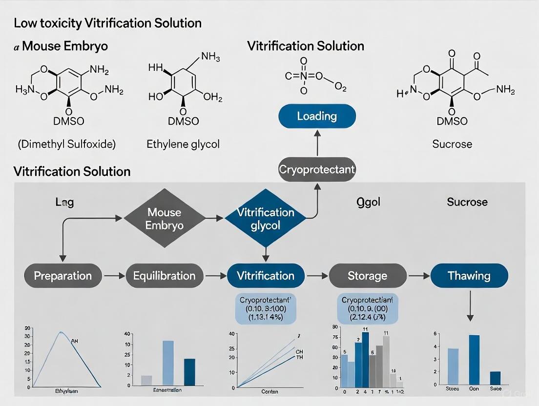
Advancing Low-Toxicity Vitrification Solutions for Mouse Embryo Cryopreservation: Strategies, Formulations, and Future Directions
This article provides a comprehensive resource for researchers and scientists on developing and optimizing low-toxicity vitrification solutions for mouse embryo cryopreservation.
Advancing Low-Toxicity Vitrification Solutions for Mouse Embryo Cryopreservation: Strategies, Formulations, and Future Directions
Abstract
This article provides a comprehensive resource for researchers and scientists on developing and optimizing low-toxicity vitrification solutions for mouse embryo cryopreservation. It covers foundational principles of cryoprotectant agent (CPA) toxicity and cellular injury mechanisms, explores innovative formulation strategies including binary CPA mixtures and commercial solutions like VEG, and details advanced methodologies such as ultra-fast vitrification to minimize osmotic stress. The content further addresses troubleshooting common challenges through high-throughput toxicity screening and quality control measures, and validates success through rigorous assessment of embryonic development, organelle integrity, and gene expression. By synthesizing current research and best practices, this guide aims to support improved cryopreservation outcomes in biomedical research and drug development.
Understanding Cryoprotectant Toxicity and Cellular Injury in Mouse Embryo Vitrification
Cryopreservation by vitrification represents a transformative approach in biomedical research, enabling long-term preservation of biological materials from single cells to complex tissues. This technique relies on the rapid cooling of high concentrations of cryoprotective agents (CPAs) to achieve a stable, ice-free glassy state that suspends all biochemical activity [1] [2]. However, researchers face a fundamental paradox: while higher CPA concentrations prevent lethal ice crystallization, they simultaneously introduce significant toxicity that can compromise cellular viability and function [3]. This challenge is particularly acute in mouse embryo cryopreservation, where maintaining developmental competence post-preservation is essential for reproductive research, genetic preservation, and drug development studies.
The toxicity of CPAs manifests through multiple mechanisms, including osmotic stress during addition/removal, direct chemical damage to cellular structures, and metabolic disruption [3] [4]. For mouse embryos, which are particularly sensitive to cryoprotectant exposure, finding the optimal balance between ice prevention and toxicity minimization is critical for successful vitrification outcomes. This application note examines current strategies to resolve this challenge through optimized CPA formulations, exposure protocols, and emerging technologies that collectively aim to enable high survival rates with preserved developmental potential.
Quantitative Analysis of CPA Toxicity Profiles
Table 1: Toxicity Rates of Scalable CPA Formulations in Kidney Tissue
| CPA Formulation | Total Concentration (M) | Toxicity Rate (k, minâ»Â¹) | Relative Toxicity | Reported Applications |
|---|---|---|---|---|
| VM3 | 8.46 | 0.007958 | 1.0x (reference) | Kidney slice vitrification |
| M22-PVP | 9.34 | 0.01755 | 2.2x | Rabbit kidney vitrification |
| M22 | 9.35 | 0.02339 | 2.9x | Rabbit kidney vitrification |
| VMP | 8.40 | Not reported | Intermediate | Rat kidney transplantation |
Table 2: Optimized CPA Mixtures for Mouse Oocyte Vitrification
| CPA Combination | Concentration in VS2 | Survival Rate (%) | Fertilization Rate (%) | Blastocyst Formation (%) |
|---|---|---|---|---|
| EG + Me₂SO | 20% + 20% | 69.2 ± 7.0 | 47.3 ± 2.7 | 38.8 ± 3.2 |
| GLY + PrOH | 20% + 20% | 42.1 ± 9.1 | 30.1 ± 4.7 | 26.1 ± 3.1 |
| EG alone | 40% | 20.7 ± 5.8 | 17.0 ± 3.2 | 8.0 ± 0.2 |
| Me₂SO alone | 40% | 8.9 ± 1.5 | 11.1 ± 0.5 | 5.6 ± 0.6 |
| Control (no CPA) | 0% | 92.1 ± 3.6 | 91.2 ± 5.5 | 74.2 ± 5.9 |
Recent systematic investigations reveal significant differences in toxicity profiles among common CPAs used in vitrification protocols. As shown in Table 1, formulations with similar osmotic concentrations can exhibit markedly different toxicity rates, with VM3 demonstrating approximately three-fold lower toxicity compared to M22 in kidney tissue models [3]. Similarly, Table 2 illustrates how strategic combination of CPAs can significantly improve outcomes compared to single-agent formulations, with EG + Meâ‚‚SO mixtures yielding substantially higher survival, fertilization, and blastocyst formation rates in mouse oocytes [5].
Mechanisms of Toxicity Reduction in CPA Mixtures
Mutual Dilution and Toxicity Neutralization
The improved performance of CPA mixtures stems from two primary mechanisms: mutual dilution and toxicity neutralization. Mutual dilution occurs when each CPA in a mixture effectively lowers the concentration of other CPAs, thereby reducing the overall toxic impact since CPA toxicity increases non-linearly with concentration [1]. Perhaps more importantly, toxicity neutralization describes the phenomenon where the addition of a second CPA partially counteracts specific toxic effects of the first CPA [1]. For instance, dimethyl sulfoxide (DMSO) has been shown to neutralize formamide toxicity, while similar neutralization effects occur between formamide and glycerol [1].
The molecular basis for these protective effects may involve stabilization of membrane structures, reduced perturbation of intracellular components, and moderation of osmotic stress. Research indicates that vascular endothelial cells—particularly vulnerable during cryopreservation and essential for post-transplant organ function—benefit significantly from these mixture effects [1]. For mouse embryos, which share similar sensitivity to osmotic and chemical stress, these principles can be strategically applied to design less toxic vitrification solutions.
Thermal Stress Considerations
Beyond direct chemical toxicity, the physical properties of CPA solutions significantly impact vitrification success. Higher glass transition temperatures (Tð‘”) have been correlated with reduced thermal stress cracking during temperature cycling [6]. Solutions with elevated Tð‘”, such as those containing sugars or sugar alcohols, exhibit lower thermal expansion coefficients, thereby minimizing the development of destructive thermal stresses during cooling and rewarming [6]. This physical stabilization complements the biochemical protection offered by optimized CPA mixtures.
Diagram 1: Mechanisms of Toxicity Reduction in CPA Mixtures. Strategic combination of CPAs provides synergistic protection through multiple complementary pathways.
High-Throughput Toxicity Screening Platform
Automated Methodology
Advanced screening platforms have revolutionized CPA development by enabling systematic evaluation of multiple formulations and exposure parameters. The Hamilton Microlab STARlet system represents one such automated liquid handling platform that permits precise control over CPA addition and removal sequences while randomizing treatments across 96-well plates to minimize positional bias [1]. This system facilitates high-throughput assessment of CPA toxicity using bovine pulmonary artery endothelial cells (BPAECs)—a relevant model for vascular-sensitive systems including the extensive vasculature of reproductive tissues.
The screening protocol involves several key steps. First, cells are cultured in standardized conditions in 96-well plates. Test CPAs are then applied using automated fluid handling with precise control over exposure duration and concentration gradients. After exposure, CPA removal is performed using multi-step dilution sequences to minimize osmotic shock. Finally, cell viability is quantified using metabolic indicators such as PrestoBlue, allowing calculation of toxicity rates and identification of promising formulations [1].
Application to Embryo Cryopreservation
While this platform utilizes endothelial cells, the fundamental principles of CPA toxicity are transferable to embryo cryopreservation. The high-throughput nature of this approach enables rapid screening of numerous CPA combinations and exposure parameters that would be impractical using embryo models directly. Promising candidates identified through initial screening can then be validated using mouse embryos in targeted follow-up studies, creating an efficient two-stage development pipeline for optimized vitrification solutions.
Protocol: Optimized Mouse Embryo Vitrification
Solution Preparation
Base Medium Preparation:
- Prepare HEPES-buffered medium supplemented with 20% fetal calf serum (FCS)
- Filter sterilize using 0.22µm membrane
- Store at 4°C for up to 1 week
Vitrification Solution (VS2):
- 20% ethylene glycol (v/v)
- 20% dimethyl sulfoxide (v/v)
- Base medium (as prepared above)
- Osmolarity: ~1500-1600 mOsm
- Prepare fresh before each experiment
Thawing Solutions:
- TS1: Base medium + 0.33 mol/L sucrose
- TS2: Base medium + 0.25 mol/L sucrose
- Equilibrate to room temperature before use
Vitrification Procedure
Equilibration Step: Transfer embryos to VS1 (containing 10% EG + 10% Meâ‚‚SO) for precisely 2 minutes at room temperature [5]
Vitrification Step: Move embryos to VS2 (20% EG + 20% Meâ‚‚SO) for 20 seconds at room temperature [5]
Loading and Cooling:
- Rapidly load embryos onto vitrification device (Open Pulled Straw or Nylon Loop)
- Immediately plunge into liquid nitrogen
- Ensure cooling rate exceeds 20,000°C/min [5]
Storage: Maintain embryos in liquid nitrogen or vapor phase below -150°C
Warming and CPA Removal
Rapid Warming:
- Remove vitrification device from liquid nitrogen
- Immediately immerse in TS1 pre-warmed to 37°C
- Hold for 3 minutes with gentle agitation [5]
Osmotic Adjustment:
- Transfer embryos to TS2 at room temperature
- Incubate for 3 minutes [5]
Recovery:
- Rinse embryos in base medium twice
- Transfer to culture medium for viability assessment
- Incubate under standard culture conditions (5% CO₂, 37°C)
Diagram 2: Mouse Embryo Vitrification Workflow. The optimized protocol emphasizes precise timing and temperature control at each transition point to minimize cumulative toxicity.
Advanced Rewarming Strategies
Nanowarming Technology
Conventional convective warming methods often produce insufficient warming rates and temperature non-uniformity that promotes ice crystallization and thermal stress damage [7] [2]. Nanowarming represents a breakthrough approach that addresses these limitations by generating heat volumetrically throughout the vitrified specimen [2]. This technique involves perfusing iron oxide nanoparticles (IONPs) throughout the vascular system prior to vitrification, then applying alternating magnetic fields to generate uniform heat distribution during rewarming [2].
While initially developed for larger organs, the principles of nanowarming could be adapted to embryo systems through modified nanoparticle delivery systems. The demonstrated success of nanowarming in recovering viable kidneys after 100 days of cryopreservation highlights its potential for revolutionizing cryopreservation outcomes across biological scales [2].
Optimization Parameters
Successful nanowarming requires careful optimization of several parameters:
- IONP concentration (typically 10 mg Fe/mL in final CPA solution)
- Perfusion pressure and flow rates to ensure uniform distribution
- Magnetic field strength and frequency (e.g., 63 kA/m at 180 kHz)
- Warming rates exceeding 70°C/min to prevent ice crystallization [2]
The Scientist's Toolkit: Essential Research Reagents
Table 3: Essential Reagents for Low-Toxicity Vitrification Research
| Reagent Category | Specific Examples | Function | Application Notes |
|---|---|---|---|
| Penetrating CPAs | Ethylene Glycol (EG), Dimethyl Sulfoxide (Meâ‚‚SO), Propylene Glycol | Intracellular ice inhibition, glass formation | EG + Meâ‚‚SO mixtures show superior toxicity profiles [5] |
| Non-penetrating CPAs | Sucrose, Trehalose, Ficoll, PVP | Osmotic buffering, extracellular stabilization | Critical for controlling edema during CPA removal |
| Biocompatible Additives | Fetal Calf Serum (20%) | Membrane stabilization, reduced toxicity | Optimal at 20% concentration in vitrification solutions [5] |
| Nanowarming Components | Silica/PEG-coated IONPs | Volumetric heating during rewarming | Enable uniform warming >70°C/min [2] |
| Viability Assays | PrestoBlue, Calcein-AM, Propidium Iodide | Quantification of post-warming survival | High-throughput compatibility essential for screening [1] |
| Naphazoline | Naphazoline HCl | High-purity Naphazoline hydrochloride for research. A potent alpha-adrenergic receptor agonist for pharmacological studies. For Research Use Only. | Bench Chemicals |
| Naproxen Etemesil | Naproxen Etemesil, CAS:385800-16-8, MF:C17H20O5S, MW:336.4 g/mol | Chemical Reagent | Bench Chemicals |
Balancing ice prevention with CPA toxicity remains a critical challenge in mouse embryo cryopreservation, but strategic approaches are emerging to address this fundamental paradox. The integration of optimized CPA mixtures, precise exposure protocols, advanced rewarming technologies, and high-throughput screening platforms creates a comprehensive framework for developing effective, low-toxicity vitrification solutions. As these technologies mature, they promise to enhance the efficiency and reliability of mouse embryo cryopreservation, supporting advancements in reproductive research, genetic preservation, and pharmaceutical development.
The successful application of these principles in complex systems—from kidney vitrification to oocyte cryopreservation—demonstrates their potential for transforming embryo cryopreservation outcomes. Future research should focus on adapting these approaches specifically for mouse embryo models, with particular attention to developmental stage-specific responses and long-term functional assessment of cryopreserved embryos.
Cryopreservation is a critical technique for the long-term storage of biological materials, including mouse embryos, essential for biomedical research and drug development. The success of these protocols, particularly those employing low-toxicity vitrification solutions, hinges on understanding and mitigating the principal mechanisms of cellular injury. These mechanisms—chilling damage, osmotic shock, and intracellular ice formation—represent significant barriers to post-thaw viability [8] [9]. This Application Note details the underlying theories and provides standardized protocols to quantify these injuries, providing a framework for researchers developing and optimizing novel, low-toxicity cryopreservation formulations for mouse embryos.
Mechanisms of Cryoinjury
The journey of a cell to cryogenic temperatures and back to a physiological state exposes it to a series of physical and chemical stresses. The following sections delineate the primary injury mechanisms.
Chilling Damage
Chilling injury occurs at temperatures above the freezing point of the solution but low enough to disrupt cellular metabolism and structure. This damage is independent of ice formation and is particularly relevant for temperature-sensitive cell types.
- Molecular Mechanisms: The fundamental cause is the thermotropic phase transition of membrane lipids from a liquid-crystalline to a gel state. This phase change compromises membrane integrity, leading to increased permeability and the leakage of intracellular components [8]. Furthermore, low temperatures can induce cold denaturation of proteins, disrupt the cytoskeleton, and inactivate critical enzymes, thereby halting development.
- Biological Consequences: In mouse embryos, chilling injury can manifest as irreversible damage to the meiotic spindle in oocytes, leading to aneuploidy, and as a general reduction in developmental competence post-thaw.
Osmotic Shock
Osmotic shock is a consequence of the dramatic cell volume changes during the addition and removal of cryoprotective agents (CPAs) and during the freezing process itself [10].
- Theoretical Basis: During CPA addition or freezing (where extracellular ice formation concentrates solutes), water exits the cell rapidly, causing excessive cell shrinkage. Conversely, during CPA dilution or thawing, water influx can cause the cell to swell beyond its critical volume. The two-factor hypothesis of freezing injury posits that these osmotic volume excursions, alongside intracellular ice formation, are key determinants of cell survival [9].
- Cellular Impact: Excessive shrinkage can lead to the collapse of intracellular structures and damage to the plasma membrane. The subsequent swelling can cause membrane rupture (lysis). The "osmotic rupture hypothesis" further suggests that the osmotic pressure gradient during freezing can itself stress the membrane to the point of failure, creating pores that permit intracellular ice formation [11].
Intracellular Ice Formation (IIF)
IIF is widely considered the most lethal event during rapid cooling, as it directly disrupts and pierces subcellular structures [9].
- Mechanisms of IIF: IIF does not occur spontaneously but is catalyzed by the extracellular environment. Two primary mechanisms are:
- Surface-Catalyzed Nucleation: Extracellular ice grows through membrane pores or invaginations, seeding the supercooled cytoplasm.
- Plasma Membrane Rupture: Osmotically driven water efflux generates sufficient pressure to rupture the plasma membrane, allowing extracellular ice to propagate into the cell [11].
- Propagation and the Role of Cell Junctions: In tissues and cell aggregates, IIF can propagate from one cell to its neighbors. Recent research using high-speed videomicroscopy has revealed that this propagation is facilitated not only by gap junctions but also by the penetration of ice into the paracellular space between cells, highlighting a complex dependence on intercellular junction protein expression [9].
The following diagram illustrates the logical progression and key events leading to these three primary injury mechanisms during a cryopreservation cycle.
Logical relationships of primary cryoinjury mechanisms.
Quantitative Data on Cryoinjury
Understanding the quantitative impact of key variables is crucial for protocol optimization. The following tables summarize critical data on cryoprotectant toxicity and the dynamics of ice formation.
Table 1: Comparative Toxicity of Common Penetrating Cryoprotectants in Model Systems
| Cryoprotectant | Test System | Key Toxicity Findings | Experimental Conditions | Reference |
|---|---|---|---|---|
| Dimethyl Sulfoxide (DMSO) | Dermal Fibroblasts | Viability decreased with increasing concentration, temperature, and exposure time. | 5-30% (v/v), 4-37°C, 10-30 min | [8] |
| Ethylene Glycol (EG) | Mouse Zygotes | 30% and 40% EG yielded 98% and 84% development to blastocysts, respectively. | 20°C for 20 min exposure | [12] |
| Glycerol (GLY) | Stallion Sperm | Concentrations >1.5% polymerized actin cytoskeleton. | N/A | [8] |
| L-Proline | Mouse Oocytes | 2M L-proline significantly improved survival vs. proline-free control (94.7% vs 88.4%). | Vitrification/Warming | [13] |
| Propylene Glycol (PG) | Mouse Zygotes | >2.5 M impaired developmental potential by decreasing intracellular pH. | N/A | [8] |
Table 2: Ice Formation Dynamics in Bovine Oocytes Under Different Conditions
| Cooling Rate | Warming Rate | CPA Concentration | Ice Formation after Cooling | Ice Formation during Warming | Reference |
|---|---|---|---|---|---|
| ~30,000 °C/min | Conventional | Standard Vitrification Solution | None detected | Large ice fractions (most free water crystallizes) | [14] |
| ~600,000 °C/min | Conventional | Standard Vitrification Solution | Essentially none | Essentially none | [14] |
| ~30,000 °C/min | High Convective (20x conventional) | Reduced CPA concentration | To be determined | To be determined (enables lower CPA use) | [14] |
Experimental Protocols
This section provides detailed methodologies for assessing cellular injury, utilizing mouse embryos or other relevant biological models.
Protocol: Assessing Osmotic Shock and Membrane Integrity During CPA Equilibration
This protocol measures cell volume changes and membrane damage during the loading and unloading of CPAs.
1. Materials:
- Log-phase mouse morulae or zygotes.
- Equilibration Medium: PB1 medium with 3.0 M Ethylene Glycol.
- Vitrification Medium: PB1 medium with 40% Ethylene Glycol and 30% Ficoll (EFS solution) [12].
- Dilution Medium: PB1 medium with 0.5 M sucrose.
- Microscope with camera and image analysis software.
- Hemocytometer or automated cell counter.
- Trypan Blue stain.
2. Method: 1. Baseline Measurement: Place a cohort of embryos (n≥20) in a culture drop and record their initial diameters using image analysis. 2. CPA Loading: * Transfer embryos to Equilibration Medium at 20°C for 5-7 minutes. * Capture images every 30 seconds to track volume changes. * Calculate cell volume assuming spherical geometry. 3. Vitrification Solution Exposure: Transfer embryos to Vitrification Medium for 2-5 minutes at 20°C. Note: For toxicity assessment, this step can be performed without actual cooling. 4. CPA Unloading (Simulated Thaw): * Transfer embryos directly to Dilution Medium. * Capture images to monitor swelling over 10 minutes. 5. Viability Assessment: Incubate embryos with Trypan Blue stain (0.4%) for 3-5 minutes. Cells with compromised membranes will take up the blue dye. 6. Culture: Wash embryos and place in culture medium to assess developmental competence to the blastocyst stage over 5 days.
3. Data Analysis:
- Plot normalized cell volume versus time to visualize shrinkage and swelling kinetics.
- Calculate the percentage of Trypan Blue positive cells for each treatment group.
- Compare blastocyst formation rates between experimental and untreated control groups.
Protocol: Quantifying Intracellular Ice Formation Using High-Speed Videomicroscopy
This protocol utilizes a controlled freezing stage and high-speed imaging to directly observe the nucleation and propagation of IIF.
1. Materials:
- Mouse insulinoma MIN6 cells (wild-type and junction-protein knockdown strains).
- Cryostage with temperature controller (e.g., Linkam or similar).
- High-speed video camera (≥1000 fps).
- Inverted microscope with 40x or 60x objective.
- CPA solution (e.g., 1.5-2.0 M DMSO in culture medium).
2. Method: 1. Sample Preparation: Culture cells to form confluent monolayers or two-cell pairs on cryostage-compatible dishes. For knockdown studies, use cells with targeted knockdown of gap, adherens, and tight junction proteins [9]. 2. CPA Equilibration: Incubate cells in CPA solution for 10-15 minutes at room temperature. 3. Mounting and Cooling: * Place the sample on the cryostage. * Initiate a rapid cooling ramp (e.g., 130°C/min) from a supra-zero temperature (e.g., 0°C) down to -50°C or lower. * Simultaneously, begin high-speed video recording. 4. Observation: Visually track the sample for the characteristic darkening or "flashing" that indicates IIF. Note the temperature of first ice formation and the pattern of cell-to-cell propagation.
3. Data Analysis:
- Review high-speed videos to determine the nucleation temperature for the first and subsequent cells.
- Calculate the probability of IIF propagation between cell pairs.
- Compare propagation rates and nucleation temperatures between wild-type and junction-protein-deficient cells to elucidate the role of intercellular connections [9].
The workflow for this detailed analysis is outlined below.
High-speed videomicroscopy workflow for IIF quantification.
The Scientist's Toolkit: Research Reagent Solutions
Selecting the appropriate reagents is fundamental to successfully investigating cryoinjury and developing improved vitrification solutions.
Table 3: Essential Reagents for Investigating Cryoinjury Mechanisms
| Reagent / Solution | Function / Utility | Application Example |
|---|---|---|
| Ethylene Glycol (EG) | A penetrating CPA with relatively low toxicity. Often a key component of low-toxicity vitrification solutions. | Used at 40% with Ficoll and sucrose (EFS solution) for mouse morulae vitrification with high survival and live birth rates [12]. |
| L-Proline | A natural, nontoxic amino acid acting as an osmoprotectant and antioxidant. Reduces osmotic and oxidative stress. | Supplementation at 2M in vitrification solution significantly improved survival and mitochondrial function in mouse oocytes [13]. |
| Synth-a-Freeze Medium | A chemically defined, protein-free, ready-to-use cryopreservation medium. Eliminates variability from serum. | Standardized cryopreservation of stem and primary cells, ensuring consistency in toxicity studies [15]. |
| Polyvinyl Alcohol (PVA) | A synthetic polymer that inhibits ice nucleation, growth, and recrystallization. A non-penetrating cryoprotectant. | Used at 1 mg/mL to reduce ice crystal growth and improve survival of bacteria during cryopreservation; applicable to cellular systems [16]. |
| Ficoll | A high-molecular-weight polymer used as a non-penetrating CPA to increase solution viscosity and aid vitrification. | A component of EFS vitrification solution, helping to achieve a glassy state without ice crystallization [12]. |
| Trehalose | A non-penetrating disaccharide that stabilizes membranes and proteins during dehydration and freezing. | Used in combination with PVA (50 mg/mL) as a low-toxicity alternative for microorganism cryopreservation [16]. |
| 2-[[(E)-octadec-9-enoyl]amino]ethyl dihydrogen phosphate | 2-[[(E)-octadec-9-enoyl]amino]ethyl dihydrogen phosphate, CAS:24435-25-4, MF:C20H40NO5P, MW:405.5 g/mol | Chemical Reagent |
| Phthalylsulfacetamide | Phthalylsulfacetamide, CAS:131-69-1, MF:C16H14N2O6S, MW:362.4 g/mol | Chemical Reagent |
Within the field of mouse embryo cryopreservation research, the development of low-toxicity vitrification solutions is paramount for ensuring high post-warm viability and maintaining developmental competence. Permeating cryoprotectants (CPAs), which enter the cell to prevent lethal intracellular ice formation, are indispensable components of these solutions. However, their inherent cytotoxicity poses a significant challenge. This Application Note provides a detailed comparative analysis of the toxicity profiles of three predominant permeating CPAs—Dimethyl Sulfoxide (DMSO), Ethylene Glycol (EG), and Glycerol—framed within the context of optimizing vitrification protocols for mouse embryos. We summarize key quantitative toxicity data, present detailed experimental methodologies for its assessment, and provide essential resources to support research into next-generation, low-toxicity cryopreservation formulations.
Toxicity Profiles and Data Comparison
The toxicity of a CPA is not an absolute value but is influenced by concentration, exposure time, temperature, and the biological system itself. The data below provide a comparative overview of these agents under conditions relevant to embryo vitrification.
Table 1: Comparative Toxicity Profiles of Common Permeating Cryoprotectants
| Cryoprotectant | Typical Vitrification Concentration | Key Toxicity Mechanisms | Reported IC50 / Toxic Threshold (Mammalian Cells) | Temperature Dependence |
|---|---|---|---|---|
| DMSO | ~1.5 - 3.0 M (10-20% v/v) [17] [18] | - Induces DNA demethylation; alters epigenetic landscape [19]- Disrupts membrane integrity & mitochondrial function [17]- Increases reactive oxygen species (ROS) production [17] | - Significant toxicity to human chondrocytes at 6 M and 8.1 M (37°C) [17]- Toxicity rate constant (k): 2.62 (at 6 mol/kg) [20] | High toxicity at elevated temperatures; rapid cooling after addition is critical [17] |
| Ethylene Glycol (EG) | ~3.0 - 6.0 M [1] | - Generally lower chemical toxicity than DMSO [17] [20]- Primary risk is osmotic shock during addition/removal | - Toxicity rate constant (k): 0.59 (at 6 mol/kg) [20] | Toxicity increases with temperature, but less pronounced than DMSO [17] |
| Glycerol | ~3.0 - 6.0 M [1] [17] | - Lower chemical toxicity than DMSO [17]- Can cause osmotic stress and damage due to slower permeability in some cell types [17] | - Toxicity rate constant (k): 0.34 (at 6 mol/kg) [20] | Cytotoxicity increases with higher temperatures; strict temperature control is essential [17] |
Table 2: Toxicity Rate Constants (k) for Single and Binary CPA Solutions at 6 mol/kg and Room Temperature [20]
| CPA Solution | Toxicity Rate Constant (k) |
|---|---|
| DMSO | 2.62 |
| Ethylene Glycol (EG) | 0.59 |
| Glycerol | 0.34 |
| Formamide | 0.61 |
| Propylene Glycol (PG) | 0.48 |
| Binary Mixture (DMSO + EG) | 1.06 |
| Binary Mixture (DMSO + Glycerol) | 0.95 |
Experimental Protocols for Toxicity Assessment
To evaluate CPA toxicity in the context of mouse embryo vitrification, the following high-throughput and embryo-specific protocols are recommended.
High-Throughput Toxicity Screening in Cell Models
This automated protocol enables rapid screening of multiple CPA formulations and is ideal for initial toxicity neutralization studies [1] [20].
1. Cell Preparation:
- Culture bovine pulmonary artery endothelial cells (BPAECs) or another relevant cell line in 96-well plates until ~80% confluent.
- Include control wells for background (cell-free media) and 100% viability (cells with no CPA exposure).
2. Automated CPA Addition:
- Use an automated liquid handling system (e.g., Hamilton Microlab STARlet) for precise, serial dilution and addition of CPA solutions to the wells.
- Employ a multi-step addition protocol to minimize osmotic shock. Randomize the placement of CPA treatments across the plate to avoid positional bias.
- Expose cells to the target CPA concentration (e.g., up to 6 mol/kg) for a defined duration (e.g., 5-30 minutes) at room temperature.
3. CPA Removal and Viability Assay:
- Remove CPA solutions using a step-wise dilution process with isotonic buffer.
- Add a cell viability indicator, such as PrestoBlue, to the wells.
- Incubate according to the manufacturer's instructions and measure fluorescence using a plate reader.
- Calculate normalized viability relative to the 100% viability control wells.
Toxicity and Functional Assessment in Mouse Embryos
This protocol directly assesses the impact of CPAs on embryo development and health, providing functional data for vitrification solution optimization [19].
1. Embryo Collection & Culture:
- Collect 8-cell stage mouse embryos following standard superovulation and mating protocols.
- Maintain embryos in pre-equilibrated culture medium under oil at 37°C and 5% CO₂.
2. CPA Exposure and Vitrification Simulation:
- Transfer embryos through a series of equilibration and vitrification solutions containing the CPA(s) of interest (e.g., 7.5% DMSO + 7.5% EG vs. 7.5% PG + 7.5% EG).
- Control the exposure time meticulously (e.g., 5 min in equilibration solution, 30 sec in vitrification solution) at room temperature to simulate vitrification conditions without plunging into liquid nitrogen.
- For toxicity neutralization tests, include groups with additives like N-acetyl-l-cysteine (NAC, 5 mM) in the DMSO-based vitrification medium [19].
3. Post-Exposure Analysis:
- Developmental Competence: Culture the embryos for 5 days to the blastocyst stage and record the rates of development.
- Epigenetic Analysis: Fix a subset of embryos at the 8-cell or blastocyst stage. Perform immunostaining for 5-methylcytosine (5mC) and 5-hydroxymethylcytosine (5hmC) to quantify DNA methylation changes [19].
- Gene Expression: Extract RNA from blastocysts for RNA-sequencing analysis to examine changes in key signaling pathways (e.g., MAPK, Wnt) and expression of DNA methyltransferases (DNMTs) and demethylases (TETs) [19].
The Scientist's Toolkit: Research Reagent Solutions
Table 3: Essential Reagents for CPA Toxicity and Vitrification Research
| Reagent / Solution | Function / Application | Example Use in Protocol |
|---|---|---|
| DMSO (High Purity) | Penetrating CPA; standard for many vitrification protocols but with known toxicity risks. | Positive control for toxicity studies; component of standard vitrification solutions [17] [18]. |
| Ethylene Glycol (EG) | Penetrating CPA; often used in mixtures for lower toxicity and rapid permeability. | Combined with DMSO or PG in vitrification solutions to reduce total toxicity load [19] [1]. |
| Propylene Glycol (PG) | Penetrating CPA; investigated as a lower-toxicity alternative to DMSO. | Used in vitrification solutions to mitigate DMSO-induced DNA demethylation [19]. |
| N-Acetyl-L-Cysteine (NAC) | Antioxidant; acts as a toxicity neutralization agent. | Added at 5 mM to DMSO-based vitrification media to ameliorate oxidative stress and DNA demethylation [19]. |
| Trehalose | Non-penetrating CPA; provides extracellular stabilization and reduces osmotic stress. | Component of vitrification and slow-freeze solutions (e.g., 0.1 M - 0.4 M) to improve post-warm survival [21]. |
| PrestoBlue / MTT Cell Viability Reagent | Metabolic activity indicator for quantitative toxicity assessment. | Used in high-throughput screening to measure cell viability after CPA exposure [1]. |
| Anti-5mC / Anti-5hmC Antibodies | Immunostaining reagents for epigenetic analysis. | Used to quantify global DNA methylation and hydroxymethylation levels in embryos post-CPA exposure [19]. |
| Bemfivastatin | Bemfivastatin, CAS:805241-79-6, MF:C34H37FN2O6, MW:588.7 g/mol | Chemical Reagent |
| Necrostatin-7 | Necrostatin-7, CAS:351062-08-3, MF:C16H10FN5OS2, MW:371.4 g/mol | Chemical Reagent |
Visualizing Toxicity Pathways and Experimental Workflows
Molecular Pathways of DMSO-Induced Toxicity in Embryos
The following diagram illustrates the key molecular mechanisms of DMSO-induced toxicity as identified in recent research, particularly the pathway leading to DNA demethylation.
High-Throughput Workflow for CPA Toxicity Screening
This workflow outlines the automated process for screening cryoprotectant toxicity, from plate preparation to data analysis.
In the field of assisted reproductive technology, vitrification has become the gold standard for cryopreserving mouse oocytes and embryos. A key challenge in protocol optimization lies in balancing the exposure time to cryoprotective agents with the osmotic stress placed on the cell. The permeability characteristics of the oocyte and embryo membranes are a central factor in this balancing act, directly influencing the optimal CPA exposure time and the potential for osmotic damage. This application note explores the relationship between membrane permeability, CPA exposure time, and cellular damage, providing researchers with detailed protocols and data to inform the development of low-toxicity vitrification solutions for mouse embryo cryopreservation.
Theoretical Framework: Permeability, Osmosis, and Toxicity
The permeability of cellular membranes to water and CPAs governs the kinetics of dehydration and CPA permeation during the vitrification process. Understanding these principles is essential for designing effective protocols.
Permeating vs. Non-Permeating Agents: Vitrification solutions typically contain a combination of permeating CPAs and non-permeating agents. Permeating CPAs include ethylene glycol and dimethyl sulfoxide, which cross the cell membrane and help prevent intracellular ice formation. Non-permeating agents include sucrose and trehalose, which create an osmotic gradient that drives cellular dehydration prior to vitrification [22]. This dehydration is critical for reducing intracellular ice formation.
Osmotic Stress and Volumetric Changes: During CPA addition and removal, cells undergo significant volumetric changes. The addition of CPAs causes initial cell shrinkage as water exits, followed by a return to original volume as CPA and water enter. This process reverses during CPA removal. Excessive osmotic stress can cause irreversible damage to the cytoskeleton and cellular organelles [23].
Toxicity and Exposure Time: Permeating CPAs become toxic with prolonged exposure. The relationship between exposure time and toxicity is concentration-dependent, with higher CPA concentrations requiring shorter exposure times. Ultra-rapid vitrification protocols aim to minimize this toxic stress by reducing CPA exposure time through increased cooling rates [24] [25].
Figure 1: Concentration-dependent toxicity pathway. Membrane permeability influences both osmotic stress and CPA toxicity, which converge to cause cellular damage. Optimal exposure time modulates this pathway to minimize detrimental effects.
Quantitative Data on Embryonic Development Post-Vitrification
The developmental stage of embryos significantly impacts their survival and developmental competence after vitrification. Later-stage embryos generally show higher resilience.
Table 1: Developmental Competence of Mouse Embryos After Vitrification at Different Stages
| Developmental Stage | Morphologically Normal after Warming (%) | Developed to Blastocyst (%) | References |
|---|---|---|---|
| Zygote | 72 | 16 | [23] |
| 2-Cell | 86 | 25 | [23] |
| 4-Cell | 90 | 71 | [23] |
| 8-Cell | 93 | 80 | [23] |
| Morula | 97 | 92 | [23] |
| Blastocyst | 90-93* | N/A | [22] |
Note: *Survival rate of artificially shrunken blastocysts vitrified in sucrose- and Ficoll-free solution.
Table 2: Impact of Vitrification Method on Mouse Oocyte Organelle Integrity
| Parameter | Fresh Oocytes (Control) | Conventional Vitrification (C-VIT) | Ultra-Fast Vitrification (UF-VIT) | References |
|---|---|---|---|---|
| Survival Rate | 100% | 95.2%* | 98.5% | [25] |
| Normal ER Distribution (Equatorial) | 92% | 66%* | 84% | [25] |
| Normal Mitochondrial Distribution | 86% | 14%* | 46%* | [25] |
| Mitochondrial Membrane Potential (ΔΨm) | 0.80 | 0.61* | 0.79 | [25] |
| Blastocyst Formation Rate | Baseline | Significantly reduced vs. control* | No significant difference vs. control | [25] |
Note: *Statistically significant difference (p < 0.05) compared to control group.
Detailed Experimental Protocols
Protocol 1: Ultra-Fast Vitrification (UF-VIT) of Mouse Oocytes
This protocol minimizes CPA exposure time to reduce toxicity and osmotic stress, based on the method described by Jin and Mazur [25].
Materials:
- Equilibration Solution: 7.5% ethylene glycol + 7.5% DMSO in base medium
- Vitrification Solution: 15% ethylene glycol + 15% DMSO + 0.5M sucrose in base medium
- Warming Solution 1: 1.0M sucrose in base medium at 37°C
- Warming Solution 2: 0.5M sucrose in base medium at room temperature
- Base medium: PBS supplemented with 20% serum substitute supplement
- Quartz capillaries or Cryotop/Cryoloop devices
- Liquid nitrogen
Procedure:
- Equilibration: Transfer oocytes into equilibration solution for 30-60 seconds at room temperature. Observe for initial shrinkage and partial return to original volume.
- Vitrification Solution Exposure: Immediately transfer oocytes to vitrification solution. Limit exposure time to 20-30 seconds.
- Loading and Cooling: Within 30 seconds of exposure to vitrification solution, load oocytes onto quartz capillaries or Cryotop devices and plunge directly into liquid nitrogen.
- Warning: Warm samples rapidly by immersing in Warming Solution 1 at 37°C for 3 minutes.
- Sucrose Dilution: Transfer oocytes to Warming Solution 2 for 5 minutes at room temperature.
- Final Rinse: Wash oocytes twice in base medium for 5 minutes each.
- Assessment: Evaluate survival rates and culture for further development.
Protocol 2: Vitrification of Mouse Embryos at Different Developmental Stages
This protocol adapts CPA exposure times based on embryonic stage, using Open Pulled Straws as containers [23].
Materials:
- Vitrification Solution 1: 7.5% EG + 7.5% DMSO in base medium
- Vitrification Solution 2: 15% EG + 15% DMSO + 0.5M sucrose in base medium
- Thawing Solution 1: 0.5M sucrose in base medium at 37°C
- Thawing Solution 2: 0.25M sucrose in base medium at room temperature
- Base medium: PBS supplemented with 20% serum substitute supplement
- Open Pulled Straws
- Liquid nitrogen
Procedure:
- Preparation: Prepare OPS by heating and pulling 0.25mL straws to achieve a thin wall.
- Equilibration: Expose embryos to Vitrification Solution 1 for 4 minutes at room temperature.
- Vitrification Solution Transfer: Move embryos to Vitrification Solution 2 for 40 seconds.
- Loading and Cooling: Load 1-3 embryos into OPS and plunge into liquid nitrogen within 40 seconds.
- Warning: Thaw straws by immersing narrow end directly into Thawing Solution 1 at 37°C.
- Sucrose Removal: Transfer embryos to Thawing Solution 2 for 3 minutes.
- Final Rinse: Wash embryos in base medium for 3 minutes.
- Culture: Culture embryos in appropriate medium for developmental assessment.
Protocol 3: Mitochondrial and ER Integrity Assessment Post-Vitrification
This protocol assesses subcellular organelle damage after vitrification.
Materials:
- Mito-Tracker Red CMXRos (for mitochondrial membrane potential)
- ER-Tracker Blue-White DPX dye
- Hoechst 33342 (for nuclear staining)
- Microtubule staining kit for spindle visualization
- Confocal microscopy equipment
Procedure:
- Staining: Co-incubate warmed oocytes/embryos with Mito-Tracker Red and ER-Tracker dyes for 30 minutes.
- Fixation: Fix samples in paraformaldehyde for 15 minutes.
- Microtubule Staining: Perform immunostaining for microtubules to visualize meiotic spindles.
- Nuclear Staining: Counterstain with Hoechst 33342.
- Imaging: Capture images using confocal microscopy with appropriate filters.
- Analysis: Assess mitochondrial distribution, ER patterns, and spindle morphology compared to fresh controls.
Figure 2: Ultra-fast vitrification workflow. Critical timing steps minimize CPA exposure while ensuring sufficient dehydration. Exposure times must be optimized based on membrane permeability characteristics.
The Scientist's Toolkit: Essential Research Reagents
Table 3: Key Reagents for Low-Toxicity Vitrification Research
| Reagent/Category | Specific Examples | Function & Application Notes |
|---|---|---|
| Permeating CPAs | Ethylene glycol, Dimethyl sulfoxide, Glycerol | Penetrate cell membrane to prevent intracellular ice formation; EG shows higher permeability and lower toxicity for oocytes/embryos [23]. |
| Non-Permeating Agents | Sucrose, Trehalose, Ficoll | Create osmotic gradient for dehydration; trehalose may offer superior glass-forming properties and membrane stabilization [26]. |
| Vitrification Containers | Cryo-loop, Open Pulled Straw, EM-grid, Cryotop | Enable ultra-rapid cooling; cryo-loop showed superior recovery rates for mouse embryos [27]. |
| Base Media | PBS with SSS, Synthetic serum substitute | Provide macromolecular support and osmotic stability during vitrification procedures [27] [22]. |
| Assessment Tools | Mito-Tracker dyes, ER-Tracker dyes, Hoechst stains | Evaluate subcellular organelle integrity and distribution post-warming; reveal mitochondrial and ER damage patterns [25]. |
| Pralnacasan | Pralnacasan, CAS:192755-52-5, MF:C26H29N5O7, MW:523.5 g/mol | Chemical Reagent |
| Pridefine | Pridefine, CAS:5370-41-2, MF:C19H21N, MW:263.4 g/mol | Chemical Reagent |
Membrane permeability characteristics fundamentally influence CPA exposure time optimization and damage mitigation in mouse embryo cryopreservation. The data and protocols presented demonstrate that later-stage embryos generally withstand vitrification better than earlier stages, and that ultra-fast vitrification methods significantly reduce CPA toxicity and osmotic stress. Implementation of stage-specific protocols with minimized, precise CPA exposure times can dramatically improve survival rates and developmental competence. These insights provide a foundation for developing refined, low-toxicity vitrification solutions that maximize preservation of cellular integrity and function.
Formulating and Applying Low-Toxicity Vitrification Solutions
Vitrification is a promising cryopreservation technique for complex biological specimens, including mouse embryos, as it prevents damaging ice formation by achieving a glassy state. However, this process requires high concentrations of cryoprotective agents (CPAs), which can exert significant chemical toxicity on cellular structures [28]. Rather than relying on single CPAs, contemporary cryopreservation research has demonstrated that using multi-CPA solutions can substantially reduce overall toxicity while maintaining vitrification capability [29].
This application note explores the strategic use of binary CPA combinations to achieve toxicity reduction through two primary mechanisms: mutual dilution (each CPA lowers the concentration of the others) and toxicity neutralization (one CPA counteracts the toxic effects of another) [29]. We provide quantitative data on effective CPA combinations, detailed experimental protocols for toxicity assessment, and specific methodologies for applying these mixtures to mouse embryo cryopreservation within the context of developing low-toxicity vitrification solutions.
Quantitative Data on CPA Mixture Toxicity
Key Binary Combinations Showing Toxicity Reduction
Table 1: Effective Binary CPA Combinations for Toxicity Reduction
| CPA Combination | Total Concentration | Observed Effect | Viability Outcome | Temperature | Cell Type | Citation |
|---|---|---|---|---|---|---|
| Formamide + Glycerol | 12 mol/kg | Toxicity neutralization | 97% viability (vs 20% with formamide alone) | 4°C | BPAEC | [30] |
| Formamide + Glycerol | 6 mol/kg | Significant toxicity decrease | Higher than single CPA solutions | Room Temperature | BPAEC | [31] |
| DMSO + 1,3-Propanediol | 6 mol/kg | Significant toxicity decrease | Higher than single CPA solutions | Room Temperature | BPAEC | [31] |
| 1,2-Propanediol + Diethylene Glycol | 6 mol/kg | Significant toxicity decrease | Higher than single CPA solutions | Room Temperature | BPAEC | [31] |
| 1,3-Propanediol + Diethylene Glycol | 6 mol/kg | Significant toxicity decrease | Higher than single CPA solutions | Room Temperature | BPAEC | [31] |
| Formamide + DMSO | Various | Toxicity neutralization | Reduced toxicity | Room Temperature | BPAEC | [29] |
Mathematical Modeling of CPA Mixture Toxicity
Advanced mathematical models have been developed to predict the toxicity of multi-CPA mixtures. These models account for both specific toxicity (direct effects of individual CPAs) and non-specific toxicity (overall effects on solution properties) [28]. The multi-CPA toxicity model incorporates interactions between common CPAs including glycerol, dimethyl sulfoxide (DMSO), propylene glycol, ethylene glycol, and formamide.
The general toxicity rate equation follows first-order kinetics:
dN/dt = -kN
Where N represents the number of viable cells, t is time, and k is the toxicity rate constant. For mixture prediction, the model uses data from single and binary CPA solutions to extrapolate to more complex mixtures [28].
Table 2: Optimized Vitrification Solution from Mathematical Modeling
| CPA Component | Concentration (molal) | Role in Mixture | Theoretical Basis |
|---|---|---|---|
| Glycerol | 7.4 | Primary cryoprotectant | Lower specific toxicity |
| DMSO | 1.4 | Synergistic component | Toxicity modulation |
| Formamide | 2.4 | Toxicity reduction | Interaction with glycerol |
This optimized mixture was identified by pairing the multi-CPA toxicity model with a vitrification/devitrification model, demonstrating the potential for mathematical optimization of vitrification solution composition [28].
Experimental Protocols
High-Throughput Toxicity Screening for CPA Mixtures
Diagram 1: High-throughput toxicity screening workflow for CPA mixtures.
Materials and Reagents
- Cells: Bovine Pulmonary Artery Endothelial Cells (BPAEC)
- CPAs: Formamide, glycerol, DMSO, 1,3-propanediol, 1,2-propanediol, diethylene glycol, ethylene glycol, propylene glycol
- Equipment: Hamilton Microlab STARlet automated liquid handling system
- Culture Media: Dulbecco's Modified Eagle Medium (DMEM) with 10% fetal bovine serum
- Viability Assay: PrestoBlue cell viability reagent
- Buffers: HEPES-buffered saline (HBS)
Procedure
CPA Stock Solution Preparation: Prepare individual CPA stock solutions at desired concentrations in HBS or appropriate buffer.
Automated Mixture Preparation: Program the liquid handler to create binary CPA mixtures in 96-well plates:
- Implement randomized plate layouts to minimize positional effects
- Include controls (cells without CPAs, single CPA solutions for comparison)
- Create concentration series for each binary combination
Cell Preparation and Seeding:
- Culture BPAEC under standard conditions (37°C, 5% CO₂)
- Seed cells in 96-well plates at optimal density (e.g., 10,000 cells/well)
- Allow cells to adhere overnight
CPA Exposure:
- Replace culture medium with CPA solutions using automated liquid handling
- Maintain consistent exposure times across experiments (e.g., 10-30 minutes)
- Conduct experiments at both room temperature and 4°C to assess temperature effects
CPA Removal:
- Perform multi-step removal using decreasing concentrations of CPAs
- Use automated liquid handling to ensure precision and reproducibility
Viability Assessment:
- Add PrestoBlue reagent to each well
- Incubate for predetermined time (typically 1-2 hours)
- Measure fluorescence using plate reader
- Calculate viability relative to control cells
Data Analysis:
- Normalize viability data to controls
- Compare binary mixture toxicity to constituent single CPAs
- Identify combinations showing significant toxicity reduction
Mouse Embryo Vitrification with Low-Toxicity CPA Mixtures
Diagram 2: Mouse embryo vitrification workflow using low-toxicity CPA mixtures.
Materials and Reagents
- Biological Material: Mouse morulae or 2-cell embryos
- Basal Medium: Modified phosphate-buffered saline (PB1 medium)
- Permeating CPAs: Ethylene glycol, glycerol, DMSO, formamide
- Non-Permeating CPA: Sucrose
- Polymer: Ficoll (for some formulations)
- Cryotop Devices or similar minimal volume tools
- Liquid nitrogen storage system
EDFS10/10a Vitrification Solution Formulation
Developed specifically for equilibrium vitrification of mouse embryos with minimal toxicity:
- Total Permeating CPA Concentration: 20% (v/v)
- Typically 10% ethylene glycol + 10% DMSO
- Sucrose Concentration: 0.4 M
- Base Medium: Modified PB1
This low-CPA concentration formulation significantly reduces toxicity compared to traditional vitrification solutions like EFS35c while maintaining effective vitrification [32].
Procedure
Embryo Collection:
- Collect morulae or 2-cell embryos from superovulated mice
- Select only high-quality embryos with uniform blastomeres and intact zonae pellucidae
CPA Equilibration:
- Expose embryos to EDFS10/10a solution at room temperature for 10 minutes
- Observe embryonic dehydration and re-expansion during equilibration
Vitrification Process:
- Load 5-6 embryos in minimal volume (<1 μL) onto Cryotop device
- Immediately plunge into liquid nitrogen
- Store in liquid nitrogen for desired duration
Warning and CPA Removal:
- Rapidly warm Cryotop in 20°C water bath for 5 seconds
- Transfer embryos through decreasing concentrations of sucrose solutions (0.5 M, 0.25 M) for 5 minutes each
- Rinse in base medium
Viability Assessment:
- Culture embryos in Hypermedium under oil at 37°C, 6% CO₂
- Assess survival by morphological appearance and re-expansion
- Evaluate developmental competence by progression to blastocyst stage
- For comprehensive assessment, transfer embryos to recipients to measure live birth rates
The Scientist's Toolkit
Table 3: Essential Research Reagent Solutions for CPA Mixture Studies
| Reagent/Category | Specific Examples | Function/Application | Notes |
|---|---|---|---|
| Permeating CPAs | Ethylene glycol, glycerol, DMSO, propylene glycol, formamide | Penetrate cell membranes to prevent intracellular ice formation | Formamide shows notable toxicity neutralization with glycerol and DMSO [30] [29] |
| Non-Permeating CPAs | Sucrose, Ficoll, diethylene glycol | Provide extracellular protection, increase viscosity | Sucrose is essential in equilibrium vitrification solutions [32] |
| Cell Culture Systems | BPAEC, mouse embryos | Toxicity screening models | BPAEC provides high-throughput capability; mouse embryos offer clinical relevance |
| Viability Assays | PrestoBlue, MTT assay | Quantify cellular toxicity | PrestoBlue enables high-throughput screening [29] |
| Automated Platforms | Hamilton Microlab STARlet | Precision liquid handling for mixture preparation | Critical for reproducibility in mixture studies [29] |
| Vitrification Devices | Cryotop, mini straws | Minimal volume containment for ultra-rapid cooling | Cryotop enables high cooling/warming rates [33] |
| Mathematical Models | Multi-CPA toxicity model, vitrification/devitrification models | Predict toxicity and optimize mixtures | Combines specific toxicity, non-specific toxicity, and CPA interactions [28] |
| Neoeriocitrin | Neoeriocitrin, CAS:13241-32-2, MF:C27H32O15, MW:596.5 g/mol | Chemical Reagent | Bench Chemicals |
| Netropsin | Netropsin, CAS:1438-30-8, MF:C18H26N10O3, MW:430.5 g/mol | Chemical Reagent | Bench Chemicals |
Mechanisms of Toxicity Reduction in CPA Mixtures
Toxicity Neutralization Phenomenon
Toxicity neutralization occurs when the addition of a second CPA eliminates or significantly reduces the toxic effects of the first CPA. The most documented example is the neutralization of formamide toxicity by glycerol or DMSO [30] [29]. When formamide at 6 mol/kg alone resulted in only 20% viability, the addition of 6 mol/kg glycerol created a mixture with total 12 mol/kg concentration that yielded 97% viability - effectively eliminating formamide's toxicity while maintaining cryoprotective potential.
Molecular Interactions
The molecular mechanisms behind toxicity reduction in CPA mixtures may involve:
- CPA Complex Formation: Intermolecular interactions between different CPAs that reduce their individual toxicities while maintaining cryoprotective function [28]
- Membrane Stabilization: Complementary effects on membrane structure that preserve integrity
- Reduced Osmotic Stress: Balanced permeation rates that minimize volume excursions
- Metabolic Protection: Preservation of mitochondrial function and reduction of oxidative stress
Application to Mouse Embryo Cryopreservation
The development of low-toxicity CPA mixtures has direct applications in mouse embryo cryopreservation for reproductive technologies and biomedical research. The EDFS10/10a solution, containing reduced CPA concentrations (total 20% permeating CPAs + 0.4 M sucrose), has demonstrated excellent results with 2-cell mouse embryos [32].
This equilibrium vitrification approach provides several advantages:
- Enables vitrification with minimal supercooling
- Allows transportation on dry ice rather than liquid nitrogen
- Reduces CPA toxicity while maintaining high survival rates
- Simplifies the cooling/warming process without needing programmable freezers
Using this approach, mouse morulae vitrified in EFS solution (containing 40% ethylene glycol + 30% Ficoll + 0.5 M sucrose) for 2-5 minutes showed 97-98% development in culture and 51% developed to live young after transfer [12].
Strategic formulation of binary CPA mixtures presents a powerful approach for reducing toxicity in vitrification solutions for mouse embryo cryopreservation. The combination of high-throughput screening, mathematical modeling, and empirical validation has identified specific synergistic pairs - particularly formamide/glycerol and formamide/DMSO - that significantly improve cellular viability while maintaining cryoprotective efficacy.
The protocols and data presented herein provide researchers with practical tools for implementing these advanced CPA mixtures in their cryopreservation workflow, advancing the goal of developing reliable, low-toxicity vitrification methods for sensitive biological specimens.
The cryopreservation of biological samples, from single cells to complex tissues, is a cornerstone of modern biomedical research, assisted reproductive technologies, and biobanking. The success of these procedures hinges on cryoprotective agents (CPAs), which mitigate freezing damage but often introduce inherent chemical toxicity that can compromise sample viability. This challenge has catalyzed the development of low-toxicity vitrification solutions, designed to enable ice-free preservation while maintaining cellular function post-thaw. For researchers focusing on mouse embryo cryopreservation, optimizing CPA cocktails to minimize toxicity is particularly critical, as it directly impacts subsequent embryonic development and the reliability of experimental or breeding outcomes.
Traditional penetrating cryoprotectants like dimethyl sulfoxide (DMSO), propylene glycol (PROH), and ethylene glycol (EG) have been widely used for decades. However, side-by-side toxicity comparisons reveal significant differences in their effects on cellular survival and function [34]. The drive towards lower toxicity formulations has led to innovations such as the VEG cocktail (Vitrification Solution with Ethylene Glycol), which was specifically designed to reduce the toxic burden of previous standards like VS55 [35]. This overview details the composition, efficacy, and application of these advanced, low-toxicity solutions, providing a structured guide for their implementation in mouse embryo research.
Comparative Analysis of Cryoprotectants
Toxicity Profiles of Conventional Cryoprotectants
The toxicity of a cryoprotectant is a function of its chemical nature, concentration, exposure time, and temperature. Understanding the relative toxicity of conventional agents is the first step in formulating safer, more effective cocktails.
Table 1: Comparative Toxicity of Common Penetrating Cryoprotectants on Mouse Oocytes/Embryos
| Cryoprotectant | Recommended Concentration | Exposure Conditions | Key Toxicity Findings | Developmental Outcome |
|---|---|---|---|---|
| Ethylene Glycol (EG) | 1.5 M | 15 min, Room Temp | Minimal toxicity; no adverse effects on survival, fertilization, or development [34]. | High blastocyst formation and hatching rates, particularly with vitrification protocols [36]. |
| Dimethyl Sulfoxide (DMSO) | 1.5 M | 15 min, Room Temp | Low toxicity; no significant adverse effects on key criteria [34]. | Effective with slow-freezing; less effective than EG in some vitrification protocols [36]. |
| Propylene Glycol (PROH) | 1.5 M | 15 min, Room Temp | High toxicity; significant oocyte degeneration (54.2%) and parthenogenetic activation (16%) [34]. | Lower survival and blastocyst formation rates; toxicity exacerbated at 37°C [34]. |
| Glycerol (G) | 1.0 - 1.5 M | Varies by protocol | Less effective for rapid freezing of embryos; relatively weak penetrability [37]. | Lower success rates compared to EG and PROH in some studies [36]. |
The data indicate that ethylene glycol consistently demonstrates a favorable toxicity profile, making it a preferred base for novel low-toxicity formulations. In contrast, PROH exhibits marked toxicity, which can be mitigated by using it in combination with other CPAs at lower concentrations [34].
Novel Low-Toxicity Formulations: The Case of VEG
The pursuit of lower toxicity has led to the development of specialized CPA cocktails for challenging applications like organ vitrification. A key advancement is the VEG solution.
Table 2: Composition and Features of VS55 vs. VEG Cryoprotectant Cocktails
| Characteristic | VS55 | VEG |
|---|---|---|
| Total Molarity | ~8.4 M | >8 M (Vitrification-relevant) |
| Primary Composition | DMSO, Formamide, PROH | Ethylene Glycol (replaces PROH) |
| Carrier Solution | Euro-Collins (EC) | Compatible with multiple (e.g., Celsior) |
| Reported Toxicity | Higher LDH release in rat hearts [35] | Reduced toxicity (lower LDH release) [35] |
| Key Advantage | Pioneering vitrification solution for organs | Improved biocompatibility and reduced chemical toxicity |
VEG was developed by replacing the more toxic propylene glycol (PROH) in the VS55 formula with ethylene glycol (EG). This substitution is grounded in the established lower toxicity of EG, as shown in Table 1. In direct screening studies on rat hearts, VEG in a Celsior carrier solution resulted in significantly lower lactate dehydrogenase (LDH) release—a marker of cell damage—compared to VS55, confirming its reduced cytotoxic profile [35]. While functional recovery of whole hearts was similar between the two cocktails, the reduced enzymatic leakage strongly supports VEG as a less toxic alternative for vitrification at high concentrations [35].
Essential Protocols for Mouse Embryo Cryopreservation
Ethylene Glycol-Based Vitrification for Mouse Embryos
The following protocol, optimized at the RIKEN BioResource Center, utilizes ethylene glycol as the primary CPA for its low toxicity and effectiveness with mouse embryos at room temperature [38].
Research Reagent Solutions
- Vitrification Solution: 30-40% (v/v) Ethylene Glycol, 0.5-1.0 M Sucrose, and a high molecular weight polymer like Ficoll (e.g., 10-20%) in a base medium (e.g., PBS with fetal bovine serum) [38].
- Base Medium: Phosphate-buffered saline (PBS) supplemented with 10-20% fetal bovine serum (FBS) or synthetic serum substitute.
- Equilibration Solution: A lower concentration (e.g., 7.5-10%) of ethylene glycol in base medium.
- Warming/Thawing Solution: 1.0 M Sucrose in base medium.
- Dilution Solutions: Stepwise sucrose solutions (e.g., 0.5 M, 0.25 M) in base medium.
Detailed Protocol
- Preparation: Place multiple drops of Vitrification Solution, Equilibration Solution, and base medium on a culture dish lid. Use cryotubes pre-warmed to room temperature.
- Equilibration: Transfer 10-20 embryos into the Equilibration Solution for 5-15 minutes at room temperature (approx. 23-25°C). This partial dehydration and CPA loading step reduces osmotic shock.
- Vitrification: a. Move the embryos from the Equilibration Solution to the concentrated Vitrification Solution. b. Quickly aspirate the embryos in a minimal volume (≤2 µl) and place them as a droplet on the inner wall of the cryotube. The entire process from entering the Vitrification Solution to sealing the tube should not exceed 60-90 seconds. c. Immediately plunge the sealed cryotube directly into liquid nitrogen for storage.
- Warming (Liquefaction): a. Warm the cryotube in air for a few seconds (optional, protocol-dependent). b. Rapidly open the tube and add pre-warmed (37°C) 1.0 M Sucrose (Warming) Solution. c. Gently mix and transfer the entire contents to a culture dish.
- Dilution and Rehydration: a. After 1-2 minutes in the 1.0 M Sucrose solution, transfer the embryos through a series of decreasing sucrose concentrations (e.g., 0.5 M, 0.25 M) for 5-10 minutes each at room temperature. b. Finally, wash the embryos twice in base medium or culture medium before transferring to a culture dish for recovery and assessment.
Ultra-Fast Vitrification to Minimize Toxicity
A complementary strategy to reduce CPA toxicity is to minimize exposure time. The Ultra-Fast Vitrification (UF-VIT) protocol achieves this by bypassing the traditional equilibration step, thereby reducing both chemical and osmotic stress [25].
Workflow: Ultra-Fast vs. Conventional Vitrification
Key Experimental Steps for UF-VIT:
- Minimal Volume Handling: Prior to CPA addition, remove the surrounding medium to minimize the volume of water that must be displaced, thus speeding up dehydration [25].
- Direct VS Exposure: Transfer the embryos directly into the Vitrification Solution, omitting or drastically reducing the time in a lower-concentration Equilibration Solution. The exposure time in the VS is critically short (e.g., less than 30-60 seconds) [25].
- Rapid Cooling and Warming: Proceed with rapid plunging into liquid nitrogen and subsequent warming as described in the standard protocol, ensuring the warming rate is sufficiently high to prevent devitrification.
Evidence of Efficacy: Studies on mouse oocytes show that UF-VIT results in significantly better preservation of mitochondrial distribution and membrane potential, as well as higher blastocyst formation rates, compared to conventional vitrification (C-VIT) [25]. This demonstrates that minimizing CPA exposure time is a potent strategy for enhancing post-thaw viability.
The Scientist's Toolkit: Essential Reagents and Materials
Table 3: Key Research Reagent Solutions for Low-Toxicity Cryopreservation
| Reagent/Material | Function/Description | Example Use Case |
|---|---|---|
| Ethylene Glycol (EG) | Low-toxicity penetrating CPA; reduces ice formation and osmotic stress [38] [34]. | Primary cryoprotectant in VEG cocktail and mouse embryo vitrification [38] [35]. |
| Sucrose | Non-penetrating osmolyte; aids dehydration and prevents osmotic shock during dilution [38]. | Standard component of vitrification and warming/dilution solutions [38]. |
| Ficoll PM-70 | High molecular weight polymer; increases solution viscosity and helps prevent devitrification [38]. | Added to vitrification solution to promote a stable glassy state. |
| Celsior Carrier Solution | Balanced salt solution; designed for organ preservation; shows superior results for heart CPA perfusion [35]. | Carrier solution for VEG and VS55 in organ vitrification studies [35]. |
| DMSO | Traditional penetrating CPA; effective but with higher toxicity concerns at high concentrations/vs. EG [34] [35]. | Component of VS55 cocktail; often used in slow-freezing protocols. |
| Propanediol (PROH) | Penetrating CPA; can exhibit high toxicity to oocytes/embryos [34]. | Use with caution; often combined with other CPAs at lower concentrations to mitigate toxicity [34]. |
| Pachypodol | Pachypodol, CAS:33708-72-4, MF:C18H16O7, MW:344.3 g/mol | Chemical Reagent |
| Piperitenone | Piperitenone|Natural Monoterpene Ketone for Research | High-purity Piperitenone, a mint-scented monoterpene for antimicrobial and antifungal research applications. For Research Use Only. Not for human consumption. |
The advancement of cryopreservation science is intrinsically linked to the development of safer, more effective cryoprotective formulations. For researchers in mouse embryo cryopreservation, the evidence strongly supports a shift towards ethylene glycol-based solutions and protocols like ultra-fast vitrification that collectively address the dual challenges of chemical and osmotic toxicity. The VEG cocktail exemplifies this progress, demonstrating that strategic reformulation can significantly reduce cytotoxic damage even at the high concentrations required for vitrification. By adopting these low-toxicity solutions and optimized protocols, scientists can enhance the viability and developmental potential of cryopreserved mouse embryos, thereby improving the reliability and efficiency of this indispensable biotechnological tool.
Cryopreservation is a cornerstone technique for the archiving and distribution of genetically engineered mouse strains, vital for biomedical research. Vitrification, a rapid cooling process that solidifies cells into a glass-like state without forming damaging ice crystals, has become the preferred method over traditional slow-freezing [39]. This protocol details a low-toxicity vitrification procedure for mouse embryos, utilizing a reduced concentration of cryoprotectants to minimize chemical toxicity and osmotic stress, thereby enhancing embryo survival and developmental potential post-warming [32]. This method is designed for researchers aiming to establish robust and efficient embryo banks.
Principle of Low-Toxicity Equilibrium Vitrification
Conventional vitrification relies on high concentrations of cryoprotectant agents (CPAs) and ultra-rapid cooling to prevent ice crystallization. However, high CPA concentrations pose risks of chemical toxicity and osmotic shock to embryos [39]. The low-toxicity equilibrium vitrification method achieves a near-equilibrium state by using a solution containing lower concentrations of both permeating and non-permeating cryoprotectants [32]. The high osmolality of the solution, primarily contributed by a non-permeating CPA like sucrose, promotes rapid dehydration of the embryo. This allows for vitrification with less permeating CPA, reducing associated toxicity. Embryos treated this way can be cooled relatively slowly and are less susceptible to damage during the warming process [32].
Research Reagent Solutions and Materials
Key Research Reagents
| Reagent | Function in Protocol | Example Formulation |
|---|---|---|
| Permeating Cryoprotectants (CPAs) | Penetrate the cell membrane, protecting from intracellular ice formation. | Ethylene Glycol (EG), Dimethyl Sulfoxide (DMSO), Propylene Glycol (PG) [40] [39]. |
| Non-Permeating Cryoprotectants | Create an osmotic gradient, drawing water out of the cell to aid dehydration. | Sucrose, Ficoll, Trehalose [40] [39]. |
| Base Medium | Foundation for preparing CPA solutions, providing nutrients and pH buffer. | Modified M2 medium [40]. |
| Osmotic Buffer | Used during warming to remove CPAs gradually and rehydrate the embryo safely. | 0.25 M Sucrose in M2 [40]. |
| In Vitro Culture Medium | Supports embryo development after warming to assess viability. | KSOM or M16 medium [40]. |
Equipment and Supplies
- Stereo-microscope for embryo handling and assessment.
- 37°C, 5% CO2 humidified incubator for embryo culture.
- Liquid nitrogen tank and Dewar for storage and processing.
- Vitrification device: Straws [41] or spatulas [42] are commonly used.
- General lab equipment: Petri dishes, pipettors, forceps, cryogenic vials [40].
Detailed Step-by-Step Protocol
Preparation of Solutions
Low-Toxicity Vitrification Solution (e.g., EDFS10/10a formulation) [32]:
- This solution typically contains a total of 20% (v/v) permeating cryoprotectants (e.g., a combination of EG and DMSO) and 0.4 M sucrose in a base medium.
- Prepare fresh and equilibrate to room temperature (25°C) before use.
Osmotic Buffer (Thawing Solution) [40]:
- 0.25 M Sucrose in M2 medium. Prepare aliquots and store at -80°C. Warm to 37°C before use.
In Vitro Culture (IVC) Medium [40]:
- Prepare drops of KSOM or M16 medium under embryo-tested mineral oil in a culture dish.
- Equilibrate in a 5% CO2 incubator at 37°C overnight or for at least one hour before warming embryos.
Equilibration and Vitrification Procedure
Embryo Collection: Collect 2-cell stage mouse embryos in M2 medium [42].
Exposure to Vitrification Solution: Using a stereomicroscope, transfer the embryos directly into the pre-equilibrated low-toxicity vitrification solution (e.g., EDFS10/10a) at room temperature [32].
Equilibration Incubation: Leave the embryos in the vitrification solution for 10 minutes at room temperature. During this period, observe initial osmotic shrinkage as water leaves the cells, followed by a slight return to volume as CPAs permeate [32].
Loading and Sealing: After equilibration, quickly load a group of embryos (up to 50) in a minimal volume of solution (e.g., 5 µl) onto a vitrification straw or spatula [40].
Vitrification: Immediately after loading, plunge the device directly into liquid nitrogen. The entire content should solidify into a glassy state within seconds. Ensure the cooling rate is extremely high (>250,000°C/min if using specialized devices) to prevent ice crystal formation [43] [32].
Storage: Transfer the vitrified samples to a pre-cooled cryogenic vial or sealed straw system and place them into a labeled position within a liquid nitrogen storage tank for long-term preservation [40].
Warming and Recovery Procedure
Prepare Thawing Environment: Have a petri dish ready with a 200 µL drop of warm (37°C) 0.25 M sucrose solution. Pre-warm the IVC dish in the incubator [40].
Rapid Warming and CPA Removal:
- Remove the straw/spatula from liquid nitrogen and open the container.
- Quickly add 0.9 mL of warm 0.25 M sucrose solution to the vial or directly expel the contents into the sucrose drop. Gently pipette to mix and thaw the sample completely [40].
- Transfer the embryos into the 0.25 M sucrose drop and incubate at room temperature for 10 minutes [40].
Washing and Rehydration: Wash the embryos twice by transferring them sequentially through two drops of pre-warmed base medium (M2) to remove residual sucrose and CPAs [40].
Post-Warm Culture: Finally, transfer the embryos to the pre-equilibrated KSOM or M16 culture drops. Return the culture dish to the 37°C, 5% CO2 incubator [40].
Assessment of Survival: Assess embryo survival and developmental competence after a few hours of culture or proceed directly to embryo transfer into pseudopregnant female mice.
Expected Results and Performance Data
When performed correctly, this low-toxicity equilibrium vitrification protocol yields high survival and development rates. The table below summarizes expected outcomes based on published research.
Table: Expected Embryo Development Outcomes Post-Vitrification
| Strain / Embryo Type | Vitrification Solution | Survival / Blastocyst Development Rate | Key Findings |
|---|---|---|---|
| ICR (2-cell) | EDFS10/10a (Low CPA) | >90% development to blastocyst [32] | No significant difference from fresh control embryos. |
| C57BL/6 (2-cell) | EDFS10/10a (Low CPA) | ~90% development to blastocyst [32] | High tolerance for the low-CPA formula. |
| Ccr2, Ccr5, Tlr6 GM strains | Tsang and Chow technique | 52.8% - 66.7% development [42] | No significant difference from non-vitrified controls for these specific strains. |
| Various GM strains | DAP213 | Effective archiving demonstrated [40] | Protocol is practical for large-scale banking of multiple strains. |
Troubleshooting Guide
| Problem | Potential Cause | Solution |
|---|---|---|
| Low survival post-warm | Intracellular ice formation | Ensure rapid plunging into LN2; check that sample volume is minimal [43]. |
| Embryos lysed during warming | Osmotic shock during CPA removal | Ensure the sucrose thawing solution is warm (37°C) and that incubation times are precise [40]. |
| Low development to blastocyst | CPA toxicity | Strictly adhere to room temperature operation and exposure times; prepare fresh CPA solutions [32]. |
| Cracks in vitrified droplet | Fracture damage | Handle samples with care during storage and transport under LN2; avoid rapid temperature shifts [32]. |
Ultra-fast vitrification (UF-VIT) represents a significant methodological advancement in the field of oocyte cryopreservation. This technique is specifically designed to mitigate two primary sources of cellular injury: the toxicity of high-concentration cryoprotective agents (CPAs) and the osmotic stress inherent in conventional vitrification (C-VIT) protocols. By substantially reducing exposure time to equilibration solutions, UF-VIT minimizes the amplitude of cell contraction and expansion, thereby preserving the structural and functional integrity of critical intracellular organelles [44]. Within the broader context of developing low-toxicity vitrification solutions for mouse embryo cryopreservation research, UF-VIT offers a promising paradigm that balances the competing demands of effective cryoprotection and minimal chemical/osmotic damage.
Key Principles and Rationale
The fundamental principle of UF-VIT involves preserving oocytes with minimal fluid volume immediately before introducing CPAs, thereby effectively bypassing the prolonged osmotic equilibrium phase typical of conventional protocols [44]. This approach leverages rapid cooling rates to achieve a glass-like state while permitting the use of lower CPA concentrations, thus combining the benefits of both slow-freezing and vitrification techniques [43].
The theoretical foundation rests upon the critical relationship between cooling rate and the required CPA concentration for successful vitrification. As cooling rates increase, the minimum CPA concentration needed to prevent ice crystal formation decreases proportionally [43]. UF-VIT capitalizes on this principle by utilizing extremely high cooling rates (up to 250,000°C/min in some systems) to enable vitrification with reduced CPA concentrations, thereby minimizing chemical toxicity while still preventing intracellular ice formation [43].
Mechanism of Reduced Osmotic Stress in UF-VIT
Comparative Performance Data
Organelle Integrity and Developmental Outcomes
Table 1: Comparative Analysis of Mouse Oocyte Outcomes Following C-VIT vs. UF-VIT
| Parameter | Fresh Oocytes (Control) | Conventional Vitrification (C-VIT) | Ultra-Fast Vitrification (UF-VIT) | Statistical Significance |
|---|---|---|---|---|
| Survival Rate | 100% (200/200) | 95.2% (200/210) | 98.5% (200/203) | C-VIT vs. Control: p < 0.05UF-VIT vs. Control: p = 0.745 |
| ER Distribution (Equatorial) | 92% (46/50) | 66% (33/50) | 84% (42/50) | C-VIT vs. Control: p < 0.01UF-VIT vs. Control: p = 1.000 |
| ER Distribution (Cortical) | 88% (44/50) | 54% (27/50) | 76% (38/50) | C-VIT vs. Control: p < 0.01UF-VIT vs. Control: p = 0.576 |
| MT Distribution | 86% (43/50) | 14% (7/50) | 46% (23/50) | C-VIT vs. Control: p < 0.001UF-VIT vs. Control: p < 0.001C-VIT vs. UF-VIT: p < 0.01 |
| Mitochondrial Membrane Potential (ΔΨm) | 0.80 | 0.61 | 0.79 | C-VIT vs. Control: p < 0.001UF-VIT vs. Control: p = 1.000C-VIT vs. UF-VIT: p < 0.001 |
| Blastocyst Formation Rate | Data not specified | Substantially reduced | Notably higher | UF-VIT > C-VIT (significant) |
| Meiotic Spindle/Chromosome Recovery | 100% | 98% | 100% | No significant differences |
Volume Dynamics During Vitrification
Table 2: Oocyte Volume Changes During CPA Exposure
| Processing Stage | Conventional Vitrification (C-VIT) | Ultra-Fast Vitrification (UF-VIT) |
|---|---|---|
| Initial Volume | 100% (Isotonic) | 100% (Isotonic) |
| After 30 sec ES Exposure | ~48-50% | ~48-50% |
| CPA Penetration Phase | Continued contraction | Re-expansion to ~57% |
| VS Stage Final Volume | Further contraction | Contraction to ~49% |
| Overall Volume Excursion | Large amplitude | Minimal amplitude |
Experimental Protocols
Ultra-Fast Vitrification Protocol for Mouse Oocytes
Principle: This protocol minimizes equilibration solution exposure time to reduce osmotic stress and CPA toxicity while maintaining high cooling rates necessary for effective vitrification [44].
Materials Required:
- Ultra-Fast Vitrification Kit (e.g., Kitazato VT601UF)
- Base medium (e.g., HEPES-buffered medium)
- Equilibration Solution (ES)
- Vitrification Solution (VS)
- Liquid nitrogen
- Cryopreservation device (e.g., Cryotop, quartz capillaries)
Procedure:
- Preparation: Pre-warm all solutions to room temperature (25°C). Prepare liquid nitrogen slush if using quartz capillary method [43].
- Equilibration:
- Transfer oocytes to ES containing 7.5% ethylene glycol (EG) and 7.5% dimethyl sulfoxide (DMSO).
- Incubate for exactly 2 minutes at room temperature [45].
- Vitrification:
- Immediately transfer oocytes to VS containing 15% EG, 15% DMSO, and 0.5M sucrose.
- Within 30 seconds, load oocytes onto cryopreservation device with minimal solution volume (<1μL).
- Cooling:
- For Cryotop system: Plunge directly into liquid nitrogen.
- For quartz capillaries: Plunge into slush nitrogen for ultra-rapid cooling (250,000°C/min) [43].
- Storage: Transfer to cryogenic storage tanks under liquid nitrogen.
Ultra-Fast Warming Protocol
Materials Required:
- Ultra-Fast Warming Kit (e.g., Kitazato VT602UF)
- Thawing Solution (TS)
- Dilution Solution
- Washing Solution
- Sucrose solutions (1.0M, 0.5M)
Procedure:
- Preparation: Pre-warm warming solutions to 37°C.
- Warming:
- Submerge cryopreservation device directly into TS at 37°C for 1 minute [45].
- Agitate gently to facilitate rapid warming.
- CPA Removal:
- Transfer oocytes through decreasing sucrose concentrations (1.0M, 0.5M) for 3 minutes each.
- Perform final wash in base medium.
- Assessment: Evaluate oocyte survival based on membrane integrity and morphological appearance.
- Culture: Transfer viable oocytes to pre-equilibrated culture media for subsequent analysis or fertilization.
Ultra-Fast Vitrification Experimental Workflow
The Scientist's Toolkit: Research Reagent Solutions
Table 3: Essential Reagents for Ultra-Fast Vitrification Research
| Reagent/Category | Specific Examples | Function & Rationale |
|---|---|---|
| Permeable CPAs | Ethylene Glycol (EG), Propylene Glycol (PrOH), DMSO | Penetrate cell membrane, replace intracellular water, depress freezing point |
| Non-Permeable CPAs | Sucrose, Trehalose, Ficoll | Create osmotic gradient for dehydration, increase solution viscosity |
| Ultra-Fast Vitrification Kits | Kitazato VT601UF (ES + VS) | Optimized formulations for reduced exposure protocols (2 min total) [45] |
| Ultra-Fast Warming Kits | Kitazato VT602UF (TS) | Streamlined 1-minute warming with no dilution or washing steps [45] |
| Base Media | HEPES-buffered media, PBS | Maintain pH stability during room temperature procedures |
| Cryodevices | Cryotop, Quartz Capillaries, Open Pulled Straws | Enable minimal volume loading and ultra-rapid cooling rates |
| Piperlongumine | Piperlongumine, CAS:20069-09-4, MF:C17H19NO5, MW:317.34 g/mol | Chemical Reagent |
| Pirenoxine |
The Ultra-Fast Vitrification approach represents a significant refinement in cryopreservation methodology, directly addressing the dual challenges of CPA toxicity and osmotic stress. By minimizing equilibration time and leveraging ultra-rapid cooling technologies, UF-VIT demonstrates superior preservation of organelle integrity and developmental competence compared to conventional vitrification methods. The protocol detailed herein provides researchers with a robust framework for implementing this technique in mouse oocyte cryopreservation studies, contributing valuable insights toward the development of low-toxicity vitrification solutions for embryo cryopreservation research. Future directions should focus on optimizing CPA cocktails specifically formulated for reduced exposure times and validating these approaches across different mammalian species and developmental stages.
Optimizing Protocols and Overcoming Common Vitrification Challenges
The cryopreservation of biological specimens, particularly mouse embryos, represents a critical methodology in reproductive biology, genetic conservation, and biomedical research. The development of low-toxicity vitrification solutions is paramount for improving survival rates and developmental potential post-thaw. Traditional approaches to cryoprotective agent (CPA) discovery have been hampered by low-throughput methodologies that struggle to efficiently assess the complex interplay between membrane permeability and toxicity. This bottleneck has restricted research to a narrow repertoire of chemicals, primarily dimethyl sulfoxide (DMSO), ethylene glycol, glycerol, propylene glycol, and formamide, despite the existence of numerous potential candidates with favorable molecular properties [46] [12].
The fundamental challenge in CPA discovery lies in identifying chemicals that possess three essential characteristics: high membrane permeability to enable rapid equilibration throughout the sample, low toxicity even at multimolar concentrations required for vitrification, and strong ice-forming inhibition capabilities. For mouse embryo cryopreservation, these requirements become particularly stringent due to the structural complexity and sensitivity of embryos. Recent advances in automation and assay miniaturization have now made it possible to address this challenge through high-throughput screening approaches that can rapidly evaluate hundreds of candidate chemicals and their mixtures [46] [1].
This application note details integrated experimental protocols for the high-throughput assessment of CPA membrane permeability and toxicity, specifically contextualized for mouse embryo cryopreservation research. The described methods enable the systematic identification of novel CPA candidates and the discovery of synergistic mixture effects that can significantly reduce overall toxicity while maintaining effective vitrification potential.
High-Throughput Screening Methodologies
Simultaneous Permeability and Toxicity Screening Platform
The foundation of modern CPA discovery lies in a high-throughput screening method that enables simultaneous assessment of cell membrane permeability and toxicity profiles. This integrated approach allows researchers to rapidly identify candidate molecules that combine high membrane permeability with low toxicity – both essential properties for effective vitrification solutions. The method utilizes intracellular calcein as a fluorescent volume marker, whose intensity exhibits a quantifiable relationship with cell volume changes induced by osmotic stress [46].
The underlying principle of this assay leverages the phenomenon that calcein fluorescence is quenched by molecules present in the cell cytoplasm, resulting in an approximately linear relationship between fluorescence intensity and cell water volume within hypertonic conditions. When cells are exposed to hypertonic CPA solutions, initial cell shrinkage causes a decrease in fluorescence, followed by a gradual recovery as permeating CPAs enter the cells and restore volume. The kinetics of this fluorescence recovery provides a direct measurement of membrane permeability, while post-exposure viability assessment quantifies CPA toxicity [46].
This methodology represents a significant advancement over previous techniques, enabling approximately 100 times faster permeability measurement than conventional methods while simultaneously assessing toxicity using the same 96-well plate. The entire screening process for a full plate requires approximately 30 minutes, dramatically increasing the pace of CPA discovery and characterization. This throughput enables researchers to efficiently screen extensive chemical libraries under multiple temperature conditions relevant to cryopreservation protocols [46].
Automated Toxicity Screening with Temperature Control
Complementing the permeability-toxicity screening platform, an automated toxicity assessment system provides robust evaluation of CPA toxicity at both room temperature and subambient conditions (4°C). This temperature range is particularly relevant for organ and tissue cryopreservation, where CPA equilibration often occurs at reduced temperatures to mitigate toxicity. The system employs automated liquid handling technology (e.g., Hamilton Microlab STARlet) to ensure precision and reproducibility in multi-step CPA addition and removal sequences, while enabling randomized treatment allocation in multi-well plates to eliminate positional bias [30] [1].
The toxicity screening protocol measures cell viability after CPA exposure using fluorescent indicators that distinguish between live and dead cells based on membrane integrity. Healthy cells retain intracellular calcein, while compromised cells release the dye into the surrounding medium, resulting in high background fluorescence and low intracellular signal. This automated approach has demonstrated that CPA toxicity is significantly reduced at 4°C compared to room temperature, highlighting the importance of temperature-controlled assessment for developing practical vitrification protocols [46] [30].
Table 1: Key Advantages of High-Throughput CPA Screening Platforms
| Screening Platform | Throughput | Parameters Measured | Key Benefits |
|---|---|---|---|
| Simultaneous Permeability & Toxicity | 96-well plate in ~30 minutes | Membrane permeability & cell viability | ~100x faster than previous methods; integrated assessment |
| Automated Toxicity Screening | High-throughput with randomized treatment | Cell viability at varied temperatures | Temperature-controlled (4°C & 25°C); automated liquid handling |
| Mixture Toxicity Screening | Multiple binary/ternary combinations | Toxicity reduction in mixtures | Identifies synergistic effects & toxicity neutralization |
Experimental Protocols
Protocol 1: Simultaneous Permeability and Toxicity Screening
This protocol describes an integrated method for assessing CPA membrane permeability and cytotoxicity using an automated plate reader with temperature control, adapted specifically for embryonic cell models.
Materials and Reagents
- Cell culture: Bovine Pulmonary Artery Endothelial Cells (BPAECs) or appropriate embryonic cell lines cultured in 96-well plates
- Staining solution: Calcein-AM (4 µM) in HEPES Buffered Saline (HBS)
- CPA solutions: Candidate cryoprotectants at 1-2 mol/kg in HBS
- Control solutions: Sucrose (non-permeating control) in HBS
- Equipment: Automated plate reader with temperature control (4°C and 25°C)
Staining and Baseline Measurement
- Culture cells to 80-90% confluence in black-walled, clear-bottom 96-well plates
- Load cells with calcein by incubating with 4 µM calcein-AM in HBS for 30 minutes at 37°C
- Wash cells twice with HBS to remove extracellular dye
- Measure baseline fluorescence using plate reader (excitation/emission: 494/517 nm)
Permeability Assessment
- Expose cells to hypertonic CPA solutions (1-2 mol/kg) in HBS
- Immediately transfer plate to temperature-controlled plate reader
- Monitor fluorescence intensity every 15-30 seconds for 20-30 minutes
- Include sucrose controls (non-permeating) on each plate
Data Analysis for Permeability
- Normalize fluorescence data to baseline measurements
- For permeating CPAs, plot normalized fluorescence versus time
- Fit data to Kedem-Katchalsky membrane transport models to determine permeability coefficients [46]
- Calculate activation energy for transport using Arrhenius equation for data collected at 4°C and 25°C
Toxicity Assessment
- Following permeability measurements, remove CPA solutions
- Wash cells twice with HBS
- Measure fluorescence immediately after washing
- Calculate viability relative to untreated controls based on calcein retention
Figure 1: Experimental workflow for simultaneous permeability and toxicity screening of CPAs using an automated plate reader.
Protocol 2: High-Throughput Toxicity Screening of CPA Mixtures
This protocol describes an automated approach for evaluating toxicity of individual CPAs and their binary mixtures at concentrations relevant to vitrification.
Materials and Reagents
- Automated liquid handling system: Hamilton Microlab STARlet or equivalent
- Cell culture: BPAECs or embryonic cells in 384-well plates
- CPA library: 21 pre-selected candidate CPAs with favorable permeability properties
- Viability indicator: PrestoBlue cell viability reagent
- Solutions: HEPES Buffered Saline (HBS), culture medium
Automated CPA Exposure
- Program liquid handler for randomized addition of CPA treatments to eliminate positional bias
- For individual CPA toxicity: Add CPAs at concentrations ranging from 2-12 mol/kg
- For mixture toxicity: Prepare binary combinations with total concentrations of 6 and 12 mol/kg
- Incubate plates at both 4°C and 25°C for exposure durations of 10-60 minutes
Viability Assessment
- Remove CPA solutions using automated multi-step dilution to avoid osmotic shock
- Wash cells twice with HBS
- Add PrestoBlue viability reagent diluted in culture medium
- Incubate for 1-2 hours at 37°C
- Measure fluorescence intensity (excitation/emission: 560/590 nm)
Data Analysis
- Normalize fluorescence readings to untreated controls (100% viability) and complete toxicity controls (0% viability)
- Calculate IC50 values for individual CPAs using non-linear regression
- Identify synergistic toxicity reduction in mixtures by comparing measured viability to expected values based on individual toxicities
- Flag combinations showing toxicity neutralization for further investigation
Key Research Findings and Data Presentation
CPA Permeability and Toxicity Profiles
Application of the high-throughput screening platform to 27 candidate chemicals revealed significant diversity in membrane permeability and toxicity characteristics. The data demonstrated that several less-common chemicals exhibited permeability properties superior to traditional CPAs, with particularly promising candidates showing rapid membrane permeation that exceeded measurement capabilities at room temperature [46].
Table 2: Membrane Permeability Parameters of Selected CPAs at Different Temperatures
| CPA | Permeability at 4°C (×10â»Â³ cm/min) | Permeability at 25°C (×10â»Â³ cm/min) | Activation Energy (kJ/mol) | Classification |
|---|---|---|---|---|
| Sucrose | 0.00026 ± 0.00007 | - | - | Non-permeating |
| Ethylene Glycol | 1.42 ± 0.13 | 6.17 ± 0.36 | 53.8 | Medium permeability |
| Dimethyl Sulfoxide | 1.04 ± 0.09 | 4.09 ± 0.22 | 49.9 | Medium permeability |
| Glycerol | 0.21 ± 0.02 | 0.84 ± 0.05 | 50.3 | Slow permeability |
| Propylene Glycol | 2.87 ± 0.21 | 10.84 ± 0.71 | 49.6 | High permeability |
| Fast-Permeating CPAs | Too fast to measure | Too fast to measure | - | Rapid classification |
Temperature significantly influenced permeability characteristics, with most CPAs showing 3-5 fold higher permeability at 25°C compared to 4°C. The calculated activation energies for transmembrane transport ranged from approximately 50-54 kJ/mol, providing insight into the temperature dependence of permeation kinetics [46].
Toxicity Reduction in CPA Mixtures
A key finding from high-throughput toxicity screening was the significant reduction in toxicity achieved through specific CPA combinations. The data revealed two primary mechanisms for this phenomenon: mutual dilution (where each CPA lowers the concentration of the other) and toxicity neutralization (where one CPA actively counteracts the toxicity of another) [30] [1].
Table 3: Toxicity Neutralization Effects in Selected Binary CPA Mixtures
| CPA Combination | Viability with CPA A Alone | Viability with CPA B Alone | Viability with Mixture | Neutralization Effect |
|---|---|---|---|---|
| Formamide + Glycerol | 20% (Formamide) | 85% (Glycerol) | 97% | Complete neutralization |
| Formamide + DMSO | 20% (Formamide) | 90% (DMSO) | 95% | Complete neutralization |
| Acetamide + Glycerol | 45% (Acetamide) | 85% (Glycerol) | 92% | Partial neutralization |
| Acetamide + DMSO | 45% (Acetamide) | 90% (DMSO) | 88% | Partial neutralization |
The most striking example of toxicity neutralization was observed in mixtures containing formamide and glycerol. Exposure to 6 mol/kg formamide alone resulted in only 20% viability, but when combined with 6 mol/kg glycerol to create a mixture with total concentration of 12 mol/kg, toxicity was virtually eliminated, yielding 97% viability [30]. This phenomenon demonstrates the potential for rational mixture design to overcome concentration limitations of individual CPAs.
The Scientist's Toolkit: Essential Research Reagents and Materials
Successful implementation of high-throughput CPA screening requires specific reagents and equipment optimized for efficiency and reproducibility. The following table details essential components of the screening workflow:
Table 4: Essential Research Reagents and Materials for High-Throughput CPA Screening
| Category | Specific Items | Function/Application | Notes |
|---|---|---|---|
| Cell Culture | Bovine Pulmonary Artery Endothelial Cells (BPAECs); DMEM with fetal bovine serum; 96-well and 384-well plates | Provides consistent cellular model for screening | BPAECs chosen due to relevance to vascular systems in embryos/organs |
| Viability Indicators | Calcein-AM (4 µM); PrestoBlue cell viability reagent | Fluorescent assessment of membrane integrity and metabolic activity | Calcein also serves as volume marker in permeability assays |
| CPA Library | 27 candidate chemicals including DMSO, ethylene glycol, glycerol, propylene glycol, formamide, acetamide | Substances screened for cryoprotective potential | Selected based on molecular weight and hydrophilicity similar to known CPAs |
| Buffer Systems | HEPES Buffered Saline (HBS); Modified PB1 medium | Maintains physiological pH and osmolarity during experiments | PB1 used specifically for embryo handling |
| Automation Equipment | Hamilton Microlab STARlet; Automated plate reader with temperature control | Enables high-throughput, reproducible screening | Temperature control essential for assessing temperature-dependent effects |
| Analysis Tools | Membrane transport modeling software; Arrhenius equation calculations | Quantifies permeability parameters and temperature dependence | Custom scripts often required for high-throughput data processing |
| Pirmenol | Pirmenol | Pirmenol is a Class Ia antiarrhythmic agent for cardiovascular research. This product is for Research Use Only (RUO). Not for human or veterinary use. | Bench Chemicals |
Application to Mouse Embryo Cryopreservation
The translation of high-throughput screening data to practical mouse embryo cryopreservation protocols requires careful consideration of embryo-specific permeability characteristics and toxicity thresholds. Historical success with vitrified mouse embryos provides valuable benchmarks for evaluating new CPA combinations identified through screening approaches [12].
For mouse morulae, previous research demonstrated that exposure to 40% ethylene glycol in PB1 medium containing 30% Ficoll with 0.5 M sucrose (EFS solution) for 2-5 minutes at 20°C resulted in 97-98% development in culture after vitrification, with 51% developing to live young at term after transfer [12]. These results establish a viability target for new CPA formulations identified through high-throughput screening.
The implementation of CPA mixtures showing toxicity neutralization effects presents particular promise for mouse embryo cryopreservation. For example, combinations containing formamide and glycerol that demonstrated complete toxicity neutralization in cellular models could be adapted to create novel vitrification solutions with reduced embryo toxicity while maintaining sufficient glass-forming tendency [30] [1].
Figure 2: Translation pathway from high-throughput screening to practical mouse embryo cryopreservation protocols.
Future directions for research include the direct adaptation of the high-throughput screening platform for mouse embryos themselves, potentially through micro-scale systems that can accommodate limited embryo numbers while still providing valuable permeability and toxicity data. Additionally, the integration of ice nucleation inhibition assessment into the screening workflow would provide a more comprehensive evaluation of the vitrification potential for identified CPA candidates and mixtures.
Cryopreservation of mouse embryos is a fundamental technique in biomedical research, enabling the preservation of valuable genetic lines and supporting the development of cell-based therapies. The process of vitrification, which involves the solidification of a solution into a glassy state without ice crystal formation, has emerged as a promising approach for cryopreserving complex biological specimens [47] [48]. However, a central challenge in vitrification is managing the significant osmotic stress that cells undergo during the addition and removal of high concentrations of cryoprotectants (CPAs) [49] [20]. These osmotic shifts can lead to cell injury through volume changes, solute imbalances, and chemical toxicity, ultimately compromising cell viability and developmental potential [28]. The success of mouse embryo cryopreservation therefore hinges on precisely controlled dehydration and rehydration protocols that minimize these detrimental effects. This document outlines the core principles and provides detailed application notes for managing osmotic stress, with a specific focus on developing low-toxicity vitrification solutions for mouse embryo cryopreservation research.
Core Principles of Osmotic Stress Management
Understanding Cryoprotectant Toxicity
A critical consideration in designing vitrification solutions is understanding and mitigating CPA toxicity. Toxicity manifests through two primary mechanisms: specific toxicity, which refers to direct damaging effects unique to a particular CPA, and non-specific toxicity, which results from the overall alteration of solution properties affecting hydrogen bonding around biomolecules [28] [20]. Research indicates that toxicity is not merely additive; CPAs can interact, leading to synergistic or neutralizing effects [28]. For instance, recent mathematical modeling efforts have shown that the combination of glycerol, DMSO, and formamide can be optimized to predict a vitrifiable mixture with minimal toxicity [28] [20]. A key strategy for reducing toxicity is the use of combinations of permeable CPAs, which allows for a decrease in the absolute concentration of any single CPA while maintaining the total solute concentration necessary for vitrification [50]. This approach leverages the complementary glass-forming properties and permeability rates of different CPAs.
The Role of Osmotic Dehydration
Osmotic dehydration is the process of removing water from cells by exposing them to a concentrated solution of osmotically active solutes [51]. In cryopreservation, this principle is harnessed to dehydrate cells before cooling, thereby reducing the amount of freezable water and the risk of lethal intracellular ice formation [47]. The driving force for water removal is the difference in osmotic pressure between the intracellular fluid and the extracellular CPA solution [51]. The efficiency of this water removal is influenced by factors such as the molecular weight and concentration of the solutes, temperature, and duration of exposure [47] [51]. Effective osmotic dehydration is a balancing act: sufficient water must be removed to enable vitrification, but excessive dehydration or overly rapid volume changes can itself cause damage to cellular structures.
Table 1: Common Cryoprotectants and Their Properties in Vitrification Solutions
| Cryoprotectant | Abbreviation | Relative Molecular Mass (g molâ»Â¹) | Key Properties and Considerations |
|---|---|---|---|
| Ethylene Glycol | EG | 62.07 | Low molecular weight, penetrates cells rapidly, generally considered less toxic for mammalian oocytes/embryos [50]. |
| Dimethyl Sulfoxide | DMSO | 78.13 | Good glass-forming ability, commonly used but can exhibit specific toxicity and affect cellular epigenetics at high concentrations [47] [48]. |
| Glycerol | Gly | 92.09 | One of the first cryoprotectants discovered, penetrates slower than EG and DMSO, often used in mixtures [47] [48]. |
| Propylene Glycol | PG | 76.06 | Similar in properties to EG, but studies indicate its non-specific toxicity can be a significant factor in mixtures [28]. |
| Sucrose | Suc | 342.30 | Non-penetrating solute, used to create osmotic pressure for dehydration, helps stabilize cell membranes [47] [50]. |
Application Notes: Designing a Low-Toxicity Vitrification Solution
Quantitative Toxicity and Vitrification Optimization
The development of modern, low-toxicity vitrification solutions moves beyond empirical testing toward model-guided design. A multi-CPA toxicity model has been developed that predicts the toxicity kinetics of mixtures containing up to five common CPAs (glycerol, DMSO, propylene glycol, ethylene glycol, and formamide) [28] [20]. This model accounts for specific toxicity, non-specific toxicity, and intermolecular interactions between CPAs. When paired with a vitrification/devitrification model, it becomes a powerful tool for in silico optimization of solution composition. For example, one such optimization predicted that a mixture of 7.4 molal glycerol, 1.4 molal DMSO, and 2.4 molal formamide would be vitrifiable and have minimal toxicity [28]. This model-based approach allows researchers to narrow down the vast landscape of possible CPA combinations before wet-lab validation, saving time and resources.
Experimental Evidence from Murine Models
Evidence from murine oocyte and embryo studies provides practical guidance for formulation. Research comparing vitrification solutions for mouse oocytes found that the optimal CPA combination can depend on the developmental stage of the oocyte [50]. For immature Germinal Vesicle (GV)-stage oocytes, a solution containing only 5.5 M Ethylene Glycol (EG) and 1.0 M sucrose yielded better maturation and cleavage rates after warming than a combination of EG and DMSO [50]. In contrast, for mature Metaphase II (MII) oocytes, both the EG-only and the EG+DMSO combination (2.7 M EG + 2.1 M DMSO + 0.5 M sucrose) showed similar survival, cleavage, and blastocyst formation rates [50]. This underscores the importance of tailoring the vitrification solution to the specific biological material. Furthermore, the use of slush nitrogen (SNâ‚‚) instead of standard liquid nitrogen (LNâ‚‚) increased the cooling rate and improved developmental outcomes for mature oocytes in both CPA formulations, highlighting that the physical protocol is as crucial as the chemical one [50].
Table 2: Optimized Vitrification Solution Compositions from Recent Research
| Solution Name/Type | Composition | Application Context | Reported Outcome |
|---|---|---|---|
| Model-Optimized Solution [28] | 7.4 molal Glycerol, 1.4 molal DMSO, 2.4 molal Formamide | Predictive model for endothelial cells | A vitrifiable solution predicted to have minimal toxicity. |
| EG-only for Mouse Oocytes [50] | 5.5 M Ethylene Glycol, 1.0 M Sucrose | Immature (GV) mouse oocytes | Superior maturation and cleavage rates compared to EG+DMSO. |
| EG+DMSO for Mouse Oocytes [50] | 2.7 M EG, 2.1 M DMSO, 0.5 M Sucrose | Mature (MII) mouse oocytes | Similar survival and development to EG-only; performance improved with slush nitrogen. |
| Plant Vitrification Solution 2 (PVS2) [47] | Typically 30% Glycerol, 15% EG, 15% DMSO in base medium | Plant shoot tips (reference) | Highly effective but known for toxicity, illustrating the need for less toxic alternatives. |
Detailed Protocols
Protocol: Multi-Step Equilibration for Mouse Embryo Vitrification
This protocol is designed to minimize osmotic shock and CPA toxicity during the dehydration of mouse embryos prior to vitrification.
Research Reagent Solutions
- Base Medium: Quinn's Advantage Medium with HEPES or equivalent handling medium supplemented with 20% Fetal Bovine Serum (FBS).
- Loading Solution: 1.5 M Ethylene Glycol in base medium.
- Vitrification Solution: 5.5 M Ethylene Glycol and 1.0 M Sucrose in base medium. Alternative: 2.7 M EG + 2.1 M DMSO + 0.5 M Sucrose for MII-stage oocytes/embryos based on experimental design [50].
- Warming Solutions: Sucrose solutions in base medium at concentrations of 1.0 M, 0.5 M, 0.25 M, 0.125 M, and 0 M.
Procedure
- Harvest and Prepare Embryos: Harvest mouse embryos at the desired developmental stage and place them in base medium at 37°C.
- Pre-equilibration (Osmotic Dehydration): Transfer embryos to the Loading Solution (1.5 M EG) for 2.5 minutes at room temperature. This step allows for initial, gentle cellular dehydration and CPA penetration [50].
- Vitrification Solution Equilibration (Final Dehydration): Transfer embryos to the Vitrification Solution for 20-30 seconds. This short exposure to a high concentration of CPAs completes the dehydration process and prepares the cells for vitrification. Note: Timing is critical here to minimize CPA toxicity [50].
- Loading and Vitrification: Quickly load the embryos (in a minimal volume of solution) onto a vitrification device (e.g., an electron microscope grid, cryotop, or open-pulled straw). Immediately plunge the device into liquid nitrogen (LNâ‚‚) or, for increased cooling rates, slush nitrogen (SNâ‚‚) [50].
- Storage: Transfer the vitrified samples to a long-term storage system under LNâ‚‚.
Protocol: Controlled Rehydration and CPA Removal
This protocol outlines a multi-step dilution method to safely rehydrate embryos and remove intracellular CPAs after warming, preventing osmotic swelling and damage.
Procedure
- Warm Samples: Rapidly retrieve the vitrification device from LN₂ and immediately place it into the first 1.0 M Sucrose warming solution for 2.5 minutes at 37°C. Rapid warming is crucial to avoid devitrification and ice crystal growth [52].
- Stepwise Dilution (Rehydration): Sequentially transfer the embryos through the decreasing concentrations of sucrose solutions (0.5 M, 0.25 M, 0.125 M), allowing 2.5 minutes in each solution [50]. This stepwise process gradually reduces the osmotic pressure of the external medium, allowing controlled water influx and preventing sudden volume expansion that could lyse the cell.
- Final Rinse: Transfer embryos to the sucrose-free base medium for 2.5 minutes to complete the removal of CPAs and equilibrate the embryos in isotonic conditions.
- Culture Assessment: Wash the embryos in fresh culture medium and transfer to a culture dish for subsequent assessment of survival, development, and functionality.
The Scientist's Toolkit
Table 3: Essential Reagents and Materials for Mouse Embryo Vitrification Research
| Item | Function and Importance |
|---|---|
| Permeable Cryoprotectants (EG, DMSO, Glycerol) | Penetrate the cell to protect against intracellular ice formation and enable vitrification. Combinations can reduce individual CPA toxicity [47] [50]. |
| Non-Permeable Solutes (Sucrose) | Create an osmotic gradient for controlled dehydration before cooling and controlled rehydration after warming, stabilizing cell membranes [47] [50]. |
| Chemically Defined, Serum-Free Freezing Medium | Provides a consistent, xeno-free environment during the stressful freezing process, crucial for reproducible results and clinical applications [52]. |
| Medical-Grade Polypropylene Cryovials | Ensure sample integrity during ultra-low temperature storage; should be DNase/RNase-free, leak-proof, and stable at -196°C [53]. |
| Controlled-Rate Freezing Container (e.g., Mr. Frosty) | Provides a reproducible cooling rate of approximately -1°C/minute when placed in a -80°C freezer, which is critical for cell survival in slow-freezing protocols [52]. |
| Liquid Nitrogen and Slush Nitrogen (SNâ‚‚) | SNâ‚‚, produced by removing latent heat from LNâ‚‚, provides a higher cooling rate than LNâ‚‚ alone, which can improve survival rates in vitrification [50]. |
Workflow and Pathway Diagrams
Diagram 1: Mouse Embryo Vitrification Workflow
Diagram 2: Cryoprotectant Toxicity Mechanisms and Mitigation
The successful cryopreservation of mouse embryos via vitrification is a critical technique in reproductive biology, genetic preservation, and drug development research. While the development of low-toxicity vitrification solutions has significantly improved viability rates, the consistency and success of these protocols are highly dependent on stringent quality control (QC) measures. Technical procedural variation between programs, referred to as "technical signature," can significantly impact survival outcomes [54]. This application note details the essential QC framework required to manage the key variables of operator skill, cryoprotectant solution consistency, and standardized protocols to ensure the reliable implementation of low-toxidity vitrification solutions in mouse embryo research.
The Impact of Operator Skill and Training
Vitrification is a manual, time-sensitive procedure requiring precision, coordination, and speed to achieve consistently high survival rates. As with other laboratory procedures, it carries an inherent learning curve and is subject to inter-operator variability [55] [18].
Evidence of Operator Dependence
A 2021 retrospective analysis of 282 patients compared manual and semi-automated vitrification. While it found little significant difference in intact survival rates between five operators for either technique, it confirmed that vitrification is generally "accepted as an operator-dependent procedure" and requires specific training to be performed successfully [55]. The study highlighted that steps such as contact time with vitrification solution, solution volume, and device loading can vary based on the operator and the device used [55].
Establishing a Proficiency Training Program
A well-trained team is mandatory to achieve consistent results [18]. A strict QC program must be applied, which includes controlling learning curves and the continuous analysis of operator outcomes [18]. A recommended training protocol is as follows:
- Phase 1: Observation and Theory. Trainees should observe at least 5 full procedures performed by a certified expert and complete theoretical coursework on the principles of cryopreservation and vitrification.
- Phase 2: Supervised Practice on Animal Models. Trainees should perform a minimum of 30 vitrification and warming procedures using mouse embryos, with survival rates assessed post-warming via culture. Proficiency is achieved when a >85% intact survival rate is consistently reached in three consecutive sessions.
- Phase 3: Procedural Audits. Even after certification, periodic (e.g., quarterly) review of an operator's technique and survival outcomes should be conducted to prevent procedural drift.
Ensuring Cryoprotectant Solution Lot Consistency
The chemical composition and consistency of vitrification solutions are fundamental to minimizing toxicity and ensuring a stable, ice-free glassy state. Cryoprotectant agents (CPAs) can be toxic at high concentrations, and their quality must be rigorously controlled [56] [18].
Composition of Low-Toxicity Solutions
Low-toxicity vitrification solutions often combine multiple permeating and non-permeating CPAs to reduce the required concentration of any single, potentially toxic agent [56] [57]. A foundational solution for mouse embryo vitrification was described in 1990, using 40% ethylene glycol and 30% Ficoll in PB1 medium, which proved to be non-toxic during 5-minute exposures and resulted in 97-98% of vitrified-warmed mouse morulae developing in culture [12].
- Permeating Agents: Small molecules that cross the cell membrane (e.g., Ethylene Glycol (EG), Dimethyl Sulfoxide (DMSO), Glycerol (GLY)). They depress the freezing point and inhibit ice crystal formation by hydrogen bonding with water molecules [56] [47].
- Non-Permeating Agents: Larger molecules that remain outside the cell (e.g., sucrose, trehalose, Ficoll). They induce vitrification extracellularly and help draw water out of the cell through osmotic pressure, minimizing lethal intracellular ice formation [56] [12].
Table 1: Common Constituents of Low-Toxicity Vitrification Solutions
| Agent Type | Compound | Example Function & Role in Low-Toxicity Formulations | Key Properties |
|---|---|---|---|
| Permeating | Ethylene Glycol (EG) | Lowers toxicity compared to DMSO; common base for low-toxicity solutions [12] [57]. | Low molecular weight, rapid membrane penetration [47]. |
| Permeating | Dimethyl Sulfoxide (DMSO) | Often used in combination with EG at moderate levels to balance efficacy and safety [57]. | Industry-standard CPA; can induce pore formation in membranes [56]. |
| Non-Permeating | Sucrose | Standard osmotic buffer; controls dehydration during CPA addition/removal [18]. | Disaccharide; provides osmotic support. |
| Non-Permeating | Ficoll | High molecular weight polymer; adds viscosity and aids vitrification [12]. | Synthetic polymer; helps achieve vitrified state at lower CPA concentrations. |
| Non-Permeating | Trehalose | Alternative to sucrose; high glass-forming ability, membrane-stabilizing properties [57]. | Disaccharide; can improve post-warming implantation rates [57]. |
QC Protocol for Incoming Solution Lots
Robust quality control is essential for managing solution consistency. The following protocol should be implemented:
- Documentation and Sourcing: Source CPAs from reputable suppliers and maintain a log for each lot number, including date received, expiration date, and storage conditions.
- Performance Validation: Upon receipt of a new lot, perform a viability assay. A minimum of 20 mouse embryos should be subjected to the complete vitrification and warming protocol using the new solution lot. The solution is deemed acceptable if the intact survival rate (embryos with 100% morphologically intact blastomeres) is not statistically different from the performance of the previous, validated lot [55] [54].
- Storage and Handling: Aliquoting solutions to avoid repeated freeze-thaw cycles and protecting light-sensitive components are critical best practices.
Implementing a Rigorous QC Program
A comprehensive QC program integrates control over personnel, reagents, and processes to minimize technical variation and ensure data integrity and reproducibility.
Key Performance Indicators (KPIs) for Vitrification Success
Tracking the right metrics is crucial. The Vienna and Alpha consensuses recommend the following KPIs for evaluating the vitrification-warming process [55]:
- Intact Survival Rate: The percentage of embryos with 100% morphologically intact blastomeres post-warming. This is the most stringent metric.
- Positive Survival Rate: The percentage of embryos with at least 50% morphologically intact blastomeres post-warming.
- Clinical Pregnancy Rate / Post-Transfer Development: For research, this translates to the rate of continued normal development in vitro or post-transfer.
Table 2: Quantitative Data from Vitrification Studies
| Study Model | Vitrification Method | Key Performance Indicators | Conclusion & QC Insight |
|---|---|---|---|
| Human Embryos (Day 2/3) [55] | Manual (MV) vs. Semi-Automated (AV) | Positive Survival: MV: 96% (323/338), AV: 90% (191/212) (p<0.05). Intact Survival: MV: 86%, AV: 84% (NS). Clinical Pregnancy: MV: 27%, AV: 22% (NS). | Manual vitrification showed favorable survival rates. Both methods showed little inter-operator variability among 5 technicians, underscoring the value of standardized training. |
| Mouse Morulae [12] | Low-Toxicity EFS Solution | Development in Culture: 97-98% post-warming. Live Young: 51% at term after transfer. | The EFS solution (40% EG, 30% Ficoll, 0.5M Sucrose) was non-toxic during 5-min exposure, providing a model for low-toxicity formulation. |
Quality Control Factors for Vitrification Workflow
When integrating a vitrification system, several quality control factors should be evaluated to ensure its reliability and safety [54]:
- Technical Ease and Reliability: Can embryos be loaded and recovered in a timely and repeatable manner?
- Procedural Simplicity and Repeatability: Does the method minimize variation between technicians and over time?
- Labeling and Traceability: Does the system allow for secure, tamper-proof, and easy identification of samples?
- LNâ‚‚ Storage Safety: Does the device offer security from physical damage and potential pathogenic contaminants? (Aseptic closed systems are preferred to mitigate contamination risk [54] [58]).
- Recovery Potential: Does the system design guarantee the reliable recovery of embryos with high survival rates?
The Scientist's Toolkit: Essential Research Reagents and Materials
Table 3: Essential Materials for Mouse Embryo Vitrification Research
| Item | Function & Application in Vitrification |
|---|---|
| Permeating Cryoprotectants | Protect cells from intracellular ice formation by penetrating the cell membrane and forming a glassy state. EG is often favored for low-toxicity protocols [12] [57]. |
| Non-Permeating Cryoprotectants | Dehydrate cells osmotically and support vitrification extracellularly, allowing for lower, less-toxic concentrations of permeating agents [56] [12]. |
| Base Medium (e.g., PB1) | The isotonic solution serving as the vehicle for preparing vitrification and warming solutions [12]. |
| Vitrification Carrier Device | The tool (e.g., Cryotop, Hemi-straw, CryoLoop) that holds the embryo in a minimal volume for ultra-rapid cooling. Choice impacts cooling rate and biosafety [54] [57]. |
| Liquid Nitrogen (LN₂) | The cryogenic medium (-196°C) used for rapid cooling and long-term storage of vitrified samples. |
| Sterile Culture Ware | For handling, equilibrating, and washing embryos during the multi-step vitrification and warming processes. |
| Programmable Freezer (Optional) | For controlled-rate cooling during vitrification, though many protocols use direct plunging into LNâ‚‚ [55]. |
The reliability of mouse embryo vitrification research using low-toxicity solutions is inextricably linked to a robust quality control program. By systematically addressing the variables of operator skill through standardized training, ensuring solution consistency via rigorous lot-testing, and monitoring the entire process with clear KPIs, research laboratories can achieve the high reproducibility and survival rates required for groundbreaking scientific discovery and drug development.
In the field of mouse embryo cryopreservation, vitrification has emerged as a predominant method due to its efficiency and high survival rates. However, the procedure introduces specific physical risks, with fracture damage representing a significant cause of embryo loss. This type of mechanical damage, characterized by cracks in the zona pellucida and cytoplasm, occurs due to thermal stress when the vitrified solution transitions between the glassy and liquid states during cooling and warming. As research shifts towards low-toxicity vitrification solutions that employ reduced cryoprotectant concentrations, understanding and mitigating these physical stresses becomes paramount. This Application Note provides detailed protocols and data-driven strategies to prevent fracture damage, ensuring the structural integrity and viability of cryopreserved mouse embryos within the context of advanced, low-toxicity cryopreservation research.
Theoretical Background: Mechanics of Fracture Damage
Fracture damage during vitrification is a direct consequence of the physical properties of the vitrified matrix. When a solution vitrifies, it forms a non-crystalline, glass-like solid. During cooling and warming, different regions of this solid can contract and expand at slightly different rates due to thermal inhomogeneity, generating internal stresses. When these localized stresses exceed the tensile strength of the vitreous material, microscopic fractures form. Research has confirmed that this damage can occur during both the cooling and the warming phases of the cryopreservation cycle [59]. The risk is exacerbated by several factors, including large sample volumes, extreme cooling rates, and the specific composition of the vitrification solution. For low-toxicity protocols, which often use lower concentrations of permeating cryoprotectants, the mechanical properties of the glass may be altered, making fracture prevention strategies even more critical.
Quantitative Analysis of Fracture Incidence
A foundational study systematically quantified the incidence of fracture damage in mouse blastocysts subjected to repeated vitrification cycles, providing critical insight into the effects of cooling and warming rates [59].
Table 1: Impact of Cooling and Warming Rates on Fracture Damage in Mouse Blastocysts
| Cooling Rate | Warming Rate | Cycles of Vitrification | Zona Damage Incidence | Blastocyst Re-expansion Rate |
|---|---|---|---|---|
| Rapid | Rapid | 1 | 1.2% | 91% |
| Rapid | Rapid | 10 | 75% | Significantly Dropped |
| Moderate | Moderate (15s in air) | 10 | 0% | 88% |
| Rapid | Moderate | 10 | 16% | Not Reported |
| Moderate | Rapid | 10 | 41% | Not Reported |
The data demonstrates that while a single rapid cycle is relatively safe, cumulative damage is severe. Most importantly, fracture damage was completely prevented (0% incidence) over ten cycles by employing moderate cooling and warming rates, with no detrimental effect on embryo survival [59]. This underscores that the rate of temperature change is a more critical factor than the number of exposures to vitrification itself.
Experimental Protocols for Fracture Prevention
Protocol 1: Moderate Cooling and Warming to Prevent Fractures
This protocol is adapted from the study that successfully prevented fracture damage over multiple vitrification cycles [59].
Materials:
- Mouse blastocysts
- Vitrification solution (e.g., based on Ethylene Glycol, Ficoll, and sucrose)
- Standard straws
- Liquid nitrogen (LNâ‚‚)
- Water bath (25°C)
Methodology:
- Loading: Suspend embryos in the vitrification solution and load them into a straw.
- Moderate Cooling: Hold the loaded straw in liquid nitrogen vapor for 3 minutes or more before plunging it directly into liquid nitrogen. This stepwise cooling reduces thermal shock.
- Storage: Store the straw in LNâ‚‚.
- Moderate Warming: Upon removal from LNâ‚‚, hold the straw in air for 15-30 seconds. This allows for a gradual initial temperature increase.
- Rapid Final Warming: Plunge the straw into a 25°C water bath to complete the warming process.
- Sucrose Dilution: Remove the embryos and step them through decreasing concentrations of sucrose solutions to remove cryoprotectants.
Protocol 2: Equilibrium Vitrification with Low Cryoprotectant Toxicity
This modern protocol focuses on achieving vitrification in a near-equilibrium state using low concentrations of cryoprotectants, thereby minimizing both chemical and physical stress [32].
Materials:
- EDFS10/10a solution (Total 20% (v/v) permeating cryoprotectants, 0.4 M sucrose)
- 2-cell mouse embryos (or other desired stages)
- Cryotubes or straws
- Liquid nitrogen (LNâ‚‚)
- Dry ice (for potential transport)
Methodology:
- Equilibration: Expose embryos to the EDFS10/10a vitrification solution at room temperature.
- Dehydration: The high osmolality of the solution, driven by the non-permeating sucrose, promotes rapid cellular dehydration. The embryos are vitrified in a near-equilibrium state with minimal supercooling.
- Cooling: Plunge the container directly into LNâ‚‚. The protocol is designed to be robust and does not require an ultra-rapid cooling device.
- Warming & Dilution: Warm the samples rapidly and rehydrate the embryos by stepwise dilution in sucrose solutions to remove cryoprotectants.
Key Advantage: The EDFS10/10a solution has significantly lower toxicity compared to traditional equilibrium vitrification solutions (e.g., EFS35c), allowing for longer, safer exposure times at room temperature [32].
The Scientist's Toolkit: Essential Research Reagents and Materials
Table 2: Key Reagents and Materials for Low-Toxicity Vitrification Research
| Item | Function/Description | Example/Note |
|---|---|---|
| Permeating Cryoprotectants | Lower freezing point and enable vitrification. Low concentrations are key for reduced toxicity. | Ethylene Glycol (EG), Propylene Glycol (PG). Used at total ~20% (v/v) in EDFS10/10a [32]. |
| Non-Permeating Cryoprotectants | Create osmotic gradient for dehydration; increase solution viscosity. | Sucrose (0.4-1.0 M). Critical for equilibrium vitrification [32] [60]. |
| Low-Toxicity Vitrification Media | Pre-mixed solutions optimized for equilibrium vitrification with low CPA load. | EDFS10/10a solution [32]. |
| Cryo-Devices | Determine cooling/warming rate and sample volume. | Cryotops (ultra-rapid), Open-Pulled Straws (OPS), conventional cryotubes. Choice depends on protocol [60] [18]. |
| Osmotic Buffering Solutions | Stepwise removal of CPAs post-warming to prevent osmotic shock. | Sucrose solutions at 0.3 M, 0.2 M, 0.1 M, etc. [60]. |
Workflow and Strategic Decision-Making
The following diagram illustrates the critical decision points and pathways for successfully preventing fracture damage in mouse embryo cryopreservation.
Preventing fracture damage is an achievable and essential component of successful mouse embryo cryopreservation, particularly when developing advanced low-toxicity vitrification protocols. The empirical evidence clearly shows that controlling thermal stress through moderate cooling and warming rates can completely eliminate this form of physical damage. By integrating the specific protocols and strategic approaches outlined in this document—including the use of equilibrium vitrification with solutions like EDFS10/10a—researchers can effectively safeguard structural integrity of embryos. This ensures that the benefits of reduced cryoprotectant toxicity are fully realized, leading to higher survival rates and more reliable outcomes in reproductive research and biobanking.
Assessing Efficacy: From Survival Rates to Embryonic Developmental Potential
Within the field of assisted reproductive technology and developmental biology research, the cryopreservation of mouse embryos serves as a critical experimental model. The movement toward low toxicity vitrification solutions aims to minimize cryoprotectant-induced stress while maintaining high survival and developmental competence. This protocol details the standardized key performance indicators and methodologies for evaluating a novel low-toxicity vitrification solution, providing researchers with a framework for rigorous assessment of cryopreservation efficacy. The implementation of consistent KPIs enables direct comparison between different cryoprotectant formulations and cooling protocols, advancing the development of gentler preservation methods for research and biomedical applications.
Quantitative KPI Benchmarks from Current Literature
Data from recent studies establish expected performance benchmarks for survival and development after vitrification, providing essential reference points for evaluating new low-toxicity formulations.
Table 1: Key Performance Indicators for Vitrified-Warmed Embryos
| Performance Metric | Control / Standard Method Performance | Low-Toxicity Method Performance | Significance & Context |
|---|---|---|---|
| Post-Warming Survival Rate | ~69% (Cleavage-stage, Slow Freezing) [61] | >95% (Various Vitrification) [61] [62] | Vitrification significantly outperforms slow freezing. |
| Blastocyst Formation Rate | Significant reduction vs. control (Mouse 2-cell) [63] | Comparable to non-frozen controls (Mouse 2-cell) [64] | Indicator of developmental competence post-warming. |
| Clinical Pregnancy Rate (CPR) | 21.5% (Cleavage-stage, Slow Freezing) [61] | 41.5% (Cleavage-stage, Vitrification) [61] | Translational metric for embryo viability. |
| Ongoing Pregnancy Rate (OPR) | N/A | 37.5% (One-Step Warming of Blastocysts) [62] | Measured in a clinical context for blastocyst warming. |
| Live Birth Rate | 53.6% (Single Vitrification of Euploid Blastocysts) [65] | 35.7% (Double Vitrification of Euploid Blastocysts) [65] | Highlighting the impact of multiple vitrification cycles. |
Table 2: Impact of Embryo Characteristics on Key Performance Indicators
| Embryo Characteristic | Impact on Pregnancy Rate (Example) | Reference |
|---|---|---|
| Blastocyst Re-expansion Post-Warming | CPR: 61.5% (Re-expanded) vs. 28.8% (Completely Shrunken) [66] | [66] |
| Day of Blastocyst Formation | CPR for Day 5 blastocysts ~3x higher than Day 6 in CSBT cycles [66] | [66] |
| Embryo Morphological Quality | Top quality (G1) blastocysts have a higher chance of pregnancy than good quality (G2) [62] | [62] |
| Maternal Age | Pronounced decline in CPR with advanced maternal age (e.g., ~24% vs. ~47% at age 42 vs. 32) [62] | [62] |
Detailed Experimental Protocols for KPI Assessment
Protocol 1: Equilibrium Vitrification Using Low-Concentration Cryoprotectants
This protocol, adapted for low-toxicity solutions, is designed for vitrifying 2-cell mouse embryos at room temperature with minimal osmotic stress and cryoprotectant toxicity [32].
Research Reagent Solutions:
- Vitrification Solution (EDFS10/10a): Contains a total of 20% (v/v) permeating cryoprotectants (e.g., 10% Ethylene Glycol + 10% DMSO) and 0.4 M sucrose in base medium [32].
- Base Medium: Typically, a modified phosphate-buffered saline (PBS) supplemented with a macromolecule like fetal bovine serum (FBS).
Step-by-Step Workflow:
- Preparation: Pre-equilibrate the EDFS10/10a vitrification solution at room temperature (25°C).
- Exposure and Loading: Transfer a group of 2-cell mouse embryos directly into the EDFS10/10a solution. After a brief exposure (30-60 seconds), quickly load the embryos in a minimal volume (< 1 µL) onto a suitable vitrification device.
- Cooling: Immediately plunge the device into Liquid Nitrogen (LNâ‚‚). The embryos are now vitrified and should be transferred to long-term storage tanks.
- Warming: Warm the embryos rapidly by immersing the device into a warming solution (e.g., 0.25 M trehalose with 20% FBS) at 37°C.
- Cryoprotectant Removal: Sequentially transfer the warmed embryos through a series of dilutions of sucrose (e.g., 0.1 M, 0.05 M) with 20% FBS at 37°C, each for 3 minutes.
- Final Wash: Wash the embryos in a standard culture medium (e.g., KSOMaa) to remove all cryoprotectants.
Protocol 2: Assessment of Post-Warming Survival and Blastocyst Formation
This protocol outlines the standardized methods for quantifying the primary KPIs following the warming of vitrified embryos.
Research Reagent Solutions:
- Culture Medium (e.g., KSOMaa): For in vitro development of embryos post-warming [63].
- Staining Solutions (e.g., Hoechst 33258): For nuclear counting to assess total cell number in resulting blastocysts [64].
Step-by-Step Workflow:
- Post-Warming Culture: Transfer all warmed embryos into pre-equilibrated culture medium droplets under oil. Culture them in a controlled environment (37°C, 5% CO₂, 5% O₂) for a defined period—typically 96 hours to assess development from the 2-cell stage to the blastocyst stage [64] [63].
- Survival Rate Assessment (for cleavage-stage embryos): Evaluate survival 2-4 hours post-warming. A blastomere is considered intact if its membrane is smooth and non-lysed. The survival rate is calculated as: (Number of embryos with ≥50% intact blastomeres / Total number of warmed embryos) × 100 [63].
- Blastocyst Formation Rate Assessment: Monitor embryos daily for development. A blastocyst is characterized by a distinct trophectoderm and inner cell mass formation. The blastocyst formation rate is calculated as: (Number of blastocysts formed / Number of survived embryos cultured) × 100.
- Blastocyst Quality Assessment (Additional KPI): Grade the resulting blastocysts based on the Gardner scoring system, which evaluates the expansion state, inner cell mass (ICM: A->C), and trophectoderm (TE: A->C) [66].
- Total Cell Number Count (Additional KPI): To further assess blastocyst quality, a subset of blastocysts can be fixed and stained with a nuclear dye (e.g., Hoechst). The total number of nuclei is counted under a fluorescence microscope, providing a quantitative measure of developmental progress and potential cryo-damage [64].
Advanced Functional KPIs and Analysis
Beyond basic survival and formation, evaluating functional and cellular health KPIs provides a deeper understanding of the low-toxicity solution's efficacy.
Analysis of Oxidative Stress and DNA Damage
Vitrification can induce reactive oxygen species (ROS) accumulation, leading to DNA damage and altered epigenetic modifications, which ultimately compromise embryo viability [63].
Methodology:
- ROS Measurement: Incubate blastocysts with 10µM DCFH-DA at 37°C for 30 minutes. After washing, measure fluorescence intensity using a fluorescent or confocal microscope. Compare the signal intensity between vitrified and fresh control blastocysts [63].
- DNA Damage Assessment: Perform immunofluorescence staining for markers like γH2AX. An increase in foci indicates double-strand breaks. The choice of DNA repair pathway (Homologous Recombination/HR or Non-Homologous End Joining/NHEJ) in vitrified embryos can be investigated using specific inhibitors (e.g., B02 for HR, KU57788 for NHEJ) [63].
Mitochondrial Function Assessment
Mitochondria are highly sensitive to cryo-injury. Their functionality is a key indicator of cellular health.
Methodology:
- Mitochondrial Membrane Potential (ΔΨm): Use the JC-1 dye. In healthy cells with high ΔΨm, JC-1 forms red fluorescent aggregates. In depolarized cells, it remains in the green fluorescent monomeric form. The red/green fluorescence ratio is a indicator of mitochondrial health [63].
- Mitochondrial Activity: Staining with MitoTracker Red CMXRos can indicate mitochondrial distribution and mass [63].
The Scientist's Toolkit: Essential Research Reagents
Table 3: Essential Reagents for Low-Toxicity Vitrification Research
| Reagent / Material | Function / Application | Example & Notes |
|---|---|---|
| Permeating Cryoprotectants | Penetrate cell membrane, reduce intracellular ice formation. | Ethylene Glycol (EG), Dimethyl Sulfoxide (DMSO). Low-toxicity protocols use lower total concentrations (~20% v/v) [32]. |
| Non-Permeating Cryoprotectants | Create osmotic gradient for dehydration, increase solution viscosity. | Sucrose, Trehalose. Low-toxicity protocols may use ~0.4M sucrose for equilibrium vitrification [32]. |
| Base Media | Provide physiological support during cryoprocedures. | PBS, KSOMaa. Often supplemented with Fetal Bovine Serum (FBS) or other macromolecules [63] [32]. |
| Vitrification Device | Physical carrier allowing ultra-rapid cooling and warming. | Cryotop, Open Pulled Straw (OPS), Cryoloop. Open systems allow fastest cooling rates [18]. |
| Culture Media | Support embryo development post-warming for KPI assessment. | KSOMaa, Global Total LP. Used for in vitro culture to blastocyst stage [63] [65]. |
| Staining Kits | Assess cellular health, ROS, mitochondrial function, and apoptosis. | DCFH-DA (for ROS), JC-1 (for ΔΨm), MitoTracker, Hoechst (for cell count) [63] [64]. |
| Inhibitors/Agonists | Investigate molecular mechanisms of cryo-damage and repair. | N-Acetylcysteine (NAC - antioxidant), B02 (RAD51 inhibitor), KU57788 (DNA-PK inhibitor) [63]. |
Within the field of assisted reproductive technologies and cryobiology, vitrification has become a cornerstone technique for the cryopreservation of gametes and embryos. While survival rates post-warming are a critical initial metric, a comprehensive evaluation of cellular health must extend beyond mere membrane integrity to assess the functional and structural status of key organelles. This application note details standardized protocols for quantifying vitrification-induced damage to mitochondria, the endoplasmic reticulum (ER), and the spindle apparatus in mouse embryos. The objective is to provide researchers with a robust framework for evaluating next-generation, low-toxicity vitrification solutions, thereby supporting the broader thesis aim of developing safer and more effective cryopreservation methods that ensure long-term developmental competence.
Quantifying Organelle Damage Post-Vitrification
A systematic analysis of organelle integrity is paramount for assessing the true cytotoxicity of vitrification solutions. The following section synthesizes key quantitative findings from recent studies, providing a benchmark for expected outcomes and areas of concern.
Table 1: Key Metrics of Organelle Damage in Vitrified Mouse Embryos
| Organelle | Assessment Metric | Finding in Vitrified Embryos | Implication |
|---|---|---|---|
| Mitochondria | ROS Levels | Significant accumulation [63] | Oxidative stress, DNA damage, apoptosis |
| Mitochondria | Mitochondrial Membrane Potential | Disrupted [63] | Compromised energy production |
| Mitochondria | Mitochondrial Ultrastructure | Abnormal [63] | Functional impairment |
| DNA/Epigenome | Global DNA Methylation | Hypermethylation [67] | Aberrant gene expression, metabolic disturbances |
| DNA/Epigenome | Histone Modifications (H3K4me2/3, H4K12ac, H4K16ac) | Elevated [63] | Altered epigenetic reprogramming |
| Cellular Outcome | Blastocyst Cell Number | Significantly reduced [63] | Impaired developmental potential |
| Cellular Outcome | Live Pup Frequency | Significantly reduced [63] | Long-term developmental compromise |
Table 2: Toxicity Profiles of Scalable Cryoprotective Agents (CPAs) Data derived from toxicity measurements on kidney tissue slices at 4°C, relevant for evaluating CPA safety profiles [3].
| Cryoprotective Agent (CPA) | Total Molarity | Toxicity Rate (k, minâ»Â¹) | Relative Toxicity |
|---|---|---|---|
| VM3 | 8.46 M | 0.007958 | Lowest |
| M22-PVP | 9.34 M | 0.01755 | Intermediate |
| M22 | 9.35 M | 0.02339 | Highest |
The data reveals that vitrification inflicts multi-faceted damage. Epigenetic alterations are particularly critical, as vitrification-induced DNA hypermethylation and histone modification changes can lead to persistent metabolic disturbances in offspring, including insulin resistance and lipid deposition [63] [67]. Furthermore, the toxicity of CPAs is not uniform; as shown in Table 2, VM3 exhibits a lower toxicity rate compared to M22 and M22-PVP under tested conditions, highlighting the importance of CPA selection [3].
Experimental Protocols for Organelle Assessment
Protocol 1: Assessment of Mitochondrial Function and Oxidative Stress
This protocol evaluates mitochondrial health and oxidative stress in mouse blastocysts following vitrification and warming, key indicators of metabolic competence [63].
Workflow Overview:
Materials:
- KSOMaa medium (Caisson Labs, IVL08)
- DCFH-DA (Beyotime Biotechnology): Fluorescent probe for reactive oxygen species (ROS).
- JC-1 Staining Kit (Beyotime Biotechnology): Ratiometric probe for mitochondrial membrane potential (MMP).
- MitoTracker Red CMXRos (Thermo Fisher Scientific): Stain for mitochondrial activity and localization.
- Paraformaldehyde (PFA) 4%: For cell fixation.
- PBS with 0.1% PVP: Washing buffer.
- Fluorescent/Confocal Microscope: For image acquisition.
Procedure:
- Recovery & Culture: After warming, culture surviving mouse blastocysts in KSOMaa medium under standard conditions (37°C, 5% CO₂) until the blastocyst stage (E4.5).
- Staining:
- For ROS: Incubate embryos in KSOMaa containing 10 µM DCFH-DA for 30 minutes at 37°C, protected from light.
- For MMP: Incubate embryos in JC-1 staining working solution for 20 minutes at 37°C, as per the manufacturer's instructions.
- For Mitochondrial Activity: Incubate embryos in KSOMaa with 500 nM MitoTracker Red CMXRos for 30 minutes at 37°C.
- Washing: Post-staining, wash all embryos three times in PBS with 0.1% PVP.
- Fixation (for MitoTracker only): Fix embryos stained with MitoTracker in 4% PFA for 30 minutes. (Note: ROS and MMP are typically measured in live embryos.)
- Mounting: Mount embryos on glass slides for imaging.
- Image Acquisition: Capture images using a fluorescent or confocal microscope with consistent settings across experimental groups.
- For JC-1, capture both green (monomeric, low MMP) and red (aggregate, high MMP) emission signals.
- Quantitative Analysis: Analyze fluorescence intensity (for DCFH-DA and MitoTracker) or the red/green fluorescence ratio (for JC-1) using ImageJ software.
Protocol 2: Evaluation of DNA Damage and Epigenetic Alterations
This protocol assesses DNA damage and key histone modifications in blastocysts, which are critical for understanding vitrification-induced epigenetic dysregulation [63].
Workflow Overview:
Materials:
- Primary Antibodies: Anti-γH2AX (for DNA double-strand breaks), Anti-H3K4me3, Anti-H4K16ac, etc.
- Fluorophore-conjugated Secondary Antibodies: Species-specific (e.g., Alexa Fluor 488, 555).
- Permeabilization Solution: PBS with 0.5% Triton X-100.
- Blocking Solution: PBS with 5% normal serum or 1% BSA.
- DAPI: For nuclear counterstaining.
- Mounting Medium with anti-fade agent.
Procedure:
- Fixation: Fix blastocysts in 4% PFA for 30 minutes at room temperature.
- Permeabilization: Permeabilize embryos by incubating in 0.5% Triton X-100 in PBS for 30-60 minutes.
- Blocking: Incubate embryos in blocking solution for 1-2 hours at room temperature to prevent non-specific antibody binding.
- Primary Antibody Incubation: Incubate embryos overnight at 4°C with the primary antibody diluted in blocking solution.
- Washing: Wash embryos thoroughly (3 x 15 minutes) in PBS with 0.1% Tween-20 (PBS-T) or blocking solution.
- Secondary Antibody Incubation: Incubate with the appropriate secondary antibody, diluted in blocking solution, for 1-2 hours at room temperature, protected from light.
- Counterstaining and Mounting: Wash embryos as before, counterstain with DAPI, and mount on slides using an anti-fade mounting medium.
- Imaging and Analysis: Acquire z-stack images using a confocal microscope. Quantify fluorescence intensity normalized to the DAPI signal or control embryos using ImageJ or similar software.
The Scientist's Toolkit: Research Reagent Solutions
Table 3: Essential Reagents for Organelle Integrity Assessment
| Reagent / Kit | Function / Target | Application in Protocol |
|---|---|---|
| DCFH-DA | Fluorescent probe for general Reactive Oxygen Species (ROS) | Measurement of oxidative stress levels in live blastocysts [63] |
| MitoTracker Red CMXRos | Cell-permeant dye that accumulates in active mitochondria | Staining of the mitochondrial network and assessment of localization [63] |
| JC-1 | Ratiometric, cationic dye for Mitochondrial Membrane Potential (MMP) | Discrimination between high (red) and low (green) MMP; indicator of health [63] |
| Anti-γH2AX Antibody | Marker for DNA double-strand breaks | Immunofluorescence staining to quantify DNA damage [63] |
| Anti-H3K4me3 / H4K16ac Antibodies | Markers for specific histone modifications | Immunofluorescence staining to assess epigenetic alterations [63] |
| N-Acetylcysteine (NAC) | Antioxidant; precursor to glutathione | Treatment (e.g., 1µM) to mitigate vitrification-induced ROS and apoptosis [63] |
| Cryotop Kit (Kitazato) | Device and media for vitrification/warming | Standardized vitrification and warming of embryos [63] |
| KSOMaa Medium | Potassium-Simplex Optimized Medium with amino acids | Standard culture medium for post-warm mouse embryo development [63] |
Moving beyond simple survival metrics to a detailed interrogation of organelle integrity provides a far more rigorous and predictive assessment of a vitrification protocol's efficacy. The application of the standardized protocols and reagents outlined here allows for the systematic quantification of damage to mitochondria, the ER, and the epigenome. This deeper level of analysis is indispensable for achieving the central goal of developing truly low-toxicity vitrification solutions that ensure not only the immediate survival of mouse embryos but also their long-term developmental health and the well-being of the resulting offspring.
Cryopreservation is a cornerstone technique in reproductive biology, genetic engineering, and biomedical research. For mouse models, which are indispensable in genetic and drug development studies, effective oocyte and embryo cryopreservation enables the efficient archiving and distribution of valuable genetically modified strains, reduces the need to maintain live animal colonies, and provides greater scheduling flexibility for experiments. The evolution of cryopreservation techniques has progressed from traditional slow-freezing methods to more advanced vitrification protocols. Vitrification, which involves the solidification of a solution into a glass-like state without ice crystal formation, has become the gold standard due to its high survival rates. However, conventional vitrification relies on high concentrations of potentially toxic cryoprotectant agents and can cause sublethal cellular damage, impacting oocyte developmental competence.
Recent innovations focus on ultra-fast vitrification (UF-VIT) techniques, which utilize extremely high cooling and warming rates to enable vitrification with lower, less toxic cryoprotectant concentrations. This application note provides a comparative analysis of conventional and ultra-fast vitrification for mouse oocytes and embryos. It details protocols, presents quantitative data on key outcomes, and contextualizes the findings within the broader research objective of developing a low-toxicity, high-efficacy cryopreservation solution for mouse embryos and oocytes.
Key Performance Metrics and Comparative Analysis
A comparative analysis of survival rates, cellular integrity, and developmental potential reveals critical differences between conventional and ultra-fast vitrification protocols.
Table 1: Comparative Outcomes of Conventional vs. Ultra-Fast Vitrification on Mouse Oocytes
| Performance Metric | Fresh Oocytes (Control) | Conventional Vitrification (C-VIT) | Ultra-Fast Vitrification (UF-VIT) | Significance |
|---|---|---|---|---|
| Survival Rate | 100% [44] | ~95.2% [44] | ~98.5% [44] | UF-VIT not significantly different from control [44] |
| Blastocyst Formation Rate | Baseline | Significantly lower than control and UF-VIT [44] | Notably higher than C-VIT; comparable to fresh oocytes in robust protocols [44] [68] | |
| Mitochondrial Distribution (Normal) | 86% [44] | 14% [44] | 46% [44] | UF-VIT significantly better than C-VIT (p<0.01) [44] |
| Mitochondrial Membrane Potential (ΔΨm) | 0.80 (red/green) [44] | 0.61 (red/green) [44] | 0.79 (red/green) [44] | UF-VIT maintains ΔΨm similar to control [44] |
| Endoplasmic Reticulum Distribution (Normal - Equatorial) | 92% [44] | 66% [44] | 84% [44] | UF-VIT not significantly different from control [44] |
| Meiotic Spindle/Chromosome Recovery | 100% [44] | 98% [44] | 100% [44] | No significant difference between groups [44] |
The data demonstrate that UF-VIT is superior in preserving key organelles and functions. Specifically, UF-VIT results in significantly less damage to mitochondria and the endoplasmic reticulum, which are critical for cellular energy production and calcium homeostasis, respectively [44]. This superior cytoplasmic preservation is a key factor underlying the higher blastocyst formation rates observed with UF-VIT protocols.
Table 2: Comparison of Protocol Characteristics and Embryonic Development Outcomes
| Characteristic | Conventional Vitrification (C-VIT) | Ultra-Fast Vitrification (UF-VIT) | Quartz Capillary Vitrification | Improved Straw-Based Protocol [68] |
|---|---|---|---|---|
| Cooling Rate | > -10,000°C/min [18] | Ultra-fast; designed to maximize rate [44] [24] | ~ -250,000°C/min [24] | Not Specified |
| Typical CPA Concentration | High (e.g., 4-8M) [24] | Lower concentrations feasible [44] [24] | Low (e.g., 1.5-2.0 M PrOH) [24] | Not Specified |
| Device Examples | Cryotop, Open Pulled Straw (OPS) [18] | Modified C-VIT devices [44] | Quartz Capillary [24] | Straw / Slimline device [68] |
| Warming Rate | ~ 40,000°C/min [24] | Rapid | Extremely Rapid | Rapid |
| Fertilization Rate (In-vitro) | Variable; can be relatively low [68] | Improved | Not specified | High, comparable to fresh oocytes [68] |
| Key Advantage | Established, widely used | Reduced CPA toxicity, better organelle preservation | Lowest CPA concentration, highest cooling rates | Practical for large-scale archiving, high viability |
A critical factor for all vitrification protocols is the warming rate. Research shows that the warming rate is at least as important as the cooling rate for survival, as a slow warming rate allows time for lethal ice crystal formation through a process called recrystallization [18]. Innovative warming technologies, such as rapid joule heating, can achieve warming rates from 5 × 10ⴠto 6 × 10⸠°C/min, which enables successful vitrification using low (2-4 M) CPA concentrations even in larger systems like tissue slices [69].
Experimental Protocols
Below are detailed, actionable protocols for the vitrification of mouse oocytes, reflecting both conventional and advanced approaches.
Conventional Vitrification (C-VIT) Protocol for Mouse Oocytes
This protocol is adapted from widely used methods employing devices like the Cryotop or Open Pulled Straws (OPS) [18].
Principle: Oocytes are exposed to a multi-step treatment of increasing CPA concentrations to achieve sufficient dehydration and CPA permeation for vitrification at high cooling rates.
Materials:
- Base Medium: e.g., Modified Phosphate-Buffered Saline (PB1) [12] or FHM20 [24].
- Permeable CPAs: Ethylene Glycol (EG) and/or Dimethyl Sulfoxide (DMSO) [18].
- Non-Permeable CPA: Sucrose.
- Vitrification Device: Cryotop, OPS, or Cryoloop.
Procedure:
- Equilibration: Expose cumulus-free MII oocytes to an equilibration solution (e.g., 7.5% v/v EG + 7.5% v/v DMSO in base medium) for 10-15 minutes at room temperature. Observe oocyte shrinkage and subsequent re-expansion.
- Vitrification Solution: Transfer oocytes to the vitrification solution (e.g., 15% EG, 15% DMSO, and 0.5 M sucrose in base medium) for less than 60 seconds at room temperature. The exposure time must be brief to minimize CPA toxicity.
- Loading and Cooling: Quickly load the minimal volume (∼1 µL) containing 1-5 oocytes onto the vitrification device. Immediately plunge the device directly into liquid nitrogen for storage.
Ultra-Fast Vitrification (UF-VIT) Protocol for Mouse Oocytes
This protocol is designed to minimize CPA exposure time and toxicity, based on recent studies [44] [70].
Principle: By drastically reducing or bypassing the equilibration step, the oocyte is dehydrated and exposed to the final CPA concentration in a very short time, minimizing osmotic stress and chemical toxicity.
Materials:
- Base Medium: As used in C-VIT.
- Permeable CPAs: Ethylene Glycol or 1,2-Propanediol (PrOH).
- Non-Permeable CPA: Sucrose or Trehalose.
- Vitrification Device: Cryotop, OPS, or Quartz Capillary (QC) [24].
Procedure:
- Minimal Equilibration/Omission: For some UF-VIT protocols, the equilibration step is omitted. Oocytes may be taken directly from the base medium to a solution containing a lower concentration of CPA (e.g., 0.75 M PrOH) for a brief rinse [24].
- Vitrification Solution & Loading: Immediately transfer oocytes to the final vitrification solution (e.g., 1.5 M PrOH + 0.5 M trehalose in base medium) for a very short duration (∼30-60 seconds) [24]. Rapidly load oocytes onto the device.
- Ultra-Fast Cooling: Plunge the device directly into liquid nitrogen or, for maximum rates, into slush nitrogen (∼ -210°C) [24].
Low-Toxicity Vitrification Solution for Mouse Embryos
This protocol is based on a seminal study demonstrating high survival with low CPA toxicity [12].
Principle: Uses a combination of a permeable CPA and a high molecular weight polymer to achieve vitrification with low CPA concentrations.
Materials:
- Base Medium: Phosphate-Buffered Saline (PB1).
- Permeable CPA: 40% v/v Ethylene Glycol.
- Polymer: 30% w/v Ficoll (a polysaccharide).
- Sugar: 0.5 M Sucrose.
- Device: Sealed straws.
Procedure:
- Solution Preparation: Prepare the vitrification solution, denoted EFS (40% Ethylene Glycol, 30% Ficoll, 0.5 M Sucrose in PB1 medium).
- Exposure and Loading: Expose mouse morulae to the EFS solution for 2-5 minutes at 20°C.
- Cooling and Storage: Load embryos into straws and directly plunge into liquid nitrogen. The solution vitrifies without crystallization.
Signaling Pathways and Workflows
The cellular stress response to cryopreservation involves specific pathways related to osmotic shock and CPA toxicity. The following diagram illustrates the logical workflow of the vitrification process and the associated cellular responses, highlighting the points where UF-VIT minimizes damage.
The Scientist's Toolkit: Essential Reagents and Materials
This table lists key reagents and materials used in the protocols cited in this note, along with their primary functions.
Table 3: Essential Research Reagents and Materials for Mouse Oocyte/Embryo Vitrification
| Item Name | Function / Rationale | Example Usage in Protocols |
|---|---|---|
| Ethylene Glycol (EG) | Permeable Cryoprotectant: Penetrates cell membrane, depresses freezing point, enables vitrification. | Used in C-VIT (e.g., 15% concentration) and low-toxicity embryo solution (40% with Ficoll) [18] [12]. |
| Dimethyl Sulfoxide (DMSO) | Permeable Cryoprotectant: Common CPA in many vitrification cocktails. | Often used in combination with EG in conventional DMSO-based systems [18]. |
| 1,2-Propanediol (PrOH) | Permeable Cryoprotectant: Lower toxicity alternative; used in slow-freezing and some vitrification. | Used in ultra-fast vitrification at 1.5-2.0 M concentrations [71] [24]. |
| Sucrose | Non-Permeable Cryoprotectant: Induces osmotic dehydration, reduces intracellular ice, stabilizes membranes. | Standard component in vitrification and warming solutions (e.g., 0.5 M) [12] [24]. |
| Ficoll | High Molecular Weight Polymer: Increases solution viscosity, supports vitrification with lower CPA levels. | Key component in low-toxicity EFS solution (30% concentration) for embryos [12]. |
| Cryotop / Open Pulled Straw (OPS) | Microvolume Vitrification Device: Maximizes cooling/warming rates by minimizing sample volume. | Standard device for C-VIT and UF-VIT, holding 1-2 µL of sample [18]. |
| Quartz Capillary (QC) | Ultra-Fast Vitrification Device: High thermal conductivity enables extreme cooling rates. | Used in research to achieve cooling rates of ~250,000°C/min with low CPA [24]. |
| Stainless Steel (SS) Mesh/Sheet | Substrate for Joule Heating: Serves as a biocompatible, rapid-conduction boundary heat source for warming. | Used in rapid joule heating platform to achieve ultra-fast warming rates [69]. |
The transition from conventional to ultra-fast vitrification represents a significant advancement in the cryopreservation of mouse oocytes and embryos. The comparative data and protocols provided in this application note clearly demonstrate that UF-VIT techniques, which leverage ultra-rapid thermal kinetics, offer a superior balance between high survival/developmental rates and low cryoprotectant toxicity. The implementation of robust, low-toxicity protocols, and the emerging promise of technologies like joule heating for rapid warming, provide researchers with powerful tools to enhance the efficiency, scalability, and reliability of preserving valuable mouse genetic resources. This progress directly supports the overarching thesis that optimizing the physical parameters of vitrification—cooling and warming rates—is a more effective strategy for improving viability than relying on high, toxic levels of chemical cryoprotection.
Within the field of assisted reproductive technology (ART), long-term cryopreservation of embryos serves as a cornerstone for fertility preservation, management of fresh cycle complications, and the stocking of transgenic animal models. The vitrification technique, which employs high concentrations of cryoprotective agents (CPAs) and ultra-rapid cooling to achieve a glass-like state, has largely superseded slow freezing due to superior survival and pregnancy outcomes [72] [18]. However, the long-term safety and efficacy of this method, particularly concerning genetic stability and live birth success, require rigorous validation. This is especially critical in research settings involving mouse models, where the integrity of genetic lines is paramount. This application note synthesizes current evidence on the effects of prolonged vitrification storage on embryo viability and neonatal outcomes, and provides a detailed, low-toxicity protocol validated for mouse embryo cryopreservation.
The Impact of Prolonged Cryostorage on Embryo Viability and Neonatal Outcomes
While vitrification is highly effective, the duration of cryopreservation itself is a variable of significant interest. A growing body of clinical and preclinical evidence suggests that extended storage times may influence reproductive outcomes.
Clinical Evidence from Large-Scale Studies
Recent large-scale retrospective studies have provided critical insights into the relationship between storage duration and IVF success.
- Table 1: Impact of Vitrification Storage Time on Clinical Pregnancy and Live Birth Rates
| Storage Duration | Clinical Pregnancy Rate (%) | Live Birth Rate (LBR) (%) | Implantation Rate (IR) (%) | Study Population | Citation |
|---|---|---|---|---|---|
| 0-2 years | - | 37.29 | 37.37 | 31,565 cycles | [73] |
| 2-5 years | - | 39.09 | 39.03 | 4,458 cycles | [73] |
| >5 years | - | 34.91 | 35.78 | 642 cycles | [73] |
| 1-90 days | 55.26 | - | - | 20,926 patients | [74] |
| 91-180 days | 52.29 | - | - | 6,472 patients | [74] |
| 181-365 days | 50.29 | - | - | 2,237 patients | [74] |
| 366-730 days | 48.93 | - | - | 746 patients | [74] |
| >731 days | 43.70 | - | - | 762 patients | [74] |
As illustrated in Table 1, one study of over 36,000 cycles found that while storage for 2-5 years was not detrimental, cryopreservation exceeding 5 years was associated with a statistically significant reduction in both implantation rate (aOR 0.82) and live birth rate (aOR 0.76) [73]. This negative effect was particularly pronounced for good-quality blastocysts [73]. Another study of 31,143 patients confirmed this trend, demonstrating a consistent decline in clinical pregnancy rates as storage duration increased from under 90 days to over two years [74].
Neonatal Outcomes and Safety Profile
A critical finding across multiple studies is that despite the observed decline in success rates with prolonged storage, the technique does not appear to adversely affect neonatal health.
- Table 2: Neonatal Outcomes Following Prolonged Vitrification Storage
| Outcome Measure | Findings | Citation |
|---|---|---|
| Congenital Anomalies | No significant differences observed among storage duration groups (0-2 years, 2-5 years, >5 years). | [73] [74] |
| Birth Weight & Height | No significant differences observed among storage duration groups. | [74] |
| Preterm Birth Rate | Not significantly affected by embryo vitrification preservation time. | [73] |
| Fetal Birth Weight & Sex Ratio | Not significantly affected by embryo vitrification preservation time. | [73] |
| Large for Gestational Age (LGA) | Rates showed a significant increase with longer storage duration (5.22% to 9.47%), while Small for Gestational Age (SGA) rates decreased. | [73] |
Table 2 summarizes key neonatal outcomes. The consensus is that long-term vitrification does not increase the risk of congenital anomalies or negatively impact standard birth metrics [73] [74]. One study noted a correlation between longer storage and shifts in LGA and SGA rates, but the clinical significance of this finding requires further investigation [73].
Low-Toxicity Vitrification Protocol for Mouse Embryos
The following protocol is adapted from a validated method that utilizes ethylene glycol instead of DMSO as the primary CPA, minimizing potential toxicity to embryos while maintaining high survivability across diverse mouse strains [75].
Reagent Preparation
- Base Medium (PB1): Prepare a phosphate-buffered saline solution supplemented with salts, glucose, sodium pyruvate, and penicillin G as described [75].
- Ficoll-Sucrose (FS) Solution: Combine 6.0 g Ficoll 70 and 3.424 g sucrose in 14 ml of PB1. After complete dissolution, add 42.0 mg of Bovine Serum Albumin (BSA) and allow it to dissolve at 4°C overnight [75].
- Equilibration Solution (EFS20): 20% (v/v) ethylene glycol, 24% (w/v) Ficoll, and 0.4 mol/L sucrose in PB1 with BSA. Combine 1 ml ethylene glycol with 4 ml FS solution. Sterilize by filtration (0.45 μm) and store aliquots at 4°C [75].
- Vitrification Solution (EFS40): 40% (v/v) ethylene glycol, 18% (w/v) Ficoll, and 0.3 mol/L sucrose in PB1 with BSA. For BALB/c or ICR strains, increase sucrose to 0.9 mol/L. Combine 2 ml ethylene glycol with 3 ml FS solution. Sterilize by filtration and store at 4°C [75].
- Thawing Solution 1 (TS1): 0.75 mol/L sucrose in PB1 with BSA. Sterilize by filtration, aliquot, and store at 4°C [75].
- Thawing Solution 2 (TS2): 0.25 mol/L sucrose in PB1. Prepare by diluting TS1 1:2 with PB1. Sterilize, aliquot, and store at 4°C [75].
Vitrification Procedure
The following workflow outlines the key steps for vitrifying mouse embryos at room temperature.
Critical Steps:
- Equilibration and Dehydration: Transfer up to 30 embryos into the EFS20 drop. Observe them under a stereomicroscope. Properly dehydrating embryos will exhibit a shrunken morphology (Fig. 2A in [75]). The 2-minute timing is critical.
- Vitrification: At approximately 1.5 minutes, prepare to pick up the embryos, aiming to transfer them into the EFS40 in the cryotube at the 2-minute mark.
- Cooling: After exactly 1 minute in EFS40, immediately submerge the cryotube directly into liquid nitrogen for long-term storage.
Thawing and Embryo Recovery
The thawing process is equally critical for embryo survival and must be performed with precision.
Critical Steps:
- Rapid Dilution: Quickly add pre-warmed TS1 to the cryotube and mix thoroughly to dilute the intracellular CPAs and prevent osmotic shock.
- Sucrose Stepping: The sequential transfer through decreasing concentrations of sucrose (TS2) allows for the controlled rehydration of the embryos. Observe the morphological changes from shrunken to fully re-expanded.
- Final Wash and Culture: After the third TS2 drop, transfer embryos to a pre-equilibrated culture medium to remove all sucrose before placing them in a CO2 incubator for recovery and subsequent transfer.
The Scientist's Toolkit: Essential Research Reagents
- Table 3: Key Research Reagent Solutions for Low-Toxicity Vitrification
| Reagent Solution | Composition | Function | Protocol-Specific Note |
|---|---|---|---|
| Ethylene Glycol | Permeating CPA | Low-toxicity alternative to DMSO; penetrates cells rapidly to depress ice formation. | Primary CPA in this protocol [75]. |
| Ficoll 70 | High molecular weight polymer | Increases solution viscosity, inhibits ice crystal growth, prevents "devitrification". | Key component in EFS20/EFS40 [75]. |
| Sucrose | Non-permeating sugar | Induces osmotic dehydration pre-vitrification; controls rehydration during thawing. | Concentration varies by mouse strain (e.g., 0.9M for BALB/c) [75]. |
| Bovine Serum Albumin (BSA) | Protein | Stabilizes cell membranes, reduces shear stress, acts as a surfactant. | Component of base medium and all solutions [75]. |
Long-term vitrification of embryos is a robust and safe method for fertility preservation and genetic stockpiling. While a gradual decline in implantation and live birth rates is observed with storage exceeding five years, the technique does not compromise the fundamental health and development of resulting offspring. The provided low-toxicity, ethylene glycol-based vitrification protocol for mouse embryos offers a reliable and effective tool for researchers, ensuring high survival and developmental rates across various strains. Continued research into optimizing CPA cocktails and storage conditions will further enhance the long-term security of cryobanked biological materials.
Conclusion
The development of low-toxicity vitrification solutions represents a significant advancement in mouse embryo cryopreservation, crucial for safeguarding valuable genetic lines in biomedical research. The move towards synergistic CPA mixtures, optimized ultra-fast protocols, and rigorous validation using high-throughput screening and cellular health assessments marks a paradigm shift from merely preventing ice formation to actively preserving cellular function. Future directions should focus on the discovery of novel, bio-inspired cryoprotectants, the refinement of closed-system vitrification devices that maintain high efficacy, and a deeper investigation into the long-term epigenetic effects of cryopreservation. By integrating these strategies, researchers can achieve more reliable and efficient preservation of mouse models, directly contributing to enhanced reproducibility and accelerated discovery in drug development and genetic research.
References
- 1. High-throughput evaluation of cryoprotective agents for ... [https://www.sciencedirect.com/science/article/pii/S0011224025001221]
- 2. Vitrification and nanowarming enable long-term organ ... [https://www.nature.com/articles/s41467-023-38824-8]
- 3. Toxicity Measurements for Human-Scale Organ Vitrification ... [https://www.jyi.org/2025-june/2025/6/30/toxicity-measurements-for-human-scale-organ-vitrification-cpas]
- 4. Cryopreservation of biological materials: applications and ... [https://link.springer.com/article/10.1007/s11626-025-01027-0]
- 5. High survival of mouse oocytes using an optimized ... [https://www.nature.com/articles/srep19465]
- 6. Higher glass transition temperatures reduce thermal stress ... [https://www.nature.com/articles/s41598-025-13295-7]
- 7. Rewarming strategies for cryopreservation: Technological ... [http://www.frigidzonemedicine.com.cn/en/article/doi/10.1515/fzm-2025-0010]
- 8. Cryoprotectant Toxicity: Facts, Issues, and Questions - PMC [https://pmc.ncbi.nlm.nih.gov/articles/PMC4620521/]
- 9. Intracellular Ice Formation: The Enigmatic Role of Cell- ... [https://pmc.ncbi.nlm.nih.gov/articles/PMC3824294/]
- 10. Osmotic stress as a mechanism of freezing injury [https://www.sciencedirect.com/science/article/pii/001122407190040X]
- 11. The osmotic rupture hypothesis of intracellular freezing injury [https://pmc.ncbi.nlm.nih.gov/articles/PMC1275720/]
- 12. A simple method for mouse embryo cryopreservation in ... [https://pubmed.ncbi.nlm.nih.gov/2374136/]
- 13. a highly effective cryoprotectant for mouse oocyte vitrification [https://www.nature.com/articles/srep26326]
- 14. Ice formation and its elimination in cryopreservation of ... [https://www.nature.com/articles/s41598-024-69528-8]
- 15. Cell Freezing Protocols | Thermo Fisher Scientific - US [https://www.thermofisher.com/us/en/home/references/gibco-cell-culture-basics/cell-culture-protocols/freezing-cells.html]
- 16. Effect of cryoprotectant-induced intracellular ice formation ... [https://www.sciencedirect.com/science/article/pii/S0011224023002857]
- 17. Cryoprotectant Toxicity: Balancing Safety and Efficacy [https://allanchem.com/cryoprotectant-toxicity-safety-efficacy/]
- 18. A review of best practices of rapid-cooling vitriï¬cation for ... [https://www.asrm.org/practice-guidance/practice-committee-documents/a-review-of-best-practices-of-rapid-cooling-vitrication-for-oocytes-and-embryos-a-committee-opinion-2021/]
- 19. Dimethyl sulfoxide–induced DNA demethylation during ... [https://link.springer.com/article/10.1007/s10815-025-03415-7]
- 20. Multiple cryoprotectant toxicity model for vitrification ... [https://www.sciencedirect.com/science/article/abs/pii/S0011224022000943]
- 21. Evaluation of Different Cryoprotectant Combinations in ... [https://pmc.ncbi.nlm.nih.gov/articles/PMC12016461/]
- 22. Successful cryopreservation of mouse and human ... [https://www.imrpress.com/journal/CEOG/47/6/10.31083/j.ceog.2020.06.5508/htm]
- 23. The effects of dimethyl sulfoxide and ethylene glycol as ... [https://pmc.ncbi.nlm.nih.gov/articles/PMC4313043/]
- 24. Ultra-rapid vitrification of mouse oocytes in low ... [https://www.sciencedirect.com/science/article/abs/pii/S1472648309002259]
- 25. Ultra-Fast Vitrification: Minimizing the Toxicity of ... [https://www.mdpi.com/1422-0067/25/3/1884]
- 26. Comparison of a commercial trehalose-based and laboratory ... [https://mefj.springeropen.com/articles/10.1186/s43043-025-00224-x]
- 27. Effects of various freezing containers for vitrification ... [https://pmc.ncbi.nlm.nih.gov/articles/PMC4799845/]
- 28. MULTIPLE CRYOPROTECTANT TOXICITY MODEL FOR ... [https://pmc.ncbi.nlm.nih.gov/articles/PMC9529850/]
- 29. High-throughput evaluation of cryoprotective agents for ... [https://www.sciencedirect.com/science/article/abs/pii/S0011224025001221]
- 30. Screening for cryoprotective agent toxicity and ... [https://pubmed.ncbi.nlm.nih.gov/40992110/]
- 31. High-Throughput Evaluation of Cryoprotective Agents for ... [https://pubmed.ncbi.nlm.nih.gov/40654698/]
- 32. Equilibrium vitrification of mouse embryos using low ... [https://www.sciencedirect.com/science/article/abs/pii/S0011224020306611]
- 33. Mouse Oocytes and Embryos Cryotop-vitrification Using Low ... [https://pmc.ncbi.nlm.nih.gov/articles/PMC3821877/]
- 34. Comparison and Avoidance of Toxicity of Penetrating ... [https://pmc.ncbi.nlm.nih.gov/articles/PMC3217997/]
- 35. CPA toxicity screening of cryoprotective solutions in rat hearts [https://www.sciencedirect.com/science/article/abs/pii/S0011224023003413]
- 36. Effects of different cryoprotectants and cryopreservation ... [https://pubmed.ncbi.nlm.nih.gov/21766153/]
- 37. Advancing Future Food Preservation with Green ... [https://link.springer.com/article/10.1007/s12393-025-09411-y]
- 38. Cryopreservation of mouse embryos by ethylene glycol- ... [https://pubmed.ncbi.nlm.nih.gov/22126797/]
- 39. Vitrification of human blastocysts for couples undergoing ... [https://pmc.ncbi.nlm.nih.gov/articles/PMC11140474/]
- 40. Mouse and Rat Embryo Freezing and Recovery by ... [https://www.protocols.io/view/mouse-and-rat-embryo-freezing-and-recovery-by-vitr-g99ubz96x.html]
- 41. Cryopreservation Protocols [https://www.infrafrontier.eu/emma/cryopreservation-protocols/]
- 42. Establishment of a mouse embryo bank at ICTB/FIOCRUZ [https://pmc.ncbi.nlm.nih.gov/articles/PMC12614882/]
- 43. Ultra-rapid vitrification of mouse oocytes in low cryoprotectant ... [https://pmc.ncbi.nlm.nih.gov/articles/PMC2818866/]
- 44. Ultra-Fast Vitrification: Minimizing the Toxicity of ... [https://pmc.ncbi.nlm.nih.gov/articles/PMC10856457/]
- 45. Ultra-Fast Vitrification and Warming Solutions [https://kitazato-ivf.com/vitrification/ultra-fast-media/]
- 46. High throughput method for simultaneous screening of ... [https://www.nature.com/articles/s41598-025-85509-x]
- 47. Vitrification Solutions for Plant Cryopreservation: Modification ... [https://pmc.ncbi.nlm.nih.gov/articles/PMC8707230/]
- 48. Chemical approaches to cryopreservation [https://www.nature.com/articles/s41570-022-00407-4]
- 49. Understanding osmoregulation, cryoprotectant toxicity, and ... [https://www.sciencedirect.com/science/article/pii/S0011224025000410]
- 50. Effects of various combinations of cryoprotectants and ... [https://pmc.ncbi.nlm.nih.gov/articles/PMC3283046/]
- 51. Osmotic Dehydration - an overview [https://www.sciencedirect.com/topics/agricultural-and-biological-sciences/osmotic-dehydration]
- 52. Protocols and Best Practices for Freezing Cells [https://www.stemcell.com/cryopreservation-basics-protocols-and-best-practices-for-freezing-cells]
- 53. Guide to Cryopreservation (2023) [https://www.accumaxlab.com/guide-to-cryopreservation/]
- 54. Quality Control Factors Influencing the Successful and ... [https://www.intechopen.com/chapters/52351]
- 55. Semi-automated versus manual embryo vitrification [https://pmc.ncbi.nlm.nih.gov/articles/PMC8666398/]
- 56. Cryopreservation: An Overview of Principles and Cell- ... [https://pmc.ncbi.nlm.nih.gov/articles/PMC7995302/]
- 57. Innovations in IVF Laboratory III: Cryopreservation and ... [https://aaamedicalibrationsltd.com/innovations-in-ivf-laboratory-iii-cryopreservation-and-vitrification-techniques/]
- 58. Embryo Vitrification: Pros and Cons [https://aivf.co/blog/embryo-vitrification-pros-and-cons/]
- 59. Fracture damage of embryos and its prevention during ... [https://pubmed.ncbi.nlm.nih.gov/8764854/]
- 60. Cryopreservation of rat embryos at all developmental ... [https://www.nature.com/articles/s41598-023-47394-0]
- 61. Comparison of vitrification and slow freezing for ... [https://www.msjonline.org/index.php/ijrms/article/view/1810]
- 62. Efficacy and safety of a one-step warming protocol ... [https://www.medrxiv.org/content/10.1101/2025.10.02.25337068v1.full-text]
- 63. Vitrification affects the post-implantation development of ... [https://pmc.ncbi.nlm.nih.gov/articles/PMC11806629/]
- 64. Closed-system solid surface vitrification versus slow ... [https://pmc.ncbi.nlm.nih.gov/articles/PMC2719071/]
- 65. Double Vitrification-Warming Cycles Reduce Live Birth ... [https://bmrat.org/index.php/BMRAT/article/view/978]
- 66. The impact of completely shrunken blastocysts on clinical ... [https://www.nature.com/articles/s41598-025-17526-9]
- 67. Embryo vitrification disturbs the pre-implantation DNA ... [https://www.sciencedirect.com/science/article/pii/S2090123225009282]
- 68. An improved vitrification protocol for the fast and safe ... [https://pubmed.ncbi.nlm.nih.gov/40981676/]
- 69. Rapid joule heating improves vitrification based ... [https://www.nature.com/articles/s41467-022-33546-9]
- 70. Ultra-fast vitrification and rapid elution of human oocytes [https://www.sciencedirect.com/science/article/pii/S1472648324008800]
- 71. Equivalent outcomes of human oocytes after vitrification or ... [https://pmc.ncbi.nlm.nih.gov/articles/PMC11998440/]
- 72. Vitrification versus slow freezing gives excellent survival ... [https://pmc.ncbi.nlm.nih.gov/articles/PMC2729856/]
- 73. Vitrification preservation of good-quality blastocysts for ... [https://pmc.ncbi.nlm.nih.gov/articles/PMC11373376/]
- 74. Effects of vitrified cryopreservation duration on IVF and ... [https://ovarianresearch.biomedcentral.com/articles/10.1186/s13048-022-01035-8]
- 75. Cryopreservation of Mouse Embryos by Ethylene Glycol ... [https://pmc.ncbi.nlm.nih.gov/articles/PMC3308583/]