Batch Effect Correction in Multi-Site Embryo Studies: Strategies for Robust Data Integration and Biological Discovery
Integrating single-cell and spatial transcriptomic data from multiple sites and studies is essential for building comprehensive models of embryonic development but is severely challenged by technical batch effects.
Batch Effect Correction in Multi-Site Embryo Studies: Strategies for Robust Data Integration and Biological Discovery
Abstract
Integrating single-cell and spatial transcriptomic data from multiple sites and studies is essential for building comprehensive models of embryonic development but is severely challenged by technical batch effects. This article provides a foundational to advanced guide for researchers and drug development professionals, exploring the profound impact of batch effects on biological interpretation and reproducibility. It details current methodological solutions, from established algorithms to novel order-preserving and deep learning approaches, and provides a practical framework for troubleshooting design flaws and optimizing correction performance. Finally, it outlines rigorous validation and comparative analysis strategies to ensure corrected data is reliable for downstream applications such as cell lineage prediction and embryo model authentication, ultimately aiming to enhance the fidelity of cross-study biological insights in embryology.
Understanding Batch Effects: Why Technical Noise Threatens Embryonic Development Research
Troubleshooting Guide: Frequently Asked Questions
FAQ 1: What are the most common sources of batch effects in multi-site studies? Batch effects are technical variations unrelated to the study's biological objectives and can be introduced at virtually every step of a high-throughput experiment [1]. The table below summarizes frequent sources encountered during different phases of a typical study [1]:
| Stage | Source | Impact Description |
|---|---|---|
| Study Design | Flawed or Confounded Design | Selecting samples based on specific characteristics (age, gender) without randomization; minor treatment effect sizes are harder to distinguish from batch effects [1]. |
| Sample Preparation & Storage | Protocol Procedure | Variations in centrifugal force, time, or temperature before centrifugation can alter mRNA, protein, and metabolite measurements [1]. |
| Sample Preparation & Storage | Sample Storage Conditions | Variations in storage temperature, duration, or number of freeze-thaw cycles [1]. |
| Data Generation | Different Labs, Machines, or Pipelines | Systematic differences from using different equipment, laboratories, or data analysis workflows [1] [2]. |
| Longitudinal Studies | Confounded Time Variables | Technical variables like sample processing time can be confounded with the exposure time of interest, making it impossible to distinguish true biological changes from artifacts [1]. |
FAQ 2: What is the real-world impact of uncorrected batch effects? The impact ranges from reduced statistical power to severe, real-world consequences, including irreproducible findings and incorrect clinical conclusions [1] [2].
- Incorrect Conclusions and Misguided Treatments: In one clinical trial, a change in the RNA-extraction solution caused a shift in gene-based risk calculations. This resulted in 162 patients being misclassified, 28 of whom subsequently received incorrect or unnecessary chemotherapy regimens [1] [2].
- Misinterpretation of Biological Differences: A study initially reported that differences between human and mouse species were greater than the differences between tissues within the same species. However, this was later shown to be a batch effect because the data from the two species were generated three years apart. After batch correction, the data clustered by tissue type, not by species [1].
- Retracted Research and Economic Loss: Batch effects are a paramount factor contributing to the "reproducibility crisis" in science. For example, a study on a fluorescent serotonin biosensor published in Nature Methods was retracted after it was discovered that the sensor's sensitivity was highly dependent on the specific batch of a reagent (fetal bovine serum), making the key results unreproducible [1].
FAQ 3: Our study has a completely confounded design. Can we still correct for batch effects? Yes, but it requires a specific experimental approach. In a confounded scenario where all samples from biological group A are processed in one batch and all from group B in another, it is statistically impossible to distinguish biological differences from technical batch variations [2]. Standard correction methods fail or may remove the biological signal of interest [2].
The most effective solution is to use a reference-material-based ratio method [2]. By profiling a well-characterized reference material (e.g., a standard sample) in every batch alongside your study samples, you can transform the absolute expression values of your study samples into ratios relative to the reference. This scaling effectively corrects for inter-batch technical variation, even in completely confounded designs [2].
FAQ 4: Are batch effects still a relevant concern with modern, large-scale datasets? Yes, batch effects remain a critical concern in the age of big data [3]. The problem has become more complex with the rise of single-cell omics technologies and large-scale multi-omics studies, which involve data measured on different platforms with different distributions and scales [1] [3]. The increasing volume and variety of data make issues of normalization and integration more prominent, not less [3].
Batch Effect Correction Algorithms (BECAs) at a Glance
The following table summarizes several common batch effect correction methods, highlighting their applicability in different study scenarios based on a large-scale multi-omics assessment [2].
| Method | Core Principle | Applicable Scenario (Balanced/Confounded) | Key Consideration |
|---|---|---|---|
| Ratio-based Scaling (e.g., Ratio-G) | Scales feature values of study samples relative to a concurrently profiled reference material [2]. | Both Balanced and Confounded [2] | Requires running reference samples in every batch; highly effective in confounded designs [2]. |
| ComBat | Empirical Bayes framework to adjust for additive and multiplicative batch biases [2] [4]. | Balanced [2] | Can introduce false signals if applied to an unbalanced, confounded design [2] [5]. |
| Harmony | Iterative PCA-based dimensionality reduction to align batches [2] [4]. | Balanced [2] | Output is an integrated embedding, not a corrected expression matrix, limiting some downstream analyses [4]. |
| Per Batch Mean-Centering (BMC) | Centers the data by subtracting the batch-specific mean for each feature [2]. | Balanced [2] | Simple but generally ineffective in confounded scenarios [2]. |
| SVA / RUVseq | Models and removes unwanted variation using surrogate variables or control genes [2]. | Balanced [2] | Performance can be variable and depends on the accurate identification of negative controls or surrogate variables [2]. |
Experimental Protocol: Implementing a Reference-Material-Based Ratio Correction
This protocol is designed to mitigate batch effects in a multi-site study, even with a confounded design, by using the ratio-based method validated in large-scale multi-omics studies [2].
1. Preparation and Design
- Select Reference Material: Choose a stable, well-characterized reference material that is representative of your sample type. In multi-omics studies, reference materials derived from immortalized cell lines (e.g., Quartet Project reference materials) are used [2].
- Experimental Plan: Integrate the reference material into every batch of your experiment. Ideally, include multiple technical replicates of the reference in each batch to account for technical variability [2].
2. Data Generation
- Process study samples and reference material replicates concurrently in every batch across all sites. Consistency in timing and protocol is critical [2].
- Generate raw omics data (e.g., transcriptomics, proteomics) for all samples and references using your standard platforms.
3. Data Processing and Ratio Calculation
- For each individual feature (e.g., a specific gene or protein) in a given batch:
- Calculate the average abundance (e.g., read count, intensity) for the reference material replicates within that batch.
- For each study sample in the same batch, divide the feature's abundance by the average reference abundance calculated in the previous step.
- This yields a ratio-based value for every feature in every study sample [2].
- Repeat this process for all features and all batches.
4. Downstream Analysis
- The resulting dataset composed of ratio-scaled values can be integrated across batches for combined analysis, such as differential expression analysis, clustering, and predictive modeling, with minimized batch influence [2].
Workflow: From Problem to Solution in Multi-Site Studies
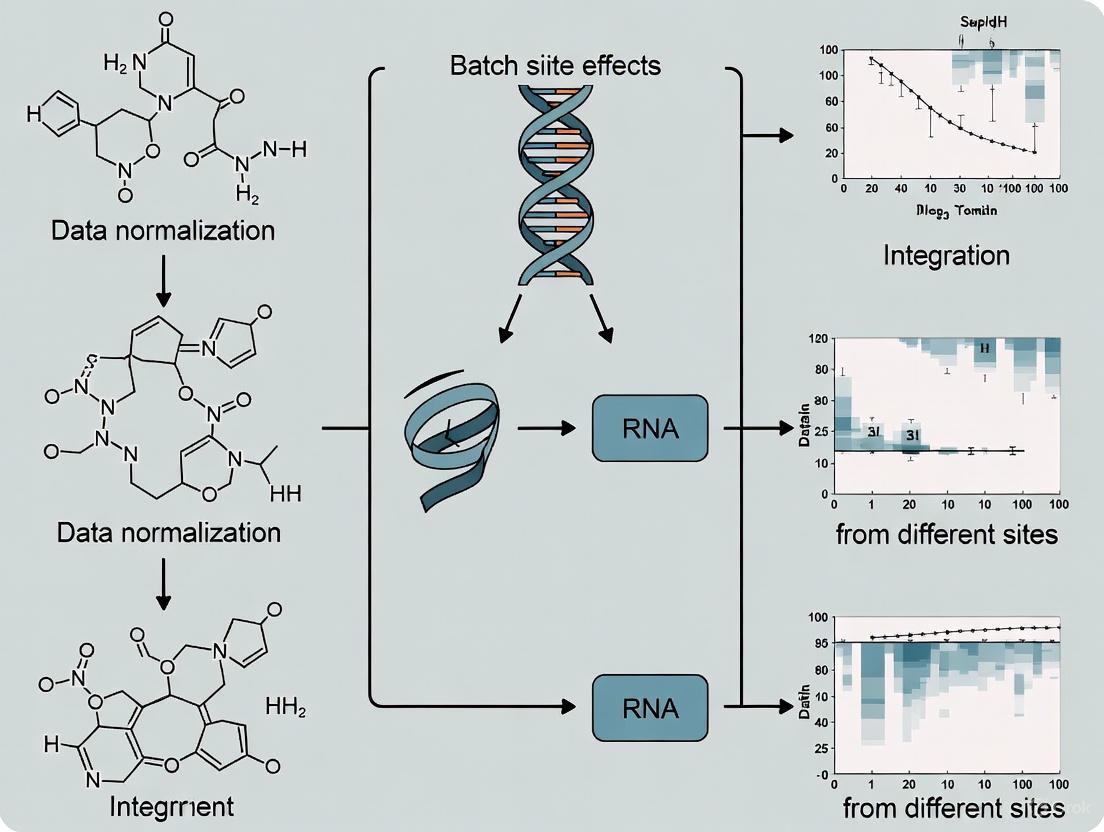
The following diagram illustrates the process of identifying and correcting for batch effects, leading to reliable data integration.
The Scientist's Toolkit: Research Reagent Solutions
| Item | Function in Batch Effect Correction |
|---|---|
| Reference Materials | Well-characterized, stable standards (e.g., certified cell lines, synthetic controls) profiled in every batch to serve as an internal baseline for ratio-based correction methods [2]. |
| Standardized Protocols | Detailed, step-by-step procedures for sample preparation, storage, and data generation to minimize the introduction of technical variation across sites and batches [1]. |
| Control Samples | Samples with known expected outcomes, used to monitor technical performance and identify deviations that may indicate batch effects. |
| Batch Effect Correction Algorithms (BECAs) | Software tools (e.g., ComBat, Harmony, custom ratio-scaling scripts) that statistically adjust the data to remove technical variation while preserving biological signal [2] [4]. |
| 5,6-Epoxyergosterol | 5,6-Epoxyergosterol|High-Purity Reference Standard |
| (5e,7z)-5,7-Dodecadienal | (5E,7Z)-5,7-Dodecadienal|180.29 g/mol |
FAQs: Data Integration & Batch Effects in Multi-Site Embryo Studies
Q1: What is a batch effect, and why is it a critical problem in multi-site embryo studies?
Batch effects are technical variations in data that are not due to the biological subject of study but arise from factors like different labs, equipment, reagent lots, operators, or processing times [6] [7]. In multi-site embryo studies, these effects can severely skew analysis, leading to misleading outcomes, such as a large number of false-positive or false-negative findings [6]. For example, a change in experimental solution can cause shifts in calculated risk, potentially leading to incorrect conclusions or treatment decisions [6]. Batch effects are a major cause of the irreproducibility crisis, raising questions about the reliability of data collected from different batches or platforms [6].
Q2: My multi-omics embryo data comes from different labs. Which batch-effect correction algorithm (BECA) should I use?
The choice of algorithm depends on your experimental design and the type of data you have. Recent large-scale benchmarks have identified several top-performing methods:
- For general multi-omics data (transcriptomics, proteomics, metabolomics): A comprehensive assessment found that the ratio-based method (Ratio-G) is highly effective, especially when batch effects are completely confounded with biological factors of interest. This method involves scaling absolute feature values of study samples relative to those of concurrently profiled reference materials [6].
- For image-based profiling data (e.g., Cell Painting): Benchmarks suggest that Harmony and Seurat RPCA are among the top performers for integrating data across different laboratories and microscopes [7].
- Guidance: If you have a common reference material profiled in every batch, a ratio-based method is highly recommended. For other scenarios, Harmony and Seurat RPCA are strong, computationally efficient choices [6] [7].
Q3: What is a "confounded scenario," and why is it particularly challenging?
A confounded scenario occurs when the biological factor you are studying (e.g., a specific treatment or embryo stage) is completely aligned with the batch. For instance, if all control embryos are processed in Batch 1 and all treated embryos are processed in Batch 2, it becomes nearly impossible to distinguish true biological differences from technical batch variations [6]. In such cases, many standard batch correction methods may fail or even remove the biological signal of interest. The ratio-based method has been shown to be particularly effective in tackling these confounded scenarios [6].
Q4: How common are chromosomal abnormalities in early embryos, and how does this impact data integration?
Chromosomal abnormalities are remarkably common during early embryogenesis. Research indicates that over 70% of fertilized eggs from infertile patients can have chromosome aberrations, which are a primary cause of embryonic lethality and miscarriages [8]. These errors lead to mosaic embryos, where cells with normal genomes coexist with cells exhibiting abnormal genomes [8]. The frequency of these errors is temporarily elevated, with one study pinpointing the 4-cell stage in mouse embryos as a period of particular instability, where 13% of cells showed chromosomal abnormalities [9]. This inherent biological variability adds a significant layer of complexity when integrating data across multiple sites, as technical batch effects must be distinguished from this genuine biological noise.
Troubleshooting Guides
Guide 1: Troubleshooting Batch Effect Correction Failures
| Symptom | Possible Cause | Solution |
|---|---|---|
| Biological signal is lost after correction. | Over-correction in a confounded batch-group scenario. | Apply a ratio-based correction method using a common reference sample profiled in all batches [6]. |
| Poor integration of new data with a existing corrected dataset. | Model-based methods require full re-computation with new data. | Use methods like Harmony or Seurat that can project new data into an existing corrected space, or re-run the correction on the entire combined dataset [7]. |
| Batch effects persist after correction. | Inappropriate method selected for the data type or scenario. | Refer to benchmarking studies: switch to a top-performing method like Harmony or Seurat RPCA for image data [7], or a ratio-based method for multi-omics data [6]. |
| Introduced new artifacts or false patterns in the data. | Over-fitting or incorrect assumptions by the algorithm. | Always visually inspect results (PCA/t-SNE plots) pre- and post-correction. Validate findings with known biological controls. |
Guide 2: Troubleshooting Chromosomal Analysis in Early Embryos
| Symptom | Possible Cause | Solution |
|---|---|---|
| High rates of aneuploidy (abnormal chromosome number) in embryos. | Meiotic errors from the oocyte, which increase with maternal age [8] [10]. | Consider maternal age and oocyte quality as factors. For research, utilize models like the "synthetic oocyte aging" system to study these errors [10]. |
| Mosaic embryos (mix of normal and abnormal cells). | Mitotic errors after fertilization, such as chromosome segregation errors during early cleavages [8] [11]. | Focus on the early cleavage divisions (particularly the 4- to 8-cell transition). Use sensitive single-cell analysis methods like scRepli-seq to detect these errors [9]. |
| Inconsistent results in preimplantation genetic testing (PGT-A). | Technical limitations of PGT-A and the biological reality of mosaicism [8]. | Acknowledge that PGT-A cannot detect all abnormalities. Results should be interpreted with caution by a clinical geneticist. |
Summarized Data & Protocols
Table 1: Performance of Selected Batch Effect Correction Algorithms
| Algorithm | Core Approach | Best For | Key Performance Finding |
|---|---|---|---|
| Ratio-Based (Ratio-G) [6] | Scales feature values relative to a common reference material. | Multi-omics data; Confounded batch-group scenarios. | "Much more effective and broadly applicable than others" in confounded designs [6]. |
| Harmony [7] | Iterative mixture-based correction using PCA. | Image-based profiling; scRNA-seq data. | Consistently ranked among the top three methods; good balance of batch removal and biological signal preservation [7]. |
| Seurat RPCA [7] | Reciprocal PCA and mutual nearest neighbors. | Large, heterogeneous datasets (e.g., from multiple labs). | Consistently ranked among the top three methods; computationally efficient [7]. |
| ComBat [7] | Bayesian framework to model additive/multiplicative noise. | - | Performance is surpassed by newer methods like Harmony and Seurat in several benchmarks [7]. |
Table 2: Key Reagents and Materials for Embryo Data Integration Studies
| Item | Function in Research | Application Note |
|---|---|---|
| Reference Materials (e.g., Quartet Project materials) [6] | Provides a technical baseline for correcting batch effects across labs and platforms. | Should be profiled concurrently with study samples in every batch for ratio-based correction. |
| Cell Painting Assay [7] | A multiplexed image-based profiling assay to capture rich morphological data from cells. | Used to generate high-content data for phenotyping embryo cells under various perturbations. |
| scRepli-seq [8] [9] | A single-cell genomics technique to detect DNA replication timing and chromosomal aberrations. | Critical for identifying chromosome copy number abnormalities and replication stress in single embryonic cells. |
| API-based EMR Integration [12] | Allows AI tools to connect securely with Electronic Medical Record systems. | Enables seamless data flow for AI-driven analysis of embryo images and patient data in clinical workflows. |
Experimental Protocol: Implementing Ratio-Based Batch Effect Correction
Purpose: To effectively remove batch effects in multi-omics studies, especially in confounded scenarios where biological groups are processed in separate batches.
Materials:
- Multi-omics datasets from multiple batches.
- Reference material (e.g., from the Quartet Project [6]) profiled in every batch.
Methodology:
- Experimental Design: In each batch of your study, concurrently profile your study samples alongside one or more aliquots of a well-characterized reference material.
- Data Generation: Process all samples (study and reference) using the same platform and protocol within a batch.
- Ratio Calculation: For each feature (e.g., gene expression, protein abundance) in each study sample, calculate a ratio value. This is done by dividing the absolute feature value of the study sample by the corresponding value from the reference material profiled in the same batch.
Ratio = Feature_value_study_sample / Feature_value_reference_material - Data Integration: Use the resulting ratio-based values for all downstream analyses and data integration across batches. This scaling effectively normalizes out batch-specific technical variations [6].
Visual Workflows & Diagrams
Batch Effect Correction Workflow
Embryo Aneuploidy Origins
For researchers in multi-site embryo studies, the pursuit of reproducible, high-impact findings is often confounded by a pervasive technical challenge: batch effects. These are technical sources of variation introduced when samples are processed in different batches, across different laboratories, by different personnel, or at different times. In multi-center research, where collaboration and large sample sizes are essential, failing to account for batch effects can lead to misleading conclusions and irreproducibility. This guide presents real-world case studies and data to illustrate the profound consequences of batch effects and provides a toolkit for their identification and correction.
FAQs: Understanding the Impact of Batch Effects
What are batch effects and why are they a critical concern in multi-site studies?
Batch effects are technical, non-biological variations in data that are introduced by differences in experimental conditions [13] [14]. These can arise from a multitude of sources, including:
- Different sequencing runs or instruments
- Variations in reagent lots
- Changes in sample preparation protocols
- Different personnel handling the samples
- Experiments conducted over multiple days or weeks [15]
In multi-site embryo studies, where samples are processed across different laboratories, these effects are magnified. They are a critical concern because they can confound biological signals, making it difficult or impossible to distinguish true biological differences from technical artifacts. This can lead to increased variability, reduced statistical power, and, in the worst cases, incorrect conclusions [16] [17].
Can batch effects truly lead to retracted papers and misdirected clinical decisions?
Yes, the impact of batch effects can be severe and far-reaching. Published case studies demonstrate serious consequences:
- Clinical Misclassification: In a clinical trial, a change in the RNA-extraction solution introduced a batch effect in gene expression profiles. This resulted in an incorrect gene-based risk calculation, leading to 162 patients being misclassified, 28 of whom subsequently received incorrect or unnecessary chemotherapy regimens [16].
- Article Retraction: A high-profile study published a novel fluorescent serotonin biosensor. The authors later discovered that the biosensor's sensitivity was highly dependent on the batch of fetal bovine serum (FBS) used. When the FBS batch changed, the key results could not be reproduced, leading to the retraction of the article [16].
- Spurious Biological Findings: One analysis suggested that gene expression differences between human and mouse species were greater than the differences between tissues within the same species. A re-analysis revealed that the data from the two species were generated three years apart. After batch effect correction, the data clustered by tissue type rather than by species, indicating the initial conclusion was an artifact of batch effects [16].
What is the largest source of technical variability in multi-laboratory cell phenotyping studies?
In a systematic multi-site assessment of reproducibility in high-content cell phenotyping, the largest source of technical variability was found to be laboratory-to-laboratory variation [18].
This study involved five laboratories using an identical protocol and key reagents to generate live-cell imaging data on cell migration. A Linear Mixed Effects (LME) model was used to quantify variability at different hierarchical levels. While biological variability (between cells and over time) was substantial, technical variability contributed a median of 32% of the total variance across all measured variables. Within this technical variability, the lab-to-lab component was the most significant, followed by variability between persons, experiments, and technical replicates [18].
The study further showed that simply combining data from different labs without correction almost doubled the cumulative technical variability [18].
How can I visually identify batch effects in my own data?
Before attempting correction, it is crucial to diagnose the presence of batch effects. Several common visualization methods can help:
- Principal Component Analysis (PCA): Perform PCA on your raw data and color the data points by batch. If the samples cluster strongly based on their batch rather than their biological condition (e.g., treatment vs. control), it indicates a strong batch effect [19] [20] [15].
- t-SNE or UMAP Plots: These dimensionality reduction techniques can also reveal batch effects. In the presence of a batch effect, cells or samples from the same batch will cluster together, even if they are from different biological groups [19] [20].
The diagram below illustrates the logical workflow for diagnosing and addressing batch effects.
What are the signs that my batch effect correction may have been too aggressive (over-correction)?
Over-correction occurs when batch effect removal algorithms also remove genuine biological signal. Key signs include:
- Distinct Cell Types Cluster Together: On a PCA or UMAP plot, distinct biological cell types that should form separate clusters are merged into one [19] [20].
- Loss of Expected Markers: Canonical cell-type-specific markers (e.g., known markers for a specific T-cell subtype) are absent from differential expression analysis after correction [19].
- Poor Marker Quality: A significant portion of the genes identified as cluster-specific markers are housekeeping genes, like ribosomal genes, which are broadly expressed across cell types and lack biological specificity [19] [20].
- Complete Overlap of Different Conditions: After correction, samples from very different biological conditions or experiments show a complete overlap, which is biologically implausible [20].
Case Study: Multi-Site Assessment of Cell Migration
Experimental Protocol
A landmark study designed to quantify sources of variability in high-content imaging involved three independent laboratories [18].
- Objective: To determine the sources of variability (biological and technical) in live-cell imaging data of migrating cancer cells.
- Cell Line: HT1080 fibrosarcoma cells, stably expressing fluorescent labels (LifeActâ€mCherry and H2Bâ€EGFP).
- Standardization: A detailed common protocol, the cell line, and all key reagents were distributed to all participating labs to minimize biological and technical variance.
- Nested Design: The experiment followed a nested structure with three laboratories, three persons per lab, three independent experiments per person, two conditions (control and ROCK inhibitor) per experiment, and three technical replicates per condition.
- Imaging & Analysis: Automated fluorescent microscopes with environmental chambers were used. All microscope-derived images were transferred to a single laboratory for uniform processing and quantification using CellProfiler and custom Matlab scripts [18].
Key Findings and Quantitative Data
The study used a Linear Mixed Effects (LME) model to partition the variance for 18 different cell morphology and migration variables. The table below summarizes the median proportion of total variance attributed to each source.
Table 1: Sources of Variance in Multi-Site Cell Phenotyping Data [18]
| Source of Variance | Type | Median Proportion of Total Variance |
|---|---|---|
| Between Laboratories | Technical | Major Source |
| Between Persons | Technical | Moderate Source |
| Between Experiments | Technical | Minor Source |
| Between Technical Replicates | Technical | Minor Source |
| Between Cells (within a population) | Biological | Substantial |
| Within Cells (over time) | Biological | Substantial |
Key Conclusion: Despite rigorous standardization, laboratory-to-laboratory variation was the dominant technical source of variability. This prevented high-quality meta-analysis of the primary data. However, the study also found that batch effect removal methods could markedly improve the ability to combine datasets from different laboratories for perturbation analyses [18].
The Scientist's Toolkit
Research Reagent Solutions
Table 2: Essential Materials for Batch Effect Monitoring and Correction
| Item | Function in Batch Effect Management |
|---|---|
| Quality Control Standards (QCS) | A standardized reference material (e.g., a tissue-mimicking gelatin matrix with a controlled analyte like propranolol) run alongside experimental samples to monitor technical variation across slides, days, and laboratories [21]. |
| Common Cell Line | Using an identical, stable cell line across all sites (e.g., HT1080 fibrosarcoma as in the case study) minimizes biological variability, allowing researchers to isolate technical batch effects [18]. |
| Common Reagent Lots | Distributing aliquots from the same lot of key reagents (e.g., fetal bovine serum, collagen, enzymes) to all participating labs prevents reagent-based variability [18] [16]. |
| Detailed Common Protocol | A single, rigorously detailed experimental protocol ensures consistency in sample handling, preparation, and imaging across all personnel and sites [18]. |
| Emeguisin A | Emeguisin A|Depsidone |
| Nanangenine H | Nanangenine H |
Computational Correction Methods
A wide array of computational tools exists to correct for batch effects. The choice of method depends on the data type (e.g., bulk RNA-seq, single-cell RNA-seq, proteomics) and the experimental design.
Table 3: Common Batch Effect Correction Algorithms
| Method | Brief Description | Common Use Cases |
|---|---|---|
| ComBat / ComBat-seq | Uses an empirical Bayes framework to adjust for batch effects. ComBat-seq is designed specifically for raw count data from RNA-seq [22] [15]. | Bulk RNA-seq, Microarray, Proteomics |
limma (removeBatchEffect) |
Uses a linear model to remove batch effects from normalized expression data [22] [15]. | Bulk RNA-seq, Microarray |
| Harmony | Iteratively clusters cells across batches and corrects them, maximizing diversity within each cluster. Known for its speed and efficiency [19] [14] [20]. | Single-cell RNA-seq |
| Seurat Integration | Uses Canonical Correlation Analysis (CCA) and mutual nearest neighbors (MNNs) to find "anchors" between datasets for integration [19] [14] [20]. | Single-cell RNA-seq |
| Mutual Nearest Neighbors (MNN) | Identifies pairs of cells that are nearest neighbors in each batch and uses them to infer the batch correction vector [19] [14]. | Single-cell RNA-seq |
| BERT | A high-performance, tree-based framework for integrating large-scale, incomplete omics datasets, leveraging ComBat or limma at each node of the tree [22]. | Large-scale multi-omics |
The following diagram illustrates how a tool like BERT hierarchically integrates data from multiple batches.
Best Practices Workflow
To effectively manage batch effects in multi-site embryo studies, a proactive and comprehensive strategy is required.
Troubleshooting Guide: Common Batch-Effect Correction Issues
1. Problem: Loss of Differential Expression Signals After Correction
- Why it happens: Many batch-effect correction methods over-correct the data, removing subtle but biologically meaningful expression differences along with technical variations [4] [16].
- Solution: Implement an order-preserving correction method. These methods, often based on monotonic deep learning networks, are specifically designed to maintain the original ranking of gene expression levels within each cell, thereby protecting differential expression patterns [4].
- Check: Compare the Spearman correlation of gene expression rankings before and after correction. A method that preserves order will show a high correlation coefficient [4].
2. Problem: Distorted Inter-Gene Correlations
- Why it happens: Methods focused solely on cell alignment across batches can disrupt the co-expression relationships between genes, which are crucial for understanding regulatory networks [4].
- Solution: Choose algorithms that incorporate the preservation of inter-gene correlation into their objective function. Evaluate the retention of significantly correlated gene pairs post-correction using Pearson and Kendall correlation metrics [4].
- Check: For a given cell type, identify significantly correlated gene pairs in the original batches. Calculate the root mean square error (RMSE) of these correlations after correction; lower values indicate better preservation [4].
3. Problem: Poor Integration of Datasets with High Missing Value Rates
- Why it happens: Incomplete omic profiles (e.g., from proteomics or metabolomics) are common in multi-site studies. Standard correction tools may fail or introduce significant data loss when faced with many missing values [22].
- Solution: Use a framework designed for incomplete data, such as Batch-Effect Reduction Trees (BERT). BERT decomposes the integration task into a binary tree, allowing it to handle features that are missing completely in some batches without the massive data loss associated with other methods [22].
- Check: Monitor the percentage of retained numeric values after correction. Methods like BERT are designed to retain all non-missing values, whereas others can lose over 50% of the data in high-missingness scenarios [22].
4. Problem: Inability to Handle Imbalanced or Confounded Study Designs
- Why it happens: Batch effects can be confounded with biological outcomes of interest (e.g., if all controls were processed in one batch and all cases in another). Standard correction cannot distinguish these technical from biological effects [16].
- Solution: Leverage methods that allow for the specification of covariates and reference samples. Providing this information helps the algorithm model and preserve biological conditions. BERT, for instance, allows users to define samples with known covariates as references to guide the correction of unknown samples [22].
- Check: Always visualize the data using UMAP or t-SNE colored by both batch and biological condition before correction. If they are perfectly confounded, statistical correction is risky, and a design-based solution is preferable [16].
5. Problem: Over-Correction Leading to the Loss of Rare Cell Types
- Why it happens: Procedural correction methods that separate the correction step from clustering can inadvertently remove subtle biological signals, such as those from small populations of rare cells [4].
- Solution: Consider methods that integrate batch-effect correction with cell clustering, or employ metrics like the Local Inverse Simpson's Index (LISI) to evaluate both batch mixing (high LISI) and cell-type purity (low LISI) after correction [4].
- Check: After correction, verify that the clusters corresponding to known rare cell types still contain a representative number of cells and exhibit expected marker gene expression [4].
Frequently Asked Questions (FAQs)
Q1: What does "order-preserving" mean in the context of batch-effect correction, and why is it critical for my analysis? A1: "Order-preserving" refers to a correction method's ability to maintain the original relative rankings of gene expression levels within each cell or batch after processing [4]. This is critical because the relative abundance of transcripts, not just their presence or absence, drives biological interpretation. Disrupting this order can lead to false conclusions in downstream analyses like differential expression or pathway enrichment studies [4].
Q2: How can I quantitatively assess if my batch-effect correction has successfully preserved biological signals? A2: You should use a combination of metrics to get a complete picture [4]:
- For Batch Mixing: Use the Local Inverse Simpson's Index (LISI). A higher LISI score indicates better mixing of batches.
- For Cell-Type Purity: Use the Average Silhouette Width (ASW) with respect to biological labels. A higher ASW indicates cells of the same type are more compact and distinct from other types.
- For Biological Structure: Use the Adjusted Rand Index (ARI) to compare clustering results before and after correction.
- For Gene Relationships: Calculate the correlation (e.g., Pearson) of inter-gene correlations before and after correction [4].
Q3: My multi-site embryo study has severe data incompleteness (many missing values). Which correction methods are suitable? A3: Traditional methods struggle with this, but the BERT (Batch-Effect Reduction Trees) framework is specifically designed for integrating incomplete omic profiles [22]. Unlike other methods that can lose up to 88% of numeric values when blocking batches, BERT's tree-based approach retains all non-missing values, making it highly suitable for sparse data from embryo studies [22].
Q4: Are there trade-offs between effectively removing batch effects and preserving the biological truth of my data? A4: Yes, this is a fundamental challenge. Overly aggressive correction can remove biological variation along with batch effects, a phenomenon known as "over-correction" [16]. This is why choosing a method with features like order-preservation and correlation-maintenance is crucial, as they are explicitly designed to minimize this trade-off by protecting intrinsic biological patterns during the correction process [4].
Quantitative Data on Correction Method Performance
The table below summarizes key performance metrics for several batch-effect correction methods, highlighting the importance of specialized features.
Table 1: Comparison of Batch-Effect Correction Method Performance
| Method / Feature | Preserves Gene Order? | Retains Inter-Gene Correlation? | Handles Incomplete Data? | Key Performance Metric |
|---|---|---|---|---|
| ComBat [4] | Yes [4] | Moderate [4] | No (requires complete matrix) | Good for basic correction, but hampered by scRNA-seq sparsity [4]. |
| Harmony [4] | Not Applicable (output is embedding) [4] | Not Evaluated | No | Effective for cell alignment and visualization [4]. |
| Seurat v3 [4] | No [4] | No [4] | No | Good cell-type clustering, but can distort gene-gene correlations [4]. |
| MMD-ResNet [4] | No [4] | No [4] | No | Uses deep learning for distribution alignment [4]. |
| Order-Preserving Method (Global) [4] | Yes [4] | High [4] | No | Superior in maintaining Spearman correlation and differential expression signals [4]. |
| BERT [22] | Not Specified | Not Specified | Yes [22] | Retains >99% of numeric values vs. up to 88% loss with other methods on 50% missing data [22]. |
Table 2: Evaluation Metrics for Biological Signal Preservation
| Metric | What it Measures | Ideal Outcome | How to Calculate |
|---|---|---|---|
| Spearman Correlation [4] | Preservation of gene expression ranking before vs. after correction. | Coefficient close to 1. | Non-parametric correlation of expression values for each gene. |
| Inter-Gene Correlation RMSE [4] | Preservation of correlation structure between gene pairs. | Low RMSE value. | Root Mean Square Error of Pearson correlations for significant gene pairs before and after correction [4]. |
| ASW (Biological Label) [4] [22] | Compactness of biological groups (e.g., cell types). | Value close to 1. | ( ASW = \frac{1}{N} \sum{i=1}^{N} \frac{bi - ai}{\max(ai, bi)} ) where (ai) is mean intra-cluster distance and (b_i) is mean nearest-cluster distance for cell (i) [22]. |
| LISI (Batch) [4] | Diversity of batches in local neighborhoods (batch mixing). | High score. | Inverse Simpson's index calculated for each cell's local neighborhood. |
Experimental Protocol: Evaluating an Order-Preserving Correction
This protocol outlines the steps to assess a batch-effect correction method's performance in a multi-site embryo study context.
Objective: To validate that a batch-effect correction method successfully removes technical variation while preserving the order of gene expression and inter-gene correlation structure.
Input: A raw, merged gene expression matrix (cells x genes) from multiple batches (sites/labs), with associated metadata for batch ID and known biological labels (e.g., embryo developmental stage).
Procedure:
Preprocessing and Initial Clustering:
- Apply standard scRNA-seq preprocessing (normalization, log-transformation, highly variable gene selection).
- Perform initial cell clustering on the uncorrected data using a graph-based method (e.g., Seurat's FindClusters) to establish a baseline for cell-type identification [4].
Application of Batch-Effect Correction:
Quantitative Evaluation of Order and Correlation Preservation:
- Gene Order Preservation:
- For each batch, select a major cell type and a rare cell type.
- For each gene, calculate the Spearman correlation coefficient between its raw (non-zero) expression values and its corrected values.
- Summarize the distribution of these correlations (e.g., median, IQR) for each method. A method that preserves order will have a distribution centered near 1 [4].
- Inter-Gene Correlation Preservation:
- For a stable cell type present in multiple batches, identify gene pairs that are significantly correlated (same direction, FDR-adjusted p-value < 0.05) in the raw data of both batches [4].
- Calculate the Pearson correlation for these gene pairs in the corrected data.
- Compute the RMSE between the pre- and post-correlation values for these pairs. A lower RMSE indicates better preservation [4].
- Gene Order Preservation:
Visualization and Final Assessment:
- Generate UMAP plots colored by batch and by biological label for both the raw and corrected data.
- A successful correction will show mixed batches but distinct, well-separated biological clusters.
- Compare the quantitative metrics from Step 3 across methods to select the one that best preserves biological truth.
The workflow for this protocol is summarized in the following diagram:
The Scientist's Toolkit: Key Research Reagent Solutions
Table 3: Essential Computational Tools and Resources
| Item | Function / Purpose | Example / Note |
|---|---|---|
| Order-Preserving Algorithm | A correction method that uses a monotonic deep learning network to maintain the original ranking of gene expression values, crucial for protecting differential expression signals [4]. | The "global monotonic model" described in [4]. |
| BERT Framework | A high-performance, tree-based data integration method for incomplete omic profiles. It minimizes data loss and can handle severely imbalanced conditions using covariates and references [22]. | Available as an R package from Bioconductor [22]. |
| Reference Samples | A set of samples with known biological covariates (e.g., a specific embryo stage) processed across multiple batches. Used to guide the correction of unknown samples and account for design imbalance [22]. | For example, include two samples of a known cell type in every batch to anchor the correction [22]. |
| Covariate Metadata | Structured information (e.g., in a .csv file) detailing the batch ID, biological condition, and other relevant factors (e.g., donor sex) for every sample. Essential for informing correction algorithms what variation to preserve [22]. | Must be complete and accurately linked to each sample in the expression matrix. |
| Quality Control Metrics (ASW, LISI, ARI) | A set of standardized metrics to quantitatively evaluate the success of integration, balancing batch removal against biological preservation [4] [22]. | Use ASW on biological labels and LISI on batch ID for a balanced view [4]. |
| Rhamnitol | Rhamnitol, CAS:1114-16-5, MF:C6H14O5, MW:166.17 g/mol | Chemical Reagent |
| Azukisaponin VI | Azukisaponin VI, CAS:82801-39-6, MF:C54H86O25, MW:1135.2 g/mol | Chemical Reagent |
The logical relationship between the key components of a successful batch-effect correction strategy is shown below:
A Practical Toolkit: Batch Effect Correction Methods for Single-Cell and Spatial Embryo Data
In multi-site embryo studies, the integration of data from different labs, protocols, and points in time is essential for robust biological discovery. However, this integration is challenged by batch effects—systematic technical variations that can obscure true biological signals. This guide provides a technical deep dive into four prominent batch-effect correction algorithms, offering troubleshooting advice and protocols to empower your research.
Core Algorithm Principles and Troubleshooting FAQs
What are the fundamental differences in how these algorithms work?
The core batch-effect correction methods differ significantly in their underlying mathematical approaches and the scenarios for which they are best suited.
| Algorithm | Core Principle | Primary Data Type | Key Assumption |
|---|---|---|---|
| ComBat | Empirical Bayes framework to adjust for known batch variables by modeling and shrinking batch effect estimates. [4] [23] [24] | Bulk RNA-seq, Microarrays | Batch effects are consistent across genes; population composition is similar across batches. [25] |
| limma | Linear modeling to remove batch effects as a covariate in the design matrix, without altering the raw data for downstream testing. [24] | Bulk RNA-seq, Microarrays | Batch effects are additive and known in advance. [23] |
| Harmony | Iterative clustering in PCA space with soft clustering and a diversity penalty to maximize batch mixing. [26] [3] | scRNA-seq, Multi-omics | Biological variation can be separated from technical batch variation in a low-dimensional space. [26] |
| MNN Correct | Identifies Mutual Nearest Neighbors (pairs of cells of the same type across batches) to estimate and correct cell-specific batch vectors. [26] [25] | scRNA-seq | A subset of cell populations is shared between batches; batch effect is orthogonal to biological subspace. [25] |
| Cryptolepinone | Cryptolepinone, CAS:160113-29-1, MF:C16H12N2O, MW:248.28 g/mol | Chemical Reagent | Bench Chemicals |
| Glycoside H2 | Glycoside H2, CAS:73529-43-8, MF:C56H92O25, MW:1165.3 g/mol | Chemical Reagent | Bench Chemicals |
Algorithm Workflow Selection
How do I choose between ComBat and limma's removeBatchEffect for my bulk transcriptomics data?
The choice hinges on whether you need to correct the data matrix for visualization or include batch in your statistical model for differential expression.
- Use
limma removeBatchEffectfor exploratory analysis and visualization: This function is ideal for creating PCA plots or heatmaps where you want to remove the batch effect to see the underlying biological structure more clearly. It works by fitting a linear model that includes your batch as a covariate and then removes its effect. Critically, the original data for differential testing remains unchanged; batch is included as a covariate in the final model. [24] - Use ComBat for a powerful correction when batch is known: ComBat uses an empirical Bayes approach to shrink the batch effect estimates towards the overall mean, which is particularly beneficial when you have many batches or small sample sizes per batch. This makes it robust, but it directly modifies your data. [4] [24] [25] A key limitation is that it assumes the composition of cell populations is the same across batches, which can lead to overcorrection if this is not true. [25]
- Best Practice Recommendation: For differential expression analysis, the most reliable method is often to include 'batch' as a covariate in your statistical model (e.g., in
DESeq2orlimma) without pre-correcting the data with ComBat orremoveBatchEffect. This approach models the effect of batch without altering the raw counts, reducing the risk of introducing artifacts. [24]
My data has different cell type compositions across batches. Which method should I use to avoid overcorrection?
This is a common challenge where bulk methods like ComBat and limma fail, as they assume uniform cell type composition. In this scenario, methods designed for single-cell data are superior.
- The Problem: If you use ComBat on data where one cell type is only present in one batch, the algorithm will incorrectly interpret the unique gene expression profile of that cell type as a batch effect and attempt to remove it, thereby erasing true biological variation. [25]
- Recommended Solution: MNN Correct or Harmony. These methods are explicitly designed to handle differing cell type compositions. [26] [25]
- MNN Correct works by identifying "mutual nearest neighbors"—pairs of cells from different batches that are most similar to each other. It assumes these pairs represent the same cell type and uses them to estimate the batch effect, which is then applied to all cells. This allows it to correct only the shared cell populations without forcing all populations to align. [25]
- Harmony operates in a PCA space and iteratively clusters cells while applying a penalty that encourages each cluster to include cells from multiple batches. This allows it to successfully integrate batches even when cell type abundances vary significantly. [26]
- Troubleshooting Tip: To diagnose overcorrection, use the RBET (Reference-informed Batch Effect Testing) framework. It uses reference genes (e.g., housekeeping genes) with stable expression to evaluate correction quality. A good correction should show low RBET values, while overcorrection will cause the value to rise again as biological signal is degraded. [27]
I am working with a confounded study design where my biological groups are processed in completely separate batches. Is correction even possible?
This is one of the most difficult scenarios, as biological and technical effects are perfectly correlated. Most standard methods will fail, as they cannot distinguish biology from batch.
- The Challenge: In a confounded design (e.g., all control samples in Batch 1 and all treatment samples in Batch 2), any attempt to remove the "batch effect" will also remove the biological differences you are trying to study. [2]
- Advanced Solution: Ratio-Based Scaling with Reference Materials. The most robust solution is to use a strategy that relies on external controls. If you profile a common reference material (e.g., a standardized control sample or pooled sample) in every batch, you can transform your data into ratios relative to that reference. [28] [2]
- Protocol: For each gene in each sample, calculate
Ratio = Expression_in_Study_Sample / Expression_in_Reference_Material. This scales all batches to a common baseline, effectively removing the batch-specific technical variation and revealing the true biological differences between groups, even in confounded designs. [2]
- Protocol: For each gene in each sample, calculate
- Application Note: This ratio-based method (Ratio-G) has been shown to be "much more effective and broadly applicable than others" in confounded scenarios for multi-omics data, including transcriptomics and proteomics. [2]
After using Harmony, I no longer have a full gene expression matrix for differential expression. What went wrong?
This is not an error but a fundamental characteristic of how some modern batch-correction methods operate.
- Understanding Output Types: Methods like Harmony and fastMNN perform correction in a low-dimensional space (e.g., after PCA). Their output is an integrated embedding, not a corrected count matrix for all genes. This embedding is excellent for visualization, clustering, and cell type annotation but cannot be used directly for standard differential expression tests on genes. [27] [26]
- Alternative Workflows:
- For DE analysis after Harmony integration: First, use the corrected embedding to identify cell populations or clusters. Then, perform differential expression testing using the original, uncorrected counts, but use the cell clusters or states identified from the integrated data as the biological variable of interest.
- Choose a method that returns a matrix: If your workflow requires a corrected gene expression matrix, select a method that provides one, such as ComBat, MNN Correct, limma's removeBatchEffect, or Scanorama. [27] [26]
Essential Research Reagent Solutions
The following reagents and computational resources are critical for implementing the protocols discussed above.
| Reagent / Resource | Function in Batch-Effect Correction | Example Use Case |
|---|---|---|
| Reference Materials | Provides a technical baseline for ratio-based correction methods. Enables correction in confounded study designs. [2] | Quartet Project reference materials (D5, D6, F7, M8) for multi-omics data. [28] [2] |
| Housekeeping Gene Panel | Serves as biologically stable reference genes for evaluating overcorrection (e.g., in the RBET framework). [27] | Pancreas-specific housekeeping genes for validating batch correction in pancreas cell data. [27] |
| Precision Biological Samples | Technical replicates across batches to assess correction performance via metrics like CV or SNR. [28] [2] | Triplicates of donor samples within each batch in the Quartet datasets. [2] |
Performance Evaluation Protocol
To quantitatively evaluate the success of any batch-effect correction method in your embryo study, implement the following protocol using a combination of metrics.
Step 1: Assess Batch Mixing
- Metric: Local Inverse Simpson's Index (LISI). LISI measures the diversity of batches in the local neighborhood of each cell. A higher LISI score indicates better mixing of batches. [27] [26]
- Metric: k-nearest neighbor Batch Effect Test (kBET). kBET tests if the local batch distribution around each cell matches the global distribution. A lower rejection rate indicates successful local mixing. [27] [26]
Step 2: Assess Biological Signal Preservation
- Metric: Adjusted Rand Index (ARI). ARI compares the cell clustering results before and after integration. A high ARI indicates that the biological cell type identities have been preserved. [4] [26]
- Metric: Average Silhouette Width (ASW). ASW evaluates the compactness and separation of cell type clusters. A high ASW indicates that cell types remain well-defined after correction. [4] [26]
- Metric: Reference-informed Batch Effect Test (RBET). This newer metric uses reference genes to evaluate correction quality and is uniquely sensitive to overcorrection, making it highly valuable. [27]
The table below summarizes the ideal outcomes for a successful correction.
| Evaluation Aspect | Key Metric | Target Outcome |
|---|---|---|
| Batch Mixing | LISI [26] | High Score |
| kBET rejection rate [26] | Low Score | |
| Biology Preservation | ARI [4] | High Score |
| ASW (cell type) [4] | High Score | |
| Overcorrection Awareness | RBET [27] | Biphasic (Optimal mid-range) |
Correction Evaluation Workflow
Troubleshooting Guides and FAQs
Frequently Asked Questions
Q1: What are the primary technical challenges when integrating incomplete omic data from multiple research sites? Integrating incomplete omic data from multiple sites presents two core challenges: batch effects (technical variations from different labs, protocols, or instruments that can confound biological signals) and data incompleteness (missing values common in high-throughput omic technologies). These issues are particularly pronounced in multi-site studies where biological and technical factors are often confounded, making it difficult to distinguish true biological signals from technical artifacts [22] [16] [6].
Q2: My data has different covariates distributed unevenly across batches. Can BERT handle this? Yes. BERT allows specification of categorical covariates (e.g., biological conditions) and can model these conditions using modified design matrices in its underlying algorithms (ComBat and limma). This preserves covariate effects while removing batch effects, which is crucial for severely imbalanced or sparsely distributed conditions [22].
Q3: How does BERT's performance compare to HarmonizR when dealing with large datasets? BERT demonstrates significant performance advantages over HarmonizR. In simulation studies with up to 50% missing values, BERT retained all numeric values, while HarmonizR's "unique removal" strategy led to substantial data loss (up to 88% for blocking of 4 batches). BERT also showed up to 11× runtime improvement by leveraging multi-core and distributed-memory systems [22].
Q4: What should I do when my phenotype of interest is completely confounded with batch? In fully confounded scenarios where biological groups separate completely by batch, standard correction methods may fail. The most effective approach is using a ratio-based method with reference materials. By scaling feature values of study samples relative to concurrently profiled reference materials in each batch, you can effectively distinguish biological from technical variations [6].
Q5: Are there scenarios where batch effect correction should not be applied? Yes, caution is needed when batch effects are minimal or when over-correction might remove biological signals. Always assess batch effect severity using metrics like Average Silhouette Width (ASW) before correction. Visualization techniques (PCA, t-SNE) should show batch mixing improvement while preserving biological group separation after correction [13].
Common Experimental Issues and Solutions
Problem: High data loss after running HarmonizR with default settings.
- Cause: HarmonizR's default "unique removal" strategy introduces additional data loss by removing features with insufficient values across batches [22].
- Solution: Consider using BERT instead, which retains all numeric values by propagating features with values from only one batch to the next correction level. If using HarmonizR, explore different blocking strategies, though this may still result in significant data loss [22].
Problem: Batch correction removes my biological signal of interest.
- Cause: This typically occurs in confounded designs where biological groups correlate perfectly with batches. Most algorithms cannot distinguish biological from technical variation in this scenario [6] [13].
- Solution: Implement a reference-based design using ratio scaling (Ratio-G). Profile reference materials alongside study samples in each batch, then transform expression data relative to reference values. This preserves biological differences while removing technical variations [6].
Problem: Unexpected clustering by processing date rather than biological group.
- Cause: Batch effects from temporal variations (different sequencing runs, reagent lots, or operators) are common, even within the same laboratory [16] [13].
- Solution: Include processing date as a batch variable in correction algorithms. For future experiments, balance biological groups across processing dates and use reference materials for longitudinal quality control [16].
Problem: Algorithm fails with "insufficient replicates" error.
- Cause: ComBat and limma (used by both BERT and HarmonizR) require at least two numerical values per feature per batch [22].
- Solution: BERT automatically handles this by removing singular numerical values (typically <1% of values) and propagating single-batch features. Ensure your data meets minimum requirements: each feature should have sufficient representation in at least some batches [22].
Performance Comparison: BERT vs. HarmonizR
Table 1: Quantitative comparison of BERT and HarmonizR performance characteristics
| Performance Metric | BERT | HarmonizR (Full Dissection) | HarmonizR (Blocking of 4) |
|---|---|---|---|
| Data Retention | Retains all numeric values | Up to 27% data loss with 50% missing values | Up to 88% data loss with 50% missing values |
| Runtime Improvement | Up to 11× faster (vs. HarmonizR) | Baseline | Varies by blocking strategy |
| Covariate Handling | Supports categorical covariates and reference samples | Limited capabilities | Limited capabilities |
| ASW Improvement | Up to 2× improvement for imbalanced conditions | Standard performance | Standard performance |
| Parallelization | Multi-core and distributed-memory systems | Embarrassingly parallel sub-matrices | Block-based parallelization |
Table 2: Algorithm suitability for different experimental scenarios
| Experimental Scenario | Recommended Tool | Key Considerations |
|---|---|---|
| Highly incomplete data (>30% missing values) | BERT | Superior data retention; preserves more features for analysis |
| Balanced batch-group design | Either tool | Both perform well when biological groups evenly distributed across batches |
| Confounded batch-group design | BERT with reference samples | Use covariate handling; ratio-based scaling recommended |
| Large-scale datasets (>1000 samples) | BERT | Better scalability and parallelization capabilities |
| Limited computational resources | HarmonizR with blocking | Reduced memory footprint with batch grouping |
| Unknown covariate levels | BERT with reference designation | Can estimate effects from references, apply to non-references |
Experimental Protocols
Protocol 1: Implementing BERT for Multi-Site Embryo Omic Data
Principle: BERT decomposes data integration into a binary tree of batch-effect correction steps, using ComBat or limma for features with sufficient data while propagating single-batch features [22].
Step-by-Step Procedure:
- Input Preparation: Format data as data.frame or SummarizedExperiment object. Ensure samples are annotated with batch information and biological covariates [22].
- Parameter Configuration: Set parallelization parameters (P = number of processes, R = reduction factor, S = sequential processing threshold). Default values typically suffice for initial runs [22].
- Reference Specification: Designate samples with known covariates as references. BERT will use these to estimate batch effects while preserving biological signals [22].
- Algorithm Execution: Run BERT using established Bioconductor implementation. Monitor progress through quality control outputs [22].
- Output Validation: Verify integration using Average Silhouette Width (ASW) scores. Compare pre- and post-integration values for both batch and biological labels [22].
Protocol 2: Reference-Based Ratio Scaling for Confounded Designs
Principle: Transform absolute feature values to ratios relative to concurrently profiled reference materials, effectively separating biological from technical variations [6].
Step-by-Step Procedure:
- Reference Selection: Choose appropriate reference materials that represent the biological system under study. For embryo research, this might include pooled samples or well-characterized reference cell lines [6].
- Concurrent Profiling: Process reference materials alongside study samples in every batch, maintaining consistent processing protocols across sites [6].
- Ratio Calculation: For each feature in every sample, calculate ratio = samplevalue / referencevalue. Use median scaling when multiple reference replicates are available [6].
- Data Integration: Proceed with integrated analysis using ratio-scaled data. Biological signals will be preserved while batch-specific technical variations are minimized [6].
Research Reagent Solutions
Table 3: Essential materials for robust multi-omics batch effect correction
| Reagent/Material | Function in Batch Correction | Implementation Considerations |
|---|---|---|
| Reference Materials | Enables ratio-based scaling; monitors technical variation | Select materials biologically relevant to study system; ensure long-term availability |
| Quality Control Metrics | Quantifies batch effect severity and correction success | Implement ASW, PCA visualization, and signal-to-noise ratios |
| Covariate Annotation | Preserves biological effects during technical correction | Comprehensive sample metadata collection; standardized formatting |
| Multiomics Standards | Facilitates integration across different data types | Use consortium-developed standards (Quartet Project materials) |
| Computational Resources | Enables processing of large-scale datasets | High-performance computing environment; adequate memory allocation |
Validation and Quality Control
Pre- and Post-Correction Assessment
Visualization: Generate PCA and t-SNE plots colored by both batch and biological groups before and after correction. Successful correction shows batches mixing while biological groups remain distinct [13].
Quantitative Metrics:
- Average Silhouette Width (ASW): Measures separation between batches (ASWbatch) and biological groups (ASWlabel). Effective correction decreases ASWbatch while maintaining or increasing ASWlabel [22].
- Signal-to-Noise Ratio (SNR): Quantifies biological signal preservation after correction [6].
- Differential Expression Analysis: Compare results before and after correction; credible correction should yield biologically plausible findings [6].
Implementation in Multi-Site Embryo Studies
For embryo-specific research, consider these adaptations:
- Use embryo-stage-matched reference materials when possible
- Account for developmental timing as a critical covariate
- Implement cross-site standardization of embryo processing protocols
- Establish consensus on minimal quality thresholds for embryo quality metrics
By implementing these troubleshooting guides, experimental protocols, and validation procedures, researchers can effectively address data incompleteness and batch effects in multi-site embryo omic studies, ensuring robust and reproducible integration of incomplete omic profiles.
In multi-site embryo studies, integrating single-cell RNA sequencing (scRNA-seq) data from different batches or laboratories is a fundamental challenge. Batch effects—systematic technical variations—can obscure true biological signals, complicating the analysis of complex processes like embryonic development. Order-preserving batch-effect correction is a methodological advancement that maintains the original relative rankings of gene expression levels within each batch after integration. This feature is crucial for preserving biologically meaningful patterns, such as gene regulatory relationships and differential expression signals. Monotonic Deep Learning Networks, which enforce constrained input-output relationships, have emerged as a powerful tool to achieve this correction while ensuring model interpretability. This technical support article provides troubleshooting guides and FAQs to help researchers successfully implement these methods in their experiments.
FAQs & Troubleshooting Guides
Theory and Conceptual Understanding
Q1: What does "order-preserving" mean in the context of batch-effect correction, and why is it important for my embryo studies?
A: Order-preserving correction maintains the original relative rankings of gene expression levels for each gene within every cell, after correcting for batch effects [4]. In technical terms, if a gene X has a higher expression level than gene Y in a specific cell before correction, this relationship is preserved after correction.
- Why it matters: This property is vital for downstream biological analysis. Disrupting the original order of gene expression can:
- Skew Gene-Gene Correlations: Artificially alter the relationships between genes, leading to incorrect inferences about gene regulatory networks [4].
- Compromise Differential Expression: Obscure true differentially expressed genes between different embryonic stages or tissue regions [4].
- Reduce Interpretability: Make it difficult to relate the corrected data back to the original biological question.
Q2: How do Monotonic Deep Learning Networks enforce order-preservation?
A: A Monotonic Deep Learning Network is a structurally constrained neural network. It contains specialized layers or modules (e.g., an Isotonic Embedding Module) that ensure the network's output is a monotonic function of its input for specified features [29] [30]. This means that as the input value for a particular gene increases, the network's corrected output for that gene is guaranteed to either always increase or always stay the same, thereby preserving the original expression order.
Experimental Design and Setup
Q3: I am designing a multi-site embryo study. What preliminary steps can I take to facilitate effective order-preserving correction later?
A: Proactive experimental design is key.
- Metadata Collection: Meticulously record all batch-related metadata (e.g., sequencing platform, laboratory of origin, sample preparation date, technician). This information is essential for the correction model.
- Include Biological Controls: If possible, include replicate samples or control cell lines across different batches. This provides a biological ground truth to validate the correction method's performance.
- Plan for Integration: Choose a monotonic deep learning method, like a global monotonic model, that is designed for multi-batch integration from the start, rather than relying on pairwise methods that can be sensitive to the order of batch processing [4] [31].
Q4: Which specific monotonic models are available for batch-effect correction?
A: Research in this area is evolving. The table below summarizes key model types based on current literature:
| Model Type / Concept | Key Mechanism | Reference in Literature |
|---|---|---|
| Global Monotonic Model | Ensures order-preservation for all genes without additional conditions. | [4] |
| Partial Monotonic Model | Ensures order-preservation based on the same initial condition or matrix. | [4] |
| Deep Isotonic Embedding Network (DIEN) | Uses separate modules for monotonic and non-monotonic features, combining them linearly for an intuitive structure. | [29] |
| MonoNet | Employs monotonically connected layers to ensure monotonic relationships between high-level features and outputs. | [30] |
Implementation and Technical Troubleshooting
Q5: I'm getting poor clustering results after applying a monotonic correction model. What could be wrong?
A: Poor integration can stem from several issues. Use the following troubleshooting table to diagnose the problem.
| Symptom | Potential Cause | Solution |
|---|---|---|
| Low clustering accuracy (Low ARI) and distinct batch clusters. | The model is failing to mix cells from different batches. | Verify that technical differences are smaller than true biological variations (e.g., between cell types), as this is a key assumption for many methods [31]. |
| Loss of rare cell populations. | The correction method is over-smoothing the data. | Ensure the method's loss function or architecture is designed to preserve biological heterogeneity. Some methods integrate clustering with correction to protect rare cell types [31] [32]. |
| Poor preservation of inter-gene correlation. | The correction method is disrupting gene-gene relationships. | Switch to or validate with a method specifically designed to preserve inter-gene correlation, which is a strength of order-preserving approaches [4]. |
Q6: How do I quantitatively evaluate if my order-preserving correction was successful?
A: You should use a combination of metrics that assess both batch mixing and biological fidelity. The table below outlines the key metrics.
| Evaluation Goal | Metric | What it Measures | Desired Outcome |
|---|---|---|---|
| Batch Mixing | Local Inverse Simpson's Index (LISI) [4] | Diversity of batches in local cell neighborhoods. | Higher LISI score indicates better mixing. |
| Clustering Accuracy | Adjusted Rand Index (ARI) [4] [31] | Similarity between clustering results and known cell type labels. | Higher ARI indicates clusters align better with true biology. |
| Cluster Compactness | Average Silhouette Width (ASW) [4] | How similar a cell is to its own cluster compared to other clusters. | Higher ASW indicates tighter, more distinct clusters. |
| Order-Preservation | Spearman Correlation [4] | Preservation of gene expression rankings before and after correction. | Correlation close to 1 indicates perfect order preservation. |
| Inter-Gene Correlation | Root Mean Square Error (RMSE) / Pearson Correlation [4] | Preservation of correlation structures between gene pairs. | Low RMSE and High Pearson correlation indicate success. |
Data Interpretation and Biological Validation
Q7: The corrected data looks well-mixed, but my differential expression analysis yields unexpected results. What should I check?
A: This can indicate that batch effects were removed at the cost of true biological signal.
- Validate with Ground Truth: Check the expression of known marker genes for key cell types in your embryo data (e.g., markers for neural tube, somites). They should still be differentially expressed in the appropriate clusters after correction.
- Leverage Order-Preservation: Use the order-preserving property of your model to your advantage. Since the relative expression levels are maintained, you can have higher confidence that drastic changes in differential expression are due to the removal of confounding technical noise rather than an artifact of the correction process [4].
- Inspect Latent Space: Use visualization tools like UMAP or t-SNE to see if the cell types are separating based on biology rather than batch in the corrected low-dimensional embedding [32] [33].
Essential Experimental Protocols
Protocol 1: Benchmarking an Order-Preserving Correction Method
This protocol outlines steps to evaluate a new monotonic deep learning model for batch-effect correction, using established metrics.
1. Data Preprocessing:
- Input: Raw gene expression matrices from multiple batches (e.g., different embryo studies).
- Filtering: Remove low-quality cells and genes. Normalize for sequencing depth.
- Feature Selection: Identify Highly Variable Genes (HVGs) to reduce dimensionality and computational load [32].
2. Model Application:
- Setup: Configure the monotonic deep learning model (e.g., global or partial), specifying which features (genes) are subject to the monotonic constraint.
- Training: Train the model on the integrated multi-batch dataset. The model will learn to map the data to a corrected space while preserving expression orders.
3. Performance Evaluation:
- Generate Corrected Output: Obtain the batch-corrected gene expression matrix from the model.
- Calculate Metrics:
- Compute LISI and ARI on the corrected data to assess batch mixing and clustering accuracy.
- For a subset of cells, calculate the Spearman correlation between the original (non-zero) expression values and the corrected values for each gene to confirm order-preservation [4].
- Calculate the Pearson correlation for known correlated gene pairs before and after correction to assess preservation of biological relationships [4].
Protocol 2: Validating Cross-Platform Integration in Embryonic Mouse Data
This protocol describes a specific experiment to test a method's ability to integrate data from different spatial transcriptomics platforms.
1. Data Collection:
- Acquire spatially resolved transcriptomics (SRT) data of embryonic mouse tissues (e.g., at stage E11.5) from at least two different technology platforms (e.g., 10x Visium and MERFISH) [34].
2. Data Integration:
- Apply the order-preserving correction framework (e.g., a method like SpaCross [34]) to integrate the datasets. The method should perform 3D spatial registration to align coordinates and construct a unified graph.
3. Biological Validation:
- Identify Spatial Domains: Cluster the integrated data to identify anatomical structures (e.g., dorsal root ganglion, heart tube).
- Check for Conservation: Verify that known, conserved anatomical regions (e.g., neural tube) are correctly identified as single, coherent domains in the integrated data.
- Check for Specificity: Confirm that stage-specific structures are also accurately captured, demonstrating that the method preserves biological variation while removing technical noise [34].
Method Workflow and Signaling Pathways
Order-Preserving Batch Correction Workflow
The following diagram illustrates the general workflow for applying a monotonic deep learning network to correct batch effects while preserving gene expression orders, as described in the protocols.
Monotonic Neural Network Architecture
This diagram outlines the core architecture of a monotonic network (e.g., DIEN [29]), showing how it processes different types of features to ensure a monotonic output.
The Scientist's Toolkit: Research Reagent Solutions
The following table lists key computational tools and resources essential for implementing order-preserving batch effect correction.
| Item / Resource | Function / Description | Relevance to Experiment |
|---|---|---|
| Monotonic DL Frameworks (e.g., code for DIEN [29], MonoNet [30]) | Pre-built neural network architectures with monotonicity constraints. | Provides the core engine for performing order-preserving corrections without building a model from scratch. |
| scRNA-seq Analysis Suites (e.g., Scanpy in Python, Seurat in R) | Comprehensive environments for single-cell data preprocessing, visualization, and analysis. | Used for initial data QC, normalization, HVG selection, and for running downstream analyses on the corrected data. |
| Evaluation Metrics Scripts (Custom or from publications) | Code to calculate ARI, LISI, ASW, and Spearman correlation. | Essential for quantitatively benchmarking the performance of the correction method against alternatives. |
| High-Performance Computing (HPC) / GPU Access | Access to powerful computational resources. | Training deep learning models on large-scale scRNA-seq data (e.g., millions of cells) is computationally intensive and often requires GPUs. |
| Public scRNA-seq Datasets (e.g., with known batch effects) | Benchmarking data from repositories like the Human Cell Atlas. | Used as positive controls to test and validate the correction method's performance on real-world, challenging data [31] [32]. |
| Lehmbachol D | Lehmbachol D|Hydroxylated Stilbenolignan|466.5 g/mol | Lehmbachol D is a hydroxylated stilbenolignan for research. It exhibits anti-inflammatory activity. This product is For Research Use Only. Not for human or veterinary use. |
| Greveichromenol | Greveichromenol, MF:C15H14O5, MW:274.27 g/mol | Chemical Reagent |
Troubleshooting Guides and FAQs
Frequently Asked Questions
Q1: What is the main advantage of using Crescendo over other batch integration tools for embryo studies? Crescendo performs batch correction directly on the gene expression count data, rather than on a lower-dimensional embedding. This is crucial for multi-site embryo research because it allows for the direct visualization and analysis of individual genes across different samples or developmental stages, preserving the ability to map specific gene patterns in anatomical context [35].
Q2: During batch correction, how can I be sure that true biological variation from my embryo samples isn't being removed? Effective batch correction must balance removing technical artifacts with preserving biological variance. Tools like Crescendo and SpaCross are designed to address this. You can evaluate this using specific metrics:
- BVR (Batch-Variance Ratio): Quantifies the reduction in batch-related variance (you want BVR < 1) [35].
- CVR (Cell-Type-Variance Ratio): Quantifies the preservation of cell-type-related variance (CVR ≥ 0.5 is generally good) [35]. Monitoring these metrics helps ensure biological integrity is maintained.
Q3: My multi-slice embryo data has significant physical deformations between sections. Can spatial batch correction methods handle this? Yes, methods like SpaCross are specifically designed for this challenge. They employ 3D spatial registration algorithms, such as Iterative Closest Point (ICP), to align spatial coordinates across different slices before batch correction, overcoming geometric integration obstacles [34].
Q4: For my embryonic tissue study, I need to integrate data from different sequencing platforms. Is this possible? Yes, cross-technology integration is a key application for advanced batch correction methods. Crescendo has been demonstrated to successfully integrate data from spatial transcriptomics platforms with single-cell RNA-seq datasets, enabling the transfer of information across technologies [35].
Common Issues and Solutions
| Problem | Cause | Solution |
|---|---|---|
| Poor visualization of spatial gene patterns | Strong batch effects obscuring consistent biological patterns across samples [35]. | Apply gene-level batch correction (e.g., Crescendo) to facilitate accurate visualization of gene expression across batches [35]. |
| Loss of important gene-gene correlations | The batch correction method disrupts the original relational structure of the data [4]. | Use an order-preserving correction method that maintains inter-gene correlation structures crucial for understanding regulatory networks [4]. |
| Inability to balance local and global spatial information | The model fails to integrate local spatial continuity with global semantic consistency [34]. | Implement a framework like SpaCross that uses an Adaptive Hybrid Spatial-Semantic Graph (AHSG) to dynamically balance both types of information [34]. |
| Low cDNA concentration after amplification | Low RNA quality or very low cellular density in starting tissue sample [36]. | Re-amplify the cDNA, using 3-6 PCR cycles. For problematic libraries, run a reconditioning PCR with 3 cycles [36]. |
Experimental Protocols & Workflows
Detailed Methodology for Crescendo Batch Correction
Crescendo uses generalized linear mixed modeling to correct for batch effects directly in the raw count matrix, while also capable of imputing lowly-expressed genes. The following workflow diagram illustrates the key steps researchers need to follow.
Crescendo Workflow
Protocol Steps:
- Input Preparation: Collect the unfiltered gene-by-cell counts matrix, cell-type annotations for each cell, and batch information (e.g., sample ID, sequencing run). Cell-type information is a critical input, as Crescendo assumes batch effects are cell-type-specific [35].
- Biased Downsampling (Optional for Scalability): To enable model fitting on large datasets (e.g., millions of cells), perform a biased downsampling that maintains representation of rare cell states and all batches. The full batch correction is still applied to all cells [35].
- Estimation Step: Crescendo fits a generalized linear model to estimate how much variation in each gene's expression is derived from biological sources (cell-type identity) versus technical confounders (batch effects) [35].
- Marginalization Step: Using the model from the estimation step, Crescendo infers a batch-free model of gene expression [35].
- Matching Step: The original model and the batch-free model are used to sample new, batch-corrected counts for the expression matrix. The output is a complete, batch-corrected count matrix amenable to downstream analysis [35].
Performance Evaluation Metrics
After performing batch correction, it is essential to quantitatively evaluate its success. The following table summarizes key metrics used in spatial transcriptomics studies.
| Metric | Formula/Calculation | Ideal Value | Interpretation |
|---|---|---|---|
| Batch-Variance Ratio (BVR) [35] | Ratio of batch-related variance after vs. before correction. | < 1 | Indicates successful reduction of batch effects. |
| Cell-Type-Variance Ratio (CVR) [35] | Ratio of cell-type-related variance after vs. before correction. | ≥ 0.5 | Indicates good preservation of biological variation. |
| Local Inverse Simpson's Index (LISI) [4] | Diversity score measuring batch mixing and cell-type separation. | High for batches, Low for cell types. | Measures integration quality (mixing & separation). |
| Adjusted Rand Index (ARI) [4] | Measures similarity between two clusterings (e.g., vs. ground truth). | Closer to 1. | Measures clustering accuracy against known labels. |
The Scientist's Toolkit
Research Reagent Solutions
| Item | Function | Application Note |
|---|---|---|
| Seeker Spatial Transcriptomics Kit [36] | Enables whole-transcriptome spatial mapping from fresh-frozen tissues. | Compatible with all species without protocol optimization. Uses a 10µm CryoCube overlay to prevent tissue drying and mRNA diffusion [36]. |
| CryoCube Overlay [36] | A section melted on top of the tissue to keep it attached and prevent drying. | Essential for high-quality data; prevents mRNA leakage, especially at tissue borders [36]. |
| SPRI Beads [36] | Magnetic beads for size-selective purification of cDNA and libraries. | Used in cleanup steps post-cDNA amplification and library preparation. A 0.6x volume ratio is typical [36]. |
| Visium Spatial Gene Expression Slide [37] | Glass slide arrayed with spatially barcoded oligonucleotides to capture mRNA. | The standard starting point for 10x Visium protocols. Each spot (55 µm) may contain 10-30 cells [37]. |
| Rubifolic acid | Rubifolic Acid | Rubifolic acid for research applications. This product is for Research Use Only (RUO) and is not intended for diagnostic or personal use. |
| 3',3'''-Biapigenin | 3',3'''-Biapigenin|Research Compound | Research-grade 3',3'''-Biapigenin, a bioactive biflavonoid fromSelaginella doederleiniiwith studied antitumor properties. For Research Use Only. Not for human consumption. |
Method Comparison for Embryo Studies
Choosing the right algorithm is critical. The table below compares key methods, highlighting their relevance to multi-site embryo research.
| Method | Core Algorithm | Key Feature | Relevance to Multi-Site Embryo Studies |
|---|---|---|---|
| Crescendo [35] | Generalized Linear Mixed Model (GLMM) | Corrects raw gene counts; enables direct gene visualization. | Ideal for tracking 3D gene expression patterns across serial embryonic sections [35]. |
| SpaCross [34] | Cross-Masked Graph Autoencoder | Integrates local spatial continuity & global semantic consistency. | Identifies both conserved and stage-specific structures (e.g., dorsal root ganglion) across developmental stages [34]. |
| Order-Preserving Method [4] | Monotonic Deep Learning Network | Maintains inter-gene correlation and expression rankings. | Preserves crucial gene regulatory relationships that define embryonic development [4]. |
| Harmony [35] | Linear Model on PCA Embeddings | Iteratively corrects lower-dimensional embeddings. | A common predecessor; does not correct raw counts, limiting direct gene visualization [35]. |
Spatial Analysis Workflow Diagram
The following diagram outlines a complete analytical workflow for a multi-site embryo study, from raw data to biological insight, incorporating the tools and methods discussed.
Full Spatial Analysis Workflow
In multi-site embryo studies, single-cell RNA sequencing (scRNA-seq) has revolutionized our ability to study lineage allocation and cell fate decisions. However, the data from different laboratories, sequencing platforms, and experimental batches introduce technical variations known as batch effects that can confound biological interpretation and lead to misleading conclusions [1]. For research on early human development, where sample scarcity and ethical considerations already present significant challenges, batch effects pose a substantial threat to data reproducibility and validity [38].
Explainable AI (XAI) models like X-scPAE (eXplained Single Cell PCA - Attention Auto Encoder) have emerged as powerful solutions that not only predict embryonic lineage allocation with high accuracy but also provide interpretable insights into the key genes driving these predictions while accounting for technical variations [39]. This technical support guide addresses common challenges and provides actionable protocols for researchers implementing these approaches in embryo studies.
Frequently Asked Questions (FAQs)
1. What is the difference between normalization and batch effect correction?
- Normalization operates on the raw count matrix and addresses technical variations such as sequencing depth across cells, library size, and amplification bias caused by gene length.
- Batch Effect Correction mitigates technical variations arising from different sequencing platforms, timing, reagents, or different conditions/laboratories. Most methods operate on dimensionality-reduced data, though some (like ComBat and Scanorama) can correct the full expression matrix [19].
2. How can I detect batch effects in my single-cell embryo data?
- Principal Component Analysis (PCA): Examine scatter plots of the top principal components. Sample separation attributed to batch rather than biological source indicates batch effects.
- t-SNE/UMAP Plot Examination: Visualize cell groups labeled by batch number. Before correction, cells from different batches often cluster separately; after correction, biological similarities should drive clustering.
- Quantitative Metrics: Utilize metrics like k-nearest neighbor batch effect test (kBET), adjusted rand index (ARI), or normalized mutual information (NMI) to objectively measure batch integration [19].
3. What are the signs of overcorrection in batch effect correction?
- Cluster-specific markers comprising genes with widespread high expression (e.g., ribosomal genes)
- Substantial overlap among markers specific to different clusters
- Absence of expected canonical cell-type markers
- Scarcity of differential expression hits in pathways expected based on sample composition [19]
4. When should I use reference materials for batch effect correction?
Reference materials are particularly valuable in confounded scenarios where biological factors of interest (e.g., developmental stage) are completely aligned with batch factors. In such cases, ratio-based correction using reference materials outperforms most other methods [2] [6].
Experimental Protocols
Protocol 1: Implementing the X-scPAE Framework for Lineage Prediction
Purpose: To predict embryonic lineage allocation while identifying and interpreting key genes involved in development.
Methodology:
- Data Preprocessing: Standardize raw single-cell transcriptomic data from human and mouse embryos using quality control metrics.
- Dimensionality Reduction: Apply Principal Component Analysis (PCA) to reduce data dimensionality and rank importance of principal components.
- Feature Extraction: Utilize an autoencoder with integrated attention mechanism to capture interactions between features.
- Model Interpretation: Apply the Counterfactual Gradient Attribution (CGA) algorithm to calculate feature importance and identify key predictor genes.
- Validation: Validate model performance using logistic regression built with extracted key genes and compare against baseline algorithms [39].
Table 1: X-scPAE Performance Metrics on Embryonic Lineage Prediction
| Metric | Test Set Performance | Validation Set Performance |
|---|---|---|
| Accuracy | 0.945 | 0.977 |
| F1-Score | 0.94 | Not reported |
| Precision | 0.94 | Not reported |
| Recall | 0.94 | Not reported |
Protocol 2: Reference Material-Based Ratio Correction
Purpose: To effectively correct batch effects in confounded experimental designs.
Methodology:
- Reference Selection: Choose appropriate reference materials (e.g., Quartet Project reference materials) to be profiled concurrently with study samples in each batch.
- Ratio Calculation: Transform expression profiles of each sample to ratio-based values using expression data of the reference sample(s) as denominator.
- Data Integration: Apply ratio-scaled values to integrate data across multiple batches, platforms, or laboratories.
- Quality Assessment: Evaluate correction efficacy using quantitative metrics and visualization techniques [2] [6].
Protocol 3: Multi-Omics Batch Effect Correction Assessment
Purpose: To evaluate batch effect correction algorithm performance across transcriptomics, proteomics, and metabolomics data.
Methodology:
- Scenario Design: Create both balanced and confounded experimental scenarios using reference materials.
- Algorithm Application: Test multiple batch effect correction algorithms (BMC, ComBat, Harmony, SVA, RUVg, RUVs, Ratio-based scaling).
- Performance Evaluation: Assess using (1) signal-to-noise ratio, (2) relative correlation coefficients, (3) accuracy of differentially expressed feature identification, (4) predictive model robustness, and (5) clustering accuracy [2] [6].
Table 2: Batch Effect Correction Algorithm Comparison
| Algorithm | Best Use Case | Strengths | Limitations |
|---|---|---|---|
| Ratio-based Scaling | Confounded batch-group scenarios | Effective across omics types; preserves biological signals | Requires reference materials |
| Harmony | Balanced batch-group scenarios | Efficient integration; handles multiple batches | May underperform in strongly confounded cases |
| ComBat | Balanced designs with known batch effects | Established method; good for transcriptomics | Can remove biological signal in confounded designs |
| Seurat Integration | Single-cell data integration | Uses CCA and MNN for alignment | Computationally intensive for very large datasets |
| MNN Correct | Single-cell data with shared cell types | Directly aligns datasets based on mutual nearest neighbors | High computational demands |
The Scientist's Toolkit: Essential Research Reagents & Materials
Table 3: Key Research Reagents for Embryonic Lineage Tracing and Batch Correction
| Reagent/Material | Function/Application | Example Use Cases |
|---|---|---|
| Quartet Project Reference Materials | Multi-omics quality control and batch correction | Provides DNA, RNA, protein, and metabolite references from matched cell lines for cross-platform standardization [2] |
| scRNA-seq Platform Controls | Technical variation assessment | 10x Genomics platform controls for monitoring batch effects introduced during library preparation [14] |
| Tamoxifen (TAM)-inducible CreER Systems | Lineage tracing in model organisms | Enables temporal control of genetic labeling for embryonic lineage fate mapping [40] |
| Fluorescent Reporter Genes (e.g., tdTomato, GFP) | Cell lineage visualization | Allows tracking of progenitor cells and their descendants in embryonic development studies [40] |
| Standardized Culture Media for Embryo Models | Reduction of technical variability | Minimizes batch effects introduced through variations in reagent lots or composition [1] |
| 7-Ketoisodrimenin | 7-Ketoisodrimenin, MF:C15H20O3, MW:248.32 g/mol | Chemical Reagent |
Workflow Visualization
X-scPAE Model Architecture for Interpretable Lineage Prediction
Batch Effect Correction Decision Workflow
From Challenge to Solution: Troubleshooting Design Flaws and Optimizing Correction Performance
Troubleshooting Guide: Common Experimental Design Issues
Q1: In our multi-site embryo study, we suspect that technical batch effects are confounded with our biological groups. How can we identify this problem?
Confounding occurs when technical effects are mixed with the biological effects you are trying to study, creating a distorted view of the true relationship between variables [41]. In multi-site studies, this often manifests as batch effects where different sites or processing batches correspond to different biological or treatment groups.
- Primary Symptoms: Your analysis reveals strong batch-specific patterns that align with your biological groups. For example, all control samples were processed in Batch A while all treatment samples were processed in Batch B.
- Diagnostic Tools:
- PCA Visualization: Create a PCA plot colored by batch and another colored by biological group. If the patterns look strikingly similar, confounding is likely present [2].
- Statistical Tests: Check if the distribution of known prognostic factors (e.g., patient age, embryo quality) differs significantly between treatment groups across batches [41].
- Stratified Analysis: Temporarily analyze your data within each batch. If the biological signal disappears or weakens considerably within individual batches, confounding is probable [42].
Q2: Our randomized clinical trial (RCT) in embryo research shows baseline differences between groups. Did randomization fail?
Not necessarily. The primary purpose of randomization is not to produce perfectly balanced groups but to eliminate systematic bias [43]. Randomization ensures that any differences in known and unknown prognostic factors occur only by chance. While perfect balance is ideal, observed differences do not invalidate the randomization process. Statistical adjustment during analysis can account for these chance imbalances [43].
Q3: What are the most effective statistical methods to correct for confounding when it cannot be avoided in the study design?
When experimental designs are "premature, impractical, or impossible," researchers must rely on statistical methods to adjust for confounding effects [42]. The choice of method depends on your data type and the number of confounders.
Table: Statistical Methods for Confounding Adjustment
| Method | Best For | Key Principle | Considerations |
|---|---|---|---|
| Stratification [42] | A small number of categorical confounders. | Analyzes the exposure-outcome relationship within homogeneous groups (strata) where the confounder does not vary. | Becomes impractical with multiple confounders or continuous variables. |
| Multivariate Regression (Linear/Logistic) [42] | Adjusting for multiple confounders simultaneously. | Uses mathematical modeling to isolate the effect of the exposure from other variables in the model. | Provides an "adjusted" odds ratio or effect estimate. Requires a sufficient sample size. |
| Analysis of Covariance (ANCOVA) [42] | Models with a continuous outcome and mix of categorical/continuous predictors. | Combines ANOVA and regression to test for group effects after removing variance explained by continuous covariates. | Increases statistical power by accounting for covariate-outcome relationships. |
| Ratio-Based Scaling [2] | Multi-batch omics studies where a reference material is available. | Scales absolute feature values of study samples relative to those of a concurrently profiled reference material. | Particularly effective when batch effects are completely confounded with biological factors [2]. |
Q4: How should we determine the timing of randomization in an embryo diagnostic trial?
The principle is to randomize as close as possible to the point when the study intervention would be used [43]. For an embryo diagnostic trial, this means:
- Do not randomize before ovarian stimulation.
- Do randomize only after a minimum number of embryos have been created, immediately before the diagnostic intervention would be applied (e.g., biopsy for genetic testing) [43]. This approach minimizes protocol deviations and participant drop-out before the intervention, leading to clearer interpretation of results [43].
Experimental Protocol: Implementing a Reference-Based Design to Combat Confounding
The following workflow is adapted from large-scale multi-omics studies and can be integrated into multi-site embryo research to technically control for batch effects, even in confounded designs [2].
Objective: To generate comparable data across multiple sites and batches, even when the distribution of biological groups is unbalanced across batches.
Reagents and Materials:
- Common Reference Material: A well-characterized, stable biological sample (e.g., a reference cell line or pooled sample) aliquoted and distributed to all participating sites [2].
- Standardized Reagent Kits: Ensure all sites use the same lot of critical reagents where possible.
- Detailed Protocol Documentation: A single, detailed standard operating procedure (SOP) for all experimental steps.
Procedure:
- Experimental Setup: In each batch at each site, process both the study samples and a pre-determined aliquot of the common reference material concurrently using the same protocol [2].
- Data Generation: Generate your primary experimental data (e.g., genetic, transcriptomic, or proteomic profiles) for all samples, including the reference.
- Ratio-Based Transformation: For each feature (e.g., gene expression level) in each study sample, calculate a ratio value relative to the value of that same feature in the reference material profiled in the same batch.
Ratio = Feature_value_study_sample / Feature_value_reference_material
- Data Integration: Use these ratio-scaled values for all downstream comparative analyses instead of the raw, absolute values [2].
Troubleshooting:
- High Variance in Reference Measurements: This indicates technical instability. Investigate protocol adherence, reagent quality, and equipment calibration across sites.
- Persistent Batch Clustering: The ratio method may be insufficient if technical effects are extreme. Consider combining it with other adjustment methods or reviewing the initial experimental process.
The diagram below illustrates the logical workflow for diagnosing and addressing confounding in your study design.
Diagram: Pathway for Addressing Confounded Designs
The Scientist's Toolkit: Research Reagent Solutions
Table: Essential Materials for Robust Multi-Site Studies
| Reagent / Material | Function in Preventing Confounding |
|---|---|
| Common Reference Materials [2] | Serves as a technical benchmark across all batches and sites, enabling ratio-based scaling to remove batch-specific noise. |
| Standardized Protocol Kits | Minimizes variation introduced by differences in reagents, lot numbers, or lab-specific protocols, a common source of batch effects. |
| Blinded Sample Labels | Helps prevent conscious or unconscious bias in sample processing and analysis, especially in non-blinded trial designs [43]. |
| Quality Control (QC) Metrics | Provides objective data to identify out-of-control batches or sites before full data generation and integration. |
Navigating Severely Imbalanced or Sparse Data with Covariates and Reference Samples
In multi-site embryo studies, researchers often face significant technical hurdles when integrating datasets. Batch effects—technical variations introduced due to processing samples at different times, locations, or with different protocols—are notoriously common in omics data and can lead to misleading outcomes if not properly addressed [1]. The challenges are magnified when dealing with:
- Severely Imbalanced Data: Occurring when biological groups of interest are unevenly distributed across batches [2].
- Sparse Data: Characterized by a high proportion of missing values or zero counts, a challenge particularly acute in single-cell RNA-sequencing (scRNA-seq) where up to 80% of gene expression values can be zero [19].
- Confounded Designs: A critical problem where batch effects are completely confounded with biological factors, making it nearly impossible to distinguish technical artifacts from true biological signals [2].
These challenges are particularly pronounced in longitudinal and multi-center embryo studies, where subtle developmental changes must be distinguished from technical variations introduced across different laboratories or processing times [1].
FAQs and Troubleshooting Guides
FAQ 1: How can I detect batch effects in my dataset?
Answer: Several visualization and quantitative methods can help identify batch effects before correction:
- Principal Component Analysis (PCA): Perform PCA on raw data and create scatter plots of the top principal components. If samples separate by batch rather than biological source, batch effects are likely present [19] [20].
- t-SNE/UMAP Plots: Visualize cell groups using t-SNE or UMAP plots, labeling cells by both batch and biological condition. In the presence of batch effects, cells from different batches tend to form separate clusters rather than grouping by biological similarities [19] [20].
- Quantitative Metrics: Utilize metrics such as Normalized Mutual Information (NMI), Adjusted Rand Index (ARI), or k-Nearest Neighbor Batch Effect Test (kBET) to objectively measure batch effects with less human bias [19] [20].
Table: Quantitative Metrics for Batch Effect Assessment
| Metric | Purpose | Interpretation |
|---|---|---|
| Adjusted Rand Index (ARI) | Measures cluster similarity between batch and biological labels | Values closer to 0 indicate batch effects; values closer to 1 indicate biological grouping [19] |
| k-BET (k-Nearest Neighbor Batch Effect Test) | Tests for batch mixing in local neighborhoods | Lower p-values indicate significant batch effects [19] |
| Average Silhouette Width (ASW) | Measures separation between batches vs. biological groups | Values near -1 indicate strong batch effects; values near 1 indicate biological effects dominate [22] |
FAQ 2: My study has severely imbalanced design where biological groups are completely confounded with batches. What correction approaches can I use?
Answer: Confounded designs represent the most challenging scenario for batch effect correction. When biological groups completely align with batches, most standard correction methods fail because they cannot distinguish biological signals from technical variations [2]. In these cases:
Reference Material-Based Ratio Methods: This approach involves concurrently profiling one or more reference materials along with your study samples in each batch. Expression profiles of each sample are then transformed to ratio-based values using expression data of the reference sample(s) as the denominator [2] [6]. This method has proven particularly effective for confounded scenarios where other methods may remove true biological signals along with batch effects [2].
BERT with Reference Samples: The Batch-Effect Reduction Trees (BERT) algorithm allows researchers to specify reference samples with known covariate levels. The algorithm estimates batch effects using these references and applies the correction to both reference and non-reference samples [22].
FAQ 3: What are the signs of overcorrection, and how can I avoid them?
Answer: Overcorrection occurs when batch effect removal also eliminates genuine biological signals. Key signs include:
- Distinct cell types clustering together on dimensionality reduction plots (PCA, UMAP) that should separate based on biological characteristics [20].
- Complete overlap of samples from very different biological conditions, suggesting the method has removed meaningful biological variation [20].
- Cluster-specific markers comprising genes with widespread high expression (e.g., ribosomal genes) rather than specific biological pathways [19].
- Absence of expected cluster-specific markers or scarcity of differential expression hits in pathways known to be present in the dataset [19].
To avoid overcorrection:
- Always compare results before and after correction using multiple visualization methods.
- Validate that known biological differences are preserved after correction.
- Use quantitative metrics to ensure biological separation is maintained while batch effects are reduced.
FAQ 4: How do I handle missing data and sparse profiles in large-scale integration?
Answer: Sparse data with extensive missing values presents unique challenges for batch effect correction:
BERT Algorithm: Specifically designed for incomplete omic profiles, BERT employs a tree-based approach that decomposes the integration task into binary tree of batch-effect correction steps. It retains features with sufficient data while propagating others without introducing artificial values [22].
HarmonizR: An imputation-free framework that employs matrix dissection to identify sub-tasks suitable for parallel data integration using established methods like ComBat and limma [22].
Ratio-Based Methods: These approaches naturally handle sparsity by focusing on relative expression rather than absolute values, making them robust to missing data patterns [2].
Table: Performance Comparison of Methods for Sparse Data
| Method | Data Retention | Runtime Efficiency | Handling of Missing Values |
|---|---|---|---|
| BERT | Retains all numeric values [22] | Up to 11× faster than alternatives [22] | No imputation required; handles arbitrary missing patterns [22] |
| HarmonizR | Can lose up to 88% of data in blocking mode [22] | Slower than BERT [22] | Uses matrix dissection to handle missing values [22] |
| Ratio-Based | High retention when reference available [2] | Computationally efficient [2] | Robust to missing values not affecting reference [2] |
Experimental Protocols
Protocol 1: Reference Material-Based Ratio Method for Confounded Designs
This protocol is adapted from the Quartet Project for quality control and data integration of multiomics profiling [2] [6].
Materials Needed:
- Well-characterized reference materials (e.g., Quartet multiomics reference materials)
- Study samples from multiple batches
- Standard omics profiling equipment and reagents
Procedure:
- Select Reference Material: Choose one or more stable reference materials that will be profiled alongside study samples in every batch.
- Concurrent Profiling: In each batch, process both reference materials and study samples under identical conditions.
- Data Generation: Generate omics data (transcriptomics, proteomics, metabolomics) using your standard platform.
- Ratio Calculation: For each feature in each study sample, calculate the ratio relative to the reference material:
Ratio = Study_sample_value / Reference_value. - Data Integration: Use the ratio-scaled values for all downstream analyses instead of absolute measurements.
Validation:
- Check that technical replicates cluster together after correction.
- Verify that known biological differences are preserved.
- Confirm that batch-specific patterns are reduced in PCA/UMAP visualizations.
Protocol 2: BERT for Imbalanced and Sparse Data
This protocol implements the Batch-Effect Reduction Trees algorithm for challenging integration tasks [22].
Materials Needed:
- Multiple batches of omics data with potential missing values
- Covariate information for samples (if available)
- Reference samples with known biological conditions (if available)
Procedure:
- Data Preparation: Format your data as a feature × sample matrix, with batch labels and covariate information.
- Parameter Specification: Define categorical covariates and reference samples if available.
- Tree Construction: BERT automatically decomposes the integration task into a binary tree of batch-effect correction steps.
- Pairwise Correction: At each tree level, BERT applies ComBat or limma to features with sufficient data, propagating others without changes.
- Iterative Processing: Intermediate batches are processed repeatedly until full integration is achieved.
- Quality Assessment: Evaluate integration using Average Silhouette Width (ASW) and other metrics.
Validation Metrics:
- Calculate ASW for biological conditions (should be high) and batch labels (should be low after correction).
- Check data retention statistics to ensure minimal loss of valuable measurements.
- Visualize integrated data using PCA/UMAP to confirm batch mixing and biological separation.
The Scientist's Toolkit: Research Reagent Solutions
Table: Essential Materials for Batch Effect Correction in Imbalanced Scenarios
| Reagent/Tool | Function | Application Context |
|---|---|---|
| Quartet Reference Materials | Matched DNA, RNA, protein, and metabolite reference materials from four family members provide multiomics benchmarking standards [2] [6] | Multiomics studies requiring cross-platform integration |
| Harmony Algorithm | Fast, scalable integration using PCA and iterative clustering [2] [14] | Single-cell RNA-seq, large datasets with mild-moderate imbalance |
| BERT Framework | Tree-based integration handling incomplete data and covariates [22] | Severely imbalanced or sparse data with missing values |
| ComBat | Empirical Bayes method for batch effect adjustment [44] [22] | Balanced or mildly imbalanced designs with known batch effects |
| Ratio-Based Scaling | Transform absolute values to ratios relative to reference [2] [6] | Completely confounded designs where biological groups align with batches |
Advanced Integration Workflow
For complex multi-site embryo studies with severe imbalance and sparse data, we recommend this integrated workflow:
This workflow emphasizes:
- Initial Assessment: Quantify batch effects and design imbalance before selecting methods.
- Scenario-Specific Solutions: Match correction strategies to the specific challenges in your data.
- Validation: Always verify that biological signals of interest are preserved after correction.
By implementing these tailored approaches, researchers can navigate even the most challenging integration scenarios in multi-site embryo studies, ensuring that technical artifacts do not compromise biological discovery.
Troubleshooting Guide: FAQs on BVR and CVR Metrics
What are BVR and CVR, and why are they crucial for my embryo study?
Answer: Batch-Variance Ratio (BVR) and Cell-type-Variance Ratio (CVR) are two quantitative metrics developed specifically to evaluate the performance of batch-effect correction algorithms (BECAs). They simultaneously measure how well a method removes technical noise while preserving meaningful biological variation [35].
In multi-site embryo studies, where samples may be processed across different laboratories, dates, or even sequencing platforms, batch effects are a major concern. Relying solely on visualizations like PCA plots can be misleading [45]. BVR and CVR provide robust, quantitative scores to help you select the best correction method for your data, ensuring that your downstream analysis of developmental gene patterns is driven by biology, not technical artifacts.
How are the BVR and CVR metrics actually calculated?
Answer: The calculation involves fitting statistical models to gene expression counts, both before and after batch-effect correction [35]. The core process can be summarized as follows:
- Model Fitting: For each gene, a generalized linear model is fitted where the gene's counts are explained by random effects for both
batchand user-definedcell-typeidentity. This is done on both the uncorrected and corrected data matrices. - Variance Extraction: The model estimates the amount of variance in gene expression that is associated with the
batchfactor and thecell-typefactor. - Ratio Calculation:
- BVR (Batch-Variance Ratio): This is the ratio of the batch-related variance after correction to the batch-related variance before correction. A lower BVR indicates more effective batch removal.
- CVR (Cell-type-Variance Ratio): This is the ratio of the cell-type-related variance after correction to the cell-type-related variance before correction. A higher CVR indicates better preservation of biological structure.
The following table outlines the interpretation of these scores:
Table 1: Interpreting BVR and CVR Scores
| Metric | Ideal Value | What It Signifies | Acceptable Range |
|---|---|---|---|
| BVR | < 1 | Batch effects have been successfully reduced. | The closer to 0, the better. |
| CVR | ≥ 1 | Biological variation has been fully preserved. | ≥ 0.5 is generally considered good preservation [35]. |
My BVR is excellent (<0.1), but my CVR is poor (<0.5). What does this mean?
Answer: This is a classic sign of over-correction. The algorithm has been so aggressive in removing technical noise that it has also erased meaningful biological variation, such as the subtle gene expression differences between developing cell lineages in your embryo samples [45].
Troubleshooting Steps:
- Verify Cell-type Labels: Ensure the cell-type annotations used in the correction are accurate. Incorrect labels will mislead the algorithm.
- Tune Algorithm Parameters: Most BECAs have parameters that control the strength of correction. Reduce the correction strength or adjust parameters aimed at preserving biological variance.
- Try a Different BECA: Test an alternative algorithm. Methods like Harmony or the order-preserving monotonic deep learning network have been designed to better balance batch removal with biological preservation [35] [4].
After correction, my CVR is high but BVR is still high (>1). What should I do?
Answer: This indicates under-correction—the batch effect has not been sufficiently removed. The remaining technical variance can still obscure true spatial gene patterns and lead to false conclusions in a multi-site study.
Troubleshooting Steps:
- Check for Hidden Batch Effects: The known batch factor you corrected for might not be the only one. Investigate other potential sources of technical variation (e.g., different personnel, reagent lots) and include them in the model if possible [45].
- Increase Correction Strength: If using a parameterized method, increase the strength of batch-effect removal.
- Algorithm Selection: Consider switching to a more powerful BECA. Benchmarking studies have found that methods like Harmony can consistently outperform others in complex scenarios [46].
What are the step-by-step protocols for benchmarking BECAs with BVR/CVR?
Answer: Here is a detailed methodology for performing a benchmark, as applied in the Crescendo study [35].
Protocol 1: Benchmarking on Real Spatial Transcriptomics Data
- Data Input: Prepare your multi-batch spatial transcriptomics dataset (e.g., from multiple embryo sections). You will need:
- A raw gene-by-cell count matrix.
- A vector of batch labels for each cell (e.g., sample ID).
- A vector of cell-type labels for each cell.
- Apply BECAs: Run your dataset through multiple batch-effect correction methods (e.g., Crescendo, Harmony, Seurat, ComBat-seq).
- Calculate Metrics: For each corrected dataset and the raw data, calculate the BVR and CVR for a set of highly variable genes.
- Visualize and Compare: Plot the BVR and CVR scores to compare the performance of all methods. The ideal method will cluster in the bottom-right quadrant (low BVR, high CVR) of the plot.
Table 2: Essential Research Reagent Solutions for Computational Benchmarking
| Item / Resource | Function in the Experiment |
|---|---|
| R / Python Environment | The computational backbone for running analysis scripts and BECAs. |
| BECA Packages (e.g., Harmony, ComBat) | The algorithms being tested for their ability to correct batch effects. |
| Crescendo Algorithm | A specific BECA that performs gene-level count correction and imputation [35]. |
| Spatial Transcriptomics Data | The experimental input data, typically from platforms like Vizgen MERSCOPE or 10x Visium. |
| Cell-type Annotations | Pre-defined biological labels (e.g., "excitatory neurons," "microglia") crucial for calculating CVR. |
Protocol 2: Benchmarking on Simulated Data
Simulation allows for testing metrics against a ground truth.
- Data Simulation: Simulate single-cell gene expression data where you control all parameters.
- Simulate cells from different batches and cell types.
- Define batch-specific and cell-type-specific gene expression rates.
- Use a Poisson distribution to sample gene counts for each cell, incorporating the defined rates [35].
- Apply Correction and Calculate Metrics: Apply a BECA (e.g., Crescendo) to the simulated data and calculate BVR and CVR as described in Protocol 1.
- Validation: Since you know the true, simulated biological signal, you can confirm that the BVR and CVR metrics accurately reflect the algorithm's performance.
The workflow for both protocols is summarized in the following diagram:
Besides BVR/CVR, what other metrics should I consider?
Answer: A comprehensive benchmark uses multiple metrics to evaluate different aspects of performance. The table below summarizes key complementary metrics:
Table 3: Complementary Benchmarking Metrics for Batch-Effect Correction
| Metric | What It Measures | Ideal Value |
|---|---|---|
| LISI (Local Inverse Simpson's Index) [4] | Batch mixing (integration) and cell-type purity (conservation). | High batch LISI (good mixing), High cell-type LISI (good separation). |
| ASW (Average Silhouette Width) [4] | How similar cells are to their own cluster vs. other clusters. | High cell-type ASW, Low batch ASW. |
| ARI (Adjusted Rand Index) [4] | Similarity between clustering results and known cell-type labels. | Closer to 1. |
| Inter-gene Correlation Preservation [4] | Whether gene-gene relationships are maintained after correction. | High correlation with pre-correlation values. |
For a robust conclusion, it is critical to not blindly trust any single metric or visualization [45]. Use a combination of these metrics to get a holistic view of each algorithm's performance.
Sample Size and Power Considerations for Effective Multi-Site Harmonization
Troubleshooting Guide: Common Multi-Site Harmonization Issues
Why is my harmonization failing with small sample sizes?
A primary challenge in multi-site harmonization is ensuring sufficient sample size to reliably estimate and correct for batch effects. Inadequate sample sizes can lead to overfitting and poor generalization of the harmonization model to new data.
- Minimum Sample Size Requirements: Research indicates that the minimum sample size required for achieving effective harmonization grows with an increasing number of sites [47]. The exact number is dataset-dependent, but studies using structural brain MRI features have leveraged learning curves to determine these requirements empirically [47].
- Impact of Small Samples: With only a few participants per site, the estimation of site-specific parameters (location and scale adjustments) becomes unstable. The empirical Bayes framework within methods like ComBat is specifically designed to mitigate this by "shrinking" these parameter estimates towards a common mean, improving stability for small sites [48] [49].
How do I handle highly unbalanced study designs?
Unbalanced studies, where a biological covariate of interest (e.g., disease status, sex) is not distributed equally across sites, pose a significant risk of introducing bias during harmonization.
- Preserving Biological Variance: Traditional harmonization methods like ComBat can remove technical site effects while preserving the linear effects of biological covariates on the mean of a feature [48]. However, they typically do not preserve the effects of these covariates on the feature's variance (scale) [49].
- Advanced Methods for Unbalanced Designs: When covariates that affect variance (e.g., sex, age) are imbalanced across sites, consider using an extension like ComBatLS. This method explicitly models and preserves covariate effects on both the location (mean) and scale (variance) of features, leading to more accurate normative scores and reduced bias [49].
- General Robustness: Standard ComBat has been shown to be robust to unbalanced studies in which the biological covariate of interest is not balanced across sites [48].
What if my data shows different variances across sites?
Assuming consistent variance across sites when it is not present can remove real biological signal.
- Adjustment Modes: The ComBat method offers different adjustment modes. While the default adjusts for both mean and variance differences between sites, you can set the
mean.onlyparameter toTRUEif your study expects biological differences in variance across sites [48]. This option adjusts only the mean of the site effects. - Investigate Source of Variance: Before using
mean.only=TRUE, carefully consider whether the differing variances are technical (and should be removed) or biological (and should be preserved). The ComBatLS method provides a more sophisticated solution for the latter case [49].
Frequently Asked Questions (FAQs)
What is the minimum sample size per site for harmonization?
There is no universal minimum, but the required sample size increases with the number of sites being harmonized [47]. The sample size must be sufficient to reliably estimate the site-effect parameters for each batch. For studies with very small sites (e.g., fewer than 5-10 samples), the empirical Bayes shrinkage in ComBat is crucial for stabilizing these estimates [48]. It is recommended to perform power calculations or leverage learning curves specific to your data type and harmonization tool to determine an adequate sample size [47].
How does the number of sites impact harmonization power?
Increasing the number of sites generally improves the precision of the overall harmonization model, as it provides more data to estimate the distribution of batch effects. However, it also introduces more complexity and may increase the overall required total sample size. The key is that harmonization methods allow you to maximize statistical power when combining data from multiple sources, which is a primary reason for their use [48].
Can I use ComBat if I have only one site with multiple scanners?
Yes. The "batch" in ComBat is defined by the unit that introduces unwanted technical variation. If you have one site with three different scanners, you should define your batch vector with three unique scanner IDs. You should provide the smallest unit of the study that you believe introduces unwanted variation [48].
What is the difference between ComBat and ComBatLS?
The following table outlines the key differences:
| Feature | ComBat | ComBatLS |
|---|---|---|
| Covariate Effect on Mean | Preserves linear effects [48] | Preserves linear and nonlinear effects [49] |
| Covariate Effect on Variance | Does not preserve; forces equal variance across sites [49] | Explicitly models and preserves effects on variance [49] |
| Best For | Balanced designs or when covariate effects on variance are minimal | Unbalanced designs where covariates (e.g., sex, age) affect variance and are unevenly distributed across sites [49] |
How do I prepare my data for ComBat harmonization?
Your data must be structured as a matrix where rows are features (e.g., voxels, brain regions, embryo morphokinetic parameters) and columns are participants [48]. You will also need to provide:
- A batch vector (length = number of participants) specifying the site or scanner ID [48].
- (Optional) A design matrix of biological covariates (e.g., age, disease status) you wish to protect [48].
- Ensure missing values are handled according to your software implementation's requirements [48].
- Remove any constant rows or rows with only missing values before running ComBat [48].
Experimental Protocols & Workflows
Protocol: Determining Sample Size via Learning Curves
This methodology is adapted from studies on MRI feature harmonization to empirically determine sample size requirements [47].
- Subsampling: Start with a small subset of your data from all sites.
- Harmonization: Apply your chosen harmonization method (e.g., neuroHarmonize, ComBat) to this subset.
- Evaluation: Quantify harmonization success using a predefined metric (e.g., the degree of site effect removal measured by ANOVA, or the accuracy of a downstream task).
- Iteration: Gradually increase the sample size and repeat steps 2 and 3.
- Analysis: Plot the performance metric against the sample size to create a learning curve. The point where performance plateaus indicates a sufficient sample size for your specific dataset [47].
Protocol: Implementing ComBatLS Harmonization
ComBatLS is an extension that preserves biological effects on feature variance [49].
- Data Standardization: Regress out the effects of biological covariates from the raw data using a generalized additive model (GAM). This step also models the log of the error standard deviation as a function of the covariates.
log(σ_ij) = ζ_k + X_ij^T * η_k[49] - Site Effect Estimation: Estimate site-specific mean (
γ_ik) and variance (δ_ik) parameters from the standardized data. - Empirical Bayes Shrinkage: Shrink the site effect parameters towards their overall means to improve stability, especially for sites with small sample sizes.
- Adjustment: Apply the final adjusted parameters to the standardized data to produce the harmonized data, which now has technical site effects removed but biological covariate effects on location and scale preserved [49].
Sample Size and Success Rates in Multi-Site Studies
The table below summarizes general principles and findings related to sample size and harmonization. Note that exact numbers are highly context-dependent.
| Factor | Impact on Harmonization & Power | Reference / Note |
|---|---|---|
| Sites Number | Required sample size grows with increasing number of sites. | [47] |
| Small Sites | ComBat's empirical Bayes framework stabilizes parameter estimates for sites with few participants. | [48] |
| Unbalanced Designs | Standard ComBat is generally robust; ComBatLS is superior for preserving variance effects of imbalanced covariates. | [48] [49] |
The Scientist's Toolkit: Essential Research Reagents
| Item | Function in Multi-Site Harmonization |
|---|---|
| ComBat | Removes batch effects while preserving biological covariate effects on feature means. Available in R, Python, and Matlab [48]. |
| ComBat-GAM | Extension of ComBat that uses generalized additive models to preserve nonlinear covariate effects [49]. |
| ComBatLS | Advanced extension that preserves covariate effects on both feature means (location) and variances (scale), crucial for unbalanced designs [49]. |
| CovBat | An extension that removes site effects in the covariance structure of features, in addition to mean and variance [49]. |
| neuroComBat | A version of ComBat specifically tailored and popularized for neuroimaging data harmonization [48]. |
| Design Matrix | A structured table specifying the biological covariates (e.g., age, sex) for each subject. Essential for informing ComBat which variables to protect from removal [48]. |
| Batch Vector | A simple list specifying the site or scanner ID for each subject. The fundamental input for defining the batches to be harmonized [48]. |
In multi-site embryo studies, the integration of data from different batches, labs, or sequencing runs is crucial for robust biological discovery. However, the process of correcting for technical batch effects carries a significant risk: the over-correction of data, which can inadvertently remove subtle but critical biological signals [1]. This technical guide outlines the pitfalls of over-correction and provides actionable strategies for researchers to preserve biological fidelity during data integration.
Frequently Asked Questions (FAQs)
1. What is over-correction and why is it a problem in batch effect correction?
Over-correction occurs when batch effect correction algorithms are too aggressive, removing not only unwanted technical variation but also genuine biological differences [20]. This is particularly problematic in multi-site embryo studies, where subtle signals related to developmental stages, minor cell subpopulations, or nuanced phenotypic variations can be lost, leading to false negative conclusions and compromised data integrity [1].
2. How can I detect the presence of batch effects in my data before correction?
Several visualization and quantitative methods can help identify batch effects:
- Principal Component Analysis (PCA): Plot your raw data using the top principal components. Separation of data points by batch rather than biological source indicates batch effects [20] [19].
- t-SNE/UMAP Plots: Visualize your data with t-SNE or UMAP, overlaying batch labels. Clustering by batch rather than biological condition signals the need for correction [20] [19].
- Clustering and Heatmaps: Generate dendrograms or heatmaps to see if samples cluster primarily by processing batch rather than treatment or biological group [20].
- Quantitative Metrics: Utilize metrics like kBET (k-nearest neighbor batch effect test) or Graph iLISI (graph-based integrated local similarity inference) for objective assessment [19].
3. What are the key visual signs that my data has been over-corrected?
After applying batch correction, be alert for these indicators of over-correction:
- Loss of Biological Separation: Distinct cell types or biological conditions that are known to be different are clustered together on dimensionality reduction plots (PCA, UMAP) [20].
- Excessive Overlap: A complete overlap of samples originating from very different biological conditions or experiments, especially when the experimental design is driven by minor differences [20].
- Non-informative Marker Genes: A significant portion of cluster-specific markers comprises genes with widespread high expression (e.g., ribosomal genes) rather than biologically meaningful markers [20] [19].
- Missing Expected Signals: Notable absence of known, expected cluster-specific markers or differential expression hits associated with pathways that should be active given the sample composition [19].
4. Which batch correction methods are less prone to over-correction?
The performance of correction methods can vary by data type and structure. Recent benchmarks suggest:
- For single-cell RNA-seq, methods like Harmony and scANVI have shown good performance in balancing batch removal and biological conservation [20] [50].
- For multi-omics studies, especially in confounded designs where batch and biology are intertwined, a ratio-based method (e.g., scaling feature values relative to a concurrently profiled reference material) has been found to be particularly effective and less prone to over-correction [2].
- Deep learning methods (e.g., scVI, scANVI) are increasingly powerful for large-scale data integration, with performance heavily dependent on their loss function design [50].
5. How does experimental design influence the risk of over-correction?
A poorly designed experiment can make batch correction nearly impossible without over-correction:
- Balanced Design: When samples from all biological groups are evenly distributed across processing batches, batch effects can often be corrected effectively by many algorithms [2] [13].
- Confounded Design: When a biological group is completely processed in a single batch (e.g., all control samples in Batch 1, all treatment in Batch 2), it becomes statistically difficult to distinguish biological signal from batch effect, dramatically increasing over-correction risk [2] [13]. In such cases, reference-material-based methods are recommended [2].
Troubleshooting Guides
Problem 1: Loss of Known Biological Separation After Correction
Symptoms: After batch correction, distinct cell types (e.g., in embryo development stages) are no longer separable in visualizations; known marker genes fail to show differential expression.
Solutions:
- Re-assess Method Aggressiveness: Try a different, less aggressive correction algorithm. If you used a method with adjustable parameters, reduce its strength.
- Leverage Reference Materials: If available, use a ratio-based correction approach relative to a common reference sample processed in all batches [2].
- Validate with Known Biology: Always check the preservation of well-established biological signals (e.g., housekeeping genes, known stage-specific markers) after correction to ensure they haven't been removed.
- Algorithm Stacking: Apply multiple correction methods sequentially and compare results to find the optimal balance.
Problem 2: Handling Imbalanced Sample Distribution Across Batches
Symptoms: Your experimental design is confounded (e.g., all samples from one embryo site processed together), leading to complete overlap of biological groups after correction or failure of standard correction methods.
Solutions:
- Reference-Based Ratio Method: Adopt a ratio-based scaling method (Ratio-G) that uses a common reference material analyzed concurrently with study samples in every batch. This method is particularly effective for confounded designs [2].
- Utilize Advanced Deep Learning: Employ semi-supervised deep learning methods like scANVI that can incorporate known cell-type labels to guide the integration process and preserve biological structure [50].
- Benchmark Rigorously: Use multiple quantitative metrics (e.g., silhouette width for biological conservation, kBET for batch mixing) to objectively compare the performance of different correction approaches on your specific dataset [19] [50].
Experimental Protocols for Mitigating Over-Correction
Protocol 1: Implementing a Reference-Material-Based Ratio Correction
Purpose: To effectively correct batch effects in confounded study designs while minimizing biological signal loss.
Materials:
- Study samples from multiple sites/batches
- Common reference material (e.g., commercially available reference RNA, or an internal control sample)
Methodology:
- Concurrent Profiling: In every experimental batch, process both the study samples and an aliquot of the common reference material [2].
- Data Generation: Generate omics data (transcriptomics, proteomics, etc.) for all samples and the reference material in the same run.
- Ratio Calculation: For each feature (gene, protein) in each study sample, calculate a ratio value by scaling the absolute feature value relative to the corresponding feature value in the reference material from the same batch [2].
- Data Integration: Use the ratio-scaled values for all downstream analyses and integrations across batches.
Validation:
- Check that technical variations are reduced by visualizing batch mixing post-correction.
- Confirm that known biological differences between samples are preserved.
Protocol 2: A Multi-Method Benchmarking Workflow for Batch Correction
Purpose: To systematically identify the optimal batch correction method that minimizes both batch effects and over-correction for a specific dataset.
Methodology:
- Pre-correction Assessment: Quantify batch effects in raw data using metrics like PCA, UMAP, and kBET [20] [19].
- Multi-Method Application: Apply 3-5 different batch correction methods (e.g., Harmony, Seurat, Scanorama, ratio-based method) to your dataset [20] [2] [14].
- Dual-Faceted Evaluation:
- Visual Inspection: Generate UMAP/t-SNE plots colored by both batch and biological labels for each method to visually assess the balance between batch mixing and biological separation [20].
Table 1: Quantitative Metrics for Evaluating Batch Correction Performance
| Metric Category | Specific Metrics | Optimal Value | What It Measures |
|---|---|---|---|
| Batch Mixing | kBET (rejection rate) | Closer to 0 | How well batches are mixed in local neighborhoods |
| PCR (batch) [20] | Closer to 1 | Percentage of correctly aligned pairs within batches | |
| Graph iLISI [19] | Higher | Local integration of batches | |
| Biological Conservation | ARI (Adjusted Rand Index) [19] | Closer to 1 | Preservation of known cell type/group clustering |
| NMI (Normalized Mutual Information) [19] | Closer to 1 | Agreement between clustering before/after correction | |
| Cell-type Silhouette Width [50] | Closer to 1 | Compactness and separation of biological groups |
Workflow Visualization
Research Reagent Solutions
Table 2: Essential Materials for Effective Batch Effect Management
| Reagent/Material | Function in Batch Effect Management | Application Notes |
|---|---|---|
| Reference Materials (e.g., Quartet Project reference materials [2]) | Provides a technical benchmark across batches for ratio-based correction methods | Enables scaling of feature values to a common standard, crucial for confounded designs |
| Multiplexing Kits (e.g., cell hashing antibodies [20]) | Allows multiple samples to be processed in a single batch | Reduces batch effects by ensuring all conditions are represented in each run |
| Standardized Reagent Lots | Minimizes technical variation from different reagent batches | Use the same lot of key reagents (enzymes, buffers) across all batches when possible |
| Harmonized Protocols | Reduces operator-induced technical variation | Standardize sample prep, storage, and processing across all sites |
| Positive Control Samples | Monitors technical performance and enables detection of over-correction | Known biological samples to verify preservation of expected signals post-correction |
Ensuring Reliability: Validation Frameworks and Comparative Analysis of Correction Methods
Frequently Asked Questions
1. What are the core metrics for evaluating batch effect correction, and what do they measure? The core metrics for evaluating batch effect correction are the Adjusted Rand Index (ARI), Average Silhouette Width (ASW), and the Local Inverse Simpson's Index (LISI). They assess different aspects of integration quality [4] [26] [51]:
- ARI (Adjusted Rand Index) measures the similarity between two clusterings, typically comparing the cell groupings identified after integration to the known, true cell type labels. It evaluates clustering accuracy [4] [26].
- ASW (Average Silhouette Width) gauges both the compactness and separation of clusters. It can be computed on cell type labels to assess how well-defined the biological groups are (cell type ASW), or on batch labels to quantify how well batches are mixed (batch ASW). A high cell type ASW and a low batch ASW are desirable [26] [51].
- LISI (Local Inverse Simpson's Index) measures the diversity of labels in the local neighborhood of each cell. It has two main forms [4] [51]:
- iLISI (integration LISI) measures batch mixing by calculating the effective number of batches in a cell's neighborhood.
- cLISI (cell type LISI) measures biological structure preservation by calculating the effective number of cell types in a cell's neighborhood.
2. My data has highly imbalanced cell types across batches. Which metrics should I trust? With imbalanced cell types, standard iLISI can be misleading, as it may penalize methods that correctly keep distinct cell types separate. For a more reliable assessment, it is recommended to use cell-type aware metrics [51]:
- Cell-type ASW: Focuses on the preservation of biological variance.
- CiLISI: A cell-type-aware version of iLISI that calculates batch mixing separately for each cell type. This prevents good biological separation from being mistaken for poor batch mixing [51].
3. After correction, my cell types are well separated but batch mixing is low. What does this mean? This outcome indicates that the correction method has prioritized the preservation of biological variance over complete technical alignment. This is often a preferable outcome, especially if the biological differences between samples are a key subject of study. You should investigate if the incomplete mixing is due to strong batch effects or the presence of batch-specific cell types [51].
4. What is a common pitfall in designing a validation pipeline? A common pitfall is evaluating performance using only one type of metric. A robust validation pipeline must simultaneously assess both batch mixing and biological conservation. A method that achieves perfect batch mixing by erasing all biological differences is not successful. Always use a combination of metrics like ARI/ASW (for biology) and LISI (for mixing) [51].
Troubleshooting Guides
Problem: Poor Batch Mixing (Low iLISI/CiLISI) After Correction
Potential Causes and Solutions:
Cause: Strong Batch Effects
- Solution: Consider using a more powerful integration method. Deep learning-based models like Adversarial Information Factorization (AIF) or MMD-ResNet are designed to handle complex, non-linear batch effects [52] [4].
- Actionable Check: Ensure the raw data shows clear batch separation in a UMAP/t-SNE plot before correction. If the separation is stark, stronger methods are needed.
Cause: Method Not Suited for Data Structure
- Solution: Switch to a semi-supervised method if you have partial cell type information. Methods like STACAS, scANVI, or scGen can use prior knowledge to guide integration, preventing the mixing of biologically distinct populations and improving the mixing of similar ones [51].
- Actionable Check: Provide the method with even incomplete or noisy cell type labels to see if mixing improves.
Cause: Inappropriate Use of Metrics
- Solution: If your datasets have different cell type compositions, stop using the global iLISI metric. Use CiLISI (per-cell-type iLISI) instead to get an accurate picture of batch mixing within each biologically similar group [51].
Problem: Loss of Biological Variation (Low Cell Type ASW/ARI) After Correction
Potential Causes and Solutions:
Cause: Overcorrection
- Solution: The integration method is too aggressive. Try a milder method or adjust the method's parameters to reduce the correction strength. Methods like Harmony and LIGER are often noted for a good balance, but results are data-dependent [26].
- Actionable Check: Compare the pre-correction and post-correction UMAPs. If distinct cell types have been merged together after correction, overcorrection is likely.
Cause: Loss of Inter-Gene Correlation
- Solution: Use a method with order-preserving features. Some methods, like the monotonic deep learning network described in the search results and ComBat, are designed to maintain the original relative rankings of gene expression, which helps preserve vital biological patterns like gene-gene correlations [4].
- Actionable Check: Calculate the Spearman correlation of significantly correlated gene pairs before and after correction. A large drop indicates a loss of biological structure.
Cause: Incorrect Anchors in Alignment
- Solution: For anchor-based methods like Seurat and STACAS, the quality of integration depends on correct "anchor" cells between batches. Use semi-supervised approaches to filter out incorrect anchors that link different cell types [51].
The table below summarizes the key metrics and how to interpret them for a successful integration [4] [26] [51].
| Metric | What It Measures | Desired Outcome | Best For |
|---|---|---|---|
| ARI | Clustering accuracy vs. known truth | Higher value (closer to 1) | Quantifying how well cell type identities are recovered. |
| Cell Type ASW | Compactness & separation of cell type clusters | Higher value (closer to 1) | Assessing the preservation of biological variance. |
| Batch ASW | Separation of cells by batch | Lower value (closer to 0) | An alternative measure of batch mixing. |
| iLISI | Diversity of batches in local neighborhoods | Higher value | Measuring batch mixing in balanced datasets. |
| cLISI | Purity of cell types in local neighborhoods | Higher value | Measuring biological conservation. |
| CiLISI | Diversity of batches per cell type | Higher value | Measuring batch mixing in imbalanced datasets. |
Benchmarking Insights from Literature: A benchmark of 14 methods found that Harmony, LIGER, and Seurat v3 were among the top performers, with Harmony having a significantly shorter runtime [26]. More recent studies highlight the advantage of semi-supervised methods (e.g., STACAS, scANVI) and advanced deep learning models (e.g., Adversarial Information Factorization) in complex scenarios involving imbalanced batches, batch-specific cell types, and when preserving biological information like inter-gene correlation is critical [51] [52] [4].
Experimental Protocols for Validation
Protocol 1: Running a Standard Benchmarking Pipeline
This protocol outlines the steps to quantitatively evaluate different batch-effect correction methods on your data.
- Data Preprocessing: Prepare your multi-batch scRNA-seq data. This typically includes normalization, log-transformation, and selection of highly variable genes. The exact steps may be dictated by the requirements of the correction methods you are testing [26].
- Method Application: Apply a set of batch-effect correction methods to your preprocessed data. Example methods to include are:
- Metric Calculation: For each corrected dataset, calculate the full suite of metrics:
- Biology Preservation: ARI, Cell Type ASW, cLISI.
- Batch Mixing: iLISI/CiLISI, Batch ASW.
- Visualization: Generate UMAP/t-SNE plots colored by both cell type and batch for a qualitative assessment of each method's performance.
- Synthesis: Compare the quantitative results and visualizations to select the method that offers the best trade-off for your specific data and research question.
Protocol 2: Evaluating Order-Preserving Performance
This specialized protocol assesses whether a correction method maintains the original gene expression relationships, which is crucial for downstream differential expression analysis [4].
- Identify Cell Types: Focus on a specific cell type that is present in multiple batches.
- Select Gene Pairs: Within that cell type, identify significantly correlated gene pairs where the average expression level is above the overall average. Ensure the correlation direction is the same in all batches [4].
- Calculate Correlation Consistency: For these significant gene pairs, calculate their Pearson correlation coefficients both before and after batch correction.
- Quantify Preservation: Use metrics like Root Mean Square Error (RMSE), Pearson correlation, and Kendall correlation between the pre- and post-correlation coefficients. A method that better preserves inter-gene correlations will have a smaller RMSE and higher correlation coefficients [4].
Validation Pipeline Workflow
The following diagram illustrates the logical workflow for establishing and running a validation pipeline, incorporating the key metrics and decision points discussed.
Diagram 1: A workflow for establishing a validation pipeline for batch-effect correction.
The Scientist's Toolkit
The table below lists key computational tools and reagents used in batch-effect correction and validation.
| Tool / Reagent | Category / Function | Brief Explanation |
|---|---|---|
| Harmony | Batch Correction Algorithm | Integrates datasets in a reduced PCA space, iteratively clustering cells and removing batch effects. Noted for fast runtime [26]. |
| Seurat v3/4 | Batch Correction & Analysis | Uses Canonical Correlation Analysis (CCA) and Mutual Nearest Neighbors (MNNs) as "anchors" to correct data [26]. |
| STACAS | Semi-Supervised Correction | An anchor-based method that uses prior cell type information to filter incorrect anchors, improving biological conservation [51]. |
| Adversarial Information Factorization (AIF) | Deep Learning Correction | Uses a conditional variational autoencoder to factor batch effects from biological signals, robust in complex scenarios [52]. |
| Scikit-learn (Python) | Metric Calculation Library | A standard library for computing metrics like ARI and ASW in Python environments. |
| scIntegrationMetrics (R) | Metric Calculation Package | An R package that implements metrics like CiLISI for specialized integration evaluation [51]. |
| Highly Variable Genes (HVGs) | Data Preprocessing | A subset of genes with high cell-to-cell variation, used as input to most correction methods to reduce noise and computational load [26]. |
| Cell Type Labels | Prior Knowledge | Annotations for cell types, used by semi-supervised methods to guide integration and improve accuracy [51]. |
Frequently Asked Questions (FAQs)
What are the key metrics for evaluating clustering accuracy and batch effect correction? Clustering performance after batch correction is typically evaluated using multiple metrics that assess both biological conservation and technical mixing [26]. Key metrics include:
- Adjusted Rand Index (ARI): Measures the similarity between two data clusterings, such as how well the computationally derived clusters match the known biological cell types. A value of 1 indicates perfect agreement [26].
- Normalized Mutual Information (NMI): An information-theoretic measure that assesses the agreement between clusterings, normalized by chance. It is less sensitive to the number of clusters than other metrics [53].
- Average Silhouette Width (ASW): Evaluates cluster compactness and separation. It can be adapted to measure both cell type separation (biological conservation) and batch mixing (batch effect removal) [26].
- Local Inverse Simpson's Index (LISI): Quantifies the diversity of batches or cell types in the local neighborhood of each cell. A higher batch LISI indicates better batch mixing, while a higher cell type LISI indicates better biological preservation [26].
- Clustering Accuracy (ACC): Measures the proportion of correctly clustered cells by finding the optimal mapping between derived clusters and ground truth labels [54].
Which batch correction methods are currently recommended for integrating scRNA-seq or spatial transcriptomics data? Recommendations are based on a method's ability to effectively remove technical batch effects while preserving meaningful biological variation. Based on comprehensive benchmarks:
- Harmony is frequently recommended due to its fast runtime and robust performance across diverse datasets. One recent study noted it as the only method consistently free of detectable calibration artifacts [26] [55].
- Seurat v3 and LIGER are also cited as strong, viable alternatives for data integration tasks [26].
- For spatial transcriptomics data, particularly in multi-slice integration, SpaCross is a novel framework that has demonstrated superior performance in spatial domain identification and robust batch effect correction while preserving spatial architectures [34].
Why is "order-preserving feature" important in batch-effect correction, and which methods offer it? The order-preserving feature refers to maintaining the relative rankings of gene expression levels within each batch after correction [4]. This is crucial for preserving biologically meaningful patterns, such as relative expression levels between genes, which are essential for accurate differential expression analysis or pathway enrichment studies [4]. Most procedural batch-effect correction methods neglect this feature. Currently, the non-procedural method ComBat and the newly developed global monotonic model are among the few that can preserve the order of gene expression levels [4].
Troubleshooting Guides
Problem: Poor Integration of Multiple Spatial Transcriptomics Slices
Issue: When integrating SRT data from multiple tissue slices or developmental stages, the resulting spatial domains are inconsistent, and batch effects obscure the true biological architecture.
Solution: Utilize a computational framework like SpaCross, which is specifically designed for multi-slice SRT data [34].
- Recommended Method: SpaCross [34].
- Protocol:
- Data Preprocessing: Integrate gene expression matrices from all slices. Filter low-quality genes and perform normalization. Use PCA for dimensionality reduction [34].
- Spatial Registration: Apply a 3D spatial registration algorithm, such as Iterative Closest Point (ICP), to align spatial coordinates across different slices [34].
- Graph Construction: Dynamically construct a 3D adjacency matrix (k-NN graph) based on the aligned spatial coordinates to capture cross-slice spatial relationships [34].
- Model Application: Process the integrated data with SpaCross. Its key components address common integration challenges:
- Cross-Masked Graph Autoencoder: Reconstructs gene expression while mitigating overfitting and preserving spatial relationships [34].
- Adaptive Hybrid Spatial-Semantic Graph (AHSG): Dynamically integrates local spatial continuity with global semantic consistency for effective multi-slice integration [34].
Problem: Batch Correction Introduces Artifacts or Removes Biological Signal
Issue: After batch correction, the data shows good batch mixing, but distinct cell types have been incorrectly merged, or the data structure appears distorted.
Solution: Carefully select a well-calibrated batch correction method and use a panel of metrics to evaluate both batch mixing and biological conservation.
Evaluation Protocol:
Apply Multiple Metrics: Do not rely on a single metric. Use a combination that evaluates both technical and biological aspects [26]. The table below summarizes core metrics. Table: Key Metrics for Evaluating Batch Correction Performance
Metric Primary Focus Interpretation Ideal Value Batch ASW Batch Mixing Measures how well batches are mixed within clusters. Closer to 1 Cell Type ASW Biological Conservation Measures how pure cell types are within clusters. Closer to 1 LISI (Batch) Batch Mixing Measures the diversity of batches in a cell's neighborhood. Higher LISI (Cell Type) Biological Conservation Measures the purity of cell types in a cell's neighborhood. Higher ARI Biological Conservation Measures agreement between identified clusters and known cell types. Closer to 1 Clustering Accuracy (ACC) Biological Conservation Proportion of correctly clustered cells against a gold standard. Closer to 1 Visual Inspection: Use UMAP or t-SNE plots to visually confirm that batches are integrated without loss of key cell type separations [26].
- Benchmark Methods: Test against methods known to be well-calibrated. Recent studies suggest Harmony is less likely to introduce measurable artifacts compared to MNN, SCVI, and LIGER [55].
Problem: Low Clustering Accuracy After Data Integration
Issue: The clusters identified after batch correction do not align well with known cell type labels or expected tissue morphology.
Solution: Ensure that the clustering algorithm and the features used are appropriate for your integrated data.
- Protocol for Improving Clustering Accuracy:
- Verify Input Features: For methods that output a corrected gene expression matrix, ensure that highly variable genes are used for clustering.
- Method Selection: For spatial transcriptomics data, consider methods that jointly model gene expression and spatial information. Frameworks like SpaCross use graph neural networks to learn embeddings that are consistent with spatial and biological context, improving cluster accuracy [34].
- Metric Calculation: Quantify performance by calculating Clustering Accuracy (ACC) or Adjusted Rand Index (ARI) against a gold standard, such as manual pathological annotations or single-cell sequencing from the same sample [54] [34] [56].
- Leverage Spatial Information: In SRT data, use spatial clustering methods (e.g., SpaGCN, STAGATE) that explicitly incorporate spatial neighborhood graphs, as they often outperform non-spatial clustering algorithms like K-means [34].
Experimental Protocols for Key Benchmarks
Protocol 1: Benchmarking Batch Correction Methods for scRNA-seq Data
This protocol is adapted from comprehensive benchmark studies [26].
- Data Collection: Assemble multiple scRNA-seq datasets with known batch effects. Ideal scenarios include:
- Datasets with identical cell types profiled on different technologies.
- Datasets with non-identical but overlapping cell types.
- Datasets with multiple batches (>2).
- Preprocessing: Follow the recommended preprocessing pipeline for each batch correction method (e.g., normalization, log-transformation, selection of highly variable genes).
- Method Application: Apply the batch correction methods to be benchmarked (e.g., Harmony, Seurat 3, LIGER, ComBat).
- Dimensionality Reduction: Project the integrated data into a low-dimensional space using PCA. Generate visualizations with UMAP or t-SNE.
- Quantitative Evaluation: Calculate the following metrics on the integrated output:
- Batch Mixing: Use LISI (batch) and batch ASW.
- Biological Conservation: Use ARI, Clustering Accuracy (ACC), and cell type ASW.
- Results Interpretation: Compare the metrics across methods. The best-performing methods will show high scores for both batch mixing and biological conservation.
Protocol 2: Evaluating Spatial Domain Identification in Multi-Slice SRT Data
This protocol is based on the evaluation of the SpaCross method [34].
- Ground Truth Establishment: Use a dataset with manual annotations of spatial domains (e.g., from pathologist annotations) or complementary protein data (e.g., from CODEX) as a reference [56].
- Data Integration: Apply the spatial integration method (e.g., SpaCross) to multiple consecutive tissue slices.
- Spatial Clustering: Perform spatial domain identification on the integrated data.
- Accuracy Assessment:
- Biological Validation: Identify conserved and stage-specific structures across developmental stages (e.g., in embryonic tissue) to demonstrate the method's ability to resolve lineage-specific patterns [34].
Method Comparison and Workflow Diagrams
Table: Comparison of Select Batch Correction Methods
| Method | Key Principle | Output | Notable Features | Considerations |
|---|---|---|---|---|
| Harmony [26] [55] | Iterative clustering in PCA space to remove batch effects. | Integrated low-dimensional embedding. | Fast; well-calibrated; good preservation of biology. | Output is an embedding, not a corrected matrix. |
| Seurat v3 [26] | Identifies "anchors" between datasets using CCA and MNNs. | Corrected gene expression matrix. | Widely adopted; returns a matrix for downstream analysis. | Can be computationally demanding for very large datasets. |
| LIGER [26] | Integrative non-negative matrix factorization (iNMF). | Shared and dataset-specific factors. | Separates technical and biological variation. | May introduce artifacts in some tests [55]. |
| ComBat [26] [4] | Empirical Bayes framework to adjust for batch. | Corrected gene expression matrix. | Order-preserving feature; fast. | Assumes linear batch effects; may not handle scRNA-seq sparsity well. |
| SpaCross [34] | Cross-masked graph autoencoder with adaptive graph. | Integrated and domain-annotated SRT data. | Designed for multi-slice SRT; balances local and global context. | Newer method, may be less widely tested than others. |
General Workflow for Benchmarking Batch Correction Methods
SpaCross Multi-Slice Spatial Transcriptomics Integration Workflow
Table: Key Resources for Batch Effect Correction and Clustering Benchmarking
| Resource Type | Name | Function/Benefit | Use Case |
|---|---|---|---|
| Benchmarking Dataset | SPATCH (Spatial Transcriptomics Benchmark) [56] | Provides uniformly generated multi-omics ST data with ground truth for systematic platform/method evaluation. | Evaluating platform sensitivity, cell segmentation, and spatial clustering methods. |
| Software / Package | Harmony [26] [55] | Fast, robust, and well-calibrated batch correction algorithm for scRNA-seq data. | Integrating cells from different experiments or sequencing runs. |
| Software / Package | Seurat [26] [14] | Comprehensive R toolkit for single-cell genomics, includes data integration and clustering functionalities. | An end-to-end workflow for single-cell data analysis, including batch correction. |
| Software / Package | SpaCross [34] | A deep learning framework for spatial domain identification and batch effect correction in multi-slice SRT data. | Analyzing and integrating multiple slices of spatially resolved transcriptomics data. |
| Evaluation Metric | ARI / NMI / ACC [54] [53] [26] | Metrics to quantitatively assess the agreement between computational clusters and biological ground truth. | Measuring clustering accuracy after data integration. |
| Evaluation Metric | LISI / ASW [26] | Metrics to quantitatively assess the mixing of batches and the preservation of cell type purity. | Evaluating the success of batch effect correction. |
FAQs: Core Concepts and Importance
Q1: Why is preserving inter-gene correlation critical when correcting batch effects in multi-site embryo studies?
Analyzing gene-gene interactions is essential for uncovering intricate dynamics in biological processes and disease mechanisms. Inter-gene correlation reveals how groups of genes co-regulate cellular functions. Preserving these correlation structures during batch-effect correction maintains the biological integrity of your data. Disrupting these relationships can lead to loss of functionally related gene clusters and misinterpretation of gene regulatory networks, which is particularly detrimental in developmental studies where coordinated gene expression drives embryogenesis [4].
Q2: What is an "order-preserving" batch-effect correction, and why does it matter for differential expression analysis?
Order-preserving feature refers to maintaining the relative rankings of gene expression levels within each batch after correction. This property ensures that intrinsic expression relationships are not disrupted, which is crucial for accurate downstream differential expression analysis. Methods with this feature prevent the loss of valuable intra-batch information and maintain reliable differential expression patterns, providing more biologically interpretable integrated data [4].
Q3: How can I validate that my batch-corrected embryo data maintains biological authenticity?
A powerful validation approach is to quantify the preservation of primary tissue co-expression patterns in your corrected data. This involves:
- Establishing robust co-expression networks from primary tissue references
- Measuring how well these networks are maintained in your processed data
- Using metrics that assess cell-type specific co-expression preservation This method has been successfully applied to neural organoid data, demonstrating that high biological fidelity is achievable with current methods [57].
Q4: What are the consequences of over-correcting batch effects in multi-site embryo studies?
Over-correction occurs when biological variation is mistakenly removed along with technical batch effects. This can:
- Eliminate genuine biological signals relevant to developmental processes
- Reduce statistical power for detecting true differentially expressed genes
- Lead to false conclusions about embryonic development mechanisms
- Compromise study reproducibility and clinical translation potential [16]
Table 1: Performance Comparison of Batch-Effect Correction Methods in Preserving Biological Fidelity
| Method | Order-Preserving Feature | Inter-Gene Correlation Preservation | Differential Expression Consistency | Recommended Use Cases |
|---|---|---|---|---|
| Global Monotonic Model | Yes | High (Smaller RMSE, higher Pearson/Kendall correlation) | Excellent | Multi-site embryo studies requiring maximum biological fidelity |
| Partial Monotonic Model | Conditional (with same matrix) | High (Smaller RMSE, higher Pearson/Kendall correlation) | Good | Studies with balanced batch integration needs |
| ComBat | Yes | Moderate | Good | Simple batch effects with minimal biological complexity |
| Procedural Methods (Seurat, Harmony) | No | Variable, often reduced | May lose original DE patterns | Initial data exploration where speed is prioritized |
| MMD-ResNet | No | Lower than monotonic methods | May require additional validation | Complex batch structures without order-preserving requirements |
Table 2: Key Validation Metrics for Assessing Biological Fidelity After Batch Correction
| Metric | Calculation Method | Optimal Range | Interpretation in Embryo Studies |
|---|---|---|---|
| Spearman Correlation | Correlation of gene expression rankings before/after correction | >0.9 | Preserved developmental expression patterns |
| Inter-gene Correlation Preservation | RMSE of gene pair correlations before/after correction | <0.1 | Maintained gene regulatory networks |
| Differential Expression Consistency | Concordance of DE calls before/after correction | >85% | Reliable identification of developmental markers |
| Cell-type Specific Co-expression | AUROC for predicting cell-type using reference markers | 0.8-1.0 | Accurate embryonic cell type identification |
Troubleshooting Guides
Problem: Loss of Biologically Relevant Differential Expression After Batch Correction
Symptoms:
- Previously validated developmental markers no show significance
- Reduced concordance with qPCR validation data
- Inconsistent pathway enrichment results
Possible Causes and Solutions:
Table 3: Troubleshooting Loss of Biological Signals
| Cause | Solution | Validation Approach |
|---|---|---|
| Over-correction | Use order-preserving methods; adjust correction strength parameters | Compare with uncorrected data using known biological markers |
| Incorrect method selection | Switch to methods specifically designed for preserving biological variation | Perform method benchmarking on positive control genes |
| Confounded study design | Re-randomize samples across batches; include biological replicates | Use statistical tests to confirm batch effects are technical, not biological |
| Insufficient positive controls | Include spike-in controls; use validated housekeeping genes | Monitor control gene behavior throughout correction process |
Problem: Poor Preservation of Inter-gene Correlation Networks
Symptoms:
- Disrupted co-expression patterns in known developmental pathways
- Inconsistent gene module identification across batches
- Poor replication of established gene regulatory networks
Solutions:
- Implement correlation-aware correction: Use methods that specifically preserve inter-gene correlations, such as monotonic deep learning networks [4]
- Validate with known networks: Test preservation of well-established co-expression networks (e.g., developmental signaling pathways)
- Adjust neighborhood parameters: Optimize k-NN graph construction parameters to balance local and global structure preservation
- Use weighted distribution distances: Employ weighted maximum mean discrepancy (MMD) to account for class imbalances between batches
Experimental Protocols
Protocol 1: Validating Order-Preserving Feature in Batch-Corrected Data
Purpose: Quantitatively assess whether batch correction maintains gene expression rankings.
Materials:
- Single-cell RNA-seq data from multiple embryo sites/batches
- High-performance computing environment
- R/Python with appropriate packages (Seurat, Scanpy, or custom monotonic networks)
Procedure:
- Preprocessing: Normalize data using standard workflows (logCPM, SCTransform)
- Pre-correction analysis: Calculate Spearman correlation for each gene across all cells within each batch
- Batch correction: Apply chosen correction method (global monotonic recommended)
- Post-correction analysis: Recalculate Spearman correlations for the same genes
- Comparison: Compute correlation preservation metrics using:
- Root Mean Square Error (RMSE) between pre- and post-correlation values
- Pearson correlation of correlation matrices
- Kendall concordance coefficients
Interpretation: Successful order preservation shows Spearman correlations >0.9 between pre- and post-correction rankings [4].
Protocol 2: Assessing Inter-gene Correlation Preservation
Purpose: Ensure biologically relevant gene-gene relationships are maintained after integration.
Procedure:
- Identify significant gene pairs: Select gene pairs with consistent correlation directions across batches (FDR < 0.05)
- Calculate correlation matrices: Compute Pearson correlations for selected pairs before and after correction
- Quantify preservation: Use multiple metrics:
- RMSE between correlation values
- Pearson correlation of correlation coefficients
- Kendall rank correlation
- Cell-type specific analysis: Repeat for each embryonic cell type with >30 cells
- Biological validation: Test preservation in known developmental pathways (Wnt, BMP, FGF signaling)
Quality Control: Focus on genes with expression above average levels to avoid dropout artifacts [4].
Workflow and Pathway Visualizations
Batch Effect Correction with Biological Fidelity Preservation
Inter-gene Correlation Preservation Assessment
The Scientist's Toolkit: Research Reagent Solutions
Table 4: Essential Computational Tools for Preserving Biological Fidelity
| Tool/Resource | Function | Application in Embryo Studies |
|---|---|---|
| Monotonic Deep Learning Networks | Order-preserving batch correction | Maintains gene expression rankings across developmental stages |
| Weighted Maximum Mean Discrepancy (MMD) | Measures distribution distance between batches | Accounts for embryonic cell type imbalances between sites |
| SpaCross Framework | Multi-slice integration with spatial relationships | Aligns spatially resolved embryo transcriptomics data |
| MetaMarkers Algorithm | Identifies robust cell-type markers across datasets | Derives conserved embryonic cell type signatures |
| Spearman Correlation Analysis | Validates order preservation | Confirms maintained expression hierarchies after correction |
| Inter-gene Correlation Metrics | Quantifies gene relationship preservation | Validates maintained developmental gene networks |
Frequently Asked Questions (FAQs)
Q1: What is a universal embryo reference, and why is it critical for authenticating my embryo model study? A universal embryo reference is a comprehensive, integrated single-cell RNA-sequencing (scRNA-seq) dataset that spans multiple early human developmental stages, from the zygote to the gastrula. It serves as a foundational benchmark. Using such a reference is critical because authenticating stem cell-based embryo models with only a handful of lineage markers carries a high risk of misannotating cell lineages due to shared markers between co-developing lineages. An unbiased, transcriptome-wide comparison against a universal reference ensures the molecular fidelity of your model to in vivo human embryos [38].
Q2: I have data from multiple sites/batches. How can I correct for batch effects without losing important biological signals? Batch-effect correction is essential for robust data integration. The key is to use methods that not only mix cells from different batches but also preserve biological variation. For spatial transcriptomics data, frameworks like SpaCross are specifically designed for multi-slice integration. They correct for technical batch effects while preserving spatially coherent biological architectures, such as the conserved structure of the dorsal root ganglion in developing mouse embryos [34]. For standard scRNA-seq data, order-preserving correction methods are recommended, as they maintain the original inter-gene correlation and differential expression information, which are crucial for accurate biological interpretation [4].
Q3: What are the consequences of authenticating my model without a relevant, stage-matched reference? Authenticating without a stage-matched reference can lead to a significant misinterpretation of your results. Without a comprehensive reference tool, there is a demonstrated risk of misannotation of cell lineages in published human embryo models. Projecting your data onto a universal reference that covers the relevant developmental stage provides an unbiased prediction of cell identities and ensures your model's annotations are accurate [38] [58].
Q4: Which computational method should I choose for integrating my data with the universal reference? Your choice depends on your data type and primary goal.
- For spatial transcriptomics data, SpaCross is a powerful framework that outperforms many other methods in spatial domain identification and multi-slice integration [34].
- For single-cell RNA-seq data integration and batch correction, several methods are available. The table below summarizes some key options.
| Method Name | Category | Key Features / Mechanism |
|---|---|---|
| Order-Preserving Method [4] | Procedural | Uses a monotonic deep learning network to maintain the original ranking of gene expression levels, preserving inter-gene correlations. |
| Harmony [4] | Procedural | Iteratively adjusts embeddings to align batches. Input is a PCA-reduced embedding, and output is a corrected feature space for clustering. |
| Seurat v3 [4] | Procedural | Uses canonical correlation analysis (CCA) and mutual nearest neighbors (MNNs) to anchor and integrate datasets. |
| ComBat [4] | Non-Procedural | A statistical model that adjusts for additive and multiplicative batch effects. It is order-preserving but can struggle with sparse scRNA-seq data. |
Q5: How is a universal embryo reference dataset constructed to ensure quality? Constructing a high-quality reference involves a standardized and rigorous pipeline [38]:
- Dataset Collection: Multiple published datasets covering a continuous developmental period are collected.
- Reprocessing: All data is uniformly reprocessed using the same genome reference and annotation to minimize initial batch effects.
- Integration: Advanced algorithms (e.g., fastMNN) are used to embed cells from all datasets into a unified space.
- Annotation: Cell types are meticulously annotated based on known lineage markers and validated against independent primate datasets.
- Tool Creation: The integrated dataset is used to build a user-friendly prediction tool where new query data can be projected and annotated.
Troubleshooting Guides
Problem: Poor Cell Type Separation After Integration with Reference
Possible Cause 1: Strong Technical Batch Effects Technical variation from different sequencing platforms or protocols can overwhelm biological signals.
- Solution:
- Apply a robust batch-effect correction method. Before projecting onto the reference, correct your query data. For scRNA-seq, consider using an order-preserving method to maintain biological integrity [4]. For spatial data, use a method like SpaCross that is designed for multi-slice integration [34].
- Visualize and Metrics: Use UMAP/t-SNE to visually inspect integration. Employ metrics like LISI (Local Inverse Simpson's Index) to assess batch mixing and ASW (Average Silhouette Width) for cluster compactness to quantitatively evaluate the correction [4].
Possible Cause 2: Mismatch Between Query Data and Reference Developmental Window Your embryo model might represent a developmental stage not well-covered by the reference.
- Solution:
- Ensure the universal reference you are using spans the developmental stage you are modeling (e.g., from zygote to gastrula) [38].
- If your model falls outside this range, you may need to seek or contribute to an extended reference.
Problem: Loss of Key Differential Expression Signals Post-Correction
Possible Cause: Over-correction by the batch-effect method. Some aggressive correction methods can mistake strong biological signals for technical noise and remove them.
- Solution:
- Switch to a batch-effect correction method that has order-preserving features. These methods are specifically designed to maintain the original rankings of gene expression and preserve differential expression patterns and inter-gene correlations within cell types [4].
- Always compare the gene-gene correlation structures and key marker expression in your data before and after correction to ensure biological signals are retained.
Experimental Protocols & Data Presentation
Protocol: Constructing an Integrated Embryo Reference Dataset
This protocol outlines the key steps for creating a universal reference, as demonstrated in the foundational Nature Methods paper [38].
1. Data Collection and Curation
- Gather multiple publicly available scRNA-seq datasets that collectively cover the desired developmental timeline.
- Example: The comprehensive human reference integrated six datasets from the zygote to Carnegie Stage 7 (CS7) gastrula [38].
2. Unified Data Reprocessing
- Reprocess all raw data using an identical computational pipeline.
- Critical Step: Use the same genome reference (e.g., GRCh38) and gene annotation for all datasets to minimize batch effects from disparate processing.
3. Data Integration with fastMNN
- Employ the fast Mutual Nearest Neighbor (fastMNN) algorithm to correct for remaining technical variations and embed all cells into a common low-dimensional space [38].
4. Cell Annotation and Validation
- Annotate cell lineages based on established marker genes and prior knowledge from the original studies.
- Validation: Contrast and validate these annotations against available independent human and non-human primate datasets [38].
5. Trajectory Inference Analysis
- Use tools like Slingshot on the integrated UMAP space to infer developmental trajectories and pseudotime for major lineages (e.g., epiblast, hypoblast, trophectoderm) [38].
6. Reference Tool Deployment
- Create a stabilized UMAP reference and build a prediction tool (e.g., with a Shiny interface) that allows users to project query datasets, assign predicted cell identities, and benchmark their models [38].
Quantitative Data from Key Studies
Table 1: Key Reagent Solutions for Embryo Model Authentication
| Research Reagent / Resource | Function in Authentication |
|---|---|
| Integrated Human Embryo scRNA-seq Reference [38] | Serves as the universal transcriptomic roadmap for unbiased benchmarking of query datasets. |
| Stabilized UMAP Projection Tool [38] | Provides a stable embedding for projecting new data and predicting cell identities with the reference. |
| SpaCross Computational Framework [34] | A deep learning tool for correcting batch effects in multi-slice spatially resolved transcriptomics data while preserving spatial domains. |
| Order-Preserving Batch-Correction Algorithm [4] | A procedural method that uses a monotonic network to correct batch effects while maintaining the original order of gene expression. |
Table 2: Performance Comparison of Batch-Effect Correction Methods
This table summarizes how different methods perform on key metrics important for preserving biological truth in scRNA-seq data, based on benchmarking studies [4].
| Method | Preserves Expression Order? | Maintains Inter-Gene Correlation? | Clustering Accuracy (ARI) | Batch Mixing (LISI) |
|---|---|---|---|---|
| Order-Preserving (Global) | Yes | High | Superior | Improved |
| ComBat | Yes | High | Moderate | Moderate |
| Seurat v3 | No | Moderate | High | High |
| Harmony | N/A (Output is embedding) | N/A (Output is embedding) | High | High |
| Uncorrected Data | N/A (Baseline) | N/A (Baseline) | Low (Limited by batch effects) | Low |
Methodology Visualizations
Authentication Workflow
Batch Effect Correction
Frequently Asked Questions
What is a batch effect and why is it a critical issue in multi-site embryo studies? Batch effects are technical variations in datasets that arise from non-biological factors such as different processing times, reagent lots, equipment, personnel, or sequencing platforms [2] [19] [15]. In multi-site embryo research, where data is pooled from multiple labs or generated over time, these effects can confound analysis by making technical variations appear as biological signals. This can severely skew outcomes, leading to false-positive or false-negative findings, misleading conclusions, and irreproducible results, ultimately undermining the reliability of embryo selection models [2] [6].
How can I detect batch effects in my embryo study dataset? The most common and effective way to identify batch effects is through visual exploration of your data before any correction is applied [19] [15].
- Principal Component Analysis (PCA): Perform PCA and create a scatter plot of the top principal components. If samples cluster primarily by batch (e.g., by lab site or processing date) rather than by biological condition (e.g., embryo quality), this confirms the presence of significant batch effects [19] [15].
- t-SNE/UMAP Plot Examination: Visualize cell or sample groups on a t-SNE or UMAP plot, labeling them by both biological group and batch number. Before correction, cells from different batches often form separate clusters even if they are biologically similar [19].
Which batch effect correction method should I use for my project? The choice of method depends heavily on your experimental design, particularly the level of confounding between your biological groups and batches [2] [6]. The table below summarizes the performance of various algorithms based on a large-scale multiomics study.
Table 1: Performance Evaluation of Batch Effect Correction Algorithms
| Method | Best-Suited Scenario | Key Advantage | Noted Limitation |
|---|---|---|---|
| Ratio-Based Scaling | All scenarios, especially confounded designs [2] [6] | Highly effective even when batch and biology are mixed; requires a reference sample [2] | Requires concurrent profiling of reference material in every batch [2] |
| Harmony | Balanced and confounded scenarios [2] | Uses iterative clustering to integrate datasets; good for single-cell data [19] [14] | Performance may vary across different omics types [2] |
| ComBat | Balanced scenario designs [2] [15] | Empirical Bayes framework is effective for balanced data and bulk RNA-seq [2] [15] | Can struggle with strongly confounded scenarios [2] |
| Mutual Nearest Neighbors (MNN) | Datasets with shared cell states/types [19] [14] | Aligns batches by identifying mutual nearest neighbors in a reduced space [19] | Computationally intensive for very large datasets [19] |
| CODAL | Single-cell data with batch-confounded cell states [59] | Uses deep learning to explicitly disentangle technical and biological effects [59] | A more complex model requiring specialized implementation [59] |
What are the signs of overcorrection? Overcorrection occurs when a batch effect correction method removes genuine biological signal along with the technical noise. Key signs include [19]:
- A significant portion of your identified biomarker genes are common, non-informative genes (e.g., ribosomal genes).
- Substantial overlap in markers specific to different biological clusters.
- The absence of canonical markers known to be present in your dataset.
- A scarcity of differential expression hits in pathways expected from your experimental conditions.
Troubleshooting Guides
Problem: Poor Model Generalization Across Clinical Sites
Description: An AI model for embryo selection, trained on data from one fertility center, performs poorly and inconsistently when applied to data from a new center. This is often due to unaccounted-for batch effects between the sites [60].
Investigation & Solution Steps:
- Benchmark Model Stability: Before deployment, assess the inherent stability of your model. Train multiple replicate models (e.g., 50) with different random initializations and evaluate the consistency of embryo rank ordering using metrics like Kendall's W. Poor agreement (values near 0) indicates high model instability, which can be exacerbated by batch effects [60].
- Quantify Batch Effects: Use the visualization techniques in the FAQ section (PCA, UMAP) on the combined dataset from all sites to confirm batch effects are present.
- Select and Apply a Correction Method:
- If a common reference is available: The ratio-based method is highly recommended. Process a common reference material (e.g., a control sample) at each site alongside the study samples. Then, transform all feature values into ratios relative to the reference to scale the data [2].
- If no reference is available: For complex, single-cell level data from embryos, use a method designed for confounded scenarios, such as Harmony or CODAL [2] [59].
- Validate Correction Efficacy: After correction, re-run the PCA/UMAP visualization. Successful correction is indicated by the mixing of samples from different sites based on biological condition rather than batch. Use quantitative metrics like the k-nearest neighbor batch effect test (kBET) or normalized mutual information (NMI) to confirm improvement [19].
Problem: Inconsistent Embryo Ranking in AI-Assisted Selection
Description: Different AI models or training runs produce vastly different rank orders for the same set of patient embryos, leading to uncertainty about which embryo to transfer [60].
Investigation & Solution Steps:
- Audit for Critical Errors: Define "critical errors" in your ranking—for instance, when a low-quality, arrested embryo is ranked highest despite the presence of a viable blastocyst. Calculate the critical error rate across your model replicates to understand the frequency of these serious mistakes [60].
- Analyze Decision-Making Interpretability: Use interpretability techniques like Gradient-weighted Class Activation Mapping (Grad-CAM) or t-SNE on model embeddings. This can reveal if replicate models are latching onto different, and potentially non-biological, features in the embryo images, making them susceptible to technical noise [60].
- Integrate Batch Correction in Training Pipeline: Ensure that the training data, which may be pooled from multiple sources, has been properly harmonized using an appropriate batch correction method from Table 1. This creates a more robust foundational dataset for model training.
- Move Beyond Single Instance Learning (SIL): Consider that SIL models which evaluate embryos in isolation may be inherently unstable. Explore more robust AI frameworks that consider the entire cohort of a patient's embryos contextually or that are explicitly designed for stability [60].
Experimental Protocol: Reference-Based Ratio Correction
This protocol is recommended for correcting batch effects in multi-site studies, especially when biological and technical factors are confounded [2].
Principle: Scaling absolute feature values of study samples relative to those of a concurrently profiled reference material in each batch. This transforms the data into a ratio scale, effectively canceling out batch-specific technical variations [2].
Workflow:
Step-by-Step Methodology:
- Reference Material Selection and Distribution: Select a well-characterized and stable reference material. For embryo studies, this could be a control cell line or a pooled sample. Distribute identical aliquots of this material to all participating sites [2].
- Concurrent Processing: In every experimental batch at each site, process the reference material alongside the study embryo samples using the exact same protocols, reagents, and equipment [2].
- Data Generation: Generate your multiomics data (e.g., transcriptomics, proteomics) from all samples, including the reference materials, in their respective batches [2].
- Ratio Calculation: For each feature (e.g., gene expression level) in every study sample, calculate a ratio value using the formula:
Ratio = Feature_value_in_study_sample / Feature_value_in_Reference_MaterialThe value for the Reference Material can be the mean or median across replicates within the same batch [2]. - Integrated Dataset Creation: Combine the ratio-scaled data from all batches into a single, integrated dataset. This new dataset has effectively had batch-specific technical variations minimized [2].
- Downstream Analysis: Proceed with benchmarking embryo selection models or performing differential expression analysis on this corrected ratio dataset.
The Scientist's Toolkit: Research Reagent Solutions
Table 2: Essential Materials for Multi-Site Batch Effect Correction
| Item | Function in Experimental Workflow |
|---|---|
| Reference Materials (RMs) | Well-characterized, stable biological samples (e.g., certified cell lines, pooled samples) processed in every batch to provide a technical baseline for ratio-based correction [2]. |
| Standardized Operating Procedures (SOPs) | Detailed, written protocols for every step from sample collection to data generation, ensuring minimal technical variation introduced by personnel or site-specific methods. |
| Quality Control (QC) Metrics | Pre-defined metrics (e.g., normal fertilization rates, blastulation rates, RNA quality numbers) to monitor batch quality and trigger troubleshooting [61]. |
| Multiplexed Libraries | Libraries with sample barcodes that allow pooling and sequencing across multiple flow cells, helping to spread out flow cell-specific technical variation [14]. |
| AI Training Datasets with Known Outcomes | Large, annotated datasets of embryo images with associated live-birth outcomes, crucial for training and validating robust AI models [60]. |
Conclusion
The successful integration of multi-site embryo studies hinges on a thoughtful and multi-faceted approach to batch effect correction. This journey begins with a solid foundational understanding of the problem, proceeds with the careful application and, when necessary, development of sophisticated methodological tools, is refined through diligent troubleshooting, and is ultimately certified by rigorous validation. By adopting this comprehensive framework, researchers can transform disparate datasets into a cohesive and biologically meaningful resource. This will not only prevent misinterpretations but also powerfully accelerate discovery in early human development, enhance the fidelity of stem cell-based embryo models, and illuminate the metabolic and transcriptional pathways fundamental to life. Future directions will likely involve more integrated multi-omics correction, the development of benchmarks specific to embryonic datasets, and a stronger emphasis on explainability to build trust in the corrected data that shapes our understanding of embryogenesis.
References
- 1. Assessing and mitigating batch effects in large-scale omics ... [https://pmc.ncbi.nlm.nih.gov/articles/PMC11447944/]
- 2. Correcting batch effects in large-scale multiomics studies ... [https://genomebiology.biomedcentral.com/articles/10.1186/s13059-023-03047-z]
- 3. Are batch effects still relevant in the age of big data? [https://www.sciencedirect.com/science/article/pii/S0167779922000361]
- 4. An order-preserving batch-effect correction method based ... [https://pmc.ncbi.nlm.nih.gov/articles/PMC12207412/]
- 5. Adjusting for Batch Effects in DNA Methylation Microarray ... [https://www.frontiersin.org/journals/genetics/articles/10.3389/fgene.2018.00083/full]
- 6. Correcting batch effects in large-scale multiomics studies ... [https://pmc.ncbi.nlm.nih.gov/articles/PMC10483871/]
- 7. Evaluating batch correction methods for image-based cell ... [https://www.nature.com/articles/s41467-024-50613-5]
- 8. Chromosome segregation errors during early embryonic ... [https://pmc.ncbi.nlm.nih.gov/articles/PMC11751853/]
- 9. Chromosome copy errors pinpointed in embryo development [https://www.riken.jp/en/news_pubs/research_news/pr/2024/20240829_1/index.html]
- 10. A new tool for understanding chromosome abnormalities in ... [https://news.yale.edu/2025/11/03/new-tool-understanding-chromosome-abnormalities-eggs-older-women]
- 11. Review The chromosomal challenge of human embryos [https://www.sciencedirect.com/science/article/pii/S2666247725000405]
- 12. AI and EMR integration in IVF : 6 things you need to know [https://mimfertility.ai/ai-and-emr-intergartion-in-ivf-6-things-you-need-to-know/]
- 13. What are Batch Effects in Omics Data and How to Correct ... [https://bigomics.ch/blog/the-tricky-problem-of-batch-effects-in-biological-data/]
- 14. Batch Effect Correction [https://www.10xgenomics.com/analysis-guides/introduction-batch-effect-correction]
- 15. How to Analyze RNAseq Data for Absolute Beginners 21 [https://ngs101.com/how-to-analyze-rnaseq-data-for-absolute-beginners-21-a-comprehensive-guide-to-batch-effects-covariates-adjustment/]
- 16. Assessing and mitigating batch effects in large-scale omics ... [https://genomebiology.biomedcentral.com/articles/10.1186/s13059-024-03401-9]
- 17. Stop Wasting Time: Correcting Batch Effects in Multi-Omics ... [https://pluto.bio/resources/Learning%20Series/batch-effects-multi-omics-data-analysis]
- 18. Multisite assessment of reproducibility in highâ€content cell ... [https://pmc.ncbi.nlm.nih.gov/articles/PMC10258559/]
- 19. Batch Effect in Single-cell RNA-seq: Frequently Asked ... [https://www.elucidata.io/blog/batch-effect-in-single-cell-rna-seq-frequently-asked-questions-and-answers]
- 20. 4 Handy Tips For Correcting Batch Effects | ScRNA-seq ... [https://www.pythiabio.com/post/4-handy-tips-for-correcting-batch-effects-scrna-seq-analysis-101]
- 21. Quality Control Standards for Batch Effect Evaluation and ... [https://pmc.ncbi.nlm.nih.gov/articles/PMC12120824/]
- 22. High performance data integration for large-scale analyses ... [https://www.nature.com/articles/s41467-025-62237-4]
- 23. Why You Must Correct Batch Effects in Transcriptomics Data? [https://www.metwarebio.com/transcriptomics-batch-effect-correction/]
- 24. Batch effects : ComBat or removebatcheffects (limma ... [https://www.biostars.org/p/266507/]
- 25. Batch effects in single-cell RNA sequencing data are ... [https://pmc.ncbi.nlm.nih.gov/articles/PMC6152897/]
- 26. A benchmark of batch-effect correction methods for single-cell ... [https://genomebiology.biomedcentral.com/articles/10.1186/s13059-019-1850-9]
- 27. Reference-informed evaluation of batch correction for ... [https://www.nature.com/articles/s42003-025-07947-7]
- 28. Protein-level batch-effect correction enhances robustness ... [https://pmc.ncbi.nlm.nih.gov/articles/PMC12586625/]
- 29. Deep Isotonic Embedding Network: A flexible Monotonic ... [https://www.sciencedirect.com/science/article/abs/pii/S0893608023007311]
- 30. MonoNet: enhancing interpretability in neural networks via ... [https://pubmed.ncbi.nlm.nih.gov/37143924/]
- 31. Deep learning enables accurate clustering with batch effect ... [https://www.nature.com/articles/s41467-020-15851-3]
- 32. A joint deep learning model enables simultaneous batch ... [https://pmc.ncbi.nlm.nih.gov/articles/PMC8494213/]
- 33. Joint deep learning for batch effect removal and classification ... [https://bmcbioinformatics.biomedcentral.com/articles/10.1186/s12859-022-04758-z]
- 34. SpaCross deciphers spatial structures and corrects batch ... [https://www.nature.com/articles/s42003-025-08810-5]
- 35. Batch correcting single-cell spatial transcriptomics count data ... [https://genomebiology.biomedcentral.com/articles/10.1186/s13059-025-03479-9]
- 36. Seeker FAQs [https://www.takarabio.com/learning-centers/spatial-omics/seeker-resources/seeker-faqs?srsltid=AfmBOooOZ0DZj32wKQ4Ye_C1OsBjSJOgx23JN54YmBthd1I2kgV5G5rF]
- 37. Spatial Transcriptomics FAQs [https://docs.polly.elucidata.io/OmixAtlas/FAQs/Spatial_Transcriptomics_FAQs.html]
- 38. A comprehensive human embryo reference tool using ... [https://www.nature.com/articles/s41592-024-02493-2]
- 39. An explainable deep learning model for embryonic lineage ... [https://pubmed.ncbi.nlm.nih.gov/39946788/]
- 40. Quantitative lineage tracing strategies to resolve ... [https://pmc.ncbi.nlm.nih.gov/articles/PMC4911926/]
- 41. Assessing bias: the importance of considering confounding [https://pmc.ncbi.nlm.nih.gov/articles/PMC3503514/]
- 42. How to control confounding effects by statistical analysis [https://pmc.ncbi.nlm.nih.gov/articles/PMC4017459/]
- 43. Design and analysis of randomized clinical trials of embryo ... [https://www.sciencedirect.com/science/article/abs/pii/S0015028225005904]
- 44. Covariance Adjustment for Batch Effect in Gene Expression ... [https://pmc.ncbi.nlm.nih.gov/articles/PMC4065794/]
- 45. Thinking points for effective batch correction on biomedical ... [https://pmc.ncbi.nlm.nih.gov/articles/PMC11471903/]
- 46. Evaluating batch correction methods for image-based cell ... [https://pmc.ncbi.nlm.nih.gov/articles/PMC10516049/]
- 47. (PDF) Sample requirement for achieving size ... multisite [https://www.academia.edu/128583612/Sample_size_requirement_for_achieving_multisite_harmonization_using_structural_brain_MRI_features]
- 48. GitHub - Jfortin1/ComBatHarmonization: Harmonization of multi - site ... [https://github.com/Jfortin1/ComBatHarmonization]
- 49. ComBatLS: A Location†and Scaleâ€Preserving Method for Multi †Site ... [https://pmc.ncbi.nlm.nih.gov/articles/PMC12152769/]
- 50. Benchmarking deep learning methods for ... - Genome Biology [https://genomebiology.biomedcentral.com/articles/10.1186/s13059-025-03869-z]
- 51. Semi-supervised integration of single-cell transcriptomics ... [https://www.nature.com/articles/s41467-024-45240-z]
- 52. A novel batch-effect correction method for scRNA-seq data ... [https://pmc.ncbi.nlm.nih.gov/articles/PMC10914288/]
- 53. Evaluation of clustering [https://nlp.stanford.edu/IR-book/html/htmledition/evaluation-of-clustering-1.html]
- 54. Clustering accuracy [https://www.aimspress.com/article/doi/10.3934/aci.2024003?viewType=HTML]
- 55. Batch correction methods used in single-cell RNA sequencing ... [https://genome.cshlp.org/content/35/8/1832]
- 56. Systematic benchmarking of high-throughput subcellular ... [https://www.nature.com/articles/s41467-025-64292-3]
- 57. Preservation of co-expression defines the primary tissue ... [https://pmc.ncbi.nlm.nih.gov/articles/PMC10081321/]
- 58. A comprehensive human embryo reference tool using ... [https://pubmed.ncbi.nlm.nih.gov/39543283/]
- 59. Multi-batch single-cell comparative atlas construction by ... [https://www.nature.com/articles/s41467-023-39494-2]
- 60. Stability and reliability of artificial intelligence models in ... [https://pmc.ncbi.nlm.nih.gov/articles/PMC12494150/]
- 61. Troubleshooting and Malfunction in the IVF Laboratory [https://www.artcompass.io/the-hidden-burden-troubleshooting-and-malfunction-in-the-ivf-laboratory/]