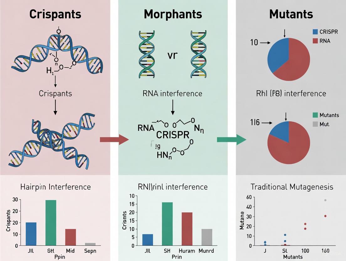
Crispants vs Morphants vs Mutants: A Comprehensive Guide to Phenotypic Comparison in Genetic Research
This article provides a definitive guide for researchers and drug development professionals on the critical phenotypic differences between crispants (F0 CRISPR/Cas9 mutants), morphants (morpholino knockdowns), and stable genetic mutants.
Crispants vs Morphants vs Mutants: A Comprehensive Guide to Phenotypic Comparison in Genetic Research
Abstract
This article provides a definitive guide for researchers and drug development professionals on the critical phenotypic differences between crispants (F0 CRISPR/Cas9 mutants), morphants (morpholino knockdowns), and stable genetic mutants. We explore the foundational concepts behind these discrepancies, including the pivotal role of genetic compensation. The content details methodological best practices for creating and analyzing each model, addresses common troubleshooting and optimization challenges, and establishes a rigorous framework for the validation and comparative analysis of genetic models. By synthesizing recent advances, this resource aims to equip scientists with the knowledge to select the appropriate model, accurately interpret phenotypic data, and design robust, efficient genetic screens for functional genomics and therapeutic target validation.
Genetic Compensation: Unraveling the Discrepancy Between Knockdown and Knockout Phenotypes
In functional genomics, establishing a causal link between a gene and a phenotype is fundamental. Researchers primarily use three key models for loss-of-function studies in zebrafish: stable mutants, morphants, and crispants. Each model operates on distinct principles and timelines for gene inactivation, leading to significant implications for phenotypic outcomes. Stable mutants are engineered to carry heritable, permanent mutations across all cells. Morphants achieve transient gene knockdown using morpholino oligonucleotides, while crispants utilize CRISPR/Cas9 to create mosaic, non-inherited mutations in the first generation (F0). Understanding the mechanisms, strengths, and limitations of each approach is crucial for designing robust experiments and accurately interpreting gene function, especially in the context of pervasive challenges like genetic compensation.
Model Comparison at a Glance
The table below summarizes the core characteristics of each model to facilitate comparison.
| Feature | Stable Mutant | Morphant | Crispant |
|---|---|---|---|
| Genetic Change | Permanent, heritable mutation [1] | Transient; blocks mRNA splicing or translation [2] [3] | Transient, non-heritable; mosaic indels in F0 [4] [5] |
| Molecular Mechanism | CRISPR/TALEN-induced indels creating frameshifts/early stop codons [6] | Antisense morpholino oligonucleotides binding target mRNA [2] | CRISPR/Cas9-induced mosaic indels in somatic cells [4] [7] |
| Development Time | 6-9 months [4] [5] | 1-2 days [3] | 1-7 days (larval phenotyping); ~3 months (adult phenotyping) [4] [5] |
| Key Advantage | Gold standard for stable, reproducible phenotypes; study of adult/long-term effects | Rapid assessment of gene function; targets maternal mRNA [3] | Rapid, cost-effective; circumvents genetic compensation [1] [7] |
| Primary Limitation | Time, cost, resource-intensive; prone to genetic compensation [1] [2] | High off-target effects (e.g., p53 pathway activation); toxicity [2] [3] | Mosaicism can lead to variable expressivity [4] [8] |
Experimental Workflows and Key Protocols
The experimental pathways for creating and validating these models involve distinct steps and timelines.
Key Experimental Steps and Methodologies
1. Crispant Generation and Validation
- gRNA Design and Selection: For each target gene, multiple single guide RNAs (sgRNAs) are designed, typically using bioinformatic platforms like Benchling. The gRNA with the highest predicted out-of-frame efficiency is selected [4] [5].
- Microinjection: A mixture of Cas9 protein (or mRNA) and the selected sgRNA(s) is microinjected into the yolk of one-cell stage zebrafish embryos [4] [7].
- Validation of Editing Efficiency: At 1 day post-fertilization (dpf), DNA is extracted from a pool of injected larvae. Next-Generation Sequencing (NGS) is performed, and tools like Crispresso2 are used to determine the fraction of reads with insertions/deletions (indels) and the out-of-frame rate. Efficiencies of >70% are common and considered sufficient for phenotyping [4] [5].
2. Phenotypic Assessment of Crispants
- Larval Staging (7-14 dpf): Phenotyping can include microscopy for specific cell types (e.g., osteoblasts), whole-mount in situ hybridization for gene expression patterns, or Alizarin Red S staining for bone mineralization [4] [7].
- Adult Staging (~90 dpf): For late-onset or structural phenotypes, crispants are raised to adulthood. Analysis can include micro-computed tomography (microCT) for detailed 3D skeletal architecture, revealing fractures, fusions, and changes in bone volume and density [4] [5].
- Molecular Analysis: Quantitative RT-PCR (RT-qPCR) on larval or adult tissue is used to assess the expression of downstream marker genes (e.g.,
bglap,col1a1afor bone studies) to confirm functional molecular consequences [4] [5].
3. Stable Mutant Generation
- Founder (F0) Generation and Raising: The initial step is identical to crispant generation. However, instead of being phenotyped directly, these mosaic F0 fish are raised to sexual maturity.
- Germline Transmission Screening: The outcrossed F0 fish are outcrossed to wild-type fish. Their progeny (F1 generation) are genotyped to identify individuals carrying the mutation in their germline.
- Homozygous Mutant Production: Identified heterozygous F1 fish are incrossed to generate an F2 generation, which will include a Mendelian ratio of wild-type, heterozygous, and homozygous mutant offspring. These stable homozygous mutants are then subjected to phenotypic analysis [1] [3].
Critical Analysis of Phenotypic Concordance
A central challenge in functional genomics is the frequent discrepancy in phenotypes observed between different models targeting the same gene.
The Genetic Compensation Phenomenon
A key explanation for the differences between crispants/morphants and stable mutants is genetic compensation. This is a phenomenon where stable mutant organisms activate compensatory mechanisms that buffer against the loss of the gene, often resulting in a less severe or absent phenotype than expected [1] [2].
- Mechanism: In stable mutants, nonsense mutations can trigger the nonsense-mediated decay (NMD) pathway of the mutant mRNA. This degradation can, in some cases, initiate a feedback loop that leads to the transcriptional upregulation of related genes (paralogs) or other genes within the same network, thereby compensating for the lost function [1] [2].
- Evidence: A compelling example is the
slc25a46gene.slc25a46crispants showed a specific and rescuable phenotype, whereas stable homozygous mutants for the same gene displayed no phenotype. RNA sequencing revealed significant changes in the gene expression profile of the stable mutants, including upregulation of theanxa6gene, which was largely absent in crispants, suggesting a compensatory mechanism had been established in the stable line [1]. - Crispants as a Solution: Because crispants are analyzed acutely in the F0 generation, there is insufficient time for the organism to develop these complex compensatory networks. This makes them particularly valuable for identifying the primary, direct function of a gene [1] [7].
Comparative Case Studies
The table below illustrates specific examples of phenotypic discrepancies and their attributed causes.
| Target Gene | Morphant/Crispant Phenotype | Stable Mutant Phenotype | Attributed Cause/Compensatory Mechanism |
|---|---|---|---|
| slc25a46 | Penetrant disease phenotype [1] | No phenotype observed [1] | Genetic compensation; upregulation of anxa6 and other genes [1] |
| bmp7b | Holoprosencephaly and cyclopia [7] | No obvious developmental defects [7] | Genetic compensation, circumvented by crispant analysis [7] |
| podxl | Reduced hepatic stellate cells (HSCs) [8] | Normal or increased HSCs [8] | Genetic compensation; complex, multi-genic changes in mutants [8] |
| egfl7 | Severe vascular development defects [2] | No obvious defects [2] | Upregulation of paralog emilin3a (Transcriptional Adaptation) [2] |
The Scientist's Toolkit: Essential Research Reagents
A successful functional genomics screen relies on a suite of carefully selected reagents and tools.
| Reagent / Solution | Function in Experiment |
|---|---|
| Alt-R CRISPR-Cas9 gRNA (IDT) | Synthetic, high-fidelity guide RNA for specific gene targeting; improves efficiency and reduces off-target effects [4] [5]. |
| Cas9 Nuclease | The "molecular scissors" that creates a double-strand break in the DNA at the location specified by the gRNA [4] [6]. |
| Morpholino Oligonucleotides | Synthetic antisense molecules that block translation or splicing of target mRNA; used for transient knockdown [2] [3]. |
| Next-Generation Sequencing (NGS) | Used to quantitatively assess the efficiency and spectrum of indel mutations in crispant pools (e.g., via Crispresso2 analysis) [4] [5]. |
| Micro-Computed Tomography (microCT) | Provides high-resolution, quantitative 3D imaging of mineralized tissues in adult zebrafish, enabling analysis of bone volume, density, and morphology [4] [5]. |
| (R)-WM-586 | (R)-WM-586, MF:C20H20F3N5O3S, MW:467.5 g/mol |
| Wallichoside | Wallichoside, MF:C20H28O8, MW:396.4 g/mol |
The choice between crispants, morphants, and stable mutants is not a matter of identifying a single "best" model but of selecting the right tool for the specific biological question and stage of research. Morphants offer speed for initial, transient knockdowns. Stable mutants remain the gold standard for studying heritable effects, late-onset phenotypes, and despite the risk of genetic compensation, for providing a stable platform for further research. Crispants have emerged as a powerful intermediate, balancing the speed of morphants with the genetic precision of CRISPR, while effectively circumventing the confounding issue of genetic compensation. A modern, rigorous functional genomics strategy often involves using crispants for rapid initial gene validation and screening, followed by the generation of stable mutant lines for confirmed hits to study long-term and organism-wide effects.
The zebrafish (Danio rerio) has cemented its role as a premier model organism for studying vertebrate development and disease, owing to its rapid external development, optical transparency, and genetic tractability [9] [4]. However, a persistent and historical puzzle has challenged researchers: widespread phenotypic discrepancies are observed when the same gene is targeted using different genetic perturbation techniques. A classic manifestation of this puzzle is the frequent lack of a severe phenotype in stable knockout mutants, even when robust, often severe, defects are present in morpholino-induced knockdown embryos (morphants) for the identical gene [9] [4]. This discrepancy initially raised concerns about the specificity of morpholinos but has since been partially explained by a fascinating biological phenomenon known as Genetic Compensation Response (GCR) [9]. This guide objectively compares the performance and outcomes of the primary reverse-genetic approaches—morphants, stable mutants, and the more recent crispants—providing researchers with a framework to select and interpret the appropriate model for their investigative goals.
Comparative Analysis of Genetic Perturbation Platforms
The following table summarizes the core characteristics, advantages, and limitations of the three primary techniques used in zebrafish functional genomics.
Table 1: Platform Comparison of Zebrafish Genetic Perturbation Techniques
| Feature | Morpholino (MO) Knockdown (Morphants) | Stable Mutant (Knockout) | Crispant (F0 Mosaic Mutant) |
|---|---|---|---|
| Molecular Mechanism | Antisense oligonucleotides block translation or splicing [9]. | Heritable, CRISPR/Cas9-induced loss-of-function alleles [9]. | Transient, mosaic CRISPR/Cas9-induced mutations in F0 generation [4]. |
| Technical Timeline | Days (injection at 1-cell stage) [9]. | 6-9 months (to generate F2 homozygous mutants) [9] [4]. | ~3 months (to phenotype adult F0 founders) [4]. |
| Phenotypic Penetrance | High, but can have off-target effects [9]. | Can be absent or mild due to Genetic Compensation [9]. | High; recapitulates stable mutant phenotypes [4]. |
| Key Artifact Sources | Off-target effects, p53-mediated apoptosis [9]. | Genetic Compensation Response (GCR) [9]. | Mosaicism (requires high mutagenesis efficiency) [4]. |
| Ideal Application | Rapid, preliminary assessment of gene function; target validation. | Studying long-term, heritable genetic effects. | High-throughput functional screening and validation [4]. |
Table 2: Quantitative Phenotypic Validation for Selected Genes
| Gene Target | Mutant Phenotype (Stable Line) | Morphant Phenotype | Crispant Phenotype (F0) | Validated Role/Pathway |
|---|---|---|---|---|
| lrp5 | Not specified in sources | Not specified in sources | Recapitulated stable mutant phenotype [4]. | Bone fragility, Wnt signaling [4]. |
| bmp1a | Not specified in sources | Not specified in sources | Phenotypic convergence with germline mutants [4]. | Osteogenesis Imperfecta, collagen processing [4]. |
| plod2 | Not specified in sources | Not specified in sources | Phenotypic convergence with germline mutants [4]. | Osteogenesis Imperfecta, collagen cross-linking [4]. |
| slc26a5 | Weak expression, no electromotility [10] [11] | Not specified in sources | Not specified in sources | Hair cell function (prestin) [10] [11]. |
| Microexons (e.g., vav2, itsn1) | Mild or no effect [12] | Not specified in sources | Not specified in sources | Neuritogenesis, neural development [12]. |
Decoding the Discrepancy: The Genetic Compensation Response
The divergence between morphant and mutant phenotypes is often attributed to the Genetic Compensation Response (GCR), an adaptive biological mechanism that provides genetic robustness. GCR is triggered by deleterious mutations in stable knockout lines but not by transient protein depletion from morpholinos [9].
This compensatory mechanism involves the upregulation of genetically related genes (homologs or genes within the same pathway) that can functionally substitute for the inactivated gene, thereby masking the expected phenotypic outcome [9]. Recent research indicates that GCR collaborates with the epigenetic machinery and is regulated through processes like Nonsense-Mediated Decay (NMD) of the PTC-bearing mRNA (the mRNA containing a Premature Termination Codon from the mutation) [9]. The following diagram illustrates this complex mechanism.
The Scientist's Toolkit: Essential Research Reagents
Successful functional genetics research in zebrafish relies on a suite of specialized reagents and tools.
Table 3: Key Research Reagents and Solutions in Zebrafish Genomics
| Reagent / Solution | Function / Purpose |
|---|---|
| Morpholino Oligonucleotides (MOs) | Antisense oligonucleotides designed to block translation initiation or pre-mRNA splicing, enabling transient gene knockdown [9]. |
| Cas9 Protein & gRNA Complex | Pre-assembled ribonucleoprotein (RNP) complex used for CRISPR/Cas9 mutagenesis. Direct injection into one-cell embryos generates mutants or crispants [9] [4]. |
| p53 Morpholino | Co-injected with gene-targeting MOs to suppress p53-dependent apoptosis, a common off-target effect, thereby improving specificity [9]. |
| Alt-R CRISPR-Cas9 gRNAs (IDT) | Commercially available, high-quality guide RNAs used for efficient and specific CRISPR/Cas9 mutagenesis, as validated in crispant studies [4]. |
| Crispresso2 | A computational tool for the analysis of next-generation sequencing data from CRISPR experiments. It quantifies indel efficiency and out-of-frame rates in crispants [4]. |
| Galectin-4-IN-2 | Galectin-4-IN-2, MF:C17H22O8, MW:354.4 g/mol |
| SDX-7539 | SDX-7539, MF:C23H38N2O5, MW:422.6 g/mol |
Experimental Protocols for Key Methodologies
Generation and Validation of Stable Mutant Lines
The creation of stable knockout lines via CRISPR/Cas9 is a multi-step process. The workflow begins with microinjection of Cas9/gRNA complexes into one-cell stage embryos. The surviving injected F0 generation are raised to adulthood; these are mosaic founders. These are outcrossed with wild-type fish to generate F1 progeny, which are genotyped to identify heterozygous carriers. Intercrossing of F1 heterozygotes produces F2 embryos, of which 25% are expected to be homozygous mutants [9]. Phenotypic analysis is performed on these F2 homozygotes and compared to their wild-type and heterozygous siblings.
Crispant Screening Protocol for Rapid Validation
Crispant analysis offers a faster alternative for gene function validation. The protocol below is adapted from bone fragility disorder research [4].
- gRNA Design and Selection: Design gRNAs using platforms like Benchling. Select the gRNA with the highest predicted out-of-frame efficiency using a tool like InDelphi-mESC.
- Microinjection: Co-inject Alt-R gRNAs (IDT) with Cas9 protein into the yolk of one-cell stage zebrafish embryos.
- Efficiency Validation (at 1 dpf): Pool genomic DNA from ~10 larvae. Amplify the target region and perform Next-Generation Sequencing (NGS). Analyze sequencing data with Crispresso2 to determine indel efficiency (aim for >70%) and out-of-frame rates [4].
- Phenotypic Analysis:
- Larval Stage (7-14 dpf): Use microscopy and Alizarin Red S staining for skeletal phenotyping.
- Adult Stage (90 dpf): Perform high-resolution microCT imaging to quantify bone volume, density, and architecture.
- Molecular Validation: Conduct RT-qPCR on crispant lysates to analyze expression changes in relevant pathway markers (e.g., bglap and col1a1a for bone studies) [4].
The workflow for generating and applying stable mutants versus crispants is summarized below.
The historical puzzle of phenotypic discrepancies in zebrafish has evolved from a confounding artifact to a rich field of study that underscores the complexity of genetic networks. The choice of model—morphant, stable mutant, or crispant—is not merely a technical decision but a strategic one that directly influences the biological question being addressed.
Morphants remain useful for rapid, preliminary functional screening but require rigorous controls. Stable mutants are essential for studying long-term, heritable effects and the phenomenon of Genetic Compensation itself. Crispants have emerged as a powerful, cost-effective tool for high-throughput functional validation, faithfully recapitulating stable mutant phenotypes for many genes and enabling rapid prioritization of candidate disease genes [4].
Future research will focus on further elucidating the molecular triggers and mechanisms of GCR. Furthermore, the integration of crispant-based screening with advanced transcriptomic and proteomic analyses promises to accelerate the functional annotation of the vertebrate genome and the modeling of human genetic disorders in zebrafish.
Genetic Compensation as a Key Mechanism for Phenotypic Robustness
Phenotypic robustness is the ability of an organism to maintain stable developmental outcomes and fitness despite genetic perturbations or environmental fluctuations [13]. This fundamental biological property ensures consistent phenotypes in the face of mutations that would otherwise be expected to cause dramatic morphological or functional consequences. Genetic compensation has emerged as a crucial molecular mechanism underlying this robustness, wherein the deleterious effects of a gene mutation are buffered by the compensatory expression of related genes [13] [14]. This phenomenon provides a compelling explanation for the frequent discrepancies observed between different genetic perturbation methods, particularly the pronounced phenotypic differences often reported between stable mutants and transient knockdown approaches.
The concept of genetic robustness was first hinted at through observations of dosage compensation in Drosophila in 1932, but has since been documented across model organisms from yeast to mammals [13]. Recent advances in genetic technologies, especially CRISPR-Cas9 genome editing, have accelerated our understanding of how organisms actively compensate for genetic lesions through multiple molecular pathways. This review will systematically compare phenotypic outcomes across crispants, morphants, and stable mutants, examining the experimental evidence for genetic compensation as a key mechanism maintaining phenotypic stability in vertebrate models.
Comparative Analysis of Genetic Perturbation Methods
Each major genetic perturbation technique offers distinct advantages and limitations for functional genomics research, with significant implications for observing genetic compensation effects.
Stable Mutants are generated through permanent germline modifications, typically introducing frameshifts or premature termination codons (PTCs) that disrupt gene function. These established mutant lines provide a permanent resource with uniform genotypes but require extended generation times—6-9 months in zebrafish compared to 3 months for crispants [4]. The persistent nature of the genetic lesion in stable mutants allows organisms to activate long-term compensatory mechanisms, often resulting in milder-than-expected phenotypes.
Crispants (F0 mosaic mutants) are generated through CRISPR-Cas9 injections in single-cell embryos, creating somatic mosaics with varying mutation efficiencies across cells. This approach produces high indel rates (71-88% efficiency documented in zebrafish screens) with significantly reduced generation time compared to stable lines [4]. The mosaic nature and rapid analysis of crispants may precede the full activation of compensatory networks, potentially revealing intermediate phenotypes between morphants and stable mutants.
Morphants are created through antisense morpholino oligonucleotides that transiently block mRNA translation or splicing. This method provides rapid protein knockdown within days but involves no genomic DNA alteration. The transient, protein-level perturbation typically does not trigger the same compensatory responses as DNA-level mutations, often resulting in more severe phenotypic consequences [15] [14].
Table 1: Technical Comparison of Genetic Perturbation Methods
| Parameter | Stable Mutants | Crispants | Morphants |
|---|---|---|---|
| Genetic alteration | Permanent germline mutation | Somatic mosaic mutations | No DNA alteration; translational blockade |
| Time to phenotype | Months (6-9 for zebrafish) | Weeks (3 months for adult zebrafish) | Days (early development) |
| Persistence | Permanent, heritable | Somatic, non-heritable | Transient (2-4 days) |
| Mutation efficiency | 100% homozygous | 71-88% indel efficiency [4] | Variable protein knockdown |
| Compensation trigger | Activates transcriptional adaptation & genetic compensation | Partial compensation possible | Minimal compensation activation |
| Key advantages | Uniform genotype, permanent resource | Rapid screening, cost-effective | Extremely fast results, dose-titratable |
Phenotypic Discrepancies Across Methods
Multiple studies across vertebrate models have documented striking phenotypic differences when comparing these perturbation methods, with genetic compensation as the proposed explanatory mechanism.
In salamander limb regeneration studies, Yap knockout mutants regenerated limbs normally, while Yap morpholino knockdown and pharmacological inhibition severely disrupted regeneration, causing delayed blastema formation and failed patterning [15]. This discrepancy was attributed to compensatory upregulation of the homologous gene Taz in knockout animals (2.5-fold increase), which was absent in morphants. Critically, when researchers blocked this compensation by knocking down Taz in the Yap mutant background, the regeneration defects reappeared, confirming the functional significance of this compensatory mechanism [15].
Similar patterns emerge in zebrafish studies, where egfl7 mutants show minimal vascular defects despite severe phenotypes in morphants [13]. This was correlated with upregulated expression of extracellular matrix proteins Emilins in mutants but not morphants. The phenomenon appears widespread—a meta-analysis suggested that approximately 80% of zebrafish genes show discrepancies between mutant and morphant phenotypes during development [15].
Table 2: Documented Cases of Phenotypic Discrepancies Attributed to Genetic Compensation
| Organism | Gene | Mutant Phenotype | Knockdown Phenotype | Compensating Gene |
|---|---|---|---|---|
| Salamander | Yap | Normal limb regeneration | Severely disrupted regeneration | Taz [15] |
| Zebrafish | egfl7 | Minor or no vascular defects | Severe vascular defects | emilin3a [13] |
| Mouse | Tet1 | Normal stem cell morphology | Loss of undifferentiated morphology | Tet2 [13] |
| Mouse | Dystrophin | Mild muscular dystrophy (mdx) | Severe muscular dystrophy (knockdown) | Utrophin [14] |
| Mouse | Kindlin-2 | Normal focal adhesions | Decreased integrin activation | Kindlin-1 [13] |
Molecular Mechanisms of Genetic Compensation
Transcriptional Adaptation Pathways
Transcriptional adaptation represents a specific genetic compensation mechanism triggered by the presence of mutant mRNA rather than protein loss. This pathway requires the generation of premature termination codons (PTCs) in the mutated gene, which are recognized by nonsense-mediated decay (NMD) factors, particularly UPF3A [15]. In salamander Yap mutants, the two-base pair deletion causing a frameshift and PTC led to UPF3A upregulation, which subsequently activated expression of the compensatory gene Taz during limb regeneration [15].
The dependence on mutant mRNA rather than protein loss explains why transcriptional adaptation occurs in stable mutants but not morphants. Supporting this model, when researchers used antisense oligonucleotides to eliminate the PTC-containing Yap mRNA in mutants, the compensatory Taz upregulation was abolished and regeneration defects emerged [15]. This mechanism appears to operate independently of the classic Upf1/Upf2/Upf3b-mediated NMD pathway, suggesting specialized functions for UPF3A in genetic compensation.
Protein Feedback Loops and Redundancy
Beyond transcriptional adaptation, genetic compensation can occur through protein-level feedback mechanisms. In these cases, the loss of a specific protein disrupts cellular complexes or pathways, triggering signaling cascades that upregulate homologous proteins with overlapping functions.
The classic example involves dystrophin deficiency in muscular dystrophy. Mdx mice lacking dystrophin show milder symptoms than human patients due to compensatory upregulation of the dystrophin-related protein utrophin [14]. This compensation occurs through a protein feedback loop where dystrophin complex instability activates Akt signaling, ultimately increasing utrophin expression and partially restoring sarcolemma stability [14].
Similarly, in mouse models, knockout of one cyclin D family member leads to upregulation of the remaining cyclin D genes, maintaining normal cell cycle progression in most tissues [13]. This form of compensation leverages inherent genetic redundancies from gene duplication events throughout evolution, where related genes retain partial functional overlap that can be co-opted when primary pathways are disrupted.
Experimental Evidence from Key Studies
Salamander Limb Regeneration Model
The recent salamander study provides particularly compelling evidence for genetic compensation in a regenerative context [15]. Researchers established Yap mutant lines using CRISPR-Cas9, introducing a two-base pair deletion in exon 4 that caused a frameshift and premature termination codon. Surprisingly, these mutants displayed normal limb regeneration despite Yap's established role in tissue regeneration.
The experimental workflow involved multiple complementary approaches:
- Mutant analysis: Comparing regeneration in wild-type versus Yap knockout animals
- Morpholino knockdown: Translational inhibition of Yap in wild-type animals
- Pharmacological inhibition: Verteporfin treatment to disrupt YAP protein function
- Rescue experiments: Blocking compensatory Taz in mutant background
Quantitative measurements showed that cell proliferation rates (via EdU assay) and regeneration efficiency (regenerating/uninjured limb ratio) were nearly identical between KO and WT animals, while both morpholino and verteporfin treatments significantly reduced these parameters [15]. Molecular analysis revealed that TAZ-specific target genes were significantly upregulated during early regeneration (1-7 dpa) in Yap KO animals, coinciding with the critical period when YAP protein would normally be required.
Zebrafish Crispant Screening Platform
Zebrafish crispant screens have emerged as valuable tools for high-throughput functional validation while potentially capturing intermediate states of genetic compensation. A recent study targeting ten fragile bone disorder genes demonstrated the efficiency of this approach, achieving mean indel efficiencies of 88% and out-of-frame rates between 49-73% in F0 mosaic founders [4].
The methodology involved:
- gRNA design: Selection of guides with highest predicted out-of-frame efficiency using InDelphi-mESC tool
- Embryo injection: Co-injection of Cas9 protein and gRNAs into one-cell stage embryos
- Validation: Next-generation sequencing of pooled larvae to quantify indel spectra
- Phenotyping: Skeletal analysis at 7, 14, and 90 days post-fertilization
Notably, different genes showed variable phenotypic penetrance in crispants, with aldh7a1 and mbtps2 crispants exhibiting severe skeletal deformities and increased mortality, while others showed more moderate effects [4]. This screening approach captures phenotypes that may represent partial compensation states, as the mosaic nature and rapid assessment may precede full compensatory network activation.
The Scientist's Toolkit: Essential Research Reagents
Table 3: Key Research Reagents for Genetic Compensation Studies
| Reagent/Category | Specific Examples | Function/Application | Considerations |
|---|---|---|---|
| Genome Editing Tools | CRISPR-Cas9, base editors, prime editors | Creating stable mutants and crispants; precise nucleotide changes | Prime editors allow precise edits without double-strand breaks [6] |
| Knockdown Reagents | Morpholino oligonucleotides, siRNA | Transient protein-level knockdown | Useful for distinguishing transcriptional adaptation from protein feedback [13] |
| NMD Pathway Modulators | UPF3A inhibitors/activators, ASOs targeting PTC-containing mRNAs | Investigating transcriptional adaptation mechanisms | ASOs can eliminate mutant mRNA to test compensation dependence [15] |
| Lipid Nanoparticles (LNPs) | CTX310 delivery system, other CRISPR-LNP formulations | In vivo delivery of editing components | Liver-tropic LNPs enable efficient hepatic editing [16] [17] |
| Lineage Tracing Systems | Cre-lox, barcoding approaches | Tracking cell fate in compensation contexts | Reveals how compensation affects cellular dynamics |
| Multi-omics Platforms | Single-cell RNA-seq, ATAC-seq, proteomics | Comprehensive molecular profiling | Identifies compensatory networks beyond immediate homologs |
| Drpitor1a | Drpitor1a, MF:C15H8N2O2, MW:248.24 g/mol | Chemical Reagent | Bench Chemicals |
| FD1024 | FD1024, MF:C21H20F2N4O2S, MW:430.5 g/mol | Chemical Reagent | Bench Chemicals |
Implications for Drug Development and Therapeutic Strategies
Understanding genetic compensation has profound implications for therapeutic development, particularly for monogenic disorders. The phenomenon offers both challenges and opportunities for precision medicine.
Compensation mechanisms may explain why some genetic disorders show variable penetrance and expressivity. In Duchenne muscular dystrophy, patients with higher utrophin expression typically experience milder symptoms, suggesting natural compensation moderates disease severity [14]. Therapeutic strategies could potentially enhance these endogenous compensatory pathways—for instance, approaches to increase utrophin expression are being explored as treatments for DMD.
Conversely, genetic compensation can pose challenges for therapeutic gene editing. If compensation already ameliorates the effects of a mutation, simply correcting the causal mutation might not fully restore normal function. Additionally, in conditions where compensation involves multiple genes, single-gene therapies may prove insufficient.
The successful application of CRISPR-based therapies like CASGEVY for sickle cell disease demonstrates that despite potential compensation complexities, effective interventions are achievable [16] [17]. As in vivo editing approaches advance, exemplified by CTX310 for ANGPTL3 reduction showing 82% triglyceride reduction and 81% LDL reduction in clinical trials [17], understanding tissue-specific compensation networks will become increasingly important for predicting therapeutic efficacy and potential resistance mechanisms.
Genetic compensation represents a fundamental biological mechanism maintaining phenotypic robustness in the face of genetic perturbations. The systematic comparison of crispants, morphants, and stable mutants reveals that permanent genetic lesions often trigger compensatory networks that buffer against phenotypic consequences, explaining frequent discrepancies between perturbation methods. Molecular mechanisms include both transcriptional adaptation pathways, triggered by mutant mRNA and mediated through factors like UPF3A, and protein feedback loops that upregulate homologous genes with overlapping functions.
For researchers investigating gene function, these findings emphasize the importance of employing multiple perturbation approaches and considering potential compensation effects when interpreting phenotypes. The expanding toolkit of CRISPR-based technologies, including base editing, prime editing, and epigenetic modifiers, provides powerful approaches to dissect these complex regulatory networks. As therapeutic genome editing advances, understanding genetic compensation will be crucial for predicting treatment efficacy and developing strategies that either exploit or circumvent these endogenous buffering systems to achieve desired clinical outcomes.
The Role of Nonsense-Mediated Decay (NMD) and Epigenetic Machinery
In the field of functional genomics, researchers employ distinct technologies to interrogate gene function, each with unique mechanistic bases and phenotypic outcomes. The comparative analysis of crispants (CRISPR-generated F0 mosaics), morphants (morpholino-induced knockdowns), and mutants (stable germline knockouts) provides critical insights for experimental design, particularly when studying biological processes involving nonsense-mediated decay (NMD) and epigenetic regulation. These approaches differ fundamentally in their implementation: crispants utilize CRISPR-Cas9 to create mosaic individuals with biallelic mutations in target cells; morphants employ antisense morpholino oligonucleotides to block translation or splicing; and mutants involve established lines with heritable genetic modifications [18] [19]. Understanding how these models interact with cellular quality control mechanisms like NMD and epigenetic machinery is essential for accurate interpretation of gene function studies and drug development pipelines.
Fundamental Mechanisms: Technological Underpinnings and Cellular Interactions
Core Definitions and Methodological Bases
The three primary approaches for gene perturbation in model organisms differ fundamentally in their mechanisms and implementation:
Crispants: Generated through CRISPR-Cas9-mediated editing in F0 embryos, creating mosaic individuals with varying proportions of mutant cells. This approach produces biallelic mutations in target tissues without establishing stable lines, with recent studies reporting indel efficiencies averaging 88% across multiple targets [19].
Morphants: Created using morpholino oligonucleotides that block translation initiation or pre-mRNA splicing through steric hindrance. This method transiently reduces functional protein levels without altering genomic DNA [18].
Mutants: Established through germline transmission of mutations, resulting in stable, heritable genetic modifications. This traditional approach requires crossing to generate homozygous individuals [18] [20].
Interaction with NMD and Epigenetic Machinery
The cellular response to gene perturbations varies significantly across these models, particularly regarding NMD activation and epigenetic adaptations:
Nonsense-Mediated Decay Pathways: NMD serves as a crucial quality control mechanism that degrades mRNAs containing premature termination codons (PTCs), preventing the production of truncated proteins [21] [22]. This pathway relies on core factors including UPF1, UPF2, and UPF3, with UPF1 serving as the central effector that recruits decay machinery when PTCs are detected [23] [22]. The key distinction between approaches lies in their interaction with NMD: mutants and crispants frequently generate PTC-containing transcripts that potentially activate NMD, while morphants typically do not produce PTCs and thus avoid this pathway [18].
Epigenetic Regulation: Epigenetic mechanisms, including DNA methylation, histone modifications, and non-coding RNAs, dynamically regulate gene expression in response to genetic perturbations [24]. Stable mutant lines may exhibit compensatory epigenetic reprogramming that alters phenotypic outcomes, while the acute nature of crispant and morphant approaches may limit such adaptation. For instance, HDAC inhibitors have been shown to reverse repressive histone marks at disease-relevant loci in neuromuscular disorders, demonstrating the therapeutic potential of epigenetic modulation [24].
Table 1: Molecular and Cellular Characteristics of Gene Perturbation Approaches
| Characteristic | Crispants | Morphants | Mutants |
|---|---|---|---|
| Genetic Basis | Somatic mutations (mosaic) | No genomic alteration | Germline mutations (uniform) |
| NMD Activation | Possible with PTC-generating indels | Unlikely | Yes, with PTC-generating mutations |
| Epigenetic Compensation | Limited due to acute nature | Limited due to acute nature | Established in stable lines |
| Temporal Resolution | Acute (days-weeks) | Rapid (hours-days) | Chronic (generations) |
| Genetic Compensation | Minimal evidence | Minimal evidence | Documented in multiple studies |
Direct Phenotypic Comparisons: Experimental Evidence and Discordance
Systematic Analysis of Phenotypic Concordance
Comparative studies have revealed significant phenotypic discordance between these approaches, with important implications for data interpretation:
A landmark investigation examining 24 genes involved in vascular development found dramatically different outcomes between approaches: only 3 genes (12.5%) showed congruent phenotypes between mutants and morphants, while morphants exhibited previously reported phenotypic defects for most genes that showed no observable phenotypes in corresponding mutant lines [20]. This discrepancy highlights the potential for false positives in morphant-based studies and underscores the importance of validation through genetic mutations.
Case Studies in Phenotypic Discordance
Specific examples illustrate the molecular basis for these phenotypic differences:
egfl7 Gene: Morphants displayed severe vascular defects, while genetic mutants showed normal vascular development. Further investigation revealed that mutants upregulated compensatory genes including emilin3a, which was not observed in morphants [18].
Islet2a Transcription Factor: Morphants exhibited truncated motor neuron axons, while mutants developed normal axons. Transcriptomic analysis revealed 174 differentially expressed genes in morphants compared to 201 in mutants, suggesting distinct compensatory mechanisms [18].
epoa Gene: Morphants showed altered pronephros development, while mutants developed normal renal structures. Genetic compensation was identified in mutants through upregulation of epob as a compensating gene, which did not occur in morphants [18].
Table 2: Documented Cases of Phenotypic Discordance Between Morphants and Mutants
| Gene | Morphant Phenotype | Mutant Phenotype | Compensatory Mechanism |
|---|---|---|---|
| egfl7 | Severe vascular defects | Normal development | Upregulation of emilin3a |
| Islet2a | Truncated axons | Normal axons | Differential expression of 174 vs 201 genes |
| epoa | Altered pronephros development | Normal pronephros | Upregulation of epob gene |
| Reference | [18] | [18] | [18] |
Experimental Design and Workflow Considerations
Methodological Protocols
Crispant Generation Protocol: The cardiodeleter zebrafish line exemplifies a tissue-specific crispant approach. This system utilizes a cardiomyocyte-specific promoter (cmlc2) to drive expression of nuclear GFP and a zebrafish codon-optimized Cas9 [25]. Guide shuttles deliver gene-specific gRNAs while permanently labeling mutant cardiomyocytes with mKate fluorescence. The workflow involves: (1) designing gRNAs with high predicted out-of-frame efficiency using tools like CRISPRScan; (2) co-injecting Cas9 protein and gRNAs into one-cell stage embryos; (3) screening for mosaic mutant cells via fluorescence; and (4) phenotypic analysis at appropriate developmental stages [25] [19].
Mutant Validation Pipeline: Establishing stable mutant lines requires: (1) generating F0 founders through CRISPR injection; (2) outcrossing to identify germline transmission; (3) establishing heterozygous lines; (4) intercrossing heterozygotes to generate homozygous mutants; and (5) comprehensive phenotypic characterization across developmental stages [19].
NMD Inhibition Experiments: To assess NMD involvement in phenotypic outcomes: (1) inhibit NMD pathway chemically (e.g., cycloheximide) or genetically (e.g., UPF1 knockdown); (2) quantify target mRNA levels via RT-qPCR; (3) assess protein truncation via western blot; (4) monitor rescue of physiological phenotypes [21] [22].
NMD Pathway Mechanism
The following diagram illustrates the central mechanism of Nonsense-Mediated Decay, which is particularly relevant for interpreting mutant and crispant phenotypes:
NMD Mechanism
Experimental Workflow Comparison
The integrated workflow for comparing crispants, morphants, and mutants involves parallel experimental tracks:
Experimental Workflow
Research Reagent Solutions and Technical Tools
Table 3: Essential Research Reagents for Gene Perturbation Studies
| Reagent/Tool | Primary Function | Application Notes |
|---|---|---|
| CRISPR-Cas9 System | Induces targeted DNA double-strand breaks | Codon-optimized versions available for zebrafish; tissue-specific promoters enable spatial control [25] |
| Morpholino Oligonucleotides | Blocks translation or splicing via steric hindrance | Requires careful dose optimization to minimize off-target effects; rescue experiments recommended [18] |
| Guide Shuttle Vectors | Delivers gRNAs and labels mutant cells | Enables tracking of mutant cells; Tol1/Tol2 transposon-based systems improve integration [25] |
| NMD Inhibitors | Blocks nonsense-mediated decay pathway | Chemicals (cycloheximide) or genetic (UPF1 knockdown) tools to assess NMD contribution [21] |
| Epigenetic Modulators | Modifies DNA methylation or histone marks | HDAC inhibitors (e.g., givinostat) test epigenetic compensation [24] |
| Tissue-Specific Cas9 Lines | Restricts mutagenesis to specific cell types | Example: Cardiodeleter zebrafish with cmlc2 promoter [25] |
Interpretation Guidelines and Best Practices
Strategic Application of Technologies
Each gene perturbation technology offers distinct advantages for specific research applications:
Crispants are optimal for: Rapid functional screening of multiple gene targets; bypassing early lethality through mosaicism; adult-stage phenotypic analysis without establishing stable lines; and disease modeling where somatic mutation reflects human pathology [19].
Morphants are appropriate for: Acute protein depletion studies; splicing inhibition analysis; and preliminary gene function assessment when complemented with genetic validation.
Mutants are essential for: Studying chronic adaptation and compensation mechanisms; analyzing complex phenotypes requiring uniform genetics; and establishing faithful animal models of human genetic disorders.
Accounting for NMD and Epigenetic Effects
Accurate interpretation of phenotypic data requires careful consideration of cellular response mechanisms:
NMD Activation Assessment: When PTCs are introduced in mutants or crispants, verify NMD sensitivity through UPF1 dependence tests and mRNA quantification. Consider that PTCs near the start codon may evade NMD, and long exons can reduce NMD efficiency [26] [22].
Epigenetic Compensation Evaluation: Monitor expression changes in related gene family members and pathway components. Employ epigenetic modifiers to test for chromatin-mediated compensation, particularly in stable mutant lines [24].
Genetic Compensation Investigation: In mutants, analyze upregulation of homologous genes or parallel pathways that may mask expected phenotypes. This compensation frequently explains discrepancies with morphant phenotypes [18].
The integration of crispant, morphant, and mutant approaches—with careful attention to NMD and epigenetic contexts—provides a powerful framework for advancing functional genomics and drug development. This comparative understanding enables more accurate interpretation of gene function data and more predictive modeling of human disease mechanisms.
A central challenge in reverse genetics is the frequent discrepancy between the severe phenotypes observed in gene knockdown experiments and the surprisingly mild or absent phenotypes in corresponding gene knockouts. The case of the egfl7 gene in zebrafish provides a foundational example of this phenomenon, revealing how genetic compensation can allow mutants to escape anticipated phenotypic consequences. This guide compares the experimental outcomes and underlying mechanisms across three key reverse genetics approaches—morpholinos, mutants, and crispants—within the broader context of phenotypic comparison research.
Phenotypic Discrepancy: Mutants vs. Morphants
Initial investigations into the function of egfl7, an endothelial extracellular matrix gene, yielded starkly different results depending on the technique used. The table below summarizes the core experimental observations.
| Perturbation Method | Observed Vascular Phenotype | Activation of Compensatory Genes | Key Experimental Evidence |
|---|---|---|---|
| Morphants (Knockdown) | Severe vascular defects [27] [28] | Not observed [27] | Morphants exhibit defective tube formation; defects not rescued in egfl7 mutant background, arguing against off-target effects [27] [29]. |
| Mutants (Knockout) | No obvious vascular defects [27] [29] | Upregulation of related ECM genes (e.g., Emilins) and vegfab [27] [28] | Proteomic and transcriptomic analysis revealed upregulated genes; injecting egfl7 morpholino into mutants did not recreate severe morphant phenotype [27]. |
| CRISPRi (Transcriptional Knockdown) | Severe vascular defects [27] | Not observed [27] | Obstructing transcript elongation did not trigger the compensatory response seen in true mutants, leading to a phenotype [27]. |
The Genetic Compensation Mechanism
The divergent phenotypes in egfl7 mutants and morphants are attributed to a phenomenon known as genetic compensation, where the organism activates a compensatory network to buffer against the loss of a gene. Research indicates this response is triggered specifically by the presence of a mutant mRNA and the nonsense-mediated decay (NMD) pathway, not merely the absence of the protein [28] [30].
In egfl7 mutants, the deleterious mutation creates a premature termination codon (PTC), leading to the degradation of the mutant mRNA via NMD. This degradation process appears to collaborate with the epigenetic machinery to initiate a transcriptional response that upregulates genes with related functions, such as other extracellular matrix components, thereby compensating for the loss of Egfl7 [30]. This mechanism is not activated in morphants, where the mRNA is often blocked from translation but not degraded, nor in CRISPRi experiments where transcript elongation is obstructed [27].
Experimental Protocols for Dissecting Compensation
To rigorously establish genetic compensation, a multi-step experimental approach is required, moving from phenotypic observation to mechanistic insight.
Foundational Phenotype Comparison
- Gene Knockdown: Inject a moderate dose of egfl7-targeting morpholino into wild-type zebrafish embryos at the one-cell stage. Analyze vascular development at 2-5 days post-fertilization (dpf) for defects [27].
- Gene Knockout: Generate a stable egfl7 mutant line using CRISPR-Cas9, typically introducing a small indel in an early exon to ensure a frameshift and PTC. Analyze the same phenotypic endpoints as in morphants [27] [29]. The egfl7 cq180 mutant, for example, has a 13-bp deletion in exon 3 [29].
Testing for Off-Target Effects and Compensation
- Rescue Experiment: Inject the egfl7 morpholino into the egfl7 mutant background. The persistence of a wild-type phenotype in these mutants, despite morpholino treatment, provides strong evidence that the morphant phenotype is not due to off-target effects but rather a failure to compensate [27] [28].
- Transcriptomic Analysis: Perform RNA sequencing on both egfl7 mutants and morphants. This unbiased approach identifies genes that are specifically upregulated in the mutant condition, pinpointing potential compensators [27].
Validating Compensatory Genes
- Functional Rescue: Co-inject mRNA of an upregulated compensatory gene (e.g., an Emilin family member) with the egfl7 morpholino into wild-type embryos. If the compensatory gene can rescue the morphant phenotype, it confirms its role in genetic compensation [27].
The Scientist's Toolkit: Key Research Reagents
The following table details essential reagents and models used in the cited egfl7 studies and related genetic compensation research.
| Reagent / Model | Function in Experiment |
|---|---|
| egfl7 Mutant (e.g., cq180, s981) | A stable knockout line with a frameshift mutation; used to study long-term adaptation and genetic compensation in the absence of the gene [27] [29]. |
| egfl7 Morpholino | An antisense oligonucleotide that blocks translation or splicing of egfl7 mRNA; used for transient knockdown to reveal the acute effect of gene loss before compensation sets in [27] [28]. |
| CRISPR-Cas9 | A genome editing system used to generate knockout mutant lines. It involves co-injecting Cas9 nuclease and a gene-specific guide RNA (gRNA) into one-cell stage embryos [6] [4]. |
| CRISPR Interference (CRISPRi) | A modified CRISPR system that uses a catalytically "dead" Cas9 (dCas9) to block transcription without cutting DNA; used to demonstrate that transcriptional blockade does not trigger compensation [27]. |
| Tg(egfl7:YFP) Transgenic Line | A reporter line that visualizes the expression pattern of egfl7 in vivo, confirming its expression in endothelial and lymphatic cells [29]. |
| STM2120 | STM2120, MF:C18H15N5O2, MW:333.3 g/mol |
| HTH-01-091 TFA | HTH-01-091 TFA, MF:C28H29Cl2F3N4O4, MW:613.5 g/mol |
Research Implications and Strategic Insights
The egfl7 case study demonstrates that the choice of genetic perturbation method can dictate experimental outcomes and biological interpretations. The following diagram illustrates the divergent molecular pathways activated by each method, leading to distinct phenotypic results.
For researchers, this has several critical implications. When a mutant lacks an expected phenotype, genetic compensation should be investigated as a potential cause, rather than defaulting to assumptions of gene redundancy. The "gold standard" for functional validation now often requires a multi-pronged approach, combining mutant analysis with crispant or morphant studies in the mutant background to dissect acute versus compensated phenotypes. Furthermore, the discovery of genetic compensation opens a novel therapeutic avenue: rather than targeting a defective gene, therapies could aim to manipulate the endogenous compensatory networks to ameliorate disease.
Best Practices in Model Generation: From CRISPR Design to Phenotypic Analysis
The advent of CRISPR/Cas9 technology has revolutionized genetic research, enabling precise genome manipulation across diverse model organisms. Within this landscape, two primary approaches have emerged for functional gene analysis: the generation of crispants (F0 generation animals with direct somatic editing) and the establishment of stable mutant lines. This guide objectively compares these methodologies, examining their relative performance in efficiency, timeline, and applicability for phenotypic comparison in preclinical research.
Experimental Protocols and Workflow Comparison
Crispant Generation Protocol
The crispant method enables rapid phenotypic assessment in the F0 generation through direct somatic editing. The following protocol, optimized for zebrafish, can be adapted for other model organisms.
1. Guide RNA Design and Preparation
- Design 4-5 sgRNAs targeting the first conserved domain or early exons of the target gene to maximize functional knockout probability [31].
- Select sgRNAs with high on-target efficiency scores, using predictive algorithms from tools like those cataloged in CRISPR-GATE [32].
- For the injection mix, combine a cocktail of multiple sgRNAs (typically 3-4) with Cas9 protein or mRNA [31].
2. Microinjection
- Prepare injection solution containing multiplexed sgRNAs (typically 50-100 pg of each) and Cas9 protein (typically 300-600 pg) [31].
- Inject into one-cell stage embryos.
- Maintain injected embryos under standard conditions and monitor for development.
3. Phenotypic Analysis
- Assess somatic mutation efficiency through phenotypic screening (for visible traits) or molecular validation (for non-visible traits).
- For genes affecting pigmentation (e.g., yellow-y), screen for mosaic phenotypic changes in G0 animals [33].
- For non-visible traits, use targeted sequencing to confirm editing efficiency in somatic tissues.
Stable Line Generation Protocol
Stable germline mutant lines provide heritable, consistent genetic models suitable for comprehensive phenotypic analysis across generations.
1. Vector Design and Assembly
- Select appropriate CRISPR system (e.g., SpCas9, SaCas9, or Cas12a) based on PAM requirements and efficiency [34].
- For CRISPRa/i applications, utilize self-selecting systems like CRISPRa-sel that employ piggyBac transposon technology for stable integration [35].
- Clone sgRNA expression cassettes into appropriate delivery vectors.
2. Delivery and Selection
- Deliver CRISPR components via microinjection, transfection, or viral transduction.
- For transgenic CRISPRa systems, apply selection pressure (e.g., puromycin) 48-72 hours post-transduction to enrich for successfully modified cells [35].
- Expand surviving cells for 1-2 weeks to establish stable populations.
3. Germline Transmission Analysis
- Outcross F0 injected animals to wild-type partners.
- Screen F1 progeny for germline transmission via PCR, sequencing, or phenotypic analysis.
- Intercross heterozygous F1 animals to generate homozygous F2 mutants for phenotypic characterization.
Table 1: Key Workflow Comparison Between Crispant and Stable Line Approaches
| Parameter | Crispant Method | Stable Line Method |
|---|---|---|
| Timeline to Phenotype | 1-7 days (somatic); ~1 month (germline) [36] | 6-12 months (zebrafish) [31] |
| Mosaic Mutation Efficiency | Up to 80% in G0 [33] | N/A (clonal populations) |
| Germline Transmission Rate | ~30% with 100% LOF phenotypes [33] | Variable (typically 5-80%) |
| Animal Usage | Reduced (single generation) | Extensive (multiple generations) |
| Phenotypic Consistency | Variable (mosaic) | High (uniform genotype) |
CRISPR Workflow Comparison: This diagram illustrates the key procedural differences between generating crispants and stable mutant lines, highlighting the significant timeline advantage of the crispant approach.
Performance and Efficiency Metrics
Editing Efficiency Comparison
Both crispant and stable line approaches demonstrate high editing efficiency, though through different mechanisms and with distinct optimization requirements.
Table 2: Quantitative Performance Comparison of CRISPR Approaches
| Performance Metric | Crispant Approach | Stable Line Approach | Experimental Support |
|---|---|---|---|
| Somatic Mutation Rate | 78-80% (mosaic phenotypes) [33] | N/A (clonal) | Zebrafish yellow-y targeting [33] |
| Complete LOF Phenotypes | ~30% of G0 animals [33] | >90% in homozygotes | Zebrafish maternal-effect genes [31] |
| Germline Transmission | Achievable in F1 progeny [33] | Required for line establishment | Zebrafish crispant studies [31] |
| Population-wide Activation | N/A | Up to 100% with CRISPRa-sel [35] | Human cell lines (K562) [35] |
| Off-Target Effects | Similar to stable approaches | Reducible with high-fidelity Cas variants [34] | Specificity comparisons [34] |
Applications in Phenotypic Research
The choice between crispant and stable line approaches depends heavily on research goals, timeline, and required phenotypic depth.
Crispant Advantages:
- Speed: Phenotypic analysis possible within days to weeks versus months required for stable line generation [31] [36].
- Efficiency: High rates of biallelic editing in somatic tissues enable rapid functional assessment [31].
- Cost-Effectiveness: Reduced animal housing and maintenance costs compared to multi-generation stable line development [36].
- Lethal Gene Analysis: Enables study of genes causing embryonic lethality when mutated [31].
Stable Line Advantages:
- Phenotypic Consistency: Uniform genotypes eliminate mosaic variability [31].
- Reproducibility: Clonal populations enable standardized assays across experiments and laboratories.
- Long-term Studies: Suitable for chronic disease modeling and aging research.
- CRISPRa Applications: Stable CRISPRa-sel systems enable sustained gene activation in >90% of cell populations [35].
The Scientist's Toolkit: Essential Research Reagents
Successful implementation of CRISPR workflows requires carefully selected reagents and tools optimized for each approach.
Table 3: Essential Research Reagents for CRISPR Workflows
| Reagent/Tool | Function | Crispant Applications | Stable Line Applications |
|---|---|---|---|
| Cas9 Protein/mRNA | CRISPR nuclease component | Direct embryo injection [31] | Vector-based expression |
| Multiplexed sgRNAs | Target sequence guidance | Cocktails of 3-5 sgRNAs for enhanced biallelic editing [31] | Single or minimal sgRNAs |
| CRISPRa-sel System | Gene activation with selection | Limited use | Stable gene activation in >90% of cells [35] |
| piggyBac Transposon | Stable genomic integration | Not typically used | CRISPRa component delivery [35] |
| High-Fidelity Cas Variants | Reduced off-target editing | Available but less critical | eSpCas9, eSpOT-ON for specific editing [37] [34] |
| CRISPR-GATE Database | gRNA design and tool selection | sgRNA design optimization [32] | Comprehensive workflow planning [32] |
| (-)-Bornyl ferulate | (-)-Bornyl ferulate, MF:C20H26O4, MW:330.4 g/mol | Chemical Reagent | Bench Chemicals |
| Andrastin C | Andrastin C, MF:C25H33Cl2N5O6, MW:570.5 g/mol | Chemical Reagent | Bench Chemicals |
The choice between crispant and stable line generation strategies represents a fundamental methodological decision in modern genetic research. Crispants offer unparalleled speed, enabling functional gene assessment in days to weeks with impressive efficiency (80% mosaic phenotypes). This approach is particularly valuable for rapid gene function screening, lethal mutation analysis, and proof-of-concept studies. Conversely, stable lines provide phenotypic consistency and reproducibility essential for detailed mechanistic studies, chronic disease modeling, and standardized drug screening. Recent advances like the CRISPRa-sel system further enhance stable line utility by enabling population-wide gene activation in over 90% of cells. The decision framework should consider research timeline, required phenotypic depth, and specific application needs, with both approaches offering complementary strengths for comprehensive phenotypic comparison in the era of precision genome engineering.
Morpholino oligonucleotides represent a cornerstone technique in molecular biology for transient gene knockdown, enabling researchers to investigate gene function by blocking translation or modulating pre-mRNA splicing. These synthetic molecules feature a morpholine ring backbone with phosphorodiamidate linkages, granting them nuclease resistance and neutral charge that minimize non-specific protein interactions and reduce immune responses compared to other oligonucleotide chemistries [38]. Within contemporary genetic research, particularly in the context of phenotypic comparisons involving crispants (F0 CRISPR/Cas9 mutants), morphants (Morpholino-induced phenotypes), and stable mutants, understanding Morpholino limitations and proper implementation is paramount for generating reliable, interpretable data.
The central challenge in Morpholino use stems from frequent discrepancies observed between morphant phenotypes and those generated by genetic mutations. As Rossi et al. noted, genetic compensation appears induced by deleterious mutations but not by gene knockdowns, potentially explaining why mutants sometimes display less severe phenotypes than morphants [2] [39]. This technical landscape necessitates rigorous guidelines for Morpholino experimental design, dosing, and validation to ensure that observed phenotypes accurately reflect specific gene function rather than off-target effects.
Morpholino Dosing Guidelines and Toxicity
Determining Optimal Dose
Establishing the correct Morpholino concentration is critical for achieving specific target knockdown while minimizing toxicity. Dosing varies significantly by delivery method, target tissue, and experimental organism.
Table 1: Recommended Morpholino Dosing by Application and Organism
| Application/Organism | Recommended Concentration | Delivery Method | Key Considerations |
|---|---|---|---|
| Zebrafish embryos [40] | 0.2-8.0 ng per embryo | Microinjection | Dose-dependent toxicity above 8 ng; requires titration |
| Mosquito larvae [41] | 0.03-0.06 μg/μl | Bath immersion | 3-hour exposure time; effective for Vivo-Morpholinos |
| Cell culture [40] | 1-10 μM | Transfection or electroporation | Varies with transfection efficiency |
| DMD exon-skipping therapies [38] | High, multiple doses (clinical) | Systemic administration | PMO chemistry allows high doses with minimal toxicity |
Effective Morpholino experiments require oligos that are fully dissolved and at a precisely known concentration to ensure reproducibility. Lyophilized Morpholinos should be resuspended in sterile, nucleus-free water, and concentrations verified using spectrophotometry, taking advantage of the hypochromic effect where single-stranded oligonucleotides exhibit increased absorbance when denatured [40]. Aliquotting and proper storage at -20°C in a humidor prevent degradation and maintain activity.
Toxicity Mitigation Strategies
Morpholino toxicity typically manifests through two primary mechanisms: sequence-independent toxicity and sequence-dependent off-target effects. The former often involves activation of cellular stress pathways, while the latter frequently results from unintended interactions with non-target RNAs.
p53 Pathway Activation: A well-documented concern is the nonspecific induction of p53-dependent apoptosis pathways, which can confound phenotypic interpretation [2]. This can be mitigated by co-injecting a p53-targeting Morpholino, though this approach requires caution as p53 mutants themselves may display developmental abnormalities [2].
Interferon Response: Morpholinos can activate interferon-stimulated genes including
isg15andisg20, along with cellular stress pathway genes such asphlda3,mdm2, andgadd45aain zebrafish [2]. These responses are concentration-dependent and highlight the importance of using the lowest effective dose.Control Strategies: Including mismatch controls with 4-5 base mismatches helps distinguish specific from non-specific effects. Rescue experiments with mRNA resistant to Morpholino binding provide the strongest evidence of specificity [40] [39].
Specificity Controls and Experimental Validation
Essential Control Experiments
Rigorous control experiments are fundamental to confirming that a Morpholino-induced phenotype results from specific target knockdown rather than off-target effects.
Table 2: Required Controls for Morpholino Experiments
| Control Type | Purpose | Implementation | Interpretation |
|---|---|---|---|
| Standard Control [40] | Baseline for comparison | Uninjected or mismatch control embryos | Identifies background developmental variability |
| Mismatch Control [40] | Detect sequence-specificity | Morpholino with 4-5 base mismatches | Phenotype should be absent in mismatch controls |
| p53 Morpholino [2] | Assess p53-dependent apoptosis | Co-injection with target Morpholino | Reduces non-specific cell death; use with caution |
| Rescue Experiment [40] [39] | Confirm specificity | Co-inject target Morpholino with resistant mRNA | Phenotype rescue demonstrates specificity |
| Second, Non-overlapping Morpholino [40] | Verify on-target effect | Target different sequence in same gene | Similar phenotypes strengthen on-target claim |
Phenotypic Validation Against Mutants
The gold standard for validating Morpholino specificity is comparison with genetic mutants. Multiple studies have revealed significant discrepancies between morphant and mutant phenotypes:
Comparative Analysis of Morpholino vs. Mutant Phenotypes
In a comprehensive reverse genetic screening, mutants for ten different genes failed to recapitulate published Morpholino-induced phenotypes [42]. Parallel informatics analysis suggested high false-positive rates for Morpholinos, with approximately 80% of morphant phenotypes not observed in mutant embryos [42]. Specific examples from zebrafish research illustrate this concerning discrepancy:
- pak4 Gene: Morpholino knockdown caused defects in primitive myelopoiesis, vasculature, and somite development with lethality by 6-7 days post-fertilization (dpf), while null mutants displayed normal primitive myelopoiesis [2].
- islet2a Gene: Morphants showed presumptive motor neurons failing to extend axons, while mutants exhibited normal axon formation and morphology [2].
- egfl7 Gene: Severe vascular development defects in morphants contrasted with no obvious defects in TALEN-induced mutants, with research suggesting genetic compensation via
emilin3aupregulation in mutants [2].
These discrepancies may arise from several mechanisms. Genetic compensation in mutants can occur through transcriptional adaptation where related genes are upregulated, potentially masking phenotypes [2]. Additionally, off-target effects of Morpholinos can activate unintended pathways, while maternal contribution of mRNA or protein in mutants (but not morphants) may rescue early developmental phenotypes.
Morpholino Applications and Protocols
Research and Therapeutic Applications
Morpholinos find diverse applications across basic research and clinical development:
- Gene Function Analysis: Used to transiently knock down gene expression and assess resulting phenotypes, particularly valuable for rapid screening [41].
- Exon Skipping Therapies: PMOs form the basis of four FDA-approved therapies for Duchenne Muscular Dystrophy (eteplirsen, golodirsen, viltolarsen, casimersen) that restore dystrophin reading frames [38].
- Vector Control: Vivo-Morpholinos effectively inhibit insecticide detoxification genes like
ABCG4in mosquito larvae, increasing permethrin susceptibility [41]. - Exosome-Based Delivery: Emerging platforms engineer exosomes loaded with PMOs for targeted therapeutic delivery, demonstrating scalable manufacturing potential [43].
Detailed Experimental Protocol: Morpholino Knockdown in Zebrafish
The following workflow outlines a standardized approach for Morpholino-mediated gene knockdown with appropriate controls and validation:
Morpholino Experimental Workflow
Step-by-Step Implementation:
Morpholino Design: Design oligos to minimize off-target RNA binding using BLAST analysis against the appropriate transcriptome. Target sequences near the translation start site for translational blocking or splice junctions for splice-modifying Morpholinos [40] [39].
Solution Preparation: Resuspend lyophilized Morpholino in nucleus-free water. Quantify concentration using spectrophotometry with hypochromicity correction by heating an aliquot to 65°C for 5 minutes then immediately measuring absorbance [40].
Microinjection: Prepare injection solutions with tracer dyes for verification. For zebrafish embryos, inject 1-2 nL into the yolk or cell cytoplasm at the 1-4 cell stage. Include vehicle-only controls and mismatch controls in each experiment [40].
Dose Optimization: Perform initial dose-response experiments with at least three concentrations spanning the typical effective range (0.2-8.0 ng per embryo for zebrafish). Select the lowest dose that produces a consistent, specific phenotype [40].
Specificity Controls: Implement minimum two control strategies:
Phenotypic Validation: Compare Morpholino phenotypes with CRISPR/Cas9-generated crispants or stable mutants. The high indel efficiency of crispants (averaging 88% in recent studies) makes them particularly valuable for rapid validation [19].
The Scientist's Toolkit: Essential Research Reagents
Table 3: Key Reagents for Morpholino Experiments
| Reagent/Resource | Function | Application Notes |
|---|---|---|
| Phosphorodiamidate Morpholino (PMO) [38] | Gene knockdown via translation blocking or splice modification | Neutral charge; nuclease-resistant; minimal immune activation |
| Vivo-Morpholino [41] | Cell-penetrating Morpholino for whole organism or tissue delivery | Conjugated with delivery moiety; enables bath immersion administration |
| p53-Targeting Morpholino [2] | Control for nonspecific p53 activation | Reduces apoptosis; may confound phenotypes due to p53 role in development |
| Fluorescent Tagged Morpholino [41] | Tracking delivery and distribution | Enables visualization of uptake and tissue distribution |
| CRISPR/Cas9 Components [19] | Generating mutant controls for validation | gRNA + Cas9 protein for crispant production |
| Capillary Electrophoresis System [39] | Quantifying intracellular Morpholino concentration | Verifies delivery and correlates with phenotypic strength |
| LUNA18 | LUNA18, CAS:2676177-63-0, MF:C73H105F5N12O12, MW:1437.7 g/mol | Chemical Reagent |
| Grandivine A | Grandivine A|RUO | Grandivine A is a steroid alkaloid fromVeratrum grandiflorumwith cited cytotoxic activity. For Research Use Only (RUO). Not for human use. |
Morpholinos remain valuable tools for gene function analysis when applied with appropriate rigor and validation. The key to successful Morpholino experiments lies in careful oligo design, precise dosing, implementation of multiple control strategies, and crucially, validation against genetic mutants. The discrepancies between morphant and mutant phenotypes observed across numerous studies underscore that Morpholino results should be interpreted cautiously until confirmed by genetic approaches.
Emerging technologies offer promising avenues for enhancing Morpholino specificity and utility. Peptide-conjugated PMOs (PPMOs) demonstrate improved pharmacokinetic profiles and cellular uptake, though they introduce potential toxicity concerns related to their arginine and 6-aminohexanoic acid residues [38]. Exosome-based delivery systems provide a scalable framework for loading Morpholinos into natural nanocarriers, potentially enhancing tissue-specific delivery [43]. Additionally, novel phosphorothioate morpholino analogs synthesized via oxathiaphospholane chemistry show enhanced stability and potential for therapeutic applications [44].
Within the framework of phenotypic comparison research involving crispants, morphants, and mutants, Morpholinos can provide valuable preliminary data when genetic mutant generation is time-consuming or costly. However, the research community increasingly recognizes that mutant phenotypes should become the standard metric for defining gene function, after which Morpholinos that recapitulate these phenotypes can be reliably applied for ancillary analyses [42]. This approach ensures scientific rigor while leveraging the unique advantages of each technological platform.
The emergence of advanced genome engineering technologies has fundamentally transformed the creation of animal models for biomedical research. Within this landscape, a critical comparison of three predominant model systems—CRISPR-generated crispants, antisense oligonucleotide-generated morphants, and classical mutants—is essential for guiding experimental design. This guide provides an objective, data-driven comparison of these models, focusing on their performance in phenotypic analysis, with a particular emphasis on quantitative skeletal analysis, advanced imaging modalities, and molecular marker profiling. Understanding the key readouts and inherent characteristics of each model is crucial for researchers and drug development professionals aiming to study skeletal development, genetic disorders, and therapeutic efficacy. The choice between these models involves careful consideration of factors such as penetrance, expressivity, temporal control, and technical practicality, all of which directly impact the reliability and translational potential of research findings.
The following diagram illustrates the fundamental workflows and logical relationships involved in generating and analyzing the three primary model systems discussed in this guide. It highlights the key technological foundations and the primary analytical pathways for phenotypic comparison.
Comparative Analysis of Key Model Systems
The table below provides a quantitative and objective comparison of the core characteristics of crispants, morphants, and classical mutant models, synthesizing data from current literature and experimental observations.
Table 1: Key Characteristics of Genetic Model Systems
| Feature | Crispants (CRISPR/Cas9) | Morphants (Morpholino) | Classical Mutants (ZFNs/TALENs) |
|---|---|---|---|
| Molecular Mechanism | RNA-guided Cas nuclease creates double-strand breaks; repaired via NHEJ/HDR [45]. | Antisense oligonucleotides block translation or splicing [46]. | Protein-guided (Zinc Finger/TALE) nuclease creates double-strand breaks [46]. |
| Mutational Nature | Somatic, non-heritable indels; potential for mosaicism [45]. | Transient, non-heritable knockdown; no genetic alteration. | Stable, heritable genomic modifications. |
| Development Speed | Very rapid (days to weeks) [46]. | Rapid (effects seen within hours of injection). | Slow (requires breeding to establish stable lines). |
| Penetrance & Expressivity | Variable; can be high but often mosaic, leading to a spectrum of phenotypes within one animal [45]. | High and consistent at optimal doses; phenotype strength is dosage-dependent. | Stable and uniform in established, isogenic lines. |
| Temporal Control | Low; edits occur early in development. | High; timing can be controlled by injection timepoint. | None; mutation is constitutive. |
| Scalability & Cost | High scalability, low cost for initial screening [46]. | High scalability, moderate cost. | Low scalability, very high cost and time investment [46]. |
| Key Risk: Off-Target Effects | Documented risk of off-target mutagenesis and large structural variations [47]. | Risk of non-specific binding and p53-mediated neurotoxicity. | Lower risk due to high-specificity protein DNA-binding [46]. |
| Key Advantage | Rapid functional screening of multiple genes; models complex genetics. | Excellent for assessing acute gene function during development. | Gold standard for reproducible, in-depth phenotypic studies. |
Detailed Experimental Protocols for Key Readouts
This section outlines the core methodologies employed for the quantitative phenotypic comparison of crispants, morphants, and mutants.
Protocol for Quantitative Skeletal Analysis
Skeletal analysis provides a primary, quantitative readout of developmental phenotypes, particularly in studies of skeletogenesis and craniofacial disorders.
Sample Preparation and Staining:
- Euthanize specimens at a standardized developmental stage (e.g., larval stage or postnatal day).
- Fix specimens in 95% ethanol for a minimum of 48 hours.
- Skin and eviscerate specimens.
- Stain for cartilage and bone using Alcian Blue and Alizarin Red S, respectively. The typical protocol involves submerging specimens in a solution of Alcian Blue (in 80% ethanol/20% acetic acid) for 8-24 hours, followed by a series of ethanol and potassium hydroxide clears, and finally staining with Alizarin Red S (in 1% KOH) for 8-24 hours until bone is sufficiently stained.
- Store cleared and stained specimens in glycerol for imaging.
Imaging and Data Acquisition:
- Image specimens using a high-resolution stereo microscope with a calibrated scale bar.
- Ensure consistent orientation and lighting across all samples for comparative analysis.
Quantitative Morphometrics:
- Use image analysis software (e.g., ImageJ) to measure key skeletal elements.
- Key Readouts:
- Linear Measurements: Length of long bones (e.g., femur, humerus), width of cranial elements.
- Area Measurements: Calcified area of specific bones (e.g., vertebrae, mandible).
- Counts: Number of vertebrae, digits, or ossification centers.
- Shape Analysis: Geometric morphometrics to analyze complex shapes of craniofacial structures.
Protocol for Live Genome Imaging and Molecular Marker Analysis
Advanced imaging and molecular techniques allow for the correlation of gross morphological phenotypes with changes in gene expression and nuclear architecture.
CRISPR-based Live Genome Imaging:
- System Design: Utilize a nuclease-deficient Cas9 (dCas9) fused to fluorescent proteins (e.g., eGFP, mCherry) or to epitope tags for signal amplification systems like SunTag [48].
- Guide RNA (gRNA) Design: For repetitive genomic regions (e.g., telomeres, centromeres), a single gRNA is sufficient. For imaging non-repetitive loci, deploy a cocktail of multiple gRNAs (e.g., 24-48) tiling the target region to amplify signal [48].
- Delivery: Co-inject/transfect mRNA encoding dCas9-fluorophore fusions and the required sgRNAs into single-cell embryos.
- Image Acquisition and Analysis: Use confocal or spinning-disk microscopy for live-cell imaging. Track genomic loci over time and quantify parameters such as nuclear positioning, chromatin dynamics, and mobility.
Molecular Marker Analysis via RNA In Situ Hybridization:
- Probe Synthesis: Generate digoxigenin (DIG)-labeled RNA antisense probes for key marker genes of interest (e.g., Sox9 for chondrocytes, Runx2 for osteoblasts).
- Tissue Processing: Fix embryos in 4% PFA, dehydrate through a methanol series, and store at -20°C. Rehydrate and permeabilize with proteinase K.
- Hybridization and Detection: Hybridize with DIG-labeled probes overnight. Wash stringently and incubate with an anti-DIG antibody conjugated to alkaline phosphatase. Develop color reaction using NBT/BCIP.
- Scoring: Score expression patterns and intensities in a blinded manner across different model systems and control siblings.
Signaling Pathways in Genome Editing and DNA Repair
The phenotypic outcomes observed in genetic models are directly influenced by the cellular response to the DNA damage induced by editing tools. The following diagram outlines the key DNA repair pathways activated following a CRISPR-Cas9-induced double-strand break (DSB), which ultimately determine the mutational profile and potential genotoxic risks.
The Scientist's Toolkit: Essential Research Reagents
Successful execution of the described experiments relies on a suite of specialized reagents and tools. The following table details key solutions for generating and analyzing genetic models.
Table 2: Essential Reagents for Genetic Model Generation and Phenotyping
| Research Reagent Solution | Function & Application in Model Generation | Key Considerations |
|---|---|---|
| CRISPR-Cas9 System | Generates crispants via microinjection of Cas9 protein/mRNA and gene-specific guide RNAs (gRNAs) [45]. | High-fidelity Cas9 variants (e.g., HiFi Cas9) can reduce off-target effects [47]. Mosaicism is common. |
| Antisense Morpholinos | Generates morphants via microinjection; blocks translation or pre-mRNA splicing of target genes [46]. | Requires careful dose optimization to minimize off-target toxicity. Controls with standard control morpholino are essential. |
| TALENs / ZFNs | Generates classical mutants via microinjection of mRNA encoding these engineered nucleases [46]. | Technically challenging and costly to design and assemble, but offer high specificity [46]. |
| dCas9-Fluorophore Fusions | Core component for live genome imaging; allows visualization of specific genomic loci without cutting DNA [48]. | Signal-to-noise ratio is a challenge; often requires signal amplification systems (e.g., SunTag, MS2) for non-repetitive loci. |
| Alcian Blue & Alizarin Red S | Histological stains for cartilage and bone, respectively; essential for quantitative skeletal analysis. | Requires careful clearing of tissues for visualization. The protocol is time-consuming but provides a permanent record. |
| NBT/BCIP Stock Solution | Colorimetric substrate for alkaline phosphatase used in RNA in situ hybridization to detect gene expression patterns. | Reaction must be monitored closely to prevent over-development and high background. |
| DNA-PKcs Inhibitors (e.g., AZD7648) | Small molecule used to inhibit the NHEJ DNA repair pathway, aiming to increase HDR efficiency in genome editing. | Caution: Recent studies show these inhibitors can drastically increase the frequency of large, on-target deletions and chromosomal translocations, posing a significant genotoxic risk [47]. |
| Lipid Nanoparticles (LNPs) | A leading delivery method for in vivo CRISPR therapy, enabling systemic administration and targeting organs like the liver [16]. | Demonstrates potential for re-dosing, unlike viral vectors which can trigger immune responses [16]. |
| Euphebracteolatin B | Euphebracteolatin B, MF:C20H32O, MW:288.5 g/mol | Chemical Reagent |
The validation of causative genes for heritable fragile bone disorders (FBDs), ranging from multifactorial osteoporosis to rare monogenic conditions like osteogenesis imperfecta (OI), remains a daunting, resource-intensive challenge [49] [19]. Traditional genetic approaches require generating stable mutant lines—a process taking six months or more in zebrafish models—creating a significant bottleneck in functional genomics pipelines [19]. The emergence of CRISPR/Cas9 technology has revolutionized reverse genetics, enabling precise mutagenesis in virtually any organism [2]. Among the most promising developments is the use of "crispants"—F0 mosaic founder zebrafish generated through CRISPR/Cas9 injections—which allow for direct phenotyping within a single generation (~3 months), dramatically accelerating validation timelines [49] [19]. This spotlight examines how crispant analysis is establishing a new paradigm for rapid functional screening of bone fragility genes, while contextualizing its performance against traditional morphant and stable mutant approaches within the broader framework of phenotypic comparison research.
Technical Comparison: Crispants Versus Established Genetic Manipulation Techniques
Table 1: Comparison of Key Genetic Manipulation Techniques in Zebrafish
| Technique | Mechanism of Action | Generation Time | Key Advantages | Principal Limitations |
|---|---|---|---|---|
| Crispants (F0 mosaics) | CRISPR/Cas9-induced indels in somatic cells | ~3 months (to adult skeletal analysis) | Rapid assessment; high throughput; models stable knock-outs; cost-effective [49] [19] | Mosaicism; potential maternal contribution; variable penetrance [19] [50] |
| Stable Mutants (F2) | Heritable germline mutations | 6-9 months | Consistent, heritable phenotypes; no mosaicism; enables longitudinal studies [2] [51] | Time-consuming; resource-intensive; genetic compensation may mask phenotypes [2] [51] |
| Morpholinos (Morphants) | Antisense oligonucleotides block translation or splicing | 2-5 days (larval phenotypes) | Rapid knockdown; dose-titratable; targets maternal mRNA [2] [51] | Transient effect; off-target toxicity; p53 pathway activation; phenotypic discrepancies [2] [51] |
The Morphant-Mutant Disjunction Paradox
Historically, significant phenotypic discrepancies between morphants and mutants have raised fundamental questions about our interpretation of gene function [2] [51]. In one comprehensive study, only 3 out of 13 mutant lines replicated the lymphatic defects observed in corresponding morphants [2]. Similar disparities were documented for genes including pak4, islet2a, and atoh8, where mutants failed to recapitulate the severe developmental phenotypes reported in morphants [2]. These observations initially raised concerns about technique-specific artifacts but ultimately led to the discovery of genetic compensation—a transcriptional adaptation response in which mutant organisms upregulate compensatory genes to mitigate the effects of gene loss [2] [51]. This phenomenon, alongside potential maternal contributions of RNA and proteins, explains why stable mutants may display less severe phenotypes than transient knockdown approaches would suggest [51].
Crispant Performance Metrics in Bone Fragility Screening
Experimental Validation and Workflow
Table 2: Summary of Crispant Skeletal Phenotypes in Bone Fragility Gene Screening
| Gene Category | Target Genes | Indel Efficiency | Larval Phenotypes (7-14 dpf) | Adult Skeletal Phenotypes (90 dpf) | Mortality |
|---|---|---|---|---|---|
| OI Genes | CREB3L1, MBTPS2, SPARC, SERPINF1, IFITM5, SEC24D | 60-92% (mean 88%) [49] [19] | Variable osteoblast and mineralization defects [49] [19] | Malformed neural/haemal arches; vertebral fractures/fusions; altered bone volume/density [49] [19] | High in aldh7a1, mbtps2 [49] |
| BMD GWAS Genes | ALDH7A1, ESR1, DAAM2, SOST | 60-92% (mean 88%) [49] [19] | Variable osteoblast and mineralization defects [49] [19] | Malformed neural/haemal arches; vertebral fractures/fusions; altered bone volume/density (except daam2) [49] [19] | High in aldh7a1, mbtps2 [49] |
A recent proof-of-concept study evaluated crispants for ten FBD-associated genes—six linked to recessive OI and four associated with bone mineral density (BMD) from genome-wide association studies [49] [19]. The experimental protocol involved:
- gRNA Design and Validation: Alt-R gRNAs were designed using the Benchling platform, selecting those with highest predicted out-of-frame efficiency via the InDelphi-mESC prediction tool [19].
- Embryo Injection: Selected gRNAs were co-injected with Cas9 protein into one-cell stage zebrafish embryos [19].
- Efficiency Assessment: Next-generation sequencing confirmed high indel efficiency across all ten crispants, averaging 88% (range: 60-92%), mimicking stable knock-out models [49] [19].
- Phenotypic Assessment:
- Larval staging (7, 14 dpf): Microscopy for osteoblast visualization in transgenic reporter lines; Alizarin Red S staining for mineralization [49] [19].
- Adult staging (90 dpf): Alizarin Red S staining and microCT for quantitative analysis of vertebral morphology, including bone volume and tissue mineral density [49] [19].
- Molecular Analysis: RT-qPCR analysis of osteogenic markers (
bglap,col1a1a) at larval stages [49].
Figure 1: Experimental workflow for crispant screening of bone fragility genes.
Performance Outcomes and Key Advantages
Crispant analysis demonstrated compelling performance characteristics for functional screening:
- High Efficiency: The mean 88% indel efficiency across all ten crispants resulted in a high proportion of knock-out alleles, effectively resembling stable knock-out models [49].
- Strong Phenotypic Concordance: Adult crispants (90 dpf) displayed consistent, pronounced skeletal abnormalities across most target genes, including malformed neural and haemal arches, vertebral fractures and fusions, and significant alterations in bone volume and density [49] [19].
- Biomarker Identification: Differential expression of osteogenic markers
bglapandcol1a1ain crispants suggests their utility as molecular biomarkers for FBD screening [49]. - Developmental Stage Considerations: While larval crispants exhibited more variable phenotypes, adult staging revealed robust, consistent skeletal defects, highlighting the importance of multi-stage analysis [49] [19].
Critical Methodological Considerations
Genetic Compensation and the Schrödinger Paradox
The observed phenotypic discrepancies between different genetic manipulation techniques have led to the conceptualization of the "Schrödinger Paradox" in zebrafish models—where the genetic system may exist in a "superposition state" until directly observed, with outcomes influenced by maternal contributions, genetic compensation, and epigenetic modifications [51]. Genetic compensation response (GCR) allows mutants to upregulate compensatory genes, potentially masking expected phenotypes [2] [51]. For example, studies of egfl7 morphants demonstrated severe vascular defects, while corresponding mutants showed almost no abnormalities, with subsequent research identifying emilin3a as a compensatory upregulated gene [2]. This phenomenon represents both a challenge for interpretation and an opportunity for discovering novel genetic networks.
Figure 2: Genetic compensation mechanisms underlying phenotypic discrepancies.
Research Reagent Solutions for Crispant Screening
Table 3: Essential Research Reagents for Crispant-Based Bone Fragility Screening
| Reagent/Resource | Specifications | Application in Screening Pipeline |
|---|---|---|
| CRISPR/Cas9 System | Alt-R gRNAs (IDT); Cas9 protein | Target gene mutagenesis in one-cell embryos [19] |
| Transgenic Reporter Lines | Osteoblast-specific (e.g., Osterix:GFP) | In vivo visualization of osteoblast differentiation and distribution [49] [19] |
| Alizarin Red S | 0.1% solution in PBS | Staining of mineralized skeletal structures in larvae and adults [49] |
| MicroCT Imaging | High-resolution (voxel size < 10μm) | Quantitative 3D analysis of vertebral architecture, bone volume, and tissue mineral density [49] [52] |
| Molecular Analysis | RT-qPCR for bglap, col1a1a |
Quantification of osteogenic marker expression as functional biomarkers [49] |
Crispant analysis represents a transformative approach for rapid functional screening of bone fragility genes, effectively balancing throughput with biological relevance. The technology demonstrates compelling advantages over traditional methods, including significantly reduced timelines (3 versus 6-9 months for stable mutants) and avoidance of morpholino-specific artifacts [49] [19]. While considerations remain regarding genetic compensation, maternal contributions, and phenotypic variability at larval stages, the robust skeletal abnormalities observed in adult crispants—combined with molecular profiling—provide a validated platform for efficient gene validation [49] [19] [50].
As functional genomics continues to identify novel candidate genes for bone fragility disorders, crispant screening offers a strategically valuable tool for prioritizing targets for further investigation. Future refinements may incorporate standardized normalization protocols to account for growth variations and systematic assessment of genetic compensation patterns across different target genes. Within the broader context of phenotypic comparison research, crispants establish themselves as an indispensable component of the functional genomics toolkit, enabling researchers to navigate the complex landscape of genotype-phenotype relationships with unprecedented efficiency.
Leveraging Crispants for High-Throughput Target Validation in Drug Discovery
In the face of escalating drug development costs and high clinical failure rates, the pharmaceutical industry urgently needs more efficient and predictive preclinical models. Crispants—mosaic animals generated by CRISPR-Cas9 editing in the first generation—have emerged as a transformative technology that bridges the critical gap between high-throughput in vitro screening and traditional, labor-intensive mutant generation. Within phenotypic comparison frameworks that also include morphants (transient knockdowns) and stable mutants, crispants offer a unique combination of speed, physiological relevance, and genetic precision that is particularly valuable for target validation in living organisms.
The ability to rapidly assess gene function in vivo represents a paradigm shift for functional genomics and early drug discovery. Crispants enable researchers to move beyond correlation to causation by directly testing gene-disease relationships in a whole-organism context within days rather than months. This accelerated timeline is particularly crucial for validating targets emerging from AI-driven discovery platforms, where computational predictions require robust biological validation in complex systems. By bypassing the need for germline transmission and stable line generation, crispant technology brings unprecedented efficiency to the critical transition from target identification to therapeutic development.
Technology Comparison: Crispants vs. Alternative Models
Defining the Technologies
Crispants are F0 mosaic organisms generated by introducing CRISPR-Cas9 components (Cas9 nuclease and gene-specific guide RNAs) directly into fertilized eggs, creating a mixture of edited and unedited cells without establishing stable genetic lines. The term combines "CRISPR" with "mutants" but emphasizes the transient, mosaic nature of the genetic perturbation [25].
Morphants are generated through transient gene knockdown using technologies like RNA interference (RNAi) with small interfering RNAs (siRNAs) or antisense morpholino oligonucleotides. These approaches reduce gene expression at the mRNA level through translational blockade or mRNA degradation without altering the underlying DNA sequence [53].
Traditional Mutants involve the establishment of stable genetic lines through germline transmission of heritable genetic alterations. In zebrafish, this typically requires raising injected embryos to adulthood, identifying founders with germline integration, and outcrossing to establish homozygous lines—a process requiring multiple generations [25].
Comparative Analysis of Key Parameters
Table 1: Comprehensive comparison of crispants, morphants, and traditional mutants for target validation
| Parameter | Crispants | Morphants | Traditional Mutants |
|---|---|---|---|
| Genetic Alteration | DNA-level knockout (indels) via NHEJ repair | mRNA-level knockdown (translation blockade/degradation) | DNA-level knockout/knock-in (germline transmission) |
| Time to Phenotype Analysis | 1-7 days post-fertilization (dpf) | 1-5 dpf | 3-12 months (multiple generations) |
| Permanence of Effect | Stable within edited cells | Transient (typically 3-5 days) | Permanent and heritable |
| Mosaic Nature | High (mixed edited/unedited cells) | Variable (tissue-dependent knockdown efficiency) | Non-mosaic (uniform genotype) |
| Tissue Specificity | Achievable with tissue-specific Cas9 lines [25] | Limited by delivery method | Constitutive unless conditional systems |
| Off-Target Effects | Low with optimized sgRNA design [53] | High (sequence-dependent and independent) [53] | Low (confirmed by stable line characterization) |
| Physiological Relevance | High (endogenous gene disruption) | Moderate (potential compensatory mechanisms) | High (endogenous gene disruption) |
| Throughput Capacity | High (compatible with 96-well formats) | High (compatible with 96-well formats) | Low (individual line maintenance) |
| Cost per Gene Target | Low ($100-500) | Low ($50-300) | High ($1000-5000 including maintenance) |
| Regulatory Considerations | Standard animal protocols | Embryos <5 dpf not classified as experimental animals in EU [54] | Full animal protocol requirements |
Table 2: Quantitative performance metrics across validation platforms
| Performance Metric | Crispants | Morphants | Traditional Mutants | In Vitro Models |
|---|---|---|---|---|
| Project Timeline | Days to weeks | Days to weeks | Months to years | Hours to days |
| Success Rate | 80-95% (efficient mutagenesis) [25] | 60-80% (variable efficacy) | >95% (confirmed lines) | 40-70% (translation to in vivo) |
| False Positive/Negative Rate | Low (DNA-level disruption) | High (off-target effects) [53] | Very low | High (limited system complexity) |
| Phenotypic Concordance with Human Biology | Moderate-high (70-82% gene conservation) [54] | Moderate (regulatory mechanisms may differ) | High (systemic assessment) | Low-moderate (lack of organismal context) |
| Multiplexing Capacity | High (multiple gRNAs) | Moderate (multiple morpholinos) | Low (complex breeding) | High (arrayed screens) |
Advantages and Limitations in Practice
Crispants offer distinct advantages for high-throughput applications. Their DNA-level editing produces true null alleles, avoiding the incomplete penetrance and compensatory adaptations that can complicate morphant interpretations. The modular nature of CRISPR systems enables tissue-specific editing when combined with tissue-specific Cas9 expression, such as the "cardiodeleter" line for cardiomyocyte-specific gene disruption [25]. This precision allows researchers to attribute phenotypes to specific cell types—a critical advantage for understanding cell-autonomous functions and reducing developmental pleiotropy.
However, crispants present unique challenges. The mosaic nature of editing means phenotypes may be variable between individuals, requiring larger sample sizes for robust statistical analysis. Additionally, the efficiency of biallelic editing varies between cells, which can complicate the interpretation of recessive phenotypes. Recent advances using multiple guide RNAs per target have significantly improved biallelic editing rates, with some systems achieving near-complete protein loss in targeted tissues [25].
Experimental Design and Workflows
Core Principles of Crispant Experimental Design
Successful crispant studies require careful consideration of guide RNA design, delivery methods, and phenotypic readouts. Unlike stable mutants, where genetic lesions are uniform and heritable, crispant experiments must account for mosaic editing patterns and optimize for high editing efficiency in relevant cell populations. The modular nature of CRISPR systems enables separation of Cas9 expression from guide RNA delivery, facilitating both constitutive and tissue-specific editing approaches.
For high-throughput target validation, a multi-guide RNA approach significantly increases the probability of generating functional knockout cells. Studies in zebrafish demonstrate that delivering three guide RNAs per target gene dramatically improves biallelic editing rates, with efficient protein loss observed in >80% of cells in targeted tissues [25]. This multi-guide strategy mitigates the limitations of mosaic editing and enables robust phenotypic assessment in F0 animals.
Standard Crispant Generation Protocol
Table 3: Key research reagents for crispant generation and validation
| Reagent/Category | Specific Examples | Function/Application |
|---|---|---|
| Core Editing Components | Cas9 protein/mRNA, synthetic sgRNAs, ribonucleoprotein (RNP) complexes | Directly mediates DNA cleavage and gene disruption |
| Delivery Tools | Tol1/Tol2 transposon systems, guide shuttles, microinjection apparatus | Efficient delivery of editing components to embryos |
| Tissue-Specific Drivers | Cardiodeleter (cmlc2:Cas9) line, other tissue-specific promoters | Restricts editing to specific cell types or tissues |
| Validation Reagents | Antibodies for protein detection, ICE assay reagents, RT-qPCR kits | Confirms editing efficiency and functional protein loss |
| Screening Tools | High-content imaging systems, automated liquid handlers, behavioral analysis platforms | Enables high-throughput phenotypic assessment |
The fundamental workflow for crispant generation involves microinjection of CRISPR components into one-cell stage embryos, followed by phenotypic assessment within days. A validated protocol for zebrafish crispant generation includes:
Guide RNA Design and Synthesis: Design 3 sgRNAs per target gene using tools like CRISPRscan, focusing on exonic regions near the 5' end of the coding sequence. Synthesize sgRNAs commercially or via in vitro transcription [25].
Preparation of Injection Mixture: Combine 300 ng/μL Cas9 protein with 50-100 ng/μL of each sgRNA to form ribonucleoprotein (RNP) complexes in nuclease-free water. Include tracer dyes (phenol red) for injection visualization.
Microinjection: Inject 1-2 nL of the RNP mixture into the cell yolk or cytoplasm of one-cell stage embryos using precision microinjection systems. Typically, 100-200 embryos are injected per target gene.
Quality Control and Screening: Assess injection success and embryo viability at 4-6 hours post-fertilization (hpf). Maintain embryos at 28.5°C in standard E3 embryo medium.
Efficiency Validation: At 24 hpf, pool 5-10 embryos for DNA extraction and tracking of indels by evolution (TIDE) analysis or next-generation sequencing to quantify editing efficiency. For tissue-specific studies, assess protein loss via immunohistochemistry at relevant developmental stages.
Phenotypic Analysis: Score morphological, behavioral, or molecular phenotypes at appropriate developmental timepoints. Compare to negative control injections (Cas9 alone or non-targeting sgRNA).
Tissue-Specific Editing Approaches
For target validation in specific tissues or cell types, tissue-specific Cas9 lines provide precise genetic control. The "cardiodeleter" line exemplifies this approach, using the cardiomyocyte-specific cmlc2 promoter to drive Cas9 expression exclusively in heart cells [25]. When combined with guide shuttles delivering gene-specific sgRNAs, this system enables:
- Cell-type-specific knockout: Restricting genetic perturbations to relevant cell populations
- Bypass of early lethality: Targeting genes essential for early development by limiting editing to specific tissues
- Permanent mutant cell labeling: Guide shuttles incorporate fluorescent reporters that mark edited cells
- Adult phenotype analysis: Enabling study of gene function in mature tissues
This tissue-specific approach demonstrated utility in validating cardiac gene function, showing that myocardial-specific deletion of ect2 induced cardiomyocyte polyploidization and abnormal cardiac morphology—phenotypes consistent with known gene function but restricted to the heart [25].
Integration with Drug Discovery Pipelines
Bridging AI Discovery andIn VivoValidation
The pharmaceutical industry's adoption of AI-driven discovery platforms has created an urgent need for rapid in vivo validation systems. Crispants provide an ideal bridge between computational prediction and mammalian testing, with zebrafish offering particular advantages: 70% of human genes have at least one zebrafish ortholog, and 82% of human disease-related genes are conserved [54]. This conservation enables direct testing of human gene targets in a whole-organism context.
Successful implementations demonstrate crispants' power in validating AI-derived targets. In one case, researchers used zebrafish crispants to validate 10 novel targets for dilated cardiomyopathy from an initial AI-generated list of 50 candidates—a 20% validation rate achieved in under one year, compared to an estimated three years using rodent models [54]. This accelerated timeline enables rapid iteration in target discovery campaigns and prioritization of the most promising candidates for therapeutic development.
High-Throughput Screening Applications
Crispants' compatibility with multi-well formats and automated imaging systems enables true high-throughput screening in vivo. Recent technological advances have further enhanced screening capabilities:
- Automated embryo handling: Robotic systems for arraying, injection, and imaging
- Multiplexed guide RNA delivery: Simultaneous targeting of multiple genes in the same animal
- High-content phenotyping: Automated analysis of morphology, behavior, and molecular markers
- Integrated data analysis: Machine learning approaches for phenotypic classification
These developments support screening approaches that were previously only possible in cell culture, but with the added physiological complexity of a whole organism. For example, crispant screens have identified genes essential for tissue regeneration, revealed novel disease mechanisms, and validated drug targets across therapeutic areas [6].
Regulatory and Safety Assessment Applications
Beyond target validation, crispants show growing utility in safety pharmacology and toxicology screening. Their transparency and rapid development enable direct visualization of drug effects on organ systems, while genetic manipulation allows testing of hypotheses about mechanism-specific toxicity. Key applications include:
- Cardiotoxicity screening: Assessment of contractility, heart rhythm, and morphology
- Neurotoxicity assessment: Analysis of neuronal development, function, and behavior
- Hepatotoxicity evaluation: Liver development and function monitoring
- Mechanistic toxicology: Using gene-specific crispants to test hypotheses about toxicity pathways
The regulatory environment is increasingly favorable for these approaches, with recent FDA initiatives encouraging non-animal testing methodologies and higher-content phenotypic assays [55]. Crispants align with these trends by providing human-relevant data while reducing mammalian animal use in accordance with 3R principles.
Crispant technology has fundamentally altered the landscape of in vivo target validation, offering an unprecedented combination of speed, precision, and physiological relevance. By enabling direct functional assessment of gene-disease relationships in living organisms within days rather than months, crispants have become an indispensable tool for bridging the gap between high-throughput in vitro discovery and therapeutic development.
The integration of crispants with AI-driven discovery platforms represents a particularly promising direction, creating a virtuous cycle where computational predictions inform experimental design and in vivo validation data refines computational models. As tissue-specific editing systems become more sophisticated and phenotyping capabilities more automated, crispants' role in drug discovery will continue to expand—ultimately accelerating the development of novel therapies for human disease while reducing the costs and failures associated with traditional approaches.
For research and development teams, the strategic implementation of crispant technology offers a competitive advantage in target validation, particularly when deployed as part of an integrated phenotypic screening platform that leverages the complementary strengths of crispants, morphants, and stable mutants based on specific project needs and timelines.
Solving Common Challenges: Off-Target Effects, Mosaicism, and Data Interpretation
In zebrafish research, morpholino oligonucleotides (morphants) have been invaluable tools for rapid gene knockdown but face significant challenges with off-target effects. A predominant concern is the unintended activation of the p53 tumor suppressor pathway, which can trigger apoptosis and produce phenotypes indistinguishable from specific gene knockdown effects. Compounding this issue, emerging research reveals intricate crosstalk between p53 and interferon (IFN) signaling pathways, creating complex confounding variables in functional genetic studies. The p53 protein not only functions as a tumor suppressor but also as an antiviral factor that stimulates innate and adaptive immunity genes, creating significant overlap with interferon-regulated pathways [56]. This mechanistic overlap means that morpholino-induced p53 activation can inadvertently stimulate interferon-responsive genes, potentially misrepresenting the true phenotype of the targeted gene.
The scientific community has increasingly turned to CRISPR-Cas9-generated crispants as a more specific alternative, though both approaches require careful validation. This guide provides a comprehensive comparative analysis of these technologies, focusing specifically on their differential effects on p53 and interferon signaling pathways, to empower researchers in selecting appropriate models and implementing necessary controls for their functional genetic studies.
Comparative Analysis: Morphants vs. Crispants
Table 1: Comprehensive comparison of morphants and crispants across key experimental parameters
| Parameter | Morphants | Crispants |
|---|---|---|
| Mechanism of Action | Translation blocking or mRNA splicing interference | CRISPR/Cas9-mediated DNA double-strand breaks |
| p53 Pathway Activation | Frequent off-target activation [57] | Minimal when properly designed [19] |
| Interferon Response | Indirect via p53-mediated signaling [56] | Not typically reported in literature |
| Temporal Resolution | Acute (hours to days) | Persistent (developmental lifespan) |
| Specificity Concerns | High - numerous documented off-target effects | Moderate - dependent on gRNA design |
| Experimental Validation | Essential - requires multiple controls [19] | Critical - indel verification and sequencing |
| Key Advantages | Rapid, dose-titratable, cost-effective | Genetic null, heritable, tissue-specific options [25] |
| Primary Limitations | Dose-dependent toxicity, nonspecific effects | Mosaicism, variable penetrance [19] |
Table 2: Documented p53 and interferon-related effects across model systems
| Experimental Context | p53 Status | Interferon Response | Key Findings |
|---|---|---|---|
| GBM Patient-Derived Cells [58] | Wild-type vs. mutant | DNA-damage associated IFN response | Both mutant and wt p53 models exhibited significant activation of DNA-damage associated interferon response in CSCs and differentiated cells |
| A549 & U-2 OS Cell Lines [56] | Wild-type | STAT1 phosphorylation modulation | Strong p53 activation reduced STAT1 phosphorylation at Tyr701 but did not decrease most interferon-stimulated genes; IFNγ synergized with p53 to enhance CASP1, IFIT1 and IFIT3 expression |
| Colorectal Cancer [59] | Not specified | Constitutive interferon-high immunophenotype | Interferon-producing cytotoxic T cells induced overexpression of antigen presentation in adjacent macrophages and tumor cells |
| Sjögren's Disease [60] | Not applicable | Type II interferon signature | Proteins associated with type II interferon-driven immune responses hold potential to monitor disease activity and predict treatment response |
Mechanistic Insights: p53-Interferon Signaling Crosstalk
p53 Regulation of Interferon Signaling Pathways
The relationship between p53 and interferon signaling is remarkably complex and context-dependent. p53 can activate SOCS1 (Suppressor of Cytokine Signaling 1), a negative regulator of STAT1 phosphorylation, thereby attenuating certain interferon signaling pathways [56]. STAT1 is a critical transcription factor for interferon signaling, and its phosphorylation at Tyr701 is essential for both type I (IFNα/β) and type II (IFNγ) interferon signaling cascades. However, this regulation exhibits unexpected complexity, as strong p53 activation, while reducing STAT1 phosphorylation, does not necessarily decrease expression of most interferon-stimulated genes [56].
The p53-SOCS1 regulatory axis demonstrates particularly complex behavior across different cellular contexts. Research has revealed that "SOCS1 can be either up- or down-regulated by p53 depending on cell type and stress conditions" [56]. For instance, in A549 cells, SOCS1 expression is upregulated by p53 activators like camptothecin and nutlin-3a, whereas in U-2 OS cells, p53 activation leads to significant downregulation of constitutively expressed SOCS1 [56]. This cell-type specific regulation substantially complicates the interpretation of morphant phenotypes, as the background p53 state and cellular context dramatically influence downstream interferon signaling.
Interferon Signaling Modulation of p53 Activity
The crosstalk between these pathways is bidirectional, with interferon signaling capable of modulating p53 activity through multiple mechanisms. Interferons regulate immune genes via STAT transcription factors, with type I interferons (e.g., IFNα1) and type II interferon (IFNγ) both inducing phosphorylation of STAT1 at Tyr701 [56]. This activation can converge on p53 regulatory networks, particularly in the context of cellular stress and DNA damage responses.
In glioblastoma models, the "DNA-damage associated interferon (IFN) response" is significantly activated in both wild-type and mutant p53 models following radiation and temozolomide treatment [58]. This suggests that interferon signaling may be engaged as part of the DNA damage response regardless of p53 status, creating potential confounding effects in crispant models where DNA damage is an intentional outcome of the experimental approach.
Diagram 1: p53-interferon signaling crosstalk mechanism. This network illustrates how morphant off-target effects engage the complex regulatory relationship between p53 activation and interferon signaling, particularly through context-dependent SOCS1 regulation.
Experimental Approaches & Methodologies
Validation Strategies for Morphant Specificity
The gold standard for morphant validation involves simultaneous p53 knockdown to distinguish specific from nonspecific effects. This approach typically utilizes:
- Co-injection with p53 morpholino: A standard rescue protocol where target gene morpholino is co-injected with p53-specific morpholino at established ratios
- Dose-response titration: Systematic reduction of morpholino concentration to identify the minimum effective dose, minimizing off-target activation
- Multiple non-overlapping morpholinos: Using at least two distinct morpholinos targeting the same gene to confirm phenotype consistency
Additionally, the field has developed sophisticated transcriptional profiling approaches to identify interferon-related off-target effects. Researchers can employ:
- RT-qPCR panels for key interferon-stimulated genes (ISGs) including IFIT1, IFIT3, and CASP1, which have been identified as markers of p53-IFN crosstalk [56]
- STAT1 phosphorylation analysis via Western blot to assess Tyr701 phosphorylation status as a readout of interferon pathway activity [56]
- SOCS1 expression monitoring across multiple cell lines or tissues to account for context-dependent regulation [56]
Crispant Quality Control Frameworks
Crispant validation requires distinct methodological approaches focused on quantifying mutagenesis efficiency and confirming phenotypic reproducibility:
- Next-Generation Sequencing (NGS) validation: Modern crispant studies achieve "mean indel efficiency of 88% across ten different crispants" [19], indicating a high proportion of knock-out alleles resembling stable knock-out models
- Tissue-specific Cas9 systems: Advanced approaches now employ "cardiomyocyte-specific Cas9 line, the cardiodeleter, that efficiently generates biallelic mutations in combination with gene-specific gRNAs" [25], enabling cell-type specific knockout without whole-organism effects
- Multiplex gRNA delivery: Systems designed with "guide shuttles encoding three gRNAs ensure biallelic disruption of the gene of interest" [25], significantly increasing mutagenesis efficiency
Diagram 2: Experimental workflows for genetic perturbation models. Parallel validation pathways for morphants and crispants emphasize distinct quality control measures specific to each technology.
The Scientist's Toolkit: Essential Research Reagents
Table 3: Key reagents for studying p53 and interferon responses in zebrafish models
| Reagent Category | Specific Examples | Research Application | Considerations |
|---|---|---|---|
| p53 Activators | Nutlin-3a, Actinomycin D, Camptothecin | Experimental p53 pathway activation; positive controls for p53-dependent phenotypes [56] | Concentrations must be titrated to avoid non-specific toxicity |
| Interferon Stimulators | Recombinant IFNα1, IFNγ | Interferon pathway activation; assessment of p53-IFN crosstalk [56] | Cell-type specific responses expected; duration critical |
| Signaling Inhibitors | STAT1 phosphorylation inhibitors, JAK inhibitors | Pathway dissection; confirmation of mechanism [56] | Potential overlapping targets require careful interpretation |
| Detection Antibodies | Phospho-STAT1 (Tyr701), SOCS1, p21 | Western blot, immunohistochemistry for pathway activity assessment [56] | Validation in zebrafish essential; cross-reactivity concerns |
| Transcriptional Reporters | p53-responsive elements, ISRE reporters | Live monitoring of pathway activation in real-time | Context-dependent responses; multiple elements recommended |
| qPCR Assays | SOCS1, IFIT1, IFIT3, CASP1, p21 | Quantitative assessment of pathway activation [56] | Normalization to stable housekeeping genes critical |
Discussion & Future Perspectives
The evolving understanding of p53-interferon crosstalk necessitates increasingly sophisticated approaches to functional genetics in zebrafish models. While crispants offer significant advantages in specificity, the research community must remain vigilant about potential confounders, particularly as we deepen our understanding of the "DNA-damage associated interferon response" [58] that may be engaged by CRISPR-Cas9-mediated DNA cleavage.
Future methodological developments will likely focus on increasingly precise temporal and spatial control of gene perturbation, including:
- Inducible crispant systems that separate developmentally essential gene functions from later phenotypes
- Multiplexed pathway monitoring that simultaneously tracks p53, interferon, and other stress response pathways during perturbation
- Single-cell sequencing approaches that resolve cellular heterogeneity in mosaic crispant models
- Advanced bioinformatic tools that can deconvolute specific versus off-target transcriptional responses
The demonstrated success of tissue-specific approaches like the "cardiomyocyte-specific Cas9 line, the cardiodeleter" [25] points toward a future where cell-type-specific knockout without whole-organism effects becomes standard practice, potentially bypassing many confounding pathways entirely.
As these technologies evolve, the research community must continue developing rigorous validation standards that account for the complex interplay between fundamental signaling pathways like p53 and interferon responses. This approach will ensure that zebrafish models continue providing robust insights into gene function with relevance to human development and disease.
The emergence of crispants—organisms with mosaic loss-of-function mutations induced by CRISPR-Cas injections in the F0 generation—has introduced a powerful intermediary between traditional morphants (morpholino-induced knockdowns) and stable mutants. This transient knockout approach offers a unique combination of advantages: it bypasses the time-consuming process of generating stable mutant lines while mitigating concerns about off-target effects and toxicity associated with morpholinos [18]. Within the broader context of phenotypic comparison in functional genomics, crispants occupy a critical methodological niche, enabling rapid gene function assessment.
However, the utility of crispants hinges upon achieving consistent and high penetrance of the edited allele, which is directly influenced by the efficiency of the gene editing process. A key determinant of this efficiency is the selection of optimal guide RNAs (gRNAs) and the management of insertion and deletion (indel) outcomes. Unlike stable mutants, where genetic compensation can mask phenotypes, crispants often exhibit more pronounced morphological defects, as illustrated by studies on the epoa and sl25a46 genes in zebrafish, where crispants showed defective pronephros development and neuronal phenotypes, respectively, that were absent in stable mutants [18]. This technical guide provides a structured comparison of gRNA selection strategies and indel optimization techniques to maximize crispant penetrance and reliability.
gRNA Selection Strategies: A Comparative Analysis
The foundation of a successful crispant experiment is the selection of a highly efficient and specific gRNA. Computational tools and empirical strategies have been developed to predict gRNA efficacy, each with distinct strengths and operational logic.
Computational Tools for gRNA Design
Computational tools leverage machine learning and hypothesis-driven rules to score gRNAs based on features known to influence Cas9 activity, such as nucleotide composition, position-specific rules, and structural features [61]. The following table summarizes the core approaches and their characteristics.
Table 1: Comparison of gRNA Design Tool Approaches
| Tool Category | Core Principle | Key Features Considered | Example Tools |
|---|---|---|---|
| Hypothesis-Driven / Rule-Based | Applies empirically derived, handcrafted rules to predict efficiency. | GC content (optimal 40-60%), specific nucleotide preferences (e.g., G at position 20, A in middle positions), avoidance of poly-N sequences [61]. | Early model rules |
| Machine Learning (ML) | Uses conventional ML models trained on large CRISPR screening datasets to score gRNAs. | Combination of sequence features, position-specific nucleotide scores, thermodynamic properties [61]. | CRISPRon, CRISPOR |
| Deep Learning (DL) | Employs advanced neural networks (e.g., CNNs) for automated feature extraction from raw sequence data. | Automated discovery of complex sequence motifs and patterns predictive of high efficiency [61]. | DeepCRISPR, DeepSpCas9 |
Evaluations suggest that learning-based tools (both ML and DL) generally outperform simple rule-based models. Deep learning tools, with their capacity for automated feature discovery, are particularly promising but require large, high-quality training datasets [61]. For most applications, using a consensus prediction from multiple learning-based tools is a robust strategy.
Empirical and Advanced gRNA Strategies
Beyond initial computational design, recent empirical developments offer powerful strategies to enhance editing outcomes.
- The "Double Tap" Method: This approach uses a primary gRNA targeting the wild-type sequence alongside one or more secondary gRNAs designed to target the most common indel sequences generated by the primary cut. By re-cleaving these predominant non-HDR indels, the method provides a second chance for homology-directed repair (HDR) or disrupts recurring, in-frame indels, thereby increasing the overall penetrance of disruptive mutations [62].
- Mismatched pegRNA (mpegRNA) for Prime Editing: In prime editing systems, a key limitation is the formation of secondary structures in the pegRNA's 3' extension due to high complementarity with the protospacer. Introducing strategic mismatches into the pegRNA protospacer (positions N3-N11) reduces this secondary structure, enhancing the stability and efficiency of the editing complex. This mpegRNA strategy has been shown to increase editing efficiency by up to 2.3-fold and reduce indel levels by 76.5% [63]. Combining mpegRNA with engineered pegRNA (epegRNA) can lead to efficiency gains of up to 14-fold [63].
The following diagram illustrates the logical workflow for selecting and optimizing a gRNA strategy, integrating both computational and empirical considerations.
Quantitative Comparison of gRNA Optimization Methods
The performance of different gRNA optimization strategies can be quantitatively compared based on their impact on key editing metrics. The data below, synthesized from recent studies, provides a clear comparison to guide experimental design.
Table 2: Quantitative Comparison of gRNA Optimization Strategies
| Optimization Strategy | Reported Increase in Editing Efficiency | Impact on Indel Formation | Key Advantages & Applications |
|---|---|---|---|
| Standard gRNA (Baseline) | Baseline (Defined as 1x) | Baseline (Model-dependent) | Simplicity; well-established protocols. |
| mpegRNA (Prime Editing) | Up to 2.3-fold vs. standard pegRNA; up to 14-fold with epegRNA [63] | Reduces indel levels by 76.5% [63] | Dramatically improved PE efficiency; significantly cleaner edits; ideal for precise base conversions. |
| Double Tap gRNA | Varies by locus; most effective on sites with high-frequency, predictable indels [62] | Can reduce overall indel rates by re-targeting common byproducts [62] | Increases HDR efficiency; improves knock-in success; useful for installing small edits and point mutations. |
| Multiplexed gRNA Knockout | Highly efficient large deletions/knockouts; 10-plex editing demonstrated in HEK293T [64] | Can induce large, defined deletions, effectively eliminating gene function [64] | Simultaneous targeting of multiple genes or genomic loci; complete ablation of gene/enhancer function. |
Detailed Experimental Protocols
Implementing the "Double Tap" Method
This protocol is adapted from the proof-of-principle study for enhancing HDR-mediated precision editing in mammalian cells [62].
- Identify Primary gRNA and Transfect: Begin with a standard gRNA that has been validated for your target locus. Transfect cells (e.g., HEK293T) with plasmids encoding Cas9 and this primary gRNA.
- Characterize Indel Spectrum: After 72 hours, extract genomic DNA. Amplify the target locus by PCR and subject it to next-generation sequencing (NGS). Analyze the data with tools like CRISPResso2 or inDelphi to identify the sequences and frequencies of the most common indel byproducts.
- Design Secondary gRNAs: Design one or more secondary gRNAs that are perfectly complementary to the most frequent indel sequences identified in the previous step. These gRNAs will target the DNA after the microhomology-mediated deletion or insertion has occurred.
- Co-transfect and Validate: Co-transfect cells with constructs expressing Cas9, the primary gRNA, and the secondary gRNA(s), along with your HDR donor template. The simultaneous presence of these components allows the primary gRNA to create the initial break and the secondary gRNA(s) to re-clear common undesired products, giving the HDR template more opportunities to repair the break correctly.
- Analyze Editing Outcomes: After 72 hours, harvest cells and analyze editing efficiency via NGS. Compare HDR and indel rates between experiments with the primary gRNA alone and the primary + secondary gRNA combination.
mpegRNA Design and Workflow for Prime Editing
This protocol is based on the mismatched pegRNA strategy developed to boost prime editing efficiency [63].
- Design Standard pegRNA: First, design a conventional pegRNA for your desired edit, including the guide spacer, primer binding site (PBS), and the reverse transcriptase (RT) template containing the edit.
- Introduce Mismatches: Systematically introduce single-base mismatches into the guide spacer region of the pegRNA. The most effective positions are typically found between N6 and N10 (3' end of the spacer). Test all four possible mismatch bases (A, C, G, T) at a given position to determine the optimal configuration for your specific locus, as the best base can vary.
- Combine with System Enhancements: For maximum efficiency, the mpegRNA can be combined with other optimizations, such as the use of epegRNA (pegRNAs with 3' RNA structural motifs) or the PE4max/PE5max systems, which employ engineered DNA repair factors.
- Deliver and Assay: Deliver the mpegRNA and prime editor machinery (e.g., PE2, PEmax) into your cells. Quantify the precise editing efficiency and indel rates using methods like targeted NGS. The expected outcome is a significant increase in desired editing product with a concurrent reduction in indels.
The following workflow diagram maps the experimental journey from initial gRNA design to the final analysis of crispant embryos, integrating the key optimization strategies discussed.
Table 3: Key Research Reagent Solutions for Crispant Generation and Validation
| Reagent / Resource | Function | Example & Notes |
|---|---|---|
| Cas9 Protein/Nuclease | Engineered nuclease that creates DSBs at target sites. | High-purity, recombinant SpCas9 is standard. Nickase variants (Cas9n) can be used for high-fidelity paired nicking [64]. |
| gRNA Expression Vector | Plasmid for in vivo expression of the designed gRNA. | U6-promoter driven vectors are most common. For multiplexing, use tandem U6 promoters or tRNAs for processing [64]. |
| Computational Design Tools | In silico prediction of gRNA efficiency and specificity. | CRISPOR, CHOPCHOP: Integrate multiple on-target and off-target scoring algorithms [65]. inDelphi: Predicts reproducible indel outcomes to inform "Double Tap" design [62]. |
| NGS Library Prep Kit | Preparation of amplicon libraries for deep sequencing of target loci. | Kits from Illumina, Thermo Fisher, etc., are used to quantify editing efficiency and indel spectra from pooled embryo samples. |
| Extracellular Vesicle (EV) Delivery System | Modular platform for Cas9 RNP delivery. | EVs loaded via MS2-MCP aptamer system; show low toxicity and high stability for delivering RNPs [66]. |
| HDR Donor Template | Single-stranded or double-stranded DNA template for precise edits. | Single-stranded oligodeoxynucleotides (ssODNs) are used for small edits; dsDNA with long homology arms for knock-ins [62]. |
Optimizing crispant penetrance is a multifaceted challenge centered on the strategic selection and engineering of gRNAs. As the comparative data demonstrates, moving beyond standard gRNA design to adopt methods like mpegRNA for prime editing or the "Double Tap" approach for standard CRISPR can yield substantial improvements in editing efficiency and reductions in unwanted indels. The choice of strategy should be guided by the experimental goal: precise single-base changes or small insertions/deletions are well-suited to mpegRNA, while the "Double Tap" method enhances HDR-based knock-ins.
Integrating these optimized gRNAs into a streamlined workflow—from computational prediction and in vitro validation to microinjection and molecular phenotyping—enables researchers to reliably generate crispants with high penetrance. This reliability is crucial for leveraging the full potential of crispants in functional genomics, allowing for rapid and robust phenotypic analysis that complements and bridges the gap between traditional morphant and mutant studies.
Navigating Mosaicism in F0 Crispants for Consistent Phenotyping
The advent of CRISPR/Cas9 technology has revolutionized genetic studies in model organisms, enabling the rapid generation of targeted mutations. A particularly transformative approach involves creating first-generation (F0) mosaic mutant organisms, commonly known as "crispants," by injecting CRISPR components at the single-cell stage. Unlike stable germline mutants that require multiple generations (6-9 months) to establish, crispants allow for phenotypic assessment within days to weeks, dramatically accelerating functional genomic screening [67] [68]. However, this speed comes with a significant challenge: genetic mosaicism.
Mosaicism in F0 crispants arises when CRISPR-induced mutations occur at different developmental timepoints, creating organisms with distinct genetic populations of cells [69]. This heterogeneity can lead to variable phenotype penetrance and expressivity, potentially compromising experimental reproducibility. This guide systematically compares crispants against traditional genetic models, provides optimized protocols to minimize mosaicism-related variability, and presents a standardized framework for consistent phenotyping across studies.
Understanding Genetic Mosaicism in F0 Models
Origins and Implications of Mosaicism
Genetic mosaicism in F0 crispants results from the timing of CRISPR/Cas9 activity during embryonic development. When CRISPR components are injected at the one-cell stage, double-strand breaks and their repair continue through subsequent cell divisions. Consequently, different cells acquire different mutations, creating a mosaic of genotypic populations within a single organism [69].
The developmental timing of mutation acquisition directly affects mutation burden and tissue distribution. Early-occurring mutations populate larger tissue regions, while later-occurring mutations affect smaller cell lineages. This mosaicism pattern explains why F0 animals often display less severe or more variable phenotypes compared to stable germline mutants, where all cells carry identical mutations [69].
Mosaicism Versus Other Genetic Perturbation Methods
Table: Comparison of Genetic Perturbation Methods in Zebrafish
| Method | Time to Phenotype | Genetic Stability | Mosaicism Level | Key Advantages | Key Limitations |
|---|---|---|---|---|---|
| F0 Crispants | 1-7 days (larval); ~3 months (adult) [70] | Permanent but mosaic edits | High (somatic mosaicism) [69] | Rapid screening; Multiple gene targeting; No stable line needed | Phenotypic variability; Germline not guaranteed |
| Stable Mutants | 6-9 months [67] | Stable, heritable edits | None (all cells genetically identical) | High phenotypic consistency; Reproducible across generations | Time and resource intensive; Not suitable for high-throughput |
| Morpholinos | 1-5 days | Transient (3-5 days) | Not applicable (transient knockdown) | Rapid application; Dose-titratable | Off-target effects; Transient nature; Developmental toxicity concerns |
Optimized Experimental Protocols to Minimize Mosaicism
gRNA Design and Selection Strategies
Careful gRNA design is paramount for achieving high mutagenesis efficiency and reducing mosaicism. The following strategies have demonstrated success:
Multi-locus targeting: Using 2-3 gRNAs per gene increases the probability of biallelic knockout. Research shows that three synthetic gRNAs per gene achieve over 90% biallelic knockouts in F0 animals [68]. This approach compensates for imperfect editing at individual target sites.
Efficiency prediction tools: Utilize multiple algorithms to select high-efficiency gRNAs. CRISPOR incorporates scores from Doench, CRISPRScan, and other tools [67]. Prioritize gRNAs with high predicted out-of-frame (OOF) efficiency using tools like InDelphi [19].
Functional domain targeting: Design gRNAs to target essential protein domains or early coding exons to maximize likelihood of loss-of-function, even with in-frame mutations [67].
Microinjection and Delivery Optimization
Standardized injection protocols significantly reduce mosaicism and improve reproducibility:
Ribonucleoprotein (RNP) complex delivery: Inject pre-assembled Cas9 protein/gRNA complexes rather than Cas9 mRNA. RNP delivery accelerates mutagenesis onset, reducing temporal window for mosaicism [68].
Optimal concentration ratios: Utilize validated gRNA:Cas9 ratios. One effective protocol uses 1-1.5 gRNAs to 1 Cas9 protein molar ratio (approximately 7.2-14.4 fmol gRNA and 9.33 fmol Cas9 protein per injection) [67].
Precision injection techniques: Calibrate injection volumes consistently (approximately 1-1.43 nL per embryo) and target the yolk/cell interface at the one-cell stage [67] [71].
Validation and Quality Control Metrics
Rigorous validation ensures crispants meet quality thresholds for reliable phenotyping:
Next-generation sequencing (NGS): Sequence target loci from pooled embryo DNA (3-5 days post-fertilization) using CRISPResso2 or similar tools to quantify editing efficiency [67] [19].
Efficiency thresholds: Aim for >80% indel efficiency and >70% out-of-frame rate across target loci [19] [4]. These thresholds correlate with high phenotypic penetrance.
Phenotypic controls: Include established positive controls (e.g., tyr or slc24a5 for pigmentation) to validate methodology in each experiment [68].
Quantitative Comparison of Phenotypic Concordance
Efficiency and Penetrance Across Studies
Table: Phenotypic Concordance Between F0 Crispants and Stable Mutants
| Study System | Target Genes | Indel Efficiency | Phenotype Penetrance in F0 | Concordance with Stable Mutants | Reference |
|---|---|---|---|---|---|
| Neurodevelopment | sox10, ret, phox2bb | 85-95% | >90% | High (phenocopied known mutants) [71] | |
| Bone Fragility | 10 FBD genes (e.g., ALDH7A1, MBTPS2) | 88% (mean) | Larval: variable; Adult: consistent | High in adult skeletal phenotypes [19] | |
| Hearing & Vestibular | 63 candidate genes | Not specified | 52 genes showed defects | Validated novel gene functions [67] | |
| Behavior | Circadian clock components | >90% | Reliably recapitulated complex behaviors | High for locomotor rhythms [68] | |
| Pigmentation | slc24a5, tyr | >95% with 3 gRNAs | 95-100% (eye pigmentation) | Complete loss matched null mutants [68] |
Applications Across Biological Systems
The optimized F0 crispant approach has successfully been applied across diverse research areas:
Neural development: Screening of 10 transcription factors identified five novel regulators of enteric nervous system neurogenesis with high efficiency [71].
Skeletal disorders: Functional validation of 10 fragile bone disorder genes demonstrated that adult crispants (90 dpf) show more consistent skeletal phenotypes than larval stages, including malformed neural arches, vertebral fractures, and altered bone density [19] [4].
Complex behaviors: F0 knockouts reliably recapitulated multi-parameter day-night locomotor behaviors and molecular circadian rhythms, demonstrating sufficient penetrance for quantitative neurological studies [68].
Essential Research Reagent Solutions
Table: Key Reagents for High-Efficiency F0 Crispant Generation
| Reagent / Tool | Specifications | Function | Validation |
|---|---|---|---|
| Synthetic gRNAs | Alt-R CRISPR-Cas9 sgRNAs (Synthego, IDT); modified ends for stability | Target-specific DNA recognition and Cas9 recruitment | Higher efficiency than in vitro transcribed gRNAs [67] [68] |
| Cas9 Protein | Alt-R S.p. Cas9 Nuclease V3; recombinant with nuclear localization signal | DNA endonuclease creating double-strand breaks | Pre-complexing with gRNAs increases efficiency [71] |
| gRNA Design Tools | CRISPOR, CHOPCHOP, Benchling with InDelphi integration | gRNA selection with efficiency and outcome prediction | Improved phenotypic penetrance with high-scoring gRNAs [67] [19] |
| Validation Software | CRISPResso2, TIDE, Synthego ICE | NGS data analysis for indel quantification and efficiency | Essential for quality control and threshold determination [67] [19] |
| Injection Equipment | Calibrated microinjection apparatus with fine needles | Precise delivery of RNP complexes to embryos | Critical for reproducibility and embryo viability [67] [68] |
F0 crispants represent a powerful methodological advancement for high-throughput functional genomics when implemented with appropriate mosaicism mitigation strategies. The optimized protocols presented here demonstrate that with careful gRNA design, RNP delivery, and quality control thresholds, crispants can achieve >90% phenotypic penetrance that closely mirrors stable mutant phenotypes across diverse biological systems [67] [68] [19].
While stable mutant lines remain essential for certain applications requiring germline transmission and complete genetic uniformity, F0 crispants offer an unparalleled combination of speed and reliability for initial gene validation, screening, and "go/no-go" decisions in therapeutic target identification [70]. By standardizing these approaches and maintaining rigorous validation standards, researchers can effectively navigate the challenges of mosaicism to harness the full potential of F0 crispant technology for accelerated genetic discovery.
In the field of functional genomics, researchers have several powerful tools at their disposal to investigate gene function in vivo. The three primary approaches—morpholino oligonucleotides (morphants), CRISPR-generated F0 mosaic mutants (crispants), and stable germline mutants—each offer distinct advantages and limitations for probing gene-phenotype relationships. Understanding the strategic application of each model is crucial for designing efficient and conclusive experiments, particularly in zebrafish, a cornerstone vertebrate model for studying development and disease [6]. This guide provides an objective comparison of these technologies, underpinned by experimental data and current methodological protocols, to help researchers select the optimal approach for their specific research goals within the context of phenotypic analysis.
The following table provides a high-level comparison of the key characteristics of morphants, crispants, and stable mutants.
Table 1: Core Characteristics of Genetic Perturbation Tools
| Feature | Morphants | Crispants | Stable Mutants |
|---|---|---|---|
| Molecular Mechanism | Transient translation or splicing blockade using antisense morpholino oligonucleotides [1]. | Transient, CRISPR-Cas9-induced insertion/deletion (indel) mutations in somatic cells [5] [4]. | Heritable, CRISPR-Cas9-induced indels or specific alleles integrated into the germline. |
| Temporal Application | Primarily used for acute, early developmental studies (e.g., first 5 days post-fertilization). | Suitable for analysis across developmental stages and into adulthood [5]. | Permanent modification; analyzable throughout the entire life cycle and across generations. |
| Genetic Architecture | Mosaic knockdown; uniform but transient reduction of targeted protein. | High-grade somatic mosaicism; a mix of unmutated, heterozygous, and homozygous mutant cells [25]. | Uniform, stable genotype; typically homozygous for the mutant allele. |
| Typical Timeline to Phenotype | 1-5 days post-fertilization (dpf). | Larval to adult stages (7-90 dpf) [5]; ~1 month for initial model creation [36]. | ≥ 6 months to generate a homozygous stable line [5] [4]. |
| Key Advantage | Rapid, dose-titratable functional knockdown. | Rapid assessment of loss-of-function phenotypes, bypassing lengthy line generation [4]. | Gold standard for conclusive gene validation; enables study of complex and adult phenotypes. |
| Primary Limitation | Potential for off-target effects and toxicity; transient nature limits long-term studies [1]. | Phenotypic variability due to mosaicism; potential for genetic compensation in non-mutant cells [1]. | Time-consuming and resource-intensive to establish; possible phenotypic buffering via genetic compensation [1]. |
The experimental workflow for implementing these technologies, particularly crispants and stable mutants, follows a logical progression from target identification to phenotypic analysis.
Diagram 1: CRISPR Workflow from Crispants to Stable Lines. This workflow outlines the shared initial steps and subsequent divergent paths for generating and analyzing crispants versus stable mutant lines.
Detailed Methodologies and Experimental Data
The Crispant Approach: Protocols and Validation
Crispant technology leverages the efficiency of CRISPR-Cas9 to create mosaic individuals with a high percentage of mutated cells. The protocol involves co-injecting Cas9 protein or mRNA and gene-specific guide RNAs (gRNAs) into one-cell stage zebrafish embryos [5] [4]. A key to success is using multiple gRNAs to ensure high rates of biallelic disruption. For instance, a study targeting genes for fragile bone disorders designed gRNAs using the Benchling platform, selected those with the highest predicted out-of-frame efficiency via the InDelphi-mESC prediction tool, and achieved a mean indel efficiency of 88% across ten genes, with out-of-frame rates ranging from 49% to 73% [5] [4]. Mutagenesis efficiency is typically confirmed by next-generation sequencing of pooled larval DNA at 1-2 days post-fertilization (dpf) and analyzed with tools like Crispresso2 [5].
The high efficiency of crispants often recapitulates stable mutant phenotypes. In a direct comparison, crispants for the bone-related gene lrp5 displayed molecular profiles and phenotypes highly similar to those of the stable germline mutant [4]. Furthermore, crispants for bmp1a and plod2, which are causal genes for osteogenesis imperfecta, showed phenotypic convergence with their respective homozygous germline mutants [4]. This validation underscores the reliability of crispants for rapid phenotypic screening.
The Stable Mutant Model: The Gold Standard and Its Caveats
Generating a stable mutant line involves raising the injected F0 generation to adulthood, identifying founders that transmit the mutation through their germline, and then breeding these to establish homozygous lines. This process is the gold standard for confirming gene function, as it allows for comprehensive phenotypic analysis in a non-mosaic organism and enables the study of complex, adult-onset traits and multigenerational effects [6].
However, a critical phenomenon observed in some stable mutants is genetic compensation, where the organism upregulates other genes to buffer against the loss of the mutated gene, thereby masking the expected phenotype [1]. This is elegantly demonstrated in a study of slc25a46, where F0 crispants exhibited a robust, rescuable phenotype, while the stable homozygous mutant showed no overt phenotype, associated with significant changes in its gene expression profile, including the upregulation of the candidate compensatory gene anxa6 [1]. This highlights a key advantage of crispants: they may circumvent genetic compensation, potentially revealing the acute, null phenotype of a gene knockout.
Advanced Applications: Tissue-Specific Crispants
A significant limitation of standard crispants is that mutations can occur in any cell type, making it difficult to attribute a phenotype to a specific tissue and potentially causing embryonic lethality for essential genes. To address this, researchers have developed tissue-specific CRISPR systems.
A recent breakthrough is the "cardiodeleter" line—a transgenic zebrafish with a cardiomyocyte-specific promoter driving Cas9 expression [25] [72]. To target a gene of interest, researchers use "guide shuttles," transposon-based vectors that deliver multiple gRNAs and a fluorescent reporter (e.g., mKate) to label the mutant cells. This modular system was validated by deleting five different genes (ect2, tnnt2a, cmlc2, amhc, and erbb2), successfully resulting in the loss of the corresponding protein or recapitulating known mutant phenotypes specifically in the heart [25]. This approach allows for the generation of viable adult mosaic mutants for genes that would be embryonically lethal if disrupted globally, enabling cell-autonomous studies of gene function.
The Morphant Technique: A Legacy Tool with Important Caveats
Morpholino oligonucleotides are synthetic antisense molecules that bind to target mRNA, blocking its translation or splicing. They were the dominant tool for transient gene knockdown in zebrafish before the widespread adoption of CRISPR. While morphants can produce rapid, titratable phenotypes, concerns have been raised about their potential for off-target effects and toxicity, which can lead to false positives [1]. The field has observed numerous discrepancies between morphant and stable mutant phenotypes, many of which are now attributed to the aforementioned genetic compensation in stable lines, rather than solely to morpholino artifacts [1]. Nevertheless, due to the potential for non-specific effects, findings from morphant studies require rigorous validation, ideally with a crispant or stable mutant model.
Decision Framework and Application Guidance
Choosing the right model depends on the research question, timeline, and resources. The following table outlines key decision factors and optimal use cases for each technology.
Table 2: Decision Framework for Selecting Genetic Perturbation Tools
| Decision Factor | Recommended Tool | Rationale and Strategic Application |
|---|---|---|
| Initial, Rapid Gene Screening | Crispant | Ideal for testing multiple candidate genes quickly. High indel efficiency often mimics a knockout, providing reliable preliminary data in weeks [5] [36]. |
| Studying Essential Genes with Early Lethality | Tissue-Specific Crispant | Allows bypassing of early embryonic lethality by restricting mutations to a specific tissue of interest, enabling functional analysis in viable adults [25] [72]. |
| Conclusive Gene Validation & Adult Phenotyping | Stable Mutant | The definitive model for establishing gene function, free from mosaicism. Essential for studies of chronic disease, behavior, and aging [6]. |
| Acute Knockdown (1-5 dpf) | Morphant or Crispant | Both are suitable. Crispants are preferred to avoid potential morpholino toxicity, but morphants allow for precise dose titration. |
| When Genetic Compensation is Suspected | Crispant | F0 crispants may reveal the true null phenotype before compensatory mechanisms are fully activated in a stable line, as seen with slc25a46 [1]. |
| Cell-Autonomous Function Studies | Tissue-Specific Crispant | The ability to permanently label presumptively mutant cells (e.g., via guide shuttles) allows precise correlation of mutant cell clones with their phenotypic outcomes [25]. |
Essential Research Reagents and Solutions
Successful implementation of these genetic tools relies on a suite of specialized reagents. The following table details key materials and their functions.
Table 3: Key Research Reagent Solutions for Genetic Perturbation Studies
| Reagent / Solution | Function and Description | Example Application |
|---|---|---|
| Alt-R CRISPR-Cas9 gRNAs (IDT) | Synthetic, high-fidelity guide RNAs designed for specific gene targets with minimal off-target effects. | Used in crispant screens for fragile bone disorders; gRNAs were selected based on the highest predicted out-of-frame efficiency [5] [4]. |
| Crispant Verified Injection Mix | A proprietary, ready-to-inject mixture containing Cas9 enzyme and multiple validated sgRNAs. | Commercial solution (e.g., from InVivo Biosystems) to accelerate knock-out generation, claiming model creation in under a month [36]. |
| Tol1/Tol2 Transposon Systems | Vector systems for genomic integration of transgenes. | Used to create "guide shuttles" for delivering gRNAs and fluorescent reporters in tissue-specific CRISPR systems [25] [72]. |
| Cardiodeleter Transgenic Line | A zebrafish strain with cardiomyocyte-specific (cmlc2 promoter) expression of nuclear GFP and Cas9. | Enables targeted, heart-specific mutagenesis when combined with a gene-specific guide shuttle [25]. |
| Crispresso2 Software | A computational tool for analyzing and quantifying sequencing data from CRISPR-Cas9 experiments. | Used to determine the fraction of reads with indels and out-of-frame rates in crispant pools [5]. |
| InDelphi-mESC Prediction Tool | An algorithm that predicts the spectrum and frequency of CRISPR-induced indel mutations. | Used to select gRNAs with the highest likelihood of producing frameshift mutations for a crispant screen [5]. |
The choice between a morphant, crispant, and stable mutant is not a matter of identifying a single "best" tool, but rather of selecting the most appropriate one for a specific scientific inquiry. Morphants, while largely superseded by CRISPR-based methods, still offer utility for acute, titratable knockdowns in early development. Crispants have emerged as a powerful and efficient platform for rapid gene screening and validation, capable of bypassing developmental lethality and, in some cases, the confounding effects of genetic compensation. Finally, stable mutants remain the indispensable gold standard for conclusive gene validation and for investigating the full spectrum of gene function across an organism's lifespan. By leveraging the decision framework and understanding the experimental protocols outlined in this guide, researchers can strategically design their studies to accelerate discovery in functional genomics and disease modeling.
Controls and Confirmatory Experiments to Ensure Phenotype Specificity
In the field of functional genomics, establishing a direct causal link between a genetic perturbation and an observed phenotype is a fundamental challenge. Research utilizing crispants (F0 CRISPR/Cas9 mosaic mutants), morphants (transient knockdowns via morpholinos), and stable mutants each present unique advantages and limitations. This guide objectively compares these models and details the critical system of controls and confirmatory experiments required to ensure that observed phenotypes are specific to the intended genetic manipulation, thereby validating biological conclusions and supporting robust drug discovery efforts.
Model Systems Comparison: Crispants, Morphants, and Mutants
The choice of genetic perturbation model involves a careful balance of throughput, phenotypic robustness, and experimental validation. The table below summarizes the core characteristics of each system.
Table 1: Comparison of Genetic Perturbation Models in Vertebrate Research
| Feature | Crispants (F0 Mosaic) | Morphants | Stable Mutants |
|---|---|---|---|
| Definition | Somatic mosaic mutants from direct CRISPR/Cas9 injection; not germline-transmitted [4] | Transient knockdown using antisense morpholino oligonucleotides | Heritable, germline-transmitted genetic modifications |
| Development Time | ~3 months to adult phenotype analysis [4] | A few days to result | 6-9 months to establish homozygous lines [4] |
| Genetic Basis | NHEJ-induced indels; high efficiency (e.g., >70% indel rate) mimics knockout [4] | Transient blockage of mRNA splicing or translation | Defined, stable knockout or knock-in allele |
| Phenotypic Strength | Variable expressivity due to mosaicism; can recapitulate severe mutant phenotypes [4] | Can have acute, severe phenotypes | Consistent and reproducible phenotype |
| Key Advantages | High throughput, cost-effective for screening, avoids compensatory mechanisms [4] | Rapid assessment, tunable dosage | Gold standard for confirmation, enables complex breeding studies |
| Key Limitations | Somatic mosaicism, potential for off-target effects | Off-target effects, toxicity, transient nature | Time-consuming and resource-intensive to generate |
Essential Control Experiments for Phenotype Specificity
Robust experimental design mandates a multi-layered control strategy to confirm that a phenotype is specific to the loss of function of the target gene.
Genetic Validation of the Perturbation
- For Crispants: Confirmation of high mutagenesis efficiency is the first critical control. This is typically achieved by next-generation sequencing (NGS) of a pool of injected embryos to quantify the insertion/deletion (indel) rate and the out-of-frame (OOF) efficiency, which predicts successful gene knockout [4]. A target indel efficiency of >70% is often sought.
- For All Models: Rescue Experiments represent one of the strongest controls for phenotype specificity. This involves co-injecting or genetically introducing a wild-type version of the target gene (or a modified version resistant to morpholinos) and demonstrating that this reverses the phenotypic abnormalities.
Replication Across Independent Models
A phenotype observed in a crispant should be independently confirmed in a second, distinct model system. A highly convincing confirmation path is to replicate the crispant phenotype in a stable germline mutant [4]. This controls for potential off-target effects inherent to the CRISPR/Cas9 system or morpholinos. Similarly, observing concordant phenotypes between crispants and morphants targeting the same gene strengthens the evidence for specificity.
Controls for Reagent and Assay Specificity
- Negative Controls: The use of scrambled guide RNA (for CRISPR) or standard control morpholino is essential. These non-targeting reagents control for non-specific effects of the injection procedure or the reagent itself [4].
- Assay Controls: For phenotypic assays like immunofluorescence (IF), knockout cell lines serve as powerful controls for antibody specificity. The absence of signal in knockout cells confirms the antibody's specificity for the target protein [73]. For phospho-specific antibodies, treatment with phosphatase enzymes to remove phosphorylation, followed by loss of antibody signal, confirms the reagent's phospho-specificity [73].
Table 2: Summary of Key Control Experiments and Their Applications
| Control Experiment | Primary Function | Applicable Model(s) |
|---|---|---|
| NGS for Indel Efficiency | Quantifies mutagenesis efficiency and predicts protein disruption | Crispants |
| Rescue with Wild-type mRNA | Confirms phenotype is specific to the loss of the target gene | Crispants, Morphants |
| Stable Mutant Validation | Confirms phenotype in a genetically stable, defined line; controls for off-targets | All (used to confirm Crispants/Morphants) |
| Scrambled Guide/Morpholino | Controls for non-specific effects of injection and reagent toxicity | Crispants, Morphants |
| Knockout Cell Line (IF) | Validates antibody specificity for the target protein | All (for imaging assays) |
| Phosphatase Treatment (IF) | Confirms antibody specificity for a phosphorylation state | All (for phospho-protein assays) |
Visualizing the Confirmatory Workflow
The following diagram outlines a logical workflow for establishing phenotype specificity, integrating the control strategies discussed.
The Scientist's Toolkit: Essential Research Reagents and Materials
Successful execution of these experiments relies on a suite of specialized reagents and tools.
Table 3: Key Research Reagent Solutions for Functional Genomics
| Reagent / Tool | Function | Application Example |
|---|---|---|
| CRISPR/Cas9 System | Creates targeted double-strand breaks for gene knockout via NHEJ repair [6] | Generation of crispants and stable mutant lines. |
| Alt-R gRNAs (IDT) | Synthetic guide RNAs with high editing efficiency and stability [4] | Used in crispant screens for consistent, high-efficiency mutagenesis. |
| Next-Generation Sequencing (NGS) | Quantifies indel and out-of-frame efficiency in a pool of crispants [4] | Essential control for verifying the molecular efficacy of the CRISPR perturbation. |
| Validated Antibodies | Detect protein localization, expression, and post-translational modifications [73] | Used in IF and WB; specificity must be confirmed with knockout controls. |
| Knockout Cell Lines | Provide a negative control to confirm antibody specificity [73] | Loss of signal in KO cells validates antibody binding is specific to the target. |
| λ-Phosphatase | Enzyme that removes phosphate groups from proteins [73] | Control for phospho-specific antibodies; loss of signal confirms specificity. |
A "genetics-first" approach to phenotyping demands a rigorous framework of controls. No single model is infallible; rather, the convergence of evidence from crispants, morphants, and stable mutants, bolstered by stringent genetic and reagent controls, provides the strongest evidence for phenotype specificity. This multi-faceted strategy is indispensable for transforming observational data into validated biological insights, thereby de-risking downstream target selection in drug development.
Ensuring Model Fidelity: A Framework for Cross-Validation and Robust Conclusions
In functional genomics and drug discovery, accurately modeling human diseases and biological processes is paramount. Researchers rely on various genetic perturbation models, including traditional mutants, morpholino-based morphants, and modern CRISPR-generated crispants, to dissect the relationship between genotype and phenotype. A core challenge in this field is the frequent observation that different models, despite possessing distinct genetic alterations, can exhibit strikingly similar observable characteristics (phenotypic convergence). Conversely, models with identical genetic modifications may display different traits (phenotypic divergence) across different experimental contexts or organismal backgrounds.
This guide provides an objective, data-driven comparison of these models, focusing on their propensity for phenotypic convergence and divergence. The insights are framed within the critical context that phenotypic divergence is not the same as genetic divergence [74]. A model's value is determined not only by its genetic construct but also by its reliability in predicting human biology and therapeutic outcomes. Understanding the sources and patterns of phenotypic inconsistency is essential for selecting the right model for specific research goals, from initial gene discovery to preclinical drug evaluation.
Defining Core Concepts
Evolutionary Patterns in Model Systems
- Phenotypic Convergence: This occurs when independently evolved lineages, or in this context, distinct genetic models, develop similar observable traits. A classic evolutionary example is the evolution of wings in birds, bats, and insects, which arose independently in different lineages to solve the challenge of flight [75]. In the laboratory, this might manifest as similar disease phenotypes arising from the disruption of different genes in the same biological pathway.
- Phenotypic Divergence: This describes the process by which populations or models evolve, or are engineered, to become less similar in their observable traits. This often happens when populations are separated and face different environmental or experimental pressures [75]. In model organism research, this can be observed when the same genetic mutation produces different phenotypic outcomes in different strains of the same species or under different laboratory conditions.
- Parallel Evolution: A specific form of convergence where closely related species or genetic backgrounds independently evolve similar traits in response to analogous environmental challenges [75]. This is particularly relevant when comparing isogenic models or when validating findings across multiple, similar model systems.
The Genotype-Phenotype Disconnect
A fundamental principle underpinning this comparison is that the relationship between a model's genetic makeup (genotype) and its observable characteristics (phenotype) is not always straightforward. Two critical points must be emphasized:
- Phenotype is Not a Perfect Proxy for Genotype: It is a common misconception that high phenotypic divergence within a population always signals high genetic divergence, and vice versa. Scientific literature warns against this, noting that "phenotypic divergence has been misinterpreted as genetic divergence" [74].
- Environmental Influence: Phenotypes result from a complex interplay of genetics and environment. The same genotype can yield different phenotypes (a phenomenon known as phenotypic plasticity) when exposed to different environmental conditions, a significant source of divergence in experimental data [74].
The following diagram illustrates the core concepts of how different genetic starting points can lead to similar or different phenotypic outcomes through convergent and divergent evolutionary paths.
Model-Specific Analysis
Traditional Mutants
Definition: Organisms with heritable genetic changes, typically induced by chemical mutagens (e.g., ENU) or radiation, and selected over multiple generations.
- Key Characteristics:
- Phenotypic Convergence: Low. The random nature of mutagenesis makes it unlikely that different mutant lines will harbor identical point mutations, reducing systematic convergence. However, mutants in different genes of the same pathway can show similar high-level phenotypes.
- Phenotypic Divergence: Can be high. The specific genetic background of the model organism and the random, uncharacterized nature of secondary "passenger" mutations can lead to significant phenotypic variance between lines, even when the primary mutation of interest is similar [74].
- Primary Use Cases: Ideal for large-scale forward genetic screens to identify novel genes involved in a biological process without preconceived hypotheses.
Morphants
Definition: Models where gene function is knocked down transiently during early development using antisense morpholino oligonucleotides.
- Key Characteristics:
- Phenotypic Convergence: Moderate to High. Different morpholinos targeting the same gene (or different genes in a complex) can produce highly similar phenotypic outcomes by disrupting a common protein or pathway.
- Phenotypic Divergence: Can be significant. A major source of divergence is the off-target effects of morpholinos, which can lead to phenotypes that are not specific to the gene being targeted. Furthermore, phenotypic effects are often transient and dose-dependent, varying between experiments [6].
- Primary Use Cases: Rapid assessment of gene function in early development, particularly in models like zebrafish, where CRISPR generation is slower. Useful when a transient knockdown is sufficient or preferable to a permanent mutant.
Crispants
Definition: Organisms (often F0 generation) with genetic modifications introduced via CRISPR-Cas systems, such as Cas9 nuclease, base editors, or prime editors, without going through the germline to create a stable line [6].
- Key Characteristics:
- Phenotypic Convergence: High. The precision of CRISPR allows multiple independent models to be generated with near-identical genetic lesions (e.g., the same frameshift mutation), leading to highly consistent and convergent phenotypes. This is powerfully demonstrated in deep mutational scans, such as those characterizing 9,225 TP53 variants, which reveal consistent functional outcomes from specific mutations [76].
- Phenotypic Divergence: Low to Moderate. The main source of divergence is genetic mosaicism, where the edited F0 generation is a mixture of cells with different indels and some unedited cells. However, high-efficiency CRISPR protocols can minimize this variance. Phenotypic outcomes are highly reproducible across experiments when the same gRNA is used [6] [76].
- Primary Use Cases: High-throughput functional genomics, rapid validation of candidate disease genes, modeling of specific human variants (saturation genome editing), and any application requiring speed and precision without the need for stable lines [6] [76].
Head-to-Head Model Comparison
The table below provides a quantitative summary of the performance and characteristics of mutants, morphants, and crispants across key experimental parameters.
Table 1: Quantitative Comparison of Genetic Perturbation Models
| Parameter | Traditional Mutants | Morphants | Crispants |
|---|---|---|---|
| Genetic Precision | Low (random mutations) | High (target-specific) | Very High (base/prime editing) [6] |
| Temporal Control | Low (heritable, constitutive) | High (transient, temporal) | Moderate (transient or heritable) |
| Development Time | Months to years | Days | Weeks [6] |
| Mosaicism Rate | Low | Not Applicable | Variable (High in F0) [6] |
| Off-Target Effect Risk | High (unmapped background mutations) | Moderate (sequence-dependent) | Low (with optimized gRNA design) [6] |
| Phenotypic Penetrance | High (in stable lines) | Variable (dose-dependent) | High (with efficient guides) [76] |
| Throughput Potential | Low | High | Very High [6] [76] |
| Best for Modeling | Complex traits, unbiased discovery | Acute, early developmental roles | Specific human variants, high-throughput screens |
Experimental Data and Case Studies
Case Study: TP53 Functional Characterization by Deep CRISPR Mutagenesis
A landmark study used CRISPR-based saturation genome editing to engineer 9,225 TP53 variants in human cancer cells, covering 94.5% of known cancer-associated missense mutations [76]. This represents a powerful application of the crispant model.
Experimental Protocol:
- Library Design: A library of guide RNAs and repair templates was designed to introduce every possible missense mutation into the TP53 gene.
- Delivery: The CRISPR library was delivered into HAP1 cells (a haploid human cell line) via lentiviral transduction, ensuring each cell received a construct to create one specific variant.
- Phenotypic Screening: Cells were grown for multiple generations. The relative abundance of each TP53 variant in the population over time was quantified by deep sequencing. A drop in a variant's frequency indicated it was a loss-of-function mutation that impaired cell fitness.
- Data Analysis: Functional scores were calculated for each mutation, precisely mapping their impact on TP53's tumor-suppressor function and distinguishing benign from pathogenic variants with high accuracy [76].
Key Findings on Convergence/Divergence:
- Convergence: The study revealed that many distinct missense mutations converged on a common loss-of-function phenotype, precisely mapping these to specific protein domains.
- Divergence: It also identified subtle phenotypic divergences; different mutations within the same domain could have varying degrees of functional impairment, information crucial for personalized cancer therapy and genetic counseling [76].
Case Study: Phenotypic vs. Genetic Divergence in Plant Breeding
A classic example from plant science underscores the universal nature of these concepts. Research has shown a low correlation between morphological (phenotypic) traits and genotypes in many plant species [74].
Experimental Protocol:
- Eighteen parental lines of winter oilseed rape were analyzed.
- Genetic Analysis: 597 molecular markers (RAPD, AFLP, isozymes) were used to calculate genetic similarity (Nei and Li coefficient).
- Phenotypic Analysis: Over a dozen agronomic and biochemical traits (e.g., seed yield, flowering time, oil content) were measured across four different environments (2 locations x 2 years). Mahalanobis distances were calculated to determine phenotypic similarity.
- Comparison: The genetic and phenotypic distance matrices were compared for correlation [74].
Key Findings on Convergence/Divergence:
- Divergence: A clear disconnect was found. Genetic divergence was only weakly associated with phenotypic divergence in one of the four environments, and not at all in the other three. This demonstrates that phenotypic divergence is strongly influenced by environment and does not reliably reflect underlying genetic differences [74].
- Convergence: The study also cited the example of the Chalco race of teosinte, a weed that looks very much like maize (phenotypic convergence) but is genetically very different, having evolved this disguise to avoid being weeded out from maize fields [74].
Visualizing a High-Throughput Crispant Screening Workflow
The following diagram outlines the general workflow for a high-throughput functional genomics screen using CRISPR crispants, as exemplified by the TP53 study and others in zebrafish and cell lines.
The Scientist's Toolkit: Essential Research Reagents
Table 2: Key Reagent Solutions for Genetic Perturbation Studies
| Reagent / Solution | Function | Example Use-Case |
|---|---|---|
| CRISPR-Cas9 System | Induces double-strand breaks for gene knockouts via NHEJ or precise knock-ins via HDR/MMEJ [6]. | Generating loss-of-function crispant models in zebrafish, mice, or cell lines. |
| Base Editors | Enables single-nucleotide changes without double-strand breaks, minimizing indel artifacts [6]. | Modeling specific human point mutations (SNPs) with high precision. |
| Prime Editors | Allows for targeted insertions, deletions, and all base-to-base conversions without double-strand breaks [6]. | Most precise editing for modeling small indels and complex mutations. |
| Cell Painting Assays | A high-content, multiplexed imaging assay that generates morphological profiles ("fingerprints") of cells [77]. | Quantifying subtle, complex phenotypic changes in phenotypic screening. |
| Guide RNA (gRNA) Libraries | Libraries of RNA molecules that direct Cas enzymes to specific genomic loci for high-throughput screening [6] [76]. | Genome-wide or pathway-specific loss-of-function screens in crispants. |
| AI-Phenotypic Profiling Platforms | AI/ML platforms (e.g., Ardigen phenAID) that analyze high-content imaging data to identify hits, predict bioactivity, and infer mechanism of action [77]. | Deconvoluting complex phenotypes and predicting drug toxicity early in screening. |
The choice between mutants, morphants, and crispants is not a matter of identifying a single "best" model, but rather selecting the right tool for the specific biological question and experimental constraints.
- For Unbiased Discovery and Complex Traits: Traditional mutants remain valuable, though their inherent genetic noise can complicate phenotypic interpretation.
- For Rapid, Transient Knockdown: Morphants offer speed and temporal control, but researchers must be vigilant about off-target effects leading to phenotypic divergence.
- For Precision, Throughput, and Modeling Human Variants: Crispants are the unequivocal leader. Their ability to create well-defined genetic lesions enables high phenotypic convergence and reproducibility, as demonstrated by large-scale functional genomics efforts [6] [76].
A critical takeaway for all researchers is that phenotypic outcomes are profoundly shaped by both genetic and environmental contexts. A model that shows perfect phenotypic convergence in one lab or under one set of conditions may exhibit divergence in another. Therefore, robust experimental design, including careful control of environmental variables and the use of complementary models for validation, is essential for drawing accurate conclusions about gene function and disease mechanism.
The functional validation of candidate disease genes identified through genetic studies remains a major bottleneck in biomedical research. For decades, the gold standard has involved the generation of stable knockout lines in model organisms, a process that is both time-consuming and resource-intensive. The emergence of CRISPR/Cas9 technology has revolutionized this process, enabling the creation of F0 mosaic founders, commonly known as "crispants." These crispants allow for rapid phenotypic assessment within a single generation, offering a potential solution to the throughput challenge. However, a critical question remains: how well do the phenotypes observed in these mosaic individuals recapitulate those of traditional stable mutants? This guide objectively examines the growing body of evidence demonstrating a high degree of concordance between crispant and stable mutant models, providing researchers with the data and methodologies needed to confidently implement this accelerated approach.
Quantitative Evidence of Phenotypic Concordance
Multiple independent studies across different biological systems have directly compared crispants and stable mutants, with results consistently supporting their phenotypic equivalence. The quantitative data below summarize key findings that benchmark crispants against established stable mutant lines.
Table 1: Key Studies Demonstrating Concordance Between Crispants and Stable Mutants
| Study Model | Gene(s) Targeted | Key Measured Parameters | Level of Concordance | Citation |
|---|---|---|---|---|
| Zebrafish - Fragile Bone Disorders | lrp5, bmp1a, plod2 | Skeletal morphology, mineralization, gene expression (e.g., bglap, col1a1a) | High phenotypic and molecular similarity | [5] [19] |
| Zebrafish - High-Throughput Screening | 125 human disease genes | Phenotypic penetrance, transcriptomic profiles | Strong phenotypic and transcriptomic overlap | [67] |
| Zebrafish - Neurobehavior | cyp20a1 | Larval locomotion (optomotor response, light-dark assay), adult anxiety-like behavior | Concordant hyperactivity and anxiety phenotypes | [78] |
The evidence from these studies is compelling. A pilot study on fragile bone disorder genes concluded that crispant screening in zebrafish offers a viable and efficient strategy for functional assessment, with adult crispants displaying pronounced and consistent skeletal phenotypes [5] [19]. Similarly, a large-scale methodological evaluation reported strong concordance between F0 crispants and stable homozygous F2 zebrafish lines, both in terms of observable phenotypes and the underlying transcriptomic responses to genetic perturbation [67]. Furthermore, a study on the orphan gene cyp20a1 found that CRISPR/Cas9-generated stable mutants exhibited behavioral abnormalities (hyperactivity and anxiety) that validated the initial observations from earlier morpholino knockdowns, reinforcing the reliability of the phenotype across different perturbation methods [78].
Detailed Experimental Protocols for Concordance Validation
To ensure the reliability of crispant data, rigorous experimental protocols must be followed. The workflows below detail the key methodologies used in the cited studies to generate and validate crispants against stable mutants.
Protocol 1: Zebrafish Crispant Generation and Skeletal Phenotyping
This protocol is adapted from studies on fragile bone disorders [5] [19].
- gRNA Design and Synthesis: For each target gene, design gRNAs using platforms like Benchling or CRISPOR. Select the gRNA with the highest predicted out-of-frame (OOF) efficiency using tools like InDelphi. Synthesize gRNAs as Alt-R gRNAs (IDT) or via in vitro transcription.
- Embryo Microinjection: Co-inject a mixture of Cas9 protein (e.g., 40 µM) and the synthesized gRNA(s) into the yolk of one-cell stage zebrafish embryos. The typical injection volume is approximately 1 nL, containing 1-2 gRNAs and Cas9 at a molar ratio of roughly 1:1 to 1.5:1 (gRNA:Cas9) [67].
- Efficiency Validation (at 1-5 days post-fertilization, dpf): Extract genomic DNA from a pool of larvae (e.g., n=10-20). Amplify the target region by PCR and analyze editing efficiency using Next-Generation Sequencing (NGS) analyzed with CRISPResso2 or Sanger sequencing analyzed with ICE (Inference of CRISPR Edits) or TIDE (Tracking of Indels by DEcomposition). Aim for high indel efficiency (e.g., >70%) [5] [79].
- Phenotypic Assessment:
- Larval Stage (7/14 dpf): Use microscopy in transgenic reporter lines (e.g.,
olig2:DsRed2), Alizarin Red S staining for mineralization, and whole-mount in situ hybridization. - Adult Stage (90 dpf): Perform high-resolution microCT scanning for quantitative 3D analysis of vertebral bone volume, density, and malformations (e.g., neural arch defects, fractures).
- Larval Stage (7/14 dpf): Use microscopy in transgenic reporter lines (e.g.,
- Molecular Analysis: Use RT-qPCR on larval or adult tissue to quantify expression changes of key osteogenic markers (e.g.,
bglap,col1a1a) [5] [19].
Protocol 2: Transcriptomic Comparison for Functional Validation
This protocol validates concordance at the molecular level [67].
- Experimental Groups: Establish three groups: 1) F0 Crispants (injected with optimized gRNAs), 2) Stable Mutants (homozygous F2 generation), and 3) Wild-type Controls (uninjected siblings).
- RNA Sequencing: At a defined developmental stage (e.g., 5 dpf), pool organisms from each group, extract total RNA, and prepare libraries for RNA-seq.
- Bioinformatic Analysis:
- Map sequencing reads to the reference genome.
- Perform differential gene expression analysis between stable mutants and wild-types, and separately between F0 crispants and wild-types.
- Compare the two differential expression profiles. A strong positive correlation (e.g., high Pearson correlation coefficient) between the transcriptomic responses of crispants and stable mutants indicates high functional concordance at the molecular level [67].
Diagram 1: A workflow for benchmarking crispants against stable mutant lines, highlighting the cyclical process of guide RNA optimization based on validation results.
Success in crispant-based screening relies on a suite of specialized reagents and computational tools. The following table details key solutions for implementing the protocols described above.
Table 2: Research Reagent Solutions for Crispant Screening
| Tool/Reagent | Function | Example Use Case |
|---|---|---|
| InDelphi / FORECasT | Predicts rates and spectra of indels to select gRNAs with high out-of-frame efficiency. | Prioritizing gRNAs likely to cause frameshifts and protein knockouts [5] [67]. |
| Alt-R CRISPR-Cas9 System (IDT) | Synthetic, chemically-modified gRNAs with high stability and reduced immunogenicity. | Consistent, high-efficiency editing in zebrafish embryos [5] [19]. |
| CRISPResso2 / ICE Analysis | Software for quantifying indel frequencies from next-generation or Sanger sequencing data. | Pre-screening F0 animals to confirm high mutagenesis rates before phenotyping [5] [79]. |
| Phenotypic Reporter Lines | Transgenic zebrafish with fluorescently tagged cell types (e.g., neurons, osteoblasts). | Enabling in vivo visualization of developmental defects in crispants [67] [78]. |
The collective evidence from diverse research domains firmly establishes that well-designed crispant models faithfully recapitulate the biology of traditional stable mutants. The high concordance observed at phenotypic, molecular, and transcriptomic levels provides a solid foundation for researchers to adopt F0 screening as a rapid and reliable validation tool. By adhering to optimized protocols—particularly the careful selection of high-efficiency gRNAs and rigorous validation of editing outcomes—scientists can leverage crispants to dramatically accelerate the pace of functional genomics and drug target discovery, reducing experimental timelines from many months to a few weeks without sacrificing scientific rigor.
In genetic research, establishing a direct causal link between a genotype and its resulting phenotype is a fundamental challenge. Reverse genetic approaches, including gene knockdowns and knockouts, are instrumental in this pursuit, yet they can yield conflicting results. This guide compares three key methodologies—morpholinos (morphants), CRISPR-generated mutants, and crispants—within the context of genetic rescue experiments. Genetic rescue, the restoration of a wild-type phenotype through the introduction of functional genetic material, serves as the gold standard for confirming causal genotype-phenotype relationships. We provide a comparative analysis of these techniques, supported by experimental data and detailed protocols, to equip researchers with the framework for definitive functional validation.
The Problem of Phenotypic Discrepancy: Morphants vs. Mutants
A significant challenge in reverse genetics is the frequent observation that the phenotypes resulting from transient gene knockdowns do not align with those from stable genetic mutants.
Empirical Evidence of Phenotypic Discrepancies
Large-scale reverse genetic screens have consistently demonstrated that a majority of mutant lines fail to recapitulate the phenotypes previously reported from morpholino knockdowns.
Table 1: Documented Phenotypic Discrepancies Between Morphants and Mutants
| Target Gene | Reported Morphant Phenotype | Observed Mutant Phenotype | Reference |
|---|---|---|---|
| gata2a, ccbe1, flt4 | Lymphatic defects | Similar lymphatic defects | [2] |
| amot, elmo1, ets1, fmnl3, nrp1a, pdgfrb | Defects in intersegmental vessel development | Normal vascular morphology | [2] |
| pak4 | Defects in myelopoiesis, vasculature, somite development; lethal | Normal primitive myelopoiesis and morphology | [2] |
| islet2a | Disrupted motor neuron axon morphology | Normal axon formation and morphology | [2] |
| atoh8 | Defects in body curvature, retinal lamination, skeletal muscle | Normal morphology | [2] |
| egfl7 | Severe vascular development defects | No obvious defects | [2] |
A study creating mutants for over 20 genes found that only a small proportion displayed embryonic defects, and mutants for ten different genes failed to recapitulate published morphant phenotypes [42]. A comparative analysis concluded that approximately 80% of morphant phenotypes were not observed in mutant embryos [42].
Underlying Mechanisms for Discrepancies
The divergence between morphant and mutant phenotypes can be attributed to two primary factors:
- Off-Target Effects of Morpholinos: Morpholinos can non-specifically induce cellular stress pathways, including the p53-dependent apoptosis pathway, and activate interferon-stimulated genes, leading to confounding phenotypic outcomes that are not specific to the target gene [2].
- Genetic Compensation: In mutant lines, the permanent disruption of a gene can trigger compensatory mechanisms within the organism. This may involve the upregulation of related genes or paralogs that mask the loss-of-function phenotype. For instance, mutations in
nid1aled to delayed body lengthening that was restored by 4-5 days post-fertilization, potentially through compensation bynid1bandnid2a[2]. This phenomenon highlights the plasticity and robustness of biological systems.
The Genetic Rescue Paradigm: A Definitive Causal Test
The gold standard for confirming that a specific genetic lesion is responsible for an observed phenotype is the Genetic Rescue experiment. This experiment tests whether reintroducing a functional copy of the candidate gene into a mutant organism can restore the wild-type phenotype, thereby establishing a causal link.
Core Principles and Workflow
Genetic rescue rigorously validates the causality between a gene and a phenotype by reversing the genetic alteration. The foundational logic is that if a phenotype is specifically caused by the loss of a particular gene, then providing a functional version of that gene should restore normal function.
Diagram: The Logic of a Genetic Rescue Experiment
Key Genetic Rescue Experimental Models
Genetic rescue has proven effective across diverse biological contexts, from conservation biology to fundamental developmental genetics.
Table 2: Documented Genetic Rescue Case Studies
| Species/Model | Gene/Population | Rescue Method | Key Rescue Outcome | Reference |
|---|---|---|---|---|
| Florida Panther | Inbred population | Translocation of 8 Texas pumas | >5x population increase, reduced morphological abnormalities, benefits persisted over 5+ generations | [80] |
| Brook Trout | Isolated stream populations | Translocation of 10 outside individuals | Increased genetic diversity; hybrid offspring had significantly larger body size (hybrid vigor) | [81] |
| Red Flour Beetle | Thermally adapted populations | Introduction of non-adapted rescuers | Increased productivity, but rescue was more effective with locally adapted individuals | [82] |
| Zebrafish (egfl7) | egfl7 mutant | Introduction of emilin3a? (Proposed) | N/A (Proposed mechanism for lack of mutant phenotype) | [2] |
| Hihi (Passerine) | Small, inbred island population | Translocation of 20 immigrants | Increased heterozygosity; increased early-life survival in F1 and F2 offspring | [83] |
Comparative Analysis of Reverse Genetic Techniques
The choice of reverse genetic technique significantly impacts the interpretation of gene function and the design of subsequent rescue experiments.
Table 3: Comparison of Key Reverse Genetic Techniques
| Feature | Morpholinos (Morphants) | Stable Mutants (CRISPR/TALENs) | Crispants (F0 Mosaic) |
|---|---|---|---|
| Mechanism of Action | Transient antisense oligonucleotides block translation or splicing [2] | Permanent, heritable genomic mutation (INDELs) [2] | Permanent, non-heritable somatic mutations via CRISPR/Cas9 [4] |
| Temporal Resolution | Acute, transient knockdown (hours to days) | Constitutive, lifelong knockout | Constitutive, but mosaic within the organism |
| Phenotype Concordance | Often fails to match mutant phenotypes (~80% discrepancy [42]) | Considered the phenotypic standard; may reveal genetic compensation [2] | High concordance with stable mutant phenotypes for many genes [4] |
| Key Limitations | High risk of off-target and toxic effects [2] | Time-consuming to generate (6+ months); possible developmental compensation | Mosaicism can complicate analysis; phenotype may be variable |
| Ideal for Genetic Rescue? | Poor candidate; transient nature and off-target effects confound rescue | Ideal candidate; stable, defined genotype allows for clear rescue interpretation | Useful for rapid pre-screening before stable line generation |
The Emergence of Crispants for Rapid Phenotyping
Crispants represent a powerful intermediate approach. They are F0 mosaic zebrafish generated by injecting CRISPR-Cas9 components at the one-cell stage, leading to somatic mutations [4] [70]. Studies have shown that crispants can faithfully recapitulate the phenotypes of stable germline mutants. For example, crispants for bone fragility genes (bmp1a, plod2, lrp5) displayed highly similar skeletal and molecular phenotypes to their stable mutant counterparts [4]. This makes them a valuable tool for rapid "go/no-go" decisions and initial phenotypic screening before committing to the lengthy process of generating a stable line for definitive genetic rescue experiments [70] [36].
Diagram: Workflow for Validating Gene Function Using Crispants and Stable Mutants
Detailed Experimental Protocols
Protocol: Generating a Stable Mutant Line for Rescue
This protocol is foundational for genetic rescue experiments in zebrafish.
- gRNA Design and Synthesis: Design guide RNAs (gRNAs) with high on-target efficiency and minimal off-target predictions using tools like CRISPRScan. Synthesize gRNAs via in vitro transcription or commercial synthesis [25] [4].
- Microinjection: Co-inject purified Cas9 protein (or Cas9 mRNA) with synthesized gRNAs into the yolk of one-cell stage zebrafish embryos [4] [70].
- Founder (F0) Identification: Raise injected embryos to adulthood. These F0 animals are mosaic for the induced mutations.
- Germline Transmission: Outcross F0 adults to wild-type fish. Screen their F1 progeny for the presence of mutations via PCR and sequencing of genomic DNA from fin clips or larval tissue.
- Stable Line Establishment: Raise F1 offspring that are heterozygous for the mutation. Intercross these heterozygotes to generate homozygous F2 mutants for phenotypic analysis [4].
Protocol: Genetic Rescue Cross
The critical experiment to confirm causality.
- Rescue Construct Generation: Clone the full genomic DNA or a cDNA of the target gene, including regulatory elements (e.g., its native promoter), into a suitable expression vector [25].
- Introduction into Mutants:
- Transgenesis: Inject the rescue construct into one-cell stage embryos obtained from crosses of heterozygous mutant parents. Identify individuals that carry both the mutant allele and the rescue transgene in the subsequent generation [25].
- Alternative Methods: For some genes, mRNA injection or breeding to a transgenic line expressing the gene under a specific promoter can be used.
- Phenotypic Analysis: Compare the phenotypes of the following groups: a) Wild-type, b) Homozygous mutants, c) Rescued homozygotes (mutant + transgene). A successful rescue is demonstrated by the restoration of the wild-type phenotype in group (c) [2].
Protocol: Tissue-Specific Mutagenesis and Rescue
This approach bypasses embryonic lethality and allows for cell-autonomous gene function analysis.
- The Cardiodeleter System: A transgenic zebrafish line (e.g., the "cardiodeleter") expresses Cas9 specifically in cardiomyocytes under the
cmlc2promoter [25]. - Guide Shuttle Delivery: A separate "guide shuttle" transposon is used, which contains both the gRNA(s) targeting the Gene of Interest (GOI) and a fluorescent reporter (e.g., mKate) to label the presumptively mutant cells [25].
- Crossing Strategy: Cross the cardiodeleter line to fish carrying the guide shuttle. The double-transgenic offspring will have Cas9 and the gRNA expressed in the same cardiomyocytes, leading to tissue-specific gene disruption [25].
- Phenotyping: Analyze the hearts of these crispants for specific phenotypes. The fluorescent reporter allows for easy identification and isolation of mutant cells.
The Scientist's Toolkit: Essential Research Reagents
Table 4: Key Reagent Solutions for Genetic Rescue Studies
| Reagent / Solution | Function in Experiment | Example Application |
|---|---|---|
| CRISPR-Cas9 System | Induces targeted double-strand breaks in the genome for knockout generation. | Creating stable mutant lines or crispants in zebrafish or cell models [2] [4]. |
| TALENs | Alternative site-specific nuclease for generating targeted mutations. | Used before the widespread adoption of CRISPR; effective for mutant generation [2]. |
| Morpholino Oligonucleotides | Transiently blocks mRNA translation or splicing. | Used for rapid gene knockdown, though phenotypes require validation with mutants [2] [42]. |
| Tol2 Transposon System | Efficient genomic integration of transgenes in zebrafish. | Delivering rescue constructs or guide shuttle vectors for tissue-specific editing [25]. |
| Guide Shuttle Vectors | Modular vectors delivering gRNAs and fluorescent reporters. | Enabling tissue-specific mutagenesis and labeling of mutant cells in crispants [25]. |
| Tissue-Specific Cas9 Lines | Transgenic lines expressing Cas9 in a defined cell population. | Restricting gene knockout to specific tissues (e.g., cardiomyocytes) to study cell-autonomous functions [25]. |
In modern genetic research, particularly in the analysis of novel models like CRISPR/Cas9-generated crispants, confirming that a genetic manipulation produces the intended molecular effect is paramount. This process, known as target engagement, ensures that observed phenotypic changes are directly linked to the intended genomic alteration. Next-Generation Sequencing (NGS) and Reverse Transcription Quantitative Polymerase Chain Reaction (RT-qPCR) have emerged as two powerful, complementary techniques for this validation. This guide objectively compares their performance in verifying target engagement within the context of phenotypic comparisons between crispants, morphants, and mutants, providing researchers with experimental data and protocols to inform their methodological choices.
The emergence of CRISPR/Cas9 technology has revolutionized reverse genetics, enabling the creation of targeted genetic modifications in model organisms like zebrafish with unprecedented ease. A key advancement is the use of first-generation (F0) mosaic founder zebrafish, or crispants, which allow for rapid in vivo functional screening, bypassing the need to establish stable mutant lines [5] [4] [19]. However, a historical challenge in genetic screening has been the frequent discrepancy observed between the phenotypes of morphants (morpholino-induced knockdowns) and mutants (stable genetic knockouts) [2] [42]. These discrepancies, often attributed to morpholino off-target effects or genetic compensation in mutants, underscore the necessity of rigorous molecular validation to confirm that the intended gene has been effectively targeted and its expression altered [2]. This process of confirming target engagement is critical for accurately interpreting phenotypic data. Within this framework, NGS and RT-qPCR serve as essential tools for providing complementary layers of evidence, confirming the presence of genomic lesions and their functional consequences on gene expression.
Technology Comparison: NGS vs. RT-qPCR for Molecular Validation
NGS and RT-qPCR offer distinct strengths and are often deployed at different stages of the validation pipeline. The table below summarizes their core characteristics.
Table 1: Comparison of NGS and RT-qPCR for Target Engagement Validation
| Feature | Next-Generation Sequencing (NGS) | Reverse Transcription qPCR (RT-qPCR) |
|---|---|---|
| Primary Application | Genomic variant detection, indel characterization, and mutation efficiency quantification [5] | Transcript-level quantification of gene expression changes [5] |
| Key Metric | Indel (Insertion/Deletion) efficiency and Out-of-Frame (OOF) rate [5] | Cycle threshold (Ct) and fold-change in gene expression [84] [85] |
| Throughput | High (can multiplex many targets/samples) | Medium to High (suitable for multiple genes/samples) |
| Turnaround Time | Longer (days, including library prep and data analysis) [84] | Shorter (several hours) [84] |
| Cost | Higher per sample | Lower per sample |
| Sensitivity | High; can detect low-frequency indels in mosaic crispants [5] | Very high; can detect low-abundance transcripts [85] |
| Key Strength | Directly confirms the presence and nature of CRISPR-induced mutations at the DNA level. | Highly sensitive and quantitative for measuring the functional outcome (mRNA reduction) of the genetic alteration. |
Experimental Data from Crispant Screening
A recent crispant screening study for fragile bone disorder genes exemplifies the combined application of these techniques. Researchers used NGS on pools of 1-day post-fertilization (dpf) larvae to first validate the efficiency of their CRISPR/Cas9 injections. They reported a mean indel efficiency of 88% across ten different genes, with out-of-frame rates ranging from 49% to 73%, successfully mimicking stable knock-out models at the molecular level [5] [4] [19].
Subsequently, the same study employed RT-qPCR to assess the functional downstream effects of these genetic alterations. They measured the expression of osteogenic markers like bglap and col1a1a and found differential expression in a substantial portion of the crispants. This provided a molecular correlate to the skeletal phenotypes observed later in development and highlighted the utility of these markers as biomarkers for successful functional screening [5].
Experimental Protocols for Validation
Below are detailed methodologies for implementing NGS and RT-qPCR in a crispant validation pipeline, based on established protocols from the literature.
This protocol is designed to quantify the success of CRISPR/Cas9 genome editing in F0 mosaic crispants.
- Sample Collection: Pool genomic DNA from at least 10-15 crispant larvae at 1-2 dpf to capture mosaic representation. Include control (wild-type or uninjected) siblings.
- Library Preparation:
- Target Amplification: Design primers to amplify a 200-300 bp region surrounding the CRISPR target site.
- Library Construction: Use a commercial library prep kit (e.g., Illumina). Fragment the PCR amplicons, ligate sequencing adapters, and index samples for multiplexing.
- Sequencing: Perform sequencing on an Illumina platform (e.g., MiSeq or NextSeq) to generate high-depth coverage (>100,000x reads per amplicon) for sensitive indel detection.
- Data Analysis:
- Quality Control: Use tools like
Fastpto remove low-quality reads and adapters. - Variant Calling: Use a CRISPR-specific analysis tool like CRISPResso2 [5] to align reads to the reference amplicon sequence and quantify the percentage of reads containing indels, the out-of-frame rate, and the spectrum of specific insertion and deletion mutations.
- Quality Control: Use tools like
This protocol assesses the functional impact of the genetic alteration by measuring changes in mRNA expression of the target gene and relevant pathway markers.
- Sample Collection: Collect crispants at the desired developmental stage (e.g., 5-7 dpf for early larval phenotypes). Homogenize pools of larvae or dissected tissues.
- RNA Extraction:
- Homogenize samples in a commercial lysis buffer containing a chaotropic salt (e.g., TRIzol).
- Extract total RNA using a column-based kit, including a DNase I digestion step to remove genomic DNA contamination.
- cDNA Synthesis:
- Quantify RNA concentration and quality.
- Perform reverse transcription using random hexamers and a reverse transcriptase enzyme.
- Quantitative PCR (qPCR):
- Assay Design: Use TaqMan probes or SYBR Green chemistry with primers designed to span an exon-exon junction to avoid amplification of genomic DNA.
- Reaction Setup: Run reactions in technical triplicates on a real-time PCR instrument. Include a standard curve of known template concentrations for absolute quantification or use the comparative Ct (2^(-ΔΔCt)) method for relative quantification.
- Normalization: Normalize the expression of the target gene to the expression of at least two validated reference genes (e.g.,
ef1a,bactin,gapdh). - Data Analysis: Calculate the fold-change in gene expression between crispants and control samples. A significant reduction in the target gene's mRNA indicates successful knockdown.
Integrated Workflow for Crispant Validation
The following diagram illustrates how NGS and RT-qPCR can be integrated into a cohesive workflow for the molecular and phenotypic validation of zebrafish crispants, providing a robust framework for researchers.
The Scientist's Toolkit: Essential Research Reagents
Successful molecular validation relies on a suite of specific reagents and tools. The table below lists key solutions for the protocols described.
Table 2: Research Reagent Solutions for Molecular Validation
| Reagent / Solution | Function | Example Application in Protocols |
|---|---|---|
| Alt-R CRISPR-Cas9 gRNA (IDT) [5] | A synthetic, high-fidelity guide RNA for specific genome targeting. | Designed via Benchling with high predicted out-of-frame efficiency for crispant generation [5]. |
| CRISPResso2 Tool [5] | A software tool for quantifying genome editing outcomes from NGS data. | Used to calculate indel % and out-of-frame rates from NGS amplicon sequencing data [5]. |
| TaqPath RT-qPCR Kits (Thermo Fisher) [84] | A master mix for one-step or two-step RT-qPCR, enabling sensitive mRNA detection. | Can be used for quantifying expression of target genes and reference genes; also used for tracking viral variants via S-gene target failure [84]. |
| IDSeq DNA/RNA Kits (Vision Medicals) [85] | Kits for the extraction of high-quality nucleic acids from clinical or complex samples. | Used for extracting DNA for mNGS library preparation in pathogen detection studies [85]. |
| NEBNext Library Prep Kits (Illumina) [84] | Kits for preparing sequencing-ready libraries from DNA or RNA fragments. | Used in the ARTIC sequencing method for SARS-CoV-2 and can be adapted for targeted amplicon sequencing [84]. |
| InDelphi-mESC Prediction Tool [5] | A machine learning tool that predicts the spectrum of CRISPR-induced repair outcomes. | Used to select gRNAs with the highest predicted out-of-frame efficiency prior to synthesis and injection [5]. |
Integrating Multiple Models for Confident Gene Function Assignment
A fundamental challenge in modern biology lies in moving beyond genomic sequences to confidently assign gene function. While sequencing technologies can generate massive amounts of genomic data, researchers still cannot robustly predict the impact of genetic variation. It is estimated that approximately 6,000 human genes currently remain uncharacterized, and clinical sequencing often identifies variants of uncertain significance that are difficult to interpret [6]. This challenge is compounded by genome-wide association studies, which have revealed that about 95% of identified risk variants reside in noncoding regions, most of which have not been functionally tested [6].
To address this, the field of functional genomics relies on systematically perturbing genes or regulatory regions and analyzing the resulting phenotypic changes. While many efforts are conducted in cell culture, understanding complex biological processes like development, physiology, and tissue homeostasis requires the use of model organisms. This article focuses on the phenotypic comparison of three key perturbation models—mutants, morphants, and crispants—in vertebrate systems, providing a comparative guide for researchers and drug development professionals seeking to confidently assign gene function [6].
The establishment of gene function often requires reverse genetics, where a specific gene is targeted and its function inferred from the resulting phenotype. Several technologies enable this in vertebrate models, each with distinct mechanisms and experimental workflows.
Mutants: Classical genetic mutants involve the permanent disruption of a gene's DNA sequence, often achieved through random mutagenesis (chemical or radiation) or, in mice, via homologous recombination in embryonic stem cells. These models provide stable, heritable alterations but can be time-consuming to generate, especially in mice [6].
Morphants: This approach utilizes transient knockdown technologies like antisense morpholino oligonucleotides. Morpholinos are synthetic molecules that bind to target RNA and block translation or splicing. They are effective for rapid functional assessment, particularly in zebrafish, but their effects are temporary and can have off-target effects [6].
Crispants: The advent of CRISPR-Cas technologies has enabled the creation of "crispants"—organisms with mosaic, non-heritable mutations induced by injecting CRISPR components into early embryos. This method allows for rapid, targeted gene disruption without the need to raise animals to adulthood, facilitating high-throughput functional analysis [6].
Table: Key Characteristics of Genetic Perturbation Models in Vertebrates
| Feature | Mutants | Morphants | Crispants |
|---|---|---|---|
| Genetic Alteration | Permanent, heritable | Transient, non-heritable | Mosaic, non-heritable |
| Molecular Tool | Homologous Recombination, ZFN, TALEN | Antisense Morpholinos | CRISPR-Cas9, Base Editors, Prime Editors |
| Development Time | Months to years | Days | Days to weeks |
| Throughput | Low | Medium | High |
| Phenotypic Consistency | High | Variable | Variable (mosaic) |
| Primary Use Case | Definitive validation, disease modeling | Rapid initial screening, early development | High-throughput screening, functional genomics |
Comparative Performance Analysis
The choice of perturbation model significantly impacts experimental outcomes, scalability, and confidence in gene function assignment. Drawing from large-scale studies, particularly in zebrafish, a performance comparison reveals distinct advantages and limitations for each method.
Efficiency and Throughput
CRISPR-based crispants excel in efficiency and scalability. In one of the first large germline datasets in vertebrates, targeting 162 loci in 83 zebrafish genes demonstrated a 99% success rate in generating mutations with an average germline transmission rate of 28% [6]. This high efficiency enables large-scale screens; for example, researchers have screened hundreds of genes to identify those essential for hair cell regeneration (254 genes) and retinal regeneration or degeneration (over 300 genes) [6]. This throughput is challenging to achieve with traditional mutant generation, which is slower and more resource-intensive. Morphants offer a middle ground, allowing for rapid testing but with lower throughput than modern CRISPR workflows.
Phenotypic Concordance and Predictive Value
A critical consideration is the agreement of phenotypes across different models, which strengthens confidence in gene function assignment. Studies targeting zebrafish orthologs of human disease-associated genes, such as 132 schizophrenia-associated genes or 40 childhood epilepsy genes, have utilized crispants to successfully model disease-relevant phenotypes [6]. While traditional mutants provide the gold standard for phenotypic validation due to their stable genetic makeup, the high concordance between crispant and mutant phenotypes for many genes supports the use of crispants as a powerful primary screening tool. Morphants can sometimes show discordant phenotypes compared to genetic mutants, potentially due to off-target effects or the transient nature of the knockdown [6].
Experimental and Practical Considerations
From a practical standpoint, crispants and morphants offer a significant speed advantage, with functional data available in days to weeks. In contrast, generating stable mutant lines, especially in mice, can take many months [6]. Furthermore, the CRISPR-Cas system is remarkably efficient across diverse organisms and does not require protein re-engineering for each new target, unlike earlier customizable nucleases like ZFNs and TALENs [6]. This universality and simplicity have cemented CRISPR's role as a revolutionary tool in functional genomics.
Table: Experimental Data from Selected Functional Genomic Screens in Vertebrate Models
| Study Focus | Perturbation Model | Scale (Number of Genes) | Key Outcome/ Efficiency |
|---|---|---|---|
| Hair Cell Regeneration [6] | CRISPR-Cas9 (Zebrafish) | 254 genes | Identified genes essential for regeneration |
| Retinal Regeneration [6] | CRISPR-Cas9 (Zebrafish) | >300 genes | Discovered genes affecting regeneration/degeneration |
| Schizophrenia Genetics [6] | CRISPR-Cas9 (Zebrafish) | 132 genes | Modeled human genetic risk in zebrafish |
| Childhood Epilepsy [6] | CRISPR-Cas9 (Zebrafish) | 40 genes | Generated mutants for disease-associated genes |
| General Mutagenesis [6] | CRISPR-Cas9 (Zebrafish) | 83 genes (162 loci) | 99% mutation success rate, 28% germline transmission |
Experimental Protocols for Key Workflows
CRISPR-Cas9 Crispant Generation in Zebrafish
The following protocol details the creation of crispants in zebrafish, a common vertebrate model [6].
- Guide RNA (gRNA) Design and Synthesis: Design a 20-nucleotide gRNA sequence complementary to the target genomic locus. The gRNA can be synthesized in vitro using T7 RNA polymerase.
- Cas9 mRNA Preparation: If not using purified protein, prepare Cas9 mRNA by in vitro transcription from a suitable plasmid template.
- Microinjection Mix Preparation: Combine Cas9 mRNA (or protein) with the synthesized gRNA. A fluorescent tracer dye can be added to identify successfully injected embryos.
- Embryo Microinjection: Inject 1-2 nL of the mixture into the cell yolk or cytoplasm of one-cell stage zebrafish embryos.
- Phenotypic Analysis: Raise the injected embryos and screen for phenotypic alterations at the desired developmental stages. The resulting F0 animals are mosaic crispants.
Traditional Mutant Generation via Homologous Recombination in Mice
This method, used for creating stable, heritable mutations in mice, involves targeted manipulation in embryonic stem (ES) cells [6].
- Targeting Vector Construction: Create a DNA vector containing a selectable marker (e.g., neomycin resistance) flanked by long arms of DNA homology (1-10 kb) to the target gene. The vector is designed to replace a critical exon or introduce a premature stop codon.
- ES Cell Transfection and Selection: Introduce the targeting vector into mouse ES cells via electroporation. Select for successfully transfected cells using antibiotics.
- Screening and Validation: Screen ES cell clones for homologous recombination events using techniques such as Southern blotting or long-range PCR.
- Blastocyst Injection and Implantation: Inject validated ES cells into mouse blastocysts, which are then implanted into a pseudopregnant female mouse.
- Germline Transmission: The resulting chimeric mice are bred to wild-type mice. Progeny with germline transmission of the targeted allele are identified and used to establish a heterozygous breeding colony.
Visualizing the Experimental Workflows
CRISPR Crispant Generation Workflow
Functional Genomics Screening Logic
The Scientist's Toolkit: Essential Research Reagents
Table: Key Reagent Solutions for Genetic Perturbation Experiments
| Reagent / Solution | Function | Perturbation Model |
|---|---|---|
| CRISPR-Cas9 System | Programmable nuclease that creates double-strand breaks (DSBs) at specific DNA target sites guided by RNA [6]. | Crispants |
| Antisense Morpholinos | Synthetic oligonucleotides that block translation or splicing of target RNAs, enabling transient gene knockdown [6]. | Morphants |
| Base Editors | Modified CRISPR systems that enable direct, irreversible chemical conversion of one DNA base to another without causing a DSB, allowing for precise single-nucleotide modifications [6]. | Crispants, Mutants |
| Prime Editors | "Search-and-replace" genome editing technology that can mediate targeted insertions, deletions, and all base-to-base conversions without requiring DSBs or donor DNA templates [6]. | Crispants, Mutants |
| Zinc-Finger Nucleases (ZFNs) | Customizable DNA-binding protein domains fused to a nuclease domain for targeted genome editing; requires protein re-engineering for new targets [6]. | Mutants |
| TALENs | Transcription activator-like effector nucleases; similar to ZFNs but with a different DNA-binding motif, also requiring protein re-engineering [6]. | Mutants |
| Homology-Directed Repair (HDR) Template | A DNA template provided to the cell to guide precise repair of a DSB, enabling knock-in of specific sequences [6]. | Mutants |
The integration of multiple models—mutants, morphants, and crispants—provides a powerful, multi-faceted approach for confident gene function assignment. While each model has distinct strengths, the emergence of CRISPR-based crispants has dramatically accelerated functional genomics in vertebrate models due to their high efficiency, scalability, and ability to model human diseases. The most robust functional assignments are achieved when phenotypic data from rapid, high-throughput crispant screens are validated with stable genetic mutants, creating a pipeline that efficiently bridges gene discovery and definitive functional characterization.
Conclusion
The phenotypic landscape in genetic research is complex, shaped by the fundamental biological process of genetic compensation and the distinct technical attributes of crispants, morphants, and mutants. Crispants have emerged as a powerful, rapid tool for high-throughput screening, often faithfully recapitulating stable mutant phenotypes and offering a significant temporal advantage. However, the choice of model must be intentional, guided by the research question and a clear understanding of each method's limitations and strengths. Moving forward, the strategic integration of these models, combined with robust validation frameworks and a deeper investigation into the molecular triggers of genetic compensation, will be crucial. These advances will not only improve the accuracy of functional genomics but also accelerate the identification and prioritization of novel therapeutic targets for human genetic diseases.
References
- 1. Genetic compensation in a stable slc25a46 mutant zebrafish [https://pmc.ncbi.nlm.nih.gov/articles/PMC7092968/]
- 2. Genotype to Phenotype: CRISPR Gene Editing Reveals ... [https://pmc.ncbi.nlm.nih.gov/articles/PMC8036752/]
- 3. Knockout, Knockdown, and the Schrödinger Paradox: Genetic... [https://pmc.ncbi.nlm.nih.gov/articles/PMC11431394/]
- 4. analysis in zebrafish as a tool for rapid functional screening of... Crispant [https://elifesciences.org/articles/100060]
- 5. Crispant analysis in zebrafish as a tool for rapid functional ... [https://pmc.ncbi.nlm.nih.gov/articles/PMC11737869/]
- 6. CRISPR-based functional genomics tools in vertebrate ... [https://www.nature.com/articles/s12276-025-01514-0]
- 7. Holoprosencephaly and cyclopia in bmp7b and bmpr1ba ... [https://pmc.ncbi.nlm.nih.gov/articles/PMC12217118/]
- 8. Genetic compensation in podocalyxin-like mutants during ... [https://elifesciences.org/reviewed-preprints/106460/reviews]
- 9. The recent advances and future perspectives of genetic ... [https://pmc.ncbi.nlm.nih.gov/articles/PMC10201552/]
- 10. Molecular specializations underlying phenotypic ... [https://pmc.ncbi.nlm.nih.gov/articles/PMC11524865/]
- 11. Molecular specializations underlying phenotypic ... [https://www.frontiersin.org/journals/neurology/articles/10.3389/fneur.2024.1437558/full]
- 12. impact of individual conserved neuronal microexons and... Phenotypic [https://elifesciences.org/articles/104275]
- 13. Genetic compensation: A phenomenon in search of mechanisms [https://pmc.ncbi.nlm.nih.gov/articles/PMC5509088/]
- 14. Genetic Buffering and Phenotype Variability - PubMed Central [https://pmc.ncbi.nlm.nih.gov/articles/PMC9590499/]
- 15. Genetic compensation response contributes to Pleurodeles ... [https://pmc.ncbi.nlm.nih.gov/articles/PMC12126524/]
- 16. CRISPR Clinical Trials: A 2025 Update [https://innovativegenomics.org/news/crispr-clinical-trials-2025/]
- 17. CRISPR Therapeutics Provides First Quarter 2025… [https://crisprtx.com/about-us/press-releases-and-presentations/crispr-therapeutics-provides-first-quarter-2025-financial-results-and-announces-positive-top-line-data-from-phase-1-clinical-trial-of-ctx310-targeting-angptl3]
- 18. Morphant - an overview [https://www.sciencedirect.com/topics/biochemistry-genetics-and-molecular-biology/morphant]
- 19. Crispant analysis in zebrafish as a tool for rapid functional ... [https://elifesciences.org/reviewed-preprints/100060]
- 20. Zebrafish mutants versus morphants | Nature Genetics [https://www.nature.com/articles/ng.3208]
- 21. - Nonsense mRNA Mediated : Decay and Recent... Mechanisms [https://pubmed.ncbi.nlm.nih.gov/40862762/]
- 22. Nonsense-mediated mRNA decay at the crossroads of many ... [https://pmc.ncbi.nlm.nih.gov/articles/PMC5437961/]
- 23. The prognostic significance of the NMD core factor UPF1 in ... [https://www.nature.com/articles/s41598-025-05711-9]
- 24. Decoding Neuromuscular Disorders: The Complex Role of ... [https://pmc.ncbi.nlm.nih.gov/articles/PMC12192096/]
- 25. Rapid and robust generation of cardiomyocyte-specific ... [https://pmc.ncbi.nlm.nih.gov/articles/PMC12049713/]
- 26. - Nonsense - Wikipedia mediated decay [https://en.wikipedia.org/wiki/Nonsense-mediated_decay]
- 27. Genetic compensation induced by deleterious mutations ... [https://pubmed.ncbi.nlm.nih.gov/26168398/]
- 28. Genetic compensation: A phenomenon in search of mechanisms [https://journals.plos.org/plosgenetics/article?id=10.1371/journal.pgen.1006780]
- 29. Epidermal growth factor-like domain 7 drives brain ... [https://www.nature.com/articles/s41467-024-50389-8]
- 30. The recent advances and future perspectives of genetic ... [https://www.sciencedirect.com/science/article/pii/S2352304221001677]
- 31. Identification of maternal-effect genes in zebrafish using ... [https://pmc.ncbi.nlm.nih.gov/articles/PMC8543149/]
- 32. CRISPR-GATE: a one-stop repository and guide to ... [https://pmc.ncbi.nlm.nih.gov/articles/PMC12576957/]
- 33. Highly Efficient Generation of Germline Mutations Using ... [https://pmc.ncbi.nlm.nih.gov/articles/PMC12206953/]
- 34. An expanded toolkit for precision genome engineering - PMC [https://pmc.ncbi.nlm.nih.gov/articles/PMC6392405/]
- 35. A versatile, high-efficiency platform for CRISPR-based ... [https://www.nature.com/articles/s41467-023-36452-w]
- 36. Crispants - Rapid Zebrafish Genetic Model Creation [https://invivobiosystems.com/in-vivo-models/zebrafish-genome-editing/crispants-zebrafish-model-creation/]
- 37. Alternatives to CRISPR-Cas9: Nucleases for Next-Gen ... [https://www.synthego.com/blog/alternatives-to-crispr-cas9-nucleases-for-next-gen-therapy/]
- 38. From FDA-approved drugs to peptide-conjugated ASO - PMC [https://pmc.ncbi.nlm.nih.gov/articles/PMC11231654/]
- 39. Making a Morpholino Experiment Work: Controls, Favoring ... [https://link.springer.com/protocol/10.1007/978-1-4939-6817-6_2]
- 40. Making a Morpholino Experiment Work: Controls, Favoring ... [https://pubmed.ncbi.nlm.nih.gov/28364230/]
- 41. Gene silencing through RNAi and antisense Vivo-Morpholino ... [https://pmc.ncbi.nlm.nih.gov/articles/PMC6712854/]
- 42. Reverse Genetic Screening Reveals Poor Correlation ... [https://www.sciencedirect.com/science/article/pii/S1534580714007357]
- 43. Capricor presents scalable framework for loading ... [https://in.investing.com/news/company-news/capricor-presents-scalable-framework-for-loading-therapeutics-into-exosomes-93CH-5123987]
- 44. Morpholino-based phosphorothioate analogs via ... [https://www.sciencedirect.com/org/science/article/pii/S1477052025007268]
- 45. Strategies to overcome the main challenges of the use of ... [https://pmc.ncbi.nlm.nih.gov/articles/PMC8892709/]
- 46. Comparing Gene Editing Platforms: CRISPR vs. Traditional ... [https://blog.crownbio.com/comparing-gene-editing-platforms-crispr-vs-traditional-methods]
- 47. The hidden risks of CRISPR/Cas: structural variations and ... [https://www.nature.com/articles/s41467-025-62606-z]
- 48. Live genome imaging by CRISPR engineering: progress ... [https://www.nature.com/articles/s12276-025-01498-x]
- 49. Crispant analysis in zebrafish as a tool for rapid functional ... [https://pubmed.ncbi.nlm.nih.gov/39817421/]
- 50. Crispant analysis in zebrafish as a tool for rapid functional ... [https://elifesciences.org/reviewed-preprints/100060/reviews]
- 51. Knockout, Knockdown, and the Schrödinger Paradox [https://www.mdpi.com/2073-4425/15/9/1164]
- 52. Characterizing Bone Phenotypes Related to Skeletal ... [https://pmc.ncbi.nlm.nih.gov/articles/PMC10724303/]
- 53. RNAi vs. CRISPR: Guide to Selecting the Best Gene ... [https://www.synthego.com/blog/rnai-vs-crispr-guide/]
- 54. AI-Driven Drug Discovery With Zebrafish In Vivo Data [https://www.zeclinics.com/blog/ai-driven-drug-discovery-zebrafish]
- 55. High Throughput Screening Market Forecast, 2025-2032 [https://www.coherentmarketinsights.com/industry-reports/high-throughput-screening-market]
- 56. The puzzling regulation of the interferon signaling system ... [https://pmc.ncbi.nlm.nih.gov/articles/PMC12165926/]
- 57. Article Mutant p53 suppresses innate immune signaling to ... [https://www.sciencedirect.com/science/article/pii/S1535610821000416]
- 58. Impact of developmental state, p53 status, and interferon ... [https://journals.plos.org/plosone/article?id=10.1371/journal.pone.0315171]
- 59. A constitutive interferon-high immunophenotype defines ... [https://pubmed.ncbi.nlm.nih.gov/39824178/]
- 60. Proteins linked to type II interferon response in Sjögren's ... [https://www.frontiersin.org/journals/immunology/articles/10.3389/fimmu.2025.1566377/full]
- 61. CRISPR–Cas9 gRNA efficiency prediction: an overview of ... [https://pmc.ncbi.nlm.nih.gov/articles/PMC9023298/]
- 62. Targeting double-strand break indel byproducts with ... [https://pmc.ncbi.nlm.nih.gov/articles/PMC9085776/]
- 63. Mismatch prime editing gRNA increased efficiency and ... [https://www.nature.com/articles/s41467-024-55578-z]
- 64. Applications of multiplexed CRISPR–Cas for genome ... [https://www.nature.com/articles/s12276-025-01500-6]
- 65. Unlocking the potential of CRISPR tools and databases for ... [https://www.frontiersin.org/journals/plant-science/articles/10.3389/fpls.2025.1563711/full]
- 66. A modular strategy for extracellular vesicle-mediated ... [https://pmc.ncbi.nlm.nih.gov/articles/PMC12639036/]
- 67. Optimizing gRNA selection for high-penetrance F0 CRISPR ... [https://pmc.ncbi.nlm.nih.gov/articles/PMC11915512/]
- 68. A simple and effective F0 knockout method for rapid ... [https://elifesciences.org/articles/59683]
- 69. De novo mutations, genetic mosaicism and human disease [https://www.frontiersin.org/journals/genetics/articles/10.3389/fgene.2022.983668/full]
- 70. Zebrafish Crispants: Rapid Gene Knockout [https://www.zeclinics.com/services-and-solutions/zebrafish-crispants-rapid-gene-knockout/]
- 71. A Rapid F0 CRISPR Screen in Zebrafish to Identify Regulator ... [https://pmc.ncbi.nlm.nih.gov/articles/PMC11996052/]
- 72. Rapid and robust generation of cardiomyocyte-specific ... [https://www.sciencedirect.com/science/article/pii/S2667237525000396]
- 73. Successful Immunofluorescence: Experimental Controls [https://blog.cellsignal.com/successful-immunofluorescence-experimental-controls]
- 74. Genetic divergence is not the same as phenotypic ... [https://pmc.ncbi.nlm.nih.gov/articles/PMC3132435/]
- 75. The direction of evolution: divergence vs. convergence [https://theg-cat.com/2018/06/06/the-direction-of-evolution-divergence-vs-convergence/]
- 76. Deep CRISPR mutagenesis characterizes the functional ... [https://pubmed.ncbi.nlm.nih.gov/39774325/]
- 77. Phenotypic profiling, Ardigen phenAID [https://ardigen.com/phenotypic-profiling-ardigen-phenaid/]
- 78. Orphan cytochrome P450 20a1 CRISPR/Cas9 mutants and ... [https://www.nature.com/articles/s41598-021-03068-3]
- 79. Evaluation of CRISPR gene-editing tools in zebrafish [https://bmcgenomics.biomedcentral.com/articles/10.1186/s12864-021-08238-1]
- 80. Multi-generational benefits of genetic rescue [https://www.nature.com/articles/s41598-024-67033-6]
- 81. Experimental test of genetic rescue in isolated populations ... [https://pubmed.ncbi.nlm.nih.gov/28664980/]
- 82. Does genetic rescue disrupt local adaptation? An ... [https://pmc.ncbi.nlm.nih.gov/articles/PMC12606163/]
- 83. Genetic rescue attempt in a small, inbred population of a ... [https://www.sciencedirect.com/science/article/abs/pii/S0006320723005311]
- 84. Combined Use of RT-qPCR and NGS for Identification ... [https://pmc.ncbi.nlm.nih.gov/articles/PMC10059866/]
- 85. Comparison and evaluation of metagenomic next- ... [https://pmc.ncbi.nlm.nih.gov/articles/PMC12634516/]