Cross-Species Embryo scRNA-seq: A New Frontier for Developmental Biology and Drug Discovery
Cross-species comparison of single-cell RNA sequencing (scRNA-seq) datasets from embryos is revolutionizing our understanding of developmental biology, disease origins, and evolutionary processes. This article provides a comprehensive guide for researchers and drug development professionals, covering the foundational principles of creating and interpreting these atlases. It delves into the methodological pipelines for data generation and integration, explores common analytical challenges and their solutions, and establishes best practices for validating embryo models and translating findings across species. By synthesizing these four core intents, this resource aims to empower robust, reproducible research that bridges the gap between model organisms and human biology, ultimately accelerating therapeutic discovery.
Cross-Species Embryo scRNA-seq: A New Frontier for Developmental Biology and Drug Discovery
Abstract
Cross-species comparison of single-cell RNA sequencing (scRNA-seq) datasets from embryos is revolutionizing our understanding of developmental biology, disease origins, and evolutionary processes. This article provides a comprehensive guide for researchers and drug development professionals, covering the foundational principles of creating and interpreting these atlases. It delves into the methodological pipelines for data generation and integration, explores common analytical challenges and their solutions, and establishes best practices for validating embryo models and translating findings across species. By synthesizing these four core intents, this resource aims to empower robust, reproducible research that bridges the gap between model organisms and human biology, ultimately accelerating therapeutic discovery.
Building the Blueprint: Principles of Cross-Species Embryonic Atlases
Cross-species analysis of embryonic development using single-cell RNA sequencing (scRNA-seq) represents a transformative approach in evolutionary and developmental biology. By comparing scRNA-seq datasets from embryos of different species, researchers can explore the fundamental question of how evolutionary forces act at the cellular level to generate diversity while conserving core developmental programs. This comparative approach provides unprecedented resolution for identifying both conserved and divergent mechanisms that shape embryonic development across the tree of life, offering insights with significant implications for understanding human development, congenital disorders, and evolutionary relationships.
Core Objectives of Cross-Species Embryo Comparison
The primary goals of comparing embryonic scRNA-seq data across species center on deciphering evolutionary relationships and developmental mechanisms at cellular resolution.
Identifying Evolutionarily Conserved Cell Types and Lineages: Cross-species comparisons enable researchers to identify cell types with shared transcriptional profiles, suggesting a common evolutionary origin. This helps in constructing cell type phylogenies that describe evolutionary relationships between cell types, much like species phylogenies [1].
Uncovering Divergent Developmental Programs: These analyses reveal species-specific adaptations in development, including the emergence of novel cell types, changes in developmental timing (heterochrony), and divergent gene expression patterns that underlie morphological differences [1] [2].
Understanding Transcriptome Evolution: Comparing gene expression patterns across species sheds light on how evolutionary forces shape transcriptional regulation, including the roles of gene duplication (paralogs), sequence evolution, and regulatory network rewiring [1] [3].
Translating Knowledge from Model to Non-Model Organisms: Cross-species cell-type assignment allows the transfer of well-established cell type annotations from model organisms (e.g., mouse) to non-model species, which often lack prior knowledge of cell-type biomarkers [3].
Providing Insights into Human Development and Disease: Studies of mammalian embryogenesis, for instance, help identify conserved genetic programs and regulatory networks whose disruption may lead to infertility, early miscarriages, or congenital diseases in humans [4] [2].
Methodological Framework and Benchmarking
Robust cross-species integration of scRNA-seq data requires sophisticated computational methods to overcome technical and biological challenges, including batch effects, transcriptome evolutionary divergence, and complex gene homology relationships.
Key Computational Challenges
- Species Effect: Cells from the same species often exhibit higher transcriptional similarity to each other than to their cross-species counterparts, creating a "species effect" that must be corrected to identify homologous cell types [5].
- Gene Homology Mapping: Accurately mapping orthologous and paralogous genes between species is complicated by gene duplications and losses, with non-one-to-one homologous genes often containing important biological information [5] [3].
- Balancing Mixing and Biological Conservation: Overly aggressive integration can obscure species-specific cell types, while insufficient correction fails to reveal true homologies [5].
Performance Comparison of Integration Strategies
A comprehensive benchmarking study (BENGAL pipeline) evaluated 28 integration strategies combining different homology mapping methods and algorithms across 16 biological tasks [5]. The table below summarizes the performance of top-performing methods based on their integrated score (weighted average of species mixing and biology conservation).
Table 1: Performance of Cross-Species Integration Algorithms
| Algorithm | Overall Integrated Score | Species Mixing | Biology Conservation | Key Strengths |
|---|---|---|---|---|
| scANVI | High | Excellent | Excellent | Semi-supervised learning; balanced performance |
| scVI | High | Excellent | Excellent | Probabilistic modeling; handles large datasets |
| SeuratV4 (CCA/RPCA) | High | Excellent | Good | Anchor-based integration; robust performance |
| SAMap | N/A* | High Alignment | Good | Specialized for distant species; handles complex homology |
| LIGER UINMF | Good | Good | Good | Incorporates unshared features; multiple species |
| Harmony | Good | Good | Good | Iterative clustering; efficient integration |
| fastMNN | Good | Good | Fair | Mutual nearest neighbors; fast computation |
Note: SAMap uses a different workflow and assessment metric (alignment score) [5].
The benchmarking revealed that the choice of integration algorithm has a greater impact on performance than the specific method for homology mapping. However, for evolutionarily distant species (e.g., zebrafish versus mammals), including non-one-to-one orthologs (one-to-many or many-to-many) becomes crucial for accurate cell-type assignment, improving accuracy by an average of 6.26% [5] [3].
Detailed Experimental Protocols
Successful cross-species embryo comparison requires standardized workflows from sample preparation through data integration and analysis.
Sample Preparation and scRNA-seq Workflow
- Sample Origin and Selection: Experiments can utilize fresh or fixed cells/nuclei from embryos at comparable developmental stages. Fixed samples offer advantages for long-term studies and complex logistical arrangements [6].
- Tissue Dissociation: Embryonic tissues require optimized dissociation protocols using specific enzymes (e.g., from the Worthington Tissue Dissociation Guide) or semi-automated systems (e.g., Miltenyi gentleMACS) to generate high-quality single-cell suspensions while preserving RNA integrity [6].
- Single-Cell Sequencing: Platforms such as 10x Genomics Chromium, Singleron, or combinatorial barcoding technologies (e.g., Parse Biosciences) are employed, with choice depending on required throughput, cell size constraints, and sample availability [7] [6].
- Quality Control: Critical steps include assessment of cell viability, RNA integrity, and sequencing metrics. Detection of multiplets (multiple cells with the same barcode) and cell clumping is essential to prevent data skewing [7] [6].
Computational Analysis Pipeline
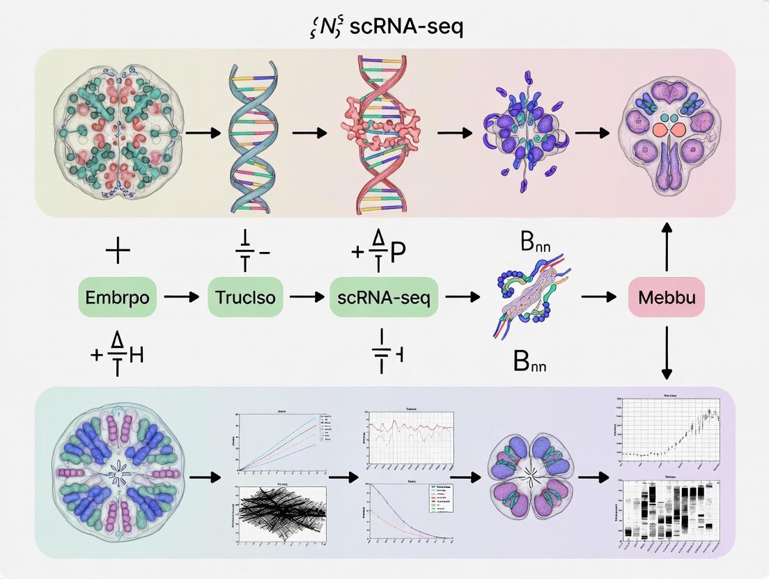
The following diagram illustrates a standard analytical workflow for cross-species embryo scRNA-seq data:
Standard Workflow for Cross-Species Embryo scRNA-seq Analysis
Advanced Integration and Analysis Methods
- Gene Homology Mapping: Utilize ENSEMBL comparative genomics resources to identify one-to-one, one-to-many, and many-to-many orthologs between species. For distant species with challenging annotations, tools like SAMap perform de-novo BLAST analysis to construct gene-gene homology graphs [5].
- Cell-Type Assignment: Machine learning approaches like CAME (a heterogeneous graph neural network) leverage both one-to-one and non-one-to-one homologous mappings to transfer cell-type labels from reference to query species, significantly improving accuracy for non-model organisms [3].
- Trajectory Analysis: Tools like Slingshot can infer developmental trajectories from integrated embeddings, enabling comparison of differentiation pathways and pseudotime dynamics between species [4].
- Regulatory Network Inference: SCENIC analysis identifies conserved and divergent transcription factor regulons, revealing evolutionary changes in gene regulatory networks driving embryonic development [4].
The Scientist's Toolkit: Essential Research Reagents and Computational Tools
This section details key resources required for successful cross-species embryonic scRNA-seq studies.
Table 2: Essential Resources for Cross-Species Embryo scRNA-seq Studies
| Category | Specific Tool/Reagent | Function and Application |
|---|---|---|
| Wet-Lab Reagents & Kits | 10x Genomics Chromium | Microfluidic platform for high-throughput scRNA-seq library preparation |
| Worthington Tissue Dissociation Enzymes | Optimized enzyme blends for embryonic tissue dissociation | |
| gentleMACS Dissociator (Miltenyi) | Instrument for standardized mechanical tissue dissociation | |
| Bioinformatics Pipelines | Seurat | Comprehensive R toolkit for scRNA-seq analysis, including integration functions |
| Scanpy | Python-based scRNA-seq analysis suite for large-scale data | |
| Cell Ranger (10x Genomics) | Pipeline for processing raw sequencing data into count matrices | |
| Cross-Species Specialized Tools | CAME | Graph neural network for cross-species cell-type assignment using complex homology [3] |
| SAMap | Specialized method for whole-body atlas integration between distant species [5] | |
| BENGAL Pipeline | Benchmarking framework for evaluating cross-species integration strategies [5] | |
| Reference Databases | ENSEMBL Compara | Database of gene homologies across multiple species |
| Human Embryo Reference Tool | Integrated scRNA-seq dataset from zygote to gastrula stages [4] | |
| Murrayamine O | Murrayamine O | Murrayamine O, a novel carbazole alkaloid for research. Explore its potential in anti-inflammatory and cytotoxic studies. For Research Use Only. Not for human or veterinary use. |
| Heteroclitin G | Heteroclitin G, MF:C22H24O7, MW:400.4 g/mol | Chemical Reagent |
Cross-species comparison of embryonic scRNA-seq datasets represents a powerful approach for deciphering the evolutionary principles governing cellular diversity and developmental programs. The core objectives—identifying conserved and divergent cell types, understanding transcriptome evolution, and translating knowledge across species—are now achievable through advanced computational methods that robustly integrate data across evolutionary distances. As benchmarking studies demonstrate, careful selection of integration strategies that balance species mixing with biological conservation is crucial for generating meaningful insights. This comparative framework not only deepens our fundamental understanding of evolutionary developmental biology but also provides critical insights into human developmental disorders and the fundamental mechanisms of life's earliest stages.
Single-cell RNA sequencing (scRNA-seq) of embryonic development across species has revolutionized our understanding of evolutionary biology. This guide provides a structured framework for comparing scRNA-seq datasets to uncover patterns of evolutionary conservation and divergence, enabling researchers to identify critical regulatory mechanisms preserved throughout evolution and those that drive species-specific adaptations. The comparative analysis of embryo scRNA-seq datasets allows scientists to trace the evolutionary history of cell types, identify key transcriptional regulators, and understand the molecular basis of morphological evolution. This approach is particularly valuable for drug development professionals seeking to identify conserved therapeutic targets and understand the translatability of model system findings to human biology.
Key Biological Questions and Analytical Approaches
Table 1: Core Biological Questions in Evolutionary scRNA-seq Analysis
| Question Category | Specific Biological Questions | Recommended Analytical Approach | Expected Output |
|---|---|---|---|
| Gene Expression Conservation | Which genes show conserved expression patterns across species? | Orthologous gene alignment, cross-species correlation analysis | List of evolutionarily constrained genes with high functional importance |
| Cell Type Evolution | Are homologous cell types present across species? | Cluster alignment, marker gene comparison, phylogenetic analysis | Cell type homology map, novel cell type identification |
| Developmental Timing | How are developmental trajectories conserved or diverged? | Pseudotime alignment, RNA velocity comparison | Aligned developmental trajectories with conserved/divergent transition points |
| Regulatory Network | Are gene regulatory networks conserved across species? | Co-expression network analysis, regulatory inference | Conserved regulatory modules, divergent network connections |
| Pathway Activity | How are signaling pathway activities evolutionarily maintained? | Pathway enrichment analysis, module scoring | Quantified pathway conservation scores across species |
Experimental Design for Cross-Species Comparisons
Species Selection Criteria
Choosing appropriate species for comparison is fundamental to evolutionary studies. Ideal species pairs should represent meaningful evolutionary distances while maintaining practical experimental feasibility. Recommended considerations include phylogenetic distance (divergence time), morphological similarities/differences, availability of reference genomes, and practical aspects of embryonic material accessibility. For mammalian studies, common comparisons include human-mouse (~90 million years divergence), human-marmoset (~43 million years), or mouse-rat (~20 million years). Each distance provides different insights: closer species reveal fine-scale regulatory changes, while more distant comparisons highlight fundamental conserved mechanisms.
Sample Collection and Timing
Proper embryonic staging and tissue collection are critical for meaningful cross-species comparisons. Use Carnegie stages for human embryos and Theiler stages for mouse embryos, with careful alignment based on morphological landmarks rather than purely temporal age. Collect equivalent anatomical structures across species, verified by expert embryological examination. Preserve samples immediately using appropriate methods (e.g., snap-freezing or immediate fixation) to maintain RNA integrity. Document all collection parameters meticulously, including maternal age, environmental conditions, and exact developmental timing.
Computational Tools for Cross-Species Analysis
Table 2: Computational Tools for Evolutionary scRNA-seq Analysis
| Tool Name | Primary Function | Species Compatibility | Input Requirements | Output Metrics |
|---|---|---|---|---|
| Seurat v5 | Cross-species integration | Multiple species with orthology data | Gene count matrices, ortholog mappings | Integrated UMAP, conserved cluster markers |
| SCENIC+ | Regulatory network inference | Mammalian, with motif databases | scRNA-seq matrix, species motif database | Regulatory networks, transcription factor activity |
| CellRank 2 | Developmental trajectory comparison | Any species with time-series data | RNA velocity, pseudotime estimates | Aligned trajectories, conserved transition points |
| OrthoFinder | Orthologous gene identification | Any eukaryotic species | Protein sequences, genome annotations | Orthogroups, phylogenetic relationships |
| Conos | Multiple dataset integration | Broad species compatibility | Processed scRNA-seq objects | Joint graph, cross-species neighbors |
Experimental Protocols for Key Analyses
Cross-Species Cell Type Alignment Protocol
Purpose: To identify homologous cell types across species and detect species-specific cell populations.
Materials:
- Processed scRNA-seq data from multiple species (count matrices)
- Ortholog mapping table (from OrthoFinder or comparable tool)
- High-performance computing environment with R/Python
Methodology:
- Ortholog Mapping: Map genes between species using one-to-one orthologs, excluding paralogs and species-specific genes.
- Integration: Use Seurat's integration workflow (SelectIntegrationAnchors followed by IntegrateData) with 5,000 integration features and k.anchor=5.
- Clustering: Apply graph-based clustering on the integrated space (resolution=0.5-1.0).
- Marker Identification: Find conserved markers using FindConservedMarkers function with min.pct=0.25 and logfc.threshold=0.25.
- Annotation: Annotate clusters using known marker genes from both species.
- Validation: Validate homologous cell types using orthogonal methods (ISH, IHC) when possible.
Expected Results: A unified UMAP visualization showing integrated cell types, with metrics for cluster conservation and identification of species-specific populations.
Developmental Trajectory Alignment Protocol
Purpose: To compare developmental progression across species and identify conserved and divergent differentiation paths.
Materials:
- scRNA-seq data with developmental time series
- Species-specific gene lengths for RNA velocity
- Pre-computed cell type annotations
Methodology:
- Pseudotime Analysis: Calculate pseudotime using Slingshot or Monocle3 for each species independently.
- RNA Velocity: Compute RNA velocity using scVelo for each species.
- Trajectory Alignment: Use CellRank 2 to align trajectories based on terminal states and driver genes.
- Gene Expression Dynamics: Identify genes with conserved versus divergent expression dynamics along aligned trajectories.
- Transition Conservation: Calculate conservation scores for developmental transitions based on gene expression changes.
Expected Results: Aligned developmental trajectories with quantitative measures of conservation for each branch point and transition.
Visualization of Analytical Workflows
Cross-Species scRNA-seq Analysis Workflow
Research Reagent Solutions
Table 3: Essential Research Reagents for Embryonic scRNA-seq Studies
| Reagent Category | Specific Product Examples | Manufacturer | Primary Function | Species Compatibility |
|---|---|---|---|---|
| Single-Cell Isolation | Chromium Next GEM Kit | 10x Genomics | Single-cell partitioning | Human, Mouse, Primate, Avian |
| Library Preparation | SMART-Seq v4 Ultra Low Input | Takara Bio | cDNA amplification | Broad eukaryotic compatibility |
| Cell Viability | LIVE/DEAD Cell Staining | Thermo Fisher | Viable cell identification | Mammalian, Avian, Fish |
| Cell Hashing | CellPlex Cell Multiplexing | 10x Genomics | Sample multiplexing | Human, Mouse, commonly studied species |
| Spatial Transcriptomics | Visium Spatial Gene Expression | 10x Genomics | Tissue context preservation | Human, Mouse, Zebrafish |
| In Situ Hybridization | RNAscope Multiplex Assay | ACD Bio | Spatial validation | Species-specific probes available |
Signaling Pathway Conservation Analysis
Signaling Pathway Conservation Patterns
Data Interpretation Framework
Quantifying Conservation and Divergence
Develop rigorous metrics for evaluating evolutionary patterns in scRNA-seq data. Conservation scores should integrate multiple aspects: gene expression level preservation, co-expression network maintenance, developmental timing conservation, and cell type homology. Calculate conservation indices for each gene, cell type, and developmental transition to enable systematic comparison across evolutionary distances. Use permutation testing to establish statistical significance for observed conservation patterns, comparing against null distributions generated by randomizing species labels or gene identities.
Biological Validation Strategies
Corroborate computational findings with experimental validation using species-appropriate techniques. Employ cross-species in situ hybridization to validate spatial expression patterns of conserved and divergent genes. Utilize CRISPR-based approaches in model systems to test functional conservation of regulatory elements. Implement organoid culture systems from multiple species to assay conserved developmental processes in controlled environments. Apply spatial transcriptomics to verify conserved tissue organization patterns across species.
Applications in Drug Development
The identification of evolutionarily conserved molecular mechanisms provides particularly valuable insights for pharmaceutical research. Conserved pathways and regulatory networks often represent fundamental biological processes with high translational potential. Drug development professionals can prioritize targets with strong conservation evidence, as these typically demonstrate higher clinical success rates. Additionally, understanding species-specific differences helps optimize preclinical models and predict potential adverse effects resulting from divergent biology. Embryonic scRNA-seq comparisons can reveal conserved therapeutic targets for regenerative medicine applications while identifying potential species-specific toxicities early in drug development pipelines.
Understanding human embryonic development from the pre-implantation stages through organogenesis is fundamental for developmental biology, regenerative medicine, and uncovering the causes of congenital disorders and early pregnancy loss [8]. While mouse models have served as valuable proxies for mammalian development for decades, significant morphological, molecular, and genetic differences exist between mouse and human embryogenesis [9] [10]. The emergence of sophisticated single-cell RNA sequencing (scRNA-seq) technologies has revolutionized this field, enabling the construction of high-resolution transcriptional atlases of early human development and facilitating cross-species comparative analyses [4] [1]. This guide synthesizes landmark studies that have provided integrated references spanning pre-implantation to organogenesis, objectively comparing their methodologies, findings, and applications within the context of cross-species embryo scRNA-seq research.
Comparative Morphological and Molecular Landscapes
Significant morphological and molecular differences exist between human and mouse embryogenesis, which genetically determine species-specific developmental pathways [9].
Table 1: Key Morphological Differences Between Mouse and Human Embryogenesis
| Developmental Feature | Mouse Embryo | Human Embryo |
|---|---|---|
| Zygotic Genome Activation (ZGA) | Occurs at the 2-cell stage [9] | Occurs between the 4- and 8-cell stages [9] |
| Facial Organ Development | Optic pit appears first [9] | All facial organs appear around the same time [9] |
| Limb Rotation | Little rotation; less flexible joints [9] | Rotates to proper position ventrally; flexible joints [9] |
| Tail Development | Elongates and thins from Theiler Stage (TS) 17 [9] | Regresses during Carnegie Stage (CS) 23 (~9th week) [9] |
| Post-Organogenesis Birth | Born almost immediately after organogenesis (~19-20 days) [9] | Remains in uterus for several more months of fetal growth [9] |
At the molecular level, cross-species comparative transcriptomics reveals that the most significant differences lie not in gene number, but in the spatiotemporal expression patterns and activities of gene products [9]. For instance, while core signaling pathways like Notch, TGFβ/BMP, and Wnt are conserved, significant differences exist for specific genes such as Wnt7a and CAPN3, particularly in neural crest, midbrain, lens, heart, and smooth muscle formation [9].
Landmark scRNA-seq Reference Datasets and Tools
Several landmark studies have created essential scRNA-seq resources to map human embryonic development, addressing the critical scarcity of in vivo samples.
Comprehensive Integrated Reference from Zygote to Gastrula
A landmark 2025 study created a universal integrated scRNA-seq reference by harmonizing six published human datasets, covering development from the zygote to the gastrula stage (Carnegie Stage 7) [4].
Table 2: Key Integrated scRNA-seq Reference Tools and Databases
| Resource Name | Scope/Species | Key Features and Application | Access |
|---|---|---|---|
| Human Embryo Reference Tool [4] | Human (Zygote to Gastrula) | Integrated data from 3,304 cells; UMAP projection; cell identity prediction; lineage trajectory inference. | Online prediction tool |
| DRscDB [11] | Drosophila, Zebrafish, Mouse, Human | Repository for published scRNA-seq data; finds orthologous genes and cell type-specific expression across species. | Web database (flyrnai.org) |
This integrated atlas delineates the continuous progression of lineage specification, beginning with the divergence of the inner cell mass (ICM) and trophectoderm (TE), followed by ICM bifurcation into epiblast and hypoblast [4]. The tool utilizes Single-cell regulatory network inference and clustering (SCENIC) analysis to identify key transcription factors driving lineage development, such as DUXA in the 8-cell lineage, VENTX in the epiblast, and OVOL2 in the TE [4]. This resource serves as a critical benchmark for authenticating stem cell-based embryo models.
Gene Expression Profiling During Organogenesis
Earlier microarray studies provided the first genome-wide gene expression profiles of human organogenesis, a period from the 4th to the 9th week (CS10-23) [9]. These studies revealed two major patterns of gene regulation: a down-regulation of "stemness," cell cycle, and metabolic genes, and an up-regulation of genes involved in multi-cellular organismal development, cell adhesion, and cell-cell signaling [9]. Furthermore, many genes exhibited an arch-shaped expression pattern, with peak levels corresponding to the development of specific organs, such as eye development genes peaking during the 5th-7th weeks [9].
Experimental Methodologies and Workflows
Studying human development relies on a combination of direct embryo analysis and innovative stem cell-based models, each with specific protocols.
Single-Cell RNA-Sequencing of Human Embryos
The workflow for creating a comprehensive reference atlas involves several standardized steps [4]:
- Sample Sourcing and Preparation: The process utilizes donated preimplantation embryos, in vitro cultured postimplantation blastocysts, and scarce in vivo gastrula-stage specimens [4] [8].
- Data Generation and Processing: scRNA-seq data from multiple studies are reprocessed using a standardized pipeline, with mapping and feature counting against the same genome reference (GRCh38) to minimize batch effects [4].
- Data Integration and Visualization: Datasets are integrated using computational methods like fast mutual nearest neighbor (fastMNN) to create a unified transcriptional landscape. Cells are visualized in two-dimensional space using Uniform Manifold Approximation and Projection (UMAP) [4].
- Cell Annotation and Trajectory Inference: Manifold learning and clustering algorithms identify distinct cell populations. Lineage developmental trajectories are inferred using tools like Slingshot, which calculates pseudotime to order cells along a differentiation path [4].
Stem Cell-Derived Embryo Models
To overcome the scarcity of post-implantation embryos, researchers have developed stem cell-derived embryo-like structures (embryoids) that recapitulate aspects of early development [12] [8]. One such model is the microfluidic post-implantation amniotic sac embryoid (μPASE) [12]. The protocol involves:
- Stem Cell Culture: Human pluripotent stem cells (hPSCs) are aggregated and loaded into a microfluidic device [12].
- Controlled Differentiation: The system allows for highly controllable and reproducible development. hPSC clusters undergo lumenogenesis and epithelization to form a central lumen [12].
- Lineage Specification: Exposure to exogenous BMP4 initiates amniotic ectoderm-like cell (AMLC) differentiation. Inductive effects from these cells cause other hPSCs to undergo an epithelial-mesenchymal transition (EMT), forming primitive streak-like and mesoderm-like cells (MeLCs) [12].
- Validation: The resulting cell populations are validated via scRNA-seq and benchmarked against available in vivo primate data to assess fidelity [12].
Table 3: Essential Research Reagent Solutions for Embryo scRNA-seq Studies
| Reagent / Resource | Function and Application | Example Use Case |
|---|---|---|
| KSOM Media [10] | Advanced in vitro culture medium for pre-implantation mouse embryos. | Supporting embryo development from zygote to blastocyst stage ex vivo. |
| Matrigel [10] | A 3D extracellular matrix hydrogel. | Providing a physiologically relevant substrate for in vitro culture of embryos and embryoids. |
| Microfluidic Devices [12] | Miniaturized systems for cell culture and manipulation. | Enabling highly controlled and scalable culture of embryoid models (e.g., μPASE). |
| BMP4 [12] | A morphogen of the TGF-β superfamily. | Directing differentiation of pluripotent stem cells towards amniotic ectoderm lineage in embryoids. |
| CRISPR-Cas9 | Gene editing tool for functional genomic studies. | Investigating gene function through targeted knockout in embryos or stem cells [2] [8]. |
| Human U133 Array [9] | Affymetrix microarray platform for gene expression profiling. | Conducting genome-wide expression analysis of human embryos during organogenesis. |
| 10x Genomics [12] | A high-throughput scRNA-seq platform. | Profiling transcriptomes of thousands of individual cells from embryoids or dissociated embryos. |
Critical Challenges and Technical Roadblocks
Research on human embryogenesis faces several significant challenges:
- Sample Scarcity and Access: There is a critical scarcity of human embryonic material, particularly for the post-implantation period between weeks 2 and 4 due to technical and ethical constraints, notably the widespread 14-day culture rule [4] [8]. Access relies on donations from IVF procedures, which can be limited by regulatory hurdles and variable embryo quality [8].
- In Vitro Culture Limitations: Current protocols for culturing human embryos beyond the blastocyst stage often lack proper morphogenesis and maternal tissue interactions, raising concerns about their physiological equivalence to in vivo embryos [8].
- Genetic Manipulation Difficulties: Efficient genetic manipulation in human embryos remains technically challenging, hampered by limited knowledge of DNA repair mechanisms and regulations prohibiting genetic modification in many jurisdictions [8].
- Cross-Species Comparison Complexities: Comparing scRNA-seq data across species is complicated by biological differences, technical batch effects, and challenges in assigning functional conservation to orthologous genes [1].
The construction of integrated scRNA-seq references from pre-implantation to organogenesis represents a transformative advance in developmental biology. These atlases, complemented by cross-species comparative analyses and validated embryoid models, provide unprecedented insights into the molecular underpinnings of human embryogenesis. While significant challenges in sample access, model fidelity, and computational integration remain, the continued refinement of these resources and tools is paving the way for a deeper understanding of human development, with profound implications for regenerative medicine and the treatment of congenital disorders.
Identifying Lineage-Specific Markers and Transcription Factors
The accurate identification of lineage-specific markers and transcription factors is a cornerstone of developmental biology, enabling researchers to decipher the complex processes of cell fate decisions, differentiation, and tissue formation. With the advent of single-cell RNA sequencing (scRNA-seq), we can now profile gene expression at unprecedented resolution across different stages of embryonic development and in various model systems. However, the utility of this data hinges on robust analytical frameworks and reference tools for correct cell type annotation and lineage validation. This guide provides a comparative analysis of experimental and computational approaches for identifying these crucial molecular signatures, with a specific focus on leveraging cross-species comparative scRNA-seq datasets to enhance the accuracy and biological relevance of the findings. We objectively evaluate the performance of different methodologies, supported by experimental data, to serve as a resource for researchers authenticating stem cell-derived models and studying evolutionary biology.
Core Experimental & Computational Approaches
Integrated Reference Atlases for Authentication
The creation of comprehensive, integrated scRNA-seq reference datasets represents a significant advancement for benchmarking cellular identities.
- Reference Tool Construction: A universal reference for human embryogenesis was developed by integrating six public scRNA-seq datasets, covering developmental stages from zygote to gastrula. The dataset includes 3,304 cells and utilizes the fast Mutual Nearest Neighbor (fastMNN) method for integration to minimize batch effects. This is visualized as a UMAP that displays continuous developmental progression [4].
- Lineage Resolution: The reference captures key lineage bifurcations: the first separates the inner cell mass (ICM) from the trophectoderm (TE), followed by the ICM's division into epiblast and hypoblast. Later stages further resolve the epiblast into primitive streak, mesoderm, definitive endoderm, and amnion, among other lineages [4].
- Performance in Authentication: When used to authenticate stem cell-based embryo models, this integrated reference revealed risks of cell lineage misannotation that occurred when models were benchmarked against less relevant or incomplete references. Its use provides an unbiased method for evaluating the fidelity of in vitro models to their in vivo counterparts [4].
Kinetic Modeling of mRNA Regulation
Understanding the dynamics of gene expression requires dissecting the contributions of mRNA transcription and degradation, which can be achieved by combining scRNA-seq with metabolic labeling.
- Experimental Workflow: Zebrafish embryos at the one-cell stage are injected with 4-thiouridine triphosphate (4sUTP), which is incorporated into newly transcribed (zygotic) RNA. This allows distinction from pre-existing maternal mRNA. Single-cell transcriptomes are then captured, and a chemical conversion step creates T-to-C changes in sequencing reads from labeled RNAs, enabling quantification [13].
- Data Analysis with Kinetic Models: Tools like GRAND-SLAM are used to statistically infer the fraction of newly transcribed mRNA for each gene in each cell. This data feeds into kinetic models that quantify cell-type-specific mRNA transcription and degradation rates during specification [13].
- Key Findings: This approach revealed highly coordinated transcription and degradation rates for many transcripts and identified selective retention of maternal mRNAs in specific early lineages like the primordial germ cells and enveloping layer, highlighting how degradation shapes cell-type-specific expression patterns [13].
Cross-Species Comparative Transcriptomics
Comparing scRNA-seq data across species identifies conserved and species-specific features of lineage specification, providing evolutionary context and validating core regulatory programs.
- Conserved Neutrophil Maturation: A cross-species analysis of neutrophil maturation in zebrafish, mouse, and human identified a high molecular similarity in immature stages. This allowed researchers to define a pan-species neutrophil maturation signature and distinguish it from zebrafish-specific gene signatures. This conserved signature was subsequently applied to human patient data, linking metastatic tumor cell infiltration in the bone marrow to an increase in mature neutrophils [14].
- Conserved Genetic Basis of Spermatogenesis: A comparison of scRNA-seq datasets from the testes of humans, mice, and fruit flies identified 1,277 conserved genes involved in spermatogenesis. Key conserved molecular programs included post-transcriptional regulation, meiosis, and energy metabolism. Systematic gene knockout of 20 candidates in Drosophila confirmed that three genes related to sperm centriole and steroid lipid processes were essential for male fertility, underscoring deep evolutionary conservation [2].
Table 1: Key Outcomes from Cross-Species Comparative Studies
| Study System | Conserved Biological Process | Number of Conserved Genes Identified | Functionally Validated Key Processes |
|---|---|---|---|
| Neutrophil Maturation [14] | Innate immune cell differentiation | A pan-species gene signature defined | Granule development, phagocytic capacity |
| Spermatogenesis [2] | Male germ cell development | 1,277 | Sperm centriole function, steroid metabolism, meiosis |
Experimental Protocols for Key Workflows
Protocol: Building an Integrated Embryo Reference Atlas
This protocol is adapted from the creation of a human embryo reference tool [4].
- Data Collection: Gather publicly available scRNA-seq datasets that cover the desired developmental window and lineages. For the human embryo reference, this included six datasets from pre-implantation embryos, post-implantation blastocysts, and a gastrula.
- Reprocessing: Reprocess all raw data using a standardized pipeline with the same genome reference and annotation (e.g., GRCh38) to minimize technical batch effects from the start. This includes mapping and feature counting.
- Data Integration: Employ a batch-correction algorithm such as fastMNN to integrate the expression profiles of all cells into a unified space.
- Dimensionality Reduction and Clustering: Generate a UMAP (Uniform Manifold Approximation and Projection) from the integrated data to visualize cellular relationships. Perform clustering to identify distinct cell populations.
- Lineage Annotation: Annotate cell clusters based on known marker genes from original studies and validated against human and non-human primate data. Key markers include:
- Trajectory Inference: Use tools like Slingshot on the UMAP embeddings to infer developmental trajectories (pseudotime) for major lineages (e.g., epiblast, hypoblast, TE) and identify genes with modulated expression.
- Regulatory Network Analysis: Perform SCENIC (Single-Cell Regulatory Network Inference and Clustering) analysis to identify cell-type-specific transcription factor activities and regulons.
- Tool Deployment: Create a user-friendly prediction tool (e.g., a Shiny app) where query datasets can be projected onto the reference to annotate cell identities.
Protocol: Cross-Species scRNA-Seq Comparison
This protocol is synthesized from studies on neutrophils and spermatogenesis [14] [2].
- Dataset Curation: Collect high-quality scRNA-seq datasets from homologous tissues or developmental processes across multiple species (e.g., human, mouse, zebrafish/fly).
- Cell Type Annotation: Independently annotate cell types/states within each species' dataset using established lineage-specific markers.
- Ortholog Mapping: Map genes between species using one-to-one orthology information from databases like Ensembl.
- Identification of Conserved Markers: Perform differential expression analysis between cell types within each species. Identify overlapping differentially expressed genes (DEGs) across species as a conserved marker set. Alternatively, use label-transfer or canonical correlation analysis to align datasets and find shared expression patterns.
- Functional Enrichment Analysis: Subject the conserved gene sets to gene ontology (GO) and pathway enrichment analysis to identify biological processes and regulatory networks preserved through evolution.
- Experimental Validation: Design in vivo or in vitro functional experiments to test the role of identified conserved genes. This can include:
Visualization of Workflows and Relationships
scRNA-seq Cross-Species Analysis Workflow
The following diagram illustrates the logical flow for a standard cross-species comparative transcriptomics study.
Figure 1: Cross-species scRNA-seq analysis workflow for identifying conserved lineage factors.
mRNA Kinetic Analysis with Metabolic Labeling
This diagram details the experimental and computational workflow for quantifying mRNA transcription and degradation rates in single cells.
Figure 2: Workflow for cell-type-specific mRNA kinetic analysis in embryos.
Table 2: Essential Research Reagent Solutions for Lineage Marker Identification
| Reagent/Resource | Function/Brief Explanation | Example Use Case |
|---|---|---|
| Integrated Embryo Reference | A curated, batch-corrected scRNA-seq atlas serving as a universal standard for cell identity annotation. | Authenticating stem cell-derived embryo models by projecting query data onto the reference [4]. |
| Metabolic Labels (e.g., 4sUTP) | Nucleotide analogs incorporated into newly synthesized RNA, allowing it to be distinguished from pre-existing RNA. | Measuring zygotic vs. maternal mRNA contributions and inferring transcription/degradation rates in single cells [13]. |
| Transgenic Reporter Strains | Organisms with fluorescent proteins under control of cell-type-specific promoters, enabling visualization and sorting. | Isolating specific maturation stages of neutrophils (Tg(BACmmp9:Citrine-CAAX)) for transcriptional profiling [14]. |
| CRISPR Knockout Systems | Precision gene-editing tools for functional validation of candidate genes in vivo. | Testing the role of evolutionarily conserved genes in processes like spermatogenesis in model organisms [2]. |
| Trajectory Analysis Software (Slingshot) | Computational tool that infers developmental pathways and pseudotime from scRNA-seq data. | Reconstructing lineage bifurcations (e.g., ICM to epiblast/hypoblast) and identifying associated genes [4]. |
| Regulatory Network Tools (SCENIC) | Infers gene regulatory networks and identifies active transcription factors from scRNA-seq data. | Discovering key transcription factors (e.g., C/ebp-β in neutrophils) driving lineage specification [4] [14]. |
| Cross-Species Alignment Algorithms | Bioinformatics methods for integrating scRNA-seq data from different species based on orthologous genes. | Identifying a core set of conserved lineage markers and regulators across humans, mice, and fish/flies [14] [2]. |
From Cells to Data: scRNA-seq Workflows and Translational Applications
Single-cell RNA sequencing (scRNA-seq) has revolutionized biological research by enabling the characterization of gene expression profiles at the single-cell level, revealing cellular heterogeneity that bulk sequencing approaches inevitably mask. This capability is particularly valuable in cross-species comparative studies, which aim to uncover conserved and divergent biological processes across evolution. For instance, scRNA-seq has been instrumental in comparing inflammatory responses to heart injury in zebrafish and mice, revealing both analogous macrophage subtypes and disparate reaction pathways that may underlie differences in regenerative capacity [15]. The successful execution of an scRNA-seq experiment requires careful consideration of multiple steps, from cell isolation to sequencing. This guide provides a systematic overview of the core workflow, compares leading technological platforms, and outlines specific methodological considerations for cross-species research, with a special focus on embryonic datasets.
Step 1: Cell and Nuclei Isolation
The first critical step is obtaining high-quality single-cell or single-nuclei suspensions.
Single-Cell Isolation: This typically involves fresh tissues. The process includes finely mincing the tissue, followed by enzymatic digestion (e.g., using collagenase, trypsin, or Accutase) and mechanical dissociation to break down the extracellular matrix. The resulting suspension is then filtered through a strainer (e.g., 40 µm) to remove clumps, and dead cells can be removed using techniques like density centrifugation or magnetic-activated cell sorting (MACS) with dead cell removal kits [16]. A critical consideration is that the dissociation process itself can induce cellular stress and alter transcriptional profiles [16].
Single-Nuclei Isolation: As an alternative, snRNA-seq uses isolated nuclei. Frozen tissue is homogenized in a lysis buffer that breaks down cell membranes but leaves nuclei intact. The suspension is then filtered and centrifuged to purify the nuclei [16]. A key advantage is compatibility with frozen, biobanked samples, which are often the primary resource for rare specimens like human embryos. It also avoids dissociation-induced stress artifacts. However, it primarily captures nascent, nuclear transcripts and may under-represent cytoplasmic mRNAs, leading to a bias in the detected transcriptome [16].
Choosing Between scRNA-seq and snRNA-seq: The decision hinges on the research question and sample availability. scRNA-seq provides a more complete picture of the cytoplasmic transcriptome but requires fresh, viable cells and is susceptible to dissociation artifacts. snRNA-seq is preferable for archived frozen samples, sensitive tissues (like neurons or pancreatic islets), and when aiming to minimize technical stress responses [16]. A comparative study on human pancreatic islets from the same donors confirmed that while both methods identify the same major cell types, they can yield different cell type proportions, underscoring the need to choose the method aligned with the study's goals [16].
Step 2: Single-Cell Library Preparation
Once a high-quality suspension is obtained, the next step is to prepare sequencing libraries. This process involves capturing individual cells, reverse-transcribing their mRNA into cDNA, and adding platform-specific barcodes and sequencing adapters.
Different scRNA-seq technologies have been developed, each with unique strengths and weaknesses. The table below summarizes key performance metrics for several established and emerging platforms based on recent comparative studies.
Table 1: Performance Comparison of High-Throughput scRNA-seq Platforms
| Platform / Technology | Capture Method | Key Strengths | Key Limitations | Suitability for Sensitive Cells (e.g., Neutrophils/Embryos) |
|---|---|---|---|---|
| 10x Genomics Chromium [17] [18] | Droplet-based | High throughput, strong gene sensitivity, widely established | Lower gene sensitivity in granulocytes, requires fresh cells (standard protocol) | Standard protocol challenging for neutrophils; Fixed RNA Profiling Flex kit allows cell fixation |
| BD Rhapsody [17] [18] | Microwell-based | High capture sensitivity for low-RNA cells, suitable for neutrophils | Lower proportion of some cell types (e.g., endothelial cells) | Effective, comparable to flow cytometry for neutrophil capture |
| Parse Biosciences (Evercode) [17] | Combinatorial barcoding (fixed cells) | Low mitochondrial gene expression, high multiplexing (up to 96 samples), no specialized equipment needed | Does not require specialized equipment | Fixed-cell workflow minimizes ex vivo artifacts |
| HIVE scRNA-seq [17] | Nano-wells | Cells can be stabilized and stored at -80°C pre-library prep | Higher levels of mitochondrial genes detected | Successfully used with RBC-depleted blood samples |
| Fluidigm C1 [19] | Microfluidics (plate-based) | High sequencing depth and sensitivity | Lower throughput, higher cost per cell | Not specifically evaluated in provided studies |
Detailed Protocol: 10x Genomics Chromium Workflow
The 10x Genomics Chromium platform is one of the most widely used droplet-based methods. The following is a generalized protocol for library preparation using a system like the Chromium Next GEM Single Cell 3' Reagent Kit [16].
- Cell Preparation and Loading: The single-cell suspension is diluted to a target concentration (e.g., 700-1,200 cells/µl). It is critical to ensure high cell viability (>80-90%) and to confirm the absence of clumps. A volume of this suspension containing the desired number of cells is added to a master mix containing enzymes, barcoded gel beads, and partitioning oil.
- Gel Bead-in-Emulsion (GEM) Generation: The mixture is loaded onto a Chromium chip and placed in the Chromium Controller. Within the chip, each cell is co-encapsulated with a single barcoded gel bead in a nanoliter-scale oil droplet, forming a GEM.
- Reverse Transcription (RT) Inside GEMs: Within each GEM, the cell is lysed, and the mRNA transcripts hybridize to the gel bead's oligonucleotides. These oligos contain a PCR handle, a cell-specific barcode (the same for all transcripts from that cell), a unique molecular identifier (UMI), and a poly(dT) sequence for mRNA capture. Reverse transcription occurs, creating cDNA strands tagged with the cell barcode and UMI.
- GEM Breakage and cDNA Cleanup: The oil emulsion is broken, and the barcoded cDNA from all GEMs is pooled. The cDNA is then purified using DynaMyneads MyOne SILANE beads.
- cDNA Amplification: The purified cDNA is PCR-amplified to generate sufficient mass for library construction.
- Library Construction: The amplified cDNA is fragmented, and a suite of adapters is ligated. This step adds sample indexes (for multiplexing) and sequences required for cluster generation and sequencing on platforms like Illumina.
- Library QC and Sequencing: The final libraries are quantified (e.g., using qPCR) and assessed for quality (e.g., using a Bioanalyzer). They are then sequenced on an appropriate Illumina platform.
Visual Guide to scRNA-seq Workflow
Step 3: Sequencing and Data Analysis
After library preparation, the next steps are sequencing and computational analysis.
Sequencing: scRNA-seq libraries are typically sequenced on Illumina platforms. The required sequencing depth depends on the project's goals and the complexity of the tissue. A common configuration is paired-end sequencing, where Read 1 contains the cell barcode and UMI, and Read 2 contains the cDNA insert.
Core Bioinformatic Analysis: The raw sequencing data (FASTQ files) undergoes a multi-step computational pipeline:
- Demultiplexing & Alignment: Sequences are assigned to their sample of origin, and reads are aligned to a reference genome.
- Gene-Cell Matrix Generation: Using tools like Cell Ranger (10x Genomics), reads are counted based on their cell barcode and UMI, generating a digital count matrix where rows represent genes and columns represent cells.
- Quality Control (QC): Cells are filtered based on metrics like the number of detected genes, total UMI counts, and the percentage of mitochondrial reads. High mitochondrial percentage can indicate stressed or dying cells [17] [20].
- Normalization & Scaling: Counts are normalized to account for technical variability (e.g., sequencing depth).
- Dimensionality Reduction & Clustering: Principal Component Analysis (PCA) is performed, followed by graph-based clustering on the results. Cells are visualized in 2D using UMAP or t-SNE.
- Cell Type Annotation: Clusters are annotated into cell types using manual annotation (checking known marker genes) or reference-based annotation with tools like SingleR or Seurat's label transfer [15] [20] [16].
Special Considerations for Cross-Species Embryonic Research
Applying scRNA-seq to cross-species embryo comparisons introduces specific challenges that require tailored approaches.
Ortholog Mapping: A fundamental step is converting gene symbols from different species (e.g., mouse, zebrafish) to a common set of orthologous genes, typically human symbols. This is done using databases like Ensembl or tools like OrthoFinder, retaining only one-to-one orthologs for a robust comparative analysis [20].
Integration and Batch Effect Correction: Data from different species, technologies, or even batches must be integrated. Batch effect correction tools like Harmony have been shown to achieve high performance in integrating PBMC data from multiple species, allowing for a joint analysis that preserves biological variation while removing technical artifacts [20].
CNV Analysis for Ploidy and Subclone Detection: In cancer and developmental biology, copy number variations (CNVs) can be inferred from scRNA-seq data to identify subpopulations of cells. A 2025 benchmarking study evaluated six CNV callers (InferCNV, copyKat, SCEVAN, CONICSmat, CaSpER, and Numbat), finding that methods incorporating allelic information (e.g., Numbat, CaSpER) performed more robustly for large datasets, though with higher computational demands [21]. This is crucial for identifying chromosomal abnormalities in embryonic cells.
Table 2: Key Computational Tools for Cross-Species scRNA-seq Analysis
| Analysis Step | Tool Example | Application in Cross-Species Research |
|---|---|---|
| Data Integration | Harmony [20] | Corrects batch effects across samples from different species for unified analysis. |
| Cell Annotation | SingleR [15], Seurat [20] | Automates cell type labeling by referencing annotated datasets. |
| Ortholog Mapping | OrthoFinder [20] | Predicts orthologous gene pairs between species for gene list conversion. |
| CNV Calling | InferCNV, Numbat [21] | Infers copy number variations to identify genetic subclones within a population. |
| Differential Expression | Seurat (Wilcoxon test) [20] | Identifies genes differentially expressed between cell clusters or conditions. |
The Scientist's Toolkit
Table 3: Essential Research Reagent Solutions for scRNA-seq
| Reagent / Kit | Function | Example Use-Case |
|---|---|---|
| Accutase [16] | Enzyme for gentle dissociation of tissue into single cells. | Preparing single-cell suspensions from delicate embryonic tissues. |
| Chromium Nuclei Isolation Kit [16] | Isulates nuclei from frozen tissue for snRNA-seq. | Processing frozen, biobanked embryo samples that are not viable for scRNA-seq. |
| Chromium Next GEM Kits (10x Genomics) [16] | Comprehensive reagent kit for GEM generation, RT, and library prep. | Standardized, high-throughput single-cell library construction. |
| Dead Cell Removal Kit [16] | Magnetic beads for removing dead cells from suspension. | Improving viability of single-cell suspensions to reduce ambient RNA. |
| RNase Inhibitors [17] | Protects RNA from degradation during sample processing. | Critical for preserving RNA in sensitive cells like neutrophils or embryonic cells. |
| Datiscin | Datiscin, MF:C27H30O15, MW:594.5 g/mol | Chemical Reagent |
| lespedezaflavanone H | lespedezaflavanone H, MF:C30H36O6, MW:492.6 g/mol | Chemical Reagent |
A successful scRNA-seq experiment, particularly in the complex context of cross-species embryology, relies on a meticulously planned and executed workflow. From the critical first decision of cell versus nuclei isolation to the selection of a platform that balances throughput, sensitivity, and suitability for delicate cells, each step influences the final data quality. The growing suite of bioinformatic tools for integration, annotation, and CNV analysis empowers researchers to draw meaningful biological insights, such as identifying evolutionarily conserved cell types and transcriptional programs. As methods continue to advance, with a clear trend towards fixed-cell protocols and multi-omics integrations, the resolution at which we can compare embryonic development across species will only increase, deepening our understanding of evolutionary biology and the fundamental principles of life.
Single-cell RNA sequencing (scRNA-seq) has revolutionized biological research by enabling the characterization of gene expression at the ultimate level of resolution—the individual cell. This is particularly powerful in embryology and cross-species comparative studies, where understanding cellular heterogeneity and lineage specification is paramount. The choice of scRNA-seq platform is critical and involves balancing throughput, sensitivity, cost, and experimental flexibility. This guide provides an objective comparison of the three principal methodological approaches—plate-based, droplet-based microfluidics, and combinatorial indexing—with a specific focus on their application in cross-species embryo research.
The development of scRNA-seq technologies has progressed from low-throughput, manual methods to highly parallelized, automated platforms. The core principle involves isolating single cells, barcoding their transcripts to preserve cellular origin, and preparing sequencing libraries. The methods differ fundamentally in how they achieve this physical separation and molecular barcoding [22].
The table below summarizes the key characteristics of the three main platform types.
Table 1: Core Platform Comparison for scRNA-seq
| Feature | Plate-Based Methods | Droplet-Based Microfluidics | Combinatorial Indexing Methods |
|---|---|---|---|
| Throughput | Lowest (improved with combinatorial indexing) [23] | Highest [23] | Intermediate to Very High [23] [24] |
| Cost per Cell | Highest [23] | Lowest [23] | Intermediate [23] |
| Sensitivity | Highest [23] | Lower than plate-based [23] | Lower than plate-based; variable [24] |
| Workflow | Flexible but labor-intensive; involves manual cell sorting and pipetting [23] | Highly automated, but requires expensive dedicated microfluidics equipment [23] | Labor-intensive; involves multiple rounds of splitting and pooling [24] |
| Best For | Smaller-scale, in-depth studies; precious samples [23] | Large-scale atlasing projects; profiling hundreds of thousands of cells [23] [24] | Large-scale studies when cost of microfluidics equipment is prohibitive; custom assay design [23] [25] |
Technical Performance and Experimental Data
To move beyond theoretical specifications, it is essential to consider quantitative performance data from real-world experiments, including direct benchmarking studies.
Throughput and Multiplet Rates
Throughput refers to the number of cells that can be reliably profiled in a single experiment, and it is intrinsically linked to the multiplet rate (the percentage of libraries derived from two or more cells).
- Droplet-Based Microfluidics: Standard protocols for platforms like 10x Genomics are designed to recover up to 10,000 cells per channel, with a significant proportion of droplets remaining empty to control for multiplets [24]. Novel methods like OAK (Overloading And unpacKing) push these boundaries by overloading the microfluidic chip. One study loaded 150,000 fixed cells, achieving a projected recovery of ~88,000 cells with a 6.6% overall multiplet rate. Loading 450,000 cells projected a recovery of ~224,000 cells, albeit with a higher multiplet rate of 10.6% and a mild reduction in genes detected per cell [24].
- Combinatorial Indexing: The UDA-seq protocol, which combines droplet barcoding with a second round of well-based indexing, demonstrated high cell recovery. From a load of 10,000 cells, it recovered 6,245 high-quality single-cell transcriptomes (62% recovery) with a low collision rate of only 1.23% [26].
Sensitivity and Data Quality
Sensitivity is often measured by the number of genes detected per cell, a critical factor for identifying rare cell types or subtle transcriptional states.
- Plate-based methods like SMART-seq3 are renowned for their high sensitivity and full-length transcript coverage, making them ideal for detecting isoform-level information [23] [22].
- Droplet-based methods show strong performance. In a benchmark, the standard 10x Genomics Chromium protocol detected a mean of 3,905 genes per cell in a K562 cell line. The high-throughput OAK method, under matched sequencing depth, detected a mean of 3,014 genes for the same cell line, indicating a mild but notable reduction in sensitivity, primarily for lowly expressed genes [24].
- Combinatorial Indexing methods have historically had lower sensitivity, but newer workflows like OAK show a strong correlation (Spearman correlation = 0.92) with standard droplet-based methods in terms of gene counts across cells, confirming the robustness of the quantitative data [24].
Table 2: Experimental Benchmarking Data from Key Studies
| Study (Method) | Cells Loaded | Cells Recovered | Multiplet/Collision Rate | Key Metric (e.g., Genes/Cell) |
|---|---|---|---|---|
| OAK (Droplet + CI) [24] | 150,000 | 87,864 (projected) | 6.6% (overall) | 3,014 mean genes (K562) |
| OAK (Droplet + CI) [24] | 450,000 | 223,680 (projected) | 10.6% (overall) | Fewer genes vs. lower load |
| Standard Chromium (Droplet) [24] | N/A | Up to 10,000 | Standard | 3,905 mean genes (K562) |
| UDA-seq (Droplet + CI) [26] | 10,000 | 6,245 (62%) | 1.23% | Data quality comparable to standard 10x |
| sci-RNA-seq (CI) [24] | N/A | N/A | N/A | Lower sensitivity vs. OAK & 10x |
Application in Cross-Species Embryo Research
The selection of a scRNA-seq platform is profoundly influenced by the specific research application. In the burgeoning field of cross-species embryology, each method offers distinct advantages.
A primary application is constructing high-resolution transcriptional atlases of embryonic development. A landmark study integrated scRNA-seq data from six published human datasets, covering development from the zygote to the gastrula stage, to create a comprehensive reference of 3,304 cells. This atlas successfully captured the bifurcation of the inner cell mass into epiblast and hypoblast, and the subsequent maturation of trophectoderm and emergence of gastrula lineages [4]. Such detailed, high-sensitivity mapping benefits from methods that prioritize transcriptional depth, making plate-based or standard droplet-based platforms well-suited for the initial atlas creation, especially when starting material is limited but of high value.
Benchmarking Embryo Models
Stem cell-derived embryo models (embryoids) are crucial tools for studying early human development. Their utility depends on faithful recapitulation of in vivo development, which is validated by comparing their scRNA-seq profiles to a ground-truth in vivo reference. The integrated human embryo reference has been used to authenticate embryoid models, revealing the risk of misannotation when relevant references are not used [4]. For these comparative studies, which often require profiling multiple models and conditions, the high throughput and robustness of droplet-based systems are advantageous.
Identifying Conserved Genetic Programs
Cross-species comparative studies aim to identify evolutionarily conserved genetic programs. One such study compared scRNA-seq datasets from the testes of humans, mice, and fruit flies to uncover a core set of 1,277 conserved genes involved in spermatogenesis. Functional validation in Drosophila confirmed that three of these genes were critical for male fertility [2]. The scale of such projects—profiling multiple individuals across species—demands very high throughput and cost-effectiveness, making droplet-based or advanced combinatorial indexing methods like OAK and UDA-seq ideal choices.
Integrated Workflows and Protocol Details
A key trend is the fusion of different methodological strengths to create optimized, next-generation workflows.
UDA-seq Experimental Protocol
UDA-seq is a universal workflow that integrates droplet microfluidics with combinatorial indexing to enhance throughput for multimodal single-cell analyses [26].
- Cell/Nuclei Preparation: Hundreds of thousands of cells or nuclei are fixed and permeabilized. For multiome (ATAC + RNA) analysis, nuclei are subjected to in situ Tn5 tagmentation.
- Droplet Barcoding (Round 1): Cells/nuclei are encapsulated using a standard droplet microfluidics device (e.g., 10x Genomics Chromium). Inside the droplets, targeted nucleic acids (RNA or accessible DNA) are labeled with a droplet-specific barcode.
- Emulsion Breaking: The droplet emulsion is broken to release the still-intact cells or nuclei.
- Well Plate Indexing (Round 2): The cells/nuclei are randomly aliquoted into a 96- or 384-well PCR plate. A second, well-specific barcode is added to all molecules via index PCR.
- Library Construction: The PCR products from all wells are pooled, purified, and used for standard library construction. Sequencing reads are demultiplexed using the unique combination of the two barcodes to assign them to individual cells [26].
Workflow for Combinatorial Indexing with Droplets
OAK-seq Experimental Protocol
The OAK protocol shares a similar two-round indexing philosophy but provides detailed performance metrics [24].
- Overloaded Droplet Barcoding: Fixed cells or nuclei are loaded at high concentration ("overloaded") into a microfluidics system (e.g., 10x Genomics Chromium) for the first round of barcoding via in situ reverse transcription.
- Unpacking and Aliquoting: The emulsion is broken, and the fixed cells are recovered, mixed, and randomly distributed into multiple aliquots (e.g., 12-96).
- Secondary Indexing: Each aliquot receives a unique secondary index via PCR.
- Library Preparation and Sequencing: A subset or all aliquots are processed into libraries. The combination of the initial droplet barcode and the secondary aliquot-specific barcode defines a unique cell identity [24].
The Scientist's Toolkit: Key Research Reagent Solutions
Success in single-cell genomics relies on a suite of specialized reagents and tools.
Table 3: Essential Reagents and Tools for scRNA-seq Experiments
| Item | Function | Example/Note |
|---|---|---|
| Microfluidic Chip | Generates nanoliter-sized droplets for high-throughput cell barcoding. | 10x Genomics Chromium chip [23] [24]. |
| Barcoded Beads | Deliver cell-specific barcodes and primers during droplet encapsulation. | Beads from Drop-seq or 10x Genomics [23]. |
| Combinatorial Indexing Primers | Series of barcoded oligonucleotides for labeling cells over multiple rounds of indexing. | Used in Parse Biosciences Evercode, sci-RNA-seq [23] [25]. |
| Fixation Reagents | (e.g., Methanol, Formaldehyde) preserve cells for delayed analysis or complex workflows. | Methanol fixation is used in OAK and UDA-seq [26] [24]. |
| Cell Hashing Antibodies | Sample multiplexing; barcoded antibodies allow pooling of samples pre-processing. | Compatible with OAK workflow for large-scale studies [24]. |
| Tn5 Transposase | Tags accessible chromatin regions in multimodal single-cell assays (e.g., Multiome). | Used in UDA-seq for single-cell ATAC-seq [26]. |
| Isooxoflaccidin | Isooxoflaccidin, MF:C16H12O5, MW:284.26 g/mol | Chemical Reagent |
| Safflospermidine A | Safflospermidine A, MF:C34H37N3O6, MW:583.7 g/mol | Chemical Reagent |
The landscape of scRNA-seq technologies offers multiple paths for researchers engaged in cross-species embryonic studies. Plate-based methods remain the gold standard for sensitivity in focused studies. Droplet-based microfluidics provides an unparalleled combination of throughput and data quality for large-scale atlas building. Combinatorial indexing offers remarkable scalability and flexibility, particularly for labs without access to costly microfluidics hardware.
The emerging trend of hybrid techniques like UDA-seq and OAK, which merge droplet microfluidics with combinatorial indexing, represents a powerful synthesis of strengths. These methods dramatically increase throughput while maintaining data quality comparable to standard protocols, making them exceptionally well-suited for the massive scale required by cross-species comparisons, clinical studies involving numerous samples, and large-scale perturbation screens. The choice of platform is not static but should be strategically aligned with the specific biological question, scale, and resources of the embryology research project at hand.
The emergence of stem cell-based embryo models represents a transformative development in the study of early human development. These models provide unprecedented experimental tools for investigating embryogenesis while overcoming the ethical limitations and tissue scarcity associated with direct human embryo research [27]. The utility of these models, however, hinges entirely on their fidelity in recapitulating the molecular, cellular, and structural aspects of their in vivo counterparts. Without rigorous benchmarking against genuine embryonic references, the scientific value of these models remains uncertain [28] [4].
Benchmarking exercises face significant challenges due to species-specific differences in developmental pathways between commonly studied model organisms like mice and humans. Mouse models, while valuable, exhibit substantial variations from human embryogenesis in key areas such as signaling sources for gastrulation, embryonic structure formation, and the timing of lineage specification [27]. These differences necessitate the development of human-specific reference tools to properly validate embryo models intended to study human development. The establishment of comprehensive benchmarking frameworks has been accelerated by advances in single-cell technologies, which enable unprecedented resolution in comparing in vitro models to native tissues [28].
This guide systematically outlines the current approaches, technologies, and reference standards for benchmarking stem cell-based embryo models, providing researchers with practical methodologies for validating their experimental systems.
Key Aspects for Evaluating Embryo Model Fidelity
Cell-Type Composition Analysis
The most fundamental aspect of embryo model validation involves demonstrating that the model contains all appropriate cell types in physiologically relevant proportions. Ideal human organoid systems should possess the specific cell types found in the target organ or embryonic structure, including not only the primary functional cells but also supporting components such as nerves, blood vessels, and immune cells [28].
Advanced single-cell RNA sequencing (scRNA-seq) enables unbiased transcriptome analysis at cellular resolution, moving beyond the limitations of traditional marker-based characterization. This approach allows researchers to identify whether embryo models contain the expected lineages and whether any aberrant cell populations are present [28] [4]. The detection of rare or transitional cell states provides particularly important information about the model's ability to recapitulate developmental dynamics.
Table: Essential Cell Lineages in Early Human Embryo Models
| Developmental Stage | Essential Lineages | Key Markers | Functional Attributes |
|---|---|---|---|
| Pre-implantation | Trophectoderm (TE) | CDX2, NR2F2 | Contributes to placental structures |
| Pre-implantation | Inner Cell Mass (ICM) | PRSS3, POU5F1 | Forms embryonic proper |
| Pre-implantation | Epiblast | TDGF1, POU5F1 | Pluripotent lineage |
| Pre-implantation | Hypoblast | GATA4, SOX17 | Contributes to yolk sac |
| Post-implantation | Cytotrophoblast (CTB) | GATA2, GATA3 | Placental progenitor |
| Post-implantation | Syncytiotrophoblast (STB) | TEAD3 | Hormone-producing layer |
| Gastrulation | Primitive Streak | TBXT | Site of gastrulation |
| Gastrulation | Amnion | ISL1, GABRP | Forms amniotic cavity |
| Gastrulation | Definitive Endoderm | SOX17, FOXA2 | Forms gut tube |
| Gastrulation | Mesoderm | MESP2 | Forms connective tissues |
Spatial Organization and Morphological Assessment
Beyond cellular composition, embryo models must recapitulate the spatial organization and three-dimensional architecture of natural embryos. This includes the proper arrangement of cell types relative to one another and the formation of higher-order structures characteristic of the developing embryo [28]. For example, sophisticated intestinal organoids should contain epithelial cells organized into villi with crypts containing stem cells, with stroma, muscle, vasculature, neurons, and immune cells in a highly organized structure.
Advanced imaging technologies now enable detailed spatial assessment through methods such as:
- Iterative immunofluorescence (4i): Allows staining of up to 40 proteins on a single tissue section, enabling high-content imaging of spatial relationships [28].
- Spatial transcriptomics: Combines RNA sequencing with imaging to map transcriptomic data to specific locations within tissue structures, though current resolutions typically exceed single-cell level [28].
- High-content image analysis: Computational approaches that quantify spatial patterns and organizational features within embryo models.
Functional Validation
While molecular and spatial characterization provides essential data, functional assessment remains the ultimate test of embryo model fidelity. Functional validation should demonstrate that the model performs specialized activities characteristic of its in vivo counterpart [28]. For example, intestinal organoids should ideally absorb nutrients, undergo peristaltic contractions, secrete mucus, and maintain a healthy microbiome.
In practice, comprehensive functional assessment presents challenges, as most in vitro organoid models lack the full complement of organ-level functions. Therefore, functional analysis often occurs at the cellular level through assays such as nutrient absorption/uptake, electrical activity measurements, contractility assessments, or secretory function quantification. The development of more sophisticated embryo models that include vascular and neuronal networks will enable more comprehensive functional testing in the future.
Established Benchmarking Technologies and Protocols
Single-Cell Genomic Approaches
Single-cell technologies have revolutionized embryo model benchmarking by enabling detailed characterization at unprecedented resolution. The table below summarizes the key methodological approaches:
Table: Single-Cell Technologies for Embryo Model Characterization
| Technology | Primary Application | Key Strengths | Limitations |
|---|---|---|---|
| scRNA-seq | Transcriptome profiling | Holistic, unbiased analysis of gene expression | Requires cell dissociation |
| snRNA-seq | Nuclear transcriptomics | Enables use of frozen tissue; detects rare cells | May miss cytoplasmic transcripts |
| scATAC-seq | Epigenome mapping | Profiles chromatin accessibility | More complex data interpretation |
| Multiomics | Combined analysis | Simultaneous transcriptome and epigenome profiling | Higher cost and computational demand |
| Spatial Transcriptomics | Spatial gene expression | Maintains spatial context | Limited single-cell resolution |
| 4i (Iterative IF) | Protein localization | High-plex protein imaging in situ | Antibody quality dependency |
Each technology offers distinct advantages for specific benchmarking applications. A multimodal approach combining several technologies typically provides the most comprehensive assessment of embryo model fidelity [28].
Performance Comparison of scRNA-seq Protocols
The choice of scRNA-seq protocol significantly impacts benchmarking quality due to substantial differences in performance characteristics. A comprehensive multicenter study comparing 13 commonly used scRNA-seq and single-nucleus RNA-seq protocols revealed marked differences in their capabilities to detect cell-type markers and resolve tissue heterogeneity [29].
Key findings from this benchmarking study include:
- Protocols differed substantially in library complexity and their efficiency at converting RNA molecules into sequencing libraries.
- These technical differences directly affected the predictive value of datasets and their suitability for integration into reference cell atlases.
- Droplet-based methods (e.g., Chromium) generally provided good detection of low-frequency cell types when proper quality controls were implemented.
- Single-nucleus RNA-seq protocols detected a higher proportion of intronic sequences, which could be advantageous for certain applications but might not fully represent cytoplasmic transcripts.
These findings highlight the importance of protocol selection based on the specific benchmarking goals and the need for consistency when comparing multiple embryo models or conducting longitudinal studies.
Integrated Human Embryo Reference Tools
Development of Comprehensive Reference Atlases
The creation of integrated reference datasets represents a critical advancement in embryo model benchmarking. Recently, researchers have developed a comprehensive human embryo reference through the integration of six published human scRNA-seq datasets covering development from zygote to gastrula stages [4]. This integrated resource addresses the previously limited availability of organized reference data for proper authentication of human embryo models.
The reference construction process involved:
- Standardized reprocessing of all datasets using the same genome reference and annotation pipeline to minimize batch effects.
- Integration of 3,304 early human embryonic cells using fast mutual nearest neighbor (fastMNN) methods to create a continuous developmental roadmap.
- Validation of lineage annotations against available human and non-human primate datasets.
- Trajectory inference analysis to map transcription factor dynamics along epiblast, hypoblast, and trophectoderm developmental pathways.
This reference tool enables researchers to project their embryo model data onto the established reference landscape, allowing quantitative assessment of developmental similarity and lineage identity [4].
Application of Reference Tools for Model Validation
The utility of integrated reference tools has been demonstrated through analyses of published human embryo models, revealing the risk of misannotation when relevant human embryo references are not utilized for benchmarking [4]. Without proper reference frameworks, researchers may incorrectly identify cell lineages based on limited marker genes that can be shared across multiple developing lineages.
The reference tool enables:
- Identity prediction for cells from embryo models based on similarity to in vivo reference cells.
- Detection of aberrant gene expression patterns that might indicate deviations from normal developmental programs.
- Assessment of developmental progression along established trajectories rather than discrete staging.
- Comparative analysis across multiple embryo models to identify best-performing systems.
These applications highlight the critical importance of using comprehensive, human-specific references rather than relying on marker genes alone or cross-species comparisons that may not accurately reflect human developmental biology.
Species-Specific Considerations in Embryo Model Benchmarking
Key Developmental Differences Between Mice and Humans
Accurate benchmarking requires recognition of significant differences between mouse and human embryogenesis that preclude direct extrapolation of validation standards across species. The following diagram illustrates critical signaling pathway differences in early post-implantation development:
These developmental differences have profound implications for embryo model benchmarking:
- Signaling sources: In mice, BMP4 originates from the extra-embryonic ectoderm, while in primates it comes from the amnion [27].
- Embryonic structure: Mouse embryos form an egg cylinder, while primate embryos develop as a flat embryonic disc [27].
- Extra-embryonic mesoderm: This lineage appears earlier in primate development and may have different origins compared to mice [27].
- Developmental timing: Mouse gestation is approximately 20 days versus 270 days in humans, with different relative timing of key developmental events [27].
These species-specific differences necessitate the use of human-specific benchmarks rather than relying on mouse developmental data, no matter how well-characterized.
Primate Models as Bridging Tools
Given the ethical limitations on human embryo research, non-human primate embryos and stem cell-based embryo models provide valuable intermediate systems for benchmarking [27]. Primate models share many developmental features with humans while being more accessible for research purposes. They offer several advantages:
- Similar developmental timing and morphological transitions compared to human embryos.
- Conservation of key signaling pathways and lineage specification mechanisms.
- Opportunity for experimental manipulation that may not be possible with human embryos.
- Ability to validate benchmarking approaches before application to human systems.
However, researchers must still verify that observations from primate models accurately reflect human development, as subtle differences may still exist.
Experimental Workflow for Embryo Model Benchmarking
The following diagram outlines a comprehensive experimental workflow for benchmarking stem cell-based embryo models against in vivo references:
This workflow integrates multiple data types to provide a comprehensive assessment of embryo model fidelity. Key steps include:
- Parallel sample processing of both embryo models and reference materials using standardized protocols.
- Multi-modal data generation combining scRNA-seq with spatial transcriptomics and protein localization techniques.
- Computational integration with established reference datasets to enable cell identity prediction.
- Comparative analysis across molecular, spatial, and functional dimensions.
- Composite fidelity assessment that synthesizes multiple lines of evidence into an overall validation metric.
This systematic approach ensures rigorous evaluation of embryo models and facilitates direct comparison across different model systems and research laboratories.
Essential Research Reagent Solutions
The following table outlines key reagents and technologies essential for implementing a comprehensive embryo model benchmarking pipeline:
Table: Essential Research Reagents for Embryo Model Benchmarking
| Reagent Category | Specific Examples | Primary Function | Considerations |
|---|---|---|---|
| Dissociation Reagents | Accutase, TrypLE, collagenase | Tissue dissociation for single-cell analysis | Optimization needed for different cell types |
| Cell Capture Kits | 10x Genomics Chromium, Parse Biosciences | Single-cell partitioning and barcoding | Throughput and cost considerations |
| Library Prep Kits | SMART-seq2, CEL-seq2, Drop-seq | cDNA amplification and library construction | Impact on gene detection sensitivity |
| Spatial Transcriptomics | 10x Visium, Nanostring GeoMx | Spatial localization of gene expression | Resolution limitations for early embryos |
| Antibody Panels | Cell surface markers, lineage-specific proteins | Cell sorting and protein validation | Validation for embryonic applications |
| Reference Datasets | Human Embryo Atlas, non-human primate data | Comparative benchmarking | Species and stage relevance |
Selection of appropriate reagents requires careful consideration of the specific embryo model system, developmental stage of interest, and benchmarking objectives. Consistency in reagent use across compared samples is essential for minimizing technical variability.
The field of stem cell-based embryo modeling is advancing rapidly, with new models exhibiting increasingly sophisticated features of early development. As these models become more complex, benchmarking approaches must similarly evolve to provide comprehensive validation across molecular, cellular, spatial, and functional dimensions. The development of integrated human embryo reference tools represents a significant advancement, enabling standardized comparison across laboratories and model systems.
Future directions in embryo model benchmarking will likely include:
- Higher-resolution spatial profiling technologies capable of capturing organizational details in small embryo structures.
- Live imaging approaches that enable tracking of developmental dynamics rather than static snapshots.
- Multi-omic integration combining transcriptomic, epigenomic, and proteomic data for comprehensive characterization.
- Functional assessment platforms that quantitatively measure physiological processes in embryo models.
- Automated scoring systems that provide objective fidelity metrics across multiple parameters.
As these technologies mature, the field will move toward increasingly rigorous and standardized benchmarking practices that ensure the scientific validity of stem cell-based embryo models and maximize their potential for advancing our understanding of human development.
The selection of appropriate preclinical models is a cornerstone of biomedical research, directly influencing the translation of basic scientific discoveries into effective human therapies. For decades, biological research and drug development have relied heavily on animal models, particularly mammals, due to their remarkable anatomical and physiological similarities to humans [30]. These models have been instrumental for investigating mechanisms of disease and assessing novel therapies before human application, with most veterinary drugs used to treat animals being identical or very similar to those used in human medicine [30]. However, not all results obtained from animal studies translate directly to humans, a limitation increasingly addressed through advanced human-relevant systems and sophisticated computational approaches [30] [31].
The evolving landscape of preclinical research now embraces a more nuanced strategy that integrates traditional animal models with emerging human-based systems. This guide provides an objective comparison of current model systems, focusing on their relevance to human biology through the lens of cross-species comparative analysis, particularly utilizing single-cell RNA sequencing (scRNA-seq) technologies. By examining the quantitative performance, experimental protocols, and specific applications of each model type, researchers can make more informed decisions when selecting species-relevant systems for their investigative needs.
Comparative Analysis of Preclinical Model Systems
Traditional Animal Models
Animal models, particularly mice and rats which constitute approximately 95% of research animals, have provided foundational knowledge in physiology, pharmacology, and disease pathology [32] [33]. Their value stems from the complex, integrated physiology of a whole living organism, which enables the study of systemic interactions between organs—something that cannot be replicated in isolated in vitro systems [30]. Modern advancements have significantly enhanced the human relevance of these models through humanized approaches, where mice are engineered to carry human genes, cells, or even tissues [32]. For instance, humanized mice successfully predicted the hepatotoxicity of fialuridine, which had previously passed conventional animal testing but caused liver failure in human clinical trials [32]. Similarly, "naturalized" mice exposed to diverse environmental factors have reproduced negative drug effects for autoimmune and inflammatory conditions that had failed in human trials after passing conventional preclinical tests [32].
However, significant limitations persist due to genetic and physiological differences between species. While over 95% of genes are homologous between mice and humans, differences exist in gene family members, redundancies, and fine regulation of gene expression [30]. These genetic differences translate to physiological variations that can limit predictive value. Different responses to pathogens among animal strains further illustrate this limitation; some mouse strains are fully resistant to Ebola virus, while others develop fatal hemorrhagic fever, reflecting the variety of clinical responses observed among human patients [30].
Emerging Human-Relevant Models
Human-based in vitro models represent a growing alternative to traditional animal testing, particularly with the passage of the FDA Modernization Act 2.0 in 2022, which specifically endorsed alternatives to animal testing for Investigational New Drug applications [31]. These systems include organoids, microphysiological systems, and organs-on-chips designed to mimic human organ functionality with greater fidelity than traditional 2D cell cultures [34].
Organ-Chip technology, such as those developed by Emulate, consists of microfluidic devices lined with living human cells that recreate tissue-specific functionality [31]. These clear, flexible polymer devices—approximately the size of a USB drive—contain hollow microfluidic channels lined with human organ cells and blood vessel cells. Notably, Liver-Chip models have demonstrated superior prediction of drug-induced liver injury compared to both animal models and hepatic spheroid systems [31]. In September 2024, the FDA's Center for Drug Evaluation and Research (CDER) accepted its first letter of intent for an organ-on-a-chip technology as a drug development tool, marking a significant regulatory milestone [31].
Despite these advances, challenges remain in replicating complex disease pathophysiology, particularly for diseases characterized by behavioral symptoms or those that rely on patient reporting, such as mental health conditions and pain disorders [31]. Scale-up of in vitro experiments to capture relevant human genetic diversity also presents technical hurdles, though pooled cell line approaches (cell villages) have been proposed as a potential solution [31].
In Silico and Computational Approaches
Computational methods represent the third pillar of modern preclinical research, including quantitative systems modeling, AI-based tools, and digital twins [31]. These approaches can predict drug metabolism, toxicities, and off-target effects by integrating and analyzing complex biological data. In January 2025, the FDA released guidance on using artificial intelligence to support regulatory decision-making for drug and biological products, signaling growing acceptance of these methodologies [31].
Companies like Revalia Bio are creating integrated human data platforms that combine multiple data sources, including perfused human organs that are unsuitable for transplant, to create what they term "Phase 0 Human Trials" [31]. This approach aims to provide a translational bridge between preclinical models and human clinical trials by contextualizing diverse human data sources.
Table 1: Quantitative Comparison of Preclinical Model Systems
| Model System | Key Advantages | Principal Limitations | Predictive Performance for Human Biology |
|---|---|---|---|
| Traditional Animal Models | Whole-organism physiology; Systemic interactions; Established regulatory acceptance | Species-specific differences; Ethical concerns; High cost and time | Variable by system: 90% of veterinary drugs identical to human drugs [30]; Some toxicities not predicted |
| Humanized Animal Models | Direct study of human biology in living context; Improved clinical correlation | Technical complexity; Limited scalability; High specialization required | Successfully predicted fialuridine hepatotoxicity missed by conventional models [32] |
| Organs-on-Chips | Human-specific biology; Real-time readouts; Better mimicry of human tissue | Limited multi-organ integration; Technical skill requirements; Emerging regulatory framework | Liver Chip outperformed conventional models in predicting drug-induced liver injury [31] |
| Organoids | Patient-specific modeling; 3D architecture; Disease modeling capability | Limited long-term functionality; Variable standardization; Immature phenotypes | Identified Zika virus tropism not detectable in rodents [34] |
| In Silico Models | High throughput; Minimal ethical concerns; Integration of diverse data types | Limited biological complexity; Validation challenges; Data quality dependence | FDA acknowledging increased use in regulatory submissions [31] |
Cross-Species Comparative Analysis with scRNA-seq
Experimental Framework for scRNA-seq Cross-Species Comparison
Single-cell RNA sequencing has revolutionized cross-species comparative analysis by enabling detailed transcriptomic comparisons at cellular resolution. The standard workflow for cross-species comparison involves multiple methodical stages, beginning with experimental design and proceeding through computational integration and biological interpretation.
Diagram 1: Cross-species scRNA-seq analysis workflow
The experimental protocol for cross-species scRNA-seq comparison involves several critical stages. First, sample collection must be carefully designed to capture comparable biological states across species, such as similar developmental timepoints or tissue regions [2]. Single-cell isolation and library preparation follow established scRNA-seq protocols (e.g., 10x Genomics, SMART-seq2) with consistent methods applied across all species to minimize technical variation [35].
Data processing typically involves:
- Normalization: Removing technical noise using methods like SCTransform or LogNormalize [35]
- Dimensionality reduction: Implementing PCA or UMAP to facilitate visualization and data compression [35]
- Clustering: Applying algorithms like Louvain or Leiden to identify cell populations [35]
For cross-species integration, methods such as mutual nearest neighbors (MNN) or Seurat's CCA anchor-based integration are employed to align datasets and correct for batch effects [4] [2]. The label-centric approach can then project cells or clusters from one species onto a reference dataset from another species to identify equivalent cell types [36]. Cross-dataset normalization enables joint analysis of multiple datasets to identify rare cell types that may be too sparsely sampled in individual datasets [36].
Application in Embryonic Development and Reproduction
Cross-species scRNA-seq has proven particularly valuable for understanding evolutionary conservation and divergence in embryonic development and reproductive biology. A comprehensive human embryo reference tool was recently developed through the integration of six published human datasets covering development from zygote to gastrula stages, providing an essential benchmark for stem cell-based embryo models [4]. This resource enables researchers to project query datasets onto the reference and annotate them with predicted cell identities, highlighting the risk of misannotation when relevant references are not utilized [4].
In reproductive biology, a cross-species comparison of scRNA-seq datasets from human, mouse, and fruit fly testes identified 1,277 conserved genes involved in spermatogenesis [2]. Systematic gene knockout experiments in Drosophila validated three genes that when mutated resulted in reduced male fertility, emphasizing the conservation of sperm centriole and steroid lipid processes across evolutionarily diverse species [2].
Table 2: Key Conserved Processes Identified Through Cross-Species scRNA-seq Analysis
| Biological System | Conserved Processes | Species Compared | Functional Validation |
|---|---|---|---|
| Early Embryogenesis | Pluripotency networks (NANOG, POU5F1); Lineage specification transcription factors | Human, non-human primates | Benchmarking of stem cell-derived embryo models [4] |
| Spermatogenesis | Meiotic genes; Post-transcriptional regulation; Sperm centriole formation; Steroid metabolism | Human, mouse, fruit fly | CRISPR knockout in Drosophila confirmed 3 fertility genes [2] |
| Brain Development | Cortical layer formation with radial glial cells | Human, mouse | Zika virus tropism identified in human brain organoids [34] |
Cell-Cell Communication Inference Across Species
Methodological Approaches for Comparative CCC Analysis
The inference of cell-cell communication (CCC) from scRNA-seq data has become a routine approach in transcriptomic analysis, with numerous computational tools and resources developed for this purpose [37]. These tools typically use gene expression information to predict intercellular crosstalk between cell clusters based on prior knowledge of ligand-receptor interactions.
A systematic comparison of 16 CCC resources and 7 inference methods revealed significant differences in their predictions and coverage [37]. Resources such as CellTalkDB, ConnectomeDB, iTALK, LRdb, and Ramilowski show high similarity with substantial overlap, while others like CellPhoneDB, CellChatDB, and EMBRACE demonstrate more limited similarity to other resources [37]. The choice of resource significantly impacts biological interpretation, as different resources show uneven coverage of specific pathways—for example, the T-cell receptor pathway is significantly underrepresented in many resources while being overrepresented in OmniPath and Cellinker [37].
For cross-species CCC analysis, the recommended protocol involves:
- Resource selection: Choosing multiple resources to maximize coverage of relevant pathways
- Method application: Using tools like LIANA framework to access multiple resources and methods simultaneously
- Consensus prediction: Identifying interactions supported by multiple method-resource combinations
- Integration with spatial data: Correlating predictions with spatial colocalization where available
The growing availability of spatial transcriptomics data provides enhanced validation for CCC predictions, as physically proximal cell types would be expected to show stronger communication signals [37].
Cross-Species CCC Workflow
Diagram 2: Cross-species cell-cell communication inference workflow
Essential Research Reagents and Computational Tools
The Scientist's Toolkit for Cross-Species Analysis
Table 3: Essential Research Reagent Solutions for Cross-Species scRNA-seq Studies
| Reagent/Tool | Function | Application Notes |
|---|---|---|
| scmap | Projection of cells from one experiment onto cell-types identified in other experiments | Enables label-centric comparison; Cloud version available at http://www.hemberg-lab.cloud/scmap [36] |
| LIANA (LIgand-receptor ANalysis frAmework) | Open-source interface to 16 CCC resources and 7 inference methods | Facilitates comprehensive cell-cell communication analysis; Available at https://github.com/saezlab/liana [37] |
| FastMNN | Batch correction and dataset integration | Effectively integrates datasets across species and experimental conditions [4] |
| Human Embryo Reference Tool | Integrated scRNA-seq dataset from zygote to gastrula stages | Provides universal reference for benchmarking human embryo models [4] |
| Organ-Chip Devices | Microfluidic systems lined with living human cells | Mimic human organ functionality; Commercial systems available for multiple organs [31] |
| UMAP Stabilization | Dimensionality reduction for visualization | Enables robust projection of query datasets onto reference atlases [4] |
| Anhydroscandenolide | Anhydroscandenolide, MF:C15H14O5, MW:274.27 g/mol | Chemical Reagent |
| Verbenacine | Verbenacine, MF:C20H30O3, MW:318.4 g/mol | Chemical Reagent |
The selection of species-relevant systems for human biology requires careful consideration of the research question, recognizing that no single model system can fully recapitulate human physiology and disease. Traditional animal models provide whole-organism complexity but face limitations in human specificity. Emerging human-based in vitro systems offer greater human relevance but lack systemic integration. Computational approaches enable high-throughput analysis but struggle with biological complexity.
The most promising path forward involves strategic integration of complementary approaches, where insights from each system are weighed according to its strengths and limitations. Cross-species comparative scRNA-seq analysis provides a powerful framework for this integration, enabling researchers to identify evolutionarily conserved mechanisms while recognizing species-specific differences. As these technologies continue to advance, they will further enhance our ability to select the most species-relevant systems for understanding human biology and developing effective therapies.
Navigating Analytical Challenges: Ensuring Rigor and Reproducibility
Addressing Data Sparsity and Technical Noise in Single-Cell Datasets
Single-cell RNA sequencing (scRNA-seq) has revolutionized biological research by enabling the profiling of gene expression at an unprecedented individual cell level. However, the high-dimensional data generated is often characterized by significant technical noise and data sparsity, where a large proportion of data entries are zeros [38] [39]. These challenges are particularly pronounced in cross-species embryonic development studies, where detecting subtle, conserved gene expression patterns is essential for understanding evolutionary biology and developmental mechanisms. This guide objectively compares computational methods designed to address these limitations, providing researchers with data-driven insights for selecting appropriate analytical tools.
Table 1: Comparison of Single-Cell Noise Reduction Methods
| Method Name | Primary Function | Noise Types Addressed | Key Advantages | Supported Data Types |
|---|---|---|---|---|
| iRECODE (Integrative RECODE) [38] [40] | Dual noise reduction & batch correction | Technical noise (dropouts) & Batch effects | Simultaneously reduces technical and batch noise; 10x more computationally efficient than sequential methods; Parameter-free [38] [41] | scRNA-seq, scHi-C, Spatial Transcriptomics [38] |
| RECODE (Original) [38] [40] | Technical noise reduction | Technical noise (dropouts) | Outperforms other imputation methods in accuracy and speed; Based on high-dimensional statistics [38] | scRNA-seq, scHi-C, Spatial Transcriptomics [38] |
| Standard Normalization Algorithms (e.g., SCTransform, scran) [42] | Data normalization & technical noise estimation | Technical noise | Employ diverse models (e.g., negative binomial) for variance stabilization and noise quantification [42] | Primarily scRNA-seq |
Understanding the Noise Problem in Single-Cell Data
The term "dropout" in scRNA-seq refers to the phenomenon where an expressed gene is not detected in a cell due to technical limitations [39]. This contributes to data sparsity, where over 90% of the entries in a typical gene-cell count matrix can be zeros [39]. Some zeros are biologically meaningful, but many are technical artifacts that obscure true biological signals.
Technical noise arises from inherent limitations in the measurement process, including small RNA inputs, varying sequencing depth, amplification biases, and low capture efficiency [38] [42]. Batch noise, or batch effects, introduces non-biological variability caused by differences in experimental conditions, reagents, or sequencing platforms across datasets [38] [41]. In cross-species embryo studies, these noise sources can confound the identification of true biological differences and conserved expression patterns, making effective noise reduction a critical preprocessing step.
Experimental Protocols for Method Evaluation
Protocol for Evaluating Dual Noise Reduction (iRECODE)
Objective: To simultaneously reduce technical noise (dropouts) and batch effects in a single-cell RNA sequencing dataset comprising multiple batches or experiments [38].
Methodology:
- Data Input: Start with a raw, high-dimensional gene expression matrix from scRNA-seq data.
- Algorithm Workflow: The method first maps gene expression data to an essential space using Noise Variance-Stabilizing Normalization (NVSN) and singular value decomposition [38].
- Noise Reduction: Within this essential space, the algorithm applies principal-component variance modification and elimination to address technical noise, while simultaneously integrating a batch correction algorithm (e.g., Harmony) to minimize batch effects [38].
- Output: A denoised, full-dimensional gene expression matrix with reduced sparsity and mitigated batch variations [38].
Performance Metrics:
- Batch Mixing: Assessed using the local inverse Simpson's index (iLISI), where a higher score indicates better mixing of cells from different batches [38].
- Cell-Type Preservation: Measured using cell-type LISI (cLISI) to ensure distinct cell-type identities are maintained after integration [38].
- Dropout Reduction: Quantified by the decrease in sparsity and the clarification of gene expression patterns across cells [38].
Protocol for Quantifying Transcriptional Noise Amplification
Objective: To assess the ability of various scRNA-seq normalization algorithms to quantify changes in transcriptional noise following a perturbation [42].
Methodology:
- Perturbation Design: Treat cells (e.g., mouse embryonic stem cells or human Jurkat T lymphocytes) with a noise-enhancer molecule like 5′-iodo-2′-deoxyuridine (IdU) and a control (e.g., DMSO) [42].
- Data Processing: Apply multiple normalization algorithms (e.g., SCTransform, scran, Linnorm, BASiCS, SCnorm) to the resulting scRNA-seq data [42].
- Noise Quantification: For each gene, calculate noise metrics such as the coefficient of variation squared (CV²) and the Fano factor (variance/mean) for both treated and control cells [42].
- Validation: Compare the scRNA-seq results with noise quantification from single-molecule RNA FISH (smFISH), considered a gold standard for mRNA quantification due to its high sensitivity [42].
Performance Metrics:
- Noise Amplification Penetrance: The percentage of expressed genes showing a significant increase in noise (ΔFano > 1 or ΔCV² > 1) after perturbation [42].
- Mean Expression Preservation: Verification that the perturbation amplifies noise without systematically altering mean expression levels (homeostatic noise amplification) [42].
- Method Accuracy: The degree to which each scRNA-seq algorithm underestimates the fold-change in noise compared to smFISH measurements [42].
Workflow and Logical Relationships
The following diagram illustrates the logical decision process for selecting and applying an appropriate strategy to address noise and sparsity in single-cell datasets, particularly in the context of cross-species embryonic research.
Diagram: Strategy for single-cell data noise reduction.
The Scientist's Toolkit: Essential Reagents and Computational Solutions
Table 2: Key Research Reagent Solutions
| Item | Function in Single-Cell Research | Application Example |
|---|---|---|
| 5′-iodo-2′-deoxyuridine (IdU) [42] | Small molecule used to orthogonally amplify transcriptional noise without altering mean expression levels. | Serves as a perturbation to benchmark scRNA-seq algorithms' ability to quantify noise changes [42]. |
| Housekeeping Genes [43] | Genes with stable expression across cell types; used as internal controls for technical noise assessment. | Gene-wise and library-wise screening in quality control pipelines; their correlation patterns help identify libraries with high technical noise [43]. |
| Droplet-Based scRNA-seq Kits (e.g., 10X Chromium) [39] | Enable high-throughput single-cell transcriptome profiling of thousands of cells. | Generating large-scale scRNA-seq datasets from embryonic samples for cross-species comparisons. |
| Plate-Based scRNA-seq Kits (e.g., SMART-seq2) [39] | Provide deeper sequencing coverage per cell compared to droplet-based methods. | Profiling individual embryonic cells where detecting lowly expressed genes is critical. |
Addressing data sparsity and technical noise is a fundamental prerequisite for robust analysis of single-cell datasets, especially in sensitive applications like cross-species embryo comparison. The RECODE platform, particularly its enhanced version iRECODE, offers a comprehensive solution that simultaneously mitigates technical dropouts and batch effects with high computational efficiency [38]. While other normalization algorithms remain useful for specific tasks, iRECODE's ability to preserve full-dimensional data while integrating diverse single-cell modalities makes it a particularly powerful tool for researchers aiming to uncover subtle, biologically significant patterns hidden within noisy data.
In cross-species embryo scRNA-seq research, batch effects present a fundamental challenge by introducing consistent technical variations that are not related to the biological system under study. These effects represent consistent fluctuations in gene expression patterns and high dropout events, primarily stemming from technical differences among analyzed cells rather than biological differences [44]. In single-cell RNA sequencing experiments, batch effects occur when cells from distinct biological conditions are processed separately, which can impact detection rates, drive distances between transcription profiles, and ultimately result in false discoveries that compromise research validity [44].
The sources of batch effects are particularly pronounced in cross-species embryonic studies, where differences can arise from sequencing platforms, experimental timing, reagent batches, laboratory conditions, and species-specific protocol variations [44] [45]. These technical variations become especially problematic when integrating datasets across different species, as the goal is to identify conserved and divergent developmental pathways rather than technical artifacts. Embryonic development studies often require combining datasets from multiple laboratories, experimental conditions, and technological platforms, making effective batch effect correction not merely optional but essential for drawing accurate biological conclusions [45].
Detection and Diagnosis of Batch Effects
Visualization Techniques for Batch Effect Identification
Before implementing correction strategies, researchers must properly identify and diagnose batch effects in their data. The most common approaches involve both visualization techniques and quantitative metrics to assess technical variations [44].
Principal Component Analysis (PCA) serves as an initial diagnostic tool, where researchers perform PCA on raw single-cell data and analyze the top principal components. Scatter plots of these components may reveal variations induced by batch effects, demonstrated by sample separation attributed to distinct batches rather than biological sources [44]. When cells from different batches cluster separately despite sharing biological characteristics, this indicates strong batch effects that require correction.
t-SNE/UMAP Plot Examination provides another visualization method for identifying batch effects. Researchers perform clustering analysis and visualize cell groups on t-SNE or UMAP plots, labeling cells based on their sample group and batch number before and after batch correction. In the presence of uncorrected batch effects, cells from different batches typically cluster separately rather than grouping based on biological similarities. After successful batch correction, the expectation is cohesive biological clustering without technical fragmentation [44].
Quantitative Metrics for Batch Effect Assessment
While visualization techniques offer intuitive assessment, quantitative metrics provide objective evaluation of batch effect severity and correction efficacy [44]. These metrics, calculated on data distribution before and after batch correction, indicate overall enhancement in integrating cells from different samples following method application.
Table: Quantitative Metrics for Batch Effect Assessment
| Metric Name | Full Name | Primary Function | Interpretation |
|---|---|---|---|
| kBET | k-nearest-neighbor Batch-Effect Test | Quantifies batch mixing in local neighborhoods | Values closer to 1 indicate better mixing |
| LISI | Local Inverse Simpson's Index | Measures diversity of batches in local regions | Higher values indicate better integration |
| ARI | Adjusted Rand Index | Compares clustering consistency with cell labels | Higher values indicate better biological preservation |
| ASW | Average Silhouette Width | Assesses cluster separation and compactness | Higher values indicate better cell type separation |
These metrics collectively evaluate both batch mixing and biological preservation, creating a comprehensive assessment framework for integration methods [46] [47].
Computational Methods for Batch Effect Correction
Categories of Integration Methods
Batch effect correction methods for single-cell RNA sequencing data can be conceptually classified into several categories based on their underlying approaches and algorithms [45] [46].
Linear decomposition methods originated from bulk transcriptomics and model batch effect as a consistent (additive and/or multiplicative) effect across all cells. Examples include ComBat, which uses empirical Bayesian methods to estimate both additive and multiplicative batch effects [45] [46]. These approaches work well for simple batch corrections where cell identity compositions are consistent across batches.
Linear embedding models represent the first single-cell-specific batch removal methods. These approaches typically use variants of singular value decomposition to embed data, then identify local neighborhoods of similar cells across batches in the embedding to correct batch effects in a locally adaptive manner. Prominent examples include Harmony, Seurat integration, Scanorama, and FastMNN [46]. These methods generally perform well for moderately complex integration tasks.
Graph-based methods typically represent the fastest approaches for batch correction. These methods use nearest-neighbor graphs to represent data from each batch, correct batch effects by forcing connections between cells from different batches, and allow for differences in cell type compositions by pruning forced edges. BBKNN (Batch-Balanced k-Nearest Neighbor) represents a prominent example in this category [46].
Deep learning approaches constitute the most recent and complex methods for batch effect removal, typically requiring substantial data for optimal performance. Most deep learning integration methods utilize autoencoder networks, either conditioning dimensionality reduction on batch covariates in conditional variational autoencoders or fitting locally linear corrections in embedded space. Notable examples include scVI, scANVI, and scGen [46]. These methods excel at handling complex integration tasks with substantial batch effects or diverse cell type compositions.
Comparative Performance of Batch Correction Methods
Several comprehensive benchmarks have evaluated the performance of various batch correction methods across different scenarios relevant to embryonic research. A landmark study compared 14 methods using multiple metrics across various integration scenarios [47]. The results demonstrated that Harmony, LIGER, and Seurat 3 emerged as recommended methods for batch integration, with Harmony particularly notable for its significantly shorter runtime [47].
Table: Comparative Performance of Batch Correction Methods
| Method | Class | Best Use Case | Runtime | Biological Preservation | Batch Removal |
|---|---|---|---|---|---|
| Harmony | Linear embedding | Simple batch correction | Fast | Good | Excellent |
| Seurat | Linear embedding | Simple to moderate tasks | Moderate | Good | Excellent |
| Scanorama | Linear embedding | Complex data integration | Moderate | Excellent | Good |
| scVI | Deep learning | Complex tasks, large data | Slow (requires GPU) | Excellent | Excellent |
| scGen | Deep learning | Complex cross-species tasks | Slow | Excellent | Good |
| BBKNN | Graph-based | Fast processing needed | Very Fast | Moderate | Good |
For cross-species embryonic studies, which often represent complex integration tasks, methods like scVI, scGen, Scanorama, and scANVI typically demonstrate superior performance [46]. These methods effectively handle the substantial technical and biological variations present across different species while preserving delicate developmental cell type differences crucial for embryonic research.
Experimental Protocols for Batch Effect Correction
Standardized Workflow for Data Integration
Implementing a systematic workflow for batch effect correction ensures reproducible and reliable results in cross-species embryonic studies. The following protocol outlines key steps for effective data integration:
Data Preprocessing and Normalization begins with quality control to remove low-quality cells and genes, followed by normalization to mitigate sequencing depth differences across cells. Normalization operates on the raw count matrix and addresses technical variations including library size and amplification bias, while batch effect correction addresses different sequencing platforms, timing, reagents, or different conditions [44]. For cross-species integration, normalization should be performed separately for each batch to account for species-specific technical effects.
Feature Selection focuses on identifying highly variable genes (HVGs) that demonstrate large variances across cells. Researchers typically select a set of HVGs (e.g., 2,000 genes) using tools like the 'FindVariableFeatures' function in Seurat, with the final set consisting of genes most frequently selected across batches [45]. For cross-species embryonic studies, identifying orthologous genes that show conserved variability patterns across species enhances integration quality.
Batch Correction Implementation applies the chosen integration method using appropriately defined batch covariates. In cross-species studies, the system covariate should represent the substantial batch effects (e.g., species differences), while additional categorical covariates (e.g., developmental stage, laboratory of origin) can be included for comprehensive correction [48]. Parameter optimization should be guided by both quantitative metrics and biological knowledge of embryonic development.
Specialized Protocol for Complex Cross-Species Integration
For substantial batch effects encountered in cross-species embryonic studies, specialized protocols such as sysVI provide enhanced integration capabilities [48]. This approach combines VampPrior and latent cycle-consistency loss on top of a conditional variational autoencoder (cVAE) to effectively handle system-level differences.
Data Preparation for sysVI requires normalized and log-transformed data with normalization set to a fixed number of counts per cell. The data should be subsetted to highly variable genes before integration, selecting HVGs per system with within-system batches as the batch_key, then taking the intersection of HVGs across systems to obtain approximately 2000 shared HVGs [48].
Covariate Preparation involves defining the "system" covariate that captures substantial batch effects (e.g., species differences). Additional categorical covariates representing weaker batch effects (e.g., samples within systems) can be included for comprehensive correction. When dealing with extensive categorical covariates, embedding should be enabled to reduce memory usage [48].
Model Training and Optimization utilizes the VampPrior with latent cycle-consistency for optimal performance. To enhance batch correction, researchers can increase cycle-consistency loss weight, while decreasing KL loss weight can improve biological preservation. The optimal cycle-consistency loss weight typically ranges between 2-10, though values as high as 50 may be necessary for challenging integrations [48]. Due to potential performance variability across random seeds, running multiple models (e.g., three) with different random seeds and selecting the best performer is recommended.
Successful integration of cross-species embryonic datasets requires both wet-laboratory reagents and computational resources. The following table details essential components of the research toolkit for these studies.
Table: Research Reagent Solutions for Cross-Species Embryonic scRNA-seq Studies
| Category | Specific Tool/Reagent | Function in Research | Considerations for Cross-Species Studies |
|---|---|---|---|
| Single-Cell Platforms | 10x Genomics Chromium | Single-cell partitioning and barcoding | Platform consistency across species improves integration |
| Library Preparation | SMART-seq2 | Full-length transcript coverage | Enhances detection of isoform differences across species |
| Species-Specific Reagents | Orthologous Antibodies | Cell type identification and validation | Confirm cross-reactivity across species |
| Computational Tools | Seurat, Scanpy | Data analysis and integration | Choose based on integration task complexity |
| Batch Correction Software | Harmony, scVI, Scanorama | Technical variation removal | Match method complexity to batch effect severity |
| Reference Annotations | Embryonic cell atlases | Cell type identification and validation | Manual curation often required for cross-species alignment |
Additional specialized resources include public data repositories such as The Cancer Genome Atlas (TCGA) for comparative analysis, Answer ALS for neurodegenerative disease modeling, and DevOmics specifically focused on normalized gene expression profiles from human and mouse early embryos across six developmental stages [49]. These resources provide essential reference data for benchmarking and validating integration approaches in embryonic development studies.
Validation and Interpretation of Integrated Data
Assessing Integration Quality and Avoiding Overcorrection
After applying batch correction methods, rigorous validation ensures that technical artifacts have been removed without eliminating biologically meaningful variation. Researchers should employ multiple assessment strategies to evaluate integration quality.
Visual Assessment involves examining UMAP or t-SNE plots to verify that cells with similar biological characteristics cluster together regardless of their batch origin. For cross-species embryonic studies, this includes confirming that homologous cell types from different species (e.g., neural progenitor cells from mouse and human embryos) co-localize in the integrated embedding while maintaining appropriate developmental relationships [44] [48].
Quantitative Evaluation utilizes metrics such as kBET, LISI, ARI, and ASW to objectively measure both batch mixing and biological preservation. These metrics should be calculated before and after integration to quantify improvement, with optimal results showing enhanced batch mixing (kBET and LISI scores approaching 1) while maintaining or improving cell type separation (high ARI and ASW values) [46] [47].
Biological Validation confirms that expected biological signals remain intact after integration. This includes verifying that known cell type markers maintain appropriate expression patterns, developmental trajectories reflect established biological knowledge, and species-specific differences align with previous research findings.
Recognizing and Addressing Overcorrection
A common challenge in batch effect correction is overcorrection, where biologically meaningful variation is inadvertently removed along with technical artifacts. Signs of overcorrection include [44]:
- A significant portion of cluster-specific markers comprising genes with widespread high expression across various cell types, such as ribosomal genes
- Substantial overlap among markers specific to clusters
- Notable absence of expected cluster-specific markers; for instance, the lack of canonical markers for a particular cell subtype known to be present in the dataset
- Scarcity or absence of differential expression hits associated with pathways expected based on the composition of samples in terms of cell types and experimental conditions
To mitigate overcorrection risks, researchers should apply the minimal correction necessary to remove technical artifacts, validate findings using orthogonal methods, and compare results across multiple integration approaches to identify robust biological signals that persist regardless of the specific correction method used.
Effective mitigation of batch effects and integration of datasets from multiple sources represents a critical capability for advancing cross-species embryonic development research. As single-cell technologies continue to evolve, producing increasingly complex and multidimensional data, integration methods must similarly advance to handle these challenges.
The current landscape offers multiple effective approaches, with method selection dependent on specific research contexts. For simple batch corrections with consistent cell type compositions across batches, Harmony and Seurat provide efficient solutions with fast runtime. For more complex integration tasks involving substantial batch effects or diverse cell type compositions, as often encountered in cross-species embryonic studies, scVI, Scanorama, and scGen typically demonstrate superior performance despite increased computational requirements [46] [47].
Future methodological developments will likely focus on improved handling of complex multi-omics integration, enhanced scalability to accommodate ever-increasing dataset sizes, and more sophisticated approaches for preserving subtle biological variations while removing technical artifacts. Particularly for embryonic development research, methods that explicitly incorporate temporal relationships and developmental trajectories will provide more biologically informed integration, ultimately advancing our understanding of conserved and divergent mechanisms in developmental biology across species.
Best Practices for Trajectory Inference and Lineage Tracing
Lineage tracing encompasses experimental techniques designed to establish hierarchical relationships between cells, serving as an essential approach for understanding cell fate, tissue formation, and human development [50]. Modern lineage-tracing studies are rigorous and multimodal, frequently incorporating advanced microscopy, state-of-the-art sequencing technology, and multiple biological models to validate hypotheses [50]. Simultaneously, trajectory inference (TI) represents a computational methodology that orders single-cell omics data along a path reflecting a continuous transition between cell states [51]. This approach is particularly valuable for studying processes like cell differentiation, embryogenesis, and disease progression, where it infers a "pseudotime" metric that simulates a cell's progression away from a reference state [51] [52]. Together, these complementary experimental and computational fields provide powerful means to reconstruct cellular dynamics and fate decisions, offering crucial insights within cross-species embryonic development research.
Experimental Lineage Tracing Techniques
Foundational and Modern Labeling Technologies
The evolution of lineage tracing spans from direct visual observation to sophisticated genetic labeling. Initial approaches relied on non-specific labels like Nile Blue applied to amphibian blastula fate mapping [50]. Subsequent advancements introduced nucleoside analogues (BrdU, EdU) that incorporate into cellular DNA to identify proliferating populations, albeit with the natural disadvantage of label dilution proportional to cell proliferation [50]. The late 20th century marked a transformative period with the development of gene editing technologies, including:
- Transgenic Reporters: The first transgenic approaches involving enzymatic reporters like E. coli-derived β-galactosidase (converting substrate X-gal into a dark blue precipitate) [50].
- Cre-loxP System: First implemented in mammalian cells in 1988 and in mice in vivo in 1994, this site-specific recombinase (SSR) system allows for knocking in/out alleles and influencing gene expression with significant cell and temporal specificity [50].
- Fluorescent Proteins: The introduction of green fluorescent protein (GFP) as an endogenous reporter enabled cells to express reporters without external stimuli [50].
Advanced Recombinase and Multicolour Systems
Contemporary imaging-based lineage tracing increasingly leverages enhanced recombinase systems and multicolour approaches to achieve greater specificity and resolution.
Dual Recombinase Systems, such as Cre-loxP combined with Dre-rox, offer multiple experimental design strategies beneficial to lineage tracing [50]. These systems enable expression following recombination of either Cre or Dre, both Cre and Dre, or Cre in the absence of Dre [50]. Applications include determining the origin of regenerative cells in remodelled bone, investigating cellular origins of alveolar epithelial stem cells post-injury, and discriminating between senescent cell populations [50].
Multicolour Lineage Tracing approaches represent a major advance for clonal analysis at the single-cell level. The "Brainbow" system, capable of expressing up to four different fluorescent proteins through stochastic Cre-loxP-mediated excision and/or inversion, was among the first [50]. A popular adaptation, the R26R-Confetti reporter, is widely applied to existing Cre models and has been used for clonal analysis in hematopoietic, epithelial, kidney, and skeletal cells [50]. Recent applications even extend to intravital imaging for tracing macrophage origin and proliferation in mammary glands in real time [50].
Table 1: Key Experimental Lineage Tracing Technologies
| Technology | Mechanism | Applications | Advantages | Limitations |
|---|---|---|---|---|
| Nucleoside Analogues (BrdU, EdU) | Incorporation into cellular DNA | Identifying proliferating cell populations | Simple implementation | Label dilution with proliferation |
| Cre-loxP System | Site-specific recombination | Clonal analysis, gene activation/knockout | High specificity, temporal control | Potential leaky expression |
| Dual Recombinase Systems (Cre/Dre) | Independent recombination at distinct sites | Distinguishing homogeneous tissue layers, multiple cell populations | Increased specificity, complex fate mapping | More complex genetic engineering |
| Multicolour Reporters (Brainbow, Confetti) | Stochastic fluorescent protein expression | Single-cell clonal analysis, intravital imaging | Distinguish clones at single-cell level | Limited color palette, potential spectral overlap |
Integrated Computational and Experimental Methods
Emerging methodologies are addressing the inherent limitations of purely experimental approaches by integrating lineage tracing with transcriptomic data. scTrace+ exemplifies this integration, enhancing cell fate inference by incorporating multi-faceted transcriptomic similarities into lineage relationships through a kernelized probabilistic matrix factorization model [53]. This approach is particularly valuable given the evaluation of seven publicly available LT-scSeq datasets revealing that more than half of the cells in most datasets did not inherit lineage barcodes from their progenitor cells, indicating highly inadequate tracking [53]. By leveraging both lineage relationships and transcriptomic similarities within and across time points, scTrace+ predicts missing cell fates and identifies genes influencing cell fate decisions in processes like hematopoietic cell differentiation and tumor drug response [53].
Computational Trajectory Inference Methods
Fundamental Concepts and Challenges
Trajectory inference methods aim to reconstruct dynamic biological processes from single-cell snapshots by ordering cells based on gene expression similarity [51]. The resulting "pseudotime" metric quantifies a cell's relative position along an inferred trajectory, with cells having larger pseudotime values considered "after" those with smaller values [52]. However, pseudotime is not always directly related to real chronological time and simply describes transition from one end of a continuum to another [52]. Several key challenges complicate trajectory inference:
- Circularity Problems: Using the same data for both trajectory inference and downstream differential expression analysis can inflate false positive rates [54].
- Continuum vs. Discrete States: A continuum of states can be interpreted as a series of closely related subpopulations, while well-separated clusters might be seen as trajectory endpoints [52].
- Biophysical Meaning: Most pseudotime approaches lack intrinsic physical meaning, with few exceptions that explicitly model gene expression dynamics [54].
Prevalent Trajectory Inference Algorithms
Multiple computational methods have been developed for trajectory inference, each with distinct approaches and strengths.
Slingshot utilizes a two-step process that first computes a minimum spanning tree (MST) from clustered data, then fits principal curves for each trajectory [51] [52]. This approach offers robustness to noise and generalizability to similar datasets, with demonstrated flexibility across different clustering methods and parameters [51].
Monocle has evolved through three major iterations. Monocle 1 introduced trajectory inference, Monocle 2 improved scalability using reversed graph embedding, and Monocle 3 expanded applicability to datasets with millions of cells while accommodating more complex trajectories including multiple origins, cell state cycles, and converging states [51]. Monocle 3 projects data to a low-dimensional space using UMAP, clusters cells with the Louvain algorithm, constructs a graph using a SimplePPT variant, and computes pseudotime by projecting samples onto the trajectory [51].
PAGA (Partition-Based Graph Abstraction) combines clustering and continuous approaches by using a multi-resolution approach to create graphs with a statistical model for node connectivity [51]. This hybrid method accommodates data distributions more aligned with single-cell data characteristics, including disconnected clusters, sparse sampling, and continuous changes between cell states [51].
Chronocell introduces a principled biophysical modeling approach to trajectory inference, formulating trajectories based on cell state transitions and inferring latent variables corresponding to "process time" [54]. This model is identifiable, making parameter inference meaningful, and can interpolate between trajectory inference and clustering depending on whether cell states form a continuum or discrete clusters [54].
Table 2: Comparison of Major Trajectory Inference Methods
| Method | Underlying Algorithm | Language | Strengths | Limitations |
|---|---|---|---|---|
| Slingshot | MST + Principal curves | R | Robust to noise, works with various clustering methods | Requires pre-clustered data |
| Monocle 3 | Reversed graph embedding + UMAP | R | Handles large datasets, complex topologies | Complex workflow, multiple iterations |
| PAGA | Graph abstraction with statistical testing | Python | Handles disconnected groups, preserves continuity | May oversimplify in highly continuous data |
| Chronocell | Biophysical process model | Not specified | Biophysical parameters, model identifiability | Challenging inference, requires quality data |
| TSCAN | Cluster-based MST | R | Computationally fast, interpretable | Struggles with complex topologies |
Specialized Frameworks for Multi-Condition Analysis
The condiments framework specifically addresses trajectory inference across multiple biological conditions (e.g., wild-type vs. knock-out, healthy vs. diseased) [55]. This workflow conducts three sequential assessments:
- Differential Topology: Determines if the developmental process is fundamentally different between conditions [55].
- Differential Progression: Tests for global timing differences along lineages between conditions [55].
- Differential Fate Selection: Identifies imbalances in lineage selection between conditions [55].
Condiments leverages trajectory structure to improve interpretability and detection of meaningful changes compared to cluster-based methods like milo and DAseq [55]. The method also enables detection of genes exhibiting different expression behaviors between conditions along differentiation paths [55].
Experimental Protocols and Workflows
Lineage Tracing Experimental Workflow
The following diagram illustrates a generalized workflow for integrating lineage tracing with single-cell RNA sequencing:
Diagram Title: Lineage Tracing with scRNA-seq Workflow
Key steps include:
- Introduce Heritable DNA Barcodes: Implement lineage barcoding via viral infection, DNA cassette recombination, or CRISPR-Cas9 genome editing to mark cells with unique, heritable DNA sequences [53].
- Sample Cells at Multiple Time Points: Collect cells across developmental time points, ensuring coverage of critical transitions.
- Single-cell RNA Sequencing: Perform scRNA-seq on collected samples using preferred platform (10x Genomics, Smart-seq2, etc.).
- Barcode Detection and Lineage Reconstruction: Identify lineage barcodes from sequencing data and reconstruct clonal relationships based on shared barcodes [53].
- Transcriptomic Analysis: Analyze gene expression patterns to identify cell states and subtypes.
- Integrated Analysis: Combine lineage and transcriptomic data to infer fate dynamics and driver genes [53].
Computational Trajectory Inference Protocol
For standard trajectory inference from scRNA-seq data without experimental lineage tracing:
- Data Preprocessing: Perform quality control, normalization, and feature selection on the count matrix.
- Dimensionality Reduction: Project data into lower-dimensional space using PCA, UMAP, or diffusion maps.
- Trajectory Inference: Apply chosen TI method (Slingshot, Monocle, PAGA) to infer trajectory structure.
- Pseudotime Assignment: Map cells onto the trajectory and calculate pseudotime values.
- Validation and Interpretation: Assess trajectory quality and identify genes associated with pseudotime.
The following diagram illustrates the multi-condition trajectory analysis workflow using the condiments framework:
Diagram Title: Multi-Condition Trajectory Analysis with Condiments
The Scientist's Toolkit
Essential Research Reagents and Tools
Table 3: Key Reagents and Computational Tools for Lineage Tracing and Trajectory Inference
| Category | Item | Function/Application |
|---|---|---|
| Genetic Tools | Cre-loxP System | Site-specific recombination for lineage labeling |
| Dre-rox System | Complementary recombinase system for dual genetic control | |
| R26R-Confetti Reporter | Stochastic multicolor labeling for clonal analysis | |
| Brainbow Cassette | Expression of up to four fluorescent proteins for lineage distinction | |
| Tamoxifen | Inducer for CreERT2 system for temporal control of recombination | |
| Sequencing | scRNA-seq Platform | Gene expression profiling at single-cell resolution |
| Lineage Barcoding | Introducing heritable DNA barcodes for lineage tracking | |
| Computational Tools | Slingshot | Trajectory inference using cluster-based MST and principal curves |
| Monocle 3 | Comprehensive scRNA-seq analysis with trajectory inference | |
| PAGA | Graph abstraction handling both discrete and continuous structures | |
| Condiments | Multi-condition trajectory comparison framework | |
| scTrace+ | Integration of lineage tracing and transcriptomic similarities |
Applications in Cross-Species Embryonic Development
Trajectory inference and lineage tracing provide powerful approaches for investigating evolutionary developmental biology. Cross-species comparison of cell atlases using single-cell transcriptional data enables systematic inference of cell-type evolution [56]. Such analyses can define a compendium of cell atlases across multiple animal species and construct cross-species cell-type evolutionary hierarchies [56]. These approaches have revealed that muscle cells and neurons are often conserved cell types, while also identifying cross-species transcription factor repertoires that specify major cell categories [56].
The integration of these methods is particularly powerful for mapping conserved and divergent developmental pathways. For example, one can apply trajectory inference to scRNA-seq data from embryonic development across multiple species, then use condiments to identify differentially progressed trajectories or fate selection decisions between species [55]. Experimental lineage tracing with multicolour reporters can then validate these computational predictions, providing direct evidence for conserved or divergent lineage relationships [50].
Lineage tracing and trajectory inference represent complementary approaches for reconstructing cellular dynamics during development and disease. Experimental lineage tracing provides direct evidence of lineage relationships through heritable marks but faces challenges with label dilution and incomplete marking [50] [53]. Computational trajectory inference offers flexible reconstruction of state transitions from snapshot data but struggles with biological validation and potential circularity in analysis [54] [52]. The most powerful approaches integrate both methodologies, leveraging their complementary strengths while mitigating their individual limitations [53].
Future methodology development will likely focus on improved biophysical modeling as exemplified by Chronocell [54], enhanced multi-condition analysis frameworks like condiments [55], and more sophisticated integration of lineage and transcriptomic information as implemented in scTrace+ [53]. These advances will further empower cross-species comparisons of embryonic development, revealing both conserved and divergent strategies in cellular differentiation and fate specification across the animal kingdom.
The field of single-cell RNA sequencing (scRNA-seq) has revolutionized our understanding of cellular heterogeneity, providing unprecedented insights into the diversity of cell types across many species. These technologies hold the promise of developing detailed cell type phylogenies that can describe the evolutionary and developmental relationships between cell types across species [1]. In the specific context of cross-species embryo research, scRNA-seq offers powerful tools for authenticating stem cell-based embryo models by enabling unbiased transcriptional profiling against in vivo counterparts [4]. However, the usefulness of these comparisons hinges critically on the molecular, cellular, and structural fidelities of the models being studied.
Despite the proliferation of scRNA-seq datasets, the field currently lacks organized and integrated reference data that can serve as universal standards for benchmarking. As noted in the development of a comprehensive human embryo reference, this absence poses significant risks of misannotation when relevant references are not utilized for benchmarking and authentication [4]. The challenge is further compounded in cross-species comparisons, where researchers must reconcile technical and biological batch effects alongside evolutionary divergences in transcriptome composition and regulation [1]. This article examines the current landscape of quality control metrics and standardization efforts, providing a comparative guide to emerging solutions and their experimental validation.
Current Challenges in Cross-Species Embryo scRNA-seq Research
Technical and Biological Variability
Comparing and contrasting single-cell datasets across species allows for testing the reproducibility of biological phenomena and identifying conserved and divergent cellular states. However, significant challenges emerge from both technical and biological sources. Technical batch effects can be introduced at every experimental step, from cell dissociation procedures and isolation methods to barcoding strategies, sequencing platforms, and analytical pipelines [1]. These are superimposed on biological batch effects caused by differences in genetic background, developmental timing, and environmental conditions.
In cross-species embryo research, additional complications arise from evolutionary relationships between orthologous and paralogous genes, and less well-understood evolutionary forces shaping transcriptome variation between species [1]. For instance, a recent multimodal cross-species comparison of pancreas development revealed that despite pigs diverging from humans earlier than mice (94 vs. 87 million years ago), pigs retain greater genomic feature similarity to humans compared to the rapidly evolving mouse lineage [57]. This highlights the importance of considering evolutionary relationships when selecting model systems and designing comparative experiments.
The Reference Dataset Gap
A fundamental challenge in the authentication of embryo models is the lack of comprehensive, well-organized reference datasets. Researchers developing human embryo models have noted that "an organized and integrated human single-cell RNA-sequencing dataset, serving as a universal reference for benchmarking human embryo models, remains unavailable" [4]. This gap necessitates considerable effort to integrate and reprocess multiple datasets, requiring standardized processing pipelines including mapping and feature counting using the same genome reference to minimize potential batch effects [4].
Table 1: Key Challenges in Cross-Species Embryo scRNA-seq Research
| Challenge Category | Specific Obstacles | Impact on Research Quality |
|---|---|---|
| Technical Variability | Cell dissociation protocols, sequencing platforms, analytical pipelines | Introduces batch effects that obscure biological signals |
| Biological Variability | Developmental timing, genetic background, environmental conditions | Complicates direct comparison between species |
| Evolutionary Divergence | Orthology assignment, transcriptome composition, regulatory networks | Challenges identification of truly homologous cell types |
| Reference Gaps | Lack of integrated datasets, standardized annotations | Limits benchmarking capabilities for embryo models |
| Computational Methods | Inconsistent clustering, trajectory inference, batch correction | Hinders reproducibility across research groups |
Established Quality Control Frameworks and Metrics
Experimental Design and Standardization Approaches
To address these challenges, researchers have developed several standardization approaches. In constructing a human embryogenesis transcriptome reference, researchers integrated six published datasets covering developmental stages from zygote to gastrula, reprocessing all data using the same genome reference (v.3.0.0, GRCh38) and annotation through a standardized processing pipeline [4]. This approach minimizes batch effects and enables meaningful comparisons across studies.
For cross-species comparisons, two primary computational strategies have emerged: separate analysis with cross-annotation and combined analysis with batch correction. Separate analysis requires cell types to be cross-annotated (typically by hand) but preserves intra-dataset heterogeneity. Combined analyses increase the number of cells used for clustering, allowing identification of additional heterogeneity and rare cell populations, but may obscure species-specific cell types [1]. The choice between these approaches depends on the specific research questions and the degree of evolutionary divergence between the species being compared.
Computational Integration and Annotation Methods
Advanced computational methods are essential for quality control in cross-species comparisons. The human embryo reference tool employs fast mutual nearest neighbor (fastMNN) methods for dataset integration, embedding expression profiles of thousands of embryonic cells into a unified dimensional space [4]. This integrated data enables the construction of prediction tools where query datasets can be projected on the reference and annotated with predicted cell identities.
Additional analytical frameworks include Single-Cell Regulatory Network Inference and Clustering (SCENIC) analysis to explore transcription factor activities based on mutual nearest neighbor-corrected expression values across different embryonic time points [4]. Slingshot trajectory inference based on 2D UMAP embeddings can reveal developmental trajectories and identify transcription factor genes showing modulated expression with inferred pseudotime [4]. These methods provide complementary approaches for validating cell type annotations and understanding developmental processes.
Table 2: Standardized Metrics for scRNA-seq Quality Assessment
| Metric Category | Specific Metrics | Application in Cross-Species Studies |
|---|---|---|
| Sequencing Quality | Reads per cell, genes per cell, mitochondrial percentage | Identifies low-quality cells across diverse species |
| Batch Effect Correction | FastMNN, CCA, Scanorama | Enables integration of datasets from different species |
| Cell Type Annotation | Universal marker genes, cluster specificity scores | Facilitates identification of homologous cell types |
| Developmental Alignment | Pseudotime inference, trajectory similarity | Compares developmental progression across species |
| Conservation Scoring | Ortholog expression correlation, regulatory similarity | Quantifies evolutionary conservation of cell types |
Comparative Analysis of Emerging Standardization Platforms
The Human Embryo Reference Tool
A significant advancement in standardization is the development of a comprehensive human embryo reference tool using scRNA-seq data. This resource was created through integration of six published human datasets covering development from zygote to gastrula, with lineage annotations contrasted and validated with available human and nonhuman primate datasets [4]. The reference employs stabilized Uniform Manifold Approximation and Projection (UMAP) to construct an early embryogenesis prediction tool where query datasets can be projected on the reference and annotated with predicted cell identities.
Experimental validation of this reference demonstrated its utility for authenticating stem cell-based embryo models. When researchers used this reference to examine published human embryo models, they identified risks of misannotation when relevant references are not utilized for benchmarking [4]. This highlights the critical importance of community-wide reference tools for quality control.
Cross-Species Integration Frameworks
For cross-species comparisons, specialized frameworks have been developed to address evolutionary divergence. These approaches must account for orthology assignment, differences in developmental timing, and species-specific gene expression patterns. A cross-species comparison of pancreas development demonstrated that pigs resemble humans more closely than mice in developmental tempo, epigenetic and transcriptional regulation, and gene regulatory networks [57]. This extended to progenitor dynamics and endocrine fate acquisition, with transcription factors regulated by NEUROG3 showing over 50% conservation between pig and human.
The computational workflow for such cross-species comparisons typically involves orthology mapping, batch correction specifically designed for cross-species data, and integrative clustering that preserves species-specific cell states while identifying homologous cell types. These methods enable researchers to distinguish between true biological differences and technical artifacts, which is essential for meaningful evolutionary comparisons.
Experimental Protocols for Quality Control Implementation
Standardized Workflow for Reference Construction
The construction of a high-quality reference dataset requires a meticulous, standardized workflow. The human embryo reference tool was developed through a multi-step process beginning with collection of six published datasets generated with scRNA-seq [4]. All datasets were reprocessed using the same genome reference (GRCh38 v.3.0.0) and annotation through a standardized processing pipeline to minimize batch effects. Integration was performed using fast mutual nearest neighbor (fastMNN) methods to establish a high-resolution transcriptomic roadmap [4].
For cell type annotation, the reference employs both published annotations and validated markers through comparison with available human and nonhuman primate datasets. The resulting UMAP displays continuous developmental progression with time and lineage specification and diversification. Validation includes SCENIC analysis to explore transcription factor activities and identification of unique markers for each distinct cell cluster from zygote to gastrula [4]. This comprehensive approach ensures the reliability of the reference for benchmarking purposes.
Cross-Species Analysis Pipeline
For cross-species comparisons, a specialized analytical pipeline is required. This typically begins with orthology mapping using established databases to identify corresponding genes across species. The next step involves separate preprocessing and clustering of each species' data to identify cell types within each dataset. Following this, integration methods specifically designed for cross-species comparisons are applied to align similar cell types across species [1].
Two primary computational strategies exist for cross-species analysis: separate analysis with cross-annotation and combined analysis with batch correction. Separate analysis preserves intra-dataset heterogeneity but requires manual cross-annotation, while combined analyses enable identification of additional heterogeneity but may obscure species-specific cell types [1]. The integrated data can then be used for comparative analyses of developmental trajectories, regulatory networks, and conservation scoring.
The Scientist's Toolkit: Essential Research Reagent Solutions
Table 3: Essential Research Reagents and Computational Tools for Cross-Species scRNA-seq Studies
| Tool Category | Specific Solutions | Function in Research |
|---|---|---|
| Reference Datasets | Human embryo reference (zygote to gastrula) | Benchmarking embryo models, validating annotations [4] |
| Integration Algorithms | fastMNN, Scanorama, CCA | Batch correction across datasets and species [4] [1] |
| Visualization Tools | UMAP, t-SNE | Dimensionality reduction for cluster visualization [4] [1] |
| Trajectory Inference | Slingshot, PAGA | Reconstruction of developmental pathways [4] |
| Regulatory Analysis | SCENIC | Transcription factor activity inference [4] |
| Orthology Mapping | OrthoDB, Ensembl Compare | Gene correspondence across species [57] [1] |
The establishment of community-wide quality control metrics for cross-species embryo scRNA-seq research represents an essential step toward realizing the full potential of these technologies for understanding evolutionary developmental biology. Significant progress has been made through the development of integrated reference datasets, standardized processing pipelines, and specialized computational methods for cross-species integration. However, continued effort is needed to expand these references to include more species, developmental stages, and experimental conditions.
The field would benefit from increased coordination in data generation, analysis, and reporting standards. This includes agreement on core quality metrics, benchmarking datasets, and validation procedures. As these community standards develop, they will enhance the reproducibility and reliability of cross-species comparisons, ultimately advancing our understanding of the evolutionary forces that shape embryonic development across the animal kingdom. The tools and frameworks reviewed here provide a foundation for these efforts, offering researchers a comprehensive set of solutions for ensuring quality in their comparative studies.
Benchmarking and Translation: Validating Models and Cross-Species Findings
Strategies for Authenticating Human Embryo Models with In Vivo Data
The emergence of stem cell-based human embryo models represents a transformative advancement in developmental biology, offering unprecedented opportunities to study early human development, congenital diseases, and infertility without the constant ethical and practical limitations associated with human embryo research [58] [59]. However, the scientific value of these models hinges entirely on their demonstrated fidelity to the in vivo developmental processes they aim to recapitulate. Authentication has therefore become a fundamental requirement in the field, moving beyond simple marker gene expression to comprehensive, unbiased molecular validation [4] [28].
This need for rigorous authentication is particularly pressing within cross-species research contexts. While model organisms like mice provide valuable developmental insights, significant species-specific differences exist in key processes such as implantation, embryonic signaling, and tissue organization [28]. Consequently, researchers require strategies that can not only benchmark models against available human data but also effectively leverage insights from comparative embryology across species. This guide systematically compares the current computational and experimental frameworks for authenticating human embryo models, with a specific focus on their application in cross-species single-cell RNA-sequencing (scRNA-seq) dataset research.
Computational Benchmarking Strategies
Integrated Reference Atlases
The most robust strategy for authenticating embryo models involves comparing their transcriptional profiles against a comprehensive, integrated reference atlas constructed from actual human embryos across developmental stages.
Table 1: Key Integrated Reference Atlas Resources
| Atlas Name/Description | Developmental Coverage | Key Features | Utility for Authentication |
|---|---|---|---|
| Comprehensive Human Embryo Reference [4] | Zygote to Gastrula (Carnegie Stage 7) | Integration of 6 human scRNA-seq datasets; 3,304 cells; UMAP projection | Gold standard for benchmarking molecular and cellular fidelity of human embryo models |
| Cell-type Specific Markers [4] | Pre-implantation to Gastrulation | Identified unique markers for distinct cell clusters (e.g., TBXT in primitive streak, ISL1 in amnion) | Validating presence and purity of specific lineages within complex models |
| Trajectory Inference Data [4] | Early lineage specification | Slingshot inference reveals 367 (epiblast), 326 (hypoblast), 254 (TE) transcription factors modulated in pseudotime | Assessing dynamic processes and differentiation trajectories in models |
The power of this approach was demonstrated when a published human embryo model was re-evaluated using such an integrated reference, revealing a substantial risk of misannotation when less comprehensive references are used for benchmarking [4]. This highlights that authentication is not merely a confirmatory step but a critical tool for identifying inaccuracies in model characterization.
Cross-Species Cell-Type Assignment Tools
A significant challenge in cross-species comparison is the accurate identification of homologous cell types between species, especially for non-model organisms with poorly annotated genomes. Computational tools designed for this task are essential for authenticating models based on evolutionary conservation.
Table 2: Cross-Species Cell-Type Assignment Tools
| Tool | Underlying Methodology | Key Innovation | Performance Advantage |
|---|---|---|---|
| CAME [3] | Heterogeneous Graph Neural Network | Utilizes non-one-to-one homologous gene mappings, not just one-to-one orthologs | Significantly improved accuracy (avg. 6.26%) on distant species pairs (e.g., zebrafish) |
| Icebear [60] | Neural Network Decomposition | Decomposes scRNA-seq data into cell identity, species, and batch factors | Enables prediction of single-cell profiles across species and direct comparison of conserved genes |
| Seurat v3 [3] | Canonical Correlation Analysis + Mutual Nearest Neighbors | Identifies "anchors" between datasets for integration and label transfer | Established method, but performance may drop with insufficient one-to-one orthologs |
These tools are particularly valuable when human reference data is scarce for certain developmental stages. CAME's ability to incorporate many-to-many homologous gene mappings allows it to capture conserved features that methods relying solely on one-to-one orthologs would miss, making it highly suitable for analyzing the transcriptional programs of early embryonic lineages that are fundamental to embryo models [3].
Diagram 1: The CAME workflow for cross-species cell-type assignment, highlighting its use of comprehensive homology mapping to generate aligned embeddings for both cells and genes.
Experimental and Analytical Workflows
Multi-Modal Benchmarking Criteria
Effective authentication requires a multi-faceted approach that moves beyond transcriptomics to build a comprehensive picture of model fidelity. The ideal in vitro system should be evaluated against three core criteria derived from in vivo biology [28].
- Cell-type Composition: The model should contain all relevant cell types found at the corresponding embryonic stage. This is assessed by comparing its transcriptomic profile (via scRNA-seq) and epigenomic state (via scATAC-seq) to reference atlases, checking for the presence and appropriate proportions of major lineages and rare cell types [28].
- Spatial Organization: The model should recapitulate the higher-order structure of the embryo. This is evaluated using spatial transcriptomics and high-content imaging techniques like iterative indirect immunofluorescence (4i), which can map the 3D arrangement of cells and tissues [28].
- Developmental Function: The model should execute key developmental processes, such as the ability to undergo germ layer specification, morphogenetic movements, or exhibit specific metabolic activities, even if full organ-level function is not achieved [28].
Application to Cross-Species Research
Cross-species comparative scRNA-seq analysis provides a powerful strategy for identifying deeply conserved genetic programs that can serve as robust benchmarks for human embryo models.
A notable example comes from a study comparing testis scRNA-seq data from humans, mice, and fruit flies. This work identified 1,277 conserved genes involved in spermatogenesis, and subsequent functional validation in Drosophila confirmed three genes related to sperm centriole and steroid metabolism as critical for male fertility [2]. This demonstrates how cross-species analysis can pinpoint a core genetic foundation for specific developmental processes.
When authenticating a human embryo model of gastrulation, for instance, the presence and correct expression of such evolutionarily conserved gene sets would provide strong evidence of its biological relevance. This approach is particularly valuable for validating the core regulatory networks in a model, even when perfect human in vivo data is unavailable.
Diagram 2: A multi-modal authentication workflow for human embryo models, showing the integration of omics technologies, benchmarking against defined criteria, and validation using cross-species conserved elements.
The Scientist's Toolkit: Essential Research Reagent Solutions
Table 3: Key Reagents and Computational Tools for Embryo Model Authentication
| Resource Type | Specific Tool / Reagent | Primary Function in Authentication |
|---|---|---|
| Computational Tools | CAME [3] | Cross-species cell-type assignment and conserved gene module extraction. |
| Icebear [60] | Cross-species imputation and direct comparison of single-cell profiles. | |
| Early Embryogenesis Prediction Tool [4] | Projecting query datasets onto a standardized reference for identity annotation. | |
| Reference Datasets | Integrated Human Embryo Atlas [4] | Core transcriptional benchmark from zygote to gastrula. |
| Cross-Species Conserved Gene Sets (e.g., from [2]) | Evolutionarily validated genetic programs for critical processes (e.g., spermatogenesis). | |
| Experimental Kits & Platforms | Single-cell RNA-sequencing Kits | Unbiased transcriptomic profiling of model and reference cells. |
| Spatial Transcriptomics Platforms | Mapping the spatial organization of cell types within 3D models. | |
| 4i (Iterative Indirect Immunofluorescence) [28] | High-throughput protein-level validation of spatial organization. |
The authentication of human embryo models is a multi-dimensional challenge that requires a sophisticated synthesis of computational biology and experimental validation. As the field progresses, the strategies outlined here—leveraging integrated human references, employing advanced cross-species computational tools like CAME, and applying multi-modal benchmarking—provide a robust framework for establishing the fidelity of these powerful models.
The integration of cross-species perspectives is not merely a workaround for limited human data but a means to identify the deeply conserved core of human development. By grounding human embryo models in both human in vivo data and evolutionarily informed benchmarks, researchers can ensure these tools fulfill their transformative potential in developmental biology and regenerative medicine.
For decades, research into pancreatic development and associated diseases like diabetes mellitus has relied heavily on mouse models. However, the critical question of how well these models replicate human biology has persisted. Complex diseases such as diabetes require models that truly resemble humans, a need that has driven the search for more translatable research platforms [61]. This case study provides an objective, data-driven comparison of pancreas development in mice, pigs, and humans, based on a comprehensive evolutionary comparison of single-cell atlases. The findings underscore significant limitations of the established mouse model while highlighting the pig as a highly representative system for human pancreatic development. This comparative analysis offers new prospects for regenerative therapies by uncovering evolutionarily conserved and species-specific mechanisms [61].
Experimental Design and Methodologies
Core Experimental Approach
The international research team, headed by Helmholtz Munich and the German Center for Diabetes Research (DZD), employed a multimodal cross-species comparison to dissect the complexities of pancreas development [61]. The study was designed to move beyond traditional, single-species investigations by integrating data from multiple model systems and leveraging advanced sequencing technologies.
- Sample Collection and Preparation: The researchers obtained pancreatic cells from mice, pigs, and humans across key developmental stages. For pigs, cells were collected from all three trimesters of the 114-day gestation period, ensuring comprehensive coverage of developmental progression [61]. Human data included newly generated single-cell RNA sequencing (scRNA-seq) datasets from embryonic stages Carnegie Stage (CS) 10 to 15, filling a critical gap in understanding early human pancreatic development [62].
- Single-Cell RNA Sequencing (scRNA-seq): This foundational technique allowed for the transcriptional profiling of individual cells. The study analyzed over 120,000 pig pancreatic cells using high-resolution scRNA-seq, enabling precise identification of developmental stages and cell types [61].
- Multi-Omics Integration: Beyond transcriptomics, the study incorporated multi-omics approaches, including epigenetic analyses, to build a more complete picture of the regulatory landscape [61]. This was complemented by single-nucleus ATAC-sequencing (snATAC-seq) data from developing murine pancreas, which profiled chromatin accessibility in over 110,000 cells across two timepoints (E14.5 and E17.5) [63].
- Computational Biology and Machine Learning: The team used sophisticated computational methods, including machine learning and artificial intelligence developed by Prof. Fabian Theis's group, to efficiently analyze the complex, high-dimensional datasets [61]. Gene co-expression networks (GCNs) were constructed to provide a systems-level view of stem cell differentiation, grouping genes into modules that represent regulatory states [62].
Key Research Reagent Solutions
The following table details essential reagents and materials used in these types of studies, as derived from the methodologies cited.
| Research Reagent / Material | Function / Application |
|---|---|
| Single-cell RNA sequencing (10x Genomics) | High-resolution profiling of transcriptional landscapes in individual cells [61] [62]. |
| Single-nucleus ATAC-Sequencing (snATAC-seq) | Mapping genome-wide chromatin accessibility to identify active regulatory regions [63]. |
| Anti-human CD326 (EpCAM) (Antibody) | Fluorescence-activated cell sorting (FACS) and identification of epithelial cells [62]. |
| Anti-human CD184 (CXCR4) (Antibody) | Identification and purification of definitive endoderm cells during differentiation [62]. |
| Anti-human SOX17 (Antibody) | Key marker for definitive endoderm in stem cell differentiation protocols [62]. |
| Anti-human NKX6-1 (Antibody) | Critical marker for pancreatic progenitors and beta cell maturation [62]. |
Key Findings and Comparative Analysis
Developmental Timing and Gene Regulation
The cross-species comparison revealed profound differences in developmental tempo and genetic control mechanisms. The study demonstrated that pigs resemble humans much more closely than mice in developmental tempo, epigenetic and genetic regulation, and gene regulatory networks [61]. This extends to the development of progenitor cells and the generation of hormone-producing endocrine cells.
A key finding was related to NEUROG3, a master regulator gene for the development of hormone-producing cells. Over half of the transcription factors regulated by NEUROG3 are identical in pigs and humans, including crucial factors like PDX1, NKX6-1, and PAX4 [61]. This high degree of conservation underscores the pig's relevance for studying this critical developmental pathway.
Identification of Novel Cell Populations
The high-resolution analysis led to the discovery of a previously unknown cell population present in both pigs and humans: the primed endocrine cell (PEC) [61] [64].
- Developmental Origin and Function: PECs emerge during embryonic development and possess the capacity to differentiate into hormone-producing islet cells [61].
- Regeneration Potential: Crucially, PECs can generate functional beta cells without the master factor NEUROG3. This finding is clinically significant as it could explain why patients with rare NEUROG3 mutations still develop functional beta cells. It also suggests PECs as a promising alternative source for regenerating insulin-producing beta cells in people with diabetes, representing a potential causal therapy [61] [64].
- Beta Cell Heterogeneity: In pigs, scientists discovered two distinct subtypes of beta cells exhibiting different gene programs. This early beta cell heterogeneity is particularly relevant for understanding why some beta cells survive diabetic conditions while others perish [61].
Maturation of Insulin-Producing Beta Cells
A critical divergence between species was observed in the expression of the transcription factor MAFA, which regulates the maturation of beta cells and is essential for functional insulin production in humans [61].
- In both pigs and humans, MAFA is already expressed by beta cells during embryonic development.
- In contrast, MAFA is absent in mouse beta cells during embryonic stages [61].
This fundamental difference highlights a significant limitation of the mouse model in studying the final maturation steps required for glucose-sensitive insulin secretion.
The table below synthesizes quantitative and qualitative data from the study, providing a structured overview of key comparative parameters.
| Parameter | Mouse | Pig | Human |
|---|---|---|---|
| Developmental Tempo | Differs from human [61] | Closely resembles human [61] | Reference species |
| MAFA Expression in Embryonic Beta Cells | Absent [61] | Present [61] | Present [61] |
| Presence of Primed Endocrine Cells (PECs) | Not reported | Present [61] [64] | Present [61] [64] |
| Conservation of NEUROG3-regulated Transcription Factors | Lower | High (>50% identical to human) [61] | Reference |
| Beta Cell Heterogeneity | Not specified in results | Two subtypes identified [61] | Implied |
Signaling Pathways and Regulatory Networks
The research provided deep insights into the gene regulatory networks that orchestrate pancreatic development. By comparing these networks across species, the team identified which mechanisms are evolutionarily conserved and which are species-specific [61]. A related study on reconstructing human pancreatic gene networks highlighted significant species-specific differences in the robustness of Gene Co-expression Networks (GCNs) and the dorsal-ventral propensity for progenitor development between humans and mice [62]. This work showed that existing protocols for differentiating stem cells into beta cells fail to reproduce human-like GCNs, thereby limiting efficiency [62].
The following diagram illustrates the core experimental workflow and the key regulatory pathways uncovered by this multimodal analysis.
Research Workflow and Key Findings
A more detailed look at the specific gene regulatory networks and cell populations reveals the biological basis for the pig's superiority as a model for human pancreatic development.
Pancreatic Cell Development Pathways
Relevance to Drug Development and Regenerative Medicine
The findings from this multimodal comparison have profound implications for diabetes research and therapy development. The identification of the PEC population opens a promising alternative pathway for regenerative medicine [61]. Since PECs can generate functional beta cells without NEUROG3, they could be harnessed to regenerate insulin-producing cells in diabetic patients, even in cases where the conventional NEUROG3-dependent pathway is compromised.
Furthermore, the study provides a blueprint for improving stem cell differentiation protocols. By understanding the precise gene regulatory networks active during human pancreas development—as illuminated by the pig model—researchers can now engineer more efficient methods to generate functional, glucose-responsive beta cells from stem cells. This is directly addressed by parallel research, which has successfully developed a new induction protocol that reconstructs human pancreatic GCN dynamics, shortens the differentiation period to 19 days, and achieves up to ~70% beta cell content. These stem cell-derived islets have been shown to significantly alleviate diabetic symptoms and maintain mature beta cell function after transplantation in mice [62].
For the drug development industry, the pig model offers a more predictive preclinical system for evaluating new diabetes therapies, potentially reducing the high attrition rates commonly encountered when translating findings from mouse models to human patients.
This comprehensive, multimodal comparison of pancreas development across mouse, pig, and human represents a milestone in translational research. It conclusively demonstrates that the pig model recapitulates key aspects of human pancreatic development—including developmental timing, gene regulatory networks, and cellular maturation—with far greater fidelity than the traditionally used mouse model. The discovery of evolutionarily conserved features, such as the PEC population and the embryonic expression of MAFA, alongside the clear delineation of species-specific mechanisms, provides an invaluable resource. This data empowers the scientific community to refine animal models, optimize stem cell differentiation protocols, and ultimately, develop more effective causal therapies for diabetes by targeting the fundamental processes of pancreatic development and cell regeneration.
In cross-species embryo research, single-cell RNA sequencing (scRNA-seq) has revolutionized our ability to analyze gene expression at the cellular level, providing unprecedented insights into cellular heterogeneity and developmental pathways [65]. The integration of multiple scRNA-seq datasets enables powerful comparative analyses, such as identifying evolutionary relationships between cell types and assessing the fidelity of model systems [66]. However, this integrative approach introduces substantial technical challenges, with batch effects representing a critical obstacle that can compromise biological interpretation [66] [67].
Batch effects arise from both technical variations (e.g., different sequencing platforms, protocols) and biological differences (e.g., species-specific gene expression patterns) [66]. When integration methods fail to adequately account for these effects, or when inappropriate references are used for annotation, misannotation occurs—where cells are incorrectly classified into cell types or states. This misannotation risk is particularly acute in cross-species embryonic studies, where developmental trajectories may be conserved but exhibit subtle molecular differences. Such errors can propagate through downstream analyses, leading to flawed conclusions about developmental mechanisms, disease models, and therapeutic targets [67] [68].
The Technical Foundations of scRNA-Seq Misannotation
Computational Origins of Integration Failure
Current scRNA-seq integration methods, particularly conditional variational autoencoders (cVAEs), struggle substantially when harmonizing datasets across biologically distinct systems such as different species, organoids and primary tissue, or varying scRNA-seq protocols [66]. These methods typically employ two primary strategies for batch correction: Kullback-Leibler (KL) divergence regularization and adversarial learning. Both approaches exhibit fundamental limitations that can inadvertently introduce misannotation.
KL regularization regulates how much cell embeddings may deviate from a standard Gaussian distribution but does not distinguish between biological and batch information, jointly removing both [66]. As KL regularization strength increases, some latent dimensions are set close to zero in all cells, resulting in irreversible information loss [66]. Adversarial learning approaches, which encourage batch indistinguishability in latent space, are prone to mixing embeddings of unrelated cell types with unbalanced proportions across batches [66]. For example, when integrating mouse and human pancreatic islet data, increased adversarial training strength caused inappropriate mixing of acinar, immune, and even beta cells [66].
Quality Control as a Source of Bias
Inadequate quality control (QC) procedures represent another prevalent source of misannotation. The application of fixed QC thresholds—such as removing cells with >10% mitochondrial counts or fewer than 200 genes—without consideration for biological context can systematically eliminate viable cell populations [67] [68]. In embryonic development, where cells naturally undergo metabolic shifts and varying stress conditions, stringent mitochondrial thresholds may precisely remove the most biologically informative cells undergoing dynamic transitions [68].
Doublets and ambient RNA present additional technical artifacts that masquerade as biological signals. Doublets (multiple cells captured in a single droplet) can form hybrid expression profiles that resemble novel cell types, while ambient RNA (free-floating transcripts incorporated into droplets) can contaminate genuine cellular signatures [67]. Without proper detection and removal using tools like DoubletFinder, Scrublet, or SoupX, these artifacts are frequently misinterpreted as legitimate cell states in developmental processes [67] [68].
Quantitative Comparison of Integration Performance
Benchmarking Integration Methods Across Challenging Domains
Systematic evaluation of integration methods reveals significant performance variations when applied to cross-system scenarios. Researchers assessed five challenging data use cases: organoid-tissue pairs, single-cell/single-nuclei RNA-seq comparisons, and cross-species integrations. The following table summarizes the performance of different integration strategies across these challenging domains:
Table 1: Performance comparison of scRNA-seq integration methods across challenging domains
| Integration Method | Cross-Species Performance | Organoid-Tissue Integration | scRNA-seq/snRNA-seq Integration | Biological Preservation |
|---|---|---|---|---|
| Standard cVAE | Limited | Moderate | Limited | High (but with batch effects) |
| Increased KL Weight | Improved batch mixing | Improved batch mixing | Improved batch mixing | Severe loss |
| Adversarial Learning | Over-correction | Over-correction | Over-correction | Mixes unrelated cell types |
| sysVI (VAMP + CYC) | Substantial improvement | Substantial improvement | Substantial improvement | High preservation |
The table demonstrates how methods focusing solely on batch correction (e.g., adversarial learning) frequently remove biological signal along with technical variation, particularly in cross-species contexts where developmental cell types may share conserved but non-identical expression programs [66].
Metrics for Assessing Integration Quality
Robust evaluation of integration performance requires multiple complementary metrics. Batch correction is commonly assessed via graph integration local inverse Simpson's index (iLISI), which evaluates batch composition in local neighborhoods of individual cells [66]. Biological preservation is typically measured with normalized mutual information (NMI), which compares clusters from a single clustering resolution to ground-truth annotation [66].
Table 2: Key metrics for evaluating integration performance and detecting misannotation
| Metric Category | Specific Metric | Optimal Range | Interpretation in Embryo Studies |
|---|---|---|---|
| Batch Correction | iLISI | 1-2 (higher better) | Measures whether similar cell types from different species mix appropriately |
| Biological Preservation | NMI (fixed clustering) | 0-1 (higher better) | Quantifies how well conserved cell type identities are preserved after integration |
| Within-Cell-Type Variation | Newly proposed metrics | Case-dependent | Assesses preservation of subtle developmental transitions within annotated types |
| Visual Inspection | UMAP visualization | Qualitative | Reveals global topology preservation and obvious misalignment |
Even with optimal metric scores, misinterpretation remains possible if integration artifacts create biologically plausible but incorrect alignments between species. This risk underscores the importance of orthogonal validation and conservative interpretation [67].
Experimental Protocols for Robust Cross-Species Annotation
Reference-Based Annotation Workflow
The following diagram illustrates a robust experimental workflow for cross-species scRNA-seq dataset annotation, emphasizing quality control and validation steps to minimize misannotation risk:
Diagram 1: Experimental workflow for cross-species annotation
The sysVI Integration Methodology
For challenging cross-system integrations, the sysVI method combining VampPrior and cycle-consistency constraints (VAMP + CYC) has demonstrated superior performance. The following diagram outlines its key computational steps:
Diagram 2: sysVI integration methodology
The experimental protocol for sysVI implementation involves:
Data Preprocessing: Normalize counts per cell and identify highly variable genes separately for each system (species) following standard scRNA-seq processing pipelines [65] [68].
Model Configuration: Implement a cVAE architecture with VampPrior initialization using pseudodata points drawn from the real data distribution, avoiding the limitations of standard Gaussian priors [66].
Cycle-Consistency Application: Apply cycle-consistency constraints that ensure a cell's expression profile can be accurately translated between systems and back again without losing essential identity information [66].
Iterative Validation: Continuously monitor integration metrics (iLISI, NMI) throughout training to balance batch correction and biological preservation, avoiding over-correction [66].
This methodology specifically addresses the limitations of standard integration approaches by preserving within-cell-type variation while effectively removing system-specific biases, making it particularly valuable for embryonic development studies where capturing subtle developmental transitions is critical [66].
Essential Research Reagent Solutions
The following table details key computational tools and resources essential for robust cross-species scRNA-seq analysis:
Table 3: Essential research reagents and computational tools for cross-species embryo scRNA-seq studies
| Resource Category | Specific Tool/Resource | Function and Application | Access Location |
|---|---|---|---|
| Integration Methods | sysVI (VAMP + CYC) | Advanced integration preserving biological variation while removing batch effects | sciv-tools package [66] |
| Quality Control | DoubletFinder, Scrublet | Detection and removal of doublets/multiplets | CRAN, GitHub [67] [68] |
| Ambient RNA Correction | SoupX, DecontX, CellBender | Removal of ambient RNA contamination | Bioconductor, GitHub [67] [68] |
| Reference Databases | Single Cell Expression Atlas, CZ Cell x Gene Discover | Curated scRNA-seq references for annotation | EMBL-EBI, CZI [69] |
| Processing Pipelines | Seurat, Scanpy | Standardized scRNA-seq analysis workflows | CRAN, Bioconductor, PyPI [65] [68] |
| Data Repositories | GEO/SRA, Single Cell Portal | Access to public scRNA-seq datasets | NCBI, Broad Institute [69] |
These resources collectively provide a foundation for minimizing misannotation risk through robust preprocessing, appropriate reference selection, and state-of-the-art integration methods specifically validated for cross-system applications [66] [69] [68].
The risk of misannotation when using irrelevant references in cross-species embryo scRNA-seq research represents a significant challenge with far-reaching implications for developmental biology and disease modeling. The pitfalls of standard integration methods—whether the information loss from KL regularization or the biological signal mixing from adversarial approaches—can systematically distort biological interpretation [66]. However, emerging methods like sysVI that combine VampPrior with cycle-consistency constraints offer promising advances for maintaining biological fidelity while achieving meaningful integration [66].
Robust scRNA-seq analysis requires moving beyond default pipelines and fixed thresholds toward context-aware, biologically informed approaches [67] [68]. This includes flexible quality control that considers biological context, careful method selection based on the specific integration challenge, and comprehensive validation using multiple complementary metrics [66] [67]. By adopting these rigorous approaches and leveraging the growing toolkit of specialized resources, researchers can significantly reduce misannotation risk and generate more reliable insights from cross-species embryonic studies.
Conclusion
Cross-species embryo scRNA-seq represents a paradigm shift, moving beyond single-organism studies to a comparative framework that powerfully illuminates the core principles of human development and disease. The integration of comprehensive reference atlases, robust analytical methods, and rigorous validation pipelines is paramount for accurate biological interpretation. Future directions will be driven by the rise of multimodal single-cell technologies, the development of more sophisticated computational tools for integration, and the creation of ever-larger, publicly available cross-species datasets. These advances will not only deepen our fundamental understanding of embryogenesis but will also critically enhance the predictive power of preclinical models, de-risk drug development pipelines, and accelerate the translation of basic research into novel therapies for human disease.
References
- 1. Cross-Species Analysis of Single-Cell Transcriptomic Data [https://www.frontiersin.org/journals/cell-and-developmental-biology/articles/10.3389/fcell.2019.00175/full]
- 2. Cross-species comparative single-cell transcriptomics ... [https://www.sciencedirect.com/science/article/pii/S2211124724014694]
- 3. Cross-species cell-type assignment from single-cell RNA-seq ... [https://pmc.ncbi.nlm.nih.gov/articles/PMC9977153/]
- 4. A comprehensive human embryo reference tool using ... [https://www.nature.com/articles/s41592-024-02493-2]
- 5. Benchmarking strategies for cross-species integration of ... [https://www.nature.com/articles/s41467-023-41855-w]
- 6. The Why and How of scRNA-Seq – A Guide for Beginners [https://www.parsebiosciences.com/blog/the-why-and-how-of-scrna-seq-a-guide-for-beginners/]
- 7. Data analysis guidelines for single-cell RNA-seq in ... [https://pmc.ncbi.nlm.nih.gov/articles/PMC9716519/]
- 8. Technical challenges of studying early human development [https://pmc.ncbi.nlm.nih.gov/articles/PMC10281548/]
- 9. Global Gene Expression during the Human Organogenesis [https://pmc.ncbi.nlm.nih.gov/articles/PMC3174391/]
- 10. and Peri-Implantation Mammalian Embryogenesis [https://www.mdpi.com/2079-7737/14/11/1596]
- 11. DRscDB: A single-cell RNA-seq resource for data mining ... [https://www.sciencedirect.com/science/article/pii/S2001037021001331]
- 12. Single-cell analysis of embryoids reveals lineage ... [https://pmc.ncbi.nlm.nih.gov/articles/PMC9499422/]
- 13. Cell-type-specific mRNA transcription and degradation ... [https://pmc.ncbi.nlm.nih.gov/articles/PMC11006943/]
- 14. Comparative transcriptomics coupled to developmental ... [https://www.nature.com/articles/s41467-024-45802-1]
- 15. Cross-species single-cell RNA-seq analysis reveals ... [https://www.nature.com/articles/s42003-024-07315-x]
- 16. Evaluating cell-specific gene expression using single ... [https://www.nature.com/articles/s41598-025-21595-1]
- 17. Comparison of single-cell RNA-seq methods to enable ... [https://pmc.ncbi.nlm.nih.gov/articles/PMC12539239/]
- 18. Performance comparison of high throughput single-cell ... [https://www.sciencedirect.com/science/article/pii/S2405844024132161]
- 19. Comparison, calibration, and benchmarking of high ... [https://explore.data.humancellatlas.org/projects/a9c022b4-c771-4468-b769-cabcf9738de3]
- 20. Cross-species single-cell analysis reveals divergence and ... [https://pmc.ncbi.nlm.nih.gov/articles/PMC11613757/]
- 21. Benchmarking scRNA-seq copy number variation callers [https://www.nature.com/articles/s41467-025-62359-9]
- 22. Microfluidics Facilitates the Development of Single-Cell RNA ... [https://pmc.ncbi.nlm.nih.gov/articles/PMC9312765/]
- 23. Single-cell RNA sequencing methods: plate-, droplet [https://www.integra-biosciences.com/united-states/en/blog/article/single-cell-rna-sequencing-methods-plate-droplet-and-microwell-based-approaches]
- 24. Overloading And unpacKing (OAK) - droplet-based ... [https://pmc.ncbi.nlm.nih.gov/articles/PMC11499997/]
- 25. A combinatorial indexing strategy for low-cost epigenomic ... [https://www.sciencedirect.com/science/article/pii/S2590346222000554]
- 26. UDA-seq: universal droplet microfluidics-based ... [https://www.nature.com/articles/s41592-024-02586-y]
- 27. synergizing stem cell and embryonic development ... [https://pmc.ncbi.nlm.nih.gov/articles/PMC11074781/]
- 28. Approaches to benchmark and characterize in vitro human ... [https://pmc.ncbi.nlm.nih.gov/articles/PMC10906492/]
- 29. Benchmarking single-cell RNA-sequencing protocols for ... [https://www.nature.com/articles/s41587-020-0469-4]
- 30. Animal models are essential to biological research: issues ... [https://pmc.ncbi.nlm.nih.gov/articles/PMC5137861/]
- 31. Beyond Animals: Revolutionizing Drug Discovery with Human ... [https://wyss.harvard.edu/news/beyond-animals-revolutionizing-drug-discovery-with-human-relevant-models/]
- 32. Modern animal models for biomedical research ... - STAT News [https://www.statnews.com/2025/11/07/animal-research-mice-models-invaluable-biomedical-discovery/]
- 33. The Road from Animal Models to New Approach Methods [https://www.sciencedirect.com/science/article/abs/pii/S0031699724006161]
- 34. How human are our models? [https://www.nature.com/articles/s44222-023-00098-6]
- 35. Comparison of scRNA-seq data analysis method ... [https://pubmed.ncbi.nlm.nih.gov/36124658/]
- 36. 14 Comparing and combining scRNA-seq datasets [https://biocellgen-public.svi.edu.au/mig_2019_scrnaseq-workshop/comparing-and-combining-scrna-seq-datasets.html]
- 37. Comparison of methods and resources for cell ... [https://www.nature.com/articles/s41467-022-30755-0]
- 38. Comprehensive noise reduction in single-cell data with the ... [https://pmc.ncbi.nlm.nih.gov/articles/PMC12570323/]
- 39. Gene representation in scRNA-seq is correlated with ... [https://pmc.ncbi.nlm.nih.gov/articles/PMC10226423/]
- 40. iRECODE: A New Computational Method That Brings ... [https://ashbi.kyoto-u.ac.jp/news_research/20786/]
- 41. iRECODE: A new computational method that brings clarity ... [https://www.eurekalert.org/news-releases/1098779]
- 42. Single-cell RNA sequencing algorithms underestimate ... [https://pmc.ncbi.nlm.nih.gov/articles/PMC11704610/]
- 43. Screen technical noise in single cell RNA sequencing data [https://www.sciencedirect.com/science/article/pii/S0888754318305809]
- 44. Batch Effect in Single-cell RNA-seq: Frequently Asked Questions [https://www.elucidata.io/blog/batch-effect-in-single-cell-rna-seq-frequently-asked-questions-and-answers]
- 45. Integration of Single-Cell RNA-Seq Datasets [https://pmc.ncbi.nlm.nih.gov/articles/PMC9982060/]
- 46. 12. Data integration [https://www.sc-best-practices.org/cellular_structure/integration.html]
- 47. A benchmark of batch-effect correction methods for single-cell RNA sequencing data - PubMed [https://pubmed.ncbi.nlm.nih.gov/31948481/]
- 48. Integration of scRNA-seq data with substantial batch effects ... [https://docs.scvi-tools.org/en/1.3.3/tutorials/notebooks/scrna/sysVI.html]
- 49. A guide to multi-omics data collection and integration for ... [https://pmc.ncbi.nlm.nih.gov/articles/PMC9747357/]
- 50. Next generation lineage tracing and its applications ... [https://www.nature.com/articles/s41540-025-00542-w]
- 51. Trajectory Inference for Single Cell Omics - PMC [https://pmc.ncbi.nlm.nih.gov/articles/PMC11844634/]
- 52. Chapter 10 Trajectory Analysis | Advanced Single-Cell ... [https://bioconductor.org/books/3.14/OSCA.advanced/trajectory-analysis.html]
- 53. scTrace+: Enhancing cell fate inference by integrating the ... [https://www.sciencedirect.com/science/article/abs/pii/S2405471225002315]
- 54. Trajectory inference from single-cell genomics data with a ... [https://journals.plos.org/ploscompbiol/article?id=10.1371/journal.pcbi.1012752]
- 55. Trajectory inference across multiple conditions with ... [https://www.nature.com/articles/s41467-024-44823-0]
- 56. Tracing cell-type evolution by cross-species comparison of ... [https://www.sciencedirect.com/science/article/pii/S2211124721001170]
- 57. A multimodal cross-species comparison of pancreas ... [https://pmc.ncbi.nlm.nih.gov/articles/PMC12546597/]
- 58. Stem Cell and Synthetic Embryo Models [https://pmc.ncbi.nlm.nih.gov/articles/PMC12356733/]
- 59. Biomedical and societal impacts of in vitro embryo models of ... [https://pmc.ncbi.nlm.nih.gov/articles/PMC8185435/]
- 60. Cross-species imputation and comparison of single-cell ... [https://pubmed.ncbi.nlm.nih.gov/37905060/]
- 61. An Evolutionary Comparison of Pancreas Development [http://www.research-in-germany.org/idw-news/en_US/2025/11/2025-11-03_An_Evolutionary_Comparison_of_Pancreas_Development___A_Milestone_in_Translational_Research_and_Diabetes_Therapies_.html]
- 62. Article Reconstructing human pancreatic gene networks ... [https://www.sciencedirect.com/science/article/abs/pii/S1534580725006033]
- 63. Single-cell chromatin accessibility of developing murine ... [https://www.sciencedirect.com/science/article/pii/S2212877823000698]
- 64. Pig study offers insights into pancreas development [https://www.eara.eu/post/pig-study-offers-insights-into-pancreas-development]
- 65. A practical guide for single-cell transcriptome data analysis ... [https://www.sciencedirect.com/science/article/pii/S0168010225000653]
- 66. Integrating single-cell RNA-seq datasets with substantial ... [https://pmc.ncbi.nlm.nih.gov/articles/PMC12577435/]
- 67. Why scRNA-seq Analyses Fail - Part 1 [https://www.accurascience.com/blogs_23_1.html]
- 68. FAQs And Tips To Avoid Pitfalls | ScRNA-Seq Analysis 101 [https://www.pythiabio.com/post/scrna-seq-analysis-faqs-and-tips-to-avoid-common-pitfalls-part-1-quality-control]
- 69. Public RNA-Seq and scRNA-Seq Databases [https://bigomics.ch/blog/ultimate-guide-to-public-rnaseq-and-sc-rna-seq-databases/]