Decoding Hox-Mediated Skeletal Transformations: From Molecular Mechanisms to Therapeutic Insights
This article provides a comprehensive resource for researchers and drug development professionals grappling with the complexities of interpreting Hox gene function in skeletal patterning.
Decoding Hox-Mediated Skeletal Transformations: From Molecular Mechanisms to Therapeutic Insights
Abstract
This article provides a comprehensive resource for researchers and drug development professionals grappling with the complexities of interpreting Hox gene function in skeletal patterning. It synthesizes foundational principles of Hox-mediated positional identity with cutting-edge methodological approaches, including single-cell transcriptomics, epigenomic profiling, and functional genomics. We address key interpretation challenges such as functional redundancy, paralog specificity, and trans-regulatory effects, while offering troubleshooting strategies for common experimental pitfalls. The content integrates validation frameworks from comparative models and highlights emerging therapeutic implications for HOX-related skeletal disorders and regenerative medicine applications, providing a roadmap for future research and clinical translation.
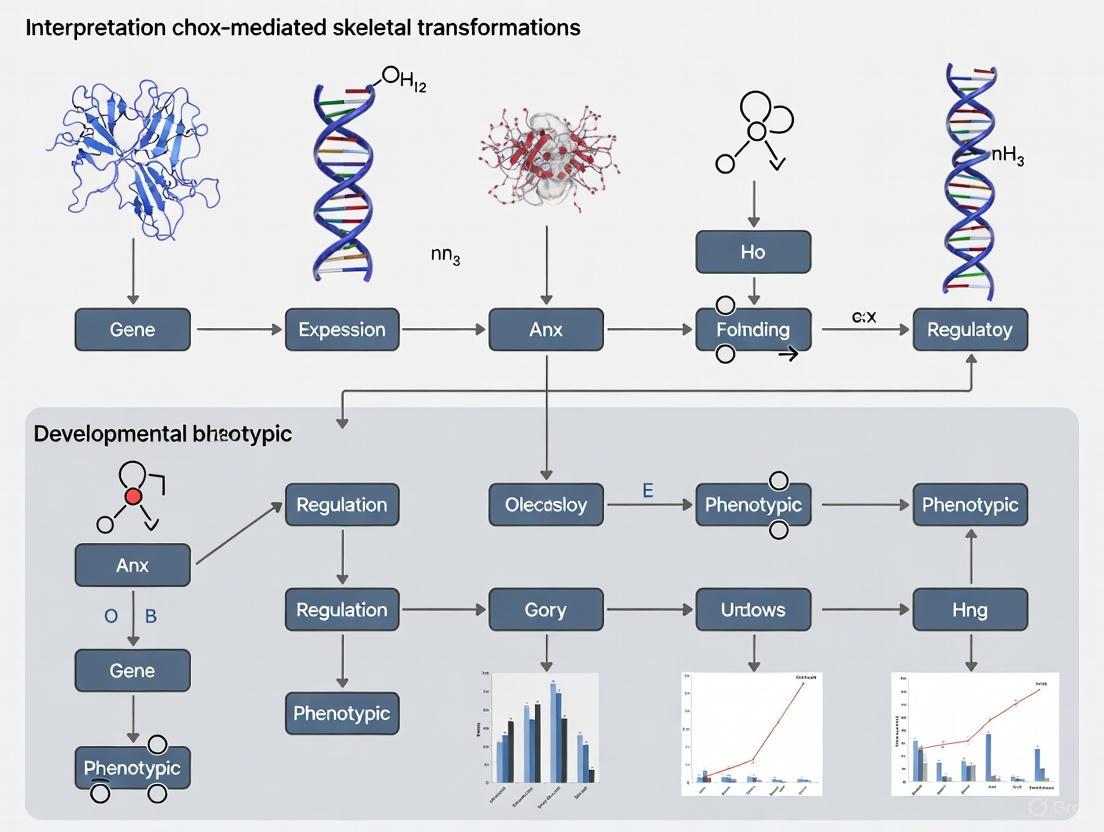
The Hox Skeletal Code: Unraveling Molecular Principles of Axial Patterning
Hox Gene Organization and Spatiotemporal Collinearity in Vertebrate Development
Troubleshooting Guide: Resolving Common Experimental Challenges
Q1: My data on Hox gene temporal expression appears inconsistent or does not show a clear collinear pattern. What could be the issue?
- Problem: Inconsistent detection of temporal collinearity.
- Solution: Ensure you are analyzing the initial phase of Hox gene expression during gastrula-neurula-tailbud stages. Later expression phases, which involve different tissues and functions, can obscure the collinear sequence [1]. Use in situ hybridization to detect expression in specific tissues like the non-organizer mesoderm or presomitic mesoderm, as RNA-seq methods can mask weak initial expression phases if subsequent phases are stronger [1].
- Prevention: Design time-course experiments with a focus on very early development. Normalized measures like half-maximal expression on total RNA-seq time courses may not be ideal for assessing temporal collinearity [1].
Q2: Why do my Hox gene knockout experiments in vertebrates not yield the expected homeotic transformations?
- Problem: Subtle or absent phenotypes in single Hox gene knockouts.
- Solution: Consider functional redundancy from paralogous genes. In vertebrates, due to cluster duplication, paralogous genes (e.g., Hoxa11 and Hoxd11) often have overlapping functions [2]. You may need to generate double or triple mutants for paralogous groups to observe dramatic phenotypes [2].
- Prevention: Review the paralogous group of your target Hox gene and design experiments to target multiple paralogs simultaneously, for example, using CRISPR-Cas9 to target conserved regions.
Q3: I have identified an evolved change in a Hox gene's expression, but introducing the ancestral allele does not produce the predicted phenotypic effect. Why?
- Problem: Epistatic interactions within the Hox-regulated network mask the effect of the individual allele [3].
- Solution: The phenotypic output of a Hox gene is determined by its position within a larger genetic network. Map the expression and function of key downstream targets in your system. In Drosophila santomea, for example, changes in other pigmentation genes (e.g., yellow, tan, ebony) made them insensitive to the Hox protein Abd-B, masking the effect of an introgressed ancestral Abd-B allele [3].
- Prevention: When studying Hox gene evolution, conduct reciprocal hemizygosity tests in hybrid backgrounds to reveal cryptic allelic effects before concluding that a Hox gene change is non-functional [3].
Q4: How can I effectively study the function of a specific Hox gene given its clustered nature and shared regulatory elements?
- Problem: Complexity of regulatory elements and potential for shared enhancers.
- Solution: Be aware that enhancer sharing between adjacent Hox genes is common. For instance, in Drosophila, the iab-5 regulatory region regulates both abd-A and Abd-B, and in mice, the CR3 enhancer regulates both Hoxb4 and Hoxb3 [4]. Use high-resolution techniques like chromosome conformation capture (3C) to map the specific 3D architecture and long-range contacts governing your gene of interest [5].
- Prevention: When interpreting knockout or mutation data, consider the potential impact on neighboring genes within the cluster due to disrupted shared regulatory elements.
Frequently Asked Questions (FAQs)
Q: What is Hox spatiotemporal collinearity, and why is it important?
A: Spatiotemporal collinearity is a fundamental property of Hox genes in many bilaterians. It describes two ordered patterns: Spatial collinearity means the order of Hox genes on the chromosome corresponds to their expression domains along the embryo's anterior-posterior (A-P) axis [4] [6]. Temporal collinearity means the same gene order also corresponds to their sequential activation in time, with anterior genes expressed before posterior ones [4] [7]. This process is crucial for properly patterning the A-P body plan, as the temporal sequence is thought to lay the basis for the spatial pattern [1].
Q: Does temporal collinearity definitively exist in all vertebrates?
A: While some studies have questioned its existence, a substantial body of evidence supports that temporal collinearity is a general rule in vertebrates [1]. Comprehensive studies examining up to 34 Hox genes in chicken, catshark, lamprey, and hagfish, as well as 9 genes in Xenopus, have demonstrated whole-cluster temporal collinearity [1]. Apparent conflicts in the data can often be resolved by focusing on the initial phase of Hox expression in specific embryonic tissues during early development [1].
Q: How do Hox genes specify different regional identities along the axis?
A: Hox genes confer regional identity through a combinatorial code [6]. The specific set or "blend" of Hox genes expressed in a segment or region defines its identity, directing it to develop as a head, thorax, or abdomen segment, or in vertebrates, to form specific types of vertebrae [2]. This is achieved because Hox proteins are transcription factors that regulate distinct sets of downstream target genes, activating or repressing genetic programs for building specific structures [2].
Q: What is the evidence that Hox gene regulation is key to evolutionary changes in body plans?
A: Correlations between shifts in Hox expression domains and morphological changes in serially homologous structures (like vertebrae or insect segments) are widespread [3]. Experimental evidence includes the fact that in snakes, Hox10 genes have lost their rib-blocking ability, which may contribute to their elongated, multi-ribbed body plan [2]. However, evolution often involves polygenic changes, where modifications in a Hox gene are accompanied by changes in its downstream network, which can epistatically mask the Hox gene's individual effect [3].
Key Experimental Data and Protocols
Quantitative Evidence for Temporal Collinearity
The table below summarizes key evidence from studies that support the existence of temporal collinearity in various vertebrate and chordate models.
Table 1: Experimental Evidence for Hox Temporal Collinearity Across Species
| Species | Number of Hox Genes Examined | Key Tissues for Initial Expression | Experimental Method | Primary Reference |
|---|---|---|---|---|
| Chicken | 34 | Primitive streak, ingressing mesoderm, presomitic mesoderm | In situ hybridization | [1] |
| Xenopus | 9 | Gastrula non-organizer mesoderm (NOM), presomitic mesoderm | In situ hybridization | [1] |
| Mouse | 12 | Presomitic mesoderm, nascent somites | In situ hybridization, RT-PCR | [1] |
| Catshark | 34 | Presomitic mesoderm | In situ hybridization | [1] |
| Lamprey | 34 | Presomitic mesoderm | In situ hybridization | [1] |
| Branchiostoma (Cephalochordate) | 12 | Presomitic mesoderm | In situ hybridization | [1] |
Core Experimental Protocol: Analyzing Hox Expression via In Situ Hybridization
This protocol is critical for detecting the precise spatial and temporal expression of Hox genes during early development [1].
Probe Synthesis:
- Cloning: Clone a unique, gene-specific fragment (typically 500-1500 bp) of the target Hox gene from genomic DNA or cDNA into a plasmid vector with RNA polymerase promoters (e.g., T7, T3, SP6).
- Transcription: Linearize the plasmid and use the appropriate RNA polymerase to synthesize a digoxigenin (DIG)- or fluorescein-labeled antisense RNA probe in vitro. Purify the probe.
Embryo Collection and Fixation:
- Collect embryos at precise developmental stages (gastrula, neurula, early tailbud) when temporal collinearity is expected to occur.
- Fix embryos immediately in 4% paraformaldehyde (PFA) in phosphate-buffered saline (PBS) for several hours at 4°C to preserve RNA and morphology.
Pre-hybridization and Hybridization:
- Rehydrate fixed embryos through a methanol series and permeabilize with proteinase K.
- Pre-hybridize embryos in a hybridization buffer containing formamide, salts, and carrier DNA/RNA to reduce non-specific binding.
- Incubate embryos with the labeled antisense probe in hybridization buffer at 55-65°C for 12-48 hours.
Washing and Antibody Binding:
- Perform a series of stringent washes with saline-sodium citrate (SSC) buffer containing formamide to remove unbound probe.
- Block non-specific sites with a blocking reagent (e.g., sheep serum, BSA).
- Incubate with an anti-DIG or anti-fluorescein antibody conjugated to alkaline phosphatase (AP).
Colorimetric Detection:
- Wash embryos thoroughly to remove unbound antibody.
- Incubate in an AP substrate solution (e.g., NBT/BCIP) which produces an insoluble colored precipitate where the probe is bound.
- Monitor the reaction and stop it by washing with PBS when the signal is clear and background is low.
Imaging and Analysis:
- Image embryos using a stereomicroscope. Document the temporal sequence of Hox gene activation and the spatial boundaries of expression in tissues like the presomitic mesoderm.
Core Experimental Protocol: Reciprocal Hemizygosity Test
This genetic test is used to determine if an evolutionary difference in a phenotype is caused by cis-regulatory changes in a specific gene, such as a Hox gene [3].
Generate Mutant Alleles:
- Use CRISPR/Cas9-mediated homology-directed repair to delete or disrupt a specific candidate cis-regulatory element (e.g., the IAB5 initiator for Abd-B) in the genomes of two different species (or populations), Species A and Species B. Replace the element with a neutral marker like RFP.
Create Hybrid Crosses:
- Cross a female from Species A (carrying the mutant allele, A-/-) with a male from Species B (wild-type, B/B). The progeny will be hemizygous for the functional B allele (A-/- / B).
- Perform the reciprocal cross: a female from Species B (mutant, B-/-) with a male from Species A (wild-type, A/A). The progeny will be hemizygous for the functional A allele (B-/- / A).
Phenotypic Analysis:
- Compare the phenotype (e.g., pigmentation intensity, gene expression via in situ) between the two groups of hybrid progeny.
- Interpretation: A phenotypic difference between the two hemizygote groups is attributed to a functional difference between the wild-type alleles of the gene from Species A and Species B, demonstrating that evolution has occurred in that gene's cis-regulation.
Diagram 1: Hox Time-Space Translation (TST) Hypothesis. This model illustrates how the temporal sequence of Hox gene expression is converted into a spatial pattern along the anterior-posterior axis, a process influenced by signals from the Spemann organizer [1].
Diagram 2: Chromatin-Based Regulation of Hox Genes. A Hox gene's expressibility is governed by its chromatin state, maintained by Polycomb (repressive) and Trithorax (activating) complexes, which is heritable during cell division [4].
The Scientist's Toolkit: Essential Research Reagents
Table 2: Key Reagents for Studying Hox Gene Function and Expression
| Reagent / Tool | Primary Function | Example Application |
|---|---|---|
| CRISPR/Cas9 with HDR | Precise genome editing for gene knockout or allele replacement. | Generating deletions in specific Hox cis-regulatory elements (e.g., IAB5) to test their function in vivo [3]. |
| In Situ Hybridization Probes | Detect and visualize the spatial and temporal localization of Hox mRNA transcripts. | Mapping the expression domains of multiple Hox genes during early embryogenesis to establish collinearity [1]. |
| Antibodies against Hox Proteins | Detect and visualize Hox protein localization and abundance. | Confirming the presence and nuclear localization of Hox transcription factors in specific embryonic regions. |
| Chromatin Conformation Capture (3C) | Map the 3D architecture of the genome and identify long-range DNA interactions. | Identifying physical loops and enhancer-promoter contacts within the silent or active Hox cluster [5]. |
| Retinoic Acid (RA) | A potent morphogen that can anteriorize or posteriorize Hox expression patterns. | Experimentally shifting Hox expression domains in cell culture (e.g., NT2/D1 cells) or whole embryos to study gene function [5]. |
| Transgenic Reporter Constructs | Assess the regulatory potential of DNA sequences in vivo. | Testing the activity of conserved non-coding sequences (e.g., from iab-5 region) to identify enhancers [3]. |
| GBR 12783 | GBR 12783, MF:C28H34Cl2N2O, MW:485.5 g/mol | Chemical Reagent |
| PS121912 | PS121912, MF:C24H21F3N2O, MW:410.4 g/mol | Chemical Reagent |
Positional Identity and the Combinatorial Hox Code in Skeletal Patterning
Frequently Asked Questions & Troubleshooting Guides
This technical support resource addresses common experimental challenges in interpreting Hox-mediated skeletal transformations, providing practical solutions for researchers and drug development professionals.
FAQ 1: Why don't my single Hox gene knockouts show expected skeletal transformations?
Issue: Researchers often observe minimal or no phenotype in single Hox gene knockout experiments, contrary to expected homeotic transformations based on expression patterns.
Explanation: Hox genes exhibit significant functional redundancy due to their paralogous organization. In vertebrates, the 39 Hox genes are organized into four clusters (HoxA, HoxB, HoxC, HoxD) with 13 paralogous groups. Members within a paralogous group share similar expression domains and often compensatory functions [8] [9].
Solution: Implement paralogous group knockout strategies targeting all members of a specific paralogous group.
Table: Functional Redundancy in Hox Paralogous Groups
| Paralogous Group | Cluster Members | Single Knockout Phenotype | Complete Paralog Knockout Phenotype |
|---|---|---|---|
| Hox5 | Hoxa5, Hoxb5, Hoxc5 | Mild or partial transformations [9] | Complete anterior transformation of T1 to C7 [9] |
| Hox6 | Hoxa6, Hoxb6, Hoxc6 | Variable effects [9] | Complete transformation of T1 to C7 [9] |
| Hox10 | Hoxa10, Hoxc10, Hoxd10 | Partial transformations [8] | Severe stylopod mis-patterning [8] |
| Hox11 | Hoxa11, Hoxc11, Hoxd11 | Zeugopod defects [8] | Severe zeugopod mis-patterning [8] |
Experimental Protocol: For comprehensive Hox10 paralog analysis:
- Generate Hoxa10 / -; Hoxc10 / -; Hoxd10 / - triple mutant mice
- Analyze E18.5 embryos for skeletal patterning using Alcian Blue and Alizarin Red staining
- Focus on stylopod elements (humerus/femur) where Hox10 function is predominant [8]
- Compare vertebral morphology across anterior-posterior axis for homeotic transformations
FAQ 2: How do I properly interpret and validate homeotic transformations?
Issue: Inconsistent interpretation of vertebral identity changes in Hox mutant models.
Explanation: Homeotic transformations in vertebrates typically manifest as anterior transformations where vertebrae assume the morphology of a more anterior segment, unlike Drosophila where transformations can be bidirectional [8] [10]. This occurs because loss of Hox function results in patterning by the remaining anterior Hox genes in the region.
Solution: Establish precise morphological criteria for vertebral identification.
Table: Vertebral Identity Markers for Homeotic Transformation Analysis
| Vertebral Region | Key Morphological Distinctions | Hox Code Responsible |
|---|---|---|
| Cervical (C1-C7) | Absence of ribs, transverse foramen | Hox4, Hox5 [11] |
| Thoracic (T1-T13) | Presence of articular facets for ribs | Hox6, Hox9 [9] |
| Lumbar | Large body, short thick processes | Hox10 [9] |
| Sacral | Fusion points for pelvic articulation | Hox10, Hox11 [9] |
Troubleshooting Guide:
- Problem: Ambiguous vertebral identity in cervicothoracic transition
- Solution: Use rib development as primary marker; in mice, T1-T7 connect to sternum while cervical vertebrae lack ribs [9]
- Problem: Incomplete penetrance of transformation
- Solution: Quantify transformation percentage across multiple litter replicates (n≥5 embryos)
- Validation: Combine skeletal preparation with Hox expression analysis via RNA in situ hybridization [12]
FAQ 3: What techniques can resolve Hox expression patterns at high resolution?
Issue: Traditional methods lack cellular resolution for analyzing the combinatorial Hox code across multiple cell types.
Explanation: The Hox code operates in a cell-type-specific manner, with recent single-cell technologies revealing unexpected complexity in Hox expression patterns [12].
Solution: Implement single-cell RNA sequencing with spatial validation.
Experimental Protocol: Single-cell RNA-seq for Hox Code Mapping
- Tissue Preparation: Dissect spine from E12.5-E15.5 mouse embryos into precise anatomical segments
- Single-cell Suspension: Generate single-cell suspensions using gentle enzymatic digestion (Collagenase II, 1mg/mL, 37°C, 20 min)
- Library Preparation: Use droplet-based scRNA-seq (10X Genomics Chromium)
- Bioinformatic Analysis: Cluster cells by type and quantify Hox expression per cluster
- Spatial Validation: Validate with Visium spatial transcriptomics or in situ sequencing [12]
Key Finding: Recent human fetal spine atlas revealed that neural crest derivatives retain the anatomical Hox code of their origin while adopting the code of their destination [12].
Hox Expression Analysis Workflow
FAQ 4: How do I address limb patterning defects in Hox mutants?
Issue: Limb defects in Hox mutants often reflect patterning errors rather than simple tissue loss.
Explanation: Posterior Hox paralogs (Hox9-13) pattern the limb skeleton along the proximodistal axis in discrete, non-overlapping domains, unlike the overlapping function in axial patterning [8].
Solution: Focus on segment-specific analyses and utilize the unique limb Hox code.
Table: Limb Segment Patterning by Hox Genes
| Limb Segment | Skeletal Elements | Required Hox Genes | Loss-of-Function Phenotype |
|---|---|---|---|
| Stylopod | Humerus/Femur | Hox10 | Severe stylopod mis-patterning [8] |
| Zeugopod | Radius/Ulna, Tibia/Fibula | Hox11 | Severe zeugopod mis-patterning [8] |
| Autopod | Hand/Foot bones | Hox13 | Complete loss of autopod elements [8] |
Experimental Insight: For forelimb positioning, Hox4/5 genes provide permissive signals throughout the neck region, while Hox6/7 provide instructive cues determining final forelimb position [11].
Hox-Mediated Limb Segment Patterning
FAQ 5: Why do I see different Hox phenotypes across tissue types?
Issue: The same Hox mutation produces different phenotypes in various tissues.
Explanation: Hox genes regulate context-specific genetic networks rather than a conserved set of targets across all tissues [13].
Solution: Perform tissue-specific transcriptomic analyses.
Experimental Approach:
- Collect multiple tissues from Hox mutants (e.g., lung, trachea, somites, BAT)
- Perform bulk RNA-seq on each tissue
- Identify differentially expressed genes
- Validate tissue-specific targets
Key Finding: Bulk RNA-seq in Hoxa5 mutants revealed few common transcriptional changes across tissues, suggesting HOXA5 regulates context-specific effectors rather than a conserved gene set [13].
Research Reagent Solutions
Table: Essential Research Reagents for Hox Skeletal Patterning Studies
| Reagent/Category | Specific Examples | Function/Application | Technical Notes |
|---|---|---|---|
| Mouse Models | Paralogous group mutants (e.g., Hoxa5 / -;Hoxb5 / -;Hoxc5 / -) | Addressing functional redundancy [9] | Requires complex breeding strategies; analyze at E18.5 |
| Skeletal Stains | Alcian Blue (cartilage), Alizarin Red (bone) | Visualization of skeletal elements | Optimal at E16.5-E18.5 for embryonic patterning |
| Spatial Transcriptomics | 10X Visium, Cartana ISS | Mapping Hox expression in tissue context [12] | 50μm resolution; validate with ISS for single-cell resolution |
| Single-cell RNA-seq | 10X Chromium | Resolving Hox code across cell types [12] | Process fresh tissue; sequence depth >50,000 reads/cell |
| Lineage Tracing | Cre-lox systems with Hox-specific promoters | Fate mapping of Hox-expressing populations | Use Hox-CreER[T2] for inducible tracing |
| Inhibition/Activation | Dominant-negative Hox constructs [11] | Perturbing Hox function in specific domains | Electroporate into dorsal LPM at HH12 for limb studies |
Advanced Technical Considerations
Integrating Positional Information Concepts
The concept of positional information is fundamental to understanding Hox function. Cells acquire positional identities through morphogen gradients, and Hox genes translate this information into region-specific morphology [14] [10]. Modern tools like MorphoGraphX 2.0 enable quantification of gene expression and growth in the context of these positional coordinate systems [15].
Regulatory Mechanisms in Hox Clusters
Hox gene regulation involves complex mechanisms:
- Collinearity: 3' to 5' gene order correlates with anterior to posterior expression [16]
- RAREs: Retinoic acid response elements embedded within Hox clusters coordinate responses to signaling gradients [16]
- Enhancer Sharing: Cis-regulatory elements can regulate multiple Hox genes within a cluster [16]
Understanding these mechanisms is essential for designing experiments that accurately perturb Hox function without disrupting core regulatory architecture.
Frequently Asked Questions (FAQs)
FAQ 1: What is the fundamental paradox of Hox protein specificity? Hox transcription factors bind to highly similar, AT-rich DNA sequences in vitro (e.g., core motifs like TAAT), yet they perform exquisitely specific functions in vivo, directing the formation of different structures along the anterior-posterior axis. This discrepancy between degenerate DNA-binding specificity in biochemical assays and highly specific functional outcomes in the organism constitutes the "Hox specificity paradox" [17] [18].
FAQ 2: How can paralogous Hox genes have both redundant and specific functions? Paralogs often share expression domains and can regulate a common set of target genes, leading to functional redundancy, where the loss of one gene can be partially compensated by its paralog. However, each paralog also has unique functions, regulating a specific subset of targets that cannot be compensated by others. This specificity arises from differences in their protein sequences outside the homeodomain, which influence interactions with cofactors and collaborators, leading to distinct transcriptional outputs [19] [20] [18].
FAQ 3: What is the difference between a Hox cofactor and a collaborator?
- Cofactors: Proteins like Extradenticle (Exd/Pbx) and Homothorax (Hth/Meis) that bind DNA cooperatively with Hox proteins. They form direct complexes with Hox proteins, dramatically increasing DNA-binding specificity and affinity by recognizing composite DNA sequences [17] [18].
- Collaborators: Other transcription factors that bind in parallel to the same cis-regulatory element as Hox proteins but do not necessarily form a direct complex on DNA. They help dictate whether the Hox protein will activate or repress transcription and contribute to cell-type-specific outcomes [17].
FAQ 4: What molecular mechanisms underlie the specific functions of different Hox paralogs? Key mechanisms include:
- Latent Specificity: Cooperative binding with PBC cofactors (Exd/Pbx) exposes subtle differences in the DNA-binding preferences of Hox paralogs [18].
- Selective Collaborations: Interactions with different sets of transcription factors at cis-regulatory modules can confer paralog-specific regulatory outcomes [21] [18].
- Variable N-terminal Regions: Sequences outside the conserved homeodomain and hexapeptide motif are divergent and can mediate unique protein-protein interactions or post-translational modifications [17] [18].
Troubleshooting Guide: Common Experimental Challenges
Issue 1: Interpreting Axial Skeleton Phenotypes in Hox Mutant Mice
| Observation | Possible Interpretation | Recommended Validation Experiments |
|---|---|---|
| Homeotic transformation (e.g., a vertebra acquires the identity of a more anterior one) | Loss-of-function of a specific Hox gene. The transformed vertebra is likely within the expression domain of the mutated Hox gene. | - Confirm the expression domain of the mutated Hox gene via in situ hybridization or LacZ reporter in the mutant background [19].- Analyze the expression of molecular markers specific to the acquired identity. |
| No observable phenotype in a single paralog mutant. | Functional redundancy: Compensated by other Hox genes (often paralogs) with overlapping expression and function. | - Generate and analyze double or compound mutants with suspected redundant paralogs (e.g., Hoxa-9 and Hoxd-9) [19].- Perform transcriptomic analysis (RNA-seq) to identify subtle gene expression changes missed by morphological inspection. |
| Synergistic or enhanced phenotype in a double mutant compared to single mutants. | The two genes have redundant functions for that particular trait. The phenotype reveals the full functional requirement shared by both paralogs [19]. | - Conduct a detailed skeletal analysis with Alcian Blue/Alizarin Red staining to quantify all vertebral transformations.- Compare the gene expression changes in single vs. double mutants using RNA-seq. |
| Novel phenotype in a double mutant not seen in either single mutant. | The paralogs may have distinct primary functions, but their combined loss disrupts a larger part of the Hox combinatorial code required for a specific structure [19]. | - Broader phenotypic analysis of other systems (e.g., limb, organs).- Chromatin Immunoprecipitation (ChIP-seq) to map the genomic binding sites for both paralogs and identify co-regulated targets. |
Issue 2: Investigating Hox Target Genes and Specificity
| Challenge | Potential Cause | Solution / Experimental Approach |
|---|---|---|
| A Hox protein binds thousands of sites (ChIP-seq) but regulates very few genes. | Much of the binding may be non-functional, low-affinity, or require specific collaborative partners to become functional. | - Integrate ChIP-seq data with ATAC-seq (chromatin accessibility) and RNA-seq data from the same tissue to focus on bound, accessible regions near differentially expressed genes [21].- Validate candidate cis-regulatory modules (CRMs) with reporter assays in vivo (e.g., in Drosophila or mouse transgenic models) [17] [18]. |
| A cis-regulatory element is activated by one Hox paralog but repressed by another in the same cellular context. | The regulatory outcome is determined by the specific combination of transcription factors (Hox collaborators) bound to the element. | - Map the transcription factor binding motifs within the CRM [18].- Use CRISPR/Cas9 to mutate candidate collaborator binding sites and test the effect on Hox-mediated regulation in a reporter assay. |
| Difficulty recapitulating Hox-specific regulation in cell culture. | The required collaborative factors or chromatin context may be missing in the cell line. | - Use primary cells or stem-cell-derived cultures that more closely mimic the in vivo environment.- Perform co-transfection experiments with expression vectors for the Hox protein and suspected collaborators. |
Quantitative Data on Hox-Mediated Axial Transformations
Table: Axial Skeleton Transformations in Hoxa-9 and Hoxd-9 Single and Double Mutant Mice [19]
| Genotype | Vertebral Transformations (Anteriorizations) | Limb Phenotype |
|---|---|---|
| Hoxa-9-/- | Vertebrae #21 - #25 (L1 - L5) | No forelimb defects observed. |
| Hoxd-9-/- | Vertebrae #23 - #25 (L3 - L5), #28, #30, #31 (S2, S4, Ca1) | Reduced humerus length; malformed deltoid crest. |
| Hoxa-9-/-; Hoxd-9-/- (Double Mutant) | - Increased penetrance/expressivity of single mutant transformations.- Novel transformations in the axial skeleton. | - Increased severity of humerus defects.- Novel alterations at the forelimb stylopod. |
Key Insight: The overlapping transformations of vertebrae L3-L5 in single mutants demonstrate redundancy, while the unique transformations and limb phenotypes reveal paralog-specific functions. The synergistic phenotypes in the double mutant confirm their shared essential role in patterning these structures [19].
Key Experimental Protocols
Protocol 1: Genetic Interaction Analysis Using Compound Mutants
Objective: To dissect functional redundancy and specificity between Hox paralogs in vivo.
Methodology:
- Generate Single and Compound Mutants: Use gene targeting in embryonic stem cells to create null alleles for individual Hox genes (e.g., Hoxa-9 and Hoxd-9). Cross single mutant mice to generate double homozygous mutants [19].
- Skeletal Preparation and Staining: Euthanize newborn or adult mice, eviscerate, and skin the carcasses. Fix in 95% ethanol.
- Cartilage Staining: Use Alcian Blue to stain cartilage.
- Bone Staining: Use Alizarin Red to stain mineralized bone.
- Clearing: Treat with potassium hydroxide to clear soft tissue, making the skeleton visible [19].
- Phenotypic Analysis:
- Systematically examine each vertebra under a dissecting microscope.
- Identify homeotic transformations by comparing morphological features (e.g., shape of transverse processes, neural spines) to wild-type controls. Anteriorization is indicated by a vertebra acquiring features of a more anterior one.
- Measure long bones for defects in growth and morphology.
Protocol 2: Identifying Direct Hox Targets via Integrated Genomics
Objective: To identify genomic regions directly bound by a Hox factor and distinguish functional binding events.
Methodology:
- Chromatin Immunoprecipitation and Sequencing (ChIP-seq):
- Crosslink proteins to DNA in dissected embryonic tissues or relevant cell models.
- Lyse cells and shear chromatin by sonication.
- Immunoprecipitate DNA-protein complexes using a specific antibody against the Hox protein of interest.
- Reverse crosslinks, purify DNA, and prepare libraries for high-throughput sequencing [21] [18].
- Assay for Transposase-Accessible Chromatin with Sequencing (ATAC-seq):
- RNA Sequencing (RNA-seq):
- Extract total RNA from wild-type and Hox-mutant tissues.
- Prepare cDNA libraries and sequence to profile global gene expression changes.
- Data Integration:
- Overlap Hox ChIP-seq peaks with ATAC-seq peaks to identify binding events in accessible chromatin.
- Correlate these high-confidence binding sites with nearby genes that are differentially expressed in the RNA-seq data from mutants to pinpoint direct, functional target genes.
Key Signaling and Regulatory Pathways
Diagram: Hox Specificity Complex on DNA. Hox proteins achieve precise DNA binding and regulatory specificity by forming complexes with PBC and HMP cofactors on composite DNA sites. Collaborator TFs binding nearby determine the ultimate transcriptional outcome (activation or repression) [17] [18].
Diagram: Genetic Workflow for Analyzing Redundancy. This logic flow outlines the experimental steps for determining whether two Hox paralogs have specific or redundant functions through the generation and analysis of single and compound mutants [19].
The Scientist's Toolkit: Research Reagent Solutions
Table: Essential Reagents for Investigating Hox-Mediated Transformations
| Reagent / Tool | Function / Application in Hox Research |
|---|---|
| Hox Mutant Mouse Models (Single & Compound Null Alleles) | In vivo analysis of gene function, redundancy, and axial patterning phenotypes. Essential for linking genotype to morphology [19]. |
| Specific Antibodies for Hox Proteins (e.g., for ChIP-seq) | To map the genomic binding sites of endogenous Hox proteins and identify direct target genes [21] [18]. |
| Alcian Blue & Alizarin Red Staining Kit | Standard histological technique for visualizing cartilage and bone in cleared skeletal preparations, allowing detailed analysis of vertebral identities [19]. |
| CRISPR/Cas9 Gene Editing System | For creating targeted mutations in Hox genes or their cis-regulatory elements in cell lines or model organisms. Enables functional validation of specific protein domains or DNA binding sites [20]. |
| PBC/HMP Expression Vectors (e.g., Pbx, Meis) | For co-transfection experiments in cell culture to study the cooperative binding and transcriptional outcomes of Hox-cofactor complexes on reporter constructs [18]. |
| Transgenic Reporter Constructs | To test the in vivo activity of candidate Hox cis-regulatory modules (CRMs) and define the roles of specific transcription factor binding sites within them [17] [18]. |
| ATAC-seq Kit | To profile the landscape of open chromatin in a given tissue, helping to distinguish functional from non-functional Hox binding events [22] [21]. |
| (S)-GSK-3685032 | (S)-GSK-3685032, MF:C22H24N6OS, MW:420.5 g/mol |
| FPI-1523 sodium | FPI-1523 sodium, MF:C9H13N4NaO7S, MW:344.28 g/mol |
Hoxa5 functions as a key trans-regulatory transcription factor that orchestrates broader Hox gene expression patterns across multiple tissue contexts, rather than acting through local cis-effects on the HoxA cluster. While Hoxa5 mutant phenotypes manifest in tissue-specific ways, recent multi-tissue transcriptomic analyses reveal a conserved trans-regulatory function wherein Hoxa5 regulates the expression of other Hox genes, particularly those within the HoxA cluster. This trans-regulatory capacity enables Hoxa5 to coordinate the complex genetic addresses required for proper patterning of developing tissues, including the respiratory system, axial skeleton, and musculoskeletal structures. The mechanistic insights into Hoxa5's trans-regulatory network resolve previous interpretation challenges in Hox-mediated skeletal transformations by demonstrating that mutant phenotypes result from genuine trans-acting functions rather than cis-acting disruption of neighboring Hox genes.
Hox genes encode an evolutionarily conserved family of transcription factors that play central regulatory roles in body patterning and development, with 39 Hox genes organized into four clusters (HoxA to HoxD) in mammals [13]. These genes are expressed sequentially along the anterior-posterior axis according to their position within the clusters, a phenomenon known as collinearity [2]. The specific combination of HOX proteins at particular anterior-posterior levels provides a unique "genetic address" that determines segment identity and morphology [23].
Hoxa5 occupies a critical position within this hierarchical system, functioning as a predominant regulator in specific axial domains. Unlike many single Hox mutations that cause relatively mild phenotypes, Hoxa5 loss-of-function leads to severe developmental defects and neonatal lethality in most mutants due to respiratory failure [23]. This unusual severity initially raised questions about whether Hoxa5 mutant phenotypes might result from cis-acting effects on neighboring Hox genes rather than genuine trans-regulatory functions. However, recent comparative studies utilizing two different Hoxa5 mutant mouse lines have demonstrated that both alleles share identical phenotypic consequences and Hox gene misregulation patterns, while epigenetic analyses revealed limited effects on the chromatin landscape of the surrounding HoxA cluster [13]. These findings provide compelling evidence that HOXA5 protein acts predominantly in trans to regulate broader Hox gene expression networks.
Key Experimental Evidence for Hoxa5 Trans-Regulation
Multi-Tissue Transcriptomic Analysis
A comprehensive RNA-seq study examining seven different biological contexts in Hoxa5 null mutants revealed that conserved transcriptional changes across tissues were rare, indicating that HOXA5 primarily regulates context-specific effector genes [13]. However, one consistent pattern emerged across all tissues: misregulation of other Hox genes, particularly a trend toward reduced expression of HoxA genes. This finding suggests that Hox genes themselves represent conserved targets of HOXA5 across diverse tissue contexts.
Table 1: Hox Gene Misregulation in Hoxa5 Null Mutants Across Tissues
| Tissue Context | Developmental Stage | Primary Hox Gene Expression Changes | Functional Consequences |
|---|---|---|---|
| Lung | E12.5-E15.5 | Reduced expression of HoxA genes | Impaired branching morphogenesis, lung hypoplasia |
| Trachea | E15.5 | Reduced expression of Hoxa1-Hoxa5 | Tracheal cartilage ring patterning defects |
| Somites | E10.5-E12.5 | Broader Hox misregulation across all four clusters | Homeotic transformations in cervical/thoracic region |
| Diaphragm | E15.5 | Altered Hox expression | Impaired phrenic innervation, respiratory defects |
| Interscapular BAT | E18.5 | Hox gene misregulation | Altered brown adipose tissue depot size |
Genome-Wide Binding Profiles
Recent ChIP-seq experiments using a novel Hoxa5FLAG epitope-tagged mouse line have uncovered the genome-wide occupancy of HOXA5 protein in developing lung tissue [24]. This approach identified an in vivo HOXA5 binding motif and revealed widespread distribution of HOXA5 binding sites throughout the genome, with targets including:
- Other Hox genes known to show expression changes in Hoxa5 null mutants
- Key signaling pathway components (FGF10, SHH, BMP4, WNT2)
- Transcriptional regulators of lung morphogenesis
When combined with ATAC-seq assays and epigenetic analyses, these data demonstrate that HOXA5 directly binds regulatory elements of other Hox genes, providing a mechanistic basis for its trans-regulatory function.
Epigenetic Landscape Analysis
Comparative analysis of epigenetic marks along the HoxA cluster in two different Hoxa5 mutant mouse lines revealed limited effects of either mutation on the chromatin landscape of the surrounding HoxA cluster [13]. This finding argues against the contribution of local cis effects to Hoxa5 mutant phenotypes and supports the model that HOXA5 protein acts in trans in the control of Hox gene expression.
Research Reagent Solutions
Table 2: Essential Research Reagents for Investigating Hoxa5 Trans-Regulatory Networks
| Reagent / Method | Primary Function | Key Application in Hoxa5 Research |
|---|---|---|
| Hoxa5FLAG epitope-tagged mouse line | In vivo protein-DNA interaction mapping | ChIP-seq to identify direct HOXA5 targets [24] |
| Hoxa5 null mutant alleles (multiple strains) | Loss-of-function studies | Comparative phenotyping and transcriptomics [13] |
| ChIP-seq protocol | Genome-wide binding site identification | Defining HOXA5 binding motif and distribution [24] |
| ATAC-seq assay | Chromatin accessibility profiling | Mapping open chromatin regions in Hoxa5 mutants [24] |
| Bulk RNA-seq | Transcriptome quantification | Multi-tissue analysis of Hox gene misregulation [13] |
| In situ hybridization | Spatial expression validation | Confirming Hox target expression patterns [24] |
Troubleshooting Guide: Technical Challenges in Hoxa5 Research
Experimental Design Challenges
Q: How can researchers distinguish between direct and indirect targets of Hoxa5 trans-regulation?
A: The combination of ChIP-seq and ATAC-seq provides the most robust approach for identifying direct targets. The recently developed Hoxa5FLAG mouse line enables precise mapping of HOXA5 binding sites [24]. For confirmation, consider these methodological considerations:
- Perform ChIP-seq at multiple developmental timepoints corresponding to peak Hoxa5 expression
- Combine with RNA-seq from Hoxa5 null tissue to correlate binding with expression changes
- Validate candidate targets through in situ hybridization to confirm spatial expression patterns
- Utilize ATAC-seq to assess chromatin accessibility changes in mutant tissues
Q: What controls are essential when interpreting Hoxa5 mutant phenotypes?
A: Given the potential for cis-acting effects in targeted mutations, employ these rigorous controls:
- Utilize multiple independent mutant alleles to confirm phenotype specificity [13]
- Perform epigenetic profiling of the HoxA cluster to exclude neighborhood effects
- Analyze expression of adjacent Hox genes (particularly Hoxa4 and Hoxa6) to rule out cis disruption
- Consider complementation assays with BAC transgenes containing only Hoxa5
Technical Optimization Issues
Q: How can researchers overcome the challenge of functional redundancy among Hox paralogs?
A: Hox paralogs often exhibit functional redundancy, which can mask phenotypic consequences in single mutants. Address this through:
- Focus on tissues where Hoxa5 shows predominant function (lung, trachea, cervical skeleton) [23]
- Generate compound mutants with other co-expressed Hox genes
- Utilize sensitive transcriptomic approaches to detect subtle gene expression changes
- Employ single-cell RNA-seq to identify cell-type specific requirements
Q: What methods best capture the dynamic nature of Hoxa5 expression and function?
A: Hoxa5 exhibits dynamic spatiotemporal expression patterns during development. To address this:
- Conduct time-course analyses at multiple developmental stages
- Utilize inducible genetic systems for temporal control of Hoxa5 function
- Combine lineage tracing with Hoxa5 perturbation to assess cell-autonomous effects
- Implement live imaging approaches to monitor morphological consequences in real-time
Signaling Pathways in Hoxa5 Trans-Regulatory Networks
Hoxa5 Trans-Regulatory Network Architecture
Molecular Mechanisms of Hoxa5-Mediated Transcription
Hoxa5 achieves transcriptional specificity through several interconnected mechanisms despite the challenge that HOX proteins typically bind similar AT-rich DNA sequences in vitro [21]. The emerging paradigm involves:
DNA Binding Specificity: HOXA5 gains target specificity through interactions with cofactors, primarily members of the PBC (Extradenticle/Pbx) and MEIS (Homothorax/Meis) families [21]. These interactions enhance DNA binding specificity through mechanisms including latent specificity and sensitivity to DNA shape rather than just nucleotide identity.
Chromatin Modification Capacity: Comparative genomic accessibility studies suggest that Hox factors like HOXA5 can differentially modify chromatin accessibility at target loci, potentially exhibiting pioneer-like activities that promote opening of closed chromatin regions [21].
Context-Dependent Cofactor Interactions: HOXA5 interacts with numerous tissue-specific transcription factors and coregulators that determine its transcriptional output in different cellular environments. This explains how the same transcription factor can regulate distinct target genes in various tissue contexts.
Frequently Asked Questions (FAQs)
Q: Does Hoxa5 regulate Hox genes through direct promoter binding or through intermediate factors?
A: Current evidence supports both mechanisms. ChIP-seq data demonstrate direct binding to regulatory elements of other Hox genes [24], while transcriptomic analyses also reveal regulation of signaling pathways that indirectly influence Hox expression [24] [13]. The relative contribution of direct versus indirect regulation likely varies by target gene and cellular context.
Q: How does Hoxa5 trans-regulation differ between tissue contexts?
A: While the trans-regulatory function is conserved across tissues, the specific Hox targets and functional outcomes show considerable context-dependence. For example, in lung development, Hoxa5 predominantly regulates Hoxa1-Hoxa7 and signaling pathways critical for branching morphogenesis [24], while in somites, it influences a broader set of Hox genes across all four clusters [13].
Q: What technical approaches best capture the full scope of Hoxa5 trans-regulatory networks?
A: A multi-assay approach is essential:
- ChIP-seq for direct target identification
- Multi-tissue RNA-seq to assess transcriptional outcomes
- ATAC-seq to evaluate chromatin accessibility changes
- Epigenetic profiling to exclude cis-acting effects
- Spatial transcriptomics to resolve expression patterns at cellular resolution
Q: Are Hoxa5 trans-regulatory functions conserved in human development and disease?
A: Yes, emerging evidence indicates conservation of HOXA5 functions in human development, and its dysregulation is implicated in various pathologies. Notably, altered HOXA5 expression occurs in lung adenocarcinoma and other cancers [25] [26], and recent work has identified roles for HOXA5 in metabolic diseases and adipose tissue dysfunction [25].
The established paradigm of Hoxa5 as a trans-regulatory orchestrator of broader Hox expression provides a framework for resolving long-standing interpretation challenges in Hox-mediated skeletal transformations. Rather than acting through local cis-effects, Hoxa5 functions as a genuine trans-acting factor that coordinates the genetic addresses defining segment identity across multiple tissue contexts.
Future research directions should focus on:
- Elucidating the precise molecular mechanisms by which Hoxa5 achieves transcriptional specificity
- Defining the cofactor interactions that determine context-dependent target gene selection
- Exploring the potential therapeutic applications of modulating Hoxa5 networks in disease contexts
- Investigating the conservation of Hoxa5 trans-regulatory functions across vertebrate evolution
The methodological framework and troubleshooting guidelines presented here provide researchers with essential tools for advancing our understanding of Hoxa5 trans-regulatory networks and their roles in development and disease.
Chromatin Landscape and 3D Genomic Architecture of Hox Clusters
Frequently Asked Questions (FAQs)
1. Why is the 3D architecture of Hox clusters so important for their function? The linear order of Hox genes is directly mirrored by their spatial and temporal expression during development, a phenomenon known as collinearity. The 3D architecture is crucial for implementing this precise regulatory pattern. Initially, the entire cluster is organized as a single, inactive chromatin compartment. During development, as genes are sequentially activated, they physically switch from an inactive compartment (marked by repressive H3K27me3) to an active compartment (marked by active H3K4me3). This bimodal organization helps to reinforce and maintain correct gene expression states, ensuring that posterior genes do not get activated in anterior regions [27] [28].
2. What are the main epigenetic regulators controlling Hox cluster chromatin? The epigenetic state of Hox clusters is primarily regulated by the opposing actions of Polycomb group (PcG) and Trithorax group (TrxG) protein complexes.
- Polycomb (PcG) complexes, such as PRC2 and PRC1, are responsible for maintaining the repressed state. PRC2 deposits the H3K27me3 repressive mark, which helps recruit PRC1. This leads to chromatin compaction and gene silencing [29] [28].
- Trithorax (TrxG) complexes counteract PcG-mediated silencing. They are associated with the deposition of active marks like H3K4me3, which keeps Hox genes in an "open" and expressible state [29] [30]. In embryonic stem cells, Hox clusters often exist in a "bivalent" state, possessing both active and repressive marks, which keeps them poised for activation upon lineage commitment [29].
3. My Hi-C data on Hox clusters is inconsistent. What could be causing this? Inconsistencies can arise from several technical and biological factors:
- Cell Type and Developmental Stage: Hox cluster architecture is highly dynamic. Data can vary significantly between cell types, anatomical origins, and precise developmental time points [28]. Ensure you are comparing equivalent samples.
- Technology Choice and Limitations: Standard Hi-C relies on formaldehyde cross-linking and restriction enzymes, which can introduce sequence bias and protein-related artifacts [31]. Consider using advanced, ligation-free methods like Micro-C or CAP-C for higher resolution and reduced background noise [31].
- Data Resolution: Low-resolution sequencing may fail to capture critical fine-scale interactions, such as the boundaries between active and inactive compartments. Opt for higher-resolution methods where possible [31].
4. How does the disruption of topological associating domains (TADs) near Hox clusters lead to disease? TADs are fundamental units of chromatin organization that constrain interactions between genes and their regulatory elements. Disruption of TAD boundaries near Hox clusters can allow enhancers to contact and activate incorrect Hox genes, leading to misexpression. This misexpression disrupts the precise Hox code necessary for skeletal patterning, which can result in homeotic transformations (where one body part develops the identity of another) and congenital malformations. For example, such disruptions have been linked to human syndromes like F-syndrome, polydactyly, and brachydactyly [31] [32].
5. We see persistent Hox expression in adult-derived mesenchymal stem cells (MSCs). Is this normal? Yes, this is a normal and functionally important phenomenon. Fibroblasts and progenitor-enriched MSCs cultured from adult tissues maintain regionally restricted Hox gene expression profiles that reflect their anatomical origin. This "Hox code" is not just a developmental relic; it functions in adult tissue maintenance, regeneration, and fracture healing. Genetic studies confirm that Hox genes are required for the fracture repair process in the adult skeleton [33].
Troubleshooting Guides
Challenge: Interpreting Complex Chromatin Interaction Data
Problem: It is difficult to determine whether observed chromatin interactions are causative of gene regulation or merely a consequence of transcription.
Solution Strategy:
- Employ Multi-Modal Integration: Correlate your 3D interaction data (e.g., from Hi-C) with complementary datasets. Map histone modifications (ChIP-seq), transcription factor binding (ChIP-seq or CUT&Tag), and transcriptional output (RNA-seq) from the same biological sample. An interaction is more likely to be functional if it correlates with active chromatin marks and gene activation [31] [28].
- Utilize Ligation-Free Technologies: To overcome biases inherent in cross-linking and restriction enzymes, use methods like Micro-C or CAP-C. These provide a more native view of chromatin structure and can reveal finer details, such as nucleosome-level interactions [31].
- Implement Functional Perturbation: Genetically disrupt specific regulatory elements (e.g., enhancers or CTCF sites) or architectural proteins and observe the concurrent effects on 3D structure and gene expression. A concomitant change in structure and function strongly suggests a causal link [34].
Table 1: Advanced Methods for 3D Genomics Analysis
| Method | Key Principle | Advantage | Best for Analyzing |
|---|---|---|---|
| Micro-C [31] | Uses micrococcal nuclease (MNase) for fragmentation. | Nucleosome-resolution mapping; no restriction enzyme bias. | Fine-scale architecture within Hox clusters. |
| CAP-C [31] | Uses dendrimers and UV for protein removal and DNA fragmentation. | Reduces protein-crosslinking artifacts; high signal-to-noise. | Transcription-dependent changes in chromatin conformation. |
| ChIA-Drop [31] | Identifies multi-way interactions in droplets using DNA barcodes. | Captures complex, multi-loci interactions simultaneously. | Enhancer hubs interacting with multiple promoters. |
| SPRITE [31] | Uses split-pool barcoding to identify interacting DNA and RNA. | Maps multi-way interactions and inter-chromosomal contacts genome-wide. | Hox genes belonging to larger nuclear bodies. |
| Chromatin Tracing [35] | Uses multiplexed FISH to visualize genomic loci in single cells. | Provides single-cell, single-molecule 3D folding paths in situ. | Cell-to-cell heterogeneity in Hox cluster organization. |
Challenge: Investigating Hox Function in Adult Skeletal Regeneration
Problem: The specific molecular mechanisms by which Hox genes function in adult mesenchymal stem/stromal cells (MSCs) during bone repair are unclear.
Solution Strategy:
- Lineage Tracing and Cell Sorting: Use mice expressing Cre recombinase under Hox gene promoters (e.g., Hoxa11-eGFP) to label and track Hox-expressing cell lineages during fracture healing. Isolate these cells via FACS for downstream transcriptomic and epigenomic analysis [33].
- In Vivo Loss-of-Function Studies: Employ conditional knockout mouse models to delete specific Hox paralogous groups in adult MSCs or osteoprogenitors. Analyze the resulting fracture healing phenotypes, focusing on callus formation, bone remodeling, and integration of musculoskeletal tissues [33] [8].
- Define Expression in Stromal Compartments: Precisely characterize Hox expression in the various connective tissues (perichondrium, tendon, muscle connective tissue) of the adult skeleton, as Hox genes are often highly expressed in stromal cells rather than differentiated skeletal cells [33] [8].
Challenge: Engineering Hox Clusters to Study Gene Regulation
Problem: Targeted inversions or modifications of the Hox cluster often lead to unexpected and severe gene misexpression, complicating data interpretation.
Solution Strategy:
- Respect Transcriptional Polarity: Vertebrate Hox clusters are highly optimized, with all genes transcribed from the same DNA strand. When engineering alleles, avoid inverting transcription units, as this can disrupt the coordinated regulatory landscape and cause aberrant gene activation (e.g., ectopic Hoxd13 expression) [34].
- Preserve CTCF-Binding Sites: CTCF sites often reside between Hox genes and act as critical insulators. Their deletion or inversion can collapse the micro-domain architecture, leading to regulatory leakage. Always map and consider the function of these sites in your genetic designs [34].
- Analyze in Multiple Developmental Contexts: Test your engineered alleles in various tissues (e.g., axial skeleton, limb buds, metanephros), as Hox clusters are regulated by distinct global enhancers in different contexts. A mutation may have a phenotype in one context but not another [34].
Experimental Protocols
Detailed Protocol: High-Resolution 3D Architecture Analysis Using Micro-C
Objective: To map the 3D chromatin architecture of the Hox cluster at nucleosome resolution.
Reagents and Equipment:
- Crosslinking Buffer (1% Formaldehyde)
- Micrococcal Nuclease (MNase)
- Biotinylated Nucleotides
- DNA Ligase
- Streptavidin Beads
- Proteinase K
- Library Preparation Kit
- High-Throughput Sequencer
Method:
- Crosslinking: Harvest and crosslink ~1 million cells with 1% formaldehyde for 10 minutes at room temperature. Quench with glycine.
- Chromatin Fragmentation: Lyse cells and digest chromatin with MNase to saturation. MNase cleaves preferentially in linker DNA, creating a population of mononucleosomes.
- End Repair and A-Tailing: Repair the MNase-digested ends and use a Klenow fragment to add an 'A' overhang.
- Proximity Ligation: Add a biotinylated bridge adapter with a 'T' overhang for ligation. Under dilute conditions, perform intra-molecular ligation to join crosslinked DNA fragments.
- Reversal of Crosslinking and Purification: Reverse crosslinks with Proteinase K and purify DNA.
- Biotin Pull-Down: Capture biotinylated ligation products using Streptavidin beads.
- Library Preparation and Sequencing: On-bead, prepare the library for paired-end high-throughput sequencing.
- Data Analysis: Process paired-end sequences using a dedicated Micro-C pipeline (e.g., distiller) to generate high-resolution contact matrices and identify TADs and chromatin loops.
Detailed Protocol: Visualizing Hox Cluster Conformation via Multiplexed FISH
Objective: To visualize the 3D folding path of a Hox cluster in single cells within intact tissue.
*Reagents and Equipment:
- Custom Oligopaint FISH Probes targeting the Hox locus
- Formamide
- Fluorescently Labeled Readout Probes
- DAPI Staining Solution
- Super-Resolution Microscope
- Automated Fluidics System
Method:
- Sample Preparation: Fix cells or tissue sections and permeabilize.
- Hybridization: Apply a pool of hundreds to thousands of uniquely barcoded Oligopaint FISH probes that tile the Hox cluster region. Hybridize overnight.
- Sequential Imaging and Stripping:
- Apply a set of fluorescent readout probes that bind to a subset of the barcodes.
- Image the fluorescence signals using a super-resolution microscope.
- Chemically strip the fluorescent readouts without damaging the sample or the primary probes.
- Repeat the process with a new set of readouts for multiple rounds until all barcodes have been imaged.
- Image Analysis and Tracing: Computational algorithms decode the sequential imaging data to identify the precise spatial positions of all targeted loci in 3D. The folding trajectory of the chromatin fiber is then reconstructed by connecting the positions of the imaged loci along the same DNA molecule [35].
Data Presentation
Table 2: Key Chromatin Marks and Their Functional Associations in Hox Regulation
| Chromatin Mark / Protein Complex | Associated Function | Effect on Hox Gene Expression | Experimental Detection Methods |
|---|---|---|---|
| H3K27me3 | Repression; Polycomb (PcG) mediated silencing | Repression | ChIP-seq, CUT&Tag |
| H3K4me3 | Activation; Trithorax (TrxG) mediated activation | Activation / Poising | ChIP-seq, CUT&Tag |
| Bivalent Domains (H3K4me3 + H3K27me3) | Poised state in ESCs | Genes are silent but primed for activation | ChIP-seq |
| CTCF | Chromatin looping / Insulation | Defines regulatory boundaries; prevents ectopic activation | ChIP-seq, CTCF Cut&Run |
| Cohesin Complex | Loop extrusion | Facilitates enhancer-promoter communication | ChIP-seq |
Signaling Pathways and Workflows
Hox Cluster Activation During Development
This diagram illustrates the dynamic transition of Hox clusters from a single inactive state to a bimodal active/inactive structure during embryonic development.
Multimodal 3D Genomics Technology Workflow
This diagram compares the general workflows of sequencing-based and imaging-based technologies for analyzing 3D chromatin architecture.
The Scientist's Toolkit: Research Reagent Solutions
Table 3: Essential Research Reagents for Hox Cluster Studies
| Reagent / Tool | Function / Application | Key Characteristics |
|---|---|---|
| Hoxa11-eGFP Reporter Mouse [33] | Fate mapping and isolation of Hox-expressing stromal cells. | Labels zeugopod-specific mesenchymal cells; useful for studying limb development and regeneration. |
| Conditional Hox Alleles (floxed) [33] [8] | Tissue-specific and temporal knockout of Hox paralog groups. | Essential for bypassing embryonic lethality and studying function in adult tissues like MSCs. |
| Oligopaint FISH Probes [35] | High-resolution imaging of Hox cluster conformation. | Allows multiplexed labeling of specific genomic loci for chromatin tracing in single cells. |
| Anti-H3K27me3 Antibody | Chromatin Immunoprecipitation (ChIP) to map repressive domains. | High-quality antibody is critical for defining Polycomb-silenced regions within the cluster. |
| Anti-H3K4me3 Antibody | Chromatin Immunoprecipitation (ChIP) to map active domains. | High-quality antibody is critical for defining Trithorax-active regions within the cluster. |
| Anti-CTCF Antibody | Mapping chromatin insulator and loop boundaries. | Identifies potential regulatory boundaries that partition the Hox cluster. |
| FTX-6746 | FTX-6746, MF:C16H7ClF2N2O, MW:316.69 g/mol | Chemical Reagent |
| SSI-4 | SSI-4, MF:C19H21ClN4O3, MW:388.8 g/mol | Chemical Reagent |
Neural Crest Derivatives and Their Retention of Positional Hox Memory
Core Concepts: Hox Codes and Neural Crest Cell Memory
What is the fundamental principle of positional memory in neural crest derivatives?
Neural crest cells (NCCs) retain a transcriptional memory of their origin along the anterior-posterior axis through maintained Hox gene expression patterns. This "Hox code" is established in the neural tube before migration and persists in derived tissues, providing positional information that influences their developmental potential and differentiation fate [12] [36] [37].
Key Mechanism: This memory operates through a positive-feedback loop that maintains regional identity. For example, in axolotl limb regeneration, posterior identity is safeguarded by sustained Hand2 expression, which primes cells to form a Shh signaling center after injury, creating a stable memory state [38].
How does Hox expression plasticity impact neural crest patterning?
While NCCs carry Hox codes from their origin, their final positional identity results from a complex integration of pre-patterned information and environmental cues received during migration and at destination sites [37].
Critical Consideration: The size of the cell community influences plasticity. Smaller grafts or individual NCCs show greater sensitivity to environmental cues compared to larger cell populations, which tend to maintain their original Hox expression [37].
Experimental Protocols & Methodologies
Protocol: Mapping Hox Expression in Developing Human Neural Crest Derivatives
This protocol is adapted from single-cell and spatial transcriptomic analyses of the human fetal spine [12].
| Step | Procedure | Purpose |
|---|---|---|
| 1. Tissue Collection | Obtain 5-13 week post-conception human fetal spines; from 9 weeks onwards, dissect into precise anatomical segments using anatomical landmarks | Capture inherent rostrocaudal maturation gradient (~6 hours difference between vertebral levels) |
| 2. Single-Cell Suspension | Process fresh tissues to generate single-cell suspensions, enrich for viable cells | Prepare for single-cell RNA sequencing |
| 3. Library Preparation | Generate single-cell mRNA libraries using droplet-based method (Chromium 10X) | Capture transcriptomic profiles of individual cells |
| 4. Spatial Validation | Apply Visium spatial transcriptomics (50μm resolution) and Cartana in-situ sequencing (single-cell resolution, 123-gene panel) on axial sections | Spatially resolve cell types and validate Hox expression patterns |
| 5. Data Integration | Use cell2location algorithm to obtain estimated cell type abundancy values for each voxel | Reconstruct spatial organization of cell types with Hox expression patterns |
Protocol: Testing Hox Plasticity in Avian Neural Crest
This approach, based on classical grafting experiments, examines the stability of Hox positional memory [36] [37].
- Donor Tissue Selection: Isolate midbrain neural crest (Hox-negative) from donor embryo
- Transplantation: Graft to more posterior hindbrain (Hox-positive) regions in host embryo
- Fate Analysis: Track morphological outcomes and Hox gene expression in graft-derived structures
- Control Experiment: Perform orthotopic grafts to verify normal developmental potential
Expected Outcome: Grafted cells often retain identity appropriate for their original position and form ectopic mandibular structures, demonstrating persistence of positional memory [36] [37].
Quantitative Data Analysis
Rostrocaudal HOX Code in Human Fetal Development
Analysis of stationary cell types in human fetal spine revealed 18 genes with strongest position-specific expression patterns [12]:
| Anatomical Region | Key HOX Genes | Specific Markers | Expression Characteristics |
|---|---|---|---|
| Cervical | HOXB-AS3, HOXA5 | HOXB6 (osteochondral), HOXC4 (meningeal) | HOXB-AS3 shows strong sensitivity for cervical region (p < 10â»Â³â°â°) |
| Thoracic | HOXC5 (meningeal) | Multiple HOX genes with segment-specific expression | Gradual transition in expression patterns along axis |
| Sacral | HOXC11 (meningeal) | Group 13 genes (very low levels) | Expressed exclusively in sacral samples, including coccyx |
Hox Combinatorial Codes in Zebrafish Neural Crest Lineages
Single-cell transcriptomics of sox10:GFP+ cells in zebrafish reveals distinct Hox signatures across neural crest derivatives [39]:
| Cell Type | Hox Signature | Developmental Timing | Additional Markers |
|---|---|---|---|
| Mesenchyme Subtypes | Multiple distinct Hox combinations across subpopulations | 48-70 hpf | Prrx1, Twist1 for mesenchymal fate |
| Enteric Neurons | Specific Hox code combinations | Progressive differentiation from 48-70 hpf | Neuronal differentiation markers |
| Neural Crest Cells | Axial-level specific Hox patterns | Maintained through migration and differentiation | Sox10, FoxD3, Tfap2a |
| Pigment Progenitors | Distinct anterior-posterior Hox codes | Emerging during embryonic-larval transition | Melanocyte differentiation genes |
Signaling Pathways & Molecular Mechanisms
Hand2-Shh Positive Feedback Loop in Positional Memory
This diagram illustrates the core regulatory circuit maintaining posterior identity in limb cells, relevant to understanding how positional memory is sustained in neural crest derivatives [38].
Experimental Workflow for Hox Code Mapping
This workflow diagram outlines the integrated approach for creating a developmental atlas of Hox expression using multiple complementary technologies [12].
Research Reagent Solutions
Essential Reagents for Neural Crest Hox Research
| Reagent/Cell Line | Application | Key Features | Experimental Use |
|---|---|---|---|
| Hoxa11-CreERT2; ROSA-LSL-tdTomato mice | Lineage tracing of Hox-expressing cells | Enables temporal deletion of Hox11 function at adult stages; labels Hox11 lineage cells | Studying continued Hox function in adult skeletal homeostasis [40] |
| ZRS>TFP; loxP-mCherry axolotl | Fate mapping of embryonic Shh cells | Labels Shh-expressing cells during development and regeneration with inducible Cre | Investigating origin of posterior cells during regeneration [38] |
| Tg(−4.9sox10:EGFP) zebrafish | Identifying NCCs and derivatives | GFP expression under sox10 promoter marks neural crest lineage | Single-cell transcriptomics of posterior NCC fates [39] |
| Hand2:EGFP knock-in axolotl | Tracking Hand2 expression | EGFP co-expressed with endogenous Hand2 via T2A sequence | Monitoring posterior identity factor in uninjured and regenerating limbs [38] |
Troubleshooting Common Experimental Challenges
How should researchers interpret conflicting data on neural crest pre-patterning versus plasticity?
Problem: Experimental results show apparent contradictions between fixed Hox codes and plastic Hox expression in neural crest cells.
Solution: Consider these factors when interpreting results:
- Community Size Effect: Smaller cell grafts show greater plasticity than larger populations [37]
- Axial Level Differences: Cranial neural crest may exhibit different regulatory mechanisms than trunk neural crest
- Temporal Factors: Early migratory NCCs may show different plasticity than later differentiated derivatives
- Technical Considerations: Orthotopic vs. heterotopic grafting produces different outcomes
Resolution Framework: The current model integrates both concepts - NCCs carry intrinsic positional information but remain responsive to local environmental cues during migration [37].
What controls should be included when studying Hox gene function in adult skeletal maintenance?
Problem: Distinguishing between developmental patterning defects and ongoing adult functions of Hox genes.
Solution: Implement these experimental controls:
- Temporal Deletion: Use inducible Cre systems (e.g., Hoxd11 conditional allele) to delete Hox function specifically in adulthood after normal development [40]
- Lineage Tracing: Combine with lineage labeling (e.g., Hoxa11-CreERT2; ROSA-LSL-tdTomato) to track mutant cell behavior
- Regional Specificity Controls: Compare effects in Hox-expressing regions versus non-expressing regions
- Differentiation Markers: Assess multiple stages of osteolineage differentiation (Runx2, osteopontin, osteocalcin, SOST) [40]
Frequently Asked Questions
Do all neural crest derivatives maintain their original Hox code?
No. Research shows neural crest derivatives can unexpectedly retain the anatomical Hox code of their origin while also adopting the code of their destination. This trend has been confirmed across multiple organs, suggesting a more complex integration of positional information than previously thought [12].
How stable is Hox positional memory in adult tissues?
Hox positional memory demonstrates remarkable stability in adult tissues. Studies show that:
- Fibroblasts maintain distinct Hox expression patterns for >35 cell generations ex vivo [41]
- Anatomic site-specific Hox patterns are not perturbed by soluble factors or heterotypic cell contact [41]
- Fibroblasts from young vs. old human donors show little difference in position-specific Hox expression [41]
Can positional memory be reprogrammed experimentally?
Yes, but with directional constraints. Research in axolotl limb regeneration demonstrates that:
- Positional memory can be reprogrammed more easily from anterior to posterior than the reverse direction [38]
- Transient exposure of anterior cells to Shh during regeneration can kick-start an ectopic Hand2-Shh loop [38]
- This leads to stable Hand2 expression and lasting competence to express Shh in subsequent amputations [38]
Advanced Profiling and Functional Genomics for Hox Phenotype Deconvolution
Single-Cell and Spatial Transcriptomics for Resolving Hox Expression Atlases
A fundamental challenge in developmental biology research is accurately interpreting the complex, spatially restricted expression of HOX genes—key transcription factors that orchestrate anteroposterior patterning in the embryonic skeleton and other tissues. Traditional bulk sequencing methods obscure critical cellular heterogeneity and spatial context, limiting our understanding of Hox-mediated skeletal transformations. The integration of single-cell RNA sequencing (scRNA-seq) and spatial transcriptomics now provides unprecedented resolution to map these expression patterns within their native tissue architecture. However, these advanced technologies introduce new technical and interpretive hurdles that can compromise data reliability and biological insights. This technical support center addresses these specific challenges through targeted troubleshooting guides and detailed experimental protocols, enabling researchers to confidently generate and interpret high-quality Hox expression atlases.
Frequently Asked Questions (FAQs)
Q1: Why is integrating scRNA-seq with spatial transcriptomics particularly important for studying Hox genes?
Hox genes exhibit precise collinear expression patterns along the anteroposterior axis, where their 3' to 5' genomic arrangement correlates with anterior to posterior body position. ScRNA-seq identifies cellular heterogeneity and transcriptional profiles but loses native spatial context. Spatial transcriptomics preserves anatomical localization but may lack single-cell resolution. Integration is crucial because it links specific Hox codes to their exact anatomical positions and cell types. For example, a recent human embryonic spine atlas combining both techniques revealed that neural crest derivatives retain the anatomical HOX code of their origin while also adopting the code of their destination, a finding impossible with either method alone [42].
Q2: What are the primary sources of technical noise in scRNA-seq data when analyzing transcription factors like Hox genes?
Hox genes and other transcription factors are often expressed at lower levels than structural genes, making them susceptible to several technical artifacts:
- Dropout events: Low-abundance transcripts may fail to be captured or amplified, creating false negatives [43].
- Amplification bias: Stochastic variation during cDNA amplification can skew representation of specific genes [43].
- Low RNA input: The limited starting material from single cells can lead to incomplete reverse transcription and reduced coverage [43].
- Cell doublets: Multiple cells captured in a single droplet can confound analysis, leading to misidentification of co-expressed Hox genes [43].
Q3: How can I validate the spatial expression patterns of Hox genes identified in my transcriptomic data?
Spatial validation requires orthogonal techniques. Beyond spatial transcriptomics (like 10x Visium or Stereo-seq), robust methods include:
- In situ sequencing (ISS): Provides single-cell resolution for a targeted gene panel. This was used to validate HOX gene expression in the developing human spine with high anatomical precision [42].
- RNA in situ hybridization (RNA-ISH): A classic method to visually confirm the expression and localization of specific Hox transcripts [44].
- Spatial imputation tools: New computational methods like ISS-Patcher can impute cell labels from high-plex ISS data onto spatial datasets, strengthening spatial annotation [45].
Q4: What are the key considerations when choosing a spatial transcriptomics platform for Hox atlas projects?
The choice depends on the biological question and required resolution [46]:
- Required Resolution: Are you studying broad tissue zones or fine subcellular structures?
- Tissue Type and Preservation: Ensure platform compatibility with your sample type (FFPE, fresh frozen, fixed frozen) and that RNA quality (RIN, DV200) meets vendor specifications [46].
- Throughput and Coverage: Consider the trade-off between high-resolution and the physical area that can be captured, especially for large embryonic structures.
Troubleshooting Guides
Addressing Low or Inconsistent Hox Gene Detection in scRNA-seq
Problem: Hox genes, often lowly expressed, are missing or show inconsistent expression across expected cell populations.
| Symptom | Potential Cause | Solution |
|---|---|---|
| High dropout rate for Hox genes | Low sequencing depth | Increase sequencing depth to capture low-abundance transcripts. Use Unique Molecular Identifiers (UMIs) to correct for amplification bias [43]. |
| Inconsistent detection between replicates | Poor cell viability or RNA quality | Implement rigorous quality control (QC). Assess cell viability, library complexity, and RNA Integrity Number (RIN) before sequencing. Use fresh, snap-frozen samples or high-quality FFPE samples with DV200 > 50% [43] [46]. |
| Putative "novel" Hox-expressing populations | Cell doublets | Use computational methods (e.g., DoubletFinder) or cell hashing to identify and exclude doublets from analysis [43]. |
| General low cDNA yield | Suboptimal reverse transcription | Always include positive and negative controls. Optimize cell lysis and RNA extraction. For FACS-sorted cells, ensure they are sorted into an appropriate, EDTA-/Mg2+-/Ca2+-free buffer like PBS to avoid inhibiting RT reactions [47]. |
Resolving Spatial Data Integration and Interpretation Challenges
Problem: Difficulty in aligning scRNA-seq clusters with spatial transcriptomics spots or interpreting spatial Hox patterns.
| Symptom | Potential Cause | Solution |
|---|---|---|
| Poor alignment between scRNA-seq clusters and spatial data | Batch effects or biological variability | Use batch correction algorithms (e.g., Harmony, Combat) to integrate datasets from different technical runs [43]. Plan experiments to process samples for both modalities in parallel. |
| Ambiguous spatial localization of Hox codes | Low resolution of spatial platform | For fine-scale mapping, choose a higher-resolution platform (e.g., Visium HD, Stereo-seq). Supplement with targeted in-situ sequencing (ISS) for validation [42] [46]. |
| Inability to trace Hox trajectories in space | Lack of spatial trajectory analysis tools | Apply spatial trajectory inference tools like OrganAxis or PAGA, which have been used to map osteolineage maturation and mesenchymal progenitor fate in developing bones [45] [48]. |
| Weak or degraded spatial signal | Poor tissue preparation or RNA degradation | Minimize time between tissue collection and freezing/fixation. Optimize cryosectioning to avoid artifacts. For FFPE samples, follow strict protocols to balance morphology and RNA integrity [46]. |
Overcoming Biological Heterogeneity in Hox Expression Analysis
Problem: High cell-to-cell variability in Hox expression complicates the definition of a stable positional code.
| Symptom | Potential Cause | Solution |
|---|---|---|
| A cell type expresses multiple Hox genes inconsistently | True biological heterogeneity or mixed populations | Use clustering and trajectory inference (e.g., Monocle, PAGA) to identify discrete subpopulations or transitional states. RNA velocity can help determine the directionality of cell fate decisions [48] [45]. |
| Hox code differs between developmentally related cells | Dynamic changes during differentiation | Perform time-series experiments or leverage computational pseudotime analysis to order cells along a developmental trajectory and understand how Hox codes evolve [49]. |
| Rare cell populations with unique Hox signatures are missed | Insufficient cell numbers or oversampling of dominant types | Use targeted cell enrichment strategies (e.g., FACS) to oversample rare populations. Employ sequencing methods with higher sensitivity (e.g., SMART-seq) for lowly expressed genes [43]. |
Experimental Protocols for Key Workflows
Integrated Protocol: Mapping Hox Codes in Embryonic Mouse Limb Buds
This protocol outlines the process for generating a spatially resolved Hox expression atlas, synthesizing methods from key studies [48] [44] [42].
I. Sample Preparation & Single-Cell Suspension
- Dissection: Isolate embryonic limb buds in cold, EDTA-/Mg2+-/Ca2+-free PBS. Work quickly to minimize RNA degradation [47].
- Dissociation: Use a validated enzymatic cocktail (e.g., collagenase/dispase) tailored to embryonic tissue. Gently triturate. Monitor under a microscope to avoid over-digestion.
- QC and Sorting: Pass cells through a flow cytometry sorter (e.g., FACS). Use a viability dye. Sort individual cells directly into lysis buffer containing RNase inhibitor [47]. Assess viability and single-cell suspension quality.
II. Single-Cell RNA Sequencing
- Library Preparation: Use a droplet-based system (e.g., 10x Genomics) following manufacturer's guidelines. Incorporate UMIs.
- Sequencing: Sequence to a sufficient depth (recommended: >50,000 reads/cell) to ensure capture of lowly expressed transcription factors.
- Control Reactions: Always include a positive control (e.g., 10 pg of control RNA from a cell line) and a negative control (mock FACS buffer) to troubleshoot cDNA yield and background [47].
III. Spatial Transcriptomics
- Tissue Preparation: Embed freshly dissected contralateral limb buds in OCT and snap-freeze. Section at 10 µm thickness and mount on spatial gene expression slides (e.g., 10x Visium) [46].
- Staining and Imaging: H&E stain for morphological annotation. Image slides at high resolution.
- Library Construction: Follow the spatial platform's specific protocol for permeabilization, reverse transcription, and library construction.
IV. Data Analysis & Integration
- scRNA-seq Preprocessing: Perform quality control, normalization, and clustering using standard tools (Seurat, Scanpy). Exclude doublets and low-quality cells.
- Spatial Data Processing: Align H&E images with sequencing data using the platform's software (e.g., Space Ranger).
- Integration: Use computational methods (e.g., cell2location [42], Seurat's anchoring) to map scRNA-seq-derived cell type labels onto the spatial coordinates.
- Hox Analysis: Visualize Hox gene expression in spatial context. Perform differential expression testing across anatomical regions. Construct spatial trajectories of Hox code progression.
The following diagram illustrates the core workflow and logical relationships of this integrated protocol:
Protocol: Validation of Hox Expression via In Situ Sequencing (ISS)
This protocol is adapted from studies that successfully validated HOX patterns in human embryonic spines [42] and joints [45].
- Probe Design: Design padlock probes targeting a panel of Hox genes and key cell-type marker genes.
- Tissue Preparation: Section FFPE or fresh frozen tissue (5-10 µm) onto glass slides. Perform paraffin removal (for FFPE) and dehydration.
- Permeabilization: Optimize proteinase K concentration and incubation time for adequate tissue permeabilization without compromising morphology.
- ISS Reaction:
- Hybridization: Incubate sections with the padlock probe mix.
- Ligation: Add ligation buffer to circularize bound probes.
- Rolling Circle Amplification (RCA): Amplify circularized probes to generate detectable fluorescence signals.
- Imaging: Acquire high-resolution images using a fluorescence microscope. Cycle through subsequent rounds of hybridization and imaging for multiplexing.
- Image Analysis: Use software (e.g., Cartana) for spot detection, decoding, and assignment of transcripts to individual cells. Co-register with morphological features.
| Category | Item / Reagent | Function / Application | Key Consideration |
|---|---|---|---|
| Sample Prep | EDTA-/Mg2+-/Ca2+-free PBS [47] | Cell washing and resuspension buffer | Prevents inhibition of reverse transcription. |
| BD FACS Pre-Sort Buffer [47] | Sheath fluid for cell sorting | Maintains cell viability and is compatible with RT reactions. | |
| RNase Inhibitor [47] | Added to lysis/FACS buffer | Preserves RNA integrity during sample processing. | |
| Sequencing | 10x Genomics Chromium [48] [44] | Droplet-based scRNA-seq platform | Standardized workflow for high-throughput cell encapsulation. |
| Unique Molecular Identifiers (UMIs) [43] | Molecular barcodes for transcripts | Corrects for amplification bias and quantifies absolute molecule counts. | |
| SMART-Seq Kits [43] | Full-length scRNA-seq | Higher sensitivity for detecting lowly expressed genes like Hox. | |
| Spatial Tech | 10x Genomics Visium HD [46] | Spatial gene expression | Near single-cell resolution (2x2 µm) for detailed mapping. |
| STOmics Stereo-seq [46] | Spatial gene expression | Subcellular resolution (500 nm) for highest precision. | |
| Cartana In Situ Sequencing [42] | Targeted spatial transcriptomics | Validates Hox patterns with single-cell resolution in tissue. | |
| Bioinformatics | Cell2Location [42] | Bayesian model for spatial mapping | Deconvolves spatial data using scRNA-seq reference. |
| ISS-Patcher [45] | Computational tool | Imputes cell labels onto in-situ sequencing data. | |
| OrganAxis [45] | Spatial annotation tool | Defines spatial trajectories and axes in developing tissues. | |
| SCENIC+ [45] | GRN inference | Predicts gene regulatory networks from multi-omic data. |
Frequently Asked Questions (FAQs)
Q1: In the context of Hox gene research, what does epigenomic profiling help to uncover?
Epigenomic profiling is crucial for understanding how the broadly expressed Hox transcription factors achieve precise and diverse transcriptional outcomes in different tissues. For instance, research on Hoxa5 mutants revealed that the HOXA5 protein acts in trans to influence the expression of other Hox genes across various tissues, without significantly altering the local chromatin landscape of the HoxA cluster itself. This suggests that Hox factors exert their context-specific functions by regulating distinct, tissue-specific effector genes and genetic networks [50] [51].
Q2: What are the advantages of single-cell epigenomic methods over bulk assays? Single-cell technologies decompose cellular heterogeneity, which is essential in complex developmental contexts. They provide insights into cell-type-specific gene regulatory programs that are averaged out in bulk sequencing. Recent advancements have led to tools like SnapATAC2, which efficiently maps open chromatin in single cells, and scEpi2-seq, which simultaneously profiles histone modifications and DNA methylation in the same cell, offering an unprecedentedly detailed view of the epigenomic landscape [52] [53].
Q3: My sequencing library yield is low after chromatin immunoprecipitation. What are the common causes? Low library yield is a frequent challenge. The root causes and solutions are often related to sample quality and enzymatic steps, as outlined in the table below.
| Common Cause | Mechanism of Failure | Corrective Action |
|---|---|---|
| Poor Input Quality / Contaminants | Enzyme inhibition from residual salts, phenol, or EDTA [54]. | Re-purify input sample; ensure high purity (e.g., 260/230 > 1.8); use fresh wash buffers [54]. |
| Inaccurate Quantification | Over- or under-estimating input concentration leads to suboptimal reaction stoichiometry [54]. | Use fluorometric methods (Qubit) over UV absorbance for template quantification; calibrate pipettes [54]. |
| Fragmentation/Inefficient Enzymatic Steps | Over- or under-fragmentation reduces ligation efficiency [54]. | Optimize fragmentation parameters (time, enzyme concentration); verify fragment size distribution [54]. |
| Suboptimal Adapter Ligation | Poor ligase performance or incorrect adapter-to-insert molar ratio [54]. | Titrate adapter:insert ratio; ensure fresh ligase and buffer; maintain optimal reaction temperature [54]. |
Q4: How can I differentiate between functional roles of specific repressive histone marks, like H3K27me3 and H3K9me3? Functional substitution experiments in mouse embryonic stem cells have shown that these marks are not redundant. While H3K9me3 can partially substitute for H3K27me3 in repressing target genes, it is less efficient. The unique repressive function of H3K27me3 is highly dependent on the interplay with the existing chromatin environment, such as the status of H3K4me3 at target promoters [55].
Troubleshooting Common Experimental Issues
Problem: High Background Noise in Histone Modification Profiling
Issue: A high background signal, or low signal-to-noise ratio, is often observed in histone modification datasets, making specific enrichment difficult to discern.
Solutions:
- Utilize Advanced Methods: Consider switching from traditional ChIP-seq to CUT&Tag. This method uses antibody-directed Tn5 transposase for targeted tagmentation, which results in a much lower background and higher resolution, and it is compatible with low cell inputs [56].
- Validate Antibodies: Ensure the use of highly specific, validated antibodies for the histone mark of interest. Non-specific antibody binding is a major contributor to background noise.
- Include Robust Controls: Always perform a control reaction with an isotype antibody or in the absence of a primary antibody to establish a baseline for non-specific signal.
Problem: Excessive Adapter Dimers in Final Library
Issue: Electropherograms show a sharp peak around 70-90 bp, indicating a high concentration of adapter dimers that can overwhelm sequencing capacity and reduce useful data output.
Solutions:
- Optimize Purification: Implement a more stringent size selection step, such as adjusting the sample-to-bead ratio in SPRI bead cleanups to exclude small fragments more effectively [54].
- Titrate Adapters: An excess of adapters promotes dimer formation. Titrate the adapter-to-insert molar ratio to find the optimal concentration that maximizes ligation efficiency while minimizing dimers [54].
- Verify Enzymatic Activity: Use fresh ligase and ensure reaction buffers are not degraded, as inefficient ligation can lead to an accumulation of unligated adapters [54].
Experimental Protocols for Key Techniques
Protocol: Single-Cell Multi-omic Profiling with scEpi2-seq
Application: This protocol is used for the simultaneous detection of histone modifications and DNA methylation in single cells, providing a holistic view of the epigenomic state [53].
Workflow Overview: The following diagram illustrates the key steps of the scEpi2-seq protocol.
Detailed Steps:
- Cell Preparation and Labeling: Isolate and permeabilize single cells. Incubate cells with a primary antibody specific to the histone modification of interest (e.g., H3K27me3) [53].
- Targeted Cleavage: Add a protein A-micrococcal nuclease (pA-MNase) fusion protein, which tethers to the antibody. Initiate targeted chromatin digestion by adding Ca²⺠[53].
- Library Construction for Histone Modifications: Repair the ends of the cleaved fragments and add an 'A' tail. Ligate adaptors containing a cell barcode, a unique molecular identifier (UMI), and sequencing handles [53].
- Bisulfite-Free DNA Methylation Detection: Pool the barcoded material and subject it to TET-assisted pyridine borane sequencing (TAPS). This chemical conversion step changes methylated cytosines (5mC) to uracil, while leaving unmodified cytosines and the barcoded adaptors intact [53].
- Sequencing and Data Extraction: Perform library preparation involving in vitro transcription, reverse transcription, and PCR. After paired-end sequencing, the data is processed to extract: a) genomic locations of histone modifications, and b) C-to-T conversions indicating methylated CpG sites [53].
Protocol: Mapping Accessible Chromatin with scATAC-seq
Application: Single-cell Assay for Transposase-Accessible Chromatin using sequencing (scATAC-seq) identifies regions of open genome, which are often regulatory elements, at single-cell resolution.
Key Computational Analysis with SnapATAC2: The computational workflow for analyzing scATAC-seq data involves several steps to transform raw data into biological insights, efficiently handled by tools like SnapATAC2 [52].
Workflow Details:
- Preprocessing: SnapATAC2 processes raw BAM files, performs quality checks, creates count matrices for accessible regions (peaks), and identifies potential doublets [52].
- Dimensionality Reduction and Clustering: The core of SnapATAC2 uses a matrix-free spectral embedding algorithm. This innovative method projects high-dimensional data into a low-dimensional space, preserving the intrinsic relationships between cells. It achieves this without constructing a memory-intensive cell-to-cell similarity matrix, making it highly scalable and efficient for large datasets [52].
- Downstream Analysis: The low-dimensional embedding enables cell clustering and visualization (e.g., with UMAP). Researchers can then identify distinct cell populations and perform differential accessibility analysis to find regulatory elements specific to each population [52].
The Scientist's Toolkit: Essential Research Reagents & Materials
The following table lists key reagents and tools essential for successful epigenomic profiling experiments.
| Research Reagent / Tool | Function / Application | Key Characteristics |
|---|---|---|
| Hoxa5 Mutant Mouse Lines [50] | In vivo model to study Hox gene function in respiratory, musculoskeletal, and other developmental contexts. | Two different targeting strategies show shared phenotypes, arguing against cis-acting effects and supporting trans regulation. |
| CUT&Tag / scCUT&Tag [56] | High-resolution mapping of histone modifications or transcription factor binding sites. | Low background noise, high signal-to-noise ratio, and compatible with low cell inputs (as few as 10 cells). |
| scEpi2-seq [53] | Simultaneous, single-cell profiling of histone modifications and DNA methylation. | Multi-omic readout from the same cell; uses TAPS for bisulfite-free methylation detection. |
| SnapATAC2 [52] | Comprehensive computational analysis of single-cell epigenomics data (scATAC-seq, scRNA-seq, etc.). | Fast, scalable, memory-efficient nonlinear dimensionality reduction; linear time/space complexity with cell count. |
| TAPS (TET-assisted Pyridine Borane Sequencing) [53] | Bisulfite-free method for detecting DNA methylation (5mC). | Gentle on DNA, preserves barcodes in single-cell assays, enables multi-omic integration. |
| S07-1066 | S07-1066, MF:C16H11Cl2FO2, MW:325.2 g/mol | Chemical Reagent |
| PP5-IN-1 | PP5-IN-1, MF:C18H18N2O3S, MW:342.4 g/mol | Chemical Reagent |
CRISPR-Cas9 Screening for Essential Hox Functions in Neurogenesis and Skeletogenesis
This technical support center provides targeted troubleshooting and methodological guidance for researchers using CRISPR-Cas9 screening to investigate Hox gene functions. Hox genes, encoding a conserved family of transcription factors, are vital for anterior-posterior patterning in embryonic development, including neurogenesis and skeletogenesis [57]. Their genomic organization into four clusters (HOXA, HOXB, HOXC, and HOXD) and precise spatiotemporal expression present unique challenges for functional genomic studies [57] [12]. The following sections address common experimental hurdles and offer standardized protocols to enhance the reliability of your CRISPR screening data in this complex field.
Troubleshooting Guide and FAQs
Common CRISPR Screening Challenges and Solutions
| Problem Category | Specific Issue | Potential Causes | Recommended Solutions |
|---|---|---|---|
| Experimental Design & Efficiency | Low editing efficiency [58] [59] | Suboptimal gRNA design, inefficient delivery, low Cas9/gRNA expression | Design 3-4 different gRNA targets per gene [59]; Verify delivery method (electroporation, lipofection, viral vectors) for your cell type [58]; Use a constitutive promoter suitable for your cells [58]. |
| High off-target effects [58] | gRNAs with high homology to other genomic regions | Use online prediction tools to design highly specific gRNAs [58]; Employ high-fidelity Cas9 variants [58]; Utilize Cas9 nickase with paired gRNAs [59]. | |
| Data Quality & Analysis | Low or no significant gene enrichment [60] | Insufficient selection pressure, poor screen quality | Increase selection pressure and/or extend screen duration [60]; Include validated positive-control genes and non-targeting sgRNAs in the library [58] [60]. |
| Large loss of sgRNAs from library [60] | Insufficient initial library coverage or excessive selection pressure | Re-establish the CRISPR library cell pool with adequate coverage (>200x sequencing depth recommended) [60]; Titrate selection pressure [60]. | |
| Biological Interpretation | Variable performance of sgRNAs targeting the same gene [60] | Intrinsic differences in gRNA editing efficiency | Design at least 3-4 sgRNAs per gene to ensure robust results [60]. |
| Unclear how to prioritize candidate genes [60] | Over-reliance on a single statistical metric | Prioritize genes using the Robust Rank Aggregation (RRA) score ranking from tools like MAGeCK; Use a combination of log-fold change (LFC) and p-value thresholds with the understanding that this may increase false positives [60]. |
Frequently Asked Questions
1. How much sequencing depth is required for a CRISPR screen? It is generally recommended to achieve a sequencing depth of at least 200x for each sample. For a typical human whole-genome knockout library, this translates to approximately 10 Gb of data per sample [60].
2. What is the difference between a positive and a negative screen?
- Negative Screening: A mild selection pressure is applied. The goal is to identify genes whose knockout causes cell death or reduced viability. This is done by detecting the depletion of corresponding sgRNAs in the surviving cell population [60].
- Positive Screening: A strong selection pressure is applied, killing most cells. The goal is to identify genes whose knockout confers a selective advantage (e.g., drug resistance). This is done by detecting the enrichment of sgRNAs in the surviving cells [60].
3. My screening data shows positive LFC values in a negative screen (and vice versa). Is this an error? Not necessarily. When using the RRA algorithm for analysis, the gene-level LFC is the median of its sgRNAs' LFCs. Extreme values from individual, poorly performing sgRNAs can skew the median, resulting in an unexpected sign for the LFC. Inspecting the behavior of individual sgRNAs for your top hits is recommended [60].
4. What are the most commonly used tools for CRISPR screen data analysis? The MAGeCK (Model-based Analysis of Genome-wide CRISPR-Cas9 Knockout) tool is currently the most widely used. It incorporates two primary algorithms: RRA (for comparing a single treatment group to a single control) and MLE (for modeling multi-condition experiments) [60].
Key Experimental Protocols
Protocol 1: Genome-wide Loss-of-Function Screen for Neurogenesis
This protocol is adapted from a study that identified essential genes for caudal neuronal differentiation [61].
1. Cell Line and Library:
- Utilize a haploid human embryonic stem cell (hESC) line harboring a genome-wide CRISPR-Cas9 knockout library (e.g., library with >180,000 sgRNAs targeting 18,166 protein-coding genes) [61].
- Maintain the mutant hESC pool under standard pluripotency culture conditions.
2. Neuronal Differentiation:
- Differentiate the mutant hESC library into caudal neuronal cells using a established protocol, such as one involving retinoic acid [61].
- Culture the cells for approximately 28 days to allow for the development of immature and mature neurons.
- Quality Control: Confirm successful differentiation via:
3. Screening and Analysis:
- Harvest cells at the neuronal stage and extract genomic DNA for next-generation sequencing of the integrated sgRNAs.
- Compare sgRNA abundance between the differentiated neuronal culture and the original pluripotent hESC library.
- Bioinformatic Analysis: Use tools like MAGeCK to identify significantly depleted sgRNAs/genes in neuronal cells, which represent essential factors for neurogenesis [61] [60]. Essential genes are often enriched for transcription factors and signaling proteins [61].
Protocol 2: Interrogating Specific Hox-downstream Gene Regulation
This protocol is adapted from a screen that characterized HOXA9 downstream targets in leukemia [62] and can be adapted for neurogenesis/skeletogenesis contexts.
1. Identify Hox-binding Sites:
- Perform Chromatin Immunoprecipitation followed by sequencing (ChIP-seq) for the Hox protein of interest (e.g., HOXA9) in your relevant cellular model (e.g., neuronal progenitor cells) to map its genomic binding peaks [62].
2. Design a Targeted sgRNA Library:
- Design a custom sgRNA library targeting the identified Hox-bound cis-regulatory elements (e.g., promoters, introns, distal enhancers). Include multiple sgRNAs per peak [62].
- Include non-targeting sgRNAs as negative controls and sgRNAs targeting essential genes (e.g., ribosomal genes) as positive controls.
3. Screening and Validation:
- Transduce the pooled sgRNA library into Cas9-expressing cells.
- Culture cells for multiple weeks and track sgRNA representation over time via sequencing.
- Analysis: Identify sgRNAs that are significantly depleted, indicating that disruption of that Hox-bound site impairs cell fitness [62].
- Functional Validation: For hits, use Cas9-editing or dCas9-KRAB silencing of the specific site to confirm reduced expression of the putative target gene (e.g., via qPCR) and impairment of cellular phenotype (e.g., reduced differentiation) [62].
Essential Signaling Pathways and Workflows
Hox Gene Regulation and Screening Workflow
Hox Gene Expression and Function in Development
Research Reagent Solutions
Essential materials and tools for conducting CRISPR-Cas9 screens on Hox gene functions.
| Reagent/Tool | Function in Experiment | Example/Note |
|---|---|---|
| CRISPR Library | Enables systematic knockout of genes genome-wide or in a targeted manner. | Genome-wide (e.g., 18,166 gene library) [61] or custom (e.g., targeting 229 HOXA9-bound peaks) [62]. |
| Cas9 Enzyme | Executes the double-strand break in DNA at the gRNA-specified location. | Use wild-type SpCas9 or high-fidelity variants (e.g., HiFi Cas9) to reduce off-target effects [58]. |
| sgRNA | Directs Cas9 to a specific genomic locus complementary to its ~20 nt guide sequence. | Design 3-4 per gene; ensure specificity using prediction tools [58] [60]. |
| Cell Line | Provides the cellular context for the screen (e.g., stem cells, progenitor cells). | Haploid hESCs [61], LNCaP prostate cancer cells [63], or iPSC-derived neuronal cultures [64]. |
| Analysis Software (MAGeCK) | Statistical tool for identifying enriched or depleted genes from screen sequencing data. | Incorporates RRA (single condition) and MLE (multi-condition) algorithms [60]. |
| Positive Control sgRNAs | Benchmarks screen performance; expected to be enriched or depleted. | Target essential genes (e.g., ribosomal genes like RPS19) [62] [60]. |
| Negative Control sgRNAs | Accounts for background noise and off-target effects. | Non-targeting sgRNAs (e.g., against LacZ, EGFP) [60]. |
Cross-Species Comparative Genomics of Hox Regulatory Landscapes
Within the field of evolutionary developmental biology, interpreting Hox-mediated skeletal transformations presents significant challenges. The Hox gene family, encoding evolutionarily conserved transcription factors, governs anterior-posterior patterning in bilaterian animals [57]. These genes are uniquely organized in clusters (HoxA, HoxB, HoxC, and HoxD in mammals), with their spatial and temporal expression following the principle of collinearity—their order on the chromosome correlates with their expression along the body axis [57] [2]. While Hox protein sequences remain remarkably conserved across species, morphological diversity largely arises from differences in their regulatory landscapes—the genomic regions containing enhancers and other regulatory elements that control Hox gene expression [65]. This technical support center addresses the key experimental hurdles researchers face when comparing these regulatory landscapes across species to understand skeletal patterning and evolution.
Troubleshooting Guide: FAQs for Cross-Species Hox Genomics
FAQ 1: How do I determine the appropriate evolutionary distance for species selection in comparative Hox studies?
Challenge: Selecting species with inappropriate evolutionary distances leads to either insufficient conserved sequence detection or inability to identify functionally relevant non-coding elements.
Solution: Species selection must align with specific biological questions. The table below outlines strategic pairing based on evolutionary distance:
Table 1: Strategic Species Selection for Comparative Hox Genomics
| Evolutionary Distance from Reference | Example Species Pairs | Optimal For Identifying | Key Considerations |
|---|---|---|---|
| Close (e.g., ~5-40 million years) | Human vs. Chimpanzee | • Recent genomic changes• Species-specific traits | Identifies sequences unique to a reference species, but may not distinguish functional elements from neutral sequences. |
| Intermediate (e.g., ~40-80 million years) | Human vs. Mouse; Human vs. Cow | • Coding sequences (CDS)• Functional non-coding enhancers | A balance between finding conserved elements and allowing for sequence divergence. Using multiple species improves confidence. |
| Distant (e.g., ~450 million years) | Human vs. Pufferfish | • Primarily coding sequences (CDS) | Tight constraints on protein function make CDS highly conserved over long distances, filtering out most non-coding regions. |
Protocol Recommendation:
- Define your core question (e.g., finding limb enhancers vs. understanding human-specific traits).
- Select a primary reference species (e.g., human).
- Choose 2-3 other species from different evolutionary tiers (see Table 1).
- Use public genomic databases (NCBI, Ensembl) to obtain orthologous sequences [66].
- Perform multi-species alignments using tools like VISTA or PipMaker [66].
FAQ 2: Why is the chromatin architecture of the Hox cluster difficult to interpret, and how can I analyze it?
Challenge: The Hox cluster's bimodal chromatin organization, with enhancers located in flanking topologically associating domains (TADs), is conserved, but the function of orthologous enhancers can diverge between species [65] [67]. This makes functional predictions based on sequence conservation alone unreliable.
Solution: Employ an integrated approach combining sequence comparison with chromatin conformation data.
Experimental Protocol: Analyzing Conserved Chromatin Architecture
Objective: To compare the Hox regulatory landscape and 3D chromatin structure between mouse and snake, revealing insights into limb loss evolution [65].
Key Steps:
- Identify Orthologous Regions: Use comparative genomic alignment (BLAST, VISTA) to define the Hox cluster and its flanking gene deserts in both species.
- Map Chromatin Interactions: Perform Chromosome Conformation Capture (3C-based methods like Hi-C) on embryonic tissues (e.g., limb buds, body axis) to identify TAD boundaries and physical contacts between promoters and enhancers.
- Identify Enhancer Elements: Use histone modification ChIP-seq (e.g., H3K27ac) and ATAC-seq to map active enhancers in the Hox flanking regions and within the cluster itself.
- Functional Validation: Test orthologous enhancer sequences (from snake and mouse) in transgenic reporter assays (e.g., in mouse embryos) to compare their activity and specificity.
Expected Outcome: The snake study revealed that despite the loss of limbs, the bimodal chromatin architecture at the HoxD locus is maintained. However, mesoderm-specific enhancers were relocated inside the Hox cluster, unlike in mice where they reside in the flanking gene deserts [65]. Furthermore, orthologous enhancer sequences drove different expression patterns, highlighting functional divergence [65].
Diagram 1: HoxD Locus Regulatory Landscape
FAQ 3: How can I distinguish between regulatory changes and functional protein changes as the cause of a skeletal transformation?
Challenge: A observed phenotype (e.g., homeotic transformation) could result from changes in Hox gene expression (regulation) or changes in the function of the Hox protein itself.
Solution: A multi-faceted approach is required to dissect these possibilities.
Diagnostic Workflow:
- Gene Expression Analysis: Perform RNA in situ hybridization or scRNA-seq on developing embryos to compare the spatiotemporal expression domains of the Hox gene between species. A change in the expression domain suggests a regulatory change [65] [12]. For example, changes in Hox10 gene expression in snakes are linked to their extensive rib development [2].
- Protein Sequence Comparison: Analyze the coding sequence of the Hox gene for amino acid changes, especially in the homeodomain. High conservation suggests the protein function is unchanged.
- Functional Assays:
- Regulatory Validation: Test putative orthologous enhancers in a reporter assay (as in FAQ 2).
- Protein Function Test: Use genetic complementation experiments. For instance, expressing a snake Hox gene in a mouse model can test if it can perform the same function as the mouse ortholog [2].
Table 2: Key Research Reagents for Cross-Species Hox Studies
| Reagent / Material | Function / Application | Example Use Case |
|---|---|---|
| Embryonic Tissue Samples | Source for genomic, transcriptomic, and epigenomic analysis. | Profiling Hox expression in snake vs. mouse developing spines and limbs [65] [12]. |
| Transgenic Reporter Constructs | Testing enhancer activity in vivo. | Cloning snake orthologous enhancer sequences into LacZ or GFP vectors for mouse electroporation [65]. |
| Antibodies for Hox Proteins | Protein localization via immunohistochemistry. | Verifying expression patterns at the protein level when mRNA is low or transient. |
| CRISPR/Cas9 System | Genome editing for functional validation. | Deleting putative enhancers in model organisms (e.g., mouse) to test loss-of-function effects. |
| Single-Cell RNA-Seq Kits | Profiling gene expression at single-cell resolution from complex tissues. | Creating a cellular atlas of the developing human spine and defining HOX codes [12]. |
| Chromatin Conformation Capture Kits | Mapping 3D genome architecture and TADs. | Confirming the conserved bimodal structure of the HoxD locus in snakes [65]. |
Diagram 2: Phenotype Cause Diagnosis
FAQ 4: What techniques are essential for building a high-resolution map of Hox expression?
Challenge: Standard bulk RNA-seq obscures cell-type-specific Hox codes and spatial information, which is critical for understanding patterning.
Solution: Integrate single-cell and spatial transcriptomic technologies.
Experimental Protocol: Creating a Hox Expression Atlas
Objective: To delineate HOX gene expression with high spatial resolution in the developing human spine [12].
Key Steps:
- Tissue Collection and Preparation: Collect fetal spines at different post-conception weeks (PCW). Dissect into precise anatomical segments along the rostrocaudal axis.
- Single-Cell RNA Sequencing (scRNA-seq):
- Create single-cell suspensions.
- Generate libraries using a droplet-based method (e.g., 10X Genomics Chromium).
- Sequence and perform bioinformatic analysis (clustering, differential expression) to define cell types and their associated Hox codes.
- Spatial Transcriptomics (ST):
- Section spine tissue axially and place on ST slides (e.g., 10X Visium).
- Perform on-slide cDNA synthesis and sequencing.
- Use algorithms (e.g., cell2location) to map cell types from scRNA-seq data onto spatial coordinates.
- In-Situ Sequencing (ISS):
- Apply a targeted gene panel (e.g., using Cartana) on consecutive tissue sections for single-cell resolution validation of key Hox genes.
- Data Integration: Correlate the Hox expression from scRNA-seq clusters with their spatial location from ST and ISS to build a comprehensive atlas.
Key Finding: This approach revealed that neural-crest-derived cells retain the anatomical HOX code of their origin while also adopting the code of their destination, a previously unappreciated level of regulatory complexity [12].
Diagram 3: High-Resolution Hox Expression Mapping
In Situ Hybridization and Lineage Tracing for Spatial Validation
This technical support center provides focused troubleshooting and guidance for researchers integrating in situ hybridization (ISH) and lineage tracing to achieve spatial validation in developmental biology. Within the context of studying Hox-mediated skeletal transformations, these techniques are indispensable for visualizing gene expression patterns and tracing the fate of specific cell populations in vivo. This resource addresses common experimental pitfalls to ensure the accuracy and reproducibility of your findings.
Lineage tracing and in situ hybridization are complementary techniques. Modern lineage tracing, often employing inducible genetic systems like Cre-loxP, allows researchers to mark a progenitor cell and all its descendants with a heritable label, such as a fluorescent protein [68] [69]. This reveals the ultimate fate and contribution of cells over time. In situ hybridization, conversely, provides a spatial snapshot of gene expression at a specific moment by using labeled nucleic acid probes to detect mRNA transcripts within tissue sections [70] [71].
In Hox research, combining these methods is powerful. For example, a study on Hoxa11-expressing skeletal stem cells used Hoxa11-CreERT2 for lineage tracing while relying on ISH and other methods to validate that loss of Hox11 function led to a failure of osteoblasts to mature, evident by the absence of osteopontin and osteocalcin expression [40]. This spatial validation is crucial for linking cell lineage to molecular phenotype.
Troubleshooting Guides
In Situ Hybridization
Problem: High background staining obscures specific signal.
- Potential Cause: Incomplete washing or non-specific probe binding, particularly in loose or pigmented tissues.
- Solutions:
- Improve tissue permeability: Optimize proteinase K incubation time. Over-digestion can damage tissue, while under-digestion can trap stain [71].
- Implement physical sample preparation: For challenging tissues like the regenerating tadpole tail, making fine incisions in a "fringe-like" pattern in non-critical areas (e.g., the fin) significantly improves reagent wash-out and reduces background [71].
- Remove pigment interference: For samples with high melanin content (e.g., Xenopus tails), add a photobleaching step after fixation and before pre-hybridization to decolorize melanosomes and melanophores [71].
- Validate probe specificity: Always run parallel controls with a sense probe to distinguish specific signal from background artifact.
Problem: Weak or no specific staining.
- Potential Cause: Low mRNA abundance, poor probe penetration, or degradation of probes or tissue RNA.
- Solutions:
- Ensure RNA integrity: Use fresh fixatives like MEMPFA and avoid prolonged storage of fixed samples. Use RNase-free conditions throughout [71].
- Increase hybridization sensitivity: Extend the development time for the colorimetric reaction and ensure the hybridization temperature is correctly optimized for your probe [70].
- Confirm probe quality: Verify probe labeling efficiency through spectrophotometry or gel electrophoresis.
Lineage Tracing
Problem: Leaky or inefficient labeling before tamoxifen induction.
- Potential Cause: Inherent promoter activity or insufficient repression of the recombinase in inducible systems (e.g., Cre-ERT2).
- Solutions:
- Validate system on negative control: Always include animals not treated with the inducer (e.g., tamoxifen) to establish a baseline for leakiness.
- Optimize inducer administration: Titrate the dose and route of tamoxifen administration. Note that tamoxifen itself can have off-target effects, such as gastritis, which may confound results [69].
- Use sparse labeling: Titrate tamoxifen to label only a small, sparse population of cells. This prevents homogeneous labeling and allows for clear distinction of individual clones [68].
Problem: Inability to distinguish adjacent clonal populations.
- Potential Cause: Homogeneous labeling with a single fluorescent reporter.
- Solutions:
- Employ multicolor reporters: Use stochastic multicolor reporter systems like R26R-Confetti or Brainbow. These systems utilize Cre-loxP recombination to activate one of several possible fluorescent proteins, generating a unique color code for each labeled progenitor cell, enabling clear clonal resolution [68].
- Utilize dual recombinase systems: Combine orthogonal systems like Cre-loxP and Dre-rox for more precise genetic control. This allows for intersectional strategies, where expression occurs only in cells with both recombinase activities, increasing specificity [68] [69].
Problem: Tamoxifen toxicity confounds interpretation.
- Potential Cause: Tamoxifen is known to cause metaplasia and apoptosis in certain tissues, such as the stomach [69].
- Solutions:
- Consider administration route: Alternative routes (e.g., intraperitoneal injection) or lower doses may mitigate local effects of oral gavage.
- Include rigorous controls: Use vehicle-only treated controls and carefully analyze non-target tissues for signs of tamoxifen-induced pathology.
- Analyze at multiple time points: Distinguish transient tamoxifen effects from genuine, persistent phenotypic changes.
Frequently Asked Questions (FAQs)
Q1: How can I simultaneously visualize the expression of two different genes in my tissue section? A1: This can be achieved with two-color in situ hybridization. The procedure involves preparing probes labeled with different haptens (e.g., DIG and FITC). The signals are detected sequentially using different enzymatic substrates (e.g., Fast Red TR and NBT/BCIP) that produce distinct colors [70].
Q2: My Hox-lineage traced cells are not expressing the expected differentiation markers. What does this mean?
A2: This is a key finding in functional studies. For instance, conditional deletion of Hox11 in adult mice revealed that Hox11-mutant pre-osteoblasts initiated differentiation (expressed Runx2) but failed to mature, lacking expression of later markers like osteopontin and osteocalcin [40]. This indicates that Hox11 is continuously required for progression through the osteoblast differentiation program, not just for embryonic patterning.
Q3: What are the best practices for validating scRNA-seq data with spatial techniques?
A3: scRNA-seq identifies distinct cell populations and their gene expression profiles, but lacks spatial context. In situ hybridization is ideal for validating these findings by mapping the precise location of identified marker genes. For example, scRNA-seq data from regenerating tadpole tails identified a population of reparative myeloid cells expressing mmp9. An optimized WISH protocol was then used to spatially validate and detail the dynamic expression pattern of mmp9+ cells during early regeneration [71].
Q4: Why is my Cre-dependent lineage tracing not cell-type-specific despite using a cell-specific promoter? A4: Promoters can have variable activity and may not be entirely restricted to the intended cell type. Furthermore, the site of transgene integration can influence its expression pattern. Always validate the specificity of your driver line using complementary methods, such as in situ hybridization for the driver gene or immunohistochemistry on reporter expression, before performing lineage tracing experiments.
Research Reagent Solutions
The table below summarizes key reagents used in the featured techniques for spatial validation in Hox research.
Table 1: Essential Research Reagents for Spatial Validation Techniques
| Reagent/System | Function | Example Application |
|---|---|---|
| Cre-loxP System | Site-specific recombination to activate heritable reporter gene expression in target cells [68] [69]. | Inducible lineage tracing of Hoxa11-expressing skeletal stem cells and their progeny [40]. |
| Tamoxifen | Inducer for Cre-ERT2 system; allows temporal control of recombination [69]. | Timing the initiation of lineage tracing in Hoxa11-CreERT2; ROSA-LSL-tdTomato mice at adult stages [40]. |
| R26R-Confetti | Multicolor fluorescent reporter; enables clonal analysis by stochastically expressing one of four colors [68]. | Visualizing distinct clones in intravital imaging of mammary gland macrophages [68]. |
| Dre-rox System | Orthogonal site-specific recombination system; used in combination with Cre-loxP for advanced genetic targeting [68] [69]. | Determining the origin of regenerative cells in distinct bone layers using a dual recombinase approach [68]. |
| Antisense RNA Probe | Labeled complementary RNA sequence for detecting specific mRNA transcripts via in situ hybridization [70] [71]. | Spatial validation of mmp9 expression in reparative myeloid cells during tail regeneration [71]. |
| MEMPFA Fixative | Cross-linking fixative solution that preserves tissue morphology and RNA integrity for ISH [71]. | Fixation of Xenopus laevis tadpole tails prior to whole-mount in situ hybridization [71]. |
Experimental Workflows and Relationships
The following diagram illustrates the logical workflow for integrating lineage tracing and in situ hybridization to address a research question in Hox biology, such as "What is the role of Hox11 in adult skeletal stem cell differentiation?"
Diagram 1: Integrated experimental workflow for lineage and spatial analysis. This workflow demonstrates how lineage tracing and ISH are performed on the same tissue samples to draw a functional conclusion, such as the role of Hox11 in promoting osteoblast maturation [40].
The next diagram outlines a detailed, step-wise protocol for performing a two-color in situ hybridization, a key technique for spatial validation.
Diagram 2: Two-color in situ hybridization workflow. This detailed protocol highlights critical steps for reducing background, such as photobleaching and fin notching, and the sequential detection of two different probes [70] [71].
Integrative Multi-Omics Approaches for Hox Target Gene Identification
The identification of Hox target genes represents a fundamental challenge in developmental biology, particularly in the context of skeletal transformations. Hox genes, encoding an evolutionarily conserved family of homeodomain-containing transcription factors, specify structures along the anteroposterior axis in bilaterians and play crucial roles in embryonic patterning [72]. The complexity of Hox gene function arises from several factors: the presence of 39 Hox genes in humans organized into four clusters (HOXA, HOXB, HOXC, and HOXD), their spatiotemporal colinearity of expression, and the relatively low specificity of their DNA-binding homeodomains [72] [73]. These challenges are particularly acute in skeletal development research, where Hox genes orchestrate intricate processes of bone and joint formation through both intramembranous and endochondral ossification pathways [45].
Integrative multi-omics approaches have emerged as powerful methodologies to overcome these historical limitations. By simultaneously analyzing multiple molecular layers, researchers can now capture the complex regulatory networks through which Hox genes achieve their specific functions. As noted in recent skeletal development research, "paired transcriptional and epigenetic profiling of approximately 336,000 nucleus droplets and spatial transcriptomics" has enabled the establishment of comprehensive regulatory atlases that would be impossible with single-omics approaches [45]. This technical advancement is particularly valuable for addressing the central paradox in Hox biology: how a relatively small family of transcription factors with similar DNA-binding preferences can generate such remarkable morphological diversity along the embryonic axes.
Core Multi-Omics Technologies for Hox Target Identification
Essential Methodological Frameworks
The identification of Hox target genes requires sophisticated multi-omics approaches that integrate complementary data types. Current methodologies can be categorized into several core technologies:
Transcriptomic Profiling: RNA sequencing, particularly single-cell RNA sequencing (scRNA-seq), enables the comprehensive mapping of gene expression patterns controlled by Hox genes. As demonstrated in oral squamous cell carcinoma (OSCC) research, analysis of 252 patient samples (234 tumor tissues and 18 adjacent normal tissues) identified 1,307 differentially expressed genes, with HOX gene families emerging as the most significantly upregulated [74]. The power of transcriptomics is greatly enhanced when combined with epigenetic and spatial data.
Epigenetic Mapping: Assay for Transposase-Accessible Chromatin using sequencing (ATAC-seq) identifies open chromatin regions where Hox transcription factors may bind. Integration with DNA methylation analysis further reveals regulatory mechanisms. In OSCC studies, integration of ATAC-seq data from matched patient samples revealed that HOX clusters were enriched in accessible chromatin regions, suggesting their activation [74]. DNA methylation datasets matched with RNA-seq data can identify cancer-specific promoter regions and infer multi-level cis-regulatory networks.
Spatial Transcriptomics: This groundbreaking technology merges tissue sectioning with single-cell sequencing to characterize gene expression within morphological context [75]. As applied in human embryonic skeletal development, spatial transcriptomics has revealed mechanisms of progenitor zonation during bone and joint formation [45]. Methods like 155-plex in situ sequencing (ISS) enable high-resolution mapping of Hox-driven patterning events.
Single-Cell Multi-Omics: The combination of scRNA-seq with other single-cell omics represents the cutting edge of Hox research. As reviewed by Biomarker Research, "single-cell multi-omics precisely captures the multidimensional aspects of single-cell transcriptomes, immune repertoire, spatial information, temporal information, epitopes, and other omics in diverse spatiotemporal contexts" [75]. This approach allows deconvolution of heterogeneous cell populations and reconstruction of cellular developmental pathways.
Table 1: Core Multi-Omics Technologies for Hox Target Identification
| Technology | Key Applications in Hox Research | Resolution | Key Outputs |
|---|---|---|---|
| scRNA-seq | Cell type identification, trajectory inference, differential expression | Single-cell | Gene expression matrices, cell clusters, pseudotime |
| ATAC-seq | Chromatin accessibility mapping, TF binding site identification | Single-cell or bulk | Open chromatin peaks, motif enrichment |
| Spatial Transcriptomics | Tissue context preservation, spatial gene expression patterns | Single-cell or near-single-cell | Spatially-resolved expression, morphological correlation |
| DNA Methylation Analysis | Epigenetic regulation of Hox targets, promoter methylation status | Single-cell or bulk | Methylation profiles, differentially methylated regions |
| Multi-ome Sequencing | Simultaneous measurement of transcriptome and epigenome | Single-cell | Paired gene expression and chromatin accessibility |
Experimental Design Considerations
Effective multi-omics studies of Hox targets require careful experimental design. For temporal dynamics analysis, longitudinal designs collecting omics data from the same biological sample across different timepoints are essential [76]. The pre-processing of longitudinal multi-omics data involves filtering low counts, normalization, and filtering time profiles based on expression fold changes [76]. For spatial analysis, sample preservation and sectioning precision are critical parameters.
The integration of multiple omics layers can be approached through network-based methods. As described in Nature Protocols, "From multi-omics longitudinal data, this approach builds and explores hybrid multi-omics networks composed of both inferred and known relationships within and between omics layers" [76]. These networks enable the identification of regulatory mechanisms and multi-omics functional modules through smart node labeling and propagation analysis.
Troubleshooting Guides & FAQs
Experimental Design and Optimization
Q: What are the key considerations when designing multi-omics experiments for Hox target identification?
A: Successful experimental design requires attention to several critical factors. First, ensure sufficient sample size and replication - in OSCC studies, analysis of 252 patient samples provided robust identification of HOX family upregulation [74]. Second, implement proper controls, including adjacent normal tissues when working with clinical samples. Third, consider temporal dynamics; for developmental studies, collect samples across multiple developmental timepoints. Fourth, address technical variability through randomized processing and batch correction algorithms. Finally, plan for computational integration from the outset, ensuring compatibility across omics platforms.
Q: How can I optimize scRNA-seq for rare cell populations in skeletal tissues?
A: Several strategies can enhance rare cell population detection. Implement sample multiplexing using DNA oligonucleotide barcodes (e.g., ClickTags method) to pool samples before processing, increasing cell throughput and reducing batch effects [75]. Use high-resolution clustering parameters and validate rare populations through orthogonal methods like FISH or immunofluorescence. For skeletal tissues specifically, leverage enzymatic digestion protocols that preserve chondrocyte and osteoblast viability, and consider nuclear sequencing (snRNA-seq) for tissues difficult to dissociate.
Data Integration and Interpretation Challenges
Q: How can I effectively integrate multiple omics layers to identify direct Hox targets?
A: Employ a hybrid approach combining data-driven and knowledge-driven methods. For data-driven integration, use tools like ARACNe for gene regulatory network inference based on mutual information between transcription factors and potential targets [76]. Complement this with knowledge-driven integration using curated databases like BioGRID for protein-protein interactions and KEGG for metabolic pathways [76]. Implement the netOmics pipeline, which uses random walk algorithms to propagate signals through multi-omics networks and identify functionally relevant Hox targets [76].
Q: What approaches can distinguish direct versus indirect Hox targets?
A: Combine ChIP-seq with transcriptomic data to distinguish direct targets. As explored in genomic approaches to understanding Hox function, "ChIP-based methods allow the identification of direct targets, provided issues of antibody specificity and cross-linking efficiency are addressed" [73]. Additionally, integrate ATAC-seq data to identify accessible chromatin regions containing Hox binding motifs. For in vivo validation, utilize CRISPR-based genome editing to disrupt putative binding sites and assess target gene effects. Temporal resolution through time-course experiments can also help separate primary from secondary targets.
Q: How can I address the problem of low DNA-binding specificity of Hox proteins?
A: Several strategies can enhance target specificity. First, analyze Hox-cofactor partnerships; TALE-class homeoproteins (Pbx/Exd and Meis/Hth) significantly increase Hox binding specificity [72]. Include cofactor binding motifs in your analysis. Second, employ combinatorial binding analysis - Hox targets often require cooperative binding at composite regulatory elements. Third, utilize computational tools like HOMER or MEME-ChIP for de novo motif discovery in Hox-bound regions. Finally, consider structural insights; molecular interaction modeling can reveal strong binding affinities to specific motifs, as demonstrated for HOXC9 binding to CDX1 motif [74].
Table 2: Troubleshooting Common Multi-Omics Challenges in Hox Research
| Problem | Potential Causes | Solutions | Preventive Measures |
|---|---|---|---|
| Poor correlation between omics layers | Technical variability, biological latency | Apply integration algorithms (CCA, MNN, Harmony) | Design matched samples, synchronized processing |
| Inconsistent Hox binding profiles | Antibody specificity, chromatin quality | Validate with multiple antibodies, optimize cross-linking | Use tagged Hox proteins, standardize chromatin preparation |
| Weak statistical power for rare cell types | Insufficient cell numbers, sampling bias | Implement cell enrichment strategies, increase sequencing depth | Use multiplexing technologies, optimize dissociation protocols |
| Difficulty distinguishing direct/indirect targets | Lack of temporal resolution, secondary effects | Integrate ChIP-seq with rapid perturbation assays | Include early timepoints, use degradation-tag systems |
| Spatial information loss | Tissue dissociation, 2D sectioning limitations | Apply spatial transcriptomics, implement computational reconstruction | Optimize section thickness, use multiplexed FISH validation |
Signaling Pathways and Regulatory Networks
Experimentally Validated Hox-Controlled Pathways
Multi-omics approaches have revealed several key signaling pathways through which Hox genes coordinate skeletal development and disease processes. In OSCC, integrative analysis identified a crucial gene regulatory network involving the ITGA6/PI3K-Akt/NF-κB/HOXC9/MMP13 pathway that drives tumor progression and metastasis [74]. Mechanistic investigations revealed that HOXC9 expression correlates with DNA hypomethylation at the CDX1 motif, which plays a crucial role in regulating MMP13 expression [74]. Single-cell RNA sequencing further elucidated HOXC9's role in driving malignant transformation through this pathway.
In skeletal development, multi-omic profiling of human embryonic joints and cranium between 5-11 weeks post-conception has revealed distinct regulatory networks governing intramembranous and endochondral ossification [45]. The study applied "SCENIC+ to predict gene programs and transcription factor accessibility changes" across developing joint populations, identifying transcription factors associated with limb mesenchymal development (TBX18, SHOX, LHX9) and chondrogenesis (SOX5, SOX6, SOX9) [45].
The following diagram illustrates a Hox-controlled signaling pathway identified through multi-omics integration:
Network Analysis Methodologies
The construction and analysis of Hox regulatory networks requires specialized computational approaches. As implemented in netOmics, the process involves building "hybrid multi-omics networks composed of both inferred and known relationships within and between omics layers" [76]. This methodology includes several key steps:
Data-driven network reconstruction: Using algorithms like ARACNe to infer transcription factor-target gene interactions based on mutual information estimation between pairs of transcript expression profiles [76].
Knowledge-driven network enhancement: Incorporating experimentally determined interactions from specialized databases like BioGRID for protein-protein interactions and KEGG for metabolic reactions [76].
Propagation analysis: Applying random walk algorithms to identify novel connections between omics molecules and key biological functions, highlighting potential regulatory mechanisms [76].
For Hox research specifically, these approaches have revealed that HOX genes frequently regulate intermediate transcription factors that subsequently control realizator genes executing cellular functions [72]. This hierarchical organization explains how a limited number of Hox genes can coordinate complex developmental processes.
The Scientist's Toolkit: Research Reagent Solutions
Essential Computational Tools
Table 3: Essential Computational Tools for Hox Multi-Omics Research
| Tool Name | Primary Function | Application in Hox Research | Key Features |
|---|---|---|---|
| netOmics | Multi-omics network integration | Construction of hybrid Hox regulatory networks | Propagation algorithms, longitudinal data support |
| Seurat | scRNA-seq analysis | Cell clustering, trajectory inference, DEG identification | Integration methods (CCA), multimodal support |
| SCENIC/SCENIC+ | Regulatory network inference | Hox target identification, TF activity assessment | cisTarget database, regulon identification |
| Monocle3 | Trajectory analysis | Pseudotime ordering of Hox-driven differentiation | UMAP implementation, graph learning approach |
| inferCNV | Copy number variation analysis | Malignant transformation identification in epithelial cells | Reference-based comparison, pattern recognition |
| ARACNe | Gene regulatory network inference | Hox-target interactions from expression data | Mutual information estimation, tolerance threshold |
| String | Protein-protein interaction networks | Hox protein interactome mapping | Curated databases, experimental evidence integration |
| ImmuCellAI | Immune cell abundance estimation | Hox-immune microenvironment correlations | 24 immune cell subtypes, robust expression signature |
| LW3 | N'-[4-(Trifluoromethyl)phenyl]isoquinoline-3-carbohydrazide | Explore N'-[4-(trifluoromethyl)phenyl]isoquinoline-3-carbohydrazide for antimicrobial research. This product is for Research Use Only (RUO). Not for human or veterinary use. | Bench Chemicals |
| IV-255 | IV-255, MF:C19H19F2N3O2, MW:359.4 g/mol | Chemical Reagent | Bench Chemicals |
Experimental Reagents and Platforms
Successful multi-omics studies of Hox targets require carefully selected experimental reagents. For chromatin accessibility studies, the ATAC-seq methodology applied to OSCC samples enabled identification of HOX cluster enrichment in accessible chromatin regions [74]. For spatial transcriptomics, the 155-plex ISS technology provided high-resolution mapping of developing embryonic joints, revealing Hox-driven zonation patterns [45].
Critical validation reagents include specific antibodies for Hox proteins, with validation through immunohistochemical staining as performed in OSCC research on 87 archived patient samples [74]. For functional validation, CRISPR-based genome editing tools enable targeted disruption of Hox binding sites, while luciferase reporter assays (like the dual-luciferase assay used to validate HOXC9 binding to CDX1 motif) provide direct evidence of regulatory interactions [74].
When working with clinical samples, DNA methylation arrays (Illumina Human Methylation 450 platform) enable genome-wide methylation profiling, which has revealed inverse correlations between promoter methylation and HOX gene expression in multiple cancer types [77]. For single-cell analyses, platform selection is critical - 10X Genomics Multiome platform enables simultaneous snRNA-seq and snATAC-seq, providing paired transcriptional and epigenetic profiles from the same cells [45].
Future Perspectives in Hox Multi-Omics Research
The field of Hox multi-omics research continues to evolve rapidly. Several emerging technologies and approaches promise to further enhance our understanding of Hox target genes and their roles in skeletal transformations. Spatial multi-omics technologies that simultaneously measure transcriptomic and proteomic information within morphological context will be particularly valuable for understanding Hox-driven patterning [75] [78]. The development of improved computational integration methods, including machine learning approaches that can handle the complexity of multi-omics data, will enable more accurate reconstruction of Hox regulatory networks [76].
Longitudinal multi-omics designs that capture dynamic processes will be essential for understanding how Hox genes orchestrate temporal progression of skeletal development [76]. As noted in recent methodological reviews, "multi-omics combined with new longitudinal experimental design can unreveal dynamic relationships between omics layers and identify key players or interactions in system development or complex phenotypes" [76]. The application of these approaches to Hox-mediated skeletal transformations will undoubtedly yield new insights into both normal development and disease processes, ultimately advancing our fundamental understanding of how morphological diversity is generated and maintained.
Resolving Interpretation Pitfalls in Hox-Mediated Skeletal Phenotypes
Distinguishing Cis versus Trans Effects in Hox Mutant Analyses
For researchers studying Hox-mediated skeletal transformations, accurately distinguishing between cis- and trans-regulatory effects is a fundamental interpretive challenge. Hox genes, encoding a family of transcription factors crucial for establishing positional identity along the craniocaudal axis during embryogenesis, are regulated by complex mechanisms [79]. Their expression is orchestrated by both cis-regulatory elements (such as enhancers and promoters) and trans-regulatory factors (including transcription factors and signaling molecules) [80]. Misinterpreting the origin of an expression change in a Hox mutant can lead to incorrect conclusions about gene function and regulatory networks. This guide provides troubleshooting frameworks and experimental protocols to confidently resolve these effects.
FAQs: Core Concepts and Troubleshooting
1. What is the fundamental difference between a cis-regulatory and a trans-regulatory mutation affecting a Hox gene?
- cis-regulatory mutation: A sequence change located within the non-coding DNA regions that control the Hox gene itself (e.g., its promoter, enhancers, or other cis-regulatory modules). These mutations affect the expression only of the Hox allele on the same chromosome and are gene-specific [81] [80].
- trans-regulatory mutation: A sequence change located elsewhere in the genome, typically in a gene encoding a transcription factor, signaling molecule, or cofactor (like MEINOX proteins) that regulates the Hox gene. These mutations can affect the expression of multiple target genes, including the Hox gene, across both alleles [81] [79].
2. In a Hox mutant exhibiting a skeletal transformation, how can I determine if the phenotype is caused by a cis or trans effect on gene expression?
The most definitive method is to perform an allele-specific expression (ASE) analysis in a system where you can distinguish the expression from two different alleles. A common experimental design for this is outlined in the workflow below.
3. My allele-specific expression experiment suggests a trans-regulatory effect. What are the most likely candidates?
A trans-effect indicates a mutation in a gene that encodes a regulatory factor controlling your Hox gene of interest. Primary candidates include:
- Other Hox genes: The Hox code is established through cross-regulatory interactions, including posterior prevalence where posterior Hox genes can inhibit anterior ones [79].
- MEINOX Cofactors: Proteins from the MEIS and PKNOX families, as well as PBX proteins, form trimeric complexes with HOX proteins to enhance their DNA-binding specificity and affinity. Mutations in these can profoundly affect Hox function [79].
- Upstream Signaling Pathways: Factors from key patterning pathways like Retinoic Acid (RA), Fibroblast Growth Factor (FGF), or Transforming Growth Factor-beta (TGF-β) can act in trans to regulate Hox expression [79].
- Chromatin Modifiers: Proteins like those in the Polycomb Repressive Complex (PRC2), which are recruited by lncRNAs like HOTAIR from the HOXC locus, can repress Hox genes in trans [79].
4. Why do cis-regulatory mutations often have larger effect sizes than trans-regulatory mutations, as my data suggests?
This is a documented phenomenon. Systematic studies have shown that while trans-regulatory mutations are more common due to a larger mutational target size, the cis-regulatory mutations that do occur often have larger effects on expression level. This is particularly true for mutations within critical transcription factor binding sites, which can be essential for promoter or enhancer function [81].
5. What are the key differences in experimental workflow for identifying cis versus trans variants?
The table below summarizes the core methodological differences.
| Experimental Aspect | Analysis of Cis-Regulatory Variants | Analysis of Trans-Regulatory Variants |
|---|---|---|
| Primary Goal | Identify mutations in regulatory sequences controlling a single gene/locus. | Identify regulatory loci (eQTLs) that control multiple genes. |
| Typical Method | Reporter gene assays (e.g., luciferase), ChIP-seq, ATAC-seq, allele-specific expression. | Expression Quantitative Trait Loci (eQTL) mapping, genetic crosses, mutagenesis screens. |
| Key Control | Demonstrate activity is specific to the suspected enhancer/promoter and is cell-type specific. | Show the genetic locus is linked/associated with expression changes in multiple target genes. |
| Mutational Target | Limited to the specific cis-regulatory region of the gene of interest [81]. | The entire genome of coding and regulatory genes [81]. |
In-Depth Experimental Protocols
Protocol 1: Allele-Specific Expression Analysis to Distinguish Cis/Trans
This protocol uses pyrosequencing, a quantitative method, to assess allelic imbalance in F1 hybrids.
I. Generation of F1 Hybrid Organisms
- Cross two parental strains (e.g., Strain A: mutant or divergent; Strain B: reference) that possess known single nucleotide polymorphisms (SNPs) within the cDNA of your target Hox gene.
- Collect embryos or tissues at the relevant developmental stage from the resulting F1 offspring. Each F1 cell will contain one allele from each parent.
II. Nucleic Acid Extraction and Processing
- gDNA Isolation: Isolate genomic DNA from tail clips or a separate portion of tissue. This serves as the 1:1 reference for allelic ratios.
- RNA Isolation and cDNA Synthesis: Isolate total RNA from the tissue of interest and treat with DNase I to remove contaminating gDNA. Reverse-transcribe the RNA into cDNA using a high-fidelity reverse transcriptase.
III. Pyrosequencing Analysis
- Design Primers: Design a PCR primer pair that amplifies a short (<150 bp) product encompassing the diagnostic SNP. One primer is biotinylated for post-PCR purification. Also, design a sequencing primer that binds adjacent to the SNP.
- PCR Amplification: Amplify the SNP-containing region from both the gDNA and cDNA samples.
- Prepare Single-Stranded DNA: Bind the biotinylated PCR product to streptavidin-coated sepharose beads and denature with NaOH to obtain single-stranded DNA.
- Perform Pyrosequencing: Load the sequencing primer and the single-stranded template into the pyrosequencer. The machine sequentially dispenses nucleotides, and light is emitted (pyrogram) when incorporation occurs. The signal intensity is proportional to the number of bases incorporated.
- Calculate Allelic Ratios: The software quantifies the peak height for each allele (e.g., A vs. T). For gDNA, confirm the ratio is approximately 50:50. For cDNA, a deviation from this ratio indicates allelic imbalance.
IV. Data Interpretation
- Calculate the allelic ratio (e.g., Allele A / Allele B) for both gDNA and cDNA.
- Cis Effect: The allelic ratio in cDNA significantly deviates from 1:1 AND is significantly different from the ratio in gDNA. The gDNA ratio remains ~1:1.
- Trans Effect: The allelic ratio in cDNA is approximately 1:1, but the overall expression level of the gene is different between the pure parental strains. This indicates a factor acting equally on both alleles.
- Combined Effect: The allelic ratio in cDNA deviates from 1:1 AND is different from the gDNA ratio, while overall expression is also changed.
Protocol 2: Functional Validation of a cis-Regulatory Element
This protocol uses a reporter assay to test if a suspected DNA sequence has enhancer activity.
I. Candidate Enhancer Identification & Cloning
- Identification: Use phylogenetic footprinting (evolutionary conservation), ChIP-seq data for histone marks (e.g., H3K27ac), or ATAC-seq data to identify putative enhancer regions near your Hox gene [80].
- PCR Amplification: Amplify the candidate element (typically 500-2000 bp) from both the wild-type and mutant genomes.
- Molecular Cloning: Clone each fragment into a reporter vector (e.g., pGL4-based) upstream of a minimal promoter (e.g., Hsp68) driving a reporter gene like luciferase or LacZ.
II. Functional Assay
- In Vitro Transfection: Transfect the constructed plasmids into a relevant cell line (e.g., primary chondrocytes or a neural tube-derived cell line). Include a transfection control plasmid (e.g., Renilla luciferase).
- In Vivo Transgenesis: For spatial specificity, purify the linearized construct and microinject it into fertilized mouse oocytes to generate transient transgenic embryos.
- Analysis:
- In Vitro: Perform a dual-luciferase assay after 48 hours. Normalize firefly luciferase activity to Renilla. Compare activity of the wild-type vs. mutant construct.
- In Vivo: Stain transgenic embryos for LacZ (or similar) and analyze the pattern of expression. Compare it to the endogenous Hox expression pattern. A altered pattern or intensity with the mutant construct confirms a functional cis-regulatory change [80].
The Scientist's Toolkit: Research Reagent Solutions
| Reagent / Material | Function in Cis/Trans Analysis | Key Considerations |
|---|---|---|
| F1 Hybrid Organisms | Provides a genetically uniform background where two alleles are exposed to the same trans-regulatory environment, enabling allele-specific expression assays. | Parental strains must contain known SNPs; tissue collection must be precisely timed. |
| Reporter Vectors (e.g., pGL4-luc2, LacZ) | Used to test the function of cloned DNA sequences. The reporter gene activity reflects the enhancer/promoter activity of the inserted fragment. | Choice of minimal promoter is critical; bacterial sequences can silence transgenes, requiring careful vector selection. |
| Pyrosequencing System | Provides quantitative, sequence-based analysis of allele-specific expression from cDNA and gDNA samples. | Requires specific primer design and instrumentation; excellent for quantifying known SNPs. |
| SNP-Genotyped Cell Lines | Useful for in vitro allele-specific expression studies or for testing cis-regulatory elements in a native chromatin context. | Ensure the cell line is relevant to the tissue/developmental context of the Hox gene being studied. |
| Antibodies for HOX/MEINOX | For ChIP-seq to map protein-DNA interactions and identify direct cis-regulatory targets of HOX-transcription factor complexes. | Antibody specificity and affinity are paramount; validation with knockout tissue is ideal. |
| Cohorts from Genetic Crosses (e.g., BXD) | Used for Expression QTL (eQTL) mapping to systematically identify trans-regulatory loci genome-wide. | Requires a large sample size for statistical power and high-quality transcriptomic data. |
| VK-1727 | VK-1727, MF:C29H25NO4, MW:451.5 g/mol | Chemical Reagent |
The table below summarizes key quantitative findings from research on cis- and trans-regulatory variations.
| Study System / Focus | Key Quantitative Finding | Implication for Hox Research |
|---|---|---|
| Yeast TDH3 Promoter Mutants [81] | Cis-regulatory mutations: Skewed toward decreased expression (-7.9 skewness). Mutations in TFBS caused large decreases (>6%).Trans-regulatory mutations: More common overall, but cis and trans were equally abundant for the largest expression changes. | Cis-mutations in critical sites (e.g., TFBS) can have strong, directional effects. Large expression changes in Hox mutants could originate from either cis or trans defects. |
| Yeast Response to Heat Stress [82] | Contribution of trans variation to expression divergence decreased from 76.9% to 61.5% after heat shock, indicating increased role for cis variation under stress. | Environmental stressors or specific signaling events during development may shift the balance between cis and trans regulatory mechanisms controlling Hox genes. |
| HOX Genomic Organization [79] | Human genome: 39 HOX protein-coding genes in 4 clusters (A, B, C, D). 231 non-coding RNA genes also identified within HOX loci. | Regulatory mutations are not limited to protein-coding sequences; non-coding RNAs like HOTAIR can act in trans to regulate other Hox clusters. |
Overcoming Functional Redundancy through Higher-Order Mutants
FAQs: Addressing Key Experimental Challenges
Q1: What is functional redundancy in genetic studies, and why is it a problem for Hox research? Functional redundancy occurs when multiple genes perform similar functions within a cell or organism. In Hox research, this is particularly common because the 39 mammalian Hox genes are organized into 13 paralogous groups, and members within each group often share similar expression domains and biochemical functions [8]. This redundancy means that deleting a single Hox gene frequently produces no obvious phenotype because related paralogs compensate for its loss, making it difficult to determine their true biological functions [40] [8].
Q2: How do higher-order mutants help overcome this interpretation challenge? Higher-order mutants (animals lacking multiple related genes) eliminate this compensatory capacity, allowing researchers to observe the true collective function of gene families. For instance, while single Hox11 paralog mutants appear normal, compound mutants missing multiple Hox11 paralogs reveal severe zeugopod (radius/ulna, tibia/fibula) patterning defects [40] [8]. Similarly, in keratin research, only triple-null mice lacking K6α, K6β, and K17 fully recapitulate the human pachyonychia congenita nail phenotype that single and double mutants fail to display [83].
Q3: What essential genetic tools are needed to study Hox functional redundancy in adulthood? To study Hox function specifically in adult stages, conditional allele systems are essential. These allow gene deletion after normal embryonic development is complete. The key tools include:
- Conditional (floxed) alleles (e.g., Hoxd11 with exon 2 flanked by loxP sites) [40]
- Tissue-specific or inducible Cre drivers (e.g., ROSACreERT2 for tamoxifen-inducible deletion) [40]
- Lineage tracing systems (e.g., Hoxa11-CreERT2;ROSA-LSL-tdTomato) to track mutant cell fates [40]
- Compound mutant backgrounds combining multiple paralog deletions [40]
Q4: What confounding factors emerge in higher-order mutants that require careful interpretation? Researchers should be alert to:
- Pleiotropic phenotypes beyond the expected defects, potentially affecting size, growth rate, and fertility [84]
- FLC-independent phenotypes not related to the primary pathway being studied [84]
- Embryonic lethality preventing adult functional analysis [84]
- Compensatory mechanisms from unrelated genetic pathways [83]
Troubleshooting Guides
Problem: Incomplete Penetrance of Skeletal Phenotypes Despite Multiple Hox Gene Deletions
Potential Causes and Solutions:
| Cause | Diagnostic Approach | Solution |
|---|---|---|
| Incomplete paralog deletion | Check expression of all paralogs in target tissue via qRT-PCR or GFP reporters [40] | Generate mutants with more comprehensive paralog coverage (e.g., target Hoxa11 and Hoxd11 for forelimb) [40] |
| Inefficient Cre recombination | Test recombination efficiency with ROSA-LSL-tdTomato reporter [40] | Optimize tamoxifen dosing; extend induction period; verify Cre activity [40] |
| Temporal compensation | Analyze phenotypes at multiple timepoints post-deletion | Induce deletion at multiple developmental stages; use earlier or later induction protocols [40] |
Problem: Unexpected Viability or Fertility Issues in Higher-Order Mutants
Investigation Strategy:
- Check for known pleiotropic roles: Some genes in redundant networks have additional functions. For example, null alleles of FY in Arabidopsis cause embryo lethality despite weaker alleles only affecting flowering time [84].
- Monitor growth parameters: Track size, chlorophyll content (in plants), and growth rate, as these often reveal broader functional roles [84].
- Use inducible systems: For essential genes, implement conditional/inducible deletion systems to bypass embryonic requirements and study adult functions [40].
Problem: Interpreting Complex Skeletal Phenotypes in Hox Higher-Order Mutants
Analysis Framework:
- Establish regional specificity: Hox genes function in region-specific manners. Compare phenotypes across different skeletal elements (e.g., zeugopod vs. autopod) [40] [8].
- Analyze cellular differentiation: Examine progression through lineage commitment using markers like Runx2 (pre-osteoblast), osteopontin (maturing osteoblast), and osteocalcin (mature osteoblast) [40].
- Assess tissue organization: Evaluate collagen organization (disorganized woven bone vs. organized lamellar bone) and osteocyte network formation (lacuno-canalicular system) [40].
Quantitative Phenotypic Comparisons in Hox11 Mutants
Table 1: Skeletal Phenotype Severity Based on Hox11 Genotype Combination
| Genotype | Embryonic Zeugopod Patterning | Postnatal Viability | Adult Bone Homeostasis | Osteoblast Maturation Defects |
|---|---|---|---|---|
| Hoxa11+/−; Hoxd11+/− | Normal [40] | Normal | Normal | None [40] |
| Hoxa11−/−; Hoxd11+/− | Mild defects | Normal | Normal | Minor [40] |
| Hoxa11−/−; Hoxd11−/− | Severe malformation [40] | Variable | Not applicable | Severe [40] |
| Adult-induced Hox11 deletion | Normal (deleted after development) | Normal | Progressive lamellar to woven bone transition [40] | Failure to express osteopontin/osteocalcin [40] |
Keratin Redundancy Overcoming Data
Table 2: Phenotypic Comparison in Keratin Multiple Mutants
| Genotype | Nail Phenotype | Oral Lesions | Skin Defects | Sebaceous Gland Defects |
|---|---|---|---|---|
| K6α−/−; K6β−/− | Mild or absent [83] | Present | Present | Absent [83] |
| K17−/− | Absent [83] | Absent | Mild cysts | Present [83] |
| K6α−/−; K6β−/−; K17−/− | Severe PC-like blistering [83] | Severe | Severe | Severe [83] |
Detailed Experimental Protocols
Protocol 1: Generating Conditional Hoxd11 Allele for Adult Deletion Studies
Background: This protocol enables temporal deletion of Hoxd11 function after normal skeletal development, allowing researchers to separate embryonic patterning roles from adult homeostatic functions [40].
Materials:
- CRISPR/Cas9 system components
- Two guide RNAs targeting regions 5' and 3' of Hoxd11 exon 2
- Single-stranded oligo donors with loxP sequences and 60bp flanking homology arms
- Zygote microinjection equipment
- PCR genotyping primers flanking insertion sites
- Hoxa11-CreERT2 line for inducible deletion
- ROSA-LSL-tdTomato reporter for lineage tracing
Method:
- Design targeting strategy: Hox genes contain two exons with the DNA-binding homeodomain in exon 2. Flank this exon with loxP sites [40].
- Sequential loxP insertion: Insert loxP sites sequentially through two rounds of zygote microinjection to generate the conditional allele [40].
- Validate allele functionality: Cross with Cre drivers and confirm deletion via PCR. The recombined band should be robust with absence of the control band, indicating efficient deletion [40].
- Phenocopy validation: Test the conditional allele by inducing embryonic deletion (E9.5) in Hoxa11-null background. Compare to traditional Hoxa11−/−;Hoxd11−/− mutants to confirm it recapitulates the full null phenotype [40].
- Adult phenotype analysis: For adult studies, induce deletion at 8-12 weeks using tamoxifen administration. Analyze bone morphology at 2, 4, and 8 weeks post-deletion [40].
Troubleshooting Notes:
- If recombination efficiency is low, increase tamoxifen concentration or use multiple doses [40].
- Always include Hoxa11eGFP/- controls as these are functionally null for Hoxa11 but have normal forelimbs [40].
Protocol 2: Analyzing Osteolineage Differentiation Defects in Hox Mutants
Background: Hox11 deletion in adults disrupts osteolineage differentiation, causing replacement of normal lamellar bone with disorganized woven bone [40].
Materials:
- Primary antibodies: Runx2, osteopontin, osteocalcin, SOST/sclerostin
- Tissue fixation: Bouin's solution for histology; 2% glutaraldehyde-1% paraformaldehyde for EM
- Embedding media: Paraffin for routine histology; epoxy resin for ultrastructure
- Stains: Hematoxylin and eosin (H&E), toluidine blue
- Imaging: Light microscopy, transmission electron microscopy
Method:
- Tissue collection and processing: Harvest zeugopod bones (radius/ulna) at multiple timepoints post-deletion [40].
- Lineage progression analysis:
- Bone matrix organization:
- Quantitative assessment:
- Measure trabecular bone volume fraction
- Quantify osteocyte dendrite number and length
- Score collagen organization patterns
Interpretation Guide:
- Normal Runx2 with absent osteopontin/osteocalcin indicates initiation but not completion of osteoblast maturation [40]
- Disorganized collagen with absent SOST indicates profound osteocyte dysfunction [40]
- Progressive phenotype suggests ongoing remodeling defect rather than acute degeneration [40]
The Scientist's Toolkit: Research Reagent Solutions
Table 3: Essential Research Reagents for Hox Redundancy Studies
| Reagent/Tool | Function/Application | Example Use Case | Key Considerations |
|---|---|---|---|
| Conditional Hox alleles (e.g., Hoxd11loxP/loxP) [40] | Temporal control of gene deletion | Studying post-developmental functions | Verify efficient Cre-mediated recombination; test multiple timepoints |
| Inducible Cre systems (e.g., ROSACreERT2) [40] | Tamoxifen-dependent recombination | Deleting genes specifically in adults | Optimize tamoxifen dose; monitor recombination efficiency with reporters |
| Lineage tracing reporters (e.g., ROSA-LSL-tdTomato) [40] | Tracking mutant cell fate | Determining differentiation potential of Hox-expressing cells | Combine with cell type-specific markers for lineage identification |
| Paralog-comprehensive mutants | Overcoming redundancy within paralog groups | Revealing full gene family function | Research expression patterns to identify all relevant paralogs in target tissue [8] |
| Skeletal stem cell markers | Identifying progenitor populations | Isulating Hox-expressing SSCs | Hox expression is restricted to regional SSC populations [40] |
Visualizing Experimental Approaches
Genetic Strategy for Overcoming Hox Functional Redundancy
Hox-Mediated Osteolineage Differentiation Defects
Core Mechanism and Research Context
The SMCHD1/LRIF1 Epigenetic Pathway
Maternal effect genes regulate offspring development by transmitting epigenetic information through the oocyte. Recent research establishes that Structural Maintenance of Chromosomes Hinge Domain Containing 1 (SMCHD1) and its direct interacting partner LRIF1 constitute a critical epigenetic pathway governing intergenerational inheritance [85] [86]. This pathway regulates Hox gene expression, which determines anterior-posterior body patterning, with defects manifesting as skeletal transformations in genetically wildtype offspring [85].
The mechanism involves maternal SMCHD1/LRIF1 establishing a repressive chromatin state at Hox loci during oogenesis and pre-implantation development [86]. This prevents precocious Hox gene activation later during embryogenesis. Surprisingly, this epigenetic state persists through cell divisions and can only be reset in the germline, creating a scenario where the phenotypic manifestation appears one generation removed from the genetic mutation [85]. This pathway operates downstream of Polycomb repressive marks (H2AK119ub and H3K27me3), which remain intact despite SMCHD1 deficiency, suggesting SMCHD1/LRIF1 acts as an auxiliary layer of repression [86] [87].
Hox Gene Dysregulation and Skeletal Transformations
In both zebrafish and mouse models, loss of maternal SMCHD1/LRIF1 function causes ectopic Hox gene expression and homeotic transformations - where one body segment develops the identity of another [85] [86].
Table 1: Vertebral Transformations in Mouse Models Following Maternal SMCHD1 Loss
| Transformation Type | Affected Vertebra | Resulting Morphology | Penetrance (MMTV-Cre model) |
|---|---|---|---|
| Cervicothoracic | C7 | Gains a rib, adopting T1 identity | 97% |
| Thoracolumbar | T13 | Loses ribs, adopting L1 identity | 63% |
| Lumbosacral | L6 | Adopts S1 identity | 52% |
In zebrafish Smchd1 mutants, researchers observed consistent posteriorizing transformations including reduced rib numbers and decreased caudal vertebrae [85]. These patterning defects correlate with anterior expansion of Hox gene expression boundaries during somite stages [85].
Key Experimental Data & Molecular Phenotypes
Quantitative Hox Gene Dysregulation
Multiple studies document significant transcriptional derepression of Hox genes following maternal SMCHD1/LRIF1 depletion.
Table 2: Molecular Phenotypes Associated with SMCHD1/LRIF1 Deficiency
| Experimental System | Key Molecular Findings | Functional Consequences |
|---|---|---|
| Zebrafish Smchd1 MZ null | Upregulation of numerous hox genes at 4-8 cell and sphere stages; anterior expansion of hoxb2a and hoxc10a expression boundaries | Vertebral patterning defects: reduced ribs and caudal vertebrae |
| Mouse Smchd1matΔ | Precocious Hox gene activation in post-implantation embryos without depletion of H2AK119ub or H3K27me3 marks | Posterior homeotic transformations affecting multiple axial regions |
| Human FSHD2 patient fibroblasts | Persistent Hox mis-regulation through cell divisions; D4Z4 hypomethylation | DUX4 expression in skeletal muscle; disease presentation |
The epigenetic changes driven by maternal SMCHD1/LRIF1 haploinsufficiency include aberrant DNA methylation patterns at Hox loci, despite preservation of Polycomb marks [85] [86]. This establishes that SMCHD1/LRIF1 functions downstream of histone modifications to enforce DNA methylation-dependent silencing that persists through development.
Experimental Protocols & Methodologies
Zebrafish Model Generation and Analysis
CRISPR/Cas9 Mutant Generation:
- Three distinct loss-of-function Smchd1 alleles targeting the GHKL ATPase domain
- Confirmed protein-null status via RT-QPCR and whole-mount in situ hybridization
- Maternal-zygotic (MZ) mutants generated for complete maternal contribution removal [85]
Phenotypic Analysis:
- Transcriptomic profiling at 4-8 cell stage (maternal transcripts) and sphere stage (zygotic activation)
- Whole-mount in situ hybridization for Hox expression boundaries at somite stages
- Skeletal staining in adults for vertebral patterning analysis
- Somite counting in embryos (48 hpf) using xirp2a marker [85]
Mouse Maternal-Effect Models
Genetic Crossing Strategies:
- Utilization of MMTV-Cre and Zp3-Cre drivers for oocyte-specific deletion
- Reciprocal crosses controlling for zygotic Smchd1 haploinsufficiency
- C57BL/6 × Castaneus F1 hybrids for polymorphism tracking [86]
Skeletal Phenotyping:
- Alcian Blue/Alizarin Red staining for cartilage and bone
- Classification of vertebral identity based on morphological criteria
- Documentation of transformation types and penetrance rates [86]
Molecular Analysis:
- RNA-sequencing of pre-implantation embryos (E2.75 morula)
- Chromatin immunoprecipitation for H2AK119ub and H3K27me3 marks
- DNA methylation analysis at Hox loci via bisulfite sequencing [86]
Troubleshooting Common Experimental Challenges
FAQ: Addressing Interpretation Challenges in Hox-Mediated Skeletal Transformations
Q: How do we distinguish between maternal versus zygotic SMCHD1 effects in experimental models?
A: Employ reciprocal crossing schemes where the mutant allele is transmitted through either the maternal or paternal germline. In zebrafish, most endogenous smchd1 up to gastrulation is maternally deposited [85]. In mice, compare Smchd1matΔ (lack maternal SMCHD1) with Smchd1het (have maternal SMCHD1) from reciprocal crosses [86]. Only embryos lacking maternal SMCHD1 exhibit the homeotic transformations, despite identical zygotic genotypes.
Q: What controls are essential for establishing true maternal epigenetic effects?
A: Critical controls include: (1) Genetically wildtype offspring from heterozygous mothers to demonstrate intergenerational inheritance; (2) Assessment of Polycomb marks (H3K27me3, H2AK119ub) to ensure SMCHD1/LRIF1 acts downstream; (3) DNA methylation analysis at target loci to confirm epimutations; (4) LRIF1 knockout phenocopy experiments to validate pathway specificity [85] [86] [87].
Q: How can we confirm the persistence of epigenetic changes through cell divisions?
A: Use cultured fibroblasts derived from FSHD2 patients with SMCHD1 haploinsufficiency. These maintain Hox mis-regulation in vitro, demonstrating the stability of the epigenetic state [85]. Additionally, track DNA methylation patterns at Hox loci through multiple developmental stages.
Q: What methods best visualize the spatial organization of these epigenetic changes?
A: Super-resolution microscopy techniques can visualize higher-order chromatin structure at ~20-30 nm resolution, approaching the scale of nucleosome groups [88]. Combine with immunofluorescence for modified histones and DNA FISH for Hox loci to correlate spatial organization with epigenetic marks.
Visualizing Mechanisms and Workflows
SMCHD1/LRIF1 Maternal Effect Mechanism
Experimental Workflow for Maternal Effect Studies
The Scientist's Toolkit: Essential Research Reagents
Table 3: Key Research Reagents for Investigating SMCHD1/LRIF1 Epimutations
| Reagent/Tool | Specific Application | Function/Utility |
|---|---|---|
| Smchd1 CRISPR/Cas9 mutants (zebrafish) | Generating maternal-zygotic null models | Complete removal of maternal Smchd1 contribution [85] |
| Zp3-Cre and MMTV-Cre mice | Oocyte-specific gene deletion | Selective ablation of maternal Smchd1 in mouse oocytes [86] |
| Hox gene RNA probes (zebrafish) | Whole-mount in situ hybridization | Visualizing spatial expression boundaries during somitogenesis [85] |
| Anti-H3K27me3 / H2AK119ub antibodies | Chromatin immunoprecipitation | Assessing Polycomb mark retention despite SMCHD1 loss [86] |
| Bisulfite sequencing primers for Hox loci | DNA methylation analysis | Detecting epimutations at target genomic regions [85] |
| FSHD2 patient-derived fibroblasts | In vitro persistence studies | Demonstrating stable inheritance of Hox mis-regulation [85] |
| Alcian Blue/Alizarin Red stain | Skeletal preparation and staining | Visualizing vertebral patterning and transformations [86] |
A fundamental challenge in developmental biology is understanding how Hox transcription factors, which bind to highly similar DNA sequences, achieve precise and diverse regulatory outcomes across different tissues. This "Hox specificity paradox" arises because most of the over 1,600 human transcription factors, including Hox proteins, exhibit DNA-binding specificities that are far more limited than their biological functions would suggest. The molecular basis for this paradox lies in the formation of context-specific TF-TF interactions that dramatically expand the gene regulatory lexicon. This technical guide addresses the experimental challenges in characterizing these tissue-dependent Hox regulatory outcomes and provides troubleshooting methodologies for researchers investigating Hox-mediated skeletal transformations.
### Frequently Asked Questions: Hox Experimental Challenges
FAQ 1: How can Hox transcription factors with nearly identical homeodomains regulate distinct target genes in different tissues? Hox factors achieve specificity through cooperative binding with other transcription factors, forming composite DNA binding sites that neither factor recognizes alone. A large-scale CAP-SELEX screen of 58,000 TF-TF pairs identified 2,198 interacting pairs, with 1,131 creating novel composite motifs distinct from individual TF specificities [89]. Additionally, Hox factors utilize different protein interaction partners across tissues, accessing varied genomic binding sites despite similar chromatin landscapes [21].
FAQ 2: Why do I detect broad Hox expression domains but observe highly tissue-specific phenotypes? Broad expression with specific function is resolved through cell-type-specific protein partnerships. Genomic studies reveal Hox factors interact with diverse co-activators, co-repressors, and sequence-specific TFs to achieve accurate transcriptional outcomes [21]. Furthermore, Hox expression patterns established during embryogenesis often persist into adulthood, maintaining positional identities in adult fibroblasts and other cell types [90].
FAQ 3: What experimental approaches can identify true functional Hox targets versus non-functional binding events? Simply mapping binding sites (e.g., by ChIP-seq) is insufficient, as Hox factors bind thousands of genomic sites with minimal transcriptional impact. Functional validation requires:
- Integrated genomics: Correlate binding with chromatin accessibility (ATAC-seq) and transcriptional output (RNA-seq).
- Interaction studies: Identify co-factors via immunoprecipitation and proximity ligation assays.
- Cis-regulatory analysis: Test candidate enhancers in reporter assays [21].
FAQ 4: How can I model the functional redundancy between Hox paralogs in my experiments? Genetic redundancy is a major challenge. Effective strategies include:
- Multiple knockout models: Target all paralogs within a group (e.g., Hox9, Hox10 genes).
- Protein interaction disruption: Mutate specific residues required for partner binding.
- Conditional/inducible systems: Bypass embryonic lethality to study later developmental functions [91].
### Troubleshooting Guide: Common Experimental Scenarios
Scenario 1: Inconsistent Skeletal Phenotypes in Hox Mutant Models
- Problem: Variable expressivity and homeotic transformations in axial skeleton.
- Investigation Checklist:
- Verify genetic background; strain effects can mask/modify Hox phenotypes.
- Analyze TF-TF interaction partners; loss of one Hox may be compensated by altered interaction networks [89].
- Check for changes in super enhancer activity; Hox factors often regulate genes through these potent regulatory hubs [92].
- Resolution Protocol: Perform CAP-SELEX or similar interaction screens to identify compensatory mechanisms and altered spacing preferences in remaining TFs [89].
Scenario 2: Discrepancy Between In Vitro and In Vivo Hox Binding Data
- Problem: ChIP-seq reveals different binding sites than predicted from in vitro affinity assays.
- Root Cause: Cellular context provides essential co-factors (e.g., Pbx, Meis) that modify Hox binding specificity and access to closed chromatin [21].
- Experimental Adjustment:
- Employ mixture-SELEX to validate composite motif formation in simplified systems [89].
- Use ATAC-seq to compare chromatin accessibility in your experimental system.
- Perform co-immunoprecipitation from native tissues to identify relevant protein partners.
Scenario 3: Challenging Interpretation of Limb Patterning Defects
- Problem: Complex malformations in Hox mutant limbs (e.g., HoxA11 mutants with misshapen ulna/radius and carpal fusions) [93].
- Analysis Framework:
### Key Methodologies for Investigating Hox Specificity
Methodology 1: CAP-SELEX for Mapping TF-TF Interactions
- Purpose: Systematically identify cooperative binding between transcription factor pairs and their composite DNA motifs [89].
- Workflow:
- TF Preparation: Express and purify DNA-binding domains of TFs.
- CAP-SELEX Cycles: Incubate TF pairs with random oligonucleotide library; perform consecutive affinity purification.
- Sequencing & Analysis: Sequence selected DNA ligands; use mutual information algorithms to detect spacing/orientation preferences and novel composite motifs [89].
The following diagram illustrates the CAP-SELEX experimental workflow for identifying transcription factor interactions:
Methodology 2: Integrated Genomic Profiling for Hox Target Identification
- Purpose: Distinguish functional Hox binding events from non-functional binding in specific tissue contexts.
- Detailed Protocol:
- Cell/Tissue Collection: Isolate specific cell populations (e.g., chondrocytes, fibroblasts) using FACS or MACS [94] [90].
- Multi-Omics Data Generation:
- ATAC-seq: Profile chromatin accessibility.
- ChIP-seq: Map Hox factor binding and histone modifications (H3K27ac for active enhancers).
- RNA-seq: Transcriptome profiling.
- Data Integration: Overlay datasets to identify Hox-bound regulatory elements with epigenetic evidence of activity that correlate with target gene expression [21] [92].
### Research Reagent Solutions
Table: Essential Research Reagents for Hox Studies
| Reagent/Category | Specific Examples | Function/Application |
|---|---|---|
| Interaction Screening | CAP-SELEX, mixture-SELEX platforms [89] | Identifies cooperative TF-TF binding and composite DNA motifs |
| Genomic Profiling | ATAC-seq, ChIP-seq (H3K27ac, H3K4me1), Hi-C [92] | Maps chromatin accessibility, histone modifications, 3D structure |
| Cell Type Markers | Fibroblast (α-SMA), Chondrocyte (COL2A1) [90] | Isulates specific cell populations for analysis |
| Hox Reporter Models | Fluorescent reporters, Conditional alleles [91] | Tracks Hox expression and enables tissue-specific knockout |
| Bioinformatic Tools | Mutual information algorithms, k-mer enrichment analysis [89] | Detects spacing preferences and novel composite motifs |
### Hox Regulatory Mechanisms Diagram
The following diagram summarizes the molecular mechanisms that enable context-specific Hox regulatory outcomes:
Addressing tissue-dependent Hox outcomes requires moving beyond single-gene approaches to network-level analyses. Successful experimental design should incorporate:
- Systematic interaction mapping to identify cooperative partners.
- Cell-type-specific resolution using single-cell genomics and careful cellular isolation.
- Multi-layered validation integrating binding, accessibility, and transcriptional data.
- Conserved-region analysis focusing on super enhancers and other regulatory hubs where Hox factors often function.
These approaches will help resolve the apparent paradox between limited Hox binding specificity and their diverse developmental functions, ultimately clarifying their roles in skeletal patterning and transformation.
Disentangling Primary versus Secondary Phenotypes in Complex Transformations
Troubleshooting Guide: Addressing Common Experimental Challenges
FAQ 1: How can I determine if a skeletal phenotype is a direct (primary) effect of a Hox gene mutation or an indirect (secondary) consequence?
Answer: Distinguishing between primary and secondary phenotypes is central to interpreting Hox gene function. A primary phenotype is a direct result of the gene's activity in the cell type where it is expressed, while a secondary phenotype may arise from defects in neighboring tissues, mechanical stresses, or systemic metabolic alterations [95] [51].
- Recommended Investigation:
- Confirm Expression: First, verify the spatial and temporal expression pattern of your Hox gene of interest in the affected skeletal tissue using in situ hybridization or immunohistochemistry. A primary role is more likely if the gene is expressed in that tissue [95] [96].
- Identify Direct Targets: Use techniques like ChIP-seq to identify genomic regions bound by the Hox transcription factor. Genes regulated by these direct binding events are strong candidates for mediating primary phenotypic effects [51].
- Assess Cell Autonomy: Perform tissue-specific knockout or lineage-tracing experiments. If the phenotype manifests only in cells that both express the Hox gene and have the mutation, it suggests a primary effect. If wild-type cells also adopt abnormal fates due to their location, it points to a secondary, non-cell-autonomous effect.
- Early Phenotype Analysis: Examine embryos at the earliest stages of skeletogenesis. Primary defects will be apparent as soon as the gene begins to function, while secondary defects will emerge later [95].
FAQ 2: My Hox mutant exhibits high phenotypic variability, even on an inbred genetic background. What are potential causes and solutions?
Answer: Phenotypic variability, including incomplete penetrance and variable expressivity, is common in Hox mutants and can be influenced by genetic modifiers and environmental factors [95] [32].
- Recommended Investigation:
- Evaluate Genetic Modifiers: The presence of other genes can mask or enhance a Hox mutation's effects. Consider conducting a genetic modifier screen or backcrossing your mutation onto different genetic backgrounds to identify modifier loci [32].
- Control Maternal Diet: Evidence from Hoxd4 transgenic mice shows that maternal folate supplementation can significantly modulate the severity of Hox-induced skeletal defects. Ensure strict control and reporting of dietary conditions, as nutrients can act as environmental modifiers [95].
- Analyze Temporal Collinearity: In some models, the timing of Hox gene activation (temporal collinearity) is critical. Variability in the precise timing of this process could lead to differences in the final skeletal pattern [96].
FAQ 3: What strategies can be used to confirm that a gene is a direct transcriptional target of a Hox protein?
Answer: Establishing direct regulation is key to moving from correlation to causation in Hox gene networks [51].
- Recommended Investigation:
- Chromatin Immunoprecipitation (ChIP): This is the gold standard. Use ChIP with an antibody against the Hox protein, followed by qPCR (ChIP-qPCR) or sequencing (ChIP-seq). This identifies where the transcription factor physically binds to the genome [51].
- Cis-Regulatory Element Analysis: For a candidate gene, identify potential enhancer regions and test their ability to drive expression in a reporter assay (e.g., luciferase assay). Then, mutate the predicted Hox binding sites within that enhancer to see if it abolishes regulation.
- Correlation with Chromatin State: Integrate your ChIP-seq data with assays for chromatin accessibility (e.g., ATAC-seq) and histone modifications. Direct targets are often associated with open chromatin and active enhancer marks [51].
Detailed Experimental Protocol: Folate Rescue in Hoxd4 Transgenic Mice
This protocol is adapted from a study demonstrating that maternal folate supplementation can reverse specific skeletal cartilage defects in Hoxd4 transgenic mice, providing a methodology for probing gene-environment interactions [95].
1. Objective: To determine if dietary folate supplementation can modulate the expressivity of a genetically induced skeletal phenotype.
2. Key Materials and Reagents:
- Animals: Female mice carrying the Hoxd4 transgene (genotype: +/+ TR/+) and homozygous transactivator males (genotype: TA/TA +/+) [95].
- Diets: Standard rodent diet versus folate-supplemented diets (e.g., at 100 mg/kg and 500 mg/kg of food) [95].
- Staining: Alcian Blue (for cartilage) and Alizarin Red (for bone) for skeletal staining.
- Equipment: Dissecting microscope, imaging system.
3. Methodology:
- Step 1: Experimental Crosses. Mate the supplemented (experimental) and non-supplemented (control) Hoxd4 transgenic female mice to the transactivator males. This generates Hoxd4-expressing (TA/+ TR/+) and control (TA/+ +/+) embryos within the same litter, controlling for uterine environment [95].
- Step 2: Dietary Supplementation. Provide the experimental female mice with a folate-supplemented diet. The study used diets containing 0 (control), 100, and 500 mg folate per kg of food.
- Critical Timing: To identify a critical window, supplementation can be restricted. The original study found that supplementation only during embryonic days 11.5-12.5 was sufficient to restore cartilage staining [95].
- Step 3: Skeletal Preparation. At the desired embryonic stage (e.g., E18.5) or at birth, collect and eviscerate embryos. Fix in 95% ethanol, then stain with Alcian Blue. Subsequently, macerate in potassium hydroxide and stain with Alizarin Red to visualize the bone.
- Step 4: Phenotypic Analysis.
- Quantify the intensity of Alcian Blue staining in specific cartilage structures (ribs, vertebrae, trachea, knee).
- Assess the structural rigidity of the rib cage and vertebral column.
- Score the "open-eye-at-birth" phenotype.
- Compare the incidence of normalized cartilage staining between supplemented and non-supplemented Hoxd4 transgenic offspring.
4. Expected Outcomes & Interpretation:
- A successful experiment will show a statistically significant increase in the proportion of Hoxd4 transgenic embryos with restored Alcian Blue staining and skeletal rigidity in the supplemented group compared to controls [95].
- Differential Tissue Response: Note that folate rescued cartilage defects but did not reverse the "open-eye-at-birth" phenotype, indicating that the effect is tissue-specific. This suggests that the primary defect in the eye is not folate-sensitive, or that folate availability/metabolism differs between tissues [95].
Research Reagent Solutions
The table below lists essential reagents and their applications for studying Hox-mediated transformations.
| Research Reagent | Function & Application in Hox Research |
|---|---|
| Folates | Used in dietary supplementation studies to investigate nutritional modulation of Hox gene-controlled skeletal phenotypes. Helps dissect gene-environment interactions [95]. |
| Alcian Blue & Alizarin Red | Histological dyes for staining cartilage (blue) and bone (red) in whole-mount skeletal preparations. Essential for visualizing skeletal phenotypes and patterning defects [95]. |
| Hox Transgenic/Null Mice | Animal models with engineered Hox gene mutations (overexpression, knockout, knock-in). Fundamental for in vivo functional analysis of Hox genes in skeletal development [95] [32]. |
| ChIP-Grade Hox Antibodies | Antibodies validated for Chromatin Immunoprecipitation. Critical for mapping the genomic binding sites of Hox transcription factors and identifying direct target genes [51]. |
| BMP/Noggin Proteins | Recombinant signaling proteins (BMP) and their inhibitors (Noggin). Used to manipulate the BMP signaling pathway, which regulates Hox temporal collinearity and axial patterning [96]. |
The Scientist's Toolkit: Key Analytical & Computational Methods
| Tool / Method | Role in Disentangling Phenotypes |
|---|---|
| Single-Cell RNA Sequencing (scRNA-seq) | Unravels cellular heterogeneity within skeletal tissues. Can identify distinct cell populations and how Hox mutations alter their transcriptomes, helping to pinpoint the primary cell types affected [97]. |
| ATAC-seq / FAIRE-seq | Assays for Transposase-Accessible Chromatin. Maps open chromatin regions to identify active regulatory elements. Reveals if Hox factors bind in pre-accessible chromatin or act as pioneers to open it [51]. |
| Integration Methods (e.g., Seismic) | Computational frameworks that integrate GWAS data with scRNA-seq. Can identify trait-associated cell types and the genes driving these associations, linking genetic variation to specific cellular functions [97]. |
| Genetic Modifier Screens | Systematic approaches (e.g., in flies or mice) to identify genes that enhance or suppress a Hox mutant phenotype. Powerful for uncovering genetic networks that interact with Hox genes [32]. |
Signaling Pathways & Experimental Workflows
The following diagrams illustrate key pathways and experimental logic for analyzing Hox-mediated transformations.
Hox-Folate Interaction Logic
BMP Regulation of Hox Collinearity
Technical Support Center
Zebrafish Platform FAQs
Q1: How can I improve the engraftment of human hematopoietic cells in a zebrafish xenograft model?
The limited conservation of human cytokines in zebrafish can hinder human cell survival and clonal heterogeneity. To address this, a "humanized" transgenic zebrafish model expressing key human cytokines has been developed.
- Solution: Generate transgenic zebrafish expressing human cytokines critical for hematopoiesis, such as GM-CSF (CSF2), Stem Cell Factor (KITLG), and SDF1α (CXCL12).
- Experimental Protocol:
- Create two transgenic lines: one expressing human CXCL12 under the zebrafish cxcl12 promoter, and another expressing human KITLG and CSF2 under a tetracycline-inducible promoter.
- Cross these lines to create a compound transgenic fish (e.g., GSS fish).
- At 24 hours post-fertilization (hpf), treat larvae with doxycycline (10 µg/mL) to induce cytokine expression.
- At 72 hpf, irradiate larvae to create niche space for transplant.
- Inject labeled human hematopoietic stem and progenitor cells (150-250 cells) into the common cardinal vein of the larvae and maintain at 35°C [98].
- Outcome: This method has been shown to prolong human cell survival, support self-renewal, multilineage differentiation, and improve hematopoietic niche-specific homing of primary human leukemia cells [98].
Q2: What is the genetic relevance of zebrafish for modeling human diseases like skeletal disorders?
Zebrafish share approximately 70% genetic homology with humans, and many genes have conserved functions. This makes them a potent model for understanding the molecular and genetic basis of human diseases [99]. In the context of skeletal research, Hox genes, which are key developmental regulators for skeletal patterning, are highly conserved. In zebrafish, as in mammals, different Hox paralogous groups are responsible for patterning distinct segments of the appendicular skeleton [8].
Mouse Model Platform FAQs
Q1: What are the expected skeletal phenotypes in Hox gene loss-of-function mutants?
The skeletal phenotypes depend on the specific Hox paralogous group that is knocked out, and they differ between the axial and appendicular skeleton.
- In the Axial Skeleton (Vertebrae): Loss-of-function mutations typically lead to anterior homeotic transformations, where a vertebra assumes the morphological identity of a more anterior vertebra. This is due to the combinatorial code of Hox gene expression along the anterior-posterior axis [8].
- In the Appendicular Skeleton (Limb): Loss-of-function mutations result in a complete loss of patterning information for a specific limb segment, as Hox paralogous groups have non-overlapping functions in the limb [8].
The table below summarizes the phenotypic outcomes for key Hox gene mutations in the mouse limb:
| Hox Paralogs Lost | Major Limb Segment Affected | Phenotypic Outcome in Limb Skeleton [8] |
|---|---|---|
| Hox10 | Stylopod (e.g., femur/humerus) | Severe mis-patterning |
| Hox11 | Zeugopod (e.g., tibia-fibula/radius-ulna) | Severe mis-patterning |
| Hox13 | Autopod (e.g., hand/foot bones) | Complete loss of skeletal elements |
Q2: From which embryonic tissues do the different musculoskeletal components in the limb originate?
Understanding the embryonic origins is crucial for interpreting patterning defects. The limb musculoskeletal system is derived from two distinct embryonic compartments [8]:
- Lateral Plate Mesoderm: Gives rise to the limb bud itself, including the cartilage precursors (skeleton) and tendon precursors.
- Somitic Mesoderm: The muscle precursors delaminate from the dermomyotome of the somites and migrate into the limb bud.
This means that in a mouse model, a genetic defect present only in the lateral plate mesoderm would be expected to affect skeletal and tendon patterning, but not the initial migration of muscle precursors [8].
Human Stem Cell Platform FAQs
Q1: My human pluripotent stem cell (hPSC) cultures are exhibiting excessive differentiation (>20%). What are the common causes and solutions?
Excessive differentiation often stems from suboptimal culture conditions. The table below outlines common issues and corrective actions [100].
| Problem Cause | Recommended Solution [100] |
|---|---|
| Old or improperly stored culture medium | Ensure complete medium is kept at 2-8°C and is less than 2 weeks old. |
| Overgrowth of colonies | Passage cultures when colonies are large and compact, before they overgrow. |
| Prolonged exposure outside incubator | Avoid having culture plates out of the incubator for more than 15 minutes at a time. |
| Uneven or inappropriate cell aggregate size during passaging | Ensure cell aggregates are evenly sized; optimize passaging reagent incubation time. |
| High colony density | Plate fewer cell aggregates during passaging to decrease density. |
Q2: How can human stem cell-derived organoids contribute to the study of Hox-mediated processes and disease?
Human organoids are 3D culture systems that re-create the architecture and physiology of human organs in remarkable detail [101]. They can be generated from human pluripotent stem cells (PSCs) through directed differentiation that mimics developmental cues, including the expression of key regulators like Hox genes [101].
- Applications: Organoids can be used to model human genetic disorders and tissue-specific development. They allow for the study of human-specific biology in a controlled in vitro setting, complementing animal models [101].
- Protocol Outline for Intestinal Organoids from PSCs: A step-by-step directed differentiation protocol exists to guide human PSCs through stages of definitive endoderm, posterior gut specification, and into 3D intestinal organoids containing multiple cell types [101].
Research Reagent Solutions
This table lists essential reagents and resources used in the experiments and models discussed above.
| Item Name | Function / Application | Key Details / Alternatives |
|---|---|---|
| mTeSR Plus / mTeSR1 | Feeder-free culture medium for hPSCs | Maintain pluripotency; quality and age are critical [100]. |
| ReLeSR | Non-enzymatic passaging reagent for hPSCs | Dissociates colonies into aggregates; incubation time is cell line-sensitive [100]. |
| Geltrex / Matrigel | Basement membrane matrix for hPSC culture | Used as a substrate for coating culture plates [100]. |
| ROCK Inhibitor (Y-27632) | Small molecule that inhibits apoptosis in single hPSCs | Improves cell survival after passaging or thawing [102]. |
| Vital Dye (e.g., CFSE) | Cytoplasmic fluorescent cell labeling | Tracks human cells after xenotransplantation in zebrafish [98]. |
| Tol2 Transgenesis System | Efficient method for creating transgenic zebrafish | Used to integrate human cytokine genes into the zebrafish genome [98]. |
| Casper Zebrafish Strain | Double pigment mutant (transparent) | Ideal for in vivo imaging and tracking of xenografted cells [98]. |
Experimental Workflow Visualizations
Diagram: Workflow for Humanized Zebrafish Xenograft Generation
Diagram: Embryonic Origins of Limb Musculoskeletal Tissues
Cross-Model Validation and Emerging Pathophysiological Connections
Comparative Analysis of Bimodal Hox Regulation Across Species
The proper development of limbs and other embryonic structures in tetrapods is orchestrated by a sophisticated genetic system centered on Hox genes. Among these, genes from the HoxA and HoxD clusters are critical for patterning the vertebrate limb. Research spanning multiple model organisms has revealed that these genes are controlled by a bimodal regulatory mechanism [103] [104].
This mechanism relies on two large, flanking genomic regions called Topologically Associating Domains (TADs): a telomeric domain (T-DOM) and a centromeric domain (C-DOM) [103] [105]. During limb development, Hoxd genes are initially activated by enhancers within the T-DOM, patterning the proximal limb segments (stylopod and zeugopod). Subsequently, regulation switches to the C-DOM, which drives the expression of a different subset of Hoxd genes to pattern the distal autopod (hand/foot) [103] [104]. The transition between these two regulatory states is masterfully coordinated by HOX13 proteins (HOXA13 and HOXD13), which simultaneously repress the T-DOM and activate the C-DOM [104]. This switch creates a zone of low Hoxd gene expression that gives rise to the wrist or ankle [103] [104].
This guide addresses the technical challenges researchers face when comparing this complex regulatory system across different species, a process essential for understanding the evolutionary origins of morphological diversity.
Frequently Asked Questions (FAQs)
Q1: What are the core components of the Hoxd bimodal regulatory system I should investigate in a new species? The core components are highly conserved and include:
- The HoxD Cluster Itself: The physical arrangement of genes from Hoxd1 to Hoxd13 [103].
- Two Flanking Regulatory Landscapes: The telomeric regulatory domain (T-DOM), containing enhancers like Cs39 and Cs65 that control early/proximal expression (e.g., Hoxd8-Hoxd11), and the centromeric regulatory domain (C-DOM), containing an archipelago of enhancers (islands I-V) that control late/distal expression (e.g., Hoxd11-Hoxd13) [103] [104].
- The Key Molecular Switch: HOX13 proteins, which are necessary for the transition from T-DOM to C-DOM regulation by repressing the former and activating the latter in a dose-dependent manner [104].
- Chromatin Architecture: The presence of two distinct Topologically Associating Domains (TADs) that physically separate the T-DOM and C-DOM, ensuring the specificity of enhancer-promoter interactions [103] [105].
Q2: I've observed a truncated zeugopod in my model. Could this be linked to Hoxd regulation? Yes. A classic example comes from the comparison of chicken and mouse. In chicken hindlimb buds, which develop into a morphologically distinct leg, the duration of T-DOM regulation is significantly shortened compared to the forelimb. This reduction directly accounts for a concurrent decrease in Hoxd gene expression in the zeugopod region [103]. To troubleshoot, compare the timing and level of T-DOM-driven gene expression (e.g., Hoxd10, Hoxd11) between your affected model and a wild-type control, or between forelimbs and hindlimbs if the defect is limb-specific.
Q3: My data suggests an enhancer has divergent activity compared to the mouse ortholog. Is this a known evolutionary mechanism? Absolutely. Changes in enhancer function are a recognized driver of morphological evolution. For instance:
- A specific, conserved enhancer within the T-DOM shows stronger activity in chick forelimbs than hindlimbs, correlating with morphological differences, whereas this difference is not observed in mouse [103].
- Studies in snakes show that their orthologous enhancer sequences can display distinct expression specificities compared to mouse sequences [105]. It is recommended you test the activity of your enhancer sequence in a transgenic reporter assay (e.g., in mouse) and compare it directly to the activity of the mouse ortholog tested under the same conditions.
Q4: Is the bimodal chromatin structure maintained in species without limbs? Surprisingly, yes. Research on corn snakes has demonstrated that the bimodal chromatin architecture at the HoxD locus is preserved despite the loss of limbs [105]. However, the regulation of Hoxd genes has been extensively repurposed. For example, mesodermal enhancers in snakes are often located within the HoxD cluster itself rather than in the flanking gene deserts used by other tetrapods [105]. This indicates the general regulatory framework is conserved, but its implementation can be radically reorganized.
Key Experimental Protocols
Protocol: Profiling Hoxd Gene Expression via Whole-Mount In Situ Hybridization (WISH)
This protocol is foundational for establishing the spatial expression patterns of Hoxd genes in the limb bud [103].
1. Principle: Use of labeled, gene-specific antisense RNA probes to detect mRNA transcripts in fixed whole embryos or tissue. 2. Workflow:
3. Detailed Steps:
- Probe Generation: Clone a fragment of the target Hoxd gene (e.g., Hoxd10, Hoxd13) into a plasmid vector. Generate digoxigenin (DIG)-labeled antisense RNA probes by in vitro transcription [103].
- Embryo Fixation: Dissect embryonic limb buds at appropriate developmental stages (e.g., mouse E12.5, chick HH28). Fix in 4% paraformaldehyde (PFA) to preserve morphology and RNA.
- Hybridization and Washes: Rehydrate fixed embryos, perform proteinase K treatment for permeabilization. Pre-hybridize to block non-specific binding. Incubate with the DIG-labeled probe overnight. Follow with high-stringency washes (e.g., with 50% formamide/1x SSC) to remove non-specifically bound probe.
- Detection: Incubate with an anti-DIG antibody conjugated to alkaline phosphatase (AP). Wash to remove unbound antibody. Develop color reaction using the AP substrates NBT/BCIP, which produces a blue-purple precipitate where the probe is bound.
- Analysis: Document results using a stereomicroscope. Compare patterns between species, limbs, or stages to identify shifts in expression domains.
Protocol: Analyzing 3D Chromatin Architecture with 4C-seq
4C-seq (Circular Chromosome Conformation Capture combined with high-throughput sequencing) is used to identify long-range genomic interactions between a specific "viewpoint" (e.g., a Hoxd gene promoter) and its regulatory landscape [104] [27].
1. Principle: Crosslinking of chromatin, digestion with a restriction enzyme, intramolecular ligation, and sequencing to reveal genome-wide physical contacts from a single locus. 2. Workflow:
3. Detailed Steps:
- Cell Crosslinking: Harvest limb bud cells and fix with formaldehyde to covalently link DNA and closely associated proteins.
- Chromatin Digestion and Ligation: Lyse cells and digest crosslinked DNA with a primary restriction enzyme (e.g., DpnII). Perform a proximity ligation under dilute conditions to favor intra-molecular ligation of crosslinked fragments.
- Viewpoint-specific Processing: Purify the ligated DNA. Perform a secondary digestion with a frequent cutter enzyme (e.g., NlaIII) to reduce fragment size surrounding your viewpoint of interest. Perform a second intramolecular ligation to create circular DNA molecules.
- Library Preparation and Sequencing: Design primers outward-facing from your chosen viewpoint (e.g., within the Hoxd13 promoter) to amplify the interacting fragments. Prepare a sequencing library from the PCR products and sequence on a high-throughput platform.
- Data Analysis: Map sequencing reads to the reference genome. The frequency of reads mapping to a genomic region indicates the interaction frequency with the viewpoint. Plot these interactions to visualize whether the viewpoint is contacting the T-DOM, C-DOM, or both [103] [104].
Comparative Data Tables
Table 1: Key Regulatory Differences in Hoxd Regulation Across Species
| Species / Model | Limb Morphology | T-DOM Activity (Proximal) | C-DOM Activity (Distal) | TAD Boundary Width | HOX13 Switch Role |
|---|---|---|---|---|---|
| Mouse [103] | Standard forelimb/hindlimb | Standard duration and level | Standard activation | ~80-100 kb (Reference) | Conserved; represses T-DOM, activates C-DOM [104] |
| Chicken (Forelimb) [103] | Specialized wing | Strong | Strong | Wider than mouse | Conserved |
| Chicken (Hindlimb) [103] | Specialized leg | Shortened duration, reduced level | Strong | Wider than mouse | Conserved |
| Bat [103] | Elongated forelimb digits | Differential enhancer activity reported | Likely modified | Not specified | Implied to be conserved |
| Snake [105] | Limbless | Repurposed/reorganized | Repurposed/reorganized | Bimodal structure conserved | Likely repurposed for axial patterning |
Table 2: Phenotypes Associated with Perturbations of the Bimodal System
| Experimental Perturbation | Observed Phenotype | Key Molecular Finding | Citation |
|---|---|---|---|
| Loss of HOX13 function (Hoxa13-/-; Hoxd13-/-) | Loss of wrist/ankle definition; proximal transformation of distal limb. | Failure to switch off T-DOM regulation in distal limb; C-DOM not fully implemented. | [104] |
| Deletion of C-DOM | Loss of autopod (digits); truncated limbs. | Second phase of Hoxd expression (Hoxd13, Hoxd12) is absent. | [104] |
| Deletion of T-DOM | Severe malformations of stylopod and zeugopod. | First phase of Hoxd expression (Hoxd8-Hoxd11) is absent or reduced. | [103] |
| Snake HoxD locus | Limbless, elongated body plan. | Bimodal chromatin structure conserved; enhancers relocated inside the cluster. | [105] |
The Scientist's Toolkit: Research Reagent Solutions
| Reagent / Tool | Function / Application | Example Use in Bimodal Regulation Studies |
|---|---|---|
| Hoxd-specific RNA probes (e.g., for Hoxd10, Hoxd11, Hoxd13) | Detecting spatial and temporal mRNA expression patterns via WISH. | Comparing proximal (T-DOM) vs. distal (C-DOM) gene expression between species or limbs [103]. |
| Anti-HOX13 Antibodies | Detecting HOX13 protein presence; Chromatin Immunoprecipitation (ChIP). | Verifying HOX13 binding within T-DOM and C-DOM to confirm its dual repressor/activator role [104]. |
| DpnII, NlaIII Restriction Enzymes | Digesting crosslinked chromatin for 3D genome analysis (4C-seq). | Mapping long-range interactions from a Hoxd gene promoter to its regulatory landscapes [104] [27]. |
| Anti-H3K27me3 / Anti-H3K4me3 Antibodies | Marking repressive (Polycomb) and active chromatin states via ChIP-seq. | Tracking the transition of Hoxd genes from a "poised" to an active state during the regulatory switch [27]. |
| Hoxd-Cluster Modified Alleles (e.g., T-DOM deletion, C-DOM deletion, split clusters) | Genetic dissection of regulatory domain function in vivo. | Determining the specific contributions of T-DOM and C-DOM to limb patterning [103] [104]. |
| Orthologous Enhancer Reporter Constructs (e.g., from mouse, chick, snake) | Testing enhancer activity and specificity in a heterologous system. | Identifying evolutionary changes in enhancer function (e.g., forelimb vs. hindlimb bias) [103] [105]. |
Validating Essential Hox Functions Through Loss-of-Function Studies
Hox genes encode an evolutionarily conserved family of transcription factors that play central regulatory roles in body patterning and development across bilaterian animals [106]. In mammals, 39 Hox genes are organized into four clusters (HoxA to HoxD) on different chromosomes, with this arrangement being fundamental for their transcriptional regulation and function [13]. A significant challenge in Hox biology lies in accurately interpreting loss-of-function phenotypes, particularly due to issues of functional redundancy, pleiotropy, and the potential for cis-acting effects in mutant models [13]. This technical support center provides targeted guidance for addressing these interpretation challenges, with a specific focus on Hox-mediated skeletal transformations.
Frequently Asked Questions (FAQs)
Q1: Why does my single Hox gene knockout show minimal phenotypic effects despite strong expression in the target tissue? A: This commonly results from functional redundancy among Hox paralogous group members. Vertebrate Hox genes are organized into 13 paralogous groups based on sequence similarity and chromosomal position [33]. Members of the same paralog group often have overlapping expression domains and similar functions, allowing them to compensate for one another. For example, in the axial skeleton, loss of all three Hox10 paralogous genes is required to observe the dramatic transformation of lumbar and sacral vertebrae to rib-bearing, thoracic-like vertebrae [33].
Q2: How can I distinguish between direct transcriptional regulation by HOXA5 versus indirect effects in my transcriptomic data? A: A combination of genomic and genetic approaches is necessary:
- Perform chromatin accessibility or interaction assays (ATAC-seq, ChIP-seq) to identify direct binding sites [13]
- Analyze epigenetic marks along Hox clusters in mutant tissues to exclude cis-effects [13]
- Compare results across multiple mutant alleles generated via different targeting strategies [13]
- Consider that HOXA5 appears to regulate other Hox genes in trans across multiple tissue contexts [13]
Q3: What controls are essential for validating that my Hox mutant phenotype is not due to cis-acting effects on neighboring genes? A: Essential controls include:
- Using at least two independent mutant alleles with different targeting strategies [13]
- Comparative analysis of epigenetic marks along the entire Hox cluster in mutant versus wild-type tissues [13]
- Examining expression patterns of neighboring genes within the same Hox cluster [13]
- Verification that the chromatin landscape of the surrounding cluster is not significantly perturbed [13]
Q4: Why do I observe different phenotypes in different tissues for the same Hox mutation? A: This reflects the context-specificity of Hox gene function. Recent transcriptomic analyses of Hoxa5 loss-of-function across seven biological contexts revealed remarkably few common transcriptional changes, suggesting that HOXA5 primarily regulates context-specific effector genes in different tissues [13]. The tissue-dependence of Hox activity results from regulation of cell-dependent target genes and genetic networks [13].
Troubleshooting Hox Loss-of-Function Experiments
Addressing Functional Redundancy
Problem: Incomplete penetrance or variable expressivity in skeletal transformation phenotypes. Solution: Implement paralog group targeting strategy.
- Experimental Protocol: Generate compound mutants targeting multiple members of the same paralog group using CRISPR/Cas9.
- Identify all paralog members in the target group (e.g., Hoxa10, Hoxc10, Hoxd10 for group 10)
- Design sgRNAs with minimal off-target potential for each gene
- Inject sgRNAs and Cas9 into single-cell embryos
- Validate complete loss of function through sequencing and protein detection
- Analyze skeletal preparations at E18.5 and P0 for homeotic transformations
Resolving Transcriptomic Interpretation Challenges
Problem: Distinguishing direct versus indirect targets in RNA-seq data. Solution: Integrated transcriptomic and epigenomic approach.
- Experimental Protocol: Coupling bulk RNA-seq with histone modification profiling.
- Isolate tissue from Hox mutant and wild-type embryos at relevant developmental stages (e.g., E12.5, E15.5)
- Perform bulk RNA-seq with biological replicates (n ≥ 3)
- Process identical samples for H3K27ac ChIP-seq to mark active enhancers
- Integrate datasets to identify differentially expressed genes with associated changes in regulatory element activity
- Validate direct targets through in situ hybridization and functional assays
Validating Specificity of Genetic Models
Problem: Uncertainty whether phenotypes result from cis-acting effects on cluster regulation. Solution: Comparative epigenetic analysis of Hox clusters.
- Experimental Protocol: Assessment of chromatin state across Hox clusters.
- Isolate nuclei from target tissues of two different Hoxa5 mutant mouse lines
- Perform ATAC-seq to map chromatin accessibility landscape
- Analyze histone modifications (H3K4me3, H3K27me3) across all Hox clusters
- Compare epigenetic profiles specifically in the regions flanking the mutated gene
- Correlate epigenetic changes with transcriptional changes of neighboring genes
Key Data Tables for Experimental Design
Table 1: Hox Paralog Groups and Skeletal Transformation Phenotypes
| Paralog Group | Anterior Expression Limit | Single Mutant Phenotype | Compound Mutant Phenotype | Key References |
|---|---|---|---|---|
| Hox10 | Thoracolumbar transition | Minor skeletal malformations | Transformation of lumbar/sacral vertebrae to thoracic identity with extended ribs | [33] |
| Hox11 | Lumbosacral transition | Zeugopod patterning defects | Transformation of sacral region to lumbar morphology | [33] |
| Hox13 | Autopod region | Autopod malformations | Severe autopod patterning defects | [33] |
Table 2: Tissue-Specific Transcriptional Changes in Hoxa5-/- Mutants
| Tissue | Developmental Stage | Number of DEGs | Commonly Dysregulated Hox Genes | Key Non-Hox Targets |
|---|---|---|---|---|
| Lung | E15.5 | Tissue-specific | Trend toward reduced HoxA genes | Context-specific effectors |
| Trachea | E15.5 | Tissue-specific | Trend toward reduced HoxA genes | Gm19248, Ptprb |
| Somites (C3-T2) | E12.5 | Tissue-specific | Broad Hox mis-regulation | Hoxa5 only common gene |
| Diaphragm | E15.5 | Tissue-specific | Trend toward reduced HoxA genes | Context-specific effectors |
Table 3: Essential Controls for Hox Loss-of-Function Studies
| Experimental Approach | Required Controls | Interpretation Caveats | Validation Methods |
|---|---|---|---|
| Single gene knockout | Wild-type littermates | Potential redundancy masking phenotypes | Analyze multiple paralog members |
| Transcriptomics (RNA-seq) | Multiple biological replicates | Indirect versus direct effects | Integrate with epigenomic data |
| Phenotypic analysis | Minimum two mutant alleles | Cis-acting effects on cluster | Epigenetic profiling of cluster |
| Skeletal preparation | Stage-matched controls | Developmental timing variations | Multiple staging methods |
Research Reagent Solutions
Table 4: Essential Research Reagents for Hox Studies
| Reagent/Category | Specific Examples | Function/Application | Technical Considerations |
|---|---|---|---|
| Mouse Models | Hoxa5-/- mutants (multiple alleles) | Loss-of-function studies | Use multiple alleles to exclude cis-effects [13] |
| Reporter Lines | Hoxa11eGFP | Lineage tracing, expression analysis | Restricted to perichondrium in zeugopod [33] |
| Epigenetic Tools | H3K27ac, H3K4me3 antibodies | ChIP-seq for regulatory element mapping | Critical for distinguishing direct targets |
| Skeletal Stains | Alcian Blue, Alizarin Red | Cartilage and bone staining | Essential for skeletal phenotype analysis |
| Transcriptomic Tools | Bulk RNA-seq protocols | Gene expression profiling | Multiple tissues and developmental stages |
Experimental Workflow Visualization
Diagram 1: Hox Loss-of-Function Validation Workflow
Diagram 2: Hox Gene Regulatory Network in Trans
Diagram 3: Hox Paralog Redundancy Mechanism
Facioscapulohumeral muscular dystrophy type 2 (FSHD2) and Bosma arhinia microphthalmia syndrome (BAMS) represent two seemingly unrelated disorders connected through mutations in a common gene: Structural Maintenance of Chromosomes Flexible Hinge Domain Containing 1 (SMCHD1). This paradoxical relationship presents a significant challenge for researchers and clinicians in interpreting molecular diagnostics and understanding disease mechanisms. While both disorders are linked to SMCHD1 dysfunction, they present with dramatically different clinical manifestations—FSHD2 as an adult-onset muscular dystrophy and BAMS as a severe congenital malformation syndrome characterized by absent nose, eye defects, and reproductive abnormalities [107] [108].
The molecular basis for this phenotypic divergence appears to involve complex oligogenic or multifactorial mechanisms that only partially overlap at the level of SMCHD1 [107]. Understanding how identical pathogenic variants in SMCHD1 can lead to such distinct clinical presentations requires careful examination of genetic backgrounds, epigenetic regulation, and additional modifying factors that researchers must account for in their experimental designs.
Key Clinical and Molecular Differences Between FSHD2 and BAMS
Table 1: Clinical and Genetic Features of FSHD2 and BAMS
| Feature | FSHD2 (Facioscapulohumeral Muscular Dystrophy Type 2) | BAMS (Bosma Arhinia Microphthalmia Syndrome) |
|---|---|---|
| Primary Clinical Manifestations | Adult-onset muscular dystrophy, facial weakness, scapular winging, progressive muscle weakness [107] | Congenital arhinia (absent nose), microphthalmia (small eyes), hypogonadotropic hypogonadism [109] |
| Age of Onset | Typically adulthood [107] | Congenital (present at birth) [109] |
| SMCHD1 Mutation Types | Missense and loss-of-function variants distributed along entire SMCHD1 locus [107] | Primarily missense variants clustered within or immediately downstream of ATPase domain [107] |
| Additional Genetic Requirements | Requires 4qA haplotype with DUX4 polyadenylation signal [107] | Unknown additional genetic/modifying factors suspected [107] |
| Epigenetic Signature | D4Z4 hypomethylation [107] | D4Z4 hypomethylation (similar to FSHD2) [107] |
| Inheritance Pattern | Autosomal dominant [107] | Primarily de novo mutations [109] |
Table 2: Essential Research Reagents for Investigating SMCHD1-Related Pathogenesis
| Research Reagent | Application/Function | Experimental Context |
|---|---|---|
| SMCHD1 Antibodies | Detect SMCHD1 protein expression and localization | Immunohistochemistry, Western blot [107] |
| Methylation-Sensitive Restriction Enzymes (FseI) | Assess D4Z4 methylation status | Southern blot analysis of epigenetic status [107] |
| Sniffin' Sticks Screening Test | Evaluate olfactory function | Clinical assessment of anosmia/hyposmia [107] |
| Zebrafish SMCHD1 Models | Study craniofacial patterning in vivo | Functional assessment of SMCHD1 variants [108] |
| HOX Gene Expression Panels | Profile Hox cluster gene expression | RNA-seq, qPCR analyses of transcriptional networks [13] |
Troubleshooting Common Experimental Challenges
FAQ 1: How can identical SMCHD1 mutations cause such different disorders?
Challenge: Researchers frequently encounter difficulty reconciling how the same SMCHD1 mutation can cause either FSHD2 or BAMS in different individuals or families.
Solution: Current evidence suggests that SMCHD1 mutations alone are insufficient to cause either disorder. The phenotypic outcome depends on additional genetic and epigenetic factors:
FSHD2 requires a permissive genetic background: The 4qA haplotype providing a polyadenylation signal for the DUX4 retrogene is essential for FSHD2 pathogenesis [107]. Without this specific genetic context, SMCHD1 mutations do not cause muscular dystrophy.
BAMS likely involves different modifying factors: The specific clustering of BAMS-associated mutations in the ATPase domain suggests possible gain-of-function mechanisms or interactions with craniofacial developmental pathways that differ from FSHD2 mechanisms [107] [110].
Epigenetic landscape differences: While both disorders show D4Z4 hypomethylation, the tissue-specific epigenetic environments may determine which developmental pathways are affected [107].
Experimental Consideration: When modeling SMCHD1 mutations, always account for the genetic background of your model system. Zebrafish models have successfully recapitulated craniofacial defects because they lack the additional FSHD-permissive genetic elements present in humans [108].
FAQ 2: What controls are essential for proper interpretation of SMCHD1 functional assays?
Challenge: Inconsistent results in SMCHD1 functional studies due to inadequate control for genetic background and epigenetic variation.
Solution: Implement a multi-tiered control strategy:
Genetic controls: Include subjects with non-permissive haplotypes (4qB) when studying FSHD2 mechanisms [107]. For BAMS studies, confirm the presence of additional craniofacial development markers.
Epigenetic controls: Always assess D4Z4 methylation status using established Southern blot protocols with methylation-sensitive restriction enzymes [107]. The Delta1 score threshold for FSHD-associated SMCHD1 variants lies below -2.1%.
Phenotypic controls: When assessing FSHD2 patients for subclinical BAMS features, use standardized clinical assessment tools like the Sniffin' Sticks test for olfactory function and detailed ophthalmologic examination [107].
Protocol: D4Z4 Methylation Analysis via Southern Blot
- Extract genomic DNA from patient blood samples or cell lines
- Digest DNA with FseI restriction enzyme (methylation-sensitive)
- Perform Southern blot analysis using standardized protocols available from the Fields Center website (urmc.rochester.edu/fields-center)
- Calculate Delta1 score as described previously [107]
- Interpret results: Scores below -2.1% indicate FSHD-associated hypomethylation
FAQ 3: How can we better model the tissue-specific effects of SMCHD1 mutations?
Challenge: Existing models fail to capture the tissue-specific manifestations of SMCHD1 mutations—muscle in FSHD2 versus craniofacial structures in BAMS.
Solution: Employ complementary model systems tailored to specific research questions:
Zebrafish models: Ideal for studying craniofacial development as they exhibit nasal cavity and eye defects when SMCHD1 is disrupted, without confounding muscular dystrophy due to absence of the DUX4 PAS element [108].
Muscle cell cultures: Primary myoblasts from FSHD2 patients maintain the epigenetic landscape necessary for studying DUX4 misregulation.
Murine models: While mice do not naturally develop FSHD due to differences in Dux gene regulation, they can be engineered to study specific aspects of SMCHD1 function in epigenetic regulation.
Experimental Workflow: Tissue-Specific SMCHD1 Pathogenesis Modeling
The Hox Gene Connection in Skeletal Patterning and Disease
While SMCHD1 represents the direct genetic link between FSHD2 and BAMS, understanding Hox gene regulation provides essential context for interpreting craniofacial and skeletal patterning defects observed in BAMS. Hox genes encode evolutionarily conserved transcription factors that orchestrate anterior-posterior patterning during embryonic development [33] [111].
The regulatory hierarchy controlling Hox gene expression involves multiple layers:
- Temporal collinearity: Hox genes are activated in sequential order from 3' to 5' within their clusters during development [33]
- Spatial restriction: Specific Hox paralog groups define morphological boundaries along the body axis [111]
- Epigenetic control: Polycomb group proteins maintain repression of Hox genes in inappropriate territories [112]
- Cross-regulatory networks: Hox genes regulate each other's expression in complex auto- and cross-regulatory loops [13]
Experimental Protocol: Assessing Hox Gene Expression in Development
- Tissue collection: Harvest target tissues (e.g., developing skeleton, somites) at appropriate developmental stages
- RNA extraction: Use standardized methods to preserve RNA integrity
- Expression analysis: Employ RNA-seq or qPCR with validated primer sets for Hox paralog groups
- Data interpretation: Compare expression patterns to established Hox code for specific axial levels [13]
The connection to SMCHD1 emerges through epigenetic regulation—both FSHD2 and BAMS involve DNA hypomethylation at specific genomic loci, suggesting that SMCHD1's role in chromatin organization may intersect with Hox gene regulatory pathways during craniofacial development [107] [112].
Advanced Technical Guide: Integrating Multi-Omics Approaches
For comprehensive analysis of Hox dysregulation in these disorders, implement integrated multi-omics workflows:
Genomic Level:
- Whole exome sequencing to identify SMCHD1 variants
- Haplotype analysis for 4qA/4qB determination
- Copy number variation assessment
Epigenomic Level:
- D4Z4 methylation analysis via Southern blot
- Chromatin accessibility assays (ATAC-seq)
- H3K27me3 profiling at Hox cluster loci
Transcriptomic Level:
- Hox gene expression profiling across tissue types
- DUX4 target gene analysis in muscle cells
- Single-cell RNA-seq for cellular resolution of gene expression
Computational Integration:
- Cross-referencing variant databases with clinical phenotypes
- Pathway enrichment analysis of dysregulated genes
- Construction of regulatory networks connecting SMCHD1 to Hox targets
The complex relationship between FSHD2 and BAMS illustrates the critical importance of considering genetic context, epigenetic regulation, and developmental timing when interpreting experimental results in Hox-mediated disorders. Researchers should:
- Rigorously validate genetic backgrounds in model systems
- Implement multi-level epigenetic analyses beyond single-locus methylation
- Account for tissue-specific expression patterns of both SMCHD1 and Hox genes
- Consider non-mutually exclusive mechanisms such as oligogenic inheritance and modifier genes
By addressing these interpretation challenges systematically, the research community can advance toward targeted interventions for these complex genetic disorders.
HOX-MEINOX Interactions in Fibrosis, Cancer, and Cellular Plasticity
Frequently Asked Questions (FAQs)
Q1: What are the core components of the HOX-MEINOX system, and what is their primary function? The HOX-MEINOX system consists of HOX transcription factors and their MEINOX cofactors (including MEIS and PKNOX/PREP proteins). These are master regulators of positional identity and cellular plasticity during embryonic development and in adult tissue homeostasis. They function by forming dimeric or trimeric complexes (e.g., HOX-PBX-MEIS) that bind DNA with high specificity to control the expression of genes involved in differentiation, proliferation, and cell identity [79] [113]. Dysregulation of this system is a key driver in pathologies such as fibrosis and cancer [79].
Q2: Why is the concept of the "HOX specificity paradox" important for experimental design? The "HOX specificity paradox" refers to the fact that while different HOX proteins bind to a common, abundant DNA sequence (5'-TAAT-3'), they elicit highly specific and distinct genetic programs. The resolution to this paradox is that HOX proteins achieve functional specificity through their interactions with TALE family cofactors, primarily PBX and MEINOX proteins [79]. Therefore, when studying the function of a specific HOX gene, researchers must also account for the expression and status of its potential MEINOX and PBX cofactors, as the biological output is a product of the complex.
Q3: In leukemia research, what is the established functional relationship between HOX genes and MEIS1? In acute myeloid leukemia (AML), a powerful collaborative relationship exists between specific HOX genes (like HOXA9 and HOXB3) and MEIS1. The HOX gene (e.g., HOXA9) is the primary oncogene that defines the identity and characteristics of the leukemia. In contrast, MEIS1 acts as a potent collaborator that accelerates the onset of leukemogenesis without typically altering the core phenotype established by the HOX gene [114] [115]. This collaboration is dependent on specific protein domains of MEIS1, including its PBX-interaction domain, homeodomain, and C-terminal domain [115].
Q4: How can HOX-MEINOX interactions influence the stemness of cancer cells? Evidence from esophageal squamous cell carcinoma (ESCC) shows that the MEINOX factor MEIS1 can promote the expression of key stem cell markers. Knockdown of MEIS1 in ESCC cell lines led to significant underexpression of stemness markers like SALL4, OCT4, BMI-1, HIWI, and KLF4 [116]. This suggests that MEIS1, potentially in concert with HOX partners, helps maintain the self-renewal and pluripotency of cancer stem-like cells (CSCs), which are critical for tumor propagation, metastasis, and therapy resistance.
Q5: What are the key structural domains of MEIS1 required for its leukemogenic activity? Structure-function analyses in murine leukemia models have identified that the PBX-interaction domain, the homeodomain, and the C-terminal domain of MEIS1 are all essential for its ability to collaborate with oncoproteins like NUP98-HOXD13 in leukemogenesis. In contrast, the N-terminal domain was found to be dispensable for collaboration, though it may regulate specific targets like Flt3 [115]. This indicates that MEIS1's role in leukemia involves multiple, independent mechanisms and protein interactions.
Troubleshooting Guides
Table 1: Common Experimental Challenges and Solutions
| Challenge | Potential Cause | Solution / Verification Step |
|---|---|---|
| Inconsistent HOX-MEINOX Phenotypes | Cell-type specific cofactor expression; varying "HOXOME" background. | Profile the expression of all major HOX, PBX, and MEINOX family members in your cell model to establish the endogenous context [79]. |
| Poor DNA Binding Specificity | Formation of incomplete complexes (e.g., HOX alone instead of HOX-PBX-MEIS). | Ensure co-expression of necessary PBX/MEINOX partners in in vitro assays (e.g., EMSA). Include antibodies for "supershift" assays to confirm complex composition [79] [114]. |
| Difficulty Modeling Leukemic Collaboration | Using a HOX gene that does not strongly collaborate with MEIS1. | For leukemia studies, prioritize established collaborative pairs like HOXA9-MEIS1 or HOXB3-MEIS1. Note that PBX1 or PREP1 may not show the same collaborative effect [114]. |
| Unexpected Senescence in Transformation Assays | Activation of tumor suppressor pathways (e.g., p16Ink4a/p19Arf). | Co-express oncogenes like Hoxa9 or Bmi1, which can suppress the senescence response, to overcome this barrier in transformation experiments [117]. |
| Unclear Transcriptional Targets | Lack of genome-wide binding or functional target validation. | Perform Chromatin Immunoprecipitation Sequencing (ChIP-seq) for HOX/MEIS1. Combine with gene expression profiling (RNA-seq) after knockdown/overexpression. Validate key targets (e.g., Trib2, Flt3, Ccl3) with functional assays [115]. |
Detailed Protocol: Validating Functional Collaboration in a Murine Bone Marrow Transformation Assay
This protocol is adapted from methodologies used to establish the collaboration between HOX and MEIS1 in leukemia [114] [115].
Objective: To determine if your gene of interest collaborates with HOXA9/MEIS1 to accelerate leukemia development in vivo.
Materials:
- Recombinant retroviruses (e.g., MSCV-based) for your gene, HOXA9, and MEIS1, each with a selectable marker (e.g., neo, puro, EGFP).
- Primary murine bone marrow cells harvested from donor mice (e.g., 5-FU treated).
- Viral packaging cells (e.g., GP+E-86, BOSC-23).
- Recipient mice (e.g., lethally irradiated (B6C3)F1).
Method:
- Virus Production: Generate high-titer, helper-free retrovirus producer cells.
- Bone Marrow Infection: Harvest bone marrow from donor mice and perform single or double infections with the retroviral supernatants.
- Experimental Groups: i) Control vector, ii) Your gene alone, iii) HOXA9 alone, iv) MEIS1 alone, v) HOXA9 + MEIS1, vi) HOXA9 + Your gene.
- Transplantation: Transplant infected cells into recipient mice via tail vein injection.
- Monitoring:
- Monitor mice for signs of leukemia (lethargy, weight loss, enlarged spleen).
- Perform peripheral blood counts and flow cytometry to track the appearance of abnormal/blasted cells.
- Endpoint Analysis:
- Sacrifice moribund mice and perform full pathological analysis.
- Collect bone marrow, spleen, and liver for histopathology, immunophenotyping, and secondary transplantation.
Interpretation: Collaboration is demonstrated if the co-expression of two genes (e.g., HOXA9 + Your gene) leads to a significantly shorter latency period to leukemia onset compared to either gene alone. The phenotype of the leukemia (lineage, differentiation block) is primarily defined by the HOX gene [114].
Key Signaling Pathways and Molecular Interactions
The following diagram illustrates the core molecular complexes and their downstream oncogenic functions in leukemia, integrating information from multiple sources [79] [114] [115].
Research Reagent Solutions
Table 2: Essential Reagents for HOX-MEINOX Research
| Reagent | Function / Application | Key Characteristics / Example |
|---|---|---|
| MSCV-based Retroviral Vectors | Efficient gene delivery and long-term expression in primary hematopoietic cells. | Often contain selectable markers (pgk-neo, pgk-puro) or fluorescent reporters (EGFP, EYFP) for tracking [114]. |
| Lentiviral shRNA Vectors (pLKO.1) | Stable knockdown of target genes (e.g., MEIS1) in cell lines. | Allows for selection with puromycin; can include GFP for visualizing transduced cells [116]. |
| Primary Murine Bone Marrow Cells | In vitro differentiation and in vivo transplantation models of leukemogenesis. | Typically harvested from 5-fluorouracil (5-FU) treated mice to enrich for progenitor cells [114] [115]. |
| Menin Inhibitors (e.g., Revumenib) | Small molecule disruptors of the menin-KMT2A interaction. | Used to target the HOX/MEIS1 transcriptional program in KMT2A-rearranged or NPM1-mutated AML [118] [119]. |
| Antibodies for PREP1/MEIS1 | Western blot analysis to confirm protein expression and stability. | Polyclonal antibodies can distinguish between different MEINOX family members [114] [113]. |
Troubleshooting Guides and FAQs
This section addresses common experimental challenges in the study of Hox gene regulatory mechanisms, providing targeted solutions for researchers.
FAQ 1: How can I distinguish between adaptive evolution and relaxed selective constraints in Hox genes when they show elevated evolutionary rates?
- Problem: An increased ω value (dN/dS ratio) is detected in Hox genes across a specific mammalian lineage. This signal could indicate either positive selection (adaptive evolution) or a reduction of functional constraints, and distinguishing between them is critical for correct interpretation.
- Solution:
- Perform Site-Specific Analysis: Use algorithms like those implemented in PAML (e.g., site models) to detect individual codons under positive selection, rather than relying on gene-wide averages [120].
- Look for Parallel Evolution: Investigate whether independent lineages with similar phenotypic modifications (e.g., elongated body plans in different marine mammals) show convergent or parallel amino acid substitutions in the same Hox genes. This is a strong indicator of adaptive evolution [120].
- Examine Regulatory Elements: Analyze the density and evolutionary patterns of repetitive elements and non-coding regions flanking the Hox clusters. A deflected density of repetitive elements can suggest regulatory restructuring, which may be adaptive [120].
FAQ 2: What could explain a drastic morphological change (e.g., loss of limbs, vertebral column elongation) in a lineage despite a highly conserved Hox gene coding sequence?
- Problem: The Hox protein sequences in a species with a derived body plan (e.g., snake) are largely conserved, yet the morphology is dramatically different. This suggests the changes are regulatory, not structural.
- Solution:
- Map Enhancer Landscapes: Use chromatin conformation capture (3C, 4C, Hi-C) techniques to identify the Topologically Associating Domains (TADs) and physical interactions between the Hox cluster and its long-range enhancers. The bimodal chromatin structure may be conserved, but the location and specificity of enhancers can change [105].
- Test Enhancer Activity: Clone putative orthologous enhancer sequences from the model organism (e.g., mouse) and the studied species (e.g., snake) into reporter constructs (e.g., lacZ). Test them in transgenic models to determine if their expression specificities have diverged. For instance, an enhancer with limb activity in mouse might show different activity in snakes [105].
- Identify Cluster-Internal Regulation: Investigate if mesodermal enhancers have relocated from typical remote positions to within the Hox cluster itself, as observed in snakes [105].
FAQ 3: How can I confirm the functional role of a Hox gene in adult skeletal repair or disease, given its primary embryonic function?
- Problem: Hox genes are expressed in adult mesenchymal stem/stromal cells (MSCs) and during fracture healing, but demonstrating their functional necessity is complex due to potential redundancy and pleiotropy.
- Solution:
- Leverage Single-Cell RNA Sequencing (scRNA-seq): Use scRNA-seq on adult tissues (e.g., bone marrow, fracture callus) to identify and cluster progenitor cell populations and confirm the regional Hox expression signatures are maintained in these adult cells [33] [121].
- Employ Conditional Knockout Models: Use inducible, cell-type-specific Cre recombinase lines (e.g., targeted to MSC populations) to delete specific Hox genes or entire paralog groups in adult animals, avoiding embryonic lethality. Subsequently, challenge the system with a fracture and assess healing outcomes [33].
- Correlate with Clinical Data: In human disease contexts (e.g., atherosclerosis, cancer), analyze large transcriptomic datasets (e.g., TCGA) to correlate HOX gene expression clusters with clinical outcomes, immune cell infiltration, and other pathophysiological features [122] [121].
FAQ 4: Why do I observe variable expressivity and pleiotropy in my Hox gene mutant model, and how should I interpret this?
- Problem: A Hox gene mutation leads to a range of phenotypes (pleiotropy) of varying severity (variable expressivity), making it difficult to pinpoint its primary function.
- Solution:
- Consider Functional Redundancy: The observed phenotype might be mild due to compensation by other Hox genes within the same paralog group. Design experiments to generate compound mutants for multiple genes within a paralogous group (e.g., Hox10: Hoxa10, Hoxc10, Hoxd10) to uncover the full functional requirement [33].
- Analyze by Cellular Context: Recognize that Hox genes can function in different cell types (e.g., in the limb, they are expressed in the perichondrium, not the differentiating cartilage). Use lineage-specific tracing and knockout models to dissect cell-autonomous functions [33].
- Consult Murine and Human Phenotypes: Compare the mutant phenotype with existing murine knockout models and known human HOX gene disorders. This can help determine if the observed pleiotropy is a conserved feature of the gene [32].
Key Experimental Protocols
This section provides detailed methodologies for key experiments cited in the research on Hox gene regulatory evolution.
Protocol: Comparative Analysis of Hox Regulatory Landscapes
Objective: To compare the chromatin architecture and enhancer-promoter interactions at a Hox locus between different species (e.g., mouse vs. snake) [105].
Workflow:
- Sample Preparation: Isolate nuclei from specific embryonic tissues (e.g., limb buds, trunk, genital tubercle) at key developmental stages from both model and study species.
- Chromatin Conformation Capture (3C-based):
- Crosslink chromatin with formaldehyde.
- Lyse nuclei and digest DNA with a restriction enzyme (e.g., DpnII).
- Perform ligation under diluted conditions to favor intra-molecular ligation of interacting DNA fragments.
- Reverse cross-links and purify DNA.
- Interaction Analysis (4C-seq):
- 3C library is used as a template.
- 4C-seq is performed using primers designed for a "viewpoint" of interest within the Hox cluster (e.g., near the Hoxd13 promoter).
- 4C-seq products are sequenced, and the reads are mapped to the reference genome to identify all genomic regions that physically interact with the chosen viewpoint.
- Data Interpretation: Compare the interaction profiles between species to identify conserved and diverged topological associating domains (TADs) and enhancer-promoter contacts.
Protocol: Testing Orthologous Enhancer Activity In Vivo
Objective: To determine if the expression specificity of an enhancer has evolved between species by testing orthologous sequences in a transgenic model [105].
Workflow:
- Enhancer Identification & Cloning: Identify putative enhancer sequences through chromatin marks (e.g., H3K27ac) and sequence conservation in both species. Clone orthologous enhancer fragments (from mouse and snake) into a reporter vector (e.g., containing a Hsp68 minimal promoter and lacZ gene).
- Pronuclear Injection: Purify the vector DNA and inject it into the pronucleus of fertilized mouse oocytes.
- Embryo Analysis: Harvest transgenic mouse embryos at the relevant developmental stage (e.g., E12.5 for limb analysis).
- Fix and stain embryos for β-galactosidase activity (lacZ reporter) to visualize the spatial pattern of enhancer activity.
- Compare the expression patterns driven by the mouse enhancer versus the snake enhancer in the mouse context.
Protocol: Quantifying Selection Pressures on Hox Genes
Objective: To detect signatures of positive selection acting on Hox genes across a phylogenetic tree [120].
Workflow:
- Sequence Alignment: Compile coding sequences for the Hox gene of interest from multiple species, ensuring a wide coverage of the phylogenetic tree of interest. Perform a high-quality multiple sequence alignment.
- Phylogenetic Reconstruction: Reconstruct a phylogenetic tree for the species dataset using a robust method (e.g., maximum likelihood).
- CodeML Analysis (PAML package):
- Run site models (e.g., M7 vs. M8) to test for a class of sites with ω > 1.
- Run branch-site models to test for positive selection affecting specific sites along a pre-defined "foreground" branch (e.g., the ancestral branch of marine mammals).
- Statistical Testing: Use likelihood ratio tests (LRTs) to compare the fit of nested models (e.g., M7 vs. M8). A significant LRT indicates presence of sites under positive selection.
- Post-analysis: Identify the specific codons under positive selection with high posterior probabilities (e.g., Bayes Empirical Bayes analysis in Model 8).
Data Presentation
This table synthesizes key quantitative findings from evolutionary studies of Hox genes, highlighting lineages with morphological adaptations [120].
| Analysis Type | Lineage / Group | Key Metric | Finding | Biological Interpretation |
|---|---|---|---|---|
| Selection Pressure (ω) | Various Mammals | ω (dN/dS) value | Increased ω values detected | Suggests relaxation of functional constraints during mammalian evolution |
| Positive Selection | Lineages with phenotypic modifications (e.g., marine mammals) | Positively-selected sites | 49 positively-selected sites identified | Indicates adaptive evolution acting on Hox genes and regulatory factors |
| Parallel Substitutions | Marine Mammals | Parallel amino acid changes | 3 parallel amino acid substitutions in Hox genes | May be responsible for convergent, streamlined body plans |
Table 2: HOX Gene Dysregulation in Human Disease Contexts
This table summarizes the roles of specific HOX genes in human pathologies, based on transcriptomic and clinical data analyses [122] [32] [121].
| HOX Gene | Disease Context | Reported Function / Mechanism | Clinical Correlation |
|---|---|---|---|
| HOXA5 | Atherosclerosis | Suppresses phenotypic switch of VSMCs; activates PPARγ [122] | Protects against carotid atherosclerosis development |
| HOXA9 | Glioblastoma | Linked to therapeutic resistance and poor survival [123] | Potential biomarker for aggressive disease |
| HOXA10, HOXB9, HOXD10 | Endometrial Cancer (UCEC) | Dysregulated expression; activates pathways like PI3K/Akt [121] | Associated with tumor proliferation and hindered apoptosis |
| HOXC6 | Colorectal Cancer, Atherosclerosis | Upregulation linked to cytokine pathway activation and ox-LDL regulation [122] [121] | Correlates with immune cell infiltration and plaque formation |
| HOX Genes (10 members) | Human Genetic Disorders | Germline mutations (e.g., in HOXA13, HOXD13) | Cause disorders with variation in inheritance, penetrance, and expressivity [32] |
Visualization of Mechanisms and Workflows
Hox Cluster Bimodal Regulation
Hox Gene Analysis Workflow
The Scientist's Toolkit: Research Reagent Solutions
Table 3: Essential Reagents for Investigating Hox Regulatory Mechanisms
This table details key reagents and their applications for studying Hox gene function and evolution.
| Reagent / Tool | Function / Application | Example Use Case |
|---|---|---|
| PAML (Phylogenetic Analysis by Maximum Likelihood) | Statistical package for detecting positive selection from codon-based sequence alignments [120]. | Quantifying ω ratios and identifying sites under positive selection in Hox genes across mammalian lineages. |
| Chromatin Conformation Capture (3C, 4C, Hi-C) | Techniques to map the 3D architecture of the genome and identify DNA-DNA interactions [105]. | Defining the bimodal TAD structure of the HoxD cluster and comparing it between mouse and snake. |
| Hoxa11eGFP Reporter Mouse | Transgenic model where GFP expression is driven by the Hoxa11 promoter/enhancers [33]. | Tracing the zeugopod-specific expression of Hoxa11 in development and adult progenitor cells. |
| Conditional Knockout Mice (e.g., Cre-loxP) | Models enabling spatial and temporal control of gene inactivation [33]. | Deleting specific Hox paralog groups in adult MSCs to study their role in fracture repair without embryonic lethality. |
| lacZ/GFP Reporter Constructs | Vectors for cloning and testing putative enhancer sequences in vivo [105]. | Comparing the activity of orthologous enhancers (e.g., from mouse and snake) in a transgenic mouse model. |
| Single-Cell RNA Sequencing (scRNA-seq) | High-resolution profiling of gene expression in individual cells [121]. | Identifying Hox expression signatures in specific subpopulations of adult MSCs or tumor microenvironment cells. |
Frequently Asked Questions (FAQs)
Q1: What is the primary challenge in studying Hox gene function during skeletal patterning? The central challenge, often termed the "Hox paradox," is understanding how Hox transcription factors, which possess highly similar DNA-binding homeodomains in vitro, achieve distinct and specific morphological outcomes in vivo. Despite binding similar AT-rich sequences, different Hox proteins specify unique segment identities along the anterior-posterior axis, such as the transformation of one vertebra into another (homeotic transformation) [21] [51].
Q2: How can a ribosomal protein mutation cause specific skeletal defects without affecting global protein synthesis? Research on the Ribosomal Protein L38 (Rpl38) in mice demonstrates that certain ribosomal proteins can have regulatory, rather than just constitutive, roles. Mutations in Rpl38 do not change global protein synthesis rates but specifically impair the translation of a subset of Hox mRNAs. This indicates that the ribosome itself can confer transcript-specific translational control, and its composition may be dynamically regulated during development to impart specificity [124] [125].
Q3: What mechanisms allow Hox transcription factors to achieve regulatory specificity? Hox factors achieve specificity through several key mechanisms:
- Co-factor Interactions: They form complexes with co-factors like Extradenticle (Exd)/Pbx and Homothorax (Hth)/Meis, which enhance DNA-binding specificity and affinity [21] [126].
- Binding Site Affinity: The use of low-affinity binding sites can restrict gene activation to specific spatial-temporal contexts and Hox protein concentrations, preventing promiscuous activation [126].
- Collaboration with Other TFs: Hox proteins interact with numerous other sequence-specific transcription factors and chromatin modifiers to achieve cell type-specific transcriptional outcomes [51].
Q4: What are the best practices for validating a Hox-dependent gene signature for translational research? A robust validation framework should include:
- Cross-Species Assay Performance: Optimize and evaluate the assay (e.g., a physiological or molecular readout) in both the model organism and humans to confirm similar performance [127].
- Systems Biology Approaches: Integrate multi-omic data (e.g., from DNA, RNA, spatial transcriptomics) to build biologically interpretable models and identify underlying mechanisms [128].
- Rigorous Computational Validation: Use training, testing, and validation cohorts to prevent overfitting. For clinical applications, the validation process must be regulated and representative of the target patient population [128].
Troubleshooting Guides
Problem 1: Interpreting Complex Skeletal Phenotypes in Hox Mouse Models
Observed Issue: Homeotic transformations (e.g., an extra rib, changed vertebral identity) in mutant mice, but unclear if this is a direct patterning defect or an indirect consequence.
| Investigation Step | Protocol & Expected Result | Interpretation of a Specific Result |
|---|---|---|
| Confirm Hox Gene Expression Boundaries | Protocol: Perform RNA in situ hybridization on wild-type E10.5-E12.5 mouse embryos for the Hox gene of interest. Expected: Clear anterior expression boundary in the pre-vertebrae. | If boundaries are shifted in the mutant, the defect may be directly linked to Hox mis-expression. If boundaries are normal, investigate post-transcriptional regulation [124]. |
| Analyze Global vs. Specific Protein Synthesis | Protocol: Use surface sensing of translation (SUnSET) or similar method to measure global protein synthesis in mutant vs. wild-type embryos. In parallel, use polysome profiling to assess translation efficiency of specific Hox mRNAs. Expected: Global synthesis unchanged; specific Hox mRNA translation impaired. | This pattern, as seen in Rpl38 mutants, indicates a regulatory role for the ribosome or a specific factor in translating a subset of mRNAs critical for patterning [124] [125]. |
| Check for Alterations in Key Pathway Activity | Protocol: Immunohistochemistry or Western blot for markers of pathways like p53 on mutant embryonic sections. Expected: No activation of p53 in affected tissues. | An activated p53 response suggests general cellular stress or defects in proliferation. Its absence supports a direct, specific role in the patterning process [124]. |
Problem 2: No or Weak Signal in Western Blot for Hox or Associated Proteins
Observed Issue: The protein of interest is not detected on the blotting membrane.
| Possible Cause | Troubleshooting Action | Verification of Fix |
|---|---|---|
| Inefficient Protein Transfer | Reversible stain the membrane with Ponceau S after transfer. For large proteins, increase transfer time; for small proteins, use a 0.2 µm pore membrane or a second blotting membrane [129] [130]. | Ponceau S staining shows many faint pink/red bands across the membrane. The pre-stained protein marker is visible. |
| Insufficient Antigen or Low Antibody Sensitivity | Confirm protein concentration with a Bradford assay. Load at least 20-30 µg of whole cell extract per lane. For low-abundance targets, enrich via immunoprecipitation. Titrate the primary antibody concentration; use a positive control [129] [130]. | A positive control lane shows a clear band. Increasing protein load or antibody concentration yields a specific signal. |
| Incompatible Antibody or Inactivated Reagent | Ensure the secondary antibody is specific for the primary antibody host species. Prepare fresh antibody dilutions. Confirm ECL reagent is not expired and that developing reagents are active [129]. | Signal is observed when a small sample of conjugated secondary antibody is directly added to substrate. |
Problem 3: Multiple or Non-Specific Bands in Western Blot
Observed Issue: Multiple unexpected bands appear, making interpretation difficult.
| Possible Cause | Troubleshooting Action | Verification of Fix |
|---|---|---|
| Protein Degradation | Add fresh protease and phosphatase inhibitors (e.g., PMSF, leupeptin, sodium orthovanadate) to the lysis buffer. Use fresh samples and sonicate to ensure complete lysis [130]. | A single, clean band at the expected molecular weight is observed. No lower molecular weight smearing or multiple bands. |
| Post-Translational Modifications (PTMs) | Consult databases like PhosphoSitePlus for known PTMs. Treat samples with specific enzymes (e.g., PNGase F for glycosylation) to see if banding pattern collapses to a single species [130]. | After enzymatic treatment, a single predominant band remains or shifts to a predictable new molecular weight. |
| Non-Specific Antibody Binding | Run a negative control (e.g., non-transfected cell lysate). Titrate the primary antibody to optimal concentration. Use the recommended blocking agent and antibody diluent (e.g., BSA vs. milk) [129] [130]. | The non-specific bands disappear in the negative control lane or upon antibody titration, leaving only the band of interest. |
Experimental Protocols & Data Presentation
Table 1: Quantitative Phenotypic Analysis of Rpl38 Mutant Axial Skeleton
Data derived from the analysis of Tail short (Ts/+) heterozygous mouse embryos [124].
| Skeletal Region | Wild-Type Phenotype | Rpl38 Mutant Phenotype | Interpretation (Homeotic Transformation) |
|---|---|---|---|
| Cervical (C5-C6) | C6 has a ventral process (anterior tuberculum); no ribs. | Ventral process present on C5; minute rib on C6/C7. | Posteriorization: C5 → C6 identity; partial C6/C7 → thoracic identity. |
| Thoracic | 13 ribs (7 vertebrosternal). | 14 ribs. | Anteriorization: First lumbar vertebra → thoracic identity. |
| Overall A-P Axis | Stereotyped arrangement of cervical, thoracic, lumbar, sacral, caudal vertebrae. | Patterning defects and homeotic transformations along the entire axis. | Disruption of the coordinated body plan. |
Table 2: Research Reagent Solutions for Hox/Skeletal Patterning Studies
Essential materials and their functions for key experiments in this field.
| Reagent / Material | Function & Application | Key Considerations |
|---|---|---|
| Rpl38 Mutant Mice (Ts, Tss, Rbt) | In vivo model to study ribosome-mediated specificity in Hox mRNA translation and skeletal patterning [124]. | Heterozygotes show dominant skeletal phenotypes; homozygotes are lethal at implantation. |
| Hox/Exd Co-factor Antibodies | Detect protein expression and localization (IHC), and confirm protein identity (Western blot) [21] [126]. | Validate for species reactivity and application (e.g., ChIP-grade). Use light-chain specific secondary antibodies for IP-Westerns [129]. |
| Polysome Profiling Sucrose Gradients | Assess the translational status (efficiency) of specific mRNAs, like Hox transcripts, from tissue or cell extracts [124]. | Requires careful handling of RNase-free conditions and ultracentrifugation. |
| pCAGGS-Rpl38 Transgene | Genetic rescue construct to confirm that Rpl38 loss is directly responsible for observed phenotypes [124]. | Used to generate transgenic mice for cross-breeding with mutants. |
| Skeletal Staining Dyes (Alcian Blue/Alizarin Red) | Differentiate cartilage (blue) and bone (red) in cleared mouse embryos for detailed skeletal phenotype analysis [124]. | Requires specific timing for embryo collection (E15.5-E18.5) and careful clearing steps. |
Visualization Diagrams
Diagram 1: Hox Translational Validation Workflow
Diagram 2: Troubleshooting Hox Skeletal Phenotypes
Conclusion
The interpretation of Hox-mediated skeletal transformations requires integrated approaches that bridge classical developmental biology with modern genomic technologies. Key advances include recognizing the prevalence of trans-regulatory effects over local cis-disruption, the critical importance of epigenetic regulation through factors like SMCHD1, and the surprising non-redundancy among paralogous Hox genes. Future research must prioritize human-specific validation using advanced stem cell models and single-cell methodologies, while exploring the therapeutic potential of modulating Hox networks in regenerative medicine, skeletal disorders, and HOX-related pathologies. The resolution of these interpretation challenges will not only advance fundamental understanding of body patterning but also unlock new avenues for targeted interventions in congenital skeletal defects and degenerative conditions.
References
- 1. Vertebrate hox temporal collinearity: does it exist and what is ... [https://pmc.ncbi.nlm.nih.gov/articles/PMC6464588/]
- 2. Homeotic Genes and Body Patterns [https://learn.genetics.utah.edu/content/basics/hoxgenes/]
- 3. Changes throughout a genetic network mask the ... [https://pmc.ncbi.nlm.nih.gov/articles/PMC6624651/]
- 4. Seeking Sense in the Hox Gene Cluster - PMC [https://pmc.ncbi.nlm.nih.gov/articles/PMC9680502/]
- 5. The three-dimensional architecture of Hox cluster silencing [https://pmc.ncbi.nlm.nih.gov/articles/PMC2995065/]
- 6. Hox Gene - an overview [https://www.sciencedirect.com/topics/immunology-and-microbiology/hox-gene]
- 7. mechanisms of Hox spatial-temporal collinearity in ... [https://pubmed.ncbi.nlm.nih.gov/39167089/]
- 8. Hox Genes and Limb Musculoskeletal Development - PMC [https://pmc.ncbi.nlm.nih.gov/articles/PMC4216602/]
- 9. Hox Genes in Development: The Hox Code [http://www.nature.com/scitable/topicpage/hox-genes-in-development-the-hox-code-41402]
- 10. Chapter 9 Hox Genes and Vertebrate Axial Pattern [https://www.sciencedirect.com/science/article/pii/S0070215309880095]
- 11. Permissive and instructive Hox codes govern limb positioning [https://elifesciences.org/reviewed-preprints/100592]
- 12. HOX gene expression in the developing human spine [https://www.nature.com/articles/s41467-024-54187-0]
- 13. Loss of Hoxa5 function affects Hox gene expression in ... [https://www.nature.com/articles/s41598-024-81867-0]
- 14. Positional information and pattern formation in development [https://pubmed.ncbi.nlm.nih.gov/7834908/]
- 15. Using positional information to provide context for ... [https://elifesciences.org/articles/72601]
- 16. Transcriptional Regulation and Implications for Controlling ... [https://pmc.ncbi.nlm.nih.gov/articles/PMC8788451/]
- 17. Hox Specificity: Unique Roles for Cofactors and Collaborators [https://pmc.ncbi.nlm.nih.gov/articles/PMC2810641/]
- 18. Mechanisms of Specificity for Hox Factor Activity - PMC [https://pmc.ncbi.nlm.nih.gov/articles/PMC5003318/]
- 19. Specific and redundant functions of the paralogous Hoxa-9 ... [https://pubmed.ncbi.nlm.nih.gov/8625797/]
- 20. Diversification and Functional Evolution of HOX Proteins [https://www.frontiersin.org/journals/cell-and-developmental-biology/articles/10.3389/fcell.2022.798812/full]
- 21. Mechanisms Underlying Hox-Mediated Transcriptional ... [https://pmc.ncbi.nlm.nih.gov/articles/PMC8635045/]
- 22. New methodological approaches and insights gained ... [https://www.sciencedirect.com/science/article/abs/pii/S0959437X25001054]
- 23. Hoxa5: A Key Player in Development and Disease - PMC [https://pmc.ncbi.nlm.nih.gov/articles/PMC5831783/]
- 24. HOXA5-driven transcriptional program in lung development [https://pubmed.ncbi.nlm.nih.gov/40816456/]
- 25. The Transcription Factor HOXA5: Novel Insights into ... [https://www.mdpi.com/2073-4409/12/16/2090]
- 26. Expression Landscape and Functional Roles of HOXA4 ... [https://www.medsci.org/v19p0572.htm]
- 27. Temporal dynamics and developmental memory of 3D ... [https://elifesciences.org/articles/02557]
- 28. Hox in space: Gene cluster regulation linked to folding of ... [https://pmc.ncbi.nlm.nih.gov/articles/PMC3383564/]
- 29. Chromatin organization and global regulation of Hox gene ... [https://pmc.ncbi.nlm.nih.gov/articles/PMC3682730/]
- 30. Seeking Sense in the Hox Gene Cluster [https://www.mdpi.com/2221-3759/10/4/48]
- 31. Advances in the multimodal analysis of the 3D chromatin ... [https://www.nature.com/articles/s12276-024-01246-7]
- 32. Minireview Human HOX gene disorders [https://www.sciencedirect.com/science/article/pii/S1096719213003600]
- 33. Hox Genes in the Adult Skeleton: Novel Functions Beyond ... [https://pmc.ncbi.nlm.nih.gov/articles/PMC5508556/]
- 34. The constrained architecture of mammalian Hox gene ... [https://pmc.ncbi.nlm.nih.gov/articles/PMC6613147/]
- 35. Image-based 3D genomics through chromatin tracing - PMC [https://pmc.ncbi.nlm.nih.gov/articles/PMC12316452/]
- 36. Cranial Neural Crest and Development of the Head Skeleton [https://www.ncbi.nlm.nih.gov/books/NBK6075/]
- 37. Patterning the cranial neural crest: Hinbrain segmentation and hox gene plasticity | Nature Reviews Neuroscience [https://www.nature.com/articles/35039056]
- 38. Molecular basis of positional memory in limb regeneration [https://www.nature.com/articles/s41586-025-09036-5]
- 39. An atlas of neural crest lineages along the posterior developing zebrafish at single-cell resolution | eLife [https://elifesciences.org/articles/60005]
- 40. Hox genes maintain critical roles in the adult skeleton - PMC [https://pmc.ncbi.nlm.nih.gov/articles/PMC7132104/]
- 41. the three Rs of Hox gene expression - PMC [https://pmc.ncbi.nlm.nih.gov/articles/PMC4099061/]
- 42. HOX gene expression in the developing human spine - PMC [https://pmc.ncbi.nlm.nih.gov/articles/PMC11579336/]
- 43. Challenges in Single-Cell RNA Seq Data Analysis & ... [https://www.elucidata.io/blog/challenges-and-solutions-in-single-cell-rna-seq-data-analysis]
- 44. A human embryonic limb cell atlas resolved in space and ... [https://www.nature.com/articles/s41586-023-06806-x]
- 45. A multi-omic atlas of human embryonic skeletal development [https://www.nature.com/articles/s41586-024-08189-z]
- 46. Getting Started with Spatial Transcriptomics [https://www.admerahealth.com/blog/considerations-for-planning-a-spatial-transcriptomics-project]
- 47. 5 tips for successful single-cell RNA-seq [https://www.takarabio.com/about/bioview-blog/tips-and-troubleshooting/5-tips-to-make-your-single-cell-rna-seq-experiments-a-success?srsltid=AfmBOorgmKPjv7Qlqpl3VscSGW77RJZ8bu6HqOgAgigFd93rpOWY3LlN]
- 48. Dissecting human embryonic skeletal stem cell ontogeny by ... [https://pmc.ncbi.nlm.nih.gov/articles/PMC8249634/]
- 49. A single-cell atlas of spatial and temporal gene expression ... [https://elifesciences.org/reviewed-preprints/102819]
- 50. Loss of Hoxa5 function affects Hox gene expression in ... [https://pmc.ncbi.nlm.nih.gov/articles/PMC11680909/]
- 51. Mechanisms Underlying Hox-Mediated Transcriptional ... [https://www.frontiersin.org/journals/cell-and-developmental-biology/articles/10.3389/fcell.2021.787339/full]
- 52. A fast, scalable and versatile tool for analysis of single-cell ... [https://www.nature.com/articles/s41592-023-02139-9]
- 53. Single-cell multi-omic detection of DNA methylation and ... [https://www.nature.com/articles/s41592-025-02847-4]
- 54. How to Troubleshoot Sequencing Preparation Errors (NGS ... [https://www.cd-genomics.com/resource-sequencing-preparation-troubleshooting.html]
- 55. Addressing the specific roles of histone modifications in ... [https://www.nature.com/articles/s41467-025-66426-z]
- 56. Histone Modifications as Individual-Specific Epigenetic ... [https://pmc.ncbi.nlm.nih.gov/articles/PMC12385217/]
- 57. Hox genes in development and beyond - PMC - PubMed Central [https://pmc.ncbi.nlm.nih.gov/articles/PMC10216783/]
- 58. Troubleshooting Guide for Common CRISPR-Cas9 Editing ... [https://synapse.patsnap.com/article/troubleshooting-guide-for-common-crispr-cas9-editing-problems]
- 59. CRISPR-Cas9 Troubleshooting | Blog [https://go.zageno.com/blog/crispr-cas9-troubleshooting]
- 60. CRISPR Screen Analysis: Expert Troubleshooting Q&A [https://www.ubigene.us/application/crispr-screening-data-analytics-faq.html]
- 61. Genome-wide screening reveals essential roles for HOX ... [https://pmc.ncbi.nlm.nih.gov/articles/PMC11589199/]
- 62. Systematic characterization of the HOXA9 downstream ... [https://www.nature.com/articles/s41467-023-43264-5]
- 63. A Genome Wide CRISPR Screen Reveals That HOXA9 ... [https://pmc.ncbi.nlm.nih.gov/articles/PMC11583586/]
- 64. Transcription of HOX Genes Is Significantly Increased ... [https://www.mdpi.com/2221-3759/11/2/23]
- 65. Reorganisation of Hoxd regulatory landscapes during the ... [https://pmc.ncbi.nlm.nih.gov/articles/PMC4969037/]
- 66. Cross-Species Sequence Comparisons: A Review of ... [https://pmc.ncbi.nlm.nih.gov/articles/PMC430969/]
- 67. A regulatory 'landscape effect' over the HoxD cluster [https://www.sciencedirect.com/science/article/pii/S001216061001290X]
- 68. Next generation lineage tracing and its applications ... [https://www.nature.com/articles/s41540-025-00542-w]
- 69. Lineage tracing: technology tool for exploring the ... [https://stemcellres.biomedcentral.com/articles/10.1186/s13287-020-01941-y]
- 70. Two-Color In Situ Hybridization: A Technique for ... [https://pubmed.ncbi.nlm.nih.gov/30043376/]
- 71. An improved method for whole-mount in situ hybridization ... [https://pmc.ncbi.nlm.nih.gov/articles/PMC11663889/]
- 72. Hox Targets and Cellular Functions - PMC - PubMed Central [https://pmc.ncbi.nlm.nih.gov/articles/PMC3892749/]
- 73. Genomic Approaches to Understanding Hox Gene Function [https://www.sciencedirect.com/science/article/abs/pii/B9780123864819000031]
- 74. Integrative multi-omics analysis decodes HOXC9-driven ... [https://pmc.ncbi.nlm.nih.gov/articles/PMC12496171/]
- 75. Single-cell sequencing to multi-omics - Biomarker Research [https://biomarkerres.biomedcentral.com/articles/10.1186/s40364-024-00643-4]
- 76. Interpretation of network-based integration from multi-omics ... [https://pmc.ncbi.nlm.nih.gov/articles/PMC8934642/]
- 77. Comprehensive analysis of HOX family genes in endometrial ... [https://tcr.amegroups.org/article/view/81671/html]
- 78. Integrated multi-omics profiling characterizes the crucial ... [https://www.sciencedirect.com/science/article/pii/S2211124725002086]
- 79. HOX and MEINOX in cellular plasticity, fibrosis, and cancer [https://pmc.ncbi.nlm.nih.gov/articles/PMC12476820/]
- 80. Cis-regulatory programs in the development and evolution of ... [https://pmc.ncbi.nlm.nih.gov/articles/PMC5360378/]
- 81. Contrasting Frequencies and Effects of cis- and trans ... [https://pmc.ncbi.nlm.nih.gov/articles/PMC4909133/]
- 82. Effects of cis and trans regulatory variations on the ... [https://www.sciencedirect.com/science/article/pii/S0378111912007445]
- 83. Overcoming Functional Redundancy To Elicit Pachyonychia ... [https://pmc.ncbi.nlm.nih.gov/articles/PMC538767/]
- 84. Functional Redundancy and New Roles for Genes of the ... [https://pmc.ncbi.nlm.nih.gov/articles/PMC2409018/]
- 85. HOX epimutations driven by maternal SMCHD1/LRIF1 ... [https://www.nature.com/articles/s41467-022-31185-8]
- 86. Maternal SMCHD1 regulates Hox gene expression and ... [https://pmc.ncbi.nlm.nih.gov/articles/PMC9314430/]
- 87. SMCHD1 and LRIF1 converge at the FSHD-associated ... [https://pmc.ncbi.nlm.nih.gov/articles/PMC10307901/]
- 88. A guide to visualizing the spatial epigenome with super- ... [https://pmc.ncbi.nlm.nih.gov/articles/PMC6699889/]
- 89. DNA-guided transcription factor interactions extend human ... [https://www.nature.com/articles/s41586-025-08844-z]
- 90. The HOX code of human adult fibroblasts reflects their ... [https://link.springer.com/article/10.1007/s00418-025-02362-9]
- 91. Hox genes and patterning the vertebrate body [https://www.sciencedirect.com/science/article/abs/pii/S0070215324000322]
- 92. Super enhancers as key drivers of gene regulatory ... [https://www.frontiersin.org/journals/cell-and-developmental-biology/articles/10.3389/fcell.2025.1674470/full]
- 93. Homeotic transformations and limb defects in Hox A11 mutant ... [https://genesdev.cshlp.org/content/7/12a/2318]
- 94. Functional genomics of human skeletal development and ... [https://www.sciencedirect.com/science/article/pii/S009286742401256X]
- 95. Folate modulates Hox gene controlled skeletal phenotypes [https://pmc.ncbi.nlm.nih.gov/articles/PMC3938166/]
- 96. Some Questions and Answers About the Role of Hox ... [https://pmc.ncbi.nlm.nih.gov/articles/PMC6895010/]
- 97. Disentangling associations between complex traits and cell ... [https://www.nature.com/articles/s41467-025-63753-z]
- 98. Humanized zebrafish enhance human hematopoietic stem ... [https://pmc.ncbi.nlm.nih.gov/articles/PMC7556680/]
- 99. Effectiveness of zebrafish models in understanding human ... [https://www.sciencedirect.com/science/article/pii/S2405844023017644]
- 100. Troubleshooting Guide for hPSC Culture [https://www.stemcell.com/troubleshooting-tips-for-hpsc-culture.html]
- 101. Human organoids: model systems for human biology and ... [https://www.nature.com/articles/s41580-020-0259-3]
- 102. Stem Cell Culture Support—Troubleshooting [https://www.thermofisher.com/us/en/home/technical-resources/technical-reference-library/cell-culture-support-center/stem-cell-culture-support/stem-cell-culture-support-troubleshooting.html]
- 103. Similarities and differences in the regulation of HoxD ... [https://pmc.ncbi.nlm.nih.gov/articles/PMC6283595/]
- 104. HOX13 proteins: the molecular switcher in Hoxd bimodal ... [https://pmc.ncbi.nlm.nih.gov/articles/PMC4888835/]
- 105. Reorganisation of Hoxd regulatory landscapes during the ... [https://elifesciences.org/articles/16087]
- 106. Hox genes: The original body builders - PMC [https://pmc.ncbi.nlm.nih.gov/articles/PMC10949336/]
- 107. FSHD type 2 and Bosma arhinia microphthalmia syndrome [https://pmc.ncbi.nlm.nih.gov/articles/PMC6105048/]
- 108. Duke Researchers Help Identify Genetic Basis for Lack of a ... [https://corporate.dukehealth.org/news/duke-researchers-help-identify-genetic-basis-lack-nose]
- 109. Clinical report of Bosma arhinia microphthalmia syndrome ... [https://www.sciencedirect.com/science/article/pii/S2530018024000283]
- 110. FSHD2- and BAMS-associated mutations confer opposing ... [https://www.sciencedirect.com/science/article/pii/S0021925820399671]
- 111. Hox genes and regional patterning of the vertebrate body ... [https://www.sciencedirect.com/science/article/pii/S0012160610002691]
- 112. Lin28a/let-7 pathway modulates the Hox code via ... [https://elifesciences.org/articles/53608]
- 113. MEIS1: From functional versatility to post-transcriptional ... [https://www.sciencedirect.com/science/article/abs/pii/S0024320525003182]
- 114. Defining Roles for HOX and MEIS1 Genes in Induction of ... [https://pmc.ncbi.nlm.nih.gov/articles/PMC88796/]
- 115. Linkage of Meis1 leukemogenic activity to multiple ... [https://www.sciencedirect.com/science/article/pii/S0301472X08000969]
- 116. MEIS1 promotes expression of stem cell markers in ... [https://bmccancer.biomedcentral.com/articles/10.1186/s12885-020-07307-0]
- 117. Functional Crosstalk between Bmi1 and MLL/Hoxa9 Axis in ... [https://www.sciencedirect.com/science/article/pii/S1934590911002244]
- 118. CAR-T cell therapy for cancer: current challenges and ... [https://www.nature.com/articles/s41392-025-02269-w]
- 119. NPM1-Mutated AML: Deciphering the Molecular and Clinical ... [https://pmc.ncbi.nlm.nih.gov/articles/PMC12248482/]
- 120. Evolutionary changes of Hox genes and relevant ... [https://pmc.ncbi.nlm.nih.gov/articles/PMC5817400/]
- 121. Effects of HOX family regulator-mediated modification patterns ... [https://pmc.ncbi.nlm.nih.gov/articles/PMC11322468/]
- 122. Deciphering the emerging landscape of HOX genes in ... [https://pmc.ncbi.nlm.nih.gov/articles/PMC10781420/]
- 123. Cracking the Genetic Code of Glioblastoma [https://communities.springernature.com/posts/cracking-the-genetic-code-of-glioblastoma-the-hidden-role-of-hox-genes]
- 124. Ribosome mediated specificity in Hox mRNA translation ... [https://pmc.ncbi.nlm.nih.gov/articles/PMC4445650/]
- 125. Ribosome-Mediated Specificity in Hox mRNA Translation ... [https://www.sciencedirect.com/science/article/pii/S0092867411003059]
- 126. Specificity of the Hox member Deformed is determined by ... [https://www.nature.com/articles/s41467-022-32408-8]
- 127. FAQs for PAR-22-169: Novel Assays to Address Translational ... [https://www.nimh.nih.gov/funding/grant-writing-and-application-process/faqs-for-par-19-214]
- 128. Q&A: Using RWD to bridge discovery and development in ... [https://www.tempus.com/resources/content/articles/qa-using-rwd-to-bridge-discovery-and-development-in-translational-research/?srsltid=AfmBOooBO13t_wBFkGQZCSGNUaShQ4wTcWM_2oFi7cSQuM6GzZRWLjjf]
- 129. Western blot troubleshooting guide! [https://www.jacksonimmuno.com/secondary-antibody-resource/technical-tips/western-blot-trouble-shooting/]
- 130. Western Blotting Troubleshooting Guide [https://www.cellsignal.com/learn-and-support/troubleshooting/western-blot-troubleshooting-guide?srsltid=AfmBOopAj-P0rqP-oVW75YbG3qz16idOsBspOkRwI3LG6KMAoKydasYj]