Mastering RNAscope for FFPE Samples: A Comprehensive Guide from Basics to Advanced Applications
This article provides a comprehensive guide for researchers and drug development professionals utilizing RNAscope in situ hybridization technology on formalin-fixed paraffin-embedded (FFPE) samples.
Mastering RNAscope for FFPE Samples: A Comprehensive Guide from Basics to Advanced Applications
Abstract
This article provides a comprehensive guide for researchers and drug development professionals utilizing RNAscope in situ hybridization technology on formalin-fixed paraffin-embedded (FFPE) samples. It covers foundational principles of the RNAscope platform and its advantages for analyzing archived tissues, detailed methodological protocols for both manual and automated workflows, systematic troubleshooting and optimization strategies for challenging samples, and validation approaches comparing RNAscope with other transcriptomic methods. The content synthesizes current best practices and technical insights to enable reliable single-molecule RNA detection while preserving crucial spatial context in precious clinical specimens.
Understanding RNAscope Technology and Its Application to Archived FFPE Tissues
RNAscope represents a significant advancement in the field of molecular pathology, providing a novel in situ hybridization (ISH) platform for the precise detection of RNA biomarkers within the morphological context of formalin-fixed, paraffin-embedded (FFPE) tissues. This technology addresses the critical limitations of conventional RNA ISH methods—namely, their technical complexity and insufficient sensitivity/specificity—through a unique probe design strategy that enables single-molecule visualization while preserving tissue architecture. As a bridge between whole-genome expression profiling and pathological assessment, RNAscope brings the benefits of in situ analysis to RNA biomarkers, facilitating their rapid development into molecular diagnostic assays for research and clinical applications [1] [2].
Conventional RNA in situ hybridization techniques have seen limited clinical adoption despite the abundance of RNA biomarkers discovered through genomic profiling. This disparity primarily stems from inherent challenges with sensitivity, specificity, and technical reproducibility. RNAscope technology overcomes these barriers through an innovative probe design strategy that allows for simultaneous signal amplification and background suppression.
The fundamental advantage of RNAscope lies in its ability to provide spatial context for RNA expression, unlike "grind-and-bind" methods like RT-PCR that homogenize tissues and lose critical morphological information. This capability is particularly valuable for investigating intratumoral heterogeneity and validating biomarker expression patterns within specific cellular compartments in complex tissue architectures [1] [2]. The technology is compatible with standard FFPE tissue specimens and can be deployed with either chromogenic dyes for bright-field microscopy or fluorescent tags for multiplex analysis, making it adaptable to various research and diagnostic workflows.
Proprietary Double Z Probe Design
The core innovation of RNAscope is its proprietary double Z (ZZ) probe design, which enables specific amplification of the target signal while effectively suppressing background noise. This design strategy is fundamental to achieving the technology's hallmark single-molecule sensitivity.
The probe system consists of a pool of specially designed oligonucleotide pairs that hybridize to the target RNA in a contiguous fashion. Each probe pair contains two distinct hybridization regions (Z sequences) that serve as binding sites for pre-amplifier molecules. This architectural design ensures that signal amplification occurs only when both halves of a probe pair bind correctly to their target sequences in close proximity, thereby minimizing non-specific hybridization and background staining that have historically plagued conventional RNA ISH methods [1].
Signal Amplification and Background Suppression
The RNAscope platform employs a sophisticated multi-step amplification process that builds upon the foundational ZZ probe binding:
- Target hybridization: ZZ probes specifically bind to the target RNA molecule.
- Pre-amplifier binding: Multiple pre-amplifier molecules bind to the ZZ probe pairs.
- Amplifier assembly: Each pre-amplifier recruits multiple amplifier molecules.
- Label incorporation: Enzyme conjugates (for chromogenic detection) or fluorescent dyes (for fluorescence detection) bind to the amplifier complexes.
This cascading amplification system enables the detection of individual RNA molecules as distinct, countable dots under magnification, while the requirement for dual recognition (via the ZZ probe design) effectively eliminates background noise from nonspecific binding or incomplete probes [1].
Experimental Protocols for FFPE Tissues
Sample Preparation Guidelines
Proper sample preparation is critical for successful RNAscope analysis. The following protocols are optimized for FFPE tissues to ensure optimal RNA preservation and detection sensitivity:
Tissue Fixation and Processing Protocol:
- Fixation: Immerse tissue specimens in 10% neutral-buffered formalin (NBF) for 16-32 hours at room temperature [3].
- Tissue Block Size: Trim tissue to 3-4 mm thickness to ensure complete fixation and processing [3].
- Dehydration and Clearing: Process fixed tissues through a graded series of ethanol and xylene solutions according to standard histological protocols [3].
- Embedding: Infiltrate with melted paraffin held at no more than 60°C to prevent RNA degradation [3].
Sectioning and Slide Preparation:
- Section Thickness: Cut FFPE tissue sections at 5±1 μm thickness using a microtome [3].
- Slide Type: Use Fisher Scientific SuperFrost Plus slides for all tissue types to minimize tissue loss during processing [3].
- Slide Drying and Baking: Air dry slides and bake at 60°C for 1-2 hours prior to initiating the RNAscope assay [3].
For tissues not fixed according to these recommended guidelines (e.g., over-fixed or under-fixed specimens), optimization of antigen retrieval conditions may be necessary, particularly when information about prior tissue processing is unavailable [3].
RNAscope Assay Workflow
The standard RNAscope procedure for FFPE tissues involves the following key steps:
- Deparaffinization and Dehydration: Remove paraffin with xylene substitute and rehydrate through graded ethanol series to water [4].
- Pretreatment and Antigen Retrieval: Perform target retrieval using specified retrieval solutions at appropriate temperatures and durations to expose target RNA sequences.
- Protease Digestion: Treat tissues with protease to permeabilize tissues and further enhance probe accessibility while preserving RNA integrity.
- Probe Hybridization: Apply target-specific RNAscope probes and hybridize for a specified duration at 40°C.
- Signal Amplification: Execute the multistep amplification process through sequential application of amplifiers and label components.
- Signal Detection: Apply chromogenic or fluorescent substrates for visualization.
- Counterstaining and Mounting: Counterstain with hematoxylin (for chromogenic detection) or appropriate nuclear stains (for fluorescent detection), then mount coverslips.
Essential Controls and Validation
Implementing appropriate controls is essential for validating RNAscope results and ensuring assay specificity:
Table 1: Essential Control Probes for RNAscope Validation
| Control Type | Target | Expected Result | Interpretation |
|---|---|---|---|
| Positive Control | PPIB (Cyclophilin B) | Score ≥2 | Validates RNA quality and assay performance [3] |
| Positive Control | POLR2A | Score ≥2 | Alternative positive control for human tissues [3] |
| Positive Control | UBC | Score ≥3 | Alternative positive control with higher expression threshold [3] |
| Negative Control | Bacterial dapB | Score <1 | Confirms specificity and establishes background levels [3] |
Data Interpretation and Quantitative Analysis
RNAscope Staining Scoring System
RNAscope employs a semi-quantitative scoring system based on discrete dot enumeration per cell rather than signal intensity. This approach directly correlates with RNA copy numbers within individual cells, as each dot represents an individual RNA molecule.
Table 2: RNAscope Semi-Quantitative Scoring Guidelines
| Score | Dots/Cell Criteria | Approximate RNA Copies/Cell | Interpretation |
|---|---|---|---|
| 0 | 0 dots/cell in most cells | <1 copy/cell | Negative/Nondetectable |
| 1 | 1-3 dots/cell | 1-3 copies/cell | Low expression |
| 2 | 4-10 dots/cell with few dot clusters | 4-10 copies/cell | Moderate expression |
| 3 | >10 dots/cell with <10% dot clusters | 11-30 copies/cell | High expression |
| 4 | >10 dots/cell with >10% dot clusters in at least 10% of cells | >30 copies/cell | Very high expression |
Successful staining validation requires the positive control (PPIB or POLR2A) to score ≥2 and the negative control (dapB) to score <1. Target gene expression should then be interpreted relative to these control values [3].
Research Reagent Solutions
Implementing RNAscope technology requires specific reagents and materials optimized for the platform's unique chemistry and workflow requirements.
Table 3: Essential Research Reagent Solutions for RNAscope Assays
| Category | Specific Reagent/Product | Function and Application |
|---|---|---|
| Control Probes | PPIB (Cyclophilin B) | Positive control probe for assessing RNA quality and assay performance [3] |
| Bacterial dapB | Negative control probe to establish background and specificity thresholds [3] | |
| Detection Kits | RNAscope Detection Reagents | Chromogenic or fluorescent detection modules for signal visualization [5] |
| Target Probes | 30,000+ RNAscope Probes | Target-specific probe sets for various genes and biomarkers [5] |
| Sample Preparation | RNAscope Pretreatment Reagents | Antigen retrieval and protease solutions for tissue pretreatment [3] |
| Automated Platform | BOND RNAscope Detection Reagents | Reagents optimized for automated platforms like Leica BOND III systems [5] |
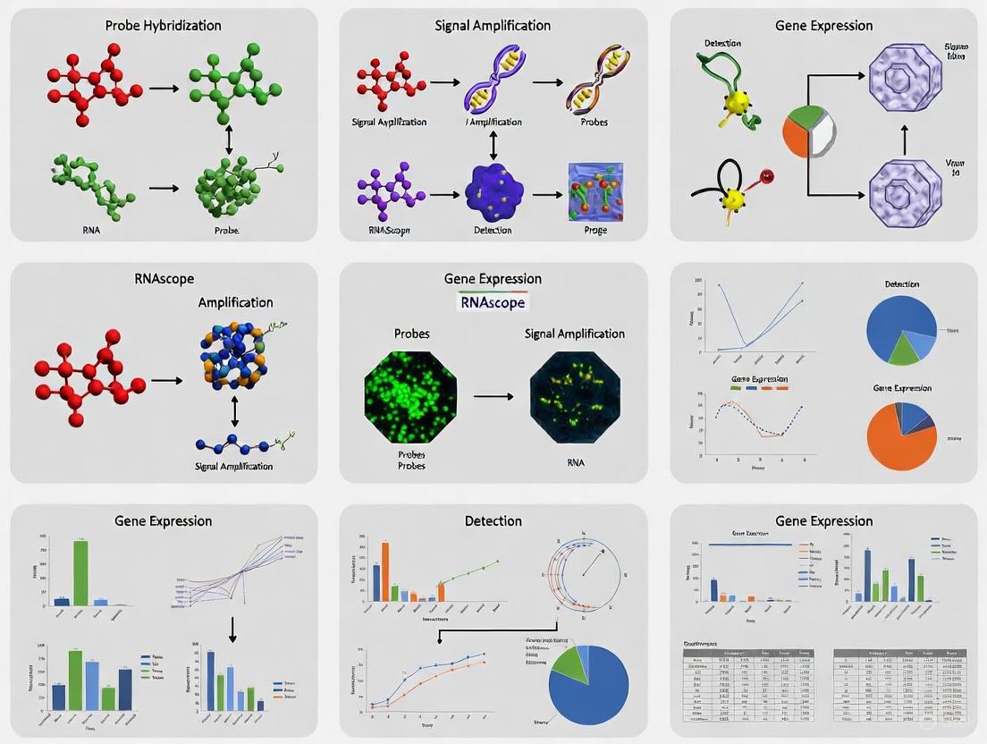
Workflow and Signaling Pathway Diagrams
RNAscope Experimental Workflow
RNAscope Signal Amplification Mechanism
Applications in Biomedical Research and Diagnostics
RNAscope technology has enabled significant advances across multiple research domains and is increasingly being adopted for diagnostic applications:
Research Applications:
- Spatial Transcriptomics: Precisely localize gene expression within tissue microenvironments while preserving morphological context.
- Biomarker Validation: Confirm and spatially map RNA biomarkers discovered through high-throughput genomic and transcriptomic profiling.
- Intratumoral Heterogeneity: Characterize variable gene expression patterns across different regions of complex tumor tissues.
- Host-Pathogen Interactions: Detect viral RNA in infected tissues, as demonstrated with SARS-CoV-2 probe sets [5].
Diagnostic Applications:
- Clinical Partnerships: Integration with Leica Biosystems for fully automated RNAscope ISH on BOND III clinical staining platforms [5].
- Companion Diagnostics: Development of analyte-specific reagents (ASRs) for targets including HPV genotypes (6, 11, 16, 18, 31, 33), CMV, EBV, and various cancer biomarkers [5].
- Standardized Workflows: Implementation in clinical laboratories with bright-field microscopy compatible with existing pathology review practices [5].
The technology's compatibility with automated platforms and standardized bright-field detection has facilitated its transition from research to clinical settings, particularly for applications requiring precise spatial localization of RNA biomarkers within pathological contexts.
Formalin-fixed paraffin-embedded (FFPE) samples represent an invaluable resource for biomedical research, with over one billion archival samples available worldwide [6]. These samples are routinely collected in clinical and pathological settings, offering extensive longitudinal data and association with detailed clinical outcomes. Their stability at room temperature and capacity for long-term storage make them ideal for retrospective studies. However, the very process that preserves tissue architecture—formalin fixation—induces significant molecular degradation that challenges conventional RNA analysis techniques. Understanding these challenges is crucial for researchers and drug development professionals seeking to leverage this vast resource for biomarker discovery, toxicogenomic profiling, and clinical diagnostics.
The core of the problem lies in the chemical processes of formalin fixation. Formalin, a solution of formaldehyde, penetrates tissues and forms methylene bridges between proteins, and between proteins and nucleic acids [6]. This cross-linking preserves morphological details but creates significant barriers to RNA extraction and analysis. Simultaneously, RNA molecules undergo fragmentation through hydrolysis and other damage mechanisms. These effects are compounded during long-term storage, where continued degradation can occur even after embedding in paraffin. The result is that RNA from FFPE samples typically exhibits both extensive fragmentation and chemical modifications that interfere with downstream molecular analyses.
The Molecular Pathology of FFPE-Induced RNA Damage
FormalIN-Induced Cross-Linking and Its Consequences
The cross-linking process begins immediately upon formalin exposure. Formaldehyde hydrates to form methylene glycol, which penetrates cells and initiates the formation of reversible protein-nucleic acid cross-linkages [7]. Within approximately 24-48 hours, these initial adducts evolve into more stable covalent bonds that create a molecular meshwork within the cell. This cross-linking has several direct consequences for RNA analysis:
- Epitope Masking: The three-dimensional structure of RNA is altered as molecules become trapped in protein-RNA cross-links, making binding sites inaccessible to probes and primers.
- Extraction Interference: Standard RNA extraction methods struggle to efficiently reverse these cross-links, leading to low yields and significant loss of material.
- Molecular Modifications: Formalin can introduce methylene adducts on RNA bases, potentially interfering with reverse transcription and enzymatic amplification steps.
The extent of cross-linking is time-dependent. While initial cross-links formed within the first 24-48 hours are partially reversible, prolonged formalin fixation leads to increasingly stable and irreversible covalent bonds that are more challenging to reverse without causing additional RNA damage [7].
RNA Fragmentation in FFPE Samples
Parallel to cross-linking, RNA molecules in FFPE samples undergo extensive fragmentation. This occurs through multiple mechanisms:
- Chemical Hydrolysis: The formalin fixation process and subsequent storage conditions create an environment conducive to RNA strand breakage.
- Nuclease Activity: Endogenous nucleases remain active until completely inactivated by formalin, which may take hours depending on tissue penetration.
- Oxidative Damage: Long-term storage can expose samples to oxidative stress, further contributing to RNA degradation.
The degree of fragmentation directly impacts the quality metrics of extracted RNA. Studies comparing FFPE samples to matched fresh-frozen tissues show dramatic differences: FFPE-extracted RNA has a median RNA integrity number (RIN) of approximately 2.5 and DV200 values (percentage of RNA fragments >200 nucleotides) of 48%, compared to fresh-frozen RNA with RIN values of 8.1 and DV200 of 97% [6]. This represents nearly a two-fold degradation that severely impacts downstream applications.
Table 1: Impact of Formalin Fixation Time on RNAscope Signal Intensity
| Formalin Fixation Time | Signal Intensity | Percent Area of Signal | Practical Implications |
|---|---|---|---|
| 1-28 days | Maintained | Maintained | Suitable for RNAscope without major optimization |
| 60-90 days | Moderate reduction | Moderate reduction | May require protocol optimization |
| 180 days | Significant reduction | Significant reduction | Detectable but diminished signal |
| 270 days | No detectable signal | No detectable signal | Not suitable for analysis |
Data from [7] demonstrates that signal intensity and percent area of signal in RNAscope assays decrease with prolonged formalin fixation, with complete loss of detectable signal by 270 days.
Analytical Challenges in FFPE RNA Profiling
Technical Limitations of Conventional RNA-Seq with FFPE Samples
The degraded nature of FFPE RNA creates specific challenges for high-throughput sequencing technologies. FFPE RNA-seq (fRNA-seq) data is characterized by a high rate of transcript dropout (zero counts for actually expressed genes), high variance in transcript counts, and susceptibility to extreme values [6]. These properties share similarities with single-cell RNA-seq data but require specialized analytical approaches.
The fragmentation pattern of FFPE RNA favors shorter fragments, which creates mapping biases toward the 3' ends of transcripts when using poly-A enrichment methods. This bias can be mitigated by using ribosomal RNA depletion protocols instead of poly-A selection, as demonstrated in a cross-platform analysis where ribo-depletion RNA-seq outperformed other methods with the highest correlations of differentially expressed genes and best overlap of pathways between fresh-frozen and FFPE groups [8].
Statistical characterization of fRNA-seq data reveals that the negative binomial distribution best fits the observed count data, with little evidence supporting zero-inflated extensions [6]. This distributional understanding is crucial for developing appropriate normalization methods and differential expression tools specifically tailored to FFPE-derived data.
Impact of Pre-analytical Variables on Data Quality
Multiple pre-analytical factors significantly influence the quality of RNA obtainable from FFPE samples:
- Time in Formalin: Studies show clear erosion of signal intensity with extended time in formalin, though biological responses generally remain consistent for 18-hour and 3-week FFPE samples compared to fresh-frozen samples [8].
- Storage Duration: While RNAscope has been successfully applied to samples stored for up to 25-27 years [9], gradual fragmentation occurs over time in paraffin blocks stored at room temperature.
- Tissue Processing Protocols: Variations in dehydration, clearing, and embedding procedures across institutions introduce additional variability.
- Extraction Methodologies: Specialized kits designed for FFPE RNA extraction typically incorporate more extensive de-crosslinking steps and are optimized for shorter fragments.
Table 2: Quality Control Recommendations for FFPE RNA-Seq
| Quality Metric | Threshold for Adequate QC | Threshold for Failed QC | Clinical Implications |
|---|---|---|---|
| RNA Concentration | ≥40.8 ng/μL | ≤18.9 ng/μL | Input below 25 ng/μL yields poor results |
| Pre-capture Library Qubit | ≥5.82 ng/μL | ≤2.08 ng/μL | Indicates insufficient library preparation |
| Sample-wise Spearman Correlation | ≥0.75 | <0.75 | Suggests high technical variance |
| Reads Mapped to Gene Regions | ≥25 million | <25 million | Inadequate sequencing depth |
| Detectable Genes (TPM > 4) | >11,400 | ≤11,400 | Limited transcriptome coverage |
Data from [10] provides concrete quality thresholds for determining whether FFPE-extracted RNA is suitable for RNA-seq analysis.
RNAscope Technology: A Novel Approach for FFPE RNA Analysis
The RNAscope Platform and Double-Z Probe Design
RNAscope represents a paradigm shift in RNA analysis from FFPE samples by moving from grind-and-bind approaches to in situ hybridization. This technology employs a novel double-Z probe design that enables single-molecule RNA visualization while preserving tissue morphology [11].
The key innovation lies in the probe design strategy. Each target RNA is detected using a series of target probe pairs that hybridize contiguously to the RNA molecule. Each probe contains a region complementary to the target RNA, a spacer sequence, and a 14-base tail sequence. Only when two probes (the "double Z") bind adjacent sites on the target RNA do their tail sequences form a complete 28-base hybridization site for the preamplifier molecule [11]. This design provides exceptional specificity because it is statistically unlikely that nonspecific hybridization will position two probes appropriately to form the preamplifier binding site.
The signal amplification system then builds on this foundation: each preamplifier contains 20 binding sites for the amplifier, which in turn contains 20 binding sites for label probes. This theoretical 8000-fold amplification per target molecule enables detection of even low-abundance transcripts in heavily cross-linked FFPE samples [11].
Optimized Protocol for FFPE Samples Using RNAscope
The RNAscope assay procedure for FFPE tissues involves specific steps optimized to overcome formalin-induced damage [12] [11]:
Sample Preparation and Sectioning:
- Cut 5μm sections from FFPE blocks and mount on Superfrost Plus slides
- Deparaffinize in xylene and dehydrate through ethanol series
- Perform target retrieval in citrate buffer (10 mM, pH 6.0) at 100-103°C for 15 minutes
Protease Digestion:
- Treat sections with protease (10 μg/mL) at 40°C for 30 minutes
- This step is critical for permeabilizing tissues and breaking protein cross-links without excessive RNA degradation
Probe Hybridization and Amplification:
- Hybridize with target probes in hybridization buffer at 40°C for 2 hours
- Perform sequential hybridizations with preamplifier, amplifier, and label probe
- For chromogenic detection, use HRP-based detection with DAB or Fast Red
The entire procedure can be completed in 7-8 hours or divided over two days, with options for both manual and automated processing on platforms such as the Ventana DISCOVERY XT or Leica BOND RX systems [12].
Experimental Applications and Validation Studies
Performance with Archival Samples Across Time
RNAscope has been validated across a wide range of FFPE sample ages, demonstrating remarkable robustness for retrospective studies. In one notable application, researchers successfully applied RNAscope to 25-27-year-old human prostate cancer samples from lymph node metastases [9]. Despite the extended storage period, the assay detected clear punctate signals for the ubiquitin C (UBC) reference gene, demonstrating preservation of detectable RNA even after decades of storage.
A systematic study of FFPE tissue storage time evaluated canine distemper virus (CDV) RNA detection in raccoon tissues stored for periods ranging from 6 months to 15 years [7]. The research found that RNA was detectable in all samples regardless of storage duration, though with some reduction in signal intensity in the oldest samples. This confirms that with appropriate methodology, RNA analysis remains feasible even in decades-old archival specimens.
For formalin fixation time, a detailed analysis measured signal of a reference gene (16S rRNA) in tissues fixed for periods ranging from 1 to 270 days [7]. The results demonstrated that signal intensity and percent area of signal decreased significantly after 180 days of formalin fixation, with no detectable signal at 270 days. This highlights the importance of knowing fixation history when selecting samples for analysis.
Comparison with Alternative Technologies
When compared to other RNA analysis methods for FFPE samples, RNAscope offers distinct advantages:
- Versus RNA-seq: RNAscope preserves spatial context lost in grind-and-bind approaches, allowing correlation of gene expression with tissue morphology and cell type identification.
- Versus Microarrays: RNAscope offers higher sensitivity for degraded RNA and does not suffer from the same dynamic range limitations.
- Versus IHC: RNAscope often provides better correlation with treatment response and can detect targets that challenge antibody detection, such as neo-antigens and highly homologous gene families [13].
For quantitative transcriptomic profiling from FFPE samples, ribo-depletion RNA-seq has been identified as the optimal approach among conventional methods. A cross-platform analysis demonstrated that this protocol outperformed poly-A enrichment and microarray methods by having the highest correlations of differentially expressed genes and best overlap of pathways between fresh-frozen and FFPE samples [8].
The Scientist's Toolkit: Essential Reagents and Materials
Table 3: Research Reagent Solutions for FFPE RNA Analysis
| Reagent/Material | Function | Application Notes |
|---|---|---|
| RNAscope Target Probes | Hybridize to target RNA sequences | Designed using double-Z architecture for specificity; 20 probe pairs typically target 1kb region |
| Preamplifier | Binds to paired probe tails | Creates binding sites for amplifier; requires contiguously bound probe pair |
| Amplifier | Multiplies signal | Contains 20 binding sites for label probes |
| Label Probes | Visualizes hybridized probes | Conjugated to enzymes (HRP/AP) or fluorescent dyes |
| HybEZ Hybridization System | Maintains optimum humidity and temperature | Critical for proper hybridization conditions |
| Protease | Digests cross-linked proteins | Permeabilizes tissue; concentration and time require optimization |
| Target Retrieval Reagents | Reverses formalin cross-links | Citrate buffer (pH 6.0) standard; conditions may require optimization |
| Positive Control Probes (PPIB, UBC) | Assess RNA quality and assay performance | PPIB (medium abundance), UBC (high abundance), POLR2A (low abundance) |
| Negative Control Probe (dapB) | Assess background signal | Bacterial gene should not generate signal in properly fixed tissue |
| 4-Methyl withaferin A | 4-Methyl withaferin A, MF:C29H40O6, MW:484.6 g/mol | Chemical Reagent |
| 31-Norlanostenol | 31-Norlanostenol, MF:C29H50O, MW:414.7 g/mol | Chemical Reagent |
FFPE samples present significant challenges for conventional RNA analysis due to formalin-induced fragmentation and cross-linking. These effects degrade RNA quality and interfere with standard molecular biology techniques. However, through specialized approaches like RNAscope in situ hybridization and ribo-depletion RNA-seq, researchers can successfully extract meaningful gene expression data from these valuable archival resources.
The key to success lies in understanding the nature of FFPE-induced damage and selecting appropriate analytical methods that either circumvent these challenges (as with RNAscope's in situ approach) or are specifically optimized to handle degraded material (as with ribo-depletion protocols). With careful attention to pre-analytical variables, quality control metrics, and protocol optimization, FFPE samples can yield high-quality data that leverages their extensive associated clinical information, unlocking their tremendous potential for translational research and biomarker discovery.
RNA in situ hybridization (ISH) has emerged as a critical technology in molecular pathology, enabling researchers to examine biomarker expression within the histopathological context of clinical specimens. The RNAscope platform, through its unique probe design strategy, overcomes the traditional limitations of RNA ISH—particularly for formalin-fixed paraffin-embedded (FFPE) tissues—by achieving single-molecule sensitivity while preserving tissue morphology. This Application Note details the fundamental principles of RNAscope probe design, provides validated protocols for FFPE samples, and presents quantitative data supporting its application in drug development research. We demonstrate how the proprietary double-Z (ZZ) probe architecture enables exceptional signal amplification and background suppression, facilitating precise spatial gene expression analysis even in challenging archival tissues.
Conventional RNA in situ hybridization techniques have faced significant challenges in clinical and research settings due to technical complexity, insufficient sensitivity, and specificity concerns [1]. The RNAscope platform addresses these limitations through a novel probe design strategy that allows simultaneous signal amplification and background suppression to achieve single-molecule visualization while preserving tissue morphology [1] [14]. This technology provides a universal solution to characterize tissue distribution of drug targets and biomarkers in a highly specific and sensitive manner, without the need for time-consuming antibody development and validation [14].
For FFPE tissues—the most widely used pathology archive—RNAscope offers particular advantages. Formalin fixation causes cross-linking and fragmentation of nucleic acids, which traditionally compromises RNA quality [15]. However, RNAscope probes are specifically designed to detect these fragmented RNA molecules, making them ideally suited for FFPE samples [15]. The platform's robust performance across various tissue types and species provides researchers and drug development professionals with a reliable tool for preclinical studies and biomarker validation.
Core Technology: The ZZ Probe Design Principle
Architectural Basis of RNAscope Probes
The fundamental innovation underlying RNAscope technology is the double-Z (ZZ) probe design. This proprietary architecture enables both specific signal amplification and effective background suppression through the following mechanism:
- Probe Pair Structure: Each ZZ pair consists of two oligonucleotides that hybridize to adjacent regions of the target RNA [16]. The "bottom" of each Z oligo contains an 18 to 25-base region complementary to the target RNA, selected for specific hybridization properties and uniform melting temperatures [16].
- Binding Footprint: Individual ZZ pairs hybridize to 36-50 bases of target RNA, with a standard RNAscope probe comprising 20 ZZ pairs spanning approximately 1000 bases of unique sequence [16].
- Amplification Strategy: This design facilitates a proprietary signal amplification system that builds specific signals only when both halves of the ZZ pair bind correctly to the target sequence.
Table 1: RNAscope Probe Design Specifications for Different RNA Targets
| Probe Type | Target Length | ZZ Pairs | Applications |
|---|---|---|---|
| RNAscope | >300 bases | ~20 pairs | mRNA, long ncRNA |
| BaseScope | 50-300 bases | 1-3 pairs | Short transcripts, SNP detection |
| miRNAscope | 17-50 bases | Specialized design | miRNA, siRNA, ASO |
Visualization of the ZZ Probe Mechanism
The following diagram illustrates the molecular mechanism of RNAscope's proprietary ZZ probe technology:
This amplification cascade enables single-molecule detection through a built-in background suppression mechanism: the system only produces signal when both components of the ZZ probe pair bind correctly to their target sequences, minimizing false positives from non-specific hybridization.
Experimental Protocols for FFPE Samples
Sample Preparation and Pretreatment
Proper sample preparation is critical for successful RNAscope analysis of FFPE tissues. The following protocol has been validated for various tissue types:
Tissue Processing:
Slide Pretreatment:
RNAscope 2.5 LS Duplex Assay Workflow
For automated staining on the Leica BOND RX system, the following protocol is recommended:
Probe Hybridization:
- Apply target probes (C1 and C2 channels) and hybridize at 40°C for 2 hours.
- Follow manufacturer's recommended amplification steps (Amp 1-6).
Signal Detection:
Counterstaining and Mounting:
- Counterstain with hematoxylin.
- Dehydrate, clear, and mount with non-aqueous mounting medium.
The following workflow diagram illustrates the complete RNAscope procedure for FFPE samples:
Quality Control and Data Interpretation
Control Probes and Sample Qualification
Rigorous quality control is essential for reliable RNAscope results, particularly for FFPE tissues where RNA integrity may vary:
- Positive Control Probes: Housekeeping genes PPIB (Cyclophilin B), POLR2A, or UBC provide reference for RNA quality [3] [15].
- Negative Control Probes: Bacterial dapB gene confirms specificity of hybridization [3].
- Sample Qualification: Successful staining should demonstrate PPIB/POLR2A score ≥2 or UBC score ≥3 with dapB score <1 [3].
Staining Interpretation and Quantification
RNAscope uses a semi-quantitative scoring system based on discrete dot enumeration:
- Score 0: No staining or <1 dot per 10 cells.
- Score 1: 1-3 dots per cell (visible at 20-40X magnification).
- Score 2: 4-10 dots per cell, very few dot clusters.
- Score 3: >10 dots per cell, <10% of dots form clusters.
- Score 4: >10 dots per cell, >10% of dots form clusters.
Table 2: RNAscope Quality Control Metrics for FFPE Tissues
| Quality Parameter | Acceptance Criteria | Impact on Interpretation |
|---|---|---|
| Positive Control (PPIB/POLR2A) | Score ≥2 | Confirms adequate RNA quality |
| Negative Control (dapB) | Score <1 | Verifies hybridization specificity |
| Signal Distribution | Punctate dots within cytoplasm/nucleus | Validates target-specific detection |
| Tissue Morphology | Well-preserved after pretreatment | Ensures reliable cellular localization |
Recent studies systematically assessing RNA degradation over archival time have shown that although RNAscope probes are designed to detect fragmented RNA, performing sample quality checks using housekeeping genes is strongly recommended to ensure accurate results [15]. RNA degradation in FFPET is most pronounced in high-expressor housekeeping genes (UBC and PPIB) compared to low-to-moderate expressors (POLR2A and HPRT1) [15].
Advanced Applications in Research and Drug Development
Specialized Probe Designs for Unique Research Needs
The flexibility of RNAscope probe design enables specialized applications:
- Intronic Probes: Designed to target intronic regions of pre-mRNAs, enabling nuclear localization and identification of specific cell types. This approach has been successfully used with Tnnt2 intronic probes to identify cardiomyocyte nuclei in cardiac regeneration studies [18].
- Cross-Species Probes: Can be designed when sequence homology exceeds 95% across species [16].
- Small RNA Detection: miRNAscope assays enable detection of ASOs, miRNAs, and siRNAs (17-50 nucleotides) alongside mRNA targets [19].
Multiplexing Capabilities for Complex Assays
RNAscope enables simultaneous detection of multiple RNA targets through channel-specific probes:
- Duplex Assays: Simultaneous detection of two RNA species using C1 (DAB) and C2 (Fast Red) channels [17].
- Multiplex Fluorescent Assays: Detection of up to four targets using different fluorophores [15].
- RNAscope Plus small RNA-RNA Assay: Enables visualization of one small RNA (ASO, miRNA, siRNA) plus up to three mRNA targets [19].
Essential Research Reagent Solutions
Table 3: Key Research Reagents for RNAscope Applications
| Reagent / Component | Function | Example Catalog Numbers |
|---|---|---|
| RNAscope 2.5 LS Duplex Reagent Kit | Core reagents for automated duplex detection | 322440 |
| RNAscope Target Probes | Species-specific probe sets for target genes | 300038 (RTU), 300038-C2 (50X) |
| Control Probes (PPIB, POLR2A) | Positive controls for RNA quality | 320748 (Human), 320768 (Mouse) |
| Negative Control Probes (dapB) | Negative control for background assessment | 312038-C2 |
| RNAscope 2.5 LS Green Accessory Pack | Green chromogen alternative to DAB | 322550 |
| BOND Epitope Retrieval Solutions | Antigen retrieval for FFPE samples | AR9961, AR9640 |
| RNAscope Control Slides | System suitability verification | 310045 (Human), 310023 (Mouse) |
RNAscope probe design represents a significant advancement in molecular pathology, providing researchers with an engine for specific signal amplification and background suppression. The proprietary ZZ probe architecture enables unprecedented sensitivity and specificity for RNA detection in FFPE tissues, making it an invaluable tool for drug development professionals requiring precise spatial gene expression analysis. By following the optimized protocols and quality control measures outlined in this Application Note, researchers can reliably implement this technology for biomarker validation, therapeutic development, and clinical research applications.
Formalin-fixed paraffin-embedded (FFPE) tissue samples represent an invaluable resource for biomedical research, with vast archives spanning decades of clinical history. These samples are particularly crucial for cancer research, drug development, and retrospective molecular studies. However, the inherent chemical modifications and progressive degradation of RNA in FFPE tissues present significant challenges for reliable molecular analysis, including RNAscope in situ hybridization. This application note systematically examines the key determinants of RNA quality in FFPE samples across different archival durations and provides evidence-based protocols to guide researchers in assessing sample suitability for spatial transcriptomics and gene expression studies.
Quantitative Impact of Archival Duration on RNA Quality
Long-term storage of FFPE samples progressively affects both the quantity and quality of recoverable RNA, though useful molecular information can often be retrieved even from decades-old specimens with appropriate methodological adjustments.
Table 1: Effects of FFPE Storage Duration on RNA Quality and Amplification Capacity
| Storage Duration | RNA Concentration | RNA Purity (OD260/OD280) | Degradation Level | Maximum Amplifiable Fragment Size | Research Implications |
|---|---|---|---|---|---|
| Freshly prepared | High | 1.8-2.1 (DNA), 1.9-2.2 (RNA) | Minimal [20] | ~700 nt [21] | Optimal for all applications |
| 1-3 years | Moderate | No significant change [20] | Moderate [20] | ~500 nt [21] | Suitable for most molecular analyses |
| 8 years | Significantly reduced | No significant change [20] | High [20] | ~200-400 nt [20] [21] | Target small genes/amplicons |
| 25+ years | Variable | Not reported | Extensive, but target-dependent [9] [15] | RNAscope still possible [9] | RNAscope feasible with quality controls [9] |
Multiple studies demonstrate that while RNA concentration decreases and fragmentation increases with storage time, RNA purity remains largely unaffected, and the material can still yield valuable scientific data. Specimens stored for longer periods show more degradation and reduced concentration of DNA and RNA after nucleic acid extraction, though purity remains stable [20]. Remarkably, researchers have successfully applied RNAscope in situ hybridization to 25-27-year-old FFPE samples of human prostate cancer metastases in lymph nodes, demonstrating that even decades-old archival material can yield meaningful results when properly validated [9].
Critical Factors Affecting RNA Integrity in FFPE Samples
Pre-analytical Variables
Several factors preceding RNA extraction significantly impact the quality of nucleic acids recovered from FFPE samples:
- Fixation Protocol: Optimal fixation uses 10% neutral-buffered formalin for 16-32 hours at room temperature [3]. Prolonged fixation (e.g., 72 hours) increases irreversible crosslinking and reduces amplifiable fragment size [21].
- Storage Conditions: Temperature dramatically affects RNA integrity. Samples stored at 4°C maintain significantly better RNA quality than those stored at room temperature or 37°C [21]. Protection from oxygen and light also helps preserve RNA integrity.
- Section Storage: FFPE sections should be analyzed within 3 months of sectioning when stored at room temperature with desiccant [3].
Sample Quality Assessment Methods
Table 2: RNA Quality Assessment Methods for FFPE Samples
| Assessment Method | Parameters Measured | Quality Threshold | Application Guidance |
|---|---|---|---|
| Spectrophotometry (NanoDrop) | Concentration, OD260/OD280 ratios | DNA: 1.8±0.1, RNA: 2.0±0.1 [20] | Initial quality screening |
| DV200 Value | Percentage of RNA fragments >200 nucleotides | ≥30% usable; ≥60% ideal for some iST platforms [22] [23] | Predicts NGS performance |
| RNA Quality Score (RQS) | RNA integrity on scale of 1-10 | Higher scores indicate better integrity [24] | Alternative to RIN for FFPE |
| Housekeeping Gene Amplification | PCR amplification of reference genes | β-globin and ALDH2 genes amplifiable in >99% of specimens [20] | Functional RNA quality assessment |
Experimental Protocols for RNA Quality Validation
RNAscope Sample Quality Control Protocol
For assessing FFPE sample suitability for RNAscope assays, implement this quality control workflow:
Detailed Procedure:
Sample Preparation: Cut FFPE tissue sections at 5±1μm thickness using positively charged slides (e.g., Fisher Scientific SuperFrost Plus). Bake slides at 60°C for 1-2 hours prior to RNAscope assay [3].
Control Probe Selection: Include both positive control probes (housekeeping genes PPIB, POLR2A, or UBC) and negative control probes (bacterial dapB gene) in each assay run [3] [15].
RNAscope Protocol: Follow manufacturer's instructions for the RNAscope Multiplex Fluorescent v2 assay, including appropriate pretreatment steps:
Quality Interpretation: Successful staining should demonstrate:
RNA Extraction and Quality Assessment Protocol
For comprehensive RNA quality evaluation prior to sequencing applications:
RNA Extraction: Use specialized FFPE RNA extraction kits. Performance varies among kits, with some demonstrating superior quality recovery [24]. Include deparaffinization with xylene and proteinase K digestion steps [20].
Quality Metrics Analysis:
Functional Validation: For gene expression studies, confirm that RNA quality supports the intended analytical approach, selecting appropriate library preparation methods based on input requirements and degradation levels [22].
The Scientist's Toolkit: Essential Research Reagents
Table 3: Key Research Reagent Solutions for FFPE RNA Quality Assessment
| Reagent Category | Specific Examples | Function/Application | Technical Considerations |
|---|---|---|---|
| RNAscope Control Probes | PPIB, POLR2A, UBC (positive), dapB (negative) [3] | Sample quality validation for RNAscope | PPIB and UBC are high expressors; POLR2A is moderate [15] |
| RNA Extraction Kits | miRNeasy FFPE, iCatcher FFPE, Ionic FFPE, ReliaPrep FFPE [25] [24] | RNA isolation from FFPE tissue | Performance varies; Promega kit provided superior yield in comparative studies [24] |
| Library Prep Kits | TaKaRa SMARTer Stranded Total RNA-Seq, Illumina Stranded Total RNA Prep [22] | RNA-seq library preparation | Kit selection depends on RNA input and degradation level; SMARTer requires 20x less input [22] |
| Nucleic Acid Analysis | NanoDrop spectrophotometers, TapeStation, BioAnalyzer [20] [24] [21] | RNA quantity and quality assessment | DV200 values predict sequencing performance better than traditional methods [22] |
| 8-Epiloganin | 8-Epiloganin|Natural Iridoid Glycoside|For Research | Bench Chemicals | |
| 8-Hydroxydigitoxigenin | 8-Hydroxydigitoxigenin | Bench Chemicals |
RNA Degradation Patterns and Technical Considerations
Understanding specific degradation patterns in FFPE samples enables more accurate interpretation of results:
Differential Degradation by Expression Level: High-expression housekeeping genes (UBC, PPIB) show more pronounced degradation in FFPE samples over time compared to low-to-moderate expressors (POLR2A, HPRT1) [15]. This pattern is particularly evident in archival duration-dependent degradation.
Storage Temperature Effects: Storage temperature significantly impacts degradation rate. Samples stored at 4°C maintain RNA integrity substantially better than those stored at room temperature or 37°C [21].
Technical Implications for RNAscope: RNAscope probes are designed to detect fragmented RNA, making the technique particularly suitable for FFPE samples [15]. However, signal intensity may decrease with archival time, necessitating appropriate control probes and interpretation criteria.
FFPE samples remain a valuable resource for spatial transcriptomics and molecular pathology despite the challenges of RNA degradation over archival time. While storage duration significantly impacts RNA quality, with longer storage resulting in increased fragmentation and reduced concentration, proper quality assessment and methodological adaptations can yield reliable data even from decades-old specimens. Critical to success are appropriate quality control measures including control probes in RNAscope assays, careful attention to pre-analytical variables, and selection of extraction and analysis methods compatible with degraded RNA. By implementing the protocols and considerations outlined in this application note, researchers can effectively evaluate FFPE sample suitability and generate robust, reproducible results for their research and drug development programs.
Formalin-fixed, paraffin-embedded (FFPE) tissue archives represent an invaluable resource for biomedical research, particularly in oncology and retrospective disease studies. However, extensive nucleic acid crosslinking and potential RNA fragmentation pose significant challenges for molecular analysis of these samples [15]. RNAscope in situ hybridization (ISH) technology enables sensitive and specific detection of RNA targets within intact cellular morphology by utilizing a unique double-Z probe design that provides simultaneous signal amplification and background suppression [1]. This application note details the successful detection of RNA in FFPE samples older than 25 years, demonstrating the robustness of RNAscope technology for long-term archived tissues and its implications for retrospective research studies.
Case Study: RNA Detection in 25-27 Year-Old FFPE Samples
Research Background and Sample Information
Researchers at Erasmus MC (Rotterdam, Netherlands) conducted a retrospective study applying RNAscope ISH to historically archived FFPE samples [9]. The investigation targeted human metastases of prostate cancer in lymph node tissues that had been collected between 1987 and 1989, making the samples approximately 25-27 years old at the time of analysis in 2014 [9] [26]. This represented the oldest known successful application of RNAscope ISH at the time of publication.
Experimental Parameters and Target Selection
The research team followed standard ACD protocols and user manuals without substantial modification [9]. For target detection, they utilized a probe targeting the UBC (Ubiquitin C) gene, a high-copy housekeeping gene, which facilitated reliable signal detection despite extensive sample archival duration [9]. The RNAscope Chromogenic Red assay was employed for visualization, with results examined at 400x magnification [9].
Table: Key Experimental Parameters for 25+ Year-Old FFPE Sample Analysis
| Parameter | Specification |
|---|---|
| Sample Type | Human prostate cancer metastases in lymph node |
| Sample Collection Years | 1987, 1988, 1989 |
| Analysis Year | 2014 |
| Sample Age at Analysis | 25-27 years |
| Target Gene | UBC (Ubiquitin C) |
| Detection Method | RNAscope Chromogenic Red assay |
| Visualization | Punctate red dots at 400x magnification |
Results and Outcomes
The experimental results demonstrated successful detection of UBC gene expression across all samples, including the 27-year-old specimen from 1987 [9]. The characteristic punctate red dot staining pattern was clearly visible, confirming preserved RNA integrity despite the extended archival period [9] [26]. The researchers noted that success with such historically archived samples depends on multiple factors including original sample fixation quality, tissue preservation methods, and storage conditions over time [9].
Systematic Analysis of RNA Degradation in Archived FFPE Samples
Archival Duration and RNA Quality Relationship
Recent systematic investigations have quantified the relationship between archival time and RNA detection capability in FFPE samples. A 2025 study analyzing breast cancer samples identified an archival duration-dependent reduction in RNAscope signals, with pronounced degradation effects observed in high-expression housekeeping genes including UBC and PPIB compared to moderate-to-low expressors like POLR2A and HPRT1 [15].
Table: RNAscope Signal Degradation Patterns in FFPE vs. Fresh Frozen Tissue (FFT) Over Time
| Parameter | FFPE Samples | Fresh Frozen Tissue (FFT) |
|---|---|---|
| Signal Reduction | Archival duration-dependent fashion | Minimal degradation over time |
| Most Affected Genes | High expressors (UBC, PPIB) | Stable detection |
| Least Affected Genes | Low-to-moderate expressors (POLR2A, HPRT1) | Stable detection |
| Key Degradation Factor | PPIB shows highest degradation (R² = 0.33-0.35) | Minimal degradation |
| Recommended Quality Control | Housekeeping gene verification essential | Housekeeping gene verification recommended |
Effects of Prolonged Formalin Fixation
A 2024 study examining formalin fixation duration demonstrated that RNAscope can detect reference gene (16S rRNA) signals in tissues fixed in 10% neutral-buffered formalin for up to 180 days, with signal intensity and percent area decreasing significantly after extended fixation [7]. Detection failed at 270 days of formalin fixation, establishing practical boundaries for fixative duration [7].
For paraffin-embedded storage intervals, the same study successfully detected canine distemper virus RNA in FFPE tissues stored for up to 15 years at room temperature, confirming that paraffin embedding provides superior long-term RNA preservation compared to extended formalin immersion [7].
Essential Protocols for Archival FFPE Sample Analysis
Sample Quality Assessment Workflow
Prior to target probe analysis, archival sample quality must be verified through a systematic workflow [12]:
RNAscope Assay Protocol for Archived FFPE Samples
The standard RNAscope protocol requires specific modifications for historically archived samples [12] [3]:
Sample Preparation:
- Use 5±1 μm thick sections mounted on SuperFrost Plus slides [3]
- Bake slides at 60°C for 1-2 hours prior to assay initiation
- Deparaffinize in xylene and ethanol following standard histology protocols
Pretreatment Optimization:
- Antigen Retrieval: May require extended time (5-minute increments) at 95-100°C [12]
- Protease Digestion: May require extended time (10-minute increments) at 40°C [12]
- Optimization Approach: Systematically vary retrieval and protease times while maintaining constant temperatures
Hybridization and Detection:
- Follow RNAscope 2.5 HD Red or Brown kit protocols precisely
- Maintain 40°C temperature during protease digestion and probe hybridization steps
- Use appropriate mounting media (EcoMount for Red assay, xylene-based for Brown assay) [12]
Quality Control and Scoring Criteria
Control Probe Implementation
Robotic quality control is essential for archival sample analysis [12] [3]:
- Positive Control Probes: PPIB (medium expressor), UBC (high expressor), or POLR2A (low-medium expressor)
- Negative Control Probe: Bacterial dapB gene should show no staining
- Sample Qualification: Successful PPIB staining should generate score ≥2, UBC score ≥3 with uniform signal distribution
- Background Assessment: dapB score should be <1, indicating minimal non-specific hybridization [12]
RNAscope Scoring Guidelines
Semi-quantitative analysis follows established scoring criteria based on dots per cell [12]:
Table: RNAscope Semi-Quantitative Scoring Guidelines
| Score | Criteria | Interpretation |
|---|---|---|
| 0 | No staining or <1 dot/10 cells | Negative |
| 1 | 1-3 dots/cell | Low expression |
| 2 | 4-9 dots/cell, none or very few dot clusters | Moderate expression |
| 3 | 10-15 dots/cell, <10% dots in clusters | High expression |
| 4 | >15 dots/cell, >10% dots in clusters | Very high expression |
Research Reagent Solutions
Table: Essential Reagents for RNAscope Analysis of Archived FFPE Samples
| Reagent/Category | Specific Examples | Function/Purpose |
|---|---|---|
| Control Probes | PPIB, POLR2A, UBC | Sample RNA quality verification |
| Negative Control | dapB | Background assessment |
| Detection Kits | RNAscope 2.5 HD Red Kit (#322350) | Chromogenic detection |
| RNAscope 2.5 HD Brown Kit (#322300) | Chromogenic detection | |
| Equipment | HybEZ Hybridization System | Maintains optimal humidity/temperature |
| Specialized Slides | SuperFrost Plus slides | Prevent tissue detachment |
| Barrier Pen | ImmEdge Hydrophobic Barrier Pen | Maintains reagent containment |
| Mounting Media | EcoMount (#320409) | For Red assay preservation |
| CytoSeal XYL | For Brown assay preservation |
Discussion and Technical Considerations
Critical Success Factors for Archival Samples
The successful application of RNAscope to decades-old FFPE samples depends on several intersecting factors [9]:
Initial Fixation Quality: Samples fixed in 10% neutral-buffered formalin for 16-32 hours following standard pathological protocols yield optimal long-term preservation [3]. Extended formalin fixation beyond 30 days initiates irreversible covalent bond formation that progressively damages RNA integrity [7].
Storage Conditions: FFPE blocks stored at room temperature with stable humidity conditions demonstrate superior long-term RNA preservation compared to tissues remaining in liquid formalin [7].
Target Selection Strategy: High-copy number targets like UBC provide more robust detection in compromised samples, though they may show more pronounced degradation patterns than moderate-copy genes [15]. For samples with significant RNA degradation, targeting lower expression genes may yield more reliable results.
Advanced Applications and Recent Innovations
Emerging methodologies enhance the utility of RNAscope for archival samples:
Digital Image Analysis: Advanced algorithms enable quantitative assessment of RNAscope signals, reducing pathologist variability and improving precision [27] [28]. Deep learning segmentation approaches now outperform manual expert annotation in identifying RNAscope dots (Fâ‚-score 0.745 vs. 0.596) [28].
Multiplex Detection: RNAscope multiplex assays allow simultaneous detection of multiple targets in the same tissue section, particularly valuable for limited archival samples [26].
Algorithm-Assisted Quantification: Commercial and open-source digital pathology solutions (QuPath, QuantISH) enable automated dot counting and H-score calculation, improving reproducibility for archival sample analysis [27] [28].
RNAscope technology represents a robust platform for RNA detection in historically archived FFPE samples, successfully demonstrating target detection in tissues preserved for over 25 years. Systematic quality control through housekeeping gene verification, appropriate pretreatment optimization, and standardized scoring methodologies enable reliable analysis of valuable archival collections. These capabilities significantly extend the research utility of pathological archives, enabling retrospective biomarker studies and long-term disease progression analysis that leverage decades of clinical preservation.
Implementing RNAscope: Complete Workflow from Sample Preparation to Multiplex Detection
Formalin-fixed paraffin-embedded (FFPE) samples represent an invaluable resource in biomedical research, particularly in cancer research, immunology, and drug development. These samples, prepared from tissue biopsies obtained during surgical procedures, are stabilized through a meticulous preservation process that allows them to remain stable for years or even decades at room temperature [29]. The significance of FFPE samples extends beyond their traditional use in morphological studies for diagnostic purposes; they now serve as crucial sources for DNA, RNA, and protein analyses, enabling genomic, transcriptomic, and proteomic investigations even after extensive storage periods [29].
The integration of advanced molecular techniques such as the RNAscope in situ hybridization (ISH) assay with FFPE samples has revolutionized our ability to visualize single RNA molecules within their morphological context [30]. This powerful combination enables researchers to investigate gene expression patterns directly in tissue sections, providing spatial information that is lost in most other molecular analyses. The proprietary "double Z" probe design technology underlying RNAscope provides highly specific and sensitive detection of target RNAs, with each dot representing a single RNA transcript [31]. This application note provides detailed protocols and guidelines for optimizing FFPE sample preparation specifically for sensitive downstream applications including RNAscope ISH, ensuring that researchers can maximize the value of these precious archival resources.
FFPE Sample Preparation: A Step-by-Step Protocol
The preparation of high-quality FFPE samples requires strict adherence to established protocols with particular attention to fixation timing, processing conditions, and sectioning techniques. The following comprehensive guidelines ensure sample integrity for demanding downstream applications including RNAscope ISH.
Tissue Fixation Guidelines
Proper fixation is the most critical step in FFPE sample preparation, directly impacting the quality of biomolecules available for subsequent analysis.
- Fixative Selection: Use 10% neutral buffered formalin (NBF) as the standard fixative [30].
- Fixation Timing: Fixation must begin immediately after tissue dissection to minimize ischemic time, which can lead to cellular degradation and compromise molecular integrity [29]. Optimal fixation times range from 16-32 hours at room temperature [30]. Fixation for less than 16 hours or exceeding 32 hours will impair performance in RNAscope assays [30].
- Tissue Size Considerations: Formalin penetration is limited in thicker tissues, affecting uniform preservation. Specimens should be of appropriate dimensions (typically not exceeding 4-5mm in thickness) to ensure complete fixation [29].
Tissue Processing: Dehydration, Clearing, and Embedding
Following fixation, tissues undergo processing to replace water with paraffin, creating a stable embedded block suitable for sectioning.
Dehydration: Transfer fixed tissues through a graded ethanol series (typically 70%, 80%, 95%, and 100% ethanol) to gradually remove all water from the sample. This step is crucial as paraffin wax is not soluble in water [29]. Note that immediate immersion in 100% ethanol can cause tissue degradation and protein denaturation.
Clearing: Treat tissues with a clearing agent such as xylene or less toxic alternatives like isopropanol to displace ethanol and remove fat from the tissue. This "clearing" step enables complete paraffin infiltration [29]. If using isopropanol, embedding must be performed with higher temperature wax.
Paraffin Embedding: Embed the cleared tissue in molten paraffin wax at approximately 60°C. The paraffin solidifies upon cooling, providing structural support for microtomy [29]. Proper embedding is essential to avoid artifacts that compromise sectioning quality and subsequent analyses.
Microtomy and Slide Preparation
Sectioning and slide preparation require precision to obtain optimal tissue sections for RNAscope and other molecular applications.
- Section Thickness: Cut embedded tissue into sections of 5±1 μm thickness using a microtome [30].
- Water Bath Step: Float paraffin ribbons on a 40-45°C water bath to smooth sections before mounting [30].
- Slide Selection: Mount sections only on SuperFrost Plus slides for proper adhesion [30].
- Drying: Air dry mounted sections overnight at room temperature [30].
- Storage: Use sectioned tissue within 3 months for optimal results. Store slides with desiccants at room temperature. For long-term preservation of RNA quality (>1 year), store blocks or sections at 2-8°C with desiccation [30].
Table 1: Critical Parameters for Optimal FFPE Sample Preparation
| Processing Step | Optimal Conditions | Potential Pitfalls |
|---|---|---|
| Fixation | 16-32 hours in 10% NBF at RT | Under-fixation: inadequate preservation; Over-fixation: excessive cross-linking |
| Tissue Size | Appropriate thickness (4-5mm) for complete formalin penetration | Larger tissues show uneven preservation |
| Dehydration | Gradual ethanol series (70%-100%) | Direct immersion in 100% ethanol causes degradation |
| Clearing | Xylene or isopropanol | Incomplete clearing impedes wax infiltration |
| Embedding | Paraffin at ~60°C | Improper embedding causes sectioning artifacts |
| Sectioning | 5±1 μm thickness | Thicker sections compromise morphology and assay performance |
| Slide Storage | With desiccants at RT; use within 3 months | Prolonged storage degrades RNA quality |
Pre-analytical Considerations and Challenges
Several pre-analytical factors significantly impact the quality of FFPE samples and their suitability for downstream molecular applications. Understanding these variables is essential for optimizing experimental outcomes.
Key Variables Affecting Sample Quality
Ischemic Time: The interval between tissue removal and fixation must be minimized. Prolonged ischemic times lead to cellular degradation, compromising molecular integrity [29]. During surgical procedures, biopsies should be immediately placed in fixative [29].
Decalcification Requirements: Tissues containing calcified structures (e.g., bone) may require decalcification before formalin fixation. This process can result in loss of nucleic acids and proteins, affecting overall sample quality [29].
Fixation Variables: Both under-fixation and over-fixation present challenges. Inadequate fixation fails to preserve tissue architecture, while excessive fixation promotes protein cross-linking that can negatively impact nucleic acid quality and antigen accessibility [29].
Impact on Downstream Molecular Analyses
The fixation and processing conditions directly influence the success of various molecular applications:
RNA Integrity: RNA is particularly susceptible to degradation during FFPE preparation. Formalin fixation and storage conditions can lead to fragmentation and chemical modifications, impacting RNA-based analyses including RNAscope [29]. Nevertheless, studies have successfully applied RNAscope ISH to 25-year-old FFPE samples when proper fixation protocols were followed [9].
DNA Quality: Formalin fixation may introduce artifacts or mutations in DNA, complicating accurate identification of genetic alterations in sequencing applications [29]. Exclusion of variants below 5% variant allele frequency is often necessary to overcome FFPE-induced artifacts in NGS studies [32].
Protein Preservation: Formaldehyde fixation denatures proteins through cross-linking, making them less accessible to antibodies in immunohistochemistry (IHC) applications [29]. Nevertheless, optimized protocols now enable deep proteomic profiling of FFPE specimens [33].
The following diagram illustrates the complete FFPE sample preparation workflow and highlights critical control points for quality assurance:
Diagram 1: FFPE Sample Preparation Workflow with Critical Control Points. This diagram illustrates the sequential steps in optimal FFPE sample processing, highlighting key quality control checkpoints that significantly impact downstream analytical success.
The Scientist's Toolkit: Essential Reagents and Equipment
Successful FFPE-based research requires specific reagents and equipment designed to preserve biomolecular integrity and support sophisticated analytical techniques.
Table 2: Essential Research Reagent Solutions for FFPE Studies
| Item | Function/Application | Examples/Specifications |
|---|---|---|
| 10% Neutral Buffered Formalin | Primary fixative that preserves tissue architecture while maintaining molecular integrity | Standardized solution with buffer to maintain pH [30] |
| RNAscope 2.5 Reagent Kit | Complete system for in situ hybridization enabling visualization of single RNA molecules | Includes Target Retrieval Reagents, Hydrogen Peroxide, Protease Plus [30] |
| HybEZ Oven System | Specialized incubation system providing humid conditions essential for RNAscope assays | Prevents section drying during hybridization steps [30] |
| SuperFrost Plus Slides | Microscope slides with enhanced adhesive properties | Critical for tissue adhesion during multi-step procedures [30] |
| QIAamp DNA FFPE Tissue Kit | Optimized nucleic acid extraction from challenging FFPE samples | Designed to overcome cross-linking from formalin fixation [32] |
| AllPrep DNA/RNA FFPE Kit | Simultaneous purification of genomic DNA and total RNA from single FFPE section | Enables multi-omic analyses from limited samples [32] |
| Methyl lucidenate A | Methyl lucidenate A, MF:C28H40O6, MW:472.6 g/mol | Chemical Reagent |
| Picraquassioside B | Picraquassioside B, MF:C19H24O11, MW:428.4 g/mol | Chemical Reagent |
Optimal FFPE sample preparation requires meticulous attention to detail throughout the entire process from tissue acquisition to sectioning. Adherence to standardized protocols for fixation, processing, and embedding is fundamental to generating high-quality samples suitable for advanced molecular techniques including RNAscope in situ hybridization. The guidelines presented in this application note provide a framework for maximizing the research utility of FFPE samples, enabling investigators to leverage these valuable biospecimens for cutting-edge spatial transcriptomics and other molecular analyses. As technologies continue to evolve, properly prepared FFPE samples will remain indispensable resources for translational research, biomarker discovery, and diagnostic applications across diverse disease contexts.
The RNAscope in situ hybridization (ISH) technology represents a significant advancement in molecular pathology, enabling the detection of RNA biomarkers within the morphological context of intact cells and formalin-fixed, paraffin-embedded (FFPE) tissues [34] [11]. This platform employs a unique double-Z probe design and signal amplification strategy that allows for single-molecule visualization at single-cell resolution while preserving tissue morphology [11]. As research and diagnostic laboratories seek to standardize and scale up their operations, the evolution from manual to automated RNAscope protocols has become crucial for ensuring assay consistency, reproducibility, and throughput [34].
This application note provides a comprehensive comparison of manual and automated RNAscope methodologies, with specific focus on implementation across two major automated platforms: the Roche DISCOVERY ULTRA and Leica Biosystems BOND RX systems. We detail optimized protocols, performance metrics, and practical considerations for implementing these techniques in biomarker research and diagnostic development, particularly within the context of FFPE sample analysis.
The Double-Z Probe Design
The fundamental innovation underlying RNAscope technology is its proprietary double-Z probe design [11]. This approach utilizes pairs of target probes ("ZZ") that hybridize in tandem to the target RNA sequence. Each probe contains a target-binding region (18-25 bases), a spacer sequence, and a tail sequence (14 bases) that collectively form a 28-base hybridization site for subsequent amplification molecules [34] [11].
This design provides exceptional specificity because it is statistically unlikely that nonspecific hybridization events would juxtapose two probes correctly to form the preamplifier binding site, thus effectively suppressing background noise [11]. Typically, 20 probe pairs targeting a 1-kb region on the RNA molecule are used, enabling robust detection even with partial RNA degradation [11].
Signal Amplification Pathway
The RNAscope signal amplification system employs a hybridization-based cascade that progressively builds detectable signals. Following target hybridization, the preamplifier binds to the paired Z probes, each preamplifier providing 20 binding sites for amplifier molecules. In turn, each amplifier contains 20 binding sites for enzyme-linked label probes, theoretically yielding up to 8000 labels for each target RNA molecule [11].
Figure 1: RNAscope Double-Z Probe Design and Signal Amplification Pathway. The diagram illustrates the sequential hybridization process that enables specific signal amplification with minimal background [34] [11].
Comparative Protocol Specifications
Manual RNAscope Protocol
The manual RNAscope assay for FFPE tissues follows a structured workflow that can be completed in 7-8 hours, either in a single day or divided across two days [12]. Key steps include:
- Slide Preparation: FFPE sections cut at 5±1μm thickness are mounted on SuperFrost Plus slides, baked at 60°C for 1-2 hours, then deparaffinized [3] [12].
- Pretreatment: Slides undergo target retrieval (15 min at 100°C) followed by protease treatment (Protease Plus, 15 min at 40°C) [34] [12].
- Hybridization and Amplification: Target probes are hybridized for 2 hours at 40°C, followed by sequential amplification steps using the HybEZ Hybridization System to maintain optimum humidity and temperature [12] [11].
- Detection: Chromogenic detection using DAB (brown) or Fast Red followed by counterstaining with hematoxylin [34] [12].
Critical manual protocol considerations include using an ImmEdge Hydrophobic Barrier Pen to prevent slide drying, ensuring all reagents are fresh, and strictly following amplification step sequences without modifications [12].
Automated Platform Protocols
Automation of RNAscope protocols on either the Roche DISCOVERY ULTRA or Leica BOND RX platforms standardizes the critical pretreatment, hybridization, and amplification steps, reducing hands-on time and inter-user variability [34] [35].
Table 1: Comparative Protocol Parameters for RNAscope Platforms
| Protocol Step | Manual RNAscope | Roche DISCOVERY ULTRA | Leica BOND RX |
|---|---|---|---|
| Baking/Deparaffinization | 1 h at 60°C, manual deparaffinization | 32 min at 37°C on instrument | On-instrument baking and deparaffinization |
| Target Retrieval | 15 min at 100°C | 16-24 min at 97°C | 15 min at 88-95°C (ER2 buffer) |
| Protease Treatment | 15 min at 40°C | 16 min at 37°C | 15 min at 40°C |
| Probe Hybridization | 2 h at 40°C | 2 h at 43°C | 2 h at 42°C |
| Throughput | Variable, limited by user capacity | Up to 30 slides in a single run | Up to 30 slides in 11h (singleplex) or 14h (duplex) |
| Detection Compatibility | Chromogenic & Fluorescent | Chromogenic (DAB/Fast Red) | Chromogenic & Fluorescent |
For the Roche DISCOVERY ULTRA platform, specific requirements include using DISCOVERY 1X SSC Buffer only (diluted 1:10) and RiboWash Buffer (diluted 1:10) in the bulk containers. The slide cleaning option should be disabled in software settings [12].
For the Leica BOND RX system, standard tissue pretreatment uses 15 minutes Epitope Retrieval 2 (ER2) at 95°C and 15 minutes enzyme (Protease) at 40°C, with milder conditions (15 min ER2 at 88°C) available for delicate tissues [12] [35]. The system utilizes Leica's proprietary Covertile technology which protects tissue morphology and enables consistent reagent application [35].
Performance Comparison and Validation
Sensitivity and Specificity Metrics
Multiple studies have demonstrated that automated RNAscope platforms maintain the high sensitivity and specificity of the manual method while improving reproducibility. The automated RNAscope assay yields a high signal-to-noise ratio with little to no background staining and results comparable to the manual assay [34]. The technology can detect single RNA molecules as distinct punctate dots, with each dot representing an individual RNA molecule [11].
Quantitative analysis of TATA-box binding protein (TBP) mRNA signals across multiple lots and experiments confirmed excellent consistency and reproducibility for the automated platforms [34]. The automated duplex RNAscope assay successfully detects two biomarkers simultaneously, enabling colocalization studies within the same tissue section [34].
Sample Quality Assessment and Controls
Proper sample qualification is essential for successful RNAscope analysis, particularly with archival FFPE samples. Key recommendations include:
- Control Probes: Always run positive control probes (housekeeping genes PPIB, POLR2A, or UBC) and negative control probes (bacterial dapB) to assess RNA quality and assay performance [3] [12].
- Scoring System: Use the semi-quantitative RNAscope scoring guidelines that evaluate staining based on dots per cell rather than signal intensity [34] [12].
Table 2: RNAscope Scoring Guidelines and Quality Assessment Criteria
| Score | Criteria | Quality Assessment |
|---|---|---|
| 0 | No staining or <1 dot/10 cells | Insufficient RNA quality |
| 1 | 1-3 dots/cell | Low expression level |
| 2 | 4-9 dots/cell, no/few dot clusters | Moderate expression |
| 3 | 10-15 dots/cell, <10% dots in clusters | High expression |
| 4 | >15 dots/cell, >10% dots in clusters | Very high expression |
| Quality Threshold | PPIB score ≥2 and dapB score <1 | Sample suitable for analysis |
For archival tissues with unknown fixation history, ACD Bio recommends a qualification workflow using control slides (Human Hela Cell Pellet, Cat. #310045) with positive and negative control probes before attempting target gene analysis [12].
Essential Research Reagent Solutions
Table 3: Key Research Reagent Solutions for RNAscope Assays
| Reagent/Category | Function | Platform Compatibility |
|---|---|---|
| Control Slides (HeLa Cell Pellet) | Assay procedure control | All platforms (Cat. #310045) |
| Positive Control Probes (PPIB, POLR2A, UBC) | Assess sample RNA integrity | All platforms |
| Negative Control Probe (dapB) | Assess background/non-specific binding | All platforms (Cat. #310043) |
| Protease Reagents (Plus, III, IV) | Tissue permeabilization | Varies by sample type and kit |
| Target Retrieval Reagents | Reverse formalin cross-links | All platforms |
| Chromogenic Detection Kits | Signal generation (DAB, Fast Red) | Platform-specific formulations |
| BOND Polymer Refine Detection | Chromogenic detection | Leica BOND systems only |
| DISCOVERY Detection Kits | Chromogenic detection | Roche DISCOVERY systems only |
| HybEZ Hybridization System | Maintain humidity/temperature | Manual assays only |
Applications and Considerations for FFPE Samples
FFPE Sample Suitability and Optimization
RNAscope technology is particularly valuable for analyzing FFPE tissues, which represent vast archival resources in both research and clinical settings. Successful application requires attention to sample preparation and potential need for protocol optimization:
- Optimal Fixation: Tissues should be fixed in 10% neutral-buffered formalin (NBF) for 16-32 hours at room temperature for optimal results [3] [7].
- Extended Storage Compatibility: RNAscope can detect targets in FFPE tissues stored for extended periods. One study demonstrated successful RNA detection in tissues with formalin fixation up to 180 days and in FFPE blocks stored for up to 15 years, though signal intensity may decrease with prolonged formalin fixation beyond 180 days [7].
- Pretreatment Optimization: For suboptimally fixed tissues or challenging samples, adjustment of target retrieval and protease treatment times may be necessary. The Leica BOND RX system allows incremental increases in ER2 time (5-minute increments) and protease time (10-minute increments) while keeping temperatures constant [12].
Multiplexing Capabilities
Both manual and automated RNAscope platforms support multiplex detection of multiple RNA targets:
- Manual Multiplexing: The RNAscope Multiplex Fluorescent v2 Kit allows detection of up to three targets (expandable to four with the Ancillary Kit) in FFPE tissues [36].
- Automated Multiplexing: The Leica BOND RX with Software 7.0 supports enhanced chromogenic and fluorescent multiplexing, allowing up to 6 individual markers to be visualized on a single slide [35].
- Duplex Detection: Simultaneous detection of two biomarkers is possible using the RNAscope 2.5 HD Duplex assay, with different color channels (C1 and C2) for each target [34] [12].
The implementation of RNAscope technology on automated platforms significantly enhances the reproducibility, throughput, and standardization of RNA in situ hybridization for FFPE tissues. Both the Roche DISCOVERY ULTRA and Leica BOND RX systems provide robust automated solutions that maintain the exceptional sensitivity and specificity of the manual RNAscope assay while reducing hands-on time and inter-user variability.
The choice between manual and automated protocols depends on specific research needs, with manual methods offering flexibility for method development and automated systems providing superior consistency for high-throughput applications. Adherence to recommended sample preparation guidelines, appropriate use of controls, and understanding platform-specific requirements are essential for successful implementation across all platforms.
As spatial biology continues to advance, automated RNAscope platforms stand to play an increasingly important role in bridging the gap between discovery research and clinical diagnostic applications, particularly for valuable archival FFPE tissue resources.
Multiplex Fluorescent RNAscope is a groundbreaking in situ hybridization (ISH) technology that enables the sensitive and specific detection of multiple RNA targets within a single formalin-fixed, paraffin-embedded (FFPE) tissue sample. This advanced platform merges patented signal amplification with background noise suppression to achieve single-molecule visualization while perfectly preserving the native tissue morphology for spatial biology studies [1]. The core of this technology is the unique double Z (ZZ) probe design, which allows for simultaneous signal amplification and background suppression, making it exceptionally suited for FFPE tissues where RNA is often fragmented and cross-linked due to formalin fixation [1] [15].
The Multiplex Fluorescent v2 Assay can simultaneously detect up to four RNA targets by assigning each target to a specific probe channel (C1, C2, C3, or C4). Signal detection is achieved through Tyramide Signal Amplification (TSA) technology, which provides a significant signal boost while maintaining low background levels. This sequential assay workflow offers researchers flexibility in visualizing any probe in any TSA-linked fluorophore channel, supported by counterstaining with DAPI fluorescent dye for nuclear identification [37] [38]. The technology has been successfully automated through the RNAscope LS Multiplex Fluorescent Assay for use on the Leica Biosystems' BOND RX Research Advanced Staining System, enhancing reproducibility and throughput for research and drug development applications [38].
Sample Quality Assessment for FFPE Tissues
The Challenge of RNA Degradation in FFPE Samples
FFPE tissue archiving, while excellent for preserving histomorphology, presents significant challenges for RNA analysis due to nucleic acid crosslinking and fragmentation induced by formalin fixation [15]. The integrity of RNA in FFPE samples progressively deteriorates based on archival duration, with studies demonstrating an archival duration-dependent reduction in RNAscope signals. This degradation is more pronounced in highly expressed genes, making quality control an essential prerequisite for accurate multiplex fluorescent RNAscope analysis [15].
Housekeeping Genes as Quality Metrics
Implementing a rigorous sample quality control check using housekeeping gene (HKG) probes is strongly recommended before proceeding with target experiments. The four established HKGs for this purpose exhibit different expression levels, providing a comprehensive assessment of RNA integrity across expression ranges [15]:
Table 1: Housekeeping Gene Probes for RNAscope Sample Quality Control
| Gene Symbol | Expression Level | Utility in Quality Control |
|---|---|---|
| UBC | High | Most susceptible to degradation; sensitive indicator of RNA integrity |
| PPIB | High to Moderate | Shows significant degradation over time; good quality indicator |
| POLR2A | Moderate to Low | More stable across archival time; reliable reference |
| HPRT1 | Moderate to Low | Consistent performance in archived samples |
Research on breast cancer samples has quantitatively demonstrated that PPIB, despite having the highest initial signal intensity, undergoes the most substantial degradation in FFPE tissues over time, with determination coefficients (R²) of 0.35 and 0.33 for adjusted transcript and H-score quantification methods, respectively [15]. This evidence underscores the critical importance of performing sample qualification using HKG panels before interpreting experimental results, particularly for archival samples.
Control Recommendations
Robust experimental design requires appropriate controls to ensure result validity. The following controls are recommended for multiplex fluorescent RNAscope assays [39]:
- Positive Control: Species-specific RNAscope 3-plex or 4-plex positive control probes (e.g., Hs-POLR2A, PPIB, UBC for human samples) to determine whether RNA quality in the tissue specimen is sufficient for detecting your RNA target.
- Negative Control: RNAscope 3-plex or 4-plex negative control probes (bacterial dapB) to confirm the absence of non-specific signal and verify appropriate tissue preparation.
- Target Marker Panel: The experimental probes of interest.
A minimum of three slides per sample is recommended: your target marker panel, a positive control, and a negative control probe [39].
Experimental Workflow and Protocols
Comprehensive Experimental Workflow
The diagram below illustrates the complete workflow for multiplex fluorescent RNAscope on FFPE tissues, from sample preparation through image analysis:
Detailed Protocol for FFPE Samples
Sample Preparation and Pretreatment
- Sectioning: Cut FFPE tissue sections at 4-7 µm thickness using a microtome and mount onto Superfrost Plus slides [15].
- Baking: Bake slides at 60°C for 1 hour using a hybridization oven (HybEZ II Oven) to ensure tissue adhesion [15] [40].
- Deparaffinization and Rehydration: Process slides through xylene and graded ethanol series (100%, 95%, 70%) to remove paraffin and rehydrate tissue [15].
- Antigen Retrieval: Perform heat-induced epitope retrieval at 98°C–102°C using RNAscope Target Retrieval reagents for 15-30 minutes [15] [40].
- Protease Digestion: Treat slides with Protease IV for 30 minutes at 40°C to expose target RNA sequences while preserving tissue morphology [40].
Probe Hybridization and Signal Amplification
- Probe Hybridization: Apply target-specific ZZ probes (C1, C2, C3, C4) and incubate at 40°C for 2 hours in a HybEZ oven. The ZZ probes are designed such that the lower portion hybridizes to the specific RNA target, while the upper portion contains a signal amplification system [37] [38].
- Signal Amplification: Perform sequential amplification steps using preamplifier and amplifier molecules that bind to the ZZ probes, creating a branching structure for significant signal amplification [37] [1].
- HRP Blocking: Between each probe channel detection, apply HRP blockers to inactivate the previous channel's enzyme before proceeding to the next target. This step is crucial for minimizing cross-talk between channels [37] [38].
- Fluorophore Labeling: Detect amplified signals using tyramide-conjugated fluorophores (TSA Vivid Dyes or Opal Dyes) at recommended dilutions of 1:750 to 1:3000. The tyramide substrate precipitates upon HRP activation, depositing fluorescent molecules at the target site [37].
Counterstaining and Mounting
- Nuclear Counterstaining: Apply DAPI (1-5 µg/mL) for 1-5 minutes to visualize cell nuclei [37].
- Mounting: Apply ProLong Gold antifade reagent and cover with coverslips to preserve fluorescence for imaging [15].
Research Reagent Solutions
Successful implementation of multiplex fluorescent RNAscope requires specific reagents and equipment. The following table details essential materials and their functions:
Table 2: Essential Research Reagents for RNAscope Multiplex Fluorescent Assays
| Reagent/Equipment | Function | Example Catalog Numbers |
|---|---|---|
| RNAscope Multiplex Fluorescent Reagent Kit v2 | Core detection reagents including pretreatment kit, amplification components, and wash buffers | 323100, 323270 [37] |
| RNAscope Target Probes | Target-specific ZZ probes for C1, C2, C3, C4 channels | Catalog-specific or made-to-order [37] |
| Positive Control Probes | Housekeeping gene probes (PPIB, POLR2A, UBC, HPRT1) for sample quality validation | Species-specific (e.g., 320868 for human) [37] [38] |
| Negative Control Probes (dapB) | Bacterial gene probes to assess non-specific background | 320871 (3-plex), 321831 (4-plex) [37] [15] |
| TSA Vivid/Opal Dyes | Fluorophores for signal detection (520, 570, 620, 690 nm) | Sold separately by ACD or Akoya [37] |
| HybEZ Hybridization System | Oven system providing precise temperature control for hybridization | 321720 [37] [40] |
| RNAscope LS Multiplex Kit | Automated assay reagents for BOND RX systems | 323275, 322800 [38] |
| 4-Plex Ancillary Kit | Additional reagents required for 4-plex detection | 323120 [37] |
Fluorophore Selection Guidelines
Strategic fluorophore assignment is critical for successful multiplex experiments. The brightness and visibility of different fluorophores vary significantly, requiring careful matching with target expression levels:
Table 3: Fluorophore Selection Guidelines for RNAscope Multiplex Assays
| Microscopy Channel | Fluorophore Options | Pros | Cons | Target Recommendation |
|---|---|---|---|---|
| FITC/GFP (Green) | TSA Vivid 520, Opal 520 | Visible with naked eye | Least distinct from tissue autofluorescence | High expressors [37] |
| Cy3 (Orange) | TSA Vivid 570, Opal 570 | Visible with naked eye | None | Low expressors or unknown expression [37] |
| Texas Red (Red) | Opal 620 | Easily differentiated from autofluorescence | None | Low expressors or unknown expression [37] |
| Cy5 (Near IR) | TSA Vivid 650, Opal 690 | Easily differentiated from autofluorescence | Not visible to naked eye | Low expressors [37] |
For the RNAscope LS Multiplex Fluorescent Assay, signal amplification is balanced across all channels, removing the necessity of selecting channels based on gene-specific expression levels [38].
Image Acquisition and Quantitative Analysis
Image Acquisition Systems
Appropriate imaging systems are required to capture the fluorescent signals from RNAscope experiments:
- Fluorescent Microscopes: Either epi-fluorescent or confocal microscopes with appropriate filter sets for the assigned fluorophores [39].
- Automated Imaging Systems: Vectra Polaris, Nuance FX, or similar multiplex biomarker imaging systems capable of spectral unmixing are recommended for high-plex applications [15] [38].
- Slide Scanners: Automated slide scanning systems such as Zeiss AxioScan Z.1 for high-throughput whole-slide imaging [40].
Quantitative Analysis Methods
Multiple approaches are available for quantifying RNAscope data, ranging from semi-quantitative scoring to fully automated digital analysis:
Semi-quantitative Scoring
Manual scoring of punctate dots following established guidelines where each dot represents a single mRNA molecule [39]. This method is accessible but time-consuming and subject to observer bias.
Digital Image Analysis
Automated analysis using specialized software provides more rigorous, reproducible quantification:
- HALO Software: ACD's preferred platform for quantitative analysis, offering specialized modules for RNAscope dot counting and cell segmentation [41].
- QuPath: Open-source alternative that can be scripted for automated cell detection and dot quantification, as demonstrated in brain tissue studies [40].
- ImageJ/CellProfiler: Additional open-source options with capabilities for RNAscope analysis, though requiring more technical expertise to implement [39].
The analytical process typically involves defining fluorescence intensity thresholds using negative controls, detecting cells based on DAPI counterstain, and quantifying punctate dots within each cell [40]. Advanced spatial biology analysis can further characterize cellular relationships and tissue organization patterns [41].
Applications in Drug Development
Multiplex Fluorescent RNAscope has emerged as a powerful tool in pharmaceutical research and development, particularly for:
Oligonucleotide Therapy Evaluation
The technology enables precise visualization and quantification of oligonucleotide therapeutics (ASOs, siRNAs, miRNAs) within tissue sections, providing critical data on biodistribution, cellular uptake, and target engagement. By simultaneously detecting therapeutic oligonucleotides and their target mRNAs, researchers can assess both on-target effects and potential off-target interactions within the morphological context of intact tissues [42].
Biomarker Validation and Companion Diagnostics
RNAscope facilitates the translation of RNA biomarkers discovered through bulk molecular techniques into spatially resolved clinical assays. The HPV detection probe has received CE-IVD status as a companion diagnostic for head and neck cancer, demonstrating the clinical utility of this platform [15]. The technology is particularly valuable for clarifying ambiguous IHC results, such as equivocal HER2 status in breast cancer, by providing direct assessment of gene expression at the RNA level [15].
Tumor Microenvironment Characterization
Multiplex capability enables comprehensive profiling of immune cell populations and functional states within the tumor microenvironment. Example applications include simultaneous detection of CD4, FOXP3, IFNg, and CD8 to characterize T-cell infiltration and activation states, providing insights into mechanisms of response and resistance to immunotherapies [37].
This application note provides a detailed protocol for the simultaneous detection of RNA and protein biomarkers within the same formalin-fixed, paraffin-embedded (FFPE) tissue section through integration of RNAscope in situ hybridization (ISH) with immunohistochemistry (IHC) or immunofluorescence (IF). This dual methodology preserves crucial spatial context and enables precise cellular localization of gene expression, addressing a significant technical challenge in molecular pathology and neuroscience research. We demonstrate a validated workflow that maintains RNA integrity while preserving protein epitopes, allowing researchers to correlate transcriptional activity with protein expression at single-cell resolution within complex tissue architectures. The protocol includes optimization strategies for critical steps including tissue preparation, antigen retrieval, protease digestion, and signal detection to ensure reproducible results across various research applications.
Spatial biology represents a paradigm shift in molecular analysis, preserving the tissue context that traditional grind-and-bind methods inherently destroy [43]. For researchers investigating complex biological systems, particularly in neuroscience and infectious disease, the ability to simultaneously visualize RNA transcripts and their protein products within the same tissue section provides invaluable insights into cellular function and disease mechanisms.
The RNAscope ISH technology utilizes a unique probe design strategy with proprietary "Z probes" that enable single-molecule detection while preserving tissue morphology [1] [44]. This method provides superior target specificity because the signal amplification system only activates when two adjacent Z probes hybridize to the target RNA sequence, dramatically reducing background noise [45]. When combined with protein detection via IHC/IF, this integrated approach allows precise correlation of transcriptional activity with protein expression and cell type identification within the native tissue architecture.
The fundamental technical challenge in combining these methodologies stems from the conflicting requirements of each technique individually [43]. Optimal IHC/IF conditions can promote RNase activity that degrades RNA targets, while the protease treatments essential for RNAscope ISH can destroy protein epitopes and antibody binding sites. This protocol presents optimized solutions to these incompatibilities, specifically tailored for FFPE samples within neuroscience research contexts.
Technical Foundations
The RNAscope platform employs a novel double-Z probe design that fundamentally differs from traditional ISH methods [44] [45]. Each target RNA molecule is detected using probe pairs that bind adjacent sequences, with the signal amplification system designed to only bind when both probes are hybridized to their target. This mechanism prevents non-specific amplification and background noise, enabling single-molecule sensitivity while maintaining excellent morphological detail.
The key advantages of RNAscope for integrated detection include:
- Single-molecule sensitivity: Capacity to detect individual RNA transcripts despite low viral loads or gene expression levels [46]
- High specificity: Accurate discrimination among highly related viral species or gene isoforms [46]
- Strand-specific detection: Capacity to differentiate sense and antisense viral RNA strands to determine replicative status [46]
- Multiplexing capability: Simultaneous detection of multiple RNA targets through different fluorophores or chromogenic signals [47]
Compatibility Challenges and Solutions
The fundamental incompatibility between standard IHC/IF and ISH protocols presents significant technical hurdles. IHC/IF procedures often introduce RNases that degrade RNA targets, while ISH requires protease treatments that can destroy protein epitopes and compromise antibody binding [43]. Additionally, the heat-induced antigen retrieval steps necessary for FFPE tissue processing can damage either RNA integrity or protein antigenicity depending on the specific conditions.
This protocol addresses these challenges through two critical modifications:
- RNase inhibition: Incorporation of recombinant ribonuclease inhibitors throughout IHC/IF procedures to protect RNA integrity [43]
- Antibody crosslinking: Covalent attachment of antibodies to tissue antigens after initial binding to prevent dissociation during subsequent ISH procedures [43]
Materials and Equipment
Essential Reagents and Solutions
Table 1: Essential Research Reagents for Combined RNAscope-IHC/IF
| Reagent Category | Specific Products | Function | Application Notes |
|---|---|---|---|
| RNAscope Reagents | RNAscope Probe Sets, AMP Reagents | Target-specific RNA detection | Custom probes available within 2 weeks [46] |
| IHC/IF Reagents | Primary Antibodies, Secondary Antibodies, Blocking Serum | Protein target detection | Validate antibodies for crosslinked conditions [43] |
| RNase Inhibition | RNaseOUT Recombinant Ribonuclease Inhibitor | Protects RNA integrity during IHC | Add to all antibody solutions [43] |
| Crosslinking Reagents | Formaldehyde, BS³ crosslinker | Stabilizes antibody-antigen complexes | Prevents antibody dissociation during ISH [43] |
| Protease Reagents | RNAscope Protease Plus/III | Tissue permeabilization | Requires optimization for specific tissues [3] |
| Mounting Media | ProLong RapidSet with DAPI | Signal preservation & nuclear staining | Prevents photobleaching; includes nuclear counterstain [43] |
Equipment Requirements
- Hybridization oven or chamber maintained at precise temperatures (Boeckel Scientific or equivalent) [48]
- Confocal or epifluorescence microscope with spectral imaging capability [43]
- Slide warmer or baking oven for slide preparation (60°C) [3]
- Water bath for antigen retrieval steps (98-102°C)
Protocol Workflow
The following diagram illustrates the complete integrated workflow for combined RNAscope and IHC/IF on FFPE sections:
Tissue Preparation and Pre-treatment
FFPE Tissue Sectioning
- Cut sections at 5±1 μm thickness using a microtome [3]
- Mount on positively charged slides (Fisher Scientific SuperFrost Plus recommended) [3]
- Bake slides at 60°C for 1-2 hours to ensure adhesion [3]
Deparaffinization and Rehydration
- Incubate slides in xylene (3 changes, 5 minutes each)
- Rehydrate through graded ethanol series (100%, 95%, 70%, 50%; 2 minutes each)
- Rinse in distilled water (2 minutes)
Antigen Retrieval
- Prepare target retrieval solution (ACD recommended solution or 10mM sodium citrate buffer, pH 6.0)
- Heat solution to 98-102°C in a water bath or steamer
- Incubate slides for 15-40 minutes (optimization required)
- Cool slides to room temperature for 20-30 minutes
- Rinse briefly in distilled water followed by 100% ethanol
Protease Treatment
- Apply RNAscope Protease Plus or Protease III (ACD recommendations)
- Incubate at 40°C for 15-30 minutes (requires tissue-specific optimization)
- Rinse slides in distilled water to terminate digestion
Table 2: Critical Optimization Parameters for Tissue Pre-treatment
| Parameter | Standard Condition | Optimization Range | Impact of Variation |
|---|---|---|---|
| Fixation Time | 16-32 hours in 10% NBF [3] | 12-48 hours | Under-fixation: tissue damage; Over-fixation: reduced accessibility |
| Section Thickness | 5±1 μm (FFPE) [3] | 3-10 μm | Thinner: better morphology; Thicker: risk of detachment |
| Antigen Retrieval Time | 15 minutes | 10-40 minutes | Increased time: enhanced signal but tissue damage risk |
| Protease Exposure | 15-30 minutes | Tissue-dependent | Critical for signal balance: over-digestion destroys epitopes |
Immunohistochemistry/Immunofluorescence
Blocking and Antibody Incubation
- Prepare blocking solution (0.1% BSA in PBS with RNase inhibitors)
- Apply blocking solution for 15-30 minutes at room temperature
- Prepare primary antibody solution in blocking buffer with RNase inhibitors
- Apply primary antibody and incubate according to validated conditions (typically 1-2 hours at room temperature or overnight at 4°C)
- Rinse with PBS containing RNase inhibitors (3 changes, 5 minutes each)
Signal Detection and Crosslinking
- Apply fluorophore- or enzyme-conjugated secondary antibodies in blocking buffer with RNase inhibitors
- Incubate for 1 hour at room temperature protected from light
- Rinse with PBS containing RNase inhibitors (3 changes, 5 minutes each)
- Crosslink antibodies using 4% formaldehyde for 10-20 minutes or BS³ crosslinker according to manufacturer instructions [43]
- Rinse with PBS to remove crosslinking reagents
RNAscope In Situ Hybridization
Probe Hybridization
- Apply target-specific RNAscope probe mixture to tissue sections
- Incubate at 40°C for 2 hours in a hybridization oven
- Perform stringency washes (2× saline-sodium citrate buffer at room temperature)
Signal Amplification
- Apply AMP 1 solution, incubate at 40°C for 30 minutes
- Wash with wash buffer (2 changes, 2 minutes each)
- Apply AMP 2 solution, incubate at 40°C for 30 minutes
- Wash with wash buffer (2 changes, 2 minutes each)
- Apply AMP 3 solution, incubate at 40°C for 15 minutes
- Wash with wash buffer (2 changes, 2 minutes each)
- For fluorescent detection, apply AMP 4-Alt A solution, incubate at 40°C for 15 minutes
- Final wash with wash buffer (2 changes, 2 minutes each)
Counterstaining, Mounting, and Imaging
Final Processing
- Apply nuclear counterstain (DAPI recommended for fluorescence, hematoxylin for chromogenic detection)
- Rinse briefly in distilled water
- For fluorescent detection, apply ProLong RapidSet or similar anti-fade mounting medium [43]
- For chromogenic detection, dehydrate through graded ethanols, clear in xylene, and apply permanent mounting medium
- Cure slides protected from light (15 minutes to overnight depending on mounting medium)
Image Acquisition and Analysis
- Acquire images using confocal or epifluorescence microscopy
- For multiplex fluorescence, use spectral imaging or sequential acquisition to minimize bleed-through
- Process images using standard analysis software (Imaris, ImageJ, or manufacturer-specific platforms)
- Quantify RNA transcripts within cell-type specific boundaries defined by IHC/IF markers [44]
Troubleshooting and Optimization
Table 3: Troubleshooting Common Issues in Combined RNAscope-IHC/IF
| Problem | Potential Causes | Solutions |
|---|---|---|
| Weak RNA Signal | Excessive RNase activity, insufficient protease digestion, suboptimal probe hybridization | Increase RNase inhibitors, optimize protease time, verify probe specificity with controls |
| Weak Protein Signal | Over-fixation, epitope damage from protease, antibody incompatibility with crosslinking | Reduce protease exposure, try alternative antibody clones, optimize crosslinking conditions |
| High Background | Non-specific antibody binding, over-amplification, insufficient washing | Increase blocking time, optimize antibody concentrations, increase stringency washes |
| Tissue Detachment | Inadequate slide coating, excessive protease, harsh retrieval conditions | Use positively charged slides, reduce protease time, optimize retrieval conditions |
| Signal Co-localization Issues | Spectral bleed-through, antibody cross-reactivity, compromised tissue morphology | Implement sequential imaging, validate antibody specificity, optimize fixation time |
Applications in Neuroscience Research
The combined RNAscope-IHC/IF methodology enables sophisticated analysis of neuroinflammatory processes with precise cellular localization. In a demonstration using rat spinal cord sections after chronic constriction injury, researchers successfully quantified inflammatory gene expression (IL-1β and NLRP3) within specific cell types identified by protein markers (IBA1 for microglia and NeuN for neurons) [44] [45]. This approach revealed that increased inflammatory mRNA following nerve injury occurred primarily within microglia rather than neurons, demonstrating the power of this technique to address previously intractable questions in neurobiology.
The methodology has been specifically optimized for thicker CNS sections (14μm) to preserve tissue integrity while allowing sufficient signal penetration [45]. Modifications to standard RNAscope protocols include adjusted baking times after heat treatment and protease steps to prevent detachment of white matter-rich spinal cord sections.
The integrated RNAscope and IHC/IF protocol presented here provides researchers with a robust methodology for simultaneous detection of RNA and protein biomarkers within the same FFPE tissue section. By addressing the fundamental technical incompatibilities between these techniques through strategic use of RNase inhibition and antibody crosslinking, this approach enables precise cellular localization of gene expression within complex tissues. The protocol offers sufficient flexibility for adaptation to various tissue types and research applications while maintaining the sensitivity and specificity required for sophisticated spatial biology investigations. As spatial transcriptomics and proteomics continue to advance, this combined methodology will play an increasingly vital role in bridging genomic discoveries with their functional tissue contexts.
RNAscope represents a groundbreaking advancement in in situ hybridization (ISH) technology, enabling the detection of RNA biomarkers within routine formalin-fixed, paraffin-embedded (FFPE) tissue specimens with single-molecule sensitivity while preserving tissue morphology [11] [49]. Its unique double-Z probe design strategy allows for simultaneous signal amplification and background suppression, overcoming the traditional limitations of sensitivity and specificity that have hindered the clinical application of RNA ISH [11] [1]. This platform is compatible with both bright-field and fluorescence microscopy, facilitating its use in research and potential diagnostic applications [11].
The compatibility of RNAscope with archival FFPE samples unlocks vast repositories of clinical specimens for retrospective research. Success has been demonstrated even on samples stored for over 25 years, although performance depends on factors like original fixation quality, tissue type, and storage conditions [9]. For FFPE tissues, the standard protocol involves deparaffinization, target retrieval in citrate buffer at a boiling temperature, protease treatment, and sequential hybridization with target probes, preamplifier, amplifier, and label probes, followed by chromogenic or fluorescent detection [11]. This robust, standardized manual assay can be completed in approximately six hours without requiring expensive instrumentation [49].
Application in Biomarker Validation
RNAscope enables spatial validation of RNA biomarkers with unrivaled sensitivity and specificity, often outperforming immunohistochemistry (IHC), particularly for challenging targets such as neo-antigens, cancer vaccine targets, and TCR targets [13] [50]. The digital "dots per cell" read-out provides an objective, quantifiable measure of biomarker expression, allowing for the establishment of accurate thresholds for patient stratification in clinical trials [13].
Table 1: RNAscope Assay Portfolio for Biomarker Development
| Assay Type | Probe Design | Target Length | Primary Applications |
|---|---|---|---|
| RNAscope | ~20 ZZ probe pairs | >300 nt | Widely used assay for low to mid-plex detection; chromogenic & fluorescent formats [50] |
| BaseScope | 1-3 ZZ probe pairs | ~50-300 nt | Splice variants, exon junctions, short/highly homologous sequences, point mutations [50] |
| miRNAscope | Proprietary design | ~17-50 nt | Small non-coding RNAs (miRNA, ASO, siRNA) [50] |
Professional Assay Services offered by ACD leverage this technology to partner with biopharma companies, accelerating biomarker development from translational research to clinical assays. These services provide access to tissue banks for target expression screening across normal and diseased tissues from multiple species, enabling the qualification of biomarkers against disease progression and clinical endpoints with high precision [13] [50].
Experimental Protocol: Biomarker Validation via RNAscope
Objective: To spatially validate the expression pattern of a candidate biomarker in FFPE tissue sections from a retrospective cohort.
Materials & Reagents:
- RNAscope FFPE Assay Kit (e.g., 2.5 HD Assay-Red or Multiplex Fluorescent v2)
- Custom-designed or catalog RNAscope probe for target biomarker
- Positive control probe (e.g., UBC, PPIB) and negative control probe (e.g., DapB)
- HybEZ Oven or compatible hybridization system
- Standard laboratory equipment: slide warmer, water bath, humidifying chamber
Procedure:
- Sectioning: Cut FFPE tissue sections at 5 µm thickness and mount on charged slides. Bake slides at 60°C for 1 hour.
- Deparaffinization & Dehydration: Immerse slides in xylene followed by graded ethanol series (100%, 100%, 70%).
- Pretreatment:
- Target Retrieval: Incubate slides in pre-warmed RNAscope Target Retrieval Reagent in a steamer or hot plate (98-102°C) for 15 minutes.
- Protease Digestion: Treat slides with RNAscope Protease Plus at 40°C for 30 minutes in a HybEZ oven.
- Probe Hybridization: Apply target-specific probe, positive control probe (PPIB/UBC), and negative control probe (DapB) to separate tissue sections. Incubate at 40°C for 2 hours.
- Signal Amplification: Perform a series of amplifications (Amp 1-6 for fluorescent assays) according to the kit manual, with washes between steps.
- Detection: For chromogenic detection, apply DAB or Fast Red mixture. For fluorescent detection, apply fluorophore-conjugated label probes.
- Counterstaining & Mounting: Counterstain with hematoxylin (chromogenic) or DAPI (fluorescent). Apply coverslips with appropriate mounting medium.
- Image Acquisition & Analysis: Scan slides using a bright-field or fluorescent slide scanner. Quantify signals using image analysis software (e.g., LEICA RNA-ISH algorithm, WEKA, SpotStudio) [51].
Application in Cancer Heterogeneity and Therapeutic Development
RNAscope's ability to perform multiplex detection of up to four RNA targets simultaneously (with fluorescent detection) makes it an indispensable tool for dissecting intra-tumor heterogeneity and characterizing the tumor microenvironment [11] [52]. By visualizing the spatial distribution of distinct cell populations and their molecular signatures within the tissue architecture, researchers can identify novel therapeutic targets and understand mechanisms of drug resistance [50].
In therapeutic development, RNAscope is used to:
- Measure therapeutic response by evaluating RNA markers as clinical endpoints [50].
- Spatially identify patient subsets with specific biomarker expression patterns for targeted therapies [13].
- Validate companion diagnostic assays for personalized medicine, as demonstrated by the CE-IVD approved RNAscope probe for HPV in head and neck cancer [15].
Table 2: Quantitative RNAscope Signal Assessment in Archived Tissues
| Parameter | Impact on RNAscope Signal | Experimental Evidence |
|---|---|---|
| Archival Duration (FFPE) | Signal decreases in archival duration-dependent fashion; detectable RNA in blocks stored up to 15 years [15] [7] | Successful detection of UBC in 25-27 year-old prostate cancer samples [9]; CDV RNA detected in 15-year-old raccoon FFPE blocks [7] |
| Formalin Fixation Time | Signal intensity and percent area decrease after prolonged fixation; detectable at 180 days, but not at 270 days [7] | 16S rRNA signal in addax tissues showed significant decrease after 180 days of formalin fixation [7] |
| RNA Degradation | High-expression genes (UBC, PPIB) show more pronounced degradation effects than low-moderate expressors (POLR2A, HPRT1) in FFPE [15] | In breast cancer FFPET, PPIB (high-expressor) showed the most degradation (R²=0.33 in H-score) over time [15] |
| Tissue Type | RNA quality and assay performance vary by tissue type and anatomical location [15] | In a fixation time study, different organs (brain, liver, kidney, etc.) showed varying signal retention [7] |
Figure 1: RNAscope Experimental Workflow for FFPE Tissues. This diagram outlines the key procedural steps for performing RNAscope analysis on archived FFPE samples, highlighting critical quality control checkpoints.
The Scientist's Toolkit: Essential Research Reagent Solutions
Table 3: Key Research Reagent Solutions for RNAscope Applications
| Reagent / Solution | Function | Application Notes |
|---|---|---|
| RNAscope FFPE Assay Kits | Complete reagent sets for chromogenic (HD) or multiplex fluorescent (v2) detection | Optimized for FFPE samples; includes all necessary reagents for hybridization, amplification, and detection [49] |
| Positive Control Probes (PPIB, UBC, POLR2A) | Assess sample RNA quality and assay performance | Housekeeping genes with varying expression levels; mandatory for validating results from archival tissue [15] [11] |
| Negative Control Probe (dapB) | Detect background/nonspecific hybridization | Bacterial gene not present in human tissues; essential for establishing assay specificity [11] |
| Protease Plus / Protease III | Enzyme treatment for tissue permeabilization | Critical for probe accessibility; concentration and time require optimization for different fixation conditions [11] |
| Target Retrieval Reagents | Antigen retrieval to reverse formalin cross-links | Enables probe access to target RNA in FFPE tissue; critical for long-term fixed samples [11] [7] |
| Custom Probe Design Service | Develop target-specific probes for novel biomarkers | Enables detection of unique targets (splice variants, mutations) in 1-2 weeks [13] [50] |
| Automation Solutions | Compatibility with Leica, Roche, and Lunaphore platforms | Enables high-throughput, standardized processing for clinical trial samples [50] |
| methyl lucidenate E2 | methyl lucidenate E2, MF:C30H42O8, MW:530.6 g/mol | Chemical Reagent |
| Gnetumontanin B | Gnetumontanin B|For Research Use Only | Gnetumontanin B is a natural product for research. Explore its potential bioactivity. For Research Use Only. Not for human or veterinary use. |
Figure 2: RNAscope Double-Z Probe Technology Mechanism. This diagram illustrates the proprietary probe design that enables simultaneous signal amplification and background suppression, facilitating single-molecule RNA detection.
Solving Common Challenges: RNAscope Troubleshooting and Performance Optimization
Within research and drug development utilizing Formalin-Fixed Paraffin-Embedded (FFPE) samples for RNAscope in situ hybridization (ISH), the reliability of molecular results is fundamentally determined during the pre-analytical phase. Formalin-fixed paraffin-embedded tissue (FFPET) is the most common type of archived tissue in routine pathology practice [15]. However, the processes of tissue collection, fixation, and storage can introduce significant variables that impact nucleic acid integrity. Factors such as agonal time, the Post-Mortem Interval (PMI), fixation procedures, and FFPE ageing and storage conditions can deeply impact the quality and quantity of the recovered nucleic acids, thus influencing the reliability of the downstream molecular tests [53]. This application note details the critical pre-analytical factors—fixation time, ischemia, and storage conditions—and provides standardized protocols to ensure the generation of high-quality, reproducible data for researchers, scientists, and drug development professionals.
The Impact of Pre-analytical Factors on RNA Integrity
Pre-analytical factors introduce specific molecular challenges for downstream RNA analysis. Formalin fixation leads to cross-linking and fragmentation of DNA and RNA, resulting in lower quality nucleic acids [15]. The duration of formalin fixation is particularly critical; while short fixation may inadequately preserve tissue, excessively long fixation can cause irreversible damage. One study noted that after approximately 30 days in formalin, covalent bonds form in the tissue—this irreversible bond formation can damage RNA and DNA via strand fragmentation and molecular modification by adducts and other cross-links [7].
Ischemic time—the period between tissue excision and fixation—allows for enzymatic degradation of RNA, compromising its quality. Furthermore, long-term storage of FFPE blocks, even at room temperature, can lead to gradual RNA fragmentation. The impact of these factors is quantifiable in RNAscope assays, where RNA degradation in FFPETs is most pronounced in high-expressor House Keeping Genes (HKGs), UBC and PPIB, than in low-to-moderate expressors POLR2A and HPRT1 [15]. Consequently, monitoring these HKGs provides a robust quality control measure for sample suitability.
Table 1: Quantitative Impact of Prolonged Formalin Fixation on RNAscope Signal
| Fixation Time in 10% NBF | Impact on RNAscope Signal (16s rRNA) | Recommended Action |
|---|---|---|
| 1 - 30 days | Minimal signal degradation; optimal for analysis [7]. | Proceed with RNAscope without major protocol adjustments. |
| 30 - 180 days | Gradual decrease in signal intensity and percent area [7]. | Include rigorous HKG quality control; consider signal quantification. |
| 180 - 270 days | Significant signal reduction; may be undetectable at 270 days [7]. | Intense QC is mandatory; sample may not be suitable for all targets. |
| > 270 days | RNAscope signal may be lost [7]. | Sample is likely unsuitable for RNAscope analysis. |
Table 2: Impact of FFPE Block Storage Duration on RNA Detection
| Storage Duration | RNAscope Detection Feasibility | Evidence |
|---|---|---|
| ≤ 3 years | Robust detection expected when fixed per protocol [9]. | ACD's standard guarantee for control probe staining. |
| Up to 15 years | RNA detection remains feasible, though signal may vary [7]. | Successful detection of canine distemper virus RNA. |
| 25+ years | Detection is possible in well-preserved samples [9]. | Successful UBC mRNA detection in 25-27-year-old prostate cancer samples. |
Experimental Protocols for Pre-analytical Quality Control
Protocol 1: Assessment of RNA Integrity in Archived FFPE Samples Using RNAscope
This protocol is designed to determine the suitability of archived FFPE samples for RNAscope analysis by quantifying the expression of housekeeping genes.
Materials & Reagents:
- FFPE tissue sections (5 µm thickness) mounted on positively charged slides.
- RNAscope Multiplex Fluorescent Reagent Kit v2 [15].
- Target probes for HKGs (e.g., UBC, PPIB, POLR2A, HPRT1) and negative control probe (dapB) [15] [54].
- Haematoxylin for counterstaining [7].
- Appropriate fluorophores or chromogenic substrates.
Methodology:
- Slide Pretreatment: Bake slides, perform deparaffinization, and conduct antigen retrieval following the manufacturer's guidelines [15] [55].
- Probe Hybridization: Apply the HKG probes and negative control probe to sequential tissue sections. The RNAscope procedure involves hybridizing the probes, followed by a series of signal amplification steps [55].
- Signal Detection: Develop signals using fluorescent or chromogenic methods and counterstain with haematoxylin [7].
- Image Acquisition & Analysis: Scan slides using a system like the Vectra Polaris [15]. Quantify signals by counting the number of punctate dots per cell in at least 10 representative 200x fields of view using image analysis software (e.g., ImageJ, HALO, or Spotstudio) [15] [7] [54].
Quality Threshold: Samples are generally considered "fit-for-purpose" if the average number of spots per cell for the low-expression probe POLR2A is ≥2 in tumor regions, and for the high-expression probe UBC is >15 [54].
Protocol 2: Systematic Evaluation of Fixation Time on RNA Integrity
This experiment characterizes the effect of formalin fixation duration on RNA degradation to establish acceptable fixation windows.
Materials & Reagents:
- Fresh tissue samples (e.g., rodent liver, spleen).
- 10% Neutral Buffered Formalin (NBF).
- 70% Ethanol for post-fixation storage.
- RNAscope reagents and control probes as in Protocol 1.
Methodology:
- Tissue Processing: Fix replicate tissue samples in 10% NBF for varying durations (e.g., 1, 3, 7, 14, 21, 28, 60, 90, 180 days) [7]. Use a 10:1 ratio of formalin to tissue volume.
- Embedding: After fixation, transfer tissues to 70% ethanol for storage (up to 60 days) before routine processing and paraffin embedding [7].
- RNAscope Analysis: Perform RNAscope on sections from each fixation time group using a consistently expressed target gene (e.g., 16s rRNA or HKG probes) [7].
- Quantification: Measure signal intensity and the percent area of signal (%area) using image analysis software. Scale values to the 1-day fixation group to account for tissue architecture variations [7].
The Scientist's Toolkit: Essential Research Reagents & Materials
Table 3: Key Research Reagent Solutions for RNAscope on FFPE Tissue
| Item | Function/Description | Example Product/Catalog Number |
|---|---|---|
| RNAscope Multiplex Kit | Core reagents for fluorescent in situ hybridization. | RNAscope Multiplex Fluorescent v2 Kit (Cat. Nos. 323100, 323120) [15]. |
| Positive Control Probes | Verify mRNA integrity (PPIB, POLR2A, UBC). | ACD Human PPIB, POLR2A, UBC probes [15] [54]. |
| Negative Control Probe | Assess non-specific background staining. | ACD bacterial dapB probe [15] [54]. |
| Opal Fluorophores | Fluorescent labels for multiplex detection. | Opal 520, 570, 620, 690 (Akoya Biosciences) [15]. |
| Automated Imaging System | Quantitative pathology imaging for signal analysis. | Vectra Polaris (Akoya Biosciences) [15]. |
| Image Analysis Software | Quantify dots per cell and signal area. | HALO (Indica Labs), ImageJ (NIH) [55] [7]. |
| 8-Hydroxyodoroside A | 8-Hydroxyodoroside A, MF:C30H46O8, MW:534.7 g/mol | Chemical Reagent |
| Mycoplanecin D | Mycoplanecin D | Mycoplanecin D is a potent DnaN-targeting antibiotic for tuberculosis research. For Research Use Only. Not for human or veterinary use. |
Workflow & Signaling Pathway Diagrams
Pre-analytical Factors and RNA Integrity Workflow
The following diagram synthesizes the logical relationships between pre-analytical factors, their molecular consequences, and the final analytical readout in RNAscope.
RNAscope HKG Quality Control Workflow
This diagram outlines the experimental workflow for implementing housekeeping gene quality control in archived samples, a critical practice recommended by studies [15] [54].
The fidelity of RNAscope data in FFPE-based research is inextricably linked to rigorous control of pre-analytical variables. Adherence to standardized protocols for fixation, minimization of ischemic time, and proper storage is paramount. Furthermore, the implementation of a mandatory quality control step using housekeeping gene probes provides an objective measure of RNA integrity, ensuring that subsequent data on target biomarkers are reliable and interpretable. By integrating these practices, researchers can confidently leverage the vast potential of archival FFPE samples in translational research and drug development.
Antigen Retrieval and Protease Optimization for Suboptimal FFPE Samples
The RNAscope in situ hybridization (ISH) technology represents a major advance in molecular pathology, enabling single-molecule RNA visualization within the histopathological context of clinical specimens [1]. This platform provides a universal solution for characterizing tissue distribution of drug targets and biomarkers with high specificity and sensitivity, without the need for antibody development [56]. However, the successful application of RNAscope, particularly in formalin-fixed paraffin-embedded (FFPE) tissues, is critically dependent on proper sample pretreatment.
Sample preparation is the foundation for successful RNAscope staining, yet many archival FFPE samples in research and clinical settings deviate from ideal preservation protocols [3] [15]. Such suboptimal FFPE samples – affected by over-fixation, under-fixation, prolonged storage, or improper processing – present significant challenges for RNA detection. This application note provides detailed protocols for antigen retrieval and protease optimization specifically tailored for suboptimal FFPE samples, framed within the broader context of advancing RNAscope technology for FFPE-based research.
The Challenge of Suboptimal FFPE Samples
FFPE tissue preservation, while excellent for morphological detail, introduces substantial challenges for RNA detection. The formalin fixation process causes nucleic acid crosslinking and fragmentation, leading to reduced RNA quality and accessibility [15]. These effects are compounded in suboptimal samples that deviate from standard fixation protocols (16-32 hours in fresh 10% neutral-buffered formalin) [3] [57].
RNA degradation in FFPE tissues occurs in an archival duration-dependent fashion, with high-expressing genes like UBC and PPIB showing the most pronounced degradation over time [15]. One study demonstrated that PPIB, which typically has the highest signal in properly preserved samples, was the most degraded in both adjusted transcript and H-score quantification methods (R² = 0.35 and R² = 0.33, respectively) across breast cancer samples archived between 2013-2020 [15]. This degradation directly impacts detection sensitivity and necessitates optimization of pretreatment conditions to expose target RNA molecules while preserving tissue morphology.
Systematic Optimization Strategy
Initial Sample Qualification
Before attempting target detection, qualify sample RNA integrity using control probes on separate slides:
- Positive controls: Housekeeping genes PPIB (medium expression), POLR2A (low-medium expression), or UBC (high expression) [57] [56]
- Negative control: Bacterial dapB gene should generate no signal in properly fixed tissue [3]
Successful staining should yield a PPIB/POLR2A score ≥2 or UBC score ≥3, with dapB score <1 [3] [57]. Samples failing these thresholds require pretreatment optimization.
Optimization Workflow
The following diagram illustrates the systematic approach to optimizing antigen retrieval and protease treatment for suboptimal FFPE samples:
Optimization Protocols
Antigen Retrieval Optimization
Antigen retrieval reverses formaldehyde-induced crosslinks, making target RNA accessible to probes. For suboptimal samples, particularly over-fixed tissues, standard retrieval conditions may be insufficient.
Recommended optimization protocol for manual assays:
- Begin with standard retrieval: 15 minutes in target retrieval solution at 98-102°C [57]
- For over-fixed tissues (>48 hours formalin): Increase retrieval time in 5-minute increments up to 30 minutes [57]
- For under-fixed tissues (<16 hours formalin): Maintain standard retrieval time but consider reducing temperature to 95°C if morphology is compromised
- Always place slides directly in room temperature water after retrieval to immediately stop the reaction [57]
For automated systems:
- Leica BOND RX: Standard retrieval uses Epitope Retrieval 2 (ER2) at 95°C for 15 minutes [57]
- For milder pretreatment: Use ER2 at 88°C for 15 minutes [57]
- For extended pretreatment: Increase ER2 time in 5-minute increments while keeping temperature constant [57]
Protease Digestion Optimization
Protease treatment permeabilizes tissues to allow probe access. Optimal protease concentration and duration depend on fixation quality and tissue type.
Standard protocol:
- Protease digestion for 15 minutes at 40°C [57]
Optimization for suboptimal samples:
- For over-fixed tissues: Increase protease time in 10-minute increments up to 45 minutes [57]
- For delicate tissues (e.g., brain, lymphoid): Begin with reduced protease time (10 minutes) and increase only if signal remains low
- Always maintain temperature at 40°C during protease treatment [57]
Table 1: Troubleshooting Guide for Suboptimal FFPE Samples
| Issue Observed | Possible Cause | Recommended Optimization |
|---|---|---|
| Low target signal with good morphology | Over-fixation, prolonged storage | Increase protease time by 10-minute increments [57] |
| High background staining | Under-fixation, excessive protease | Reduce protease time by 5-minute increments [57] |
| Weak staining across all controls | General RNA degradation | Increase antigen retrieval time by 5-minute increments [57] |
| Tissue detachment or damage | Excessive protease or fragile tissue | Reduce protease time; use SuperFrost Plus slides [3] |
| Inconsistent staining across tissue | Variable fixation | Optimize using most compromised tissue region |
Tissue-Specific Considerations
Different tissue types require tailored optimization approaches. The following table summarizes optimal pretreatment conditions for various tissue types based on multi-species validation studies:
Table 2: Tissue-Specific Pretreatment Recommendations
| Tissue Type | Fixation Sensitivity | Recommended Antigen Retrieval | Recommended Protease Treatment |
|---|---|---|---|
| Brain/Neural | High | Standard 15 min at 95°C [57] | Mild: 15 min at 40°C [57] |
| Liver | Medium | Standard 15 min at 95°C [56] | Standard: 15 min at 40°C [56] |
| Lung | Medium-High | Standard 15 min at 95°C [56] | Standard: 15 min at 40°C [56] |
| Lymphoid | High | Mild: 15 min at 88°C [57] | Reduced: 10-15 min at 40°C [57] |
| Skin | Medium | Extended: 20 min at 95°C [58] | Extended: 25 min at 40°C [58] |
| Pancreas | Medium | Standard 15 min at 95°C [56] | Standard: 15 min at 40°C [56] |
Experimental Validation and Quality Control
Quantitative Assessment
Implement rigorous scoring to validate optimization success. RNAscope uses a semi-quantitative scoring system based on dots per cell rather than signal intensity [3] [57]:
Table 3: RNAscope Staining Scoring Guidelines
| Score | Criteria | Interpretation |
|---|---|---|
| 0 | No staining or <1 dot/10 cells | Negative |
| 1 | 1-3 dots/cell | Low expression |
| 2 | 4-9 dots/cell, very few dot clusters | Moderate expression |
| 3 | 10-15 dots/cell, <10% dots in clusters | High expression |
| 4 | >15 dots/cell, >10% dots in clusters | Very high expression |
Successful optimization should yield PPIB/POLR2A scores ≥2 or UBC scores ≥3, with dapB scores <1, indicating adequate RNA quality with minimal background [57].
Method Validation
In a validation study detecting avian influenza A virus, researchers established high correlation between RNAscope and immunohistochemistry (IHC) [59]. Pearson correlation of r = 0.95 and Lin concordance coefficient of Ïc = 0.91 indicated high correlation and moderate concordance between the techniques [59]. Notably, RNAscope demonstrated significantly higher H-score values for brain, lung, and pancreatic tissues (p ≤ 0.05), suggesting potentially greater sensitivity in these tissue types [59].
The Scientist's Toolkit: Essential Research Reagent Solutions
Successful optimization requires specific reagents and equipment. The following table details essential materials:
Table 4: Essential Research Reagents and Equipment
| Item | Function | Specific Recommendations |
|---|---|---|
| Control Probes | Sample qualification | PPIB, POLR2A (medium-low expressors); UBC (high expressor); dapB (negative control) [3] [57] |
| Slide Type | Tissue adhesion | Fisher Scientific SuperFrost Plus Slides to prevent tissue loss [3] |
| Barrier Pen | Reagent containment | ImmEdge Hydrophobic Barrier Pen (maintains barrier throughout procedure) [57] |
| Mounting Media | Slide preservation | Xylene-based for chromogenic; EcoMount or PERTEX for fluorescent assays [57] |
| Hybridization System | Temperature/humidity control | HybEZ System for maintaining optimum conditions during hybridization [57] |
| Automation Systems | High-throughput processing | Roche DISCOVERY ULTRA/XS or Leica BOND RX with appropriate reagent kits [60] [56] |
| Detection Kits | Signal development | RNAscope 2.5 HD Brown/Red for manual; RNAscope 2.5 LS for automated systems [56] |
| Pedicellin | Pedicellin | |
| Isovestitol | Isovestitol|For Research | Isovestitol is a 2'-O-methylated isoflavonoid for research. This product is For Research Use Only. Not for human or therapeutic use. |
Discussion and Future Directions
Optimization of antigen retrieval and protease treatment conditions enables researchers to extract valuable data from suboptimal FFPE samples that would otherwise be unsuitable for RNA analysis. The systematic approach outlined here – beginning with sample qualification, followed by sequential optimization of retrieval and digestion parameters – provides a validated framework for maximizing RNA detection sensitivity while preserving tissue morphology.
Recent advancements in RNAscope technology continue to enhance its application for FFPE samples. The development of protease-free workflows now enables simultaneous detection of RNA and protein biomarkers, even for protease-sensitive epitopes [60]. This is particularly valuable for spatial multi-omics studies requiring co-localization of nucleic acids and proteins within the same tissue section [60].
As spatial biology evolves, optimized RNAscope protocols will play an increasingly important role in unlocking the molecular information contained within vast FFPE archives worldwide. The ability to reliably analyze suboptimal samples expands the utility of these invaluable resources for biomarker discovery, therapeutic development, and clinical diagnostics across diverse research applications.
For researchers utilizing RNAscope in situ hybridization (ISH) for formalin-fixed paraffin-embedded (FFPE) samples, implementing a rigorous control probe strategy is not merely a supplementary step but a fundamental requirement for generating scientifically valid and interpretable data. The RNAscope platform, with its proprietary double Z probe design that enables single-molecule RNA detection through simultaneous signal amplification and background suppression, has revolutionized spatial genomics research [11] [61]. However, this high sensitivity necessitates equally robust controls to distinguish true biological signals from technical artifacts.
Within the context of FFPE tissue research, variables such as fixation time, storage conditions, and inherent tissue heterogeneity can significantly impact RNA integrity and assay performance [56] [54]. A comprehensive control strategy employing housekeeping genes and negative controls provides researchers with essential tools to verify tissue RNA quality, confirm technical assay execution, and ensure the specificity of detected signals [62] [57]. This application note details a standardized framework for control probe implementation, complete with quantitative benchmarks and experimental protocols, to ensure the reliability of RNAscope data in preclinical and drug development research.
The Science Behind the Probes: Rationale and Selection Criteria
The RNAscope Technology Foundation
The RNAscope assay achieves its exceptional signal-to-noise ratio through a unique probe design strategy. Rather than using single probes, the technology employs pairs of "double Z" probes that must bind contiguously to the target RNA [11] [61]. Only when these two probes hybridize adjacent to each other do they form a complete binding site for the preamplifier molecule, initiating a hybridization cascade that ultimately results in signal amplification. This requirement for dual probe binding dramatically reduces non-specific background, as it is statistically improbable that two independent probes would bind nonspecifically in immediate proximity on off-target sequences [61].
Control Probe Classification and Function
A robust RNAscope experiment incorporates two distinct levels of quality control, each serving a specific purpose in validating experimental outcomes [56] [62]:
- Technical Workflow Control: Verifies that the assay procedure has been performed correctly using control cell pellets (e.g., HeLa or 3T3 cells) with known expression profiles.
- Sample/RNA Quality Control: Assesses the suitability of the experimental FFPE tissue sample for RNA detection, taking into account variables in fixation, processing, and RNA integrity.
To implement these quality controls, researchers must strategically deploy both positive and negative control probes, selected according to the specific requirements of their experimental system and target molecules.
Table 1: RNAscope Control Probe Classification and Applications
| Control Type | Target | Expression Level | Primary Application | Interpretation |
|---|---|---|---|---|
| Negative Control | Bacterial DapB gene | Not present in mammalian tissue | Assess background/non-specific signal | Score <1 indicates acceptable background [62] [57] |
| Positive Control (Low) | POLR2A | 5-15 copies/cell [62] | Rigorous control for low-expressing targets | Score ≥2 indicates acceptable sample quality [57] |
| Positive Control (Medium) | PPIB | 10-30 copies/cell [62] | Recommended for most applications | Score ≥2 indicates acceptable sample quality [57] |
| Positive Control (High) | UBC | >20 copies/cell [62] | Paired with high-expression targets only | Score ≥3 indicates acceptable sample quality [57] |
Implementing the Control Strategy: Experimental Protocol
Sample Qualification Workflow
Before running target experiments on precious FFPE samples, researchers should follow a systematic qualification workflow to verify both technical procedure and sample quality. The diagram below illustrates this recommended process.
Detailed Step-by-Step Protocol for Control Assays
Sample Preparation and Pretreatment
For FFPE tissues, cut 5 μm sections and mount on SuperFrost Plus slides [57]. Then proceed with:
- Deparaffinization and Hydration: Incubate slides in xylene followed by graded ethanol series (100%, 95%, 70%) [11].
- Epitope Retrieval: Boil slides in citrate buffer (10 mmol/L, pH 6.0) for 15 minutes [11] or use epitope retrieval solution (ER2) at 95°C for 15 minutes on automated platforms [56].
- Protease Digestion: Treat slides with protease (10 μg/mL for manual, 15 minutes at 40°C for automated) to permeabilize tissues [11] [56].
RNAscope Hybridization and Detection
The following protocol is adapted for manual assays. Automated protocols on Leica BOND RX or Ventana DISCOVERY systems follow similar principles with instrument-specific reagent handling [56] [57].
- Probe Hybridization: Apply target probes (PPIB, POLR2A, UBC, or DapB) in hybridization buffer and incubate at 40°C for 2 hours in a HybEZ Oven [11] [57].
- Signal Amplification: Perform sequential 30-minute incubations at 40°C with AMP1, AMP2, AMP3, and AMP4 reagents, with buffer washes between steps [56].
- Chromogenic Detection: Incubate with DAB (for brown signal) or Fast Red (for red signal) substrate followed by counterstaining with hematoxylin [11] [57].
- Microscopy and Analysis: Visualize under bright-field microscopy. Each punctate dot represents a single RNA molecule [61].
Scoring and Interpretation Guidelines
Interpret RNAscope results by scoring the number of dots per cell rather than signal intensity. Use the following semi-quantitative scoring system for control probes [57]:
Table 2: RNAscope Semi-Quantitative Scoring Guidelines
| Score | Criteria | Interpretation |
|---|---|---|
| 0 | No staining or <1 dot per 10 cells | Negative |
| 1 | 1-3 dots per cell | Low expression |
| 2 | 4-9 dots per cell, very few dot clusters | Moderate expression |
| 3 | 10-15 dots per cell, <10% dots in clusters | High expression |
| 4 | >15 dots per cell, >10% dots in clusters | Very high expression |
For a sample to be considered qualified for target experiments, positive control probes (PPIB or POLR2A) should typically score ≥2, while the negative control (DapB) should score <1 [57].
Experimental Validation and Data Analysis
Quantitative Performance of Control Probes
Research has quantitatively established the performance characteristics of control probes across various FFPE tissue types. A study analyzing multiple tumor types (colorectal, breast, prostate, and ovarian) demonstrated robust detection of control probes, with quantitative image analysis confirming expected expression patterns [54].
Table 3: Quantitative Expression of Control Probes in FFPE Tumor Tissues
| Tissue Type | POLR2A (Spots/Cell) | PPIB (Spots/Cell) | UBC (Spots/Cell) | Assessment |
|---|---|---|---|---|
| Colorectal Tumor | >2 | >8 | >15 | Optimal |
| Ovarian Tumor | >2 | >8 | >15 | Optimal |
| Breast Tumor | >2 | >8 | >15 | Optimal |
| Prostate Tumor | >2 | >8 | >15 | Optimal |
| Tumor Stroma | ~1 | >3 | >6 | Variable |
Tissue-Specific Optimization Considerations
While the standard RNAscope protocol works for most tissues, some tissue types may require pretreatment optimization. The diagram below outlines the decision process for optimizing challenging samples.
For automated platforms, the following pretreatments are recommended [56] [57]:
- Standard Protocol: 15 minutes Epitope Retrieval 2 (ER2) at 95°C + 15 minutes Protease at 40°C
- Milder Protocol: 15 minutes ER2 at 88°C + 15 minutes Protease at 40°C
- Extended Protocol: Incrementally increase ER2 time by 5 minutes and Protease time by 10 minutes while maintaining temperatures
The Scientist's Toolkit: Essential Research Reagent Solutions
Table 4: Essential Research Reagents for RNAscope Control Experiments
| Reagent/Equipment | Function | Examples/Specifications |
|---|---|---|
| Control Probes | Assess assay performance and RNA quality | PPIB, POLR2A, UBC (positive); DapB (negative) [62] |
| RNAscope Reagent Kits | Provide core reagents for detection | 2.5 HD BROWN/RED for manual; 2.5 LS for automated [56] |
| HybEZ Hybridization System | Maintains optimal humidity and temperature | Required for proper hybridization [57] |
| Automated Staining Platforms | Standardize and scale assays | Leica BOND RX, Ventana DISCOVERY XT/ULTRA [56] |
| Specialized Slides | Prevent tissue detachment | SuperFrost Plus slides [57] |
| Hydrophobic Barrier Pen | Creates reagent containment areas | ImmEdge Hydrophobic Barrier Pen [57] |
| Image Analysis Software | Quantifies RNA expression patterns | HALO, Aperio RNA ISH Algorithm [56] [63] |
Advanced Applications: Multiplexing and Complex Experimental Designs
For multiplex RNAscope experiments, the control strategy requires additional considerations. The HiPlex platform enables detection of up to 12 targets in FFPE tissues using iterative detection methods [64]. In these complex assays:
- Include species-specific positive control probes compatible with the multiplex system
- Use the universal negative control probe to establish background levels
- Assign fluorophores strategically based on expression levels (e.g., brightest fluorophores for low-expression targets) [64]
- Employ image registration software to align signals from multiple detection rounds
When analyzing co-expression patterns or rare cell populations, control probes provide the essential reference points for validating observed expression patterns and ensuring that quantitative comparisons between targets are technically sound [63].
A meticulously planned control probe strategy incorporating housekeeping genes (PPIB, POLR2A, UBC) and negative controls (DapB) forms the foundation of rigorous RNAscope experiments in FFPE tissue research. By implementing the protocols, scoring guidelines, and optimization approaches detailed in this application note, researchers can confidently validate their technical procedures, verify sample quality, and generate spatially resolved RNA data of the highest reliability. This systematic approach to experimental controls is particularly crucial in drug development pipelines, where decisions based on biomarker expression patterns carry significant translational weight.
The RNAscope in situ hybridization (ISH) technology represents a significant advancement in molecular pathology, enabling precise examination of RNA biomarker status within the histopathological context of clinical specimens. For researchers and drug development professionals working with formalin-fixed paraffin-embedded (FFPE) samples, accurate signal assessment and scoring are paramount for generating reliable, reproducible data. This application note establishes comprehensive guidelines for the quantitative interpretation of RNAscope results, with particular emphasis on FFPE tissue applications. The unique double Z probe design strategy underlying RNAscope technology allows simultaneous signal amplification and background suppression to achieve single-molecule visualization while preserving tissue morphology [1]. Unlike traditional RNA ISH techniques that suffer from technical complexity and insufficient sensitivity, RNAscope enables robust RNA detection in routinely processed FFPE tissue specimens, making it particularly valuable for retrospective studies utilizing archival samples [1] [7].
The quantification principles for RNAscope are fundamentally different from protein-based detection methods like immunohistochemistry (IHC). Whereas IHC results in diffuse staining patterns that can be challenging to quantify objectively, RNAscope generates discrete punctate dots where each dot potentially represents an individual mRNA molecule [65] [66]. This characteristic signal pattern allows for more precise quantification through dot counting rather than intensity measurements. For drug development professionals validating targets or assessing treatment response, this capability provides a direct correlation between signal dots and transcript numbers, enabling truly quantitative spatial biology within morphological context [67].
RNAscope Scoring Fundamentals and System Classification
Core Scoring Methodology
The RNAscope assay employs a semi-quantitative scoring system based on evaluating the number of punctate dots per cell rather than signal intensity [3] [12]. This approach directly correlates dot count with RNA copy numbers, whereas dot intensity primarily reflects the number of probe pairs bound to each RNA molecule [12]. The standardized scoring system categorizes results into five distinct classifications ranging from no detection to very high expression, providing a consistent framework for comparison across experiments and laboratories [12].
Table 1: RNAscope Semi-Quantitative Scoring Guidelines for FFPE Tissues
| Score | Interpretation | Criteria | Approximate Transcripts/Cell |
|---|---|---|---|
| 0 | Negative | No staining or <1 dot/10 cells | <0.1 |
| 1 | Low | 1-3 dots/cell | 1-3 |
| 2 | Moderate | 4-9 dots/cell, none or very few dot clusters | 4-9 |
| 3 | High | 10-15 dots/cell and <10% dots are in clusters | 10-15 |
| 4 | Very High | >15 dots/cell and >10% dots are in clusters | >15 |
Sample Quality Control and Validation
Proper implementation of control probes is essential for validating RNAscope results in FFPE research. The recommended approach involves simultaneous assessment of both positive control probes (housekeeping genes PPIB, POLR2A, or UBC) and negative control probes (bacterial dapB gene) to evaluate sample RNA quality and assay performance [3] [12]. Successful staining is defined by a PPIB/POLR2A score ≥2 or UBC score ≥3, coupled with a dapB score <1, indicating adequate RNA preservation with minimal background [3]. This quality control framework is particularly crucial for FFPE samples, where RNA integrity may be compromised by fixation and storage conditions [15].
For FFPE tissues, the housekeeping gene PPIB (Cyclophilin B) serves as an excellent reference gene, typically exhibiting moderate to high expression levels (10-30 copies per cell) [12]. The bacterial dapB gene, which should not generate signal in properly fixed tissue, establishes the background threshold [12]. Researchers should note that UBC, being a high expressor, is more susceptible to degradation in FFPE tissues stored for extended periods, with studies demonstrating pronounced signal reduction in archival samples compared to low-to-moderate expressors like POLR2A and HPRT1 [15].
Experimental Protocols for Signal Assessment
Sample Qualification Workflow
RNAscope Multiplex Fluorescent Assay Protocol
The RNAscope Multiplex Fluorescent v2 Assay provides a robust methodology for simultaneous detection of multiple RNA targets in FFPE tissues. The following protocol details the critical steps for signal assessment and quantification:
Sample Preparation: FFPE tissue sections should be cut at 5 ± 1 μm thickness and mounted on SuperFrost Plus slides [3] [12]. Slides must be air-dried and baked at 60°C for 1-2 hours prior to assay initiation [3]. For archival samples exceeding 3 years since sectioning, preliminary qualification with control probes is strongly recommended [9].
Pretreatment Optimization:
- Antigen Retrieval: Perform target retrieval using recommended buffers at 98°C–102°C [15] [12]. For tissues fixed outside recommended conditions (16-32 hours in 10% NBF), optimize retrieval time incrementally (5-minute increments) [12].
- Protease Digestion: Maintain precise temperature control at 40°C during protease treatment [12]. For over-fixed tissues, extend protease digestion in 10-minute increments while monitoring signal-to-noise ratio with control probes [12].
Probe Hybridization and Amplification:
- Utilize the HybEZ Hybridization System to maintain optimum humidity and temperature during hybridization steps [12].
- For multiplex assays, prepare probe mixtures according to specified ratios (C2:C1 at 1:50) when using 50X concentrated C2 probes with Ready-To-Use C1 probes [12].
- Apply all amplification steps sequentially; omission of any step will result in signal loss [12].
Signal Development and Imaging:
- For fluorescent detection, use recommended fluorophores (Opal 520, 570, 620, 690) with appropriate filter sets [65] [15].
- Acquire images at 20x or preferably 40x magnification using standardized exposure settings across compared samples [65].
- For quantitative analysis, ensure images are acquired within 2 weeks of assay completion to minimize signal degradation [15].
Quality Control Assessment Protocol
Rigorous quality control is essential for reliable signal interpretation:
Control Probe Implementation:
- Include positive control probes (PPIB for moderate expression, UBC for high expression, POLR2A for low-to-moderate expression) on each sample to assess RNA integrity [12].
- Include negative control (dapB) on each sample to establish background threshold [3] [12].
- For archival samples, utilize POLR2A as a more stable reference gene, as high expressors like UBC and PPIB show more significant degradation over time [15].
Signal Validation:
- Compare target gene expression with both negative and positive controls within the same sample [3].
- Validate successful staining using established thresholds: PPIB/POLR2A score ≥2 or UBC score ≥3, with dapB score <1 [3].
- For samples with suboptimal controls, optimize pretreatment conditions before proceeding to target analysis [12].
Quantitative Data Analysis Methods
Semi-Quantitative Scoring Implementation
The semi-quantitative scoring system for RNAscope provides a practical balance between throughput and precision for FFPE tissue analysis. Implementation requires:
Dot Counting Methodology:
Handling Expression Heterogeneity:
- Record the proportion of cells at each scoring level when significant heterogeneity exists within a sample [67].
- For polarized cells, note subcellular localization patterns (basal, apical, cytoplasmic, nuclear).
Threshold Establishment:
Image-Based Quantitative Analysis
For higher precision requirements, digital image analysis provides objective quantification:
Brightfield Chromogenic Analysis:
Multiplex Fluorescent Analysis:
Data Normalization:
Table 2: Effect of Archival Duration on RNAscope Signal in FFPE Tissues
| Archival Condition | Maximum Signal Retention | Key Quality Indicators | Recommended Controls |
|---|---|---|---|
| Fresh FFPE (<3 years) | >95% | PPIB/POLR2A score ≥2, UBC score ≥3 | Standard PPIB, dapB |
| Medium-term (3-10 years) | 50-80% | POLR2A score ≥2, PPIB may be reduced | POLR2A, dapB |
| Long-term (10-15 years) | 30-60% | Detectable signal for low-moderate expressors | POLR2A, HPRT1 |
| Extended formalin fixation (>180 days) | Significant reduction | Qualitative detection possible | Multiple HKGs recommended |
Technical Considerations for FFPE Samples
Impact of Pre-Analytical Factors
RNAscope signal quality in FFPE tissues is significantly influenced by pre-analytical factors that must be considered during experimental design and data interpretation:
Fixation Conditions:
- Optimal Fixation: 16-32 hours in fresh 10% neutral-buffered formalin at room temperature provides ideal RNA preservation [3] [12].
- Extended Fixation: Formalinfixation beyond 180 days demonstrates significant signal reduction, with complete loss observed at 270 days of continuous formalin immersion [7].
- Suboptimal Fixation: Tissues fixed outside recommended parameters require pretreatment optimization, particularly adjustment of antigen retrieval and protease digestion times [12].
Archival Duration Effects:
- RNA Degradation Pattern: Signal intensity decreases in an archival duration-dependent fashion, with high-expression genes (UBC, PPIB) showing more pronounced degradation than low-to-moderate expressors (POLR2A, HPRT1) [15].
- Long-term Storage: RNAscope can detect targets in FFPE tissues stored for up to 15 years, and in exceptional cases, even in 25-year-old archival samples [9] [7].
- Section Storage: Unstained sections should be analyzed within 3 months of sectioning when stored at room temperature with desiccant [3].
Troubleshooting and Optimization
Effective signal assessment requires awareness of common technical challenges and appropriate optimization strategies:
Signal Deficiency Issues:
- No Signal: Verify protease temperature maintenance at 40°C [12], confirm reagent freshness [12], and check that all amplification steps were applied in correct order [12].
- Weak Signal: Increase antigen retrieval time incrementally [12], extend protease digestion duration [12], and verify probe hybridization temperature [12].
- Inconsistent Signal: Ensure uniform reagent coverage using ImmEdge Hydrophobic Barrier Pen [12], maintain consistent slide orientation during washes, and prevent tissue drying [12].
Background and Noise Reduction:
- High Background: Shorten protease digestion time [12], reduce antigen retrieval duration [12], and verify negative control (dapB) performance [3].
- Non-specific Staining: Ensure proper wash buffer preparation [12], use recommended mounting media [12], and validate probe specificity using positive and negative control cells [66].
The Scientist's Toolkit: Essential Research Reagent Solutions
Table 3: Essential Research Reagents for RNAscope Signal Assessment in FFPE Tissues
| Reagent/Catalog Item | Application | Critical Function | Technical Notes |
|---|---|---|---|
| RNAscope Control Slides (Cat. 310045 - Human Hela; 310023 - Mouse 3T3) | Assay validation | Test assay conditions and protocol performance | Use with every experiment to validate technical execution [3] |
| Positive Control Probes (PPIB, POLR2A, UBC) | Sample qualification | Assess RNA integrity and sample quality | PPIB/POLR2A for moderate, UBC for high expression levels [12] |
| Negative Control Probe (dapB) | Background establishment | Determine assay background and specificity | Should generate score <1 in properly fixed tissue [3] |
| HybEZ Hybridization System | Assay performance | Maintain optimum humidity and temperature during hybridization | Required for manual assay procedures [12] |
| SuperFrost Plus Slides | Tissue adhesion | Prevent tissue loss during stringent assay conditions | Critical for maintaining tissue integrity throughout procedure [3] [12] |
| IHC HDx Reference Standards | Assay standardization | Validate, optimize, and monitor assay performance | Generate discrete signals for objective quantification [66] |
| RNAscope Multiplex Fluorescent v2 Kit | Multiplex detection | Simultaneous detection of multiple RNA targets | Use with Opal fluorophores (520, 570, 620, 690) [15] |
| ImmEdge Hydrophobic Barrier Pen | Liquid containment | Maintain reagent coverage and prevent tissue drying | Only barrier pen compatible with RNAscope procedure [12] |
The quantitative interpretation guidelines presented herein provide a comprehensive framework for signal assessment and scoring of RNAscope assays in FFPE samples. The semi-quantitative scoring system, based on dot counting per cell rather than signal intensity, offers a robust methodology for correlating visual signals with transcript abundance. Proper implementation of control probes, attention to pre-analytical variables affecting RNA integrity in archival tissues, and adherence to optimized protocols are fundamental to generating reliable, reproducible data. As research increasingly utilizes archival FFPE specimens for retrospective studies and biomarker validation, these guidelines provide essential standards for accurate RNA visualization and quantification within morphological context, ultimately supporting drug development professionals and researchers in advancing spatial transcriptomics applications.
This application note provides a systematic troubleshooting guide for three common challenges encountered during RNAscope in situ hybridization in formalin-fixed paraffin-embedded (FFPE) samples: tissue detachment, high background, and weak signal. We present detailed protocols and solutions validated through current research to ensure reliable RNA detection while preserving tissue morphology. The recommendations are contextualized within the broader framework of optimizing RNAscope assays for research and drug development applications, emphasizing the importance of rigorous quality control measures and standardized workflows for generating reproducible spatial transcriptomics data.
RNAscope technology represents a significant advancement in RNA in situ hybridization, enabling single-molecule visualization of target RNA within intact FFPE tissues while preserving histological context [2]. Despite its robust design, successful implementation requires careful attention to technical details throughout the experimental workflow. The pre-analytical factors in tissue preparation, including fixation time, storage conditions, and sectioning techniques, profoundly impact assay performance [15] [68]. This document addresses three prevalent technical issues—tissue detachment, high background, and weak signal—by providing evidence-based solutions grounded in current research findings and manufacturer recommendations.
Recent studies have systematically evaluated how archival duration and fixation times affect RNAscope signal quality. Research demonstrates that while RNA degradation occurs in FFPE samples over time, RNAscope can successfully detect targets in tissues stored for up to 15 years, though with gradually diminishing signal intensity [68]. Another investigation found that high-expression housekeeping genes like UBC and PPIB show more pronounced degradation over time compared to moderate-to-low expressors [15]. These findings underscore the importance of both proper technique and appropriate control selection for accurate interpretation of RNAscope results.
The Scientist's Toolkit: Essential Research Reagent Solutions
Table 1: Essential reagents and materials for successful RNAscope experiments
| Item | Function/Role | Specific Recommendations |
|---|---|---|
| Slide Type | Tissue adhesion | Superfrost Plus Slides are required; other types may cause detachment [69] [12] |
| Barrier Pen | Creating hydrophobic barrier | ImmEdge Hydrophobic Barrier Pen maintains barrier throughout procedure [12] |
| Control Probes | Assessing RNA quality & specificity | Positive: PPIB, POLR2A, UBC; Negative: bacterial dapB [12] [3] |
| Fixative | Tissue preservation | Fresh 10% NBF for 16-32 hours at room temperature [3] [70] |
| Mounting Media | Preserving signal | Varies by assay: xylene-based for Brown; EcoMount/Pertex for Red [12] |
| Hybridization System | Maintaining optimal conditions | HybEZ System ensures proper humidity and temperature during hybridization [30] |
Troubleshooting Specific Issues
Tissue Detachment
Root Causes and Preventive Strategies
Tissue detachment primarily stems from suboptimal slide selection or inadequate baking procedures. Adhesion problems occur more frequently with improperly prepared samples, particularly when the initial fixation deviates from recommended protocols [69] [70].
Preventive measures include:
- Using Superfrost Plus Slides exclusively, as these have been specifically validated with the RNAscope platform [12] [3]
- Extended baking of slides (up to overnight) when dealing with stubborn tissues [69]
- Reducing boiling time during target retrieval to minimize tissue stress [69]
- Baking slides in active air circulating ovens rather than HybEZ ovens for this specific step [69]
Protocol Optimization for Problematic Tissues
For tissues prone to detachment, implement this modified protocol:
- Slide Preparation: Cut FFPE sections at 5±1 µm thickness and mount on Superfrost Plus Slides [3] [30]
- Drying: Air-dry slides overnight at room temperature [30]
- Baking: Bake slides for 60 minutes at 60°C in a dry oven (not HybEZ oven) [30]
- Deparaffinization: Process through fresh xylene and ethanol series using standard protocols [30]
High Background
Signal-to-Noise Optimization
Excessive background signal typically results from inadequate protease digestion, over-fixed tissues, or suboptimal probe hybridization. The RNAscope platform incorporates proprietary background suppression technology, but proper technique remains essential for clean results [2].
Troubleshooting strategies include:
- Validating protease activity: Ensure temperature is maintained at exactly 40°C during protease digestion [12]
- Optimizing retrieval times: For over-fixed tissues, reduce protease treatment time while maintaining temperature [12] [71]
- Using fresh reagents: Always prepare fresh ethanol and xylene solutions for deparaffinization [12] [71]
- Proper probe handling: Warm probes and wash buffer to 40°C to resolubilize precipitates that form during storage [12]
Quality Control Assessment
Implement rigorous quality control using the recommended scoring system:
Table 2: RNAscope scoring guidelines for assay qualification [12] [71]
| Score | Criteria | Interpretation |
|---|---|---|
| 0 | No staining or <1 dot/10 cells | Negative/Normal background |
| 1 | 1-3 dots/cell | Threshold for acceptable background |
| 2 | 4-9 dots/cell; no/few clusters | Optimal positive control for PPIB/POLR2A |
| 3 | 10-15 dots/cell; <10% clusters | Optimal positive control for UBC |
| 4 | >15 dots/cell; >10% clusters | High expression |
A successful assay should yield a PPIB/POLR2A score ≥2 or UBC score ≥3 with a dapB score <1, indicating adequate signal-to-noise ratio [3] [71].
Weak or Absent Signal
Addressing Pre-analytical Variables
Weak signal often reflects RNA degradation or inadequate target retrieval. Recent research demonstrates that RNA quality in FFPE tissues declines with archival time in a gene-specific manner, with high-copy-number transcripts like UBC and PPIB showing more pronounced degradation [15]. Formalin fixation beyond 180 days can eliminate detectable signal entirely [68].
Solutions include:
- Sample qualification: Always run control probes on new sample types to assess RNA integrity [3]
- Retrieval optimization: Adjust target retrieval conditions based on fixation history [12]
- Protease optimization: Increase protease time in increments of 10 minutes for over-fixed tissues [71]
- Section storage: Use slides within 3 months of sectioning when stored at room temperature with desiccant [3]
Automated Platform Optimization
For automated platforms, follow system-specific guidelines:
Leica BOND RX System:
- Standard pretreatment: 15 minutes ER2 at 95°C + 15 minutes protease at 40°C [12] [71]
- Milder conditions: 15 minutes ER2 at 88°C + 15 minutes protease at 40°C [71]
- Extended protocol: Increase ER2 in 5-minute increments and protease in 10-minute increments [71]
Ventana DISCOVERY Systems:
- Replace bulk solutions with recommended buffers [12]
- Perform system decontamination every 3 months to prevent microbial growth [12]
- Disable slide cleaning option in software settings [12]
Figure 1: Troubleshooting workflow for common RNAscope issues. Follow this decision tree to systematically address tissue detachment, high background, and weak signal problems.
Recommended Workflow for FFPE Samples
Comprehensive Quality Control Approach
Implement this standardized workflow for reliable results:
Sample Preparation
Control Experiments
Troubleshooting Iteration
Image Analysis and Quantification
Recent advances in image analysis platforms enable robust quantification of RNAscope results. Studies demonstrate that image analysis methods can perform at similar levels to qRT-PCR for quantifying gene expression [51]. Among available tools, the WEKA algorithm showed the highest agreement with manual quantification in comparative studies [51].
When implementing image analysis:
- Select analysis tools based on expected expression levels of target genes
- Consider software usability and functionality for your specific application
- Validate automated quantification against manual scoring for initial experiments
Effective troubleshooting of RNAscope assays requires systematic investigation of pre-analytical factors, careful optimization of retrieval conditions, and rigorous quality control using appropriate reference probes. The solutions presented here for tissue detachment, high background, and weak signal empower researchers to overcome the most common technical challenges in FFPE sample analysis. As RNAscope continues to evolve as a powerful tool for spatial transcriptomics in research and drug development, adherence to these standardized protocols will ensure reliable, reproducible results that faithfully represent in situ RNA expression patterns.
Validating RNAscope Performance Against Other Transcriptomic Methods
Next-generation sequencing (NGS) and RNA sequencing (RNA-seq) provide comprehensive profiles of gene expression, but are limited in spatial context. RNAscope in situ hybridization (ISH) serves as a powerful orthogonal validation technique, confirming transcriptomic findings within the morphologic context of formalin-fixed paraffin-embedded (FFPE) tissues [7] [1]. This application note details protocols for correlating bulk and single-cell RNA-seq data with RNAscope, enabling robust confirmation of high-throughput discoveries in FFPE tissues, which are invaluable for biomedical research and drug development.
The RNAscope platform utilizes a unique patented probe design with a "double Z" probe architecture that enables simultaneous signal amplification and background suppression, allowing for single-molecule visualization in FFPE tissues [1]. This technology is particularly suited for validating NGS findings because it:
- Achieves high sensitivity and specificity for RNA detection in FFPE samples [54]
- Preserves tissue morphology for cellular and sub-cellular localization
- Is compatible with routinely processed FFPE tissue specimens [7]
- Provides quantitative data compatible with image analysis algorithms
Experimental Design for Correlation Studies
Sample Preparation Considerations
Successful correlation begins with proper sample handling. For FFPE tissues:
- Fixation: Use 10% neutral-buffered formalin (NBF) with fixation times ideally between 16-36 hours [7]
- Storage: Paraffin blocks can be stored at room temperature for extended periods (RNA detection has been demonstrated in blocks stored for up to 15 years) [7]
- Sectioning: Cut 5μm sections and mount on appropriately charged slides
- Controls: Include positive control probes (POLR2A, PPIB, UBC) and negative control probes (DapB) in each run [54]
Table 1: Effect of Formalin Fixation Time on RNAscope Signal Detection
| Formalin Fixation Time | Signal Intensity | Percent Area of Signal | Recommended Use |
|---|---|---|---|
| 1-28 days | High | High | Ideal for analysis |
| 60-90 days | Moderate | Moderate | Acceptable |
| 180 days | Low but detectable | Low | May be used with caution |
| 270 days | Not detectable | Not detectable | Not recommended |
Data adapted from: Effect of formalin-fixation and paraffin-embedded tissue... [7]
Target Selection from Transcriptomic Data
When selecting targets from NGS/RNA-seq data for RNAscope confirmation:
- Prioritize transcripts with significant differential expression (p-value < 0.05, log2FC > 1)
- Include transcripts of various abundance levels to assess dynamic range
- Select both known and novel transcripts of biological interest
- Consider transcripts with potential clinical or therapeutic relevance
Integrated Workflow for NGS and RNAscope Correlation
The following diagram illustrates the complete workflow from sample preparation to integrated data analysis:
Detailed Experimental Protocols
Next-Generation RNA Sequencing
RNA Quality Control and Library Preparation
- RNA Isolation: Use appropriate isolation methods for FFPE samples. Assess quality using Agilent Bioanalyzer; RIN > 6 is recommended [72]
- Library Preparation Options:
- Poly-A Selection: Enriches for mRNA by selecting polyadenylated transcripts using poly-T oligos attached to magnetic beads [72]
- Ribo-depletion: Removes ribosomal RNA using commercially available kits (e.g., RiboMinus), enriching for pre-mRNA and noncoding RNA [72]
- Strand-Specific Protocols: Preserve strand information valuable for de novo transcriptome assembly [72]
- Sequencing Depth: Aim for 20-30 million reads per sample for standard differential expression analysis; increase depth for novel isoform detection [73]
Bioinformatic Analysis Pipeline
- Quality Control: Use FastQC or similar tools to assess read quality
- Alignment: Map reads to reference genome using splice-aware aligners (STAR, HISAT2)
- Quantification: Generate count matrices using featureCounts or HTSeq
- Differential Expression: Identify significant changes using DESeq2, edgeR, or limma [74]
- Isoform Analysis: Detect alternative splicing and novel isoforms with specialized tools (Bambu, FLAIR, IsoQuant) [73]
RNAscope in Situ Hybridization
Probe Design and Selection
- Control Probes: Always include positive controls (POLR2A, PPIB, UBC) and negative control (DapB) [54]
- Target Probes: Design custom probes against transcripts of interest identified by RNA-seq
- Multiplexing Capability: RNAscope allows simultaneous detection of multiple targets using different chromogenic or fluorescent labels
Detailed Staining Protocol
- Slide Preparation: Bake FFPE sections at 60°C for 1 hour, followed by deparaffinization in xylene and ethanol series
- Pretreatment: Perform target retrieval using preheated target retrieval solution at 98-102°C for 15 minutes, followed by protease digestion for 30 minutes at 40°C
- Probe Hybridization: Apply target probes and incubate at 40°C for 2 hours in a HybEZ oven
- Signal Amplification: Perform sequential amplifier hybridization (AMP1, AMP2, AMP3) at 40°C for 30 minutes, 30 minutes, and 15 minutes respectively
- Detection: Apply chromogenic or fluorescent detection reagents
- Counterstaining and Mounting: Counterstain with hematoxylin or DAPI, then mount with appropriate mounting medium
Image Analysis and Quantification
- Brightfield Imaging: Capture images at 200-400X magnification for spot counting
- Quantitative Analysis: Use automated image analysis software (e.g., Spotstudio) to count spots per cell [54]
- Threshold Determination: Establish minimum signal thresholds based on negative controls
Table 2: RNAscope Control Probe Performance in FFPE Tissues
| Control Probe | Expected Spots/Cell in Tumor Cells | Expected Spots/Cell in Stroma | Interpretation Guidelines |
|---|---|---|---|
| POLR2A | ≥2 spots/cell | ≥1 spot/cell | Low expressing control |
| PPIB | >8 spots/cell | >3 spots/cell | Medium expressing control |
| UBC | >15 spots/cell | >6 spots/cell | High expressing control |
| DapB (Negative) | 0 spots/cell | 0 spots/cell | Background assessment |
Data adapted from: RNAscope in situ hybridization confirms mRNA integrity... [54]
Data Integration and Correlation Analysis
Quantitative Correlation Methods
- Expression Correlation: Compare normalized read counts from RNA-seq with spots per cell from RNAscope
- Statistical Analysis: Calculate Pearson or Spearman correlation coefficients to assess concordance
- Dynamic Range Assessment: Evaluate performance across low, medium, and high abundance transcripts
Interpretation Guidelines
The following diagram illustrates the decision-making process for interpreting correlation results:
Research Reagent Solutions
Table 3: Essential Research Reagents for NGS-RNAscope Correlation Studies
| Reagent/Category | Specific Examples | Function & Application Notes |
|---|---|---|
| RNAscope Kits | RNAscope 2.5 HD Assay-Red | Core detection kit for chromogenic RNA ISH in FFPE tissues |
| Control Probes | POLR2A, PPIB, UBC, DapB | Assess sample quality, RNA integrity, and background |
| Custom Target Probes | Designed against transcripts of interest | Validate specific findings from NGS/RNA-seq experiments |
| Library Prep Kits | Illumina Stranded mRNA Prep, TruSeq RNA | Prepare sequencing libraries from FFPE-derived RNA |
| RNA Quality Assessment | Agilent Bioanalyzer RNA kits | Determine RNA Integrity Number (RIN) for sample QC |
| Data Analysis Tools | SQANTI3, Bambu, FLAIR, IsoQuant | Process long-read RNA-seq data and characterize transcripts [73] |
| Image Analysis Software | Spotstudio, QuPath | Quantify RNAscope signals in tissue sections |
Applications in Drug Discovery and Development
The NGS-RNAscope correlation approach has significant applications throughout the drug discovery pipeline:
- Target Validation: Confirm putative targets identified by transcriptomic profiling in relevant disease models [75]
- Biomarker Development: Identify and spatially localize predictive and pharmacodynamic biomarkers
- MoA Elucidation: Understand molecular mechanisms of action for small molecules and novel therapeutics [75]
- Toxicology Assessment: Identify potential toxicity pathways activated at high compound concentrations [75]
Troubleshooting and Technical Considerations
- Low Correlation Between Platforms: May result from regional heterogeneity; ensure matched tissue regions are analyzed
- Poor RNAscope Signal: Optimize protease digestion time and assess formalin fixation conditions [7]
- High Background: Titrate protease concentration and ensure proper washing stringency
- Variable Signal Across Tissue Types: Normalize using appropriate housekeeping probes for each tissue
Integrating NGS/RNA-seq with RNAscope ISH creates a powerful framework for confirming high-throughput transcriptomic findings in FFPE tissues. This correlative approach leverages the comprehensive profiling capability of sequencing technologies with the spatial context and morphological preservation of in situ hybridization, providing greater confidence in research findings for drug development and biomarker discovery.
The intricate cellular heterogeneity of the tumor microenvironment (TME) is a central focus in oncology research. Understanding this complexity often requires tools that preserve the spatial context of gene expression, a feature lost in conventional single-cell RNA sequencing (scRNA-seq) due to tissue dissociation [52] [76]. For research grounded in formalin-fixed paraffin-embedded (FFPE) samples—the mainstay of clinical pathology archives—selecting the appropriate spatial profiling technology is critical. This application note provides a comparative analysis of two foundational approaches: the highly sensitive, targeted RNAscope in situ hybridization (ISH) assay and the higher-plex, discovery-oriented imaging-based spatial transcriptomics (iST) platforms. We frame this comparison within the context of a broader thesis on RNAscope for FFPE research, detailing experimental protocols and providing structured data to guide researchers and drug development professionals in mapping tumor heterogeneity.
The choice between RNAscope and broader spatial transcriptomics platforms is fundamentally guided by the research objective, balancing the need for high sensitivity and specificity against the requirement for high-plex discovery.
RNAscope: Targeted, High-Confidence Detection
RNAscope is a targeted ISH technology that utilizes a proprietary double-Z probe design to achieve exceptional sensitivity and specificity. This paired-probe system ensures that signal amplification only occurs when both probes bind adjacent to each other on the target mRNA, dramatically reducing background noise [77]. It is ideally suited for hypothesis-driven research where the key markers are known a priori, such as validating biomarker expression, confirming targets from sequencing data, or visualizing the distribution of a limited set of critical genes within the TME. Its compatibility with standard FFPE samples and light microscopy makes it a versatile tool that integrates seamlessly into existing clinical and research pathology workflows [78] [77].
Imaging-Based Spatial Transcriptomics: High-Plex Discovery
Imaging-based spatial transcriptomics (iST) platforms, such as Xenium (10x Genomics), MERSCOPE (Vizgen), and CosMx (NanoString), represent a more recent advancement. These methods are based on multiplexed single-molecule RNA fluorescence in situ hybridization (smRNA-FISH) [52] [79]. They employ complex combinatorial barcoding strategies, involving multiple rounds of hybridization, imaging, and dye removal, to simultaneously profile hundreds to thousands of genes within their native tissue context [52] [80]. These platforms are discovery-oriented, enabling unbiased mapping of cell types, states, and cellular neighborhoods across the entire TME. A key distinction among iST platforms lies in their signal amplification strategies: Xenium uses padlock probes and rolling circle amplification, CosMx utilizes a branching hybridization chain reaction, while MERSCOPE relies on direct hybridization of many tiled probes per transcript without secondary amplification [79].
Table 1: Core Technology Comparison for FFPE Tissues
| Feature | RNAscope | Imaging-Based Spatial Transcriptomics (e.g., Xenium, MERSCOPE, CosMx) |
|---|---|---|
| Core Principle | Targeted ISH with signal amplification via complementary paired probes | Multiplexed smRNA-FISH with combinatorial barcoding |
| Maximum Plexy | Low-plex (typically 1-12 targets per assay) [80] | High-plex (hundreds to thousands of genes) [81] [79] |
| Spatial Resolution | Subcellular | Single-cell to subcellular |
| Readout | Chromogenic or fluorescent, imaged via standard or confocal microscopy | Fluorescent, imaged via integrated, automated platforms |
| Best Application | Hypothesis-testing, biomarker validation, clinical assay development | Hypothesis-generating, discovery atlas-building, deep TME characterization |
| Workflow Integration | Fits standard pathology workflows; can be automated | Requires specialized instrumentation and bioinformatic analysis |
Performance Benchmarking in Tumor Tissues
Recent systematic benchmarks have quantitatively compared the performance of these technologies in FFPE tissues, providing critical data for experimental design.
Sensitivity and Specificity
Sensitivity, or the probability of detecting a given transcript, is a crucial metric, especially for analyzing FFPE tissues where RNA can be degraded. A 2025 benchmarking study on FFPE Tissue Microarrays (TMAs) containing 17 tumor and 16 normal tissue types found that Xenium consistently generated higher transcript counts per gene without sacrificing specificity [79] [82]. In a separate study on medulloblastoma cryosections, all iST methods successfully delineated the tumor's distinct microanatomy, with Xenium and MERSCOPE showing similar optical resolutions [52]. RNAscope is renowned for its high specificity due to its paired-probe design, which minimizes off-target binding and results in a low false-positive rate, a key advantage for diagnostic applications [77].
Cell Segmentation and Phenotyping
Accurate assignment of transcripts to individual cells is vital for analyzing tumor heterogeneity. This process, known as cell segmentation, is influenced by tissue morphology, staining quality, and computational algorithms. The same 2025 benchmarking study reported that all commercial iST platforms could perform spatially resolved cell typing, but with varying capabilities. Xenium and CosMx were able to identify slightly more cell clusters than MERSCOPE, though with different false discovery rates and cell segmentation error frequencies [79]. RNAscope, often used with a nuclear counterstain, allows for cell segmentation, but its lower plexy limits the depth of unsupervised cell phenotyping compared to iST.
Table 2: Performance Metrics from Recent Benchmarking Studies
| Performance Metric | RNAscope | Xenium | MERSCOPE | CosMx |
|---|---|---|---|---|
| Transcripts/Cell (Median) | Not directly comparable (targeted) | ~166 (in breast cancer FFPE) [81] | Varies by tissue and panel | Varies by tissue and panel |
| Sensitivity | High (detects low-abundance RNA) [77] | Consistently high in FFPE benchmarks [79] [82] | Varies | High, comparable to Xenium in FFPE [79] |
| Specificity (False Discovery Rate) | Exceptionally High [77] | High [79] | High [79] | High [79] |
| Cell Segmentation Fidelity | Good with standard stains | Good, improved with membrane stain [79] | Good [79] | Good [79] |
| Key Benchmarking Finding | Gold-standard for sensitivity/specificity in ISH [78] | High transcript counts & cluster number in FFPE [79] [82] | Robust performance | High transcript counts & concordance with scRNA-seq [79] |
Experimental Protocols for FFPE Tissues
RNAscope Assay Workflow on FFPE Tissue Sections
The RNAscope protocol for FFPE tissues is robust and standardized, enabling reliable integration into research and diagnostic pipelines [77].
- Sample Preparation: Cut 5-10 µm sections from FFPE tissue blocks. Mount on charged slides and dry.
- Deparaffinization and Rehydration: Bake slides, then deparaffinize in xylene and rehydrate through a graded ethanol series.
- Pretreatment: Perform a brief incubation with hydrogen peroxide to quench endogenous peroxidases. Subsequently, expose slides to a target retrieval reagent to unmask RNA epitopes. A protease digestion step is then used to permeabilize the tissue.
- Probe Hybridization: Apply the target-specific RNAscope probes to the tissue section and incubate in a hybridization oven to allow for specific probe binding.
- Signal Amplification: Perform a series of amplifier hybridizations that build upon the initial probe binding. This multi-step amplification is what confers the assay's high signal-to-noise ratio.
- Detection and Visualization: For chromogenic detection, apply a enzyme-based substrate reaction that yields a permanent precipitate. Alternatively, for fluorescence, apply fluorophore-conjugated labels.
- Counterstaining and Mounting: Counterstain with hematoxylin (for chromogenic) or a nuclear dye like DAPI (for fluorescence). Finally, mount the slides with an appropriate mounting medium.
- Imaging and Analysis: Image slides using a brightfield microscope (chromogenic) or a fluorescence microscope. Analysis can be performed manually by a pathologist or using digital image analysis platforms like Indica Labs' HALO [77].
Imaging-Based Spatial Transcriptomics Workflow (e.g., Xenium/MERSCOPE)
The workflow for iST platforms is highly automated and integrated within proprietary instruments but shares common preparatory steps [52] [81] [79].
- Panel Selection: Choose a pre-designed or custom gene panel relevant to the tumor type (e.g., 300-1000 genes).
- FFPE Sectioning and Preparation: Cut serial sections (typically 5 µm) adjacent to those used for RNAscope or Visium. Adhere to specific gene expression slides provided by the vendor.
- Deparaffinization and Permeabilization: Deparaffinize and rehydrate slides following a protocol optimized for the platform. Permeabilize tissue to enable probe access.
- Probe Hybridization: Incubate the tissue with the massive pool of barcoded gene-specific probes.
- Multiplexed Imaging Cycles: Load the slide into the automated instrument. The platform performs repeated cycles of fluorescent reporter hybridization, imaging, and dye inactivation/de-staining.
- Image Processing and Decoding: The instrument's computational pipeline aligns all imaging rounds and decodes the combinatorial barcode for each detected mRNA molecule, assigning it a gene identity and spatial coordinate.
- Cell Segmentation and Data Generation: The instrument software uses a nuclear stain (like DAPI) and often a membrane stain to segment individual cells and assign transcripts to them, outputting a digital cell-by-gene count matrix and spatial coordinates.
Integrated Analysis Strategies
The true power of these technologies is realized when they are used in an integrated manner, leveraging the strengths of each [81].
- Validation and Bridging: Use RNAscope as an orthogonal validation tool to confirm the spatial expression patterns of key genes discovered in an iST or scRNA-seq dataset. This confirms findings with a method that has high sensitivity and specificity and is readily available in many pathology labs.
- Multi-Modal Tissue Mapping: Process serial sections from the same FFPE block with different technologies. For example, use a high-plex iST platform (Xenium) to get a comprehensive map of the TME, and use RNAscope on the next section to perform ultra-sensitive detection of a critical low-abundance target not well-captured in the iST panel.
- Data Integration and Resolution Enhancement: Computational integration of iST data with whole transcriptome data from techniques like Visium or scRNA-seq can impute gene expression beyond the targeted panel, providing a more comprehensive view of cellular states [81]. Furthermore, as demonstrated in one study, reimaging slides after iST analysis can improve cell segmentation accuracy and integrate additional protein readouts [52].
The Scientist's Toolkit: Essential Research Reagents and Materials
Table 3: Key Research Reagent Solutions for Spatial Biology
| Item | Function | Example/Note |
|---|---|---|
| RNAscope Probe Sets | Target-specific probes for mRNA detection. | Custom or pre-designed for genes of interest (e.g., AAV transgenes, cancer biomarkers) [83]. |
| RNAscope Detection Kits | Chromogenic or fluorescent detection of hybridized probes. | Kits are optimized for different applications and plexy levels [77]. |
| FFPE Tissue Sections | The primary sample source for archival clinical research. | 5 µm thickness is standard; RNA integrity is key for performance [77]. |
| Hybrization & Amplification Buffers | Enable specific probe binding and signal amplification. | Proprietary formulations are included in commercial kits [77]. |
| Xenium/MERSCOPE Gene Panels | Pre-designed or custom panels of gene-specific probes. | Panels are curated for specific tissues (e.g., Human Breast Panel) or biological processes [81] [79]. |
| Cell Segmentation Stains | Fluorescent dyes to demarcate nuclei and cell membranes. | DAPI (nuclei) and membrane stains (e.g., antibodies against Pan-CK) are critical for accurate transcript assignment [52] [79]. |
| Multiplex Fluorescence Reporters | Barcoded fluorescent probes for combinatorial decoding. | Essential for the cyclic imaging process of iST platforms [52]. |
Both RNAscope and imaging-based spatial transcriptomics are powerful technologies for dissecting tumor heterogeneity in FFPE samples. RNAscope remains the gold-standard for sensitive, specific, and accessible targeted spatial gene expression analysis, perfectly suited for focused studies and validation within clinical research workflows. In contrast, high-plex iST platforms offer an unparalleled, unbiased view of the cellular ecosystem within tumors, driving discovery and hypothesis generation. The decision between them is not a question of which is superior, but which is optimal for the specific research question at hand. Furthermore, as demonstrated through integrated analysis strategies, their combined application on serial FFPE sections provides a multi-faceted and deeply validated understanding of the tumor microenvironment, accelerating oncology research and therapeutic development.
Formalin-fixed paraffin-embedded tissue (FFPET) represents the most widely used pathology archive worldwide, providing exceptional preservation of tissue histomorphology for diagnostic and research purposes. However, the formalin fixation process induces extensive nucleic acid cross-linking and fragmentation, typically resulting in lower-quality RNA compared to fresh frozen tissue (FFT). Within the broader thesis on RNAscope technology for FFPE samples research, this application note provides a systematic, quantitative assessment of RNA degradation patterns across these two common archival methods. The reliability of RNA analysis in FFPE tissues is crucial for both research and clinical diagnostics, particularly as techniques like RNA fluorescence in situ hybridization (RNA-FISH) become increasingly employed to diagnose disease pathology. This document presents detailed experimental data and standardized protocols to guide researchers in understanding and controlling for RNA degradation effects in their studies.
Quantitative Comparison of RNA Quality in FFPE vs. FFT
Archival Duration-Dependent RNA Degradation
Multiple systematic studies have demonstrated that RNA degradation in FFPE tissues occurs in an archival duration-dependent fashion, with pronounced differences observed compared to fresh frozen controls.
Table 1: Comparative RNA Quality Metrics in FFPE vs. Fresh Frozen Tissues
| Parameter | Fresh Frozen Tissue (FFT) | FFPE Tissue | Experimental Basis |
|---|---|---|---|
| RNAscope Signal Intensity | Higher, well-preserved | Significantly lower, archival-dependent decrease | Breast cancer samples (30 FFPE, 32 FFT) [84] [15] |
| RNA Integrity | Maintained identifiable ribosomal peaks | Extensive fragmentation, cross-linking | Bioanalyzer electrophoretic profiles [85] |
| Effect of Prolonged Fixation | Not applicable | Signal detection possible up to 180 days; undetectable at 270 days | Formalin-fixation time study on multiple tissue types [7] |
| Long-Term Storage Potential | RNA detectable after years at -80°C | RNA detectable in blocks stored up to 15 years at room temperature | Canine distemper virus detection study [7] |
| Recommended Quality Control | Standard RNA integrity assessment | Housekeeping gene (HKG) validation essential | RNAscope multiplex fluorescent assay [84] [15] |
Differential Degradation by Expression Level
The degradation of RNA in FFPE tissues does not affect all transcripts equally. Analysis of housekeeping genes with varying expression levels reveals a significant pattern:
Table 2: Housekeeping Gene Degradation Patterns in FFPE Tissues
| Housekeeping Gene | Expression Level Category | Degradation Pattern in FFPE | Statistical Significance |
|---|---|---|---|
| UBC | High expressor | Most pronounced degradation | p < 0.0001 [84] |
| PPIB | High expressor | Significant degradation (R² = 0.33-0.35) | p < 0.0001 [84] [15] |
| POLR2A | Low-to-moderate expressor | Less pronounced degradation | p < 0.0001 [84] |
| HPRT1 | Low-to-moderate expressor | Less pronounced degradation | p < 0.0001 [84] |
The quantitative analysis of RNA expression over time demonstrated that PPIB, which has the highest baseline signal, showed the most substantial degradation in both adjusted transcript and H-score quantification methods (R² = 0.35 and R² = 0.33, respectively) [84]. This pattern suggests that highly expressed genes may be more vulnerable to degradation effects in FFPE archival contexts.
Experimental Protocols for RNA Quality Assessment
RNAscope Multiplex Fluorescent Assay for RNA Integrity Evaluation
Purpose: To systematically evaluate RNA quality and degradation patterns in archived tissues using the RNAscope platform.
Materials:
- RNAscope Multiplex Fluorescent v2 Kit (Advanced Cell Diagnostics, Cat. Nos. 323100 and 323120)
- Four housekeeping gene (HKG) probes: UBC, PPIB, POLR2A, and HPRT1
- Negative control probe: bacterial dapB
- Opal fluorophores (520, 570, 620, 690)
- Superfrost Plus slides (VWR, Cat. No. 48311-703)
- HybEZ II Oven (Advanced Cell Diagnostics, Cat. No. 321720)
- Vectra Polaris Automated Quantitative Pathology Imaging System or equivalent
Methodology:
Sample Preparation:
- For FFPET samples: Cut tissue sections at 4-5 μm thickness using a microtome. Bake slides at 60°C for 1-2 hours followed by deparaffinization.
- For FFT samples: Cut cryosections at 7-10 μm thickness. Fix immediately in 4% paraformaldehyde at room temperature for 20 minutes [15].
Pretreatment Optimization:
- FFPET sections require antigen retrieval at 98°C-102°C using the appropriate retrieval solution.
- Protease treatment should be optimized based on fixation conditions: standard is 15 minutes at 40°C, but over-fixed tissues may require extended treatment (25-35 minutes) [57].
Probe Hybridization and Signal Amplification:
- Follow the RNAscope Multiplex Fluorescent v2 assay protocol exactly as described by the manufacturer.
- Hybridize with the four HKG probes and negative control probe simultaneously.
- Perform sequential signal amplification and fluorescence development using Opal fluorophores.
- Mount with ProLong Gold antifade reagent [15].
Image Acquisition and Analysis:
- Acquire images within 2 weeks post-assay using quantitative pathology imaging system.
- Acquire multiple fields per sample (recommended: 10 representative 200× images).
- Quantify signals using image analysis software (e.g., ImageJ, Halo, QuPath).
- Calculate integrated density (signal intensity) and percent area of signal [7].
Figure 1: RNAscope Experimental Workflow for FFPE and Fresh Frozen Tissues
RNA Quality Assessment Using DV200 and Functional Assays
Purpose: To evaluate RNA quality from archived tissues using complementary methods beyond traditional RIN scores.
Materials:
- Agilent 2100 Bioanalyzer with RNA 6000 Pico Kit
- DNase treatment kit
- Qubit RNA HS Assay
- qPCR reagents with reference gene primers
- RNeasy Mini Kit (Qiagen)
Methodology:
RNA Extraction:
- For FFPET: Include extended tissue lysis time (up to 10 hours) to reduce high-molecular-weight species.
- Add incubation step at 70°C to increase RNA yields (approximately 2.5-fold improvement).
- Perform on-column DNase treatment to minimize genomic DNA contamination [85].
Quality Assessment:
- Determine RNA concentration using fluorometric assays (Qubit RNA HS).
- Analyze RNA integrity using Agilent Bioanalyzer.
- Calculate DV200 values (percentage of RNA fragments >200 nucleotides) rather than relying solely on RIN.
- Perform functional testing via qPCR with primers annealing near 5'- and 3'-ends of reference genes [85] [86].
Interpretation:
- DV200 > 70% indicates high-quality FFPE RNA suitable for most applications.
- Successful amplification of both 5' and 3' regions in qPCR indicates preserved RNA integrity.
- Combination of DV200 assessment and functional testing provides optimal quality determination [85].
Visualization of RNA Degradation Patterns
The differential degradation patterns between FFPE and FFT samples, as well as among genes with varying expression levels, can be visualized through systematic analysis.
Figure 2: RNA Degradation Relationships in Different Archival Conditions
The Scientist's Toolkit: Essential Research Reagents
Table 3: Essential Research Reagents for RNA Quality Assessment Studies
| Reagent/Category | Specific Examples | Function/Application | Recommendation Basis |
|---|---|---|---|
| Control Probes | PPIB, POLR2A, UBC, dapB | Assess sample RNA quality and optimal permeabilization | RNAscope recommended workflow [57] [3] |
| Detection Kits | RNAscope Multiplex Fluorescent v2 Kit | Enable target RNA detection with signal amplification | Breast cancer study methodology [15] |
| Slide Type | Superfrost Plus slides | Prevent tissue loss during stringent processing | RNAscope technical guidelines [57] [3] |
| Imaging Systems | Vectra Polaris, Standard fluorescent microscopes | Quantitative image acquisition and analysis | Experimental protocols [15] |
| Image Analysis Software | ImageJ, Halo, QuPath, Spotstudio | Quantify signal intensity, spots per cell | Multiple study methodologies [7] [54] |
| RNA Quality Assessment | Agilent Bioanalyzer, Qubit RNA HS Assay | Evaluate RNA integrity, concentration, DV200 values | RNA-seq workflow optimization [85] [86] |
The quantitative assessment of RNA degradation patterns between FFPE and fresh frozen tissues reveals systematic, reproducible differences that must be accounted for in experimental design. Based on the comprehensive data analysis presented in this application note, the following best practices are recommended:
Implement Rigorous Quality Control: Always perform sample quality checks using housekeeping genes with varying expression levels (UBC, PPIB, POLR2A) before proceeding with target gene analysis [84] [57].
Optimize Pretreatment Conditions: Adjust antigen retrieval and protease treatment times based on fixation conditions and archival duration, particularly for FFPE samples stored for extended periods [7] [57].
Employ Appropriate Assessment Methods: Utilize DV200 values rather than RIN scores for FFPE-derived RNA quality assessment, complemented by functional qPCR testing [85] [86].
Account for Expression-Level Effects: Recognize that highly expressed genes demonstrate more pronounced degradation in FFPE tissues and adjust interpretation accordingly [84] [15].
Validate Across Sample Types: Establish quality thresholds specifically for each tissue type and storage condition, as degradation patterns may vary by tissue origin [7] [54].
When these guidelines are implemented, RNAscope technology provides a robust platform for reliable RNA detection in both FFPE and FFT samples, enabling confident interpretation of results within research and diagnostic contexts.
Evaluating Different Library Preparation Approaches for FFPE Gene Expression Profiling
Formalin-fixed paraffin-embedded (FFPE) tissues represent one of the most abundant and valuable resources in oncology and biomedical research, with billions of samples archived worldwide in hospitals and tissue banks [24]. These samples are routinely used for diagnostic purposes and retrospective studies due to their capacity to preserve tissue morphology and their cost-effective storage at room temperature. However, the formalin fixation process induces RNA cross-linking, fragmentation, and chemical modifications, resulting in degraded nucleic acids that pose significant challenges for downstream genomic applications [22] [15]. Despite these challenges, the scientific community has made substantial progress in developing specialized library preparation technologies that can effectively handle low-input, degraded RNA derived from FFPE samples.
The evaluation of library preparation approaches must consider multiple factors, including input requirements, compatibility with degraded samples, workflow efficiency, and the quality of resulting sequencing data. Next-generation sequencing (NGS) has transformed cancer research and clinical practice, with RNA sequencing (RNA-seq) driving advances in mutational profiling and personalized oncology [22]. However, transcriptomic signatures remain essential for understanding disease mechanisms, including therapy resistance pathways. This application note provides a comprehensive comparison of current library preparation technologies for FFPE-derived RNA, detailed experimental protocols, and practical guidance for selecting optimal RNA-seq strategies in clinical and translational research settings, framed within the broader context of spatial transcriptomics and RNAscope research.
Comparative Analysis of Library Preparation Kits
Performance Metrics for FFPE RNA-Seq
When evaluating library preparation kits for FFPE-derived RNA, several critical performance metrics must be considered. Library complexity refers to the diversity of unique RNA molecules represented in the sequencing library, with higher complexity providing more comprehensive transcriptome coverage. Gene detection sensitivity measures the number of unique genes identified at a specific sequencing depth, which is particularly important for detecting low-abundance transcripts in degraded samples. Duplicate read rate indicates the proportion of PCR-amplified duplicates in the final dataset, with lower rates suggesting more efficient capture of unique molecules. rRNA depletion efficiency is crucial for maximizing informative reads, as residual ribosomal RNA can consume significant sequencing capacity [22] [87].
Additional metrics include strandedness preservation, which maintains information about the original transcriptional strand, and insert size distribution, which reflects the fragment length representation in the final library. For FFPE samples, the DV200 value (percentage of RNA fragments >200 nucleotides) and RNA Quality Score (RQS) are commonly used to assess input RNA quality before library preparation [24]. Studies have demonstrated that while RNA from FFPE samples is fragmented, samples with DV200 values >30% are generally usable for RNA-seq protocols, though optimal performance requires DV200 >60% for some spatial transcriptomics applications [22] [79].
Direct Kit Comparison Studies
A recent direct comparison of two FFPE-compatible stranded RNA-seq library preparation kits revealed important performance characteristics. The study evaluated the TaKaRa SMARTer Stranded Total RNA-Seq Kit v2 (Kit A) and Illumina Stranded Total RNA Prep Ligation with Ribo-Zero Plus (Kit B) using identical FFPE samples from a cohort of melanoma patients [22]. Both kits generated high-quality RNA-seq data, but with notable differences: Kit A achieved comparable gene expression quantification to Kit B while requiring 20-fold less RNA input (a crucial advantage for limited samples), albeit at the cost of increased sequencing depth requirements. Kit B demonstrated better alignment performance, with a higher percentage of uniquely mapped reads and lower duplication rates (10.73% vs. 28.48%) [22].
A broader evaluation study compared four commercially available Whole Transcriptome Analysis (WTA) solutions: Watchmaker RNA Library Prep Kit, KAPA RNA HyperPrep Kit with RiboErase, NEBNext Ultra II Directional RNA Library Prep Kit, and Illumina Stranded Total RNA Prep [87]. This analysis found that all chemistries generated quality libraries with high-quality RNA inputs, but significant differences emerged with challenging, low-input, and degraded FFPE samples. The Watchmaker chemistry demonstrated superior performance in library complexity and gene detection sensitivity, attributed to its novel FFPE decrosslinking step, engineered reverse transcriptase with improved conversion of RNA to cDNA, and fewer bead purification steps that prevent sample loss [87].
Table 1: Performance Comparison of RNA Library Preparation Kits for FFPE Samples
| Kit Name | Input Requirement | Workflow Time | Automation Compatibility | Key Strengths | Best Applications |
|---|---|---|---|---|---|
| TaKaRa SMARTer Stranded Total RNA-Seq Kit v2 | 20-fold lower than standard kits | ~4.5 hours | Limited | Excellent for limited samples, comparable gene expression quantification | Small biopsies, precious samples with limited RNA |
| Illumina Stranded Total RNA Prep Ligation with Ribo-Zero Plus | Standard input (100-1000 ng) | ~8 hours | Yes | High alignment rates, low duplication rates, established workflow | Standard FFPE samples with sufficient RNA quantity |
| Watchmaker RNA Library Prep Kit | 0.25-100 ng total RNA | 3.5 hours | Yes | Novel FFPE decrosslinking, high-complexity libraries, consistent across inputs | Challenging, low-input, and degraded FFPE samples |
| KAPA RNA HyperPrep Kit with RiboErase | 1-100 ng RNA | 4 hours | Yes | Low residual rRNA, optimized for degraded samples | FFPE samples with moderate degradation |
| NEBNext Ultra II Directional RNA Library Prep Kit | 10 ng-1 μg RNA | 6 hours | Yes | Strand-specificity, directional information | Applications requiring strand orientation |
| IDT xGen Broad-Range RNA Library Preparation Kit | 10 ng-1 μg RNA or 100 pg-100 ng mRNA | 4.5 hours | Yes | Adaptase technology, no second-strand synthesis | Broad input range, degraded samples |
Impact on Downstream Analyses
The choice of library preparation method significantly influences downstream bioinformatic analyses and biological interpretations. In the comparison of Kit A and Kit B, differential gene expression analysis revealed a high degree of concordance (83.6-91.7%) between significantly differentially expressed genes identified by both kits, despite their technical differences [22]. Furthermore, pathway enrichment analysis using the KEGG database demonstrated that 16/20 up-regulated and 14/20 down-regulated pathways were commonly identified, indicating that the biological conclusions remained consistent across platforms [22].
Housekeeping gene expression correlation between the two kits showed a highly significant relationship (R² = 0.9747, p-value < 0.001), reinforcing the reproducibility of expression measurements across different library preparation technologies [22]. These findings suggest that while technical differences exist between kits, robust biological signatures can be reliably detected with proper optimization and quality control measures.
Experimental Protocols for FFPE Gene Expression Profiling
RNA Extraction from FFPE Tissues
The initial RNA extraction from FFPE samples represents a critical step that fundamentally influences downstream success. A systematic comparison of seven commercially available FFPE RNA extraction kits across three different tissue types (tonsil, appendix, and B-cell lymphoma lymph nodes) revealed significant variations in both quantity and quality of recovered RNA [24]. The study employed a rigorous experimental design with 189 extractions (7 kits × 9 samples × 3 replicates) to ensure statistical robustness.
Protocol for Optimal RNA Extraction from FFPE Tissues:
Sectioning: Cut 5-20 μm thick sections from FFPE blocks using a microtome. For regional analysis or to avoid biases, employ systematic distribution of sections across sample collection tubes [22] [24].
Deparaffinization: Add 1 mL xylene to each sample tube, vortex thoroughly, and incubate at room temperature for 5 minutes. Centrifuge at full speed for 5 minutes and carefully remove supernatant without disturbing the pellet [24].
Ethanol Wash: Add 1 mL of 100% ethanol to the pellet, vortex, and incubate at room temperature for 5 minutes. Centrifuge at full speed for 5 minutes and remove supernatant. Air-dry the pellet for 10-15 minutes until no ethanol residue remains [24].
Digestion and Lysis: Add appropriate lysis buffer (kit-dependent) and Proteinase K (typically 20-40 μL). Incubate at 56°C for 15 minutes, then at 80°C for 15-30 minutes. Specific kits may require modified incubation conditions [24].
DNase Treatment: Add DNase solution (provided in most kits) and incubate at room temperature for 15-30 minutes to remove genomic DNA contamination [24].
RNA Purification: Bind RNA to purification columns, wash with appropriate wash buffers (typically 2-3 washes with ethanol-containing buffers), and elute in nuclease-free water using the minimum recommended elution volume (usually 20-50 μL) [24].
The comparative extraction study identified the ReliaPrep FFPE Total RNA Miniprep System (Promega) as providing the best balance of both quantity and quality across tested tissue samples, though the Roche kit systematically provided better quality recovery [24]. Quality assessment should include measurement of RNA concentration, DV200 values, and RNA Quality Score (RQS) using appropriate instrumentation such as the Agilent TapeStation [24].
Library Preparation Protocol for Degraded RNA
Based on comparative performance data, the following protocol outlines the library preparation process optimized for degraded FFPE RNA samples using the Watchmaker RNA Library Prep Kit, which demonstrated superior performance with challenging samples [87].
Day 1: RNA QC and Fragmentation (3.5 hours total hands-on time)
RNA Quality Assessment: Determine RNA concentration using fluorometric methods and assess RNA integrity through DV200 calculation. Samples with DV200 > 30% are generally suitable for library preparation [22] [24].
FFPE Treatment (Optional): For highly cross-linked samples, incubate with FFPE treatment solution at appropriate temperature (varies by kit) for 15-30 minutes to reverse formalin-induced crosslinks [87].
rRNA Depletion: Hybridize with rRNA depletion probes according to kit specifications. For Watchmaker chemistry, this involves incubation with Polaris Depletion probes followed by enzyme-mediated degradation of rRNA [87].
RNA Fragmentation: For samples that are not already sufficiently fragmented, perform controlled fragmentation using metal-induced hydrolysis. Incubate at 94°C for 3-8 minutes in fragmentation buffer, then place immediately on ice [87].
Day 2: Library Construction (4 hours hands-on time)
First-Strand cDNA Synthesis: Combine fragmented RNA, random primers, dNTPs, and reverse transcriptase. Incubate at 25°C for 10 minutes, then 42°C for 30-45 minutes. The Watchmaker kit utilizes an engineered reverse transcriptase with improved conversion efficiency for degraded RNA [87].
Second-Strand Synthesis: Add second-strand synthesis mix including dUTP for strand marking. Incubate at 16°C for 1 hour [87].
End Repair and A-Tailing: Perform in a single enzymatic step at 65°C for 30 minutes to prepare fragments for adapter ligation [87].
Adapter Ligation: Add unique dual index adapters and ligase. Incubate at 20°C for 15 minutes [87].
Library Amplification: Using 8-12 cycles of PCR with appropriate polymerase. The Watchmaker chemistry uses a high-fidelity polymerase that maintains representation of low-abundance transcripts [87].
Library QC and Normalization: Assess library quality using Agilent Bioanalyzer or TapeStation, quantify by qPCR, and normalize to 4nM for sequencing [87].
Table 2: Troubleshooting Common Issues in FFPE RNA Library Preparation
| Problem | Potential Causes | Solutions | Preventive Measures |
|---|---|---|---|
| Low library yield | Excessive RNA degradation, inefficient enzymatic steps | Increase input RNA (if available), extend enzymatic incubation times, optimize bead purification ratios | Use fresh FFPE blocks (<2 years old), optimize extraction protocol, use degradation-resistant kits |
| High duplication rates | Insufficient input RNA, overamplification, low library complexity | Reduce PCR cycles, increase input RNA, use unique molecular identifiers (UMIs) | Use kits designed for low input, minimize purification steps, assess RNA quality before library prep |
| High rRNA background | Inefficient rRNA depletion, degraded RNA | Optimize depletion conditions, increase probe concentration, use alternative depletion strategy | Use kits with proven depletion efficiency, check RNA integrity before depletion |
| Biased gene representation | Random hexamer priming issues, fragmentation bias | Use kits with optimized priming strategies, adjust fragmentation conditions | Use kits with demonstrated uniform coverage, avoid over-fragmentation |
| Low sequencing complexity | Extensive RNA degradation, sample age | Use specialized FFPE kits with decrosslinking steps, increase sequencing depth | Extract RNA from newer FFPE blocks, use targeted approaches for very old samples |
RNAscope Assay Protocol for Spatial Validation
The RNAscope technology provides a crucial orthogonal validation method for RNA-seq results while maintaining spatial context within tissues. The following protocol outlines the standard RNAscope procedure for FFPE tissues [15] [57].
Pre-treatment Steps for FFPE Tissues:
Baking and Deparaffinization: Bake FFPE sections at 60°C for 1 hour. Deparaffinize in xylene (2 × 5 minutes) followed by 100% ethanol (2 × 1 minute) [57].
Antigen Retrieval: Incubate slides in antigen retrieval buffer at 98-102°C for 15-30 minutes. Cool slides at room temperature for 10-15 minutes, then wash in distilled water [57].
Protease Digestion: Apply protease solution and incubate at 40°C for 15-30 minutes. Optimal protease time should be determined empirically for each tissue type [57].
RNAscope Hybridization and Detection:
Probe Hybridization: Apply target probes and incubate at 40°C for 2 hours in the HybEZ Oven [57].
Signal Amplification: Perform sequential amplifier applications (Amp 1, Amp 2, Amp 3) with appropriate washing steps between each amplification [57].
Signal Detection: Apply fluorescent or chromogenic detection reagents according to experimental needs. For multiplex detection, perform sequential probe detection with enzyme inactivation between channels [57].
Counterstaining and Mounting: Apply appropriate counterstain (e.g., Gill's Hematoxylin diluted 1:2), and mount with recommended mounting medium [57].
Quality control should include positive control probes (PPIB, POLR2A, or UBC) that should generate scores ≥2-3 with relatively uniform signal throughout the sample, and negative control probes (dapB) that should generate scores <1 indicating low background [57].
Integration with Spatial Transcriptomics and RNAscope
Spatial Transcriptomics Platforms for FFPE Tissues
The emergence of imaging-based spatial transcriptomics (iST) platforms compatible with FFPE tissues has created new opportunities for validating and contextualizing bulk RNA-seq findings. A recent systematic benchmarking of three commercial iST platforms—10X Xenium, Vizgen MERSCOPE, and Nanostring CosMx—on FFPE tissue microarrays revealed distinct performance characteristics across platforms [79]. The study analyzed 33 different tumor and normal tissue types, generating a comprehensive dataset of >5.0 million cells.
The benchmarking demonstrated that Xenium consistently generated higher transcript counts per gene without sacrificing specificity, while both Xenium and CosMx measured RNA transcripts in concordance with orthogonal single-cell transcriptomics data [79]. All three platforms successfully performed spatially resolved cell typing, with Xenium and CosMx identifying slightly more cell clusters than MERSCOPE, though with different false discovery rates and cell segmentation error frequencies [79]. These findings highlight the importance of platform selection based on specific research questions and sample characteristics.
Concordance Between RNA-seq and RNAscope
Studies have demonstrated good concordance between RNA-seq data and RNAscope quantification, particularly for medium to highly expressed genes. A comparative analysis of gene expression quantification methods in high-grade serous ovarian carcinoma samples found that automated quantification methods (QuantISH and QuPath) showed good concordance with RNAscope scores, while RT-droplet digital PCR showed less concordance [88]. The study focused on CCNE1, WFDC2, and PPIB genes, demonstrating that QuantISH exhibited robust performance even for low-expressed genes like CCNE1 [88].
The integration of RNA-seq and RNAscope technologies provides a powerful framework for comprehensive gene expression analysis. While RNA-seq offers whole-transcriptome profiling, RNAscope enables spatial validation and single-cell resolution within the tissue architecture, making it particularly valuable for heterogeneous tissues like tumors [15] [89].
Diagram 1: Comprehensive Workflow for FFPE Gene Expression Profiling. This diagram illustrates the complete experimental workflow from FFPE tissue processing to data validation, highlighting critical decision points and orthogonal validation methods.
The Scientist's Toolkit: Essential Research Reagents and Materials
Successful gene expression profiling from FFPE samples requires careful selection of reagents and materials throughout the experimental workflow. The following table details essential solutions and their functions based on comparative performance studies.
Table 3: Essential Research Reagent Solutions for FFPE Gene Expression Profiling
| Reagent Category | Specific Product Examples | Function | Performance Notes |
|---|---|---|---|
| RNA Extraction Kits | ReliaPrep FFPE Total RNA Miniprep System (Promega), Roche FFPE RNA Kit | Extract and purify RNA from FFPE tissues while reversing crosslinks | Promega provides best quantity/quality balance; Roche offers superior quality recovery [24] |
| RNA Library Prep Kits | Watchmaker RNA Library Prep, TaKaRa SMARTer Stranded Total RNA-Seq, Illumina Stranded Total RNA | Convert RNA to sequencing-ready libraries | Watchmaker excels with degraded samples; TaKaRa optimal for low input; Illumina provides robust standard workflow [22] [87] [90] |
| rRNA Depletion Reagents | Polaris Depletion (Watchmaker), Ribo-Zero Plus (Illumina), RiboErase (KAPA) | Remove ribosomal RNA to enrich for mRNA | Watchmaker and Illumina show superior depletion efficiency (<1% rRNA) [22] [87] |
| RNAscope Reagents | RNAscope Multiplex Fluorescent Kit, Positive Control Probes (PPIB, POLR2A, UBC), Negative Control Probe (dapB) | Target-specific RNA detection in situ | Essential for spatial validation; control probes critical for quality assessment [15] [57] |
| QC and Analysis Kits | Agilent TapeStation RNA reagents, Illumina Infinium FFPE QC Kit | Assess RNA quality and library preparation success | DV200 >30% required for RNA-seq; >60% recommended for spatial applications [22] [24] [79] |
| Enzymatic Mixes | Proteinase K, DNase I, Reverse Transcriptase, High-Fidelity Polymerase | Digest proteins, remove DNA, convert RNA, amplify libraries | Specialized enzymes (e.g., Watchmaker's reverse transcriptase) improve degraded RNA conversion [87] [24] |
Diagram 2: Library Prep Kit Selection Guide. This decision tree provides a systematic approach for selecting the optimal library preparation kit based on RNA input amount and quality characteristics.
The evaluation of different library preparation approaches for FFPE gene expression profiling reveals that kit selection must be guided by specific sample characteristics and research objectives. For samples with limited RNA quantity, the TaKaRa SMARTer Stranded Total RNA-Seq Kit v2 provides excellent performance with 20-fold lower input requirements. For challenging, degraded FFPE samples, the Watchmaker RNA Library Prep Kit demonstrates superior performance due to its novel decrosslinking step and engineered enzymes. For standard FFPE samples with sufficient input, the Illumina Stranded Total RNA Prep offers robust, well-established workflows with high alignment rates and low duplication.
The integration of bulk RNA-seq data with spatial validation methods like RNAscope and spatial transcriptomics platforms strengthens experimental conclusions by preserving tissue context. As spatial technologies continue to evolve, their compatibility with FFPE tissues will further enhance our ability to extract meaningful biological insights from archived samples. By following the optimized protocols and selection guidelines outlined in this application note, researchers can maximize the utility of precious FFPE samples for gene expression profiling in both research and clinical contexts.
Spatial biology has emerged as a critical discipline in biomedical research, requiring technologies that preserve the histological context of gene expression analysis. RNAscope in situ hybridization (ISH) has established itself as the gold standard for RNA visualization within intact tissues, combining unparalleled sensitivity and specificity with complete structural preservation [91]. This technology platform enables researchers and clinicians to translate biomarker discoveries into validated diagnostic assays with single-molecule resolution.
The fundamental innovation of RNAscope lies in its unique double-Z probe design, which enables simultaneous signal amplification and background suppression to achieve single-molecule visualization while preserving native tissue morphology [11]. For clinical researchers working with formalin-fixed paraffin-embedded (FFPE) samples—the standard in pathology departments worldwide—RNAscope provides a robust platform capable of analyzing even decades-old archival specimens [9] [26]. This technical breakthrough has positioned RNAscope as an indispensable tool for biomarker validation, therapeutic development, and clinical diagnostics.
Core Mechanism and Design Principle
The RNAscope platform employs a patented probe design strategy that fundamentally distinguishes it from conventional in situ hybridization methods. This design creates a high-fidelity signal amplification system capable of detecting individual RNA molecules within their native cellular environment.
The key technological differentiators include:
Double-Z Probe Configuration: Pairs of target probes (ZZ probes) are designed to hybridize contiguously to the target RNA molecule. Each probe contains a region complementary to the target RNA, a spacer sequence, and a 14-base tail sequence. The two tail sequences together form a 28-base hybridization site for the preamplifier molecule [11].
Cascade Amplification System: The hybridized preamplifier contains multiple binding sites for amplifier molecules, which in turn contain numerous binding sites for label probes. This hierarchical structure can theoretically yield up to 8000 labels for each target RNA molecule when 20 probe pairs target a 1-kb region [11].
Background Suppression: The double-Z design ensures that nonspecific hybridization events rarely juxtapose probe pairs along off-target molecules, virtually eliminating false-positive signals. This inherent specificity allows RNAscope to achieve single-molecule sensitivity without compromising signal-to-noise ratio [11].
The following diagram illustrates the proprietary RNAscope mechanism:
Performance Advantages Over Traditional Methods
When evaluated against conventional molecular detection techniques, RNAscope demonstrates significant advantages for spatial biomarker analysis:
Table 1: Comparative Analysis of Biomarker Detection Platforms
| Feature | RNAscope | Traditional ISH | qRT-PCR | Immunohistochemistry (IHC) |
|---|---|---|---|---|
| Sensitivity | Very high (single-molecule) | Moderate | High | Variable |
| Spatial Resolution | Single-cell | Limited | None (tissue homogenate) | Single-cell |
| Multiplexing Capacity | Yes (up to 12 targets with HiPlex) | Rare | Limited | Limited |
| Sample Type Compatibility | FFPE, frozen, cells | Mostly frozen | RNA extracts | FFPE, frozen |
| Morphological Context | Preserved | Preserved | Lost | Preserved |
| Clinical Implementation | Growing adoption | Rare | Limited | Widely used |
| Target Type | RNA | RNA | RNA | Protein |
This performance profile makes RNAscope particularly suitable for complex tissue analysis where cellular heterogeneity requires single-cell resolution and preservation of spatial relationships between different cell populations [11] [92].
RNAscope in Clinical Biomarker Development
Applications in Oncology
RNAscope has transformed biomarker development in oncology by enabling precise spatial localization of gene expression within tumor microenvironments. Key applications include:
Therapeutic Target Validation: Confirming expression of drug targets in malignant cells while excluding expression in tumor-associated stromal cells that could confound bulk analysis methods.
Resistance Mechanism Elucidation: Mapping the emergence of resistant subclones through spatial analysis of resistance markers within treated tumors.
Biomarker Stratification: Identifying patient subgroups based on expression patterns of predictive biomarkers directly in clinical FFPE specimens.
Tumor Heterogeneity Characterization: Documenting intratumoral variations in gene expression that may impact therapeutic response and disease progression.
The platform's unparalleled sensitivity allows detection of low-abundance transcripts that conventional ISH methods cannot reliably visualize, making it particularly valuable for cytokines, transcription factors, and other regulatory molecules with critical roles in cancer biology [11].
Infectious Disease Detection
In infectious disease research, RNAscope provides a powerful tool for pathogen localization and host-response characterization:
Viral Reservoir Identification: Research on HIV-1/SIV reservoirs demonstrated RNAscope's capability to detect single virions within B cell follicles of lymphoid tissues, revealing anatomical sanctuaries for latent virus [26].
Viral Life Cycle Analysis: Modified RNAscope protocols (DNAscope) enable simultaneous detection of viral DNA and RNA in the same tissue section, distinguishing between latent and active infections [26].
Host-Pathogen Interactions: Spatial mapping of viral infection in relation to host immune response markers provides insights into mechanisms of immune evasion and tissue tropism.
Neurobiology Applications
In neuroscience, RNAscope has become an essential tool for cell type characterization and circuit analysis:
Cell Type Classification: Multiplexed detection of multiple mRNA markers enables precise classification of neuronal and glial subpopulations in complex brain regions [93].
Functional Activity Mapping: Detection of immediate early genes (e.g., Fos) combined with cell-type-specific markers allows correlation of neural activity with cellular identity [93].
Molecular Profiling: The ability to detect low-abundance transcripts in heterogeneous brain tissues supports comprehensive molecular profiling of neural cells in their native context.
Experimental Protocol for FFPE Samples
Sample Preparation and Pretreatment
Proper sample preparation is critical for successful RNAscope analysis of FFPE specimens. The following protocol has been validated for archival tissues:
Table 2: RNAscope Protocol for FFPE Tissue Sections
| Step | Reagents/Equipment | Purpose | Critical Parameters |
|---|---|---|---|
| Sectioning | Microtome, charged slides | Obtain 5μm tissue sections | Avoid folds, tears, or section damage |
| Deparaffinization | Xylene, ethanol series | Remove embedding paraffin | Complete removal essential for probe access |
| Antigen Retrieval | Citrate buffer (10mmol/L, pH 6), 100-103°C | Reverse formaldehyde cross-links | 15-minute incubation at boiling temperature |
| Protease Digestion | Protease IV (10μg/mL), 40°C | Permeabilize tissue for probe entry | 30-minute incubation; concentration may require optimization |
| Probe Hybridization | Target probes in hybridization buffer, 40°C | Specific binding to target RNA | 2-hour incubation; probe design critical for specificity |
| Signal Amplification | Preamplifier, amplifier, label probe | Cascade amplification | Sequential 15-30 minute incubations at 40°C |
| Detection | Chromogenic (DAB/Fast Red) or fluorescent labels | Visualize hybridized probes | Choice depends on microscope available and multiplexing needs |
| Counterstaining | Hematoxylin (chromogenic) or DAPI (fluorescent) | Cellular contextualization | Light counterstain recommended to avoid signal masking |
The complete workflow for FFPE tissue analysis is illustrated below:
Essential Research Reagent Solutions
Successful implementation of RNAscope requires specific reagents and equipment designed to optimize performance:
Table 3: Essential Research Reagents for RNAscope Implementation
| Reagent/Equipment | Function | Example Catalog Numbers |
|---|---|---|
| RNAscope Probe Kits | Target-specific detection | 320850 (Fluorescent Multiplex), 322350 (HD Red) |
| Positive Control Probes | Assess RNA quality and procedure | Species-specific housekeeping genes (e.g., UBC) |
| Negative Control Probes | Determine background signal | 320871 (3-plex negative control), dapB bacterial gene |
| HybEZ II Oven System | Precision temperature control for hybridization | 321710/321720 (HybEZ oven) |
| Pretreatment Reagents | Antigen retrieval and protease digestion | 322380 (Universal Pretreatment Kit) |
| Hydrophobic Barrier Pen | Create incubation zones on slides | 310018 (Immedge hydrophobic pen) |
| Automated Analysis Software | Quantitative image analysis | HALO, QuPath, ImageJ with custom scripts |
Controls and Validation
Robust experimental design requires appropriate controls to ensure accurate interpretation:
Positive Control: A housekeeping gene (e.g., Ubiquitin C) confirms tissue RNA integrity and assay procedure [11] [39]. Positive staining easily visible under 10× objective indicates adequate quality.
Negative Control: A bacterial gene (dapB) with no homology to mammalian sequences assesses nonspecific background staining [11] [39].
Multiplexing Controls: When detecting multiple targets, include both positive and negative controls for each channel to confirm specific signal detection.
Quantitative Analysis and Data Interpretation
Signal Recognition and Scoring
RNAscope signals appear as discrete punctate dots, with each dot representing a single mRNA molecule [39]. Proper interpretation requires distinguishing true signals from background:
Punctate Dots: Individual, well-defined dots represent single RNA transcripts. Dot counting provides direct quantitative data on transcript numbers [39].
Dot Clusters: Tight aggregates may represent multiple mRNA molecules in close proximity, often occurring in regions with very high expression levels [39].
Scoring Guidelines: Semi-quantitative analysis typically uses a 0-4 scoring system based on dot counts per cell, while quantitative analysis employs automated dot counting algorithms [39].
Automated Quantification Methods
For robust, reproducible data analysis, several software platforms support automated quantification:
QuPath: Open-source solution with custom scripts for cell detection and dot counting, particularly useful for large tissue sections [93].
HALO: Commercial platform used by ACD for quantitative analysis, offering specialized modules for RNAscope data [39].
ImageJ/CellProfiler: Open-source alternatives with customization capabilities for specific experimental needs [39].
The quantification workflow typically includes tissue segmentation, cell detection, dot identification, and threshold establishment using negative controls to define positive signals [93].
Advanced Applications and Protocol Variations
Multiplexing and High-Plex Applications
RNAscope supports increasingly complex multiplexing capabilities for sophisticated experimental designs:
Fluorescent Multiplexing: Simultaneous detection of 3-4 RNA targets using spectrally distinct fluorophores [11] [92].
RNAscope HiPlex: Sequential detection of up to 12 targets in the same tissue section through iterative hybridization and signal removal [92].
Multiomic Integration: Combined detection of RNA and protein targets using RNAscope with immunofluorescence for comprehensive cellular characterization.
Specialized Protocol Adaptations
The core RNAscope technology has been adapted for specialized research needs:
BaseScope: Designed for detecting short RNA sequences (<300 bases), splice variants, and point mutations with the same specificity as RNAscope [92].
DNAscope: Modified protocol for DNA detection, enabling visualization of viral genomes and genomic loci while preserving tissue morphology [26].
Whole-Mount Applications: Adaptation for intact tissues such as zebrafish embryos, providing three-dimensional spatial resolution of gene expression patterns [26].
Implementation in Clinical Diagnostics
Regulatory and Technical Considerations
Translating RNAscope from research to clinical diagnostics requires attention to several critical factors:
Analytical Validation: Establishing sensitivity, specificity, accuracy, and reproducibility using clinically relevant sample sets.
Probe Verification: Confirming probe performance against established reference methods for each intended-use target.
Quality Control: Implementing rigorous controls for sample fixation, processing, staining, and interpretation to ensure consistent results.
Platform Standardization: Establishing standardized protocols across laboratories to enable result comparability.
Emerging Clinical Applications
RNAscope is increasingly being implemented in clinical diagnostic settings:
Companion Diagnostics: Development of spatially-resolved biomarkers to guide targeted therapy selection.
Infectious Disease Detection: Direct detection of pathogen RNA in clinical specimens with single-cell resolution.
Cancer Subtyping: Refinement of tumor classification based on spatial expression patterns of key biomarkers.
Treatment Response Prediction: Identification of resistance mechanisms through spatial analysis of tumor heterogeneity.
Future Directions
The RNAscope platform continues to evolve, with several emerging trends shaping its future applications:
Expanded Probe Menus: Ongoing development of additional probes, with recent expansion to over 70,000 unique probes across more than 450 species [94].
Automation Integration: Compatibility with automated staining platforms such as the Lunaphore COMET system for high-throughput applications [94].
Artificial Intelligence Enhancement: Integration with machine learning algorithms for automated pattern recognition and quantitative analysis.
Spatial Atlas Development: Contribution to comprehensive spatial maps of gene expression in normal and diseased tissues.
RNAscope has firmly established itself as the gold standard for in situ RNA analysis, providing an essential bridge between genomic discovery and clinical diagnostic implementation. Its unparalleled sensitivity and specificity, combined with preservation of spatial context, make it uniquely positioned to address critical challenges in biomarker development and validation.
For researchers and clinicians working with FFPE specimens, RNAscope offers a robust, reproducible platform that leverages archival tissue resources while providing single-molecule resolution. As spatial biology continues to transform our understanding of disease mechanisms, RNAscope stands as a cornerstone technology enabling the transition from bulk analysis to spatially-resolved molecular pathology.
Conclusion
RNAscope technology represents a transformative approach for analyzing RNA in archived FFPE tissues, enabling researchers to extract high-quality spatial transcriptomic data from precious clinical specimens while preserving morphological context. By mastering the foundational principles, implementing optimized workflows, applying systematic troubleshooting, and validating against complementary methods, researchers can reliably unlock molecular insights from even decades-old archives. As spatial biology advances, RNAscope's integration with multiplex protein detection and correlation with emerging sequencing technologies will further accelerate biomarker discovery, therapeutic development, and the transition toward personalized medicine, ultimately enhancing our understanding of disease mechanisms within their native tissue microenvironment.
References
- 1. RNAscope: A Novel in Situ RNA Analysis Platform for ... [https://www.sciencedirect.com/science/article/pii/S1525157811002571]
- 2. RNAscope: a novel in situ RNA analysis platform for ... - PubMed [https://pubmed.ncbi.nlm.nih.gov/22166544/]
- 3. RNAscope Assay Technical Support and FAQ [https://acdbio.com/technical-support/getting-started]
- 4. A Protocol Fast-Tracking Sample Preparation for ... [https://pmc.ncbi.nlm.nih.gov/articles/PMC11648944/]
- 5. RNAscope in situ hybridization (ISH) for Clinical Diagnostics [https://acdbio.com/diagnostics]
- 6. Generative and integrative modeling for transcriptomics with ... [https://translational-medicine.biomedcentral.com/articles/10.1186/s12967-025-07031-y]
- 7. Effect of formalin-fixation and paraffin-embedded tissue ... [https://pmc.ncbi.nlm.nih.gov/articles/PMC11185121/]
- 8. A Cross-Platform Analysis of Gene Expression Profiles ... [https://pmc.ncbi.nlm.nih.gov/articles/PMC4659533/]
- 9. FFPE | In Situ Hybridization, RNA-ISH [https://acdbio.com/innovation/ffpe]
- 10. Quality control recommendations for RNASeq using FFPE ... [https://bmcmedgenomics.biomedcentral.com/articles/10.1186/s12920-022-01355-0]
- 11. RNAscope: A Novel in Situ RNA Analysis Platform for ... [https://pmc.ncbi.nlm.nih.gov/articles/PMC3338343/]
- 12. RNAscope Troubleshooting Guide and FAQ [https://acdbio.com/technical-support/solutions]
- 13. Biomarker Assay Development | In Situ Hybridization, RNA ... [https://acdbio.com/biomarker-assay-development]
- 14. RNAscope, In Situ Hybridization Technique | ACD [https://acdbio.com/referenceguide]
- 15. RNAscope Multiplex FISH Signal Assessment in FFPE and ... [https://pmc.ncbi.nlm.nih.gov/articles/PMC11755420/]
- 16. Probe Design | In Situ Hybridization, RNA-ISH [https://acdbio.com/support-solution-category/probe-design]
- 17. RNAscope 2.5 LS Duplex Assay [https://acdbio.com/rnascope-25-ls-duplex-assay]
- 18. Intronic RNAscope probes enable precise identification of ... [https://www.nature.com/articles/s42003-025-08012-z]
- 19. RNAscope to visualize and quantify ASO miRNA and siRNA [https://acdbio.com/science/applications/research-areas/sirnaaso]
- 20. Effect of preservation time of formalin-fixed paraffin-embedded ... [https://pmc.ncbi.nlm.nih.gov/articles/PMC7309401/]
- 21. Determinants of RNA Quality from FFPE Samples | PLOS One [https://journals.plos.org/plosone/article?id=10.1371/journal.pone.0001261]
- 22. Comparative analysis of library preparation approaches for ... [https://www.nature.com/articles/s41598-025-12992-7]
- 23. Systematic benchmarking of imaging spatial ... [https://pmc.ncbi.nlm.nih.gov/articles/PMC12635344/]
- 24. Systematic comparison of quantity and quality of RNA ... [https://translational-medicine.biomedcentral.com/articles/10.1186/s12967-024-05890-5]
- 25. RNA Extraction Method Impacts Quality Metrics and ... [https://www.sciencedirect.com/science/article/pii/S0023683722004433]
- 26. Innovative Uses of RNAscope [https://acdbio.com/innovations-rnascope]
- 27. Validation of a DKK1 RNAscope chromogenic in situ ... [https://www.nature.com/articles/s41598-021-89060-3]
- 28. Deep Learning Segmentation of Chromogenic Dye ... [https://link.springer.com/article/10.1007/s10278-024-01301-9]
- 29. What are FFPE samples and how are they prepared? [https://www.lexogen.com/blog/what-are-ffpe-samples-and-how-are-they-prepared/]
- 30. FFPE Sample Preparation and Pretreatment [https://www.bio-techne.com/resources/protocols-troubleshooting/ffpe-sample-preparation-pretreatment-rnascope-2.5-assay]
- 31. 2025 Jan 14 - Unlock the role of spatial profiling in ... [https://acdbio.com/2025-jan-14-unlock-role-spatial-profiling-detecting-diverse-mrna-markers-and-short-targets]
- 32. Systematic review and feasibility study on pre-analytical ... [https://www.nature.com/articles/s41598-024-69285-8]
- 33. Protocol for deep proteomic profiling of formalin-fixed paraffin ... [https://pubmed.ncbi.nlm.nih.gov/37355991/]
- 34. Fully Automated RNAscope In Situ Hybridization Assays for ... [https://pmc.ncbi.nlm.nih.gov/articles/PMC5132049/]
- 35. BOND RX Fully Automated Research Stainer [https://www.leicabiosystems.com/us/ihc-ish/ihc-ish-instruments/bond-rx/]
- 36. Are You In(to) Situ? – Putting Together Your First ... [https://bitesizebio.com/42865/are-you-into-situ-putting-together-your-first-rnascope-assay/]
- 37. RNAscope Multiplex Fluorescent V2 Assay | ACD [https://acdbio.com/rnascope-multiplex-fluorescent-v2-assay]
- 38. RNAscope LS Multiplex Fluorescent Assay [https://acdbio.com/rnascope-ls-multiplex-fluorescent-assay]
- 39. RNAscope Assay Visualization and Interpretation [https://acdbio.com/support-solution-category/rnascope-assay-visualization-and-interpretation]
- 40. Quantitative Analysis of Gene Expression in RNAscope ... [https://en.bio-protocol.org/en/bpdetail?id=4580&type=0]
- 41. An Overview of Quantitative Image Analysis of RNAscope ... [https://acdbio.com/2023-oct-10-overview-quantitative-image-analysis-rnascope-ish-data-professional-assay-services]
- 42. 2025 Mar 25 - Visualize and Quantify Oligonucleotide ... [https://acdbio.com/2025-mar-25-visualize-and-quantify-oligonucleotide-therapy-delivery-spatial-biodistribution-and]
- 43. Spatial Omics x Neuroscience: Dual ISH-IHC Brain Mapping [https://www.thermofisher.com/blog/life-in-the-lab/spatial-omics-dual-ish-ihc-brain-mapping/]
- 44. Combining RNAscope and immunohistochemistry to ... [https://www.frontiersin.org/journals/molecular-neuroscience/articles/10.3389/fnmol.2023.1225847/full]
- 45. Combining RNAscope and immunohistochemistry to visualize ... [https://pmc.ncbi.nlm.nih.gov/articles/PMC10470653/]
- 46. Viral RNA Detection using RNAscope® ISH | ACD [https://acdbio.com/science/applications/research-areas/infectious-diseases]
- 47. Bio-Techne Announces Commercial Release of RNAscope ... [https://acdbio.com/bio-techne-announces-commercial-release-rnascope%E2%84%A2-hiplex-v2-assay-single-cell-validation-and-spatial]
- 48. Combined fluorescent in situ hybridization for detection of ... [https://pmc.ncbi.nlm.nih.gov/articles/PMC3779857/]
- 49. ACD Launches RNAscopeâ„¢ FFPE Assay Kit, a ... [https://www.prnewswire.com/news-releases/acd-launches-rnascope-ffpe-assay-kit-a-breakthrough-rna-in-situ-hybridization-product-for-clinical-research-and-biomarker-validation-100335004.html]
- 50. Spatially Identify and Validate RNA Biomarkers ... [https://www.bio-techne.com/reagents/rnascope-ish-technology/biomarker-development]
- 51. RNAscope compatibility with image analysis platforms for ... [https://www.sciencedirect.com/science/article/pii/S0065128121000878]
- 52. Comparison of spatial transcriptomics technologies using ... [https://genomebiology.biomedcentral.com/articles/10.1186/s13059-025-03624-4]
- 53. Are pre-analytical factors fully considered in forensic FFPE ... [https://link.springer.com/article/10.1007/s00414-025-03480-8]
- 54. RNAscope in situ hybridization confirms mRNA integrity ... [https://www.oncotarget.com/article/21851/text/]
- 55. RNAscope Workflow, In Situ Hybridization Steps | ACD [https://acdbio.com/ebook/introduction/rnascope-workflow]
- 56. RNAscope ISH Reference Guide [https://www.bio-techne.com/reagents/rnascope-ish-technology/rnascope/rnascope-ish-reference-guide]
- 57. RNAscope Troubleshooting Guide and FAQ [https://acdbio.com/technical-support/solutions/faq]
- 58. Assessing spatial sequencing and imaging approaches to ... [https://pmc.ncbi.nlm.nih.gov/articles/PMC11794982/]
- 59. Validation of an RNAscope assay for the detection of avian ... [https://pubmed.ncbi.nlm.nih.gov/37334770/]
- 60. 2025 Mar 18 - Accelerate RNA and Protein Biomarker ... [https://acdbio.com/2025-mar-18-accelerate-rna-and-protein-biomarker-development-new-rnascope%E2%84%A2-ish-protease-free-assays]
- 61. RNAscope Enables Spatial Genomic ISH ... [https://acdbio.com/science/how-it-works]
- 62. Control Slides and Control Probes for RNAscope [https://acdbio.com/control-slides-and-control-probes-rnascope]
- 63. A Guide for RNAscopeâ„¢ Data Analysis [https://www.bio-techne.com/reagents/rnascope-ish-technology/rnascope/guide-rnascope-data-analysis]
- 64. RNAscopeâ„¢ HiPlex v2 Assays | In Situ Hybridization, RNA ... [https://acdbio.com/rnascope-hiplex-assays]
- 65. Image Analysis | In Situ Hybridization, RNA-ISH [https://acdbio.com/image-analysis]
- 66. Reference Standards | In Situ Hybridization, RNA-ISH [https://acdbio.com/science/applications/research-solutions/reference-standards]
- 67. Automated Quantitative RNA in Situ Hybridization for ... [https://www.sciencedirect.com/science/article/pii/S1525157812003157]
- 68. Effect of formalin-fixation and paraffin-embedded tissue storage times on RNAscope in situ hybridization signal amplification - PubMed [https://pubmed.ncbi.nlm.nih.gov/38650110/]
- 69. In Situ Hybridization Tissue Detachment | ACD [https://acdbio.com/ebook/troubleshooting/tissue-detachment]
- 70. In Situ Hybridization Troubleshooting | ACD [https://acdbio.com/ebook/troubleshooting/sample-preparation-and-optimization]
- 71. RNAscope ISH Troubleshooting [https://www.bio-techne.com/resources/protocols-troubleshooting/rnascope-ish-troubleshooting]
- 72. RNA Sequencing and Analysis - PMC - PubMed Central - NIH [https://pmc.ncbi.nlm.nih.gov/articles/PMC4863231/]
- 73. Systematic assessment of long-read RNA-seq methods for ... [https://www.nature.com/articles/s41592-024-02298-3]
- 74. a web-based platform for high-throughput sequencing data ... [https://bmcbioinformatics.biomedcentral.com/articles/10.1186/s12859-020-03577-4]
- 75. Applications and Showcases of RNA Sequencing ... [https://www.lexogen.com/blog/applications-and-showcases-of-rna-sequencing-throughout-the-drug-discovery-process/]
- 76. An introduction to spatial transcriptomics for biomedical research [https://genomemedicine.biomedcentral.com/articles/10.1186/s13073-022-01075-1]
- 77. Breakthrough RNA Detection in FFPE Tissues [https://www.linkedin.com/pulse/breakthrough-rna-detection-ffpe-tissues-vkele]
- 78. USCAP 114th Annual Meeting 2025 [https://acdbio.com/uscap-114th-annual-meeting-2025]
- 79. Systematic benchmarking of imaging spatial ... [https://www.nature.com/articles/s41467-025-64990-y]
- 80. Advances in spatial transcriptomics and its applications ... [https://pmc.ncbi.nlm.nih.gov/articles/PMC11188176/]
- 81. High resolution mapping of the tumor microenvironment ... [https://www.nature.com/articles/s41467-023-43458-x]
- 82. Systematic benchmarking of imaging spatial transcriptomics ... [https://pubmed.ncbi.nlm.nih.gov/41266321/]
- 83. Bio-Techne to Showcase Next-Generation Solutions ... [https://acdbio.com/bio-techne-showcase-next-generation-solutions-gene-and-cell-therapy-american-society-gene-cell]
- 84. RNAscope Multiplex FISH Signal Assessment in FFPE and Fresh ... [https://pubmed.ncbi.nlm.nih.gov/39844680/]
- 85. Systematic evaluation of RNA quality, microarray data reliability and... [https://www.nature.com/articles/s41598-018-24781-6?error=cookies_not_supported]
- 86. Top tips for RNA -sequencing that involves degraded inputs [https://www.news-medical.net/whitepaper/20220802/Top-tips-for-RNA-sequencing-that-involves-degraded-inputs.aspx]
- 87. Comparing Library Prep Tech for FFPE-Derived RNA ... [https://www.technologynetworks.com/genomics/posters/comparing-library-preparation-technologies-for-ffpe-derived-rna-391849]
- 88. Regular article Comparative Analysis of Gene Expression ... [https://www.sciencedirect.com/science/article/pii/S1525157824001636]
- 89. Professional Assay Services, Types of Analysis [https://acdbio.com/services/quantitative-analysis]
- 90. Library Prep Kits for Sequencing Low-Input and Degraded ... [https://blog.genohub.com/2025/01/31/low-input-and-degraded-ffpe-samples-in-ngs-choosing-the-right-library-prep-kit/]
- 91. A New Users Guide to Getting Started with RNAscope ... [https://acdbio.com/2023-october-31-new-users-guide-getting-started-rnascope%E2%84%A2-ish-technology]
- 92. RNAscope Protocol: Step-by-Step Guide, Applications & ... [https://machinecircuit.com/rnascope-protocol-step-by-step-guide-applications-best-practices/]
- 93. Quantitative Analysis of Gene Expression in RNAscope ... [https://pmc.ncbi.nlm.nih.gov/articles/PMC9901451/]
- 94. Press Releases [https://investors.bio-techne.com/press-releases/detail/473/bio-techne-launches-expanded-menu-of-rnascope-probes-for]