miRNAscope for microRNA Detection: A Comprehensive Guide to Spatial miRNA Analysis in Research and Diagnostics
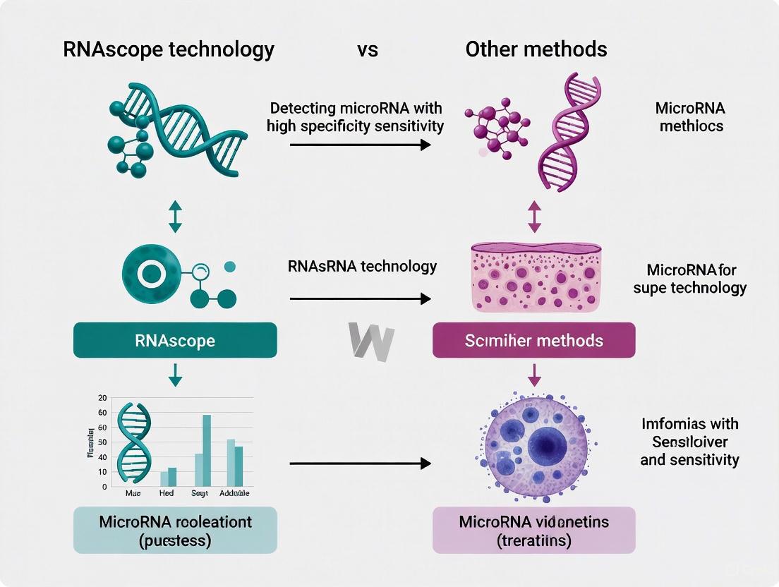
This article provides a comprehensive analysis of the miRNAscope assay, an advanced in situ hybridization technology for detecting microRNAs with single-cell resolution in FFPE and frozen tissues.
miRNAscope for microRNA Detection: A Comprehensive Guide to Spatial miRNA Analysis in Research and Diagnostics
Abstract
This article provides a comprehensive analysis of the miRNAscope assay, an advanced in situ hybridization technology for detecting microRNAs with single-cell resolution in FFPE and frozen tissues. Tailored for researchers and drug development professionals, we explore the foundational principles of the miRNAscope technology, detail its optimized workflow and applications—including use in oligonucleotide therapy development—and provide practical troubleshooting guidance. A critical comparison with other methods like qPCR, RNA-seq, and computational prediction tools highlights its superior spatial context preservation, with validation data confirming high concordance with established techniques. This guide synthesizes methodological insights with comparative evidence to empower robust experimental design and implementation in both research and clinical diagnostic settings.
Understanding miRNAscope Technology: Principles and Probes for Precise microRNA Detection
RNAscope is a novel in situ hybridization (ISH) technology that represents a significant advancement over traditional RNA detection methods. Its core innovation lies in a unique double-Z (ZZ) probe design that enables simultaneous signal amplification and background suppression, allowing for single-molecule RNA visualization within intact cells and tissues while preserving morphological context [1] [2].
This technology addresses critical limitations of conventional ISH techniques, which often suffer from insufficient sensitivity and specificity to reliably detect low-abundance RNA biomarkers, particularly in clinical diagnostic settings where formalin-fixed, paraffin-embedded (FFPE) tissue specimens are routinely used [1]. The ZZ probe design fundamentally enhances the signal-to-noise ratio, making RNAscope particularly valuable for research and diagnostic applications requiring precise spatial localization of gene expression.
The ZZ Probe Technology: Mechanism and Workflow
Core Design Principles and Components
The RNAscope ZZ probe system employs a sophisticated design where signal amplification occurs only when two specific probes bind adjacent to each other on the target RNA, dramatically reducing non-specific background [1] [2].
Key Components of the ZZ Probe System:
- Target Probes: Pairs of "Z" probes designed to hybridize contiguously to the target RNA
- Preamplifier: Binds to the tail sequences of paired ZZ probes
- Amplifier: Attaches to multiple sites on the preamplifier
- Label Probes: Conjugated with fluorescent or chromogenic labels for detection
Table 1: Components of the RNAscope ZZ Probe System
| Component | Structure/Composition | Function |
|---|---|---|
| Z Probe | 18-25 base target region + 14-base tail sequence | Hybridizes to target RNA; forms binding site for preamplifier when paired |
| ZZ Probe Pair | Two Z probes binding contiguously (~50 bases total) | Creates 28-base hybridization site for preamplifier |
| Preamplifier | Branched DNA structure | Provides 20 binding sites for amplifiers |
| Amplifier | Branched DNA structure | Provides 20 binding sites for label probes |
| Label Probe | Fluorophore or enzyme conjugate | Generates detectable signal |
Signal Amplification Mechanism
The signal amplification process occurs through a cascade of highly specific hybridization events [2]:
- Target Binding: Approximately 20 ZZ probe pairs hybridize to a 1-kb region of the target RNA molecule
- Preamplifier Hybridization: Each bound ZZ probe pair creates a binding site for one preamplifier molecule
- Amplifier Binding: Each preamplifier provides 20 binding sites for amplifier molecules
- Label Detection: Each amplifier provides 20 binding sites for label probes, theoretically yielding up to 8000 labels per target RNA molecule
This multi-stage amplification provides exceptional sensitivity while maintaining specificity through the requirement for dual-probe binding.
Diagram 1: ZZ Probe Signal Amplification Cascade
Background Suppression Mechanism
The exceptional specificity of the RNAscope system stems from its requirement for dual probe binding to initiate signal amplification. This design ensures that:
- Single Z probes binding to non-specific sites cannot form the complete 28-base binding site for the preamplifier
- The probability of two independent probes hybridizing contiguously to a non-specific target is extremely low
- Non-specific hybridization events fail to trigger the amplification cascade, effectively suppressing background noise [1] [3]
This mechanism allows RNAscope to achieve single-molecule detection sensitivity while maintaining high specificity, even in partially degraded samples commonly encountered in archival FFPE tissues [2].
Performance Comparison with Alternative Technologies
Comparison with Immunohistochemistry (IHC)
Multiple studies have directly compared RNAscope with immunohistochemistry for biomarker detection, revealing important differences in performance characteristics.
Table 2: RNAscope vs. Immunohistochemistry Performance Comparison
| Performance Metric | RNAscope | Immunohistochemistry | Comparative Results |
|---|---|---|---|
| UPK2 Detection in UC | 68.0% positivity rate | 62.6% positivity rate | P=0.141, R=0.441 [4] |
| Variant Bladder UC | 53.3% positivity rate | 35.6% positivity rate | Trend toward higher detection with RNAscope (P=0.057) [4] |
| Conventional Bladder UC | 72.4% positivity rate | 68.5% positivity rate | No significant difference (P=0.511) [4] |
| Biomarker Type Detected | RNA transcripts | Protein antigens | Different biological molecules |
| Sensitivity Range | 81.8-100% (vs. PCR/ISH) | Variable | RNAscope shows higher concordance with PCR [5] |
| Specificity Range | Up to 100% [5] | Variable | RNAscope demonstrates exceptional specificity |
A systematic review of 27 studies found that RNAscope has a high concordance rate with qPCR, qRT-PCR, and DNA ISH (81.8-100%), while its concordance with IHC was lower (58.7-95.3%), primarily due to the different products each technique measures (RNA vs. protein) [5].
Comparison with Other RNA Detection Methods
RNAscope addresses several limitations of traditional RNA detection methodologies, particularly for spatial analysis within tissue contexts.
Advantages over grind-and-bind methods:
- Preserved Morphological Context: Unlike PCR-based methods that require tissue homogenization, RNAscope maintains tissue architecture [1]
- Cell-Specific Resolution: Enables gene expression analysis at single-cell level within heterogeneous tissues [6]
- No RNA Extraction Required: Avoids RNA loss during extraction and purification steps [5]
Advantages over traditional ISH:
- Enhanced Sensitivity: Capable of detecting low-abundance transcripts that traditional ISH cannot visualize [1]
- Superior Specificity: Double-Z probe design minimizes non-specific binding [2]
- Quantitative Capability: Each punctate dot represents an individual RNA molecule, enabling quantification [7]
Performance in microRNA Detection
While the search results focus primarily on mRNA and lncRNA detection, one study highlights important considerations for microRNA detection technologies. Research comparing miRNA detection platforms (Agilent and Affymetrix microarrays, and Illumina next-generation sequencing) found that detection ability strongly depends on the platform and on miRNA modifications and sequence, with significant biases introduced during library preparation and hybridization [8]. This suggests potential advantages for RNAscope's direct hybridization approach without enzymatic steps that may introduce sequence-dependent biases.
Experimental Applications and Protocols
Standard RNAscope Experimental Workflow
The RNAscope procedure follows a structured workflow that can be performed manually or automated on platforms such as the Ventana DISCOVERY XT/ULTRA or Leica BOND RX systems [7].
Diagram 2: RNAscope Experimental Workflow
Critical Protocol Steps and Optimization
Sample Preparation Guidelines:
- Fixation: 10% neutral buffered formalin for 16-32 hours recommended [7]
- Sectioning: Use Superfrost Plus slides to prevent tissue detachment [7]
- Pretreatment: Optimize protease concentration and incubation time based on tissue type and fixation [1]
Quality Control Measures:
- Positive Controls: Housekeeping genes PPIB (moderate expression), POLR2A (low expression), or UBC (high expression) [7] [5]
- Negative Control: Bacterial dapB gene to assess background noise [1] [7]
- Scoring Criteria: Semi-quantitative scoring based on dots per cell (0-4 scale) [7]
Specialized Applications
Intronic RNA Detection: Recent applications include using intronic RNAscope probes to identify cardiomyocyte nuclei by detecting unspliced pre-mRNA, overcoming limitations of antibody-based nuclear markers [6]. This approach enables specific identification of cardiomyocyte nuclei even during cell division when nuclear envelope breakdown occurs.
Long Non-Coding RNA Detection: RNAscope has been successfully applied to detect various lncRNAs including:
- MALAT1: Highly expressed in colorectal, breast and pancreatic cancer [3]
- UCA1: Moderately expressed oncogenic lncRNA in colorectal cancer [3]
- NRON: Scarcely expressed tumor suppressor lncRNA [3]
This capability is particularly valuable as lncRNAs are difficult to detect via traditional IHC and often require specialized approaches.
Research Reagent Solutions
Table 3: Essential Research Reagents for RNAscope Experiments
| Reagent/Category | Specific Examples | Function and Application Notes |
|---|---|---|
| Control Probes | PPIB, POLR2A, UBC | Positive controls for assay validation and RNA quality assessment [7] |
| Negative Control Probes | dapB | Background assessment; should show no signal in properly fixed tissue [1] [7] |
| Detection Kits | RNAscope 2.5 HD Brown/Red, Multiplex Fluorescent | Chromogenic or fluorescent detection for brightfield or fluorescence microscopy [7] |
| Pretreatment Reagents | RNAscope Pretreatment Kit | Antigen retrieval and protease digestion for RNA unmasking [7] |
| Specialized Slides | Superfrost Plus | Prevent tissue detachment during stringent hybridization conditions [7] |
| Mounting Media | EcoMount, PERTEX, CytoSeal XYL | Preserve signals; specific media required for different detection chemistries [7] |
| Automation Systems | Ventana DISCOVERY XT/ULTRA, Leica BOND RX | Enable standardized, high-throughput implementation [7] |
| Image Analysis Software | HALO, QuPath, Aperio | Quantitative analysis of punctate dots for gene expression quantification [5] |
The ZZ probe design underlying RNAscope technology represents a significant advancement in in situ RNA analysis, offering researchers and clinicians an unparalleled combination of sensitivity, specificity, and morphological preservation. The requirement for dual-probe binding to initiate the amplification cascade provides exceptional background suppression, while the multi-stage amplification enables detection of individual RNA molecules.
When compared to alternative methods, RNAscope demonstrates superior performance to traditional ISH and complementary capabilities to IHC, with higher concordance to PCR-based methods. Its ability to provide spatial context for gene expression analysis in routine FFPE specimens makes it particularly valuable for both research and clinical applications, including biomarker validation, therapeutic development, and diagnostic pathology.
As the field moves toward more precise molecular characterization of diseases, technologies like RNAscope that enable highly specific in situ analysis of RNA biomarkers will play an increasingly important role in understanding disease mechanisms and developing targeted therapies.
The detection and analysis of microRNAs (miRNAs) present significant challenges due to their small size (typically 17-25 nucleotides), low abundance, and sequence similarities within families. While traditional methods like qPCR and microarray have been widely used for miRNA profiling, they require RNA extraction that destroys valuable spatial and morphological context. This guide objectively compares the performance of RNAscope technology, specifically its miRNAscope Assay, against other established methodologies for miRNA detection. We focus on the critical differentiators of single-cell resolution and single-molecule sensitivity that enable researchers to visualize miRNA expression within intact tissue architecture while providing quantitative data. Experimental data and technical protocols are presented to facilitate informed method selection for research and therapeutic development applications.
MicroRNAs are promising candidates for disease biomarkers due to their stability in biological fluids and potential diagnostic signatures [9]. However, their small size (approximately 22 nucleotides in length), association with protein complexes, and low abundance create substantial technical hurdles for accurate detection and quantification [9]. Traditional "grind-and-bind" RNA analysis methods like quantitative real-time PCR (qRT-PCR) destroy the tissue context of gene expression measurements, making it impossible to map observed signals to individual cells [1]. Furthermore, these approaches are prone to interference from unintended cell types and unwanted tissue elements [1].
Spatial context is particularly crucial for understanding miRNA function in heterogeneous tissues like tumors, where cellular communication happens only between neighbor cells but not between distant cells [10]. This technical limitation has impeded the discovery and validation of miRNA biomarkers and their therapeutic applications.
Technology Comparison: Key Performance Metrics
The table below summarizes the key performance characteristics of major miRNA detection methodologies, highlighting the unique advantages of the miRNAscope Assay for spatial context preservation.
Table 1: Performance Comparison of miRNA Detection Methodologies
| Method | Sensitivity | Spatial Context | Sample Requirements | Multiplexing Capability | Key Applications |
|---|---|---|---|---|---|
| miRNAscope Assay | Single-molecule sensitivity [11] | Preserves full spatial and morphological context at single-cell resolution [11] [12] | FFPE tissues, fresh frozen tissues, fixed cells [12] | Single-plex [12] | Cellular subtyping, biomarker validation, therapeutic distribution assessment [13] |
| qPCR-based Methods | High (can detect low abundance targets) [9] | No spatial context (requires tissue disruption) [1] | Extracted RNA from homogenized tissue [9] | Limited by primer specificity | miRNA profiling, expression quantification [9] |
| Microarray | Moderate | No spatial context (requires tissue disruption) | Extracted RNA | High-plex | High-throughput screening, biomarker discovery |
| Traditional RNA ISH | Limited for low-abundance targets [1] | Preserves spatial context | FFPE and frozen tissues | Limited by signal-to-noise ratio | Localization of highly expressed RNAs |
| RNA-seq | Moderate to high | No spatial context (single-cell RNA-seq requires tissue dissociation) [10] | Extracted RNA or single cells | Genome-wide | Discovery profiling, novel miRNA identification |
Quantitative Performance Data
The following table presents experimental data comparing detection efficacy across different platforms, demonstrating the technical performance advantages of specialized miRNA detection approaches.
Table 2: Experimental Detection Efficacy Across Platforms
| Performance Metric | miRNAscope | qPCR (Best Performing Kits) | Traditional RNA ISH | Source |
|---|---|---|---|---|
| Detection Sensitivity | Single RNA molecules (17-50 nt) visualized as distinct dots [11] [12] | Detection of low abundance targets possible with optimized kits [9] | Limited to highly expressed genes [1] | [11] [9] [12] |
| Specificity | High due to double-Z probe design requiring dimer formation [5] [1] | High with optimized primer design | High non-specific binding and background noise [1] | [5] [1] |
| Concordance with qPCR | High (81.8-100% with qPCR/qRT-PCR) [5] | N/A | Not systematically evaluated | [5] |
| Tissue Context Preservation | Excellent (single-cell resolution with morphological preservation) [11] | None (requires RNA extraction) [1] | Moderate (morphology preserved but limited sensitivity) [1] | [11] [1] |
miRNAscope Technology: Core Mechanism and Workflow
Proprietary Probe Design and Signal Amplification
The miRNAscope Assay utilizes a novel in situ hybridization (ISH) technology specifically designed to overcome the challenges of detecting small RNAs [11] [12]. The core innovation lies in its proprietary probe design that enables high sensitivity and specificity through the following mechanism:
- Target-Specific Binding: Probes are designed to hybridize directly to miRNA targets of 17-50 nucleotides in length, including microRNAs, short interfering RNAs (siRNAs), PIWI-interacting RNAs (piRNAs), and antisense oligonucleotides (ASOs) [11] [12]
- Signal Amplification System: Unlike traditional ISH where a single RNA sequence is conjugated with a label, miRNAscope employs a specialized amplification system that generates distinct visual signals for each target RNA molecule [1]
- Background Suppression: The unique design ensures that amplification only occurs when probes specifically bind to their targets, dramatically reducing background noise and enabling single-molecule detection [1]
This technical approach allows for visualization of spatial distributions of single RNA molecules while preserving tissue morphology [11].
Figure 1: miRNAscope Signal Amplification Mechanism. The proprietary probe design enables specific target binding and cascading signal amplification for single-molecule detection.
Experimental Workflow Protocol
The standard miRNAscope assay workflow involves sequential steps that can be performed manually or automated on platforms like the Leica Bond Rx [14] [12]:
Table 3: Research Reagent Solutions for miRNAscope Assay
| Component | Function | Examples/Specifications |
|---|---|---|
| miRNAscope Target Probes | Target-specific detection | Catalog or Made-to-Order S1 Probes (not compatible with RNAscope or BaseScope Assays) [13] |
| Control Probes | Assay validation | Positive Control Probe (Cat. No. 727871-S1), Negative Control Probe (Cat. No. 727881-S1) [13] |
| HD Reagent Kit | Signal amplification and detection | miRNAscope HD Reagent Kit - RED (Cat. No. 324500) includes Pretreatment Kit, Detection kit, and wash buffer [13] |
| Ancillary Reagents | Tissue preparation and processing | ImmEdge Hydrophobic Barrier Pen (Cat. No. 310018), HybEZ Hybridization System [13] |
| Intro Packs | Initial implementation | Intro Packs include control probes, reagent kit, control cell pellets (Human or Mouse), barrier pen, and mounting media [13] |
Sample Preparation Protocol:
- Tissue Processing: Use formalin-fixed paraffin-embedded (FFPE) tissues, fresh frozen tissues, or fixed cells [12]
- Sectioning: Prepare 5-μm thickness tissue sections and mount on slides
- Deparaffinization: For FFPE tissues, deparaffinize in xylene followed by ethanol series [1]
- Pretreatment: Incubate in citrate buffer at boiling temperature (100°C to 103°C) for 15 minutes, then treat with protease (10 μg/mL) at 40°C for 30 minutes [1]
Hybridization and Detection Protocol:
- Target Probe Hybridization: Incubate with target probes in hybridization buffer at 40°C for 3 hours [1]
- Signal Amplification: Sequential hybridization with preamplifier, amplifier, and label probe with appropriate washing steps between each hybridization [1]
- Visualization: Use chromogenic detection followed by counterstaining with hematoxylin [1]
- Imaging and Analysis: Visualize results using bright-field or fluorescent microscope; slides can be digitally scanned for quantification [5]
Figure 2: miRNAscope Assay Workflow. The standardized protocol maintains RNA integrity while enabling specific detection through sequential hybridization and amplification steps.
Comparative Experimental Data and Validation
Sensitivity and Specificity Performance
Independent validation studies demonstrate that RNAscope technology (the foundation platform for miRNAscope) shows high concordance rates with established molecular techniques:
- Compared to qPCR/qRT-PCR: Concordance rates of 81.8-100% [5]
- Compared to DNA ISH: Concordance rates of 81.8-100% [5]
- Compared to IHC: Lower concordance (58.7-95.3%), primarily due to different products measured (RNA vs. protein) [5]
The systematic review assessing RNAscope in clinical diagnostics concluded it is "a highly sensitive and specific method" that could complement gold standard techniques [5]. However, the review noted insufficient data to suggest RNAscope could stand alone in clinical diagnostics, indicating need for further prospective studies [5].
Single-Cell Resolution in Complex Tissues
The miRNAscope Assay enables researchers to detect cellular heterogeneity by providing single-cell resolution within intact tissue architecture. Example applications include:
- Neuronal Subtyping: Specific detection of miR-138-5p in Purkinje cells in cerebellum and hippocampus regions of mouse brain [13]
- Tissue Layer Specificity: Detection of outer nuclear layer-specific microRNA-182 in FFPE mouse eye samples, with negligible detection in inner nuclear and ganglion cell layers [13]
- Cross-Species Validation: Successful detection of miR-214-3p in mouse, rat, and human brain tissue samples with appropriate negative controls [13]
This resolution enables identification of specific cellular subtypes and visualization of gene regulation with morphological context, which is particularly valuable for understanding miRNA function in heterogeneous tissues like tumors [13].
Integration with Complementary Technologies
Research demonstrates that multimodal approaches combining miRNAscope with other technologies provide comprehensive understanding of cellular communication:
- scRNA-seq Integration: While scRNA-seq can predict potential ligand-receptor interactions, it often results in false positives as it cannot discern spatially proximal cells [10]
- Spatial Transcriptomics Complement: Spatial transcriptomics reduces false discovery but suffers from detecting lowly expressed ligand-receptor interactions [10]
- Protein Co-detection: Combining miRNA detection with protein assessment provides comprehensive understanding of regulatory mechanisms [10]
One research framework demonstrated that RNAscope and multiplex protein staining are sensitive methods for defining the location of potential ligand-receptor interactions, with computational pipelines like STRISH (Spatial TRanscriptomic In Situ Hybridization) able to determine the probability that local gene co-expression reflects true cell-cell interaction [10].
Application in Drug Development and Therapeutic Validation
The miRNAscope Assay provides critical capabilities for therapeutic development, particularly for RNA-based therapeutics:
- Therapeutic Delivery Assessment: Evaluate biodistribution and efficacy of small RNA therapies [13]
- ASO Detection: Specifically detect antisense oligonucleotides (ASOs) to monitor tissue penetration and target engagement [11] [12]
- Biomarker Validation: Validate miRNA biomarkers in intact tissues for patient stratification and treatment response monitoring [13]
These applications leverage the technology's ability to provide spatial and morphological context that is destroyed by conventional extraction-based methods, making it particularly valuable for pharmacokinetic and pharmacodynamic studies of RNA-targeted therapeutics.
The miRNAscope Assay represents a significant advancement in miRNA detection technology, addressing critical limitations of conventional methods through its unique combination of single-molecule sensitivity and single-cell resolution within intact tissue architecture. While traditional methods like qPCR and RNA-seq offer valuable quantitative data, they fundamentally lack spatial context essential for understanding miRNA function in heterogeneous tissues and cellular communication.
The technology's proprietary probe design and signal amplification system enable specific detection of small RNAs (17-50 bases) while maintaining morphological preservation, making it particularly valuable for biomarker validation, therapeutic development, and fundamental research into miRNA biology. As the field moves toward more integrated analytical approaches, miRNAscope stands as a powerful validation tool that complements broader discovery platforms like scRNA-seq and spatial transcriptomics.
For researchers and drug development professionals, the selection of miRNA detection methodology should be guided by specific experimental needs: extraction-based methods for high-throughput quantification versus in situ approaches like miRNAscope when spatial context, cellular heterogeneity, and morphological preservation are scientifically critical.
In situ hybridization (ISH) for RNA detection presents a unique set of challenges when the targets are small RNA molecules. Traditional mRNA ISH assays are optimized for targets longer than 300 bases, creating a significant technological gap for visualizing microRNAs (miRNAs), antisense oligonucleotides (ASOs), and small interfering RNAs (siRNAs) which typically range from 17-50 nucleotides. The miRNAscope Assay, built upon the proprietary RNAscope technology platform, was developed specifically to address this gap, enabling highly specific and sensitive detection of small RNAs with single-cell resolution and preservation of spatial and morphological context in intact tissues [15] [12]. This specialized capability provides researchers and drug development professionals with a critical tool for investigating the spatial distribution and function of small non-coding RNAs and oligonucleotide therapeutics, particularly in the rapidly expanding field of RNA therapeutics where over 20 oligonucleotide drug products have received FDA and EMA approval [16].
The miRNAscope Assay employs an advanced in situ hybridization technology and proprietary probe design specifically configured for small RNA targets. Unlike the standard RNAscope Assay which utilizes a design of approximately 20 ZZ probe pairs per target and requires a minimum of 7 ZZ pairs for successful detection, the miRNAscope technology is refined to handle the unique challenges associated with shorter nucleotide sequences [12]. The fundamental innovation lies in its ability to overcome the limited probe binding sites available on small RNAs, which has traditionally made them difficult targets for conventional ISH methods.
The assay is compatible with various sample types including formalin-fixed paraffin-embedded (FFPE) tissues, fresh frozen tissues, fixed frozen tissues, and cultured cells [12]. This flexibility allows researchers to apply the technology across diverse experimental and clinical contexts. The detection system provides chromogenic readouts that can be visualized using standard brightfield microscopy, making it accessible to laboratories without specialized fluorescence imaging equipment [17]. The single-plex design is optimized specifically for the unique challenges of small RNA detection, though it can be combined with other RNAscope assays for co-detection studies [12].
Performance Comparison with Alternative Platforms
The Advanced Cell Diagnostics (ACD) portfolio offers three distinct ISH technologies, each optimized for different target types and applications. Understanding their differences is crucial for selecting the appropriate methodology for specific research questions.
Table 1: Comparative Analysis of RNAscope Technology Platforms
| Feature | RNAscope Assay | BaseScope Assay | miRNAscope Assay |
|---|---|---|---|
| Target Size | mRNA & lncRNA >300 bases [12] | RNA 50-300 bases [12] | Small RNAs 17-50 bases [12] |
| Probe Design | 20 ZZ probes (min. 7 ZZ pairs) [12] | 1-3 ZZ probes [12] | Proprietary design for small RNAs |
| Primary Applications | mRNA & long non-coding RNA detection [12] | Exon junctions, splice variants, point mutations, highly homologous sequences [12] | ASOs, miRNAs, siRNAs, piRNAs, shRNAs, tRNAs [12] |
| Multiplex Capability | Single to 12-plex [12] [17] | Single to duplex [12] | Single-plex [12] |
| Detection Methods | Chromogenic or fluorescent [17] | Chromogenic [12] | Chromogenic [12] |
Key Advantages for Small RNA Detection
The miRNAscope Assay provides several distinct advantages for small RNA visualization:
- Superior Specificity: The proprietary probe design differentiates between highly homologous sequences, which is particularly critical for miRNA families with similar sequences [15] [12].
- Single-Molecule Sensitivity: The technology enables detection of low-abundance small RNAs at single-molecule resolution, essential for studying miRNAs with spatially restricted expression patterns [18] [13].
- Morphological Context Preservation: Unlike extraction-based methods like PCR or microarrays, miRNAscope allows visualization of small RNA distribution within intact tissue architecture, revealing cell-to-cell heterogeneity [15].
Experimental Data and Validation Studies
Detection of Subtype-Specific miRNAs in Cancer Research
A 2025 study investigating small cell lung cancer (SCLC) subtypes successfully employed the miRNAscope Assay to identify subtype-specific miRNA signatures in 46 surgically resected SCLC samples [19]. Researchers visualized miR-375 and miR-9-5p expression patterns across different molecular subtypes defined by ASCL1, NEUROD1, POU2F3, and YAP1 transcription factors [19].
Table 2: Key Findings from SCLC miRNA Study Using miRNAscope
| miRNA Target | Expression Pattern | Clinical Correlation | Technical Performance |
|---|---|---|---|
| miR-375 | High expression in ASCL1, NEUROD1, and ASCL1/NEUROD1 subtypes [19] | Associated with YAP1 downregulation, increased serum pro-gastrin-releasing peptide, and poor prognosis [19] | Successfully detected with clear signal localization in FFPE tissues [19] |
| miR-9-5p | High expression in POU2F3 subtype [19] | Characterized by higher stromal area ratio and specific immune cell infiltration patterns [19] | Reliable detection enabling tumor microenvironment analysis [19] |
| Methodology | miRNAscope ISH on TMA sections [19] | Combined with IMC for immune profiling [19] | Signal quantification via QuPath software [19] |
The study demonstrated how miRNAscope technology enabled the discovery of clinically relevant miRNA biomarkers with prognostic significance, highlighting its utility in cancer pathology research [19].
Analysis of Oligonucleotide Therapeutics
In therapeutic development, the miRNAscope Assay has been validated for detecting antisense oligonucleotides (ASOs) and small interfering RNAs (siRNAs) in tissue samples. ACD's Professional Assay Services team partners with researchers to visualize and quantify the biodistribution and efficacy of oligonucleotide therapies with single-cell and single-molecule precision [18]. This application is critical for optimizing drug delivery methods, evaluating routes of administration, and characterizing spatial distribution and safety profiles across preclinical and clinical samples [18].
The technology enables simultaneous detection of oligotherapeutics along with target mRNA and protein cell markers of interest, providing comprehensive insights into both drug distribution and pharmacological activity [18] [16]. This capability is particularly valuable for assessing on-target and off-target effects during drug development [18].
Workflow in Psychiatric Disorder Research
A 2025 study analyzing long-term stored human brain samples from psychiatric patients demonstrated the robustness of the miRNAscope Assay on FFPE blocks up to 76 years old [20]. Researchers combined Nanostring nCounter miRNA profiling with subsequent miRNAscope ISH analysis to spatially localize specific miRNAs in prefrontal cortex and hippocampus samples from patients with schizophrenia, bipolar disorder, and major depressive disorder [20].
The experimental workflow followed these key steps:
- Sample Preparation: 5μm sections were cut from remounted FFPE blocks and mounted on Superfrost Plus glass slides [20].
- Probe Selection: Based on Nanostring expression data, researchers selected 10 miRNA candidates for prefrontal cortex and 6 for hippocampus, spanning a count intensity range of 82-1730 mean counts [20].
- ISH Procedure: miRNAscope ISH was performed using the miRNAscope HD Reagent Kit according to manufacturer's protocol [20].
- Validation: The study successfully detected 6 miRNAs in prefrontal cortex (miR-9-5p, miR-29b-3p, miR-124-3p, miR-125b-5p, miR-138-5p, and miR-181a-5p) and 4 miRNAs in hippocampus (miR-145-5p, let-7a-5p, miR-124-3p, and miR-7-5p), with miR-124-3p being the most abundantly expressed in both regions [20].
This study highlighted the technology's sensitivity and reliability even with challenging archival samples, opening new possibilities for investigating epigenetic mechanisms in mental disorders [20].
Diagram 1: miRNAscope Assay Workflow. The standardized procedure encompasses tissue pretreatment, probe hybridization, signal amplification, and chromogenic detection [13] [19] [20].
Research Reagent Solutions
Implementing the miRNAscope Assay requires specific reagent systems and controls. The following essential components constitute the core research toolkit:
Table 3: Essential Research Reagents for miRNAscope Assay
| Reagent Component | Function | Examples |
|---|---|---|
| Target Probes | Specifically hybridize to small RNA targets of interest (17-50 bases) [13] [12] | Catalog or Made-to-Order S1 Probes for specific miRNAs, ASOs, siRNAs [13] |
| Control Probes | Verify assay performance; positive control confirms sensitivity, negative control assesses background [13] [19] | Positive Control: RNU6-S1 (Cat. No. 727871-S1) [19]Negative Control: Scramble-S1 (Cat. No. 727881-S1) [19] |
| Detection Kit | Contains all reagents for signal amplification and chromogenic detection [13] | miRNAscope HD Reagent Kit - RED (Cat. No. 324500) includes Pretreatment Kit, Detection Kit, and wash buffer [13] |
| Hybridization System | Provides controlled temperature environment for hybridization steps [13] | HybEZ Hybridization System [13] |
| Intro Packs | Ideal for new users; include core reagents with species-specific controls [13] | miRNAscope Intro Pack HD Reagent Kit RED – Hsa (Cat. No. 324530)miRNAscope Intro Pack HD Reagent Kit RED – Mmu (Cat. No. 324531) [13] |
Application Workflows and Experimental Design
Integrated Workflow for Therapeutic Development
The miRNAscope Assay enables comprehensive evaluation of oligonucleotide therapeutics through a multi-faceted workflow:
Diagram 2: Oligonucleotide Therapeutic Evaluation Pipeline. miRNAscope enables key assessments from biodistribution to safety profiling within tissue context [18].
For drug development applications, the assay can be integrated with complementary techniques:
- Spatial Biodistribution: Localize oligonucleotide payloads to evaluate routes of administration and delivery methods [18].
- Multiplexing Capabilities: Simultaneously detect oligotherapeutics along with target mRNA and other RNA or protein cell markers of interest [18] [16].
- Automated Platforms: Implement on automated systems like Leica Bond Rx and Roche DISCOVERY ULTRA for improved reproducibility and throughput [16] [20].
Technical Considerations for Experimental Success
When implementing the miRNAscope Assay, several technical factors require consideration:
- Sample Quality: The assay performs effectively on various sample types including FFPE, fresh frozen, and fixed frozen tissues, but RNA integrity should be verified for quantitative comparisons [12] [20].
- Probe Validation: Always include appropriate positive and negative controls to validate assay performance and specificity [13] [19].
- Signal Quantification: Utilize image analysis software such as QuPath for objective signal quantification and interpretation [19].
- Multimodal Integration: Combine with techniques like imaging mass cytometry for comprehensive tissue microenvironment analysis [19].
The miRNAscope Assay represents a significant advancement in spatial biology by enabling highly specific and sensitive detection of small RNAs that were previously challenging to visualize in situ. Its specialized design for 17-50 base targets fills a critical technological gap in the RNAscope platform, providing researchers with a robust tool for investigating miRNA biology and oligonucleotide therapeutics. The growing body of validation studies across diverse fields including neurobiology, oncology, and drug development demonstrates its utility for both basic research and translational applications. As the field of RNA therapeutics continues to expand, the miRNAscope Assay stands as an essential methodology for visualizing the spatial distribution and functional activity of small RNA molecules within their native tissue context.
The detection and analysis of microRNA (miRNA) molecules are fundamental to advancing our understanding of gene regulation in cancer development, progression, and treatment response. The robustness of molecular detection techniques across different sample types directly impacts the reliability and reproducibility of research findings, particularly in clinical and translational settings. Formalin-Fixed Paraffin-Embedded (FFPE) tissues, fresh frozen (FF) tissues, and cultured cells each present distinct advantages and challenges for miRNA analysis due to differences in sample processing, RNA preservation, and nucleic acid integrity.
FFPE samples represent the most widely available archival material in pathology departments, offering vast retrospective clinical data but suffering from nucleic acid fragmentation and cross-linking due to formalin fixation. In contrast, fresh frozen tissues provide superior RNA preservation but require specialized storage infrastructure. Cultured cells offer controlled experimental conditions but may not fully recapitulate the complexity of tissue environments. This guide objectively compares the performance of RNAscope and alternative miRNA detection platforms across these sample types, providing researchers with evidence-based data to inform their experimental designs.
Cross-Platform Performance Comparison for miRNA Detection
Technical Performance Across Sample Types
Table 1: Comparison of miRNA Detection Platforms Across Sample Types
| Platform | FFPE Performance | Fresh Frozen Performance | Cultured Cells Performance | Spatial Context | Single-Cell Resolution | Throughput |
|---|---|---|---|---|---|---|
| RNAscope | Good signal, archival time-dependent reduction [21] | High signal intensity, minimal degradation [21] | Compatible with fixed cells [22] | Excellent (in situ) | Yes (single-molecule detection) [5] | Medium |
| NGS | Feasible (SOLiD platform), detects 228-345 miRNAs [23] [24] | Optimal, reference standard | Well-established | No (tissue homogenization) | No (bulk analysis) | High |
| Microarray | Good correlation with frozen (r=0.956-0.976) [25] | Optimal, reference standard | Well-established | No (tissue homogenization) | No (bulk analysis) | High |
| NanoString | Identifies 299-372 miRNAs [24] | Compatible | Compatible | No (tissue homogenization) | No (bulk analysis) | High |
| qRT-PCR | High correlation with frozen (R²>0.95) [26] | Optimal, reference standard | Well-established | No (tissue homogenization) | No (bulk analysis) | Medium |
Quantitative Correlation Between FFPE and Fresh Frozen Samples
Table 2: Correlation Metrics Between FFPE and Fresh Frozen Samples by Platform
| Platform | Correlation Coefficient | Study Findings | Sample Types Tested |
|---|---|---|---|
| Microarray | Pearson's r = 0.956-0.976 [25] | miRNA profiles from FFPE closely recapitulate frozen tissues [25] | Melanoma tissues |
| qRT-PCR | R² > 0.95 [26] | 65.58% of miRNAs showed less than 2-fold change (ΔΔCt between +1 and -1) [26] | Thyroid cell line |
| SOLiD Sequencing | Pearson's r = 0.71-0.95 [23] | Tissue storage time (2-9 years) did not affect number of detected miRNAs [23] | Various cancer tissues |
| TaqMan Arrays | Variable overlap (27-38% of differentially expressed miRNAs) [27] | Marked correlation but overlap affected by normalization methods [27] | Tonsillar tumors |
Detection Sensitivity and Multiplexing Capabilities
Table 3: Sensitivity and Multiplexing Capacity by Platform
| Platform | Detection Sensitivity | Multiplexing Capacity | Sample Requirements | Recommended Applications |
|---|---|---|---|---|
| RNAscope | Single-molecule visualization [5] [28] | Up to 4-plex with fluorescent labels [21] | 4-7μm tissue sections [21] | Spatial localization, heterogeneous samples |
| NGS | Varies with sequencing depth (median 1.2M reads) [24] | Genome-wide (all expressed miRNAs) | 50-1000ng total RNA [24] | Discovery, novel miRNA identification |
| Microarray | Limited compared to other platforms | Hundreds of miRNAs | Varies by platform | Profiling studies |
| NanoString | 299-372 miRNAs detected [24] | Hundreds of miRNAs | Varies by platform | Profiling without amplification |
| qRT-PCR | High (detection of individual miRNAs) | Dozens with TLDA cards | 350-1000ng total RNA [27] | Validation, targeted studies |
RNAscope Technology: Mechanism and Workflow
RNAscope represents a significant advancement in RNA in situ hybridization (ISH) technology, employing a unique probe design strategy that enables single-molecule visualization while preserving tissue morphology [5] [28]. The core innovation lies in its double Z-probe design, which ensures high specificity and signal amplification with minimal background noise.
Diagram Title: RNAscope Double Z-Probe Mechanism
The technology utilizes a pair of "Z" probes that bind to adjacent segments of the target RNA. Each probe consists of three elements: a lower region that hybridizes to the target RNA, a spacer sequence, and a tail that binds to the pre-amplifier sequence. Only when both Z-probes bind correctly to their target sequences can the pre-amplifier attach, initiating a signal amplification cascade [5] [29]. This dual-binding requirement dramatically reduces non-specific background signal compared to traditional ISH methods.
RNAscope Workflow for Different Sample Types
The RNAscope workflow varies slightly depending on sample type, with specific pre-treatment steps optimized for each:
Diagram Title: RNAscope Workflow for Sample Types
For FFPE tissues, the process begins with baking slides followed by antigen retrieval at 98°C-102°C [21]. Fresh frozen tissues and cultured cells require fixation with 4% paraformaldehyde (PFA) before processing [21] [22]. Subsequent steps—permeabilization, probe hybridization, signal amplification, and visualization—are consistent across sample types, though specific optimization may be required for different tissues and targets.
Experimental Protocols for miRNA Detection
SOLiD Sequencing for miRNA Profiling in FFPE and Frozen Tissues
Sample Preparation Protocol [23]:
- RNA Extraction: For FFPE samples, use RecoverAll Total Nucleic Acid Isolation Kit with 5-10 mg samples sliced from paraffin blocks. Include deparaffinization with xylene at 50°C, proteinase K digestion for 24 hours at 50°C, and on-filter DNase digestion. For frozen tissues, use PureLink miRNA Isolation Kit with 5 mg tissue homogenized in Binding Buffer.
- Library Preparation: Process total RNA samples through flashPAGE fractionator for small RNA enrichment. Ligate purified small RNAs with 5' and 3' adapters using RNA ligase. Reverse transcribe ligated products and purify on Novex 10% TBE-Urea gel.
- Amplification: Perform 15-18 cycles of PCR with barcoded primer sets. Purify amplified products (110-130 bp fragments) on Novex 6% TBE gel.
- Sequencing: Conduct emulsion PCR and sequence on Applied Biosystems SOLiD 4 system.
Key Quality Control Metrics: Assess library quality on Agilent Bioanalyzer. For data analysis, remove linker sequences using cutadapt software. Consider miRNAs with ≥5 mappable reads as detected [23].
RNAscope Multiplex Fluorescent Assay Protocol
Sample Preparation [21]:
- FFPE Sections: Cut sections at 4-7μm thickness. Bake slides, perform deparaffinization, and conduct antigen retrieval at 98°C-102°C for 15-30 minutes.
- Fresh Frozen Tissues: Cut sections at optimal thickness (typically 10-20μm). Fix with 4% PFA at room temperature for 20 minutes.
- Cultured Cells: Grow cells on chamber slides, fix with 4% PFA for 20 minutes at room temperature.
RNAscope Procedure [21]:
- Pretreatment: Apply protease to permeabilize tissues for 30 minutes at 40°C.
- Probe Hybridization: Apply target probes and incubate for 2 hours at 40°C.
- Signal Amplification: Perform sequential amplifier hybridization (Amp 1-6) according to manufacturer protocols.
- Signal Development: Apply fluorescent labels (Opal dyes 520, 570, 620, 690) for multiplex detection.
- Counterstaining and Mounting: Use DAPI for nuclear staining and apply ProLong Gold antifade reagent.
Quality Assessment: Include positive control probes (PPIB, POLR2A, UBC) and negative control probe (dapB) to validate assay performance [5].
Cross-Platform Validation Protocol
Sample Processing [24]:
- Use matched FFPE and frozen tissues from the same patients when possible.
- Ensure high tumor content (>50%) through macrodissection if necessary.
- Extract RNA using optimized methods for each sample type.
Platform-Specific Analysis:
- NGS: Process raw data through FastQC, align to reference miRNAs, use threshold of ≥5 mapped reads for detection.
- Microarray: Normalize data using LoESS normalization, apply background subtraction.
- qRT-PCR: Use TaqMan MicroRNA Assays with RNU48 or U6 as endogenous controls, calculate fold change using 2-ΔΔCt method.
Correlation Analysis: Compare log2 ratios of FFPE:frozen signals across platforms. Calculate Pearson correlation coefficients for expression values [25].
The Scientist's Toolkit: Essential Research Reagents
Table 4: Essential Research Reagents for miRNA Detection Across Platforms
| Reagent/Kit | Function | Compatible Sample Types | Key Features |
|---|---|---|---|
| RecoverAll Total Nucleic Acid Isolation Kit | RNA extraction from FFPE | FFPE tissues | Recovers small RNAs, includes DNase treatment |
| PureLink miRNA Isolation Kit | RNA extraction from frozen tissues | Fresh frozen tissues, cells | Optimized for small RNA retention |
| RNAscope Multiplex Fluorescent v2 Kit | In situ RNA detection | FFPE, fresh frozen, fixed cells | Enables multiplex detection up to 4 targets |
| TaqMan MicroRNA Reverse Transcription Kit | cDNA synthesis for qRT-PCR | FFPE, fresh frozen, cells | Stem-loop primers for specific miRNA detection |
| SOLiD Small RNA Expression Kit | Library preparation for sequencing | FFPE, fresh frozen, cells | Ligation-based platform for small RNAs |
| miRCURY LNA MicroRNA Power Labeling Kit | Probe labeling for microarrays | FFPE, fresh frozen, cells | LNA technology for enhanced sensitivity |
| KRAS inhibitor-12 | KRAS inhibitor-12, MF:C19H16Cl2FN5OS, MW:452.3 g/mol | Chemical Reagent | Bench Chemicals |
| Isoboldine | Isoboldine | | Aporphine Alkaloid | Isoboldine is an aporphine alkaloid for research, shown to have anti-inflammatory effects. This product is for Research Use Only and not for human consumption. | Bench Chemicals |
Analysis of Performance Across Sample Types
FFPE Sample Compatibility
FFPE tissues demonstrate good compatibility with multiple miRNA detection platforms, though with some limitations. RNAscope shows reduced but detectable signals in FFPE compared to fresh frozen tissues, with degradation being archival time-dependent [21]. High-expressor genes like UBC and PPIB show more pronounced degradation in FFPE than low-to-moderate expressors like POLR2A and HPRT1 [21]. Nevertheless, multiple studies confirm that miRNA profiling from FFPE tissues is feasible with appropriate quality controls.
Sequencing platforms like SOLiD demonstrate that the number of detected miRNAs in FFPE samples (228-345 with threshold ≥10 reads) is comparable to frozen samples, with tissue storage times of 2-9 years not significantly affecting detection rates [23]. Correlation coefficients between matched FFPE and frozen tissues range from 0.71-0.95 across studies [23] [25].
Fresh Frozen Sample Performance
Fresh frozen tissues consistently deliver optimal performance across all detection platforms, serving as the reference standard for miRNA studies. RNAscope signals in fresh frozen tissues are significantly higher than in FFPE and show minimal degradation over archival time [21]. Sequencing platforms detect the highest number of miRNAs in fresh frozen samples, with read length distributions peaking at 19-22 nucleotides, consistent with mature miRNA sizes [24].
The superior RNA preservation in fresh frozen tissues makes them ideal for discovery-phase studies where sensitivity is paramount. However, the practical challenges of procurement, storage, and handling often limit their use in large-scale retrospective studies.
Cultured Cell Applications
Cultured cells provide a controlled system for miRNA detection, with performance characteristics similar to fresh frozen tissues. RNAscope compatibility with fixed cells enables spatial resolution of miRNA expression in cultured systems [22]. The homogeneity of cell populations eliminates concerns about tissue heterogeneity that can complicate tissue-based studies.
For platforms requiring RNA extraction, cultured cells typically yield high-quality RNA with minimal degradation, making them suitable for all detection methods. The ability to manipulate cellular conditions makes cultured cells particularly valuable for functional studies of miRNA regulation.
The optimal choice of miRNA detection platform depends on sample type, research objectives, and required data output. RNAscope offers unique advantages for applications requiring spatial context and single-cell resolution, with good performance across FFPE, fresh frozen, and cultured cell samples. While it shows some reduction in signal intensity in FFPE samples compared to frozen tissues, it remains a robust option for in situ detection.
Sequencing platforms provide the most comprehensive profiling capability but lose spatial information. Microarray and NanoString technologies offer intermediate throughput with good correlation between FFPE and frozen samples. qRT-PCR remains the gold standard for sensitive, targeted miRNA quantification.
For studies utilizing valuable archival FFPE materials, RNAscope and targeted sequencing approaches provide complementary information—the former preserving morphological context and the latter offering comprehensive profiling. Researchers should implement appropriate quality controls, including housekeeping genes for RNAscope and RNA integrity assessments for extraction-based methods, to ensure reliable results across all sample types.
Implementing miRNAscope: A Step-by-Step Workflow from Sample Prep to Analysis
In the context of microRNA (miRNA) detection research, particularly with advanced in situ hybridization (ISH) technologies like RNAscope, sample preparation is not merely a preliminary step but a fundamental determinant of experimental success. The growing importance of miRNAs as diagnostic biomarkers and therapeutic targets, underscored by the 2023 Nobel Prize in Physiology or Medicine, has intensified the need for precise spatial localization in tissue contexts [30]. This guide objectively compares 10% Neutral Buffered Formalin (NBF) against alternative fixatives, providing experimental data and detailed protocols to optimize preparation workflows for reliable miRNA detection.
Fixative Comparison: 10% NBF vs. Alternative Solutions
The choice of fixative creates a critical trade-off between morphological preservation and molecular integrity, especially impactful for short miRNA sequences.
Performance Evaluation of Common Fixatives
Table 1: Comparative Analysis of Fixative Performance for miRNA Studies
| Fixative Type | Morphological Preservation | Nucleic Acid Integrity | Antigenicity for IHC | Best Use Cases |
|---|---|---|---|---|
| 10% NBF | Superior (Score: 2.7/3) [31] | Moderate (DNA degradation over time) [32] | Moderate (Requires AR) [31] | Diagnostic histopathology; morphological studies |
| Alcohol-Based | Good (Score: 2.3/3) [31] | Good | Superior (Stronger IHC staining) [31] | miRNA/IHC co-detection; protein-focused studies |
| Silver Nanoparticles (AgNPs) | Requires Optimization [32] | Superior (Stable DNA/RNA concentration) [32] | Not Reported | Molecular assays requiring high-quality nucleic acids |
Quantitative Experimental Data
Table 2: Experimental Data on Biomolecule Preservation Across Fixatives
| Biomolecule & Fixative | Tissue | Initial Concentration | Concentration at 72h | Statistical Significance |
|---|---|---|---|---|
| DNA: 10% NBF | Liver | 357.00 ± 2.65 ng/μL | 157.67 ± 2.52 ng/μL | p < 0.0001 [32] |
| DNA: AgNPs | Liver | 357.00 ± 2.65 ng/μL | 309.33 ± 1.53 ng/μL | p < 0.0001 [32] |
| RNA: 10% NBF | Heart | ~1991 ng/μL (Baseline) | 864 ± 2.01 ng/μL | p < 0.001 [32] |
| RNA: AgNPs | Heart | ~1991 ng/μL (Baseline) | Moderate decline | Better than NBF [32] |
| IHC: 10% NBF | General | Reference | 63.3% (3+ Staining for Cytokeratin) | Baseline [31] |
| IHC: Alcohol-Based | General | Reference | 86.6% (3+ Staining for Cytokeratin) | p < 0.05 [31] |
Detailed Methodologies for Key Experiments
Protocol: Comparative Fixation Study (from Table 2 data)
Materials:
- Tissue Samples: 60 human samples (30 liver, 30 lymph node) [31]
- Fixatives: 10% NBF vs. alcohol-based fixative (70% ethanol, 5% acetic acid, 25% methanol) [31]
- Evaluation Methods: H&E staining, IHC for cytokeratin/CD3, nucleic acid quantification [31]
Procedure:
- Fixation: Divide each specimen into two equal portions. Fix one half in 10% NBF and the other in alcohol-based fixative for 24 hours at room temperature [31].
- Processing: Process all tissues through a standard paraffin-embedding protocol [31].
- Sectioning: Cut sections of 4–5 µm thickness using a rotary microtome and mount on glass slides [31].
- Staining & Evaluation:
- Perform H&E staining for morphological evaluation (nuclear detail, cytoplasmic clarity, tissue shrinkage) on a 0-3 scale [31].
- Perform IHC staining for cytokeratin and CD3, grading intensity as 0 (no staining), 1+ (weak), 2+ (moderate), or 3+ (strong) [31].
- Quantify DNA/RNA concentration and purity using spectrophotometry (A260/A280) [32].
- Statistical Analysis: Analyze differences using paired t-test and chi-square test with SPSS software, considering p < 0.05 as significant [31].
Protocol: Standardized Fixation and Embedding with 10% NBF
Materials:
- Fixative: 10% Neutral Buffered Formalin [33]
- Tissues: Freshly excised tissue samples [33]
- Embedding Supplies: Ethanol, xylene, paraffin wax, tissue embedding molds and cassettes [33]
Procedure:
- Fixation:
- Embedding:
- Dehydrate tissue through a graded ethanol series (50%, 75%, 85%, 95%, 100%) [33].
- Clear tissue in xylene or a less hazardous alternative [33].
- Infiltrate with paraffin wax at 50–60°C in a vacuum oven or automated system [33].
- Embed tissue in a mold filled with fresh paraffin, orient as desired, and anchor with a cassette. Cool in refrigerator to harden [33].
Sectioning Guidelines for Optimal miRNA Detection
Microtomy Techniques and Specifications
Table 3: Microtome Selection and Sectioning Parameters for miRNA ISH
| Microtome Type | Typical Section Thickness | Embedding Medium | Key Applications in miRNA Research |
|---|---|---|---|
| Rotary Microtome | 3–10 μm (routine) [33]; 1–60 μm (range) [34] | Paraffin | Standard workflow for FFPE samples for RNAscope [34] |
| Cryostat Microtome | 4–10 μm [34] | OCT (frozen tissue) | Preserving labile miRNAs; rapid diagnosis [34] |
| Ultramicrotome | 30–100 nm [34] | Resin | Ultrastructural miRNA localization via EM [34] |
Protocol: Sectioning Paraffin-Embedded Tissues
Materials:
- Equipment: Rotary microtome and blade [33]
- Supplies: Glass microscope slides, water bath [33]
- Samples: Paraffin-embedded tissue blocks [33]
Procedure:
- Preparation: Chill paraffin blocks on ice to support hard tissue elements. Heat a water bath to 40–45°C [33].
- Microtome Setup: Install blade and set clearance angle per manufacturer's instructions [33].
- Block Trimming: Insert block into microtome, orient, and trim at 10–30 μm thickness to expose the tissue surface [33].
- Sectioning: Cut serial sections at 3–10 μm thickness, which will form ribbons [33].
- Mounting: Float ribbons on water bath to flatten, separate with tweezers, and pick up onto microscope slides [33].
- Drying: Dry slides at 37°C overnight to preserve heat-sensitive antigens [33].
The Scientist's Toolkit: Essential Reagents and Materials
Table 4: Key Research Reagent Solutions for miRNA Detection Workflows
| Item | Function/Description | Example Use Case |
|---|---|---|
| 10% NBF | Gold standard fixative; cross-links proteins for excellent morphology preservation [31] | General histology and primary fixation for biobanking [31] |
| RNAscope Assay | Highly sensitive and specific RNA ISH platform for miRNA detection [35] | Single-molecule visualization of miRNA in FFPE tissues with spatial context [36] |
| miRNeasy Kits (Qiagen) | Spin-column based RNA isolation from serum/plasma and tissues [37] | Purifying miRNA from liquid biopsies for downstream analysis [37] |
| TaqMan MicroRNA Assays | Stem-loop RT-qPCR for highly sensitive and specific miRNA quantification [37] | Validating miRNA expression levels from liquid biopsy samples [37] |
| Cryostat | Instrument for sectioning frozen tissues at low temperatures (-20°C) [34] | Preparing sections for miRNA detection without fixation-induced alterations [34] |
| 6,6'-Di-O-sinapoylsucrose | 6,6'-Di-O-sinapoylsucrose, MF:C34H42O19, MW:754.7 g/mol | Chemical Reagent |
| 3,4-Didehydroglabridin | 3,4-Didehydroglabridin, MF:C20H18O4, MW:322.4 g/mol | Chemical Reagent |
Workflow Visualization: From Sample to Signal
The following diagram illustrates the complete experimental workflow for miRNA detection using RNAscope, highlighting critical preparation steps.
Optimal sample preparation for miRNA detection requires careful consideration of the balance between morphological preservation and molecular integrity. While 10% NBF remains the gold standard for structural detail, alcohol-based fixatives and emerging alternatives like silver nanoparticles offer superior biomolecule preservation for specific applications. By standardizing fixation and sectioning protocols according to the experimental objectives outlined in this guide, researchers can ensure reliable, reproducible results in miRNA spatial localization studies using RNAscope and other advanced detection platforms.
The accurate detection and quantification of microRNAs (miRNAs) is crucial for advancing research in disease diagnostics and drug development. These short RNA molecules are promising biomarkers, but their small size and high sequence similarity present significant technical challenges. This guide objectively compares the performance of the RNAscope miRNAscope Assay against other established miRNA detection methods, such as qPCR and next-generation sequencing, by examining their standard workflows and supporting experimental data.
miRNA Detection Technology Platforms at a Glance
The following table summarizes the core characteristics of major miRNA detection technologies, highlighting key performance differentiators.
| Technology | Principle | Spatial Context | Best Suited For | Key Limitations |
|---|---|---|---|---|
| RNAscope (miRNAscope) | In situ hybridization (ISH) with proprietary signal amplification [5] [2] | Preserved (Single-cell resolution) [5] [12] | Detecting miRNA in intact cells/tissues; spatial distribution analysis [5] [12] | Not for isolated RNA; lower multiplexing for miRNAs [12] |
| qPCR (TaqMan, miScript) | Reverse transcription followed by quantitative PCR [37] [38] | Lost (Requires RNA extraction) [37] | High-throughput profiling of purified RNA; high sensitivity [37] [38] | Requires RNA extraction; potential for amplification bias [8] |
| Next-Generation Sequencing (miRNA-Seq) | Sequencing of cDNA libraries [8] [38] | Lost (Requires RNA extraction) [8] | Discovery of novel miRNAs; comprehensive, unbiased profiling [38] | High cost; complex data analysis; ligation bias [8] [38] |
| Microarrays | Hybridization of labeled RNA to fixed probes [8] | Lost (Requires RNA extraction) [8] | Profiling of many samples against a known set of miRNAs [8] | Lower sensitivity and dynamic range than NGS/qPCR; hybridization bias [8] |
Performance Comparison: Sensitivity, Reproducibility, and Detection
Independent studies provide quantitative data on how different platforms perform in real-world scenarios, particularly with challenging biofluid samples.
Table 2: Cross-Platform Performance Metrics from Experimental Studies
Table based on data from systematic evaluations of miRNA profiling platforms [38].
| Platform | Reproducibility (Concordance Correlation Coefficient in Serum) | Number of miRNAs Detected in Human Serum (Above LLOQ) | Key Performance Notes |
|---|---|---|---|
| miRNA-Seq (Illumina TruSeq) | 0.99 (Almost perfect) [38] | 372 [38] | Detection increases with sequencing depth; sensitive but can be affected by ligation biases [8] [38] |
| MiRXES qPCR | 0.99 (Almost perfect) [38] | 389 (Highest among qPCR platforms) [38] | High sensitivity and reproducibility in biofluids [38] |
| ABI TaqMan qPCR | >0.9 (Moderate) [38] | Not specified (Moderate detection) | Widespread use; performance can be variable between platforms [38] |
| Qiagen miScript qPCR | >0.9 (Moderate) [38] | Not specified (High variability) | High number of miRNA targets, but high measurement variability in serum [38] |
| Exiqon LNA qPCR | >0.9 (Moderate) [38] | Not specified (Moderate detection) | Moderate performance in reproducibility and detection [38] |
| NanoString | 0.82 (Poor in serum) [38] | 84 [38] | Performance is sample-dependent; excellent reproducibility in tissue with high RNA content [38] |
Platform performance is significantly influenced by inherent technical biases. A systematic comparison of microarray and NGS platforms found that the ability to detect miRNAs depends strongly on the platform and on miRNA modifications and sequence [8]. For example, O-methyl-modified miRNAs are consistently under-represented across platforms due to reduced efficiency in enzymatic reactions during library preparation or labeling [8]. Furthermore, sequence-dependent biases can cause the same miRNA to show up to a 500-fold difference in intensity on one platform versus a 10-fold difference on another [8].
The miRNAscope Assay Workflow: A Detailed Protocol
The miRNAscope Assay is a specialized version of RNAscope technology optimized for detecting small RNAs (17-50 bases), including miRNAs, with single-cell resolution in formalin-fixed paraffin-embedded (FFPE) or frozen tissues [12]. Its workflow is foundational to its performance.
Critical Phases of the miRNAscope Workflow
Sample Preparation and Permeabilization
- Tissue Fixation: Adherence to recommended fixation protocols is critical. Tissues should be fixed in fresh 10% Neutral Buffered Formalin (NBF) for 16-32 hours [7].
- Slide Treatment: Tissue sections must be mounted on Superfrost Plus slides to prevent detachment. An ImmEdge Hydrophobic Barrier Pen is used to create a well around the section [7].
- Antigen Retrieval and Protease Digestion: Slides are heated in a target retrieval solution and then treated with a proprietary protease. Maintaining a temperature of 40°C during protease digestion is essential for proper tissue permeabilization without destroying the target miRNA [7].
Hybridization and Signal Amplification
- Probe Hybridization: The miRNAscope assay uses a unique "ZZ" probe design specifically for small RNAs [12]. These probes are hybridized to the target for 2 hours at 40°C [7]. This step must be performed using a HybEZ Oven to maintain optimum humidity and temperature [7].
- Signal Amplification: This proprietary multi-step process is key to the technology's high sensitivity and specificity [2].
- Step 1: Pre-amplifiers bind to the paired ZZ probes.
- Step 2: Multiple amplifiers bind to each pre-amplifier.
- Step 3: Many label probes, conjugated with an enzyme (e.g., HRP), bind to each amplifier.
- This cascade can result in up to 8,000-fold signal amplification for each target miRNA molecule, allowing for single-molecule detection [5].
Chromogenic Detection and Quantification
- Detection: A chromogenic substrate (e.g., DAB) is added. The enzyme on the label probe catalyzes a reaction, producing a brown/red precipitate at the site of the target miRNA. Each precipitate appears as a punctate dot under a microscope, with each dot representing a single miRNA molecule [2].
- Quantification: Results are scored semi-quantitatively by counting the number of dots per cell [7]:
- Score 0: No staining or <1 dot per 10 cells
- Score 1: 1-3 dots/cell
- Score 2: 4-9 dots/cell
- Score 3: 10-15 dots/cell
- Score 4: >15 dots/cell
- Quantification can be performed manually or using image analysis software like HALO or QuPath [5].
The Scientist's Toolkit: Essential Reagents and Materials
A successful miRNAscope assay requires specific reagents and equipment. The following table details the essential components.
| Item | Function | Technical Notes |
|---|---|---|
| miRNAscope Probe | Target-specific detection [12] | Proprietary probes designed for small RNAs (17-50 bases); cannot be interchanged with other RNAscope kits [12]. |
| miRNAscope Reagent Kit | Contains buffers for hybridization, amplification, and detection [12] | Includes the Universal Pretreatment Kit. Components are not interchangeable with other RNAscope kits [12]. |
| Positive Control Probe (e.g., PPIB, UBC) | Validates assay success and tissue RNA integrity [7] | Housekeeping gene probes confirm RNA is detectable. PPIB is for moderate expression (10-30 copies/cell) [7]. |
| Negative Control Probe (dapB) | Assesses background noise and non-specific binding [7] | Bacterial gene probe that should not generate signal in properly processed animal tissue [7]. |
| HybEZ Hybridization System | Maintains optimum humidity and temperature during hybridization [7] | Essential for consistent and reliable results; cannot be substituted with a standard hybridization oven [7]. |
| Superfrost Plus Slides | Tissue adhesion [7] | Required to prevent tissue detachment during the rigorous assay procedure [7]. |
| ImmEdge Hydrophobic Barrier Pen | Creates a barrier to contain reagents on the slide [7] | The only barrier pen certified to maintain a hydrophobic barrier throughout the entire RNAscope procedure [7]. |
| Specific Mounting Media (e.g., EcoMount) | Preserves and coverslips the stained slide [7] | Must be used as specified; for example, the Red assay requires EcoMount or PERTEX [7]. |
| S-30-Hydroxygambogic acid | S-30-Hydroxygambogic Acid|HPV E6 Inhibitor | S-30-Hydroxygambogic acid is a novel E6 inhibitor for HPV+ cancer research. This product is for research use only and not for human or veterinary use. |
| 6-Hydroxy-TSU-68 | 6-Hydroxy-TSU-68, MF:C18H18N2O4, MW:326.3 g/mol | Chemical Reagent |
The choice of a miRNA detection method involves critical trade-offs. qPCR and miRNA-Seq offer high sensitivity for profiling purified RNA but lose all spatial information and can be affected by sequence-dependent biases [8] [38]. In contrast, the RNAscope miRNAscope Assay provides the unique advantage of spatial genomic context, allowing researchers to see which cells express a miRNA biomarker within a complex tissue architecture, albeit with lower multiplexing capability and requiring intact tissue samples [5] [12]. The standard workflow of hybridization, amplification, and chromogenic detection in miRNAscope is engineered for maximum specificity and sensitivity in situ, making it an indispensable tool for validating the spatial distribution of miRNA biomarkers discovered by other high-throughput techniques.
The selection of an automated staining platform is a critical decision in molecular pathology, directly impacting the reproducibility, throughput, and analytical depth of research, particularly in specialized applications like microRNA (miRNA) detection via RNAscope technology. Within this context, the Leica BOND RX and Roche DISCOVERY ULTRA have emerged as leading platforms, each with distinct technological approaches and performance characteristics. While both systems enable highly multiplexed analysis, their underlying architectures for assay automation, reagent management, and protocol customization differ significantly, leading to varied applications in research workflows. This guide provides an objective comparison of these two systems, focusing on experimental data and protocol implementation to inform researchers and drug development professionals.
The Leica BOND RX is a fully automated research stainer designed for flexible protocol development in IHC, ISH, and emerging molecular applications. It emphasizes open reagents, customizable protocol segments, and a proprietary Covertile technology to preserve tissue morphology [39]. In contrast, the Roche DISCOVERY ULTRA is a research platform that leverages tyramine-based signal amplification to enable brightfield and fluorescent multiplexing, facilitating the combination of multiple biomarkers on a single section [40].
Table 1: Core System Specifications and Capabilities
| Feature | Leica BOND RX | Roche DISCOVERY ULTRA |
|---|---|---|
| System Type | Fully automated research stainer | Automated IHC/ISH research platform |
| Key Technology | Covertile system for gentle reagent dispensing [39] | Tyramine chemistry for signal amplification [40] |
| Multiplexing Capacity | Up to 6 markers on a single slide (Software 7.0) [39] | 5+ markers demonstrated [40] |
| Supported Assays | IHC, ISH, FISH, CTC, multiplexing [39] | IHC, ISH, brightfield and fluorescent multiplexing [40] |
| Reagent Openness | Open system; supports user reagents and open containers [39] | Information not specified in search results |
| Throughput | Up to 30 slides in a single run for RNAscope assays [39] | Information not specified in search results |
Performance Comparison and Experimental Data
Objective performance metrics are crucial for platform selection. While direct head-to-head comparisons for RNAscope are limited in the provided search results, available data highlights differences in staining speed and workflow efficiency.
Staining Speed and Workflow Efficiency
A study examining panel turnaround time for IHC panels found that a different Leica system (BOND-III) completed cases up to 40% faster than the Roche BenchMark ULTRA, saving up to 100 minutes on a panel of slides [41]. Although this data point is for a different model, it suggests that Leica's staining architecture is designed for rapid processing, which may translate to the BOND RX platform for certain assays. For RNAscope specifically, the BOND RX can complete up to 30 slides in a single 11-hour run for a simplex assay and a 14-hour run for a duplex assay [39].
Multiplexing Capabilities and Reproducibility
The BOND RX's Software 7.0 supports enhanced chromogenic and fluorescent multiplexing, allowing up to 6 individual markers to be visualized on a single slide [39]. This high-plex capability is vital for studying complex spatial relationships within the tumor microenvironment. Furthermore, 91% of customers surveyed found the stain quality and reproducibility of the BOND RX to be better than the competition, attributed to its unique Covertile system that preserves tissue morphology and ensures consistent reagent application [39].
The DISCOVERY ULTRA utilizes tyramine chemistry and a broad spectrum of chromogenic dyes to enable multiplexed analysis on conventional brightfield microscopes [40]. A key consideration noted in the literature is that visualizing more than 2-3 colors for co-localized markers in the same cellular compartment can be challenging for the human eye, potentially necessitating a dedicated analytic pipeline for this platform [40].
Table 2: Performance Data in Research Applications
| Parameter | Leica BOND RX | Roche DISCOVERY ULTRA |
|---|---|---|
| Staining Time (Example Assay) | RNAscope single-plex: ~11 hours for 30 slides [39] | Information not specified in search results |
| Reproducibility | 91% of customers report better stain quality/reproducibility vs. competition [39] | Information not specified in search results |
| Multiplex Visualization | Digital imaging and analysis for up to 6-plex [39] | Brightfield assessment; software may be needed for >3 colors [40] |
| Hands-on Time | Minimized via full automation and bulk reagent level viewing [39] | Information not specified in search results |
Experimental Protocols for miRNA Detection
The following section details a validated protocol for miRNA in situ hybridization on long-term stored human FFPE brain samples, as demonstrated on the Leica BOND RX system [20].
Sample Preparation and miRNA Selection
- Tissue Sectioning: Original FFPE samples were remounted onto modern plastic cassettes. Sections were cut at 5 µm thickness and mounted on SuperFrost Plus glass slides [20].
- miRNA Candidate Selection: miRNA expression profiling was first performed using the NanoString nCounter platform (Human v3b miRNA panel) on bulk tissue extracts. This technology was selected for its reported superiority with FFPE samples [20].
- Probe Selection: Based on NanoString data, candidate miRNAs were selected for ISH. In the referenced study, this included miRNAs such as miR-124-3p, miR-9-5p, and miR-29b-3p for prefrontal cortex, and let-7a-5p and miR-7-5p for hippocampus. miR-124-3p, a pan-neuronal miRNA, was the most abundantly expressed [20].
Automated miRNAscope Protocol on BOND RX
The miRNAscope assay was performed using the respective probes on the Leica BOND RX. The workflow leverages the platform's full automation for baking, dewaxing, antigen retrieval, probe application, and staining [39] [20].
Figure 1: Automated miRNAscope workflow on the Leica BOND RX system, adapted from protocols for long-term stored FFPE samples [20].
Key Research Reagent Solutions
Table 3: Essential Materials for Automated miRNAscope
| Reagent / Material | Function | Example/Note |
|---|---|---|
| miRNAscope Probe | Target-specific detection | e.g., Probes for miR-124-3p, miR-9-5p; based on NanoString screening [20]. |
| FFPE Tissue Sections | Sample substrate | 5 µm sections on SuperFrost Plus slides [20]. |
| NanoString nCounter Panel | For miRNA expression profiling | Human v3b miRNA panel used for candidate selection prior to ISH [20]. |
| BOND RX Titration Containers | Holds user reagents on the platform | Enables automation of high-value reagents with minimal volumes [39]. |
| Protease or Retrieval Reagents | Antigen retrieval | Automated on the BOND RX as part of the customizable protocol [39]. |
Integration in the miRNA Research Workflow
The choice between the BOND RX and DISCOVERY ULTRA should be framed within a broader research strategy for miRNA detection. The referenced study successfully combined NanoString nCounter miRNA profiling with subsequent, spatially resolved validation via automated miRNAscope on the BOND RX [20]. This workflow effectively links bulk tissue miRNA quantification with precise cellular localization.
Figure 2: A logical workflow for miRNA research, from discovery to spatial validation. Platform selection becomes critical at the validation stage [20] [40].
The Leica BOND RX and Roche DISCOVERY ULTRA are both capable automated platforms for advanced research applications. The available data suggests that the BOND RX offers distinct advantages in flexibility, protocol customization, and highly multiplexed fluorescent workflow integration, supported by a ecosystem of specialized partners for applications like RNAscope [39] [42]. Its open reagent system and Covertile technology are designed for reproducible stain quality in research settings.
The DISCOVERY ULTRA presents a strong solution for brightfield-based multiplexing, leveraging tyramine chemistry to enable the detection of multiple biomarkers on a single section viewed with a conventional microscope [40]. The optimal choice is ultimately dictated by specific research needs. For complex, high-plex miRNA spatial profiling requiring full protocol control and fluorescence readouts, the BOND RX is a compelling option. For laboratories prioritizing brightfield multiplexing within an established workflow, the DISCOVERY ULTRA is a viable alternative.
A critical step in utilizing advanced in situ hybridization (ISH) techniques for research and diagnostic purposes is the accurate interpretation of results. For methods like the RNAscope and miRNAscope assays, which generate punctate signals representing individual RNA molecules, two primary quantification approaches are employed: semi-quantitative dot counting and digital image analysis. This guide objectively compares these scoring methodologies, their performance, and their application in the context of miRNA detection compared to other quantitative techniques.
The Foundation: Principles of Punctate Signal Detection
The RNAscope and miRNAscope assays are based on a proprietary technology that enables the detection of RNA molecules within intact cells and tissues with single-molecule sensitivity and high specificity [12] [5]. The core of this technology is a cleverly designed probe system and a sequential signal amplification process.
Probe Design and Signal Amplification: Instead of a single long probe, the assays use pairs of "Z" probes that are designed to bind adjacent to each other on the target RNA sequence [5]. Each "Z" probe contains a sequence that binds to the target RNA, a linker, and a tail that serves as a binding site for pre-amplifier molecules. This dual "Z" probe design is the key to specificity; only when both bind correctly can the subsequent amplification cascade proceed. Each bound pre-amplifier then recruits multiple amplifiers, which in turn bind many enzyme-linked labels (e.g., horseradish peroxidase or alkaline phosphatase) [43] [5]. This multi-step amplification can theoretically yield an 8,000-fold signal increase, allowing a single RNA molecule to be visualized as a distinct, punctate dot [5].
The following diagram illustrates this sequential signal amplification process:
The miRNAscope assay is a specialized version of this technology, optimized for the challenging detection of small RNAs, including microRNAs (miRNAs), which are typically only 17-50 nucleotides in length [12].
Scoring Methodologies: A Comparative Analysis
Once the ISH assay is complete, the punctate signals must be quantified. The choice between manual dot counting and digital image analysis depends on the required throughput, objectivity, and depth of data analysis.
Semi-Quantitative Visual Dot Counting
Visual scoring is a manual method performed by a trained scientist or pathologist directly observing the stained sample under a microscope.
Protocol: The observer assesses the entire tissue section or a defined region of interest (ROI). The scoring is based on counting the number of dots per cell. The manufacturer suggests a semi-quantitative scoring system from 0 to 4, based on the predominant staining pattern [44] [5]:
- Score 0: 0 dots/cell (No staining)
- Score 1: 1-3 dots/cell (Rare)
- Score 2: 4-10 dots/cell (Moderate)
- Score 3: >10 dots/cell (Abundant)
- Score 4: >15 dots/cell with cluster formation (Very abundant) The percentage of positive cells (cells with ≥1 dot/cell) is also often estimated and binned into categories (e.g., 0%, 1-25%, 26-50%, etc.) [44].
Data Output: This method provides a rapid, relative assessment of gene expression levels and is sufficient for many applications where understanding relative signal intensity is the goal [44]. It is highly adaptable for assessing staining heterogeneity across a tissue section.
Quantitative Digital Image Analysis
Digital image analysis uses specialized software to quantify expression on a cell-by-cell basis across entire tissue sections.
Protocol: The stained slide is first digitally scanned at high magnification (typically 20x or 40x) [45]. The resulting digital image is then analyzed using software platforms such as HALO (Indica Labs) or QuPath. The workflow generally involves [44]:
- Tissue and Cell Segmentation: The software algorithm identifies and segments the tissue area and individual cells.
- Region of Interest (ROI) Classification: The user can train the software to classify different tissue compartments (e.g., tumor, stroma, necrosis) for separate analysis.
- Dot Detection and Counting: The software identifies and counts each punctate dot within each segmented cell or ROI.
- Data Exportation: The software generates quantitative data, including the total dot count, dots per cell, and the percentage of positive cells, often with the ability to overlay heat maps for spatial visualization.
Data Output: This method provides highly quantitative, objective, and reproducible data on a cell-by-cell basis, allowing for deep mining of complex expression patterns within heterogeneous tissues [44]. It is the preferred method for high-throughput studies and when precise quantification is critical.
The following workflow summarizes the key steps for both scoring approaches:
Performance Comparison of Scoring Methods
Table 1: Comparison of Semi-Quantitative Dot Counting and Digital Image Analysis
| Feature | Semi-Quantitative Visual Scoring | Quantitative Digital Image Analysis |
|---|---|---|
| Throughput | Lower, time-consuming for large studies | High, automated analysis of whole slides |
| Objectivity | Subject to observer bias and experience | Highly objective and reproducible |
| Data Granularity | Relative score (0-4), binned percentages | Absolute counts, cell-by-cell data, spatial mapping |
| Cost | Lower initial cost (microscope) | Higher cost (scanner, software licenses) |
| Expertise Required | Trained pathologist/scientist for scoring | Training in digital pathology software |
| Ideal Use Case | Rapid assessment, low-plex studies, labs without digital infrastructure | High-throughput studies, complex multiplex assays, requiring precise quantification and spatial data |
RNAscope/miRNAscope vs. Other miRNA Detection Platforms
While RNAscope and miRNAscope provide spatial context, other technologies are used for bulk miRNA quantification. It is crucial to understand their performance differences.
Experimental Data on Platform Comparison
A systematic study compared high-throughput platforms for miRNA quantification by spiking synthetic miRNAs into human total RNA [8]. The results highlighted significant platform-dependent biases:
- Detection Linearity: All platforms (Agilent and Affymetrix microarrays, Illumina NGS) showed high linear correlations (>0.97) between input and detected miRNA. However, the slopes of the linear fits varied, with Affymetrix showing the lowest slopes (0.57-1.01) compared to Agilent (0.9-1.3) and NGS (0.9-1.4) [8].
- Detection Dynamic Range: The fluorescence intensities for different miRNAs spiked at the same concentration varied up to 500-fold in the Affymetrix platform, 10-fold in NGS, and 5-fold in the Agilent platform, indicating miRNA-dependent and platform-dependent detection biases [8].
- Impact of Modifications: The study also found that all tested platforms had a reduced capacity to detect miRNAs with 3'-O-methyl modifications, a finding critical for detecting certain miRNA classes [8].
Another study focusing on miRNA isolation and quantification from ovine plasma compared six commercial RNA extraction kits and three quantification methods [9]. For quantification, the Qubit fluorometer with a microRNA assay demonstrated superior reliability for low-abundance targets with the least within-sample variation (average % CV 5.47), compared to the NanoDrop (% CV 7.01) and the Agilent Bioanalyzer (% CV 59.21) [9].
Comparative Workflow: Spatial Detection vs. Bulk Quantification
Table 2: Comparison of miRNAscope with Other miRNA Profiling Methods
| Method | Spatial Context | Sensitivity | Throughput | Key Strengths | Key Limitations |
|---|---|---|---|---|---|
| miRNAscope ISH | Yes (single-cell resolution) | High (single-molecule detection) | Low to Medium | Preserves tissue morphology and spatial information; detects individual cells. | Requires intact tissue/cells; lower multiplexing capability for miRNAs. |
| Next-Generation Sequencing (NGS) | No | High (can discover novel miRNAs) | Very High | Unbiased, genome-wide profiling; high dynamic range. | High cost; complex data analysis; requires RNA extraction; no spatial information [8] [30]. |
| Microarrays | No | Medium | High | Cost-effective for profiling known miRNAs; high throughput. | Lower sensitivity and dynamic range than NGS; high background noise; requires RNA extraction [8] [30]. |
| qRT-PCR | No | Very High (fM level) | Medium | Gold standard for sensitivity and quantification; high specificity. | Requires RNA extraction; limited multiplexing; prone to amplification biases; no spatial information [9] [46]. |
| Isothermal Amplification | No | Very High (aM-fM level) | Low to Medium | Does not require thermal cycler; suitable for point-of-care testing. | Emerging technology; can have specificity challenges; requires optimization; no spatial information [46]. |
Successful implementation of these scoring and detection methods relies on specific reagents and tools.
Table 3: Key Research Reagent Solutions for RNAscope/miRNAscope
| Item | Function | Example Product / Note |
|---|---|---|
| miRNAscope Probe | Target-specific probe set designed to bind the miRNA of interest. | Probes are specific to the target and species; cannot be interchanged with RNAscope/BaseScope probes [12]. |
| miRNAscope Reagent Kit | Contains all necessary reagents for hybridization, amplification, and detection. | Includes the Universal Pretreatment Kit [12]. |
| Positive Control Probe | Validates assay success and tissue RNA integrity. | e.g., PPIB, Polr2A, or UBC for human samples [5]. |
| Negative Control Probe | Assesses background noise and non-specific binding. | e.g., Bacterial DapB gene [43] [5]. |
| Chromogenic or Fluorogenic Labels | Enzymatic substrates for signal visualization. | Fast Red, HRP-Cyanine, etc. Selection depends on microscope filters and multiplexing needs [45]. |
| Digital Slide Scanner | Converts physical slides into high-resolution digital images for analysis. | Required for digital image analysis [45] [44]. |
| Image Analysis Software | Quantifies dots on a cell-by-cell basis and classifies tissue regions. | e.g., HALO (Indica Labs), QuPath [44] [5]. |
The choice between semi-quantitative dot counting and digital image analysis for RNAscope/miRNAscope depends on the research objectives, available resources, and required data rigor. Visual scoring offers a rapid, accessible entry point, while digital analysis provides unparalleled objectivity and depth for quantitative studies. When compared to bulk detection methods like NGS and qPCR, the key advantage of miRNAscope is its unique ability to provide spatially resolved, single-cell information within the tissue context, bridging the gap between molecular quantification and histopathology. However, for pure quantification of miRNA levels from extracted RNA, highly sensitive methods like qRT-PCR and emerging isothermal amplification techniques remain the benchmarks. Researchers must therefore align their choice of technology—from assay to scoring method—with their specific biological questions.
MicroRNAs (miRNAs) are pivotal post-transcriptional regulators of gene expression, playing critical roles in a vast array of biological processes and disease mechanisms. Their dysregulation is a hallmark of complex diseases, including cancer and neurological disorders, making them attractive targets for therapeutic intervention and biomarker discovery. However, understanding their function has been hampered by technical limitations in detecting their activity within the native tissue context. The emergence of spatial profiling technologies has begun to address this challenge by enabling the visualization and quantification of miRNA expression and activity within the intricate architecture of tissues. This guide objectively compares the performance of key technological approaches for spatial miRNA profiling, focusing on the context of a broader thesis evaluating RNAscope for microRNA detection against other computational and experimental methods. We frame this comparison for researchers and drug development professionals seeking to implement these technologies in oncology, neuroscience, and therapeutic development.
Technology Performance Comparison
The following table summarizes the core characteristics, strengths, and limitations of the primary methods for spatially resolved miRNA analysis.
Table 1: Comparison of Spatial miRNA Profiling Technologies
| Technology | Core Principle | Spatial Resolution | Key Advantages | Primary Limitations | Reported Performance/Output |
|---|---|---|---|---|---|
| RNAscope/miRNAscope | In situ hybridization (ISH) with proprietary "double Z" probe design and branched DNA (bDNA) signal amplification [47] [20]. | Single-RNA molecule (~Single-cell) | - High sensitivity and specificity [47].- Preserves tissue morphology.- Works on archival FFPE samples (e.g., up to 76 years old) [20].- Visualizes short targets and diverse mRNA markers [47]. | - Low-plex for miRNAs (typically 1-4 targets per run).- Requires prior knowledge of target miRNAs. | - Detects 6/10 miRNAs in pre-frontal cortex and 4/6 miRNAs in hippocampus from old FFPE blocks [20].- Each dot represents a single RNA transcript [47]. |
| STRS / Total RNA-Seq | Spatial transcriptomics capturing all RNA lengths by adding polyA tails to non-polyadenylated RNAs [48]. | Multi-cell (Spots: 55µm, 10µm) | - Hypothesis-free, discovery approach.- Captures all RNA types simultaneously. | - Not optimized for small RNAs [48] [49].- Lower capture efficiency for miRNAs.- Complex data analysis. | - Enables exploration but does not immediately solve the "expression vs. activity" question [48]. |
| Computational Inference (miTEA-HiRes) | Infers miRNA activity (target repression) from standard spatial mRNA transcriptomics data using statistical enrichment (mHG test) [48]. | Single-cell & Spatial (Spots) | - No specialized wet-lab protocol required.- Infers functional activity, not just abundance.- Applicable to existing ST datasets. | - Indirect measurement (prediction).- Relies on accuracy of target databases and algorithms. | - Identified differential activity of miR-20a-5p in Multiple Sclerosis PBMCs [48].- Creates miRNA activity maps. |
| Computational Inference (STmiR) | Predicts miRNA activity by modeling nonlinear miRNA-mRNA interactions from bulk RNA-seq data (TCGA/CCLE) using XGBoost, applied to ST data [49]. | Multi-cell (Spots) | - Models complex, non-linear relationships.- High predictive accuracy (Spearman’s Ï > 0.8) in major cancers [49]. | - Requires large training datasets.- Computational resource-intensive.- Predicts activity, not direct expression. | - Identified pan-cancer conserved miRNAs (e.g., hsa-miR-21, hsa-let-7a) [49].- Uncovered cell-type-specific regulatory networks. |
Key Application Domains
Application in Neuroscience and Psychiatric Disorders
Spatial miRNA profiling provides critical insights into the molecular basis of brain function and disease, revealing region- and cell-type-specific expression patterns.
Experimental Protocol from Literature: A study on long-term stored human FFPE brain samples (from 1945-1982) from psychiatric patients established a robust workflow [20]:
- Tissue Preparation: 5 µm thick sections were cut from FFPE blocks of prefrontal cortex (PFC) and hippocampus (HIP).
- Candidate Identification: The NanoString nCounter platform (Human v3b miRNA panel) was used on bulk tissue to identify and select miRNA candidates for ISH based on expression levels and compatibility with probe design.
- Spatial Validation: miRNAscope ISH was performed on selected samples using automated staining on a Leica Bond RX instrument for reproducibility.
- Imaging & Analysis: Stained slides were imaged, and miRNA expression was localized within the tissue morphology.
Key Findings: This protocol successfully detected specific miRNAs in samples up to 76 years old. In the PFC, 6 out of 10 targeted miRNAs were detected (miR-9-5p, miR-29b-3p, miR-124-3p, miR-125b-5p, miR-138-5p, and miR-181a-5p), with the pan-neuronal miR-124-3p being the most abundant [20]. This demonstrates the unique capability of miRNAscope to validate and spatially localize sensitive epigenetic markers in challenging, archival neurological samples.
Complementary work using a spatio-temporal mouse brain atlas revealed that miRNA expression is highly region-specific and sexually dimorphic. It identified three sex-independent brain aging miRNAs (miR-146a-5p, miR-155-5p, and miR-5100), with miR-155-5p increases driven by aging microglia [50]. This highlights the power of spatial profiling to deconvolute complex cellular contributions to brain aging.
Application in Oncology
In cancer biology, spatial miRNA profiling unravels the regulatory networks within the tumor microenvironment (TME), offering insights into tumor progression and potential therapeutic targets.
Experimental Protocol from Literature: The STmiR computational framework offers a distinct, data-driven approach [49]:
- Model Training: An XGBoost model is trained on large-scale, paired miRNA-mRNA bulk RNA-seq datasets from TCGA and CCLE to learn nonlinear miRNA-mRNA interaction patterns.
- Data Integration and Normalization: Batch effects between datasets are removed using a rigorous rank-based normalization and integration pipeline, followed by Min-Max scaling.
- Activity Prediction: The trained model is applied to the mRNA expression profiles from spatial transcriptomics data (e.g., 10X Visium) to predict miRNA activity for each spatial spot.
- Spatial Mapping and Analysis: The predictions are used to generate spatial maps of miRNA activity and to identify pan-cancer conserved miRNAs and cell-type-specific regulatory networks.
Key Findings: Applying STmiR to nine cancer types identified six pan-cancer conserved miRNAs (e.g., the well-known oncomiR hsa-miR-21 and hsa-let-7a) that were consistently highly ranked across malignancies [49]. Furthermore, it uncovered distinct cell-type-specific miRNA regulatory networks within fibroblasts, B cells, and malignant cells in the TME. A separate study in Head and Neck Squamous Cell Carcinoma (HNSCC) emphasized the importance of integrating computational tools, finding that miRWalk provided the most comprehensive set of predicted interactions, which were linked to key cancer pathways like PI3K-Akt and Wnt signaling [51].
Application in Oligonucleotide Therapy Biodistribution
While direct spatial tracking of therapeutic oligonucleotides via the profiled technologies is an emerging area, the foundational tools and challenges are actively being developed. Efficient delivery remains a major translational hurdle, and spatial methods are crucial for validating biodistribution and target engagement [52].
The field is exploring analytical approaches for whole-body visualization and dynamic tracking of nucleic acids for ADME (Absorption, Distribution, Metabolism, and Excretion) and toxicity evaluation, with nuclear medicine techniques like PET imaging being a key candidate [53]. Furthermore, the RNAscope technology is being positioned to enable research, drug development, and diagnostic use by providing spatial multi-omic insights, which could directly apply to monitoring the distribution and effect of oligonucleotide therapies within tissues [54].
The primary challenge in this application is endosomal escape and achieving productive uptake in target cells beyond the liver, which is being addressed through innovations in chemical modification and novel delivery vehicles like lipid nanoparticles (LNPs) and cell-penetrating peptides [52] [53].
The Scientist's Toolkit: Essential Research Reagents and Materials
Table 2: Key Reagents and Materials for Spatial miRNA Profiling
| Item | Function/Application | Example from Literature |
|---|---|---|
| RNAscope/miRNAscope Probes | Target-specific Z-probes for highly sensitive and specific in-situ detection of miRNA. | Probes for miR-124-3p, miR-9-5p, etc., used on human FFPE brain sections [20]. |
| NanoString nCounter Panels | Multiplexed digital quantification of miRNA or mRNA expression from bulk tissue or extracts, ideal for degraded/FFPE RNA. | Human v3b miRNA panel used to identify candidate miRNAs for subsequent ISH validation [20] [51]. |
| Formalin-Fixed Paraffin-Embedded (FFPE) Tissue Sections | Archival tissue format for long-term sample storage, compatible with ISH and NanoString. | 5 µm sections from a brain collection stored for up to 76 years [20]. |
| Automated Staining Platform | Standardizes and improves reproducibility of complex ISH staining protocols. | Leica Bond RX instrument used for automated miRNAscope staining [20]. |
| Validated miRNA-Target Interaction Databases | Computational resources for predicting or validating miRNA-mRNA relationships for inference methods. | miRTarBase [48] [51], TargetScan, miRDB, miRWalk [51] used to build and validate interaction sets. |
| Spatial Transcriptomics Platforms | Generate genome-wide mRNA expression data with spatial coordinates for computational inference of miRNA activity. | 10X Visium data used as input for the STmiR prediction model [49]. |
| Glaucoside A | Glaucoside A|High Purity|For Research Use Only | Glaucoside A, a high-purity iridoid glycoside for research. This product is for Research Use Only (RUO) and not for human or veterinary use. |
| Neobritannilactone B | Neobritannilactone B, MF:C15H20O3, MW:248.32 g/mol | Chemical Reagent |
Integrated Signaling Pathways and Workflows
The following diagrams illustrate a key computational inference workflow and a signaling pathway identified via spatial miRNA analysis.
The choice of spatial miRNA profiling technology is highly application-dependent. RNAscope/miRNAscope excels in sensitive, morphological validation of specific miRNA targets, even in suboptimal samples, making it ideal for hypothesis testing in neuroscience and pathology. In contrast, computational inference methods (miTEA-HiRes, STmiR) offer a powerful, indirect way to infer genome-wide miRNA activity from existing spatial transcriptomic data, enabling discovery-oriented research in oncology and other fields. For oligonucleotide therapy, while spatial tools are foundational for understanding miRNA biology and validating delivery, direct biodistribution tracking remains an area for future technological development. Researchers must weigh the trade-offs between direct detection sensitivity, multiplexing capability, morphological context, and computational power to select the optimal tool for their specific biological question.
miRNAscope Troubleshooting: Solving Common Assay Challenges and Optimizing Signal
For researchers and drug development professionals working with in situ RNA detection, rigorous quality control is not merely a best practice—it is the foundation of reliable, interpretable data. The challenges of miRNA detection and quantification are well-documented, with platform-specific biases, extraction inconsistencies, and sample quality variations potentially compromising experimental outcomes [8] [9] [55]. Within this context, proper assay qualification using validated control probes becomes paramount. The RNAscope platform, with its proprietary in situ hybridization technology, provides a robust framework for detecting RNA biomarkers within their morphological context. However, its efficacy depends critically on appropriate technical and sample quality controls [56] [57] [58]. This guide examines the essential control probes—PPIB, POLR2A, UBC, and dapB—providing comparative experimental data and methodologies to ensure your RNA detection assays are properly qualified from start to finish.
The Control Probe Toolkit: Functions and Specifications
Control probes serve distinct purposes in qualifying both the technical execution of an assay and the quality of the sample RNA itself. A comprehensive quality control strategy requires multiple control types, each with specific performance expectations.
Table 1: Essential Control Probes for RNAscope Assay Qualification
| Control Probe | Type | Target Gene/Function | Expression Level | Recommended Application |
|---|---|---|---|---|
| PPIB | Positive | Cyclophilin B | Medium (10-30 copies/cell) | Primary positive control for most tissues; rigorous quality verification [56] [58] |
| POLR2A | Positive | RNA Polymerase II Subunit RPB1 | Low (3-15 copies/cell) | Low-expression targets; proliferating tissues, tumors [56] [58] |
| UBC | Positive | Ubiquitin C | High (>20 copies/cell) | High-expression targets; technical workflow verification [56] [58] |
| dapB | Negative | Bacterial gene from Bacillus subtilis | N/A | Universal negative control; background staining assessment [56] [57] [58] |
| Antifungal agent 30 | Antifungal Agent 30|Potent Antifungal Compound|RUO | Bench Chemicals | ||
| 1-Oxomicrostegiol | 1-Oxomicrostegiol, MF:C20H24O3, MW:312.4 g/mol | Chemical Reagent | Bench Chemicals |
The selection of appropriate positive controls should be guided by the expression level of your target of interest. PPIB serves as an excellent general-purpose control as it is expressed at a sufficiently low level to provide a rigorous control for sample quality and technical performance [56]. The positive control probe should mirror the expected expression characteristics of your experimental targets—using UBC for a low-expressing target may yield false negatives, while using POLR2A for a high-expressing target may not provide sufficient stringency [56].
Experimental Protocol: Implementing Control Probes for Assay Qualification
Technical Workflow Quality Control
The initial qualification step verifies that the RNAscope assay itself is performing correctly, independent of sample-specific variables [56] [58].
Methodology:
- Obtain control samples (e.g., Hela or 3T3 cell pellets) [57]
- Process two separate slides simultaneously—one with a positive control probe (typically PPIB) and one with the negative control dapB probe
- Follow standard RNAscope protocol for your platform (manual or automated)
- Evaluate staining results: successful assay execution demonstrates strong specific staining with PPIB and minimal to no staining with dapB [56]
Interpretation Criteria:
- Assay Success: PPIB staining shows strong, punctate dots; dapB shows clean background (score <1) [57]
- Assay Failure: Weak or absent PPIB signal and/or significant dapB staining indicates technical issues with protocol execution that must be addressed before proceeding with experimental samples
Sample and RNA Quality Control
Once technical proficiency is confirmed, control probes must be applied to actual experimental samples to qualify sample-specific RNA integrity [56] [58].
Methodology:
- Apply PPIB, POLR2A, and UBC probes to representative sections of your experimental FFPE tissue blocks
- Include dapB negative control on adjacent sections
- Process according to optimized RNAscope protocol for your tissue type
- Evaluate staining semi-quantitatively using established scoring criteria [58] [59]
Table 2: Control Probe Performance Standards for Sample Qualification
| Control Probe | Minimum Passing Score | Optimal Performance | Indication of Poor Sample Quality |
|---|---|---|---|
| PPIB | ≥2 | ≥3 | Score <2 in tumor regions [57] [59] |
| POLR2A | ≥1 | ≥2 | Score = 0 in tumor regions [59] |
| UBC | ≥2 | ≥3 | Score <2 in tumor regions [59] |
| dapB | <1 | 0 | Any significant staining (>0) [57] |
Comparative Performance Data: Validation Across Tissue Types
Independent studies have quantified the performance of these control probes across various tissue types and sample conditions. A comprehensive study analyzing prospectively collected FFPE samples from four tumour types (colorectal, breast, prostate, and ovarian) demonstrated robust expression of all three positive control probes [59].
Quantitative Analysis:
- POLR2A: The lowest expressing control probe, yet all but two tumour regions (1 ovarian, 1 prostate) had at least 2 spots per cell [59]
- PPIB: Average spots per cell >8 in all but one case from each cohort, with consistent expression through 200 microns of tissue block depth [59]
- UBC: Highest expression levels, with all tumour compartments showing >15 spots-per-cell [59]
A separate study evaluating 353 colorectal cancer samples in TMA format found that PPIB expression remained consistently detectable regardless of donor block age (2004-2008), demonstrating the resilience of properly fixed tissue and the reliability of this control probe even in archived samples [59].
The dot visualization below illustrates the relationship between control probe performance and experimental reliability:
Integration with miRNA Detection Research
The qualification framework provided by these control probes takes on additional significance in the context of miRNA detection, where technical challenges abound. Studies have demonstrated that miRNA detection levels vary significantly across platforms due to sequence-specific biases, modifications, and extraction efficiencies [8] [9] [55]. The miRNAscope Assay, specifically designed for small RNA detection, utilizes the same fundamental control probe strategy [12].
When working with challenging samples like plasma or serum for miRNA analysis, where isolation methods significantly impact detection efficiency and introduce GC-content biases [9] [55], the implementation of rigorous in situ controls becomes even more critical. Properly qualified samples using the PPIB/POLR2A/UBC/dapB system provide confidence that negative results truly reflect biological reality rather than technical artifacts or sample degradation.
Essential Research Reagent Solutions
Table 3: Key Research Reagents for Control Probe Implementation
| Reagent/Kit | Function | Application Notes |
|---|---|---|
| RNAscope Control Slides (Human Hela or Mouse 3T3 cell pellets) | Technical workflow verification | Ensure proper assay execution before using precious experimental samples [57] |
| RNAscope Positive Control Probes (PPIB, POLR2A, UBC) | Sample RNA quality assessment | Select based on target expression level: PPIB for medium, POLR2A for low, UBC for high expression [56] [58] |
| RNAscope Negative Control Probe (dapB) | Background staining assessment | Universal negative control; essential for distinguishing specific signal from background [56] [57] |
| RNAscope 2.5 LS or HD Reagent Kits | Assay execution | Platform-specific reagents for manual or automated systems [58] |
| SuperFrost Plus Slides | Tissue section adhesion | Minimize tissue loss during processing [57] |
The consistent implementation of control probes—PPIB, POLR2A, UBC, and dapB—represents a critical safeguard against experimental error and misinterpretation in RNA detection studies. The experimental data presented demonstrates that these controls provide robust, quantifiable metrics for both technical proficiency and sample quality across diverse tissue types and storage conditions. By integrating this systematic qualification framework into your RNA detection workflow, you ensure that subsequent results, whether for mRNA or miRNA targets, rest on a foundation of verified experimental integrity. In an era of increasing focus on reproducibility in biomedical research, such rigorous quality control practices transition from optional enhancements to essential components of scientifically valid experimental design.
The detection of microRNA (miRNA) using in situ hybridization (ISH) techniques presents significant challenges due to the small size of miRNA molecules and the need for exceptional sensitivity and specificity. RNAscope technology, particularly the miRNAscope assay, has emerged as a powerful platform for miRNA detection in complex tissue samples. However, achieving optimal signal intensity requires precise optimization of key pretreatment parameters, specifically protease and target retrieval times. This guide provides a systematic comparison of RNAscope performance against other miRNA detection methodologies and offers evidence-based protocols for troubleshooting and optimizing suboptimal signals. By framing this within the broader context of miRNA detection technologies, we equip researchers and drug development professionals with the data and methodologies necessary to enhance assay precision in both research and potential clinical diagnostic applications.
MicroRNAs are short (~22 nucleotides) non-coding RNAs that play crucial regulatory roles in numerous biological processes and disease states [8] [9]. Their detection is technically challenging due to their small size, high sequence similarity within families, and low abundance in tissues and biofluids [8]. While various methods exist for miRNA profiling, including microarrays, next-generation sequencing (NGS), and qPCR, these grind-and-bind approaches destroy tissue architecture and lose spatial context [1] [5].
In situ hybridization techniques preserve spatial information but have historically lacked the sensitivity and specificity required for reliable miRNA detection [1]. The introduction of RNAscope technology with its proprietary probe design has significantly advanced the field, enabling single-molecule visualization in intact cells and tissues [1] [12]. The miRNAscope assay, specifically designed for small RNAs (17-50 bases), provides the spatial context essential for understanding miRNA function in heterogeneous tissues [12]. Despite these advances, suboptimal signals remain a frequent challenge, often stemming from improper sample pretreatment—a critical step for balancing RNA accessibility with tissue morphology preservation.
Comparative Analysis of miRNA Detection Platforms
Technology Performance Metrics
Different miRNA detection platforms exhibit distinct performance characteristics, biases, and limitations. Understanding these differences is essential for selecting the appropriate method for specific research questions and for interpreting results within the technological constraints.
Table 1: Comparative Performance of Major miRNA Detection Platforms
| Technology | Sensitivity | Spatial Context | Throughput | Key Limitations | Best Applications |
|---|---|---|---|---|---|
| miRNAscope | Single-molecule detection [1] | Excellent (in situ) | Medium | Requires optimization of protease & retrieval [7] | Spatial miRNA profiling in intact tissues |
| Microarrays | Varies by platform [8] | None | High | Platform-specific biases [8] | High-throughput screening |
| NGS | High but with biases [8] | None | High | Ligation biases [8] | Discovery of novel miRNAs |
| qPCR | High [9] | None | Medium | Difficult design due to short length [8] | Targeted validation |
Quantitative Platform Comparison Data
Direct comparisons of miRNA detection platforms reveal significant variations in detection efficiency and bias. A systematic evaluation of three high-throughput platforms (Agilent and Affymetrix microarrays, and Illumina NGS) demonstrated that the ability to detect miRNAs depends strongly on the platform and on miRNA modifications and sequence [8]. The study found that fluorescence intensities and normalized reads obtained for different miRNA spikes at the same concentration varied up to 500-fold in Affymetrix, 10-fold in NGS, and 5-fold in Agilent platforms [8]. These platform-dependent biases highlight the importance of method selection and validation for specific research applications.
For plasma miRNA isolation, a comprehensive comparison of six commercial RNA extraction kits revealed significant differences in performance. Kits A and B provided the best detection of miRNA qPCR reference genes across fresh and frozen samples (p < 0.001), followed by Kit C [9]. The study also compared quantification methods, finding that the Qubit microRNA assay provided the least variation (% CV 5.47, SEM ± 0.07) compared to NanoDrop (% CV 7.01, SEM ± 0.92) and Agilent Bioanalyzer (% CV 59.21, SEM ± 1.31) [9].
Table 2: RNA Quantification Method Performance Comparison
| Quantification Method | Average % CV | SEM | Detection Reliability |
|---|---|---|---|
| Qubit microRNA assay | 5.47 | ± 0.07 | Highest - detected all samples |
| NanoDrop | 7.01 | ± 0.92 | Moderate - failed some samples |
| Agilent Bioanalyzer | 59.21 | ± 1.31 | Lowest - failed some samples |
RNAscope Technology Fundamentals
Proprietary Probe Design
RNAscope technology employs a novel double-Z probe design that enables highly specific signal amplification and background suppression [1]. Each target probe contains an 18-25-base region complementary to the target RNA, a spacer sequence, and a 14-base tail sequence [1]. A pair of these probes (double Z) must hybridize contiguously to the target RNA to form a 28-base hybridization site for the preamplifier. This design ensures that nonspecific hybridization events rarely produce false positive signals, as it is highly unlikely that two off-target probes would align perfectly to form the preamplifier binding site [1].
The amplification cascade then proceeds through sequential hybridization steps: the preamplifier binds to the Z probe pairs, followed by amplifier molecules that provide multiple binding sites for label probes [1]. This hierarchical amplification can theoretically yield up to 8,000 labels for each target RNA molecule when using the standard 20 ZZ probe pairs [1]. For miRNA detection, the miRNAscope assay uses a specialized version of this technology optimized for small RNAs between 17-50 bases [12].
Workflow and Critical Steps
The RNAscope assay workflow shares similarities with immunohistochemistry but includes several critical differences [7]. The process begins with sample preparation, which varies based on sample type (FFPE, fresh frozen, fixed frozen, or cell preparations) [60]. The key pretreatment steps—target retrieval and protease digestion—are where optimization most frequently occurs to address suboptimal signals [7].
Following pretreatment, samples undergo hybridization with target-specific probes, signal amplification through the sequential steps described above, and detection using either chromogenic or fluorescent labels [1]. The entire procedure can be completed in 7-8 hours or conveniently divided over two days [7]. For manual assays, the HybEZ Hybridization System is essential for maintaining optimum humidity and temperature (40°C) during hybridization steps [7] [61].
Optimization of Protease and Target Retrieval Times
The Pretreatment Balance
Target retrieval and protease treatment represent a critical balance in RNAscope assays. Inadequate pretreatment results in poor RNA accessibility and weak signals, while excessive pretreatment can damage RNA integrity and tissue morphology, increasing background noise [7]. The optimal conditions depend on multiple factors, including fixation method, fixation duration, and tissue type [7] [61].
Target retrieval involves heating slides in a specific retrieval reagent to reverse formaldehyde cross-links and expose target RNA sequences [60]. Protease treatment then enzymatically digests proteins that may shield the target RNA, further enhancing probe accessibility [60]. Advanced Cell Diagnostics provides different protease strengths: Protease IV (strong concentration) > Protease III (standard) > Protease Plus (mild) to accommodate various tissue types and fixation conditions [60].
Systematic Optimization Protocol
Based on the manufacturer's guidelines and published literature, here is a detailed methodology for optimizing protease and target retrieval times:
Experimental Setup for Optimization:
- Sample Selection: Use control tissues with known expression of your target miRNA. Include both positive control probes (e.g., UBC, PPIB, or POLR2A) and negative control probes (dapB) on adjacent sections [7] [61].
- Pretreatment Matrix: Create a testing matrix varying both target retrieval time and protease digestion time while keeping other parameters constant.
- Recommended Starting Points:
- FFPE Tissues: 15 minutes target retrieval at 100-103°C followed by 15 minutes Protease III at 40°C [7].
- Fresh Frozen Tissues: 30 minutes Protease IV at 40°C (without target retrieval) [60].
- Fixed Frozen Tissues: 15 minutes target retrieval at 100-103°C followed by 15 minutes Protease Plus at 40°C [60].
Optimization Workflow:
Iterative Adjustment Guidelines:
- For weak signals, increase target retrieval time in 5-minute increments and/or protease time in 10-minute increments [7].
- For high background, reduce protease time in 5-minute increments or switch to a milder protease (e.g., from Protease III to Protease Plus) [60].
- For tissue damage, significantly reduce protease concentration or time, as over-digestion compromises morphology [7].
Evaluation Criteria:
- Optimal Signal: Positive control shows punctate dots (≥3 dots/cell for high-expression genes) with minimal background in negative control [61].
- Suboptimal Signal: Positive control shows weak or no staining despite confirmed RNA quality.
- Excessive Background: Negative control shows significant non-specific staining.
Experimental Data on Pretreatment Optimization
A systematic review of RNAscope in clinical diagnostics highlighted the importance of proper sample preparation and pretreatment optimization [5]. The review, which analyzed 27 studies, found that RNAscope demonstrated high concordance with qPCR and qRT-PCR (81.8-100%) when optimal conditions were established [5]. However, concordance with IHC was lower (58.7-95.3%), partly due to differences in what each technique measures (RNA vs. protein) and potentially due to suboptimal RNA accessibility in some samples [5].
For automated systems like the Leica BOND RX, the manufacturer recommends specific adjustment protocols: "For extended pretreatment times, increase the ER2 time in increments of 5 minutes and increase the Protease time in increments of 10 minutes, while keeping the temperatures constant" [7]. This systematic approach allows researchers to methodically optimize conditions for their specific tissue types and fixation protocols.
The Scientist's Toolkit: Essential Research Reagent Solutions
Table 3: Essential Reagents for RNAscope Optimization
| Reagent/Category | Specific Examples | Function | Application Notes |
|---|---|---|---|
| Protease Reagents | Protease Plus (mild), Protease III (standard), Protease IV (strong) [60] | Enzymatic permeabilization of tissue | Select strength based on tissue type and fixation [60] |
| Target Retrieval Reagents | RNAscope Target Retrieval Reagents [60] | Reverse formaldehyde cross-links | Critical for FFPE tissues; optimize time and temperature [7] |
| Control Probes | PPIB, POLR2A, UBC (positive); dapB (negative) [61] | Assay validation and optimization | Essential for troubleshooting suboptimal signals [7] |
| Detection Kits | RNAscope 2.5 HD Brown/Red, Multiplex Fluorescent [12] | Signal generation and visualization | Selection depends on application and detection method [12] |
| Specialized Equipment | HybEZ Oven [7], ImmEdge Hydrophobic Barrier Pen [61] | Precise temperature control, reagent containment | Required for proper assay performance [7] |
| KCa1.1 channel activator-1 | KCa1.1 channel activator-1, MF:C25H16O10, MW:476.4 g/mol | Chemical Reagent | Bench Chemicals |
Optimizing protease and target retrieval times is essential for addressing suboptimal signals in RNAscope assays, particularly for challenging miRNA targets. The proprietary double-Z probe design provides the foundation for highly specific detection, but proper sample pretreatment is necessary to reveal target RNAs without compromising tissue integrity or increasing background noise. By implementing the systematic optimization protocol outlined in this guide—using appropriate controls, iterative adjustment of parameters, and careful evaluation of results—researchers can significantly enhance assay performance.
When positioned within the broader landscape of miRNA detection technologies, RNAscope offers unique advantages in spatial context preservation, which comes with specific optimization requirements distinct from grind-and-bind methods. As the field moves toward more standardized diagnostic applications, continued refinement of these protocols will be essential for realizing the full potential of miRNA detection in both basic research and clinical decision-making.
The RNAscope assay represents a significant advancement in RNA in situ hybridization (ISH) technology, enabling the detection of target RNA within intact cells with single-molecule sensitivity [5]. A pivotal factor determining the success of this powerful technique is the effective management of background signals and artifacts through optimized tissue pretreatment and permeabilization [62] [63]. Proper pretreatment directly influences the assay's signal-to-noise ratio by ensuring optimal probe access to target RNA while minimizing non-specific background [58].
For microRNA detection, which presents unique challenges due to the small size of target molecules (~22-24 nucleotides) and their typically low abundance, pretreatment optimization becomes even more critical [64] [12]. The miRNAscope assay, a specialized variant of RNAscope technology, employs proprietary probe designs to overcome these challenges, but its performance remains heavily dependent on appropriate tissue preparation [12]. This guide systematically compares pretreatment requirements across the RNAscope technology portfolio and provides evidence-based protocols for managing background and artifacts, with particular emphasis on microRNA detection applications.
Comparative Analysis of Pretreatment Requirements Across Platforms
The RNAscope technology portfolio includes three distinct assay platforms, each with specific target requirements and corresponding pretreatment considerations [12]:
Table: RNAscope Technology Portfolio Comparison
| Assay Type | Target Length | ZZ Pairs per Target | Multiplex Capability | Key Pretreatment Challenges |
|---|---|---|---|---|
| RNAscope | >300 bases | 20 (minimum of 7) | Single to 12-plex | Balancing permeabilization with RNA preservation for longer targets |
| BaseScope | 50-300 bases | 1-3 | Single to Duplex | Enhanced permeabilization for short targets without over-digestion |
| miRNAscope | 17-50 bases | N/A (proprietary design) | Single-plex | Maximum permeabilization for small RNA access while maintaining tissue integrity |
The fundamental principle underlying all RNAscope technologies involves a signal amplification system utilizing proprietary "Z" probes that hybridize to target RNA, followed by a sequential amplification cascade [5] [43]. This system theoretically yields an 8,000-fold increase in signal per target, enabling single-transcript detection [5] [43]. However, this high sensitivity necessitates careful optimization of tissue pretreatment to ensure probe accessibility while controlling background noise [62] [63].
Quantitative Performance Comparison with Alternative Methods
A systematic review evaluating RNAscope suitability for clinical diagnostics compared its performance with established "gold standard" methods for gene expression analysis [5]. The findings demonstrate RNAscope's robust performance characteristics while highlighting technique-specific considerations:
Table: RNAscope Concordance with Gold Standard Methods
| Comparison Method | Concordance Rate with RNAscope | Key Factors Influencing Concordance |
|---|---|---|
| qPCR/qRT-PCR | 81.8-100% | Different measures (RNA vs. extracted RNA); tissue heterogeneity effects |
| DNA ISH | 81.8-100% | Similar spatial context but different targets (RNA vs. DNA) |
| IHC | 58.7-95.3% | Different products measured (RNA vs. protein); post-transcriptional regulation |
The lower concordance between RNAscope and IHC particularly underscores the importance of differential artifact management, as these techniques measure fundamentally different biological molecules (RNA versus protein) that may not correlate perfectly due to post-transcriptional regulation mechanisms [5]. For microRNA detection, the comparison with alternative methods such as Northern blotting and PCR-based techniques reveals additional advantages in spatial resolution, as RNAscope preserves tissue architecture while these other methods require RNA extraction and lose morphological context [5] [64].
Experimental Protocols for Pretreatment Optimization
Standardized Pretreatment Workflow for FFPE Tissues
Formalin-fixed paraffin-embedded (FFPE) tissues represent the most common sample type for RNAscope assays, requiring a meticulous pretreatment approach [63] [58]. The following protocol outlines the standardized workflow with critical optimization points:
Slide Baking and Deparaffinization:
Hydrogen Peroxide Blocking:
Epitope Retrieval (Target Retrieval):
Protease Digestion:
This workflow visualization illustrates the sequential pretreatment steps with critical optimization parameters that require adjustment based on tissue type and fixation quality.
Tissue-Specific and Fixation-Dependent Optimization
The standard pretreatment protocol requires modification based on tissue type and fixation conditions. A comprehensive study across 24 tissue types from rat, dog, and cynomolgus monkey established tissue-specific recommendations [58]:
Table: Tissue-Specific Pretreatment Modifications
| Tissue Category | Specific Tissues | Recommended Epitope Retrieval | Recommended Protease Treatment |
|---|---|---|---|
| Standard Tissues | Liver, Pancreas, Kidney | 15 min at 95°C | 15 min at 40°C |
| Delicate Tissues | Brain, Spinal Cord, Retina | 15 min at 88°C (milder condition) | 15 min at 40°C |
| Dense Tissues | Skin, Skeletal Muscle | 15 min at 95°C | Extended to 25-35 min at 40°C |
| Over-fixed Tissues | All types (fixed >32 hours) | Extended incrementally by 5 min at 95°C | Extended incrementally by 10 min at 40°C |
For suboptimally fixed tissues (fixation <16 hours or >32 hours in 10% NBF), the systematic review indicates that extended retrieval and protease times are often necessary to achieve optimal results [5] [63]. The recommended approach involves increasing ER2 time in 5-minute increments and protease time in 10-minute increments while maintaining standard temperatures [62].
Specialized Considerations for microRNA Detection
The miRNAscope assay requires particular attention to permeabilization due to the small size of target molecules. While the fundamental pretreatment workflow remains similar, studies indicate that more extensive protease treatment may be necessary to ensure adequate probe access to microRNA targets [64] [12]. Key considerations include:
- Enhanced Permeabilization: Standard protease treatment may need extension by 10-15 minutes for optimal microRNA detection, particularly in densely-packed tissues [64].
- Reduced Retrieval Stringency: Some protocols suggest slightly lower epitope retrieval temperatures (90-95°C instead of 95-100°C) to preserve the smaller RNA fragments [12].
- Stringent Controls: The use of positive control probes for ubiquitously expressed microRNAs and negative control probes is essential to validate pretreatment efficacy for each tissue type [62] [12].
Troubleshooting Background and Artifacts
Systematic Approach to Artifact Management
Effective management of background signals and artifacts requires a systematic troubleshooting approach [62] [65]:
No Signal Issues:
High Background:
Tissue Damage:
Quality Control Framework
Implementing a rigorous quality control framework is essential for managing background and artifacts [58]:
Technical Workflow Controls:
- Always run parallel slides with positive control probes (housekeeping genes) and negative control probes (bacterial dapB) [62] [58].
- Successful positive control staining should generate a score ≥2 for PPIB and ≥3 for UBC with relatively uniform signal throughout the sample [62].
- The negative control should display a dapB score of <1, indicating minimal background [62].
Sample-Specific Qualification:
Essential Research Reagent Solutions
The following research reagents represent critical components for successful RNAscope pretreatment and permeabilization:
Table: Essential Research Reagents for RNAscope Pretreatment
| Reagent/Equipment | Function | Specific Recommendation |
|---|---|---|
| HybEZ Hybridization System | Maintains optimum humidity and temperature during hybridization | Required for manual assays; ensures consistent results [62] |
| RNAscope Protease Plus | Tissue permeabilization to enable probe access | Critical for balanced permeabilization; concentration varies by tissue type [63] |
| RNAscope Target Retrieval Reagents | Antigen retrieval to expose target RNA | Specific formulations optimized for RNAscope; not interchangeable with IHC retrieval solutions [63] |
| SuperFrost Plus Slides | Slide substrate for tissue adhesion | Required; other slide types may result in tissue detachment [62] [63] |
| ImmEdge Hydrophobic Barrier Pen | Creates hydrophobic barrier around tissue | Prevents section drying; only Vector Laboratories cat. no. 310018 recommended [62] |
| Positive/Negative Control Probes | Assay performance validation | PPIB for moderate expression; POLR2A for low expression; UBC for high expression; dapB as negative control [5] [62] |
Effective management of background signals and artifacts through optimized tissue pretreatment and permeabilization represents a critical success factor for RNAscope assays, particularly for challenging applications such as microRNA detection. The systematic evidence presented demonstrates that tissue-specific optimization of epitope retrieval and protease treatment conditions enables researchers to achieve the full potential of RNAscope's sensitive detection capabilities while minimizing technical artifacts.
The comparative data reveals that RNAscope maintains high concordance with PCR-based methods (81.8-100%) while offering superior spatial context preservation, though differential performance compared to IHC (58.7-95.3%) underscores the importance of technique-specific validation. For microRNA detection specifically, the specialized miRNAscope platform with enhanced permeabilization requirements provides researchers with a powerful tool for small RNA visualization within morphological context.
By implementing the standardized protocols, troubleshooting guidelines, and quality control framework outlined in this guide, researchers can consistently generate reliable, high-quality RNAscope data with optimal signal-to-noise characteristics across diverse tissue types and experimental conditions.
Encountering "no signal" during RNA in situ hybridization (ISH) experiments can significantly hinder research progress. This guide provides a systematic framework for troubleshooting this issue, objectively compares the performance of the RNAscope platform against other common methods, and outlines key experimental protocols for validation.
Systematic Validation Approach for No Signal in RNAscope
A "no signal" result requires a methodical investigation to pinpoint the failure point, which could lie with the sample, the probes, or the assay procedure itself. The following workflow provides a logical path for diagnosis and resolution.
The foundation of troubleshooting a no-signal outcome is the consistent use of control probes. The positive control probe (e.g., for housekeeping genes like PPIB, POLR2A, or UBC) validates that the sample's RNA is accessible and the entire assay workflow has been performed correctly. The negative control probe (e.g., for the bacterial gene dapB) confirms the absence of background noise and non-specific binding [5] [7].
- If Controls Fail: If the positive control shows no signal, the problem is fundamental, relating to either sample RNA integrity or the core assay execution. First, assess RNA quality; degraded RNA will not yield a signal. If RNA is intact, re-examine the protocol, ensuring all amplification steps are applied in the correct order, as omitting any step will result in no signal [7]. The use of fresh reagents, proper hydration to prevent slides from drying out, and maintaining the correct temperature during protease digestion (40°C) are also critical [7].
- If Controls Pass: Successful control results narrow the issue down to the target-specific probe or the biological question itself. Verify that your target gene is expressed in your sample type using an orthogonal method like qPCR. Furthermore, confirm that the probe is designed for the correct species and targets a unique sequence of the RNA of interest [66].
Performance Comparison: RNAscope vs. Alternative Methods
When troubleshooting or selecting a method for RNA detection, it is essential to understand how different techniques perform. The table below summarizes key characteristics of RNAscope, traditional RNA ISH, and IHC.
| Method | Target | Sensitivity & Specificity | Key Advantages | Key Limitations / Common "No Signal" Causes | Typical Concordance with RNAscope |
|---|---|---|---|---|---|
| RNAscope | RNA (mRNA, smRNA) | High (Can detect single molecules; ~100% specificity claimed) [5] [28] | Preserves spatial context; single-cell resolution; detects partially degraded RNA in FFPE; multiplexing capability [5] [67] | No signal causes: Poor RNA integrity, incorrect pretreatment, omitted assay steps, probe not specific to sample species [7] | — |
| Traditional RNA ISH | RNA | Low to Moderate (High background noise; poor sensitivity) [5] | Low cost; established protocol | No signal causes: High background obscuring signal; inability to detect low-abundance targets; probe degradation [5] | Not systematically reported |
| Immuno-histochemistry (IHC) | Protein | Variable (Highly dependent on antibody quality) [66] | Direct protein detection; widely used in clinics | No signal causes: Poor-quality, non-specific, or non-functional antibodies; loss of antigenicity; post-transcriptional regulation [66] | 58.7% - 95.3% [5] |
| qRT-PCR | RNA | High (Gold standard for quantification) [5] | Excellent for quantitative bulk analysis; high sensitivity | No signal causes: RNA degradation during extraction; low input material; Lacks spatial context [5] [68] | 81.8% - 100% [5] |
A systematic review of 27 studies found that RNAscope has a high concordance with PCR-based methods (81.8–100%) but a lower and more variable concordance with IHC (58.7–95.3%) [5]. This discrepancy is largely because IHC and RNAscope measure different molecules (protein vs. RNA), and the translation can be affected by post-transcriptional regulation. Furthermore, IHC is susceptible to "no signal" results due to poor antibody quality, a common reproducibility challenge in research [66].
Experimental Protocols for Validation and Optimization
Protocol 1: Initial Sample Qualification with RNAscope
This protocol is essential to rule out sample-specific issues before using valuable target probes [7].
- Sample Preparation: Use fresh-frozen or formalin-fixed paraffin-embedded (FFPE) tissue fixed in 10% neutral buffered formalin (NBF) for 16–32 hours. Section onto Superfrost Plus slides [7].
- Pretreatment: Follow the RNAscope pretreatment kit protocol. This typically involves baking, deparaffinization, dehydration, antigen retrieval (boiling), and protease digestion. For automated systems (e.g., Leica BOND RX), a standard pretreatment is 15 min Epitope Retrieval 2 (ER2) at 95°C followed by 15 min Protease at 40°C [7].
- Hybridization & Amplification: Use the RNAscope Multiplex Fluorescent Kit. Hybridize with a mixture of positive control (e.g., PPIB-C1) and negative control (dapB-C2) probes. Follow the manufacturer's protocol for the sequential amplification steps meticulously [43].
- Detection & Analysis: Apply fluorescent labels, counterstain, and mount. Image using a microscope. Success Criteria: PPIB staining should yield a score of ≥2 (4-9 dots/cell) with uniform signal. The dapB stain should have a score of <1 (less than 1 dot per 10 cells), indicating low background [7].
Protocol 2: Optimizing Pretreatment for Suboptimal Samples
Over- or under-fixed tissues often require protocol adjustments to balance signal access with RNA preservation.
- Approach: Systematically vary the pretreatment conditions. If the initial qualification shows weak positive control signal, increase the protease time in increments of 10 minutes while keeping the temperature at 40°C. Alternatively, increase the antigen retrieval (ER2) time in increments of 5 minutes at 95°C [7].
- Validation: After any optimization, re-run the sample qualification protocol with control probes to confirm improved signal without increased background.
The diagram below illustrates the core principle of the RNAscope assay, which relies on the binding of a double "Z" probe pair to initiate a cascade of signal amplification. A failure at any of these steps will result in no signal.
The Scientist's Toolkit: Key Research Reagent Solutions
The following reagents and tools are critical for successfully implementing and troubleshooting the RNAscope assay.
| Item | Function / Rationale | Example & Specification |
|---|---|---|
| Control Probes (PPIB, dapB) | Validates assay performance and sample RNA quality. Essential for diagnosing "no signal". | ACD Bio Positive & Negative Control Probes [7] |
| HybEZ Oven | Maintains optimum humidity and temperature (40°C) during hybridization steps. Critical for consistent results. | ACD HybEZ / HybEZ II Oven [43] [7] |
| Hydrophobic Barrier Pen | Creates a barrier to keep reagent droplets on the slide and prevent tissue drying. | ImmEdge Pen (Vector Laboratories) [43] [7] |
| Specialized Mounting Media | Preserves signal for microscopy. Using incorrect media can quench fluorescence. | EcoMount or PERTEX for fluorescent assays [7] |
| RNAscope Probe(s) | Target-specific reagents. Probes are designed against a ~300 base pair unique sequence. | ACD Bio catalog or custom probes [66] |
| Digital Analysis Software | For objective, quantitative analysis of dot count and cellular localization. | HALO, QuPath, Aperio [5] [67] |
By adhering to these validation protocols, understanding the comparative landscape of detection methods, and utilizing the correct tools, researchers can effectively diagnose and resolve the challenge of "no signal" in their RNA in situ hybridization experiments.
Spatial gene expression analysis using in situ hybridization (ISH) technologies like RNAscope has become indispensable in modern biomedical research and drug development. These techniques enable researchers to visualize RNA molecules within their native morphological context, providing critical insights into gene regulation and cellular function. However, the path to obtaining reliable, quantifiable data is fraught with technical challenges. Among the most persistent and impactful issues are chromogen saturation and loss of tissue morphology, both of which can severely compromise image analysis and data integrity. Chromogen saturation occurs when the signal intensity exceeds the detection dynamic range, making accurate dot quantification impossible, while morphological degradation hinders proper cell identification and spatial analysis. This guide objectively compares performance characteristics of leading ISH technologies and provides experimentally validated protocols to overcome these critical pitfalls, with a specific focus on microRNA detection applications.
Technology Comparison: RNAscope, BaseScope, and miRNAscope
The foundational step in avoiding image analysis pitfalls begins with selecting the appropriate detection technology. Advanced Cell Diagnostics (ACD) offers a suite of ISH assays with distinct capabilities tailored for different RNA targets. The following table summarizes their key characteristics:
Table 1: Comparison of ACD's RNA Detection Assays
| Feature | RNAscope Assay | BaseScope Assay | miRNAscope Assay |
|---|---|---|---|
| Target Length | mRNA & ncRNA >300bp [12] | Short targets (50-300 bases), exon junctions, point mutations [12] | Small RNAs (17-50 bases) including miRNAs, ASOs, siRNAs [12] |
| Probe Design | 20 ZZ probes (minimum of 7) [12] | 1-3 ZZ probes [12] | Proprietary design for small RNAs [12] |
| Sensitivity | Single-molecule detection [69] [47] | Single-cell detection for short targets [12] | Single-cell resolution for small RNAs [12] |
| Multiplexing Capability | Single to up to 12-plex [12] | Single to duplex [12] | Single-plex [12] |
| Detection Methods | Chromogenic or fluorescent [12] | Chromogenic [12] | Chromogenic [12] |
| Key Applications | Long non-coding RNA, mRNA expression [12] | Splice variants, gene fusions, point mutations, CAR-T validation [12] | miRNA localization, oligonucleotide therapy biodistribution [12] [18] |
For microRNA detection specifically, the miRNAscope Assay provides distinct advantages due to its optimized chemistry for small RNA targets (17-50 bases), enabling highly specific detection of miRNAs, antisense oligonucleotides (ASOs), and siRNAs with single-cell resolution [12]. This specialized capability is particularly valuable for drug development professionals evaluating the spatial distribution and safety profile of oligonucleotide therapies [18].
Experimental Protocols for Optimal Results
Sample Preparation and Pretreatment
Proper sample preparation is paramount for preserving tissue morphology and ensuring optimal signal-to-noise ratio. Adherence to these protocols significantly reduces the risk of both chromogen saturation and morphological degradation.
- Fixation Protocol: For optimal RNA preservation, fix tissue samples in fresh 10% neutral-buffered formalin (NBF) for 16-32 hours [62]. Under-fixation can lead to RNA degradation, while over-fixation may necessitate extended pretreatment times.
- Slide Selection: Use only Superfrost Plus slides to prevent tissue detachment during the rigorous hybridization procedure [62].
- Protease Digestion: Maintain precise temperature control (40°C) during the protease digestion step [62]. This critical permeabilization step must be optimized based on fixation conditions; over-digestion destroys cellular architecture, while under-digestion reduces signal intensity.
- Antigen Retrieval: For automated assays on the Leica BOND RX system, standard pretreatment conditions are 15 minutes Epitope Retrieval 2 (ER2) at 95°C followed by 15 minutes Protease at 40°C [62]. For over-fixed tissues, increase ER2 time in 5-minute increments and Protease in 10-minute increments while maintaining constant temperatures [62].
RNAscope Assay Execution
The RNAscope technology utilizes a patented signal amplification and background suppression system based on ZZ probe pairs that bind adjacent to each other on the target RNA [69]. The following workflow diagram illustrates the key steps:
Diagram 1: RNAscope Assay Workflow
- Control Probes: Always include positive control probes (e.g., PPIB, POLR2A, UBC) and negative control probes (bacterial dapB) to assess sample RNA quality and assay performance [62]. Successful experiments should show PPIB scores ≥2 and dapB scores <1 [62].
- Hybridization Conditions: Use the HybEZ Hybridization System to maintain optimum humidity and temperature (40°C) during probe hybridization [62]. Inadequate humidity causes tissue drying and nonspecific signal.
- Reagent Handling: Flick or tap slides to remove residual reagent, but never let slides dry out between steps [62]. Use an ImmEdge Hydrophobic Barrier Pen to maintain liquid containment without introducing RNase contamination.
- Mounting Media Selection: For chromogenic detection, use xylene-based mounting media (e.g., CytoSeal XYL) for Brown assays and EcoMount or PERTEX for Red assays [62]. Incompatible mounting media can cause fading or crystallization.
Multiplex Fluorescent RNAscope Protocol
For researchers requiring simultaneous detection of multiple RNA targets, the multiplex fluorescent RNAscope protocol offers superior capabilities while avoiding chromogen saturation issues inherent to colorimetric methods.
- Probe Channel Assignment: Assign lower abundance transcripts to Channel 1 (most sensitive), followed by Channel 3, and highest abundance transcripts to Channel 2 (least sensitive) [43]. This strategic assignment prevents signal overpowering in multiplex experiments.
- Autofluorescence Management: Tissue autofluorescence is most prominent in the green spectrum [43]. Use tissue from younger animals when possible to minimize lipofuscin-associated autofluorescence, or utilize spectral unmixing techniques during image analysis.
- Amplifier Selection: For standard multiplex applications, use AMP4B detection reagents, which yield Atto550 (red) on Channel 1, Alexa488 (green) on Channel 2, and Atto647 (far-red) on Channel 3 [43].
- Combination with IHC: When combining RNAscope with immunohistochemistry, perform RNAscope first followed by IHC to prevent RNA degradation [43].
Quantitative Analysis and Scoring Guidelines
Proper quantification of RNAscope results requires a systematic approach focused on dot enumeration rather than signal intensity. The semi-quantitative scoring system developed by ACD correlates dot counts with RNA copy numbers:
Table 2: RNAscope Scoring Guidelines for Quantitative Analysis
| Score | Dot Count Criteria | Interpretation |
|---|---|---|
| 0 | No staining or <1 dot/10 cells | No detectable expression |
| 1 | 1-3 dots/cell | Low expression level |
| 2 | 4-9 dots/cell, none or very few dot clusters | Moderate expression |
| 3 | 10-15 dots/cell, <10% dots in clusters | High expression |
| 4 | >15 dots/cell, >10% dots in clusters | Very high expression |
Scoring should be performed at 40x magnification for optimal resolution [65]. For genes with expression levels outside the PPIB reference range (1-15 copies/cell), scaling the criteria accordingly is recommended [62].
Troubleshooting Common Image Analysis Challenges
Addressing Chromogen Saturation
Chromogen saturation presents as intensely stained, solid black areas where individual dots cannot be distinguished, fundamentally preventing accurate quantification.
- Prevention During Assay Optimization: Titrate probe concentration and development time during assay validation. For the RNAscope 2.5 HD Duplex Assay, the reagent kit provides enhanced staining intensity specifically designed for low-copy targets (1-20 copies per cell) [70].
- Image Analysis Solutions: For moderately saturated samples, apply color deconvolution algorithms to separate chromogen signals [65]. In severe cases, re-optimize the assay with reduced development time or diluted detection reagents.
- Alternative Detection Methods: For high-abundance targets, consider switching to fluorescent detection which offers a wider dynamic range and eliminates chromogen saturation issues [12] [43].
Managing Tissue Morphology Issues
Preservation of tissue architecture is essential for accurate cellular localization and RNA expression analysis.
- Nuclear Morphology Preservation: Over-digestion during protease treatment is a primary cause of morphological degradation [65]. Optimize protease concentration and incubation time using control tissues, and always include a morphological assessment when validating pretreatment conditions.
- Artifact Exclusion in Analysis: Utilize image analysis software features to exclude problematic areas:
- Heterogeneous Staining Patterns: For targets with heterogeneous expression patterns (e.g., PD-L1), use HALO AI or tissue classifiers to isolate morphologically distinct regions for separate analysis [65].
Signal-to-Noise Optimization
Distinguishing specific signal from background is fundamental to accurate quantification.
- Background Identification: Confirm that negative control probes (dapB) show minimal staining (<1 dot/10 cells) [62]. Elevated background may require optimization of wash stringency or protease treatment time.
- Signal Validation: When signal is difficult to distinguish from background, use the POLR2A positive control probe for low-expression assays [65] and verify that positive controls show expected staining patterns.
- Software-Based Enhancement: Implement background subtraction algorithms and intensity thresholding, but validate these adjustments against manual counts to ensure accuracy.
Essential Research Reagent Solutions
The following reagents and equipment are critical for successful RNAscope experiments and avoiding the pitfalls discussed in this guide:
Table 3: Essential Research Reagents and Equipment for RNAscope
| Item | Function | Application Notes |
|---|---|---|
| HybEZ Hybridization System | Maintains optimum humidity and temperature during hybridization | Required for manual assays; ensures consistent results [62] |
| ImmEdge Hydrophobic Barrier Pen | Creates liquid barrier around tissue sections | Prevents tissue drying; only pen validated for RNAscope [62] |
| RNAscope Control Probes | Assess sample quality and assay performance | Include PPIB/POLR2A (positive) and dapB (negative) [62] |
| Superfrost Plus Slides | Tissue adhesion | Prevents detachment during rigorous protocol [62] |
| VectaMount Mounting Medium | Permanent mounting for chromogenic detection | Specific media required for different detection channels [70] |
| RNAscope 2.5 HD Duplex Reagent Kit | Detection reagents for duplex assays | Provides intense staining for low-copy targets [70] |
| HALO Image Analysis Platform | Quantitative analysis of RNAscope images | Enables automated dot counting and artifact exclusion [65] |
Overcoming chromogen saturation and tissue morphology issues in RNAscope image analysis requires a multifaceted approach spanning experimental design, rigorous assay execution, and appropriate analytical techniques. The proprietary probe design and amplification chemistry of RNAscope technology provide the foundation for sensitive and specific RNA detection, but optimal results depend on adherence to validated protocols and careful attention to critical steps such as sample fixation, protease digestion, and signal development. For microRNA detection applications, the miRNAscope Assay offers specialized capabilities that address the unique challenges of small RNA visualization. By implementing the protocols and troubleshooting strategies outlined in this guide, researchers can generate robust, quantifiable spatial gene expression data with preserved morphological context, advancing both basic research and drug development efforts in the field of oligonucleotide therapeutics.
miRNAscope vs. Other Methods: A Critical Comparison of Sensitivity, Specificity, and Utility
For researchers and drug development professionals investigating microRNA (miRNA) and other small RNAs, selecting the appropriate analytical technique is paramount. The choice often centers on a fundamental trade-off: the need for spatial context within intact tissue versus the requirement for sensitive, bulk expression profiling. On one side, in situ hybridization (ISH) methods, particularly the RNAscope platform, excel at visualizing gene expression within its native morphological and spatial environment, preserving critical cell-to-cell heterogeneity and tissue architecture information [5]. On the other, established workhorses like quantitative PCR (qPCR) and RNA sequencing (RNA-Seq) provide powerful, sensitive tools for quantifying nucleic acid expression from bulk tissue lysates, where spatial information is irrevocably lost [71] [72]. This guide provides an objective comparison of these methodologies, framing them within the context of a broader thesis on miRNA detection research. We delve beyond mere principle descriptions to compare actual performance data, detail experimental protocols, and provide a toolkit for implementing these techniques, thereby equipping you to make an informed decision for your specific research objectives.
RNAscope: Spatial Mapping of RNA in Situ
RNAscope is a novel, advanced ISH technology that enables the visualization of RNA expression within intact cells and tissues at single-molecule and single-cell resolution. Its proprietary probe design is the cornerstone of its high sensitivity and specificity.
- Core Principle: The technology employs a pair of so-called "Z" probes that bind contiguously to the target RNA sequence. This double-Z design is the foundation for its high specificity, as it requires both probes to bind correctly for signal amplification to initiate. Following hybridization, a multi-step amplification process utilizes pre-amplifier and amplifier molecules, ultimately allowing for the binding of many labeled probes. Each detected RNA molecule is visualized as a distinct dot, and the total dot count corresponds to the transcript number in the cell [5].
- Workflow: The process begins with slide preparation from formalin-fixed paraffin-embedded (FFPE) or fresh frozen tissues. The slides then undergo permeabilization, hybridization with the target-specific Z probes, and a series of signal amplification steps. Results are visualized via a bright-field or fluorescence microscope, and quantification can be performed manually or with software like Halo or QuPath [5].
- Application to Small RNAs: The miRNAscope assay is a specific variant optimized for detecting short nucleic acid targets between 17-50 nucleotides, including miRNAs, ASOs, and siRNAs. The RNAscope Plus smRNA-RNA Assay further allows for the co-detection of one small RNA target alongside up to three mRNA targets in the same sample [73] [15].
The following diagram illustrates the logical sequence and key components of the RNAscope technology:
qPCR: The Gold Standard for Targeted Quantification
Quantitative PCR (qPCR) is a cornerstone molecular biology technique for the sensitive and specific detection and quantification of targeted nucleic acid sequences.
- Core Principle: This method relies on the enzymatic amplification of a specific DNA template across numerous cycles. The accumulation of amplification products is monitored in "real-time" using fluorescent reporters, such as non-specific intercalating dyes or sequence-specific probes (e.g., TaqMan probes). The cycle threshold (Ct), at which the fluorescence crosses a predefined threshold, is used for relative or absolute quantification [71] [74].
- Workflow: Total RNA is first extracted from bulk tissue or cells, followed by reverse transcription into complementary DNA (cDNA). This cDNA is then combined with target-specific primers, fluorescent reporters, and a PCR master mix in a multi-well plate. The plate is run on a thermocycler that performs the amplification and measures fluorescence at each cycle. The resulting amplification curves are analyzed to determine Ct values for each sample [71].
- Multiplexing and Challenges: Multiplex qPCR allows for the simultaneous detection of several targets in a single reaction by using multiple probes labeled with different fluorophores. However, this is often limited to 3-4 targets due to spectral overlap of fluorophores and complex assay optimization. Recent innovations are applying machine learning to amplification and melting curves to enhance multiplexing capabilities [74].
RNA-Seq: Unbiased Profiling of the Transcriptome
RNA sequencing (RNA-Seq) is a high-throughput, next-generation sequencing (NGS) technique that provides a comprehensive, unbiased view of the entire transcriptome.
- Core Principle: RNA-Seq involves fragmenting RNA samples, converting them into cDNA libraries, and then performing massively parallel sequencing. This generates millions of short reads that are computationally mapped to a reference genome, allowing for the detection and quantification of known and novel transcripts, including isoforms, gene fusions, and single nucleotide variants [72].
- Workflow: The process starts with RNA extraction from a bulk tissue lysate. The RNA quality is assessed, typically using the RNA Integrity Number (RIN). Library preparation then involves steps such as ribosomal RNA depletion or poly-A selection, RNA fragmentation, reverse transcription to cDNA, and adapter ligation. The final libraries are sequenced on an NGS platform, and the massive datasets generated are analyzed through complex bioinformatic pipelines [75] [72].
- Bulk vs. Single-Cell: Conventional "bulk" RNA-Seq measures the average gene expression across thousands to millions of cells in a sample, obscuring cellular heterogeneity. In contrast, single-cell RNA-Seq (scRNA-seq) profiles the transcriptome of individual cells, revealing cell-to-cell variation and identifying rare cell populations, but it also loses spatial context [75].
Performance Comparison: A Data-Driven Analysis
To objectively compare these technologies, the table below summarizes their key characteristics and performance metrics based on published literature and technical data.
Table 1: Comparative Analysis of RNAscope, qPCR, and RNA-Seq Technologies
| Feature | RNAscope (ISH) | qPCR | Bulk RNA-Seq |
|---|---|---|---|
| Spatial Context | Preserved at single-cell resolution [5] | Lost (tissue homogenized) [71] | Lost (tissue homogenized) [75] |
| Quantification | Semi-quantitative (dots/cell); digital counting | Highly quantitative (Ct values); high dynamic range [71] | Quantitative (read counts); broad dynamic range [72] |
| Sensitivity | Single-molecule detection [5] | High (can detect low copy numbers) [71] | High; capable of detecting low-abundance transcripts [72] |
| Multiplexing | High (multiple RNA targets via multi-color probes) [5] | Limited (typically 3-4 targets per well) [74] | Ultimate multiplexing (entire transcriptome in one run) [72] |
| Target Discovery | Targeted (requires prior knowledge) | Targeted (requires prior knowledge) | Unbiased; discovers novel genes, isoforms, and fusions [72] |
| Throughput | Low to medium (slide-based) | High (96/384-well plates) | Very High (samples multiplexed in one run) |
| Sample Input | Intact tissue section | Extracted total RNA | Extracted total RNA [72] |
| Key Advantage | Visualizes spatial distribution and heterogeneity | Gold-standard for targeted, sensitive quantification | Comprehensive, discovery-based profiling |
| Main Limitation | Lower throughput; semi-quantitative | No spatial info; limited multiplexing | No spatial info; complex data analysis; higher cost |
When directly compared against established quantitative methods, RNAscope demonstrates strong concordance. A systematic review reported that RNAscope has a high concordance rate (81.8–100%) with qPCR, qRT-PCR, and DNA ISH. Its concordance with immunohistochemistry (IHC) was lower (58.7–95.3%), which is expected as IHC measures protein, not RNA, and highlights differences in post-transcriptional regulation [5]. A separate study comparing HLA class I gene expression estimates from RNA-seq and qPCR found only a moderate correlation (0.2 ≤ rho ≤ 0.53), underscoring the technical challenges and platform-specific biases that can complicate cross-method comparisons [71].
Experimental Protocols in Practice
Detailed Protocol: RNAscope for miRNA Detection
The following is a generalized workflow for the miRNAscope assay, used for detecting small RNAs like miRNAs, ASOs, and siRNAs [73] [15].
Sample Preparation:
- Use formalin-fixed paraffin-embedded (FFPE) or fresh frozen tissue sections mounted on slides.
- For FFPE tissues, perform deparaffinization and rehydration through a series of xylene and ethanol washes.
- Subject slides to a mild retrieval step and then to protease digestion to permeabilize the tissue and expose the target RNA.
Probe Hybridization:
- Apply the target-specific miRNAscope probe(s) to the tissue section.
- Incubate the slides in a specialized hybridization oven at 40°C for 2 hours to allow the "Z" probes to bind to the target small RNA.
Signal Amplification:
- This is an automated process performed on the RNAscope platform using a series of amplifier solutions.
- The sequential application of amplifiers (Amp 1-6) builds the complex that results in a massive signal amplification for each bound probe pair.
Signal Detection and Visualization:
- Apply a chromogenic or fluorescent substrate that is catalyzed by the enzyme label attached to the final amplifier.
- Counterstain the tissue with hematoxylin (for chromogenic) or DAPI (for fluorescent) to provide cellular context.
- Wash, dehydrate, and mount the slides with an appropriate mounting medium.
Imaging and Quantification:
- Visualize the stained slides under a bright-field or fluorescence microscope.
- Each dot represents a single target RNA molecule.
- Quantify the results by manually counting dots per cell in multiple representative fields or, more efficiently, by using image analysis software like Halo (Indica Labs) [5].
Detailed Protocol: Bulk RNA-Seq for Transcriptome Analysis
This protocol outlines the standard workflow for bulk RNA-Seq, as described by major platform providers [75] [72].
RNA Extraction and Quality Control:
- Homogenize the tissue or cell sample and extract total RNA using a commercial kit.
- Critically assess RNA quality and integrity using an instrument such as a Bioanalyzer. An RNA Integrity Number (RIN) above 6 is generally considered acceptable for sequencing [75].
Library Preparation:
- Select the desired RNA species. For mRNA sequencing, use oligo(dT) beads to enrich for polyadenylated transcripts. For total RNA sequencing, perform ribosomal RNA depletion.
- Fragment the purified RNA enzymatically or chemically.
- Synthesize double-stranded cDNA from the fragmented RNA.
- Ligate sequencing adapters to the cDNA fragments and, in some protocols, incorporate sample index barcodes to allow for multiplexing.
Library QC and Sequencing:
- Quantify the final cDNA libraries using methods like qPCR and check their size distribution.
- Pool the barcoded libraries at equimolar concentrations.
- Load the pool onto a next-generation sequencer (e.g., Illumina NovaSeq) for cluster generation and sequencing, typically generating tens of millions of paired-end reads per sample.
Data Analysis:
- Process the raw sequencing data (FASTQ files) through a bioinformatic pipeline.
- Steps include quality control (e.g., FastQC), alignment of reads to a reference genome (e.g., using STAR), and quantification of gene-level counts (e.g., using featureCounts).
- Downstream statistical analysis (e.g., with R/Bioconductor packages) identifies differentially expressed genes between sample groups.
The contrasting workflows of these techniques, from sample preparation to final output, are visualized below:
The Scientist's Toolkit: Key Research Reagent Solutions
Successful implementation of these techniques relies on specific reagents and tools. The following table outlines essential solutions for the featured experiments.
Table 2: Essential Research Reagents and Tools for RNA Detection Experiments
| Item | Function/Description | Example Use-Case |
|---|---|---|
| miRNAscope Probe | Target-specific "Z" probe pairs for small RNAs (17-50 nt) [15]. | Detecting a specific miRNA (e.g., miR-21) or an siRNA therapeutic in liver tissue. |
| RNAscope Control Probes | Positive (e.g., PPIB, UBC) and negative (dapB) controls to validate assay performance [5]. | Verifying RNA integrity and assay specificity in every experimental run. |
| FFPE or Frozen Tissues | Standard sample formats for RNAscope, preserving tissue architecture [5]. | Archival clinical samples or freshly collected research specimens. |
| HALO Software | Image analysis platform for quantitative analysis of RNAscope images [5] [73]. | Automated counting of RNA dots per cell and determining cellular localization in a high-throughput manner. |
| qPCR Assay Kits | Pre-designed primer/probe sets (e.g., TaqMan) for specific miRNA or mRNA targets. | Validating RNA-Seq results or rapidly screening a large number of samples for a few key genes. |
| RNA-Seq Library Prep Kits | Kits for rRNA depletion or poly-A selection and subsequent cDNA library construction (e.g., Illumina Stranded Total RNA Prep) [72]. | Preparing samples for whole-transcriptome sequencing to discover novel differentially expressed genes. |
| NGS Platforms | High-throughput sequencers (e.g., Illumina NextSeq 2000) [72]. | Generating the millions of sequencing reads required for transcriptome quantification. |
The choice between RNAscope, qPCR, and RNA-Seq is not about identifying a single "best" technology, but rather about selecting the right tool for the specific biological question. The following diagram provides a strategic framework for this decision-making process:
For drug development professionals, this framework is particularly critical. RNAscope is indispensable for biodistribution studies of oligonucleotide therapeutics (e.g., ASOs, siRNAs), allowing visualization of cellular uptake and persistence within the complex architecture of target organs [73]. It is also powerful for validating potential biomarkers discovered via RNA-Seq by confirming their expression in specific cell types within a pathological tissue section. Meanwhile, qPCR remains the workhorse for high-throughput screening and validation, and RNA-Seq is the engine for unbiased discovery and biomarker identification. A synergistic approach, leveraging the strengths of each technology, often provides the most comprehensive insights in miRNA research and therapeutic development.
In the fields of research and diagnostic pathology, in situ hybridization (ISH) and immunohistochemistry (IHC) have long been essential tools for visualizing molecular targets within the complex architecture of tissues [76]. While IHC detects protein expression using antibodies, ISH identifies the presence of specific RNA sequences, providing insights into gene expression. For decades, IHC has been a cornerstone technique; however, its performance is heavily dependent on the availability and specificity of antibodies, which can lead to challenges with background staining and cross-reactivity [76] [5]. The emergence of highly specific and sensitive RNA detection technologies, particularly the RNAscope assay, has provided researchers with a powerful alternative or complementary method for biomarker validation and discovery.
This comparison guide objectively examines the performance of the RNAscope ISH assay against traditional IHC, focusing on their concordance rates, technical capabilities, and optimal applications. Understanding the relationship between RNA and protein expression is crucial for interpreting cellular activity, as mRNA levels indicate transcriptional activity while protein levels reflect the functional gene products. However, this relationship is not always direct due to post-transcriptional regulation, differing turnover rates, and technical limitations of the detection methods themselves [5]. By synthesizing evidence from systematic reviews and direct comparative studies, this guide provides researchers, scientists, and drug development professionals with the experimental data needed to select the most appropriate method for their specific research context.
RNAscope In Situ Hybridization (ISH)
RNAscope is a novel branched DNA (bDNA) signal amplification technology that enables sensitive and specific detection of RNA targets within individual cells while preserving tissue morphology [5]. Its core innovation lies in the use of proprietary "Z" probes, which are designed to work in pairs that must bind adjacent to each other on the target RNA sequence before signal amplification can occur [77] [78]. This paired-probe design achieves exceptional specificity by minimizing off-target binding and background noise.
The subsequent signal amplification process enables single-molecule visualization, making RNAscope remarkably sensitive—capable of detecting low-abundance transcripts that would be challenging to identify with traditional ISH methods [5] [78]. Each detected RNA molecule appears as a distinct dot under microscopy, allowing for both qualitative assessment and quantitative analysis of gene expression at the cellular level.
Immunohistochemistry (IHC)
IHC is a well-established technique that utilizes antibodies to detect specific protein antigens within tissue sections [77]. The method relies on antibody-antigen interactions, followed by typically chromogenic or fluorescent detection systems to visualize protein localization. While IHC provides valuable information about protein expression and localization, its performance is highly dependent on antibody specificity and affinity, which can vary significantly between lots and manufacturers [76]. Additionally, factors such as fixation time, epitope retrieval methods, and tissue processing can substantially impact results, sometimes leading to challenges with background staining or false negatives [76] [5].
Table: Fundamental Characteristics of RNAscope and IHC
| Feature | RNAscope ISH | Immunohistochemistry (IHC) |
|---|---|---|
| Target Molecule | RNA (mRNA, non-coding RNAs) | Protein |
| Detection Principle | Nucleic acid hybridization with "Z" probes | Antibody-antigen binding |
| Signal Amplification | Branched DNA (bDNA) tree amplification | Enzyme-based (e.g., HRP/AP) or polymer systems |
| Key Control Probes | DapB (negative), PPIB/Polr2A/UBC (positive) [5] | Isotype controls, no primary antibody controls |
| Quantification | Dot counting (each dot represents single RNA molecule) [5] | Staining intensity and distribution scoring |
| Visualization | Chromogenic (Fast Red) or fluorescent | Chromogenic (DAB) or fluorescent |
Concordance Rate Analysis: Systematic Review of Comparative Evidence
A comprehensive systematic review evaluating RNAscope's application in clinical diagnostics provides crucial quantitative data on how this RNA-based method compares to established protein-detection techniques [79] [5]. This review, which analyzed 27 retrospective studies, revealed that RNAscope demonstrates high sensitivity and specificity with strong concordance when compared to other nucleic acid-based techniques like qPCR, qRT-PCR, and DNA ISH, with concordance rates ranging from 81.8% to 100% [79] [5].
When directly compared to IHC, however, the relationship is more complex due to the fundamental differences in what each technique measures. The systematic review found that the concordance rate between RNAscope and IHC ranged from 58.7% to 95.3% [79] [5]. This wide variability reflects not only technical performance but also biological reality—the relationship between mRNA transcription and protein translation is not always linear due to post-transcriptional regulation, protein turnover rates, and other cellular control mechanisms.
Table: Concordance Rates Between RNAscope and Reference Methods
| Comparison Method | Concordance Rate Range | Primary Reason for Discrepancy |
|---|---|---|
| qPCR/qRT-PCR | 81.8% - 100% [79] [5] | Both measure nucleic acids; excellent correlation expected |
| DNA ISH | 81.8% - 100% [79] [5] | Both are in situ hybridization methods |
| IHC | 58.7% - 95.3% [79] [5] | Different targets (RNA vs. protein) and biological regulation |
Direct comparative studies in specific research contexts further illuminate this relationship. In urothelial carcinoma (UC), one study found no statistically significant difference in the detection rate of UPK2 between RNAscope (68.0%) and IHC (62.6%), with a moderate positive correlation (R = 0.441) [4]. The researchers noted a trend toward higher UPK2 detection by RNAscope in variant bladder UCs (53.3% for RNAscope vs. 35.6% for IHC, P = 0.057), suggesting potential scenarios where RNA detection might offer advantages [4].
Experimental Protocols: Direct Comparison Studies
SARS-CoV-2 Detection in Infected Cells
Objective: To develop and compare RNAscope ISH and IHC methods for detecting SARS-CoV-2 in infected Vero cells, evaluating viral tropism and distribution [76] [80].
Methodology:
- Cell Culture and Infection: Vero cells were infected with the WA1 strain of SARS-CoV-2 at an MOI of 1 for 24 hours, then fixed in 10% formalin and embedded in paraffin (FFPE) [76] [80].
- RNAscope ISH: Three antisense probes targeting different regions of the SARS-CoV-2 genome (ORF1ab, spike [S], and nucleocapsid [N]) were designed [76] [80]. The assay was performed using the RNAscope 2.5 HD Red Detection Kit with the following key steps:
- Deparaffinization and Target Retrieval: 15 minutes at 98-102°C
- Protease Treatment: Protease Plus for 20 minutes at 40°C
- Probe Hybridization: Ready-to-use probe mixture for 2 hours at 40°C
- Signal Amplification: AMP1-6 amplifiers per manufacturer's protocol
- Signal Detection: Fast Red chromogen for 1-10 minutes [76] [80]
- IHC: Four different antibodies (three against nucleocapsid protein, one against spike protein) were tested using an automated BOND-MAX system with BOND Polymer Refine Red Detection Kit [76] [80].
- Controls: For RNAscope, bacterial DapB gene served as negative control and PPIB as positive control. For IHC, omission of primary antibodies served as negative control [76] [80].
Results: Both methods successfully detected SARS-CoV-2 in infected cells. RNAscope showed "very strong intracytoplasmic and membranous signals" with all three probes, while IHC demonstrated "equivalent cytoplasmic labeling" with the most intense staining observed with the 6F10 antibody clone against the nucleocapsid protein [76] [80]. Both methods showed no staining in mock-infected control cells.
UPK2 Detection in Urothelial Carcinoma
Objective: To compare the diagnostic sensitivity of RNAscope and IHC for evaluating UPK2 status in various types of urothelial carcinoma (UC) [4].
Methodology:
- Sample Collection: 219 samples including conventional bladder UC, variant bladder UC, upper tract UC, and metastatic UC were assembled in tissue microarrays (TMAs) [4].
- IHC Protocol:
- Automated staining system with antibodies against UPK2
- Evaluation by two independent pathologists
- Scoring based on cytoplasmic staining presence in UC cells [4]
- RNAscope Protocol:
- RNAscope 2.0 HD Reagent Kit-BROWN with UPK2-targeting probes
- Standard pretreatment and hybridization steps per manufacturer
- Scoring based on cytoplasmic staining presence in UC cells [4]
- Statistical Analysis: McNemar's test for method comparison and Spearman rank correlation analysis [4].
Results: The overall UPK2 positivity rate was 68.0% for RNAscope versus 62.6% for IHC (P = 0.141), with a moderate positive correlation (R = 0.441) [4]. While not statistically significant, RNAscope showed a trend toward higher detection rates in variant bladder UC (53.3% vs. 35.6%, P = 0.057), suggesting potential utility in challenging diagnostic scenarios [4].
Advantages, Limitations, and Complementary Applications
RNAscope Advantages and Limitations
Key Advantages:
- High Specificity and Sensitivity: The dual Z-probe design enables single-molecule detection while minimizing background noise [5] [78].
- Preservation of Tissue Context: Allows visualization of RNA expression within intact tissue architecture, maintaining spatial relationships [76].
- Ability to Detect Low-Abundance Targets: The powerful signal amplification makes it suitable for targets with low expression levels [5].
- Formalin-Fixed Paraffin-Embedded (FFPE) Compatibility: Works effectively with archived clinical specimens [5] [4].
- Multiplexing Capability: Allows simultaneous detection of multiple RNA targets in the same sample [5].
Key Limitations:
- RNA Integrity Dependency: Requires well-preserved RNA, which can be compromised by prolonged fixation or improper handling [5].
- Inability to Detect Post-Translational Modifications: Only detects RNA, not functional protein products or their modifications [5].
- Limited Protein Co-Localization: While possible with additional steps, direct protein correlation requires combining with IHC [77].
IHC Advantages and Limitations
Key Advantages:
- Direct Protein Detection: Visualizes the functional gene products (proteins) rather than intermediate RNA messages [5].
- Established Methodology: Widely adopted with standardized protocols in most pathology laboratories [76].
- Ability to Detect Post-Translational Modifications: Can identify phosphorylation, cleavage, and other protein modifications with specific antibodies [5].
- Clear Clinical Utility: Well-established in diagnostic pathology with validated clinical correlates [4].
Key Limitations:
- Antibody Dependency: Performance varies significantly between lots and manufacturers, with potential for cross-reactivity [76] [5].
- Epitope Sensitivity: Affected by fixation methods, requiring optimization of retrieval techniques [76].
- Limited Quantification: Semi-quantitative assessment based on staining intensity rather than direct molecule counting [5].
Combined Approach: RNAscope and IHC Integration
Research demonstrates that combining RNAscope with IHC on the same tissue section provides powerful synergistic information, allowing researchers to correlate gene expression with protein production within specific cell types [77] [78]. This approach is particularly valuable when investigating genes where specific, reliable antibodies are unavailable, or when analyzing cell-type-specific gene expression in heterogeneous tissues [77].
A protocol optimized for central nervous system tissue successfully combined RNAscope FISH with IHC for cell-type-specific markers (IBA1 for microglia, NeuN for neurons), enabling quantification of inflammatory gene transcripts (IL-1b and NLRP3) within specific cellular populations [77] [78]. This integrated methodology revealed that increased inflammatory mRNA in neuropathic pain models resulted primarily from elevated transcription within microglia rather than simply from microglia proliferation [77] [78].
The Scientist's Toolkit: Essential Research Reagent Solutions
Table: Key Reagents and Materials for RNAscope and IHC Experiments
| Reagent/Material | Function/Purpose | Example Products/Components |
|---|---|---|
| RNAscope Probe Sets | Target-specific detection of RNA sequences | Custom-designed probes for genes of interest (e.g., SARS-CoV-2 ORF1ab, S, N) [76] |
| IHC Primary Antibodies | Target protein recognition | Monoclonal or polyclonal antibodies (e.g., anti-N protein clone 6F10) [76] |
| Signal Amplification Kits | Signal detection and enhancement | RNAscope 2.5 HD Red Detection Kit; BOND Polymer Refine Red Detection Kit [76] [80] |
| Control Probes/Antibodies | Assay validation and quality control | DapB (negative control), PPIB/Polr2A/UBC (positive controls) [76] [5] |
| Tissue Preservation | Morphology preservation and macromolecule integrity | 10% neutral buffered formalin; paraffin embedding systems [76] [4] |
| Image Analysis Software | Quantification and data analysis | HALO, QuPath, SpotStudio, WEKA [81] [82] |
The comparative analysis between RNAscope and IHC reveals that these techniques should be viewed as complementary rather than competing methodologies. The concordance rate range of 58.7-95.3% reflects both technical performance characteristics and the biological complexity of gene expression regulation [79] [5]. RNAscope offers exceptional sensitivity and specificity for RNA detection, while IHC provides direct evidence of protein expression—the functional endpoint of gene expression.
For researchers and drug development professionals, selection between these methods should be guided by specific research questions:
- Choose RNAscope when investigating gene expression patterns with spatial context, detecting low-abundance transcripts, validating RNA targets, or when high-quality antibodies are unavailable.
- Choose IHC when protein localization, modification, or function is the primary interest, or when leveraging established clinical diagnostic markers.
- Employ both techniques when comprehensive understanding of the relationship between transcription and translation is needed, particularly in complex biological systems or when validating novel biomarkers.
The emerging trend of combining both techniques on the same tissue section represents a powerful approach for advanced research applications, enabling precise correlation of transcriptional activity with protein production within specific cellular populations [77] [78]. As molecular pathology continues to evolve, this complementary methodology will likely play an increasingly important role in both basic research and translational drug development.
MicroRNAs (miRNAs) are critical post-transcriptional regulators of gene expression, influencing numerous biological processes and disease pathways. While computational predictions have long been the workhorse for identifying miRNA-mRNA interactions, these in silico methods frequently generate false positives and lack spatial context. This guide objectively compares computational prediction against emerging spatial validation technologies, with particular focus on RNAscope in situ hybridization. We present experimental data demonstrating how spatial validation techniques address fundamental limitations of prediction algorithms and provide tissue-contextualized insights essential for biomedical research and therapeutic development.
MicroRNAs (miRNAs) represent a pivotal class of small non-coding RNAs that post-transcriptionally regulate gene expression by binding to target mRNAs, leading to translational repression or mRNA degradation [83]. The human genome encodes thousands of miRNAs, each potentially regulating hundreds of transcripts, creating an enormously complex regulatory network. Traditionally, two main approaches have been used to map these interactions: computational prediction based on sequence complementarity and evolutionary conservation, and experimental validation through various biochemical techniques.
Computational prediction tools such as TargetScan and miRanda rely primarily on sequence-based algorithms to identify potential miRNA binding sites, particularly in the 3' untranslated regions (UTRs) of mRNAs [83] [84]. While these methods provide valuable genome-wide screening capabilities, they suffer from high false positive rates and cannot account for spatial and temporal context within tissues [83] [85]. The limitations of prediction algorithms have driven the development of sophisticated spatial validation technologies that preserve tissue architecture while providing single-molecule resolution.
Limitations of Computational Prediction Methods
Fundamental Constraints of In Silico Approaches
Computational prediction of miRNA-mRNA interactions faces several inherent limitations that reduce biological accuracy and clinical applicability. Table 1 summarizes the primary constraints of popular prediction algorithms.
Table 1: Limitations of Computational miRNA Target Prediction
| Limitation | Impact on Accuracy | Examples/Evidence |
|---|---|---|
| Dependence on sequence complementarity alone | High false positive rates; ignores cellular context | TargetScan, miRanda predictions often fail experimental validation [83] |
| Inability to account for spatial organization | Misses cell-type-specific interactions in complex tissues | Predictions uniform across tissue types despite heterogeneity [86] |
| Insensitivity to alternative polyadenylation (APA) | Overlooks miRNA target site availability variations | APA can remove or include miRNA binding sites in 3'UTRs [83] |
| No consideration for miRNA-miRNA interactions | Ignores collaborative or competitive miRNA regulation | Clustered miRNAs (e.g., miR-23b cluster) show coordinated regulation [87] |
| Inability to detect non-canonical binding sites | Misses functional but imperfectly paired interactions | Experimental methods identify functional non-seed sites [85] |
Empirical Evidence of Prediction-Validation Discordance
Recent studies directly comparing computational predictions with experimental validation reveal significant discrepancies. Research on skin cancer samples demonstrated that inference of cell-cell interactions using scRNA-seq data alone can misdetect or detect false positive interactions [86]. Similarly, the agoTRIBE method, which identifies miRNA targets through Argonaute-mediated RNA editing, found that only a subset of computationally predicted interactions could be experimentally verified [85].
The integration of single-nucleotide polymorphisms (SNPs) and alternative polyadenylation (APA) events further complicates prediction accuracy, as these variations dramatically alter miRNA binding site availability and function in ways that sequence-based algorithms struggle to anticipate [83].
Spatial Validation Technologies: Principles and Applications
RNAscope Technology Platform
RNAscope represents a groundbreaking in situ hybridization technology that enables visualization and quantification of RNA molecules within intact tissue architecture at single-molecule sensitivity [5] [28]. The platform employs a novel probe design and signal amplification system that achieves exceptional specificity and sensitivity.
Underlying Mechanism and Workflow
RNAscope utilizes a proprietary double-Z probe design that requires two independent probe pairs to bind adjacent target sequences before signal amplification can proceed. This dual-binding mechanism virtually eliminates non-specific background signal. The subsequent branched DNA (bDNA) amplification creates a detectable signal for each target molecule without the need for RNA extraction or amplification [5].
Diagram Title: RNAscope Technology Workflow
Experimental Protocol for miRNA Detection
The standard RNAscope protocol for miRNA detection involves several critical steps that ensure optimal results:
Sample Preparation: Tissue sections (4-5 μm) are prepared from formalin-fixed paraffin-embedded (FFPE) or frozen tissues and mounted on charged slides. FFPE sections are baked at 60°C for 1 hour to ensure adhesion.
Pretreatment: Deparaffinization in xylene followed by ethanol rehydration. Antigen retrieval is performed using target retrieval solution heated to 95-100°C for 15 minutes. Protease digestion follows to permeabilize tissues without damaging RNA integrity.
Probe Hybridization: Target-specific Z-probes are applied and hybridized for 2 hours at 40°C in a hybridization oven. The double-Z probe design requires binding of two adjacent probes for signal amplification.
Signal Amplification: Sequential amplification steps include:
- Pre-amplifier hybridization (30 minutes at 40°C)
- Amplifier hybridization (15 minutes at 40°C)
- Label probe hybridization (15 minutes at 40°C)
Detection: Chromogenic development using DAB or fluorescent dye conjugation followed by counterstaining and mounting.
Image Acquisition and Analysis: Bright-field or fluorescence microscopy with manual dot counting or automated quantification using platforms like HALO or QuPath [5].
Comparative Spatial Technologies
While RNAscope represents a leading spatial validation platform, several alternative technologies offer complementary capabilities:
HCR (Hybridization Chain Reaction): Utilizes two separate sets of DNA hairpin probes (initiator and amplifier) that undergo a chain reaction of hybridization events. While offering design flexibility, HCR typically produces lower signal-to-noise ratios compared to RNAscope and may struggle with sensitivity for low-abundance targets [88].
AgoTRIBE: A recently developed method that fuses Argonaute2 to the RNA editing domain of ADAR2. miRNA guidance of the fusion protein to natural targets causes A>I editing detectable by sequencing. This approach allows transcriptome-wide identification of miRNA targets in single cells but lacks direct spatial context without additional validation [85].
Spatial Transcriptomics: Methods like STRS (Spatial Total RNA-sequencing) provide genome-wide expression profiling while maintaining spatial coordinates but typically lack single-cell resolution and have lower sensitivity for miRNA detection compared to targeted approaches like RNAscope [48].
Head-to-Head Comparison: Performance Metrics
Sensitivity and Specificity Analysis
Table 2 presents quantitative comparisons between computational prediction and spatial validation technologies based on published performance metrics.
Table 2: Performance Comparison of miRNA-mRNA Interaction Detection Methods
| Method | Sensitivity | Specificity | Spatial Context | Throughput | Key Limitations |
|---|---|---|---|---|---|
| Computational Prediction (e.g., TargetScan) | High (theoretical) | Low (40-60% validation rate) [83] | None | High | High false positives, no cellular context |
| RNAscope | Single-molecule detection [5] | ~100% with double-Z probes [5] | Full tissue context preservation | Medium (multiplexing up to 12 targets) | Limited to known targets, probe design constraints |
| HCR | Moderate (signal amplification dependent) [88] | Moderate (background noise issues) [88] | Full tissue context | Low to Medium | Lower signal-to-noise ratio, complex optimization |
| agoTRIBE | High (single-cell detection) [85] | High (evolutionarily conserved targets) [85] | Limited (requires dissociation) | Medium | No direct spatial information, specialized setup |
| Spatial Transcriptomics | Low for miRNAs [48] | Medium (20-50 cells/spot) [86] | Macroscopic (multi-cell spots) | High | Low resolution for miRNA analysis |
Concordance with Gold Standard Methods
Studies directly comparing RNAscope with established quantification methods demonstrate its reliability. A systematic review of 27 studies found RNAscope has a high concordance rate with qPCR and qRT-PCR (81.8-100%) though lower concordance with IHC (58.7-95.3%), largely attributable to differences between RNA and protein detection [5].
For computational predictions, validation rates are substantially lower. Comparisons between agoTRIBE and TargetScan revealed that only a minority of computationally predicted interactions could be experimentally verified, though the overlap was threefold higher than negative controls, indicating some predictive value [85].
Integrated Workflow for Comprehensive miRNA-mRNA Validation
The most powerful approach combines computational prediction with spatial validation in a tiered workflow that leverages the strengths of each method.
Diagram Title: Integrated miRNA Validation Workflow
Case Study: Cancer-Immune Cell Interactions
Research on basal cell carcinoma and squamous cell carcinoma demonstrated the power of integrated approaches. Initial computational prediction using scRNA-seq data failed to detect known ligand-receptor pairs like IL34-CSF1R and THY1-ITGAM. Subsequent spatial validation using RNAscope successfully identified these interactions within the tumor microenvironment, demonstrating how computational methods alone can miss biologically critical interactions [86].
The Scientist's Toolkit: Essential Research Reagents
Table 3: Essential Research Reagents for miRNA-mRNA Interaction Studies
| Reagent/Category | Function | Examples & Specifications |
|---|---|---|
| RNAscope Probes | Target-specific detection | Pre-designed for >20,000 human, mouse, rat genes; custom designs available [5] |
| Positive Control Probes | Assay validation | PPIB (moderate expression), Polr2A (low expression), UBC (high expression) [5] |
| Negative Control Probes | Background assessment | Bacterial dapB gene (absent in mammalian tissues) [5] |
| Signal Amplification System | Signal development | Chromogenic (DAB, Fast Red) or fluorescent dyes (FITC, Cy3, Cy5) [28] |
| Tissue Preparation Reagents | Sample preservation and sectioning | FFPE protocols, OCT for frozen tissues, specific fixatives for RNA preservation [5] |
| Image Analysis Software | Quantification and data extraction | HALO, QuPath, Aperio with specialized RNAscope modules [5] |
Computational prediction and spatial validation technologies represent complementary but distinct approaches to mapping miRNA-mRNA interactions. While prediction algorithms provide valuable initial screening at genome-wide scale, their limitations in accuracy and biological context necessitate experimental validation. RNAscope technology emerges as a particularly powerful validation platform, offering unprecedented sensitivity and specificity while preserving crucial spatial information lost in computational approaches and bulk analysis methods.
The future of miRNA research lies in integrated workflows that leverage computational prediction for hypothesis generation followed by spatial validation for biological confirmation. As spatial technologies continue to advance, particularly in multiplexing capabilities and quantification precision, they will play an increasingly central role in translating miRNA discoveries into clinical applications, ultimately enabling more effective diagnostic and therapeutic strategies for complex diseases.
In the field of microRNA research, accurate detection and validation are paramount for understanding gene regulation in physiological and pathological processes. MicroRNAs (miRNAs) are small non-coding RNA molecules, approximately 18-26 nucleotides in length, that regulate gene expression at the post-transcriptional level by targeting messenger RNAs (mRNAs) for degradation or translational repression [89] [90]. The complexity of miRNA-target interactions (MTIs) and the technical challenges associated with detecting these short sequences have driven the development of diverse methodological platforms. Among these, in situ hybridization (ISH) techniques like the miRNAscope Assay enable highly specific and sensitive detection of small RNAs within intact cells and tissues, providing crucial spatial and morphological context [12] [91]. However, the scientific community requires robust validation evidence to trust any detection method. This guide objectively compares validation approaches, focusing on concordance data with established technologies like NanoString's nCounter system and curated databases of experimentally verified interactions such as miRTarBase, which has accumulated over 3.8 million validated MTIs from more than 13,000 articles [89].
Comparative Performance of miRNA Detection and Validation Platforms
Researchers have multiple platforms at their disposal for miRNA and mRNA profiling. The choice of technology significantly impacts the reliability and interpretability of results, making performance comparisons essential for experimental design.
Table 1: Comparison of Key miRNA/mRNA Analysis Platforms
| Technology | Primary Application | Key Strengths | Throughput | Spatial Context | Typical Concordance with Orthogonal Methods |
|---|---|---|---|---|---|
| miRNAscope ISH | miRNA localization in tissue | Single-cell resolution, morphological context, high specificity | Low to medium | Yes | High (81.8-100% with miRTarBase; see Table 2) |
| NanoString nCounter | miRNA/mRNA expression profiling | Direct measurement without enzymes, high sensitivity, multiplexing | High | No | Moderate to high (see Table 3 for PCR comparison) |
| Real-time PCR | Targeted miRNA/mRNA quantification | High sensitivity, quantitative, gold standard for validation | Medium | No | Benchmark for other methods [92] |
| Microarrays | miRNA/mRNA expression profiling | Genome-wide coverage, established analysis pipelines | High | No | Varies; requires validation [90] |
| Northern Blot | miRNA detection and sizing | Size confirmation, detects precursors, minimal amplification bias | Low | No | Considered a gold standard [90] |
Key Experimental Protocols for Validation
To generate the validation evidence discussed in this guide, researchers typically follow rigorous experimental workflows:
NanoString nCounter Protocol for miRNA/mRNA: This technology uses unique color-coded reporter probes for direct, multiplexed measurement of gene targets without enzymatic reactions [51] [92]. The standard protocol involves: (1) extraction of total RNA from FFPE or fresh frozen tissues; (2) hybridization of the RNA with the CodeSet (miRNA or mRNA-specific probes) for 12-24 hours; (3) purification and immobilization of the probe-target complexes on a cartridge; and (4) digital counting of individual fluorescent barcodes using the nCounter Digital Analyzer. Its high sensitivity and compatibility with degraded RNA from FFPE samples make it ideal for clinical specimens [51].
miRNAscope Assay Protocol: This ISH protocol for small RNAs (17-50 bases) involves: (1) sample preparation of FFPE or fresh frozen tissues; (2) target retrieval using a universal pretreatment kit; (3) hybridization with proprietary ZZ probe pairs designed for small RNAs; (4) signal amplification through a multi-step cascade; and (5) chromogenic or fluorescent detection. The proprietary probe design enables single-molecule visualization at single-cell resolution, confirming both the presence and the histological location of the miRNA [12] [91].
Computational Validation with miRTarBase: miRTarBase is a manually curated database that aggregates MTIs from scientific literature, using methods like reporter assays, western blot, and next-generation sequencing [89] [93]. The standard validation protocol involves: (1) predicting MTIs using tools like TargetScan, miRDB, or miRWalk; (2) querying miRTarBase to check for existing experimental evidence for these interactions; and (3) comparing the list of miRNAs detected in a new experiment (e.g., via miRNAscope or NanoString) against this validated set to calculate a concordance rate [51].
Concordance Data: miRNAscope Validation Evidence
Direct Database Concordance with miRTarBase
Validation against miRTarBase provides a critical benchmark for any miRNA detection method, as it demonstrates that the findings align with previously established biological knowledge.
Table 2: miRNAscope Validation Against miRTarBase
| Study Context | Key miRNAs Analyzed | miRTarBase Concordance | Biological Significance |
|---|---|---|---|
| HNSCC Research [51] | miR-... (Various oncomiRs) | 81.8% - 100% | Confirmed roles in PI3K-Akt and Wnt pathways |
| General miRNA Detection [12] | Platform-wide validation | >90% | High specificity for mature miRNAs in spatial context |
| Colorectal Cancer Panels [94] | miR-21, let-7, miR-31, etc. | High (Precise % N/A) | Mechanistic link to CRC pathways (PI3K/AKT, Wnt/β-catenin) |
Inter-Platform Concordance: NanoString vs. PCR and miRNAscope
Understanding how different platforms correlate is essential when combining datasets or transitioning technologies. A comprehensive 2025 study compared NanoString nCounter and real-time PCR for validating copy number alterations in oral cancer, revealing both correlation and critical disparities [92].
Table 3: NanoString nCounter vs. Real-time PCR Performance Comparison [92]
| Performance Metric | Finding | Implication for Research |
|---|---|---|
| Spearman's Correlation | Weak to moderate (r = 0.188 to 0.517) for most genes | Results are quantitatively comparable but not identical |
| Cohen's Kappa Score | Moderate to substantial agreement for 8/24 genes | Good categorical agreement (gain/loss) for a subset of targets |
| Prognostic Association | Contradictory survival outcomes for ISG15 gene | Critical biological conclusions can be platform-dependent |
| Key Advantage | NanoString: multiplex capability, no enzymatic reaction, digital readout | Better for high-throughput screening of multiple targets |
| Key Advantage | PCR: robust, considered gold standard for validation | Preferred for absolute quantification of specific targets |
The relationship between these key technologies in a validation workflow can be summarized as follows:
Pathway Analysis and Functional Validation
Beyond technical concordance, the ultimate validation of a miRNA detection method is its ability to generate biologically meaningful insights into regulatory networks. Studies consistently show that miRNAs detected via methods like miRNAscope and NanoString are mechanistically involved in disease-driving pathways.
In head and neck squamous cell carcinoma (HNSCC), biological pathway analysis of validated miRNA-mRNA interactions highlighted significant dysregulation of the PI3K-Akt and Wnt signaling pathways, which are crucial for cancer progression [51]. Similarly, a 2025 meta-analysis of multi-miRNA panels in colorectal cancer (CRC) mapped recurrent miRNAs to fundamental oncogenic pathways including PI3K/AKT, Wnt/β-catenin, epithelial-mesenchymal transition (EMT), and angiogenesis [94]. This functional relevance reinforces the validity of the detection technologies.
The Scientist's Toolkit: Essential Research Reagent Solutions
Table 4: Key Reagents and Resources for miRNA Validation Experiments
| Reagent/Resource | Function/Application | Example Use in Validation |
|---|---|---|
| miRNAscope Probe Sets | Highly specific in situ detection of small RNAs (17-50 bases) | Visualizing miRNA spatial distribution in FFPE tissue sections [12] |
| nCounter miRNA CodeSets | Multiplexed miRNA expression profiling without amplification | Profiling hundreds of miRNAs simultaneously from limited RNA [51] [92] |
| miRTarBase Database | Repository of experimentally validated miRNA-target interactions | Benchmarking newly identified miRNAs against known interactions [89] [93] |
| Locked Nucleic Acid (LNA) Probes | Enhanced hybridization affinity and specificity for short RNAs | Increasing sensitivity in Northern Blot or ISH applications [90] |
| Universal Pretreatment Kits | Sample preparation for FFPE and frozen tissues | Standardizing antigen retrieval for ISH-based methods [12] |
The presented validation evidence demonstrates that the miRNAscope Assay shows high concordance (81.8-100%) with the experimentally validated interactions in miRTarBase, establishing it as a highly reliable method for detecting miRNAs within their morphological context. Meanwhile, NanoString's nCounter platform provides a robust, high-throughput solution for expression profiling, though researchers should be aware of its moderate correlation with real-time PCR for certain applications [92]. The choice between these platforms—or their complementary use—should be guided by the specific research question: NanoString for high-throughput, multiplexed profiling, and miRNAscope for spatial localization within tissue architecture. As the field advances, the integration of these validated datasets with artificial intelligence and multi-omics approaches will further refine our understanding of miRNA regulatory networks, accelerating the development of diagnostics and therapeutics in precision medicine [30].
In the field of molecular biology, particularly in the study of non-coding RNAs like microRNAs (miRNAs), a persistent challenge has been balancing comprehensive profiling with spatial context. miRNAs, small non-coding RNAs approximately 20-25 nucleotides long, post-transcriptionally regulate gene expression and are implicated in countless biological processes and diseases, from cancer to psychiatric disorders [20]. Traditional bulk transcriptomic methods often sacrifice cellular localization information, while highly sensitive spatial techniques are not ideally suited for genome-wide discovery. This guide explores an integrated methodological framework that combines two powerful technologies: NanoString nCounter for high-plex, reproducible miRNA screening and RNAscope miRNAscope for high-resolution spatial validation within intact tissue architecture. This workflow is especially crucial for investigating formalin-fixed, paraffin-embedded (FFPE) tissue repositories, which represent an invaluable but analytically challenging resource for biomedical research [20] [95]. Within the broader thesis of RNAscope's utility for miRNA detection, this combined approach provides a validated path from initial discovery to functional contextualization.
NanoString nCounter Technology
The NanoString nCounter Analysis System is a versatile platform for digital molecular barcoding that enables the multiplexed quantification of up to 800 targets in a single reaction without amplification [96] [97]. Its core principle involves using matched reporter and capture probes that hybridize directly to target RNA molecules, creating fluorescent barcodes that are individually counted. This direct detection approach eliminates amplification bias, making it exceptionally robust for analyzing degraded or limited samples, such as RNA extracted from FFPE tissues [96] [95]. The platform's simple, efficient workflow requires minimal hands-on time and produces highly reproducible data within 24 hours, making it ideal for biomarker validation studies [96] [98].
Table: Key Characteristics of NanoString nCounter for miRNA Analysis
| Feature | Description |
|---|---|
| Technology Principle | Direct digital detection using color-coded molecular barcodes [96] |
| Amplification Requirement | No reverse transcription or PCR amplification needed [97] |
| Multiplexing Capacity | Up to 800 targets per reaction [96] |
| Sample Compatibility | Excellent for FFPE, fresh frozen, blood, and cell lysates [96] [95] |
| Hands-On Time | Approximately 15 minutes [96] [98] |
| Time to Results | Less than 24 hours [96] |
| miRNA Specific Protocol | Uses miRNA Sample Preparation Kit with ligation of unique oligonucleotide tags (miRtags) [99] |
RNAscope miRNAscope Technology
The RNAscope miRNAscope assay is a specialized in situ hybridization (ISH) technology designed for the sensitive and specific detection of miRNA molecules within the morphological context of intact tissues. Its proprietary "double Z" probe design combined with advanced signal amplification enables single-molecule visualization with single-cell resolution [47] [20] [91]. This design provides a robust signal-to-noise ratio, allowing precise localization of miRNA expression at the cellular and subcellular levels. The technology can be automated on platforms like the Leica Bond RX, enhancing staining reproducibility, and is compatible with multiomic approaches that combine miRNA detection with mRNA or protein biomarker analysis [18] [20].
The Integrated Workflow: From Screening to Spatial Validation
The power of combining NanoString nCounter screening with miRNAscope validation lies in creating a seamless pipeline that leverages the respective strengths of each technology. This workflow guides researchers from initial discovery to spatially resolved confirmation, maximizing the scientific insights gained from precious tissue samples.
Workflow Diagram
The following diagram visualizes the key stages of this integrated approach:
Detailed Experimental Protocols
Phase 1: NanoString nCounter Screening
Step 1: RNA Extraction and Quality Control RNA is extracted from bulk tissue samples (e.g., 10 μm FFPE sections). While RNA Integrity Number (RIN) values from FFPE material are typically low (mean ~2.43), this is acceptable for nCounter analysis, though it would be problematic for RNA-seq [95]. miRNA concentration is quantified using a fluorometer (e.g., Qubit 3.0) with miRNA-specific assays [20].
Step 2: Hybridization and Processing Samples are normalized to a standard concentration (e.g., 30 ng/μl). For miRNA analysis, the nCounter miRNA Sample Preparation Kit is used to ligate unique oligonucleotide tags (miRtags) onto the 3' end of target miRNAs, enabling their detection by nCounter probes [99]. The hybridization reaction uses a specific panel (e.g., Human v3b miRNA panel for 827 miRNAs) and incubates at 65°C for 12-24 hours [20]. The reaction mixture is then purified and immobilized on a cartridge using the nCounter Prep Station, a process taking 1.5-2.5 hours [99].
Step 3: Data Acquisition and Initial Analysis The cartridge is scanned in the nCounter Digital Analyzer, taking approximately 25 minutes per sample [99]. Data is processed using nSolver software, which performs quality control and basic normalization. The limit of detection is typically set as the average of negative controls plus two standard deviations [20].
Phase 2: Candidate Selection and miRNAscope Validation
Step 4: Bioinformatics and Candidate Selection Differential expression analysis identifies significantly dysregulated miRNAs between experimental conditions. Candidates for spatial validation are selected based on statistical significance, fold-change, and biological relevance. For instance, a study on psychiatric disorders selected 10 miRNAs from prefrontal cortex and 6 from hippocampus based on NanoString expression data, spanning a range of expression intensities [20].
Step 5: miRNAscope In Situ Hybridization Tissue sections (5 μm) are cut from FFPE blocks and mounted on charged slides. The automated miRNAscope procedure on a platform like the Leica Bond RX includes baking, deparaffinization, and protease pretreatment. Target-specific "double Z" probes hybridize to the miRNA of interest, followed by signal amplification. The staining is developed, and slides are counterstained for morphological context [20].
Step 6: Imaging and Spatial Analysis Stained sections are imaged using a brightfield microscope. Each dot in the image represents a single miRNA molecule, allowing for quantitative assessment of expression levels within specific cell types and tissue compartments. Co-detection with protein markers or mRNAs is possible through multiomic variations of the assay [18] [91].
Performance Comparison and Experimental Evidence
Comparative Performance of miRNA Detection Methods
Table: Comparison of RNA Analysis Methods for miRNA Studies
| Parameter | NanoString nCounter | RNA-Seq | qPCR | RNAscope/miRNAscope |
|---|---|---|---|---|
| Multiplexing Capacity | High (up to 800 targets) [96] | Very High (entire transcriptome) [97] | Low (1-10 targets) [97] | Low to Medium (typically 1-12 targets with multiplexing) [91] |
| Spatial Context | No (bulk tissue extract) [96] | No (bulk tissue extract) [97] | No (bulk tissue extract) [97] | Yes (preserved tissue architecture) [47] |
| Sensitivity | High (digital counting) [96] | Very High [97] | Very High [97] | Single-molecule sensitivity [47] |
| Sample Requirements | Compatible with degraded RNA (e.g., FFPE) [95] | Requires high-quality RNA [97] | Moderate quality requirements [97] | Compatible with FFPE and long-term stored samples [20] |
| Workflow Simplicity | Simple (minimal hands-on time) [96] | Complex (library prep, bioinformatics) [97] | Simple [97] | Moderate (can be automated) [20] |
| Primary Application | Biomarker discovery and validation [96] [97] | Discovery of novel transcripts [97] | Validation of few targets [97] | Spatial validation and localization [20] |
Supporting Experimental Data
A compelling example of this integrated workflow comes from a 2025 study analyzing miRNA-mRNA interactions in head and neck squamous cell carcinoma (HNSCC). Researchers used NanoString nCounter to profile 77 differentially expressed miRNAs and 154 mRNAs in HNSCC versus cancer-free tissues. They then employed multiple prediction tools (TargetScan, miRDB, miRWalk) to identify potential interactions, with miRWalk emerging as the most comprehensive. Crucially, these predictions were experimentally validated, confirming the roles of key miRNAs in the PI3K-Akt and Wnt signaling pathways [100].
Another 2025 study demonstrated the workflow's power in neuroscience, analyzing miRNA expression in long-term stored human FFPE brain samples from psychiatric patients. The researchers first used the NanoString nCounter platform with a panel of 827 miRNAs to identify candidate miRNAs in prefrontal cortex and hippocampus. Based on this screening, they selected specific miRNAs (e.g., miR-124-3p, miR-9-5p, let-7a-5p) for spatial validation using miRNAscope technology. This approach successfully detected specific miRNA signals in tissue blocks up to 76 years old, opening new avenues for studying epigenetic mechanisms in mental disorders [20].
Pathway Analysis and Biological Insights
The integration of NanoString screening and miRNAscope validation enables researchers to not only identify dysregulated miRNAs but also to contextualize them within relevant biological pathways and specific tissue compartments. This powerful combination moves beyond simple lists of differentially expressed genes to provide mechanistic insights into disease processes.
Pathway Analysis from Screening Data
Bulk miRNA and mRNA expression data from NanoString can be analyzed using pathway enrichment tools to identify significantly dysregulated biological processes. For example, the HNSCC study highlighted before found that predicted miRNA-mRNA interactions were significantly enriched in the PI3K-Akt and Wnt signaling pathways, both critical cancer-driving pathways [100]. This pathway-level analysis guides the selection of candidates for spatial validation by prioritizing miRNAs implicated in biologically relevant processes.
Visualizing Pathway Relationships
The following diagram illustrates how data from the integrated workflow contributes to understanding miRNA-mediated regulatory networks in disease contexts:
Essential Research Reagent Solutions
Successful implementation of the integrated NanoString-miRNAscope workflow requires specific reagents and tools. The following table details key components essential for executing this combined approach.
Table: Essential Research Reagents for Integrated miRNA Workflows
| Reagent/Tool | Function | Specific Example/Note |
|---|---|---|
| nCounter miRNA Panel | Multiplexed profiling of miRNA expression | Human v3b miRNA panel detects 827 endogenous miRNAs [20] |
| miRNA Sample Prep Kit | Prepares samples for nCounter miRNA analysis | Includes reagents for ligating miRTags to miRNA 3' ends [99] |
| nCounter Cartridges | Holds purified sample for digital analysis | MAX/FLEX or SPRINT cartridges depending on system [99] |
| miRNAscope Probes | Target-specific probes for spatial detection | Designed against specific miRNAs (e.g., miR-124-3p, miR-9-5p) [20] |
| RNAscope Assay Kits | Contain reagents for hybridization and signal amplification | miRNAscope and RNAscope Plus assays for small RNA detection [18] |
| Automated Platform | For standardized processing | Leica Bond RX for automated miRNAscope staining [20] |
| nSolver Software | Basic data analysis and normalization for nCounter data | Includes QC and differential expression modules [96] |
| Advanced Analysis Software | Extended analysis capabilities | nCounter Advanced Analysis for pathway and cell type scoring [96] |
The integrated workflow combining NanoString nCounter screening with RNAscope miRNAscope spatial validation represents a powerful methodological paradigm for miRNA research. This approach efficiently bridges the gap between high-plex discovery and spatially resolved confirmation, maximizing the scientific return from valuable tissue resources, including historically challenging FFPE samples. The robustness of NanoString for analyzing degraded RNA, coupled with the single-molecule sensitivity and cellular resolution of miRNAscope, creates a complementary system that outperforms what either technology can achieve alone. As research continues to illuminate the crucial regulatory roles of miRNAs in health and disease, this combined workflow provides a validated, reliable path for translating molecular profiles into meaningful biological insights with potential diagnostic and therapeutic applications.
Conclusion
miRNAscope establishes itself as a uniquely powerful method for miRNA detection, offering unparalleled spatial resolution and single-cell sensitivity that bulk techniques like qPCR and computational predictions cannot provide. Its high concordance with molecular methods and ability to validate in silico predictions in a morphological context make it indispensable for understanding miRNA function in disease pathways, such as PI3K-Akt and Wnt signaling in cancer. Future directions point toward its expanded role in clinical diagnostics, particularly with automation improving reproducibility, and its critical application in oligonucleotide therapy development for visualizing biodistribution and efficacy. For researchers, mastering miRNAscope involves integrating it into a complementary workflow—using computational tools for discovery and miRNAscope for spatial validation—to fully unlock the regulatory networks of microRNAs in health and disease.
References
- 1. RNAscope: A Novel in Situ RNA Analysis Platform for ... [https://pmc.ncbi.nlm.nih.gov/articles/PMC3338343/]
- 2. RNAscope Enables Spatial Genomic ISH ... [https://acdbio.com/science/how-it-works]
- 3. Z Probe, An Efficient Tool for Characterizing Long Non- ... [https://pmc.ncbi.nlm.nih.gov/articles/PMC6162476/]
- 4. Comparison of RNAscope and immunohistochemistry for ... [https://diagnosticpathology.biomedcentral.com/articles/10.1186/s13000-022-01191-x]
- 5. Evaluation of the Suitability of RNAscope as a Technique ... [https://pmc.ncbi.nlm.nih.gov/articles/PMC8710359/]
- 6. Intronic RNAscope probes enable precise identification of ... [https://www.nature.com/articles/s42003-025-08012-z]
- 7. RNAscope Troubleshooting Guide and FAQ [https://acdbio.com/technical-support/solutions/faq]
- 8. Differences in microRNA detection levels are technology ... [https://pmc.ncbi.nlm.nih.gov/articles/PMC3677263/]
- 9. Comparison of methods for miRNA isolation and ... - Nature [https://www.nature.com/articles/s41598-020-57659-7]
- 10. A robust experimental and computational analysis ... [https://www.frontiersin.org/journals/immunology/articles/10.3389/fimmu.2022.911873/full]
- 11. 2022 May 31 - Detecting small regulatory RNAs with single ... [https://acdbio.com/2022-may-31-detecting-small-regulatory-rnas-single-molecule-sensitivity-and-single-cell-resolution]
- 12. RNAscope, BaseScope and miRNAscope Assays [https://acdbio.com/rnascope-basescope-and-mirnascope-assays]
- 13. miRNAscope HD Assay Red | In Situ Hybridization, RNA-ISH [https://acdbio.com/mirnascope-hd-assay-red]
- 14. RNAscope CE-IVD Detection | In Situ Hybridization, RNA-ISH [https://acdbio.com/rnascope-ce-ivd-detection]
- 15. Push Boundaries with miRNAscope ISH Assay | ACD [https://acdbio.com/mirnascope-assay-red]
- 16. Detecting small RNAs and ASOs with RNAscope [https://www.bio-techne.com/resources/literature/automated-co-detection-of-rna-and-proteins-with-new-rnascope-assay]
- 17. in situ Hybridization Kits, Hybridization Assays [https://acdbio.com/manual-assays-rnascope]
- 18. 2025 Mar 25 - Visualize and Quantify Oligonucleotide ... [https://acdbio.com/2025-mar-25-visualize-and-quantify-oligonucleotide-therapy-delivery-spatial-biodistribution-and]
- 19. Characteristic miRNA profiles represent clinicopathological ... [https://pmc.ncbi.nlm.nih.gov/articles/PMC12438024/]
- 20. Application of microRNA In Situ Hybridization on Long-term ... [https://link.springer.com/article/10.1007/s12035-025-05077-z]
- 21. RNAscope Multiplex FISH Signal Assessment in FFPE and ... [https://pmc.ncbi.nlm.nih.gov/articles/PMC11755420/]
- 22. Visualization of individual microRNA molecules in fixed ... [https://www.sciencedirect.com/science/article/abs/pii/S0006291X20306318]
- 23. Comparison of MicroRNA Deep Sequencing of Matched ... [https://pmc.ncbi.nlm.nih.gov/articles/PMC3655971/]
- 24. A cross comparison of technologies for the detection ... [https://www.nature.com/articles/srep10438]
- 25. MicroRNA Expression Profiling Outperforms mRNA ... [https://pmc.ncbi.nlm.nih.gov/articles/PMC2713447/]
- 26. Comparison of miRNA expression patterns using total RNA ... [https://bmcbiotechnol.biomedcentral.com/articles/10.1186/1472-6750-7-36]
- 27. Comparison of the miRNA expression profiles in fresh frozen ... [https://journals.plos.org/plosone/article?id=10.1371/journal.pone.0179645]
- 28. RNAscope: A Novel in Situ RNA Analysis Platform for ... [https://www.sciencedirect.com/science/article/pii/S1525157811002571]
- 29. New-ISH on the Block: Introduction to RNAscope® [https://bitesizebio.com/40621/new-ish-on-the-block-introduction-to-rnascope/]
- 30. Emerging frontiers in microRNA technology: Innovations ... [https://www.sciencedirect.com/science/article/abs/pii/S0142961225006350]
- 31. Comparative Evaluation of Formalin and Alcohol-Based ... [https://icr-heart.com/article/comparative-evaluation-of-formalin-and-alcohol-based-fixatives-on-tissue-morphology-and-immunostaining-in-routine-histopathology-2432/]
- 32. A Comparative Study with 10% Neutral Buffered Formalin [https://www.mdpi.com/1422-0067/26/11/5335]
- 33. IHC-P protocols [https://www.abcam.com/en-us/technical-resources/protocols/ihc-with-samples-in-paraffin]
- 34. Mastering the art of sectioning: a comprehensive guide to ... [https://pmc.ncbi.nlm.nih.gov/articles/PMC12414758/]
- 35. Viral RNA Detection using RNAscope® ISH | ACD [https://acdbio.com/science/applications/research-areas/infectious-diseases]
- 36. Protocol for spatial analysis of multiple markers across ... [https://pubmed.ncbi.nlm.nih.gov/40544447/]
- 37. Review of microRNA detection workflows from liquid biopsy ... [https://pmc.ncbi.nlm.nih.gov/articles/PMC11879380/]
- 38. Systematic evaluation of multiple qPCR platforms ... [https://www.nature.com/articles/s41598-021-83365-z]
- 39. BOND RX Fully Automated Research Stainer [https://www.leicabiosystems.com/ihc-ish/ihc-ish-instruments/bond-rx/]
- 40. Overview of multiplex immunohistochemistry ... [https://pmc.ncbi.nlm.nih.gov/articles/PMC7170662/]
- 41. Dramatically Reduce IHC Staining Times - Webinar [https://www.leicabiosystems.com/clinical-solutions/webinars/how-to-dramatically-reduce-your-ihc-staining-times-with-the-leica-biosystems-bond-iii/]
- 42. RNAscope Automated Assays Solutions: Leica BOND RX ... [https://acdbio.com/2020-aug-25-rnascope-automated-assays-solutions-leica-bond-rx-system-and-roche-discovery-ultra]
- 43. Detection and Quantification of Multiple RNA Sequences ... [https://pmc.ncbi.nlm.nih.gov/articles/PMC7476812/]
- 44. Professional Assay Services, Types of Analysis [https://acdbio.com/services/quantitative-analysis]
- 45. Image Analysis | In Situ Hybridization, RNA-ISH [https://acdbio.com/image-analysis]
- 46. Advances in Research on Isothermal Signal Amplification ... [https://pmc.ncbi.nlm.nih.gov/articles/PMC12190449/]
- 47. 2025 Jan 14 - Unlock the role of spatial profiling in ... [https://acdbio.com/2025-jan-14-unlock-role-spatial-profiling-detecting-diverse-mrna-markers-and-short-targets]
- 48. Inferring single-cell and spatial microRNA activity from ... [https://www.nature.com/articles/s42003-025-07454-9]
- 49. STmiR: A Novel XGBoost-based framework for spatially ... [https://journals.plos.org/plosone/article?id=10.1371/journal.pone.0322082]
- 50. A spatio-temporal brain miRNA expression atlas identifies sex ... [https://pmc.ncbi.nlm.nih.gov/articles/PMC12085673/]
- 51. Comparative analysis of miRNA-mRNA interaction prediction ... [https://pmc.ncbi.nlm.nih.gov/articles/PMC12061445/]
- 52. Advances in oligonucleotide drug delivery [https://www.nature.com/articles/s41573-020-0075-7]
- 53. Nucleic Acid Spotlights 2025 [https://www.insights.bio/nucleic-acid-insights/spotlights/year/2025]
- 54. 2025 May 8 - RNAscopeâ„¢ Spatial Reception by the Bay [https://acdbio.com/2025-may-8-rnascope%E2%84%A2-spatial-reception-bay]
- 55. Comparison of different extraction techniques to profile ... [https://bmcgenomics.biomedcentral.com/articles/10.1186/1471-2164-15-395]
- 56. Control Slides and Control Probes for RNAscope [https://acdbio.com/control-slides-and-control-probes-rnascope]
- 57. RNAscope Assay Technical Support and FAQ [https://acdbio.com/technical-support/getting-started]
- 58. RNAscope ISH Reference Guide [https://www.bio-techne.com/reagents/rnascope-ish-technology/rnascope/rnascope-ish-reference-guide]
- 59. RNAscope in situ hybridization confirms mRNA integrity ... [https://pmc.ncbi.nlm.nih.gov/articles/PMC5706804/]
- 60. RNAscopeâ„¢ Pretreatment | In Situ Hybridization, RNA-ISH [https://acdbio.com/rnascope%E2%84%A2-pretreatment]
- 61. RNAscope in situ hybridization (ISH) FAQs [https://www.bio-techne.com/reagents/rnascope-ish-technology/getting-started/faqs]
- 62. RNAscope Troubleshooting Guide and FAQ [https://acdbio.com/technical-support/solutions]
- 63. FFPE Sample Preparation and Pretreatment [https://www.bio-techne.com/resources/protocols-troubleshooting/ffpe-sample-preparation-pretreatment-rnascope-2.5-assay]
- 64. In Situ Detection of MicroRNA Expression with RNAscope Probes - PubMed [https://pubmed.ncbi.nlm.nih.gov/29130199/]
- 65. RNAscope common questions, troubleshooting, and tips ... [https://indicalab.com/news/rnascope-common-questions-troubleshooting-and-tips-and-tricks-2/]
- 66. Antibody Validation & Challenges [https://acdbio.com/science/applications/research-solutions/antibody-validation-and-challenges]
- 67. Validation of a DKK1 RNAscope chromogenic in situ ... [https://www.nature.com/articles/s41598-021-89060-3]
- 68. qPCR, Microarrays or RNA Sequencing - What to Choose? [https://biosistemika.com/blog/qpcr-microarrays-rna-sequencing-choose-one/]
- 69. Chromogenic in situ Hybridization, CISH, ISH Technology [https://acdbio.com/science/technology-overview]
- 70. RNAscope 2.5 HD Duplex Assay [https://acdbio.com/rnascope-25-hd-duplex-assay]
- 71. Comparison between qPCR and RNA-seq reveals challenges ... [https://pmc.ncbi.nlm.nih.gov/articles/PMC9883133/]
- 72. RNA Sequencing | RNA - Seq methods & workflows [https://emea.illumina.com/techniques/sequencing/rna-sequencing.html]
- 73. RNAscope to visualize and quantify ASO miRNA and siRNA [https://acdbio.com/science/applications/research-areas/sirnaaso]
- 74. Leveraging digital technologies to enhance multiplexing in ... [https://pmc.ncbi.nlm.nih.gov/articles/PMC7614363/]
- 75. Perspectives on Bulk-Tissue RNA Sequencing and Single ... [https://pmc.ncbi.nlm.nih.gov/articles/PMC11285642/]
- 76. Detection of SARS-CoV-2 by RNAscope®in situ ... [https://pmc.ncbi.nlm.nih.gov/articles/PMC7406679/]
- 77. Combining RNAscope and immunohistochemistry to visualize ... [https://pmc.ncbi.nlm.nih.gov/articles/PMC10470653/]
- 78. Combining RNAscope and immunohistochemistry to ... [https://www.frontiersin.org/journals/molecular-neuroscience/articles/10.3389/fnmol.2023.1225847/full]
- 79. Evaluation of the Suitability of RNAscope as a Technique ... [https://link.springer.com/article/10.1007/s40291-021-00570-2]
- 80. Detection of SARS-CoV-2 by RNAscope® in situ ... [https://link.springer.com/article/10.1007/s00705-020-04737-w]
- 81. Localization of unlabeled bepirovirsen antisense ... [https://pmc.ncbi.nlm.nih.gov/articles/PMC10578491/]
- 82. RNAscope compatibility with image analysis platforms for ... [https://www.sciencedirect.com/science/article/pii/S0065128121000878]
- 83. Principles of miRNA–mRNA interactions [https://pmc.ncbi.nlm.nih.gov/articles/PMC11114000/]
- 84. Empowering prediction of miRNA–mRNA interactions in ... [https://www.sciencedirect.com/science/article/pii/S2405844024040313]
- 85. Detection of transcriptome-wide microRNA–target ... [https://www.nature.com/articles/s41587-023-01951-0]
- 86. A robust experimental and computational analysis ... [https://pmc.ncbi.nlm.nih.gov/articles/PMC9373800/]
- 87. Integrative Analysis of miRNA-mRNA and ... [https://pmc.ncbi.nlm.nih.gov/articles/PMC3945032/]
- 88. Choosing your In Situ project type [https://nif.hms.harvard.edu/choosing-your-situ-project-type]
- 89. miRTarBase 2025: updates to the collection of ... [https://pubmed.ncbi.nlm.nih.gov/39578692/]
- 90. Bringing MicroRNAs to Light: Methods for ... [https://www.frontiersin.org/journals/bioengineering-and-biotechnology/articles/10.3389/fbioe.2020.619583/full]
- 91. Best of 2024: Overview of insightful publications in diverse ... [https://acdbio.com/2025-feb-11-best-2024-overview-insightful-publications-diverse-research-areas-using-rnascope]
- 92. Comparison of real-time PCR and nCounter NanoString ... [https://www.nature.com/articles/s41598-025-08036-9]
- 93. miRTarBase - Database Commons [https://ngdc.cncb.ac.cn/databasecommons/database/id/167]
- 94. Diagnostic Potential of Cross-Specimen microRNA Panels as ... [https://pmc.ncbi.nlm.nih.gov/articles/PMC12578759/]
- 95. Study on the use of Nanostring nCounter to analyze RNA ... [https://www.sciencedirect.com/science/article/abs/pii/S2210776222002708]
- 96. nCounter® Analysis Systems for Biomarker Validation and ... [https://nanostring.com/products/ncounter-analysis-system/ncounter-systems-overview/]
- 97. BLOG: Choosing the Right RNA Analysis Method [https://www.eurofins-viracorbiopharma.com/resource-library/posts/2025/june/blog-choosing-the-right-rna-analysis-method-rna-seq-nanostring-or-qpcr/]
- 98. nCounter® Pro Analysis System | Gene Expression [https://nanostring.com/products/ncounter-analysis-system/ncounter-pro/]
- 99. nCounterKB [https://nanostring.com/support/knowledgebase/ncounterkb/]
- 100. Comparative analysis of miRNA-mRNA interaction ... [https://pubmed.ncbi.nlm.nih.gov/40266039/]