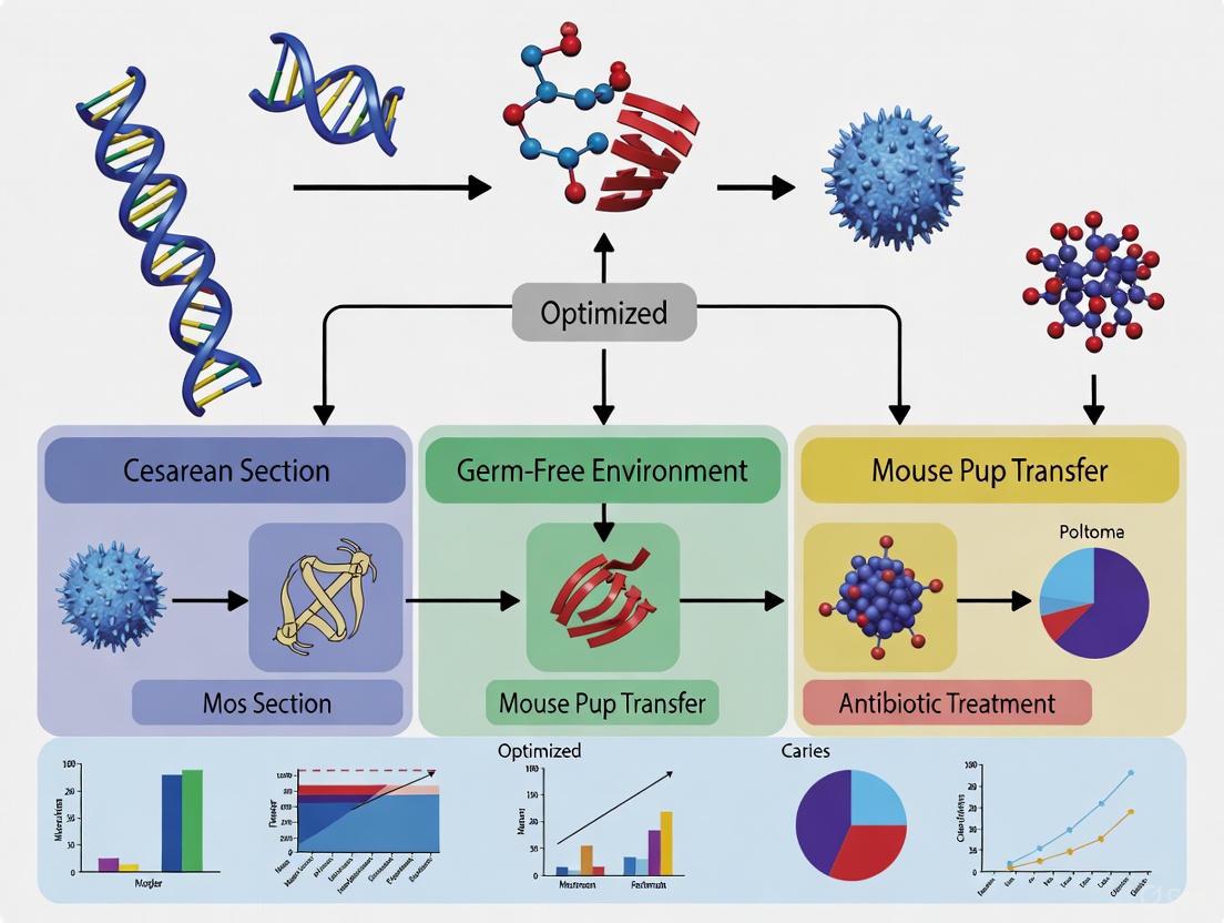
Optimized Cesarean Derivation: A Protocol for Enhanced Germ-Free Mouse Production
This article provides a comprehensive guide for researchers on optimizing cesarean section techniques to improve the efficiency and reproducibility of germ-free (GF) mouse production.
Optimized Cesarean Derivation: A Protocol for Enhanced Germ-Free Mouse Production
Abstract
This article provides a comprehensive guide for researchers on optimizing cesarean section techniques to improve the efficiency and reproducibility of germ-free (GF) mouse production. Based on the latest 2025 research, we detail a refined surgical method that preserves the female reproductive tract (FRT-CS), significantly boosting fetal survival rates. We explore the strategic use of in vitro fertilization (IVF) for precise delivery timing and compare the maternal care capabilities of different GF foster strains. Furthermore, we cover critical validation steps for confirming germ-free status and discuss the profound implications of using optimized GF mouse models in biomedical and drug development research.
The Indispensable Role of Germ-Free Mice in Modern Biomedical Research
Defining the Germ-Free Mouse Model and Its Critical Applications
Germ-free (GF) mice, also known as axenic mice, are specially raised animals that are completely devoid of all living microorganisms, including bacteria, viruses, fungi, and other microbes [1]. These mice serve as powerful "clean slate" tools in biomedical research, enabling scientists to study microbiome-host interactions without the confounding variables introduced by resident microbial communities. The development and maintenance of GF mice require highly specialized facilities and rigorous monitoring protocols to confirm and preserve their sterile status through a combination of culturing, microscopy, serology, and molecular detection techniques [1] [2].
The historical foundation for germ-free animal research dates back to 1885 when Louis Pasteur first proposed the concept, though he believed bacteria-free life was impossible [3] [2]. The first germ-free mammal (a guinea pig) was generated at Berlin University a decade later but survived only 13 days due to technical limitations [3]. The field advanced significantly when Gustafsson successfully obtained GF rats via sterile cesarean section, followed by Pleasants producing GF mice in 1959 [3]. These pioneering efforts established the foundation for modern gnotobiotic research, with ongoing technological refinements continuing to enhance the efficiency and accessibility of GF mouse models for contemporary research applications across diverse fields including immunology, oncology, neuroscience, and metabolic disease [1] [4].
Germ-Free Mouse Production and Derivation
Production Methods and Technical Refinements
The derivation of germ-free mice primarily relies on two established methods: sterile cesarean section and aseptic embryo transfer [3]. Both approaches require execution within completely sterile isolator environments to prevent microbial contamination, with each method presenting distinct advantages and technical considerations for researchers.
Sterile Cesarean Section remains the "golden method" for obtaining germ-free mice, based on the "sterile womb hypothesis" which posits that the placental epithelium serves as an effective barrier protecting the fetus from microbial exposure [3]. In this procedure, fetuses are delivered via sterile C-section from specific pathogen-free (SPF) donor females near term. The intact uterine sac is removed and transferred through a disinfectant bath into a sterile isolator, where pups are carefully extracted, revived, and introduced to GF foster mothers [3]. Recent technical refinements have significantly improved the efficiency of this approach through optimized surgical techniques that preserve the female reproductive tract (FRT-CS), which has demonstrated improved fetal survival rates while maintaining sterility compared to traditional C-section methods (T-CS) [3] [5].
Aseptic Embryo Transfer represents an alternative derivation method that must be conducted entirely within sterile isolators [3]. This approach involves transferring two-cell stage embryos derived from SPF mice via in vitro fertilization (IVF) into pseudopregnant GF recipients [3]. While embryo transfer can provide more precise control over developmental timing, the procedure faces technical constraints including lower embryo survival rates (approximately 50% of transferred embryos result in live births) and the requirement for specialized microsurgical equipment within the isolator environment [3].
Table 1: Comparison of Germ-Free Mouse Derivation Methods
| Parameter | Sterile Cesarean Section | Aseptic Embryo Transfer |
|---|---|---|
| Theoretical Basis | "Sterile womb hypothesis" | Direct sterile introduction of embryos |
| Technical Complexity | Moderate | High (requires microsurgery in isolator) |
| Success Rate | High with FRT-CS optimization | ~50% embryo survival rate |
| Contamination Risk | Low, but potential for pathogens crossing placental barrier | Very low |
| Delivery Timing Control | Variable with natural mating; improved with IVF donors | Precise control with IVF |
| Equipment Requirements | Standard surgical instruments in isolator | Stereomicroscope and specialized transfer equipment in isolator |
Production Workflow and Optimization Strategies
The following diagram illustrates the optimized workflow for germ-free mouse production, integrating recent technical refinements in both cesarean section and embryo transfer approaches:
Recent research has identified several key factors that significantly enhance germ-free mouse production efficiency [3] [5]:
FRT-CS Surgical Optimization: Implementation of female reproductive tract-preserving cesarean section (FRT-CS) techniques, which selectively clamp only the cervix base while preserving the entire reproductive tract, has demonstrated significantly improved fetal survival rates compared to traditional methods that clamp both the cervix base and top of the uterine horn [3].
IVF Integration for Timing Control: Utilizing in vitro fertilization (IVF) for obtaining donor embryos enables precise control over delivery dates, enhancing experimental reproducibility and planning. Studies show that IVF-derived donor mothers undergoing pre-labor FRT-CS on predicted delivery dates provide more reliable production scheduling compared to natural mating approaches [3] [5].
Strategic Foster Mother Selection: Systematic evaluation of different GF foster strains has revealed significant variation in nursing capabilities. BALB/c and NSG mice exhibit superior nursing and weaning success, while C57BL/6J demonstrates the lowest weaning rate in GF conditions—a finding that contrasts with maternal care observations in SPF C57BL/6J foster mothers [3].
Critical Research Applications
Establishing Causal Microbiome-Host Relationships
Germ-free mice serve as indispensable tools for moving beyond correlational observations to establishing causal relationships in microbiome research [1]. While high-throughput sequencing technologies have revolutionized our understanding of microbiome associations with various disease states, these approaches primarily identify correlations rather than prove causation [1]. GF models provide a controlled biological system in which investigators can directly assess the functional consequences of complete microbial absence or introduce defined microbial communities to test specific hypotheses about microbiome-host interactions [1] [6].
The unique value of GF mice in causal determination is exemplified in cardiovascular research, where studies have revealed discrepancies between broad-spectrum antibiotic treatment and germ-free models [7]. For instance, while both antibiotic-treated and GF Apoe-deficient mice sometimes show reduced aortic root lesions under certain dietary conditions, divergent results in other studies highlight the complex and sometimes contradictory findings between these approaches [7]. These observations underscore the importance of GF models in distinguishing direct microbial effects from antibiotic-mediated side effects in cardiovascular pathophysiology.
Mechanistic Insights into Host Physiology
The absence of microbial influence in GF mice produces distinctive physiological alterations that provide unique windows into microbiome-host interactions. One of the most evident anatomical characteristics of GF mice is a significantly enlarged cecum, a phenomenon observed in both GF and antibiotic-treated models [7] [2]. Additional gastrointestinal changes include elongated villus structures with reduced width and poorly developed capillary networks in small intestinal villi [7]. These structural modifications correspond with functional alterations in nutrient absorption and gastrointestinal motility.
Recent spatial metabolic characterization studies have identified significant molecular differences in GF mice across multiple tissues, including ileum, colon, spleen, lung, liver, and kidney [8]. The liver demonstrates the greatest number of metabolic changes, with molecules putatively identified as phenol sulfate and 5-amino valeric acid betaine showing significantly altered abundance in both intestinal and systemic tissues [8]. Concurrent phenotypic characterization reveals substantial alterations in immune cell populations throughout the body, indicating an aberrant immune response that underscores the critical role of microbial stimulation in immune system development and priming, even at sites distal from the intestine [8].
The following diagram illustrates the major physiological systems impacted by the germ-free condition and their interrelationships:
Therapeutic Research Applications
Germ-free mice provide invaluable platforms for investigating therapeutic interventions across diverse disease domains, facilitating mechanistic understanding of microbiome-related pathophysiology and treatment responses [1]. The following table summarizes key research applications and representative findings using GF mouse models:
Table 2: Therapeutic Research Applications of Germ-Free Mouse Models
| Research Area | Application Focus | Key Insights from GF Models |
|---|---|---|
| Immunology | Immune development and inflammatory diseases | Demonstrated critical microbiota role in immune stimulation and priming; identified aberrant immune responses in GF mice [8] [2] |
| Metabolic Diseases | Host metabolism and energy regulation | Revealed altered metabolic profiles in liver and other tissues; identified microbiome-derived metabolites influencing host metabolism [8] |
| Cardiovascular Disease | Atherosclerosis and thrombosis | Established microbiota contribution to thromboinflammation; identified reduced thrombosis tendency in GF mice [7] |
| Oncology | Cancer progression and therapy response | Validated that microbiota depletion alters tumor proteomic landscape and improves chemotherapy response in pancreatic cancer [9] |
| Neuroscience | Gut-brain axis and neurological disorders | Provided evidence for microbiome influence on blood-brain barrier function and neurodevelopment [6] |
| Infectious Disease | Pathogen colonization and infection | Elucidated how resident microbiota provides colonization resistance against pathogens [2] |
| Gastroenterology | Intestinal barrier function and IBD | Identified structural and functional alterations in GI tract development and function [7] [8] |
Experimental Protocols
Optimized Cesarean Section Derivation Protocol
Objective: To efficiently generate germ-free mice via sterile cesarean section with maximal pup survival and sterility assurance [3].
Materials:
- Pregnant SPF donor mice at gestational day 18-19
- Sterile surgical instruments (autoclaved)
- Clidox-S disinfectant system (1:3:1 dilution, activated 15 minutes before use)
- Sterile polyvinyl chloride (PVC) isolator
- Heating pad (pre-heated to 40-45°C)
- GF foster mothers (BALB/c or NSG strains recommended)
Procedure:
- Pre-surgical Preparation: Euthanize pregnant SPF donor mice via cervical dislocation. Activate Clidox-S disinfectant and pre-heat surgical area within isolator.
- Surgical Technique: Perform female reproductive tract-preserving cesarean section (FRT-CS) by:
- Making abdominal incision under sterile conditions
- Carefully exposing the uterine horns
- Placing clamps selectively at the cervix base only (preserving ovaries and uterine horns)
- Excising the entire intact uterus
- Disinfection and Transfer: Immerse intact uterine sac in Clidox-S disinfectant for 30 seconds, then rapidly transfer into sterile isolator.
- Pup Extraction: Within isolator, carefully incise amniotic membrane with sterile scissors and expose pups. Gently wipe amniotic fluid with sterile cotton swabs until spontaneous breathing is noted.
- Foster Introduction: Immediately transfer revived pups to proven GF foster mother (BALB/c or NSG strains preferred based on optimized weaning rates).
- Sterility Monitoring: Monitor pups for survival and conduct regular sterility testing through culturing, serology, and molecular methods.
Critical Parameters:
- Complete the entire procedure from euthanasia to pup revival within 5 minutes to maximize survival
- Maintain strict aseptic technique throughout the process
- Use FRT-CS method rather than traditional approach for improved fetal survival
- Confirm sterility through comprehensive testing regimen
Microbiota Reconstitution Protocol
Objective: To conventionalize germ-free mice with defined microbial communities for functional studies [10].
Materials:
- Adult germ-free mice (8-12 weeks old)
- Donor fecal material or defined bacterial communities
- Sterile phosphate-buffered saline (PBS)
- Anaerobic chamber for bacterial preparation
- Gavage needles (sterile)
- Metabolic caging for post-procedure monitoring
Procedure:
- Inoculum Preparation:
- For fecal microbiota transplants: homogenize donor fecal material in sterile PBS (100 mg/mL) under anaerobic conditions
- For defined communities: prepare bacterial suspensions to appropriate concentrations in anaerobic conditions
- Centrifuge briefly to remove particulate matter
- Mouse Preparation: Transfer GF mice to sterile metabolic cages and fast for 4-6 hours prior to gavage
- Inoculation: Administer 200μL of prepared inoculum via oral gavage using sterile technique
- Post-inoculation Monitoring:
- House mice in sterile isolators or positive pressure ventilated cages
- Monitor for successful colonization via fecal sampling at 24h, 72h, and 7 days post-inoculation
- Verify community composition through 16S rRNA sequencing and microbial load quantification
- Experimental Timeline: Allow 2-3 weeks for stable microbial community establishment before beginning experimental procedures
Validation Methods:
- Assess colonization success via 16S rRNA sequencing of fecal samples
- Confirm functional reconstitution through physiological parameters (cecal size normalization, immune cell profiling)
- Monitor for contamination through regular sterility checks
The Scientist's Toolkit: Essential Research Reagents and Materials
Table 3: Essential Research Reagents and Materials for Germ-Free Mouse Studies
| Category | Specific Items | Function/Application |
|---|---|---|
| Sterilization Equipment | Clidox-S disinfectant, autoclave, sterile isolators | Maintenance of sterile environment and equipment [3] |
| Validation Tools | Culture media (aerobic/anaerobic), PCR reagents for 16S rRNA and pathogen detection, serology assays | Confirmation of germ-free status and contamination screening [1] [2] |
| Surgical Materials | Sterile surgical instruments, heating pads, sterile cotton swabs, specialized clamps | Cesarean section derivation and surgical procedures within isolators [3] |
| Housing Components | Polyvinyl chloride (PVC) isolators, sterile bedding, autoclaved food and water | Long-term maintenance of germ-free colonies [3] |
| Microbiota Manipulation | Antibiotic cocktails (neomycin, vancomycin, metronidazole, ampicillin), gavage needles, anaerobic chamber | Creation of pseudo-germ-free controls and microbiota reconstitution studies [2] [9] |
| Analytical Tools | 16S rRNA sequencing reagents, metabolomics platforms, tissue processing reagents | Downstream analysis of microbial and host parameters [10] [8] |
| Specialized Mouse Strains | BALB/c, C57BL/6, NSG foster strains, various transgenic lines | Optimization of production efficiency and disease-specific investigations [3] [4] |
| Jineol | Jineol (3,8-Dihydroxyquinoline) | High-purity Jineol (C9H7NO2). Explore its research applications in melanogenesis inhibition and antibacterial studies. This product is for Research Use Only (RUO). |
| Flufenamic Acid | Flufenamic Acid, CAS:530-78-9, MF:C14H10F3NO2, MW:281.23 g/mol | Chemical Reagent |
Germ-free mouse models represent indispensable tools for establishing causal relationships in microbiome research and elucidating the mechanistic basis of host-microbe interactions [1]. Recent technical refinements in cesarean section techniques, IVF integration, and strategic foster mother selection have significantly enhanced the efficiency and reproducibility of germ-free mouse production [3] [5]. These optimized protocols enable more reliable generation of GF models for diverse research applications across immunology, metabolism, neuroscience, and oncology [1] [4].
The unique physiological characteristics of GF mice—including distinctive gastrointestinal, immune, metabolic, and vascular alterations—provide critical insights into the multifaceted roles of microorganisms in host physiology [7] [8]. As microbiome research continues to evolve, germ-free models will remain essential for validating findings from antibiotic depletion studies, investigating microbiome-based therapeutic strategies, and advancing our fundamental understanding of host-microbe symbiosis in health and disease [2] [9] [6].
The establishment of germ-free (GF) mouse colonies is a critical procedure in biomedical research, enabling the study of host-microbiome interactions in the absence of confounding microbial influences. Sterile cesarean section (C-section) remains the gold standard method for deriving GF colonies, based on the "sterile womb hypothesis" which posits that the placental epithelium serves as a barrier protecting the fetus from microbial exposure [3]. This protocol details optimized techniques for efficient production of GF mice via C-section derivation, incorporating recent advancements in surgical methods, donor selection, and foster strain selection to enhance survival rates and experimental reproducibility.
Key Experimental Data and Comparisons
Table 1: Comparison of Cesarean Section Techniques on Fetal Survival
| Surgical Technique | Description | Key Advantages | Fetal Survival Rate |
|---|---|---|---|
| Traditional C-section (T-CS) | Clamps placed at both cervix base and top of uterine horn | Standardized approach | Lower survival rate [3] |
| Female Reproductive Tract Preserved C-section (FRT-CS) | Selectively clamps only cervix base, preserving entire reproductive tract | Preserves ovarian and uterine structures; Significantly improved fetal survival [3] |
Table 2: Donor Conception Methods for C-section Timing
| Conception Method | Delivery Timing Control | Experimental Reproducibility | Implementation Complexity |
|---|---|---|---|
| Natural Mating (NM) | Limited: Confirmed by vaginal plug (G0.5), monitored from G18 for natural delivery before FRT-CS | Lower due to mating variability | Simple, requires monitoring |
| In Vitro Fertilization (IVF) | High: Implantation of two-cell stage embryos (E0.5), enabling precise pre-labor FRT-CS on predicted delivery date | Enhanced through precise timing control | Technically complex, requires specialized equipment [3] |
Table 3: Maternal Care Performance of GF Foster Strains
| Foster Mother Strain | Nursing Capability | Weaning Success Rate | Recommended Application |
|---|---|---|---|
| BALB/c | Superior: High weaning success, milk contributes significantly to pup weight gain | High | Primary choice for GF pup reception [3] |
| NSG (NOD/SCID Il2rg–/–) | Superior: Excellent nursing and weaning success | High | Primary choice for GF pup reception [3] |
| KM (Outbred) | Moderate: Acceptable nursing capability | Moderate | Suitable when inbred strains unavailable [3] |
| C57BL/6J | Poor: Lowest weaning rate among tested strains | Low | Not recommended as GF foster mothers [3] |
Detailed Experimental Protocols
Female Reproductive Tract Preserved C-section (FRT-CS) Protocol
Objective: To surgically derive germ-free pups while maximizing survival through reproductive tract preservation.
Materials:
- Pregnant SPF donor mice (C57BL/6 or BALB/c)
- Sterile surgical instruments (autoclaved at 121°C for 1200s)
- Clidox-S disinfectant (1:3:1 dilution, activated for 15 min)
- Polyvinyl chloride (PVC) isolator pre-heated to 40-45°C
- Sterile aspen wood shavings (autoclaved)
- GF foster mothers (BALB/c or NSG strains recommended)
Procedure:
- Donor Preparation: Euthanize pregnant SPF donor mice via cervical dislocation at term gestation.
- Surgical Setup: Perform all procedures under aseptic conditions within sterile isolator.
- FRT-CS Technique:
- Make midline abdominal incision to expose uterine horns
- Place clamp selectively at cervix base only, preserving ovaries, uterine horn, uterine junction
- Carefully extract intact uterine sac containing fetuses
- Disinfection: Immerse uterine sac in activated Clidox-S solution for sterilization.
- Fetal Extraction:
- Transfer disinfected uterine sac into sterile isolator
- Incise amniotic membrane with sterile surgical scissors to expose pups
- Cut umbilical cord using sterile instruments
- Wipe amniotic fluid with sterile cotton swabs until spontaneous breathing noted
- Time Constraint: Complete entire procedure within 5 minutes to ensure pup viability and maintain sterility.
- Foster Introduction: Immediately introduce derived pups to pre-conditioned GF foster mother.
Quality Control:
- Monitor pup survival and growth daily
- Verify germ-free status through regular sterility testing of fecal samples
- Maintain complete procedural documentation including surgical times and observations
In Vitro Fertilization for Donor Generation Protocol
Objective: To generate precisely timed pregnant donors for coordinated C-section derivation.
Materials:
- SPF donor embryos (C57BL/6J)
- CD-1 female mice as embryo transfer recipients
- Sterile IVF equipment and media
- Pseudopregnant recipient females
Procedure:
- Embryo Collection: Harvest two-cell stage embryos from superovulated SPF donors.
- Embryo Transfer: Surgically transfer embryos to pseudopregnant CD-1 recipients.
- Timing Designation: Designate day of implantation as embryonic day 0.5 (E0.5).
- Scheduling: Schedule pre-labor FRT-CS for predicted delivery date based on precise embryonic timing.
- Validation: Confirm pregnancy progression through physiological indicators before scheduled C-section.
Signaling Pathways and Experimental Workflows
Diagram 1: Complete workflow for germ-free mouse production via cesarean section derivation, highlighting critical decision points that impact success rates.
Diagram 2: Step-by-step protocol for the Female Reproductive Tract Preserved C-section (FRT-CS) technique, highlighting the critical clamping step that differentiates it from traditional approaches.
Research Reagent Solutions
Table 4: Essential Materials for Germ-Free Mouse Derivation
| Item | Specification | Function | Application Notes |
|---|---|---|---|
| Clidox-S Disinfectant | 1:3:1 dilution, activated 15 min before use | Sterilization of uterine sac and surface disinfection | Effective chlorine dioxide-based sterilant; crucial for maintaining sterility during transfer [3] |
| PVC Isolator | Polyvinyl chloride isolator with transfer chamber | Maintenance of sterile environment for GF mice | Requires pre-heating to 40-45°C for 15 min before procedure to prevent hypothermia [3] |
| Sterile Surgical Instruments | Autoclaved at 121°C for 1200s | Performing aseptic C-section procedure | Must remain sterile throughout entire surgical process |
| Aspen Wood Shavings | Autoclaved before use | Bedding for GF mice housing | Changed weekly to maintain hygienic conditions [3] |
| SPF Donor Mice | BALB/c, C57BL/6 from certified vendors | Source of embryos for GF derivation | Confirmed free of pathogenic bacteria, viruses, and parasites [3] |
| GF Foster Strains | BALB/c, NSG, KM | Nursing and care of derived GF pups | BALB/c and NSG show superior maternal care; C57BL/6J not recommended [3] |
| Laboratory Diet | Labdiet 5CJL, autoclaved | Nutrition for GF colonies | Unrestricted access to maintain health of foster mothers and pups [3] |
The 'Sterile Womb Hypothesis' as the Foundation for Cesarean Rederivation
The Sterile Womb Hypothesis posits that the placental epithelium serves as an effective barrier, protecting the developing fetus from microbial exposure and maintaining a sterile intrauterine environment throughout gestation [3] [11]. This theory forms the foundational principle for cesarean rederivation, the gold-standard method for generating germ-free (GF) mouse models for microbiome research [3]. According to this hypothesis, term fetuses develop without native colonization, meaning GF mice can be obtained through sterile cesarean section (C-section) delivery before contact with the maternal microbiota occurs during vaginal birth [3] [11].
This protocol details optimized techniques for GF mouse production based on this principle, enabling researchers to obtain axenic animals essential for studying host-microbiome interactions, immune system development, and therapeutic screening [3].
Theoretical Foundation: The Sterile Womb and Primo-Colonization
The "sterile womb" concept is supported by several lines of evidence, including the successful derivation of germ-free animals via aseptic hysterectomy and the general failure to detect significant, consistent bacterial communities in fetal tissues like meconium, placenta, and amniotic fluid after accounting for potential contamination [11]. While some studies using Next-Generation Sequencing (NGS) technologies have detected bacterial DNA in these tissues, subsequent analyses often reveal that these signals are not significantly different from negative controls or are likely skin contaminants such as Staphylococcus epidermidis [11].
The initial microbial colonization of the newborn, or primo-colonization, is a critical event shaped by delivery mode. Vaginally delivered infants acquire microbiota resembling the maternal vaginal community, whereas cesarean-delivered neonates are initially colonized by microbes similar to the maternal skin and environmental surfaces [11]. Cesarean rederivation exploits this principle by surgically transferring fetuses into a sterile isolator before this initial colonization from the birth canal can occur.
Quantitative Data on Optimized Cesarean Techniques
Comparison of Cesarean Section Techniques
The surgical technique for cesarean section significantly impacts fetal survival rates. The optimized Female Reproductive Tract Preserved C-section (FRT-CS) method demonstrates superior outcomes compared to the traditional approach (T-CS) [3].
Table 1: Impact of Surgical Technique on Fetal Survival in C57 and BC Strains
| Surgical Technique | Key Feature | Fetal Survival Rate | Maintained Sterility |
|---|---|---|---|
| Traditional C-section (T-CS) | Clamps placed at cervix base and top of uterine horn | Standard | Yes |
| FRT-C-section (FRT-CS) | Clamps only cervix base, preserving entire reproductive tract | Significantly Improved | Yes |
Efficacy of Germ-Free Foster Strains
The choice of GF foster mother strain is a critical determinant of pup weaning success. Maternal care capabilities vary drastically between strains under germ-free conditions [3].
Table 2: Weaning Success of Different Germ-Free Foster Mother Strains
| Foster Mother Strain | Strain Type | Weaning Success | Notes |
|---|---|---|---|
| BALB/c | Inbred | Superior | Exhibits superior nursing and weaning success |
| NSG | Inbred | Superior | Exhibits superior nursing and weaning success |
| KM | Outbred | Moderate | - |
| C57BL/6J | Inbred | Lowest | Performance in GF conditions contrasts with SPF findings |
Experimental Protocols
Protocol: Sterile Cesarean Section Rederivation
Objective: To derive germ-free mouse pups from specific pathogen-free (SPF) donor mothers via sterile C-section.
Principle: Based on the Sterile Womb Hypothesis, fetuses are harvested by C-section before contact with the non-sterile birth canal, transferred into a sterile isolator, and fostered by a germ-free lactating dam [3].
Pre-Procedure Preparations:
- Isolator Setup: Assemble and sterilize a polyvinyl chloride (PVC) isolator using chlorine dioxide (e.g., Clidox-S). Activate the heating pad inside the isolator (40–45°C) for at least 15 minutes prior to surgery to prevent pup hypothermia [3].
- Sterile Supplies: Autoclave (121°C for 1200s) all life supplements (food, water, bedding) and surgical instruments (scissors, forceps, clamps) [3].
- Donor & Foster Mice: Time-mated SPF pregnant dams serve as donors. A proven lactating GF foster mother (optimally BALB/c or NSG strain), with a litter born 1-3 days prior, is placed inside the isolator [3].
Procedure:
- Euthanize SPF Donor: Euthanize the pregnant SPF donor dam at gestational day 18-20 via cervical dislocation [3].
- Surgical Extraction:
- Perform the optimized FRT-CS technique: Saturate the abdomen with disinfectant. Make a midline incision to expose the uterine horns. Place a clamp selectively at the cervix base to preserve the entire reproductive tract [3].
- Excise the intact uterine horns and immediately submerge them in a sterile container filled with disinfectant solution (e.g., Clidox-S at 1:3:1 dilution, activated for 15 min) for rapid sterilization of the exterior [3].
- Isolator Transfer: Quickly transfer the sealed, disinfected container into the sterile isolator via a sterilizing transfer port [3].
- Pup Retrieval Inside Isolator:
- Incise the uterine sac with sterile surgical scissors under aseptic conditions.
- Carefully peel away the amniotic membrane to expose the pup.
- Wipe the pup's face and body with a sterile cotton swab to clear amniotic fluid until spontaneous breathing is noted.
- Cut the umbilical cord [3].
- Fostering: Immediately present the revived pup to the GF foster mother. The entire procedure, from donor euthanasia to pup fostering inside the isolator, must be completed within 5 minutes to ensure viability and sterility [3].
Protocol: Utilizing In Vitro Fertilization (IVF) for Timed Donors
Objective: To achieve precise control over the delivery date of donor embryos, enhancing experimental reproducibility [3].
Procedure:
- IVF & Embryo Transfer: Perform in vitro fertilization using sperm and oocytes from the desired SPF donor strain (e.g., C57BL/6J). Transfer the resulting two-cell stage embryos into the uterus of a pseudopregnant SPF recipient female (e.g., CD-1 strain) [3].
- Timing Definition: Designate the day of embryo implantation as embryonic day 0.5 (E0.5) [3].
- Scheduled C-section: On the predicted delivery date (typically E19.5), perform the pre-labor FRT-CS as described in Protocol 4.1. This eliminates variability from natural mating and allows for precise scheduling [3].
Visualization of Workflows
GF Mouse Production Workflow
Foster Strain Selection Logic
The Scientist's Toolkit: Research Reagent Solutions
Table 3: Essential Materials and Reagents for Cesarean Rederivation
| Item Name | Function/Application | Specification/Example |
|---|---|---|
| PVC Isolator | Provides a sterile barrier environment for housing GF mice and performing procedures. | Suzhou Fengshi Laboratory Animal Equipment Co., Ltd. [3] |
| Chlorine Dioxide Disinfectant | Sterilizes the exterior of the excised uterus and disinfects the isolator environment. | Clidox-S (1:3:1 dilution, activated 15 min) [3] |
| Autoclave | Sterilizes all supplies (food, water, bedding, instruments) before entry into the isolator. | 121°C for 1200s [3] |
| SPF Donor Strains | Source of embryos for deriving GF lines. Common strains include C57BL/6 and BALB/c. | Purchased from licensed animal providers (e.g., Shanghai SLAC) [3] |
| GF Foster Strains | Lactating dams to nurse and wean derived pups. Optimized strains are BALB/c and NSG. | BALB/cAnSlac, maintained in-house [3] |
| Heating Pad | Prevents hypothermia in newborn pups during the C-section procedure inside the isolator. | Pre-heated to 40-45°C for >15 min [3] |
| Aspen Wood Shavings | Autoclavable bedding material for housing mice within the isolator. | Changed once per week [3] |
| Standard Diet | Autoclavable rodent diet provided ad libitum to both SPF donors and GF colonies. | Labdiet 5CJL [3] |
| Fluindione | Fluindione, CAS:957-56-2, MF:C15H9FO2, MW:240.23 g/mol | Chemical Reagent |
| JNJ0966 | JNJ0966, MF:C16H16N4O2S2, MW:360.5 g/mol | Chemical Reagent |
Key Challenges in Traditional Germ-Free Mouse Production
Germ-free (GF) mice, completely devoid of all living microorganisms, serve as indispensable tools in biomedical research for studying host-microbe interactions, immune system development, and disease mechanisms. [12] [1] The production of these animals via traditional cesarean section (C-section) methods faces significant technical and biological challenges that can compromise efficiency, reproducibility, and animal welfare. Within the broader context of optimizing cesarean section techniques for GF mouse production, this application note details the principal challenges and provides standardized protocols to enhance experimental outcomes for researchers and drug development professionals.
Critical Challenges in Traditional GF Mouse Production
Inefficient Surgical Derivation and Neonatal Survival
Traditional sterile C-section techniques for obtaining GF pups present substantial hurdles in fetal survival and procedural efficiency.
Table 1: Impact of Cesarean Section Techniques on Pup Survival
| Surgical Method | Description | Key Findings | Effect on Pup Survival |
|---|---|---|---|
| Traditional C-section (T-CS) | Clamps placed at cervix base and top of uterine horn. [3] | Standard approach with baseline survival rates. | Baseline (Reference) |
| Female Reproductive Tract-Preserved C-section (FRT-CS) | Selective clamping only at cervix base, preserving entire reproductive tract. [3] | Significantly improved fetal survival rates while maintaining sterility. [3] | Significantly Improved |
Unpredictable Delivery Timing and Experimental Reproducibility
The reliance on natural mating (NM) of donor mice introduces significant variability. The precise timing of conception and birth is difficult to predict, complicacing the scheduling of C-sections and increasing the risk of missing the optimal window for pup derivation. [3] This variability directly jeopardizes experimental reproducibility across studies and facilities.
Inadequate Maternal Care from GF Foster Strains
The survival of derived GF pups depends on successful cross-fostering by a lactating GF female. However, not all mouse strains provide equivalent maternal care under germ-free conditions, leading to poor pup weaning rates. [3]
Table 2: Strain-Dependent Weaning Success of GF Foster Mothers
| Foster Mother Strain | Strain Type | Reported Weaning Success | Notes |
|---|---|---|---|
| C57BL/6J | Inbred | Lowest weaning rate [3] | Performance in stark contrast to SPF C57BL/6J foster mothers. [3] |
| BALB/c | Inbred | Superior nursing and weaning success [3] | Recommended strain for optimal pup survival. |
| NSG (NOD/SCID Il2rg–/–) | Inbred | Superior nursing and weaning success [3] | Recommended strain for optimal pup survival. |
| KM (Kunming) | Outbred | Evaluated for maternal care capabilities [3] |
High Costs and Specialized Infrastructure
Maintaining GF colonies is resource-intensive, requiring significant investment in specialized isolators, sterilization equipment (autoclaves, gas sterilizers), and continuous environmental monitoring systems. [13] [12] The labor-intensive nature of strict husbandry protocols and stringent testing to confirm the germ-free status further adds to the cost, which can be prohibitive for smaller laboratories. [13]
Optimized Experimental Protocols
Protocol: Female Reproductive Tract-Preserved C-Section (FRT-CS)
Objective: To aseptically derive GF mouse pups with improved survival rates via an optimized surgical technique.
Materials:
- Pregnant SPF donor mouse at late gestation.
- Sterile surgical instruments (scissors, forceps, clamps).
- Clidox-S disinfectant (prepared 15 min in advance: 1 part base, 3 parts water, 1 part activator). [14]
- Sterile polyvinyl chloride (PVC) isolator pre-heated to 40–45°C. [3]
- Sterile swabs and bedding.
Procedure:
- Euthanize the pregnant donor female via cervical dislocation. [3]
- Surgical Access: Quickly submerge the abdominal area in disinfectant and make a midline incision to expose the uterine horns.
- FRT-CS Technique: Identify the cervix and place a clamp selectively at its base only, preserving the ovary, uterine horn, and uterine junction. [3]
- Uterine Transfer: Excise the uterus and immediately transfer it into a sterile isolator via a disinfectant tank filled with Clidox-S. [3]
- Pup Extraction: Inside the isolator, incise the amniotic membrane with surgical scissors to expose each pup. Cut the umbilical cord and use a sterile cotton swab to gently wipe away amniotic fluid until spontaneous breathing is noted. [3]
- Timing: The entire procedure, from euthanasia to pup extraction, must be completed within 5 minutes to ensure pup viability and sterility. [3]
Protocol: Utilizing In Vitro Fertilization (IVF) for Timed Pregnancies
Objective: To achieve precise control over donor embryo delivery dates, enhancing experimental scheduling and reproducibility.
Materials:
- SPF donor females and males (e.g., C57BL/6).
- CD-1 or other suitable SPF recipient females.
- Materials for IVF and embryo transfer.
Procedure:
- Generate Embryos: Perform IVF using oocytes and sperm from the desired donor strain (e.g., C57BL/6J) to create embryos. [3]
- Embryo Transfer: Surgically transfer two-cell stage embryos into pseudopregnant SPF recipient females (e.g., CD-1). [3]
- Define Gestation Day: Designate the day of embryo implantation as embryonic day 0.5 (E0.5). [3]
- Scheduled Derivation: Perform the FRT-CS on the predicted delivery date, which is more accurately known due to the controlled timing of IVF, enabling pre-labor C-section. [3]
Protocol: Selection and Preparation of GF Foster Mothers
Objective: To maximize pup survival post-derivation by using strains proven to exhibit superior maternal care under GF conditions.
Materials:
- Lactating GF females of a recommended strain (e.g., BALB/c or NSG). [3]
- Sterile cages, food, water, and bedding.
Procedure:
- Strain Selection: Select proven GF foster strains such as BALB/c or NSG, which have demonstrated superior nursing and weaning success. [3]
- Preparation: Ensure foster mothers are of optimal age (e.g., four months old) and have prior successful birthing and nursing experience. [3]
- Cross-Fostering: Gently transfer the derived GF pups to the nest of the foster mother immediately after the C-section procedure. Minimize disturbance to the foster mother and her existing litter.
The Scientist's Toolkit: Essential Reagents and Materials
Table 3: Key Research Reagent Solutions for Germ-Free Mouse Production
| Item | Function/Application | Key Notes |
|---|---|---|
| Clidox-S | Chlorine dioxide sterilant for disinfecting surfaces, tissue samples, and the isolator environment. [14] | Requires 10-minute contact time. Prepare 15 min in advance; effective for 3-4 hours. Highly corrosive, requires deactivation before disposal. [14] |
| F10SC | Broad-spectrum disinfectant used for gas sterilization of items not suitable for autoclaving. [14] | Effective for 3-4 months. Used in an atomizer for fogging biosafety cabinets. [14] |
| Autoclavable Diet | Specialized, nutrient-fortified rodent food. | Standard diets lose nutrients during sterilization; use fortified diets to account for loss during autoclaving or gamma irradiation. [15] |
| Individually Ventilated Cages (IVCs) | Housing within isolators to maintain sterility with HEPA-filtered airflow. [13] | Provides a micro-isolated environment inside the main isolator. |
| Environmental Monitoring System | Tracks temperature, humidity, and microbial presence in real-time. [13] | Integrated with Laboratory Information Management Systems (LIMS) for data logging. [13] |
| Kanosamine | Kanosamine, CAS:576-44-3, MF:C6H13NO5, MW:179.17 g/mol | Chemical Reagent |
| HMR 1556 | HMR 1556 is a potent, selective IKs potassium channel blocker for cardiac research. For Research Use Only. Not for human or veterinary use. |
Workflow and Strategic Decision Diagrams
Strategic Optimization of GF Mouse Production
Sterile Isolator Setup and Maintenance Workflow
Concluding Remarks
Addressing the key challenges in traditional germ-free mouse production—through the adoption of optimized C-section techniques, IVF for precise timing, and evidence-based selection of foster strains—significantly enhances the efficiency, reproducibility, and ethical standards of this critical biomedical research model. The detailed protocols and strategic workflows provided herein offer a practical framework for researchers to advance their studies in host-microbiome interactions and therapeutic development.
Step-by-Step Protocol: Implementing Optimized Cesarean Techniques
Application Notes and Protocols
Within the specialized field of germ-free (GF) mouse production, the cesarean section (C-section) technique is the gold standard for obtaining sterile pups from specific pathogen-free (SPF) donor mothers. The conventional surgical approach, however, can impact the viability of neonates. The Female Reproductive Tract-Preserving C-Section (FRT-CS) is a refined surgical protocol designed to enhance fetal survival rates during this critical derivation process, thereby improving the efficiency of establishing and maintaining GF mouse colonies for microbiome and drug development research [3].
The following tables summarize key experimental findings from studies optimizing GF mouse production.
Table 1: Impact of Cesarean Section Technique on Fetal Survival [3]
| Surgical Technique | Description | Fetal Survival Outcome |
|---|---|---|
| Traditional C-Section (T-CS) | Clamps placed at the cervix base and the top of the uterine horn. | Baseline survival rate (Used as a comparison) |
| FRT-CS (Recommended) | Selective clamping only at the cervix base, preserving the ovary, uterine horn, and cervix. | Significantly improved fetal survival rates while maintaining sterility. |
Table 2: Weaning Success Rates of Germ-Free Pups by Foster Mother Strain [3]
| Foster Mother Strain | Strain Type | Weaning Success | Notes |
|---|---|---|---|
| BALB/c | Inbred | Superior | Exhibited superior nursing and weaning success. |
| NSG | Inbred | Superior | Exhibited superior nursing and weaning success. |
| KM | Outbred | Moderate | - |
| C57BL/6J | Inbred | Lowest | Lowest weaning rate; contrast with SPF C57BL/6J foster mothers. |
Table 3: Comparison of Donor Mouse Conception Methods [3]
| Conception Method | Description | Impact on C-Section |
|---|---|---|
| Natural Mating (NM) | Conventional mating of donor females with males. | Inherent variability in delivery timing, reducing experimental reproducibility. |
| In Vitro Fertilization (IVF) | IVF-derived embryos transferred to recipient females. | Precise control over donor delivery dates, enhancing experimental reproducibility for pre-labor FRT-CS. |
Experimental Protocols
Protocol: Female Reproductive Tract-Preserving C-Section (FRT-CS)
Objective: To aseptically deliver GF mouse pups from a SPF donor while maximizing neonatal survival through refined surgical technique.
Pre-operative Preparations:
- Isolator Setup: Perform the procedure within a sterile polyvinyl chloride (PVC) isolator. Autoclave all supplies (food, water, bedding, surgical instruments) at 121°C for 20 minutes prior to entry [3].
- Environmental Control: Activate a heating pad inside the isolator (40–45°C) for at least 15 minutes pre-surgery to prevent pup hypothermia [3].
- Disinfectant Preparation: Prepare a chlorine dioxide disinfectant (e.g., Clidox-S at a 1:3:1 dilution) and activate for 15 minutes before use [3].
- Donor Euthanasia: Euthanize the pregnant SPF donor female via cervical dislocation [3].
Surgical Procedure:
- Aseptic Transfer: Move the donor mouse into the sterile isolator via a transfer port or dunk tank.
- Exposure: Position the animal and expose the abdominal wall.
- Abdominal Incision: Make a midline incision through the skin and abdominal wall to access the peritoneal cavity.
- Uterine Exposure: Gently expose the gravid uterus.
- FRT-CS Clamping: Identify the cervix base. Apply a clamp selectively only at the cervix base, preserving the integrity of the entire reproductive tract (ovaries, uterine horns) [3].
- Uterine Excision: Excise the uterine horn containing the fetuses.
- Disinfection: Immerse the excised uterine horn in the prepared chlorine dioxide disinfectant for sterilization [3].
- Transfer to Isolator: Quickly transfer the disinfected uterine horn into the sterile isolator interior.
- Pup Extraction: Inside the isolator, incise the uterine sac and amniotic membrane with sterile surgical scissors to expose the pup.
- Umbilical Cord Separation: Cut the umbilical cord.
- Stimulation: Use a sterile cotton swab to wipe away amniotic fluid and stimulate breathing until spontaneous respiration is noted [3].
- Pup Placement: Immediately place the viable pup with a pre-conditioned GF foster mother.
Critical Step: The entire procedure, from donor euthanasia to pup transfer into the isolator, must be completed within 5 minutes to ensure pup viability and sterility [3].
Protocol: In Vitro Fertilization for Timed Donor Conception
Objective: To generate donor females with precisely controlled delivery dates for scheduled FRT-CS.
Procedure:
- IVF Procedure: Perform in vitro fertilization using oocytes and sperm from the desired SPF donor strain (e.g., C57BL/6J) [3].
- Embryo Transfer: Implant the two-cell stage embryos into a recipient female (e.g., CD-1 strain). Designate the day of implantation as embryonic day 0.5 (E0.5) [3].
- Predicted Delivery: Calculate the expected delivery date. These IVF-derived donors undergo pre-labor FRT-CS on this predicted date, eliminating the variability associated with natural birth timing [3].
Workflow and Decision Diagrams
The following diagrams illustrate the optimized workflow for GF mouse production and the logical process for selecting an optimal foster mother strain.
The Scientist's Toolkit: Research Reagent Solutions
Table 4: Essential Materials for FRT-CS and Germ-Free Mouse Production [3]
| Item / Reagent | Function / Application | Example / Note |
|---|---|---|
| SPF Donor Mice | Source of embryos for deriving GF pups. | Common strains: C57BL/6, BALB/c. |
| GF Foster Mice | Care for and nurse FRT-CS derived pups. | Optimal strains: BALB/c, NSG. Avoid C57BL/6J. |
| Chlorine Dioxide Disinfectant | Surface sterilization of the excised uterus. | E.g., Clidox-S. Must be freshly activated. |
| Sterile Isolator | Provides a sterile barrier environment for surgery and housing. | Polyvinyl chloride (PVC) isolators are standard. |
| Autoclave | Sterilization of all entry supplies (food, water, bedding, instruments). | Critical for maintaining germ-free conditions. |
| Heating Pad | Prevents hypothermia in neonates during the surgical procedure. | Maintain temperature at 40–45°C inside isolator. |
| Surgical Instruments | Performing the cesarean section and pup extraction. | Fine scissors, forceps, clamps. Must be sterile. |
| Kazinol B | Kazinol B, CAS:99624-27-8, MF:C25H28O4, MW:392.5 g/mol | Chemical Reagent |
| Hydronidone | Hydronidone, CAS:851518-71-3, MF:C12H11NO2, MW:201.22 g/mol | Chemical Reagent |
Within the specialized field of germ-free (GF) mouse production, the precise timing of donor pup delivery is a critical determinant of success. Traditional reliance on naturally mated (NM) donors introduces significant variability, complicating the coordination of sterile cesarean sections (C-sections) with the availability of prepared foster mothers. This application note details a refined donor strategy utilizing in vitro fertilization (IVF) to achieve precise control over delivery dates, thereby enhancing the efficiency, reproducibility, and success rate of GF mouse derivation projects. This approach directly addresses a key challenge in the optimized cesarean section technique for germ-free mouse production, enabling researchers to schedule surgical procedures with a high degree of accuracy and minimize neonatal loss [16].
Experimental Evidence: IVF vs. Natural Mating
A direct comparative study within GF mouse production protocols evaluated the impact of donor conception method—IVF versus natural mating—on the predictability of the process and subsequent pup survival after C-section [16].
In the study, two groups of donor mothers were established:
- NM Donors: C57BL/6J females were naturally mated with males, with the presence of a vaginal plug confirming gestation day 0.5 (G0.5). These donors were then monitored for natural delivery from G18 onward before undergoing a female reproductive tract-preserving C-section (FRT-CS) [16].
- IVF Donors: Embryos from C57BL/6J mice were created via IVF and transferred into recipient CD-1 females. The day of two-cell stage embryo implantation was designated as embryonic day 0.5 (E0.5). These IVF-derived donors underwent pre-labor FRT-CS on their predetermined delivery date [16].
The primary finding was that the use of IVF allowed for precise control over the delivery timing of donor mothers. This control enabled the scheduling of the sterile C-section to occur immediately before the expected natural labor, ensuring optimal fetal maturity for survival outside the uterus while eliminating the guesswork and round-the-clock monitoring associated with natural mating [16].
Table 1: Comparative Analysis of Donor Conception Methods for GF Mouse Production
| Feature | Natural Mating (NM) | In Vitro Fertilization (IVF) |
|---|---|---|
| Delivery Date Control | Unpredictable; requires continuous monitoring from G18 [16] | High; C-section performed on a predetermined date [16] |
| Experimental Reproducibility | Lower due to variability in conception and birth timing [16] | Enhanced through precise scheduling [16] |
| Pup Survival Post-C-section | Not directly compared, but viability is high with FRT-CS [16] | Not directly compared, but viability is high with FRT-CS [16] |
| Requirement for Technical Expertise | Low (standard breeding) | High (requires specialized IVF skills and equipment) [17] |
| Primary Advantage | Technically simple | Unlocks precise scheduling and superior experimental planning [16] |
Detailed IVF Protocol for Donor Generation
The following protocol for mouse IVF is adapted from established methodologies and is designed to generate donor embryos with a known developmental timeline [17].
Materials and Equipment
- Hormones: Pregnant Mare's Serum Gonadotropin (PMSG) and human Chorionic Gonadotropin (hCG) [17].
- Media: Preincubation medium (e.g., FERTIUP PM), fertilization medium (e.g., CARD MEDIUM), and washing medium (e.g., mHTF) [17].
- Equipment: Humidified incubator (37°C, 5% CO₂), stereomicroscope, plastic dishes (35mm x 10mm), liquid paraffin, micropipettes and tips, fine dissection tools (scissors, #5 forceps, micro-spring scissors) [17].
- Animals: Mature female mice (8-12 weeks old) for superovulation, and mature male mice (3-6 months old) with proven fertility for sperm collection [17].
Step-by-Step Procedure
Day 1: Superovulation of Females
- Between 2:00 PM and 6:00 PM, administer an intraperitoneal (i.p.) injection of 7.5 IU PMSG to each mature female mouse [17].
Day 3: hCG Injection and Sperm Preparation
- Exactly 48-52 hours after PMSG, administer an i.p. injection of 7.5 IU hCG to the same females [17].
- Prepare the sperm dish: Place a 100 µL drop of preincubation medium (e.g., FERTIUP PM) in a dish, cover with liquid paraffin, and equilibrate in the incubator for at least 30 minutes [17].
- Sacrifice one or two mature male mice and collect the cauda epididymides. Carefully cut the ducts and gently press to release sperm into the prepared drop of medium. Incubate for 60 minutes to allow for sperm capacitation [17].
Day 4: Oocyte Collection and Insemination
- Approximately 15-17 hours after the hCG injection, sacrifice the superovulated females and rapidly collect the oviducts [17].
- Prepare the fertilization dish: Place a 200 µL drop of fertilization medium (e.g., CARD MEDIUM) in a dish, cover with liquid paraffin, and place in the incubator 10 minutes before use [17].
- Under a stereomicroscope, tear open the ampulla of each oviduct to release the cumulus-oocyte complexes (COCs) into the drop of fertilization medium. Complete this process for each mouse within 30 seconds to maintain oocyte health [17].
- Add approximately 3 µL of the capacitated sperm suspension to the drop containing the COCs. Return the dish to the incubator [17].
Day 4 (3-6 Hours Post-Insemination): Oocyte Washing and Assessment
- Prepare the washing dish with 4 drops (80 µL each) of mHTF medium under liquid paraffin [17].
- 3 hours after insemination, wash the oocytes by transferring them through the three fresh drops of mHTF medium [17].
- 6 hours after insemination, observe the oocytes under a microscope. Identify and remove any parthenogenetic oocytes, which display only one pronucleus. Fertilized oocytes will have two pronuclei (male and female) [17].
Day 5: Embryo Culture and Transfer
- The following day, after overnight culture, identify and select the successfully developed 2-cell stage embryos [17].
- These embryos can now be transferred into pseudo-pregnant recipient females. The day of transfer is designated as E0.5, establishing a precise timeline for the expected birth, which typically occurs 19-20 days later [16] [17].
Integration with Germ-Free Rederivation
The integration of IVF into the GF mouse production pipeline creates a seamless and highly predictable workflow. The known embryonic age (E0.5) of IVF-derived donors allows for the accurate scheduling of the sterile C-section for around E19.5, just prior to natural labor [16]. This precise timing is crucial for optimizing fetal survival during the FRT-CS procedure.
Following the C-section, the choice of GF foster mother strain is paramount. Research indicates that under germ-free conditions, BALB/c and NSG strains exhibit superior nursing and weaning success compared to C57BL/6J, which showed the lowest weaning rate [16]. This finding is critical for selecting the most effective foster mothers to ensure the survival of the derived GF pups.
Table 2: The Scientist's Toolkit - Key Reagents for IVF-Based Donor Strategy
| Reagent/Item | Function/Application | Example/Note |
|---|---|---|
| PMSG | Mimics Follicle-Stimulating Hormone (FSH); stimulates follicle growth and superovulation in female mice [17]. | Typically administered at 7.5 IU per mouse [17]. |
| hCG | Mimics Luteinizing Hormone (LH); triggers final oocyte maturation and ovulation [17]. | Administered 48-52 hours after PMSG at 7.5 IU per mouse [17]. |
| Fertilization Medium | Supports the process of capacitation, sperm-egg interaction, and fertilization [17]. | e.g., CARD MEDIUM; composition is optimized for these steps [17]. |
| Washing Medium | Used for washing oocytes/embryos after fertilization to remove metabolic waste and non-adherent sperm [17]. | e.g., mHTF; a balanced salt solution for embryo handling and short-term culture [17]. |
| Liquid Paraffin | Used to overlay culture medium drops; prevents evaporation and minimizes changes in osmolarity and pH [17]. | Essential for maintaining a stable micro-environment for gametes and embryos. |
Leveraging IVF as a core donor strategy transforms the production of germ-free mice from an unpredictable process into a scheduled, efficient, and reproducible scientific procedure. By providing exact control over the delivery date of donor pups, this method facilitates optimal timing for the cesarean section, maximizes the coordination with prepared foster mothers, and ultimately enhances the overall success rate of deriving germ-free colonies. This approach is particularly valuable for rapidly recovering GF colonies after contamination and for efficiently generating new GF models to advance microbiome research [16] [18].
In the production of germ-free (GF) mice, which are indispensable for studying host-microbiome interactions, the establishment and maintenance of a sterile environment is the cornerstone of success [3]. The aseptic derivation of mice via cesarean section is a critical procedure that demands an absolute contamination-free environment to ensure the survival and sterility of the pups [3]. Isolator technology provides this essential barrier, physically separating the delicate operative procedure and the pups from the external, non-sterile environment [19] [20]. This document outlines detailed application notes and protocols for the setup, sterilization, and operation of isolators, specifically tailored for germ-free mouse production facilities. Adherence to these protocols is vital for maximizing pup survival and ensuring the integrity of research models.
Isolator Selection and Classification
Choosing the appropriate type of isolator is the first critical step. For germ-free mouse production, the isolator must maintain Grade A/ISO 5 air quality to provide an aseptic environment for post-operative care and housing [21].
Table 1: Isolator Types for Germ-Free Mouse Production
| Isolator Type | Primary Function in GF Mouse Research | Pressure Regime | Key Features |
|---|---|---|---|
| Flexible Film Isolator | Long-term housing & breeding of GF mice; post-C-section pup rearing [20]. | Positive | Cost-effective; transparent vinyl walls; ideal for animal housing [20]. |
| Rigid Isolator | Long-term housing of GF mice; procedures requiring high durability [20]. | Positive or Negative | Made of stainless steel; more durable but higher cost [20]. |
| Aseptic Isolator | Performing sterile C-section procedures and other aseptic manipulations [20] [21]. | Positive | Designed for aseptic processing; maintains uncompromised isolation [21]. |
| Transfer Isolator | Safe introduction of sterile supplies (food, water, bedding) and movement of mice between isolators [20]. | Varies | Provides a sterile bridge between two environments; prevents contamination during transfer [20]. |
Isolator Sterilization and Decontamination Protocols
Reproducible interior bio-decontamination is a defining characteristic of an isolator and is paramount for preventing microbial contamination of GF mice [21]. Hydrogen peroxide-based systems are the industry standard for achieving a sporicidal state.
Hydrogen Peroxide Vapor (HPV) Bio-decontamination
This process involves vaporizing a hydrogen peroxide solution and introducing it into the sealed isolator until saturation and micro-condensation occur on all surfaces, ensuring comprehensive bio-decontamination [21].
Experimental Protocol: Automated Hydrogen Peroxide Vapor Decontamination
- Objective: To achieve a validated 6-log reduction of spore-forming bacteria (Geobacillus stearothermophilus) on all interior surfaces of the isolator [20] [21].
- Materials:
- Automated Hâ‚‚Oâ‚‚ Vapor Generator (e.g., CURIS TRINITY, Bioquell Qube) [20] [21].
- Hydrogen Peroxide Sterilant (e.g., 7% for HHP, 35% for traditional VPHP) [20] [21].
- Biological Indicators (BIs): Geobacillus stearothermophilus spores at a minimum concentration of 10ⶠper indicator [20] [21].
- Chemical Indicators.
- Leak-testing equipment for the isolator.
- Procedure:
- Preparation: Remove all unnecessary items. Ensure the isolator is clean and dry. Load pre-sterilized materials (cages, food, water) into the isolator. Place BIs and chemical indicators at predefined worst-case locations (e.g., far from the vapor inlet, inside gloves, downstream of filters) [20].
- Leak Testing: Perform a validated leak test on the isolator and gloves to ensure an airtight seal [21].
- Decontamination Cycle: Seal the isolator and connect it to the Hâ‚‚Oâ‚‚ generator. Initiate the automated cycle, which typically includes:
- Conditioning: The system reduces humidity to a defined set point.
- Gassing: Hâ‚‚Oâ‚‚ is injected and circulated until the target concentration and saturation are achieved.
- Exposure: The lethal concentration is maintained for a predetermined time to achieve the 6-log kill.
- Aeration: Hâ‚‚Oâ‚‚ is catalytically broken down into water vapor and oxygen, and the interior is safely vented [20].
- Validation: Retrieve BIs after the cycle and incubate them in culture media alongside positive and negative controls. No growth of the test BIs after 7 days confirms a successful 6-log sporicidal reduction [21].
Table 2: Comparison of Hydrogen Peroxide Decontamination Methods
| Parameter | Vaporized Hydrogen Peroxide (VPHP) | Hybrid Hydrogen Peroxide (HHP) |
|---|---|---|
| Hâ‚‚Oâ‚‚ Concentration | 35% - 59% [20] | ~7% [20] |
| Mechanism | True vapor leading to micro-condensation [21]. | Vapor + submicron aerosol particles [20]. |
| Cycle Time | Longer | Shorter (can reduce from days to under an hour) [20]. |
| Material Compatibility | Can cause yellowing of certain films; more caustic [20]. | Better material compatibility [20]. |
| Efficacy | Validated 6-log sporicidal reduction [21]. | Validated 6-log sporicidal reduction [20]. |
Application Notes for Germ-Free Mouse Production
Optimized Cesarean Section within an Isolator
The surgical derivation of GF pups must be performed under strict aseptic conditions inside an aseptic isolator or a biological safety cabinet with a direct transfer port to a flexible film isolator.
Experimental Protocol: Sterile Cesarean Section in an Isolator
- Objective: To aseptically deliver term fetuses from a specific pathogen-free (SPF) donor mouse into a germ-free environment [3].
- Pre-requisites:
- Donor Mice: Use timed-pregnant SPF mice. The study by Scientific Reports (2025) showed that using in vitro fertilization (IVF) to generate donor embryos allows for precise control over delivery timing, enhancing reproducibility compared to natural mating [3] [5].
- Foster Mothers: Select a proven GF foster strain. The same 2025 study found that GF BALB/c and NSG strains exhibited superior nursing and weaning success, while GF C57BL/6J had the lowest weaning rate [3] [5].
- Isolator Preparation: The receiving flexible film isolator must be sterilized via HPV and contain a pre-warmed heating pad (40-45°C), sterile surgical instruments, drapes, and disinfectant (e.g., Clidox-S) [3].
- Surgical Procedure (within isolator):
- Euthanize Donor: Euthanize the pregnant donor mouse via cervical dislocation outside the isolator. The uterus is then externally disinfected and transferred into the aseptic isolator via a rapid transfer port (RTP) submerged in disinfectant [3].
- Surgical Technique: The study compared Traditional C-section (T-CS) with a Female Reproductive Tract Preserved C-section (FRT-CS). FRT-CS, which selectively clamps only the cervix base, significantly improved fetal survival rates while maintaining sterility [3].
- Pup Extraction: Incise the uterine sac with sterile scissors. Gently remove each pup and wipe away amniotic fluid with a sterile swab until spontaneous breathing is noted. The entire procedure from uterus transfer to pup resuscitation must be completed within 5 minutes to ensure viability [3].
- Transfer to Foster: Immediately place the viable pups with the pre-conditioned GF foster mother inside the rearing isolator [3].
Sterility Assurance and Monitoring
The Scientist's Toolkit: Essential Research Reagents and Materials
Table 3: Key Research Reagent Solutions for Germ-Free Mouse Isolator Management
| Item | Function / Application | Example / Specification |
|---|---|---|
| Hydrogen Peroxide Sterilant | Primary agent for automated bio-decontamination of isolators [20] [21]. | Bioquell Hydrogen Peroxide Sterilant-AQ; CURIS 7% HHP Solution [20] [21]. |
| Biological Indicators (BIs) | Validation of 6-log sporicidal efficacy of decontamination cycles [20] [21]. | Geobacillus stearothermophilus spores, ≥10ⶠper indicator [20]. |
| Sporicidal Disinfectant | Surface disinfection of items prior to entry into the isolator (e.g., via dunk tank) [3]. | Activated Chlorine Dioxide (Clidox-S) [3]. |
| HEPA Filters | Maintains ISO 5 / Grade A air quality inside the isolator by removing airborne particulates and microorganisms [19] [21]. | Integral component of isolator air handling system [21]. |
| Rapid Transfer Ports (RTPs) | Allows sterile transfer of materials between isolators or from the outside without breaking containment [19]. | Double-door port system that is interlocked to prevent both doors being open simultaneously. |
| Sterilized Diet and Bedding | Nutrition and housing for GF mice; must be sterilized and introduced without contamination [3]. | Autoclaved (121°C for 20+ minutes) or irradiated diet and aspen wood shavings [3]. |
| Fluphenazine dimaleate | Fluphenazine Dimaleate | Fluphenazine dimaleate is a potent typical antipsychotic research compound. For Research Use Only. Not for human or veterinary diagnostic or therapeutic applications. |
| 1-A09 | Information on 1-A09: Vision Screener and Electronic Component | This page aggregates information on products named 1-A09, including a medical vision screening device and an electronic switch component. All content is For Research Use Only. |
Within the specialized field of germ-free (GF) mouse production, the period immediately following a sterile cesarean section (C-section) is the most critical determinant of success. The successful resuscitation and subsequent post-operative care of pups within the sterile isolator are paramount for establishing viable GF colonies essential for microbiome research [3] [22]. This protocol details evidence-based, optimized procedures for these phases, framing them within the broader research objective of refining C-section techniques to enhance efficiency and reproducibility in GF mouse production [3]. The guidelines herein are designed for researchers and technicians working in gnotobiotic facilities.
Pre-Procedural Preparations
A sterile, organized, and well-prepared isolator environment is the foundation for successful pup resuscitation. Meticulous attention to detail in preparation prevents procedural delays and mitigates contamination risks.
Isolator and Equipment Setup
- Isolator Assembly: Utilize flexible film, positive-pressure polyvinyl chloride (PVC) isolators equipped with attached gloves and a 12-inch transfer port [23] [22]. Ensure the isolator is leak-tested and the high-efficiency particulate air (HEPA) filters are functioning correctly.
- Sterilization: Sterilize the interior of the assembled isolator by spraying with a 2% solution of peracetic acid using an atomizer [22]. All items entering the isolator must be sterilized beforehand.
- Thermoregulation: Place a heating pad inside the isolator and activate it at least 15 minutes before the C-section to achieve a surface temperature of 40–45°C [3]. This is critical to prevent hypothermia in neonates, which lack effective thermoregulation.
- Supply Sterilization: Autoclave all surgical instruments (fine forceps, scissors), gauze, and bedding at 121°C for a minimum of 20 minutes [3] [22]. Sterilize water and heat-sensitive liquids like artificial milk components via gamma radiation (25-50 kGy) [22]. Pack supplies in sterile glass bottles or stainless-steel cylinders sealed with PVC film and filter paper for secure transfer into the isolator [22].
Reagent and Solution Formulation
Preparing resuscitation reagents in advance streamlines the procedure. The following table lists essential solutions.
Table 1: Key Research Reagent Solutions for Pup Resuscitation
| Reagent/Solution | Function/Application | Preparation and Sterilization Method |
|---|---|---|
| Chlorine Dioxide (Clidox-S) | Disinfectant for the exterior of the uterine sac during transfer into the isolator [3]. | Prepare as a 1:3:1 dilution and activate for 15 minutes before use [3]. |
| Peracetic Acid (2% Solution) | Sterilizing agent for the internal environment of the flexible film isolator [22]. | Mix equal parts of liquid A (acetic acid/sulfuric acid) and liquid B (hydrogen peroxide) 24-48 hours in advance; dilute to 2% [22]. |
| Artificial Milk Formulation | Nutritional support for hand-rearing suckling GF rats if fostering fails [22]. | Combine irradiated rabbit milk, milk powder, fetal bovine serum, and olive oil in proportions that vary with pup age; sterilize via 25 kGy γ-radiation [22]. |
This section outlines the sequential workflow from the moment the uterus is transferred into the isolator until the pups are stabilized. The diagram below illustrates the logical flow of the core resuscitation procedure.
Figure 1: Core Workflow for Pup Resuscitation in the Isolator. Key time and temperature considerations are highlighted.
Step-by-Step Procedural Details
- Transfer and Disinfection: Upon exteriorization from the donor dam, the entire uterine sac must be immediately sprayed with a chlorine dioxide disinfectant (e.g., Clidox-S) to decontaminate the exterior surface [3].
- Aseptic Transfer: Rapidly move the disinfected uterine sac into the sterile isolator through the transfer port [3] [22].
- Fetal Delivery: Inside the isolator, use sterile surgical scissors to incise the uterine sac and carefully release the amniotic fluid. Gently remove the pup [3] [22].
- Stimulation of Breathing: Use a sterile cotton swab or gauze piece to gently but thoroughly wipe the pup, clearing amniotic fluid from the nose and mouth. Continue stimulation until spontaneous breathing is noted [3]. The entire procedure, from donor euthanasia to the initiation of breathing, must be completed within 5 minutes to maximize viability [3].
- Umbilical Cord Severance: Using sterile scissors, cut the umbilical cord [3].
- Thermal Support: Immediately place the resuscitated pup on the pre-warmed heating pad (40–45°C) inside the isolator to maintain core body temperature [3].
Post-Operative Care and Monitoring
After successful resuscitation, continuous monitoring and appropriate nursing are required to ensure survival through the weaning period.
Foster Mother Strategy
The use of a proven GF foster mother is the most effective method for ensuring pup survival [3] [22].
- Strain Selection: Strain selection significantly impacts weaning success. Evidence indicates that GF BALB/c and NSG strains exhibit superior nursing capabilities and result in higher weaning rates. In contrast, GF C57BL/6J fosters have the lowest weaning success, a finding that contrasts with their maternal performance in specific pathogen-free (SPF) conditions [3].
- Introduction to Foster Dam: Gently transfer the resuscitated pups to the cage of a lactating GF foster dam that has recently given birth (ideally within 1-3 days). To improve acceptance, intermingle the bedding and consider coating the new pups with soiled bedding from the foster dam's nest [22].
Hand-Rearing Protocol
If a suitable foster dam is unavailable, artificial rearing is necessary, though it is labor-intensive and has a lower success rate [22].
- Feeding Regimen: Feed pups a specialized artificial milk formula (see Table 1) 5-6 times per day using a gavage tube. The formula composition must be adjusted for the pup's age [22].
- Monitoring and Weaning: Weigh pups daily to adjust milk volume. Stimulate urination and defecation after each feeding by gently massaging the urogenital area with a warm, moist cotton swab. The weaning process can begin around postnatal day 14, introducing autoclaved solid food and water [22].
Health and Sterility Surveillance
Continuous monitoring is essential for both pup health and the integrity of the GF status.
- Viability Assessment: Monitor for normal development, including milk spots in the stomach, consistent weight gain, and age-appropriate physical and motor development [22] [24].
- Affective State Communication: Be aware that pups emit ultrasonic vocalizations (USVs) in the 40-kHz and 66-kHz ranges, which can serve as non-invasive indicators of their affective state, potentially signaling distress or discomfort [24].
- Sterility Testing: Regularly test the GF status of the colony by collecting fecal samples and swabbing the isolator interior. Culture samples in rich media and perform 16S qPCR to confirm the absence of microbial contamination [22] [25].
Quantitative Data and Best Practices
Implementing optimized protocols has a measurable impact on production efficiency. The following table summarizes key quantitative findings from recent research.
Table 2: Impact of Optimized Protocols on Germ-Free Mouse Production
| Experimental Factor | Protocol/Method | Key Quantitative Outcome | Reference |
|---|---|---|---|
| Cesarean Technique | Female Reproductive Tract-Preserved C-section (FRT-CS) | Significantly improved fetal survival rates compared to Traditional C-section (T-CS) while maintaining sterility. | [3] |
| Foster Mother Strain | Use of GF BALB/c or NSG strains | Superior weaning success rates compared to GF C57BL/6J fosters. | [3] |
| Donor Source | In Vitro Fertilization (IVF) derived donors | Enabled precise control over delivery dates, enhancing experimental reproducibility for timed C-sections. | [3] |
| Pup Vocalization | Ultrasonic Vocalization (USV) monitoring | 40-kHz USVs are enhanced during rough maternal treatment, informing about the pup's affective state. | [24] |
The integration of these optimized strategies creates a comprehensive framework for post-operative care. The diagram below illustrates how these elements interact within the overall monitoring and maintenance system.
Figure 2: Post-Operative Care and Monitoring Framework. This system ensures pup survival and confirms germ-free status.
Maximizing Efficiency: Data-Driven Strategies for Success
Within the broader research on optimized cesarean section techniques for germ-free (GF) mouse production, the selection of an appropriate foster mother strain is a critical determinant of success. Following the sterile derivation of pups via cesarean section, their survival depends entirely on the maternal care and nursing capabilities of the foster dam [3] [26]. This application note provides a detailed, evidence-based protocol for selecting and utilizing foster mothers, based on a strain-specific analysis of nursing success. The quantitative data and standardized procedures outlined herein are designed to enhance the efficiency and reproducibility of establishing and maintaining GF mouse colonies, which are indispensable tools for studying host-microbiome interactions [3] [27].
The success of cross-fostering in GF mouse production is highly strain-dependent. The following table summarizes key quantitative findings from a comparative study of different foster strains, providing a data-driven basis for selection.
Table 1: Comparative Weaning Success of Different GF Foster Mother Strains
| Foster Mother Strain | Strain Type | Reported Weaning Success | Key Characteristics and Maternal Behaviors |
|---|---|---|---|
| BALB/c | Inbred | Superior | Exhibits superior nursing capabilities and high weaning success; milk contributes significantly to pup weight gain [3]. |
| NSG (NOD/SCID Il2rg–/–) | Inbred | Superior | Demonstrates excellent nursing and weaning success, making it a highly reliable foster strain [3]. |
| KM (Kunming) | Outbred | Satisfactory | An outbred strain that shows satisfactory maternal care and can be effectively used as a foster mother [3]. |
| C57BL/6J | Inbred | Lowest | Demonstrates the lowest weaning rate in a GF environment, a finding that contrasts with its maternal performance under SPF conditions [3]. |
Experimental Protocols
Protocol: Strain Comparison for Nursing Capability Assessment
This protocol details the methodology for systematically evaluating the maternal performance of different mouse strains as GF foster mothers.
I. Objective To assess and compare the nursing capabilities and weaning success of different GF foster mother strains for rearing cesarean-derived GF pups.
II. Materials
- GF Foster Dams: BALB/c, NSG, KM, and C57BL/6J strains. All dams should be four months old and have prior successful birthing and nursing experience [3].
- GF Pup Donors: Pregnant SPF donor mice (e.g., C57BL/6).
- Housing: Flexible-film polyvinyl chloride (PVC) isolators, sterilized with 2% peracetic acid or similar disinfectant [3] [26] [27].
- Supplies: Autoclaved or irradiated food, sterile water, sterile bedding, and all necessary supplies introduced into the isolator via a sterile transfer port [26].
III. Methodology
- Isolator Preparation: Assemble and sterilize the PVC isolator. Sterilize the interior by spraying with 2% peracetic acid and allow to aerate. All supplies (food, water, bedding) must be sterilized (e.g., via autoclaving at 121°C or gamma irradiation) before being introduced into the isolator [26] [27].
- Cesarean Section: Perform a female reproductive tract-preserving C-section (FRT-CS) on time-mated SPF donor mice. Euthanize the donor, remove the uterus, disinfect it externally (e.g., with Clidox-S), and transfer it rapidly into the sterile isolator [3].
- Pup Resurrection: Inside the isolator, carefully incise the uterine sac and amniotic membrane. Wipe the pups with a sterile swab to clear fluids and stimulate breathing. Cut the umbilical cord [3].
- Cross-Fostering: Immediately place the resurrected GF pups with a pre-assigned GF foster mother. The foster dam should have given birth recently (within 1-3 days) to ensure she is lactating and has a strong maternal instinct [3] [28].
- Monitoring and Data Collection:
- Pup Survival: Record the number of pups accepted by the foster mother and monitor survival daily.
- Weaning Success: Calculate the final weaning rate as the percentage of fostered pups that survive to weaning age (typically 21-28 days) [3].
- Maternal Behavior: Observe and note qualitative behaviors such as nesting, pup retrieval, and nursing.
IV. Analysis Compare the final weaning rates across the different foster strains. Statistical analysis (e.g., ANOVA) should be used to determine if observed differences are significant.
Workflow: Germ-Free Mouse Production via Cesarean Section and Fostering
The following diagram illustrates the integrated workflow for producing germ-free mice, from donor preparation to the selection of a successful foster mother.
The Scientist's Toolkit: Research Reagent Solutions
The following table lists essential materials and reagents required for establishing and maintaining GF foster mothers and their pups.
Table 2: Essential Research Reagents and Materials for Germ-Free Foster Studies
| Item | Function/Application | Sterilization Method | Key Considerations |
|---|---|---|---|
| Flexible-Film Isolator (PVC) | Provides a sterile physical barrier for housing GF mice [27]. | Chemical sterilization (e.g., 2% Peracetic Acid spray) [26]. | Maintain positive internal pressure; check integrity regularly. |
| Peracetic Acid (2%) | Primary sterilizing agent for isolator interiors and non-autoclavable equipment [26]. | Prepared from stock solutions (Acetic acid/Sulfuric acid & Hydrogen Peroxide). | Unstable compound; must be prepared fresh before use [26]. |
| Clidox-S | A chlorine dioxide disinfectant used for sterilizing tissue samples and surfaces during C-section [3]. | Activated before use (1:3:1 dilution). | Used for rapid external sterilization of the uterus post-excision [3]. |
| Irradiated Breeder Diet | Nutrition for GF foster dams to support lactation. | Gamma irradiation (e.g., 50 kGy) [26]. | Preserves nutrient integrity better than autoclaving for sensitive vitamins. |
| Autoclaved Bedding & Water | Provides hydration and enrichment in a sterile environment. | Autoclaving (121°C for >20 mins) [26]. | Use glass bottles for water; bedding should be heat-stable. |
| Artificial Pup Milk | For hand-rearing pups if fostering fails or for specific experiments [26]. | Sterile preparation and gavage. | Requires specialized gavage needles and frequent feeding (every 4-6 hrs). |
The selection of the foster mother strain is not a minor technical detail but a pivotal factor in the efficient production of germ-free mice. Quantitative evidence clearly indicates that BALB/c and NSG strains function as superior foster mothers, while the C57BL/6J strain performs poorly in this specific role under GF conditions [3]. By integrating the optimized cesarean section technique (FRT-CS) with the evidence-based foster selection and detailed protocols provided in this document, researchers can significantly enhance the reliability and yield of their GF mouse production pipelines, thereby accelerating critical research in microbiome science and drug development.
Within the specialized field of germ-free (GF) mouse production, the cesarean section (C-section) technique is a critical step for obtaining sterile pups from specific pathogen-free (SPF) donor mothers. The surgical method employed directly impacts fetal survival rates and the overall efficiency of establishing GF colonies. This application note provides a comparative analysis of two surgical techniques: the Female Reproductive Tract Preserved C-Section (FRT-CS) and the Traditional C-Section (T-CS). The data and protocols herein are framed within a broader research thesis on optimizing cesarean techniques for enhanced germ-free mouse production, providing actionable methodologies for researchers and scientists in pharmaceutical and microbiological research [16].
Comparative Data Analysis
The primary study compared FRT-CS and T-CS using 80 pregnant SPF mice (40 C57BL/6 and 40 BALB/c). The key metric for comparison was the fetal survival rate, which is crucial for the successful derivation of germ-free pups [16].
Table 1: Quantitative Comparison of FRT-CS vs. T-CS on Fetal Survival
| Surgical Technique | Key Surgical Difference | Reported Fetal Survival Rate | Impact on Germ-Free Production |
|---|---|---|---|
| FRT-CS (Female Reproductive Tract Preserved) | Clamps placed only at the cervix base, preserving the entire reproductive tract (ovary, uterine horn, uterine junction, and cervix) [16]. | Significantly improved [16] | Enhances efficiency of obtaining GF pups by increasing viable pups for fostering. |
| T-CS (Traditional C-Section) | Clamps placed at both the cervix base and the top of the uterine horn [16]. | Lower than FRT-CS | Reduced yield of viable GF pups, decreasing overall production efficiency. |
Detailed Experimental Protocols
Protocol A: Female Reproductive Tract Preserved C-Section (FRT-CS)
This optimized protocol is designed for the derivation of germ-free mice.
I. Research Reagent Solutions & Essential Materials
Table 2: Key Research Reagents and Materials
| Item | Function/Application in Protocol |
|---|---|
| Pregnant SPF Donor Mice (e.g., C57BL/6, BALB/c) | Source of fetuses for germ-free derivation [16]. |
| Clidox-S (chlorine dioxide disinfectant) | Sterilization of the uterine sac and disinfection of the isolator environment [16]. |
| Sterile PVC Isolator | Germ-free housing for foster mothers and derived pups [16]. |
| Sterile Surgical Instruments (scissors, clamps) | For performing the C-section under aseptic conditions. |
| Sterile Gauze and Cotton Swabs | For drying pups and stimulating breathing [16]. |
| Heating Pad | Maintains pup body temperature during and after the procedure to prevent hypothermia [16]. |
| Germ-Free Foster Mother (e.g., BALB/c, NSG strain) | Provides maternal care and nursing for the derived pups [16]. |
II. Step-by-Step Workflow
- Euthanasia of Donor: Euthanize the pregnant SPF donor mouse at gestational day 19.5 via cervical dislocation [16].
- Aseptic Preparation: Saturate the abdominal fur with 70% ethanol to ensure an aseptic surgical field [29].
- Surgical Incision: Make a midline incision through the abdominal wall to expose the uterus.
- FRT-CS Clamping Technique: Identify the cervix and place a clamp only at the cervix base. Carefully avoid clamping the top of the uterine horn to preserve the integrity of the entire female reproductive tract [16].
- Uterine Excision: Excise the entire uterus and transfer it immediately into a sterile container.
- Disinfection: Submerge the intact uterus in a Clidox-S solution for sterilization, following the manufacturer's recommended concentration and contact time [16].
- Transfer to Isolator: Quickly transfer the disinfected uterus into a sterile polyvinyl chloride (PVC) isolator containing a pre-heated heating pad (40–45°C) [16].
- Pup Extraction: Inside the isolator, carefully incise the uterine wall and amniotic membrane with sterile surgical scissors to release each pup.
- Umbilical Cord Cutting: Cut the umbilical cord approximately 3 mm distal to the umbilical attachment [29].
- Resuscitation & Stimulation: Use a sterile cotton swab to gently wipe away amniotic fluid and massage each pup until spontaneous breathing is noted [16].
- Fostering: Immediately present the viable pups to a proven, lactating germ-free foster mother (e.g., BALB/c or NSG strain) that has given birth within the last 24-48 hours [16].
- Sterility Confirmation: The entire procedure, from uterine excision to fostering, must be completed within 5 minutes to ensure pup viability and maintain sterility [16].
Protocol B: Traditional C-Section (T-CS)
The T-CS protocol follows the same overarching steps as Protocol A (euthanasia, aseptic preparation, incision, etc.), with one critical deviation in the surgical technique [16]:
- Step 4 - T-CS Clamping Technique: During the surgery, clamps are placed at both the cervix base and the top of the uterine horn [16]. This approach does not preserve the full female reproductive tract.
Workflow Visualization
The following diagram illustrates the logical workflow and critical decision points for deriving germ-free mice via cesarean section, integrating the choice of surgical technique and the use of IVF for donor timing.
The comparative data clearly demonstrates that the FRT-CS technique significantly improves fetal survival rates compared to the traditional T-CS method [16]. This improvement is attributed to the less invasive clamping approach, which preserves the female reproductive tract. For researchers focused on germ-free mouse production, adopting the FRT-CS protocol is a key optimization strategy. It directly enhances the yield of viable pups, thereby increasing the efficiency and reproducibility of generating these valuable animal models [16].
Integrating this optimized surgical technique with other refined strategies—such as using in vitro fertilization (IVF) to precisely control donor delivery dates and selecting superior foster mother strains like BALB/c or NSG—creates a robust and highly efficient pipeline for germ-free mouse production [16]. This integrated approach is essential for advancing studies in host-microbiome interactions, immunology, and drug development.
This protocol details a refined methodology for germ-free (GF) mouse production, integrating in vitro fertilization (IVF) with natural mating (NM) to enhance workflow planning and efficiency. By synchronizing donor source timelines and optimizing surgical and fostering techniques, this approach addresses the critical challenges of predicting delivery dates and ensuring pup viability, which are essential for reproducible biomedical research.
In germ-free (GF) mouse research, the efficient derivation of GF pups is paramount. Traditional reliance on natural mating (NM) for timed pregnancies introduces significant variability due to the unpredictability of copulation and precise delivery timing, complicating the scheduling of sterile cesarean sections (C-sections). This article presents a hybrid workflow that strategically integrates IVF with NM. This integration allows researchers to leverage the precision of IVF for scheduling while utilizing NM where appropriate, thereby streamlining the entire production pipeline from donor conception to the successful weaning of GF pups.
The following tables summarize key quantitative findings from optimization experiments.
Table 1: Impact of Cesarean Section Technique on Fetal Survival Rates. A comparison of two surgical methods for obtaining GF pups from pregnant SPF donor mice (n=80). FRT-CS (Female Reproductive Tract-preserving C-section) significantly improves fetal survival compared to the traditional method (T-CS) [3].
| C-Section Technique | Donor Strain | Fetal Survival Rate | Key Feature |
|---|---|---|---|
| FRT-CS | C57BL/6 & BALB/c | Significantly Improved | Clamping only at the cervix base; preserves entire reproductive tract [3]. |
| T-CS (Traditional) | C57BL/6 & BALB/c | Lower (Baseline) | Clamping at both the cervix base and the top of the uterine horn [3]. |
Table 2: Weaning Success of Germ-Free Pups by Foster Mother Strain. Assessment of maternal care capabilities across different GF foster strains (n=15 foster mothers per strain). BALB/c and NSG strains demonstrated superior performance [3].
| Foster Mother Strain | Weaning Success | Maternal Care Proficiency |
|---|---|---|
| BALB/c | Superior | Superior nursing and weaning success [3]. |
| NSG | Superior | Superior nursing and weaning success [3]. |
| KM (Outbred) | Moderate | Not specified |
| C57BL/6J | Lowest | Lowest weaning rate in a GF environment [3]. |
Integrated Workflow: IVF and Natural Mating
This section outlines the core protocols for managing donor conceptions via IVF and NM to synchronize timelines for C-section.
Experimental Protocol: IVF-Derived Donors for Timed Pregnancies
This protocol uses IVF to generate donor mothers with a precisely known delivery date [3].
- Objective: To achieve exact control over the timing of donor pregnancies for scheduling C-sections.
- Procedure:
- IVF and Embryo Transfer: Perform in vitro fertilization using oocytes and sperm from desired SPF donor strains (e.g., C57BL/6J). Transfer the resulting two-cell stage embryos into recipient females (e.g., CD-1) [3].
- Define Gestation Day: Designate the day of embryo implantation as embryonic day 0.5 (E0.5) [3].
- Monitor and Schedule: Monitor the IVF-derived donor mothers and perform the sterile FRT-CS on the predicted delivery date, just before the expected onset of natural labor [3].
Experimental Protocol: Natural Mating for Donor Conception
This protocol describes the traditional method for obtaining timed-pregnant donors, acknowledging its inherent variability.
- Objective: To obtain pregnant donor females through natural mating.
- Procedure:
- Mating Setup: House one male mouse with two female mice from the desired SPF donor strain [3].
- Check for Plug: Check females daily for the presence of a vaginal plug. Confirm successful copulation by identifying the plug [3].
- Define Gestation Day: Record the day a vaginal plug is observed as gestation day 0.5 (G0.5) [3].
- Monitor for Delivery: Closely monitor donor mothers from G18 onward for signs of imminent natural delivery before performing the C-section [3].
Workflow Integration and Timeline Management
The core of this application note is the strategic integration of the two donor conception methods to optimize workflow planning. The following diagram visualizes this parallel and integrated workflow.
Ancillary Protocols
Experimental Protocol: Optimized Cesarean Section (FRT-CS)
The FRT-CS technique is critical for improving pup survival during derivation [3].
- Objective: To surgically derive GF pups from SPF donors while maximizing fetal survival.
- Procedure:
- Euthanize Donor: Euthanize the timed-pregnant donor mouse via cervical dislocation [3].
- Perform FRT-CS: Under aseptic conditions, perform the C-section by clamping only at the cervix base, thereby preserving the entire female reproductive tract (ovary, uterine horn, uterine junction, and cervix) [3].
- Disinfect and Transfer: Rapidly disinfect the intact uterine sac with Clidox-S and transfer it into a sterile isolator. The entire process from euthanasia to transfer must be completed within 5 minutes to ensure pup viability and sterility [3].
- Extract Pups: Inside the isolator, incise the amniotic membrane, wipe the pup clean to stimulate breathing, and cut the umbilical cord [3].
Experimental Protocol: Foster Mother Selection and Management
The choice of GF foster mother strain is a decisive factor for weaning success [3].
- Objective: To ensure the successful nursing and weaning of derived GF pups.
- Procedure:
- Strain Selection: Select proven GF foster mothers from strains with demonstrated high maternal care proficiency, specifically BALB/c or NSG. Avoid using C57BL/6J females as GF foster mothers due to their documented low weaning rates in this environment [3].
- Foster Mother Preparation: Use foster mothers that are approximately four months old and have previously given birth at least once to ensure maternal experience [3].
- Pup Introduction: Introduce the derived GF pups to the foster mother within the sterile isolator.
The Scientist's Toolkit: Research Reagent Solutions
Table 3: Essential Materials and Reagents for Germ-Free Mouse Production. This table lists key items required for the procedures outlined in these application notes.
| Item | Function/Application |
|---|---|
| Clidox-S | A chlorine dioxide disinfectant used for sterilizing the exterior of the uterine sac and for general disinfection within the isolator environment [3]. |
| PVC Isolator | A polyvinyl chloride isolator that provides a sterile barrier environment for housing GF mice and performing post-C-section pup handling [3]. |
| Hormone Regimens | Estrogen and progesterone supplementation used in IVF protocols and for preparing recipients in synchronized cycles [30]. |
| SPF Donor Strains | Specific pathogen-free mice (e.g., C57BL/6, BALB/c) used as sources of embryos or timed-pregnant mothers for C-section rederivation [3]. |
| GF Foster Strains | Germ-free mice (BALB/c, NSG, KM) serving as surrogate mothers for nurturing derived GF pups after C-section [3]. |
| Anesthesia | General anesthesia (without intubation) used to ensure the donor mouse feels no discomfort during the egg retrieval procedure in IVF cycles [31]. |
Application Notes and Protocols for Germ-Free Mouse Production
Within the context of optimized cesarean section techniques for germ-free (GF) mouse production, maintaining sterility and preventing hypothermia in neonates represent the most critical technical challenges. Success in GF mouse generation directly impacts the reliability of microbiome-host interaction studies, which are fundamental to modern biomedical research [6] [32]. This protocol outlines standardized procedures to address contamination and thermoregulation pitfalls during sterile cesarean derivation, drawing from recent technological advancements and empirical evidence. Implementation of these guidelines will enhance reproducibility in gnotobiotic research by ensuring consistent generation of viable, contamination-free animals.
Contamination Control in Gnotobiotic Operations
Comprehensive Sterility Testing Framework
Rigorous and periodic sterility testing forms the cornerstone of contamination control in GF mouse colonies. The dynamic nature of microbial ecosystems necessitates a multi-modal assessment approach to verify axenic status beyond initial derivation [32].
Table 1: Sterility Testing Methods and Applications
| Method Type | Sample Sources | Frequency | Key Parameters | Limitations |
|---|---|---|---|---|
| Culture-Based | Feces, vaginal swabs, isolator surfaces | Weekly | Aerobic/anaerobic bacteria, fungi | Detects only culturable organisms |
| Molecular | Fecal pellets, water, bedding | Bi-weekly | 16S rRNA gene PCR | Identifies non-viable contaminants |
| Microscopy | Fecal samples | Monthly | Direct visualization | Limited taxonomic resolution |
| Metadata | Food, water, bedding pre-sterilization | Per batch | pH, turbidity | Indirect indicator only |
Samples should be collected from multiple isolator locations and animal specimens, with particular attention to fecal samples as primary indicators of colonization status [32]. Culture media must support diverse microbial growth, including aerobic and anaerobic bacteria alongside fungi. Molecular methods, particularly 16S rRNA gene amplification, provide complementary detection of fastidious or unculturable organisms. A combination of these methods significantly enhances detection sensitivity compared to any single approach.
Isolator Technology and Sterilization Protocols
Modern flexible-film polyvinyl chloride (PVC) isolators with positive pressure systems create impermeable mechanical barriers that separate sterile inner environments from external contamination sources [32]. Key components include:
- Housing chamber: Transparent PVC with integrated shelves for cage organization
- Air filtration system: HEPA filters removing 99.97% of particulates ≥0.3μm
- Port system: Double-door entry for secure material transfer
- Glove assemblies: Butyl rubber gloves with sealed attachment systems
All materials entering isolators (food, water, bedding, instruments) require sterilization via:
- Autoclaving: 121°C for 20 minutes using validated steam sterilization cycles
- Chemical disinfection: Clidox-S (chlorine dioxide) at 1:3:1 dilution, activated for 15 minutes before use [3]
- Radiation: Gamma irradiation of diets (typically 25-50 kGy)
Personnel training must emphasize aseptic technique during all isolator operations, with particular attention to glove integrity, port transfer procedures, and emergency contamination protocols.
Hypothermia Management in Neonatal GF Mice
Pathophysiology and Risk Assessment
Neonatal GF mice exhibit inherent thermoregulatory vulnerabilities due to their underdeveloped metabolic systems and absence of microbial co-metabolites that influence energy homeostasis [33]. Core body temperature in GF mice is consistently ≈0.25°C lower than conventionally-raised counterparts throughout the diurnal cycle, exacerbating hypothermia risk during procedural stress [33]. Without intervention, hypothermia during cesarean derivation causes significantly increased mortality through cardiovascular compromise and metabolic depression.
Active Warming Systems and Monitoring
Preventing hypothermia requires proactive thermal support throughout the cesarean process:
- Pre-operative warming: Heating pads pre-warmed to 40-45°C for at least 15 minutes before surgery [3]
- Intra-operative protection: Complete surgical procedures within 5 minutes to minimize heat loss
- Post-operative stabilization: Immediate transfer to temperature-controlled incubators or warmed foster mothers
Advanced temperature monitoring should employ calibrated digital thermometers with rectal probes (e.g., RET-3, accuracy ±0.1°C) [34]. For precise correlation with core temperature, brain temperature can be monitored via implantable probes (IT-21), demonstrating consistent ≈0.5°C elevation above rectal measurements during hypothermic conditions [34].
Table 2: Hypothermia Intervention Protocol Comparison
| Method | Temperature Stability | Technical Demand | Suitable Application | Limitations |
|---|---|---|---|---|
| Ice/Heating Pad | ±0.5°C variation | Low | Short procedures | Uneven contact, frequent repositioning |
| Water Immersion | ±0.1°C variation | Moderate | Precision studies | Requires anesthetic maintenance |
| Forced Air Warming | ±0.3°C variation | Low-Moderate | Extended procedures | Equipment intensive |
| Conductive Warming Pad | ±0.4°C variation | Low | Routine applications | Surface temperature gradients |
The water immersion technique provides superior temperature control, maintaining target temperatures within ±0.1°C variation compared to ±0.288°C with ice/heating pad methods [34]. This approach suspends anesthetized neonates in water-tight zip-lock bags within temperature-regulated water baths, creating uniform thermal transfer across the entire body surface.
Integrated Workflow for Cesarean Derivation
Optimized Cesarean Technique
The female reproductive tract-preserving cesarean section (FRT-CS) significantly improves fetal survival rates compared to traditional methods [3]. This technique involves selective clamping only at the cervix base, preserving the entire reproductive architecture including ovary, uterine horn, and uterine junction. The sequential procedure encompasses:
- Donor preparation: Timed-pregnant SPF donors euthanized via cervical dislocation
- Uterine excision: Rapid removal of intact uterus with minimal disturbance
- Surface sterilization: Dip tank immersion in Clidox-S disinfectant (1:3:1 dilution)
- Isolator transfer: Immediate placement into sterile PVC isolator environment
- Pup extraction: Amniotic membrane incision with sterile scissors, umbilical cord transection
- Respiration stimulation: Gentle wiping with sterile cotton swabs until spontaneous breathing
Strategic Foster Mother Selection
Foster strain selection critically influences weaning success, with BALB/c and NSG strains demonstrating superior maternal care in GF environments compared to C57BL/6J [3]. Optimal foster mothers should be:
- Age and experience: Four months old with previous successful birth experience
- Health status: Certified GF status with regular sterility verification
- Synchronization: Pre-birth timing coordinated within 24-48 hours of cesarean derivation
- Behavioral screening: Documented maternal instinct with no cannibalism history
Research Reagent Solutions
Table 3: Essential Materials for Germ-Free Mouse Production
| Item | Specification | Function | Application Notes |
|---|---|---|---|
| Clidox-S | Chlorine dioxide disinfectant | Surface sterilization | 1:3:1 dilution, 15min activation [3] |
| PVC Isolator | Flexible film, positive pressure | Sterile housing environment | HEPA-filtered air supply [32] |
| Digital Thermometer | Thermalert TH-5 with probes | Temperature monitoring | ±0.1°C accuracy [34] |
| Sterile Diet | Labdiet 5CJL, irradiated | Nutritional support | Gamma-irradiated (25-50 kGy) |
| Aspen Bedding | Autoclaved wood shavings | Environmental enrichment | Weekly changes [3] |
| Surgical Instruments | Autoclavable stainless steel | Cesarean procedure | Dedicated isolator set |
Integrating these contamination control and hypothermia management strategies creates a robust framework for reproducible GF mouse production. The synergistic application of FRT-CS surgical refinement, strategic foster mother selection, multi-modal sterility testing, and precision thermoregulation addresses the most significant technical barriers in gnotobiotic research. Regular protocol review and personnel training remain essential for maintaining colony integrity, particularly as new detection technologies and housing systems emerge. These standardized approaches provide the foundation for high-quality investigations into microbiota-host interactions underlying human health and disease.
Ensuring Model Integrity: Validation and Broader Model Context
Sterility assurance is a foundational principle in the production and maintenance of germ-free (GF) mouse colonies, which are irreplaceable animal models for studying the interaction between the microbiome and host physiology [3]. In this context, sterility assurance refers to the validated processes and protocols used to render and confirm an animal free from viable microorganisms, ensuring its germ-free status for research integrity [35]. The core objective is to reduce the probability of a viable microorganism being present to an acceptably low level. This is quantitatively defined by the Sterility Assurance Level (SAL), which for invasive medical devices and surgically implanted materials is typically established at 10â»â¶, meaning there is a probability of no more than one viable microorganism in one million sterilized items [35] [36]. This same rigorous statistical assurance is the goal in deriving and maintaining germ-free mice, as they are used in invasive research and to prevent contamination of isolated environments.
The production of GF mice via cesarean rederivation is considered a gold-standard method, predicated on the "sterile womb hypothesis" which posits that the placental epithelium forms a barrier, protecting the fetus from microbial exposure [3]. The process involves performing a sterile cesarean section on a specific pathogen-free (SPF) donor mother, aseptically delivering the pups, and transferring them to a sterile isolator where they are fostered by a germ-free mother [3]. Every stage of this process—from the surgical technique and disinfection of the uterine sac to the maintenance of the isolator environment—requires meticulous sterility assurance protocols to confirm and maintain the germ-free status of the resulting colony.
Quantitative Data on Optimized GF Mouse Production
Efficiency in GF mouse production is critical for research timelines and cost-effectiveness. Recent studies have systematically quantified the impact of different techniques on pup survival and weaning success, which are key indicators of successful sterility assurance during derivation. The following tables summarize the core quantitative findings from this research.
Table 1: Impact of Cesarean Section Technique on Fetal Survival [3]
| Surgical Technique | Description | Key Finding | Impact on Sterility Assurance |
|---|---|---|---|
| Traditional C-section (T-CS) | Clamps placed at both the cervix base and the top of the uterine horn. | Standard fetal survival rate. | Maintains sterility but offers less optimal survival. |
| Female Reproductive Tract Preserved C-section (FRT-CS) | Selective clamping only at the cervix base, preserving the entire reproductive tract. | Significantly improved fetal survival rates. | Maintains sterility while improving viable pup yield. |
Table 2: Comparison of Donor Mouse Conception Methods [3]
| Conception Method | Description | Key Advantage | Impact on Experimental Reproducibility |
|---|---|---|---|
| Natural Mating (NM) | Donor females mated with males for 72 hours; gestation timed from vaginal plug detection. | Relies on natural biological processes. | Higher variability in predicting delivery timing. |
| In Vitro Fertilization (IVF) | IVF-derived embryos transferred to recipient females; implantation date is precisely known. | Enables precise control over donor delivery dates. | Enhances experimental reproducibility and planning. |
Table 3: Maternal Care and Weaning Success of GF Foster Strains [3]
| Foster Mother Strain | Strain Type | Weaning Success | Suitability as GF Foster Mother |
|---|---|---|---|
| C57BL/6J | Inbred | Lowest weaning rate | Poor |
| BALB/c | Inbred | Superior nursing and weaning success | Excellent |
| NSG | Inbred | Superior nursing and weaning success | Excellent |
| KM (Kunming) | Outbred | Moderate weaning success | Good |
Experimental Protocols for Key Procedures
Protocol: Female Reproductive Tract Preserved Cesarean Section (FRT-CS)
This optimized protocol is designed to maximize pup survival while maintaining absolute sterility during the derivation of germ-free mice [3].
- Objective: To aseptically deliver fetuses from a SPF donor mouse into a germ-free environment, preserving the integrity of the female reproductive tract to improve neonatal survival.
- Materials:
- Pregnant SPF donor mouse at the correct gestation stage (e.g., from G18 for natural mating).
- Germ-free isolator with pre-heated interior (40-45°C).
- Clidox-S disinfectant, activated 15 minutes prior to use (1:3:1 dilution).
- Sterile surgical instruments (scissors, forceps, clamps).
- Sterile swabs.
- Pre-Procedure Preparation:
- Isolator Setup: Assemble the polyvinyl chloride (PVC) isolator and activate the heating pad for at least 15 minutes to prevent pup hypothermia [3].
- Sterilization: All life supplements (water, food, bedding) and surgical instruments must be autoclaved at 121°C for 1200 seconds before entry into the isolator [3].
- Donor Euthanasia: Euthanize the pregnant SPF donor mouse via cervical dislocation.
- Step-by-Step Procedure:
- Aseptic Laparotomy: Perform a laparotomy under strict aseptic conditions to expose the uterine horns.
- Reproductive Tract Preservation: Place a clamp selectively at the cervix base. Crucially, avoid clamping the top of the uterine horns, thereby preserving the entire reproductive tract (ovary, uterine horn, uterine junction, and cervix) [3].
- Uterine Excision: Excise the entire uterine horn.
- Disinfection: Transfer the uterine horn to a container with activated Clidox-S disinfectant for surface sterilization.
- Isolator Transfer: Quickly transfer the disinfected uterine horn into the sterile isolator.
- Pup Extraction: Within the isolator, incise the amniotic membrane with sterile surgical scissors to expose the pup. Use a sterile cotton swab to wipe away amniotic fluid until spontaneous breathing is noted [3].
- Umbilical Cord Severance: Cut the umbilical cord.
- Time Constraint: The entire procedure, from uterine excision to the initiation of pup breathing, must be completed within 5 minutes to ensure pup viability and sterility [3].
Protocol: Sterility Assurance Monitoring for the Germ-Free Isolator
Routine monitoring is essential to confirm the ongoing germ-free status of the isolator and its inhabitants.
- Objective: To verify the absence of viable microorganisms within the GF isolator environment.
- Materials:
- Sterile swabs or filter paper.
- Aerobic and anaerobic bacterial culture media (e.g., Tryptic Soy Agar, Thioglycollate broth).
- Fungal culture media (e.g., Sabouraud Dextrose Agar).
- Transport systems for entering samples into the isolator.
- Step-by-Step Procedure:
- Sample Collection: Collect fecal samples directly from mice inside the isolator. Additionally, swab the isolator's interior surfaces (walls, floor, glove ports) [3].
- Sample Export: Securely package the samples according to the isolator's transfer protocol to prevent contamination during exit.
- Microbiological Culture:
- Inoculate the samples onto aerobic and anaerobic bacterial culture media.
- Inoculate onto fungal culture media.
- Incubate the bacterial cultures at 35-37°C and the fungal cultures at 25-30°C for a minimum of 14 days.
- Result Interpretation: A confirmed germ-free status requires no growth of any microbial colonies on any of the culture media after the incubation period. Any growth indicates a contamination event, necessitating investigation and re-derivation.
Workflow Visualization of GF Mouse Rederivation
The following diagram illustrates the logical workflow and decision points for producing germ-free mice via optimized cesarean rederivation.
The Scientist's Toolkit: Essential Research Reagent Solutions
The following table details key materials and reagents essential for the successful execution of germ-free mouse derivation and sterility assurance protocols.
Table 4: Essential Research Reagents and Materials for GF Mouse Production
| Item | Function/Application | Specification Notes |
|---|---|---|
| Clidox-S | A chlorine dioxide-based disinfectant used for surface sterilization of the uterine sac and for disinfecting materials entering the isolator [3]. | Requires activation (1:3:1 dilution) 15 minutes before use [3]. |
| Biological Indicators (BI) | Contains bacterial spores (e.g., Geobacillus stearothermophilus) to validate sterilization cycles for equipment; can be adapted for isolator sterility challenge tests [36]. | Provides the highest level of confidence in sterilization efficacy [36]. |
| Chemical Indicators (CI) | Used on packaging and within loads to provide an immediate, visual indication that a sterilization process has been completed. | Useful for monitoring sterilized surgical instrument packs entering the isolator [36]. |
| Culture Media | Used for routine sterility assurance monitoring of the isolator environment (air, surfaces) and animal fecal samples [3]. | Should include aerobic, anaerobic, and fungal media, incubated for at least 14 days. |
| Germ-Free Foster Mice | To nurse and wean pups derived via C-section. Their germ-free status is paramount. | Strains matter; BALB/c and NSG show superior weaning success compared to C57BL/6J in GF conditions [3]. |
Germ-free (GF) mouse models are indispensable for elucidating the microbiome's role in host physiology. Recent research utilizing spatial biology approaches has identified significant metabolic and immunological alterations in GF mice across multiple organ systems, underscoring the microbiome's systemic influence [37]. Concurrently, methodological refinements in GF mouse production—specifically through optimized cesarean section (C-section) techniques, the use of in vitro fertilization (IVF) for precise timing, and strategic selection of foster strains—have significantly enhanced the efficiency and reproducibility of generating these vital research models [3] [5]. This Application Note synthesizes these findings to provide researchers with a consolidated reference on the core phenotypes of GF mice and the advanced protocols required for their generation.
Table 1: Metabolic and Immune Phenotypes of GF vs. Colonized Mice
Data derived from spatial metabolomics and phenotypic characterization of 6-8 week-old C57BL/6J mice [37].
| Tissue | Key Metabolic Alterations (GF vs SPF) | Key Immune Phenotypic Alterations |
|---|---|---|
| Liver | Greatest number of significantly changed molecules; implicated in dysfunctional lipid metabolism [37] | Significant alterations in immune cell numbers [37] |
| Intestine (Ileum/Colon) | Altered abundance of phenol sulfate and 5-amino valeric acid betaine [37] | Aberrant immune response; altered immune cell numbers [37] |
| Spleen | Presence of systemically disseminated microbial molecules [37] | Significant alterations in immune cell numbers [37] |
| Lung | Presence of systemically disseminated microbial molecules [37] | Significant alterations in immune cell numbers [37] |
| Kidney | Presence of systemically disseminated microbial molecules [37] | Significant alterations in immune cell numbers [37] |
Table 2: Optimization of Germ-Free Mouse Production
Data from comparative analyses of surgical techniques, donor conception methods, and foster mother strains [3].
| Optimization Parameter | Method/Strain Compared | Key Outcome |
|---|---|---|
| Cesarean Section Technique | Traditional (T-CS) vs. Female Reproductive Tract-Preserving (FRT-CS) | FRT-CS significantly improved fetal survival rates while maintaining sterility [3] |
| Donor Conception Method | Natural Mating (NM) vs. In Vitro Fertilization (IVF) | IVF enabled precise control over donor delivery dates, enhancing experimental reproducibility [3] |
| Foster Mother Strain (GF) | BALB/c, NSG, KM, C57BL/6J | BALB/c and NSG mice exhibited superior nursing and weaning success; C57BL/6J had the lowest weaning rate [3] |
Experimental Protocols
Protocol: Female Reproductive Tract-Preserving Cesarean Section (FRT-CS)
Principle: This optimized surgical technique minimizes trauma by selectively clamping only the cervix base, preserving the integrity of the ovaries, uterine horn, and uterine junction, which leads to improved fetal survival [3].
Procedure:
- Euthanasia: Euthanize the pregnant SPF donor mouse via cervical dislocation.
- Aseptic Preparation: Perform all subsequent steps under aseptic conditions. Submerge the mouse abdomen in a chlorine dioxide disinfectant (e.g., Clidox-S, activated for 15 minutes in a 1:3:1 dilution) within a sterile isolator.
- Surgical Exposure: Make a midline incision to expose the abdominal cavity and locate the uterine horns.
- Reproductive Tract Preservation: Place a clamp selectively at the base of the cervix. Do not clamp the top of the uterine horn.
- Uterine Extraction: Excise the entire reproductive tract, preserving the ovaries, uterine horn, and uterine junction.
- Disinfection and Transfer: Transfer the extracted uterus into the sterile isolator. Submerge it in fresh Clidox-S disinfectant for final sterilization.
- Pup Extraction: Inside the isolator, carefully incise the uterine sac and amniotic membrane with sterile surgical scissors to expose the pups.
- Neonatal Resuscitation: Wipe amniotic fluid from the pup's mouth and nose using a sterile cotton swab until spontaneous breathing is noted. Cut the umbilical cord.
- Timing: The entire procedure, from euthanasia to pup transfer, must be completed within 5 minutes to ensure pup viability and sterility [3].
Protocol: Integrating IVF for Timed Pregnancies in GF Mouse Production
Principle: Using IVF-derived embryos transferred to recipient females allows for precise synchronization and prediction of delivery dates, overcoming the variability of natural mating and enabling scheduled C-sections [3].
Procedure:
- IVF and Embryo Transfer: Perform in vitro fertilization using sperm and oocytes from the desired donor strains (e.g., C57BL/6J). Transfer the resulting two-cell stage embryos into a pseudopregnant recipient female (e.g., CD-1 strain). Designate the day of implantation as embryonic day 0.5 (E0.5) [3].
- Donor Monitoring: Monitor naturally mated (NM) donor females from gestation day 18 (G18) onward for signs of imminent natural delivery.
- Scheduled C-Section: Perform the FRT-CS procedure on the IVF-derived donor mothers on the predicted delivery date, which is calculated based on the known E0.5. This ensures the procedure is performed pre-labor [3].
Workflow Visualization
Systemic Phenotypes of GF Mice
The Scientist's Toolkit: Research Reagent Solutions
Table 3: Essential Materials for GF Mouse Production and Phenotyping
| Item/Category | Specific Examples / Strains | Function / Application |
|---|---|---|
| Mouse Strains (Foster) | BALB/c, NSG, KM, C57BL/6J [3] | Nursing and weaning GF pups post C-section; BALB/c and NSG show superior success. |
| Mouse Strains (Donor/Model) | C57BL/6J [37] | Commonly used inbred strain for modeling metabolic and immune phenotypes. |
| Disinfectant | Clidox-S (Chlorine Dioxide) [3] | Sterilizing the exterior of the uterine sac and surgical instruments before transfer into the isolator. |
| Sterile Isolator | Polyvinyl Chloride (PVC) Isolators [3] | Maintaining a germ-free environment for housing GF mice and performing C-sections. |
| Surgical Tools | Surgical Scissors, Clamps [3] | Performing the sterile C-section and pup extraction. |
| Diet | Irradiated Pelleted Diet (e.g., Labdiet 5CJL) [37] | Sterile nutrition for both SPF and GF mice to prevent microbial contamination. |
| Analytical Technique | Desorption Electrospray Ionization Mass Spectrometry Imaging (DESI-MSI) [37] | Spatially mapping the location and relative abundance of small molecules in tissues. |
| Analytical Technique | Imaging Mass Cytometry (IMC) [37] | High-plex phenotypic characterization of cells in their native tissue context. |
The critical role of gut microbiota in regulating host physiology, disease progression, and therapeutic responses has necessitated the development of robust animal models for microbiome research. Two primary approaches have emerged for manipulating the microbiota in mouse models: germ-free mice and antibiotic-treated mice [2] [38]. While germ-free models are often considered the gold standard for studying host-microbe interactions, antibiotic treatment protocols offer a more accessible alternative for many research applications [2] [6]. Understanding the strengths, limitations, and appropriate applications of each model is essential for designing rigorous experiments, particularly in the context of optimized cesarean section techniques for germ-free mouse production [3]. This application note provides a detailed comparison of these models, along with standardized protocols to guide researchers in selecting and implementing the right tool for their specific research questions.
Model Comparison: Germ-Free vs. Antibiotic-Treated Mice
Table 1: Comprehensive comparison of germ-free and antibiotic-treated mouse models
| Characteristic | Germ-Free (GF) Mouse Model | Antibiotic-Treated (ABX) Mouse Model |
|---|---|---|
| Microbial Status | Completely devoid of all detectable microorganisms (bacteria, viruses, fungi, archaea) [2] [27] | Significant reduction, but not complete elimination, of bacterial load; selective depletion possible [2] [7] |
| Key Technical Requirements | Specialized sterile isolators, rigid sterility testing, gnotobiotic facilities [2] [27] | Access to antibiotics; less specialized housing needed [2] |
| Primary Advantages | • "Blank slate" for microbial associations [39]• No developmental microbial influence [2]• Superior for establishing causality [39] | • Rapid, inexpensive, and accessible [2] [38]• Applicable to any mouse genotype [2]• Studies microbiota role in adulthood [2] |
| Major Limitations/Challenges | • High cost and labor-intensive [2]• Requires specialized skills and facilities [2]• Altered host physiology (e.g., enlarged cecum) [7] | • Incomplete microbiota ablation [2]• Risk of antibiotic-resistant bacteria [2] [7]• Potential for off-target drug effects [7] [9] |
| Ideal Research Applications | • Causative role of specific microbes via mono-association [27]• Fecal Microbiota Transplantation (FMT) studies [39]• Studies on early-life immune and physiological development [2] | • Screening the role of microbiota in adult disease [2]• Research requiring rapid depletion of gut bacteria [2]• Studies where GF facilities are unavailable [38] |
Detailed Experimental Protocols
Germ-Free Mouse Derivation and Maintenance
The production of germ-free mice relies on strict aseptic techniques throughout derivation and housing.
Rederivation via Optimized Cesarean Section
Recent advances have improved the efficiency of germ-free mouse production via cesarean section [3].
- Donor Preparation: Use timed-pregnant SPF donor females. Delivery timing can be controlled via natural mating or, for greater precision, In Vitro Fertilization [3].
- Surgical Technique: Employ the Female Reproductive Tract-Preserving C-Section (FRT-CS) method. This technique, which involves clamping only the cervix base instead of both the cervix and the top of the uterine horn, has been shown to significantly improve fetal survival rates while maintaining sterility [3].
- Aseptic Transfer: Euthanize the donor female at full term, immediately before natural birth. Excise the intact uterus and transfer it into a sterile isolator via a disinfectant dip tank (e.g., Clidox-S, iodine, or 10% bleach) [3] [27].
- Pup Retrieval and Resuscitation: Inside the isolator, incise the uterine sac and amniotic membrane. Gently wipe the pups with a sterile cotton swab to clear amniotic fluid until spontaneous breathing is noted, and cut the umbilical cord [3].
- Fostering: Immediately transfer the pups to a pre-established germ-free foster mother that has recently given birth. Studies indicate that BALB/c and NSG strains make superior GF foster mothers, exhibiting higher nursing and weaning success compared to C57BL/6J [3].
Housing and Sterility Testing
- Isolator Technology: House GF mice in positive-pressure, flexible-film polyvinyl chloride (PVC) isolators [27]. All incoming materials (food, water, bedding, cages) must be sterilized, typically via autoclaving at 121°C for 20 minutes [3].
- Routine Sterility Monitoring: Test the GF status of the colony regularly using a combination of methods [2] [27]:
- Culture-Based: Inoculate aerobic and anaerobic broth/media with fecal samples.
- Molecular Methods: Perform 16S rRNA gene PCR or qPCR on fecal and environmental samples.
- Microscopy: Direct examination of samples.
- Serology: Test for specific viral pathogens.
Antibiotic Treatment Model Protocols
Antibiotic treatment is used to create a "pseudo-germ-free" state. Various regimens exist, but recent studies focus on optimizing efficacy while minimizing animal morbidity [9].
Table 2: Common broad-spectrum antibiotic treatment regimens for microbiota depletion
| Antibiotic Cocktail Composition | Typical Concentration in Drinking Water | Treatment Duration | Key Considerations |
|---|---|---|---|
| Ampicillin + Vancomycin + Neomycin + Metronidazole [2] [7] | 1 g/L each [2] | 2–4 weeks [2] | Common "standard" cocktail; can cause dehydration; adding sweeteners may improve palatability [2]. |
| Refined Cocktails (e.g., Ciprofloxacin + Metronidazole) [7] [9] | Varies (dose-adjusted) [9] | 1–2 weeks | Designed to reduce toxicity, weight loss, and mortality while maintaining effective depletion [9]. |
| Individual Antibiotics (e.g., Vancomycin, Metronidazole) [2] | 1 g/L [2] | 1–2 weeks | Used for selective depletion of Gram-positive bacteria or anaerobes, respectively [2]. |
- Administration Methods:
- Drinking Water: The most common method. Add antibiotics to autoclaved drinking water. To mask bitterness, consider adding a sweetener like Splenda or Kool-aid [2]. Monitor water consumption and animal health closely to avoid dehydration [2].
- Oral Gavage: Allows for precise dose delivery but is more labor-intensive. This method is recommended to prevent dehydration, sometimes used in combination with or as an alternative to water administration [2] [7].
- Validation of Depletion: After treatment, validate the reduction in bacterial load by:
The Scientist's Toolkit: Essential Research Reagents and Materials
Table 3: Key reagents and materials for microbiota manipulation studies
| Item | Function/Application | Examples / Key Specifications |
|---|---|---|
| Germ-Free Isolator | Provides a sterile physical barrier for housing and breeding GF mice [27] | Flexible-film PVC isolator with positive pressure, gloves, transfer port, and HEPA-filtered air supply [27]. |
| Disinfectant | Surface and material sterilization for entry into the isolator [3] [27] | Clidox-S (chlorine dioxide), 2% peracetic acid, iodine solutions [3]. |
| Defined Microbiota | For colonizing GF mice to create gnotobiotic models [27] | Altered Schaedler Flora (ASF), Oligo-Mouse Microbiota (OMM), or custom synthetic communities (Syncoms) [27]. |
| Broad-Spectrum Antibiotics | Depleting the indigenous microbiota in conventionally raised mice [2] [9] | Ampicillin, Vancomycin, Neomycin, Metronidazole. Use pharmaceutical or high-purity grades. |
| Sterilized Diet & Water | Nutrition for GF mice without introducing contaminants [3] [27] | Irradiated or autoclaved chow (e.g., LabDiet 5CJL); autoclaved water [3]. |
| Sterility Testing Kits | Routine monitoring of microbial contamination in GF colonies [27] | Aerobic/anaerobic culture media, primers for 16S rRNA gene PCR, serology kits for common murine pathogens [2] [27]. |
Workflow and Decision Pathways
Experimental Workflow for Host-Microbiota Interaction Studies
The following diagram illustrates a generalized experimental workflow for investigating host-microbiota interactions, integrating both GF and ABX models.
Diagram 1: Pathway for selecting a mouse model for host-microbiota interaction studies.
Germ-Free Mouse Production Workflow
The diagram below details the optimized workflow for generating a germ-free mouse colony via cesarean derivation.
Diagram 2: Optimized workflow for germ-free mouse production via cesarean section.
Selecting between germ-free and antibiotic-treated mouse models is a critical decision that directly impacts the validity, interpretation, and reproducibility of research findings. Germ-free mice, particularly those derived via optimized cesarean techniques, provide the most stringent and conclusive system for establishing causality in host-microbe interactions, free from the confounding effects of antibiotics [3] [39]. In contrast, antibiotic-treated models offer a practical and powerful alternative for specific applications, such as rapid screening or studies in adult mice, provided their limitations concerning incomplete depletion and off-target effects are carefully considered and controlled for [2] [9].
The continued refinement of both models—through improved cesarean techniques, better foster strains, and optimized antibiotic regimens—will further empower researchers to dissect the complex dialogue between the host and its microbiota, accelerating the discovery of novel microbiome-based therapeutics.
The Impact of Optimized GF Mouse Production on Translational Research
Application Note
This document details optimized protocols for the production of germ-free (GF) mice, a cornerstone model for studying host-microbiome interactions in translational research. Implementing these refined techniques directly addresses key bottlenecks—surgical survival, procedural predictability, and postnatal care—enhancing the efficiency, reproducibility, and cost-effectiveness of generating GF colonies for biomedical studies [40].
The following table consolidates key quantitative findings from recent studies, providing a comparative overview of the optimized techniques.
Table 1: Comparative Analysis of Germ-Free Mouse Production Methods
| Optimization Factor | Method 1 | Method 2 | Key Performance Metrics | Implications for Translational Research |
|---|---|---|---|---|
| Cesarean Section Technique | Traditional C-section (T-CS) | Female Reproductive Tract Preserved C-section (FRT-CS) | Fetal Survival Rate: Significantly improved with FRT-CS while maintaining sterility [40]. | Higher yield of live GF pups per procedure, reducing the number of donor mice required. |
| Donor Conception Method | Natural Mating (NM) | In Vitro Fertilization (IVF) | Delivery Timing Control: IVF enables precise control over donor delivery dates [40]. | Enhanced experimental reproducibility and efficient scheduling of complex C-section procedures. |
| GF Foster Mother Strain | C57BL/6J | BALB/c, NSG | Weaning Success: BALB/c and NSG strains exhibited superior nursing and weaning success. C57BL/6J had the lowest weaning rate in a GF environment [40]. | Improved pup survival to adulthood, ensuring a stable GF mouse supply. Contrasts with maternal behavior observed in SPF C57BL/6J mothers. |
Detailed Experimental Protocols
Protocol: Female Reproductive Tract Preserved Cesarean Section (FRT-CS)
Principle: This optimized surgical technique minimizes tissue damage by selectively clamping only the cervix base, preserving the integrity of the ovaries and uterine horns, which leads to improved fetal survival rates [40].
Materials:
- Pregnant SPF donor mouse at gestation day 18.5 (G18.5) or as determined by IVF timing.
- Sterile surgical instruments (scissors, forceps, clamps).
- Disinfectant solution (e.g., Clidox-S, diluted 1:3:1 and activated for 15 minutes).
- Pre-warmed sterile heating pad (40–45°C).
- Germ-free isolator with transfer port.
- Sterile swabs.
Procedure:
- Euthanasia and Preparation: Euthanize the pregnant donor mouse via cervical dislocation. Saturate the abdominal fur with disinfectant.
- Surgical Access: Make a midline incision through the skin and peritoneum to expose the abdominal cavity.
- Uterine Extraction: Gently exteriorize the uterine horns.
- FRT-CS Clamping: Place a clamp selectively at the base of the cervix. Avoid placing clamps on the top of the uterine horns.
- Dissection and Transfer: Dissect the uterus free from connecting tissues. Immediately transfer the entire uterus into the sterile isolator via a disinfectant-filled transfer port. The entire process from euthanasia to transfer must be completed within 5 minutes to ensure pup viability and sterility.
- Pup Extraction Inside Isolator: Within the isolator, incise the uterine sac with sterile scissors. Carefully peel the fetus from the amniotic membrane.
- Respiration Stimulation: Wipe the pup's mouth and body with a sterile cotton swab to clear amniotic fluid until spontaneous breathing is noted.
- Umbilical Cord Severance: Cut the umbilical cord with sterile scissors.
- Fostering: Immediately present the revived pup to a pre-conditioned GF foster mother.
Protocol: Integrating IVF for Scheduled GF Mouse Derivation
Principle: Using IVF-derived embryos transferred into recipient females provides unparalleled precision in predicting the date of birth, allowing for optimal scheduling of the sterile C-section and resource allocation [40].
Materials:
- SPF donor mice (e.g., C57BL/6) for oocyte and sperm collection.
- Recipient female mice (e.g., outbred CD-1).
- Pre-vasectomized GF male mice for generating pseudopregnant recipients.
- Standard IVF laboratory equipment and reagents.
Procedure:
- IVF and Embryo Transfer: Perform in vitro fertilization using gametes from the desired SPF donor strain. Surgically transfer two-cell stage embryos into the oviducts of a synchronized pseudopregnant recipient female. This day is designated as embryonic day 0.5 (E0.5) [40].
- Scheduled C-section: On the predicted delivery date (typically E19.5), perform the FRT-CS procedure before the recipient mother goes into natural labor. This pre-labor timing is crucial for success.
- Pup Processing: Follow the standard pup extraction and resuscitation steps as detailed in Section 2.1.
Protocol: Selection and Preparation of GF Foster Mothers
Principle: The choice of foster mother strain is a critical determinant of pup survival and weaning success. Selecting strains with proven maternal performance in a GF environment is essential [40].
Materials:
- GF female mice of foster strains (efficient: BALB/c, NSG; less efficient: C57BL/6J).
- Soiled bedding from the foster mother's cage.
Procedure:
- Strain Selection: Prioritize the use of GF BALB/c or NSG strains as foster mothers based on their documented superior weaning rates [40].
- Foster Mother Conditioning: Select foster mothers that are approximately four months old and have successfully given birth and raised a litter at least once.
- Synchronization: Remove the foster mother's biological pups shortly before introducing the C-section-derived GF pups.
- Scent Transfer: Gently rub the new GF pups with soiled bedding from the foster mother's cage to mask foreign odors and improve acceptance.
- Monitoring: Closely monitor the foster mother for nursing behavior and absence of pup rejection for the first 24-48 hours.
Workflow Visualization
The following diagram illustrates the logical workflow for producing germ-free mice, integrating the optimized protocols described in this document.
Research Reagent Solutions
Table 2: Essential Materials for Germ-Free Mouse Production
| Item | Function/Application in Protocol | Specification Notes |
|---|---|---|
| Germ-Free Isolator | Provides a sterile environment for housing GF mice and performing pup resuscitation after C-section. | Polyvinyl chloride (PVC) isolators are standard. Requires pre-heating before C-section to prevent pup hypothermia [40]. |
| Disinfectant (Clidox-S) | Sterilizes the exterior of the uterine horn during transfer into the isolator and general surface disinfection. | Use a 1:3:1 dilution, activated for 15 minutes before use [40]. |
| SPF Donor Strains | Source of embryos for GF derivation via C-section. Common strains include C57BL/6 and BALB/c. | Must be confirmed free of specific pathogenic viruses, bacteria, and parasites [40]. |
| GF Foster Strains | Care for and nurse C-section-derived pups to weaning age. | BALB/c and NSG strains are recommended over C57BL/6J due to superior weaning rates in GF conditions [40]. |
| Autoclave | Sterilizes all materials entering the GF isolator, including food, water, bedding, and surgical tools. | Standard sterilization cycle: 121°C for 1200 seconds (20 minutes) [40]. |
| Pre-vasectomized GF Males | Used to generate pseudopregnant recipient females for embryo transfer in the IVF protocol. | Must be germ-free and proven sterile. |
Conclusion
The optimization of cesarean section techniques, including the FRT-CS method, strategic use of IVF for donor timing, and careful selection of foster strains like BALB/c and NSG, represents a significant advancement in germ-free mouse production. These methodologies collectively address the core challenges of low survival rates and experimental irreproducibility, offering a more efficient and reliable pipeline. The resulting high-quality germ-free models are invaluable for dissecting host-microbiome interactions in areas such as immunology, metabolism, and drug toxicity. Future directions should focus on the further integration of these techniques with advanced genetic models and 'humanized' mice, paving the way for more predictive and personalized preclinical research with direct implications for human health and therapeutic development.
References
- 1. Germ-Free Mice: Powerful Tools for Microbiome Research [https://www.taconic.com/resources/what-are-germ-free-mice]
- 2. Mouse Microbiota Models: Comparing Germ-Free Mice ... [https://www.frontiersin.org/journals/physiology/articles/10.3389/fphys.2018.01534/full]
- 3. Optimized cesarean techniques, IVF use, and foster strain ... [https://www.nature.com/articles/s41598-025-05411-4]
- 4. Germ Free Mice Strategic Insights: Analysis 2025 and ... [https://www.archivemarketresearch.com/reports/germ-free-mice-284969]
- 5. Optimized cesarean techniques, IVF use, and foster strain ... [https://pubmed.ncbi.nlm.nih.gov/40715200/]
- 6. What we need to know about the germ-free animal models [https://www.aimspress.com/article/doi/10.3934/microbiol.2024007]
- 7. Antibiotic Treatment Protocols and Germ-Free Mouse ... [https://pmc.ncbi.nlm.nih.gov/articles/PMC6751252/]
- 8. Spatial metabolic and phenotypic characterization of the ... [https://pubmed.ncbi.nlm.nih.gov/40760821/]
- 9. Refining antibiotic cocktail regimens for pseudo-germ-free ... [https://www.sciencedirect.com/science/article/pii/S2090123225006034]
- 10. Assessing gut microbial provisioning of essential amino ... [https://www.nature.com/articles/s42003-025-08966-0]
- 11. From Short- to Long-Term Effects of C-Section Delivery on ... [https://pmc.ncbi.nlm.nih.gov/articles/PMC8537978/]
- 12. What we need to know about the germ-free animal models [https://pmc.ncbi.nlm.nih.gov/articles/PMC10955174/]
- 13. How Germ Free Mice Works — In One Simple Flow (2025) [https://www.linkedin.com/pulse/how-germ-free-mice-works-one-simple-flow-2025-creative-brand-blast-hpnle/]
- 14. Protocol to colonize gnotobiotic mice in early life and ... [https://pmc.ncbi.nlm.nih.gov/articles/PMC9763935/]
- 15. Biological adaptation to a germ-free environment should not ... [https://pmc.ncbi.nlm.nih.gov/articles/PMC12120197/]
- 16. Optimized cesarean techniques, IVF use, and foster strain ... [https://pmc.ncbi.nlm.nih.gov/articles/PMC12297627/]
- 17. In Vitro Fertilization (IVF) [https://card.medic.kumamoto-u.ac.jp/card/english/sigen/manual/mouseivf.html]
- 18. Germ-Free Rederivations [https://www.taconic.com/services/microbiome/germ-free-rederivations]
- 19. Contamination Control Strategies: Comparing Cleanrooms ... [https://www.aftonscientific.com/outsourced-pharma-capacity-update-october-2025-fill-finish/]
- 20. Types of Isolators and How to Decontaminate Them [https://curissystem.com/resources/blog/h2o2-for-isolators]
- 21. Frequently Asked Questions - Isolators and Bio ... [https://www.bioquell.com/faq-isolators/]
- 22. Methods for Establishment and Maintenance of Germ-Free ... [https://pmc.ncbi.nlm.nih.gov/articles/PMC7326071/]
- 23. Surgical Isolators | Gnotobiotic Mice | Germ-free Rodents [https://www.cbclean.com/surgical_isolator.html]
- 24. Understanding pup affective state through ethologically ... [https://www.nature.com/articles/s41598-017-13518-6]
- 25. The microbiome is dispensable for normal respiratory ... [https://www.frontiersin.org/journals/physiology/articles/10.3389/fphys.2024.1481394/full]
- 26. Methods for Establishment and Maintenance of Germ-Free ... [https://www.frontiersin.org/journals/microbiology/articles/10.3389/fmicb.2020.01148/full]
- 27. Sterility testing of germ-free mouse colonies [https://www.frontiersin.org/journals/immunology/articles/10.3389/fimmu.2023.1275109/full]
- 28. Cross-fostering immediately after birth induces a permanent ... [https://microbiomejournal.biomedcentral.com/articles/10.1186/s40168-015-0080-y]
- 29. Targeting the Microbiota Reverses Câ€Sectionâ€Induced Effects ... [https://pmc.ncbi.nlm.nih.gov/articles/PMC12623274/]
- 30. IVF Frozen Embryo Transfer Timeline with Downloadable [https://aivf.co/blog/ivf-frozen-embryo-transfer-timeline-schedule-downloadable-aivf/]
- 31. IVF Timeline: How Long is the IVF Process from Start to ... [https://www.pfcla.com/blog/ivf-timeline-how-long-is-the-ivf-process-from-start-to-finish]
- 32. Sterility testing of germ-free mouse colonies - PMC [https://pmc.ncbi.nlm.nih.gov/articles/PMC10662041/]
- 33. Atypical behavioral and thermoregulatory circadian ... [https://www.nature.com/articles/s41598-022-18291-9]
- 34. A Method for Hypothermia-Induction and Maintenance ... [https://pmc.ncbi.nlm.nih.gov/articles/PMC3570728/]
- 35. Sterility Assurance Level - an overview [https://www.sciencedirect.com/topics/engineering/sterility-assurance-level]
- 36. Sterility Assurance Monitoring of Medical Devices [https://www.steris.com/healthcare/knowledge-center/sterile-processing/what-is-sterility-assurance-monitoring?srsltid=AfmBOookSZEOzvT0eM5_QothwNbasLqQIc0T-PP_IOsaXrvvEPszbiUh]
- 37. Spatial metabolic and phenotypic characterization of the germâ€free mouse model - PMC [https://pmc.ncbi.nlm.nih.gov/articles/PMC12448274/]
- 38. Comparison of Germ-Free and Antibiotics Treatment ... [https://www.walshmedicalmedia.com/open-access/comparison-of-germfree-and-antibiotics-treatment-models-in-mouse-gut-microbiota-117844.html]
- 39. Of men in mice: the development and application ... [https://www.nature.com/articles/s12276-020-0473-2]
- 40. cesarean techniques, IVF use, and foster strain selection... Optimized [https://www.nature.com/articles/s41598-025-05411-4?error=cookies_not_supported&code=9e3afb48-4c9a-4079-ba13-ac50bc1d3324]