Optimizing Germline Transmission in Zebrafish CRISPR Mutants: A Guide for Functional Genomics and Disease Modeling
This article provides a comprehensive overview of strategies for achieving efficient germline transmission of CRISPR-Cas9-induced mutations in zebrafish, a critical vertebrate model for functional genomics and drug discovery.
Optimizing Germline Transmission in Zebrafish CRISPR Mutants: A Guide for Functional Genomics and Disease Modeling
Abstract
This article provides a comprehensive overview of strategies for achieving efficient germline transmission of CRISPR-Cas9-induced mutations in zebrafish, a critical vertebrate model for functional genomics and drug discovery. We explore the foundational principles of CRISPR in zebrafish, detail high-throughput methodologies for mutagenesis and screening, and address common challenges with advanced optimization and troubleshooting techniques. Furthermore, we compare the efficacy of different genome-editing tools and discuss the validation of mutant lines for modeling human diseases, offering a complete resource for researchers and drug development professionals to accelerate their genetic studies.
Understanding Germline Transmission: Core Concepts of Zebrafish CRISPR
The Critical Role of Germline Transmission in Functional Genomics
In the field of vertebrate functional genomics, the ability to create stable, heritable genetic lines is paramount for rigorous biological investigation. Germline transmission—the process by which induced genetic modifications are passed from founder animals to their offspring—transforms transient, somatic edits into stable, research-grade animal models. This process is the critical bridge between initial gene perturbation and the establishment of reproducible genetic lines for functional analysis. In zebrafish (Danio rerio), the combination of CRISPR-based genome editing and germline transmission has revolutionized our capacity to model human diseases and perform systematic genetic screens. The zebrafish model offers unique advantages for high-throughput genetics, including external fertilization, high fecundity, and rapid embryonic development, making it particularly suited for large-scale functional genomics efforts [1] [2]. This technical guide examines the methodologies, challenges, and applications of germline transmission in zebrafish CRISPR research, providing a comprehensive framework for researchers pursuing functional genomic studies.
Germline Transmission as a Validation Gateway in High-Throughput Mutagenesis
The scalability of CRISPR-Cas9 technology in zebrafish has enabled mutagenesis rates previously unimaginable in vertebrate model organisms. Early demonstrations of this capability showed that CRISPR-based mutagenesis could achieve a 99% success rate for generating mutations across 162 targeted loci, with an average germline transmission rate of 28% from founder fish [2]. This efficiency is approximately sixfold higher than previous technologies such as TALENs and ZFNs, positioning CRISPR as the preferred technology for large-scale functional genomics [2].
The standard workflow for establishing heritable mutations begins with microinjection of CRISPR components (Cas9 nuclease and gene-specific guide RNA) into one-cell stage zebrafish embryos. These injected founders (F0 generation) develop as genetic mosaics, with mutations present in only a subset of cells. To identify founders carrying mutations in their germ cells, these F0 fish are outcrossed to wild-type partners, and their F1 progeny are screened for the presence of induced mutations [3] [2]. The successful identification of mutant alleles in the F1 generation confirms germline transmission, enabling the establishment of stable lines for phenotypic analysis.
Table 1: Key Stages in a High-Throughput Zebrafish Mutagenesis Pipeline
| Stage | Timeline | Key Activities | Outcome |
|---|---|---|---|
| Target Selection & sgRNA Synthesis | 3 days | Bioinformatics design; cloning-free sgRNA synthesis via overlapping oligonucleotides | Target-specific, functional sgRNAs |
| Microinjection & Mosaic Founder Generation | 1 day | Co-injection of Cas9 mRNA/protein + sgRNA into one-cell embryos | Genetically mosaic F0 generation |
| Germline Transmission Screening | 3 months | Outcross F0 founders; screen F1 embryos via fluorescence PCR or sequencing | Identification of germline-transmitting founders |
| Stable Line Establishment | 6 months | Inbreeding F1 carriers to generate F2 homozygous mutants | Stable, genetically defined mutant lines |
A significant innovation in increasing the throughput of phenotypic screening is the demonstration that F1 phenotyping can be performed by inbreeding two injected founder fish, effectively reducing the timeline for phenotypic analysis by an entire generation (approximately 3-4 months) [2]. This approach requires careful experimental design and validation but offers substantial efficiency gains for large-scale functional genomics projects.
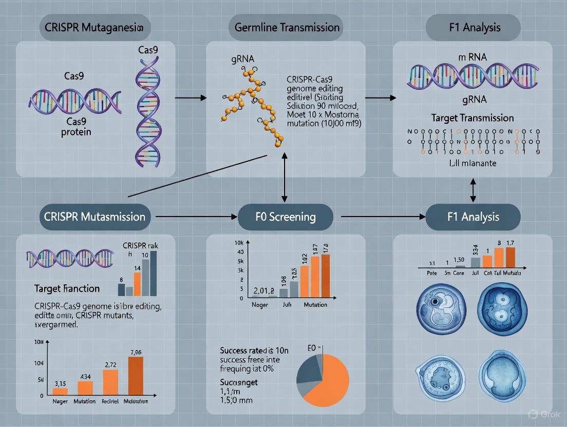
Figure 1: Workflow for establishing zebrafish mutant lines through germline transmission. The process begins with CRISPR design and culminates in stable line establishment, with germline transmission confirmation as the critical validation step.
Quantitative Benchmarks and Efficiency Optimization
Establishing Baseline Efficiency Metrics
Understanding expected efficiency rates is crucial for experimental planning in functional genomics. Large-scale zebrafish CRISPR studies have established key quantitative benchmarks for germline transmission. In one comprehensive study targeting 162 loci across 83 genes, researchers documented a 99% success rate in generating mutations, with an average germline transmission rate of 28% from founder fish [2]. This transmission rate represents the percentage of F0 founders that successfully pass induced mutations to their F1 progeny. The study further verified 678 unique alleles from 58 genes through high-throughput sequencing, demonstrating the rich genetic diversity that can be achieved through CRISPR mutagenesis [2].
Efficiency varies substantially based on the specific sgRNA design and target locus. While many published "rules" for efficient sgRNA design show limited predictive value for germline transmission rates in zebrafish, the presence of a GG or GA dinucleotide genomic match at the 5′ end of the sgRNA does correlate with improved efficiency [2]. This finding highlights the continued importance of empirical testing over purely computational predictions.
Advanced Strategies for Enhancing Germline Transmission
Knock-in and Precision Editing
While standard CRISPR knockout approaches rely on non-homologous end joining (NHEJ), more precise homology-directed repair (HDR) enables knock-in of specific point mutations—a critical capability for modeling human disease variants. However, HDR efficiency is typically much lower than NHEJ, creating significant challenges for obtaining germline transmission [4] [5].
Systematic optimization has identified several key factors that improve HDR-mediated germline transmission:
- Cas9 delivery method: Using Cas9 protein instead of mRNA significantly increases editing efficiency, with one study showing a rise from 1.04% to 2.83% somatic editing efficiency at one locus [5]
- Repair template design: Non-target asymmetric PAM-distal (NAD) single-stranded oligodeoxynucleotide (ssODN) templates outperform other conformations [5]
- Small molecule enhancement: Inhibiting NHEJ with SCR7 (a DNA ligase IV inhibitor) and stimulating HDR with RS-1 (a Rad51 stimulator) can significantly improve HDR efficiency—with SCR7 increasing point mutation rates from 16% to 58% in one study [4]
Table 2: Efficiency Benchmarks for Different CRISPR Editing Approaches in Zebrafish
| Editing Approach | Typical Somatic Efficiency | Germline Transmission Rate | Key Applications |
|---|---|---|---|
| NHEJ-Mediated Knockout | Up to 80% indel formation [6] | ~28% average [2] | Gene function loss studies, reverse genetics |
| HDR-Mediated Point Mutation | 1-5% without optimization [5] | 2-25% with optimization [4] | Disease-associated point mutation modeling |
| Base Editing | 9-90% depending on system [7] | Comparable to knockout approaches [7] | Precise nucleotide conversion without DSBs |
Early Genotyping and Selection
A powerful strategy for improving germline transmission outcomes involves early genotyping of mosaic founders to identify those with the highest editing rates. The Zebrafish Embryo Genotyper (ZEG) device enables minimally invasive DNA extraction from 72 hours post-fertilization embryos, allowing researchers to selectively raise only those embryos with the highest mutation rates [5]. This approach has demonstrated a 17-fold increase in somatic editing efficiency for some alleles, particularly benefiting those with lower inherent editing efficiencies [5]. By reducing the number of animals that need to be raised to adulthood and focusing resources on the most promising founders, this method addresses both efficiency and ethical concerns in zebrafish functional genomics.
Technical Protocols and Methodologies
High-Throughput Mutagenesis Workflow
A complete high-throughput functional genomics workflow encompasses the following key methodologies [3]:
Target Selection and sgRNA Design: Identify target sites with appropriate specificity considerations. Computational tools like CRISPRscan can assist, though empirical validation remains essential [6]
Cloning-Free sgRNA Synthesis: Using two partially overlapping oligonucleotides (one target-specific, one generic) that form a double-stranded template through annealing and extension via Taq DNA polymerase, followed by in vitro transcription directly from the linear DNA template [2]
Microinjection: Co-inject Cas9 mRNA or protein with synthesized sgRNA into one-cell stage zebrafish embryos. Protein delivery often yields higher efficiency [5]
Germline Transmission Screening: Outcross adult F0 founders to wild-types and screen seven F1 embryos per founder using fluorescence PCR or sequencing-based methods to identify germline-transmitting events [2]
Mutation Verification: Confirm exact lesions in transmitting founders by Sanger or next-generation sequencing, followed by establishment of stable lines
Mutation Detection and Verification
Accurate detection of induced mutations is crucial for identifying germline transmission events. Several methods have been systematically compared for their reliability:
- NGS-based approaches: Provide the most precise quantification of editing efficiency but at higher cost [6]
- ICE (Inference of CRISPR Edits): Analysis of Sanger sequencing data shows strong correlation (r = 0.90) with NGS data and offers a cost-effective alternative [6]
- TIDE (Tracking of Indels by Decomposition): Another Sanger-based tool with good correlation to NGS (r = 0.59) but greater tendency to underestimate efficiency [6]
- Polyacrylamide Gel Electrophoresis (PAGE): The most affordable approach but with weaker correlation to NGS data (r = 0.37), making it less reliable for quantitative assessments [6]
For challenging HDR-mediated point mutations, specific strategies improve identification:
- Incorporate blocking mutations in the repair template to prevent re-cleavage by Cas9 and enable design of mutant-specific primers [4]
- Implement nested PCR with external primers outside the donor DNA region to avoid amplification of residual donor template [4]
- Use mutant-specific primers in quantitative PCR reactions to selectively amplify HDR-induced mutations while excluding indels and wild-type sequences [4]
The Scientist's Toolkit: Essential Research Reagents
Table 3: Key Research Reagents for Zebrafish CRISPR Germline Transmission Studies
| Reagent Category | Specific Examples | Function & Application | Technical Considerations |
|---|---|---|---|
| Nuclease Systems | Cas9 mRNA, Cas9 protein, ribonucleoprotein (RNP) complexes | Induces double-strand breaks at target loci | Protein delivery often increases efficiency and reduces mosaicism [5] |
| Guide RNA Design | CRISPRscan, CHOPCHOP, ZiFiT Targeter | Bioinformatics tools for target selection | Most "rules" show limited predictive value except GG/GA at 5' end [2] |
| Repair Templates | Single-stranded oligodeoxynucleotides (ssODNs), plasmid donors | Homology-directed repair for precise editing | Non-target asymmetric PAM-distal (NAD) design outperforms other conformations [5] |
| Efficiency Enhancers | SCR7 (NHEJ inhibitor), RS-1 (HDR stimulator) | Modulate DNA repair pathways to favor desired outcome | SCR7 increased HDR efficiency from 16% to 58% in one study [4] |
| Screening Tools | ZEG device, fluorescence PCR, ICE analysis, NGS platforms | Identify and quantify germline transmission events | Early genotyping with ZEG enabled 17-fold efficiency increase [5] |
| Ampelopsin F | Ampelopsin F, MF:C28H22O6, MW:454.5 g/mol | Chemical Reagent | Bench Chemicals |
| Panaxcerol B | Panaxcerol B, MF:C27H46O9, MW:514.6 g/mol | Chemical Reagent | Bench Chemicals |
Emerging Technologies and Future Directions
Advanced Editing Platforms
Beyond standard CRISPR-Cas9 systems, several innovative technologies show significant promise for enhancing germline transmission studies:
Base editing technologies represent a particularly exciting advancement, enabling direct chemical conversion of one DNA base to another without inducing double-strand breaks. Both cytosine base editors (CBEs) and adenine base editors (ABEs) have been successfully implemented in zebrafish, achieving editing efficiencies ranging from 9.25% to 90% depending on the specific system and target locus [7]. The development of "near PAM-less" editors such as CBE4max-SpRY further expands the targeting scope, potentially increasing the range of genes accessible to germline modification [7].
Prime editing offers another promising approach, combining a Cas9 nickase with a reverse transcriptase to enable precise edits without double-strand breaks. While not yet as widely adopted in zebrafish as base editors, this technology addresses some of the limitations of both traditional HDR and base editing approaches [1].
Induced Germ Cell Technologies
A particularly innovative approach addresses the fundamental challenge of limited primordial germ cells (PGCs) in early embryos. Researchers have identified a combination of nine germplasm factors (9GMs: vasa, dazl, piwil1, dnd1, nanos3, tdrd6, tdrd7a, dazap2, and buc) that can efficiently convert blastomeres into induced PGCs (iPGCs) [8]. These iPGCs demonstrate functional capability, migrating to genital ridges and developing into functional gametes when transplanted into germ cell-deficient hosts [8].
This technology enables a novel workflow combining genome editing with iPGC transplantation:
- Perform gene editing in donor embryos
- Induce PGC formation using germplasm factors
- Transplant iPGCs into germ cell-deficient hosts
- Obtain functional gametes from the host animals
This approach "resolves the contradiction between high knock-in efficiency and early lethality" that often plagues studies of essential genes, as edited embryos that would otherwise die before reproductive maturity can still contribute to the germline through iPGC transplantation [8].
Figure 2: Innovative germline transmission workflow using induced primordial germ cells (iPGCs). This approach bypasses embryonic lethality and enhances transmission efficiency for challenging genetic edits.
Germline transmission represents the critical juncture in functional genomics where targeted genetic modifications transition from transient experimental observations to stable, research-grade biological tools. The continuous refinement of CRISPR-based technologies in zebrafish—from high-throughput mutagenesis pipelines to precision base editing and induced germ cell formation—has dramatically expanded our capacity to model genetic diseases and perform systematic functional genomic studies. As these technologies mature, they promise to further accelerate the pace of discovery in vertebrate functional genomics, enabling increasingly sophisticated investigations into gene function, genetic interactions, and the molecular basis of human disease. The ongoing optimization of germline transmission methodologies ensures that zebrafish will remain at the forefront of these efforts, providing an essential platform for connecting genetic variation to biological function.
Zebrafish as a Premier Vertebrate Model for CRISPR Screening
Zebrafish (Danio rerio) have emerged as a premier vertebrate model for high-throughput CRISPR screening, occupying a unique experimental niche between in vitro cell culture systems and low-throughput mammalian models. This position is powered by specific biological advantages: approximately 70% of human genes have at least one zebrafish ortholog, and an even higher percentage (approximately 84%) of genes known to be associated with human diseases have functional counterparts in zebrafish [9]. The external fertilization, optical transparency of embryos, and rapid development—with major organ systems formed within 24–48 hours post-fertilization—enable direct observation of phenotypic consequences in a whole vertebrate organism [9]. Furthermore, their small size, high fecundity, and cost-effectiveness facilitate large-scale genetic studies that would be prohibitively expensive or ethically challenging in mammalian systems [9] [10]. The combination of these inherent advantages with CRISPR-Cas9 technologies has revolutionized approaches to functional genomics, disease modeling, and drug discovery.
Technical Foundations: CRISPR-Cas9 Workflow in Zebrafish
The implementation of CRISPR-based screening in zebrafish follows a streamlined workflow designed for scalability and efficiency, from sgRNA design to phenotypic analysis.
sgRNA Design and Validation
The process begins with the careful design of guide RNAs (sgRNAs). While multiple computational tools exist for predicting sgRNA efficiency (e.g., CRISPRScan), studies have revealed large discrepancies between different prediction methods, underscoring the importance of empirical validation [6]. Target sites are typically selected to minimize potential off-target effects by searching for sequences with low homology to other genomic regions. The most common target site consensus for Streptococcus pyogenes Cas9 is N21GG, though designs are often adjusted to accommodate promoter requirements for in vitro transcription, such as GGN19GG [11]. Tools are available to batch-design sgRNAs and corresponding PCR primers for amplicon sequencing, enabling high-throughput pipeline development.
Delivery and Screening Strategies
CRISPR components are commonly introduced into one-cell stage zebrafish embryos via microinjection. This can be achieved using Cas9 protein mRNA in combination with sgRNAs, or as pre-assembled ribonucleoprotein (RNP) complexes [6] [10]. A significant innovation in the field is the use of mosaic G0 mutant screens, where injected individuals (G0) are phenotypically screened without raising stable mutant lines, dramatically increasing throughput [6] [12]. To accurately identify germline-transmitting mutations, researchers increasingly employ amplicon sequencing of sperm samples from G0 founder males. This approach simultaneously provides information on transmission rates and specific indel sequences, facilitates cryopreservation of sperm archives, and enables the design of efficient genotyping assays for identifying F1 carriers [11].
Table 1: Key Phases of a Zebrafish CRISPR Screening Workflow
| Phase | Key Activities | Output |
|---|---|---|
| Design & Construction | sgRNA design, oligo synthesis, in vitro transcription of sgRNAs/Cas9 mRNA | Validated sgRNAs with high predicted and empirical efficiency |
| Delivery & Mutation Generation | Microinjection into one-cell embryos, raising injected embryos | Mosaic G0 founder fish |
| Germline Screening | Sperm collection, amplicon sequencing of target loci, cryopreservation | Identification of founders transmitting desired mutations, archived sperm |
| Phenotypic Analysis | Raising F1 families, genotyping, morphological/behavioral/molecular phenotyping | Functional association between genetic perturbation and phenotype |
Quantitative Analysis of Editing Efficiency and Confounders
Understanding the efficiency and precision of CRISPR editing is crucial for experimental design and data interpretation. A systematic evaluation of 50 different gRNAs targeting 14 genes in zebrafish revealed that experimental in vivo editing efficiencies in mosaic G0 embryos often diverged significantly from computational predictions [6]. The same study provided reassuring evidence that off-target mutation rates in vivo are generally low, with the majority of tested loci showing frequencies below 1% [6].
However, researchers must be aware of potential confounders. RNA-seq analysis of "mock" injected control larvae (injected with Cas9 enzyme or mRNA without gRNA) revealed hundreds of differentially expressed genes compared to uninjected siblings [6]. These genes were associated with processes such as "response to wounding" and "cytoskeleton organization," highlighting a potentially lasting effect from the microinjection process itself that could confound phenotypic analysis if not properly controlled [6].
Table 2: Efficiency of Precision Genome Editing Technologies in Zebrafish
| Editing Technology | Editing Type | Key Features | Reported Efficiency | Key References |
|---|---|---|---|---|
| CRISPR-Cas9 (NHEJ) | Indels (knockout) | Creates double-strand breaks, repaired by NHEJ | High efficiency, germline transmission up to 28% | [1] [11] |
| HDR-Mediated Knock-in | Point mutations, small insertions | Requires donor DNA template; lower efficiency | Challenging; germline transmission traditionally <2% | [13] |
| Base Editors (BE3, AncBE4max) | Single-base substitutions (C>T, A>G) | No double-strand breaks; reduced indels | BE3: 9-28%; AncBE4max: ~3x BE3 efficiency | [7] |
| Prime Editing (PE2, PEn) | All 12 possible base-to-base conversions, small insertions/deletions | No double-strand breaks; uses pegRNA and reverse transcriptase | PE2: 8.4% for substitution; PEn: superior for insertions up to 30bp | [14] |
Advanced Tools: Precision Editing with Base and Prime Editors
Beyond conventional CRISPR-Cas9, more precise genome editing tools have been successfully adapted for zebrafish research, significantly expanding the scope of disease modeling.
Base Editors enable single-nucleotide changes without inducing double-strand breaks, addressing a key limitation of traditional CRISPR-Cas9. Cytosine Base Editors (CBEs) catalyze C•G to T•A conversions, while Adenine Base Editors (ABEs) facilitate A•T to G•C changes [7]. Continuous development has yielded improved editors with expanded capabilities. For instance, the AncBE4max system, optimized for zebrafish codon usage, demonstrated approximately threefold higher editing efficiency compared to the original BE3 system [7]. More recently, a "near PAM-less" cytidine base editor (CBE4max-SpRY) was developed, bypassing the traditional NGG PAM requirement and enabling targeting of virtually all PAM sequences with efficiencies reaching up to 87% at some loci [7].
Prime Editors represent a further advancement, capable of implementing all 12 possible base-to-base conversions, as well as small insertions and deletions, without requiring double-strand breaks or donor DNA templates. A comparative study in zebrafish demonstrated that the nickase-based PE2 editor was more efficient for precise base pair substitutions (8.4% vs. 4.4% for PEn), while the nuclease-based PEn editor was superior for inserting short DNA fragments (up to 30 bp) [14]. This versatility enables the precise modeling of human disease-associated point mutations and the insertion of functional sequences like nuclear localization signals [14].
Diagram 1: Zebrafish CRISPR screening workflow, showing parallel paths for direct G0 phenotypic analysis and germline transmission for stable line generation.
Successful implementation of zebrafish CRISPR screening requires a collection of well-characterized reagents and resources.
Table 3: Essential Research Reagent Solutions for Zebrafish CRISPR Screening
| Reagent/Resource | Function/Purpose | Key Considerations |
|---|---|---|
| Cas9 Expression Vector or Cas9 Protein | Engineered nuclease that cleaves target DNA | Can be delivered as mRNA, protein, or plasmid; codon-optimized versions enhance efficiency |
| sgRNA Design Tools (e.g., CRISPRScan, ACEofBASEs) | Bioinformatics design of specific guide RNAs | Predict on-target efficiency and potential off-target effects; species-specific tools available |
| Target-Specific sgRNAs | Guides Cas9 to specific genomic loci | Chemically synthesized or in vitro transcribed; modified sgRNAs can enhance stability |
| Amplicon Sequencing Primers | Amplification of target regions for NGS validation | Nested primer designs improve specificity for sequencing-based efficiency quantification |
| Microinjection Equipment | Delivery of CRISPR components into embryos | Standard zebrafish microinjection setup with fine-needle pipettes |
| Phenotypic Assay Reagents (e.g., Nile Red, antibodies) | Detection and quantification of phenotypic outcomes | Cell-type specific reporters, lipophilic dyes for adiposity, behavioral tracking systems |
Case Study: In Vivo CRISPR Screening for Adipose Tissue Regulators
A compelling demonstration of zebrafish's power for CRISPR screening comes from a study investigating regulators of adipose tissue remodeling [12]. Researchers developed a quantitative imaging pipeline to assess hyperplastic (many small adipocytes) versus hypertrophic (few large adipocytes) morphology in zebrafish subcutaneous adipose tissue. They then applied this platform in an F0 CRISPR mutagenesis screen targeting 25 candidate genes derived from human genetic and transcriptomic data on adipocyte size [12].
The screen successfully identified six genes that significantly altered adipose morphology. Three genes (foxp1b, txnipa, mmp14b) disruption induced hypertrophic morphology, while three others (ptenb, cxcl14, srpx) induced hyperplastic morphology [12]. Notably, Sushi Repeat Containing Protein (Srpx) had no previously characterized role in adipose biology. Follow-up studies on foxp1b revealed that mutants displayed a developmental bias toward hypertrophic growth but failed to undergo further hypertrophic remodeling in response to a high-fat diet, suggesting that early developmental patterning constrains later adaptive responses to nutritional challenge [12]. This case study exemplifies the power of combining zebrafish CRISPR screening with quantitative phenotyping to uncover novel genetic regulators of physiological processes with direct relevance to human metabolic health.
Zebrafish have firmly established their position as a premier vertebrate model for CRISPR screening, combining genetic tractability with physiological relevance in a scalable format. The continuous refinement of genome editing tools—from efficient Cas9 variants to base and prime editors—is expanding the range of genetic questions that can be addressed. These technologies enable everything from large-scale knockout screens to precise modeling of human disease-associated point mutations.
Future directions in the field will likely focus on increasing the throughput and sophistication of both genetic perturbations and phenotypic readouts. Methods such as MIC-Drop and Perturb-seq, which increase screening scalability and enable single-cell transcriptional profiling of CRISPR perturbations, respectively, hold significant promise for dissecting complex biological mechanisms [1]. Furthermore, the integration of single-cell transcriptomics, computational modeling, and machine learning with zebrafish CRISPR screening data is enhancing the translational relevance of findings [9]. As these technologies mature and datasets expand, zebrafish will continue to provide invaluable insights into gene function, disease mechanisms, and therapeutic strategies, solidifying their role in the functional genomics landscape.
In the field of functional genomics, the advent of CRISPR-Cas technology has revolutionized our ability to perform targeted genome editing in model organisms. Central to this process are the DNA repair mechanisms that resolve the CRISPR-induced double-strand breaks (DSBs). In zebrafish (Danio rerio), a premier model for vertebrate biology and human disease, understanding these pathways is paramount for improving the efficiency of precise gene editing, particularly in the context of germline transmission of genetic modifications. The three primary pathways—Non-Homologous End Joining (NHEJ), Homology-Directed Repair (HDR), and Microhomology-Mediated End Joining (MMEJ)—compete to repair DSBs, each resulting in distinct mutational outcomes [15] [16]. Their complex interplay directly influences the success of CRISPR knock-in strategies, where the goal is the precise integration of exogenous DNA sequences over error-prone mutagenesis [17]. This whitepaper provides an in-depth technical guide to these mechanisms, framed within the context of zebrafish CRISPR research, to empower scientists in the design and interpretation of their genome editing experiments.
Core DNA Repair Pathways in Zebrafish
In zebrafish, as in other vertebrates, the repair of CRISPR-Cas9-induced DSBs is a competitive process between several conserved pathways. The table below summarizes the key characteristics of the three major pathways.
Table 1: Core DNA Double-Strand Break Repair Pathways in Zebrafish CRISPR Mutagenesis
| Pathway | Key Proteins | Template Requirement | Mutagenic Outcome | Typical Efficiency in Zebrafish |
|---|---|---|---|---|
| Non-Homologous End Joining (NHEJ) | Ku70/80, DNA-PKcs, DNA Ligase IV [15] | None (error-prone) | Small insertions/deletions (indels); imperfect knock-in [16] | High (dominant pathway) [18] |
| Homology-Directed Repair (HDR) | Rad51, BRCA2, RAD52 [17] [15] | Homologous donor DNA (precise) | Precise nucleotide changes; accurate knock-in [18] | Low (typically <10% without optimization) [18] [16] |
| Microhomology-Mediated End Joining (MMEJ) | POLQ (DNA Polymerase Theta), PARP1 [17] | Microhomology sequences (5-25 bp) | Deletions flanked by microhomology regions [17] | Variable; contributes to imprecise repair [17] |
The NHEJ pathway is the most active in zebrafish embryos and is often considered a major barrier to precise HDR-mediated knock-in. It functions throughout the cell cycle by directly ligating broken DNA ends, a process that frequently results in small insertions or deletions (indels) [16]. In CRISPR experiments, this typically leads to gene knockouts.
HDR is the only pathway that can facilitate precise knock-in, such as the introduction of fluorescent protein tags or specific disease-associated point mutations. It requires a donor DNA template with homology arms and is active primarily in the S and G2 phases of the cell cycle. Its low efficiency in zebrafish is a significant technical challenge, often resulting in mosaic embryos where only a subset of cells carries the desired modification [18] [16].
MMEJ is an alternative, error-prone repair pathway that operates in a microhomology-dependent manner. It typically results in larger deletions than NHEJ. Recent studies have shown that even with NHEJ inhibition, imprecise repair persists due to the activity of MMEJ and the related Single-Strand Annealing (SSA) pathway, complicating knock-in efforts [17].
The following diagram illustrates the competitive interplay between these pathways following a CRISPR-induced double-strand break in a zebrafish cell.
Diagram 1: Competitive DNA Repair Pathways Activated by CRISPR/Cas9. Following a double-strand break (DSB), the cell utilizes different repair mechanisms based on template availability, leading to distinct mutational outcomes with varying efficiencies.
Quantitative Analysis of Repair Outcomes
Understanding the relative efficiency and outcomes of these pathways is critical for experimental design. Recent quantitative studies in zebrafish and human cells have shed light on the distribution of repair events.
Table 2: Quantitative Distribution of CRISPR-Mediated Repair Outcomes in RPE1 Cells (with NHEJ Inhibition)
| Repair Outcome | Approximate Frequency | Key Characteristics |
|---|---|---|
| Perfect HDR | Variable; increased with pathway suppression | Precise integration of the donor sequence [17] |
| Small Deletions (<50 nt) | Significantly reduced | Typically associated with NHEJ [17] |
| Large Deletions (≥50 nt) & Complex Indels | ~50% of integration events | Associated with MMEJ and other alternative pathways [17] |
| Asymmetric HDR | Reduced by SSA suppression | Only one end of the donor is precisely integrated [17] |
A key finding from recent research is that inhibiting the predominant NHEJ pathway, while boosting the proportion of perfect HDR, is not sufficient to completely suppress non-HDR repairs. In human RPE1 cells, even with NHEJ inhibition, imprecise integration still accounted for nearly half of all integration events, underscoring the significant role of alternative pathways like MMEJ and SSA in CRISPR knock-in [17].
Advanced Strategies to Modulate Repair Pathways
A primary focus in zebrafish CRISPR research is to shift the repair equilibrium away from error-prone pathways and toward HDR. Both genetic and chemical modulation strategies have been successfully employed.
Chemical Reprogramming of DNA Repair
The use of small-molecule inhibitors to transiently modulate key repair proteins has proven highly effective in zebrafish embryos. The table below summarizes key reagents used to enhance HDR efficiency.
Table 3: Research Reagent Solutions for Enhancing HDR in Zebrafish
| Reagent / Solution | Target Pathway | Molecular Function | Effect on Genome Editing |
|---|---|---|---|
| NU7441 [18] | NHEJ inhibitor | DNA-PK inhibitor [18] | Enhanced HDR efficiency up to 13.4-fold in zebrafish embryos [18] |
| Alt-R HDR Enhancer V2 [17] | NHEJ inhibitor | Potent, commercially available NHEJ inhibitor | Increased knock-in efficiency approximately 3-fold in human cell studies [17] |
| ART558 [17] | MMEJ inhibitor | Selective inhibitor of POLQ (DNA Polymerase Theta) [17] | Reduces large deletions and complex indels; increases perfect HDR frequency [17] |
| SCR7 [18] | NHEJ inhibitor | DNA Ligase IV inhibitor | Shows species-specific effects; no significant HDR enhancement in zebrafish [18] |
| RS-1 [18] | HDR enhancer | RAD51 stimulator | Modest but significant increase in HDR efficiency [18] |
The experimental workflow for a typical chemical enhancement experiment in zebrafish is illustrated below.
Diagram 2: Workflow for Enhanced HDR in Zebrafish Embryos. The protocol involves co-injection of CRISPR components and a donor template into one-cell stage embryos, followed by immediate treatment with small molecules to modulate DNA repair pathways, ultimately leading to screening and validation of precisely edited animals.
Inhibition of Alternative Repair Pathways
Beyond NHEJ, targeting the MMEJ and SSA pathways is a novel strategy to further enhance precision. Suppressing POLQ, the central effector of MMEJ, reduces the occurrence of large deletions and complex indels at the target site [17]. Similarly, inhibiting RAD52, a key protein in the SSA pathway, reduces imprecise donor integration, particularly a faulty repair pattern known as "asymmetric HDR" where only one end of the donor DNA integrates correctly [17]. Combined inhibition of NHEJ and these alternative pathways represents the cutting edge for improving precise knock-in efficiency.
Experimental Protocols for Zebrafish
Visual HDR Reporter Assay
A quantitative in vivo reporter system was developed to screen for HDR-enhancing conditions in zebrafish at single-cell resolution [18].
- Principle: A transgenic zebrafish line expresses eBFP2 (blue fluorescent protein) specifically in fast-muscle cells. A donor template containing the tdTomato (red fluorescent protein) gene flanked by homology arms is designed to replace eBFP2 upon successful HDR.
- Method: Embryos are co-injected with Cas9 protein, a guide RNA targeting eBFP2, and the tdTomato donor DNA. Successful HDR converts individual muscle fibers from blue to red fluorescence.
- Quantification: The number of red fluorescent fibers in living embryos is counted at 72 hours post-fertilization (hpf). This provides a quantitative and rapid readout of HDR efficiency, allowing for the testing of chemical modulators like NU7441, which was shown to increase HDR events from 4.0 to 53.7 red fibers per embryo on average [18].
Optimized Protocol for HDR-Mediated Knock-In
The following is a detailed methodology for achieving precise knock-in in zebrafish, incorporating best practices and chemical enhancement [18] [16]:
- sgRNA Design: Select a sgRNA with very high efficiency (indel frequency >70% in pooled embryos). The target site should be as close as possible to the intended insertion point [16].
- Donor Template Design: For linear dsDNA donors, use homology arms of 300-1000 bp. Incorporating the sgRNA target sequence within the homology arm can promote "self-linearization" of the donor and enhance HDR efficiency [18].
- Microinjection Cocktail: Co-inject into the cytoplasm of one-cell stage embryos:
- CRISPR Component: High-quality Cas9 protein pre-complexed with sgRNA as a ribonucleoprotein (RNP) complex for rapid activity.
- Donor DNA: 50-100 ng/μL of purified, linearized donor template.
- Chemical Modulators: Add 50 μM NU7441 directly to the injection mix or incubate embryos in the solution immediately after injection [18].
- Screening and Validation: Raise injected embryos (F0) and screen for somatic editing using fluorescence, PCR, or restriction fragment analysis. Cross founder F0 fish to wild-types and screen the F1 offspring for germline transmission of the precise edit.
The competition between NHEJ, HDR, and MMEJ pathways fundamentally shapes the outcomes of CRISPR genome editing in zebrafish. While NHEJ remains the dominant and most efficient pathway, leading to high knockout rates, HDR's inefficiency has been a major bottleneck for precise knock-in. The strategic inhibition of NHEJ and the emerging targeting of alternative pathways like MMEJ and SSA provide powerful chemical-genetic strategies to reprogram the zebrafish embryo's repair landscape. Quantitative assays and optimized protocols are crucial for evaluating these strategies and achieving seamless integration of genetic material. As the field moves forward, a deeper understanding of the complex interplay between these DNA repair mechanisms will continue to enhance the precision and efficiency of generating zebrafish models with germline-transmitted mutations, thereby accelerating functional genomics and the modeling of human disease.
The establishment of heritable mutant lines is the cornerstone of genetic research in model organisms. In zebrafish (Danio rerio), the efficiency with which CRISPR-induced mutations are transmitted through the germline to subsequent generations fundamentally determines the practicality and scale of functional genomics studies. The advent of CRISPR-Cas9 technology has revolutionized targeted mutagenesis in zebrafish, offering unprecedented capabilities for high-throughput functional genomics and disease modeling [19]. This whitepaper provides a comprehensive technical benchmark of germline transmission rates in zebrafish CRISPR mutants, tracing the evolution of these critical efficiency metrics from foundational studies to contemporary precision editing approaches. Within the broader context of zebrafish CRISPR research, understanding these benchmarks enables researchers to design appropriately powered experiments, allocate resources efficiently, and advance therapeutic discovery through robust in vivo validation of candidate genes.
Historical Benchmarks: Establishing CRISPR-Cas9 in Zebrafish
Foundational High-Throughput Mutagenesis
Early large-scale efforts to establish CRISPR-Cas9 as a robust tool in zebrafish provided critical baseline metrics for germline transmission. A seminal 2015 study by Varshney et al. established a high-throughput pipeline that would serve as a benchmark for years to come [2]. This groundbreaking work reported:
Key Historical Germline Transmission Data (Varshney et al., 2015) [2]
| Metric | Result |
|---|---|
| Overall Success Rate for Generating Mutations | 99% (across 162 loci targeting 83 genes) |
| Average Germline Transmission Rate | 28% |
| Unique Alleles Verified | 678 from 58 genes |
| Comparative Efficiency vs. TALENs/ZFNs | ~6-fold more efficient |
This study demonstrated that CRISPR/Cas9 was not only highly efficient at inducing mutations but also reliably transmitted these mutations through the germline. The authors employed a cloning-free single-guide RNA (sgRNA) synthesis method that significantly increased throughput, enabling the synthesis of hundreds of sgRNAs in a few hours [2]. Their strategy involved crossing founder fish (F0) to wild-type fish and analyzing F1 progeny for insertions or deletions (indels) using fluorescence PCR or sequencing, establishing a protocol that would become standard for germline transmission assessment.
Technical Workflows for Germline Identification
The development of streamlined protocols was instrumental in standardizing the identification of germline-transmitting founders. A 2018 protocol by Boel et al. detailed a robust, economical CRISPR-Cas9 strategy specifically designed to minimize equipment needs and enable participation by personnel across experience levels [20]. This protocol emphasized:
- Efficient sgRNA Synthesis: Using target-specific and generic oligonucleotides to form double-stranded templates for in vitro transcription.
- Cas9 Delivery: Employing Cas9 protein rather than mRNA, with injection-ready aliquots stored at -80°C to minimize freeze-thaw cycles.
- Mutation Identification: Implementing a PCR-based heteroduplex mobility assay (HMA) for initial screening, followed by next-generation sequencing (NGS) for comprehensive characterization of indels in heterozygous fish [20].
This workflow demonstrated that careful optimization of reagent preparation and delivery could yield consistent germline transmission without requiring highly specialized expertise, thus democratizing the generation of zebrafish knockout lines.
Current Benchmarks: Precision Genome Editing and Transmission Efficiencies
Advanced Editing Platforms: Base and Prime Editors
Recent advances have shifted from simple knockout approaches to precise nucleotide-level editing, with corresponding developments in germline transmission metrics.
Base Editors enable direct conversion of one nucleotide to another without double-strand breaks through fusion of catalytically impaired Cas proteins with deaminase enzymes [7]. Cytosine Base Editors (CBEs) facilitate C:G to T:A conversions, while Adenine Base Editors (ABEs) catalyze A:T to G:C changes [7]. The evolution of these tools in zebrafish has seen progressive efficiency improvements:
- BE3 System: Initial cytosine base editing with efficiencies of 9.25% to 28.57% in somatic cells [7].
- AncBE4max: A codon-optimized system showing approximately threefold enhanced editing efficiency compared to BE3 [7].
- CBE4max-SpRY: A "near PAM-less" cytidine base editor achieving efficiencies up to 87% at some loci, bypassing traditional NGG PAM requirements [7].
Prime Editors represent a more versatile precise editing technology that uses a Cas9 nickase-reverse transcriptase fusion and a prime editing guide RNA (pegRNA) to directly write new genetic information into a target site [14]. A 2025 study systematically compared nickase-based (PE2) and nuclease-based (PEn) prime editors in zebrafish, with significant implications for germline transmission:
Prime Editing Efficiency Comparison [14]
| Editor Type | Application | Precision Editing Efficiency | Indel Rate | Precision Score |
|---|---|---|---|---|
| PE2 (Nickase) | Nucleotide substitution | 8.4% | Lower | 40.8% |
| PEn (Nuclease) | Nucleotide substitution | 4.4% | Higher | 11.4% |
| PEn (Nuclease) | Short sequence insertion (3-30 bp) | Higher than PE2 | Variable | Not specified |
This study further demonstrated that both somatic precision editing and germline transmission of these precise modifications could be achieved, with the editing mode (substitution vs. insertion) dictating the optimal editor choice [14].
Optimized Knock-In Strategies for Germline Transmission
Recent methodological refinements have significantly improved knock-in efficiency, which traditionally lagged behind knockout approaches. A 2023 study described the "S-NGG-25" strategy—an optimized MMEJ-mediated knock-in approach using donors with shortened microhomology arms (25 bp) and reduced Cas9/sgRNA consensus sites [21]. This method demonstrated:
- High-Efficiency Germline Transmission: Successfully applied to 33 connexin genes, with germline transmission rates ranging from 5.8% to 35.4% across founders [21].
- Streamlined Screening: Implementation of a combined fluorescence enrichment and caudal-fin junction-PCR protocol to expedite the identification of germline-transmitting founders [21].
- Reduced Mosaicism: Improved rates of obtaining seamless F1 carriers with precise integrations [21].
This optimized platform addresses one of the most challenging aspects of zebrafish genome engineering—efficient and precise protein tagging—with direct implications for improving germline transmission rates of complex alleles.
Experimental Protocols for Assessing Germline Transmission
Foundational Workflow for Germline Transmission Assessment
The following experimental workflow visualizes the standard process for generating and identifying zebrafish with germline-transmitted mutations:
Diagram 1: Standard workflow for assessing germline transmission in zebrafish CRISPR mutants.
Step 1: sgRNA Design and Synthesis
- Design: Select target sites in early exons near the 5' end of coding sequences to maximize likelihood of gene disruption. Ideal sgRNA sequences contain 40-80% GC content with a G at the 5′ position for efficient T7 transcription [20].
- Synthesis: Employ cloning-free methods using partially overlapping oligonucleotides (target-specific + generic scaffold) annealed and extended to form dsDNA templates for in vitro transcription [2].
Step 2: Microinjection
- Preparation: Combine sgRNA with Cas9 protein (1 mg/mL) in injection-ready aliquots [20].
- Delivery: Microinject into the yolk or cell cytoplasm of one-cell stage embryos [20].
- Controls: Include uninjected embryos from the same clutch as critical controls [6].
Step 3: Founder Raising
- Rearing: Raise injected embryos (F0) to sexual maturity (approximately 3 months).
- Mosaicism Expectation: F0 founders are highly mosaic, carrying multiple different editing events in different germ cells [22].
Step 4: Outcrossing
- Breeding: Outcross individual F0 founders to wild-type fish.
- Germline Assessment: The resulting F1 embryos represent the genetic composition of the germline of the F0 founder.
Step 5: Mutation Screening
- Initial Screening: Use heteroduplex mobility assays (HMA) or T7 endonuclease I (T7E1) assays to identify potential mutants [20].
- Advanced Screening: Implement fluorescence enrichment strategies when fluorescent reporters are co-injected [21].
Step 6: Sequence Verification
- Amplification: PCR-amplify target regions from pooled F1 embryos or fin clips from adult F1 fish.
- Sequencing: Utilize Sanger sequencing or next-generation sequencing (NGS) to characterize specific indel patterns or precise edits [20] [2].
- Transmission Rate Calculation: Calculate as the percentage of F1 offspring carrying the mutation derived from a specific F0 founder.
Advanced Methodologies for Precision Editing
For base and prime editing applications, modified protocols are required:
Base Editing Workflow [7]
- Component Preparation: Synthesize mRNA for base editor (BE) constructs (e.g., BE3, AncBE4max) or prepare ribonucleoprotein (RNP) complexes.
- Microinjection: Inject BE mRNA/sgRNA complexes or RNP into one-cell stage embryos.
- Efficiency Optimization: Modify PAM recognition using engineered Cas variants (e.g., VQR, SpRY) to expand targetable sites.
- Temperature Control: Incubate injected embryos at elevated temperatures (e.g., 32°C) to enhance base editing efficiency in some systems [14].
Prime Editing Workflow [14]
- pegRNA Design: Design prime editing guide RNAs with reverse transcription template and primer binding site sequences.
- Component Delivery: Co-inject PE mRNA or protein with pegRNA into zebrafish embryos.
- Editing Validation: Use amplicon sequencing of target regions to quantify precise edit incorporation rates and identify potential byproducts.
The Scientist's Toolkit: Essential Reagents and Methods
Table 1: Key Research Reagent Solutions for Zebrafish CRISPR Germline Transmission Studies
| Reagent/Method | Function | Technical Notes |
|---|---|---|
| Cas9 Protein | CRISPR endonuclease that creates DSBs at target sites | Using protein instead of mRNA can increase efficiency and reduce mosaicism [20] |
| sgRNA In Vitro Transcription Kit | High-throughput sgRNA synthesis | Enables cloning-free production of hundreds of sgRNAs [2] |
| Heteroduplex Mobility Assay (HMA) | Initial mutation screening | Rapid, inexpensive method to detect indels in pooled embryos [20] |
| Next-Generation Sequencing (NGS) | Comprehensive mutation characterization | Precisely identifies and quantifies mutant alleles; essential for assessing complex editing outcomes [20] |
| Fluorescence Enrichment | Screening for knock-in events | Uses co-injected fluorescent markers or integrated tags to identify potential founders [21] |
| Caudal-Fin Junction PCR | Germline transmission screening | Enables non-lethal genotyping of potential F0 founders before breeding [21] |
| Long-Read Sequencing (PacBio/Nanopore) | Detecting structural variants | Identifies large, complex on-target and off-target edits missed by short-read methods [22] |
| GS-443902 trisodium | GS-443902 trisodium, MF:C12H14N5Na3O13P3+, MW:598.16 g/mol | Chemical Reagent |
| TRAP-14 amide | TRAP-14 amide, MF:C81H119N21O22, MW:1738.9 g/mol | Chemical Reagent |
Factors Influencing Germline Transmission Efficiency
Technical and Biological Determinants
Germline transmission rates are influenced by multiple interconnected factors:
sgRNA Design: Optimal sgRNAs feature high GC content (40-80%), a G at the 5′ position, and minimal predicted off-target effects [20]. However, historical data suggests that most published "rules" for sgRNA design poorly predict germline transmission rates, with the exception of a GG or GA dinucleotide at the 5′ end [2].
Delivery Method and Timing: Microinjection at the one-cell stage is critical. Delivery of preassembled Cas9-sgRNA ribonucleoprotein (RNP) complexes generally yields higher editing and germline transmission rates compared to mRNA injection [20] [22].
Biological Constraints: The inherent mosaicism of F0 founders remains a fundamental challenge. Studies demonstrate that adult founder zebrafish are mosaic in their germ cells, with individual founders carrying multiple different editing events [22].
Addressing Unintended Mutagenesis
A comprehensive 2022 study revealed that CRISPR-Cas9 can introduce large structural variants (SVs ≥50 bp) at both on-target and off-target sites, with 6% of editing outcomes in founder larvae representing such SVs [22]. Critically, these SVs can be transmitted to the next generation, with 9% of F1 offspring carrying an SV [22]. This highlights the importance of:
- Comprehensive Validation: Using long-read sequencing technologies to detect large structural variants missed by conventional methods.
- Off-Target Assessment: Employing genome-wide methods like Nano-OTS to identify potential off-target sites before germline transmission studies [22].
The benchmarking data presented in this whitepaper trace a trajectory of remarkable progress in zebrafish CRISPR germline transmission efficiency. From the foundational average of 28% transmission in early high-throughput studies [2] to the sophisticated precision editing systems now achieving efficiencies above 80% at optimized loci [7], the field has witnessed substantial methodological refinement. Current research priorities include improving the fidelity of complex knock-in approaches [21], minimizing structural variants [22], and expanding the target scope of precision editors [7] [14]. For researchers and drug development professionals, these benchmarks provide critical reference points for experimental design, highlighting both the capabilities and limitations of current zebrafish genome engineering platforms. As these technologies continue to mature, they promise to further accelerate the use of zebrafish in functional genomics and therapeutic discovery.
Within vertebrate biomedical research, the zebrafish (Danio rerio) has emerged as a preeminent model that uniquely combines high experimental throughput with ethical husbandry and robust 3Rs (Replacement, Reduction, and Refinement) compliance. This technical whitepaper examines these advantages within the specific context of generating and analyzing germline transmission in zebrafish CRISPR mutants. We detail how the zebrafish model enables rapid, large-scale genetic screens and detailed functional validation of gene edits with significantly reduced animal usage, lower costs, and diminished ethical concerns compared to traditional mammalian models. The integration of CRISPR/Cas9 technology with the inherent biological features of zebrafish creates a powerful platform for accelerating preclinical research, particularly in the validation of disease-associated genetic variants and drug discovery pipelines.
The generation of stable, heritable mutant lines through germline transmission is a fundamental requirement for functional genomics and disease modeling. In this context, the zebrafish offers a compelling alternative to mammalian models. The external fertilization and development of zebrafish embryos provide direct access to the germline from the earliest stages, enabling high-efficiency CRISPR/Cas9 editing and simplifying the recovery of mutant alleles [23] [24]. Furthermore, approximately 70% of human genes have at least one zebrafish ortholog, and 84% of genes known to be associated with human disease have a zebrafish counterpart, making it a highly relevant model for human biology and pathology [9]. This high degree of genetic conservation, combined with its practical advantages, positions the zebrafish as an ideal system for studying the functional consequences of genetic alterations in a vertebrate system. The following sections will dissect the specific advantages of throughput, husbandry, and 3Rs compliance, providing a technical foundation for leveraging the zebrafish model in sophisticated germline transmission studies.
Quantitative Advantages in Throughput and Scale
The experimental throughput achievable with zebrafish is orders of magnitude greater than that of mammalian models, a critical factor in large-scale genetic screens and drug discovery initiatives.
Table 1: Comparative Throughput and Husbandry of Animal Models
| Feature | Zebrafish | Mice | Source |
|---|---|---|---|
| Embryos/Clutch | 70 - 300 | 2 - 12 (litter size) | [24] [25] |
| Time to Sexual Maturity | 2 - 3 months | ~2 months | [24] |
| Developmental Timeline | Major organs formed in 24-48 hours | Several weeks | [9] |
| High-Throughput Screening | Very high (larvae in multi-well plates) | Moderate | [9] |
| Husbandry Cost | Low | High | [9] |
The data in Table 1 underscores the scalability of the zebrafish system. A single mating pair can produce hundreds of embryos weekly, enabling high-throughput phenotypic assays and large-scale mutagenesis screens that would be logistically and financially prohibitive in mice [9] [24]. This fecundity is crucial for germline transmission studies, as it allows researchers to screen large numbers of F1 offspring to identify those carrying the desired mutation, even when transmission rates are low.
The rapid development of zebrafish is another key throughput accelerator. Major organ systems are formed within 24 to 72 hours post-fertilization (hpf), allowing for the rapid assessment of phenotypic outcomes in a vertebrate system [9]. This speed, combined with the ability to house many animals in a small space, significantly compresses research timelines from gene editing to phenotypic analysis in stable lines.
Husbandry and Economic Efficiency
The practical aspects of zebrafish husbandry contribute directly to its status as a cost-effective and efficient model.
- Size and Space Efficiency: The small size of zebrafish (larval and adult stages) allows for high-density housing in aquarium systems, drastically reducing the physical footprint and maintenance costs compared to mammalian facilities [9] [25].
- Cost-Effectiveness: Overall husbandry costs for zebrafish are significantly lower than for mice. This includes expenses related to housing, feeding, and husbandry labor, making large-scale experiments more feasible [9].
- Genetic Diversity: Unlike highly inbred mammalian models, common laboratory zebrafish strains are genetically heterogeneous. This diversity more accurately models human genetic variation and can provide more translatable results, particularly in drug response studies [24]. While this requires careful experimental design and appropriate sample sizes, the large number of available embryos makes this manageable.
Adherence to the 3Rs Principles in Zebrafish CRISPR Research
The zebrafish model aligns powerfully with the ethical principles of Replacement, Reduction, and Refinement (3Rs), a cornerstone of modern humane science.
Replacement
A paramount ethical advantage is the classification of zebrafish embryos and larvae before 5 days post-fertilization (dpf). According to EU Directive 2010/63/EU, they are not considered protected animals as they have not begun independent feeding [26]. This allows researchers to gather systemic in vivo data from a whole vertebrate organism under an in vitro classification, effectively replacing the use of protected animals in early-stage toxicity screens, disease modeling, and drug discovery [26] [25].
Reduction
Zebrafish are inherently suited to reducing animal numbers:
- Sequential Assessment: The optical transparency of larvae enables researchers to non-invasively measure multiple parameters (e.g., organ function, behavior) in the same animal over time, reducing variability and the need for separate cohorts for each measurement [26].
- Pipeline Impact: Using zebrafish in early-stage discovery helps narrow down the most promising compounds or genetic targets, reducing the number of mammals required for subsequent regulatory testing phases [26] [25].
Refinement
Zebrafish research naturally refines experimental approaches to minimize suffering.
- Non-Invasive Imaging: The transparency of embryos and larvae (including genetically transparent lines like casper) allows for high-resolution, real-time imaging of internal processes without invasive surgical procedures, minimizing stress and harm [9] [26] [24].
- Minimal Intervention: This transparency reduces or eliminates the need for invasive techniques that are common in mammalian research, leading to less stressful experimental procedures and more accurate, artifact-free data [26].
Experimental Protocols for Germline Transmission
Efficient germline transmission of CRISPR/Cas9-induced mutations is critical for establishing stable lines. The following protocols detail optimized methodologies.
Optimized CRISPR/Cas9 Delivery for High-Efficiency Editing
The choice of reagents and delivery methods significantly impacts mutagenesis efficiency and mosaicism in G0 embryos, which in turn influences germline transmission rates.
Table 2: Key Research Reagent Solutions for Zebrafish CRISPR
| Reagent / Tool | Function / Description | Application in Germline Transmission |
|---|---|---|
| Cas9 Protein | Pre-complexed ribonucleoprotein; enables immediate activity upon injection. | Superior to Cas9 mRNA; leads to higher editing efficiency and reduced mosaicism [5] [27]. |
| sgRNA (single guide RNA) | Synthetic RNA guiding Cas9 to specific genomic loci. | Target-specific cleavage. Efficiency can be predicted using tools like CRISPRScan [23] [6]. |
| ssODN (single-stranded Oligodeoxynucleotide) | Short, single-stranded DNA template for Homology-Directed Repair (HDR). | Used for introducing precise point mutations (knock-ins). Non-target asymmetric PAM-distal (NAD) conformations are recommended [5]. |
| Zebrafish Embryo Genotyper (ZEG) | Device for minimally invasive biopsy of 72 hpf embryos. | Allows early genotyping and selective raising of embryos with high editing efficiency, reducing animals raised and screened [5]. |
| NHEJ Inhibitors | Chemical compounds that suppress the error-prone Non-Homologous End Joining pathway. | Can be used to favor HDR, improving knock-in efficiency [27]. |
Protocol Steps:
- Component Preparation: Synthesize sgRNA via in vitro transcription from a DNA template or use pre-designed crRNA:tracrRNA duplexes. Use purified Cas9 protein.
- Microinjection Mix Preparation: Co-complex the sgRNA and Cas9 protein to form a ribonucleoprotein (RNP) complex prior to injection. For knock-ins, include an ssODN repair template with a NAD conformation [5].
- Zygote Microinjection: Inject 1-2 nL of the RNP mixture directly into the cytoplasm of single-cell stage zebrafish embryos.
- Temperature Manipulation (Optional): Incubate injected embryos at a reduced temperature (e.g., 12°C) for 30-60 minutes post-injection. This extends the single-cell stage, significantly increasing mutagenesis efficiency by providing more time for Cas9 to act before DNA replication and cell division [28].
Screening for Germline Transmission
Identifying founders (F0 fish that transmit mutations through their germline) is a critical step.
- Early Somatic Screening: At 3-5 dpf, use the ZEG device to extract a small amount of genomic DNA from embryos with minimal lethality. Analyze this DNA via next-generation sequencing (NGS) to quantify somatic editing efficiency [5].
- Founder Selection and Raising: Select and raise only the embryos showing the highest levels of correct editing to adulthood. This pre-selection can lead to a 17-fold increase in somatic editing efficiency in the raised cohort, dramatically improving the odds of obtaining germline transmission [5].
- Outcrossing and F1 Screening: Outcross the adult pre-selected F0 fish to wild-type partners. Collect and raise F1 embryos.
- Genotyping F1 Progeny: At an appropriate stage, genotype individual F1 larvae to identify those that are heterozygous for the desired mutation, confirming successful germline transmission from the F0 founder.
The following workflow diagram illustrates this optimized pipeline for achieving germline transmission.
The zebrafish model provides an unparalleled combination of high throughput, cost-effective husbandry, and robust 3Rs compliance, making it an indispensable tool for modern biomedical research, particularly in the realm of CRISPR/Cas9 germline transmission studies. Its capabilities enable the rapid functional validation of human disease-associated genes and the acceleration of drug discovery pipelines. By adopting the optimized protocols outlined herein—including the use of Cas9 protein, early genotyping with the ZEG device, and temperature modulation—researchers can significantly enhance the efficiency of generating stable mutant lines. As the demand for genetically accurate disease models and ethical research practices grows, the zebrafish stands as a powerful and strategic platform for advancing our understanding of vertebrate biology and disease.
High-Throughput Workflows: From sgRNA Design to Stable Lines
Cloning-Free sgRNA Synthesis for Scalable Mutagenesis
The advent of CRISPR-Cas9 technology has revolutionized genetic engineering, enabling precise genome manipulations across model organisms. For zebrafish researchers, this technology has been particularly transformative for studying gene function through targeted mutagenesis. Early CRISPR implementations relied on plasmid-based sgRNA expression, requiring days to weeks of molecular cloning for each new target [29]. This bottleneck significantly constrained scalability, especially for large-scale functional genomics screens. The development of cloning-free sgRNA synthesis methods has dramatically accelerated this process, allowing researchers to proceed from target selection to embryo injection in a single day [30] [31].
These technical advances are particularly crucial in the context of germline transmission research, where efficiency and precision directly impact successful establishment of stable mutant lines. Traditional approaches often produced mosaic founders, complicating germline transmission and requiring extensive outcrossing [31]. Cloning-free methods, particularly those utilizing synthetic sgRNAs or pre-complexed ribonucleoproteins (RNPs), have demonstrated superior efficiency in generating non-mosaic mutants with high germline transmission rates [32] [31]. This technical guide examines current cloning-free sgRNA synthesis methodologies, their application in zebrafish mutagenesis, and their critical role in advancing germline transmission studies.
sgRNA Formats: From Plasmid-Based to Cloning-Free Systems
Traditional vs. Modern sgRNA Synthesis Approaches
CRISPR guide RNAs exist in multiple formats, each with distinct advantages for specific applications. Understanding these formats is essential for selecting the optimal approach for scalable mutagenesis.
Two-Component System (crRNA:tracrRNA): This natural bacterial system utilizes a target-specific CRISPR RNA (crRNA) complexed with a trans-activating crRNA (tracrRNA) that serves as a scaffold for Cas9 binding [31]. This system is commercially available as chemically synthesized oligonucleotides, requiring no cloning and offering immediate use after complexing.
Single-Guide RNA (sgRNA): Researchers have engineered a chimeric RNA molecule that combines crRNA and tracrRNA into a single transcript via a linker loop [29]. While this format can be expressed from plasmids requiring cloning, it can also be synthesized chemically or produced via in vitro transcription (IVT) without cloning.
The evolution toward cloning-free methods represents a significant advancement for high-throughput applications. As evidenced by recent studies in zebrafish, direct delivery of synthetic sgRNAs or RNPs has demonstrated remarkable efficiency in generating biallelic mutations in F0 embryos, with some approaches achieving over 99% edited alleles in germline tissues [32].
Comparison of sgRNA Synthesis Methods
Table 1: Comparison of Cloning-Free sgRNA Synthesis Methods
| Method | Procedure | Time Required | Key Advantages | Limitations | Best Applications |
|---|---|---|---|---|---|
| Chemical Synthesis | Commercially produced via solid-phase synthesis | Immediate shipment | High purity; chemical modifications available; minimal batch variation | Cost at small scale; limited length options | High-precision editing; screening; therapeutic development |
| In Vitro Transcription (IVT) | DNA template with promoter + in vitro transcription | 1-3 days | Cost-effective for large-scale production; customizable targets | RNA contamination risk; 5' end heterogeneity; purification required | Large-scale screens; testing multiple targets |
| crRNA:tracrRNA Duplex | Commercial oligonucleotides complexed with tracrRNA | 1 day (including complexing) | High efficiency; reduced off-target effects; flexible target switching | Higher cost than IVT; requires complexing step | RNP delivery; precision editing; reduced mosaicism |
Cloning-Free sgRNA Synthesis Methodologies
Chemically Synthesized sgRNAs
Synthetic sgRNAs are produced through solid-phase chemical synthesis, where individual ribonucleotides are sequentially added to a growing RNA chain [29]. This method offers several advantages for germline transmission studies:
- Chemical modifications: Incorporation of 2'-O-methyl analogs at the three terminal nucleotides and phosphorothioate bonds can dramatically enhance RNA stability and editing efficiency [7].
- High purity: High-performance liquid chromatography (HPLC) purification ensures removal of incomplete transcripts and contaminants.
- Reproducibility: Minimal batch-to-batch variation provides consistent editing efficiency across experiments.
A recent zebrafish study utilizing extended, GC-rich, chemically modified sgRNAs reported exceptional germline transmission efficiency, with edited alleles accounting for over 99% of alleles in testes and 100% inheritance in offspring [32].
In Vitro Transcription (IVT)
The IVT method utilizes bacteriophage RNA polymerases (T7, SP6, or T3) to transcribe sgRNAs from DNA templates containing the appropriate promoter [29]. The typical workflow includes:
- Template design: PCR amplification or oligonucleotide annealing to create DNA templates with promoter sequences
- In vitro transcription: RNA synthesis using ribonucleotide mixes and RNA polymerase
- Purification: Removal of proteins, unincorporated nucleotides, and aborted transcripts
- Quality control: Quantification and integrity assessment
While cost-effective for large-scale screening, IVT-synthesized sgRNAs may exhibit 5' end heterogeneity, potentially affecting editing efficiency. For germline transmission studies, IVT is particularly valuable when testing numerous target sites before committing to synthetic sgRNAs for critical experiments.
crRNA:tracrRNA Complexes
The two-component system represents perhaps the most straightforward cloning-free approach. The method typically involves:
- Commercial procurement: Chemically synthesized crRNA and tracrRNA
- Complex formation: Annealing of crRNA with tracrRNA to form functional guide RNAs
- RNP complex assembly: Combination with purified Cas9 protein before delivery
This approach was successfully employed in a cloning-free mouse genome editing study, which reported high-efficiency generation of non-mosaic mutants with germline transmission rates averaging 52.8% [31]. The reduced mosaicism is particularly advantageous for germline transmission studies, as founders more reliably transmit edited alleles to the next generation.
Experimental Protocols for Zebrafish Germline Transmission Studies
Optimized RNP Microinjection Protocol for High-Efficiency Germline Editing
Based on recent successful approaches in zebrafish [32], the following protocol has been optimized for maximal germline transmission:
Reagents and Equipment:
- Purified recombinant SpCas9 protein (commercial source)
- Chemically synthesized sgRNA (target-specific) or crRNA:tracrRNA duplex
- Nuclease-free water
- Phenol red solution (0.5% for injection tracking)
- Microinjection system with needle puller and injector
- Zebrafish embryos at 1-cell stage
Procedure:
- RNP Complex Assembly:
- Prepare 5µg/µL Cas9 protein working concentration
- Mix with sgRNA at 1:2 molar ratio (Cas9:sgRNA)
- Incubate at 37°C for 10 minutes to form RNP complexes
Injection Mix Preparation:
- Combine RNP complexes with phenol red (final concentration 0.05%)
- Centrifuge at 14,000g for 10 minutes to remove aggregates
Zebrafish Embryo Injection:
- Collect 1-cell stage embryos within 20 minutes post-fertilization
- Inject 1-2nL of RNP mix into the yolk sac directly adjacent to the cell
- Maintain injected embryos at 28.5°C in embryo medium
Germline Transmission Assessment:
- Raise injected embryos (F0) to adulthood
- Outcross F0 founders to wild-type fish
- Screen F1 embryos for edited alleles via PCR and sequencing
A critical optimization in recent protocols involves yolk sac injection at the 1-cell stage rather than cytoplasmic injection, which has demonstrated improved biallelic editing and germline transmission [32].
ssODN HDR Protocol for Precise Editing
For precise knock-in mutations, single-stranded oligodeoxynucleotides (ssODNs) can be co-injected with RNP complexes:
Additional Reagents:
- Ultramer ssODN repair template (IDT) with 30-40bp homology arms
- Target-specific modifications flanked by PAM-disrupting silent mutations
Procedure:
- Design ssODN with homologous arms complementary to target region
- Prepare injection mix containing:
- 30 ng/μL Cas9 protein
- 0.6 pmol/μL sgRNA
- 20 ng/μL ssODN repair template
- Inject into pronucleus of zebrafish embryos
- Screen for precise edits using restriction fragment length polymorphism or sequencing assays
This approach has achieved 35% HDR efficiency in mouse models [31], suggesting similar potential in zebrafish with protocol optimization.
Quantitative Analysis of Editing Efficiencies
Comparative Performance of Editing Platforms
Table 2: Editing Efficiencies Across Cloning-Free Platforms in Vertebrate Models
| Editing Platform | Typical Efficiency Range | Key Applications in Germline Studies | Mosaicism Rate | Germline Transmission Rate |
|---|---|---|---|---|
| Cas9 RNP (NHEJ) | 70-99% [32] | Gene knockouts; large deletions | Low with optimized injection | 28-100% with biallelic editing [32] |
| Cas9 RNP (HDR) | 20-35% [31] | Point mutations; small insertions | Moderate | ~50% in non-mosaic founders [31] |
| Base Editors | 9-87% [7] | Single nucleotide conversions | Variable | 28% average (zebrafish) [1] |
| Prime Editors | 4-8% (substitution) [14] | Precise edits without donors | Low | Successfully demonstrated [14] |
The Scientist's Toolkit: Essential Research Reagents
Table 3: Key Reagent Solutions for Cloning-Free sgRNA Synthesis
| Reagent/Category | Specific Examples | Function/Application | Considerations for Germline Studies |
|---|---|---|---|
| Cas9 Proteins | Recombinant SpyCas9 (IDT), Alt-R S.p. Cas9 Nuclease | DNA cleavage at target sites | High-purity grades reduce toxicity; protein concentration affects mosaicism |
| Synthetic sgRNAs | Synthego EZ sgRNA, Sigma Custom CRISPR sgRNA | Target-specific guidance for Cas9 | Chemical modifications enhance stability and editing efficiency [7] |
| crRNA:tracrRNA Systems | IDT Alt-R CRISPR-Cas9 crRNA and tracrRNA | Two-component guide system | Flexible target switching; demonstrated reduced off-target effects [31] |
| Template DNA for IVT | Custom oligonucleotides with T7 promoter | Template for in vitro transcription | Cost-effective for screening multiple targets; requires purification |
| Delivery Reagents | Alt-R Cas9 Electroporation Enhancer | Improves cellular delivery | Critical for hard-to-transfect primordial germ cells |
| Quality Control Tools | Bioanalyzer RNA chips, UV spectrophotometry | Assess sgRNA integrity and concentration | Essential for reproducible editing efficiency |
| OXA-06 | OXA-06, MF:C21H18FN3, MW:331.4 g/mol | Chemical Reagent | Bench Chemicals |
| Epimedonin J | Epimedonin J, MF:C25H26O6, MW:422.5 g/mol | Chemical Reagent | Bench Chemicals |
Visualizing Experimental Workflows
Cloning-Free sgRNA Synthesis and Screening Pipeline
Germline Transmission Pipeline for Zebrafish Mutants
Cloning-free sgRNA synthesis represents a transformative advancement for scalable mutagenesis in zebrafish germline transmission studies. The methods detailed in this guide—from chemically synthesized sgRNAs to crRNA:tracrRNA systems—provide researchers with powerful tools to accelerate functional genomics research. The quantitative data presented demonstrates that these approaches achieve editing efficiencies compatible with high-throughput screening while maintaining the precision required for disease modeling.
As CRISPR technology continues to evolve, cloning-free methods will undoubtedly remain central to large-scale functional genomics initiatives in zebrafish and other model organisms. Their simplicity, efficiency, and compatibility with germline transmission studies position them as essential tools for unraveling gene function in vertebrate development and disease.
In the field of zebrafish genome engineering, the choice between delivering the CRISPR-Cas9 system as mRNA or as a purified protein complex is a critical decision that directly impacts editing efficiency, mutagenesis rates, and ultimately, the success of germline transmission in mutant lines. Within the broader context of establishing heritable genetic mutations, the method of CRISPR component delivery influences the timing, precision, and consistency of genomic edits in founder (F0) generations. This technical guide provides an in-depth comparison of mRNA and Cas9 protein microinjection protocols, synthesizing current research to empower researchers in making evidence-based decisions for their specific experimental goals in functional genomics and drug development.
Molecular Mechanisms and Delivery Strategies
The CRISPR-Cas9 system functions as a prokaryotic adaptive immune mechanism that has been repurposed for precise genome editing in eukaryotic cells, including zebrafish embryos [33]. The system comprises two key components: the Cas9 endonuclease and a single-guide RNA (sgRNA) that directs Cas9 to a specific genomic locus. Upon binding to the target DNA sequence adjacent to a protospacer adjacent motif (PAM), Cas9 creates a double-strand break (DSB) [33]. The cell then repairs this break through one of two primary pathways:
- Non-Homologous End Joining (NHEJ): An error-prone process that often results in insertions or deletions (indels), leading to gene knockouts.
- Homology-Directed Repair (HDR): A more precise mechanism that requires a donor DNA template for accurate gene correction or insertion [33].
Three principal strategies exist for delivering these components into zebrafish embryos:
- Plasmid DNA: Encoding both Cas9 and sgRNA within the same vector.
- Cas9 mRNA + sgRNA: Co-injecting in vitro transcribed mRNA with synthesized sgRNA.
- Cas9 Protein + sgRNA (RNP Complex): Direct delivery of the preassembled ribonucleoprotein complex [33].
The following workflow diagram illustrates the critical decision points in selecting and optimizing a delivery method for germline transmission studies:
Quantitative Comparison of Delivery Methods
Editing Efficiency and Practical Considerations
Table 1: Direct comparison of mRNA versus Cas9 protein delivery methods
| Parameter | Cas9 mRNA + sgRNA | Cas9 Protein + sgRNA (RNP) |
|---|---|---|
| Mutagenesis Efficiency | Variable (locus-dependent) [34] | Consistently high (30-87% across loci) [32] [7] |
| Onset of Activity | Delayed (requires transcription/translation) | Immediate activity upon delivery [33] |
| Germline Transmission | Standard efficiency | Enhanced biallelic editing in founders; near-complete transmission in F1 [32] |
| Mosaicism in F0 | Higher incidence | Reduced mosaicism due to rapid degradation [32] |
| Optimal Dose | ~400 pg mRNA + 100 pg sgRNA [34] | ~800 pg protein + 100 pg sgRNA [34] |
| Cellular Process | Requires in vivo translation | Direct nuclear activity with NLS [34] |
| Experimental Consistency | Moderate (batch-to-batch variation in mRNA) | High (standardized protein quality) [35] |
| Toxicity | Moderate potential | Generally lower cellular stress [35] |
Advanced Applications and Editing Precision
Table 2: Performance in specialized genome editing applications
| Application | mRNA Approach | Protein RNP Approach | Key Findings |
|---|---|---|---|
| Prime Editing | PE2 mRNA + pegRNA [14] | PEn RNP + pegRNA/springRNA [14] | PEn RNP superior for insertions ≤30 bp; PE2 better for single nucleotide substitutions [14] |
| Base Editing | ABE/CBE mRNA systems [7] | RNP delivery of engineered BEs [7] | Codon-optimized editors (AncBE4max) show ~3× higher efficiency in RNP format [7] |
| Multiplex Editing | Effective but with reduced efficiency [34] | Highly efficient for simultaneous multi-gene targeting [34] | RNP enables complex phenotypic assessments in F0 [34] |
| Disease Modeling | Used in cataract gene evaluation [35] | RNP preferred for rapid F0 assessment (2-week pipeline) [35] | Protein RNP enables direct genotype-phenotype correlation in F0 [35] |
Detailed Experimental Protocols
Cas9 Protein RNP Microinjection for Enhanced Germline Transmission
The optimized protocol for Cas9 protein delivery focuses on maximizing biallelic editing in founder embryos to improve germline transmission rates [32]:
RNP Complex Preparation:
- Use chemically synthesized sgRNA with 3' GGAUC modifications to enhance stability [34].
- Complex purified Cas9 protein (with dual NLS tags) with sgRNA at 3:1 molar ratio in injection buffer.
- Incubate at 37°C for 10 minutes to allow complete RNP formation.
Embryo Collection and Preparation:
- Collect zebrafish embryos within 15 minutes post-fertilization.
- Maintain embryos at 28.5°C in E3 medium until injection.
Microinjection Parameters:
- Inject 1 nL of RNP solution (containing ~800 pg Cas9 protein + 100 pg sgRNA) directly into the yolk sac at the 1-cell stage [34].
- For difficult-to-edit loci, consider injection into the cell cytoplasm for improved nuclear access.
Post-Injection Incubation:
- Immediately following injection, transfer embryos to 12°C incubation for 30-60 minutes [28].
- This temperature reduction extends the single-cell stage from ~40 minutes to 70-100 minutes, significantly increasing mutagenesis efficiency by allowing more time for Cas9 activity before cell division [28].
Embryo Screening and Maintenance:
- Return embryos to standard 28.5°C after the cold incubation period.
- Assess editing efficiency through T7E1 assay or targeted amplicon sequencing at 24-48 hpf.
mRNA-based Microinjection Protocol
mRNA Preparation:
Injection Mixture:
- Combine 400 pg of Cas9 mRNA with 100 pg of sgRNA in nuclease-free injection buffer [34].
Embryo Injection:
- Inject 1 nL into the cytoplasm of 1-cell stage embryos.
- Maintain injected embryos at standard temperature (28.5°C) throughout development.
Efficiency Validation:
- Assess mutagenesis rates at 48 hpf using restriction fragment length polymorphism (RFLP) or sequencing methods.
The Scientist's Toolkit: Essential Research Reagents
Table 3: Key reagents and their functions in zebrafish CRISPR microinjection
| Reagent/Material | Function | Specification Notes |
|---|---|---|
| Cas9 Protein | CRISPR endonuclease for DNA cleavage | N-NLS-Cas9-NLS-C configuration; commercial source (e.g., GenScript) [34] |
| sgRNA | Target-specific guide RNA | Chemically synthesized with 3' modifications (GGAUC) for enhanced stability [34] |
| Cas9 mRNA | Template for in vivo Cas9 translation | 5'-capped, polyadenylated, with Kozak sequence for optimal translation [34] |
| NLS Sequences | Nuclear localization signal | SV40 Large T-antigen (PKKKRKV) at N-terminus; nucleoplasmin (KR[PAATKKAGQA]KKKK) at C-terminus [34] |
| Microinjection Buffer | Vehicle for delivery components | Typically contains nuclease-free water with tracking dye [34] |
| Rainbow Trout Ovarian Fluid (RTOF) | Oocyte preservation medium | Extends viability of isolated oocytes for pre-fertilization injection [28] |
| Prime Editors | Precise editing without donor DNA | PE2 (nickase-based) for substitutions; PEn (nuclease-based) for insertions [14] |
| Base Editors | Single-nucleotide changes without DSBs | AncBE4max (CBE) for C→T conversions; ABE for A→G conversions [7] |
| VK-2019 | VK-2019, MF:C29H25NO4, MW:451.5 g/mol | Chemical Reagent |
| IND81 | IND81, MF:C18H14N4S2, MW:350.5 g/mol | Chemical Reagent |
Strategic Decision Framework for Method Selection
When to Prefer Cas9 Protein RNP Delivery
The preassembled RNP complex approach is particularly advantageous for:
- Germline Transmission Studies: The optimized RNP protocol generates a high proportion of heterozygotes with edited alleles in the F1 generation, indicating successful biallelic editing in founders [32]. In studies targeting the Sox9 gene, this approach resulted in edited alleles accounting for over 99% of alleles in testes, with all offspring inheriting the edited allele [32].
- Reduced Mosaicism: The rapid activity and degradation of delivered Cas9 protein limits persistent editing across cell divisions, resulting in more uniform editing in F0 embryos [32].
- Minimized Off-Target Effects: The shorter intracellular persistence of Cas9 protein compared to mRNA reduces the window for off-target activity [7].
- High-Throughput Applications: Robotic injection systems achieve speeds of 1.0 second per embryo with efficiency comparable to manual methods, enabling large-scale studies [36].
When mRNA Delivery Remains Appropriate
Despite the advantages of RNP delivery, mRNA-based approaches maintain relevance for:
- Extended Expression Needs: For applications requiring sustained Cas9 activity across multiple developmental stages.
- Budget-Constrained Studies: When the cost of purified Cas9 protein is prohibitive for large-scale experiments.
- Specialized Editor Systems: When using novel base editors or prime editors where protein production is challenging.
The selection between mRNA and Cas9 protein delivery methods represents a fundamental decision point in zebrafish CRISPR experimental design, with significant implications for editing efficiency and germline transmission success. For researchers prioritizing high germline transmission rates, reduced mosaicism, and rapid phenotypic assessment in F0 embryos, the Cas9 protein RNP approach offers demonstrable advantages. The immediate activity of preassembled complexes, combined with optimized injection parameters and temperature modulation, enables efficient biallelic editing critical for establishing stable mutant lines. As CRISPR technologies continue to evolve with base editing and prime editing systems, the principles of efficient delivery remain paramount. Researchers should align their method selection with specific experimental goals, considering the trade-offs between efficiency, precision, and practical implementation constraints in their pursuit of robust germline transmission in zebrafish mutant models.
Strategies for Multiplexed Gene Targeting
The advent of CRISPR-Cas technology has revolutionized genetic research, enabling precise manipulations of the genome across model organisms. Within this field, multiplexed gene targeting—the simultaneous targeting of multiple genetic loci—has dramatically enhanced the scale and efficiency of functional genomics. This approach is particularly transformative for studying complex biological processes and diseases in vertebrate models, where functional redundancy and polygenic traits are common. In the context of zebrafish research, multiplexed CRISPR has become an indispensable tool for high-throughput mutagenesis and disease modeling [1] [37]. The ability to efficiently generate stable, heritable mutations in multiple genes within a single experimental line significantly accelerates the validation of candidate disease genes identified through human genomic studies [38] [2]. This technical guide explores the strategies, methodologies, and applications of multiplexed gene targeting, with a specific focus on achieving efficient germline transmission in zebrafish CRISPR mutants, a critical step for establishing robust genetic models for biomedical research.
The Framework for Multiplexed CRISPR
Multiplexed CRISPR technologies fundamentally rely on the coordinated expression of numerous guide RNAs (gRNAs) alongside a Cas enzyme within a single cell. This enables parallel editing, repression, or activation of multiple genetic targets [37]. The core advantage of multiplexing lies in its scalability, which is essential for comprehensive functional genomics. In zebrafish, this capability allows researchers to model polygenic diseases, target gene families with redundant functions, and perform systematic genetic screens with unprecedented throughput [39].
The selection of the CRISPR system forms the foundation of any multiplexing strategy. While CRISPR-Cas9 from Streptococcus pyogenes remains the most widely used platform due to its well-characterized activity and high efficiency, Cas12a (Cpf1) offers distinct advantages for multiplexing. Unlike Cas9, which requires a separate tracrRNA, Cas12a can process its own crRNA arrays from a single transcript, simplifying the delivery of multiple guides [37]. The key consideration when choosing a Cas enzyme is the Protospacer Adjacent Motif (PAM) requirement, which dictates the genomic sites available for targeting. For example, Cas9 typically recognizes an NGG PAM, while Cas12a recognizes TTTV and related motifs [40].
Figure 1: A strategic workflow for planning and executing a multiplexed gene targeting experiment in zebrafish, from initial design to germline analysis.
Beyond standard nucleases, precision editing tools like Base Editors (BEs) have been successfully adapted for multiplexed applications in zebrafish. Base editors enable direct conversion of one nucleotide to another without creating double-strand breaks, making them ideal for introducing specific point mutations across multiple loci. Cytosine Base Editors (CBEs) facilitate C•G to T•A conversions, while Adenine Base Editors (ABEs) catalyze A•T to G•C changes [7]. The development of near PAM-less editors, such as CBE4max-SpRY, has further expanded the targeting scope, enabling researchers to address virtually any genomic sequence [7].
Key Genetic Architectures for gRNA Expression
A critical technical challenge in multiplexed CRISPR is the efficient expression and processing of multiple gRNAs within a cell. Several robust genetic architectures have been developed to overcome this, each with distinct mechanisms and advantages [37].
tRNA–gRNA Arrays
This architecture exploits the cell's endogenous tRNA processing machinery. gRNA sequences are flanked by 77-nt long pre-tRNA genes. The endogenous ribonucleases P and Z recognize and cleave at the 5' and 3' ends of each pre-tRNA, respectively, releasing individual functional gRNAs. This system is highly efficient and functions across diverse organisms, including zebrafish [37].
Cas12a crRNA Arrays
The Cas12a system is inherently multiplex-friendly. A single transcript can be designed to contain multiple crRNA units, each comprising a direct repeat sequence followed by the spacer targeting sequence. The Cas12a protein itself processes this long precursor transcript into mature crRNAs, eliminating the need for additional processing enzymes [37].
Ribozyme-flanked gRNAs
In this system, each gRNA in an array is flanked by self-cleaving ribozymes, such as the Hammerhead and Hepatitis Delta Virus ribozymes. Upon transcription, the ribozymes catalyze their own excision, releasing the individual gRNAs. This method is versatile and compatible with both Pol II and Pol III promoters [37].
Csy4-Processing Dependent Arrays
gRNAs are separated by specific 28-nt RNA recognition sequences for the bacterial endoribonuclease Csy4. Co-expression of Csy4 results in precise cleavage at these sites, processing a long transcript into discrete gRNAs. While highly specific, this method requires the consistent co-expression of the Csy4 protein [37].
Table 1: Comparison of Primary gRNA Array Architectures for Multiplexed Gene Targeting
| Architecture | Processing Mechanism | Key Features | Organism Compatibility |
|---|---|---|---|
| tRNA–gRNA Arrays | Endogenous RNase P and Z | No need for heterologous enzymes; high efficiency | Zebrafish, Mammals, Plants, Bacteria [37] |
| Cas12a crRNA Arrays | Native Cas12a processing | Simplified delivery; self-processing array | Zebrafish, Mammals, Plants, Yeast [37] |
| Ribozyme-flanked gRNAs | Self-cleaving ribozymes (HH/HDV) | Compatible with Pol II promoters; inducible systems | Zebrafish, Mammals, Yeast [37] |
| Csy4-flanked gRNAs | Heterologous Csy4 enzyme | High precision; requires co-expression of Csy4 | Mammalian Cells, Yeast, Bacteria [37] |
Experimental Protocol: A Zebrafish Case Study
The following detailed protocol is adapted from a study that successfully used multiplexed CRISPR-Cas9 to investigate candidate genes for hearing loss in zebrafish, demonstrating a streamlined approach from gene targeting to phenotypic validation [38].
Reagent Preparation
- Cas9 mRNA Synthesis: Linearize a plasmid containing the Cas9 coding sequence using the XbaI restriction enzyme. Purify the linearized DNA and use it as a template for in vitro transcription with a T7 High Yield RNA Synthesis Kit. Cap the synthesized mRNA using the Vaccinia Capping System and purify it. Determine the concentration and store at -80°C [38].
- Multiplexed gRNA Synthesis (de novo cloning-free method): This high-throughput method avoids time-consuming cloning steps.
- Design: For each target gene (e.g., gabbr1a, gabbr2, necap1, tmem183a), select a 20-nt gRNA target sequence starting with "GG" and located within the 5' exons of the coding sequence to maximize the chance of generating loss-of-function alleles [38] [2].
- Template Assembly: Synthesize six short oligonucleotides (38–46 nt) that, when annealed, form a double-stranded DNA template containing the T7 promoter, the gene-specific target sequence, and the common gRNA scaffold.
- Transcription: Ligate the annealed oligonucleotides, recover the full template via gel electrophoresis, and use it for in vitro transcription with a T7 high yield RNA synthesis kit. Purify the resulting gRNAs and store at -80°C [38].
Embryo Microinjection
- Preparation: Prepare a microinjection mixture on ice containing Cas9 mRNA at 750 ng/µL and a pool of all five gRNAs, each at a concentration of 30 ng/µL [38].
- Injection: Inject approximately 1.5 nL of the mixture into the cytoplasm of one-cell stage wild-type (AB strain) zebrafish embryos [38].
- Incubation: Incubate the injected embryos at 28°C under a standard 14 h/10 h light/dark cycle. Recent evidence suggests that transient low-temperature incubation (e.g., 16°C) during early development can suppress off-target mutagenesis without compromising on-target efficiency in both medaka and zebrafish, offering a strategy to enhance data robustness [41].
Genotype Identification and Germline Transmission Analysis
- Founder (F0) Screening: Raise the injected embryos to adulthood. These are the mosaic founders (F0).
- Germline Testing: Outcross individual F0 fish to wild-type partners.
- DNA Extraction: At 5 days post-fertilization (dpf), clip a small piece of tail tissue from the resulting F1 progeny. Extract genomic DNA using a rapid alkaline lysis method [38] [2].
- PCR and Sequencing: Amplify the targeted genomic regions from the F1 DNA using gene-specific primers. Analyze the PCR products by Sanger sequencing or high-throughput sequencing to identify individuals carrying indel mutations [38] [2]. The presence of a mutation in the F1 generation confirms germline transmission from the mosaic F0 parent.
Phenotypic Screening in the F1 Generation
A significant efficiency gain can be achieved by inbreeding F1 fish that carry heterozygous mutations. This can produce homozygous mutant F2 offspring for phenotypic analysis, saving an entire generation of breeding (approximately 3-4 months) compared to traditional F2-based screens [2]. In the hearing loss study, this involved:
- Behavioural Test (C-start Response): At 5 dpf, place individual larvae in wells on a vibrating stage. Deliver vibrational pulses and use a high-speed camera to record the escape response. A blind analysis of the video sequences identifies larvae with impaired hearing [38].
- Histological Validation (AMI-43 staining): Stain larvae with the fluorescent dye AMI-43, which is taken up by functional hair cells. Fix the larvae and image them via confocal microscopy. Quantify fluorescence intensity to confirm specific sensory cell deficits, as performed to validate the tmem183a hearing loss phenotype [38].
Figure 2: The experimental workflow for generating and analyzing germline-transmitting zebrafish mutants using multiplexed CRISPR, from embryo injection to the production of homozygous F2 fish for phenotypic analysis.
Quantitative Data and Efficiency
The success of multiplexed gene targeting is evident in robust quantitative data from large-scale zebrafish studies. A foundational study targeting 162 loci across 83 genes reported a 99% success rate in generating mutations, with an average germline transmission rate of 28% from founder (F0) fish [2]. This high efficiency makes CRISPR-Cas9 approximately sixfold more effective than previous generation technologies like ZFNs and TALENs for generating heritable mutations in zebrafish [2]. When designing gRNAs, a key predictive factor for high germline transmission is the presence of a GG or GA dinucleotide at the 5' end of the gRNA genomic match, a rule empirically derived from this large dataset [2].
Table 2: Quantitative Efficiency of Multiplexed CRISPR in Zebrafish
| Efficiency Metric | Reported Value | Experimental Context |
|---|---|---|
| Mutation Generation Success | 99% (160/162 loci) | Targeting 83 genes with two gRNAs per gene [2] |
| Average Germline Transmission | 28% | Measured in F1 progeny from F0 founder outcrosses [2] |
| Multiplexing Scale Demonstrated | Up to 5 genes simultaneously | Co-injection of 5 gRNAs (gabbr1a, gabbr2, necap1, tmem183a, zgc103499) [38] |
| Efficiency vs. Older Technologies | ~6x higher than ZFNs/TALENs | Comparative analysis of germline transmission rates [2] |
| Base Editing Efficiency (AncBE4max) | ~3x higher than BE3 system | Codon-optimized cytosine base editor in zebrafish [7] |
| Near PAM-less Editor Efficiency | Up to 87% at some loci | Using CBE4max-SpRY system in zebrafish [7] |
Table 3: Key Research Reagent Solutions for Multiplexed Gene Targeting
| Reagent / Tool | Function | Example & Notes |
|---|---|---|
| Cas9 mRNA | Engineered nuclease that creates double-strand breaks at DNA targets guided by gRNA. | Codon-optimized versions with nuclear localization signals (NLS) enhance efficiency in zebrafish [2] [39]. |
| Cloning-free gRNA Synthesis Oligos | Enables high-throughput, inexpensive synthesis of gRNA templates without molecular cloning. | Partially overlapping oligonucleotides are annealed and extended to form a dsDNA template for IVT [38] [2]. |
| Base Editor Systems (ABE/CBE) | Fusion proteins that enable precise single-nucleotide changes without double-strand breaks. | AncBE4max (CBE) shows ~3x higher efficiency than BE3 in zebrafish. Evo-Ferrari-ABE8e is a high-performance Adenine Base Editor [7]. |
| Alt-R CRISPR-Cas Systems | Commercial, optimized CRISPR reagents for improved editing efficiency and reduced toxicity. | Alt-R Cas9 and Cas12a systems include modified synthetic gRNAs that enhance stability and reduce immune responses in cells [40]. |
| HDR Donor Templates | DNA templates for introducing specific sequences (e.g., point mutations, tags) via Homology-Directed Repair. | Can be short single-stranded DNA oligos (ssODNs) or long double-stranded DNA fragments (HDR Donor Blocks) [40]. |
| Microinjection Apparatus | Precision equipment for delivering CRISPR reagents into zebrafish embryos at the one-cell stage. | Critical for achieving high editing rates and germline transmission [38]. |
| rhAmpSeq CRISPR Analysis System | A targeted next-generation sequencing solution for validating on-target editing and screening for off-target effects. | Provides an end-to-end workflow for multiplexed, highly specific amplicon sequencing [40]. |
Multiplexed gene targeting represents a paradigm shift in functional genomics, providing an unparalleled platform for dissecting complex genetic interactions and modeling human diseases. The strategies outlined in this guide, from selecting the appropriate gRNA expression architecture to implementing streamlined experimental protocols, provide a robust framework for efficient genetic manipulation in zebrafish. The high efficiency of generating and transmitting mutations, as demonstrated by quantitative data, underscores the power of this approach for high-throughput candidate gene validation. As CRISPR technologies continue to evolve with the refinement of base editing, prime editing, and enhanced delivery methods, the scope and precision of multiplexed gene targeting will undoubtedly expand. By integrating these advanced strategies, researchers can systematically illuminate the functions of the vast uncharted regions of the vertebrate genome, accelerating the journey from genetic association to functional understanding and therapeutic development.
The completion of the zebrafish genome sequence positioned this model organism as a cornerstone for validating candidate human disease genes [2]. However, a significant challenge emerged: while CRISPR/Cas9 enabled efficient generation of mutations with a 99% success rate for inducing mutations, the average germline transmission rate was only about 28% [2]. This discrepancy between somatic mutation and germline transmission creates a screening bottleneck, necessitating efficient methods to identify founders that successfully pass mutations to the next generation. Traditional methods like gel electrophoresis and Sanger sequencing are often labor-intensive, low-throughput, and lack the sensitivity to detect mosaic mutations in founder populations [42] [43].
The field has therefore moved toward two powerful high-throughput genotyping strategies: fluorescent PCR with capillary electrophoresis and next-generation sequencing (NGS). These methods enable researchers to precisely identify and quantify editing events early in the experimental pipeline, dramatically reducing the time, cost, and animal husbandry required to establish stable zebrafish lines [2] [5]. This technical guide details the implementation of these methods within the context of zebrafish CRISPR mutagenesis, providing a framework for accelerating functional genomics research.
Fluorescent PCR-Based Genotyping Methods
Core Principles and Workflow
Fluorescent PCR combined with capillary electrophoresis is a highly sensitive method that separates PCR products by size with single-base-pair resolution. Its key advantage lies in the ability to distinguish precise knock-in alleles from wild-type sequences and non-homologous end joining (NHEJ)-mediated indels in mosaic founder embryos [42] [43]. The method, often referred to as CRISPR-STAT, uses fluorescently-labeled primers to amplify the target region. The resulting fragments are analyzed by capillary electrophoresis, generating a chromatogram where peaks represent different alleles present in a potentially mosaic sample [42] [44].
The application varies depending on the type of genetic modification:
- For epitope tags or small insertions: Researchers detect a predictable shift in the PCR product size [43] [44].
- For point mutations: The method is combined with restriction fragment length polymorphism (RFLP) analysis. The repair template is designed to introduce or abolish a restriction site, allowing digested and undigested fragments to be distinguished by size [42] [43].
Detailed Experimental Protocol
Phase 1: sgRNA Validation and Somatic Knock-in Screening This initial phase assesses the activity of the CRISPR/Cas9 system and the preliminary success of the knock-in.
- sgRNA Design and Synthesis: Design sgRNAs with high on-target scores using tools like CRISPRscan. Synthesize sgRNAs via in vitro transcription using a cloning-free method with two partially overlapping oligonucleotides, which is amenable to 96-well format for high-throughput projects [2] [45].
- Repair Template Design: For HDR using single-stranded oligodeoxynucleotides (ssODNs), design asymmetric homology arms. Incorporate silent mutations to disrupt the Protospacer Adjacent Motif (PAM) site and prevent re-cutting of the successfully edited allele [43] [5].
- Microinjection and Embryo Collection: Co-inject sgRNA, Cas9 protein (or mRNA), and the ssODN repair template into one-cell stage zebrafish embryos. Research indicates that using Cas9 protein with a non-target asymmetric PAM-distal (NAD) ssODN conformation significantly outperforms other conditions [5].
- Somatic Screening via CRISPR-STAT: At 1-2 days post-fertilization, collect a subset of injected embryos and extract genomic DNA. Perform fluorescent PCR with labeled primers and analyze the products by capillary electrophoresis. Compare embryos injected with sgRNA/Cas9 + ssODN to those injected with sgRNA/Cas9 alone. An enrichment of peaks at the expected size (for insertions) or the presence of digestion-resistant peaks (for point mutations) indicates successful somatic knock-in [43].
Phase 2: Germline Transmission Screening This phase identifies founder fish (F0) that transmit the precise edit through their germline.
- Founder Rearing: Raise the injected embryos (F0) to adulthood.
- Fin Biopsy and DNA Extraction: Gently take a fin clip from each anesthetized F0 fish. Extract genomic DNA while the fish recovers, minimizing animal loss [43].
- Fluorescent PCR and Analysis: Perform fluorescent PCR on the fin clip DNA. Screen for the presence of the precise knock-in allele, evident as a peak at the expected size. The peak height can provide a semi-quantitative estimate of the mosaicism level in the germline [43] [44].
- Founder Outcross and F1 Screening: Outcross F0 founders identified as positive for the knock-in allele to wild-type fish. Collect and screen individual F1 embryos using the same fluorescent PCR method to confirm germline transmission and establish stable lines [42].
Table 1: Key Reagents for Fluorescent PCR-Based Genotyping
| Reagent Category | Specific Example | Function in the Protocol |
|---|---|---|
| Oligonucleotides | M13F-FAM Primer (/56-FAM/TGTAAAACGACGGCCAGT) [42] [44] | Fluorescently-labeled universal primer for PCR, enabling capillary detection. |
| PCR Enzymes | AmpliTaq Gold DNA Polymerase [42] [44] | Robust polymerase for specific amplification of the target locus from genomic DNA. |
| Capillary Electrophoresis | GeneScan 400HD ROX dye size standard [42] [44] | Internal size standard for accurately sizing fluorescent PCR fragments. |
| HDR Template | Project-specific ssODN Ultramers [42] [43] | Single-stranded DNA donor template for homology-directed repair. |
| DNA Extraction | Extraction Solution (e.g., Sigma E7526) [44] | For rapid preparation of PCR-quality genomic DNA from fin clips or embryos. |
Diagram 1: The Fluorescent PCR Screening Workflow. This pipeline outlines the key steps from initial design to establishing a stable knock-in line, highlighting critical decision points.
Next-Generation Sequencing (NGS) Genotyping Methods
Core Principles and Workflow
NGS-based genotyping provides a comprehensive, quantitative, and unbiased view of editing outcomes by sequencing the target locus across thousands of molecules in a single reaction [5]. This deep sequencing is particularly powerful for detecting complex editing events, low-frequency mosaic mutations, and multiple alleles within a single founder. A key advancement is the integration of NGS with early embryonic genotyping using devices like the Zebrafish Embryo Genotyper (ZEG), which allows for non-lethal DNA extraction from embryos at 72 hours post-fertilization [5]. This enables researchers to screen and selectively raise only the embryos with the highest editing efficiency, drastically improving the odds of obtaining germline transmission.
Detailed Experimental Protocol
Early Selection Pipeline with ZEG and NGS
- Microinjection: Co-inject sgRNA, Cas9, and repair template (if applicable) into one-cell stage embryos, optimizing conditions as described in Section 2.2 [5].
- Early Embryo Genotyping (at 72 hpf):
- Anesthetize embryos and transfer individually to the ZEG device.
- The ZEG device extracts a tiny amount of tail fin tissue or cells with minimal lethality, allowing the embryo to recover and continue developing [5].
- Extract genomic DNA from the tissue sample.
- NGS Library Preparation and Sequencing:
- Amplify the target genomic region from each sample using two-step PCR. The first PCR uses gene-specific primers, and the second adds Illumina sequencing adapters and sample barcodes.
- Pool all barcoded libraries and sequence on a high-throughput platform (e.g., Illumina MiSeq) [5].
- Bioinformatic Analysis:
- Process raw sequencing data through a pipeline that involves demultiplexing (separating samples by barcode), quality filtering, and alignment to the reference sequence.
- Quantify the percentage of reads containing indels (for knockouts) or the precise HDR-mediated edit (for knock-ins). This provides a direct, quantitative measure of somatic editing efficiency for each embryo [5].
- Selective Rearing and Germline Screening:
- Based on the NGS data, selectively raise to adulthood only the embryos showing the highest rates of the desired edit.
- Outcross these pre-selected F0 founders and screen their F1 progeny for germline transmission, using either NGS or fluorescent PCR for confirmation. This pre-selection leads to a 17-fold increase in somatic editing efficiency and significantly improves germline transmission rates [5].
Table 2: Comparison of Genotyping Methods for Zebrafish CRISPR Screening
| Parameter | Fluorescent PCR (CRISPR-STAT) | Next-Generation Sequencing (NGS) |
|---|---|---|
| Primary Application | High-throughput screening for small insertions/deletions and precise knock-ins of known size [42] [43] | Comprehensive detection of all mutation types, ideal for complex edits and unbiased variant discovery [5] |
| Throughput | High (96-well plate format) [2] | Moderate to High (multiplexing of dozens to hundreds of samples) [5] |
| Quantification | Semi-quantitative (based on peak height) [42] | Fully quantitative (provides percentage of each allele) [5] |
| Sensitivity for Mosaicism | High | Very High (can detect very low-frequency alleles) [5] |
| Cost per Sample | Low | Moderate to High |
| Technical & Bioinformatics Demand | Lower (standard molecular biology lab) | Higher (requires bioinformatics expertise) [43] [5] |
| Key Advantage | Speed, cost-effectiveness, and easy implementation for known targets. | Unparalleled depth and quantification of editing outcomes. |
Diagram 2: The NGS-Based Genotyping Workflow with Early Selection. This pipeline integrates early, non-lethal genotyping with deep sequencing to pre-select the best founders, optimizing resource use.
Successful implementation of these genotyping strategies relies on a core set of reagents and resources.
Table 3: Research Reagent Solutions for Efficient Genotyping
| Category | Item | Explanation & Function |
|---|---|---|
| CRISPR Components | sgRNA Synthesis Kit (e.g., HiScribe T7) [44] | For in vitro transcription of high-quality, RNase-free sgRNA. |
| Cas9 Protein (recombinant) | Direct use of Cas9 protein complexed with sgRNA as Ribonucleoprotein (RNP) increases mutation efficiency and reduces off-target effects compared to mRNA [5]. | |
| HDR Template | Single-Stranded Oligodeoxynucleotides (ssODNs) | Short, single-stranded DNA donors serve as repair templates for HDR to introduce point mutations or epitope tags [43] [5]. |
| Genotyping - Fluorescent PCR | Fluorescently-Labeled Primers (e.g., 6-FAM) [42] [44] | Primers tagged with a fluorophore for detection via capillary electrophoresis. |
| Capillary Electrophoresis System (e.g., ABI 3730) | Instrumentation for high-resolution separation and sizing of fluorescently-labeled PCR fragments. | |
| Genotyping - NGS | Zebrafish Embryo Genotyper (ZEG) Device [5] | A microfluidic device for minimally invasive biopsy of 72 hpf embryos for early genotyping. |
| NGS Library Prep Kit | Kits for preparing multiplexed, barcoded sequencing libraries from genomic DNA amplicons. | |
| Bioinformatics Tools | CRISPRscan | Web tool for designing and scoring sgRNAs for predicted efficiency [45]. |
| CRISPR-STAT / ICE | Tools for analyzing and quantifying editing efficiency from trace data or sequencing data [5]. |
The integration of fluorescent PCR and NGS-based genotyping into the zebrafish CRISPR workflow represents a significant leap forward for functional genomics. These methods directly address the critical bottleneck of identifying germline-transmitting founders by providing early, quantitative, and high-fidelity data on editing outcomes. Fluorescent PCR offers a robust, cost-effective solution for high-throughput screening of defined edits, while NGS provides unparalleled depth for characterizing complex mutations and enables revolutionary early-selection protocols.
By adopting these precise genotyping strategies, researchers can systematically validate candidate human disease genes in zebrafish with increased speed and confidence. This not only accelerates basic research into gene function but also enhances the utility of zebrafish as a powerful platform for modeling human genetic diseases and bridging the gap between genomic sequencing and functional understanding.
Accelerated Phenotyping in the F1 Generation
The transition from F0 "crispant" analysis to the F1 generation represents a critical juncture in zebrafish CRISPR-Cas9 research, bridging rapid initial phenotypic screening with the establishment of stable, heritable genetic lines. This technical guide provides a comprehensive framework for accelerating phenotypic analysis in F1 progeny, enabling researchers to efficiently validate germline transmission and conduct robust functional genetic studies. By integrating optimized germline transmission rates with high-throughput genotyping and advanced phenotyping platforms, we demonstrate how F1 screening pipelines can dramatically compress experimental timelines from gene identification to validated phenotype from months to weeks. The protocols and data presented herein provide scientists and drug development professionals with actionable methodologies to enhance the throughput and reliability of genetic screens in vertebrate model systems.
In zebrafish genetic research, the F1 generation represents the first filial generation derived from outcrossing germline-transmitting F0 founder fish. Unlike the mosaic F0 crispants, which contain heterogeneous mutations across different cells, F1 animals are typically heterozygous for uniform, heritable mutant alleles, providing a genetically stable platform for phenotypic analysis. The strategic shift toward accelerated F1 phenotyping addresses a fundamental bottleneck in functional genomics: the traditional months-long process of establishing homozygous mutant lines before phenotypic characterization can begin [46] [47].
This paradigm leverages several key advantages over F0 screening approaches. F1 embryos exhibit reduced mosaicism, leading to more consistent and interpretable phenotypes than their F0 counterparts. The establishment of stable heterozygous lines enables longitudinal studies across developmental stages and facilitates the investigation of late-onset or subtle phenotypes that may be obscured by mosaic background in F0 animals [47]. Furthermore, F1 screening provides immediate access to heritable mutant alleles for downstream applications, including the generation of homozygous lines, complementation testing, and the creation of multipurpose genetic resources for the research community.
The conceptual framework for accelerated F1 phenotyping builds upon methodological refinements in CRISPR-Cas9 efficiency that have transformed zebrafish genetic workflows. While F0 crispant approaches have demonstrated remarkable utility for rapid initial screening [46] [48], F1 analysis provides a critical validation step that confirms germline transmission and enables more rigorous quantification of phenotypic effects. This technical guide outlines integrated experimental pipelines that combine high-efficiency mutagenesis with streamlined genotyping and phenotyping protocols to maximize throughput in F1-based genetic screens.
Core Principles and Theoretical Framework
Genetic Foundations of F1 Inheritance
The genetic composition of F1 progeny is fundamentally determined by the germline transmission characteristics of F0 founder fish. When CRISPR-Cas9 ribonucleoprotein complexes are injected at the single-cell stage, they induce double-strand breaks that are repaired through non-homologous end joining (NHEJ), generating a spectrum of insertion/deletion (indel) mutations [23] [49]. The resulting F0 animals are genetic mosaics, with different cells harboring distinct mutant alleles—a composition that significantly influences the transmission patterns to F1 offspring.
During gametogenesis in F0 founders, a subset of these mutant alleles becomes incorporated into the germline. The principle of germline transmission dictates that each F1 embryo inherits a single allele from the F0 parent, resulting in a predominantly heterozygous genotype at the target locus [23]. This transition from mosaicism to heterozygosity in the F1 generation provides a more uniform genetic background for phenotypic assessment, reducing the variability often encountered in F0 screens and enabling more powerful statistical analysis of mutant phenotypes.
Quantitative Predictors of F1 Screening Efficiency
The efficiency of F1 phenotyping pipelines depends critically on several quantifiable parameters in the F0 generation. The germline transmission rate is perhaps the most crucial determinant, representing the percentage of F0 founders that successfully transmit mutant alleles to their progeny. This rate is directly influenced by the efficiency of CRISPR-Cas9 mutagenesis in the primordial germ cells of injected embryos. Empirical data suggest that using optimized protocols with multiple guide RNAs per gene can achieve mutagenesis efficiencies exceeding 90% in F0 embryos [46], establishing a robust foundation for high rates of germline transmission.
Table 1: Key Parameters Influencing F1 Screening Efficiency
| Parameter | Definition | Optimal Range | Impact on F1 Screening |
|---|---|---|---|
| F0 Mutagenesis Efficiency | Percentage of F0 embryos with biallelic mutations | >90% [46] | Higher efficiency increases likelihood of germline transmission |
| Germline Transmission Rate | Percentage of F0 founders transmitting mutations to F1 | 50-90% [23] | Directly determines number of F1 families to screen |
| Indel Efficiency | Percentage of sequencing reads containing indels in target tissue | >88% [47] | Predicts functional knockout efficiency in F1 |
| Out-of-Frame Mutation Rate | Percentage of indels causing frameshifts | 49-73% [47] | Determines likelihood of protein null alleles |
The functional knockout probability represents another critical parameter, determined by the proportion of induced mutations that generate frameshifts and premature stop codons. Research indicates that using multiple guide RNAs targeting the same gene substantially increases this probability through synergistic effects, with combinatorial targeting of three loci theoretically achieving >90% biallelic knockout probability [46]. This multi-locus approach maximizes the likelihood of generating functional null alleles in the F1 generation, thereby enhancing phenotypic penetrance.
Experimental Design and Workflow
Strategic Planning for F1 Screens
Effective F1 phenotyping begins with meticulous experimental design that aligns genetic targeting strategies with phenotypic assessment capabilities. The gene selection and prioritization phase should consider both biological relevance and practical screening logistics. For large-scale screens, target genes can be prioritized based on human disease association, expression patterns, or previous functional data from other model systems. Evidence suggests that zebrafish possess orthologs for more than 75% of human disease-associated genes [46], making them particularly suitable for modeling human genetic disorders.
The guide RNA design phase represents a critical determinant of screening success. Current best practices recommend designing three synthetic gRNAs per target gene, with each guide targeting non-overlapping exonic regions, particularly those encoding essential protein domains [46] [48]. This multi-locus approach maximizes the probability of generating functional null alleles through frameshift mutations while minimizing the potential for functional compensation through in-frame deletions. Guide efficiency can be pre-validated using computational prediction tools followed by empirical testing in F0 embryos using methods such as headloop PCR, which enables rapid assessment of mutagenesis efficiency without sequencing [48].
Integrated F1 Screening Pipeline
A comprehensive F1 screening pipeline integrates sequential steps from F0 generation to F1 phenotypic analysis, with multiple checkpoints to ensure quality control and experimental efficiency. The following workflow diagram illustrates the key stages in this process:
F1 Screening Workflow: From gRNA design to integrated data analysis
F0 generation and prescreening initiates the pipeline through microinjection of pre-assembled Cas9 ribonucleoprotein complexes into one-cell stage embryos. The use of synthetic crRNA:tracrRNA complexes with Alt-R modifications has been shown to significantly enhance mutagenesis efficiency compared to in vitro transcribed guides [46] [48]. Following injection, F0 embryos are raised to adulthood with tracking of individual founders. A critical quality control step involves prescreening F0 founders for mutagenesis efficiency through PCR-based methods or limited sequencing of fin clip biopsies, enabling prioritization of founders with high mutation rates for outcrossing.
Germline outcrossing strategies should maximize the recovery of mutant F1 progeny while maintaining genetic diversity. Each F0 founder is outcrossed to wild-type animals, and the resulting F1 embryos are collected for analysis. The number of F1 families required depends on the germline transmission rate, but typically screening 2-3 F1 families per target gene provides sufficient power to detect transmitted mutations [23].
Methodologies and Protocols
High-Efficiency CRISPR-Cas9 Mutagenesis
The foundation of successful F1 screening lies in achieving high-efficiency mutagenesis in the F0 generation. The following protocol details an optimized method for generating heritable mutations using synthetic gRNAs:
Reagent Preparation:
- Design three crRNAs targeting non-overlapping exonic regions of the target gene using established design tools (e.g., Benchling) with out-of-frame efficiency prediction [47].
- Synthesize Alt-R modified crRNAs and tracrRNA (Integrated DNA Technologies) to enhance stability and reduce immunogenicity [46] [48].
- Prepare Cas9 protein (e.g., Alt-R S.p. Cas9 Nuclease V3) at 10-20 µM working concentration in nuclease-free duplex buffer.
Ribonucleoprotein Complex Assembly:
- Combine 1.5 µL of each 100 µM crRNA (3 crRNAs total) with 4.5 µL of 100 µM tracrRNA.
- Heat mixture at 95°C for 5 minutes, then cool to room temperature for 5-10 minutes to form gRNA duplexes.
- Add 6 µL of 20 µM Cas9 protein to the 9 µL gRNA mixture (final molar ratio ~1:2 Cas9:total gRNA).
- Incubate at 37°C for 10-15 minutes to form RNP complexes.
Microinjection:
- Backload assembled RNP complexes into injection needles.
- Inject 1-2 nL per embryo at the one-cell stage (approximately 200-500 pg Cas9 protein per embryo).
- Maintain injected embryos in E3 embryo medium at 28.5°C, refreshing medium daily.
- Assess injection success by including a pigmentation gene control (e.g., tyr or slc45a2) to visualize mutagenesis efficiency [46].
This protocol consistently achieves >90% biallelic mutagenesis in F0 embryos when targeting pigmentation genes, with indel efficiencies of 71-88% confirmed by next-generation sequencing [46] [47].
High-Throughput F1 Genotyping Strategies
Efficient genotyping of F1 progeny is essential for correlating genotypes with phenotypes. The following table compares modern genotyping approaches suitable for F1 screening:
Table 2: High-Throughput Genotyping Methods for F1 Screening
| Method | Principles | Throughput | Advantages | Limitations |
|---|---|---|---|---|
| PACE Genotyping [50] | PCR-based competitive allele extension with fluorescent detection | 96-384 samples per run | Rapid, cost-effective, works with crude lysates | Limited to known edit types, requires optimization |
| Next-Generation Sequencing [47] | Multiplexed amplicon sequencing with barcoding | 100-1000+ samples per run | Detects all mutation types, quantitative | Higher cost, computational requirements |
| Headloop PCR [48] | Suppression PCR that preferentially amplifies mutant alleles | 48-96 samples per run | No sequencing required, highly sensitive | Qualitative rather than quantitative |
| Restriction Fragment Length Analysis | PCR followed by restriction digest of wild-type sequence | 48-96 samples per run | Inexpensive, no special equipment | Only works when edit disrupts restriction site |
For most F1 screening applications, we recommend a two-tiered genotyping approach: initial rapid screening using PACE genotyping or headloop PCR to identify mutant carriers, followed by confirmation and characterization of selected mutants using next-generation sequencing. This strategy balances throughput with comprehensive mutation characterization.
PACE genotyping implementation involves:
- Design allele-specific primers targeting the edited locus.
- Prepare crude DNA lysates from fin clips or tail biopsies by heating tissue in 25-50 µL of lysis buffer at 95°C for 10 minutes.
- Set up PACE reactions according to manufacturer protocols using 1-2 µL of crude lysate per reaction.
- Run on standard real-time PCR instruments with fluorescence detection.
- Analyze dissociation curves to distinguish wild-type, heterozygous, and homozygous genotypes [50].
Advanced Phenotyping Platforms for F1 Analysis
Comprehensive phenotypic characterization of F1 mutants requires integration of multiple assessment modalities across developmental stages. The following platforms enable multi-parameter phenotyping:
Automated Behavioral Phenotyping:
- Locomotor activity monitoring using video tracking systems (e.g., ZebraBox, ViewPoint)
- Light-dark transition assays to quantify visual function and anxiety-like behaviors
- Acoustic startle response measurements for neural circuit function
- Multi-parameter analysis of day-night activity patterns [46]
High-Content Morphological Screening:
- Automated brightfield imaging for morphological assessment
- Fluorescent imaging of transgenic reporter lines
- Micro-CT scanning for skeletal phenotyping in adult fish [47]
- Automated analysis platforms (e.g., RpEGEN for MATLAB) for quantitative assessment [48]
Molecular and Cellular Phenotyping:
- RNA in situ hybridization for gene expression patterns
- Immunofluorescence for protein localization and abundance
- qRT-PCR of candidate biomarker genes (e.g., bglap and col1a1a for bone phenotypes) [47]
- Histological analysis of tissue architecture
These phenotyping platforms can be integrated into staged screening pipelines, with initial high-throughput behavioral and morphological assessments followed by targeted molecular and cellular analysis of hits.
Research Reagent Solutions
Table 3: Essential Research Reagents for F1 Screening
| Reagent Category | Specific Products | Function in Workflow | Technical Considerations |
|---|---|---|---|
| CRISPR Components | Alt-R crRNA/tracrRNA (IDT) [46] [48] | Guide RNA for target recognition | Enhanced stability with chemical modifications |
| Alt-R S.p. Cas9 Nuclease V3 [46] | RNA-guided endonuclease | High specificity, minimal off-target effects | |
| Genotyping Systems | PACE Genotyping System (3CR Bioscience) [50] | High-throughput allele detection | Works with crude lysates, 70-80% cost reduction vs sequencing |
| Crispresso2 [47] | NGS data analysis | Quantifies indel efficiency and spectrum | |
| Delivery Tools | Microinjection needles | Embryo microinjection | Precise delivery to one-cell stage embryos |
| Fluorinated oil/surfactant [51] | Microfluidic droplet generation | For MIC-Drop platform implementation | |
| Screening Platforms | MATLAB with RpEGEN [48] | Automated phenotype quantification | Customizable analysis pipelines |
| Micro-CT scanner [47] | Skeletal phenotyping | Quantitative bone morphology analysis |
Data Analysis and Interpretation
Genotype-Phenotype Correlation
Robust statistical methods are essential for establishing meaningful genotype-phenotype relationships in F1 screens. The primary analytical challenge involves distinguishing true mutant phenotypes from background variation, particularly when working with heterozygous mutants that may exhibit incomplete penetrance. We recommend implementing a hierarchical statistical framework that accounts for both within-clutch and between-family variability.
For continuous phenotypic measures (e.g., locomotor activity, bone density), linear mixed-effects models can partition variance components while accounting for relatedness among F1 progeny from the same F0 founder. For categorical phenotypes (e.g., presence/absence of morphological defects), generalized estimating equations provide appropriate handling of correlated observations within families. Statistical power in F1 screens typically requires analysis of 15-30 mutant individuals per gene, depending on effect size and phenotypic variability [47] [48].
Validation and Follow-up Strategies
Hit validation represents a critical step following initial F1 screening. The gold standard approach involves independent allele generation using different gRNAs targeting the same gene, which controls for potential off-target effects and confirms that observed phenotypes result from disruption of the intended target. Additional validation methodologies include:
- Rescue experiments through mRNA injection to demonstrate phenotypic reversion [51]
- Genetic complementation testing with existing mutant alleles
- Molecular phenotyping to confirm expected changes in gene expression or protein function
- Cross-species validation in mammalian cell culture systems where appropriate
For high-confidence hits proceeding to further characterization, establishment of homozygous mutant lines enables more detailed investigation of phenotypic consequences across the lifespan and provides resources for mechanistic studies.
Visualizing Genetic Screening Outcomes
The following diagram illustrates the strategic decision points in an integrated F0-to-F1 screening pipeline, highlighting key success metrics and alternative pathways based on experimental outcomes:
F0-to-F1 Screening Decision Pipeline: Key steps and alternative pathways
Accelerated F1 phenotyping represents a powerful methodological advancement that substantially compresses the timeline from gene targeting to phenotypic validation in zebrafish CRISPR screens. By integrating optimized mutagenesis protocols, high-throughput genotyping technologies, and multi-parameter phenotyping platforms, researchers can now efficiently bridge the gap between F0 crispant screening and the establishment of stable genetic lines. This approach maintains the throughput advantages of rapid F0 screening while incorporating the genetic stability and experimental rigor afforded by F1 analysis.
The ongoing development of even more sophisticated screening methodologies promises to further enhance the capabilities of F1-based approaches. Emerging technologies such as base editing and prime editing offer potential pathways for introducing specific nucleotide changes without double-strand breaks [49], while advanced delivery systems including viral vectors [52] and lipid nanoparticles [53] may enable more controlled temporal and spatial regulation of gene editing. The integration of single-cell sequencing technologies with F1 screening will provide unprecedented resolution in characterizing molecular phenotypes, potentially revealing cell-type-specific functions of target genes.
As these methodologies continue to evolve, accelerated F1 phenotyping will play an increasingly central role in functional genomics, disease modeling, and therapeutic target validation. The frameworks and protocols outlined in this technical guide provide a foundation for researchers to implement and further refine these approaches, driving discovery in vertebrate biology and translational medicine.
Overcoming Low Efficiency: Advanced Techniques and Novel Solutions
Within the broader research objective of achieving high-efficiency germline transmission of precise genetic modifications in zebrafish, the optimization of core reagents is a critical determinant of success. The generation of stable, heritable mutant lines via CRISPR-Cas9 hinges on the precise introduction of defined alterations, such as point mutations or epitope tags, into the genome. This process relies on homology-directed repair (HDR) using single-stranded oligodeoxynucleotides (ssODNs) as repair templates, yet its efficiency in zebrafish has historically been low [54] [55]. This technical guide provides an in-depth analysis of how the conformational properties of ssODNs and the delivery format of Cas9 nuclease can be systematically optimized to enhance HDR rates, thereby facilitating the reliable production of zebrafish knock-in models for functional genomics and disease modeling.
Optimizing ssODN Repair Template Conformation
The design of the ssODN repair template profoundly influences the efficiency of precise genome editing. Several key parameters must be considered to maximize HDR rates.
Homology Arm Length and Symmetry
The length of the homology arms flanking the desired edit is a primary factor. Research indicates that extending homology arm length from 60 nucleotides (nt) to 120 nt can result in a tenfold improvement in total HDR efficiency in some cases [56]. However, this "total HDR" includes both precise integration and erroneous events; the rate of error-free, precise HDR typically remains in the 1-4% range [56]. As shown in Table 1, while longer arms improve efficiency, the gains may plateau or slightly decrease with further extension (e.g., to 180 nt) [56].
Table 1: Impact of ssODN Design Parameters on HDR Efficiency
| Design Parameter | Tested Conditions | Impact on HDR Efficiency | Key Findings |
|---|---|---|---|
| Homology Arm Length | 60 nt, 120 nt, 180 nt | Strongly influential | Maximal total HDR (4-8%) with 120 nt arms; 180 nt showed moderate decrease [56]. |
| Template Symmetry | Symmetrical vs. Asymmetrical | Variable | Asymmetric design with 36-nt PAM-distal and 90-nt PAM-proximal arms can be superior [54]. |
| Strand Complementarity | Target vs. Non-target strand | Moderate | Templates complementary to the non-target (non-guide) strand tend to perform slightly better [56] [54]. |
| Chemical Modification | Phosphorothioate (PS) linkages | Beneficial | PS modifications at oligo ends protect from exonuclease degradation and improve consistency [54]. |
Strand Complementarity and Asymmetric Design
The strand to which the ssODN is complementary also affects outcomes. Evidence suggests that ssODNs designed to be complementary to the non-target strand (the strand not bound by the sgRNA) tend to yield slightly higher HDR rates [56]. Furthermore, an asymmetric design featuring a shorter homology arm (e.g., 36 nt) on the PAM-distal side and a longer arm (e.g., 90 nt) on the PAM-proximal side has been demonstrated to outperform symmetrical designs, achieving a 3- to 10-fold improvement in efficiency as measured by allele-specific PCR and next-generation sequencing [54].
Chemical Modifications
Incorporating phosphorothioate (PS) linkages at the 3' and 5' ends of the ssODN enhances knock-in efficiency and consistency [54]. These modifications replace a non-bridging oxygen with sulfur in the phosphate backbone, increasing resistance to exonuclease degradation and thereby improving the stability of the repair template in vivo.
Cas9 Delivery Methods and Reagent Formats
The method by which the Cas9 nuclease and its guide RNA are delivered into the zebrafish embryo impacts both on-target efficiency and off-target effects. The two primary formats are compared in Table 2.
Table 2: Comparison of Cas9 Ribonucleoprotein (RNP) and mRNA Delivery
| Delivery Format | Composition | Key Advantages | Potential Drawbacks |
|---|---|---|---|
| Ribonucleoprotein (RNP) Complex | Pre-complexed Cas9 protein + sgRNA | Rapid action, reduced off-target effects, high mutagenesis efficiency [55]. | Requires purification of recombinant Cas9 protein. |
| Cas9 mRNA + sgRNA | In vitro transcribed mRNA and sgRNA | Simple to produce, widely adopted. | Longer cellular translation step, potentially increased off-target effects. |
The RNP complex is increasingly favored for its rapid activity. Since the nuclease is already functional upon injection, it can create a double-strand break before the first cell division, increasing the chance of the edit being incorporated into the germline. This method has been associated with high rates of mutagenesis and successful HDR when co-injected with a donor template [55]. Delivery of Cas9 mRNA requires translation within the cell, which delays cleavage activity. To mitigate a common concern with CRISPR editing, a recent study demonstrated that transient low-temperature incubation of zebrafish embryos after CRISPR injection can significantly suppress off-target mutagenesis without compromising on-target efficiency [41].
Integrated Experimental Protocol for Zebrafish Knock-in Generation
The following detailed protocol integrates the optimized parameters discussed above for a typical knock-in experiment aimed at introducing a point mutation or a small epitope tag.
Reagent Design and Preparation
- sgRNA Design and Synthesis: Identify a target site proximal to the intended modification (ideally <15 nt from the cut site) [54]. Use prediction tools to select a guide with high efficiency and low off-target potential. Synthesize sgRNA via in vitro transcription or as a synthetic, chemically modified crRNA:tracrRNA complex [55].
- ssODN Design and Ordering: Design an asymmetric ssODN complementary to the non-target strand. For a point mutation, incorporate synonymous mutations in the sgRNA target sequence to prevent re-cleavage of the edited allele [54]. Include phosphorothioate modifications at the terminal three nucleotides on both ends. Homology arms of 36 nt (PAM-distal) and 90 nt (PAM-proximal) are recommended [54].
- Cas9 Preparation: Use either:
- Cas9 RNP Complex: Complex purified recombinant Cas9 protein with synthetic sgRNA at a molar ratio of 1:2 to 1:4. Incubate at 37°C for 10-15 minutes before injection [55].
- Cas9 mRNA: Use in vitro transcribed, codon-optimized Cas9 mRNA.
Microinjection and Embryo Handling
- Injection Mix Preparation: Combine the following in a microinjection needle:
- Cas9 RNP complex (e.g., 300-500 ng/µL) or Cas9 mRNA (e.g., 150-300 ng/µL)
- ssODN repair template (e.g., 50-100 ng/µL)
- Phenol red tracking dye (optional)
- Zebrafish Embryo Injection: Inject 1-2 nL of the mixture into the cell yolk or cytoplasm of one-cell stage zebrafish embryos.
- Post-Injection Incubation (Optional): To reduce off-target effects, incubate injected embryos at a lower temperature (e.g., 16-20°C) for the first 6-8 hours post-fertilization before returning them to 28.5°C [41].
Screening and Validation
- Primary Screening (Somatic): At 24-48 hours post-fertilization (hpf), extract genomic DNA from a pool of embryos. Use allele-specific PCR (AS-PCR) for highly sensitive detection of the knock-in allele [54]. Restriction Fragment Length Polymorphism (RFLP) can be used if a silent mutation introduces or disrupts a restriction site.
- Secondary Validation: Confirm positive hits from the primary screen by Sanger sequencing or next-generation sequencing (NGS) of PCR amplicons. NGS is particularly valuable as it can reveal complex and erroneous integration events that are not detected by standard PCR [56].
- Germline Transmission Screening: Raise injected (F0) embryos to adulthood. Outcross individual F0 fish to wild-type partners and screen the resulting F1 progeny for the presence of the knock-in allele using the methods described above. Positive F0 founders typically transmit at rates between 1% and 31.8%, depending on the method and locus [54] [57].
Figure 1: A streamlined workflow for generating zebrafish knock-in lines using optimized ssODN and Cas9 delivery methods, from reagent design to the establishment of a stable line.
Table 3: Key Research Reagent Solutions for Zebrafish Knock-ins
| Item | Function | Example/Note |
|---|---|---|
| Asymmetric ssODN | Homology-directed repair template | Designed with 36/90 nt arms, complementary to non-target strand, PS-modified ends [54]. |
| Synthetic crRNA:tracrRNA | Guides Cas9 to genomic target | Chemically modified synthetic RNA can enhance stability and efficiency [55]. |
| Recombinant Cas9 Protein | Nuclease for DNA cleavage | For forming RNP complexes; enables immediate activity upon injection [55]. |
| Allele-Specific PCR (AS-PCR) | Sensitive detection of knock-in allele | Offers dramatically greater sensitivity than restriction-based methods for primary screening [54]. |
| Next-Generation Sequencing (NGS) | Validation and quality control | Essential for determining precise HDR rates and identifying erroneous repair events [56]. |
The efficient generation of zebrafish knock-in models for germline transmission studies is critically dependent on reagent optimization. The convergence of an asymmetric, chemically modified ssODN design with an efficient Cas9 delivery method such as RNP complexes represents a robust strategy to overcome the historical challenge of low HDR efficiency. By adhering to these optimized parameters—including homology arm structure, strand complementarity, and the use of sensitive screening assays—researchers can significantly enhance their ability to create precise genetic models, thereby accelerating functional genomics and the validation of human disease genes in zebrafish.
The ability to precisely modify the zebrafish genome through homology-directed repair (HDR) is a cornerstone of modern functional genomics and disease modeling. While CRISPR-Cas9 has dramatically simplified the generation of loss-of-function alleles via non-homologous end joining (NHEJ), HDR-mediated knock-in efficiency remains a significant bottleneck [23] [58]. This technical challenge is particularly acute in the context of germline transmission, where the recovery of stable, precisely edited lines is essential for downstream analysis. The inherent competition between the highly efficient but error-prone NHEJ pathway and the precise but inefficient HDR pathway fundamentally limits the throughput of precise genetic modeling in zebrafish [59] [18]. This guide synthesizes recent methodological advances that shift this equilibrium by strategically suppressing NHEJ while simultaneously stimulating HDR pathways, thereby enabling efficient generation of zebrafish models with patient-specific variants and precisely engineered alleles.
DNA Repair Pathway Fundamentals: The HDR-NHEJ Competition
When CRISPR-Cas9 induces a double-strand break (DSB), the cell's repair machinery is activated. The two primary competing pathways for repair are Non-Homologous End Joining (NHEJ) and Homology-Directed Repair (HDR) [59]. NHEJ is an error-prone process that directly ligates broken DNA ends, often resulting in small insertions or deletions (indels) that disrupt gene function. This pathway is active throughout the cell cycle and is generally favored in zebrafish embryos [23] [18]. In contrast, HDR requires a homologous template (such as a single-stranded oligodeoxynucleotide) and results in precise, seamless repair. However, HDR is restricted primarily to the S and G2 phases of the cell cycle when sister chromatids are present, making it inherently less efficient [59].
The following diagram illustrates the critical decision points between these competing pathways and the primary experimental strategies used to influence this balance:
Quantitative Analysis of HDR Enhancement Strategies
Chemical Reprogramming of DNA Repair Pathways
Small molecule inhibitors targeting key NHEJ components have demonstrated remarkable efficacy in shifting the repair equilibrium toward HDR. The quantitative effects of these chemical interventions are summarized in the table below:
Table 1: Efficacy of Small Molecule Modulators in Enhancing HDR Efficiency
| Compound | Target | Reported Effect on HDR | Optimal Concentration | Key Findings |
|---|---|---|---|---|
| NU7441 [18] | DNA-PK inhibitor (NHEJ) | 13.4-fold increase | 50 µM | Most effective single compound; increased red fibers from 4.0 ± 3.0 to 53.7 ± 22.1 per embryo in visual assay |
| RS-1 [18] | RAD51 stimulator (HDR) | ~1.5-fold increase | 15-30 µM | Modest but significant enhancement; acts by stabilizing RAD51 nucleoprotein filaments |
| SCR7 [18] | Ligase IV inhibitor (NHEJ) | No significant effect | N/A | Ineffective in zebrafish embryos despite success in other systems |
| Ku70 Morpholino [60] | Ku70 knockdown (NHEJ) | Variable enhancement | 0.5-1.0 mM | Target-dependent improvement; key component of NHEJ initiation complex |
Template Engineering and Molecular Tool Optimization
Beyond small molecules, strategic optimization of the editing components themselves significantly impacts HDR outcomes. The following table quantifies the effects of different template designs and delivery methods:
Table 2: Effect of Template Design and Delivery Method on HDR Outcomes
| Strategy | Key Parameter | Efficiency Achieved | Germline Transmission | Reference |
|---|---|---|---|---|
| zLOST (Long ssDNA) [57] | 299-512 nt templates | ~98% phenotypic rescue (tyr locus) | Up to 31.8% | Superior to dsDNA and short ssODN donors |
| Asymmetric ssODN [60] | 36-90 nt arms, antisense | 3-8% precise knock-in in alleles | 30-45% of injected animals | Improved over symmetric designs |
| RNP Complex Delivery [60] | Cas9 protein + sgRNA | High somatic efficiency | 30-45% founder rates | Improved over mRNA injection |
| PE2 Prime Editor [14] | Nickase-based, no donor | 8.4% substitution efficiency | Confirmed transmission | Bypasses HDR/NHEJ competition entirely |
Detailed Experimental Protocols
Optimized Workflow for HDR-Enhanced Knock-In
The following diagram outlines a comprehensive experimental workflow that integrates the most effective strategies discussed:
Step-by-Step Protocol for HDR-Enhanced Knock-In
Target Selection and sgRNA Design
- Identify a target site where the Cas9 cut site is within 10 nucleotides of the intended modification [60].
- Select sgRNAs with demonstrated high cutting efficiency (>60%) using validated prediction tools [58] [16].
- Design the repair template to disrupt the PAM site or protospacer sequence to prevent re-cutting of successfully edited alleles [58] [60].
Repair Template Design
- For point mutations and small insertions, use asymmetric single-stranded oligodeoxynucleotides (ssODNs) with homology arms of 36-90 nucleotides in the antisense orientation [60].
- For larger insertions, utilize long single-stranded DNA (lssDNA) templates such as zLOST, with homology arms of 300-500 base pairs [57].
- Ensure the repair template overlaps the DSB site symmetrically or asymmetrically, with the DSB site within 20 nucleotides of the target nucleotide [58].
Microinjection Mixture Preparation
- Use ribonucleoprotein (RNP) complexes composed of purified Cas9 protein and synthesized sgRNA rather than Cas9 mRNA [13] [60].
- Combine RNP complexes with the repair template at optimal concentrations.
- Add NU7441 (50 µM final concentration) to the injection mixture to inhibit DNA-PK and suppress NHEJ [18].
- As an alternative or complementary approach, include a Ku70-targeting morpholino (0.5-1.0 mM) to disrupt the NHEJ initiation complex [60].
Embryo Injection and Screening
- Microinject 1-2 nL of the prepared mixture into the cytoplasm or cell body of one-cell stage zebrafish embryos [58] [60].
- Incubate injected embryos at standard temperatures (28.5°C) and monitor for development.
- At 1-3 days post-fertilization, pool embryos for initial efficiency assessment using high-resolution melting analysis or restriction fragment length polymorphism assays [60].
- For qualitative reporters, screen for phenotypic rescue (e.g., pigmentation recovery in albino mutants) [57].
Germline Transmission Screening
- Raise injected embryos (F0) to adulthood and outcross to wild-type partners.
- Screen F1 progeny for precise edits using allele-specific PCR, next-generation sequencing, or phenotypic markers [13] [57].
- For difficult-to-detect edits in a background of indels, use high-fidelity PCR followed by nested PCR with mutation-specific primers to enrich for precisely edited alleles [13].
The Scientist's Toolkit: Essential Reagents for HDR Enhancement
Table 3: Key Research Reagents for Enhancing HDR Efficiency
| Reagent Category | Specific Examples | Function/Purpose | Considerations |
|---|---|---|---|
| NHEJ Inhibitors | NU7441, Ku70 Morpholino | Suppresses competing error-prone repair pathway | NU7441 more reliable than SCR7 in zebrafish [18] |
| HDR Stimulators | RS-1 | Stabilizes RAD51 nucleoprotein filaments | Moderate effect alone; may combine with NHEJ inhibitors [18] |
| Repair Templates | Asymmetric ssODNs, zLOST (lssDNA) | Provides homology for precise repair | zLOST shows order-of-magnitude improvement over ssODNs [57] |
| Editing Machinery | Cas9 RNP complexes | Immediate nuclease activity; reduces mosaicism | More consistent than mRNA delivery [60] |
| Detection Tools | HRM analysis, NGS | Identifies precise edits in background of indels | Essential for quantifying HDR efficiency [60] |
| WB-308 | WB-308, MF:C19H17FN2O, MW:308.3 g/mol | Chemical Reagent | Bench Chemicals |
| A 71915 | A 71915, CAS:1175277-92-5, MF:C69H116N26O15S2, MW:1614.0 g/mol | Chemical Reagent | Bench Chemicals |
The integration of multiple enhancement strategies—including chemical reprogramming of DNA repair pathways, optimized template design, and RNP delivery—has dramatically improved the efficiency of HDR-mediated knock-in in zebrafish. While the inherent competition between NHEJ and HDR remains a fundamental challenge, the protocols outlined herein enable researchers to achieve germline transmission rates of 30% or higher for precise genetic modifications [57] [60]. These advances are particularly significant in the context of disease modeling, where the ability to introduce patient-specific variants with high fidelity accelerates functional validation of human genetic variants. As emerging technologies like prime editing continue to evolve [14], the toolbox for precision genome engineering in zebrafish will expand further, enabling increasingly sophisticated genetic analyses in this versatile model organism.
The field of genome editing has been revolutionized by the discovery of clustered regularly interspaced short palindromic repeats (CRISPR)-Cas systems, which provide a programmable mechanism for targeting specific DNA sequences [61]. While CRISPR-Cas9 nucleases enable efficient gene disruption, they rely on the formation of double-strand breaks (DSBs) that are typically repaired by error-prone non-homologous end joining (NHEJ), often resulting in unpredictable insertions or deletions (indels) [61]. The precision of genome editing has been substantially advanced through the development of two innovative platforms: base editors and prime editors. These technologies enable precise nucleotide changes without requiring DSBs or donor DNA templates, thereby expanding therapeutic possibilities for correcting pathogenic mutations [62].
These precision editing tools hold particular significance for research involving germline transmission in zebrafish models. Zebrafish have emerged as a powerful vertebrate model for functional genomics due to their external fertilization, rapid development, and high fecundity [1]. The ability to create precise point mutations that can be transmitted through the germline allows researchers to model human genetic diseases with unprecedented accuracy and study gene function in a physiological context [63] [1]. As the field moves toward more sophisticated genetic modeling, base and prime editing technologies offer the precision necessary to dissect genotype-phenotype relationships in vertebrate systems.
Technical Foundations of Base Editing and Prime Editing
DNA Base Editing
DNA base editing represents the first evolution beyond standard CRISPR-Cas9 nuclease editing, enabling direct chemical conversion of one DNA base to another without inducing DSBs [61]. Developed initially in 2016, base editors utilize catalytically impaired Cas9 variants (dCas9) fused to single-stranded DNA modifying enzymes [62]. These editors exploit the DNA unwinding activity of Cas9 to expose a single-stranded DNA region for modification by the fused enzyme, while cellular DNA repair mechanisms permanently incorporate the change [61].
Two primary classes of DNA base editors have been developed thus far [61]:
- Cytosine Base Editors (CBEs) convert C•G base pairs to T•A base pairs through targeted deamination of cytosine to uracil, which is then replicated as thymine.
- Adenine Base Editors (ABEs) convert A•T base pairs to G•C base pairs through deamination of adenine to inosine, which is replicated as guanine.
These editors can install all four transition mutations (C→T, T→C, A→G, and G→A), which collectively account for approximately 25% of known human pathogenic single-nucleotide polymorphisms (SNPs) [61] [62]. Base editing efficiency typically depends on the positioning of the target base within a narrow "editing window" relative to the protospacer adjacent motif (PAM) sequence [64].
Table 1: Comparison of Major Base Editor Systems
| Editor Type | Key Components | Base Conversion | Theoretical Correction Potential | Primary Limitations |
|---|---|---|---|---|
| Cytosine Base Editor (CBE) | dCas9 + cytidine deaminase | C•G → T•A | ~15% of pathogenic SNPs [61] | Undesired bystander edits within activity window [64] |
| Adenine Base Editor (ABE) | dCas9 + adenosine deaminase | A•T → G•C | ~10% of pathogenic SNPs [61] | Limited to transition mutations; size constraints for delivery [61] |
Prime Editing
Prime editing, developed in 2019, substantially expands editing capabilities beyond base editing [62]. This "search-and-replace" technology can mediate targeted insertions, deletions, and all 12 possible base-to-base conversions without requiring DSBs or donor DNA templates [64]. Prime editors consist of two key components: (1) a Cas9 nickase fused to an engineered reverse transcriptase (PE2), and (2) a prime editing guide RNA (pegRNA) that both specifies the target site and encodes the desired edit[sitation:9].
The prime editing process involves several key steps [64]:
- The pegRNA directs the prime editor to the target DNA sequence.
- The Cas9 nickase cleaves the DNA strand containing the target sequence.
- The reverse transcriptase uses the pegRNA template to synthesize new DNA containing the desired edit.
- Cellular repair mechanisms resolve the resulting DNA heteroduplex to incorporate the edit.
Prime editing theoretically could correct up to 89% of known genetic variants associated with human diseases, making it substantially more versatile than base editing systems [61]. Additionally, prime editing demonstrates greater precision than base editing, as it can make specific single-base changes without affecting neighboring bases within an activity window [64].
Table 2: Evolution of Prime Editing Systems
| Prime Editor Version | Key Improvements | Typical Editing Efficiency | Primary Applications |
|---|---|---|---|
| PE1 | Wild-type M-MLV reverse transcriptase fused to Cas9 nickase | Baseline | Proof-of-concept |
| PE2 | Engineered reverse transcriptase with 5 mutations | 1.3- to 5.1-fold higher than PE1 [64] | Standard prime editing applications |
| PE3/PE3b | Additional sgRNA to nick non-edited strand | 2-3-fold higher than PE2 [64] | Enhanced efficiency with minimal indels |
| PEmax | Codon-optimized RT, additional NLS, Cas9 mutations | Varies by target | Improved expression and activity in human cells [64] |
| epegRNAs | Engineered pegRNAs with stabilizing pseudoknots | Improved relative to standard pegRNAs [64] | Enhanced RNA stability and editing efficiency |
Application in Zebrafish Models and Germline Transmission Studies
Zebrafish have emerged as a premier vertebrate model for functional genomics and disease modeling due to practical advantages including external fertilization, rapid embryonic development, high fecundity, and embryonic transparency [1] [6]. The establishment of efficient CRISPR-based genome editing in zebrafish has further enhanced their utility for large-scale genetic studies [1].
Historical Context of Precision Editing in Zebrafish
Initial demonstrations of precise genome editing in zebrafish utilized CRISPR-Cas9-induced homology-directed repair (HDR) to correct a premature stop codon in the albino (slc45a2) locus [63]. This groundbreaking study achieved germline transmission of a precise single-nucleotide exchange in approximately 10% of adult fish (3 out of 28), demonstrating the feasibility of precise genetic correction in zebrafish [63]. The researchers employed a strategy using circular donor DNA containing CRISPR target sites, which increased somatic rescue rates up to 46% of injected larvae [63].
However, HDR-based approaches in zebrafish face significant limitations, including low efficiency (typically <10%) and reliance on cell division, which restricts editing to certain cell types [61]. These challenges motivated the adoption of base editing and prime editing technologies that can achieve precise modifications without requiring HDR or inducing DSBs.
Base and Prime Editing Workflows in Zebrafish
The experimental workflow for implementing base and prime editing in zebrafish follows a standardized approach, with optimization at each stage critical for achieving efficient germline transmission [63] [6]:
Guide RNA Design: Selection of optimal target sequences using predictive tools to maximize on-target activity while minimizing potential off-target effects. For base editing, the target base must be positioned within the editor's activity window relative to the PAM sequence [65].
Editor mRNA Synthesis: In vitro transcription of high-quality mRNA encoding the base editor or prime editor protein, along with synthesis of the appropriate guide RNA (sgRNA for base editors, pegRNA for prime editors) [63].
Microinjection: Delivery of editor mRNA and guide RNA into the yolk or cell cytoplasm of one-cell stage zebrafish embryos. Optimization of injection concentrations is critical to balance editing efficiency with embryo viability [6].
G0 Mosaic Analysis: Assessment of editing efficiency at 3-5 days post-fertilization (dpf) using targeted sequencing approaches. At this stage, edited animals are mosaic, containing a mixture of edited and unedited cells [6].
Germline Transmission Testing: Outcrossing of mature G0 animals to wild-type partners and screening of F1 offspring for the presence of the desired edit. Typically, only a subset of G0 animals will transmit edits to the next generation [63].
Stable Line Establishment: Raising and genotyping F1 animals that carry the desired edit to establish stable lines for phenotypic analysis.
Key Technical Considerations for Zebrafish Studies
Several technical factors require special consideration when implementing base and prime editing in zebrafish models:
Editing Efficiency Validation: Multiple methods exist for quantifying editing efficiency in G0 animals, including Illumina sequencing (most accurate), Sanger sequencing with decomposition tools (ICE, TIDE), and polyacrylamide gel electrophoresis (PAGE) [6]. These methods show varying degrees of correlation, with next-generation sequencing approaches providing the most reliable quantification [6].
Control for Injection Effects: "Mock" injected controls (Cas9 protein or mRNA without guide RNA) are essential, as studies have identified hundreds of differentially expressed genes in injected larvae related to wound response and cytoskeleton organization [6].
Off-Target Assessment: In vivo off-target mutation rates in zebrafish are generally low (<1% for most predicted sites), but remain a consideration, particularly for G0 mosaic studies where outcrossing cannot eliminate unwanted mutations [6].
Advanced Editor Systems and Optimization Strategies
Editor Engineering and Evolution
Significant engineering efforts have focused on optimizing base editor and prime editor systems to improve efficiency, precision, and delivery capabilities [64]:
Prime Editor Evolution:
- PE4/PE5: Incorporate dominant-negative mismatch repair protein (MLH1dn) to improve editing efficiency by 1.3- to 7.7-fold by biasing cellular repair toward the edited strand [64].
- PE6 Series: Includes specialized editors evolved for specific applications, with PE6a and PE6b featuring compact reverse transcriptase domains from bacterial retrons for improved viral delivery [64].
- PE7: Fuses the La protein to stabilize pegRNA 3' ends, improving editing efficiency [64].
Base Editor Optimization:
- Engineering of Cas9 variants with altered PAM specificities (e.g., SpG, SpRY) to expand the targeting scope of base editors [61].
- Development of narrower activity window variants to minimize bystander editing [61].
- Size reduction for compatibility with adeno-associated virus (AAV) delivery systems [61].
Delivery Considerations for Zebrafish
Efficient delivery of editing components remains a critical challenge for both base editing and prime editing applications. In zebrafish, microinjection of mRNA and guide RNAs represents the primary delivery method [63]. However, the large size of prime editors (∼6.3 kb for PE2) presents packaging challenges for viral delivery systems, which may be relevant for certain applications [61].
Table 3: Research Reagent Solutions for Base and Prime Editing in Zebrafish
| Reagent Category | Specific Examples | Function | Application Notes |
|---|---|---|---|
| Editor Plasmids | PE2, PEmax, ABEmax, BE4max | Source of editor mRNA | Codon-optimized versions improve expression in zebrafish [64] |
| Guide RNA Templates | pegRNAs, epegRNAs, sgRNAs | Target specification and edit encoding | epegRNAs with RNA pseudoknots improve stability and editing efficiency [64] |
| Delivery Tools | Microinjection needles, capillary pullers | Physical delivery to embryos | Needle calibration critical for embryo viability [63] |
| Efficiency Assessment | ICE, TIDE, CrispRVariants | Quantification of editing efficiency | Next-generation sequencing provides most accurate efficiency measurements [6] |
| Control Reagents | Non-targeting guides, MLH1dn | Experimental controls | Essential for distinguishing specific editing effects [64] [6] |
Future Directions and Therapeutic Potential
Base editing and prime editing technologies continue to evolve rapidly, with ongoing efforts focused on expanding targeting scope, improving efficiency, and enhancing specificity [62]. The recent development of PAM-less Cas9 variants (e.g., SpRY) enables targeting of previously inaccessible genomic regions, further expanding the therapeutic potential of these technologies [61].
For zebrafish research, these advances promise to enhance the precision of disease modeling and functional genomics studies. The ability to create specific point mutations that recapitulate human pathogenic variants enables more accurate modeling of genetic diseases in a vertebrate system [1]. Additionally, the high throughput nature of zebrafish experimentation facilitates medium-scale screening of multiple disease-associated variants, helping to address the challenge of variants of uncertain significance identified through human genetic studies [1].
As these technologies mature, initial therapeutic applications are beginning to emerge. For example, prime editing has demonstrated promising results in correcting the sickle cell anemia mutation in patient-derived stem cells, with correction rates up to 40% [62]. Similar approaches could be modeled and optimized in zebrafish before translation to mammalian systems.
The first prime editing clinical trials are anticipated to begin in the near future, marking a significant milestone in the evolution of precision genome editing [62]. As these technologies progress, zebrafish models will continue to provide a valuable platform for testing editor efficacy, assessing functional consequences of precise genetic corrections, and optimizing delivery strategies for therapeutic applications.
Within the field of zebrafish genetics, achieving efficient germline transmission remains a significant bottleneck in the rapid generation of heritable mutant lines using CRISPR/Cas9. Conventional methods, which involve injecting CRISPR components into one-cell-stage embryos, often suffer from low editing efficiency and mosaicism in the founder generation (F0), leading to labor-intensive and time-consuming screening processes to identify germline-transmitting founders [66] [5]. To address these challenges, two innovative methodological frameworks have been developed: in vitro oocyte injection and early embryo selection. This whitepaper provides an in-depth technical guide to these methods, detailing their protocols, applications, and quantitative benefits within the context of a broader thesis on enhancing germline transmission in zebrafish CRISPR research.
In Vitro Oocyte Injection for Improved Genome Editing
Principle and Workflow
The in vitro oocyte injection strategy shifts the timing of CRISPR/Cas9 delivery to an earlier, pre-fertilization stage. This approach involves collecting mature oocytes from female zebrafish, performing microinjection in vitro, and subsequently conducting in vitro fertilization (IVF) [66]. A key advantage is that it allows the inherent translation machinery of the oocyte to produce Cas9 protein prior to fertilization, which can significantly increase the probability of introducing mutations in the germline precursor cells.
The diagram below illustrates the core workflow and logical progression of this method:
Detailed Experimental Protocol
Oocyte Collection and Storage
- Animal Preparation: Maintain adult female and male zebrafish separately overnight. Select females with swollen and elastic abdomens, indicating readiness for spawning [66].
- Anesthesia and Collection: Anesthetize selected females with 0.2 mg/mL ethyl 3-aminobenzoate methanesulfonate salt (tricaine). Gently press the abdomen to extrude mature oocytes under a microscope [66].
- Storage Medium: Scatter the collected oocytes in an oocyte storage medium. The optimized medium consists of 90% Leibovitz’s L-15 medium with L-glutamine, 0.5 mg/mL bovine serum albumin (BSA), with the pH adjusted to 9.0 using 10N NaOH [66].
Microinjection
- CRISPR Complex Preparation: Dilute Cas9 capped RNA (300 ng/μL) and sgRNA (30-50 ng/μL) in 0.2 M KCl injection buffer [66].
- Injection Procedure: Arrange the oocytes along the edge of a glass slide in the storage medium. Using a micro-injector (e.g., Eppendorf), inject 1 nL of the RNA solution into each oocyte [66].
- Post-Injection Incubation: After injection, culture the oocytes in the storage medium in the dark for approximately 30 minutes to allow for initial Cas9 protein expression [66].
In Vitro Fertilization (IVF)
- Sperm Preparation: Dissect testes from two male zebrafish and macerate them in 200-300 μL of ice-cold Cortland solution to create a sperm suspension. A milky white color indicates good sperm quality [66].
- Fertilization: Remove the storage medium from the culture dish containing the injected oocytes. Add 20-30 μL of the sperm solution directly onto the oocytes, followed immediately by a few drops of fresh system water. After 1-2 minutes, gently wash the dish with fresh water to initiate embryo development [66].
Quantitative Advantages of Oocyte Injection
The following table summarizes the key improvements offered by the in vitro oocyte injection method, particularly for challenging targets.
Table 1: Efficiency Improvements with In Vitro Oocyte Injection
| Metric | Conventional Method (1-cell embryo injection) | In Vitro Oocyte Injection Method | Experimental Context |
|---|---|---|---|
| Germline Transmission Rate | ~11% - 28% [66] | Significantly improved over conventional methods [66] | Zebrafish CRISPR/Cas9 editing |
| Editing Efficiency for low-efficiency sgRNAs | Low | Markedly improved [66] | Target sites with poor sgRNA activity |
| General Genome Editing Efficiency | Variable, can be low | Improved, especially with Cas9 protein [66] | Broad application across targets |
Early Embryo Selection via the ZEG Device and NGS
Principle and Workflow
The early embryo selection strategy aims to overcome the inefficiency of raising and screening large numbers of F0 adults by identifying and selecting embryos with the highest somatic editing rates early in development. This method combines the Zebrafish Embryo Genotyper (ZEG) device, which allows minimal-invasive DNA extraction from embryos at 72 hours post-fertilization (hpf), with Next-Generation Sequencing (NGS) for precise, quantitative genotyping [5].
The workflow for this method is outlined below:
Detailed Experimental Protocol
CRISPR Injection and Embryo Rearing
- CRISPR Delivery: Inject CRISPR-Cas9 components (e.g., Cas9 protein complexed with sgRNA) into the yolk or cell of one-cell-stage zebrafish embryos using standard protocols [5] [67].
- Optimal Conditions: Research indicates that using Cas9 protein (as opposed to mRNA) with a non-target asymmetric PAM-distal (NAD) single-stranded oligodeoxynucleotide (ssODN) repair template yields higher somatic editing efficiencies, which is critical for this selection method [5].
- Rearing: Raise injected embryos under standard conditions until 72 hpf [5].
DNA Sampling with the ZEG Device
- Principle: The ZEG device enables the extraction of a minute amount of genomic DNA from a 72 hpf embryo with minimal lethality, allowing the same embryo to be recovered and raised to adulthood [5].
- Procedure: The specific operational protocol for the ZEG device should be followed as described in the original literature [5]. This typically involves immobilizing the embryo and using a fine probe to collect a small tissue sample.
Genotyping and Selection
- NGS Library Prep and Sequencing: Use the extracted DNA to prepare amplicon sequencing libraries for the targeted genomic region. Perform NGS on a platform such as Illumina [5].
- Data Analysis: Analyze the NGS data to determine the precise somatic editing efficiency (percentage of sequencing reads containing the desired edit) for each embryo.
- Embryo Selection: Select only the embryos with the highest somatic editing efficiencies (e.g., top 5-10%) to be raised to adulthood for subsequent germline transmission screening.
Quantitative Advantages of Early Embryo Selection
This method directly addresses the challenge of low knock-in efficiency by pre-screening potential founders.
Table 2: Efficiency Gains from Early Embryo Selection using ZEG and NGS
| Metric | Without Early Selection | With Early Selection (ZEG + NGS) | Experimental Context |
|---|---|---|---|
| Fold-Increase in Somatic Editing Efficiency | Baseline | Up to ~17-fold [5] | cacna1c knock-in in zebrafish |
| Benefit for Low-Efficiency Alleles | Low germline transmission probability | Particularly evident and significant improvement [5] | Alleles with <2% somatic efficiency |
| Germline Transmission Success | Low and variable | Demonstrated in pre-selected embryo groups [5] | Proof-of-concept in multiple targets |
The Scientist's Toolkit: Essential Research Reagents
Successful implementation of these innovative methods relies on a suite of specialized reagents and tools.
Table 3: Key Research Reagent Solutions for Advanced Zebrafish Gene Editing
| Reagent / Tool | Function / Description | Application Notes |
|---|---|---|
| Leibovitz's L-15 Medium | A specialized medium for the in vitro storage of zebrafish oocytes, maintaining their viability [66]. | Must be supplemented with 0.5 mg/mL BSA and pH adjusted to 9.0 for optimal results [66]. |
| Cortland Solution | A physiological saline solution used for the preparation and maintenance of zebrafish sperm during IVF [66]. | Keep ice-cold to preserve sperm motility and quality [66]. |
| Cas9 Protein | Purified Cas9 nuclease protein. When complexed with sgRNA to form a Ribonucleoprotein (RNP), it enables rapid and efficient DNA cleavage upon injection. | Outperforms Cas9 mRNA in terms of editing efficiency and reduces off-target effects in both oocyte injection and early embryo protocols [66] [5]. |
| Non-target Asymmetric PAM-distal (NAD) ssODN | A single-stranded oligodeoxynucleotide repair template designed for HDR-based knock-in, with the mutation placed distally from the Protospacer Adjacent Motif (PAM) site on the non-target strand. | A conformation identified to significantly boost knock-in efficiency in zebrafish embryos when used with Cas9 protein [5]. |
| Zebrafish Embryo Genotyper (ZEG) | A specialized device that allows for minimally invasive sampling of genomic DNA from 72 hpf zebrafish embryos, enabling their recovery and continued development [5]. | Critical for linking early somatic editing efficiency to potential germline transmission without sacrificing the embryo. |
| Hortiamide | Hortiamide, MF:C20H23NO2, MW:309.4 g/mol | Chemical Reagent |
The integration of in vitro oocyte injection and early embryo selection represents a significant leap forward in zebrafish genetic engineering. These methods directly address the core challenges of efficiency and throughput in achieving germline transmission. The oocyte injection protocol leverages the biological context of the female germline to enrich for mutations, while the ZEG-based selection strategy employs a data-driven approach to identify the most promising founder candidates. Together, they provide researchers with a powerful, synergistic toolkit that can dramatically reduce the time, cost, and animal use required to generate heritable zebrafish mutants, thereby accelerating functional genomics and modeling of human diseases.
Addressing Mosaicism and Off-Target Effects in Founder Populations
The advent of CRISPR/Cas9 technology has revolutionized genetic research in model organisms, with zebrafish (Danio rerio) emerging as a particularly valuable system for functional gene studies and disease modeling. However, the widespread adoption of CRISPR-based approaches in zebrafish faces two significant technical hurdles: high mosaicism and off-target effects in founder (G0) populations. Mosaicism, characterized by the presence of multiple distinct genotypes within a single organism, complicates phenotypic analysis and germline transmission in G0 fish. Concurrently, off-target editing events at genomic sites with sequence similarity to the intended target raise substantial concerns about the specificity and safety of CRISPR interventions. Within the context of germline transmission studies in zebrafish, these challenges are particularly acute, as they can obscure genotype-phenotype correlations and reduce the efficiency of establishing stable mutant lines. This technical guide provides an in-depth analysis of the origins, detection methods, and strategic solutions for mitigating mosaicism and off-target effects, equipping researchers with the tools necessary to enhance the precision and reliability of their zebrafish CRISPR experiments.
Understanding and Quantifying Mosaicism in G0 Zebrafish
Origins and Impact of Mosaicism
Mosaicism in G0 zebrafish arises primarily from the timing of CRISPR/Cas9 activity relative to embryonic development. When CRISPR components are introduced into single-cell zygotes, the editing process may not complete before DNA replication and subsequent cell divisions. This delayed action results in a founder organism composed of cells with different mutation profiles—a phenomenon known as somatic mosaicism. The short single-cell stage in zebrafish embryos, lasting approximately 40 minutes at 28°C, provides a very narrow window for CRISPR components to act before the first cell division, contributing significantly to the high mosaicism rates observed in this model organism [28].
The implications of mosaicism for germline transmission studies are profound. A mosaic G0 fish may harbor multiple different mutant alleles in its germ cells, making the transmission pattern to F1 progeny complex and unpredictable. This allelic diversity complicates the establishment of clean, stable mutant lines and can obstruct accurate functional analysis in F0 generations, as the phenotypic manifestation may not uniformly represent the genetic modification across all tissues [68].
Quantitative Assessment of Mosaicism
Accurately quantifying mosaicism is essential for interpreting experimental outcomes. Next-generation sequencing (NGS) approaches provide the most comprehensive assessment, revealing complex indel spectra and the precise proportion of different mutant alleles in pooled or individual samples. In one study targeting the piwil2 gene in Nile tilapia, NGS revealed an average read depth of >10,000x per individual, uncovering extensive mosaicism that simpler methods had underestimated [68].
For more rapid assessment, several methodological options exist with varying precision:
Table 1: Methods for Quantifying CRISPR Editing Efficiency and Mosaicism
| Method | Principle | Detection Capability | Quantitative Accuracy | Best Use Cases |
|---|---|---|---|---|
| TIDE/ICE Decomposition | Deconvolution of Sanger sequencing traces | Limited to major indels; may miss complex patterns | Moderate; tends to underestimate efficiency compared to NGS | Initial screening; when resources are limited |
| Polyacrylamide Gel Electrophoresis (PAGE) | Heteroduplex formation and mobility shift | Detects presence of indels but not specific types | Low; qualitative rather than quantitative | Quick confirmation of editing activity |
| qEva-CRISPR | Multiplex ligation-based probe amplification | All mutation types including large deletions | High; enables multiplexed target/off-target analysis | Unbiased sgRNA screening; difficult genomic regions |
| Next-Generation Sequencing | High-throughput sequencing of target loci | Comprehensive detection of all mutation types | Very high; provides base-pair resolution | Definitive characterization; complex mosaic patterns |
Recent evaluations of these methods in zebrafish demonstrate that ICE analysis of Sanger sequencing data shows reasonable correlation with NGS-based quantification (Spearman Ï = 0.88), though it systematically underestimates editing efficiency by approximately 19% [6]. The choice of method should align with the required resolution and available resources.
Strategic Approaches to Minimize Mosaicism
Optimization of CRISPR Delivery Parameters
The composition and delivery of CRISPR components significantly impact mosaicism rates. Several key parameters have been systematically investigated:
CRISPR Formulation: Using Cas9 protein complexed with sgRNA as a ribonucleoprotein (RNP) complex, rather than Cas9 mRNA, accelerates editing onset and can reduce mosaicism. The pre-formed active complex bypasses the translation step required for mRNA, enabling immediate genome editing upon delivery [13].
Temperature Modulation: Reducing the incubation temperature of injected embryos from the standard 28°C to 12°C extends the single-cell stage from approximately 40 minutes to 70-100 minutes. This temperature manipulation provides a longer temporal window for CRISPR activity before the first cell division, resulting in significantly increased mutagenesis efficiency and potentially reduced mosaicism [28].
Concentration Optimization: Empirical testing of sgRNA concentrations relative to a fixed Cas9 dose (500 ng/μL) reveals that optimal mutagenesis occurs at molar ratios of Cas9:sgRNA between 1:13.4 and 1:22.4, with higher concentrations not necessarily providing additional benefit [68].
Advanced Delivery Techniques
Novel delivery approaches aim to introduce CRISPR components at even earlier developmental stages:
Oocyte Electroporation: Inspired by success in murine models, introducing CRISPR components into oocytes prior to fertilization represents a promising approach to achieve editing before any cell divisions. Implementation in zebrafish requires specialized media to maintain oocyte viability, with rainbow trout ovarian fluid (RTOF) shown to preserve zebrafish oocyte viability for up to 4 hours ex vivo [28].
Delivery Vehicle Optimization: The use of lipid nanoparticles and other advanced delivery vehicles may improve the uniformity of CRISPR component distribution among embryonic cells, though these approaches are still under development for zebrafish applications [69].
The following diagram illustrates the key strategic approaches to reducing mosaicism in zebrafish CRISPR experiments:
Off-Target Effects: Detection and Quantification
Mechanisms and Risk Factors for Off-Target Editing
Off-target effects (OTE) in CRISPR editing refer to unintended modifications at genomic loci with sequence similarity to the target site. These events pose significant concerns for functional studies and potential therapeutic applications, as they can disrupt gene regulation, create confounding phenotypes, and potentially lead to oncogenic transformations [69]. The primary mechanisms driving off-target activity include:
Sequence Homology: Off-target sites typically share high sequence identity with the gRNA, particularly in the "seed region" proximal to the PAM (protospacer adjacent motif) sequence. Mismatches, especially when distributed rather than clustered, can still permit cleavage, though with reduced efficiency [69].
PAM Flexibility: While wild-type SpCas9 requires a 5'-NGG-3' PAM, relaxed or engineered PAM variants can recognize alternative sequences, expanding the targetable genome but potentially increasing off-target risk [69].
Chromatin Accessibility: The local chromatin environment significantly influences editing efficiency, with open chromatin regions more susceptible to both on-target and off-target editing [69].
Cellular Environment: Factors including gRNA and Cas9 concentration, delivery method, and cell type-specific DNA repair mechanisms all contribute to the off-target profile [69] [6].
Methodologies for Off-Target Detection
A range of experimental approaches exists for identifying and quantifying off-target events, each with distinct strengths and limitations:
Table 2: Methods for Detecting and Quantifying Off-Target Effects
| Method | Detection Principle | Sensitivity | Throughput | Key Applications |
|---|---|---|---|---|
| CIRCLE-Seq | In vitro circularization and amplification of potential off-target sites | Very high (in vitro) | High | Comprehensive in vitro off-target profiling |
| GUIDE-seq | Integration of double-stranded oligodeoxynucleotides at DSB sites | High (in cells) | Medium | Genome-wide in cellulo off-target mapping |
| DISCOVER-Seq | Recruitment of DNA repair factors (MRE11) to DSB sites | Medium-high | Medium | In vivo off-target identification |
| qEva-CRISPR | Multiplex ligation-based probe amplification | High | High | Quantitative, multiplexed analysis of predicted sites |
| NGS of Predicted Sites | Targeted sequencing of bioinformatically-predicted off-target loci | Variable | Medium | Focused validation of specific concerns |
In zebrafish specifically, empirical evidence suggests that actual in vivo off-target mutation rates are generally low (<1% for most predicted sites), though this varies considerably with gRNA specificity and experimental conditions [6]. One study employing exome sequencing in a multi-generational design found no significant increase in transmitted de novo single-nucleotide mutations attributable to CRISPR editing [6].
Strategies for Minimizing Off-Target Effects
gRNA Design and CRISPR Tool Selection
The most effective approach to minimizing off-target effects begins with careful gRNA design and selection of appropriate CRISPR tools:
Bioinformatic Screening: Utilize multiple gRNA design tools (e.g., CRISPRScan, CRISPOR) to identify guides with minimal potential off-target sites. Prioritize gRNAs with unique spacer sequences that have minimal homology to other genomic regions, particularly in the seed sequence [6].
Specificity-Enhanced Cas9 Variants: Employ high-fidelity Cas9 variants such as eSpCas9 or SpCas9-HF1, which incorporate mutations that reduce tolerance to mismatches between the gRNA and target DNA [69].
Base and Prime Editing: Consider base editors or prime editors for precise nucleotide changes without double-strand break formation, significantly reducing the risk of off-target indels while potentially introducing different specificity considerations [69].
Experimental and Delivery Optimization
Dosage Control: Utilize the minimum effective concentration of CRISPR components, as off-target rates often show stronger concentration dependence than on-target editing [68].
Delivery Method Optimization: Choose delivery methods that enable precise control over component concentration and limit persistence. RNP delivery often provides superior specificity compared to plasmid-based expression, likely due to its transient activity [69].
Chemical Modifications: Incorporate specific chemical modifications into synthetic gRNAs that can alter thermodynamic properties and improve specificity by promoting dissociation at off-target sites [69].
The comprehensive workflow for off-target assessment integrates both computational prediction and experimental validation:
The Scientist's Toolkit: Essential Reagents and Methodologies
Table 3: Research Reagent Solutions for Zebrafish CRISPR Studies
| Reagent/Method | Function | Application Notes | Key References |
|---|---|---|---|
| Cas9 Protein (RNP) | Direct genome editing without translation | Reduces mosaicism; improves specificity; use 500 ng/μL as starting concentration | [13] [68] |
| High-Specificity Cas9 Variants | Engineered nucleases with reduced off-target activity | eSpCas9, SpCas9-HF1; maintain on-target efficiency while reducing off-targets | [69] |
| Chemically Modified gRNAs | Synthetic guides with enhanced stability and specificity | 2'-O-methyl-3'-phosphonoacetate modifications improve resistance to nucleases | [69] |
| Rainbow Trout Ovarian Fluid (RTOF) | Preserves oocyte viability for early editing | Enables oocyte electroporation by extending viability to 4+ hours ex vivo | [28] |
| qEva-CRISPR Kit | Quantitative evaluation of editing efficiency | Detects all mutation types; enables multiplex analysis; works in difficult genomic regions | [70] |
| NTR/MTZ Ablation System | Conditional germ cell ablation for regeneration studies | Tg(vasa:dendra2-NTR) with 5mM MTZ for germ cell-specific ablation | [71] |
| Temperature Control System | Precision incubation for developmental timing | 12°C incubation extends single-cell stage to 70-100 minutes | [28] |
Addressing mosaicism and off-target effects in founder populations represents a critical frontier in optimizing zebrafish CRISPR methodologies for both basic research and preclinical applications. The strategies outlined in this technical guide—from timing and temperature optimization to advanced gRNA design and comprehensive off-target assessment—provide a framework for enhancing the precision and reliability of genome editing outcomes. As the field continues to evolve, emerging technologies including machine learning-guided gRNA design, novel editing platforms beyond Cas9, and increasingly sophisticated delivery methods promise to further refine our ability to generate specific and reproducible genetic modifications. Through the systematic implementation of these approaches, researchers can more effectively leverage the zebrafish model to advance our understanding of gene function and disease mechanisms, while laying the groundwork for safer therapeutic genome editing in clinical contexts.
Ensuring Success: Validating Mutants and Translating Research
The advent of targeted genome editing technologies has revolutionized functional genomics and disease modeling in vertebrate models, particularly in zebrafish (Danio rerio). As a genetically tractable vertebrate with high fecundity and external embryonic development, zebrafish provides an ideal platform for comparing the efficiency and practicality of zinc-finger nucleases (ZFNs), transcription activator-like effector nucleases (TALENs), and clustered regularly interspaced short palindromic repeats (CRISPR) systems. These technologies have enabled researchers to move beyond traditional forward genetics and morpholino-based approaches to achieve precise genetic modifications with higher specificity and reliability [72]. Understanding the relative merits of these systems is crucial for designing effective genetic studies, especially in the context of germline transmission where heritable mutations are established for functional analysis.
Each technology operates on the principle of inducing targeted double-strand breaks (DSBs) in DNA, which are subsequently repaired by endogenous cellular mechanisms such as error-prone non-homologous end joining (NHEJ) or homology-directed repair (HDR) [73]. However, they differ significantly in their molecular architectures, design complexities, editing efficiencies, and practical implementation. This review provides a comprehensive technical comparison of these three genome-editing platforms, with a specific focus on their application in zebrafish models and their relative performance in achieving germline transmission, a critical consideration for establishing stable genetic lines.
Molecular Mechanisms and Design Considerations
Zinc-Finger Nucleases (ZFNs)
ZFNs are fusion proteins comprising a customizable zinc-finger DNA-binding domain and the non-specific FokI cleavage domain. The Cys2-His2 zinc-finger domain, one of the most common DNA-binding motifs in eukaryotes, consists of approximately 30 amino acids in a conserved ββα configuration [73]. Each zinc finger typically recognizes 3 base pairs (bp) of DNA, with arrays of 3-6 fingers combined to recognize 9-18 bp sequences. The FokI nuclease domain must dimerize to become active, necessitating the design of two ZFN monomers that bind opposite DNA strands with correct orientation and spacing [73].
The modular assembly approach for ZFN construction utilizes pre-selected zinc-finger modules that recognize specific nucleotide triplets. While conceptually straightforward, this method often produces ZFNs with low success rates due to context-dependent effects where neighboring fingers influence DNA-binding affinity and specificity [74]. Alternative approaches like Oligomerized Pool Engineering (OPEN) select for functional zinc-finger arrays from randomized libraries but are labor-intensive and challenging for non-specialist laboratories [73].
Transcription Activator-Like Effector Nucleases (TALENs)
TALENs are similar to ZFNs in their chimeric design, combining a customizable DNA-binding domain from transcription activator-like effectors (TALEs) with the FokI nuclease domain. TALEs are natural proteins from Xanthomonas bacteria containing repetitive domains of 33-35 amino acids, each recognizing a single nucleotide [73]. The DNA-binding specificity is determined by two hypervariable amino acids known as repeat-variable diresidues (RVDs), with the code NI recognizing adenine, NG recognizing thymine, HD recognizing cytosine, and NN recognizing guanine [73] [75].
TALEN construction benefits from this simple, modular code but presents technical challenges due to extensive identical repeat sequences. Methods like "Golden Gate" molecular cloning, high-throughput solid-phase assembly, and ligation-independent cloning techniques have been developed to facilitate TALEN assembly [73]. Like ZFNs, TALENs require dimerization of the FokI domain, necessitating pairs targeting opposing DNA strands with proper spacing.
CRISPR/Cas9 System
The CRISPR/Cas9 system differs fundamentally from ZFNs and TALENs by utilizing RNA-guided DNA recognition. The core components include the Cas9 nuclease and a single-guide RNA (sgRNA) comprising a ~20 nucleotide sequence complementary to the target DNA and a structural scaffold that recruits Cas9 [1]. Cas9 induces double-strand breaks at sites complementary to the sgRNA and adjacent to a protospacer adjacent motif (PAM), which for the commonly used Streptococcus pyogenes Cas9 is 5'-NGG-3' [1] [2].
The simplicity of reprogramming CRISPR/Cas9 to new targets by merely changing the sgRNA sequence represents a significant advantage over protein-based platforms. CRISPR target sites can be identified computationally throughout the genome, with design tools accounting for factors like GC content, potential off-target sites, and the presence of the PAM sequence [2].
Table 1: Molecular Characteristics of Genome Editing Technologies
| Feature | ZFNs | TALENs | CRISPR/Cas9 |
|---|---|---|---|
| DNA-binding domain | Zinc-finger proteins | TALE repeats | RNA (sgRNA) |
| Recognition code | Complex, context-dependent | Simple modular (RVDs) | Watson-Crick base pairing |
| Cleavage domain | FokI | FokI | Cas9 |
| Dimerization required | Yes | Yes | No |
| Target length | 9-18 bp per ZFN monomer | 14-20 bp per TALEN monomer | 20 nt + PAM |
| Target constraint | G-rich targets preferred | 5' T required | NGG PAM required |
| Engineering approach | Modular assembly or selection | Modular assembly | sgRNA synthesis |
Experimental Protocols for Zebrafish Genome Editing
ZFN and TALEN Implementation
For ZFN and TALEN approaches in zebrafish, the coding sequences for the designed nucleases are typically cloned into expression vectors containing specific promoters (e.g., T7, SP6) for in vitro mRNA synthesis [74]. Plasmids are linearized with appropriate restriction enzymes and purified using PCR purification kits. mRNA is synthesized using the mMessage mMachine T7 Ultra kit, yielding approximately 20 µg of polyA-tailed mRNA, which is dissolved in nuclease-free water [74]. mRNA synthesis and polyA tailing are verified by agarose gel electrophoresis, with final mRNA concentrations adjusted to 0.8-1.2 µg/µL.
Approximately 50-100 pg of each ZFN or TALEN mRNA is injected into the cell of zebrafish embryos at the one-cell stage [74]. mRNA concentrations sufficient to cause developmental defects in 10-50% of injected embryos are often used as an indicator of nuclease activity when assaying for somatic mutations and generating germline mutants.
CRISPR/Cas9 Workflow
The CRISPR/Cas9 system offers significant advantages in experimental workflow, particularly for high-throughput applications. A cloning-free method for sgRNA synthesis utilizes two partially overlapping oligonucleotides: one target-specific oligonucleotide and a generic oligonucleotide applicable to all constructs [2]. These oligonucleotides form a double-stranded template by annealing at a 20-nt overlap, which is extended via Taq DNA polymerase. sgRNAs are then synthesized by in vitro transcription directly from the linear, double-stranded DNA template [2].
For targeted mutagenesis, Cas9 mRNA or protein is co-injected with sgRNA into one-cell stage zebrafish embryos. A high-throughput pipeline involves designing sgRNAs to target two different exons for each gene, except for smaller genes with only 1-2 exons where two different positions in the same exon are targeted [2]. This approach increases the likelihood of generating functional knockouts.
Germline Transmission Analysis
To assess germline transmission, founder fish (F0) injected with nucleases are raised to adulthood and crossed to wild-type fish. For CRISPR/Cas9, F1 progeny are analyzed from seven embryos per founder outcross for insertions or deletions (indels) in the targeted genomic region [2]. Mutations can be detected using fluorescence PCR or sequencing methods. For restriction-based assays, half of the PCR reaction is digested with an appropriate restriction enzyme and analyzed by agarose gel electrophoresis. For sequencing-based approaches, PCR is performed using a standard forward primer and a fluorescently-labeled reverse primer [74].
Diagram 1: CRISPR/Cas9 Workflow in Zebrafish
Efficiency Comparison: Quantitative Analysis
Somatic and Germline Mutation Rates
A large-scale comparative analysis of ZFN and TALEN mutagenicity in zebrafish revealed significant differences in efficiency. Using deep sequencing to evaluate mutation rates, TALENs demonstrated significantly higher mutagenicity, being approximately 10-fold more efficient than ZFNs generated using context-dependent assembly (CoDA) [74] [76]. TALENs were more likely to be mutagenic overall, with a strong correlation observed between somatic and germline mutagenicity [74].
In a comprehensive study targeting 162 loci across 83 genes in the zebrafish genome, CRISPR/Cas9 achieved a remarkable 99% success rate for generating mutations, with an average germline transmission rate of 28% [2]. This efficiency represents a sixfold improvement over ZFNs and TALENs, making CRISPR the superior technology for high-throughput mutagenesis projects [2]. The study verified 678 unique alleles from 58 genes by high-throughput sequencing, demonstrating the robustness of the CRISPR/Cas9 system.
Multiplexing Capabilities
CRISPR/Cas9 offers significant advantages for multiplexed genome editing, enabling simultaneous targeting of multiple genes in a single experiment. This capability is particularly valuable for studying genetic interactions, modeling polygenic diseases, and screening gene families [2]. While TALENs can also be multiplexed in theory, the practical challenges of expressing multiple large TALEN constructs simultaneously reduce their multiplexing efficiency. ZFNs present even greater challenges for multiplexing due to their complexity and potential cross-reactivity.
Table 2: Efficiency Comparison of Genome Editing Technologies in Zebrafish
| Parameter | ZFNs | TALENs | CRISPR/Cas9 |
|---|---|---|---|
| Success rate for inducing mutations | Variable (context-dependent) | High | 99% [2] |
| Average germline transmission rate | Low | Moderate | 28% [2] |
| Relative efficiency compared to CRISPR | 6-fold lower [2] | 6-fold lower [2] | Reference |
| Mutation frequency range | Variable | 11-33% [75] | 24.4-59.4% [75] |
| Multiplexing capability | Limited | Moderate | High [2] |
| Time required for mutant generation | Months | Months | Single generation [2] |
Precision Editing and Advanced Applications
Base and Prime Editing
Beyond conventional gene knockout approaches, precision genome editing technologies have been developed to enable more subtle genetic modifications. Base editors (BEs) facilitate direct conversion of one nucleotide to another without inducing double-strand breaks. Cytosine base editors (CBEs) enable C•G to T•A conversions by fusing a catalytically impaired Cas9 (nCas9) to cytidine deaminase enzymes, while adenine base editors (ABEs) facilitate A•T to G•C conversions using engineered adenine deaminases [7]. These systems have been successfully applied in zebrafish for modeling specific point mutations associated with human diseases.
Prime editing represents a further advancement, enabling precise base substitutions, insertions, and deletions without double-strand breaks or donor templates [77]. The system comprises nCas9 (H840A) fused to an engineered reverse transcriptase and a prime editing guide RNA (pegRNA) that specifies the target site and encodes the desired edit. Recent optimization using PE7, a state-of-the-art prime editing system, combined with La-accessible pegRNAs, achieved up to 15.99% editing efficiency at target loci in zebrafish—a 6.81- to 11.46-fold improvement over previous systems [77]. This approach successfully generated zebrafish with a tyr P302L mutation exhibiting melanin reduction, demonstrating its utility for precise genetic modeling.
Off-Target Effects and Mitigation Strategies
Off-target effects remain a concern for all genome editing technologies, though their prevalence and characteristics differ among platforms. ZFNs can exhibit off-target activity due to the context-dependent nature of zinc-finger DNA binding, while TALENs generally show higher specificity but may be affected by DNA methylation at CpG sites [74].
CRISPR/Cas9 off-target effects typically result from sgRNA binding to genomic sequences with partial complementarity. Several strategies have been developed to minimize these effects, including:
- Computational sgRNA design: Tools that identify sgRNAs with minimal off-target potential by searching for sequences with mismatches in the seed region [2].
- High-fidelity Cas9 variants: Engineered Cas9 proteins with enhanced specificity, such as HF-Cas9 [7].
- Temperature modulation: Recent research demonstrates that low-temperature incubation (16°C) of zebrafish and medaka embryos after CRISPR/Cas9 injection significantly reduces off-target mutation rates while maintaining on-target efficiency [41].
- RNP delivery: Using preassembled ribonucleoprotein complexes of Cas9 and sgRNA rather than nucleic acid expression vectors can reduce persistence and thus off-target effects [77].
Diagram 2: Off-Target Mitigation Strategy
The Scientist's Toolkit: Essential Research Reagents
Table 3: Essential Research Reagents for Zebrafish Genome Editing
| Reagent Category | Specific Examples | Function and Application |
|---|---|---|
| Nuclease Expression Plasmids | ZFN/TALEN: pMLM290, pMLM292, pMLM800, pMLM802 [74]; CRISPR: Cas9 expression vectors | Provide templates for in vitro transcription of nuclease mRNAs |
| sgRNA Synthesis Components | Target-specific oligonucleotides, generic scaffold oligonucleotides, Taq DNA polymerase [2] | Enable cloning-free synthesis of sgRNAs for CRISPR experiments |
| In Vitro Transcription Kits | mMessage mMachine T7 Ultra Kit (Ambion) [74] | Generate capped, polyadenylated mRNA for microinjection |
| Microinjection Equipment | Micropipette pullers, microinjection needles, micromanipulators | Deliver nucleic acids or proteins to zebrafish embryos |
| Genotyping Reagents | Fluorescently-labeled PCR primers, restriction enzymes, QIAamp DNA Mini Kit [74] [2] | Detect induced mutations in somatic tissues and germline |
| Embryo Handling Tools | Micropipettes, petri dishes, embryo medium, methylcellulose | Maintain and manipulate zebrafish embryos |
| Mutation Detection Tools | SHRiMP2 software, BLAT, Illumina sequencing platforms [74] | Identify and quantify induced mutations via deep sequencing |
The comparative analysis of ZFNs, TALENs, and CRISPR/Cas9 technologies in zebrafish reveals a clear evolutionary trajectory in genome editing capabilities. ZFNs, while pioneering the field of targeted genome engineering, present significant challenges in design and efficiency that limit their practical utility for high-throughput applications. TALENs offered improvements in reliability and targeting flexibility but still suffered from technical complexities in construction. The advent of CRISPR/Cas9 has dramatically transformed the landscape, providing unprecedented efficiency, simplicity, and versatility for zebrafish genome editing.
The exceptional germline transmission rates achieved with CRISPR/Cas9—averaging 28% across hundreds of targets—coupled with its capacity for multiplexed genome editing, make it the superior choice for establishing stable mutant lines and conducting large-scale functional genomics studies [2]. Furthermore, the development of advanced CRISPR-based technologies like base editing and prime editing has expanded the precision and scope of genetic modifications possible in zebrafish models.
For researchers focusing on germline transmission in zebrafish CRISPR mutants, the optimized protocols and design principles outlined in this review provide a robust foundation for successful genome engineering. As these technologies continue to evolve, they will undoubtedly further accelerate functional gene characterization and disease modeling in this versatile vertebrate model system, ultimately enhancing our understanding of gene function in development and disease.
The creation of precise knock-in alleles is a cornerstone of modern biomedical research, enabling the study of human disease mechanisms in model organisms. While CRISPR/Cas9 technology has revolutionized genome editing, achieving efficient homology-directed repair (HDR) for knock-in generation presents distinct challenges compared to simple gene knockouts. This technical guide explores advanced methodologies for creating knock-in human disease variants, with particular emphasis on applications within zebrafish models for germline transmission studies. We detail optimized protocols, quantitative efficiency benchmarks, and validation strategies to empower researchers in developing accurate disease models.
Core Principles of Knock-In Allele Generation
Knock-in editing using CRISPR/Cas9 introduces specific point mutations or small sequences into genomic DNA through the HDR pathway, which uses an exogenous DNA repair template to incorporate desired changes [78]. This approach stands in contrast to non-homologous end joining (NHEJ), which dominantly repairs CRISPR-induced double-strand breaks with random insertions or deletions (indels) [79]. The HDR process occurs at relatively low efficiency compared to NHEJ, typically resulting in single-digit to low-double-digit percentages of desired edits in treated cells [78].
A significant challenge in knock-in generation stems from the competition between HDR and NHEJ repair pathways. NHEJ is active throughout the cell cycle and generally more efficient than HDR, producing undesired indels that can dominate the editing outcomes [78]. Furthermore, HDR is restricted to S and G2 phases of the cell cycle, making knock-in approaches less efficient in terminally differentiated cells compared to cycling cells [78].
Allele-Specific Targeting Considerations
For disease modeling, researchers often need to target specific alleles, particularly for dominant disorders where selective ablation of pathogenic alleles is required while preserving wild-type function [79]. Single nucleotide polymorphisms (SNPs) can facilitate allele-specific targeting by creating novel protospacer adjacent motif (PAM) sites or by locating differences in the seed region near an available PAM [79]. The CRISPR system can discriminate between similar alleles based on single nucleotide differences, enabling selective targeting of deleterious alleles in heterogeneous genetic backgrounds [79].
Experimental Design and Optimization
Target Selection and gRNA Design
Effective knock-in experimental design begins with careful target site selection. The optimal target site should be located as close as possible to the intended edit to maximize HDR efficiency. For zebrafish models, which possess compact genomes, consideration of local chromatin structure is essential for accessibility.
gRNA design considerations:
- PAM proximity: Select gRNAs with cleavage sites within 10-15 bp of the intended edit
- Specificity: Verify minimal off-target potential using predictive algorithms
- Efficiency: Prioritize gRNAs with predicted high on-target activity scores
HDR Donor Template Design
The donor template serves as the blueprint for HDR-mediated repair and must be carefully engineered:
Table 1: Donor Template Design Strategies
| Template Type | Advantages | Limitations | Optimal Use Cases |
|---|---|---|---|
| Single-stranded oligodeoxynucleotides (ssODNs) | High efficiency for small edits (<50 bp); cellular compatibility | Limited payload capacity; secondary structure concerns | Point mutations, small epitope tags |
| Double-stranded DNA plasmids | Larger payload capacity (>1 kb); stable production | Lower efficiency; increased cytotoxicity | Reporter insertions, large gene segments |
| Viral vector templates | High delivery efficiency; long template capacity | Complex production; safety considerations | Therapeutic applications |
Essential donor elements include:
- Homology arms: 35-50 bp for ssODNs; 800-1000 bp for plasmid donors
- Silent mutations: Introduce synonymous changes in PAM or seed region to prevent re-cleavage
- Frame preservation: Maintain reading frame for protein-coding sequences
Zebrafish-Specific Workflow for Germline Transmission
The zebrafish model offers particular advantages for disease modeling, including external development, high fecundity, and optical clarity for phenotypic analysis. The workflow for establishing germline-transmitting knock-in lines involves several critical phases.
Figure 1: Zebrafish knock-in workflow for germline transmission
Critical Optimization Parameters
Based on established zebrafish protocols, several factors significantly impact germline transmission efficiency [13]:
Table 2: Optimization Parameters for Efficient Germline Transmission
| Parameter | Optimal Condition | Impact on Efficiency | Validation Method |
|---|---|---|---|
| Cas9 delivery format | Cas9 protein instead of mRNA | Increases efficiency up to 25% [13] | Western blot, activity assays |
| Donor template type | ssODN for point mutations | 74% embryo editing efficiency [13] | PCR screening, sequencing |
| NHEJ suppression | Chemical inhibitors | Enhances HDR:NHEJ ratio | T7E1 assay, ICE analysis |
| HDR stimulation | Small molecule enhancers | Increases knock-in rate 2-3 fold | Fluorescent reporter systems |
| Embryo selection | Developmental staging | Maximizes editing in primordial germ cells | Morphological assessment |
Microinjection Protocol
Preparation of CRISPR components:
- Complex SpCas9 protein (300-500 ng/μL) with synthesized gRNA (50-100 ng/μL)
- Add HDR donor template (ssODN at 100-200 ng/μL or plasmid at 50-100 ng/μL)
- Include fluorescent dextran tracer (0.05%) for injection visualization
Zebrafish embryo collection and injection:
- Collect embryos within 15 minutes of spawning
- Inject 1-2 nL of CRISPR mixture into the cell cytoplasm at 1-cell stage
- Maintain injected embryos at 28.5°C in E3 embryo medium
Post-injection handling:
- Remove unviable embryos at 4-6 hours post-fertilization (hpf)
- Screen for fluorescence tracer at 24 hpf to identify successfully injected embryos
- Raise to adulthood for germline transmission analysis (F0 founders)
Multi-Modal Validation Strategies
Comprehensive validation of knock-in alleles requires multi-modal approaches to confirm both genomic integration and functional consequences.
Genotypic Validation
Initial screening employs PCR-based methods to detect successful editing:
- Allele-specific PCR: Design primers spanning the edit junction with one primer complementary to the introduced sequence
- Restriction fragment length polymorphism (RFLP): Introduce or eliminate a restriction site with the edit for enzymatic detection
- Sanger sequencing with quantitative analysis: Use tools like ICE (Inference of CRISPR Edits) to quantify editing efficiency from Sanger traces [80]
For complex edits or multiplexed approaches, the qEva-CRISPR method provides quantitative evaluation of editing efficiency while detecting all mutation types, including point mutations and large deletions [70]. This ligation-based approach allows parallel analysis of multiple targets with high sensitivity.
Advanced validation employs single-cell multi-omic approaches such as CRAFTseq (CRISPR by ADT, flow cytometry and transcriptome sequencing), which simultaneously assesses CRISPR editing by targeted genome sequencing alongside downstream functional effects in mRNA and cell-surface proteins [81]. This method is particularly valuable for detecting subtle functional consequences of non-coding variants.
Phenotypic Validation in Zebrafish
Germline transmission confirmation:
- Outcross F0 founders to wild-type fish
- Screen F1 progeny for the edit at 3-5 days post-fertilization (dpf)
- Calculate germline transmission rates (percentage of F0 founders transmitting edit)
Functional assessment:
- Analyze molecular phenotypes (gene expression, protein localization)
- Document developmental consequences if applicable
- Perform behavioral or physiological assays for specific disease models
The Scientist's Toolkit
Table 3: Essential Research Reagents for Knock-In Generation
| Reagent/Category | Specific Examples | Function | Application Notes |
|---|---|---|---|
| CRISPR Nucleases | SpCas9, SaCas9, CjCas9, Cas12a | DNA cleavage at target sites | CjCas9 ideal for AAV packaging; SaCas9 for smaller size [79] |
| High-Fidelity Variants | SpCas9-HF1, eSpCas9, HypaCas9 | Reduced off-target effects | Critical for therapeutic applications [79] |
| Delivery Vectors | pX330, lentiCRISPRv2, AAV vectors | Cas9 and gRNA delivery | Lentiviral for hard-to-transfect cells [78] |
| HDR Donors | ssODNs, dsDNA plasmids, AAV templates | Repair templates for knock-in | ssODNs for point mutations; plasmids for large inserts [13] |
| HDR Enhancers | RS-1, L755507, Scr7 | Increase HDR efficiency | Cell type-specific optimization required [13] |
| Analysis Tools | ICE, TIDE, qEva-CRISPR, CRAFTseq | Edit quantification and validation | ICE for Sanger data; CRAFTseq for single-cell multi-omics [81] [80] |
Troubleshooting Common Challenges
Low HDR Efficiency
- Challenge: NHEJ outcompetes HDR, resulting in predominant indel formation
- Solutions:
- Use NHEJ inhibitors (e.g., Scr7) during the editing window
- Synchronize cell cycles to enrich for S/G2 phases where HDR occurs
- Employ Cas9 fusion proteins that recruit HDR factors
- Optimize donor template design and concentration
mosaicism in F0 Founders
- Challenge: Incomplete editing in early embryos results in mosaic animals with both edited and unedited cells
- Solutions:
- Optimize injection timing at earliest embryonic stage possible
- Use dual gRNA strategies to excise larger regions
- Implement temperature-sensitive Cas9 variants for precise temporal control
- Screen multiple F1 progeny from each founder to identify germline transmission
Off-Target Effects
- Challenge: Non-specific editing at genomic sites with similarity to the target sequence
- Solutions:
- Utilize high-fidelity Cas9 variants (SpCas9-HF1, eSpCas9)
- Employ computational prediction with empirical validation (GUIDE-seq, CIRCLE-seq)
- Implement dual nicking approaches with Cas9 nickase variants
- Perform whole-genome sequencing on established lines
Advanced Applications and Future Directions
The development of precise knock-in alleles enables sophisticated disease modeling approaches:
Humanized zebrafish models: Introduction of human disease-associated SNPs into zebrafish orthologs to study conserved pathogenic mechanisms
Conditional alleles: Incorporation of Cre/lox or other recombinase systems for spatiotemporal control of gene expression
Multiplexed editing: Simultaneous introduction of multiple variants to model complex polygenic diseases
Single-cell multi-omics: Application of CRAFTseq and similar approaches to understand cell-type-specific effects of disease variants in complex tissues [81]
As CRISPR technology continues to evolve, base editing and prime editing offer promising alternatives to traditional HDR, potentially enabling higher efficiency and reduced indel formation. These next-generation approaches may further enhance our ability to model human genetic diseases with unprecedented precision.
The creation of knock-in alleles for human disease variants in zebrafish represents a powerful approach for functional genomics and disease mechanism studies. By implementing the optimized protocols, validation strategies, and troubleshooting approaches outlined in this technical guide, researchers can significantly improve the efficiency of germline-transmitting line establishment. The integration of multi-omic validation technologies provides unprecedented resolution for characterizing both genetic and functional outcomes, bridging the gap between genetic variation and pathological consequences in a clinically relevant model system.
The generation of heritable mutations in zebrafish using CRISPR-Cas9 has revolutionized functional genomics and disease modeling. However, a significant bottleneck remains in the efficient identification and isolation of germline-edited events, as traditional methods involving outcrossing of founder (F0) fish and screening their F1 progeny are time-consuming and resource-intensive. This technical guide explores the principle of somatic-to-germline correlation and presents a robust, electrophoresis-based protocol for screening sperm from F0 zebrafish males to rapidly identify germline-editing events. By enabling direct screening of the F0 germline, this method significantly reduces animal husbandry, minimizes the number of unproductive crosses, and accelerates the establishment of stable mutant lines, thereby enhancing the throughput and efficiency of zebrafish reverse genetic studies.
The CRISPR-Cas9 system has become an indispensable tool for targeted genome editing in zebrafish, enabling everything from simple gene knockouts to precise nucleotide substitutions [19] [82]. A persistent challenge, however, lies in the transition from somatic editing in injected embryos to the establishment of stable, heritable mutant lines. A common practice is to outcross F0-injected fish, which are highly mosaic, and screen their F1 progeny for inherited mutations [83]. Since not all F0-injected fish possess germline mutations, this approach results in numerous unproductive crosses, requiring extensive animal husbandry and delaying phenotypic analysis by at least one generation (approximately 3-4 months) [2].
This guide addresses this bottleneck by framing the solution within the broader context of zebrafish germline transmission research. We detail a methodology that shifts the screening paradigm from the F1 generation to the F0 germline itself, leveraging the accessibility of zebrafish sperm for rapid, cost-effective identification of germline edits before initiating time-consuming breeding schemes.
Core Principle: Somatic-to-Germline Correlation and Direct Germline Screening
The foundational principle for efficient screening is the direct analysis of the germline in F0 founder fish. Founder zebrafish are highly mosaic, meaning the genetic alterations in their somatic tissues (e.g., fin clips) may not accurately reflect the mutations present in their germ cells (sperm or oocytes) [22]. Consequently, screening somatic tissues can be misleading and result in the unnecessary rearing and outcrossing of fish that do not carry the desired edit in their gametes.
The protocol outlined herein is based on the direct screening of sperm collected from F0 male zebrafish [83]. This approach is grounded in the following logic:
- Direct Sampling: Sperm is a direct sample of the male germline.
- Non-Lethal Collection: Sperm can be collected from anesthetized male zebrafish without euthanasia, allowing the same fish to be used for breeding once identified as a germline carrier.
- Early Identification: By screening sperm, researchers can identify which F0 males are worth outcrossing long before their offspring are produced and raised.
Quantitative Foundations of CRISPR Efficiency in Zebrafish
To establish realistic expectations for screening efforts, it is critical to understand the typical efficiencies of CRISPR-Cas9 in zebrafish. The table below summarizes key metrics from foundational and large-scale studies.
Table 1: Key Efficiency Metrics for CRISPR-Cas9 in Zebrafish
| Metric | Typical Efficiency | Context and Notes | Source |
|---|---|---|---|
| Mutation Generation Success | ~99% | Success rate for inducing mutations across 162 targeted loci. | [2] |
| Average Germline Transmission Rate | ~28% | Average rate at which F0 fish transmitted mutations to F1 progeny. | [2] |
| High-Efficiency Germline Transmission | Up to 100% | Achievable in a subset of founders; indicates biallelic mutations in the germline. | [84] |
| Somatic Mutation Efficiency (RNP injection) | >90% | Efficiency of editing in founder larvae using ribonucleoprotein complexes. | [22] |
| Structural Variants (SVs) | ~6% of editing outcomes | Large insertions/deletions (≥50 bp) at on-target sites in founder larvae. | [22] |
Technical Guide: Sperm Screening for Germline Edits
This protocol describes an electrophoresis-based strategy for identifying germline insertions or deletions (indels) at specific loci using sperm from F0-injected male zebrafish [83].
Preliminary Steps: sgRNA Design and Mutagenesis
- sgRNA Design: Design a synthetic guide RNA (sgRNA) with a protospacer adjacent motif (PAM) specific to your Cas endonuclease. Tools like CHOPCHOP or CRISPRscan can assist in selecting efficient targets [82].
- Genotyping Primer Design: Design forward and reverse primers to amplify a 200-400 bp region surrounding the CRISPR cut site. This product size is optimal for resolving small indels via gel electrophoresis [83].
- CRISPR Injection: Inject a mixture of Cas9 protein (e.g., 25 µM) and locus-specific sgRNA (e.g., 25 µM) into one-cell stage zebrafish embryos [83].
- Rearing: Raise the injected embryos to sexual maturity (F0 adults).
Sperm Collection and DNA Extraction
- Anesthetize and Collect: Anesthetize a mature F0 male zebrafish. Apply gentle pressure to the abdomen to expel sperm, which is collected with a capillary tube or pipette tip.
- DNA Extraction: Transfer the sperm sample to a tube and extract genomic DNA using a standard lysis buffer and Proteinase K digestion, followed by purification [82].
PCR Amplification and Electrophoresis Analysis
- PCR: Amplify the target locus from the sperm-derived DNA using the designed primers.
- Gel Electrophoresis: Resolve the PCR product on a high-percentage (e.g., 2-4%) agarose gel. The presence of multiple bands or a smeared banding pattern indicates a mixture of edited and wild-type alleles in the germline, confirming the F0 male is a germline mosaic [83].
Table 2: Essential Research Reagent Solutions for Sperm Screening
| Reagent / Tool | Function | Specifications / Notes |
|---|---|---|
| Cas9 Nuclease | Creates double-strand breaks at the target locus. | Can be used as protein (RNP) for higher efficiency and reduced off-target effects. |
| Synthetic Guide RNA (sgRNA) | Directs Cas9 to the specific genomic locus. | 17-20 bp spacer sequence; can be synthesized chemically or via in vitro transcription. |
| Genotyping Primers | Amplifies the target region for analysis. | Should produce a 200-400 bp amplicon; must flank the cut site. |
| Lysis Buffer | Breaks down cell membranes to release genomic DNA. | Typically contains Tris, EDTA, and detergent (e.g., Triton X-100). |
| Proteinase K | Digests proteins and nucleases for clean DNA extraction. | Final concentration of ~10 µg/mL in lysis buffer. |
| High-Resolution Agarose | Matrix for separating DNA fragments by size. | Higher percentages (e.g., 4%) better resolve small indels. |
The following workflow diagram visualizes the direct sperm screening protocol and its efficiency advantage.
Advanced Strategies for Enhancing Germline Editing Efficiency
Beyond efficient screening, several methods can increase the initial likelihood of achieving germline edits.
Optimizing Mutagenesis Conditions
The efficiency of CRISPR-Cas9 mutagenesis is intrinsically linked to the timing of the first cell division. The short single-cell stage in zebrafish (≈40 minutes) contributes to high mosaicism. Reducing the incubation temperature of injected embryos from the standard 28°C to 12°C can delay the first cell division by about an hour. This simple modification is associated with a statistically significant increase in mutagenesis efficiency, providing more time for the Cas9 complex to act before DNA replication and cell division [28].
Utilizing Prime Editing for Precision
For introducing precise nucleotide changes rather than indels, Prime Editing offers a powerful alternative. This system uses a Cas9 nickase fused to a reverse transcriptase and a specialized prime editing guide RNA (pegRNA) to directly write new genetic information into a target locus without requiring double-strand breaks. Recent studies in zebrafish show that the nickase-based PE2 system is particularly effective for single-nucleotide substitutions, while a nuclease-based PEn system can efficiently insert short DNA fragments (up to 30 bp). These precise edits can also be transmitted through the germline [14].
The following diagram summarizes the strategies for improving the initial rate of germline editing.
Validation and Off-Target Considerations
After identifying a potential germline carrier via sperm screening, final validation is essential.
- Outcross and F1 Analysis: Outcross the positive F0 male and perform fin-clip genotyping on the resulting F1 offspring to confirm the stable, heritable transmission of the specific allele(s).
- Long-Read Sequencing: For critical applications, consider using long-read sequencing (e.g., PacBio) on F1 individuals. This can identify larger, unintended structural variants (e.g., deletions >50 bp) that may have occurred at the on-target site, which are missed by standard PCR and gel electrophoresis [22].
- Off-Target Analysis: While predicted off-target mutagenesis is often a low concern for in vivo genetic studies [2], it is advisable to pre-test gRNAs for off-target activity using methods like Nano-OTS in vitro, especially for clinical or highly precise modeling applications [22].
The journey from somatic editing to a stable, heritable mutant line is a critical path in zebrafish research. The strategy of directly screening the F0 germline via sperm collection represents a significant methodological advance, drastically reducing the time, resources, and animal numbers required. When combined with optimization of initial editing conditions (e.g., temperature control) and the application of next-generation editors (e.g., Prime Editors), researchers can create a highly efficient pipeline. This integrated approach—from optimized mutation induction to rapid germline screening—accelerates functional genomics, disease modeling, and drug discovery in the versatile zebrafish model.
Functional Validation in Biomedical and Clinical Contexts
Functional validation is the cornerstone of translational research, enabling scientists to move from genomic association to biological mechanism. In the context of a broader thesis on germline transmission in zebrafish CRISPR mutants, this process takes on critical importance for understanding gene function, disease etiology, and therapeutic development. Zebrafish (Danio rerio) have emerged as a pivotal model organism for functional genomics due to their genetic similarity to humans, transparent embryos, and rapid development [7]. More than 75% of human disease-associated genes have a zebrafish orthologue, making this model exceptionally valuable for validating gene function and modeling human diseases [46]. The advent of CRISPR-Cas technologies has revolutionized functional genomics by enabling precise genetic manipulations in zebrafish, facilitating high-throughput mutagenesis workflows, knock-in alleles, and large-scale screens that bring us closer to understanding gene functions in development, physiology, and pathology [1].
The fundamental challenge of functional genomics lies in providing systematic perturbation of genes and/or regulatory regions and analyzing the ensuing phenotypic changes at a scale that can inform both basic biology and human pathology [1]. While clinical sequencing identifies numerous candidate disease variants, functional evidence is often required to classify their clinical significance and disease relevance [85]. CRISPR-based functional validation in zebrafish addresses this need by providing robust in vivo evidence for gene function and variant pathogenicity. This technical guide explores the current methodologies, experimental protocols, and analytical frameworks for functional validation in zebrafish models, with particular emphasis on their application within germline transmission studies and biomedical research.
Advanced Genome Editing Tools for Functional Validation
Evolution of CRISPR-Based Editing Systems
The development of CRISPR-Cas systems has provided researchers with an unprecedented ability to perform functional validation in zebrafish models. The CRISPR-Cas9 system, originating from Streptococcus pyogenes, utilizes a guide RNA (gRNA) with a 20-nucleotide sequence that targets specific DNA through complementary base pairing, with the Cas9 protein catalyzing a double-strand break (DSB) at the target site [1]. These DSBs activate the cell's endogenous repair mechanisms, primarily non-homologous end joining (NHEJ) or homology-directed repair (HDR), leading to gene knockouts or precise edits respectively [23]. The technology has rapidly advanced from initial demonstrations in zebrafish by Hwang et al., who achieved precise gene disruptions at the tyr and gata5 loci, to sophisticated editing systems enabling complex genetic modeling [1].
Table 1: Advanced Genome Editing Systems for Zebrafish Functional Genomics
| Editing System | Editing Mechanism | Key Applications | Advantages | Limitations |
|---|---|---|---|---|
| CRISPR-Cas9 (NHEJ) | Double-strand breaks repaired by error-prone NHEJ | Gene knockouts, frameshift mutations | High efficiency, simple design | Potential off-target effects, indel heterogeneity |
| Cytosine Base Editors (CBEs) | Direct C:G to T:A conversion without DSBs | Disease modeling, precise point mutations | Reduced indel formation, high precision | Restricted to specific transitions, bystander edits |
| Adenine Base Editors (ABEs) | Direct A:T to G:C conversion without DSBs | Disease modeling, precise point mutations | Reduced indel formation, high precision | Restricted to specific transitions, activity window limitations |
| Prime Editors | Targeted insertions, deletions, all base-to-base conversions | Precise sequence alterations, VUS characterization | Versatility, precision without DSBs | Lower efficiency in some systems |
| Conditional Systems (3C) | Cre-dependent Cas9 activation | Spatiotemporal gene inactivation, essential gene study | Temporal control, cell-type specific | Requires transgenic line development |
Base Editing and Prime Editing Technologies
Beyond standard CRISPR-Cas9 systems, base editing technologies have revolutionized precision genome engineering in zebrafish. Base editors enable direct conversion of one nucleotide into another without creating double-strand breaks, primarily through cytosine base editors (CBEs) and adenine base editors (ABEs) [7]. CBEs utilize engineered enzymes like cytosine deaminases (APOBEC1) fused to catalytically impaired Cas9, achieving precise C:G to T:A conversions within a specific editing window [7]. Similarly, ABEs catalyze A:T to G:C conversions using engineered adenine deaminases [7]. These technologies have seen rapid optimization for zebrafish applications, with novel variants like AncBE4max showing approximately threefold enhanced editing efficiency compared to earlier BE3 systems, and near PAM-less editors like CBE4max-SpRY dramatically expanding the targetable genomic space [7].
Prime editing represents another advancement, offering precision edits without double-strand breaks and enabling targeted insertions, deletions, and all possible base-to-base conversions [1]. While prime editing applications in zebrafish are still emerging, they hold significant promise for functional validation of specific variants, particularly those classified as variants of uncertain significance (VUS) in human genetic studies. The expanding CRISPR toolkit allows researchers to select the most appropriate editing technology based on the specific validation question, balancing considerations of efficiency, precision, and throughput.
Experimental Design and Workflow Strategies
Germline Transmission Studies
Traditional germline transmission studies in zebrafish involve creating stable mutant lines through two generations of breeding, typically taking four to six months [46]. The standard workflow begins with microinjection of CRISPR components into one-cell stage embryos, followed by raising injected founders (F0) to adulthood. These F0 fish are outcrossed to wild-type animals, and their progeny (F1) are screened for the presence of mutations, indicating germline transmission. Positive F1 fish are then raised and intercrossed to generate homozygous F2 mutants for phenotypic analysis [23]. This approach ensures stable, heritable mutations but represents a significant time investment that can limit screening throughput.
Table 2: Comparison of Zebrafish Mutagenesis Approaches for Functional Validation
| Parameter | Traditional Germline Transmission | F0 Biallelic Knockout | Conditional Mutagenesis (3C) |
|---|---|---|---|
| Time to Phenotype | 4-6 months | 1 week | Variable (depends on Cre activation) |
| Technical Complexity | Moderate | Low | High (requires transgenesis) |
| Germline Transmission | Yes | No (transient) | Yes (stable transgenic lines) |
| Phenotype Penetrance | High (homogeneous) | Variable (mosaic) | Spatially/temporally controlled |
| Multiplexing Capacity | Limited | High (3-4 genes) | Moderate |
| Best Applications | Stable line generation, detailed phenotypic analysis | Rapid screening, essential gene analysis | Spatiotemporal studies, essential genes |
F0 Biallelic Knockout Strategies
For rapid functional validation, F0 biallelic knockout methods have been developed that dramatically compress the experimental timeline. This approach uses multi-locus targeting with synthetic gRNAs to maximize the probability of generating functional null mutations directly in injected embryos [46]. The theoretical foundation suggests that with mutation probabilities over 80% at each target locus, targeting three to four loci per gene achieves over 90% biallelic knockout probability [46]. Empirical validation of this approach demonstrated that injecting three ribonucleoprotein complexes (RNPs) targeting the slc24a5 gene resulted in 95% of larvae showing complete loss of eye pigmentation, a fully penetrant null phenotype [46]. This method enables researchers to progress from gene to behavioral phenotype in approximately one week instead of several months, facilitating more rapid functional validation of candidate genes [46].
Conditional Mutagenesis Systems
For studying essential genes that cause embryonic lethality when constitutively mutated, conditional mutagenesis systems provide spatial and temporal control over gene inactivation. The Cre-Controlled CRISPR (3C) mutagenesis system offers an efficient alternative to traditional floxed alleles [86]. This system relies on a transgene containing a Cre-dependent Cas9-GFP fusion protein and a gRNA targeting the gene of interest. In the default state, only the gRNA is expressed, but upon Cre-mediated recombination, Cas9-GFP is expressed, leading to mutagenesis in recombined cells [86]. This system has been successfully demonstrated in zebrafish using both ubiquitous and tissue-specific Cre drivers, enabling conditional gene inactivation with fluorescent labeling of mutant cells [86]. The 3C system is particularly valuable for functional validation of genes with pleiotropic functions or those required for early development.
Detailed Methodological Protocols
Protocol 1: High-Efficiency F0 Biallelic Knockout
Principle: This protocol utilizes multi-locus targeting with synthetic gRNAs to achieve high-efficiency biallelic knockout in F0 zebrafish embryos, enabling rapid phenotypic screening [46].
Materials:
- Synthetic crRNA:tracrRNA duplexes (3 per gene target)
- Recombinant Cas9 protein
- Microinjection system
- One-cell stage zebrafish embryos
Procedure:
- gRNA Design and Validation: Select three target sites within the gene of interest using established design tools. Prioritize sites with high predicted efficiency scores, targeting early exons to maximize frameshift probability. Validate gRNA efficiency using a quick PCR-based tool before proceeding to large-scale injections [46].
- RNP Complex Assembly: For each injection, prepare a mixture containing 28-30 pmol total of the three gRNAs (approximately 10 pmol each) and Cas9 protein at a molar ratio of 1:1 (gRNA:Cas9). Pre-assemble the RNP complexes by incubating at 37°C for 10 minutes [46].
- Microinjection: Inject 1-2 nL of the RNP mixture into the cell cytoplasm of one-cell stage zebrafish embryos. Include uninjected controls from the same clutch.
- Phenotypic Screening: Raise injected embryos under standard conditions and screen for phenotypes starting at 2-3 days post-fertilization. For quantitative phenotypes, include appropriate positive and negative controls.
- Genotypic Validation: Sequence target loci in pooled embryos or individual animals to quantify mutagenesis efficiency and allele diversity.
Validation: This method consistently converts >90% of injected embryos into biallelic knockouts, with fully penetrant pigmentation phenotypes and near-complete absence of wild-type alleles in deep sequencing data when targeting genes with visible phenotypes [46].
Protocol 2: Germline Transmission and Stable Line Establishment
Principle: This protocol establishes stable mutant zebrafish lines through germline transmission of CRISPR-induced mutations, enabling detailed phenotypic analysis across generations [23].
Materials:
- Cas9 mRNA or protein
- Guide RNA templates
- In vitro transcription kit
- Microinjection system
- Zebrafish breeding facilities
Procedure:
- CRISPR Component Preparation: Synthesize sgRNA through in vitro transcription using DNA oligomer templates and commercially available reagents. Alternatively, use synthetic gRNAs for higher consistency. Prepare Cas9 mRNA or recombinant protein.
- Embryo Injection: Co-inject Cas9 mRNA/protein and sgRNA into the cytoplasm of one-cell stage embryos. Optimal concentrations typically range from 100-300 pg of Cas9 mRNA and 25-50 pg of sgRNA per embryo.
- Founder (F0) Generation: Raise injected embryos to sexual maturity. Outcross individual F0 fish to wild-type partners to identify germline-transmitting founders.
- F1 Screening: Genotype F1 progeny to identify individuals carrying inherited mutations. Sequence target loci to characterize specific alleles.
- Stable Line Establishment: Intercross heterozygous F1 fish to generate homozygous F2 mutants. Confirm genotypes and establish stable lines for phenotypic analysis.
Validation: This approach achieves a 99% success rate for generating mutations with an average germline transmission rate of 28% across targeted loci [1]. Established lines provide consistent, reproducible material for detailed phenotypic characterization.
Experimental Workflow for Functional Validation in Zebrafish. This diagram outlines the key decision points and pathways for functional validation using either rapid F0 biallelic knockout or traditional germline transmission approaches.
Quantitative Data Analysis and Validation Metrics
Efficiency Assessment Across Editing Platforms
Rigorous quantification of editing efficiency is essential for robust functional validation. Different editing platforms exhibit characteristic efficiency profiles that must be considered during experimental design. Base editing systems show variable efficiency depending on the specific editor and target sequence, with optimized systems like AncBE4max achieving approximately threefold higher editing efficiency compared to early BE3 systems [7]. For traditional CRISPR-Cas9 knockout approaches, multi-locus targeting significantly enhances functional knockout rates, with three gRNAs achieving >90% biallelic knockout in F0 embryos for visible phenotypes like pigmentation [46].
Table 3: Efficiency Metrics for Zebrafish Genome Editing Approaches
| Editing Approach | Typical Efficiency Range | Key Efficiency Factors | Validation Methods |
|---|---|---|---|
| CRISPR-Cas9 (NHEJ) | 9-28% (germline); >90% (F0 biallelic) | gRNA design, delivery method, target accessibility | Sanger sequencing, NGS, T7E1 assay |
| Cytosine Base Editors | Varies by system and target | Editing window, sequence context, PAM availability | NGS, restriction digest if PAM disrupted |
| Adenine Base Editors | Varies by system and target | Editing window, sequence context, PAM availability | NGS, restriction digest if PAM disrupted |
| Conditional 3C System | High in recombined cells | Cre activity, recombination efficiency | GFP fluorescence, phenotypic analysis, NGS |
Phenotypic Validation Metrics
Beyond genotypic validation, robust functional assessment requires quantitative phenotypic metrics. Behavioral phenotypes can be particularly challenging to quantify in F0 knockout approaches due to potential mosaicism. However, optimized multi-locus targeting generates populations suitable for quantitative analysis of complex phenotypes, including multi-parameter day-night locomotor behaviors and escape responses to irritants [46]. For germline transmission studies, phenotypic analysis typically follows establishment of homozygous lines, allowing for detailed characterization across developmental stages and physiological systems. High-throughput phenotyping platforms have been developed for zebrafish that enable systematic assessment of morphological, behavioral, and physiological parameters, facilitating comprehensive functional annotation.
The Scientist's Toolkit: Research Reagent Solutions
Table 4: Essential Research Reagents for Zebrafish Functional Genomics
| Reagent Category | Specific Examples | Function and Application | Technical Notes |
|---|---|---|---|
| CRISPR Nucleases | SpCas9, Cas9 nickase, dCas9 | DNA cleavage, base editing fusion scaffold | Nickase used in base editors to reduce indel formation |
| Base Editors | BE3, BE4max, AncBE4max, Target-AID | Precision nucleotide conversion | AncBE4max shows ~3x higher efficiency than BE3 in zebrafish |
| Delivery Systems | Microinjection, electroporation, transduction | Introduction of editing components | RNP complex injection increases mutagenesis efficiency |
| gRNA Formats | Synthetic gRNA, in vitro transcribed gRNA | Target recognition and nuclease guidance | Synthetic gRNAs circumvent 5' end modification limitations |
| Conditional Systems | Cre-Controlled CRISPR (3C), CreERT2 | Spatiotemporal control of mutagenesis | Enables tissue-specific and temporal control of gene inactivation |
| Screening Tools | ACEofBASEs, CHOPCHOP | sgRNA design and off-target prediction | Online platforms for efficient experimental design |
| Validation Reagents | Deep sequencing platforms, phenotypic markers | Assessment of editing efficiency and functional impact | NGS enables quantitative assessment of editing rates and mosaicism |
Integration with Biomedical and Clinical Applications
Disease Modeling and Functional Mechanism Elucidation
Zebrafish functional validation plays a crucial role in elucidating disease mechanisms, particularly for neurological disorders where hundreds of gene variants have been associated but biological mechanisms remain poorly understood [46]. The rapid validation pipeline enables researchers to systematically test candidate genes from human genomic studies, establishing causal relationships between genetic variants and pathological phenotypes. Base editing technologies have been particularly valuable for creating precise disease-associated point mutations, enabling modeling of specific human variants rather than complete gene knockouts [7]. These approaches have been successfully applied to model various human conditions, including oculocutaneous albinism, cancer, and circadian disorders [7] [46].
Clinical Variant Interpretation
Functional evidence from zebrafish models is increasingly recognized in clinical variant classification frameworks. The American College of Medical Genetics and Genomics (ACMG) guidelines include functional data as supporting evidence for variant pathogenicity classification [85]. Standardized functional validation approaches in zebrafish can provide this critical evidence, particularly for variants of uncertain significance (VUS) that represent a major challenge in clinical genetics. Multiplexed assays of variant effect (MAVEs) in zebrafish enable high-throughput functional assessment of multiple variants simultaneously, generating datasets that support more accurate variant interpretation [85]. As clinical genomics continues to expand, functional validation in zebrafish and other model systems will play an increasingly important role in bridging the gap between variant detection and clinical interpretation.
Functional Validation Pathway from Gene Discovery to Clinical Application. This diagram illustrates the workflow from human genetic discoveries to functional validation in zebrafish models and eventual clinical translation.
Future Directions and Concluding Perspectives
The field of functional validation in zebrafish continues to evolve rapidly, with several emerging trends shaping future applications. The integration of single-cell genomics and spatial transcriptomics with CRISPR screening enables unprecedented resolution in functional genomics, allowing researchers to not only identify essential genes but also understand their cell-type-specific functions [87]. Base editing and prime editing technologies are becoming increasingly sophisticated, with expanded targeting scope, reduced off-target effects, and improved efficiency [7] [1]. The growing emphasis on genetic validation in biotechnology and pharmaceutical development further underscores the importance of robust functional validation platforms like zebrafish for de-risking novel therapeutic targets [88].
For researchers focused on germline transmission in zebrafish CRISPR mutants, the expanding toolkit offers multiple pathways for functional validation, each with distinct advantages depending on the specific research question. Traditional germline transmission remains valuable for detailed phenotypic characterization and long-term studies, while F0 biallelic knockout approaches enable rapid screening of multiple gene candidates. Conditional systems provide essential tools for studying gene function in specific tissues or developmental stages. As these technologies continue to mature and integrate with multi-omics approaches, zebrafish functional validation will play an increasingly central role in bridging the gap between genomic discovery and biological mechanism, ultimately accelerating therapeutic development and precision medicine.
The combination of the zebrafish (Danio rerio) model organism with CRISPR/Cas9 gene-editing technology has ushered in a new era for functional genomics and the study of human diseases. This synergy is particularly powerful for investigating cardiac and monogenic diseases, as it enables the precise modeling of patient-specific mutations in a vertebrate system with high genetic and physiological homology to humans. A central pillar of this research is the successful generation of stable, heritable mutant lines through germline transmission, which allows for the consistent and reproducible study of disease mechanisms across generations [89] [90]. This technical guide delves into successful case studies, detailing the experimental protocols and quantitative outcomes that demonstrate the efficacy of this approach for disease modeling.
Zebrafish as a Model for Human Disease
Zebrafish offer a unique set of advantages that make them exceptionally suitable for modeling human diseases and for high-throughput genetic and drug discovery research.
- High Genetic Homology: Approximately 70-84% of human genes have at least one obvious zebrafish ortholog, and this figure rises to 84% for genes known to be associated with human disease [91] [25] [92]. This high degree of conservation means that the biological function of most human disease genes can be studied in zebrafish.
- Practical and Experimental Advantages: Zebrafish are small, have a short life cycle, and produce a large number of externally fertilized embryos. A key feature for disease modeling is the optical transparency of their embryos and larvae, which permits direct, non-invasive observation of organ development and function, including the heartbeat and blood circulation [91] [25] [90].
- Physiological Relevance for Cardiac Research: Although the zebrafish heart has two chambers instead of four, its early development, pumping function, and the genetic circuitry controlling it are strikingly similar to humans [93]. Key parameters such as heart rate, action potential shape, and electrocardiogram (ECG) outline are conserved, making it a valid model for studying cardiac pathophysiology [94] [93].
CRISPR/Cas9 Workflow for Germline Transmission
The process of creating a stable zebrafish disease model using CRISPR/Cas9 involves a multi-stage workflow designed to introduce a specific genetic alteration and ensure its heritability. The following diagram illustrates the key steps from initial design to the establishment of a homozygous mutant line.
Experimental Protocol for Knock-In Mutagenesis
The following detailed methodology is adapted from a seminal study that successfully modeled Cantú syndrome in zebrafish [89].
Target Selection and Reagent Design:
- sgRNA Design: Design a single-guide RNA (sgRNA) with a 20-nucleotide sequence complementary to the genomic target site immediately adjacent to a Protospacer Adjacent Motif (PAM; sequence: 5'-NGG-3'). Tools like ChopChop (http://chopchop.cbu.uib.no/) can be used for efficient design [89].
- Donor Template Design: Synthesize a single-stranded oligodeoxynucleotide (ssODN) template to serve as the repair donor for HDR. This template must contain:
- The desired patient-derived point mutation(s).
- Homology arms (typically 50 bases in total) complementary to the sequence flanking the Cas9 cut site.
- "Silent" mutations within the PAM sequence or the sgRNA seed region to prevent re-cleavage of the successfully edited allele by Cas9 [89].
Microinjection:
- Prepare a microinjection mixture containing:
- Cas9 nuclease (as mRNA or purified protein).
- sgRNA.
- ssODN donor template.
- Inject approximately 1-2 nL of the mixture directly into the cytoplasm or cell yolk of one-cell stage zebrafish embryos.
- Prepare a microinjection mixture containing:
Founder (F0) Generation and Screening:
- Raise the microinjected embryos to adulthood. These F0 fish are mosaic, meaning the genetic alteration may be present only in a subset of their cells.
- Outcross individual F0 adults to wild-type partners.
- Screen the resulting F1 progeny (usually at 24-48 hours post-fertilization) for the presence of the precise knock-in mutation. This is typically done via PCR amplification of the target locus followed by Sanger sequencing or restriction fragment length polymorphism (RFLP) analysis if the mutation alters a restriction site [89].
Establishment of Stable Lines:
- Identify F1 offspring that are heterozygous for the desired mutation. These fish represent the first generation with the mutation in their germline.
- Intercross heterozygous F1 fish to generate F2 progeny, which should follow Mendelian inheritance (25% wild-type, 50% heterozygous, 25% homozygous).
- Confirm the genotype of homozygous F2 animals through sequencing. A stable line can now be maintained from these founders [89].
Case Study 1: Modeling Cantú Syndrome (Cardiac Disease)
Background and Objective
Cantú syndrome (CS) is a rare autosomal dominant condition characterized by hypertrichosis, distinctive facial features, and cardiovascular abnormalities such as cardiomegaly and dilated blood vessels. It is caused by gain-of-function missense mutations in genes (ABCC9 and KCNJ8) encoding subunits of the ATP-sensitive potassium (KATP) channel. The objective was to create a zebrafish model that recapitulates the human cardiac phenotype by introducing a specific patient-derived mutation [89].
Experimental Approach and Quantitative Results
Researchers introduced a Valine-to-Methionine point mutation at position 65 (V65M) in the zebrafish kcnj8 gene, which is orthologous to human KCNJ8. They used a 50 bp ssODN donor template in combination with CRISPR/Cas9, targeting a site where the mutation was located within 4 base pairs of the PAM sequence to maximize efficiency [89].
Table 1: Quantitative Outcomes of Cantú Syndrome (kcnj8 V65M) Model Generation
| Parameter | Result | Measurement/Analysis Method |
|---|---|---|
| Founder (F0) Germline Transmission Efficiency | 3.8% - 21.4% | Sequencing of F1 progeny [89] |
| Heterozygous Cardiac Phenotype in Larvae (5 dpf) | Significantly elevated | High-speed video imaging [89] |
|     • Cardiac Output | +53% | Calculated from stroke volume and heart rate [89] |
|     • Stroke Volume | Significantly increased (P<0.0001) | Measured from ventricular volume [89] |
|     • Ventricular Volume (mEDV & mESV) | Significantly increased | Volumetric analysis of the heart chamber [89] |
|     • Blood Flow Velocity (Cardinal Vein) | Significantly decreased (P<0.0001) | Erythrocyte flow tracking [89] |
| Adult Heterozygous Cardiac Phenotype (3 months) | Significantly enlarged | Histological sectioning and H&E staining [89] |
|     • Ventricular Chamber Size | +~50% (P=0.0096) | Area measurement of largest chamber section [89] |
This study successfully demonstrated that a patient-specific point mutation could be introduced into the zebrafish genome via CRISPR/Cas9 and ssODN templates to create a heritable model of Cantú syndrome. The heterozygous mutant zebrafish recapitulated key features of the human disease, including significantly enlarged ventricles, enhanced cardiac output, and reduced vascular flow, thereby establishing a causal link between the mutation and the cardiovascular phenotype [89].
Case Study 2: Advanced Methodology for Precise Genome Modification
The Challenge of HDR Efficiency
A significant hurdle in zebrafish knock-in mutagenesis has been the low efficiency of Homology-Directed Repair (HDR). While non-homologous end joining (NHEJ) is efficient for creating knockouts, HDR is necessary for precise nucleotide changes. Traditional methods using double-stranded DNA (dsDNA) or short single-stranded ODNs (ssODNs) often result in efficiencies of only 2-5% [57].
zLOST: An Optimized Protocol
The zLOST (zebrafish Long Single-Stranded DNA Template) method was developed to overcome this limitation. It utilizes long single-stranded DNA (lssDNA) donors, which have been shown to dramatically increase HDR efficiency in other models [57].
Key Steps of the zLOST Protocol [57]:
- Template Generation: Produce a long lssDNA donor (e.g., 299-512 nucleotides) containing the desired edit flanked by homology arms.
- Visual Assay for Efficiency: Co-inject Cas9 mRNA, a guide RNA targeting the mutant locus, and the lssDNA donor into embryos homozygous for a loss-of-function mutation in the tyrosinase (tyr) gene, which confers an albino phenotype.
- Efficiency Quantification: Score the HDR efficiency based on the phenotypic rescue—the reappearance of pigmented melanocytes in the otherwise albino larvae. This provides a rapid and visual readout of successful knock-in.
Table 2: Comparing HDR Donor Template Efficiencies (zLOST Study)
| Donor Template Type | Description | Approximate HDR Efficiency (Pigmentation Rescue) |
|---|---|---|
| dsDNA Fragment | Double-stranded DNA PCR product | ≤ 3% |
| ssODN (asymmetric) | ~129 nucleotide single-stranded oligo | ~5% |
| zLOST (lssDNA) | ~500 nucleotide single-stranded DNA | ~98% (somatic), Germline transmission up to 31.8% |
Application in Disease Modeling
The zLOST method was successfully used to model human diseases by introducing exact human mutations, such as the twist2 E78Q and rpl18 L51S variants, into the zebrafish genome with high efficiency and precision [57].
The Scientist's Toolkit: Essential Research Reagents
Table 3: Key Reagents for CRISPR/Cas9 Genome Editing in Zebrafish
| Reagent / Solution | Function | Technical Notes |
|---|---|---|
| CRISPR/Cas9 System | Creates a double-strand break at a specific genomic locus. | Cas9 can be delivered as mRNA or purified protein. Protein pre-complexed with gRNA (ribonucleoprotein, RNP) can increase efficiency and reduce off-target effects [89] [1]. |
| Single-Guide RNA (sgRNA) | Directs Cas9 to the target DNA sequence via base-pairing. | Can be synthesized in vitro. Target efficiency varies and should be pre-validated [89]. |
| Donor Template (ssODN) | Serves as a repair template for HDR to introduce precise point mutations. | Typically 50-200 nt. Should include desired mutations and silent mutations to disrupt the PAM and prevent re-cleavage [89]. |
| Donor Template (lssDNA, for zLOST) | A long single-stranded DNA donor for high-efficiency HDR. | ~300-2000 nt. Generated from dsDNA PCR products. Significantly improves knock-in rates compared to ssODNs [57]. |
| Microinjection Apparatus | For delivering CRISPR reagents into one-cell stage zebrafish embryos. | Requires a micromanipulator, injector, and fine glass needles [89] [57]. |
| Genotyping Tools | To identify and confirm the presence of the desired mutation. | PCR, Sanger sequencing, Restriction Enzyme Digestion (if mutation alters a site), or Next-Generation Sequencing (NGS) for quantitative efficiency [89] [57]. |
The case studies presented here underscore the power of combining zebrafish with CRISPR/Cas9 technology for modeling human genetic diseases. The successful generation of germline-transmissible models of Cantú syndrome demonstrates the feasibility of recapitulating complex cardiac phenotypes with patient-specific mutations. Furthermore, methodological advancements like the zLOST protocol are solving previous challenges related to HDR efficiency, making precise genome editing more accessible and robust. These approaches provide researchers with powerful, high-throughput platforms not only for deciphering disease mechanisms but also for performing targeted genetic screens and testing potential therapeutic compounds, thereby accelerating the pace of biomedical discovery.
Conclusion
The field of zebrafish CRISPR mutagenesis has matured, with standardized high-throughput pipelines now enabling germline transmission rates that make large-scale functional genomics and disease modeling highly feasible. The integration of optimized sgRNA design, efficient microinjection techniques, and advanced genotyping methods forms a robust foundation. Furthermore, the advent of precision tools like base editors and novel optimization strategies addresses the long-standing challenge of low knock-in efficiency. These advancements solidify the zebrafish's role as an indispensable model for validating human disease genes and streamlining the drug discovery pipeline. Future directions will likely focus on achieving near-universal PAM recognition, further refining the fidelity of single-base editing, and integrating these functional genomics tools directly into target validation and preclinical efficacy testing, thereby bridging the gap between genetic discovery and therapeutic application.
References
- 1. CRISPR-based functional genomics tools in vertebrate ... [https://www.nature.com/articles/s12276-025-01514-0]
- 2. High-throughput gene targeting and phenotyping in zebrafish ... [https://pmc.ncbi.nlm.nih.gov/articles/PMC4484386/]
- 3. A high-throughput functional genomics workflow based on ... [https://pubmed.ncbi.nlm.nih.gov/27809318/]
- 4. An efficient platform for generating somatic point mutations ... [https://pmc.ncbi.nlm.nih.gov/articles/PMC5925810/]
- 5. Improved selection of zebrafish CRISPR editing by early ... [https://www.nature.com/articles/s41598-023-27503-9]
- 6. Evaluation of CRISPR gene-editing tools in zebrafish [https://bmcgenomics.biomedcentral.com/articles/10.1186/s12864-021-08238-1]
- 7. Base editors in zebrafish: a new era for functional ... [https://www.frontiersin.org/journals/genome-editing/articles/10.3389/fgeed.2025.1598887/full]
- 8. Induced formation of primordial germ cells from zebrafish ... [https://www.nature.com/articles/s41467-023-43587-3]
- 9. Zebrafish: A Versatile and Powerful Model for Biomedical ... [https://pmc.ncbi.nlm.nih.gov/articles/PMC12632426/]
- 10. Combining Zebrafish and CRISPR/Cas9: Toward a More ... [https://pmc.ncbi.nlm.nih.gov/articles/PMC6037853/]
- 11. Efficient identification of CRISPR /Cas9-induced insertions/deletions by... [https://bmcgenomics.biomedcentral.com/articles/10.1186/s12864-016-2563-z]
- 12. In vivo CRISPR screening identifies regulators ... [https://elifesciences.org/reviewed-preprints/107327]
- 13. An efficient platform for generating somatic point mutations with... [https://pubmed.ncbi.nlm.nih.gov/29500194/]
- 14. Optimised genome editing for precise DNA insertion and ... [https://elifesciences.org/reviewed-preprints/107475]
- 15. Mechanisms of DNA damage, repair and mutagenesis - PMC [https://pmc.ncbi.nlm.nih.gov/articles/PMC5474181/]
- 16. Genome editing using CRISPR/Cas9-based knock-in ... [https://www.sciencedirect.com/science/article/abs/pii/S1046202316302833]
- 17. Comparative analysis of multiple DNA double-strand break ... [https://pmc.ncbi.nlm.nih.gov/articles/PMC12075812/]
- 18. Chemical reprogramming enhances homology-directed ... [https://www.nature.com/articles/s42003-019-0444-0]
- 19. CRISPR-based functional genomics tools in vertebrate models [https://pmc.ncbi.nlm.nih.gov/articles/PMC12322193/]
- 20. Efficient Production and Identification of CRISPR/Cas9 ... [https://pmc.ncbi.nlm.nih.gov/articles/PMC6231919/]
- 21. Optimal tagging strategies for illuminating expression ... [https://www.nature.com/articles/s42003-023-05686-1]
- 22. CRISPR-Cas9 induces large structural variants at on-target ... [https://www.nature.com/articles/s41467-022-28244-5]
- 23. Zebrafish genome engineering using the CRISPR-Cas9 ... [https://pmc.ncbi.nlm.nih.gov/articles/PMC5127170/]
- 24. Zebrafishology, study design guidelines for rigorous and ... [https://www.nature.com/articles/s42003-025-07496-z]
- 25. Combining Zebrafish and CRISPR/Cas9: Toward a More ... [https://www.frontiersin.org/journals/pharmacology/articles/10.3389/fphar.2018.00703/full]
- 26. Zebrafish and the 3Rs: Ethical and Efficient Research ... [https://www.zeclinics.com/blog/zebrafish-and-the-3rs]
- 27. An efficient platform for generating somatic point mutations ... [https://www.sciencedirect.com/science/article/pii/S002192582039757X]
- 28. Improving CRISPR/Cas9 mutagenesis efficiency by ... [https://www.nature.com/articles/s41598-020-77677-9]
- 29. The Complete Guide to Understanding CRISPR sgRNA [https://www.synthego.com/guide/how-to-use-crispr/sgrna/]
- 30. A Cloning-Free Method for CRISPR/Cas9-Mediated ... [https://pmc.ncbi.nlm.nih.gov/articles/PMC5982833/]
- 31. CRISPR/Cas9-mediated gene manipulation to create... [https://www.nature.com/articles/srep42244?error=cookies_not_supported&code=761a3c7e-2f69-4c3f-8986-940800dcf5cc]
- 32. Optimization of genome editing by CRISPR ... [https://pubmed.ncbi.nlm.nih.gov/39848539/]
- 33. Delivery Strategies of the CRISPR-Cas9 Gene-Editing ... [https://pmc.ncbi.nlm.nih.gov/articles/PMC5723556/]
- 34. Comparison of Various Nuclear Localization Signal-Fused ... [https://pmc.ncbi.nlm.nih.gov/articles/PMC5844304/]
- 35. Rapid and efficient cataract gene evaluation in F0 ... [https://www.sciencedirect.com/science/article/pii/S1046202320302747]
- 36. Comparing robotic and manual injection methods in ... [https://pubmed.ncbi.nlm.nih.gov/38420933/]
- 37. Multiplexed CRISPR technologies for gene editing and ... [https://www.nature.com/articles/s41467-020-15053-x]
- 38. Multiplexed Genome Editing for Efficient Phenotypic ... [https://pmc.ncbi.nlm.nih.gov/articles/PMC8879510/]
- 39. Expanding the CRISPR Toolbox in Zebrafish for Studying ... [https://www.frontiersin.org/journals/cell-and-developmental-biology/articles/10.3389/fcell.2019.00013/full]
- 40. CRISPR workflow for genome editing | IDT [https://www.idtdna.com/pages/technology/crispr/crispr-workflow]
- 41. Low-temperature embryo incubation suppresses off-target ... [https://pubmed.ncbi.nlm.nih.gov/40131558/]
- 42. Fluorescent PCR–based Screening Methods for Precise ... [https://pmc.ncbi.nlm.nih.gov/articles/PMC10415208/]
- 43. A robust pipeline for efficient knock-in of point mutations and epitope... [https://pmc.ncbi.nlm.nih.gov/articles/PMC9730659/]
- 44. Fluorescent PCR–based Screening Methods for Precise ... [https://bio-protocol.org/en/bpdetail?id=4732&type=0]
- 45. Labeling to Visualize Low-Expressed Proteins in... Fluorescence [https://www.jove.com/t/67616/fluorescence-labeling-to-visualize-low-expressed-proteins-in-zebrafish]
- 46. A simple and effective F0 knockout method for rapid ... [https://elifesciences.org/articles/59683]
- 47. Crispant analysis in zebrafish as a tool for rapid functional ... [https://elifesciences.org/articles/100060]
- 48. A CRISPR-Cas9-mediated F0 screen to identify pro- ... [https://www.nature.com/articles/s41598-023-29046-5]
- 49. Advancing CRISPR genome editing into gene therapy clinical ... [https://pmc.ncbi.nlm.nih.gov/articles/PMC12094669/]
- 50. Transforming CRISPR Gene Editing Workflows [https://3crbio.com/blog/crispr-workflows-for-edit-screening/]
- 51. MIC-Drop: A platform for large-scale in vivo CRISPR screens [https://pmc.ncbi.nlm.nih.gov/articles/PMC9083377/]
- 52. Viral delivery of an RNA-guided genome editor for ... [https://www.nature.com/articles/s41477-025-01989-9]
- 53. CRISPR Clinical Trials: A 2025 Update [https://innovativegenomics.org/news/crispr-clinical-trials-2025/]
- 54. Optimized knock-in of point mutations in zebrafish using ... [https://pmc.ncbi.nlm.nih.gov/articles/PMC6158492/]
- 55. Generation of a zebrafish knock-in line expressing MYC ... [https://www.sciencedirect.com/science/article/abs/pii/S0006291X22004429]
- 56. CRISPR/Cas9-mediated homology-directed repair by ... [https://pmc.ncbi.nlm.nih.gov/articles/PMC6215429/]
- 57. CRISPR/Cas9-mediated precise genome modification by a ... [https://bmcgenomics.biomedcentral.com/articles/10.1186/s12864-020-6493-4]
- 58. Homology-Directed Repair in Zebrafish: Witchcraft and ... [https://pmc.ncbi.nlm.nih.gov/articles/PMC7793982/]
- 59. HDR vs NHEJ: DNA Repair Pathway Comparison [https://invivobiosystems.com/crispr/hdr-vs-nhej/]
- 60. Efficient Generation of Knock-In Zebrafish Models for Inherited ... [https://pmc.ncbi.nlm.nih.gov/articles/PMC8431507/]
- 61. CRISPR-Cas9 DNA Base-Editing and Prime-Editing - PMC [https://pmc.ncbi.nlm.nih.gov/articles/PMC7503568/]
- 62. Back to Basics - Base & Prime Editing [https://frontlinegenomics.com/back-to-basics-base-and-prime-editing/]
- 63. Precise and efficient genome editing in zebrafish using the ... [https://pmc.ncbi.nlm.nih.gov/articles/PMC4299274/]
- 64. Prime Editing: Adding Precision and Flexibility to CRISPR ... [https://blog.addgene.org/prime-editing-crisp-cas-reverse-transcriptase]
- 65. Optimized sgRNA design to maximize activity and minimize ... [https://pmc.ncbi.nlm.nih.gov/articles/PMC4744125/]
- 66. A novel technique based on in vitro oocyte injection to ... [https://www.nature.com/articles/srep34555]
- 67. Unlocking the zebrafish gene editor's toolbox - PMC [https://pmc.ncbi.nlm.nih.gov/articles/PMC6590056/]
- 68. Physiological impact and comparison of mutant screening ... [https://www.nature.com/articles/s41598-020-69421-0]
- 69. Off-target effects in CRISPR-Cas genome editing for ... [https://pmc.ncbi.nlm.nih.gov/articles/PMC12329535/]
- 70. qEva-CRISPR: a method for quantitative evaluation of ... [https://pmc.ncbi.nlm.nih.gov/articles/PMC6158505/]
- 71. Zebrafish as a Model for Germ Cell Regeneration [https://www.frontiersin.org/journals/cell-and-developmental-biology/articles/10.3389/fcell.2021.685001/full]
- 72. /Cas9 in CRISPR : an zebrafish combination for human... efficient [https://link.springer.com/article/10.1007/s00439-016-1739-6]
- 73. ZFN, TALEN and CRISPR/Cas-based methods for genome ... [https://pmc.ncbi.nlm.nih.gov/articles/PMC3694601/]
- 74. A large-scale in vivo analysis reveals that TALENs are ... [https://pmc.ncbi.nlm.nih.gov/articles/PMC3575824/]
- 75. Progress in Gene-Editing Technology of Zebrafish - PMC [https://pmc.ncbi.nlm.nih.gov/articles/PMC8468279/]
- 76. A large-scale in vivo analysis reveals that TALENs ... - PubMed [https://pubmed.ncbi.nlm.nih.gov/23303782/]
- 77. Optimized Ribonucleoprotein Complexes Enhance Prime ... [https://pmc.ncbi.nlm.nih.gov/articles/PMC12345536/]
- 78. Resources for the design of CRISPR gene editing experiments [https://genomebiology.biomedcentral.com/articles/10.1186/s13059-015-0823-x]
- 79. Allele-specific genome targeting in the development of ... [https://pmc.ncbi.nlm.nih.gov/articles/PMC7053192/]
- 80. How To Use ICE: A Detailed Guide for Analyzing CRISPR ... [https://www.synthego.com/guide/how-to-use-crispr/ice-analysis-guide/]
- 81. Precisely defining disease variant effects in CRISPR-edited ... [https://www.nature.com/articles/s41586-025-09313-3]
- 82. CRISPR/Cas9-Mediated Genome Editing in Zebrafish - PMC [https://pmc.ncbi.nlm.nih.gov/articles/PMC11753827/]
- 83. Screening Sperm for Rapid Isolation of Germline Edits in ... [https://pmc.ncbi.nlm.nih.gov/articles/PMC10697136/]
- 84. Heritable and Precise Zebrafish Genome Editing Using a ... [https://journals.plos.org/plosone/article?id=10.1371/journal.pone.0068708]
- 85. Consultation informs strategies for improving the use of ... [https://www.sciencedirect.com/science/article/pii/S0002929725001831]
- 86. Cre-Controlled CRISPR mutagenesis provides fast and ... [https://www.nature.com/articles/s41467-021-21427-6]
- 87. 2025 and Beyond: The Future of Genomic Data Analysis ... [https://blog.crownbio.com/2025-and-beyond-the-future-of-genomic-data-analysis-and-innovations-in-genomics-services]
- 88. How genetic validation is transforming biotech industry in 2025 [https://www.drugdiscoverytrends.com/biotech-in-2025-precision-medicine-smarter-investments-and-more-emphasis-on-rwd-in-clinical-trials/]
- 89. Effective CRISPR/Cas9-based nucleotide editing in ... [https://pmc.ncbi.nlm.nih.gov/articles/PMC6215435/]
- 90. CRISPR/Cas9 in zebrafish: an efficient combination for human ... [https://pmc.ncbi.nlm.nih.gov/articles/PMC5214880/]
- 91. Modeling Neuronal Diseases in Zebrafish in the Era of CRISPR [https://pmc.ncbi.nlm.nih.gov/articles/PMC7324878/]
- 92. Gene Editing Using CRISPR: Zebrafish as a Model System ... [https://www.synthego.com/blog/crispr-zebrafish/]
- 93. Zebrafish as a Versatile Model for Cardiovascular Research [https://www.mdpi.com/2073-4409/14/7/531]
- 94. Zebrafish Heart Failure Models [https://www.frontiersin.org/journals/cell-and-developmental-biology/articles/10.3389/fcell.2021.662583/full]