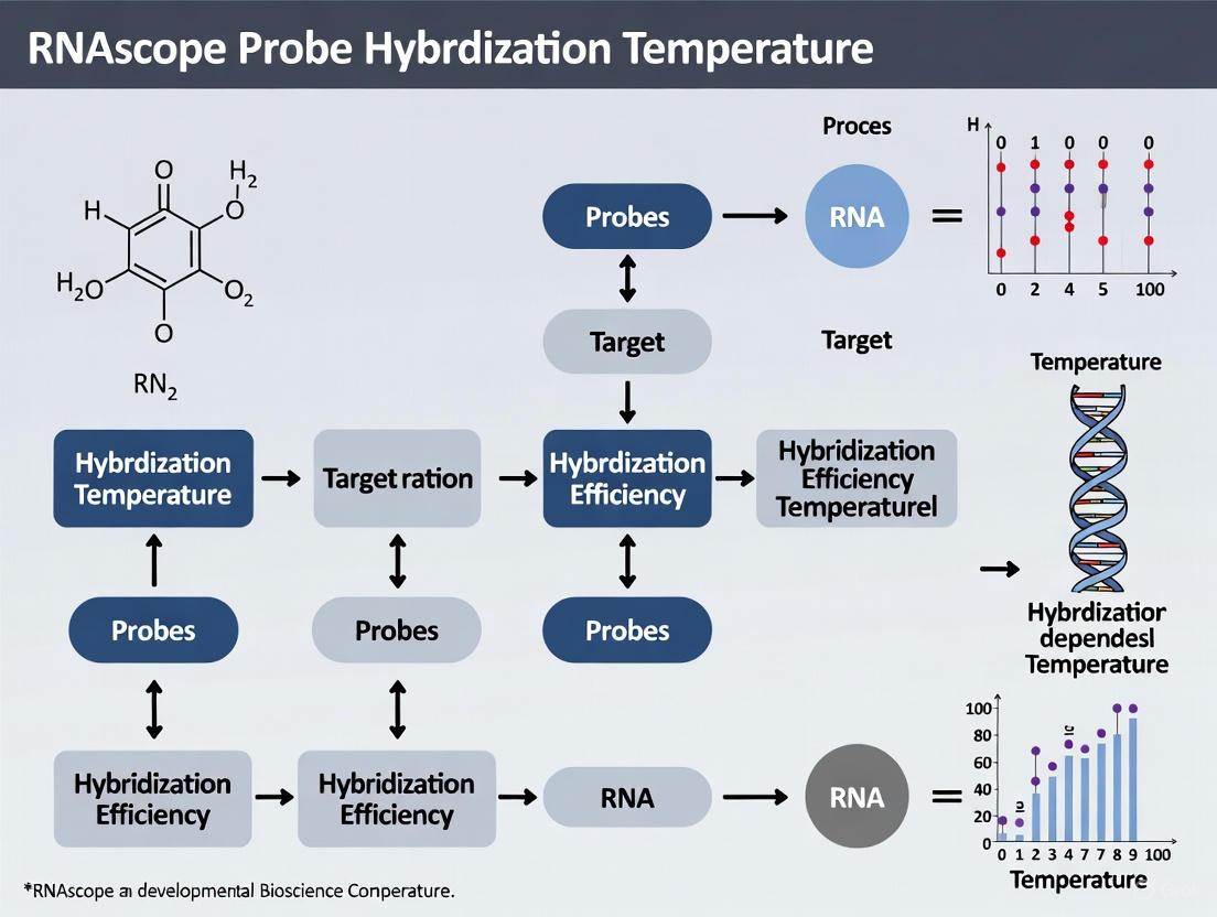
RNAscope Hybridization Temperature: The Critical Factor for Optimal Signal and Specificity
This article provides a comprehensive guide for researchers and drug development professionals on the pivotal role of hybridization temperature in the RNAscope in situ hybridization assay.
RNAscope Hybridization Temperature: The Critical Factor for Optimal Signal and Specificity
Abstract
This article provides a comprehensive guide for researchers and drug development professionals on the pivotal role of hybridization temperature in the RNAscope in situ hybridization assay. Covering foundational principles, methodological applications, and advanced troubleshooting, we detail how precise temperature control at 40°C is critical for single-molecule sensitivity and high-specificity RNA detection. We further explore temperature optimization for diverse sample types—from archived FFPE blocks to whole-mount embryos—and validate its impact through comparative analyses with gold-standard techniques like IHC and qPCR, offering a complete resource for robust and reproducible gene expression analysis in any tissue.
Why 40°C is Non-Negotiable: The Science Behind RNAscope Hybridization Temperature
The Role of Temperature in the RNAscope ZZ Probe Design and Hybridization Efficiency
The RNAscope in situ hybridization assay relies on a patented signal amplification system centered on the ZZ probe design. Temperature is a foundational parameter that critically influences every stage of this process, from initial probe hybridization to final signal amplification.
The proprietary ZZ probe design features oligonucleotide pairs where each "Z" probe contains a target-specific hybridizing region. Typically, 20 ZZ pairs are designed to span approximately 1000 bases of unique target RNA sequence [1]. The probe design algorithm selects oligos with compatible melting temperatures specifically for optimal hybridization under standardized RNAscope assay conditions, ensuring minimal off-target binding [1].
A critical piece of equipment for maintaining proper temperature control during manual assays is the HybEZ Hybridization System, which maintains optimum humidity and a consistent temperature of 40°C throughout the hybridization and amplification steps [2] [3]. This temperature consistency is vital for ensuring specific hybridization and robust signal development.
Table: Key Temperature Parameters in RNAscope Assay Workflow
| Assay Stage | Temperature | Function | Technical Importance |
|---|---|---|---|
| Probe Hybridization | 40°C | Facilitates specific ZZ probe binding to target RNA | Optimized for probe design melting temperatures; critical for signal specificity [2] [3] |
| Protease Digestion | 40°C | Tissue permeabilization for probe access | Must be maintained precisely for optimal tissue preservation and accessibility [2] |
| Signal Amplification | 40°C | Sequential amplifier hybridization | Temperature stability ensures efficient signal build-up without background [2] |
| Target Retrieval (Automated) | 88-95°C | Antigen retrieval for fixed tissues | Varies by tissue fixation; requires optimization for over-/under-fixed samples [2] [4] |
Experimental Protocols: Temperature-Dependent Methodologies
Standard Manual Assay Protocol with Temperature Controls
The following protocol outlines the critical temperature-sensitive steps for manual RNAscope assays, based on established methodologies [2] [5]:
Pretreatment and Protease Digestion: Following antigen retrieval, slides should be directly transferred to room temperature water without cooling [2] [3]. Protease digestion is then performed at a maintained temperature of 40°C to optimally permeabilize tissue without damaging RNA targets [2].
Probe Hybridization: Prior to application, both target probes and wash buffer must be warmed to 40°C to resolve precipitation that may occur during storage [2] [4]. Hybridization then proceeds at 40°C in a HybEZ oven for the duration specified in the assay protocol (typically 2 hours) [2].
Signal Amplification: All subsequent amplification steps (AMP 1-6) are performed at 40°C in the HybEZ system, with strict adherence to the prescribed order and timing to prevent signal loss [2] [5].
Troubleshooting Note: Deviation from these temperature specifications, particularly allowing slides to dry or altering hybridization temperatures, will significantly impact assay performance and may result in signal loss or elevated background [2] [4].
Automated Platform Temperature Parameters
For automated RNAscope implementations on platforms such as the Leica BOND RX system, temperature parameters are programmed directly into the staining protocol [2] [4]:
Standard Pretreatment: 15 minutes Epitope Retrieval 2 (ER2) at 95°C followed by 15 minutes protease treatment at 40°C [4].
Milder Pretreatment: For delicate tissues, 15 minutes ER2 at 88°C followed by 15 minutes protease at 40°C [2] [4].
Extended Pretreatment: For over-fixed tissues, increase ER2 time in 5-minute increments (maintaining 95°C) and protease in 10-minute increments (maintaining 40°C) [2] [4].
Diagram: Temperature-Critical Steps in RNAscope Workflow. The visualization highlights the assay steps requiring precise temperature control, particularly the multiple stages maintained at 40°C in the HybEZ system.
Troubleshooting Guide: Temperature-Related Experimental Issues
Common Temperature-Related Problems and Solutions
Problem: No Signal or Weak Signal
- Potential Cause: Inadequate probe hybridization temperature or precipitation of probe reagents due to improper warming [2] [4].
- Solution: Verify HybEZ oven is maintaining stable 40°C temperature. Pre-warm probes and wash buffer to 40°C before use to resolve precipitation [4]. Ensure all amplification steps are performed in correct order at 40°C [2].
Problem: High Background Noise
Problem: Tissue Detachment or Damage
Problem: Inconsistent Results Between Runs
Optimization Guidelines for Challenging Samples
For samples that deviate from standard fixation protocols (10% NBF for 16-32 hours), temperature parameters may require adjustment [2] [6]:
Over-fixed Tissues: Increase target retrieval time in 5-minute increments at 95°C and protease treatment in 10-minute increments at 40°C [4].
Under-fixed Tissues: Begin with standard temperature parameters but anticipate potential RNA degradation and adjust expectations for signal intensity accordingly [6].
Automated System Adjustments: For Leica BOND RX systems, implement graduated pretreatment optimization while maintaining constant temperatures [2] [4].
Frequently Asked Questions (FAQs)
Q1: Why is precisely 40°C critical for RNAscope hybridization and amplification steps? A: The ZZ probe design is optimized for specific hybridization at 40°C, which represents the ideal balance between binding specificity and efficiency for the proprietary probe architecture. Temperature deviations can result in either non-specific binding (if too low) or inadequate signal amplification (if too high) [2] [1].
Q2: Can I use a standard hybridization oven instead of the HybEZ system? A: The HybEZ system is specifically recommended as it provides both precise temperature control and optimized humidity management. Other ovens may not maintain the consistent 40°C required throughout the multiple-hour procedure, potentially compromising results [2] [3].
Q3: How should I handle temperature transitions during the pretreatment phase? A: After the high-temperature target retrieval step (95°C), slides should be directly transferred to room temperature water without gradual cooling. This immediate transition helps preserve RNA integrity and prepares the tissue for subsequent protease treatment at 40°C [2] [3].
Q4: What is the optimal storage temperature for RNAscope probes? A: RNAscope probes should be stored at 4°C and are stable for up to 2 years from manufacturing when properly stored. Before use, they must be warmed to 40°C to resolubilize any precipitates that may form during storage [1] [4].
Q5: How does temperature affect the interpretation of RNAscope results? A: Proper temperature control ensures that signal dots represent specific target RNA molecules. The number of dots per cell (not intensity) correlates with RNA copy numbers, and consistent temperature conditions are essential for reproducible semi-quantitative scoring between experiments [2] [4].
Table: Essential Research Reagent Solutions for Temperature-Controlled RNAscope
| Reagent/Equipment | Function in Temperature Control | Technical Specification |
|---|---|---|
| HybEZ Hybridization System | Maintains precise 40°C during hybridization/amplification | Critical for manual assays; provides temperature and humidity control [2] [3] |
| RNAscope Target Retrieval Reagents | Enables high-temperature epitope retrieval (95°C) | Optimized for RNAscope workflow; requires no cooling after boiling [2] [5] |
| ImmEdge Hydrophobic Barrier Pen | Prevents tissue drying during temperature cycles | Maintains barrier integrity throughout 40°C incubations [2] [3] |
| Pre-warmed Wash Buffer | Maintains consistent temperature during stringency washes | Must be warmed to 40°C to prevent temperature shock to samples [2] [4] |
| Control Probes (PPIB/dapB) | Validate proper temperature conditions | PPIB should score ≥2 with proper temperature control [2] [7] |
How Temperature and Humidity Interact as Critical Assay Performance Factors
For researchers utilizing the RNAscope in situ hybridization (ISH) platform, achieving consistent and reliable results hinges on the precise control of the physical environment. Temperature and humidity are critical factors affecting assay performance [8]. This guide details how these factors interact, provides protocols for optimal control, and offers troubleshooting advice to resolve common experimental challenges related to environmental conditions.
Experimental Protocols for Environmental Control
Standardized Workflow for Manual RNAscope Assay
The RNAscope procedure can be completed within a single day. Adherence to the following protocol is essential for maintaining proper temperature and humidity throughout [9].
Critical Environmental Control Steps:
- Protease Digestion: Maintain protease treatment at exactly 40°C [10]. Under-digestion results in lower signal and ubiquitous background, while over-digestion degains RNA and compromises tissue morphology [8].
- Probe Hybridization and Amplification: All hybridization and amplification steps (AMP1-AMP6) must be performed at 40°C within a HybEZ Oven or similar validated system [8] [3]. This oven provides the optimum humidity and temperature control that are necessary for proper RNAscope assay performance; other incubators may not provide consistent results [8] [3].
- Slide Hydration: Never allow slides to dry at any point after pretreatment. Ensure the hydrophobic barrier drawn with an ImmEdge pen remains intact to prevent drying, which can cause high background and unreliable results [10] [4].
Optimizing Pretreatment Conditions for Automated Systems
For automated platforms like the Leica BOND RX, pretreatment is a key area for optimization to ensure probe access to target RNA while preserving RNA integrity [9] [4].
Table 1: Automated Pretreatment Optimization Guide
| Tissue Condition | Epitope Retrieval 2 (ER2) | Protease Treatment | Rationale |
|---|---|---|---|
| Standard Protocol | 15 min at 95°C | 15 min at 40°C | Validated starting point for most tissues [4] |
| Sensitive Tissues | 15 min at 88°C | 15 min at 40°C | Milder conditions for delicate morphology [4] |
| Over-fixed/ Dense Tissues | Increase in 5 min increments at 95°C | Increase in 10 min increments at 40°C | Enhanced permeabilization for challenging samples [4] |
Quantitative Data and Performance Metrics
RNAscope Scoring Guidelines
The success of the assay under controlled conditions is evaluated by scoring punctate dots, where each dot represents a single RNA molecule. Score based on dot count per cell, not intensity [10] [4].
Table 2: Semi-Quantitative RNAscope Scoring System
| Score | Dots per Cell (at 20X Magnification) | Interpretation |
|---|---|---|
| 0 | No staining or <1 dot per 10 cells | Negative/Negligible expression |
| 1 | 1-3 dots/cell | Low expression |
| 2 | 4-9 dots/cell; very few clusters | Moderate expression |
| 3 | 10-15 dots/cell; <10% dots in clusters | High expression |
| 4 | >15 dots/cell; >10% dots in clusters | Very high expression |
A successful assay using the housekeeping gene PPIB as a positive control should yield a score of ≥2, while the high-copy gene UBC should score ≥3. The negative control (bacterial dapB) must have a score of <1 to confirm low background [10] [4].
Impact of Environmental Excursions
Deviations from optimal conditions directly impact assay outcomes. The relationship between environmental control and results can be visualized as follows:
Troubleshooting Guides and FAQs
Frequently Asked Questions
Q1: Can I use a standard hybridization oven instead of the HybEZ system? No. The HybEZ oven is the only hybridization oven that has been extensively tested and validated for the RNAscope assay. Other incubators or hybridization stations may not provide the consistent temperature and humidity required, leading to unreliable results [8] [3].
Q2: Why must I proceed directly to room temperature water after the boiling epitope retrieval step? Cooling the slides slowly can promote RNA degradation. Moving slides directly to room temperature water immediately stops the retrieval reaction and helps preserve RNA integrity, which is critical for signal generation [10] [3].
Q3: My positive control shows a good signal, but my target probe has no signal. What environmental factors should I check? This indicates the overall assay worked, but your specific probe hybridization failed.
- Verify Probe Hybridization Temperature: Confirm your oven was accurately maintained at 40°C during the 2-hour hybridization step.
- Check Probe Preparation: Ensure probes were warmed to 40°C and mixed thoroughly to redissolve any precipitation that occurred during storage [10] [4].
- Review Pretreatment Conditions: Your specific target RNA or tissue type may require optimized protease or retrieval times (refer to Table 1).
Q4: I observe high background across my sample, including the negative control. What is the likely cause? High ubiquitous background is often a sign of under-digestion due to insufficient protease activity, which can be caused by incorrect temperature (not 40°C) or an outdated protease reagent [8]. It can also occur if slides were allowed to dry during the procedure [10].
Troubleshooting Common Problems
Problem: Low or No Signal
- Potential Cause 1: Inaccurate oven temperature during hybridization or amplification.
- Solution: Calibrate the HybEZ oven to ensure it maintains 40°C.
- Potential Cause 2: Over-digestion from excessive protease time or temperature.
- Potential Cause 3: Slides dried during the assay.
Problem: High Background Noise
- Potential Cause 1: Under-digestion from insufficient protease treatment.
- Potential Cause 2: Low hybridization temperature or high humidity, leading to non-specific probe binding.
- Solution: Confirm the oven is at 40°C and that the humidity control tray has adequate, but not excessive, moisture [8].
- Potential Cause 3: Use of old or degraded reagents, especially alcohols and xylene.
- Solution: Always use fresh reagents [10].
The Scientist's Toolkit: Essential Research Reagent Solutions
Table 3: Key Materials for RNAscope Assay Success
| Item | Function | Importance for Environmental Control |
|---|---|---|
| HybEZ Oven | Provides precise temperature (40°C) and humidity control during hybridization and amplification. | Critical; the validated system for consistent performance [8] [3]. |
| ImmEdge Hydrophobic Barrier Pen | Creates a barrier to contain liquid reagents over the tissue section. | Prevents slides from drying out, which is a major cause of background [10] [3]. |
| SuperFrost Plus Microscope Slides | Provides superior tissue adhesion. | Prevents tissue detachment during high-temperature retrieval and prolonged assay [10] [3]. |
| RNAscope Positive/Negative Control Probes | Verifies assay performance and sample RNA quality. | Essential for diagnosing if signal issues are environmental or sample-related [9] [4]. |
| Fresh Xylene & Ethanol | Used for deparaffinization and dehydration. | Old reagents can leave residues, increasing background noise [8] [10]. |
| Assay-Specific Mounting Media | Preserves and coverslips the stained slide. | Using an incorrect medium can quench signal or cause fading [10] [4]. |
| Guignardone L | Guignardone L | Guignardone L is a fungal meroterpenoid for research applications. This product is For Research Use Only (RUO). Not for diagnostic, therapeutic, or personal use. |
| 2',3'-cGAMP | 2',3'-cGAMP|cGAS-STING Pathway Agonist | High-purity 2',3'-cGAMP, a native STING agonist and immunotransmitter. Essential for innate immunity, cancer, and virology research. For Research Use Only. Not for human use. |
Why is the HybEZ Oven specifically recommended for RNAscope and BaseScope assays?
The HybEZ II Hybridization System is explicitly recommended because it provides the gasket-sealed, temperature-controlled humidifying chamber that is essential for optimized RNAscope and BaseScope assay performance [11]. Successful implementation of these assays is directly linked to the hybridization environment [11]. The oven's ability to accurately maintain a stable temperature is not just beneficial but is described as "essential to the success" of these workflows [11]. In fact, ACD states that the HybEZ II Oven is the only hybridization oven for which they can provide a performance guarantee for their RNAscope and BaseScope assays [11].
What are the consequences of inconsistent temperature and humidity?
Failure to maintain the optimal temperature and humidity during the hybridization and key incubation steps can lead to a range of experimental issues, primarily affecting signal quality and specificity.
The table below summarizes common problems and their association with temperature control:
| Problem | Potential Consequence | Relation to Temperature/Humidity |
|---|---|---|
| No Signal[/caption] [4] [8] | Complete assay failure. | Omitting steps, or improper conditions preventing probe binding and amplification. |
| High Background[/caption] [10] [4] | Non-specific staining, making true signal difficult to distinguish. | Can be caused by under-digestion during the protease step, which is performed at a specific 40°C [8]. |
| Poor Morphology[/caption] [8] | Loss of tissue or cellular structure. | Can be caused by over-digestion with protease [8]. |
| Variable Results[/caption] [8] | Inconsistent staining between runs or across slides. | Directly linked to the use of non-validated equipment that cannot provide consistent temperature and humidity [8]. |
Troubleshooting Temperature and Humidity Issues
If you suspect your results are being impacted by suboptimal hybridization conditions, follow this guide.
1. Instrument-Specific Checks
For Manual Assays with a HybEZ Oven:
- Verify Humidity: Ensure the humidifying paper in the Humidity Control Tray is kept wet to maintain adequate humidity [10] [4].
- Check Seal: Confirm the gasket-sealed chamber is properly closed to ensure a stable environment [11].
- Pre-warm Reagents: Always warm your probes and wash buffer to 40°C before use to prevent precipitation and ensure proper hybridization [10].
For Automated Assays on a Ventana/Roche or Leica System:
- Software Settings: Do not adjust the recommended temperatures in the protocol unless specifically instructed by ACD technical support [10] [4].
- Instrument Maintenance: For Ventana systems, ensure the "Slide Cleaning" option is unchecked in the software [10]. Have your Roche representative perform a decontamination protocol every three months to prevent microbial growth in fluidic lines [10] [4].
- Correct Buffers: Use only the recommended buffers (e.g., DISCOVERY 1X SSC Buffer for Ventana, 1x BOND Wash Solution for Leica) and ensure residual water is purged from the system after cleaning [10] [4].
2. General Workflow Best Practices
- Prevent Slide Drying: Never let your slides dry out between steps. Flick or tap slides to remove residual reagent, but immediately proceed to the next solution [10] [4] [8].
- Check Hydrophobic Barrier: Use only the ImmEdge Hydrophobic Barrier Pen and ensure the barrier remains intact throughout the entire procedure to prevent tissue drying [10] [4].
- Use Fresh Reagents: Always use fresh ethanol and xylene, as older reagents can compromise results [10] [8].
- Follow Protocols Exactly: Do not alter the protocol sequence or timing. For example, after the boiling step for antigen retrieval, slides should be placed directly into room temperature water without a cooling period [10].
The following diagram illustrates the critical role of the HybEZ Oven within the broader RNAscope workflow and the consequences of temperature deviation.
Experimental Protocol: Validating Assay Conditions with Controls
When setting up your assay, it is critical to include control probes to validate that the entire system, including your HybEZ Oven, is functioning correctly [10] [4].
1. Required Materials (The Scientist's Toolkit)
| Item | Function | Source |
|---|---|---|
| HybEZ II Oven System | Provides validated, stable temperature and humidity for hybridization steps. | [11] |
| Positive Control Probes (PPIB, POLR2A, UBC) | Assess sample RNA quality and optimal permeabilization. | [10] [4] |
| Negative Control Probe (dapB) | Assesses background and non-specific signal; should be minimal. | [10] [4] |
| Control Slides (e.g., HeLa Cell Pellet) | Provide a consistent biological reference for scoring. | [10] [4] |
| ImmEdge Hydrophobic Barrier Pen | Creates a barrier to prevent slide drying; others may fail. | [10] [4] |
| Superfrost Plus Slides | Required for tissue adhesion; other types may cause detachment. | [10] [4] |
| RNAscope 1X Wash Buffer | Used for washing steps between reagent applications. | [10] [12] |
2. Step-by-Step Validation Procedure
- Step 1: Slide Preparation. Run your test samples alongside the recommended control slides (e.g., Human Hela Cell Pellet, Cat. No. 310045) using the positive and negative control probes [10] [4].
- Step 2: Probe Hybridization in HybEZ Oven. Perform the hybridization and key incubation steps (such as the 40°C protease digestion) strictly within the HybEZ Oven as per the user manual [10] [8].
- Step 3: Scoring and Interpretation. Use the semi-quantitative RNAscope scoring guidelines to evaluate your results [10] [4]. Focus on counting the number of dots per cell, not the signal intensity.
- Acceptable Result: The positive control (e.g., PPIB) should generate a score ≥2, and the high-copy UBC should score ≥3, with relatively uniform signal. The negative control (dapB) should have a score of <1, indicating little to no background [10] [4].
- Unacceptable Result: If your positive controls are weak or your negative control has high background, your assay conditions (including fixation, protease digestion, and temperature control) require optimization before proceeding to experimental targets [10].
Key Takeaways for Consistent Results
- The HybEZ Oven is a Required, Validated Component: It is not a generic incubator but a system specifically engineered and tested to provide the precise environmental conditions necessary for the proprietary RNAscope and BaseScope chemistry to work as guaranteed [11] [8].
- Temperature Stability is Directly Linked to Signal Fidelity: Fluctuations in temperature can lead to a cascade of problems, from complete assay failure (no signal) to high, uninterpretable background [8].
- Always Validate with Controls: Before using precious experimental samples, always run the recommended positive and negative control probes to confirm your entire workflow, from sample preparation to hybridization in the HybEZ Oven, is performing optimally [10] [4].
Frequently Asked Questions (FAQs)
Q1: What is the precise hybridization temperature for the RNAscope assay, and why is it critical?
The RNAscope assay must be performed at 40°C for the hybridization and amplification steps [10] [4] [3]. This temperature is critical because the proprietary probe pairs (ZZ pairs) are designed to hybridize optimally at this specific temperature. Deviations can prevent the proper binding of the preamplifier, which requires both Z probes of a pair to hybridize correctly to adjacent target sequences for the subsequent signal amplification to occur [13] [14].
Q2: What happens if the hybridization temperature is too low?
A temperature below the recommended 40°C can lead to:
- Loss of Specificity (High Background): Lower temperatures facilitate weaker, non-specific binding of the probes to off-target sequences. The background suppression mechanism, which relies on the precise juxtaposition of two probes, fails, resulting in high background noise [14].
- Reduced Signal: Paradoxically, a temperature that is too low can also result in no signal or a weak signal because the sequential amplification steps, which are also temperature-dependent, will not proceed efficiently.
Q3: What are the consequences of a hybridization temperature that is too high?
Excessively high temperatures can cause:
- Complete Signal Loss: The primary risk is the denaturation of the RNA target and the delicate probe-target complexes. High temperatures can prevent the probes from binding to the target RNA altogether, resulting in a complete absence of signal [14].
- Tissue Morphology Damage: Elevated temperatures can damage the tissue structure, compromising the histological context of the experiment.
Q4: How is temperature control practically managed in the RNAscope workflow?
For manual assays, the HybEZ Hybridization System is required [10] [4] [3]. This is not a standard laboratory oven; it is specifically designed to maintain the optimum humidity and temperature (40°C) throughout the critical hybridization and amplification steps. Using a standard oven without precise humidity control can lead to the slides drying out, which is a common cause of assay failure.
Q5: Besides hybridization, are there other temperature-sensitive steps?
Yes, several other steps require careful temperature control:
- Protease Digestion: This step must be performed at 40°C to properly permeabilize the tissue without destroying it [10].
- Reagent Preparation: Probes and wash buffer should be warmed to 40°C before use, as precipitation during storage can affect assay performance if not properly redissolved [10] [4].
- Target Retrieval (Antigen Retrieval): This step is performed at a boiling temperature (e.g., 95-100°C) [10] [4] [13]. A key difference from IHC is that no cooling is required after this step; slides should be transferred directly to room temperature water to immediately stop the reaction [10] [3].
Troubleshooting Guide: Temperature-Related Issues
The following table summarizes common problems, their likely temperature-related causes, and recommended solutions.
| Observation | Possible Temperature-Related Cause | Recommended Solution |
|---|---|---|
| No signal or very weak signal | Hybridization temperature too high; protease treatment temperature incorrect [10] [14]. | Verify and calibrate HybEZ oven temperature to ensure it is maintained at 40°C [10] [3]. |
| High background (non-specific staining) | Hybridization temperature too low [14]. | Confirm oven temperature is precisely 40°C; do not reduce temperature to "increase signal." |
| Weak signal in positive control, high background in negative control | Improper reagent warming [10] [4]. | Warm all probes and wash buffer to 40°C before use to ensure reagents are fully dissolved and active. |
| Inconsistent staining across slides | Inconsistent temperature or humidity in the hybridization oven; slides drying out [10]. | Use the dedicated HybEZ system and ensure the humidifying paper is kept wet throughout the procedure [10] [4]. |
| Tissue detachment | Incorrect slide type combined with thermal stress [10] [3]. | Use only Superfrost Plus slides to ensure tissue adhesion throughout the heated assay steps [10] [3]. |
Experimental Protocol: Validating Optimal Temperature Conditions
The following workflow and methodology are adapted from established RNAscope protocols and troubleshooting guides to systematically investigate temperature effects [10] [13].
Objective: To empirically determine the impact of temperature deviation on signal, background, and specificity in the RNAscope assay.
Detailed Methodology
Sample Preparation:
Experimental Groups:
- Group 1 (Optimal Control): Perform the entire RNAscope assay, including all hybridization and amplification steps, at the standard 40°C in a properly calibrated HybEZ oven [10].
- Group 2 (Low Temperature): Perform the assay at a sub-optimal temperature of 37°C.
- Group 3 (High Temperature): Perform the assay at a supra-optimal temperature of 43°C.
Probe Hybridization:
Data Collection and Analysis:
Quantitative Data Interpretation
The expected outcomes from the described experiment can be summarized as follows:
| Temperature Condition | Expected Positive Control (PPIB) Score | Expected Negative Control (dapB) Score | Dot Morphology |
|---|---|---|---|
| 40°C (Optimal) | ≥ 2 [10] [4] | < 1 [10] [4] | Clear, punctate dots [14] |
| 37°C (Too Low) | Lower than control | > 1 (High Background) | Diffuse, non-punctate staining |
| 43°C (Too High) | 0 - 1 (Very Weak/No Signal) | Variable, but signal may be lost | Very few or no dots visible |
The Scientist's Toolkit: Essential Research Reagent Solutions
The following reagents and equipment are non-negotiable for achieving reliable, temperature-controlled results with the RNAscope assay.
- HybEZ Hybridization System: This specialized oven is mandatory for manual assays. It provides precise temperature control (40°C) and maintains optimum humidity, preventing slide dehydration which is a common point of failure [10] [4] [3].
- Control Probes (Positive & Negative): Essential for validating any experiment. The positive control (e.g., PPIB) confirms RNA integrity and correct assay performance, while the negative control (dapB) establishes the level of non-specific background [10] [7] [14].
- Superfrost Plus Slides: These specific slides are required to prevent tissue detachment during the heated assay steps and multiple wash buffers [10] [3].
- ImmEdge Hydrophobic Barrier Pen: The only pen recommended to create a barrier that remains intact throughout the entire procedure, containing the reagents and preventing drying [10] [3].
- RNAscope Assay Kits: The core reagents for the detection and amplification steps. The protocols must be followed exactly without alteration for consistent results [10] [4].
Precision in Practice: Temperature Protocols for Standard and Advanced RNAscope Applications
FAQs: The Role of 40°C in RNAscope Hybridization
Q1: Why is the hybridization step performed at exactly 40°C in the RNAscope assay?
The 40°C hybridization temperature is a critical parameter optimized for the RNAscope technology. This temperature specifically balances several key factors: it facilitates the proper binding of the ZZ probe pairs to their target RNA sequences while maintaining the stringency necessary for the proprietary signal amplification and background suppression system to function correctly. Deviating from this temperature can disrupt the assay's efficiency, leading to either weak signal or increased background [10] [8].
Q2: What are the consequences of an incorrect hybridization temperature?
An incorrect hybridization temperature is a common source of assay failure.
- Temperatures below 40°C can lead to non-specific probe binding, resulting in high background noise and false-positive signals.
- Temperatures above 40°C may denature the probe-target complexes or disrupt the sensitive signal amplification system, leading to weak or absent signal (no detection of the target RNA) [8]. The assay's enzymatic steps are also calibrated for this temperature.
Q3: What equipment is essential for maintaining the correct hybridization environment?
The HybEZ II Hybridization System is explicitly designed and validated by ACD for this purpose. It is not a standard incubator; it provides a gasket-sealed, temperature-controlled humidifying chamber that is essential for optimized RNAscope assay performance. This system guarantees stable temperature at 40°C and high humidity, preventing slides from drying out, which is catastrophic for the assay [11] [8]. ACD notes that other incubators or hybridization stations may not provide consistent results.
Q4: How should probes be prepared before the 40°C hybridization step?
Probes must be warmed to 40°C before application to the slides. This is crucial because precipitation can occur during storage, and warming ensures the probes are fully dissolved and active. After warming, allow the probes to cool to room temperature before preparing the probe mixture for application. For multiplex assays, ensure probes are mixed in the correct channels and ratios as specified in the protocol [10] [4].
Troubleshooting Guide: Hybridization Temperature Issues
Table: Troubleshooting Common Issues Related to Hybridization
| Problem | Potential Cause | Solution |
|---|---|---|
| No Signal or Weak Signal | Hybridization temperature too high; oven calibration incorrect. | Verify oven temperature is precisely 40°C using a calibrated thermometer. Do not alter the protocol [10] [4]. |
| Probes not warmed properly before use. | Warm all probes and wash buffer at 40°C for 10-20 minutes before use to dissolve any precipitates [10] [4]. | |
| High Background | Hybridization temperature too low. | Ensure the HybEZ oven is correctly set to and maintaining 40°C [8]. |
| Slides dried out during incubation. | Check that the hydrophobic barrier from the ImmEdge pen is intact and the humidifying paper in the tray is sufficiently wet [10] [8]. | |
| Uneven Staining | Inconsistent temperature or humidity across the slide. | Use the validated HybEZ II System and ensure the slide tray is level. Make sure the probe mixture covers the tissue section completely [11] [8]. |
| TF-DG-cThea | TF-DG-cThea, MF:C49H41NO21, MW:979.8 g/mol | Chemical Reagent |
| (S)-(-)-Verapamil-d3Hydrochloride | (S)-(-)-Verapamil-d3Hydrochloride, MF:C27H39ClN2O4, MW:494.1 g/mol | Chemical Reagent |
Table: Key Control Probes for Validating Assay Performance
| Control Probe | Target | Expected Result in a Valid Assay | Purpose |
|---|---|---|---|
| Positive Control (e.g., PPIB, UBC) | Housekeeping genes | PPIB score ≥2; UBC score ≥3. Relatively uniform signal [10] [4]. | Verifies RNA integrity, sample pretreatment, and overall assay success. |
| Negative Control (dapB) | Bacterial gene | Score of <1 (little to no staining) [10] [4]. | Assesses non-specific background and background suppression. |
Experimental Protocol: Validating the Hybridization Environment
This protocol is critical for qualifying your lab's hybridization setup and troubleshooting potential temperature-related issues.
Objective: To confirm that your local hybridization system (e.g., HybEZ oven) and reagents are functioning correctly by using control probes and slides.
Materials:
- RNAscope reagent kit (e.g., Multiplex Fluorescent v2, Cat. No. 323100) [15]
- Positive and Negative Control Probes (e.g., PPIB, UBC, and dapB) [10] [4]
- Control slides (e.g., Human HeLa Cell Pellet, Cat. No. 310045) [10] [4]
- HybEZ II Oven, Humidity Control Tray, and Humidifying Paper [11]
- ImmEdge Hydrophobic Barrier Pen [10] [4]
Methodology:
- Sample Preparation: Follow the standard protocol for deparaffinization, rehydration, and target retrieval for your FFPE control slides [15] [16].
- Protease Treatment: Apply Protease Plus and incubate at 40°C for 30 minutes (or as per kit instructions) [15]. This step is critical for tissue permeabilization.
- Probe Hybridization:
- Prepare the probe mixture according to the user manual.
- Warm the probes and wash buffer at 40°C before use [10] [4].
- Remove slides from the 40°C incubator, tap off residual buffer, and apply the probe mixture.
- Immediately return slides to the pre-heated 40°C HybEZ oven and hybridize for 2 hours [15] [16].
- Signal Amplification & Detection: Perform all subsequent amplification and wash steps exactly as directed by the protocol without deviation [10].
- Scoring and Analysis:
Workflow Diagram: The 40°C Hybridization in Context
The following diagram illustrates the key steps of the RNAscope workflow that are dependent on precise temperature control, with the central 40°C hybridization step being paramount.
The Scientist's Toolkit: Essential Reagents and Equipment
Table: Essential Materials for a Successful RNAscope Assay
| Item | Function | Protocol-Specific Note |
|---|---|---|
| HybEZ II Oven | Provides a gasket-sealed, temperature-controlled (40°C), humidified chamber. | Critical for consistent results; ACD's only validated system for manual assays [11] [8]. |
| Positive & Negative Control Probes | Validate sample RNA quality, pretreatment, and assay performance. | Must be run with every experiment to qualify results [10] [4]. |
| ImmEdge Hydrophobic Barrier Pen | Creates a barrier to contain liquid reagents and prevent slides from drying out. | The only barrier pen validated for use throughout the RNAscope procedure [10] [15]. |
| Superfrost Plus Slides | Provides superior tissue adhesion. | Required to prevent tissue detachment during the assay [10] [4]. |
| RNAscope Wash Buffer | Used for stringency washes after hybridization and amplification steps. | Must be warmed to 40°C before use and diluted to 1X as directed [10] [15]. |
| Aspericin C | Aspericin C, MF:C17H30O4, MW:298.4 g/mol | Chemical Reagent |
| Neopuerarin A | Neopuerarin A, MF:C21H20O9, MW:416.4 g/mol | Chemical Reagent |
FAQ: Probe Design and Channel Specification
What is the difference between a C1, C2, C3, or C4 probe? The designations C1, C2, C3, and C4 refer to the specific amplification channel a probe is designed for and do not indicate different temperature requirements. The letter indicates the compatible assay type (e.g., "C" for RNAscope and BaseScope assays), and the number (1, 2, 3, 4) allows for the simultaneous detection of different target RNAs using various fluorophores in a multiplex assay [1].
How does probe design ensure uniform hybridization? RNAscope employs a proprietary "double-Z" probe design. Each probe pair (ZZ) hybridizes contiguously to a target region, and custom software automatically selects probe sequences with compatible melting temperatures (Tm) for optimal hybridization under standardized assay conditions [14] [1]. This design ensures that all probes, regardless of their channel designation, perform robustly at the same assay temperature.
What is the minimum target sequence length required for probe design? The required length depends on the specific assay type [1]:
- RNAscope: Best for mRNAs or ncRNAs longer than 300 bases. A standard probe set includes 20 ZZ pairs.
- BaseScope: Designed for short target sequences between 50 and 300 bases, using 1-3 ZZ probe pairs.
- miRNAscope: For detecting very short RNAs between 17 and 50 bases.
Standardized Temperature Protocol
The RNAscope technology is designed for simplicity and robustness, utilizing a single, standardized temperature for the key hybridization steps during the detection phase, regardless of the number of channels used.
| Protocol Step | Temperature | Duration | Notes |
|---|---|---|---|
| Protease Digestion | 40°C | 30 minutes | Critical for tissue permeabilization. |
| Target Probe Hybridization | 40°C | 2 hours | For all C1, C2, C3, C4 probes. |
| Preamplifier & Amplifier Hybridization | 40°C | 30 & 15 minutes | Consistent temperature for signal amplification steps. |
Troubleshooting Guide: Temperature-Related Issues
A stable thermal environment is critical for assay performance. The following table outlines common issues and solutions related to temperature management.
| Problem | Potential Cause | Recommended Solution |
|---|---|---|
| Weak or No Signal | Incorrect hybridization temperature; degraded reagents due to improper storage. | Verify oven calibration is at 40°C. Ensure the HybEZ Humidity Control Tray has adequate water. Store all probes at 4°C [10] [1]. |
| High Background | Incomplete or non-specific hybridization. | Strictly maintain 40°C for all hybridization and wash steps. Do not alter incubation times. Pre-warm probes and wash buffer to 40°C to prevent precipitation [10]. |
| Inconsistent Signal Across Multiplex Channels | Probe concentration errors; temperature gradients across the slide. | Use the HybEZ system to ensure optimum humidity and even heat distribution. For manual assays, confirm probe mixing ratios (e.g., C2:C1 is 1:50 for 2-plex) [10]. |
| Signal Loss in Automated Systems | Instrument calibration drift; bulk solution issues. | Perform regular instrument maintenance. For Ventana systems, have a representative perform a decontamination protocol quarterly and replace all bulk solutions with recommended buffers [10]. |
Experimental Protocol: Validating Temperature Uniformity
For laboratories establishing a new RNAscope workflow or troubleshooting inconsistent results, validating thermal uniformity is essential.
Objective: To confirm that the temperature across the entire hybridization platform (e.g., hotplate, HybEZ oven) is a consistent 40°C, ensuring uniform probe hybridization for all channels [17].
Materials:
- HybEZ Hybridization System or equivalent heated platform
- Calibrated thermal probe or infrared thermometer
- Standard buffer solution
Methodology:
- Thermal Profiling: Place the heating platform in an ambient temperature representative of normal lab conditions. Place thermal probes at multiple locations across the heating surface, including the center and all four corners.
- Data Logging: Activate the platform and set it to 40°C. Allow the system to stabilize for at least 30 minutes. Record the temperature from all probes at 5-minute intervals over a period of 60 minutes.
- Analysis: Calculate the mean temperature and standard deviation for each probe location. The assay requires temperatures to fall within a tight distribution of 40°C ± 0.5°C for optimal performance [17].
Expected Outcome: A successful validation will show all measurement points within the specified range, confirming that the experimental setup is capable of providing the consistent environment required for reliable multiplexed RNA detection.
Research Reagent Solutions
The following table details key materials and instruments essential for successfully performing a multiplexed RNAscope assay.
| Item | Function in Assay | Specification / Note |
|---|---|---|
| HybEZ Hybridization System | Maintains optimum humidity and a stable 40°C temperature during all hybridization steps. | Required for manual assays to prevent slide drying and ensure temperature uniformity [10] [14]. |
| RNAscope Target Probes (C1-C4) | Channel-specific probes that hybridize to the target RNA. | Ready-to-Use (RTU) for C1; C2 may be 50X concentrate. Store at 4°C; stable for up to 2 years [10] [1]. |
| Positive Control Probes (PPIB, POLR2A, UBC) | Assess sample RNA quality and optimal permeabilization. | Housekeeping genes; use to qualify your sample and optimize pretreatment [10]. |
| Negative Control Probe (dapB) | Assess background and non-specific binding. | Bacterial gene; should yield no signal (score <1) in properly fixed tissue [10] [14]. |
| Superfrost Plus Slides | Provide adhesion for tissue sections throughout the assay. | Required; other slide types may result in tissue detachment [10]. |
| ImmEdge Hydrophobic Barrier Pen | Creates a well to contain reagents on the slide. | The only barrier pen recommended to maintain a hydrophobic barrier throughout the procedure [10]. |
FAQ: Temperature Adaptation for Challenging Samples
How does hybridization temperature affect RNAscope results?
The hybridization temperature is critical for the RNAscope assay's success. It balances the conflicting goals of achieving high probe assembly efficiency (the fraction of probes bound to a given RNA) and high specificity (minimizing off-target binding). The temperature must be carefully controlled to ensure optimal signal-to-noise ratio, as it directly influences the brightness of single-molecule signals and the overall detection efficiency [18]. The standard recommended hybridization temperature is 40°C, which is maintained using a HybEZ Hybridization System to ensure optimum conditions [2] [4].
Why is temperature control different for cryosections and isolated cardiomyocytes?
Cryosections and isolated cardiomyocytes have different structural integrities and permeabilization requirements, which necessitate tailored temperature conditions during the protease and hybridization steps. For cryosections, a longer, warmer protease treatment (40 minutes at 40°C) is often used for RNA detection only, whereas a shorter, cooler treatment (20 minutes at room temperature) is applied when co-detecting RNA and protein to preserve antigen integrity [19]. For isolated cardiomyocytes, a shorter protease treatment (15 minutes) is sufficient, and the temperature (room temperature vs. 40°C) is similarly determined based on whether subsequent antibody staining is required [19].
What are the consequences of using incorrect temperatures?
Using incorrect temperatures can lead to two main issues:
- Excessively high temperatures can cause probe denaturation, reduce hybridization efficiency, and lead to weak or no signal.
- Excessively low temperatures can increase non-specific binding of probes, resulting in high background noise and false positives [18]. Furthermore, letting slides dry out at any point due to improper temperature or humidity control will compromise the assay [2] [4].
Troubleshooting Guide: Temperature-Related Issues
Problem: Weak or No Signal
| Possible Cause | Recommended Solution |
|---|---|
| Suboptimal hybridization temperature | Verify and maintain hybridization oven at 40°C; use a calibrated thermometer [2]. |
| Insufficient permeabilization | Optimize protease treatment: for cryosections, increase to 40 minutes at 40°C (RNA only) or 20 mins at RT (RNA+protein). For cardiomyocytes, use 15 minutes [19]. |
| Probe precipitation | Warm probes and wash buffer to 40°C before use to re-dissolve any precipitates [2] [4]. |
Problem: High Background or Non-Specific Staining
| Possible Cause | Recommended Solution |
|---|---|
| Low-temperature non-specific binding | Ensure hybridization is performed at 40°C, not lower [18]. |
| Over-digestion by protease | For over-fixed tissues, adjust protease time in increments of 10 minutes while keeping temperature constant at 40°C [2] [4]. |
| Inadequate washing | Perform all wash steps with 1x Wash Buffer at room temperature for 2 minutes each; do not skip washes [19]. |
Problem: Tissue Detachment or Morphology Damage
| Possible Cause | Recommended Solution |
|---|---|
| Use of incorrect slide type | Use only Superfrost Plus slides to ensure tissue adhesion [2] [4]. |
| Over-drying during protocol | Flick slides to remove reagent but do not let them dry; ensure hydrophobic barrier from ImmEdge pen remains intact [2]. |
Optimized Temperature Protocols
Detailed Protocol for Cryosections
The table below summarizes the key temperature-sensitive steps for processing cryosections.
Table: Temperature and Duration for Key Steps in Cryosection Protocol
| Step | Reagent/Process | Temperature | Duration | Notes |
|---|---|---|---|---|
| Refixation | 4% PFA/PBS | Room Temperature | 15 minutes | [19] |
| Protease Treatment | Protease III | 40°C (for RNA only) or Room Temperature (for RNA+Protein) | 40 minutes (at 40°C) or 20 minutes (at RT) | Critical permeabilization step [19] |
| Hybridization | Target Probe | 40°C | 2 hours | Maintain in HybEZ oven [19] |
| Signal Amplification | AMP1, AMP2 | 40°C | 30 minutes each | [19] |
| Signal Amplification | AMP3 | 40°C | 15 minutes | [19] |
| Signal Development | HRP Channel | 40°C | 15 minutes | [19] |
| Tyramide Signal Amplification | TSA Fluorophore | 40°C | 30 minutes | [19] |
Detailed Protocol for Isolated Cardiomyocytes
The table below summarizes the key temperature-sensitive steps for processing isolated cardiomyocytes.
Table: Temperature and Duration for Key Steps in Isolated Cardiomyocytes Protocol
| Step | Reagent/Process | Temperature | Duration | Notes |
|---|---|---|---|---|
| Protease Treatment | Protease III | 40°C (for RNA only) or Room Temperature (for RNA+Protein) | 15 minutes | Cells are suspended [19] |
| Hybridization | Target Probe | 40°C | Overnight | Maintain in HybEZ oven [19] |
| Signal Amplification | AMP1, AMP2 | 40°C | 30 minutes each | [19] |
| Signal Amplification | AMP3 | 40°C | 15 minutes | [19] |
| Signal Development | HRP Channel | 40°C | 15 minutes | [19] |
| Tyramide Signal Amplification | TSA Fluorophore | 40°C | 30 minutes | [19] ``` |
The Scientist's Toolkit: Essential Research Reagent Solutions
Table: Key Reagents and Equipment for RNAscope on Challenging Samples
| Item | Function | Application Note |
|---|---|---|
| HybEZ Oven | Maintains optimum humidity and a constant 40°C temperature during hybridization and amplification steps. | Essential for consistent results; required for all RNAscope hybridization steps [2] [19]. |
| Protease III | Enzymatically permeabilizes tissue to allow probe access to target RNA. | Treatment time and temperature (RT vs. 40°C) must be optimized for sample type and co-detection goals [19]. |
| RNAscope Multiplex Fluorescent Kit v2 | Contains all reagents for probe hybridization, signal amplification, and development. | Follow kit protocol precisely; do not alter temperatures or incubation times [19] [4]. |
| Superfrost Plus Slides | Provide superior tissue adhesion for challenging samples like cryosections. | Mandatory to prevent tissue detachment during high-temperature steps [2] [4]. |
| ImmEdge Hydrophobic Barrier Pen | Creates a barrier to maintain reagent volume over tissue and prevent drying. | The only pen recommended to maintain a barrier throughout the RNAscope procedure [2]. |
| Positive/Negative Control Probes (PPIB, dapB) | Assess sample RNA quality, permeabilization efficiency, and assay performance. | Always run controls; successful PPIB staining should generate a score ≥2 [2] [4]. |
| L-Pentaguluronic acid | L-Pentaguluronic acid, MF:C30H42O31, MW:898.6 g/mol | Chemical Reagent |
| Oleaside A | Oleaside A, MF:C30H44O7, MW:516.7 g/mol | Chemical Reagent |
FAQs on Temperature and Probe Design
Q1: How does temperature stability during hybridization affect DNAscope results? Temperature is a critical parameter for the DNAscope assay performance. Consistent temperature during the hybridization and amplification steps ensures optimal probe binding and proper signal amplification. The HybEZ oven is the only hybridization system extensively validated to maintain optimum humidity and temperature; using other incubators may not provide consistent results [10] [8]. Deviations from the recommended 40°C incubation temperature can lead to reduced signal or increased background.
Q2: What are the recommended storage conditions and stability for DNAscope probes? ACD Bio recommends storing RNAscope probes at 4°C, where they are stable for up to 2 years from the date of manufacturing [1]. While this data is specific to RNAscope, DNAscope probes likely have similar stability profiles when stored under the same recommended conditions. Before use, probes and wash buffer should be warmed to 40°C to re-dissolve any precipitation that may have occurred during storage [10].
Q3: How can researchers optimize conditions for non-standard samples like whole-mount zebrafish embryos? For non-standard samples where fixation and permeabilization may vary, a qualification workflow is strongly recommended [10]. This involves running positive and negative control probes on the sample to assess RNA integrity and optimal permeabilization. For automated systems on the Leica BOND RX, pretreatment conditions (Epitope Retrieval and Protease times) can be incrementally adjusted to optimize for specific tissue types and fixation conditions [10] [4].
Q4: What is the impact of temperature shifts on embryonic development in zebrafish models? Research using zebrafish models demonstrates that temperature is a significant environmental factor influencing development. Studies on Zoep (One-eyed pinhead) mutants showed that a heat shock of 34°C, applied at or before the midblastula stage, significantly increased the penetrance of neural tube defects compared to embryos maintained at the standard 28.5°C [20]. This highlights that precise temperature control is crucial not only for the assay itself but also for managing the developmental context of the model organism.
Troubleshooting Guides
Common Temperature-Related Issues and Solutions
| Problem | Potential Cause | Recommended Solution |
|---|---|---|
| No or weak signal | Incorrect hybridization temperature; under-fixation [6] | Verify incubator is maintained at 40°C; ensure tissue was fixed properly [10] [8]. |
| High background | Over-digestion with protease; suboptimal temperature control [8] | Titrate protease incubation time; ensure temperature is precisely 40°C during protease step [10]. |
| Poor morphology | Over-digestion with protease; tissue drying out [10] | Reduce protease incubation time; ensure hydrophobic barrier remains intact to maintain humidity [10] [8]. |
| Inconsistent results between runs | Use of non-validated equipment; temperature fluctuations | Use the validated HybEZ Oven System; do not alter protocol temperatures [8]. |
Temperature Sensitivity in Zebrafish Embryo Studies
The following table summarizes quantitative findings from a zebrafish study on neural tube defects, illustrating the impact of temperature.
| Factor | Impact on Phenotype Penetrance | Statistical Significance | Experimental Conditions |
|---|---|---|---|
| Heat Shock (34°C) | Significantly increased open neural tube phenotype in Zoep mutants [20] | Analysis of variance (ANOVA) indicated temperature was a significant contributing factor [20] | Heat shock applied at or before 4 hours post-fertilization (hpf) [20] |
| Genetic Background (Clutch) | Variable penetrance of neural tube defects (0% to 100%) [20] | Analysis of variance (ANOVA) indicated genetic background was a significant contributing factor [20] | Clutches from single pairs of heterozygous adults [20] |
Experimental Protocols
Detailed Protocol: RNA In Situ Hybridization on Fixed Frozen Sections
This protocol, adapted from Stanford University, is a working example of a meticulous workflow that can be applied to related DNAscope assays, highlighting critical temperature points [16].
Materials:
- RNAscope Target Probes (e.g., C1, C2, C3)
- RNAscope Multiplex Fluorescent Reagent Kit v2 (ACD, Cat. No. 323270)
- 4% Paraformaldehyde (PFA) in PBS
- 30% Sucrose Solution
- Optimal Cutting Temperature (OCT) Embedding Medium
- Ethanol series (50%, 70%, 100%)
- Superfrost Plus microscopic slides
- ImmEdge Hydrophobic Barrier Pen
- HybEZ Hybridization System or equivalent oven
Procedure: Day 1: Pretreatment and Probe Hybridization
- Section Preparation: Wash frozen sections mounted on Superfrost Plus slides in 1x PBS for 5 minutes [16].
- Baking: Bake slides at 60°C for 30 minutes in a hybridization incubator [16].
- Post-fixation: Post-fix slides in 4% PFA for 15 minutes at 4°C [16].
- Dehydration: Dehydrate tissue by immersing slides sequentially in 50%, 70%, and 100% ethanol for 5 minutes each. Air dry for 5 minutes at room temperature [16].
- Hydrogen Peroxide: Cover sections with Hydrogen Peroxide and incubate for 10 minutes at room temperature. Wash 2x in PBS [16].
- Target Retrieval: Preheat 1x PBS and Target Retrieval Reagent in a steamer (>99°C). Briefly immerse slides in PBS, then transfer to Target Retrieval Reagent and steam for 3 minutes [16].
- Protease Digestion: Draw a barrier around sections, apply Protease Plus, and incubate at 40°C for 20 minutes in a hybridization oven. Wash 2x in PBS [16].
- Probe Hybridization: Dilute and mix target probes. Apply probe mixture to sections and incubate at 40°C for 2 hours. Wash 2x in Wash Buffer. Store slides in 5x SSC buffer overnight at room temperature [16].
Day 2: Signal Amplification and Development
- Amplification: Perform sequential amplifications by applying AMP1, AMP2, and AMP3 reagents, incubating each at 40°C for 30, 30, and 15 minutes respectively, with washes in between [16].
- Fluorescence Development: For each channel, sequentially apply the corresponding HRC blocker, incubate at 40°C for 15 minutes, wash, apply the diluted fluorophore, incubate at 40°C for 30 minutes, wash, and then apply HRP Blocker [16].
- Counterstaining and Mounting: Apply DAPI for 30 seconds at room temperature, remove, and add fluorescence-compatible mounting media. Cover with a coverslip and store at 4°C [16].
Workflow Diagram for Temperature-Critical Assay Steps
Research Reagent Solutions
| Item | Function / Application |
|---|---|
| HybEZ Hybridization System | Maintains optimum humidity and 40°C temperature during critical hybridization and amplification steps; validated for reliable assay performance [10] [8]. |
| Positive Control Probes (PPIB, POLR2A, UBC) | Species-specific housekeeping gene probes used to qualify sample RNA integrity and assay performance under standardized conditions [10] [4]. |
| Negative Control Probe (dapB) | Bacterial gene probe that should not generate signal in properly processed tissue; used to assess nonspecific background and assay specificity [10] [4]. |
| Protease III / Protease Plus | Enzyme for tissue permeabilization; digestion time is critical and may require optimization for different sample types (e.g., whole-mount embryos) [10] [16]. |
| Superfrost Plus Slides | Specifically required to prevent tissue detachment during the stringent temperature cycles of the assay protocol [10]. |
| ImmEdge Hydrophobic Barrier Pen | Creates a barrier to maintain reagent coverage over the tissue, preventing slides from drying out during high-temperature incubations [10] [16]. |
Troubleshooting Temperature-Related Issues: From Signal Loss to Poor Morphology
The Critical Role of Temperature in RNAscope Assays
In RNAscope probe hybridization research, temperature is not merely a setting—it is a fundamental variable that directly dictates the success or failure of your experiment. The process of in situ hybridization (ISH) is critically dependent on precise temperature control to ensure specific probe binding, optimal signal amplification, and effective background suppression [10] [4]. An oven that deviates from its set temperature, even by a few degrees, can lead to a cascade of experimental failures, including weak signal, high background, or complete absence of detection.
The RNAscope protocol specifies exact incubation temperatures for key steps, particularly the hybridization and amplification stages, which are typically performed at 40°C [10]. This temperature is carefully optimized to allow the specific binding of ZZ probe pairs to your target RNA sequence. If your oven runs too cool, hybridization efficiency drops drastically, resulting in low signal. If it runs too hot, you risk non-specific binding, high background noise, and potential degradation of the RNA target [4]. Therefore, regular verification and calibration of your laboratory oven is not general maintenance; it is a critical component of experimental quality control.
How to Calibrate Your Laboratory Oven
Calibrating an oven involves adjusting its temperature settings to ensure the internal environment matches the displayed setpoint. The following methodology, adapted from standardized procedures, provides a reliable approach for laboratory equipment [21] [22].
Tools You Will Need
- A certified, traceable digital thermometer with an external probe. For research purposes, this master thermometer should itself be calibrated against a NIST (National Institute of Standards and Technology) standard [23].
- A thermal-safe grate clip to suspend the probe in the center of the oven.
- (Optional) Heat-resistant gloves for safety.
Calibration Procedure
1. Initial Temperature Assessment
- Place the oven rack in the center position.
- Use the grate clip to suspend the thermometer probe in the geometric center of the oven cavity [21] [22].
- Set the oven to the critical temperature used in your RNAscope protocol (e.g., 40°C) [10].
- Close the door and allow the oven to complete its preheating cycle and stabilize. Do not open the door during this time, as it causes significant temperature fluctuations [21].
- Once stabilized, record the temperature displayed on your certified thermometer. This is the oven's actual temperature.
2. Interpreting Results and Taking Action
- Compare the actual temperature to the set temperature.
- Acceptable Variance (±2°C): If the variance is within a tolerable range (e.g., ±2°C for sensitive molecular biology work), your oven may not require adjustment. The minor fluctuation is likely within the operational stability of the RNAscope assay.
- Significant Variance (>±5°C): If a significant and consistent discrepancy is found, you should calibrate the oven.
3. Performing the Calibration The method depends on your oven's control system.
- For Digital Control Ovens: Many modern laboratory ovens have a built-in calibration mode. This is typically accessed through a specific sequence of button presses on the control panel. Consult your oven's owner's manual for the exact procedure to enter this mode and apply an offset to correct the temperature [21] [24].
- For Analog Control Ovens: Ovens with analog knobs often have a small calibration screw on the back of the thermostat shaft. After removing the knob, minor adjustments to this screw (e.g., clockwise to increase temperature, counter-clockwise to decrease) can bring the oven into alignment [22]. Make adjustments gradually, as even a slight turn can cause a large change.
After any adjustment, repeat the temperature assessment to verify the calibration was successful.
Important Safety Note: If your oven's temperature is off by a large margin (e.g., >30°C), it indicates a potential hardware failure. In such cases, do not attempt to calibrate it yourself. Contact a certified service technician for repair [21] [22].
Troubleshooting Guide: Oven Temperature & RNAscope Results
Use the following table to diagnose how oven temperature issues might manifest in your RNAscope experiments.
| Observed Problem | Potential Oven Temperature Link | Other Factors to Investigate |
|---|---|---|
| Weak or No Signal | Oven running too cool during hybridization/amplification steps, leading to inefficient probe binding [4]. | Sample RNA degradation [6], suboptimal protease treatment time [10] [4], expired reagents. |
| High Background Noise | Oven running too hot, causing non-specific probe binding [4]. | Inadequate washing, over-fixation of tissue, protease treatment time too long [10] [4]. |
| Inconsistent Staining Between Runs | Unstable oven temperature or a hot spot within the cavity, leading to uneven heating of slides. | Inconsistent sample preparation [6], variation in reagent incubation times, uneven reagent coverage. |
Frequently Asked Questions (FAQs)
How often should I check my oven's temperature?
For research purposes, it is good practice to verify the temperature quarterly. If your oven is used continuously for critical assays like RNAscope, a monthly check is recommended. Furthermore, you should always check the temperature if you move the oven, after any power outage, or if you begin to observe inconsistent experimental results [23].
What is the best type of thermometer to use for calibration?
For a one-time check, a high-quality digital thermometer with a separate probe is sufficient. However, for ongoing quality assurance and especially if you need to generate calibration records for compliance (e.g., GLP), you should invest in a "master" thermometer that is certified and traceable to a national standard like NIST. This master thermometer should be recalibrated by an accredited service annually [23].
My oven temperature fluctuates constantly. Is this normal?
Yes, to an extent. Most ovens use a cycling thermostat—they heat up past the set temperature, turn off, cool down slightly below the set temperature, and then turn on again. This creates a temperature swing that is typically within a range of a few degrees. The key is that the average temperature should be centered on your setpoint. Large, erratic swings are a sign of a failing thermostat or heating element and require service.
The temperature is uniform in the center but I suspect hot spots elsewhere in the oven. How can I check?
You can create a "hot spot map" of your oven [22]. Place the thermometer probe at multiple predefined locations (e.g., front-left, center, back-right, top, bottom) while the oven is set to a stable temperature. Record the reading at each location. This will help you identify colder and hotter zones, allowing you to strategically place slides for consistent results or avoid certain areas altogether.
The Scientist's Toolkit: Essential Research Reagent Solutions
The following reagents and materials are critical for both the RNAscope assay and for ensuring your equipment is functioning correctly.
| Item | Function in the Context of Oven Calibration & RNAscope |
|---|---|
| NIST-Traceable Digital Thermometer | Serves as the "master" reference standard for verifying and calibrating oven temperatures, ensuring traceability and data integrity [23]. |
| HybEZ Oven or Equivalent | A specialized hybridization system designed to maintain optimum humidity and a precise 40°C temperature for the RNAscope assay workflow, preventing slide drying and temperature fluctuation [10] [4]. |
| Positive Control Probes (PPIB, POLR2A, UBC) | Essential housekeeping gene probes used to verify that the entire RNAscope assay, including temperature-dependent steps, performed correctly on your sample [10] [4]. |
| Negative Control Probe (dapB) | A bacterial gene probe that should not hybridize in most samples; used to assess non-specific background signal, which can be caused by excessive oven temperature [10] [4]. |
| RNAscope Wash Buffer | Used in the washing steps between hybridizations. Must be pre-warmed to the correct temperature (40°C) to avoid introducing temperature shocks that could affect hybridization [10]. |
Experimental Workflow for Oven Verification
The diagram below outlines the logical workflow for integrating oven temperature verification into your experimental routine.
Optimizing Protease Digestion Time and Temperature for Different Tissues
Frequently Asked Questions
What is the primary function of protease digestion in the RNAscope assay? Protease digestion is a critical permeabilization step that makes the target RNA within intact cells accessible to the probes by partially digesting the cellular proteins. Accurate optimization is essential; under-digestion can lead to weak or no signal, while over-digestion can damage tissue morphology and result in high background or loss of signal [10] [4].
How do I know if my protease digestion needs optimization? The need for optimization is best determined by running control probes. Successful staining with a positive control probe (e.g., PPIB) should yield a score ≥2, while the negative control probe (dapB) should show a score of <1, indicating low background [10] [4]. Poor results with controls, high background, or tissue degradation are key indicators that digestion conditions need adjustment [4].
Can I use the same protease digestion conditions for all tissue types? No, different tissue types often require different digestion stringencies. Dense tissues or those fixed for extended periods may require extended digestion, while more delicate tissues like lymphoid or neural tissues often benefit from milder conditions to preserve morphology [25] [4].
Besides time and temperature, what other factors can affect protease digestion? The efficiency of protease digestion is also influenced by the preceding epitope retrieval step, the type of protease used, and the quality of the original tissue fixation [10] [25]. All these steps work in concert to achieve optimal RNA accessibility.
Troubleshooting Guide: Protease Digestion
Weak or No Signal
- Possible Cause: Incomplete tissue permeabilization due to under-digestion.
- Solution: Increase the protease digestion time in increments of 10 minutes while keeping the temperature constant at 40°C [10] [4]. For automated systems, also consider increasing the epitope retrieval (ER2) time in 5-minute increments [4].
High Background or Non-Specific Staining
- Possible Cause: Over-digestion of the tissue by the protease.
- Solution: Reduce the protease digestion time. Ensure the hydrophobic barrier remains intact throughout the assay to prevent localized drying and over-digestion [10] [4].
Poor Tissue Morphology
- Possible Cause: Protease treatment is too harsh, damaging the tissue structure.
- Solution: Adopt a milder pretreatment condition. For the Leica BOND RX system, this means reducing the ER2 temperature from 95°C to 88°C for 15 minutes and maintaining protease digestion at 40°C for 15 minutes [25] [4].
Optimization Data Tables
Standard vs. Mild Pretreatment Conditions
The following conditions are recommended for use on the Leica Biosystems' BOND RX automated platform [25] [4].
| Parameter | Standard Pretreatment | Mild Pretreatment |
|---|---|---|
| Epitope Retrieval | 15 min at 95°C (ER2 buffer) | 15 min at 88°C (ER2 buffer) |
| Protease Digestion | 15 min at 40°C | 15 min at 40°C |
| Best For | General use on a wide range of FFPE tissues [25]. | Delicate tissues (e.g., lymphoid tissue, retina) [25]. |
Tissue-Specific Optimization Guidelines
The table below summarizes recommended adjustments for specific tissue types and scenarios [10] [25] [4].
| Tissue / Scenario | Recommended Adjustment |
|---|---|
| Over-fixed Tissues | Increase both ER2 time (in 5-min increments) and Protease time (in 10-min increments) from standard conditions [4]. |
| Dense Tissues | Increase both ER2 time (in 5-min increments) and Protease time (in 10-min increments) from standard conditions [4]. |
| Lymphoid Tissues | Start with Mild Pretreatment conditions (ER2 at 88°C) [25]. |
| Retina | Start with Mild Pretreatment conditions (ER2 at 88°C) [25]. |
The Scientist's Toolkit: Essential Reagents
| Item | Function in Protease Digestion / RNAscope |
|---|---|
| Protease Plus / LS Protease | The enzyme used to digest proteins in the tissue, enabling probe access to RNA [5] [4]. |
| RNAscope Protease Reagents | Ready-to-use protease solutions provided in RNAscope kits [5]. |
| HybEZ Oven | Maintains optimum humidity and a constant 40°C temperature during protease digestion and hybridization steps [10] [5]. |
| BOND Epitope Retrieval Buffer 2 (ER2) | Buffer used in the epitope retrieval step prior to protease digestion on the BOND RX system [25] [4]. |
| Superfrost Plus Slides | Microscope slides required to prevent tissue detachment during the rigorous protocol steps [10] [4]. |
| ImmEdge Hydrophobic Barrier Pen | Creates a barrier around the tissue section to retain reagents and prevent drying, which is critical for consistent digestion [10] [5]. |
Experimental Workflow for Optimization
Integrated Signaling and Workflow Pathway
The following diagram integrates protease digestion within the broader context of the RNAscope assay, highlighting its role in the signaling pathway that leads to successful RNA visualization.
Addressing High Background and Low Signal Through Temperature and Humidity Control
FAQs: Core Principles of Temperature and Humidity Control
Q1: Why are temperature and humidity control so critical in RNAscope assays?
Precise temperature and humidity are fundamental to the RNAscope assay's success because they directly govern the hybridization stringency and the stability of the reaction environment. The proprietary ZZ probe pairs are designed to hybridize at a specific temperature; deviations can lead to either poor binding (low signal) or non-specific binding (high background). Furthermore, due to the multiple incubation and washing steps performed on the bench, maintaining humidity prevents slides from drying out. Even minor drying of the tissue section creates severe, irreversible background noise that can obscure specific signal [10] [3].
Q2: What are the specific temperature and humidity requirements for the manual RNAscope assay?
The manual RNAscope assay requires a consistent hybridization temperature of 40°C for all probe incubation and amplification steps. This temperature is maintained using a dedicated hybridization oven, such as the HybEZ II System. The system also controls humidity via a water reservoir in the Humidity Control Tray to ensure a fully humidified environment throughout the lengthy procedure, thus preventing slide dehydration [10] [3].
Q3: How does temperature impact the specificity of probe hybridization?
Temperature is a key factor in achieving high stringency. At the optimal temperature (40°C for RNAscope), the ZZ probe pairs bind stably to their perfectly matched target RNA sequences. If the temperature is too low, probes may bind to similar but off-target sequences, increasing background. If the temperature is too high, even the specific probe-target binding can be disrupted, leading to a loss of valid signal. The requirement for two adjacent "Z" probes to bind for signal initiation is a built-in check for specificity that is highly dependent on correct thermal conditions [13].
Q4: My positive control shows good signal, but my target probe has high background. Could temperature be the issue?
While a good positive control signal indicates that the general assay conditions and sample RNA quality are acceptable, channel-specific background can still occur. It is essential to ensure that all probes and wash buffers were pre-warmed to 40°C before use. Precipitation can occur during storage, and if cold probes or buffers are applied, they may not perform as intended. Always warm these reagents at 40°C before adding them to the slides to ensure proper solubility and hybridization kinetics [10] [3].
Troubleshooting Guide: High Background and Low Signal
This guide helps diagnose and resolve issues related to temperature and humidity control.
Table 1: Troubleshooting High Background and Low Signal
| Symptom | Potential Cause | Recommended Solution |
|---|---|---|
| High background across entire tissue section | Slides dried out during the assay due to low humidity or broken hydrophobic barrier [10]. | Use the ImmEdge Hydrophobic Barrier Pen and ensure the barrier remains intact. Keep the Humidity Control Tray adequately filled with water and the lid securely on the hybridization oven [10] [3]. |
| Low or no signal on all channels, including positive control | Hybridization temperature incorrect; oven not calibrated to 40°C [10]. | Verify the oven temperature with a calibrated thermometer. Do not alter the protocol hybridization temperature [10] [3]. |
| Probes or wash buffer used cold, causing precipitation [3]. | Pre-warm all probes and 1x Wash Buffer to 40°C before use to re-dissolve any precipitates [10] [3]. | |
| High background with specific probe(s) in multiplex assay | Hybridization temperature not uniform across the slide or oven. | Ensure the hybridization oven is level and that slides are properly seated in the tray to guarantee even heat distribution. |
| Weak, patchy signal | Inconsistent temperature during protease digestion step. | Ensure the protease digestion step is performed at a maintained 40°C. Fluctuations here can lead to uneven tissue permeabilization [10]. |
Research Reagent Solutions
The following table lists essential materials and reagents required for optimal temperature and humidity control in RNAscope experiments.
Table 2: Essential Research Reagents and Equipment
| Item | Function in Temperature/Humidity Control |
|---|---|
| HybEZ II Hybridization System | A dedicated oven that maintains the critical 40°C temperature and provides a humidified environment for the hybridization and amplification steps. It is considered essential for manual assays [3]. |
| ImmEdge Hydrophobic Barrier Pen | Creates a waterproof barrier around the tissue section, containing the small volume of reagents and preventing evaporation and slide drying during high-temperature incubations [10]. |
| RNAscope Fluorescent Multiplex Kit | Provides the core reagents, including amplifiers and labels, which are optimized to function at the standard 40°C assay temperature. |
| Positive & Negative Control Probes | Essential experimental controls to distinguish between assay-wide issues (e.g., temperature) and target-specific problems. Always run PPIB/DapB or a multiplex control [10] [3]. |
| Superfrost Plus Slides | Required for proper tissue adhesion throughout the assay, preventing tissue loss which can be exacerbated by temperature changes and fluid dynamics [10]. |
Experimental Protocol: Optimization of Hybridization Conditions
This protocol provides a detailed methodology for testing and validating hybridization conditions.
Title: Empirical Validation of Hybridization Temperature for RNAscope Probes
Background: While the standard RNAscope protocol specifies a 40°C hybridization temperature, researchers investigating novel probes or working with sub-optimally fixed tissues may need to empirically verify the optimal temperature to maximize signal-to-noise ratio. This protocol outlines a systematic approach to this optimization.
Materials:
- RNAscope Multiplex Fluorescent Reagent Kit (ACD, Cat. No. 320851) [13]
- HybEZ II Oven (ACD, Cat. No. 321720)
- Target probes of interest and control probes (PPIB, dapB)
- Superfrost Plus slides with test tissue sections
- ImmEdge Hydrophobic Barrier Pen (Vector Laboratories, Cat. No. H-4000) [10]
Methodology:
- Slide Preparation: Following the standard RNAscope sample pretreatment protocol (fixation, dehydration, protease digestion), apply the hydrophobic barrier to identical test tissue sections.
- Probe Hybridization: Dilute the target probes and controls as recommended. Apply them to the tissue sections.
- Temperature Gradient Incubation: Place the slides in the HybEZ oven pre-set to a temperature gradient. For example, set up batches of slides to hybridize at 38°C, 40°C (standard), and 42°C.
- Amplification and Detection: Complete the remainder of the RNAscope protocol exactly as described in the user manual, ensuring all subsequent steps for all slides are performed at the standard 40°C.
- Imaging and Analysis: Image all slides under identical microscope settings. Quantify the signal (number of punctate dots per cell) and background for each temperature condition.
Workflow Diagram: Diagnostic Pathway for Signal Issues
The diagram below outlines a logical pathway for diagnosing and addressing issues related to high background and low signal, focusing on environmental controls.
Diagnosing RNAscope Signal and Background Issues
Theoretical Framework: Hybridization Kinetics and Selectivity
The optimization of hybridization temperature is rooted in the thermodynamics and kinetics of nucleic acid binding. The fundamental goal is to maximize the signal from perfectly matched target sequences while minimizing background from off-target, mismatch hybridization.
Research on microarray hybridization kinetics demonstrates that maximum selectivity between match and mismatch species is achieved at equilibrium reaction conditions. However, the time required to reach equilibrium is prolonged at lower temperatures. Furthermore, mismatch species can control the time to equilibrium via competitive displacement mechanisms. Therefore, a universal criterion for temperature optimization is to select a temperature that allows the reaction to reach equilibrium within the practical timeframe of the experiment. The RNAscope assay's standard temperature of 40°C represents a balance that promotes efficient and specific binding of its ZZ probe pairs within the assay's timeline [26].
The RNAscope technology ingeniously incorporates a kinetic proofreading step: stable binding of the preamplifier molecule requires two "Z" probes to hybridize adjacently on the target RNA. This double-check mechanism inherently suppresses background from non-specific binding, but its efficacy is highly dependent on the reaction being performed at the correct, optimized temperature [13].
Best Practices for Reagent Handling and Slide Drying to Prevent Temperature Artifacts
This technical support center provides essential guidance for researchers working with RNAscope in situ hybridization assays. Proper reagent handling and slide preparation are critical for ensuring the specificity and sensitivity of your results, particularly within the context of advanced RNAscope probe hybridization temperature research. The following FAQs and troubleshooting guides address common experimental challenges directly, helping to safeguard your research integrity and support robust drug development.
FAQs: Temperature Control and Reagent Integrity
Q1: How should I store RNAscope probes to ensure their longevity?
RNAscope probes should be stored at 4°C. When stored according to manufacturer recommendations, their stability is tested for up to two years from the date of manufacturing [1].
Q2: What is the consequence of a temperature excursion on enzyme reagents?
The stability of enzymes during short-term temperature excursions varies. A study on 23 unmodified restriction enzymes showed they can remain active when stored at ambient temperature for one to three weeks [27]. However, repeated temperature fluctuations and freeze-thaw cycles can denature enzymes and compromise their function. Always validate the performance of affected reagents [27].
Q3: Can I use a standard household refrigerator for reagent storage?
No. Laboratory-grade refrigeration units are required for storing chemicals and reagents. These units provide proper temperature control and comply with safety standards, unlike household appliances which are not designed for this purpose [28].
Q4: My thermocycler finished and held my PCR samples at room temperature overnight. Are they ruined?
No, your samples are likely fine. DNA samples in PCR tubes are stable at ambient temperature for weeks without noticeable degradation after amplification. Many thermal cyclers are designed to keep tubes cool after the cycling process concludes [27].
Q5: What is the single most critical factor for successful RNAscope slide drying?
Precise temperature control is paramount. Using a slide drying warmer set to an appropriate, consistent temperature ensures slides dry quickly and evenly. This minimizes the risk of artifacts and preserves sample integrity for accurate analysis [29].
Troubleshooting Guides
Problem: High Background or Non-Specific Staining in RNAscope Assay
| Potential Cause | Recommended Action | Principle |
|---|---|---|
| Incomplete slide drying | Ensure slides are dried on a warmer with precise temperature control for uniform, rapid drying. | Prevents moisture-related diffusion of reagents, which causes background [29]. |
| Improper protease treatment | Maintain protease digestion step at exactly 40°C as per protocol. Do not deviate. | Optimal temperature is critical for tissue permeabilization without over-digestion [2]. |
| Probe or buffer precipitation | Warm probes and wash buffer to 40°C before use to re-dissolve any precipitated components. | Ensures correct reagent concentration and homogeneous application [2]. |
| Use of incorrect mounting media | Use only xylene-based mounting media (e.g., CytoSeal XYL) for Brown assays, or EcoMount/PERTEX for Red assays. | Specific media are required to preserve the chromogenic signal [2]. |
Problem: Loss of Signal or Weak Staining in RNAscope Assay
| Potential Cause | Recommended Action | Principle |
|---|---|---|
| Over-fixed or under-fixed tissue | Adhere to recommended 16–32 hours in 10% NBF. Optimize antigen retrieval time for non-standard samples. | Proper fixation is foundational for RNA accessibility and integrity [2] [5]. |
| Incomplete vacuum infiltration during fixation | Perform vacuum infiltration until tissue no longer floats, holding at -27 inHg [5]. | Ensures complete penetration of fixative throughout the tissue sample. |
| Deviations from hybridization temperature | Use a HybEZ Oven or equivalent to maintain optimum humidity and temperature during hybridization. | Temperature is critical for specific probe binding; fluctuations hinder hybridization [2]. |
| Use of expired reagents | Implement a strict inventory system using FIFO (First-In, First-Out) and monitor expiry dates. | Reagent efficacy diminishes over time, leading to failed reactions [28]. |
Experimental Protocols for Temperature-Critical Steps
Protocol for Optimized Tissue Fixation and Processing
This protocol is critical for preserving RNA and preventing temperature-related artifacts from the start.
Materials:
Methodology:
- Dissection and Fixation: Submerge tissue immediately in cold 4% formaldehyde. Perform vacuum infiltration at -27 inHg on ice until tissue sinks [5].
- Post-Fixation: Cap vials and rock at 1°C to 4°C for 12-24 hours [5].
- Dehydration: Process through a graded ethanol series (50% to 100%) on a rocker at 1°C to 4°C [5].
- Clearing and Wax Infiltration:
- Embedding and Sectioning: Embed in paraffin blocks using a warming station. Section and mount on Superfrost Plus slides [2].
- Slide Drying: Dry slides on a slide warmer. The consistent heat ensures even drying and optimal adhesion for subsequent ISH steps [29].
Protocol for RNAscope Slide Drying and Pre-Hybridization
Proper slide drying is a simple but vital step to prevent tissue detachment and artifacts.
Materials:
Methodology:
- Section Drying: After mounting paraffin sections, dry slides on a slide warmer. The controlled heat accelerates drying uniformly, minimizing the risk of artifacts that occur with uneven air-drying [29].
- Baking: Bake slides to further ensure adhesion.
- Barrier Application: Use an ImmEdge pen to create a hydrophobic barrier around sections. This is the only pen validated to maintain its barrier throughout the RNAscope procedure, preventing evaporation and reagent cross-contamination [2].
- Deparaffinization and Dehydration: Process slides through xylene and ethanol series per the RNAscope manual protocol.
Temperature Monitoring and Data Presentation
Critical Temperature Ranges for Common Reagents
Adherence to these temperature ranges is non-negotiable for reagent integrity.
| Reagent Category | Examples | Storage Temperature | Key Risks of Deviation |
|---|---|---|---|
| RNAscope Probes | Target Probes, Control Probes | 4°C [1] | Reduced signal stability and probe performance over time. |
| Enzymes | Polymerases, Protease Plus | -20°C (often in glycerol) [27] | Loss of catalytic activity; denaturation from repeated freeze-thaw. |
| Biologicals | Antibodies, Proteins | 2°C to 8°C (for short-term) [30] | Protein denaturation and aggregation, leading to loss of function. |
| Organic Solvents | Ethanol, Xylene | <25°C (flammable storage) [28] | Increased volatility and fire hazard. |
| Buffers & Solutions | Wash Buffer, SSC Buffer | Room Temperature (20-25°C) [31] | Contamination or evaporation; some may precipitate. |
Quantitative Data: DNA Polymerase Fidelity vs. Temperature
This data, while not from RNAscope, illustrates the fundamental principle that temperature directly impacts enzyme fidelity, a critical consideration for any enzymatic step in research.
| Polymerase Origin | Optimal Activity Temperature | Key Fidelity Finding | Experimental Context |
|---|---|---|---|
| Psychrophilic (Psychromonas ingrahamii) | ~37°C [32] | Reaction temperature substantially increases substitution and deletion error rates. [32] | High-throughput sequencing of polymerase error profiles across temperatures. |
| Mesophilic (E. coli Klenow Fragment) | ~37°C [32] | Reaction temperature substantially increases substitution and deletion error rates. [32] | High-throughput sequencing of polymerase error profiles across temperatures. |
| Thermophilic (Taq Polymerase) | ~72°C [32] | More stable at high temperatures; inactive at low temperatures. [32] | High-throughput sequencing of polymerase error profiles across temperatures. |
Workflow Visualization
The Scientist's Toolkit: Essential Research Reagent Solutions
| Item | Function | Specific Example/Model |
|---|---|---|
| HybEZ Hybridization System | Maintains optimum humidity and temperature during RNAscope hybridization steps. Required for the assay. [2] | ACD HybEZ II Oven (Cat. No 240200ACD) [5] |
| Slide Drying Warmer | Provides controlled heat for rapid, uniform drying of microscope slides, preventing artifacts. [29] | Slide Warmer (Model 77, Cat. 12-594) [5] |
| Laboratory Refrigerator/Freezer | Provides safe, temperature-stable storage for reagents; laboratory-grade, not household. [28] | Thermoline Vaccine Fridge / Haier -86°C Freezer [30] |
| Temperature Data Logger | Accurately records temperature during storage and shipment for validation and compliance. [33] | SpotSee MaxiLog Series [33] |
| Hydrophobic Barrier Pen | Creates a barrier to prevent reagent mixing and slides from drying out. Specific pens are required. [2] | ImmEdge Pen (Vector Labs Cat. No. 310018) [2] [5] |
| Superfrost Plus Microscope Slides | Provide superior adhesion for tissue sections, reducing the risk of detachment. [2] | Fisherbrand Superfrost Plus (22-037-246) [5] |
Beyond the Protocol: Validating Temperature Optimization with Gold-Standard Techniques
The reliability of any in situ hybridization technique, including RNAscope, is fundamentally dependent on precise assay conditions, with hybridization temperature being a critical variable. This technical support document is framed within a broader thesis investigating the optimization of RNAscope probe hybridization temperatures. Proper temperature control ensures optimal probe binding specificity and signal amplification, which are the bedrock of the high concordance rates with established molecular techniques like qPCR and IHC discussed herein. This guide provides researchers, scientists, and drug development professionals with the essential troubleshooting knowledge and experimental protocols to achieve robust, reproducible RNAscope results, thereby validating its role as a powerful tool in clinical and research diagnostics.
Systematic Review: Concordance with Gold Standard Methods
A systematic review conducted in 2021 evaluated the application of RNAscope in the clinical diagnostic field by comparing it to current 'gold standard' methods [34]. The review analyzed 27 retrospective studies, primarily within cancer samples, and assessed the concordance rate (CR) between RNAscope and other techniques.
Table 1: Concordance rates between RNAscope and other techniques from a systematic review of 27 studies [34].
| Comparison Method | Concordance Rate (CR) Range | Primary Reason for Discrepancy |
|---|---|---|
| qPCR / qRT-PCR / DNA ISH | 81.8% – 100% | High technical agreement at the RNA/DNA level. |
| Immunohistochemistry (IHC) | 58.7% – 95.3% | Measures different molecules (RNA vs. protein); differences can arise from post-transcriptional regulation. |
The review concluded that RNAscope is a highly sensitive and specific method with high concordance to PCR-based techniques and DNA ISH [34]. The lower, more variable concordance with IHC is largely attributed to the fundamental difference in what each technique measures: RNAscope detects RNA transcripts, while IHC detects the resulting proteins. Discrepancies can therefore reflect genuine biological events such as post-transcriptional regulation, rather than technical failure [34].
Key Evidence from a HER2 Breast Carcinoma Study
A pivotal study quantifying HER2 mRNA in 132 invasive breast carcinomas demonstrates RNAscope's performance in a head-to-head comparison. This study found that RNAscope and qPCR were 97.3% concordant with FISH in cases where FISH results were unequivocal [35]. Furthermore, the study highlighted that RNAscope was superior to qPCR in cases exhibiting intratumoral heterogeneity or equivocal FISH results, underscoring its value in providing spatial context at the single-cell level [35].
RNAscope Technology: Principle and Workflow
Underlying Principle and Signal Amplification
RNAscope is a novel in situ hybridization (ISH) assay based on a patented signal amplification and background suppression technology [34]. Its core principle involves the use of proprietary "Z" probes [34]. Each "ZZ" probe pair is designed to bind to a contiguous ~50-base region of the target RNA [1].
- High Specificity: The assay requires two independent "Z" probes to bind adjacent to each other on the target RNA before a pre-amplifier molecule can attach. This double-Z requirement suppresses background noise by making off-target binding highly unlikely [34].
- High Sensitivity: Once bound, each target RNA molecule can hybridize to 20 such probe pairs. This initiates a branching amplification cascade, ultimately allowing for up to 8,000-fold signal amplification per RNA molecule, enabling single-molecule visualization [34].
Standardized Workflow for Optimal Results
The RNAscope assay workflow involves several critical steps that must be meticulously followed to ensure the high concordance rates reported in the literature.
Table 2: Essential reagents and materials for the RNAscope assay [10] [7] [4].
| Item Category | Specific Item | Function & Importance |
|---|---|---|
| Slide & Mounting | Superfrost Plus slides | Prevents tissue detachment during the rigorous assay procedure [10]. |
| ImmEdge Hydrophobic Barrier Pen | Maintains a barrier to prevent slides from drying out; others may fail [10]. | |
| Assay-Specific Mounting Media (e.g., CytoSeal XYL for Brown) | Using incorrect media can degrade signal [10]. | |
| Critical Equipment | HybEZ Oven System | Maintains optimum humidity and temperature during hybridization; other systems may not provide consistent results [10] [8]. |
| Fresh Reagents (Ethanol, Xylene) | Old or degraded reagents can introduce background noise or reduce signal [10]. | |
| Controls | Positive Control Probes (PPIB, POLR2A, UBC) | Assess sample RNA quality and permeabilization. PPIB is for moderate expression [10] [34]. |
| Negative Control Probe (dapB) | Confirms absence of background noise [10] [34]. |
Troubleshooting Guide: Resolving Common Experimental Issues
Frequently Asked Questions (FAQs)
Q1: I am getting no signal or a very weak signal. What should I check?
- Follow the Protocol Exactly: Do not alter the protocol. Ensure all amplification steps are performed in the correct order; omitting any step will result in no signal [10] [4].
- Check Reagent Quality: Always use fresh reagents, including ethanol and xylene. Warm probes and wash buffer to 40°C to dissolve any precipitation that occurred during storage [10] [8].
- Verify Pretreatment: Protease digestion is critical. Under-digestion results in low signal and background, while over-digestion damages morphology and causes RNA loss [8]. Optimize protease time for over- or under-fixed tissues [10].
- Run Controls: Always run positive (PPIB, UBC) and negative (dapB) control probes on your sample. If controls fail, the assay conditions are not optimized [10] [7].
Q2: My negative control (dapB) has high background. What does this indicate?
- Over-digestion with Protease: Excessive protease treatment can create non-specific background signal. Titrate and reduce the protease incubation time [8].
- Insufficient Washes: Ensure wash steps are performed thoroughly with fresh 1x Wash Buffer [4].
- Sample Drying Out: Check that the hydrophobic barrier from the ImmEdge pen remains intact throughout the assay. Do not let slides dry at any time [10] [8].
Q3: How do I set up probes for a multiplex assay?
- To independently detect multiple targets, each probe must be in a different channel (C1, C2, C3, C4) [8] [1].
- A probe in the C1 channel must always be present in the probe mixture. Channel C1 probes are Ready-To-Use (RTU), while C2, C3, and C4 probes are 50X concentrated stocks [10] [4].
- If you are not detecting a specific C1 target, you must use a "Blank Probe – C1" diluent in the mixture [8].
Q4: Can I use RNAscope on tissues that were not fixed according to ACD's recommendations?
- Yes, but it requires optimization. The recommended fixation is 16–32 hours in fresh 10% Neutral Buffered Formalin (NBF) at room temperature [10] [7].
- For over- or under-fixed tissues, you will need to adjust the antigen retrieval (e.g., "Pretreat 2" boiling time) and/or protease digestion times. The user manual provides guidelines for these adjustments [10] [4].
Scoring Guidelines for Interpreting Results
A critical aspect of troubleshooting is correct result interpretation. RNAscope uses a semi-quantitative scoring system based on the number of dots per cell, which correlates to RNA copy numbers [10] [34].
Table 3: RNAscope scoring guidelines for interpreting staining results [10] [4].
| Score | Criteria | Interpretation |
|---|---|---|
| 0 | No staining or <1 dot per 10 cells | Negative / No expression |
| 1 | 1-3 dots/cell | Low expression |
| 2 | 4-9 dots/cell; very few clusters | Moderate expression |
| 3 | 10-15 dots/cell; <10% in clusters | High expression |
| 4 | >15 dots/cell; >10% in clusters | Very high expression |
A successful assay is indicated by a positive control (PPIB) score of ≥2 or a UBC score of ≥3, with a negative control (dapB) score of <1 [10] [4].
Advanced Applications & Experimental Protocols
Protocol: Optimizing Pretreatment for Sub-Optimal Samples
For tissues with unknown or non-recommended fixation history, follow this optimization protocol [4]:
- Start with Standard Conditions: Begin with the recommended pretreatment in the user manual (e.g., 15 min ER2 at 95°C and 15 min Protease at 40°C for the BOND RX system).
- Run Control Probes: Always include PPIB and dapB controls on a consecutive section.
- Titrate Conditions: If the PPIB signal is weak (score <2) and dapB is clean (score <1), increase the protease time in increments of 10 minutes. If background is high (dapB score >1), decrease protease time.
- Adjust for Over-fixation: For known over-fixed tissues, a suggested adjustment is to increase both ER2 and Protease times (e.g., 20 min ER2 at 95°C and 25 min Protease at 40°C) [4].
Protocol: Using Intronic Probes for Nuclear Identification
A 2025 study showcased an advanced application of RNAscope using intronic probes to precisely identify cardiomyocyte (CM) nuclei, overcoming limitations of antibody-based methods [36].
- Principle: Probes are designed to target intronic regions of pre-mRNA (e.g., Tnnt2, Myl2, Myl4), which are retained in the nucleus before splicing, allowing for specific nuclear localization [36].
- Workflow: The protocol follows the standard RNAscope workflow on frozen sections (8-16 µm) from PFA-fixed tissue [36].
- Application: This method enables reliable identification of CM nuclei even during cell division when the nuclear envelope breaks down, facilitating accurate studies of CM proliferation in development and disease [36].
Frequently Asked Questions (FAQs)
Q1: What are the most critical steps for successfully analyzing aged FFPE samples with RNAscope? The most critical steps are proper sample qualification and pretreatment optimization [10] [6]. For aged FFPE samples, it is vital to run positive and negative control probes to assess RNA integrity before attempting target detection [10]. Furthermore, the pretreatment conditions (epitope retrieval and protease digestion) often require optimization to balance RNA signal with tissue morphology, especially for samples where fixation history is unknown [25] [10].
Q2: My RNAscope assay on an old FFPE sample shows a weak or absent signal. What should I do? A weak or absent signal, assuming your probes are working, often points to suboptimal sample preparation or under-fixation of the original tissue, which can lead to significant RNA loss [6]. To troubleshoot:
- Qualify your sample: Run the ACD positive control probes (e.g., PPIB, POLR2A) on your sample. If the positive control also shows a weak signal, the issue is with your sample or its pretreatment [10].
- Optimize pretreatment: Consider extending the epitope retrieval and/or protease digestion times. A recommended approach is to increase ER2 time in 5-minute increments and protease time in 10-minute increments while keeping temperatures constant [10].
Q3: My RNAscope assay on an old FFPE sample shows high background. How can I fix this? High background is frequently due to over-fixation or excessive protease digestion [10]. To reduce background:
- Use the negative control: Always run the bacterial dapB negative control probe. A high dapB signal confirms excessive background [10].
- Adjust pretreatment: Switch to a milder pretreatment condition. For example, reduce the epitope retrieval temperature from 95°C to 88°C [25]. You can also try reducing the protease digestion time [10].
Q4: Why is RNAscope a preferred method for validating antibody specificity in research? RNAscope ISH is increasingly used for antibody validation because it directly detects the target RNA with high specificity and sensitivity, providing an independent method to confirm protein expression patterns observed with IHC [37]. This is crucial given the documented challenges with antibody specificity, including batch-to-batch variability and non-specific binding. RNAscope allows researchers to confirm whether the spatial distribution of mRNA aligns with the protein detection pattern of their antibody [37].
Troubleshooting Guide
This guide assists in diagnosing and resolving common issues when working with challenging FFPE samples.
Problem: Weak or No Signal
| Probable Cause | Recommended Action | Reference |
|---|---|---|
| Under-fixed tissue | Qualify sample with positive control probes (PPIB, POLR2A). If controls fail, RNA may be degraded; sample may not be usable. | [6] |
| Suboptimal pretreatment | Optimize pretreatment conditions. For aged samples, try extended pretreatment: 20 min ER2 at 95°C & 25 min Protease at 40°C. | [10] |
| Improper protease digestion | Ensure protease digestion is performed at exactly 40°C. | [10] |
| Probe hybridization issues | Warm probes and wash buffer to 40°C before use to dissolve potential precipitates. | [10] |
Problem: High Background Signal
| Probable Cause | Recommended Action | Reference |
|---|---|---|
| Over-fixation | Use a milder pretreatment: 15 min ER2 at 88°C and 15 min Protease at 40°C. | [25] |
| Excessive protease digestion | Reduce the duration of the protease treatment in 5-minute increments. | [10] |
| Inadequate washing | Ensure wash steps are performed thoroughly and with fresh buffers. | [10] |
| Sample autofluorescence | For fluorescent assays, include the proper controls to distinguish true signal from autofluorescence. | [16] |
Problem: Poor Tissue Morphology or Tissue Detachment
| Probable Cause | Recommended Action | Reference |
|---|---|---|
| Incorrect slide type | Use only Superfrost Plus slides. Other slide types will result in tissue detachment. | [10] |
| Over-digestion with protease | Reduce protease digestion time. | [10] |
| Hydrophobic barrier failure | Use only the ImmEdge Hydrophobic Barrier Pen. Other pens may not withstand the assay procedure. | [10] |
Experimental Protocol & Workflow for Aged FFPE Samples
The following workflow and detailed protocol are optimized for challenging, long-term stored FFPE samples, based on the robust RNAscope LS Reagent Kit for automated systems with modifications for manual assays [25] [10] [16].
Detailed Protocol for Manual Assay
Day 1: Pretreatment and Hybridization
- Bake and Hydrate: Bake slides at 60°C for 30 min. Immerse slides in 1x PBS for 5 min [16].
- Post-fix: Post-fix slides in fresh 4% PFA for 15 min at 4°C [16].
- Dehydrate: Immerse slides sequentially in 50%, 70%, and 100% ethanol for 5 min each. Air dry for 5 min [16].
- Hydrogen Peroxide: Apply RNAscope Hydrogen Peroxide to sections, incubate for 10 min at RT. Wash 2x in PBS [16].
- Epitope Retrieval:
- Preheat Target Retrieval Reagent in a steamer (>99°C).
- Briefly immerse slides in PBS, then transfer to preheated Target Retrieval Reagent.
- Steam for 3-5 min (optimize time based on sample quality).
- Transfer slides to fresh PBS, then immerse in 100% ethanol for 3 min. Air dry completely [16].
- Protease Digestion:
- Probe Hybridization:
- Apply target probe mixture to sections.
- Incubate slides at 40°C in a hybridization oven for 2 hours.
- Wash slides 2x for 2 min each in 1x RNAscope Wash Buffer [16].
Day 2: Signal Amplification and Detection
- Amplification:
- Apply AMP1; incubate at 40°C for 30 min. Wash 2x.
- Apply AMP2; incubate at 40°C for 30 min. Wash 2x.
- Apply AMP3; incubate at 40°C for 15 min. Wash 2x [16].
- Signal Detection (for chromogenic detection):
- Apply the appropriate HRP-based detection reagent.
- Apply chromogenic substrate (e.g., DAB) to develop the signal.
- Apply HRP Blocker [10].
- Counterstain and Mount:
Pretreatment Optimization Data
Optimal pretreatment is crucial for balancing RNA accessibility and tissue integrity in aged samples. The table below summarizes standard and mild conditions [25].
| Pretreatment Type | Epitope Retrieval | Protease Digestion | Recommended For |
|---|---|---|---|
| Standard | 95°C for 15 min (ER2 Buffer) | 40°C for 15 min | Most FFPE tissues; a good starting point for robust tissues. |
| Mild | 88°C for 15 min (ER2 Buffer) | 40°C for 15 min | Lymphoid tissues, retina, and samples showing high background or poor morphology. |
| Extended | 95°C for 20-25 min (ER2 Buffer) | 40°C for 25-35 min | Under-fixed or older FFPE samples where standard pretreatment yields weak signal [10]. |
RNAscope Scoring Guidelines
Accurate interpretation of staining results is semi-quantitative. Score based on the number of dots per cell [10].
| Score | Criteria (Dots per Cell) | Interpretation |
|---|---|---|
| 0 | No staining or <1 dot/10 cells | Negative / No expression |
| 1 | 1-3 dots/cell | Low expression |
| 2 | 4-9 dots/cell; very few clusters | Moderate expression |
| 3 | 10-15 dots/cell; <10% in clusters | High expression |
| 4 | >15 dots/cell; >10% in clusters | Very high expression |
Essential Research Reagent Solutions
The following reagents and materials are critical for a successful RNAscope experiment, especially with demanding samples [10] [16].
| Item | Function | Note |
|---|---|---|
| Superfrost Plus Slides | Provides superior tissue adhesion during stringent assay steps. | Mandatory to prevent tissue loss [10]. |
| ImmEdge Hydrophobic Barrier Pen | Creates a barrier to maintain reagent coverage and prevent tissue drying. | The only pen validated for the procedure [10]. |
| RNAscope Control Probes (PPIB, dapB) | Qualifies sample RNA integrity and assay specificity. | Essential for troubleshooting and validating results [10]. |
| Protease III | Enzymatically permeabilizes tissue to allow probe access to RNA. | Temperature must be maintained at 40°C [10] [16]. |
| HybEZ Oven | Maintains optimum humidity and temperature during hybridization and amplification. | Required for consistent and reliable results [10]. |
| TSA Fluorophores | Used for signal development in multiplex fluorescent assays. | Allows for sequential detection of multiple targets [16]. |
FAQ: Core Technology and Temperature Role
Q1: How does precise temperature control during hybridization enable RNAscope's single-molecule sensitivity?
Precise temperature control at 40°C during the probe hybridization step is critical for the RNAscope assay's success [2] [3]. This specific temperature creates the optimal conditions for the proprietary "ZZ" probes to bind correctly to their target RNA sequence [38]. Each probe pair must hybridize in tandem to the target RNA to initiate the subsequent signal amplification cascade. If the temperature is too low, it can promote non-specific probe binding, leading to high background noise. If the temperature is too high, the specific hydrogen bonding between the probes and the target RNA can be disrupted, resulting in little to no signal [34]. This requirement for precise temperature is managed by the specialized HybEZ Hybridization System, which maintains both optimum humidity and temperature throughout the assay workflow [2] [3].
Q2: What is the fundamental technological difference between traditional RNA ISH and RNAscope that improves sensitivity?
The fundamental difference lies in RNAscope's patented double "Z" probe design, which provides built-in background suppression and enables powerful signal amplification.
- Traditional RNA ISH: Relies on single, long, linearly-labeled probes. These probes are prone to non-specific binding and suffer from high background noise, which masks the detection of low-abundance RNA targets [34] [39].
- RNAscope: Uses pairs of short probes ("ZZ" probes) that are designed to bind adjacent to each other on the same target RNA molecule. The binding of both probes in a pair is required to form a docking site for the pre-amplifier molecule, which then initiates a multi-step amplification process. This "double Z" requirement ensures that off-target binding of a single probe does not generate a false-positive signal [34] [38].
The following table summarizes the key comparative features:
Table 1: Key Technological Differences Between Traditional ISH and RNAscope
| Feature | Traditional RNA ISH | RNAscope Technology |
|---|---|---|
| Probe Design | Single, long, linearly-labeled probes [34] | Pairs of short "ZZ" probes that must bind in tandem [34] [38] |
| Signal Amplification | Limited or none | Multi-step amplification yielding up to 8,000-fold signal amplification [34] |
| Background Suppression | Poor, leading to high noise [34] [39] | Excellent, due to the requirement for two probes to bind for signal initiation [34] [38] |
| Sensitivity | Can only detect highly expressed genes [34] | Single-molecule sensitivity in a cell [34] [39] [38] |
| Quantification | Difficult and unreliable | Semi-quantitative; punctate dots can be counted per cell [2] [34] |
Experimental Protocol: Validating Assay Sensitivity
This protocol outlines the standard experiment for validating RNAscope assay performance and establishing proper pretreatment conditions, which is a prerequisite for achieving optimal temperature-dependent hybridization.
Method: Sample Qualification Using Control Probes [2] [9] [4]
Objective: To verify tissue RNA integrity and optimize pretreatment conditions before running experimental target probes, ensuring the assay is functioning at its maximum sensitivity.
Materials:
- FFPE tissue sections (5 µm) on SuperFrost Plus slides [2] [3]
- RNAscope reagent kit (e.g., RNAscope 2.5 HD or LS)
- Control Probes:
- HybEZ Oven or automated staining system (BOND RX or DISCOVERY ULTRA) [2] [3]
Procedure:
- Sectioning and Pretreatment: Cut and mount FFPE tissue sections. Perform deparaffinization, antigen retrieval (e.g., 15 min at 95-100°C), and protease treatment (e.g., 15 min at 40°C) according to the user manual [38].
- Hybridization: Apply positive and negative control probes to separate serial sections of the test sample. Incubate slides at 40°C for 2 hours in the HybEZ oven [38].
- Signal Amplification & Detection: Perform the sequential amplifier hybridization steps followed by chromogenic detection (e.g., DAB) as per the kit protocol [9] [38].
- Counterstaining and Mounting: Counterstain with hematoxylin and mount with recommended mounting media [2] [4].
Interpretation of Results: The assay is considered optimized and the sample is qualified when the following scoring results are achieved using the standard RNAscope scoring guidelines [2] [4] [38]:
- Positive Control (PPIB): Score of ≥2
- Negative Control (dapB): Score of <1
Table 2: RNAscope Scoring Guidelines for Assay Validation [2] [4] [38]
| Score | Criteria | Interpretation |
|---|---|---|
| 0 | No staining or <1 dot/10 cells | No expression or failed assay |
| 1 | 1-3 dots/cell | Low expression |
| 2 | 4-9 dots/cell; very few dot clusters | Moderate expression |
| 3 | 10-15 dots/cell; <10% dots in clusters | High expression |
| 4 | >15 dots/cell; >10% dots in clusters | Very high expression |
Troubleshooting Guide: Temperature and Hybridization Issues
Problem: Low or No Specific Signal
- Cause 1: Incorrect hybridization temperature.
- Cause 2: Incomplete amplification steps.
- Cause 3: Suboptimal sample pretreatment.
Problem: High Background Noise
- Cause 1: Probe precipitation or improper handling.
- Cause 2: Slides drying out during the procedure.
- Cause 3: Non-specific probe binding.
The Scientist's Toolkit: Essential Research Reagent Solutions
Table 3: Essential Materials for RNAscope Assay Implementation
| Item | Function | Recommendation |
|---|---|---|
| HybEZ II Hybridization System | Maintains optimum humidity and the critical 40°C temperature during hybridization and protease steps [2] [3]. | Mandatory for manual assays. |
| SuperFrost Plus Microscope Slides | Provides superior tissue adhesion to prevent detachment during the stringent assay steps [2] [3]. | Required; other slide types may fail. |
| ImmEdge Hydrophobic Barrier Pen | Creates a barrier to contain reagents and, crucially, prevent tissue from drying out [2] [3]. | Vector Laboratories Cat. #310018. |
| Control Probes (PPIB, dapB) | Validates assay performance, tissue RNA quality, and optimal permeabilization [2] [34] [4]. | Run at minimum 3 slides per sample: target, PPIB, dapB. |
| RNAscope 2.5 LS Reagent Kit | Provides all reagents for the automated assay workflow on Leica or Roche platforms [9] [38]. | For automated, high-throughput workflows. |
Workflow Visualization: The Role of Temperature in RNAscope
The following diagram illustrates the key steps of the RNAscope assay, highlighting the stages where precise temperature control is critical for achieving single-molecule sensitivity.
Integrated co-detection assays represent a significant advancement in spatial biology, enabling researchers to simultaneously examine cell-type-specific gene expression and identify the cellular sources of secreted proteins within the morphological context of intact tissues. This multimodal validation approach is particularly valuable for correlating RNA signals with protein expression, providing a powerful method to confirm gene expression patterns while identifying translated proteins within the same cell. The RNAscope in situ hybridization (ISH) platform, with its proprietary "double Z" probe design and advanced signal amplification, enables highly specific and sensitive detection of target RNA, with each dot visualizing a single RNA transcript [40]. This technical framework allows researchers to acquire more comprehensive data from precious samples and address critical research questions across various applications, including antibody validation, pathogen detection, and studies of highly homologous targets or splice variants [41].
For researchers engaged in RNAscope probe hybridization temperature research, co-detection assays provide a critical validation platform to optimize and verify probe specificity and binding efficiency under different thermal conditions while directly observing corresponding protein production.
Key Research Reagent Solutions
Successful implementation of co-detection assays requires specific reagents and materials designed to maintain RNA integrity while enabling effective antibody binding. The following table outlines essential components for RNA-protein co-detection workflows:
| Item Name | Function/Application | Compatibility/Notes |
|---|---|---|
| RNA-Protein Co-detection Ancillary Kit [41] | Provides blocker, antibody diluent, and target retrieval reagents | Compatible with multiple RNAscope assays on manual and automated platforms |
| ImmEdge Hydrophobic Barrier Pen [10] | Creates barrier to prevent tissue drying | Required; other barrier pens not recommended |
| Superfrost Plus Slides [10] | Microscope slides for tissue section mounting | Required; other slide types may cause tissue detachment |
| RNAscope 2.5 HD RED Assay [41] | Detection of RNA targets | Compatible with co-detection workflow |
| RNAscope Multiplex Fluorescent v2 Assay [41] | Simultaneous detection of multiple RNA targets | Compatible with co-detection workflow |
| EcoMount or PERTEX [10] | Mounting media for RED assays | Required; other mounting media not recommended |
| VS RNA-Protein Co-detection Ancillary Kit [41] | Specialized reagents for Roche DISCOVERY Ultra systems | For automated staining platforms |
Experimental Protocols and Workflows
Integrated Co-detection Workflow
The co-detection process integrates standard RNAscope ISH procedures with immunohistochemistry detection in a sequential workflow. The following diagram illustrates the key stages in the integrated RNA-protein co-detection protocol:
Sample Preparation Protocol
Proper sample preparation is critical for successful co-detection. The most common reason for subpar results with the RNAscope assay is suboptimal sample preparation [6]. The following protocol outlines key steps:
- Fixation: Tissue specimens should be fixed in fresh 10% Neutral Buffered Formalin (NBF) for 16-32 hours at room temperature and blocked into a thickness of 3-4 mm [10] [6].
- Embedding: Dehydrate in a graded series of ethanol and xylene, followed by infiltration with melted paraffin held at no more than 60°C [6].
- Sectioning: Cut embedded tissue into 5 ±1 μm sections using a microtome and mount sections on Superfrost Plus Slides [10] [6].
- Drying: Air-dry slides overnight at room temperature. Do not bake slides unless they will be used within one week [6].
For plant tissues, adaptations may include using 4% formaldehyde solution with vacuum infiltration, followed by dehydration through an ethanol series (50%, 70%, 85%, 95%, 100%) and clearing with Citrisolv before paraffin embedding [5].
RNAscope Hybridization and Detection
The RNAscope hybridization represents a critical phase where precise temperature control is essential:
- Protease Digestion: Include a protease digestion step to permeabilize tissue. Ensure temperature is maintained at 40°C during this step [10].
- Hybridization System: Use the HybEZ Hybridization System to maintain optimum humidity and temperature during assay workflow [10].
- Probe Hybridization: Warm probes and wash buffer at 40°C. Precipitation occurs during storage and may affect assay results [10].
- Amplification Steps: Apply all amplification steps in the right order; missing any step will result in no signal [10].
Following RNA detection, proceed with protein detection using the co-detection reagents according to the specific platform being used (manual or automated).
Troubleshooting Guides and FAQs
Common Experimental Challenges and Solutions
| Problem | Possible Cause | Solution | Reference |
|---|---|---|---|
| No signal in experimental sample | Suboptimal sample preparation, incorrect probe hybridization | Confirm positive and negative controls score as expected; verify using POLR2A positive control probe for low expression assays | [42] |
| High background noise | Non-specific binding, insufficient washing | Use ImmEdge Hydrophobic Barrier Pen; ensure fresh reagents including ethanol and xylene; follow recommended wash steps | [10] |
| Tissue detachment | Incorrect slide type, drying issues | Use only Superfrost Plus Slides; ensure hydrophobic barrier remains intact so tissues do not dry out | [10] |
| Difficulty distinguishing signal from background | Suboptimal staining conditions, improper fixation | Refer to RNAscope Troubleshooting Guide; optimize fixation conditions | [42] |
| Weak or low signal | Under-fixation, RNA degradation, suboptimal protease treatment | Ensure proper fixation time; optimize protease treatment duration; increase target retrieval time for over-fixed tissues | [10] [6] |
| Artifacts affecting spot counting | Tissue folds, anthracotic pigments, red blood cells | Use HALO AI or Tissue Classifier to detect and exclude artifacts; manual annotation tools for one-off artifacts | [42] |
Frequently Asked Questions
Q: What magnification is recommended for imaging RNAscope results? A: Image acquisition for RNAscope images is recommended at 40x magnification for optimal resolution and accurate spot counting [42].
Q: How can I manage heterogeneous staining patterns in my co-detection experiments? A: For morphologically distinct regions, use computational tools like HALO AI or Tissue Classifier to isolate tissues of interest for analysis. Annotations can also be drawn manually for precise regional analysis [42].
Q: What are the key differences between RNAscope ISH and IHC workflows? A: While both share similarities, key differences include: no cooling required during antigen retrieval in RNAscope, inclusion of protease digestion step, requirement for specific mounting media, and use of the HybEZ Hybridization System to maintain optimum conditions [10].
Q: Why should I consider RNAscope ISH for antibody validation? A: RNAscope ISH provides higher resolution signal at a cellular level, avoids batch-to-batch variations common with antibodies, allows detection of any target with a unique 300bp sequence, and typically provides results faster than developing and validating custom antibodies [37].
Q: How do I optimize conditions for automated platforms? A: For Leica BOND RX systems, standard pretreatment is 15 minutes Epitope Retrieval 2 (ER2) at 95°C and 15 minutes Enzyme (Protease) at 40°C. For milder pretreatment, use 15 min ER2 at 88°C and 15 min Protease at 40°C [10].
Troubleshooting Decision Pathway
When encountering issues with co-detection assays, follow this systematic troubleshooting pathway to identify and resolve common problems:
Applications and Implementation
The integrated RNA-protein co-detection platform enables diverse research applications that benefit from multimodal validation:
- Antibody Validation: Confirm antibody specificity by comparing protein localization with mRNA expression patterns of the same target [37].
- Pathogen Detection: Simultaneously identify infectious agents and host cell markers to understand infection mechanisms.
- Cellular Source Identification: Determine the cellular sources of secreted proteins by correlating protein presence with specific gene expression.
- Splice Variant Analysis: Detect specific mRNA splice variants while confirming corresponding protein production.
- Highly Homologous Targets: Distinguish between highly similar targets through precise probe design while confirming protein expression.
For researchers focusing on RNAscope probe hybridization temperature optimization, co-detection assays provide critical feedback on how temperature variations affect both RNA signal quality and the ability to successfully detect corresponding proteins in the same tissue section. This integrated approach ensures that optimized hybridization conditions translate to biologically meaningful results that accurately reflect gene expression and protein production relationships within the spatial context of intact tissues.
Conclusion
Hybridization temperature is a cornerstone of the RNAscope assay, fundamentally influencing probe binding efficiency, signal amplification, and ultimately, the reliability of gene expression data. Strict adherence to the 40°C benchmark, using validated equipment like the HybEZ oven, is essential for achieving the platform's renowned single-molecule sensitivity and specificity. As the technology expands into new frontiers—from quantifying oligonucleotide therapeutics to identifying cardiomyocyte nuclei with intronic probes—precise temperature control remains the unifying factor for success. Mastering this parameter empowers researchers to generate robust, reproducible spatial gene expression data, thereby accelerating discoveries in basic research and the development of next-generation clinical diagnostics and therapies.
References
- 1. Probe Design | In Situ Hybridization, RNA-ISH [https://acdbio.com/support-solution-category/probe-design]
- 2. RNAscope Troubleshooting Guide and FAQ [https://acdbio.com/technical-support/solutions/faq]
- 3. RNAscope in situ hybridization (ISH) FAQs [https://www.bio-techne.com/reagents/rnascope-ish-technology/getting-started/faqs]
- 4. RNAscope ISH Troubleshooting [https://www.bio-techne.com/resources/protocols-troubleshooting/rnascope-ish-troubleshooting]
- 5. Expanded RNAscopeâ„¢ In Situ Hybridization Assay [https://www.protocols.io/view/expanded-rnascope-in-situ-hybridization-assay-dp8p5rvn.html]
- 6. Troubleshooting | In Situ Hybridization, RNA-ISH [https://acdbio.com/ebook/troubleshooting]
- 7. RNAscope Assay Technical Support and FAQ [https://acdbio.com/technical-support/getting-started]
- 8. RNAscope Assay Workflow | In Situ Hybridization, RNA-ISH [https://acdbio.com/support-solution-category/rnascope-assay-workflow]
- 9. RNAscope ISH Reference Guide [https://www.bio-techne.com/reagents/rnascope-ish-technology/rnascope/rnascope-ish-reference-guide]
- 10. RNAscope Troubleshooting Guide and FAQ [https://acdbio.com/technical-support/solutions]
- 11. acdbio.com/ hybez -ii-system [https://acdbio.com/hybez-ii-system]
- 12. Protocol to validate custom-designed BaseScope probes ... [https://pmc.ncbi.nlm.nih.gov/articles/PMC12615734/]
- 13. Detection and Quantification of Multiple RNA Sequences ... [https://pmc.ncbi.nlm.nih.gov/articles/PMC7476812/]
- 14. RNAscope: A Novel in Situ RNA Analysis Platform for ... [https://pmc.ncbi.nlm.nih.gov/articles/PMC3338343/]
- 15. Dual In Situ Hybridization/Immunofluorescence [https://www.protocols.io/view/dual-in-situ-hybridization-immunofluorescence-b3g4qjyw.html]
- 16. RNA In situ hybridization (ISH) using RNAscope® [https://www.protocols.io/view/rna-in-situ-hybridization-ish-using-rnascope-dydu7s6w.html]
- 17. Thermal Management in High-Throughput Screening [https://mls.ascentialtech.com/mls/resources/thermal-management-guide/]
- 18. Protocol optimization improves the performance of ... [https://www.nature.com/articles/s41598-025-17477-1]
- 19. Optimized RNAscope Protocol for Cardiomyocyte Nuclei ... [https://www.protocols.io/view/optimized-rnascope-protocol-for-cardiomyocyte-nucl-d5mf843n.html]
- 20. Temperature Sensitivity of Neural Tube Defects in Zoep Mutants [https://pmc.ncbi.nlm.nih.gov/articles/PMC4677512/]
- 21. How to Calibrate an Oven [https://www.whirlpool.com/blog/kitchen/how-to-calibrate-an-oven.html]
- 22. How to Calibrate an Oven for Better Baking Results [https://stressbaking.com/how-to-calibrate-your-oven/]
- 23. Oven Calibration - IFSQN [https://www.ifsqn.com/forum/index.php/topic/15573-oven-calibration/]
- 24. Wall Ovens & Ranges - How to Calibrate My Oven [https://products.geappliances.com/appliance/gea-support-search-content?contentId=37804]
- 25. Pretreatment Conditions, In Situ Hybridization Temperature [https://acdbio.com/ebook/introduction/materialsmethod/optimal-pretreatment-conditions]
- 26. Microarray temperature optimization using hybridization ... [https://pubmed.ncbi.nlm.nih.gov/19381979/]
- 27. Left Out in the Lab: What Reagents Survive Ambient ... [https://www.goldbio.com/blogs/articles/left-out-in-the-lab-what-reagents-survive-ambient-temperatures?srsltid=AfmBOopvnvtrHSOuqzyWV5e8u5bjsdsTANbHkOA8DsSe3xVoNlrEDz6l]
- 28. Safe Lab Reagent Storage Guide | Best Practices 2025 [https://www.labdisposable.com/best-practices-for-storing-lab-reagents-safe-compliant-and-organized-laboratory-storage/]
- 29. Understanding Slide Drying Warmers: Essential Tools ... - YIDI [https://www.zjyidi.com/news_detail/1899651632377712640.html]
- 30. How to Keep Samples at the Right Temperature in Your Lab [https://www.thermoline.com.au/blog/how-to-keep-samples-at-the-right-temperature-in-your-lab]
- 31. Temperature sensitivity and reagent handling FAQ ... [https://knowledge.illumina.com/library-preparation/trusight-oncology-tumor/library-preparation-trusight-oncology-tumor-faq-list/000002921]
- 32. Temperature effect on polymerase fidelity - PMC [https://pmc.ncbi.nlm.nih.gov/articles/PMC8592868/]
- 33. Your guide to monitoring temperature-sensitive reagents [https://spotsee.io/blog/reagent-temperature-monitoring-solutions/?srsltid=AfmBOoo8OK070M3CrP_MYkERIoL32ZdzTvnLLc6f0wkPT8PGKz_lbRhe]
- 34. Evaluation of the Suitability of RNAscope as a Technique ... [https://pmc.ncbi.nlm.nih.gov/articles/PMC8710359/]
- 35. Automated Quantitative RNA in Situ Hybridization for ... [https://www.sciencedirect.com/science/article/pii/S1525157812003157]
- 36. Intronic RNAscope probes enable precise identification of ... [https://pmc.ncbi.nlm.nih.gov/articles/PMC11977257/]
- 37. Antibody Validation & Challenges [https://acdbio.com/science/applications/research-solutions/antibody-validation-and-challenges]
- 38. Fully Automated RNAscope In Situ Hybridization Assays for ... [https://pmc.ncbi.nlm.nih.gov/articles/PMC5132049/]
- 39. RNAscope: A Novel in Situ RNA Analysis Platform for ... [https://www.sciencedirect.com/science/article/pii/S1525157811002571]
- 40. 2025 Jan 14 - Unlock the role of spatial profiling in ... [https://acdbio.com/2025-jan-14-unlock-role-spatial-profiling-detecting-diverse-mrna-markers-and-short-targets]
- 41. RNA Protein Co Detection Assays | In Situ Hybridization ... [https://acdbio.com/co-detection-of-mrna-and-protein]
- 42. RNAscope common questions, troubleshooting, and tips ... [https://indicalab.com/news/rnascope-common-questions-troubleshooting-and-tips-and-tricks-2/]