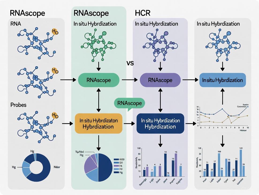
RNAscope vs. HCR: A Strategic Guide to Choosing the Right In Situ Hybridization Method
This article provides a comprehensive comparison for researchers and drug development professionals between two powerful in situ hybridization (ISH) technologies: RNAscope and Hybridization Chain Reaction (HCR). We explore their foundational principles, including RNAscope's proprietary branched DNA signal amplification and HCR's enzyme-free, kinetically controlled hairpin assembly. The content details their methodological workflows, key applications in research and clinical diagnostics, and practical guidance for troubleshooting and optimization. A critical validation and comparative analysis equips scientists with the knowledge to make an informed choice based on factors such as sensitivity, specificity, multiplexing capability, cost, and sample type, ultimately accelerating biomarker development and therapeutic research.
RNAscope vs. HCR: A Strategic Guide to Choosing the Right In Situ Hybridization Method
Abstract
This article provides a comprehensive comparison for researchers and drug development professionals between two powerful in situ hybridization (ISH) technologies: RNAscope and Hybridization Chain Reaction (HCR). We explore their foundational principles, including RNAscope's proprietary branched DNA signal amplification and HCR's enzyme-free, kinetically controlled hairpin assembly. The content details their methodological workflows, key applications in research and clinical diagnostics, and practical guidance for troubleshooting and optimization. A critical validation and comparative analysis equips scientists with the knowledge to make an informed choice based on factors such as sensitivity, specificity, multiplexing capability, cost, and sample type, ultimately accelerating biomarker development and therapeutic research.
Core Principles: Deconstructing the Signal Amplification Mechanisms of RNAscope and HCR
In situ hybridization (ISH) has long been a fundamental tool for visualizing nucleic acid sequences within their morphological context in tissues and cells. However, traditional RNA ISH methods have faced significant challenges in achieving sufficient sensitivity and specificity for reliable detection of low-abundance RNA transcripts, particularly in clinical specimens [1]. This limitation has been especially notable given the abundance of RNA biomarkers discovered through whole-genome expression profiling. While quantitative PCR (qPCR) offers high sensitivity for RNA detection, it is a "grind-and-bind" approach that destroys tissue architecture and eliminates crucial spatial information [1] [2]. The emergence of highly sensitive and specific ISH technologies, particularly RNAscope's branched DNA (bDNA) technology and Hybridization Chain Reaction (HCR), has revolutionized the field by enabling single-molecule RNA visualization while preserving tissue morphology [1] [3]. This comparison guide objectively examines the technological principles, performance characteristics, and practical applications of these advanced ISH platforms to inform researchers, scientists, and drug development professionals in their experimental design decisions.
Technological Foundations: Mechanisms of Amplification
RNAscope's bDNA Technology and Z-Probe Design
RNAscope employs a patented branched DNA (bDNA) signal amplification system with a unique double-Z probe design strategy that enables simultaneous signal amplification and background suppression [1] [2]. This technology utilizes a series of specifically designed target probes that hybridize to the target RNA molecule, with each probe containing an 18-25 base region complementary to the target RNA, a spacer sequence, and a 14-base tail sequence [1]. The key innovation lies in the double-Z probe design, where pairs of target probes (conceptualized as "Z" shapes) must bind contiguously to the target RNA (∼50 bases) to form a complete recognition site for the subsequent amplification steps [1] [2]. This requirement ensures exceptional specificity, as nonspecific hybridization events are unlikely to juxtapose two probes correctly to form the recognition site.
The amplification cascade begins when the paired Z-probes hybridize adjacent to each other on the target RNA, creating a 28-base hybridization site for the preamplifier molecule [1]. Each preamplifier contains 20 binding sites for the amplifier molecule, which in turn contains 20 binding sites for the label probe. This hierarchical branching structure theoretically yields up to 8000 labels for each target RNA molecule when 20 probe pairs target a 1-kb region [1]. The label probe can be conjugated to either fluorescent dyes for fluorescence microscopy or enzymes for chromogenic detection compatible with bright-field microscopy [1]. This design provides the foundation for RNAscope's exceptional sensitivity, capable of detecting individual RNA molecules with high specificity.
Figure 1: RNAscope's bDNA Cascading Amplification. The double-Z probe design requires two probes to bind adjacent to each other on the target RNA to initiate the signal amplification cascade through preamplifier, amplifier, and label probe binding, enabling single-molecule visualization [1] [2].
Hybridization Chain Reaction (HCR) Mechanism
Hybridization Chain Reaction represents a different approach to signal amplification, utilizing an enzyme-free, isothermal method based on the mechanism of triggered self-assembly of DNA hairpins [3] [4]. The fundamental HCR system consists of two metastable DNA hairpins (H1 and H2) that coexist stably until exposed to an initiator sequence trigger [3]. In the presence of the target RNA, DNA probes complementary to the target mRNA expose initiator sequences that trigger a chain reaction of alternating H1 and H2 hairpin openings, forming long, nicked double-stranded DNA polymers [4]. These amplification polymers serve as scaffolds for fluorophore binding, generating detectable signal at the site of target RNA localization.
The latest iteration, in situ HCR v3.0, introduces a critical innovation with split-initiator probes that provide automatic background suppression [3]. Unlike the standard HCR v2.0 approach where each probe carries a full HCR initiator, HCR v3.0 employs pairs of cooperative split-initiator probes that each carry half of the HCR initiator [3]. Only when both probes hybridize specifically to adjacent binding sites on the target mRNA are the two halves colocalized to form a complete initiator, triggering the HCR amplification. This design ensures that individual probes binding non-specifically cannot initiate the amplification cascade, dramatically reducing background signal [3].
Figure 2: Hybridization Chain Reaction with Split-Initiator Probes. HCR v3.0 utilizes split-initiator probes that must hybridize adjacently on the target RNA to form a complete initiator, which then triggers the self-assembly of H1 and H2 hairpins into fluorescent amplification polymers [3] [4].
Performance Comparison: Experimental Data and Technical Specifications
Sensitivity and Specificity Metrics
Table 1: Quantitative Performance Comparison Between RNAscope and HCR
| Performance Parameter | RNAscope bDNA Technology | HCR v3.0 |
|---|---|---|
| Detection Sensitivity | Single RNA molecule detection [1] | Single RNA molecule detection [3] |
| Background Suppression | Double-Z design ensures minimal background; negative control (dapB) should yield score <1 [1] [5] | ≈50-fold background suppression with split-initiator probes compared to standard HCR [3] |
| Signal Amplification | Theoretical 8000 labels per target RNA with 20 probe pairs [1] | Amplification proportional to hairpin concentration and reaction time [3] |
| Multiplexing Capacity | Up to 4 targets simultaneously with spectrally distinct labels [1] | Up to 5 targets simultaneously with orthogonal HCR systems [3] |
| Target Length Requirements | Optimal for sequences ≥300 nucleotides [2] | Compatible with various target lengths, including microRNAs [6] |
Experimental Workflow and Protocol Requirements
Table 2: Experimental Protocol and Workflow Comparison
| Protocol Aspect | RNAscope | HCR |
|---|---|---|
| Assay Duration | 7-8 hours, completable in one day [5] | 1-3 days depending on optimization [6] |
| Sample Compatibility | Formalin-fixed paraffin-embedded (FFPE) tissues, frozen tissues, cell cultures [1] [5] | Whole-mount embryos, thick tissue sections, mammalian and bacterial cells [3] |
| Tissue Pretreatment | Required: antigen retrieval (citrate buffer, 15 min boiling) and protease digestion (10 μg/mL, 30 min at 40°C) [1] | Varies by sample type; generally requires permeabilization [3] |
| Hybridization Conditions | 40°C for 2 hours using HybEZ system [1] [5] | Room temperature or mild heating conditions [4] |
| Detection Methods | Chromogenic (DAB, Fast Red) or fluorescent [1] | Primarily fluorescent; some colorimetric applications [4] |
Experimental Protocols for Key Applications
RNAscope Protocol for FFPE Tissue Sections
The RNAscope assay for formalin-fixed, paraffin-embedded (FFPE) tissues follows a standardized protocol that can be performed manually or on automated platforms [1] [5]. For manual assays:
Sample Preparation: Cut 5-μm thick tissue sections and mount on Superfrost Plus slides. Deparaffinize in xylene and dehydrate through an ethanol series [1].
Pretreatment: Perform antigen retrieval by incubating sections in citrate buffer (10 mmol/L, pH 6) at boiling temperature (100-103°C) for 15 minutes. Treat with protease (10 μg/mL) at 40°C for 30 minutes in a HybEZ hybridization oven [1] [5].
Probe Hybridization: Apply target probes in hybridization buffer and incubate at 40°C for 2 hours. For multiplex detection, use equimolar amounts of target probes with different channel configurations [1] [5].
Signal Amplification: Perform sequential 30-minute hybridizations with preamplifier, amplifier, and label probe at 40°C, with wash steps between each hybridization [1].
Detection and Visualization: For chromogenic detection, use DAB with hematoxylin counterstaining. For fluorescent detection, use fluorophore-conjugated label probes with appropriate mounting media [1] [5].
Controls: Always include positive control probes (e.g., housekeeping genes PPIB, POLR2A, or UBC) and negative control probes (bacterial dapB) to assess RNA quality and assay performance [5].
HCR v3.0 Protocol for Whole-Mount Embryos
The HCR v3.0 protocol for whole-mount samples such as chicken embryos involves the following key steps [3]:
Sample Fixation and Permeabilization: Fix samples in formaldehyde-based fixative and permeabilize with detergent to enable probe access.
Hybridization with Split-Initiator Probes: Hybridize samples with pairs of split-initiator probes designed against the target mRNA. Typically use 20 probe pairs for optimal signal-to-background ratio.
Amplification with HCR Hairpins: After washing away unbound probes, add H1 and H2 hairpins to initiate the hybridization chain reaction. The amplification time can be adjusted from hours to overnight depending on signal requirements.
Imaging and Analysis: Image samples using fluorescence microscopy. The signal appears as distinct fluorescent foci corresponding to individual RNA molecules.
Multiplexing: For simultaneous detection of multiple targets, use orthogonal HCR systems with different initiator sequences and spectrally distinct fluorophores [3].
The Scientist's Toolkit: Essential Research Reagent Solutions
Table 3: Essential Research Reagents and Equipment for Advanced ISH
| Item | Function/Purpose | RNAscope | HCR |
|---|---|---|---|
| Specialized Slides | Tissue adhesion during stringent processing | Superfrost Plus slides required [5] | Standard slides typically sufficient |
| Barrier Pens | Create hydrophobic barriers to maintain reagent coverage | ImmEdge Hydrophobic Barrier Pen required [5] | Helpful but not specifically mandated |
| Hybridization System | Maintain optimum humidity and temperature during hybridization | HybEZ system required [5] | Standard hybridization chambers acceptable |
| Protease Reagents | Tissue permeabilization for probe access | Specific concentrations critical (e.g., 10 μg/mL) [1] | Concentration requires optimization per sample |
| Detection Kits | Signal generation and visualization | RNAscope 2.5 HD kits (Brown/Red) with specific mounting media [5] | Custom hairpin sets with fluorophore labels |
| Control Probes | Assess sample quality and assay performance | Positive (PPIB, UBC) and negative (dapB) controls essential [5] | Target-specific controls recommended |
| Automation Compatibility | High-throughput and standardized processing | Compatible with Ventana DISCOVERY XT/ULTRA and Leica BOND RX [5] | Primarily manual protocols |
| Blepharotriol | Blepharotriol|Phenolic Nor-Triterpene|RUO | Blepharotriol, a phenolic nor-triterpene from Celastraceae species. For Research Use Only. Not for diagnostic or therapeutic use. | Bench Chemicals |
| Sarbronine M | Sarbronine M, MF:C20H28O4, MW:332.4 g/mol | Chemical Reagent | Bench Chemicals |
Discussion: Advantages, Limitations, and Application Scenarios
RNAscope Strengths and Considerations
RNAscope offers several distinct advantages that make it particularly suitable for certain research and diagnostic applications. Its high sensitivity and specificity enable reliable detection of even low-abundance RNA transcripts, with the unique double-Z probe design providing exceptional signal-to-noise ratio [1] [2]. The platform's compatibility with FFPE tissues makes it invaluable for retrospective studies using archived clinical samples [1]. Additionally, RNAscope's capacity for both chromogenic and fluorescent detection provides flexibility for various imaging needs and facilitates integration with standard histopathology workflows [5]. The availability of pre-validated, commercially available probes for many targets reduces optimization time and ensures consistency across experiments [7].
However, RNAscope has certain limitations to consider. The technology has the highest per-sample monetary cost among high-sensitivity ISH methods, making it less suitable for large-scale screening studies [6]. Probe design constraints require target sequences of at least 300 nucleotides for optimal performance, limiting application to shorter transcripts without using the specialized BaseScope variant [2]. Additionally, the proprietary nature of the amplification system limits customization options compared to open-source methods like HCR [7].
HCR Strengths and Considerations
HCR technology offers complementary advantages, particularly appealing for certain research scenarios. The method's enzyme-free, isothermal amplification provides a simpler biochemical environment that may be preferable for delicate samples or certain experimental conditions [4]. The open-source nature of HCR allows researchers to design custom probes for any target of interest, providing greater flexibility, especially for non-standard model organisms or novel transcripts [3]. HCR's modular design supports straightforward multiplexing with up to five simultaneous targets using orthogonal amplifier systems [3]. From a cost perspective, HCR becomes increasingly economical with larger sample sizes, as the per-sample cost decreases significantly [6].
Limitations of HCR include generally lower signal amplification compared to RNAscope's branched DNA approach, potentially affecting detection of very low-abundance targets [7]. The method primarily supports fluorescent detection, limiting its application in bright-field microscopy contexts common in clinical pathology [6]. HCR also requires more extensive optimization of experimental conditions, including probe design and hybridization parameters, which demands greater expertise and time investment [7] [6].
RNAscope's bDNA technology with its innovative Z-probe design represents a significant advancement in RNA in situ hybridization, offering robust, sensitive, and specific detection of RNA molecules within morphological context. The double-Z probe architecture with cascading amplification provides exceptional signal-to-background ratio, making it particularly valuable for clinical research and diagnostic applications where reliability and consistency are paramount [1] [5]. Meanwhile, HCR v3.0 with its split-initiator probes and enzyme-free amplification offers an powerful alternative with greater customizability and lower per-sample costs for large-scale studies [3] [6].
The choice between these technologies ultimately depends on specific research requirements, including sample type, target abundance, required throughput, budgetary constraints, and technical expertise. RNAscope excels in standardized environments, clinical translation, and situations requiring chromogenic detection or integration with automated pathology platforms. HCR offers advantages in exploratory research, highly multiplexed studies, and resource-limited settings where reagent costs are a primary consideration. Both technologies continue to evolve, pushing the boundaries of sensitivity, multiplexing capability, and practical implementation in both basic research and clinical diagnostics.
In the field of spatial biology, in situ hybridization (ISH) techniques provide crucial windows into the spatial organization of RNA expression within cells and tissues, enabling researchers to visualize gene expression in its anatomical context [8]. For researchers, scientists, and drug development professionals, the choice of ISH method can significantly impact experimental outcomes, particularly when studying low-abundance targets or working with complex sample types. Two powerful methods dominate the current landscape: the widely adopted, proprietary RNAscope technology and the enzyme-free, mechanism-driven Hybridization Chain Reaction (HCR) [7] [6]. This guide provides an objective comparison of these technologies, with a specific focus on the fundamental principles underpinning HCR's enzyme-free paradigm—toehold-mediated strand displacement and hairpin polymerization—and their practical implications for experimental design and performance.
While RNAscope employs a branched DNA (bDNA) signal amplification system that relies on enzymatic reactions [7], HCR utilizes an isothermal, enzyme-free approach based on the principles of nucleic acid self-assembly [8]. This fundamental difference in amplification mechanism creates distinct experimental workflows, performance characteristics, and optimization requirements that researchers must consider when selecting the appropriate method for their specific applications, whether in basic research, drug development, or clinical pathology.
Mechanistic Foundations: Contrasting HCR and RNAscope
HCR: Enzyme-Free Signal Amplification Through Hairpin Polymerization
The Hybridization Chain Reaction employs a unique enzyme-free amplification mechanism based on the principles of nucleic acid strand displacement and hybridization kinetics [8]. In HCR, RNA targets are detected by nucleic acid probes that trigger isothermal chain reactions in which fluorophore-labeled DNA hairpins self-assemble into tethered fluorescent amplification polymers [8]. This process begins when an initiator probe hybridizes to the target RNA molecule, exposing a sequence that serves as a toehold for the initiation of the chain reaction [7].
The core amplification mechanism involves two metastable hairpin species (H1 and H2) that remain stable in the absence of the initiator. Upon initiator exposure, a toehold-mediated strand displacement reaction occurs, opening the H1 hairpin and exposing a sequence that subsequently opens the H2 hairpin, which in turn exposes a sequence identical to the original initiator [9]. This creates a positive feedback loop where the H1 and H2 hairpins alternately add to the growing polymer, forming a long nicked double helix that incorporates hundreds of fluorophores precisely at the location of the target RNA [8]. This mechanism enables signal amplification without enzymes, as the system is driven solely by the thermodynamics of nucleic acid hybridization and the kinetic stability of properly designed hairpin structures [10].
HCR Mechanism: Toehold-mediated hairpin polymerization. The initiator probe binds to the target RNA, triggering sequential hairpin opening and polymerization.
RNAscope: Enzymatic Signal Amplification via Branched DNA
In contrast to HCR's enzyme-free approach, RNAscope employs a proprietary branched DNA (bDNA) amplification system that relies on enzymatic signal development [7]. The technology uses specially designed "Z-probes" that contain two distinct binding regions: a target-binding sequence that hybridizes to the RNA of interest and a pre-amplifier sequence that serves as a scaffolding for signal amplification [7]. Each Z-probe hybridizes to the target RNA, forming a target-specific "Z-probe/target RNA" complex [7].
The amplification process involves sequential hybridization steps where multiple pre-amplifier and amplifier molecules, each labeled with specific oligonucleotide sequences, bind to the Z-probe complex [7]. This branching architecture creates a signaling tree that can accommodate numerous label probes, significantly amplifying the signal compared to single-step hybridization methods. The final detection typically involves enzymatic development either through chromogenic precipitation or fluorescent labeling, providing flexibility in detection modalities but introducing enzyme dependency into the system [6]. This enzymatic dependence differentiates RNAscope fundamentally from HCR's purely hybridization-based approach and has implications for experimental flexibility, multiplexing capabilities, and quantitative performance.
Performance Comparison: Experimental Data and Technical Specifications
Quantitative Performance Comparison
The table below summarizes key performance characteristics based on experimental data from peer-reviewed studies:
Table 1: Technical Performance Comparison of HCR and RNAscope
| Parameter | HCR | RNAscope |
|---|---|---|
| Amplification Mechanism | Enzyme-free hairpin polymerization [8] | Branched DNA with enzymatic development [7] |
| Signal-to-Background Ratio | Median: 90 (Range: 15-609 across 21 imaging scenarios) [8] | High, but quantitative data not provided in search results |
| Multiplexing Capability | Simultaneous multiplexing with orthogonal amplifiers [8] | Possible, but may require sequential staining [6] |
| Detection Efficiency | Linear signal scaling with target abundance [8] | High sensitivity for low-abundance targets [7] |
| Spatial Resolution | Subcellular/single-molecule resolution [8] | High resolution, but may vary with enzymatic diffusion [6] |
| Sample Compatibility | Whole-mount embryos, FFPE sections, autofluorescent samples [8] | FFPE tissues, frozen tissues, cell cultures [7] |
Experimental Optimization and Protocol Details
HCR Protocol Optimization:
Achieving optimal performance with HCR requires careful attention to oligonucleotide quality and experimental conditions. Research demonstrates that HCR is highly sensitive to oligonucleotide quality, with purification methods significantly impacting polymerization efficiency [10]. PAGE purification can greatly enhance hairpin polymerization both in solution and in situ, while ligation-based purification methods have been shown to yield immunoHCR stains at least 3.4-times stronger than non-purified controls [10].
Key optimization parameters for HCR include:
- Incubation Times: Extending both probe hybridization and amplification incubation times to overnight can improve signal strength, especially in thicker samples, with minimal benefit beyond overnight incubation [11].
- Probe Design: Switching to boosted probe designs (with more binding sites) can elevate signal without protocol changes [11].
- Hairpin Quality: Ensuring high-fidelity hairpin synthesis and appropriate purification is critical for minimizing background and maximizing signal [10].
RNAscope Protocol Considerations:
RNAscope offers a more standardized approach with simplified protocols:
- Probe Design: RNAscope probes are commercially available as pre-validated sets, reducing optimization time but limiting customization [7].
- Experimental Timeline: Staining can typically be completed within one day, significantly faster than conventional ISH methods [6].
- Automation Compatibility: The technology is compatible with automated staining systems used in clinical pathology [6].
Practical Implementation: Research Reagent Solutions
Essential Materials and Reagents
Table 2: Essential Research Reagent Solutions for HCR Experiments
| Reagent Category | Specific Examples | Function | Optimization Notes |
|---|---|---|---|
| HCR Hairpins | H1 and H2 hairpins with fluorophores [8] | Signal amplification via polymerization | PAGE purification critical for performance [10] |
| Initiator Probes | Target-specific initiators [8] | Target recognition and HCR initiation | Boosted designs available for low-abundance targets [11] |
| Hybridization Buffers | Formamide-containing buffers [12] | Control stringency of hybridization | Concentration affects probe assembly efficiency [12] |
| Purification Systems | PAGE, RP-HPLC, IE-HPLC [10] | Oligonucleotide quality control | Ligation-based methods enhance polymerization [10] |
| Blocking Agents | Salmon sperm DNA, tRNA [12] | Reduce non-specific binding | Critical in autofluorescent samples [11] |
Workflow Integration and Experimental Design
The experimental workflow for implementing HCR involves several critical stages where reagent quality and protocol optimization significantly impact outcomes. The diagram below illustrates the optimized workflow and key decision points:
HCR Workflow: Key experimental stages and optimization points from sample preparation to imaging.
Comparative Analysis: Advantages and Limitations in Research Contexts
HCR Advantages and Limitations
Advantages:
- Enzyme-Free Amplification: The isothermal, enzyme-free nature of HCR eliminates enzyme-related variability and cost [8].
- Quantitative Performance: HCR signal scales approximately linearly with target abundance, enabling accurate RNA relative quantitation with subcellular resolution [8].
- Multiplexing Flexibility: Orthogonal HCR amplifiers operate independently, allowing simultaneous multiplexed detection without serial staining [8].
- Customization Potential: Researchers can design custom probes for any target sequence, providing flexibility for novel targets [7].
Limitations:
- Optimization Requirements: HCR requires careful optimization of probe design, hairpin quality, and hybridization conditions [7].
- Background Sensitivity: The method can sometimes produce background signal from nonspecific hybridization events [7].
- Probe Design Complexity: Creating efficient initiator and hairpin probes requires expertise in nucleic acid thermodynamics [7].
RNAscope Advantages and Limitations
Advantages:
- Standardized Protocols: RNAscope offers simplified, reliable protocols with minimal optimization requirements [6].
- High Sensitivity: The technology provides exceptional sensitivity for detecting low-abundance transcripts [7].
- Clinical Validation: RNAscope has been extensively validated in research and clinical applications [7].
- Automation Compatibility: The system works with automated pathology equipment, enabling high-throughput applications [6].
Limitations:
- Probe Customization: Commercial probes limit customization options and may not be available for all targets [7].
- Cost Considerations: RNAscope has higher monetary cost per sample, which increases proportionally with sample number [6].
- Enzyme Dependency: The requirement for enzymatic development introduces additional variables and costs [7].
Cost-Benefit Analysis for Research Implementation
Table 3: Practical Implementation Considerations for HCR and RNAscope
| Consideration | HCR | RNAscope |
|---|---|---|
| Monetary Cost | Moderate; decreases with sample number [6] | High; proportional to sample number [6] |
| Time Investment | 1-3 days staining; significant optimization time [6] | 1 day staining; minimal optimization [6] |
| Technical Expertise | Requires expertise in nucleic acid chemistry [7] | Accessible to broad user base [6] |
| Customization Potential | High for novel targets and applications [7] | Limited to commercially available probes [7] |
| Suitability for High-Throughput | Moderate, requires validation [8] | High, compatible with automation [6] |
The choice between HCR and RNAscope represents a strategic decision that should align with specific research goals, available resources, and experimental constraints. HCR's enzyme-free paradigm offers distinct advantages for researchers requiring quantitative measurements, flexible multiplexing, and customization capabilities, particularly when studying complex biological systems where multiple RNA targets need to be visualized simultaneously in their native context. The fundamental mechanisms of toehold-mediated strand displacement and hairpin polymerization provide a robust foundation for precise spatial mapping of RNA expression, albeit with greater initial optimization requirements.
Conversely, RNAscope presents a compelling solution for applications requiring rapid implementation, high sensitivity, and operational simplicity, particularly in clinical or diagnostic settings where standardization and reliability are paramount. Its proprietary probe design and enzymatic amplification system deliver consistent performance with minimal optimization, though at higher per-sample costs and with reduced customization flexibility.
For the research community, understanding these fundamental technologies and their performance characteristics enables informed selection of the most appropriate method for specific experimental needs. As spatial biology continues to advance, both technologies will play crucial roles in elucidating the complex architecture of gene expression in development, disease, and biological regulation.
The analysis of gene expression patterns within their native tissue context is a cornerstone of biological research and diagnostic pathology. For decades, this field has been dominated by two fundamental techniques: conventional in situ hybridization (ISH) for nucleic acid detection and immunohistochemistry (IHC) for protein localization. While invaluable, these methods possess significant limitations that compromise their reliability and application scope. Conventional ISH, particularly using digoxigenin (DIG)-labeled RNA probes, is often hampered by poor sensitivity and high background noise, making it unsuitable for detecting low-abundance transcripts. Furthermore, its procedures are complex and time-consuming, typically requiring 2-3 days for completion and presenting difficulties for multiplex staining [6]. IHC, while more established in clinical settings, suffers from an absolute dependency on antibody quality, with issues of specificity, availability, and validation frequently leading to unreliable results, especially for targets beyond common human pathological markers or in non-human species [13] [6]. The emergence of RNAscope and Hybridization Chain Reaction (HCR) technologies represents a paradigm shift, addressing these long-standing challenges through innovative signal amplification strategies that offer single-molecule sensitivity, exceptional specificity, and streamlined workflows.
Technological Breakthroughs: Core Mechanisms of RNAscope and HCR
RNAscope: The Double-Z Probe Design
The RNAscope technology, a proprietary method developed by Advanced Cell Diagnostics (ACD), achieves its performance through a unique double-Z probe design and a branched DNA (bDNA) signal amplification system [7] [13]. This design is the key to its high specificity and sensitivity.
The mechanism can be broken down into three critical stages, illustrated in the diagram below:
Diagram Title: RNAscope Signal Amplification Mechanism
As shown, the process begins with a pair of "Z" probes that hybridize to adjacent binding sites on the target RNA [14]. Each Z probe consists of three parts: a region that hybridizes to the target RNA, a spacer, and a tail that binds to the pre-amplifier [13]. Critically, the pre-amplifier can only bind if both Z probes are correctly hybridized, providing a built-in check for specificity that prevents off-target binding and background noise [14]. Each bound pre-amplifier then recruits multiple amplifier molecules, which in turn are labeled with numerous fluorescent probes. This cascade can result in an amplification of up to 8,000 times, enabling the detection of individual RNA molecules as distinct, quantifiable dots under a microscope [7] [13].
HCR: Enzyme-Free Hybridization Chain Reaction
HCR represents a different philosophical approach, utilizing an isothermal, enzyme-free amplification process based on the mechanism of a hybridization chain reaction [3] [15]. Its core components are two species of metastable DNA hairpins (H1 and H2) that coexist stably until triggered by a specific DNA initiator sequence.
The following diagram outlines the stepwise process of HCR signal amplification:
Diagram Title: HCR Signal Amplification Mechanism
The latest iteration, HCR v3.0, significantly enhances specificity by employing split-initiator probes [3]. Instead of a single probe carrying a full initiator, two separate probes each carry half of the initiator sequence. The full initiator is only assembled and HCR is only triggered when both probes hybridize correctly to adjacent sites on the target mRNA. This design ensures automatic background suppression, as any single probe binding non-specifically elsewhere in the sample cannot initiate the amplification cascade [3]. This innovation allows researchers to use larger, unoptimized probe sets while maintaining a high signal-to-noise ratio.
Comparative Performance: Quantitative Data Against Gold Standards
Superiority Over Conventional ISH and IHC
The advantages of RNAscope and HCR are not merely theoretical; they are demonstrated by superior performance metrics in direct comparisons with conventional methods. The table below summarizes key quantitative and qualitative comparisons.
Table 1: Performance Comparison of ISH and IHC Techniques
| Parameter | Conventional ISH | IHC | RNAscope | HCR (v3.0) |
|---|---|---|---|---|
| Sensitivity | Low to moderate; struggles with low-abundance targets [13] | Variable; depends on antibody affinity and titer [6] | Very High; single-molecule detection [7] [13] | Very High; capable of single-molecule imaging [3] |
| Specificity | Moderate; prone to off-target hybridization [13] | Variable; cross-reactivity is a common issue [6] | Very High; proprietary double-Z design ensures minimal background [7] [14] | High; split-initiator design suppresses amplified background [3] |
| Multiplexing Capability | Difficult; sequential staining is complex [6] | Moderate; limited by antibody host species and chromogen overlap | High; simultaneous detection of multiple targets with different fluorophores [7] | High; simultaneous one-stage amplification for multiple targets [3] |
| Experimental Time | 2–3 days [6] | 1–2 days | ~1 day [6] | 1–3 days [6] |
| Quantification | Semi-quantitative; based on stain intensity | Semi-quantitative; based on stain intensity | Digital/Qunatitative; dots are countable, correlating to RNA copies [13] | Analog/Digital; qHCR for relative and dHCR for absolute quantitation [3] |
| Key Limitation | Complex procedure, high background [13] [6] | Relies entirely on antibody quality and availability [13] [6] | Higher cost; probe design constraints for some sequences [7] | Probe design can be complex for users [7] |
A systematic review from 2021 provides compelling quantitative evidence for RNAscope, showing a high concordance rate with established molecular techniques like qPCR and qRT-PCR (81.8–100%). However, its concordance with IHC was lower (58.7–95.3%), underscoring the fundamental difference between detecting RNA and protein and the potential for discrepancies in gene expression analysis [13]. This highlights a key advantage of RNAscope: it can serve as a reliable method to validate antibody specificity in IHC [6]. In breast cancer diagnostics, for instance, FISH (a DNA ISH technique) is considered more reliable than IHC for detecting HER2 amplification, a critical therapeutic biomarker [16].
RNAscope vs. HCR: A Direct Comparison for Informed Selection
When deciding between RNAscope and HCR, researchers must weigh their specific needs against the characteristics of each method. The table below outlines the critical factors for this decision.
Table 2: Direct Comparison of RNAscope and HCR In Situ Hybridization
| Factor | RNAscope | HCR |
|---|---|---|
| Probe Design & Cost | Proprietary, pre-validated probes from ACD [7]. Higher cost per sample, but no design/validation time [6]. | User-designed or outsourced [7] [15]. Lower monetary cost, but requires design time and optimization [7] [6]. |
| Signal Amplification | Branched DNA (bDNA) [7]. | Hybridization Chain Reaction (enzyme-free) [7] [3]. |
| Ease of Use | High. Standardized, user-friendly protocol; amenable to full automation on staining platforms [17] [6]. | Moderate. Requires more hands-on optimization, but protocols have been simplified (e.g., no proteinase K) [15]. |
| Multiplexing | Well-established for multiplexing with different fluorescent channels [7] [13]. | Designed for straightforward multiplexing with simultaneous one-stage amplification [3]. |
| Sample Compatibility | Excellent for FFPE tissues (most common application), frozen tissues, and cell cultures [7] [18]. | Can have reduced efficiency in certain FFPE tissues; excellent for whole-mount samples and thick tissues [7] [3]. |
| Best Suited For | Clinical diagnostics, labs prioritizing throughput and standardization, projects with a focused set of targets. | Academic research, high-level multiplexing, whole-mount imaging, labs with budget constraints and technical expertise for optimization. |
Experimental Protocols: From Theory to Practice
Integrated RNAscope and IHC Protocol for CNS Tissue
The combination of RNAscope and IHC on the same tissue section is a powerful application that allows for the precise cellular assignment of gene expression. The following workflow, adapted from a detailed methods paper, is optimized for thicker (14 μm) fixed spinal cord sections [14].
Diagram Title: Combined RNAscope & IHC Workflow
Key Modifications and Considerations:
- Tissue Preparation: Fixation with 10% Neutral Buffered Formalin (NBF) for 16-32 hours is critical for RNA integrity [18]. Thicker sections (e.g., 14 μm) are used to preserve cellular structures for subsequent analysis.
- Protease Treatment: This step is essential for probe permeability but is carefully optimized to preserve antigenicity for IHC. The RNAscope protocol provides specific recommendations based on tissue type [14] [18].
- Simultaneous vs. Sequential Staining: The protocol can be performed with the RNAscope and IHC steps done sequentially on the same day. The relatively mild hybridization conditions (40°C) of RNAscope help preserve protein epitopes [14].
- Analysis: Images are acquired using confocal microscopy. Quantification of RNA dots (transcripts) within IHC-defined cell boundaries (e.g., neurons labeled with NeuN, microglia labeled with IBA1) is performed using advanced image analysis software like Halo or QuPath [13] [14].
Modified HCR Protocol for Cost-Effectiveness
A modified HCR protocol has been developed to make the technique more accessible by addressing its primary drawback: cost. The key innovation is the use of short hairpin DNAs (36-44 nucleotides), which are significantly cheaper to synthesize than the traditional 72-nucleotide hairpins while maintaining a high signal-to-noise ratio [15].
Key Advancements in the Modified Protocol:
- Cost Reduction: Shorter hairpin DNAs directly lower the monetary cost of the assay [15].
- Preserved Antigenicity: The protocol removes the proteinase K treatment step, which better preserves morphological structures and protein antigens, making combined HCR-IHC much more reliable [15].
- Sensitivity: This modified HCR has demonstrated better sensitivity for detecting low-abundance mRNAs like the oxytocin receptor (Oxtr) compared to traditional FISH with tyramide signal amplification [15].
The Scientist's Toolkit: Essential Research Reagent Solutions
Successful implementation of these advanced ISH techniques requires specific reagents and tools. The following table details the key components for setting up these experiments.
Table 3: Essential Research Reagent Solutions for RNAscope and HCR
| Item | Function | Examples & Notes |
|---|---|---|
| Control Probes | Critical for validating assay conditions and RNA quality. | Positive Control: Housekeeping genes (PPIB, POLR2A, UBC) [13] [18]. Negative Control: Bacterial dapB gene [13] [18]. |
| Probe Sets | Target-specific reagents for hybridization. | RNAscope: Pre-designed, validated probes from ACD's catalog [19]. HCR: User-designed split-initiator probe sets [3] [15]. |
| Amplification Systems | Generate the detectable signal. | RNAscope: Proprietary pre-amplifier, amplifier, and label probes [7] [13]. HCR: Fluorescently labeled H1 and H2 DNA hairpins [3] [15]. |
| Signal Detection Reagents | Enable visualization of amplified signal. | Chromogenic (DAB) or fluorescent dyes for RNAscope [7]. Fluorophores conjugated to HCR hairpins (e.g., Alexa Fluor dyes) [3]. |
| Automated Staining System | Standardizes workflow, reduces hands-on time, and enhances reproducibility. | Roche Ventana BenchMark ULTRA/PLUS series for IHC and ISH [17]. Compatible with the automated RNAscope workflow [13]. |
| Image Analysis Software | Quantifies transcript numbers and performs colocalization analysis. | Halo, QuPath, Aperio; capable of counting dots/cell and analyzing in a cell-type-specific context [13] [14]. |
| Isoglobotetraose | Isoglobotetraose, MF:C26H45NO21, MW:707.6 g/mol | Chemical Reagent |
| Dendryphiellin D | Dendryphiellin D, MF:C21H28O5, MW:360.4 g/mol | Chemical Reagent |
The evolution from traditional ISH and IHC to RNAscope and HCR technologies marks a significant advancement in molecular histology. By overcoming the critical limitations of sensitivity, specificity, and operational complexity, these methods provide researchers and diagnosticians with powerful, reliable tools for visualizing gene expression at the single-molecule level. RNAscope offers a turnkey, highly robust solution ideal for standardized and clinical environments, whereas HCR provides unparalleled flexibility and cost-effectiveness for specialized research applications, especially in whole-mount and multiplexed imaging. Both techniques enable a more precise and quantitative understanding of gene expression within its anatomical context, accelerating discovery in basic research and enhancing accuracy in diagnostic pathology. The choice between them ultimately depends on the specific experimental needs, resource constraints, and technical expertise of the laboratory.
Workflows and Applications: Implementing RNAscope and HCR in Research and Diagnostics
In the field of molecular pathology, the ability to visualize RNA biomarkers within their native tissue context provides invaluable insights into gene expression patterns, cellular heterogeneity, and disease mechanisms. RNA in situ hybridization (ISH) has emerged as a critical technology for this purpose, yet its adoption in clinical and research settings has been hampered by technical challenges including insufficient sensitivity, specificity, and reproducibility. This guide objectively compares two prominent ISH methodologies: the commercially established RNAscope technology and the more recent Hybridization Chain Reaction (HCR) approach, with a specific focus on their performance within a standardized workflow from formalin-fixed paraffin-embedded (FFPE) sample preparation to automated staining. The examination is framed within the broader thesis that while both methods enable transcript visualization, their technical architectures impose significant practical implications for workflow standardization, data reliability, and implementation in regulated environments. We present supporting experimental data to empower researchers, scientists, and drug development professionals in making informed technological selections for their specific applications.
Technical Foundations: A Comparative Architecture of RNAscope and HCR
The fundamental differences between RNAscope and HCR begin at the level of probe design and signal amplification mechanics. These architectural decisions directly influence performance characteristics including sensitivity, specificity, signal-to-noise ratio, and operational complexity.
RNAscope Technology Architecture
RNAscope, a proprietary technology developed by Advanced Cell Diagnostics (ACD), employs a unique "Z-probe" design and branched DNA (bDNA) signal amplification system [13] [20]. The core mechanism involves:
- Paired "Z" Probes: Each probe pair consists of two oligonucleotides that hybridize to adjacent target sequences on the RNA of interest [7]. The lower region hybridizes to the target RNA, a spacer connects to the tail, and the tail binds to pre-amplifier sequences.
- Branched DNA Amplification: After hybridization, a pre-amplifier molecule binds specifically to the paired Z-probe tails. This is followed by sequential binding of amplifier molecules and finally, enzyme-labeled probes (for chromogenic detection) or directly labeled probes (for fluorescence) [20]. This cascade results in up to 8,000-fold signal amplification, enabling single-molecule detection [13].
- Specificity Mechanism: The system requires two independent probes to hybridize adjacently for signal generation, dramatically reducing off-target binding and background noise [14]. This dual-Z requirement means non-specific binding of a single probe produces no signal.
HCR Technology Architecture
Hybridization Chain Reaction represents a different approach based on initiated self-assembly of nucleic acid hairpins:
- Initiator and Amplifier Probes: HCR uses two separate sets of DNA hairpin probes: initiator probes that hybridize to the target RNA, and amplifier probes that undergo a chain reaction upon initiation [7].
- Hairpin Amplification Cascade: When an initiator probe binds to its target, it opens to expose a sequence that nucleates the hybridization of the first amplifier hairpin. This hairpin opens in turn to expose another sequence that hybridizes with additional amplifiers, forming a long polymerization chain [7] [21].
- Signal Generation: The accumulated amplifiers are labeled with fluorophores for detection. Unlike RNAscope's controlled bDNA system, HCR relies on kinetic self-assembly which can be more sensitive to experimental conditions.
Table 1: Fundamental Architectural Comparison of RNAscope and HCR
| Parameter | RNAscope | HCR |
|---|---|---|
| Probe Design | 20-25 base pairs; proprietary "Z" probe pairs with spacer and tail regions [7] | Two separate DNA hairpin probes (initiator and amplifier) [7] |
| Amplification Mechanism | Sequential bDNA hybridization (controlled enzymatic process) [20] | Hybridization chain reaction (kinetic self-assembly) [7] |
| Specificity Control | Dual Z-probe requirement prevents amplification from single probe binding [14] | Specificity primarily from initiator probe binding; no inherent dual-check system |
| Commercial Status | Commercially available with pre-validated probes and kits [7] | More flexible probe design but requires custom optimization [7] |
Standardized RNAscope Workflow: From FFPE to Automated Analysis
The RNAscope platform offers a thoroughly optimized and standardized workflow compatible with various sample types, with FFPE tissues being the most common in clinical and research settings. The process can be performed manually or automated on staining systems from Roche Tissue Diagnostics or Leica Biosystems [22].
Sample Preparation and Pretreatment
Proper sample preparation is critical for successful RNA preservation and detection:
- FFPE Tissue Sections: Standard 4-5μm sections are mounted on charged slides and baked according to established protocols [20]. The quality of fixation significantly impacts RNA preservation; under-fixation risks RNA degradation, while over-fixation reduces probe accessibility.
- Pretreatment Steps: The standardized pretreatment involves three key steps to prepare tissues for hybridization [20]:
- Target Retrieval: A buffer system with heating to partially reverse formaldehyde cross-links that occur during tissue fixation.
- Protease Digestion: Application of proprietary proteases (e.g., Protease Plus, Protease III, or Protease IV) to permeabilize cell membranes and unmask target RNA by degrading bound proteins.
- Hydrogen Peroxide Treatment: To quench endogenous peroxidase activity, particularly important for chromogenic detection methods.
- RNA Integrity Assessment: Positive control probes (e.g., PPIB for moderate expression, POLR2A for low expression, UBC for high expression) and negative control probes (bacterial dapB gene) are essential for verifying RNA quality and assay specificity [13] [20].
Probe Hybridization and Signal Amplification
The core detection process follows a standardized timeline that can be completed within a single day [22]:
- Hybridization: Target-specific probes are hybridized to the pretreated tissue sections at 40°C for 2 hours. For multiplex assays, probes are pooled before this hybridization step [20].
- Signal Amplification: The proprietary amplification sequence involves sequential 30-minute incubations with pre-amplifier, amplifier, and finally label probe (chromogenic or fluorescent) [13] [20]. This controlled amplification cascade is optimized for maximum signal-to-noise ratio.
- Detection and Visualization: For chromogenic detection, enzyme-labeled probes catalyze a precipitation reaction resulting in visible dots. For fluorescent detection, fluorophore-conjugated probes provide discrete fluorescent signals. Each punctate dot represents an individual RNA molecule [20].
Automated Staining and Quantification
Automation represents a key advantage in standardization:
- Automated Platforms: RNAscope is compatible with fully automated staining systems including Roche Discovery Ultra and Leica BOND RX, ensuring consistency across runs and operators [22].
- Quantification Methods: Analysis can be performed manually by counting dots per cell or using digital image analysis software such as HALO (Indica Labs), QuPath, or Aperio algorithms [13] [23]. Digital analysis improves precision, reduces pathologist bias, and increases throughput [23].
- Scoring Guidelines: The manufacturer recommends semi-quantitative scoring based on dots per cell rather than signal intensity. Successful staining should show PPIB/POLR2A scores ≥2 or UBC scores ≥3, with negative control (dapB) scores <1 [20].
Standardized RNAscope workflow from sample preparation through analysis
Experimental Performance Data: Quantitative Comparisons
Independent studies and validation experiments provide critical performance data comparing RNAscope and HCR across multiple parameters. These quantitative assessments reveal meaningful differences with practical implications for research and diagnostic applications.
Sensitivity and Detection Efficiency
Detection sensitivity represents a crucial differentiator between ISH platforms, particularly for low-abundance transcripts:
- RNAscope Sensitivity: The bDNA amplification system enables detection of single RNA molecules, with documented sensitivity reaching 100% in controlled studies [13]. Each RNA molecule can be hybridized to 20 "Z" dimers, ultimately generating up to 8,000-fold signal amplification [13].
- HCR Sensitivity: Standard HCR requires approximately 20 probe pairs to generate reliable signals comparable to RNAscope [21]. The recently developed Yn-situ HCR variant demonstrates improved sensitivity, with studies showing that 3-5 probe pairs can produce detectable signals, though single-probe detection remains challenging [21].
- Dynamic Range: RNAscope provides a wide dynamic range for quantification, as demonstrated in cell line studies showing significant correlation between RNAscope digital H-scores and RNA-Seq data (Spearman's rho = 0.86, p < 0.0001) [23].
Specificity and Signal-to-Noise Performance
Specificity determines confidence in experimental results and is particularly important in clinical diagnostics:
- RNAscope Specificity: The dual Z-probe requirement provides inherent specificity, with studies reporting specificity approaching 100% [13]. This architecture means that off-target binding is highly unlikely, as it would require two independent probes to bind incorrectly in adjacent positions [14].
- HCR Specificity: Traditional HCR methods can produce background signal from nonspecific hybridization of probes or amplifiers to off-target RNA molecules [7]. The Yn-situ modification shows improved specificity but still lacks the built-in dual verification mechanism of RNAscope [21].
- Concordance with Gold Standards: Systematic reviews comparing RNAscope to established methods like qPCR, qRT-PCR, and DNA ISH show high concordance rates (81.8-100%), while concordance with IHC is lower (58.7-95.3%), primarily due to different products being measured (RNA vs. protein) [13].
Table 2: Experimental Performance Comparison Based on Published Studies
| Performance Metric | RNAscope | HCR (Standard) | HCR (Yn-situ Variant) |
|---|---|---|---|
| Minimum Probes for Detection | 3 probe pairs (theoretical) [20] | ~20 probe pairs for reliable signal [21] | 3-5 probe pairs for detection [21] |
| Single-Molecule Sensitivity | Yes, demonstrated [13] [24] | Challenging with standard protocol | Improved but not equivalent to RNAscope [21] |
| Reported Specificity | Approaches 100% with dual-Z probe design [13] | Variable; background signal reported [7] | Improved with preamplifier design [21] |
| Signal-to-Noise Ratio | Excellent due to controlled amplification [20] | Moderate; can be affected by hybridization conditions [7] | Improved with smaller puncta size [21] |
| Compatibility with FFPE | Excellent; optimized for clinical archives [13] [20] | Limited; fixation affects efficiency [7] | Improved with EDC crosslinking [21] |
| Multiplexing Capability | Yes; multiple channels with unique probes [13] [7] | Yes; flexible design theoretically possible | Demonstrated in study [21] |
Diagnostic Validation Data
The clinical utility of RNAscope is demonstrated through rigorous validation studies conforming to regulatory standards:
- DKK1 Assay Validation: A CLIA-guided validation of DKK1 RNAscope assay for gastric/GEJ adenocarcinoma demonstrated acceptable sensitivity, specificity, accuracy, and precision for clinical use [23]. The assay utilized digital image analysis (QuPath) to quantify DKK1 expression, reducing pathologist variability.
- HER2 Testing in Breast Cancer: RNAscope was applied to quantify HER2 mRNA in 132 invasive breast carcinomas, showing 97.3% concordance with FISH in unequivocal cases and superior performance in cases with intratumoral heterogeneity or equivocal FISH results [25].
- Cost-Benefit Assessment: While RNAscope reagents may have higher per-test costs, the reduced optimization time, automated processing, and diagnostic reliability contribute to favorable overall economics, particularly in regulated environments [13] [7].
Essential Research Reagent Solutions
Implementation of either RNAscope or HCR technologies requires specific reagent systems. The following table details core components for establishing these platforms in research or diagnostic settings.
Table 3: Essential Research Reagent Solutions for RNA ISH Workflows
| Reagent Category | Specific Examples | Function in Workflow | RNAscope Specific | HCR Compatible |
|---|---|---|---|---|
| Protease Reagents | RNAscope Protease Plus, Protease III, Protease IV [20] | Permeabilizes cell membranes; unmasks RNA targets by degrading bound proteins | Yes (proprietary formulations) | Variable (protocol-specific) |
| Target Retrieval Reagents | RNAscope Target Retrieval Buffers [20] | Partially reverses formaldehyde cross-links from tissue fixation | Yes (optimized for ISH) | Yes (generic citrate/EDTA buffers) |
| Probe Systems | RNAscope Target Probes, Positive Control (PPIB, UBC, POLR2A), Negative Control (dapB) [13] [20] | Target-specific detection with built-in quality controls | Yes (proprietary Z-probes) | No (custom DNA hairpins) |
| Amplification Systems | RNAscope Amplification Reagents [20] | Signal amplification through bDNA technology | Yes (proprietary cascade) | No (HCR hairpin amplifiers) |
| Detection Systems | RNAscope Chromogenic/ Fluorescent Detection Kits [20] | Visualizes amplified signal for microscopy | Yes (optimized for platform) | Partially (fluorophore-labeled hairpins) |
| Image Analysis Software | HALO, QuPath, Aperio [13] [23] | Quantifies signal dots per cell for objective analysis | Platform-agnostic | Platform-agnostic |
Integration with Complementary Techniques
A significant advantage of standardized ISH platforms is their compatibility with other histological techniques, enabling comprehensive tissue analysis:
- RNAscope-IHC Co-detection: Protocols successfully combine RNAscope with immunohistochemistry (IHC) on the same tissue section, allowing simultaneous detection of RNA and protein markers [13] [14]. This is particularly valuable for cell-type-specific RNA quantification, such as identifying inflammatory gene expression in specific neuronal or glial populations [14].
- Multiplex RNA Detection: RNAscope allows multiplex analysis using multiple probes with different colors, enabling detection of several genes within a single slide [13]. This capability facilitates studies of gene co-expression patterns and cellular heterogeneity within tissue architecture.
- Automation Compatibility: The standardized RNAscope workflow enables full automation on platforms like Roche Discovery Ultra and Leica BOND RX, ensuring consistent results across operators and runs [22]. This automation capability is particularly valuable for high-throughput studies and clinical diagnostics.
Integration pathways for RNA ISH with complementary techniques
The comparative analysis of RNAscope and HCR technologies within a standardized workflow framework reveals distinct profiles that inform their appropriate application contexts. RNAscope offers a standardized, optimized system with demonstrated clinical utility, robust performance in FFPE tissues, and compatibility with automated platforms—attributes particularly valuable for diagnostic applications, multi-site studies, and regulated environments. The technology's dual-Z probe design provides exceptional specificity, while its bDNA amplification ensures consistent sensitivity across targets. Conversely, HCR platforms, particularly newer variants like Yn-situ, offer potential advantages in probe design flexibility and development cost, making them attractive for exploratory research involving novel targets or specialized applications. However, this flexibility comes with increased optimization requirements and potentially variable performance across targets and tissue types. For researchers and drug development professionals, the selection between these technologies should be guided by application requirements: RNAscope provides a validated, standardized solution for clinical translation and high-reliability studies, while HCR offers adaptable alternatives for discovery-phase research with appropriate investment in optimization and validation.
In the field of in situ hybridization (ISH), techniques for visualizing RNA within its native cellular context have evolved significantly to overcome challenges of sensitivity, specificity, and multiplexing. Two prominent methods have emerged as powerful solutions: the commercially available RNAscope and the user-designed Hybridization Chain Reaction (HCR). RNAscope, a proprietary branched DNA (bDNA) assay, is celebrated for its robust and standardized workflow, making it a go-to for many diagnostic and research applications [13]. In contrast, HCR represents a more flexible, enzyme-free approach that relies on a triggered, isothermal amplification process, offering researchers greater control over probe design and signal amplification [3]. This guide focuses on the core components of designing and executing an HCR experiment—probe sets and hairpin amplifiers—while objectively comparing its performance and requirements against the RNAscope platform. Understanding the principles and practical considerations of HCR is essential for researchers seeking a customizable and cost-effective path to highly multiplexed, quantitative RNA imaging, particularly in challenging samples like whole-mount embryos and thick tissue sections [3] [8].
Technical Principles and Mechanisms
The Fundamental Mechanism of HCR
The core of HCR is an isothermal, enzyme-free signal amplification mechanism. The process is initiated by DNA probes that bind to a specific target mRNA. This initiation triggers a chain reaction in which two metastable DNA hairpin molecules (H1 and H2) sequentially open and hybridize to form a long, nicked double-stranded DNA polymer [15] [8]. Each hairpin is labeled with a fluorophore, ensuring that the resulting polymer accumulates a strong fluorescent signal at the exact site of the target RNA [3].
Evolution of HCR Probe Design for Enhanced Specificity
A critical advancement in HCR technology has been the development of split-initiator probes (in situ HCR v3.0), which confer automatic background suppression.
- Standard Probes (v2.0): A single probe carries a full HCR initiator sequence. If this probe binds non-specifically, it will trigger amplification, leading to amplified background [3].
- Split-Initiator Probes (v3.0): The full initiator is split into two halves, each attached to a separate probe. These two probes are designed to bind adjacently on the target RNA. Only when both bind correctly are the two initiator halves brought together to trigger the HCR amplification. A single probe binding non-specifically lacks a partner and cannot initiate the reaction, thereby suppressing background noise [3].
Experimental data in whole-mount chicken embryos demonstrated that using unoptimized standard probes led to high background, whereas split-initiator probes showed no measurable background, resulting in a dramatically enhanced signal-to-background ratio [3].
RNAscope's Branched DNA (bDNA) Technology
In contrast, RNAscope employs a different, yet also enzyme-free, signal amplification system. It uses pairs of so-called "Z" probes that bind to the target RNA [7] [13]. When a pair binds adjacent sites, they create a docking site for a pre-amplifier molecule. This pre-amplifier, in turn, binds multiple amplifier molecules, each of which can host many labeled probes. This branched DNA (bDNA) structure can result in signal amplification by up to 8,000-fold, allowing for single-molecule detection [13]. Its "Z" probe design is the key to its high specificity, as off-target binding is unlikely to form the required dimer structure for amplification [13].
Performance and Experimental Data Comparison
Direct comparisons and independent benchmarking studies provide critical insights into the practical performance of HCR and RNAscope.
Table 1: Comparative Analysis of HCR and RNAscope Performance Characteristics
| Characteristic | HCR (v3.0 with Split-Initiator Probes) | RNAscope |
|---|---|---|
| Signal Amplification | Linear polymerization of fluorescent hairpins [3] | Branched DNA (bDNA) cascade (~8,000x amplification) [13] |
| Sensitivity | High; enables single-molecule imaging [3] | Very high; validated for single-molecule detection [13] |
| Specificity & Background | Automatic background suppression; ~50-60 fold reduction in amplified background vs v2.0 [3] | Extremely high specificity due to "Z" probe dimer requirement [13] |
| Multiplexing | Straightforward with orthogonal hairpin sets; simultaneous one-step amplification for multiple targets [3] [8] | Enabled by different chromogenic or fluorescent labels [7] [13] |
| Quantitation | Quantitative; amplified signal scales linearly with target number, enabling qHCR imaging and dHCR imaging [3] [8] | Quantitative; each dot represents a single transcript, countable manually or with software [13] [26] |
| Resolvability (AUC metric)* | Demonstrated AUC maximum of ~0.90 in a benchmarking flow cytometry study [27] | High concordance with qPCR (81.8-100%) in systematic reviews [13] |
| Sample Type Compatibility | Whole-mount embryos, thick tissue sections, FFPE tissues, cell cultures [3] [8] | FFPE tissues (most common), frozen tissues, cell cultures [7] [13] |
AUC: Area Under the Curve, where 1.00 indicates perfect resolvability and 0.50 indicates no resolvability [27].
A landmark benchmarking study using the Bias and Resolvability Attribution using Split Samples (BRASS) framework provided a direct, quantitative comparison of methods, including HCR. The study found that "HCR Flow Protein" achieved an excellent resolvability profile with an AUC maximum of approximately 0.90, demonstrating its power to resolve different levels of gene expression [27]. RNAscope, on the other hand, has been extensively validated in clinical and research settings, with a systematic review showing a high concordance rate with qPCR and qRT-PCR (81.8-100%), underscoring its reliability and accuracy [13].
Experimental Protocols
Detailed HCR Protocol: From Probe Design to Imaging
The successful execution of an HCR experiment hinges on careful probe design, hairpin selection, and a optimized wet-lab protocol.
Probe and Hairpin Design
- Split-Initiator Probe Design: For each target mRNA, design 5-10 sets of split-initiator probe pairs [15]. Each probe is typically 36-39 nucleotides long, consisting of:
- Hairpin Amplifier Design: HCR hairpins (H1 and H2) are typically 36-44 nucleotides long and are designed with a 12-nt stem domain using tools like NUPACK to ensure stability and specificity [15]. The hairpins are then conjugated with fluorophores via linkers.
Table 2: The Scientist's Toolkit - Essential Reagents for HCR
| Item | Function | Specifications & Notes |
|---|---|---|
| Split-Initiator Probe Pairs | Target recognition and HCR initiation. | 5-10 pairs per mRNA target; 36-39 nt in length; HPLC or PAGE purified [15]. |
| H1 and H2 Hairpin Amplifiers | Enzyme-free signal amplification. | 36-44 nt DNA hairpins; fluorophore-labeled; kinetically trapped [3] [15]. |
| Hybridization Buffer | Facilitates probe binding to target RNA. | Contains formamide, salts, and blockers to manage stringency [28]. |
| HCR Amplification Buffer | Environment for hairpin polymerization. | Contains salt and buffer to maintain stable reaction conditions [3] [15]. |
| TMAC Wash Buffer | (For miRNA detection) Enhances stringency of post-hybridization washes. | Critical for discriminating between closely related miRNA sequences [28]. |
| Mounting Medium with DAPI | Preserves samples and counterstains nuclei. | Use an anti-fade medium for fluorescence preservation. |
Wet-Lab Procedure
The following workflow chart outlines the key steps for a successful HCR experiment, from sample preparation to final imaging.
Key Protocol Notes:
- Permeabilization: A modified HCR protocol omits proteinase K treatment, which better preserves morphological structures and antigenicity for subsequent immunohistochemistry [15].
- Simultaneous Amplification: For multiplexing, different orthogonal HCR hairpin sets (e.g., for different colors) can be applied simultaneously in a one-step reaction, unlike serial enzymatic amplifications [8].
- Amplification Time: The signal strength can be tuned by varying the amplification time, as the polymerization reaction continues over time [6].
The RNAscope workflow is a standardized, often automated, process [13] [6]:
- Slide Preparation: FFPE or frozen sections are prepared.
- Permeabilization: Tissue is treated with proprietary reagents to allow probe access.
- Hybridization: Target-specific "Z" probes are hybridized to the RNA.
- Signal Amplification: A series of pre-amplifier and amplifier molecules are hybridized in a sequential, but automated, process.
- Visualization: Detection is achieved via chromogenic or fluorescent labels, followed by imaging and analysis with software like HALO or QuPath [13] [26].
Discussion: Advantages, Limitations, and Application Scope
Choosing between HCR and RNAscope depends heavily on the research goals, resources, and sample types.
When to Choose HCR: HCR is ideal for labs requiring high customizability, lower long-term costs for large-scale projects, and a unified method for simultaneous protein and RNA detection (HCR-immunochemistry) [6] [8]. Its linear signal amplification and fixed polymer size are advantageous for quantitative imaging, and its one-step multiplexing is highly efficient. The main limitations are the initial effort for probe and condition optimization and the potential for background with poorly designed standard probes [7] [3].
When to Choose RNAscope: RNAscope is the preferred option for labs seeking a standardized, robust, and easy-to-use system with minimal optimization, particularly in a clinical or diagnostic setting [13] [6]. Its main advantages are its proven high sensitivity and specificity, commercial support, and compatibility with automated staining platforms. The primary disadvantage is higher cost per sample, which can be prohibitive for large-scale or exploratory studies. Its quantitative capability is also dependent on software analysis to count discrete dots [7] [13].
Table 3: Summary of Key Considerations for Method Selection
| Factor | HCR | RNAscope |
|---|---|---|
| Monetary Cost | Moderate; decreases with increasing sample number [6] | High; cost per sample is high [6] |
| Time & Labor Cost | Moderate; requires optimization but protocol is 1-3 days [6] | Low; simplified, often 1-day protocol with minimal optimization [6] |
| Probe Design Flexibility | High; user can design probes for any known sequence [3] [15] | None; probes must be purchased from the manufacturer [7] [6] |
| Ease of Use | Moderate; requires technical expertise in probe design and optimization [7] | Easy; standardized, kit-based protocol [6] |
| Multiplexing Workflow | Simultaneous one-step amplification [3] [8] | Simultaneous for fluorescent probes [7] |
| Combination with IHC | Yes; unified HCR protocol for protein and RNA is available [8] | Yes; compatible with IHC on the same section [13] |
Both HCR and RNAscope are powerful, high-sensitivity ISH platforms that have moved beyond the capabilities of traditional methods. The choice between them is not a matter of absolute superiority, but of strategic alignment with project needs. RNAscope offers a streamlined, reliable path for focused studies where ease of use and robustness are paramount. HCR, particularly its third-generation iteration, provides an open, flexible framework for researchers who require deep customization, large-scale multiplexing, and quantitative imaging, and who are willing to invest in initial optimization. By understanding the principles of probe sets and hairpin amplifiers, researchers can effectively design and execute HCR experiments to uncover spatial gene expression patterns with high resolution and confidence.
In situ hybridization (ISH) has evolved from a method for localizing single RNA transcripts to a powerful tool for visualizing complex gene regulatory networks within their native spatial context. Multiplexing capability—the simultaneous detection of multiple RNA targets in a single sample—has become paramount for studying cellular heterogeneity, signaling pathways, and complex tissue organization. For researchers and drug development professionals, the choice between advanced ISH technologies significantly impacts experimental outcomes, data richness, and interpretive power. This guide objectively compares the multiplexing performance of two prominent platforms: RNAscope, a proprietary branched DNA (bDNA) technology, and Hybridization Chain Reaction (HCR), an enzyme-free, programmable amplification method. We evaluate their multiplexing capacities using structured experimental data, detailed protocols, and analytical visualizations to inform your experimental design.
The fundamental architectures of RNAscope and HCR dictate their multiplexing workflows, scalability, and signal fidelity. The diagrams below illustrate the distinct mechanisms enabling simultaneous detection of multiple RNA targets for each technology.
RNAscope Multiplexing Mechanism
Figure 1: RNAscope multiplexing relies on the double-Z probe design. Probe pairs hybridize contiguously to the target RNA, creating a binding site for a preamplifier. This initiates a branched DNA (bDNA) amplification cascade, culminating in the binding of fluorophore-labeled probes assigned to specific channels (C1-C4) [7] [1].
HCR Multiplexing Mechanism
Figure 2: HCR v3.0 multiplexing uses split-initiator probes. Only when both probes in a pair bind adjacently to the target RNA is the full initiator assembled, triggering a chain reaction where fluorophore-labeled DNA hairpins (H1 and H2) self-assemble into a tethered amplification polymer. Orthogonal, non-interacting hairpin sets enable simultaneous multiplexing [3] [29].
Direct Comparison of Multiplexing Capabilities
The table below provides a quantitative, data-driven comparison of the key parameters that define multiplexing performance for RNAscope and HCR.
Table 1: Direct comparison of multiplexing capabilities between RNAscope and HCR
| Parameter | RNAscope | HCR (v3.0) |
|---|---|---|
| Maximumplexity | Up to 12-plex (HiPlex v2) [30] | Up to 5-plex demonstrated in research settings [3] [29] |
| Standardplexing | 4-plex (Multiplex Fluorescent v2) [31] | Routinely demonstrated for 3-5 targets simultaneously [29] |
| Amplification Mechanism | Branched DNA (bDNA) hybridization [7] [1] | Hybridization Chain Reaction (HCR) [3] [29] |
| Signal Localization | Tethered (non-diffusible products) [32] | Tethered (non-diffusible polymers) [29] |
| Probe Design | Proprietary double-Z probes; 20-25 base target homology [7] | Split-initiator probes (v3.0); 25 base target homology per probe [3] |
| Background Suppression | Specificity from paired ZZ probe binding [1] | Automatic Background Suppression (ABS) from split-initiator design [3] |
| Protocol Timeline | ~9 hours (HiPlex v2) to ~14 hours (Multiplex Fluorescent v2) [30] | ~36 hours (with two overnight steps) [29] |
| Key Sample Compatibility | FFPE, frozen tissues, cell cultures [7] [31] | Whole-mount embryos, FFPE sections, thick tissues [3] [29] |
Experimental Data and Performance Validation
RNAscope: High-Plex Validation in Complex Tissues
RNAscope's multiplexing capability is validated for high-complexity spatial phenotyping, particularly in oncology and neuroscience. The HiPlex v2 assay enables sequential detection of 12 RNA targets on the same FFPE or frozen tissue section through a process involving cleavable fluorophores [30]. This high-plexing is crucial for characterizing intricate systems like the tumor microenvironment (TME), where identifying co-expression patterns and cellular heterogeneity is essential.
Experimental data from a protease-free Multiplex Fluorescent v2 assay demonstrated simultaneous detection of key RNA targets (TNFA, TCF7, IFNG) alongside protein markers (CD8, PD1) in tumor microarrays [33]. This highlights a significant application: combined RNA-in situ hybridization and immunofluorescence (ISH-IHC) for spatial multiomics, allowing researchers to link transcriptional activity with protein expression and cell identity within the native tissue architecture [33] [31].
HCR: Robust Multiplexing Across Diverse Organisms
HCR's programmable nature enables robust multiplexed mRNA imaging across a remarkably broad spectrum of species, from bacteria to human tissue sections [29]. A landmark study successfully performed 4-channel multiplexed imaging in whole-mount chicken embryos using large, unoptimized split-initiator probe sets (v3.0) to visualize mRNAs EphA4, Sox10, FoxD3, and Dlx5 in the neural crest [3]. This demonstrates HCR's power for studying interacting gene networks in developmental biology.
Quantitative analysis reveals that HCR v3.0's automatic background suppression provides a typical HCR suppression of ≈50-fold in situ. This means that non-specific binding of individual probes does not generate amplified background, drastically improving the signal-to-background ratio and making multiplexing more robust, even with unoptimized probe sets [3].
Detailed Experimental Protocols
RNAscope Multiplex Fluorescent v2 Workflow
This protocol is adapted for a standard 3-plex or 4-plex experiment on FFPE tissue sections [31].
Table 2: Key research reagent solutions for RNAscope Multiplex Fluorescent v2 assay
| Reagent/Material | Function | Example Catalog Number |
|---|---|---|
| RNAscope Target Probes (C1-C4) | Channel-specific probes hybridizing to target RNA | Varies by target |
| RNAscope Multiplex Fluorescent Reagent Kit v2 | Contains pretreatment reagents, amplifiers, and detection components | 323100 (3-plex) |
| TSA Vivid Dyes or Opal Dyes | Fluorophores for signal detection; purchased separately | Opal 520, 570, 620, 690 |
| HybEZ Hybridization System | Provides controlled temperature for hybridization steps | N/A |
| Protease | Digests proteins to expose target RNA; concentration is critical | Included in kit |
Step-by-Step Procedure:
- Sample Pretreatment: Deparaffinize and rehydrate FFPE sections. Perform heat-induced epitope retrieval in citrate buffer and treat with protease (e.g., 10 μg/mL at 40°C for 30 minutes) to permeabilize the tissue and make RNA accessible [1].
- Probe Hybridization: Apply a mixture of channel-specific (C1-C4) target probes to the sample. Incubate at 40°C for 2 hours. Each probe pair binds specifically to its target RNA sequence [31] [1].
- Signal Amplification: Perform a series of sequential hybridizations in the HybEZ oven:
- Preamplifier (binds to paired ZZ probes)
- Amplifier (binds to preamplifier)
- Fluorophore-labeled probes (e.g., Opal dyes conjugated to HRP, bind to amplifier) [31].
- Signal Development & Fluorophore Cleavage (for HiPlex >4-plex): For HiPlex 12-plex, after imaging, the fluorophore is chemically cleaved without damaging the tissue or the bound probes, allowing the next round of amplification and detection with a different fluorophore. This cycle is repeated to build up to 12-plex data from a single sample [30].
- Counterstaining and Mounting: Counterstain nuclei with DAPI and mount slides for microscopy [32].
HCR v3.0 Multiplex Workflow
This protocol is generalized for multiplexed mRNA imaging in whole-mount samples like zebrafish or chicken embryos [3] [29].
Step-by-Step Procedure:
- Sample Fixation and Permeabilization: Fix samples in 4% paraformaldehyde. Permeabilization conditions are organism-specific (e.g., using proteinase K) to allow deep penetration of probes into thick samples [29].
- Detection Stage (Parallel): Hybridize all probe sets for N target mRNAs simultaneously. Each probe set contains multiple split-initiator probe pairs designed for a specific target. Incubate overnight at 37°C [3] [29].
- Amplification Stage (Parallel): After washing, add a mixture containing N orthogonal pairs of fluorescently labeled HCR hairpins (H1 and H2). These amplifiers operate independently and simultaneously. Incubate for 4-6 hours or overnight at room temperature [29].
- Post-Hybridization Washes, Counterstaining, and Imaging: Wash samples thoroughly to remove unbound hairpins. Counterstain with DAPI (if applicable) and image. The entire protocol, including two overnight steps, typically takes about 36 hours [29].
Discussion: Strategic Selection for Multiplexed Experiments
Choosing between RNAscope and HCR for multiplexing depends heavily on the experimental priorities.
- Choose RNAscope for maximumplexity and clinical translation. When the experimental goal requires detecting more than five targets, especially in clinical FFPE samples, RNAscope's commercially available and validated HiPlex 12-plex system is the clear choice [30]. Its standardized, robust kits are ideal for high-throughput spatial phenotyping in drug development and oncology.
- Choose HCR for whole-mount imaging and protocol flexibility. For studies involving intact embryos, organoids, or other thick, complex tissues, HCR's superior sample penetration is a decisive advantage [32] [29]. Furthermore, its programmable, enzyme-free nature means the protocol length is independent of the number of targets, making 5-plex as straightforward as 1-plex, a significant benefit for complex genetic circuits [29].
- Consider probe design and cost. RNAscope uses proprietary, pre-validated probes, saving time but at a higher cost and with less design flexibility. HCR v3.0, with its automatic background suppression, allows the use of large, unoptimized probe sets, providing greater flexibility for investigating non-standard targets or model organisms and potentially lower costs for custom applications [7] [3].
In conclusion, both RNAscope and HCR represent powerful, high-performance solutions for multiplex RNA detection, each excelling in different domains. The decision hinges on the specific requirements ofplexity, sample type, and desired balance between a standardized kit and a flexible, programmable system.
The convergence of transcriptomic and proteomic data within their native tissue context represents a significant advancement in spatial biology. Traditional molecular analysis techniques often operate in isolation, requiring researchers to choose between measuring RNA expression or protein localization. However, a comprehensive understanding of complex biological systems necessitates the simultaneous assessment of multiple molecular layers. In situ hybridization (ISH) for RNA detection and immunohistochemistry (IHC) or immunofluorescence (IF) for protein detection can be powerfully integrated to provide these multi-omics insights directly within intact tissue architectures [34] [13]. This integrated approach is particularly valuable for correlating transcriptional activity with translational output, identifying cellular sources of low-abundance factors, and validating novel biomarkers within the morphological context of clinical specimens [35] [14]. This guide objectively compares how two prominent ISH technologies—RNAscope and Hybridization Chain Reaction (HCR)—facilitate this integration, providing researchers with the data needed to select the appropriate platform for their multi-omics investigations.
The foundational principles of each ISH technology dictate its performance characteristics and compatibility with protein co-detection.
RNAscope: A Brief Probe-Based Amplification System
RNAscope employs a proprietary probe design known as the "double Z" probe pair [1] [13]. Each target RNA is recognized by multiple pairs of probes that hybridize contiguously. A single RNA molecule is typically targeted by 10-20 probe pairs [1]. This design requires two probes to bind adjacent sites on the target RNA to form a complete binding site for the preamplifier. This initiates a branched DNA amplification cascade, ultimately yielding up to 8,000 labels per target RNA molecule [1] [13]. The requirement for dual-probe binding provides exceptional specificity, while the signal amplification enables single-molecule sensitivity [34] [13]. RNAscope is compatible with both bright-field (chromogenic) and fluorescence detection, making it adaptable for various microscopy platforms and downstream analysis workflows [1].
HCR: An Enzyme-Free Linear Amplification System
HCR operates on a different principle of signal amplification. In HCR 3.0, two DNA probes form a pair that must bind next to each other on the target transcript to form a shared initiator sequence [36]. This initiator then triggers a hybridization chain reaction where fluorophore-labeled hairpin oligonucleotides self-assemble into a long, branched polymerization product. This enzyme-free, linear amplification method is noted for its speed and flexibility, as probes can be designed using web tools and ordered in bulk [36]. The latest version's split-initiator system improves specificity by reducing off-target binding and background signal.
Performance Comparison: Key Metrics for Multi-Omics Applications
The table below summarizes the quantitative performance characteristics of RNAscope and HCR when integrated with protein detection methods, based on published experimental data.
Table 1: Performance Comparison for Multi-Omics Integration
| Performance Metric | RNAscope with IHC/IF | HCR with IHC/IF |
|---|---|---|
| Reported Sensitivity | Single-molecule detection [34] [13] | High sensitivity, suitable for validating scRNAseq data [36] |
| Reported Specificity | 100% specificity claimed due to dual Z-probe design [13] | High specificity with split-initiator system in HCR 3.0 [36] |
| Multiplexing Capacity | Up to 4-plex RNA detection in standard formats [1]; New workflows enabling up to 6-plex protein/RNA co-detection [37] | Inherently multiplex-friendly with different fluorophores [36] |
| Concordance with IHC | 58.7-95.3% (varies due to RNA vs. protein measurement) [13] | Protocol-dependent; enables benchmarking of RNA:protein ratios [36] |
| Assay Time | ~6-8 hours for RNAscope component [1] | Faster than traditional ISH; SHInE protocol combines HCR and IHC in streamlined workflow [36] |
| Tissue Compatibility | Validated for FFPE, frozen tissues, and fixed cells [1] [13] | Compatible with various specimens; protocol compatible with tissue clearing [36] |
Integration Methodologies: Experimental Workflows and Protocols
Successful multi-omics integration requires optimized workflows that preserve both nucleic acid and protein integrity while maintaining tissue morphology.
RNAscope with IHC/IF: Sequential Staining Protocols
The integration of RNAscope with IHC/IF typically follows a sequential approach, with the RNA in situ hybridization performed first, followed by immunostaining. This workflow has been successfully applied to various tissue types, including central nervous system tissue and clinical FFPE specimens [34] [14].
Table 2: Key Research Reagent Solutions for RNAscope-IHC Integration
| Reagent/Category | Specific Examples | Function in Workflow |
|---|---|---|
| Probe System | RNAscope Z probes [34] [13] | Target-specific RNA detection with signal amplification |
| Signal Detection | Fast Red (chromogenic), TSA-Cy3/Cy5 (fluorescent) [35] | Visualizes hybridized RNA probes |
| Antibodies | Anti-IBA1 (microglia), Anti-NeuN (neurons) [34] [14] | Protein detection for cell-type identification |
| Permeabilization | RNAscope Protease Plus [35] | Enables probe access to RNA targets |
| Mounting Media | Fluorescent mounting medium with DAPI [35] | Counterstaining and slide preservation |
A detailed protocol for simultaneous visualization in thicker (14-μm) fixed tissue sections has been developed, modifying the standard RNAscope protocol to preserve the integrity of challenging tissues like spinal cord white matter [34] [14]. Key modifications include:
- Tissue Preparation: Fixed tissue sections are sliced at 14μm and baked onto slides after heat treatment and protease steps to prevent detachment [34].
- Protease Treatment: Optimization of protease concentration and incubation time is critical for balancing RNA accessibility with protein epitope preservation [34].
- Sequential Staining: RNAscope is performed first, followed by IHC without the need for RNase-removing reagents [34].
- Multiplexed IF and ISH (mIFISH): For complex tissue analysis, such as in transplant kidney biopsies, a protocol combining duplex ISH with automated IF staining on platforms like the Leica Bond RX has been developed, enabling comprehensive cellular phenotyping with mRNA expression analysis [35].
Diagram 1: Sequential RNAscope-IHC Workflow. The ISH component is completed first, followed by the IHC/IF steps.
HCR with IHC/IF: Simultaneous Staining Protocols
The SHInE (Simultaneous HCR, IHC, and EdU labeling) protocol represents an efficient approach for combining HCR with protein detection [36]. This method integrates the primary antibody incubation simultaneously with the HCR amplification step, reducing total experimental time and minimizing washing steps.
Table 3: Key Research Reagent Solutions for HCR-IHC Integration
| Reagent/Category | Specific Examples | Function in Workflow |
|---|---|---|
| Probe System | HCR 3.0 DNA probes [36] | Target-specific RNA detection with split-initiator system |
| Amplification System | Fluorophore-labeled hairpin amplifiers [36] | Linear signal amplification via hybridization chain reaction |
| Antibodies | Custom or commercial primary antibodies [36] | Protein detection for cellular markers |
| Click Chemistry | EdU with Alexa Fluor azides [36] | Proliferation labeling in combination with RNA/protein detection |
| Tissue Clearing | DEEP-Clear method [36] | Enhances imaging depth in opaque or pigmented tissues |
Key features of the SHInE protocol include:
- Simultaneous Execution: Primary antibody incubation occurs during the HCR amplification step, streamlining the workflow [36].
- Compatibility with Proliferation Markers: The protocol incorporates EdU labeling for detecting proliferating cells alongside RNA and protein visualization [36].
- Tissue Clearing Compatibility: SHInE is compatible with the DEEP-Clear method, enabling improved imaging in opaque or pigmented specimens [36].
- Flexibility: The protocol accommodates self-designed HCR probes alongside custom antibodies and home-made buffers, reducing dependency on proprietary systems [36].
Diagram 2: Simultaneous HCR-IHC Workflow (SHInE Protocol). The amplification and primary antibody steps occur concurrently, reducing total processing time.
Application Data: Experimental Evidence and Validation
Both technologies have been successfully applied in rigorous research settings to address complex biological questions through multi-omics integration.
RNAscope with IHC/IF in Neuroscience Research
In a study of neuroinflammation in rat spinal cord, RNAscope combined with IHC enabled cell-type-specific quantification of interleukin-1beta (IL-1b) and NLRP3 inflammasome transcripts within microglia (IBA1-positive) and neurons (NeuN-positive) [34] [14]. This approach demonstrated that microglia were primarily responsible for increased inflammatory mRNA following chronic constriction injury, and revealed that increased expression resulted from both heightened transcription density within individual microglia and cellular proliferation [14]. The method provided nuanced analysis of gene expression changes within specific cell populations in somatotopically relevant laminae, which would not have been possible with either technique alone [14].
RNAscope with IF in Transplant Immunology
In human kidney transplant biopsies, a multiplex IF and ISH (mIFISH) assay was developed to detect low-abundance cytokines (TNF-α, IFN-γ, CXCL9) while simultaneously phenotyping immune cells [35]. This approach allowed precise quantitative assessment of tubulitis, a key morphological correlate of alloimmune injury, at single-cell resolution [35]. The researchers reported that the entire procedure, including image acquisition, could be accomplished in approximately 15 hours, with a reagent cost of approximately $120 per slide for a 3-plex assay [35].
HCR with IHC in Developmental Biology
The SHInE protocol was validated in the marine bristleworm Platynereis dumerilii, demonstrating simultaneous detection of RNA transcripts, protein localization, and proliferating cells (via EdU) in regenerating trunk pieces [36]. This integrated approach provided insights into complex biological processes by enabling the benchmarking of gene expression against established protein markers in a single sample [36]. The compatibility with tissue clearing further enhanced imaging capabilities in these specimens [36].
Technical Considerations for Implementation
Researchers should consider several practical aspects when implementing these integrated approaches:
- Fixation Conditions: Optimal fixation is critical for preserving both RNA integrity and protein epitopes. For CNS tissue in RNAscope-IHC, a post-fixation time of 4 hours in 4% paraformaldehyde was found to balance these needs [34].
- Protease Optimization: Protease concentration and incubation time must be carefully titrated to provide sufficient RNA accessibility without destroying protein epitopes for IHC/IF [34].
- Signal Strength Balancing: When combining multiple detection methods, signal intensities must be balanced to prevent any single channel from overwhelming others, particularly in multiplex applications.
- Control Experiments: Appropriate controls are essential, including no-primary antibody controls for IHC/IF, and sense probes or bacterial gene probes (e.g., dapB) for ISH [13].
- Image Analysis Considerations: Thicker sections (e.g., 14μm) used for co-detection may require confocal microscopy and specialized image analysis software for accurate 3D quantification [34].
In the evolving landscape of biomedical research and therapeutic development, the ability to visualize genetic material within its native tissue context has become indispensable. In situ hybridization (ISH) technologies bridge the critical gap between molecular assays that destroy tissue architecture and morphological techniques that lack molecular specificity. Two prominent methodologies—RNAscope and Hybridization Chain Reaction (HCR)—have emerged as powerful tools enabling researchers to localize RNA molecules within intact cells and tissues. While both techniques serve the fundamental purpose of RNA visualization, their underlying mechanisms, performance characteristics, and optimal applications differ significantly. RNAscope employs a proprietary branched DNA (bDNA) signal amplification system with a unique double-Z probe design that enables single-molecule detection while suppressing background noise [1]. In contrast, HCR utilizes a * hybridization chain reaction* where metastable DNA hairpin probes self-assemble into long amplification polymers upon initiation by a target-specific probe [7]. This comprehensive comparison examines how these technologies perform across two critical applications: cancer biomarker validation, where precise quantification of expression patterns in heterogeneous tumor samples is essential, and oligonucleotide therapy biodistribution, which requires sensitive detection of therapeutic nucleic acids within complex tissue environments.
Technology Comparison: Mechanism and Performance
Fundamental Mechanisms and Key Differentiators
The core differentiators between RNAscope and HCR begin with their fundamental detection mechanisms. RNAscope's distinctive double-Z probe design requires two adjacent probe pairs to bind the target RNA before signal amplification can proceed, creating a built-in specificity check that minimizes off-target detection [1]. This system generates discrete, quantifiable dots representing individual RNA molecules, providing both localization and quantitative information. The pre-designed, standardized amplification system offers consistent performance across different targets and sample types [7] [1]. Conversely, HCR employs a linear amplification approach where initiator probes bound to target RNA trigger the self-assembly of fluorescently labeled hairpin oligonucleotides into long polymers [7]. This method offers greater flexibility in probe design and the potential for higher amplification gains, though it may require more extensive optimization to minimize background signal [7].
Table 1: Core Technology Comparison Between RNAscope and HCR
| Feature | RNAscope | HCR |
|---|---|---|
| Signal Amplification | Branched DNA (bDNA) | Hybridization Chain Reaction |
| Probe Design | Proprietary double-Z | Custom initiator and amplifier probes |
| Signal Pattern | Discrete, punctate dots | Diffuse fluorescent signal |
| Single-Molecule Sensitivity | Yes [1] | Limited |
| Commercial Probe Availability | Extensive catalog [38] [39] | Custom design primarily [7] |
| Multiplexing Capacity | Up to 4-plex routinely [40] | Theoretical higher-plex with sequential rounds |
Performance Metrics in Research Applications
When evaluated across key performance parameters critical for research and diagnostic applications, each technology demonstrates distinct strengths and limitations. RNAscope's signal-to-noise ratio is notably high due to its requirement for dual probe binding, effectively suppressing background and enabling reliable single-molecule quantification [1]. This characteristic makes it particularly valuable for applications requiring precise measurement of expression levels, such as biomarker validation studies. HCR's amplification method can potentially generate stronger signals for low-abundance targets but may also produce more background signal if not carefully optimized [7]. In terms of sensitivity, RNAscope has demonstrated robust detection of individual RNA molecules in formalin-fixed, paraffin-embedded (FFPE) tissues, the standard for clinical archives [1]. HCR's sensitivity can be compromised in challenging sample types like FFPE due to reduced hybridization efficiency and tissue accessibility issues [7]. For reproducibility, RNAscope's standardized, commercialized system offers consistency across experiments and laboratories, while HCR's custom nature requires rigorous optimization but offers greater design flexibility for novel targets [7].
Table 2: Performance Comparison of RNAscope versus HCR
| Parameter | RNAscope | HCR | Implications for Research |
|---|---|---|---|
| Sensitivity | Single-molecule detection [1] | Lower for low-abundance targets [7] | RNAscope preferred for rare transcripts |
| Specificity | High (double-Z validation) [1] | Moderate (requires optimization) [7] | RNAscope superior for homologous targets |
| Reproducibility | High (standardized system) [40] | Variable (lab-dependent optimization) [7] | RNAscope better for multi-center studies |
| FFPE Compatibility | Excellent [1] [41] | Limited [7] | RNAscope preferred for clinical archives |
| Tissue Penetration | ~80μm [7] | Not specified | RNAscope suitable for thicker sections |
| Multiplexing | Established (up to 4-plex) [40] | Flexible but complex | Application-dependent advantage |
Diagram 1: Comparative signaling pathways of RNAscope (blue) and HCR (red) technologies. RNAscope utilizes a branched DNA amplification approach with sequential hybridization steps, while HCR relies on hybridization chain reaction with self-assembling hairpin probes.
Application 1: Cancer Biomarker Validation
Technical Requirements for Biomarker Validation
Cancer biomarker validation presents unique technical challenges that demand robust molecular detection tools. Tumor tissues typically exhibit significant heterogeneity at cellular and molecular levels, requiring technologies capable of resolving expression patterns within specific cell populations amidst complex tissue architectures. Furthermore, the widespread use of formalin-fixed, paraffin-embedded (FFPE) tissue archives in clinical settings necessitates compatibility with suboptimally preserved RNA, which can be fragmented due to extended formalin fixation or long-term storage [41]. Ideal validation platforms must provide single-cell resolution to distinguish malignant from stromal and immune cells, high sensitivity to detect moderately expressed biomarkers, and precise quantification capabilities to establish correlations with clinical parameters [42]. These technical requirements make the choice between RNAscope and HCR particularly significant for translational research programs aiming to advance biomarkers toward clinical implementation.
RNAscope Performance in Cancer Biomarker Studies
RNAscope has established a strong track record in cancer biomarker validation, with demonstrated efficacy across multiple cancer types including breast, lung, and ovarian carcinomas. The technology's single-molecule sensitivity enables precise quantification of expression levels, which is critical for determining clinical thresholds [1]. In heterogeneous tumor samples, RNAscope's ability to generate discrete, quantifiable signals allows researchers to distinguish expression levels between different cellular subpopulations and correlate these patterns with histopathological features [42]. The platform's compatibility with standard bright-field microscopy through chromogenic detection (BROWN and RED assays) makes it accessible to pathology laboratories without specialized fluorescence imaging equipment [40]. For multiplexed biomarker studies, RNAscope's fluorescent assays enable simultaneous detection of up to four RNA targets within the same tissue section, allowing co-expression analysis and cell typing within complex tumor microenvironments [40] [42]. The technology has proven particularly valuable for validating RNA biomarkers identified through bulk sequencing approaches by enabling spatial confirmation within morphologically intact tissues [1].
HCR Performance in Cancer Biomarker Studies
HCR offers distinct advantages for certain cancer biomarker applications, particularly those requiring high-level multiplexing beyond the standard 4-plex capacity of RNAscope. The flexible probe design system enables customization for novel or variant targets that may not be covered by commercial RNAscope probe sets [7]. However, HCR's performance in FFPE tissues—the mainstay of clinical cancer archives—can be suboptimal due to reduced hybridization efficiency resulting from protein cross-linking during formalin fixation [7]. The technique's diffuse signal pattern presents challenges for precise RNA quantification compared to RNAscope's discrete punctate dots [7]. While HCR theoretically offers greater signal amplification through extended polymer formation, this can come at the cost of increased background in suboptimally optimized assays [7]. For research programs with sufficient technical expertise for protocol optimization and access to high-quality frozen tissues, HCR represents a viable option, particularly for discovery-phase studies requiring highly customized probe design.
Case Study: Heterogeneous Biomarker Expression in Lung Cancer
A representative study demonstrating RNAscope's clinical utility investigated heterogeneous AFAP1-AS1 expression in human lung cancer tissues [42]. Researchers employed the RNAscope 2.5 HD RED assay to precisely map expression patterns within morphologically distinct tumor foci. The technology successfully identified discrete clonal populations—one expressing AFAP1-AS1 and another lacking expression—within the same tissue section [42]. This spatial resolution of molecular heterogeneity would be challenging to achieve with grind-and-bind methods like RT-qPCR that homogenize tissue architecture. Simultaneous detection of the housekeeping gene PPIB confirmed RNA integrity in both populations, validating the specificity of the observed expression differences [42]. The bright-field compatible chromogenic signal facilitated seamless integration with standard histopathological assessment, demonstrating how RNAscope bridges molecular analysis and morphological context in cancer biomarker validation.
Application 2: Oligonucleotide Therapy Biodistribution
Technical Requirements for Biodistribution Studies
The development of oligonucleotide-based therapeutics, including antisense oligonucleotides (ASOs), small interfering RNAs (siRNAs), and gene therapy vectors, requires precise assessment of tissue distribution and cellular uptake to establish pharmacokinetic-pharmacodynamic relationships and identify potential off-target effects. Regulatory agencies like the FDA strongly recommend comprehensive biodistribution studies for gene therapy products [38] [39]. Ideal technologies for these applications must detect exogenous nucleic acids against a background of endogenous RNA, distinguish between closely related sequences (e.g., codon-optimized transgenes versus native sequences), provide subcellular resolution to determine compartment-specific localization, and quantify expression levels with single-cell sensitivity [38] [39]. Unlike cancer biomarker studies that typically target endogenous RNAs, biodistribution analysis often requires detection of modified nucleic acids with potentially altered hybridization characteristics.
RNAscope Performance in Biodistribution Analysis
RNAscope has emerged as a powerful solution for oligonucleotide therapy biodistribution studies, offering several capabilities specifically valuable for therapeutic development. The platform's flexible probe design enables development of custom probes targeting proprietary therapeutic sequences, including viral vectors (AAV, lentivirus), transgenes, and regulatory elements (promoters, WPRE) [38] [39]. This allows precise distinction between human transgenes and cross-species orthologs in animal models, a critical requirement for preclinical development [39]. RNAscope's single-molecule sensitivity provides quantitative assessment of vector uptake and transgene expression at cellular resolution, far exceeding the tissue homogenate-based quantification offered by qPCR methods [38]. The technology's ability to provide morphological context reveals important distribution patterns, such as preferential vector sequestration in interstitial spaces versus productive cellular uptake, which would be obscured in bulk assays [38]. For combination therapies, RNAscope's multiplexing capabilities enable simultaneous detection of therapeutic nucleic acids alongside cell-type markers and downstream response genes, providing comprehensive insights into mechanism of action and cellular tropism [39].
HCR Performance in Biodistribution Analysis
HCR's application in oligonucleotide therapy biodistribution studies is more limited but may offer value in specific contexts. The technology's customizable probe design can potentially target unique therapeutic sequences without the need for commercial probe development [7]. However, HCR's typically lower sensitivity may challenge detection of sparsely distributed therapeutics, particularly in cases of limited delivery efficiency [7]. The technique's signal amplification approach can theoretically generate strong signals for visualization purposes but may compromise precise quantification due to its nonlinear amplification characteristics [7]. While HCR represents a potentially cost-effective option for preliminary screening in research settings, its limitations in sensitivity, quantification, and FFPE compatibility reduce its utility for regulatory submissions requiring robust, standardized methodologies.
Case Study: AAV Vector Biodistribution Analysis
A compelling application of RNAscope in gene therapy development involves the biodistribution assessment of adeno-associated virus (AAV) vectors [38]. Researchers utilized custom-designed RNAscope probes targeting AAV sequences to visualize vector distribution and transgene expression in target tissues. The assay provided critical insights into cellular tropism, transduction efficiency, and subcellular localization—parameters essential for vector optimization [38]. Quantitative image analysis using HALO software enabled precise enumeration of AAV-positive nuclei and differentiation between nuclear versus cytoplasmic vector localization [38]. This morphological context revealed that a significant portion of AAV signal was sequestered in interstitial spaces rather than achieving productive cellular uptake, informing subsequent vector engineering efforts to improve delivery efficiency [38]. The ability to correlate transgene expression with both vector presence and tissue morphology exemplifies how RNAscope provides multidimensional data beyond what is possible with bulk quantification methods.
Experimental Protocols and Methodologies
RNAscope Standardized Protocol
The RNAscope assay follows a standardized workflow that has been optimized for consistent performance across various sample types. For FFPE tissues, the protocol begins with deparaffinization in xylene and ethanol rehydration, followed by heat-induced epitope retrieval in citrate buffer (10 mmol/L, pH 6.0) at 100-103°C for 15 minutes [1]. Subsequent protease digestion (typically 10 μg/mL for 30 minutes at 40°C) enhances tissue permeability while preserving RNA integrity [1]. The sample is then hybridized with target-specific probes in a hybridization buffer containing formamide and blocking reagents for 2 hours at 40°C [1]. This is followed by sequential 30-minute hybridizations with preamplifier, amplifier, and label probe molecules at 40°C, with wash steps between each hybridization [1]. For fluorescent detection, the label probe is conjugated to fluorophores such as Alexa Fluor dyes, while chromogenic detection utilizes horseradish peroxidase (HRP) or alkaline phosphatase enzymes with appropriate substrates (DAB, Fast Red) [40] [1]. The entire procedure can be completed within one working day, with minimal hands-on time due to the ready-to-use reagent format [40].
HCR Customized Protocol
The HCR protocol requires more customization and optimization compared to the standardized RNAscope workflow. Sample preparation begins similarly with deparaffinization and rehydration for FFPE tissues, though optimal permeabilization conditions may vary significantly between tissue types and fixation protocols [7]. Hybridization with initiator probes occurs first, typically overnight at lower temperatures (37°C) to enhance target accessibility [7]. Following rigorous washing to remove unbound probes, the amplification hairpins are added simultaneously and allowed to self-assemble for 4-24 hours at room temperature [7]. The extended amplification time and potential requirement for multiple optimization cycles (probe concentration, hybridization time, washing stringency) make the HCR protocol more variable and time-consuming than RNAscope [7]. This technical complexity presents a significant consideration for research programs with limited resources for protocol development.
Diagram 2: Comparative experimental workflows for RNAscope (blue) and HCR (red) assays. RNAscope features a standardized protocol with consistent timing, while HCR requires extensive optimization and longer hybridization steps.
The Scientist's Toolkit: Essential Research Reagent Solutions
Table 3: Essential Research Reagents and Solutions for RNAscope and HCR
| Reagent Category | Specific Examples | Function | RNAscope Features | HCR Features |
|---|---|---|---|---|
| Probe Systems | RNAscope target probes, HCR initiator probes | Target recognition and binding | Double-Z design (20-25 bp), 10-20 pairs per target [7] [1] | Initiator and amplifier hairpins, custom design [7] |
| Amplification Systems | Preamplifier, amplifier (RNAscope); Hairpin amplifiers (HCR) | Signal enhancement | Branched DNA cascade, 8000x amplification [1] | Linear polymerization, variable amplification [7] |
| Detection Reagents | HRP-, AP-conjugated labels; Fluorophores | Signal visualization | TSA-based Opal fluorophores; DAB, Fast Red chromogens [40] | Directly labeled hairpins; fluorescent detection only [7] |
| Sample Preparation | Protease reagents, retrieval buffers | Tissue permeabilization | Standardized protease concentration (10 μg/mL) [1] | Lab-optimized concentration, variable between tissues [7] |
| Hybridization Buffers | Formamide-based buffers | Controlled hybridization | Proprietary buffer with blocking reagents [1] | Customized stringency controls [7] |
| Analysis Tools | HALO software, Cellpose | Image analysis and quantification | Compatible with quantitative analysis platforms [38] [43] | Compatible with fluorescence analysis tools [43] |
| Penicolinate A | Penicolinate A, MF:C24H32N2O4, MW:412.5 g/mol | Chemical Reagent | Bench Chemicals | |
| Deutarserine | Deutarserine (CTP-692) | Deutarserine is an investigational deuterated D-serine analog for neuroscience research. It targets NMDA receptor hypofunction. For Research Use Only. Not for human use. | Bench Chemicals |
The comparative analysis of RNAscope and HCR technologies reveals distinct application profiles that can guide researchers in selecting the optimal platform for specific experimental needs. RNAscope demonstrates superior performance in scenarios requiring high sensitivity, precise quantification, and compatibility with clinical archives (FFPE tissues). Its standardized protocol, commercial probe availability, and established track record in both biomarker validation and biodistribution studies make it particularly valuable for translational research and regulatory submissions. Conversely, HCR offers advantages in applications demanding extensive customization, theoretical higher-plex capability, and cost-effectiveness for exploratory studies, though these come with trade-offs in reproducibility and ease of implementation.
For cancer biomarker validation, where analysis of clinically relevant FFPE specimens and precise quantification are paramount, RNAscope represents the preferred platform. Its ability to provide single-molecule resolution in heterogeneous tumor tissues has been extensively validated across cancer types [1] [42]. For oligonucleotide therapy biodistribution, RNAscope's sensitive detection of exogenous nucleic acids and ability to distinguish closely related sequences provide critical insights into delivery efficiency and cellular tropism that support therapeutic optimization [38] [39]. HCR may find application in preliminary biodistribution screening or when targeting novel sequences not amenable to standard probe design, though researchers should anticipate more extensive optimization requirements and potentially lower sensitivity in complex tissues [7].
The ongoing evolution of both technologies continues to expand their application landscapes. Advances in multiplexing capacity, integration with protein detection, and computational analysis tools will further enhance their utility in unraveling the spatial complexity of biological systems and therapeutic responses. As these technologies mature, strategic selection based on performance characteristics aligned with specific research objectives will maximize the insights gained from spatial RNA analysis across diverse biomedical applications.
Optimizing Performance: Critical Factors, Troubleshooting, and Cost-Benefit Analysis
RNAscope's Critical Success Factors: Temperature, Humidity, and Protease Digestion
The reliability of any in situ hybridization (ISH) technique hinges on the precise execution of its most critical steps. For the RNAscope assay, three factors stand out as non-negotiable for success: tightly controlled temperature and humidity during hybridization, and optimized protease digestion for tissue permeabilization. This guide objectively compares how these factors influence RNAscope's performance against another prominent method, Hybridization Chain Reaction (HCR), providing researchers with the experimental data and protocols needed for robust experimental design.
RNAscope Technology, pioneered by Advanced Cell Diagnostics (ACD), is a powerful ISH platform known for its high sensitivity and specificity in detecting RNA targets within intact cells and tissues. Its proprietary double Z ("ZZ") probe design enables signal amplification alongside background suppression, allowing for single-molecule visualization [44] [19]. In contrast, Hybridization Chain Reaction (HCR) is a method where metastable DNA hairpin probes undergo a cascade of hybridization events upon initiation by a target-specific probe, leading to a localized amplification of signal [21].
The fundamental workflows of these techniques share similarities but differ in their probe design and amplification mechanics, which in turn dictate their operational requirements. The diagram below illustrates the core steps and critical parameters of the RNAscope protocol.
Direct Comparison of Critical Parameters and Performance
The table below summarizes the key experimental parameters and performance characteristics of RNAscope and HCR, based on published data.
Table 1: Comparative Analysis of RNAscope and HCR (Yn-situ) Key Parameters
| Parameter | RNAscope | HCR / Yn-situ |
|---|---|---|
| Typical Probe Pairs Required | 20 ZZ pairs (standard) [21] | 20 pairs (standard); 3-5 pairs (with Yn-situ preamplifier) [21] |
| Optimal Hybridization Conditions | 40°C in a proprietary HybEZ oven for precise humidity and temperature control [45] [46] | Specific temperature not detailed in results; follows standard HCR principles [21] |
| Protease Digestion Role | Critical; must be optimized for each tissue type to balance signal and morphology [45] | Not explicitly discussed as a critical factor in the available data [21] |
| Recommended Fixation | 16–32 hours in fresh 10% NBF at room temperature [18] [46] | Formaldehyde fixation followed by EDC crosslinking for improved RNA retention [21] |
| Signal-to-Noise Ratio | High, due to background suppression via ZZ probe design [44] | High with Yn-situ; improved over standard HCR due to preamplifier design [21] |
| Puncta Size | Larger, brighter puncta [21] | Smaller puncta with Yn-situ, aiding in single-molecule resolution [21] |
| Target Length Requirement | >300 nucleotides (optimal 1000 nt) for RNAscope; 50-300 nt for BaseScope [46] [47] | Effectively detects targets with fewer probes, potentially more suitable for shorter transcripts [21] |
A 2022 study developing Yn-situ, an enhanced HCR method, provided direct quantitative comparisons with RNAscope. The research demonstrated that the novel preamplifier design in Yn-situ allowed for a significant reduction in the number of probe pairs needed—from the commonly used 20 sets down to just 3-5—while maintaining or improving performance [21]. This study also highlighted that Yn-situ could produce quantitative results with smaller puncta and a higher signal-to-noise ratio than the 20-probe set required for RNAscope, facilitating more precise single-molecule quantification [21].
Detailed Experimental Protocols for Critical Steps
RNAscope Assay: Optimized Protocol for FFPE Tissues
The following protocol is standardized for FFPE tissues and highlights the critical steps where temperature, humidity, and protease digestion are paramount [45] [18] [46].
Sample Preparation:
- Tissue Fixation: Fix tissues in fresh 10% Neutral Buffered Formalin (NBF) for 16–32 hours at room temperature. Under-fixation or over-fixation can degrade RNA and impact signal [18] [46].
- Sectioning: Cut FFPE tissue sections at a thickness of 5 ± 1 µm and mount on SuperFrost Plus slides to prevent tissue loss [18].
Pretreatment and Protease Digestion (Critical Step):
- Bake slides at 60°C for 1-2 hours, followed by deparaffinization in xylene and ethanol [18].
- Perform antigen retrieval in a pre-warmed target retrieval solution, boiling for 15 minutes. Do not cool slides; immediately transfer to distilled water and then to the wash buffer [46].
- Protease Digestion: Treat slides with RNAscope Protease for 30 minutes at 40°C. This step is highly critical. Under-digestion results in low signal and high background, while over-digestion damages tissue morphology and causes RNA loss [45].
Probe Hybridization (Critical Step):
- Apply target-specific probes to the slides.
- Hybridize for 2 hours at 40°C in a HybEZ oven. This specialized oven is validated to maintain consistent temperature and humidity, which are essential for proper hybridization kinetics and assay performance. Using other incubators may yield inconsistent results [45] [46].
Signal Amplification and Detection:
HCR/Yn-situ Protocol with Key Optimizations
The Yn-situ method introduces a preamplifier and an enhanced fixation step to the standard HCR workflow [21].
Sample Fixation and Pretreatment:
- Fix tissues with formaldehyde.
- For improved RNA retention, especially in older or delicate tissues, perform a post-fixation crosslinking step using 1-ethyl-3-(3-dimethylaminopropyl) carbodiimide (EDC). This step was shown to dramatically improve signal quality [21].
- Permeabilize tissues using a standardized protocol.
Probe Hybridization and Amplification:
- Hybridize with a reduced set of 3-5 target probe pairs.
- Apply the Y-shaped preamplifier, which binds to the target probes. Each preamplifier contains 20 initiator repeats.
- Initiate the Hybridization Chain Reaction (HCR) by adding fluorescently labeled hairpin probes. The multiple initiators on the preamplifier trigger numerous HCR reactions, leading to strong signal amplification from very few initial probe bindings [21].
The logical flow of the Yn-situ probe design and amplification system, which underpins its sensitivity, is shown below.
The Scientist's Toolkit: Essential Research Reagent Solutions
Successful implementation of these ISH techniques requires specific reagents and equipment. The following table lists essential solutions for both RNAscope and HCR/Yn-situ assays.
Table 2: Essential Reagents and Equipment for RNAscope and HCR/Yn-situ
| Item Name | Function/Description | Application in |
|---|---|---|
| HybEZ Oven (ACD) | Provides critical, validated control of temperature (40°C) and humidity during hybridization. | RNAscope [45] [46] |
| RNAscope Protease | Optimized enzyme for tissue permeabilization; requires careful titration. | RNAscope [45] |
| SuperFrost Plus Slides | Charged slides recommended to ensure tissue adhesion throughout the stringent protocol. | RNAscope [18] [46] |
| Positive Control Probes (e.g., PPIB, POLR2A) | Species-specific probes for housekeeping genes to verify RNA quality and assay performance. | RNAscope [18] [46] |
| Negative Control Probe (dapB) | Probe targeting a bacterial gene to assess non-specific background signal. | RNAscope [18] [46] |
| EDC (1-ethyl-3-(3-dimethylaminopropyl) carbodiimide) | Crosslinking reagent for enhanced RNA immobilization in fixed tissues. | HCR / Yn-situ [21] |
| Custom Preamplifier | Synthesized DNA molecule containing multiple HCR initiator sites; core of Yn-situ signal amplification. | HCR / Yn-situ [21] |
| HCR Hairpin Oligonucleotides | Fluorescently labeled, metastable DNA hairpins that polymerize to amplify signal. | HCR / Yn-situ [21] |
| (+)-Butin | (+)-Butin|High-Purity Reference Standard | |
| Neopuerarin B | Neopuerarin B, MF:C21H20O9, MW:416.4 g/mol | Chemical Reagent |
The critical success factors of temperature, humidity, and protease digestion highlight the delicate balance between signal optimization and tissue integrity in RNA ISH. RNAscope offers a highly standardized, robust platform where success is ensured by strict adherence to its validated protocol using proprietary equipment and reagents. In comparison, the enhanced HCR method Yn-situ provides greater flexibility and a lower probe requirement, achieving high sensitivity with smaller puncta ideal for quantification, though it may involve a more complex initial probe and preamplifier setup.
The choice between these techniques depends on the research priorities: RNAscope is excellent for standardized, high-sensitivity detection in a format that is consistent across labs, while HCR/Yn-situ is a powerful, cost-effective alternative for labs seeking high-resolution, quantitative data and are comfortable with a more customizable protocol. Ultimately, a deep understanding of these critical parameters empowers researchers to produce reliable and reproducible data, pushing the boundaries of spatial biology.
The successful application of advanced RNA in situ hybridization (ISH) techniques, such as RNAscope and Hybridization Chain Reaction (HCR), hinges on appropriate sample preparation. The choices made during fixation, permeabilization, and antigen preservation form the foundation upon which all subsequent analysis is built, directly impacting the sensitivity, specificity, and overall quality of your results. This guide provides a detailed, evidence-based comparison of these critical steps within the broader context of selecting between RNAscope and HCR ISH for your research.
The primary goal of sample preparation in RNA ISH is to preserve tissue morphology while simultaneously making the target RNA accessible to probes without degradation. The inherent instability of RNA molecules presents a significant challenge, making the initial preparation steps critical for a successful experiment [13]. Both RNAscope and HCR require optimized sample fixation and permeabilization to achieve their advertised performance. The fundamental difference in their requirements often stems from their distinct probe chemistries and amplification mechanisms.
RNAscope, a proprietary technology, utilizes a branched DNA (bDNA) signal amplification system and is highly optimized for consistent performance across a range of sample types, including formalin-fixed paraffin-embedded (FFPE) and fresh frozen tissues [13] [48] [49]. In contrast, HCR relies on an enzyme-free, cooperative hybridization chain reaction for signal amplification. Its performance can be more sensitive to sample handling and permeabilization conditions, particularly in challenging samples like FFPE tissues [7].
Comparative Analysis of Key Protocols
The following sections break down the experimental protocols for the most common sample types, providing a direct comparison of the requirements for RNAscope and HCR.
Formalin-Fixed Paraffin-Embedded (FFPE) Samples
FFPE samples are a mainstay in clinical and research pathology. The fixation and embedding process must strike a balance between preserving tissue architecture and retaining RNA integrity and accessibility.
Table 1: FFPE Sample Preparation Protocol Comparison
| Procedural Step | RNAscope Protocol | HCR Protocol (for frozen sections, adapted for FFPE) | Critical Parameter |
|---|---|---|---|
| Fixation | 16–32 hours in 10% Neutral Buffered Formalin (NBF) at room temperature [50]. | (Note: HCR protocols in search results focus on frozen tissues; FFPE application may require optimization and is noted to have limitations [7]). | Fixation time is critical; under- or over-fixation impairs assay performance [50]. |
| Embedding | Standard dehydration through ethanol series and xylene, followed by paraffin embedding [50]. | Information not specified in search results for HCR. | Use of recommended slides (e.g., SuperFrost Plus) is essential to prevent tissue detachment [50]. |
| Sectioning | 5 ± 1 µm sections mounted on SuperFrost Plus slides [50]. | Information not specified in search results. | Section thickness. |
| Deparaffinization | Sequential washes in fresh xylene and 100% ethanol [50]. | Required for FFPE samples, but specific protocol not detailed. | Use of fresh reagents is mandatory to prevent contamination. |
| Pretreatment - Target Retrieval | Yes, using RNAscope Target Retrieval Reagents [50]. | Required for FFPE samples, but specific protocol not detailed. | Heating conditions must be optimized. |
| Pretreatment - Protease Digestion | Yes, using RNAscope Protease Plus [50]. | Proteinase K is commonly used [51]. | Digestion time and concentration are key optimization variables. |
The following workflow diagram generalizes the FFPE preparation process, which is most thoroughly documented for RNAscope.
(caption: General FFPE Sample Preparation Workflow. Steps like fixation and protease digestion are critical for RNA integrity and access.)
RNAscope has been extensively validated for FFPE samples. Notably, with proper fixation and processing, RNAscope can successfully detect RNA in archived FFPE samples over 25 years old, demonstrating remarkable robustness [48]. The protocol requires specific, validated reagents like Protease Plus and is designed to be used with the HybEZ Hybridization System to maintain consistent humidity during incubations [50].
For HCR, the search results primarily detail its use on fresh-frozen tissues, and its application on FFPE samples is noted to have potential limitations due to reduced hybridization efficiency from fixation [7]. While not impossible, using HCR on FFPE samples likely requires significant optimization of the pretreatment steps, including protease concentration and digestion time.
Fresh Frozen Samples
Frozen tissues offer better preservation of RNA but can present challenges for morphological preservation.
Table 2: Fresh Frozen Sample Preparation Protocol Comparison
| Procedural Step | RNAscope Protocol | HCR Protocol (V3HCR) [51] | Critical Parameter |
|---|---|---|---|
| Freezing | Flash-freezing recommended. Embedded in optimal cutting temperature (OCT) compound [49]. | Flash-freezing. Embedded in OCT compound (e.g., TFM, NEG-50) [51]. | Rapid freezing to minimize ice crystal formation and RNA degradation. |
| Sectioning | Cryosectioning. Thickness not specified but typically 5-20 µm. | Cryosectioning. Protocol optimized for sectioned frozen tissues [51]. | Sectioning in RNase-free conditions. |
| Fixation | Fixed with 10% NBF; specific duration not detailed [49]. | Fixed in 4% PFA [51]. | Fixative choice and duration. |
| Permeabilization | Uses RNAscope Protease III & Protease IV reagents [49]. | Permeabilization is part of the multi-day protocol, which includes proteinase K treatment [51]. | Enzyme concentration and incubation time are critical for probe access. |
| Unique Steps | Compatible with automated workflow [13]. | Includes steps to quench lipofuscin autofluorescence in human neurons [51]. | Addressing sample-specific background. |
The HCR protocol for frozen tissues is a detailed, multi-day process. A key advantage noted is the ability to perform multiplexed imaging of up to four distinct mRNA transcripts within a single sample by using probes with different initiator sequences (B1-B4) [51]. The protocol also highlights a common challenge with human tissues: autofluorescence from lipofuscin. The HCR protocol is compatible with the use of TrueBlack Lipofuscin Autofluorescence Quencher, which suppresses this background without signal loss [51].
Essential Reagents and Research Solutions
The following table compiles key reagents and materials required for sample preparation and staining, as identified in the search results.
Table 3: Research Reagent Solutions for RNA ISH Sample Preparation
| Reagent / Material | Function in Protocol | Example Products / Comments |
|---|---|---|
| 10% Neutral Buffered Formalin (NBF) | Cross-linking fixative that preserves tissue morphology. | Standard for RNAscope FFPE protocol; fixation time must be strictly controlled (16-32 hrs) [50]. |
| Paraformaldehyde (PFA) | Cross-linking fixative, often used for frozen tissues. | Used in HCR protocol for frozen sections (e.g., 4% PFA) [51]. |
| Protease Enzymes | Digests proteins to permeabilize the tissue and allow probe access to RNA. | RNAscope: Protease Plus, Protease III, Protease IV [49] [50]. HCR: Proteinase K [51]. |
| Target Retrieval Reagents | Reverses cross-links from formalin fixation to expose target RNA. | Used in RNAscope FFPE protocol [50]. Required for FFPE samples in general. |
| Hydrophobic Barrier Pen | Creates a barrier around the tissue section to contain small volumes of reagent. | ImmEdge Pen is specified as required for RNAscope [50]. |
| SuperFrost Plus Microscope Slides | Positively charged slides that enhance tissue adhesion. | Mandatory for RNAscope to prevent tissue loss during stringent washes [50]. Also used in HCR protocols [51]. |
| HybEZ Hybridization System | Provides controlled humidity and temperature for hybridization steps. | Validated for RNAscope to prevent sections from drying out [50]. |
| Lipofuscin Autofluorescence Quencher | Reduces nonspecific background fluorescence in human tissues, particularly neurons. | TrueBlack is noted as compatible with HCR protocols [51]. |
Impact of Preparation on Performance and Experimental Data
The quality of sample preparation directly translates to assay performance metrics such as sensitivity, specificity, and signal-to-background ratio.
- RNA Integrity: The success of any RNA ISH assay depends on RNA quality. RNAscope uses integrated positive control probes (e.g., PPIB, UBC) to validate RNA integrity and the detection process. Failure of the positive control indicates degraded RNA or suboptimal pretreatment [13] [50].
- Concordance with Gold Standards: A systematic review of RNAscope found it has a high concordance rate with qPCR and qRT-PCR (81.8–100%), validating its quantitative potential when properly executed. Its concordance with immunohistochemistry (IHC) was lower (58.7–95.3%), which is expected as IHC detects protein rather than RNA [13].
- Background Suppression: The principle of automatic background suppression is a key differentiator for HCR v3.0. By using split-initiator probes, HCR ensures that only two adjacent probes binding to the target mRNA can initiate the amplification cascade. Single probes binding non-specifically do not trigger amplification, leading to a typical 50-fold suppression of background signal compared to earlier versions [3]. This design makes HCR more robust when using large, unoptimized probe sets.
The following diagram illustrates this core principle that contributes to HCR's low background.
(caption: HCR v3.0 Automatic Background Suppression. Signal amplifies only when two probes bind adjacently on the target mRNA.)
The choice between RNAscope and HCR, and the ultimate success of your experiment, is fundamentally linked to sample preparation. The following evidence-based recommendations can guide your protocol development:
- For FFPE Samples, RNAscope Offers a Robust, Validated Workflow. The protocol is well-established, with specific reagents and controls for consistent results, even on decades-old archival samples [48] [50]. HCR application on FFPE is less documented and may require extensive optimization [7].
- For Fresh Frozen Tissues Requiring High-Level Multiplexing, HCR Presents a Powerful Option. HCR's inherent design for multiplexing and its automatic background suppression make it ideal for visualizing multiple low-abundance transcripts simultaneously with high specificity [51] [3].
- Adhere Strictly to Fixation Guidelines. For RNAscope FFPE, fixation in 10% NBF for 16-32 hours is a non-negotiable parameter. Deviation leads to suboptimal results [50].
- Always Run Controls. Use positive and negative control probes on every sample to distinguish true technical failure from low target expression or poor RNA quality [13] [50].
- Anticipate Sample-Specific Challenges. For tissues with high autofluorescence (e.g., human neurons), incorporate quenching steps compatible with your chosen ISH method [51].
By understanding the principles and rigorously applying the optimized protocols for fixation, permeabilization, and antigen preservation, researchers can reliably leverage the unique strengths of both RNAscope and HCR to achieve precise and meaningful spatial gene expression data.
In the evolving field of in situ hybridization (ISH), the accurate interpretation of results hinges on method-specific control strategies and analytical paradigms. For RNAscope and Hybridization Chain Reaction (HCR) ISH, this involves not only the use of distinct control probes but also a fundamental principle: scoring discrete dots, not signal intensity. Each dot corresponds to an individual RNA molecule, transforming the analysis from subjective intensity assessment to objective transcript counting [52] [13]. This review provides a detailed comparison of control probes and interpretation frameworks for RNAscope and HCR, equipping researchers with the knowledge to implement robust, quantifiable ISH assays.
RNAscope Technology: Controls and Interpretation
Core Principles and Workflow
RNAscope, a proprietary branched DNA (bDNA) assay, utilizes a unique "Z-probe" design. Each probe pair must bind adjacently to the target RNA to form a docking site for pre-amplifier molecules, ensuring high specificity [7] [13]. A cascade of sequential amplification follows, ultimately resulting in a punctate dot for each target RNA molecule, detectable via fluorescence or chromogenic methods [20] [13]. The requirement for two independent probe binding events minimizes off-target binding and background noise.
The diagram below illustrates the RNAscope probe design and signal amplification mechanism:
Essential Control Probes
Running appropriate controls is critical for validating RNAscope assay performance and sample RNA quality [18] [53]. The required controls are summarized in the table below.
Table 1: RNAscope Control Probes and Their Applications
| Control Type | Target Gene | Function | Interpretation of Success | Common Use Cases |
|---|---|---|---|---|
| Positive Control | PPIB (Cyclophilin B) | Assesses sample RNA integrity and assay performance [18] [53]. | Score ≥2 (4–9 dots/cell) [53]. | Moderate expression genes (10-30 copies/cell) [13]. |
| Positive Control | POLR2A | Tests assay sensitivity for low-abundance targets [53]. | Score ≥2 [53]. | Low expression genes (5-15 copies/cell) [13]. |
| Positive Control | UBC (Ubiquitin C) | Validates detection of highly expressed genes [53]. | Score ≥3 (>15 dots/cell) [53]. | High expression genes [13]. |
| Negative Control | dapB (bacterial gene) | Detects non-specific background staining and off-target hybridization [18] [52]. | Score <1 (<1 dot/10 cells) [53]. | Essential for all experiments [18]. |
Interpretation: Scoring Dots, Not Intensity
A foundational rule in RNAscope analysis is that the number of punctate dots correlates with the number of RNA transcript copies, while dot intensity reflects the number of probe pairs bound to a single molecule [53] [52]. Therefore, quantification should focus on counting dots per cell, not measuring intensity.
The manufacturer provides a semi-quantitative scoring system based on dots per cell [53]:
Table 2: RNAscope Semi-Quantitative Scoring Guidelines
| Score | Criteria | Visual Representation |
|---|---|---|
| 0 | No staining or <1 dot/10 cells. | No specific signal. |
| 1 | 1-3 dots/cell. | Low expression level. |
| 2 | 4-9 dots/cell. None or very few dot clusters. | Moderate expression level. |
| 3 | 10-15 dots/cell and <10% dots are in clusters. | High expression level. |
| 4 | >15 dots/cell and >10% dots are in clusters. | Very high expression level. |
For clusters of dots, which represent multiple transcripts in close proximity, the recommendation is to estimate the number of individual dots within the cluster [52]. This scoring can be performed manually or using image analysis software like QuPath, HALO, or ImageJ [52] [13].
HCR in Situ Hybridization: Controls and Interpretation
Core Principles and Workflow
HCR utilizes a different amplification mechanism based on a hybridization chain reaction. In this method, an "initiator" probe binds to the target RNA, which then triggers the self-assembly of fluorescently labeled "hairpin" DNA amplifiers into a long polymer [7] [6]. The degree of amplification can be tuned by varying the hybridization time of the hairpins [6].
The following diagram illustrates the HCR signal amplification mechanism:
Control Strategies
Unlike RNAscope, which relies on standardized, commercially available control probes, HCR often requires researchers to design and validate their own controls [7]. The general principles, however, remain similar.
- Positive Controls: A probe set targeting a ubiquitously and reliably expressed housekeeping gene (e.g., PPIB, GAPDH, ACTB) should be included. Successful signal from this control indicates that the entire HCR protocol, from sample preparation to hybridization and amplification, is functioning correctly.
- Negative Controls: These are critical due to HCR's noted susceptibility to background signal [7]. Controls should include:
- A no-probe control to assess autofluorescence and non-specific hairpin polymerization.
- A gene-specific negative control, such as a probe targeting a bacterial gene (e.g., dapB) or a scramble sequence with no target in the sample, to identify off-target hybridization.
- For multiplex experiments, controls for each channel are necessary to check for cross-channel bleed-through.
Interpretation and Signal Challenges
HCR signal is also visualized as fluorescent foci, ideally with each focus representing a single transcript. However, a key challenge with HCR is achieving a high signal-to-noise ratio, as the method can be prone to non-specific amplification and background signal, which complicates dot quantification [7]. The polymerase-like nature of the amplification can lead to the formation of large, bright aggregates that may not linearly correlate with transcript count, making precise single-molecule quantification more difficult compared to RNAscope [7] [6].
Comparative Workflow and Reagent Toolkit
The experimental workflow for RNAscope is highly standardized and can be completed in a single day [53] [6]. HCR protocols can be more variable, often taking 1-3 days, and require more extensive optimization by the user [7] [6]. The following diagram outlines the key stages of each protocol for FFPE tissue, highlighting critical differences.
Research Reagent Solutions
Successful execution of these techniques requires specific reagents and equipment.
Table 3: Essential Research Reagents and Equipment
| Item | Function | RNAscope Specificity | HCR Specificity |
|---|---|---|---|
| Control Probes | Validate assay performance and RNA quality. | Commercially available (PPIB, POLR2A, UBC, dapB) [18] [20]. | User-designed and validated. |
| Probe Sets | Hybridize to the target RNA sequence. | Proprietary Z-probes from ACD/Bio-Techne [7] [20]. | User-designed initiator probes [7]. |
| Amplification System | Generates the detectable signal. | Pre-amplifier, amplifier, and labeled probes in a branched DNA system [7] [20]. | Fluorescent DNA hairpins (H1, H2) [7]. |
| HybEZ Oven | Maintains optimum humidity and temperature during hybridization. | Required [53]. | Not specified, but a controlled hybridization chamber is needed. |
| Specialized Slides | Provides adhesion for tissue sections throughout the assay. | Superfrost Plus slides are mandatory to prevent tissue loss [18] [53]. | Recommended for tissue sections. |
| Image Analysis Software | Quantifies dot count and cellular location. | HALO, QuPath, ImageJ, CellProfiler [52] [13]. | Same tools, but background correction is often more critical. |
The choice between RNAscope and HCR for in situ hybridization significantly influences experimental design, particularly regarding controls and data interpretation. RNAscope offers a standardized, user-friendly system with rigorously validated control probes and a well-defined dot-counting scoring system, making it ideal for clinical research and diagnostic applications where robustness and reproducibility are paramount [7] [13]. In contrast, HCR provides greater flexibility and lower per-sample costs for large-scale projects but demands extensive optimization and user-designed controls, with interpretation potentially complicated by background noise [7] [6].
Both methods adhere to the core principle of "scoring dots, not intensity" for transcript quantification. The decision ultimately rests on the research priorities: RNAscope for standardized, sensitive, and specific detection with minimal optimization, and HCR for flexible, scalable multiplexing where resources permit thorough protocol establishment and validation.
For researchers and drug development professionals selecting an in situ hybridization (ISH) technique, the total project investment—encompassing both monetary and time costs—is a critical consideration alongside pure performance. Two leading high-sensitivity ISH methods, RNAscope and Hybridization Chain Reaction (HCR), represent different cost structures and workflows. RNAscope, a proprietary, commercially optimized platform, offers a streamlined, consistent process. In contrast, HCR provides a more flexible, often lower-cost framework that requires greater hands-on investment from the researcher [7] [6]. This guide provides an objective comparison of the total project investment for these two techniques, supporting informed decision-making for research and preclinical studies.
The choice between RNAscope and HCR involves a direct trade-off between monetary cost, time investment, and performance characteristics such as sensitivity and ease of use. The table below summarizes the core quantitative and qualitative factors that constitute the total project investment.
Table 1: Direct Comparison of RNAscope and HCR In Situ Hybridization
| Factor | RNAscope | HCR In Situ Hybridization |
|---|---|---|
| Monetary Cost (per sample) | High [6] | Moderate; decreases with increasing sample size [6] |
| Probe Cost & Design | Commercially available, pre-validated probes; cost is proportionate to sample number [7] [6] | User-designed or outsourced; custom probe design is more feasible, leading to lower cost per sample for large studies [7] [6] |
| Total Staining Time | ~1 Day [6] | 1–3 Days [6] |
| Experimental Optimization | Mostly unnecessary; standardized, validated protocols [6] | Necessary; requires time for probe design and condition optimization [7] [6] |
| Sensitivity & Specificity | High sensitivity and specificity; single-molecule detection [7] [13] | High sensitivity, but can be prone to background signal; optimization is key [7] |
| Ease of Use | Easy; simplified, user-friendly protocols [6] | Moderate; more complex procedures and probe design [6] |
| Multiplexing Capability | Excellent; well-established for multiplexed RNA detection [7] [54] | Excellent; inherently suitable for multiplexed fluorescence [6] [55] |
| Compatibility with FFPE Tissues | Excellent; widely validated and robust [7] [19] | Possible, but can have limitations; fixation can reduce efficiency [7] |
Detailed Methodologies and Experimental Protocols
Understanding the distinct workflows of each technology is essential to appreciate the differences in time and resource investment.
RNAscope Experimental Protocol
RNAscope employs a standardized, often automated, protocol that can be completed in a single day [6]. Its proprietary design is key to its consistency.
Key Steps for RNAscope (FFPE Tissue):
- Sample Preparation: Formalin-fixed paraffin-embedded (FFPE) or frozen tissue sections are mounted and baked.
- Pretreatment: Slides undergo deparaffinization, a target retrieval step, and protease digestion to permeabilize the tissue and expose the target RNA.
- Probe Hybridization: Target-specific "Z" probes, which are short oligonucleotides designed to bind adjacent to each other on the RNA of interest, are hybridized to the sample [7] [13].
- Signal Amplification: This is a multi-step, branched DNA (bDNA) amplification process:
- Visualization and Analysis: The signal is detected using microscopy, and results are quantified by counting discrete dots, each representing a single RNA molecule [13].
Figure 1: RNAscope employs a proprietary branched DNA (bDNA) signal amplification system that builds a complex on the target RNA, resulting in strong, specific detection [7] [13].
HCR Experimental Protocol
The HCR protocol is more variable and often requires optimization by the researcher, extending the total hands-on time to 1-3 days [6] [55].
Key Steps for HCR v3.0 (Whole Mount Embryo Example):
- Sample Preparation and Fixation: Samples (e.g., whole mount embryos or tissue sections) are fixed, often with paraformaldehyde, and permeabilized to allow probe entry [55].
- Probe Hybridization: A set of DNA "initiator" probes, designed to bind to the target RNA, are hybridized to the sample [7].
- Amplification via Hybridization Chain Reaction: This step uses two fluorescently labeled DNA hairpin probes (H1 and H2).
- The initiator probe bound to the target RNA nucleates the opening of the first hairpin (H1).
- The opened H1 then exposes a sequence that nucleates the opening of the second hairpin (H2).
- This process repeats in a chain reaction, self-assembling into a long, fluorescent polymer tethered to the initiator probe [7] [6].
- Washing, Counterstaining, and Imaging: Excess hairpins are washed away, and samples may be cleared for 3D imaging [55]. Signal appears as fluorescent foci, with intensity proportional to amplification time.
Figure 2: HCR signal amplification relies on a triggered chain reaction where metastable DNA hairpins self-assemble into a long polymer, providing customizable signal strength [7] [6].
The Scientist's Toolkit: Essential Research Reagents
Successful implementation of either RNAscope or HCR requires specific reagent solutions. The table below details the key materials and their functions.
Table 2: Essential Reagents for RNAscope and HCR Experiments
| Reagent / Solution | Function | RNAscope | HCR |
|---|---|---|---|
| Probes | Binds specifically to the target RNA sequence for detection. | Proprietary "Z" probe pairs; pre-validated and sold by ACD [7]. | User-designed DNA initiator probes; can be synthesized in-house or outsourced [7] [55]. |
| Amplification System | Enhances the signal to detectable levels. | Branched DNA (bDNA) system: pre-amplifier, amplifier, and label probes [7] [13]. | Fluorescent DNA hairpins (H1, H2) that self-assemble via hybridization chain reaction [7] [6]. |
| Protease | Digests proteins to increase tissue permeability for probes. | Required [13]. | Required [55]. |
| Hybridization Buffer | Provides optimal conditions for specific probe-RNA hybridization. | Proprietary buffer included in kits. | Standard or optimized molecular biology buffers (e.g., from Molecular Instruments) [55]. |
| Detection Labels | Allows visualization of the signal. | Chromogenic (DAB) or fluorescent dyes [7] [13]. | Fluorophores conjugated to DNA hairpins (e.g., Alexa Fluor dyes) [6] [55]. |
| Control Probes | Validates assay performance and sample quality. | Essential; includes positive (e.g., PPIB, POLR2A) and negative (bacterial DapB) controls [13]. | Recommended; typically, user must design and validate. |
The decision between RNAscope and HCR for in situ hybridization is fundamentally a decision about resource allocation. RNAscope minimizes time costs and technical uncertainty through a standardized, robust, and easy-to-use platform, making it ideal for focused studies in clinical or industrial drug development settings where reproducibility and speed are paramount. Its main drawback is a high per-sample monetary cost. Conversely, HCR offers a lower monetary cost per sample, especially for large-scale projects, and provides great flexibility in probe design and amplification tuning. This advantage is counterbalanced by a significant investment of researcher time for protocol optimization and troubleshooting [7] [6] [55]. The optimal choice is therefore project-dependent: RNAscope for efficiency and standardization, and HCR for cost-effective, large-scale, or highly customized research applications.
Head-to-Head Comparison: Validating Sensitivity, Specificity, and Clinical Utility
For researchers navigating the complexities of in situ hybridization (ISH) , selecting the appropriate methodology is pivotal for experimental success. This guide provides an objective comparison between two prominent techniques—RNAscope and Hybridization Chain Reaction (HCR)—framed within a practical selection matrix. The evaluation, based on current scientific literature, focuses on critical project variables: sample number, target abundance, and desired flexibility. RNAscope emerges as a standardized, highly sensitive platform ideal for focused studies with limited optimization time, particularly in clinical and formalin-fixed, paraffin-embedded (FFPE) tissue contexts. In contrast, HCR offers greater customizability and lower per-sample costs, making it suitable for high-throughput research where probe design flexibility and multiplexing are priorities, provided that dedicated optimization time is available. The following data-driven analysis equips scientists and drug development professionals with the evidence needed to align their technical approach with specific project goals.
Key Technical Specifications at a Glance
Table 1: Core characteristics of RNAscope and HCR in situ hybridization methods.
| Parameter | RNAscope | HCR (Hybridization Chain Reaction) |
|---|---|---|
| Underlying Principle | Branched DNA (bDNA) signal amplification [7] | Enzyme-free, hybridization chain reaction [7] [6] |
| Probe Design | Proprietary "Z-probes" targeting short sequences; often pre-designed [7] [13] | User-designed initiator and amplifier DNA hairpins; flexible design [7] |
| Signal Amplification | Sequential hybridization of pre-amplifier and amplifier molecules [7] [13] | Linear amplification via self-assembling fluorescent DNA polymers [7] [6] |
| Sensitivity | Single-molecule detection; high sensitivity [13] [24] | High, but may be lower than RNAscope for low-abundance targets [7] |
| Specificity | Very high; double "Z" probe design suppresses background [13] | High, but can be prone to background from non-specific hybridization [7] |
| Multiplexing Capability | Well-established for multiplex fluorescent detection [7] [13] | Inherently suitable for multiplexing with different fluorophore pairs [6] |
| Best Suited For | Sensitive and specific detection in FFPE tissues; clinical diagnostics; when standardization is key [7] [13] | Flexible probe design; studies requiring custom nucleic acid scaffolds; lower per-sample cost for large batches [7] [6] |
In situ hybridization (ISH) is a cornerstone technique in molecular pathology and research, enabling the visualization and localization of specific nucleic acid sequences within the context of intact cells or tissues. While conventional ISH methods have been used for decades, challenges related to sensitivity, specificity, and robustness have driven the development of advanced signal amplification platforms. Among these, RNAscope and HCR have emerged as two powerful, yet philosophically distinct, approaches [7] [6].
RNAscope, a commercial technology developed by Advanced Cell Diagnostics (ACD), is a branched DNA (bDNA) assay renowned for its exceptional sensitivity and specificity. Its proprietary probe design employs a pair of so-called "Z" probes that bind adjacent to each other on the target RNA. This double-Z binding is a prerequisite for the subsequent hybridization of a pre-amplifier molecule, which then recruits multiple amplifier molecules, each binding numerous enzyme-conjugated or fluorescent probes. This cascading amplification system can generate up to 8,000-fold signal amplification per RNA molecule while effectively suppressing background noise, enabling single-molecule visualization [7] [13] [24].
HCR (Hybridization Chain Reaction) is an enzyme-free, isothermal amplification method. In this system, a primary "initiator" probe hybridizes to the target RNA. This initiator then triggers the sequential, self-assembly of two metastable DNA hairpin probes ("amplifiers") into a long, fluorescently labeled polymerization product that accumulates at the site of the target. The amplification is controlled by the kinetics of the hybridization reaction, and the length of the polymer—and thus the signal intensity—can be tuned by varying the reaction time [7] [6].
The fundamental difference in their mechanisms leads to distinct practical profiles. RNAscope's rigid, multi-step protocol offers a turnkey solution with high reproducibility, whereas HCR's more flexible, enzyme-free system provides researchers with greater control over probe design and amplification parameters [7] [6].
Method Selection Matrix
Choosing between RNAscope and HCR requires a clear assessment of project-specific parameters. The following matrix synthesizes experimental data and technical specifications to guide this decision-making process.
Table 2: Method selection matrix based on project goals and constraints.
| Project Goal / Constraint | Recommended Method | Rationale and Supporting Evidence |
|---|---|---|
| Low Target Abundance (e.g., single-copy genes, low-expression transcripts) | RNAscope | RNAscope is explicitly designed for single-molecule sensitivity [7] [13]. Its proprietary signal amplification system is highly effective for detecting sparse RNA transcripts where maximum sensitivity is critical [7]. |
| High-Throughput / Large Sample Number | HCR | While RNAscope has a high monetary cost per sample, the cost of HCR decreases with increasing sample size, making it more cost-effective for large-scale studies [6]. |
| Maximum Experimental Flexibility (e.g., custom probe design, tuning amplification) | HCR | HCR offers significant probe design flexibility and is not limited to commercially available options. The degree of signal amplification can be adjusted by the user based on the reaction time [7] [6]. |
| Minimal Optimization Time / Standardization | RNAscope | RNAscope is noted for its ease of operation, with a simplified and standardized procedure that can be completed in one day. Probe design and validation are handled by the manufacturer, saving significant time [7] [6]. |
| Multiplexing (Detection of multiple RNA targets) | Both (Context-Dependent) | Both methods support multiplexed detection. RNAscope offers a standardized multiplex fluorescent assay [7] [13]. HCR is inherently suitable for multiplexing by using orthogonal hairpin pairs for different targets [6]. |
| Challenging Sample Types (e.g., FFPE tissues) | RNAscope | RNAscope is highly validated and robust for FFPE tissues, a mainstay in clinical pathology [7] [13]. HCR may have limitations with FFPE samples due to reduced hybridization efficiency from fixation [7]. |
| Limited Budget / Cost-Consciousness | HCR | HCR can be less expensive than RNAscope, particularly when custom probe design is required and for larger sample batches [7] [6]. |
Experimental Protocols and Data
RNAscope Workflow and Experimental Validation
The RNAscope assay workflow is optimized for standardization and reliability, particularly for FFPE tissues. The detailed methodology, as utilized in validation studies, is as follows [13]:
- Slide Preparation: Cut 5-10 µm sections from FFPE tissue blocks and mount on slides. Bake slides to ensure tissue adhesion.
- Deparaffinization and Rehydration: Immerse slides in xylene (or substitute) followed by a graded series of ethanol.
- Pretreatment: Perform a brief heat retrieval in a target retrieval solution, followed by a protease digestion to expose target RNA sequences.
- Probe Hybridization: Apply the target-specific RNAscope probe mixture and incubate at 40°C in a hybridization oven for 2 hours.
- Signal Amplification: A series of sequential amplifier hybrids are applied. Each step involves a brief incubation followed by a wash to remove unbound molecules. The process involves:
- Hybridization of the pre-amplifier to the Z-probes.
- Hybridization of multiple amplifiers to the pre-amplifier.
- Hybridization of enzyme-conjugated or fluorescent labels to the amplifiers.
- Signal Detection: For chromogenic detection, apply a substrate solution (e.g., DAB) which produces a precipitating stain. For fluorescence, the slides are ready for imaging after application of the labeled probes.
- Counterstaining and Mounting: Counterstain with hematoxylin (chromogenic) or a nuclear stain like DAPI (fluorescent), then mount with an appropriate medium.
Supporting Experimental Data: A systematic review evaluating RNAscope for clinical diagnostics found a high concordance rate with other molecular techniques like qPCR and qRT-PCR (81.8–100%). This confirms its high sensitivity and specificity as a detection method. The review highlighted RNAscope's reliability but also noted that its concordance with immunohistochemistry (IHC) was lower (58.7–95.3%), which is expected as the two techniques measure different biomolecules (RNA vs. protein) subject to different regulatory mechanisms [13] [56].
HCR Workflow and Experimental Validation
The HCR protocol offers more flexibility but requires careful optimization by the user. A generalized protocol is outlined below [7] [6]:
- Sample Fixation and Permeabilization: Fix tissues or cells with formaldehyde and permeabilize with detergent (e.g., Triton X-100) to allow probe entry.
- Pre-hybridization: Equilibrate samples in a hybridization buffer.
- Hybridization of Initiator Probe: Apply the target-specific initiator probe and incubate overnight at room temperature or 37°C.
- Washes: Perform stringent washes to remove unbound initiator probes.
- Amplification: Apply a solution containing the two fluorescent DNA hairpin amplifiers (H1 and H2). The hairpins are previously snap-cooled to ensure proper formation. Incubate for a specified period (e.g., 2-12 hours) to allow the chain reaction to proceed.
- Washes and Mounting: Wash the samples to stop the reaction and mount for fluorescence microscopy.
Supporting Experimental Data: A 2023 review notes that HCR's sensitivity may not be as high as RNAscope's for detecting low-abundance or sparsely expressed transcripts [7]. The method can also be prone to background signal from non-specific hybridization of the HCR probes or amplifiers to off-target RNA molecules, a limitation that requires careful optimization of experimental conditions to mitigate [7].
The Scientist's Toolkit: Essential Research Reagents
A successful ISH experiment, regardless of the chosen method, relies on a foundation of core reagents and instruments. The following table details the essential components for setting up and performing these analyses.
Table 3: Key research reagent solutions for RNAscope and HCR in situ hybridization.
| Item / Reagent | Function / Purpose | Method Applicability |
|---|---|---|
| Formalin-Fixed Paraffin-Embedded (FFPE) or Frozen Tissues | Provides the morphological context for RNA localization; FFPE is the gold standard for pathology. | Both |
| RNAscope Probe(s) | Proprietary probe sets containing the target-specific "Z" probes and amplification reagents. | RNAscope |
| HCR Initiator and Hairpin Probes | Custom-designed DNA oligonucleotides that bind the target and execute the amplification reaction. | HCR |
| Protease (e.g., Protease III) | Digests proteins to expose target RNA sequences for probe hybridization. | Both (specific type may vary) |
| Hybridization Buffers | Provides optimal ionic and pH conditions for specific nucleic acid hybridization. | Both |
| Signal Detection Kit | Contains chromogenic (DAB, Fast Red) or fluorescent labels for visualization. | Both |
| Hybridization Oven | Provides precise temperature control for the hybridization and amplification steps. | Both |
| Fluorescence Microscope with Camera | Essential for imaging and quantifying fluorescent signals, especially for multiplexing. | Both (critical for HCR) |
| Positive Control Probe (e.g., PPIB) | Validates assay performance and tissue RNA integrity. | RNAscope |
| Negative Control Probe (e.g., dapB) | Assesses and confirms the absence of non-specific background signal. | RNAscope |
The choice between RNAscope and HCR in situ hybridization is not a matter of one technique being universally superior, but rather of matching the tool to the experimental question and constraints. This guide provides a clear framework for that decision.
For projects demanding high sensitivity, robust performance in FFPE tissues, minimal optimization time, and standardization—such as in clinical diagnostics, biomarker validation, or studies with a limited number of targets—RNAscope is the recommended platform. Its principal advantages are reproducibility and ease of use, offset by higher per-sample costs and less flexibility in probe design.
For research requiring high customizability, lower per-sample costs for large batches, and flexibility in probe design and amplification tuning—such as high-throughput screening, fundamental research in model organisms, or highly multiplexed imaging in non-FFPE samples—HCR presents a powerful and cost-effective alternative. Its trade-offs involve a greater investment in initial optimization and potentially lower sensitivity for the most challenging targets.
As the field of spatial biology advances, both technologies continue to evolve. RNAscope is being increasingly integrated into automated diagnostic workflows, while HCR and related methods like SABER FISH are pushing the boundaries of multiplexing capability [6]. By applying the selection matrix provided, researchers can strategically leverage the strengths of each method to advance their scientific and drug development goals.
For researchers investigating gene expression patterns within the spatial context of tissues and cells, in situ hybridization (ISH) has become an indispensable tool. Among the various ISH techniques available, RNAscope and Hybridization Chain Reaction (HCR) have emerged as prominent methods offering superior sensitivity and specificity compared to traditional approaches. However, these two technologies differ fundamentally in their probe design strategies and accessibility, presenting researchers with critical decision points when planning experiments. This guide provides an objective comparison of the probe design and accessibility characteristics of commercial RNAscope versus user-designed HCR systems, empowering scientists to select the most appropriate methodology for their specific research requirements.
Technical Foundations: Probe Design and Signal Amplification Mechanisms
The distinct performance characteristics of RNAscope and HCR stem from their fundamentally different probe architectures and signal amplification principles.
RNAscope: Commercialized Branched DNA Technology
RNAscope employs a proprietary, commercially developed probe system based on branched DNA (bDNA) technology. The core innovation lies in its "Z-probe" design, where short oligonucleotides (typically 18-25 bases) target specific RNA sequences [7] [13]. Each probe pair hybridizes to 36-50 bases of the target RNA [57]. A typical RNAscope probe set comprises approximately 20 such ZZ pairs, spanning about 1000 bases of unique sequence, creating redundancy and robustness [57]. Signal amplification occurs through a sequential hybridization process: bound Z-probes recruit pre-amplifier molecules, which in turn bind multiple amplifiers, each capable of accommodating numerous label probes [7] [58]. This sophisticated cascade can theoretically yield up to 8000-fold signal amplification, enabling detection of individual RNA molecules [13] [58].
Figure 1: RNAscope Signal Amplification Pathway. The diagram illustrates the sequential binding process of Z-probes to target RNA, followed by recruitment of pre-amplifiers, amplifiers, and label probes to generate amplified signal.
HCR: User-Designed Enzymeless Amplification
In contrast to RNAscope's commercialized system, HCR employs a fundamentally different amplification mechanism based on enzyme-free hybridization chain reaction. The system utilizes two sets of DNA hairpin probes: initiator probes that hybridize directly to the target RNA, and amplifier probes that undergo a controlled chain reaction in response to initiator binding [7] [6]. When the initiator probe binds to its target, it undergoes a conformational change that exposes a sequence that can nucleate the hybridization of the first amplifier hairpin. This hybridization event exposes another sequence that nucleates the hybridization of the second amplifier hairpin, leading to the formation of long, nicked double-stranded DNA polymers [7] [59]. These polymers serve as physical scaffolds for the attachment of multiple fluorescent labels, providing signal amplification without enzymatic reactions.
Figure 2: HCR Signal Amplification Pathway. The diagram illustrates the hybridization chain reaction process where initiator probes bound to target RNA trigger self-assembly of fluorescent hairpin probes into extended polymers.
Comparative Performance Data: RNAscope vs. HCR
The structural differences between RNAscope and HCR probe systems translate to distinct performance characteristics, as evidenced by published experimental data.
Table 1: Quantitative Performance Comparison Between RNAscope and HCR
| Performance Parameter | RNAscope | HCR |
|---|---|---|
| Sensitivity | High (single-molecule detection) [13] | Variable; can be high with optimization [7] |
| Specificity | High (100% reported in controlled studies) [13] | Moderate to high; requires careful optimization [7] |
| Concordance with qPCR | 81.8-100% [13] | Limited comparative data available |
| Concordance with IHC | 58.7-95.3% [13] | Limited comparative data available |
| Signal-to-Noise Ratio | Generally high [7] | Can be affected by background [7] |
| Multiplexing Capacity | High (up to 4-plex with standard kits) [58] | High (theoretically unlimited) [59] |
| Tissue Penetration | ~80μm [7] | Dependent on probe design and sample preparation |
Table 2: Probe Design and Accessibility Comparison
| Characteristic | RNAscope | HCR |
|---|---|---|
| Probe Design Control | Manufacturer-controlled [57] | User-controlled [7] |
| Probe Sequence Information | Proprietary (not disclosed) [57] | Fully accessible to researcher [59] |
| Minimum Target Length | 300 bases (mRNA) [57] | Flexible; can target shorter sequences [7] |
| Customization Flexibility | Limited to manufacturer offerings | High; user can adjust all parameters [59] |
| Probe Validation | Manufacturer-validated [57] | User-responsibility [7] |
| Species Compatibility | Broad but dependent on catalog availability | Unlimited with sequence information [6] |
Experimental Protocols and Workflows
Implementation of RNAscope and HCR in the laboratory involves distinct workflows, with significant implications for experimental planning and resource allocation.
RNAscope Standardized Protocol
The RNAscope platform offers a streamlined, standardized workflow that can be completed within a single day [58]. The process begins with sample preparation, which varies slightly depending on sample type (FFPE, frozen, or cell cultures). For FFPE tissues, sections are baked, deparaffinized, and treated with hydrogen peroxide before undergoing target retrieval and protease digestion. The pretreated samples are then hybridized with the target-specific probe mix for two hours at 40°C [58]. This is followed by a series of amplification steps using the proprietary AMP amplifiers. Signal detection is achieved through chromogenic or fluorescent development, and the slides are counterstained and mounted for analysis. A significant advantage of the RNAscope system is its compatibility with automated staining platforms, enhancing reproducibility and throughput for clinical or high-volume applications [6].
Figure 3: RNAscope Workflow. The diagram illustrates the standardized, sequential steps of the RNAscope protocol from sample preparation through signal detection and analysis.
HCR Customizable Protocol
The HCR workflow typically requires more extensive optimization and hands-on time compared to RNAscope. The process begins with probe design, where researchers must identify target sequences and design compatible initiator and hairpin probes [7]. This design phase is critical and requires careful consideration of factors such as target accessibility, secondary structure, and probe specificity. Following sample preparation and fixation, tissues are permeabilized to facilitate probe access. The hybridization step involves incubating samples with initiator probes, followed by washing to remove unbound probes. The amplification reaction is then initiated by adding the hairpin probes, with reaction time varying from hours to days depending on the desired signal strength [7] [59]. The flexibility of the HCR system allows researchers to adjust amplification levels by modifying reaction time, but this also introduces additional variables that require optimization and validation [7].
Research Reagent Solutions: Essential Materials
Successful implementation of either RNAscope or HCR requires specific reagent systems. The following table outlines core components for each technology.
Table 3: Essential Research Reagents for RNAscope and HCR
| Reagent Category | RNAscope Solutions | HCR Solutions | Function/Purpose |
|---|---|---|---|
| Probe Systems | Catalog or custom ZZ probe pairs [57] | User-designed initiator and hairpin probes [7] | Target recognition and binding |
| Amplification Reagents | Proprietary AMP amplifiers [58] | Fluorescently labeled hairpin assemblies [59] | Signal enhancement |
| Detection Chemistry | Chromogenic or fluorescent labels [60] | Fluorophore-conjugated hairpins [7] | Signal visualization |
| Sample Processing | Pretreatment kits [58] | Standard molecular biology buffers | Tissue preparation and permeabilization |
| Validation Controls | Positive control probes (PPIB, Polr2A, UBC) [13] | User-designed control probes | Assay performance verification |
| Analysis Tools | HALO, QuPath, Aperio software [13] [61] | Custom image analysis pipelines | Signal quantification and interpretation |
Discussion: Strategic Implementation Considerations
The choice between RNAscope and HCR involves balancing multiple factors, including research goals, technical expertise, and resource constraints.
RNAscope offers a standardized, controlled system with high reliability, making it particularly valuable for clinical diagnostics, preclinical studies, and multi-site collaborations where reproducibility is paramount [13]. The platform's high sensitivity and specificity have been extensively validated across various sample types, including challenging FFPE tissues [7] [13]. However, this standardization comes with limitations in probe customization and higher monetary costs per sample [6]. Additionally, researchers cannot access the exact probe sequences, which may hinder troubleshooting and obscure understanding of experimental details [59].
In contrast, HCR provides greater flexibility and lower long-term costs, particularly for large-scale studies or specialized applications [7] [6]. The open nature of the system allows for unlimited multiplexing, customization for unique targets, and adaptation to non-standard model organisms [59]. However, these advantages come with a steeper learning curve and require significant investment in protocol optimization and validation [7]. The potential for background signal and variable performance across different target molecules necessitates careful experimental controls and validation [7].
Emerging technologies like SABER FISH and OneSABER aim to bridge the gap between commercial and user-designed systems by providing modular, open platforms that maintain the flexibility of custom probe design while offering improved standardization [59]. These approaches enable researchers to use a single probe set with multiple signal amplification methods, potentially offering the "best of both worlds" for specialized research applications.
RNAscope and HCR represent two philosophically distinct approaches to in situ hybridization probe design, each with characteristic strengths and limitations. RNAscope provides a standardized, commercially supported system ideal for applications requiring high reproducibility, clinical translation, and minimal development time. Conversely, HCR offers an accessible, flexible framework suited for exploratory research, highly multiplexed studies, and investigations requiring full experimental transparency. The decision between these technologies should be guided by specific research objectives, available resources, and technical expertise, with the understanding that both methods continue to evolve and improve, offering researchers an expanding toolkit for spatial transcriptomics research.
For researchers and drug development professionals, the ability to simultaneously visualize RNA and protein within the complex architecture of tissues is a powerful capability. Combined in situ hybridization (ISH) and immunohistochemistry (IHC) workflows enable the precise co-localization of gene expression and protein production, providing a more complete picture of cellular function in health and disease. However, the success of these multiplexed assays hinges critically on the preservation of tissue morphology and protein antigenicity throughout the experimental process. The choice of ISH technology profoundly impacts these factors. This guide objectively compares two prominent sensitive ISH methods—RNAscope and Hybridization Chain Reaction (HCR)—within the critical context of combined ISH/IHC workflows, providing the experimental data and protocols needed to inform your experimental design.
Technological Principles and Mechanism of Action
Understanding the fundamental working principles of RNAscope and HCR in situ hybridization is essential for predicting their performance in combined assays, particularly regarding their impact on the delicate balance of RNA detection, antigen preservation, and tissue integrity.
RNAscope: A Targeted Signal Amplification System
RNAscope, a proprietary technology from Advanced Cell Diagnostics (ACD), employs a novel probe design and amplification system engineered for high specificity and single-molecule sensitivity [7] [13]. Its mechanism can be broken down into three key stages:
- Probe Hybridization: Pairs of "Z" probes, each containing 18–25 base pairs complementary to the target RNA, hybridize in tandem to the same RNA molecule [7] [13].
- Pre-Amplification: The "Z" regions of the paired probes create binding sites for a pre-amplifier molecule. This step is designed to occur only when both "Z" probes bind correctly, suppressing background from non-specific or single-probe binding events [13].
- Signal Amplification: Multiple amplifier molecules bind to each pre-amplifier, and each amplifier, in turn, is labeled with many enzyme labels (for chromogenic detection) or fluorophores (for fluorescent detection) [7]. This branched DNA (bDNA) amplification can generate a signal up to 8,000-fold without enzyme-based replication of the target, preserving tissue RNA [13].
HCR: A Polymerization-Based Signal Amplification
Hybridization Chain Reaction (HCR) utilizes a different principle based on the self-assembly of DNA hairpins. In this system:
- Initiation: An "initiator" probe binds to the target RNA [7].
- Amplification: The initiator probe triggers a chain reaction by sequentially opening metastable DNA hairpin probes (H1 and H2). These hairpins hybridize to each other to form a long, nicked double-stranded DNA polymer tethered to the initiation site [7] [6].
- Visualization: The DNA polymer itself is labeled with numerous fluorophores, providing the signal amplification necessary for detection [6].
The diagram below illustrates the core difference between RNAscope's targeted, controlled amplification and HCR's polymerization-based amplification.
Direct Performance Comparison in Combined Workflows
The structural differences between RNAscope and HCR lead to distinct experimental outcomes, especially in demanding combined ISH/IHC applications. The table below summarizes key performance metrics based on published data and technical evaluations.
Table 1: Performance Comparison of RNAscope and HCR in Combined ISH/IHC Workflows
| Performance Characteristic | RNAscope | HCR In Situ Hybridization |
|---|---|---|
| Impact on Antigenicity | Low to Moderate. Proprietary protease pretreatment required can degrade some protein epitopes. Sequential protocols (IHC first) are often recommended [62]. | Generally Lower. Milder hybridization conditions and the absence of enzymatic amplification can better preserve antigenicity [6]. |
| Tissue Morphology Preservation | Excellent in optimized protocols. A fine-tuned whole-mount protocol for zebrafish embryos was critical to preserve integrity, as the standard protocol caused disintegration [32]. | Good. However, the formation of long DNA polymers can potentially increase physical stress on tissue. |
| Assay Sensitivity | Very High. Single-molecule detection; highly effective for low-abundance transcripts [7] [13]. | High. Signal amplification is potent but can be more variable and may be less sensitive for very sparse targets compared to RNAscope [7]. |
| Multiplexing with IHC | Well-Established. Numerous published protocols and validated kits for co-detection [62]. Demonstrated for targets like CD4 mRNA/FOXP3 protein [62]. | Feasible. Compatible due to low hybridization temperatures, but fewer published standardized protocols for integrated workflows [6]. |
| Typical Workflow Duration | ~1 Day. Streamlined, commercial kit format reduces hands-on time and optimization [7] [6]. | 1–3 Days. Requires more optimization and longer amplification times, which are user-adjustable [7] [6]. |
Experimental Data and Validation
Concordance with Gold Standard Techniques
A systematic review evaluating RNAscope in the clinical diagnostic field provides quantitative data on its reliability. When compared to techniques like quantitative PCR (qPCR) and DNA ISH, RNAscope showed a high concordance rate, ranging from 81.8% to 100% [13]. This demonstrates its robustness as a detection method. However, its concordance with IHC was lower (58.7% to 95.3%), underscoring the fundamental biological and technical differences between detecting RNA and protein, and highlighting the challenge of perfectly aligning these signals in a combined assay [13].
Case Study: Optimizing a Whole-Mount RNAscope-IHC Protocol
Research adapting RNAscope for whole-mount zebrafish embryos is a compelling case study in preserving morphology and antigenicity. The initial application of the tissue-section RNAscope protocol led to embryo disintegration and high background, indicating severe morphological damage [32]. The researchers successfully optimized the protocol by:
- Fine-tuning permeabilization: Adjusting the protease concentration and duration to allow probe penetration without destroying the embryo's structural integrity.
- Optimizing hybridization conditions: Modifying temperature, time, and buffer composition to maintain tissue structure while ensuring specific binding.
- Preserving antigenicity: The finalized protocol was demonstrated to conserve the function of fluorescent proteins and the antigenicity of proteins, allowing successful simultaneous IHC [32].
This work underscores that while RNAscope is a powerful technique, protocol optimization for specific sample types is non-negotiable for success in combined workflows.
Detailed Experimental Protocols
RNAscope ISH-IHC Co-Detection Workflow
The following integrated protocol is recommended for combining RNAscope with IHC, based on established methodologies [62].
Table 2: Essential Research Reagent Solutions for RNAscope ISH-IHC
| Reagent / Solution | Function in the Workflow |
|---|---|
| Protease (e.g., Protease III) | Controlled permeabilization of the tissue to allow probe entry without damaging morphology or antigens. |
| RNAscope Target Probes | Target-specific "Z" probe pairs designed to hybridize to the mRNA of interest. |
| Amplification Reagents (Amp 1-4) | A series of reagents that build the signal amplification tree on the bound "Z" probes. |
| Chromogenic or Fluorescent Label | The final label (e.g., Fast Red, HRP-Cyanine) for visualizing the RNA signal. |
| Primary Antibody | Antibody specific to the protein target of interest for the IHC portion of the assay. |
| HRP/AP-conjugated Secondary Antibody | Enzyme-conjugated antibody for chromogenic detection of the primary antibody. |
Day 1: IHC Staining (Recommended First Step)
- Deparaffinization and Rehydration: Bake FFPE slides, followed by standard dewaxing and rehydration through xylene and ethanol series [63].
- Antigen Retrieval: Perform heat-induced or enzymatic epitope retrieval suitable for your target protein.
- Protein Blocking: Incubate with a normal serum or protein block to reduce non-specific antibody binding.
- Primary Antibody Incubation: Apply the validated primary antibody and incubate overnight at 4°C.
Day 2: IHC Detection & RNAscope ISH
- Secondary Antibody: Apply enzyme-conjugated (e.g., HRP) secondary antibody and incubate.
- Chromogen Development: Develop the protein signal with a chromogen like DAB. Note: Use a chromogen that is distinct from the planned RNA signal.
- Post-Fixation: Lightly post-fix the section to stabilize the IHC signal.
- RNAscope Protease Treatment: Gently permeabilize with RNAscope Protease, a critical step that must be optimized to avoid degrading the IHC signal [62].
- ISH Hybridization & Amplification: Follow the standard RNAscope protocol: hybridize target probes, followed by a series of amplification steps (Amp 1-4) as per manufacturer's instructions [7] [64].
- RNA Signal Development: Apply the chromogenic or fluorescent label for RNA detection.
- Counterstaining and Mounting: Counterstain with hematoxylin or DAPI and mount with an appropriate medium.
HCR ISH-IHC Co-Detection Workflow
The HCR workflow offers more flexibility but requires greater upfront optimization, particularly in probe design and amplification time [7] [6].
Key Steps:
- Sample Preparation: Fix, section, and permeabilize tissues using standard methods. Protease treatment should be optimized to balance RNA accessibility with antigen preservation.
- Probe Hybridization: Hybridize the initiator probes to the target RNA. This is typically done at a low, mild temperature (e.g., 37°C) that is beneficial for preserving protein antigenicity [6].
- Washes: Stringent washes to remove unbound probes.
- HCR Amplification: Incubate with the fluorescent DNA hairpins (H1 and H2) to initiate the polymerization reaction. The amplification time can be adjusted from hours to overnight based on signal strength and background [7].
- IHC Staining: Following HCR amplification and washes, proceed with standard IHC protocol (blocking, primary antibody, secondary antibody, and chromogenic development). Performing IHC after HCR can avoid exposing the antibodies to the HCR hybridization conditions.
- Mounting and Imaging: Mount in an aqueous mounting medium suitable for fluorescence and bright-field microscopy.
The following diagram illustrates the sequential nature of a typical integrated workflow, highlighting steps critical to preserving both morphology and antigenicity.
The selection between RNAscope and HCR for combined ISH/IHC is not a matter of one being universally superior, but rather which is most appropriate for your specific research context.
- Choose RNAscope when your priority is maximum sensitivity and reliability for detecting RNA, particularly low-abundance transcripts, and you value a streamlined, time-efficient workflow. Its main trade-off is the potential need for careful optimization to mitigate the impact of its required protease treatment on protein antigenicity.
- Choose HCR when preserving protein antigenicity is the foremost concern and you have the resources for probe design and protocol optimization. Its milder hybridization conditions are advantageous for delicate epitopes, though it may require longer assay times and offer slightly lower sensitivity for the most challenging RNA targets.
Ultimately, successful integration of either technology into a combined ISH/IHC workflow demands empirical validation. Researchers are strongly advised to optimize critical steps—especially permeabilization and the order of operations—using their specific tissue and targets to achieve the perfect balance of robust RNA detection, clear protein signal, and impeccable tissue morphology.
In situ hybridization (ISH) has become an indispensable tool in molecular pathology, enabling the examination of biomarker status within the histopathological context of clinical specimens. The clinical diagnostic field has witnessed significant technological evolution, with RNAscope emerging as a proprietary platform and Hybridization Chain Reaction (HCR) representing an open, non-proprietary alternative. The growing importance of RNA biomarker detection is underscored by the expanding global in-situ hybridization market, which was valued at USD 1,870 million in 2025 and is projected to reach approximately USD 3,600 million by 2034, demonstrating a compound annual growth rate (CAGR) of 7.53% [65]. This growth is largely driven by the rising prevalence of chronic diseases and advancements in precision diagnostics, particularly in oncology. Within this expanding market, fluorescence in situ hybridization (FISH) dominates with a 54% share, while RNA probes represent the fastest-growing segment [65]. This review provides a comprehensive comparison of RNAscope and HCR technologies, focusing on their clinical validation status, performance characteristics, and pathway through the evolving regulatory landscape for in situ hybridization test systems.
Technology Comparison: Fundamental Mechanisms and Performance Characteristics
Core Technology and Signal Amplification Mechanisms
RNAscope employs a patented double-Z probe design strategy that enables simultaneous signal amplification and background suppression. Each RNAscope probe consists of short oligonucleotides (20-25 bases) labeled with multiple adjacent "Z" sequences that hybridize to the target RNA, forming a target-specific complex [7] [24]. The signal amplification occurs through a branched DNA (bDNA) mechanism where multiple pre-amplifier and amplifier molecules are sequentially hybridized to the Z-probe/target RNA complex. This proprietary system can achieve up to 8,000-fold signal amplification, enabling single-molecule visualization while preserving tissue morphology [13]. The requirement for "Z" probes to form a dimer on the target RNA sequence before the pre-amplifier can bind contributes to the technology's high specificity, minimizing off-target binding and background noise [13].
HCR (Hybridization Chain Reaction) utilizes an enzyme-free, isothermal amplification approach based on two sets of DNA hairpin probes: initiator and amplifier probes [7]. The initiator probe hybridizes to the target RNA molecule and undergoes a conformational change that exposes a previously sequestered sequence. This exposed sequence then nucleates the hybridization of the first amplifier hairpin, which in turn opens to expose another sequence that propagates the chain reaction [59]. This process results in the formation of long amplification polymers that serve as scaffolds for fluorophore attachment. Unlike RNAscope, HCR does not require enzymatic amplification steps, making it potentially less expensive and more accessible for custom probe design [7]. However, the method can be more susceptible to background signal from nonspecific hybridization and may require careful optimization of experimental conditions.
Table 1: Core Technology Comparison Between RNAscope and HCR
| Feature | RNAscope | HCR |
|---|---|---|
| Probe Design | Short oligonucleotides (20-25 bp) with Z sequences [7] | DNA hairpin probes (initiator and amplifier) [7] |
| Amplification Mechanism | Branched DNA (bDNA) with enzymatic signal development [7] [24] | Hybridization chain reaction, enzyme-free [7] |
| Theoretical Amplification | Up to 8,000-fold [13] | Variable, depends on chain reaction length [7] |
| Multiplexing Capacity | High (up to 12-plex with current systems) [66] | Moderate, limited by hairpin interactions [7] |
| Probe Design Flexibility | Restricted to proprietary system | High, open platform for custom design [59] |
Performance Characteristics and Experimental Considerations
Sensitivity and Specificity: A systematic review evaluating RNAscope's application in clinical diagnostics demonstrated high sensitivity and specificity, with concordance rates of 81.8-100% when compared to qPCR, qRT-PCR, and DNA ISH [13]. However, the concordance with immunohistochemistry (IHC) was lower (58.7-95.3%), reflecting the different molecules measured by each technique (RNA vs. protein) [13]. The unique double-Z probe design enables RNAscope to achieve both sensitivity and specificity approaching 100% under optimal conditions [13] [24]. In contrast, HCR's sensitivity may be lower for low-abundance targets, and the method can produce background signal from nonspecific hybridization, though recent advancements like the SABER (Signal Amplification By Exchange Reaction) method have improved these limitations [59] [7].
Sample Compatibility: RNAscope has been extensively validated for various sample types, including formalin-fixed paraffin-embedded (FFPE) tissues, frozen tissues, and cell cultures, making it particularly suitable for clinical archives [13] [7]. The technology performs well with FFPE samples despite RNA degradation that may occur during fixation and processing. HCR exhibits more variable performance across sample types, with noted limitations in FFPE tissues due to reduced hybridization efficiency and signal amplification in these often suboptimal samples [7]. While HCR can be effective in fresh frozen tissues and cell cultures, compatibility issues may arise in heavily cross-linked FFPE samples.
Tissue Penetration and Accessibility: RNAscope probes may face limitations in penetrating deeper regions of thick tissues, with maximum effective penetration of approximately 80μm [7]. This constraint can impact signal detection in densely packed tissues or tissue sections with significant extracellular matrix components. HCR demonstrates better performance in whole-mount samples and thicker sections, as evidenced by applications in regenerative flatworms and other model organisms [59]. The smaller initiator probes in HCR may facilitate better tissue penetration, though this advantage can be offset by the larger amplification polymers that form during the chain reaction.
Table 2: Performance Comparison for Clinical Diagnostic Applications
| Parameter | RNAscope | HCR |
|---|---|---|
| Analytical Sensitivity | Single-molecule detection [24] | Moderate to high, varies with target abundance [7] |
| Analytical Specificity | Very high (minimal off-target binding) [13] | Moderate, requires careful optimization [7] |
| FFPE Performance | Excellent, extensively validated [13] [24] | Variable, can be challenging [7] |
| Tissue Penetration | ~80μm maximum [7] | Superior for thick tissues/whole mounts [59] |
| Signal-to-Noise Ratio | High with proper optimization [7] | Variable, can suffer from background [7] |
| Multiplexing Capability | High (commercially available multiplex kits) [66] | Moderate, limited by probe compatibility [7] |
Experimental Protocols and Methodologies
RNAscope Workflow Protocol
The RNAscope workflow involves a standardized, multi-step procedure that can be performed manually or using automated platforms. For FFPE tissues, the process begins with slide preparation involving baking at 60°C for 1 hour followed by deparaffinization in xylene and ethanol series [13]. Subsequent steps include:
Pretreatment and Permeabilization: Hydrogen peroxide treatment for 10 minutes at room temperature to quench endogenous peroxidase activity, followed by target retrieval using steam heating in a proprietary buffer solution, and protease digestion for 15-30 minutes to expose target RNA sequences [13] [66].
Hybridization: Probes are hybridized to the target RNA for 2 hours at 40°C using the HybEZ Hybridization System [66]. The unique double-Z probes are designed to bind specifically to the target RNA sequence.
Signal Amplification: This involves a multi-step amplification process where pre-amplifier, amplifier, and enzyme-labeled (HRP or AP) probes are sequentially hybridized to the Z-probes with washing steps between each addition. The amplification steps typically require 15-30 minutes each at 40°C [13] [66].
Signal Detection: For fluorescent detection, tyramide signal amplification (TSA) with fluorophores such as Opal dyes or ACD's Vivid Dyes is employed, followed by counterstaining with DAPI [66]. For chromogenic detection, DAB or Fast Red substrates are used.
Visualization and Analysis: Slides are mounted and visualized using bright-field or fluorescence microscopy. Quantitative analysis can be performed manually or using specialized software such as Halo, QuPath, or Aperio [13]. Each dot represents an individual RNA molecule, and dot counting provides quantitative data on gene expression.
HCR Workflow Protocol
The HCR protocol employs a different approach based on hybridization chain reaction principles:
Sample Preparation: Samples are fixed, permeabilized, and pre-hybridized following standard ISH protocols. For FFPE tissues, similar deparaffinization and antigen retrieval steps as RNAscope are required [7].
Probe Hybridization: Initiator probes are hybridized to the target RNA for 30-60 minutes at 37-45°C. These probes are designed to be complementary to the target RNA and contain an initiator sequence [7].
Amplification: The amplification hairpins are prepared separately by heating to 95°C followed by slow cooling to room temperature to ensure proper secondary structure formation. The pre-annealed hairpins are then added to the sample and incubated for 30-60 minutes at room temperature [7]. During this step, the initiator sequence on the bound probes triggers the self-assembly of the fluorescent amplification polymers through a series of hybridization events.
Washing and Mounting: Stringent washes are performed to remove unbound hairpins and reduce background signal [7]. Samples are then mounted with antifade medium containing DAPI for nuclear counterstaining.
Imaging and Analysis: Visualization is performed using fluorescence microscopy. Image analysis can be challenging due to potential background signal and requires careful threshold setting [7].
Recent advancements have integrated HCR with other methods like SABER (Signal Amplification By Exchange Reaction), which uses primer exchange reaction (PER) to generate long concatemeric probes, enhancing signal strength and enabling multiplexing [59].
Regulatory Pathways and Clinical Validation
Current Regulatory Framework for ISH Test Systems
The regulatory landscape for in situ hybridization test systems is evolving to accommodate technological advancements and expanding clinical applications. The U.S. Food and Drug Administration (FDA) has proposed to reclassify ISH test systems indicated for use with a corresponding approved oncology therapeutic product (product codes NYQ, MVD, OWE, and PNK) from class III (premarket approval) into class II (special controls), subject to premarket notification [67]. This reclassification reflects the growing familiarity with ISH technologies and establishes a more streamlined pathway for diagnostic test system approval.
The proposed reclassification would create a new device classification regulation with special controls that the FDA believes are necessary to provide reasonable assurance of safety and effectiveness for these devices. According to the proposed rule, ISH test systems are "device systems that use nucleic acid probes to target DNA or RNA sequences through complementary base pairing in a tissue sample and are indicated for use with a corresponding approved oncology therapeutic product" [67]. The special controls would likely include requirements for analytical validation, clinical validation, software verification and validation (for automated systems), and comprehensive labeling instructions.
Clinical Validation Status of RNAscope and HCR
RNAscope has undergone extensive clinical validation across multiple disease areas, particularly in oncology. The systematic review of 27 retrospective studies demonstrated that RNAscope is a "highly sensitive and specific method that has a high concordance rate with qPCR, qRT-PCR, and DNA ISH" [13]. The review concluded that RNAscope could be used as a complementary technique alongside existing procedures to enhance disease diagnosis, particularly for confirming unclear results from gold standard methods [13]. However, the review also noted insufficient data to suggest that RNAscope could stand alone in the clinical diagnostic setting, indicating need for further prospective studies to validate diagnostic accuracy values in keeping with relevant regulations [13].
The technology has been successfully implemented for biomarker detection in various cancers, with recent advancements focusing on multiplex applications. For instance, the RNAscope Multiomic LS platform enables co-detection of immune cell markers, cytokine mRNAs, and even protein-protein interactions like PD1-PDL1 in the same tissue section, providing comprehensive spatial biology information for therapeutic response assessment [68]. These capabilities position RNAscope well for companion diagnostic development in the era of targeted therapies and immunooncology.
HCR currently lacks the extensive clinical validation database of RNAscope and remains primarily a research tool. While HCR offers advantages in terms of cost and design flexibility, its limitations in FFPE tissues and variable signal-to-noise ratio have hindered widespread clinical adoption [7]. The technology shows promise for research applications, particularly in non-traditional model organisms and whole-mount samples where its tissue penetration characteristics are advantageous [59]. However, transition to clinical diagnostics would require substantial analytical validation, standardization of protocols, and demonstration of robustness across multiple sample types and laboratory environments.
Recent developments, such as the OneSABER platform that combines SABER-FISH with canonical ISH approaches, aim to address some of these limitations by creating a unified, open platform that can be more easily standardized and validated [59]. Such approaches may facilitate future clinical translation of HCR-based methodologies.
Visualizing Technology Workflows and Regulatory Pathways
RNAscope Technology Workflow
Diagram 1: RNAscope Workflow. The process involves sample preparation followed by a proprietary multi-step signal amplification system using Z-probes, pre-amplifiers, amplifiers, and labeled probes, culminating in signal detection and analysis [13] [24] [66].
HCR Technology Workflow
Diagram 2: HCR Workflow. The process involves sample preparation, initiator probe hybridization, and enzyme-free signal amplification through hybridization chain reaction, culminating in fluorescent detection and analysis [7].
Regulatory Pathway for ISH-Based Diagnostics
Diagram 3: Regulatory Pathway for ISH-Based Diagnostics. The FDA has proposed reclassifying ISH test systems for use with approved oncology therapeutics from Class III to Class II with special controls, creating a more streamlined regulatory pathway [67].
The Researcher's Toolkit: Essential Reagents and Materials
Successful implementation of either RNAscope or HCR requires specific reagents and instrumentation. The following table details key components necessary for establishing these technologies in a research or clinical setting.
Table 3: Essential Research Reagent Solutions for RNAscope and HCR
| Reagent/Material | Function | RNAscope Specific | HCR Specific |
|---|---|---|---|
| Probes | Target-specific recognition | Proprietary Z-probes [24] | Custom initiator probes [7] |
| Amplification System | Signal enhancement | Pre-amplifier, amplifier molecules [13] | DNA hairpin amplifiers [7] |
| Detection Reagents | Signal generation | HRP/AP-labeled probes, TSA dyes [66] | Fluorophore-labeled hairpins [7] |
| Hybridization System | Controlled hybridization conditions | HybEZ System [66] | Standard hybridization chamber |
| Protease Reagents | Tissue permeabilization | RNAscope Protease [66] | Proteinase K or equivalent |
| Wash Buffers | Remove unbound reagents | RNAscope Wash Buffer [66] | Standard saline buffers |
| Mounting Medium | Sample preservation and imaging | Antifade with DAPI [66] | Antifade with DAPI |
| Control Probes | Assay validation | Positive (PPIB, Polr2A) and negative (dapB) [13] | Target-specific positive controls |
The comparative analysis of RNAscope and HCR technologies reveals distinct pathways for clinical diagnostic development. RNAscope currently holds a stronger position for immediate clinical translation, with extensive validation data, robust performance in FFPE tissues, and established regulatory precedents. Its high sensitivity and specificity, combined with multiplexing capabilities through platforms like RNAscope Multiomic, make it well-suited for companion diagnostic development in oncology and other therapeutic areas [68]. The proposed FDA reclassification of ISH test systems to Class II with special controls will likely accelerate clinical adoption of RNAscope-based assays [67].
HCR offers advantages as an open, flexible platform with lower cost potential, making it valuable for research applications and biomarker discovery. However, limitations in FFPE tissue performance, variable signal-to-noise ratio, and the current lack of extensive clinical validation data present significant barriers to near-term diagnostic implementation [7]. Emerging approaches like the OneSABER platform, which combines elements of SABER-FISH with canonical ISH detection methods, may bridge the gap between proprietary and open platforms by offering standardized, customizable solutions that maintain the benefits of both approaches [59].
The global expansion of the in-situ hybridization market, particularly in Asia Pacific with its growth rate of 30%, will likely drive further innovation and adoption of both technologies [65]. Future developments will probably focus on increasing multiplexing capabilities, enhancing automation for improved reproducibility, reducing costs, and integrating with digital pathology platforms for streamlined analysis. As regulatory pathways evolve and validation data accumulate, both RNAscope and HCR technologies are poised to play increasingly important roles in precision medicine, enabling comprehensive spatial biomarker analysis that connects gene expression to histological context for improved diagnostic accuracy and therapeutic decision-making.
Conclusion
The choice between RNAscope and HCR is not a matter of one being universally superior, but rather a strategic decision based on specific project requirements. RNAscope offers a turnkey, highly validated solution with exceptional ease of use and reliability, making it ideal for clinical translation and standardized workflows. HCR provides greater probe design flexibility and potential cost savings for large-scale studies, appealing to researchers who require customization. Both techniques have significantly advanced the field of spatial biology by enabling highly sensitive, multiplexed RNA detection within a morphological context. Future directions will likely see increased automation, further integration with proteomics, and the expanded use of both platforms in clinical diagnostics and the development of novel RNA-targeting therapies, solidifying their role as indispensable tools in biomedical research.
References
- 1. : A Novel in Situ RNA Analysis Platform for Formalin-Fixed... RNAscope [https://pmc.ncbi.nlm.nih.gov/articles/PMC3338343/]
- 2. New-ISH on the Block: Introduction to RNAscope [https://bitesizebio.com/40621/new-ish-on-the-block-introduction-to-rnascope/]
- 3. Third-generation in situ hybridization chain reaction [https://pmc.ncbi.nlm.nih.gov/articles/PMC6031405/]
- 4. Hybridization Chain Reaction - an overview [https://www.sciencedirect.com/topics/biochemistry-genetics-and-molecular-biology/hybridization-chain-reaction]
- 5. RNAscope Troubleshooting Guide and FAQ [https://acdbio.com/technical-support/solutions/faq]
- 6. Recent Advances in High-sensitivity In Situ Hybridization and ... [https://pmc.ncbi.nlm.nih.gov/articles/PMC10323200/]
- 7. Choosing your In Situ project type [https://nif.hms.harvard.edu/choosing-your-situ-project-type]
- 8. Hybridization chain reaction enables a unified approach to ... [https://pmc.ncbi.nlm.nih.gov/articles/PMC8645210/]
- 9. Nucleic acid strand displacement – from DNA ... [https://pmc.ncbi.nlm.nih.gov/articles/PMC10132225/]
- 10. Purification of DNA oligonucleotides to improve ... [https://www.sciencedirect.com/science/article/pii/S1871678423000195]
- 11. Tips to Boost Performance in Your HCRâ„¢ RNA ... [https://www.hcrimaging.com/articles/boost-performance]
- 12. Protocol optimization improves the performance of ... [https://www.nature.com/articles/s41598-025-17477-1]
- 13. Evaluation of the Suitability of RNAscope as a Technique ... [https://pmc.ncbi.nlm.nih.gov/articles/PMC8710359/]
- 14. Combining RNAscope and immunohistochemistry to ... [https://www.frontiersin.org/journals/molecular-neuroscience/articles/10.3389/fnmol.2023.1225847/full]
- 15. Modified in situ Hybridization Chain Reaction Using Short ... [https://www.frontiersin.org/journals/molecular-neuroscience/articles/10.3389/fnmol.2020.00075/full]
- 16. Comparison of Immunohistochemical Methods (IHC) and ... [https://pubmed.ncbi.nlm.nih.gov/36883108/]
- 17. Automated slide staining systems [https://diagnostics.roche.com/us/en/products/product-category/lab-type/pathology-lab/automated-slide-staining-systems.html]
- 18. RNAscope Assay Technical Support and FAQ [https://acdbio.com/technical-support/getting-started]
- 19. RNAscope, In Situ Hybridization Technique | ACD [https://acdbio.com/referenceguide]
- 20. Learn How RNAscope Technology Works [https://www.bio-techne.com/reagents/rnascope-ish-technology/get-started]
- 21. Robust and sensitive in situ RNA detection using Yn-situ [https://pmc.ncbi.nlm.nih.gov/articles/PMC9046451/]
- 22. RNAscope Workflow, In Situ Hybridization Steps | ACD [https://acdbio.com/ebook/introduction/rnascope-workflow]
- 23. Validation of a DKK1 RNAscope chromogenic in situ ... [https://www.nature.com/articles/s41598-021-89060-3]
- 24. RNAscope: A Novel in Situ RNA Analysis Platform for ... [https://www.sciencedirect.com/science/article/pii/S1525157811002571]
- 25. Automated Quantitative RNA in Situ Hybridization for ... [https://www.sciencedirect.com/science/article/pii/S1525157812003157]
- 26. RNAscope compatibility with image analysis platforms for ... [https://www.sciencedirect.com/science/article/pii/S0065128121000878]
- 27. Comparison of bias and resolvability in single-cell and single ... [https://pmc.ncbi.nlm.nih.gov/articles/PMC8172639/]
- 28. Combined microRNA and mRNA detection in mammalian ... [https://www.nature.com/articles/s41598-019-57194-0]
- 29. Mapping a multiplexed zoo of mRNA expression - PMC [https://pmc.ncbi.nlm.nih.gov/articles/PMC5087610/]
- 30. RNAscopeâ„¢ Fluorescent Multiplex Assays by ACD [https://acdbio.com/rnascope-fluorescent-multiplex-assays]
- 31. RNAscope Multiplex Fluorescent V2 Assay | ACD [https://acdbio.com/rnascope-multiplex-fluorescent-v2-assay]
- 32. Simultaneous high-resolution detection of multiple transcripts ... [https://pmc.ncbi.nlm.nih.gov/articles/PMC4172952/]
- 33. AACR 2025 - RNAscope Multiplex v2 Assay [https://www.bio-techne.com/resources/literature/spatial-multiomics-reveals-t-cell-activation-in-tumor-microenvironment]
- 34. Combining RNAscope and immunohistochemistry to visualize ... [https://pmc.ncbi.nlm.nih.gov/articles/PMC10470653/]
- 35. Novel In Situ Hybridization and Multiplex ... [https://pmc.ncbi.nlm.nih.gov/articles/PMC7350082/]
- 36. A Fast And Versatile Method for Simultaneous HCR ... [https://pmc.ncbi.nlm.nih.gov/articles/PMC10445416/]
- 37. Expand your Multiomic Capabilities with RNAscopeâ„¢ [https://acdbio.com/2024-may-8-expand-your-multiomic-capabilities-rnascope%E2%84%A2]
- 38. [RNAscope enables visualization of biodistribution] | [ACD] [https://acdbio.com/science/applications/research-areas/gene-therapy]
- 39. Guided by RNAscope, Powerful Outcomes in Gene Therapy [https://www.bio-techne.com/reagents/rnascope-ish-technology/gene-therapy]
- 40. in situ Hybridization Kits, Hybridization Assays [https://acdbio.com/manual-assays-rnascope]
- 41. Review of In Situ Hybridization Techniques for Drug Research ... [https://pmc.ncbi.nlm.nih.gov/articles/PMC10467011/]
- 42. A Guide for RNAscopeâ„¢ Data Analysis [https://www.bio-techne.com/reagents/rnascope-ish-technology/rnascope/guide-rnascope-data-analysis]
- 43. SCAMPR, a single-cell automated multiplex pipeline for RNA ... [https://pmc.ncbi.nlm.nih.gov/articles/PMC9606134/]
- 44. Chromogenic in situ Hybridization, CISH, ISH Technology [https://acdbio.com/science/technology-overview]
- 45. RNAscope Assay Workflow | In Situ Hybridization, RNA-ISH [https://acdbio.com/support-solution-category/rnascope-assay-workflow]
- 46. RNAscope in situ hybridization (ISH) FAQs [https://www.bio-techne.com/reagents/rnascope-ish-technology/getting-started/faqs]
- 47. RNAscope Technology | Assay selection Guide [https://lifescience.invitro.com.au/applications-and-techniques/spatial-biology/rnascope-technology-assay-selection-guide/]
- 48. FFPE | In Situ Hybridization, RNA-ISH [https://acdbio.com/innovation/ffpe]
- 49. Fresh frozen tissue | In Situ Hybridization, RNA-ISH [https://acdbio.com/sample-compatibility/fresh-frozen-tissue]
- 50. FFPE Sample Preparation and Pretreatment [https://www.bio-techne.com/resources/protocols-troubleshooting/ffpe-sample-preparation-pretreatment-rnascope-2.5-assay]
- 51. Hybridization Chain Reaction for localization of mRNA in ... [https://pmc.ncbi.nlm.nih.gov/articles/PMC9202517/]
- 52. RNAscope Assay Visualization and Interpretation [https://acdbio.com/support-solution-category/rnascope-assay-visualization-and-interpretation]
- 53. RNAscope Troubleshooting Guide and FAQ [https://acdbio.com/technical-support/solutions]
- 54. AACR 2025 - RNAscope Multiomics Assay for Spatial ... [https://www.bio-techne.com/resources/literature/identifying-immune-cell-phenotypes-using-spatial-multiomics]
- 55. Optimization of Whole Mount RNA Multiplexed in situ ... [https://pmc.ncbi.nlm.nih.gov/articles/PMC9196907/]
- 56. Evaluation of the Suitability of RNAscope as a Technique ... [https://link.springer.com/article/10.1007/s40291-021-00570-2]
- 57. Probe Design | In Situ Hybridization, RNA-ISH [https://acdbio.com/support-solution-category/probe-design]
- 58. Detection and Quantification of Multiple RNA Sequences ... [https://pmc.ncbi.nlm.nih.gov/articles/PMC7476812/]
- 59. One probe fits all: a highly customizable modular RNA in situ ... [https://pmc.ncbi.nlm.nih.gov/articles/PMC12148028/]
- 60. RNAscope® technology | In Situ Hybridization, RNA-ISH [https://acdbio.com/dataanalysisguide]
- 61. An Overview of Quantitative Image Analysis of RNAscope ... [https://acdbio.com/2023-oct-10-overview-quantitative-image-analysis-rnascope-ish-data-professional-assay-services]
- 62. What is Dual ISH-IHC? [https://www.bio-techne.com/resources/blogs/dual-ish-ihc-better-together]
- 63. Steps to Better ISH Staining [https://www.leicabiosystems.com/us/knowledge-pathway/steps-to-better-ish/]
- 64. 2017 Jan 24 - Introduction to the RNAscopeâ„¢ Assay [https://acdbio.com/2017-jan-24-introduction-rnascope%E2%84%A2-assay]
- 65. In-Situ Hybridization Market Size to Hit USD ... [https://www.precedenceresearch.com/in-situ-hybridization-market]
- 66. RNAscopePlus | In Situ Hybridization, RNA-ISH [https://acdbio.com/rnascopeplus]
- 67. Reclassification of In Situ Hybridization Test Systems ... [https://www.federalregister.gov/documents/2025/06/11/2025-10549/hematology-and-pathology-devices-reclassification-of-in-situ-hybridization-test-systems-for-use-with]
- 68. RNAscope Multiomic LS CoDetection of PD1-PDL1 [https://www.bio-techne.com/resources/literature/co-detection-of-immune-cell-markers-and-cytokines-with-rnascope-multiomics]