RNAscope vs. HCR v3.0: A Comprehensive Guide to Specificity, Background, and Performance
This article provides a detailed comparative analysis for researchers and drug development professionals on two powerful in situ hybridization technologies: RNAscope and Hybridization Chain Reaction v3.0 (HCR v3.0).
RNAscope vs. HCR v3.0: A Comprehensive Guide to Specificity, Background, and Performance
Abstract
This article provides a detailed comparative analysis for researchers and drug development professionals on two powerful in situ hybridization technologies: RNAscope and Hybridization Chain Reaction v3.0 (HCR v3.0). We explore the foundational principles of each method, focusing on their unique mechanisms for ensuring specificity and managing background signal. The scope includes practical methodological applications across diverse sample types—from human autopsy tissues to whole-mount embryos—alongside troubleshooting and optimization strategies. Finally, we present a direct performance comparison of sensitivity, multiplexing capability, and cost-effectiveness to guide informed experimental design in biomedical and clinical research.
Core Principles: How RNAscope and HCR v3.0 Achieve Specificity and Suppress Background
In the field of spatial genomics, the ability to visualize gene expression within the native tissue context is fundamental to understanding cellular function, heterogeneity, and disease pathology. RNAscope Technology, developed by Advanced Cell Diagnostics (ACD), represents a significant advancement in in situ hybridization (ISH) by enabling highly sensitive and specific detection of target RNA within intact cells [1]. This technology addresses the major limitations of traditional RNA ISH methods—namely, high background noise and poor sensitivity—through its proprietary "double Z" probe design and branched DNA (bDNA) signal amplification [1] [2]. The assay allows for single-molecule visualization in which each punctate dot corresponds to an individual RNA transcript, providing researchers and drug development professionals with a robust tool for quantitative spatial gene expression analysis [1] [3].
This guide objectively examines the core RNAscope mechanism, with a specific focus on its operational principles and performance data relative to an alternative method, Hybridization Chain Reaction v3.0 (HCR v3.0). HCR v3.0 is another signal amplification method that employs a different mechanism, based on the self-assembly of DNA hairpins, and is noted for its lower cost and flexibility in probe design [4] [5]. By providing a detailed comparison of their underlying technologies, experimental protocols, and performance metrics, this article serves as a critical resource for scientists selecting the most appropriate spatial transcriptomics tool for their specific research context.
Core Technology and Mechanism of RNAscope
The "Double Z" Probe Design
The foundation of RNAscope's high specificity lies in its unique "double Z" (ZZ) probe design. This innovative approach functions similarly to a molecular AND gate, requiring two independent events for signal generation and thereby dramatically reducing non-specific background [1].
Each target Z probe is composed of three distinct regions:
- Target-Binding Region: The lower segment, comprising an 18-25 base sequence, is complementary to the target RNA. This sequence is carefully selected for specific hybridization and uniform performance [1].
- Spacer Sequence: A linker that connects the target-binding region to the tail sequence [1].
- Tail Sequence: The upper 14-base tail that, when paired with a complementary probe, forms a binding site for the subsequent amplification machinery [1].
For each target RNA, approximately 20 pairs of these double Z probes are designed to hybridize along the length of the transcript. Crucially, the pre-amplifier molecule in the amplification cascade can only bind if two "Z" probes hybridize to the target RNA in tandem. It is highly improbable that two independent probes would bind nonspecifically to adjacent off-target sites, which prevents the amplification of false-positive signals [1] [2]. This design also makes the technology suitable for analyzing partially degraded RNA samples, as the relatively short target region (40-50 bases spanned by the double Z) can still successfully hybridize [1].
Branched DNA (bDNA) Signal Amplification
Following specific probe hybridization, RNAscope employs a multi-step branched DNA (bDNA) amplification process to achieve a strong, detectable signal for each target molecule. This process is achieved through a cascade of sequential hybridization steps without enzyme involvement [1] [5].
- Hybridize Target Probes: The double Z probe pairs hybridize to the target RNA.
- Bind Pre-Amplifier: Each complete ZZ pair forms a 28-base binding site for a single pre-amplifier molecule.
- Bind Amplifiers: Multiple amplifier molecules then hybridize to the numerous binding sites on each pre-amplifier.
- Bind Label Probes: Finally, many labeled probes, conjugated with either fluorescent molecules or chromogenic enzymes, bind to the amplifier molecules [1].
This bDNA cascade results in a theoretical 8,000-fold signal amplification for each target RNA molecule, transforming a single RNA molecule into a microscopically visible dot [2]. The 20x20x20 probe design ensures robust detection even if some probes cannot access their binding sites due to RNA secondary structure or partial degradation [1].
The following diagram illustrates the RNAscope probe design and the sequential process of bDNA signal amplification:
Direct Comparison: RNAscope vs. HCR v3.0
While both RNAscope and HCR v3.0 are used for fluorescent in situ RNA detection, their underlying mechanisms, strengths, and limitations differ significantly. The table below provides a structured, point-by-point comparison based on published data and user experiences.
| Feature | RNAscope | HCR v3.0 |
|---|---|---|
| Core Mechanism | Branched DNA (bDNA) amplification [5] | Hybridization Chain Reaction (HCR) DNA hairpin self-assembly [5] [6] |
| Probe Design | Proprietary double Z ("ZZ") probes [1] | Separate initiator and amplifier DNA hairpin probes [5] |
| Signal Amplification | ~8,000x via sequential bDNA hybridization [2] | Potentially longer amplification chains via polymerization [5] |
| Specificity | Exceptionally high (up to 100%); double Z probe design prevents off-target amplification [1] [2] | High, but may produce background signal from nonspecific hairpin hybridization [5] |
| Sensitivity | Single-molecule detection; highly sensitive due to powerful bDNA amplification [1] [5] | High, but may be less sensitive than RNAscope for low-abundance targets [5] |
| Multiplexing | High-plex capability (dozens of targets) using commercial panels [7] | Easy multiplexing with different initiator/amplifier sets [4] [6] |
| Best For | FFPE tissues, clinical diagnostics, and highly sensitive, specific quantification [1] [2] | Whole-mount samples, thick tissues, and flexible, custom probe design [4] [6] |
| Cost & Accessibility | Commercially available, pre-validated probes; higher cost [5] | Lower cost; more flexibility for custom probe design [4] [5] |
A systematic review from 2022 further validates RNAscope's performance in a diagnostic context, comparing it to established "gold standard" methods like IHC, qPCR, and DNA ISH. The review found RNAscope to be a "highly sensitive and specific method" with a high concordance rate with PCR-based methods (81.8–100%) [2] [8]. The concordance with IHC was lower (58.7–95.3%), which is largely attributed to the fundamental difference in what is being measured (RNA vs. protein) and differences in post-transcriptional regulation [2] [8].
Experimental Protocols and Workflows
RNAscope Workflow for Formalin-Fixed Paraffin-Embedded (FFPE) Tissues
The standard RNAscope assay on FFPE tissues involves a series of critical steps to ensure optimal RNA accessibility and specific signal detection [1].
- Slide Preparation and Pretreatment: FFPE tissue sections are mounted on slides and baked. They then undergo deparaffinization and a series of pretreatments, which include hydrogen peroxide to quench endogenous peroxidases and target retrieval to unmask the target RNA sequences. A proteolytic enzyme treatment is used to permeabilize the tissue, allowing probe entry [1].
- Probe Hybridization: The target-specific RNAscope probes, designed with the double Z architecture, are applied to the tissue and hybridized to the target RNA for a defined period (typically 2 hours) [1].
- Signal Amplification: This step involves the sequential, automated application of the pre-amplifier and amplifier molecules to build the bDNA amplification tree. These steps are typically performed using a dedicated instrument or manually with precise timing [1].
- Visualization and Detection: For fluorescent detection, fluorescently labeled probes are hybridized to the amplifiers. The slides are then counterstained with DAPI and a mounting medium is applied before visualization under a fluorescence microscope. For chromogenic detection, an enzyme-based reaction is used [1].
- Quantification: Each punctate dot represents a single RNA molecule. Quantification can be performed manually or, more commonly, using automated image analysis software such as HALO or QuPath, which can count dots on a cell-by-cell basis [1] [2].
HCR v3.0 Workflow for Whole Mount Samples
The HCR v3.0 protocol, particularly for whole-mount samples like octopus embryos or plant tissues, has been optimized for 3D penetration and multiplexing [4] [6].
- Sample Fixation and Permeabilization: Samples are fixed with paraformaldehyde and then dehydrated through a methanol series. A critical permeabilization step using proteinase K is required to allow probe penetration into the whole tissue [4].
- Probe Hybridization: A pool of probes, each containing a split-initiator sequence, is hybridized to the target mRNA overnight.
- Amplification with DNA Hairpins: After washing off excess probes, amplification hairpins (H1 and H2) are snap-cooled separately to prevent self-assembly and then applied together to the sample. The initiator on the bound probes triggers a chain reaction of hybridization between the two hairpins, leading to the formation of a fluorescent polymer tethered to the target RNA. This amplification step also occurs overnight [4].
- Clearing and Imaging: For 3D visualization, samples are often cleared using methods like fructose-glycerol to render them transparent. They are then imaged using light-sheet or confocal fluorescence microscopy [4].
The following workflow diagram visually contrasts the key steps and applications of these two technologies:
Essential Research Reagent Solutions
Successful implementation of RNAscope and HCR v3.0 relies on a suite of specific reagents and tools. The table below details key components for each technology.
| Item | Function | Technology |
|---|---|---|
| RNAscope Pretreatment Kit | Unmasks target RNA and permeabilizes cells in FFPE tissues for probe access [1]. | RNAscope |
| RNAscope Target Probes | Proprietary double Z probes designed for specific RNA targets; pre-validated for performance [1]. | RNAscope |
| RNAscope Detection Reagents | Contains the pre-amplifier, amplifier, and label probes for the bDNA signal amplification cascade [1]. | RNAscope |
| Positive Control Probes (e.g., PPIB, Polr2A) | Validates assay success; assesses tissue RNA integrity [2]. | RNAscope |
| Negative Control Probe (dapB) | Confirms absence of background noise from non-specific probe binding [2]. | RNAscope |
| HCR v3.0 DNA Oligo Pools | Custom-designed probe sets containing multiple split-initiator probe pairs for the target mRNA [4]. | HCR v3.0 |
| HCR v3.0 Amplifier Hairpins (H1, H2) | Fluorescently labeled DNA hairpins that self-assemble upon initiation to amplify the signal [4]. | HCR v3.0 |
| Proteinase K | Enzyme used for tissue permeabilization in whole-mount samples to enable probe penetration [4]. | HCR v3.0 |
| HALO / QuPath Software | Automated image analysis platforms for quantifying punctate dots (RNAscope) or fluorescence intensity [1] [2]. | Both |
RNAscope, with its engineered double Z probe design and powerful bDNA amplification, establishes a benchmark for sensitivity and specificity in spatial RNA detection, particularly in clinical and FFPE tissue-based research [1] [2] [8]. Its standardized, kit-based workflow facilitates integration into existing anatomic pathology workflows, making it a robust tool for diagnostic development and validation [3]. In contrast, HCR v3.0 offers a flexible and cost-effective alternative, with key strengths in whole-mount applications and 3D imaging where its mechanism allows for excellent tissue penetration and multiplexing in a single round of hybridization [4] [6].
The choice between these technologies is not a matter of superiority, but of strategic alignment with research goals. For projects demanding the highest level of specificity and quantitation in complex clinical tissues, or where integration with standardized clinical workflows is paramount, RNAscope is the definitive choice. For exploratory research in developmental models, where 3D spatial context is critical and custom probe design is necessary, HCR v3.0 presents a powerful and accessible option.
The ongoing evolution of spatial transcriptomics will likely see further refinement of both technologies. RNAscope continues to expand its multiplexing capabilities and integration with protein detection for a complete multiomic picture [7], while HCR's adaptability makes it a favorite for pioneering work in non-model organisms [4]. Understanding the fundamental mechanisms of bDNA amplification and Z-probe design, as detailed in this guide, empowers researchers to make an informed decision, selecting the optimal tool to illuminate the spatial architecture of gene expression.
In situ hybridization techniques have become indispensable tools in molecular biology, enabling researchers to visualize and localize specific RNA molecules within cells and tissues. Among the available methods, Hybridization Chain Reaction v3.0 (HCR v3.0) and RNAscope have emerged as powerful approaches, each with distinct mechanisms and advantages. RNAscope is a proprietary technique developed by Advanced Cell Diagnostics that employs a branched DNA (bDNA) signal amplification system, allowing for highly sensitive and specific detection of RNA transcripts at the single-molecule level [5]. Its probe design utilizes short oligonucleotides (20-25 bases) labeled with multiple adjacent "Z" sequences that hybridize to target RNA, forming complexes that subsequently undergo multi-step amplification with pre-amplifier and amplifier molecules [5].
In contrast, HCR v3.0 represents a significant evolution in enzyme-free amplification technology. This method relies on a mechanism wherein two sets of DNA hairpin probes (H1 and H2) coexist metastably until exposed to an initiator sequence triggered by target recognition [9]. The fundamental innovation in HCR v3.0 lies in its implementation of split-initiator probes that provide automatic background suppression throughout the protocol [9]. This key advancement ensures that reagents will not generate amplified background even if they bind non-specifically within the sample, dramatically enhancing performance and robustness while eliminating the need for extensive probe optimization when working with new targets or organisms [9]. The following diagram illustrates the core mechanism of HCR v3.0's split-initiator probe system:
HCR v3.0 Mechanism: Target-specific colocalization of split-initiator probes triggers amplification, while non-specific binding produces no background signal.
Comparative Performance Analysis: Quantitative Data
Direct comparison of HCR v3.0 and RNAscope reveals distinct performance characteristics that inform their application in research settings. The following tables summarize key comparative data and experimental findings:
Table 1: Direct Technical Comparison Between HCR v3.0 and RNAscope
| Parameter | HCR v3.0 | RNAscope |
|---|---|---|
| Amplification Mechanism | Enzyme-free hybridization chain reaction | Branched DNA (bDNA) amplification |
| Probe Design | Split-initiator probe pairs (25 nt each) | Z-probes with multiple oligonucleotide sequences (20-25 bases) [5] |
| Background Suppression | Automatic via split-initiator design [9] | High specificity through proprietary probe design [5] |
| Multiplexing Capability | Simultaneous detection of multiple targets [9] [10] | Multiplexed detection with different fluorophores [5] |
| Sensitivity | Suitable for low-abundance transcripts [5] | High sensitivity, single-molecule detection [5] |
| Sample Compatibility | Whole-mount embryos, thick tissues [9] [10] | FFPE tissues, frozen tissues, cell cultures [5] |
| Cost Considerations | Lower cost, especially for custom designs [5] [10] | Commercially available, potentially higher cost [5] [10] |
| Probe Optimization | Minimal need with automatic background suppression [9] | Requires validation, pre-optimized probes available [5] |
Table 2: Experimental Performance Metrics of HCR v3.0 from Published Studies
| Experimental Setting | Performance Metric | Result | Reference |
|---|---|---|---|
| Whole-mount chicken embryos | Background suppression with unoptimized probe sets | ≈50-fold reduction in background compared to standard probes [9] | Choi et al., 2018 [9] |
| In situ validation | HCR suppression with split-initiator probes | ≈60-fold suppression in solution studies [9] | Choi et al., 2018 [9] |
| Signal-to-background ratio | With 20 split-initiator probe pairs | No measurable background increase; high signal-to-background ratio [9] | Choi et al., 2018 [9] |
| Octopus vulgaris embryos | Multiplexing capability | Successful 4-plex detection in whole-mount specimens [10] [4] | Deryckere et al., 2022 [10] [4] |
| Anolis sagrei ovary | Specificity in complex tissue | High specificity for pyriform cells in lizard ovary [11] | Weberling et al., 2025 [11] |
Experimental Protocols and Methodologies
HCR v3.0 Workflow for Whole-Mount Specimens
The standard HCR v3.0 protocol for whole-mount specimens involves a series of critical steps that ensure optimal RNA preservation, probe hybridization, and signal amplification. The following workflow diagram illustrates the complete experimental process:
HCR v3.0 Experimental Workflow: Step-by-step protocol from sample preparation to imaging.
Sample Preparation and Fixation: Specimens are typically fixed in 4% paraformaldehyde (PFA) overnight at 4°C, followed by dehydration through a graded methanol series (25%, 50%, 75%, 100%) and storage at -20°C until use [10] [11]. For whole-mount octopus embryos, researchers have successfully implemented a permeabilization step using proteinase K (10 μg/ml) for 15 minutes at room temperature to facilitate probe access [10] [4].
Probe Hybridization: The detection stage involves preparing probe solutions by adding 0.4 pmol of each split-initiator probe to 100 μl of probe hybridization buffer, followed by incubation overnight at 37°C [10] [4]. The split-initiator probes are designed to target adjacent binding sites (typically 25 nucleotides each) on the mRNA of interest [9].
Amplification and Detection: During the amplification stage, H1 and H2 hairpins (3 pmol each) are separately snap-cooled (95°C for 90 seconds, then 30 minutes at room temperature) and added to amplification buffer [10] [4]. Amplification proceeds overnight at room temperature, after which excess hairpins are removed with 5xSSCT washes [10] [4]. Samples are then counterstained with DAPI for nuclear visualization and often cleared using fructose-glycerol or other clearing methods compatible with HCR v3.0 signal preservation [10] [4].
The RNAscope protocol follows a different approach based on its proprietary technology. After sample preparation and fixation, the method involves sequential hybridization of target probes, pre-amplifier molecules, and amplifier molecules, with washing steps between each hybridization [5]. The branched DNA amplification system creates a scaffolding for signal development, which can be detected using fluorescent or chromogenic labels [5]. RNAscope is particularly optimized for formalin-fixed paraffin-embedded (FFPE) tissues, with a standardized protocol that ensures consistency across experiments [5].
The Scientist's Toolkit: Essential Research Reagents
Successful implementation of HCR v3.0 requires specific reagents and materials optimized for the technique. The following table details essential components for HCR v3.0 experiments:
Table 3: Essential Research Reagent Solutions for HCR v3.0 Experiments
| Reagent/Material | Function | Specifications/Examples |
|---|---|---|
| Split-Initiator Probes | Target-specific recognition | Custom-designed 25 nt probes targeting adjacent mRNA sites [9] |
| HCR Hairpin Amplifiers | Signal amplification | Fluorophore-labeled H1 and H2 hairpins (e.g., B1-Alexa Fluor-546, B2-Alexa Fluor-647) [10] |
| Probe Hybridization Buffer | Optimal hybridization conditions | Molecular Instruments buffer for specific probe binding [11] |
| Amplification Buffer | Hairpin polymerization environment | Optimized buffer for HCR chain reaction [11] |
| Proteinase K | Tissue permeabilization | 10 μg/ml for 15 minutes at room temperature [10] |
| Fructose-Glycerol Solution | Tissue clearing | Preserves HCR fluorescence signal for 3D imaging [10] |
| Mounting Media | Sample preservation | 5xSSCT for maintaining signal integrity [11] |
| MSC1094308 | MSC1094308, MF:C29H29F3N2, MW:462.5 g/mol | Chemical Reagent |
| NP-G2-044 | NP-G2-044, CAS:1807454-59-6, MF:C21H16F3N3O2, MW:399.4 g/mol | Chemical Reagent |
Applications and Experimental Evidence
Whole-Mount Vertebrate Embryo Imaging
HCR v3.0 has demonstrated exceptional performance in challenging imaging environments, particularly in whole-mount vertebrate embryos. Research by Choi et al. (2018) validated the technology in chicken embryos, where split-initiator probes provided automatic background suppression even when using large, unoptimized probe sets [9]. In these experiments, standard HCR probes showed dramatically increasing background as probe set size increased from 5 to 20 probes, while split-initiator probes exhibited no measurable background increase with larger probe sets [9]. This capability is particularly valuable for developmental biology studies where three-dimensional context is essential for understanding gene expression patterns.
Multiplexed Analysis in Invertebrate Systems
The versatility of HCR v3.0 has been demonstrated in non-model organisms, including Octopus vulgaris embryos. Researchers successfully implemented a four-channel multiplexed experiment to study neurogenesis using large unoptimized split-initiator probe sets targeting neuronal markers (Ov-elav, Ov-apolpp, Ov-ascl1, and Ov-neuroD) [10] [4]. The protocol compatibility with fructose-glycerol clearing and light sheet fluorescence microscopy enabled detailed three-dimensional reconstruction of gene expression patterns that revealed spatial organization not apparent in two-dimensional analyses [10] [4]. This application highlights how HCR v3.0 provides a cost-effective solution for species where commercial antibody tools are unavailable or prohibitively expensive [10].
Specialized Tissue Applications
Recent research has revealed unexpected applications for HCR v3.0 in specialized tissue contexts. In studies of the brown anole lizard (Anolis sagrei) ovary, poly(A) probes used as positive controls in HCR RNA-FISH produced strikingly specific and intense signals in pyriform cells—specialized lizard-specific nurse cells [11]. This finding suggests that poly(A) signal intensity can serve as a robust molecular marker for this cell type and demonstrates how HCR v3.0 can reveal cell-type-specific characteristics based on transcriptional activity or poly(A) transcript storage [11].
The choice between HCR v3.0 and RNAscope depends heavily on specific research requirements, sample characteristics, and resource constraints. HCR v3.0 offers significant advantages in background suppression, multiplexing flexibility, and cost-effectiveness, particularly for whole-mount specimens and three-dimensional imaging applications [9] [10]. The split-initiator probe design eliminates the need for extensive probe optimization, making it suitable for exploratory studies and non-model organisms [9].
RNAscope remains a valuable tool for applications requiring highest sensitivity, particularly in clinical samples and FFPE tissues where its standardized protocol and validated probe sets provide reliability and consistency [5]. The proprietary probe design and branched DNA amplification offer robust signal generation for low-abundance targets [5].
Researchers should consider implementing HCR v3.0 when working with thick or whole-mount specimens, when conducting multiplexed experiments with multiple RNA targets, when studying non-model organisms requiring custom probe design, or when operating within budget constraints that preclude commercial RNAscope probes [5] [10]. The experimental evidence demonstrates that HCR v3.0's automatic background suppression enables researchers to achieve high signal-to-background ratios with unoptimized probe sets, significantly accelerating experimental timelines while maintaining rigorous performance standards [9].
For researchers and drug development professionals working with spatial genomics, achieving high specificity in RNA detection is paramount. Non-specific binding and amplified background noise can compromise data integrity, leading to inaccurate biological conclusions and challenges in validating therapeutic targets. Two advanced in situ hybridization (ISH) technologies have emerged to address this critical issue: the commercial RNAscope platform, renowned for its single-molecule detection capability, and the Hybridization Chain Reaction v3.0 (HCR v3.0) method, which employs an innovative automatic background suppression system [9] [1]. While both techniques represent significant advancements over traditional ISH, their underlying mechanisms for ensuring specificity differ fundamentally. This guide provides an objective, data-driven comparison of their performance, experimental protocols, and practical applications to inform your research platform selection.
Core Technology & Specificity Mechanisms
The distinct approaches of RNAscope and HCR v3.0 to achieve exceptional specificity are rooted in their proprietary probe designs and amplification strategies.
RNAscope: Single-Molecule Detection via Double-Z Probes
RNAscope's technology is built on a double-Z probe design that creates an inherent requirement for dual recognition [1]. Each target RNA is detected by approximately 20 pairs of probes. A single "Z" probe features an 18-25 base target-binding region, a spacer, and a 14-base tail sequence [12] [1]. Critically, only when two probes bind adjacent sites on the target RNA do their tail sequences form a complete 28-base binding site for the pre-amplifier molecule [1]. This dual-recognition requirement makes it statistically unlikely that non-specific binding of individual probes will occur in tandem, thus preventing false-positive amplification [13]. The subsequent signal amplification cascade—where each pre-amplifier binds multiple amplifiers, each binding numerous labeled probes—enables visualization of individual RNA molecules as distinct punctate dots under a standard microscope [12] [1].
HCR v3.0: Automatic Background Suppression via Split-Initiator Probes
HCR v3.0 introduces a split-initiator probe system that conditionally triggers amplification only upon successful target recognition [9]. This system replaces standard full-initiator probes with pairs of probes that each carry half of the HCR initiator sequence (I1) [9]. When both probes in a pair hybridize to adjacent sites on the target mRNA, they colocalize the two initiator halves, enabling them to act as a full initiator and trigger the HCR amplification cascade [9]. This cascade involves metastable DNA hairpins (H1 and H2) that self-assemble into tethered fluorescent amplification polymers [9]. The automatic background suppression is inherent because individual probes binding non-specifically anywhere in the sample cannot colocalize initiator halves and therefore cannot trigger amplification [9].
The following diagram illustrates the core logical relationship and mechanism difference between these two technologies:
Quantitative Performance Comparison
Direct comparative studies between RNAscope and HCR v3.0 are limited in the literature, as most publications focus on validating one technology. However, examination of independent performance data from each platform reveals their respective strengths.
Table 1: Specificity and Performance Metrics Comparison
| Performance Parameter | RNAscope | HCR v3.0 |
|---|---|---|
| Specificity Mechanism | Double-Z probe design requiring tandem binding [1] | Split-initiator probes requiring colocalization [9] |
| Background Suppression | Prevents amplification from non-specifically bound single probes [1] | ≈50-60-fold suppression of non-specific amplification [9] |
| Signal-to-Background Ratio | High (enables single-molecule detection) [1] | Increases monotonically with probe set size using unoptimized probes [9] |
| Probe Set Optimization Needs | Requires validated probe sets from vendor | Compatible with large, unoptimized probe sets [9] |
| Multiplexing Capability | Up to 3-plex with standard kits [12] | Up to 5-plex demonstrated [9] |
| Single-Molecule Sensitivity | Yes, each dot represents a single transcript [1] | Possible with digital HCR imaging (dHCR) [9] |
Table 2: Experimental Validation Data
| Experimental Context | RNAscope Performance | HCR v3.0 Performance |
|---|---|---|
| Whole-Mount Embryos | Not specifically reported in results | No measurable background increase with 20 split-initiator probe pairs in chicken embryos [9] |
| Signal Quantification | Single-molecule quantification via punctate dot counting [1] | Analog relative quantitation (qHCR) or digital absolute quantitation (dHCR) [9] |
| Probe Validation | Essential to use vendor-designed and validated probes | No need for individual "bad probe" removal when using split-initiator design [9] |
Experimental Protocols & Methodologies
The experimental workflows for both platforms share common ISH principles but differ in key aspects that impact practical implementation.
RNAscope Workflow for Fresh-Frozen Sections
The RNAscope multiplex fluorescent assay for fresh-frozen sections involves a structured, vendor-optimized protocol [12]:
Sample Preparation: 10-20μm thick fresh-frozen sections are fixed and pretreated using the RNAscope Pretreatment Kit to unmask target RNA and permeabilize cells while preserving RNA integrity [12].
Probe Hybridization: Target probes (C1, C2, C3) are hybridized to the sample. Channel 1 probes serve as diluent for other channels, with Channel 2 and 3 probes diluted 50-fold into the Channel 1 probe mix [12]. Hybridization occurs at 40°C in a specialized oven for 2 hours [12].
Signal Amplification: Sequential amplification steps using the RNAscope Fluorescent Multiplex Kit include:
- Amplification Step 1: Pre-amplifier hybridization (30 minutes at 40°C)
- Amplification Step 2: Amplifier hybridization (30 minutes at 40°C)
- Amplification Step 3: Label probe hybridization (15 minutes at 40°C)
- All steps are followed by 2×2 minute washes with 1x Wash Buffer [12]
Visualization & Analysis: Samples are mounted and visualized under a microscope. Each punctate dot represents a single RNA molecule, quantifiable manually or via HALO software [1].
HCR v3.0 Workflow for Whole-Mount Samples
The HCR v3.0 protocol for challenging whole-mount samples, such as plant tissues or octopus embryos, has been adapted across diverse organisms [6] [4]:
Sample Preparation & Permeabilization: Fixed samples undergo permeabilization to enable probe access. For plant tissues, this involves alcohol treatment and cell wall enzyme digestion [6]. For octopus embryos, proteinase K treatment (10μg/ml for 15 minutes at room temperature) is used [4].
Probe Hybridization: Split-initiator probe pairs (0.4pmol each in 100μl probe hybridization buffer) are hybridized to the target overnight [4]. Each probe pair consists of two 25-nucleotide probes binding adjacent sequences on the target mRNA [6].
Amplification: Hairpin H1 and H2 amplifiers (3pmol each) are separately snap-cooled (95°C for 90 seconds, then 5 minutes on ice) and incubated for 30 minutes at room temperature before adding to the sample in amplification buffer [4]. Amplification proceeds overnight in darkness [6].
Washing & Imaging: Excess hairpins are removed by washing with 5x SSCT (3×100μl washes at room temperature), followed by imaging [4]. For 3D imaging, samples can be cleared using fructose-glycerol methods compatible with HCR signal preservation [4].
The following workflow diagram compares the key procedural differences between the two technologies:
The Scientist's Toolkit: Essential Research Reagents
Successful implementation of either technology requires specific reagent systems optimized for each platform's unique biochemistry.
Table 3: Essential Research Reagents and Solutions
| Reagent Category | RNAscope-Specific Solutions | HCR v3.0-Specific Solutions |
|---|---|---|
| Core Kits | RNAscope Fluorescent Multiplex Kit (Cat. #320851) [12] | Custom-designed split-initiator probe pairs [9] |
| Probe Systems | Target probes in multiple channels (C1, C2, C3) [12] | HCR amplifiers (B1-Alexa Fluor-546, B2-Alexa Fluor-647, B3-Alexa Fluor-488) [6] |
| Pretreatment Reagents | RNAscope Pretreatment Kit (Cat. #322380) [12] | Proteinase K (10μg/ml) for permeabilization [4] |
| Buffers & Solutions | 50x Wash Buffer (Cat. #310091) [12] | Probe hybridization buffer, amplification buffer [4] |
| Specialized Equipment | HybEZ Oven for controlled temperature hybridization [12] | Standard laboratory incubator or water bath [6] |
| Detection & Imaging | Fluorophore-labeled label probes [12] | Snap-cooled DNA hairpins (H1, H2) [4] |
| NT157 | NT157, MF:C16H14BrNO5S, MW:412.3 g/mol | Chemical Reagent |
| OB-24 | OB-24, CAS:939825-12-4, MF:C15H18BrClN2O2, MW:373.67 g/mol | Chemical Reagent |
Both RNAscope and HCR v3.0 offer exceptional specificity through distinct biochemical mechanisms, yet each excels in different research scenarios. RNAscope provides a standardized, commercially optimized system ideal for clinical research, diagnostic applications, and studies requiring definitive single-molecule quantification [1] [13]. Its standardized kits and validated probes offer reliability and reproducibility across laboratories. Conversely, HCR v3.0 offers exceptional flexibility for exploratory research in non-model organisms, whole-mount samples requiring deep tissue penetration, and studies where cost considerations preclude commercial probe systems [9] [6] [4]. Its automatic background suppression enables researchers to use large, unoptimized probe sets without extensive validation. The selection between these platforms should be guided by your specific research requirements, sample type, and the balance between standardization flexibility needed for your experimental objectives.
The accurate detection of RNA transcripts within their native cellular environment is fundamental to advancing our understanding of gene expression in health and disease. In situ hybridization (ISH) technologies have become indispensable tools for this purpose, enabling researchers to visualize RNA with subcellular resolution. However, a significant challenge that persists across platforms is the management of inherent background signal, which can compromise data interpretation and experimental conclusions. This background primarily originates from two distinct mechanistic sources: probe cross-reactivity and non-specific polymerization.
This guide provides a detailed comparative analysis of how two prominent ISH technologies—RNAscope and Hybridization Chain Reaction v3.0 (HCR v3.0)—are engineered to mitigate these specific background challenges. By examining their underlying mechanisms, presenting quantitative performance data, and outlining standard experimental workflows, this article aims to equip researchers with the knowledge needed to select the most appropriate technology for their specific experimental needs, particularly within drug development and biomedical research contexts.
Fundamental Mechanisms of Background Generation
The inherent background in ISH assays stems from fundamental biophysical processes. Understanding the distinction between probe cross-reactivity and non-specific polymerization is crucial for selecting the appropriate detection technology and troubleshooting experimental outcomes.
Probe Cross-Reactivity occurs when a probe molecule binds to off-target RNA sequences that share partial complementarity. This is akin to a key fitting into multiple, similar-but-imperfect locks. In molecular assays, a single cross-reactive probe binding to an off-target site can initiate an amplification cascade, leading to a false-positive signal that is often indistinguishable from true signal. This challenge is not unique to ISH; it is a well-documented issue in other methodologies like ELISA, where antibody cross-reactivity can lead to false immunosignals [14].
Non-Specific Polymerization, also referred to as amplified background, involves the unintended self-assembly of signal amplification molecules independent of the target. This can be triggered by individual probes or amplifier components that bind non-specifically to cellular components (e.g., proteins, lipids, or other nucleic acids). Even in the absence of a specific target, this non-specific binding can initiate the amplification machinery, generating background noise that obscures legitimate signal [9].
The following diagram illustrates the core design philosophies of RNAscope and HCR v3.0 in preventing these two types of background.
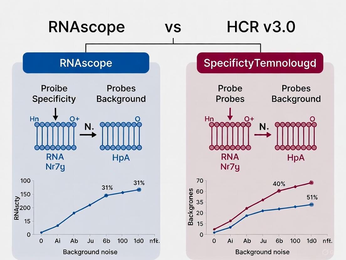
Diagram 1: Core background suppression mechanisms in RNAscope and HCR v3.0. Both technologies require two independent probe binding events to colocalize components for initiating signal amplification, preventing false positives from single non-specifically bound probes. HCR v3.0's hairpin amplifiers are also conditionally metastable, preventing non-specific polymerization.
Technology-Specific Mechanisms for Background Suppression
RNAscope: Dual Z Probe Design
The RNAscope platform tackles the challenge of background at the level of probe design and the initial amplification trigger [1].
- Probe Design: Instead of a single long probe, RNAscope uses pairs of so-called "double Z" probes. Each probe pair is designed to bind to adjacent ~50-base regions of the target RNA.
- Mechanism of Specificity: Each individual "Z" probe contains a 14-base tail sequence. Only when both probes in a pair bind correctly to their adjacent target sites are their two tail sequences brought together to form a single 28-base binding site for the pre-amplifier molecule. A single probe binding non-specifically elsewhere in the cell presents only a 14-base sequence, which is insufficient to stably bind the pre-amplifier. This requirement for cooperative binding dramatically reduces background from probe cross-reactivity [1].
- Signal Amplification: Once the pre-amplifier binds, it initiates a branched DNA (bDNA) amplification cascade, which is a proprietary, enzymatic signal amplification process.
HCR v3.0: Split-Initiator Probes
HCR v3.0 incorporates a concept known as automatic background suppression, which addresses both probe-level and amplifier-level non-specificity [9].
- Probe Design: HCR v3.0 replaces the standard full-initiator probes used in earlier versions with split-initiator probes. Each target is detected by a set of probe pairs, where each probe carries one half of the sequence required to initiate the HCR cascade.
- Mechanism of Specificity: The full HCR initiator is only assembled when two split-initiator probes bind adjacently on the target RNA. An individual probe binding non-specifically carries only a half-initiator and is incapable of triggering the HCR polymerization reaction. This design suppresses amplified background from cross-reactive probes [9].
- Signal Amplification: The HCR amplification itself is isothermal and enzyme-free, relying on the triggered self-assembly of metastable DNA hairpins. A key inherent feature is that individual hairpin molecules that bind non-specifically in the sample do not trigger the chain reaction, providing a second layer of background suppression at the amplification stage [9].
Quantitative Performance Comparison
Empirical data from peer-reviewed literature and technical resources demonstrate the practical impact of these different mechanisms on assay performance. The table below summarizes key quantitative findings.
Table 1: Quantitative Performance Comparison of RNAscope and HCR v3.0
| Performance Metric | RNAscope | HCR v3.0 | Experimental Context |
|---|---|---|---|
| Background Suppression | Proprietary double Z probe design prevents non-specific amplification [1]. | ≈50-fold suppression of amplified background with split-initiator probes [9]. | Comparison of signal vs. background in whole-mount chicken embryos [9]. |
| Probe Set Validation | Designed to work with ~20 ZZ probe pairs per target without individual validation [1]. | Enables use of large, unoptimized probe sets (e.g., 20 probe pairs) without increased background [9]. | Use of unoptimized probe sets in complex samples [9]. |
| Signal-to-Background Ratio | High signal-to-noise enabled by requirement for dual probe binding [1]. | Maintains high S/B; increases monotonically with probe set size [9]. | Multiplexed mRNA imaging in neural crest of whole-mount chicken embryos [9]. |
| Sensitivity | Single-molecule detection sensitivity visualized as punctate dots [1]. | Capable of single-molecule detection (dHCR imaging) in thick, autofluorescent samples [9]. | Detection of low-abundance targets in challenging samples [9]. |
Detailed Experimental Protocols
To ensure reproducibility and provide clarity on how these technologies are implemented, we outline the core experimental workflows. The following diagram provides a visual overview of the key stages common to both protocols, highlighting critical divergence points.
Diagram 2: Core experimental workflows for RNAscope and HCR v3.0. Both protocols begin with sample fixation and permeabilization, followed by probe hybridization and stringent washes. The amplification and detection phases then diverge, with RNAscope employing a multi-step, branched DNA (bDNA) amplification system, while HCR v3.0 uses a one-step, enzyme-free hairpin amplification.
RNAscope Protocol (Based on ViewRNA Kit)
The RNAscope protocol is a structured, multi-step process that can be completed over two to three days [15].
Sample Fixation and Pretreatment:
- Fix tissue sections or cells in 4% formaldehyde overnight at 4°C.
- Dehydrate samples through an ethanol series (50%, 70%, 100%).
- Heat slides at 60°C for 30 minutes.
- Draw a hydrophobic barrier around the samples.
- Apply a protease solution (e.g., Protease QF or optimized Bacterial Type XXIV Proteinase) and incubate at 40°C for 10-30 minutes to unmask target RNA. Note: Protease concentration and time require optimization for each sample type.
Probe Hybridization:
- Apply the target-specific probe set (diluted in Probe Set Diluent QT) to the tissue section.
- Incubate slides in a HybEZ oven or similar humidified hybridization chamber at 40°C for 2 hours.
Signal Amplification:
- Pre-Amplification: Hybridize the PreAmplifier Mix to the bound probe pairs.
- Amplification: Hybridize the Amplifier Mix to the pre-amplifiers.
- Labeling: Hybridize the Label Probe (e.g., conjugated to alkaline phosphatase, AP) to the amplifiers.
- Each amplification step is typically performed at 40°C for 1 hour, with stringent washes between steps.
Detection and Visualization:
- For chromogenic detection, develop signal using a substrate like Fast Red, which produces a punctate, red precipitate.
- Counterstain with hematoxylin or DAPI and apply a mounting medium for imaging.
- Punctate dots can be quantified manually or with automated image analysis software (e.g., HALO) [1].
HCR v3.0 Protocol
The HCR v3.0 protocol is typically simpler, with fewer steps, and can be adapted for fluorescence detection [9] [16].
Sample Fixation and Permeabilization:
- Fix samples appropriately (e.g., with formaldehyde).
- Permeabilize using a detergent such as Triton X-100 to allow probe and hairpin entry.
Probe Hybridization:
Signal Amplification:
- After washing to remove unbound probes, add the two metastable DNA hairpins (H1 and H2), which are pre-annealed and fluorophore-labeled.
- The amplification reaction is incubated overnight in the dark at room temperature. This enzyme-free, isothermal step allows the chain reaction to propagate only where the full initiator has been assembled on the target [9].
Detection and Visualization:
- Wash the sample to remove un-polymerized hairpins.
- Counterstain (e.g., with DAPI) and mount for imaging.
- Signal appears as fluorescent foci, which can be quantified relative to background.
Essential Research Reagent Solutions
Successful implementation of either technology requires a set of core reagents. The following table lists key solutions and their functions within the experimental workflows.
Table 2: Key Research Reagent Solutions for RNA ISH Experiments
| Reagent Category | Specific Examples | Function in the Protocol | Technology Application |
|---|---|---|---|
| Fixatives | 4% Formaldehyde (in PBS) | Preserves tissue morphology and immobilizes RNA within cells. | Universal |
| Permeabilization Agents | Triton X-100, Proteases (e.g., Bacterial Type XXIV) | Creates pores in cellular membranes to allow entry of probes and amplifiers. | Universal (Protease: RNAscope) |
| Blocking Agents | BSA, Casein, Fish Gelatin, Non-Fat Dry Milk [14] | Reduces non-specific binding (NSB) by saturating sticky sites on the solid phase and sample. | Universal |
| Specific Probes | RNAscope Double Z Probes, HCR split-initiator Probes | Target-specific reagents that hybridize to the RNA of interest and provide the platform for signal amplification. | Technology-specific |
| Amplification Systems | RNAscope PreAmp/Amp/Label Probes, HCR DNA Hairpins (H1 & H2) | Generates a detectable signal (fluorescent or chromogenic) from the initial probe-binding event. | Technology-specific |
| Detection Substrates | Fast Red (Chromogen), Fluorophore-conjugated HCR hairpins | The final molecule that is enzymatically converted or directly imaged to produce the visible signal. | Technology-specific |
| Stringent Wash Buffers | Saline-sodium citrate (SSC) buffers with detergent | Removes unbound and weakly bound probes/amplifiers to reduce background. | Universal |
Both RNAscope and HCR v3.0 represent significant advancements over earlier ISH methods by implementing elegant biochemical strategies to suppress the inherent background caused by probe cross-reactivity and non-specific polymerization.
The choice between these two robust technologies ultimately depends on the specific research requirements. RNAscope, with its highly structured, multi-step amplification, is often praised for its consistent, punctate signal and high success rate in clinical and formalin-fixed, paraffin-embedded (FFPE) samples. In contrast, HCR v3.0 offers flexibility through its enzyme-free, isothermal amplification, which can be advantageous in sensitive samples or when designing custom probes for novel targets. Its simpler workflow and the ability to use unoptimized probe sets make it highly accessible for exploratory research.
Researchers are empowered to make an informed selection based on whether their priority lies in the proven, highly standardized performance of RNAscope or the flexible, enzyme-free chemistry of HCR v3.0, all while having a clear understanding of the mechanisms that ensure specificity in their RNA imaging data.
Practical Applications: From Multiplexed Imaging to Subcellular Localization
In the evolving field of spatial biology, multiplexed in situ hybridization technologies enable researchers to map complex gene expression patterns within their native tissue context. Among the leading methodologies, the RNAscope HiPlex assay and Hybridization Chain Reaction v3.0 (HCR v3.0) represent distinct technological approaches to achieving highly multiplexed RNA detection. RNAscope HiPlex employs an iterative detection system using cleavable fluorophores to enable high-plex spatial profiling [17] [18]. In contrast, HCR v3.0 utilizes a mechanism of split-initiator probes and enzyme-free signal amplification to achieve simultaneous multiplexing with inherent background suppression [9] [6]. This guide provides an objective comparison of their multiplexing capabilities, experimental workflows, and performance characteristics to inform researchers selecting the optimal platform for their specific applications.
The core distinction between these platforms lies in their fundamental approach to multiplexing: RNAscope HiPlex employs sequential target detection across multiple rounds, while HCR v3.0 enables simultaneous detection of multiple targets in a single round.
Table 1: Core Technology Specification Comparison
| Feature | RNAscope HiPlex v2 | HCR v3.0 |
|---|---|---|
| Maximum Plex | 12-plex (FFPE), up to 48-plex (frozen, with HiPlexUp) [17] [18] | Typically 3-5 targets simultaneously [9] [6] |
| Amplification Method | Proprietary signal amplification with cleavable fluorophores | Enzyme-free hybridization chain reaction |
| Detection Strategy | Iterative sequential detection | Simultaneous detection |
| Key Innovation | Fluorophore cleavage and multiple detection rounds | Split-initiator probes for automatic background suppression [9] |
| Sample Compatibility | FFPE tissues, fresh/fixed frozen tissues [17] | Whole-mount specimens, thick tissues, diverse organisms [9] [6] [4] |
Experimental Protocols & Workflows
RNAscope HiPlex v2 Assay Workflow
The RNAscope HiPlex protocol relies on sequential detection cycles to achieve high-plex analysis. The workflow can be visualized as follows:
Key Experimental Steps [17] [18]:
- Sample Preparation: FFPE or fresh frozen tissue sections are prepared using standard fixation protocols.
- Probe Hybridization: Target-specific probes are hybridized. Each probe is designed to bind specific RNA sequences.
- Signal Amplification & Detection: The proprietary amplification system generates a fluorescent signal. For each round, up to four targets are detected in distinct fluorescent channels (e.g., AF488, Dylight550, Dylight650, AF750).
- Fluorophore Cleavage: A rapid cleavage step removes the fluorescent signals without damaging the tissue morphology or bound probes.
- Iterative Rounds: Steps 2-4 are repeated for new sets of targets until all 12 (or more) targets have been detected.
- Image Analysis: Images from all rounds are aligned using specialized registration software (RNAscope HiPlex Image Registration Software) to create a composite multiplex image [17].
HCR v3.0 Workflow
HCR v3.0 employs a simultaneous detection approach, which is fundamentally different:
Key Experimental Steps [9] [6] [4]:
- Sample Preparation & Permeabilization: Tissues are fixed and permeabilized. For challenging samples like whole-mount plant tissues or octopus embryos, this includes enzymatic cell wall digestion or proteinase K treatment [6] [4].
- Split-Initiator Probe Hybridization: Pairs of short DNA probes (25 nucleotides each) are hybridized to adjacent sites on the target mRNA. Each probe carries half of an HCR initiator sequence.
- Cooperative Initiation & Amplification: Only when both probes bind correctly to their adjacent target sites is a full HCR initiator assembled. This initiator then triggers the self-assembly of fluorescently labeled DNA hairpins (H1 and H2) in an enzyme-free chain reaction.
- Simultaneous Multiplexed Imaging: Different mRNA targets are detected using orthogonal HCR amplifier systems (e.g., B1, B2, B3) with different fluorophores. All targets are imaged in a single step after a single amplification round.
- 3D Imaging & Analysis: The protocol is particularly suited for 3D imaging of whole-mount samples, often combined with tissue clearing and light-sheet fluorescence microscopy [4].
Performance Data & Experimental Validation
Key Performance Metrics
Table 2: Quantitative Performance Comparison
| Performance Metric | RNAscope HiPlex v2 | HCR v3.0 |
|---|---|---|
| Signal-to-Background | High signal-to-noise maintained through proprietary probe design and controlled sequential detection [17] | ≈50-60 fold background suppression due to split-initiator probes [9] |
| Single-Cell Resolution | Yes, punctate dots representing single transcripts [17] [19] | Yes, enables digital mRNA absolute quantitation (dHCR imaging) [9] |
| Quantitation Capability | Spatial quantification and co-expression analysis possible with pipelines like SCAMPR [19] | Analog relative quantitation (qHCR) and digital absolute quantitation (dHCR) [9] |
| Compatible Applications | Tumor Microenvironment (TME) profiling, neuronal subtyping, validation of scRNA-seq data [17] [18] [19] | Whole-mount embryogenesis, 3D spatial mapping in diverse organisms (plants, octopus) [6] [4] |
Experimental Validation Studies
- RNAscope HiPlex for Neuronal Classification: The SCAMPR pipeline combined RNAscope HiPlex with immunohistochemistry (IHC) for HuC/D to accurately demarcate neuronal boundaries in mouse nodose ganglion and visual cortex. This allowed for high-dimensional quantification of 12-plex mRNA expression at single-cell resolution and revealed gene expression changes induced by early life stress [19].
- HCR v3.0 in Whole-Mount Plant and Octopus Embryos: Studies in Arabidopsis inflorescences successfully detected 3 transcripts (AP3, AG, STM) simultaneously in 3D, confirming known expression patterns with low background [6]. Similarly, in Octopus vulgaris embryos, HCR v3.0 was optimized with fructose-glycerol clearing to visualize neuronal markers (Ov-elav, Ov-apolpp), revealing spatial organization not apparent in 2D sections [4].
The Scientist's Toolkit: Essential Research Reagents
Table 3: Key Reagents and Resources
| Component | RNAscope HiPlex v2 | HCR v3.0 |
|---|---|---|
| Core Kits | HiPlex12 Reagents Kit (Cat. # 324409/324419) [17] | Custom DNA Oligo Pools & HCR amplifiers (B1-546, B2-647, B3-488) [4] |
| Control Probes | Species-specific Positive Control Probe & Universal Negative Control Probe [17] [18] | Target-specific split-initiator probe pairs; negative control with non-targeting probes [6] |
| Specialized Equipment | Fluorescent microscope (DAPI, AF488, Atto550, Atto647N, AF750), HybEZ Hybridization System [17] | Standard fluorescent microscope; LSFM recommended for 3D whole-mount imaging [4] |
| Analysis Software | RNAscope HiPlex Image Registration Software (v2.1) [17] [18] | Standard image analysis software (e.g., FIJI/ImageJ); may require custom scripts for 3D analysis |
| Additional Reagents | Probe Diluent, Hydrophobic Barrier Pen [17] | Proteinase K, amplification buffers, and clearing reagents (e.g., fructose-glycerol) [6] [4] |
| PD 127443 | PD 127443, CAS:121502-05-4, MF:C20H28N2O, MW:312.4 g/mol | Chemical Reagent |
| (Rac)-BMS-1 | PD-1/PD-L1 Inhibitor 1 | Explore our PD-1/PD-L1 Inhibitor 1, a small molecule designed to block immune checkpoint interaction for cancer immunotherapy research. For Research Use Only. |
The choice between RNAscope HiPlex and HCR v3.0 is application-dependent, revolving around a fundamental trade-off between plexing capacity and workflow simplicity.
Choose RNAscope HiPlex v2 when your research demands high-plex spatial profiling (12-48 targets) in standard tissue sections, particularly for clinical samples like FFPE tissues. Its primary strength lies in generating comprehensive cell atlases within complex tissues, such as characterizing the tumor immune microenvironment [17] [20] or validating single-cell RNA sequencing datasets [19]. Users should be prepared for a more complex, multi-day iterative protocol and ensure access to the required imaging and analysis software.
Choose HCR v3.0 when your research prioritizes robustness, ease of use, and 3D spatial context in challenging samples. Its automatic background suppression makes it exceptionally robust for unoptimized probe sets in diverse organisms, from plants to octopuses [9] [6] [4]. The simultaneous workflow is simpler and faster for low-to-mid plexing (3-5 targets) and is ideal for whole-mount embryonic studies where understanding spatial organization in three dimensions is critical.
In summary, RNAscope HiPlex v2 excels in breadth of detection for sectional analysis, while HCR v3.0 offers superior versatility and simplicity for simultaneous multiplexing in complex 3D architectures. Understanding these core distinctions enables researchers to align their technology selection with their specific biological questions and experimental models.
The accuracy and reliability of RNA detection in biological research are profoundly influenced by the type of sample preparation used. For researchers and drug development professionals selecting between advanced in situ hybridization (ISH) platforms like RNAscope and Hybridization Chain Reaction (HCR) v3.0, understanding sample type compatibility is paramount for experimental success. Formalin-Fixed Paraffin-Embedded (FFPE) tissues represent the most widely available clinical material for pathological studies, offering superior morphological preservation and stable long-term storage at room temperature [21] [22]. In contrast, fresh-frozen (FF) tissues are considered the "gold standard" for nucleic acid preservation, maintaining RNA in a more native state but requiring continuous ultra-low temperature storage [21] [22]. Whole-mount tissues present additional challenges for probe penetration while offering complete three-dimensional structural context. This guide objectively compares the performance of RNAscope and HCR across these sample types, providing structured experimental data and protocols to inform your platform selection within the broader context of specificity and background performance research.
RNAscope's Branched DNA (bDNA) Architecture
RNAscope employs a proprietary branched DNA (bDNA) signal amplification system characterized by its unique "Z-probe" design [2] [13]. This technology uses short oligonucleotide pairs (20-25 bases) that hybridize to adjacent sequences on the target RNA [5]. The mechanism requires both "Z" probes to bind correctly to initiate a sequential amplification cascade:
- Probe Hybridization: "Z-probe" pairs bind adjacent target RNA sequences [2] [13].
- Preamplifier Binding: Each bound Z-probe pair recruits a preamplifier molecule [2].
- Amplifier Assembly: Multiple amplifier molecules bind to each preamplifier [2] [12].
- Label Incorporation: Label probes (chromogenic or fluorescent) conjugate to amplifiers, achieving up to 8000-fold signal amplification [2].
This structured approach provides single-molecule sensitivity and high specificity, as off-target binding rarely brings two Z-probes into correct proximity for amplification [2] [12].
HCR v3.0's Hybridization Chain Reaction
HCR v3.0 utilizes an enzyme-free, initiated chain reaction mechanism based on DNA hairpin probes [5]. The fundamental process involves:
- Initiator Hybridization: An "initiator" probe hybridizes to the target RNA, exposing a previously sequestered sequence [5].
- Chain Reaction: Metastable fluorogenic DNA hairpin probes undergo a chain reaction of hybridization events, self-assembling into amplification polymers [5].
- Signal Accumulation: Long nucleic acid polymers form in situ, each carrying multiple fluorescent labels [5].
Recent advancements like Yn-situ have introduced a preamplifier design to HCR, reducing the number of required probe pairs from 20 to as few as 3-5 while maintaining detection sensitivity [23].
Figure 1: Comparative signal amplification mechanisms of RNAscope and HCR v3.0 technologies.
Performance Comparison Across Sample Types
Formalin-Fixed Paraffin-Embedded (FFPE) Tissues
FFPE samples represent the most common archival material in clinical pathology, but present challenges for RNA detection due to formalin-induced cross-linking and nucleic acid fragmentation [24] [22].
Table 1: FFPE Tissue Performance Comparison
| Performance Metric | RNAscope | HCR v3.0 |
|---|---|---|
| Probe Penetration | Optimized via proprietary pretreatment [25] | Limited by tissue density; ~80μm maximum penetration [5] |
| RNA Degradation Tolerance | High - detects partially degraded targets [2] | Moderate - requires longer intact sequences for multiple initiators |
| Archive Compatibility | Excellent - works with 25+ year old samples [25] | Limited data on archival tissue performance |
| Signal-to-Noise Ratio | High - minimal background due to dual Z-probe design [2] | Variable - background signal reported in some applications [5] |
| Validation Data | Systematic review shows 81.8-100% concordance with qPCR/qRT-PCR [2] | Limited comparative validation in FFPE |
RNAscope demonstrates robust performance in FFPE tissues due to its ability to detect short RNA fragments and tolerance to formalin-induced modifications. The technology has been successfully applied to FFPE samples up to 25-27 years old when properly fixed and stored [25]. HCR v3.0 shows more variable performance in FFPE tissues, with noted limitations in probe penetration through dense formalin-fixed matrices [5].
Fresh-Frozen Tissues
Fresh-frozen tissues preserve RNA integrity more effectively but present challenges for morphological preservation.
Table 2: Fresh-Frozen Tissue Performance Comparison
| Performance Metric | RNAscope | HCR v3.0 |
|---|---|---|
| RNA Preservation | Excellent - single-molecule sensitivity [12] | Excellent - enhanced signal amplification [5] |
| Morphology Compatibility | Compatible with standard cryosectioning [12] | Requires optimization for tissue integrity |
| Multiplexing Capability | High - validated 3-plex detection [12] | Theoretically high but requires extensive optimization |
| Quantitative Performance | High - linear signal response enables transcript counting [2] | Moderate - signal saturation at high expression levels |
| Protocol Simplicity | Standardized kit-based workflow [12] | Customization often required |
Both platforms perform well in fresh-frozen tissues, with RNAscope offering a more standardized workflow and HCR providing potential cost advantages for custom applications [5]. RNAscope's validated multiplexing capability (up to 3 targets simultaneously) in fresh-frozen sections is particularly valuable for complex tissue analysis [12].
Whole-Mount Tissues
Whole-mount preparations present unique challenges for probe penetration throughout three-dimensional structures while preserving structural integrity.
Table 3: Whole-Mount Tissue Performance Comparison
| Performance Metric | RNAscope | HCR v3.0 |
|---|---|---|
| Tissue Penetration | Limited data; optimized for thin sections | Moderate - better penetration through modifications [5] |
| Signal Uniformity | Not well characterized in 3D contexts | Variable - signal attenuation in deeper layers |
| Protocol Adaptation | Requires significant modification from standard FFPE/FF protocols | More easily adaptable to 3D samples [5] |
| Background Effects | Potentially increased in thick tissues | Background accumulation throughout matrix |
While comprehensive direct comparisons in whole-mount tissues are limited in the literature, HCR v3.0's flexible probe design and amplification chemistry may offer advantages for thicker specimens [5]. RNAscope's standardized protocols are primarily optimized for sectioned tissues, though innovative researchers have adapted it for specialized whole-mount applications.
Experimental Protocols for Sample Processing
RNAscope Protocol for FFPE Tissues
The following protocol is adapted from the standardized RNAscope FFPE workflow [2] [25]:
Sample Preparation:
- Fix tissue in 10% Neutral Buffered Formalin (NBF) for 16-32 hours at room temperature [24] [25].
- Process through ethanol dehydration series and embed in paraffin.
- Section at 4-5μm thickness and mount on positively charged slides.
- Store slides at 4°C, -20°C, or -80°C; use within 3 months (room temperature) or 1 year (frozen) [24].
Pretreatment Protocol:
- Bake slides at 60°C for 1 hour to adhere sections.
- Deparaffinize in xylene (2 × 10 minutes) and 100% ethanol (2 × 5 minutes).
- Air dry completely, then draw hydrophobic barrier around sections.
- Perform RNAscope Hydrogen Peroxide treatment for 10 minutes at room temperature.
- Perform target retrieval by heating in RNAscope Target Retrieval Reagent for 15 minutes at 98-102°C.
- Rinse slides in distilled water, then in 100% ethanol.
- Air dry completely before proceeding to hybridization.
Hybridization and Amplification:
- Apply Protease Plus treatment for 30 minutes at 40°C.
- Hybridize with target probes for 2 hours at 40°C.
- Perform sequential amplifier hybridization (Amp 1-6) with appropriate rinses.
- Develop with desired chromogenic or fluorescent substrates.
- Counterstain, mount, and image.
HCR v3.0 Protocol for Fresh-Frozen Tissues
This protocol incorporates recent improvements from the Yn-situ method for enhanced sensitivity [23]:
Sample Preparation:
- Embed fresh tissue in OCT compound and flash-freeze in liquid nitrogen-cooled isopentane.
- Section at 10-20μm thickness on cryostat and mount on Superfrost slides.
- Store at -80°C until use.
Fixation and Permeabilization:
- Fix sections in 4% Paraformaldehyde (PFA) for 1 hour at 4°C.
- Rinse in 1× PBS (3 × 5 minutes each).
- Treat with EDC [1-ethyl-3-(3-dimethylaminopropyl) carbodiimide] fixative for 1 hour to crosslink RNA to proteins [23].
- Permeabilize with detergent solution (0.1-0.5% Triton X-100) for 30 minutes.
- Perform acetylation treatment to reduce non-specific binding.
Hybridization and Amplification:
- Prehybridize with hybridization buffer for 30 minutes at room temperature.
- Hybridize with initiator probes (0.5-2μM) for 12-16 hours at 37°C.
- Wash with hybridization buffer (4 × 15 minutes each) at 37°C.
- Amplify with hairpin probes (0.5-1μM) for 4-6 hours at room temperature.
- Wash with 5× SSCT (4 × 15 minutes each) and counterstain with DAPI.
- Mount with antifade mounting medium and image.
Figure 2: Comparative experimental workflows for FFPE versus fresh-frozen tissue processing.
The Scientist's Toolkit: Essential Research Reagent Solutions
Table 4: Key Reagents for RNA ISH Applications
| Reagent Category | Specific Examples | Function | Compatibility Notes |
|---|---|---|---|
| Fixatives | 10% NBF, 4% PFA, EDC crosslinker | Preserve tissue architecture and nucleic acids | NBF preferred for FFPE (RNAscope); EDC enhances RNA retention in FF (HCR) [23] |
| Permeabilization Agents | Proteinase K, Protease Plus, Triton X-100 | Enable probe access to cellular targets | Concentration critical - varies by fixation method and tissue type [24] |
| Probe Systems | RNAscope ZZ probes, HCR initiators | Target-specific recognition | RNAscope: 20-25bp Z-probes; HCR: initiator+amplifier sets [2] [5] |
| Amplification Reagents | bDNA amplifiers, HCR hairpins | Signal enhancement | bDNA: sequential binding; HCR: polymerization reaction [2] [5] |
| Detection Substrates | Chromogenic dyes, Fluorophores (Alexa dyes) | Visualize target localization | RNAscope: validated multiplex fluorophores; HCR: flexible label incorporation [12] |
| Control Probes | PPIB, Polr2A, UBC (positive); dapB (negative) | Assay validation | Essential for determining RNA quality and specificity [2] |
| Tyk2-IN-8 | Tyk2-IN-8, CAS:2127109-84-4, MF:C20H17N9, MW:383.4 g/mol | Chemical Reagent | Bench Chemicals |
| PF-06835919 | PF-06835919, CAS:2102501-84-6, MF:C16H19F3N4O2, MW:356.34 g/mol | Chemical Reagent | Bench Chemicals |
The selection between RNAscope and HCR v3.0 should be driven by sample type characteristics and research objectives. RNAscope demonstrates superior performance in FFPE tissues, with extensive validation in archival clinical samples and standardized protocols that ensure reproducibility [2] [25]. Its dual Z-probe design provides exceptional specificity, making it ideal for clinical applications and regulatory studies. HCR v3.0 offers advantages in fresh-frozen tissues and specialized applications requiring custom probe design, with potential cost benefits for high-throughput screening [5] [23]. The recent Yn-situ enhancement substantially improves HCR sensitivity, reducing the number of required probes from 20 to 3-5 pairs while maintaining detection capability [23].
For drug development professionals requiring analysis of archival clinical samples across multiple sites, RNAscope provides the consistency and reliability needed for regulatory submissions. For basic researchers working with genetically modified models or novel targets in frozen tissues, HCR v3.0 offers flexibility and cost-effectiveness. As both technologies continue to evolve, ongoing comparative validation in diverse sample types remains essential for advancing RNA biomarker discovery and spatial transcriptomics applications.
The precise spatial localization of RNA transcripts within intact tissues is crucial for understanding gene expression patterns during development and disease. For years, RNAscope has been a dominant commercial player in this field, renowned for its high sensitivity and ease of use. However, the emergence of Hybridization Chain Reaction v3.0 (HCR v3.0) presents a powerful, flexible, and cost-effective alternative, particularly for complex experimental workflows involving whole-mount samples, multiplexing, and combination with other techniques like immunohistochemistry (IHC). This guide objectively compares the performance of HCR v3.0 against RNAscope, framing the discussion within a broader thesis on specificity and background performance. We provide supporting experimental data and detailed methodologies to help researchers, scientists, and drug development professionals select the optimal RNA detection method for their specific applications, especially when the research goal involves three-dimensional imaging of thick tissues and simultaneous protein detection.
The core innovation of HCR v3.0 that enables its robust performance is its split-initiator probe design, which provides automatic background suppression. Unlike standard probes that carry a full HCR initiator, HCR v3.0 uses pairs of probes that each carry half of an initiator sequence. The full initiator is only assembled, and signal amplification is only triggered, when both probes bind adjacently on the specific target mRNA. This ensures that individual probes binding non-specifically elsewhere in the sample cannot initiate the amplification cascade, dramatically reducing background noise [9].
Performance and Specificity Comparison: HCR v3.0 vs. RNAscope
Direct, head-to-head comparisons of HCR v3.0 and RNAscope reveal distinct advantages and trade-offs, rooted in their fundamental mechanisms. The following table summarizes the key performance characteristics based on published experimental data.
Table 1: Experimental Performance Comparison of HCR v3.0 and RNAscope
| Feature | HCR v3.0 | RNAscope | Experimental Support & Context |
|---|---|---|---|
| Signal Amplification | Enzyme-free, hybridization chain reaction [9]. | Branched DNA (bDNA) [5]. | HCR's isothermal amplification allows for flexible protocol design. |
| Probe Design & Cost | Custom design is possible; can be less expensive, especially for custom targets [5]. | Proprietary, pre-validated probes; commercial cost structure [5]. | HCR offers flexibility for non-model organisms; RNAscope provides convenience and reliability for validated targets. |
| Background & Specificity | Automatic background suppression via split-initiator probes. Typical 50-60 fold suppression of non-specific amplification measured in situ and in gels [9]. | High specificity inherent to proprietary "Z-probe" design [5]. | HCR v3.0's design makes it robust against background from unoptimized probe sets [9]. |
| Multiplexing | Straightforward multiplexing with simultaneous one-stage signal amplification for up to five targets [9] [6]. | Capable of multiplexing using different fluorophores [5]. | Both are effective, with HCR v3.0 offering a highly flexible and simultaneous amplification step. |
| Sensitivity | Enables both analog relative quantitation (qHCR) and digital absolute quantitation (dHCR) at the single-molecule level [9]. | Highly sensitive and specific detection at the single-molecule level [5]. | Both techniques are sufficiently sensitive for low-abundance transcripts, though some reports indicate RNAscope may have a sensitivity edge [5]. |
| Compatibility with Thick Tissues & Clearing | Excellent compatibility; validated in whole-mount vertebrate embryos, octopus, and plants with fructose-glycerol and other clearing methods [10] [6]. | Tissue penetration can be a limitation; maximum effective penetration is approximately 80 µm [5]. | HCR v3.0 is particularly suited for 3D imaging of large, whole-mount samples. |
| Combination with IHC | Robustly combined with IHC in whole-mount samples, preserving both RNA and protein signals [10] [6]. | Can be combined with IHC, though protocol optimization may be needed, especially for thicker samples. | HCR v3.0's antibody-free amplification facilitates combination with antibody-based protein detection. |
Table 2: Quantitative Background Suppression Data for HCR v3.0
| Experimental Condition | Assay Type | Key Metric (HCR Suppression) | Implication |
|---|---|---|---|
| Split-initiator probes (one absent) | In vitro Gel Assay | ≈60-fold reduction in polymer formation [9] | Minimal non-specific amplification when probe binding is incomplete. |
| Split-initiator probes (one absent) | In situ (Whole-mount embryos) | ≈50-fold suppression of background signal [9] | Dramatic reduction of amplified background in complex biological samples. |
| Standard probes (v2.0) vs. Split-initiator (v3.0) | In situ (Whole-mount chicken embryos) | Monotonic decrease in SBR with standard probes; monotonic increase with split-initiator probes [9] | Allows use of large, unoptimized probe sets to reliably increase signal-to-background ratio (SBR). |
Experimental Protocols for Combined HCR v3.0, IHC, and Clearing
The following detailed protocols, adapted from recent literature, demonstrate the workflow for combining HCR v3.0 with IHC and tissue clearing to achieve high-quality, multiplexed 3D imaging.
Detailed Workflow: Whole-Mount HCR v3.0 with IHC and Fructose-Glycerol Clearing
This protocol, optimized for Octopus vulgaris embryos and plant tissues, serves as a robust template for various sample types [10] [6].
Sample Preparation and Fixation
- Fix tissues overnight in 4% Paraformaldehyde (PFA) in PBS.
- For plant tissues, a permeabilization step using cell wall-degrading enzymes (e.g., cellulase and pectolyase) is essential [6].
- Dehydrate samples through a graded methanol (MeOH) series (e.g., 25%, 50%, 75%, 100%) and store at -20°C until use.
Rehydration and Permeabilization
- Rehydrate samples through a descending MeOH/PBST series to 1x PBS-DEPC.
- Permeabilize tissues by incubating with Proteinase K (e.g., 10 µg/ml for 15 minutes at room temperature). Note: Omit this step if preserving fluorescent proteins for simultaneous detection.
- Post-fixation in 4% PFA for 20-30 minutes is recommended to maintain tissue integrity.
HCR v3.0 In Situ Hybridization
- Probe Hybridization: Incubate samples in a solution containing 0.4 pmol of each split-initiator probe per 100 µl of probe hybridization buffer. Incubate overnight at 37°C.
- Post-Hybridization Washes: Remove unbound probes with 4-5 stringent washes over several hours using probe wash buffer.
- Signal Amplification:
- Prepare HCR hairpins (H1 and H2) by snap-cooling (heat to 95°C for 90 seconds, then incubate on ice for 5 minutes and at room temperature for 30 minutes).
- Incubate samples in amplification buffer containing the pre-annealed hairpins (e.g., 3 pmol of each per 100 µl buffer). Amplification is typically performed overnight in the dark at room temperature.
- Post-Amplification Washes: Remove excess hairpins with 3-4 washes in 5x SSCT buffer.
Immunohistochemistry (IHC)
- After HCR washes, block samples in a suitable blocking buffer (e.g., 5% normal serum in PBST) for 1-2 hours.
- Incubate with primary antibody diluted in blocking buffer overnight at 4°C.
- Wash thoroughly to remove unbound antibody.
- Incubate with fluorophore-conjugated secondary antibody overnight at 4°C.
- Perform final washes before clearing.
Nuclear Staining and Tissue Clearing
- Counterstain nuclei by incubating with DAPI (e.g., 1:2000 dilution for 2 hours).
- Clear samples by immersing in fructose-glycerol clearing solution (e.g., 65% fructose, 15% glycerol, 0.01% sodium azide) for at least 48 hours [10]. This water-based method effectively preserves HCR and IHC fluorescence signals.
Imaging
- Image cleared samples using Light Sheet Fluorescence Microscopy (LSFM) or confocal microscopy. LSFM is ideal for large, whole-mount samples as it provides fast imaging with minimal photobleaching.
Workflow Visualization
The following diagram illustrates the integrated experimental pipeline.
The Scientist's Toolkit: Essential Reagent Solutions
Successful implementation of this combined protocol relies on key reagents. The following table details these essential components and their functions.
Table 3: Key Research Reagent Solutions for Combined HCR v3.0 and IHC
| Reagent / Solution | Function / Purpose | Key Considerations |
|---|---|---|
| Split-Initiator Probe Sets | DNA oligonucleotides that bind target mRNA and colocalize split initiators for specific HCR amplification [9]. | Designed in pairs (20-30 pairs per target) for automatic background suppression. Can be custom-designed. |
| HCR Hairpin Amplifiers (H1 & H2) | Fluorophore-labeled DNA hairpins that undergo chain reaction assembly upon initiator binding, amplifying the signal [9] [10]. | Sold for different initiator sequences (B1, B2, B3, etc.) for multiplexing. Must be snap-cooled before use. |
| Probe Hybridization Buffer | Aqueous buffer facilitating specific binding of DNA probes to the target mRNA. | Contains components (e.g., formamide, salts) to control stringency and prevent non-specific hybridization. |
| Amplification Buffer | Aqueous buffer for the HCR self-assembly process. | Provides optimal ionic and pH conditions for the hairpin polymerization reaction. |
| Proteinase K | Enzyme that digests proteins to permeabilize tissues for better probe/antibody penetration [10] [6]. | Concentration and time must be optimized for each tissue type to avoid over-digestion. |
| Fructose-Glycerol Solution | A water-based clearing agent that reduces light scattering by matching the refractive index of the tissue [10]. | Effectively preserves fluorescent signals from HCR and IHC; non-toxic and easy to prepare. |
| Primary & Secondary Antibodies | Primary antibody binds the target protein; secondary antibody (conjugated to a fluorophore) binds the primary for detection. | Choose secondary fluorophores with spectra distinct from HCR channels to minimize bleed-through. |
Mechanism of HCR v3.0 Specificity
The superior background performance of HCR v3.0 is directly attributable to its split-initiator mechanism, a critical advance over previous versions and a key differentiator from other methods.
The experimental data and protocols presented herein support a clear thesis: HCR v3.0 is a superior technology for applications demanding the highest level of specificity in complex samples, especially when the experimental design requires whole-mount imaging, multiplexing, and combination with IHC. Its automatic background suppression mechanism provides a robustness that allows researchers to use large, unoptimized probe sets for new targets with confidence, a significant advantage in exploratory research and work on non-model organisms. While RNAscope remains a highly sensitive and user-friendly commercial solution, particularly for routine analysis of sectioned tissues, HCR v3.0 establishes a new benchmark for quantitative, multi-modal, three-dimensional spatial transcriptomics. The continued adoption and refinement of these combined protocols will undoubtedly accelerate discovery in developmental biology, neuroscience, and drug development.
The subcellular detection of SARS-CoV-2 RNA has emerged as a critical research area for understanding viral pathogenesis, replication dynamics, and host-pathogen interactions. This case study objectively compares the performance of Hybridization Chain Reaction v3.0 (HCR v3.0) and RNAscope for detecting SARS-CoV-2 RNA at subcellular resolution, providing experimental data and protocols to guide researchers in selecting appropriate methodologies for their specific applications. Within the broader thesis of RNAscope versus HCR v3.0 specificity and background performance research, this analysis reveals that HCR v3.0 offers significant advantages in multiplexing capability and cost-efficiency, while RNAscope provides exceptional sensitivity and operational simplicity for diagnostic applications [5] [26].
Technology Comparison: HCR v3.0 vs. RNAscope
Fundamental Mechanisms and Principles
RNAscope employs a proprietary branched DNA (bDNA) amplification system using patented "Z-probes" that contain target-specific sequences paired with amplifier binding sites. Each probe pair must bind adjacent sites on the target RNA to initiate a sequential hybridization process that builds a branching DNA structure, enabling significant signal amplification through fluorophore-labeled probes [5]. This design inherently minimizes off-target binding and false positives, as single probes binding non-specifically cannot initiate the amplification cascade.
HCR v3.0 utilizes a fundamentally different approach based on hybridization chain reaction principles. The technology employs split-initiator probes where each probe carries half of an HCR initiator sequence. Only when both probes hybridize adjacently on the target RNA are the initiator fragments colocalized to trigger a chain reaction of fluorescent hairpin oligonucleotides self-assembling into amplification polymers [9]. This mechanism incorporates automatic background suppression, as individual probes binding non-specifically cannot initiate the amplification cascade [9].
Performance Characteristics Comparison
Table 1: Comparative Analysis of HCR v3.0 and RNAscope Performance Characteristics
| Parameter | HCR v3.0 | RNAscope |
|---|---|---|
| Signal Amplification Mechanism | Enzyme-free hybridization chain reaction | Branched DNA (bDNA) amplification |
| Probe Design | Split-initiator probes (25 nt each) | "Z-probes" (20-25 bases) with amplifier binding sites |
| Multiplexing Capacity | High (simultaneous 5-plex demonstrated) [9] | Moderate (typically 3-4 plex) [5] |
| Sensitivity | Moderate (improved with HCR-Cat/HCR-Immuno) [27] | High (single-molecule detection capable) [5] |
| Background Suppression | Automatic background suppression via split-initiator design [9] | High specificity through proprietary probe design |
| Sample Compatibility | FFPE, frozen tissues, cell cultures, whole mounts [4] | FFPE, frozen tissues, cell cultures [5] |
| Experimental Timeline | 1-3 days [26] | 1 day [26] |
| Cost Considerations | Moderate; decreases with sample number [26] | High; proportional to sample number [26] |
| Quantitative Capability | qHCR imaging & dHCR imaging [9] | Semi-quantitative |
Table 2: Signal Performance Metrics in Challenging Samples
| Sample Type | Target | HCR v3.0 Result | RNAscope Result | Reference |
|---|---|---|---|---|
| Whole-mount vertebrate embryos | Multiple mRNAs | Robust signal with minimal background | High background with unoptimized probes | [9] |
| Cleared tissue volumes (600μm) | YFP mRNA | Very dim signal (requires 10x laser power) | Information missing | [27] |
| FFPE tissues | SARS-CoV-2 | Compatible with optimization | Established performance | [5] |
| Low-abundance transcripts | eve, lar | Weak or undetectable | Detected with high sensitivity | [27] |
Experimental Protocols for SARS-CoV-2 RNA Detection
HCR v3.0 Protocol for SARS-CoV-2 Detection
Probe Design and Synthesis:
- Design approximately 20-30 split-initiator probe pairs targeting conserved regions of the SARS-CoV-2 genome
- Each probe should be 25 nucleotides targeting adjacent sites on the viral RNA
- Order DNA oligo pools from commercial suppliers (e.g., Integrated DNA Technologies)
- Dissolve probes in nuclease-free distilled water [4]
Sample Preparation:
- Fix SARS-CoV-2 infected cells or tissues in 4% paraformaldehyde overnight
- For FFPE samples, follow standard deparaffinization and rehydration protocols
- Permeabilize with proteinase K (10μg/ml in PBS-DEPC) for 15 minutes at room temperature
- Gradually dehydrate through methanol series (25%, 50%, 75%, 100%) for frozen samples [4]
Hybridization and Amplification:
- Prepare probe solution: 0.4 pmol of each probe in 100μL probe hybridization buffer
- Hybridize overnight at 37°C
- Wash with probe wash buffer 4×15 minutes at 37°C
- Prepare HCR hairpins: snap-cool 3 pmol each of H1 and H2 hairpins (95°C for 90s, 5min on ice, 30min at room temperature)
- Add hairpins to amplification buffer and incubate overnight at room temperature
- Wash with 5× SSCT 3×15 minutes at room temperature [4]
Signal Enhancement (Optional for Low-Abundance Targets):
- For challenging targets, implement HCR-Cat: use FITC-labeled amplifiers followed by FITC-specific antibodies conjugated to HRP and catalytic deposition of fluorescent reporters [27]
- Alternatively, use HCR-Immuno: detect haptens conjugated to HCR hairpins with primary antibodies followed by Alexa Fluor-conjugated secondary antibodies [27]
RNAscope Protocol for SARS-CoV-2 Detection
Sample Preparation:
- Fix samples in 10% neutral buffered formalin for 24 hours
- Process through standard FFPE protocol or use frozen sections
- Bake FFPE slides at 60°C for 1 hour
- Deparaffinize and rehydrate through xylene and ethanol series
- Perform target retrieval and protease treatment per manufacturer protocols [5]
Hybridization and Amplification:
- Apply SARS-CoV-2 specific target probes (commercially available from ACD)
- Perform sequential hybridization with amplifier molecules per manufacturer timeline
- Detect with fluorophore-labeled probes
- Counterstain and mount [5]
Technical Diagrams and Mechanisms
HCR v3.0 Mechanism with Automatic Background Suppression
RNAscope Mechanism with Branched DNA Amplification
Experimental Workflow Comparison
Research Reagent Solutions for SARS-CoV-2 RNA Detection
Table 3: Essential Research Reagents for SARS-CoV-2 RNA Detection Studies
| Reagent Category | Specific Examples | Function | Technology Compatibility |
|---|---|---|---|
| Probe Systems | Split-initiator probe pairs (25nt each) | Target-specific hybridization and initiation | HCR v3.0 |
| RNAscope SARS-CoV-2 target probes | Target-specific hybridization with amplifier sequences | RNAscope | |
| Amplification Systems | HCR hairpin amplifiers (B1-Alexa546, B2-Alexa647, B3-Alexa488) | Signal amplification via hybridization chain reaction | HCR v3.0 |
| RNAscope amplifier molecules | Branched DNA signal amplification | RNAscope | |
| Detection Reagents | Fluorophore-labeled hairpins | Direct fluorescent signal generation | HCR v3.0 |
| HRP/FITC-conjugated antibodies | Enzymatic signal enhancement | HCR-Cat/HCR-Immuno | |
| Sample Processing | Proteinase K | Tissue permeabilization | Both |
| Paraformaldehyde | Tissue fixation | Both | |
| Specialized Kits | HCR RNA-FISH Kit (Molecular Instruments) | Complete workflow solution | HCR v3.0 |
| RNAscope Sample Preparation Kits | Optimized sample processing | RNAscope |
Discussion and Research Implications
Performance Optimization Strategies
Enhancing HCR v3.0 Sensitivity: For low-abundance SARS-CoV-2 RNA targets, several optimization strategies significantly improve HCR v3.0 performance. Extension of both probe hybridization and amplification incubation times to overnight enhances signal intensity, particularly in thick or complex samples [16]. Implementation of boosted probe designs with increased binding sites elevates signal without protocol modifications. For the most challenging targets, HCR-Cat and HCR-Immuno approaches provide dramatic sensitivity improvements—HCR-Cat demonstrates approximately 240-fold signal increase compared to standard HCR v3.0 in validation studies [27].
RNAscope Optimization: RNAscope performance benefits from strict adherence to manufacturer protocols for sample preparation and protease treatment. The technology demonstrates particular strength in clinical samples with moderate RNA quality, making it suitable for archival SARS-CoV-2 research specimens [5].
Application to SARS-CoV-2 Research
The subcellular localization of SARS-CoV-2 RNA provides critical insights into viral replication mechanisms and host cell responses. HCR v3.0's multiplexing capability enables simultaneous detection of viral RNA with host factor mRNAs, revealing spatial relationships between viral replication and cellular defense mechanisms. RNAscope's exceptional sensitivity facilitates detection of individual viral RNA molecules in infected tissues, supporting viral load quantification and distribution studies [5].
Research indicates that HCR v3.0 with automatic background suppression maintains robust performance even with large, unoptimized probe sets, making it particularly valuable for rapidly evolving research needs such as emerging SARS-CoV-2 variants [9]. The method's flexibility in probe design allows researchers to quickly adapt to new viral strains without waiting for commercial probe development.
Cost-Benefit Analysis for Research Programs
The selection between HCR v3.0 and RNAscope involves significant cost considerations. RNAscope offers the advantage of operational simplicity and rapid implementation but incurs high per-sample costs that increase linearly with sample number. Conversely, HCR v3.0 requires more extensive optimization and researcher expertise but demonstrates decreasing per-sample costs with increasing scale, making it more suitable for large-scale studies [26].
For SARS-CoV-2 research programs anticipating analysis of numerous samples across multiple variants, HCR v3.0 provides greater long-term cost efficiency. For diagnostic applications or smaller studies with limited optimization resources, RNAscope offers a more immediately accessible solution with guaranteed performance.
Studying gene expression patterns is fundamental to understanding embryonic development, organogenesis, and the molecular basis of disease. For non-model organisms such as the common octopus (Octopus vulgaris), where antibody tools are often unavailable or prohibitively expensive, methods that enable direct detection of mRNA expression in situ are critically important [4] [10]. The challenge is particularly pronounced when aiming to visualize gene expression in three dimensions within whole-mount embryos, which requires both exceptional sensitivity to detect low-abundance transcripts and high specificity to minimize background signal in thick, autofluorescent samples [9].
This case study examines how third-generation in situ Hybridization Chain Reaction (HCR v3.0) was successfully deployed to map the spatial expression of key neurogenic genes in whole-mount octopus embryos, enabling unprecedented three-dimensional analysis of brain development [4] [10]. We will objectively compare its performance against alternative technologies, particularly RNAscope, through experimental data and quantitative metrics, framed within the broader thesis of evaluating RNA in situ hybridization platforms for complex spatial transcriptomics applications.
Fundamental Mechanisms
HCR v3.0 employs an enzyme-free, isothermal amplification system based on hybridization chain reaction. The key innovation in version 3.0 is the use of split-initiator probes that provide automatic background suppression [9]. In this system:
- Each target mRNA is detected using a pair of cooperative split-initiator probes that each carry half of the HCR initiator sequence [9].
- Only when both probes bind specifically to adjacent sites on the target mRNA are the two initiator halves colocalized, enabling cooperative initiation of the HCR signal amplification cascade [9].
- Individual probes binding non-specifically within the sample cannot trigger amplification, thereby suppressing amplified background [9].
RNAscope, developed by Advanced Cell Diagnostics (ACD), utilizes a proprietary "Z-probe" design and branched DNA (bDNA) signal amplification [2] [5]:
- Each target is detected by a pair of "Z" probes that hybridize to the target RNA [2].
- The "Z" probes contain a tail sequence that binds to pre-amplifier molecules, which in turn bind multiple amplifier molecules [2].
- This design provides signal amplification of up to 8,000 times and enables single-molecule detection [2].
Comparative Technical Specifications
Table 1: Technical comparison between HCR v3.0 and RNAscope
| Feature | HCR v3.0 | RNAscope |
|---|---|---|
| Amplification Mechanism | Enzyme-free hybridization chain reaction | Branched DNA (bDNA) amplification |
| Signal Amplification | ~50-60-fold background suppression [9] | Up to 8,000-fold amplification [2] |
| Probe Design | Split-initiator probe pairs (25-nt binding sites) [9] | "Z" probe pairs (20-25 bases) with tail sequences [5] |
| Multiplexing Capability | Simultaneous detection of multiple targets [4] | Multiplex detection with different fluorophores [2] |
| Background Suppression | Automatic background suppression via split initiators [9] | Specificity through "Z" probe dimer requirement [2] |
| Sample Compatibility | Whole-mount embryos, thick tissues [4] [10] | FFPE tissues, frozen tissues, cell cultures [5] |
| Cost Consideration | Lower cost, especially for custom targets [4] | Commercially available, pre-validated probes [5] |
Experimental Application: 3D Gene Expression Mapping in Octopus Embryos
Research Objectives and Biological Context
The octopus represents a remarkable example of complex brain evolution, with a centralized nervous system containing approximately 200 million neurons that develops embryonically over roughly 40 days [28]. Researchers sought to understand the spatial and temporal patterns of neurogenesis by mapping the expression of key neural markers in whole-mount Octopus vulgaris embryos at stage XV (mid-organogenesis) [4] [10]. The specific targets included:
- Ov-elav: A pan-neuronal marker expressed in differentiated neurons [4]
- Ov-apolpp: A glial cell marker [4]
- Ov-ascl1: A neural progenitor marker [4]
- Ov-neuroD: A neural precursor marker [4]
Detailed HCR v3.0 Protocol for Whole-Mount Embryos
The optimized protocol, based on Molecular Instruments' HCR v3.0 with adaptations for octopus embryos, proceeded as follows [4] [10]:
Sample Preparation: Stage XV octopus embryos (approximately 1.25 mm × 0.88 mm) were fixed in 4% paraformaldehyde overnight, manually dechorionated, and dehydrated through a graded methanol series [4].
Probe Design and Hybridization:
- Between 26-33 split-initiator probe pairs were designed for each target gene using a custom automation code (Easy_HCR) [4].
- Probes were ordered as DNA Oligo Pools from Integrated DNA Technologies [4].
- Embryos were permeabilized with proteinase K (10 μg/ml, 15 minutes, room temperature) [4].
- Probe hybridization was performed with 0.4 pmol of each probe in 100 μl of probe hybridization buffer [4].
Signal Amplification:
- HCR hairpins (B1-Alexa Fluor-546, B2-Alexa Fluor-647, B3-Alexa Fluor-488) were obtained from Molecular Instruments [4].
- Hairpins H1 and H2 (3 pmol each) were snap-cooled (95°C for 90 seconds, 5 minutes on ice, 30 minutes at room temperature) before adding to amplification buffer [4].
- Amplification proceeded overnight, followed by washing to remove excess hairpins [4].
Tissue Clearing and Imaging:
The following diagram illustrates the HCR v3.0 mechanism and experimental workflow:
The Scientist's Toolkit: Essential Research Reagents
Table 2: Key research reagent solutions for HCR v3.0 in octopus embryos
| Reagent/Equipment | Function | Specific Application in Study |
|---|---|---|
| Split-Initiator Probe Pairs | Target mRNA recognition and HCR initiation | 26-33 custom-designed pairs per target gene [4] |
| HCR Hairpins (H1, H2) | Signal amplification through chain reaction | Fluorophore-labeled (Alexa Fluor-488, -546, -647) [4] |
| Fructose-Glycerol Solution | Tissue clearing for deep imaging | Optimized for HCR signal preservation [4] [10] |
| Light Sheet Fluorescence Microscope | 3D imaging of cleared samples | Enabled whole-mount embryo visualization [4] |
| Proteinase K | Tissue permeabilization | 10 μg/ml for 15 minutes at room temperature [4] |
Performance Comparison and Experimental Outcomes
Quantitative Assessment of HCR v3.0 Performance
The HCR v3.0 protocol demonstrated exceptional performance in the challenging octopus embryo samples:
- Background Suppression: Quantitative measurements revealed approximately 50-fold HCR suppression using split-initiator probes in situ, dramatically reducing non-specific amplification [9].
- Signal-to-Background Ratio: Unlike standard probes where increasing probe set size decreases signal-to-background ratio, HCR v3.0 with split-initiator probes showed no measurable increase in background even with large, unoptimized probe sets [9].
- Multiplexing Capability: The study successfully implemented four-channel multiplexing to simultaneously visualize neuronal (Ov-elav), glial (Ov-apolpp), progenitor (Ov-ascl1), and precursor (Ov-neuroD) markers [4].
Comparative Performance Data
Table 3: Quantitative performance comparison between HCR v3.0 and RNAscope
| Performance Metric | HCR v3.0 | RNAscope | Experimental Basis |
|---|---|---|---|
| Sensitivity | Sufficient for single-molecule imaging (dHCR) [9] | Single-molecule detection [2] | Both enable digital quantitation [9] [2] |
| Specificity/Background | ~50-fold background suppression in situ [9] | High specificity through Z-probe design [2] | Split-initiator vs. Z-probe mechanisms [9] [2] |
| Multiplexing | 4+ targets simultaneously [4] | Multiple targets with different channels [2] | Experimental demonstrations [4] [2] |
| Tissue Penetration | Effective in whole-mount embryos [4] | Limited to ~80μm penetration [5] | Protocol specifications [4] [5] |
| Sample Compatibility | Whole-mount embryos, thick tissues [4] | FFPE, frozen tissues, cell cultures [5] | Application in published studies [4] [5] |
| Concordance with Gold Standards | Matched previous sectional data [4] | 58.7-95.3% with IHC [2] | Comparison to orthogonal methods [4] [2] |
Key Scientific Findings Enabled by HCR v3.0
The application of HCR v3.0 to octopus embryonic neurogenesis yielded significant biological insights:
- Spatial Organization Revealed: Three-dimensional reconstruction revealed spatial organization of neurogenic zones that had not been discovered using traditional two-dimensional methods [4].
- Migration Patterns: The technology helped identify that an important pool of neural progenitors is located outside the central brain cords in the lateral lips adjacent to the eyes, suggesting newly formed neurons migrate into the cords [28].
- Validation of Expression Patterns: The expression patterns observed in whole-mount octopus embryos matched previous data gathered from paraffin-embedded transverse sections, validating the technique's reliability [4].
Discussion: Strategic Considerations for Technology Selection
Advantages of HCR v3.0 for 3D Whole-Mount Applications
The case study demonstrates several distinct advantages of HCR v3.0 for challenging applications such as 3D gene expression mapping in whole-mount embryos:
- Superior Tissue Penetration: HCR v3.0 proved effective in samples nearly 1mm in size, whereas RNAscope has acknowledged limitations with tissue penetration (approximately 80μm) [4] [5].
- Automatic Background Suppression: The split-initiator design provides inherent background suppression, enabling researchers to use large, unoptimized probe sets without the need for extensive validation of individual probes [9].
- Cost-Effectiveness: Researchers noted that HCR v3.0 offers a more cost-effective solution compared to other branched DNA methods, making it particularly valuable for studies in non-model organisms where extensive gene expression screening is required [4].
Complementary Strengths of RNAscope
While HCR v3.0 excelled in this specific application, RNAscope offers complementary strengths that may make it more suitable for other research contexts:
- Established Validation: RNAscope has been extensively validated in clinical diagnostics and research settings, with a wide range of commercially available, pre-validated probes [2] [5].
- High Sensitivity: The branched DNA amplification provides up to 8,000-fold signal amplification, potentially offering advantages for detecting low-abundance targets [2].
- Clinical Integration: RNAscope has stronger established pathways for clinical diagnostic implementation, with standardized controls and analysis protocols [2].
Technical and Practical Considerations
The following diagram summarizes the key decision factors for selecting between these technologies:
The successful application of HCR v3.0 for 3D gene expression mapping in octopus embryos demonstrates its unique value for spatial transcriptomics in challenging samples. The technology's automatic background suppression, robust performance with unoptimized probe sets, and effectiveness in thick, whole-mount specimens position it as a powerful solution for developmental biology research, particularly in non-model organisms [9] [4].
While RNAscope remains a highly sensitive and clinically validated platform with distinct advantages for standardized applications and clinical diagnostics [2], HCR v3.0 offers compelling benefits for research requiring deep tissue penetration, custom probe design flexibility, and cost-effective multiplexing. The choice between these technologies ultimately depends on specific research goals, sample characteristics, and resource constraints, with both platforms representing significant advances over traditional in situ hybridization methods.
This case study illustrates how methodological innovations in RNA in situ hybridization continue to expand our ability to visualize gene expression with unprecedented spatial resolution, enabling fundamental discoveries in evolutionary developmental biology and creating new opportunities for understanding the molecular basis of complex biological systems.
Optimizing Performance and Troubleshooting Common Challenges
In the field of spatial transcriptomics, the ability to accurately visualize mRNA expression is fundamentally governed by the design of the molecular probes used for detection. The core challenge lies in achieving high signal-to-background ratios, where specific binding produces a clear signal and non-specific binding does not generate amplified background. This guide objectively compares the probe design philosophies and validation metrics of two prominent in situ hybridization technologies: the commercially streamlined RNAscope and the highly flexible, often custom-designed Hybridization Chain Reaction v3.0 (HCR v3.0). The thesis central to this comparison is that while commercial panels offer simplicity and consistency, custom design provides unparalleled adaptability, with HCR v3.0 introducing a robust mechanism for automatic background suppression that mitigates the traditional risks associated with unoptimized probe sets [9].
Comparative Probe Design Architectures
The fundamental difference between the two technologies lies in their probe design and signal amplification mechanisms, which directly impact their complexity, specificity, and ease of use.
RNAscope employs a commercial, pre-optimized probe set model. Users purchase target-specific probes that are designed and validated by the manufacturer. The technology typically uses a "Z"-probe design with a proprietary signal amplification system to achieve high sensitivity and specificity without the need for user optimization.
HCR v3.0 utilizes an open, custom-design model. Researchers design their own probe sets, which now feature a split-initiator architecture [9]. This design replaces a single probe carrying a full initiator with a pair of probes that each carry half of the initiator [9]. This innovation is central to HCR v3.0's performance, as it provides automatic background suppression; amplified signal occurs only when both probes bind adjacently to the specific target mRNA, while individual non-specificly bound probes cannot trigger the amplification cascade [9].
The following diagram illustrates the logical relationship of how the HCR v3.0 probe system functions to suppress background:
Diagram 1: The HCR v3.0 Split-Initiator Logic for Background Suppression.
Performance and Experimental Data
Independent research validates the performance claims of HCR v3.0's design. The following table summarizes key quantitative findings from the literature, directly comparing the outcomes of using standard (v2.0) versus split-initiator (v3.0) probes.
Table 1: Quantitative Comparison of HCR Probe Performance In Situ.
| Performance Metric | Standard Probes (HCR v2.0) | Split-Initiator Probes (HCR v3.0) | Experimental Context |
|---|---|---|---|
| Background Suppression | Low | ≈50-60 fold higher suppression [9] | Gel assay & whole-mount chicken embryos [9] |
| Signal-to-Background Ratio | Decreased monotonically with added probes [9] | Increased monotonically with added probes [9] | Whole-mount chicken embryo, 20-probe set [9] |
| Robustness with Unoptimized Probes | Poor (high amplified background) [9] | High (no measurable background increase) [9] | Neural crest of whole-mount chicken embryos [9] |
| Multiplexing Capability | Not tested in provided results | Successful 4-plex imaging demonstrated [9] | Whole-mount chicken embryos [9] |
A key experiment in whole-mount chicken embryos—a challenging, thick, and autofluorescent sample—demonstrated that increasing a standard probe set from 5 to 20 probes dramatically increased background and destroyed the signal-to-background ratio [9]. In stark contrast, using a 20-pair split-initiator probe set targeting nearly identical sequences resulted in no measurable background increase and a high signal-to-background ratio, enabling clear visualization of specific mRNA expression patterns without prior probe optimization [9].
Detailed Experimental Protocols
To contextualize the data above, here are the core methodologies for implementing and validating HCR v3.0, which can be adapted for various sample types.
Core HCR v3.0 Protocol for Whole-Mount Samples
This protocol is adapted from studies on octopus embryos and plants [6] [10].
- Sample Fixation and Permeabilization: Fix samples (e.g., chicken embryos, octopus embryos, plant inflorescences) in 4% Paraformaldehyde (PFA) [10]. Permeabilization is critical and varies by sample. For plants, this involves alcohol treatment and cell wall enzyme digestion [6]. For animal tissues, a proteinase K treatment (e.g., 10 μg/ml for 15 minutes) is often used [10].
- Hybridization: Rehydrate samples and incubate with the custom-designed split-initiator probe set (typically 0.4-4 pmol per probe in 100 μl hybridization buffer) overnight [9] [10].
- Signal Amplification: Wash off excess probes. Prepare HCR hairpin amplifiers (H1 and H2) by snap-cooling (90-95°C for 90 seconds, then cooling at room temperature for 30 minutes) [10]. Incubate samples with the pre-cooled hairpins (3 pmol of each in 100 μl amplification buffer) overnight in the dark [10].
- Imaging and Clearing: Wash samples to remove excess hairpins. Counterstain with DAPI if desired. For 3D imaging of thick samples, a clearing step is often applied. Fructose-glycerol clearing has been shown to preserve HCR v3.0 fluorescent signal effectively for light-sheet microscopy [10].
The workflow for a typical HCR v3.0 experiment, from design to imaging, is outlined below.
Diagram 2: HCR v3.0 Experimental Workflow.
Protocol for Specificity Validation
A standard method to validate probe set specificity, as performed in the cited research, is to use partial probe sets [9].
- Method: For a given target, perform HCR v3.0 using the full set of split-initiator probe pairs (e.g., 20 pairs). In parallel, run the protocol using only the "odd-numbered" probes or only the "even-numbered" probes from the set.
- Expected Outcome: The partial probe sets should yield minimal to no amplified signal because the split-initiator pairs are incomplete. A strong signal with the full set, but not with the partial sets, confirms that the signal is specific and dependent on cooperative probe binding [9]. Gel studies can further quantify this suppression, showing strong polymer formation only when both probe and target are present [9].
The Scientist's Toolkit: Essential Research Reagent Solutions
Successful implementation of these technologies, particularly custom HCR v3.0, relies on a set of key reagents and tools.
Table 2: Key Reagents and Tools for Probe-Based mRNA Detection.
| Item | Function | Example Use Case |
|---|---|---|
| Split-Initiator Probe Pools | Synthetic DNA oligos that bind target mRNA and conditionally initiate HCR amplification. | Custom detection of any mRNA target in any organism; requires in-house or outsourced design [9] [10]. |
| HCR Hairpin Amplifiers | Fluorophore-labeled DNA hairpins that self-assemble into tethered amplification polymers. | Signal amplification in HCR v3.0; available from commercial sources (e.g., Molecular Instruments) for different initiator sequences (B1, B2, B3, etc.) [9] [6]. |
| Automated Probe Design Tools | Software/code to automate the design of HCR v3.0 split-initiator probe pairs. | Ensures probe specificity and optimal performance; tools like Easy_HCR were used for octopus and plant studies [6] [10]. |
| Commercial Probe Design Services | Expert support for complex assay design (e.g., for multiplexing, SNPs, or using special chemistries). | Outsourcing probe design for validation or complex targets; offered by companies like Microsynth [29]. |
| xGen Custom Hyb Panels | Custom, pooled oligonucleotide probes for targeted next-generation sequencing. | While used for NGS, this service exemplifies the industrial process of custom panel design and validation, highlighting the importance of functional testing [30]. |
| Tissue Clearing Reagents | Chemicals that render thick biological samples optically transparent. | Enables 3D imaging of HCR-stained samples; fructose-glycerol is compatible with HCR v3.0 signal [10]. |
The choice between a commercial solution like RNAscope and a custom HCR v3.0 approach hinges on the research priorities. Commercial probe sets provide a validated, off-the-shelf solution that maximizes consistency and minimizes hands-on design time. In contrast, HCR v3.0 represents a paradigm shift in custom probe design by incorporating automatic background suppression directly into its architecture [9]. This makes custom design more robust and accessible, allowing researchers to rapidly deploy large, unoptimized probe sets for novel targets across diverse species, from plants to cephalopods [6] [10]. The experimental data confirm that this approach can achieve exceptional specificity and multiplexing capabilities, making it a powerful tool for exploratory spatial transcriptomics research where commercial panels are unavailable or prohibitively expensive.
In the field of spatial biology, signal-to-noise ratio is a paramount determinant for the success of RNA in situ hybridization experiments. High background noise can obscure true signals, leading to false positives, inaccurate quantification, and ultimately, unreliable scientific conclusions. Two prominent technological platforms—RNAscope and Hybridization Chain Reaction v3.0 (HCR v3.0)—have emerged as leaders in sensitive and specific RNA detection. Each employs a distinct molecular strategy to maximize signal amplification while minimizing background noise. RNAscope, a proprietary commercially developed system, utilizes a branched DNA (bDNA) amplification approach with its unique "Z-probe" design [5] [2]. In contrast, HCR v3.0 is an enzyme-free, method-based system that employs a conditional hybridization chain reaction with split-initiator probes to achieve automatic background suppression [9]. This guide provides an objective, data-driven comparison of these platforms, detailing their mechanisms, performance characteristics, and optimal implementation strategies to help researchers select the most appropriate technology for their specific experimental needs.
RNAscope: Branched DNA Amplification with Z-Probes
The RNAscope technology is built upon a proprietary probe design and a branched DNA (bDNA) signal amplification system [2]. Its core innovation lies in the use of paired "Z" probes, where each probe contains a target-specific region that binds to the RNA of interest, a linker sequence, and a tail that binds pre-amplifier sequences [2]. This design requires two probes to bind adjacent sites on the target RNA before amplification can proceed, providing a fundamental layer of specificity. The amplification cascade then proceeds through sequential binding of pre-amplifiers, amplifiers, and enzyme-conjugated or fluorescently-labeled probes, ultimately generating up to 8,000-fold signal amplification for each target RNA molecule [2]. Each detected RNA molecule appears as a distinct dot, enabling single-molecule visualization and quantification [3] [2].
HCR v3.0: Conditional Hybridization Chain Reaction with Automatic Background Suppression
HCR v3.0 operates on fundamentally different principles, utilizing an isothermal, enzyme-free amplification system based on conditional hybridization of DNA hairpins [9]. The key innovation in version 3.0 is the introduction of split-initiator probes, where each probe pair carries half of the initiator sequence for the HCR amplification cascade [9]. Only when both probes bind adjacently to the target mRNA are the two initiator halves colocalized, enabling initiation of the chain reaction. This design provides automatic background suppression, as individual probes binding non-specifically throughout the sample cannot trigger the amplification cascade [9]. The initiated chain reaction leads to the self-assembly of long DNA amplification polymers from metastable hairpin monomers, providing robust signal amplification while maintaining high specificity.
Performance Comparison: Quantitative Data Analysis
The following tables summarize key performance metrics and characteristics for both platforms, based on experimental data from the literature.
Table 1: Quantitative Performance Comparison
| Performance Metric | RNAscope | HCR v3.0 |
|---|---|---|
| Signal Amplification | Up to 8,000-fold [2] | Conditional polymerization [9] |
| Probe Design | 20-25 base oligonucleotides with Z sequences [5] | Split-initiator probes (25 nt each) [9] [4] |
| Background Suppression | High specificity through Z-probe pairing [2] | ~50-60-fold suppression vs standard probes [9] |
| Detection Sensitivity | Single-molecule detection [3] [2] | Single-molecule imaging (dHCR) [9] |
| Multiplexing Capacity | Simultaneous detection of multiple targets [2] | Straightforward multiplexing up to five targets [9] |
Table 2: Experimental Applications and Validation Data
| Application Context | RNAscope Performance | HCR v3.0 Performance |
|---|---|---|
| Clinical Concordance | 81.8-100% with qPCR/qRT-PCR; 58.7-95.3% with IHC [2] | Not fully established in clinical diagnostics |
| Whole-Mount Embryos | Compatible with standard protocols [5] | Robust performance in chicken embryos; 50-fold background suppression demonstrated [9] |
| Probe Set Optimization | Pre-validated commercial probes available [3] | Enabled use of large unoptimized probe sets without increased background [9] |
| Subcellular Localization | Single-molecule resolution [2] | Distinct patterns of viral RNA localization demonstrated [31] |
Experimental Protocols: Implementation Guidelines
RNAscope Workflow Protocol
The RNAscope protocol follows a standardized workflow optimized for different sample types, including formalin-fixed paraffin-embedded (FFPE) tissues, frozen tissues, and cell cultures [2]. The procedure begins with slide preparation, where tissue sections are mounted and dried according to standard histological practices. The protocol then proceeds through three critical stages:
- Permeabilization: Treatment with protease to enable probe access to intracellular RNA targets while maintaining tissue morphology.
- Hybridization: Application of target-specific Z-probes that hybridize to the RNA of interest. This step requires precise temperature and timing control.
- Signal Amplification: Sequential application of pre-amplifier, amplifier, and labeled probes to build the branched DNA amplification complex.
The process concludes with visualization using either bright-field or fluorescence microscopy, depending on the detection chemistry employed [2]. For quantitative analysis, each detected RNA molecule appears as a distinct dot that can be counted manually or using specialized software such as Halo, QuPath, or Aperio [2]. The manufacturer provides positive controls (PPIB, Polr2A, or UBC) and negative controls (bacterial dapB gene) to validate assay performance and tissue RNA integrity [2].
HCR v3.0 Workflow Protocol
The HCR v3.0 protocol employs a two-stage workflow that has been successfully adapted for various sample types, including whole-mount octopus embryos and human FFPE tissues [4] [31]. Key steps include:
- Sample Preparation and Permeabilization: Fixed samples are rehydrated and treated with proteinase K (e.g., 10 μg/ml for 15 minutes at room temperature for octopus embryos) to enable probe access [4].
- Detection Stage: Split-initiator probe pairs are hybridized to the target RNA. Typically, 0.4 pmol of each probe in 100 μl of hybridization buffer is used, with overnight hybridization often employed for optimal results [4].
- Amplification Stage: Snap-cooled DNA hairpins (H1 and H2) are added to initiate the hybridization chain reaction. A typical preparation involves separately preparing 3 pmol of each hairpin (from 3 μM stock), snap-cooling them (90 seconds at 95°C, followed by 5 minutes on ice and 30 minutes at room temperature), then adding to amplification buffer for overnight amplification [4].
After amplification, excess hairpins are removed through washing steps, and samples can be cleared using methods such as fructose-glycerol clearing, which has been shown to preserve HCR v3.0 signals effectively for 3D imaging with light sheet fluorescence microscopy [4].
Research Reagent Solutions: Essential Materials for Implementation
Table 3: Key Reagents and Their Applications
| Reagent / Solution | Function | Technology Platform |
|---|---|---|
| Z-Probes | Proprietary probes with target-binding region and amplifier-binding tail | RNAscope [2] |
| Split-Initiator Probes | Probe pairs that each contain half of the HCR initiator sequence | HCR v3.0 [9] |
| Pre-Amplifier Sequences | Intermediate molecules that bind to Z-probes and multiple amplifiers | RNAscope [2] |
| DNA Hairpins (H1 & H2) | Metastable hairpin monomers that form amplification polymers | HCR v3.0 [9] |
| Probe Hybridization Buffer | Optimized solution for specific probe-target binding | Both platforms |
| Amplification Buffer | Solution supporting signal amplification reaction | Both platforms |
| Protease (Proteinase K) | Permeabilization agent for sample preparation | Both platforms [4] |
| Positive Control Probes (PPIB, Polr2A, UBC) | Validate assay performance with housekeeping genes | RNAscope [2] |
| Negative Control Probe (dapB) | Assess background noise with bacterial gene not in animal tissues | RNAscope [2] |
Discussion: Strategic Selection for Research Applications
The choice between RNAscope and HCR v3.0 depends heavily on specific research requirements, sample characteristics, and resource constraints. RNAscope offers a standardized, commercially validated system with high sensitivity and specificity, particularly advantageous for clinical research and diagnostic development where reproducibility and reliability are paramount [2]. Its established workflow integrates well with existing pathology practices, and the availability of pre-validated probes reduces optimization time. However, this convenience comes with higher costs, especially for custom probe designs.
HCR v3.0 provides greater flexibility and cost-effectiveness, particularly valuable for exploratory research in non-model organisms or when studying multiple targets simultaneously [9] [4]. The automatic background suppression enables researchers to use large, unoptimized probe sets without the risk of amplified background, significantly reducing the need for extensive probe validation [9]. The compatibility with various clearing methods and 3D imaging platforms makes HCR v3.0 particularly suitable for developmental biology and whole-mount tissue studies [4].
Both technologies continue to evolve, with recent advancements focusing on enhanced multiplexing capabilities, improved signal-to-noise ratios, and compatibility with increasingly complex sample types. The strategic selection between these platforms should consider the specific experimental goals, sample availability, technical expertise, and analytical requirements of each research project.
Tissue Penetration Challenges in Thick and Autofluorescent Samples
For researchers studying gene expression in intact biological systems, thick and autofluorescent samples present a significant technical hurdle. Techniques like fluorescence in situ hybridization (FISH) must overcome sample opacity, light scattering, and inherent background fluorescence to accurately localize RNA molecules. This challenge is particularly acute in neuroscience, developmental biology, and whole-organ studies where preserving three-dimensional architecture is essential. Among available RNA imaging technologies, RNAscope and Hybridization Chain Reaction v3.0 (HCR v3.0) offer distinct approaches to this problem with different mechanistic strategies for signal amplification and background suppression [5]. This guide objectively compares their performance in challenging imaging environments, supported by experimental data and implementation protocols.
RNAscope Technology
RNAscope employs a proprietary probe system with paired "Z" probes that bind adjacent to each other on the target RNA. This double-Z design initiates a branched DNA (bDNA) amplification cascade that can generate up to 8,000-fold signal amplification [2]. Each detected RNA molecule appears as a distinct fluorescent dot, enabling single-molecule quantification. The requirement for two adjacent probes to bind correctly provides inherent specificity, as off-target binding rarely brings the required probes into proximity [2].
HCR v3.0 Technology
HCR v3.0 utilizes a fundamentally different approach based on enzyme-free hybridization chain reaction. The key innovation in version 3.0 is the split-initiator probe design, where two separate probes each carry half of an initiator sequence [9]. Only when both bind adjacent sites on the target RNA do they form a complete initiator that triggers a chain reaction of hairpin oligonucleotide polymerizations. This design provides automatic background suppression because non-specifically bound probes don't colocalize to form functional initiators [9].
Comparative Mechanism Visualization
The diagrams below illustrate the core mechanistic differences between these two technologies, highlighting how each achieves signal amplification while managing background signal.
The RNAscope mechanism (top) shows the sequential binding of components in the branched DNA amplification system, while the HCR v3.0 mechanism (bottom) illustrates the conditional initiation process that enables its automatic background suppression capability.
Performance Comparison in Challenging Samples
Quantitative Performance Metrics
The table below summarizes key performance characteristics of both technologies when applied to thick and autofluorescent samples, based on published experimental data.
Table 1: Performance Comparison in Thick and Autofluorescent Samples
| Parameter | RNAscope | HCR v3.0 | Experimental Context |
|---|---|---|---|
| Signal Amplification | ~8,000-fold via bDNA system [2] | Conditional polymerization via hairpin assembly [9] | In vitro amplification efficiency measurements |
| Background Suppression | High specificity via double-Z probe design [2] | ~50-60-fold suppression with split-initiator probes [9] | Whole-mount chicken embryos with unoptimized probe sets |
| Tissue Penetration | ~80 μm maximum penetration in thick tissues [5] | Compatible with samples up to 1 cm using small hairpin designs [32] | Whole-mount adult mouse brain and octopus embryos |
| Multiplexing Capacity | Simultaneous detection of multiple targets with different fluorophores [5] | Up to 10-plex with simultaneous one-stage amplification [32] | Neural crest of whole-mount chicken embryos |
| Compatibility with Clearing | Compatible with some clearing methods | Excellent compatibility with fructose-glycerol and other clearing methods [4] | Octopus vulgaris embryos with fructose-glycerol clearing |
Experimental Evidence in Challenging Models
HCR v3.0 Performance in Whole-Mount Embryos
In whole-mount chicken embryos—a representative thick, autofluorescent sample—HCR v3.0 demonstrated remarkable robustness against background accumulation. When probe set size was increased from 5 to 20 split-initiator probe pairs, no measurable change in background was observed, while the signal-to-background ratio increased monotonically [9]. In contrast, using standard HCR v2.0 probes (lacking split-initiator design) caused dramatic background increases with larger probe sets [9].
This performance advantage was further validated in whole-mount Octopus vulgaris embryos, where HCR v3.0 successfully combined with fructose-glycerol clearing and light sheet fluorescence microscopy to visualize neurogenesis markers in three dimensions [4]. The protocol maintained signal integrity throughout the clearing process, enabling visualization of spatial organization not detectable with two-dimensional methods.
RNAscope Limitations in Penetration
While RNAscope offers exceptional sensitivity for thin sections, its penetration in thick tissues is limited to approximately 80 μm [5]. This constraint stems from the large size of the branched DNA amplification complex, which can exceed 100 nm in diameter, potentially hindering diffusion into dense tissue matrices. For this reason, RNAscope performs optimally in tissue sections rather than intact whole-mount specimens.
Experimental Protocols for Challenging Samples
HCR v3.0 Protocol for Whole-Mount Samples
The following protocol has been optimized for thick, autofluorescent samples based on published methodology [4]:
Sample Preparation
- Fix specimens in 4% paraformaldehyde overnight
- Dehydrate through graded methanol series (25%, 50%, 75%, 100%)
- Store at -20°C in 100% methanol until use
Permeabilization
- Rehydrate through decreasing methanol series
- Treat with proteinase K (10 μg/mL in PBS-DEPC) for 15 minutes at room temperature
Probe Hybridization
- Prepare probe solution: 0.4 pmol of each split-initiator probe in 100 μL HCR probe hybridization buffer
- Hybridize overnight at 37°C
Signal Amplification
- Prepare separately snap-cooled hairpin H1 and H2 (3 pmol each)
- Add to amplification buffer and incubate overnight at room temperature
- Wash with 5x SSCT buffer to remove excess hairpins
Clearing and Imaging (Optional)
- Clear samples using fructose-glycerol method
- Image with light sheet fluorescence microscopy for 3D reconstruction
RNAscope Protocol Considerations
For RNAscope applications in challenging samples, key considerations include [5] [2]:
- Sample Thickness Limitation: Section tissues to ≤80 μm for optimal probe penetration
- Probe Validation: Always include positive (PPIB, Polr2A, UBC) and negative (bacterial dapB) control probes
- Permeabilization Optimization: Titrate protease concentration and incubation time for different tissue types
- Signal Quantification: Use specialized software (Halo, QuPath, Aperio) for dot counting and analysis
Research Reagent Solutions
The table below outlines essential reagents and their functions for implementing these technologies in challenging samples.
Table 2: Essential Research Reagents for RNA Imaging in Challenging Samples
| Reagent Category | Specific Examples | Function | Technology |
|---|---|---|---|
| Probe Systems | Split-initiator probe pairs, HiFi Probes | Target-specific recognition with background suppression | HCR v3.0 [9] [32] |
| Double-Z probes | Target-specific recognition with signal amplification | RNAscope [2] | |
| Amplification Components | H1 and H2 hairpins | Enzyme-free signal amplification via hybridization chain reaction | HCR v3.0 [9] |
| Pre-amplifier, amplifier sequences | Branched DNA signal amplification | RNAscope [2] | |
| Buffers and Solutions | Probe hybridization buffer | Optimal conditions for specific probe-target binding | Both technologies |
| Amplification buffer | Optimal conditions for signal amplification | Both technologies | |
| Clearing Reagents | Fructose-glycerol solution | Reduces light scattering in thick samples | HCR v3.0 [4] |
| Detection Molecules | Fluorophore-labeled probes | Signal visualization via fluorescence microscopy | Both technologies |
For researchers investigating gene expression in thick and autofluorescent samples, the choice between RNAscope and HCR v3.0 involves important trade-offs. RNAscope provides exceptional sensitivity and single-molecule quantification in appropriately thin specimens but faces penetration limitations in intact whole-mount samples. Conversely, HCR v3.0 offers superior performance in thick samples through its automatic background suppression and compatibility with tissue clearing methods, enabling 3D visualization of gene expression patterns. The decision framework should consider sample thickness, required multiplexing level, and need for absolute versus relative quantification when selecting the optimal technology for challenging imaging environments.
Detecting low-abundance RNA transcripts presents a significant challenge in molecular biology, with implications spanning from basic research to clinical diagnostics. The limitations of traditional in situ hybridization (ISH) techniques—including high background, insufficient sensitivity for rare transcripts, and long processing times—have driven the development of advanced signal amplification platforms. Among these, RNAscope and Hybridization Chain Reaction v3.0 (HCR v3.0) have emerged as leading technologies, each employing distinct mechanistic approaches to achieve single-molecule sensitivity while preserving tissue context. RNAscope, a proprietary technique utilizing a branched DNA (bDNA) amplification system, was specifically designed to overcome the pitfalls of conventional ISH by providing exceptional specificity through its unique "Z-probe" design [12] [13]. In parallel, HCR v3.0 employs an enzyme-free, hybridization-based chain reaction that enables signal amplification through the self-assembly of fluorescent hairpin oligonucleotides, offering distinct advantages in multiplexing flexibility and cost-effectiveness for research applications [10] [5].
The performance divergence between these platforms becomes particularly evident when targeting sparse or minimally expressed transcripts, where sensitivity limits directly impact experimental outcomes. This comparison guide objectively evaluates the strategic approaches employed by RNAscope and HCR v3.0 for low-abundance transcript detection, supported by experimental data and detailed methodologies to inform researcher selection based on specific application requirements, sample types, and analytical goals.
Technology Comparison: Fundamental Mechanisms and Performance Metrics
Core Technology Platforms
RNAscope employs a sophisticated probe design strategy wherein approximately 20 oligonucleotide "ZZ" pairs target contiguous ~50-base regions of the RNA sequence. The mechanism relies on a signal amplification hierarchy where hybridized ZZ probe pairs bind preamplifiers via a 28-base tail region, with each preamplifier subsequently binding multiple amplifiers that provide numerous fluorescent label binding sites. This sequential amplification theoretically yields an 8000-fold signal increase per target, enabling single-transcript visualization. Critically, the requirement for physical proximity of two specific probes to generate signal differentiates RNAscope from traditional ISH methods and provides a foundation for its exceptional specificity [12] [13].
HCR v3.0 utilizes a fundamentally different approach based on initiator-mediated hairpin assembly. The system employs two separate sets of DNA hairpin probes: initiator probes that hybridize to the target RNA and expose a target-specific sequence, and amplifier probes that subsequently hybridize to form extended amplification polymers. This enzyme-free chain reaction creates a growing fluorescent polymer at the site of each target molecule, with signal intensity approximately scaling linearly with both the number of probes per RNA target and the number of target molecules [10] [27]. While this design facilitates robust multiplexing, it presents inherent limitations for short transcripts or those expressed at very low levels.
Table 1: Fundamental Technology Comparison
| Feature | RNAscope | HCR v3.0 |
|---|---|---|
| Amplification Mechanism | Branched DNA (bDNA) | Hybridization Chain Reaction |
| Signal Amplification | ~8000-fold per target | Linear with probe number & target count |
| Probe Design | 20-25 base "ZZ" pairs (~50 target sites) | Split-initiator probe pairs |
| Multiplexing Capacity | Up to 3 channels simultaneously | Virtually unlimited with sequential rounds |
| Background Suppression | Built-in via dual Z-probe requirement | Built-in via split-initiator design |
| Protocol Duration | ~1 day | ~3 days |
Quantitative Performance Data
Recent comparative studies and technology evaluations provide critical performance metrics for assessing sensitivity limits across platforms. In direct comparisons, RNAscope demonstrates single-molecule detection sensitivity with the ability to visualize individual RNA molecules as distinct fluorescent dots, a capability that stems from its robust signal amplification system [12] [13]. The technology maintains this sensitivity across various sample types, including challenging formalin-fixed paraffin-embedded (FFPE) tissues, with minimal background interference [5] [13].
For HCR v3.0, baseline sensitivity is sufficient for many moderately expressed transcripts but shows limitations for low-abundance targets, particularly in thick tissues with high autofluorescence. However, next-generation HCR enhancements have demonstrated remarkable improvements. HCR-Cat (HCR with catalytic reporter deposition) achieves a dramatic 240-fold signal increase averaged across all laser powers tested compared to standard HCR v3.0 when detecting hypocretin (hcrt) mRNA in zebrafish. Furthermore, HCR-Cat enabled robust RNA detection with just a single probe pair, whereas HCR v3.0 failed to produce detectable signal even at maximum laser power with the same target [27].
Table 2: Experimental Sensitivity Performance
| Application Context | RNAscope Performance | HCR v3.0 Performance | Enhanced HCR Performance |
|---|---|---|---|
| Low-abundance mRNA detection | Single-molecule sensitivity demonstrated [13] | Limited for very low abundance targets [27] | HCR-Cat: 240x signal increase vs. HCR v3.0 [27] |
| Short transcript detection | Not specifically documented | Limited with few probe binding sites [27] | HCR-Cat: Robust detection with single probe pair [27] |
| Multiplexed detection | 3-plex with high specificity [12] | Excellent multiplexing capability [10] | HCR-Multi: ~70x signal increase for single target [27] |
| Tissue penetration | ~80μm maximum penetration [5] | Good penetration in whole mounts [10] | HCR-Cat: Effective in cleared 600μm sections [27] |
Experimental Protocols for Optimal Low-Abundance Transcript Detection
RNAscope Protocol for Fresh-Frozen Sections
The following protocol, adapted from Basic Protocol 1 by Wang et al. [12], optimizes RNAscope for sensitive detection in fresh-frozen sections, which provide superior RNA preservation compared to FFPE samples:
Materials Required:
- RNAscope Fluorescent Multiplex Kit (ACD, Cat. No. 320851)
- Target probes in three different channels (C1, C2, C3)
- Pretreatment Kit (ACD, Cat. No. 322380)
- 10-20μm thick fresh-frozen sections on Superfrost slides
- Paraformaldehyde (PFA; 4% in PBS)
- Ethanol series (50%, 70%, 100%)
- Hydrophobic barrier pen
- Humidifying chamber
- Oven (40°C)
Methodology:
- Fixation and Pretreatment: Fix fresh-frozen sections in 4% PFA for 15 minutes at 4°C. Dehydrate through ethanol series (50%, 70%, 100%; 5 minutes each) and air-dry. Draw hydrophobic barrier around sections and perform protease treatment for 30 minutes at 40°C.
- Probe Hybridization: Prepare probe mixtures according to multiplexing requirements, assigning low-abundance transcripts to Channel 1 (most sensitive). Add probes to sections and incubate for 2 hours at 40°C.
- Signal Amplification: Perform sequential amplifier hybridization using AMP solutions per manufacturer instructions. Use AMP4B for standard applications, resulting in Atto550 (red) on Channel 1, Alexa488 (green) on Channel 2, and Atto647 (far-red) on Channel 3.
- Counterstaining and Mounting: Counterstain with DAPI, wash, and mount with aqueous mounting medium. Image using high-resolution fluorescence microscopy (20-63X).
Critical Optimization Notes: For low-abundance targets, assign these transcripts to Channel 1, which demonstrates highest sensitivity, followed by Channel 3. Channel 2 shows lowest sensitivity and should be reserved for abundant reference transcripts. Tissue autofluorescence is most prominent in the green spectrum; using tissue from younger animals can ameliorate autofluorescence artifacts associated with lipofuscin accumulation [12].
HCR v3.0 Whole Mount Protocol with Sensitivity Enhancements
This protocol, adapted from whole mount HCR applications in octopus embryos and plants [10] [6], includes optimizations for detecting low-abundance targets:
Materials Required:
- HCR v3.0 probe sets and hairpin amplifiers (Molecular Instruments)
- Proteinase K (10μg/mL in PBS-DEPC)
- Probe hybridization buffer
- Amplification buffer
- 5xSSCT wash buffer
- Fructose-glycerol clearing solution (for 3D imaging)
Methodology:
- Sample Preparation and Permeabilization: Fix samples in 4% PFA overnight. For plant tissues, permeabilize with cell wall enzymes; for animal tissues, use proteinase K (15 minutes at room temperature). Critical step: optimize permeabilization duration for specific tissue type.
- Probe Hybridization: Add 0.4 pmol of each probe to 100μL probe hybridization buffer. Hybridize overnight at room temperature. Extended incubation times enhance signal for low-abundance targets.
- Signal Amplification: Snap-cool hairpins (95°C for 90s, 5 minutes on ice, 30 minutes room temperature). Add 3 pmol each of H1 and H2 hairpins to 100μL amplification buffer. Incubate overnight in the dark.
- Counterstaining and Clearing: Wash with 5xSSCT (3×15 minutes), counterstain with DAPI (1:2000 for 2 hours), and clear in fructose-glycerol solution for at least 2 days.
Sensitivity Enhancement Strategies:
- HCR-Cat Protocol: Replace standard fluorophore-conjugated hairpins with FITC-labeled hairpins. After HCR amplification, incubate with FITC-specific antibody conjugated to HRP, followed by catalytic deposition of fluorescent tyramide signal. This approach increases signal approximately 240-fold compared to standard HCR v3.0 [27].
- HCR-Multi Protocol: After HCR-Immuno, use a secondary antibody labeled with an initiator instead of a fluorophore, enabling additional rounds of HCR amplification. Two rounds of HCR-Multi produce a ~70-fold signal increase over HCR v3.0 while maintaining spatial resolution [27].
Technical Diagrams
RNAscope Mechanism
HCR v3.0 Mechanism
Research Reagent Solutions
Table 3: Essential Reagents for Low-Abundance Transcript Detection
| Reagent Category | Specific Products | Function in Assay |
|---|---|---|
| Probe Systems | RNAscope Target Probes (ACD) [12]; HCR v3.0 Probe Sets (Molecular Instruments) [10] | Target-specific hybridization for transcript detection |
| Amplification Kits | RNAscope Fluorescent Multiplex Kit [12]; HCR Amplification Hairpins [10] | Signal amplification for sensitivity enhancement |
| Detection Reagents | Alexa Fluor dyes (RNAscope) [12]; HCR-Cat: FITC-hairpins + anti-FITC-HRP [27] | Fluorescent signal generation and enhancement |
| Sample Preparation | RNAscope Pretreatment Kit [12]; Proteinase K [10] | Tissue permeabilization and target accessibility |
| Optimization Tools | Positive/Negative Control Probes [12]; Boosted Probes (HCR) [16] | Assay validation and sensitivity optimization |
Discussion and Strategic Implementation
The comparative analysis reveals distinct strategic advantages for each platform in addressing low-abundance transcript detection challenges. RNAscope provides a standardized, commercially validated system with robust performance across diverse sample types, particularly advantageous for clinical samples and FFPE tissues where reproducibility and reliability are paramount. Its built-in background suppression and consistent single-molecule sensitivity make it ideal for applications requiring high specificity with minimal optimization [12] [5] [13].
HCR v3.0, particularly with next-generation enhancements, offers exceptional flexibility for complex multiplexing and specialized applications. The open-platform design facilitates custom probe development, while the enzyme-free amplification preserves tissue morphology and protein epitopes for combined RNA-protein detection. The recent development of HCR-Cat and HCR-Multi bridges the sensitivity gap, enabling detection of challenging targets that were previously inaccessible with standard HCR v3.0 [27] [6].
For researchers prioritizing sensitivity and reproducibility with minimal optimization, RNAscope represents the superior choice, particularly when working with limited sample quantities or archived clinical specimens. Conversely, for investigations requiring highly multiplexed target detection or whole mount imaging, HCR v3.0 with sensitivity enhancements provides a powerful, flexible platform. The emerging HCR-Cat approach demonstrates particular promise for detecting short RNA species or transcripts with limited probe binding sites, achieving sensitivity comparable to enzyme-based systems while maintaining HCR's inherent multiplexing capabilities [27].
Future directions in low-abundance transcript detection will likely focus on combining the standardized reliability of RNAscope with the enhanced flexibility of next-generation HCR approaches, potentially through integrated platforms that leverage the strengths of both technologies for unprecedented sensitivity and multiplexing capacity in complex tissue environments.
For researchers investigating spatial gene expression, two powerful in situ hybridization technologies—RNAscope and Hybridization Chain Reaction version 3.0 (HCR v3.0)—present distinct cost, workflow, and performance characteristics. This comparison guide provides an objective analysis of reagent expenses and protocol timelines to inform selection between these methods. RNAscope employs a proprietary branched DNA (bDNA) amplification system with commercially available, pre-validated probes, offering a streamlined workflow compatible with automated staining platforms [33] [5]. In contrast, HCR v3.0 utilizes an enzyme-free, hybridization chain reaction system with customizable probes that researchers can design and synthesize at lower cost, though requiring more hands-on optimization [9] [4]. The following sections detail experimental protocols, direct comparisons, and practical implementation considerations to guide researchers in matching technology selection to their experimental constraints and scientific objectives.
RNAscope Technology Framework
RNAscope employs a unique probe design strategy wherein each target-specific "Z-probe" contains two adjacent oligonucleotide "Z" sequences that hybridize to the target RNA [5]. This creates a target-specific "Z-probe/target RNA" complex that serves as the foundation for signal amplification. The technology utilizes a branched DNA (bDNA) amplification system where multiple pre-amplifier and amplifier molecules, each labeled with specific oligonucleotide sequences, are sequentially hybridized to the Z-probe/target RNA complex [5]. This multi-step hybridization results in significant signal amplification, enabling detection of individual RNA molecules as punctate dots under standard microscopy [33]. The fully automated RNAscope workflow can be completed within a single day and is compatible with automated staining systems including Roche's Discovery Ultra and Leica's BOND RX [33] [34].
HCR v3.0 Technology Framework
HCR v3.0 represents the third generation of hybridization chain reaction technology, featuring innovative split-initiator probes that provide automatic background suppression throughout the protocol [9]. Unlike traditional HCR probes that carry a full initiator sequence, HCR v3.0 employs pairs of cooperative split-initiator probes that each carry half of the HCR initiator I1 [9]. This design ensures that HCR amplification occurs only when both probes hybridize specifically to adjacent binding sites on the target mRNA, dramatically reducing non-specific amplification [9]. The system utilizes metastable DNA hairpins (H1 and H2) that co-exist until exposed to the cognate initiator, triggering a conditional self-assembly cascade that results in tethered fluorescent amplification polymers [9]. This mechanism provides isothermal, enzyme-free signal amplification that preserves cellular architecture while enabling multiplexing capabilities [9] [6].
Figure 1: Fundamental mechanisms of RNAscope and HCR v3.0 technologies
Experimental Protocols and Workflows
RNAscope Workflow Protocol
The RNAscope assay follows a structured workflow that can be performed manually or on automated staining systems. The complete procedure can be finished within a single day, making it one of the faster options for RNA in situ hybridization [33]. The process begins with sample preparation and fixation, typically using formalin-fixed paraffin-embedded (FFPE) tissues, frozen tissues, or cell cultures [5]. Samples then undergo protease treatment to permeabilize tissues and enhance probe accessibility. The target-specific Z-probes are hybridized to the RNA sequences of interest, followed by a series of sequential amplifications using the branched DNA system [5]. Finally, the amplified signal is detected using fluorescently labeled probes or chromogenic detection methods that specifically bind to the amplification products [5]. For multiplexed experiments, the process can be repeated with different probe sets, though this significantly increases both hands-on time and reagent costs [19].
HCR v3.0 Workflow Protocol
The HCR v3.0 protocol typically spans 2-3 days and has been successfully adapted for various sample types including whole mount vertebrate embryos, plants, and octopus embryos [6] [4]. The procedure begins with sample fixation and permeabilization, which for challenging samples like whole mount plants includes alcohol treatment and cell wall enzyme digestion to achieve better probe penetration [6]. Following permeabilization, samples are incubated with the split-initiator probe pairs that hybridize to adjacent sites on the target mRNA [9]. After probe hybridization and washing, the HCR amplification hairpins (H1 and H2) are snap-cooled separately (90 seconds at 95°C followed by 5 minutes on ice and 30 minutes at room temperature) before being applied to the sample [4]. Amplification proceeds overnight, after which excess hairpins are removed through washing [4]. For multiplexed experiments, different initiator/amplifier sequences (B1, B2, B3, etc.) with different fluorescent dyes can be used simultaneously [6].
Figure 2: Comparative workflows of RNAscope and HCR v3.0 technologies
Direct Comparison: Expenses and Timelines
Cost and Timeline Analysis
Table 1: Direct comparison of reagent expenses and protocol timelines
| Parameter | RNAscope | HCR v3.0 |
|---|---|---|
| Protocol Duration | Single day [33] | 2-3 days [6] [4] |
| Hands-on Time | Low (compatible with full automation) [33] [34] | Moderate to high (requires manual processing) [4] |
| Reagent Costs | Higher (proprietary system) [4] | Significantly lower [4] |
| Probe Expenses | Commercial probes at premium pricing [5] | Custom design possible at lower cost [4] |
| Automation Compatibility | Fully automated systems available [33] [34] | Primarily manual with limited automation options |
| Multiplexing Costs | Incremental cost per target [19] | Cost-effective for multiple targets [9] [6] |
Performance Metrics and Experimental Data
Table 2: Performance characteristics and experimental validation
| Performance Metric | RNAscope | HCR v3.0 |
|---|---|---|
| Sensitivity | Single-molecule detection [5] | High, but potentially lower for low-abundance transcripts [5] |
| Background Suppression | High specificity with minimal background [5] | Automatic background suppression (50-60x reduction) [9] |
| Signal-to-Noise Ratio | Generally high [5] | Variable, requires optimization [5] |
| Sample Compatibility | FFPE tissues, frozen tissues, cell cultures [5] | Whole mount specimens, thick tissues [9] [6] |
| Tissue Penetration | Limited to ~80 μm [5] | Excellent for thick samples [6] |
| Multiplexing Capability | Up to 12 genes with repeated staining [19] | 3-5 simultaneous targets [9] [6] |
Quantitative studies demonstrate that HCR v3.0 provides typical background suppression of approximately 50-60 fold compared to traditional methods [9]. In whole mount chicken embryos, a representative challenging imaging setting, split-initiator probes (v3.0) showed no measurable increase in background even with larger probe sets, while standard probes exhibited dramatically increased background under the same conditions [9]. This automatic background suppression enables researchers to use unoptimized probe sets for new targets and organisms, significantly reducing optimization time and costs [9].
Research Reagent Solutions
Essential Materials and Their Functions
Table 3: Key research reagent solutions for implementing RNAscope and HCR v3.0
| Reagent/Equipment | Function | Technology |
|---|---|---|
| BOND RNAscope Detection Reagents | Chromogenic in situ hybridization on automated systems | RNAscope [34] |
| HuC/D Antibodies | Neuronal cytoplasmic marker for cell segmentation | RNAscope [19] |
| HCR Amplifiers (B1, B2, B3) | Fluorophore-labeled hairpins for signal amplification | HCR v3.0 [4] |
| Split-Initiator Probe Pools | Target-specific probes with automatic background suppression | HCR v3.0 [9] |
| Proteinase K | Tissue permeabilization for probe access | Both [4] |
| Probe Hybridization Buffer | Optimal conditions for probe-target hybridization | Both [4] |
| Automated Staining Systems | Standardized, hands-off processing | RNAscope [33] [34] |
| Fructose-Glycerol Solution | Tissue clearing while preserving fluorescent signal | HCR v3.0 [4] |
Implementation Considerations
Selection Guidelines for Research Applications
When deciding between RNAscope and HCR v3.0, researchers should consider several application-specific factors. RNAscope presents significant advantages for clinical and high-throughput applications where reproducibility, standardization, and compatibility with FFPE tissues are paramount [5]. The availability of pre-validated probes for well-characterized genes and species further enhances its utility in standardized settings [5]. The technology's sensitivity and specificity make it particularly valuable for detecting low-abundance transcripts in heterogeneous tissue samples [19]. However, these benefits come at a premium cost, both for equipment and recurring reagent expenses.
HCR v3.0 offers compelling advantages for basic research applications, particularly when investigating non-model organisms, developing novel targets, or working with limited research budgets [4]. The ability to design custom probes provides flexibility for exploring uncharacterized genes or species without commercial probe availability [4]. The technology's superior performance in whole mount specimens and thick tissues makes it ideal for developmental biology studies where three-dimensional context is essential [6] [4]. Additionally, the efficient multiplexing capabilities enable simultaneous detection of multiple targets without proportional cost increases [9] [6].
Protocol Optimization Insights
Successful implementation of either technology requires attention to protocol-specific optimization points. For RNAscope, users report that tissue penetration can be limited to approximately 80 μm, potentially restricting utility for thicker tissue sections [5]. The proprietary nature of the system also means that probe design constraints may present challenges for certain RNA sequences with high homology or complex secondary structures [5].
For HCR v3.0, researchers note that while the background suppression is significantly improved over previous versions, achieving optimal signal-to-noise ratio still requires careful optimization of experimental conditions [5]. The multi-day protocol demands more hands-on time, though the cost savings can be substantial, particularly for multiplexed experiments [4]. Recent adaptations for plant tissues and octopus embryos demonstrate the protocol's versatility across diverse sample types, with successful integration with immunohistochemistry and tissue clearing methods [6] [4].
Direct Performance Comparison: Sensitivity, Robustness, and Quantitative Analysis
In the field of molecular biology, high-resolution spatial genomics and transcriptomics have become indispensable for understanding complex biological systems. The demand for techniques that offer multiplexing, quantitation, and high signal-to-background in challenging samples has driven the development of advanced signal amplification methodologies. This comparison guide objectively evaluates the performance of Hybridization Chain Reaction version 3.0 (HCR v3.0)—specifically its three quantitative analysis modes: qHCR imaging, qHCR flow cytometry, and dHCR imaging—against alternative approaches, with particular consideration of the widely used RNAscope technology. The analysis is framed within a broader thesis on RNAscope versus HCR v3.0 specificity, background performance, and applicability across diverse research contexts, providing researchers, scientists, and drug development professionals with experimental data to inform their technology selection.
Core Principles of HCR v3.0
HCR v3.0 represents a third-generation signal amplification technology that leverages the mechanism of hybridization chain reaction for detecting nucleic acid targets. This isothermal, enzyme-free approach employs two kinetically trapped DNA hairpins (H1 and H2) that coexist metastably until exposed to a specific DNA initiator sequence (I1), triggering a conditional self-assembly cascade that results in the formation of tethered fluorescent amplification polymers [9]. The fundamental innovation in HCR v3.0 is the implementation of automatic background suppression through split-initiator probes, wherein each probe carries only half of the HCR initiator I1, and full initiation occurs only when both probes hybridize adjacently to the target mRNA [9]. This design ensures that non-specifically bound probes do not trigger amplification, dramatically enhancing the signal-to-background ratio without requiring extensive probe optimization.
Key Technological Differentiators
The split-initiator probe system distinguishes HCR v3.0 from both previous HCR generations and alternative technologies like RNAscope. Whereas standard probes (v2.0) carrying full initiators generate amplified background if they bind non-specifically, split-initiator probes suppress this background by approximately 50-60-fold in situ while maintaining strong signal amplification [9]. This automatic background suppression enables researchers to use large, unoptimized probe sets for new targets without compromising performance, significantly enhancing robustness and ease of use. The technology supports multiplexed experiments through orthogonal amplifiers that operate independently within the sample, allowing simultaneous detection of multiple targets without serial procedures [35].
Table 1: Key Molecular Components of HCR v3.0
| Component | Type/Structure | Function |
|---|---|---|
| Split-initiator probes | Two 25-nt probes per target site | Each carries half of HCR initiator I1; require adjacent hybridization to trigger amplification |
| HCR hairpins | Kinetically trapped DNA hairpins (H1 & H2) | Fluorophore-labeled; self-assemble into amplification polymers upon initiation |
| Amplification polymers | Tethered fluorescent nanostructures | Signal amplification that remains localized to target site |
| Orthogonal amplifiers | B1, B2, B3, etc. | Enable multiplexing; different initiator sequences for different targets |
Comparative Performance Analysis
Quantitative Analysis Modes of HCR v3.0
HCR v3.0 enables three distinct quantitative analysis modes, each optimized for specific research applications and sample types:
qHCR imaging - Provides analog mRNA relative quantitation with subcellular resolution in the anatomical context of whole-mount vertebrate embryos and other thick, autofluorescent samples. This mode preserves spatial information while enabling comparative expression analysis [9].
qHCR flow cytometry - Enables analog mRNA relative quantitation for high-throughput expression profiling of mammalian and bacterial cells in suspension. This approach combines the specificity of in situ hybridization with the statistical power of flow cytometry [36] [37].
dHCR imaging - Offers digital mRNA absolute quantitation via single-molecule imaging in thick autofluorescent samples. This mode provides precise molecule counting while maintaining anatomical context [9].
Direct Performance Comparison with Alternative Technologies
When compared to enzyme-based amplification methods like CARD (Catalytic Reporter Deposition) and commercial solutions such as RNAscope, HCR v3.0 demonstrates distinct advantages in key performance metrics:
Table 2: Performance Comparison: HCR v3.0 vs. Alternative Technologies
| Performance Metric | HCR v3.0 | CARD/ISH | RNAscope |
|---|---|---|---|
| Multiplexing Capacity | Up to 10-plex simultaneously [35] | Limited; requires serial staining [35] | Limited multiplexing |
| Background Suppression | Automatic via split-initiator probes (~50-60-fold suppression) [9] | Not inherent | Probe-dependent |
| Quantitative Capability | Full quantitative modes (analog & digital) [9] | Qualitative [35] | Semi-quantitative |
| Spatial Resolution | Subcellular; polymers remain tethered [35] | Compromised by diffusion [35] | High resolution |
| Experimental Timeline | Simultaneous regardless of multiplex level [35] | Increases with multiplexing (e.g., 4 days for 2-plex) [35] | Moderate |
| Sample Compatibility | Whole-mount embryos, tissues, cells [35] [6] [4] | Sectioned samples typically | Mostly sectioned samples |
| Cost Considerations | Lower reagent costs [4] | Moderate | Higher commercial costs |
Signal-to-Background Performance in Challenging Samples
In highly autofluorescent samples such as whole-mount vertebrate embryos, HCR v3.0 demonstrates exceptional performance. Testing in whole-mount chicken embryos revealed that while standard probes (v2.0) showed dramatically increasing background with larger probe sets, split-initiator probes (v3.0) exhibited no measurable background increase even with unoptimized 20-probe-pair sets [9]. This automatic background suppression enables researchers to confidently use larger probe sets to enhance signal without the need for individual probe validation. The preserved signal-to-background ratio facilitates accurate quantitation and sensitive detection in challenging imaging environments where autofluorescence typically impedes robust analysis.
Experimental Applications and Protocols
Detailed Methodologies for Key Applications
Whole Mount RNA-FISH in Plant Tissues
The adaptation of HCR v3.0 for plant species involves a 3-day protocol that combines tissue permeabilization with HCR signal amplification. Fixed plant samples undergo alcohol treatment and cell wall enzyme digestion to enable probe penetration, followed by HCR RNA-FISH using split-initiator probes [6]. This methodology has been successfully applied to Arabidopsis thaliana, Zea mays, and Sorghum bicolor, allowing simultaneous detection of three transcripts in 3D with low background and high specificity. The protocol maintains compatibility with fluorescent protein detection, enabling simultaneous visualization of mRNA and protein expression domains [6].
Multiplexed Imaging in Vertebrate Embryos
For whole-mount zebrafish embryos and mouse brain sections, HCR spectral imaging with linear unmixing enables simultaneous imaging of ten RNA and/or protein targets [35]. The protocol involves simultaneous one-step HCR signal amplification for all targets, followed by spectral image acquisition and computational linear unmixing to separate fluorescence contributions from different channels. This approach provides quantitative signal with subcellular resolution, enabling both RNA absolute quantitation with single-molecule resolution and protein relative quantitation within anatomical context [35].
Flow Cytometry Applications
The qHCR flow cytometry protocol enables high-throughput expression profiling of mammalian or bacterial cells in suspension using third-generation in situ HCR (v3.0) with automatic background suppression [36] [37]. Cells are fixed and permeabilized following standard flow cytometry protocols, followed by in situ HCR with split-initiator probes and analysis using conventional flow cytometers. This approach provides analog mRNA relative quantitation with the statistical power of high-throughput cell analysis [36].
Research Reagent Solutions
Table 3: Essential Research Reagents for HCR v3.0 Experiments
| Reagent/Component | Function | Application Notes |
|---|---|---|
| Split-initiator probe sets | Target detection; each pair binds adjacent mRNA sites | 25-nt probes; typically 20-30 pairs per target |
| HCR hairpin amplifiers | Signal amplification via polymer formation | Fluorophore-labeled H1 & H2 hairpins; orthogonal sets for multiplexing |
| Probe hybridization buffer | Enable specific probe-target hybridization | Formulated for high specificity and low background |
| Amplification buffer | Support HCR polymer self-assembly | Optimized ionic conditions for efficient amplification |
| Permeabilization reagents | Enable probe access to intracellular targets | Proteinase K for animal tissues [4]; cell wall enzymes for plants [6] |
| Blocking reagents | Reduce non-specific binding | Contribute to automatic background suppression |
| Wash buffers | Remove unbound reagents | Critical for maintaining low background |
Technical Diagrams
HCR v3.0 Molecular Mechanism
HCR v3.0 Experimental Workflow
Discussion and Research Implications
Advantages in Specific Research Contexts
HCR v3.0 offers particular benefits for complex tissue imaging and whole-mount specimens where autofluorescence and probe penetration present significant challenges. The technology's capacity for 10-plex spectral imaging in highly autofluorescent samples, including whole-mount zebrafish embryos and mouse brain sections, demonstrates its superior performance in demanding imaging environments [35]. The automatic background suppression inherent to the split-initiator design makes the technology particularly robust for exploratory research in non-model organisms where probe validation may be impractical [4].
The quantitative capabilities of HCR v3.0 across its three analysis modes address diverse research needs from high-throughput screening (qHCR flow cytometry) to spatial absolute quantitation (dHCR imaging). The linearity between amplified signal and target number enables precise expression analysis, while the tethered amplification polymers preserve spatial resolution at subcellular levels [9]. These characteristics make HCR v3.0 particularly valuable for developmental biology, neuroscience, and pathology applications where both quantitative expression data and spatial context are essential.
Limitations and Considerations
While HCR v3.0 demonstrates superior performance in many metrics, researchers should consider that the technology requires custom probe design and optimization for new targets, though the automatic background suppression reduces the need for extensive probe validation. The spectral imaging approach for high-plex experiments requires specialized instrumentation and computational analysis for linear unmixing, which may present barriers for some laboratories [35]. Additionally, while the reagent costs are generally lower than commercial alternatives like RNAscope [4], the initial setup may require significant investment in probe synthesis.
The comprehensive comparison of HCR v3.0's quantitative analysis modes reveals a technology platform with distinct advantages for spatial transcriptomics and proteomics in challenging samples. The automatic background suppression through split-initiator probes, multiplexing capacity via orthogonal amplifiers, and diverse quantitative modes address longstanding limitations of enzymatic amplification methods while providing robust performance across diverse sample types. For researchers requiring high-sensitivity, quantitative spatial gene expression analysis in complex tissues and whole-mount specimens, HCR v3.0 offers a versatile solution with performance characteristics that exceed many alternative technologies. The continuing adoption and adaptation of HCR v3.0 across model systems from plants to octopus embryos [6] [4] underscores its utility as a foundational technology for spatial molecular profiling in both basic research and drug development contexts.
In the field of molecular biology, the ability to detect biomolecules with single-molecule resolution represents a significant technological achievement, enabling researchers to visualize and quantify individual RNA transcripts within their native cellular and tissue contexts. This capability is crucial for understanding cellular heterogeneity, gene expression dynamics, and regulatory mechanisms in both health and disease. Among the various advanced in situ hybridization (ISH) technologies developed to achieve this goal, RNAscope and third-generation Hybridization Chain Reaction (HCR v3.0) have emerged as powerful techniques that offer exceptional sensitivity and specificity [5] [2]. While both methods can achieve single-molecule detection, they employ fundamentally different signal amplification mechanisms and experimental approaches, each with distinct advantages and limitations.
The critical importance of single-molecule resolution lies in its ability to detect low-abundance transcripts, precisely quantify expression levels, and reveal spatial expression patterns that would be obscured in bulk analysis methods. For diagnostic applications and basic research, the technical differences between platforms can significantly impact experimental outcomes, workflow efficiency, and data interpretation. This comparison guide provides a detailed examination of RNAscope and HCR v3.0, focusing on their amplification mechanisms, performance characteristics, and practical implementation to inform researchers, scientists, and drug development professionals in selecting the most appropriate technology for their specific applications.
RNAscope: Branched DNA Amplification
RNAscope employs a proprietary branched DNA (bDNA) amplification system that utilizes a unique probe design strategy for highly specific signal generation [5] [2]. The technology employs pairs of "Z" probes that specifically bind to the target RNA, with each probe containing a tail sequence that can bind pre-amplifier molecules [2]. This design requires two independent hybridization events for signal generation, dramatically reducing non-specific binding. The sequential binding of pre-amplifiers and amplifiers creates a branching structure that can accommodate numerous enzyme-linked or fluorescently-labeled probes, resulting in up to 8,000-fold signal amplification for each target RNA molecule [2]. Each detected RNA molecule appears as a distinct fluorescent or chromogenic dot under microscopy, enabling direct visualization and quantification at single-molecule resolution [3] [2].
HCR v3.0: Split-Initiator Hybridization Chain Reaction
HCR v3.0 utilizes an enzyme-free, isothermal amplification system based on the mechanism of hybridization chain reaction [9]. The key innovation in this third-generation implementation is the use of split-initiator probes that provide automatic background suppression throughout the protocol [9]. Rather than using full initiator sequences, HCR v3.0 employs pairs of probes that each carry half of the HCR initiator sequence. Only when both probes hybridize adjacently on the target mRNA can they form a complete initiator that triggers the self-assembly of fluorescent DNA hairpins into tethered amplification polymers [9]. This conditional initiation mechanism ensures that individually bound probes that might hybridize non-specifically cannot trigger the amplification cascade, dramatically reducing background signal while maintaining high sensitivity [9].
Comparative Signal Amplification Mechanisms
Visualization of amplification mechanisms for RNAscope and HCR v3.0
Performance Comparison: Quantitative and Experimental Data
Sensitivity and Specificity Metrics
Table 1: Performance Characteristics of RNAscope and HCR v3.0
| Parameter | RNAscope | HCR v3.0 |
|---|---|---|
| Detection Principle | Branched DNA amplification [5] [2] | Hybridization chain reaction with split-initiator probes [9] |
| Signal Amplification | Up to 8,000-fold [2] | Not quantified in fold-amplification, but enables single-molecule detection [9] |
| Single-Molecule Resolution | Yes, each dot represents individual mRNA molecules [3] [2] | Yes, enables digital mRNA absolute quantitation (dHCR imaging) [9] |
| Background Suppression | High specificity through Z-probe design requiring dual binding [2] | Automatic background suppression; ~50-fold suppression in situ [9] |
| Multiplexing Capacity | High - multiple channels with different probes [5] [2] | Straightforward multiplexing with simultaneous amplification [9] |
| Quantitative Capabilities | Digital counting of RNA molecules [2] | Both analog relative quantitation (qHCR) and digital absolute quantitation (dHCR) [9] |
Experimental Validation Data
RNAscope has demonstrated high concordance rates with established molecular techniques including qPCR, qRT-PCR, and DNA ISH, ranging from 81.8% to 100% in comparative studies [2]. When compared with immunohistochemistry (IHC), the concordance is somewhat lower (58.7-95.3%), which is expected given that these techniques measure different biomolecules (RNA vs. protein) with potentially different turnover rates [2]. The technology has been extensively validated in clinical and research settings, particularly for cancer biomarker detection in formalin-fixed paraffin-embedded (FFPE) tissues [2].
HCR v3.0 has been quantitatively evaluated through gel studies demonstrating approximately 60-fold suppression of non-specific amplification compared to traditional HCR implementations [9]. In situ validation in whole-mount chicken embryos showed typical HCR suppression of approximately 50-fold when using split-initiator probes compared to standard probes [9]. The background suppression capabilities enable researchers to use large, unoptimized probe sets while maintaining high signal-to-background ratios, as demonstrated in four-channel multiplexed experiments in neural crest tissues [9].
Experimental Protocols and Methodologies
RNAscope Workflow and Protocol
Sample Preparation: RNAscope is compatible with various sample types including FFPE tissues, frozen tissues, and cell cultures [5] [2]. For FFPE tissues, sections are typically cut at 5μm thickness and mounted on charged slides [2].
Protocol Steps:
- Deparaffinization and Dehydration: Standard xylene and ethanol series for FFPE tissues [2].
- Pretreatment: Protease digestion for tissue permeabilization and antigen retrieval [2].
- Probe Hybridization: Target-specific Z-probe pairs are hybridized to the RNA of interest for 2 hours at 40°C [2].
- Signal Amplification: Sequential application of pre-amplifier, amplifier, and enzyme- or fluorophore-conjugated probes [2].
- Signal Detection: Chromogenic development or fluorescence imaging [2].
- Counterstaining and Mounting: Appropriate counterstains (e.g., hematoxylin) and mounting media are applied [2].
The entire RNAscope procedure can be completed within one day and has been automated for high-throughput applications [2].
HCR v3.0 Workflow and Protocol
Sample Preparation: HCR v3.0 has been successfully applied to whole-mount specimens, including vertebrate embryos and plant tissues, demonstrating exceptional performance in thick, autofluorescent samples [9] [6].
Protocol Steps:
- Fixation and Permeabilization: Tissue fixation followed by enzymatic or detergent-based permeabilization [6].
- Pre-hybridization: Blocking to reduce non-specific binding [6].
- Probe Hybridization: Split-initiator probe pairs are hybridized to the target mRNA [9] [6].
- Amplification: Application of DNA hairpin amplifiers (H1 and H2) that self-assemble into fluorescent polymers upon initiator formation [9].
- Washing and Mounting: Removal of unbound hairpins and sample preservation for imaging [6].
A significant advantage of HCR v3.0 is the ability to perform simultaneous amplification for multiple targets in a single step, significantly reducing hands-on time and protocol complexity for multiplexed experiments [9].
Experimental Workflow Comparison
Comparative experimental workflows for RNAscope and HCR v3.0
Research Reagent Solutions and Essential Materials
Table 2: Essential Research Reagents and Materials
| Category | RNAscope | HCR v3.0 |
|---|---|---|
| Core Detection Components | Z-probe pairs, Pre-amplifiers, Amplifiers, Labeled probes [2] | Split-initiator probe pairs, DNA hairpin amplifiers (H1 and H2) [9] |
| Sample Processing Reagents | Protease enzymes, Hydrogen peroxide, Wash buffers [2] | Permeabilization enzymes (pectinase, cellulase for plants), Hybridization buffers [6] |
| Detection Systems | Fluorescent or chromogenic labels, Enzyme conjugates [2] | Fluorophore-labeled DNA hairpins [9] |
| Controls | Positive control probes (PPIB, Polr2A, UBC), Negative control probe (dapB) [2] | Target-specific positive controls, Non-targeting negative control probes [9] |
| Specialized Equipment | Hybridization oven, Automated slide stainer (optional), Bright-field/fluorescence microscope [2] | Standard hybridization equipment, Fluorescence microscope with appropriate filter sets [9] [6] |
Advantages and Limitations in Research Applications
RNAscope Strengths and Considerations
RNAscope offers several significant advantages for research and clinical applications. The technology provides exceptional sensitivity and specificity, with the proprietary Z-probe design enabling detection of individual RNA molecules while minimizing background [2]. It has been extensively validated in FFPE tissues, making it particularly valuable for clinical samples and archival materials [5] [2]. The availability of commercially available, pre-validated probes for numerous targets across human, mouse, and rat genomes saves significant time and resources in probe development and optimization [5]. RNAscope enables highly multiplexed detection through multiple channels with different fluorophores, allowing simultaneous visualization of several RNA targets within the same sample [5] [2]. Furthermore, the technology is compatible with automated platforms, facilitating high-throughput screening and standardized implementation across laboratories [2].
The limitations of RNAscope include probe design constraints for certain RNA sequences with high homology or complex secondary structures [5]. The technology may have reduced sensitivity for very low-abundance transcripts or those expressed sparsely within tissues [5]. Tissue penetration can be challenging in dense samples or those with extensive extracellular matrix, with maximum effective penetration of approximately 80μm [5]. There is potential for cross-reactivity when working with highly homologous RNA sequences, necessitating careful probe design and validation [5]. Additionally, RNAscope involves proprietary reagents that may increase costs compared to open-source alternatives [5].
HCR v3.0 Strengths and Considerations
HCR v3.0 demonstrates distinct advantages in specific research contexts. The automatic background suppression mechanism dramatically reduces non-specific signal, enabling robust performance even with unoptimized probe sets [9]. The technology is particularly well-suited for whole-mount specimens and has been successfully applied to thick tissues including vertebrate embryos and plant tissues [9] [6]. HCR v3.0 enables straightforward multiplexing with simultaneous one-stage signal amplification for multiple targets, simplifying experimental design [9]. The enzyme-free, isothermal amplification process occurs at room temperature without specialized equipment [9]. Researchers have flexibility in probe design, potentially reducing costs compared to commercial alternatives [5].
The limitations of HCR v3.0 include potentially higher background signal compared to RNAscope if not properly optimized, though this is significantly improved in version 3.0 [5]. The probe design complexity is greater than traditional FISH approaches, requiring careful design of initiator and amplifier probes with complementary sequences [5]. The sensitivity may be lower for some low-abundance targets compared to RNAscope's extensive amplification system [5]. Performance in FFPE tissues may require optimization, as fixation and processing methods can affect RNA accessibility [5]. There are generally fewer pre-validated probe sets commercially available compared to RNAscope [5].
The selection between RNAscope and HCR v3.0 depends primarily on specific research requirements, sample characteristics, and experimental goals. RNAscope represents an optimal choice for researchers working with clinical samples, FFPE tissues, and when using established targets with commercially available probes. Its standardized protocol, high sensitivity, and extensive validation make it particularly suitable for diagnostic applications and translational research where reproducibility and reliability are paramount [5] [2].
HCR v3.0 offers significant advantages for investigations requiring multiplexed detection in whole-mount specimens, thick tissues, or when designing custom probes for novel targets. The automatic background suppression mechanism and simultaneous amplification protocol provide robustness and flexibility for developmental biology, plant research, and other applications where three-dimensional spatial context is essential [9] [6].
Both technologies continue to evolve, with ongoing developments aimed at enhancing multiplexing capabilities, improving quantification accuracy, and expanding applications to emerging research areas such as spatial transcriptomics and single-cell analysis. Understanding the fundamental mechanisms, performance characteristics, and practical considerations of each platform enables researchers to make informed decisions that align with their specific experimental needs and research objectives.
In the field of spatial biology, the ability to visualize RNA expression within its native anatomical context has been revolutionized by advanced in situ hybridization technologies. Two prominent platforms, the Hybridization Chain Reaction v3.0 (HCR v3.0) and the RNAscope assay, offer distinct approaches to achieving high sensitivity and specificity. While RNAscope has established itself as a robust commercial system with a proven track record in clinical and research settings, HCR v3.0 introduces a transformative engineering principle: automatic background suppression. This technical comparison examines how HCR v3.0's novel probe design enhances experimental robustness and simplifies workflow, reducing the need for extensive optimization while maintaining high-quality performance in challenging imaging environments.
Core Technology Comparison: Divergent Paths to Specificity
The fundamental difference between these two platforms lies in their probe design and the consequent workflow implications for researchers.
HCR v3.0: Split-Initiator Probes for Automatic Background Suppression
HCR v3.0 replaces standard probes carrying full initiator sequences with cooperative split-initiator probe pairs [9]. Each probe carries only half of the initiator sequence needed to trigger the hybridization chain reaction. The full initiator is assembled only when both probes bind adjacently to their specific target mRNA. This design ensures that any individual probes binding non-specifically throughout the sample cannot trigger amplification, thereby providing automatic background suppression throughout the protocol [9]. This intrinsic suppression dramatically enhances performance and robustness, allowing researchers to use unoptimized probe sets for new targets and organisms with confidence [9] [38].
RNAscope: ZZ Probe Pairs for Simultaneous Amplification and Background Suppression
RNAscope employs a proprietary "ZZ" probe design as the foundation of its technology [13] [39]. This design enables simultaneous signal amplification and background suppression to achieve single-molecule visualization while preserving tissue morphology [13]. The RNAscope platform is known for its sensitivity and specificity, supporting various sample preparations including FFPE and frozen tissues [39]. While highly effective, this approach typically relies on proprietary, pre-designed probes that may offer less flexibility compared to the more customizable HCR system.
Table 1: Core Technology Comparison
| Feature | HCR v3.0 | RNAscope |
|---|---|---|
| Probe Design | Split-initiator probe pairs | Proprietary "ZZ" probe pairs |
| Amplification Mechanism | Enzyme-free hybridization chain reaction | Proprietary signal amplification system |
| Background Suppression | Automatic via split initiators | Built into proprietary design |
| Multiplexing Capacity | Up to 10 targets simultaneously [38] | Varies by assay format |
| Sample Compatibility | Diverse organisms, whole-mount specimens [9] [4] | FFPE, frozen tissues; extensive validated targets [39] |
Quantitative Performance Data
Experimental data directly comparing these technologies demonstrates the practical impact of their differing design philosophies.
Background Suppression Efficacy
In rigorous testing of HCR v3.0, gel studies quantified the background suppression capability of the split-initiator design, demonstrating typical HCR suppression of approximately 60-fold in vitro (lane 3 versus lanes 4 and 5) [9]. Subsequent in situ validation showed consistent performance with typical HCR suppression of approximately 50-fold when using split-initiator probes [9].
The practical consequence of this automatic background suppression was evidenced in experiments with whole-mount chicken embryos, where using split-initiator probe pairs that addressed nearly identical target subsequences showed no measurable change in background even as probe set size increased [9]. In contrast, using standard probes resulted in dramatically increasing background with larger probe sets [9].
Table 2: Performance Metrics in Challenging Samples
| Metric | HCR v3.0 with Split-Initiator Probes | Standard Probes (HCR v2.0) |
|---|---|---|
| Background in Whole-Mount Embryos | No measurable change with larger probe sets [9] | Dramatic increase with larger probe sets [9] |
| Signal-to-Background Ratio | Increases monotonically with probe set size [9] | Decreases monotonically with probe set size [9] |
| Probe Set Optimization Need | Minimal - unoptimized sets perform well [9] | Crucial - requires testing to remove "bad probes" [9] |
Experimental Applications and Protocols
HCR v3.0 in Whole-Mount Specimens
The enhanced robustness of HCR v3.0 has proven particularly valuable in challenging imaging environments. Researchers have successfully adapted HCR v3.0 for whole-mount multiplexed RNA imaging in diverse species, including chicken embryos [9], octopus embryos [4], and various plant tissues [6].
A representative protocol for whole-mount HCR v3.0 involves several key stages [6] [4]:
- Sample Preparation: Fixation in 4% PFA followed by dehydration in methanol series
- Permeabilization: Proteinase K treatment (10μg/ml for 15 minutes at room temperature)
- Hybridization: Probe hybridization in appropriate buffer (overnight, ~37°C)
- Amplification: Hairpin amplifier assembly (overnight at room temperature)
- Imaging: Standard fluorescence microscopy or light sheet fluorescence microscopy
A critical advantage noted in these protocols is the compatibility of HCR v3.0 with fructose-glycerol clearing methods, which preserve the fluorescent signal while enabling three-dimensional reconstruction of gene expression patterns [4].
RNAscope in Formalin-Fixed Paraffin-Embedded (FFPE) Tissues
RNAscope has demonstrated particular strength in clinical and pathology-focused research applications. The RNAscope 2.5 HD Duplex Assay enables simultaneous visualization of two RNA targets while maintaining single-cell resolution in FFPE samples [40]. The protocol utilizes HRP-based green and AP-based Fast Red chromogens for detectable signals, ideal for archiving due to permanent staining visible under standard brightfield microscopy [40].
Research Reagent Solutions
Table 3: Essential Research Reagents and Their Functions
| Reagent/Tool | Function | Technology |
|---|---|---|
| Split-Initiator Probe Pairs | Each carries half HCR initiator; enable automatic background suppression | HCR v3.0 [9] |
| HCR Hairpin Amplifiers | Fluorophore-labeled DNA hairpins that self-assemble for signal amplification | HCR v3.0 [9] |
| ZZ Probe Pairs | Proprietary probes providing foundation for simultaneous amplification and background suppression | RNAscope [13] [39] |
| Protease Digest Reagents | Enable sample permeabilization for probe access | Both |
| Signal Amplification Kits | Proprietary reagent systems for chromogenic or fluorescent detection | RNAscope [40] |
| Tissue Clearing Solutions | Enable deep imaging in thick specimens (e.g., fructose-glycerol) | Primarily HCR [4] |
Technology Workflow Visualization
The following diagrams illustrate the fundamental mechanisms of each technology, highlighting their different approaches to ensuring specificity.
HCR v3.0 Split-Initiator Mechanism
RNAscope ZZ Probe Design
Discussion and Research Implications
The introduction of automatic background suppression in HCR v3.0 represents a significant advancement in in situ hybridization technology, with profound implications for research efficiency and experimental design. The key differentiator lies in the robustness against non-specific probe binding, which directly addresses a major historical challenge in RNA imaging.
For research applications involving novel targets or non-model organisms, HCR v3.0's ability to function effectively with unoptimized probe sets can dramatically accelerate experimental timelines. The quantitative evidence demonstrates that researchers can simply increase probe set size to enhance signal-to-background ratios without the penalty of increased background that plagues conventional approaches [9].
RNAscope maintains advantages in standardized diagnostic applications and situations where extensive validation of specific targets has already been performed. The commercial availability of over 50,000 pre-designed targets across more than 450 species provides a comprehensive resource for many research applications [39].
Recent advancements continue to push the boundaries of both technologies. Next-generation HCR methods that combine the specificity of HCR v3.0 with enzyme-based signal amplification (HCR-Cat, HCR-Immuno) demonstrate enhanced sensitivity for challenging targets while retaining the benefits of automatic background suppression [27]. Meanwhile, RNAscope has expanded its multiplexing capabilities with assays like the HiPlex v2 system, enabling detection of numerous targets in the same sample [39].
The comparative analysis of HCR v3.0 and RNAscope reveals how fundamental differences in probe design philosophy translate to distinct practical advantages. HCR v3.0's split-initiator approach with automatic background suppression provides exceptional robustness and ease of use, particularly beneficial for exploratory research in novel systems or when designing large probe sets. The technology's performance in challenging whole-mount specimens, coupled with its compatibility with various clearing and imaging methods, makes it particularly valuable for three-dimensional spatial gene expression analysis.
RNAscope remains a highly robust and standardized platform with proven utility in both basic research and clinical applications. The choice between these technologies ultimately depends on specific research needs: HCR v3.0 offers greater flexibility and intrinsic background suppression for method development and complex imaging environments, while RNAscope provides a thoroughly validated, standardized system for routine detection of established targets. As both platforms continue to evolve, researchers benefit from an increasingly sophisticated toolkit for spatial transcriptomics, enabling ever more precise investigation of gene expression within native anatomical contexts.
In the field of spatial biology, the ability to visualize gene expression within its native morphological context is fundamental to advancing our understanding of complex biological systems. Two prominent technologies—RNAscope and Hybridization Chain Reaction v3.0 (HCR v3.0)—have emerged as powerful tools for in situ hybridization, each with distinct mechanistic approaches to signal amplification and background suppression. This comparison guide objectively evaluates the performance of these technologies across diverse and challenging experimental models, including vertebrate embryos, human tissues, and plants. The validation of these platforms in such complex systems is critical for researchers and drug development professionals who require reliable spatial gene expression data in pre-clinical models and clinical specimens. By examining experimental data and protocols, this review provides a scientific framework for selecting the appropriate methodology based on specific research requirements, sample characteristics, and performance metrics.
RNAscope Technology
RNAscope, developed by Advanced Cell Diagnostics (ACD), represents a branched DNA (bDNA) signal amplification approach for in situ hybridization. The technology employs a unique probe design strategy where each probe pair contains complementary "Z" sequences that form a binding site for pre-amplifier molecules only when both probes hybridize adjacent to each other on the target RNA [5]. This initial hybridization is followed by a multi-step amplification process where amplifier molecules bind to the pre-amplifiers, creating a branched DNA structure that dramatically amplifies the signal [5]. The requirement for dual probe hybridization before any amplification can occur provides inherent background suppression, as individually binding probes do not generate signal. RNAscope is recognized for its high sensitivity and specificity, with capabilities for single-molecule detection, making it particularly valuable for clinical and research applications where precise RNA localization is critical.
HCR v3.0 Technology
HCR v3.0 utilizes an enzyme-free, isothermal hybridization chain reaction for signal amplification. The fundamental innovation in version 3.0 is the implementation of split-initiator probes that provide "automatic background suppression" throughout the protocol [9]. In this system, each target mRNA is detected using pairs of cooperative probes that each carry half of an HCR initiator sequence. Only when both probes hybridize specifically to adjacent binding sites on the target mRNA are the two initiator halves colocalized, enabling them to form a complete initiator that triggers a chain reaction of fluorescent hairpin assembly [9]. Individual probes that bind non-specifically within the sample cannot colocalize initiator halves and therefore do not trigger the amplification cascade, providing robust background suppression even with unoptimized probe sets. This mechanism allows for flexible probe design and multiplexing capabilities across diverse sample types.
Table 1: Core Technology Comparison
| Feature | RNAscope | HCR v3.0 |
|---|---|---|
| Amplification Mechanism | Branched DNA (bDNA) | Hybridization Chain Reaction |
| Signal Amplification | ~1000-fold (proprietary) | Polymer-based amplification |
| Background Suppression | Dual Z-probe design | Split-initiator probes |
| Multiplexing Capacity | Up to 12-plex with specialized systems | Up to 5-plex with standard protocols |
| Probe Design | 20-25 base oligonucleotides with Z sequences | 25 nucleotide split-initiator probe pairs |
| Commercial Availability | 40,000+ catalog probes | Custom design through Molecular Instruments |
Visual Representation of Core Technologies
The following diagrams illustrate the fundamental mechanisms of RNAscope and HCR v3.0 technologies, highlighting their distinct approaches to signal amplification and background suppression.
Diagram 1: Core mechanism comparison of RNAscope and HCR v3.0
Performance Validation in Vertebrate Embryos
Experimental Protocols for Vertebrate Embryos
Whole-mount in situ hybridization in vertebrate embryos presents significant challenges related to tissue thickness, autofluorescence, and probe penetration. For HCR v3.0 validation in chicken embryos, researchers employed a detailed protocol beginning with embryo fixation and permeabilization using proteinase K treatment (10 μg/ml in PBS-DEPC for 15 minutes at room temperature) [4]. Probe hybridization was performed using split-initiator probe pairs at concentrations of 0.4 pmol per 100 μL of probe hybridization buffer, followed by amplification with 3 pmol each of H1 and H2 hairpins separately prepared through snap cooling (95°C for 90 seconds, 5 minutes on ice, followed by 30 minutes at room temperature) in 100 μL of amplification buffer [4]. For RNAscope applications in similar models, the standard manufacturer protocol is typically followed, which includes protease digestion, probe hybridization, and signal amplification steps, though specific vertebrate embryo adaptations may require optimization of protease treatment duration and hybridization times to balance signal intensity with tissue integrity.
Quantitative Performance Metrics
The performance of HCR v3.0 in vertebrate embryos was systematically evaluated through studies examining signal-to-background ratio and background suppression capabilities. Research demonstrated that using split-initiator probes with 20 probe pairs provided automatic background suppression, with non-specific probes failing to generate amplified background [9]. Gel studies quantified the HCR suppression capability of split-initiator probes at approximately 60-fold, while in situ measurements demonstrated approximately 50-fold suppression [9]. When probe set size was increased from 5 to 20 split-initiator probe pairs, background levels remained unchanged while signal-to-background ratio increased monotonically [9]. In contrast, using standard probes (v2.0) with the same increase in probe set size resulted in dramatic background increases and decreasing signal-to-background ratios [9].
Table 2: Performance Metrics in Vertebrate Embryos
| Parameter | RNAscope | HCR v3.0 |
|---|---|---|
| Signal-to-Background Ratio | High (proprietary data) | Increases monotonically with probe set size [9] |
| Background Suppression | Dual Z-probe mechanism | ~50-60 fold suppression [9] |
| Probe Set Size Optimization | Required for optimal performance | Not required due to automatic background suppression [9] |
| Tissue Penetration Depth | ~80μm in thick tissues [5] | Sufficient for whole-mount chicken embryos [9] |
| Multiplexing Capability | Up to 12-plex with specialized systems | 5-plex demonstrated in neural crest [9] |
Visualization of Vertebrate Embryo Applications
The workflow for applying these technologies to vertebrate embryos involves specific steps to overcome the challenges of thick, autofluorescent samples while preserving morphological integrity.
Diagram 2: Vertebrate embryo HCR v3.0 workflow and applications
Performance in Plant Systems
Adaptation Challenges and Solutions
Plant tissues present unique challenges for in situ hybridization technologies due to the presence of cell walls, cuticles, and high levels of autofluorescence. A recently developed whole-mount HCR v3.0 protocol for plants addresses these challenges through a combination of alcohol treatments and cell wall enzyme digestion to permeabilize the cuticle, cell membrane, and cell wall of fixed plant samples [6]. The protocol successfully demonstrates simultaneous detection of three transcripts (AP3, AG, and STM) in Arabidopsis inflorescences with expected spatial patterns and low background [6]. RNAscope has also been applied to plant systems, though its performance in whole-mount plant specimens is less extensively documented in the literature, potentially due to greater challenges with probe penetration through plant cell walls.
Experimental Protocol for Plant Systems
The plant-optimized HCR v3.0 protocol spans three days and enables processing of samples in Eppendorf tubes with limited handling [6]. Key steps include fixation, permeabilization through enzymatic digestion, probe hybridization at lower temperatures, and signal amplification. The protocol has been validated across multiple plant species, including Arabidopsis thaliana, Zea mays (maize), and Sorghum bicolor [6]. Importantly, the method preserves fluorescent protein signals, allowing simultaneous detection of endogenous fluorescent reporters alongside FISH signals, enabling direct comparison of protein and RNA expression domains for mobile proteins [6]. The protocol also supports combination with immunohistochemistry, facilitating concurrent detection of RNA and protein in the same sample.
Performance in Human Tissues and Clinical Applications
Technical Considerations for Clinical Specimens
Human tissues, particularly formalin-fixed paraffin-embedded (FFPE) clinical samples, present distinct challenges for RNA visualization technologies. RNAscope has established itself as a "gold standard" in clinical and research applications with over 6,000 peer-reviewed publications and 40,000 unique RNAscope ISH catalog probes available [41]. The technology demonstrates robust performance in FFPE tissues, with sensitivity and specificity sufficient for clinical diagnostic applications [5]. HCR v3.0 shows excellent performance in whole-mount specimens and thicker tissues, though its compatibility with standard FFPE processing may present limitations, as the fixation and processing methods can affect RNA accessibility and hybridization efficiency [5].
Multiplexing Capabilities
Both technologies offer multiplexing capabilities, though through different approaches. RNAscope enables multiplexed RNA detection through probes with different fluorophores or chromogenic labels, allowing examination of co-expression patterns or interactions between multiple RNA molecules [5]. The proprietary design provides high specificity in multiplexed applications. HCR v3.0 offers straightforward multiplexing with simultaneous one-stage signal amplification for up to five targets, using different initiator/amplifier sequences (B1, B2, B3, etc.) with different fluorescent dyes [9] [6]. The HCR approach enables flexible probe design for custom targets, which can be advantageous for research on less-studied genes or non-model organisms.
Research Reagent Solutions
The following table details key reagents and materials essential for implementing these technologies, particularly in complex model systems.
Table 3: Essential Research Reagents for RNA In Situ Hybridization
| Reagent Category | Specific Examples | Function | Technology Compatibility |
|---|---|---|---|
| Probe Systems | RNAscope Z-probes, HCR split-initiator probe pairs | Target-specific RNA hybridization | Platform-specific |
| Amplification Components | RNAscope pre-amplifier/amplifier, HCR hairpins (H1/H2) | Signal amplification | Platform-specific |
| Fixation Reagents | 4% Paraformaldehyde (PFA) | Tissue preservation and RNA immobilization | Both |
| Permeabilization Agents | Proteinase K, cell wall digestive enzymes | Tissue accessibility for probes | Both (concentration optimization needed) |
| Hybridization Buffers | Proprietary formulations | Optimal probe binding conditions | Platform-specific |
| Wash Buffers | SSCT, PBS-Tween | Remove unbound reagents | Both |
| Amplification Buffers | Proprietary formulations | Optimal self-assembly conditions | Platform-specific |
| Clearing Reagents | Fructose-glycerol, organic solvents | Tissue transparency for 3D imaging | Both (signal preservation varies) |
| Mounting Media | Anti-fade formulations | Signal preservation for microscopy | Both |
Optimization Guidelines
Performance Enhancement Strategies
Both technologies benefit from specific optimization approaches to enhance performance in challenging samples. For HCR v3.0, recommended optimizations include increasing probe concentration from 4 nM to 20 nM, extending both probe hybridization and amplification incubation times to overnight (particularly for thicker samples), and utilizing boosted probe designs with more binding sites for low-abundance targets [16]. For RNAscope, optimization typically focuses on antigen retrieval conditions in FFPE samples, protease treatment duration, and hybridization time adjustments based on target abundance and tissue characteristics.
Troubleshooting Common Issues
Addressing background signal and autofluorescence is crucial for both technologies. For HCR v3.0, background issues are minimized by the automatic background suppression mechanism, but persistence may require optimization of probe concentration, hybridization temperature, or wash stringency [16]. RNAscope background concerns are addressed through proper sample preparation, protease optimization, and using appropriate controls to distinguish specific signal from non-specific binding [5]. Both technologies may require tissue-specific optimization for penetration in dense tissues or samples with high extracellular matrix content.
The validation of RNAscope and HCR v3.0 technologies across vertebrate embryos, human tissues, and plant systems demonstrates their respective strengths and optimal applications. RNAscope provides exceptional sensitivity and reliability in clinical specimens, particularly FFPE tissues, with an extensive catalog of validated probes supporting standardized applications. HCR v3.0 offers exceptional flexibility in probe design, robust background suppression through its split-initiator mechanism, and strong performance in whole-mount specimens and challenging imaging environments. The selection between these technologies should be guided by specific research requirements, including sample type, target abundance, multiplexing needs, and required throughput. Both platforms continue to evolve, with ongoing innovations further enhancing their capabilities for spatial gene expression analysis in complex biological systems.
Decision Matrix: Choosing the Right Technology for Your Specific Research Goals
Selecting the appropriate technique for visualizing gene expression in situ is a critical step in experimental design. For many researchers, the choice narrows down to two powerful, yet distinct, technologies: the commercially established RNAscope and the highly versatile Hybridization Chain Reaction v3.0 (HCR v3.0). This guide provides an objective, data-driven comparison to help you determine the right tool for your specific research context.
Core Technology and Mechanism of Action
The fundamental difference between these techniques lies in their probe design and signal amplification mechanisms, which directly impact their performance.
RNAscope: Branched DNA (bDNA) Amplification
RNAscope, a proprietary technology from Advanced Cell Diagnostics (ACD), utilizes a multi-step amplification process based on branched DNA [5] [2]. Each target RNA molecule is detected by a pair of “Z†probes, which contain a tail that binds to pre-amplifier molecules [2]. These pre-amplifiers then bind multiple amplifiers, which in turn are labeled with numerous enzyme-conjugated or fluorescent probes, achieving up to 8,000-fold signal amplification [2]. Each punctate dot represents a single mRNA molecule [42]. The assay requires samples to be processed according to specific guidelines, such as formalin fixation for 16-32 hours for FFPE tissues, for optimal results [43].
HCR v3.0: Enzyme-free Hybridization Chain Reaction
HCR v3.0 is an enzyme-free method that relies on a conditional chain reaction of DNA hairpin probes [9]. Its key innovation is the use of split-initiator probes. Instead of one probe carrying a full initiator, a pair of probes each carries half of the initiator sequence [9]. HCR amplification occurs only when both probes bind adjacently to the target mRNA, colocalizing the two initiator halves. This design provides automatic background suppression, as any single probe binding non-specifically cannot initiate the amplification cascade [9]. The resulting amplification polymer is a long DNA wire that can be labeled with multiple fluorophores.
The following diagram illustrates the core signaling pathways of each technology:
Performance and Technical Specifications
The following tables summarize key quantitative and qualitative data to facilitate direct comparison.
Table 1: Quantitative Performance Metrics
| Parameter | RNAscope | HCR v3.0 |
|---|---|---|
| Signal Amplification | ~8,000-fold (bDNA) [2] | Polymer-based (enzyme-free) [9] |
| Demonstrated Background Suppression | Not explicitly quantified | ~50-60 fold suppression with split-initiator probes [9] |
| Multiplexing Capacity | Up to 4-12 channels (varies by kit) [44] [42] | Up to 10 channels simultaneously [32] |
| Sensitivity & Specificity | Highly sensitive and specific; 100% reported in clinical study [2] | High; enables single-molecule detection [9] [32] |
| Sample Penetration Depth | ~80 µm in thick tissues [5] | Penetrates thick samples (e.g., 1 cm whole-mount brain) [32] |
Table 2: Practical Implementation and Experimental Design
| Aspect | RNAscope | HCR v3.0 |
|---|---|---|
| Probe Design & Cost | Proprietary, pre-validated probes; higher cost [5] | Flexible, custom design; lower cost, "Infinite Catalog" [5] [32] |
| Probe Design Complexity | Constrained; challenging for homologous sequences [5] | More complex but streamlined by providers [5] |
| Optimal Sample Types | FFPE, frozen tissues, cell cultures [43] [5] | FFPE, frozen tissues, whole-mounts, cleared samples [32] |
| Key Strengths | High sensitivity, established protocol, clinical compatibility [5] [2] | Automatic background suppression, cost-effectiveness, deep tissue penetration [9] [5] [32] |
| Key Limitations | Signal-to-noise ratio, tissue penetration, cost [5] | Background signal, sensitivity can be lower than RNAscope [5] |
Experimental Protocols and Workflows
Understanding the practical workflow is essential for planning your experiments and resources.
RNAscope Workflow for FFPE Tissues
The RNAscope protocol is a standardized, sequential process [43] [2]:
- Sample Preparation: FFPE tissue sections (5 ±1 µm) are baked at 60°C and deparaffinized [43].
- Pretreatment: Slides undergo dewaxing, antigen retrieval, and protease digestion to permit probe access [43].
- Probe Hybridization: Target-specific "Z" probes are hybridized to the mRNA for 2 hours at 40°C [2].
- Signal Amplification: A series of pre-amplifier, amplifier, and label probe hybridizations are performed to build the branched DNA complex [2].
- Visualization: Signal is developed using chromogenic or fluorescent substrates and visualized via microscopy [2].
HCR v3.0 Workflow for Whole-Mount Samples
The HCR v3.0 protocol is a simpler, two-stage process, as demonstrated in whole-mount octopus embryos [4]:
- Sample Preparation: Fixation (e.g., 4% PFA overnight), dehydration, and permeabilization (e.g., with Proteinase K) [4].
- Probe Hybridization: A mixture of split-initiator probes is hybridized to the target mRNA in the sample overnight [4].
- Amplification: Excess probes are washed away. Snap-cooled DNA hairpins (H1 and H2) are added and incubated overnight. The HCR polymerization occurs only at the site of colocalized split-initiators [9] [4].
- Imaging & Clearing: Samples are washed and can be imaged directly or cleared using compatible methods (e.g., fructose-glycerol) for 3D imaging with light sheet fluorescence microscopy [4].
The workflow comparison is visualized below:
The Scientist's Toolkit: Essential Research Reagents
Successful implementation of either technology requires specific reagents and controls.
Table 3: Essential Reagents and Controls for RNAscope and HCR v3.0
| Category | Item | Function | Example Products/Catalog Numbers |
|---|---|---|---|
| Core Kits | RNAscope Assay Kit | Provides core reagents for hybridization, amplification, and detection. | RNAscope 2.5 HD Red Kit (#322350) [44] |
| HCR Gold RNA-FISH Kit | Contains probe hybridization buffer, amplifiers, and amplification buffers. | HCR Gold RNA-FISH Kit [32] | |
| Control Probes | Positive Control Probe | Validates tissue RNA quality and integrity. | PPIB, POLR2A, UBC (for RNAscope) [43] [2] |
| Negative Control Probe | Assesses background and non-specific binding. | Bacterial dapB gene [43] [2] | |
| Sample Preparation | HybEZ Hybridization System | Provides a dedicated oven for consistent temperature control during hybridization (RNAscope). | ACD HybEZ System (#321461) [44] |
| ImmEdge Pen | Creates a hydrophobic barrier around tissue sections to contain reagents. | ImmEdge Hydrophobic Barrier Pen (#310018) [44] | |
| Probes | Target-Specific Probes | Detect the RNA molecule of interest. | ACD pre-designed or custom probes [44]; HCR HiFi Probes from Molecular Instruments [32] |
| Specialized Applications | Multiplex Fluorescent Detection Kit | Enables simultaneous detection of multiple RNA targets. | RNAscope Multiplex Fluorescent Reagent Kit (#320850) [44] |
| DNAscope Reagents | Modified protocol for viral DNA detection. | RNAscope 2.5 HD Brown Kit (#322300) [44] |
Decision Matrix: Aligning Technology with Research Goals
Use the following matrix to guide your final selection based on primary application needs.
Table 4: Technology Selection Matrix Based on Key Research Criteria
| If your primary need is... | Recommended Technology | Rationale |
|---|---|---|
| Clinical Diagnostics & FFPE Tissues | RNAscope | High concordance with gold-standard methods (IHC, qPCR); validated for clinical use; robust on archived FFPE samples [2] [44]. |
| Maximizing Budget & Custom Targets | HCR v3.0 | Lower cost per assay; flexible probe design for any organism or custom target without high design fees [5] [32]. |
| Whole-Mount 3D Imaging | HCR v3.0 | Superior tissue penetration for thick samples; compatibility with clearing methods for 3D light-sheet microscopy [4] [32]. |
| Simplicity & Standardization | RNAscope | Commercially standardized, pre-validated probes and kits; easier to implement without extensive optimization [5]. |
| High-Plex Multiplexing (≥5 targets) | HCR v3.0 | Streamlined one-step amplification for up to 10 targets simultaneously is a core strength [9] [32]. |
| Detecting Low-Abundance Targets | Context-Dependent | RNAscope for its high proven sensitivity in clinical samples [5] [2]. HCR v3.0 for its automatic background suppression, which can aid in detecting faint signals against low noise [9]. |
Conclusion
The choice between RNAscope and HCR v3.0 is not one of superiority, but of strategic alignment with project goals. RNAscope offers exceptional, validated sensitivity and ease of use for defined targets, particularly in clinical FFPE samples. HCR v3.0, with its automatic background suppression, provides unparalleled robustness, flexibility for custom probe design, and cost-effectiveness for exploratory research and multiplexing in challenging samples like whole-mount embryos. Future directions point toward increased multiplexing, integration with single-cell omics data for spatial validation, and the development of even more sensitive amplifiers. Ultimately, this comparison empowers researchers to leverage the unique strengths of each platform to advance our understanding of gene expression in health and disease.
References
- 1. RNAscope Enables Spatial Genomic ISH ... [https://acdbio.com/science/how-it-works]
- 2. Evaluation of the Suitability of RNAscope as a Technique ... [https://pmc.ncbi.nlm.nih.gov/articles/PMC8710359/]
- 3. 2025 Jan 14 - Unlock the role of spatial profiling in ... [https://acdbio.com/2025-jan-14-unlock-role-spatial-profiling-detecting-diverse-mrna-markers-and-short-targets]
- 4. Optimization of Whole Mount RNA Multiplexed in situ ... [https://www.frontiersin.org/journals/physiology/articles/10.3389/fphys.2022.882413/full]
- 5. Choosing your In Situ project type [https://nif.hms.harvard.edu/choosing-your-situ-project-type]
- 6. A rapid and sensitive, multiplex, whole mount RNA ... [https://plantmethods.biomedcentral.com/articles/10.1186/s13007-023-01108-9]
- 7. Best of 2024: Overview of insightful publications in diverse ... [https://acdbio.com/2025-feb-11-best-2024-overview-insightful-publications-diverse-research-areas-using-rnascope]
- 8. Evaluation of the Suitability of RNAscope as a Technique ... [https://link.springer.com/article/10.1007/s40291-021-00570-2]
- 9. Third-generation in situ hybridization chain reaction [https://pmc.ncbi.nlm.nih.gov/articles/PMC6031405/]
- 10. Optimization of Whole Mount RNA Multiplexed in situ ... [https://pmc.ncbi.nlm.nih.gov/articles/PMC9196907/]
- 11. Poly(A) probe HCR RNA-FISH specifically marks pyriform ... [https://pmc.ncbi.nlm.nih.gov/articles/PMC12616890/]
- 12. Detection and Quantification of Multiple RNA Sequences ... [https://pmc.ncbi.nlm.nih.gov/articles/PMC7476812/]
- 13. RNAscope: A Novel in Situ RNA Analysis Platform for ... [https://www.sciencedirect.com/science/article/pii/S1525157811002571]
- 14. Non-Specific Binding and Cross-Reaction of ELISA [https://pmc.ncbi.nlm.nih.gov/articles/PMC8394222/]
- 15. Hybridization Histochemistry of Neural Transcripts - PMC [https://pmc.ncbi.nlm.nih.gov/articles/PMC6217960/]
- 16. Tips to Boost Performance in Your HCRâ„¢ RNA ... [https://www.hcrimaging.com/articles/boost-performance]
- 17. RNAscopeâ„¢ HiPlex v2 Assays | In Situ Hybridization, RNA-ISH | ACDBio [https://acdbio.com/rnascope-hiplex-assays]
- 18. RNAscope HiPlex v2 Assays [https://www.bio-techne.com/reagents/rnascope-ish-technology/rnascope/fluorescent-assays/hiplex-manual]
- 19. SCAMPR, a single-cell automated multiplex pipeline for RNA ... [https://pmc.ncbi.nlm.nih.gov/articles/PMC9606134/]
- 20. Bio-Techne to Showcase Cutting-Edge Solutions at the ... [https://acdbio.com/bio-techne-showcase-cutting-edge-solutions-2025-american-association-cancer-research-aacr-annual]
- 21. Performance of 16S Metagenomic Profiling in Formalin- ... [https://www.mdpi.com/2072-6694/13/21/5421]
- 22. FFPE vs Frozen Tissue Samples [https://www.biochain.com/blog/ffpe-vs-frozen-tissue-samples/]
- 23. Robust and sensitive in situ RNA detection using Yn-situ [https://pmc.ncbi.nlm.nih.gov/articles/PMC9046451/]
- 24. Review of In Situ Hybridization Techniques for Drug Research ... [https://pmc.ncbi.nlm.nih.gov/articles/PMC10467011/]
- 25. FFPE | In Situ Hybridization, RNA-ISH [https://acdbio.com/innovation/ffpe]
- 26. Recent Advances in High-sensitivity In Situ Hybridization and Costs and Benefits to Consider When Employing These Methods - PMC [https://www.ncbi.nlm.nih.gov/pmc/articles/PMC10323200/]
- 27. Next-generation hybridization chain reaction tools with ... [https://pmc.ncbi.nlm.nih.gov/articles/PMC12440025/]
- 28. Identification of neural progenitor cells and their progeny ... [https://elifesciences.org/articles/69161]
- 29. Probe Design Service [https://www.microsynth.com/design_service_en.html]
- 30. xGen Custom NGS Hybridization Capture Panels | IDT [https://www.idtdna.com/pages/products/next-generation-sequencing/workflow/xgen-ngs-hybridization-capture/custom-hyb-panels]
- 31. Subcellular Detection of SARS-CoV-2 RNA in Human ... [https://pmc.ncbi.nlm.nih.gov/articles/PMC8822351/]
- 32. HCRâ„¢ Gold RNA-FISH [https://www.molecularinstruments.com/hcr-gold-rnafish]
- 33. RNAscope Workflow, In Situ Hybridization Steps | ACD [https://acdbio.com/ebook/introduction/rnascope-workflow]
- 34. BOND RNAscope Detection Reagents – Brown [https://shop.leicabiosystems.com/us/ihc-ish/detection-systems/pid-bond-rnascope-detection-reagents-brown?srsltid=AfmBOop-B9Ze7G0JDdNdUOoj7ywOn7einnzDeocBLEYiJQ9PbSuoYs5w]
- 35. HCR spectral imaging: 10-plex, quantitative, high- ... [https://pmc.ncbi.nlm.nih.gov/articles/PMC10941662/]
- 36. qHCR Flow Cytometry (v3.0) - PubMed - NIH [https://pubmed.ncbi.nlm.nih.gov/32394379/]
- 37. qHCR Flow Cytometry (v3.0) [https://authors.library.caltech.edu/records/6v47w-dk147]
- 38. HCR RNA-FISH [https://www.moleculartechnologies.org/info/hybridization]
- 39. RNAscope ISH [https://www.bio-techne.com/reagents/rnascope-ish-technology/rnascope]
- 40. RNAscope 2.5 HD Duplex Assay [https://acdbio.com/rnascope-25-hd-duplex-assay]
- 41. Bio-Techne Files Patent Infringement Lawsuit Against ... [https://acdbio.com/bio-techne-files-patent-infringement-lawsuit-against-molecular-instruments]
- 42. RNAscope Assay Visualization and Interpretation [https://acdbio.com/support-solution-category/rnascope-assay-visualization-and-interpretation]
- 43. RNAscope Assay Technical Support and FAQ [https://acdbio.com/technical-support/getting-started]
- 44. Innovative Uses of RNAscope [https://acdbio.com/innovations-rnascope]