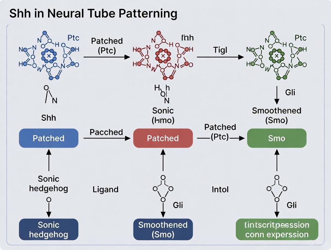
Sonic Hedgehog in Neural Tube Patterning: From Morphogen Gradient to Therapeutic Target
This article synthesizes current knowledge on the Sonic Hedgehog (SHH) signaling pathway, a master regulator of neural tube patterning.
Sonic Hedgehog in Neural Tube Patterning: From Morphogen Gradient to Therapeutic Target
Abstract
This article synthesizes current knowledge on the Sonic Hedgehog (SHH) signaling pathway, a master regulator of neural tube patterning. It explores the foundational mechanisms by which SHH, as a morphogen, establishes dorsoventral neuronal identities through concentration and duration gradients. The content delves into methodological approaches for studying SHH, including explant assays and stem cell differentiation models, and addresses challenges in pathway modulation and optimization. Furthermore, it examines the pathway's validation in disease contexts, linking its dysregulation to neural tube defects and cancers, and discusses the translational potential of SHH pathway modulators in regenerative medicine and oncology for a specialized audience of researchers and drug development professionals.
The SHH Morphogen: Decoding a Gradient that Builds the Nervous System
The Sonic Hedgehog (SHH) signaling pathway functions as a classical morphogen, orchestrating the pattern of the ventral neural tube through a concentration-dependent gradient that instructs progenitor cells to adopt distinct fates. Originating from the notochord and floor plate, the SHH gradient activates specific genetic programs in a dose-dependent manner, leading to the precise spatial arrangement of motor neurons, oligodendrocyte precursor cells (OPCs), and various interneurons. This whitepaper delves into the molecular mechanisms of SHH gradient formation, its interpretation by neural progenitors, and the experimental methodologies used to elucidate these processes. Furthermore, it explores the therapeutic implications of targeting this pathway, given its critical role in both development and disease. Framed within broader research on neural tube patterning, this review synthesizes foundational and contemporary findings to serve as a technical resource for researchers and drug development professionals.
The concept of a morphogen, a substance that governs the fate of cells in a concentration-dependent manner, provides a fundamental framework for understanding embryonic development. Lewis Wolpert's "French Flag Model" posits that a signal emitted from a localized source forms a concentration gradient across a field of cells, and these cells interpret different threshold levels of the signal to adopt distinct, discrete fates [1]. In the developing vertebrate neural tube, Sonic Hedgehog (SHH) represents a quintessential morphogen. Secreted initially by the notochord and subsequently by the floor plate cells at the ventral midline, SHH patterns the ventral neural tube by specifying the identity of progenitor domains arrayed along the dorso-ventral axis [2] [1] [3].
The establishment of this pattern is a prerequisite for the generation of diverse neuronal and glial cell types, including motor neurons (MNs), V3 interneurons, and oligodendrocyte precursor cells (OPCs). The neural tube's response to the SHH gradient is not merely spatial but also temporal; the duration of SHH exposure works in concert with concentration to refine cellular identities [4]. The critical role of SHH signaling is underscored by the severe congenital anomalies, such as holoprosencephaly and neural tube defects, that arise from its disruption [5] [3]. Moreover, aberrant reactivation of the SHH pathway in adulthood is a driver of several malignancies, including medulloblastoma and basal cell carcinoma, making it a significant therapeutic target [6] [7] [8]. This paper details the mechanisms by which SHH fulfills its role as a morphogen, focusing on its function in neural tube patterning.
SHH Gradient Formation and Interpretation
Establishment of the Morphogen Gradient
The SHH morphogen gradient is established from two primary signaling centers. During early embryogenesis, the notochord, located beneath the neural tube, is the first source of SHH. This signal induces the formation of the floor plate at the ventral midline of the neural tube itself. Once specified, the floor plate becomes a secondary, sustained source of SHH, reinforcing and shaping the gradient [1] [3]. The SHH protein is synthesized as a precursor that undergoes autocatalytic cleavage to produce a biologically active N-terminal fragment, which is then modified by the addition of a cholesterol moiety. This lipid modification is crucial for its ability to form a steep, long-range gradient, as it influences the molecule's diffusion and distribution through the extracellular matrix [3].
The propagation of SHH is not a simple matter of free diffusion. It is actively regulated by interactions with heparan sulfate proteoglycans (HSPGs) on the cell surface. Enzymes like Sulf1 and Sulf2 remodel these HSPGs by removing 6-O-sulfate groups, which in turn modulates the binding and distribution of SHH. For instance, Sulf2a, expressed in neural progenitors, acts to reduce the sensitivity of target cells to SHH, thereby fine-tuning the morphogen's effective signaling range and ensuring proper progenitor domain specification [9].
Intracellular Interpretation of the SHH Gradient
The cellular interpretation of the SHH gradient begins with its binding to the Patched1 (PTCH1) receptor on target cells. In the absence of SHH, PTCH1 constitutively represses the activity of the seven-transmembrane protein Smoothened (SMO). Binding of SHH to PTCH1 relieves this repression, allowing SMO to accumulate and initiate an intracellular signaling cascade that ultimately leads to the activation of the GLI family of transcription factors (GLI1, GLI2, GLI3) [6] [7] [8].
The key to morphogen function lies in how different concentration thresholds of SHH lead to differential GLI activity and, consequently, differential gene expression. In the ventral neural tube, progenitor domains are defined by the combinatorial expression of transcription factors. The concentration and duration of SHH signaling dictate which genes are activated or repressed. Progenitors exposed to the highest SHH levels activate Nkx2.2 and become the p3 domain, which gives rise to V3 interneurons. Slightly lower levels promote the expression of Olig2 in the pMN domain, which produces motor neurons. Even lower thresholds specify other interneuron subtypes [2] [9] [1]. This process ensures that a continuous gradient of SHH is interpreted to generate discrete blocks of cellular identity.
Table 1: SHH Concentration Thresholds and Ventral Neural Tube Patterning
| Progenitor Domain | Neural Cell Fate Produced | Key Transcription Factor | Required SHH Signal |
|---|---|---|---|
| p3 | V3 Interneurons | Nkx2.2 | High concentration/threshold [9] |
| pMN | Somatic Motor Neurons (MNs) | Olig2 | Intermediate concentration/threshold [9] [10] |
| p* | Oligodendrocyte Precursor Cells (OPCs) | Olig2 & Nkx2.2 | Late, high signal (from lateral floor plate) [9] |
| p2, p1, p0 | V2, V1, V0 Interneurons | Irx3, Pax6, Dbx1/2, etc. | Low concentrations/thresholds [2] [1] |
Experimental Protocols for Investigating SHH Morphogen Function
A combination of in vitro and in vivo approaches has been instrumental in defining SHH as a morphogen. The following protocols represent key methodologies in the field.
In Vitro Neural Differentiation Assay
This assay tests the direct concentration-dependent effect of SHH on naive neural progenitor cells.
Materials:
- Reagent: Recombinant Sonic Hedgehog (SHH) N-terminal peptide (e.g., R&D Systems).
- Cells: Neuralized embryonic stem cells or primary neural tube progenitors.
- Media: Defined neural differentiation medium (e.g., DMEM/F12 with N2 and B27 supplements).
Methodology:
- Neural Induction: Differentiate pluripotent stem cells into primitive neural progenitor cells using dual-SMAD inhibition (e.g., with SB431542 and LDN-193189).
- SHH Titration: Plate neuralized progenitors and treat them with a range of recombinant SHH concentrations (e.g., from 1 nM to 500 nM). Include a control with no SHH (ventralizing factor).
- Ventral Patterning: Co-administer a caudalizing factor like retinoic acid (RA) to direct progenitors toward a spinal cord identity.
- Analysis: After 3-7 days, analyze the cells by immunocytochemistry or quantitative PCR (qPCR) for domain-specific markers.
In Vivo Morpholino Knockdown in Zebrafish
Zebrafish are an ideal model for functional genetics due to their external development and optical clarity. This protocol assesses the consequence of loss-of-function of SHH pathway components.
Materials:
- Reagent: Gene-specific morpholino oligonucleotides (MOs) designed to block translation or splicing of a target gene (e.g., sulf2a, shha).
- Model: Wild-type zebrafish embryos.
Methodology:
- Microinjection: At the 1-4 cell stage, inject morpholinos into the yolk of zebrafish embryos. Use a standard control morpholino for comparison.
- Incubation: Allow embryos to develop to desired stages (e.g., 24 hpf for early patterning, 48 hpf for neuronal specification, 72 hpf for gliogenesis).
- Phenotypic Analysis:
- In Situ Hybridization (ISH): Fix embryos and perform ISH with riboprobes for specific cell fate markers.
- sulf2a MO: Analyze for an increase in sim1a+ V3 interneurons and a decrease in olig2+ MNs and OPCs at 48 hpf and 72 hpf, respectively [9].
- Immunohistochemistry: Use antibodies against proteins like Olig2 or Islet1 to visualize motor neuron populations.
- In Situ Hybridization (ISH): Fix embryos and perform ISH with riboprobes for specific cell fate markers.
- Validation: Confirm knockdown efficacy through RT-PCR to detect mis-spliced transcripts or Western blotting if antibodies are available [9].
Regulatory Modulation of SHH Signaling
The SHH gradient is not static, and its interpretation is finely tuned by multiple regulatory mechanisms. As highlighted in the experimental protocols, the extracellular sulfatase Sulf2a plays a critical role in shaping the SHH response. Expressed in neural progenitors, Sulf2a remodels heparan sulfates to reduce the sensitivity of responding cells to the SHH signal. This activity is essential for preventing progenitor cells like those in the pMN domain from inappropriately adopting a high-threshold p3 (V3 interneuron) fate, thereby ensuring the proper balance between motor neuron and interneuron production [9]. This represents a key mechanism where the responding cell's capacity to interpret the morphogen is actively regulated.
Furthermore, the duration of SHH exposure is a critical parameter in cell fate specification. Studies in the limb bud and neural tube have demonstrated that prolonged exposure to SHH can specify more posterior limb identities and contribute to the late specification of cell types like OPCs. The pMN domain, after generating motor neurons, receives a new, high-level SHH signal from the lateral floor plate. This sustained signal, facilitated by Sulf1, induces the formation of a new progenitor domain (p*) where cells co-express Olig2 and Nkx2.2 and are fated to become OPCs [9] [4]. This illustrates how temporal dynamics work alongside concentration to expand the patterning capacity of a single morphogen.
Therapeutic Implications and Pathway Targeting
The critical role of SHH in development is mirrored by its pathogenic potential when dysregulated in adulthood. Constitutive, ligand-independent activation of the SHH pathway, often through inactivating mutations in PTCH1 or activating mutations in SMO, is a hallmark of cancers like basal cell carcinoma (BCC) and a subset of medulloblastoma (SHH-MB) [6] [8]. This has led to the development of targeted therapies that inhibit pathway components.
Table 2: FDA-Approved Hedgehog Pathway Inhibitors in Cancer
| Drug Name | Molecular Target | Primary Indication | Key Challenge in CNS Application |
|---|---|---|---|
| Vismodegib (GDC-0449) | Smoothened (SMO) | Advanced Basal Cell Carcinoma [6] | Poor blood-brain barrier (BBB) permeability [7] |
| Sonidegib (LDE225) | Smoothened (SMO) | Advanced Basal Cell Carcinoma [6] | Poor blood-brain barrier (BBB) permeability [7] |
| Arsenic Trioxide (ATO) | GLI proteins (& PML-RARα) | Acute Promyelocytic Leukemia [6] | Suppresses GLI activity; used in APL, efficacy in SHH-MB being investigated |
A significant challenge in treating CNS tumors like SHH-MB with SMO inhibitors is their limited penetration of the blood-brain barrier (BBB). Current research focuses on novel drug delivery systems to overcome this hurdle. Strategies include encapsulating drugs like sonidegib in engineered HDL-mimetic nanoparticles (eHNPs) or polymeric nanoparticles, which can enhance BBB transit and improve tumor-specific delivery [7]. For tumors resistant to SMO inhibition, downstream inhibitors targeting the GLI transcription factors represent an alternative strategy, though their clinical development is less advanced [6] [7].
The Scientist's Toolkit: Key Research Reagents
Table 3: Essential Reagents for SHH Morphogen Research
| Reagent / Tool | Function & Application | Example Use Case |
|---|---|---|
| Recombinant SHH Protein | Purified active ligand for in vitro stimulation; used to create concentration gradients. | Titrating SHH in stem cell differentiation protocols to induce specific neuronal fates [10]. |
| SMO Inhibitors (e.g., Vismodegib, Sonidegib) | Small molecule antagonists of Smoothened; used to inhibit canonical pathway signaling. | Testing pathway dependence in cancer models or probing the requirement of SHH signaling at specific developmental timepoints [6] [7]. |
| Morpholino Oligonucleotides | Gene-specific knockdown tools for loss-of-function studies in model organisms like zebrafish. | Rapidly assessing the in vivo function of genes like sulf2a in spinal cord patterning [9]. |
| Antibody Panels (Olig2, Nkx2.2, Pax6, Islet1) | Markers for immunohistochemistry and immunocytochemistry to identify progenitor domains and neuronal subtypes. | Analyzing the pattern of the neural tube in mutant embryos or differentiated cell cultures [2] [9]. |
| SHH-Reporting Cell Lines | Cell lines with a GLI-responsive reporter (e.g., GFP or Luciferase) to quantify pathway activity. | High-throughput screening for SHH pathway agonists or inhibitors [6]. |
| POPSO | Popso (Poplar Propolis Extract) | Popso, a poplar-type propolis extract rich in flavonoids. For Research Use Only (RUO). Supports studies in microbiology, oxidative stress, and phytochemistry. |
| 4-(2-Hydroxyethyl)-1-piperazineethanesulfonic acid | 4-(2-Hydroxyethyl)-1-piperazineethanesulfonic acid, CAS:7365-45-9, MF:C8H18N2O4S, MW:238.31 g/mol | Chemical Reagent |
Visualizing the SHH Signaling Pathway and Experimental Workflow
The following diagrams, generated using DOT language, illustrate the core SHH signaling mechanism and a typical experimental workflow.
Diagram 1: SHH Signaling Pathway in Neural Patterning
(SHH Signaling Pathway: This diagram illustrates the core mechanism. SHH binding to PTCH1 releases the inhibition on SMO. Activated SMO prevents the proteolytic processing of GLI proteins into repressors and promotes their activation. The active GLI transcription factors then translocate to the nucleus to regulate the expression of target genes, which specify distinct neural progenitor fates in a concentration-dependent manner.)
Diagram 2: Experimental Workflow for SHH Morphogen Study
(SHH Experimental Workflow: A generalized workflow for studying SHH morphogen function. Research begins with a defined objective, leading to the selection of an in vitro or in vivo model system. The pathway is perturbed through methods like SHH protein titration or genetic knockdown. The resulting phenotypic changes are analyzed using molecular and cellular techniques, culminating in data interpretation.)
Sonic Hedgehog (Shh) signaling constitutes a cornerstone of embryonic development, serving as a master regulator of ventral neural tube patterning. The establishment of distinct neuronal progenitor domains and subsequent cell fates along the dorsoventral axis is orchestrated by a gradient of Shh protein, meticulously organized from its primary secretion sources. This whitepaper delineates the precise cellular origins of the Shh signal—the notochord and the floor plate—and explores the sophisticated mechanisms governing its dissemination and interpretation. Within the broader context of Shh research in neural tube patterning, understanding these foundational signaling sources is critical for elucidating the etiology of congenital neural defects and developing therapeutic strategies for neurodegenerative diseases and spinal cord injuries.
Molecular Mechanisms of Shh Production and Signaling
The Shh signaling pathway is a highly conserved system initiated by the production and processing of the Shh ligand. The human SHH gene encodes a precursor protein that undergoes autocatalytic cleavage to yield a 19 kDa N-terminal fragment (Shh-N) and a 26 kDa C-terminal fragment (Shh-C) [11] [12]. This processing event, catalyzed by Shh-C, results in the covalent attachment of a cholesterol moiety to the C-terminus of Shh-N, producing the biologically active ligand. Subsequent palmitoylation of the N-terminal cysteine residue by acyl transferase yields the fully active, dually lipid-modified Shh protein [12]. These lipid modifications are crucial for regulating the spatial distribution and signaling range of Shh by tethering the ligand to cell membranes, thus preventing uncontrolled diffusion [13].
The canonical Shh signaling cascade is initiated when the active Shh ligand binds to its transmembrane receptor Patched1 (Ptch1). In the absence of Shh, Ptch1 constitutively inhibits the G protein-coupled receptor Smoothened (Smo) [14] [11]. Shh binding to Ptch1 relieves this inhibition, allowing Smo to accumulate in primary cilia and transduce an intracellular signal that prevents the proteolytic processing of Gli transcription factors into their repressor forms [14] [15]. This leads to the nuclear translocation of full-length Gli activators (primarily Gli1 and Gli2) and the transcriptional activation of target genes, including Ptch1 and Gli1 themselves, creating a feedback loop [15] [12].
Diagram 1: Canonical Shh Signaling Pathway. The pathway shows inhibition of Smo by Ptch1 in the absence of Shh, leading to Gli repressor formation and suppression of target genes. Shh binding releases Smo inhibition, enabling Gli activator formation and target gene transcription.
Primary Signaling Centers: Notochord and Floor Plate
The ventral neural tube is patterned by Shh secreted from two principal signaling centers: the notochord and the floor plate. The notochord, a transient mesodermal structure underlying the neural tube, serves as the initial source of Shh during early embryogenesis [16] [10]. This notochord-derived Shh is responsible for the induction of the floor plate, a specialized group of glial cells at the ventral midline of the neural tube [17]. Once specified, the floor plate itself becomes a secondary source of Shh, maintaining and refining the Shh gradient that patterns ventral neuronal progenitors [17].
The development of distinct neuronal subtypes in the ventral neural tube occurs in a precise spatial order that corresponds to their requirement for different Shh concentrations and exposure durations. Progenitors close to the signaling sources, exposed to high Shh levels, adopt ventral fates such as floor plate and V3 interneurons, while those at progressively greater distances, exposed to lower concentrations, form motor neurons and more dorsal interneuron subtypes [17]. This concentration-dependent patterning extends to the specification of Olig2+ motoneuron progenitors and Hb9+ motoneurons, which require sustained Shh signaling for their development and maintenance [16].
Sclerotome as a Transit Pathway
Recent research has revealed that the sclerotome, the ventral compartment of the somites, serves as a crucial transit pathway for Shh dissemination. Notochord-derived Shh must traverse the sclerotome to reach both the myotome and the basal aspect of the neural tube [16]. Reduction of Shh in the sclerotome through targeted expression of membrane-tethered Hedgehog-interacting protein (Hhip:CD4) significantly decreases the number of Olig2+ motoneuron progenitors and Hb9+ motoneurons, demonstrating that sclerotomal Shh is necessary for proper neural tube development [16].
Notably, notochord grafting experiments have demonstrated that Shh presentation to the basal side of the neuroepithelium, corresponding to the sclerotome-neural tube interface, profoundly affects motoneuron development compared to apical grafting [16]. This suggests that initial ligand presentation occurs preferentially at the basal side of epithelia, revealing the sclerotome as a novel pathway through which notochord-derived Shh disperses to coordinate the development of both mesodermal and neural progenitors.
Dynamics of Gradient Interpretation
The interpretation of the Shh gradient involves not only concentration thresholds but also temporal dynamics of signaling. Floor plate specification exemplifies this sophisticated mechanism, involving a biphasic response to Shh signaling [17]. During gastrulation and early somitogenesis, floor plate induction depends on high levels of Shh signaling. Subsequently, prospective floor plate cells become refractory to Shh signaling, a prerequisite for the elaboration of their definitive non-neuronal identity [17]. This dynamic response expands the patterning capacity of a single ligand, enabling the specification of multiple cell types.
Table 1: Quantitative Effects of Shh Pathway Manipulations on Neural Development
| Experimental Manipulation | Biological System | Quantitative Effect | Biological Outcome | Citation |
|---|---|---|---|---|
| Hhip1 misexpression in sclerotome | Chick embryo | 40% reduction in Hb9+ motoneurons | Impaired motoneuron differentiation | [16] |
| Shh-P150 exosome application | Neural explant assay | Induction of Nkx2.2+ ventral progenitors | Ventral neural tube patterning | [14] |
| Shh-P450 exosome application | Neural explant assay | Increased progenitor proliferation | Expansion of neural progenitor pool | [14] |
| Floor plate deletion | Chick/mouse embryo | Altered FoxA2+ floor plate cells | Disrupted ventral patterning | [17] |
Novel Secretion Mechanisms and Signaling Dynamics
Distinct Exosomal Pools for Patterning versus Proliferation
Emerging evidence indicates that Shh is secreted on biochemically distinct exosomal pools that mediate different aspects of neural development. Ultracentrifugation studies have identified two principal exosomal fractions: Shh-P150 and Shh-P450, distinguished by their differential pelletability [14]. These pools possess unique protein and miRNA cargo and execute distinct biological functions.
The Shh-P150 exosome fraction mediates canonical signaling and neural tube patterning, inducing the expression of ventral spinal cord markers including Nkx2.2 [14]. Conversely, the lighter Shh-P450 pool drives progenitor proliferation through Gαi-mediated signaling but is incapable of patterning ventral neuronal progenitors [14]. The identification of Rab7 as a regulator of Shh-P150 biogenesis suggests that differential packaging of Shh into distinct exosomal pools provides a molecular switch between its morphogenetic and mitogenic outputs, resolving the long-standing question of how Shh segregates these distinct functions.
Spatiotemporal Control of Signaling
The deployment of Shh from different sources follows precise spatiotemporal dynamics that are essential for proper neural tube patterning. The notochord provides the initial Shh signal during early developmental stages, establishing the foundation for ventral patterning [16] [10]. As development proceeds, the floor plate assumes the role of the dominant Shh source, maintaining and refining the patterning signal [17]. This transition ensures the proper temporal coordination of ventral neuronal specification.
Recent technological advances, including optogenetic control of morphogen production, have enabled unprecedented precision in investigating Shh gradient formation and dynamics [18]. These approaches allow researchers to manipulate the timing, duration, and location of Shh production with high spatial and temporal resolution, providing new insights into how gradient interpretation translates into precise patterns of gene expression and cell fate specification.
Diagram 2: Shh Secretion and Gradient Formation. The diagram illustrates how Shh from notochord and floor plate creates a ventral-to-dorsal concentration gradient through direct secretion and sclerotomal transit, leading to specification of distinct ventral neuronal subtypes.
Experimental Approaches and Methodologies
Key Experimental Paradigms
Research elucidating the origins and mechanisms of Shh signaling has employed sophisticated experimental approaches, including loss-of-function and gain-of-function studies in model organisms. Electroporation-based gene manipulation in chick embryos has been particularly instrumental for spatially and temporally controlled perturbation of Shh signaling [16]. At 23- to 25-somite stages, electroporation at the level of epithelial somites enables targeted manipulation of the prospective sclerotome, allowing researchers to investigate Shh transit through this compartment without affecting earlier patterning events [16].
Notochord grafting experiments have provided critical insights into the directional nature of Shh presentation. Studies demonstrate that grafting notochord fragments adjacent to the basal sclerotomal side of the neural tube profoundly affects motoneuron development compared to apical grafts [16]. Similarly, basal grafting with respect to the dermomyotome significantly enhances myotome formation, suggesting a general requirement for initial ligand presentation at the basal side of epithelia [16].
Neural Explant Assays
Neural explant assays have served as a foundational methodology for investigating Shh patterning activity [14]. In this protocol, neural tube explants are cultured in three-dimensional matrices and exposed to purified Shh or exosomal fractions. The explants are typically cultured for 48-72 hours, then processed for immunohistochemistry or RNA analysis to assess the induction of ventral markers such as Nkx2.2, Olig2, and FoxA2 [14]. This assay has been crucial for demonstrating the differential activities of Shh-P150 and Shh-P450 exosomal fractions, with Shh-P150 inducing ventral progenitor markers while Shh-P450 promotes proliferation without patterning activity [14].
Exosome Isolation and Characterization
The identification of distinct Shh-containing exosomal pools relied on sophisticated fractionation techniques [14]. The standard protocol involves sequential ultracentrifugation of conditioned media from Shh-expressing cells. Initial low-speed centrifugation (10,000 × g) removes cellular debris, followed by high-speed centrifugation (150,000 × g) to pellet the P150 exosomal fraction. The resulting supernatant is then subjected to ultracentrifugation at 450,000 × g to pellet the P450 fraction [14]. These biochemically distinct pools are further characterized by electron microscopy, western blotting for exosomal markers (e.g., Alix, Tsg101), and functional assays in neural explants or stem cell differentiation systems.
Table 2: Research Reagent Solutions for Shh Signaling Studies
| Research Tool | Type | Primary Application | Key Features & Function | Citation |
|---|---|---|---|---|
| Hhip:CD4 | Membrane-tethered antagonist | Loss-of-function studies | Sequesters Shh in sclerotome; reduces motoneuron differentiation | [16] |
| Shh-P150 & Shh-P450 | Distinct exosomal fractions | Functional segregation studies | P150 patterns ventral neural tube; P450 drives proliferation | [14] |
| ShhN:YFP | Fluorescent Shh construct | Ligand trafficking studies | Palmitoylated but not cholesterol-modified; tracks Shh movement | [16] |
| Neural Explant Assay | Ex vivo culture system | Pattering activity testing | Measures induction of ventral markers by Shh sources | [14] |
| Optogenetic Shh Control | Spatiotemporal manipulation | Gradient dynamics studies | Precise control over timing and location of Shh production | [18] |
The Scientist's Toolkit: Essential Research Reagents
The investigation of Shh secretion and function relies on a specialized toolkit of reagents and methodologies. Membrane-tethered Hhip (Hhip:CD4) serves as a critical tool for cell-autonomous sequestration of Shh ligand, specifically designed to prevent Shh movement through the sclerotome without the confounding effects of secreted Hhip [16]. Fluorescently tagged Shh constructs (ShhN:YFP) enable visualization of ligand distribution and trafficking, particularly valuable for studying the transit of Shh through various compartments [16].
For functional studies, specific Shh pathway agonists (SAG, Purmorphamine) and antagonists (Cyclopamine, Vismodegib) allow precise pharmacological manipulation of signaling activity [11] [12]. The development of optogenetic systems for controlling Shh production represents a cutting-edge approach for investigating morphogen dynamics with high spatiotemporal precision [18]. These tools, combined with traditional embryological techniques such as notochord grafting and neural explant cultures, provide a comprehensive methodological framework for dissecting the complexity of Shh signaling from its origins in the notochord and floor plate.
The notochord and floor plate serve as the primary signaling centers that establish and maintain the Shh gradient responsible for ventral neural tube patterning. Through sophisticated mechanisms including sclerotomal transit, differential exosomal packaging, and dynamic cellular responses, Shh from these sources orchestrates the precise spatial and temporal patterning of neuronal progenitors. The evolving understanding of these processes, facilitated by advanced experimental approaches and reagents, continues to reveal unexpected complexity in how a single morphogen coordinates the development of multiple tissue types. As research progresses, these insights promise to inform therapeutic strategies for neural developmental disorders and regenerative medicine approaches for neurological injuries.
The French Flag model, a foundational concept in developmental biology, posits that cells acquire distinct identities in response to different concentrations of a diffusible morphogen. This whitepaper explores the Sonic Hedgehog (SHH) protein as a quintessential morphogen that patterns the ventral neural tube along the dorsoventral axis. We detail the mechanisms of SHH gradient formation, the dynamic interpretation of this gradient by neural progenitor cells, and the quantitative precision that ensures robust patterning. Furthermore, we discuss cutting-edge methodologies for investigating SHH signaling and its implications for neurodevelopmental disorders and therapeutic strategies. This guide provides researchers and drug development professionals with a comprehensive technical overview of SHH-mediated patterning, underscoring its critical role in neural development.
The French Flag model, introduced by Lewis Wolpert, provides a theoretical framework for understanding how a uniform field of cells can differentiate into distinct spatial domains [19]. According to this model, a secreted signaling molecule—a morphogen—forms a concentration gradient across a developing tissue. Cells respond to this gradient by adopting specific fates based on the local morphogen concentration, effectively dividing the tissue into discrete regions analogous to the stripes of the French flag [19] [20]. Sonic Hedgehog (SHH) is a paradigmatic morphogen that executes this model during ventral patterning of the vertebrate neural tube [20] [3]. The neural tube, the embryonic precursor to the spinal cord and brain, exhibits a remarkably organized structure where distinct neuronal subtypes arise at precise positions along the dorsoventral (DV) axis. This patterning is largely governed by a SHH concentration gradient, which emanates from ventral signaling centers—the notochord and the floor plate [19] [3]. Cells exposed to the highest SHH concentrations become ventral floor plate cells, while progressively lower concentrations specify more dorsal progenitor domains, giving rise to different neuronal subtypes [20]. The interpretation of the SHH gradient is a dynamic process, integrating both the concentration of the signal and the duration of cellular exposure, and is refined by intricate feedback mechanisms within the receiving cells [20].
The SHH Signaling Pathway: From Ligand to Transcriptional Response
The SHH signaling pathway is the molecular machinery that transmits the extracellular morphogen signal into intracellular gene expression changes. The core components and sequence of events are as follows:
Canonical Pathway Activation:
- Ligand Processing and Secretion: SHH is synthesized as a precursor protein that undergoes autocatalytic cleavage and dual lipid modification: cholesterol at its C-terminus and palmitate at its N-terminus [19] [21]. These modifications are critical for its potency and range of diffusion. The processed and modified SHH ligand is secreted from the source cells via the transmembrane protein Dispatched [19].
- Receptor Binding and Signal Initiation: The secreted SHH ligand binds to its primary receptor, Patched1 (PTCH1), on the target cell membrane. PTCH1 constitutively inhibits a second transmembrane protein, Smoothened (SMO). Binding of SHH to PTCH1 relieves this inhibition [19] [22].
- Ciliary Transduction and Transcriptional Activation: SMO then accumulates in the primary cilium, a key signaling organelle. This leads to the activation and nuclear translocation of the GLI family of transcription factors (GLI1, GLI2, GLI3), which subsequently induce or repress the expression of target genes, including PTCH1 itself and GLI1, creating a negative feedback loop [19] [20] [22].
Non-Canonical Pathways: SHH can also signal independently of the canonical PTCH1-SMO-GLI axis. These non-canonical pathways may be SMO-dependent but GLI-independent, or operate completely independently of SMO, and are involved in processes like axon guidance and cell migration [19] [22].
Table 1: Core Components of the SHH Signaling Pathway
| Component | Type | Function in SHH Signaling |
|---|---|---|
| SHH | Ligand | Lipid-modified morphogen; binds to PTCH1 to initiate signaling. |
| PTCH1 | Receptor | Transmembrane receptor that inhibits SMO in the absence of SHH. |
| SMO | Transducer | Seven-pass transmembrane protein; transduces signal upon PTCH1 inhibition relief. |
| GLI1/2/3 | Transcription Factors | Terminal effectors; regulate transcription of target genes (GLI1/2 activators, GLI3 repressor). |
| Primary Cilium | Organelle | Specialized cellular compartment where key signaling events (SMO/GLI activation) occur. |
| BOC/GAS1/CDON | Coreceptors | Enhance SHH binding and signaling efficiency [19] [23]. |
| SUFU | Negative Regulator | Cytosolic protein that inhibits GLI protein activity [22]. |
| SN003 | SN003, MF:C19H25N5O2, MW:355.4 g/mol | Chemical Reagent |
| C-021 | CCR4 Antagonist C-021|Research Compound |
The following diagram illustrates the core canonical SHH signaling pathway and its key outputs.
Diagram 1: Canonical SHH Signaling Pathway. When SHH is absent (left), PTCH1 inhibits SMO, leading to the proteolytic processing of GLI factors into repressors and suppression of target gene expression. When SHH is present (right), it binds PTCH1, relieving inhibition of SMO. SMO activation promotes the formation of GLI activators, which translocate to the nucleus and induce target gene transcription.
Establishing the SHH Morphogen Gradient
The formation of the SHH concentration gradient is a highly regulated process involving specialized signaling centers, unique biochemical properties of the ligand, and active transport mechanisms.
Signaling Centers and Spatiotemporal Dynamics
The primary sources of SHH in the developing neural tube are the notochord (a mesodermal structure underlying the neural tube) and the floor plate (the ventral-most structure of the neural tube itself) [19] [20] [3]. Signaling initiates with SHH secretion from the notochord, which is responsible for the initial induction of the floor plate. Once established, the floor plate itself becomes a secondary source of SHH, reinforcing and maintaining the gradient [20]. The gradient is not static; it evolves over time. The amplitude of the SHH gradient increases as development progresses, meaning cells near the source are exposed to progressively higher concentrations for longer durations [20]. This temporal dynamics is crucial for the sequential induction of ventral progenitor identities.
Biochemical Mechanisms of Gradient Formation
The range and shape of the SHH gradient are profoundly influenced by the molecule's biochemistry:
- Dual Lipid Modification: The covalent attachment of cholesterol and palmitate makes SHH highly hydrophobic [19] [21]. This hydrophobicity traditionally suggested a limited capacity for free diffusion, promoting the formation of steep, short-range gradients. However, it also facilitates the assembly of SHH into larger multimeric complexes or micelles and its association with lipoprotein particles, which can enable long-range distribution [20].
- Feedback Regulation: The SHH gradient is shaped by feedback loops from the responding cells. The pathway targets PTCH1 and GLI1 are themselves transcriptional targets of SHH signaling. Upregulation of PTCH1 at the site of ligand reception creates a sink that can limit further spread of the SHH ligand, thereby sharpening the gradient in a non-cell-autonomous manner [20].
Interpreting the Gradient: From Signal to Cell Fate
The conversion of a continuous SHH concentration gradient into discrete cellular domains is a complex process of signal interpretation involving temporal integration and transcriptional networks.
Concentration and Duration Dependence
Neural progenitor cells translate different SHH signal intensities and exposure times into distinct fate choices. In vitro studies using chick neural explants have demonstrated that a two- to threefold increase in SHH concentration is sufficient to switch progenitor identity from one subtype to the next, more ventral one [20]. For instance, lower concentrations specify motor neuron (pMN) progenitors, while higher concentrations specify V3 interneurons [19] [20]. Furthermore, the duration of SHH exposure is equally critical. Prolonged signaling is required to activate genes that require high levels of SHH, leading to a progressive ventralization of cell fate over time [20].
Intracellular Interpretation and Network Architecture
The cellular response to SHH is not a simple passive reception but an active process of refinement:
- Temporal Adaptation and Negative Feedback: The concept of "temporal adaptation" describes how cells continuously adjust their response to a persistent SHH signal. Key to this is the induction of negative regulators like PTCH1 and SUFU. This feedback creates a system where the initial level of signaling is strong, but adapts over time, allowing cells to effectively measure and lock in a positional identity based on the signal history [20].
- Transcriptional Cross-Repression: The boundaries between different progenitor domains are sharpened by a network of transcription factors that mutually repress each other's expression. For example, in the ventral neural tube, the domain of Pax6 (dorsal) is separated from Nkx2.2 (ventral) by cross-repressive interactions, ensuring a clean switch between domains rather than a blended mixture of cell types [19].
Table 2: Progenitor Domains in the Ventral Neural Tube
| Progenitor Domain | Key Transcription Factor | Neuronal Output | Relative SHH Exposure |
|---|---|---|---|
| Floor Plate (FP) | FoxA2 | Specialized non-neuronal signaling cells | Highest |
| p3 | Nkx2.2 | V3 interneurons | High |
| pMN | Olig2 | Motor neurons | Medium-High |
| p2 | Irx3, Pax6 | V2 interneurons | Medium |
| p1 | Dbx1, Pax6 | V1 interneurons | Low |
| p0 | Dbx2, Pax6 | V0 interneurons | Lowest |
| Dorsal Domains | Pax3, Pax7 | Sensory interneurons | None / BMP signal |
Quantitative Analysis of Gradient Precision
Recent quantitative studies have reshaped our understanding of the precision of morphogen gradients, moving away from the idea of highly variable gradients requiring combinatorial readouts.
A 2022 re-analysis of gradient precision in the mouse neural tube demonstrated that the positional error of the SHH gradient had been previously overestimated due to methodological limitations in data fitting [24]. The study concluded that:
- A single SHH gradient is sufficiently precise to define the boundaries of central progenitor domains (like the NKX6.1 and PAX3 boundaries) with the accuracy observed biologically, which is within 1-3 cell diameters [24].
- The patterning mechanism is robust to changes in gradient amplitude. Because domain boundaries are defined by specific concentration thresholds, a change in the overall amplitude of the SHH gradient shifts the absolute position of all boundaries but can leave the relative sizes of the interior progenitor domains largely unchanged. This ensures a precise number of progenitor cells for each neuronal type, even in the face of natural embryo-to-embryo variation [24].
Table 3: Key Quantitative Findings on SHH Gradient Precision
| Parameter | Historical Estimation | Revised Estimation (2022) | Implication |
|---|---|---|---|
| Positional Error | Up to 30+ cell diameters in neural tube center [24] | 1-3 cell diameters [24] | Single gradient is sufficiently precise for patterning. |
| Gradient Shape | Assumed to be a perfect exponential [24] | Mean of multiple exponentials is non-exponential [24] | Fitting a single exponential to averaged data overestimates variability. |
| Domain Sizing | Assumed to be affected by amplitude noise | Robust to amplitude changes [24] | Progenitor cell numbers are precisely controlled. |
Advanced Experimental Protocols
Investigating the SHH gradient requires a multidisciplinary approach combining embryology, molecular biology, and advanced imaging.
Classical Embryological Manipulations
- Neural Tube/Notochord Explant Co-culture: This protocol involves isolating the neural tube and notochord from model organisms like chick or mouse embryos. The tissues are cultured in close proximity, and the differentiation of neural progenitors is assessed via immunohistochemistry for domain-specific markers (e.g., Olig2, Nkx2.2) [20].
- Microparticle/Bead Implantation: Beads soaked in purified SHH-N protein (the active N-terminal fragment) or in specific inhibitors (e.g., cyclopamine, an SMO antagonist) are implanted into the developing neural tube or limb bud in vivo. This creates a localized source or sink of signaling, allowing researchers to observe changes in patterning and gene expression around the bead [25].
Live Imaging and Single-Cell Analysis
- Fluorescent Reporter Lines: Transgenic animals (e.g., zebrafish, mice) expressing fluorescent proteins under the control of SHH-responsive promoters (e.g., Gli1 or Ptc1 promoters) enable real-time visualization of pathway activity in vivo [26].
- Single-Cell Resolution Tracking: A 2023 reviewed preprint detailed a protocol using a photoactivatable SHH reporter (Kaede) in zebrafish embryos. This allows for tracking of SHH signaling dynamics in single neural progenitor cells over time and correlating these dynamics with the final transcriptional identity of the cell, revealing significant heterogeneity in response dynamics at the single-cell level that still results in robust fate specification at the population level [26].
The workflow for such a single-cell analysis is complex and involves multiple steps, as summarized below.
Diagram 2: Workflow for Single-Cell SHH Signaling Analysis. This pipeline enables the correlation of dynamic SHH signaling history with the ultimate fate of individual neural progenitor cells.
The Scientist's Toolkit: Essential Research Reagents
The following table catalogues critical reagents and models used in SHH gradient and neural tube patterning research.
Table 4: Key Research Reagents and Models for SHH Studies
| Reagent / Model | Type | Primary Function in Research |
|---|---|---|
| Recombinant SHH-N Protein | Protein | The active, N-terminal fragment of SHH; used for in vitro and in vivo (bead implantation) gain-of-function studies to mimic pathway activation [25]. |
| 5E1 Anti-SHH Antibody | Monoclonal Antibody | Function-blocking antibody; used for in vivo neutralization of SHH ligand to create loss-of-function conditions and study patterning defects [25]. |
| Cyclopamine / SANT-1 | Small Molecule Inhibitor | Specific inhibitors of Smoothened (SMO); used to chemically inhibit the canonical SHH pathway in a dose-dependent manner [22]. |
| SAG (Smoothened Agonist) | Small Molecule Agonist | Activates SMO; used to experimentally stimulate the SHH pathway downstream of PTCH1 [22]. |
| SHH-GFP Knock-in Mice | Genetic Model | Mouse line expressing a biologically active SHH-GFP fusion protein from the endogenous Shh locus; enables direct visualization and quantification of the SHH protein gradient [20]. |
| Avian Embryo (Chick/Quail) | Model System | Classic model for embryological manipulations due to easy accessibility; ideal for microsurgery, electroporation, and bead implantation experiments [25]. |
| Zebrafish Reporter Lines | Transgenic Model | Transgenic fish with GFP or other reporters under SHH-pathway control; excellent for live, real-time imaging of signaling dynamics at single-cell resolution [26]. |
| Human Cerebral Organoids | In Vitro 3D Model | Stem cell-derived models that recapitulate aspects of human brain development; used to study human-specific SHH functions and neurodevelopmental disorders [19]. |
| Isrib | ISRIB|Integrated Stress Response Inhibitor|eIF2B Activator | ISRIB is a potent small molecule inhibitor of the integrated stress response (ISR) that reverses the effects of eIF2α phosphorylation. This product is For Research Use Only. Not for human or veterinary diagnostic or therapeutic use. |
| OU749 | OU749 CAS 519170-13-9|GGT Inhibitor | OU749 is a non-glutamine, uncompetitive, and species-specific GGT inhibitor for research. For Research Use Only. Not for human use. |
Implications in Development and Disease
Dysregulation of the precisely controlled SHH signaling pathway leads to severe congenital disorders and cancers.
- Holoprosencephaly (HPE): HPE is a spectrum of brain malformations caused by a failure of the forebrain to separate into two hemispheres. Mutations in the SHH gene are a major genetic cause of HPE, highlighting the critical role of SHH in ventral forebrain patterning and midline development [23] [21] [3]. Dose-dependent effects are evident, where reduced SHH signaling leads to a spectrum of facial and brain midline defects, ranging from hypotelorism (close-set eyes) to cyclopia [25].
- Cancer: Aberrant activation of the SHH pathway in adulthood is a driver of several cancers. The most well-established link is with medulloblastoma, where constitutive SHH signaling promotes tumor growth in the cerebellum [22] [3]. Mutations in pathway components like PTCH1 and SUFU are also associated with Gorlin syndrome, which predisposes individuals to basal cell carcinomas and medulloblastomas [21] [22].
- Aging-Related Neurodegenerative Diseases: Emerging evidence implicates altered SHH signaling in the pathogenesis of diseases like Alzheimer's disease, Parkinson's disease, and amyotrophic lateral sclerosis (ALS). The pathway's role in adult neurogenesis, neuronal maintenance, and protection against oxidative stress and inflammation is now being explored as a potential therapeutic avenue [22].
The patterning of the neural tube by the SHH morphogen gradient remains a premier example of the French Flag model in action. The process is remarkably sophisticated, involving the dynamic formation of a concentration gradient, its interpretation through a network of intracellular feedback mechanisms, and the translation of this information into discrete, precisely positioned cellular domains. While the core principles are well-established, advances in live imaging, single-cell analysis, and quantitative modeling continue to reveal new layers of complexity, including heterogeneity in single-cell responses and unexpected robustness mechanisms. A deep understanding of SHH biology is therefore indispensable, not only for deciphering the fundamental rules of embryonic development but also for informing novel therapeutic strategies for a wide range of human diseases, from severe birth defects to cancer and neurodegenerative disorders.
The Sonic Hedgehog (SHH) signaling pathway is a master regulator of embryonic development, with a particularly crucial role in the precise patterning of the vertebrate neural tube. The efficacy and range of the SHH morphogen are not solely dictated by its amino acid sequence but are profoundly regulated by a unique post-translational modification: the covalent attachment of two lipid moieties, cholesterol and palmitate. This dual-lipid modification is indispensable for orchestrating the complex cell fate decisions that generate distinct neuronal progenitor domains along the dorsoventral axis. This whitepaper delves into the molecular machinery behind these modifications, their direct impact on SHH ligand biogenesis, distribution, and signaling potency, and outlines the essential experimental tools for probing their functions. Understanding these mechanisms is paramount for developing therapeutic interventions for congenital disorders and cancers driven by aberrant Hedgehog signaling.
The vertebrate neural tube, the embryonic precursor to the central nervous system, exhibits a highly organized structure where distinct neuronal subtypes emerge in specific spatial order. This dorsoventral (DV) patterning is primarily directed by a gradient of SHH protein secreted from two key signaling centers: the notochord and, subsequently, the floor plate cells within the neural tube itself [20]. SHH operates as a classical morphogen—a secreted molecule that conveys positional information through its concentration and the duration of exposure. Progenitor cells exposed to different SHH levels activate distinct transcriptional programs, leading to the establishment of six primary progenitor domains (p0, p1, p2, pMN, p3, and the floor plate), each giving rise to a specific class of neurons [20]. The emergence of these domains is a dynamic process; genes requiring progressively higher levels or longer durations of SHH signaling are sequentially induced at the ventral midline [20]. The formation of this exquisite pattern raises a fundamental question: how is the distribution and perception of the SHH gradient so precisely controlled? The answer lies in the unique biochemical tethering of the SHH ligand itself via cholesterol and palmitate modifications.
The Biochemistry of SHH Lipid Modification
The SHH protein is synthesized as a ~45 kDa precursor that undergoes a series of critical processing steps within the secretory pathway to become the active, lipid-modified morphogen.
Cholesterol Modification: An Unusual Autoprocessing Event
Cholesterol modification is an autocatalytic process that occurs in the endoplasmic reticulum. The C-terminal domain of the SHH precursor catalyzes an intramolecular cleavage reaction. This proceeds through a thioester intermediate that is resolved by nucleophilic attack from the hydroxyl group of a cholesterol molecule [27] [28]. The result is a covalent ester bond linking cholesterol to the C-terminal glycine (Gly-198 in mouse SHH) of the newly formed ~19 kDa N-terminal signaling fragment (SHH-N) [29] [28]. This reaction is essential for the correct formation of SHH gradients in vivo.
Palmitoylation: A Catalytic Addition by Hhat
The second lipid modification is the attachment of a palmitate group, a 16-carbon saturated fatty acid. This reaction is catalyzed by the enzyme Hedgehog acyltransferase (Hhat) (known as Skinny hedgehog or Rasp in Drosophila), a member of the membrane-bound O-acyltransferase (MBOAT) family [27] [30]. Hhat transfers palmitate from palmitoyl-Coenzyme A to the alpha-amino group of the N-terminal cysteine (Cys-25 in human SHH) of the cholesterol-modified SHH-N fragment, forming a stable amide bond [30] [29]. Unlike cholesterol modification, palmitoylation is a purely enzymatic process and is absolutely required for full SHH signaling activity.
Table 1: Key Characteristics of SHH Lipid Modifications
| Feature | Cholesterol Modification | Palmitate Modification |
|---|---|---|
| Modification Type | Autocatalytic | Enzymatic (Hhat) |
| Bond Type | Ester | Amide |
| Attachment Site | C-terminal Glycine | N-terminal Cysteine |
| Enzyme/Mechanism | C-terminal domain of SHH precursor | Hedgehog acyltransferase (Hhat) |
| Cellular Location | Endoplasmic Reticulum | Endoplasmic Reticulum |
Functional Consequences of Dual Lipidation on SHH Signaling
The attachment of two hydrophobic anchors fundamentally shapes the behavior of the SHH ligand, influencing its membrane association, distribution, and ultimate signaling potency.
Membrane Tethering and Solubilization
The dual lipid moieties render the mature SHH protein highly hydrophobic, causing it to be firmly associated with the plasma membrane of the producing cell [31] [32]. This membrane tethering poses a challenge for a morphogen that must signal at a distance. The resolution of this paradox involves specialized machinery for ligand solubilization and release. The 12-pass transmembrane protein Dispatched (Disp) is critical for releasing lipidated SHH from the cell surface [27] [32]. Recent models suggest a collaborative process where Disp, potentially assisted by the secreted glycoprotein Scube2, facilitates the transfer of cholesterol-modified SHH to extracellular carriers like high-density lipoproteins (HDL) [33] [32]. This process may be completed by proteolytic shedding of the palmitoylated N-terminus, generating a soluble, mono-lipidated SHH form with high bioactivity [32].
Formation of Gradients and Signaling Range
The lipid modifications are crucial for shaping the SHH gradient in the target field, such as the neural tube. The hydrophobic nature of the ligand limits its free diffusion, contributing to a steep, stable concentration gradient that is essential for patterning multiple cell types. Visualization of a SHH-GFP fusion protein in the neural tube reveals an exponentially decaying gradient from the ventral midline, with punctate structures enriched near ciliary basal bodies [20]. The cholesterol moiety, in particular, is vital for long-range signaling activity and for the formation of large, multimeric SHH complexes [34] [31]. In Drosophila, Hh ligands associate with lipoprotein particles (lipophorins), and reducing lipophorin levels impairs long-range signaling [33].
Signaling Potency and Cellular Reception
Lipid modifications are not merely for distribution; they directly enhance the ligand's ability to activate its receptor. Studies comparing the signaling potency of different SHH forms have demonstrated that the dually lipidated protein is significantly more potent than forms lacking one or both lipids [29] [31]. The lipids govern cellular reception by controlling the ligand's association with target cell membranes. Research shows that either lipid adduct is sufficient to confer cellular association, with the cholesterol adduct primarily anchoring the ligand to the plasma membrane and the palmitate adduct augmenting ligand internalization [31]. Crucially, signaling potency directly correlates with the cellular concentration of the SHH ligand, which is maximized by the presence of both lipids [31].
Experimental Analysis of SHH Lipidation
Investigating the roles of SHH lipid modifications requires a suite of well-established biochemical and cell-based assays.
Key Experimental Protocols
1. Assessing Hhat Activity and SHH Palmitoylation in Cells:
- Method: Co-transfect cells (e.g., HEK293T) with cDNAs encoding Hhat and SHH.
- Labeling: Incubate cells with palmitate analogues, such as
125I-Iodopalmitateor azide/alkyne-modified palmitate (e.g., 17-octadecynoic acid). - Detection: Immunoprecipitate SHH from cell lysates. For radioactive labels, quantify incorporation via phosphorimaging after SDS-PAGE. For click-compatible labels, use a copper-catalyzed azide-alkyne cycloaddition reaction to conjugate a fluorescent or biotin tag, followed by detection [30].
2. In Vitro Hhat Enzymatic Assay:
- Enzyme Source: Use detergent-solubilized or purified Hhat protein.
- Substrate: An N-terminal Shh peptide (as short as 10-11 amino acids) with a C-terminal biotin tag.
- Reaction: Incubate with
125I-Iodopalmitoyl-CoAor an alkyne-labeled palmitoyl-CoA. - Detection: Capture the biotinylated peptide on streptavidin beads and measure incorporated radiolabel or perform click chemistry for fluorescence-based detection [30] [20].
3. Functional Patterning Assay (Neural Explant):
- Tissue Source: Isolate neural tube explants from chick or mouse embryos.
- Treatment: Expose explants to purified SHH proteins (wild-type or lipid-mutant forms) or to distinct SHH-carrying exosomal pools (e.g., Shh-P150 vs. Shh-P450) [14].
- Readout: After culture, analyze the expression of ventral progenitor markers (e.g., Nkx2.2, Olig2) by in situ hybridization or immunohistochemistry to assess patterning competence [14].
The Scientist's Toolkit: Essential Research Reagents
Table 2: Key Reagents for Studying SHH Lipidation and Function
| Reagent/Category | Example Specific Items | Primary Function in Research |
|---|---|---|
| Molecular Tools | SHH cDNA constructs (wild-type, C25A/S, ShhN) | To express defined lipid-mutant forms of SHH in cells [31]. |
| Chemical Inhibitors | RU-SKI 43 (Hhat inhibitor); Lovastatin (cholesterol synthesis inhibitor) | To pharmacologically block palmitoylation or deplete cellular cholesterol pools, respectively [33] [30]. |
| Palmitate Analogues | 17-ODYA (Alkynyl-palmitate); 125I-Iodopalmitate | For metabolic labeling and detection of palmitoylated SHH [30]. |
| Assay Systems | C3H10T1/2 cell line (alkaline phosphatase induction); Gli-luciferase reporter assay | Cell-based bioassays to quantify SHH signaling potency [29] [31]. |
| Carrier Molecules | Purified High-Density Lipoproteins (HDL) | To study the role of lipoprotein particles in SHH solubilization and transport [32]. |
| Symmetric Dimethylarginine | SDMA (Symmetric Dimethylarginine) Research Chemical | High-purity SDMA for renal and cardiovascular disease research. This product is for Research Use Only and is not intended for diagnostic or personal use. |
| 2-Acetyl-4-tetrahydroxybutyl imidazole | 2-Acetyl-4-tetrahydroxybutyl imidazole, CAS:94944-70-4, MF:C9H14N2O5, MW:230.22 g/mol | Chemical Reagent |
Visualization of SHH Biogenesis, Release, and Reception
The following diagram summarizes the key stages of SHH processing, from its biogenesis to its reception on a target cell, highlighting the central role of its dual lipid modifications.
The dual lipid modification of Sonic Hedgehog is a quintessential example of how fundamental biochemistry directs high-order biological patterning. The covalent attachment of cholesterol and palmitate is not a mere ancillary feature but is integral to the morphogen's function, governing its release from producing cells, its distribution through developing tissues, and its potent activation of signal transduction in target cells. Within the context of neural tube patterning, these modifications ensure the formation of a robust and precise gradient that is interpreted by progenitor cells to generate the diverse neuronal subtypes of the central nervous system.
Future research will continue to elucidate the precise structural mechanisms of Hhat and Dispatched, and the dynamic interplay between different SHH carriers (exosomes, lipoproteins) in specific developmental contexts. Furthermore, the direct implication of Hedgehog signaling in multiple cancers and its regulation by lipids presents a compelling therapeutic avenue. The development of specific inhibitors targeting Hhat or the lipid-dependent release machinery, alongside already approved SMO inhibitors, holds promise for a new generation of targeted therapies with potentially greater efficacy and reduced resistance. The study of SHH lipidation remains a rich field at the intersection of biochemistry, developmental biology, and medicine.
The Sonic Hedgehog (Shh) signaling pathway is a master regulator of embryonic development, with its canonical pathway comprising PTCH1, SMO, and GLI transcription factors serving critical functions in neural tube patterning. This canonical cascade translates extracellular morphogen gradients into precise intracellular transcriptional responses that dictate ventral neural progenitor fates. Through quantitative analysis of Shh gradient dynamics and intracellular signaling adaptations, this whitepaper elucidates the fundamental mechanisms governing pathway operation. We detail experimental methodologies for quantifying Shh gradient formation and GLI activity dynamics, providing researchers with robust protocols for investigating pathway mechanics. The intricate feedback regulation and cross-pathway interactions discussed herein offer valuable insights for therapeutic targeting in developmental disorders and cancers characterized by pathway dysregulation.
The canonical Sonic Hedgehog (Shh) pathway represents one of the fundamental signaling cascades governing vertebrate embryonic development, with particularly crucial roles in neural tube patterning. This pathway operates through a highly conserved membrane-to-nucleus signaling relay involving three key components: the Patched1 (PTCH1) receptor, the Smoothened (SMO) transducer, and the GLI family of transcription factors. In the developing neural tube, Shh secreted from the notochord and floor plate establishes a concentration gradient along the ventral-dorsal axis, providing positional information that determines the identity of distinct neural progenitor domains [35]. The accurate interpretation of this gradient through the canonical PTCH1-SMO-GLI axis ensures proper specification of motor neurons and various interneurons, with pathway dysfunction resulting in severe neural tube defects.
The canonical pathway is distinguished by its dependence on the primary cilium, a specialized organelle that serves as a signaling hub for pathway component trafficking and activation. The pathway features multiple regulatory layers, including transcriptional feedback loops, post-translational modifications, and protein stability control, which collectively ensure precise spatiotemporal regulation of signaling activity [36] [37]. These regulatory mechanisms enable the pathway to exhibit adaptive dynamics in response to sustained Shh exposure, a property crucial for its morphogenetic functions in neural tube patterning [35].
Core Pathway Mechanics
The Membrane Signaling Complex
In the absence of Shh ligand, PTCH1 localizes to the primary cilium and constitutively suppresses SMO activity through indirect means. While the precise mechanism remains under investigation, current evidence suggests that PTCH1, which shares structural homology with bacterial transmembrane transporters, may prevent the accumulation of activating sterol lipids near SMO or actively transport endogenous SMO inhibitors [36] [38]. The SMO receptor possesses two distinct sterol-binding domains: an extracellular cysteine-rich domain (CRD) and a site within its seven-transmembrane domain (7TMD). Recent structural analyses indicate that SMO activation involves the opening of a tunnel that enables cholesterol movement from the membrane inner leaflet to the CRD, though the exact activation mechanism continues to be elucidated [36].
Upon Shh binding, PTCH1 undergoes conformational changes that relieve its inhibition of SMO. This binding event requires cooperation from multiple co-receptors, including CAM-related/downregulated by oncogenes (CDO), brother of CDO (BOC), and growth-arrest-specific 1 (GAS1), which form a multimolecular complex with PTCH1 to facilitate high-affinity Shh binding and promote signal transduction [36]. Additionally, glypicans (membrane-associated heparan sulfate proteoglycans) enhance Shh stability and promote its internalization with PTCH1, while Hedgehog-interacting protein (HHIP) acts as a negative regulator by sequestering Shh ligand and making it unavailable for PTCH1 binding [36].
Following Shh binding, PTCH1 is internalized and removed from the cilium, abolishing its inhibition of SMO. This allows SMO to accumulate within the ciliary membrane, where its C-terminal tail becomes phosphorylated by casein kinase 1α (CK1α) and G-protein-coupled receptor kinase 2 (GRK2) [36]. These phosphorylation events trigger conformational changes that promote SMO dimerization and activation, enabling it to relay the signal to downstream cytoplasmic components.
Cytoplasmic Signal Transduction
The downstream cytoplasmic events of the canonical Shh pathway center on the regulation of GLI transcription factors, which exist in three vertebrate variants (GLI1, GLI2, and GLI3) that display both overlapping and distinct functions. In the absence of pathway activation, GLI proteins are sequestered in the cytoplasm through binding to Suppressor of Fused (SUFU), a major negative regulator of the pathway [36] [37]. SUFU binding not only prevents GLI nuclear translocation but also promotes the proteolytic processing of GLI2 and GLI3 into their repressor forms (GLI2-R, GLI3-R).
The processing of full-length GLI proteins into repressor forms involves sequential phosphorylation by protein kinase A (PKA), glycogen synthase kinase 3 beta (GSK3β), and casein kinase 1 (CK1) within a cytoplasmic complex [36] [37]. This phosphorylation targets GLI2/3 for ubiquitination by the SCFβ-TrCP E3 ubiquitin ligase complex, leading to partial proteasomal degradation that generates N-terminal repressor fragments. These truncated GLI repressors then translocate to the nucleus and suppress the expression of Shh target genes.
Upon pathway activation, the signal from ciliary SMO triggers the dissociation of the SUFU-GLI complex. The mechanism involves the displacement of GPR161 (a negative regulator that promotes GLI3 repressor formation) from the cilium and the recruitment of proteins such as EVC/EVC2 that facilitate GLI activation [36]. The liberated full-length GLI2 and GLI3 proteins then undergo additional post-translational modifications and translocate to the nucleus as transcriptional activators (GLI2-A, GLI3-A). Notably, GLI1 differs from GLI2 and GLI3 in that it functions primarily as a transcriptional activator and is not subject to proteolytic processing into a repressor form [37].
Nuclear Transcriptional Regulation
Within the nucleus, activated GLI proteins bind to specific consensus sequences (5'-GACCACCCA-3') in the promoter regions of target genes, thereby initiating or repressing transcription. The transcriptional output is determined by the balance between activator (primarily GLI1 and GLI2-A) and repressor (primarily GLI3-R) forms [37] [39]. GLI1 possesses the strongest transcriptional activation capacity but lacks a repressor domain, while GLI2 serves as the primary transcriptional activator in response to Shh signaling, and GLI3 predominantly functions as a repressor [39].
Key target genes of the canonical pathway include PTCH1 and GLI1 themselves, creating critical feedback regulatory loops. The upregulation of PTCH1 establishes a negative feedback loop that dampens pathway activity, while GLI1 induction creates a positive feedback loop that amplifies the signaling response [35] [37]. Other important target genes include regulators of cell cycle progression (CYCLIN D1, MYC), apoptosis regulators (BCL2), and transcription factors involved in neural patterning (NKX2.2, OLIG2) [35] [37].
Table 1: GLI Transcription Factor Functions in Canonical Shh Signaling
| Transcription Factor | Primary Function | Processing | Regulatory Features |
|---|---|---|---|
| GLI1 | Transcriptional activator | No proteolytic processing | Target gene; positive feedback regulator |
| GLI2 | Primary transcriptional activator | Proteolytic processing to repressor form | Main mediator of Shh signal; regulated by SUFU and SPOP |
| GLI3 | Primarily functions as repressor | Proteolytic processing to repressor form | Constitutive repressor in absence of signal; ratio of GLI3R/GLI3A determines output |
Quantitative Dynamics in Neural Tube Patterning
The operation of the canonical Shh pathway during neural tube patterning exhibits sophisticated temporal and spatial dynamics that enable precise control of progenitor domain specification. Quantitative analysis of Shh gradient formation and intracellular signaling activity has revealed complex adaptive behaviors critical for proper neural patterning.
Table 2: Quantitative Dynamics of Shh Signaling in Mouse Neural Tube Patterning
| Parameter | Early Development (E8.5) | Late Development (E10.5) | Measurement Technique |
|---|---|---|---|
| Shh Gradient Amplitude (Câ‚€) | Baseline | >10-fold increase | Immunofluorescence intensity profiling |
| Gradient Decay Length (λ) | 19.6 ± 4.2 μm | Remains constant | Exponential curve fitting to Shh intensity profiles |
| Gli Activity Levels | Increases rapidly | Decreases after peak (adaptation) | Transcriptional reporter (GBS-GFP) quantification |
| Ptch1 Expression | Induced by Shh signaling | Undergoes adaptation | Immunostaining and transcript quantification |
Research quantifying the Shh gradient in developing mouse neural tube revealed that while gradient amplitude increases more than 10-fold between E8.5 and E10.5, the decay length remains relatively constant at approximately 19.6±4.2 μm [35]. This expanding amplitude exposes neural progenitor cells to increasing Shh concentrations over time. Surprisingly, intracellular signaling activity measured through GLI transcriptional reporters demonstrates adaptive behavior, initially increasing to peak levels around E9 before declining despite the continuously rising Shh concentration [35].
This adaptation phenomenon involves at least three distinct mechanisms: transcriptional upregulation of PTCH1 creating negative feedback, transcriptional downregulation of GLI genes reducing pathway capacity, and differential stability between active and repressive GLI isoforms [35]. The stability of activated GLI proteins is reduced compared to their repressive forms, creating an integral feedback mechanism that contributes to adaptation. Notably, this adaptive behavior differs between cell types, with NIH3T3 fibroblasts showing sustained signaling compared to neural progenitors, potentially due to maintained GLI2 expression [35].
Experimental Methodologies
Quantifying Shh Gradient Formation
Objective: To measure the spatiotemporal dynamics of Shh morphogen gradient formation in the developing neural tube.
Materials:
- Mouse embryos spanning developmental stages (E8.5-E10.5)
- Anti-Shh primary antibodies
- Fluorescently-labeled secondary antibodies
- Tissue fixation and permeabilization solutions
- Confocal microscopy equipment
- Image analysis software (e.g., ImageJ, Imaris)
Procedure:
- Collect mouse embryos at precise developmental stages (accurately staged by somite count).
- Fix embryos in 4% paraformaldehyde for 2-4 hours at 4°C, followed by cryopreservation in sucrose solution and embedding in OCT compound.
- Section brachial region neural tubes transversely at 10-16μm thickness using a cryostat.
- Perform immunohistochemistry with anti-Shh antibodies using standardized conditions across all samples to enable quantitative comparison.
- Image sections using confocal microscopy with identical laser power, gain, and exposure settings across all samples.
- Measure fluorescence intensity along the dorsal-ventral axis in a defined region (e.g., 16μm adjacent to the apical lumen) using image analysis software.
- Determine the position of peak Shh intensity, typically occurring 5-13μm from the ventral midline, which defines the boundary between Shh source and target tissue.
- Fit an exponential function (C = C₀e^(-x/λ)) to intensity profiles to derive gradient amplitude (C₀) and decay length (λ) parameters.
- Correlate gradient parameters with developmental time and tissue size.
Technical Considerations: The Shh gradient measurement protocol requires meticulous standardization of immunohistochemistry and imaging conditions to enable valid quantitative comparisons. Staging accuracy is critical, and dorsal-ventral length of the neural tube can serve as a proxy for developmental stage [35]. The position of the Nkx2.2 expression domain boundary provides a valuable landmark for validating gradient measurements.
Monitoring GLI Activity Dynamics
Objective: To quantify the temporal adaptation of intracellular GLI activity in response to Shh signaling.
Materials:
- Tg(GBS-GFP) transgenic mouse line (GLI-binding site transcriptional reporter)
- Anti-GFP antibodies
- Anti-Ptch1 antibodies (for correlation with endogenous pathway activity)
- Tissue processing reagents as above
- Quantitative PCR equipment and reagents
Procedure:
- Collect Tg(GBS-GFP) embryo neural tubes at multiple developmental timepoints (E8.5-E10.5).
- Process tissue for either immunohistochemistry or RNA extraction.
- For protein-level assessment, perform co-immunostaining for GFP and Ptch1 to compare reporter activity with endogenous pathway output.
- Quantify nuclear GFP intensity across neural progenitor domains using fluorescence microscopy and image analysis.
- For transcript-level assessment, perform quantitative RT-PCR for GFP mRNA alongside endogenous targets (Gli1, Ptch1, Hip1).
- Normalize measurements to internal controls and plot temporal profiles of GLI activity.
- Compare activity dynamics with simultaneous measurements of Shh ligand concentration.
Technical Considerations: The Tg(GBS-GFP) reporter provides a direct readout of net GLI transcriptional activity, reflecting the balance between activator and repressor forms [35]. Combining reporter analysis with endogenous target measurement (Ptch1, Gli1) enables validation of pathway activity status. Adaptation kinetics can be quantified by calculating the ratio of peak to steady-state activity levels.
Pathway Visualization
Figure 1: Canonical Shh Pathway Mechanism. The diagram illustrates the core signaling cascade from Shh binding to PTCH1 through GLI-mediated transcriptional regulation. Key regulatory steps include SMO ciliary translocation, SUFU-GLI complex dissociation, and feedback regulation of target genes.
Figure 2: Experimental Workflow for Analyzing Shh Pathway Dynamics. The flowchart outlines the integrated methodology for quantifying both Shh gradient formation and intracellular signaling activity during neural tube patterning.
Research Reagent Solutions
Table 3: Essential Research Reagents for Investigating Canonical Shh Signaling
| Reagent Category | Specific Examples | Research Application | Technical Considerations |
|---|---|---|---|
| Pathway Reporters | Tg(GBS-GFP) mice | Monitoring GLI transcriptional activity in live tissues | Reports net balance of GLI activator/repressor forms |
| Chemical Modulators | Cyclopamine (SMO inhibitor), SAG (SMO agonist), Forskolin (PKA activator) | Pathway perturbation studies | Dose-response characterization essential for specificity |
| Antibody Reagents | Anti-Shh, Anti-Ptch1, Anti-GLI1/2/3, Anti-SUFU | Protein localization and quantification | Validation for specific applications required |
| Cell Models | Gli mutant MEFs, NIH3T3 fibroblasts | Mechanistic studies in controlled environments | Context-dependent signaling differences observed |
| Computational Tools | Approximate Bayesian Computation (ABC) | Inferring pathway parameters from quantitative data | Requires accurate prior knowledge of system |
The canonical Shh pathway comprising PTCH1, SMO, and GLI transcription factors represents a sophisticated signaling system that converts graded morphogen information into precise transcriptional responses during neural tube patterning. The quantitative dynamics of this pathway, including its adaptive properties and feedback regulation, enable robust patterning despite biological noise and variability. The experimental methodologies outlined provide researchers with robust tools for investigating pathway mechanics, while the reagent toolkit facilitates standardized investigation across model systems. Continuing elucidation of pathway regulation, including cross-talk with other developmental signaling pathways and cell-type-specific modulations, will further enhance our understanding of its roles in development and disease, potentially revealing new therapeutic opportunities for neural tube defects and cancers driven by pathway dysregulation.
Within the developing vertebrate neural tube, the secreted protein Sonic Hedgehog (SHH) acts as a classical morphogen, conferring positional information to neural progenitor cells to specify distinct ventral neuronal fates [20]. While the concept of a SHH concentration gradient has been a cornerstone of developmental biology, a more nuanced understanding has emerged: the duration of SHH signaling is an equally critical parameter instructing progenitor cell identity [20] [40]. The interpretation of this quantitative information—both concentration and time—by progenitor cells is processed through a complex genetic regulatory network (GRN) to establish discrete, spatially organized domains of progenitors, which subsequently give rise to different neuronal subtypes [40]. This temporal dimension of SHH signaling ensures the precise sequential generation of ventral neuronal subtypes, from V3 interneurons to motor neurons, and is fundamental to the robust patterning of the central nervous system [20]. Disruptions to the precise timing of SHH signaling can lead to severe developmental defects and are implicated in various congenital disorders and cancers [41] [22]. This review synthesizes current research on how signal duration shapes ventral progenitor identity, focusing on molecular mechanisms, experimental evidence, and implications for therapeutic development.
Molecular Mechanisms of Temporal Interpretation
The progenitor cell's interpretation of SHH signal duration is an active process mediated by a dynamic intracellular signaling network and feedback loops.
The Core SHH Signaling Pathway and Transcriptional Output
The canonical SHH pathway is initiated when the SHH ligand binds to its receptor, Patched (PTCH1), relieving its inhibition of the seven-transmembrane protein Smoothened (SMO) [38] [22]. Activated SMO accumulates at the primary cilium and transduces a signal that prevents the proteolytic cleavage of the GLI family of transcription factors (GLI1, GLI2, GLI3) from their repressor forms into activators [38]. The resulting GLI activator profiles (primarily GLI2) translocate to the nucleus to regulate the expression of target genes, including key progenitor identity transcription factors such as Nkx2.2 and Olig2 [40].
Key Mechanisms for Interpreting Signal Duration
- Temporal Adaptation via Negative Feedback: A fundamental mechanism for timing the cellular response is negative feedback. Prolonged SHH signaling induces the expression of its receptor, PTCH1, and other negative feedback components like SuFu [20]. This feedback loop desensitizes the cell to the SHH signal over time, a process termed temporal adaptation. Consequently, the intracellular signaling dynamics do not simply mirror the external SHH concentration but are refined by the cell's historical exposure to the ligand.
- Sequential Gene Activation in the GRN: The GRN downstream of SHH is composed of cross-repressive transcription factors. Different genes within this network possess distinct activation thresholds and kinetics for their response to the GLI activator profile. Genes with low thresholds and fast on-kinetics (e.g.,
Olig2) are activated first, while those with higher thresholds and/or slower on-kinetics (e.g.,Nkx2.2) require a longer duration of signaling to be activated [20] [40]. This design ensures a sequential commitment to more ventral fates as the signal duration increases.
Table 1: Key Molecular Players in Interpreting SHH Signal Duration
| Molecule | Role/Function | Impact on Temporal Interpretation |
|---|---|---|
| PTCH1 | SHH receptor; inhibits SMO | Its transcription is induced by SHH signaling, creating a negative feedback loop that desensitizes the cell over time [20]. |
| GLI Transcription Factors | Effectors of SHH signaling; exist as activators (GLIA) or repressors (GLIR) | The balance of GLIA/GLIR shifts with signal duration, altering transcriptional output [38]. |
| Olig2 | Basic helix-loop-helix (bHLH) transcription factor | An early response gene; expressed in pMN progenitors with intermediate SHH duration [40]. |
| Nkx2.2 | Homeodomain transcription factor | A late response gene; requires sustained SHH signaling for expression; specifies p3 progenitor identity [40]. |
Quantitative Data: Linking Duration to Progenitor Identity
The relationship between SHH duration and progenitor fate has been quantitatively defined through in vitro explant studies and in vivo live imaging.
In Vitro Explant Studies
Seminal work using chick neural tube explants exposed to controlled concentrations of recombinant SHH protein established a direct correlation between exposure time and ventral fate acquisition. Researchers demonstrated that a two- to threefold increase in SHH concentration or a prolonged duration of exposure was necessary to switch progenitor identity toward a more ventral cell fate [20]. For instance, motor neuron (pMN) progenitors are specified with a shorter duration of SHH exposure, while V3 interneuron (p3) progenitors require a significantly longer exposure to be induced [20] [40].
Table 2: Correlation Between SHH Signal Duration and Ventral Progenitor Specification
| Progenitor Domain | Neuronal Output | Required SHH Duration/Level | Key Identity Marker |
|---|---|---|---|
| p3 | V3 Interneurons | Long duration / High level | Nkx2.2 [40] |
| pMN | Motor Neurons | Intermediate duration / Level | Olig2 [40] |
| p2 | V2 Interneurons | Short duration / Low level | Irx3, Pax6 [20] |
| p0, p1, pD | V0, V1 Interneurons, Dorsal | Very short or no SHH | Dbx1, Dbx2, Pax6 [20] [40] |
In Vivo Live Imaging and Heterogeneity
Recent advances using live imaging in zebrafish embryos expressing SHH and fate reporters have provided a single-cell resolution view of these dynamics. These studies confirm that, on average, progenitors adopting more ventral fates (e.g., lateral floor plate) exhibit a faster and greater increase in SHH signaling activity compared to motor neuron progenitors [40]. However, a key finding has been the significant heterogeneity in Shh response dynamics and fate specification in single neural progenitors [40]. Individual cells with highly similar ptch2:kaede (SHH response) traces can make different fate choices, and conversely, cells with different response dynamics can adopt the same fate [40]. This highlights the role of non-linear processing through the GRN and the influence of noise and potentially other signaling pathways in refining the final fate decision.
Experimental Protocols for Investigating Temporal Dynamics
Chick Neural Tube Explant Assay
This classic protocol is used to quantitatively assess the potency of SHH signaling activity in patterning ventral neural fates [14] [20].
- Dissection: Isolate neural tubes from HH stage ~10-12 chick embryos.
- Explant Culture: Dissect dorsal neural tube tissue (prespecified to dorsal fates) and culture it in a three-dimensional matrix (e.g., collagen or Matrigel).
- Treatment: Add recombinant SHH protein (or test substances like conditioned media, exosomal fractions) to the culture medium. To test temporal requirements, explants can be exposed to SHH for varying durations (e.g., 24h, 48h, 72h) before being washed and returned to control medium until analysis.
- Analysis: After a fixed total culture period (e.g., 72-96 hours), process explants for in situ hybridization or immunohistochemistry to detect progenitor domain markers (e.g.,
Nkx2.2,Olig2,Pax6). The induction of specific markers is quantified to determine the patterning activity of the treatment [14].
Genetic Inducible Fate Mapping and Conditional Knockout in Mice
This in vivo approach allows for the precise temporal manipulation and tracing of SHH-responsive cells.
- Mouse Lines: Utilize transgenic mice expressing a tamoxifen-inducible Cre recombinase under the control of a SHH-responsive promoter (e.g.,
Gli1-CreERT2) or the SHH promoter itself (e.g.,Shh-CreERT2). These are crossed with reporter lines (e.g.,R26R-tdTomato) and/or conditional knockout lines (e.g.,Smo-flox). - Temporal Control: Administer tamoxifen to pregnant dams at specific embryonic time points (e.g., E9.0, E10.5, E12.5). This activates Cre in SHH-expressing or -responding cells at that precise moment, indelibly labeling them and/or deleting
Smoto ablate SHH signaling. - Analysis: Analyze embryos or pups at later stages to determine the fate of the labeled or mutant cells. This reveals the contribution of progenitors active at specific times to final neuronal populations and the requirement for SHH signaling during that temporal window [41] [42].
Live Imaging of SHH Dynamics in Zebrafish
This protocol enables direct, single-cell quantification of SHH response and fate choice over time [40].
- Transgenic Reporters: Generate or use double-transgenic zebrafish embryos. One transgene reports SHH signaling (e.g.,
ptch2:kaede), while another indicates fate (e.g.,nkx2.2a:mgfpfor p3 progenitors,olig2:gfpfor pMN). - Image Acquisition: Mount live embryos and acquire time-lapse confocal microscopy images of the developing neural tube over many hours.
- Single-Cell Tracking: Use specialized software (e.g., GoFigure2) to manually or automatically track individual progenitor cells through time, capturing their position and fluorescence intensity for each reporter.
- Data Correlation: Analyze the resulting tracks to correlate the dynamic SHH response profile (e.g., maximum intensity, time-integrated response, rate of increase) with the ultimate fate choice of each cell [40].
Pathway and Workflow Visualization
The Scientist's Toolkit: Key Research Reagents
Table 3: Essential Reagents for Investigating SHH Temporal Dynamics
| Reagent / Tool | Function & Utility | Example Use-Case |
|---|---|---|
| Recombinant SHH Protein | Purified N-terminal signaling domain (SHH-N); used to provide a controlled SHH signal to cells or explants. | Dose- and time-response studies in chick neural tube explant assays [20]. |
| Cyclopamine / SANT-1 | Small molecule inhibitors of Smoothened (SMO); used to acutely block SHH signal transduction. | Validating the specific requirement for SHH signaling during a defined temporal window [14]. |
| SAG / Purmorphamine | Small molecule agonists of Smoothened (SMO); used to activate the pathway in the absence of SHH ligand. | Testing whether forced pathway activation is sufficient to drive ventral fates and bypass temporal requirements. |
| SHH-Responsive Reporter Cells (e.g., Shh-Light2) | Cell lines containing a GLI-responsive firefly luciferase reporter; used for quantitative, high-throughput screening of SHH pathway activity. | Quantifying the agonist/antagonist potency of novel compounds or biological samples [14]. |
| Conditional Mutant Mice (e.g., Smo-flox, Gli-flox) | Genetically engineered mice allowing spatial and temporal control of gene deletion. | Studying the loss-of-function of SHH pathway components at specific developmental stages via inducible Cre drivers [41] [42]. |
| Inducible Cre Lines (e.g., Gli1-CreERT2, Shh-CreERT2) | Transgenic mice expressing a tamoxifen-activated Cre recombinase in SHH-responding or -producing cells. | Genetic inducible fate mapping; tracing the lineage of cells active at specific time points [42] [43]. |
| Live-Imaging Reporter Zebrafish (e.g., ptch2:Kaede) | Transgenic fish where a fluorescent protein is expressed under the control of a SHH-responsive promoter. | Directly visualizing and quantifying SHH signaling dynamics in single cells in real-time [40]. |
| 3F8 | 3F8, CAS:159109-11-2, MF:C15H14N2O4, MW:286.28 g/mol | Chemical Reagent |
| A 779 | A 779, MF:C39H60N12O11, MW:873.0 g/mol | Chemical Reagent |
Discussion and Future Perspectives
The paradigm that signal duration is a fundamental determinant of ventral progenitor identity is now well-established. The molecular machinery involving negative feedback, sequential GRN activation, and potential epigenetic priming enables cells to function as a biological chronometer for SHH exposure. Recent work highlighting the heterogeneity in single-cell responses indicates a level of stochasticity and regulatory complexity that buffers the system to ensure robust patterning outcomes [40]. Furthermore, the discovery that distinct exosomal pools of SHH may segregate its patterning (Shh-P150) and proliferative (Shh-P450) functions adds a new layer of complexity to how the temporal dimension of signaling is regulated at the ligand level [14].
Future research will need to further dissect the crosstalk between temporal SHH signaling and other patterning pathways like WNT, FGF, and TGF-β/BMP, which collectively regulate progenitor competence [44] [43]. From a therapeutic standpoint, a deep understanding of SHH temporal dynamics is crucial. In regenerative medicine, protocols for differentiating human pluripotent stem cells into specific neuronal subtypes must recapitulate these precise temporal cues to generate therapeutically relevant and precise cells [44]. Conversely, in oncology, where the SHH pathway is often aberrantly activated, drugs targeting components like SMO must be evaluated for their effects on both the concentration and timing of pathway inhibition. The ongoing refinement of experimental tools, particularly high-resolution live imaging and single-cell omics, promises to unlock further details of this exquisite temporal control system, with broad implications for developmental biology, neurology, and drug discovery.
Sonic hedgehog (Shh) operates as a classical morphogen during embryonic development, coordinating both the precise patterning of ventral neural progenitor identities and the expansion of progenitor pools. For decades, the mechanism enabling a single signaling molecule to direct these distinct cellular outcomes has remained elusive. Contemporary research has uncovered a novel paradigm wherein Shh is secreted via biochemically and functionally distinct exosomal pools. This whitepaper delineates the critical discovery that a dense vesicle fraction, Shh-P150, drives Smoothened-Gli1 signaling to establish ventral progenitor identities, while a lighter pool, Shh-P450, activates a Smoothened-Gαi-dependent pathway that enhances progenitor proliferation without inducing ventral fate. We further detail the essential role of the late endosomal regulator Rab7 in Shh-P150 biogenesis, establishing exosomal packaging as a molecular switch toggling Shh between its mitogenic and morphogenetic roles. This partitioning mechanism provides a foundational concept for understanding the pleiotropic functions of Shh in neural tube development, with profound implications for developmental disorders and targeted therapeutic strategies.
The neural tube serves as the embryonic precursor to the central nervous system, and its precise development along the dorsal-ventral (D-V) axis is orchestrated by opposing morphogen gradients. Sonic Hedgehog (Shh), secreted from the notochord and floor plate, stands as a pivotal morphogen in this process, exhibiting pleiotropic functions that include ventral neural patterning, progenitor proliferation, and axon guidance [45]. Acting as a concentration-dependent morphogen, Shh establishes spatially restricted transcriptional domains in neuronal precursors, with high concentrations inducing ventral fates (e.g., V3 interneurons and motor neurons) and lower concentrations specifying more dorsal identities (e.g., V2, V1, and V0 interneurons) [14] [45]. Concurrently with its patterning role, Shh promotes the growth and proliferation of neural progenitors; pathway inhibition leads to reduced proliferation and survival, while hyperactivation drives excessive progenitor expansion [14]. The central paradox has been understanding how a single secreted ligand can direct such disparate cellular responses—pattern specification versus proliferation. Recent findings demonstrate that the answer lies not in the ligand itself, but in its packaging and secretion mechanism via distinct exosomal pools.
Results: Biochemical and Functional Segregation of Shh Exosomes
Identification of Two Distinct Shh-Bearing Exosomal Pools
The mechanistic enabler of Shh's functional partitioning is its secretion on two biochemically distinct exosomal populations, isolated through differential ultracentrifugation and characterized as follows:
Table 1: Characteristics of Distinct Shh Exosomal Pools
| Feature | Shh-P150 Pool | Shh-P450 Pool |
|---|---|---|
| Biochemical Definition | Dense fraction pelleted at 150,000g | Lighter fraction pelleted at 450,000g |
| Primary Function | Neural tube patterning | Progenitor proliferation |
| Signaling Pathway | Canonical Smoothened-Gli1 | Non-canonical Smoothened-Gαi |
| Key Regulator | Rab7-dependent biogenesis | Not specified |
| Patterning Competence | Induces Nkx2.2, Olig2, FoxA2 | Inactive |
| Proliferative Effect | Not detected | Potent |
Our previous work demonstrated that these pools possess unique protein and miRNA cargo, suggesting distinct biogenesis pathways and functional capacities [14]. The critical finding is that these biochemical differences translate directly to discrete biological activities.
Shh-P150 Exosomes Drive Ventral Neural Patterning
The Shh-P150 fraction is responsible for the classic morphogenetic activity of Shh. In chick neural tube explant assays, Shh-P150 exosomes are competent to pattern ventral progenitors, inducing the expression of key marker genes such as Nkx2.2 (characteristic of V3 interneurons) and Olig2 (characteristic of motor neuron progenitors) [14]. This activity aligns with the canonical Shh signaling pathway, wherein ligand binding to Patched (Ptch) relieves inhibition of Smoothened (Smo), leading to the activation of Gli transcription factors and the subsequent transcriptional program that defines ventral cell identity [14] [45]. The failure of the Shh-P450 fraction to elicit any patterning response, despite its ability to activate a Gli-reporter in some cell-based assays, highlights a critical difference in signaling competence within the context of the developing neural tube [14].
Shh-P450 Exosomes Specifically Promote Progenitor Proliferation
Conversely, the Shh-P450 exosomal pool exerts a potent mitogenic effect. While it cannot induce ventral fate specification, it robustly enhances the proliferation of neural progenitors [14] [46]. This proliferative function is mediated through a non-canonical signaling pathway. The Shh-P450 pool activates Smoothened, which then couples to Gαi proteins, triggering a downstream cascade that promotes cell cycle progression and expansion of the progenitor pool without altering its dorsal-ventral identity [14]. This discovery effectively uncouples Shh's patterning and proliferation roles, attributing each to a physically distinct vehicle for ligand dissemination.
Rab7 Governs Exosomal Fate and Functional Partitioning
The biogenesis of the patterning-competent Shh-P150 pool is regulated by the small GTPase Rab7, a master regulator of late endosomal trafficking [14] [46]. Loss of Rab7 function disrupts notochord-mediated ventral neural patterning. Crucially, this disruption is characterized by a bias in secretion toward the proliferative Shh-P450 pool, thereby depleting the cells of the patterning signal while potentially exacerbating proliferative signaling [14]. This identifies Rab7 as a critical molecular switch in the exosomal partitioning system, ensuring the balanced production of both pools for correct neural tube development.
Experimental Protocols
Isolation and Characterization of Shh Exosomal Pools
Objective: To isolate the Shh-P150 and Shh-P450 exosomal fractions from conditioned media and characterize their biochemical properties [14].
Methodology:
- Cell Culture and Transfection: HEK293T cells are transfected with full-length Shh cDNA. Cells are cultured to ~70% confluency in DMEM with 10% FBS, then washed and switched to serum-free Exosome Production Media (EPM) or media supplemented with N2 or B27 for 48 hours.
- Conditioned Media Collection: Conditioned media is collected and subjected to sequential centrifugation to remove cells and debris: 500g for 10 minutes, 2,000g for 20 minutes, and 10,000g for 30 minutes.
- Differential Ultracentrifugation: The clarified supernatant is ultracentrifuged at 150,000g for 90 minutes to pellet the Shh-P150 fraction. The resulting supernatant is then transferred to a fresh tube and ultracentrifuged at 450,000g for 90 minutes to pellet the Shh-P450 fraction.
- Characterization: Pellets are resuspended in PBS or lysis buffer. The presence of Shh and other specific cargo proteins (e.g., ESCRT components, tetraspanins) in each fraction is confirmed by Western blotting. Particle size and concentration can be validated using nanoparticle tracking analysis.
Neural Explant Assay for Patterning Competence
Objective: To test the functional capacity of exosomal fractions to induce ventral progenitor fates in a native tissue context [14].
Methodology:
- Explant Dissection: Dorsal neural tube explants are microdissected from embryonic day 2 (E2) chick neural tubes. These explants are naive to Shh signaling.
- Exosome Treatment: Explants are cultured in a three-dimensional matrix (e.g., collagen gel) in the presence of isolated Shh-P150 or Shh-P450 exosomes. Purified Shh protein or control vehicle serves as positive and negative controls, respectively.
- Incubation and Analysis: Explants are incubated for 48-72 hours. Subsequently, they are processed for whole-mount in situ hybridization or immunofluorescence to detect the expression of ventral progenitor markers such as Nkx2.2 and Olig2. The induction of these markers is a definitive readout of patterning activity.
Analysis of Progenitor Proliferation
Objective: To quantify the proliferative effect of Shh-P450 exosomes on neural progenitors [14].
Methodology:
- Treatment: Neural tube explants or primary neural progenitor cells are treated with isolated Shh-P150 or Shh-P450 fractions.
- Proliferation Marker Incorporation: A nucleoside analog (e.g., EdU or BrdU) is added to the culture medium to label cells undergoing DNA synthesis during a defined pulse period.
- Fixation and Detection: Tissues or cells are fixed, and the incorporated EdU/BrdU is detected via a fluorescent click-chemistry reaction or antibody staining, respectively.
- Quantification: Co-staining with a progenitor marker (e.g., Sox2) and a nuclear dye (e.g., DAPI) allows for the calculation of the proliferation index: (EdU+/Sox2+ cells) / (Total Sox2+ cells). A significant increase in this index specifically in the Shh-P450-treated group indicates proliferative activity.
Functional Interrogation of Rab7
Objective: To determine the role of Rab7 in the biogenesis of the Shh-P150 exosomal pool [14].
Methodology:
- Loss-of-Function: Rab7 expression is knocked down in Shh-producing cells (e.g., notochord cells) using siRNA or shRNA. Alternatively, a dominant-negative form of Rab7 can be overexpressed.
- Exosome Isolation and Analysis: Conditioned media from Rab7-deficient cells is processed for exosome isolation as in Protocol 3.1. The resulting P150 and P450 fractions are analyzed by Western blot to quantify the relative abundance of Shh. Loss of Rab7 function is expected to deplete Shh from the P150 fraction.
- Functional Validation: The isolated fractions from Rab7-deficient cells are tested in the neural explant assay (Protocol 3.2). The loss of patterning activity in the P150 fraction confirms Rab7's critical role in generating the morphogenetically active exosomes.
The Scientist's Toolkit: Key Research Reagents
This section catalogues essential reagents and tools utilized in the foundational studies of Shh exosomal partitioning, providing a resource for researchers aiming to replicate or build upon this work.
Table 2: Essential Research Reagents for Studying Shh Exosomes
| Reagent / Tool | Function / Description | Application in Research |
|---|---|---|
| Shh cDNA Plasmid | Full-length cDNA for expressing dual-lipid-modified Shh protein. | Transfection into HEK293T cells to produce Shh-conditioned media for exosome isolation [14]. |
| Differential Ultracentrifuge | Instrument for high-speed centrifugation to separate vesicle populations by buoyant density. | Critical for the biochemical separation of Shh-P150 and Shh-P450 exosomal fractions [14]. |
| Rab7 siRNA/shRNA | RNA interference constructs for targeted knockdown of Rab7 expression. | Functional interrogation of Rab7's role in Shh-P150 exosome biogenesis and neural patterning [14]. |
| Neural Tube Explant Culture | Ex vivo system using dissected dorsal neural tissue from model organisms (e.g., chick, mouse). | Gold-standard bioassay for testing the patterning competence (ventral fate induction) of exosomal fractions [14]. |
| EdU/BrdU Kit | Cell proliferation assay kits based on nucleoside analog incorporation. | Quantifying the mitogenic effect of Shh-P450 exosomes on neural progenitor cells [14]. |
| Gαi Inhibitor (e.g., PTX) | Pertussis toxin, a specific inhibitor of Gαi protein function. | Validating the non-canonical Smoothened-Gαi signaling pathway responsible for Shh-P450-mediated proliferation [14]. |
| AEM1 | AEM1|NRF2 Inhibitor | AEM1 is a potent NRF2 inhibitor with anti-tumor activity and oral efficacy. It sensitizes cancer cells to chemo. For Research Use Only. Not for human use. |
| ZQ-16 | ZQ-16|GPR84 Agonist |
Discussion and Future Perspectives
The partitioning of Shh's patterning and proliferation roles onto distinct exosomes represents a significant leap in our understanding of morphogen biology. This mechanism provides a robust solution to the long-standing question of how a single ligand can elicit disparate cellular responses, ensuring that proliferative signals do not interfere with the precise spatial codes required for tissue patterning, and vice versa.
The implications of this discovery are broad and impactful:
- Novel Disease Mechanisms: Disruption of the Rab7-dependent sorting mechanism or an imbalance in the production of these exosomal pools could underlies certain neurodevelopmental disorders and birth defects, such as neural tube defects (NTDs) [14] [47]. This offers new etiological perspectives beyond simple pathway hyper- or hypo-activation.
- Therapeutic Targeting: In disease contexts where Shh signaling is misregulated, such as cancer, targeting specific exosomal biogenesis pathways (e.g., Rab7 for patterning-driven defects) or specific downstream effectors (e.g., Gαi for proliferation-driven tumors) could yield more precise therapeutics with reduced off-target effects.
- Model System Development: The ORDER method for generating NT organoids with opposing BMP and SHH gradients highlights the move toward more sophisticated human model systems [48]. Incorporating knowledge of distinct exosomal pools could further enhance the fidelity of these models for disease modeling and drug screening.
Future research directions will need to focus on elucidating the complete molecular cargo of each exosomal pool, identifying the regulatory mechanisms that control their relative production, and exploring the potential conservation of this partitioning system for other pleiotropic signaling molecules in development and disease.
Research Tools and Models: Recapitulating SHH Patterning In Vitro and In Vivo
The neural explant assay represents a foundational in vitro system for elucidating the mechanisms of Sonic Hedgehog (SHH) signaling during neural tube patterning. This technical guide details the application of this classic assay, which enables researchers to expose naive neural tissues to precisely controlled concentrations of SHH, thereby mimicking the in vivo morphogen gradient. By providing a controlled environment for manipulating and observing the cellular responses to SHH, the explant system has been instrumental in establishing the quantitative relationships between SHH concentration, exposure duration, and subsequent neuronal fate specification. This whitepaper outlines the core methodologies, key quantitative findings, and practical reagents that constitute this powerful experimental paradigm.
The vertebrate central nervous system begins its development with the formation of the neural tube, which gives rise to the spinal cord and brain. A fundamental process in this development is the dorsoventral (DV) patterning of the neural tube, which generates distinct neuronal subtypes in a highly organized spatial arrangement [20]. The secreted protein Sonic Hedgehog (SHH) plays a pivotal role in this process, acting as a morphogen—a signaling molecule that forms a concentration gradient and directs different cellular fates at different concentrations [20].
SHH is initially secreted from the notochord (a structure underlying the neural tube) and later from the floor plate (the ventral-most structure of the neural tube itself) [20]. This SHH gradient patterns the ventral neural tube into distinct progenitor domains, each expressing a unique combination of transcription factors and giving rise to specific neuronal subtypes. These domains are classified as p0, p1, p2, pMN (motor neuron progenitors), p3, and floor plate populations, arrayed from dorsal to ventral positions [20]. The neural explant assay has been crucial in providing direct experimental evidence for SHH's morphogen function, allowing scientists to dissect the precise mechanisms by which this gradient is established, interpreted, and translated into patterned tissue.
The Neural Explant Assay: Principle and Workflow
The neural explant assay involves culturing small pieces of embryonic neural tissue in a three-dimensional matrix, typically collagen, for defined periods [49]. This ex vivo approach preserves the tissue's intrinsic developmental potential while allowing for precise experimental manipulation.
Core Experimental Principle
The fundamental principle of the assay is to isolate neural tissue at a developmental stage prior to the establishment of the endogenous SHH gradient—effectively creating a "naive" system. Researchers can then apply purified SHH protein at known concentrations and for controlled durations. The response of the explant is subsequently analyzed by examining changes in gene expression (via in situ hybridization or immunohistochemistry), morphological changes, or alterations in cell behavior such as adhesion and migration [50] [49]. This system directly tests the sufficiency of SHH to induce specific ventral cell fates.
Generic Workflow
The diagram below illustrates the standard workflow for a neural explant assay designed to test SHH activity.
Key Methodologies and Quantitative Outcomes
The neural explant assay has yielded critical quantitative data on the dose- and time-dependent effects of SHH.
SHH-Mediated Neuronal Patterning
A classic application of the explant assay involves testing the induction of ventral neuronal markers by different SHH concentrations. Studies using chick neural tube explants demonstrated that two- to threefold increases in SHH concentration are sufficient to switch cell identity to a more ventral fate [20]. For instance, lower concentrations may induce motor neuron (MN) fates (marked by Olig2), while higher concentrations repress Olig2 and induce the p3 progenitor marker Nkx2.2 [20].
Table 1: SHH Concentration-Dependent Patterning in Neural Explants
| SHH Concentration | Induced Progenitor Domain | Key Marker Expression | Neuronal Subtype Produced |
|---|---|---|---|
| Low | pMN | Olig2+ | Motor Neurons |
| Medium | p2, p1, p0 | Various (e.g., Irx3, Pax6) | V2, V1, V0 Interneurons |
| High | p3 | Nkx2.2+ | V3 Interneurons |
| Highest | Floor Plate | FoxA2+ | Floor Plate Cells |
Furthermore, the assay has revealed the importance of exposure duration. Increasing the length of time explants are exposed to a constant concentration of SHH can also direct cells toward more ventral identities, a process linked to a dynamic feedback mechanism termed temporal adaptation [20].
SHH-Mediated Inhibition of Neural Crest Cell Migration
Beyond cell fate specification, the explant assay has uncovered a role for SHH in regulating cell behavior. When trunk neural crest cells (NCCs) are explanted and cultured on a fibronectin substrate, they normally disperse and migrate away from the explant. The addition of biologically active SHH to the substrate strongly inhibits this migration [50].
Table 2: Quantitative Effects of SHH on Neural Crest Cell Migration
| Experimental Condition | Coating Concentration | Effect on NCC Migration | Proposed Mechanism |
|---|---|---|---|
| Fibronectin (FN) alone | N/A | Robust cell dispersion and migration | Normal integrin-mediated adhesion |
| FN + adsorbed SHH | 10-20 µg/mL | Dramatic reduction in migrating cells; cells remain clustered | Decreased cell-substrate adhesion via integrins |
| FN + soluble SHH | 0.1-5 µg/mL | Mild reduction in migration; less potent than adsorbed form | Likely lower effective concentration at substrate |
This inhibition is reversible and is not due to altered specification, proliferation, or survival. Instead, SHH impairs integrin-mediated cell-substrate adhesion, a mechanism that appears to be independent of the canonical Patched-Smoothened-Gli signaling pathway [50]. This finding highlights an unanticipated, non-inductive role for SHH in restricting cell dispersion in the ventral neural tube.
The Scientist's Toolkit: Essential Reagents and Materials
Successful execution of the neural explant assay requires a specific set of reagents and tools. The following table catalogues the essential components.
Table 3: Key Research Reagent Solutions for Neural Explant Assays
| Reagent/Material | Function/Application | Example Specifications |
|---|---|---|
| Sonic Hedgehog (SHH) | Key morphogen; tested factor in assay | Recombinant N-terminal fragment (biologically active); used from 0.1-5 µg/mL in solution or adsorbed [50] |
| Collagen Matrix | 3D support for explant culture | Rat tail collagen Type I; provides a physiological scaffold for tissue growth [49] |
| Culture Medium | Nutrient support for explant survival | Defined medium (e.g., Panserin 401), often supplemented with HEPES, glucose, insulin, N2 supplement, and penicillin [51] [52] |
| Fixative | Tissue preservation for analysis | Acetone-Methanol (1:1) solution or 4% Paraformaldehyde (PFA) [51] [49] |
| Primary Antibodies | Cell fate and protein localization analysis | Anti-Tuj1 (β-Tubulin III, neuron-specific), Anti-Olig2, Anti-Nkx2.2, Anti-FoxA2 [51] [52] |
| In Situ Hybridization Probes | Gene expression analysis | Digoxigenin-labeled RNA probes for genes like Nkx2.2, Olig2, Pax6 [50] |
| Image Analysis Software | Quantification of outgrowth and expression | ImageJ Fiji with plugins (NeuronJ, Sholl Analysis) for neurite tracing and quantification [51] [52] |
| 5-HT3 antagonist 4 | 5-HT3 antagonist 4, MF:C16H12ClN3O, MW:297.74 g/mol | Chemical Reagent |
| Nfps | Nfps, CAS:405225-21-0, MF:C24H24FNO3, MW:393.4 g/mol | Chemical Reagent |
Signaling Pathways and Analytical Workflows
The response of cells in an explant to SHH is governed by a well-defined signaling pathway. Furthermore, modern adaptations of the assay incorporate sophisticated image analysis.
SHH Signaling Pathway
Understanding the molecular events triggered by SHH binding is crucial for interpreting explant assay results. The following diagram outlines the core SHH signaling cascade.
Analytical Workflow for Neurite Outgrowth
When assessing the effect of factors on neuronal explants (e.g., spiral ganglion neurons), quantifying neurite outgrowth is a key metric. The following workflow, adaptable for SHH studies, compares manual and automated analysis methods.
Studies have demonstrated that Sholl analysis offers a significant advantage in precision and throughput compared to manual tracing, while effectively distinguishing between treatment groups, making it suitable for high-throughput screening of SHH or other neurotrophic factors [52].
The neural explant assay remains an indispensable tool in the developmental biologist's arsenal. Its power lies in its simplicity and precision, allowing for the deconstruction of the complex process of neural tube patterning into quantifiable parameters of SHH concentration and exposure time. The data generated from this system form the bedrock of our understanding of morphogen function. Furthermore, its adaptability—from studying cell fate specification and gene regulatory networks to cell adhesion and migration—ensures its continued relevance. As a classic and robust system, the neural explant assay will undoubtedly continue to validate findings from in vivo models and drive the discovery of new mechanisms underlying SHH signaling in development and disease.
The directed differentiation of stem cells into specific neuronal subtypes represents a cornerstone of modern regenerative medicine and disease modeling. Central to this process for motor neuron generation is the morphogen Sonic Hedgehog (SHH), a pivotal signaling molecule that orchestrates ventral patterning in the developing neural tube. During embryogenesis, SHH secreted from the notochord and floor plate establishes a concentration gradient that determines the fate of neural progenitor cells along the dorsoventral axis, ultimately specifying motor neuron progenitors at precise concentrations [10]. This developmental paradigm has been successfully recapitulated in vitro to guide pluripotent stem cells through the sequential stages of neural induction, caudalization, and ventralization to generate functional motor neurons [53] [54]. The critical role of SHH extends beyond developmental patterning to include proliferative effects on neural progenitors and trophic support for mature motor neurons, highlighting its multifaceted importance in both development and potential therapeutic applications [55] [56]. This technical guide comprehensively details the molecular mechanisms, experimental protocols, and practical applications of leveraging SHH signaling for motor neuron generation from stem cells, providing researchers with a framework for implementing these techniques in basic and translational research contexts.
SHH Signaling Mechanisms in Neural Patterning
Canonical SHH Signaling Pathway
The SHH signaling cascade initiates when the SHH ligand binds to its receptor Patched-1 (PTCH1), relieving PTCH1's inhibition of Smoothened (SMO) [10]. Activated SMO then signals through the primary cilium to control the processing and activity of Glioma-associated (GLI) transcription factors (GLI1, GLI2, GLI3), which mediate the canonical transcriptional response [14]. In the absence of SHH, GLI proteins are proteolytically processed into repressor forms that suppress downstream target genes. Upon pathway activation, full-length GLI activators translocate to the nucleus to induce expression of target genes including NKX2.2, OLIG2, and HB9, which are critical for specifying ventral neuronal fates [55] [57].
Emerging Paradigms: Differential Exosomal Signaling
Recent research has revealed that SHH's distinct functions in patterning versus proliferation are partitioned through biochemically distinct exosomal pools [46] [14]. The dense vesicle fraction (Shh-P150) drives Smoothened-Gli1 signaling to establish ventral progenitor identities, while a lighter pool (Shh-P450) activates a Smoothened-Gαi-dependent pathway that enhances progenitor proliferation without inducing ventral fate [14]. Rab7, a late endosomal regulator, has been identified as essential for Shh-P150 biogenesis and notochord-mediated ventral neural patterning, establishing exosomal packaging as a molecular switch that toggles SHH between its mitogenic and morphogenetic roles [46].
Figure 1: SHH Signaling Pathway and Exosomal Function Partitioning. The canonical pathway (top) shows SHH-mediated activation of target genes through GLI transcription factors. The exosomal partitioning mechanism (bottom) illustrates how distinct exosomal pools separate patterning and proliferation functions.
Experimental Protocols for Motor Neuron Differentiation
Small Molecule-Based Differentiation of Human Pluripotent Stem Cells
A highly efficient protocol for generating motor neuron progenitors (MNPs) from human pluripotent stem cells (hPSCs) using small molecules achieves near-pure populations (>95%) in 12 days [57]. This chemically defined method eliminates the need for recombinant proteins and enables large-scale production of MNPs for research and potential therapeutic applications.
Day 0-6: Neural Induction and Caudalization
- Culture hPSCs in neural induction medium containing:
- CHIR99021 (3 μM): WNT agonist that promotes neural induction and caudalization
- SB431542 (2 μM): Inhibitor of activin-nodal signaling
- DMH1 (2 μM): Inhibitor of bone morphogenetic protein (BMP) signaling
- After 6 days, this combination yields >98% SOX1+ neuroepithelial progenitors (NEPs) with caudal identity expressing HOXA3 [57]
Day 6-12: Ventral Patterning to Motor Neuron Progenitors
- Switch to ventral patterning medium containing:
- CHIR99021 (1 μM): WNT agonist to suppress NKX2.2 expression
- SB431542 (2 μM) and DMH1 (2 μM): Continued BMP inhibition
- Retinoic acid (0.1 μM): Caudalizing factor
- Purmorphamine (0.5 μM): SHH pathway agonist
- After 6 days, this combination yields 95±3% OLIG2+ MNPs with minimal NKX2.2+ interneuron contamination [57]
Day 12+: Motor Neuron Maturation
- For terminal differentiation to motor neurons:
- Withdraw mitogens (CHIR, SB, DMH)
- Maintain RA and Purmorphamine for 7-10 days
- Add neurotrophic factors (BDNF, GDNF, CNTF) to support maturation
- This typically yields >90% HB9+/MNX1+ mature motor neurons by day 28 [57]
Expansion of Motor Neuron Progenitors
A significant advantage of this protocol is the ability to expand MNPs while maintaining their differentiation potential [57]. MNPs can be passaged and maintained in expansion medium (CHIR+SB+DMH+RA+Pur) for at least five passages, with a single MNP amplifying to approximately 1×10^4 cells. This expansion capability addresses the critical need for large quantities of consistent MNPs for biochemical analysis, disease modeling, and drug screening applications.
Alternative Protocol Using Recombinant SHH
While small molecule approaches offer advantages in cost and consistency, recombinant SHH protein remains effective for motor neuron differentiation:
- Neural induction: Using dual SMAD inhibition (SB431542 + Dorsomorphin) for 7-10 days to generate neural progenitors [53]
- Ventral patterning: Treatment with retinoic acid (0.1 μM) and recombinant SHH (100-500 ng/ml) or purmorphamine (0.5-1 μM) for 7-14 days to specify motor neuron progenitors [53]
- Terminal differentiation: Withdrawal of mitogens and continuation with SHH/purmorphamine and RA for an additional 7-14 days to generate functional motor neurons
This approach typically yields approximately 50% motor neurons without further purification, demonstrating the potency of SHH-based patterning [53].
Quantitative Data and Outcomes
Table 1: Efficiency Metrics of SHH-Based Motor Neuron Differentiation Protocols
| Protocol Component | Small Molecule Approach [57] | Recombinant SHH Approach [53] | Combined SHH/RA Approach [10] |
|---|---|---|---|
| MNP Purity | 95±3% OLIG2+ | ~50% HB9+ | 70-80% OLIG2+ |
| Time to MNPs | 12 days | 14-21 days | 14-18 days |
| Motor Neuron Yield | >90% HB9+ | ~50% HB9+ | 60-70% HB9+ |
| Key Ventral Markers | OLIG2+, NKX2.2- | HB9+, OLIG2+ | HB9+, ISL1+ |
| Expansion Potential | ~10,000-fold expansion | Limited expansion | Limited expansion |
| Functional Maturation | Electrically active, form synapses | Electrically active | Electrically active |
Table 2: SHH-Mediated Biological Effects in Motor Neuron Development
| Biological Process | SHH Function | Experimental Evidence | Signaling Mechanism |
|---|---|---|---|
| Ventral Patterning | Specifies motor neuron progenitor identity | Induces OLIG2+ progenitors in hPSCs [57] | Canonical (PTCH-SMO-GLI) |
| Progenitor Proliferation | Promotes expansion of neural progenitors | Increases Ki67+ progenitor cells [57] | Non-canonical (Gαi-mediated) |
| Motor Neuron Survival | Trophic support for mature MNs | Enhances SMI32+ cell survival [56] | Transcriptional regulation of survival factors |
| Neurite Outgrowth | Stimulates axon extension | Increases neurite length in primary cultures [56] | Localized signaling in growth cones |
| Cell Fate Specification | Represses dorsal/interneuron fates | Suppresses NKX2.2 expression [57] | GLI-mediated transcriptional repression |
The Scientist's Toolkit: Essential Research Reagents
Table 3: Key Reagents for SHH-Mediated Motor Neuron Differentiation
| Reagent Category | Specific Examples | Function | Working Concentration |
|---|---|---|---|
| SHH Pathway Agonists | Purmorphamine, SAG | Activates SHH signaling by targeting SMO | 0.5-1 μM [53] [57] |
| Recombinant SHH | N-terminal SHH protein | Binds PTCH receptor to initiate signaling | 100-500 ng/ml [53] [56] |
| SHH Pathway Antagonists | Cyclopamine, KAAD-cyclopamine | Inhibits SHH signaling by targeting SMO | 5-12.5 μM [56] |
| WNT Agonists | CHIR99021 | Promotes caudalization and neural induction | 1-3 μM [57] |
| BMP Inhibitors | DMH1, Dorsomorphin | Promotes neural induction | 2 μM [57] |
| TGF-β/Activin Inhibitors | SB431542 | Promotes neural induction | 2 μM [57] |
| Caudalizing Factors | Retinoic acid | Patterns caudal neural identity | 0.1 μM [57] |
| Motor Neuron Markers | OLIG2, HB9/MNX1, ISL1 | Identify motor neuron progenitors and mature MNs | Immunostaining or reporter lines |
| Ac-Gly-Lys-OMe | Ac-Gly-Lys-OMe, CAS:10236-44-9, MF:C11H21N3O4, MW:259.30 g/mol | Chemical Reagent | Bench Chemicals |
| EC23 | EC23, CAS:104561-41-3, MF:C23H24O2, MW:332.4 g/mol | Chemical Reagent | Bench Chemicals |
Integration with Broader Developmental Processes
The effectiveness of SHH-mediated motor neuron differentiation depends on its integration with other patterning signals in a spatially and temporally coordinated manner. During neural development, SHH acts in concert with other morphogens to establish precise neuronal identities:
Rostrocaudal Patterning: While SHH patterns the ventral neural tube, retinoic acid (RA) and WNT signaling establish rostrocaudal identities. RA promotes caudal fates characteristic of spinal cord, while WNT signaling influences anterior-posterior patterning [10]. Successful motor neuron generation requires both ventralization by SHH and caudalization by RA.
Dorsoventral Patterning: SHH signaling from the floor plate creates a ventral-to-dorsal concentration gradient that opposing BMP signaling from the roof plate establishes complementary dorsal-to-ventral gradients [10]. The balance between these opposing signals determines the specific progenitor domains along the dorsoventral axis, with high SHH concentrations specifying motor neuron progenitors.
Temporal Dynamics: The response of neural progenitors to SHH signaling changes over developmental time. Early SHH exposure promotes floor plate differentiation, while slightly later exposure specifies motor neurons, demonstrating temporal competence in progenitor response [58].
Emerging Research and Technical Advancements
Exosomal Partitioning of SHH Signaling
The recent discovery that SHH's patterning and proliferation functions are segregated into distinct exosomal populations represents a significant advance in understanding how this single morphogen coordinates multiple developmental processes [46] [14]. The dense Shh-P150 exosomal fraction drives canonical GLI-dependent patterning, while the lighter Shh-P450 fraction activates a non-canonical Gαi-mediated proliferative pathway. This partitioning mechanism enables independent regulation of progenitor domain specification and progenitor pool expansion during neural tube development.
Applications in Disease Modeling and Drug Screening
The ability to generate highly pure, functional motor neurons from human pluripotent stem cells has enabled new approaches for modeling motor neuron diseases such as amyotrophic lateral sclerosis (ALS) and spinal muscular atrophy (SMA) [57] [56]. SHH-derived motor neurons from patient-specific induced pluripotent stem cells (iPSCs) recapitulate disease-specific phenotypes, including protein aggregation, neurite degeneration, and altered electrophysiological properties. These models provide valuable platforms for drug screening and mechanistic studies, with SHH supplementation showing potential neuroprotective effects in ALS models [56].
Protocol Optimization and Enhancement
Recent protocol refinements have focused on improving the efficiency, purity, and functionality of SHH-derived motor neurons:
- Small molecule combinations: Systematic screening of SHH agonists with WNT modulators and BMP inhibitors to optimize ventral patterning [57]
- Temporal precision: Staged application of patterning signals to more accurately recapitulate embryonic development
- Functionality enhancement: Incorporation of neurotrophic factors and activity-dependent stimulation to promote synaptic maturation
Visualizing the Experimental Workflow
Figure 2: Experimental Workflow for Motor Neuron Differentiation. The schematic outlines the key stages and temporal progression from pluripotent stem cells to mature motor neurons using SHH-based patterning protocols.
The directed differentiation of stem cells into motor neurons using SHH patterning represents a powerful example of applying developmental biology principles to stem cell engineering. The precise control of SHH signaling—both in terms of concentration and timing—enables the generation of highly pure populations of motor neuron progenitors and mature motor neurons that recapitulate key aspects of their in vivo counterparts. The continued refinement of these protocols, coupled with emerging insights into the mechanisms of SHH signaling partitioning and integration with other patterning systems, promises to further enhance the efficiency and fidelity of motor neuron generation. These advances support increasingly sophisticated applications in disease modeling, drug screening, and the development of cell-based therapies for motor neuron disorders.
Sonic Hedgehog (SHH) signaling is a master regulator of embryonic development, with its disruption leading to a broad spectrum of congenital abnormalities. Genetic models, particularly knockout mice, have been instrumental in deciphering the precise roles of SHH and the consequences of its pathway disruption. These models recapitulate critical aspects of human disease, providing a systems-level understanding of the phenotypic spectrum—from severe holoprosencephaly and neural tube patterning defects to limb and craniofacial malformations. This whitepaper details the experimental methodologies, phenotypic outcomes, and underlying molecular mechanisms revealed through these models, offering a comprehensive technical guide for researchers and drug development professionals working within the context of neural tube patterning and congenital disease.
The Sonic Hedgehog (SHH) signaling pathway is one of the key regulators of animal development, with critical functions in patterning the neural tube, limbs, and face [37]. In mammals, the SHH gene encodes a secreted protein morphogen that acts in a concentration-dependent manner to specify different cell fates in the developing embryo [59]. The pathway is activated when the SHH ligand binds to its receptor, Patched (PTCH1), thereby relieving the suppression of Smoothened (SMO) and allowing the activation of GLI transcription factors, which in turn regulate the expression of target genes [11] [37].
Knockout mouse models, where the Shh gene or components of its pathway are genetically inactivated, are indispensable tools for probing the gene's function in vivo. The severe phenotypes observed in these models, such as cyclopia and the absence of ventral neural cell types, provided the first direct genetic evidence that SHH is a ventralizing signal essential for the normal dorsoventral patterning of the neural tube [59] [38]. Furthermore, these models faithfully mirror human congenital disorders like holoprosencephaly (HPE), establishing their relevance for preclinical research into disease mechanisms and therapeutic interventions [60] [59].
Phenotypic Spectrum of SHH Disruption in Mouse Models
Neural Tube and Central Nervous System Defects
Targeted disruption of the Shh gene in mice produces profound defects in the developing central nervous system. The most notable abnormality is holoprosencephaly (HPE), a failure of the forebrain to separate into distinct hemispheres, which is often accompanied by cyclopia, the presence of a single eye [59]. This is due to the crucial role of SHH secreted by the notochord and floor plate in patterning the ventral neural tube. In the spinal cord, SHH is responsible for inducing different ventral interneuron progenitors (V0-V3) and motor neuron progenitors; inhibition of SHH signaling halts this differentiation entirely [59]. The following table summarizes the key neural phenotypes:
Table 1: Neural Phenotypes in SHH Knockout Mice
| Phenotypic Feature | Developmental Consequence | Experimental Evidence |
|---|---|---|
| Holoprosencephaly (HPE) | Failure of forebrain bifurcation into distinct hemispheres [59]. | Observed in Shh -/- null mutants [59]. |
| Cyclopia | Formation of a single, median eye [59]. | Identified in murine null mutation of Shh gene [59]. |
| Absence of Floor Plate | Loss of the ventral-most structure of the neural tube [59]. | Notochord-derived SHH is indispensable for floor plate induction [59]. |
| Loss of Ventral Cell Types | Specific deficiencies in motor neurons and ventral interneurons [59]. In vitro studies with naïve neural plate explants confirm SHH is necessary [59]. |
Craniofacial and Limb Patterning Defects
Beyond the CNS, SHH knockout mice exhibit severe craniofacial and limb malformations, underscoring the pathway's role in the development of multiple organ systems. SHH signaling from the surface ectoderm is essential for the growth and patterning of the underlying cranial neural crest cell (cNCC)-derived mesenchyme that forms the upper lip and midface [61]. Disruption of this signaling leads to orofacial clefts, one of the most common human craniofacial birth defects [61]. In the limb, SHH expression in the Zone of Polarizing Activity (ZPA) at the posterior margin governs anteroposterior patterning, determining digit number and identity [62]. Ectopic anterior expression of SHH is a known cause of preaxial polydactyly (extra digits on the thumb/hallux side) [62].
Table 2: Non-Neural Phenotypes in SHH Pathway Disruption Models
| Phenotypic Category | Specific Defect | Molecular or Etiological Basis |
|---|---|---|
| Craniofacial Defects | Orofacial Clefts (OFCs) | Disrupted epithelial-mesenchymal interaction; failure of medial nasal processes to fuse with maxillary processes [61]. |
| Limb Patterning Defects | Preaxial Polydactyly (PPD) | Ectopic anterior expression of SHH, often due to mutations in the ZPA Regulatory Sequence (ZRS) enhancer [62]. |
| Other Defects | Hypertrichosis | Reported in a family with a deletion of a SHH silencer, suggesting SHH deregulation during follicle development [62]. |
Core Experimental Protocols for SHH Research
Generating SHH Knockout Mice
The standard protocol for creating a conventional Shh knockout mouse model involves homologous recombination in embryonic stem (ES) cells to create a null allele.
- Vector Construction: A targeting vector is designed where a critical exon of the Shh gene is flanked by loxP sites (for conditional deletion) or replaced by a selectable marker (e.g., neomycin resistance gene) to create a constitutive null allele.
- ES Cell Electroporation and Selection: The targeting vector is introduced into mouse ES cells via electroporation. Cells are then selected with antibiotics (e.g., G418) to identify those that have incorporated the vector.
- Screening: Positive ES cell clones are screened for correct homologous recombination using techniques such as Southern blotting or long-range PCR.
- Blastocyst Injection and Breeding: Genetically modified ES cells are injected into mouse blastocysts, which are then implanted into pseudopregnant females. The resulting chimeric mice are bred to transmit the mutant allele through the germline, establishing a heterozygous knockout line.
- Phenotypic Analysis: Intercrossing of heterozygotes yields homozygous (Shh -/-) knockout embryos, which are analyzed at various embryonic stages to characterize the phenotypic spectrum.
In Vitro Modeling with Neural Organoids
Human induced pluripotent stem cell (iPSC)-derived neural organoids provide a tridimensional human model to study SHH patterning.
- iPSC Generation: Somatic cells (e.g., fibroblasts from a skin biopsy) are reprogrammed into iPSCs using a cocktail of reprogramming factors [63].
- Embryoid Body Formation: iPSCs are aggregated to form embryoid bodies, which spontaneously differentiate into the three germ layers.
- Neural Induction and Patterning: The aggregates are guided towards a neural fate using specific morphogens. By default, these organoids adopt a dorsal telencephalic (cortical) fate. To generate ventral subtypes (e.g., basal ganglia), organoids are exposed to SHH pathway agonists (e.g., Purmorphamine or SAG) at specific time windows and concentrations [63].
- Analysis: The patterned organoids can be harvested for analysis via immunohistochemistry (to detect ventral markers like NKX2.1), RNA sequencing, and electrophysiology to validate the successful generation of SHH-patterned neuronal populations [63].
Molecular Mechanisms: The SHH Signaling Pathway
The SHH signaling pathway is a complex cascade involving multiple steps of processing, transport, and signal transduction. The diagram below illustrates the core mechanism of the pathway in a vertebrate cell, from ligand production to target gene activation.
Figure 1: The Core SHH Signaling Pathway in Vertebrates. The active, lipid-modified SHH ligand is secreted and binds to PTCH1, derepressing SMO. Activated SMO leads to the inactivation of SUFU, allowing GLI activators (primarily GLI2) to translocate to the nucleus and initiate target gene transcription.
The pathway's operation can be broken down into key mechanistic steps:
Ligand Biosynthesis and Secretion: SHH is synthesized as a ~45 kDa precursor that undergoes autoproteolytic cleavage to produce a 19 kDa N-terminal fragment (SHH-N), which is the active signaling molecule [59] [37]. This cleavage reaction is intramolecular and results in the covalent attachment of a cholesterol moiety to the C-terminus of SHH-N [59]. Subsequently, the Skinny hedgehog (Ski) acyltransferase catalyzes the addition of a palmitoyl group to the N-terminus of SHH-N [59]. These dual lipid modifications are critical for the ligand's membrane association, secretory regulation, and long-range signaling activity [59]. The release of this lipid-anchored ligand from the producing cell requires the membrane transporter protein Dispatched (DISP) [59] [11].
Signal Reception and Transduction: In the absence of the SHH ligand, the receptor Patched (PTCH1) constitutively inhibits Smoothened (SMO), a G-protein-coupled receptor (GPCR)-like protein [37]. The current model suggests PTCH1 acts as a transporter that prevents the accumulation of an activating sterol ligand near SMO [11]. Binding of SHH to PTCH1 relieves this inhibition, allowing SMO to accumulate and become activated in the primary cilium [37]. Activated SMO then transduces the signal by preventing the proteolytic processing of the GLI transcription factors (primarily GLI2 and GLI3) into their repressor forms.
Transcriptional Regulation and Feedback: In the absence of signal, GLI proteins are sequestered in the cytoplasm by SUFU and processed into repressors (e.g., GLI3R) that suppress target gene expression [37]. Upon pathway activation, full-length GLI proteins (mainly GLI2A, the primary activator) translocate to the nucleus to activate the transcription of target genes, including PTCH1 and GLI1 themselves [37]. This creates a potent negative feedback loop, as increased PTCH1 protein on the cell surface dampens the signaling response, and a positive feedback loop through GLI1 [37].
The Scientist's Toolkit: Key Research Reagents
Table 3: Essential Reagents for SHH Pathway Research
| Reagent / Tool | Type | Primary Function in Research |
|---|---|---|
| SAG | Small Molecule Agonist | Activates the SHH pathway by directly binding to and activating SMO [11]. |
| Purmorphamine | Small Molecule Agonist | Another commonly used SMO agonist used to ventralize neural progenitors in vitro [11]. |
| Cyclopamine | Small Molecule Antagonist | Plant-derived alkaloid that inhibits SHH signaling by binding directly to SMO and preventing its activation [11] [61]. |
| Vismodegib | Small Molecule Antagonist | Clinically approved SMO inhibitor used in cancer therapy and as a research tool to potently block pathway activity [11]. |
| Recombinant SHH-N | Protein | The purified, active N-terminal fragment of SHH; used to activate the pathway in cell culture and organoid models [61]. |
| O9-1 Cell Line | Immortalized Cell Line | A cranial neural crest cell (cNCC) line used to study SHH targets in craniofacial mesenchyme in vitro [61]. |
| Anti-PTCH1 / Anti-GLI1 Antibodies | Antibody | Essential for detecting protein localization and expression levels via Western blot, immunohistochemistry, and immunofluorescence. |
| PTCH1-LacZ Knock-in Mice | Genetic Reporter Model | Mice where the LacZ reporter gene is inserted into the Ptch1 locus, providing a sensitive readout of pathway activity, as Ptch1 is a direct transcriptional target [37]. |
| Apcin | Apcin, CAS:300815-04-7, MF:C13H14Cl3N7O4, MW:438.6 g/mol | Chemical Reagent |
| AC-73 | AC-73, MF:C21H21NO2, MW:319.4 g/mol | Chemical Reagent |
Knockout mouse models have been foundational in illuminating the non-negotiable role of SHH signaling in embryonic development and the severe, multi-system phenotypic spectrum that arises from its disruption. The mechanistic insights gained from these models—ranging from ligand biogenesis to transcriptional regulation—have direct implications for understanding human congenital disorders like holoprosencephaly and orofacial clefts. The continued refinement of these genetic models, combined with modern human stem cell-based systems like neural organoids, provides a powerful integrated platform. This toolkit allows researchers to not only dissect the fundamental biology of neural tube patterning but also to develop and test novel therapeutic strategies for SHH-related pathologies.
Sonic Hedgehog (SHH) is a pivotal morphogen that orchestrates ventral neural tube patterning, establishing spatially restricted transcriptional domains in neuronal precursors in a concentration-dependent manner [14]. The synthesis of bioactive SHH ligand is a complex biosynthetic process. The SHH precursor protein undergoes autoprocessing to cleave off and covalently attach cholesterol to the signaling ligand, which is vital for its function as a morphogen and oncogenic effector [64]. Recent research has revealed that SHH is secreted on two biochemically and functionally distinct exosomal pools: a dense vesicle fraction (Shh-P150) that drives Smoothened-Gli1 signaling to establish ventral progenitor identities, and a lighter pool (Shh-P450) that activates a Smoothened-Gαi-dependent pathway enhancing progenitor proliferation without inducing ventral fate [46] [14]. This partitioning of SHH's patterning and proliferation roles onto distinct exosomes provides a molecular switch that toggles SHH between its mitogenic and morphogenetic outputs during neural tube development [46]. Understanding the dynamics of SHH gradient formation and signaling activation requires sophisticated molecular tools, including reporter constructs and fusion proteins that enable visualization and quantification of these processes in live cells and tissues.
SHH Reporter Construct Design and Validation
Core Reporter Strategy and Construct Designs
The fundamental strategy for monitoring SHH autoprocessing involves coupling intracellular SHhC autoprocessing to the extracellular secretion of a bioluminescent reporter enzyme [64]. This approach enables non-invasive, high-throughput compatible monitoring of SHH ligand biosynthesis. Researchers have developed chimeric constructs where the SHh ligand domain is replaced with heterologous polypeptides, creating fusion proteins that report on autoprocessing activity through detectable signals in the cell culture media.
Table 1: SHH Autoprocessing Reporter Constructs
| Construct Type | Genetic Modification | Autoprocessing Activity | Primary Application |
|---|---|---|---|
| WT HA-NLuc-SHhC | None (wild-type) | Fully active | Evaluating autoprocessing inhibitors |
| C1A Mutant | Cys>Ala at position 1 of SHhC | Completely blocked | Control for 100% inhibition |
| D46A Mutant | Asp>Ala at position 46 of SHhC | Thioester formation without cholesterolysis | Identifying autoprocessing activators |
The WT reporter construct is designed for assessing native SHhC activity and characterizing potential autoprocessing inhibitors. Autoprocessing of this construct cleaves off and cholesterylates the HA-NLuc module using cellular cholesterol in the endoplasmic reticulum. The cholesterylated HA-NLuc then transits through the secretory pathway for extracellular release, with NLuc activity in the culture media reporting cellular SHhC autoprocessing activity [64].
The C1A control construct serves as a critical negative control, mimicking 100% inhibition of SHhC autoprocessing. The Cys>Ala mutation at position 1 of SHhC removes the critical nucleophilic thiol group required for thioester formation, thereby completely blocking SHhC enzymatic function. The precursor form of this reporter remains largely in cellular fractions where it undergoes degradation via ER-associated degradation (ERAD) [64].
The D46A construct represents an inducible autoprocessing reporter designed for identifying small molecule activators. This mutation removes a conserved general base (Asp46) critical for activating the cholesterol substrate. While D46A mutants can form internal thioester intermediates, the absence of the D46 carboxylate group prevents transesterification to substrate cholesterol [64].
Quantitative Validation of Reporter Performance
Initial characterization of these SHH autoprocessing reporters through transient transfection in HEK293 cells demonstrates distinct bioluminescence signatures corresponding to their designed functionalities.
Table 2: Quantitative Performance of SHH Reporter Constructs
| Construct | Mean Bioluminescence | Standard Deviation | Activity vs WT | Inducibility with 2-ACC |
|---|---|---|---|---|
| WT HA-NLuc-SHhC | 100% | ±15% | 100% | Not applicable |
| C1A Mutant | 8% | ±3% | 8% | Not responsive |
| D46A Mutant | 12% | ±4% | 12% | 65% of WT activity |
The validation experiments confirmed that the WT reporter consistently yielded the highest extracellular bioluminescence, consistent with fully active SHhC autoprocessing. The C1A mutant showed minimal signal (approximately 8% of WT), confirming its utility as a negative control. The D46A mutant exhibited low baseline activity (approximately 12% of WT) but could be chemically rescued to approximately 65% of WT activity using 2-α carboxy cholestanol (2-ACC), a synthetic sterol designed to bypass the catalytic defect through intramolecular general base catalysis [64].
Experimental Workflows and Methodologies
Cell-Based Reporter Assay Protocol
Cell Culture and Transfection:
- Utilize HEK293 cells cultured in Eagle's Minimum Essential Medium (EMEM) supplemented with 10% fetal bovine serum (FBS) [64].
- For transient transfections, use Lipofectamine 3000 or comparable transfection reagent with 1-2 μg of reporter plasmid DNA per 35mm dish.
- Maintain cells at 37°C in a 5% CO₂ atmosphere for 18-24 hours post-transfection before assaying.
Bioluminescence Detection:
- Directly detect NLuc activity in cell culture media using a novel anionic phosphonylated coelenterazine derivative, CLZ-2P, as the NLuc substrate [64].
- For 1536-well plate formats, add 5μL of CLZ-2P substrate (diluted to working concentration in PBS) to each well containing 5μL of cell culture media.
- Measure bioluminescence immediately using a compatible plate reader with integration times of 1-5 seconds per well.
Western Blot Analysis:
- Harvest cells in RIPA buffer supplemented with protease inhibitors.
- Separate proteins by SDS-PAGE (4-12% gradient gels) and transfer to PVDF membranes.
- Probe with anti-HA primary antibody (1:2000 dilution) followed by HRP-conjugated secondary antibody (1:5000 dilution).
- Visualize using enhanced chemiluminescence to detect precursor and processed forms of the reporter constructs.
Chemical Induction and Inhibition Studies
Activator Screening (D46A Reporter):
- Prepare serial dilutions of test compounds (e.g., 2-ACC) in DMSO.
- Treat transfected cells 12 hours post-transfection with compound concentrations ranging from 0.1-100μM.
- Incubate for 24 hours before collecting media for bioluminescence measurement.
- Calculate fold-induction relative to DMSO-treated controls [64].
Inhibitor Screening (WT Reporter):
- Treat WT reporter-transfected cells with test compounds 12 hours post-transfection.
- Use the SMO antagonist cyclopamine (10μM) as a positive control for pathway inhibition.
- Incubate for 24 hours before media collection.
- Normalize inhibition percentage relative to untreated controls [64].
Diagram Title: SHH Reporter Assay Workflow
Advanced Visualization Techniques for SHH Gradients
Molecular Visualization Principles
Effective visualization of SHH signaling components adheres to established design principles for molecular animation. Key considerations include avoiding misleading representations of molecular agency (preventing depictions where molecules appear to "seek" targets purposefully), accurately representing stochastic motion and diffusion, and maintaining appropriate spatial and temporal scaling [65]. For SHH gradient visualization specifically, techniques should emphasize:
- Concentration Gradients: Use color intensity gradients with sufficient contrast ratios (at least 4.5:1 for large elements, 7:1 for standard elements) to represent decreasing SHH concentrations [66] [67].
- Molecular Interactions: Employ 3D molecular structure rendering with ball-and-stick models, space-filling models, or ribbon diagrams to depict SHH-PTCH interactions [68].
- Dynamic Processes: Implement trajectory visualization and time-lapse rendering to represent SHH diffusion and gradient formation over time [68].
Pathway Visualization Diagrams
Diagram Title: SHH Signaling Pathway
The Scientist's Toolkit: Essential Research Reagents
Table 3: Key Research Reagents for SHH Reporter Studies
| Reagent / Tool | Type | Function / Application | Example Use |
|---|---|---|---|
| HA-NLuc-SHhC Constructs | Plasmid DNA | Report SHH autoprocessing via bioluminescence | Stable cell line generation |
| CLZ-2P | Chemical substrate | Anionic coelenterazine analog for NLuc detection | High-throughput screening in 1536-well plates |
| 2-ACC (2-α carboxy cholestanol) | Chemical inducer | Bypasses D46A catalytic defect | Activator screening with D46A reporter |
| 5E1 anti-SHH antibody | Monoclonal antibody | Neutralizes SHH signaling | Loss-of-function studies [25] |
| SHH-N protein | Recombinant protein | Activates SHH signaling | Gain-of-function studies [25] |
| PBX1/PBX3 expression vectors | Transcription factors | Regulate SHH expression in FEZ | Study transcriptional regulation [69] |
| CCMI | CCMI, CAS:917837-54-8, MF:C19H15Cl2N3O2, MW:388.2 g/mol | Chemical Reagent | Bench Chemicals |
| AM580 | AM580|Potent and Selective RARα Agonist | Bench Chemicals |
Applications in Neural Tube Patterning Research
The quantitative relationship between SHH signaling levels and neural tube patterning has been rigorously established through dose-response experiments. Studies demonstrate that reduced SHH signaling causes structural narrowing of the frontonasal process and progressive hypotelorism, while increased SHH signaling causes widening of the midface, frontonasal hypoplasia, and lateral divergence of the maxillaries [25]. Principal components analysis of shape data indicates that changes to the midface explain the largest proportion of variation (62.8% on PC1), independently of variation in eye and brain shape [25].
These reporter systems enable investigation of how SHH gradient disruption contributes to neural tube defects. The D46A mutant reporter, in particular, provides a platform for identifying small molecules that could potentially rescue autoprocessing defects associated with conditions like holoprosencephaly (HPE), where approximately half of the reported HPE-associated mutations in SHH map to the gene's 3' region encoding the autoprocessing domain [64]. Furthermore, the ability to distinguish between the two exosomal pools of SHH (Shh-P150 and Shh-P450) using these reporters offers insights into how morphogen packaging influences neural patterning versus proliferation decisions [46] [14].
SHH reporter constructs and fusion proteins represent powerful tools for visualizing and quantifying the dynamics of SHH gradient formation and signaling activity. The combination of luciferase-based reporters with strategically designed mutant forms enables both inhibitor and activator screening in high-throughput formats. When applied to neural tube patterning research, these tools illuminate the quantitative relationship between SHH signaling levels and morphological outcomes, providing insights into both normal development and developmental disorders. The continuing refinement of these visualization approaches, coupled with advanced imaging techniques and molecular animation principles, will further enhance our understanding of how SHH gradients orchestrate complex patterning processes in the developing neural tube.
Ultracentrifugation and Biochemical Partitioning of SHH Exosomal Variants
The Sonic Hedgehog (SHH) signaling pathway is a fundamental regulator of embryonic development, most notably in the precise patterning of the vertebrate neural tube. In recent years, evidence has mounted that a portion of this morphogen's biological activity is facilitated through its association with extracellular vesicles, particularly exosomes. This technical guide provides an in-depth overview of the methodologies for isolating exosomal variants of SHH via ultracentrifugation, contextualized within the framework of neural tube patterning research. We detail experimental protocols, provide quantitative comparisons of isolation techniques, and diagram the core signaling pathways and experimental workflows to serve researchers and drug development professionals investigating this novel mode of SHH distribution.
The Graded SHH Signal in Neural Tube Patterning
During vertebrate embryogenesis, the neural tube undergoes a complex process of dorsoventral (DV) patterning that gives rise to the distinct neuronal subtypes of the spinal cord. The secreted protein Sonic Hedgehog (SHH) acts as a classic morphogen in this process, emanating from two primary sources: the notochord and, subsequently, the floor plate cells of the neural tube itself [20] [70]. The established model is that SHH forms a concentration gradient along the DV axis, where differing levels and durations of SHH exposure instruct progenitor cells to adopt distinct fates [20] [2].
This graded activity is both dynamic and precise. Cells adjacent to the ventral midline are exposed to the highest concentrations of SHH for the longest duration, adopting the most ventral identities, such as floor plate (FP) and p3 progenitors. In contrast, cells at progressively more dorsal positions are exposed to lower concentrations, adopting pMN, p2, p1, and p0 progenitor fates, which give rise to motor neurons and various interneurons [20] [59]. In vitro studies with chick neural explants have demonstrated that two- to threefold increases in SHH concentration are sufficient to sequentially switch cell identity toward more ventral fates [20]. This patterning system is a quintessential example of how a single morphogen can orchestrate the generation of cellular diversity in a developing tissue.
Exosomes as Novel Carriers of SHH Signaling
Exosomes are nanoscale extracellular vesicles (EVs) with a diameter of 30-200 nm, secreted by nearly all cell types through the endosomal pathway [71] [72]. They are delimited by a lipid bilayer and carry a complex cargo of proteins, lipids, and nucleic acids, which they can deliver to recipient cells, thereby serving as intercellular communication vehicles [72].
The hypothesis that SHH can be trafficked via exosomes introduces a potential mechanism for regulating the distribution, stability, and range of the SHH morphogen gradient. The traditional view of a freely diffusing morphogen is being re-evaluated in light of evidence that a fraction of secreted SHH is associated with vesicles. This exosomal association could protect the lipid-modified SHH from degradation, facilitate its movement through extracellular spaces, and potentially influence its signaling potency in target cells during critical processes like neural tube patterning [70]. Isolating and characterizing these SHH-positive exosomes is therefore crucial for a complete understanding of SHH morphogenesis.
Experimental Protocols for Exosome Isolation
Pre-Analytic Plasma Processing (Basic Protocol)
A critical first step for isolating exosomes from biological fluids is proper sample collection and processing to prevent contamination and preserve vesicle integrity.
Materials:
- Sample: Whole blood collected in anti-coagulant EDTA Vacutainer tubes.
- Equipment: Bench-top centrifuge, Class II biosafety cabinet, pipettes, sterile filtered tips.
- Consumables: 1.5 ml Eppendorf tubes, Micronic 0.75 ml barcoded storage tubes.
Procedure:
- Initial Centrifugation: Centrifuge whole blood for 15 minutes at 1,500 RCF (xg) at room temperature.
- Plasma Collection: In a biosafety cabinet, carefully aspirate approximately 1.5 ml of plasma from the top layer, taking care not to disturb the underlying buffy coat (white blood cells and platelets).
- Cleaning Spin: To further reduce platelet contamination, recentrifuge the collected plasma for 10 minutes at 2,200 RCF (xg) at 4°C.
- Aliquoting and Storage: Distribute the plasma into 250 µl aliquots in cryotubes and store immediately at -80°C [71].
Exosome Isolation by Differential Ultracentrifugation
Ultracentrifugation is widely considered the "gold standard" for exosome isolation, separating vesicles based on their size and density through sequential centrifugation steps [71] [73] [72].
Materials:
- Sample: Pre-processed plasma or cell culture supernatant.
- Reagents: Phosphate-buffered saline (PBS), pH 7.4.
- Equipment: Preparative Ultracentrifuge (e.g., Beckman Coulter L-80), Type 70.1 fixed-angle rotor, SW60 swinging-bucket rotor, polycarbonate bottles, and polyallomer tubes.
Procedure:
- Remove Cells and Debris: Centrifuge the plasma/supernatant at 2,000 xg for 30 minutes at 4°C to pellet intact cells.
- Remove Apoptotic Bodies and Large Vesicles: Transfer the supernatant to a new tube and centrifuge at 10,000 xg for 45 minutes at 4°C.
- Pellet Exosomes: Transfer the resulting supernatant to ultracentrifuge tubes. Centrifuge at 100,000 xg for 70 minutes at 4°C using a Type 70.1 rotor (k-factor ~42). This pellets the exosomes.
- Wash Pellet: Resuspend the crude exosome pellet in a large volume of PBS to remove contaminating proteins. Centrifuge again at 100,000 xg for 70 minutes at 4°C.
- Final Resuspension: Carefully discard the supernatant and resuspend the final, purified exosome pellet in a small volume of PBS (e.g., 50-100 µl) for downstream analysis [71] [72].
Table 1: Key Ultracentrifugation Parameters for Exosome Isolation
| Step | Relative Centrifugal Force (RCF) | Duration | Temperature | Target |
|---|---|---|---|---|
| Cell Removal | 2,000 xg | 30 min | 4°C | Whole cells, large debris |
| Large Vesicle Removal | 10,000 xg | 45 min | 4°C | Apoptotic bodies, microvesicles |
| Exosome Pelletting | 100,000 xg | 70 min | 4°C | Exosomes, small vesicles |
| Wash | 100,000 xg | 70 min | 4°C | Contaminating proteins |
Biochemical Partitioning and Analysis of SHH
Following isolation, the exosomal fraction must be analyzed to confirm the presence and biochemical state of SHH.
1. Protein Extraction and Quantification:
- Lyse a portion of the isolated exosomes using RIPA buffer supplemented with protease inhibitors.
- Quantify the total protein concentration using a standardized assay like BCA.
2. Immunoblotting for SHH:
- Separate proteins by SDS-PAGE and transfer to a nitrocellulose membrane.
- Probe with antibodies specific for SHH. Key characteristics to assess include:
- Dual Lipid Modification: The mature, active form of SHH is ~19 kDa (Shh-N) and is modified by N-terminal palmitoylation and C-terminal cholesterylation [59] [70]. These modifications can affect its mobility on gels and its association with membranes and exosomes.
- Precursor Detection: The unprocessed ~45 kDa precursor may also be detected in some preparations.
3. Proteomic Profiling:
- For a comprehensive analysis, subject the exosomal preparation to liquid chromatography-mass spectrometry (LC-MS) [71]. This can confirm the identity of SHH and determine the co-isolation of other proteins, including potential contaminants like apolipoproteins [71] or other components of the SHH signaling pathway.
Comparative Performance of Isolation Methods
While ultracentrifugation is the most common method, several other techniques exist, each with distinct advantages and drawbacks. The choice of method depends on the required purity, yield, and downstream application.
Table 2: Comparison of Exosome Isolation Methods
| Method | Principle | Purity | Yield | Advantages | Disadvantages |
|---|---|---|---|---|---|
| Ultracentrifugation | Size/Density via sequential spinning | High | Medium | Gold standard; no chemical additives; good for large volumes | Time-consuming; requires expensive equipment; potential for vesicle damage [72] |
| Precipitation | Polymer-based (e.g., PEG) precipitation | Low | High | Fast (6x faster than UC); simple protocol; high yield [71] | Co-precipitation of contaminants (e.g., lipoproteins) [71] |
| Size-Exclusion Chromatography (SEC) | Size-based separation in porous matrix | Medium-High | Medium | Maintains vesicle integrity and function; high reproducibility | May require sample pre-concentration; sample dilution |
| Immunoaffinity Capture | Antibody-binding to surface markers (CD63, CD81, CD9) | Very High | Low | High specificity for exosome subpopulations; very pure | Lower throughput; high cost; may not capture all SHH+ exosomes |
| Tangential Flow Filtration (TFF) | Size-based separation via filtration | Medium | High | Scalable to large volumes; fast | Membrane fouling; potential for shear stress |
Quantitative comparisons reveal that the precipitation method can recover a ~2.5-fold higher particle concentration per ml than ultracentrifugation, though with a significantly higher co-isolation of non-exosomal contaminants like apolipoproteins [71]. For studies focused on the biochemical nature of SHH, where purity is paramount, ultracentrifugation or a combination of methods (e.g., UC followed by SEC) is often preferred.
The Scientist's Toolkit: Essential Research Reagents
Table 3: Key Reagents for SHH Exosome Research
| Reagent / Solution | Function / Application |
|---|---|
| EDTA Vacutainer Tubes | Pre-analytic blood collection; prevents coagulation for plasma preparation. |
| Dulbecco's PBS (1X) | Washing and resuspension buffer for exosome pellets; maintains physiological pH and osmolarity. |
| Protease Inhibitor Cocktails | Added to lysis buffers to prevent degradation of SHH and other exosomal proteins during extraction. |
| Anti-SHH Antibodies | Critical for detecting SHH in exosomal lysates via immunoblotting or for capturing SHH+ exosomes via immunoaffinity. |
| Anti-CD63/CD81/CD9 Antibodies | Markers for the general exosome population; used for characterization or immunoaffinity isolation. |
| RIPA Lysis Buffer | Efficiently lyses exosomes to release intravesicular SHH and other protein cargo for downstream analysis. |
| Polyethylene Glycol (PEG) | Polymer used in precipitation-based isolation kits to force exosomes out of solution. |
| Sucrose or Iodixanol | Used to create density gradients for high-purity separation of exosomes from contaminants. |
| AMPPD | AMPPD, CAS:122341-56-4, MF:C18H23O7P, MW:382.3 g/mol |
| 2-HBA | 2-HBA, CAS:131359-24-5, MF:C17H14O3, MW:266.29 g/mol |
Visualizing Pathways and Workflows
SHH Signaling Pathway and Neural Tube Patterning
The following diagram illustrates the canonical SHH signaling pathway and its outcome in the developing neural tube, providing context for the functional role of exosomal SHH.
Figure 1: SHH Signaling and Neural Tube Patterning. In the canonical pathway, SHH binding to PTCH1 relieves its inhibition of SMO. Active SMO prevents SUFU from sequestering GLI transcription factors, allowing GLI to enter the nucleus and activate target genes. The concentration gradient of SHH (high ventrally to low dorsally) leads to the specification of distinct progenitor domains along the dorsoventral axis of the neural tube [20] [37] [2].
Experimental Workflow for SHH Exosome Isolation
This flowchart outlines the complete experimental journey from sample collection to the biochemical analysis of SHH-containing exosomes.
Figure 2: Workflow for Isolating and Analyzing SHH Exosomal Variants. The process begins with careful sample preparation and plasma isolation, followed by the sequential steps of differential ultracentrifugation to purify exosomes. The final exosomal pellet is then subjected to biochemical analyses to confirm the presence and characterize the form of SHH [71] [72].
The partitioning of Sonic Hedgehog into exosomes represents a significant and understudied layer of regulation in one of developmental biology's most critical signaling systems. The methodology of ultracentrifugation provides a robust, though not exclusive, means of isolating these SHH-positive vesicles for further study. By applying the detailed protocols, comparative data, and analytical frameworks provided in this guide, researchers can rigorously investigate the contribution of exosomal SHH to the complex process of neural tube patterning. A deeper understanding of this mechanism may not only resolve lingering questions about morphogen gradient formation but also identify novel targets for therapeutic intervention in SHH-related diseases and congenital disorders.
The Sonic Hedgehog (SHH) signaling pathway is a fundamental morphogen pathway responsible for orchestrating the dorsoventral patterning of the neural tube during embryonic development [14] [10]. Secreted from the notochord and floor plate, SHH forms a concentration gradient that determines the identity of neuronal progenitor cells in a dose-dependent manner; higher concentrations induce ventral cell fates (including motor neurons), while lower concentrations permit more dorsal fates [74] [14]. This precise spatial and temporal control is crucial for forming a properly structured nervous system. The core transduction mechanism involves SHH binding to its receptor Patched (PTCH1), which relieves the suppression of the Smoothened (SMO) receptor. Activated SMO then initiates intracellular signaling that ultimately regulates the Gli family of transcription factors (Gli1, Gli2, Gli3), controlling the expression of target genes that drive cellular responses ranging from differentiation to proliferation [75] [10]. Pharmacological modulation of this pathway using specific agonists and antagonists provides researchers with powerful tools to dissect these complex processes and explore therapeutic interventions.
The SHH Signaling Pathway: A Drug Target
The SHH pathway presents multiple nodes for pharmacological intervention, with the most successful targeting the critical SMO receptor.
Canonical and Non-Canonical Pathways
SHH signaling operates through several interconnected mechanisms. The canonical pathway involves the classic PTCH1-SMO-GLI axis and directly regulates the transcriptional programs for neural patterning and cell fate determination [75] [14]. This pathway is the primary target for most established agonists and antagonists. Additionally, SHH can signal through non-canonical pathways, which may operate independently of PTCH1/SMO or GLI transcription factors. These include the activation of pathways such as RhoA/Rock, ERK/MAPK, and PI3K/Akt, which can influence processes like cytoskeletal rearrangement, cell migration, and survival [75]. Recent research also reveals that distinct biological outputs (e.g., patterning vs. proliferation) are linked to how the SHH ligand is packaged and secreted. For instance, SHH is secreted on different exosomal pools (e.g., Shh-P150 and Shh-P450), where Shh-P150 mediates ventral neural tube patterning, while Shh-P450 drives progenitor proliferation, potentially through Gαi-mediated signaling [14].
Direct Targeting of Smoothened
A key breakthrough was the discovery that small molecules can directly bind and modulate SMO activity, bypassing upstream components. SMO is a Class F G-protein-coupled receptor (GPCR)-like protein, and its transmembrane cavity is a well-defined binding pocket for synthetic molecules [74]. Agonists binding to this pocket stabilize an active conformation, whereas antagonists stabilize an inactive one. This direct mechanism of action means that the effects of these modulators are independent of the HH-protein ligand and the PTCH1 receptor [74]. This principle is the foundation for using these molecules in experimental designs to probe pathway function.
Pharmacological Toolbox: Agonists and Antagonists
The following tables summarize key pharmacological agents for modulating the SHH pathway, their mechanisms, and applications in neural tube patterning research.
Table 1: Key Smoothened Agonists and Their Experimental Applications
| Compound Name | Mechanism of Action | EC50 / Potency | Key Applications in Neural Tube Research |
|---|---|---|---|
| Hh-Ag 1.1 [74] | Synthetic SMO agonist | ~3 µM (EC50) | Promotes cell-type-specific differentiation in vitro [74]. |
| Hh-Ag 1.5 [74] | Synthetic SMO agonist | ~1 nM (EC50) | High-potency agonist; useful for low-concentration studies [74]. |
| Purmorphamine | Synthetic SMO agonist | ~0.1-1 µM | Commonly used in stem cell differentiation protocols to ventralize neural progenitors [74]. |
| SAG (Smoothened Agonist) | Synthetic SMO agonist | ~3 nM | Rescues neural tube patterning defects in Shh-null mouse embryos [74]. |
Table 2: Key Smoothened Antagonists and Their Experimental Applications
| Compound Name | Mechanism of Action | IC50 / Potency | Key Applications in Neural Tube Research |
|---|---|---|---|
| Cyclopamine [74] | Plant-derived SMO antagonist | ~0.3 µM | Classic tool for inhibiting SHH signaling; leads to loss of ventral neural cell types [74]. |
| Vismodegib | Synthetic SMO antagonist (FDA-approved) | ~3 nM | Validates phenotypic effects of SHH inhibition; models pathway suppression [74]. |
| Cur61414 [74] | Synthetic SMO antagonist | Not Specified | Inhibits SHH signaling in biochemical studies, independent of Ptc [74]. |
Table 3: Functional Assays for Monitoring SHH Pathway Activity
| Assay Type | Measured Output | Experimental Utility |
|---|---|---|
| Gli-Luciferase Reporter [74] | Transcriptional activity of GLI factors | Quantifies canonical pathway activation/inhibition in live cells. |
| qPCR for Endogenous Targets (e.g., Gli1, Ptch1) [74] | mRNA expression of direct SHH target genes | Confirms pathway modulation and assesses endogenous response. |
| Neural Plate/Neural Tube Explant [14] | Expression of domain-specific markers (e.g., Nkx2.2, Olig2, Pax6) | Directly tests compound's ability to pattern ventral neural tissue. |
| Proliferation Assays (e.g., [3H]-thymidine incorporation) [74] | Rate of DNA synthesis/cell division | Distinguishes mitogenic role of SHH from its patterning role. |
Experimental Design and Protocols
A Workflow for Testing Compounds in Neural Tube Patterning
The following diagram outlines a logical workflow for designing experiments to test SHH agonists and antagonists in the context of neural tube patterning.
Key Experimental Protocols
Cell-Based Reporter Assay for Modulator Screening
This protocol is adapted from high-throughput screening approaches used to identify SMO agonists [74].
- Cell Line: Use stable cell lines expressing a Gli-responsive luciferase reporter (e.g., S12 clone of C3H10T1/2 cells) [74].
- Treatment: Plate cells in 96-well plates. The next day, add compounds (agonists/antagonists) in a concentration series. For antagonist studies, co-treat with a sub-maximal concentration of a known agonist (e.g., 0.3 nM SHH protein or 10 nM SAG) to assess inhibitory potential [74].
- Incubation: Treat cells for 24-48 hours.
- Readout: Lyse cells and measure luciferase activity. Normalize data to vehicle control (0%) and maximal SHH protein response (100%).
- Data Analysis: Generate dose-response curves to calculate EC50 (agonists) or IC50 (antagonists).
Neural Tube Explant Assay for Patterning Studies
This ex vivo protocol directly tests a compound's ability to influence ventral neural fate, a key phenotype in SHH research [14].
- Explant Isolation: Dissect dorsal neural tubes from embryonic day (E) 8.5-9.5 mouse or stage-matched chick embryos in oxygenated, ice-cold culture medium.
- Treatment: Culture explants on membrane filters in a defined medium (e.g., DMEM/F12 with N2/B27 supplements). Add the pharmacological agent. A positive control (recombinant SHH protein) and negative control (vehicle, e.g., DMSO) are essential.
- Incubation: Culture for 3-5 days, changing the medium and compound every 24-48 hours.
- Analysis: Fix explants and process for in situ hybridization (ISH) or immunofluorescence to detect ventral marker genes (e.g., Nkx2.2 for V3 interneurons, Olig2 for motor neuron progenitors). The induction of these markers indicates the agonist activity of the compound [14].
Proliferation Assay for Mitogenic Response
This protocol distinguishes the proliferative effect of SHH signaling from its patterning role.
- Cell Preparation: Use primary neural precursor cells (e.g., cerebellar granule neuron precursors or neural tube-derived progenitors) [74].
- Treatment: Plate cells at low density and treat with the compound of interest.
- Pulse-Labeling: Add [3H]-thymidine or a synthetic nucleoside analog (e.g., EdU) to the culture medium for the final 4-6 hours of a 24-48 hour treatment period.
- Quantification: For [3H]-thymidine, measure incorporated radioactivity by scintillation counting. For EdU, fix cells and use a click-chemistry-based detection kit followed by microscopy or flow cytometry. Compare incorporation rates to vehicle and positive control (SHH protein) treated samples [74].
The Scientist's Toolkit: Research Reagent Solutions
Table 4: Essential Research Reagents for SHH Pathway Modulation Studies
| Reagent / Resource | Function and Utility | Example Sources / Comments |
|---|---|---|
| SMO Agonists (e.g., SAG, Purmorphamine) | Chemically activates the SHH pathway downstream of PTCH1; used to mimic SHH ligand and ventralize neural progenitors. | Commercially available from major biochemical suppliers (e.g., Tocris, Sigma-Aldrich). |
| SMO Antagonists (e.g., Cyclopamine, Vismodegib) | Inhibits canonical SHH signaling; used to study loss-of-function phenotypes and validate pathway specificity. | Vismodegib is an FDA-approved drug available for research. |
| Recombinant SHH Protein | The native ligand; positive control for pathway activation in assays and explant cultures. | Available from multiple vendors (e.g., R&D Systems); use the biologically active N-terminal fragment (SHH-Np). |
| Gli-Responsive Luciferase Reporter | Measures canonical pathway activity quantitatively in a high-throughput manner. | Stable cell lines available or can be generated by transfecting a plasmid with multimerized Gli binding sites [74]. |
| Antibodies for Neural Progenitor Markers (e.g., Olig2, Nkx2.2, Pax6) | Visualizes and quantifies domain-specific patterning outcomes in neural tissue. | Critical for immunohistochemistry and immunofluorescence on explants or whole embryos. |
| IUPHAR/BPS Guide to Pharmacology (GtoPdb) | Expert-curated database for pharmacological targets, ligands, and drug mechanisms. | Open-access online resource for validating compound-target information [76]. |
| BG45 | BG45 HDAC3 Inhibitor|For Research Use Only | BG45 is a selective HDAC3 inhibitor for cancer and neurodegenerative disease research. This product is For Research Use Only and not for human or veterinary diagnosis or therapeutic use. |
Pathway and Mechanism Visualization
The following diagram illustrates the core SHH signaling pathway and the sites of action for key pharmacological modulators.
The Sonic Hedgehog (SHH) signaling pathway is a fundamentally conserved mechanism that plays a dual role in vertebrate development and disease. Initially identified as a critical morphogen responsible for patterning the dorsoventral axis of the neural tube, SHH governs cell fate through precise concentration gradients and temporal exposure [20] [2] [3]. This same pathway, when dysregulated, becomes a powerful driver of oncogenesis in specific brain tumors, particularly medulloblastoma (MB) and certain gliomas [77] [78]. The foundational principle of this guide is that the molecular mechanisms underpinning SHH's role in neural tube patterning—such as graded signaling, negative feedback loops, and temporal adaptation—are the very same mechanisms that, when hijacked, lead to tumor pathogenesis. Understanding SHH in its developmental context provides an essential blueprint for modeling and targeting SHH-dependent brain tumors, creating a direct bridge from basic embryology to clinical application [20] [79].
SHH in Neural Tube Patterning: A Quantitative Framework
During neural tube formation, SHH secreted from the notochord and floor plate establishes a concentration gradient that dictates the identity of ventral neuronal progenitor domains [20] [3]. Cells interpret their position along this gradient not only through SHH concentration but also through the duration of exposure, a process termed temporal adaptation [20]. Progenitor cells exposed to higher SHH concentrations for longer durations adopt progressively more ventral fates [20].
Table 1: SHH Concentration and Duration Thresholds for Neural Progenitor Specification
| Progenitor Domain | Neuronal Output | Relative SHH Concentration | Key Transcription Factors |
|---|---|---|---|
| pMN | Motor Neurons | Low | Olig2, Nkx6.1/6.2 |
| p3 | V3 Interneurons | High | Nkx2.2 |
| p2 | V2 Interneurons | Very Low | Irx3, Pax6 |
| p1 | V1 Interneurons | Very Low | Pax6, Dbx2 |
| p0 | V0 Interneurons | Very Low | Dbx1/2, Pax6 |
| Floor Plate | - | Highest | Foxa2 |
The gradient is dynamic; its amplitude increases over time, exposing ventral midline cells to the highest levels of SHH for the longest period [20]. This graded signal is translated into differential gene expression via a core signaling pathway. The following diagram illustrates the fundamental SHH signaling cascade, which is conserved from development to disease:
Negative feedback is a critical feature of this system. SHH signaling upregulates the expression of its receptor, Patched1 (PTCH1), and other inhibitors like Hedgehog-interacting protein (HHIP), which shape the gradient and buffer against fluctuations, ensuring patterning robustness [20]. This precise developmental logic becomes catastrophic when disrupted in the adult brain.
Aberrant SHH Signaling in Brain Tumors: Core Mechanisms
Dysregulation of the SHH pathway is a well-established driver of several CNS malignancies. The most characterized is the SHH-subgroup of medulloblastoma (SHH-MB), which constitutes approximately 27% of all cases and is most common in adults and infants [77]. In these tumors, granule neuron precursors in the cerebellum, whose proliferation is normally regulated by SHH from Purkinje cells, undergo malignant transformation due to constitutive pathway activation [79] [78].
Table 2: Molecular Alterations in SHH-Driven Brain Tumors
| Tumor Type | Common Genetic Alterations | Consequence on SHH Pathway |
|---|---|---|
| Medulloblastoma (SHH-subgroup) | Inactivating mutations in PTCH1, SUFU; Activating mutations in SMO | Constitutive, ligand-independent activation of downstream signaling |
| Glioma | Overexpression of SHH ligand and GLI1 | Ligand-dependent autocrine/paracrine signaling |
| Medulloblastoma (SHH-subgroup) | Upregulation of CMKLR1 | Forms a positive feedback loop that reinforces pathway activity |
The pathogenesis often involves mutations in key pathway components, such as inactivating mutations in the tumor suppressor PTCH1 or activating mutations in SMO, leading to ligand-independent, constitutive signaling [78]. Recent research has identified novel feedback circuits that reinforce this oncogenic state. For instance, the G protein-coupled receptor CMKLR1 is upregulated in SHH-MB and creates a positive feedback loop: SHH signaling transcriptionally induces CMKLR1, which then signals through Gα(i)βγ and PI3K/Akt to inactivate Protein Kinase A (PKA) [77]. Since PKA normally phosphorylates GLI2 for proteasomal degradation, its inactivation stabilizes GLI2 and further amplifies the expression of SHH target genes, including CMKLR1 itself [77]. This circuit is a prime example of how developmental feedback is co-opted in cancer.
Beyond mutational activation, ligand-dependent signaling also plays a role in gliomas, where tumor cells produce SHH ligand to stimulate their own growth (autocrine signaling) or to shape the tumor microenvironment (paracrine signaling) [78].
Experimental Protocols: From Development to Disease Modeling
The principles of SHH-mediated neural patterning directly inform established protocols for in vitro disease modeling and drug screening.
Differentiating Motor Neurons from Human Embryonic Stem Cells (hESCs)
This protocol recapitulates the ventralization of the neural tube for generating motor neurons, a cell type relevant for disease modeling and regenerative medicine [10].
Detailed Methodology:
- Neural Induction: Culture hESCs (e.g., H9 line) in neural induction medium containing dual SMAD signaling inhibitors (e.g., Noggin, SB431542) for 10-14 days to form neural progenitor cells (NPCs) [10].
- Caudalization: Add Retinoic Acid (RA) at a concentration of 1µM to the culture medium to pattern the NPCs toward a spinal cord identity [10].
- Ventralization: Simultaneously with RA, add recombinant N-Terminal SHH protein or a small molecule SMO agonist (e.g., Purmorphamine, SAG). The critical factor is the concentration:
- A lower concentration of SHH (e.g., 50-100 ng/mL) is sufficient for pMN progenitor specification.
- A higher concentration (e.g., 500-1000 ng/mL) may be required for more ventral fates or to model robust pathway activation [10].
- Terminal Differentiation: After 1-2 weeks of patterning, withdraw SHH and RA to allow progenitors to terminally differentiate into motor neurons, which can be identified by immunostaining for markers like HB9, ISL1, and ChAT.
Modeling SHH-MedulloblastomaIn Vivo
The spontaneous SmoA1 transgenic mouse model is a gold standard for studying SHH-MB pathogenesis and therapy [77].
Detailed Methodology:
- Animal Model: Utilize transgenic mice (e.g., Jackson Laboratory stock #008831) that express a constitutively active mutant of Smoothened (SmoA1) under the control of the Neurod2 promoter, restricting expression to cerebellar granule cells [77].
- Tumor Monitoring: Mice spontaneously develop medulloblastoma within 20-30 weeks. Monitor for clinical symptoms such as enlarged posterior fossa, tilted head, and hunched posture. Survival after symptom onset is typically 2-3 weeks [77].
- Therapeutic Intervention: For pre-clinical drug testing, administer SHH pathway inhibitors (e.g., Vismodegib or Sonidegib, which target SMO) via oral gavage once tumors are detected by MRI or upon symptom onset. Monitor tumor regression via longitudinal imaging and record survival extension [78].
Identifying SHH Pathway Targets via Transcriptomics
Comparative transcriptomic profiling defines the SHH-regulated genes critical for its oncogenic action [80].
Detailed Methodology:
- In Vitro Stimulation: Treat immortalized cranial neural crest cells (O9-1 line) with recombinant SHH ligand (e.g., 1-3 µg/mL) or vehicle control for 48 hours.
- In Vivo Inhibition: Administer the SHH pathway inhibitor cyclopamine (e.g., 100 mg/kg) or vehicle to pregnant mice at gestational day (GD) 8.25. Harvest frontonasal prominence (FNP) tissue from embryos at GD9.25 [80].
- RNA Sequencing: Extract total RNA from both models and perform RNA-sequencing.
- Data Integration: Identify genes that are significantly (FDR p-value < 0.05) upregulated with SHH in vitro and downregulated with cyclopamine in vivo (or vice-versa). This yields a high-confidence set of SHH target genes with relevance to pathogenesis, such as GLI1, PTCH1, and HHIP [80].
The Scientist's Toolkit: Essential Reagents and Models
Table 3: Key Research Reagent Solutions for SHH Studies
| Reagent / Model | Category | Function and Application |
|---|---|---|
| Recombinant SHH Protein | Signaling Ligand | Ventralizes neural progenitors in stem cell differentiation protocols; used for in vitro pathway activation. |
| Purmorphamine / SAG | Small Molecule Agonist | Activates the SHH pathway by binding to and activating SMO; used as a more stable alternative to SHH protein. |
| Cyclopamine | Small Molecule Antagonist | Inhibits the SHH pathway by binding to and inhibiting SMO; used for in vivo inhibition studies. |
| Vismodegib / Sonidegib | Small Molecule Inhibitor | Clinically approved SMO inhibitors; used for pre-clinical therapy testing in SHH-MB models. |
| SmoA1 Transgenic Mouse | In Vivo Disease Model | Genetically engineered model that spontaneously develops SHH-subgroup medulloblastoma for pathogenesis and therapeutic studies. |
| O9-1 Cell Line | In Vitro Model | Immortalized cranial neural crest cell line; used to study SHH target genes and response in a relevant progenitor population. |
Therapeutic Implications and Targeting Strategies
Targeting the SHH pathway has yielded clinically approved therapies, yet significant challenges remain. SMO inhibitors like vismodegib and sonidegib are used for treating advanced SHH-MB, leveraging the developmental principle that pathway suppression can halt tumor growth [78]. However, resistance frequently develops through mechanisms such as mutations in the SMO drug-binding pocket or the emergence of downstream genetic alterations (e.g., SUFU loss, GLI2 amplification) that render SMO inhibition ineffective [78]. This has spurred the development of strategies targeting downstream components, such as GLI transcription factors, or employing combination therapies to overcome resistance. The recent identification of the CMKLR1/PKA/Gli2 axis represents a novel, therapeutically targetable feedback circuit specific to SHH-MB [77]. Furthermore, biomarker discovery is critical for patient stratification, as the efficacy of SHH inhibitors is confined to tumors with active upstream pathway lesions.
The journey of SHH from a key developmental morphogen to a central player in brain cancer exemplifies how deep mechanistic understanding of embryology can illuminate pathogenesis and guide therapy. The quantitative principles of concentration-dependent patterning, temporal integration, and negative feedback that govern neural tube development provide an indispensable framework for modeling SHH-driven tumors and designing targeted interventions. While current SMO-targeted therapies represent a major breakthrough, their limitations underscore the need to delve deeper into the pathway's complexity. Future efforts focused on understanding and targeting downstream effectors, resistance mechanisms, and novel reinforcing loops like the CMKLR1 circuit hold the promise of more effective and durable treatments for patients with SHH-driven brain tumors.
Challenges in Pathway Control: From Experimental Noise to Therapeutic Targeting
The Sonic Hedgehog (Shh) signaling pathway plays an essential role as a morphogen during vertebrate embryonic development, particularly in the precise patterning of the neural tube [37]. Acting as a classical morphogen, Shh establishes spatially restricted transcriptional domains in neuronal precursors in a concentration-dependent manner, leading to the specification of distinct neuronal cell types [14]. However, the Shh pathway exhibits remarkable complexity, with the same ligand coordinating multiple biological outcomes—including cell patterning, proliferation, and survival—often simultaneously within the same developmental context [14] [37].
Recent research has revealed that the partitioning of Shh's distinct roles is achieved through sophisticated mechanisms involving feedback loops and signal robustness systems [14] [81]. These mechanisms ensure precise signal interpretation despite external perturbations and fluctuating ligand concentrations. This review examines the molecular strategies that confer robustness to Shh signaling during neural tube patterning, with particular focus on how feedback regulation and pathway crosstalk maintain signaling fidelity.
Core Shh Signaling Machinery and Feedback Regulation
Canonical Shh Signaling Pathway
The canonical Shh pathway operates through a well-defined receptor cascade:
- Ligand-Receptor Interaction: Shh binding to its receptor Patched (Ptch1) relieves Ptch1-mediated inhibition of Smoothened (Smo) [37].
- Ciliary Translocation: Smo accumulation in the primary cilium initiates downstream signaling [37].
- Transcriptional Activation: Gli family transcription factors (Gli1, Gli2, Gli3) translocate to the nucleus to activate target genes, including Ptch1 and Gli1 themselves [37].
This pathway is intrinsically regulated by negative feedback loops, as evidenced by the induction of Ptch1 expression upon pathway activation, which creates a self-limiting signaling circuit [37].
Gli Processing and Regulatory Controls
The Gli transcription factors undergo complex post-translational modifications that significantly influence Shh signaling output:
Table 1: Gli Transcription Factor Functions in Shh Signaling
| Transcription Factor | Primary Function | Regulatory Processing | Role in Feedback Loops |
|---|---|---|---|
| Gli1 | Transcriptional activator [37] | Expressed as full-length activator [37] | Part of positive feedback; induced by pathway activation [37] |
| Gli2 | Primarily transcriptional activator [37] | Post-translational processing determines activator/repressor ratio [37] | Main mediator of Shh-activated transcription [37] |
| Gli3 | Primarily transcriptional repressor [37] | Proteolytic processing to repressor form (Gli3R) inhibited by Shh [37] | Ratio of Gli3R/Gli3A controls spatial patterning [37] |
The regulatory control of Gli proteins involves multiple kinases including PKA, GSK3β, and CK1, which phosphorylate Gli factors to promote their proteolytic processing into repressor forms in the absence of Shh signaling [37]. SUFU (Suppressor of Fused) serves as a critical negative regulator by binding Gli proteins in the cytoplasm and preventing their nuclear translocation [37].
Diagram 1: Core Shh signaling with feedback. Shh binding to Ptch relieves inhibition of Smo, leading to Gli activation and target gene expression, including pathway components that create feedback loops.
Partitioning Biological Functions Through Distinct Signaling Mechanisms
Segregation of Patterning and Proliferation Roles
Emerging evidence demonstrates that Shh's distinct biological functions are partitioned through specialized mechanisms. A key finding reveals that Shh is secreted on two biochemically distinct exosomal populations with different functional properties:
Table 2: Functionally Distinct Shh Exosomal Pools
| Exosomal Pool | Sedimentation Properties | Biological Activity | Signaling Mechanism | Regulatory Control |
|---|---|---|---|---|
| Shh-P150 | 150,000g pellet [14] | Neural tube patterning; induces ventral progenitor markers [14] | Canonical signaling; promotes Gli-dependent transcription [14] | Rab7-dependent biogenesis [14] |
| Shh-P450 | 450,000g pellet [14] | Progenitor proliferation; does not pattern ventral fates [14] | Gαi-mediated signaling [14] | Distinct protein and miRNA cargo [14] |
This partitioning mechanism enables the same ligand to coordinate spatially and temporally distinct developmental processes without cross-interference. The Shh-P150 exosomal pool demonstrates competency in patterning ventral neural progenitors, while the Shh-P450 pool exerts a proliferative effect through alternative signaling mechanisms [14].
Experimental Evidence for Functional Partitioning
The functional distinction between Shh exosomal pools has been demonstrated through several experimental approaches:
Neural Explant Assays: When applied to chick neural tube explants, Shh-P150 exosomes induce the expression of ventral spinal cord markers (such as Nkx2.2 and Olig2), while Shh-P450 exosomes fail to elicit this patterning response [14].
Proliferation Assays: Shh-P450 exosomes stimulate proliferation of neural progenitors without inducing ventral fate specification, indicating a direct mitogenic effect separable from patterning functions [14].
Signaling Mechanism Studies: Shh-P450 exerts its proliferative effect through Gαi-mediated signaling, while Shh-P150 functions through canonical Gli-dependent transcription [14].
Signal Robustness Through Pathway Crosstalk and Ciliary Regulation
SHH-Prostaglandin Signaling Crosstalk
Recent research has identified a crosstalk circuit between Shh and prostaglandin signaling that stabilizes primary cilium length and ensures robust signal transduction [81]. This circuit involves:
- SHH Activation of cPLA2α: SHH signaling stimulates cytosolic phospholipase A2α (cPLA2α) to produce arachidonic acid [81].
- PGE2 Production: Arachidonic acid is metabolized to generate prostaglandin E2 (PGE2) [81].
- EP4 Receptor Activation: PGE2 activates the ciliary E-type prostanoid receptor 4 (EP4), a Gαs-coupled GPCR [81].
- cAMP Homeostasis: EP4 signaling helps maintain ciliary cAMP equilibrium, stabilizing primary cilium length and promoting intraflagellar transport [81].
Disruption of this SHH-EP4 crosstalk circuit destabilizes cAMP levels, slows ciliary transport, reduces ciliary length, and attenuates SHH pathway induction [81]. Consistent with this mechanism, Ep4-/- mice exhibit shortened neuroepithelial primary cilia and altered SHH-dependent neuronal cell fate specification [81].
Primary Cilium as a Signaling Hub
The primary cilium serves as an essential organizational center for Shh signaling components, with its specialized membrane composition and diffusion barriers enabling precise signal regulation [81] [37]. Key aspects of ciliary-dependent signal robustness include:
- Compartmentalization: The primary cilium creates a specialized signaling environment with distinct lipid composition and regulated protein entry [81].
- Amplification Mechanisms: The small volume of the primary cilium allows for rapid modulation of second messengers including cAMP [81].
- Transport Regulation: Ciliary length stability maintained through EP4 signaling ensures proper intraflagellar transport and signaling component turnover [81].
Diagram 2: SHH-prostaglandin crosstalk circuit. SHH activation initiates a signaling cascade through cPLA2α and PGE2 to activate ciliary EP4 receptor, maintaining cAMP homeostasis and cilium length for robust signaling.
Experimental Approaches for Studying Shh Signaling Complexity
Key Methodologies for Partitioning Studies
Exosome Isolation Protocol:
- Cell Culture: HEK293T cells transfected with Shh cDNA cultured for 2 days in DMEM with 10% FBS to ~70% confluency [14].
- Conditioning: Cells washed with PBS and cultured with Exosome Production Media (EPM) for 48 hours [14].
- Differential Ultracentrifugation: Conditioned media cleared of cells and debris (3000g, 15min), followed by sequential centrifugation at 150,000g (Shh-P150 pool) and 450,000g (Shh-P450 pool) [14].
- Characterization: Isolated exosomes analyzed for specific protein cargo and functional properties [14].
Neural Tube Explant Assay:
- Explant Preparation: Neural tubes isolated from embryonic day 2-3 chick embryos [14].
- Treatment: Explants cultured with Shh-P150 or Shh-P450 exosomal fractions [14].
- Analysis: Ventral progenitor markers (Nkx2.2, Olig2) assessed by in situ hybridization or immunofluorescence; proliferation measured by BrdU incorporation [14].
Research Reagent Solutions
Table 3: Essential Research Reagents for Shh Signaling Studies
| Reagent/Category | Specific Examples | Function/Application | Experimental Use |
|---|---|---|---|
| Pathway Modulators | SMO antagonist LDE225; cPLA2α inhibitor giripladib [81] | Inhibit specific signaling components | Mechanism of action studies; pathway dissection |
| Cell Culture Models | HEK293T-Shh; IMCD3 cells [14] [81] | Shh-responsive cell systems | Exosome production; ciliary biology studies |
| Exosome Isolation Reagents | Differential ultracentrifugation reagents; Exosome Production Media [14] | Isolation of distinct exosomal pools | Functional partitioning studies |
| Signaling Readouts | Gli-luciferase reporter; cAMP assays; PGE2 ELISA [14] [81] | Measure pathway activity | Quantitative signaling assessment |
| Animal Models | Ep4-/- mice; chick embryonic models [14] [81] | In vivo validation | Neural tube patterning analysis |
Implications for Therapeutic Development
The complexity of Shh signaling, particularly the partitioning of different biological functions through distinct mechanisms, presents both challenges and opportunities for therapeutic intervention. In cancer contexts, including medulloblastoma and glioblastoma, aberrant Shh signaling drives tumor progression through both canonical and non-canonical mechanisms [37]. The discovery of functionally distinct exosomal pools suggests potential strategies for targeting specific pathway outputs (e.g., proliferation) while preserving others [14]. Similarly, the identification of crosstalk circuits between Shh and prostaglandin signaling reveals new potential intervention points for modulating pathway activity without complete inhibition [81].
Future therapeutic approaches may leverage the emerging understanding of signal robustness mechanisms to develop more precise interventions that modulate specific pathological functions of Shh signaling while sparing its physiological roles, potentially reducing side effects and improving therapeutic efficacy in Shh-driven diseases.
The Sonic Hedgehog (SHH) signaling pathway is a master regulator of embryonic development, with its role in neural tube patterning serving as a paradigmatic example of its precision and complexity. During neural tube development, SHH secreted from the notochord and floorplate establishes a morphogenetic gradient that directs the differentiation of distinct neuronal progenitor populations in a concentration-dependent manner [2] [70]. This precise spatiotemporal control is mediated through a sophisticated signaling cascade that culminates in the activation of GLI transcription factors, which then dictate cell fate decisions by regulating specific gene expression programs [55] [82].
The therapeutic inhibition of SHH signaling has gained significant attention for treating various cancers where the pathway is aberrantly activated [83] [8] [84]. However, achieving therapeutic efficacy without disrupting normal developmental functions represents a substantial challenge. The specificity imperative stems from the pathway's continuous roles in adult tissue homeostasis, including maintenance of neural stem cells, tissue repair, and metabolic functions [22] [82]. Off-target effects can manifest as developmental toxicities, impaired tissue regeneration, or disruption of physiological processes, underscoring the critical need for precision targeting strategies in SHH inhibition.
Molecular Mechanisms of SHH Signaling and Vulnerabilities to Off-Target Effects
Canonical SHH Signaling Cascade
The canonical SHH pathway operates through a meticulously regulated sequence of events:
Ligand Processing: SHH undergoes autocatalytic cleavage to generate a 19kD N-terminal fragment (SHH-N), which subsequently receives cholesterol and palmitate modifications essential for its signaling activity and range [70]. These lipid modifications mediate SHH association with carrier proteins and extracellular matrices, shaping the morphogen gradient critical for neural tube patterning [84].
Signal Reception: In the absence of SHH ligand, the PTCH1 receptor constitutively suppresses Smoothened (SMO) activity. Upon SHH binding, PTCH1 inhibition is relieved, allowing SMO to accumulate in primary cilia and initiate downstream signaling [82] [85]. The primary cilium serves as a critical signaling hub where key pathway components converge and interact.
Signal Transduction: Activated SMO triggers intracellular events that prevent proteolytic processing of GLI transcription factors into repressor forms, enabling the nuclear translocation of GLI activators (primarily GLI2 and GLI3) and induction of target genes including PTCH1, GLI1, and HHIP [82] [84].
Table 1: Core Components of the Canonical SHH Signaling Pathway
| Component | Function | Role in Neural Tube Patterning |
|---|---|---|
| SHH Ligand | Morphogen signaling molecule | Establvents ventralizing gradient; specifies motor neuron and interneuron fates |
| PTCH1 | Primary receptor; inhibits SMO | Regulates spatial extent of SHH response; feedback inhibitor |
| SMO | G-protein coupled receptor; signal transducer | Transduces SHH signal across membrane; requires primary cilium |
| GLI2 | Primary transcriptional activator | Mediates activation of ventral neural tube genes |
| GLI3 | Primary transcriptional repressor | Suppresses ventral fates in dorsal neural tube |
| SUFU | Major negative regulator | Sequesters GLI proteins in cytoplasm; controls nuclear access |
Non-Canonical SHH Signaling Pathways
Beyond the canonical pathway, SHH can signal through non-canonical mechanisms that present additional challenges for targeted inhibition:
Type I Non-Canonical: GLI activation occurs independently of SMO, often through cross-talk with other signaling pathways including TGF-β, KRAS, and AKT [55] [82]. This mechanism contributes to the resistance observed with SMO-specific inhibitors in certain cancers.
Type II Non-Canonical: SMO-dependent signaling that does not activate GLI-mediated transcription but instead regulates cellular processes through other effectors such as small GTPases [82]. This pathway can influence actin cytoskeleton reorganization and affect processes like axon guidance in the developing nervous system [55].
The existence of these parallel signaling modalities underscores the limitation of strategies targeting only canonical pathway components and highlights the need for comprehensive understanding of pathway architecture for effective therapeutic intervention.
Strategic Approaches for Targeted SHH Inhibition
Targeting Specific Pathway Nodes
Different nodes in the SHH pathway present unique opportunities and challenges for specific inhibition:
Ligand-Level Targeting: Antibodies or small molecules that specifically block SHH binding to PTCH1 offer theoretical specificity but must overcome challenges related to extreme ligand potency and the presence of multiple HH ligands (SHH, IHH, DHH) with overlapping functions [84]. The high affinity of SHH for its receptors and its localized action in specialized microenvironments complicates effective ligand neutralization.
SMO-Level Targeting: SMO antagonists represent the most clinically advanced SHH inhibitors, with compounds like vismodegib and sonidegib approved for basal cell carcinoma [8] [84]. However, these agents are susceptible to resistance mutations in SMO and fail to inhibit non-canonical, SMO-independent GLI activation [83] [82]. The structural diversity of SMO binding sites allows for development of allosteric inhibitors that may exhibit improved specificity profiles.
GLI-Level Targeting: Direct inhibition of GLI transcription factors addresses both canonical and non-canonical pathway activation but presents significant challenges due to the transcriptional master regulator status of GLI proteins and their extensive interactome [8] [82]. Emerging approaches include GLI degraders, interference with GLI activator complex formation, and suppression of GLI expression through upstream modulators.
Exploiting Spatial and Temporal Regulation
The development of context-dependent inhibitors represents a promising strategy for reducing off-target effects:
Cilia-Targeted Approaches: Since canonical SHH signaling requires primary cilia for signal transduction, targeting cilia-localized components could potentially spare non-canonical functions [85] [84]. This approach might preserve SHH activities in tissues where non-canonical signaling predominates in adulthood.
Tissue-Specific Delivery: Nanoparticle-based delivery systems and antibody-drug conjugates that selectively deliver SHH inhibitors to pathologic tissues can minimize systemic exposure [8]. The unique vascular properties of tumors and certain diseased tissues can be exploited for selective drug accumulation.
Pathway Activation-State Selectivity: Some emerging compounds demonstrate preferential activity against hyperactive versus physiological SHH signaling, potentially creating a therapeutic window that preserves normal pathway function while inhibiting pathological activation [83].
Experimental Assessment of Targeting Specificity
In Vitro Specificity Profiling
Comprehensive specificity assessment requires multifaceted experimental approaches:
Primary Cell-Based Assays: Comparing inhibitor effects across primary cells from different tissues (e.g., neural progenitors, fibroblasts, immune cells) helps identify tissue-specific off-target effects [83]. The developmental origin of cells influences their dependency on SHH signaling, with neural precursors exhibiting particular sensitivity.
High-Content Screening: Automated microscopy systems can quantify multiple cellular phenotypes simultaneously, including ciliary structure, cell viability, and differentiation status [82]. These approaches enable detection of subtle morphological changes indicative of off-target pathway modulation.
Transcriptomic Profiling: RNA sequencing of inhibitor-treated cells provides global assessment of pathway specificity by evaluating expression changes in both SHH target genes and unrelated pathways [83] [8]. The comprehensiveness of transcriptomic analysis makes it particularly valuable for detecting unexpected off-target effects.
Table 2: Key Assays for Evaluating SHH Inhibition Specificity
| Assay Type | Key Readouts | Advantages | Limitations |
|---|---|---|---|
| Neural Tube Progenitor Culture | Dorsoventral marker expression (NKX2.2, PAX6) | Preserves developmental context; functional readout | Technically challenging; limited scalability |
| GLI-Luciferase Reporter | Luciferase activity under GLI-responsive promoter | Highly sensitive; quantitative; high-throughput | Does not capture non-transcriptional effects |
| Ciliary Localization Assay | SMO and GLI accumulation in primary cilia | Visualizes key signaling step; can detect compartment-specific effects | Requires specialized cell lines and equipment |
| Phosphoproteomics | Global phosphorylation changes | Unbiased identification of off-target kinase inhibition | Complex data analysis; may miss non-phospho events |
In Vivo Specificity Validation
Murine Neural Tube Patterning Models: Embryonic day 8.5-10.5 mouse embryos provide a sensitive system for detecting disruption of SHH-dependent patterning [2] [85]. Monitoring the expression boundaries of ventral neural markers (e.g., NKX2.2, OLIG2) and dorsal markers (e.g., PAX6, PAX7) can reveal shifts in progenitor domain specification caused by insufficient pathway inhibition.
Adult Tissue Homeostasis Models: Assessing inhibitor effects on known SHH-dependent maintenance processes in adults, including neural stem cell proliferation in hippocampal and subventricular zones, cerebellar integrity, and muscle regeneration, helps evaluate preservation of physiological functions [22] [82].
Transgenic Reporter Systems: Mice expressing fluorescent proteins under control of SHH-responsive elements (e.g., Gli1-LacZ) enable visual assessment of pathway activity in different tissues following inhibitor administration [83]. These systems provide spatial resolution of inhibitor effects across multiple organ systems simultaneously.
The Scientist's Toolkit: Research Reagent Solutions
Table 3: Essential Reagents for SHH Specificity Research
| Reagent Category | Specific Examples | Research Applications | Considerations for Specificity |
|---|---|---|---|
| SMO Inhibitors | Cyclopamine, Vismodegib (GDC-0449), LDE225 | Proof-of-concept studies, cancer models | Differential effects on ciliary SMO vs. total cellular SMO |
| GLI Inhibitors | GANT61, Arsenic Trioxide, GLI1/2 CRISPRi | Targeting transcription-level activation | Variable effects on GLI activator vs. repressor forms |
| SHH Ligand Inhibitors | 5E1 antibody, Robotnikinin | Blocking ligand-receptor interaction | Specificity for SHH vs. IHH/DHH; effect on processed forms |
| Cilia-Modifying Reagents | IFT inhibitors, Chloral hydrate | Disrupting ciliary function | Affects multiple cilia-dependent pathways beyond SHH |
| Pathway Reporter Systems | Gli-Luciferase, Ptch-LacZ, Gli1-GFP | Monitoring pathway activity | Context-dependent reporter responsiveness |
| Neural Pattern Markers | NKX2.2, OLIG2, PAX6, DBX1/2 antibodies | Assessing neural tube patterning | Cross-reactivity with related transcription factors |
Emerging Technologies and Future Directions
Advanced Therapeutic Modalities
PROTAC-Based Degraders: Proteolysis-Targeting Chimeras that selectively degrade SMO or GLI proteins offer potential advantages over traditional inhibitors, including catalytic activity and potential for improved selectivity through ternary complex formation [8]. These molecules can achieve sustained pathway suppression without continuous high drug exposure.
Context-Dependent Allosteric Regulators: Compounds that stabilize specific conformational states of SMO may achieve tissue-selective effects based on differences in SMO post-translational modifications or interacting proteins across cell types [84]. The modular nature of SMO structure provides multiple potential allosteric sites for manipulation.
RNA-Based Therapeutics: siRNA and antisense oligonucleotides targeting GLI transcription factors or pathway components offer potential for temporal control and tissue-specific delivery, potentially reducing off-target effects associated with small molecules [8] [82].
Precision Assessment Tools
Single-Cell Transcriptomics: Resolution of SHH pathway activity at single-cell level reveals cell-type-specific responses to inhibition that may be masked in bulk analyses [55]. This approach is particularly valuable in heterogeneous tissues like the developing neural tube and brain.
Biosensor-Based Monitoring: Genetically-encoded biosensors that report SMO activation state or GLI nuclear localization in live cells enable real-time tracking of inhibition kinetics and recovery, providing dynamic information about compound specificity [82].
Computational Prediction Models: Machine learning approaches integrating chemical, structural, and systems biology data can predict inhibitor specificity profiles before extensive experimental testing, accelerating the rational design of next-generation SHH inhibitors [8].
Achieving specificity in SHH inhibition requires multidimensional strategies that account for the pathway's complexity, particularly its fundamental role in neural tube patterning and ongoing functions in tissue homeostasis. The most promising approaches combine node-specific targeting with advanced delivery systems and context-dependent activation. As our understanding of SHH signaling deepens to include its non-canonical functions and crosstalk with other pathways, new opportunities emerge for developing inhibitors that maximize therapeutic impact while minimizing disruption of vital physiological processes. The continued refinement of specificity assessment methodologies will be crucial for translating these advanced targeting strategies into clinically viable therapies.
Diagrams
SHH Signaling Pathway and Inhibition Nodes
Experimental Workflow for Specificity Assessment
The development of the central nervous system (CNS) from a homogeneous sheet of progenitor cells into a structure of immense cellular diversity is orchestrated by a limited number of secreted morphogens. Among these, Sonic Hedgehog (SHH) and Retinoic Acid (RA) play particularly pivotal and interconnected roles. Within the context of neural tube patterning research, the precise coordination of these two signaling pathways is not merely a developmental phenomenon but a critical experimental parameter for reliably generating specific neuronal subtypes in vitro [10] [86]. The neural tube's dorso-ventral axis is patterned by opposing gradients of morphogens: SHH, secreted from the notochord and floor plate, ventralizes the neural tube, while BMP and WNT signals from the roof plate dorsalize it [86]. Simultaneously, the rostro-caudal axis is patterned by factors including RA, which acts as a caudalizing agent [10]. Understanding this coordinate system is foundational for fine-tuning progenitor differentiation, as the identity of a neuronal cell is determined by its unique positional history within this morphogenetic crosshair.
Molecular Mechanisms of SHH and RA Signaling
The Sonic Hedgehog (SHH) Signaling Pathway
The SHH pathway is a classic example of a morphogen-driven, concentration-dependent patterning system. Its mechanism can be summarized as follows:
- Ligand and Reception: The SHH protein binds to its receptor, Patched-1 (PTCH1), which constitutively inhibits the GPCR-like protein Smoothened (SMO). SHH binding to PTCH1 releases this inhibition [10].
- Signal Transduction: Activated SMO prevents the proteolytic processing of Glioma-associated oncogene (GLI) transcription factors into their repressor forms. This allows the full-length GLI activators (GLI1, GLI2) to translocate to the nucleus [10] [86].
- Transcriptional Regulation: In the nucleus, GLI activators induce the expression of target genes, which include cell fate determinants and feedback regulators like PTCH1 itself [10]. The specific outcome of this signaling—determining whether a progenitor becomes a motor neuron, a V2 interneuron, or another ventral cell type—depends critically on the concentration and duration of SHH exposure [10] [2].
The Retinoic Acid (RA) Signaling Pathway
RA signaling differs from SHH in that it operates via intracellular nuclear receptors. Its pathway involves:
- Synthesis and Degradation: RA is a lipid-soluble metabolite of Vitamin A (retinol). Synthesis occurs in a two-step process: first, retinol dehydrogenase (e.g., RDH10) oxidizes retinol to retinaldehyde; second, retinaldehyde dehydrogenases (e.g., ALDH1A2, also known as RALDH2) irreversibly convert retinaldehyde to RA [87]. The system is finely tuned by degradation enzymes of the CYP26 family, which ensure RA has a short half-life and acts only in specific spatiotemporal contexts [87].
- Nuclear Signaling and Transcriptional Control: RA diffuses into the nucleus and binds to Retinoic Acid Receptors (RARs), which form heterodimers with Retinoid X Receptors (RXRs). This ligand-receptor complex binds to RA Response Elements (RAREs) in the regulatory regions of target genes, leading to their transcriptional activation or repression [87]. Key target genes include many Hox genes, which are fundamental for establishing rostro-caudal identity in the hindbrain and spinal cord [86].
Integration of SHH and RA Signaling
SHH and RA pathways do not operate in isolation; they converge to coordinate neural patterning. During spinal cord development, RA-mediated caudalization works in concert with SHH-mediated ventralization to specify motor neuron progenitors [10]. The pathways can exhibit synergistic interactions, with RA sometimes enhancing the expression of SHH target genes [10]. Furthermore, they can antagonistically refine each other's domains; for instance, in the limb bud, RA generated in the trunk diffuses proximally, while CYP26B1 degrades RA in the distal limb, working with FGF signaling to create a proximal-distal RA gradient essential for patterning [87]. This complex interplay means that optimizing their coordination in vitro requires careful consideration of their relative timing, concentration, and spatial context.
The following diagram illustrates the core components and their relationships within the integrated SHH and RA signaling pathways:
Quantitative Optimization of SHH and RA for Progenitor Patterning
Fine-tuning progenitor differentiation requires a quantitative understanding of how SHH and RA concentrations influence cell fate. The following table summarizes key quantitative data from foundational studies, providing a reference for designing in vitro differentiation protocols.
Table 1: Quantitative SHH and RA Dose-Response in Experimental Patterning
| Experimental System | Morphogen & Manipulation | Concentration / Dose Range | Key Phenotypic Outcome | Source |
|---|---|---|---|---|
| Avian Midface Patterning | SHH-N Protein Beads | 0.4, 0.8, 1.6 mg/ml | Midface widening and bifurcation at highest dose; narrowing with antibody inhibition. | [25] |
| Avian Midface Patterning | 5E1 Anti-SHH Antibody | 2.5x10ⶠ- 4x10ⷠcells/ml | Progressive frontonasal narrowing and hypotelorism with increasing antibody concentration. | [25] |
| Zebrafish Teratogenicity | All-trans Retinoic Acid (ATRA) | EC₅₀: 0.2 - 0.26 µg/L | Tail malformations as most sensitive indicator (exposure windows 4-48 to 4-120 hpf). | [88] |
| Zebrafish Teratogenicity | All-trans Retinoic Acid (ATRA) | EC₅₀: 1.0 - 1.21 µg/L | Posterior swim bladder non-inflation (exposure windows 4-48 to 4-120 hpf). | [88] |
| Mouse Inner Ear Organoid | Retinoic Acid Supplementation | Increased levels during OV formation | Modulated organoid efficiency; increased nonsensory markers, decreased sensory markers. | [89] |
The data underscores several critical principles for optimization. First, the effect of SHH is strictly concentration-dependent, creating a continuum of ventral neural tube fates [10] [2]. Second, the timing of RA exposure is crucial, as it operates within narrow critical windows during which it can exert teratogenic effects or effectively pattern tissues [87] [89] [88]. Finally, the relationship between dose and outcome is not always linear; threshold effects are evident, particularly in the context of malformations like holoprosencephaly resulting from disrupted SHH signaling [25].
Experimental Protocols for Patterning Motor Neurons and Organoids
In Vitro Differentiation of Human Embryonic Stem Cells into Motor Neurons
A well-established protocol for generating motor neurons from hESCs recapitulates the key in vivo patterning events [10].
Detailed Methodology:
- Neural Induction: Initiate differentiation by inhibiting BMP and TGF-β/SMAD signaling using small molecule inhibitors (e.g., LDN193189, SB431542) or by using recombinant antagonists like Noggin. This step directs pluripotent cells toward a neural fate [10] [86].
- Caudalization: Add Retinoic Acid (RA) to the culture medium at a typical concentration range of 0.1 - 1 µM. This critical step patterns the neural progenitors toward a spinal cord identity [10].
- Ventralization: Simultaneously, or in a temporally overlapping window, apply a SHH pathway agonist. This can be recombinant SHH protein itself (often used at concentrations of 100 - 500 ng/ml) or small molecule agonists of SMO (e.g., Purmorphamine, SAG). The combination of RA and SHH instructs progenitors to adopt a motor neuron progenitor fate [10].
- Terminal Differentiation: Following progenitor specification, withdraw the morphogens to allow for terminal differentiation into mature, functional motor neurons. This stage can be supported by neurotrophic factors like BDNF, GDNF, and CNTF.
This workflow is summarized in the following diagram:
Patterning of Inner Ear Organoids with Retinoic Acid
The use of 3D organoid systems has revealed the importance of morphogen timing for generating complex tissues. The inner ear organoid protocol demonstrates how RA is used to guide anterior-posterior patterning [89].
Detailed Methodology:
- Cue-Driven Phase (Days 0-~12):
- Aggregation & Initial Patterning: Aggregate pluripotent stem cells into 3D spheres. Sequentially apply inhibitors and growth factors to guide differentiation toward non-neural ectoderm and then preplacodal ectoderm. Key supplements include BMP4, FGF2, and TGF-β inhibitors (e.g., SB431542, RepSox) [89].
- Otic Vesicle (OV) Formation: By approximately day 12, these cues lead to the formation of Pax2-positive OV structures within the aggregates.
- Self-Assembly & RA Patterning Phase:
- Critical Window: The transition from OV to patterned organoid is a key period for RA intervention.
- RA Titration: Supplementing the culture medium with RA during OV formation modulates the balance between sensory and nonsensory cell fates. Lower RA exposure favors sensory (anterior) fate (e.g., hair cells), while higher RA exposure promotes nonsensory (posterior) fate [89].
- Maturation: Continue culture without exogenous morphogens to allow for the self-organization and maturation of the inner ear organoids, which will contain hair cells and other specialized cell types.
The Scientist's Toolkit: Essential Research Reagents
Successfully implementing these protocols requires a suite of well-characterized reagents. The following table lists key tools for manipulating and monitoring the SHH and RA pathways.
Table 2: Research Reagent Solutions for SHH and RA Pathway Manipulation
| Reagent Name | Type | Target/Function | Application in Patterning |
|---|---|---|---|
| Recombinant SHH-N | Protein | Agonist of SHH pathway | Used in beads or media to ventralize neural tissue and induce motor neuron fates [25]. |
| Purmorphamine | Small Molecule | Smoothened (SMO) Agonist | A potent and cost-effective alternative to recombinant SHH protein for activating ventral patterning [10]. |
| 5E1 Antibody | Monoclonal Antibody | Neutralizes SHH ligand | Used for loss-of-function studies to inhibit SHH signaling, resulting in ventral patterning defects [25]. |
| All-trans Retinoic Acid (ATRA) | Small Molecule | RAR Ligand | The primary reagent for activating RA signaling, used for caudalization and in studies of teratogenicity [87] [88]. |
| DEAB (Diethylaminobenzaldehyde) | Small Molecule | ALDH Inhibitor | Inhibits retinaldehyde dehydrogenase, blocking the synthesis of endogenous RA [87]. |
| CYP26 Inhibitors (e.g., Liarozole) | Small Molecule | Inhibits RA degradation | Increases endogenous RA levels by blocking its metabolic breakdown [87]. |
| SB431542 | Small Molecule | TGF-β Receptor Inhibitor | Critical for neural induction and, in inner ear organoid protocols, for efficient otic vesicle formation [10] [89]. |
| Pax2 Antibody | Antibody | Transcription Factor | Marker for identifying otic vesicle structures in organoid cultures [89]. |
| Olig2 / Hb9 Antibodies | Antibodies | Transcription Factors | Key markers for identifying motor neuron progenitors (Olig2) and mature motor neurons (Hb9) [10]. |
The precise coordination of SHH and RA signaling is a cornerstone of neural development and a powerful tool for regenerative medicine. As research progresses, the future of fine-tuning progenitor differentiation lies in moving beyond static concentration gradients to dynamic control. This involves mimicking the natural temporal oscillations of morphogens and understanding their interaction with other pathways like WNT and FGF on a more quantitative level [10] [86]. Furthermore, the integration of advanced organoid and assembloid technologies provides a more physiologically relevant platform for studying these interactions in a human context [86] [89]. The ultimate challenge and opportunity will be to leverage this refined understanding to develop robust, clinically applicable protocols for generating specific neuronal subtypes for disease modeling, drug screening, and cell-based therapies for neurodegenerative diseases and neural injuries [10].
The Sonic Hedgehog (SHH) signaling pathway serves as a master regulator of neural tube patterning, governing critical processes including ventral neural progenitor specification, neuronal differentiation, and axonal pathfinding. Recent evidence identifies extracellular vesicles (EVs), particularly exosomes, as novel carriers of SHH morphogens that facilitate intercellular communication within the developing nervous system. However, the significant heterogeneity of exosomal populations presents substantial challenges for isolating and characterizing specific SHH-functional pools. This technical guide comprehensively addresses methodologies for isolating SHH-positive exosomes, evaluating their bioactivity, and applying them in neural patterning research. We provide detailed protocols, analytical frameworks, and practical solutions to overcome heterogeneity barriers, enabling researchers to precisely investigate SHH-mediated exosomal signaling in neural development and its implications for regenerative medicine.
During vertebrate neural tube development, SHH emanates from ventral midline structures to establish a concentration gradient that patterns the dorso-ventral axis [2] [55]. This gradient dictates the specification of distinct neuronal subtypes through precise spatiotemporal activation of the canonical SHH pathway, initiated when SHH binds to its receptor Patched (PTCH), relieving inhibition of Smoothened (SMO) and activating GLI transcription factors [22]. Beyond this canonical signaling, SHH also engages in non-canonical pathways that influence axonal guidance through mechanisms independent of transcriptional regulation [55].
Extracellular vesicles, especially exosomes (30-150 nm lipid bilayer vesicles), have emerged as crucial mediators of intercellular communication in the nervous system [90] [91]. These nanoscale carriers transport bioactive molecules including proteins, lipids, and nucleic acids, protected from degradation in the extracellular environment [92]. Recent findings indicate that exosomes can carry SHH morphogens, potentially contributing to the establishment and maintenance of SHH gradients during neural development [55]. This exosome-mediated transport represents a sophisticated mechanism for spatial distribution of morphogenetic signals.
However, exosomes exhibit profound heterogeneity based on their cellular origin, biogenesis pathway, and molecular cargo [90] [91]. This heterogeneity presents significant technical challenges for isolating and testing specific SHH-functional pools, as conventional isolation methods often yield mixed populations with varying biological activities. This technical guide addresses these challenges by providing refined methodologies for targeting SHH-positive exosomes within complex biological mixtures, enabling precise investigation of their roles in neural patterning and their potential therapeutic applications in neurodegenerative disorders and neural repair [22].
The Challenge of Exosomal Heterogeneity
The biological diversity of extracellular vesicles significantly complicates efforts to isolate specific functional subpopulations, particularly those carrying SHH morphogens. This heterogeneity manifests across multiple dimensions that researchers must acknowledge and address experimentally.
Cellular Origin Diversity: Exosomes derived from different neural cell types exhibit distinct functional properties and molecular compositions. SHH-producing cells in the neural tube, including floor plate cells and specific neuronal populations, generate exosomes with unique surface markers and cargo profiles that differ from those of other neural cells [91]. For example, exosomes from neural stem cells may carry different signaling molecules compared to those from differentiated neurons or glial cells.
Biogenesis Pathway Variation: Exosomes originate through different formation mechanisms that influence their characteristics. Exosomes (30-150 nm) form through the endosomal pathway via multivesicular bodies, microvesicles (100-1000 nm) bud directly from the plasma membrane, and apoptotic bodies (500-2000 nm) emerge during programmed cell death [91]. Each biogenesis pathway yields vesicles with distinct size distributions, membrane compositions, and cargo loading mechanisms that affect SHH incorporation and presentation.
Cell State Influence: The physiological or pathological state of the parent cell profoundly impacts exosomal secretion and content. During neural tube development, SHH expression dynamically changes, potentially altering the quantity and quality of SHH-positive exosomes released [91]. Cellular stress, cell cycle stage, and differentiation status represent additional factors that introduce heterogeneity into exosomal populations.
Technical Implications for SHH Research
The inherent heterogeneity of exosomes directly impacts experimental outcomes in SHH research. Bulk isolation methods typically yield mixed populations where SHH-positive exosomes may represent only a minor fraction, potentially obscuring functional analyses. Furthermore, different exosomal subpopulations may contain SHH in various structural states (full-length, processed, or complexed with binding proteins) with distinct biological activities. Without precise isolation strategies, researchers cannot establish reliable correlations between exosomal SHH content and specific neural patterning outcomes.
Isolation Strategies for SHH-Positive Exosomes
Isolating specific subpopulations of exosomes containing SHH requires sequential methodologies that combine general purification with specific enrichment techniques. The table below compares the primary isolation approaches applicable to SHH-positive exosome isolation:
Table 1: Comparison of Exosome Isolation Methods for SHH Research
| Method | Principle | Advantages for SHH Studies | Limitations |
|---|---|---|---|
| Ultracentrifugation | Sequential centrifugation based on size/density | High yield; no chemical modification; maintains bioactivity | Cannot separate SHH+ subsets; equipment intensive [90] [91] |
| Size-Exclusion Chromatography | Separation by hydrodynamic volume | Preserves vesicle integrity; good functionality retention | Limited resolution; cannot isolate specific subpopulations [93] |
| Immunoaffinity Capture | Antibody binding to surface antigens | High specificity for SHH+ exosomes; excellent purity | Potential epitope masking; may affect bioactivity [90] |
| Microfluidic Devices | Lab-on-chip technology using antibodies | Small sample volume; rapid processing; high sensitivity | Low throughput; requires specialized equipment [90] [91] |
| Polymer Precipitation | Polymer-decreased solubility | Simple protocol; compatible with various samples | Co-precipitation of contaminants; may trap impurities [91] |
Sequential Isolation Protocol
For optimal isolation of SHH-positive exosomes, we recommend a sequential approach that combines multiple techniques:
Step 1: Initial Pre-purification
- Begin with differential centrifugation of cell culture supernatant or tissue homogenate: 300 × g for 10 min (cells), 2,000 × g for 20 min (debris), and 10,000 × g for 30 min (large vesicles)
- Process the supernatant using ultracentrifugation at 100,000 × g for 70 min or size-exclusion chromatography to obtain a general exosome population [91]
Step 2: Immunoaffinity Enrichment of SHH-Positive Exosomes
- Incubate pre-purified exosomes with biotinylated anti-SHH antibody (2 µg per 100 µg exosomal protein) for 2 hours at 4°C with gentle rotation
- Add streptavidin-conjugated magnetic beads (50 µL bead suspension per 1 µg antibody) and incubate for 1 hour at 4°C
- Place the tube in a magnetic separator for 2 minutes and discard the supernatant
- Wash the bead-bound exosomes three times with PBS (pH 7.4)
- Elute SHH-positive exosomes using 0.1 M glycine-HCl (pH 2.5-3.0) for 10 minutes, followed by immediate neutralization with 1 M Tris-HCl (pH 8.0) [90]
Step 3: Microfluidic Refinement (Optional)
- For higher purity requirements, process the immunoaffinity-enriched exosomes through a SHH-functionalized microfluidic chip
- Use alternating current electroosmosis flow at specific frequencies to enhance binding efficiency [91]
This sequential methodology significantly enriches SHH-positive exosomes while maintaining their structural integrity and biological activity for downstream functional assays.
Quality Assessment of Isolated SHH-Positive Exosomes
Rigorous characterization of isolated exosomes is essential to validate isolation success and ensure sample quality:
- Nanoparticle Tracking Analysis: Confirm exosome size distribution (30-150 nm) and concentration using the NanoSight system [91]
- Transmission Electron Microscopy: Verify exosomal morphology and immunogold labeling for SHH
- Western Blot Analysis: Detect positive markers (SHH, CD63, CD81) and negative markers (calnexin) to assess purity
- SHH-Specific ELISA: Quantify SHH content in the isolated exosomal fraction
Detection and Functional Testing of Exosomal SHH
Comprehensive analysis of isolated SHH-positive exosomes requires orthogonal techniques that evaluate both physical characteristics and biological activity.
Analytical Methods for SHH Detection
Table 2: Detection Methods for Exosomal SHH Characterization
| Method | Target | Sensitivity | Throughput | Key Applications |
|---|---|---|---|---|
| Single-Vesicle Flow Cytometry | Surface SHH | High (single vesicle) | Medium | Quantifying SHH+ exosome percentage [90] |
| ELISA | SHH concentration | Moderate (pg/mL) | High | Absolute SHH quantification [91] |
| Western Blot | SHH processing | Low (µg protein) | Low | Detecting SHH isoforms (full-length/processed) |
| Nanoparticle Tracking | Particle concentration | Size-dependent | Medium | Correlating particle count with SHH content |
Functional Bioassays for SHH Activity
Validating the biological activity of exosomal SHH is crucial for confirming its functional relevance in neural patterning:
SHH Reporter Assay
- Culture SHH Light II cells (PTCH1-GLI responsive luciferase reporter) in 24-well plates
- Treat cells with isolated exosomes (10-50 µg/mL) for 24-48 hours
- Measure luciferase activity using standard luminescence protocols
- Include recombinant SHH (5 nM) as positive control and cyclopamine (5 µM) as pathway inhibitor control [22]
Neural Progenitor Response Assay
- Isolate neural progenitor cells from E10.5 mouse neural tube
- Culture in differentiation conditions with SHH-positive exosomes (20 µg/mL)
- Analyze ventral neural marker expression (NKX2.2, OLIG2) via immunofluorescence after 72 hours
- Compare with recombinant SHH protein and control exosomes [55]
Axon Guidance Assay
- Utilize compartmentalized chambers with E11 chick dorsal root ganglia
- Apply SHH-positive exosomes to one compartment
- Quantify axon turning and growth cone guidance responses [55]
These functional assays collectively verify that exosomal SHH retains appropriate folding, processing, and receptor engagement capacity to activate downstream signaling pathways in target neural cells.
Research Reagent Solutions
Successful isolation and testing of SHH-positive exosomes requires specific reagents and tools optimized for this specialized application:
Table 3: Essential Research Reagents for SHH-Positive Exosome Studies
| Reagent/Category | Specific Examples | Function in Workflow |
|---|---|---|
| SHH Antibodies | Anti-SHH (C-15), Anti-SHH (N-19) | Immunoaffinity capture and detection of exosomal SHH |
| Exosome Markers | Anti-CD63, Anti-CD81, Anti-CD9 | General exosome characterization and quality control |
| Magnetic Beads | Streptavidin MyOne Tosylactivated Dynabeads | Immobilization of antibodies for affinity capture |
| Microfluidic Chips | Anti-SHH functionalized chips | High-purity isolation of SHH+ exosome subsets |
| Reporter Cell Lines | SHH Light II cells | Functional assessment of SHH pathway activation |
| Neural Progenitor Markers | Anti-NKX2.2, Anti-OLIG2, Anti-PAX6 | Evaluation of neural patterning outcomes |
Experimental Workflow and Data Interpretation
Implementing a standardized workflow ensures consistent results and facilitates accurate interpretation of experimental data in SHH-positive exosome research.
Integrated Experimental Pipeline
The following diagram illustrates the comprehensive workflow for isolating and testing specific SHH-positive exosome pools:
Key Considerations for Data Interpretation
When analyzing results from SHH-positive exosome experiments, several critical factors require attention:
- Quantitative Normalization: Express results relative to both exosome protein content and particle number to account for potential heterogeneity in SHH loading efficiency
- Activity Comparison: Compare the specific activity of exosomal SHH (activity per ng SHH) with recombinant SHH to evaluate functional efficiency
- Temporal Dynamics: Consider the kinetics of exosomal SHH signaling, which may differ from soluble SHH due to distinct delivery mechanisms
- Spatial Distribution: Account for potential differences in diffusion properties and tissue penetration between exosomal and soluble SHH morphogens
Proper interpretation of experimental data should integrate these factors to build accurate models of exosomal SHH function in neural patterning.
The methodologies presented in this technical guide provide researchers with comprehensive tools for addressing exosomal heterogeneity in the context of SHH signaling and neural patterning. By implementing these refined isolation and detection strategies, scientists can overcome longstanding challenges in characterizing specific functional pools of SHH-positive exosomes. The sequential isolation approach combining ultracentrifugation with immunoaffinity capture enables sufficient enrichment of SHH-positive exosomes for detailed functional analysis, while the recommended detection assays validate both physical presence and biological activity.
Future methodological developments will likely focus on increasing the specificity and throughput of SHH-positive exosome isolation. Emerging technologies such as multiplexed microfluidic platforms enabling simultaneous isolation of multiple exosomal subpopulations, and advanced single-vesicle analysis techniques providing high-resolution molecular characterization, hold particular promise [90] [91]. Additionally, the development of novel biosensors for real-time tracking of exosomal SHH signaling in living neural tissues would significantly advance our understanding of its spatiotemporal dynamics during neural tube patterning.
From a therapeutic perspective, standardized methodologies for isolating and testing SHH-positive exosomes create opportunities for developing novel regenerative approaches for neurodegenerative diseases, neural injury, and neurodevelopmental disorders [22]. The ability to precisely characterize exosomal SHH pools may facilitate their application as biomarkers for neural patterning defects or as therapeutic agents with potentially superior delivery efficiency and safety profiles compared to recombinant proteins. As these technologies mature, they will undoubtedly enhance our fundamental understanding of morphogen signaling in neural development while opening new avenues for clinical intervention in neurological disorders.
Sonic Hedgehog (SHH) is a fundamental morphogen governing the patterning of the neural tube, establishing the precise dorsoventral organization that gives rise to the diverse neuronal and glial populations of the central nervous system [3] [55]. However, SHH never acts in isolation. The formation of a correctly patterned neural tube is a complex morphogenetic process that requires the tightly coordinated activity of multiple signaling pathways. Among these, interactions with Wnt, Bone Morphogenetic Protein (BMP), and Fibroblast Growth Factor (FGF) pathways are particularly critical [94] [95] [96]. This intricate crosstalk ensures the proper spatial and temporal specification of neural progenitors by integrating proliferative, patterning, and differentiation signals.
The management of these interactions is not merely a backdrop for SHH signaling but is a fundamental mechanism that defines cellular responses. Pathway crosstalk can be antagonistic or cooperative, can occur at the level of ligand secretion, receptor complexes, intracellular signal transducers, or target gene promoters, and is essential for transitioning neural tissue from a state of proliferation to one of differentiation and maturation [10] [55]. A detailed understanding of this crosstalk is therefore paramount for developmental biology and for interpreting the etiology of neurodevelopmental disorders and designing rational therapeutic strategies.
Canonical SHH Signaling: A Baseline Mechanism
The canonical SHH pathway serves as the primary mechanism through which SHH signaling exerts its effects on target cells. The pathway is initiated when the SHH ligand binds to its transmembrane receptor, Patched1 (PTCH1) [22]. In the absence of SHH, PTCH1 constitutively inhibits the activity of a second transmembrane protein, Smoothened (SMO). Ligand binding to PTCH1 relieves this inhibition, allowing SMO to accumulate and become activated [22]. Activated SMO initiates an intracellular signaling cascade that prevents the proteolytic processing of the GLI family of transcription factors (GLI1, GLI2, GLI3) into their repressor forms and promotes their activation [22]. The resulting activated GLI proteins translocate to the nucleus to regulate the transcription of target genes, including itself, PTCH1 (forming a negative feedback loop), and genes critical for cell cycle progression, survival, and fate specification [22] [55].
Table 1: Core Components of the Canonical SHH Signaling Pathway
| Component Category | Key Elements | Primary Function |
|---|---|---|
| Ligand | Sonic Hedgehog (SHH) | Secreted morphogen; binds to PTCH1 receptor |
| Receptors | Patched1 (PTCH1), Smoothened (SMO) | PTCH1 inhibits SMO; SHH binding blocks PTCH1, freeing SMO |
| Intracellular Transducers | GLI1, GLI2, GLI3 (Transcription Factors) | Key mediators; regulated by SMO activity; control target gene expression |
| Key Target Genes | PTCH1, GLI1, BCL2, Cyclins | Feedback regulation, cell survival, cell cycle control |
The following diagram illustrates the core canonical SHH signaling pathway:
SHH and Wnt Signaling: A Dynamic Balance
The interaction between SHH and Wnt/β-catenin signaling is a paradigm of pathway crosstalk, characterized by both antagonism and synergy that is highly context-dependent [95]. During neural tube patterning, this interplay is crucial for defining progenitor domains and coordinating growth with differentiation.
Antagonistic Interactions in Neural Patterning
A primary function of SHH is to specify ventral neuronal fates in the spinal cord, while Wnt signaling is active in the dorsal neural tube, promoting dorsal fates [95]. This mutual antagonism helps establish a clear dorsoventral axis. Canonical Wnt signaling restricts the activity of the SHH pathway by promoting the expression of GLI3, which acts primarily as a transcriptional repressor in this context [95]. Conversely, in the ventral neural tube, SHH signaling suppresses Wnt activity, ensuring that dorsalizing signals do not encroach on ventral territories.
Synergistic Roles in Proliferation and Circuit Formation
Despite their antagonistic roles in spatial patterning, SHH and Wnt often cooperate to drive neural progenitor proliferation. SHH signaling can upregulate the expression of Tcf3/4, which are downstream effectors of Wnt signaling, leading to increased expression of cyclin D1 and progression through the G1 phase of the cell cycle [55]. Furthermore, during later stages of neural circuit formation, such as axon guidance and synaptogenesis, SHH and Wnt signals can act cooperatively to guide commissural axons and organize synaptic machinery [95].
Table 2: Modes of SHH and Wnt Pathway Interaction
| Context | Nature of Interaction | Molecular Mechanism | Biological Outcome |
|---|---|---|---|
| Neural Tube Patterning | Antagonistic | Wnt promotes GLI3 repressor formation; SHH suppresses Wnt/β-catenin activity | Establishes dorsoventral axis |
| Neural Progenitor Proliferation | Synergistic | SHH upregulates Tcf3/4 to enhance Wnt-driven cyclin D1 expression | Promotes expansion of progenitor pools |
| Axon Guidance | Cooperative | SHH and Wnt act as guidance cues for commissural axon pathfinding | Ensures correct neural circuit wiring |
The complex relationship between SHH and Wnt signaling can be summarized as follows:
SHH and BMP Signaling: Dorsoventral Patterning Coordination
The coordination between SHH and BMP signaling is a cornerstone of neural tube development, representing a classic example of dorsoventral patterning coordination. BMPs, secreted from the roof plate, function as dorsalizing factors, while SHH, secreted from the notochord and floor plate, acts as the primary ventralizing signal [10] [95]. The establishment of distinct neuronal subtypes along the dorsoventral axis is achieved through the formation of opposing gradients of these morphogens.
The interaction is fundamentally antagonistic. SHH signaling in the ventral neural tube directly or indirectly suppresses the expression and activity of BMP pathway components [95]. Conversely, BMP signaling in the dorsal neural tube inhibits the ventralizing activity of SHH. This mutual repression sharpens the boundary between dorsal and ventral progenitor domains. This antagonism can also be observed in other developing systems, such as the limb bud, where the precise balance between these pathways regulates processes like interdigital cell death [97].
SHH and FGF Signaling: Temporal Coordination and Fate Specification
The crosstalk between SHH and FGF signaling is often synergistic and critical for the temporal control of development, particularly in the specification of distinct neuronal populations. A key example is the development of midbrain dopaminergic (mDA) neurons. The induction of mDA neurons depends on signaling centers that secrete both SHH and FGF8 [94]. Here, SHH, secreted from the ventral midline (floor plate), and FGF8, secreted from the isthmic organizer (IsO), act in concert to instruct neural precursors to adopt a dopaminergic fate.
Studies show that SHH and FGF8 synergistically act on neural precursor cells to induce the expression of genes essential for the development of dopamine neurons, such as Otx2, Lmx1a, and Msx1 [94] [22]. This synergy is not merely additive; the pathways interact to create a unique transcriptional signature that would not be achieved by either signal alone. Furthermore, FGF signaling has been implicated in the initial induction of SHH expression in the limb bud, demonstrating that the hierarchical relationship between these pathways can vary by tissue context [3].
The following diagram illustrates the collaborative interaction of SHH with other key pathways in a specific developmental context:
Experimental Approaches for Analyzing Pathway Crosstalk
Dissecting the complex interactions between signaling pathways requires a multifaceted experimental strategy. The following section outlines key methodologies and reagents essential for investigating SHH crosstalk.
Key Research Reagent Solutions
Table 3: Essential Reagents for Studying SHH Pathway Crosstalk
| Reagent / Tool | Function / Application | Specific Example (from search results) |
|---|---|---|
| Conditional Knockout Mice | Enables tissue-specific and temporal gene inactivation to study loss-of-function. | K5Cre-Catnb(ex3)fl/+BmprIAfl/fl mice used to study Wnt/β-catenin and BMP interaction in limb development [97]. |
| Signaling Pathway Reporter Mice | Visualizes spatial and temporal activity of a pathway in vivo. | BATLacZ mice used to monitor Wnt/β-catenin signaling activity [97]. |
| Morphogens & Agonists/Antagonists | To activate or inhibit pathways in in vitro assays or explant cultures. | Use of exogenous SHH protein on human fetal brain-derived radial glia cells [98]; SMO agonists for potential therapeutic use [22]. |
| Fluorescence In Situ Hybridization (FISH) | Detects and localizes specific mRNA transcripts in tissue sections. | Used on human fetal forebrain sections (8-40 gestational weeks) to map SHH and receptor expression [98]. |
| Combined FISH & Immunohistochemistry | Allows correlation of gene expression with specific cell-type markers. | Used to identify SHH-expressing cell types (radial glia, neurons, astrocytes) in the developing human cortex [98]. |
Detailed Experimental Protocol: Analyzing SHH Expression and Interaction with Other Pathways in Developing Human Tissue
The following protocol is adapted from methodologies used to characterize SHH pathway components in the developing human fetal brain [98]. It provides a framework for analyzing the spatiotemporal dynamics of SHH and its crosstalk with other pathways.
Objective: To map the expression of SHH, its receptors (PTCH1, BOC, GAS1), downstream effectors (GLI1-3), and components of interacting pathways (e.g., FGF8, WNT ligands) in the developing human neural tube.
Materials:
- Tissue Samples: Human fetal brain tissue from a spectrum of gestational ages (e.g., 10-40 gestational weeks), obtained with ethical approval and informed consent [98].
- Probes: Digoxigenin (DIG)-labeled riboprobes generated from human full-coding sequences (CDS) for genes of interest (e.g., SHH, PTCH1, GLI1, FGF8) [98].
- Antibodies: Primary antibodies for cell-type-specific markers (e.g., SOX2 for progenitors, TUJ1 for neurons, GFAP for astrocytes) and secondary antibodies conjugated to fluorophores or enzymes [98].
Methodology:
- Tissue Preparation: Fix dissected tissues in 4% paraformaldehyde, cryoprotect in sucrose, embed in OCT compound, and section on a cryostat (e.g., 15 µm thickness) [98].
- Fluorescence In Situ Hybridization (FISH):
- Hybridize tissue sections with DIG-labeled riboprobes overnight at 70°C.
- Detect bound probes using an alkaline-phosphatase-conjugated anti-DIG antibody and develop with Fast Red substrate for fluorescent signal [98].
- Immunohistochemistry (post-FISH):
- Perform antigen retrieval on sections after FISH.
- Block sections and incubate with primary antibodies against cell-type-specific markers.
- Incubate with fluorescently-labeled secondary antibodies (e.g., Alexa 488, Alexa 555) [98].
- Imaging and Analysis:
- Image sections using a fluorescence or confocal microscope.
- Co-localize SHH (or other target gene) mRNA signal (Fast Red) with specific cell type markers (e.g., Alexa 488) to identify the cellular sources and targets of the signaling pathways.
- Compare expression patterns across different gestational ages and different anatomical regions (e.g., ventral vs. dorsal neural tube) to infer potential interactions and temporal dynamics.
The precise patterning of the neural tube by Sonic Hedgehog is an emergent property of a dynamic signaling network, not the action of a single pathway. SHH's function is fundamentally shaped and refined through its antagonistic and synergistic crosstalk with the Wnt, BMP, and FGF pathways. These interactions manage critical developmental decisions—from establishing the dorsoventral axis and specifying neuronal subtypes to controlling progenitor pool expansion and guiding axonal pathfinding. A comprehensive, mechanistic understanding of this crosstalk is indispensable for deciphering the logic of neural development and provides a critical foundation for unraveling the pathogenesis of neurodevelopmental disorders and for advancing regenerative medicine strategies aimed at repairing the nervous system.
In embryonic development, the precise patterning of the vertebrate neural tube represents a fundamental model of how cells acquire distinct identities based on positional information. This process is orchestrated by morphogens—secreted signaling molecules that distribute in concentration gradients across developing tissues. Sonic Hedgehog (SHH) emerges as a paramount morphogen governing the dorsal-ventral (DV) axis patterning of the neural tube, where it dictates the specification of distinct neuronal progenitor domains in a concentration- and time-dependent manner [20] [99]. The core principle underlying this process is that neural progenitor cells interpret quantitative information encoded in both the concentration of SHH and the duration of exposure to it, subsequently activating genetic programs that determine their fate [20]. This whitepaper examines the critical parameters of dosage and timing precision in SHH-mediated neural tube patterning, synthesizing current research findings and their implications for experimental design and therapeutic development.
SHH Gradient Dynamics in Neural Tube Patterning
Establishment and Dynamics of the SHH Morphogen Gradient
Sonic Hedgehog protein is initially secreted from the notochord, which lies ventral to the developing neural tube, and later from the floor plate cells within the neural tube itself [20] [99]. Visualization of SHH distribution using SHH-GFP fusion proteins reveals an exponentially decaying gradient along the dorsal-ventral axis, with the highest concentrations at the ventral midline [20] [99]. This gradient is not static but dynamic—its amplitude increases over developmental time, meaning ventral midline cells are exposed to progressively higher SHH concentrations for longer durations than their dorsal counterparts [20]. This dynamic gradient enables the sequential induction of transcription factors characteristic of progressively more ventral progenitor domains [99].
The spread and distribution of SHH are tightly regulated by multiple factors. Before secretion, SHH undergoes dual lipid modifications—cholesterol addition at the C-terminus and palmitoylation at the N-terminus—which affect its solubility and range of distribution [20] [99]. These modifications facilitate SHH assembly into high-molecular-weight complexes and its association with extracellular carriers, influencing its diffusion through the neural epithelial tissue [20] [99]. Additionally, heparan sulfate proteoglycans (HSPGs) and other extracellular binding proteins further modulate SHH distribution, creating a precise gradient that patterns the neural tube despite molecular noise inherent to biological systems [99] [100].
Progenitor Domain Specification by Threshold SHH Concentrations
The ventral neural tube is organized into six principal progenitor domains, each defined by a unique combinatorial code of transcription factors: the floor plate (FP), p3, pMN (motor neuron), p2, p1, and p0 domains [20] [99]. These domains emerge progressively, with markers of increasingly ventral identity appearing sequentially at the ventral midline in correlation with rising SHH levels [20].
Table 1: Neural Progenitor Domains and SHH Dependency in Vertebrate Neural Tube
| Progenitor Domain | Neuronal Output | Key Transcription Factors | SHH Requirement |
|---|---|---|---|
| Floor Plate (FP) | Non-neuronal signaling center | Foxa2 | Highest concentration and longest duration |
| p3 | V3 interneurons | Nkx2.2 | High concentration |
| pMN | Motor neurons | Olig2 | Intermediate concentration |
| p2 | V2 interneurons | Irx3, Nkx6.1 | Lower concentration |
| p1 | V1 interneurons | Pax6, Dbx2 | Low concentration |
| p0 | V0 interneurons | Dbx1, Dbx2 | Minimal or no SHH required |
The specification of these domains follows a French flag model, where threshold SHH concentrations activate distinct transcriptional programs. In vitro explant assays demonstrate that two- to threefold increases in SHH concentration are sufficient to sequentially switch cell identity toward more ventral fates [20] [99]. For instance, lower SHH concentrations induce Olig2 expression (specifying pMN domain), while higher concentrations suppress Olig2 and induce Nkx2.2 (specifying p3 domain) [20]. This precise concentration-dependent response enables neural progenitor cells to interpret their positional coordinates along the DV axis and adopt corresponding identities.
Quantitative Parameters: Dosage and Temporal Dynamics
Concentration-Dependent Cell Fate Specification
The concentration-dependent action of SHH has been rigorously quantified through in vitro studies using chick neural tube explants. These experiments establish that specific SHH concentration thresholds trigger distinct transcriptional programs corresponding to different progenitor domains [20] [99].
Table 2: SHH Concentration and Temporal Requirements for Neural Progenitor Specification
| Progenitor Domain | Relative SHH Concentration Threshold | Critical Time Window | Key Experimental Findings |
|---|---|---|---|
| pMN (Motor Neuron) | Low (~1-2x baseline) | Early (∼24h in explants) | Initial Olig2 expression with short exposure/low dose [20] |
| p3 (V3 Interneuron) | High (~3-5x baseline) | Later (∼48h in explants) | Nkx2.2 induction requires higher dose/longer exposure [20] |
| Floor Plate | Highest | Extended duration | Foxa2 expression requires maximal dose and sustained signaling [20] |
The molecular basis for this concentration decoding involves intracellular signaling dynamics. SHH binding to its receptor Patched (Ptch) relieves inhibition of Smoothened (Smo), leading to activation of Gli transcription factors and expression of target genes [14] [77]. Different SHH concentrations generate graded intracellular signals that are translated into distinct transcriptional outputs through kinetic thresholds in the expression of SHH-target genes [20].
Temporal Adaptation and Signal Duration
Beyond absolute concentration, the duration of SHH exposure serves as a critical parameter in cell fate specification. Studies demonstrate that extending the time of SHH exposure can redirect progenitor cells toward more ventral identities, even without increasing concentration [20] [99]. This temporal component is mediated through a mechanism termed "temporal adaptation," where cells continuously refine their response to SHH through negative feedback loops [20].
Negative feedback operates at two levels: (1) non-cell-autonomously, by regulating SHH distribution across the tissue through proteins like Ptch1 and Hip1 that bind and modulate SHH mobility; and (2) cell-autonomously, by converting different SHH concentrations into distinct durations of intracellular signaling [20]. This feedback mechanism enables cells to buffer against fluctuations in SHH concentration, ensuring robust patterning despite biological noise [20] [100]. The integration of concentration and duration information allows progenitor cells to make reliable fate decisions in a dynamic morphogen field.
Molecular Mechanisms of Signal Interpretation
Intracellular Signal Transduction Cascade
The SHH signaling cascade initiates when SHH binds to and inhibits its receptor Patched (Ptch), thereby relieving Ptch-mediated suppression of Smoothened (Smo) [14] [77]. Activated Smo then signals through the primary cilium to control the processing and activity of Gli transcription factors (Gli1, Gli2, Gli3), which mediate the canonical transcriptional response [14]. In the absence of SHH, Gli proteins are proteolytically processed into repressor forms that suppress SHH target genes. SHH signaling inhibits this processing, allowing activation of target gene expression [77].
Functional Segregation of SHH Signaling Outputs
Recent research reveals that SHH's distinct functions—patterning versus proliferation—are partitioned through secretion on biochemically distinct exosomes [14]. Two separable exosomal pools, termed Shh-P150 and Shh-P450, carry different protein cargo and execute different biological functions. Shh-P150 exosomes mediate canonical signaling and neural tube patterning, inducing expression of ventral spinal cord markers. In contrast, Shh-P450 exosomes drive progenitor proliferation through Gαi-mediated signaling while being unable to pattern ventral progenitors [14]. This segregation provides a molecular mechanism for differential interpretation of SHH signaling based on the nature of the extracellular vehicles carrying the morphogen.
Experimental Approaches and Methodologies
Key Experimental Models and Assays
The investigation of SHH dosage and timing effects employs several well-established experimental paradigms:
Neural Tube Explant Cultures: Dissected chick or mouse neural tissues cultured in defined concentrations of recombinant SHH protein enable precise manipulation of dosage and timing parameters [20] [99]. This method allows direct correlation of SHH concentration with progenitor domain marker expression through immunostaining or RNA analysis.
SHH Pathway Manipulation: Small molecule agonists (e.g., SAG) and antagonists (e.g., cyclopamine) of Smoothened enable acute manipulation of pathway activity in vitro and in vivo [101]. Genetic loss-of-function and gain-of-function approaches further elucidate requirements for specific pathway components.
Exosome Isolation and Characterization: Differential ultracentrifugation protocols separate distinct SHH-containing exosomal populations (Shh-P150 and Shh-P450) from conditioned media of SHH-expressing cells [14]. These fractions can be tested individually for their patterning and proliferative activities in neural explant assays.
Essential Research Reagents and Tools
Table 3: Key Research Reagents for SHH Dosage and Timing Studies
| Reagent / Tool | Function / Application | Experimental Utility |
|---|---|---|
| Recombinant SHH-N Protein | Active N-terminal signaling domain | Establishing concentration gradients in explant cultures [20] |
| Cyclopamine | Natural alkaloid Smo inhibitor | Acute inhibition of SHH signaling to define critical windows [101] |
| SAG (Smoothened Agonist) | Small molecule Smo activator | Enhancing pathway activity independent of SHH ligand [101] |
| SHH-GFP Fusion Protein | Fluorescently tagged SHH | Visualizing gradient distribution and dynamics in living tissues [20] [99] |
| SHH Reporter Cell Lines (e.g., Gli-luciferase) | Transcriptional activity reporters | Quantifying pathway activation under different dosage conditions [14] |
| Shh-P150/Shh-P450 Exosomes | Distinct SHH-containing vesicles | Testing functional specialization of SHH pools [14] |
Implications for Therapeutic Development and Disease
Precise manipulation of SHH signaling holds significant therapeutic potential, particularly in regenerative medicine and oncology. In diabetes research, strategic inhibition of SHH signaling at the definitive endoderm stage enhances differentiation of stem cells into functional insulin-producing cells (IPCs), while reactivation at later stages may maintain β-cell function [101]. However, SHH overexpression in mature pancreatic β-cells impairs glucose-sensing insulin secretion, highlighting the critical importance of precise timing in pathway manipulation [101].
In medulloblastoma, the most common pediatric brain tumor, aberrant SHH pathway activation drives tumorigenesis [77]. Recent research identifies a positive feedback loop where SHH signaling upregulates CMKLR1 receptor expression, which in turn enhances SHH pathway activity through Gαi-mediated PKA inhibition, creating a feed-forward oncogenic circuit [77]. This interplay underscores the necessity of understanding dosage dynamics in therapeutic targeting, as incomplete pathway inhibition may fail to disrupt these reinforcing loops. The discovery of distinct exosomal SHH pools further suggests that targeted therapies should consider not only overall pathway activity but also the specific signaling modules engaged [14].
The patterning of the neural tube by Sonic Hedgehog exemplifies how precise control of dosage and timing parameters governs embryonic development and cellular differentiation. The concentration-dependent response of neural progenitors to SHH gradients, coupled with temporal adaptation mechanisms, enables the robust specification of distinct neuronal subtypes from a seemingly uniform progenitor pool. The emerging complexity of SHH signaling—including its segregation into functionally distinct exosomal pools and context-dependent feedback regulation—highlights the sophistication of morphogen-based patterning systems. For researchers and drug development professionals, these findings emphasize that quantitative parameters of signal exposure must be rigorously controlled in experimental paradigms and therapeutic applications. Future research will continue to elucidate how cells integrate multidimensional signaling information to make fate decisions, with broad implications for developmental biology, regenerative medicine, and cancer therapeutics.
The Sonic Hedgehog (SHH) signaling pathway is a master regulator of embryonic development, particularly in neural tube patterning where it acts as a morphogen to specify ventral neuronal progenitor fates. While its aberrant activation drives several cancers, leading to the development of SHH pathway inhibitors, translating these therapeutics from bench to bedside presents significant challenges. This review examines the dual hurdles of efficacy and toxicity associated with SHH inhibitors (SHHi). We explore how the fundamental role of SHH in neurodevelopment intrinsically links to on-target adverse effects, and how mechanisms such as ligand packaging into distinct exosome subsets may partition these effects. Furthermore, we detail the emergence of drug resistance in clinical settings and the innovative strategies, including advanced drug delivery systems and combination therapies, being employed to overcome these barriers. The discussion is framed within the context of SHH's non-negotiable role in neural tube patterning, providing a mechanistic foundation for understanding and mitigating the limitations of this promising therapeutic class.
The Sonic Hedgehog (SHH) pathway is one of the most evolutionarily conserved signaling cascades, playing an indispensable role in embryonic development. Its most characterized function is in neural tube patterning, where SHH, secreted from the notochord and floor plate, acts as a classic morphogen to establish spatially restricted transcriptional domains in neuronal precursors in a concentration-dependent manner [2] [14]. This graded signal is responsible for specifying the identity of ventral neuronal progenitors, leading to the generation of distinct cell types, including motor neurons and interneurons [2] [22]. The precision of this process is critical; dysregulation of SHH signaling during development can lead to severe congenital disorders such as holoprosencephaly [102] [22].
Beyond its developmental roles, uncontrolled activation of the SHH pathway is a well-established driver of oncogenesis. Aberrant SHH signaling is implicated in several cancers, most notably basal cell carcinoma (BCC) and SHH-subgroup medulloblastoma (SHH-MB) [7] [103]. This pathogenic hyperactivation provided the rationale for developing targeted inhibitors. The first wave of these therapeutics, primarily Smoothened (SMO) antagonists like vismodegib and sonidegib, demonstrated sufficient efficacy to gain FDA approval for advanced BCC [104] [105]. However, the very nature of the pathway they inhibit—a critical developmental regulator—poses unique and significant challenges for their clinical application. The journey of SHH inhibitors from fundamental bench research on neural patterning to widespread bedside use is fraught with hurdles related to both toxicity and efficacy, which form the focus of this technical analysis.
The Fundamental Toxicity Challenge: On-Target Mechanistic Insights
The toxicity profile of SHH inhibitors is not an off-target artifact but is intrinsically linked to the physiological function of the pathway they suppress. Understanding this requires a return to the pathway's core mechanics.
Canonical SHH Signaling and Inhibitor Mechanism
In the canonical pathway, in the absence of the SHH ligand, its receptor Patched (PTCH) localizes to the primary cilium and constitutively suppresses the activity of the G protein-coupled receptor (GPCR)-like protein Smoothened (SMO). Upon SHH binding to PTCH, this inhibition is relieved, allowing SMO to accumulate in the cilium. This triggers an intracellular cascade that prevents the proteolytic processing of GLI transcription factors (GLI1, GLI2, GLI3) into their repressor forms, allowing their full-length activators to translocate to the nucleus and induce the expression of target genes [103] [22]. SMO inhibitors, such as vismodegib and sonidegib, act by binding to SMO, preventing its ciliary accumulation and subsequent signal transduction, thereby shutting down the transcriptional output [7] [103]. The following diagram illustrates this core signaling mechanism and the site of inhibitor action.
On-Target Adverse Events and Developmental Toxicity
Given the pathway's role in adult tissue homeostasis, particularly in hair, skin, and muscle, inhibiting it leads to a predictable set of on-target adverse events. The most common include muscle spasms, alopecia (hair loss), and dysgeusia (taste distortion) [105]. These effects are mechanism-based and are thus difficult to avoid completely with SMO-targeted agents.
More profound toxicity arises from the potential impact on developmental processes. Recent preclinical studies directly targeting SHH signaling during critical windows of embryogenesis model these concerns. A 2025 study using a dietary model of SHH inhibition with vismodegib in mice demonstrated that high-level exposure (225 ppm) from gestational day 4-12 led to abnormal forebrain patterning, face and brain malformations, and early postnatal mortality. While lower doses (25 and 75 ppm) did not produce overt craniofacial malformations or robust neurobehavioral differences, the study confirms the exquisite sensitivity of the developing nervous system to SHH pathway suppression [102]. This underscores the grave teratogenic risk of these compounds, mandating strict contraceptive measures during their clinical use.
Efficacy Hurdles: Resistance and Drug Delivery
Beyond toxicity, the clinical efficacy of first-generation SHH inhibitors is limited by two major hurdles: the development of drug resistance and the challenge of delivering drugs to tumor sites, particularly in the brain.
Mechanisms of Drug Resistance
The emergence of resistance is a critical problem that diminishes the long-term efficacy of SMO inhibitors in a significant proportion of patients. The mechanisms are multifaceted and can be categorized as follows:
- Genetic Mutations in SMO: The most straightforward mechanism involves point mutations in the drug-binding pocket of SMO (e.g., D473H) that sterically hinder inhibitor binding while paradoxically still allowing pathway activation [103].
- Downstream Pathway Activation: Resistance can occur through mutations in downstream components that render the pathway active despite SMO inhibition. This includes loss-of-function mutations in SUFU (a negative regulator) or amplifications of GLI transcription factors [103] [106].
- Non-Canonical Bypass Signaling: Tumors can evade SMO inhibition by activating GLI through alternative, SMO-independent pathways. Key drivers of this bypass include KRAS/MAPK, PI3K/AKT, and TGF-β signaling, which can directly phosphorylate and activate GLI proteins [103].
- Loss of Primary Cilia: The primary cilium is a crucial organelle for canonical SHH signaling. Some resistant tumors, particularly in medulloblastoma, lose their primary cilia, which disrupts normal pathway regulation and can lead to aberrant GLI activation [103].
The following diagram summarizes these key resistance mechanisms.
The Blood-Brain Barrier (BBB) Challenge
For treating SHH-medulloblastoma, a pediatric brain tumor, the blood-brain barrier (BBB) presents a formidable obstacle. The BBB tightly regulates the passage of molecules from the bloodstream into the brain, excluding many large or hydrophilic drugs. Many SMO inhibitors have suboptimal physicochemical properties that limit their brain penetration, reducing their efficacy against central nervous system tumors [7]. This has spurred the development of advanced drug delivery systems to circumvent this barrier, as discussed in Section 5.
Partitioning Patterning and Proliferation: A Novel Mechanistic Insight
A groundbreaking area of research that may inform future strategies to decouple efficacy from toxicity stems from the discovery that SHH's distinct biological functions are partitioned into different biochemical fractions.
Recent work has revealed that the notochord secretes SHH on two biochemically distinct pools of exosomes (extracellular vesicles), termed Shh-P150 and Shh-P450. These pools carry different protein and miRNA cargo and possess unique signaling properties [14]:
- Shh-P150 exosomes are competent for canonical neural tube patterning. They are responsible for directing the differentiation of ventral neuronal progenitors, such as motor neurons, in a concentration-dependent manner, fulfilling the classic morphogen role of SHH.
- Shh-P450 exosomes, in contrast, are inactive in patterning assays but drive progenitor proliferation through a Gαi-mediated signaling mechanism.
This segregation suggests that the morphogenetic (patterning) and mitogenic (proliferative) outputs of SHH signaling can be mechanically uncoupled. This has profound implications for therapeutic targeting. If the proliferative signal driving tumors is primarily mediated by the Shh-P450 pool, it may be possible to develop strategies that selectively target this pathway, potentially sparing the developmental patterning functions mediated by Shh-P150 and thereby reducing on-target toxicities. The following table summarizes the key differences between these exosomal pools.
Table 1: Distinct Functional Roles of SHH-Containing Exosome Pools
| Exosome Pool | Biochemical Properties | Primary Biological Function | Signaling Mechanism | Therapeutic Implication |
|---|---|---|---|---|
| Shh-P150 | Heavier fraction (150,000g pellet) | Neural tube patterning, ventral progenitor specification | Canonical, concentration-dependent GLI activation | Mediates critical developmental functions; inhibition causes teratogenicity. |
| Shh-P450 | Lighter fraction (450,000g pellet) | Progenitor proliferation and growth | Non-canonical, Gαi-mediated | May drive tumorigenesis; potential selective target. |
Experimental Models and Emerging Strategies
Overcoming the hurdles of SHHi requires robust experimental models to dissect mechanisms and validate innovative solutions.
Key Experimental Models and Protocols
In Vivo Modeling of Developmental Toxicity: The 2025 study by Addissie et al. provides a protocol for assessing the neurodevelopmental impact of SHHi [102].
- Model System: C57BL/6J mice.
- Exposure Paradigm: Dietary administration of vismodegib (0, 25, 75, or 225 ppm) from gestational day (GD)4 through GD12, targeting the period of craniofacial morphogenesis.
- Endpoint Analysis: Embryos/fetuses are examined for forebrain patterning (at GD11) and face/brain malformations (at GD17). Adult offspring are subjected to a behavioral battery (rotarod, open field, marble burying, olfactory tests, fear conditioning) to detect subtle neurodevelopmental deficits.
Neural Explant Assay for Patterning vs. Proliferation: This classic assay, used in the exosome studies, functionally tests SHH activity [14].
- Preparation: Neural tubes are dissected from chicken or mouse embryos and cultured ex vivo.
- Treatment: Explants are exposed to purified SHH ligand, different exosome fractions (P150 vs. P450), or pathway inhibitors.
- Readout: Patterning is assessed by quantifying the expression of ventral progenitor markers (e.g., Nkx2.2, Olig2) via in situ hybridization or immunofluorescence. Proliferation is measured by EdU or BrdU incorporation.
The Scientist's Toolkit: Key Research Reagents
Table 2: Essential Reagents for SHH Pathway and Inhibitor Research
| Reagent / Tool | Function / Description | Key Application |
|---|---|---|
| Vismodegib | First-in-class SMO antagonist | In vitro and in vivo inhibition of canonical SHH signaling; model therapeutic and teratogen. |
| Sonidegib (LDE225) | SMO antagonist (2nd gen) | Studying efficacy, resistance, and toxicity in BCC and medulloblastoma models. |
| SHH-P150 & P450 Exosomes | Biochemically distinct SHH vesicle pools | Investigating the segregation of patterning vs. proliferative SHH outputs. |
| Gli-Luciferase Reporter | Construct with GLI-responsive elements | Quantifying canonical pathway activity in high-throughput screens. |
| Neural Explant Culture | Ex vivo model of developing neural tube | Functional testing of SHH morphogen activity and inhibitor effects on patterning. |
Emerging Strategies to Overcome Hurdles
The research community is developing multi-pronged strategies to address the efficacy and toxicity challenges of SHHi.
Advanced Drug Delivery Systems: To improve brain penetration for medulloblastoma therapy, nanocarriers are being engineered. Engineered HDL-mimetic nanoparticles (eHNPs) have been used to deliver SMO inhibitors like sonidegib. These nanoparticles leverage the SR-B1 receptor, highly expressed on SHH-MB cells, for active targeting, simultaneously depriving tumors of cholesterol and delivering the cytotoxic drug [7]. Polymeric nanoparticles (e.g., based on poly(2-oxazoline)) are also being explored to enhance the bioavailability and BBB crossing of vismodegib [7].
Next-Generation Inhibitors and Combination Therapies: Efforts are underway to develop inhibitors that target downstream effectors, particularly GLI transcription factors, to overcome resistance from SMO mutations [7] [103]. Furthermore, rational combination therapies are being tested, such as co-administering SMO inhibitors with PI3K/AKT or MEK inhibitors to block non-canonical bypass signaling routes that drive resistance [103].
Biomarker-Driven Therapy and Clinical Management: In the clinic, optimizing the use of existing HHIs is crucial. A 2025 expert consensus recommends that HHIs like sonidegib and vismodegib can be used to shrink locally advanced BCC tumors prior to surgery or as primary therapy in selected cases [105]. Proactive management of common adverse events (e.g., muscle spasms, alopecia) is essential for maintaining patients' quality of life and treatment adherence.
The development of SHH pathway inhibitors represents a triumph of translational research, moving from fundamental discoveries in neural tube patterning to approved cancer therapeutics. However, this journey has also illuminated significant challenges. The on-target toxicities of these drugs are a direct consequence of inhibiting a pathway vital for development and adult tissue homeostasis. Concurrently, their clinical utility is constrained by the emergence of drug resistance and inherent pharmacokinetic limitations.
Recent advances offer a path forward. The discovery that SHH's roles in patterning and proliferation are partitioned into distinct exosomal pools opens a new conceptual framework for designing more selective therapies. Meanwhile, innovations in nanoparticle-based drug delivery and the development of next-generation inhibitors targeting downstream nodes hold promise for improving efficacy, especially in resistant and CNS-located tumors. The future of SHH-targeted therapy lies in a more precise, mechanistically informed approach that seeks to uncouple antitumor efficacy from developmental toxicity, ultimately fulfilling the promise of this critical pathway as a therapeutic target.
Beyond Development: Validating SHH Roles in Disease and Regeneration
The Sonic Hedgehog (SHH) signaling pathway is a master regulator of embryonic development, playing an indispensable role in the patterning and morphogenesis of the neural tube, the embryonic precursor to the brain and spinal cord [85] [59]. This pathway orchestrates a complex series of molecular and cellular events that determine the dorsal-ventral axis of the central nervous system. The secretion of the SHH ligand from ventral signaling centers, notably the notochord and the floor plate, establishes a morphogenetic gradient that instructs the identity of progenitor domains along the ventral neural tube [59] [107]. Cells exposed to different SHH concentrations activate distinct genetic programs, leading to the specification of diverse neuronal subtypes, such as motor neurons and various interneurons [59]. Beyond its role in cell fate determination, a growing body of evidence underscores its critical function in regulating the cellular behaviors—such as proliferation, apoptosis, and cytoskeletal remodeling—that directly power the physical process of neural tube closure (NTC) [85] [108]. Disruption of the precise spatiotemporal control of SHH signaling is a well-established cause of severe congenital malformations, including exencephaly, a lethal neural tube defect (NTD) characterized by a failure of cranial neural tube closure [85] [109] [108].
The Molecular Mechanism of the SHH Signaling Pathway
The SHH pathway is a meticulously regulated cascade, and its vertebrate-specific operation is intimately linked to the primary cilium, a cellular antenna that acts as a signaling hub [85]. The core mechanism can be dissected into several key stages:
Ligand Processing and Secretion
The SHH protein is synthesized as a 45-kDa precursor that undergoes autoproteolytic cleavage to yield a 19-kDa N-terminal fragment (Shh-N) and a 26-kDa C-terminal fragment (Shh-C) [59]. This cleavage reaction, catalyzed by Shh-C, results in the covalent attachment of a cholesterol moiety to the C-terminus of Shh-N. Subsequently, the N-terminus is palmitoylated by the acyltransferase Skinny hedgehog (Ski) [59]. These dual lipid modifications are critical for the ligand's potency, range of action, and multimetric assembly [59]. The release of this lipid-anchored ligand from the producing cell requires the transmembrane transporter Dispatched (DISP) [59].
Signal Transduction at the Cell Membrane
In the absence of the SHH ligand, its receptor Patched1 (PTCH1) localizes to the primary cilium and constitutively suppresses the activity of the seven-pass transmembrane protein Smoothened (SMO). PTCH1 is thought to act by preventing the accumulation of SMO-activating sterols [85] [11]. Upon SHH binding, PTCH1 exits the cilium, relieving its inhibition on SMO. The activated SMO then accumulates within the ciliary membrane, a key step for initiating downstream signaling [85].
Intracellular Cascade and Transcriptional Regulation
The cilium serves as the platform for the downstream signaling complex. The key effectors are the GLI family zinc-finger transcription factors (Gli1, Gli2, and Gli3 in vertebrates) [85] [59]. In the absence of SHH signal, full-length Gli2 and Gli3 are sequentially phosphorylated by protein kinase A (PKA), GSK3β, and CK1. This phosphorylation primes them for proteolytic processing into N-terminal repressor forms (Gli2R, Gli3R), which translocate to the nucleus and suppress the expression of SHH target genes [85]. A critical negative regulator in this process is Suppressor of Fused (SUFU), which binds to and sequesters full-length Gli proteins in the cytoplasm, facilitating their processing and degradation [85].
Following SHH activation and SMO ciliary localization, this proteolytic processing is inhibited. The intraflagellar transport (IFT) system, including dynein-2 motors for retrograde transport, is essential for the movement and processing of Gli proteins within the cilium [85] [109]. This leads to the dissociation of the SUFU-Gli complex and allows the full-length Gli2 and Gli1 to function as transcriptional activators (GliA), which enter the nucleus to activate target gene expression [85]. The balance between Gli activator and repressor forms, finely tuned by the cilium, dictates the cellular response to the SHH morphogen gradient.
Diagram 1: The canonical Sonic Hedgehog (SHH) signaling pathway and its regulation by the primary cilium. In the absence of SHH, PTCH1 inhibits SMO, leading to GLI repressor formation. SHH binding relieves this inhibition, allowing SMO to activate the GLI activators via a ciliary-dependent process. IFT: Intraflagellar Transport.
SHH Pathway Dysregulation and Exencephaly Pathogenesis
Exencephaly results from a failure of cranial neural tube closure, leaving the brain exposed to the amniotic fluid; it is a precursor to anencephaly [110] [108]. The relationship between SHH signaling and NTDs is complex, as both loss and gain of function can lead to defects, though through distinct mechanisms.
Loss of Pathway Inhibition and Ciliary Dysfunction
A primary mechanism leading to exencephaly is the loss of negative regulation of the SHH pathway. Mouse models with mutations in negative regulators display a characteristic phenotype of polydactyly and exencephaly [109]. For instance:
- Sufu Knockout: Loss of this critical negative regulator leads to constitutive Gli activator activity and open neural tube phenotypes [85] [109].
- Gli3 Mutant (Pdn/Pdn): This mutant, which affects the processing of Gli3 into its repressor form, also presents with exencephaly [109].
- IFT-A Complex Mutants (Ift122, Ttc21b): These mutants exhibit expanded SHH signaling domains due to defective ciliary function and impaired Gli3 repressor formation. This misregulation leads to severe apical constriction defects in lateral neuroepithelial cells, resulting in failed cranial neural tube closure and exencephaly [108].
This demonstrates that unbridled SHH pathway activation, often due to failed generation of the Gli3 repressor, is a key driver of cranial NTDs.
Loss of Pathway Activation
In contrast, complete loss of SHH ligand or key pathway activators is more strongly associated with holoprosencephaly, a defect in the separation of the cerebral hemispheres, rather than classic NTDs like exencephaly [85] [59]. However, mutations in genes required for the activation of the pathway, particularly those involved in ciliary retrograde transport, can cause NTDs. For example, WDR34, a component of the dynein-2 motor complex required for retrograde IFT, is essential for SHH activation. Surprisingly, Wdr34 knockout mice exhibit exencephaly, a phenotype consistent with overactivation rather than loss of signaling [109]. This paradox highlights the complexity of ciliary function in signal transduction, where the same organelle is required for both the generation of activators and repressors.
Cellular Mechanisms: How SHH Patterning Directs Neural Tube Closure
Recent high-resolution imaging studies have revealed that SHH signaling governs cranial neural tube closure by spatially patterning cell remodeling behaviors across the neuroepithelium [108].
During closure of the mouse midbrain, neuroepithelial cells undergo a process of patterned apical constriction. SHH signaling establishes a gradient that dictates distinct cellular behaviors along the mediolateral axis:
- Midline Cells: Experience high SHH signaling and maintain a flat, apically expanded architecture. The transcription factor Gli2 is required to maintain this specific midline cell morphology [108].
- Lateral Cells: Experience lower SHH signaling levels and undergo synchronous, sustained apical constriction. This large-scale contraction of apical surfaces is the primary driver of neural fold elevation in the cranial region [108].
When this patterned regulation is disrupted—for instance, by the loss of IFT-A proteins leading to expanded SHH activation—lateral cells fail to constrict their apices properly. This failure disrupts the tissue-level biomechanics required for the neural folds to elevate and fuse, culminating in exencephaly [108]. This mechanism is distinct from spinal cord closure, underscoring the region-specific roles of SHH in neurulation.
Table 1: Key Mouse Models Linking SHH Pathway and Ciliary Genes to Exencephaly
| Gene | Protein Function | Effect on SHH Signaling | Exencephaly Phenotype | Key Reference |
|---|---|---|---|---|
| Sufu | Negative regulator; sequesters Gli | Constitutive activation | Yes | [85] [109] |
| Gli3 | Transcription factor; forms major repressor (Gli3R) | Loss of repressor formation; net activation | Yes (in Pdn/Pdn mutant) | [109] |
| Ift122 | IFT-A complex; ciliary retrograde transport | Expanded signaling; disrupted Gli3R processing | Yes | [108] |
| Ttc21b | IFT-A complex; ciliary retrograde transport | Expanded signaling; disrupted Gli3R processing | Yes | [108] |
| Wdr34 | Dynein-2 intermediate chain; retrograde IFT | Loss of activation (but phenotype suggests complex disruption) | Yes | [109] |
Quantitative Data from Human and Animal Studies
Human Genetic Evidence
A screen of 100 anencephaly patients from the Chinese Han population identified two rare missense mutations in the WDR34 gene (c.1177G>A; p.G393S and c.1310A>G; p.Y437C) [109]. Functional analyses confirmed these are loss-of-function mutations in SHH signaling. The p.G393S variant was also found to disrupt Planar Cell Polarity (PCP) signaling, a pathway critical for convergent extension movements during neurulation. This suggests that mutations in ciliary genes like WDR34 can contribute to NTDs by simultaneously affecting multiple developmental signaling pathways [109].
Functional Validation in Zebrafish
Morpholino-mediated knockdown of wdr34 in zebrafish resulted in severe convergent extension defects and pericardial edema [109]. Rescue experiments demonstrated that while wild-type human WDR34 mRNA could ameliorate these defects, the p.G393S mutant mRNA had significantly less rescuing ability, confirming its pathogenic nature [109].
Table 2: Functional Characterization of WDR34 Mutations in NTDs
| Parameter | Wild-Type WDR34 | G393S Mutant | Y437C Mutant | Assay System |
|---|---|---|---|---|
| Protein Expression | Normal | Normal | Normal | Cell culture [109] |
| SHH Signaling Activity | Normal | Loss-of-function | Loss-of-function | Luciferase reporter, gene expression [109] |
| PCP Signaling Activity | Promotes | Loss-of-function | Not fully reported | Luciferase reporter [109] |
| Rescue in Zebrafish | Strong | Weak | Intermediate | Morpholino knockdown phenotype [109] |
Experimental Approaches and Methodologies
To investigate the role of SHH in exencephaly, researchers employ a suite of molecular, cellular, and in vivo techniques.
In Vivo Mouse Models and Phenotypic Analysis
- Genetically Engineered Mutants: Generation of knockout (e.g., Sufu -/-, Wdr34 -/-) and knock-in mouse models to study loss-of-function and specific point mutations [85] [109] [108].
- Embryo Collection and Staging: Timed pregnancies and collection of embryos at specific somite stages (e.g., E8.5, 6-9 somites) for cranial NTC analysis [108].
- Phenotypic Scoring: Examination for gross morphological defects like exencephaly and polydactyly. High-resolution imaging of neural tube closure status [109] [108].
Cell Biological and Imaging Techniques
- Confocal Imaging and 3D Segmentation: High-resolution imaging of immunostained mouse embryos and semi-automated segmentation of individual neuroepithelial cells to quantify apical surface area, cell height, and orientation [108].
- Immunofluorescence: Staining for actin (phalloidin), phosphorylated myosin light chain, and apical markers (e.g., ZO-1) to visualize actomyosin organization and apical constriction in the neuroepithelium [108].
Molecular and Biochemical Assays
- Luciferase Reporter Assays: Transfection of cells with a Gli-responsive luciferase reporter (e.g., 8xGli-BS-luc) to quantify SHH pathway activity in response to mutations or drug treatments [109].
- Gene Expression Analysis: qRT-PCR or RNA in situ hybridization to measure the expression levels of endogenous SHH target genes (e.g., Gli1, Ptch1) [109].
- Site-Directed Mutagenesis: Used to introduce patient-derived point mutations (e.g., G393S, Y437C) into expression plasmids for functional testing [109].
Diagram 2: A multi-modal experimental workflow for validating the pathogenicity of SHH pathway gene variants in exencephaly, integrating in vivo mouse models with in vitro molecular and functional assays. PCP: Planar Cell Polarity; MO: Morpholino.
The Scientist's Toolkit: Key Research Reagents
Table 3: Essential Research Reagents for Investigating SHH in Neural Tube Defects
| Reagent / Tool | Category | Key Function in Research | Example Use |
|---|---|---|---|
| SAG (Smoothened Agonist) | Small Molecule Agonist | Activates SHH pathway by binding and activating SMO | Testing therapeutic pathway activation in models of injury or loss-of-function [111] [11] |
| Cyclopamine | Small Molecule Antagonist | Inhibits SHH pathway by binding and inhibiting SMO | Validating SHH-dependent phenotypes; studying pathway inhibition [11] |
| Gli-Luciferase Reporter | Molecular Biology | Reports transcriptional activity of Gli proteins | Quantifying SHH pathway activity in cells (e.g., for mutant characterization) [109] |
| Shh-N Conditioned Medium | Protein/Biochemical | Source of active SHH ligand | Treating neural explants or cells to study ventral patterning [59] |
| WDR34 Morpholino | Genetic Tool | Knocks down gene expression in zebrafish | Rapid in vivo functional screening of gene necessity [109] |
| Anti-Gli3 Antibody | Immunological | Detects full-length and repressor form of Gli3 | Assessing Gli3 processing in ciliary mutants via Western blot [85] |
| Ift122/Ttc21b Mutant Mice | Animal Model | Models IFT-A dysfunction and expanded SHH signaling | Studying cellular mechanisms of exencephaly [108] |
The pathogenesis of exencephaly linked to SHH pathway mutations is a paradigm of how disrupted morphogen signaling leads to structural birth defects. The evidence is clear that the primary cilium acts as a central processing unit, and its dysfunction—whether disrupting the balance of Gli activators and repressors or the spatial patterning of cell behaviors—is a key etiological factor. The discovery that SHH directs a patterned program of apical constriction in the cranial neuroepithelium provides a direct mechanistic link between pathway mutation and the biomechanical failure of neural tube closure.
Future research will need to further elucidate the non-canonical roles of SHH pathway components and their interaction with other critical pathways like PCP and Wnt. The emerging concept, highlighted by recent studies, that different functional outputs of SHH (e.g., patterning vs. proliferation) may be partitioned into biochemically distinct exosomal pools adds a new layer of complexity to the model [46]. Understanding this level of regulation could reveal novel therapeutic targets. While therapeutic modulation of the SHH pathway post-neurulation remains a distant goal for preventing NTDs, a deeper understanding of its mechanisms continues to be fundamental for improving genetic diagnosis and risk assessment for these devastating congenital malformations.
The Sonic Hedgehog (SHH) signaling pathway, a critical regulator of embryonic development and neural tube patterning, is frequently hyperactivated in central nervous system (CNS) tumors. This whitepaper examines the mechanisms and consequences of aberrant SHH signaling in medulloblastoma (MB) and glioblastoma, cancers characterized by significant morbidity and mortality. In MB, SHH pathway activation typically occurs through ligand-independent, mutation-driven mechanisms, while in glioblastoma, ligand-dependent signaling predominates, contributing to tumor maintenance and resistance. We summarize current therapeutic strategies targeting the SHH pathway, including FDA-approved SMO inhibitors, and detail experimental methodologies for investigating SHH signaling in CNS tumors. Understanding SHH pathway dynamics provides crucial insights for developing novel targeted therapies for these devastating malignancies.
The Sonic Hedgehog (SHH) signaling pathway is a highly conserved mechanism that plays a pivotal role in embryonic development, particularly in the patterning of the neural tube—the embryonic precursor to the central nervous system (CNS) [78] [10]. During neural tube development, SHH secreted from the notochord and floor plate establishes a ventral-to-dorsal morphogen gradient that determines the fate of neural progenitor cells, directing them to become specific neuronal subtypes [10] [112]. This precise spatial and temporal regulation is essential for proper formation of the brain and spinal cord. The pathway regulates key processes including cell proliferation, differentiation, and survival—functions that, when dysregulated in adulthood, can contribute to tumorigenesis [78] [8].
The critical role of SHH in CNS development becomes evident when considering the severe consequences of its dysregulation. Absence of SHH leads to serious midline defects such as holoprosencephaly, while elevated SHH signaling is linked to neural tube defects including exencephaly, anencephaly, and spina bifida due to incomplete closure of the spinal cord and backbone [78]. This delicate balance in developmental signaling is mirrored in cancer, where either insufficient or excessive pathway activity can have pathological consequences. In the mature CNS, SHH pathway reactivation occurs in various tumors, with the mechanisms and consequences of this hyperactivation varying significantly between different cancer types, particularly between medulloblastoma and glioblastoma [78] [112].
SHH Signaling Pathway: Molecular Mechanisms
Canonical SHH Signaling
The canonical SHH pathway operates through a precise sequence of molecular interactions:
In the absence of SHH ligand, the Patched 1 (PTCH1) receptor localizes to primary cilia and inhibits Smoothened (SMO), a G protein-coupled receptor-like protein. This inhibition prevents downstream signaling, leading to the proteolytic processing of GLI transcription factors (GLI1, GLI2, GLI3) into their repressor forms (GLI-R). These repressors then suppress the expression of SHH target genes [78] [112].
Upon SHH binding, the ligand-receptor interaction triggers PTCH1 internalization and degradation, relieving its inhibition of SMO. Activated SMO accumulates in primary cilia and initiates an intracellular signaling cascade that prevents GLI protein processing. The full-length GLI proteins then translocate to the nucleus and activate transcription of target genes involved in cell proliferation, survival, and stemness, including GLI1 itself, PTCH1, and cyclins [78] [112] [113].
The SHH ligand undergoes complex processing to become active, including autocleavage to yield an N-terminal fragment (SHH-N), cholesterol modification at its C-terminus, and palmitoylation at its N-terminus by Hedgehog acyltransferase [112]. This dual lipid modification is essential for proper ligand secretion and signaling activity.
Non-Canonical SHH Signaling
Beyond the canonical pathway, SHH can signal through non-canonical mechanisms that operate independently of SMO or GLI transcription factors. These non-canonical pathways can be divided into two types:
- SMO-independent, GLI-dependent signaling where other pathways directly regulate GLI activity
- Non-canonical pathways that modulate calcium levels, activate small GTPases such as RhoA and Rac1, or disrupt cyclin B1 to increase cell proliferation and survival [78] [114]
These non-canonical mechanisms contribute to the complexity of SHH signaling in cancer and may represent resistance mechanisms to SMO-targeted therapies.
Figure 1: Canonical SHH Signaling Pathway. The pathway exists in two states: OFF when SHH ligand is absent, leading to target gene repression, and ON when SHH is present, leading to target gene activation. Key components include PTCH1 receptor, SMO transducer, and GLI transcription factors. [78] [112]
SHH Hyperactivation in Medulloblastoma
Medulloblastoma (MB) represents one of the most well-characterized SHH-driven cancers, with approximately 30% of cases attributed to aberrant SHH pathway activation [78] [112]. The SHH-MB subgroup typically originates from cerebellar granule neuron precursors (GNPs), whose normal development is critically dependent on SHH signaling secreted by Purkinje cells.
Mechanisms of Pathway Activation
In SHH-MB, pathway hyperactivation occurs primarily through ligand-independent mechanisms involving genetic alterations in pathway components:
- PTCH1 Inactivating Mutations: Approximately 50% of SHH-MB cases harbor loss-of-function mutations in the PTCH1 tumor suppressor gene, which constitutively activate signaling by releasing SMO inhibition [78] [113].
- SUFU Mutations: Loss-of-function mutations in the SUFU tumor suppressor occur in 5-10% of SHH-MB cases, leading to uncontrolled GLI activator formation [113].
- SMO Activating Mutations: Gain-of-function mutations in SMO that render it constitutively active are found in 5-10% of SHH-MB cases [113].
- GLI Amplifications: Amplification of GLI1 or GLI2 occurs in approximately 5% of cases, directly increasing transcriptional activity of SHH target genes [112].
These genetic alterations drive uncontrolled proliferation of GNPs by dysregulating cell cycle progression and preventing normal differentiation. The result is the formation of tumors typically located in the cerebellar hemispheres, in contrast to other MB subgroups that arise in the midline vermis.
Distinctive Features of SHH-MB
SHH-MB demonstrates several distinctive characteristics:
- Age Distribution: Bimodal incidence with peaks in infants and adults, but rare in childhood
- Transcriptional Profile: High expression of SHH target genes including GLI1, PTCH1, HHIP, and MYCN
- Tumor Microenvironment: Significant interaction with stromal components and immune cells that may support tumor growth
- Therapeutic Vulnerability: Generally responsive to SMO inhibitors, though resistance frequently develops
Table 1: Genetic Alterations in SHH-Subgroup Medulloblastoma
| Genetic Alteration | Frequency | Functional Consequence | Clinical Associations |
|---|---|---|---|
| PTCH1 mutation | ~50% | Loss of SMO inhibition | Gorlin syndrome association |
| SUFU mutation | 5-10% | Enhanced GLI activator formation | Familial cases, infants |
| SMO mutation | 5-10% | Constitutive SMO activity | Better initial treatment response |
| GLI1/2 amplification | ~5% | Increased transcriptional output | Aggressive disease |
| MYCN amplification | 10-15% | Enhanced proliferation | Poor prognosis |
SHH Signaling in Glioblastoma
In contrast to medulloblastoma, glioblastoma multiforme (GBM) typically exhibits ligand-dependent activation of the SHH pathway rather than mutational activation. This paracrine signaling between tumor cells and the microenvironment plays a crucial role in maintaining glioma stem cells (GSCs)—a subpopulation responsible for tumor initiation, therapeutic resistance, and recurrence [78].
Mechanisms of Pathway Activation
Glioblastoma employs distinct mechanisms for SHH pathway activation:
- Paracrine Signaling: GSCs secrete SHH ligand that activates signaling in surrounding stromal cells, which in turn produce growth factors and cytokines that support tumor growth and vascularization [78].
- Autocrine Signaling: Some GBM subtypes exhibit autocrine SHH signaling where tumor cells both produce and respond to SHH ligand [8].
- Non-Canonical Activation: SHH pathway components interact with other signaling networks important in GBM, including PI3K/AKT, MAPK, and TGF-β pathways, creating signaling redundancy and therapeutic resistance [114].
- Microenvironment Interaction: SHH signaling in GBM regulates the tumor microenvironment by influencing angiogenesis, immune cell infiltration, and extracellular matrix remodeling [114].
Functional Consequences in Glioblastoma
The functional outcomes of SHH pathway activation in GBM include:
- Glioma Stem Cell Maintenance: SHH signaling promotes self-renewal and maintenance of GSCs, which are resistant to conventional therapies and drive tumor recurrence [78].
- Therapeutic Resistance: SHH pathway activation contributes to radiation and temozolomide resistance through enhanced DNA repair mechanisms and promotion of stem cell phenotypes.
- Immune Modulation: SHH signaling creates an immunosuppressive microenvironment by recruiting regulatory T cells (Tregs) and M2-polarized tumor-associated macrophages (TAMs), while simultaneously reducing CD8+ T cell infiltration [114].
- Angiogenesis: SHH signaling induces vascular endothelial growth factor (VEGF) expression, promoting tumor vascularization [114].
Table 2: Contrasting SHH Pathway Activation in Medulloblastoma vs. Glioblastoma
| Feature | Medulloblastoma | Glioblastoma |
|---|---|---|
| Primary Mechanism | Ligand-independent (mutational) | Ligand-dependent (paracrine/autocrine) |
| Key Genetic Alterations | PTCH1, SUFU, SMO mutations | Rare mutations in pathway components |
| Cellular Origin | Cerebellar granule neuron precursors | Glioma stem cells |
| Stromal Dependence | Lower | High (critical for microenvironment) |
| Therapeutic Targeting | SMO inhibitors (vismodegib, sonidegib) | Limited efficacy with SMO inhibitors alone |
| Resistance Mechanisms | SMO mutations, GLI amplification | Non-canonical signaling, pathway crosstalk |
Therapeutic Targeting of SHH Signaling
Targeting the SHH pathway has yielded clinically approved therapies primarily for basal cell carcinoma and medulloblastoma, with ongoing investigations for other SHH-driven malignancies.
FDA-Approved SHH Pathway Inhibitors
- Vismodegib: First-in-class SMO inhibitor approved for advanced basal cell carcinoma and SHH-subgroup medulloblastoma. It binds to the transmembrane domain of SMO, preventing its activation and downstream signaling [78].
- Sonidegib: Another SMO inhibitor approved for locally advanced basal cell carcinoma, with demonstrated efficacy in medulloblastoma clinical trials [78].
These agents have shown significant clinical efficacy in SHH-driven medulloblastoma, with response rates of 30-50% in clinical trials. However, their effectiveness in glioblastoma has been limited, likely due to the predominance of ligand-dependent signaling and non-canonical pathway activation.
Resistance Mechanisms
The clinical utility of SHH pathway inhibitors is limited by several resistance mechanisms:
- SMO Mutations: Point mutations in SMO that interfere with drug binding while maintaining signaling capability [113].
- GLI Amplification: Bypass of SMO inhibition through amplification of downstream GLI transcription factors [112].
- Non-Canonical Activation: Alternative pathway activation through PI3K/AKT, RAS/MAPK, or TGF-β signaling that maintains GLI activity independent of SMO [114].
- Tumor Microenvironment Protection: Stromal cells that are not targeted by SMO inhibitors continue to produce supportive factors [114].
Emerging Therapeutic Strategies
Next-generation approaches to target SHH signaling include:
- GLI Inhibitors: Small molecules that directly target GLI transcription factors or their processing, such as GANT61 and arsenic trioxide [8].
- Combination Therapies: Simultaneous targeting of SHH and complementary pathways such as PI3K/AKT or mTOR to overcome resistance [114].
- Immunotherapy Combinations: Pairing SHH pathway inhibitors with immune checkpoint blockers to counteract SHH-mediated immunosuppression [114].
- Nanoparticle Delivery: Improving drug delivery to brain tumors while minimizing systemic toxicity [78].
Experimental Protocols for SHH Pathway Investigation
In Vitro Assessment of SHH Pathway Activity
Protocol: GLI-Luciferase Reporter Assay Purpose: To quantitatively measure SHH pathway activation in tumor cells.
Materials:
- GLI-responsive luciferase reporter plasmid (8xGLI-BS-Luc)
- Control Renilla luciferase plasmid (pRL-TK)
- Lipofectamine 3000 transfection reagent
- SMO agonists (SAG, Purmorphamine) and antagonists (Vismodegib, Cyclopamine)
- Dual-Luciferase Reporter Assay System
- Luminometer
Procedure:
- Seed medulloblastoma or glioblastoma cells in 24-well plates at 50-60% confluence.
- Co-transfect cells with GLI-responsive firefly luciferase plasmid and constitutive Renilla luciferase control using lipid-based transfection.
- After 6 hours, treat cells with SHH pathway modulators at desired concentrations.
- Incubate for 24-48 hours to allow pathway activation/repression.
- Lyse cells and measure firefly and Renilla luciferase activities using dual-luciferase assay.
- Normalize firefly luciferase readings to Renilla values for transfection efficiency.
- Calculate fold activation relative to vehicle-treated controls.
Applications: This assay allows high-throughput screening of SHH pathway activity and inhibitor efficacy in various cellular models [78] [112].
In Vivo Modeling of SHH-Driven Tumors
Protocol: Orthotopic Xenograft Model of SHH-Medulloblastoma Purpose: To evaluate SHH pathway inhibition on tumor growth in vivo.
Materials:
- Immunocompromised mice (NSG or nude strains)
- SHH-driven medulloblastoma cells (e.g., DAOY, MED8A)
- Stereotactic injection apparatus
- Small animal MRI system
- SHH pathway inhibitors (formulated for in vivo delivery)
- Tissue processing equipment for immunohistochemistry
Procedure:
- Anesthetize mice and secure in stereotactic frame.
- Make small cranial burr hole at coordinates targeting cerebellum (2mm posterior to lambda, 1mm lateral).
- Inject 1-2×10^5 SHH-medulloblastoma cells in 2-3μL PBS at 2mm depth.
- Monitor tumor growth weekly using MRI beginning at 2 weeks post-injection.
- Randomize mice into treatment groups when tumors reach 2-3mm diameter.
- Administer SHH pathway inhibitors or vehicle control via oral gavage or IP injection.
- Monitor tumor response by weekly MRI and record survival times.
- Harvest brains at endpoint for histological analysis and biomarker assessment.
Endpoint Analyses:
- Immunohistochemistry for SHH pathway components (GLI1, PTCH1)
- Assessment of proliferation (Ki67) and apoptosis (TUNEL) markers
- RNA sequencing of tumor tissue to evaluate pathway inhibition
- Drug concentration measurements in plasma and tumor tissue [78] [112]
Figure 2: Experimental Workflow for SHH Pathway Investigation. Comprehensive approach combining in vitro screening, in vivo validation, and mechanistic studies to evaluate SHH pathway activity and therapeutic targeting in CNS tumors. [78] [112]
The Scientist's Toolkit: Essential Research Reagents
Table 3: Key Research Reagents for SHH Pathway Investigation
| Reagent/Category | Specific Examples | Application/Function | Experimental Context |
|---|---|---|---|
| SMO Inhibitors | Vismodegib, Sonidegib, Cyclopamine | Inhibit SMO activation; positive controls for pathway inhibition | In vitro and in vivo therapeutic studies |
| SMO Agonists | SAG, Purmorphamine | Activate SMO; positive controls for pathway activation | Pathway validation assays |
| GLI Inhibitors | GANT61, GANT58, Arsenic trioxide | Direct inhibition of GLI transcription factors | Overcoming SMO inhibitor resistance |
| Reporter Systems | 8xGLI-BS-Luc, GLI-GFP reporters | Quantitative measurement of pathway activity | High-throughput screening |
| Antibodies | Anti-GLI1, Anti-PTCH1, Anti-SMO | Protein detection and localization | IHC, Western blot, flow cytometry |
| Cell Lines | DAOY, MED8A (MB); U87, U251 (GBM) | In vitro modeling of SHH-driven tumors | Mechanistic and therapeutic studies |
| Animal Models | Ptch1+/− mice; orthotopic xenografts | In vivo tumor modeling and therapeutic testing | Preclinical efficacy studies |
| qPCR Assays | GLI1, PTCH1, HHIP, MYCN | Transcriptional readout of pathway activity | Biomarker analysis |
The SHH signaling pathway represents a critical regulator of CNS development whose dysregulation contributes significantly to medulloblastoma and glioblastoma pathogenesis. While these tumors both involve SHH pathway hyperactivation, they demonstrate fundamentally different mechanisms—largely ligand-independent and mutation-driven in medulloblastoma versus ligand-dependent and microenvironment-mediated in glioblastoma. These distinctions have profound implications for therapeutic targeting, with SMO inhibitors showing efficacy primarily in medulloblastoma but limited success in glioblastoma.
Future research directions should focus on:
- Developing strategies to overcome resistance to SMO inhibitors, particularly through direct GLI targeting
- Understanding and targeting non-canonical SHH signaling mechanisms
- Exploring combination approaches that address both tumor cells and the supportive microenvironment
- Advancing biomarker development to identify patient subgroups most likely to benefit from SHH-directed therapies
- Improving CNS delivery of SHH pathway inhibitors to enhance efficacy while reducing systemic toxicity
As our understanding of SHH signaling in cancer continues to evolve, integrating this knowledge with insights from developmental biology will remain essential for developing more effective therapeutic strategies for these devastating malignancies.
The Sonic Hedgehog (SHH) signaling pathway is a fundamental mechanism governing cell fate, proliferation, and patterning during embryonic development, with particularly critical functions in neural tube patterning. This pathway operates through two distinct yet sometimes interconnected arms: the canonical pathway, which relies on the primary cilium and GLI transcription factors, and the non-canonical pathway, which functions independently of these components to mediate more rapid cellular responses [22] [115]. The proper coordination between these signaling modes is essential for the precise spatial and temporal organization of the developing nervous system. Disruption in either pathway can lead to severe neural tube defects, including holoprosencephaly and spina bifida, underscoring their importance in neural development [25] [115]. Within the context of neural tube patterning, understanding the distinct mechanisms, outcomes, and crosstalk between canonical and non-canonical SHH signaling provides crucial insights into both normal development and disease pathogenesis.
Molecular Mechanisms of Canonical SHH Signaling
The canonical SHH pathway represents the classical, well-characterized signaling cascade that proceeds through a series of precisely regulated steps from ligand reception to transcriptional activation. The core components of this pathway include the SHH ligand, its receptors Patched (PTCH1/2) and co-receptors (GAS1, CDON, BOC), the signal transducer Smoothened (SMO), and the GLI family of transcription factors (GLI1, GLI2, GLI3) [22] [116] [115].
Key Stages of Canonical Pathway Activation
In the off state (absence of SHH ligand), PTCH1 localizes to the primary cilium and inhibits SMO by preventing its accumulation within this signaling compartment. This inhibition allows protein kinase A (PKA), casein kinase 1 (CK1), and glycogen synthase kinase 3β (GSK3β) to phosphorylate the GLI transcription factors (primarily GLI2 and GLI3), targeting them for proteasomal processing into their repressor forms (GLIR). These repressors then translocate to the nucleus and suppress the expression of SHH target genes [117] [116].
Upon SHH ligand binding, the pathway undergoes dramatic reorganization. The ligand binds to PTCH1 via its N-terminal palmitate, inserting into PTCH1's sterol tunnel and blocking its sterol export function [117]. This binding induces PTCH1 internalization and degradation, relieving its inhibition on SMO. Consequently, SMO becomes phosphorylated by CK1α and GPCR kinase 2 (GRK2), adopts an active conformation, and accumulates in the primary cilium [117] [116]. Within the cilium, active SMO initiates an intracellular cascade that prevents GLI protein processing, promotes their dissociation from the suppressor SUFU, and allows full-length GLI activators (primarily GLI2) to translocate to the nucleus, where they activate transcription of target genes including GLI1, PTCH1, and HHIP [117] [116].
Table 1: Core Components of the Canonical SHH Signaling Pathway
| Component | Function | Localization | Role in Neural Tube Patterning |
|---|---|---|---|
| SHH Ligand | Secreted morphogen; initiates signaling | Extracellular space | Ventralizing signal; concentration gradient determines neuronal subtypes |
| PTCH1 | Primary receptor; inhibits SMO | Primary cilium (off state) | Regulates pathway activity threshold; mutations cause holoprosencephaly |
| SMO | Signal transducer; 7-pass transmembrane protein | Primary cilium (on state) | Key regulatory node; target of therapeutic inhibitors |
| GLI2 | Primary transcriptional activator | Nucleus (on state) | Main effector for ventral neural tube patterning |
| GLI3 | Primary transcriptional repressor | Nucleus (off state) | Crucial for dorsal-ventral patterning balance |
| SUFU | Negative regulator; sequesters GLI proteins | Cytoplasm | Modulates GLI activity; buffer against excessive signaling |
| Primary Cilium | Signaling compartment | Cell surface | Essential hub for pathway component trafficking and activation |
Canonical Signaling Visualization
Molecular Mechanisms of Non-Canonical SHH Signaling
Non-canonical SHH signaling encompasses several alternative mechanisms that activate GLI transcription factors or elicit cellular responses independently of the canonical PTCH-SMO-GLI axis. These pathways have gained increasing recognition for their roles in development, tissue homeostasis, and disease, particularly in contexts where canonical signaling is insufficient to explain observed biological outcomes [117] [116]. In neural development, non-canonical signaling contributes to processes requiring rapid, transcription-independent responses such as cytoskeletal reorganization, cell migration, and axon guidance [118] [115].
Classification of Non-Canonical Signaling Modes
Non-canonical SHH signaling can be broadly categorized into three principal types:
Type 1: SMO-independent GLI activation represents the most extensively documented form of non-canonical signaling in cancer and development. This mode involves the activation of GLI transcription factors through crosstalk with other signaling pathways, completely bypassing SMO. Numerous oncogenic signals and tumor suppressors can directly modulate GLI activity, including KRAS, TGF-β, AKT, PKC, and EGFR pathways [117] [116]. In neural tube development, this crosstalk ensures the integration of SHH signaling with other patterning cues to achieve precise spatial organization of neuronal subtypes.
Type 2: SMO-dependent, GLI-independent signaling mediates rapid cellular responses that do not require changes in gene transcription. This form of non-canonical signaling typically involves the activation of small GTPases (RhoA, Rac1), regulation of calcium fluxes, and cytoskeletal rearrangements [118] [115]. During neural development, this pathway is crucial for primordial germ cell (PGC) migration, axonal guidance, and growth cone navigation [118]. A specific example involves SHH signaling through PTCH2/GAS1 receptors on unciliated PGCs to activate SMO-dependent but GLI-independent pathways that regulate cell motility through phosphorylation of CREB and Src kinases [118].
Type 3: PTCH-dependent, SMO-independent signaling represents a less common but biologically significant mechanism where PTCH receptors elicit cellular responses without engaging SMO. This includes the role of PTCH as a "dependence receptor" that can induce apoptosis in the absence of ligand by regulating caspase activation and cyclin B1 [119]. This mechanism may contribute to the elimination of inappropriately positioned cells during neural tube patterning.
Table 2: Classification of Non-Canonical SHH Signaling Pathways
| Signaling Type | Key Components | Primary Functions | Examples in Neural Development |
|---|---|---|---|
| SMO-independent GLI activation | Oncogenic drivers (KRAS, AKT), tumor suppressors, inflammatory signals | Cell proliferation, survival, stemness | Integration of SHH with other patterning signals; neural progenitor expansion |
| SMO-dependent, GLI-independent | Small GTPases (RhoA, Rac1), calcium signaling, cytoskeletal proteins | Cell migration, axon guidance, cytoskeletal organization | Primordial germ cell migration; growth cone guidance; neural crest cell migration |
| PTCH-dependent, SMO-independent | Caspases, cyclin B1, mitochondrial proteins | Apoptosis regulation, cell cycle control | Elimination of mis-specified neural progenitors; tissue patterning quality control |
Emerging Concepts in Non-Canonical Signaling
Recent research has revealed additional layers of complexity in non-canonical SHH signaling. A 2025 study demonstrated that distinct exosomal pools partition SHH's morphogenetic and mitogenic functions [46]. A dense vesicle fraction (Shh-P150) drives canonical Smoothened-Gli1 signaling for ventral neural patterning, while a lighter pool (Shh-P450) activates a non-canonical Smoothened-Gαi pathway that enhances progenitor proliferation without inducing ventral fate specification [46]. This mechanism represents a novel strategy for diversifying SHH signaling outcomes during neural development.
Additionally, the protein WDR11 has been identified as a critical coordinator of canonical and non-canonical signaling during primordial germ cell development [118]. WDR11 regulates ciliogenesis and influences whether SMO localizes inside or outside of cilia in response to different receptor contexts (PTCH1/BOC versus PTCH2/GAS1), thereby determining signaling outcomes [118]. This finding provides mechanistic insight into how cells toggle between canonical and non-canonical signaling modes during development.
Comparative Analysis of Pathway Characteristics
The canonical and non-canonical SHH signaling pathways exhibit distinct biochemical, temporal, and functional characteristics that define their respective roles in neural development and disease. Understanding these differences is essential for comprehending how SHH signaling achieves such diverse biological outcomes from a limited set of core components.
Table 3: Comparative Characteristics of Canonical vs. Non-Canonical SHH Signaling
| Characteristic | Canonical Signaling | Non-Canonical Signaling |
|---|---|---|
| Primary cilium | Essential signaling hub | Not required (often cilium-independent) |
| Temporal dynamics | Slow (transcriptional responses) | Rapid (post-translational modifications) |
| Key effectors | GLI transcription factors | Small GTPases, calcium, cyclin B1, kinases |
| Biological outcomes | Cell fate specification, patterning | Cell migration, cytoskeletal organization, apoptosis |
| Therapeutic targeting | SMO inhibitors (vismodegib, sonidegib) | Pathway-specific inhibitors (e.g., ROCK, AKT inhibitors) |
| Role in neural tube patterning | Ventral neural tube patterning; motor neuron specification | Primordial germ cell migration; neural crest migration; axon guidance |
| Receptor context | PTCH1 with BOC/CDON co-receptors | PTCH2 with GAS1 co-receptors; various tyrosine kinase receptors |
| SMO requirement | Absolute requirement | Variable (SMO-dependent and SMO-independent forms) |
| GLI involvement | Central to signaling | Variable (GLI-dependent and GLI-independent forms) |
Pathway Integration and Crosstalk Visualization
Quantitative Analysis of SHH Pathway Modulation
Quantitative assessment of SHH signaling outcomes provides critical insights into the concentration-dependent and threshold effects that distinguish canonical and non-canonical responses. These quantitative relationships are particularly important in neural tube patterning, where SHH acts as a morphogen to specify distinct neuronal subtypes at different concentration thresholds.
Experimental Dose-Response Relationships
A seminal dose-response study in avian embryos demonstrated the quantitative relationship between SHH signaling levels and morphological outcomes in facial development [25]. Researchers administered SHH-N protein at concentrations of 0.4, 0.8, and 1.6 mg/ml versus PBS control and observed progressive morphological changes quantified through geometric morphometrics. Principal components analysis revealed that changes to the midface explained the largest proportion of shape variation (62.8% on PC1), with reduced SHH signaling causing frontonasal narrowing and hypotelorism, while increased signaling induced midface widening and bifurcation [25]. These findings demonstrate nonlinear relationships between SHH dose and phenotypic outcomes, suggesting threshold effects that may contribute to variability in human midfacial malformations such as holoprosencephaly.
In cancer models, quantitative analysis has revealed distinct outcomes based on signaling mode. In small cell lung cancer (SCLC), SHH overexpression activated canonical signaling but also induced chromosomal instability and cyclin B1 upregulation through non-canonical mechanisms [119]. This contrasted with tumors expressing an activating Smo mutant (SmoM2), which exhibited canonical pathway activation without these non-canonical features, demonstrating how different activation modes produce quantitatively distinct pathological outcomes.
Table 4: Quantitative Effects of SHH Pathway Modulation in Experimental Models
| Experimental Manipulation | Concentration/Type | Quantitative Outcomes | Signaling Mode |
|---|---|---|---|
| SHH-N bead treatment [25] | 0.4, 0.8, 1.6 mg/ml | Progressive midface widening; PC1 explained 62.8% shape variation | Canonical |
| 5E1 SHH antibody treatment [25] | 4×10ⷠto 2.5×10ⶠcells/ml | Dose-dependent frontonasal narrowing and hypotelorism | Canonical inhibition |
| SHH overexpression in SCLC [119] | Transgenic overexpression | Increased proliferation (Pcna); large cell phenotype; chromosomal instability | Canonical + non-canonical |
| Activated SMO in SCLC [119] | SmoM2 mutant | Increased proliferation without chromosomal instability | Canonical only |
| Exosomal SHH pools [46] | Shh-P150 vs Shh-P450 | Distinct patterning vs. proliferation outcomes | Canonical vs. non-canonical |
Experimental Approaches for Pathway Analysis
Key Methodologies for Signaling Studies
Investigating the distinct contributions of canonical and non-canonical SHH signaling requires carefully designed experimental approaches that can disentangle these interconnected pathways. The following methodologies represent core techniques used in the field:
Genetic Manipulation Models: Conditional knockout mice (e.g., Shh(^{lox/lox}), Ptch1(^{lox/lox})) enable tissue-specific and temporal control of pathway components [119]. Inducible Cre-lox systems (e.g., AdCre inhalation in SCLC models) allow precise manipulation in specific cell types. Transgenic overexpression models (e.g., lox-EGFP-STOP-lox-Shh) demonstrate the consequences of pathway hyperactivation [119].
Chemical Inhibition and Pathway Modulation: SMO inhibitors (e.g., vismodegib, sonidegib) specifically target canonical signaling, while 5E1 monoclonal antibody blocks SHH binding to PTCH, inhibiting both canonical and some non-canonical pathways [25] [119]. Small molecule inhibitors targeting non-canonical downstream effectors (e.g., ROCK, AKT, or Src inhibitors) help dissect non-canonical contributions.
Morphometric and Quantitative Imaging: High-resolution micro-computed tomography (μCT) enables 3D reconstruction of embryonic structures [25]. Geometric morphometrics with Procrustes superimposition and principal components analysis (PCA) quantitatively assesses shape changes [25]. Live imaging of fluorescently labeled cells (e.g., StellaGFP PGCs) allows tracking of migratory behavior in slice cultures [118].
Biochemical and Molecular Analyses: Immunohistochemistry for pathway components (SHH, GLI2, PTCH1) and activity markers (GLI1 expression) localizes signaling in tissues [119]. Quantitative RT-PCR for target genes (GLI1, PTCH1, HHIP) quantifies pathway activity [25]. RNA sequencing identifies both canonical and non-canonical transcriptional programs.
The Scientist's Toolkit: Essential Research Reagents
Table 5: Key Research Reagents for SHH Signaling Studies
| Reagent/Category | Specific Examples | Function/Application | Experimental Context |
|---|---|---|---|
| SHH Pathway Modulators | 5E1 anti-SHH antibody [25] | Blocks SHH binding to PTCH; inhibits ligand-dependent signaling | Distinguishing ligand-dependent vs independent activation |
| Recombinant SHH-N protein [25] | Activates SHH signaling; used for dose-response studies | Bead implantation for localized pathway activation | |
| SMO inhibitors (vismodegib, sonidegib) [115] | Specifically targets canonical signaling | Testing SMO-dependence of phenotypes | |
| Genetic Models | Conditional knockout mice (Shh(^{lox/lox}), Ptch1(^{lox/lox})) [119] | Tissue-specific gene deletion | Analyzing cell-type specific functions |
| Transgenic reporters (Gli1-lacZ, Gli-Luciferase) | Reporters of pathway activity | Monitoring spatial and temporal activation patterns | |
| Inducible systems (Cre-ER(^T2), rtTA) | Temporal control of gene expression | Analyzing stage-specific requirements | |
| Cell Biological Tools | Primary cilia markers (Arl13b, acetylated tubulin) [118] | Visualizes primary cilia | Determining ciliary localization of pathway components |
| Phospho-specific antibodies (pCREB, pSrc) [118] | Detects non-canonical signaling activity | Monitoring GLI-independent pathway activation | |
| Analytical Approaches | Geometric morphometrics [25] | Quantifies shape changes | Assessing morphological consequences of pathway manipulation |
| RNA sequencing | Comprehensive transcriptome analysis | Identifying canonical and non-canonical target genes |
The canonical and non-canonical SHH signaling pathways represent complementary rather than opposing mechanisms that collectively orchestrate complex developmental processes, particularly in neural tube patterning. Rather than functioning in isolation, these pathways exhibit extensive crosstalk and coordination, with the biological outcome often determined by the integration of both signaling modes [118]. The emerging paradigm suggests that the cellular context—including receptor expression profiles, primary cilium status, and the cellular signaling landscape—determines which pathway predominates and how their outputs are integrated.
The partitioning of SHH signaling into distinct exosomal pools [46] and the coordination by proteins such as WDR11 [118] represent sophisticated mechanisms for achieving signaling specificity from a limited set of components. In neural tube patterning, this dual signaling capacity allows SHH to function both as a morphogen specifying neuronal subtypes through canonical signaling and as a guidance cue directing cell migration through non-canonical mechanisms. Understanding the nuanced relationship between these pathways provides not only fundamental insights into developmental biology but also important considerations for therapeutic interventions, particularly in cancers and congenital disorders where SHH signaling is disrupted.
The Sonic Hedgehog (SHH) signaling pathway is a fundamentally conserved mechanism that plays a pivotal role in embryonic development, tissue patterning, and cellular differentiation. Within the developing neural tube, SHH functions as a morphogen, emanating from the notochord and floor plate to establish distinct ventral progenitor domains in a concentration-dependent manner [107]. This precise spatial and temporal control over cell fate decisions is critical for proper formation of the vertebrate central nervous system. Research has elucidated that SHH orchestrates both ventral neural patterning and progenitor proliferation during spinal cord development, with recent evidence revealing that these distinct functional outcomes are specified through secretion via biochemically and functionally separate exosomal pools [46].
The same pathway that so meticulously directs embryonic development becomes subverted in oncogenesis. Aberrant activation of the SHH pathway has been implicated in various malignancies, including basal cell carcinoma (BCC), medulloblastoma, and numerous other solid tumors [8] [103]. The molecular basis for this pathological activation often stems from mutations in key pathway components, most commonly loss-of-function mutations in the PTCH1 tumor suppressor or gain-of-function mutations in SMO, leading to constitutive signaling independent of ligand binding [103]. This mechanistic understanding has positioned the SHH pathway as a compelling target for therapeutic intervention in oncology, with Hedgehog pathway inhibitors (HPIs) emerging as a novel class of targeted cancer therapeutics.
SHH Signaling Pathway: Mechanisms and Therapeutic Targeting
Canonical SHH Signaling Mechanism
The canonical SHH pathway represents a sophisticated signaling cascade that depends on the primary cilium for proper signal transduction [103]. In the absence of the SHH ligand, the Patched (PTCH) receptor localizes to the cell membrane and constitutively suppresses the activity of Smoothened (SMO), a G protein-coupled receptor (GPCR)-like protein. This repression prevents SMO accumulation in the cilium and initiates the proteolytic processing of GLI transcription factors into their repressive forms, thereby silencing downstream target genes [103].
Upon SHH ligand binding to PTCH, the inhibition of SMO is relieved, allowing SMO to migrate to the tip of the primary cilium. This translocation triggers a signaling cascade that results in the release of full-length GLI activators from their SUFU-mediated sequestration. These activated GLI transcription factors then translocate to the nucleus to initiate the expression of SHH target genes governing crucial processes such as cell proliferation, survival, and differentiation [8] [103].
Non-Canonical SHH Signaling in Cancer
Beyond the canonical pathway, SHH signaling can be activated through non-canonical mechanisms that contribute to oncogenesis and therapeutic resistance. These alternative routes can be categorized as:
- SMO-independent GLI activation: Oncogenic signals from other pathways, including KRAS-MEK-ERK, mTOR/S6K1, TGF-β, and PI3K/AKT, can directly regulate GLI transcription factors, bypassing SMO entirely [103].
- GLI-independent SMO signaling: SHH can signal through SMO to activate small GTPases (RhoA, Rac1) or Src family kinases, influencing processes such as cell migration and axon guidance without engaging GLI-mediated transcription [103].
- PTCH-dependent apoptosis regulation: PTCH expression in the absence of SHH ligand can promote apoptosis, while ligand binding blocks this pro-apoptotic signal, creating a survival advantage in cancer cells [103].
Visualizing SHH Signaling and Inhibition
The following diagram illustrates the core components of the SHH signaling pathway and the mechanism of action for Smoothened inhibitors:
SHH Signaling Pathway and Inhibition Mechanism
Clinical Trial Methodologies and Assessment Protocols
Standardized Clinical Trial Designs for HPI Evaluation
The clinical validation of Hedgehog Pathway Inhibitors has followed methodical trial designs with standardized endpoints and assessment protocols. The pivotal trials for approved HPIs employed sophisticated methodologies to rigorously evaluate both efficacy and safety.
ERIVANCE Trial (Vismodegib): This pivotal phase II trial employed a single-arm, two-cohort, multicenter design investigating vismodegib 150 mg daily in patients with locally advanced BCC (laBCC) and metastatic BCC (mBCC) [120]. The primary endpoint was objective response rate (ORR) as determined by independent central review, with secondary endpoints including duration of response, progression-free survival, and safety profile. Response assessment incorporated comprehensive evaluation using radiologic imaging (for metastatic disease), clinical photography, and biopsy confirmation when applicable.
BOLT Trial (Sonidegib): This phase II multicenter trial compared two doses of sonidegib (200 mg vs. 800 mg daily) in patients with advanced BCC [120]. The study employed a central review committee to assess response according to modified Response Evaluation Criteria in Solid Tumors (RECIST), with the primary endpoint being ORR. The trial implemented rigorous safety monitoring with standardized adverse event collection using Common Terminology Criteria for Adverse Events (CTCAE) criteria.
Objective Response Assessment Methodology
The determination of objective response in HPI clinical trials follows a structured protocol:
Baseline Tumor Assessment: Comprehensive mapping and measurement of all target lesions using calipers (for cutaneous lesions) and CT/MRI imaging (for internal and metastatic lesions) at baseline.
Central Review Process: Independent expert review of all response data to minimize investigator bias, with blinding to treatment assignment when applicable.
Response Criteria Definitions:
- Complete Response (CR): Disappearance of all target lesions, confirmed by repeat assessment ≥4 weeks later.
- Partial Response (PR): ≥30% decrease in the sum of diameters of target lesions, referenced against baseline sum diameters.
- Stable Disease (SD): Neither sufficient shrinkage to qualify for PR nor sufficient increase to qualify for progressive disease.
- Progressive Disease (PD): ≥20% increase in the sum of diameters of target lesions, referenced to the smallest sum recorded since treatment began.
Confirmation Schedule: Regular assessment intervals (typically every 8-12 weeks) with confirmation of response sustained for at least 4 weeks.
Clinical Efficacy Data from HPI Trials
Approved Hedgehog Pathway Inhibitors: Efficacy Profiles
Table 1: Clinical Efficacy of Approved Hedgehog Pathway Inhibitors in Advanced BCC
| Therapeutic Agent | Trial Name/Phase | Patient Population | Objective Response Rate (ORR) | Complete Response Rate | Median Duration of Response (Months) | Median Progression-Free Survival (Months) |
|---|---|---|---|---|---|---|
| Vismodegib | ERIVANCE (Phase II) | laBCC | 60.3% | 20.6% | 12.9* | 9.3* |
| Vismodegib | ERIVANCE (Phase II) | mBCC | 48.5% (all PR) | 0% | - | - |
| Sonidegib | BOLT (Phase II) | laBCC | 56% | - | - | - |
| Vismodegib | STEVIE (Phase II) | laBCC | 60.3% | - | - | - |
*Investigator-assessed
Real-World Evidence and Long-Term Outcomes
Real-world studies have complemented the efficacy data from clinical trials, providing insights into HPI performance in diverse clinical settings. A recent eight-year retrospective analysis of 32 patients treated at tertiary referral centers in Australia demonstrated an 84% overall objective response rate (75% partial response, 9% complete response) across all advanced BCC subtypes [121]. However, this analysis revealed significant challenges with secondary acquired drug resistance, which occurred in 77% of locally advanced and metastatic BCC patients after a median duration of 13 months [121].
Meta-analyses of HPI efficacy have further refined our understanding of response patterns across disease stages. A comprehensive meta-analysis of 16 studies with 1,689 participants reported a pooled objective response rate of 73% (95% CI 63%-82%) across all disease stages, with differential efficacy observed between locally advanced disease (ORR 63%, 95% CI 49%-75%) and metastatic disease (ORR 25%, 95% CI 14%-40%) [122]. No statistically significant differences were found in the effectiveness of different HPIs, suggesting a class effect for SMO inhibitors [122].
Adverse Event Profile and Management Strategies
Characteristic Adverse Events of Hedgehog Pathway Inhibitors
The safety profile of HPIs has been extensively characterized across clinical trials and real-world experience. The adverse events associated with this drug class are notable for their high frequency and potential impact on quality of life and treatment adherence.
Table 2: Common Adverse Events Associated with Hedgehog Pathway Inhibitors
| Adverse Event | Incidence Range | Typical Severity | Onset Timing | Management Strategies |
|---|---|---|---|---|
| Muscle Spasms | 59-66% | Grade 1-2 | Early (weeks) | Calcium/magnesium supplementation, dose reduction |
| Alopecia | 45-62% | Grade 1-2 | Early (weeks) | Supportive care, patient counseling |
| Dysgeusia | 55% | Grade 1-2 | Early (weeks) | Dietary modifications, zinc supplementation |
| Weight Loss | 30-45% | Grade 1-2 | Intermediate (months) | Nutritional support, appetite stimulants |
| Fatigue | 30-40% | Grade 1-2 | Variable | Activity pacing, dose interruption |
| Elevated CK | 15-25% | Grade 1-3 | Variable | Monitoring, dose adjustment |
Real-world data indicates that approximately 90% of patients experience adverse effects that impact quality of life, with muscle spasms, alopecia, and dysgeusia representing the most frequently reported class-effects [121] [120]. These toxicities lead to significant rates of treatment modification, with one Australian retrospective analysis reporting that patients received a median of 63% of the recommended continuous daily dosing for vismodegib and 75% for sonidegib when calculated according to actual prescription and medication dispensing patterns [121].
Resistance Mechanisms and Next-Generation Approaches
Molecular Mechanisms of Resistance to SMO Inhibitors
The emergence of resistance represents a significant challenge in HPI therapy, with multiple identified mechanisms that can be broadly categorized as primary or secondary resistance:
Primary Resistance: Typically arises from pre-existing genetic alterations, most notably mutations in the SMO drug-binding pocket (e.g., G497W mutation) that prevent inhibitor binding while maintaining pathway activity [123] [103].
Secondary Acquired Resistance: Develops after an initial therapeutic response through various adaptive mechanisms:
- SMO mutations: Selection for resistant clones with mutations in the SMO binding pocket that diminish drug affinity.
- Downstream pathway activation: Bypass of SMO inhibition through amplification of GLI transcription factors or loss of SUFU function.
- Non-canonical pathway activation: Engagement of alternative signaling pathways (KRAS, mTOR, TGF-β) that sustain GLI activity independent of SMO.
- Loss of primary cilia: Ablation of the primary cilium, an organelle essential for canonical SHH signaling but dispensable for certain resistant tumor phenotypes [103].
Clinical evidence indicates that acquired resistance develops in the majority of patients with advanced BCC, with one real-world analysis reporting secondary resistance in 77% of locally advanced and metastatic BCC patients after a median duration of 13 months [121].
Novel Therapeutic Strategies and Pipeline Compounds
Several innovative approaches are being developed to overcome resistance and improve the therapeutic index of HPI therapy:
Next-Generation HPIs:
- SGT-610 (Patidegib): A topical HPI currently in Phase III development for Gorlin syndrome, offering potential for localized treatment with reduced systemic exposure [123].
- ENV-101: An investigational HPI in Phase II trials for idiopathic pulmonary fibrosis, demonstrating the expanding therapeutic applications of HPI therapy beyond oncology [123].
Combination Strategies:
- Neoadjuvant Approaches: Emerging evidence supports the use of HPIs in the neoadjuvant setting followed by surgery, potentially reducing surgical morbidity while mitigating resistance development through time-limited exposure [120].
- Rational Polytherapy: Combinations of HPIs with inhibitors of parallel pathways (EGFR, PI3K/AKT, MEK/ERK) to address non-canonical resistance mechanisms [103].
The Scientist's Toolkit: Essential Research Reagents
Table 3: Key Research Reagents for SHH Pathway Investigation
| Reagent/Category | Primary Function | Example Applications | Key Considerations |
|---|---|---|---|
| SHH-Light2 Cells | Luciferase reporter assay for SHH pathway activity | High-throughput screening of HPI compounds | Validated system for quantifying pathway inhibition |
| Recombinant SHH Ligand | Pathway activation in vitro | Cell-based assays, neuronal patterning studies | Concentration-dependent effects; multiple isoforms available |
| SMO Antagonists (Cyclopamine, Vismodegib) | Reference standard inhibitors | Mechanism of action studies, comparator for novel compounds | Differential potency and pharmaceutical properties |
| GLI Reporter Constructs | Monitoring transcriptional activity | Dissecting canonical vs. non-canonical signaling | Multiple binding site configurations available |
| Primary Cilia Markers (Arl13b, acetylated α-tubulin) | Visualization of primary cilia | Localization studies of pathway components | Essential for investigating cilia-dependent signaling |
| GLI-Specific Inhibitors (GANT61, Arsenic Trioxide) | Targeting downstream pathway elements | Overcoming SMO inhibitor resistance | Distinct mechanism from SMO antagonists |
The therapeutic validation of SHH pathway inhibitors represents a paradigm for translating fundamental developmental biology research into clinically meaningful cancer therapeutics. The journey from understanding SHH's role in neural tube patterning to deploying targeted inhibitors in oncology exemplifies the power of mechanistic biology in drug development. While current HPIs have demonstrated significant efficacy in advanced BCC, challenges remain with resistance development and tolerability limitations. The next frontier in this field involves developing strategies to overcome resistance through next-generation inhibitors and rational combination therapies, potentially expanding the utility of HPIs beyond their current applications. As our understanding of SHH signaling complexity deepens, particularly regarding the functional partitioning of signaling outputs through distinct exosomal populations [46], new therapeutic opportunities will likely emerge that leverage this sophisticated biological system for improved cancer treatment.
The Sonic Hedgehog (SHH) signaling pathway, initially characterized for its fundamental role in embryonic neural tube patterning, has emerged as a critical player in adult neurological function and neurodegenerative pathology. During neural tube development, SHH acts as a concentration-dependent morphogen that establishes the dorsoventral axis of the neural tube, directly influencing the specification of motor neuron progenitors [10] [124]. This developmental role establishes a foundational context for understanding its recently discovered functions in maintaining neuronal integrity and modulating pathological processes in neurodegenerative diseases such as Amyotrophic Lateral Sclerosis (ALS) and Parkinson's Disease (PD). The transition of SHH from a developmental morphogen to a neuroprotective agent represents a paradigm shift in our understanding of neural maintenance and repair mechanisms, offering novel therapeutic avenues for conditions characterized by progressive neuronal loss.
The molecular machinery of SHH signaling is remarkably conserved between developmental and neurodegenerative contexts. The pathway initiates when SHH ligand binds to its receptor Patched (PTCH1), releasing inhibition of the Smoothened (SMO) receptor [125]. This activation prevents proteolytic processing of GLI transcription factors into their repressor forms and promotes formation of activator forms that translocate to the nucleus to regulate target gene expression [125]. In the adult nervous system, this pathway modulates diverse processes including neurogenesis, anti-oxidation, anti-inflammation, and autophagy [125], all of which are relevant to neurodegenerative disease pathogenesis.
SHH Signaling in Amyotrophic Lateral Sclerosis
Neuroprotective Mechanisms in ALS Models
ALS is a fatal neurodegenerative disorder characterized by progressive loss of upper and lower motor neurons, leading to muscle weakness, paralysis, and typically death within 3-5 years of diagnosis [126]. The SOD1-G93A transgenic mouse model, which expresses a mutant form of superoxide dismutase 1, has been instrumental in elucidating the role of SHH in ALS pathophysiology. Research using this model has demonstrated that SHH signaling exerts neuroprotective effects through modulation of the PI3K/AKT signaling pathway [126].
In SOD1-G93A mice, the expression of key SHH pathway components (SHH, Gli-1) and phosphorylated AKT (p-AKT) decreases as the disease progresses [126]. Experimental manipulation of the SHH pathway significantly alters disease trajectory: administration of the SMO agonist purmorphamine (15 mg/kg) prolongs survival time, while the SMO antagonist cyclopamine (12 mg/kg) shortens it [126]. These behavioral manifestations correlate with molecular changes – purmorphamine treatment increases p-AKT expression, whereas cyclopamine decreases it [126]. Importantly, when a PI3K/AKT inhibitor (LY294002) is administered, SHH and Gli-1 protein expression remains unchanged, indicating that SHH functions upstream of PI3K/AKT signaling in this neuroprotective cascade [126].
Table 1: SHH Pathway Modulation in SOD1-G93A ALS Mouse Model
| Intervention | Dosage | Molecular Effect | Behavioral Outcome |
|---|---|---|---|
| Purmorphamine (SMO agonist) | 15 mg/kg | ↑ p-AKT protein expression | Prolonged survival |
| Cyclopamine (SMO antagonist) | 12 mg/kg | ↓ p-AKT protein expression | Shortened survival |
| LY294002 (PI3K/AKT inhibitor) | 10 mg/kg | No change in SHH/Gli-1 | Not reported |
SHH and Synaptic Plasticity in Motor Neuron Disease
Beyond cell survival pathways, SHH contributes to functional recovery after motor neuron injury through mechanisms involving synaptic plasticity. In a mouse model of neurotoxic motor neuron depletion induced by cholera toxin-B saporin (CTB-Sap), partial motor neuron death initially impairs locomotion, but significant spontaneous recovery occurs within one month post-lesion [127]. This recovery correlates with increased expression of SHH and synaptic proteins (synapsin-I and AMPA receptor subunit GluR2) [127].
Notably, SHH expression significantly correlates with levels of TDP-43, a DNA/RNA-binding protein implicated in ALS pathogenesis [127]. Under physiological conditions, TDP-43 localizes to synapses and likely functions as a neuronal activity-responsive factor [127]. The correlation between SHH and TDP-43 suggests they participate in a complex mechanism of compensatory plasticity in response to motor neuron injury, possibly regulating local RNA translation at synapses to support synaptic reorganization and functional recovery.
SHH Signaling in Parkinson's Disease Models
Neuroprotective Actions in Experimental PD
Parkinson's Disease is characterized by progressive degeneration of dopaminergic neurons in the substantia nigra pars compacta, leading to cardinal motor symptoms including tremor, rigidity, bradykinesia, and postural instability [128]. While the search results provided limited specific studies on SHH in PD models, evidence suggests that SHH signaling protects against various insults relevant to PD pathogenesis, including amyloid β-peptide, glutamate excitotoxicity, hydrogen peroxide, and rotenone [125].
The neuroprotective mechanisms of SHH in PD models parallel those observed in other neurodegenerative contexts. SHH activates signaling cascades that promote mitochondrial functional integrity, including increasing mitochondrial mass and inhibiting the mitochondrial fission protein Drp1, thereby reducing mitochondrial fragmentation [125]. Additionally, SHH signaling modulates autophagy pathways that clear misfolded proteins, a process particularly relevant to PD where protein aggregation contributes to pathogenesis [125]. SHH also exerts anti-inflammatory effects by modulating glial activation and suppressing neuroinflammatory responses that exacerbate dopaminergic neuron vulnerability [125].
SHH and Neurogenesis in Adult Brain
An additional mechanism by which SHH may influence PD progression is through the regulation of adult neurogenesis. SHH signaling maintains proliferation and differentiation of neural stem/progenitor cells (NSCs/NPCs) in adult neurogenic niches, including the subventricular zone (SVZ) and subgranular zone (SGZ) of the hippocampus [125]. In PD models, enhanced neurogenesis may contribute to compensatory mechanisms that temporarily maintain function despite ongoing dopaminergic neuron loss. The demonstration that SHH signaling decreases with aging provides a plausible connection between age-related decline in SHH activity and increased vulnerability to PD in the elderly population [125].
Experimental Models and Methodologies
In Vivo Models of Neurodegeneration
Table 2: Experimental Models for Investigating SHH in Neurodegeneration
| Model System | Induction Method | Key Readouts | Relevance to Human Disease |
|---|---|---|---|
| SOD1-G93A transgenic mouse | Expression of mutant human SOD1 gene | Survival time, motor performance, SHH/PI3K/AKT pathway analysis | Models familial ALS with SOD1 mutations |
| Neurotoxic motor neuron lesion | CTB-Sap injection (3.0 µg/3.0 µL per muscle) | Grid walk test, synaptic protein expression, SHH/TDP-43 correlation | Models motor neuron injury with spontaneous recovery |
| MPTP model | Peripheral MPTP administration (10-20 mg/kg) | Dopaminergic neuron counts, motor behavior, microglial activation | Models Parkinson's disease pathogenesis |
| Rotenone model | Chronic rotenone administration (2-3 mg/kg/day) | α-synuclein pathology, oxidative stress markers, nigral degeneration | Models Parkinson's disease with Lewy-body-like pathology |
The Scientist's Toolkit: Key Research Reagents
Table 3: Essential Reagents for Investigating SHH in Neurodegeneration
| Reagent | Type | Function/Application | Example Usage |
|---|---|---|---|
| Purmorphamine | Small molecule agonist | Activates SMO receptor | 15 mg/kg in ALS model to enhance SHH signaling [126] |
| Cyclopamine | Small molecule antagonist | Inhibits SMO receptor | 12 mg/kg in ALS model to suppress SHH signaling [126] |
| 5E1 antibody | Monoclonal antibody | Blocks SHH ligand | Neutralizes SHH signaling in neural tube patterning studies [25] |
| SHH-N protein | Recombinant protein | Activates SHH pathway | Bead implantation to locally enhance SHH signaling [25] |
| LY294002 | Small molecule inhibitor | Inhibits PI3K/AKT pathway | 10 mg/kg to determine SHH relationship to PI3K/AKT [126] |
Analytical Techniques for SHH Pathway Assessment
Investigation of SHH signaling in neurodegenerative models requires a multifaceted methodological approach. Western blotting enables quantification of pathway components (SHH, Gli-1, PTCH1, SMO) and downstream effectors (p-AKT) in tissue homogenates from nervous system regions [126] [127]. Immunohistochemical analysis provides spatial information about protein expression and cellular localization in intact tissue sections, particularly valuable for correlating pathway activation with specific neuronal populations or pathological features [127]. Behavioral assessments such as the grid walk test for motor function or rotarod test for coordination provide functional correlates to molecular changes [127]. Quantitative RT-PCR measures transcription of SHH target genes, offering insights into pathway activity at the transcriptional level [25].
Molecular Pathways and Mechanisms
Figure 1: SHH Signaling Pathway in Neurodegeneration
The diagram illustrates the core SHH signaling pathway and its intersection with neuroprotective mechanisms. SHH binding to PTCH releases inhibition of SMO, leading to activation of GLI transcription factors. GLI then regulates target gene expression and stimulates the PI3K/AKT pathway, ultimately promoting neuroprotection through multiple mechanisms including enhanced cell survival, reduced inflammation, and improved mitochondrial function [126] [125].
The emerging roles of SHH signaling in ALS and Parkinson's disease models reveal a remarkable functional transition from developmental morphogen to neuroprotective factor in the adult nervous system. The experimental evidence demonstrates that SHH activation protects against neurodegenerative processes through multiple complementary mechanisms, including modulation of survival pathways (PI3K/AKT), enhancement of synaptic plasticity, regulation of adult neurogenesis, and reduction of neuroinflammation.
These findings suggest that therapeutic strategies targeting the SHH pathway may hold promise for treating neurodegenerative conditions. However, significant challenges remain, including the need for CNS-targeted delivery methods and approaches to achieve cell-type-specific modulation of signaling. Future research should focus on elucidating the temporal dynamics of SHH signaling throughout disease progression, identifying optimal therapeutic windows for intervention, and developing novel compounds that selectively activate neuroprotective aspects of SHH signaling without promoting oncogenic processes. The continued investigation of SHH in neurodegeneration represents a compelling convergence of developmental biology and neuropathology that may yield innovative treatments for these devastating disorders.
The Sonic Hedgehog (SHH) signaling pathway is a cornerstone of embryonic development, governing cell differentiation, tissue patterning, and organogenesis across species. Initially discovered in Drosophila melanogaster as a segment polarity gene, Hedgehog (Hh) signaling has been conserved throughout evolution, with vertebrates possessing three homologs: Sonic (SHH), Indian (IHH), and Desert (DHH) Hedgehog [37] [38]. This review focuses on the core conservation of SHH signaling from Drosophila to mammals, emphasizing its role in neural tube patterning. We synthesize quantitative data, experimental methodologies, and signaling mechanisms to provide a resource for researchers and drug development professionals.
Core Signaling Pathway: From Flies to Mammals
The Hedgehog pathway operates through a canonical ligand-receptor cascade that is strikingly conserved between Drosophila and mammals. The following diagram summarizes the core components and their interactions:
Key Conserved Components:
- Ligands: Drosophila Hh vs. mammalian SHH, IHH, DHH [38].
- Receptors: Patched (Ptch) and Smoothened (Smo) roles are identical [37] [38].
- Transducers: Drosophila Cubitus interruptus (Ci) corresponds to mammalian Gli transcription factors (Gli1, Gli2, Gli3) [37].
- Regulators: SUFU negatively controls pathway activity in both systems [37].
Quantitative Principles of Neural Tube Patterning
In the vertebrate neural tube, SHH acts as a morphogen, forming a concentration gradient that determines distinct neuronal progenitor domains. The table below summarizes quantitative parameters derived from experimental studies:
Table 1: SHH Gradient Parameters in Neural Tube Patterning
| Parameter | Experimental Findings | Species | Source |
|---|---|---|---|
| Gradient Shape | Exponential decay from ventral midline; punctate distribution near cilia | Mouse | [20] |
| Dynamic Range | 2–3 fold concentration changes specify distinct progenitor domains (e.g., pMN, p3, FP) | Chick | [20] |
| Temporal Duration | Prolonged exposure (24–48 hours) required for ventral-most fates (e.g., floor plate) | Chick/Mouse | [20] |
| Threshold Concentrations | ~1–5 nM for motor neurons; >10 nM for floor plate induction | Chick explants | [20] |
| Feedback Regulation | PTCH1 and HIP1 expression induced by SHH create negative feedback loops | Mouse/Chick | [129] [20] |
Mechanistic Insights:
- Gradient Dynamics: The SHH gradient expands dorsally over time, with ventral cells exposed to higher concentrations for longer durations [20].
- Temporal Adaptation: Progenitor cells convert SHH concentration into distinct signal durations via negative feedback (e.g., PTCH1 upregulation) [20].
- Domain Specification: Six ventral progenitor domains (p0–p3, pMN, FP) are defined by combinatorial transcription factor expression (e.g., NKX2.2, OLIG2) [20] [55].
Experimental Protocols for SHH Patterning Studies
Neural Tube Explant Assays
Objective: To quantify SHH concentration-dependent progenitor specification [20].
Methodology:
- Tissue Isolation: Dissect embryonic day 8 (E8) chick or E9.5 mouse neural tubes.
- SHH Exposure: Culture explants in defined media with recombinant SHH (0.5–50 nM).
- Duration Control: Treat for 24–72 hours, monitoring progenitor markers (e.g., OLIG2, NKX2.2).
- Fixation and Staining: Use 4% PFA fixation, immunofluorescence for transcription factors.
- Quantification: Measure marker expression domains via confocal microscopy and image analysis.
Key Considerations:
- Concentration Gradients: Use microfluidic devices to generate precise SHH gradients.
- Genetic reporters: Employ GFP-tagged SHH alleles to visualize gradient dynamics in real-time [20].
Cross-Species Validation UsingptcExpression
Objective: To demonstrate SHH pathway conservation via target gene regulation [129].
Methodology:
- Ectopic SHH Expression: Electroporate SHH expression vectors into dorsal neural tube regions.
- Endpoint Analysis: Hybridize tissue sections with ptc riboprobes or anti-PTC antibodies.
- Quantitative PCR: Measure ptc mRNA levels in responsive tissues.
Interpretation: Ectopic SHH induces ptc in dorsal regions, confirming its role as a conserved pathway target [129].
The Scientist's Toolkit: Research Reagent Solutions
Table 2: Essential Reagents for SHH Pathway Studies
| Reagent | Function | Example Applications |
|---|---|---|
| Recombinant SHH Protein | Activates pathway in vitro; used for concentration-gradient experiments | Neural explant patterning [20] |
| SHH-GFP Fusion Proteins | Visualizes ligand distribution and gradient dynamics in live tissues | Gradient quantification in neural tubes [20] |
| Ptch1/LacZ Reporters | Monitors pathway activity via endogenous Ptch1 expression | Genetic lineage tracing [129] |
| SMO Agonists/Antagonists | Modulates pathway activity; e.g., SAG (agonist), cyclopamine (antagonist) | Perturbation studies [37] |
| Gli-Luciferase Reporters | Quantifies transcriptional activity in high-throughput screens | Drug discovery [37] |
| Anti-Gli Antibodies | Detects activator/repressor forms (Gli2A, Gli3R) via Western blot or IHC | Pathway status assessment [37] |
Conservation of Feedback and Regulatory Mechanisms
Negative feedback is a hallmark of SHH signaling conserved from Drosophila to mammals. The following diagram illustrates how feedback loops ensure robust patterning:
Conserved Feedback Loops:
- Negative Feedback: PTCH1 is transcriptionally induced by SHH signaling, attenuating pathway activity [129] [20].
- Positive Feedback: GLI1 amplifies its own expression, creating a bistable response [37].
- Pathway Crosstalk: SHH interacts with Wnt, BMP, and FGF pathways to refine neural patterning [10] [55].
Implications for Disease and Therapeutics
Dysregulated SHH signaling causes congenital disorders (e.g., holoprosencephaly) and cancers (e.g., medulloblastoma) [37] [55]. Conservation across species enables:
- Disease Modeling: Drosophila mutants identify novel pathway modifiers.
- Drug Development: SMO inhibitors (e.g., vismodegib) leverage conserved pathway architecture [37].
The SHH pathway exemplifies deep evolutionary conservation, from its core components to its dynamic role in neural tube patterning. Quantitative gradient interpretation, feedback regulation, and cross-species experimental approaches provide a framework for understanding SHH in development and disease. This knowledge underpins ongoing therapeutic efforts targeting the pathway in cancer and regenerative medicine.
The Sonic Hedgehog (SHH) signaling pathway, a fundamental morphogen system essential for embryonic development, plays a critical and dynamic role in adult tissue repair and regeneration. Initially characterized for its function in patterning the dorsal-ventral axis of the vertebral neural tube [20] [2], SHH acts as a graded morphogen where increasing concentrations and durations of exposure direct cells to adopt progressively more ventral neuronal fates [20]. This precise developmental mechanism is repurposed in adulthood, where SHH and its relatives, Desert Hedgehog (DHH) and Indian Hedgehog (IHH), orchestrate complex regenerative responses in tissues such as the lung and pancreas. The reactivation of these embryonic pathways following tissue injury facilitates epithelial-mesenchymal crosstalk, protects specialized cell populations from death, and stimulates progenitor cell proliferation to restore tissue integrity [130] [131]. This whitepaper examines the dual roles of Hedgehog signaling in lung and pancreatic regeneration, detailing molecular mechanisms, experimental approaches, and therapeutic potential for researchers and drug development professionals.
SHH Signaling Mechanisms: Canonical and Non-Canonical Pathways
Core Signaling Components
The Hedgehog signaling pathway comprises several key components:
- Ligands: Three primary ligands exist in mammals: Sonic Hedgehog (SHH), Desert Hedgehog (DHH), and Indian Hedgehog (IHH) [132] [8].
- Receptors: Patched1 (PTCH1) and Patched2 (PTCH2) function as primary HH receptors [132] [133].
- Signal Transducer: Smoothened (SMO), a seven-pass transmembrane protein [132] [8].
- Transcription Factors: GLI family proteins (GLI1, GLI2, GLI3) execute downstream transcriptional programs [132].
Canonical Signaling Cascade
In the off state (absence of HH ligand), PTCH resides in the primary cilium and inhibits SMO localization. This permits proteolytic processing of GLI transcription factors into their repressor forms (primarily GLI3R) [132] [133]. Upon ligand binding, PTCH inhibition is relieved, allowing SMO to accumulate in cilia and initiate intracellular signaling that prevents GLI repressor formation and promotes nuclear translocation of GLI activators (primarily GLI2A) [132]. This activates transcription of target genes including PTCH1 itself and GLI1, creating feedback loops [20] [132].
The following diagram illustrates the Hedgehog signaling pathway mechanism:
Non-Canonical Signaling
Emerging evidence reveals non-canonical HH signaling that operates independently of GLI-mediated transcription or involves crosstalk with other signaling pathways [132]. This includes SHH-mediated activation that bypasses traditional PTCH-SMO-GLI axis components, potentially contributing to tissue homeostasis and repair through alternative mechanisms.
SHH in Lung Development and Regeneration
Developmental Roles
During embryonic lung development, SHH functions as a critical morphogen that regulates epithelial-mesenchymal interactions essential for proper branching morphogenesis and cellular differentiation [132]. SHH is secreted from the epithelial layer and signals to the surrounding mesenchyme, influencing gene expression patterns that guide airway formation and alveolar specification.
Regenerative Mechanisms in Adult Lung
Recent research reveals that DHH, rather than SHH, plays a predominant role in adult airway regeneration. In response to injury from environmental toxins like sulfur dioxide (SO₂) or viral infections (influenza, SARS-CoV-2), solitary pulmonary neuroendocrine cells (PNECs)—comprising less than 1% of tracheal epithelial cells—secrete DHH [130] [131].
This DHH signal initiates an epithelial-mesenchymal feedback (EMF) loop:
- Epithelial-derived DHH activates GLI1 in mesenchymal cells
- GLI1 induces expression of interleukin-6 (IL-6)
- IL-6 promotes epithelial cell survival and proliferation through STAT3 signaling
- Basal stem cells proliferate and differentiate to restore epithelial integrity [130]
The following table summarizes quantitative findings from lung regeneration studies:
Table 1: Hedgehog Signaling in Lung Regeneration - Experimental Findings
| Experimental Model | Intervention | Key Findings | Reference |
|---|---|---|---|
| SOâ‚‚ inhalation injury | Genetic Dhh knockout | 96% ciliated cell loss (vs 85% control); 88% secretory cell loss (vs 41% control) | [130] |
| SOâ‚‚ inhalation injury | Hh pathway agonist | 66% ciliated cell survival (vs 9.7% control); 82% secretory cell survival (vs 43% control) | [130] |
| Influenza infection | Hh pathway agonist | 100% survival at 8 days (vs 0% in Gli1-deficient mice) | [130] |
| SARS-CoV-2 infection | Genetic Dhh knockout | Extensive ciliated cell loss in airways | [130] |
Experimental Models for Lung Regeneration Research
The following diagram outlines a typical experimental workflow for studying hedgehog signaling in lung regeneration:
SHH in Pancreatic Development and Regeneration
Developmental Patterning
During pancreatic organogenesis, precise spatiotemporal regulation of SHH signaling is essential. Initially, SHH must be absent for pancreatic specification; ectopic SHH expression transforms pancreatic mesoderm into intestinal mesenchyme [133]. Later in development, SHH signaling is required for β-cell maturation and function, demonstrating stage-specific requirements [101] [133].
Regenerative Applications and Diabetes Therapy
Research into SHH manipulation for pancreatic regeneration primarily focuses on generating functional insulin-producing cells (IPCs) from stem cells for diabetes therapy. A systematic review of in vitro and in vivo studies reveals that inhibition of SHH signaling at the definitive endoderm stage promotes IPC differentiation, while reactivation at later stages enhances functional maturation [101].
Key findings include:
- SHH inhibition during early differentiation promotes pancreatic specification
- SHH reactivation in late-stage differentiation improves β-cell function
- Baseline SHH concentrations in mature β-cells influence insulin secretion
- SHH overexpression in mature β-cells impairs endocrine function and glucose sensing [101]
Table 2: Hedgehog Pathway Manipulation in Pancreatic Beta-Cell Regeneration
| Intervention | Timing | Effect on Insulin-Producing Cells | Reference |
|---|---|---|---|
| SHH inhibition (e.g., cyclopamine) | Definitive endoderm stage | Promotes differentiation of stem cells into IPCs | [101] |
| SHH reactivation | Late-stage differentiation | Enhances functional maturation of IPCs | [101] |
| SHH overexpression | Mature β-cells | Impairs glucose-stimulated insulin secretion | [101] |
| DHH signaling activation | Adult islets | Protects β-cells from streptozotocin toxicity | [130] |
Clinical relevance is highlighted by evidence that patients treated with HH pathway inhibitors for cancer management show increased diabetes incidence, suggesting tonic HH signaling maintains β-cell health [130] [131].
The Scientist's Toolkit: Essential Research Reagents
Table 3: Key Research Reagents for Studying Hedgehog Signaling in Regeneration
| Reagent/Cell Line | Type | Primary Research Application | Key Features |
|---|---|---|---|
| Cyclopamine | Small molecule inhibitor | SHH pathway inhibition; IPC differentiation | Binds and inhibits SMO; used at definitive endoderm stage [101] |
| SAG | Small molecule agonist | HH pathway activation; regeneration studies | Binds and activates SMO; protects against injury [130] |
| Mouse ES cells | Cell line | Pancreatic differentiation studies | Responsive to SHH manipulation during differentiation [101] |
| HUES1 | Human embryonic stem cell line | Human pancreatic differentiation | Used with GLP-1 and cyclopamine for IPC generation [101] |
| INS-1 β-cells | Rat insulinoma cell line | β-cell function studies | Express SHH pathway components; insulin secretion assays [101] |
| DhhCreERT2 mouse | Genetically engineered mouse model | Lineage tracing; cell-specific Dhh knockout | Enables temporal control of Dhh expression [130] |
| SHH-N peptide | Recombinant protein | Pathway activation studies | Biologically active N-terminal fragment [101] |
Comparative Analysis: Lung vs. Pancreatic Regeneration Mechanisms
While both lung and pancreatic regeneration utilize Hedgehog signaling, distinct differences exist:
Lung Regeneration primarily employs DHH (rather than SHH) expressed by rare neuroendocrine cells that initiate a protective epithelial-mesenchymal feedback loop involving IL-6 signaling to promote survival and proliferation of epithelial stem cells following injury [130] [131].
Pancreatic Regeneration utilizes precisely timed SHH manipulation during stem cell differentiation protocols, with inhibition required early and activation beneficial later for generating functional β-cells, while in mature islets, DHH from β-cells provides autocrine and paracrine protection [101] [130].
The following diagram illustrates the comparative signaling mechanisms in these two tissues:
The Hedgehog signaling pathway demonstrates remarkable functional plasticity, serving as a precise embryonic morphogen in neural tube patterning and being repurposed as a regenerative signaling system in adult tissues. The shared epithelial-mesenchymal feedback mechanism in both lung and pancreas highlights an evolutionarily conserved template for tissue maintenance and repair.
Future research directions should focus on:
- Developing tissue-specific HH pathway modulators to avoid off-target effects
- Exploring human translation of findings from mouse models
- Investigating DHH-specific therapeutics for respiratory protection
- Optimizing temporal control of SHH manipulation for diabetes cell therapy
- Examining HH pathway interactions with other regenerative signaling networks
The therapeutic potential is substantial: HH pathway agonists could protect against lung damage from environmental toxins or viral infections, while precisely timed HH modulation could enhance β-cell regeneration for diabetes treatment [130] [131]. However, challenges remain in achieving tissue-specific targeting and avoiding potential oncogenic effects of prolonged pathway activation, highlighting the need for continued research into this evolutionarily conserved signaling system.
Conclusion
The SHH signaling pathway is a paradigm of how a single morphogen can orchestrate the complex process of neural tube patterning through sophisticated spatiotemporal control. Key takeaways include the fundamental role of its concentration gradient in assigning neuronal fates, the emerging understanding of its functions being partitioned into distinct exosomal pools, and its critical involvement in both developmental disorders and cancers. Future research must focus on deciphering the nuanced code of exosome-specific signaling, developing next-generation therapeutics with improved specificity to mitigate side effects, and harnessing SHH's potential in regenerative medicine for neurodegenerative diseases and tissue repair. For researchers and drug developers, mastering the context-dependent outputs of this pathway remains a central challenge and a tremendous opportunity for clinical translation.
References
- 1. Sonic Hedgehog signalling and the control of neural cell fate [https://pmc.ncbi.nlm.nih.gov/articles/PMC2647768/]
- 2. The role of Sonic in hedgehog neural tube patterning [https://pubmed.ncbi.nlm.nih.gov/11130176/]
- 3. protein - Wikipedia Sonic hedgehog [https://en.wikipedia.org/wiki/Sonic_hedgehog_protein]
- 4. Evidence for an Expansion-Based Temporal Shh Gradient ... [https://www.sciencedirect.com/science/article/pii/S0092867404007123]
- 5. Integrative Analysis of Key Signalling Pathways in Neural ... [https://pmc.ncbi.nlm.nih.gov/articles/PMC12405674/]
- 6. Efficacy and safety of sonic hedgehog pathway inhibitors in ... [https://pmc.ncbi.nlm.nih.gov/articles/PMC6434684/]
- 7. Drug Delivery Systems for Hedgehog Inhibitors in the ... [https://www.frontiersin.org/journals/chemistry/articles/10.3389/fchem.2021.688108/full]
- 8. Mechanisms and therapeutic potential of the hedgehog ... [https://www.nature.com/articles/s41420-025-02327-w]
- 9. Sulf2a controls Shh-dependent neural fate specification in ... [https://www.nature.com/articles/s41598-020-80455-2]
- 10. The role of Sonic Hedgehog (SHH) in the formation ... [https://link.springer.com/article/10.1007/s44337-025-00480-w]
- 11. Sonic Hedgehog signaling in spinal cord injury [https://www.frontiersin.org/journals/molecular-neuroscience/articles/10.3389/fnmol.2025.1624501/full]
- 12. Sonic Hedgehog signaling in spinal cord injury [https://pmc.ncbi.nlm.nih.gov/articles/PMC12344183/]
- 13. SHH sonic hedgehog signaling molecule [ (human)] [https://www.ncbi.nlm.nih.gov/gene/6469]
- 14. THE PATTERNING AND PROLIFERATION ROLES OF ... [https://www.sciencedirect.com/science/article/abs/pii/S0012160625002751]
- 15. Multifaceted roles of sonic hedgehog signaling in ... [https://www.sciencedirect.com/science/article/abs/pii/S0012160625001307]
- 16. Neural tube development depends on notochord-derived ... [https://pmc.ncbi.nlm.nih.gov/articles/PMC7272346/]
- 17. Distinct Sonic Hedgehog signaling dynamics specify floor ... [https://genesdev.cshlp.org/content/24/11/1186]
- 18. Investigating morphogen and patterning dynamics with ... [https://www.sciencedirect.com/science/article/pii/S1534580725004952]
- 19. Establishing Hedgehog Gradients during Neural Development [https://pmc.ncbi.nlm.nih.gov/articles/PMC9856818/]
- 20. Establishing and Interpreting Graded Sonic Hedgehog ... [https://pmc.ncbi.nlm.nih.gov/articles/PMC2742090/]
- 21. The role of sonic in vertebrate hedgehog development [https://pubmed.ncbi.nlm.nih.gov/9205942/]
- 22. Frontiers | The Role of Sonic Pathway in the Hedgehog ... Development [https://www.frontiersin.org/journals/molecular-biosciences/articles/10.3389/fmolb.2021.711710/full]
- 23. Signals - Developmental - Embryology Sonic hedgehog [https://embryology.med.unsw.edu.au/embryology/index.php/Developmental_Signals_-_Sonic_hedgehog]
- 24. Precision of morphogen gradients in neural tube ... [https://www.nature.com/articles/s41467-022-28834-3]
- 25. Quantitative analyses link modulation of sonic hedgehog ... [https://pmc.ncbi.nlm.nih.gov/articles/PMC2947484/]
- 26. Heterogeneity of Sonic Hedgehog response dynamics and ... [https://elifesciences.org/reviewed-preprints/96980/reviews]
- 27. Cellular and molecular mechanisms of Hedgehog signalling [https://pmc.ncbi.nlm.nih.gov/articles/PMC12140928/]
- 28. Review Cholesterol modification of proteins [https://www.sciencedirect.com/science/article/abs/pii/S1388198100001487]
- 29. Identification of a Palmitic Acid-modified Form of Human ... [https://www.sciencedirect.com/science/article/pii/S0021925819578577]
- 30. Palmitoylation of Hedgehog proteins by ... - PubMed Central [https://pmc.ncbi.nlm.nih.gov/articles/PMC8061759/]
- 31. Lipid Modifications of Sonic Hedgehog Ligand Dictate Cellular ... [https://journals.plos.org/plosone/article?id=10.1371/journal.pone.0021353]
- 32. Two-way Dispatched function in Sonic hedgehog shedding ... [https://elifesciences.org/reviewed-preprints/86920]
- 33. Cholesterol and its derivatives in Sonic Hedgehog signaling ... [https://pmc.ncbi.nlm.nih.gov/articles/PMC3675894/]
- 34. Cholesterol Modification of Sonic Hedgehog Is Required ... [https://www.sciencedirect.com/science/article/pii/S0092867401003695]
- 35. and Gli regulate Shh signalling... | Nature Communications Ptch 1 [https://www.nature.com/articles/ncomms7709?error=cookies_not_supported]
- 36. Role of GLI Transcription Factors in Pathogenesis and Their ... [https://pmc.ncbi.nlm.nih.gov/articles/PMC6163343/]
- 37. A highlight on Sonic hedgehog pathway [https://biosignaling.biomedcentral.com/articles/10.1186/s12964-018-0220-7]
- 38. Hedgehog signaling pathway [https://en.wikipedia.org/wiki/Hedgehog_signaling_pathway]
- 39. Unique and complimentary activities of the Gli transcription ... [https://pubmed.ncbi.nlm.nih.gov/16571352/]
- 40. Heterogeneity of Sonic Hedgehog response dynamics and ... [https://elifesciences.org/reviewed-preprints/96980]
- 41. Distinct temporal requirements for Sonic hedgehog signaling ... [https://pmc.ncbi.nlm.nih.gov/articles/PMC6240319/]
- 42. Duration of Shh signaling contributes to mDA neuron diversity [https://pmc.ncbi.nlm.nih.gov/articles/PMC3548960/]
- 43. Sonic Hedgehog functions through dynamic changes in ... [https://pmc.ncbi.nlm.nih.gov/articles/PMC2991106/]
- 44. Defining the signalling determinants of a posterior ventral ... [https://pmc.ncbi.nlm.nih.gov/articles/PMC8015249/]
- 45. Establishing Hedgehog Gradients during Neural ... [https://www.mdpi.com/2073-4409/12/2/225]
- 46. THE PATTERNING AND PROLIFERATION ROLES OF ... [https://pubmed.ncbi.nlm.nih.gov/41005522/]
- 47. Multiplexing of TMT labeling reveals folate-deficient diet- ... [https://www.frontiersin.org/journals/cell-and-developmental-biology/articles/10.3389/fcell.2024.1294726/full]
- 48. Establishing dorsal-ventral patterning in human neural ... [https://pubmed.ncbi.nlm.nih.gov/40373768/]
- 49. Explant culture -Development -Neuroscience ... [https://bio-protocol.org/category.aspx?fl3=781&c=1]
- 50. Sonic hedgehog restricts adhesion and migration of neural ... [https://pmc.ncbi.nlm.nih.gov/articles/PMC60086/]
- 51. Automated Neurite Outgrowth Measurement of Explants [https://www.protocols.io/view/automated-neurite-outgrowth-measurement-of-explant-dm5s486e.html]
- 52. Technical report: Efficient and accurate assessment of neurite ... [https://pmc.ncbi.nlm.nih.gov/articles/PMC12136451/]
- 53. Directed differentiation of ventral spinal progenitors and motor ... [https://pubmed.ncbi.nlm.nih.gov/18238853/]
- 54. Directed Differentiation of Embryonic Stem Cells into Motor ... [https://www.sciencedirect.com/science/article/pii/S0092867402008358]
- 55. The Many Hats of Sonic Hedgehog Signaling in Nervous ... [https://pmc.ncbi.nlm.nih.gov/articles/PMC5831807/]
- 56. Trophic and proliferative effects of Shh on motor neurons in ... [https://bmcneurosci.biomedcentral.com/articles/10.1186/1471-2202-14-119]
- 57. Generation and expansion of highly pure motor neuron ... [https://www.nature.com/articles/ncomms7626]
- 58. Induction of motor neurons by Sonic hedgehog is ... [https://www.sciencedirect.com/science/article/pii/S0960982295001308]
- 59. Sonic hedgehog signalling pathway: a complex network - PMC [https://pmc.ncbi.nlm.nih.gov/articles/PMC4117150/]
- 60. SHH Gene [https://maayanlab.cloud/Harmonizome/gene/SHH]
- 61. Identification of sonic hedgehog-regulated genes and ... [https://bmcgenomics.biomedcentral.com/articles/10.1186/s12864-018-4885-5]
- 62. The disruption of a novel limb cis-regulatory element of SHH is ... [https://pmc.ncbi.nlm.nih.gov/articles/PMC4795241/]
- 63. Role of SHH in Patterning Human Pluripotent Cells towards ... [https://pmc.ncbi.nlm.nih.gov/articles/PMC8073580/]
- 64. A cell-based bioluminescence reporter assay of human ... [https://pmc.ncbi.nlm.nih.gov/articles/PMC9772569/]
- 65. Design principles for molecular animation [https://www.frontiersin.org/journals/bioinformatics/articles/10.3389/fbinf.2024.1353807/full]
- 66. Text has enhanced contrast | ACT Rule | WAI [https://www.w3.org/WAI/standards-guidelines/act/rules/09o5cg/]
- 67. Text color must provide high contrast with its background [https://docs.acquia.com/web-governance/text-color-must-provide-high-contrast-its-background]
- 68. 18.2 Visualization techniques for molecular properties [https://fiveable.me/computational-chemistry/unit-18/visualization-techniques-molecular-properties/study-guide/Fm19uAj3yYsFW7zs]
- 69. PBX1 and PBX3 transcription factors regulate SHH expression ... [https://pubmed.ncbi.nlm.nih.gov/40397886/]
- 70. Sonic hedgehog in the nervous system: functions ... [https://www.sciencedirect.com/science/article/abs/pii/S0959438802002908]
- 71. Exosome Isolation by Ultracentrifugation and Precipitation [https://pmc.ncbi.nlm.nih.gov/articles/PMC8088761/]
- 72. Efficient methods of isolation and purification of extracellular ... [https://nanoconvergencejournal.springeropen.com/articles/10.1186/s40580-025-00509-x]
- 73. Exosome Isolation Protocols and Quantification Guidelines [https://www.sepscience.com/exosome-isolation-protocols-and-quantification-guidelines-12144]
- 74. Small-molecule modulators of Hedgehog signaling: identification and... [https://jbiol.biomedcentral.com/articles/10.1186/1475-4924-1-10]
- 75. Pharmacological modulation of Sonic Hedgehog signaling ... [https://pubmed.ncbi.nlm.nih.gov/37739118/]
- 76. Database Release 2025.3 - guidetopharmacology blog [https://blog.guidetopharmacology.org/2025/09/10/database-release-2025-3/]
- 77. CMKLR1/PKA signaling reinforces sonic hedgehog ... [https://www.nature.com/articles/s41389-025-00582-1]
- 78. Sonic hedgehog signalling pathway in CNS tumours: its role ... [https://molecularbrain.biomedcentral.com/articles/10.1186/s13041-024-01155-w]
- 79. Of mitogens and morphogens: modelling Sonic Hedgehog ... [https://pmc.ncbi.nlm.nih.gov/articles/PMC7482217/]
- 80. Identification of sonic hedgehog-regulated genes and ... [https://pmc.ncbi.nlm.nih.gov/articles/PMC6020285/]
- 81. Sonic Hedgehog activates prostaglandin signaling to ... [https://pmc.ncbi.nlm.nih.gov/articles/PMC11166601/]
- 82. Unravelling the Mysteries of the Sonic Hedgehog Pathway in ... [https://pmc.ncbi.nlm.nih.gov/articles/PMC11202538/]
- 83. Hedgehog signaling inhibition blocks growth of resistant ... [https://pmc.ncbi.nlm.nih.gov/articles/PMC3659806/]
- 84. Hedgehog signaling in tissue homeostasis, cancers and ... [https://www.nature.com/articles/s41392-023-01559-5]
- 85. The relationship between Sonic hedgehog signalling, cilia ... [https://pmc.ncbi.nlm.nih.gov/articles/PMC3635124/]
- 86. Engineering human neuronal diversity: Morphogens and stem ... [https://pmc.ncbi.nlm.nih.gov/articles/PMC12447334/]
- 87. Retinoic acid, RARs and early development - PMC [https://pmc.ncbi.nlm.nih.gov/articles/PMC9561040/]
- 88. An adverse outcome pathway approach linking retinoid ... [https://www.sciencedirect.com/science/article/pii/S0166445X24003138]
- 89. Retinoic acid signaling guides the efficiency of inner ear ... [https://pmc.ncbi.nlm.nih.gov/articles/PMC11957153/]
- 90. Advances of extracellular vesicles isolation and detection ... [https://pubmed.ncbi.nlm.nih.gov/41084034/]
- 91. Advances of extracellular vesicles isolation and detection ... [https://jnanobiotechnology.biomedcentral.com/articles/10.1186/s12951-025-03768-2]
- 92. Current trends in exosomes as therapeutic drug delivery ... [https://pubmed.ncbi.nlm.nih.gov/41143953/]
- 93. Isolation methods of exosomes derived from dental stem cells [https://www.nature.com/articles/s41368-025-00370-y]
- 94. Crosstalk of Intercellular Signaling Pathways in the ... [https://pmc.ncbi.nlm.nih.gov/articles/PMC6473842/]
- 95. Sonic Hedgehog Regulates Wnt Activity During Neural ... [https://www.sciencedirect.com/science/article/abs/pii/B9780123946225000080]
- 96. Crosstalk between FGF, TGF-β, BMP, and Wnt Signaling ... [https://www.mdpi.com/journal/jdb/special_issues/signaling_pathways]
- 97. Antagonistic crosstalk of Wnt/β-catenin/Bmp signaling ... [https://www.sciencedirect.com/science/article/abs/pii/S0006291X09024826]
- 98. Multiple roles of Sonic Hedgehog in the developing human ... [https://pmc.ncbi.nlm.nih.gov/articles/PMC5968052/]
- 99. Establishing and Interpreting Graded Sonic Hedgehog Signaling during Vertebrate Neural Tube Patterning: The Role of Negative Feedback [https://cshperspectives.cshlp.org/content/1/2/a002014.full]
- 100. Precision of morphogen gradients in neural tube development [https://pmc.ncbi.nlm.nih.gov/articles/PMC8894346/]
- 101. Manipulation of Sonic Hedgehog Signaling Pathway in ... [https://pmc.ncbi.nlm.nih.gov/articles/PMC10862108/]
- 102. Examining the Neurodevelopmental Impact of Sonic ... - PubMed [https://pubmed.ncbi.nlm.nih.gov/40145366/]
- 103. Hedgehog Pathway Inhibitors as Targeted Cancer Therapy ... [https://www.mdpi.com/1422-0067/23/3/1733]
- 104. Hedgehog Pathway Inhibitors Market Report 2025 [https://finance.yahoo.com/news/hedgehog-pathway-inhibitors-market-report-082400432.html]
- 105. Efficacy and Safety of Hedgehog Inhibitors for Advanced ... [https://pubmed.ncbi.nlm.nih.gov/40327577/]
- 106. Mechanistic insights into medulloblastoma relapse [https://www.sciencedirect.com/science/article/pii/S0163725824000937]
- 107. The role of Sonic hedgehog in neural tube patterning [https://link.springer.com/article/10.1007/PL00000652]
- 108. Sonic hedgehog signaling directs patterned cell ... [https://elifesciences.org/articles/60234]
- 109. WDR34 mutation from anencephaly patients impaired both ... [https://pmc.ncbi.nlm.nih.gov/articles/PMC7527273/]
- 110. Gene–environment interactions underlying the etiology of ... [https://pmc.ncbi.nlm.nih.gov/articles/PMC12619917/]
- 111. Sonic Hedgehog pathway: molecular mechanisms and ... [https://www.sciencedirect.com/science/article/abs/pii/S0006295225005003]
- 112. Sonic Hedgehog Signaling in Cerebellar Development ... [https://www.frontiersin.org/journals/cell-and-developmental-biology/articles/10.3389/fcell.2022.864035/full]
- 113. Hedgehog pathway and cancer: A new area (Review) [https://www.spandidos-publications.com/10.3892/or.2024.8775]
- 114. Interactions between hedgehog signaling pathway and the ... [https://biosignaling.biomedcentral.com/articles/10.1186/s12964-024-01812-6]
- 115. Sonic hedgehog signalling pathway in CNS tumours [https://pmc.ncbi.nlm.nih.gov/articles/PMC11580395/]
- 116. Non-canonical Hedgehog Signaling Pathway in Cancer [https://www.frontiersin.org/journals/genetics/articles/10.3389/fgene.2019.00556/full]
- 117. Canonical Hedgehog Pathway and Noncanonical GLI ... [https://pmc.ncbi.nlm.nih.gov/articles/PMC9406872/]
- 118. Coordination of canonical and noncanonical Hedgehog ... [https://www.nature.com/articles/s41598-023-38017-9]
- 119. The role of canonical and non-canonical Hedgehog ... [https://www.nature.com/articles/onc2017173]
- 120. Advanced Basal Cell Carcinoma: A Narrative Review on ... [https://pmc.ncbi.nlm.nih.gov/articles/PMC12194520/]
- 121. Eightâ€Year Experience of Hedgehog Pathway Inhibitors at ... [https://pmc.ncbi.nlm.nih.gov/articles/PMC12334801/]
- 122. Clinical outcomes and adverse events of Hedgehog ... [https://www.sciencedirect.com/science/article/pii/S2405844024155077]
- 123. Hedgehog Pathway Inhibitors - Pipeline Insight, 2025 [https://www.researchandmarkets.com/reports/5262008/hedgehog-pathway-inhibitors-pipeline-insight?srsltid=AfmBOoqJYzCFpkT8dCI74KbcN95_K-caV1po0yLDKNcXTRgQuvreTcYC]
- 124. Establishing and interpreting graded Sonic Hedgehog ... [https://pubmed.ncbi.nlm.nih.gov/20066087/]
- 125. Emerging Roles of Sonic Hedgehog in Adult Neurological ... [https://pmc.ncbi.nlm.nih.gov/articles/PMC6121355/]
- 126. Neuroprotective Effect of Sonic Hedgehog Mediated PI3K/AKT ... [https://pmc.ncbi.nlm.nih.gov/articles/PMC9525365/]
- 127. Sonic Hedgehog and TDP-43 Participate in the ... [https://www.mdpi.com/2411-5142/2/2/11]
- 128. Animal models of Parkinson ' s - Wikipedia disease [https://en.wikipedia.org/wiki/Animal_models_of_Parkinson%27s_disease]
- 129. Regulation of patched by sonic hedgehog in the developing ... [https://pmc.ncbi.nlm.nih.gov/articles/PMC38430/]
- 130. Neuroendocrine cells orchestrate regeneration through ... [https://www.sciencedirect.com/science/article/abs/pii/S0092867425005628]
- 131. Damage from smoke and respiratory viruses mitigated in mice ... [https://med.stanford.edu/news/all-news/2025/07/hedgehog-pathway.html]
- 132. Sonic Hedgehog Signaling in the Lung. From Development to ... [https://pmc.ncbi.nlm.nih.gov/articles/PMC4370254/]
- 133. Differential role of Hedgehog signaling in human pancreatic ... [https://pmc.ncbi.nlm.nih.gov/articles/PMC4867399/]