A Practical Guide to Validating Organoid Differentiation with Immunohistochemistry Markers
This article provides a comprehensive guide for researchers and drug development professionals on validating organoid differentiation using immunohistochemistry (IHC).
A Practical Guide to Validating Organoid Differentiation with Immunohistochemistry Markers
Abstract
This article provides a comprehensive guide for researchers and drug development professionals on validating organoid differentiation using immunohistochemistry (IHC). It covers the foundational principles of selecting lineage-specific markers, detailed methodological protocols for IHC application, strategies for troubleshooting common challenges, and comparative validation approaches using complementary technologies. By offering a structured framework for confirming cellular identity, maturity, and structural organization in 3D cultures, this resource aims to enhance the reliability and reproducibility of organoid models in basic research and preclinical applications.
Understanding Organoid Biology and Key Immunohistochemistry Markers
The Role of Organoids in Modeling Human Development and Disease
Organoids are three-dimensional (3D) in vitro tissue cultures derived from stem cells that self-organize to recapitulate the architecture and physiology of human organs [1] [2]. These miniature organ models have emerged as powerful tools for studying human development and disease, addressing critical limitations of traditional two-dimensional (2D) cell cultures and animal models that often fail to fully reproduce human-specific pathophysiology [1] [3]. The fundamental strength of organoid technology lies in its ability to mimic the complex tissue microenvironments found in vivo, providing specialized niches with dynamic combinations of extracellular components, including extracellular matrix (ECM) and cell adhesion molecules that are essential for proper cellular function [4] [2]. The organoid field has expanded rapidly since its emergence, with applications spanning disease modeling, drug screening, regenerative medicine, and personalized therapeutic development [2] [5].
The validation of organoid differentiation status and quality represents a crucial aspect of their application in research. As the field progresses, quantitative assessment methods have become increasingly sophisticated, moving beyond simple marker analysis to comprehensive molecular characterization [3]. This guide provides a systematic comparison of current organoid modeling approaches, with particular emphasis on experimental protocols and validation methodologies centered on immunohistochemical markers, offering researchers a framework for selecting appropriate organoid systems for their specific investigative needs.
Organoid Generation: Technical Approaches and Methodologies
Foundation Techniques for Organoid Development
Organoids can be derived from two primary cell sources: pluripotent stem cells (PSCs), including both embryonic stem cells (ESCs) and induced pluripotent stem cells (iPSCs), and tissue-resident adult stem cells (ASCs) [2] [5]. The derivation method fundamentally influences the characteristics and applications of the resulting organoids. PSC-derived organoids follow developmental trajectories that mimic organogenesis in vivo, generating tissue analogs containing multiple cell types that self-organize into structures remarkably similar to native organs [2]. In contrast, ASC-derived organoids primarily model adult tissue homeostasis and regeneration, maintaining the identity and genetic stability of their tissue of origin over time [2].
The 3D culture systems that support organoid growth utilize either scaffold-based or scaffold-free approaches [5]. Scaffold-based systems employ extracellular matrix substitutes such as Matrigel or Basement Membrane Extract (BME) to provide structural support that mimics the native stem cell niche [6] [7]. These matrices contain essential basement membrane components like laminin, collagen, and growth factors that promote cell polarization and self-organization. Alternatively, scaffold-free systems rely on the innate ability of cells to produce their own ECM and self-assemble under defined culture conditions, often using low-adhesion plates or hanging drop methods to encourage 3D aggregation [5].
Key Signaling Pathways Guiding Organoid Differentiation
The successful generation of organoids requires precise manipulation of evolutionarily conserved signaling pathways that direct embryonic development. The following diagram illustrates the core pathways utilized to guide pluripotent stem cells toward specific organoid fates.
Figure 1: Signaling Pathways in Organoid Differentiation. Core developmental pathways are manipulated to direct stem cells toward specific organ identities. Inhibition of Wnt/BMP promotes forebrain fate, while activation drives hindgut formation.
The differentiation process typically begins with the formation of embryoid bodies that undergo germ layer specification, followed by progressive regionalization through the spatial and temporal manipulation of these signaling pathways [2]. For example, forebrain organoids are induced by dual SMAD inhibition to block BMP and TGF-β signaling, promoting default neural ectoderm differentiation [4] [8]. In contrast, hindgut and colon organoids require activation of Wnt and FGF signaling to promote posterior endoderm patterning, followed by further specification with growth factors like EGF and BMP antagonists [7].
Comparative Analysis of Organoid Model Systems
Brain Organoids: Protocols and Cellular Recapitulation
Brain organoids have emerged as particularly valuable models for studying human-specific aspects of neurodevelopment and neurological disorders. Recent systematic analyses have quantified the cellular diversity and protocol-specific strengths of different brain organoid approaches, as summarized in the table below.
Table 1: Comparison of Brain Organoid Protocols and Their Cellular Recapitulation
| Protocol Type | Regional Specificity | Key Cell Types Present | Recapitulation of In Vivo Development | Quantitative Similarity Score* |
|---|---|---|---|---|
| Dorsal Forebrain | Dorsal telencephalon | Ventricular radial glia (SOX2+), intermediate progenitors (PPP1R17+), deep-layer neurons (CTIP2+) | Reduced proliferation and increased neuronal differentiation over time (D35-D50) [4] | High for cortical cell types [8] |
| Ventral Forebrain | Ventral telencephalon | NKX2-1+ medial ganglionic eminence progenitors, GABAergic neurons | Generates inhibitory neuron populations | High for ventral identities [8] |
| Midbrain | Midbrain floor plate | FOXA2+ floor plate progenitors, tyrosine hydroxylase+ dopaminergic neurons | Recapitulates dopaminergic neuron development | Moderate to high for midbrain types [8] |
| Striatal | Striatum | DARPP-32+ medium spiny neurons, striatal interneurons | Models striatal development and connectivity | Moderate for striatal neurons [8] |
Quantitative similarity scores based on systematic scRNA-seq comparisons to human fetal brain references [8]
The cellular composition of brain organoids changes dynamically over time, reflecting developmental processes observed in vivo. Proteomic analysis of dorsal forebrain organoids (DFOs) across differentiation timepoints (days 20, 35, and 50) revealed reduced proliferative capacity and increased neuronal differentiation over time, demonstrated by decreased SOX2-positive radial glia and increased CTIP2-positive deep-layer neurons [4]. Importantly, secretome analysis showed distinct characteristics at each timepoint, with peak secretion of cell adhesion molecules, synaptic proteins, and proteases occurring at day 35 during peak neurogenesis [4].
Gastrointestinal Organoids: Disease Modeling Applications
Gastrointestinal organoids have proven particularly valuable for modeling inherited disorders and cancers, with colon organoids providing key insights into colorectal cancer pathogenesis. The following table compares their applications in modeling genetic disorders.
Table 2: Gastrointestinal Organoids in Disease Modeling
| Organoid Type | Genetic Modification | Disease Model | Key Phenotypic Features | Therapeutic Insights |
|---|---|---|---|---|
| Colon Organoids | APC heterozygous mutation (FAP1) | Familial Adenomatous Polyposis | Truncated APC protein (332aa), hyperactivated mTOR pathway, impaired differentiation [7] | Rapamycin restored differentiation potential by inhibiting mTOR [7] |
| Colon Organoids | APC splice site mutation (FAP2) | Familial Adenomatous Polyposis | Larger truncated APC protein, normal mTOR signaling, impaired differentiation [7] | No response to rapamycin treatment [7] |
| Gastric Organoids | None (wild-type) | Normal development | Recapitulates fundic and antral gastric epithelium | Platform for studying H. pylori infection [9] |
| Patient-Derived Tumor Organoids (PDTOs) | From colorectal cancer biopsies | Colorectal Cancer | Retains patient-specific tumor heterogeneity, drug response profiles [6] | Enabled assessment of cytotoxic vs. cytostatic drug effects [6] |
The response of APC-mutated colon organoids to rapamycin highlights the potential for personalized therapeutic approaches based on specific mutation types. FAP1 organoids with mTOR pathway hyperactivation showed restored differentiation capacity after rapamycin treatment, while FAP2 organoids without mTOR activation were unresponsive [7]. This demonstrates how organoid models can identify patient subgroups most likely to benefit from targeted therapies.
Quantitative Assessment of Organoid Quality and Differentiation
Analytical Frameworks for Organoid Validation
The validation of organoid differentiation status requires multidisciplinary approaches combining immunohistochemical markers, molecular analyses, and functional assessments. Researchers have developed increasingly sophisticated quantitative methods to evaluate organoid quality and similarity to native tissues.
Web-based Similarity Analytics System (W-SAS) represents a significant advancement in organoid quality assessment, providing quantitative calculation systems to evaluate organ-specific similarity based on organ-specific gene expression panels (Organ-GEP) [3]. This analytical platform uses RNA-seq data to calculate similarity percentages between hPSC-derived organoids and human reference tissues, offering researchers an objective metric for quality control [3]. The system has been validated for multiple organ types including liver, lung, stomach, and heart, with specific gene panels containing 144-149 organ-specific genes that clearly separate target organs from other tissues in multidimensional discriminant analysis [3].
For brain organoids, the NEST-Score provides a computational framework for evaluating protocol-driven and cell-line-dependent differentiation propensities through multiplexed single-cell RNA sequencing analysis [8]. This systematic approach enables direct comparison to in vivo references across multiple protocols and cell lines, creating benchmarks for cell-type recapitulation in brain organoid research [8].
Standardized Immunohistochemical Markers for Organoid Validation
Immunohistochemical analysis remains a cornerstone of organoid validation, providing spatial information about protein expression and cellular organization within 3D structures. The following experimental workflow illustrates a standardized approach for organoid differentiation and validation.
Figure 2: Organoid Differentiation and Validation Workflow. Standardized protocol for generating and validating organoids, from stem cell culture to multi-modal analysis.
Table 3: Essential Immunohistochemical Markers for Organoid Validation
| Organoid Type | Cell Type Markers | Progenitor Markers | Maturation Markers | Spatial Organization Markers |
|---|---|---|---|---|
| Dorsal Forebrain Organoids | CTIP2+ (deep neurons), TBR1+ (cortical neurons) [4] | SOX2+ (radial glia), PPP1R17+ (intermediate progenitors) [4] | Synaptophysin (synapses), MAP2 (neurites) [4] | Neural rosettes (ZO-1, N-cadherin) [4] |
| Colon Organoids | MUC2+ (goblet cells), CHGA+ (enteroendocrine cells) [7] | OLFM4+ (stem cells), KI67+ (proliferating cells) [6] | ALPI+ (enterocytes), VIL1+ (brush border) [7] | Crypt-like structures [7] |
| Patient-Derived Tumor Organoids | Cytokeratin+ (epithelial identity) [6] | CD44+ (cancer stem cells) [6] | Cleaved caspase-3 (apoptosis) [6] | Architectural heterogeneity [6] |
The quantitative assessment of these markers provides critical information about organoid differentiation status. In dorsal forebrain organoids, researchers have documented progressive maturation through decreased SOX2-positive radial glia (from 35.2% to 15.8% of total area) and increased CTIP2-positive deep-layer neurons (from 12.4% to 24.1%) between days 35 and 50 of differentiation [4]. This temporal analysis confirms that organoids recapitulate the developmental transition from proliferation to neuronal differentiation observed in human fetal neocortex.
Essential Research Reagents and Methodologies
The Scientist's Toolkit: Critical Reagents for Organoid Research
Table 4: Essential Research Reagents for Organoid Generation and Validation
| Reagent Category | Specific Examples | Function | Application Examples |
|---|---|---|---|
| Stem Cell Culture | mTeSR1 medium, Geltrex matrix [7] | Maintain pluripotency and viability of hPSCs | Culture of human ESCs and iPSCs prior to differentiation [7] |
| Differentiation Factors | CHIR99021 (Wnt activator), Activin A, FGF4, BMP4 [7] | Direct lineage specification and regional patterning | Definitive endoderm induction (Activin A) and hindgut patterning (CHIR, FGF4) [7] |
| 3D Culture Matrices | Matrigel, BME (Basement Membrane Extract), Cultrex [6] [7] | Provide structural support mimicking native ECM | Embedding of organoids for 3D growth and maintenance [6] |
| Inhibitors & Modulators | Y-27632 (ROCK inhibitor), LDN-193189 (BMP inhibitor) [7] | Enhance cell survival and inhibit specific pathways | ROCK inhibitor during passaging to prevent apoptosis [7] |
| Immunohistochemical Reagents | Primary antibodies (SOX2, CTIP2, KI67), fluorescent secondary antibodies [4] | Visualize specific cell types and protein localization | Identification of neural progenitors (SOX2) and postmitotic neurons (CTIP2) [4] |
| Molecular Analysis Kits | RNA-seq libraries, proteomics sample preparation kits [4] [3] | Enable transcriptomic and proteomic characterization | RNA-seq for similarity analysis, LC-MS for proteome/secretome [4] [3] |
Advanced Methodologies for Organoid Analysis
Liquid Chromatography-Mass Spectrometry (LC-MS) has emerged as a powerful tool for comprehensive proteomic and secretome analysis of organoids. In dorsal forebrain organoids, this approach has identified dynamic changes in protein secretion during development, revealing that cell adhesion molecules, synaptic proteins, and proteases are predominantly secreted at day 35 during peak neurogenesis, while extracellular matrix proteins become more abundant at later stages [4]. This temporal proteomic profiling provides unprecedented insight into the developing brain microenvironment.
High-content live-cell imaging systems represent another critical technological advancement, enabling longitudinal tracking of organoid growth and drug responses. For patient-derived tumor organoids, this approach has enabled simultaneous measurement of cell birth and death events, organoid volume, and morphological features like sphericity and ellipticity [6]. These multiparametric analyses can distinguish between cytotoxic and cytostatic drug effects, providing valuable information for preclinical drug screening.
Organoid technology has fundamentally transformed our approach to modeling human development and disease, providing unprecedented access to human-specific biological processes. The continued refinement of organoid systems through standardized validation protocols, quantitative assessment tools, and sophisticated molecular characterization will further enhance their utility in both basic research and translational applications. As the field progresses, integration of organoids with other advanced technologies such as organ-on-a-chip systems, CRISPR-based genome editing, and artificial intelligence-driven image analysis promises to unlock even greater potential for understanding human biology and developing personalized therapeutic approaches. The systematic comparison of organoid protocols and validation methodologies presented in this guide provides researchers with a framework for selecting appropriate model systems and assessment strategies for their specific research objectives.
Principles of Self-Organization and Differentiation in 3D Cultures
The emergence of sophisticated three-dimensional (3D) cell culture models represents a transformative advancement in biomedical research, enabling scientists to recapitulate complex tissue architectures and functions in vitro. Central to this revolution is the biological principle of self-organization—the innate capacity of cells to spontaneously form complex, patterned structures through local interactions without external guidance [10]. This principle mirrors developmental biology processes, where a single zygote progresses into a highly organized organism through self-assembly, self-patterning, and self-driven morphogenesis [10]. In 3D cultures, this phenomenon allows stem cells and progenitor cells to choreograph their own assembly, creating microtissues that closely resemble their in vivo counterparts in both structure and function.
The significance of self-organization extends beyond basic biology to practical applications in drug discovery, disease modeling, and personalized medicine [11] [12]. Unlike traditional two-dimensional (2D) monolayers, which suffer from simplified cell-cell and cell-matrix interactions, self-organizing 3D models develop tissue-like characteristics including physiological cell polarization, gradient formation of oxygen and nutrients, and more authentic responses to pharmacological compounds [11] [13]. This enhanced biological relevance makes them indispensable tools for validating therapeutic targets and evaluating drug efficacy and safety with greater predictive power for human physiology.
Table 1: Core Principles of Self-Organization in 3D Cultures
| Principle | Definition | Role in 3D Culture | Example in Organogenesis |
|---|---|---|---|
| Self-Assembly | Spontaneous organization of cells into structured aggregates through physical and chemical interactions | Forms the initial 3D architecture of spheroids and organoids | Cell aggregation and sorting to establish basic tissue organization [10] |
| Self-Patterning | Emergence of heterogeneous cell populations from homogeneous starting materials in response to spatial and temporal cues | Creates distinct regional identities and specialized zones within organoids | Formation of crypt-villus structures in intestinal organoids [10] [12] |
| Self-Driven Morphogenesis | Cell shape changes and tissue remodeling driven by intrinsic mechanical forces | Generates complex tissue shapes and internal structures | Development of lumens, branching structures, and polarized epithelia [10] |
| Symmetry Breaking | Transition from a symmetrical to asymmetrical organization through instability amplification | Establaxes anterior-posterior and dorsal-ventral axes in developing tissues | Formation of inner cell mass and trophoblast in blastocyst-like structures [10] |
Comparative Analysis of 3D Culture Platforms
The landscape of 3D cell culture technologies encompasses diverse platforms, each with distinct advantages, limitations, and applications. Understanding these differences is crucial for selecting the appropriate model system for specific research questions, particularly in the context of self-organization and differentiation capacity.
Spheroids vs. Organoids: Defining Characteristics
While often used interchangeably in literature, spheroids and organoids represent fundamentally distinct 3D models with different capabilities in self-organization and differentiation [14]. Spheroids are simple, spherical aggregates of cells that can form through self-assembly but typically lack the complex spatial organization and multicellular diversity of native tissues [11] [15]. They can be generated from various cell sources, including cell lines, primary cells, or multiple cell types in co-culture, and are valued for their simplicity and reproducibility [11].
In contrast, organoids are defined as "a collection of organ-specific cell types that develops from stem cells or organ progenitors and self-organizes through cell sorting and spatially restricted lineage commitment in a manner similar to in vivo" [11]. These structures exhibit higher-order organization, including functional domains, polarized epithelia, and in some cases, rudimentary physiological functions [12]. Organoids can be derived from either pluripotent stem cells (PSCs) or tissue-resident adult stem cells, with each source offering distinct advantages for specific applications [12].
Table 2: Comparative Analysis of 3D Culture Platforms for Self-Organization Studies
| Parameter | Multicellular Spheroids | Organoids | Organs-on-Chips | 3D Bioprinted Constructs |
|---|---|---|---|---|
| Self-Organization Capacity | Moderate (self-assembly into aggregates) | High (complex tissue mimicry with multiple cell types) | Variable (engineered structures with some self-organization) | Low (primarily pre-determined architecture) [11] |
| Cellular Complexity | Low to moderate (typically 1-3 cell types) | High (can contain multiple tissue-specific cell types) | Moderate (designed co-cultures) | Customizable (user-defined cell composition) [11] [14] |
| Architectural Fidelity | Basic spherical organization | High (recapitulates microanatomy of target organ) | Moderate (channel-based structures) | High (precisely controlled geometry) [11] |
| Throughput & Scalability | High (compatible with HTS formats) | Moderate (can be variable, less amenable to HTS) | Low (difficult to scale for HTS) | Moderate (improving with automation) [11] |
| Differentiation Potential | Limited (maintains original cell phenotypes) | High (can undergo multilineage differentiation) | Moderate (depends on initial cell state) | Variable (depends on bioink and culture conditions) [11] [14] |
| Key Applications | Drug screening, toxicity testing, basic cancer research | Disease modeling, host-pathogen interactions, developmental biology | ADME studies, mechanistic toxicology, barrier function | Tissue engineering, regenerative medicine, precision medicine [11] [13] |
Scaffold-Based vs. Scaffold-Free Culture Systems
The extracellular matrix (ECM) plays a crucial role in guiding self-organization and differentiation in 3D cultures. Scaffold-based systems utilize natural or synthetic materials to provide structural support and biochemical cues that mimic the native tissue microenvironment [15]. Natural hydrogels such as Matrigel, collagen, and laminin are widely used as they contain inherent biological signals that support cell adhesion, proliferation, and differentiation [12] [15]. However, these materials often suffer from batch-to-batch variability and undefined composition. Synthetic hydrogels based on polymers like polyethylene glycol (PEG) offer greater reproducibility and control over mechanical properties but may lack natural cell adhesion motifs [15].
Scaffold-free approaches leverage cells' innate ability to produce their own ECM and self-organize into 3D structures without external scaffolding materials [16] [15]. These include techniques such as hanging drop cultures, low-adhesion surfaces, rotating bioreactors, and innovative approaches like acoustic levitation [11] [16]. These methods often produce more uniform structures and eliminate potential interference from scaffold materials, though they may be limited in their ability to maintain complex architectures long-term [16].
Experimental Framework for Validating Self-Organization and Differentiation
Establishing Self-Organizing 3D Cultures: Core Methodologies
The successful generation of self-organizing 3D cultures requires careful optimization of multiple parameters, including cell source, matrix composition, and soluble signaling factors. For tissue-derived organoids, the process typically begins with tissue dissociation using enzymatic (collagenase, dispase) or mechanical methods to isolate stem/progenitor cells [12]. These cells are then embedded in an appropriate 3D matrix such as Matrigel or synthetic hydrogels and cultured with tissue-specific media formulations containing essential growth factors and morphogens [12].
For PSC-derived organoids, the process involves stepwise differentiation protocols that recapitulate developmental pathways. Initially, PSCs are aggregated to form embryoid bodies, which are then directed toward specific lineages through sequential exposure to patterning factors [12]. For example, intestinal organoid differentiation might begin with definitive endoderm induction using Activin A, followed by hindgut patterning with FGF and WNT agonists, and finally maturation in a pro-intestinal culture system [12] [17].
Figure 1: Directed Differentiation Workflow for Human Intestinal Organoids
Monitoring Self-Organization: Key Parameters and Methodologies
Validating the self-organization process requires assessment across multiple parameters that collectively demonstrate the emergence of tissue-like structure and function. Morphological analysis through brightfield and time-lapse microscopy tracks the structural progression from dispersed cells to organized aggregates and eventually to complex architectures with characteristic features such as lumens, buds, or crypt-like domains [12] [16]. For example, in a study of mesenchymal stem cell (MSC) spheroids formed via acoustic levitation, researchers quantified the dynamics of self-organization by measuring the evolution of spheroid dimensions over time, observing a characteristic transition from disk-like layers to stable spheroids with an average diameter of 400±60 μm within 12 hours [16].
Gene expression profiling through single-cell RNA sequencing or RT-PCR analyses confirms the activation of developmental pathways and emergence of tissue-specific transcriptional programs [18]. In neuromuscular organoids derived from human pluripotent stem cells, single-cell RNA sequencing revealed reproducible neural and mesodermal differentiation trajectories as organoids developed and matured over several months [18].
Functional assessments provide crucial validation of physiological relevance. These may include measurement of contractility in cardiac or neuromuscular organoids [18], electrical activity in neural models, transport functions in epithelial barriers, or secretory functions in endocrine organoids [17]. For instance, complex human intestinal organoids supplemented with EREG demonstrated peristaltic-like contractions indicative of a functional neuromuscular unit, a significant advancement over previous intestinal models [17].
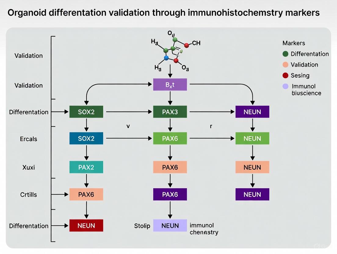
Validating Differentiation Through Immunohistochemistry Markers
Immunohistochemistry (IHC) serves as an essential tool for validating differentiation status and spatial organization within 3D cultures. The selection of appropriate markers depends on the specific tissue type being modeled and should include indicators of key cell populations, structural elements, and functional domains.
Table 3: Essential Immunohistochemistry Markers for Validating Organoid Differentiation
| Organoid Type | Key Cell Type | Primary Markers | Spatial Organization Cues | Functional Validation |
|---|---|---|---|---|
| Intestinal Organoids [12] [17] | Enterocytes | Villin, Sucrase-isomaltase (SI) | Crypt-villus architecture with basal nuclei | Alkaline phosphatase activity, absorption assays |
| Goblet cells | MUC2, TFF3 | Scattered distribution in epithelial layer | Mucin secretion, PAS staining | |
| Enteroendocrine cells | Chromogranin A, Synaptophysin | Individual cells interspersed in epithelium | Hormone secretion (serotonin, GLP-1) | |
| Paneth cells | Lysozyme, Defensin-5 | Localized to crypt bases | Antimicrobial activity assays | |
| Neuromuscular Organoids [18] | Spinal motor neurons | ISL1, HB9, ChAT | Cluster organization in neural regions | Calcium imaging, electrophysiology |
| Skeletal muscle | Myosin Heavy Chain, Desmin | Striated muscle fibers with peripheral nuclei | Contraction analysis, response to stimuli | |
| Schwann cells | S100B, GFAP | Association with neuromuscular junctions | Synaptic transmission studies | |
| Hepatic Organoids [11] | Hepatocytes | Albumin, HNF4α | Polarized epithelial structures | Albumin/urea secretion, CYP450 activity |
| Biliary cells | CK7, CK19 | Tubular structures | Anion transport assays | |
| Cerebral Organoids [11] | Neural progenitors | SOX2, Nestin | Ventricular-like zones | Proliferation assays (Ki67) |
| Neurons | TUJ1, MAP2 | Cortical plate-like organization | Calcium imaging, synaptic activity | |
| Astrocytes | GFAP, S100β | Distributed through outorganoid | Glutamate uptake assays |
The IHC protocol for 3D cultures requires specific adaptations compared to traditional tissue sections. Due to their size and density, organoids and spheroids require extended fixation times (typically 4-24 hours depending on size) and careful attention to permeabilization conditions to ensure adequate antibody penetration without compromising epitope integrity [12]. Clearing techniques such as CLARITY or iDISCO can enhance imaging depth in larger organoids, while confocal microscopy with z-stack acquisition enables comprehensive 3D reconstruction of the entire structure [18].
When interpreting IHC results, researchers should assess not only the presence of specific markers but also their spatial distribution and relationship to structural features. For example, in properly patterned intestinal organoids, proliferative cells (marked by Ki67) should localize to crypt-like domains, while differentiated absorptive and secretory cells should populate villus-like regions [12]. Similarly, in neuromuscular organoids, the precise apposition of presynaptic (SV2, synaptophysin) and postsynaptic (btungarotoxin receptors) markers at neuromuscular junctions confirms the establishment of functional connectivity [18].
Signaling Pathways Governing Self-Organization and Differentiation
The self-organization and differentiation processes in 3D cultures are orchestrated by complex signaling networks that recapitulate developmental programs. Understanding and manipulating these pathways is essential for directing organoid development and maturation.
Figure 2: Key Signaling Pathways in 3D Culture Patterning
The Wnt/β-catenin pathway serves as a master regulator of stem cell maintenance in many epithelial tissues, particularly in the intestine [10] [12]. Activation of Wnt signaling through agonists like R-spondin or CHIR99021 promotes the expansion of stem and progenitor populations, while inhibition drives differentiation. The BMP/TGF-β pathway often acts in opposition to Wnt signaling, promoting differentiation and suppressing stemness [10]. In intestinal organoid culture, the BMP antagonist Noggin is essential for maintaining the stem cell niche and supporting long-term expansion.
The FGF signaling pathway plays crucial roles in regional patterning and morphogenesis across multiple tissue types [10] [18]. In neural organoids, FGF signaling influences anterior-posterior patterning, while in intestinal models, FGF4 directs hindgut specification during differentiation from pluripotent stem cells. The Notch pathway functions as a key mediator of cell fate decisions through lateral inhibition, enabling the generation of diverse cell types from homogeneous progenitor populations [12].
Recent advances have identified additional factors that enhance organoid complexity and functionality. Epiregulin (EREG), an epidermal growth factor family member, has been shown to significantly enhance the differentiation of human intestinal organoids, promoting the simultaneous development of epithelium, mesenchyme, enteric neuroglial populations, endothelial cells, and organized smooth muscle in a single differentiation protocol [17]. This represents a significant advancement over traditional methods that typically require complex co-culture systems to achieve similar cellular diversity.
The Scientist's Toolkit: Essential Reagents and Technologies
Successful establishment and validation of self-organizing 3D cultures requires access to specialized reagents, matrices, and equipment. The following table summarizes key solutions and their applications in organoid technology.
Table 4: Essential Research Reagent Solutions for 3D Culture and Differentiation Studies
| Category | Specific Product/Technology | Function in 3D Culture | Application Examples |
|---|---|---|---|
| Extracellular Matrices | Matrigel | Basement membrane extract providing structural support and biological cues | Intestinal, gastric, hepatic organoid culture [12] [15] |
| Synthetic PEG-based hydrogels | Tunable scaffolds with defined mechanical properties and modular bioactivity | Customized microenvironments for mechanistic studies [15] | |
| Collagen I | Fibrillar matrix supporting cell migration and organization | Stromal co-cultures, invasion assays [15] | |
| Growth Factors & Morphogens | R-spondin-1 | Potent Wnt pathway agonist for stem cell maintenance | Intestinal, gastric, hepatic organoids [12] |
| Noggin | BMP antagonist supporting stem/progenitor cell expansion | Intestinal, cerebral organoids [12] | |
| EGF | Epithelial growth and survival factor | Virtually all epithelial organoid types [12] | |
| FGF4/FGF10 | Mesenchymal-epithelial signaling, branching morphogenesis | Intestinal differentiation, lung organoids [17] | |
| Epiregulin (EREG) | Enhanced co-differentiation of multiple lineages | Complex intestinal organoids with neural/vascular components [17] | |
| Cell Culture Platforms | Low-adhesion plates | Scaffold-free spheroid formation through forced aggregation | Tumor spheroids, MSC aggregates [11] [15] |
| Hanging drop plates | Gravity-enforced assembly of uniform spheroids | Developmental models, drug screening [11] | |
| Acoustic levitation chips | Contactless manipulation and culture of cell aggregates | MSC spheroid formation and culture [16] | |
| Microfluidic chips | Precise control over soluble gradients and mechanical forces | Organs-on-chips, vascularized models [11] [13] | |
| Validation Tools | Live-cell imaging systems | Dynamic monitoring of self-organization and functional responses | Contractility analysis, migration studies [16] [13] |
| Single-cell RNA sequencing | Resolution of cellular heterogeneity and lineage trajectories | Characterization of organoid development [18] | |
| Confocal microscopy | 3D reconstruction of spatial organization and marker expression | Immunohistochemistry validation [18] |
The field of 3D cell culture continues to evolve rapidly, with ongoing efforts focused on enhancing the physiological relevance, reproducibility, and scalability of self-organizing models. Key challenges that remain include improving vascularization to support long-term culture and growth of larger structures, enhancing cellular complexity through incorporation of immune, neural, and stromal components, and developing standardized protocols to reduce variability across laboratories [10] [12]. The integration of advanced bioengineering approaches such as 3D bioprinting, microfluidics, and smart biomaterials promises to address these limitations, enabling unprecedented control over the cellular microenvironment [11] [13].
As these technologies mature, self-organizing 3D cultures are poised to transform biomedical research by providing increasingly faithful models of human development, disease, and drug response. The continued refinement of differentiation protocols and validation methodologies, particularly through comprehensive immunohistochemical analysis, will further strengthen their utility as predictive platforms for both basic research and translational applications. By harnessing the innate capacity of cells to self-organize within precisely engineered microenvironments, scientists can unlock new opportunities to understand human biology and develop more effective therapeutics.
Selecting Lineage-Specific Markers for Different Organoid Types
The validation of cellular identity in organoids is a cornerstone of their application in developmental biology, disease modeling, and drug development. As three-dimensional structures that recapitulate the complexity of in vivo organs, confirming that organoids contain the correct cell types in proper proportions and maturation states is paramount. This guide objectively compares the performance of lineage-specific markers across various organoid types, providing a consolidated resource for researchers to select appropriate markers and methodologies for robust validation of their models. The data and protocols summarized herein are framed within the broader thesis that rigorous, multi-method validation is essential for generating biologically relevant organoid data.
Comparative Analysis of Organoid Markers
Central Nervous System Organoids
Table 1: Markers for Brain Organoid Differentiation and Validation
| Cell Type | Key Markers | Localization/Function | Temporal Dynamics | Validation Methods Cited |
|---|---|---|---|---|
| Radial Glia (vRG) | SOX2, Ki67 | Ventricular zone/neural rosettes; Progenitor proliferation | Significantly reduced from D35 to D50 [4] | Immunohistochemistry (IHC), Proteomics (LC-MS) [4] |
| Intermediate Progenitor Cells (IPCs) | PPP1R17 | Transitional progenitor state | Significantly decreased at D50 vs. D35 [4] | Immunohistochemistry [4] |
| Deep Layer Neurons | CTIP2 | Maturing neuronal populations | Significantly increased at D50 vs. D35 [4] | Immunohistochemistry [4] |
| Secretome Profile | Cell adhesion molecules, Synaptic proteins, Metalloproteases | Protein secretion during neurogenesis | Distinctly increased during peak neurogenesis (Day 35) [4] | Secretome analysis (LC-MS) [4] |
Experimental Protocol: Proteomic and Secretomic Analysis of Dorsal Forebrain Organoids (DFOs)
- Organoid Generation: DFOs were differentiated from three human iPSC lines (KOLF2.1J, BIONi010-C, HMGU1) according to established protocols [4].
- Time Points: Organoids were collected at days 20, 35, and 50 of differentiation for analysis [4].
- Sample Preparation: For proteomics, samples consisted of 3 pooled organoids per replicate. For secretome analysis, conditioned media was likely analyzed, though the specific preparation method is not detailed in the provided excerpt [4].
- Proteomics: Liquid chromatography-mass spectrometry (LC-MS) was performed to investigate the proteome, identifying 4,431 proteins [4].
- Data Analysis: Differential protein abundance analysis and dimensionality reduction (PCA) were used to identify distinct clustering based on differentiation day [4].
- Validation: Immunohistochemistry (IHC) was performed on organoids at D35 and D50 for markers including SOX2, PPP1R17, and CTIP2 to correlate with proteomic findings [4].
Pancreatic Organoids
Table 2: Markers for Human Pancreatic Organoid (hPO) Characterization
| Cell Type / State | Key Markers | Expression Level / Localization | Functional Notes | Validation Methods Cited |
|---|---|---|---|---|
| Ductal Identity | KRT19, EpCAM, SOX9, CLDN2 | Widespread expression across all major clusters [19] | Defines core epithelial phenotype | scRNA-seq, Immunofluorescence [19] |
| Proliferating Cells | MKI67, PCNA, CENPM, TOP2A | High in specific clusters (e.g., Clusters 3 & 4) [19] | Maintains organoid expansion in culture | scRNA-seq, Immunofluorescence [19] |
| Ductal Subpopulation 0 | MUC5AC, MUC5B, SPINK4, TFF1 | Upregulated in a specific ductal subcluster [19] | Mucin-related gene expression profile | scRNA-seq [19] |
| Excluded Cell Types | CPA1 (acinar), INS, GCG (endocrine) | Very low or not detected [19] | Confirms purity of ductal model | scRNA-seq [19] |
Experimental Protocol: scRNA-seq of Human Pancreatic Organoids
- Organoid Generation: hPOs were derived from human islet-depleted pancreatic tissue via mechanical dissociation and cultured in Matrigel with a defined medium supporting pancreatic epithelial growth [19].
- Quality Control: Organoids were assessed for genetic stability (karyotyping, γH2AX staining), senescence (β-galactosidase, SASP factors), and long-term expansion potential [19].
- Single-Cell Preparation: Organoids from three independent donors at passage 5 were prepared for sequencing [19].
- Sequencing & Analysis: scRNA-seq was performed using the 10X Genomics Chromium system. After quality filtering, 3,187 cells were analyzed. Clustering and differential gene expression analysis revealed distinct transcriptional populations [19].
- Validation: Immunofluorescence staining for key proteins like SOX9 and MKI67 was used to validate scRNA-seq findings at the protein level [19].
Cancer Organoids (Colorectal and Head & Neck)
Table 3: Markers for Cancer Organoid Validation and Drug Testing
| Organoid Type | Key Validation Markers | Purpose of Marker | Correlation with Clinical Response | Validation Methods Cited |
|---|---|---|---|---|
| Colorectal Cancer (CRC) PDOs | Pan-cytokeratin, CDX2, CK20, Ki67 | Histological similarity to original tumor; proliferation index | Strong correlation between PDO and clinical treatment response [20] | IHC, H&E Staining, DNA Sequencing [20] |
| Head and Neck Cancer (HNC) PDOs | (Tumor-retained DNA alterations) | Genomic fidelity to parent tumor | In vitro organoid response to radiotherapy mimicked clinical response [21] | DNA Sequencing, Radiotherapy/Chemotherapy Assays [21] |
Experimental Protocol: Establishing and Validating Patient-Derived Organoids (PDOs)
- Tissue Processing: CRC or HNC tumor tissue is washed, cut into small pieces, and enzymatically digested (e.g., with collagenase, dispase) into single cells or small fragments [20].
- 3D Culture: The digested tissue is embedded in a gel-based extracellular matrix (e.g., Matrigel) and cultured with an organoid-specific medium containing essential growth factors [20].
- Quality Control:
- Histology: H&E staining and IHC of organoids and original tumor are compared by a pathologist to confirm architectural and protein expression similarity [20].
- Genomics: Whole-exome or genome sequencing is performed on organoids and the parent tumor to confirm retention of mutational profiles and copy number alterations [20].
- Drug Screening: PDOs are exposed to a panel of therapies (e.g., chemotherapy, radiotherapy, targeted agents). Viability or growth is measured and compared to the patient's clinical response to the same treatments [21].
Organoid Generation and Validation Workflow
The following diagram illustrates the general workflow for generating, differentiating, and validating organoids, integrating key steps from the cited protocols.
The Scientist's Toolkit: Essential Research Reagents
Table 4: Key Reagents for Organoid Culture and Validation
| Reagent / Material | Function / Application | Examples from Literature |
|---|---|---|
| Basement Membrane Matrix | Provides a 3D scaffold for organoid growth and polarization. | Matrigel is used for embedding pancreatic [19], brain [4], and cancer [20] organoids. |
| Lineage-Specific Media | Directs differentiation and maintains cell viability through defined factors. | Dorsal Forebrain Organoid medium [4]; Organoid-specific media with WNT, R-spondin for CRC PDOs [20]. |
| Antibodies for IHC/IF | Visualizes protein expression and localization of lineage markers. | Anti-SOX2, CTIP2 for brain organoids [4]; Anti-KRT19, SOX9 for pancreatic organoids [19]. |
| Dissociation Enzymes | Breaks down tissue for initial organoid culture and passaging. | Collagenase, dispase, TrypLE Express for CRC PDOs [20]; StemPro Accutase for brain organoids [22]. |
| scRNA-seq Kits | Profiles transcriptional landscape and cellular heterogeneity. | 10X Genomics Chromium system for pancreatic organoid analysis [19]. |
| LC-MS Instrumentation | Quantifies global protein expression (proteome) and secreted factors (secretome). | Used to analyze proteome/secretome dynamics in brain organoids [4]. |
The selection of lineage-specific markers is not a one-size-fits-all process but depends heavily on the organoid type, the specific cell populations of interest, and the developmental or disease context. As demonstrated by the comparative data, a multi-faceted validation strategy combining IHC with genomic, proteomic, and functional analyses is critical for building confidence in organoid models. The continued refinement of these markers and protocols, aided by resources like organoid databases [23] and standardized classifiers [24], will be essential for advancing organoid technology toward its full potential in basic research and clinical translation.
The advent of human neural organoid technology has provided researchers with an unprecedented in vitro model to study human brain development, disease mechanisms, and therapeutic interventions. These three-dimensional, self-organizing structures derived from pluripotent stem cells recapitulate key aspects of the embryonic brain's cellular diversity and cytoarchitecture [25]. However, the utility of these models depends entirely on the rigorous validation of their cellular composition and developmental progression. Immunohistochemistry (IHC) serves as an indispensable tool in this validation process, enabling researchers to confirm the presence, abundance, and spatial organization of specific progenitor and neuronal populations.
Among the extensive array of neural markers, four have emerged as particularly critical for assessing organoid fidelity: SOX2 and PAX6 for progenitor populations, and CTIP2 and Ki67 for evaluating neuronal differentiation and proliferative activity. These markers provide a multifaceted view of organoid development, from the maintenance of neural stem cell niches to the establishment of layered cortical structures. This guide systematically compares the application of these markers across different organoid protocols, providing researchers with experimental data and methodological frameworks to validate their own organoid differentiation systems.
Marker Functions and Expression Patterns in Neural Development
Progenitor Cell Markers
SOX2 is a transcription factor essential for maintaining neural progenitor cell (NPC) pluripotency and self-renewal. It is prominently expressed in the ventricular zone (VZ) of both the developing brain and neural organoids, where it marks radial glial cells (RGCs) – the primary neural stem cells [26]. In organoid models, SOX2+ cells typically form organized ventricular-like structures with apicobasal polarity, recapitulating the embryonic neuroepithelium [27]. The persistence and proper organization of SOX2+ progenitor zones are critical indicators of sustained neurogenic capacity in long-term organoid cultures.
PAX6 is a paired-box transcription factor fundamental for dorsal forebrain patterning and neurogenesis. It is expressed in RGCs of the dorsal telencephalon and plays a crucial role in specifying cortical neuronal fates [27]. In organoids, PAX6 expression confirms the establishment of dorsal cortical identity and is particularly abundant in the ventricular and subventricular zones [27]. The presence of PAX6+ progenitors is a key metric for evaluating the regional specificity of guided cortical organoid protocols.
Neuronal and Proliferation Markers
CTIP2 (encoded by BCL11B) is a zinc-finger transcription factor that specifies subcortical projection neurons of the deep cortical layers (V and VI) [27] [28]. During development, CTIP2+ neurons are born early and occupy deep positions in the cortical plate, following the inside-out pattern of corticogenesis. In organoids, the emergence of CTIP2+ neurons in appropriate spatial arrangements indicates the progression of cortical layer formation and neuronal maturation [27]. The presence of properly organized CTIP2+ populations is a hallmark of advanced organoid models that recapitulate later stages of cortical development.
Ki67 is a nuclear protein associated with cellular proliferation that is present during all active phases of the cell cycle but absent in quiescent cells. It serves as a robust marker for identifying dividing progenitor populations in both the VZ and outer subventricular zone (oSVZ) of organoids [29] [26]. Quantifying Ki67+ cells provides insights into the proliferative capacity and growth dynamics of organoids, with abnormalities often indicating developmental defects or disease-specific phenotypes.
Table 1: Key Markers for Validating Neural Organoid Development
| Marker | Cell Type | Expression Pattern | Developmental Significance |
|---|---|---|---|
| SOX2 | Neural progenitor cells (Radial glia) | Nuclear expression in ventricular zone | Maintains progenitor pool; essential for self-renewal |
| PAX6 | Dorsal forebrain progenitors | Nuclear expression in VZ/SVZ | Specifies cortical identity; regulates neurogenesis |
| CTIP2 | Deep layer cortical neurons (Layers V-VI) | Nuclear expression in cortical plate | Specifies subcortical projection neuron identity |
| Ki67 | Proliferating cells | Nuclear expression in cycling cells | Marks active cell division in progenitor zones |
Comparative Analysis of Marker Expression Across Organoid Protocols
Sliced Neocortical Organoids (SNOs) for Enhanced Maturation
The sliced neocortical organoid (SNO) system represents a significant advancement for modeling late-stage cortical development by overcoming the diffusion limit that plagues traditional 3D organoid cultures [29]. This method involves precisely sectioning day-45 forebrain organoids into 500-μm thick slices using a vibratome, which are then maintained in long-term culture with periodic reslicing every 4 weeks [29]. This approach dramatically reduces interior hypoxia and cell death, enabling sustained neurogenesis over extended periods.
In SNOs, immunohistochemical analyses reveal remarkable preservation of progenitor zones, with abundant SOX2+ radial glial cells maintaining organized ventricular-like structures even at day 150 [29]. These cultures show persistent KI67+ proliferative activity and TBR2+ intermediate progenitor populations in expanded outer subventricular zone-like regions, indicating maintained neurogenic capacity [29]. Most notably, SNOs demonstrate progressive expansion of the cortical plate with establishment of distinct upper and deep cortical layers, evidenced by the appropriate segregation of CTIP2+ deep-layer neurons [29]. This advanced laminar organization, which resembles the third-trimester embryonic human neocortex, represents a significant improvement over conventional organoid models where cortical layer separation is often rudimentary or inconsistent.
Astrocyte-Conditioned Medium Treated Organoids (MACMOs)
An alternative approach to enhance organoid maturation involves supplementing cultures with astrocyte-conditioned medium (ACM) to create MACMOs [27]. This method leverages astrocyte-secreted factors that naturally promote neuronal maturation in vivo. When applied to forebrain organoids, ACM treatment accelerates neuronal differentiation and leads to an enlarged neuronal layer with overproduction of CTIP2+ deep-layer cortical neurons [27].
Immunohistochemical characterization of MACMOs reveals typical dorsal forebrain patterning with appropriate PAX6+ and FOXG1+ regional identity [27]. These organoids develop organized ventricular-like zones containing SOX2+ radial glial cells and TBR2+ intermediate progenitors, similar to control organoids [27]. However, the significant thickening of the neuronal layer and preferential increase in CTIP2+ populations demonstrates ACM's specific effect on promoting deep-layer neuron generation [27]. Electrophysiological assessments confirm that these morphological changes correspond to enhanced functional maturation, with MACMOs showing significantly improved neuronal network activity [27].
Williams Syndrome Forebrain Organoids for Disease Modeling
Forebrain organoids generated from Williams Syndrome (WS) patient-derived iPSCs reveal disease-specific alterations in marker expression patterns [26]. WS organoids exhibit abnormal neural progenitor dynamics, with significantly increased proportions of KI67+/SOX2+ proliferating progenitors compared to controls [26]. This is coupled with disrupted cell cycle exit, as evidenced by reduced numbers of KI67-/EdU+ cells after a 24-hour EdU pulse [26].
Further immunohistochemical analysis reveals fewer TBR2+ intermediate progenitor cells localized to the MAP2- ventricular zone-like layer in WS organoids [26]. The abnormal progenitor behavior leads to subsequent deficits in neuronal differentiation, with WS organoids showing reduced DCX+ immature neurons and aberrant relative thickening of the SOX2+ ventricular zone at the expense of the cortical plate [26]. These marker expression abnormalities provide quantifiable metrics of the neurodevelopmental deficits in WS and demonstrate how organoid models can capture disease-specific phenotypes.
Table 2: Quantitative Comparison of Marker Expression Across Organoid Protocols
| Protocol | SOX2+ Progenitors | PAX6+ Dorsal Progenitors | CTIP2+ Deep Neurons | KI67+ Proliferating Cells | Key Findings |
|---|---|---|---|---|---|
| Sliced Neocortical Organoids (SNOs) [29] | Sustained in organized VZ through day 150 | Maintained dorsal identity | Form distinct deep layers in expanded CP | Sustained proliferation in oSVZ-like regions | Enables late-stage cortical development with layer separation |
| ACM-Treated Organoids (MACMOs) [27] | Normal VZ organization | Preserved dorsal patterning | Significantly increased populations | Standard proliferation | Enhanced neuronal layer thickness and maturation |
| Williams Syndrome Organoids [26] | Increased percentage in VZ | Not specifically reported | Not specifically quantified | Increased proliferation; reduced cell cycle exit | Aberrant NPC dynamics and neurogenesis deficits |
| Standard Forebrain Organoids [27] | Present in VZ structures | Confirm dorsal forebrain identity | Present but limited layer separation | Normal early proliferation | Baseline protocol for comparison |
Experimental Protocols for Organoid Immunohistochemistry
Organoid Generation and Sectioning
For standard forebrain organoid generation, begin with human pluripotent stem cells (hPSCs) and form embryoid bodies in ultra-low attachment 96-well plates [27]. Employ dual SMAD and WNT inhibition strategies to enhance cortical identity, typically using SB431542 (TGF-β inhibitor), LDN-193189 (BMP inhibitor), and XAV939 (WNT inhibitor) during the first 2-3 weeks of differentiation [27]. Transfer organoids to orbital shakers or spinning bioreactors after 5-7 days to improve nutrient exchange.
For the SNO protocol, at day 45, embed organoids in low-melting-point agarose and section into 500-μm thick slices using a vibratome [29]. Collect slices that dissociate spontaneously or with gentle pipetting, then transfer to 6-well plates for culture on an orbital shaker. Repeat slicing every 4 weeks to maintain optimal thickness for diffusion [29].
For ACM treatment, prepare conditioned medium from primary mouse or human astrocytes by collecting serum-free culture supernatant after 48 hours of conditioning [27]. Supplement differentiation media with 25-50% ACM from day 20 onward to promote neuronal maturation.
Immunohistochemistry and Imaging
Fix organoids or organoid slices in 4% paraformaldehyde for 30-60 minutes at room temperature, followed by cryoprotection in 30% sucrose overnight. Embed in OCT compound and section at 10-20 μm thickness using a cryostat [26].
Perform standard immunofluorescence with the following primary antibody incubations (typically overnight at 4°C):
- SOX2 (1:200-1:500, rabbit monoclonal)
- PAX6 (1:200-1:500, mouse monoclonal)
- CTIP2 (1:200-1:500, rat monoclonal)
- Ki67 (1:200-1:500, rabbit monoclonal)
Use appropriate species-specific secondary antibodies conjugated to Alexa Fluor dyes (1:500-1:1000) for detection. Counterstain with DAPI for nuclear visualization [26].
For quantitative analysis, image multiple regions from at least 3-5 organoids per condition using confocal microscopy. Calculate marker-positive cell percentages by counting positive cells relative to total DAPI+ cells in defined regions of interest using ImageJ or similar software [26]. For layer thickness measurements, take multiple perpendicular measurements across organoid sections from the ventricular surface to the pial surface.
Signaling Pathways Regulating Marker Expression
The expression of these key markers is regulated by specific signaling pathways that can be manipulated in organoid systems to direct developmental outcomes. The WNT/β-catenin signaling pathway plays a particularly important role in regulating human cortical neuron subtype fate specification, as demonstrated in SNO systems [29]. Modulation of this pathway can influence the balance of deep versus upper layer neurons, directly affecting CTIP2 expression patterns.
In Williams Syndrome organoids, disrupted GTF2IRD1-TTR-ERK signaling leads to abnormal neurogenesis, affecting both SOX2+ progenitor dynamics and subsequent neuronal differentiation [26]. The GTF2IRD1 transcription factor directly binds to the transthyretin (TTR) promoter, and its deficiency in WS reduces TTR expression, subsequently impairing ERK signaling activation [26]. This pathway disruption ultimately contributes to the observed imbalances in KI67+ proliferating progenitors and their differentiation into cortical neurons.
Astrocyte-secreted factors in ACM-treated organoids promote neuronal maturation and specifically enhance CTIP2+ deep-layer neuron generation through mechanisms that may involve enhanced ERK signaling and metabolic support via lipid droplet accumulation [27]. These findings highlight how extrinsic cues can intrinsically influence marker expression and cellular fate decisions in organoid models.
Diagram 1: Signaling pathways regulating key marker expression in neural organoids. Three major pathways (WNT/β-catenin in yellow, GTF2IRD1-TTR-ERK in red, and astrocyte-mediated in blue) influence the expression of progenitor and neuronal markers (green).
The Scientist's Toolkit: Essential Research Reagents
Table 3: Essential Research Reagents for Neural Organoid Studies
| Reagent/Category | Specific Examples | Function in Organoid Research |
|---|---|---|
| Stem Cell Lines | H1, H9, DYR, DXR hPSC lines [27]; Patient-derived iPSCs [26] | Provide genetically defined starting material for organoid generation and disease modeling |
| Patterning Molecules | SB431542 (TGF-β inhibitor), LDN-193189 (BMP inhibitor), XAV939 (WNT inhibitor) [27] | Direct regional specification toward dorsal forebrain fate during early differentiation |
| Extracellular Matrices | Matrigel, Geltrex, Brain Extracellular Matrix (BEM) [30] | Provide structural support and biochemical cues for 3D organization and neural differentiation |
| Culture Systems | Ultra-low attachment plates, Orbital shakers, Spinning bioreactors, Microfluidic devices [29] [30] | Enable proper organoid formation, nutrient exchange, and reduce hypoxia |
| Critical Antibodies | SOX2, PAX6, CTIP2, Ki67, TBR2, SATB2, TUJ1, MAP2 [29] [27] [26] | Validate cellular composition, regional identity, and developmental progression via IHC |
| Cell Tracking Reagents | EdU (5-ethynyl-2'-deoxyuridine) [29] [26] | Label and track proliferating cells and their progeny in pulse-chase experiments |
| Functional Assay Tools | Calcium indicators, Multi-electrode arrays (MEAs) [27] | Assess neuronal maturation and network activity in live organoids |
The comprehensive analysis of SOX2, PAX6, CTIP2, and Ki67 expression provides critical insights into the developmental fidelity and functional capacity of neural organoid models. The comparative data presented in this guide demonstrates that protocol selection significantly impacts marker expression patterns and ultimately determines the utility of organoids for specific research applications. Sliced neocortical organoids excel at modeling late cortical development with distinct layer formation, while ACM-treated organoids offer enhanced neuronal maturation, and disease-specific organoids reveal pathogenetic alterations in neurodevelopment.
Researchers should select validation markers and interpretation frameworks based on their specific experimental goals. For studies focused on progenitor biology, SOX2 and Ki67 quantification provides essential metrics of stem cell maintenance and proliferation. For investigations of cortical patterning and layer specification, PAX6 and CTIP2 offer robust indicators of regional identity and neuronal differentiation. As organoid technology continues to evolve, these fundamental markers will remain indispensable tools for benchmarking protocol improvements and ensuring the physiological relevance of these revolutionary human brain models.
Intestinal organoids have emerged as a transformative in vitro model that recapitulates the cellular diversity and function of the intestinal epithelium. These self-organizing three-dimensional structures contain stem cells, progenitor cells, and all major differentiated epithelial lineages found in the native intestine [31]. The validation of intestinal organoid differentiation states relies heavily on the detection of key molecular markers: LGR5 for stem cells, MUC2 for goblet cells, CHGA for enteroendocrine cells, and LYZ for Paneth cells [32] [31] [33]. This guide provides an objective comparison of experimental approaches for generating and analyzing these epithelial lineages, supported by quantitative data and detailed methodologies to assist researchers in selecting appropriate protocols for their specific applications.
Marker Expression Profiles Across Culture Conditions
The expression of lineage markers in intestinal organoids is highly dependent on culture conditions, particularly the combination of signaling pathway modulators used. The tables below summarize quantitative data on marker expression across different experimental setups.
Table 1: Marker Expression in Mouse Intestinal Organoids Under Different Culture Conditions
| Culture Condition | LGR5+ Stem Cells | LYZ+ Paneth Cells | MUC2+ Goblet Cells | CHGA+ Enteroendocrine Cells | Reference |
|---|---|---|---|---|---|
| ENR (EGF, Noggin, R-spondin1) | Present (in crypt domains) | Present | Present | Present | [34] |
| ENR + CHIR99021 (C) | Increased percentage & intensity | Reduced | Not specified | Not specified | [34] |
| ENR + Valproic Acid (V) | Markedly increased | Not specified | Not specified | Not specified | [34] |
| ENR + CHIR + VPA (CV) | >97% GFP+ cells | Few observed | Not observed | Not observed | [34] |
| CHIR + LDN (2ki) | Higher than ENR | Similar to ENR | Similar to ENR | Similar to ENR | [35] |
Table 2: Marker Expression in Human Intestinal Organoids Under Different Culture Conditions
| Culture Condition | LGR5+ Stem Cells | LYZ+ Paneth Cells | MUC2+ Goblet Cells | CHGA+ Enteroendocrine Cells | Reference |
|---|---|---|---|---|---|
| IF Culture Condition | Minimal LGR5 expression | Rare or absent | Present | Present | [33] |
| IL-22 Patterning | Minimal LGR5 expression | Induced generation | Present | Present | [33] |
| TpC Condition | Scattered mNeonGreen+ | DEFA5+ and LYZ+ | MUC2+ | CHGA+ | [33] |
| Proliferative (OGM) | High | Low | Low | Low | [36] |
| Differentiated (ODM) | Low | High | High | High | [36] |
Experimental Protocols for Organoid Differentiation and Analysis
Growth Factor-Free Mouse Organoid Culture
Purpose: To maintain Lgr5+ intestinal stem cells without exogenous growth factors through direct modulation of Wnt and BMP signaling pathways [35].
Methodology:
- Isolate intestinal crypts from Lgr5-EGFP-IRES-creERT2 mice
- Embed crypts in Matrigel and culture with 10 μM CHIR99021 (GSK3 inhibitor) and 0.2 μM LDN-193189 (BMP type I receptor inhibitor)
- Refresh culture medium every 2-3 days
- Passage organoids every 7-10 days by dissociating with TrypLE Express Enzyme
- For differentiation assays, fix organoids and immunostain for lineage markers
Key Observations: This 2ki system maintains Lgr5+ ISCs with similar efficiency to conventional ENR culture while preserving differentiation capacity toward secretory lineages (Paneth, goblet, and enteroendocrine cells) though enterocyte differentiation is attenuated [35].
Enhanced Stemness Human Organoid Culture
Purpose: To enhance LGR5+ stem cell population while maintaining differentiation potential in human small intestinal organoids (hSIOs) [33].
Methodology:
- Generate LGR5-mNeonGreen reporter hSIOs using CRISPR-Cas9 technology
- Culture in basal condition containing EGF, Noggin (or DMH1), R-Spondin1, CHIR99021, A83-01, IGF-1, and FGF-2
- Add TpC combination: Trichostatin A (HDAC inhibitor), 2-phospho-L-ascorbic acid (Vitamin C), and CP673451 (PDGFR inhibitor)
- For single-cell cloning, dissociate organoids to single cells and plate in Matrigel
- Culture for 7-21 days, monitoring LGR5-mNeonGreen expression and budding structures
- Fix and immunostain for differentiation markers at various time points
Key Observations: The TpC condition supports simultaneous self-renewal and differentiation, generating organoids with LGR5+ stem cells, ALPI+ enterocytes, MUC2+ goblet cells, CHGA+ enteroendocrine cells, and DEFA5+/LYZ+ Paneth cells [33].
Transit-Amplifying Cell Modulation Protocol
Purpose: To investigate how transit-amplifying (TA) cell proliferation influences secretory vs. absorptive cell fate decisions [32].
Methodology:
- Establish enteroid monolayers from jejunal intestinal crypts at 10-20% initial confluency
- Apply combinatorial perturbations across eight epithelial signaling pathways (Wnt, BMP, Notch, HDAC, JAK, p38 MAPK, TGF-β, EGFR) using 13 modulators individually and in 78 pairwise combinations
- Culture for 48 hours with EdU labeling to identify proliferating cells
- Fix and immunostain for Lgr5 (stem), EdU (proliferating), Lyz (Paneth), Muc2 (goblet), and ChgA (enteroendocrine) cells
- Quantify cell types using automated image analysis algorithms
- Validate key findings in 3D organoids and in vivo models
Key Observations: Modulating proliferation of transit-amplifying cells changes the ratio of differentiated secretory to absorptive cell types, highlighting an underappreciated role for TA cells in tuning differentiated cell type composition [32].
Signaling Pathways Controlling Lineage Specification
The differentiation of intestinal epithelial lineages is coordinated through interconnected signaling pathways. The diagram below illustrates the key pathways and their modulation in organoid culture systems.
Diagram 1: Signaling pathways controlling intestinal epithelial lineage specification and pharmacological modulators used in organoid cultures.
The Wnt/β-catenin pathway is essential for maintaining LGR5+ stem cells and promoting Paneth cell differentiation [35] [37]. BMP signaling inhibition is required for stem cell maintenance, while Notch signaling promotes stem cell self-renewal and enterocyte differentiation at the expense of secretory lineages [35] [34]. EGFR signaling supports proliferation but is dispensable for stem cell maintenance in some contexts [35].
The Scientist's Toolkit: Essential Research Reagents
Table 3: Essential Reagents for Intestinal Organoid Research
| Reagent Category | Specific Examples | Function in Organoid Culture |
|---|---|---|
| Wnt Pathway Agonists | CHIR99021, R-spondin1, Wnt3a | Activates β-catenin signaling to maintain stemness and promote proliferation [35] [34] |
| BMP Inhibitors | LDN-193189, Noggin, DMH1 | Blocks BMP signaling to support stem cell maintenance [35] [33] |
| Notch Modulators | Valproic Acid, DAPT | HDAC inhibitors activate Notch; γ-secretase inhibitors block Notch to alter lineage specification [32] [34] |
| EGFR Agonists | EGF, EPIREGULIN (EREG) | Promotes epithelial proliferation and enhances differentiation complexity [35] [17] |
| Differentiation Inducers | IL-22, Notch inhibitors | Promotes Paneth cell generation and secretory lineage differentiation [33] |
| Epithelial Markers | Anti-LGR5, Anti-MUC2, Anti-CHGA, Anti-LYZ | Immunohistochemical validation of stem and differentiated cell populations [32] [31] [33] |
| Dissociation Enzymes | TrypLE Express, Dispose | Gentle dissociation of organoids to single cells for passaging or cloning [33] [36] |
Comparative Analysis of Differentiation Efficiency
Stem Cell Maintenance Capacity
The efficiency of LGR5+ stem cell maintenance varies significantly across culture systems. The small molecule combination CHIR99021 and valproic acid (CV) supports nearly homogeneous cultures of mouse Lgr5+ intestinal stem cells with >97% GFP+ cells and colony-forming efficiency of 25-40% from single cells [34]. Similarly, the growth factor-free 2ki system (CHIR99021 + LDN-193189) maintains Lgr5+ ISCs long-term with normal karyotype after 20 passages [35]. In human systems, the TpC condition significantly increases the proportion of LGR5-mNeonGreen+ cells and colony-forming efficiency from dissociated single cells while maintaining multi-lineage differentiation capacity [33].
Secretory Lineage Differentiation Potential
The balance between stem cell maintenance and secretory lineage differentiation represents a key challenge in organoid culture optimization. Conventional ENR mouse organoid cultures generate all intestinal epithelial cell types but maintain stem cells only in crypt-like domains [34]. The CV condition maximizes stemness but largely eliminates secretory differentiation [34]. In contrast, the 2ki system maintains normal differentiation toward secretory cells (Paneth, goblet, enteroendocrine) while attenuating enterocyte differentiation [35]. Human TpC organoids achieve both high stemness and robust secretory differentiation, generating MUC2+ goblet cells, CHGA+ enteroendocrine cells, and DEFA5+/LYZ+ Paneth cells within the same culture [33].
Applications in Disease Modeling and Toxicity Testing
The differentiation state of intestinal organoids significantly impacts their application in disease modeling and toxicology. Proliferative organoids (maintained in OGM) are more susceptible to anti-proliferative compounds like chemotherapeutic agents, while differentiated organoids (in ODM) better model villus functionality and show different toxicity profiles [36]. This highlights the importance of selecting culture conditions matched to specific research applications, particularly for predictive toxicology studies where cellular composition dramatically influences compound sensitivity.
The selection of intestinal organoid culture systems should be guided by specific research objectives, weighing the balance between stem cell expansion capacity and lineage differentiation fidelity. For high-throughput screening of stem cell-targeting compounds, conditions like CV or 2ki that maximize LGR5+ populations offer significant advantages. For physiological modeling of intestinal function or toxicology studies, systems like TpC that maintain both stemness and differentiation capacity provide more comprehensive representation of intestinal epithelium. The validation of these systems through rigorous assessment of LGR5, MUC2, CHGA, and LYZ expression remains essential for ensuring physiological relevance and experimental reproducibility.
The emergence of human pluripotent stem cell (hPSC)-derived kidney organoids represents a transformative advance in nephrology, offering unprecedented opportunities for modeling renal development, disease, and drug toxicity [38] [39]. These complex three-dimensional structures contain multilineage nephrogenic progenitor cells that recapitulate aspects of kidney development in vitro [40]. However, the utility of these organoids for research and clinical applications depends entirely on the faithful recreation of authentic nephron segments found in human kidneys. Segment-specific nephron markers thus become essential tools for validating differentiation efficiency, assessing reproducibility, and quantifying organoid quality [41].
The nephron, the kidney's functional unit, comprises distinct segments with specialized functions—from the blood-filtering podocytes to the various tubular sections responsible for reabsorption and secretion. Current kidney organoid models primarily generate structures resembling the proximal nephron, including podocytes, proximal tubules, and distal tubules, arranged in appropriate segmental order [41]. This review provides a comprehensive comparison of markers used to identify these segments in kidney organoids, supported by experimental data and detailed methodologies, to establish a standardized framework for organoid validation within the broader context of differentiation quality control.
Nephron Progenitor Cells: The Foundation of Organoid Development
The generation of kidney organoids begins with the directed differentiation of hPSCs into multipotent nephron progenitor cells (NPCs), which serve as the foundation for all subsequent nephron segments. Efficient induction of these precursors is critical for high-quality organoid formation.
Markers of Nephron Progenitor Cells
NPCs are characterized by the co-expression of transcription factors SIX2, SALL1, WT1, and PAX2 [38]. The transition from primitive streak to posterior intermediate mesoderm (IM) represents a crucial developmental juncture, with proper BMP4 signaling modulation being essential for generating metanephric NPCs rather than lateral plate mesoderm [38].
Table 1: Key Markers for Nephron Progenitor Cells and Early Lineages
| Cell Population | Key Markers | Expression Pattern | Functional Significance |
|---|---|---|---|
| Nephron Progenitor Cells (NPCs) | SIX2, SALL1, WT1, PAX2 | Nuclear (transcription factors) | Multipotent progenitors capable of generating all nephron epithelial segments except collecting ducts [38] |
| Posterior Intermediate Mesoderm | WT1, HOXD11 | Cytoplasmic/nuclear | Precursor population giving rise to metanephric mesenchyme [38] |
| Renal Vesicle | PAX8, LHX1 | Nuclear | Earliest epithelial structure committed to nephron formation [38] |
| Early Nephron patterning | JAG1, HNF1B | Membrane (JAG1), Nuclear (HNF1B) | Defines emerging proximal-distal axis in developing nephrons [42] |
Experimental Protocol for NPC Differentiation
The foundational protocol for efficient NPC generation involves a carefully timed sequence of signaling pathway manipulations:
Primitive Streak Induction: Treat hPSCs with high-dose CHIR99021 (8-10 µM), a GSK-3β inhibitor that activates Wnt signaling, for 4 days to induce T+TBX6+ primitive streak cells [38].
Posterior IM Specification: Subsequent treatment with activin (10 ng/mL) between days 4-7 generates WT1+HOXD11+ posterior IM with approximately 90% efficiency [38].
NPC Induction: Treat posterior IM cells with FGF9 (10 ng/mL) from day 7 to generate SIX2+SALL1+WT1+PAX2+ NPCs [38].
Critical protocol variations include adding low-dose noggin (5 ng/mL, a BMP antagonist) during primitive streak induction for cell lines with high endogenous BMP signaling, which suppresses lateral plate mesoderm formation (marked by FOXF1) and enhances posterior IM specification [38].
Segment-Specific Markers in Mature Kidney Organoids
Mature kidney organoids contain epithelial structures representing the major nephron segments found in vivo. The proper arrangement of these segments—podocytes, proximal tubules, loops of Henle, and distal tubules—in continuous, contiguous organizations represents a key quality metric [38] [41].
Comprehensive Marker Table for Nephron Segments
Table 2: Segment-Specific Markers in Kidney Organoids
| Nephron Segment | Key Markers | Localization | Morphological Features | Validation in Organoids |
|---|---|---|---|---|
| Podocytes | NPHS1 (nephrin), SYNPO (synaptopodin), PODXL (podocalyxin), WT1 | Peripheral structures, bulbous morphology | Complex cellular processes, basement membrane contact | Form glomerulus-like structures with filtration slit-like structures [39] [41] |
| Proximal Tubule | LTL (Lotus tetragonolobus lectin), CUBN (cubilin), LRP2 (megalin) | Middle segment, straight tubular morphology | Brush border microvilli, endocytic capacity | Active transport of dextran and organic anions [39] [41] |
| Loop of Henle | SLC12A1 (Na-K-Cl cotransporter) | Between proximal and distal segments | Simple cuboidal epithelium | Limited differentiation in most protocols; often missing distinct markers like SLC12A3 [39] |
| Distal Tubule | ECAD (E-cadherin), CDH1, GATA3 | Central region, branching tubular morphology | Tight junctions, hormone responsiveness | Response to cAMP stimulation with fluid swelling [39] [41] |
| Collecting Duct | AQP2 (aquaporin 2), GATA3 | Not typically present in standard organoids | Principal and intercalated cells | Generally absent in most hPSC-derived organoids [39] [41] |
Experimental Validation of Segment Identity
Beyond marker expression, functional validation strengthens segment identification:
- Podocyte Function: Formation of slit diaphragm-like structures between foot processes; evidence of limited filtration capacity in engrafted models [41].
- Proximal Tubule Function: Active transport of molecules including dextran and organic anions; expression of solute carriers (SLC family); susceptibility to nephrotoxins like cisplatin [42] [41].
- Distal Tubule Function: Response to hormonal stimulation (e.g., cAMP-mediated fluid transport) [41].
Signaling Pathways Controlling Nephron Patterning
The development of distinct nephron segments is controlled by evolutionarily conserved signaling pathways that can be manipulated to enhance organoid differentiation.
Figure 1: Signaling pathways controlling nephron segment patterning in kidney organoids. Note how targeted PI3K inhibition can enhance proximal tubule differentiation.
Advanced Differentiation Strategies for Segment Enhancement
Recent advances have enabled the generation of organoids with enhanced specific segments, particularly proximal tubules, which are crucial for drug toxicity testing and disease modeling.
Proximal-Biased Organoid Differentiation
A 2025 study demonstrated that transient PI3K inhibition during early nephrogenesis activates Notch signaling, shifting nephron axial differentiation toward proximal tubule precursors [42]. This protocol generates "proximal-biased" organoids with expanded expression of HNF4A+ proximal precursors that mature into functional proximal convoluted tubule cells expressing solute carriers, including organic cation and anion transporters [42].
The differentiation timeline for proximal-biased organoids includes:
- Days 0-4: Primitive streak induction with CHIR99021
- Days 4-7: Posterior intermediate mesoderm specification
- Days 7-12: Nephron progenitor expansion with FGF9
- Days 12-14: PI3K inhibitor treatment during renal vesicle stage
- Days 14-29: Maturation with continued FGF9
This approach generates organoids with enhanced functional proximal tubules capable of transporter-mediated uptake and more robust injury responses to nephrotoxins [42].
Distal Nephron and Collecting Duct Models
While standard kidney organoids lack authentic collecting ducts, alternative models have been developed:
- Tubuloid Differentiation: Adult kidney tissue-derived or urine-derived tubuloids can be differentiated to model the distal nephron and collecting ducts, expressing functional NKCC2 and ENaC capable of diuretic-inhibitable electrolyte uptake [43].
- UB Organoid Protocols: Separate differentiation strategies generate ureteric bud-like organoids that express collecting duct markers, though these are less established than proximal nephron protocols [41].
Quality Control and Validation Standards
Rigorous quality control is essential for generating reproducible, high-quality kidney organoids suitable for research and drug screening applications.
Essential Quality Control Measures
- Segmental Organization Verification: Confirm proper proximal-to-distal arrangement of segments (podocytes → proximal tubule → distal tubule) within individual nephron-like structures [41].
- Multiple Marker Validation: Use at least two specific markers per segment with appropriate cellular localization [41].
- Batch-to-Batch Assessment: Include internal controls across different batches and cell lines to account for variability [39] [41].
- Functional Assays: Complement marker expression with functional tests such as transport assays or injury response evaluations [41].
Common Artifacts and Off-Target Cells
Kidney organoids frequently contain non-renal cell types that require monitoring:
- Neuronal Cells: SOX2+ neuronal precursors and STMN2+ neuron-like cells [39]
- Muscle Cells: MYOG+ muscle-like cells [39]
- Melanocyte-like Cells: PMEL+ cells [39]
- Cartilage-like Cells: Mesenchymal cells expressing ACAN, SOX9, and other cartilage markers [39]
Single-cell RNA sequencing has revealed that transplantation of organoids under the mouse kidney capsule can diminish these off-target populations, enhancing renal identity [39].
The Scientist's Toolkit: Essential Research Reagents
Table 3: Key Reagents for Kidney Organoid Research
| Reagent Category | Specific Examples | Function in Organoid Differentiation | Application Notes |
|---|---|---|---|
| Wnt Pathway Agonists | CHIR99021 (8-12 µM) | Induces primitive streak and posterior IM; enhances nephron formation [38] [44] | Concentration critical and cell line-dependent [44] |
| Growth Factors | FGF9 (10 ng/mL), Activin A (10 ng/mL) | Promotes NPC induction and posterior IM specification [38] | Lower FGF9 concentrations sufficient for NPC induction [38] |
| Signaling Inhibitors | PI3K inhibitors (e.g., LY294002), BMP inhibitors (Noggin) | Enhances proximal tubule differentiation; modulates BMP signaling for proper IM patterning [38] [42] | Timing critical for proximal enhancement [42] |
| Extracellular Matrices | GelTrex, Matrigel | Provides substrate for 2D differentiation or 3D embedding | Concentration affects organoid formation efficiency [40] |
| Detection Reagents | LTL, Anti-NPHS1, Anti-ECAD, Anti-SYNPO antibodies | Identifies specific nephron segments in immunofluorescence | Combined use recommended for segment validation [40] [41] |
Emerging Technologies and Future Directions
Automated High-Throughput Screening Platforms
Recent advances have enabled automation of kidney organoid differentiation in 96-well and 384-well formats, allowing large-scale compound screening and disease modeling [44]. These systems use high-content imaging to quantify organoid features, including segment-specific marker expression and morphological changes in response to perturbations.
Deep Learning for Quality Assessment
Convolutional neural networks can predict kidney organoid differentiation status from bright-field images alone, achieving high correlation with molecular marker expression [40]. These non-destructive methods enable longitudinal monitoring and quality assessment without fixing organoids for staining.
Enhanced Maturation through Bioengineering
Organ-on-chip platforms and enhanced perfusion systems promote better organization and maturation of kidney organoids, potentially leading to more adult-like phenotypes and improved functionality across all nephron segments [45].
Comprehensive validation of segment-specific nephron markers remains essential for quality control in kidney organoid research. The established panel of markers—from NPC transcription factors to segment-specific proteins and functional transporters—provides a robust framework for assessing organoid differentiation. Recent advances in proximal-biased differentiation, automated screening, and quality assessment algorithms are addressing limitations in organoid reproducibility and maturation. As these technologies evolve, standardized marker validation will continue to play a central role in establishing kidney organoids as faithful models of human renal development, disease, and drug responses.
Interpreting Marker Expression Patterns in Developmental Context
Organoids, which are self-organized three-dimensional tissues derived from stem cells, have emerged as powerful in vitro models that mimic the structural and functional complexity of real organs [46] [12]. These simplified organ models replicate key aspects of in vivo tissue organization, making them invaluable for studying development, disease mechanisms, and drug responses [47]. The validation of organoid differentiation through marker expression analysis represents a critical step in ensuring these models accurately recapitulate the biological processes they are designed to study.
For researchers and drug development professionals, interpreting marker expression patterns requires understanding both the technological landscape for analysis and the biological context of development. This guide provides a comprehensive comparison of current methodologies and frameworks for validating organoid differentiation status, with particular emphasis on immunohistochemical markers and their interpretation within developmental contexts. We objectively evaluate the performance of various analytical approaches, from traditional protein detection to advanced computational algorithms, to assist in selecting appropriate validation strategies for specific research applications.
Comparative Analysis of Organoid Validation Technologies
Core Analytical Technologies for Marker Validation
Table 1: Comparison of Core Technologies for Validating Organoid Differentiation
| Technology | Resolution | Throughput | Key Applications | Quantitative Capability | Required Expertise |
|---|---|---|---|---|---|
| Immunohistochemistry | ~250 nm (protein localization) | Low-medium | Protein localization, spatial distribution, cellular morphology | Semi-quantitative | Medium (sample preparation, imaging) |
| Single-Cell RNA Sequencing | Single-cell (transcriptome-wide) | High | Cell type identification, lineage relationships, heterogeneity assessment | Highly quantitative | High (bioinformatics, computational biology) |
| Quantitative Similarity Algorithms | Tissue-level (gene panel-based) | High | Overall organ similarity assessment, quality control, maturation status | Highly quantitative (percentage similarity scores) | Medium (data analysis) |
| Phase-Fluorescent Transformation | ~250 nm (virtual staining) | Very high | Live organoid phenotyping, crypt-villus structure analysis, high-content screening | Quantitative (based on virtual staining) | Medium (AI model implementation) |
Imaging Modalities for Organoid Analysis
Table 2: Imaging Technologies for Organoid Phenotypic Characterization
| Imaging Technology | Resolution | 3D Capability | Living Organoids | Key Advantages | Primary Limitations |
|---|---|---|---|---|---|
| Bright-field Microscopy | ~2 µm | No | Yes | No staining required, simple operation, low cost | Limited to 2D information, low resolution |
| Laser Scanning Confocal Microscopy | ~200 nm (XY) ~500 nm (Z) | Yes | Yes | High resolution, precise optical sectioning | Photobleaching, limited penetration depth (~100µm) |
| Multiphoton Microscopy | Similar to LSCM | Yes | Yes | Enhanced penetration depth (hundreds of µm), reduced phototoxicity | Longer acquisition times, higher equipment cost |
| Light Sheet Fluorescence Microscopy | Sub-micron | Yes | Yes | Rapid imaging, high signal-to-noise ratio, low phototoxicity | Complex sample preparation, limited field of view |
| Electron Microscopy | ~0.1 nm | Limited | No | Ultra-high resolution for subcellular structures | Sample destruction, no live imaging possible |
| Quantitative Phase Imaging | ~0.5 µm | Yes | Yes | Label-free, non-destructive, quantitative phase data | Lacks molecular specificity without computational analysis |
Experimental Protocols for Marker Validation
Single-Cell RNA Sequencing for Lineage Validation
The protocol for validating organoid differentiation through scRNA-seq involves several critical steps that ensure robust identification of cell types and their developmental trajectories [48] [49]:
Organoid Dissociation: Single-cell suspensions are prepared from organoids using enzymatic digestion (e.g., collagenase, elastase, or dispase) combined with mechanical disruption. The enzymatic composition and duration must be optimized for each organoid type to preserve cell viability while achieving complete dissociation [12].
Cell Quality Control: Cellular barcodes are assessed for quality using three key metrics: number of counts per barcode (count depth), number of genes per barcode, and fraction of mitochondrial counts. Barcodes with low counts/genes and high mitochondrial fractions indicate poor-quality cells or debris, while those with exceptionally high counts may represent multiplets [49].
Library Preparation and Sequencing: Single-cell libraries are prepared using platforms such as the 10X Genomics Chromium system. The choice between read-based (SMART-seq2) and count-based (UMI) protocols depends on the required sensitivity and quantitative accuracy [49].
Bioinformatic Analysis: Sequencing data undergo preprocessing (quality control, normalization), feature selection, dimensionality reduction (PCA, UMAP), and clustering. Cell clusters are annotated using known marker genes from reference datasets, with tools like Seurat or Scanpy enabling identification of distinct cell populations and their lineage relationships [48] [49].
This approach was successfully applied to validate cerebral organoids, demonstrating that cells in organoid cortex-like regions use genetic programs remarkably similar to fetal tissue to generate a structured cerebral cortex [48]. The study identified diverse progenitor populations (apical progenitors, basal progenitors) and differentiated cell types using specific marker genes, establishing a robust framework for developmental validation.
Organoid Similarity Scoring Algorithm
The quantitative calculation system for assessing organoid similarity to human organs employs the following methodology [3]:
Organ-Specific Gene Panel Construction:
- Step 1 - Differential Expression Analysis: Tissue-specific genes are identified by comparing expression between target organs (e.g., heart, lung, stomach) and other tissues using paired t-tests (p-value < 0.05).
- Step 2 - Confidence Interval Filtering: Genes are filtered based on the lower bound of the 99% confidence interval for the target tissue exceeding the maximum upper bound of 99% confidence intervals for all other tissues.
- Step 3 - Quantile Comparison: The top 25% expression values in the target tissue must exceed the maximum top 25% values in other tissues by a factor of 1.05.
Similarity Score Calculation: For a given organoid RNA-seq dataset, the expression values for the organ-specific gene panel are compared to the reference organ expression profile using a specialized algorithm. The output is a similarity percentage score that quantitatively reflects how closely the organoid transcriptome matches the target human tissue.
Web-Based Implementation: The Web-based Similarity Analytics System (W-SAS) provides researchers with an accessible platform for uploading RNA-seq data and receiving similarity scores for liver, lung, stomach, and heart organoids [3].
This system has been validated with hPSC-derived lung bud organoids, gastric organoids, and cardiomyocytes, demonstrating its utility for quantitative quality assessment across multiple organ systems.
CRISPR-Based Screening in Organoids
The CHOOSE (CRISPR-human organoids-single-cell RNA sequencing) system enables high-throughput functional validation of developmental genes in cerebral organoids [50]:
Dual sgRNA Library Construction: A lentiviral library is constructed with verified pairs of guide RNAs targeting genes of interest, along with unique clone barcodes to track individual integration events.
Organoid Generation and Genetic Perturbation: Human pluripotent stem cells expressing inducible Cas9 are infected with the sgRNA library at low multiplicity of infection (MOI ~2.5%) to ensure single integrations. Mosaic embryoid bodies are then generated and differentiated into cerebral organoids using established protocols.
Single-Cell Multiomic Readout: Organoids are dissociated at specific timepoints (e.g., day 120) and subjected to single-cell RNA sequencing alongside chromatin accessibility assessment.
Phenotype Analysis: The system identifies changes in cell type composition, gene expression patterns, and developmental trajectories associated with specific genetic perturbations, enabling direct assessment of gene function in human neurodevelopment.
This approach has identified vulnerable cell populations in autism spectrum disorder, including dorsal intermediate progenitors and upper-layer excitatory neurons, while constructing gene regulatory networks underlying cerebral organoid development [50].
Visualization of Organoid Validation Workflows
Integrated Organoid Validation Framework
CHOOSE System for Functional Screening
Research Reagent Solutions for Organoid Validation
Table 3: Essential Research Reagents for Organoid Marker Validation Studies
| Reagent Category | Specific Examples | Primary Function | Application Notes |
|---|---|---|---|
| Extracellular Matrices | Matrigel, Synthetic hydrogels | Provide 3D structural support, biochemical cues | Matrigel offers biological complexity; synthetic hydrogels enable precise mechanical control [46] [12] |
| Cell Type-Specific Markers | PAX6 (neural progenitors), LGR5 (intestinal stem cells), TBR2/EOMES (intermediate progenitors) | Identify specific cell populations, validate differentiation status | Antibody validation for organoid systems is essential; combine multiple markers for definitive identification [48] [51] |
| CRISPR Screening Components | Dual sgRNA vectors, Inducible Cas9 systems, Unique clone barcodes | Enable high-throughput genetic perturbation studies | Low MOI infection ensures single integrations; barcodes enable clonal tracking [50] |
| Organ-Specific Gene Panels | LuGEP (lung), StGEP (stomach), HtGEP (heart) | Quantitative assessment of organ similarity | Derived from GTEx database; provide percentage similarity scores [3] |
| Live-Cell Imaging Agents | Hoechst (nuclei), UEA-I (secretory cells), LGR5-EGFP (intestinal stem cells) | Enable visualization of cellular structures and populations in live organoids | PhaseFIT can virtually generate these stains from phase images, enabling live monitoring [52] |
| Tissue Dissociation Reagents | Collagenase, Elastase, Dispase, DNase | Generate single-cell suspensions for scRNA-seq | Optimization required for each organoid type; DNase reduces clumping from released DNA [12] |
The validation of organoid differentiation through marker expression analysis requires a multifaceted approach that integrates complementary technologies. Traditional immunohistochemistry provides spatial context, while single-cell RNA sequencing offers comprehensive transcriptional profiling at unprecedented resolution. Emerging technologies such as quantitative similarity algorithms and AI-based virtual staining represent significant advances in standardization and scalability.
For researchers and drug development professionals, the selection of appropriate validation strategies should be guided by specific research questions, required throughput, and available resources. The integration of these approaches within a developmental context ensures that organoid models accurately recapitulate in vivo processes, thereby enhancing their utility for both basic research and translational applications. As the field progresses, continued refinement of validation frameworks will be essential for establishing organoids as reliable models of human development and disease.
Protocols and Best Practices for IHC in Organoid Systems
This guide provides an objective comparison of methodologies for processing and imaging organoids, focusing on the critical steps from fixation to analysis. Framed within the broader research context of validating organoid differentiation using immunohistochemistry markers, it is designed to help researchers select the most appropriate techniques for their specific experimental needs.
Organoids are three-dimensional (3D) multicellular structures that mimic the micro-anatomy and function of organs, making them invaluable tools for studying development, disease, and drug responses [53] [54]. A significant challenge in organoid research is the transition from 3D culture to high-quality microscopic visualization. Unlike thin tissue sections, whole-mount organoids require specialized processing to make their internal structures accessible to light, a process achieved through optical clearing [53] [55]. The choice of workflow, from fixation to final image acquisition, directly impacts the ability to accurately resolve and quantify differentiation markers, such as those revealed by immunohistochemistry. This guide compares established and emerging protocols, providing supporting experimental data to inform method selection.
Comparative Analysis of Organoid Processing & Imaging Methods
The following table summarizes the key characteristics of different clearing and imaging techniques used in organoid research.
Table 1: Comparison of Organoid Clearing and Imaging Techniques
| Method Name | Type | Key Principle | Typical Sample Size | Key Advantages | Key Limitations | Suitability for IHC Validation |
|---|---|---|---|---|---|---|
| CUBIC [53] | Aqueous Clearing | Delipidation and refractive index (RI) homogenization with urea and sugar reagents. | Mouse intestinal organoids, ~100-500 µm | Simple, low-cost, good preservation of fluorescent proteins. | May require long incubation times. | Good |
| RapiClear 1.47 [53] | Commercial Solution | Immersion in a ready-to-use solution to match RI. | Various organoids | Rapid, user-friendly, effective for various samples. | Higher cost compared to lab-made solutions. | Good |
| 2,2'-thiodiethanol (TDE) [53] | RI Matching Mounting | Gradual immersion in TDE solution to adjust RI. | Thick samples, human brain slices | Adjustable RI, simple protocol, compatible with many stains. | May not be sufficient for very large, dense samples. | Good |
| Glycerol-based [55] | Aqueous Clearing | Sequential immersion in glycerol solutions for RI matching. | Gastruloids (100-500 µm) | Effective for large, dense organoids; 3-fold reduction in intensity decay at 100µm depth [55]. | Requires sequential incubation for best results. | Very Good |
| Opticlear [53] | Organic Solvent-Based | Delipidation and dehydration in organic solvents. | Human brain slices | Potentially high clarity for complex tissues. | Harsh solvents may damage some fluorescent proteins. | Moderate |
Detailed Experimental Protocols
Protocol 1: CUBIC Clearing for Intestinal Organoids
This protocol, adapted from comparisons on intestinal organoids, provides a cost-effective clearing method [53].
- Fixation and Immunostaining: Fix organoids in 4% Paraformaldehyde (PFA) for 30 minutes to several hours at 4°C. Permeabilize and block using PBS containing Triton X-100 and BSA. Perform standard immunostaining procedures with primary and fluorescently conjugated secondary antibodies.
- CUBIC Clearing: Incubate the stained organoids in CUBIC1 reagent (25 wt% urea, 25 wt% N-methyl-D-glucamine, and Triton X-100) for several hours to days at room temperature. This step delipidates and hydrates the sample.
- RI Matching: Transfer the organoids to CUBIC2 reagent (50 wt% sucrose, 25 wt% urea, 10 wt% triethanolamine) for RI matching. Incubate until the sample becomes transparent.
- Mounting and Imaging: Mount the cleared organoids in CUBIC2 reagent and image using confocal or light-sheet microscopy.
Protocol 2: Whole-Mount Two-Photon Imaging for Dense Organoids
This pipeline is optimized for large, dense organoids like gastruloids, enabling deep-tissue imaging at cellular resolution [55].
- Fixation and Immunostaining: Fix samples in 4% PFA. Perform whole-mount immunostaining with nuclear stain (e.g., Hoechst) and antibodies for target proteins.
- Clearing and Mounting: Clear samples by mounting them in 80% glycerol, which provides an 8-fold reduction in intensity decay at 200 µm depth compared to PBS [55]. For dual-view imaging, mount between two coverslips with spacers.
- Sequential Opposite-View Imaging: Image the sample using a two-photon microscope from one side, then flip the sample holder and image from the opposite side.
- Computational Processing (Tapenade Pipeline):
- Spectral Unmixing: Remove signal cross-talk between fluorescent channels.
- Dual-View Registration and Fusion: Align and merge the two opposite-view image stacks to reconstruct a complete in toto 3D image of the organoid.
- 3D Nuclei Segmentation: Use the provided Python package, Tapenade, for accurate 3D segmentation of individual cell nuclei.
- Signal Normalization: Correct for intensity variations across depth and channels.
Quantitative Data and Performance Metrics
The performance of different imaging modalities can be quantitatively compared based on key parameters relevant to organoid screening and analysis.
Table 2: Performance Comparison of Imaging Modalities for Organoid Analysis
| Imaging Modality | Speed | Penetration Depth | Photo-bleaching | Resolution | Best Use Case |
|---|---|---|---|---|---|
| Laser Scanning Confocal [53] | Moderate | Limited in dense samples | Moderate | High | High-resolution 3D reconstruction of smaller organoids. |
| Sinning Disk Confocal [53] | Fast | Limited in dense samples | Moderate | High | Faster acquisition of dynamic processes. |
| Two-Photon Microscopy [53] [55] | Slow | Excellent (up to 200+ µm in cleared gastruloids [55]) | Minimal | High | Deep-tissue imaging of large, dense organoids; live imaging. |
| Lightsheet Microscopy [53] [56] | Very Fast | Good | Minimal | Good to High | High-throughput, long-term live imaging of multiple organoids. |
Advanced Analysis and Validation Workflows
Digital Analysis Pipelines
After image acquisition, quantitative analysis is crucial for validating differentiation.
- MOrgAna: This machine learning-based software segments entire organoids from 2D bright-field or fluorescence images. It quantifies morphological and fluorescence features across hundreds of images within minutes, providing an unbiased assessment of organoid development and marker expression [57].
- LSTree Workflow: Designed for long-term light-sheet imaging data, this pipeline performs 3D segmentation of single organoids, their lumen, cells, and nuclei over time. It links 3D segmentation meshes with lineage trees, allowing researchers to correlate cellular events with spatial context and marker expression history [56].
Deep Learning for Differentiation Prediction
Beyond post-hoc analysis, deep learning can directly predict differentiation outcomes from bright-field images. In hypothalamic-pituitary organoid research, a model using EfficientNetV2-S and Vision Transformer architectures predicted the expression of the differentiation marker RAX from bright-field images with 70% accuracy, outperforming expert predictions [58]. This approach allows for non-invasive, high-throughput quality control during organoid differentiation without requiring genetic modification.
The Scientist's Toolkit
Table 3: Essential Research Reagent Solutions for Organoid Fixation and Imaging
| Item | Function | Example Use Case |
|---|---|---|
| Cultrex / Matrigel | Basement membrane extract for 3D organoid culture. | Provides the scaffold for intestinal, brain, and other organoid types to grow [53] [59]. |
| Paraformaldehyde (PFA) | Cross-linking fixative. | Standard fixation (e.g., 4%) for preserving organoid structure and antigen integrity prior to staining [53] [55]. |
| Triton X-100 | Detergent for permeabilization. | Permeabilizes cell membranes to allow antibodies to access intracellular targets [53]. |
| Bovine Serum Albumin (BSA) | Blocking agent. | Reduces non-specific antibody binding during immunostaining [53]. |
| RapiClear 1.47 | Commercial refractive index matching solution. | Rapid clearing of a wide variety of organoid types for deep imaging [53]. |
| Glycerol (80%) | Aqueous mounting and clearing medium. | Effective clearing for large, dense organoids like gastruloids in two-photon imaging [55]. |
| NucBlue (DAPI) | Nuclear counterstain. | Labels all nuclei in fixed samples, allowing for assessment of cellularity and structure [53]. |
| Primary & Secondary Antibodies | Target-specific staining. | Validate differentiation by detecting cell-type-specific protein markers (e.g., sucrase-isomaltase for intestinal cells [53]). |
Workflow Visualization
The following diagram illustrates the standard workflow from organoid culture to quantitative analysis, integrating the methods and tools discussed.
Optimizing Sample Preparation for 3D Structures
The evolution of three-dimensional (3D) structures has revolutionized biomedical research, particularly in the field of organoid development. Sample preparation for these complex 3D models represents a critical juncture where engineering precision meets biological complexity, directly influencing experimental outcomes and the validity of subsequent analyses. Within the context of validating organoid differentiation through immunohistochemistry markers, optimized sample preparation ensures that the structural integrity and biomolecular composition of samples are preserved, enabling accurate microscopic evaluation. The growing sophistication of 3D biological models, including self-organizing intestinal organoids that recapitulate in vivo tissue architecture, demands equally advanced preparation methodologies [12] [17]. This guide objectively compares prevailing sample preparation techniques for 3D structures, providing performance data and detailed protocols to support researchers in making informed methodological decisions for their specific organoid validation requirements.
Comparative Analysis of 3D Structure Preparation Technologies
Various technologies have been developed to fabricate and prepare 3D structures for analysis, each with distinct advantages, limitations, and optimal application scenarios. The table below summarizes key performance characteristics across multiple preparation methods.
Table 1: Comparison of 3D Structure Preparation Technologies
| Technology | Resolution | Suitable Materials | Typical Applications | Key Advantages | Major Limitations |
|---|---|---|---|---|---|
| Multiphoton 3D Laser Printing (MPLP) | Sub-micron [60] | Photocurable acrylates (PETA, PEGDA) [60] | Micro-scaffolds, tissue engineering [60] | High resolution, geometrical freedom | Complex parameter optimization required |
| Powder Bed Fusion (PBF)/Selective Laser Melting (SLM) | 50 μm layer thickness [61] | Metals (β-Ti alloys, Ti-6Al-4V) [61] | Porous biomedical implants, aerospace [61] | Precise microstructural control | High equipment costs, residual stress |
| Material Extrusion (Metal-Polymer) | 100-400 μm [62] | Metal-polymer composites (Cu-PLA) [62] | Functional prototypes, economical parts [62] | Cost-effective, simple equipment | Requires post-processing, porosity issues |
| Vat Polymerization (SLA, DLP) | High [63] | Photopolymer resins [63] | Microfluidic devices, sample preparation components [63] | Smooth surface finish | Limited material options |
| Organoid Self-Assembly | Cellular scale [12] | Living cells, Matrigel, synthetic hydrogels [12] | Disease modeling, drug screening [12] [17] | Biological relevance, patient-specific | Heterogeneity, limited scalability |
Quantitative Performance Data
The optimization of preparation parameters significantly influences the final structural and mechanical properties of 3D constructs. The following table summarizes experimental data from recent studies demonstrating how controlled parameters affect key output metrics.
Table 2: Quantitative Performance Data for 3D Preparation Technologies
| Technology | Key Parameters | Output Metrics | Results | Reference |
|---|---|---|---|---|
| MPLP | Laser Power, Scan Speed, Photoinitiator Concentration [60] | Degree of Acrylate Conversion, Stiffness | Higher laser power increased conversion up to 85%; Stiffness modulated from 0.5-3.5 GPa | [60] |
| SLM (β-Ti Alloy) | Laser power (180W), melting rate (550 mm/s), layer thickness (50μm) [61] | Porosity, Dimensional Stability, Mechanical Properties | Low elastic modulus (55-65 GPa), suitable for bone implants; Minimal deformation with optimized parameters | [61] |
| Material Extrusion (Cu-PLA) | Debinding time, sintering time, layer thickness (0.3-0.4mm) [62] | Shrinkage, Hardness, Structural Integrity | 30.59% shrinkage; 12.5% hardness increase; 25% failure with suboptimal debinding | [62] |
| Organoid Culture | Matrix stiffness, soluble factors (VEGF, BMP4, FGF2) [12] [64] | Cellular complexity, Vascularization | 9-fold increase in endothelial cells with optimized factors; Formation of functional vasculature | [17] [64] |
Experimental Protocols for 3D Structure Preparation
Protocol 1: Multiphoton 3D Laser Printing for Microstructures
Multiphoton 3D Laser Printing (MPLP) enables fabrication of high-resolution microstructures with tunable mechanical properties essential for biomedical applications [60].
Materials and Equipment:
- Photocurable ink (crosslinker + photoinitiator)
- Multiphoton lithography system (femtosecond laser)
- Nanoindentation apparatus for mechanical testing
- SEM for structural characterization
Step-by-Step Methodology:
Ink Formulation: Prepare simplified two-component inks using crosslinkers (PETA, BPAEDA, or PEGDA) and photoinitiators (DETC, BBK, or BAPO). Maintain consistent composition for reproducible results [60].
Parameter Optimization: Systematically vary key parameters using Full Factorial Analysis:
- Laser Power: Test low, medium, and high settings within the optimal printability window
- Scan Speed: Evaluate multiple speed settings to determine optimal curing conditions
- Photoinitiator Concentration: Test varying concentrations (e.g., 1-3% w/w) [60]
Printing Execution:
- Focus the near-infrared femtosecond laser inside the photocurable ink
- Initiate localized curing via multiphoton absorption in the focal voxel
- Scan the laser in 3D according to the digital design to build structures layer-by-layer
Post-processing Characterization:
- Assess printability and structural fidelity using Scanning Electron Microscopy
- Quantify mechanical properties via nanoindentation to determine relaxation modulus
- Analyze degree of acrylate conversion using FTIR and Raman spectroscopy [60]
Protocol 2: Optimized Metal-Polymer Composite Preparation
Material extrusion of metal-polymer composites followed by debinding and sintering provides a cost-effective approach for functional metal parts [62].
Materials and Equipment:
- Metal-polymer filament (90% copper, 10% PLA)
- Material extrusion 3D printer (Artillery Sidewinder X1)
- Debinding and sintering furnace
- Dimensional measurement tools
Step-by-Step Methodology:
Printing Parameters Setup:
- Set layer thickness to 0.3mm or 0.4mm based on resolution requirements
- Use 80% infill density with triangular pattern for optimal structural integrity
- Maintain nozzle temperature at 240°C for proper extrusion [62]
Green Part Fabrication:
- Print components with uniform dimensions (25×25×5mm)
- Ensure adequate bed adhesion to prevent warping
- Allow printed "green" parts to cool gradually
Thermal Post-processing:
- Debinding: Remove polymer binder using optimized time and temperature profiles
- Sintering: Heat to 1052°C in controlled atmosphere to fuse metal particles
- Cooling: Implement controlled cooling rate to minimize stress formation [62]
Quality Assessment:
- Measure dimensional shrinkage (typically ~30%)
- Evaluate hardness increase (typically ~12.5%)
- Assess structural integrity for defects or cracks
Protocol 3: Vascularized Intestinal Organoid Preparation
The generation of complex intestinal organoids with endogenous vascular networks enables more physiologically relevant models for drug development and disease modeling [17] [64].
Materials and Equipment:
- Human pluripotent stem cells (hPSCs)
- Matrigel or synthetic hydrogel matrices
- Growth factors (VEGF, BMP4, FGF2, EREG)
- Immunohistochemistry reagents
Step-by-Step Methodology:
Initial Differentiation:
- Culture hPSCs in intestinal differentiation media
- Pattern towards hindgut lineage using recombinant proteins/small molecules
- Form 3D spheroids in Matrigel [64]
Vascular Enhancement:
- Add VEGF early (day 2) to promote endothelial cell survival
- Supplement with BMP4 and FGF2 following patterning phase
- Include EREG to enhance co-differentiation of multiple lineages [17]
Long-term Maintenance:
- Culture in "EGF-basal" media without RSPO or NOG
- Maintain for extended periods (2+ months) with regular media changes
- Monitor endothelial population via microscopy and scRNA-seq [64]
Validation via Immunohistochemistry:
Visualization of Workflows and Signaling Pathways
3D Structure Preparation Workflow
The following diagram illustrates the comprehensive workflow for preparing 3D structures, integrating both fabrication and biological approaches:
3D Structure Preparation Workflow Diagram
Organoid Vascularization Signaling Pathway
The following diagram illustrates key signaling pathways involved in vascularized organoid development, crucial for validating differentiation through immunohistochemistry markers:
Organoid Vascularization Signaling Pathway
The Scientist's Toolkit: Essential Research Reagents and Materials
Table 3: Research Reagent Solutions for 3D Structure Preparation
| Category | Specific Reagents/Materials | Function | Application Examples |
|---|---|---|---|
| Bioinks & Matrices | Matrigel, Synthetic hydrogels (PEGDA), Alginate | Provide 3D scaffold for cell growth and differentiation | Organoid culture, tissue engineering [12] [60] |
| Growth Factors | VEGF, BMP4, FGF2, EREG, R-spondin | Direct cell differentiation and tissue patterning | Vascularized organoids, stem cell differentiation [17] [64] |
| Photoinitiators | DETC, BBK, BAPO | Initiate photopolymerization in laser printing | Multiphoton 3D printing, microfabriction [60] |
| Metal Alloys | β-Ti alloys (Ti25Nb4Ta8Sn), Cu-PLA composites | Provide structural integrity and functionality | Biomedical implants, porous structures [61] [62] |
| Characterization Reagents | CDH5, KDR, FLT1 antibodies for IHC | Enable visualization and validation of specific cell types | Endothelial cell detection, vascular network validation [17] [64] |
The optimization of sample preparation for 3D structures represents a critical frontier in advancing biomedical research and development. As demonstrated through the comparative data and protocols presented, methodological selection must align with specific research objectives, whether pursuing technical fabrication with engineered materials or biological fabrication with living systems. The validation of complex models such as vascularized intestinal organoids through immunohistochemistry markers particularly benefits from standardized, reproducible preparation protocols that maintain structural integrity while enabling precise biomolecular analysis. Continued refinement of these methodologies, coupled with cross-disciplinary integration of engineering and biological principles, will further enhance the physiological relevance and experimental utility of 3D structures across basic research, drug discovery, and therapeutic development applications.
Antibody Selection and Validation for Organoid Applications
Organoid models have emerged as powerful three-dimensional (3D) tools that preserve tumour heterogeneity and microenvironmental features, providing a more accurate representation of in vivo conditions than traditional two-dimensional cultures [65]. Within the broader context of validating organoid differentiation, immunohistochemistry (IHC) markers serve as essential analytical tools for confirming cellular identity, spatial organization, and functional maturity. Antibody-based validation provides researchers with critical data on protein expression, subcellular localization, and cellular composition within the complex 3D architecture of organoids [66]. The selection of appropriate antibodies and their rigorous validation becomes paramount for generating reliable, reproducible data in organoid research, particularly when these models are employed for high-stakes applications such as drug screening and personalized cancer therapy development [65] [67].
The complexity of organoid systems, which may include multiple cell types and complex cytoarchitecture, presents unique challenges for antibody validation. Unlike simple 2D cultures, organoids require antibodies that can penetrate 3D structures while maintaining specificity in a dense cellular environment. Furthermore, as organoid technology advances to incorporate immune components and other stromal cells through co-culture systems, the demands on antibody performance increase significantly [65]. This guide provides a comprehensive comparison of antibody validation approaches and their application in organoid research, equipping scientists with methodologies to ensure data quality and experimental reproducibility.
Antibody Validation Frameworks: A Comparative Analysis
The validation of antibodies for organoid applications requires a multi-faceted approach to ensure specificity, sensitivity, and reproducibility. Different validation methods offer complementary strengths, and their selection depends on experimental goals, available resources, and the specific organoid model being studied. The table below summarizes the core validation methodologies, their key performance metrics, and applicability to organoid research.
Table 1: Comparative Analysis of Antibody Validation Methods for Organoid Research
| Validation Method | Key Performance Metrics | Typical Experimental Output | Suitability for Organoid Models | Primary Limitations |
|---|---|---|---|---|
| Genetic Validation (KO/KI) | Specificity confirmation | Western blot, IHC staining | High (when cell lines are available) | Requires engineered cell lines; may not account for post-translational modifications |
| Orthogonal Validation | Method-to-method correlation | MS, IHC, IF correlation | Medium to High | Resource-intensive; requires multiple established techniques |
| Biochemical Validation | Target engagement specificity | IP-MS, cross-linking studies | Medium | May not reflect native tissue context |
| Spatial Validation | Subcellular localization accuracy | Confocal microscopy, IHC | Very High (critical for 3D) | Dependent on image resolution and analysis expertise |
| Multiplex Validation | Co-localization precision | Multiplex IHC/IF, CODEX | High (for complex microenvironments) | Spectral overlap challenges; expensive reagents |
Genetic validation through knockout (KO) or knock-in (KI) strategies represents the gold standard for establishing antibody specificity. This method utilizes cell lines or organoids where the target gene has been deleted or modified, providing a definitive negative or positive control. For organoid research, this approach is particularly valuable when applied to organoid lines with CRISPR-Cas9 modified targets, as it allows specificity testing within the relevant 3D context [67]. Orthogonal validation, which correlates antibody-based detection with alternative methods such as mass spectrometry (MS) or RNA sequencing, provides complementary evidence of target identification and is especially useful for novel targets where well-characterized antibodies are unavailable.
Biochemical validation methods, including immunoprecipitation followed by mass spectrometry (IP-MS), help confirm that antibodies interact specifically with their intended target proteins and not with off-target antigens. While this approach provides valuable specificity data, it may not fully recapture the native protein conformation and binding accessibility present in intact organoids. Spatial validation through high-resolution confocal microscopy is particularly critical for organoid applications, as it confirms expected subcellular localization patterns within the complex 3D architecture. Finally, multiplex validation has gained importance with the increasing use of multi-parameter imaging in organoid research, requiring demonstration that antibodies perform reliably when used in combination rather than individually.
Organoid Marker Validation: System-Specific Applications
Different organoid systems require validation of distinct marker panels to confirm their differentiation status, cellular composition, and functional maturity. The selection of appropriate markers depends on the organoid origin (tissue-specific stem cells vs. pluripotent stem cells) and the targeted cell types. The table below provides a comparative overview of essential markers across different organoid models and their validation considerations.
Table 2: Key Validation Markers for Major Organoid Systems
| Organoid Type | Critical Lineage Markers | Differentiation Stage Markers | Validation Challenges | Recommended Validation Approach |
|---|---|---|---|---|
| Intestinal Organoids | LGR5, OLFM4, KRT20 [65] | MUC2, CHGA, LYZ | Crypt-villus architecture; cellular polarity | Genetic validation (Lgr5-KO models); spatial confirmation |
| Hepatic Organoids | ALB, HNF4A, AFP | CYP3A4, ASGR1, AAT | Functional maturity assessment; heterogeneity | Orthogonal + functional validation (albumin secretion) |
| Cerebral Organoids | SOX2, NESTIN, MAP2 | FOXG1, CTIP2, TBR1 | Regional identity confirmation; cellular diversity | Multiplex validation for layered structures |
| Tumor Organoids | EpCAM, CK7, CK20 | Tissue-specific antigens (PSA, CEA) | Tumor heterogeneity; stromal contamination | Comparison with primary tissue IHC |
| Immuno-organoids | CD45, CD3, CD20 | CD4, CD8, CD19, CD11c | Immune cell viability; activation states | Flow cytometry correlation; functional assays |
For intestinal organoids, the validation of LGR5+ stem cell populations represents a critical quality control step, as these cells drive organoid formation and self-renewal [65]. Antibodies against LGR5 require rigorous validation using genetic approaches, as commercial antibodies vary significantly in their performance. Similarly, hepatic organoids necessitate validation of both progenitor markers (such as HNF4A) and functional maturation markers (including albumin and cytochrome P450 enzymes), with orthogonal validation through functional assays providing the most compelling evidence of hepatocyte maturity.
Cerebral organoids present unique challenges due to their cellular complexity and protracted differentiation timeline. Antibodies for neural stem cell markers (SOX2, NESTIN) and neuronal markers (MAP2, TUJ1) require validation at multiple time points to account for developmental expression changes. Tumor organoids benefit from validation against the original patient tissue whenever possible, ensuring that key diagnostic and therapeutic targets are preserved in the organoid model. For immuno-organoids, which incorporate immune components to study tumor-immune interactions [65] [67], validation must confirm both immune cell identity and functional capacity through activation markers and cytokine production.
Experimental Protocols: Detailed Methodologies for Antibody Validation in Organoids
Protocol 1: Specificity Validation Using CRISPR-Cas9 Modified Organoids
This protocol utilizes genetic knockout in organoids to provide definitive evidence of antibody specificity, particularly valuable for characterizing new antibodies or validating established antibodies in new organoid models.
Materials and Reagents:
- Organoid line amenable to genetic modification
- CRISPR-Cas9 components for target gene knockout
- Validated guide RNAs targeting gene of interest
- Antibody against target protein
- Isotype-matched control antibody
- Organoid culture reagents (Matrigel, specific growth factors) [65]
- Fixation solution (4% Paraformaldehyde in PBS)
- Permeabilization buffer (0.1-0.5% Triton X-100)
- Blocking buffer (5% normal serum in PBS)
- Immunofluorescence staining reagents
- Confocal microscope with image analysis software
Procedure:
- Generate knockout organoid line: Using CRISPR-Cas9 technology, create a stable knockout organoid line for the target gene. Validate complete knockout through DNA sequencing and Western blotting if a suitable antibody is available.
- Culture wild-type and knockout organoids: Maintain both wild-type and knockout organoid lines under identical culture conditions using optimized media formulations [65]. For intestinal organoids, include essential factors like Wnt3A, R-spondin, and Noggin.
- Harvest and process organoids: Collect organoids at appropriate developmental stage. Fix with 4% PFA for 15-30 minutes at room temperature, followed by permeabilization with 0.1-0.5% Triton X-100 for 10-15 minutes.
- Blocking and antibody incubation: Incubate organoids with blocking buffer for 1-2 hours at room temperature. Apply primary antibody at optimized dilution in blocking buffer overnight at 4°C. Include isotype-matched control antibody at same concentration.
- Secondary antibody and imaging: Apply appropriate fluorophore-conjugated secondary antibodies for 1-2 hours at room temperature. Counterstain with DAPI for nucleus visualization.
- Image acquisition and analysis: Acquire high-resolution z-stack images using confocal microscopy. Compare signal intensity between wild-type and knockout organoids using quantitative image analysis software.
Validation Criteria: Antibody is considered specific if signal is abolished in knockout organoids while maintained in wild-type controls. Isotype control should show no specific staining in either line.
Protocol 2: Orthogonal Validation Using Multiplexed Imaging and RNA Scope
This protocol combines protein-level detection (IHC/IF) with RNA-level detection to provide orthogonal validation of antibody specificity, particularly useful for targets without available knockout models.
Materials and Reagents:
- Organoids of interest
- Antibody against target protein
- RNA Scope probes for target mRNA
- RNA Scope kit reagents
- Multiplex IF staining reagents
- Compatible fluorophores
- Confocal microscope with spectral imaging capabilities
- Image analysis software with co-localization analysis features
Procedure:
- Organoid preparation: Culture, harvest, and fix organoids as described in Protocol 1. Optimize fixation conditions to preserve both protein epitopes and RNA integrity.
- Simultaneous protein and RNA detection: Perform RNA Scope according to manufacturer's instructions, followed by standard immunofluorescence staining. Alternatively, use commercial kits designed for simultaneous protein and RNA detection.
- Multiplexed image acquisition: Acquire high-resolution z-stack images using appropriate filter sets to prevent spectral bleed-through.
- Co-localization analysis: Quantify the degree of co-localization between protein signal (antibody detection) and RNA signal (RNA Scope) using Pearson's correlation coefficient or Mander's overlap coefficient.
- Single-cell analysis: For heterogeneous organoids, perform single-cell analysis to correlate protein and RNA expression at the cellular level.
Validation Criteria: Antibody is considered validated if protein signal shows significant spatial correlation with target mRNA expression at the cellular and subcellular levels.
Experimental Workflow and Signaling Pathways
The following diagram illustrates the complete workflow for antibody validation in organoid research, integrating multiple validation approaches to ensure comprehensive characterization.
Figure 1: Comprehensive Antibody Validation Workflow for Organoid Research
The critical signaling pathways in organoid biology directly influence antigen expression and must be considered during antibody validation. The following diagram illustrates key pathways regulating stemness and differentiation in intestinal organoids, highlighting common antibody targets.
Figure 2: Key Signaling Pathways in Intestinal Organoid Biology
The Scientist's Toolkit: Essential Research Reagent Solutions
The table below details essential reagents and their functions for antibody validation workflows in organoid research, providing researchers with a comprehensive resource for experimental planning.
Table 3: Essential Research Reagents for Antibody Validation in Organoids
| Reagent Category | Specific Examples | Primary Function | Key Considerations |
|---|---|---|---|
| Extracellular Matrix | Matrigel, Synthetic hydrogels [65] | 3D structural support for organoids | Batch-to-batch variability (Matrigel); tunable properties (synthetic) |
| Growth Factors/Cytokines | Wnt3A, R-spondin, Noggin, EGF [65] | Maintain stemness and promote growth | Tissue-specific requirements; concentration optimization |
| Fixation Reagents | Paraformaldehyde, Methanol, Acetone [66] | Preserve tissue architecture and antigens | PFA preferred for most IHC; methanol for phospho-epitopes |
| Permeabilization Agents | Triton X-100, Saponin, Tween-20 | Enable antibody access to intracellular targets | Concentration optimization critical for 3D structures |
| Blocking Reagents | Normal serum, BSA, Commercial blockers | Reduce non-specific antibody binding | Match serum species to secondary antibody host |
| Detection Systems | Enzymatic (HRP), Fluorescent (IF) [66] | Visualize antibody-antigen binding | Multiplex capacity (IF); sensitivity (enzymatic) |
| Mounting Media | Aqueous, Hard-set, Antifade | Preserve samples for microscopy | Antifade essential for fluorescence; compatibility with dyes |
Successful antibody validation in organoids depends not only on the primary antibody itself but also on the supporting reagent systems. The extracellular matrix provides the 3D environment that maintains organoid architecture and signaling contexts, directly influencing antigen presentation and accessibility [65]. Growth factors and cytokines establish the signaling milieu that regulates target protein expression, making their consistent application critical for reproducible validation across experiments. Fixation and permeabilization conditions require particular optimization for organoids compared to traditional tissue sections, as antibody penetration through multiple cell layers in 3D structures presents unique challenges.
Blocking reagents help minimize background staining, with species-matched normal serum often providing superior results compared to protein-only blockers. Detection systems should be selected based on application needs, with fluorescent methods enabling multiplexing and confocal imaging through organoid z-stacks, while enzymatic methods may provide greater sensitivity for low-abundance targets. Finally, appropriate mounting media preserves the structural integrity of stained organoids for high-resolution imaging and archival.
Addressing Penetration Challenges in Intact Organoids
In the field of organoid research, the ability to non-invasively assess and deliver compounds into intact, three-dimensional (3D) structures is a pivotal challenge. As organoids become more complex and better at recapitulating in vivo environments, their dense core and intricate architecture pose significant barriers to deep imaging and uniform molecule penetration. This guide compares current technologies and methodologies designed to overcome these hurdles, providing a critical resource for validating organoid differentiation and function.
Technologies for Non-Invasive Structural and Functional Assessment
A primary challenge in working with intact organoids is obtaining high-quality internal data without sectioning, which destroys the 3D structure. The following table compares several non-invasive assessment technologies.
| Technology | Key Principle | Penetration Depth / Resolution | Primary Application in Organoids | Key Quantitative Outputs |
|---|---|---|---|---|
| Scanning Acoustic Microscopy (SAM) [68] | High-frequency ultrasound waves interact with tissue microstructures. | 40-60 MHz transducers; Resolves microstructural variations. | Quantitative mapping of tissue heterogeneity and differentiation zones. | Nakagami shape parameter (m=0.62-0.99) [68] |
| Next-Gen Electrophysiology [69] | 3D implantable microelectrode arrays (MEAs) record electrical signals. | Volumetric access to neural activity throughout the organoid. | Functional characterization of neural network activity in brain organoids. | Firing rate, burst patterns, local field potentials, functional connectivity [69] |
| Organoid-on-a-Chip (OOC) [70] [71] | Microfluidic chips provide perfused, controlled microenvironments. | Enhances nutrient/waste exchange, improves compound delivery. | Modeling tissue-tissue interactions, enhancing organoid maturity, and drug testing. | Improved reproducibility and physiological relevance [70] |
Experimental Protocols for Deep Penetration and Analysis
To effectively use the technologies listed above, specific experimental protocols are essential. Here, we detail methodologies for acoustic imaging, nanoparticle delivery, and functional electrophysiology.
Protocol 1: Quantitative Microstructural Mapping with Scanning Acoustic Microscopy
This protocol is designed for the non-destructive, label-free assessment of the internal microstructure of intact brain organoids [68].
Key Reagents & Materials:
- Fixed or live intact brain organoids
- High-frequency SAM system: Equipped with 40 MHz and 60 MHz ultrasonic transducers.
- Acoustic coupling fluid: Typically deionized water.
- Software: For B-mode image acquisition and statistical analysis of backscattered signals (e.g., using Rayleigh and Nakagami models).
Workflow:
- Organoid Preparation: Culture and maintain human brain organoids using standard protocols. They can be imaged live or fixed for SAM analysis.
- Transducer Selection and Calibration: Choose between 40 MHz (for deeper penetration) and 60 MHz (for higher resolution) transducers. Characterize transducer performance through experimental measurement and numerical simulation.
- SAM Imaging: Immerse the organoid in the coupling fluid and scan with the selected transducer to generate B-mode acoustic images.
- Statistical Analysis: Analyze the statistical distribution of the backscattered signal amplitude from multiple regions of interest (ROIs) within the organoid.
- Fit the data to both the Rayleigh and Nakagami distributions.
- The Nakagami shape parameter
mis particularly useful, where a value closer to 1 indicates a more homogeneous tissue (e.g., neuroproliferative zone), and a value lower than 1 indicates a more heterogeneous tissue (e.g., differentiated region) [68].
Data Interpretation: The Nakagami parameter provides a quantitative measure of tissue heterogeneity that is not visually apparent in standard B-mode images, allowing for the distinction between different developmental compartments without destruction.
Protocol 2: Evaluating Nanoparticle Penetration in Tumor Organoids
This protocol utilizes patient-derived tumor organoids (PDOs) to assess the delivery efficiency of nanoparticle-based drugs, a key challenge in cancer nanomedicine [70].
Key Reagents & Materials:
- Patient-Derived Tumor Organoids (PDOs): Cultured in a relevant 3D matrix (e.g., Matrigel or synthetic hydrogels).
- Fluorescently-labeled Nanoparticles
- Confocal Microscopy or Light-Sheet Fluorescence Microscopy
- Image Analysis Software (e.g., Fiji/ImageJ)
Workflow:
- Organoid Culture: Establish PDOs from patient tissue samples, ensuring they retain the original tumor's architecture and molecular profiles.
- Nanoparticle Exposure: Incubate intact organoids with the nanoparticle formulation at a desired concentration and duration.
- Washing and Fixation: Gently wash organoids to remove non-internalized nanoparticles, then fix them.
- 3D Imaging: Use confocal or light-sheet microscopy to acquire z-stack images through the entire organoid.
- Quantitative Analysis:
- Measure the fluorescence intensity as a function of depth from the organoid surface.
- Calculate the Penetration Depth (the depth at which fluorescence drops to 50% of the maximum surface intensity).
- Determine the Uniformity Coefficient (the ratio of the average intensity in the core to the average intensity at the periphery).
Data Interpretation: This protocol directly quantifies the penetration barrier. Successful nanoparticle formulations will show deeper and more uniform distribution, correlating with improved therapeutic efficacy in subsequent drug response assays.
Protocol 3: Functional Network Interrogation with 3D Microelectrode Arrays
This protocol is for chronic, long-term monitoring of neural activity throughout the volume of a brain organoid, overcoming the surface-limited nature of traditional electrophysiology [69].
Key Reagents & Materials:
- Mature brain organoids (≥2-3 months old to ensure network activity).
- 3D MEA Platform: Such as flexible, implantable, or needle-based electrode arrays that can be integrated within the organoid.
- Extracellular Recording Solution
- Data Acquisition System with capabilities for multi-channel, high-speed recording.
Workflow:
- Organoid Maturation: Culture brain organoids until they exhibit spontaneous electrical activity, which can take several months.
- Device Integration: Carefully position the 3D MEA within the organoid or seed the organoid around the device to achieve chronic integration.
- Signal Acquisition: Record extracellular signals over time (days to weeks). Use band-pass filtering to isolate different signal types:
- Action Potentials (Spikes): 300 - 3,500 Hz.
- Local Field Potentials (LFPs): 1 - 100 Hz.
- Data Analysis:
- Spike Sorting: Identify and classify action potentials from individual neurons.
- Burst Analysis: Detect periods of high-frequency, synchronized firing.
- Network Analysis: Calculate functional connectivity maps based on cross-correlation or transfer entropy between recording channels.
Data Interpretation: The presence of synchronized bursting and organized network oscillations is a hallmark of functional maturation. This protocol allows researchers to validate organoid functionality and model neurological diseases characterized by network dysfunction, such as epilepsy or schizophrenia [69].
Research Reagent Solutions for Penetration Studies
The table below lists key reagents and materials essential for conducting the experiments described in this guide.
| Item | Function in Penetration Studies | Example Application |
|---|---|---|
| Patient-Derived Tumor Organoids (PDOs) [70] | Retain original tumor architecture and TME; used to test nanoparticle penetration. | Evaluating nanodrug delivery efficiency and personalized treatment responses [70]. |
| Tunable Stiffness Hydrogels [70] | Provide a customizable 3D ECM to model biomechanical properties influencing diffusion. | Studying how matrix stiffness (e.g., 4 kPa for pancreas vs. 20-30 kPa for lung) affects drug penetration [70]. |
| Fluorescently-Labeled Nanoparticles [70] | Act as tracers or drug carriers to visually quantify penetration depth and distribution. | Measuring penetration profiles in intact organoids via 3D microscopy [70]. |
| 3D Implantable Microelectrode Arrays [69] | Enable volumetric recording of electrophysiological activity from within intact organoids. | Chronic monitoring of neural network development and drug effects in brain organoids [69]. |
Strategic Workflow for Addressing Penetration Challenges
The following diagram illustrates a logical workflow for selecting and applying the appropriate technologies based on the research goal, whether it is structural analysis, functional assessment, or therapeutic delivery.
Discussion and Future Perspectives
The technologies and protocols compared here highlight a concerted move toward non-invasive, quantitative, and functional analysis of intact organoids. While SAM provides unmatched label-free microstructural data, 3D electrophysiology platforms are essential for validating the functional maturity of neural models. Meanwhile, the use of PDOs for nanoparticle testing directly addresses the critical translational challenge of drug delivery in solid tumors.
Future advancements will likely involve the deeper integration of these technologies. For instance, organoid-on-a-chip (OOC) platforms can combine improved compound perfusion with built-in sensors for real-time functional readouts [70] [71]. Furthermore, initiatives like the NIH Standardized Organoid Modeling (SOM) Center are poised to increase reproducibility and broad adoption of these sophisticated models by establishing validated protocols and making them accessible to the research community [72]. As these tools become more standardized and widely available, they will profoundly enhance our ability to use organoids for reliable drug discovery and personalized medicine.
Imaging and Analysis Techniques for 3D Cultures
Three-dimensional (3D) cell cultures have emerged as indispensable tools in biological research, providing a more accurate model of the complex architectural microenvironment found in natural tissues compared to conventional two-dimensional (2D) cultures [73] [74]. These advanced culture systems, including spheroids and organoids, recapitulate critical aspects of in vivo conditions, enabling more physiologically relevant studies of development, disease mechanisms, and drug responses [75] [46]. However, the transition to 3D models presents significant challenges for imaging and analysis, as traditional microscopy techniques designed for thin, transparent samples are poorly suited for thicker, highly scattering 3D structures [73] [76]. This comparison guide provides a comprehensive overview of current imaging methodologies and analytical approaches for 3D cultures, with particular emphasis on their application in validating organoid differentiation through immunohistochemistry markers. We objectively evaluate the performance of various techniques, supported by experimental data, to assist researchers in selecting appropriate methods for their specific applications in drug development and basic research.
Core Imaging Technologies for 3D Cultures
Imaging 3D cell cultures requires specialized techniques that can penetrate thicker samples while maintaining resolution and contrast. The table below summarizes the key characteristics of major imaging modalities used for 3D cultures.
Table 1: Comparison of Major Imaging Modalities for 3D Cell Cultures
| Imaging Technique | Principle | Resolution | Penetration Depth | Primary Applications | Key Advantages | Main Limitations |
|---|---|---|---|---|---|---|
| Confocal Microscopy | Point illumination with spatial filtering via pinhole [73] | Sub-micrometer [73] | <100 μm [73] | Fluorescence and reflectance imaging of fixed and live samples [73] | High-resolution optical sectioning [73] | Limited penetration depth; scattering causes beam defocusing [73] |
| Multiphoton Microscopy | Nonlinear optical excitation using near-infrared photons [73] | High resolution (similar to confocal) [73] | Up to 1 mm (tissue-dependent) [73] | Deep tissue fluorescence imaging [73] | Reduced photobleaching; restricted to focal volume [73] | Requires expensive ultra-short pulse lasers [73] |
| Optical Coherence Tomography (OCT) | Interferometric measurement of backscattered light [73] | High resolution (μm scale) [73] | Several millimeters [73] | Structural assessment of 3D constructs [73] | Non-destructive; suitable for dynamic monitoring [73] | No molecular specificity; scattering-based contrast only [73] |
| Light Sheet Fluorescence Microscopy | Selective illumination of single planes with orthogonal detection [77] | Moderate to high | Hundreds of micrometers to millimeters [77] | Rapid imaging of large samples and live monitoring [77] | Fast acquisition; minimal phototoxicity [77] | Limited spatial resolution; requires sample clearing for best results [77] |
| Histological Analysis | Physical sectioning and staining of fixed samples [76] | Limited only by diffraction | No practical limit with serial sectioning | Gold standard for structure validation; IHC marker analysis [76] | High resolution; compatible with various stains | Destructive; no live imaging capability [76] |
Advanced Analytical Approaches for Organoid Validation
Beyond structural imaging, comprehensive validation of organoid differentiation states and functionality requires multimodal assessment strategies.
Multi-omics Integration for Organoid Characterization
Advanced sequencing technologies provide comprehensive molecular characterization of organoids:
- Genomics: Assess genomic stability and lineage commitment during organoid differentiation. Whole-genome sequencing can monitor for genomic drifting in long-term cultures and validate the maintenance of region-specific gene expression patterns [75].
- Transcriptomics: RNA sequencing analyzes functional maturation by comparing transcriptomic profiles to primary tissues. For example, LuCaP patient-derived xenograft/organoid models maintain reliance on androgen receptor signaling pathways, conserving genomic heterogeneity for therapeutic response studies [75].
- Proteomics: Mass spectrometry-based proteome profiling validates protein expression patterns. Studies demonstrate that patient-derived tumor organoid proteomes recapitulate diversity among patients and resemble original tumors, enabling more accurate disease modeling [75].
- Metabolomics: Targeted analysis of central carbon metabolites identifies donor-dependent variability and functional metabolic activity, particularly valuable for intestinal organoid cultures studying metabolic processes [75].
Functional Assessment of Organoids
Functional validation extends beyond morphological and molecular characterization:
- Drug Screening: Organoids enable high-throughput compound testing while maintaining patient-specific responses. Cancer organoids have been used to identify therapeutic vulnerabilities and nominate drug combination strategies for clinical trials [75] [46].
- Barrier Function: Epithelial organoids can form functional barriers with appropriate polarization, measurable through transepithelial electrical resistance (TEER) and permeability assays [46].
- Secretory Capacity: Hormone production, mucus secretion, and other specialized functions provide critical validation of organoid maturation, particularly for pancreatic, hepatic, and intestinal models [75].
Experimental Protocols for Imaging and Analysis
Protocol: Immunohistochemical Analysis of 3D Cultures
This standardized protocol for IHC analysis of 3D cultures ensures reproducible results for differentiation validation:
- Fixation: Immerse 3D cultures in 4% paraformaldehyde for 30-60 minutes at room temperature, depending on size [78].
- Processing: Dehydrate through graded ethanol series (70%, 95%, 100%) and embed in paraffin using standard histological protocols [78].
- Sectioning: Cut 4-5 μm thick sections using a microtome and mount on charged glass slides [78].
- Deparaffinization and Antigen Retrieval: Heat slides in citrate buffer (pH 6.0) or EDTA buffer (pH 8.0) using a pressure cooker or microwave [78].
- Immunostaining:
- Block endogenous peroxidase with 3% H₂O₂
- Apply protein block (5% normal serum) for 30 minutes
- Incubate with primary antibody overnight at 4°C
- Apply appropriate secondary antibody for 1 hour at room temperature
- Develop with DAB chromogen and counterstain with hematoxylin [78]
- Imaging and Analysis: Scan slides using a whole slide scanner and quantify staining intensity with image analysis software [78].
Protocol: Whole-Mount Immunofluorescence of Organoids
For 3D imaging without sectioning, whole-mount immunofluorescence preserves structural integrity:
- Fixation: Fix organoids in 4% PFA for 30-45 minutes at room temperature [77].
- Permeabilization and Blocking: Incubate in 0.5% Triton X-100 with 5% normal serum for 2-4 hours [77].
- Antibody Incubation:
- Primary antibody incubation for 24-48 hours at 4°C with gentle agitation
- Extensive washing (6-12 hours) with PBS containing 0.1% Tween-20
- Secondary antibody incubation for 24 hours at 4°C [77]
- Tissue Clearing (Optional):
- Apply tissue clearing reagents such as CUBIC, CLARITY, or Scale solutions to reduce light scattering [77]
- Incubate until samples become transparent
- Mounting and Imaging: Mount in appropriate mounting medium and image using confocal or light sheet microscopy [77].
Experimental Data: Comparative Drug Response in 2D vs 3D Cultures
Recent research provides quantitative evidence of differential responses between culture models:
Table 2: Experimental Drug Response Data in Liposarcoma Models
| Culture Model | Cell Line | Drug Treatment | Viability Response | Key Findings |
|---|---|---|---|---|
| 2D Monolayer | Lipo246 | SAR405838 (MDM2 inhibitor) | Significant reduction [78] | Higher sensitivity to treatment [78] |
| 3D Collagen Scaffold | Lipo246 | SAR405838 (MDM2 inhibitor) | Higher viability maintained [78] | Increased drug resistance compared to 2D [78] |
| 2D Monolayer | Lipo863 | SAR405838 (MDM2 inhibitor) | Significant reduction [78] | Higher sensitivity to treatment [78] |
| 3D Collagen Scaffold | Lipo863 | SAR405838 (MDM2 inhibitor) | Higher viability maintained [78] | Increased drug resistance compared to 2D [78] |
This data demonstrates that 3D collagen models show significantly different drug responses compared to traditional 2D cultures, highlighting the importance of model selection in drug screening applications [78].
Workflow Integration and Experimental Design
The integration of imaging and analysis techniques into a cohesive workflow is essential for robust organoid validation. The following diagram illustrates a recommended experimental pathway:
Essential Research Reagent Solutions
Successful imaging and analysis of 3D cultures requires specific reagents and materials. The following table details key solutions and their applications:
Table 3: Essential Research Reagents for 3D Culture Analysis
| Reagent Category | Specific Examples | Function | Application Notes |
|---|---|---|---|
| Extracellular Matrices | Matrigel, Collagen I, Fibrin, Synthetic PEG-based hydrogels [73] [78] [15] | Provide 3D scaffold for cell growth and differentiation | Matrigel contains >1,800 proteins; collagen offers defined composition [78] |
| Fixation Solutions | 4% Paraformaldehyde, Methanol, Ethanol:acetone mixtures [78] [77] | Preserve cellular architecture and antigen integrity | PFA most common; permeabilization required for intracellular targets [77] |
| Permeabilization Agents | Triton X-100, Tween-20, Saponin, Digitonin [77] | Enable antibody penetration for whole-mount staining | Concentration and incubation time critical for balance between access and structure preservation [77] |
| Tissue Clearing Reagents | CUBIC, CLARITY, Scale solutions [77] | Reduce light scattering for deep tissue imaging | Enable imaging of intact organoids without sectioning [77] |
| Primary Antibodies | Cell type-specific markers (e.g., CDX2 for colon), differentiation markers, structural proteins [78] [79] | Identify specific cell types and differentiation states | Validation for 3D cultures essential; may require different conditions than 2D [78] |
| Detection Systems | Fluorescent conjugates, enzymatic (HRP/AP) systems, tyramide signal amplification [78] [77] | Visualize bound primary antibodies | Signal amplification often necessary for thick samples [77] |
| Mounting Media | Antifade reagents with DAPI, aqueous mounting media [77] | Preserve fluorescence and provide nuclear counterstain | Refractive index matching crucial for 3D imaging quality [77] |
Imaging Selection Criteria for Specific Applications
Choosing the appropriate imaging methodology depends on multiple experimental factors. The following decision diagram outlines key selection criteria:
The imaging and analysis of 3D cultures present both significant challenges and unprecedented opportunities for advancing biological research and drug development. As this comparison guide demonstrates, no single technique provides a complete solution; rather, a multimodal approach combining complementary methodologies yields the most comprehensive insights. Confocal and multiphoton microscopy offer high-resolution optical sectioning for live imaging, while light sheet fluorescence microscopy enables rapid visualization of larger samples. OCT provides valuable structural assessment non-destructively, and traditional histology remains the gold standard for high-resolution validation of immunohistochemistry markers.
The integration of these imaging approaches with multi-omics technologies and functional assays creates a powerful framework for validating organoid differentiation and functionality. As the field advances, continued development of tissue clearing methods, improved fluorophores, and computational analysis tools will further enhance our ability to extract meaningful information from complex 3D models. By carefully selecting appropriate imaging and analysis techniques based on specific research questions and sample characteristics, researchers can maximize the potential of 3D cultures to model human development, disease, and therapeutic responses with unprecedented fidelity.
Cerebral organoids, three-dimensional in vitro structures derived from human induced pluripotent stem cells (iPSCs), have emerged as a transformative model for studying human-specific brain development and cortical organization [46] [80]. These self-organizing systems recapitulate key aspects of the complex architecture and functional characteristics of the developing human brain, providing unprecedented access to developmental processes that were previously inaccessible for direct experimental manipulation [81] [82]. Unlike traditional two-dimensional cultures, cerebral organoids exhibit remarkable structural complexity, including the formation of luminal structures, regional patterning, and diverse cellular heterogeneity that mirrors early human neurodevelopment [46].
The validation of organoid differentiation states and regional identities requires robust, quantitative assessment methods, with immunohistochemical markers serving as essential tools for confirming cellular identity and tissue organization [75] [83]. This case study examines the application of advanced imaging and immunohistochemical techniques to track cortical development in cerebral organoids, with particular emphasis on quantitative analytical approaches that ensure reproducible and interpretable results. We present a comprehensive analysis of experimental protocols, key signaling pathways, and validated marker panels that collectively establish a framework for assessing the fidelity of in vitro cortical development.
Experimental Protocols for Cerebral Organoid Generation and Analysis
Organoid Generation and Differentiation
The foundation of reliable cortical development tracking begins with standardized organoid generation protocols. Current methodologies typically employ unguided or guided differentiation approaches to direct pluripotent stem cells toward neural lineages through precisely timed molecular cues [82].
Key Protocol Steps:
- Day 0: Aggregate approximately 500 human iPSCs into spherical embryoid bodies using low-cell-number protocols to enhance reproducibility and reduce initial size variability [81].
- Day 4: Transfer embryoid bodies to neural induction medium (NIM) containing extrinsic matrix (Matrigel) to support neuroepithelial formation and polarization [81].
- Day 10: Exchange media to enhance neural differentiation conditions.
- Day 15: Provide vitamin A supplementation to support maturation and regional patterning [81].
This protocol modification from traditional approaches results in organoids with smaller initial size and earlier expansion of lumens surrounded by neuroepithelium, facilitating more consistent imaging and analysis [81]. The inclusion of extracellular matrix components proves critical for supporting tissue morphogenesis, with matrix-induced regional guidance linked to WNT and Hippo (YAP1) signaling pathways that mark the earliest emergence of brain region identities [81].
Long-term Live Imaging and Light-Sheet Microscopy
Conventional endpoint analyses provide limited insight into the dynamic processes of cortical development. Recent advances address this limitation through sophisticated long-term live imaging approaches:
Imaging Chamber Specifications:
- Custom fluorinated ethylene propylene chambers with rounded cone microwells (800µm diameter) provide stable long-term imaging environments [81].
- Multi-well design (16 organoids per experiment) enables parallel imaging under different experimental conditions [81].
- Inverted light-sheet platform with 25× objective (demagnified to 18.5×) captures entire organoids during early development, transitioning to tiled acquisition as organoids increase in size [81].
- Environmental control maintains sterile conditions compatible with weeks of continuous development [81].
Multi-mosaic Fluorescent Labeling:
- Employ endogenously tagged iPSC lines expressing fluorescent markers for specific subcellular structures: plasma membrane (CAAX, RFP), actin cytoskeleton (ACTB, GFP), microtubules (TUBA1B, RFP), nucleus (HIST1H2BJ, GFP), and nuclear envelope (LAMB1, RFP) [81].
- Combine labeled lines with unlabeled parental lines at 2:100 ratio to achieve sparse mosaicism for single-cell tracking and segmentation [81].
- Image organoids for 188 hours with 30-minute temporal resolution to capture developmental dynamics from neuroepithelial formation through early patterning stages [81].
This integrated approach enables quantitative analysis of tissue-scale properties including organoid volume, lumen volume, and lumen number throughout development, revealing distinct morphodynamic phases including rapid growth, tissue stabilization with lumen fusion, and patterning stages [81].
Immunohistochemical Validation of Cortical Development
Immunohistochemistry provides essential validation of cellular identity and organization within developing organoids. Standardized protocols must account for fixation conditions, epitope preservation, and quantitative analysis methods.
Tissue Processing Considerations:
- Optimization of fixation methods is critical, as prolonged formalin fixation significantly reduces staining quality for key neuronal markers including NeuN, CNPase, GFAP, and CD45/LCA [83].
- Heat-induced epitope retrieval (HIER) or proteolytic enzyme treatment (PrER) can partially reverse formaldehyde-induced antigen masking [83].
- Perfusion fixation followed by storage in 0.1% paraformaldehyde at 4°C preserves antigenicity far better than immersion fixation and long-term storage in formalin [83].
Quantitative Analysis Methods:
- Manual cell counting normalized to total Iba1-positive cells enables comparison of marker expression across different brain regions [84].
- Microphotometry systems capable of dividing stained sections into approximately 120,000 discrete areas enable precise quantification of fluorescence intensity distributions [85].
- Integration of single-cell abundance data with morphological assessment provides functional context for marker expression patterns [84].
Quantitative Analysis of Cortical Development
Morphodynamic Tracking of Organoid Development
Long-term live imaging reveals distinct developmental trajectories in cerebral organoids, with quantitative metrics providing robust assessment of developmental progression:
Table 1: Temporal Development of Organoid Morphological Features
| Day of Development | Average Organoid Volume (Relative to Day 4) | Total Lumen Volume | Average Lumen Number per Organoid |
|---|---|---|---|
| Day 4 | 1.0x | Minimal | 0 |
| Day 5 | ~1.8x | Expanding | 3.7 ± 2.5 |
| Day 6 | ~2.9x | Expanding | 13.4 ± 2.5 |
| Day 7 | ~3.5x | Expanding | ~8.2 |
| Day 8 | ~4.0x | Beginning to decrease | 5.4 ± 0.8 |
| Day 11+ | Continued growth at slower rate | Decreasing | Stabilized at ~5.4 |
Data derived from light-sheet microscopy analysis of 16 organoids imaged over 188 hours with 30-minute resolution [81]. The lumen number increase followed by decrease indicates a fusion process where small lumens consolidate into larger, more stable structures, mirroring aspects of ventricular development in the embryonic brain [81].
Immunohistochemical Marker Expression Profiles
Validated immunohistochemical markers enable precise identification of neuronal and glial populations within developing organoids. The following table summarizes key markers with demonstrated utility in quantitative studies of human neural tissue:
Table 2: Key Immunohistochemical Markers for Assessing Cortical Development
| Marker | Cellular Target | Expression Pattern in Human Brain | Utility in Organoid Validation | Fixation Sensitivity |
|---|---|---|---|---|
| NeuN | Neuronal nuclei | Mature neurons | Marks neuronal maturation | High - significantly reduced by prolonged formalin fixation [83] |
| GFAP | Astrocytes | Astrocyte cell bodies and processes | Identifies astrocytic populations | Moderate - better preserved with perfusion fixation [83] |
| Iba1 | Microglia/macrophages | All microglia and perivascular macrophages | Detects microglial incorporation | Not reported in search results |
| CNPase | Oligodendrocytes | Myelinating processes | Identifies oligodendrocyte lineage | High - significantly reduced by prolonged formalin fixation [83] |
| S100β | Astrocytes | Astrocyte cell bodies | Confirms astrocyte identity | Resistant - preserved across fixation conditions [83] |
| HLA-DR | Activated microglia | Microglia with antigen presentation function | Detects reactive microglial states | Moderate - suitable for quantitative studies [84] |
| P2RY12 | Homeostatic microglia | Resting microglia | Identifies homeostatic microglia | Not reported in search results |
| TMEM119 | Microglia-specific | Specific to microglia (not other macrophages) | Confirms microglial identity | Not reported in search results |
Marker expression must be interpreted in the context of cellular morphology and spatial distribution. For example, in microglial studies, L-Ferritin specifically labels dystrophic microglia, while CD206 and CD163 show higher expression in perivascular macrophages than parenchymal microglia [84]. Regional differences are also significant, with CD206, CD163, CD32, and L-Ferritin showing higher expression in gray matter than white matter [84].
Signaling Pathways in Cortical Development
The development of cortical structures in organoids involves coordinated signaling pathways that guide regional patterning and morphogenesis. Research has identified several critical pathways that can be monitored as validation of proper cortical development:
Matrix-Mediated Signaling in Brain Regionalization
This pathway illustrates how extracellular matrix components influence cortical development through mechanosensing mechanisms. The provision of extrinsic matrix enhances lumen expansion and promotes telencephalon formation, while organoids grown without extrinsic matrix show altered morphologies with increased neural crest and caudalized tissue identities [81]. The WNT ligand secretion mediator (WLS) marks the earliest emergence of non-telencephalic brain regions, providing a specific marker for assessing regional patterning in developing organoids [81].
The Scientist's Toolkit: Essential Research Reagents
Successful tracking of cortical development requires carefully selected reagents and methodologies. The following table compiles essential research tools validated in cerebral organoid studies:
Table 3: Essential Research Reagent Solutions for Organoid Studies
| Reagent Category | Specific Product/Model | Application in Organoid Research | Key Considerations |
|---|---|---|---|
| Extracellular Matrix | Matrigel | Supports neuroepithelial formation and polarization | Enhances lumen expansion and telencephalon formation [81] |
| Microscopy System | Custom light-sheet platform (e.g., Viventis Microscopy) | Long-term live imaging of organoid development | Enables tracking at 30-minute intervals over weeks [81] |
| Cell Lines | Fluorescently tagged WTC-11 iPSCs | Lineage tracing and subcellular feature tracking | Sparse mosaicism (2:100 labeled:unlabeled) enables single-cell resolution [81] |
| Immunohistochemistry Antibodies | NeuN, GFAP, Iba1, CNPase | Cell type identification and quantification | Fixation method critically affects staining quality [83] |
| Single-Cell Analysis | Combinatorial barcoding scRNA-seq | Unbiased transcriptional profiling | Reveals cell heterogeneity and differentiation states [86] |
| Microglial Incorporation | iPSC-derived microglial progenitors | Creating immunocompetent cerebral organoids | Enables study of neuro-immune interactions [82] |
The selection of appropriate extracellular matrix proves particularly critical, as matrix composition directly influences morphogenetic patterning through mechanosensing pathways [81]. Similarly, the choice between guided and unguided differentiation protocols significantly impacts the resulting regional identities, with guided protocols using specific morphogens to direct fate patterning [82].
Discussion and Future Perspectives
The integration of live imaging, immunohistochemical validation, and single-cell transcriptomics provides an unprecedented window into human cortical development using cerebral organoid models. Quantitative tracking of morphodynamic processes, including lumen formation and expansion, coupled with validated marker expression profiles, establishes a robust framework for assessing the fidelity of in vitro cortical development.
Future advancements in organoid technology will likely focus on enhancing reproducibility through standardized protocols, incorporating additional cellular components such as functional microvasculature and meningeal tissues, and improving maturation to later developmental stages [82]. The development of more complex immunocompetent organoid models containing microglia and other immune cells will further enhance their utility for modeling neurodevelopmental processes and disease states [82]. As these models continue to evolve, the quantitative immunohistochemical approaches outlined in this case study will remain essential for validating their relevance to human brain development.
The application of cerebral organoids in drug discovery represents a particularly promising frontier, with the potential to accelerate therapeutic development while reducing reliance on animal models [87]. As regulatory agencies increasingly recognize the value of human-based models, standardized assessment protocols incorporating the quantitative methods described here will be essential for establishing organoids as reliable platforms for preclinical research [87].
Intestinal organoids have emerged as powerful in vitro models that recapitulate the cellular diversity and structural complexity of the native intestinal epithelium. These three-dimensional structures, derived from either human adult stem cells (hASCs) or human pluripotent stem cells (hPSCs), contain multiple intestinal cell lineages, including stem cells, enterocytes, goblet cells, enteroendocrine cells, and Paneth cells [88]. However, a significant challenge persists across the field: achieving and maintaining a balanced, representative cellular diversity that accurately mimics in vivo conditions while supporting robust proliferation [33]. The characterization of this cellular heterogeneity is not merely an academic exercise—it fundamentally influences research outcomes, particularly in predictive toxicology and disease modeling where cellular composition directly affects experimental results [36].
Recent advances in organoid culture systems have demonstrated that enhancing stem cell "stemness" through specific small molecule combinations can amplify differentiation potential, thereby increasing cellular diversity without artificial spatial or temporal signaling gradients [33]. Simultaneously, studies have revealed that the differentiation state of organoids significantly impacts their response to toxic compounds, highlighting the necessity of thorough characterization for meaningful data interpretation [36]. This case study examines the current methodologies and challenges in characterizing intestinal organoids, with particular focus on immunohistochemical markers, single-cell transcriptomics, and functional assays that collectively validate model fidelity.
Materials and Methods: Experimental Frameworks for Characterization
Organoid Culture and Differentiation Protocols
Human intestinal organoids (hIOs) were derived from either hASCs isolated from intestinal tissues or hPSCs directed toward intestinal lineage [88]. For hASC-derived organoids, intestinal crypts were isolated from duodenal tissues procured post-mortem through chelation and mechanical dissociation methods [36]. The crypts were embedded in Cultrex Reduced Growth Factor Basement Membrane Matrix (Type II) and cultured with IntestiCult Human Intestinal Organoid Growth Medium supplemented with 10 μM ROCK inhibitor Y-27632 and 2.5 μM CHIR 99021 (GSK-3 inhibitor) for initial establishment [36].
For differentiation studies, organoids were maintained in two distinct states:
- Proliferative state: Continuous culture in growth medium containing Wnt agonists, R-spondin 1, Noggin, and epidermal growth factor (EGF) [36] [89].
- Differentiated state: Transition to differentiation medium (IntestiCult Human Intestinal Organoid Differentiation Medium) for 4-7 days following initial expansion, with reduction of Wnt signaling components [36] [89].
The TpC culture system incorporated Trichostatin A (HDAC inhibitor), 2-phospho-L-ascorbic acid (Vitamin C), and CP673451 (PDGFR inhibitor) to enhance stemness and subsequent cellular diversity [33]. This combination substantially increased the proportion of LGR5+ stem cells and their differentiation potential, enabling the generation of organoids with more representative cellular heterogeneity under a single culture condition [33].
Immunohistochemistry and Imaging
Organoids were fixed in paraformaldehyde, embedded in paraffin or optimal cutting temperature compound, and sectioned for immunohistochemical analysis. Table 1 details the primary antibodies and markers used to identify specific intestinal cell types.
Table 1: Key Immunohistochemical Markers for Intestinal Cell Lineages
| Cell Type | Marker | Function/ Significance | Detection Method |
|---|---|---|---|
| Stem Cells | LGR5 | Wnt target gene, stem cell marker [33] | IF, Reporter (mNeonGreen) |
| OLFM4 | Stem cell marker [33] | IF | |
| Enterocytes | Intestinal Alkaline Phosphatase (ALPI) | Brush border enzyme, maturation marker [33] | IF, Enzymatic assay |
| Goblet Cells | Mucin 2 (MUC2) | Major secretory mucin [33] [89] | IF, PAS staining |
| Enteroendocrine Cells | Chromogranin A (CHGA) | Neuroendocrine secretory protein [33] [89] | IF |
| Somatostatin (SST) | EEC subtype marker [33] | IF | |
| Glucagon (GCG) | EEC subtype marker [33] | IF | |
| Paneth Cells | Lysozyme (LYZ) | Antimicrobial protein [33] [89] | IF, TEM |
| Defensin Alpha 5 (DEFA5) | Antimicrobial peptide [33] [89] | IF | |
| Paneth/Goblet Cells | REG1A, REG1B, DMBT1 | Host defense genes induced by IL-22 [89] | IF, scRNA-seq |
IF: Immunofluorescence; TEM: Transmission electron microscopy; PAS: Periodic acid-Schiff; scRNA-seq: Single-cell RNA sequencing.
Immunofluorescence imaging was performed using confocal microscopy, with particular attention to the spatial distribution of cell types within the organoid structures [33] [89]. Transmission electron microscopy provided ultrastructural validation of specialized features such as Paneth cell granules and enterocyte microvilli [89].
Molecular Characterization Techniques
Digital PCR (dPCR) was employed for sensitive quantification of low-abundance transcripts in limited organoid material [90]. This technique enabled absolute quantification of epithelial marker genes without standard curves, offering advantages over traditional qPCR for samples with minimal RNA yield [90].
Single-cell RNA sequencing (scRNA-seq) provided comprehensive transcriptional profiling of individual cells within organoids. Cells were clustered using graph-based methods and annotated according to established markers, with module scores calculated to distinguish closely related populations such as Paneth and goblet cells [33] [89]. RNA velocity analysis was performed to delineate differentiation trajectories [89].
Results: Comparative Analysis of Characterization Data
Cellular Diversity Under Different Culture Conditions
Comprehensive characterization revealed striking differences in cellular composition across culture platforms. The optimized TpC condition supported generation of all major intestinal epithelial lineages, with immunohistochemistry confirming the presence of mature enterocytes (ALPI+), goblet cells (MUC2+), enteroendocrine cells (CHGA+), and Paneth cells (DEFA5+, LYZ+) [33]. Notably, Paneth cells—typically rare or absent in conventional cultures—were readily detectable and exhibited proper localization at the base of budding structures [33] [89].
Table 2: Quantitative Comparison of Organoid Characterization Methods
| Method | Key Applications | Advantages | Limitations |
|---|---|---|---|
| Immuno-histochemistry | Spatial localization of specific cell types; Protein-level validation [33] [89] | Visualizes structural context; Wide antibody availability | Semi-quantitative; Limited multiplexing |
| Digital PCR | Absolute quantification of low-abundance transcripts; Validation of specific markers [90] | High sensitivity; No standard curve needed; Works with low RNA input | Targeted approach only; Limited discovery power |
| scRNA-seq | Comprehensive cell type identification; Lineage trajectory inference; Novel marker discovery [33] [89] | Unbiased characterization; High-resolution clustering | High cost; Computational complexity; Loss of spatial information |
| TEM | Ultrastructural analysis; Subcellular features [89] | Reveals organelle-level details; Confirms functional specialization | Technically challenging; Low throughput |
Single-cell RNA sequencing analysis of TpC-cultured organoids identified 10 distinct cellular populations: intestinal stem cells (ISCs), two subclusters of transit-amplifying cells, early and late enterocytes, secretory progenitors, goblet cells, Paneth cells, enteroendocrine cells, and Tuft cells [33] [89]. This diversity markedly exceeded that observed in conventional cultures, which were predominantly composed of stem cells and progenitor populations with limited maturation.
Functional Validation Through Toxicity Assessment
The critical importance of thorough characterization was demonstrated in toxicity studies where differentiation state profoundly influenced compound sensitivity [36]. Proliferative organoids showed heightened susceptibility to anti-proliferative agents like chemotherapeutics, while differentiated organoids exhibited greater resistance but increased sensitivity to other compound classes [36].
Table 3 illustrates representative compounds that elicited differential toxicity responses based on organoid differentiation state, highlighting how cellular composition directly impacts experimental outcomes in predictive toxicology.
Table 3: Differential Toxicity in Proliferative vs. Differentiated Organoids
| Compound | Mechanism of Action | Proliferative Organoid Response | Differentiated Organoid Response |
|---|---|---|---|
| Afatinib | EGFR inhibitor | High sensitivity [36] | Reduced sensitivity [36] |
| Colchicine | Microtubule disruptor | High sensitivity [36] | Reduced sensitivity [36] |
| Sorafenib | Multi-kinase inhibitor | Moderate sensitivity [36] | Variable response [36] |
| Aspirin | COX inhibitor | Lower sensitivity [36] | Increased sensitivity [36] |
Signaling Pathways in Cell Fate Determination
Characterization efforts further illuminated the signaling networks governing cell fate decisions in intestinal organoids. The IL-22-mTOR signaling pathway was identified as a critical regulator of Paneth cell differentiation, with IL-22 deficiency resulting in complete abolishment of this key cell type [89]. Additionally, manipulation of Wnt, Notch, and BMP pathways enabled directed differentiation toward specific lineages, demonstrating how pathway modulation can shape organoid composition [33].
Diagram 1: Signaling pathways regulating intestinal cell differentiation. Pathway manipulations enable directed differentiation toward specific lineages, with IL-22 identified as critical for Paneth cell development [33] [89] [17].
Discussion: Implications for Research Applications
Standardization and Quality Control
The characterization data presented in this case study underscore the necessity for standardized assessment protocols in organoid research. Variability in cellular composition—whether intentional through directed differentiation or unintentional through culture drift—represents a significant confounding factor in data interpretation [36] [88]. Implementation of robust quality control measures, including regular assessment of marker expression panels and functional benchmarks, is essential for generating reproducible, comparable data across laboratories [88].
Recent initiatives have proposed comprehensive frameworks for organoid standardization, encompassing critical quality attributes, quality control measures, and functional assessments specific to intestinal models [88]. These guidelines emphasize the importance of characterizing both identity (cellular composition and structural features) and functionality (barrier integrity, enzyme activity, transport capabilities) to ensure model fidelity [88].
Advanced Model Systems
Beyond epithelial-only organoids, recent developments have achieved more complex systems incorporating mesenchymal lineages, enteric neurons, and vasculature [17] [91]. The addition of EPIREGULIN (EREG) to differentiation protocols enhanced co-differentiation of epithelium, mesenchyme, enteric neuroglial populations, endothelial cells, and organized smooth muscle in a single differentiation without co-culture [17] [91]. These advanced models demonstrate peristaltic-like contractions and functional vasculature that anastomoses with host vessels upon transplantation, representing more physiologically relevant systems for studying intestinal function and disease [17].
Diagram 2: Comprehensive workflow for intestinal organoid generation and characterization. The process begins with stem cell sources and proceeds through 3D culture with specific niche factors, culminating in multi-modal validation of cellular diversity and function [33] [36] [17].
The Scientist's Toolkit: Essential Research Reagents
Successful characterization of intestinal organoids requires a comprehensive set of research tools and reagents. The following table details essential solutions for generating and validating intestinal organoid models.
Table 4: Essential Research Reagent Solutions for Intestinal Organoid Characterization
| Reagent Category | Specific Examples | Function in Organoid Research |
|---|---|---|
| Stem Cell Niche Factors | R-spondin 1, Noggin, EGF, Wnt3a [33] [36] [89] | Maintain stem cell compartment and support proliferation |
| Small Molecule Modulators | CHIR99021 (Wnt agonist), A83-01 (TGF-β inhibitor), Y-27632 (ROCK inhibitor) [33] [36] | Direct differentiation and enhance viability |
| Cytokine Additives | IL-22, EREG (Epiregulin) [17] [89] | Promote Paneth cell differentiation and enhance cellular complexity |
| Matrix Materials | Cultrex Reduced Growth Factor BME, Matrigel [36] [90] [88] | Provide 3D structural support mimicking basal lamina |
| Characterization Tools | LGR5 reporter constructs, validated antibodies for intestinal markers [33] [89] | Enable tracking of stem cell dynamics and lineage identification |
Characterizing cellular diversity in intestinal organoids is not a peripheral consideration but a fundamental requirement for generating physiologically relevant models. As this case study demonstrates, comprehensive assessment using complementary techniques—including immunohistochemistry, molecular analyses, and functional assays—provides essential validation of model fidelity. The continued refinement of characterization standards will enhance reproducibility across laboratories and enable more accurate predictions of human physiology and drug responses, ultimately advancing drug development and personalized medicine approaches.
The evolving toolkit for organoid generation and assessment, including improved culture systems, specialized reagents, and standardized quality metrics, promises to further bridge the gap between in vitro models and human intestinal physiology. Through rigorous characterization and validation, intestinal organoids will continue to transform our approach to understanding intestinal biology, disease mechanisms, and therapeutic interventions.
Quantitative Approaches for Assessing Differentiation Efficiency
The advancement of organoid technology has revolutionized biomedical research by providing in vitro three-dimensional models that mimic the structural and functional characteristics of human organs. As these complex systems gain prominence in disease modeling, drug development, and regenerative medicine, the critical challenge of quantitatively evaluating their differentiation efficiency becomes increasingly important. Traditional qualitative assessments of tissue-specific markers through histology and gene expression analysis are insufficient for standardized comparison across laboratories and protocols. This guide objectively compares the performance of currently available quantitative approaches for assessing differentiation efficiency in organoids, with a specific focus on methods applicable within immunohistochemistry markers research. We present experimental data and detailed methodologies to help researchers select appropriate quantification strategies based on their specific organoid systems and research requirements.
Comparative Analysis of Quantitative Assessment Platforms
Table 1: Comparison of Quantitative Platforms for Assessing Organoid Differentiation Efficiency
| Platform/Method | Measurement Principle | Throughput | Key Output Metrics | Required Input Data | Applicable Organ Systems | Reference Accuracy |
|---|---|---|---|---|---|---|
| W-SAS (Web-based Similarity Analytics System) | Organ-specific gene expression panels | High | Similarity percentage (%) to target organ | RNA-seq data (TPM, FPKM/RPKM) | Liver, lung, stomach, heart | High (Validated with GTEx database) [3] |
| Organ-Specific GEPs | Tissue-specific gene selection algorithm | Medium | Quantitative organ similarity score | RNA-seq data | Heart (144 genes), lung (149 genes), stomach (73 genes) | High (Three-step statistical selection) [3] |
| Digital IHC Analysis | Deep learning-based biomarker prediction | Medium-High | Positive/negative classification, staining intensity | H&E whole slide images | Gastrointestinal cancers, various epithelia | AUC 0.90-0.96 across biomarkers [92] |
| scRNA-seq with NEST-Score | Single-cell transcriptome comparison to in vivo references | Low-Medium | Cell-type recapitulation scores | scRNA-seq data | Brain regions (dorsal/ventral forebrain, midbrain, striatum) | Protocol-dependent [8] |
| Morphometric Analysis | Size and shape standardization | High | Circularity, area, reproducibility metrics | Brightfield images | Retinal, intestinal, cerebral organoids | High for physical parameters [93] |
Detailed Experimental Protocols
W-SAS Similarity Percentage Calculation
The Web-based Similarity Analytics System (W-SAS) provides a quantitative assessment of organoid similarity to human reference organs through a standardized analytical pipeline [3].
Materials Required:
- RNA-seq data from organoids (TPM, FPKM, or RPKM values)
- Access to W-SAS platform (https://www.kobic.re.kr/wsas/)
- Reference transcriptome data from GTEx database
Procedure:
- Organ-Specific Gene Panel Selection: For the target organ (liver, lung, stomach, or heart), the system utilizes pre-defined organ-specific gene expression panels (Organ-GEPs) constructed through a three-step statistical process:
- Step 1: Differential expression analysis using t-tests (p-value < 0.05) to identify tissue-specific genes
- Step 2: Confidence interval filtering to select genes with higher lower bound of 99% CI in target tissue than maximum upper bound of 99% CI in other tissues
- Step 3: Quantile comparison to eliminate false positives using top 25% RPKM values with 1.05× threshold [3]
Data Input: Prepare and upload RNA-seq data from organoids in TPM, FPKM, or RPKM format to the W-SAS platform.
Similarity Calculation: The algorithm compares expression patterns of organ-specific gene panels between organoid samples and human reference tissues from the GTEx database.
Result Interpretation: The system outputs a similarity percentage score ranging from 0-100%, where higher percentages indicate greater transcriptional similarity to the target human organ.
Validation Notes: This method has been validated with hPSC-derived lung bud organoids, gastric organoids, and cardiomyocytes, demonstrating accurate detection of organ similarity [3].
Deep Learning-Based IHC Biomarker Prediction
This protocol enables quantitative IHC assessment directly from H&E-stained whole slide images, reducing time and resource requirements for traditional IHC [92].
Materials Required:
- Paired H&E and IHC whole slide images
- HEMnet neural network for IHC-to-H&E label mapping
- VGG Image Annotator (VIA) platform for pathologist verification
- ResNet-50 architecture with Mean Teacher semi-supervised learning framework
Procedure:
- Whole Slide Image Preparation: Collect paired H&E and IHC stained slides from the same tissue blocks. Scan using compatible slide scanners (KF-PRO-020 or Pannoramic 250 Flash Scanner).
Automatic Annotation:
- Align corresponding IHC and H&E WSIs using HEMnet combining affine transformation and B-spline-based non-rigid registration
- Transfer molecular labels from IHC slides to H&E slides
- Calculate mutual information to verify alignment precision across adjacent slices [92]
Pathologist Verification:
- Upload H&E WSIs with automated annotations to VIA platform
- Have experienced pathologist review and correct annotation errors
- Extract final image tiles (512 × 512 pixels) from corrected annotations
Model Training and Testing:
- Apply stain normalization using Vahadane method with iterative luminosity standardization
- Train biomarker prediction models using Mean Teacher framework with ResNet-50 backbone
- Optimize using combined loss function: supervised loss (binary cross-entropy) + consistency loss (mean squared error) [92]
Performance Validation:
- Conduct multi-reader multi-case study comparing AI-IHC to conventional IHC
- Assess consistency rates across biomarkers (Desmin, Pan-CK, P40, P53, Ki-67)
- Calculate concordance rates for T-staging applications
Validation Data: This approach has demonstrated AUCs of 0.90-0.96 across five IHC biomarkers (P40, Pan-CK, Desmin, P53, Ki-67) with accuracies between 83.04-90.81% in gastrointestinal cancer subtyping [92].
Organoid Size Standardization for Enhanced Reproducibility
This method addresses variability in differentiation efficiency caused by inconsistencies in organoid size and shape, particularly critical for retinal organoid systems [93].
Materials Required:
- Low-adhesion 96-well U-bottom plates
- Defined cell numbers for aggregation (e.g., 2,000 cells/well)
- Centrifuge for forced reaggregation
- Agitation platform for long-term culture
Procedure:
- Single-Cell Suspension Preparation: Enzymatically dissociate hPSC colonies into single-cell suspensions using TrypLE Express Enzyme or similar proteolytic enzymes.
Forced Reaggregation:
- Seed singularized cells into low-adhesion 96-well U-bottom plates at defined cell densities (250-8,000 cells/well)
- Centrifuge plates to promote aggregate formation
- Culture aggregates in appropriate differentiation media [93]
Fusion Prevention:
- Transfer approximately 45-48 cellular aggregates to each 10 cm dish on Day 1
- Gently agitate plates every 2-3 days to prevent fusion
- Monitor aggregate size and circularity throughout differentiation
Efficiency Optimization:
- For retinal organoids, use SIX6:GFP reporter cell line to assess retinal fate specification
- Identify optimal cell density for specific organoid types (1,000-8,000 cells/well for retinal organoids)
- Quantify efficiency based on reporter expression and morphological criteria [93]
Performance Metrics: This standardization approach has demonstrated 100% efficiency in retinal lineage specification when initial aggregate size is established between 1,000-8,000 cells per well, with significantly improved reproducibility in size and shape parameters compared to traditional methods [93].
Signaling Pathways and Workflow Visualization
Diagram 1: BMP Signaling Pathway in Retinal vs. Forebrain Fate Specification. This workflow illustrates how timed BMP signaling activation directs pluripotent stem cells toward retinal specification, while BMP inhibition defaults to forebrain fate [93].
Diagram 2: Deep Learning Workflow for Virtual IHC Prediction. This process enables immunohistochemistry biomarker prediction directly from H&E-stained whole slide images using automated annotation transfer and semi-supervised learning [92].
The Scientist's Toolkit: Essential Research Reagents
Table 2: Essential Research Reagents for Organoid Differentiation Assessment
| Reagent/Category | Specific Examples | Function in Differentiation Assessment | Application Notes |
|---|---|---|---|
| Extracellular Matrices | Cultrex Reduced Growth Factor BME Type II, Matrigel | Provides 3D scaffolding for organoid development | Critical for proper structural organization; batch variations affect reproducibility [36] [93] |
| Differentiation Media Kits | IntestiCult Organoid Growth/Differentiation Media | Directs lineage-specific differentiation | Enables controlled transition between proliferative and differentiated states [36] |
| Signaling Pathway Modulators | CHIR 99021 (GSK-3 inhibitor), IWP2 (WNT inhibitor), BMP4 | Regulates key developmental pathways | Concentration and timing critically impact efficiency [93] [94] |
| Cell Dissociation Reagents | TrypLE Express Enzyme, dispase, EDTA | Generates single-cell suspensions for standardization | Enzyme selection affects cell viability and reaggregation efficiency [93] |
| Reporter Cell Lines | SIX6:GFP retinal lineage reporter | Enables visualization of early fate specification | Allows quantitative assessment of differentiation efficiency [93] |
| IHC Validation Antibodies | P40, Pan-CK, Desmin, P53, Ki-67 | Confirms cell type-specific protein expression | Clone selection critical for specificity; consult NordiQC for performance data [95] [92] |
The quantitative assessment of organoid differentiation efficiency has evolved from qualitative histological evaluations to sophisticated computational algorithms and standardized physical parameters. Each platform compared in this guide offers distinct advantages: W-SAS provides comprehensive transcriptome-based similarity scoring, deep learning-based IHC prediction offers time and resource efficiency, while physical standardization addresses fundamental variability sources in organoid formation. The optimal approach depends on research priorities, with transcriptomic methods excelling in comprehensive characterization, digital IHC providing practical diagnostic applications, and physical parameter control ensuring experimental reproducibility. As organoid technology continues to advance toward clinical applications, integrating multiple quantitative assessment methods will be essential for establishing validated standards that ensure reliability across research and translational applications.
Solving Common Challenges in Organoid Validation
Addressing Heterogeneity and Batch-to-Batch Variability
The promise of organoid technology in revolutionizing disease modeling, drug development, and personalized medicine is tempered by two significant technical challenges: heterogeneity in differentiation outcomes and batch-to-batch variability in culture systems. These issues compromise experimental reproducibility, data reliability, and translational relevance, presenting substantial barriers to the widespread adoption of organoid models in pharmaceutical development and basic research [96] [97]. Heterogeneity manifests as differences in organoid size, morphology, cellular composition, and architectural organization, even within the same differentiation batch [96]. Meanwhile, batch-to-batch variability stems from inconsistencies in culture components, particularly undefined matrices like Basement Membrane Extracts (BME), and the inherent stochasticity of self-organization processes in three-dimensional (3D) cultures [58] [97].
Addressing these challenges requires a multifaceted approach integrating standardized quality assessment frameworks, advanced computational tools, and refined culture systems. This guide objectively compares current methodologies for controlling organoid quality, providing researchers with experimental data and protocols to enhance reproducibility in their organoid differentiation and validation workflows, particularly through the lens of immunohistochemistry (IHC) marker analysis.
Quantitative Comparison of Standardization Approaches
Table 1: Comparison of Major Strategies for Addressing Organoid Heterogeneity and Variability
| Standardization Approach | Key Metrics for Assessment | Reported Effectiveness | Technical Complexity | Scalability | Primary Applications |
|---|---|---|---|---|---|
| Quality Control (QC) Frameworks [96] | Morphology, size, cellular composition, cytoarchitectural organization, cytotoxicity | Accurately discriminated organoid quality levels (100% accuracy in classifying H₂O₂-induced quality variance) | Medium | High for initial QC; Medium for full QC | Pre-study screening, batch quality validation |
| Deep Learning Prediction Models [58] | Bright-field image features correlating with differentiation markers | 70% accuracy in 3-class prediction of differentiation outcome (vs. 60% expert accuracy) | High | Potentially high with automation | Early differentiation screening, non-destructive monitoring |
| Defined Culture Matrices [97] | Growth efficiency, phenotypic stability, transcriptional fidelity | Improved reproducibility vs. BME (batch variation reduced from 15-20% to <5% with synthetic hydrogels) | Low to Medium | Medium to High | High-throughput screening, regenerative medicine |
| Microenvironmental Control [98] | Cellular diversity, necrosis reduction, functional maturation | Enhanced neuronal activity and reduced hypoxia (necrosis reduction from ~40% to <10% in vascularized co-cultures) | High | Low to Medium | Disease modeling, neuropharmacology |
Table 2: Quality Control Scoring System for 60-Day Cortical Organoids [96]
| QC Criterion | Assessment Method | Scoring Scale (0-5) | Minimum Threshold Score | Key Markers/Parameters |
|---|---|---|---|---|
| Morphology | Bright-field imaging | 0 (severe degradation) to 5 (optimal, well-defined borders) | 3 | Surface integrity, structural compactness, absence of protruding cysts |
| Size & Growth Profile | Diameter measurement over time | 0 (extreme deviation) to 5 (optimal, consistent growth) | 3 | Diameter range 400-600μm, consistent expansion |
| Cellular Composition | Immunohistochemistry | 0 (missing major cell types) to 5 (appropriate ratios) | 3 | Proportions of SOX2+ progenitors, CTIP2+ neurons |
| Cytoarchitectural Organization | Immunofluorescence/Confocal microscopy | 0 (disorganized) to 5 (well-structured) | 3 | Presence of neural rosettes, layered organization |
| Cytotoxicity Viability assays | 0 (high cell death) to 5 (minimal death) | 3 | <20% cytotoxicity by live/dead staining |
Experimental Protocols for Quality Assessment and Control
Implementing a Comprehensive QC Framework for Cerebral Organoids
The following protocol, adapted from the validated methodology for 60-day cortical organoids [96], provides a systematic approach to quality assessment:
Initial QC (Non-invasive, Pre-study Screening):
- Morphological Analysis: Capture bright-field images of individual organoids using a standardized microscopy setup. Score each organoid (0-5) based on structural integrity: optimal scores (4-5) require dense overall structure with well-defined borders and absence of surface irregularities or protruding cysts.
- Size and Growth Profiling: Measure organoid diameters using image analysis software (e.g., ImageJ). Track size distribution across the batch, excluding outliers falling outside the expected range (400-600μm for 60-day cortical organoids). Calculate coefficient of variation (CV) to quantify batch homogeneity.
- Decision Point: Organoids scoring below minimum thresholds (≤3 for morphology, ≤3 for size) should be excluded from subsequent experiments.
Final QC (Comprehensive, Post-study Analysis):
- Cellular Composition Assessment: Fix a representative subset of organoids (minimum 3-5 per batch) in 4% PFA, embed in paraffin or OCT compound, and section at 10-20μm thickness. Perform immunohistochemistry for lineage-specific markers:
- Neural progenitors: SOX2, PAX6
- Neurons: βIII-tubulin (TUBB3), CTIP2 (deep layer neurons), SATB2 (upper layer neurons)
- Quantify cell-type proportions across multiple organoids and sections to assess compositional consistency.
- Cytoarchitectural Evaluation: Stain sections with architectural markers (e.g., N-cadherin for rosette structures, ZO-1 for apical junctions) to assess tissue organization. Score based on presence and integrity of ventricular zone-like regions and cortical layering.
- Cytotoxicity Testing: Incubate live organoids with propidium iodide (5μg/mL) and calcein-AM (2μM) for 30-60 minutes. Image using confocal microscopy and quantify live/dead cell ratios, with scores based on percentage of dead cells (<10% = score 5; >50% = score 0).
Validation: This QC system successfully discriminated quality variances in cortical organoids exposed to hydrogen peroxide-induced oxidative stress, demonstrating robust correlation between QC scores and functional viability [96].
Deep Learning for Predicting Differentiation Outcomes
For researchers establishing pituitary organoid differentiation, the following deep learning protocol enables non-invasive quality prediction [58]:
Dataset Preparation:
- Generate hypothalamic-pituitary organoids using SFEBq method with RAX::VENUS knock-in human ESCs to correlate RAX expression with subsequent ACTH secretory capacity.
- Capture bright-field images of day-30 organoids alongside fluorescence imaging to quantify RAX::VENUS-positive area.
- Categorize organoids into three classes based on RAX::VENUS area: Category A (>70%), B (40-70%), C (<40%).
- Collect 500 images per category, with 80% for training and 20% for testing.
Model Training:
- Implement two parallel architectures: EfficientNetV2-S (CNN-based) and Vision Transformer (attention-based).
- Train EfficientNetV2-S using AdamW optimizer for 100 epochs with cross-validation.
- Train Vision Transformer using Adam optimizer for 20 epochs with cross-validation.
- Employ ensemble modeling by averaging outputs from both architectures.
Performance Validation:
- Compare model predictions against expert assessments using the same test dataset.
- Evaluate classification accuracy and particularly sensitivity/specificity for identifying poorly-differentiating organoids (Category C).
- The published model achieved 70% accuracy in 3-class prediction, outperforming expert observers (maximum 60% accuracy) [58].
Application: This approach allows non-destructive identification of organoids with low differentiation potential early in culture, improving overall batch quality by selective inclusion.
The Scientist's Toolkit: Essential Research Reagents and Solutions
Table 3: Key Research Reagents for Organoid Quality Control and Validation
| Reagent/Category | Specific Examples | Function in Quality Control | Application Notes |
|---|---|---|---|
| Extracellular Matrices | Matrigel, Cultrex, BME, synthetic PEG hydrogels | Provide 3D structural support, influence signaling pathways | Batch-to-batch variation high in BME; synthetic matrices improve reproducibility [97] |
| Cell Lineage Markers | SOX2, PAX6, TBR1, CTIP2, SATB2 | Identify cellular composition and differentiation status | Critical for validating organoid identity and maturation state [96] [98] |
| Functional Maturation Markers | MAP2, NeuN, SYB2, PSD-95, GFAP | Assess neuronal maturity, synaptic formation, glial differentiation | Correlate with electrophysiological functionality [98] |
| Viability Assays | Calcein-AM, propidium iodide, Caspase-3 IHC | Quantify cell death, apoptosis, and overall viability | Essential for cytotoxicity scoring in QC frameworks [96] |
| Live Imaging Reagents | GCaMP, voltage-sensitive dyes | Monitor functional activity non-invasively | Enable longitudinal assessment without fixation [98] |
| Molecular Validation Tools | scRNA-seq kits, bulk RNA-seq reagents | Transcriptomic validation of cell types and states | Gold standard for comprehensive characterization [98] |
Technological Innovations for Enhanced Reproducibility
Advanced Computational Approaches
Deep learning models now surpass human experts in predicting organoid differentiation outcomes from bright-field images alone. The ensemble model combining EfficientNetV2-S and Vision Transformer architectures achieved 70% accuracy in classifying pituitary organoids into three differentiation categories, significantly outperforming expert observers (maximum 60% accuracy) [58]. This approach enables non-invasive quality assessment at critical differentiation timepoints, allowing researchers to screen out poorly-differentiating organoids early in culture. Particularly valuable was the model's high sensitivity (82-83%) and specificity (89-90%) in identifying the lowest-quality organoids (Category C), which would likely fail to develop functional secretory capacity [58].
Microenvironmental Control Systems
Bioengineering approaches significantly enhance organoid reproducibility by controlling the cellular microenvironment. Vascularized co-cultures, organ-on-chip platforms, and defined synthetic matrices address core sources of variability:
Organoid-on-Chip Platforms: Microfluidic systems improve nutrient/waste exchange, reducing hypoxia-induced necrosis from approximately 40% to under 10% in long-term cultures [99] [98]. These platforms provide controlled mechanical stimulation and enable real-time monitoring of organoid function.
Defined Synthetic Matrices: Recombinant protein-based hydrogels and synthetic polymers with tunable mechanical properties eliminate the batch variability inherent in biologically-derived matrices like Matrigel [97]. Studies demonstrate that matrix stiffness alone can significantly influence organoid phenotypes through mechanotransduction pathways, particularly YAP/TAZ signaling [97].
Standardized Differentiation Protocols: Optimized temporal signaling factor exposure reduces heterogeneity in regional specification. For example, in retinal organoid models, restricting MYCN overexpression to specific developmental windows (days 70-120) produced more consistent tumorigenic transformation (80% incidence) compared to earlier (25%) or later (43.5%) interventions [100].
Addressing heterogeneity and batch-to-batch variability in organoid systems requires integrated strategies combining rigorous quality control frameworks, computational prediction tools, and standardized culture environments. The experimental data presented demonstrates that implementation of systematic QC scoring can objectively classify organoid quality, while deep learning approaches enable non-invasive prediction of differentiation outcomes superior to human assessment. As organoid technology continues to evolve toward broader pharmaceutical and clinical applications, these standardization methodologies will be essential for ensuring data reliability, experimental reproducibility, and translational relevance. The protocols and comparative data provided here offer researchers practical approaches for validating organoid differentiation through immunohistochemistry markers while controlling for the inherent variability that has historically challenged the field.
Optimizing Marker Panels for Immature vs Mature Cell Types
In the field of immunohistochemistry (IHC) research, particularly for validating the success of organoid differentiation, accurately distinguishing between immature and mature cell types is a fundamental challenge. The maturation state of a cell directly governs its function, and misidentification can lead to erroneous conclusions in disease modeling and drug development. This guide provides a structured comparison of marker panels, focusing on dendritic cells (DCs) as a well-defined model system, to equip researchers with the tools for precise cellular characterization. Within the context of organoid research, applying optimized marker panels is crucial for confirming that in vitro models faithfully replicate the cellular hierarchies and mature functions of their in vivo counterparts [75].
Dendritic Cells: A Paradigm for Maturation States
Dendritic cells serve as an excellent model for understanding maturation, as they exist in distinct functional states defined by specific surface markers and immunological roles [101] [102].
- Immature Dendritic Cells (iDCs): Residing in peripheral tissues, iDCs are specialized in antigen capture via receptors like CD91 and CD36. They exhibit a high phagocytic capacity but are poor activators of T-cells, instead promoting immune tolerance [101] [102].
- Mature Dendritic Cells (mDCs): Upon encountering a threat, DCs undergo maturation. They upregulate MHC and co-stimulatory molecules, lose phagocytic activity, and migrate to lymph nodes to initiate robust T-cell-mediated immunity [101] [102].
- Semi-Mature and Tolerogenic DCs: An intermediate population, often induced by specific anti-inflammatory signals, can promote tolerogenic responses by expanding regulatory T-cells (Tregs), a key consideration in autoimmune and cancer research [101] [102].
Table 1: Key Surface Markers for Dendritic Cell Maturation States
| Maturation State | Core Phenotypic Markers | Functional Role | Cytokine Profile |
|---|---|---|---|
| Immature | MHC-II(low), CD80(low), CD83(low), CD86(low), CD91, CD36 | Antigen capture; promotes immune tolerance | Low immunostimulatory cytokine production |
| Mature | MHC-II(high), CD80(high), CD83(high), CD86(high) | T-cell priming; promotes anticancer immunity | IL-12, IL-6, IL-1β (Immunostimulatory) |
| Tolerogenic | ILT3, ILT4 [102] | Induces T-cell deletion or Treg expansion [102] | IL-10, TGF-β (Immunosuppressive) [101] |
Experimental Protocol: Generating and Validating DC Maturation States
The following protocol, adapted from established methodologies, details the in vitro generation and validation of monocyte-derived DCs (moDCs) across different maturation states [102]. This workflow provides a template for researchers to create controlled systems for marker panel analysis.
Isolation and Differentiation of Monocyte-Derived DCs (moDCs)
- Isolation of Peripheral Blood Mononuclear Cells (PBMCs): Collect blood in heparin or EDTA tubes. Dilute blood with PBS in a 1:1 ratio. Layer the diluted blood over a Ficoll density gradient and centrifuge at 805 x g for 30 minutes without brakes. Collect the PBMC ring, wash with PBS/EDTA, and count live cells [102].
- Monocyte Enrichment: Resuspend the PBMC pellet at a concentration of 10^7 cells per 80 µL of staining buffer (PBS with 2% FBS). Add CD14 microbeads (20 µL per 10^7 cells) and incubate for 15 minutes at 4°C. Perform positive selection using a magnetic column separator to isolate CD14+ monocytes [102].
- Differentiation to Immature DCs: Culture the purified CD14+ monocytes in RPMI 1640 medium supplemented with 10% FBS, 1% Non-Essential Amino Acids, and 0.05 mM 2-mercaptoethanol. Add cytokines GM-CSF (1000 U/mL) and IL-4 (1000 U/mL) to drive differentiation into immature moDCs. Culture for 5-7 days [102].
Polarization into Mature and Tolerogenic States
- Generation of Immunogenic Mature DCs: To induce full maturation, stimulate immature moDCs with a pro-inflammatory cytokine cocktail or other danger signals, simulating pathogen encounter [101].
- Generation of Tolerogenic DCs: To induce a tolerogenic state, treat immature moDCs with a combination of immunosuppressive agents such as Vitamin D3 metabolite (1α,25(OH)2D3), IL-10, dexamethasone, or rapamycin [102].
Visualizing the Dendritic Cell Maturation Pathway
The following diagram illustrates the differentiation pathway from monocytes to the various dendritic cell states, highlighting key inducing signals and the resulting functional outcomes.
The Scientist's Toolkit: Essential Research Reagent Solutions
Successful experimentation relies on a suite of reliable reagents. The table below details key solutions used in the featured DC differentiation protocol, which can serve as a model for assembling kits for other cell types.
Table 2: Key Research Reagent Solutions for DC Differentiation & Analysis
| Item | Function/Application | Example in Protocol |
|---|---|---|
| CD14 Microbeads | Magnetic labeling for positive selection of CD14+ monocytes from PBMCs. | Critical first step for obtaining a pure monocyte population for differentiation [102]. |
| Cytokines (GM-CSF, IL-4) | Key signaling proteins that direct monocyte differentiation into immature dendritic cells. | Added to culture medium for 5-7 days to generate immature moDCs [102]. |
| Polarizing Agents | Substances that drive immature DCs toward specific functional fates. | Danger signals (e.g., LPS) for immunogenic maturity; Vitamin D3/IL-10 for tolerogenic DCs [101] [102]. |
| Flow Cytometry Antibodies | Antibodies conjugated to fluorochromes for detecting surface marker expression. | Used to validate DC states by analyzing levels of MHC-II, CD80, CD83, CD86, and others [101] [102]. |
| Cell Culture Medium | A nutrient-rich solution supporting cell survival and growth in vitro. | RPMI 1640, supplemented with FBS, NEAA, and 2-mercaptoethanol [102]. |
The strategic optimization of marker panels is not merely a technical procedure but a critical determinant of success in organoid validation and cellular research. By adopting a systematic approach—defining clear maturation states, employing controlled differentiation protocols, and using multi-parameter marker validation—researchers can achieve a higher degree of confidence in their models. The dendritic cell paradigm offers a robust framework that can be adapted and applied to other cell lineages, ultimately accelerating the development of more accurate disease models and effective therapeutics.
Strategies for Improving Antibody Penetration in Dense Structures
The emergence of sophisticated three-dimensional tissue models, particularly organoids, has revolutionized the study of human development, disease, and drug response. Organoids are simple tissue-engineered, cell-based in vitro models that recapitulate many aspects of the complex structure and function of corresponding in vivo tissues [46] [12]. They can be derived from pluripotent stem cells or tissue-resident stem and progenitor cells, including those from diseased tissues like tumors [46]. However, the very complexity that makes organoids physiologically relevant also presents a significant barrier: the efficient penetration of antibodies used for characterization and analysis.
Validating organoid differentiation and internal structure typically relies on immunohistochemistry (IHC) markers, a method favored for being rapid, widely available, and cost-effective [103]. Yet, the dense, multi-layered structures of mature organoids can impede antibody diffusion, leading to uneven staining, false negatives, and inaccurate interpretation of molecular phenotypes. This penetration challenge is paralleled in the therapeutic domain, where delivering antibody-based drugs to solid tumors remains a major hurdle [104]. This guide objectively compares the performance of current strategies designed to overcome these barriers, providing researchers with a framework to select the optimal method for their specific organoid validation research.
Comparative Analysis of Penetration Strategies
Multiple strategies have been developed to enhance antibody delivery into dense tissues. The table below provides a comparative overview of their key performance metrics, advantages, and limitations.
Table 1: Performance Comparison of Antibody Penetration Strategies
| Strategy | Mechanism of Action | Key Performance Advantages | Primary Limitations | Suitable Organoid Types |
|---|---|---|---|---|
| Nanobodies & Small Fragments [104] | Use of single-domain heavy-chain-only (VHH) antibodies (~15 kDa). | Superior tissue penetration; access to concave epitopes; high stability [104]. | Naturally short serum half-life; may require engineering to mitigate rapid clearance [104]. | Dense tumor organoids; neural organoids (potential BBB crossing) [104]. |
| Optimized Conjugation Chemistry [105] | Site-specific bioconjugation (e.g., glycoengineering, PODS electrophiles) for homogeneous, stable probes. | Improved tumor-to-liver ratios (e.g., 1.8-fold increase); reduced non-specific uptake; more predictable biodistribution [105]. | More complex and expensive production process compared to stochastic conjugation [105]. | All types, particularly where background signal is high. |
| Passive Pre-treatment Methods | Use of detergents (e.g., Triton X-100) to permeabilize lipid membranes. | Simple, low-cost, and widely established protocol. | Can damage antigen epitopes and tissue morphology; variable effectiveness. | Robust, fixed organoids for endpoint analysis. |
| Active Physical Methods | Application of ultrasound, electroporation, or pressure-driven flow to force convection. | Can significantly enhance delivery speed and depth in a controlled manner. | Risk of physical disruption to organoid architecture; requires specialized equipment. | Larger, vascularized organoids or those with particularly dense cores. |
| Probe Size Reduction [106] | Employing antibody fragments (e.g., Fab, scFv) instead of full-size IgGs (~150 kDa). | Smaller hydrodynamic radius for deeper diffusion compared to full-sized antibodies [106]. | Reduced avidity due to loss of bivalency; often requires affinity maturation. | Best for small antigen targets in densely packed tissues. |
The selection of an optimal strategy often involves trade-offs between penetration depth, specificity, and preservation of sample integrity. Data from in vivo imaging studies critically informs these choices by directly tracking antibody distribution, revealing that even minor chemical modifications can significantly alter targeting and clearance profiles [105]. For instance, a switch from conventional maleimide to a phenyloxadiazolesulfone (PODS) electrophile for cysteine conjugation reduced liver and kidney signal, yielding a 1.8-fold improvement in tumor-to-liver ratio [105].
Experimental Protocols for Validation
To ensure reliable results, any penetration strategy must be paired with a robust validation workflow. The following section outlines detailed protocols for assessing strategy efficacy using immunohistochemistry.
Protocol 1: Validating Penetration via 3D Immunostaining and Clearing
This protocol is designed to evaluate the depth and uniformity of antibody staining within an entire organoid.
- Sample Preparation: Culture organoids using standard matrices like Matrigel or defined synthetic hydrogels [46] [12]. Fix with 4% paraformaldehyde for 30-60 minutes at room temperature.
- Permeabilization and Blocking: Permeabilize with 0.5% Triton X-100 in PBS for 2-4 hours. Block non-specific sites with a solution containing 5% serum (from the host species of the secondary antibody) and 0.1% Triton X-100 for 12-16 hours at 4°C.
- Primary Antibody Incubation: Incubate with the primary antibody (e.g., a nanobody conjugate or a full-size IgG applied using a selected penetration strategy) diluted in blocking solution for 24-48 hours at 4°C under gentle agitation.
- Washing: Perform extensive washing with PBS containing 0.1% Tween-20 (PBST) over 12-24 hours, with solution changes every 2-4 hours.
- Secondary Antibody Incubation: Incubate with fluorophore-conjugated secondary antibodies (pre-validated for minimal aggregation) diluted in blocking solution for 24-48 hours at 4°C in the dark.
- Post-staining Tissue Clearing: After final washes, process organoids with a tissue-clearing reagent (e.g., Scale, CUBIC, or commercial kits) as per the manufacturer's instructions to render the sample optically transparent.
- Imaging and Analysis: Image using a confocal or light-sheet fluorescence microscope. Z-stack images through the entire organoid should be analyzed for fluorescence intensity as a function of depth to generate a penetration profile.
Protocol 2: Quantitative IHC Staining Analysis in Sectioned Organoids
This method uses traditional IHC on sectioned samples to provide a quantitative measure of staining penetration and intensity.
- Processing and Sectioning: After the immunostaining procedure is complete (following steps similar to Protocol 1 but without clearing), embed organoids in paraffin or optimal cutting temperature (OCT) compound. Section into 5-10 µm thick slices using a microtome or cryostat.
- IHC Staining: Perform standard IHC on sections. For chromogenic detection (e.g., DAB), apply the primary antibody, appropriate secondary polymer, and chromogen. Counterstain with hematoxylin.
- Digital Imaging and Quantification: Scan slides using a whole-slide scanner. Use digital image analysis software to quantify the intensity of the chromogen signal. Compare the signal intensity at the periphery versus the core of the organoid section. A high core-to-periphery intensity ratio indicates successful penetration.
- Statistical Analysis: Analyze multiple organoids and sections (e.g., n≥10). Report data as mean ± standard deviation. Use statistical tests like a paired t-test to confirm that the staining intensity in the core is not significantly lower than at the periphery.
Table 2: Key Reagent Solutions for Penetration Experiments
| Research Reagent | Function in Protocol | Key Considerations |
|---|---|---|
| Triton X-100 | Detergent for permeabilizing lipid membranes to allow antibody entry. | Concentration and time must be optimized to balance penetration with antigen preservation. |
| Matrigel | A biologically derived extracellular matrix for 3D organoid culture [12]. | Lot-to-lot variability can affect organoid density and permeability. |
| Synthetic Hydrogels | Defined matrices (e.g., PEG-based) for tunable 3D organoid culture [12]. | Stiffness and ligand density can be engineered to mimic native niche [12]. |
| Nanobody Conjugates | Small, recombinant antibody fragments for deep tissue penetration [104]. | Must be validated for specificity; short half-life may require fusion to Fc domain. |
| Phenyloxadiazolesulfone (PODS) | Stable cysteine-reactive electrophile for site-specific antibody conjugation [105]. | Improves in vivo stability over maleimides, reducing off-target uptake [105]. |
| Tissue Clearing Reagents | Chemicals that reduce light scattering to enable deep imaging (e.g., CUBIC, Scale). | Must be compatible with the fluorophores used and preserve organoid structure. |
Visualization of Workflows and Strategic Relationships
The following diagrams illustrate the logical decision-making process for selecting a penetration strategy and the experimental workflow for validating its efficacy.
Strategic Selection for Antibody Penetration
Diagram 1: A strategic workflow for selecting an antibody penetration strategy based on organoid characteristics and research goals.
Experimental Validation Workflow
Diagram 2: The core experimental workflow for validating antibody penetration, featuring two parallel paths for analysis via 3D imaging or traditional IHC.
The accurate validation of organoid differentiation states via immunohistochemistry is fundamentally constrained by the ability of antibodies to penetrate dense tissue structures. No single strategy is universally superior; the choice depends on a balance of factors including organoid type, target antigen accessibility, and required signal-to-noise ratio. Emerging solutions, particularly the use of nanobodies and site-specifically conjugated probes, show significant promise by enhancing penetration and reducing background based on quantitative imaging data [104] [105]. As organoid models continue to increase in complexity and scale—incorporating elements like vasculature and functional enteric neurons [17]—the development and rigorous validation of robust antibody delivery methods will remain a critical cornerstone of reliable organoid research.
Troubleshooting Poor Signal-to-Noise Ratio
In the context of organoid differentiation research, "signal-to-noise ratio" transcends its traditional technical definition to represent a fundamental biological challenge. Here, the "signal" constitutes the specific, target renal cell types generated through directed differentiation protocols, while thenoise" encompasses unwanted, off-target cell populations that confound experimental results and validation. The emergence of kidney organoids derived from human pluripotent stem cells (PSCs) has generated significant excitement for investigating organogenesis, disease mechanisms, and potential replacement tissue sources [107]. However, the utility of these complex in vitro models depends critically on their fidelity to in vivo tissues and the purity of their cellular composition.
Recent comprehensive analyses using single-cell transcriptomics have quantified this signal-to-noise challenge, revealing that approximately 10–20% of cells in kidney organoids are non-renal, off-target populations [107]. These unwanted cells, primarily neuronal and muscle lineages, introduce biological noise that can obscure disease modeling data, compromise drug screening results, and potentially hinder future therapeutic applications. This biological noise problem becomes particularly critical when using organoids to model human kidney development, homeostasis, and disease, where precise cellular composition is essential for accurate interpretation [107]. The validation of organoid differentiation through immunohistochemistry markers research must therefore contend with both technical assay noise and this fundamental biological variability, making troubleshooting poor signal-to-noise ratio a multidimensional challenge requiring sophisticated solutions.
Comparative Analysis of Kidney Organoid Differentiation Protocols
Quantitative Cell-Type Composition Across Protocols
Directed differentiation of pluripotent stem cells to kidney organoids has been achieved through several protocol variations, all based upon stepwise recapitulation of kidney development during embryogenesis by modulating key signaling pathways, principally Wnt and Fgf [107]. A comparative analysis of two prominent protocols—termed the Takasato and Morizane protocols—reveals significant differences in their resultant cellular compositions and noise profiles, despite both generating a diverse range of kidney cell types.
Table 1: Quantitative Comparison of Renal vs. Non-Renal Cell Populations in Kidney Organoids
| Cell Category | Specific Cell Types | Morizane Protocol (iPSC) | Takasato Protocol (iPSC) | Key Marker Genes |
|---|---|---|---|---|
| Renal Signal (Target Cells) | Podocytes | 28.5% | 14.3% | PODXL, NPHS2, NPHS1 |
| Tubular Epithelia | Proportionally lower | Proportionally higher | EPCAM, SLC3A1, WFDC2, SLC12A1 | |
| Mesenchyme | Present | Present | Cluster-specific markers | |
| Biological Noise (Off-target Cells) | Neuronal | 3 clusters | 4 clusters | CRABP1 |
| Muscle | 1 cluster | - | MYLPF, MYOG | |
| Other non-renal | - | 1 cluster (potential melanocyte) | MLANA, PMEL | |
| Overall Noise Percentage | All non-renal cells | ~11% | ~21% | Various |
This quantitative comparison demonstrates that while both protocols successfully generate target renal structures, they differ significantly in their noise profiles. The Morizane protocol produces a higher proportion of podocytes but still contains identifiable neuronal and muscle contaminants. The Takasato protocol generates more tubular epithelial cells but exhibits a higher overall percentage of off-target cells (approximately 21%), including additional neuronal clusters and a potentially novel non-renal cell type expressing melanocyte markers [107]. These differences highlight the critical importance of protocol selection based on the specific research application—the Morizane protocol might be preferable for podocyte-focused studies, while the Takasato protocol might be better suited for tubular epithelium research, with the understanding that each carries distinct noise challenges.
Protocol Fidelity and Immaturity Concerns
Beyond simple percentage comparisons, single-cell RNA sequencing has revealed that organoid-derived cell types remain immature when benchmarked against fetal and adult human kidney cells [107]. This immaturity represents an additional dimension of the signal quality problem, suggesting that current protocols may not fully recapitulate the functional maturity of adult kidney tissues. The transcriptional profiles of organoid cells most closely resemble first-trimester fetal kidney or late capillary loop stage nephrons, indicating arrested development at a relatively early fetal stage [107].
This immaturity concern is consistent across both protocols and both human embryonic stem cell (hESC) and induced pluripotent stem cell (iPSC) sources, suggesting it represents a fundamental current limitation in kidney organoid differentiation technology rather than a protocol-specific issue. The reconstruction of lineage relationships through pseudotemporal ordering has identified some ligands, receptors, and transcription factor networks associated with fate decisions, providing initial roadmaps for potential intervention to enhance maturation [107]. However, until maturation is improved, this limitation must be considered when interpreting data from kidney organoid models, particularly for disease modeling applications focused on adult-onset renal pathologies.
Experimental Strategy: BDNF Inhibition to Enhance Signal-to-Noise Ratio
Mechanism of Noise Reduction
The power of single-cell technologies to characterize organoid differentiation is exemplified by the discovery and subsequent mitigation of a key signaling pathway responsible for generating off-target neuronal populations. Analysis of lineage relationships during organoid differentiation identified the specific expression of brain-derived neurotrophic factor (BDNF) and its cognate receptor NTRK2 in the neuronal lineage [107]. This finding provided a mechanistic explanation for the consistent appearance of neuronal contaminants across different cell lines and protocols, representing a significant source of biological noise.
BDNF, a member of the neurotrophin family, normally promotes neuronal survival, differentiation, and synaptic plasticity during neural development. Its aberrant expression in kidney organoid differentiation cultures likely represents inappropriate lineage specification resulting from imperfectly optimized differentiation conditions. The presence of both ligand (BDNF) and receptor (NTRK2) creates an autocrine or paracrine signaling loop that supports the survival and proliferation of neuronal cells within developing kidney organoids. This noise pathway operates alongside the desired renal differentiation signals, effectively creating a competing lineage specification program that diverts a subset of cells toward neuronal fates.
Intervention and Outcome
Based on this mechanistic understanding, researchers implemented a targeted intervention using BDNF pathway inhibition to suppress neuronal differentiation without affecting kidney lineage development [107]. This approach represents a sophisticated troubleshooting strategy that specifically addresses the biological noise at its source, rather than applying non-specific methods that might impair overall organoid health or differentiation efficiency.
Table 2: Experimental Results of BDNF Pathway Inhibition
| Experimental Condition | Neuronal Reduction | Effect on Kidney Differentiation | Overall Signal-to-Noise Improvement |
|---|---|---|---|
| BDNF Pathway Inhibition | ~90% decrease | No adverse effects | Substantial improvement |
| Control (No Inhibition) | Baseline neuronal contamination | Normal kidney differentiation | Reference baseline |
The results demonstrated that inhibiting the BDNF-NTRK2 pathway reduced neuronal populations by approximately 90% without compromising kidney differentiation [107]. This highly specific noise reduction strategy significantly enhanced the signal-to-noise ratio by selectively decreasing off-target cells while preserving target renal populations. The success of this approach highlights the value of using single-cell transcriptomics to identify specific noise sources and design targeted interventions—a methodology that can potentially be applied to other off-target populations in organoid differentiation systems.
Detailed Experimental Protocols
Single-Cell RNA Sequencing for Signal-to-Noise Assessment
Objective: To comprehensively characterize cellular heterogeneity and quantify signal-to-noise ratio in kidney organoids.
Methodology: Based on the approach described by [107], this protocol enables systematic quantification of target renal cells versus off-target populations:
- Organoid Dissociation: Pool multiple organoids (12-24 for smaller organoid protocols) from at least two separate differentiation batches to ensure representative sampling. Dissociate into single-cell suspensions using enzymatic digestion appropriate for renal tissues.
- Single-Cell Library Preparation: Utilize droplet-based single-cell RNA sequencing (e.g., DropSeq) to capture and barcode individual cells. Target approximately 1,000-2,000 genes per cell with sequencing depth sufficient to distinguish closely related cell types.
- Bioinformatic Analysis:
- Quality Control: Filter cells based on mitochondrial gene percentage, number of detected genes, and total unique molecular identifiers (UMIs).
- Batch Correction: Apply mutual nearest neighbors (MNN) correction to account for technical variability between batches.
- Clustering: Perform principal component analysis on highly variable genes followed by graph-based clustering and t-distributed stochastic neighbor embedding (t-SNE) for visualization.
- Cell Type Annotation: Identify cluster identity by comparing expression of established marker genes (e.g., PODXL for podocytes, EPCAM for tubular epithelia, MYOG for muscle, CRABP1 for neuronal cells).
- Signal-to-Noise Quantification: Calculate the percentage of cells in renal clusters (podocytes, tubular epithelia, mesenchyme) versus non-renal clusters (neuronal, muscle, other off-target) to determine the overall signal-to-noise ratio.
This protocol generates quantitative data on organoid composition that enables direct comparison between differentiation protocols, cell lines, and experimental interventions aimed at improving purity.
BDNF Pathway Inhibition Protocol
Objective: To reduce neuronal contamination in kidney organoids through targeted pathway inhibition.
Methodology: Adapted from the successful noise reduction strategy demonstrated by [107]:
- Inhibitor Selection: Select a small molecule inhibitor targeting the BDNF receptor NTRK2 (e.g., ANA-12, Cyclotraxin-B, or other commercially available NTRK2 inhibitors).
- Timing of Treatment: Based on pseudotemporal ordering data indicating when neuronal lineage commitment occurs, add inhibitor during the intermediate stages of differentiation (typically between days 10-20 of protocol).
- Dose Optimization: Perform dose-response experiments to identify the concentration that maximizes neuronal reduction while minimizing any potential toxicity to renal lineages. Test across a range (e.g., 0.1-10 μM) based on manufacturer recommendations and literature values.
- Validation: Assess intervention efficacy through:
- Immunohistochemistry: Stain for neuronal markers (e.g., TUJ1, MAP2) and renal markers (e.g., PODXL, LTL) to qualitatively assess relative abundance.
- qPCR: Quantify expression levels of neuronal genes (CRABP1, NTRK2) and renal genes (NPHS1, NPHS2, SLC12A1).
- Single-Cell RNA Sequencing: For comprehensive assessment, repeat the single-cell RNA sequencing protocol to quantify the specific reduction in neuronal clusters and confirm unchanged renal differentiation.
This protocol provides a targeted approach to improve signal-to-noise ratio by specifically reducing a major identified source of biological noise.
Quantitative Immunohistochemistry Validation
Objective: To quantitatively assess protein expression of key renal markers and off-target markers in organoids.
Methodology: Adapted from quantitative IHC methods used in clinical biomarker studies [108]:
- Sample Preparation: Fix organoids in 4% paraformaldehyde, embed in paraffin, and section at 4-μm thickness. Perform antigen retrieval with citrate buffer (pH 6.0) using an autoclave.
- Immunostaining: Incubate sections with primary antibodies against renal markers (e.g., NPHS1 for podocytes, EPCAM for tubular cells) and noise markers (e.g., MYOG for muscle, TUJ1 for neuronal cells). Use appropriate species-specific secondary antibodies conjugated with enzymes for colorimetric detection (e.g., 3,3'-diaminobenzidine, DAB).
- Digital Imaging: Scan stained slides at 40× magnification using a high-throughput slide scanner (e.g., Pannoramic scan from 3DHistech).
- Quantitative Analysis: Use computer-assisted diagnosis applications (e.g., QuantCenter from 3DHistech) to:
- Manually outline tumor/organoid areas for analysis.
- Select five different regions, each containing >800 cells.
- Apply appropriate quantification modules (nuclear quant, membrane quant, or cytoplasm quant based on marker localization).
- Calculate H-scores using the formula: H-score = Σpi(i+1), where "pi" represents the percentage of positive cells and "i" represents intensity (0-3) [108].
- Statistical Analysis: Compare H-scores between experimental conditions to quantify improvements in signal-to-noise ratio following interventions.
This quantitative IHC approach provides orthogonal validation to transcriptomic data and enables direct visualization of spatial distribution of signal and noise populations.
Visualizing Experimental Workflows and Signaling Pathways
Kidney Organoid Signal-to-Noise Assessment Workflow
The following diagram illustrates the comprehensive workflow for assessing and troubleshooting signal-to-noise ratio in kidney organoid differentiation, integrating the key experimental protocols described above:
BDNF-NTRK2 Noise Pathway and Intervention
This diagram details the specific molecular mechanism of neuronal noise generation and the targeted inhibition strategy that successfully improved signal-to-noise ratio:
The Scientist's Toolkit: Essential Research Reagents and Materials
Table 3: Key Research Reagent Solutions for Organoid Signal-to-Noise Optimization
| Reagent Category | Specific Examples | Function in Signal-to-Noise Optimization | Experimental Application |
|---|---|---|---|
| Cell Lines | H9 hESC line, BJFF.6 iPSC line [107] | Foundation for organoid differentiation; assess protocol variability across cell sources | Baseline organoid generation for signal-to-noise assessment |
| Differentiation Reagents | CHIR99021 (Wnt activator), FGF9 [107] | Direct differentiation toward renal lineages; core "signal" generation | Kidney organoid differentiation via Morizane or Takasato protocols |
| Noise Pathway Inhibitors | NTRK2 inhibitors (e.g., ANA-12) [107] | Targeted reduction of neuronal off-target populations; noise suppression | BDNF pathway inhibition experimental arm |
| Single-Cell RNA Seq Reagents | DropSeq reagents [107] | Comprehensive cellular characterization; signal and noise quantification | Cell diversity analysis and protocol comparison |
| Primary Antibodies | Anti-PODXL, anti-NPHS1 (podocytes) [107] | Validation of target renal cell populations; signal confirmation | Immunohistochemistry and immunoflourescence validation |
| Primary Antibodies | Anti-TUJ1, anti-MYOG (off-target cells) [107] | Identification of neuronal and muscle contaminants; noise detection | Signal-to-noise ratio assessment |
| Quantitative IHC Tools | Nuclear Quant, Membrane Quant modules [108] | Automated, objective quantification of marker expression | H-score calculation for statistical comparison |
| Image Analysis Software | QuantCenter, CaseViewer [108] | Digital pathology analysis and quantification | High-throughput assessment of marker expression |
This toolkit represents essential resources for implementing the described experimental approaches to assess and optimize signal-to-noise ratio in kidney organoid research. The combination of differentiation reagents, targeted inhibitors, and advanced analytical tools enables comprehensive characterization and iterative improvement of organoid differentiation protocols.
Within organoid research, rigorous quality control (QC) is paramount for generating reliable, reproducible data, particularly for validating differentiation protocols via immunohistochemistry markers. The inherent three-dimensional complexity and self-organizing nature of organoids introduce significant challenges, including heterogeneity in size, cellular composition, and structural organization [96] [109]. This guide objectively compares established and emerging methodologies for assessing organoid structural integrity and viability, providing researchers with a framework to select appropriate QC techniques for their specific applications.
Comparative Analysis of Quality Control Methods
The table below summarizes the core characteristics, applications, and limitations of primary QC methodologies used to assess organoid structural integrity and viability.
| Method Category | Specific Method/Parameter | Measured Output(s) | Key Advantages | Key Limitations/Dependencies |
|---|---|---|---|---|
| Non-Invasive Morphological Analysis | Bright-field Microscopy (Feret Diameter, Area) [47] [109] | Size, shape, gross morphology, growth profile [96] [109] | Simple, low-cost, non-destructive, suitable for longitudinal studies [47] | Limited to 2D information; does not assess internal structure or molecular composition [47] |
| Non-Invasive Morphological Analysis | Optical Coherence Tomography (OCT) [47] | 3D tissue dynamics, volumetric imaging [47] | Non-invasive, no sample processing, enables long-term tracking [47] | Relatively low resolution (~10 µm); not suitable for transparent samples [47] |
| Molecular and Cellular Composition | Immunohistochemistry (IHC) / Immunofluorescence (IF) [96] [47] | Protein marker expression, cytoarchitectural organization, cellular composition [96] | High specificity, provides spatial context for key markers [47] | Destructive; requires specific antibodies; challenging for whole-mount samples [110] |
| Molecular and Cellular Composition | Bulk RNA Sequencing & Organ-Specific GEP [3] | Transcriptomic similarity to target organ, cellular deconvolution [3] [109] | Quantitative, holistic, can predict unintended differentiation [3] [109] | Destructive; requires computational analysis; loses spatial information [3] |
| Viability and Cytotoxicity | Cytotoxicity Assays (e.g., DNA damage, cell viability markers) [96] | Cell death, DNA damage, overall viability [96] | Quantifies health and toxicity; can be multiplexed with other assays [96] | Often destructive; may require specific staining and instrumentation [96] |
| High-Resolution 3D Imaging | Confocal Microscopy [47] | High-resolution 3D structure, subcellular localization [47] | High resolution; enables 4D live imaging [47] | Photobleaching/phototoxicity; limited penetration depth (~100 µm) [47] |
| High-Resolution 3D Imaging | Light-Sheet Fluorescence Microscopy (LSFM) [47] | Rapid 3D imaging of large samples, long-term live imaging [47] | Fast acquisition, high signal-to-noise ratio, low phototoxicity [47] | Complex sample preparation; thin light sheet limits field of view [47] |
Key Experimental Data and Thresholds
Quantitative thresholds are critical for objective organoid selection. Studies have established the following benchmarks:
- Feret Diameter: In unguided brain organoids, a maximum caliper diameter exceeding 3050 µm at day 30 strongly correlates with poor quality and a higher proportion of unintended mesenchymal cells [109].
- Cortical Organoid QC Scoring: A hierarchical scoring system (0-5 per index) for 60-day cortical organoids establishes minimum thresholds for morphology, size, cellular composition, cytoarchitectural organization, and cytotoxicity to pass initial and final QC [96].
Detailed Experimental Protocols for Key Assays
Hierarchical Quality Control Scoring for Cerebral Organoids
This protocol, validated on 60-day cortical organoids, uses a tiered approach to efficiently exclude low-quality samples [96].
Workflow: Hierarchical Organoid QC
Methodology [96]:
Initial QC (Pre-Study):
- Image Acquisition: Capture bright-field images of individual organoids.
- Morphological Scoring (Criterion A): Score organoids (0-5) based on compactness, border integrity, and absence of cystic structures.
- Size and Growth Profiling (Criterion B): Measure parameters like Feret diameter and area, comparing against expected growth trajectories.
- Thresholding: Organoids failing to meet minimum scores for A and B are excluded from the study.
Final QC (Post-Study):
- Cellular Composition (Criterion C): Fix organoids and perform IHC/IF for lineage-specific markers (e.g., SOX2 for neural progenitors, MAP2 for neurons). Score based on the presence and proportion of expected cell types.
- Cytoarchitectural Organization (Criterion D): Analyze stained samples for the formation of tissue-specific structures (e.g., ventricular-like structures in neural organoids). Score based on organization and integrity.
- Cytotoxicity (Criterion E): Perform assays for cell death markers (e.g., TUNEL for apoptosis) or general viability. Score based on the level of damage.
- Data Inclusion: Only organoids meeting all post-study thresholds are included in the final dataset.
Whole-Mount Immunofluorescence for ECM-Embedded Organoids
Standard IHC is challenging for 3D organoids. This protocol optimizes whole-mount staining for matrix-embedded samples, preserving 3D structure [110].
Methodology [110]:
- Fixation and Permeabilization:
- Carefully fix organoids in their ECM gel droplets with 4% paraformaldehyde.
- Permeabilize with a buffer containing Triton X-100 or Tween-20 to allow antibody penetration.
- Blocking and Staining:
- Incubate with a blocking buffer (containing serum, BSA, or glycine) to reduce non-specific background.
- Incubate with primary antibodies diluted in an appropriate buffer for 24-48 hours at 4°C with gentle agitation.
- Wash thoroughly with a customized IF-wash buffer to reduce background while preserving sample integrity.
- Incubate with fluorescently-conjugated secondary antibodies for another 24-48 hours.
- Clearing and Mounting:
- Clear the stained organoids using a fructose-glycerol solution to reduce light scattering.
- Mount the cleared samples for imaging by confocal or light-sheet microscopy.
Computational Similarity Assessment via Organ-Specific Gene Expression Panels
This method quantifies how closely an organoid's transcriptome resembles its target human organ [3].
Workflow: Computational Organoid Validation
Methodology [3]:
- RNA Sequencing: Extract total RNA from organoids and perform bulk RNA-seq.
- Data Input: Process raw sequencing data to obtain TPM or FPKM values.
- Similarity Calculation: Input the expression values into a web-based analytics platform (e.g., Web-based Similarity Analytics System - W-SAS).
- Algorithmic Analysis: The system uses pre-defined Organ-Specific Gene Expression Panels (Organ-GEPs) constructed from human tissue transcriptomes (e.g., GTEx database). The algorithm compares the organoid's gene expression profile to the reference panel.
- Result Interpretation: The output is a quantitative similarity percentage, indicating the degree of transcriptional resemblance to the target organ.
The Scientist's Toolkit: Essential Research Reagents and Materials
The table below lists key reagents and their critical functions in organoid quality control protocols.
| Reagent / Material | Function in Quality Control | Example Application in Protocol |
|---|---|---|
| Primary Antibodies for IHC/IF | Visualization of cell-type-specific proteins and architectural markers [96] [47]. | Scoring cellular composition (e.g., SOX2, MAP2) and cytoarchitectural organization (e.g., PAX6) in cerebral organoids [96] [109]. |
| ECM Gel (e.g., Matrigel) | Provides a 3D scaffold that mimics the native stem cell niche, supporting organoid growth and structure [12] [46]. | Standard support matrix for organoid culture; requires optimized protocols for whole-mount staining [110]. |
| Blocking Buffer (BSA/Serum) | Reduces non-specific antibody binding, lowering background noise in IHC/IF [110]. | Essential step in whole-mount immunofluorescence to improve signal-to-noise ratio [110]. |
| Optical Clearing Agents | Renders opaque organoids transparent by reducing light scattering, enabling deeper imaging [47] [110]. | Used in whole-mount protocols (e.g., fructose-glycerol solution) prior to 3D microscopy [110]. |
| RNA Sequencing Kits | Enables comprehensive transcriptomic profiling for molecular quality assessment [3] [109]. | Preparation of RNA-seq libraries for analysis with Organ-Specific GEPs (e.g., LuGEP, HtGEP) [3]. |
| Viability/Cytotoxicity Assay Kits | Quantifies cell death and metabolic activity, serving as a direct measure of organoid health [96]. | Used to assign a cytotoxicity score in the final QC of the hierarchical framework [96]. |
A multi-faceted approach is essential for robust validation of organoid differentiation. No single QC method is sufficient. Non-invasive morphological analysis provides an efficient first pass. Advanced 3D imaging and IHC are irreplaceable for validating structural integrity and marker expression in spatial context. Transcriptomic similarity algorithms offer a powerful, quantitative measure of molecular fidelity.
Integrating these methods into a standardized hierarchical framework, as demonstrated in recent studies, minimizes variability and empowers researchers to generate high-quality, reproducible organoid models. This rigorous QC foundation is critical for advancing the use of organoids in disease modeling, drug screening, and regenerative medicine.
Adapting Protocols for Different Organoid Systems
Organoid technology has revolutionized biomedical research by providing sophisticated three-dimensional (3D) in vitro models that mimic the complexity of human organs. These stem cell-derived structures preserve the cellular heterogeneity, architecture, and functionality of their in vivo counterparts, offering unprecedented opportunities for studying development, disease modeling, drug screening, and personalized therapy. However, the successful implementation of organoid models across diverse research and clinical applications hinges on the critical adaptation of culture protocols to specific organoid systems. The inherent biological differences between tissues—from the brain to the intestine—demand carefully tailored approaches in stem cell source, signaling pathway manipulation, and extracellular matrix composition. This guide provides a systematic comparison of protocol adaptations across major organoid systems, supported by experimental data and detailed methodologies, to facilitate robust and reproducible organoid research.
Comparative Analysis of Major Organoid Systems
Key Variations in Culture Requirements and Functional Outputs
Table 1: Comparative Analysis of Protocol Requirements Across Organoid Systems
| Organoid System | Stem Cell Source | Essential Signaling Modulators | Extracellular Matrix | Differentiation Timeline | Key Characterization Markers | Primary Applications |
|---|---|---|---|---|---|---|
| Cerebral Cortical | hPSCs (ESCs/iPSCs) | TGF-β/BMP inhibitors (SB431542, DMH1) | Matrigel | 60+ days [96] | SOX2 (neural progenitors), MAP2 (mature neurons), PAX6 [111] | Neurodevelopment, disease modeling, toxicity testing |
| Midbrain | hPSCs | SHH pathway agonists (SAG, SHHC25), CHIR99021 (Wnt activation) | Vitronectin (initial coating) | ~30 days for initial specification [112] | Tyrosine hydroxylase (dopaminergic neurons), FOXA2, LMX1A | Parkinson's disease modeling, neuronal connectivity studies |
| Intestinal | Adult intestinal stem cells or hPSCs | Wnt agonists (Wnt3A), R-spondin, Noggin, EGF | Matrigel | 7-21 days [113] | Lgr5+ (stem cells), Lysozyme (Paneth cells), Mucin (goblet cells) | Host-microbiome, barriers, therapy |
| Tumor Organoids | Patient tumor tissues | Varies by cancer type; often Wnt, EGF, Noggin | BME, Matrigel, Geltrex | 2-4 weeks [114] | Retains parental tumor markers; varies by cancer type | Drug screening, immunotherapy, mechanism |
Quantitative Assessment of Organoid Quality and Reproducibility
Table 2: Quality Assessment and Reproducibility Metrics Across Organoid Systems
| Quality Parameter | Cerebral Organoids (60-day) [96] | Brain Organoids (30-day) [111] | Intestinal Organoids [113] | Tumor Organoids [114] |
|---|---|---|---|---|
| Size Variability | Scoring system based on diameter ranges | Feret diameter threshold: 3050 μm for quality assessment | Consistent budding structures | Varies with tumor source |
| Morphological Scoring | 5-parameter system (0-5 scale): morphology, size, cellular composition, organization, cytotoxicity | Single parameter (Feret diameter) predicts quality with 94.4% PPV | Epithelial integrity, crypt-villus architecture | Similarity to parental tumor histology |
| Cellular Composition Assessment | Immunostaining for neural/neuronal (SOX2, MAP2) | Flow cytometry for PAX6+ progenitors; Transcriptomic analysis for mesenchymal cell content | Paneth, goblet, enteroendocrine cells | Genomic stability, heterogeneity retention |
| Success Rate | Batch-dependent; QC framework improves consistency | Varies by hPSC line; MC content correlates with quality | High with optimized niche factors | 50-90% depending on cancer type |
| Major Confounders | Necrotic cores, non-neural differentiation | Mesenchymal cell contamination (0.5-74% range) | Microbial contamination, differentiation efficiency | Stromal overgrowth, genetic drift |
Detailed Experimental Protocols for Key Organoid Systems
Cerebral Cortical Organoid Differentiation (60-day Protocol)
The cerebral cortical protocol emphasizes sequential patterning toward dorsal forebrain fates, producing organoids with recognizable ventricular-like structures [96].
Day 0 - Neural Induction:
- Begin with high-quality hPSCs at 70-80% confluency in vitronectin-coated plates.
- Replace hPSC medium with neural induction medium containing dual SMAD inhibitors SB431542 (2 μM) and DMH1 (2 μM) to promote neuroectodermal differentiation.
- Add WNT pathway activator CHIR99021 (0.4 μM) and SHH pathway agonist SHHC25 (500 μg/mL) for regional patterning.
Days 2-8 - Medium Transition:
- Perform half-medium changes every other day with fresh neural induction medium containing the same factor concentrations.
Day 9 - 3D Aggregation:
- Treat cells with dispase (1 U/mL) for 7-10 minutes at 37°C until colony edges begin to curl.
- Gently detach colonies using a 1 mL pipette tip, avoiding excessive disruption.
- Transfer cell aggregates to low-adhesion flasks in neural induction medium with reduced SHHC25 (100 μg/mL) and the addition of SAG (2 μM) to enhance patterning.
- Culture aggregates stationary at 37°C with 5% CO2, adjusting flask size based on organoid number.
Days 10-60 - Maturation and Maintenance:
- Feed organoids twice weekly with specialized maturation medium.
- Monitor morphology closely; apply quality control scoring at day 30 and 60 based on morphology, size progression, and absence of cysts [96].
Midbrain Organoid Generation with Viral Targeting
This protocol enables specific targeting of ventricular zone regions in midbrain organoids for disease modeling and gene delivery applications [112].
hPSC Preparation:
- Culture hPSCs in vitronectin-coated 6-well plates with essential daily medium changes.
- Passage at 70-80% confluency using EDTA solution (1 mL/well) for 1 minute at 37°C.
- Gently dissociate to small clumps (50-100 μm) and replate at 1:30-1:40 dilution in fresh medium.
Midbrain Patterning (Days 0-8):
- On day 0, replace medium with neural induction medium containing SB431542 (2 μM), DMH1 (2 μM), CHIR99021 (0.4 μM), and SHHC25 (500 ng/mL).
- Perform half-medium changes every other day maintaining the same factors.
3D Transfer and Maturation (Day 9+):
- On day 9, digest with dispase (1 U/mL) for 7-10 minutes until rosette structures appear.
- Gently dislodge colonies, centrifuge at 800× g for 1 minute, and resuspend in neural induction medium with CHIR99021 (0.4 μM), SHHC25 (100 ng/mL), and SAG (2 μM).
- Transfer to low-adhesion flasks at appropriate density (T25 flask for 20-45 organoids with 8-10 mL medium).
Viral Injection (Day 30-60):
- Assemble microinjection apparatus with pulled glass capillaries.
- Load viral suspension (e.g., AAV9 with cell-type specific promoters like synapsin for neurons).
- Carefully inject virus into tube-like ventricular zone regions to minimize structural damage.
- Culture injected organoids for appropriate expression time before analysis.
Tumor Organoid Establishment from Clinical Specimens
Tumor organoid protocol preserves the genetic and phenotypic heterogeneity of original tumors for personalized medicine applications [114].
Sample Processing:
- Obtain tumor tissues via surgical resection or less-invasive methods (pleural effusion, ascites).
- Mechanically dissociate into 1-3 mm³ pieces using scalpels and forceps.
- Digest with collagenase/hyaluronidase and TrypLE Express enzymes with periodic agitation.
- For overnight digestions, add 10 μM ROCK inhibitor to improve viability.
- Monitor digestion until 2-10 cell clusters predominate; filter through 70-100 μm strainers.
3D Embedding and Culture:
- Resuspend cell clusters in ECM solution (BME, Matrigel, or Geltrex) at appropriate density.
- Plate 10-20 μL drops in pre-warmed plates; incubate at 37°C for 15-30 minutes to solidify.
- Add tumor-specific medium containing essential factors (Wnt agonists, EGF, Noggin) tailored to cancer type.
- Culture with medium changes every 2-3 days; passage every 2-4 weeks based on growth.
Validation and Cryopreservation:
- Confirm retention of parental tumor markers via immunohistochemistry.
- Perform genomic analysis to validate mutational profile maintenance.
- For biobanking, cryopreserve in specialized freezing medium with controlled rate freezing.
Signaling Pathways in Organoid Differentiation
Figure 1: Signaling pathways and differentiation timelines for major organoid systems. The diagram illustrates key signaling manipulations required to direct pluripotent stem cells toward specific organoid fates, with approximate differentiation timelines indicated for each system.
Quality Control Framework for Organoid Validation
Figure 2: Hierarchical quality control workflow for organoid validation. The framework begins with non-invasive assessments and progresses to comprehensive analysis, enabling efficient selection of high-quality organoids for research applications.
The Scientist's Toolkit: Essential Research Reagent Solutions
Table 3: Key Research Reagents for Organoid Culture and Characterization
| Reagent Category | Specific Examples | Function | Application Notes |
|---|---|---|---|
| Stem Cell Sources | hESCs (H9, H1), hiPSCs (IMR90), adult tissue stem cells | Foundation for organoid generation | hPSCs for neural lineages; tissue-specific stem cells for epithelial organoids |
| Signaling Modulators | CHIR99021 (Wnt activator), SB431542 (TGF-β inhibitor), DMH1 (BMP inhibitor), SAG (SHH agonist) | Direct lineage specification and patterning | Concentration and timing critical for regional identity |
| Extracellular Matrices | Matrigel, BME, Geltrex, synthetic hydrogels | Provide 3D structural support and biochemical cues | Batch variability in natural matrices; synthetic options improve reproducibility |
| Growth Factors | EGF, Noggin, R-spondin, FGF, HGF | Promote proliferation and maintain stemness | Combinations vary by organoid system |
| Characterization Antibodies | SOX2 (neural progenitors), MAP2 (neurons), PAX6 (dorsal forebrain), Lgr5 (intestinal stem cells) | Validate cellular composition and identity | Species-specific validation required |
| Cell Culture Supplements | B27, N2, ROCK inhibitor (Y-27632) | Enhance survival and differentiation | ROCK inhibitor particularly important for single cell seeding |
The adaptation of protocols across different organoid systems requires meticulous attention to stem cell source, signaling pathway manipulation, and microenvironmental support. As evidenced by the comparative data, each organoid type demands specialized conditions that reflect the unique biology of its target tissue. The implementation of robust quality control frameworks, such as the morphological scoring system for cerebral organoids and the Feret diameter measurement for general brain organoid assessment, is essential for generating reproducible and reliable data. Continued refinement of these protocols, coupled with advanced characterization methods and standardized reporting, will further enhance the utility of organoid systems in both basic research and clinical applications. The integration of emerging technologies such as single-cell omics, microfluidic systems, and artificial intelligence will undoubtedly accelerate this progress, ultimately strengthening the translational potential of organoid models across diverse biomedical contexts.
Timepoint Selection for Capturing Dynamic Differentiation Processes
Selecting optimal timepoints is a critical factor in successfully capturing the dynamic and multi-stage process of organoid differentiation. Improper timing can lead to missed critical developmental windows or inaccurate interpretation of cellular composition, ultimately compromising experimental validity. This guide objectively compares the performance of different temporal sampling strategies across multiple organoid models, providing a structured framework for researchers to validate organoid differentiation states through immunohistochemistry (IHC) markers. Within the broader context of thesis research on validating organoid differentiation, precise timepoint selection emerges as the foundational element that ensures the reliability of subsequent IHC marker analysis, enabling accurate phenotyping and functional assessment of engineered tissues.
Comparative Analysis of Organoid Differentiation Timelines
Tabular Comparison of Differentiation Dynamics Across Organoid Types
Table 1: Comparative differentiation timelines and key markers across organoid models
| Organoid Type | Differentiation Initiation | Intermediate Stages | Maturation Endpoint | Key Validation Markers |
|---|---|---|---|---|
| Artificial Thymic Organoids (T-cell) | Day 0 (hEMP aggregation) | Week 1-3: CD4ISP/DP precursors emergence; Week 3: CD3+TCRαβ+ cells appear [115] | Week 5-7: Dominance of mature CD3+TCRαβ+CD8SP T-cells [115] | CD3, TCRαβ, CD8αβ, CD4, CD5, CD7 [115] |
| Cerebral Organoids | Day 0 (Embryoid body formation) | 5-6 weeks: Deep-layer neuron formation [116] | 10+ weeks: Upper layer neuron production [116] | EMX1 (cortical), GAD2 (GABAergic), TTR (choroid plexus) [116] |
| Small Intestinal Organoids | Day 0 (Crypt embedding) | 7 days: Proliferative phase in OGM [36] | 4 additional days: Differentiation in ODM [36] | Transcriptomic profiling for proliferative vs. differentiated states [36] |
Table 2: Impact of differentiation state on toxicological response in intestinal organoids
| Compound | Proliferative Organoid IC₅₀ | Differentiated Organoid IC₅₀ | Differential Response | Clinical Correlation |
|---|---|---|---|---|
| Afatinib | Defined IC₅₀ values | Defined IC₅₀ values | Significant difference observed [36] | Better predicts clinical diarrhea incidence [36] |
| Colchicine | Defined IC₅₀ values | Defined IC₅₀ values | Significant difference observed [36] | Better predicts clinical diarrhea incidence [36] |
| Sorafenib | Defined IC₅₀ values | Defined IC₅₀ values | Significant difference observed [36] | Better predicts clinical diarrhea incidence [36] |
Key Experimental Findings on Temporal Dynamics
Research demonstrates that organoid differentiation occurs through precisely timed developmental windows that must be captured through strategic timepoint selection. In artificial thymic organoid (ATO) models, T-cell maturation follows a defined sequence: hematopoietic progenitor emergence occurs within the first 10 days, followed by T-lineage commitment (CD45+CD5+CD7+) by week 1, with CD4+CD8+ double-positive precursors appearing by week 3, and mature CD3+TCRαβ+ single-positive T cells dominating cultures by weeks 5-7 [115]. This temporal progression enables stage-specific analysis of T-cell development and function.
Similarly, cerebral organoid differentiation occurs through distinct morphological stages that correspond to specific cellular compositions. Studies classifying cerebral organoids based on morphological variants identified at 5-6 weeks found that variant 1 (organoids with rosette-like concentric layered structures) primarily contained cortical neurons expressing EMX1 and SLC17A7, while variant 2 (low transparency with no clear internal structures) predominantly comprised GABAergic neurons expressing GAD2, DLX1, and DLX2 [116]. These findings highlight how temporal staging combined with morphological assessment enables non-destructive prediction of cellular composition.
The functional consequences of differentiation state are particularly evident in pharmacological studies using intestinal organoids. Research comparing proliferative versus differentiated duodenal organoids demonstrated significantly different toxicity responses to compounds including afatinib, colchicine, and sorafenib, underscoring how timepoint selection directly impacts predictive accuracy in toxicity assays [36]. This differential susceptibility reflects the in vivo situation where actively-dividing crypt cells show heightened vulnerability to anti-proliferative agents compared to post-mitotic villus cells.
Experimental Protocols for Temporal Analysis
Protocol 1: Tracking T-cell Differentiation in Artificial Thymic Organoids
Primary Objective: To capture the complete T-cell differentiation timeline from pluripotent stem cells to mature conventional T-cells [115].
Methodology Details:
- Phase 1 - Mesoderm Induction (Days -17 to -15): Generate human embryonic mesodermal progenitors (hEMPs) from pluripotent stem cells in feeder-free conditions, achieving cultures comprising 45-80% CD326-CD56+ hEMPs [115].
- Phase 2 - Hematopoietic Induction (Days -14 to 0): Aggregate hEMPs with MS5-hDLL4 stromal cells in 3D organoids cultured at air-liquid interface on porous membranes using EGM-2 medium with TGFβ inhibitor and cytokines [115].
- Phase 3 - T-cell Differentiation (Days 1-49): Transition to RB27 medium (RPMI with ascorbic acid and B27 Supplement) containing SCF, IL-7, and FLT3L, maintaining organoids for 7 weeks with weekly sampling [115].
Critical Timepoints:
- Day -10: Assess emergence of endothelial cells (CD34+CD43-VE-Cadherin+) and hematopoietic progenitor cells (CD34+CD43+CD45+/-) [115].
- Week 1: Analyze for CD45+CD5+CD7+ T-lineage commitment and CD3-CD8αα+ innate-like T-cells [115].
- Week 3: Evaluate CD4+CD8+ double-positive precursors and emergence of CD3+TCRαβ+ cells [115].
- Weeks 5-7: Monitor for mature CD3+TCRαβ+CD8SP and CD4SP T-cells, assessing TCR repertoire diversity [115].
Quality Control Measures: Include flow cytometry analysis at each timepoint for CD3, CD4, CD8, TCRαβ, CD5, CD7, and CD45. Functional assessment through antigen-specific cytotoxicity assays for TCR-engineered lines [115].
Protocol 2: Morphological Staging of Cerebral Organoids
Primary Objective: To establish correlation between morphological appearance and cellular composition during cerebral organoid differentiation [116].
Methodology Details:
- Cerebral Organoid Differentiation: Induce cerebral organoids from human induced pluripotent stem cells using established protocols with 5-6 week induction period [116].
- Morphological Classification System: Categorize organoids weekly into seven morphological variants:
- Variant 1: Rosette-like concentric layered structures
- Variant 2: Low transparency with no clear internal structures
- Variant 3: Balloon-like cystic structures
- Variant 4: Fibrous epithelial-like structures
- Variant 5: Organoids with pigmentation
- Variant 6: Transparent organoids with cyst-like internal structures
- Variant 7: Transparent periphery without clear internal structures [116]
- Validation Methods: Conduct single-cell RNA sequencing and immunofluorescence analysis on representative organoids from each morphological category [116].
Critical Timepoints:
- Week 5-6: Initial morphological classification and sampling for scRNA-seq [116].
- Week 10+: Assessment of upper layer neuron formation in mature cortical organoids [116].
Quality Control Measures: Correlation of morphological classification with marker gene expression: EMX1 for cortical neurons, GAD2 for GABAergic neurons, COL1A1 for fibroblasts, TYR for melanocytes, and TTR for choroid plexus [116].
Visualization of Differentiation Pathways and Workflows
T-cell Differentiation Workflow in Artificial Thymic Organoids
Cerebral Organoid Classification Decision Pathway
Notch Signaling in T-cell Lineage Commitment
Research Reagent Solutions for Differentiation Monitoring
Table 3: Essential reagents and materials for organoid differentiation studies
| Reagent Category | Specific Product | Function in Differentiation | Application Examples |
|---|---|---|---|
| Stromal Cell Lines | MS5-hDLL4 [115] | Provides critical Notch signaling for T-lineage commitment | Artificial thymic organoids [115] |
| Culture Matrices | Cultrex Reduced Growth Factor BME, Type II [36] | 3D scaffold supporting organoid formation and growth | Intestinal and other epithelial organoids [36] |
| Basal Media | IntestiCult Organoid Growth/ Differentiation Media [36] | Stage-specific formulation for proliferation vs. differentiation | Intestinal organoid differentiation [36] |
| Cytokine Supplements | SCF, IL-7, FLT3L [115] | Supports hematopoietic specification and T-cell maturation | T-cell differentiation in ATOs [115] |
| IHC Validation Markers | S100, SOX10, HMB-45 [117] | Melanocytic differentiation markers | Melanoma and neural crest derivatives [116] [117] |
| IHC Validation Markers | EMX1, GAD2, TTR [116] | Neural and regional specification markers | Cerebral organoid characterization [116] |
| IHC Validation Markers | CD3, TCRαβ, CD4, CD8 [115] | T-cell lineage and maturation markers | Artificial thymic organoid validation [115] |
Strategic timepoint selection is fundamental to capturing the dynamic progression of organoid differentiation and ensuring accurate validation through immunohistochemistry markers. The comparative data presented in this guide demonstrates that each organoid system follows a unique temporal developmental trajectory, with critical windows for specific marker expression that must be aligned with sampling schedules. For T-cell differentiation in artificial thymic organoids, the 7-week protocol with weekly monitoring enables complete tracking from mesodermal precursors to mature T-cells. Cerebral organoid classification at the 5-6 week timeframe allows correlation between morphological features and cellular composition. Intestinal organoid studies reveal the critical importance of comparing both proliferative and differentiated states, as differentiation status significantly impacts functional responses to pharmacological agents. By aligning experimental sampling with these established temporal frameworks, researchers can significantly enhance the reliability of organoid validation through immunohistochemistry, ultimately strengthening the foundation for using these complex in vitro models in drug development and disease modeling.
Corroborating IHC Findings with Complementary Methods
Integrating IHC with Single-Cell RNA Sequencing Data
The advancement of organoid technologies has revolutionized the study of human development and disease by providing physiologically relevant in vitro models. A critical challenge in this field remains the rigorous validation of organoid differentiation states and cellular identities. The integration of Immunohistochemistry (IHC) and single-cell RNA sequencing (scRNA-seq) has emerged as a powerful methodological framework to address this challenge, creating a complementary pipeline that bridges transcriptional profiling with protein-level, spatial confirmation [118]. This integration is particularly vital for confirming that organoids accurately recapitulate the complex cellular heterogeneity and spatial organization of their in vivo counterparts.
Within the specific context of validating organoid differentiation, IHC provides high-resolution spatial protein expression data crucial for confirming cell type identity and tissue architecture, while scRNA-seq offers an unbiased, high-dimensional view of transcriptional states across thousands of individual cells [119] [120]. The convergence of these datasets enables researchers to move beyond correlation to functional validation, ensuring that transcriptional signatures identified through scRNA-seq correspond to authentic, protein-expressing cell populations with correct spatial localization within the organoid structure. This review provides a comparative guide to current methodologies for IHC and scRNA-seq integration, evaluates their performance in validating organoid differentiation, and presents experimental protocols and datasets that support their application in research and drug development.
Comparative Analysis of IHC and scRNA-seq Technologies
Fundamental Principles and Technical Outputs
IHC and scRNA-seq operate on fundamentally different principles, generating complementary data types. IHC is a protein-based imaging technique that detects antigen epitopes using antibody binding and visualizes them microscopically, providing spatial context and relative protein abundance within tissue architecture [120]. Its output is typically qualitative or semi-quantitative, scored by pathologists based on staining intensity and distribution. In contrast, scRNA-seq is a sequencing-based method that captures the transcriptome of individual cells through mRNA reverse transcription, cDNA amplification, and high-throughput sequencing [119]. Its output is quantitative digital gene expression data, enabling computational analysis of cellular heterogeneity without prior knowledge of cell identity.
The key difference lies in their resolution: IHC offers high spatial resolution but is limited to a predefined set of protein targets, while scRNA-seq provides comprehensive transcriptional profiling at single-cell resolution but traditionally loses spatial information (though spatial transcriptomics is bridging this gap). For organoid validation, this means IHC confirms the presence and location of key differentiation markers at the protein level, while scRNA-seq identifies both known and novel cell types based on their complete transcriptional profiles.
Performance Comparison for Organoid Validation
The table below summarizes the comparative performance of IHC and scRNA-seq across key parameters relevant to validating organoid differentiation:
Table 1: Performance Comparison of IHC and scRNA-seq for Organoid Validation
| Parameter | IHC | scRNA-seq |
|---|---|---|
| Resolution | Single-cell (spatial), protein localization | Single-cell (dissociated), transcriptional |
| Multiplexing Capacity | Moderate (typically 4-8 markers simultaneously with MxIF) [120] | High (10,000+ genes per cell simultaneously) [121] |
| Throughput | Low to moderate (manual scoring) to high (automated image analysis) | Very high (up to 10,000+ cells per run) [119] |
| Quantitative Rigor | Semi-quantitative (intensity scoring) | Fully quantitative (digital counts, UMIs) [119] |
| Spatial Context | Preserved (crucial for architecture validation) | Lost in standard protocols (requires integration) |
| Discovery Power | Limited (hypothesis-driven) | High (unbiased identification of novel types) [121] |
| Cell Type Identification | Marker-based (requires prior knowledge) | Profile-based (de novo clustering) |
| Technical Variability | Inter-observer, antibody lot, staining protocol | Batch effects, amplification bias, dropout [122] |
| Sample Requirement | FFPE sections, frozen tissue | Fresh or preserved single-cell suspensions |
| Data Integration Complexity | Low to moderate (image analysis pipelines) | High (computational preprocessing, batch correction) [122] |
Quantitative Correlation Between Technologies
Studies have systematically evaluated the correlation between protein expression measured by IHC and mRNA levels detected by sequencing technologies. Recent research analyzing nine key cancer biomarkers (including ESR1, PGR, ERBB2, and MKI67) across 365 solid tumor samples demonstrated strong correlations between RNA-seq and IHC, with Spearman's correlation coefficients ranging from 0.53 to 0.89 for most markers [123]. This establishes that mRNA levels can reliably predict protein expression for many clinically relevant biomarkers.
Notably, this study established RNA-seq thresholds that accurately reflected clinical IHC classifications with diagnostic accuracy up to 98% for certain biomarkers [123]. However, correlation strength was influenced by tumor microenvironment factors and tumor purity, with PD-L1 (CD274) showing a more moderate correlation (0.63), highlighting that the relationship between transcript and protein abundance can be marker-dependent [123]. These findings validate that integrated analysis can provide consistent results across technological platforms, which is essential for reliable organoid validation.
Experimental Protocols for Integrated Validation
Workflow for Combined IHC and scRNA-seq Analysis
The following diagram illustrates a comprehensive workflow for integrating IHC and scRNA-seq to validate organoid differentiation:
Detailed Methodologies for Key Techniques
Immunohistochemistry and Multiplexed Immunofluorescence (MxIF)
For comprehensive protein marker validation in organoids, multiplexed immunofluorescence enables simultaneous detection of multiple biomarkers on the same section, preserving spatial relationships between cell types [120]. The protocol involves:
- Sectioning: Organoids are embedded in paraffin and sectioned at 4-5μm thickness or cryopreserved for frozen sections.
- Antibody Validation: Each antibody is validated using control tissues with known expression patterns. Antibodies are conjugated to fluorophores (e.g., Cy3, Cy5) and optimal concentrations are determined using a range-finding experiment [120].
- Sequential Staining: Sections undergo iterative rounds of staining with antibody pairs, image acquisition, and chemical bleaching to inactivate fluorescence between rounds [120]. A typical MxIF panel for organoid validation might include markers for key lineage identities (e.g., CDX2 for intestinal differentiation [64], PAX6 for neural progenitors [50]), proliferation (Ki67), and structural proteins.
- Image Analysis: Quantitative, single-cell-based imaging analysis is performed using platforms like QuPath [123] or custom algorithms to extract signal intensity, cell segmentation data, and spatial neighborhood information.
Single-Cell RNA Sequencing Workflow
The scRNA-seq protocol for organoids involves distinct technological approaches:
- Single-Cell Isolation: Organoids are dissociated into single-cell suspensions using enzymatic digestion (e.g., collagenase, trypsin) with viability optimization [119]. Mechanical disruption must be balanced against cell integrity preservation. For hard-to-dissociate tissues, single-nucleus RNA sequencing (snRNA-seq) provides an alternative that minimizes dissociation-induced stress responses [119].
- Library Preparation: Current high-throughput methods utilize droplet-based systems (10x Genomics) [119] or combinatorial indexing approaches. The process includes:
- Reverse Transcription: mRNA is reverse-transcribed into cDNA with cell-specific barcodes and Unique Molecular Identifiers (UMIs) to correct for amplification bias [119].
- cDNA Amplification: Either PCR-based (SMART-seq2) or in vitro transcription-based (CEL-seq2) amplification is performed.
- Library Construction: Fragmented cDNA is prepared with sequencing adapters.
- Sequencing: Libraries are sequenced on platforms such as Illumina NovaSeq with sufficient depth (50,000+ reads per cell) to capture transcriptional diversity [123].
Integrated Data Analysis Pipeline
The computational integration of IHC and scRNA-seq data requires specialized approaches:
- Cell Type Annotation: Transcriptomic clusters are annotated using reference databases (e.g., PanglaoDB, CellMarker) [121] and correlation methods. Marker genes from scRNA-seq inform IHC panel design for spatial validation.
- Spatial Mapping: Computational deconvolution algorithms can map scRNA-seq-derived cell types to spatial positions based on IHC marker patterns, even without full spatial transcriptomics.
- Batch Effect Correction: Integration across multiple organoid batches or experiments requires methods like STACAS, which uses semi-supervised approaches to correct technical variation while preserving biological heterogeneity [122].
- Validation Metrics: Successful integration is quantified using metrics like cell-type aware Local Inverse Simpson's Index (CiLISI) for batch mixing and Average Silhouette Width (ASW) for cluster separation [122].
Research Reagent Solutions for Organoid Validation
The table below outlines essential research reagents and their applications in integrated IHC-scRNA-seq studies for organoid validation:
Table 2: Essential Research Reagents for Organoid Validation Studies
| Reagent Category | Specific Examples | Function in Organoid Validation |
|---|---|---|
| Lineage Markers (Antibodies) | CDX2 (intestinal) [64], PAX6 (neural) [50], ARID1B (neural) [50], CDH5 (endothelial) [64] | Confirm cell type identity and differentiation status at protein level |
| Functional Markers (Antibodies) | Ki67 (proliferation) [123] [120], P53 (apoptosis) [120], P16 (senescence) [120] | Assess functional states and responses within organoids |
| scRNA-seq Library Prep Kits | 10x Genomics Chromium, SMART-seq, CEL-seq | Generate barcoded sequencing libraries from single cells |
| Cell Dissociation Reagents | Collagenase, Trypsin-EDTA, Accutase | Dissociate organoids into viable single-cell suspensions |
| Validation Databases | PanglaoDB [121], CellMarker [121], Human Cell Landscape [121] | Reference for cell type annotation and marker selection |
| Integration Platforms | STACAS [122], Seurat [122], Harmony [122] | Computational tools for data integration and batch correction |
Signaling Pathways in Organoid Differentiation and Validation
Organoid differentiation is guided by specific signaling pathways that can be monitored through both transcriptional and protein expression changes. The following diagram illustrates key pathways and their validation markers in neural and intestinal organoid models:
In neural organoid models, the CHOOSE (CRISPR-human organoids-single-cell RNA sequencing) system has identified vulnerability of specific cell types—including dorsal intermediate progenitors, ventral progenitors, and upper-layer excitatory neurons—to perturbations in autism spectrum disorder (ASD) risk genes [50]. This system enables precise mapping of gene regulatory networks controlling differentiation pathways, with validation of protein expression changes via IHC.
In intestinal organoid models, research has demonstrated that endothelial cells (ECs) can be co-differentiated and maintain intestine-specific transcriptional signatures marked by proteins including CDH5, KDR, and FLT1 [64]. These vascularized intestinal organoids (vHIOs) show that endogenous EC populations cultured with VEGF, FGF2, and BMP4 factors share highest similarity with native intestinal ECs, validated through both scRNA-seq and IHC [64].
Comparative Performance in Disease Modeling Applications
Case Study: Neural Organoid Models of Autism Spectrum Disorder
The CHOOSE system represents a cutting-edge application of integrated scRNA-seq and protein validation for studying neurodevelopmental disorders [50]. In this approach:
- Screening Scale: Pooled CRISPR screens target 36 high-risk ASD genes in cerebral organoids with single-cell transcriptomic readouts of 49,754 cells [50].
- Cell Type Identification: scRNA-seq identified diverse neural populations including radial glial cells, intermediate progenitor cells, excitatory neurons with layer specificity, and interneuron subtypes [50].
- Protein Validation: Immunohistochemistry confirmed loss of protein expression for targeted genes (e.g., ARID1B) and validated cell identity markers at the protein level [50].
- Integrated Discovery: Perturbation of the BAF chromatin remodeling complex subunit ARID1B revealed altered fate transitions from progenitors to oligodendrocyte and interneuron precursor cells, a finding confirmed in patient-specific iPSC-derived organoids [50].
This integrated approach demonstrated how scRNA-seq can identify novel differentiation defects in disease models, while IHC provides essential protein-level confirmation in spatial context.
Case Study: Vascularized Intestinal Organoid Development
Research on human pluripotent stem cell-derived intestinal organoids (HIOs) showcases how sequential scRNA-seq and IHC analysis can reveal novel differentiation capacities:
- Temporal Analysis: scRNA-seq across differentiation timepoints (days 0, 3, 7, 14) identified a transient endothelial cell population expressing canonical markers (CDH5, KDR, FLT1) that declined under standard culture conditions [64].
- Spatial Confirmation: Wholemount IHC staining with endothelial marker CD144 confirmed the presence and spatial distribution of EC-like populations early in HIO development [64].
- Protocol Optimization: Modified culture conditions with VEGF, FGF2, and BMP4 resulted in a 9-fold increase in EC maintenance, creating vascularized HIOs (vHIOs) [64].
- Tissue Specificity Validation: Comparison with human fetal intestine, lung, and kidney EC signatures confirmed that HIO ECs most closely resembled intestinal ECs, demonstrating proper organ-specific patterning [64].
This case study highlights how integrated analysis can not only validate differentiation outcomes but also guide protocol improvements to enhance organoid complexity and physiological relevance.
The integration of IHC and scRNA-seq provides an essential framework for validating organoid differentiation states, combining the unbiased discovery power of transcriptomics with the spatial and protein-level confirmation of histology. As organoid models increase in complexity and physiological relevance, this multi-modal approach will become increasingly critical for establishing model fidelity, particularly for evaluating novel cell types and states that may emerge during differentiation.
Future directions will likely include increased multiplexing capacity for both technologies, with advances in multiplexed IHC enabling simultaneous detection of dozens of protein markers [120], and scRNA-seq protocols incorporating spatial information and epigenetic modalities. Computational methods for data integration will also evolve, with semi-supervised approaches like STACAS [122] providing more sophisticated tools for reconciling datasets across platforms and experiments. For researchers validating organoid models, establishing a systematic pipeline that strategically employs both technologies will ensure rigorous characterization of differentiation outcomes and enhance the translational relevance of these powerful experimental systems.
Correlating Protein Expression with Transcriptomic Profiles
Validating the successful differentiation of organoids into specific neural lineages is a cornerstone of reliable three-dimensional in vitro research. While transcriptomic analysis via RNA sequencing is often the preferred initial readout, a comprehensive validation strategy requires correlation with protein-level data, as mRNA levels alone do not always predict the abundance of their corresponding proteins due to post-transcriptional regulatory mechanisms [4] [124]. This guide objectively compares the performance of key methodological approaches for correlating transcriptomic and proteomic profiles, providing experimental data and protocols to aid researchers in selecting the optimal techniques for validating organoid differentiation states through immunohistochemistry markers.
Core Methodologies for Multi-Omic Correlation
Several computational and experimental approaches enable the correlation of transcriptomic and proteomic data. The table below summarizes the primary methods, their key features, and performance considerations.
Table 1: Comparison of Core Methodologies for Correlating Transcriptomic and Proteomic Data
| Method | Key Features | Data Output | Key Considerations |
|---|---|---|---|
| Coupled Mixture Model [124] | A probabilistic clustering model that links mRNA and protein clusters via a conditional prior distribution ( p(j \mid k) ). | Joint cluster assignments revealing complex, many-to-many relationships between transcript and protein clusters. | Highly flexible; reveals biological insights where simple correlation fails; requires statistical expertise. |
| Concatenation-Based Clustering [124] | mRNA and protein expression vectors are combined into a single profile before clustering. | Unified clusters of genes with similar mRNA and protein profiles. | Inflexible; cannot identify genes with divergent mRNA/protein expression; increases feature space complexity. |
| Independent Clustering with Correlation Analysis [124] | mRNA and protein data are clustered separately, followed by analysis of cluster relationships. | Two independent sets of clusters; relationships are inferred post-hoc. | Preserves unique patterns at each level but may obscure direct, gene-level linkages. |
| Deep Learning for Phenotypic Prediction [58] | Uses bright-field images of organoids to predict protein expression (e.g., RAX) linked to differentiation outcomes. | Classification of organoids based on differentiation potential, correlating a visual phenotype with molecular markers. | Non-invasive quality control tool; can be applied without genetic modification; requires initial training data. |
The Coupled Mixture Model: A Detailed Protocol
The Coupled Mixture Model offers a sophisticated and flexible approach to unravel the complex relationships between transcriptomic and proteomic data, moving beyond simplistic correlation analyses [124].
Experimental Protocol
- Data Preparation: Collect quantitative time-series data for both mRNA and proteins for the same set of N genes across T time points. Ensure the data is properly normalized.
- Model Specification:
- Assume the mRNA data is to be clustered into K components and the proteomic data into J components.
- Define component density functions, ( p(x{gm} \mid \Deltak) ) for mRNA and ( p(x{gp} \mid \Deltaj) ) for proteins, where ( x{gm} ) and ( x{gp} ) are the expression profiles of gene g. Gaussian densities are a common and suitable choice for this purpose.
- The model is coupled through a joint prior distribution over the components, factorized as ( p(k, j) = \pik \theta{j \mid k} ), where ( \pik ) is the prior for mRNA cluster k, and ( \theta{j \mid k} ) is the conditional probability of a gene belonging to protein cluster j given it is in mRNA cluster k.
- Parameter Inference: Use the Expectation-Maximization (EM) algorithm to find the parameters that maximize the likelihood of the observed data. To avoid local maxima, run the algorithm from multiple (e.g., 100) random initializations and retain the solution with the highest value of the lower bound on the likelihood.
- Interpretation: Analyze the inferred ( \theta_{j \mid k} ) matrix to understand the probabilistic links between mRNA and protein expression clusters. A complex relationship is indicated when one mRNA cluster links to multiple protein clusters, and vice versa.
The following diagram illustrates the logical structure and workflow of this coupled model:
Quantitative Data from Organoid Studies
Recent studies on neural organoids provide concrete data on the dynamic relationship between the transcriptome and proteome during differentiation, which can be used to benchmark validation strategies.
Proteomic and Secretome Dynamics in Dorsal Forebrain Organoids
A 2025 study on human dorsal forebrain organoids (DFOs) derived from three hiPSC lines quantified protein and secretion dynamics across differentiation days 20, 35, and 50 using liquid chromatography-mass spectrometry (LC-MS) [4].
Table 2: Key Proteomic and Secretome Findings in Dorsal Forebrain Organoids [4]
| Analysis Type | Day 20 | Day 35 | Day 50 | Key Observations |
|---|---|---|---|---|
| Proteome | - | - | - | Reduced proliferation; increased neuronal differentiation over time. 95.9% of 4,431 identified proteins were found at all time points. |
| Unique Proteins | 23 uniquely identified proteins | No uniquely expressed proteins (transitionary state) | 7 uniquely identified proteins | Protein signature at D35 is transitionary between D20 and D50. |
| Secretome | - | Distinct characteristics | Distinct characteristics | Peak secretion of cell adhesion molecules, synaptic proteins, and proteases. |
| Immunohistochemistry | - | SOX2+ RG and PPP1R17+ IPC populations higher. | CTIP2+ deep layer neuronal population significantly increased. | Confirms proteomic data showing a shift from progenitor proliferation to neuronal maturation. |
Experimental Protocol for Organoid Proteome and Secretome Analysis
The following workflow was used to generate the correlative data in the dorsal forebrain organoid study [4]:
- Organoid Generation: Generate dorsal forebrain organoids (DFOs) from at least three human induced pluripotent stem cell (hiPSC) lines (e.g., KOLF2.1J, BIONi010-C, HMGU1) using an established protocol [4].
- Sample Collection: Collect organoids at key differentiation time points (e.g., day 20, 35, and 50). For proteomics, pools of 3 organoids per cell line per time point are used in triplicate. For secretome analysis, concentrate proteins from the spent culture medium.
- Liquid Chromatography-Mass Spectrometry (LC-MS):
- Protein Digestion: Digest proteins into peptides using a protease like trypsin.
- Liquid Chromatography: Separate the complex peptide mixture via LC.
- Mass Spectrometry: Analyze the eluted peptides using MS to determine their mass-to-charge ratio and fragment them to obtain sequence information.
- Data Processing: Identify and quantify proteins by matching MS/MS spectra to protein sequence databases.
- Immunohistochemistry (IHC) Validation: Fix, section, and stain organoids with antibodies against key progenitor (e.g., SOX2, PPP1R17) and neuronal (e.g., CTIP2) markers. Quantify the stained area to track cellular composition changes over time.
- Data Integration: Correlate IHC findings with proteomic and secretome profiles to build a comprehensive model of differentiation.
The integrated nature of this workflow is visualized below:
The Scientist's Toolkit: Research Reagent Solutions
The following table details essential materials and reagents used in the featured experiments for correlating protein expression with transcriptomic profiles.
Table 3: Essential Research Reagents for Multi-Omic Organoid Validation
| Reagent / Solution | Function / Application | Example Use Case |
|---|---|---|
| Human iPSC Lines [4] | Source material for generating region-specific organoids. | KOLF2.1J, BIONi010-C, HMGU1 lines used for dorsal forebrain organoid differentiation [4]. |
| Liquid Chromatography-Mass Spectrometry (LC-MS) [4] | High-sensitivity identification and quantification of proteins and secreted factors in organoid samples. | Proteomic and secretome analysis of dorsal forebrain organoids across multiple time points [4]. |
| Differentiation & Validation Antibodies [4] | Protein-level validation of cell fate and differentiation state via immunohistochemistry. | Anti-SOX2 (radial glia), anti-PPP1R17 (intermediate progenitors), anti-CTIP2 (deep layer neurons) [4]. |
| Fluorescent Protein Knock-In Cell Lines [58] | Visualizing and tracking the expression of specific genes (e.g., transcription factors) during live differentiation. | RAX::VENUS knock-in human ESCs used to link RAX expression to future pituitary differentiation potential [58]. |
| Deep Learning Models (EfficientNetV2-S, Vision Transformer) [58] | Non-invasive prediction of differentiation outcomes and underlying molecular expression from bright-field images. | Ensemble model predicting RAX expression level in hypothalamic-pituitary organoids with 70% accuracy, outperforming human experts [58]. |
| Coupled Mixture Model Software [124] | Computational tool for probabilistic clustering that flexibly links transcriptomic and proteomic clusters. | Unraveling complex, many-to-many relationships between mRNA and protein expression profiles in HMEC cell line data [124]. |
Correlating protein expression with transcriptomic profiles is not a one-size-fits-all endeavor. As the data demonstrates, simple correlation is often insufficient, and the choice of method depends heavily on the research question. For validating organoid differentiation, a multi-faceted approach is most powerful. This includes using targeted IHC to confirm specific neuronal lineages suggested by transcriptomic data, leveraging LC-MS-based proteomics to gain an unbiased view of the global protein landscape, and adopting sophisticated computational models like the Coupled Mixture Model to understand the nuanced relationships between these layers of biological information. Emerging tools like deep learning further enhance this framework by providing non-invasive, high-throughput methods for quality control, ultimately leading to more robust and reproducible organoid models for research and drug development.
Combining IHC with Proteomic and Secretome Analysis
The validation of organoid differentiation represents a critical challenge in developmental biology, disease modeling, and drug development. While immunohistochemistry (IHC) has long been the cornerstone for spatial validation of cellular markers, it provides an inherently limited view of complex biological systems. The integration of IHC with proteomic and secretome analyses has emerged as a powerful multi-omics approach that combines spatial resolution with comprehensive molecular profiling. This methodological synergy enables researchers to not only confirm the presence of specific cell types but also to understand their functional states, signaling activities, and roles within the microenvironment. Within the context of organoid differentiation, this combined approach provides unprecedented insights into developmental trajectories, cellular communication, and functional maturation—addressing a fundamental need in the field for validation beyond morphological assessment.
The broader thesis of this work posits that robust validation of organoid models requires converging evidence from multiple analytical platforms. While IHC confirms structural organization and cell-type specification through traditional markers like SOX2, CTIP2, and Ki67, proteomics delivers a global view of intracellular protein expression dynamics, and secretome analysis reveals the actively secreted factors that mediate intercellular communication. This comparative guide examines the performance, technical requirements, and informational output of each method, providing researchers with a framework for selecting and integrating these complementary techniques in organoid differentiation studies.
Technical Comparison of Analytical Platforms
Methodological Principles and Outputs
Immunohistochemistry provides spatial resolution of specific protein targets within tissue architecture, enabling identification of cell types and their organization in organoids. For example, in dorsal forebrain organoids, IHC has quantified the reduction of SOX2-positive radial glia and increased CTIP2-positive deep-layer neuronal populations between day 35 and day 50 of differentiation, confirming neural maturation [4]. This technique excels at visualizing the distribution of specific markers but is limited to pre-selected targets and provides no quantitative functional data.
Proteomic analysis, typically employing liquid chromatography-mass spectrometry (LC-MS/MS), enables unbiased identification and quantification of thousands of proteins within organoids. In kidney organoids, proteomic analysis revealed 350 significantly upregulated and 428 downregulated proteins between days 21 and 29 of culture, showing decreased podocyte markers (NPHS1, SYNPO) and increased extracellular matrix proteins (ACTA2, COL1A1) [125]. This global protein profiling captures intracellular states but requires cell lysis, preventing continued culture.
Secretome analysis focuses on proteins actively secreted or released into the extracellular environment, typically through analysis of conditioned media. In dorsal forebrain organoids, secretome analysis revealed distinct temporal profiles with increased secretion of cell adhesion molecules, synaptic proteins, and proteases during peak neurogenesis at day 35 [4]. This approach identifies paracrine signaling factors but requires careful control to avoid contamination from intracellular proteins released by cell death.
Table 1: Core Methodological Characteristics and Applications
| Parameter | Immunohistochemistry (IHC) | Proteomic Analysis | Secretome Analysis |
|---|---|---|---|
| Analytical Focus | Spatial protein localization | Global intracellular protein quantification | Extracellular secreted protein identification |
| Sample Requirements | Fixed, sectioned organoids | Lysed organoid pellets | Conditioned media from viable organoids |
| Key Readouts | Cell-type markers (SOX2, CTIP2), proliferation (Ki67) | Protein expression dynamics, pathway activation | Signaling molecules, ECM components, cytokines |
| Detection Limits | ~5-50 proteins per experiment | >5,000 proteins identified | Hundreds of secreted proteins |
| Temporal Resolution | Single time points (endpoint) | Multiple time points possible | Dynamic secretion profiling |
| Key Applications in Organoids | Validation of cellular composition and organization | Unbiased differentiation tracking | Microenvironment communication mapping |
Performance Characteristics and Data Output
Table 2: Performance Metrics and Experimental Outcomes
| Performance Metric | IHC | Proteomics | Secretome |
|---|---|---|---|
| Multiplexing Capacity | Limited (typically 1-8 markers/section) | High (thousands of proteins) | Moderate (hundreds of proteins) |
| Quantitative Accuracy | Semi-quantitative (intensity-based) | High (label-free or labeled quantification) | Variable (depends on normalization) |
| Spatial Information | Excellent (cellular/subcellular resolution) | None (bulk analysis) | None (bulk analysis) |
| Functional Insights | Indirect (via marker colocalization) | Direct (pathway analysis) | Direct (signaling molecule identification) |
| Throughput | Moderate (manual scoring often required) | High (automated pipeline) | Moderate to high |
| Dynamic Range | Limited by antibody affinity | 4-5 orders of magnitude | 3-4 orders of magnitude |
| Key Experimental Findings in Organoids | Reduced SOX2+ progenitors, increased CTIP2+ neurons in DFOs [4] | 778 significantly changing proteins in kidney organoids over time [125] | Distinct secretory profiles at D35 vs D50 in neural organoids [4] |
Experimental Protocols for Integrated Workflows
Organoid Culture and Sample Preparation
For comprehensive organoid validation, parallel samples must be prepared for each analytical platform. Dorsal forebrain organoids are differentiated according to established protocols for 20-50 days, with collection at multiple time points [4]. At each interval, organoids are divided for: (1) IHC fixation in 4% PFA followed by cryosectioning, (2) proteomic analysis through immediate lysis, and (3) secretome analysis via conditioned media collection.
Critical Consideration for Secretome Analysis: Conditioned media should be collected from serum-free cultures over 24-48 hours after thorough washing. To ensure secretome quality and minimize contamination from intracellular proteins released through cell death, viability should be confirmed (>95% via trypan blue exclusion) and lactate dehydrogenase (LDH) levels measured to confirm minimal cytoplasmic contamination [126] [127]. For kidney organoids, culture duration significantly impacts proteome composition, with older organoids (day 29) showing increased extracellular matrix deposition but decreased glomerular protein expression compared to younger organoids (day 21) [125].
IHC Protocol for Organoid Differentiation Validation
The standard IHC protocol for organoids includes the following steps: (1) fixation in 4% PFA for 2-4 hours at 4°C, (2) cryoprotection in 30% sucrose overnight, (3) embedding in OCT compound and sectioning at 10-20μm thickness, (4) antigen retrieval using citrate buffer (pH 6.0) or Tris-EDTA (pH 9.0), (5) blocking with 5% normal serum with 0.1-0.3% Triton X-100, (6) primary antibody incubation overnight at 4°C, (7) species-appropriate fluorescent secondary antibody incubation for 2 hours at room temperature, and (8) mounting with DAPI-containing medium for nuclear counterstaining.
For dorsal forebrain organoid validation, key antibody targets include: SOX2 for radial glia cells, PPP1R17 for intermediate progenitor cells, CTIP2 for deep-layer neurons, and Ki67 for proliferating cells [4]. Quantification involves imaging multiple organoid sections and calculating the percentage area positive for each marker or counting positive cells within defined regions of interest.
Proteomic Analysis via LC-MS/MS
Proteomic sample preparation follows these steps: (1) organoid lysis in strong denaturing buffer (e.g., 8M urea, 2M thiourea in ammonium bicarbonate), (2) protein reduction with dithiothreitol and alkylation with iodoacetamide, (3) digestion with trypsin/Lys-C overnight at 37°C, (4) desalting with C18 solid-phase extraction, (5) LC-MS/MS analysis using high-resolution instruments, and (6) database searching and quantitative analysis [4] [125].
In kidney organoid studies, this approach has identified over 6,700 proteins, with 5,403 quantifiable across samples [125]. Data analysis typically includes principal component analysis to assess sample grouping, differential expression analysis to identify significantly changing proteins, and hierarchical clustering to reveal protein expression patterns over time.
Secretome Analysis Methodologies
Secretome analysis employs two primary approaches: (1) Conventional methods involving serum-free culture, protein concentration (ultrafiltration, TCA precipitation), and LC-MS/MS analysis; or (2) Bioorthogonal metabolic labeling using unnatural sugars (e.g., ManNAz, GalNAz, GlcNAz) for specific enrichment of secreted glycoproteins [128].
The metabolic labeling approach provides significant advantages, with ManNAz-based secretion labeling identifying 282 secretory proteins—130% more than GalNAz and 67.2% more than GlcNAz [128]. This method involves: (1) metabolic labeling with unnatural sugars for 48-72 hours, (2) click chemistry conjugation with biotin-alkyne probes, (3) streptavidin-based affinity enrichment, and (4) on-bead digestion and LC-MS/MS analysis.
Figure 1: Integrated Experimental Workflow for Organoid Validation
The Scientist's Toolkit: Essential Research Reagents and Solutions
Table 3: Key Research Reagent Solutions for Integrated Organoid Analysis
| Reagent Category | Specific Products/Components | Function in Workflow |
|---|---|---|
| Organoid Culture Systems | Matrigel, B27 Supplement, N2 Supplement, Growth Factors (EGF, FGF, BDNF) | Supports 3D organoid differentiation and maintenance |
| IHC Antibodies | SOX2, CTIP2, Ki67, NPHS1, SYNPO, SOX9, ACTA2 | Cell-type specific markers for spatial validation |
| Protein Digestion Kits | Trypsin/Lys-C Mix, FASP Kits, S-Trap Columns | Efficient protein digestion for MS analysis |
| Secretome Enrichment Tools | ManNAz, DBCO-Biotin, Streptavidin Beads, Ultracentrifugation Devices | Metabolic labeling and enrichment of secreted proteins |
| LC-MS/MS Platforms | Q-Exactive, Orbitrap Fusion, TimsTOF, Eclipse | High-sensitivity protein identification and quantification |
| Data Analysis Software | MaxQuant, Proteome Discoverer, PEAKS, ImageJ | Data processing, quantification, and statistical analysis |
Integrated Data Interpretation and Pathway Analysis
The true power of combining IHC with proteomic and secretome analysis emerges during data integration, where complementary datasets provide a comprehensive view of organoid differentiation. In dorsal forebrain organoids, this integration revealed an important divergence: while proteome analysis showed gradual increases in neural differentiation markers over time, secretome analysis demonstrated distinct temporal signatures with specific increases in adhesion molecules, synaptic proteins, and proteases during peak neurogenesis at day 35 [4]. This suggests that secretory activity does not simply mirror intracellular protein composition but follows its own regulatory program.
In kidney organoids, TNFα exposure triggered a secretome response characterized by 322 differentially expressed proteins, including cytokines and complement components [125]. The transcript expression of these proteins was significantly higher in individuals with poorer clinical outcomes in proteinuric kidney disease, demonstrating how organoid secretome analysis can identify clinically relevant biomarkers. This inflammatory response was validated through IHC showing increased expression of C3 and VCAM1 in both human tubular cells and kidney organoids.
Figure 2: Information Integration from Multi-Method Analysis
The combination of IHC with proteomic and secretome analysis represents a powerful validation framework for organoid differentiation studies. While IHC provides essential spatial context for cellular organization, proteomics delivers comprehensive protein expression data, and secretome analysis reveals critical signaling molecules that mediate microenvironment communication. The experimental data compiled in this comparison demonstrates that each method contributes unique information, and their integration enables robust validation of organoid models that more accurately recapitulate in vivo biology.
For researchers pursuing organoid-based disease modeling or drug development, this multi-platform approach addresses fundamental validation challenges by providing converging evidence from complementary analytical perspectives. The protocols, reagents, and data integration strategies outlined here provide a roadmap for implementing this comprehensive validation framework, ultimately strengthening the biological relevance and experimental utility of organoid model systems across research and therapeutic applications.
In the field of organoid research, functional validation is a critical step that moves beyond simple marker identification to establish a direct, causative link between the expression of specific molecular markers and the physiological state of the tissue model. This process is fundamental for confirming that organoids accurately recapitulate in vivo functionality, thereby ensuring their reliability for disease modeling, drug screening, and developmental biology research [129] [5].
The core challenge lies in correlating molecular data—often obtained from immunohistochemistry (IHC) or transcriptomics—with robust, quantitative physiological readouts. This guide objectively compares the performance of various validation strategies and technologies, providing a structured framework for researchers to design and interpret their own validation experiments.
Comparative Analysis of Validation Approaches
The table below summarizes the primary methodological approaches for linking marker expression to physiological readouts, highlighting their key applications, comparative advantages, and limitations based on current research.
Table 1: Comparison of Functional Validation Methods in Organoid Research
| Methodology | Key Physiological Readouts | Performance in Validation | Key Advantages | Inherent Limitations |
|---|---|---|---|---|
| Proteomic & Secretome Analysis (LC-MS/MS) [4] | - Protein abundance & pathways- Secreted factors (CAMs, synaptic proteins, proteases) [4] | - Identifies 4,431 proteins in Dorsal Forebrain Organoids (DFOs) [4]- Reveals distinct secretome profiles at different stages (e.g., D35) [4] | - Direct measurement of functional proteins- Captures dynamic extracellular signaling | - Bulk analysis masks single-cell heterogeneity- Complex data processing and interpretation |
| Live Light-Sheet Microscopy & Morphodynamics [81] | - Tissue & lumen volume- Cell morphometrics and alignment- Lineage tracing and nuclear migration | - Tracks 16 organoids in parallel over weeks [81]- Quantifies lumen expansion and fusion events [81] | - Unprecedented temporal resolution of development- Direct correlation of structure and dynamic behavior | - Technically demanding setup and analysis- Sparsely labelled samples required for clarity |
| Temporal Transcriptomics [130] | - Genome-wide expression patterns- "Transcriptional age" as a physiological metric | - Defined a gene expression signature for "long-lived" vs. "short-lived" states in C. elegans [130]- Identifies genes that change with physiological age | - Provides a systems-level view of physiological state- Can predict physiological age and future outcomes | - mRNA levels may not directly reflect protein activity or function |
| Immunohistochemistry (IHC) & Cellular Composition [4] | - Spatial distribution of cell types (e.g., SOX2+ radial glia, CTIP2+ neurons)- Proliferation markers (Ki67) | - Quantified significant decrease in SOX2+ progenitors and increase in CTIP2+ neurons from D35 to D50 in DFOs [4] | - Spatially resolved data in an architectural context- Standardized, accessible technology | - Typically endpoint analysis- Semi-quantitative without advanced imaging |
Detailed Experimental Protocols
To ensure reproducibility, this section outlines the core protocols underpinning the compared methodologies.
Protocol for Proteomic and Secretome Analysis of Neural Organoids
This protocol, adapted from a study on human dorsal forebrain organoids (DFOs), enables a direct link between the internal proteomic state and the external secretory profile [4].
Step 1: Organoid Culture and Sample Preparation
- Differentiate DFOs from at least three distinct human induced Pluripotent Stem Cell (hiPSC) lines to account for genetic background variability [4].
- Collect organoids at key developmental timepoints (e.g., day 20, 35, and 50). For proteomics, pool three organoids per replicate sample. For secretome analysis, incubate individual organoids in a minimal protein-free medium for 24 hours to collect conditioned media [4].
Step 2: Protein Extraction and Digestion
- Lyse organoid samples for proteomic analysis using a strong denaturant (e.g., RIPA buffer with protease inhibitors).
- Digest proteins into peptides using a sequence-specific enzyme like trypsin.
- For secretome samples, concentrate the conditioned media and subject it to the same digestion process.
Step 3: Liquid Chromatography and Mass Spectrometry (LC-MS)
- Separate the resulting peptides using high-performance liquid chromatography (HPLC).
- Analyze eluted peptides with a tandem mass spectrometer (LC-MS/MS) to acquire spectral data for protein identification and quantification.
Step 4: Data Analysis
- Map the MS spectra to a protein sequence database to identify and quantify proteins.
- Use bioinformatic tools for differential expression analysis, pathway enrichment (e.g., GO, KEGG), and visualization (e.g., PCA, hierarchical clustering) [4].
Protocol for Long-Term Live Imaging of Brain Organoid Morphodynamics
This protocol utilizes sparse, mosaically labeled organoids to track morphogenetic events in real-time over weeks of development [81].
Step 1: Generation of Sparse Fluorescent Organoids
- Use hiPSC lines with endogenously tagged fluorescent proteins for key cellular structures (e.g., membrane, actin, tubulin, nucleus).
- Mix these labeled cells with unlabeled parental hiPSCs at a low ratio (e.g., 2:100) to create a sparse mosaic, allowing clear visualization of individual cells [81].
Step 2: Specialized Imaging Chamber Setup
- At day 4 of differentiation, transfer organoids to a custom imaging chamber, such as a fluorinated ethylene propylene microwell chamber, which stabilizes the sample for long-term imaging.
- Embed organoids in a matrix (e.g., Matrigel) to provide physiological support and minimize drift.
Step 3: Light-Sheet Microscopy and Image Acquisition
- Use an inverted light-sheet microscope with environmental control (temperature, CO₂).
- Image organoids continuously with a time resolution of 30 minutes for up to 188 hours (over a week), using tiling acquisition if the organoids grow larger than the field of view [81].
Step 4: Image Processing and Quantification
- Employ computational demultiplexing and 3D segmentation tools to track tissue-scale properties (organoid volume, lumen volume and number) and cellular behaviors (nuclear migration, cell shape changes) over time [81].
Visualizing the Multi-Modal Validation Workflow
The following diagram illustrates the logical flow of a multi-modal experiment designed to rigorously link marker expression to physiological readouts.
The Scientist's Toolkit: Essential Research Reagents
Successful functional validation relies on a suite of specialized reagents and tools. The table below details key solutions for this field.
Table 2: Key Research Reagent Solutions for Organoid Validation
| Reagent/Tool | Primary Function in Validation | Specific Application Example |
|---|---|---|
| hiPSC Lines with Endogenous Tags [81] | Enables live tracking of subcellular dynamics without overexpression artifacts. | hiPSCs with endogenously tagged tubulin (TUBA1B-RFP) or histone (HIST1H2BJ-GFP) for visualizing cytoskeleton and nuclei during morphogenesis [81]. |
| Regional Differentiation Kits | Guides pluripotent stem cells toward specific tissue fates (e.g., forebrain, liver). | Dorsal forebrain organoid (DFO) kits generate tissues with ventricular zone-like regions containing SOX2+ radial glia and CTIP2+ neurons [4]. |
| LC-MS/MS Grade Solvents & Enzymes | Ensures high sensitivity and low contamination in proteomic sample preparation. | Critical for identifying over 4,000 proteins and detecting subtle secretome changes in neural organoids across development [4]. |
| Validated Antibody Panels for IHC | Provides spatial mapping of cell-type-specific markers and functional proteins. | Antibodies against SOX2 (progenitors), CTIP2 (deep-layer neurons), and Ki67 (proliferation) to quantify cellular composition changes over time [4]. |
| Decellularized ECM & Synthetic Matrices [81] | Provides a physiologically relevant 3D microenvironment that influences organoid patterning and maturation. | Matrigel used to support neuroepithelial formation, lumen expansion, and telencephalic patterning in unguided brain organoids [81]. |
| Custom Light-Sheet Microscopy Chambers [81] | Allows for stable, long-term, multi-organoid imaging under sterile conditions. | Fluorinated ethylene propylene (FEP) microwell chambers enable parallel imaging of 16 organoids for over a week with minimal drift [81]. |
Benchmarking Against Primary Tissue and In Vivo Development
Organoids—three-dimensional, self-organizing structures derived from stem cells—have emerged as transformative tools for modeling human development, disease, and drug responses in vitro [46] [131]. These miniaturized organ models conserve parental gene expression and mutation characteristics while recapitulating key aspects of tissue architecture and function [131]. However, their scientific utility depends entirely on how accurately they mimic the complexity of native human tissues in vivo [132]. Benchmarking, the systematic process of validating organoid models against primary tissue references, therefore represents a foundational activity in advanced organoid research [132].
For researchers investigating organoid differentiation, benchmarking against primary tissue provides essential validation of model fidelity [132]. This process employs multiple complementary approaches—including transcriptional profiling, spatial analysis, and functional assessment—to determine how closely organoids recapitulate the cellular diversity, spatial organization, and functionality of their in vivo counterparts [132]. The emergence of comprehensive human cell atlases, enabled by single-cell genomic technologies, has revolutionized these benchmarking efforts by providing unprecedented reference data for comparing in vitro models with native tissues [132]. This guide objectively compares current benchmarking methodologies, provides experimental protocols for key validation approaches, and presents quantitative data on organoid fidelity across multiple tissue systems.
Benchmarking Dimensions and Assessment Methodologies
Effective benchmarking requires multi-dimensional assessment across complementary parameters that collectively define tissue authenticity. The table below summarizes the core dimensions, methodologies, and key indicators used to evaluate organoid models against primary tissues.
Table 1: Multi-dimensional Framework for Benchmarking Organoids Against Primary Tissues
| Benchmarking Dimension | Assessment Methodologies | Key Indicators of Fidelity |
|---|---|---|
| Cell-type Composition | scRNA-seq [132], snRNA-seq [132], Flow cytometry [6] [98], Immunostaining [133] [98] | Presence/absence of all relevant cell types; Proportional abundance of cell populations; Expression of cell-type-specific markers [132] |
| Spatial Organization | Immunohistochemistry [133] [134], Immunofluorescence [98], 4i iterative staining [132], Spatial transcriptomics [132] | Formation of correct tissue architecture; Appropriate cellular zonation; Presence of structural landmarks [133] [132] |
| Functional Capacity | Calcium imaging [98], Multielectrode arrays [98], Nutrient absorption assays [132], Barrier function tests [98], Contraction measurements | Electrophysiological activity [98]; Metabolic functions [132]; Secretory capabilities; Mechanoresponsiveness [132] |
| Molecular Signature | Bulk RNA-seq [98], scRNA-seq [98] [132], scATAC-seq [132], Multiomics integration [132] | Transcriptional similarity to target tissue; Epigenetic landscape matching; Pathway activation states [132] |
| Maturation Status | Temporal transcriptomics [98], Marker expression analysis [98], Electrophysiology [98] | Expression of mature vs. fetal markers; Presence of age-associated features; Functional maturity metrics [98] |
Case Study: Benchmarking Brain Organoid Maturation
Brain organoids exemplify both the promise and challenges of organoid technology, particularly regarding maturation milestones. Assessments focus on structural architecture (cortical lamination validated via SATB2, TBR1, and CTIP2 markers), cellular diversity (neurons, astrocytes, oligodendrocytes), and functional maturation (synaptic activity, network oscillations) [98]. However, extended culture periods (≥6 months) are empirically required to achieve late-stage maturation markers, often exacerbating metabolic stress and hypoxia-induced necrosis [98]. This creates a fundamental limitation for modeling adult-onset neurological disorders, as organoids typically remain at fetal-to-early postnatal stages even after prolonged culture [98].
Diagram 1: Multi-dimensional benchmarking workflow for brain organoids, covering structural, cellular, functional, and molecular aspects.
Case Study: Immune Component Integration in Intestinal Organoids
A landmark study demonstrated that intestinal organoids transplanted into humanized mice developed immune components resembling human intestinal lymphoid follicles [133]. Through mass cytometry and immunohistochemistry, researchers documented the temporal migration of human immune cells (CD45+) to the mucosal layer and formation of organized lymphoid structures containing both T cells (CD3+) and B cells (CD20+) with distinct zonation patterns [133]. This immune integration occurred progressively, with 12-week transplants showing scattered immune cells while 16- and 20-week transplants demonstrated organized follicular structures resembling human fetal intestine at 19 post-conceptual weeks [133]. This model provides a unique system for studying human intestinal-immune crosstalk during development.
Experimental Protocols for Organoid Benchmarking
Protocol: Histological and Immunohistochemical Validation
Histological validation remains a cornerstone of organoid benchmarking, providing essential spatial and morphological information [134]. The following protocol has been successfully applied to patient-derived non-small cell lung cancer (NSCLC) organoids and other systems:
- Fixation and Embedding: Suspend organoids in pooled fresh-frozen plasma and reconstituted thrombin to form an organoid block. Fix overnight in 4% buffered formalin, followed by standard dehydration and paraffin embedding [134].
- Sectioning and Staining: Cut 2μm paraffin sections using a microtome. Perform hematoxylin and eosin (H&E) staining following standard pathological protocols [134].
- Immunohistochemistry (IHC): Deparaffinize and rehydrate sections. Perform antigen retrieval using target retrieval solution (pH 6 for p40, panCK, CD44, and TTF-1). Incubate with primary antibodies (e.g., TTF-1 clone 8G7G3/1, p40 clone BC28, panCK clone AE1/AE3) using an automated stainer according to manufacturer's instructions [134].
- Analysis: Evaluate stained sections for tissue architecture, cellular morphology, and marker expression patterns. Compare directly with parental tumor tissue sections when available [134].
Protocol: Genetic Validation Using Next-Generation Sequencing
Genetic validation ensures organoids maintain the mutational profile of their source tissue, essential for cancer modeling and personalized medicine applications:
- Nucleic Acid Extraction: Isolve DNA from both organoid and parental tumor samples using standard extraction kits, ensuring high quality (DNA integrity number >7) and sufficient quantity (>50ng) for library preparation [134].
- Library Preparation: Use targeted sequencing panels (e.g., Oncomine Focus Assay covering 52 genes) according to manufacturer's protocols. This includes DNA quantification, amplification, and adapter ligation [134].
- Sequencing and Analysis: Perform sequencing on appropriate platforms (e.g., Ion GeneStudio S5). Process raw data through standardized bioinformatic pipelines for variant calling. Compare variants between organoid and parental tissue samples to confirm retention of key mutations (e.g., KRAS p.Gly12Val, RET fusions) [134].
Protocol: Imaging-Based Growth and Drug Response Quantification
High-content imaging provides quantitative data on organoid growth dynamics and therapeutic responses:
- Labeling and Imaging: Label organoids with H2B-GFP lentivirus (40 MOI) to enable nuclear tracking. Add vital dye (DRAQ7) to identify dead cells. Acquire time-lapse images using high-resolution confocal microscopy with multiple z-level scanning [6].
- Feature Extraction: Quantify both cellular features (birth events, death events) and organoid-level morphological parameters (volume, sphericity, ellipticity) from reconstructed 4D datasets [6].
- Growth Rate Calculation: Calculate linear growth rates based on either volume measurements or live cell counts over time. Use these metrics to determine differential responses to therapeutic interventions (cytotoxic vs. cytostatic effects) [6].
Diagram 2: Experimental workflow for multi-modal organoid benchmarking, covering histological, genomic, and imaging-based approaches.
Quantitative Comparison Data
Establishment Rates and Validation Success Across Cancer Organoids
The table below summarizes quantitative data on establishment success and validation metrics from patient-derived cancer organoid studies, highlighting the variability across tumor types.
Table 2: Establishment and Validation Metrics for Patient-Derived Cancer Organoids
| Cancer Type | Establishment Rate | Histological Concordance | Genetic Concordance | Key Validation Methods |
|---|---|---|---|---|
| Colorectal Cancer (CRC) | High success in multiple studies [6] [131] | Maintains glandular architecture [6] | Retains original mutations [6] | H&E, IHC, Targeted NGS [6] |
| Non-Small Cell Lung Cancer (NSCLC) | 18/37 (48.6%) [134] | Complementary characteristics to parental tumor [134] | 3/9 with traceable alterations retained mutations [134] | H&E, IHC panel (TTF-1, p40), OFA NGS [134] |
| Prostate Cancer | Successfully established for advanced disease [65] | Maintains tumor histology | Retains molecular features | Histology, Molecular profiling [65] |
| Pancreatic Cancer | Established but challenging [65] | Recapitulates ductal features | Preserves mutational landscape | H&E, IHC, Genetic analysis [65] |
Temporal Development of Immune Structures in Intestinal Organoids
The progressive maturation of immune components in transplanted intestinal organoids follows a defined timeline, as quantified in the following table.
Table 3: Temporal Development of Immune Structures in Human Intestinal Organoids Transplanted into Humanized Mice
| Time Post-Transplant | Immune Cell Infiltration | Lymphoid Structure Organization | Comparison to Human Fetal Development |
|---|---|---|---|
| 12 weeks | CD45+ cells present in lamina propria and epithelium [133] | Scattered T cells and B cells; Initial colocalization [133] | Similar to 11 post-conceptual weeks [133] |
| 16 weeks | Increased B cells and CD4+ T cells [133] | Distinct cellular zonation appearing [133] | Resembles 14-16 post-conceptual weeks [133] |
| 20 weeks | Sustained immune cell populations [133] | Organized T and B cell zones resembling lymphoid follicles [133] | Similar to 19 post-conceptual weeks [133] |
The Scientist's Toolkit: Essential Research Reagents and Solutions
Successful organoid benchmarking requires specialized reagents and materials for culture, characterization, and analysis. The following table details essential solutions used in the featured experiments and broader benchmarking workflows.
Table 4: Essential Research Reagents for Organoid Benchmarking Studies
| Reagent Category | Specific Examples | Function and Application |
|---|---|---|
| Extracellular Matrices | Matrigel Matrix for Organoid Culture [134], Cultrex Reduced Growth Factor BME [6], Synthetic hydrogels [65] | Provides 3D scaffold mimicking native extracellular environment; Supports polarized growth and organization [6] [65] |
| Basal Media Formulations | Advanced DMEM/F-12 [6] [134], DMEM/F-12 with supplements [6] | Nutrient foundation supporting organoid growth and maintenance [6] [134] |
| Critical Supplements | B-27 serum-free supplement [6] [134], N-2 supplement [6] [134], N-acetylcysteine [6], GlutaMAX [134] | Provides essential growth factors, hormones, and antioxidants for stem cell maintenance [6] [134] |
| Growth Factors and Cytokines | hEGF [134], bFGF [134], Noggin [6], R-spondin [131], Wnt3A [65], HGF [65] | Directs differentiation and maintains stemness by modulating key signaling pathways [6] [65] |
| Small Molecule Inhibitors | Y-27632 (ROCK inhibitor) [6] [134], A83-01 (TGF-β inhibitor) [6] [134], SB202190 [6] | Enhances cell survival after passage; inhibits differentiation-promoting signaling [6] [134] |
| Characterization Antibodies | TTF-1 [134], p40 [134], panCK [134], CD45 [133], CD3 [133], CD20 [133] | Enables histological validation and cell type identification through IHC/IF [133] [134] |
| Genetic Analysis Tools | Oncomine Focus Assay [134], scRNA-seq kits [132], scATAC-seq reagents [132] | Facilitates genomic validation and transcriptomic/epigenomic benchmarking [134] [132] |
Benchmarking against primary tissue and in vivo development remains an essential, iterative process in organoid research, requiring multi-modal assessment across structural, cellular, functional, and molecular dimensions [132]. While current organoid models demonstrate remarkable fidelity in preserving key aspects of native tissues—including histology, genetic profiles, and certain functional characteristics—important limitations persist [98] [132]. These include incomplete maturation, particularly for modeling adult-onset diseases; variable establishment success rates across tissue types; and often missing microenvironmental components such as fully integrated vascular and immune systems [133] [98] [134].
The ongoing development of standardized benchmarking protocols, coupled with emerging technologies such as spatial transcriptomics, multi-omics integration, and complex co-culture systems, continues to enhance both the fidelity and validation rigor of organoid models [65] [132]. As these technologies mature, they will further strengthen the position of organoids as indispensable tools for studying human development, disease mechanisms, and therapeutic interventions with greater physiological relevance and predictive power.
Comparative Analysis Across Multiple Cell Lines and Protocols
Validating the differentiation and quality of cerebral organoids is a critical step in ensuring the reliability and reproducibility of research findings. This process is inherently complex due to the influence of two major variables: the choice of pluripotent stem cell line and the specific differentiation protocol employed. Inconsistencies in morphology, cellular composition, and cytoarchitectural organization can compromise experimental outcomes, particularly in disease modeling and drug screening [96]. This guide provides an objective, data-driven comparison of performance across various cell lines and protocols, framing the analysis within the essential context of validation through immunohistochemistry markers and other standardized quality control measures. It is designed to help researchers select the most appropriate and robust models for their investigative needs.
Performance Comparison of Cell Lines and Protocols
Systematic analyses reveal that both the cell line and differentiation protocol significantly impact the cellular diversity, regional specificity, and transcriptional fidelity of the resulting neural organoids.
Protocol Performance and Regional Patterning
The capacity of different protocols to generate specific brain regions varies considerably. A large-scale integrated transcriptomic atlas of over 1.7 million cells from 26 distinct neural organoid protocols provides a quantitative basis for comparison [28].
Table 1: Protocol Performance in Generating Brain Regions
| Protocol Type | Targeted Brain Region | Primary Strength | Notable Limitations/Imprecision |
|---|---|---|---|
| Unguided [28] | Whole Brain | Capacity to generate cells across all brain regions; high cellular diversity. | High variability in regional proportions between batches and datasets. |
| Dorsal Forebrain-Guided [28] | Dorsal Telencephalon (Neocortex) | Strong enrichment for dorsal telencephalic cell types. | Increased proportion of cells from neighboring brain regions. |
| Midbrain-Guided [28] | Midbrain | Enrichment for midbrain cell types. | Frequently shows high proportions of hindbrain neurons. |
| Ventral Forebrain-Guided [28] | Ventral Telencephalon | Enrichment for ventral telencephalic cell types. | Impurity in regional targeting observed. |
Cell Line Differentiation Propensity
The choice of hiPSC line influences the efficiency and outcome of differentiation. A systematic analysis of multiple cell lines across four protocols introduced the NEST-Score to evaluate cell-line- and protocol-driven differentiation propensities [135]. This study established that different hiPSC lines exhibit inherent biases in their differentiation potential, and together, the tested protocols can recreate a majority of cell types found in the developing human brain. This underscores the importance of validating differentiation outcomes for each specific cell line and protocol combination.
Quantitative Assessment of Cellular Recapitulation
Beyond regional identity, the fidelity of organoid models can be quantified by how well they recapitulate specific primary cell types. Mapping organoid data to a primary human brain reference atlas allows for the calculation of a "presence score" for each cell type [28].
Table 2: Recapitulation of Primary Brain Cell Types in Neural Organoids
| Cell Type Category | Representation in HNOCA | Specific Examples of Well-Represented Types | Specific Examples of Under-Represented Types |
|---|---|---|---|
| Telencephalic Cell Types | Strongly represented [28] | Dorsal telencephalon-derived neural progenitor cells and glutamatergic neurons; ventral telencephalon-derived GABAergic neurons. | - |
| Non-Telencephalic Neurons | Variable representation [28] | Some diencephalon and hindbrain glutamatergic/GABAergic neurons. | Thalamic reticular nucleus GABAergic neurons; dorsal midbrain m1-derived GABAergic neurons; cerebellar Purkinje cells. |
| Non-Neural Cell Types | Largely absent [28] | - | Erythrocytes, immune cells, vascular endothelial cells (non-neuroectodermal origin). |
Experimental Protocols for Validation
A rigorous, multi-faceted approach is required to fully characterize and validate neural organoids across different cell lines and protocols. The following methodologies represent standard and advanced practices in the field.
Core Quality Control Framework
A proposed quality control (QC) framework for 60-day cortical organoids establishes a standardized scoring system based on five critical criteria, organized in a hierarchical manner for efficiency [96]:
- Initial QC (Pre-study): Relies on non-invasive assessments.
- Criterion A - Morphology: Evaluates overall structure, surface smoothness, and the presence of suboptimal cystic cavities or necrotic cores. A minimum score is required to proceed.
- Criterion B - Size and Growth Profile: Assesses if the organoid's diameter falls within an expected range (e.g., 2-5 mm for 60-day cortical organoids) and has followed a normal growth trajectory.
- Final QC (Post-study): Incorporates in-depth, invasive analyses.
- Criterion C - Cellular Composition: Uses immunohistochemistry to quantify key cell populations (e.g., SOX2+ progenitors, CTIP2+ deep-layer neurons, TUJ1+ neurons, GFAP+ astrocytes).
- Criterion D - Cytoarchitectural Organization: Assesses the presence and organization of ventricular zone-like regions (neural rosettes).
- Criterion E - Cytotoxicity Level: Measures cell death, for example via a lactate dehydrogenase (LDH) release assay.
Proteomic and Secretomic Analysis
Liquid chromatography-mass spectrometry (LC-MS) can be used to investigate dynamic changes in the proteome and secretome during organoid differentiation [4].
- Sample Preparation: Dorsal forebrain organoids (DFOs) are generated from multiple hiPSC lines. Samples are collected at key developmental timepoints (e.g., day 20, 35, and 50). For proteomics, whole organoids are processed. For secretome analysis, conditioned media is collected and concentrated.
- Data Acquisition and Analysis: Proteins are identified and quantified using LC-MS. Data analysis involves differential abundance analysis, principal components analysis (PCA) for clustering, and hierarchical clustering to visualize similarities between time points and cell lines. This approach can reveal reduced proliferation and increased synaptic protein abundance over time, independent of genetic background [4].
Integrated Transcriptomic Mapping
Single-cell RNA sequencing (scRNA-seq) allows for the systematic comparison of organoid cell types to their in vivo counterparts.
- Procedure: Single-cell suspensions are prepared from organoids and subjected to scRNA-seq. The resulting data is integrated into a unified atlas, such as the Human Neural Organoid Cell Atlas (HNOCA) [28].
- Mapping and Annotation: The organoid data is projected onto a primary reference atlas of the developing human brain using tools like scArches [28]. This enables the transfer of cell class, subregion, and neurotransmitter labels from the primary reference to the organoid cells, providing a quantitative measure of transcriptomic fidelity and identifying which primary cell types are under-represented.
Signaling Pathways and Experimental Workflows
The following diagrams illustrate the key signaling pathways influencing organoid patterning and a standardized workflow for quality control.
Key Signaling Pathways in Brain Organoid Patterning
The regional identity of brain organoids is guided by morphogen pathways. The Wnt/β-catenin pathway is a key determinant, where its level of activation helps specify caudal (posterior) versus rostral (anterior) fates. This pathway interacts with mechanosensing pathways, such as those involving YAP, which can be influenced by the extracellular matrix (ECM).
Diagram: Signaling in Brain Region Patterning. ECM influences YAP activity, which upregulates WLS expression to enhance Wnt signaling, promoting caudal fates. [81]
Hierarchical Quality Control Workflow
A proposed hierarchical QC framework for cortical organoids prioritizes non-destructive assays first, reserving more complex analyses for organoids that pass initial checks [96].
Diagram: Hierarchical Organoid QC Workflow. This workflow efficiently excludes low-quality organoids early while reserving in-depth analysis for qualified samples. [96]
The Scientist's Toolkit: Essential Research Reagents
The following table details key reagents and their functions as used in the featured experiments for generating and validating neural organoids.
Table 3: Key Research Reagents for Neural Organoid Validation
| Reagent / Material | Function / Application | Example Use in Context |
|---|---|---|
| hiPSC Lines (e.g., KOLF2.1J, BIONi010-C, HMGU1) [4] | The foundational cellular material; different lines can exhibit varying differentiation efficiencies. | Used in proteomic studies to show differentiation trends are consistent across genetic backgrounds [4]. |
| Extracellular Matrix (ECM) (e.g., Matrigel) [81] | Provides a 3D scaffold that supports tissue polarization, neuroepithelial formation, and lumen morphogenesis. | Exposure to Matrigel was shown to enhance lumen expansion and promote telencephalon formation in unguided organoids [81]. |
| Patternining Morphogens (e.g., SHH, BMP, FGF) [28] | Small molecules used in guided protocols to direct regional specification of organoids. | Applied in guided protocols to generate dorsal, ventral, midbrain, or striatal organoids [28]. |
| Immunohistochemistry (IHC) Antibodies | Crucial for validating cellular composition and cytoarchitecture. | SOX2: Marks radial glia progenitors [4]. CTIP2: Marks deep-layer neurons [4]. TUJ1 (TUBB3): Marks immature and mature neurons. GFAP: Marks astrocytes. |
| Mass Spectrometry (Liquid Chromatography-MS) [4] | Enables comprehensive profiling of the proteome (intracellular proteins) and secretome (extracellularly released proteins). | Used to track dynamic changes in protein abundance and secretion during dorsal forebrain organoid differentiation [4]. |
| scRNA-seq Reagents [28] | Allow for transcriptome-wide quantification of gene expression at the single-cell level, enabling cell type identification and comparison to in vivo references. | Used to build the Human Neural Organoid Cell Atlas (HNOCA) and map organoid cells to primary brain cell types [28]. |
Establishing Comprehensive Validation Frameworks for Specific Applications
The emergence of three-dimensional organoid technology represents a paradigm shift in biomedical research, offering unprecedented models that mimic human physiology and disease. However, the transformative potential of organoids is contingent upon establishing robust validation frameworks that ensure reliability, reproducibility, and clinical relevance. Validation frameworks for organoids comprise standardized methodologies to assess morphological, structural, cellular, molecular, and functional parameters, confirming that these complex in vitro models accurately recapitulate the characteristics of their in vivo counterparts [96] [75]. The critical importance of such frameworks is underscored by challenges related to organoid quality and reproducibility, which currently hinder their broader adoption in disease modeling, drug screening, and clinical applications [96] [131].
The validation process must be tailored to specific applications, whether for developmental studies, disease modeling, drug discovery, or personalized medicine. As organoid technology advances toward clinical decision-making, comprehensive validation becomes increasingly essential. This guide examines current validation approaches, compares their applications across organoid types, and provides detailed experimental protocols to establish rigorous quality standards in organoid research.
Core Components of Organoid Validation Frameworks
Multi-Parameter Quality Control Scoring Systems
A comprehensive quality control framework for cerebral cortical organoids demonstrates a hierarchical approach to validation, evaluating five critical criteria through a standardized scoring system. This framework employs a scoring scale from 0 (low quality) to 5 (high quality) for each parameter, with minimum thresholds determining organoid suitability for research applications [96].
- Morphology: Assesses overall structure, border definition, and presence of undesirable features such as cystic cavities or necrotic cores
- Size and Growth Profile: Evaluates dimensional consistency and expansion characteristics over time
- Cellular Composition: Verifies presence and proportion of expected cell types using molecular markers
- Cytoarchitectural Organization: Examines tissue architecture and spatial arrangement of cells
- Cytotoxicity Level: Measures cell death and viability markers
The framework is designed for two applications: (1) Initial QC using non-invasive criteria (morphology and size) to determine organoid eligibility before studies, and (2) Final QC employing all scoring criteria for comprehensive analysis post-study [96]. This systematic approach minimizes observer bias and enables objective, reproducible quality assessments essential for research consistency.
Histological and Immunohistochemical Validation
Histological and immunohistochemical analyses form the cornerstone of organoid validation, providing essential information about tissue architecture and cellular composition. The College of American Pathologists has established evidence-based guidelines for analytical validation of immunohistochemical assays, many principles of which apply to organoid validation [136] [137]. These guidelines address validation requirements for predictive markers with distinct scoring systems and provide specific guidance for various specimen types.
For non-small cell lung cancer organoids, researchers implemented a validation protocol involving hematoxylin-eosin staining and immunohistochemical analysis using a panel of markers including Thyroid transcription factor-1, p40, panCK, and CD44 to confirm maintenance of parental tumor characteristics [134]. This histological validation is essential for identifying representative organoids that maintain the key features of the original tissue.
Immunohistochemistry validation requires careful attention to protocol standardization, antibody validation, and interpretation criteria. Updated guidelines emphasize harmonized concordance requirements set at 90% for all IHC assays and specific validation procedures for assays performed on cytology specimens [137]. These principles ensure accurate, reproducible results when validating organoid models.
Comparative Analysis of Validation Approaches Across Organoid Types
Table 1: Validation Methods Across Different Organoid Types
| Organoid Type | Key Validation Markers/Methods | Application Context | Validation Metrics | Reference Experimental Data |
|---|---|---|---|---|
| Cerebral Cortical | Morphology scoring, size measurement, cellular composition, cytoarchitectural organization, cytotoxicity [96] | Neuroscience research, disease modeling, neurotoxicity testing | Quality score (0-5) for each parameter; minimum thresholds for inclusion | Accurate quality discrimination in H₂O₂ exposure experiments; hierarchical framework implementation [96] |
| Non-Small Cell Lung Cancer (NSCLC) | H&E staining, IHC (TTF-1, p40, panCK, CD44), NGS (Oncomine Focus Assay) [134] | Cancer research, personalized treatment prediction | Histological concordance, genetic alteration retention | 18 primary cultures from 37 samples; retention of KRAS mutations and RET-fusion in 3 validated lines [134] |
| Intestinal | Epithelial/mesenchymal markers, functional contraction assays, vascular integration [17] | Developmental biology, regenerative medicine | Presence of multiple lineages, functional contraction, vascular anastomosis | EREG-enhanced differentiation with multiple cell types; peristaltic-like contractions after transplantation [17] |
| Patient-Derived Cancer Organoids | Histopathology, DNA/RNA sequencing, drug response profiling [138] | Personalized oncology, treatment response prediction | Genetic stability, drug response correlation with patient outcomes | Correlation between PDO drug sensitivity and patient response in colorectal cancer [138] |
Table 2: Analytical Techniques for Organoid Validation
| Validation Technique | Information Provided | Throughput Capacity | Key Applications | Technical Considerations |
|---|---|---|---|---|
| Immunohistochemistry / Histology | Cellular composition, protein expression, tissue architecture | Medium | All organoid types; essential for primary validation | Requires optimization for 3D structures; antibody validation critical [136] [134] |
| Genomic Sequencing | Genetic fidelity, mutational profiles, stability assessment | Medium-High | Tumor organoids, genetic disease models | Whole-genome sequencing assesses genomic drifting in long-term culture [75] |
| Transcriptomic Analysis | Gene expression patterns, differentiation status, functional capacity | High | Developmental models, disease progression studies | RNA-sequencing validates molecular phenotype; confirms cellular identity [75] |
| Proteomic Profiling | Protein expression dynamics, signaling pathway activity | Medium | Disease modeling, drug mechanism studies | Mass spectrometry reveals patient-specific profiles; identifies biomarkers [75] |
| Functional Assays | Organ-specific functions, physiological responses | Low-Medium | Mature organoids, transplantation studies | Contraction assays, transport measurements, metabolic activity [17] |
Experimental Protocols for Organoid Validation
Protocol for Histological and Immunohistochemical Validation
The histological validation of patient-derived NSCLC organoids follows a standardized protocol to ensure representative modeling of original tumor characteristics [134]:
- Organoid Processing: Organoids are suspended in pooled fresh-frozen plasma and reconstituted thrombin. The resulting organoid block is fixed overnight in 4% buffered formalin, dehydrated, and embedded in paraffin.
- Sectioning and Staining: 2μm sections are cut from paraffin blocks for hematoxylin-eosin staining and immunohistochemistry.
- Immunohistochemical Panel: According to Best Practices Recommendations for Diagnostic Immunohistochemistry in Lung Cancer, a panel of markers is used:
- TTF-1 (clone 8G7G3/1) for adenocarcinoma differentiation
- p40 (clone BC28) for squamous cell carcinoma differentiation
- panCK (clone AE1/AE3) for epithelial confirmation
- CD44 (clone DF1485) for stemness assessment
- Staining Protocol: Automated staining using Dako Autostainer Link48 with antigen retrieval using target retrieval solution pH 6. Appropriate controls are included in each run.
- Interpretation: Histopathological assessment by qualified pathologists comparing organoid sections with original tumor specimens.
This protocol ensures that NSCLC organoids maintain histological features and protein expression patterns of their parental tumors, validating their representativeness for downstream applications [134].
Protocol for Cerebral Organoid Quality Control Framework
The quality control framework for 60-day cortical organoids implements a hierarchical scoring system [96]:
Initial QC (Non-invasive):
- Morphological Assessment: Bright-field imaging to evaluate overall structure, border definition, and presence of cystic cavities or necrotic cores
- Size and Growth Profile: Measurement of diameter and growth kinetics over culture period
- Scoring: Each parameter scored 0-5; minimum thresholds must be met to proceed to Final QC
Final QC (Comprehensive):
- Cellular Composition Analysis: Immunostaining for neural progenitors (SOX2), neurons (TUJ1), and astrocytes (GFAP) to verify expected cellular diversity
- Cytoarchitectural Organization: Assessment of cortical layer formation and rosette structures characteristic of neural tube development
- Cytotoxicity Measurement: Cell death assays (e.g., LDH release, caspase activation)
- Composite Scoring: Integrated evaluation across all parameters with defined thresholds for quality classification
Validation Experiments: Framework robustness tested by exposing organoids to graded H₂O₂ doses to induce quality variations, successfully discriminating quality levels.
This protocol provides a standardized approach to categorize cerebral organoid quality, enhancing experimental reproducibility and reliability [96].
Key Technologies and Research Reagent Solutions
Table 3: Essential Research Reagents for Organoid Validation
| Reagent Category | Specific Examples | Function in Validation | Application Notes |
|---|---|---|---|
| Extracellular Matrix | Matrigel Matrix for Organoid Culture [134] | Provides 3D scaffolding for organoid growth | Critical for structural development; batch-to-batch variability requires monitoring |
| Cell Type-Specific Markers | TTF-1, p40, panCK, synaptophysin [134] | Identifies cellular composition and differentiation | Antibody validation essential; panel approach recommended for comprehensive characterization |
| Molecular Profiling Kits | Oncomine Focus Assay [134] | Genetic validation of mutational profiles | Targeted NGS panels verify retention of key genetic alterations in tumor organoids |
| Cell Culture Supplements | B-27, N-2, growth factors (hEGF, bFGF) [134] | Supports specific lineage differentiation | Serum-free formulations enhance reproducibility; component concentration optimization needed |
| Viability/Cytotoxicity Assays | LDH release, caspase activation assays [96] | Assesses cellular health and toxic responses | Multiple assay types recommended for comprehensive assessment |
Signaling Pathways and Experimental Workflows
Applications in Precision Medicine and Drug Development
Validated organoid platforms have demonstrated significant utility in precision medicine, particularly in oncology. Patient-derived organoids serve as predictive biomarkers for treatment response, enabling personalized therapeutic strategies [138]. In multiple studies, PDO drug screen results have correlated with clinical responses in cancer patients, demonstrating clinical validity:
- Colorectal Cancer: The TUMOROID and CinClare trials showed that PDO drug screen results predicted clinical response to irinotecan-based regimens in metastatic colorectal cancer patients [138]
- Various Cancers: Seventeen publications have examined PDOs as predictive biomarkers, with five reporting statistically significant correlations between PDO drug sensitivity and patient treatment response [138]
- Drug Screening: PDOs enable high-throughput drug screening while maintaining patient-specific genetic and phenotypic features, facilitating individualized treatment selection [129]
The validation process for PDOs intended for clinical applications must address analytical validity, clinical validity, and clinical utility. Analytical validity requires accurate, reproducible, and robust tests, while clinical validity demands correlation with clinical endpoints [138]. For clinical utility, the use of PDOs must improve patient outcomes compared to standard care.
Beyond oncology, validated organoid models are transforming drug development pipelines by providing more human-relevant systems for efficacy and toxicity testing. Human pluripotent stem cell-derived organoids outperform traditional 2D cultures and animal models in replicating human-specific pathophysiology, enabling better predictions of therapeutic efficacy and safety [129]. These advanced models also align with ethical principles by reducing reliance on animal experimentation while providing more physiologically relevant data.
Conclusion
Immunohistochemistry remains an indispensable tool for validating organoid differentiation, providing spatial context and protein-level confirmation that complements omics technologies. A strategic approach combining well-validated marker panels with optimized protocols enables researchers to confidently assess cellular composition, maturation state, and structural organization in these complex 3D models. As the field advances, future developments will focus on standardizing validation frameworks, improving organoid maturity through engineering approaches, and expanding the repertoire of validated markers for disease-specific models. The rigorous application of these validation principles will enhance the translational relevance of organoids in drug discovery, disease modeling, and personalized medicine, ultimately bridging the gap between in vitro models and human physiology.
References
- 1. Human organoids: model systems for human biology and ... [https://www.nature.com/articles/s41580-020-0259-3]
- 2. Disease modelling in human organoids - PMC [https://pmc.ncbi.nlm.nih.gov/articles/PMC6679380/]
- 3. Development of a quantitative prediction algorithm for ... [https://www.nature.com/articles/s41467-021-24746-w]
- 4. Human dorsal forebrain organoids show differentiation ... [https://pmc.ncbi.nlm.nih.gov/articles/PMC12270659/]
- 5. Recent progress on the organoids: Techniques ... [https://www.sciencedirect.com/science/article/pii/S0753332225001362]
- 6. Comparison of Cell and Organoid-Level Analysis of Patient ... [https://pmc.ncbi.nlm.nih.gov/articles/PMC7372585/]
- 7. Rapamycin rescues APC-mutated colon organoid ... [https://www.nature.com/articles/s41417-025-00935-3]
- 8. Quantitative profiling of human brain organoid cell diversity ... [https://www.sciencedirect.com/science/article/pii/S2211124725009398]
- 9. Organoids, Volume 4, Issue 3 (September 2025) – 7 articles [https://www.mdpi.com/2674-1172/4/3]
- 10. Building Complex Life Through Self-Organization - PMC [https://pmc.ncbi.nlm.nih.gov/articles/PMC6784492/]
- 11. Three-Dimensional Cell Cultures in Drug Discovery and ... [https://pmc.ncbi.nlm.nih.gov/articles/PMC5448717/]
- 12. Organoids - PMC [https://pmc.ncbi.nlm.nih.gov/articles/PMC10270325/]
- 13. Perspective of 3D culture in medicine: transforming ... [https://www.frontiersin.org/journals/bioengineering-and-biotechnology/articles/10.3389/fbioe.2024.1491669/full]
- 14. 3D cell culture models: how to obtain and characterize the ... [https://pmc.ncbi.nlm.nih.gov/articles/PMC12011624/]
- 15. 3D Cell Culture Methods and Applications [https://elveflow.com/microfluidic-reviews/3d-cell-culture-methods-and-applications-a-short-review/]
- 16. Self-organization and culture of Mesenchymal Stem Cell ... [https://www.nature.com/articles/s41598-021-87459-6]
- 17. Coordinated differentiation of human intestinal organoids ... [https://www.sciencedirect.com/science/article/pii/S1934590925000463]
- 18. Self-Organizing 3D Human Trunk Neuromuscular Organoids [https://www.sciencedirect.com/science/article/pii/S1934590919305259]
- 19. Exploring human pancreatic organoid modelling through ... [https://www.nature.com/articles/s42003-024-07193-3]
- 20. Use of Patient-Derived Organoids as a Treatment Selection ... [https://pmc.ncbi.nlm.nih.gov/articles/PMC8870458/]
- 21. Patient-derived head and neck cancer organoids allow ... [https://www.sciencedirect.com/science/article/pii/S2666634023001356]
- 22. A large and diverse brain organoid dataset of ... [https://www.nature.com/articles/s41597-024-03330-z]
- 23. OrganoidDB: a comprehensive organoid database for the ... [https://pmc.ncbi.nlm.nih.gov/articles/PMC9825539/]
- 24. DevKidCC allows for robust classification and direct ... [https://genomemedicine.biomedcentral.com/articles/10.1186/s13073-022-01023-z]
- 25. Brain organoids: a new paradigm for studying human ... [https://www.frontiersin.org/journals/neuroscience/articles/10.3389/fnins.2025.1699814/full]
- 26. A human forebrain organoid model reveals the essential ... [https://elifesciences.org/articles/98081]
- 27. Astrocyte-secreted cues promote neural maturation and ... [https://pmc.ncbi.nlm.nih.gov/articles/PMC11930946/]
- 28. An integrated transcriptomic cell atlas of human neural ... [https://www.nature.com/articles/s41586-024-08172-8]
- 29. Sliced Human Cortical Organoids for Modeling Distinct ... [https://pmc.ncbi.nlm.nih.gov/articles/PMC7366517/]
- 30. Microfluidic device with brain extracellular matrix promotes ... [https://www.nature.com/articles/s41467-021-24775-5]
- 31. Intestinal organoid models as tools to interrogate the ... [https://pmc.ncbi.nlm.nih.gov/articles/PMC12379590/]
- 32. Transit-amplifying cells coordinate changes in intestinal cell... epithelial [https://pmc.ncbi.nlm.nih.gov/articles/PMC7917018/]
- 33. A tunable human intestinal organoid system achieves ... [https://www.nature.com/articles/s41467-024-55567-2]
- 34. Niche-independent high-purity cultures of Lgr5+ intestinal ... [https://pmc.ncbi.nlm.nih.gov/articles/PMC3951815/]
- 35. A growth factor-free culture system underscores the coordination... [https://www.nature.com/articles/s41421-018-0051-0?error=cookies_not_supported]
- 36. The differentiation state of small intestinal organoid models ... [https://www.frontiersin.org/journals/cell-and-developmental-biology/articles/10.3389/fcell.2025.1508820/full]
- 37. Krüppel-like Factor 5 Regulates Stemness, Lineage ... [https://pmc.ncbi.nlm.nih.gov/articles/PMC7078555/]
- 38. Nephron organoids derived from human pluripotent stem cells ... [https://pmc.ncbi.nlm.nih.gov/articles/PMC4747858/]
- 39. Single cell census of human kidney organoids shows ... [https://www.nature.com/articles/s41467-019-13382-0]
- 40. Deep learning predicts the differentiation of kidney ... [https://pmc.ncbi.nlm.nih.gov/articles/PMC9902733/]
- 41. Physiology assays in human kidney organoids - PMC [https://pmc.ncbi.nlm.nih.gov/articles/PMC9076410/]
- 42. Controlling nephron precursor differentiation to generate ... [https://www.nature.com/articles/s41467-025-63107-9]
- 43. Tubuloid differentiation to model the human distal nephron ... [https://www.sciencedirect.com/science/article/pii/S2211124723016261]
- 44. High-throughput automation enhances kidney organoid ... [https://pmc.ncbi.nlm.nih.gov/articles/PMC5984728/]
- 45. Engineering physiological environments to advance kidney ... [https://www.sciencedirect.com/science/article/pii/S0955067423001552]
- 46. Organoids | Nature Reviews Methods Primers [https://www.nature.com/articles/s43586-022-00174-y]
- 47. Present Application and Perspectives of Organoid Imaging ... [https://pmc.ncbi.nlm.nih.gov/articles/PMC8945799/]
- 48. Human cerebral organoids recapitulate gene expression ... [https://pmc.ncbi.nlm.nih.gov/articles/PMC4697386/]
- 49. Current best practices in single‐cell RNA‐seq analysis [https://pmc.ncbi.nlm.nih.gov/articles/PMC6582955/]
- 50. Single-cell brain organoid screening identifies ... [https://www.nature.com/articles/s41586-023-06473-y]
- 51. 11. Annotation [https://www.sc-best-practices.org/cellular_structure/annotation.html]
- 52. PhaseFIT: live-organoid phase-fluorescent image ... [https://www.nature.com/articles/s41377-023-01296-y]
- 53. Comparison of different clearing and acquisition methods ... [https://pmc.ncbi.nlm.nih.gov/articles/PMC7865078/]
- 54. Mapping the Trajectory of Organoid Research: Insights into ... [https://www.sciencedirect.com/science/article/pii/S2666138125000179]
- 55. A quantitative pipeline for whole-mount deep imaging and ... [https://elifesciences.org/reviewed-preprints/107154]
- 56. Multiscale light-sheet organoid imaging framework [https://www.nature.com/articles/s41467-022-32465-z]
- 57. MOrgAna: accessible quantitative analysis of organoids ... [https://pmc.ncbi.nlm.nih.gov/articles/PMC8451065/]
- 58. A deep learning approach to predict differentiation ... [https://www.nature.com/articles/s42003-024-07109-1]
- 59. Colorectal cancer patients-derived immunity-organoid ... [https://www.nature.com/articles/s41419-024-07266-5]
- 60. nano-FFA: ink formulation and process optimization in ... [https://pubs.rsc.org/en/content/articlehtml/2025/nr/d5nr02899j]
- 61. Optimisation of 3D Printing Parameters and Surface ... [https://pmc.ncbi.nlm.nih.gov/articles/PMC12653683/]
- 62. Optimization of post-processing parameters for enhanced ... [https://link.springer.com/article/10.1007/s43939-025-00385-6]
- 63. Recent progress in 3D and 4D printing in analytical ... [https://www.sciencedirect.com/science/article/abs/pii/S016599362500281X]
- 64. Differentiation of human intestinal organoids with ... [https://pmc.ncbi.nlm.nih.gov/articles/PMC7480827/]
- 65. Breakthroughs and challenges of organoid models for ... [https://www.nature.com/articles/s41420-025-02505-w]
- 66. Immunohistochemistry (IHC): The Complete Guide [https://www.antibodies.com/applications/immunohistochemistry]
- 67. Immune Organoids: A Review of Their Applications in Cancer ... [https://pmc.ncbi.nlm.nih.gov/articles/PMC12384695/]
- 68. Non-invasive quantitative acoustic imaging of intact brain ... [https://www.sciencedirect.com/science/article/pii/S2590049825000967]
- 69. Beyond Structure: Next-Generation Electrophysiological ... [https://www.ijstemcell.com/journal/view.html?uid=670&&vmd=Full]
- 70. Harnessing Organoid Platforms for Nanoparticle Drug ... [https://pmc.ncbi.nlm.nih.gov/articles/PMC12282546/]
- 71. Advancements, Strategies, and Challenges in Organoid ... [https://www.sciencedirect.com/science/article/pii/S2095809925006083]
- 72. NIH Standardized Organoid Modeling (SOM) Center [https://www.nih.gov/som]
- 73. Imaging and Analysis of Three-Dimensional Cell Culture ... [https://pmc.ncbi.nlm.nih.gov/articles/PMC3699323/]
- 74. Review of 3D Cell Culture with Analysis in Microfluidic Systems [https://pmc.ncbi.nlm.nih.gov/articles/PMC7015157/]
- 75. Organoid assessment technologies - PMC - PubMed Central [https://pmc.ncbi.nlm.nih.gov/articles/PMC10731122/]
- 76. A comparison of imaging methodologies for 3D tissue ... [https://pubmed.ncbi.nlm.nih.gov/20981758/]
- 77. Imaging 3D cell cultures with optical microscopy [https://www.nature.com/articles/s41592-025-02647-w]
- 78. Comparison of three-dimensional cell culture techniques ... [https://www.frontiersin.org/journals/cell-and-developmental-biology/articles/10.3389/fcell.2024.1362696/full]
- 79. In Vitro Differentiation of Human iPS Cells Into Colon ... [https://www.sigmaaldrich.com/US/en/technical-documents/protocol/cell-culture-and-cell-culture-analysis/3d-cell-culture/human-colon-organoids?srsltid=AfmBOoqyFfH5kj6wtPYMeq2nJPYCqlJ-V8-eCjYPGhnvkpzqv6n12kr2]
- 80. Development of Organoids for Drug Research and Tissue ... [https://www.sciencedirect.com/science/article/pii/S2950482125000300]
- 81. Morphodynamics of human early brain organoid ... [https://www.nature.com/articles/s41586-025-09151-3]
- 82. Building Immunocompetent Cerebral Organoids From a ... [https://pmc.ncbi.nlm.nih.gov/articles/PMC12436987/]
- 83. Immunohistochemical Markers for Quantitative Studies of ... [https://pmc.ncbi.nlm.nih.gov/articles/PMC2324185/]
- 84. Quantitative immunohistochemical analysis of myeloid cell ... [https://www.nature.com/articles/s41598-020-68086-z]
- 85. Quantitative analysis of immunohistochemical distributions ... [https://www.sciencedirect.com/science/article/abs/pii/0306452294901708]
- 86. From Animal Testing to Organoids: The New Era of Human ... [https://www.parsebiosciences.com/blog/from-animal-testing-to-organoids-the-new-era-of-human-based-drug-development/]
- 87. Accelerating drug discovery with organoids [https://www.roche.com/stories/review-organoid-technologies]
- 88. Standardization and quality assessment for human ... [https://www.frontiersin.org/journals/cell-and-developmental-biology/articles/10.3389/fcell.2024.1383893/full]
- 89. Optimized human intestinal organoid model reveals ... [https://pmc.ncbi.nlm.nih.gov/articles/PMC9438971/]
- 90. Digital PCR characterizes epithelial cell populations in ... [https://pmc.ncbi.nlm.nih.gov/articles/PMC11906084/]
- 91. Coordinated differentiation of human intestinal organoids with ... [https://pubmed.ncbi.nlm.nih.gov/40043706/]
- 92. Development and clinical validation of deep learning-based ... [https://bmcgastroenterol.biomedcentral.com/articles/10.1186/s12876-025-04045-0]
- 93. A highly reproducible and efficient method for retinal ... [https://pmc.ncbi.nlm.nih.gov/articles/PMC11194494/]
- 94. Organoid protocols [https://www.nature.com/collections/ghgbhacefc]
- 95. Frequently Asked Questions from Immunohistochemistry ... [https://www.cap.org/member-resources/councils-committees/immunohistochemistry-topic-center/frequently-asked-questions-from-immunohistochemistry-laboratories]
- 96. Towards a quality control framework for cerebral cortical ... [https://www.nature.com/articles/s41598-025-14425-x]
- 97. Developments in gastrointestinal organoid cultures to ... [https://www.frontiersin.org/journals/bioengineering-and-biotechnology/articles/10.3389/fbioe.2025.1521044/full]
- 98. Navigating Brain Organoid Maturation: From Benchmarking ... [https://pmc.ncbi.nlm.nih.gov/articles/PMC12383812/]
- 99. Organoids as Next-Generation Models for Tumor ... [https://www.mdpi.com/2674-1172/4/4/23]
- 100. Human Retinal Organoid Modeling Defines Developmental ... [https://www.mdpi.com/1422-0067/26/17/8675]
- 101. Immature, Semi-Mature, and Fully Mature Dendritic Cells [https://pmc.ncbi.nlm.nih.gov/articles/PMC3858649/]
- 102. Generation of Immature, Mature and Tolerogenic Dendritic ... [https://pmc.ncbi.nlm.nih.gov/articles/PMC4993244/]
- 103. Diagnostic Performance of Immunohistochemistry ... [https://pmc.ncbi.nlm.nih.gov/articles/PMC10002995/]
- 104. Key Trends Shaping Therapeutic Antibodies in 2025 and ... [https://www.diatheva.com/key-trends-shaping-therapeutic-antibodies-in-2025-and-beyond/]
- 105. assessing the impact of chemical modifications on antibody ... [https://www.nature.com/articles/s44303-025-00109-8]
- 106. Emerging strategies for the formulation of antibody– ... [https://www.sciencedirect.com/science/article/pii/S1359029425000743]
- 107. Comparative analysis and refinement of human PSC-derived ... [https://pmc.ncbi.nlm.nih.gov/articles/PMC6324730/]
- 108. Quantitative immunohistochemistry (IHC) analysis of ... [https://pmc.ncbi.nlm.nih.gov/articles/PMC8339853/]
- 109. Deciphering brain organoid heterogeneity by identifying key ... [https://pmc.ncbi.nlm.nih.gov/articles/PMC12488937/]
- 110. Protocol for whole-mount immunofluorescence staining of ... [https://www.sciencedirect.com/science/article/pii/S2666166724002971]
- 111. Deciphering brain organoid heterogeneity by identifying ... [https://www.nature.com/articles/s42003-025-08855-6]
- 112. Protocol for differentiating human pluripotent stem cells into ... [https://pmc.ncbi.nlm.nih.gov/articles/PMC12318278/]
- 113. Organoids, Volume 4, Issue 1 (March 2025) – 6 articles [https://www.mdpi.com/2674-1172/4/1]
- 114. Modeling methods of different tumor organoids and their ... [https://pmc.ncbi.nlm.nih.gov/articles/PMC12366422/]
- 115. Organoid-induced differentiation of conventional T cells from ... [https://pmc.ncbi.nlm.nih.gov/articles/PMC6687310/]
- 116. Validation of non-destructive morphology-based selection ... [https://pmc.ncbi.nlm.nih.gov/articles/PMC11589179/]
- 117. Immunohistochemistry for Skin Cancers: New Insights into ... [https://pmc.ncbi.nlm.nih.gov/articles/PMC12153578/]
- 118. Maximize Sample Value with Combined IHC and RNAseq ... [https://cellcarta.com/science-hub/combined-ihc-rnaseq-analysis/]
- 119. Single‐cell RNA sequencing technologies and applications [https://pmc.ncbi.nlm.nih.gov/articles/PMC8964935/]
- 120. Quantitative single-cell analysis of immunofluorescence ... [https://breast-cancer-research.biomedcentral.com/articles/10.1186/s13058-021-01475-y]
- 121. An overview of computational methods in single-cell ... [https://pmc.ncbi.nlm.nih.gov/articles/PMC12065632/]
- 122. Semi-supervised integration of single-cell transcriptomics ... [https://www.nature.com/articles/s41467-024-45240-z]
- 123. RNA sequencing and immunohistochemistry jointly ... [https://www.nature.com/articles/s41598-025-12780-3]
- 124. Investigating the correspondence between transcriptomic and ... [https://pmc.ncbi.nlm.nih.gov/articles/PMC4141638/]
- 125. An integrated organoid omics map extends modeling ... [https://www.nature.com/articles/s41467-023-39740-7]
- 126. Secretome Proteomic Approaches for Biomarker Discovery [https://pmc.ncbi.nlm.nih.gov/articles/PMC7559921/]
- 127. Secretome Proteomic Approaches for Biomarker Discovery [https://www.mdpi.com/1648-9144/56/9/443]
- 128. [Comparison of the performance of secretome analysis ... [https://pubmed.ncbi.nlm.nih.gov/34505430/]
- 129. Stem Cells and Organoids: A Paradigm Shift in Preclinical ... [https://pmc.ncbi.nlm.nih.gov/articles/PMC12299584/]
- 130. Similarities and differences in the gene expression ... [https://pmc.ncbi.nlm.nih.gov/articles/PMC11984696/]
- 131. Organoids: The current status and biomedical applications [https://pmc.ncbi.nlm.nih.gov/articles/PMC10192887/]
- 132. Approaches to benchmark and characterize in vitro human ... [https://pmc.ncbi.nlm.nih.gov/articles/PMC10906492/]
- 133. In vivo development of immune tissue in human intestinal ... [https://www.nature.com/articles/s41587-022-01558-x]
- 134. Establishment and Validation of Patient-Derived Non-Small ... [https://www.mdpi.com/2674-1172/3/4/17]
- 135. Quantitative profiling of human brain organoid cell diversity ... [https://pubmed.ncbi.nlm.nih.gov/40864552/]
- 136. Principles of Analytic Validation of Immunohistochemical ... [https://www.cap.org/protocols-and-guidelines/cap-guidelines/current-cap-guidelines/principles-of-analytic-validation-of-immunohistochemical-assays]
- 137. Principles of Analytic Validation of Immunohistochemical ... [https://pubmed.ncbi.nlm.nih.gov/38391878/]
- 138. Patient-derived organoids as a predictive biomarker for ... [https://www.nature.com/articles/s41698-021-00168-1]