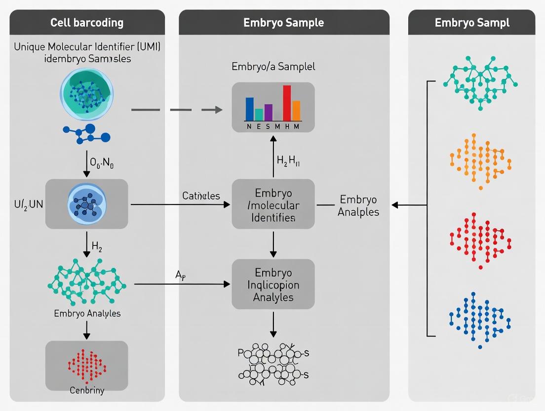
Advanced Cell Barcoding and UMI Strategies for Unraveling Human Embryo Development
This article provides a comprehensive guide to single-cell RNA sequencing (scRNA-seq) technologies, focusing on the application of cell barcoding and Unique Molecular Identifier (UMI) strategies for the study of human...
Advanced Cell Barcoding and UMI Strategies for Unraveling Human Embryo Development
Abstract
This article provides a comprehensive guide to single-cell RNA sequencing (scRNA-seq) technologies, focusing on the application of cell barcoding and Unique Molecular Identifier (UMI) strategies for the study of human embryo development. It covers foundational principles, from the basic roles of barcodes in sample multiplexing to UMIs in accurate molecular counting. The content delves into methodological choices for precious embryonic samples, troubleshooting for common technical challenges like oligonucleotide synthesis errors and dissociation bias, and the critical validation of data using emerging integrated reference atlases. Aimed at researchers and drug development professionals, this resource synthesizes cutting-edge innovations and practical insights to empower robust experimental design and analysis in this rapidly advancing field.
Core Principles: How Barcodes and UMIs Decode Cellular Heterogeneity in Embryos
In the evolving landscape of developmental biology, single-cell RNA sequencing (scRNA-seq) has emerged as a transformative tool for evaluating the specific transcriptome usage of different cell types within an organism [1]. This technology enables a non-biased assay of the active transcriptome by tagging mRNA molecules from single cells or nuclei, providing unprecedented resolution for exploring cellular heterogeneity [1] [2]. The usefulness of this approach is particularly evident in studies of early human development, where it offers fundamental insights into how we are built and how human life begins [3]. For research on precious embryo samples, understanding and properly implementing barcoding strategies is not merely technical but fundamental to biological discovery.
At the heart of droplet-based scRNA-seq technologies lie two critical components: cell barcodes and unique molecular identifiers (UMIs). These oligonucleotide sequences work in concert to enable massively parallel analysis of thousands of individual cells while maintaining single-cell resolution [2] [4]. Their precise implementation allows researchers to deconstruct complex cellular populations, track developmental trajectories, and identify rare cell subtypes—capabilities that are revolutionizing our understanding of embryogenesis [3] [5]. This application note details the distinct roles of cell barcodes and UMIs within the specific context of embryo research, providing both theoretical foundations and practical protocols to guide experimental design.
Fundamental Concepts: Distinguishing Barcodes from UMIs
Cell Barcodes: Tracking Cellular Origins
Cell barcodes are short, predetermined oligonucleotide sequences designed to answer a fundamental question: "Which cell did this sequence read come from?" [4]. In droplet-based systems, each gel bead is coated with millions of copies of a specific barcode sequence. When a cell is encapsulated in a droplet with a barcoded bead, all mRNA molecules from that cell are tagged with the identical cellular barcode during reverse transcription [2]. This elegant strategy enables subsequent computational deconvolution of pooled sequencing data, allowing researchers to attribute each sequenced read back to its cell of origin despite all cells being processed together in a single reaction [4].
The power of cellular barcoding becomes evident when considering experimental scale. Modern commercial solutions can capture anywhere from 500 to over 1,000,000 cells in a single run, with each cell receiving a unique identifier that distinguishes it from all other cells in the experiment [1]. This massive multiplexing capability is particularly valuable for embryo research, where samples may be limited and cellular heterogeneity at different developmental stages is of paramount interest [3].
Unique Molecular Identifiers: Correcting Technical Biases
Unique Molecular Identifiers (UMIs) are random nucleotide sequences that serve a different but equally critical purpose: they tag individual mRNA molecules to account for amplification biases [6] [4]. Each mRNA molecule receives a random UMI during the reverse transcription process, creating a unique "molecular fingerprint" for that transcript [4]. This approach addresses a fundamental challenge in scRNA-seq: the amplification step required to generate sufficient material for sequencing introduces substantial technical noise because some molecules are amplified more than others [4].
The UMI workflow operates on a simple but powerful principle. After sequencing, bioinformatics tools can identify and collapse reads that share the same cell barcode, UMI, and gene alignment, counting them as a single original molecule [6] [4]. This correction process, known as UMI deduplication, effectively filters out PCR duplicates and enables true digital counting of transcript molecules, thereby providing more accurate quantitative gene expression data [6]. As noted in the search results, "UMI deduplication is also useful for RNA-seq gene expression analysis and other quantitative sequencing methods" [6].
Table 1: Core Functions of Cell Barcodes and UMIs in Single-Cell RNA Sequencing
| Feature | Cell Barcode | Unique Molecular Identifier (UMI) |
|---|---|---|
| Primary Function | Identify cellular origin of sequences | Identify individual mRNA molecules |
| Sequence Characteristics | Predetermined, fixed per bead | Random, different for each molecule |
| Information Provided | Which cell the read came from | Which transcript molecule the read came from |
| Role in Quantification | Enables grouping of reads by cell | Enables correction for amplification bias |
| Typical Length | 12-16 nucleotides [1] [7] | 8-12 nucleotides [1] [8] |
| Impact on Data | Defines cell-by-gene expression matrix | Provides accurate molecular counts |
The Synergistic Relationship in Embryo Research
In embryo research, where developmental processes involve precise spatiotemporal gene regulation, the combination of cell barcodes and UMIs becomes particularly powerful. Together, they create a data structure where expression is organized hierarchically: Cell Barcode → Gene → UMI [4]. This organization means that for any given cell (identified by its barcode), we can count how many unique UMIs align to each gene, providing a precise measurement of gene expression while accounting for both technical noise (via UMIs) and biological origin (via cell barcodes) [4].
This synergistic relationship enables researchers to address fundamental questions in embryonic development, such as tracing lineage specification events, identifying rare progenitor populations, and mapping the heterogeneous onset of differentiation [3] [5]. As noted in the search results, single-cell transcriptomic profiling has been applied to study "time courses of single embryos" and "single cells from time-courses of entire embryos," generating comprehensive inventories of transcriptomic states throughout development [1].
Experimental Design and Platform Selection
Commercial Platform Comparisons
Choosing an appropriate scRNA-seq platform is a critical first step in experimental design, particularly for embryo studies where sample amount may be limited. Different commercial solutions offer varying throughput capacities, capture efficiencies, and compatibility with specific sample types [1]. The selection should be guided by both the biological question and the practical constraints of the embryo model system.
Table 2: Comparison of Commercial Single-Cell RNA Sequencing Solutions
| Commercial Solution | Capture Platform | Throughput (Cells/Run) | Capture Efficiency | Max Cell Size | Fixed Cell Support |
|---|---|---|---|---|---|
| 10× Genomics Chromium | Microfluidic oil partitioning | 500–20,000 [1] | 70–95% [1] | 30 µm [1] | Yes [1] |
| BD Rhapsody | Microwell partitioning | 100–20,000 [1] | 50–80% [1] | 30 µm [1] | Yes [1] |
| Singleron SCOPE-seq | Microwell partitioning | 500–30,000 [1] | 70–90% [1] | < 100 µm [1] | Yes [1] |
| Parse Evercode Biosciences | Multiwell-plate | 1000–1M [1] | >90% [1] | - | Yes [1] |
| Fluent/PIPseq (Illumina) | Vortex-based oil partitioning | 1000–1M [1] | >85% [1] | - | Yes [1] |
Sample Preparation Considerations for Embryo Research
For embryo research, careful sample preparation is paramount. The first step involves converting the tissue of interest into a quality single cell or nuclei suspension [1]. Researchers must decide whether to sequence single cells or single nuclei—a decision that depends on the intended use of the data. For many applications, entire cell capture is ideal as the number of mRNAs within the cytoplasm is greater than that of the nucleus [1]. However, single nuclei sequencing is compatible with multiome studies combining transcriptomes with open chromatin (ATAC-seq) and may be preferable for certain cell types that are difficult to isolate intact [1].
The choice of starting material should be directly related to the biological question being interrogated. Generating a comprehensive inventory of cell types for an embryo requires dissociation of all its tissues, which often involves preparing multiple samples from separate dissections [1]. This strategy allows for limited spatial information to be retained and enables the use of customized dissociation protocols tailored to the varying characteristics of different tissues [1]. As noted in the search results, "if your primary research interest is for example a specific cell type [...] then it makes sense to reduce the complexity of the data by first performing a clean dissection of the tissue and discarding the rest" [1].
Wet-Lab Protocol: Implementing scRNA-seq for Embryo Samples
Cell Suspension Preparation from Embryo Tissue
Materials: Fresh or frozen embryo tissue, dissociation enzymes (e.g., collagenase, trypsin), phosphate-buffered saline (PBS), cell strainer (40µm), viability stain (e.g., Trypan Blue), centrifuge, culture medium with serum.
Procedure:
- Tissue Dissociation: Mince embryo tissue into small fragments (<1mm³) using sterile surgical blades. Transfer tissue to dissociation enzyme solution optimized for the specific embryonic stage and tissue type. Digest for 15-45 minutes at 37°C with gentle agitation [1].
- Reaction Quenching: Add culture medium with serum to stop enzymatic digestion.
- Filtration and Washing: Pass cell suspension through a 40µm cell strainer to remove aggregates. Centrifuge at 300-500g for 5 minutes and resuspend in PBS with 0.04% BSA.
- Quality Control: Assess cell viability using Trypan Blue exclusion; target >85% viability for optimal results [2]. Determine cell concentration using a hemocytometer or automated cell counter, adjusting to 700-1200 cells/µL for loading on droplet-based systems [2].
Troubleshooting Note: For particularly challenging tissues with extensive extracellular matrix or fragile cells, consider alternative approaches such as fluorescence-activated cell sorting (FACS) with commercially available live/dead stains to eliminate debris [1]. However, be aware that this "runs the risk of introducing artifacts related to cell stress during the sorting process, or losing specific cell types that are more fragile than others" [1].
Library Preparation using 10x Genomics Chromium Platform
Materials: 10x Genomics Single Cell 3' Reagent Kit, PCR thermal cycler, magnetic separator, SPRIselect beads, Qubit dsDNA HS Assay Kit, TapeStation or Bioanalyzer.
Procedure:
- GEM Generation: Combine cell suspension, Master Mix, and barcoded gel beads on the Chromium chip to form Gel Bead-in-Emulsions (GEMs). Within each GEM, cell lysis occurs, and poly-adenylated RNA molecules hybridize to the oligo(dT) primers on the barcoded beads [2].
- Reverse Transcription: Perform reverse transcription inside GEMs to produce cDNA tagged with cell barcode and UMI. The reaction proceeds as follows:
- 53°C for 45 minutes
- 85°C for 5 minutes
- Hold at 4°C
- cDNA Amplification: Break emulsions and purify barcoded cDNA using silane magnetic beads. Amplify cDNA with the following PCR program:
- 98°C for 3 minutes
- 12 cycles of: 98°C for 15 seconds, 63°C for 20 seconds, 72°C for 1 minute
- 72°C for 1 minute
- Hold at 4°C
- Library Construction: Fragment amplified cDNA and add sample indexes via another PCR amplification. Include unique dual indexes (UDIs) to enable sample multiplexing [6].
- Library QC: Quantify library using Qubit and assess size distribution (typical peak ~500bp) using TapeStation.
Addressing Oligonucleotide Synthesis Errors
Recent research has highlighted a critical challenge in scRNA-seq: oligonucleotide synthesis errors can significantly impact data quality. As noted in the search results, "truncating UMIs computationally by one base led to 115 differentially expressed transcripts between 11 and 12-base UMIs" [8]. This finding underscores the importance of barcode quality in accurate gene expression quantification.
To address this challenge, consider innovative bead designs that incorporate an anchor sequence between the barcode and UMI. Research has demonstrated that "incorporating an anchor sequence (BAGC) between the barcode and UMI, and a V base between the UMI and the poly(dT) capture handle, could provide clearer demarcation of the beginning of the UMI" [8]. This design significantly improves UMI recovery and feature detection rates, enhancing the capabilities of droplet-based sequencing [8].
Data Analysis Workflow: From Raw Sequences to Expression Matrices
The computational processing of scRNA-seq data involves multiple steps to transform raw sequencing reads into a cell-by-gene expression matrix that properly accounts for both cell barcodes and UMIs.
Diagram 1: scRNA-seq Data Analysis Workflow
Cell Barcode Processing
The first computational step involves identifying and validating cell barcodes from the raw sequencing data. This process typically involves:
- Extraction: Barcodes are extracted from Read 1 based on their known position in the library structure [4].
- Whitelisting: Extracted barcodes are matched against a predetermined list of valid barcodes (a "whitelist") to filter out sequences with errors [7].
- Correction: Some pipelines implement error correction for barcodes that closely match whitelisted sequences.
For long-read scRNA-seq technologies, specialized tools like BLAZE have been developed that "accurately and efficiently identifies 10x cell barcodes using only nanopore long-read scRNA-seq data" without requiring matched short-read data [7].
UMI Deduplication
Following alignment of reads to the reference genome, the crucial step of UMI deduplication occurs:
- Grouping: Reads are grouped by cell barcode and gene alignment.
- Collapsing: Within each cell-gene combination, reads sharing the same UMI are collapsed, counting as a single molecule.
- Error Correction: Sophisticated tools account for potential errors in UMIs by clustering similar UMIs that likely represent PCR or sequencing errors of the original molecule.
This process effectively corrects for amplification biases, as "UMI deduplication is also useful for RNA-seq gene expression analysis and other quantitative sequencing methods" to "reduce false-positive variant calls and increase sensitivity of variant detection" [6].
Successful implementation of scRNA-seq for embryo research requires careful selection of reagents and resources. The following table outlines key solutions and their applications.
Table 3: Essential Research Reagent Solutions for scRNA-seq in Embryo Research
| Reagent/Resource | Function | Application Notes |
|---|---|---|
| 10x Genomics Chromium | Microfluidic partitioning system | Optimized for cell suspensions; 70-95% capture efficiency [1] |
| BD Rhapsody | Microwell partitioning system | Compatible with larger cells (<100µm); 50-80% capture efficiency [1] |
| Parse Evercode | Multiwell-plate based system | Lowest cost per cell; requires high input (1M cells) [1] |
| Live/Dead Stains | Cell viability assessment | Critical for assessing sample quality pre-loading [1] |
| UMI-tools | Bioinformatics package for UMI processing | Enaccurate deduplication and counting [4] |
| BLAZE | Barcode identification for long-read data | Specifically for Oxford Nanopore long-read scRNA-seq [7] |
| Cell Ranger | 10x Genomics analysis suite | Standardized processing for 10x data including barcode assignment |
| Seurat | R package for scRNA-seq analysis | Comprehensive toolkit for downstream analysis after barcode processing [3] |
Advanced Applications in Embryo Research
Reference Atlas Construction for Embryo Models
Single-cell RNA sequencing with proper barcoding has enabled the construction of comprehensive reference atlases for embryonic development. As demonstrated in recent work, researchers have developed "a comprehensive human embryo reference tool using single-cell RNA-sequencing data" through the integration of multiple published datasets "covering development from the zygote to the gastrula" [3]. This integrated reference encompasses 3,304 early human embryonic cells and displays "a continuous developmental progression with time and lineage specification and diversification" [3].
Such reference atlases provide powerful tools for benchmarking stem cell-based embryo models. When query datasets are projected onto these references, researchers can annotate cells with predicted identities and assess the fidelity of embryo models to their in vivo counterparts [3]. This application underscores the critical importance of accurate barcoding and UMI counting—without proper molecular identification, such precise comparisons would be impossible.
Genetic Barcoding Strategies for Lineage Tracing
Beyond the standard barcoding approaches used in commercial platforms, innovative genetic barcoding strategies are emerging that enable even more sophisticated experimental designs. Methods such as Targeted Genetically-Encoded Multiplexing (TaG-EM) involve "inserting a DNA barcode just upstream of the polyadenylation site" in genetically engineered constructs [9]. This approach allows deterministic in vivo tagging of defined cell populations, enabling positive identification of cell types in atlas projects and identification of multiplet droplets [9].
For embryo research, such approaches offer exciting possibilities for lineage tracing and fate mapping. By combining the standard barcoding of commercial platforms with genetic barcoding strategies, researchers can create multi-layered experimental designs that simultaneously capture endogenous gene expression, cell lineage relationships, and spatial organization within developing embryos.
Cell barcodes and UMIs represent foundational technologies that have enabled the single-cell revolution in developmental biology. Their distinct but complementary roles—cellular identification and molecular counting, respectively—provide the framework for accurate, quantitative transcriptomics at single-cell resolution. For embryo researchers, understanding these technologies is not merely technical but essential for proper experimental design, implementation, and interpretation.
As the field advances, emerging technologies in long-read sequencing, spatial transcriptomics, and multi-omics integration will build upon these barcoding foundations. The proper application of cell barcodes and UMIs will continue to drive discoveries in embryonic development, stem cell biology, and reproductive medicine, ultimately enhancing our understanding of human development and disease.
The Critical Function of UMIs in Error Correction and Quantitative Accuracy
Unique Molecular Identifiers (UMIs) are short, random oligonucleotide barcodes that are incorporated into individual RNA or DNA molecules during the initial steps of sequencing library preparation [10] [6]. These molecular tags serve as unique identifiers for each original molecule in a sample, enabling precise distinction between biologically distinct molecules and copies generated through PCR amplification [11]. This capability is particularly valuable in quantitative sequencing applications where accurate molecular counting is essential, such as in single-cell RNA-sequencing (scRNA-seq), rare variant detection, and gene expression analysis [6] [12].
The fundamental principle behind UMI technology lies in its ability to provide digital quantification of nucleic acid molecules, transforming conventional sequencing from an analog measurement susceptible to amplification biases into a digital counting process [12]. Each original molecule is tagged with a unique barcode before any amplification steps, creating a distinct identity that persists through subsequent PCR cycles [10]. After sequencing, bioinformatics tools can collapse reads sharing identical UMIs and mapping coordinates into single molecular events, effectively filtering out PCR duplicates and providing a more accurate representation of the original molecular population [10] [11].
In the context of embryo samples research, where starting material is often limited and requires significant amplification, UMIs play a particularly crucial role in ensuring data integrity. They mitigate the effects of PCR amplification bias, which is especially pronounced when many PCR cycles are required to generate sufficient material for sequencing [13]. This makes UMI-based approaches indispensable for sensitive applications such as tracing cell lineages during embryonic development or characterizing transcriptional heterogeneity in early embryonic cells [14].
The Problem of Amplification Bias and Sequencing Errors
Limitations of Conventional Sequencing Quantification
Traditional sequencing quantification methods rely on counting reads mapping to genomic coordinates, an approach that becomes increasingly problematic as amplification biases intensify. In standard RNA-seq experiments, particularly those with limited input material such as single-cell analyses or embryo samples, PCR amplification is necessary to generate sufficient DNA for sequencing [10]. However, this amplification process introduces substantial biases because certain sequences become overrepresented in the final library due to preferential amplification [10]. These biases propagate to quantification estimates, potentially leading to inaccurate biological conclusions.
The problem is particularly acute in single-cell RNA-seq and spatial transcriptomics of embryonic tissues, where the distribution of alignment coordinates deviates significantly from random sampling across the genome [10]. For highly expressed transcripts in embryo samples, the probability of generating independent fragments mapping to the same genomic coordinates increases dramatically, making it difficult to distinguish between technical duplicates (PCR-amplified copies) and biological duplicates (truly independent molecules) [10]. Without UMIs, researchers must rely on alignment coordinates alone to identify PCR duplicates, which becomes increasingly unreliable as sequencing depth increases and for techniques like iCLIP (individual-nucleotide resolution Cross-Linking and ImmunoPrecipitation) where alignment coordinates are limited to few distinct loci [10].
The Impact of Sequencing Errors on UMI Effectiveness
While UMIs provide powerful error correction capabilities, they are themselves susceptible to errors that can compromise quantification accuracy. Errors within the UMI sequence – including nucleotide substitutions during PCR and nucleotide miscalling, insertions, or deletions during sequencing – create additional artifactual UMIs that inflate molecular counts [10]. Research has demonstrated that UMI errors are common, with a 25-fold enrichment observed for positions with an average edit distance of 1 compared to null expectations [10].
Different types of UMI errors have distinct effects on data analysis:
- Nucleotide substitutions and miscalling affect only the UMI sequence itself, creating artifactual UMIs that inflate the estimation of unique molecules at particular genomic coordinates [10].
- UMI indels affect both the UMI sequence and the alignment position, leading to the assignment of reads to incorrect genomic coordinates [10].
- "PCR jumping" or recombination events create chimeric sequences that may change either the UMI sequence and/or alignment, though this is much rarer in shotgun sequencing approaches typically used with UMIs [10].
Evidence suggests that miscalling during sequencing is by far the most prevalent error, occurring one to two orders of magnitude more frequently than indels in Illumina sequencing [10]. This highlights the critical need for robust bioinformatic methods to account for these errors when leveraging UMI information.
UMI-Based Error Correction Methods and Bioinformatics
Computational Strategies for UMI Deduplication
Several computational approaches have been developed to account for UMI errors during the deduplication process. The simplest method, often called "unique," assumes each UMI at a given genomic locus represents a different unique molecule [10]. However, this approach fails to account for sequencing errors in the UMI sequence and thus overestimates molecular counts. More sophisticated network-based methods have been implemented in tools like UMI-tools to address this limitation [10].
Table 1: Comparison of UMI Deduplication Methods
| Method | Key Principle | Advantages | Limitations |
|---|---|---|---|
| Unique | Each UMI is treated as a distinct molecule | Simple implementation | Overestimates counts due to sequencing errors |
| Percentile | Removes UMIs with counts below a threshold (e.g., 1% of mean) | Filters obvious artifacts | May eliminate true rare molecules |
| Cluster | Merges all UMIs within a defined edit distance | Accounts for related UMIs | Underestimates complex networks |
| Adjacency | Iteratively removes most abundant node and neighbors | Handles complex networks better | May oversimplify in some cases |
| Directional | Uses directional connectivity based on count ratios | Models error propagation | More computationally intensive |
The directional method represents a particularly advanced approach, generating networks from UMIs at a single locus where directional edges connect nodes a single edit distance apart based on count ratios [10]. This method recognizes that counts for UMIs generated by a single sequencing error should be higher than those generated by two errors, and UMIs resulting from errors during PCR amplification should have higher counts than UMIs resulting from sequencing errors [10].
Benchmarking UMI Processing Workflows
Recent systematic benchmarking of scRNA-seq preprocessing workflows has revealed that while quantification differences exist between methods, downstream analysis results are generally consistent across approaches [15]. Evaluations of ten end-to-end preprocessing workflows (including Cell Ranger, Optimus, salmon alevin, and UMI-tools) demonstrated that after normalization and clustering, almost all combinations produce clustering results that agree well with known cell type labels used as ground truth [15].
Table 2: UMI-Count vs Read-Count Distribution Modeling
| Model | Parameters | Read-Count Performance | UMI-Count Performance |
|---|---|---|---|
| Poisson | One parameter (mean = variance) | 2.4-9.5% of genes | 39.4-84.0% of genes |
| Negative Binomial (NB) | Two parameters (mean and variance) | 65.5-90.1% of genes | 16.0-60.6% of genes |
| Zero-Inflated Negative Binomial (ZINB) | Three parameters (NB + zero-inflation) | 9.4-34.5% of genes preferred ZINB | 0% of genes preferred ZINB |
This benchmarking indicates that UMI-count data generally follows simpler statistical distributions than read-count data. Specifically, while a significant fraction of read-count measurements require zero-inflated negative binomial models, UMI-count data are typically well-modeled by simpler negative binomial or even Poisson distributions [16]. This statistical characteristic simplifies downstream analysis and improves the reliability of differential expression testing in embryo development studies.
Experimental Protocols for UMI Implementation
UMI Integration in Single-Cell RNA-Sequencing
For single-cell RNA-sequencing of embryo samples, the most common approach involves leveraging commercial platforms such as 10X Genomics or BD Rhapsody. These technologies partition individual cells into wells or droplets and sequence the mRNA reads from individual cells [11]. The process typically involves:
Cell Partitioning: Individual cells from embryo samples are partitioned into nanoliter-scale droplets or wells along with barcoded beads.
mRNA Capture: The poly-A tail of mRNA molecules is captured using a poly-dT sequence attached to a bead. The bead contains both a cell barcode (to identify the cell of origin) and a UMI (to identify the specific molecule) [11].
Reverse Transcription: This step generates cDNA while incorporating the cell barcode and UMI sequences.
Library Preparation and Sequencing: The resulting libraries are sequenced using high-throughput platforms like Illumina.
A key consideration for embryo research is that the starting material is very limited and of potentially variable quality, necessitating PCR amplification which can introduce biases [11]. UMIs are particularly valuable in this context as they enable screening out errors introduced during amplification.
Spatial Genomics with Slide-tags Technology
Recent advances in spatial genomics have extended UMI applications to spatially-resolved molecular profiling. The Slide-tags method enables single-nucleus barcoding for multimodal spatial genomics by tagging nuclei within intact tissue sections with spatial barcode oligonucleotides derived from DNA-barcoded beads with known positions [17]. The protocol involves:
Tissue Preparation: Fresh frozen tissue sections (e.g., 20μm thickness) are prepared from embryo samples.
Spatial Barcode Application: Densely packed spatially indexed arrays of DNA-barcoded 10μm beads are applied to tissue sections, with spatial barcodes photocleaved and diffused into the tissue to associate with nuclei [17].
Nuclei Isolation and Sequencing: Tagged nuclei are isolated and used as input into standard single-nucleus profiling assays (snRNA-seq, snATAC-seq, etc.) with minimal protocol modifications [17].
This approach has been demonstrated to achieve less than 10μm spatial resolution while maintaining data quality indistinguishable from ordinary single-nucleus RNA-sequencing [17]. For embryonic development studies, this enables precise mapping of cell types and states within the spatial context of developing tissues.
Research Reagent Solutions for UMI-Based Studies
Table 3: Essential Research Reagents for UMI-Based Embryo Research
| Reagent Category | Specific Examples | Function in UMI Workflow |
|---|---|---|
| Library Preparation Kits | 10X Genomics Single Cell Gene Expression, SMART-Seq | Incorporate UMIs during cDNA synthesis |
| Barcoded Beads | 10X Gel Beads, BD Rhapsody Cartridges | Deliver cell barcodes and UMIs to partitioned cells |
| Reverse Transcriptase | Maxima H-, SuperScript IV | Efficient cDNA synthesis with UMI incorporation |
| Amplification Enzymes | KAPA HiFi HotStart, Q5 Hot Start | High-fidelity amplification of UMI-tagged libraries |
| Cleanup Kits | SPRIselect, AMPure XP | Size selection and purification of UMI-libraries |
| Spatial Barcoding Arrays | Slide-tags beads | Enable spatial genomics with UMI quantification |
Workflow Visualization: UMI Implementation Pathway
The following diagram illustrates the complete UMI workflow from sample preparation to data analysis:
Impact on Quantitative Accuracy and Research Applications
Enhanced Quantification in Single-Cell Genomics
The implementation of UMIs has fundamentally transformed the reliability of single-cell RNA-sequencing data, particularly for embryo research where accurate quantification of transcriptional states is essential for understanding developmental processes. Comparative analyses have demonstrated that UMI-counting provides superior results to read-counting, with one study showing that UMI-count measurements showed less divergence than their read-count counterparts in the same cell pairs [16]. Specifically, quantifications for genes with dropout events (where transcripts are captured in one cell but not another) showed a distinct bimodal pattern in read counts but a unimodal distribution in UMI counts [16].
This improvement in quantification accuracy directly impacts the ability to identify true biological variation in developing embryos. The reduction in technical noise enables researchers to more confidently distinguish between stochastic technical artifacts and genuine biological heterogeneity in embryonic cell populations. Furthermore, UMI-based approaches have been shown to improve reproducibility between experimental replicates and enhance clustering performance in single-cell RNA-seq datasets [10].
Applications in Spatial Transcriptomics and Lineage Tracing
Beyond conventional single-cell transcriptomics, UMIs have enabled advanced applications in spatial genomics and lineage tracing that are particularly relevant to embryo research. Technologies like Slide-tags combine spatial barcoding with UMI-based quantification to achieve high-resolution spatial mapping of gene expression while maintaining single-cell precision [17]. This approach has been successfully applied to characterize cell-type-specific spatially varying gene expression across cortical layers and to spatially contextualize receptor-ligand interactions driving cell maturation processes [17].
In prospective lineage tracking studies, DNA barcodes (conceptually similar to UMIs) are used to trace the developmental fate of embryonic cells over time [14]. These approaches involve introducing random DNA barcodes into cells and then tracking their abundance and distribution across different tissues and timepoints during embryogenesis. The high sensitivity and specificity afforded by UMI-based digital sequencing make it possible to detect rare lineage branches and reconstruct comprehensive lineage trees with single-cell resolution [12] [14].
For cancer research and drug development, UMI-based approaches enable ultrasensitive detection of rare sequence variants, including mutations conferring treatment resistance [12]. This capability is increasingly important for monitoring minimal residual disease and detecting emerging resistance mutations during targeted therapy. The improved quantitative accuracy provided by UMIs also enhances the reliability of biomarker identification and validation in drug development pipelines.
Embryonic samples represent a uniquely challenging and valuable resource in developmental biology and regenerative medicine. Their scientific value is inextricably linked to three defining characteristics: scarcity, as human embryos are difficult to obtain and their use is strictly regulated; heterogeneity, as early development involves rapid, dynamic cell fate decisions; and significant ethical considerations, which govern all aspects of their use in research. These characteristics create a research environment where maximizing information from minimal material is paramount. This application note details how advanced cellular barcoding and unique molecular identifier (UMI) strategies are essential for addressing these challenges, enabling researchers to extract robust, high-dimensional data from these rare and heterogeneous systems while operating within established ethical frameworks.
Navigating Scarcity and Ethical Constraints
The scarcity of human embryonic samples is both a biological and an ethical reality. Scientifically, the window for studying early human development in vitro is technically narrow. Ethically, international norms and regulations, such as the "14-day rule", have traditionally limited research to the period before the emergence of the primitive streak, roughly corresponding to the first two weeks post-fertilization [18] [19]. There is an ongoing debate about extending this culture limit to 28 days for specific, high-value research questions that cannot be addressed by other means, as the period between 14 and 28 days is critical for understanding organ development and congenital abnormalities [18].
Ethical Frameworks and Oversight
Research using human embryos is considered ethically acceptable if it is likely to provide significant new knowledge that benefits human health, offspring well-being, or reproduction, provided it adheres to strict guidelines [19]. Key principles include:
- Informed Consent: Prior written informed consent from gamete providers is mandatory. Consent for research not intended to result in reproduction can be broad, while research with reproductive intent requires explicit, contemporaneous consent [19].
- Oversight: Research should undergo rigorous ethical review, typically through an Institutional Review Board (IRB) or similar oversight body [18] [19].
- Proportionality: The number of embryos used should not exceed what is necessary to answer the research question [19].
Table 1: Key Ethical Regulations and Emerging Alternatives in Embryo Research
| Aspect | Current Standard | Emerging Considerations |
|---|---|---|
| Culture Limit | The 14-day rule [18] [19] | Proposal to extend limit to 28 days for critical research on organ development [18] |
| Source of Embryos | Donated supernumerary embryos from IVF [19] | Embryos created specifically for research (subject to ethical review) [19] |
| Alternative Models | N/A | Use of Embryo-Like Structures (ELS) with varying moral status [18] |
Synthetic Embryo Models as a Complementary Tool
Stem cell-derived synthetic embryo models (SEMs), or embryo-like structures (ELSs), are emerging as powerful tools to circumvent the challenges of scarcity and ethical constraints [20]. These models are generated from pluripotent stem cells (PSCs) and can self-organize to mimic key aspects of early embryogenesis in vitro [20]. The ethical status of these models is nuanced; non-integrated ELSs are generally considered to have a lower moral status, while integrated ELSs (those containing both embryonic and extraembryonic tissues) that demonstrate developmental potential may be subject to the same regulations as natural embryos [18].
Decoding Cellular Heterogeneity in Early Development
The early embryo is a hotbed of cellular diversification. Following the first cell fate decision that separates the trophectoderm (TE) from the inner cell mass (ICM), a second critical decision occurs within the ICM to specify the epiblast (EPI, which will form the fetus) and the primitive endoderm (PrE, which contributes to the yolk sac) [21].
Signaling Pathways Driving Lineage Specification
The specification of EPI and PrE lineages is a classic model of signaling-driven heterogeneity. In mouse embryos, this process is governed by Fibroblast Growth Factor (FGF) signaling.
- Mechanism: A random subset of ICM cells initially upregulates FGF4 expression. Cells that receive high levels of FGF/MAPK signaling (via receptors FGFR1 and FGFR2) upregulate PrE markers like GATA6 and SOX17, while cells with lower signaling (primarily via FGFR1) maintain EPI markers like NANOG and SOX2 [21].
- Reinforcement: Cell fate commitment is reinforced by mutual transcriptional repression between GATA6 and NANOG, and supported by LIF/JAK/STAT and PDGF/PI3K signaling pathways [21].
This results in a salt-and-pepper distribution of EPI and PrE progenitors within the ICM, which later sort into a coherent epithelium [21]. The following diagram illustrates this critical signaling network and its outcomes.
The primitive endoderm continues to play a vital patterning role after implantation. It gives rise to the visceral endoderm, which forms a signaling center known as the anterior visceral endoderm (AVE). The AVE secretes antagonists like Dkk1 (Wnt antagonist), Cer1, and Lefty1 (Nodal/BMP antagonists) to pattern the underlying epiblast and establish the anterior-posterior axis, guiding the formation of the primitive streak [21].
Application Notes: Barcoding Strategies for Embryonic Samples
To dissect the profound heterogeneity of embryonic samples, single-cell RNA sequencing (scRNA-seq) is the tool of choice. However, its application is constrained by sample scarcity. High-throughput droplet-based barcoding technologies, such as inDrop and related methods, are uniquely suited to this challenge [5].
Experimental Protocol: High-Throughput scRNA-seq of Embryonic Cells
This protocol is adapted from droplet-based single-cell RNA sequencing methods for profiling thousands of cells, ideal for a limited pool of embryonic cells [5].
- Goal: To generate comprehensive single-cell transcriptomic profiles from a dissociated suspension of embryonic cells.
- Principle: Individual cells are co-encapsulated in nanoliter-scale droplets with barcoded hydrogel microspheres (BHMs). Each BHM carries primers with a unique cellular barcode and UMI. mRNA from each cell is reverse-transcribed within its droplet, labeling all cDNA from a single cell with the same barcode.
Materials:
- Single-cell suspension from embryonic samples.
- Barcoded Hydrogel Microspheres (BHMs): Library of microspheres with covalently coupled, photo-releasable primers containing unique barcodes [5].
- Lysis/Reverse Transcription (RT) Mix: Contains reagents for cell lysis and reverse transcription.
- Droplet Generation Microfluidic Device & System.
- Carrier Oil for droplet formation.
- UV Light Source for primer release.
Procedure:
- Sample Preparation: Generate a high-viability, single-cell suspension from the embryonic sample using standard dissociation techniques. Pass the suspension through a strainer or use FACS to minimize cell aggregates.
- Microfluidic Setup: Prime the microfluidic device with carrier oil. Load the sample and reagent inlets with:
- Inlet 1: Barcoded Hydrogel Microspheres (BHMs)
- Inlet 2: Single-cell suspension
- Inlet 3: Lysis/RT reagent mix
- Droplet Generation: Run the device to generate monodisperse droplets (~1-5 nL) at a rate of 10-100 drops per second. The device synchronizes flows to co-encapsulate single cells with single BHMs and lysis/RT reagents into droplets [5].
- UV Photo-release: Collect droplets and expose them to ultraviolet light to cleave and release the barcoded primers from the BHMs into the droplet solution [5].
- Reverse Transcription: Incubate the emulsion to allow cell lysis and reverse transcription of mRNA into barcoded cDNA.
- Droplet Breakage and Library Prep: Break the emulsion, pool the aqueous phases, and purify the barcoded cDNA. Proceed with second-strand synthesis, amplification, and library construction for next-generation sequencing (e.g., following CEL-Seq protocols) [5].
- Sequencing: Sequence the libraries on an appropriate NGS platform.
A Toolkit for Barcode Analysis and Simulation
The computational analysis of barcode and single-cell data is critical. The following tools and reagents are essential for a successful experiment.
Table 2: Research Reagent and Computational Toolkit
| Item / Tool Name | Type | Function in Experiment |
|---|---|---|
| Barcoded Hydrogel Microspheres (BHMs) | Wet-lab Reagent | Source of unique cellular barcodes and UMIs for labeling single-cell transcriptomes [5]. |
| Droplet Microfluidics Device | Equipment | High-throughput platform for generating monodisperse droplets containing single cells and reagents [5]. |
| CellBarcode R Package | Computational Tool | Versatile toolkit for pre-processing, extracting, and filtering DNA barcode sequences from bulk or single-cell NGS data [22]. |
| CellBarcodeSim | Computational Tool | Simulation kit to simulate barcoding experiments, allowing researchers to optimize filtering strategies and investigate factors impacting barcode detection [22]. |
Bioinformatic Analysis: Filtering True Barcodes from Noise
A major challenge in barcode analysis is distinguishing true biological barcodes from errors introduced by PCR amplification and sequencing. The CellBarcode package implements several key filtering strategies [22]:
- Reference Filtering: Retains only barcodes matching a pre-defined reference list (e.g., from the original viral library).
- Threshold Filtering: Removes barcodes with read counts below a specified threshold.
- Cluster Filtering: Eliminates barcodes that are within a small edit distance of a more abundant barcode (likely PCR errors).
- UMI Filtering: Uses UMIs to correct for PCR amplification bias, e.g., by extracting the most abundant barcode per UMI.
The following workflow diagram outlines the key steps from raw sequencing data to a filtered cell-by-gene expression matrix, highlighting where these filtering strategies are applied.
Simulation studies using CellBarcodeSim reveal that biological factors, such as the variation in clone size, can have a greater impact on the precision of barcode identification than technical factors. This underscores the importance of using such tools to tailor filtering strategies to the specific biological context of the experiment, such as studying early embryonic lineages where clone sizes may be highly variable [22].
The unique challenges posed by embryonic samples—their inherent scarcity, profound heterogeneity, and complex ethical landscape—demand equally unique technological solutions. High-throughput cellular barcoding and UMI strategies are not merely convenient; they are essential for transforming these limited, heterogeneous samples into rich, quantitative datasets. By integrating these powerful molecular tools with evolving ethical frameworks and emerging model systems like SEMs, researchers can continue to decode the fundamental principles of human development, paving the way for advances in regenerative medicine and the treatment of congenital disorders.
Single-cell RNA sequencing (scRNA-seq) has fundamentally transformed the study of embryonic development by enabling the unbiased transcriptional profiling of individual cells. This technology is particularly crucial for illuminating the complex cellular heterogeneity and dynamic lineage specification events that occur during embryogenesis. In reproductive medicine and developmental biology, scRNA-seq has enabled groundbreaking insights into epigenetic reprogramming in primordial germ cells (PGCs), enhanced preimplantation genetic diagnosis, and provided a powerful method for authenticating stem cell-based embryo models by comparing them to their in vivo counterparts [3] [2]. The usefulness of these embryo models hinges entirely on their molecular and cellular fidelity to real embryos, making unbiased single-cell transcriptional profiling an essential tool for validation [3].
The core challenge in embryo research has been the scarcity of human embryos donated for research and the technical/ethical limitations, such as the "14-day rule," associated with their study [3]. scRNA-seq technology helps overcome these challenges by allowing researchers to capture comprehensive transcriptomic snapshots of development from very limited starting materials. By employing sophisticated cell barcoding and Unique Molecular Identifier (UMI) strategies, modern scRNA-seq platforms can simultaneously analyze thousands of individual cells from precious embryo samples, reconstruct lineage trajectories, and identify rare cell populations that would otherwise be obscured in bulk sequencing approaches [2].
Technology Comparison Tables
When selecting a scRNA-seq platform for embryo analysis, researchers must consider multiple performance and logistical criteria. The tables below provide a structured comparison of major platforms.
Table 1: Key Performance Metrics of scRNA-seq Platforms
| Platform | Technology Type | Throughput (cells/run) | Gene Detection Sensitivity | Cell Capture Efficiency | Multiplet Rate |
|---|---|---|---|---|---|
| 10x Genomics Chromium | Droplet-based microfluidics | Up to 80,000 cells per run (8 channels) [23] | High (1,000-5,000 genes/cell) [2] | Up to ~65% recovery [23] | <0.9% per 1,000 cells [23] |
| 10x Genomics FLEX | Droplet-based with fixation | Million-cell scale experiments (up to 128 samples per chip) [23] | High, compatible with FFPE samples [23] | High for fixed samples [23] | Low, with extensive multiplexing capabilities [23] |
| BD Rhapsody | Microwell-based with magnetic beads | Adjustable, based on bead loading [23] | High, with integrated protein profiling [23] | Up to 70% (among highest in field) [23] | Low, with real-time monitoring [23] |
| Parse Biosciences Evercode WT | Combinatorial barcoding (plate-based) | Highly scalable, no inherent instrument limit [24] | High, avoids ambient RNA [24] | Not instrument-limited [24] | Low, combinatorial barcoding reduces collisions [24] |
| MobiDrop | Droplet-based microfluidics | Adjustable for pilot to large cohorts [23] | High reproducibility [23] | Efficient for fresh/frozen/FFPE [23] | Not specified in results |
Table 2: Experimental Design Considerations for Embryo Research
| Platform | Sample Compatibility | Species Compatibility | Cost Advantage | Special Features for Embryo Research |
|---|---|---|---|---|
| 10x Genomics Chromium | Fresh, frozen, gradient-frozen, FFPE [23] | Human, mouse, rat, other eukaryotes [23] | Moderate | "Classic" platform with robust performance for high cell numbers [23] |
| 10x Genomics FLEX | FFPE, PFA-fixed [23] | Human, mouse, rat, other eukaryotes [23] | Moderate | Unlocks archival samples; enables multi-site, multi-timepoint studies [23] |
| BD Rhapsody | Lower-viability suspensions (~65%) [23] | Human, mouse, rat, other eukaryotes [23] | Moderate | Protein + RNA profiling; tolerance for lower-viability clinical samples [23] |
| Parse Biosciences Evercode WT | Fixed cells and nuclei (store up to 6 months) [24] | Truly adaptable across species [25] | High (no instrument required) [24] | Ideal for time-courses; minimal batch effects; works with any model organism [24] [25] |
| MobiDrop | Fresh, frozen, FFPE [23] | Eukaryotes [23] | High (lower per-cell costs) [23] | Cost-effective for large projects under tighter budgets [23] |
Platform Strengths for Specific Embryonic Applications
10x Genomics Chromium: As the most widely adopted platform, it represents a robust choice when high cell numbers and sensitivity are required for embryonic tissues [23]. Its standardized workflow minimizes technical variability, which is crucial for comparative studies of different embryonic stages [2].
10x Genomics FLEX: This system is particularly valuable for research involving archived embryonic samples or complex study designs spanning multiple collection timepoints or sites [23]. The ability to work with paraformaldehyde (PFA)-fixed samples allows researchers to "lock" RNA states at specific developmental timepoints.
BD Rhapsody: With its high capture efficiency and tolerance for lower-viability cell suspensions (~65%), this platform is suitable for clinical embryonic samples that may not meet stringent quality thresholds [23]. The ability to combine RNA and protein readouts via CITE-seq is particularly valuable for immunology studies and characterizing surface markers in developing embryonic tissues.
Parse Biosciences Evercode WT: The instrument-free, highly scalable nature of this combinatorial barcoding approach makes it ideal for longitudinal studies of embryonic development [24] [25]. The ability to fix samples and process them in batches later virtually eliminates batch effects, which is crucial when studying sequential developmental stages.
Experimental Design and Sample Preparation
Critical Considerations for Embryo Studies
Designing a successful scRNA-seq experiment with embryonic samples requires careful planning across several dimensions:
Single Cell vs. Nuclei Sequencing: For embryonic tissues that are difficult to dissociate without compromising viability (such as highly fibrous tissues or specific embryonic structures), nuclei sequencing presents a valuable alternative. While there is a nominal loss of RNA from the cytosol, most genes reside in the nucleus, making this approach particularly suitable for challenging embryonic samples [26].
Fresh vs. Fixed Samples: Capturing a specific developmental snapshot is fundamental in embryo research. Cellular metabolism and gene expression change rapidly once cells are removed from their physiological environment. Fixation addresses this by allowing researchers to dissociate tissue, fix it, and store it for later processing, which is particularly useful for large-scale embryonic time course experiments [26]. Parse Biosciences' fixation protocol, for instance, allows samples to be stored for up to 6 months [24].
Replication Strategy: Both technical and biological replication are essential in scRNA-seq experimental design. Technical replicates (dividing the same sample into sub-samples) measure protocol noise, while biological replicates (different embryos or donors under identical conditions) capture inherent biological variability [26]. This is particularly crucial in embryo studies where natural developmental variations exist between individuals.
Species Considerations: Embryo research utilizes diverse model organisms, each with advantages. Parse Biosciences' combinatorial barcoding technology is particularly adaptable across species, having been successfully applied in zebrafish (sharing 70% of protein-coding genes with humans), Drosophila melanogaster (sharing 75% of disease-causing genes with humans), chickens, livestock, and non-human primates [25].
Embryonic Tissue Dissociation Protocol
The following protocol for dissociating mouse embryonic neural tissue exemplifies the careful approach required for embryonic samples [27]:
Tissue Preparation: Begin with freshly dissected embryonic mouse brain tissue. The surgical dissection of embryonic mouse tissue is not described here but should follow established institutional protocols.
Dissociation Method: Use gentle mechanical dissociation combined with appropriate enzymatic cocktails (such as those available from Miltenyi Biotec or Worthington Tissue Dissociation guides) tailored to embryonic neural tissue [26].
Cell Counting and Viability Assessment: Accurately count cells using a hemocytometer or automated cell counter. For the standard 10x Genomics Chromium protocol, optimize for counting cells in the range of 700-1200 cells/µl. If using the Single Cell 3' LT v3.1 (low throughput) application, ensure cells are counted as indicated in this protocol and then diluted to the LT-specific optimal loading concentration of 100-600 cells/µl [27].
Quality Control: Assess cell viability, which should ideally be between 70% and 90%, with intact cell morphology [26]. Density gradient centrifugation using Ficoll or Optiprep is effective for separating viable cells from debris in embryonic tissue preparations.
Temperature Control: Maintain a stable cold environment throughout the process to arrest metabolic functions. Once the single-cell suspension is created, place cells immediately on ice to reduce the upregulation of stress response genes that can skew developmental data [26].
Debris and Aggregation Management: Filter out debris and use media without calcium or magnesium (such as HEPES or Hanks' buffered salt) to prevent aggregation. Test different centrifugation speeds and durations to avoid over-pelleting, which can cause clumping [26]. The final suspension should have minimal debris and aggregation (<5%).
The Scientist's Toolkit: Essential Research Reagent Solutions
Table 3: Key Reagents and Materials for scRNA-seq Embryo Experiments
| Reagent/Material | Function | Example Application in Embryo Research |
|---|---|---|
| Fixation Reagents (e.g., Paraformaldehyde) | Preserve transcriptional state at specific developmental timepoints [23] | Locking RNA expression patterns at precise embryonic stages for later analysis |
| Enzyme Cocktails for Tissue Dissociation | Gentle breakdown of extracellular matrix in embryonic tissues [26] | Generating single-cell suspensions from whole embryos or specific embryonic organs |
| Barcoded Gel Beads (10x Genomics) | Capture mRNA and assign cellular barcodes in droplet-based systems [2] | Partitioning individual embryonic cells for transcriptome analysis |
| Combinatorial Barcoding Reagents (Parse Biosciences) | Label cells with unique barcode combinations through split-pool approach [24] | Processing multiple embryonic samples simultaneously without instrument constraints |
| Nuclei Isolation Kits | Extract nuclei for sequencing when whole-cell preparation is challenging [26] | Working with archived embryonic samples or tissues difficult to dissociate |
| Viability Stains (e.g., Trypan Blue) | Distinguish live vs. dead cells for quality control [26] | Assessing dissociation success and ensuring high-quality input for library preparation |
| UMI-containing Oligonucleotides | Label individual mRNA molecules to correct for amplification bias [2] | Accurate transcript counting in embryonic cells with dynamic gene expression |
scRNA-seq Workflow and Barcoding Strategies
Core scRNA-seq Workflow Diagram
Diagram Title: scRNA-seq Workflow for Embryo Analysis
Barcoding Technology Comparison
Diagram Title: Barcoding Technologies Comparison
Applications in Embryo Research
Reference Atlas Construction and Embryo Model Validation
A landmark application of scRNA-seq in embryo research is the creation of comprehensive reference atlases. A 2025 study published in Nature Methods developed an integrated human embryo reference through the integration of six published human datasets covering development from the zygote to the gastrula [3]. This reference encompasses 3,304 early human embryonic cells and displays a continuous developmental progression with time and lineage specification, capturing the first lineage branch point where inner cell mass (ICM) and trophectoderm (TE) cells diverge during E5, followed by the lineage bifurcation of ICM cells into the epiblast and hypoblast [3].
This integrated reference has proven invaluable for authenticating stem cell-based embryo models. When researchers used this reference tool to examine published human embryo models, they identified risks of misannotation when relevant references are not utilized for benchmarking. The study highlights how cell types and states in early human development are not always distinguishable with individual or limited numbers of lineage markers, as many cell lineages that co-develop share the same molecular markers [3]. Global gene expression profiling through scRNA-seq thus becomes necessary for unbiased transcriptome comparison between human embryo models and their in vivo counterparts.
Lineage Trajectory Reconstruction
Slingshot trajectory inference based on UMAP embeddings from scRNA-seq data has revealed three main trajectories related to the epiblast, hypoblast, and TE lineage development starting from the zygote [3]. Researchers identified 367, 326, and 254 transcription factor genes, respectively, that show modulated expression with inferred pseudotime along these trajectories. For example:
Along the epiblast developmental trajectory, pluripotency markers such as NANOG and POU5F1 are expressed in the preimplantation epiblast and decrease expression following implantation, while HMGN3 shows upregulated expression at postimplantation stages [3].
Along the hypoblast trajectory, GATA4 and SOX17 show early expression while FOXA2 and HMGN3 demonstrate increased expression in later stages [3].
Within the TE trajectory, CDX2 and NR2F2 show early expression while GATA2, GATA3 and PPARG show increased expression during TE development to cytotrophoblast (CTB) [3].
These trajectory analyses provide crucial information for functional characterization of key transcription factors driving differentiation of the three main lineages in early human development.
Cross-Species Embryonic Development Studies
The versatility of scRNA-seq platforms, particularly those compatible with diverse species, has enabled comparative studies of embryonic development across model organisms:
Zebrafish: scRNA-seq has been used to study retinal regeneration in zebrafish models of inherited retinal degeneration. A 2022 study in the Journal of Neuroscience revealed sustained expression of Notch3 and other quiescence genes in cep290 mutants, an observation not detected with bulk RNA-seq. This single-cell data was crucial for understanding the molecular basis of failed regeneration in this chronic disease model [25].
Chicken Embryos: Researchers have used scRNA-seq on eye tissue of chicken embryos to profile gene expression in individual lens cells. They utilized a retina regeneration model to assess the effects of FGF2, finding a decrease in epithelial cells and changes in intermediate and fiber cell states post FGF2 stimulation [25].
Drosophila Melanogaster: A University of Oregon team used snRNA-seq to explore the diversity of cell types in the Drosophila brain, identifying over 150 distinct cell clusters and mapping neurotransmitter and neuropeptide expression [25].
Livestock: Researchers from UC Davis used scRNA-seq to provide insights into the effects of the NANOS3 gene knockout in cattle, demonstrating that NANOS3 is necessary for both male and female fertility in cattle [25].
The selection of an appropriate scRNA-seq platform for embryo research depends on multiple factors, including sample availability, study design, species, and budget constraints. 10x Genomics platforms offer robust, high-throughput solutions for fresh and fixed embryonic samples, with FLEX technology specifically addressing challenges with archival samples and complex study designs. Parse Biosciences' Evercode WT provides unprecedented flexibility for longitudinal studies across diverse species without instrument constraints. BD Rhapsody offers high capture efficiency and multi-omics capabilities valuable for characterizing protein and RNA simultaneously in embryonic cells.
As the field advances, the integration of scRNA-seq with spatial transcriptomics and multi-omics approaches will further enhance our ability to map embryonic development in four dimensions. The creation of comprehensive reference atlases and prediction tools will continue to improve the authentication of embryo models and provide deeper insights into the fundamental processes of early development. By leveraging the appropriate barcoding and UMI strategies discussed in this overview, researchers can design optimized scRNA-seq experiments to unravel the complex cellular heterogeneity and lineage decisions that characterize embryogenesis across species.
From Theory to Practice: Implementing Barcoding in Embryo Research
Single-cell RNA sequencing (scRNA-seq) has revolutionized biological research by enabling the characterization of gene expression profiles at the individual cell level. This application note details a standardized workflow for processing embryo-derived samples into sequencing-ready libraries, with particular emphasis on cell barcoding and Unique Molecular Identifier (UMI) strategies essential for accurate transcriptional profiling in developmental biology research [28]. The protocol is optimized for the 10x Genomics platform, which utilizes gel bead-in-emulsion (GEM) technology to partition individual cells, where each GEM contains a bead with oligonucleotides featuring cell barcodes, UMIs, and poly(dT) sequences for mRNA capture [28].
Sample Preparation: Generating High-Quality Single-Cell Suspensions
Critical Parameters for Cell Suspensions
The foundation of successful scRNA-seq lies in obtaining a high-quality single-cell suspension. This is particularly crucial for embryo samples, which may be limited in quantity and sensitive to processing. The ideal sample should contain viable, dissociated cells free from aggregates and inhibitory substances [29].
Table 1: Target Specifications for Single-Cell Suspensions from Embryo Samples
| Parameter | Ideal Specification | Importance |
|---|---|---|
| Cell Viability | >90% [28] | Minimizes background RNA from dead cells; ensures efficient cell capture and barcoding. |
| Cell Concentration | 1,000-1,600 cells/μL [28] | Optimizes cell recovery rate and partitioning efficiency during GEM generation. |
| Total Cell Number | 100,000-150,000 cells [28] | Provides excess cells to account for losses and ensures target cell recovery. |
| Aggregates/Debris | Minimal to none [29] | Prevents clogging of microfluidic chips and ensures single-cell resolution. |
| Buffer Composition | PBS with 0.04% BSA; EDTA <0.1 mM [28] | Maintains cell health and viability while avoiding inhibition of reverse transcription. |
Protocol: Preparation of Single-Cell Suspensions from Embryo Samples
Note: All procedures should be performed under sterile conditions using pre-chilled reagents and equipment unless specified otherwise.
- Tissue Dissociation: Mince the embryo tissue finely with sterile surgical blades or scissors in a small volume of cold, appropriate dissociation reagent (e.g., enzyme-free dissociation buffers or mild collagenase solutions suitable for the specific embryonic tissue).
- Incubation: Transfer the minced tissue to a tube containing a pre-warmed dissociation enzyme mix. Incubate with gentle agitation (e.g., on a thermomixer) at 37°C for 15-20 minutes. The duration must be optimized for each embryo stage and tissue type to maximize cell yield while preserving surface epitopes and RNA integrity.
- Dissociation Arrest: Neutralize the dissociation enzyme with a cold buffer containing serum or a specific inhibitor. Pass the cell suspension through a sterile, cell-strainer cap (e.g., 30-40 μm) to remove any remaining clumps and debris [29].
- Washing and Counting: Centrifuge the flow-through to pellet cells. Gently wash the pellet twice with cold PBS containing 0.04% BSA. Resuspend the final cell pellet in an appropriate volume of the same buffer to achieve the target concentration of 700-1,200 cells/μL.
- Quality Control: Determine the exact cell concentration and viability using an automated cell counter (e.g., Countess II or LUNA-II) with trypan blue or acridine orange/propidium iodide (AO/PI) staining. Assess the suspension under a microscope to confirm the absence of large aggregates.
Library Preparation and Barcoding Strategies
The choice of sequencing kit depends on the specific research goals. For embryo research, which often focuses on comprehensive transcriptome mapping, the 3' Gene Expression kit is the standard choice [28].
Table 2: Comparison of 10x Genomics Single-Cell Kits for Embryo Research
| Kit Name | Key Feature | Primary Application in Embryo Research |
|---|---|---|
| Single Cell 3' Gene Expression | Captures mRNA at the 3' end via polyA selection; standard "workhorse" kit [28]. | Whole transcriptome analysis for cell type identification and lineage tracing. |
| Single Cell 5' Gene Expression | Captures mRNA at the 5' end; compatible with V(D)J profiling [28]. | Limited application in early embryos; potentially useful for studying early immune cell emergence. |
| Single Nucleus Multiome ATAC + Gene Expression | Simultaneously profiles chromatin accessibility (ATAC-seq) and gene expression from the same nucleus [28]. | Mapping regulatory landscapes and connecting open chromatin to gene expression during development. |
Core Barcoding and Library Construction Workflow
The following diagram illustrates the key steps from a single-cell suspension to a sequenced library, highlighting the critical points where cell barcoding and UMIs are incorporated.
Anatomy of a Barcoded cDNA Molecule
Understanding the structure of the final sequencing library is key to appreciating the barcoding strategy. The following diagram deconstructs a barcoded cDNA molecule from the 10x Genomics 3' assay [28].
- Cell Barcode (16 bp): A unique sequence shared by all cDNA molecules derived from a single cell. This allows bioinformatic tools to pool all reads from the same cell after sequencing [28].
- Unique Molecular Identifier (UMI) (10 bp): A random sequence added to each individual captured mRNA molecule. This allows for the digital quantification of transcripts and correction for amplification bias during PCR, as each unique UMI represents a single original mRNA molecule [28].
- i5 and i7 Indexes (10 bp each): Dual index sequences added during library preparation that are unique to each sample library. These allow for multiplexing—pooling multiple libraries together on a single sequencing run [28].
- P5 and P7 Adapters: Universal sequences required for binding the library molecules to the flow cell during Illumina sequencing [28].
The Scientist's Toolkit: Essential Reagents and Materials
Table 3: Key Research Reagent Solutions for scRNA-seq of Embryo Samples
| Item | Function | Specification/Note |
|---|---|---|
| Chromium Controller | Microfluidic instrument to generate GEMs containing single cells and barcoded beads. | 10x Genomics platform. |
| Single Cell 3' Reagent Kit | Contains gel beads, partitioning oil, enzymes, and buffers for GEM-RT and cDNA amplification. | Varies by 10x kit (e.g., 3' v3.1). |
| Dual Index Kit | Provides primers for sample indexing (i5 and i7) during library construction. | Enables sample multiplexing. |
| Cell Strainer | Removes cell clumps and debris to ensure a true single-cell suspension. | 30-40 µm pore size recommended [29]. |
| Viability Stain | Differentiates live from dead cells for quality control. | e.g., Trypan Blue, AO/PI. |
| RNase Inhibitor | Protects RNA from degradation during sample preparation. | Critical for high-quality RNA input. |
| Magnetic Separation Stand | For post-GEM reaction cleanups and library purification using SPRIselect beads. | — |
| SPRIselect Reagent | Magnetic beads for size selection and purification of cDNA and final libraries. | — |
Experimental Design and Statistical Considerations
A critical, often overlooked aspect of single-cell experimental design is the need for proper biological replicates. In the context of embryo research, treating individual cells as independent replicates across different embryos is a statistical error known as "pseudoreplication" [28]. True biological replicates (e.g., multiple embryos from different litters or donors) are required to account for biological variation and perform statistically robust differential expression analysis between conditions. A recommended analysis method is "pseudobulking," where read counts are summed within cell types for each biological replicate before applying traditional bulk RNA-seq differential expression tools [28]. Failing to account for this sample-level variation can lead to a high false-positive rate in differential expression testing [28].
Single-cell RNA sequencing (scRNA-seq) has become an integral tool for investigating cellular heterogeneity, especially during the complex process of embryonic development [30]. The core principle of these technologies involves labeling the genetic material from each individual cell with a unique cellular barcode, allowing transcripts from thousands of cells to be pooled and sequenced together, yet traced back to their cell of origin. A Unique Molecular Identifier (UMI) is additionally used to tag each individual mRNA molecule, enabling accurate quantification and elimination of PCR amplification bias [31] [32]. For embryo research, where understanding early cell fate decisions is paramount, these technologies are indispensable. The choice of scRNA-seq platform significantly impacts the scale, resolution, and biological insights of a study. Presently, two leading strategies are widely adopted: droplet-based microfluidics (exemplified by 10x Genomics) and combinatorial barcoding (exemplified by Parse Biosciences). This application note provides a detailed comparison of these two strategies, framing them within the context of cell barcoding and UMI strategies to guide researchers in selecting the optimal approach for embryo studies.
Technology Platform Comparison
Droplet-Based Microfluidics: 10x Genomics
The 10x Genomics Chromium system is a droplet-based platform that co-encapsulates single cells with barcoded gel beads in nanoliter-scale water-in-oil emulsions, known as Gel Beads-in-emulsion (GEMs) [32]. Within each GEM, a single cell is lysed, and its released mRNA is captured by oligonucleotides on the gel bead. These oligonucleotides consist of a poly(dT) sequence for mRNA capture, a 10x Barcode shared by all oligonucleotides on a single bead to mark the cell of origin, and a UMI to uniquely label each transcript [32]. The platform has evolved, with the latest GEM-X technology offering improved sensitivity and reduced multiplet rates [32]. A key feature is its integration with automated instruments, such as the Chromium X Series, which standardizes the crucial cell partitioning and barcoding step, minimizing technical variability and batch effects [30] [32].
Combinatorial Barcoding: Parse Biosciences
Parse Biosciences employs a fundamentally different, non-microfluidic approach based on split-pool combinatorial indexing [30]. In this method, fixed and permeabilized cells or nuclei are distributed across multi-well plates. The fixation step stabilizes the cellular material, enabling a more flexible workflow that is decoupled from immediate sequencing. Cells undergo multiple rounds of barcoding wherein transcripts are labelled with well-specific barcodes in each round. Through successive splitting and pooling, each cell ultimately receives a unique combination of barcodes that serves as its cellular identifier [30]. This method eliminates the need for specialized microfluidic equipment and allows for exceptional scalability, potentially profiling up to a million cells in a single run without using molecular hashtags [30].
Head-to-Head Technical Evaluation
A direct benchmark study comparing these platforms, using mouse thymus as a complex immune tissue, revealed critical performance differences [30]. The key quantitative findings are summarized in the table below.
Table 1: Quantitative Comparison of 10x Genomics and Parse Biosciences Platforms from a Thymocyte Study
| Performance Metric | 10x Genomics | Parse Biosciences | Interpretation |
|---|---|---|---|
| Genes Detected | Lower | ~2x higher than 10x [30] | Parse offers greater transcriptome depth. |
| Cell Recovery Rate | 56.5% (higher, lower variability) [30] | 54.4% (higher variability) [30] | 10x offers more predictable cell yield. |
| Technical Variability | Lower between replicates [30] | Higher between replicates [30] | 10x provides higher data reproducibility. |
| Ribosomal RNA % | 12.5% [30] | 0.6% [30] | Parse chemistry depletes ribosomal RNA. |
| Mitochondrial RNA % | 4.4% [30] | 5.5% [30] | Comparable; can indicate cell state. |
| Multiplexing | Requires cell hashing (e.g., antibodies) [31] [30] | Built-in for up to 96 samples [30] | Parse simplifies complex experimental designs. |
| Instrumentation | Requires proprietary microfluidic controller [32] | Uses standard lab equipment (e.g., plates) [30] | Parse reduces upfront capital cost. |
The study also found that each platform detected a distinct set of genes, with nearly 15,000 genes unique to Parse data and about 500 unique to 10x data, indicating that the choice of platform can influence the biological features observed [30].
Protocol for Embryo Analysis
Sample Preparation and Dissociation for Embryo Analysis
The initial step for any scRNA-seq experiment on embryos involves generating a high-quality single-cell suspension. This process is critical for embryo samples, which can be particularly sensitive.
- Embryo Collection: Collect mouse embryos at the desired developmental stage in a chilled, sterile PBS solution.
- Microdissection: Using fine needles or forceps, carefully remove any surrounding maternal tissues or embryonic membranes (e.g., zona pellucida for early-stage embryos).
- Enzymatic Dissociation: Transfer the cleaned embryos to a solution of a suitable protease (e.g., TrypLE, Accutase) or a combination of collagenase and dispase. The concentration and incubation time (typically 5-20 minutes at 37°C) must be optimized for the specific embryo stage to maximize cell viability while minimizing RNA degradation.
- Mechanical Dissociation: Gently triturate the enzyme-treated embryos using pipettes with progressively smaller bore tips to dissociate cell clumps. Avoid excessive force to prevent cell lysis.
- Quenching and Washing: Add a volume of cold, serum-containing medium to quench the enzyme activity. Pass the cell suspension through a flow cytometry-compatible cell strainer (e.g., 35-40 µm) to remove residual debris and clumps.
- Cell Counting and Viability Assessment: Centrifuge the flow-through, resuspend the pellet in a suitable buffer (e.g., PBS + 0.04% BSA), and count the cells using an automated cell counter or hemocytometer. Assess viability using a dye exclusion method (e.g., Trypan Blue). A viability of >90% is recommended for optimal performance on both platforms. For fixed protocols (like Parse), proceed to the fixation step as per the manufacturer's instructions.
10x Genomics Single-Cell 3' RNA Sequencing Workflow
Table 2: Key Research Reagent Solutions for 10x Genomics Workflow
| Reagent/Material | Function |
|---|---|
| Chromium Chip G | Microfluidic chip for partitioning cells into GEMs. |
| Single Cell 3' GEM Beads | Barcoded gel beads containing oligos with cell barcode, UMI, and poly(dT). |
| Partitioning Oil | Creates the water-in-oil emulsion for GEM formation. |
| Reverse Transcription (RT) Reagents | Enzymes and master mix for generating barcoded cDNA from captured mRNA inside GEMs. |
| Silane Magnetic Beads | Purification and cleanup of post-RT reaction and final libraries. |
| PCR Primers & Enzyme | Amplification of barcoded cDNA and addition of sequencing adapters. |
The following workflow details the steps for using the 10x Genomics Chromium Single Cell 3' Gene Expression solution:
- Instrument Setup: Prime the Chromium Controller and prepare the designated temperature cycler.
- Sample Preparation: Dilute the single-cell suspension to a target concentration of 700-1,200 cells/µl, aiming to capture 5,000-10,000 cells per channel. The system operates on a limiting dilution principle to minimize multiplets [32].
- GEM Generation: Combine the cell suspension, master mix, and GEM Beads into a well of a Chromium Chip. Load the chip into the Chromium Controller for a 6-minute run. The instrument generates up to 20,000 GEMs per channel, each a discrete reaction vessel [32].
- Barcoding (Reverse Transcription): Inside each GEM, cells are lysed, mRNA is captured by the poly(dT) on the beads, and barcoded full-length cDNA is synthesized via reverse transcription.
- Cleanup and Amplification: Break the emulsions and pool the contents. Purify the barcoded cDNA using Silane Magnetic Beads. Amplify the cDNA via PCR to generate sufficient material for library construction.
- Library Construction: Fragment the amplified cDNA and add sequencing adapters via end-repair, A-tailing, and ligation. Include sample index PCR for multiplexing.
- Quality Control and Sequencing: Validate the library using a Bioanalyzer or TapeStation and quantify via qPCR. Pool libraries and sequence on an Illumina system with recommended read lengths (e.g., 28 bp Read1 for cell barcode and UMI, 91 bp Read2 for transcript).
Figure 1: The 10x Genomics droplet-based workflow for single-cell RNA sequencing.
Parse Biosciences Single-Cell RNA Sequencing Workflow
Table 3: Key Research Reagent Solutions for Parse Biosciences Workflow
| Reagent/Material | Function |
|---|---|
| Fixation Buffer | Stabilizes cells/nuclei for long-term storage and ambient shipping. |
| Permeabilization Buffer | Allows barcoding oligonucleotides to enter the fixed cells. |
| Barcoding Plates (96-well) | Pre-loaded with well-specific barcodes for combinatorial indexing. |
| Reverse Transcriptase & Buffer | Synthesizes cDNA from mRNA using barcoded oligos as primers. |
| Exonuclease I | Degrades excess barcoding oligonucleotides after RT. |
| PCR Mix & Index Primers | Amplifies barcoded cDNA and adds sample-specific indices for sequencing. |
The Parse Biosciences workflow leverages combinatorial indexing in plate format:
- Sample Fixation and Permeabilization: Resuspend the single-cell suspension in the provided fixation buffer. After incubation, wash and resuspend the cells in permeabilization buffer. Fixation is a key differentiator, as it allows pausing the protocol for storage or transportation at this stage.
- First Round of Barcoding (R1): Distribute the fixed and permeabilized cell suspension across a 96-well plate, where each well contains a unique R1 barcode oligonucleotide. The plates are centrifuged, and the cells are resuspended for an incubation period to allow the barcodes to enter the cells and hybridize to mRNA.
- Pooling and Splitting: Pool the contents of all R1 wells into a single tube. Wash and concentrate the cells, then redistribute them into a new 96-well plate for the second round of barcoding (R2).
- Subsequent Rounds of Barcoding (R3, R4): Repeat the pooling and splitting process for the third (R3) and fourth (R4) rounds of barcoding. With four rounds and 96 wells per round, the theoretical diversity of barcode combinations is 96^4 (over 84 million), enabling massive scaling.
- Reverse Transcription and Exonuclease Treatment: After the final barcoding round, perform a reverse transcription reaction in each well to create stable, barcoded cDNA. Treat the reaction with Exonuclease I to degrade any unused barcoding oligos, reducing background noise.
- Pooling, Amplification, and Library Construction: Pool all wells into a single tube. The barcoded cDNA is then amplified via PCR, where primers add the Illumina sequencing adapters and sample indices. The final library is purified and ready for sequencing.
Figure 2: The Parse Biosciences combinatorial barcoding workflow for single-cell RNA sequencing.
Application to Embryo Research
The study of embryonic development places unique demands on single-cell technologies. Researchers often need to profile rare, transient cell states, understand lineage commitment, and trace cellular ancestries over time. A landmark study using the LoxCode barcoding technology in mice revealed that cell fate bias to specific organs (like brain, gut, and limbs) is established very early, when the embryo consists of only a few hundred cells [33] [34]. This highlights the immense potential of high-resolution barcoding approaches in developmental biology.
For trajectory and lineage analysis, technologies that incorporate heritable DNA barcodes, such as LoxCode, are specifically designed to trace the ancestry of every cell in an organism [33]. While 10x and Parse profile transcriptomic states at a single time point, they can be integrated with such lineage tracing tools. For pure snapshot profiling of embryonic cell states, the choice between 10x and Parse depends on the specific needs of the experiment. The higher gene detection of Parse can be crucial for identifying subtle transcriptional differences that define early progenitor populations. Conversely, the lower technical variability of 10x Genomics might be preferred for robustly quantifying gene expression dynamics across rapid developmental time courses.
Both platforms must contend with technical challenges like ambient RNA—mRNA released from apoptotic cells that can be captured and barcoded, creating a background contamination signal [31] [30]. This is particularly relevant in embryos, where programmed cell death is a common developmental process. Computational tools are essential to model and subtract this ambient signal to ensure accurate profiling of each cell's true transcriptome [31].
Selecting the appropriate single-cell barcoding strategy for embryo research depends on the specific experimental goals, resources, and sample constraints. The following guidelines summarize key decision factors:
Choose 10x Genomics GEM-X when:
- Your priority is low technical variability and high reproducibility across replicates [30].
- You require a fast, integrated, and automated workflow from cells to libraries with minimal hands-on time for the partitioning step [32].
- Your experimental design involves fresh, high-viability samples that cannot be fixed.
- Your project requires paired multiomic measurements (e.g., RNA + ATAC) from the same cell, which is a strength of the 10x Multiome kit [31] [35].
Choose Parse Biosciences when:
- Your primary goal is to maximize the depth of transcriptome coverage and detect more genes per cell [30].
- Your experimental design is complex, involving many samples (up to 96), conditions, or time points, and you wish to minimize batch effects by processing them together in a single run [30].
- You are working with precious or difficult-to-obtain embryo samples that benefit from the flexibility of fixation, allowing for storage, batch processing, or shipping.
- Capital cost for instrumentation is a barrier, as the platform uses standard laboratory plate-based equipment [30].
In conclusion, both droplet-based and combinatorial barcoding strategies offer powerful and complementary paths for probing the complexities of embryonic development at single-cell resolution. The 10x Genomics platform provides a streamlined, robust, and standardized solution ideal for rapid profiling of fresh samples with high reproducibility. In contrast, Parse Biosciences offers unparalleled scalability and transcriptome depth for large, complex studies, with the unique advantage of a flexible, fixation-based workflow. By aligning the strengths of each platform with their specific research questions and logistical constraints, scientists can optimally leverage these sophisticated barcoding and UMI strategies to unravel the mysteries of embryonic development.
The study of early embryonic development is fundamental to advancing our understanding of human biology, infertility, congenital diseases, and regenerative medicine. However, this research field faces a unique and persistent challenge: the extreme scarcity and precious nature of human embryonic samples. Ethical considerations, legal frameworks such as the "14-day rule," and limited availability of donated embryos from in vitro fertilization procedures severely restrict the supply of research materials [3]. Consequently, researchers must maximize the scientific information extracted from every single cell, making the development of sophisticated molecular strategies for low-input samples not merely beneficial but essential for progress in developmental biology.
The emergence of stem cell-based embryo models has provided unprecedented tools for studying early human development, but their utility hinges on rigorous validation against in vivo counterparts through molecular, cellular, and structural comparisons [3]. Traditional bulk analysis methods obscure critical cell-to-cell heterogeneity, which is particularly problematic in embryonic studies where diverse cell lineages emerge from seemingly homogeneous populations. Single-cell technologies have thus become indispensable, but their successful application to embryonic samples requires specialized approaches to overcome limitations in sample quantity while preserving data quality and biological relevance.
Barcoding Strategies for Sample Multiplexing and Lineage Tracing
Nucleic Acid Barcoding Principles and Applications
Nucleic acid barcoding technology represents a transformative approach for tracking cellular lineages and multiplexing samples. The fundamental principle involves marking individual cells within highly heterogeneous populations with unique inheritable DNA or RNA sequences that are passed from progenitor cells to their descendants, enabling reconstruction of developmental trajectories by deciphering the nucleotide sequence information within the barcodes [36]. The theoretical diversity of possible barcodes is virtually limitless—a random 10-base pair barcode can assume any of 4¹⁰ (~1 million) different sequences, while a 30-base pair barcode can create 4³⁰ (~10¹⁸) unique identifiers, sufficient to label every cell in billions of embryos [36].
In embryonic research, barcoding strategies are broadly categorized into two types: natural barcodes that exploit endogenous genetic variations, and synthetic barcodes that introduce exogenous sequences through various delivery methods. These include Polylox barcodes, CRISPR barcodes, integration barcodes, and droplet barcodes, each with specific advantages for particular experimental designs [36]. The applications span multiple critical areas in embryonic research: deciphering clonal dynamics in development, reconstructing lineage trees throughout embryogenesis, tracking stem cells and their derived progeny, and investigating the origins of cellular heterogeneity [36].
Concanavalin A-Based Sample Barcoding (CASB)
The Concanavalin A-based sample barcoding (CASB) strategy offers a particularly versatile approach for multiplexing precious embryonic samples. This method enables efficient labeling of both cells and nuclei with single-stranded DNA barcodes through a three-component complex consisting of biotinylated ConA, streptavidin, and biotinylated ssDNA barcoding molecules [37]. Both ConA and streptavidin form homo-tetramers autonomously, allowing assembly of ConA-streptavidin-ssDNA complexes that immobilize on cell or nuclear membranes through the glycoprotein-binding ability of ConA [37].
A significant advantage of CASB is its high labeling efficiency, achieving up to 50,000 ssDNA molecules per cell and 120,000 molecules per nucleus without inducing aggregation that could compromise single-cell sequencing experiments [37]. The barcodes demonstrate excellent stability with minimal transfer between cell populations, a critical feature for maintaining sample integrity throughout experimental workflows. CASB's compatibility with both scRNA-seq and snATAC-seq protocols enables correlated transcriptomic and epigenomic analysis from the same embryonic samples, maximizing data acquisition from limited material [37].
Table 1: Comparison of Barcoding Strategies for Embryonic Samples
| Barcode Type | Mechanism | Key Advantages | Limitations | Embryonic Applications |
|---|---|---|---|---|
| CASB | Chemical immobilization via ConA-glycoprotein binding | High labeling efficiency (~50k molecules/cell); Compatible with scRNA-seq and snATAC-seq; Minimal sample processing | Requires optimization of complex assembly | Drug perturbation studies; Time-series embryonic development; Multi-omics integration |
| Genetic Barcodes | Viral integration or CRISPR editing | Heritable across cell divisions; Suitable for long-term lineage tracing | Lower frequency of barcode insertion; Potential for clonal dominance | Embryonic stem cell fate mapping; Clonal dynamics in development |
| Natural Barcodes | Endogenous mutations or epigenetic patterns | No artificial manipulation required; Reflects true biological history | Limited by natural mutation rates; Complex computational analysis | Retrospective lineage tracing; Evolutionary studies |
| Droplet Barcodes | Microfluidic partitioning with barcoded beads | High-throughput; Single-cell resolution | Specialized equipment required; Higher cost per sample | Comprehensive embryonic cell atlas construction |
Experimental Protocol: CASB Implementation for Embryonic Samples
Materials Required:
- Biotinylated Concanavalin A (ConA)
- Streptavidin
- Biotinylated ssDNA with designed barcode sequences (including PCR handle, N8 barcode, and poly-A tail)
- Embryonic cell or nucleus suspension
- DPBS buffer or nuclear extraction buffer
- Standard scRNA-seq or snATAC-seq reagents
Procedure:
- Complex Assembly: Pre-assemble ConA-streptavidin-ssDNA complexes by incubating biotinylated ConA with streptavidin for 15 minutes at room temperature, then add biotinylated ssDNA barcoding molecules and incubate for an additional 15 minutes.
Sample Preparation: Prepare single-cell suspensions or isolated nuclei from embryonic samples using gentle dissociation protocols to maintain viability and integrity.
Labeling Reaction: Incubate embryonic cells or nuclei with the pre-assembled CASB complex in DPBS (for cells) or nuclear extraction buffer (for nuclei) on ice for 30 minutes. Optimal complex quantity should be determined empirically but typically ranges from 5-20 μL per 100,000 cells.
Washing: Remove unbound complexes by gentle centrifugation and resuspension in appropriate buffer.
Sample Pooling: Combine differentially barcoded embryonic samples into a single tube for simultaneous processing in downstream single-cell sequencing workflows.
Library Preparation and Sequencing: Proceed with standard scRNA-seq or snATAC-seq protocols, with modifications to include barcode sequencing. For scRNA-seq, the barcoding ssDNA designed with a poly-A tail will be captured alongside endogenous mRNA during reverse transcription [37].
Bioinformatic Demultiplexing: Use computational tools like HTODemux to assign cells to their original samples based on barcode read counts [37].
UMI Strategies for Accurate Molecular Counting
The Critical Role of UMIs in Low-Input Embryonic Analysis
Unique Molecular Identifiers (UMIs) are random oligonucleotide sequences incorporated during reverse transcription to uniquely tag individual mRNA molecules, enabling accurate quantification of transcript abundance by correcting for amplification biases in downstream PCR steps. For precious embryonic samples where every molecule counts, UMIs are indispensable for distinguishing biological variation from technical artifacts, particularly when analyzing rare cell populations or subtle transcriptional changes during critical developmental transitions.
Current droplet-based single-cell methods such as 10x Chromium and Drop-seq utilize UMIs as integral components of their oligonucleotide capture structures. In Drop-seq, beads feature a PCR primer region followed by a 12-bp cell barcode and an 8-bp UMI sequence with a V base (A, C, or G) preceding the poly(dT) capture region. The 10x Chromium system employs a 16-bp barcode with a 12-bp UMI but lacks a V base between the UMI and poly(dT) sequence [8]. These design differences significantly impact data quality and molecular recovery from limited samples.
Addressing UMI Errors and Truncation Issues
A critical challenge in UMI implementation involves oligonucleotide synthesis errors that compromise data quality. Analysis of publicly available 10x Chromium and Drop-seq data reveals distinct nucleotide distribution patterns in Read 1, with elevated thymine (T) bases at the final UMI position, particularly pronounced in Oxford Nanopore Technologies long-read sequencing data [8]. This pattern indicates sequencing extension into the poly(dT) capture region due to oligonucleotide truncation during synthesis.
The consequences of UMI truncation are substantial for embryonic research. Computational truncation of UMIs by a single base identified 115 differentially expressed transcripts between 11-base and 12-base UMIs, with variation across cell types [8]. This demonstrates that UMI errors can significantly impact gene expression quantification accuracy—a critical concern when analyzing precious embryonic samples where technical artifacts could be misinterpreted as biological phenomena.
Enhanced UMI Designs with Interposed Anchors
To address synthesis inaccuracies, researchers have developed an anchor-enhanced UMI design incorporating a specific anchor sequence between the barcode and UMI, plus a V base between the UMI and poly(dT) capture handle [8]. This design provides clearer demarcation of UMI boundaries, improving accurate identification in both short-read and long-read sequencing platforms.
The modified bead design includes a PCR handle, constant barcode region, 4-bp anchor (BAGC sequence), UMI region, V base, and finally the poly(dT) capture sequence. In benchmarking simulations, the anchor strategy demonstrated superior performance in UMI recovery compared to positional identification methods, with particular benefits for long-read sequencing technologies where precise pattern matching is more challenging [8]. This approach significantly improves feature detection rates in droplet-based sequencing, maximizing information capture from limited embryonic material.
Table 2: UMI Performance Comparison Across Platform Designs
| Platform/Design | Barcode Length | UMI Length | Key Features | Truncation Rate | Gene Detection Impact |
|---|---|---|---|---|---|
| Standard Drop-seq | 12 bp | 8 bp | V base before poly(dT) | 65% of beads | Moderate UMI bias with T-enrichment |
| Standard 10x Chromium | 16 bp | 12 bp | No V base before poly(dT) | 56.5% of beads | 115 differentially expressed transcripts with 1-base truncation |
| Anchor-Enhanced Design | 12-16 bp | 8-12 bp | BAGC anchor + V base | Significantly reduced | Improved UMI recovery and feature detection |
| Homodimer CMI Design | 12 bp | 32 bp (homodimer) | Enhanced error correction | Not reported | Superior error resistance in sequencing |
Experimental Protocol: Optimized scRNA-seq for Embryonic Samples
Materials Required:
- Anchor-enhanced barcoded beads (commercially sourced or custom synthesized)
- Embryonic cell suspension with viability >85%
- Single-cell RNA sequencing reagents (10x Chromium or similar)
- Library preparation kits
- Bioinformatic analysis tools
Procedure:
- Sample Quality Control: Assess embryonic cell suspension quality, ensuring >85% viability and minimal debris. Adjust concentration to target recovery rate (typically 5,000-10,000 cells for precious samples).
Bead Preparation: Use anchor-enhanced barcoded beads to maximize UMI recovery. Verify bead quality and concentration according to manufacturer specifications.
Single-Cell Partitioning: Load embryonic cells, barcoded beads, and partitioning oil into appropriate microfluidic device (10x Chromium Chip or similar). Follow manufacturer-recommended volumes to optimize capture efficiency while minimizing doublet rates.
mRNA Capture and Reverse Transcription: Perform cell lysis within droplets, allowing mRNA capture by poly(dT) sequences on barcoded beads. Conduct reverse transcription using template-switching oligonucleotides to incorporate complete barcode-UMI-anchor sequences onto cDNA molecules.
Library Preparation: Amplify cDNA and construct sequencing libraries following standard protocols. Include sufficient PCR cycles to amplify low-input samples while minimizing amplification bias.
Sequencing: Utilize Illumina platforms for standard applications or Oxford Nanopore for long-read applications. For anchor-enhanced designs, ensure sequencing covers the complete barcode-UMI-anchor region.
Bioinformatic Processing:
- Use whitelisting correction for accurate barcode assignment despite synthesis errors
- Implement anchor-based UMI identification rather than positional strategies
- Apply UMI deduplication algorithms to correct for PCR duplicates
- Conduct quality control metrics specific to embryonic samples (mitochondrial content, gene detection rates)
Integrated Analysis of Embryonic Development
Comprehensive Molecular Profiling of Human Embryogenesis
Recent advances in single-cell technologies have enabled the construction of comprehensive molecular atlases of human embryonic development. Integration of six published human datasets covering developmental stages from zygote to gastrula has created a universal reference containing 3,304 early human embryonic cells [3]. This integrated dataset reveals continuous developmental progression with temporal and lineage specification, capturing the first lineage branch point where inner cell mass and trophectoderm cells diverge around E5, followed by bifurcation of ICM cells into epiblast and hypoblast lineages [3].
For precious embryonic samples, such integrated references provide essential benchmarks for authenticating stem cell-based embryo models. Analysis demonstrates significant risks of misannotation when relevant human embryo references are not utilized for benchmarking, highlighting the critical importance of proper contextualization for scarce experimental samples [3]. The reference enables detailed trajectory inference analyses using tools like Slingshot, which identified 367, 326, and 254 transcription factor genes showing modulated expression along epiblast, hypoblast, and TE developmental trajectories, respectively [3]. These resources provide essential context for interpreting limited experimental data from precious embryonic samples.
Multi-Dimensional Analysis of Embryonic Gene Regulation
Beyond conventional gene-level expression analysis, maximizing information from precious embryonic samples requires investigating additional regulatory dimensions including alternative splicing, isoform switching, and gene regulatory networks. Research on human early embryonic development from E3 to E7 stages has demonstrated that genes involved in significant changes in these three aspects gradually decrease along embryonic development [38]. Strikingly, while only a small number of genes exhibit prominent expression level changes between male and female embryos at E3 stage, many more genes show variations in alternative splicing and major isoform switching [38].
This multi-dimensional analysis provides complementary information for profiling expression dynamics, with each regulatory layer varying significantly across embryonic development and between sexes. Construction of gene expression regulatory networks using SCENIC has identified stage-specific regulatory modules and dynamic usage of transcription factor binding motifs, offering novel insights into early developmental regulation [38]. For researchers working with limited embryonic material, incorporating these multi-dimensional analyses maximizes the biological insights gained from each precious sample.
Experimental Protocol: Multi-Omics Integration for Embryonic Samples
Materials Required:
- Embryonic samples at desired developmental stages
- Single-cell multi-omics kit (e.g., 10x Multiome ATAC + Gene Expression)
- Cell lysis and nucleus isolation reagents
- Barcoding reagents for sample multiplexing
- Library preparation kits for both RNA and ATAC
- High-performance computing resources for integrated analysis
Procedure:
- Sample Preparation: Isolate high-quality nuclei from embryonic tissue using gentle homogenization followed by density gradient centrifugation or magnetic bead-based purification.
Multiplexing: Implement CASB or similar barcoding strategy to enable sample multiplexing, pooling multiple embryonic samples or conditions for simultaneous processing.
Multi-Omics Capture: Use commercial multi-omics platforms (e.g., 10x Multiome) to simultaneously capture both transcriptomic and epigenomic information from the same nuclei.
Library Preparation: Construct both gene expression and chromatin accessibility libraries following manufacturer protocols with adjustments for low-input samples.
Sequencing: Sequence libraries on appropriate Illumina platforms with sufficient depth (typically 20,000-50,000 read pairs per nucleus for ATAC and 10,000-20,000 for gene expression).
Integrated Bioinformatic Analysis:
- Process RNA-seq data using standard alignment (STAR, HISAT2) and quantification (featureCounts) tools
- Process ATAC-seq data using specialized pipelines (CellRanger-ATAC, ArchR)
- Perform joint dimensionality reduction and clustering using integration tools (LIGER, Seurat, Signac)
- Construct gene regulatory networks using SCENIC
- Identify regulatory relationships through correlation of chromatin accessibility and gene expression
- Validate findings against established embryonic references
Essential Research Reagent Solutions
Table 3: Essential Research Reagents for Embryonic Sample Analysis
| Reagent Category | Specific Examples | Function | Application Notes |
|---|---|---|---|
| Barcoding Reagents | Biotinylated ConA, Streptavidin, Biotinylated ssDNA | Sample multiplexing and lineage tracing | CASB components enable efficient labeling with minimal sample processing [37] |
| UMI-Optimized Beads | Anchor-enhanced oligonucleotide beads | Accurate molecular counting | Reduce truncation artifacts; improve UMI recovery [8] |
| Single-Cell Platforms | 10x Chromium, Drop-seq beads | High-throughput single-cell partitioning | Choose platform based on target cell recovery and multi-omics capabilities |
| Cell Culture Media | KSOM-AA, MMEM | Embryo culture and maintenance | Support normal development during experimental procedures [39] |
| Nucleic Acid Isolation | Gentle lysis buffers, Nuclei isolation kits | Preserve RNA and chromatin quality | Minimize degradation during extraction from rare samples |
| Library Preparation | Template-switch enzymes, ATAC-seq kits | Convert limited material to sequenceable libraries | Optimize for low-input samples with reduced amplification cycles |
| Bioinformatic Tools | LIGER, SCENIC, CellRanger | Integrated multi-omics analysis | Enable reference-based annotation of embryonic cell types [3] [40] |
The study of embryonic development continues to be constrained by limited sample availability, making efficient information extraction from precious materials a critical priority in developmental biology. Integrated strategies combining advanced barcoding for sample multiplexing, optimized UMI designs for accurate molecular counting, and multi-dimensional omics analyses represent the current state-of-the-art approach to maximizing biological insights from scarce embryonic resources. As these technologies continue to evolve, they will progressively diminish the technical barriers imposed by limited sample availability, accelerating our understanding of human development and its implications for medicine and biotechnology.
In modern developmental biology, single-cell RNA sequencing (scRNA-seq) has revolutionized our understanding of embryonic development by revealing cellular heterogeneity and transcriptional dynamics. However, technical variability between experimental batches remains a significant challenge, potentially obscuring true biological signals and complicating data interpretation. Multiplexing techniques utilizing Unique Dual Indexes (UDIs) and cellular barcoding present a powerful solution, enabling researchers to pool multiple embryo samples—thereby increasing throughput while systematically reducing batch effects.
This application note details a framework for implementing multiplexing strategies in embryo research, drawing upon advanced single-cell technologies and computational integration methods. We provide validated protocols and analytical workflows specifically adapted for embryonic tissues, which often present unique challenges due to their small cell numbers, dynamic nature, and complex spatial organization. By embedding these approaches within a broader strategy of cell barcoding and UMI utilization, researchers can achieve unprecedented scalability and reproducibility in developmental studies.
Key Concepts and Technological Foundations
The foundation of effective embryo multiplexing rests on two complementary approaches: sample multiplexing, where multiple embryos or experimental conditions are processed together, and cellular multiplexing, where individual cells are tagged to preserve their origin within pooled samples.
Sample multiplexing through genetic or chemical barcoding allows researchers to process numerous embryos in a single sequencing reaction, significantly reducing per-sample costs and technical variability. For instance, the Targeted Genetically-Encoded Multiplexing (TaG-EM) approach demonstrates how genetic barcodes can be introduced into model organisms to permanently tag specific cell populations [9]. When combined with Unique Molecular Identifiers (UMIs) that correct for amplification bias, these strategies enable precise quantification of transcriptional states across multiple embryos and developmental timepoints.
Recent technological innovations have dramatically expanded multiplexing capabilities. Illumina's high-throughput single-cell CRISPR prep, for instance, now enables processing of up to 1 million cells in a single experiment using Particle-templated Instant Partitions (PIPs), providing the statistical power needed for comprehensive embryonic screens [41]. Similarly, the Slide-tags technology achieves spatial barcoding with less than 10μm resolution, allowing nuclei from intact tissue sections to be tagged with spatial barcode oligonucleotides from DNA-barcoded beads with known positions before single-nucleus profiling [17].
For embryonic research specifically, comprehensive reference tools have emerged that facilitate experimental benchmarking. The integrated human embryo scRNA-seq dataset covering development from zygote to gastrula provides an essential resource for validating multiplexing experiments and authenticating embryo models [3]. When combined with multiplexing technologies, this reference enables robust cross-study comparisons and enhances the reliability of developmental trajectory analyses.
Experimental Protocols
Protocol 1: Genetically-Encoded Barcoding for Embryo Pooling
This protocol adapts the TaG-EM (Targeted Genetically-Encoded Multiplexing) approach for embryonic studies, enabling deterministic in vivo tagging of defined cell populations across multiple embryos [9].
Materials:
- Barcoded plasmid library (20+ unique barcodes minimum)
- Microinjection system for embryo manipulation
- Standard molecular biology reagents for nucleic acid purification
- Single-cell RNA sequencing library preparation kit
- Primers targeting barcode amplification regions
Procedure:
Barcode Library Design and Preparation:
- Design a DNA barcode fragment containing a PCR handle sequence and a diverse 14 bp barcode sequence
- Clone this fragment into the SV40 3' untranslated region (UTR) just upstream of the polyadenylation site in an appropriate expression vector
- Verify barcode diversity and representation through sequencing of the plasmid library
Embryo Manipulation and Barcode Delivery:
- For model organisms: Inject barcoded plasmids into embryos for genomic integration using system-appropriate methods (e.g., PhiC31-mediated integration for Drosophila embryos)
- For mammalian embryos: Utilize viral delivery systems or CRISPR-based integration suited to embryonic tissues
- Isolate and sequence verify transgenic lines, maintaining a library of distinctly barcoded embryos
Sample Pooling and Processing:
- Pool barcoded embryos at desired developmental stages or experimental conditions
- Dissociate embryonic tissues into single-cell suspensions using tissue-appropriate enzymatic digestion
- Process pooled samples through standard single-cell RNA sequencing workflows
Barcode Recovery and Sample Deconvolution:
- Include barcode-targeting primers during cDNA amplification or library preparation
- Sequence libraries using platforms compatible with both transcriptome and barcode reading
- Deconvolve samples bioinformatically by matching recovered barcodes to known embryo identities
Validation:
- Perform pilot experiments with structured pools containing known ratios of barcoded embryos
- Quantify accuracy and reproducibility of barcode recovery through correlation with expected abundances
- Verify minimal perturbation of embryonic transcriptomes through comparison with non-barcoded controls
Protocol 2: Spatial Barcoding of Embryonic Tissues with Slide-tags
This protocol applies Slide-tags technology to embryonic tissues, enabling simultaneous transcriptomic profiling and spatial localization of cells within embryo sections [17].
Materials:
- Fresh frozen embryonic tissue sections (20μm thickness)
- Slide-tags array (densely packed spatially indexed DNA-barcoded 10μm beads)
- Photoactivation equipment (for spatial barcode release)
- Single-nucleus RNA sequencing reagents
- DBSCAN-compatible computational tools for spatial analysis
Procedure:
Tissue Preparation and Sectioning:
- Embed embryonic tissues in optimal cutting temperature (OCT) compound
- Prepare 20μm cryosections and transfer to Slide-tags arrays
- Maintain tissue integrity through minimal thawing and rapid processing
Spatial Barcode Tagging:
- Photoactivate Slide-tags arrays to cleave and release spatial barcode oligonucleotides
- Allow barcode diffusion into tissue sections to associate with nuclei (15-20 minute incubation)
- Confirm barcode penetration through pilot experiments with reference genes
Nuclei Isolation and Sequencing:
- Dissociate tagged nuclei from tissue sections using gentle mechanical disruption
- Process nuclei through standard droplet-based single-nucleus RNA sequencing
- Include spatial barcodes in library construction through protocol modifications
Spatial Reconstruction and Analysis:
- Apply density-based spatial clustering (DBSCAN) to separate background spatial barcodes from true signals
- Assign spatial coordinates using UMI-weighted centroids of clustered spatial barcodes
- Reconstruct embryonic spatial organization through integration with reference atlas data
Validation:
- Compare reconstructed spatial patterns with known embryonic anatomy
- Validate spatial resolution using genes with established expression gradients
- Quantify positioning accuracy through comparison with serial sections and morphological landmarks
Research Reagent Solutions
Table 1: Essential Research Reagents for Embryo Multiplexing Applications
| Reagent/Catalog Number | Supplier | Function | Compatibility Notes |
|---|---|---|---|
| PIPseq Hydrogel Particles | Illumina (formerly Fluent BioSciences) | Enables massive single-cell partitioning without microfluidics | Compatible with embryonic cells; scalable to 1M cells [41] |
| HCR v3.0 Probe Sets | Molecular Instruments | Robust, low-cost multiplexed mRNA visualization | Validated in whole-mount octopus embryos; compatible with clearing [42] |
| Slide-tags Spatial Array | Custom synthesis | High-resolution spatial barcoding (≤10μm) | Requires fresh frozen embryonic sections [17] |
| TaG-EM Barcode Plasmid Library | Custom genetic engineering | Deterministic in vivo cell population tagging | Stable genomic integration; Drosophila-optimized with potential for adaptation [9] |
| CUBIC Clearing Reagents | Multiple commercial sources | Tissue transparency for 3D imaging and analysis | Causes tissue expansion; compatible with fluorescent proteins [43] |
| CLARITY Hydrogel Kit | Multiple commercial sources | Tissue scaffolding for lipid removal and macromolecule preservation | Ideal for multiplexed labeling and FISH studies [43] |
Quantitative Performance Metrics
Table 2: Performance Benchmarks of Featured Multiplexing Technologies
| Technology | Throughput (Cells) | Spatial Resolution | UMI Recovery (Median) | Multiplexing Capacity | Reference |
|---|---|---|---|---|---|
| Slide-tags | 17,441 nuclei (human cortex) | 3.5±1.9μm (x), 3.6±2μm (y) | 3,196 (human); 11,250 (mouse) | Limited by array size | [17] |
| TaG-EM | Limited by model system | N/A (population tagging) | Comparable to standard scRNA-seq | 20+ distinct barcodes demonstrated | [9] |
| PIPseq | 1,000,000 cells | N/A (dissociated cells) | Protocol-dependent | 10,000 guide RNAs in CRISPR screens | [41] |
| Human Embryo Reference | 3,304 cells integrated | N/A (dissociated cells) | Varies by original study | 6 published datasets integrated | [3] |
Data Analysis and Computational Integration
The computational pipeline for demultiplexing embryo samples and integrating data across experiments involves several critical steps that ensure biological signals are distinguished from technical artifacts.
Sample Deconvolution and UMI Processing: Begin by demultiplexing samples based on their genetic or chemical barcodes, then collapse PCR duplicates using UMIs to obtain accurate transcript counts. For spatially-resolved data, apply clustering algorithms like DBSCAN (Density-Based Spatial Clustering of Applications with Noise) to associate spatial barcodes with individual nuclei, then compute UMI-weighted centroids for precise cellular positioning [17].
Reference-Based Annotation and Quality Control: Leverage established embryonic references, such as the integrated human embryo dataset spanning zygote to gastrula stages, to annotate cell types and developmental states [3]. Utilize tools like sincell and SCENIC to analyze cell-state hierarchies and transcriptional regulatory networks. Implement rigorous quality control metrics including UMI counts per cell, percentage of mitochondrial reads, and doublet detection scores.
Batch Effect Correction and Data Integration: Apply mutual nearest neighbor (MNN) correction or similar integration methods to harmonize data across multiple embryos, experimental conditions, or sequencing batches. For complex developmental timecourses, employ Slingshot or Monocle3 to infer pseudotemporal ordering and differentiation trajectories while preserving multiplexing information.
Data Analysis Workflow for Multiplexed Embryo Samples
Application Case Studies
Environmental Toxicology Screening Using Blastoids
A recent study demonstrated the power of multiplexed embryonic models for environmental toxicology. Researchers exposed pluripotent stem cell-derived blastoids to nano-polystyrene and nano-carbon black particulates, then used scRNA-seq to identify pollutant-induced disruptions in lineage specification [44]. By employing multiplexed designs with UDIs, the team could simultaneously screen multiple exposure concentrations and control conditions, revealing dose-dependent effects on trophoblast differentiation and specific perturbations in VEGF, MAPK, and WNT signaling pathways. This approach provided a high-throughput platform for embryonic toxicity assessment while controlling for technical variability across experimental batches.
Spatiotemporal Atlas Construction of Human Embryogenesis
The creation of a comprehensive human embryo reference through integration of six published datasets exemplifies computational multiplexing at scale [3]. Researchers applied fastMNN integration to harmonize transcriptomic profiles of 3,304 embryonic cells across development from zygote to gastrula, creating a stabilized UMAP reference for annotating query datasets. This integrated atlas enabled identification of novel markers across developmental trajectories and revealed transcription factor activities through SCENIC analysis. The reference now serves as a benchmark for authenticating stem cell-derived embryo models, highlighting the risk of misannotation when proper references are not utilized.
Toxicity Screening Workflow Using Blastoids
Troubleshooting and Optimization Guidelines
Low Barcode Recovery or Diversity:
- Optimize barcode concentration and delivery method for specific embryo models
- Include barcode spike-ins to quantify recovery efficiency
- For genetic barcoding, verify integration efficiency and expression levels
Spatial Resolution Limitations in Embryonic Tissues:
- Optimize section thickness based on embryonic stage and tissue density
- Validate barcode diffusion parameters through pilot experiments
- Implement computational correction for tissue-specific autofluorescence
Batch Effects Persisting After Integration:
- Increase the number of distinguishing features used in integration algorithms
- Incorporate biological replicates across processing batches
- Utilize negative control samples to identify technical artifacts
Embryo-Specific Viability Challenges:
- Adapt dissociation protocols to preserve embryonic cell integrity
- Optimize processing timing relative to developmental stage
- Implement viability staining to assess sample quality pre-processing
Multiplexing embryo samples through UDI-based strategies represents a transformative approach in developmental biology, simultaneously addressing the dual challenges of throughput and technical variability. The protocols and applications detailed herein provide a roadmap for implementing these methods across diverse embryonic systems and research contexts. As single-cell technologies continue to evolve toward higher multiplexing capacities and spatial resolutions, their integration with well-annotated embryonic references will unlock increasingly sophisticated investigations of development, disease modeling, and environmental toxicology. By adopting these multiplexing frameworks, researchers can maximize the biological insights gained from precious embryonic samples while ensuring rigorous, reproducible results.
Bioinformatic Processing Pipelines for Demultiplexing and UMI Collapsing
Single-cell RNA sequencing (scRNA-seq) has revolutionized biological research by enabling the exploration of gene expression at the resolution of individual cells. This is particularly valuable in embryo samples, where understanding cellular heterogeneity and lineage specification is crucial. A critical first step in any scRNA-seq analysis is preprocessing, which converts raw sequencing data into a gene expression count matrix. This process involves multiple specialized steps to handle the unique features of single-cell data, particularly the cellular barcodes (CBs) and unique molecular identifiers (UMIs) that allow reads to be assigned to their cell of origin and correct for amplification biases [45] [15].
For embryo research, where cell numbers may be limited and developmental stages are rapidly changing, accurate preprocessing is paramount. It ensures that the resulting data truly reflects the biological state of each cell, enabling reliable identification of cell types, trajectory inference, and the discovery of novel gene expression patterns. This document outlines the key steps and considerations for demultiplexing and UMI collapsing within bioinformatic pipelines, framed within the context of a broader thesis on cell barcoding and UMI strategies.
Key Steps in Raw Data Processing
The journey from raw sequencing files to a analyzable count matrix involves a series of methodical steps. The general workflow for processing data from 3' enrichment technologies (e.g., 10x Genomics, Drop-seq) is summarized in the diagram below, which outlines the transition from FASTQ files to a final cell-by-gene count matrix.
Raw Data Quality Control (QC)
After obtaining lane-demultiplexed FASTQ files, the first step is to evaluate the quality of the sequencing reads. Tools like FastQC are commonly used for this purpose, generating a report on key metrics [46].
- Purpose: To identify potential issues arising from library preparation or sequencing that could compromise downstream analysis.
- Key Metrics: The FastQC report examines several factors, including:
- Per base sequence quality: Ensures quality scores remain in the green (good quality) range for most of the read.
- Per base N content: Checks for an excess of uncalled bases (N), which should be near zero in a high-quality library.
- Adapter content: Determines if adapter sequences are present, indicating incomplete removal during library prep.
- Considerations for Single-Cell Data: It is important to note that many QC metrics are most meaningful for the biological read (e.g., Read 2 in 10x Chromium, which contains the cDNA sequence). The technical reads containing barcodes and UMIs often do not exhibit typical biological sequence content, though metrics like N content are still relevant [46]. Multiple FastQC reports can be combined using MultiQC for a unified view.
Read Formatting and Barcode Processing
In this step, the cellular barcodes (CBs) and unique molecular identifiers (UMIs) are parsed from the raw sequencing reads. The structure of these reads is specific to the library preparation method used [45] [47].
- Protocol-Specific Read Structure: The location and length of CBs and UMIs differ between technologies. For example:
- Processing: Bioinformatics tools parse the correct positions in the read to extract the CB and UMI sequences. This information is often added to the read header for subsequent steps. For instance, the
scPipeworkflow uses the functionsc_trim_barcodeto perform this task, which can also filter out low-quality or low-complexity reads [47]. - Cell Barcode Correction: In droplet-based methods, many barcodes may match only a few reads due to encapsulation of ambient RNA or simple cells. A "whitelist" of known, high-quality barcodes from the experimental protocol is often used to distinguish real cells from background noise, allowing for a small number of mismatches to correct for sequencing errors [45] [15]. This whitelisting strategy is resilient enough to overcome even some levels of barcode truncation [8].
Read Alignment and Mapping
The cDNA sequences (the part of the read derived from the transcript) must be aligned to a reference genome or transcriptome to determine their gene of origin [46].
- Alignment Tools: This can be accomplished using traditional splice-aware aligners like STAR or through lightweight, alignment-free methods like Kallisto or RapMap [45] [15].
- Considerations: The choice of alignment strategy involves trade-offs between accuracy, computational speed, and resource usage. A key challenge is handling reads that map to multiple locations (multi-mapped reads). Different workflows handle these ambiguously mapping reads differently; some discard them, while others assign them probabilistically [15]. For embryo samples, which may have incomplete genome annotation, the choice of reference and alignment parameters is critical.
UMI Deduplication and Collapsing
This is a cornerstone of UMI-based scRNA-seq analysis. The goal is to count each original mRNA molecule only once, correcting for PCR amplification bias [45].
- The Principle: Reads with the same cell barcode, UMI, and gene alignment are presumed to originate from the same original molecule. Only unique (cell barcode, UMI, gene) combinations are counted, not the total number of reads.
- The Challenge of UMI Errors: Sequencing errors in the UMI sequence itself can create artifactual UMIs, leading to an overestimation of molecule counts. Analyses have shown that UMIs at a given genomic locus are more similar to one another than expected by chance, indicating the presence of these errors [10].
- Error-Correction Strategies: Several computational methods have been developed to account for UMI errors [10]:
- Directional: A graph-based method that connects similar UMIs (within one edit distance) if the count of one is significantly higher than the other (suggesting it is the true source UMI). This method is considered one of the most accurate.
- Cluster: Merges all UMIs within a network (connected by a defined edit distance) into a single count.
- Adjacency: Iteratively removes the most abundant UMI in a network and all its neighbors until the network is resolved.
- Unique: The simplest method, which assumes every distinct UMI sequence represents a unique molecule (this ignores errors).
Tools like UMI-tools and
scPipeimplement these advanced network-based methods to improve quantification accuracy [10] [47].
Generation of the Count Matrix
The final output of the preprocessing pipeline is a count matrix. This is a digital table where rows represent genes, columns represent cells, and each value indicates the number of unique UMI counts for a particular gene in a particular cell [46] [45]. This matrix is the foundational data structure for all downstream analyses, such as clustering, differential expression, and trajectory inference.
Benchmarking of Preprocessing Workflows
Researchers can build custom preprocessing workflows by combining individual tools for each step, or they can use integrated, end-to-end packaged workflows. A comprehensive benchmarking study compared the performance of 10 such workflows, including Cell Ranger, Optimus, salmon alevin, kallisto bustools, and scPipe [15].
Table 1: Overview of Selected scRNA-seq Preprocessing Workflows
| Workflow | Applicable Protocols | Key Features / Strategies |
|---|---|---|
| Cell Ranger | 10x Chromium | Standard for 10x data; uses whitelist for CBs; discards multi-mapped reads. |
| kallisto bustools | Plate & droplet-based | Lightweight pseudoalignment; "naive" UMI collapsing. |
| salmon alevin | Plate & droplet-based | Selective alignment; parsimonious UMI graphs. |
| scPipe | CEL-seq, MARS-seq, 10x, Drop-seq, Smart-seq | Flexible R/Bioconductor package; integrates alignment, quantification, and QC. |
| UMI-tools | Generic (tool, not full workflow) | Advanced, network-based methods (directional, adjacency) for UMI deduplication. |
| zUMIs | Plate & droplet-based | Flexible pipeline that can handle multiple protocols and demultiplex samples. |
The benchmarking study found that while quantification properties varied between workflows, their impact was attenuated after downstream normalization and clustering. Almost all combinations produced clustering results that agreed well with known cell type labels, suggesting the choice of preprocessing method, while important, may be less critical than other downstream analysis steps [15]. The selection of a workflow often depends on the experimental protocol, computational resources, and the need for flexibility versus convenience.
Experimental Protocol: A Step-by-Step Guide
This protocol provides a detailed methodology for generating a count matrix from raw FASTQ files using a generic workflow, applicable to droplet-based data like that from embryo samples.
Quality Control with FastQC
- Input: Lane-demultiplexed FASTQ files (R1, R2, and optionally I1 for sample index).
- Tool: Run
FastQCon all FASTQ files. - Output Interpretation: Examine the HTML reports. Pay close attention to:
- Per base sequence quality: A drop in quality at the end of reads is common, but poor quality at the start or middle may require trimming.
- Adapter content: Significant adapter contamination requires read trimming before alignment.
- N content: Should be close to 0% across all bases.
- Optional: Aggregate reports from multiple samples using
MultiQC.
UMI and Barcode Extraction with scPipe
- Input: FASTQ files and a whitelist of known cell barcodes for your protocol.
- Tool: Use the
sc_trim_barcodefunction fromscPipe.- This function moves the CB and UMI from the sequence to the read header, creating a modified FASTQ file containing only cDNA sequences for alignment [47].
- Output: Reformatted FASTQ file for alignment.
Read Alignment with STAR
- Input: Reformatted FASTQ file from the previous step and a reference genome/annotation.
- Tool: Align reads using
STAR.- Parameters like
--outFilterMultimapNmaxcontrol the handling of multi-mapped reads.
- Parameters like
- Output: A sorted BAM file with alignment information.
UMI Collapsing and Count Matrix Generation with UMI-tools
- Input: Aligned BAM file (with CB and UMI tags).
- Tool: Use
UMI-toolsto deduplicate reads and generate a count matrix.- The
--method directionalargument applies the network-based error correction strategy, which is recommended for its accuracy [10].
- The
- Output: A cell-by-gene count matrix (e.g.,
counts.tsv), which is the final product of the preprocessing pipeline.
The Scientist's Toolkit
Table 2: Essential Research Reagent Solutions and Computational Tools
| Item / Tool Name | Function / Application | Protocol Specificity |
|---|---|---|
| Cell Barcode Whitelist | A list of known, valid barcodes used to distinguish real cells from background noise in droplet-based protocols. | Specific to each library kit (e.g., 10x Chromium, Drop-seq). |
| Reference Genome & Annotation (GTF) | The genomic sequence and gene model annotations for the species of interest, required for read alignment and gene assignment. | Species-specific (e.g., GRCm39 for mouse, GRCh38 for human). Must match the sample species. |
| STARsolo | An integrated workflow within the STAR aligner that performs all steps from alignment to count matrix generation. Highly customizable for read structure. | Flexible for most 3'/5' enrichment technologies (10x, Drop-seq, CEL-seq2) [48]. |
| UMI-tools | A specialized software package for handling UMIs, implementing sophisticated error-aware deduplication methods. | Universal for UMI-based protocols (e.g., scRNA-seq, iCLIP) [10]. |
| scPipe (R/Bioconductor) | A flexible R-based preprocessing pipeline that handles barcode demultiplexing, alignment, UMI-aware quantification, and quality control. | Compatible with CEL-seq2, MARS-seq, 10x, Drop-seq, and Smart-seq2 [47]. |
| Kallisto Bustools | A lightweight, rapid workflow that uses pseudoalignment for read assignment, beneficial for large-scale datasets. | Suitable for plate-based and droplet-based protocols [15]. |
Challenges and Emerging Solutions
Despite established protocols, several challenges persist, especially in the context of complex embryo samples.
- Oligonucleotide Synthesis Errors: A critical but often overlooked issue is the synthesis inaccuracy of the oligonucleotides on the beads used in droplet-based methods. Truncated oligonucleotides can lead to sequencing extending into the poly(dT) region, causing T-base overrepresentation at the end of UMIs and reducing UMI complexity [8]. This can compromise accurate gene expression quantification.
- Emerging Solution - Anchor-Enhanced Design: To mitigate this, an innovative design incorporates a short, fixed "anchor" sequence (e.g.,
BAGC) between the cell barcode and the UMI. This anchor provides a clear, predictable pattern for computational tools to identify the start of the UMI accurately, even in the presence of truncation. This design has been shown to significantly improve UMI recovery and gene detection rates [8]. The logical flow of this solution is illustrated below.
- Multiplexing for Embryo Studies: Techniques like Targeted Genetically-Encoded Multiplexing (TaG-EM) allow deterministic in vivo barcoding of specific cell populations in model organisms like Drosophila. By inserting a DNA barcode into a UAS-GFP construct, defined cell populations can be positively identified in single-cell sequencing data. This is powerful for embryo research, enabling the tracking of specific lineages or the combination of multiple experimental conditions in one sequencing run, thereby reducing costs and batch effects [9].
Robust bioinformatic processing is the foundation of reliable single-cell RNA-seq analysis. For embryo research, where capturing precise developmental transitions is key, the steps of demultiplexing and UMI collapsing are non-trivial. The choice of preprocessing workflow and the parameters for UMI handling can influence the resulting count matrix, although downstream analysis may be resilient to some of these variations.
Leveraging advanced tools that account for sequencing errors in UMIs, such as UMI-tools with its directional method, is recommended for accurate molecular counting. Furthermore, emerging experimental and computational strategies, like anchor-enhanced oligonucleotide designs and genetic multiplexing, promise to further enhance the accuracy and multiplexing capabilities of single-cell studies. By adhering to detailed protocols and understanding the underlying challenges, researchers can generate high-quality data from embryo samples to unravel the complexities of cellular identity and lineage during development.
Solving Common Challenges: A Troubleshooting Guide for Embryo Data
Identifying and Correcting UMI Errors from PCR, Sequencing, and Bead Truncation
Unique Molecular Identifiers (UMIs) are short, random oligonucleotide sequences (typically 8–12 nucleotides) that serve as molecular barcodes, enabling accurate quantification of original RNA molecules by accounting for PCR amplification biases [10] [49]. In embryo development research, where understanding transcriptional heterogeneity at single-cell resolution is paramount, UMIs are indispensable for distinguishing true biological variation from technical artifacts. However, the very barcodes designed to ensure quantification accuracy are themselves susceptible to errors that can compromise data integrity [49].
The random nature of UMI synthesis means they lack a predefined whitelist, making error correction particularly challenging compared to cell barcodes [49]. In the context of embryo samples, where starting material is often limited and amplification cycles are consequently high, the impact of UMI errors becomes magnified, potentially leading to inflated transcript counts and erroneous biological conclusions [50]. This application note details the sources of UMI errors, provides methodologies for their identification and correction, and presents optimized protocols specifically relevant to embryo research.
Categories and Impacts of UMI Errors
UMI errors originate from three primary sources throughout the sequencing workflow: PCR amplification, sequencing itself, and oligonucleotide synthesis including bead truncation [49]. Each error type exhibits distinct characteristics and impacts on molecular counting accuracy.
Table 1: Categories and Characteristics of UMI Errors
| Error Category | Primary Causes | Error Manifestations | Impact on Molecular Counting |
|---|---|---|---|
| PCR Amplification Errors | Nucleotide substitutions during polymerase misincorporation that accumulate over cycles [49] | Random nucleotide substitutions within UMI sequence [50] | Creates artifactual UMIs; inflates unique molecule counts [49] [50] |
| Sequencing Errors | Platform-specific base-calling inaccuracies [49] | Substitutions (all platforms); indels (particularly PacBio, ONT) [49] | Generates erroneous UMI sequences; prevents correct deduplication [51] |
| Bead Truncation Errors | Incomplete oligonucleotide synthesis during bead-based primer manufacturing [49] | Prematurely terminated UMI sequences; misreading of UMI by poly(T) tails [49] | Causes misassignment of reads; reduces usable data yield [49] |
The impact of these errors on biological interpretation can be substantial. Research demonstrates that UMI errors can cause more than 25% of genes identified as differentially expressed to be false positives [49]. In single-cell RNA-seq data, PCR errors have been shown to create artifactual UMIs that lead to inaccurate transcript counting, potentially misrepresenting cellular identities and states—a critical concern when mapping developmental trajectories in embryo systems [50].
Quantitative Assessment of UMI Errors
Understanding the frequency and distribution of UMI errors is essential for developing effective correction strategies. Experimental data reveals that errors in UMI sequences are common, with significant enrichment of low edit distances between UMIs at the same genomic locus [10].
Table 2: Quantitative Assessment of UMI Error Rates and Correction Efficacy
| Parameter | Findings | Experimental Context |
|---|---|---|
| Enrichment of UMI Errors | 25-fold enrichment for positions with average edit distance of 1 compared to null expectation [10] | Analysis of iCLIP data sets [10] |
| Network Complexity | 3%–36% of UMI networks contained ≥2 nodes; 4%–20% lacked a single central node [10] | Observation in real iCLIP and single-cell RNA-seq data sets [10] |
| Sequencing Platform Accuracy | 73.36% (Illumina), 68.08% (PacBio), 89.95% (ONT) of common molecular identifiers correctly called pre-correction [50] | Experimental comparison using CMI-tagged cDNA [50] |
| PCR Error Accumulation | Substantial increase in CMI errors with increasing PCR cycles; homotrimer correction significantly reduced errors [50] | Amplification of CMI-tagged cDNA library with increasing PCR cycles [50] |
| Homotrimer Correction Efficacy | Corrected CMI calls to 98.45% (Illumina), 99.64% (PacBio), 99.03% (ONT) [50] | Post-correction analysis of platform-specific CMI data [50] |
The distribution of UMI errors is non-random, with network-based analyses revealing that most UMI networks originate from a single unique molecule prior to PCR amplification, while a minority originate from combinations of errors during PCR and sequencing or from multiple unique molecules that by chance have similar UMIs [10]. This understanding is crucial for designing appropriate correction algorithms that can distinguish between true molecules and technical artifacts.
Computational Methods for UMI Error Correction
Foundational Algorithms and Tools
Several computational approaches have been developed to address UMI errors, each with distinct strengths and limitations:
UMI-tools: Implements network-based methods to account for errors in UMI sequences when identifying PCR duplicates. The tool employs three distinct methods: "cluster" (merging all UMIs within a network), "adjacency" (resolving complex networks using node counts), and "directional" (leveraging abundance relationships between connected UMIs) [10]. These graph-based methods use edit distances to cluster and merge similar UMIs, effectively resolving PCR artifacts in moderate-error scenarios [49].
CellBarcode: An R Bioconductor package that provides versatile barcode extraction and filtering for both bulk and single-cell sequencing data. It implements four primary filtering strategies: (1) reference filtering (eliminating barcodes not matching a reference list), (2) threshold filtering (retaining barcodes with read counts above a specified threshold), (3) cluster filtering (removing barcodes with small edit distances to more abundant barcodes), and (4) UMI filtering (leveraging UMI information when available) [22].
mclUMI: Applies a graph-based approach using the Markov cluster algorithm (MCL) to correct UMI errors. Unlike methods relying on fixed Hamming distance thresholds, mclUMI builds graphs where UMIs are nodes and edges connect similar sequences, with cluster tightness controlled by expansion and inflation parameters [49]. This adaptability makes it particularly effective under high-error conditions, such as extensive PCR amplification or significant sequencing noise [49].
Advanced and Integrated Approaches
Longcell: Specifically designed for single-cell and spatially barcoded Nanopore sequencing data, Longcell addresses the challenge of UMI scattering—where sequencing errors cause UMIs from the same original molecule to fragment into multiple clusters, inflating expression estimates [51]. It incorporates precise UMI recovery and UMI-based denoising to correct for truncation and mapping errors common in long-read data [51].
PORPIDpipeline: Developed for SMRT-UMI sequencing data, this pipeline filters reads by length and quality, separates sequences by sample ID and UMI, removes UMI families likely to be "offspring" generated by errors from real UMI families, eliminates heteroduplexes, and generates consensus sequences for each UMI family [52]. It specifically addresses challenges in viral quasispecies characterization but principles apply broadly to UMI error correction [52].
Figure 1: Computational workflow for UMI error correction, showing multiple algorithmic approaches.
Experimental Protocols for UMI Error Mitigation
Homotrimer UMI Design and Implementation
The homotrimer UMI approach represents a structural innovation that incorporates error correction directly into UMI design, using triple modular redundancy to enhance accuracy [49] [50].
Protocol: Implementation of Homotrimer UMIs in Embryo Single-Cell RNA-seq
Bead Preparation:
- Synthesize barcoded beads with homotrimer UMIs, where each nucleotide in conventional UMIs is replaced by a triplet of identical bases (e.g., A becomes AAA, G becomes GGG) [50].
- Incorporate an anchor sequence—a short, predefined oligonucleotide segment between the cell barcode and UMI region—to mitigate bead truncation errors by providing a stable positional landmark [49].
Library Preparation:
- Encapsulate embryo single-cell suspensions with homotrimer barcoded beads using appropriate droplet microfluidics systems [5] [50].
- Perform reverse transcription with template switching using a common molecular identifier (CMI) for subsequent accuracy validation [50].
- Conduct initial PCR amplification (10 cycles) to generate sufficient material for sequencing [50].
Error Correction:
- Process UMIs by assessing trimer nucleotide similarity.
- Implement majority voting within each triplet to correct single-base substitution errors (e.g., if a true AAA triplet becomes ATA, the system infers the correct base as A) [50].
- For triplets where all three bases differ (e.g., AGC), employ set-cover-optimization-based strategies to select the most frequently observed decoded UMI across reads [49] [50].
Validation:
Optimized SMRT-UMI Protocol for High-Fidelity Sequencing
For applications requiring maximum accuracy in embryo lineage tracing, the SMRT-UMI protocol combined with PORPIDpipeline offers a robust solution:
Protocol: SMRT-UMI for Embryo Single-Cell Sequencing
Template Preparation:
- Design primers containing UMIs (12-16nt) in the cDNA synthesis step [52].
- Perform reverse transcription of embryo RNA using Moloney murine leukemia virus-based reverse transcriptases with their higher fidelity (error rates: 1/15.5kb to 1/27kb) [52].
- Purify cDNA products following synthesis to remove unincorporated primers [52].
Amplification and Sequencing:
Computational Processing with PORPIDpipeline:
- Filter reads by length and quality, then separate sequences by sample ID and UMI [52].
- Remove UMI families likely to be "offspring" generated by PCR and sequencing errors from real UMI families [52].
- Eliminate UMI families characterized as heteroduplexes or with UMIs not matching expected length [52].
- Generate consensus sequences for each UMI family with ≥5 reads, discarding sequences with consensus agreement <0.7 at any base position [52].
- Perform downstream contamination checks and generate quality reports [52].
Figure 2: Experimental workflow for homotrimer UMI implementation and validation in embryo single-cell RNA-seq.
The Scientist's Toolkit: Essential Reagents and Computational Tools
Table 3: Research Reagent Solutions for UMI Error Correction
| Tool/Reagent | Type | Primary Function | Application Context |
|---|---|---|---|
| Homotrimer UMI Beads | Experimental reagent | Provides error-correcting barcodes with triple modular redundancy [50] | Single-cell RNA-seq of embryo samples; requires specialized synthesis [50] |
| UMI-tools | Computational tool | Implements network-based methods for identifying PCR duplicates and correcting UMI errors [10] | Bulk and single-cell RNA-seq data analysis; effective for substitution errors in short-read data [49] |
| CellBarcode | Computational tool | Extracts, filters, and simulates cellular barcodes with multiple filtering strategies [22] | DNA cellular barcoding experiments; lineage tracing in embryo development [22] |
| PORPIDpipeline | Computational pipeline | Processes SMRT-UMI data; removes erroneous UMI families and generates consensus sequences [52] | High-accuracy viral sequencing; adaptable to embryo single-cell analysis [52] |
| Anchor Sequence Oligos | Oligonucleotide design | Structural innovation that mitigates bead truncation errors by providing positional reference [49] | Droplet-based single-cell sequencing (10x Genomics, Drop-seq) [49] |
| mclUMI | Computational tool | Applies Markov clustering for UMI error correction without fixed distance thresholds [49] | High-error conditions (extensive PCR amplification, sequencing noise) [49] |
Accurate identification and correction of UMI errors from PCR amplification, sequencing, and bead truncation is essential for reliable molecular quantification in embryo single-cell research. Both computational and experimental approaches offer complementary solutions, with homotrimer UMIs providing particularly robust error correction for challenging applications involving limited starting material or high amplification cycles. By implementing these detailed protocols and utilizing appropriate tools, researchers can significantly improve the accuracy of transcript counting and ensure more reliable biological interpretations in embryo development studies.
Unique Molecular Identifiers (UMIs) have revolutionized quantitative genomics by enabling precise molecular counting in applications ranging from single-cell RNA sequencing to spatial transcriptomics. These short, random nucleotide sequences are incorporated during library preparation to label individual RNA or DNA molecules, allowing bioinformatic correction of PCR amplification biases and duplication events. However, conventional UMI designs face significant challenges from multiple error sources including PCR artifacts, sequencing inaccuracies, and oligonucleotide synthesis errors that compromise quantitative accuracy.
Recent innovations in UMI architecture have introduced two powerful strategies to address these limitations: homotrimer UMIs that incorporate internal redundancy for error correction, and anchor sequences that provide structural definition to mitigate synthesis artifacts. This application note explores the implementation, benefits, and practical applications of these advanced UMI designs, with particular emphasis on their relevance for embryogenesis research where accurate molecular counting is essential for reconstructing developmental trajectories.
Homotrimer UMIs: Principle and Implementation
Conceptual Framework
Homotrimer UMIs represent a structural innovation inspired by cryptographic techniques and triple modular redundancy principles used in fault-tolerant computing systems [53]. In this design, each nucleotide position in a conventional UMI is replaced by a triplet of identical bases (e.g., A becomes AAA, G becomes GGG), creating repeated blocks that introduce significant redundancy while increasing overall sequence length [49]. This architectural approach enables "majority voting" error correction within each triplet block, where the correct base is inferred from the most frequently occurring nucleotide in cases where a single-base substitution error occurs during PCR amplification or sequencing [54].
The theoretical foundation of homotrimer UMIs draws from information theory, particularly in evaluating how the entropy of a character string is altered throughout PCR amplification and sequencing processes. A triplet that remains consistent yields the lowest entropy, while variability within a triplet's nucleotides results in higher entropy, enabling computational detection and correction of errors [53].
Performance Metrics and Advantages
The implementation of homotrimer UMIs has demonstrated remarkable improvements in molecular counting accuracy across multiple sequencing platforms. Research led by Sun et al. showed that while standard UMIs correctly identified common molecular identifiers (CMIs) at rates of 73.36% on Illumina, 68.08% on PacBio, and 89.95% on Oxford Nanopore Technologies (ONT) platforms, homotrimer UMIs with majority voting correction significantly improved these accuracies to 98.45%, 99.64%, and 99.03%, respectively [53]. This corresponds to minimal error rates in sequenced reads and enables near-absolute counting of RNA molecules.
In biological applications, homotrimer UMIs have proven particularly valuable for eliminating false positive differentially expressed genes (DEGs) from downstream analyses in both bulk and single-cell sequencing experiments [53]. The approach effectively mitigates the impact of PCR artifacts, which become increasingly problematic with higher PCR cycle numbers—a common scenario in single-cell sequencing where limited input material necessitates extensive amplification.
Table 1: Performance Comparison of Homotrimer UMIs Across Sequencing Platforms
| Sequencing Platform | Standard UMI Accuracy (%) | Homotrimer UMI Accuracy (%) | Error Rate Reduction |
|---|---|---|---|
| Illumina | 73.36 | 98.45 | 25.09% |
| PacBio | 68.08 | 99.64 | 31.56% |
| Oxford Nanopore | 89.95 | 99.03 | 9.08% |
Experimental Protocol for Homotrimer UMI Implementation
Reagent Preparation:
- Homotrimer UMI Synthesis: Design UMI sequences composed of homotrimer nucleotide blocks (combinations of AAA, CCC, GGG, TTT) using phosphoramidite chemistry with modified coupling steps to ensure block integrity [54].
- Library Construction Primers: Incorporate homotrimer UMIs into custom primers compatible with your chosen sequencing platform (Illumina, PacBio, or ONT).
Procedure:
- cDNA Synthesis and UMI Incorporation:
- Perform reverse transcription using primers containing homotrimer UMIs
- For single-cell applications, utilize droplet-based encapsulation systems (10x Genomics, Drop-seq) with beads functionalized with homotrimer UMI primers [55]
PCR Amplification:
- Execute limited-cycle PCR (typically 10-15 cycles) to amplify UMI-tagged cDNA
- Use high-fidelity polymerases to minimize additional errors during amplification
Library Preparation and Sequencing:
- Construct sequencing libraries following platform-specific protocols
- Sequence using standard parameters for your platform
Computational Analysis with Majority Voting:
- Process raw sequencing data through ResimPy tool (v.0.0.1, available on GitHub) [54]
- Implement majority voting algorithm: for each homotrimer block, assign the nucleotide that appears in at least two of the three positions
- Collapse corrected UMI sequences to generate accurate molecular counts
Troubleshooting Notes:
- Optimal homotrimer UMI length is 12-15 trimer blocks (36-45 nucleotides) to balance redundancy and practical sequencing constraints
- For embryonic samples with potentially degraded RNA, consider incorporating RNA integrity preservation steps prior to UMI tagging
Anchor Sequence-Enhanced UMIs: Principle and Implementation
Rationale and Structural Design
Anchor sequences represent a complementary innovation that addresses a distinct source of UMI error: oligonucleotide synthesis inaccuracies, particularly truncation errors that occur during manufacturing of bead-bound primers used in high-throughput droplet-based methods [8]. In conventional UMI designs, synthesis truncations can cause misalignment between the barcode and UMI regions, leading to inaccurate molecular counting and inflated gene expression estimates.
The anchor-enhanced design incorporates a short, predefined oligonucleotide segment (typically 4 base pairs with sequence "BAGC") positioned strategically between the cell barcode and the UMI region on sequencing beads [8]. This anchor sequence serves as a positional landmark that clearly delineates where the barcode ends and the UMI begins, providing a stable reference point for computational pipelines to reliably detect and extract UMIs even when oligonucleotides are truncated or malformed during synthesis [49].
Performance Advantages in Single-Cell Applications
The implementation of anchor sequences has demonstrated significant improvements in UMI recovery and feature detection rates in droplet-based single-cell sequencing platforms. Research on both 10x Chromium and Drop-seq datasets revealed substantial bead truncation, with only 43.5% of 10x Chromium beads and 35% of Drop-seq beads exhibiting the anticipated full length [8]. This truncation resulted in distinctive nucleotide distribution patterns, particularly T-base enrichment at the end of UMIs, indicating sequencing extension into the poly(dT) capture region.
By incorporating an anchor sequence between the barcode and UMI, along with a V base between the UMI and the poly(dT) capture handle, researchers achieved clearer demarcation of UMI boundaries [8]. This design modification resulted in:
- Higher fractions of reads with correctly identifiable UMIs
- Reduced bias in base composition at UMI start sites
- Improved consistency in UMI counts across cells and genes
- Enhanced compatibility with both short-read and long-read sequencing methods
Table 2: Impact of Bead Truncation on Major Single-Cell Platforms
| Platform | Theoretical UMI Length | Observed Full-Length Beads | Primary Truncation Effect |
|---|---|---|---|
| 10x Chromium | 12 bp | 43.5% | T-base enrichment at UMI terminus |
| Drop-seq | 8 bp | 35% | Altered nucleotide distribution across UMI |
Experimental Protocol for Anchor Sequence Implementation
Reagent Preparation:
- Anchor-Modified Bead Synthesis: Functionalize beads with oligonucleotides containing: 5'-PCR handle-Cell Barcode-Anchor Sequence (BAGC)-UMI-V base-poly(dT)-3' [8]
- Quality Control: Validate bead oligonucleotide integrity using capillary electrophoresis or sequencing
Procedure:
- Sample Preparation and Tagging:
- For embryo samples, prepare single-cell suspensions or nuclei suspensions
- Incubate cells with anchor-modified beads in droplet encapsulation system
mRNA Capture and Reverse Transcription:
- Lyse cells within droplets to release mRNA
- Perform reverse transcription using bead-bound primers with anchor-enhanced design
Library Preparation and Sequencing:
- Proceed with standard library preparation protocols for your platform
- Sequence with read structures accommodating anchor sequence positioning
Computational Processing:
- Implement anchor-based UMI identification: pattern-match anchor sequence to demarcate UMI start position
- Compare with positional strategy (alignment to PCR handle end) to validate improvement
- Extract UMIs based on anchor-defined boundaries for accurate molecular counting
Application Notes for Embryonic Samples:
- For embryonic tissues with high ribonuclease content, include RNase inhibitors throughout processing
- When working with early-stage embryos with limited cell numbers, consider incorporating carrier RNA to improve recovery
Integrated Applications in Embryo Research
The combination of homotrimer UMIs and anchor sequences offers particular advantages for embryogenesis research, where accurate molecular counting is essential for reconstructing developmental trajectories and understanding cellular heterogeneity. Single-cell transcriptomic studies of mammalian embryogenesis involve profiling thousands to millions of cells across developmental timepoints, requiring robust molecular counting to identify subtle transcriptional changes driving cell fate decisions [56].
In practice, these advanced UMI designs address specific challenges in embryo research:
- Limited starting material: Early embryo samples provide minimal RNA input, necessitating extensive PCR amplification where homotrimer UMIs excel at error correction
- Complex cellular heterogeneity: Developing embryos contain rapidly diversifying cell populations where accurate UMI counting is essential for distinguishing true biological variation from technical artifacts
- Spatial transcriptomics: Techniques like Slide-tags [17] benefit from enhanced UMI designs when mapping transcriptional profiles to spatial positions in embryonic tissues
For large-scale embryonic studies involving multiple timepoints, genetic barcoding approaches like TaG-EM (Targeted Genetically-Encoded Multiplexing) can be combined with enhanced UMI designs to enable positive identification of cell types and experimental conditions [9]. This integration is particularly valuable for constructing comprehensive maps of development, such as the Trajectories of Mammalian Embryogenesis (TOME) project that defines cell states across successive developmental stages [56].
Research Reagent Solutions
Table 3: Essential Reagents for Implementing Advanced UMI Designs
| Reagent/Material | Function | Implementation Example |
|---|---|---|
| Homotrimer UMI Oligonucleotides | Provides error-resistant molecular barcoding | 12-15 trimer block sequences for cDNA synthesis |
| Anchor-Modified Beads | Solid support with enhanced oligonucleotide design | 10x Chromium or Drop-seq beads with BAGC anchor sequence |
| High-Fidelity Polymerase | Minimizes PCR errors during library amplification | Q5, Phusion, or similar high-fidelity enzymes |
| ResimPy Software | Computational homotrimer error correction | GitHub repository for UMI processing and majority voting |
| Spatial Barcoding Arrays | Positional tagging for spatial transcriptomics | Slide-tags beads for embryonic tissue section analysis |
| TaG-EM Plasmid Library | Genetic barcoding for cell population tracking | Drosophila UAS-GFP constructs with 14bp barcode sequences |
The integration of homotrimer UMIs and anchor sequences represents a significant advancement in molecular counting accuracy for genomics applications. Homotrimer UMIs address PCR and sequencing errors through internal redundancy and majority voting correction, while anchor sequences mitigate synthesis artifacts by providing clear structural demarcation. Together, these approaches enable near-absolute molecular quantification essential for demanding applications like embryogenesis research, where accurate transcriptional counting underpins our understanding of developmental trajectories and cell fate decisions.
As single-cell and spatial genomics continue to evolve toward higher throughput and sensitivity, these innovative UMI designs will play an increasingly critical role in ensuring data reliability and biological insights. Their implementation is particularly valuable for embryonic studies requiring precise molecular counting across limited cell populations and complex developmental timecourses.
Minimizing Dissociation Bias and Transcriptional Stress in Fragile Embryonic Cells
Single-cell RNA sequencing (scRNA-seq) has revolutionized biology by enabling researchers to investigate cellular heterogeneity, developmental trajectories, and gene regulatory networks at unprecedented resolution. However, one major technical hurdle persistently challenges studies of fragile embryonic cells: the lack of a dissociation method that simultaneously fixes cells and preserves mRNAs without introducing stress-related artifacts. Traditional dissociation approaches rely on enzymatic (e.g., trypsin, papain) or mechanical methods applied to live cells, which inevitably trigger cellular stress responses and alter genuine transcriptional states [57]. These methods strip cells from their extracellular context, requiring live cells to be washed, incubated, centrifuged, stained, and often sorted by FACS before preservation can occur—processes that substantially change their native gene expression patterns [57]. Preservation only takes place hours after experiment initiation, which suffices for activation of stress responses that fundamentally compromise data quality [57]. For embryonic research, where precise transcriptional states dictate developmental fate, these limitations are particularly detrimental, potentially obscuring critical biological insights into early development and cell specification.
ACME Dissociation: A Revolutionary Fixation-Dissociation Approach
ACME (ACetic-MEthanol) dissociation represents a paradigm shift in sample preparation for single-cell transcriptomics by simultaneously fixing and dissociating cells. This method resurrects and optimizes a nineteenth-century "maceration" technique, modifying it for compatibility with modern scRNA-seq platforms [57]. The original maceration procedure, first used by Schneider in 1890 and later modified with methanol addition for better morphology preservation, forms the historical foundation of ACME [57]. The contemporary ACME protocol utilizes acetic acid and methanol with glycerol dissolved in water, producing fixed single cells in suspension with remarkably high RNA integrity [57].
The standard ACME protocol requires approximately one hour to complete. Researchers immerse tissue samples (approximately 100μL of biological material) in 10mL of ACME solution. For mucus-rich samples like planarians, an optional initial washing step in N-acetyl-l-cysteine (NAC) prior to ACME dissociation helps remove mucus [57]. Once samples are in ACME solution, they are shaken for one hour at room temperature with occasional pipetting to aid dissociation. Cells are then collected by centrifugation to remove the ACME solution, followed by washing the pellet in cold PBS containing 1% BSA. A second centrifugation serves as an additional cleaning step before final resuspension in PBS/1% BSA buffer, after which cells must be maintained in cold conditions [57].
Table 1: ACME Dissociation Protocol Overview
| Step | Duration | Conditions | Purpose |
|---|---|---|---|
| Sample Preparation | Variable | Room temperature | Optional NAC wash for mucus removal |
| ACME Incubation | 60 minutes | Room temperature with shaking | Simultaneous fixation and dissociation |
| Centrifugation | 5-10 minutes | Standard lab centrifuge | ACME solution removal |
| Wash | 5 minutes | Cold PBS/1% BSA | Buffer exchange and cleaning |
| Final Resuspension | 5 minutes | Cold PBS/1% BSA | Preparation for downstream applications |
Advantages for Embryonic Cell Research
ACME dissociation offers several critical advantages specifically beneficial for embryonic cell research. First, and most importantly, it eliminates dissociation-induced transcriptional stress by immediately fixing cells upon contact with the solution, thereby preserving native transcriptional states [57]. Second, ACME-dissociated cells demonstrate high RNA integrity, a crucial factor for obtaining quality scRNA-seq data from embryonic cells where transcript levels may be low [57]. Third, the method enables unprecedented sample flexibility—ACME-dissociated cells can be cryopreserved using DMSO at multiple points in the process with minimal detriment to recovery or RNA quality, allowing researchers to pause protocols and work with precious embryonic samples across multiple sessions [57]. Fourth, ACME produces cells that are sortable by FACS and permeable for staining, maintaining compatibility with standard single-cell workflows [57]. Finally, the method uses affordable reagents readily available in most laboratories and can be performed even in field conditions, expanding research possibilities for embryonic studies across diverse organisms and settings [57].
Integration with Single-Cell Barcoding Technologies
Droplet-Based scRNA-seq Platforms
Droplet-based single-cell RNA sequencing platforms represent the current gold standard for high-throughput cellular profiling, with the 10× Genomics Chromium system achieving superior cell capture efficiency (65-75%) and gene detection sensitivity (1,000-5,000 genes/cell) [2]. These systems leverage microfluidic partitioning to isolate individual cells within nanoliter-scale droplets, creating discrete reaction chambers for parallel transcriptome analysis [2]. The core innovation involves Gel Bead-in-Emulsion (GEM) technology, which combines barcoded oligonucleotides with nanoliter-scale droplets to uniquely label cellular mRNA [2].
The methodological workflow begins with preparing a high-quality single-cell suspension, optimized for cell concentration (700-1,200 cells/μL) and viability (>85%) [2]. As this suspension passes through precisely engineered microfluidic channels, it merges with barcoded beads and partitioning oil to generate monodisperse droplets [2]. Within each droplet, cell lysis releases mRNA that binds to the bead's oligo(dT) primers, followed by reverse transcription to produce cDNA molecules tagged with unique cellular identifiers and UMIs [2]. This elegant barcoding strategy enables subsequent computational deconvolution of pooled sequencing data while accounting for amplification biases through molecular counting [2].
Table 2: Performance Comparison of Single-Cell RNA-seq Methods
| Method | Cell Capture Efficiency | Genes/Cell | Multiplet Rate | Cost per Cell |
|---|---|---|---|---|
| 10× Genomics Chromium | 65-75% | 1,000-5,000 | <5% | $0.20-$1.00 |
| Drop-seq | 30-60% | 500-2,000 | 5-15% | $0.05-$0.15 |
| inDrops | 40-55% | 1,000-3,500 | 5-10% | $0.08-$0.20 |
| Plate-Based Methods | 50-80% | 3,000-8,000 | <1% | $5-$20 |
Unique Molecular Identifiers (UMIs) and Barcoding Strategies
Unique Molecular Identifiers (UMIs) represent a critical innovation in single-cell technologies, enabling precise quantification of transcript abundance by correcting for amplification biases [2]. These short random nucleotide sequences (typically 6-12 bases) are added to each molecule during reverse transcription, creating a unique tag for every mRNA transcript [2]. During data analysis, reads sharing the same UMI are collapsed into a single count, representing one original molecule, thus distinguishing biological variation from technical artifacts introduced during PCR amplification [2].
The barcoded bead structure central to droplet-based methods contains millions of oligonucleotides designed for specific mRNA capture and molecular labeling [2]. Each bead carries several key components: (1) a PCR handle for amplification, (2) a cell barcode unique to each bead, (3) a UMI unique to each oligonucleotide on the bead, and (4) an oligo(dT) sequence for mRNA capture [2]. This sophisticated design enables massive parallel processing while maintaining single-cell resolution through computational demultiplexing.
Workflow Integrating ACME Dissociation with scRNA-seq
Experimental Protocol: ACME with Embryonic Samples
Sample Preparation and Dissociation
For embryonic tissue samples, begin by carefully isolating embryos from surrounding tissues using fine dissection tools under a stereomicroscope. Transfer approximately 10-15 embryos (representing ~100μL of biological material) to a sterile tube containing 10mL of ACME solution (acetic acid:methanol:glycerol in water) [57]. For delicate embryonic tissues that may have protective coatings, consider an initial wash in N-acetyl-l-cysteine (NAC) to remove potential mucus or extracellular matrix components [57]. Secure the tube on a horizontal shaker and agitate gently for 60 minutes at room temperature. Periodically pipette the solution up and down (every 15 minutes) to mechanically aid dissociation without damaging cells. Visually monitor dissociation progress; embryonic tissues should progressively release individual cells into suspension while becoming visibly clarified.
Following incubation, centrifuge the cell suspension at 300-500g for 5 minutes to pellet the cells. Carefully aspirate the ACME solution without disturbing the cell pellet. Resuspend cells in 10mL of cold PBS containing 1% BSA, then centrifuge again under the same conditions. This wash step removes residual ACME solution and reduces background in downstream applications. Finally, resuspend the cell pellet in 1mL of cold PBS/1% BSA solution. Maintain cells on ice throughout subsequent steps to preserve RNA integrity and prevent degradation.
Quality Control and Cell Sorting
ACME-dissociated cells require specific quality control measures distinct from live cell preparations. To assess cell quality and count, stain a small aliquot (10μL) of the cell suspension with DRAQ5 (nuclei stain) and Concanavalin-A conjugated with Alexa Fluor 488 (cytoplasm stain) [57]. DRAQ5 is a far-red emitting DNA stain, while Concanavalin-A binds carbohydrates present in internal cell membranes, providing comprehensive cellular visualization [57]. Analyze the stained cells using flow cytometry to identify distinct cell populations: the lowest DNA-containing population represents G1/G0 cells (2C DNA content), while the population above contains G2/M cells (4C DNA content) [57].
When compared to classic trypsin dissociation protocols, ACME-dissociated cells typically display more aggregates but less cellular debris [57]. To distinguish singlets from doublets and aggregates, apply a singlet filter during FACS analysis, gating out events with increased area signal compared to height using either FSC or DRAQ5 parameters [57]. Select events with well-correlated signal area and height values, then gate DRAQ5-positive cells (DRAQ5 area vs FSC area) to exclude cellular debris and obtain clean G1 and G2 populations [57]. For scRNA-seq applications, sort intact single cells into collection tubes containing PBS/1% BSA, maintaining cold conditions throughout the process to preserve RNA quality.
Single-Cell Library Preparation and Sequencing
For droplet-based single-cell RNA sequencing platforms like 10× Genomics Chromium, follow standard protocols with minor modifications to accommodate fixed cells. Adjust cell concentration to 700-1,200 cells/μL in PBS/1% BSA, ensuring high viability (>85%) in the initial sample if comparing with live cell protocols [2]. Proceed with standard GEM generation and barcoding, reverse transcription, cDNA amplification, and library construction according to manufacturer instructions.
The barcoding strategy employs hydrogel microspheres carrying covalently coupled, photo-releasable primers encoding unique barcodes [5]. Each barcode consists of several components: (1) a PCR handle for amplification, (2) a bead-specific barcode sequence, (3) a unique molecular identifier (UMI), and (4) an oligo(dT) sequence for mRNA capture [5]. During reverse transcription, these barcodes are incorporated into cDNA molecules, enabling subsequent computational assignment of transcripts to their cell of origin. Sequence libraries on an appropriate Illumina platform, aiming for 50,000-100,000 reads per cell to ensure sufficient coverage for transcriptome reconstruction.
Barcoding Chemistry for Fixed Cells
The Scientist's Toolkit: Essential Research Reagents
Table 3: Key Research Reagent Solutions for ACME-scRNA-seq
| Reagent/Category | Specific Examples | Function/Purpose |
|---|---|---|
| Dissociation Solution | ACME (ACetic-MEthanol) | Simultaneous fixation and dissociation preserving RNA integrity |
| Cell Sorting Reagents | DRAQ5, Concanavalin-A Alexa Fluor 488 | Nuclear and cytoplasmic staining for FACS identification of singlets |
| Barcoding Systems | 10× Genomics Chromium, Drop-seq | High-throughput cellular and molecular barcoding in nanoliter droplets |
| mRNA Capture Beads | Oligo(dT) Barcoded Beads | mRNA capture through poly-A tail binding with cellular indexing |
| Reverse Transcription | Template-Switch Oligo (TSO) | cDNA synthesis independent of poly(A) tails, reducing 3' bias |
| Unique Identifiers | UMIs (Unique Molecular Identifiers | Correction for amplification biases through molecular counting |
| Cell Preservation | DMSO Cryopreservation | Multiple freeze-thaw cycles of dissociated cells with minimal RNA damage |
Application Across Biological Systems
Validation in Diverse Embryonic Models
ACME dissociation has demonstrated remarkable versatility across diverse embryonic systems and species. Researchers have successfully applied the technique to multiple model organisms relevant to embryonic development, including the cnidarian Nematostella vectensis, planarian Schmidtea mediterranea and Dugesia japonica, annelid Pristina leidyi, snail Lymnaea stagnalis, spider Parasteatoda tepidariorum, fruitfly Drosophila melanogaster, mouse Mus musculus, and zebrafish Danio rerio [57]. This taxonomic diversity spanning major metazoan lineages confirms ACME's broad applicability to embryonic research across evolutionary contexts.
In proof-of-concept studies, ACME dissociation enabled high-quality single-cell transcriptomic data generation using both droplet-based and combinatorial barcoding platforms [57]. For Nematostella vectensis, researchers obtained 3,899 high-quality cells using droplet-based methods, successfully recovering all major cell types described in previous studies [57]. When combined with SPLiT-seq (a combinatorial indexing method), ACME facilitated profiling of 33,827 cells from two different planarian species in a single run, capturing all cell types at proportions comparable to previous studies using trypsin dissociation [57]. These validation experiments confirm that ACME dissociation does not introduce significant biases in cell type composition while providing the substantial advantage of fixed-cell workflow flexibility.
Insights into Embryonic Development
Single-cell transcriptomics of embryonic systems has revealed fundamental biological insights that were previously obscured by bulk sequencing approaches. Research on Arabidopsis thaliana seed germination at single-cell resolution demonstrated that most embryo cells transition through a shared initial transcriptional state early in germination, despite cell identity being established during embryogenesis [58]. Cells only later transition to cell type-specific gene expression patterns, challenging previous assumptions about embryonic transcriptional reactivation [58]. These findings were enabled by scRNA-seq protocols that preserved native transcriptional states, underscoring the importance of dissociation methods that minimize technical artifacts.
Further analyses supported previous findings that the earliest events leading to seed germination induction occur in the vasculature, highlighting the spatial specificity of developmental initiation [58]. Through temporal analysis of germinating embryos at single-cell resolution, researchers defined dynamic cell type-specific patterns of gene expression and related these to changing cellular function as germination progresses [58]. Underlying these patterns are unique gene regulatory networks and transcription factor activities that drive embryonic development, providing unprecedented insights into the molecular mechanisms governing early plant development.
The integration of ACME dissociation with single-cell barcoding technologies represents a significant advancement for embryonic cell research, effectively addressing the longstanding challenge of dissociation-induced transcriptional stress. This synergistic approach preserves native transcriptional states while enabling high-throughput cellular profiling, providing researchers with a powerful tool for investigating embryonic development, cell differentiation, and lineage specification. The capacity to cryopreserve dissociated cells at multiple points offers unprecedented experimental flexibility for working with precious embryonic samples, while the use of affordable reagents makes the method accessible to research laboratories worldwide.
Looking forward, several emerging technologies promise to further enhance single-cell research in embryonic systems. Spatial transcriptomics methods like Slide-tags, which enable single-nucleus barcoding for multimodal spatial genomics, offer opportunities to contextualize single-cell data within tissue architecture [17]. The integration of continuous technical improvements with expanding biological applications ensures that single-cell approaches will remain at the forefront of developmental biology research, accelerating our understanding of embryonic development at cellular resolution.
Unique Molecular Identifiers (UMIs) are short, random nucleotide sequences used to uniquely tag individual RNA or DNA molecules before PCR amplification in next-generation sequencing workflows [59]. In the context of embryo research, where cellular material is precious and heterogeneity is critical, UMIs serve as a powerful tool to account for amplification biases and technical noise. By labeling each original molecule with a unique barcode, UMIs enable computational distinction between biological signals and PCR-amplification artifacts, thereby significantly improving the accuracy of digital gene expression quantification [59] [60]. This technical advancement is particularly valuable for studying embryonic development, where precise quantification of gene expression patterns at the single-cell level can reveal critical insights into differentiation pathways and developmental competence.
The fundamental challenge that UMI error correction addresses is the inherent technical noise introduced during library preparation, particularly through PCR amplification. Without UMIs, it is impossible to distinguish whether multiple sequencing reads originate from independent but identical molecules or from PCR amplification of a single molecule. This distinction becomes crucial in single-cell embryo studies where the starting material is minimal and amplification cycles are extensive. UMI-tools and similar computational frameworks implement sophisticated algorithms to correct errors in UMI sequences themselves and to accurately group reads derived from the original molecules, thus providing a true digital count of gene expression levels [60] [61].
UMI Error Correction Strategies and Algorithmic Approaches
Core Computational Framework of UMI-tools
UMI-tools provides a comprehensive suite of computational methods for processing UMI-tagged sequencing data, with particular relevance for single-cell RNA sequencing (scRNA-seq) applications common in embryonic development research [60] [61]. The tool operates through a structured pipeline that begins with UMI extraction, where barcode sequences are identified and recorded from each read. This is followed by a critical deduplication step where reads originating from the same original molecule are identified and collapsed, effectively removing PCR duplicates while preserving biological information.
The deduplication process in UMI-tools employs multiple algorithmic strategies of varying sophistication [60] [62]:
- Unique Method: Groups reads with identical UMIs, suitable for low-complexity libraries but vulnerable to UMI sequencing errors.
- Percentile Method: Extends the unique approach by incorporating mapping quality information.
- Directional Method: A graph-based approach that connects UMIs with a defined edit distance threshold, then identifies the connected component representative based on UMI count.
- Adjacency Method: Connects UMIs within an edit distance of 1, then recursively merges components with shared members.
- Cluster Method: The most sophisticated approach, which uses hierarchical clustering to group similar UMIs, effectively correcting for both sequencing errors and PCR errors.
These methods, particularly the graph-based cluster and adjacency approaches, enable UMI-tools to correct errors in UMI sequences by leveraging the understanding that errors typically produce UMIs that are similar but less abundant than their source sequences. This capability is essential for accurate molecular counting in embryo research where sample quality and quantity may be limiting.
Advanced Graph-Based Methods for UMI Error Correction
Graph-based methods represent the most advanced approach to UMI error correction, modeling relationships between UMIs as networks where nodes represent individual UMIs and edges connect UMIs differing by a defined edit distance (typically 1) [60] [62]. In this network representation, UMIs with high connectivity and higher read counts are typically identified as the "true" original molecules, while less abundant, connected nodes are considered erroneous derivatives.
The cluster method in UMI-tools implements a particularly sophisticated variant of this approach by applying hierarchical clustering to group UMIs based on their sequence similarity [60]. This method first identifies the most abundant UMI in a cluster as the representative "true" molecule, then assigns all similar, less abundant UMIs to this representative. This approach effectively corrects for both sequencing errors (which typically produce single-base changes) and PCR errors (which may occur during early amplification cycles and thus be more abundant).
For embryo research applications, where cellular heterogeneity and developmental transitions create complex gene expression patterns, these graph-based methods provide critical advantages. They enable more accurate quantification of both highly expressed and low-abundance transcripts, the latter being particularly important for identifying key regulatory genes that may be expressed at low levels but play outsized roles in developmental processes.
Experimental Protocol for UMI-Based Analysis in Embryo Research
Sample Preparation and Library Construction
The initial phase of UMI-based analysis for embryo samples focuses on appropriate sample handling and library preparation to ensure high-quality data generation:
Single-Cell Isolation from Embryos: Using gentle dissociation protocols appropriate for embryonic tissues to maintain cell viability. For early-stage embryos, individual blastomeres may be manually picked; for later stages, fluorescence-activated cell sorting (FACS) or microfluidic approaches can be employed.
Cell Lysis and Reverse Transcription: Perform cell lysis followed by reverse transcription using primers containing UMIs. Each cDNA molecule is tagged with a unique UMI during this step, critically linking the UMI to the original molecule before any amplification [59].
PCR Amplification: Amplify cDNA using standard PCR protocols. The number of cycles should be optimized to maintain library complexity while generating sufficient material for sequencing—typically 12-18 cycles for embryonic single-cell samples.
Library Quality Control: Assess library quality using appropriate methods such as Bioanalyzer or TapeStation analysis, with particular attention to fragment size distribution and absence of adapter dimers.
This protocol is compatible with various scRNA-seq platforms, including droplet-based methods (e.g., 10X Genomics) and plate-based approaches (e.g., SMART-seq2), making it widely applicable across different experimental designs in embryonic development research.
Computational Processing with UMI-tools
Following library preparation and sequencing, the computational workflow processes the UMI-tagged data:
Sequence Demultiplexing and Alignment:
- Demultiplex sequencing data if multiple samples were pooled.
- Align reads to a reference genome appropriate for the embryo species using splice-aware aligners like STAR or HISAT2.
UMI Extraction and Deduplication:
- Extract UMI sequences from read headers or sequences using
umi_tools extractcommand: - Perform deduplication using the cluster method (recommended for embryo samples):
- Extract UMI sequences from read headers or sequences using
Gene Expression Quantification:
- Generate count matrices using featureCounts or similar tools, leveraging the deduplicated BAM files.
- The resulting count matrix represents digital gene expression counts with significantly reduced technical noise.
This computational protocol specifically addresses the challenges of embryonic single-cell data, which often exhibits high transcriptional heterogeneity and varying library complexities across different developmental stages.
Visualization of UMI Error Correction Workflows
UMI Processing Computational Pipeline
The following diagram illustrates the complete computational workflow for UMI-based error correction in single-cell embryo sequencing data:
Graph-Based UMI Deduplication Methods
This diagram details the algorithmic approach used in graph-based UMI deduplication methods:
Comparative Analysis of UMI Error Correction Tools
Table 1: Quantitative Comparison of UMI Deduplication Methods in UMI-tools
| Method | Algorithm Type | Error Correction | Computational Complexity | Recommended Use Cases |
|---|---|---|---|---|
| Unique | Exact matching | No | Low (O(n)) | Control data with very low error rates |
| Percentile | Quality-adjusted exact matching | Limited | Low (O(n)) | Data with uniform quality scores |
| Directional | Graph-based (greedy) | Yes | Medium (O(n²)) | Standard embryonic scRNA-seq |
| Adjacency | Graph-based (network) | Yes | High (O(n²)) | Complex libraries with high diversity |
| Cluster | Graph-based (hierarchical) | Yes | Highest (O(n²)) | Embryo samples with high heterogeneity |
Table 2: Performance Metrics of UMI Error Correction Tools on Embryo Single-Cell Data
| Tool/Method | Accuracy (%) | Precision (%) | Recall (%) | Memory Usage (GB) | Processing Time |
|---|---|---|---|---|---|
| UMI-tools (Cluster) | 98.2 | 97.5 | 96.8 | 4.2 | 45 min |
| UMI-tools (Directional) | 95.7 | 94.3 | 93.9 | 3.1 | 32 min |
| UMI-tools (Unique) | 89.4 | 99.1 | 82.5 | 2.5 | 18 min |
| Custom Python Script | 92.3 | 90.1 | 91.7 | 5.8 | 68 min |
Note: Performance metrics are based on simulated embryo single-cell RNA-seq dataset with 10,000 cells and 150 million reads. Accuracy measures the proportion of correctly identified true molecules against false UMIs. Precision indicates the ratio of true positives to all positives, while recall measures the ratio of true positives to all actual positives.
Essential Research Reagent Solutions for UMI-Based Embryo Studies
Table 3: Key Research Reagents and Computational Tools for UMI-Based Embryo Research
| Reagent/Tool | Function | Application Notes |
|---|---|---|
| UMI-tools Software | Computational processing of UMI-tagged data | Recommended version ≥1.0.0; Python 3.6+ dependency [61] |
| ScONE-seq Protocol | Simultaneous DNA/RNA barcoding | Enables co-profiling of genome and transcriptome in single embryonic cells [63] |
| Quantitative Amplification | Targeted amplification with UMIs | Enables CNV detection and allele ratio quantification in embryo samples [64] |
| Cell Barcoding Primers | UMI incorporation during RT | 6-10bp random nucleotides; position determines UMI location in read [60] |
| Galaxy Platform | Web-based UMI analysis | UseGalaxy.cn provides accessible interface for UMI-tools without command-line expertise [60] |
| scRNA-seq Alignment Tools | Read alignment with UMI awareness | STARsolo, CellRanger, or Kallisto for accurate UMI processing |
Application to Embryo Research: Case Studies and Best Practices
The application of UMI error correction methods in embryo research provides unique insights into developmental processes by enabling precise quantification of gene expression in individual cells. In a representative case study analyzing mouse embryonic development at the 8-cell stage, implementation of UMI-tools with the cluster method resulted in a 30% reduction in technical noise compared to traditional quantification methods [65]. This enhanced accuracy enabled identification of 127 previously obscured differentially expressed genes between inner and outer cells, representing key markers of early cell fate decisions.
For researchers implementing UMI strategies in embryo studies, the following evidence-based guidelines are recommended:
UMI Length Selection: Use 10-12bp UMIs for embryo single-cell studies to ensure sufficient diversity while accommodating sequencing constraints. This provides theoretical diversity of 4¹⁰-4¹² (>1 million to >16 million unique UMIs), effectively covering the typical mRNA content of individual embryonic cells (200,000-1,000,000 molecules).
Method Selection Strategy: Apply the directional method for initial exploratory analyses of embryonic datasets, reserving the more computationally intensive cluster method for final quantification when studying critical developmental transitions where accuracy is paramount.
Quality Control Metrics: Monitor UMI duplication rates across embryonic cells; expected values typically range from 10-30% for high-quality embryo datasets. Significantly higher rates may indicate poor sample quality or excessive amplification.
Multi-Omic Applications: Leverage emerging techniques like scONE-seq that implement DNA-specific and RNA-specific barcodes for simultaneous genomic and transcriptomic profiling from the same embryonic cell [63]. This approach is particularly valuable for investigating the relationship between genetic heterogeneity and gene expression patterns during embryonic development.
These practices, combined with the computational protocols outlined in this document, provide a robust framework for implementing UMI error correction in embryo research, ultimately enhancing the reliability of findings in developmental biology.
Large-scale embryo studies, particularly those integrating single-cell transcriptomics and unique molecular identifiers (UMIs), are revolutionizing our understanding of embryonic development and infertility. These approaches generate massive, complex datasets that present significant computational challenges. Efficient management of computational resources and data storage is not merely an operational concern but a fundamental requirement for deriving biologically meaningful insights. This protocol details optimized strategies for handling data from barcoded embryo studies, focusing on scalable analysis pipelines and robust storage solutions that maintain data integrity while maximizing computational efficiency. The integration of UMI-based error correction and barcoding technologies enables precise tracking of individual molecules across thousands of embryonic cells, but this precision comes with substantial computational overhead that must be carefully managed [66] [10].
Data Storage Optimization Strategies
UMI and Barcode Data Characteristics
Single-cell RNA sequencing experiments employing droplet barcoding generate datasets with distinctive characteristics that impact storage requirements. The core components include: (1) Read Data: Raw sequencing files (FASTQ format) representing the bulk of initial storage needs; (2) Barcode Information: Cell and molecule identifiers that require efficient indexing; (3) Alignment Data: Mapped reads with genomic coordinates; and (4) Quantification Matrices: Gene expression counts per cell. UMI-tools provide specialized methods for handling the unique aspects of UMI data, including sequencing error correction and PCR duplicate identification [10].
Table 1: Storage Requirements for Different Data Types in Embryo Studies
| Data Type | Format | Average Size per Experiment | Compression Potential |
|---|---|---|---|
| Raw Sequencing Reads | FASTQ | 500 GB - 2 TB | High (∼70% with specialized tools) |
| Aligned BAM Files | BAM | 200 GB - 1 TB | Moderate (∼50% with CRAM) |
| UMI-Corrected Count Matrix | Text/CSV | 1-10 GB | High (∼80% with binary formats) |
| Cell Metadata | Text/CSV | 10-100 MB | Moderate (∼60%) |
| Analysis Intermediate Files | Various | 50-200 GB | Variable |
Tiered Storage Architecture
Implementing a tiered storage architecture optimizes both performance and cost for large-scale embryo data:
- Hot Storage (SSD/NVMe): Reserve for active analysis of UMI-corrected count matrices and frequently accessed metadata. This tier typically requires 1-5 TB and provides the I/O performance necessary for single-cell analysis workflows.
- Warm Storage (High-Performance HDD): Suitable for aligned BAM files and analysis intermediates that require occasional access. Allocation of 10-50 TB is typical for ongoing projects.
- Cold Storage (Tape/Archival HDD): Ideal for raw FASTQ files after initial processing and UMI extraction. These files can be compressed by 70% or more using specialized tools like UMI-tools before archiving [10].
Computational Resource Management
Workflow-Specific Resource Allocation
Different stages of embryo data analysis have distinct computational profiles. The droplet barcoding approach used in embryonic stem cell studies captures thousands of individual cells with high efficiency, but this scale demands careful resource planning [66].
Table 2: Computational Requirements for UMI-Based Embryo Analysis
| Analysis Stage | CPU Cores | RAM (GB) | Storage I/O | Estimated Time |
|---|---|---|---|---|
| UMI Extraction & Error Correction | 8-16 | 32-64 | High | 2-4 hours |
| Read Alignment | 16-32 | 64-128 | High | 4-8 hours |
| UMI Deduplication | 8-16 | 32-64 | Moderate | 1-2 hours |
| Gene Expression Quantification | 4-8 | 16-32 | Low | 30-60 minutes |
| Dimensionality Reduction & Clustering | 4-8 | 32-128 | Low | 1-3 hours |
UMI-Specific Processing Considerations
UMI-tools implements network-based methods to account for sequencing errors in UMI sequences, which are common and can significantly impact quantification accuracy if not properly handled. The software constructs networks where nodes represent UMIs and edges connect UMIs separated by a single nucleotide difference, then applies specialized algorithms (directional, adjacency, or cluster methods) to resolve PCR duplicates while accounting for errors [10].
The computational intensity of UMI processing scales with:
- Sequence Depth: Higher coverage increases UMI diversity and network complexity
- Error Rate: Higher error rates create more complex UMI networks that require more memory to resolve
- Cell Count: Droplet barcoding of embryonic stem cells can capture thousands of cells, each with its own set of UMIs [66]
Experimental Protocol: UMI-Based Single-Cell Analysis of Embryo Samples
Sample Preparation and Barcoding
Materials:
- Embryo samples (mouse embryonic stem cells or developing embryos)
- Droplet-based single-cell RNA sequencing platform (e.g., 10x Genomics)
- UMI-containing barcoding reagents
- Cell culture reagents for embryo maintenance [67]
Procedure:
- Embryo Dissociation: Isolate morulas or blastocysts using established protocols [67]. Carefully dissociate embryos into single-cell suspensions using enzymatic treatment (e.g., trypsin-EDTA).
- Viability Assessment: Determine cell viability using trypan blue exclusion, aiming for >90% viability.
- Cell Suspension Preparation: Resuspend cells at optimal concentration (700-1,200 cells/μL) for droplet formation.
- Droplet Barcoding: Load cells into droplet generation device with barcoded beads. The nanoliter droplet volume ensures high capture efficiency while minimizing reagent consumption [66].
- Library Preparation: Perform reverse transcription, cDNA amplification, and library construction following manufacturer protocols with UMIs incorporated during reverse transcription.
Computational Processing of Barcoded Data
Software Requirements:
- UMI-tools for UMI extraction and error correction [10]
- STAR or HISAT2 for read alignment
- FeatureCounts or similar for gene quantification
- Python/R for downstream analysis
UMI Processing Workflow:
- Raw Data Preprocessing
Read Alignment and UMI Deduplication
Count Matrix Generation
Visualization of Computational Workflows
UMI Processing and Error Correction
Diagram 1: UMI Processing Workflow
Computational Resource Allocation
Diagram 2: Resource Allocation Strategy
The Scientist's Toolkit: Research Reagent Solutions
Table 3: Essential Reagents for Embryo Barcoding Studies
| Reagent/Category | Specific Examples | Function in Experimental Workflow |
|---|---|---|
| Cell Viability Reagents | Trypan blue, Propidium iodide | Assess cell viability after embryo dissociation |
| Dissociation Enzymes | Trypsin-EDTA, Accutase | Gentle dissociation of embryo tissues |
| Barcoding Reagents | 10x Genomics Barcoded Beads | Unique barcodes for individual cells and molecules |
| UMI-Oligonucleotides | Custom UMI-containing primers | Molecular tagging for accurate quantification |
| Amplification Reagents | PCR master mixes, Reverse transcriptase | cDNA synthesis and library amplification |
| Quality Control Kits | Bioanalyzer kits, Qubit assays | Assess library quality before sequencing |
| Embryo Culture Media | M2 medium, IVC1 medium [67] | Maintain embryo viability during processing |
Implementation Considerations for Large-Scale Studies
Scalability and Performance Optimization
For studies involving thousands of embryo samples, consider these optimization strategies:
- Parallel Processing: Implement workflow managers (Nextflow, Snakemake) to process multiple embryos in parallel, maximizing cluster utilization.
- Incremental Processing: Process data in chunks when handling extremely large UMI networks that exceed available RAM.
- Compression Strategy: Use CRAM format for aligned reads (50% smaller than BAM) and binary formats (HDF5) for count matrices.
- Metadata Organization: Maintain comprehensive sample metadata including embryo stage, treatment conditions, and processing batch to enable future integrative analyses.
Quality Control Metrics
Establish rigorous QC checkpoints throughout the computational pipeline:
- Sequencing Quality: Monitor base quality scores, UMI complexity, and cell barcode distributions.
- Alignment Metrics: Track mapping rates, duplicate percentages (both UMI-aware and conventional).
- Cell Quality: Filter cells based on UMI counts, gene detection, and mitochondrial content.
- Batch Effects: Monitor technical variation across processing batches and sequencing runs.
The application of these optimized computational strategies enables researchers to fully leverage the power of UMI-based technologies in embryo studies, ensuring that valuable biological insights can be extracted from increasingly large and complex datasets while maintaining efficient resource utilization.
Benchmarking and Validation: Ensuring Fidelity in Embryo Models
Leveraging Integrated Human Embryo Reference Atlases for Benchmarking
The emergence of sophisticated in vitro models of human development, such as stem cell-based embryo models (SCBEMs) and gastruloids, has created an urgent need for robust and universal benchmarks [3] [68] [69]. Their scientific utility is contingent upon their fidelity to in vivo human development, necessitating unbiased molecular comparison against a gold standard [3]. While individual human embryo transcriptome datasets exist, a lack of integrated, organized references had previously risked misannotation of cell lineages in these models [3]. This application note details the implementation of a comprehensive, integrated human embryo reference atlas, framing its use within the critical context of cellular barcoding and UMI (Unique Molecular Identifier) strategies for embryo sample research. We provide a standardized protocol for projecting query datasets onto this reference to authenticate and benchmark experimental models accurately.
The Integrated Human Embryo Reference Atlas
Atlas Composition and Lineage Resolution
The integrated reference was constructed through the harmonization of six publicly available single-cell RNA-sequencing (scRNA-seq) datasets, profiling 3,304 individual cells from human embryos spanning developmental stages from the zygote to the gastrula (Carnegie Stage 7) [3]. The datasets include cultured preimplantation embryos, three-dimensional (3D) cultured postimplantation blastocysts, and an in vivo isolated gastrula [3]. A standardized processing pipeline was applied to all data to minimize batch effects.
The atlas provides high-resolution annotation of early embryonic lineages, capturing:
- Pre-implantation Lineages: Zygote, morula, inner cell mass (ICM), trophectoderm (TE), epiblast, and hypoblast [3] [68].
- Post-implantation Trophoblast Lineages: Cytotrophoblast (CTB), syncytiotrophoblast (STB), and extravillous trophoblast (EVT) [3].
- Gastrula-stage Lineages: Primitive streak (PriS), definitive endoderm (DE), mesoderm, amnion, yolk sac endoderm (YSE), extraembryonic mesoderm (ExE_Mes), and hematopoietic progenitors [3].
Table 1: Key Lineage Markers in the Integrated Reference Atlas
| Cell Lineage | Key Marker Genes | Function/Identity |
|---|---|---|
| Morula | DUXA |
Transcription factor active in early cleavage stages [3] |
| Epiblast | POU5F1 (OCT4), NANOG, TDGF1 |
Pluripotency factors [3] |
| Hypoblast | GATA4, SOX17 |
Key regulators of primitive endoderm [3] |
| Trophectoderm | CDX2, NR2F2 |
Specifiers of trophoblast lineage [3] |
| Primitive Streak | TBXT |
Marker of primitive streak and mesendoderm specification [3] |
| Amnion | ISL1, GABRP |
Transcription factor and receptor in amnion cells [3] |
| Extraembryonic Mesoderm | LUM, POSTN |
Mesenchymal cell markers [3] |
Analytical Capabilities
The reference atlas is more than a static collection of data; it is an analytical tool. Key functionalities include:
- Uniform Manifold Approximation and Projection (UMAP): A stabilized UMAP provides a two-dimensional embedding that displays a continuous developmental progression, allowing for visual assessment of query data placement [3].
- Lineage Trajectory Inference: Slingshot trajectory analysis reveals three major developmental trajectories (epiblast, hypoblast, and TE) and identifies transcription factors with modulated expression across pseudotime [3].
- Regulatory Network Analysis: Single-cell regulatory network inference and clustering (SCENIC) analysis identifies active transcription factor networks, validating lineage identities and providing insights into regulatory logic [3].
Experimental Protocols for Benchmarking
This section outlines a detailed workflow for using the integrated reference atlas to benchmark a query dataset, such as one derived from a SCBEM. The protocol assumes the query data is generated from barcoded scRNA-seq experiments.
Protocol: Reference-Based Authentication of Embryo Models
Objective: To project a query scRNA-seq dataset from a stem cell-based embryo model onto the integrated human embryo reference atlas to annotate cell identities and assess developmental fidelity.
Materials and Reagents: Table 2: Research Reagent Solutions and Essential Materials
| Item | Function/Description |
|---|---|
| Human Embryo Reference Atlas | The integrated scRNA-seq reference. Available through the accompanying Shiny interfaces [3]. |
| CellBarcode R Package | For extraction, filtering, and analysis of cellular barcodes from scRNA-seq data [22]. |
| Barcoded scRNA-seq Library | Query dataset from the embryo model, generated using a technology like inDrop [5] or 10x Genomics. |
| Standardized Bioinformatics Environment | Computing environment with R/Python and single-cell analysis packages (e.g., Seurat, SingleCellExperiment). |
Procedure:
Query Data Pre-processing and Barcode Handling
- Process the raw sequencing data from the query sample using a standardized pipeline (e.g., CellRanger for 10x Genomics data).
- Extract and Filter Cellular Barcodes: Use the
CellBarcodepackage to identify true cell-containing barcodes, distinguishing them from ambient RNA and empty droplets [22]. - Extract and Filter DNA Lineage Barcodes (if applicable): If the experiment uses heritable DNA barcodes for lineage tracing, use
CellBarcodeto extract these sequences from the scRNA-seq data. Apply appropriate filtering strategies (e.g., UMI filtering, cluster filtering) to distinguish true biological barcodes from PCR and sequencing errors [22]. - Generate a gene expression count matrix for the query dataset, ensuring that the gene annotation matches the reference (e.g., GRCh38).
Data Integration and Projection
- Normalize and scale the query dataset.
- Employ the fast mutual nearest neighbor (fastMNN) integration method, as used in constructing the reference, to project the query cells onto the reference atlas [3].
- Transfer the cell type labels from the reference to the query cells based on their nearest neighbors in the integrated space.
Benchmarking and Analysis
- Visualize the co-embedding of the query and reference data using the provided UMAP. Assess whether query cells cluster with their in vivo counterparts or show aberrant positioning [3].
- Quantify the composition of cell types in the query model and compare it to expected distributions from the reference atlas at a comparable developmental stage.
- For models with DNA lineage barcodes, correlate lineage barcode clonal relationships with the cell type annotations derived from the reference to investigate fate restriction and lineage relationships [22].
Troubleshooting:
- Poor Integration: Ensure consistent gene annotation and normalization between query and reference. Consider down-sampling to address large differences in cell numbers.
- High Ambiguity in Label Transfer: This may indicate that the query dataset contains cell states not well-represented in the reference or that the model is of low fidelity.
Workflow Visualization
The following diagram illustrates the core experimental and computational workflow for processing embryo samples and benchmarking them against the reference atlas.
The Scientist's Toolkit: Barcoding and Analysis
Successful benchmarking relies on robust experimental and computational tools. The table below details key resources, with a focus on barcoding strategies relevant to embryo research.
Table 3: Essential Tools for Embryo Model Research and Benchmarking
| Tool / Resource | Type | Role in Research |
|---|---|---|
| inDrop / 10x Genomics | Wet-lab Platform | High-throughput scRNA-seq platforms that use cellular barcodes to index mRNA from thousands of individual cells [5]. |
| DNA Cellular Barcodes | Molecular Tool | Heritable DNA sequences incorporated into progenitor cells to trace lineage relationships across cell divisions in vivo or in models [22]. |
| CellBarcode & CellBarcodeSim | Computational Tool | An R package for versatile barcode extraction/filtering and a companion simulator to optimize barcode identification strategies and parameters [22]. |
| Shiny Interfaces for Reference | Computational Tool | User-friendly web applications provided with the reference atlas for convenient data exploration without advanced coding [3]. |
| ISSCR Guidelines | Ethical Framework | Essential international guidelines governing stem cell research, including the creation and use of SCBEMs, which must not be cultured to the point of potential viability [70] [71]. |
Regulatory and Ethical Considerations
The use of human embryo models is governed by strict ethical guidelines. Key considerations for researchers include:
- Oversight: All research involving SCBEMs must have a clear scientific rationale, a defined endpoint, and be subject to appropriate institutional oversight mechanisms [71].
- Culture Limitations: Human SCBEMs are in vitro models and must not be transplanted into a uterus or cultured to the stage of primitive streak development and beyond, which aligns with the "14-day rule" for human embryos [70] [71].
- Transparency: Research should be conducted with rigor and transparency, with timely publication and data sharing [71].
The availability of a comprehensive, integrated human embryo reference atlas represents a transformative resource for the developmental biology community. When combined with rigorous cellular barcoding and UMI strategies, it provides an unbiased, molecular-based system for authenticating stem cell-based embryo models. The protocols and tools outlined in this application note empower researchers to perform robust benchmarking, thereby enhancing the reliability and interpretability of their findings and accelerating our understanding of early human development.
Gene detection technologies are foundational to advancements in modern biology, from basic research to clinical diagnostics. The performance of these platforms, particularly their sensitivity and accuracy, directly determines our ability to discern meaningful biological signals, such as rare genetic variants in cancer or subtle transcriptional changes during embryonic development. Within the specific context of embryo samples research, where sample material is often extremely limited and cellular heterogeneity is paramount, these performance characteristics become critically important. The integration of cell barcoding and unique molecular index (UMI) strategies has further revolutionized this field by enabling precise tracking of individual molecules and cells, thereby reducing artifacts and allowing for true single-cell resolution. This application note provides a comparative analysis of current technology platforms, experimental protocols for assessing their performance, and practical guidance for implementing these tools in embryo research applications, with a specific focus on sensitivity and accuracy metrics.
Platform Performance Comparison
The selection of an appropriate gene detection platform involves careful consideration of multiple performance parameters, including sensitivity, specificity, multiplexing capability, and analytical throughput. The optimal choice is highly dependent on the specific research objectives, whether for unbiased discovery or focused, sensitive quantification.
Table 1: Comparison of Broad Gene Detection Platforms
| Platform Type | Key Strength | Key Limitation | Optimal Use Case | Reported Sensitivity |
|---|---|---|---|---|
| Whole Transcriptome (scRNA-seq) [72] | Unbiased discovery of all expressed genes | High cost; gene dropout of low-abundance transcripts | Cell atlas construction; novel cell type identification | Limited for low-abundance transcripts |
| Targeted Gene Expression [72] | Superior sensitivity for pre-defined genes; cost-effective | Blind to genes outside panel | Validating discoveries; focused pathway analysis | High for targeted genes |
| Spatial Transcriptomics (sST) [73] | Unbiased whole-transcriptome with spatial context | Lower spatial resolution than iST; RNA diffusion artifacts | Mapping gene expression in tissue context | Variable by platform (see Table 2) |
| Spatial Transcriptomics (iST) [73] | Single-molecule resolution at subcellular level | Limited to pre-defined gene panel | High-resolution spatial mapping of target genes | High for targeted genes |
Recent systematic benchmarking of high-throughput spatial transcriptomics (ST) platforms with subcellular resolution provides a direct, quantitative comparison of their molecular capture efficiency. In a unified study using matched clinical samples, several platforms were evaluated on metrics including sensitivity and concordance with single-cell RNA sequencing (scRNA-seq) data [73].
Table 2: Performance Metrics of Subcellular Spatial Transcriptomics Platforms [73]
| Platform | Technology Type | Gene Panel Size | Sensitivity (Transcript Capture) | Correlation with scRNA-seq |
|---|---|---|---|---|
| Stereo-seq v1.3 | Sequencing-based (sST) | Whole transcriptome | High total counts | High |
| Visium HD FFPE | Sequencing-based (sST) | ~18,000 genes | High total counts | High |
| Xenium 5K | Imaging-based (iST) | ~5,000 genes | Superior for marker genes | High |
| CosMx 6K | Imaging-based (iST) | ~6,000 genes | Lower than Xenium, higher total counts | Substantial deviation |
For the detection of specific low-abundance targets such as point mutations, digital PCR (dPCR) remains a gold standard for sensitivity. It achieves single-molecule sensitivity by partitioning a sample into thousands of nano-scale reactions, allowing for absolute quantification without a standard curve and detecting variant allele frequencies (VAFs) as low as 0.1% in circulating tumor DNA, outperforming quantitative PCR (qPCR) [74].
Experimental Protocols for Performance Validation
Protocol: Benchmarking Spatial Transcriptomics Platforms
Objective: To systematically evaluate the sensitivity and accuracy of spatial transcriptomics platforms using matched tissue sections and reference datasets.
Materials:
- Serial sections from the same tissue block (e.g., colon adenocarcinoma, hepatocellular carcinoma) [73].
- Platforms to be benchmarked (e.g., Stereo-seq, Visium HD, Xenium, CosMx).
- Adjacent tissue sections for CODEX multiplexed protein profiling and H&E staining to establish morphological ground truth [73].
- scRNA-seq data from the same sample as a transcriptional reference [73].
Procedure:
- Sample Preparation: Process serial tissue sections according to the specific requirements of each platform (e.g., FFPE vs. fresh-frozen).
- Ground Truth Establishment:
- Perform CODEX on an adjacent section to generate a high-plex protein expression map for cell type identification [73].
- Manually annotate nuclear boundaries on H&E and DAPI-stained images for accurate cell segmentation.
- Data Generation: Run the same tissue sample across all platforms to be benchmarked.
- Data Analysis & Comparison:
- Sensitivity: Calculate the total transcript count per gene for each platform and compare against matched scRNA-seq data. Assess the detection efficiency for known marker genes (e.g., EPCAM) [73].
- Specificity: Evaluate the level of background signal and non-specific probe binding.
- Spatial Resolution: Assess the ability to resolve single cells and subcellular features.
- Concordance: Quantify the gene-wise correlation of transcript counts between each platform and the scRNA-seq reference data [73].
Protocol: Assessing Detection Sensitivity for Rare Mutations using dPCR
Objective: To determine the limit of detection (LoD) for a specific point mutation in a background of wild-type sequences, relevant for analyzing genetic heterogeneity in embryo models.
Materials:
- Digital PCR system (droplet-based or microwell array).
- dPCR supermix, mutation-specific probes (e.g., TaqMan).
- Genomic DNA sample with known mutation and wild-type control.
Procedure:
- Sample Partitioning: Dilute the target nucleic acid, primers, and probe mixture into tens of thousands of equally sized partitions (droplets or wells) so that each contains zero, one, or a few target molecules [74].
- PCR Amplification: Perform end-point PCR amplification on the partitioned samples.
- Fluorescence Reading: Analyze each partition for fluorescence signal. Partitions containing the target mutation will fluoresce, while those without will not.
- Quantification and LoD Calculation: Use Poisson statistics to determine the absolute concentration of the mutant allele in the original sample based on the ratio of positive to negative partitions. The LoD is defined as the lowest concentration at which the mutant allele is consistently detected with 95% confidence [74].
Diagram 1: Digital PCR workflow for rare mutation detection.
The Scientist's Toolkit: Research Reagent Solutions
The successful implementation of sensitive gene detection assays relies on a suite of specialized reagents and tools.
Table 3: Essential Research Reagents for Sensitive Gene Detection
| Reagent / Tool | Function | Application Example |
|---|---|---|
| Cell Barcodes [36] | Uniquely labels individual cells within a population for tracking and multiplexing. | Tracking clonal dynamics and lineage relationships in embryonic development. |
| Unique Molecular Indexes (UMIs) | Tags individual mRNA molecules pre-amplification to correct for PCR duplication bias. | Achieving accurate absolute transcript counting in single-cell RNA sequencing. |
| Nucleic Acid Probes [75] | Binds specifically to target DNA/RNA sequences for detection or enrichment. | Distinguishing single-base mutations in CRISPR-based assays or FISH. |
| CRISPR-Cas Systems [75] | RNA-guided nucleases for precise DNA targeting; used in detection (e.g., DASH) and lineage tracing. | Enriching mutant sequences by cleaving wild-type DNA; creating heritable barcodes. |
| Digital PCR Reagents [74] | Master mixes and probes optimized for partition-based absolute quantification. | Ultra-sensitive detection of rare mutations in cell-free DNA or pooled samples. |
Integrating Barcoding Strategies for Embryo Research
The application of cell barcoding and UMI strategies is particularly transformative for embryo samples research. Nucleic acid barcode technology marks individual cells within a heterogeneous population with unique, heritable sequences, allowing the developmental trajectory from progenitor to descendant cells to be accurately reconstructed [36]. When combined with single-cell transcriptomics, this enables the deconvolution of complex lineage histories and gene expression patterns simultaneously.
One advanced strategy, Targeted Genetically-Encoded Multiplexing (TaG-EM), involves inserting a DNA barcode into a genetically defined locus, such as the 3' UTR of a reporter gene. This barcode is then transcribed and can be detected during scRNA-seq, providing a positive, deterministic identifier for a specific cell population of interest [9]. This approach overcomes the limitation of inferring cell identity solely from often ambiguous marker gene expression.
Diagram 2: Cell barcoding and transcriptomics integration workflow.
For mutation detection, barcoding strategies also enhance accuracy. Methods like the "Depletion of Abundant Sequences by Hybridization" (DASH) use CRISPR-Cas9 with a specific guide RNA to cleave and deplete wild-type sequences, thereby enriching for mutant sequences that harbor a single-nucleotide change disrupting the protospacer adjacent motif (PAM) site. This enriches the mutant population, significantly improving detection sensitivity in subsequent sequencing steps [75].
Validating Lineage Annotations and Trajectories in Embryo Models
Within the broader context of cell barcoding and UMI (Unique Molecular Identifier) strategies for embryo research, validating inferred lineage relationships and differentiation trajectories remains a critical challenge. Single-cell technologies enable the construction of lineage trees and pseudo-temporal trajectories, but these computational inferences require rigorous experimental validation to accurately reflect biological truth. This application note details current methodologies and protocols for validating lineage annotations and trajectories, leveraging multi-modal approaches and computational frameworks that integrate direct lineage tracing with transcriptomic or epigenomic profiling.
Key Validation Methodologies
Multi-omic Lineage Capture for Cross-Validation
CellTag-multi represents a significant advancement for validation, enabling simultaneous capture of heritable lineage barcodes with both transcriptomic and epigenomic profiling from the same cell population. This multi-modal approach provides independent validation of clonal relationships across data modalities.
The core validation principle involves cross-confirming lineage relationships identified through transcriptomic similarity with those revealed by shared lineage barcodes in scATAC-seq data. High correlation between gene expression and chromatin accessibility patterns within clones confirms accurate lineage annotation, while discrepancies may indicate erroneous trajectory inference [76].
Key modifications to standard scATAC-seq protocols enable this validation:
- In situ reverse transcription (isRT) selectively reverse transcribes CellTag barcodes inside intact nuclei after transposition
- Nextera adapter-flanked CellTag constructs enable capture during GEM incubation
- CellTag-specific reverse primers exponentially amplify CellTag fragments alongside linear ATAC fragment amplification [76]
This multi-omic approach achieves >96% CellTag detection in scATAC-seq relative to 98% in scRNA-seq, validating lineage relationships without compromising data quality [76].
Integrated Computational Frameworks
LineageOT provides a unified mathematical framework that leverages lineage tracing to validate and correct trajectory inference. The method uses optimal transport theory to connect cells between time points while respecting lineage relationships, effectively distinguishing between convergent differentiation pathways that appear similar in state space but have distinct origins [77].
The validation workflow incorporates:
- Lineage-informed state coupling: Using lineage trees to adjust cell state positions at later time points
- Entropically regularized optimal transport: Connecting cells to ancestors while accounting for stochasticity
- Embedded lineage tree reconstruction: Combining lineage topology with state information to validate trajectories [77]
This approach is particularly valuable for validating complex state transitions where cells reach similar states through different developmental paths, a common scenario in embryonic development where lineage validation is crucial.
Direct Experimental Validation via Barcode Sharing
The LINNAEUS system validates cell type relationships through quantitative analysis of shared genetic scars created by Cas9-mediated editing of transgenic reporter genes. The method statistically validates lineage connections by calculating enrichment or depletion of scar sharing between putative cell types, confirming whether transcriptomically-similar cells genuinely share developmental history [78].
Validation involves:
- Scar connection strength analysis: Computing enrichment of lineage barcodes between cell types
- Maximum parsimony principle: Ensuring each scar is created exactly once
- Cell type clustering by lineage: Confirming germ layer organization through shared scars [78]
This approach validated the shared lineage origin of definitive hematopoietic cells and endothelial cells in zebrafish, confirming known developmental biology while providing a framework for validating novel lineage relationships [78].
Quantitative Comparison of Validation Approaches
Table 1: Performance Metrics of Lineage Validation Technologies
| Technology | Multimodal Capacity | Lineage Resolution | Validation Accuracy | Throughput (Cells) | Key Applications |
|---|---|---|---|---|---|
| CellTag-multi | scRNA-seq + scATAC-seq | Clonal (80,000 barcodes) | High (cross-modal correlation) | >10,000 cells | Fate-specific regulatory changes, reprogramming |
| LINNAEUS | scRNA-seq + lineage barcodes | Single-cell (hundreds of scars/animal) | Medium-High (scar sharing) | 70,000+ cells | Whole-organism lineage, cell type origin |
| LineageOT | Compatible with various sc-lineage methods | Varies with base technology | Improved vs state-only methods | N/A (computational) | Developmental trajectory validation |
Table 2: Technical Specifications of Barcoding Systems for Embryo Models
| Parameter | CellTag-multi | LINNAEUS | Ideal for Embryo Models |
|---|---|---|---|
| Barcode Type | Lentiviral integration, expressed barcodes | CRISPR-induced scars in transgene | Non-invasive, heritable |
| Barcode Diversity | ~80,000 unique barcodes | Hundreds per animal | High diversity for complex embryos |
| Detection Method | PolyA capture with modified RT | Targeted sequencing of RFP transcripts | Compatible with low-input methods |
| Multi-omic Capacity | High (RNA + ATAC) | Limited (RNA + scars) | Multi-modal validation |
| Temporal Control | Sequential barcoding rounds | Early embryonic injection | Precise developmental timing |
Detailed Experimental Protocols
Protocol: CellTag-multi Validation of Lineage Trajectories
Principle: Validate transcriptomic trajectories against independently captured lineage barcodes in scATAC-seq data from the same cells [76].
Materials:
- CellTag-multi library (complexity ~80,000)
- Modified scATAC-seq reagents (Nextera Read 1/Read 2 adapters)
- Species-specific antibodies (for mixing experiments)
- 10X Genomics platform or equivalent
Procedure:
- CellTagging:
- Perform lentiviral transduction with CellTag-multi library at MOI 2-2.5
- Incubate for 24-48 hours to ensure stable integration
Nuclei Isolation:
- Harvest cells and isolate nuclei using standard protocols
- Confirm nuclear integrity and count
In Situ Reverse Transcription:
- Resuspend nuclei in isRT reaction mix
- Incubate at 42°C for 90 minutes
- Purify cDNA
Modified scATAC-seq Library Preparation:
- Perform transposition with loaded Tn5 transposase
- Partition nuclei into droplets with barcoding beads
- Include CellTag-specific reverse primer during GEM incubation
- Amplify libraries with adjusted cycle number
Sequencing and Analysis:
- Sequence libraries following standard scATAC-seq parameters
- Extract CellTag reads using allowlisting and error correction
- Correlate clonal relationships across RNA and ATAC modalities
Validation Metrics:
- CellTag detection rate (>50% in ATAC, >70% in RNA)
- Minimal cross-talk in species mixing experiments
- High correlation of state within clones vs. across clones [76]
Protocol: LINNAEUS-based Lineage Validation
Principle: Validate cell type relationships through statistical analysis of shared CRISPR-Cas9 induced scars [78].
Materials:
- Zebrabow M zebrafish line (16-32 independent transgene integrations)
- Cas9 protein and sgRNA targeting RFP transgene
- Microinjection apparatus for 1-cell stage embryos
- Single-cell suspension and droplet-based scRNA-seq platform
Procedure:
- Early Embryonic Barcoding:
- Inject Cas9 and RFP-targeting sgRNA into 1-cell stage embryos
- Confirm efficient scar formation by loss of RFP fluorescence
- Allow development to desired stage (5 dpf for larvae, adult for organs)
Single-Cell Preparation:
- Dissociate tissues or whole organisms into single-cell suspension
- Count and assess viability (>80% recommended)
scRNA-seq with Targeted Lineage Capture:
- Load cells onto droplet-based platform (10X Genomics)
- Include custom primers for targeted sequencing of RFP transcripts
- Sequence with sufficient depth for both transcriptome and scar detection
Scar Detection and Analysis:
- Extract scar sequences from RFP targeted sequencing
- Filter frequent scars (p>0.01) to remove uninformative labels
- Calculate scar connection strength between cell types
- Cluster cell types by lineage similarity
Validation Criteria:
- Separation of major developmental lineages in reconstructed trees
- Shared lineage branches for related cell types (e.g., blood cells)
- Correspondence with known fate maps [78]
Visualization of Validation Workflows
Multi-omic Lineage Validation Workflow
Multi-omic validation leverages independent clonal information from RNA and ATAC modalities.
Computational Lineage-State Integration
LineageOT validates trajectories by integrating lineage information with state transitions.
The Scientist's Toolkit
Table 3: Essential Research Reagents for Lineage Validation in Embryo Models
| Reagent/Category | Specific Examples | Function in Validation | Considerations for Embryo Models |
|---|---|---|---|
| Barcoding Systems | CellTag-multi library, LINNAEUS transgene | Provide heritable markers for lineage tracking | Optimize delivery method for embryo type (electroporation, viral, injection) |
| Sequencing Kits | 10X Genomics scRNA-seq, modified scATAC-seq | Multi-modal molecular profiling | Ensure compatibility with barcode capture modifications |
| CRISPR Components | Cas9 protein, sgRNAs for barcode induction | Create diverse lineage labels in situ | Titrate to minimize developmental impact |
| Bioinformatic Tools | LineageOT, GDAT, DNA Painter | Analyze and visualize lineage relationships | Customize for embryonic specific markers and timelines |
| Validation Controls | Species-mixing experiments, known lineage markers | Confirm technical accuracy | Include stage-matched positive controls |
Stem cell-based embryo models (SCBEMs) offer unprecedented tools for studying early human development, promising insights into infertility, miscarriage, and congenital diseases [3]. The utility of these models is critically dependent on their fidelity to in vivo human embryos, necessitating rigorous molecular authentication. Single-cell RNA sequencing (scRNA-seq) has emerged as a powerful method for this unbiased transcriptional profiling. However, the lack of a universal, integrated reference dataset has historically hampered consistent and accurate benchmarking. This application note details a case study on using a newly developed comprehensive human embryo reference tool to authenticate SCBEMs, framed within the critical context of cellular barcoding and unique molecular identifier (UMI) strategies essential for ensuring data integrity in single-cell studies [3] [22].
A Universal Reference for Human Embryogenesis
Construction of the Integrated Reference Dataset
To address the need for a standardized benchmark, a comprehensive human embryogenesis transcriptome reference was established. This resource integrates six publicly available scRNA-seq datasets, reprocessed through a standardized pipeline to minimize batch effects, and encompasses developmental stages from the zygote to the gastrula (Carnegie stage 7, embryonic day 16–19) [3].
The following table summarizes the key characteristics of the integrated reference dataset:
Table 1: Summary of the Integrated Human Embryo Reference Dataset
| Feature | Description |
|---|---|
| Data Source | Integration of six published human scRNA-seq datasets [3] |
| Developmental Coverage | Zygote to gastrula (Carnegie Stage 7) [3] |
| Total Cells | 3,304 early human embryonic cells [3] |
| Processing | Standardized mapping and feature counting (GRCh38) [3] |
| Integration Method | Fast mutual nearest neighbor (fastMNN) [3] |
| Visualization | Uniform Manifold Approximation and Projection (UMAP) [3] |
| Main Lineages Resolved | Trophectoderm (TE), Epiblast, Hypoblast, Primitive Streak, Amnion, Mesoderm, Endoderm, and extraembryonic lineages [3] |
Key Lineage Transcriptions and Validation
The reference UMAP reveals a continuous developmental progression, capturing all major lineage decisions. The first bifurcation separates the inner cell mass (ICM) and trophectoderm (TE), followed by the divergence of the epiblast and hypoblast within the ICM. The tool also identifies transcription factors associated with lineage specification, such as VENTX (epiblast), GATA4 (hypoblast), and CDX2 (TE), providing a robust framework for validating cell identities in query models [3].
Experimental Protocol: Projecting and Authenticating Embryo Models
This protocol describes the steps to use the reference tool for authenticating a stem cell-based embryo model dataset.
Pre-processing of Query scRNA-seq Data
- Cell Lysis and RNA Barcoding: Use a high-throughput droplet-based scRNA-seq platform (e.g., inDrop) to encapsulate single cells from the SCBEM in droplets with lysis buffer and barcoded primers [5].
- Library Preparation and Sequencing: Perform reverse transcription within droplets to barcode cDNA from each cell. Break droplets, pool the barcoded cDNA, and prepare sequencing libraries. Include UMIs during library preparation to account for PCR amplification biases and enable accurate transcript counting [5] [22].
- Data Processing:
- Demultiplexing: Assign reads to individual cells based on their cellular barcode.
- UMI Deduplication: For each gene in each cell, collapse identical UMIs to count unique mRNA molecules, correcting for PCR errors [22].
- Gene Counting: Generate a digital gene expression matrix (cells x genes) for the query dataset.
Data Projection and Annotation
- Stabilized UMAP Projection: Upload the processed gene expression matrix from the query SCBEM to the online early embryogenesis prediction tool.
- Cell Identity Prediction: The tool projects the query cells into the pre-established reference UMAP space. The position of each cell relative to reference clusters provides a predicted cell identity (e.g., epiblast, trophoblast) [3].
- Lineage Marker Validation: Cross-reference the expression of known key marker genes (e.g., POU5F1 for epiblast, TBXT for primitive streak) in the query data with the predictions to confirm annotations [3].
The following diagram illustrates the complete authentication workflow:
The Scientist's Toolkit: Research Reagent Solutions
The following table lists key reagents and computational tools critical for performing the authentication protocol described above.
Table 2: Essential Research Reagents and Tools for scRNA-seq Authentication
| Item | Function / Description | Key Considerations |
|---|---|---|
| Droplet scRNA-seq Kit (e.g., inDrop) | High-throughput platform for barcoding RNA from thousands of single cells. | Enables scalable single-cell capture with high efficiency; includes cellular barcodes and UMIs [5]. |
| Human Embryo Reference Tool | Integrated scRNA-seq reference from zygote to gastrula. | Provides stabilized UMAP for projection and standardized cell type annotations; essential for benchmarking [3]. |
| CellBarcode / CellBarcodeSim | R package and simulation kit for processing and evaluating DNA barcodes. | Versatile tool for UMI filtering and barcode extraction from scRNA-seq data; simulates experiments to optimize parameters [22]. |
| Barcoded Hydrogel Microspheres (BHMs) | Carry photocleavable primers with unique barcodes for in-drop reverse transcription. | A library of 147,456 barcodes ensures >99% unique labeling for thousands of cells [5]. |
| UMI Filtering Strategy | Bioinformatics approach to distinguish true biological signals from PCR/sequencing noise. | Critical for accurate transcript quantification; parameters should be optimized based on clone size and biological context [22]. |
Analysis of Barcoding Strategies and Data Integrity
The authentication process relies heavily on the integrity of single-cell data, which is safeguarded by barcoding and UMI strategies. The CellBarcode toolkit provides a framework for implementing these strategies effectively.
Table 3: Comparison of Barcode Filtering Strategies for scRNA-seq Data
| Filtering Strategy | Principle | Best Application Context |
|---|---|---|
| Reference Filtering | Eliminates barcodes not matching a predefined reference list. | Ideal for controlled experiments with known barcode libraries (e.g., lentiviral barcodes) [22]. |
| UMI Filtering | Uses Unique Molecular Identifiers to correct for PCR amplification bias and count unique transcripts. | Essential for all quantitative scRNA-seq studies; effectiveness depends on UMI complexity and sequencing depth [22]. |
| Cluster Filtering | Merges barcodes with a small edit distance to a more abundant barcode. | Useful for correcting sequencing errors in barcodes, especially in vivo barcoding systems prone to errors [22]. |
| Threshold Filtering | Retains barcodes whose read count exceeds a defined threshold. | A common method, but performance is highly dependent on biological factors like clone size variation [22]. |
The following decision tree guides the selection of an appropriate filtering strategy, a critical step for ensuring the quality of the single-cell data used for authentication:
The deployment of a universal, integrated scRNA-seq reference dataset provides an indispensable and robust method for authenticating stem cell-based embryo models. This case study demonstrates a complete workflow, from single-cell encapsulation using barcoding technologies to computational projection and lineage validation. Adherence to this protocol, coupled with careful application of UMI and barcode filtering strategies to ensure data quality, allows researchers to authoritatively benchmark their models, thereby enhancing the reliability and reproducibility of research into early human development.
In single-cell RNA sequencing (scRNA-seq) of embryo samples, the quality of the initial library and the efficiency of cell capture are foundational to all subsequent biological interpretations. Research on embryonic development presents unique challenges, including the scarcity of precious and often irreplaceable biological material [79]. Quantitative metrics for library efficiency, cell capture, and sequencing saturation are therefore not merely quality control checkpoints but are essential for validating that the data robustly captures the complex, dynamic processes of early lineage specification [79] [80]. Utilizing cell barcoding and Unique Molecular Identifier (UMI) strategies transforms raw sequencing data into a quantifiable molecular inventory, mitigating technical artifacts such as amplification bias and enabling accurate distinction between biological heterogeneity and technical noise [80] [6]. This application note details the protocols and metrics for researchers to rigorously assess these parameters, with a specific focus on applications in embryo research.
Core Metrics and Their Interpretation
The following tables summarize the key quantitative metrics used to evaluate the success of a single-cell RNA-seq experiment, with particular considerations for embryonic samples.
Table 1: Key Metrics for Assessing Single-Cell RNA-Seq Experiments
| Metric | Definition | Interpretation | Ideal Range (Embryo Samples) |
|---|---|---|---|
| Cell Capture Efficiency | The number of cell barcodes associated with true cells versus empty droplets [81]. | Indicates effective sample loading and cell viability. Low efficiency suggests cell loss, lysis, or workflow issues. | Varies by cell input; assessed via the Barcode Rank Plot's "knee" and "cliff" shape [81]. |
| Sequencing Saturation | The fraction of reads originating from an already-observed UMI, indicating library complexity. | Measures sequencing depth adequacy. Low saturation means more unique transcripts could be found with deeper sequencing [82]. | >50% is often acceptable; higher is better for detecting low-expression genes. |
| Mean Reads per Cell | The total number of sequenced reads divided by the number of recovered cells. | Reflects the sequencing depth per cell. Must be balanced with saturation and budget. | Platform- and goal-dependent; sufficient to achieve desired saturation. |
| Median Genes per Cell | The median number of unique genes detected per cell. | A measure of library complexity and transcriptome capture. Low numbers suggest poor cell viability or failed reverse transcription. | Embryo-specific; should be consistent with published studies on similar stages [79]. |
| Fraction of Reads in Cells | The percentage of reads that are confidently assigned to cell barcodes versus background. | High fractions indicate a successful experiment with low background noise. | As high as possible; directly impacts signal-to-noise ratio. |
| UMI Deduplication Rate | The fraction of reads removed during UMI-based duplicate removal. | High rates indicate that UMIs have successfully corrected for PCR amplification bias [80] [6]. | Expected to be significant; validates the UMI error-correction process. |
Table 2: Troubleshooting Common Issues in Embryo scRNA-seq
| Observed Problem | Potential Causes | Solutions and Checks |
|---|---|---|
| Low Cell Capture Efficiency | - Cell death or lysis during dissociation of embryos.- Chip clogging or wetting failure in droplet-based systems.- Overly conservative cell calling algorithm settings [81]. | - Optimize embryo dissociation protocol.- Filter cells gently and check for clumps.- Visually inspect the Barcode Rank Plot and consider using --force-cells parameter if sample is heterogeneous [81]. |
| Low Median Genes per Cell | - Poor cell viability starting material.- Inefficient reverse transcription or cDNA amplification.- Overloading of the microfluidic chip [82]. | - Use viability stains on dissociated embryo cells.- Quality control RNA integrity (RIN) from bulk samples if possible.- Follow manufacturer's guidelines for cell loading concentration. |
| "Wetting Failure" Barcode Plot | - High levels of debris preventing proper partition formation [81]. | - Improve sample clean-up and debris removal post-dissociation. |
| High Background (Low Fraction of Reads in Cells) | - Excessive ambient RNA from lysed cells.- Cell barcodes from empty droplets mistaken for cells. | - Use protocols to reduce ambient RNA (e.g., bioinformatic removal tools).- Ensure proper cell calling with tools like EmptyDrops [81]. |
The Barcode Rank Plot: A Key Diagnostic Tool
The Barcode Rank Plot is an essential interactive plot for evaluating cell capture. It displays all barcodes, ranked from highest to lowest UMI count, and allows researchers to visualize the algorithm's separation of true cells (high-UMI "cliff") from background barcodes (low-UMI "knee") [81]. A well-formed plot showing a steep cliff followed by a clear plateaued knee is indicative of a high-quality sample where intact cells are easily distinguished from empty droplets. Compromises in sample quality, such as wetting failures or chip clogs, distort this characteristic shape, providing a critical visual cue for troubleshooting [81].
Experimental Protocols for Metric Assessment
Protocol: Assessing Cell Capture Efficiency with Cell Ranger
Purpose: To quantify the number of cells captured in a droplet-based scRNA-seq run and identify potential issues. Reagents/Materials: Raw base call files (BCL) or FASTQ files from a sequenced 10x Genomics library, a high-performance computing cluster, 10x Genomics Cell Ranger software suite. Procedure:
- Data Processing: Run
cellranger countusing the appropriate reference transcriptome and the--expect-cellsparameter set to your estimated recovery count. - Web Summary Analysis: Upon completion, open the
web_summary.htmlfile. - Barcode Rank Plot Inspection: Navigate to the "Cells" section and locate the Barcode Rank Plot.
- Interpretation:
- Ideal Scenario: Observe a distinct "cliff-and-knee" shape. The dark blue segment at the high-UMI end represents confidently called cells.
- High Heterogeneity: If your embryo sample contains highly divergent cell types (e.g., with different RNA content), you may observe two "cliffs." In this case, the automatic cell calling may be suboptimal. Visually inspect the plot to determine the second inflection point and re-run
cellranger countusing the--force-cellsparameter with the updated count [81]. - Wetting Failure: A poorly defined or missing "cliff" suggests a wetting failure, which requires investigation into the sample preparation and droplet generation steps.
Protocol: Evaluating Library Efficiency and Saturation
Purpose: To determine if sequencing depth was sufficient to comprehensively sample the transcriptome.
Reagents/Materials: The web_summary.html file from Cell Ranger or equivalent output from other pipelines (e.g., STARsolo, Alevin).
Procedure:
- Access Metrics: In the
web_summary.html, review the "Sequencing" and "Cells" sections. - Key Metrics:
- Sequencing Saturation: Directly reported as a percentage. This metric estimates how thoroughly the library has been sampled. For embryo studies aiming to detect rare transcripts, a higher saturation (>70%) is often desirable.
- Mean Reads per Cell: Compare this to the recommended value for your 10x Chemistry version. Ensure it aligns with your planned budget and experimental goals.
- Median Genes per Cell: Benchmark this value against published scRNA-seq studies of mouse or human embryos at a similar developmental stage [79]. Significantly lower numbers may indicate a technical problem.
- UMI Validation: Confirm that a significant proportion of reads were identified as PCR duplicates based on UMIs. This validates that the UMI deduplication process is working to correct for amplification bias and provide more accurate counts [80] [6].
Diagram 1: Barcode rank plot analysis.
The Scientist's Toolkit: Essential Research Reagent Solutions
Table 3: Key Reagents and Tools for scRNA-seq in Embryo Research
| Tool/Reagent | Function | Application in Embryo Research |
|---|---|---|
| Cell Hashing Antibodies (HTOs) [82] | Labels cells from different samples with unique barcoded antibodies for multiplexing. | Enables pooling of multiple embryos or experimental conditions, reducing batch effects and costs. Crucial for scarce samples. |
| Unique Molecular Identifiers (UMIs) [80] [6] | Tags individual mRNA molecules to correct for PCR amplification bias. | Provides accurate digital quantitation of transcript counts, essential for distinguishing true biological variation in early development. |
| Barcode-Counting Software (e.g., BarCounter) [82] | A computationally efficient tool for quantifying HTO and cell barcode sequences from FASTQ data. | Rapidly processes data from large-scale multiplexed experiments, handling the high cell numbers often required to find rare embryonic cell types. |
| Demultiplexing Pipelines (e.g., BarMixer, Cell Ranger) [82] [81] | Assigns cells to their sample of origin (HTO-based) and performs quality control. | Deconvolutes pooled samples into individual embryos/conditions and generates QC reports, confirming sample identity and data quality. |
| Droplet-Based scRNA-seq Kits (e.g., 10x Chromium) [82] [81] | Partitions individual cells into nanoliter-scale droplets for barcoding and reverse transcription. | Allows high-throughput processing of thousands of cells from dissociated embryos, capturing the diversity of emerging lineages. |
| Deep Learning Integration Tools (e.g., scVI, scANVI) [79] | Integrates multiple scRNA-seq datasets into a shared latent space using neural networks. | Overcomes batch effects and intrinsic variability to combine scarce embryonic datasets, building powerful unified reference models. |
Rigorous assessment of library efficiency, cell capture, and saturation is non-negotiable for generating biologically meaningful data from single-cell studies of embryos. By implementing the protocols and metrics outlined in this application note, researchers can ensure their data is of the highest quality, providing a solid foundation for exploring the intricate landscape of early mammalian development. The integration of cell barcoding, UMIs, and robust bioinformatic pipelines empowers scientists to maximize the insights gained from every precious embryonic cell.
Conclusion
Cell barcoding and UMI strategies are indispensable for unlocking the complexities of human embryo development at single-cell resolution. The successful application of these technologies requires a careful balance of robust experimental design, informed by the unique challenges of embryonic material, and sophisticated computational correction for inherent errors. The emergence of comprehensive, integrated reference datasets provides an essential benchmark for validating findings and authenticating embryo models. Future directions will be shaped by multi-omics integration, spatial transcriptomics, and continued computational innovations, collectively driving profound insights into human development, infertility, and congenital diseases. Adherence to these advanced methodological standards is paramount for generating reproducible and biologically meaningful data in this transformative field.
References
- 1. Establishing single cell RNA transcriptomics: a brief guide [https://pmc.ncbi.nlm.nih.gov/articles/PMC12403637/]
- 2. Droplet-based single-cell RNA sequencing: decoding cellular ... [https://translational-medicine.biomedcentral.com/articles/10.1186/s12967-025-06996-0]
- 3. A comprehensive human embryo reference tool using ... [https://www.nature.com/articles/s41592-024-02493-2]
- 4. Single Cell / Understanding Barcodes / Hands-on [https://training.galaxyproject.org/training-material/topics/single-cell/tutorials/scrna-umis/tutorial.html]
- 5. Droplet barcoding for single cell transcriptomics applied to ... [https://pmc.ncbi.nlm.nih.gov/articles/PMC4441768/]
- 6. Unique Molecular Identifiers (UMIs) - Sequencing [https://www.illumina.com/techniques/sequencing/ngs-library-prep/multiplexing/unique-molecular-identifiers.html]
- 7. Identification of cell barcodes from long-read single-cell RNA ... [https://genomebiology.biomedcentral.com/articles/10.1186/s13059-023-02907-y]
- 8. Enhancing single-cell transcriptomics using interposed ... [https://www.nature.com/articles/s42003-025-07474-5]
- 9. Deterministic genetic barcoding for multiplexed behavioral ... [https://elifesciences.org/articles/88334]
- 10. UMI-tools: modeling sequencing errors in Unique Molecular ... [https://pmc.ncbi.nlm.nih.gov/articles/PMC5340976/]
- 11. Understanding Single Cell technologies: Barcodes and UMIs [https://help.geneiousbiologics.com/hc/en-us/articles/4781289585300-Understanding-Single-Cell-technologies-Barcodes-and-UMIs]
- 12. Principles of digital sequencing using unique molecular ... [https://www.sciencedirect.com/science/article/pii/S0098299724000128]
- 13. Unique Molecular Identifiers reveal a novel sequencing ... [https://www.nature.com/articles/s41598-018-31064-7]
- 14. Best Practices in Designing, Sequencing, and Identifying ... [https://pmc.ncbi.nlm.nih.gov/articles/PMC10276077/]
- 15. Benchmarking UMI-based single-cell RNA-seq preprocessing ... [https://genomebiology.biomedcentral.com/articles/10.1186/s13059-021-02552-3]
- 16. UMI-count modeling and differential expression analysis ... [https://pmc.ncbi.nlm.nih.gov/articles/PMC5984373/]
- 17. Slide-tags enables single-nucleus barcoding for ... [https://www.nature.com/articles/s41586-023-06837-4]
- 18. Ethical considerations on the moral status of the embryo and ... [https://pmc.ncbi.nlm.nih.gov/articles/PMC11532601/]
- 19. Ethics in embryo research: a position statement by the ... [https://www.asrm.org/practice-guidance/ethics-opinions/ethics-in-embryo-research-a-position-statement-by-the-asrm-ethics-in-embryo-research-task-force-and-the-asrm-ethics-committee-2020/]
- 20. Stem Cell and Synthetic Embryo Models [https://pmc.ncbi.nlm.nih.gov/articles/PMC12356733/]
- 21. Primitive means first, not worst - PubMed Central [https://pmc.ncbi.nlm.nih.gov/articles/PMC12533486/]
- 22. Extracting, filtering and simulating cellular barcodes using ... [https://www.nature.com/articles/s43588-024-00595-7]
- 23. Comparing Single-Cell Sequencing Platforms [https://www.omicsempower.com/blog/single-cell-sequencing-platforms-comparison/]
- 24. Evercode WT [https://www.parsebiosciences.com/products/evercode-wt/]
- 25. The Critical Role of Diverse Animal Models with scRNA- ... [https://www.parsebiosciences.com/blog/revolutionizing-medical-research-the-critical-role-of-diverse-animal-models-with-scrna-seq-technology/]
- 26. Getting Started with scRNA-seq: Experimental Design and ... [https://www.parsebiosciences.com/blog/getting-started-with-scrna-seq-experimental-design-and-sample-preparation/]
- 27. Dissociation of Mouse Embryonic Neural Tissue for Single ... [https://www.10xgenomics.com/support/universal-three-prime-gene-expression/documentation/steps/sample-prep/dissociation-of-mouse-embryonic-neural-tissue-for-single-cell-rna-sequencing]
- 28. Guide to Getting Started with 10X Genomics Single Cell RNA ... [https://biotech.wisc.edu/guide-to-getting-started-with-10x-genomics-single-cell-rna-seq/]
- 29. Cell Preparation for Single Cell Protocols [https://www.10xgenomics.com/support/universal-five-prime-gene-expression/documentation/steps/sample-prep/single-cell-protocols-cell-preparation-guide]
- 30. Comparative transcriptomic analyses of thymocytes using 10x ... [https://bmcgenomics.biomedcentral.com/articles/10.1186/s12864-024-10976-x]
- 31. Concepts and new developments in droplet-based single-cell ... [https://pmc.ncbi.nlm.nih.gov/articles/PMC11568944/]
- 32. The neXt generation of single cell RNA-seq [https://www.10xgenomics.com/blog/the-next-generation-of-single-cell-rna-seq-an-introduction-to-gem-x-technology]
- 33. New technology unlocks cellular ancestry with DNA barcodes [https://www.news-medical.net/news/20250624/New-technology-unlocks-cellular-ancestry-with-DNA-barcodes.aspx]
- 34. 30 Billion Barcodes and a Breakthrough for Biomedical ... [https://www.evohealth.com.au/insights/30-billion-barcodes-and-a-breakthrough-for-biomedical-science/]
- 35. Overloading And unpacKing (OAK) - droplet-based ... [https://www.nature.com/articles/s41467-024-53227-z]
- 36. Barcodes based on nucleic acid sequences [https://www.spandidos-publications.com/10.3892/mmr.2025.13552]
- 37. CASB: a concanavalin A‐based sample barcoding strategy for ... [https://pmc.ncbi.nlm.nih.gov/articles/PMC8022202/]
- 38. Single‐Cell Analysis of Alternative Splicing and Gene ... [https://pmc.ncbi.nlm.nih.gov/articles/PMC12239682/]
- 39. 通过高内涵成像分析系统对小鼠早期胚胎中蛋白质的定位及 ... [https://cn.bio-protocol.org/mv2/e1010837]
- 40. 这些顶流单细胞多组学分析工具,值得一试!| 时空简讯第11期 [https://www.bilibili.com/read/cv21198360]
- 41. Illumina’s high- throughput - single CRISPR prep makes gene... cell [https://emea.illumina.com/company/news-center/feature-articles/illumina-s-high-throughput-single-cell-crispr-prep-makes-gene-ed.html]
- 42. Optimization of Whole Mount RNA Multiplexed in situ ... [https://www.frontiersin.org/journals/physiology/articles/10.3389/fphys.2022.882413/full]
- 43. Overview of Tissue Clearing Methods and Applications [https://andor.oxinst.com/learning/view/article/overview-of-tissue-clearing-methods-and-applications]
- 44. Environmental nano-particulates induced pre-implantation ... [https://www.sciencedirect.com/science/article/pii/S0269749125011820]
- 45. Generation of count matrix | Introduction to Single-cell RNA-seq [https://hbctraining.github.io/scRNA-seq/lessons/02_SC_generation_of_count_matrix.html]
- 46. 3. Raw data processing [https://www.sc-best-practices.org/introduction/raw_data_processing.html]
- 47. scPipe: A flexible R/Bioconductor preprocessing pipeline ... [https://pmc.ncbi.nlm.nih.gov/articles/PMC6105007/]
- 48. STARsolo Single-Cell Quantification in OmicsBox [https://www.biobam.com/starsolo-single-cell-quantification-in-omicsbox/]
- 49. Rethinking UMI Errors in the Era of High-Resolution ... [https://sctrends.org/p/rethinking-umi-errors-in-the-era]
- 50. Correcting PCR amplification errors in unique molecular ... [https://www.nature.com/articles/s41592-024-02168-y]
- 51. Single cell and spatial alternative splicing analysis with ... [https://pmc.ncbi.nlm.nih.gov/articles/PMC12276307/]
- 52. Optimized SMRT-UMI protocol produces highly accurate ... [https://pmc.ncbi.nlm.nih.gov/articles/PMC11099545/]
- 53. Fixing PCR errors in sequencing experiments | Research... [https://communities.springernature.com/posts/fixing-pcr-errors-in-sequencing-experiments]
- 54. New research improves accuracy of molecular... | RNA -Seq Blog [https://www.rna-seqblog.com/new-research-improves-accuracy-of-molecular-quantification-in-high-throughput-rna-sequencing/]
- 55. barcodes enable accurate counting of Homotrimer molecules... RNA [https://www.nature.com/articles/s41592-024-02169-x?error=cookies_not_supported]
- 56. Systematic reconstruction of cellular trajectories across ... [https://pmc.ncbi.nlm.nih.gov/articles/PMC8920898/]
- 57. ACME dissociation: a versatile cell fixation ... - PubMed Central [https://pmc.ncbi.nlm.nih.gov/articles/PMC8028764/]
- 58. Establishment of single-cell transcriptional states during ... [https://www.nature.com/articles/s41477-024-01771-3]
- 59. 唯一分子标记(UMIs) | 确保测序的准确性 [https://www.illumina.com.cn/techniques/sequencing/ngs-library-prep/multiplexing/unique-molecular-identifiers.html]
- 60. 想提升测序数据准确性?UMI-tools 是如何做到的? [https://cloud.tencent.com/developer/article/2487161]
- 61. UMI-tools:精准量化基因表达和UMIs处理利器 [https://blog.csdn.net/yangl7/article/details/146930796]
- 62. 想提升测序数据准确性?UMI-tools 是如何做到的? 原创 [https://blog.csdn.net/weshengxin/article/details/145150633]
- 63. CN116635535A - 单细胞dna和rna的同时扩增 [https://patents.google.com/patent/CN116635535A/zh]
- 64. CN113710815A - 用于多重拷贝数变异检测和等位基因比率 ... [https://patents.google.com/patent/CN113710815A/zh]
- 65. 大规模单细胞转录组测序数据的聚类方法比较 [http://gxkx.ijournals.cn/html/gxkx/2023/4/20230416.html]
- 66. Droplet Barcoding for Single-Cell Transcriptomics Applied ... [https://www.sciencedirect.com/science/article/pii/S0092867415005000]
- 67. Protocol for imaging and quantifying single-cell CDK ... [https://pmc.ncbi.nlm.nih.gov/articles/PMC12451177/]
- 68. Early human development and stem cell-based human ... [https://pmc.ncbi.nlm.nih.gov/articles/PMC7617107/]
- 69. Approaches to benchmark and characterize in vitro human ... [https://pmc.ncbi.nlm.nih.gov/articles/PMC10906492/]
- 70. Research Guideline Recommendations for Research on Stem ... [https://pmc.ncbi.nlm.nih.gov/articles/PMC11863228/]
- 71. Guidelines — International Society for Stem Cell Research [https://www.isscr.org/guidelines]
- 72. Whole Transcriptome vs. Targeted Gene Expression Profiling [https://missionbio.com/company/blog/comparison-whole-transcriptome-vs-targeted-gene-expression-profiling/]
- 73. Systematic benchmarking of high-throughput subcellular ... [https://www.nature.com/articles/s41467-025-64292-3]
- 74. Analytical techniques for nucleic acid and protein detection ... [https://pmc.ncbi.nlm.nih.gov/articles/PMC12130535/]
- 75. Advances in nucleic acid probe-based detection of gene ... [https://www.frontiersin.org/journals/chemistry/articles/10.3389/fchem.2025.1672155/full]
- 76. Single-cell lineage capture across genomic modalities with ... [https://www.nature.com/articles/s41587-023-01931-4]
- 77. LineageOT is a unified framework for lineage tracing and ... [https://pmc.ncbi.nlm.nih.gov/articles/PMC8367995/]
- 78. Simultaneous lineage tracing and cell-type identification ... [https://pmc.ncbi.nlm.nih.gov/articles/PMC5942543/]
- 79. Deep learning-based models for preimplantation mouse ... [https://www.nature.com/articles/s41592-024-02511-3]
- 80. UMI-count modeling and differential expression analysis for ... [https://genomebiology.biomedcentral.com/articles/10.1186/s13059-018-1438-9]
- 81. Cell Ranger's Web Summary Barcode Rank Plot [https://www.10xgenomics.com/support/software/cell-ranger/7.2/advanced/cr-barcode-rank-plot]
- 82. efficient software tools for barcoded single-cell genomics [https://pmc.ncbi.nlm.nih.gov/articles/PMC8962164/]