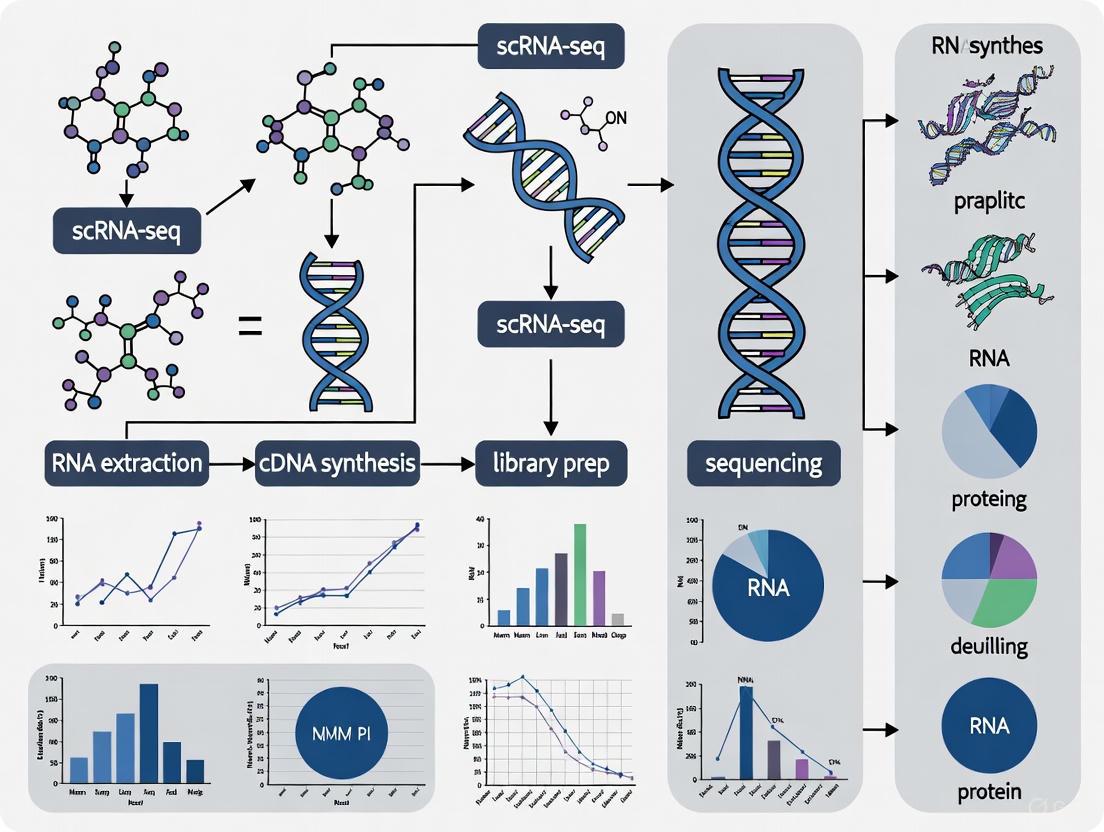
Benchmarking scRNA-seq Platforms for Embryo Research: A Guide to Technology Selection and Experimental Optimization
Single-cell RNA sequencing has revolutionized our understanding of early embryonic development, providing unprecedented insights into lineage specification and cellular heterogeneity.
Benchmarking scRNA-seq Platforms for Embryo Research: A Guide to Technology Selection and Experimental Optimization
Abstract
Single-cell RNA sequencing has revolutionized our understanding of early embryonic development, providing unprecedented insights into lineage specification and cellular heterogeneity. This article provides a comprehensive, practical guide for researchers evaluating scRNA-seq platforms for embryo work. We explore the foundational principles of scRNA-seq technology, compare the methodological strengths and limitations of major commercial platforms, address key troubleshooting and optimization strategies for precious embryonic samples, and outline robust validation frameworks for benchmarking embryo models. By synthesizing current literature and multi-platform benchmarking studies, this resource aims to empower developmental biologists to select the most appropriate technological and bioinformatic approaches for their specific research questions, ultimately accelerating discoveries in human embryogenesis and stem cell biology.
Understanding scRNA-seq Fundamentals in Embryo Development Research
Single-cell RNA sequencing (scRNA-seq) has fundamentally transformed our ability to study early human development by enabling unprecedented resolution of cellular heterogeneity and lineage specification. This technology allows researchers to profile the transcriptome of individual cells, providing insights into the dynamic gene expression patterns that guide the transformation of a single zygote into a complex gastrulating embryo. The application of scRNA-seq is particularly crucial for understanding human embryogenesis, given the ethical restrictions and technical challenges associated with working with human embryos, especially beyond the 14-day post-fertilization limit [1] [2]. These limitations have driven the development of stem cell-based embryo models, whose fidelity must be validated against in vivo references—a process where scRNA-seq has become indispensable [1].
The period from zygote to gastrula represents a remarkably complex and coordinated sequence of events encompassing maternal-to-zygotic transition, lineage specification, and the establishment of the basic body plan during gastrulation [2]. scRNA-seq has enabled researchers to decode these processes by mapping transcriptional landscapes, identifying novel cell states, and reconstructing developmental trajectories. This review examines how different scRNA-seq platforms perform in the context of embryo research, providing experimental data and comparative analyses to guide researchers in selecting appropriate methodologies for studying embryogenesis.
Technical Foundations of scRNA-seq in Embryonic Research
Core Methodological Principles and Workflow
A standard scRNA-seq workflow involves multiple critical steps, each contributing to the quality and interpretability of the resulting data. The process begins with single-cell dissociation from embryonic tissues, followed by single-cell isolation using either droplet-based or microwell-based technologies [3]. Library construction then incorporates cellular barcodes to tag mRNA from each cell and, in many protocols, unique molecular identifiers (UMIs) to distinguish between amplified copies of the same mRNA molecule and reads from separate mRNA molecules of the same gene [3]. After sequencing, raw data processing involves quality control, demultiplexing, genome alignment, and quantification to produce count matrices for downstream analysis [3].
Quality control is particularly crucial when working with precious embryonic samples. Researchers must carefully evaluate three key QC covariates: the number of counts per barcode (count depth), the number of genes per barcode, and the fraction of counts from mitochondrial genes per barcode [3]. Barcodes with low count depth, few detected genes, and high mitochondrial content often indicate dying cells or cells with broken membranes, while those with unexpectedly high counts and gene numbers may represent doublets [3]. These metrics must be considered jointly to avoid filtering out biologically relevant cell populations, such as quiescent cells or respiratory-active cells with naturally high mitochondrial content [3].
Analytical Approaches for Developmental Data
Once quality-controlled data is obtained, several analytical approaches are specifically valuable for embryonic development studies. Trajectory inference methods like Slingshot can reconstruct developmental paths and order cells along pseudotime, revealing the dynamics of gene expression during lineage specification [1]. RNA velocity analysis leverages the ratio of unspliced to spliced mRNAs to predict future cell states and directionality of development [4]. Additionally, regulatory network inference through tools like SCENIC (Single-Cell Regulatory Network Inference and Clustering) can identify key transcription factors driving lineage decisions [1].
The integration of multiple datasets has proven particularly powerful for creating comprehensive reference atlases. For example, the integration of six published human datasets covering development from zygote to gastrula has enabled the construction of a universal embryogenesis reference that can be used to authenticate stem cell-based embryo models [1]. Such integrated resources provide a standardized framework for benchmarking cellular identities and states during development.
Comparative Performance of scRNA-seq Platforms
Selecting an appropriate scRNA-seq platform requires careful consideration of multiple performance metrics, each with implications for embryonic research. The table below summarizes the key characteristics of four commercially available platforms based on current evaluations.
Table 1: Performance Comparison of scRNA-seq Platforms
| Platform | Technology | Throughput (cells/run) | Capture Efficiency | Key Strengths | Sample Compatibility | Cost Advantage |
|---|---|---|---|---|---|---|
| 10x Genomics Chromium | Droplet-based | Up to 80,000 | ~65% | High throughput, strong reproducibility | Fresh, frozen, FFPE | Moderate |
| 10x Genomics FLEX | Droplet-based | Up to 128 samples per chip | ~65% | FFPE compatibility, multiplexing power | FFPE, PFA-fixed | Moderate to High |
| BD Rhapsody | Microwell-based | Adjustable | Up to 70% | Protein+RNA integration, lower viability tolerance | Fresh, frozen (65% viability) | Moderate |
| MobiDrop | Droplet-based | Adjustable | Not specified | Cost-effective, automated workflow | Fresh, frozen, FFPE | High |
Platform performance significantly impacts the ability to resolve rare cell types and transitional states during embryogenesis. Studies comparing 10x Chromium and BD Rhapsody in complex tissues have found that while both platforms show similar gene sensitivity, they exhibit cell type detection biases [5]. For instance, BD Rhapsody demonstrated lower proportion of endothelial and myofibroblast cells, while 10x Chromium showed lower gene sensitivity in granulocytes [5]. BD Rhapsody also consistently shows higher mitochondrial content, which could be advantageous for detecting metabolic states or potentially problematic if interpreted as stress response [5].
The source of ambient RNA contamination—a significant concern when working with limited embryonic material—also differs between plate-based and droplet-based platforms, necessitating different bioinformatic correction approaches [5]. These platform-specific characteristics must be considered during experimental design, as they can profoundly influence the detection of critical transitional populations during embryonic development.
Experimental Design and Methodological Considerations
Standardized Processing and Analysis Pipeline
To ensure reproducibility and minimize batch effects in embryogenesis studies, researchers should implement standardized processing pipelines. A comprehensive human embryo reference dataset was created by reprocessing six published datasets using the same genome reference (GRCh38) and annotation through a standardized pipeline [1]. This approach included mapping and feature counting with consistent parameters, followed by data integration using fast mutual nearest neighbor (fastMNN) methods to correct for technical variations while preserving biological signals [1].
For downstream analysis, the following workflow represents current best practices:
Figure 1: Standard scRNA-seq analysis workflow for embryonic development studies
Special Considerations for Embryonic Samples
Working with embryonic materials presents unique challenges that require methodological adaptations. Limited sample availability necessitates protocols that maximize cell recovery, while the rapidly changing developmental states demand high sensitivity to capture transient expression patterns. For precious archival specimens, such as historically collected human embryos, 10x Genomics FLEX offers particular advantages due to its compatibility with FFPE-preserved tissues [6].
Cell viability can be a significant concern, particularly for clinical embryonic samples or delicate early-stage embryos. In such cases, BD Rhapsody's tolerance for lower-viability suspensions (~65%) provides an important advantage over other platforms that require higher viability thresholds [6]. Additionally, the ability to combine transcriptomic with protein readouts through CITE-seq or AbSeq technologies makes BD Rhapsody particularly valuable for immunology-focused developmental studies [6].
When studying human gastrulation, researchers have successfully applied Smart-Seq2 protocol, which provides full-length transcript coverage enabling differentiation between transcript isoforms—a crucial capability for understanding regulatory mechanisms during this dynamic period [4]. This protocol detected a median of 4,000 genes per cell in a Carnegie Stage 7 human gastrula, sufficient to identify 11 distinct cell populations including epiblast, primitive streak, nascent mesoderm, and various extraembryonic lineages [4].
Key Research Reagents and Materials
Table 2: Essential Research Reagents for scRNA-seq Embryogenesis Studies
| Reagent Category | Specific Examples | Function in Experimental Workflow |
|---|---|---|
| Library Preparation Kits | 10x Chromium Next GEM Single Cell 3' Reagent Kits, BD Rhapsody Cartridge & Magnetic Beads | Barcode cells and mRNA molecules, prepare sequencing libraries |
| Sample Preservation Solutions | TRIzol, RNAlater, Paraformaldehyde (PFA) | Stabilize RNA states in precious embryonic samples |
| Cell Dissociation Reagents | Trypsin-EDTA, Collagenase, Accutase | Generate single-cell suspensions from embryonic tissues |
| Viability Stains | Trypan Blue, Propidium Iodide, DAPI | Assess cell integrity before loading on platform |
| RNA Extraction Kits | Qiagen RNeasy, Zymo Research Quick-RNA | Isolate RNA for quality control assessment |
| Bioinformatic Tools | Seurat, Scanpy, SCENIC, Slingshot | Analyze sequencing data, identify cell types and trajectories |
Application to Embryogenesis: From Zygote to Gastrula
Decoding Preimplantation Development
scRNA-seq has revealed the remarkable transcriptional dynamics during human preimplantation development. Studies analyzing nearly 2,000 individual cells from human preimplantation embryos have documented the highly dynamic transcriptome reflective of maternal-to-zygotic transition (MZT) and the differentiation of blastomeres into three distinct lineages [2]. The most significant shift in gene expression occurs between the four- and eight-cell stages, coinciding with major zygotic genome activation (ZGA) [2].
Research by Yan et al. identified 22,687 expressed genes during preimplantation development, including 8,701 long non-coding RNAs—far exceeding what was previously detectable by cDNA microarrays [2]. The upregulation of approximately 2,500 genes at the eight-cell stage showed strong enrichment for RNA metabolism and translation, chromosome organization, cell division, and DNA packaging—all hallmark processes of ZGA [2]. Additionally, the persistence of maternal mRNA degradation through the morula stage and the late activation of Y chromosomal genes demonstrate that ZGA remains incomplete at the eight-cell stage [2].
Lineage specification becomes transcriptionally apparent at the blastocyst stage, with clear markers distinguishing the three foundational lineages: NANOG and SOX2 for the epiblast (EPI), GATA4 and PDGFRA for the primitive endoderm (PrE), and GATA2 and GATA3 for the trophectoderm (TE) [2]. The functional specialization of these lineages is reflected in their enriched gene ontology terms, with EPI genes associated with stem cell maintenance, PrE genes with morphogenesis of epithelium and endoderm development, and TE genes with apical plasma membrane and transporter activity [2].
Unveiling Human Gastrulation
Gastrulation represents a pivotal but poorly understood stage of human development, largely due to limited access to in utero samples. The first transcriptomic characterization of an entire gastrulating human embryo (Carnegie Stage 7, approximately 16-19 days post-fertilization) identified 11 distinct cell populations through unsupervised clustering [4]. These included epiblast, primitive streak, nascent mesoderm, axial mesoderm, emergent mesoderm, advanced mesoderm, extraembryonic mesoderm, endoderm, hemato-endothelial progenitors, and erythroblasts [4].
RNA velocity and diffusion map analyses revealed trajectories from the epiblast along two broad streams corresponding to mesoderm and endoderm, separated along the second diffusion component [4]. The first diffusion component closely corresponded to both cell type and spatial location, reflecting the extent of differentiation and the 'temporal age' of cells based on when they emerged from the epiblast [4]. This ordering showed that extraembryonic mesoderm cells, which emerge relatively early during gastrulation, plotted further from the epiblast than axial mesoderm cells that emerge later [4].
Comparative analysis between human and mouse gastrulation identified both conserved and species-specific expression patterns. Of 662 genes differentially expressed along the trajectory from epiblast to nascent mesoderm in both species, 531 shared the same trend—either increasing (117 genes) or decreasing (414 genes) [4]. Conserved patterns included decreased CDH1, transient TBXT expression, and continuously increasing SNAI1 [4]. However, species-specific differences emerged for genes such as SNAI2 (upregulated only in human), TDGF1 (opposing trends), and FGF8 (transient expression in mouse only) [4], highlighting the importance of direct human embryonic research rather than relying solely on model organisms.
Integrated Embryo Reference Tools and Future Directions
The creation of comprehensive reference atlases represents a significant advancement for the field. Researchers have recently developed an integrated human embryo reference through the combination of six published datasets covering developmental stages from zygote to gastrula [1]. This resource includes 3,304 early human embryonic cells embedded into a unified transcriptional space using stabilized Uniform Manifold Approximation and Projection (UMAP), displaying continuous developmental progression with temporal and lineage specification [1].
This reference enables the Early Embryogenesis Prediction Tool, where query datasets can be projected onto the reference and annotated with predicted cell identities [1]. Application of this tool to published human embryo models has revealed the risk of misannotation when relevant references are not utilized for benchmarking, underscoring the importance of such integrated resources for authenticating in vitro models [1]. The reference has been complemented with SCENIC analysis to capture transcription factor activities across embryonic time points, identifying key regulators such as DUXA in 8-cell lineages, VENTX in the epiblast, OVOL2 in the trophectoderm, and ISL1 in amnion [1].
Slingshot trajectory inference based on this integrated reference has revealed three main developmental trajectories related to epiblast, hypoblast, and trophectoderm lineages, identifying 367, 326, and 254 transcription factor genes respectively that show modulated expression with pseudotime [1]. This analysis provides a foundation for functional characterization of key transcription factors driving lineage specification in early human development.
As the field advances, future directions will likely include multi-omic approaches combining transcriptomics with epigenomic, proteomic, and spatial information. The continued refinement of stem cell-based embryo models, validated against these increasingly comprehensive references, will further enhance our understanding of human embryogenesis while addressing ethical constraints. These advancements, coupled with ongoing improvements in scRNA-seq technologies' sensitivity, throughput, and cost-effectiveness, promise to unravel the remaining mysteries of early human development.
Single-cell RNA sequencing (scRNA-seq) has revolutionized biological research by enabling the detailed examination of gene expression at the individual cell level. This capability is particularly crucial for understanding complex biological systems such as developing embryos, where cellular heterogeneity plays a fundamental role in development and disease. The selection of an appropriate scRNA-seq platform is a critical decision that directly impacts data quality, experimental scale, and biological insights. This guide provides an objective comparison of the three principal technological approaches—droplet-based, microwell-based, and plate-based platforms—with a specific focus on their application in embryo research.
Platform Working Principles and Mechanisms
Droplet-Based Microfluidics
Droplet-based platforms utilize microfluidic technology to partition individual cells into nanoliter-scale aqueous droplets within an oil emulsion [7] [8]. The process begins when an aqueous suspension containing cells is combined with barcoded beads and oil within a microfluidic chip [9]. This system generates thousands of Gel Bead-In-Emulsions (GEMs), where each droplet ideally contains a single cell and a single barcoded bead [9]. Within these compartments, cells are lysed, and their mRNA molecules are released and captured by the barcoded beads via poly(dT) primers [8] [9]. Each bead contains oligonucleotides with a cell barcode, a unique molecular index (UMI), and poly(dT) sequences [9]. The UMIs are critical for correcting amplification bias and enabling accurate transcript quantification [9]. Following reverse transcription, the emulsions are broken, and the cDNA is amplified and prepared for sequencing [8]. The 10x Genomics Chromium system represents the most widely adopted commercial implementation of this technology [10].
Microwell-Based Platforms
Microwell-based systems employ physical arrays of tiny wells to isolate individual cells [8]. In this approach, a chip containing hundreds of thousands of microwells is first loaded with uniquely barcoded beads [8] [9]. As the beads settle by gravity, they ideally occupy individual wells. A cell suspension is then loaded onto the chip, allowing cells to sediment into the wells [9]. The system's dimensions are optimized to minimize double occupancy of both beads and cells [9]. After cell capture, cells are lysed, and their RNA hybridizes to the barcoded beads in a process similar to droplet-based methods [8]. The BD Rhapsody system is a prominent example of a commercial microwell-based platform [9]. A key advantage of this method is the ability to wash away cell-free RNAs before cell lysis, potentially reducing ambient RNA contamination [11].
Plate-Based Methods with Combinatorial Indexing
Plate-based methods, the earliest scRNA-seq approach, initially utilized fluorescence-activated cell sorting (FACS) to distribute individual cells into separate wells of multiwell plates [7] [8]. Modern implementations have evolved to use combinatorial indexing strategies to significantly increase throughput [8]. In these methods, fixed and permeabilized cells are distributed into wells of a plate (96, 384, or 1,536 wells), where the RNA is reverse transcribed with a well-specific barcode [8]. All cells are then pooled, mixed, and redistributed into a second plate for a second round of barcoding [8]. The combination of barcodes allows sequencing reads to be assigned to single cells, enabling the processing of up to 1 million cells through multiple rounds of barcoding [8]. Parse Biosciences' Evercode technology is a leading example of this approach [8].
Figure 1: Workflow comparison of the three main scRNA-seq platform technologies. Each method employs distinct physical partitioning and barcoding strategies to achieve single-cell resolution.
Performance Comparison and Experimental Data
Key Performance Metrics
When evaluating scRNA-seq platforms, researchers consider several critical performance metrics that directly impact data quality and biological interpretation. Gene sensitivity refers to the number of genes detected per cell, which affects the ability to identify cell types and states [5]. Cell throughput determines how many cells can be profiled in a single experiment, important for capturing rare cell populations [8]. Mitochondrial content can indicate cell stress or damage during processing [5]. Ambient RNA contamination occurs when RNA from lysed cells is captured by barcoded beads, creating background noise [5]. Reproducibility and cell type representation biases are crucial for accurate biological interpretation [5].
Table 1: Comprehensive Performance Comparison of scRNA-seq Platforms
| Performance Metric | Droplet-Based (10x Chromium) | Microwell-Based (BD Rhapsody) | Plate-Based (Combinatorial Indexing) |
|---|---|---|---|
| Gene Sensitivity | Similar to BD Rhapsody [5] | Similar to 10x Chromium [5] | Highest sensitivity [8] |
| Cell Throughput | Highest (Commercial systems) [8] | Intermediate [8] | Lowest (though combinatorial indexing improves scalability) [8] |
| Mitochondrial Content | Lower than BD Rhapsody [5] | Highest reported [5] | Varies by protocol |
| Ambient RNA | Droplet-specific contamination patterns [5] | Well-specific contamination patterns [5] | Potentially lower due to washing steps |
| Cell Type Representation | Lower sensitivity for granulocytes [5] | Lower proportion of endothelial/myofibroblast cells [5] | Depends on specific protocol |
| Doublet Rate | Controlled by microfluidics and computational methods [10] | Controlled by well dimensions and computational methods [8] | Lower due to combinatorial barcoding |
| Multiplexing Capability | Compatible with cell hashing [9] | Compatible with cell hashing [9] | Built-in multiplexing through combinatorial indexing [8] |
Direct Comparative Studies
A systematic comparison of 10x Chromium and BD Rhapsody platforms using complex tumor tissues revealed several important performance differences. The study examined both fresh and artificially damaged samples, providing insights into platform performance under challenging conditions [5]. While both platforms demonstrated similar gene sensitivity, they exhibited distinct patterns of cell type detection biases [5]. The BD Rhapsody platform detected a lower proportion of endothelial and myofibroblast cells, whereas the 10x Chromium system showed lower gene sensitivity specifically in granulocytes [5]. Additionally, the sources and patterns of ambient RNA contamination differed between the platforms, reflecting their fundamental technological differences [5].
Another comparative analysis highlighted that droplet-based systems like 10x Genomics Chromium utilize gel emulsion microbeads, while microwell-based systems like BD Rhapsody employ magnetic beads [9]. These differences in bead chemistry and reactor design contribute to variations in performance characteristics, including cDNA conversion efficiency and recovery rates [9].
Table 2: Technical Specifications and Experimental Considerations
| Parameter | Droplet-Based | Microwell-Based | Plate-Based |
|---|---|---|---|
| Single-Cell Partitioning | Microfluidic droplets [8] | Physical microwell array [8] | Multiwell plates or combinatorial indexing [8] |
| Barcoding Method | Beads with cell barcode and UMI [9] | Beads with cell barcode and UMI [9] | Well-specific barcodes or combinatorial indexing [8] |
| Cell/Bead Pairing Efficiency | Poisson distribution-dependent (<1% in early systems) [11] | High pairing rate (~80%) [11] | Defined by plate well number |
| Cost Per Cell | Lowest (due to miniaturization) [8] | Intermediate [8] | Highest (due to greater reagent consumption) [8] |
| Equipment Requirements | Expensive microfluidics instrument [8] | Specialized chip [8] | Standard lab equipment (pipettes, centrifuges) [8] |
| Workflow Complexity | Highly automated [8] | Partially automated [8] | Flexible but labor intensive [8] |
| Sample Multiplexing | Compatible with cell hashing [9] | Compatible with cell hashing [9] | Built-in multiplexing capability [8] |
Application in Embryo Research
Special Considerations for Embryo Studies
Embryo research presents unique challenges for scRNA-seq applications. The limited biological material available from early embryonic stages necessitates highly sensitive platforms that can work with small cell numbers [1] [2]. The dynamic nature of embryonic development requires platforms that can capture rapid transcriptional changes and identify transitional cell states [1]. Additionally, researchers must navigate ethical and legal constraints, particularly the "14-day rule" that limits experimentation on human embryos beyond this developmental stage [1] [2]. These limitations have driven the development of stem cell-based embryo models that require careful validation against in vivo references [1].
Reference Tools for Embryo Model Validation
Comprehensive reference datasets have been developed to validate stem cell-derived embryo models against natural embryonic development. One such resource integrates six published human datasets covering developmental stages from zygote to gastrula [1]. This reference contains expression profiles of 3,304 early human embryonic cells and provides a high-resolution transcriptomic roadmap of early human development [1]. The tool employs stabilized Uniform Manifold Approximation and Projection (UMAP) for visualization and allows researchers to project query datasets onto the reference to annotate cell identities [1]. Such resources are essential for authenticating embryo models and ensuring their fidelity to in vivo counterparts at molecular, cellular, and structural levels [1].
Platform Selection for Embryo Research
For embryo research, the choice of scRNA-seq platform depends on specific experimental needs. Droplet-based systems offer high throughput for comprehensive profiling of heterogeneous embryonic cell populations [1]. Microwell-based platforms provide a balance between throughput and sensitivity, potentially advantageous for working with limited embryo material [8] [9]. Plate-based methods with combinatorial indexing enable massive scaling when building detailed atlases of embryonic development across multiple stages [8]. The compatibility with multi-omic measurements (simultaneous analysis of transcriptome, surface proteins, and immune repertoire) is particularly valuable for comprehensively characterizing embryonic cell types and states [9].
Detailed Experimental Protocols
Sample Preparation for Embryo Work
Proper sample preparation is critical for successful scRNA-seq experiments with embryonic material. The decision between using whole cells versus nuclei depends on the research question and sample characteristics [12]. For challenging tissues or archived samples, nuclei sequencing often provides a more robust alternative [12]. Maintaining temperature control throughout sample processing is essential, as holding cells at 4°C helps arrest metabolic functions and reduces stress-related gene expression [12]. Sample viability should ideally be between 70% and 90%, with minimal cell clumping and debris (<5% aggregation) [12]. For embryonic tissues, gentle dissociation protocols that preserve cell integrity are paramount, potentially utilizing enzyme cocktails specifically formulated for delicate tissues [12].
Quality Control and Validation
Rigorous quality control measures are essential for generating reliable scRNA-seq data from embryo studies. Species-mixing experiments represent a gold-standard approach for quantifying doublet rates, where human and mouse cells are mixed and processed together [10]. The resulting "barnyard plots" allow clear identification of heterotypic doublets through their mixed-species expression profiles [10]. For embryo-specific work, projection onto reference atlases enables quality assessment and validation of cell type identities [1]. Computational methods for ambient RNA correction and doublet detection should be routinely applied, particularly for embryonic data where cell states may be transitional and poorly defined [10].
Innovative Methods for Temporal Dynamics
Understanding the temporal dynamics of gene expression is particularly important in embryo development studies. Well-TEMP-seq represents an innovative microwell-based method that combines metabolic RNA labeling with scRNA-seq to distinguish newly transcribed RNAs from pre-existing RNAs in single cells [11]. This approach utilizes 4-thiouridine (4sU) labeling and subsequent chemical conversion to mark newly synthesized transcripts with T-to-C substitutions [11]. The method achieves a high single cell/barcoded bead pairing rate (~80%) and significantly reduces cell loss compared to previous approaches [11]. Such temporal resolution methods are particularly valuable for embryo research, where developmental processes involve rapid transcriptional changes.
The Scientist's Toolkit
Table 3: Essential Research Reagent Solutions for scRNA-seq in Embryo Research
| Reagent/Consumable | Function | Platform Compatibility |
|---|---|---|
| Barcoded Beads with Oligo(dT) | Captures polyadenylated mRNA and provides cell barcodes/UMIs | All platforms (chemistry varies) [8] [9] |
| Cell Hashing Antibodies | Enables sample multiplexing and doublet identification | All platforms [9] |
| Feature Barcoding Antibodies | Measures surface protein expression alongside transcriptome | All platforms (CITE-seq) [9] |
| Enzyme Dissociation Cocktails | Generates single-cell suspensions from embryonic tissues | All platforms [12] |
| Viability Stains | Assesses cell integrity and viability before processing | All platforms [12] |
| 4-Thiouridine (4sU) | Metabolic RNA labeling for temporal dynamics studies | Compatible with various platforms [11] |
| Iodoacetamide (IAA) | Chemical conversion for 4sU-based RNA labeling | Specific to metabolic labeling protocols [11] |
| Proteinase K | Cell lysis agent, particularly in microfluidics-free methods | Specific to protocols like PIP-seq [13] |
Figure 2: Experimental design decision framework for scRNA-seq studies in embryo research. The framework highlights key considerations across sample preparation, platform selection, and workflow optimization.
Emerging Technologies and Future Directions
The field of single-cell genomics continues to evolve rapidly, with new technologies addressing current limitations. Microfluidics-free approaches like Particle-templated Instant Partition Sequencing (PIP-seq) represent a promising direction, offering scalability and flexibility without specialized equipment [13]. This method uses particle-templated emulsification to compartmentalize cells and barcoded hydrogel templates using only a vortexer, making single-cell sequencing more accessible [13]. Multi-omic integrations that combine transcriptome with epigenome, proteome, and spatial information are becoming increasingly important for comprehensive profiling of embryonic development [7] [9]. Spatial transcriptomics technologies that preserve positional information are particularly valuable for embryo research, where spatial organization is fundamental to developmental processes [7]. As these technologies mature, they will provide increasingly powerful tools for unraveling the complexities of embryonic development at single-cell resolution.
Embryo research occupies a uniquely challenging niche in single-cell genomics. Unlike many other biological systems where sample material can be readily replenished, early human embryos represent an exceptionally limited and precious resource. This scarcity is compounded by technical challenges of working with minimal cell numbers and ethical considerations surrounding their use. These constraints create a research landscape where every cell counts, and optimization of single-cell RNA sequencing (scRNA-seq) approaches becomes paramount. This guide examines the distinctive challenges of embryo scRNA-seq work and provides objective performance comparisons of platforms suited for this specialized application.
Technical Hurdles in Embryo scRNA-seq
Working with embryonic material presents a constellation of technical challenges that differentiate it from other scRNA-seq applications:
Extreme Sample Limitation
The fundamental challenge in embryo research is the very limited number of cells available for analysis. Early human development involves progressively increasing cell numbers: from a single zygote to approximately 3,304 cells captured in integrated datasets from zygote to gastrula stages [1]. Each embryonic cell represents a disproportionately large fraction of the total available biological material, making cell loss during processing scientifically catastrophic.
Sample Heterogeneity and Dynamic Transitions
Early embryonic development is characterized by rapid cellular differentiation and progressive lineage specification. The first lineage branch point occurs as inner cell mass and trophectoderm cells diverge during E5, followed by bifurcation of ICM cells into epiblast and hypoblast [1]. This means that even within a single embryo, cells may represent fundamentally different developmental trajectories and states.
Technical Noise and Sensitivity Issues
The low RNA input from limited embryonic cells exacerbates technical challenges including:
- Amplification bias from stochastic variation in amplification efficiency
- Dropout events where transcripts fail to be captured or amplified
- Batch effects between different experimental runs [14]
These issues are particularly problematic when studying rare cell populations or low-abundance transcripts critical for understanding developmental transitions.
Platform Performance Comparison for Embryo Work
Selecting an appropriate scRNA-seq platform requires careful consideration of performance characteristics particularly relevant to embryo research. The table below summarizes key metrics from benchmarking studies:
Table 1: scRNA-seq Platform Performance Comparison for Embryo Research Applications
| Platform | Gene Sensitivity | Cell Type Detection Biases | Mitochondrial Content | Ambient RNA Control | Suitability for Low Cell Input |
|---|---|---|---|---|---|
| 10× Chromium | High gene sensitivity | Lower sensitivity in granulocytes | Moderate | Droplet-based contamination profile | Good for standard inputs |
| BD Rhapsody | Similar sensitivity to 10x | Lower proportion of endothelial/myofibroblast cells | Highest mitochondrial content | Plate-based contamination profile | Good for standard inputs |
| Smart-seq2 | Highest sensitivity per cell | Limited by manual processing | Variable | Well-based isolation | Excellent for low cell numbers |
| DRUG-seq | Moderate sensitivity | Less characterized | Moderate | Well-based control | Good for targeted approaches |
Data derived from performance comparisons in complex tissues [5] and embryo-specific studies [1].
Key Performance Differentiators
- Gene Sensitivity: Critical for detecting low-abundance transcription factors driving developmental transitions
- Cell Type Representation: Platform-specific biases affect ability to resolve rare embryonic populations
- Technical Artifacts: Mitochondrial content and ambient RNA vary by platform and impact data quality
Experimental Design Considerations
Sample Preparation Protocols
Optimized sample preparation is crucial for embryonic material. Key methodological considerations include:
- Cell Dissociation: Gentle protocols to preserve RNA integrity while achieving single-cell suspensions
- Viability Assessment: Critical for ensuring data quality from precious samples
- Minimal Handling: Reduced processing steps to minimize cell loss [14]
Quality Control Metrics
Rigorous QC is essential when working with limited embryonic cells:
- Cell Doublet Identification: Especially important when analyzing pooled embryos
- Batch Effect Mitigation: Technical variation can confound biological signals
- Dropout Imputation: Computational correction for missing data [14]
Analytical Framework for Embryo scRNA-seq Data
The analytical workflow for embryonic scRNA-seq data requires specialized approaches to address unique challenges:
Embryo scRNA-seq Analysis Workflow
Reference-Based Annotation
Given the well-defined lineage relationships in embryonic development, reference-based approaches are particularly powerful. Integrated reference datasets covering human development from zygote to gastrula stages enable more accurate cell type identification [1]. These resources provide:
- Lineage Annotation Standards: Consistent classification of embryonic cell types
- Developmental Trajectory Mapping: Pseudotemporal ordering of cells along differentiation paths
- Transcription Factor Activity: Inference of regulatory dynamics using tools like SCENIC [1]
Trajectory Inference Methods
For analyzing embryonic development, trajectory inference approaches are essential:
- Slingshot: Models branching lineages from reduced dimension embeddings
- Pseudotime Analysis: Orders cells along developmental continuums
- RNA Velocity: Predicts future cell states from splicing dynamics
Research Reagent Solutions for Embryo scRNA-seq
Table 2: Essential Research Reagents and Platforms for Embryo scRNA-seq
| Reagent/Platform | Function | Application in Embryo Work |
|---|---|---|
| Unique Molecular Identifiers (UMIs) | Correction for amplification bias | More accurate quantification of scarce transcripts |
| Cell Hashing | Multiplexing samples | Enables pooling of limited embryonic material |
| Spike-in Controls | Technical normalization | Account for platform-specific sensitivity differences |
| Viability Stains | Cell quality assessment | Prevent sequencing of compromised cells |
| Gentle Dissociation Kits | Tissue processing | Preserve RNA integrity from delicate embryonic cells |
| Smart-seq2 Reagents | High-sensitivity full-length | Optimal for very low cell input applications |
| 10x Chromium | High-throughput profiling | Suitable when cell numbers permit droplet approaches |
Integration with Complementary Technologies
Given the limitations of scRNA-seq alone, multi-modal approaches are particularly valuable in embryo research:
Spatial Transcriptomics
scRNA-seq inherently loses spatial context, which is critical for understanding embryonic patterning [15]. Spatial transcriptomics technologies preserve positional information:
- Sequencing-based platforms (Visium, Stereo-seq) for unbiased transcriptome capture
- Imaging-based platforms (Xenium, CosMx) for targeted high-resolution mapping [16]
Multi-omics Integration
Combining scRNA-seq with other data modalities enhances insights:
- ATAC-seq: Chromatin accessibility for regulatory state
- Proteomics: Protein expression validation
- CODEX: Spatial protein mapping [16]
Future Directions and Emerging Solutions
The field continues to evolve with promising approaches to address embryo-specific challenges:
- Low-Input Protocol Refinement: Methods requiring fewer cells while maintaining data quality
- Computational Imputation: Enhanced algorithms for correcting technical artifacts
- Integrated Reference Atlases: Comprehensive developmental maps for annotation
- Multi-ome Technologies: Simultaneous measurement of transcriptome and epigenome from same cells
Embryo scRNA-seq work presents unique challenges that demand specialized approaches from experimental design through computational analysis. The limited availability of embryonic material, dynamic nature of early development, and technical sensitivity requirements necessitate careful platform selection and methodological optimization. By understanding these distinctive challenges and leveraging appropriate technologies and analytical frameworks, researchers can maximize insights from these precious samples while advancing our understanding of early human development.
Single-cell RNA sequencing (scRNA-seq) has revolutionized the study of embryonic development by enabling researchers to investigate cellular heterogeneity, lineage relationships, and fate decisions at unprecedented resolution. Unlike bulk RNA sequencing, which averages gene expression across thousands of cells, scRNA-seq captures the transcriptome of individual cells, revealing rare populations and continuous transitional states that are fundamental to understanding embryogenesis [17]. This technology has become particularly valuable for studying human development, where ethical and technical limitations restrict access to embryo samples [2]. Within this context, three core applications have emerged as particularly impactful: reconstructing lineage trajectories, elucidating cell fate decisions, and validating stem cell-derived embryo models.
This guide objectively compares how different scRNA-seq platforms and methodological approaches address these core applications, with a specific focus on research using human embryos and embryo models. We summarize performance metrics based on published studies and provide detailed experimental protocols to facilitate reproducible research.
Application 1: Lineage Tracing
Technology Comparison
Lineage tracing aims to identify all progeny arising from an individual cell, placing them within a lineage hierarchy [18]. Modern scRNA-seq approaches to lineage tracing can be broadly categorized into two strategies: inferential methods that reconstruct lineages from transcriptomic similarity, and experimental methods that combine scRNA-seq with heritable genetic recorders.
Table 1: Comparison of scRNA-seq-Based Lineage Tracing Strategies
| Method Type | Key Technology | Resolution | Throughput | Key Applications in Embryo Research | Limitations |
|---|---|---|---|---|---|
| Inferential Lineage Tracing | Computational trajectory inference (e.g., Slingshot, Monocle) | Medium (Population-level dynamics) | High (Standard scRNA-seq workflows) | Mapping developmental trajectories from zygote to gastrula [1] | Hypothetical relationships; cannot account for independent lineages converging on similar states [19] |
| Experimental Lineage Tracing | Genetic barcoding (e.g., CRISPR recorders, Polylox) | High (Clonal-level resolution) | Medium to High | Validating inferred trajectories in embryoid bodies [20]; Clonal analysis in organogenesis | Complex experimental design; potential barcode silencing |
| Integrated Approaches | Parallel scRNA-seq and genetic recording | Very High (Direct clonal relationships with transcriptomic state) | Medium | Pinpointing fate decision timing (e.g., PGC specification) [20] | Most complex implementation; specialized analysis required |
Experimental Protocol: Parallel scRNA-seq and Genetic Recording
This protocol, adapted from [20], details how to combine inducible genetic recording with scRNA-seq to validate lineage trajectories in developing embryoid bodies (EBs).
Principle: An inducible Cre recombinase activates a stochastic genetic switch to generate unique, heritable barcodes in progenitor cells during a narrow temporal window. Subsequent scRNA-seq simultaneously captures transcriptomic states and lineage barcodes from the same single cells.
Step-by-Step Workflow:
Cell Engineering: Generate embryonic stem cells (ESCs) containing:
- An inducible Cre-ER(^T2) gene under a constitutive promoter.
- A Lox–STOP–Lox (LSL) cassette followed by a diverse array of possible fluorescent proteins or transcriptomic barcodes (e.g., in the Rosa26 safe-harbor locus).
Temporal Barcode Induction: Differentiate EBs from the engineered ESCs. At the developmental time point of interest (e.g., day 2-4 of EB differentiation), add 4-Hydroxytamoxifen (4-OHT, 500 nM) for a short pulse (e.g., 6-12 hours) to induce nuclear translocation of Cre-ER(^T2).
Stochastic Recombination: Cre recombinase excises the STOP cassette in a random subset of cells, leading to irreversible activation of a random fluorescent protein/barcode combination. This barcode is stably inherited by all progeny of the labeled progenitor.
Single-Cell Capture and Library Preparation: At endpoint(s) of interest (e.g., day 7-10 EBs), dissociate EBs into single-cell suspension. Use a platform such as 10x Genomics Chromium to prepare:
- A standard scRNA-seq library (3' gene expression).
- A custom targeted amplicon library to sequence the lineage barcode region from the same cells.
Sequencing and Data Integration: Sequence libraries on an Illumina platform. Process data with the following steps:
- Align scRNA-seq reads to the reference genome (GRCh38) using Cell Ranger [21] or STARsolo.
- Cluster cells based on transcriptomic similarity and visualize using UMAP.
- Assign lineage barcodes to each cell and superimpose this clonal information onto the transcriptomic clusters.
Validation: This method validated the hypothesis that Primordial Germ Cell (PGC)-like lineage commitment in EBs occurs at the preimplantation epiblast-like stage, demonstrating the power of integrated lineage tracing for pinpointing fate decisions [20].
Figure 1: Integrated Experimental Workflow for scRNA-seq Lineage Tracing. The diagram outlines the key stages of a parallel genetic recording and scRNA-seq experiment, from cell line engineering to validated lineage reconstruction.
Application 2: Cell Fate Decisions
Analyzing Developmental Transitions
Cell fate decisions are the fundamental processes where a multipotent progenitor cell chooses a specific differentiation path. scRNA-seq enables the dissection of these decisions by capturing cells in transitional states and ordering them along a pseudotemporal continuum [19]. This analytical process, known as trajectory inference, reconstructs the underlying developmental landscape from snapshot data.
Key Workflow for Fate Decision Analysis:
High-Quality Data Collection: Perform scRNA-seq on embryos or embryo models across multiple time points. Strict quality control is essential, filtering cells by UMI counts, genes detected, and mitochondrial read percentage [3] [21].
Data Integration and Visualization: Integrate data from multiple samples or time points using methods like fastMNN [1] or Harmony. Reduce dimensionality using UMAP or t-SNE to visualize the continuum of cell states.
Trajectory Inference: Apply algorithms (e.g., Slingshot [1], PAGA, Monocle3) to the reduced-dimensional space. These tools infer the graph structure of development, positioning root (progenitor) and leaf (differentiated) nodes.
Pseudotime Ordering: Order cells along the inferred trajectories based on transcriptomic similarity, assigning a "pseudotime" value from the start to the end of a lineage.
Identification of Fate Regulators: Analyze genes that exhibit dynamic expression along pseudotime. Transcription factors with expression patterns that correlate with branching decisions are candidate regulators of cell fate [1].
Table 2: Key Fate Decisions Resolved by scRNA-seq in Human Embryos
| Developmental Stage | Fate Decision | Key Transcriptional Regulators Identified | Reference Model |
|---|---|---|---|
| Preimplantation (E5) | ICM vs. Trophectoderm (TE) | CDX2, NR2F2 (TE); VENTX, POU5F1 (ICM/Epiblast) | Human blastocysts [1] [2] |
| Postimplantation (E7-9) | Epiblast vs. Hypoblast | GATA4, SOX17 (Hypoblast); HMGN3 (Late Epiblast) | In vitro cultured blastocysts [1] |
| Gastrulation (CS7) | Primitive Streak & Germ Layer Formation | TBXT (Primitive Streak); MESP2 (Mesoderm) | Carnegie Stage 7 human embryo [1] |
Research Reagent Solutions
Table 3: Essential Reagents and Tools for scRNA-seq Fate Mapping
| Item | Function | Example Application in Embryo Research |
|---|---|---|
| 10x Genomics Chromium | High-throughput single-cell capture and barcoding | Generating comprehensive atlases from limited human embryo samples [21] |
| Cell Ranger | Processing scRNA-seq data: alignment, filtering, UMI counting, initial clustering | Standardized processing pipeline for integrating public human embryo datasets [1] [21] |
| Cre-loxP System | Inducible genetic fate mapping; cornerstone of genetic recording | Tracing all descendants of a labeled progenitor population in vivo [22] [23] |
| R26R-Confetti Reporter | Stochastic multicolor labeling for clonal visualization | Intravital imaging of clonal dynamics in organogenesis [22] |
| SCENIC | Computational inference of transcription factor regulatory networks | Identifying key fate regulators (e.g., ISL1 in amnion) across human embryogenesis [1] |
| SoupX / CellBender | Computational removal of ambient RNA contamination | Improving data quality from complex, dissociated embryo tissues [21] |
Application 3: Embryo Model Validation
Stem cell-based embryo models (e.g., blastoids, gastruloids) offer unprecedented tools for studying early human development. A primary application of scRNA-seq is to authenticate these models by benchmarking their transcriptomic profiles against a definitive in vivo reference [1] [2].
The Need for an Integrated Reference: Indiscriminate comparison to individual published datasets carries a high risk of misannotation, as many lineages share common markers during development [1]. To address this, a universal scRNA-seq reference has been constructed by integrating six published human datasets, covering development from zygote to gastrula (Carnegie Stage 7) [1]. This resource provides a high-resolution roadmap against which embryo models can be objectively evaluated.
Standardized Validation Workflow:
Reference Building: Process multiple human embryo scRNA-seq datasets through a unified pipeline (e.g., GRCh38 alignment, standardized annotation) to minimize batch effects. Integrate data using a method like fastMNN to create a stabilized UMAP embedding [1].
Model Querying: Process scRNA-seq data from the embryo model using the same pipeline. Project the model's cells into the pre-established reference UMAP space.
Quantitative Assessment: Evaluate the fidelity of the embryo model based on:
- Transcriptomic Similarity: How closely do the model's cells cluster with their in vivo counterparts in the reference space?
- Lineage Presence and Purity: Does the model contain the correct complement of lineages (e.g., epiblast, hypoblast, TE derivatives), and are they free from misannotation?
- Developmental Progression: Do the transcriptomic trajectories (e.g., from epiblast to primitive streak to mesoderm) match those in the reference?
Performance Metric: The key metric is the successful co-embedding of model cells with their authentic in vivo counterparts, without significant overlap with incorrect lineages. This process has revealed that using irrelevant references can lead to misannotation, underscoring the need for a comprehensive and stage-matched reference tool [1].
Figure 2: Embryo Model Validation Workflow. This diagram illustrates the process of authenticating stem cell-derived embryo models by projecting their scRNA-seq data onto an integrated in vivo reference atlas.
The selection of an scRNA-seq platform and analytical approach for embryo research must be guided by the specific biological question. For lineage tracing, the gold standard is shifting from purely inferential methods to integrated approaches that combine scRNA-seq with genetic recording, providing direct experimental validation of clonal relationships. For deconstructing cell fate decisions, rigorous trajectory inference applied to high-quality data is essential for identifying branching points and key regulators. Finally, the validation of embryo models now critically depends on the use of comprehensive, integrated reference atlases to avoid misannotation and accurately assess transcriptional fidelity.
The continued advancement of scRNA-seq technologies, including increased throughput, multimodal assays (simultaneous measurement of transcriptome and epigenome), and enhanced spatial transcriptomics, will further refine these core applications. This will enable an even deeper understanding of human embryogenesis and improve the reliability of in vitro models that mimic this remarkable process.
Comparative Analysis of scRNA-seq Platforms for Embryonic Studies
Single-cell RNA sequencing (scRNA-seq) has revolutionized biological research by enabling transcriptomic profiling at the individual cell level, proving particularly valuable for investigating complex systems like embryonic development. The choice of platform significantly influences experimental outcomes, as each employs distinct technologies with specific strengths and limitations. This guide provides an objective comparison of four major commercial scRNA-seq platforms—10x Genomics Chromium, Fluidigm C1, Bio-Rad ddSEQ, and WaferGen ICELL8—focusing on their performance characteristics and relevance for embryo research.
Each platform utilizes a different methodological approach for single-cell isolation and library preparation, which directly impacts its application potential.
Platform Core Technology Workflow
The diagram above illustrates the fundamental technological differences between platforms. Droplet-based systems (10x Genomics Chromium and Bio-Rad ddSEQ) partition thousands of single cells into individual droplets using microfluidics, with each droplet containing reagents for reverse transcription and barcoding [24]. The Fluidigm C1 system uses integrated fluidic circuits (IFCs) with nanochannels to isolate single cells, followed by automated on-chip cell lysis, cDNA conversion, and pre-amplification [24] [25]. The WaferGen ICELL8 employs a nanowell-based approach where cells are dispensed into nanowells, imaged to identify wells containing single cells, and then processed for cell lysis and cDNA synthesis [24] [26].
Performance Comparison and Experimental Data
Direct comparisons across platforms reveal significant differences in throughput, sensitivity, and data quality that should inform experimental design.
Table 1: Comprehensive Platform Performance Metrics
| Platform | Technology | Throughput (Cells/Run) | Cell Capture Efficiency | Gene Detection Sensitivity | Read Depth per Cell | Key Strengths |
|---|---|---|---|---|---|---|
| 10x Genomics Chromium | Droplet Microfluidics | 1,000-80,000 cells [24] | 55-65% capturing efficiency [24]; Up to 80% cell recovery [27] | High throughput, Lower bias for high-GC content genes [24] | Economical for large cell numbers [24] | Ideal for large-scale studies, immune profiling, tumor heterogeneity [24] |
| Fluidigm C1 | Microfluidic IFC | 100-800 cells [24] | Limited by cell size and distribution [24] | High read depth per cell [24]; Full-length transcript coverage [28] | High (recommended for deep sequencing) [24] | Automated library construction, consistent results, detailed transcriptome analysis [24] |
| Bio-Rad ddSEQ | Droplet Microfluidics | 1,000-10,000 cells [24] | Varies by sample type and preparation [24] | Highest overlap in detecting highly variable genes with 10X Genomics [24]; Good for miRNA detection [24] | Moderate [24] | User-friendly, integrates well into existing workflows [24] |
| WaferGen ICELL8 | Nanowell Array | 500-1,800 cells [24]; Up to 3,300+ with protocol optimization [26] | 24-35% [24]; Improved with CellenONE integration [26] | Higher efficiency detecting long non-coding RNAs (lincRNA) [24]; Higher sensitivity than 10X in some studies [29] | Flexible (SE or PE mode) [26] | Precise cell capture, flexible chemistry, accommodates various cell types and sizes (3µm to 500µm) [24] [26] |
Table 2: Practical Considerations for Experimental Design
| Platform | Cost per Cell | Cell Size Compatibility | Multimodality | Sample Compatibility | Best Applications in Embryo Research |
|---|---|---|---|---|---|
| 10x Genomics Chromium | $$ (Economical due to high throughput) [24] | Limited by droplet size [26] | Gene expression, Protein, TCR/BCR, CRISPR, ATAC [27] | Fresh, frozen, or FFPE samples [27] | Large-scale embryonic cell atlas projects, developmental trajectories |
| Fluidigm C1 | $$$$$ (Higher cost per cell) [24] | Size-restricted based on IFC tolerance [25] | Full-length transcriptome analysis [28] | Limited by IFC specifications | Validating results from larger studies, deep sequencing of specific embryonic cell types |
| Bio-Rad ddSEQ | $$$ (Moderate cost) [24] | Standard cell sizes | 3' mRNA sequencing [30] | Standard preparations | Differential expression in moderately heterogeneous embryonic tissues |
| WaferGen ICELL8 | $$$$ (Moderate to high) [24] | Highly flexible (3µm to 500µm) [26] | Full-length or 3' end coverage, flexible chemistry [26] | Living or fixed cells, nuclei, various morphologies [26] | Rare embryonic cell populations, cells with unusual morphology/size |
Key Experimental Findings and Benchmarking Studies
Multi-Center Benchmarking Reveals Platform-Specific Performance
A comprehensive multi-center study compared several scRNA-seq platforms using well-characterized reference cell lines to evaluate performance across multiple laboratories [28]. The study found that while pre-processing and normalization contributed to variability in gene detection and cell classification, batch-effect correction was the most important factor in correctly classifying cells [28]. The characteristics of scRNA-seq datasets (e.g., sample/cellular heterogeneity and platform used) were critical in determining the optimal bioinformatic method [28].
The study also revealed that full-length transcript technologies (such as Fluidigm C1 and ICELL8) demonstrated higher library complexity and provided better representations of captured transcripts with lower sequencing depth compared to 3'-based technologies (like 10x Genomics Chromium and ddSEQ) [28]. However, 3'-based technologies continued to detect more genes with deeper sequencing, depending on the transcript content of the cell type [28].
ABRF Comparative Study on Platform Performance
The Association of Biomolecular Resource Facilities (ABRF) Genomics Research Group conducted a systematic comparison of scRNA-seq platforms using SUM149PT cells [25] [29]. Key findings included:
- Sensitivity: The ICELL8 system detected approximately twice as many genes as the 10X Genomics system under both tested conditions [29].
- Read Efficiency: The ICELL8 system demonstrated significantly higher read efficiency (43% of total reads were usable) compared to 10X Genomics (26-36% usable reads) [29].
- Gene Diversity: When downsampled to 20,000 reads per cell, the ICELL8 system showed comparable or better gene diversity detection than other platforms [29].
Experimental Protocols and Methodologies
Standardized Experimental Workflow
General scRNA-seq Experimental Process
While each platform has specific protocols, all share a common workflow beginning with sample preparation. For embryo work, this typically involves:
Sample Preparation: Generation of viable single-cell suspensions from embryonic tissue through enzymatic or mechanical dissociation [31]. Critical steps include cell counting and quality control to ensure appropriate concentration of viable cells free of clumps and debris.
Cell Partitioning: Platform-specific isolation of individual cells using the respective technologies described above.
Cell Lysis and Barcoding: Release of RNA from individual cells followed by barcoding with cell-specific identifiers to trace analytes back to their cell of origin [31].
cDNA Synthesis: Reverse transcription of RNA into complementary DNA (cDNA), which is subsequently amplified through PCR to create sufficient material for high-throughput sequencing [24].
Library Preparation: Construction of sequencing libraries using platform-specific protocols, such as the SMARTer Ultra Low RNA kit for Fluidigm C1 [25] or the Chromium kit for 10X Genomics [21].
Sequencing and Data Analysis: High-throughput sequencing followed by computational analysis using platform-specific pipelines (e.g., Cell Ranger for 10X Genomics [21]) or third-party tools.
Platform-Specific Protocol Variations
10X Genomics Chromium: Utilizes gel beads-in-emulsion (GEM) technology where single cells are partitioned into individual droplets containing barcoded beads [27] [31]. The process is highly automated with instrument-supported automation of critical workflow steps [27].
Fluidigm C1: Features automated library construction on integrated fluidic circuits (IFCs) with visual confirmation of single-cell capture via microscopy [25]. The system uses the SMARTer Ultra Low RNA kit for on-IFC cDNA preparation [25].
WaferGen ICELL8: Employs nanowell-based capture with imaging to identify wells containing single cells, allowing exclusion of doublets and debris [26]. The system supports flexible chemistry including full-length SMART technology for comprehensive transcript coverage [26].
Bio-Rad ddSEQ: Uses droplet microfluidics similar to 10X Genomics but requires specific data processing tools like ddSeeker due to unique barcode positioning in read sequences [30].
The Scientist's Toolkit: Essential Research Reagents and Materials
Table 3: Key Research Reagent Solutions for scRNA-seq Experiments
| Reagent/Material | Function | Platform Applications |
|---|---|---|
| SMARTer Ultra Low RNA Kit | cDNA synthesis from low-input RNA | Fluidigm C1 [25], ICELL8 (full-length protocol) [26] |
| Chromium Single Cell 3' Reagent Kits | Library preparation for droplet-based sequencing | 10X Genomics Chromium [21] |
| Nextera XT DNA Sample Preparation Kit | Library construction from cDNA | Fluidigm C1 [25] |
| CellenONE System | Image-based single cell isolation and dispensing | ICELL8 enhancement for improved capture efficiency [26] |
| SureCell WTA 3' Library Prep Kit | Whole transcriptome amplification | Bio-Rad ddSEQ [30] |
| Live/Dead Cell Viability Assays | Assessment of cell viability prior to processing | All platforms (e.g., Calcein AM/EthD-1 for Fluidigm [25]) |
| Murine RNase Inhibitor | Prevention of RNA degradation during sample processing | ICELL8 [25] and other platforms |
The optimal scRNA-seq platform for embryo research depends on specific experimental requirements. 10x Genomics Chromium offers the best solution for large-scale embryonic cell atlas projects requiring high throughput. Fluidigm C1 provides superior read depth for validating results from larger studies or characterizing subtle cell state changes during development. Bio-Rad ddSEQ balances ease of use with reliable performance for standard differential expression studies. WaferGen ICELL8 excels in flexibility, accommodating diverse cell sizes and morphologies encountered in embryonic development, while offering high sensitivity for detecting rare transcripts and non-coding RNAs. By matching platform capabilities to research objectives, scientists can maximize the insights gained from precious embryonic samples.
Single-cell RNA sequencing (scRNA-seq) has revolutionized developmental biology by enabling the characterization of gene expression in individual cells. This is particularly powerful for studying embryonic development, where rapid cell state transitions and lineage specification events occur. A fundamental consideration in designing these studies is the balance between throughput—the number of cells that can be profiled—and depth—the sensitivity and molecular detail captured per cell. This guide provides an objective comparison of current scRNA-seq platforms, evaluating their performance and suitability for specific embryonic research applications.
Platform Comparison at a Glance
The table below summarizes the key technical characteristics and applications of major scRNA-seq platforms relevant to embryonic research.
Table 1: Comparative Analysis of scRNA-seq Platforms for Embryonic Research
| Platform / Method | Core Technology | Throughput (Cells) | Key Strengths | Ideal Embryonic Research Context |
|---|---|---|---|---|
| 10x Genomics Chromium (Universal 3') [32] [27] | Droplet-based (GEM-X) | 500 - 20,000 per sample (Up to 160K per chip) | High cell recovery (up to 80%), high sensitivity, multiomic options (protein, CRISPR). | Large-scale atlas building of heterogeneous embryonic tissues; immune cell development. |
| BD Rhapsody [33] | Microwell-based | Hundreds to thousands (Cartridge: >220,000 partitions) | Gentle, gravity-based cell capture; beads can be stored and subsampled; minimal batch effects. | Longitudinal or multi-center studies; projects requiring sample archiving and re-analysis. |
| Drop-Seq [34] [35] [36] | Droplet-based | High (Tens of thousands) | Low cost per cell; highly accessible and customizable. | Large-scale screening and atlas projects with limited budget; method development. |
| Quartz-Seq2 [37] [36] | Plate-based (Cell barcoding) | Up to 1,536 cells per run | High UMI conversion efficiency (30-50%); sensitive gene detection. | Focused studies on specific, FACS-sorted cell populations from embryos. |
| SCAN-seq [38] | Plate-based (Full-length, TGS) | 48 cells per pool (Nanopore) | Full-length transcript sequencing; identifies unannotated transcripts and isoforms. | In-depth analysis of alternative splicing, isoform usage, and allele-specific expression in early development. |
| Smart-seq2 [36] | Plate-based (Full-length) | 96 - 384 cells per run | High sensitivity; detects the most genes per cell. | Deep investigation of small, precious cell populations, such as early embryonic lineages. |
Experimental Protocols and Performance Data
Metabolic Labeling for Dynamic Transcriptome Capture
Protocol Overview: Metabolic RNA labeling techniques, when coupled with high-throughput scRNA-seq, enable precise measurement of gene expression dynamics. This is crucial for studying rapid state transitions, such as the maternal-to-zygotic transition in embryos.
- Methodology: Cells are incubated with nucleoside analogs (e.g., 4-thiouridine, 4sU), which are incorporated into newly synthesized RNA. The labeled RNA is then tagged for detection through induced base conversions (e.g., T>C conversions) during library preparation [34].
- Benchmarking Data: A 2025 study benchmarked ten chemical conversion methods on the Drop-seq platform, analyzing 52,529 cells. It found that on-beads methods, particularly the meta-chloroperoxybenzoic acid/2,2,2-trifluoroethylamine combination, outperformed in-situ approaches in conversion efficiency and data quality [34].
- Application in Embryos: When applied to 9,883 zebrafish embryonic cells, these optimized methods enhanced the detection of zygotically activated transcripts during the maternal-to-zygotic transition, and findings were experimentally validated [34].
Full-Length scRNA-seq for Isoform Resolution
Protocol Overview: Third-generation sequencing (TGS) platforms, like Nanopore, overcome the short-read limitation of NGS by sequencing full-length cDNA, enabling the discovery of novel isoforms and unannotated transcripts.
- Methodology (SCAN-seq): Single cells are processed in plates using reverse transcription primers containing Nanopore-compatible 24-nt barcodes. The full-length cDNAs from up to 48 cells are pooled to obtain sufficient material for Nanopore library construction and sequencing [38].
- Performance Data: In a study of mouse preimplantation embryos, SCAN-seq exhibited sensitivity and accuracy comparable to NGS-based scRNA-seq methods. Crucially, it identified 27,250 unannotated transcripts from 9,338 genes, many showing developmental stage-specific expression. It also demonstrated high accuracy in determining allele-specific gene expression [38].
- Comparison to NGS: While high-throughput methods like Drop-seq are cost-effective for transcriptome quantification of large numbers of cells, full-length methods like Smart-seq2 and SCAN-seq detect more genes per cell and provide isoform-level information, making them more efficient for focused, in-depth studies [38] [36].
Decision Framework: Selecting a Platform for Your Research Question
The choice of platform should be driven by the specific biological question. The diagram below outlines a logical pathway for selecting the most appropriate technology.
The Scientist's Toolkit: Essential Reagent Solutions
Successful scRNA-seq experiments in embryonic research rely on a suite of specialized reagents. The table below details key solutions and their critical functions.
Table 2: Key Research Reagent Solutions for scRNA-seq in Embryonic Studies
| Reagent / Kit | Function | Considerations for Embryonic Research |
|---|---|---|
| Barcoded Beads (e.g., 10x Gel Beads, BD Enhanced Cell Capture Beads) [32] [33] [35] | Carry cell barcodes and UMIs to label all mRNAs from a single cell. | Bead stability (BD beads can be stored for months) allows for subsampling and archival, valuable for longitudinal embryo studies [33]. |
| Reverse Transcription (RT) Kit | Converts captured mRNA into stable cDNA. | Optimization for low-input RNA is critical for small embryonic cells. Low enzyme concentration can reduce cost and variability [37]. |
| CRISPR-based rRNA Depletion Kit [39] | Removes ribosomal RNA sequences to enrich for mRNA. | Dramatically increases mapping rates of mRNA (e.g., from 16% to 63%) and reduces sequencing costs, beneficial for any large-scale project [39]. |
| Feature Barcoding Kits (e.g., 10x) [32] | Enables simultaneous capture of surface protein expression alongside transcriptome. | Crucial for immunology and defining complex cell states in developing embryos where protein data complements transcriptomics [32]. |
| Nuclei Isolation Kit [32] | Isolates nuclei from difficult-to-dissociate or frozen/fixed tissues. | Essential for profiling embryonic tissues where cell dissociation is challenging or when working with archived samples [32]. |
There is no single "best" scRNA-seq platform for all embryonic research questions. The decision is a strategic trade-off. High-throughput droplet/her microwell systems (10x Genomics, BD Rhapsody, Drop-seq) are unparalleled for cataloging cellular diversity across thousands of cells in complex embryonic tissues. In contrast, high-sensitivity, full-length platforms (SCAN-seq, Smart-seq2) are indispensable for uncovering the deep molecular logic of development, including isoform dynamics and allele-specific expression in small, defined populations. By aligning the technical capabilities of each platform with precise biological objectives, researchers can effectively design studies to deconstruct the intricate processes of embryonic development.
Single-cell RNA sequencing (scRNA-seq) has revolutionized biological research by enabling the transcriptomic profiling of individual cells, thereby uncovering cellular heterogeneity that bulk sequencing methods inevitably obscure [40] [41]. The selection of an appropriate scRNA-seq platform is paramount, as its technical performance directly dictates the quality and reliability of the generated data. This is especially critical in sensitive applications like embryo research, where sample material is often scarce and represents unique, irreplaceable developmental time points. This guide provides a objective, data-driven comparison of leading scRNA-seq technologies, focusing on the core performance metrics of cell capture efficiency, sensitivity, and multiplet rates. The evaluation is framed within the context of designing robust experiments for embryological studies, where understanding the strengths and limitations of each platform is the first step toward generating meaningful discoveries.
Performance Metrics Comparison
The following tables summarize the key performance characteristics of various scRNA-seq platforms, based on published data and manufacturer specifications. These metrics are crucial for selecting the right technology for specific experimental needs, particularly in embryo research where sample integrity and data accuracy are paramount.
Table 1: Comparison of Key Performance Metrics Across scRNA-seq Platforms
| Platform | Cell Capture Efficiency | Gene Detection Sensitivity (Genes/Cell) | Reported Multiplet Rate | Throughput (Cells per Run) | Cost Efficiency |
|---|---|---|---|---|---|
| 10x Genomics Chromium (GEM-X) | Up to 80% cell recovery efficiency [42] | 1,000 - 5,000 genes/cell [41]; Detects 61-98% more genes than previous version [42] | 0.4% per 1,000 cells loaded [42] | 80,000 - 960,000 cells per kit [40] [42] | Higher per-cell cost; optimized sequencing depth can reduce overall cost [41] [27] |
| inDrops-2 | Information not available in search results | Sensitivity matches state-of-the-art commercial systems [43] | Information not available in search results | Throughput of 5,000 cells/minute [43] | 6-fold lower cost than commercial systems [43] |
| Seq-Well | Information not available in search results | Information not available in search results | Low density loading to minimize doublets [44] | ~88,000 wells per array [44] | Low-cost, portable platform [44] |
| CEL-Seq2 | Information not available in search results | Detects nearly twice as many genes per cell as Smart-Seq [45] [46] | Information not available in search results | Highly-multiplexed, suitable for 384-well plates [45] | Lower costs and less hands-on time than predecessor [45] [46] |
Table 2: Summary of Multiplet Impact and Detection
| Aspect | Findings |
|---|---|
| Prevalence | Multiplet rates can range from 5% to 40% in droplet-based scRNA-seq [47]. Actual rates can exceed heuristic predictions by more than twofold [47] [48]. |
| Impact on Data | Multiplets distort clustering and cell type annotation, can be mistaken for new cell types, and inflate artefactual signals in differential gene expression analysis [47] [48]. |
| Detection Challenge | Computationally detected multiplets often fail to fully remove artefacts. Tools like DoubletFinder and Scrublet detect only a small subset of true multiplets [47]. |
| Recommended Strategy | More robust strategies, such as multimodal validation (e.g., cell hashing), are advocated for accurate multiplet removal [47] [48]. |
Experimental Protocols for Key Platforms
10x Genomics Chromium Platform
The Chromium platform utilizes microfluidic partitioning to encapsulate single cells in Gel Bead-In-EMulsions (GEMs). The workflow begins with the preparation of a high-quality single-cell suspension, with optimal concentration ranging from 700–1,200 cells/μL and viability exceeding 85% [41]. The cell suspension is combined with barcoded gel beads and partitioning oil on a microfluidic chip. Within each GEM, the cell is lysed, and the gel bead dissolves, releasing oligonucleotides that capture poly-adenylated mRNA. These oligonucleotides contain a cell barcode, a unique molecular identifier (UMI), and a poly(dT) sequence [40] [42]. Reverse transcription occurs inside the GEM, producing barcoded cDNA. After breaking the emulsion, the cDNA is purified and amplified via PCR to construct a sequencing library. The entire partitioning and barcoding process on the Chromium X Series instrument is completed in just six minutes [42].
inDrops-2 Protocol
inDrops-2 is an open-source platform designed for high sensitivity at a significantly lower cost. Its protocol offers flexibility by supporting two distinct amplification methods: exponential (PCR-based) and linear (IVT-based) [43]. A key feature is its compatibility with preserved cells. Cells can be fixed and stored in 90% methanol at -80°C for long-term preservation. For processing, these cells are rehydrated by centrifugation, removal of methanol, and resuspension in an ice-cold rehydration buffer [43]. Single cells and barcoded mRNA capture beads are then co-encapsulated in water-in-oil droplets using a microfluidic device. After encapsulation, cell lysis and mRNA hybridization to the barcoded beads occur within the droplets. The subsequent steps depend on the chosen workflow: either reverse transcription and PCR amplification for the exponential method, or reverse transcription, second-strand synthesis, and in vitro transcription for the linear method, followed by final library preparation [43].
CEL-Seq2 Protocol
CEL-Seq2 is a plate-based method that uses early barcoding and in vitro transcription (IVT) for amplification. The process starts with sorting individual cells into wells of a plate containing uniquely barcoded CEL-Seq2 primers. These primers include a T7 promoter, a cell barcode, a UMI, and a poly(T) sequence for mRNA capture [45]. Cells are lysed by freezing, and then mRNA is reverse-transcribed. The resulting cDNA is then pooled, reducing hands-on time, and converted to double-stranded DNA. The T7 promoter is used to drive linear amplification via IVT, producing amplified RNA (aRNA). A notable improvement in CEL-Seq2 is the ligation-free library preparation, where the Illumina adaptor is incorporated during a random hexamer-primed reverse transcription step of the aRNA. This change significantly improves read mapping rates from ~61% to over 93% and increases the detection of genes and transcripts [45] [46].
Workflow and Logical Diagrams
The following diagram illustrates the core logical process shared by high-throughput scRNA-seq technologies, highlighting the critical steps of partitioning and molecular barcoding that enable the deconvolution of single-cell transcriptomes.
Figure 1. Core scRNA-seq Workflow. The process begins with the creation of a single-cell suspension. Cells are then partitioned into nanoliter-scale reactions (GEMs or droplets) along with barcoded beads. Within each partition, the cell is lysed, and its mRNA is captured by the beads. Reverse transcription produces barcoded cDNA, which is then amplified and prepared for sequencing. Finally, computational pipelines use the cell barcode and UMI to assign reads back to their cell of origin and count molecules, generating a gene expression matrix [40] [41] [42].
The Scientist's Toolkit: Essential Research Reagent Solutions
Successful scRNA-seq experiments rely on a suite of specialized reagents and materials. The following table details key components and their critical functions in the experimental workflow.
Table 3: Key Reagents and Materials for scRNA-seq Experiments
| Item | Function / Description |
|---|---|
| Barcoded Gel Beads | Core reagent containing millions of oligonucleotides with cell barcodes, UMIs, and poly(dT) sequences for mRNA capture and molecular labeling [40] [42]. |
| Partitioning Oil & Microfluidic Chips | Forms the water-in-oil emulsion to create nanoliter-scale reaction vessels (GEMs/droplets). Chip design is crucial for efficiency and multiplet rate [42]. |
| Cell Lysis Buffer | Typically contains guanidine thiocyanate and detergents to rapidly break down the cell membrane and release RNA while preserving its integrity [44]. |
| Reverse Transcription Enzyme & Mix | Converts captured mRNA into stable, barcoded cDNA within each partition. Enzyme choice impacts sensitivity [45]. |
| Template Switch Oligo (TSO) | Used in some protocols (e.g., inDrops-2 exponential workflow) to enable full-length cDNA synthesis independent of poly(A) tails, mitigating 3' bias [43] [41]. |
| SPRI Beads | Magnetic beads used for clean-up and size selection of cDNA and final libraries, replacing less efficient column-based methods [45] [44]. |
| Library Preparation Kit | Kits (e.g., from KAPA or Nextera) are used to add sequencing adapters and sample indices for multiplexing on NGS platforms [44]. |
The landscape of scRNA-seq technologies offers a range of solutions with distinct trade-offs. The 10x Genomics Chromium platform, particularly with its latest GEM-X technology, sets a high benchmark for cell recovery, sensitivity, and low multiplet rates, making it a robust choice for large-scale atlas building and complex systems like developing embryos [42]. However, its commercial cost may be prohibitive for some. Open-source platforms like inDrops-2 present a compelling, highly cost-effective alternative with sensitivity rivaling commercial systems, ideal for large-scale studies where budget is a primary constraint [43]. For lower-throughput applications where maximum sensitivity is the priority, plate-based methods like CEL-Seq2 remain highly competitive [45]. A critical, often underestimated consideration across all droplet-based methods is the pervasive issue of multiplets. Reliance on computational removal tools alone is insufficient, and researchers should strongly consider experimental designs that incorporate multiplexing, such as cell hashing, for more accurate multiplet identification and cleaner data [47] [48]. The optimal platform is not universal but depends on the specific experimental goals, sample type, and resource constraints. For embryology researchers working with precious, limited samples, prioritizing high sensitivity and robust multiplet management is paramount to unlocking the full potential of single-cell transcriptomics.
Single-cell RNA sequencing (scRNA-seq) has emerged as a transformative technology in developmental biology, particularly for profiling precious human embryo samples and stem cell-derived embryo models where cellular heterogeneity is paramount [1] [2]. Unlike bulk RNA-seq, which averages gene expression across thousands of cells, scRNA-seq enables researchers to resolve distinct cellular lineages and rare cell populations during critical developmental transitions from blastocyst to gastrula stages [31] [17]. However, this analytical power comes with significant financial considerations that must be carefully weighed against experimental objectives. The fundamental cost drivers for scRNA-seq include reagent consumption, sequencing depth requirements, and specialized instrumentation—all of which are substantially higher than traditional bulk sequencing approaches [49] [50]. For embryo researchers working with limited sample material, this cost-benefit analysis must also account for the value of maximizing information from irreplaceable specimens, where the higher resolution of scRNA-seq may justify the additional investment [1] [2].
Comparative Cost Structures Across Major scRNA-seq Platforms
Comprehensive Cost Breakdown by Platform
Table 1: Comparative Cost Structures of scRNA-seq Platforms for Embryo Research
| Platform/Technology | Key Application in Embryo Research | Reagent Cost Premium vs. Bulk RNA-seq | Sequencing Reads Per Cell | Typical Cells Per Sample | Key Cost Drivers |
|---|---|---|---|---|---|
| 10x Genomics Chromium | High-throughput profiling of embryo models [50] | 10-20x higher [49] | 50,000-150,000 [49] | 3,000-10,000+ [50] | Microfluidic chips, barcoded beads, partitioning reagents |
| SORT-seq | Targeted analysis of rare embryonic cell populations [50] | Moderate (plate-based) | 20,000-100,000 | 384-768 (per plate) | 384-well plates, FACS sorting, individual barcodes |
| VASA-seq | Full-length transcriptome for isoform analysis in development [50] | High (specialized reagents) | Varies with protocol | 384-768 (per plate) | Full-length protocol reagents, specialized plates |
| Bulk RNA-seq | Population-level quality control of embryo cultures | Baseline | 20-50 million total [51] | Population-based | Library prep kits, standard sequencing |
Platform-Specific Financial Considerations
The 10x Genomics platform exemplifies the high-throughput approach where reagent costs scale with cell numbers but offer significant economies of scale for larger studies [50]. The microfluidics technology requires specialized chips and barcoded beads, contributing to a per-sample cost structure that becomes more favorable when processing thousands of cells across multiple samples [31]. In contrast, plate-based methods like SORT-seq provide more flexibility for smaller-scale embryo studies where researchers may prioritize specific subpopulations available in limited quantities [50]. This approach minimizes reagent waste when working with precious embryo samples where total cell numbers may be constrained. VASA-seq occupies a specialized niche with its ability to profile non-coding RNAs and full-length transcripts, providing enhanced biological insights at a premium cost that may be justifiable for investigating regulatory mechanisms in embryonic development [50].
Experimental Design Considerations for Cost Optimization
Strategic Trade-offs in Experimental Planning
Table 2: Cost Optimization Strategies for Embryo scRNA-seq Studies
| Experimental Parameter | Cost Implications | Embryo Research Considerations | Recommended Approach |
|---|---|---|---|
| Number of cells | Direct impact on reagent consumption and sequencing volume [49] | Embryo samples often limited; balance resolution with practicality [1] | Pilot studies to determine optimal cell numbers for detecting rare populations |
| Sequencing depth | Higher depth increases sequencing costs proportionally [49] | Critical for detecting low-abundance developmental regulators [2] | 50,000 reads/cell for cell typing; 100,000+ for rare transcript detection |
| Number of samples | Linear increase in library prep and sequencing costs [49] | Multiple embryo stages or conditions needed for developmental timecourses [1] | Multiplexing using barcodes to maximize flow cell utilization [51] |
| Cell viability | Poor viability wastes reagents on compromised cells [50] | Embryo dissociations often yield sensitive cells | >90% viability recommended; dead cell removal techniques |
| Replicate strategy | Increases total project costs | Essential for rigorous developmental studies [1] | Balance biological vs. technical replicates; use pooled designs when possible |
Sample-Specific Optimization Strategies
For embryo research specifically, the dissociation process presents unique challenges that directly impact cost efficiency. Protocols must balance tissue dissociation efficiency with preservation of cell viability and transcriptomic integrity [50]. Pilot studies using small subsets of valuable embryo samples are recommended to optimize this balance before committing to full-scale experiments. Additionally, researchers should consider implementing sample multiplexing with genetic barcodes, which allows pooling of multiple samples in a single sequencing run, significantly reducing per-sample sequencing costs while maintaining the ability to deconvolve data bioinformatically [51]. This approach is particularly valuable for time-course studies of embryonic development where multiple stages need to be compared.
Operational Workflows and Technical Requirements
End-to-End Experimental Process
Experimental Workflow for Embryo scRNA-seq
Essential Research Reagent Solutions
Table 3: Essential Research Reagents for Embryo scRNA-seq
| Reagent Category | Specific Examples | Function in Workflow | Embryo-Specific Considerations |
|---|---|---|---|
| Cell Dissociation | Trypsin-EDTA, Collagenase, Accutase | Tissue dissociation to single cells | Gentle enzymes crucial for preserving embryo cell viability and transcriptome [50] |
| Viability Stains | Trypan blue, Propidium iodide, DAPI | Assessment of cell integrity | Critical for ensuring quality input material from limited embryo samples [50] |
| Library Prep Kits | 10x Genomics Chromium kits, SMART-seq reagents | cDNA synthesis, amplification, and barcoding | Choice affects sensitivity for detecting low-abundance transcripts [49] |
| Barcoded Beads/Oligos | 10x Gel Beads, plate barcodes | Cell-specific barcoding for multiplexing | Enable sample pooling to reduce sequencing costs [51] |
| Quality Control Kits | Bioanalyzer RNA kits, Fragment Analyzer | Assessment of RNA and library quality | Essential for troubleshooting embryo sample quality [51] |
| RNase Inhibitors | Recombinant RNase inhibitors | Preservation of RNA integrity | Critical given extended processing times for embryo work [50] |
| Magnetic Beads | SPRIselect, AMPure XP | Size selection and cleanup | Affect recovery of valuable cDNA from limited input [51] |
Data Analysis Considerations and Computational Costs
Bioinformatics Workflow for Embryo scRNA-seq
Computational Analysis Pipeline
Analytical Best Practices for Embryo Studies
The computational analysis of embryo scRNA-seq data presents unique opportunities and challenges. Recent benchmarks emphasize that feature selection methods significantly impact integration quality and reference mapping accuracy [52]. For embryo studies specifically, selecting highly variable genes using batch-aware methods improves the detection of biologically relevant cell states while mitigating technical artifacts [52]. The creation and utilization of integrated reference atlases, such as the human embryo roadmap spanning zygote to gastrula stages, provides invaluable resources for automated cell annotation and quality assessment of newly generated embryo models [1]. These computational approaches must be optimized for the distinctive characteristics of embryonic cells, including their transient states, lineage-specific markers, and unique transcriptional bursting patterns that differ from somatic cells [17].
The cost-benefit analysis of scRNA-seq for embryo research ultimately hinges on aligning technical capabilities with specific research questions. While the financial investment substantially exceeds traditional bulk sequencing approaches, the resolution afforded by single-cell profiling provides unparalleled insights into early human development [1] [2]. For research applications where cellular heterogeneity, lineage tracing, or rare cell population identification is paramount, the additional costs are justified by the biological insights gained. Strategic experimental design that leverages platform-specific strengths, implements careful cost-control measures, and utilizes appropriate computational methods enables researchers to maximize the scientific return on investment while advancing our understanding of fundamental developmental processes.
Single-cell RNA sequencing (scRNA-seq) has revolutionized our understanding of early human development, offering unprecedented insights into cellular heterogeneity and lineage specification during embryogenesis. For researchers studying embryonic development, sample compatibility remains a significant challenge due to the unique logistical and technical constraints of working with embryonic tissues. The choice of preservation method—fresh, frozen, or fixed—directly impacts data quality, cellular recovery, and experimental feasibility, making platform selection a critical determinant of research success.
Embryonic samples present particular challenges for single-cell analysis. Their frequent scarcity, rapid RNA degradation kinetics, and the complex logistics of acquisition from model systems or human donors necessitate careful planning of preservation strategies. While fresh processing provides optimal RNA integrity, it is often impractical for embryonic studies where timing and sample accessibility are limiting factors. Thus, researchers frequently turn to cryopreserved or fixed samples, each with distinct advantages and limitations that interact differently with various scRNA-seq platforms [53].
This guide provides a systematic comparison of mainstream scRNA-seq platforms, focusing specifically on their compatibility with different embryonic sample types. We evaluate performance metrics across preservation methods and provide experimental protocols optimized for embryonic tissues, empowering developmental biologists to design robust single-cell studies that maximize biological insights while navigating practical constraints.
scRNA-seq Platform Comparison: Performance Across Sample Types
Several high-throughput scRNA-seq platforms have established capabilities for processing challenging sample types relevant to embryonic research. Each system employs distinct cell capture technologies that confer specific advantages for particular sample preservation methods:
10x Genomics Chromium utilizes droplet-based microfluidics to partition individual cells into nanoliter-scale droplets along with barcoded beads. This platform maintains dominance in the field, with demonstrated compatibility for fresh, frozen, and fixed samples including FFPE tissues. Its high cell throughput (up to 80,000 cells per run across 8 channels) and robust reproducibility make it suitable for comprehensive embryonic atlasing projects [6].
10x Genomics FLEX represents an extension of Chromium technology specifically optimized for fixed samples. It enables sample fixation with 4% PFA to lock RNA states in place, providing exceptional stability for complex study designs involving multi-site collaborations or time-course experiments. The platform's multiplexing capacity (up to 128 samples per chip) enables million-cell scale experiments, making it valuable for large-scale embryonic studies spanning multiple developmental stages [6].
BD Rhapsody employs a microwell-based capture system with 200,000 wells that co-localize cells with magnetic barcoded beads. This platform demonstrates particular tolerance for lower-viability cell suspensions (approximately 65%), a common challenge with dissociated embryonic tissues. Its compatibility with combined protein and RNA profiling (CITE-seq, AbSeq) enables integrated analysis of surface markers and transcriptomes in developing embryos [6].
MobiDrop is a droplet-based system that emphasizes cost-effectiveness and workflow flexibility. It features an automated workflow integrating capture, library preparation, and nucleic acid extraction in a single step, reducing technical variability. The platform's lower per-cell cost facilitates larger cohort studies under budget constraints, though independent validation specifically with embryonic tissues remains less extensive [6].
Quantitative Performance Metrics Across Sample Types
Table 1: Platform Performance Metrics for Different Embryonic Sample Types
| Platform | Technology | Fresh Samples | Frozen Samples | FFPE/Fixed Samples | Cell Throughput | Cell Capture Efficiency | Key Advantages for Embryonic Work |
|---|---|---|---|---|---|---|---|
| 10x Chromium | Droplet microfluidics | Excellent [6] | Excellent [6] | Good [6] | Up to 80,000 cells per run [6] | ~65% [6] | High reproducibility, broad validation |
| 10x FLEX | Droplet microfluidics | Good [6] | Good [6] | Excellent [6] | Up to 1 million cells with multiplexing [6] | Similar to Chromium [6] | Enables archival sample use, multi-site studies |
| BD Rhapsody | Microwell array | Good [6] | Good [6] | Moderate [6] | Adjustable based on cartridges | Up to 70% [6] | Tolerates lower viability, protein+RNA profiling |
| MobiDrop | Droplet-based | Good [6] | Good [6] | Good [6] | Adjustable for pilot to large cohorts [6] | Not specified | Cost-effective, automated workflow |
Table 2: Data Quality Metrics Across Platforms
| Platform | Gene Detection Sensitivity | Sample Multiplexing Capacity | Mitochondrial Content | Ambient RNA Contamination | Doublet Rate |
|---|---|---|---|---|---|
| 10x Chromium | High [6] | Moderate (with cell hashing) | Standard | Lower in droplet-based [5] | <0.9% per 1,000 cells [6] |
| 10x FLEX | High for fixed samples [6] | High (up to 128 samples) [6] | Standard | Lower in droplet-based [5] | Similar to Chromium |
| BD Rhapsody | High [6] | Moderate (with cell hashing) | Higher [5] | Source differs from droplet-based [5] | Not specified |
| MobiDrop | High reproducibility [6] | Not specified | Not specified | Not specified | Not specified |
Experimental Protocols for Embryonic Sample Processing
Sample Preparation and Quality Control
Successful single-cell analysis of embryonic tissues begins with optimized sample preparation. Embryonic samples require particular care during dissociation due to their delicate nature and susceptibility to stress-induced artifacts:
Fresh Tissue Dissociation Protocol:
- Immediately place embryonic tissue in cold, oxygenated cell preservation buffer after collection
- Mechanically dissociate using gentle trituration with fire-polished Pasteur pipettes of decreasing bore sizes
- Enzymatically digest using tissue-appropriate enzyme cocktails (e.g., papain at 2U/mL with 200U/mL DNase I at 37°C for 10-20 minutes) [54]
- Pass resulting suspension through a 40μm cell strainer to remove aggregates
- Centrifuge at 500g for 5 minutes at 4°C and resuspend in cold PBS with 0.25% BSA [54]
- Assess viability and cell count using trypan blue exclusion [54]
Cryopreservation Protocol:
- Prepare cryoprotectant solution (e.g., 90% FBS with 10% DMSO)
- Suspend single cells at 5-10×10^6 cells/mL in cryoprotectant
- Implement controlled-rate freezing (-1°C/minute) to -80°C
- Transfer to liquid nitrogen for long-term storage
- For thawing, rapidly warm vial in 37°C water bath and immediately dilute in pre-warmed culture medium
- Centrifuge and resuspend in appropriate buffer for scRNA-seq processing
Fixation Protocol for Embryonic Cells:
- Prepare fresh 4% paraformaldehyde in PBS
- Fix dissociated cells for 15-30 minutes at room temperature with gentle agitation
- Quench fixation with 125mM glycine for 5 minutes
- Wash cells twice with PBS containing 0.25% BSA
- For FFPE-like processing, embed fixed cells in low-melt agarose and process through ethanol dehydration and paraffin embedding [55]
- For single-cell suspension fixation, store in methanol-free fixative buffers at 4°C for later processing
Quality Control Measures:
- Assess cell viability using trypan blue exclusion or fluorescent viability dyes
- Evaluate RNA integrity using bioanalyzer (RIN >8 for fresh/frozen, DV200 >50% for fixed samples)
- Confirm single-cell suspension by microscopy to identify and remove aggregates
- Determine optimal cell concentration for target platform using hemocytometer or automated cell counters
Platform-Specific Processing Workflows
10x Genomics Chromium/FLEX Protocol:
- Prepare single-cell suspension at 500-1,200 cells/μL in appropriate buffer
- Combine cells with barcoded gel beads and partitioning oil using Chromium chip
- Generate barcoded cDNA through reverse transcription in droplets
- Break droplets, recover, and purify barcoded cDNA
- Amplify cDNA via PCR (12-16 cycles depending on cell number) [54]
- Prepare libraries using Chromium Single Cell 3' Library Kit
- Assess library quality using Bioanalyzer before sequencing
BD Rhapsody Protocol:
- Label cells with molecular tags (Optional: for multiome analysis)
- Load cells onto microwell cartridge (approximately 20,000 cells per cartridge)
- Add magnetic barcoded beads to cartridge for cell-bead pairing
- Transfer beads-mRNA complexes to tube for reverse transcription
- Perform cDNA amplification and library preparation using BD Rhapsody WTA Amplification Kit
- Assess library quality and quantity before sequencing
Visualization of Experimental Workflows
The Scientist's Toolkit: Essential Research Reagent Solutions
Table 3: Essential Research Reagents for Embryonic scRNA-seq Workflows
| Reagent Category | Specific Products | Function in Workflow | Considerations for Embryonic Samples |
|---|---|---|---|
| Tissue Dissociation Kits | Worthington Tissue Dissociation enzymes, Miltenyi gentleMACS [53] | Generate single-cell suspensions from embryonic tissues | Optimize enzyme concentration and timing to preserve viability of delicate embryonic cells |
| Cell Preservation Media | FBS with DMSO, Commercial cryopreservation media | Maintain cell viability during freezing and storage | Use controlled-rate freezing for sensitive embryonic stem cell populations |
| Fixation Reagents | 4% Paraformaldehyde, Methanol-free fixatives [6] | Stabilize RNA transcripts for delayed processing | Limit fixation time to maintain RNA accessibility while preserving morphology |
| Viability Stains | Trypan blue, Propidium iodide, Fluorescent viability dyes [54] | Distinguish live/dead cells for quality control | Use membrane-impermeant dyes that don't penetrate live embryonic cells |
| RNA Stabilizers | RNA/DNA Defender, RNAlater | Preserve RNA integrity during processing | Critical for embryonic tissues with high RNase activity |
| Library Preparation Kits | Chromium Single Cell 3' Kit, BD Rhapsody WTA Kit [6] [54] | Generate sequencing libraries from single-cell suspensions | Select kits compatible with your preservation method and sequencing goals |
| Barcode Oligonucleotides | Cell multiplexing oligos (Cell hashing) [6] | Pool multiple samples while maintaining sample identity | Enable cost-effective processing of small embryonic samples across conditions |
Discussion and Platform Recommendations for Embryonic Research
Performance Considerations for Developmental Studies
The optimal scRNA-seq platform for embryonic research depends heavily on specific experimental requirements, sample availability, and research questions. Our analysis reveals several key considerations for developmental biologists:
Fresh embryonic samples perform well across all major platforms, with 10x Chromium providing excellent gene detection sensitivity and cell throughput for comprehensive embryonic atlasing projects. The platform's strong reproducibility makes it suitable for comparative studies across developmental stages. However, BD Rhapsody's tolerance for lower-viability suspensions (approximately 65%) [6] offers advantages for embryonic tissues that are particularly susceptible to dissociation-induced stress.
Frozen embryonic samples maintain good performance with both droplet and microwell-based platforms, enabling flexibility in experimental design. Cryopreservation facilitates the accumulation of rare embryonic samples across multiple timepoints or experimental conditions. 10x Chromium and BD Rhapsody both demonstrate robust performance with frozen samples, though researchers should note the higher mitochondrial content observed in BD Rhapsody data [5], which may require additional bioinformatic filtering.
Fixed embryonic samples are best processed using specialized chemistries like 10x FLEX, which is specifically optimized for cross-linked RNA. Recent studies confirm that FFPE-derived libraries can achieve high-quality sequencing metrics comparable to fresh samples, with similar cellular heterogeneity captured across preservation methods [55]. This compatibility unlocks vast archives of histopathological embryonic collections for retrospective single-cell analysis, though with potentially reduced gene detection sensitivity compared to fresh processing.
Emerging Technologies and Future Directions
Third-generation sequencing technologies employing long-read sequencing (PacBio, Oxford Nanopore) are emerging as valuable tools for embryonic research, particularly for isoform discovery and allele-specific expression analysis during development. While these platforms currently have lower throughput than short-read methods, they enable direct reading of intact cDNA molecules, providing unprecedented insight into transcript diversity in developing systems [54].
The growing availability of integrated reference atlases, such as the human embryo reference spanning zygote to gastrula stages [1], provides essential benchmarks for evaluating embryonic models. Projection tools leveraging these references enable researchers to authenticate stem cell-based embryo models and properly annotate cell identities in scRNA-seq datasets [1].
For researchers embarking on embryonic single-cell studies, we recommend:
- Conducting pilot experiments with actual embryonic tissues to validate platform performance
- Implementing rigorous quality control measures throughout sample processing
- Leveraging public data resources and reference atlases for experimental design and analysis
- Considering combinatorial barcoding approaches for complex time-course studies [53]
- Accounting for platform-specific biases in cell type representation during data interpretation [5]
As single-cell technologies continue to evolve, improvements in sensitivity, throughput, and multi-omics integration will further enhance our ability to unravel the complex molecular events underlying embryonic development across diverse preservation methods and experimental paradigms.
Optimizing scRNA-seq Workflows for Challenging Embryonic Samples
In embryonic development research, the quality of single-cell RNA sequencing (scRNA-seq) data is fundamentally determined by the initial steps of sample preparation. Achieving high cell viability and recovery from precious embryonic tissues remains a significant challenge, directly impacting the ability to resolve cellular heterogeneity and identify rare progenitor populations. This guide objectively compares leading scRNA-seq platforms by evaluating experimental data on their performance with sensitive cell types, providing a framework for selecting the optimal methodology based on rigorous sample preparation protocols. The subsequent analysis focuses on maximizing the integrity of embryonic cells throughout the dissociation, preservation, and processing pipeline to ensure the most accurate transcriptional representation.
Comparative Analysis of scRNA-seq Platform Performance
Different scRNA-seq technologies offer varying advantages for capturing the transcriptomes of sensitive cells. The table below summarizes key performance metrics from controlled studies, which are crucial for evaluating suitability for embryo work.
Table 1: Performance Comparison of scRNA-seq Methods for Sensitive Cell Types
| Method (Company) | Technology Principle | Key Findings for Sensitive Cells | Reported Mitochondrial Gene % (Lower is Better) | Suitability for Clinical/Embryo Site Implementation |
|---|---|---|---|---|
| Chromium Flex (10x Genomics) | Probe-based hybridization (fixed cells) [56] | Simplified sample collection; reliable capture of neutrophil transcriptomes; suitable for clinical site collection [56] | 0-8% [56] | High [56] |
| Evercode (Parse Biosciences) | Combinatorial barcoding (fixed cells) [56] | Strong concordance with flow cytometry; captures neutrophil transcriptomes; low mitochondrial gene expression [56] | Lowest among methods [56] | High [56] |
| HIVE (Honeycomb Biotechnologies) | Nano-well arrays [56] | Successfully used for RBC-depleted samples; cells can be stabilized and stored at -80°C [56] | Higher than fixed-cell methods [56] | Moderate [56] |
| Chromium Single-Cell 3' v3.1 (10x Genomics) | Gel emulsion beads [56] | Challenging for granulocytes; high mitochondrial gene levels observed; requires protocol additives for sensitive cells [56] | Up to 25% [56] | Lower [56] |
| Illumina Single Cell 3' RNA Prep | PIPseq technology (microfluidics-free) [57] | Gentle isolation detects fragile cells often missed by other methods; wide processing range (100-200,000 cells) [57] | Data not provided in sources | High (due to accessible workflow) [57] |
Critical Sample Preparation Protocols for Optimal Outcomes
The following section details specific experimental methodologies cited in the comparative studies, which form the foundation for robust and reproducible sample preparation.
Protocol 1: Processing and Stabilization of Blood-Derived Neutrophils for scRNA-seq
This protocol, adapted from a 2025 study, highlights steps for handling short-lived, RNA-sensitive primary cells, which share characteristics with delicate embryonic cells [56].
- Sample Collection and Storage: Blood drawn from healthy donors was divided into aliquots. To test stability, an aliquot was stored at 4°C for 24 hours prior to processing, simulating constraints of multi-site trials [56].
- Cell Fixation and Processing (for Flex and Evercode): Cells were fixed and permeabilized to stabilize RNA content immediately after collection. This step is critical for preserving transcriptomes of sensitive cells like neutrophils during transportation or storage [56].
- Cell Capture and Library Preparation: Fixed cells were processed using the Flex, Evercode, and standard Chromium protocols. A separate experiment evaluated the HIVE system using RBC-depleted samples [56].
- Data Analysis and Quality Control: Sequenced data were analyzed using a established pipeline (BESCA). A minimum threshold of 50 genes and 50 Unique Molecular Identifiers (UMIs) per cell was applied to ensure inclusion of granulocytes, which have naturally low RNA content, while filtering out empty droplets [56].
Protocol 2: Obtaining High-Quality Cells and Nuclei from Archived Tissue
This 2025 protocol demonstrates how to effectively handle archived tissues, which is relevant for leveraging biobanked embryo samples [58].
- Tissue Preservation: Human skeletal muscle tissue was archived in Allprotect Tissue Reagent (ATR), a stabilizer that allows storage at 37°C for up to 24 hours, facilitating fieldwork and global collaborations [58].
- Tissue Dissociation and Homogenization: Archived tissue was washed and dissociated to create a single-cell suspension. The dissociation buffer and mechanical methods were optimized for muscle tissue [58].
- Nuclei Extraction and Enrichment: Nuclei were extracted from the homogenate. To enrich for intact nuclei, samples were stained with antibodies against nuclear pore complex (NPC) proteins and isolated using Fluorescence-Activated Cell Sorting (FACS) [58].
- Quality Control and Capture: The integrity of nuclei was confirmed via fluorescent imaging. Researchers compared four pipeline variations: whole cells vs. nuclei, each with either simple filtration or FACS enrichment, prior to capture on a 10X Genomics platform [58].
Visualizing the Platform Selection Workflow
The following diagram outlines the decision-making process for selecting a sample preparation and scRNA-seq strategy based on sample characteristics and research goals, particularly for embryo research.
Essential Research Reagent Solutions
The table below lists key reagents and materials used in the cited protocols, which are fundamental for successful sample preparation.
Table 2: Key Reagents and Materials for scRNA-seq Sample Preparation
| Reagent / Material | Function in Sample Preparation | Example Use Case |
|---|---|---|
| Allprotect Tissue Reagent (ATR) | Chemical stabilizer for archiving tissues at variable temperatures [58] | Preservation of tissue samples from multi-site collaborative studies [58]. |
| RNase Inhibitors | Suppresses RNase activity to prevent RNA degradation [56] | Protecting low-RNA-content cells like neutrophils during processing [56]. |
| Collagenase / TrypLE | Enzymatic dissociation of tissues and cell colonies [59] | Breaking down extracellular matrix in solid tissues or dissociating adherent cell lines [59]. |
| Antibodies (Nuclear Pore Complex) | Labels intact nuclei for FACS enrichment [58] | Isulating high-quality nuclei from complex tissue homogenates [58]. |
| Propidium Iodide (PI) | Fluorescent dye that binds nucleic acids in cells with compromised membranes [59] | Accurate assessment of cell viability by flow cytometry before library prep [59]. |
| Fixation and Permeabilization Reagents | Stabilizes cellular RNA content at the point of collection [56] | Enables transportation and storage of samples without significant degradation for Flex and Evercode [56]. |
The pursuit of maximizing cell viability and recovery in scRNA-seq for embryo research demands a meticulous and platform-aware approach to sample preparation. Evidence indicates that fixed-cell methods like Evercode and 10x Genomics Flex offer significant advantages for transcriptome stabilization and capture efficiency for sensitive cell types, while microfluidics-free options like the Illumina platform provide accessibility. The decision between analyzing single cells or single nuclei must be guided by the specific biological question and sample constraints. By adhering to these optimized protocols and selecting the appropriate technological platform, researchers can ensure the generation of high-quality, reliable data that truly reflects the intricate cellular dynamics of embryonic development.
{# The User's Request Context}
| Platform | Throughput (Cells/Run) | Capture Efficiency | Max Cell Size | Sample Multiplexing | Fixed Cell Support | Key Advantages for Embryonic Material |
|---|---|---|---|---|---|---|
| 10x Genomics Chromium [60] [61] | 500 - 20,000 | ~65% [6] | 30 µm [60] | Up to 4-8 samples [60] | Yes (with Flex) [6] [61] | High reproducibility, strong support for multiplexing and fixed samples. |
| BD Rhapsody [60] [6] | 100 - 20,000 | Up to ~70% [6] | 30 µm [60] | Up to 12 samples (species-dependent) [60] | Information missing | High capture efficiency, tolerant of lower cell viability (~65%). |
| Parse Biosciences [60] [61] | 1,000 - 1M | >90% [60] | Information missing | Up to 384 samples [60] | Yes (requires fixed cells) [60] [61] | Ultra-high throughput and sample multiplexing; ideal for large-scale or long-term fixed-sample projects. |
| Illumina Single Cell Prep [60] [61] | 1,000 - 1M | >85% [60] | Up to 60 µm [61] | No [60] | Yes [61] | Vortex-based partitioning (no microfluidics); flexible for varied cell sizes. |
| MobiDrop [6] | Adjustable | Information missing | Information missing | Information missing | Yes (FFPE) [6] | Lower per-cell cost, streamlined automated workflow. |
A Guide to Key Experimental Protocols
Optimizing cell capture for embryonic material often involves specialized protocols beyond standard workflows.
Metabolic RNA Labeling for Dynamics
This protocol integrates nucleoside analogs (e.g., 4sU) to tag newly synthesized RNA, allowing precise measurement of gene expression dynamics during critical events like the maternal-to-zygotic transition [62].
- Key Reagents: Nucleoside analogs (4-Thiouridine, 5-Ethynyluridine), chemical conversion reagents (Iodoacetamide, mCPBA/TFEA) [62].
- Workflow: Cells are incubated with 4sU, fixed, and encapsulated. Post-cell lysis in droplets, on-bead mRNA undergoes chemical conversion before reverse transcription and library prep [62].
- Application: Successfully applied to profile 9,883 zebrafish embryonic cells, identifying zygotically activated transcripts [62]. The "on-beads" conversion method, particularly with mCPBA/TFEA, outperforms in-situ approaches with higher T-to-C substitution rates (~8%) and better RNA recovery [62].
Single-Nucleus RNA Sequencing (snRNA-seq)
For embryonic tissues that are difficult to dissociate or are frozen, snRNA-seq is a powerful alternative that minimizes dissociation-induced artifacts [60] [63].
- Key Reagents: Nuclei isolation buffers, DNase I, fluorescence-activated cell sorting reagents [63].
- Workflow: Tissue is homogenized to release nuclei, which are then isolated and purified. Single nuclei are captured using standard platforms, and intronic reads are often retained during sequencing to enhance transcriptional signal [63].
- Application: Enables transcriptomic profiling of complex or archived embryonic samples. It is compatible with multiome studies, allowing simultaneous profiling of gene expression and open chromatin (ATAC-seq) from the same nucleus [60].
Sample Fixation and Multiplexing
Reversible fixation methods preserve transcriptomic states and enable sample multiplexing, maximizing data consistency and reducing costs [60].
- Key Reagents: Methanol, dithio-bis(succinimidyl propionate), sample multiplexing oligos [60].
- Workflow: Cells are fixed immediately after dissociation to halt transcriptional responses. For multiplexing, cells from different samples are labeled with sample-specific barcoding oligos (cell hashing) before being pooled together for a single run on platforms like the 10x Genomics Chromium [60] [61].
- Application: Crucial for studies involving rare embryonic samples collected over time, allowing researchers to pool samples and minimize batch effects [60].
The Scientist's Toolkit
| Reagent/Material | Function |
|---|---|
| Nucleoside Analogs (4sU, 5-EU) [62] | Metabolic label incorporated into newly synthesized RNA for studying transcriptional dynamics. |
| Chemical Conversion Reagents (IAA, mCPBA/TFEA) [62] | Chemically modify labeled RNA for detection via T-to-C conversions in sequencing data. |
| Fixation Reagents (Methanol, DSP) [60] | Preserve cellular RNA content and halt transcriptional responses, enabling sample multiplexing. |
| Nuclei Isolation Buffers [63] | Lyse cell membranes while keeping nuclei intact for snRNA-seq. |
| Sample Multiplexing Oligos (Cell Hashing) [60] [61] | Barcode entire cell samples for pooling, reducing costs and technical variability. |
| Poly[T]-Primed Barcoded Beads [60] [63] | Capture polyadenylated mRNA from single cells/nuclei within microfluidic devices. |
| Fluorescence-Activated Cell Sorting (FACS) [63] | Isolate specific cell types or remove debris based on fluorescent markers to enrich target populations. |
Platform Selection and Experimental Workflow
The diagram below illustrates the decision-making workflow for selecting and applying a single-cell platform to embryonic material, integrating key considerations like sample type and research goal.
Single-cell RNA sequencing (scRNA-seq) has revolutionized biological research by enabling the characterization of gene expression profiles at the resolution of individual cells. This technology has proven particularly valuable for studying complex biological systems where cellular heterogeneity is fundamental, such as in early human embryonic development [2]. However, scRNA-seq experiments are plagued by technical variation that can obscure true biological signals if not properly addressed [64]. Technical variability in scRNA-seq data arises from multiple sources throughout the experimental workflow, including cell isolation, reverse transcription, amplification, and sequencing [65]. This variation can be categorized as inter-cell variability (differences between cells due to both biological and technical factors) and within-cell variability (technical noise affecting molecular capture and detection within individual cells) [64].
A particularly challenging aspect of technical variation is the batch effect—systematic technical differences between groups of samples processed at different times, by different personnel, or with different reagent lots [66]. Batch effects represent a formidable challenge in scRNA-seq experiments because each sample (individual cell) is processed as a single unrepeatable batch, making the analysis of biological variability across single cells particularly challenging [65]. For researchers studying human embryo development, where sample availability is severely limited by ethical and technical constraints, effectively addressing batch effects becomes paramount for generating meaningful data [1] [2].
The journey from living tissue to scRNA-seq data involves multiple steps, each introducing potential technical artifacts. The low starting material (RNA from a single cell) necessitates significant amplification, which introduces biases such as 3' end enrichment and preferential amplification of certain transcripts [64]. Dropout events represent another significant challenge, where the expression of some genes is not detected even though they are actually expressed in the cell due to amplification failure prior to sequencing [67]. When observing a zero read count, researchers cannot readily distinguish between a true biological absence of expression and a technical dropout event [67].
Additional sources of technical variation include:
- Cell-specific size factors: Characteristics of cell size, library size, and sequencing depth that vary between individual cells [67]
- Amplification bias: Unequal representation of transcripts during PCR amplification [65]
- Sequencing depth variation: Differences in the number of reads obtained per cell [65]
- Cell viability and integrity: Stress responses triggered during cell isolation and processing [12]
Platform-Specific Technical Considerations
Different scRNA-seq platforms introduce distinct technical artifacts. A systematic comparison of two established high-throughput 3'-scRNA-seq platforms—10× Chromium and BD Rhapsody—revealed platform-specific biases in complex tissues [5]. The performance metrics showed that while both platforms had similar gene sensitivity, they differed significantly in mitochondrial content capture, cell type detection biases, and sources of ambient RNA contamination [5]. For instance, BD Rhapsody showed a lower proportion of endothelial and myofibroblast cells, while 10× Chromium had lower gene sensitivity in granulocytes [5]. The source of ambient noise also differed between the plate-based and droplet-based platforms [5].
Experimental Design Strategies for Batch Effect Control
Fundamental Design Principles
Proper experimental design represents the first and most crucial line of defense against batch effects. Complete randomization, where each batch measures all cell types, has frequently been advocated to control for batch effects, though it is rarely implemented in real applications due to time and budget constraints [67]. Fortunately, mathematical proofs have established that true biological variability can be separated from batch effects under two more flexible and realistic experimental designs: the reference panel design and the chain-type design [67].
Three experimental designs that allow for proper batch effect correction:
| Design Type | Key Principle | Advantages | Limitations |
|---|---|---|---|
| Completely Randomized | Each batch contains all cell types or conditions [67] | Simplest for statistical correction; gold standard | Often impractical due to cost or sample limitations |
| Reference Panel | A common reference sample included across batches [67] | Practical for large studies; allows alignment to reference | Reference may not represent all biological conditions |
| Chain-Type | Batches share overlapping biological samples [67] | Flexible for longitudinal or large-scale studies | Complex statistical modeling required |
For the designs above to be effective, certain conditions must be met: the log-odds ratios in the logistic regressions for dropout rates must be negative (meaning highly expressed genes are less likely to have dropout events), every two cell types must have more than one differentially expressed gene, and the ratios of mean expression levels between cell types must differ for each cell-type pair [67]. These conditions are routinely observed in real scRNA-seq data [67].
Practical Implementation Guidelines
Successful implementation of scRNA-seq experiments for embryo research requires careful planning at multiple levels. Sample size consideration is critical—researchers must sequence enough cells to answer their biological question, with the technology choice (microfluidic-based vs. combinatorial barcoding) impacting scalability [12]. The decision between whole cell sequencing versus nuclei sequencing depends on the research question and sample nature, with nuclei sequencing being beneficial for cells difficult to dissociate without compromising viability, such as highly fibrous tissues [12].
The choice between fresh or fixed samples represents another critical decision point. Fixation addresses the challenge of rapid changes in cellular metabolism and gene expression once cells are removed from their physiological environment, which can lead to results that reflect stress responses rather than true biological states [12]. Fixation enables researchers to "pause" the biological state at the moment of preservation, providing flexibility in experimental timing—particularly valuable for clinical samples arriving at unpredictable times or large-scale projects requiring sequential sample collection over extended periods [12].
Diagram: Comprehensive scRNA-seq workflow highlighting critical control points (red), best practices (green), and analytical options (blue) for managing technical variation.
Replication Strategies
Proper replication is essential for disentangling technical variability from biological signals. Researchers must distinguish between technical replicates (the same sample divided into sub-samples and processed separately to measure protocol noise) and biological replicates (biologically different samples processed under identical conditions to capture inherent biological variability) [12]. Inadequate replication is a common reason for manuscript rejection, making proper experimental planning crucial [12].
For embryo research specifically, where samples are often extremely limited, pooling strategies may be necessary when viable cells are scarce. Combining samples from distinct model organisms or multiple sections of identical tissue can create sufficient biological mass to meet minimum cell counts for scRNA-seq sample preparation [12]. Fixation protocols further enable the accumulation of cells or nuclei over time, making pooling logistically more feasible for rare embryo samples [12].
Computational Methods for Batch Effect Correction
When batch effects cannot be prevented through experimental design alone, computational correction methods offer a powerful solution. These algorithms aim to remove technical variation from the data, preventing this variation from confounding downstream biological analysis [66]. Popular batch correction methods include:
- Harmony: An algorithm that iteratively corrects batch effects while preserving biological diversity
- Mutual Nearest Neighbors (MNN): Identifies pairs of cells from different batches that have similar expression patterns, using them as anchors for correction [67]
- Seurat Integration: A comprehensive analysis toolkit that includes methods for scRNA-seq data integration across multiple batches [66]
- LIGER: Uses integrative non-negative matrix factorization to identify shared and dataset-specific factors [66]
- BUSseq: An interpretable Bayesian hierarchical model that simultaneously corrects batch effects, clusters cell types, and imputes missing data caused by dropout events [67]
The BUSseq Approach
BUSseq represents a particularly comprehensive approach to batch effect correction as it closely mimics the data-generating procedure of scRNA-seq experiments [67]. This Bayesian hierarchical model accounts for the count nature of scRNA-seq data, overdispersion, dropout events, and cell-specific size factors simultaneously [67]. The model can automatically detect the total number of cell types present in a dataset according to the Bayesian information criterion (BIC), and provides a batch-effect corrected version of count data that can be used for downstream analysis as if all data were measured in a single batch [67].
BUSseq is mathematically proven to be effective under not only the completely randomized design but also under the more practical reference panel and chain-type designs, as long as certain conditions are met: (I) highly expressed genes are less likely to have dropout events, (II) every two cell types have more than one differentially expressed gene, and (III) the ratios of mean expression levels between two cell types are different for each cell-type pair [67]. These conditions are almost always satisfied in real scRNA-seq data from diverse biological systems [67].
Advanced Modeling with CTMM
For more advanced analyses partitioning interindividual variation, the Cell Type-specific Linear Mixed Model (CTMM) provides a robust framework for detecting and quantifying cell type-specific variation across individuals in scRNA-seq data [68]. CTMM partitions single-cell gene expression variation across individuals into two distinct components: variation shared across cell types and variation specific to each cell type [68]. This approach is particularly valuable for embryo research, where understanding developmental trajectories and cell fate decisions requires precise quantification of how gene expression variability changes across developmental stages and cell types.
CTMM has demonstrated particular utility in characterizing transcriptomic variation across donors along developmental trajectories. When applied to scRNA-seq data from human induced pluripotent stem cells (iPSCs) differentiating into endoderm, CTMM revealed that almost 100% of transcriptome-wide variability between donors was differentiation stage-specific [68]. The model also identified individual genes with statistically significant stage-specific variability across samples, including 85 genes that did not have significant stage-specific mean expression [68].
Practical Recommendations for Embryo Research
Platform Selection Considerations
For embryo research specifically, platform selection should be guided by the specific research questions and sample limitations. The systematic comparison between 10× Chromium and BD Rhapsody platforms provides valuable insights for researchers [5]:
| Performance Metric | 10× Chromium | BD Rhapsody |
|---|---|---|
| Gene Sensitivity | Similar to BD Rhapsody [5] | Similar to 10× Chromium [5] |
| Mitochondrial Content | Lower | Highest [5] |
| Cell Type Representation | Lower proportion of granulocytes [5] | Lower proportion of endothelial and myofibroblast cells [5] |
| Ambient RNA Source | Droplet-specific [5] | Plate-specific [5] |
| Gene Sensitivity in Granulocytes | Lower [5] | Better |
Quality Control Standards
Rigorous quality control is essential for generating reliable scRNA-seq data from precious embryo samples. Key quality metrics include:
- Sample viability: Should be between 70% and 90%, with intact cell morphology [12]
- Aggregation: Minimal cell clumping and debris (<5% aggregation) [12]
- Temperature control: Maintain cells at 4°C during processing to arrest metabolic functions and reduce stress response gene upregulation [12]
- Accurate cell counting: Critical for loading appropriate cell concentrations [12]
- Visual inspection: Essential for verifying single-cell capture, particularly in microfluidic systems [65]
For the Fluidigm C1 platform, researchers have developed specific quality control pipelines that include visual inspection of microfluidic plates, filtering based on total mapped reads, percentage of unmapped reads, percentage of ERCC spike-in reads, and number of genes detected [65]. Data-driven inclusion cutoffs based on control libraries (amplified from samples without cells) can help identify and remove problematic samples [65].
| Resource Category | Specific Examples | Function/Purpose |
|---|---|---|
| Dissociation Tools | gentleMACS Dissociator, Singulator Platform [12] | Automated tissue dissociation for reproducible single-cell suspensions |
| Enzyme Cocktails | Worthington Tissue Dissociation Guide, Miltenyi Biotec kits [12] | Standardized protocols for different tissue types with optimal enzyme concentrations |
| Spike-in Controls | ERCC RNA Spike-in Mix [65] | Normalization for technical variability in sample processing |
| Unique Molecular Identifiers (UMIs) | Various UMI designs [65] | Correction for amplification bias by counting molecules rather than reads |
| Reference Datasets | Human embryo reference from zygote to gastrula [1] | Benchmarking embryo models against in vivo developmental trajectories |
| Bioinformatics Tools | Single Cell Experimental Planner [12] | Experimental design and power analysis for scRNA-seq studies |
Addressing technical variation and batch effects in scRNA-seq experiments requires a comprehensive strategy spanning experimental design, wet-lab practices, and computational analysis. For embryo research, where samples are particularly precious and limited, integrating multiple approaches—careful experimental design incorporating appropriate controls, standardized processing protocols, and validated computational correction methods—provides the most robust framework for generating biologically meaningful results. As single-cell technologies continue to evolve, maintaining rigorous standards for technical validation will remain essential for advancing our understanding of human embryo development and for properly evaluating the fidelity of stem cell-based embryo models [1] [2].
Selecting an optimal bioinformatic pipeline is a critical step in single-cell RNA sequencing (scRNA-seq) studies, particularly for sensitive applications like embryonic development research. The choice of tools for pre-processing, normalization, and quality control can significantly impact biological interpretations, especially when working with complex, heterogeneous tissues. This guide provides an objective comparison of available methodologies to help researchers make informed decisions for their embryo work.
Table of Contents
- Introduction
- Experimental Protocols in Benchmarking Studies
- Performance Comparison of Pre-processing Pipelines
- Normalization Methods: A Head-to-Head Evaluation
- Quality Control and Batch-Effect Correction
- Integrated Benchmarking and Emerging Tools
- The Scientist's Toolkit
- Conclusion and Recommendations for Embryo Research
Single-cell RNA sequencing has revolutionized the study of cellular heterogeneity, offering unprecedented resolution for profiling complex biological systems like developing embryos [69]. However, the analytical journey from raw sequencing reads to biological insights involves multiple steps, each with numerous algorithmic options. The high-dimensional data generated requires specialized expertise for accurate analysis and interpretation [69]. With an exponential increase in available tools—from just 84 in 2017 to 1,192 by March 2022—navigating this landscape has become increasingly challenging for researchers [69]. This guide systematically compares the performance of different pipeline components, focusing on their application in embryology research where cell identity and rare populations are of paramount importance.
Experimental Protocols in Benchmarking Studies
Benchmarking studies follow rigorous methodologies to evaluate computational tools. Understanding these protocols is essential for interpreting their findings.
Multi-Center Study Design (SEQC-2 Consortium) A comprehensive multi-center evaluation used well-characterized reference cell lines (a breast cancer cell line and a B-lymphocyte line) to model realistic scenarios where malignant and normal tissues are analyzed in parallel [28]. The experimental design included:
- Platforms Compared: 10X Genomics Chromium, Fluidigm C1, Fluidigm C1 HT, and Takara Bio ICELL8 [28].
- Sequencing Modes: Both 3'-end and full-length transcript scRNA-seq [28].
- Bioinformatic Methods: Six pre-processing pipelines, eight normalization methods, and seven batch-effect correction algorithms [28].
- Validation: Comparison with bulk cell RNA-seq data from the same cell lines (triplicates each) [28].
Integrated Benchmarking with IBRAP The IBRAP (Integrated Benchmarking scRNA-seq Analytical Pipeline) tool was applied to both single- and multi-sample integration analyses using primary pancreatic tissue, cancer cell lines, and simulated data with ground truth cell labels [69]. This approach enabled direct performance assessment against known cellular identities.
Embryo-Specific Reference Tool For embryology specifically, researchers developed a comprehensive human embryo reference through integration of six published datasets covering development from zygote to gastrula stage [1]. The protocol included:
- Data Reprocessing: Standardized mapping and feature counting using the same genome reference (GRCh38) to minimize batch effects [1].
- Integration Method: Employed fast mutual nearest neighbor (fastMNN) to establish a high-resolution transcriptomic roadmap [1].
- Validation: Contrasted lineage annotations with available human and non-human primate datasets [1].
Performance Comparison of Pre-processing Pipelines
Pre-processing, which includes read alignment, barcode/UMI processing, and cell filtering, establishes the foundation for all downstream analyses. The choice of pipeline affects gene detection, cell counts, and data quality.
Comparison of Pre-processing Tools
| Tool/Pipeline | Method Type | Key Strengths | Key Limitations | Best For |
|---|---|---|---|---|
| Cell Ranger (10X Genomics) [28] | Read alignment (STAR) | High sensitivity for cell barcode identification [28] | Higher computational resources; may retain low-quality cells in non-human models [70] [28] | Standard human/mouse 10X data |
| Kallisto-BUStools [70] [71] | Pseudoalignment | Faster alignment; higher gene detection rates; flexible reference choice [70] | Lower cell counts (more stringent filtering) [70] | Non-model organisms; maximizing gene detection |
| STARsolo [71] | Read alignment | Faster than Cell Ranger with similar accuracy; multi-platform data analysis [71] | - | Large datasets; platforms beyond 10X |
| zUMIs/UMI-tools [28] | UMI processing | Filters low-quality cells; detects more genes per cell [28] | Lower cell sensitivity than Cell Ranger [28] | UMI-based data requiring stringent QC |
| Alevin-fry [71] | Read alignment & quantification | Improved accuracy in barcode/UMI processing [71] | - | General purpose, especially 10X data |
Impact on Biological Outcomes
The choice of pre-processing pipeline can directly influence biological discovery. In one study analyzing zebrafish pineal gland, data processed with Kallisto revealed clearer clustering and identified an additional photoreceptor cell type that had previously gone undetected [70]. This demonstrates that pipelines favoring higher gene detection rates with more stringent cell filtering can enhance cell type identification, a crucial consideration for embryonic development studies where identifying rare cell populations is essential.
Normalization Methods: A Head-to-Head Evaluation
Normalization addresses technical variations in sequencing depth and efficiency to enable accurate biological comparisons between cells. Different approaches make varying assumptions about the data structure.
Comparative Analysis of Normalization Techniques
| Normalization Method | Underlying Principle | Performance Characteristics | Integration with Downstream Analysis |
|---|---|---|---|
| Log-normalization (CPM) [72] | Scaling by cell total count (size factor) followed by log-transform | Simple, widely used; may inconsistently standardize genes of different expression levels [72] | Standard in Seurat/Scanpy; good general performance |
| SCTransform [69] [72] | Regularized Negative Binomial regression | Effectively standardizes genes across expression levels; removes technical noise [72] | Integrated in Seurat; recommended for multi-protocol data integration [72] |
| Scran [69] [72] | Pooling cells with similar total counts to estimate size factors | Addresses dropout and zero counts; performs well in comparative studies [72] | Requires pre-clustering; good for heterogeneous data |
| TPM [69] | Transcripts per kilobase million | Accounts for gene length; primarily for within-sample comparison [69] | Less common for 3'-end datasets (e.g., 10X) |
Experimental Insights
In multi-platform evaluations, normalization contributed significantly to variability in gene detection and cell classification [28]. Model-based approaches like SCTransform have demonstrated superior performance in standardizing expression values across genes with different abundance levels, particularly benefiting studies that integrate datasets from different protocols or platforms [72].
Quality Control and Batch-Effect Correction
Quality control filters out low-quality cells and technical artifacts, while batch-effect correction enables integration of datasets from different experiments or platforms.
Quality Control Metrics and Methods
- Empty Droplet Identification: Tools like EmptyDrops, DropletQC, and CellBender (deep learning-based) distinguish cells containing material from empty droplets [71].
- Doublet Detection: Scrublet, DoubletFinder, and Solo identify droplets containing multiple cells, which is crucial for embryo studies where cell dissociation can be challenging [71].
- Mitochondrial Content: A standard metric for cell viability, though thresholds must consider cell physiological factors [71].
Batch-Effect Correction Performance
In multi-center evaluations, batch-effect correction was identified as the most critical factor for correctly classifying cells, even more impactful than pre-processing or normalization choices [28]. The performance of these methods, however, depends strongly on dataset characteristics, including sample heterogeneity and the specific platforms used [28].
Batch-Effect Correction Algorithms
| Algorithm | Integration Capability | Key Applications |
|---|---|---|
| Harmony [69] | Included in IBRAP | Multi-sample integration |
| fastMNN [1] [28] | Used in human embryo reference | Dataset integration with strong performance |
| Scanorama [69] [28] | Included in IBRAP and benchmark studies | Large dataset integration |
| Seurat CCA [69] [28] | Anchor-based integration | Aligning similar cell types across datasets |
| BBKNN [69] [28] | Graph-based integration | Rapid neighborhood preservation |
Integrated Benchmarking and Emerging Tools
Given the multitude of available tools, integrated benchmarking platforms provide systematic approaches for identifying optimal pipeline combinations for specific datasets.
IBRAP: A Comprehensive Benchmarking Solution
The Integrated Benchmarking scRNA-seq Analytical Pipeline (IBRAP) addresses the challenge of method selection by combining multiple interchangeable components with benchmarking metrics [69]. Key features include:
- Modular Design: Interchangeable normalization (n=4), integration (n=4), and clustering (n=7) methods [69].
- Benchmarking Metrics: Multiple metrics including Adjusted Rand Index (ARI), Normalized Mutual Information (NMI), and connectivity to evaluate pipeline performance [69].
- Application Evidence: IBRAP analysis confirms that optimal pipelines are dependent on individual samples and studies, supporting the need for flexible, dataset-specific tool selection [69].
Emerging AI and Multi-Modal Approaches
Recent advances include AI-powered tools that facilitate scRNA-seq data interpretation:
- CellWhisperer: A multimodal AI model that enables chat-based exploration of scRNA-seq data, allowing researchers to use natural language queries to analyze gene expression patterns [73].
- scCompass: An integrated multi-species scRNA-seq database containing approximately 105 million cells across 13 species, standardized for AI-ready applications [74].
The Scientist's Toolkit
Essential research reagents and computational resources for scRNA-seq analysis in embryonic development studies.
| Resource | Function/Purpose | Application Context |
|---|---|---|
| IBRAP [69] | Integrated benchmarking of multiple pipeline combinations | Determining optimal analytical methods for specific datasets |
| Human Embryo Reference [1] | Standardized reference from zygote to gastrula | Benchmarking embryo models; authenticating cell identities |
| Scanorama [69] [28] | Batch-effect correction and dataset integration | Combining multiple embryo datasets from different studies |
| SCTransform [69] [72] | Normalization using regularized negative binomial regression | Handling technical noise in sparse embryo data |
| Kallisto-BUStools [70] | Pseudoalignment and quantification | Pre-processing non-standard organism data or maximizing gene detection |
| CellWhisperer [73] | AI-powered natural language data exploration | Intuitive querying of embryo scRNA-seq datasets |
Analytical Workflow for Embryo scRNA-seq Data
The following diagram illustrates the core analytical workflow for scRNA-seq data, from raw sequencing reads to biological interpretation, highlighting key decision points at each stage:
Benchmarking Process for Pipeline Selection
The diagram below outlines the systematic approach for benchmarking different pipeline combinations to identify the optimal workflow for a specific dataset:
Based on comprehensive benchmarking studies, the selection of bioinformatic pipelines for scRNA-seq analysis in embryo research should be guided by both technical performance and biological relevance.
Key Findings:
- No One-Size-Fits-All Solution: Benchmarking analyses consistently demonstrate that no single pipeline performs optimally across all datasets [69] [28]. The best combination depends on data characteristics, including cellular heterogeneity and sequencing platform.
Critical Importance of Batch-Effect Correction: For studies integrating multiple embryo samples or comparing across platforms, batch-effect correction emerges as the most crucial analytical step for accurate cell classification [28].
Reference-Based Annotation Superiority: In embryo studies where cell identity is paramount, reference-based annotation methods show superiority in identifying both major and rare cell types compared to purely unsupervised approaches [69] [1].
Embryo-Specific Recommendations:
- Leverage Specialized References: Utilize emerging embryo-specific reference atlases for accurate cell annotation and model validation [1].
- Prioritize Rare Cell Detection: Select pipelines that maximize sensitivity for identifying rare populations, potentially favoring methods with higher gene detection rates over those maximizing cell counts [70].
- Implement Systematic Benchmarking: Employ integrated benchmarking tools like IBRAP to identify optimal pipeline combinations for specific embryo datasets rather than relying on default parameters [69].
The field continues to evolve with AI-based tools and multi-modal approaches promising to further streamline analysis. However, rigorous benchmarking and biological validation remain essential, particularly for embryonic development studies where accurate cell fate determination is critical.
Single-cell RNA sequencing (scRNA-seq) has revolutionized our understanding of early human development, enabling the unprecedented molecular and cellular authentication of stem cell-based embryo models [1]. A critical, yet often overlooked, challenge in this domain is the accurate management and interpretation of mitochondrial RNA (mtRNA) data. The proportion of mitochondrial counts (mtDNA%) is a standard metric for quality control, used to filter out apoptotic, stressed, or low-quality cells [75]. However, applying a uniform mtDNA% threshold is problematic, as mitochondrial content varies significantly by species, tissue type, and cell type [75]. This is particularly relevant for embryonic cells, where proper mitochondrial function is crucial for development. This guide objectively compares the performance of two high-throughput scRNA-seq platforms—10x Chromium and BD Rhapsody—in the context of embryonic research, with a specific focus on mtRNA handling and its impact on data interpretation.
Platform Performance Comparison: A Focus on Mitochondrial RNA
A systematic performance comparison of scRNA-seq platforms using complex tissues, such as tumors, provides key metrics relevant to embryonic studies [5]. The experimental design included both fresh and artificially damaged samples from the same tumors, offering a robust framework for evaluating platform performance under optimal and challenging conditions.
Key Performance Metrics
The table below summarizes the comparative performance of 10x Chromium and BD Rhapsody based on critical metrics for embryonic research [5].
Table 1: Performance Comparison of scRNA-seq Platforms in Complex Tissues
| Performance Metric | 10x Chromium | BD Rhapsody |
|---|---|---|
| Gene Sensitivity | Similar to BD Rhapsody | Similar to 10x Chromium |
| Mitochondrial Content | Lower | Highest |
| Cell Type Representation | Lower gene sensitivity in granulocytes | Lower proportion of endothelial and myofibroblast cells |
| Ambient RNA Source | Droplet-based specific | Plate-based specific |
| Reproducibility | High | High |
| Clustering Capabilities | High | High |
Experimental Protocol for Platform Comparison
The methodology from the cited study provides a blueprint for a rigorous platform evaluation [5].
- Sample Preparation: Tumors were processed to create single-cell suspensions. To simulate challenging conditions common with precious embryonic samples, a subset of cells was artificially damaged.
- Platform Processing: Aliquots from the same tumor sample were processed in parallel using the 10x Chromium (v3 chemistry) and BD Rhapsody (BD Rhapsody Express Kit) platforms according to the manufacturers' protocols.
- Data Analysis:
- Gene Sensitivity: Measured as the mean number of genes detected per cell.
- Mitochondrial Content: Calculated for each cell as the percentage of reads mapping to the mitochondrial genome.
- Cell Type Representation: Cell types were annotated using known marker genes, and the proportions of each cell type recovered by each platform were compared.
- Ambient RNA Contamination: Quantified using empty droplet background models.
Mitochondrial RNA in scRNA-seq Data Analysis
The management of mtRNA data is a critical step that can significantly influence biological interpretation if not handled correctly.
The Critical Role of mtDNA% in Quality Control
The mtDNA% is a key indicator of cellular health. High mtRNA levels often signal cellular stress, compromised membranes, or the onset of apoptosis, making it a vital QC metric for filtering low-quality cells [75]. However, the commonly used default threshold of 5% is not universally applicable. Systematic analysis has revealed that mtDNA% in human tissues is significantly higher than in mouse tissues, and a 5% threshold fails to accurately discriminate between healthy and low-quality cells in 29.5% of the 44 human tissues analyzed [75]. This is a crucial consideration for embryonic research, where cell viability is paramount and tissue-specific benchmarks are often lacking.
An Experimental Workflow for scRNA-seq in Embryonic Research
The following diagram outlines a robust experimental and computational workflow for scRNA-seq analysis of embryonic cells, integrating specific considerations for mtRNA management.
Diagram 1: A recommended scRNA-seq workflow for embryonic cells, highlighting critical steps for mitochondrial RNA management.
The Scientist's Toolkit: Essential Reagents & Materials
The following table lists key reagents and materials essential for conducting a scRNA-seq study on embryonic cells, based on standard protocols and the comparative analysis cited.
Table 2: Key Research Reagent Solutions for Embryonic scRNA-seq
| Item | Function / Description | Example Use-Case |
|---|---|---|
| Human Embryo Reference Atlas | An integrated scRNA-seq dataset from zygote to gastrula for benchmarking embryo models [1]. | Authenticating stem cell-based embryo models by projecting query data onto the reference to validate cell identities. |
| Validated mtDNA% Thresholds | Tissue-specific reference values for mtDNA%, not a universal 5% cutoff [75]. | Applying a biologically appropriate QC filter for human embryonic cells to avoid removing healthy, high-metabolic activity cells. |
| scRNA-seq Analysis Platform | Software for end-to-end data processing (e.g., Nygen, BBrowserX, Partek Flow) [76]. | Performing data normalization, batch correction, clustering, and trajectory inference (e.g., Slingshot) on embryo model data. |
| High-Throughput scRNA-seq Kit | Commercial kits for single-cell library preparation (e.g., 10x Chromium, BD Rhapsody). | Processing single-cell suspensions from embryonic samples or models into sequencer-ready libraries. |
| Mitochondrial Gene List | A curated list of mitochondrial-encoded genes for species-specific mtDNA% calculation. | Accurately quantifying the mitochondrial transcript proportion per cell during the QC pipeline. |
The selection of an scRNA-seq platform for embryonic research is not one-size-fits-all. The choice between 10x Chromium and BD Rhapsody involves trade-offs. If the research focus is on cell types known to have high mitochondrial content or requires maximal sensitivity for detecting rare transcriptional events, the lower mtDNA% bias of the 10x Chromium platform might be advantageous [5]. Conversely, for broad cell type discovery, researchers must be aware of the potential under-representation of certain lineages, such as endothelial cells, on the BD Rhapsody platform [5]. Ultimately, a rigorous, data-driven approach that includes platform-specific QC thresholds—particularly for mitochondrial RNA—is essential for generating accurate and biologically meaningful insights into early human development.
Benchmarking and Validating Embryonic scRNA-seq Data Quality
Single-cell RNA sequencing (scRNA-seq) has revolutionized our understanding of early embryonic development, providing unprecedented resolution to study cell fate decisions and lineage specification. For researchers studying human and mouse embryogenesis, well-curated reference datasets serve as essential benchmarks for validating experimental models, authenticating cell identities, and benchmarking analytical pipelines. This guide objectively compares the performance and applications of recently established gold-standard reference datasets for human and mouse embryonic development, providing researchers with a clear framework for selecting appropriate resources for their specific experimental needs.
Comprehensive Reference Datasets for Embryonic Development
Human Embryo Reference Atlas
A landmark 2025 study established a comprehensive human embryo reference through integration of six published scRNA-seq datasets, creating a universal benchmark for evaluating stem cell-based embryo models [1].
Scope and Composition: This integrated reference spans developmental stages from the zygote to the gastrula, incorporating expression profiles of 3,304 early human embryonic cells [1]. The dataset captures key lineage specification events including the divergence of inner cell mass (ICM) and trophectoderm (TE) cells around embryonic day 5 (E5), followed by the bifurcation of ICM cells into epiblast and hypoblast lineages [1].
Analytical Framework: The dataset employs fast mutual nearest neighbor (fastMNN) methods for integration and Uniform Manifold Approximation and Projection (UMAP) for visualization, revealing continuous developmental progression with time and lineage specification [1]. Three main developmental trajectories were reconstructed using Slingshot trajectory inference, identifying 367, 326, and 254 transcription factor genes showing modulated expression along the epiblast, hypoblast, and TE trajectories, respectively [1].
Unique Applications: This reference enables authentication of human embryo models through a robust prediction tool where query datasets can be projected onto the reference and annotated with predicted cell identities. Studies utilizing this reference have demonstrated the risk of misannotation when relevant human embryo references are not employed for benchmarking [1].
Mouse Gastrulation Model
A 2021 study introduced a temporal model for mouse gastrulation using 153 individually sampled embryos spanning 36 hours of molecular diversification, providing single-embryo resolution of this critical developmental window [77].
Methodological Innovation: This dataset combines single-embryo scRNA-seq with precise morphological staging to infer differentiation flows and lineage specification dynamics. The experimental design enabled the development of a network flow model that infers differentiation of embryonic cell ensembles, revealing that gastrulation is dominated by progenitor states that continuously multi-furcate rather than follow hierarchical binary decisions [77].
Key Findings: The model demonstrates that combinatorial multi-furcation dynamics characterize most lineages, with dozens of transcription factors regulating mesoderm multifurcations. This finding challenges the conventional paradigm of differentiation being governed by a series of binary choices [77].
Table 1: Comparison of Gold-Standard Embryonic Reference Datasets
| Feature | Human Embryo Atlas | Mouse Gastrulation Model |
|---|---|---|
| Developmental Scope | Zygote to gastrula (E0-E16-19) | 36-hour gastrulation window |
| Sample Size | 3,304 cells | 153 individual embryos |
| Key Technologies | scRNA-seq, fastMNN, UMAP, Slingshot | scRNA-seq, network flow modeling, single-embryo resolution |
| Primary Applications | Embryo model validation, lineage annotation, trajectory inference | Fate decision mapping, transcriptional bifurcation analysis, TF perturbation studies |
| Unique Insights | Continuous developmental progression with three main lineage trajectories | Rejection of binary fate decisions in favor of combinatorial multi-furcation |
| Data Integration Method | fastMNN | Network flow algorithms |
| Validation Approach | Projection and annotation of query datasets | Tetraploid complementation and chimera assays |
Experimental Protocols and Methodologies
Standardized Processing Pipeline
The human embryo reference established a standardized processing pipeline to minimize batch effects across the six integrated datasets [1]:
- Data Reprocessing: All datasets were reprocessed using the same genome reference (GRCh38 v.3.0.0) and annotation
- Mapping and Feature Counting: Uniform parameters applied across all samples
- Integration: fastMNN method for embedding expression profiles into a unified two-dimensional space
- Cell Type Annotation: Combined published and updated annotations based on transcriptional profiles
- Regulatory Analysis: Single-cell regulatory network inference and clustering (SCENIC) to explore transcription factor activities
Single-Embryo Resolution Approach
The mouse gastrulation model employed a distinctive experimental workflow [77]:
- Precise Timing: Individual embryos collected at precise timepoints across a 36-hour window
- Single-Embryo Processing: Each embryo processed separately to maintain temporal resolution
- Morphological Staging: Integration of morphological criteria with transcriptional profiling
- Network Flow Modeling: Algorithmic inference of differentiation flows across the embryonic transcriptional manifold
- Functional Validation: Chimera assays using time-matched embryos for transcription factor perturbation studies
Experimental Workflow for Embryonic Reference Atlas Construction
Performance Evaluation Across scRNA-seq Platforms
Multi-Center Benchmarking Insights
A comprehensive 2021 multi-center study generated 20 scRNA-seq datasets from two biologically distinct cell lines across four platforms (10x Genomics Chromium, Fluidigm C1, Fluidigm C1 HT, and Takara Bio's ICELL8 system) to evaluate platform performance [78].
Key Findings: The study revealed that preprocessing and normalization contributed significantly to variability in gene detection and cell classification. Batch effects were substantial across platforms and centers, with successful correction dependent on bioinformatic pipelines [78].
Platform-Specific Considerations: Among correction methods, Seurat v3, Harmony, BBKNN, and fastMNN effectively corrected batch effects for data derived from biologically identical or dissimilar samples. However, when samples contained large fractions of biologically distinct cell types, Seurat v3 over-corrected and misclassified cell types, while limma and ComBat failed to remove batch effects [78].
scRNA-seq Platform Comparison
The two most successful commercial platforms, BD Rhapsody and 10x Genomics Chromium, employ different mechanisms with implications for embryonic studies [9]:
10x Genomics Chromium: A droplet-based system that partitions thousands of cells into nanoliter-scale Gel Bead-In-Emulsions (GEMs), where all generated cDNA from one cell shares a common cell barcode [9].
BD Rhapsody: A microwell-based technology that deposits individual cells into an array of picoliter wells under gravity, then loads barcoded beads for mRNA capture [9].
Table 2: scRNA-seq Platform Performance for Embryonic Studies
| Parameter | 10x Genomics Chromium | BD Rhapsody | Fluidigm C1 | ICELL8 |
|---|---|---|---|---|
| Technology | Droplet-based | Microwell-based | Microfluidics-based | Nanowell-based |
| Throughput | High (thousands of cells) | High (thousands of cells) | Medium (hundreds of cells) | Medium (thousands of wells) |
| Cell Capture Efficiency | Variable, depends on cell concentration | Controlled via gravity deposition | High for specific cell sizes | Imaging-based cell identification |
| UMI Incorporation | Yes, improves quantification | Yes, improves quantification | Limited in full-length protocols | Compatible with both 3' and full-length |
| Multimodal Capabilities | CITE-seq, cell hashing | Protein detection, targeted panels | Limited | Flexible system |
| Cost Considerations | Moderate per cell | Moderate per cell | Higher per cell | Variable based on scale |
| Embryo-Specific Advantages | Suitable for dissociated embryonic cells | Enables targeted gene panels for lineage markers | Better for larger embryonic cells | Allows visual confirmation of cell type |
Analytical Frameworks for Embryonic Data
Clustering and Cell Type Identification
SC3 (Single-Cell Consensus Clustering) has emerged as a robust tool for unsupervised clustering of scRNA-seq data, achieving high accuracy and robustness by combining multiple clustering solutions through a consensus approach [79]. SC3 performs particularly well when the number of eigenvectors retained after spectral transformation is between 4-7% of the number of cells [79].
For embryonic datasets where rare cell types might be present, SC3 implements a hybrid approach that combines unsupervised and supervised methodologies: selecting a subset of cells for clustering, then using support vector machines (SVM) to assign labels to remaining cells. This approach maintains accuracy while enabling analysis of large datasets [79].
Imputation Methods for Dropout Correction
The scLRTC (low-rank tensor completion) method represents a significant advancement for imputing dropout entries in scRNA-seq expression data from embryonic studies [80]. This method exploits the similarity of single cells to build a third-order low-rank tensor and employs tensor decomposition to denoise data, effectively restoring gene-to-gene and cell-to-cell correlations [80].
Performance Comparison: When evaluated on nine scRNA-seq datasets and four simulation datasets, scLRTC outperformed other methods (SAVER, MAGIC, scImpute, DrImpute, CMF-Impute, PBLR, WEDGE, and scGNN) in achieving the most accurate cell classification results, as measured by adjusted rand index (ARI) and normalized mutual information (NMI) [80].
Gene Regulatory Network Inference
scMTNI (single-cell Multi-Task Network Inference) provides a framework for inferring cell type-specific gene regulatory networks from scRNA-seq and scATAC-seq datasets on cell lineages [81]. This approach integrates cell lineage structure with multimodal measurements to model network dynamics during embryonic development.
Benchmarking Performance: In comparative analyses using simulated data with known ground truth networks, scMTNI and MRTLE outperformed other multi-task learning and single-task algorithms in recovering network structure, as measured by Area under the Precision recall curve (AUPR) and F-score of top edges [81].
The Scientist's Toolkit: Essential Research Reagents and Solutions
Table 3: Key Research Reagent Solutions for Embryonic scRNA-seq Studies
| Reagent/Resource | Function | Application in Embryonic Studies |
|---|---|---|
| SMART-Seq v4 Ultra Low Input RNA Kit | Full-length cDNA synthesis | Optimal for limited embryonic material, provides greater sensitivity for alternative splicing analysis |
| Nextera XT DNA Library Preparation Kit | Library preparation for sequencing | Compatible with Fluidigm C1 system for embryonic cell transcriptomes |
| Oligonucleotide-labeled Antibodies | Multiplexed protein detection alongside transcriptomes | Enables simultaneous measurement of cell surface proteins and RNA in embryonic cells |
| Cell Hashing Oligonucleotides | Sample multiplexing and multiplet identification | Allows pooling of embryos from different stages or conditions, reducing batch effects |
| Unique Molecular Identifiers (UMIs) | Correction of amplification bias | Improves quantitative accuracy of transcript counting in embryonic cells |
| SC3 R Package | Unsupervised clustering of single cells | Identifies cell types in embryonic datasets based on transcriptome profiles alone |
| Harmony Algorithm | Batch effect correction | Integrates embryonic data across different platforms and experimental batches |
| Slingshot Package | Trajectory inference | Reconstructs developmental lineages from embryonic single-cell data |
The establishment of comprehensive reference datasets for human and mouse embryonic development represents a transformative resource for the developmental biology community. The human embryo atlas provides an integrated framework from zygote to gastrula stages, while the mouse gastrulation model offers unprecedented temporal resolution of fate decisions. When selecting scRNA-seq platforms for embryonic studies, researchers must consider throughput, multimodal capabilities, and compatibility with analytical frameworks for trajectory inference and gene regulatory network analysis. As embryo models continue to evolve in sophistication, these gold-standard references will play an increasingly critical role in validating their fidelity to in vivo development and ensuring accurate biological interpretation.
Single-cell RNA sequencing (scRNA-seq) has revolutionized our understanding of cellular heterogeneity, proving particularly transformative for studying human embryonic development where cell numbers are limited and ethical constraints limit sample availability [2]. The selection of an appropriate scRNA-seq platform is paramount for generating reliable, reproducible data in embryo research, where capturing subtle transcriptional differences during lineage specification is essential [1]. This guide provides an objective comparison of leading high-throughput scRNA-seq platforms, focusing on performance metrics critical for embryo research: sensitivity, accuracy, and reproducibility. We present supporting experimental data to help researchers, scientists, and drug development professionals select the most appropriate technology for their specific developmental biology applications.
Benchmarking studies consistently demonstrate that platform-specific performance characteristics significantly impact downstream biological interpretations [5] [82]. For embryo research, where samples are often irreplaceable, understanding these technical differences is crucial for experimental design. This evaluation is framed within the broader thesis that effective platform selection requires balancing throughput with analytical precision, especially when validating stem cell-based embryo models against in vivo references [1].
Experimental Methodologies for Platform Comparison
Standardized Sample Processing and Benchmarking
Robust platform comparisons require carefully controlled experimental designs that eliminate biological variability as a confounding factor. The most reliable benchmarking studies utilize:
- Reference Samples: Complex tissues or synthetic tissue mixtures with known cell type ratios are used across platforms. One study employed tumors with high cellular diversity, testing both fresh and artificially damaged samples from the same source to examine performance under challenging conditions [5].
- Common Analysis Pipelines: To ensure fair comparisons, data from all platforms are processed through standardized bioinformatic workflows for gene sensitivity, mitochondrial content, reproducibility, clustering capabilities, and cell type representation [5] [83].
- Multi-Platform Evaluation: Systematic comparisons often include Drop-Seq, Fluidigm C1, and DroNC-Seq, or more recent platforms like 10x Chromium and BD Rhapsody, to assess sensitivity, accuracy, and precision in transcript quantification [82].
Key Performance Metrics and Their Measurement
The performance of scRNA-seq platforms is quantified using multiple orthogonal metrics:
- Gene Sensitivity: Measured as the number of genes detected per cell, calculated from UMI counts to eliminate amplification bias [5] [9].
- Accuracy and Precision: Assessed through technical replicates using metrics like read count correlation coefficients and coefficient of variation [82].
- Cell Type Representation: Evaluated by measuring the proportional recovery of known cell types from reference samples and identifying potential biases [5].
- Ambient RNA Contamination: Quantified by detecting background RNA levels not originating from intact cells, with different patterns observed between plate-based and droplet-based platforms [5].
Table 1: Core Performance Metrics and Measurement Approaches
| Metric Category | Specific Metrics | Measurement Approach |
|---|---|---|
| Sequencing Depth | Mean Reads per Cell, Saturation | Sequencing statistics from platform software |
| Cell Recovery | Cells Estimated, Multiplet Rate | Cell barcode analysis, doublet detection tools |
| Gene Detection | Mean Genes per Cell, UMI Counts | Feature counting from aligned reads |
| Data Quality | Mitochondrial Read %, rRNA % | Alignment to reference genomes |
| Technical Noise | Correlation Between Replicates | Statistical analysis of UMI counts |
Comparative Performance of scRNA-seq Platforms
Platform Mechanisms and Technical Foundations
The two dominant high-throughput scRNA-seq platforms employ fundamentally different capture mechanisms:
- 10x Genomics Chromium: A droplet-based system that partitions individual cells into nanoliter-scale Gel Bead-In-Emulsions (GEMs) where all generated cDNA shares a common cell barcode [9]. This platform uses gel emulsion microbeads prepared with chemical reagents that deliver oligonucleotides containing universal PCR priming sites, UMIs, cell barcodes, and poly-dT sequences [9].
- BD Rhapsody: A microwell-based technology where individual cells are randomly deposited into arrays of picoliter wells via gravity, followed by loading of magnetic beads bearing cell barcodes and UMIs to saturation [9]. The system uses magnetic microspheres in a microfluidic device optimized to prevent double occupancy [9].
Both platforms sample from pools of millions of barcodes to separately index each cell's transcriptome and incorporate Unique Molecular Identifiers (UMIs) to correct for amplification bias, enabling quantitative assessment of expression levels [9].
Quantitative Performance Comparison
Direct comparisons of 10x Chromium and BD Rhapsody using matched samples from complex tissues reveal distinct performance characteristics:
- Gene Sensitivity: Both platforms demonstrate similar overall gene sensitivity, though 10x Chromium shows lower sensitivity for granulocytes, while BD Rhapsody exhibits higher mitochondrial content [5].
- Cell Type Detection Biases: Systematic biases in cell type representation have been observed, with BD Rhapsody showing lower proportions of endothelial cells and myofibroblasts, while 10x Chromium underrepresents certain immune populations [5].
- Ambient RNA: The source and pattern of ambient RNA contamination differs between platforms, reflecting their distinct capture mechanisms [5].
Table 2: Experimental Performance Comparison of 10x Chromium vs. BD Rhapsody
| Performance Metric | 10x Chromium | BD Rhapsody | Experimental Context |
|---|---|---|---|
| Gene Sensitivity | Similar overall sensitivity | Similar overall sensitivity | Analysis of complex tumors [5] |
| Cell Type Biases | Lower granulocyte detection | Lower endothelial/myofibroblast recovery | Cell type proportionality analysis [5] |
| Mitochondrial Content | Standard levels | Higher content | Quality metrics from fresh tissues [5] |
| Ambient RNA Source | Droplet-based pattern | Microwell-based pattern | Damaged sample analysis [5] |
| Cell Multiplexing | Supported with hashtags | Supported with hashtags | Protein expression integration [9] |
Experimental Protocols for Platform Benchmarking
Sample Preparation and Quality Control
Standardized sample preparation is critical for meaningful platform comparisons:
- Cell Line Authentication: Use well-characterized cell lines or primary tissues with known composition. For embryo model studies, reference samples should include defined mixtures of cell types representing expected lineages [1] [82].
- Viability Assessment: Ensure >90% cell viability through fluorescence-based viability staining to minimize ambient RNA from dead cells [5].
- Cell Staining and Multiplexing: For protein co-detection, incubate cells with DNA-barcoded antibodies against ubiquitous surface proteins, followed by washing to remove unbound antibodies before processing for scRNA-seq [9].
- Sample Multiplexing: Use cell "hashing" techniques with oligonucleotide-tagged antibodies to pool multiple samples, reducing batch effects and enabling super-loading of instruments [9].
Data Processing and Analysis Workflow
A standardized computational pipeline ensures consistent comparison across platforms:
- Read Alignment and Quantification: Process all datasets through the same alignment pipeline (e.g., STAR or Cell Ranger) using identical reference genomes and annotations to minimize batch effects [1].
- Feature Selection: Employ highly variable gene selection methods, as these have been shown to produce higher-quality integrations than using all features or randomly selected genes [52].
- Data Integration: Use mutual nearest neighbor (MNN) methods or other batch correction approaches to embed expression profiles from different platforms into shared dimensional space [1].
- Metric Calculation: Compute standardized metrics for sensitivity (genes/cell), accuracy (differential expression validation), and reproducibility (inter-replicate correlation) [5] [82].
Essential Research Reagent Solutions
The reliability of scRNA-seq benchmarking depends on consistent use of quality reagents across platforms. Key materials and their functions include:
Table 3: Essential Research Reagents for scRNA-seq Benchmarking
| Reagent Category | Specific Examples | Function in Experiment |
|---|---|---|
| Viability Stains | Fluorescence-based viability dyes | Distinguish intact cells from debris and dead cells |
| DNA-Barcoded Antibodies | Cell hashing antibodies (e.g., TotalSeq-B) | Sample multiplexing and multiplet identification |
| UMI Beads | Gel beads (10x) or magnetic beads (BD) | Delivery of barcodes and UMIs for mRNA capture |
| Reverse Transcription Mix | Template-switching enzymes | cDNA generation from captured mRNA |
| PCR Amplification Kits | Custom platform-specific kits | cDNA amplification for library preparation |
| Library Preparation Kits | Platform-specific kits | Addition of adapters and sample indices |
Implications for Embryo Research Applications
Platform Selection for Embryo Model Validation
The choice of scRNA-seq platform has profound implications for evaluating stem cell-based embryo models, which require precise benchmarking against in vivo references [1]. Research demonstrates that:
- Reference Mapping: Comprehensive integrated references spanning zygote to gastrula stages (3,304 cells) enable projection of query datasets for annotation with predicted cell identities [1].
- Misannotation Risk: Without relevant human embryo references, there is significant risk of misannotating cell lineages in embryo models, highlighting the need for platform-specific validation [1].
- Lineage Tracing: Platforms must capture sufficient genes to resolve closely related lineages, such as epiblast, hypoblast, and trophectoderm derivatives during early development [2].
Addressing Technical Challenges in Embryo Analysis
Embryo research presents unique challenges that influence platform selection:
- Sample Limitations: When embryo samples are extremely limited, platforms with higher cell recovery rates are preferred, though this must be balanced against potential increases in multiplet rates [5].
- Transcriptome Complexity: Embryonic cells undergo rapid transcriptional changes, requiring platforms with high sensitivity to capture low-abundance transcripts critical for lineage specification [1] [2].
- Spatial Context Preservation: While scRNA-seq loses native spatial information, integrating platform data with spatial transcriptomic methods can reconstruct patterning relationships in embryos [17].
This comparison guide demonstrates that both 10x Chromium and BD Rhapsody platforms offer competitive performance for scRNA-seq applications, with distinct strengths and limitations that must be considered within specific experimental contexts. For embryo research, where sample availability is limited and lineage resolution is critical, platform selection should prioritize sensitivity and accuracy over sheer throughput.
Future developments in scRNA-seq technology will likely focus on increasing integration with other modalities, including protein measurement, spatial context, and chromatin accessibility from the same cells [9]. Standardized resources like scUnified, which provides 13 uniformly processed datasets, will enable more systematic benchmarking and method development [83]. As computational methods advance, particularly in feature selection and data integration [52], the performance gaps between platforms may narrow through improved bioinformatic processing.
For researchers embarking on embryo studies, we recommend pilot experiments comparing platforms with samples that closely mirror their experimental system, using the metrics and methodologies outlined in this guide. Such rigorous validation ensures that biological discoveries reflect true developmental processes rather than technical artifacts of the chosen platform.
In single-cell RNA sequencing (scRNA-seq) research, particularly in the nuanced field of embryo development, a fundamental challenge persists: can biological interpretations made from one experimental platform be reliably replicated on another? Confounding technical variation, introduced by different sequencing technologies and laboratory protocols, can obscure true biological signals and jeopardize the validity of scientific findings. This guide objectively compares the performance of major scRNA-seq platforms and outlines established methodologies for cross-platform validation, providing a framework for ensuring consistent and reproducible biological insights.
The Imperative for Cross-Platform Validation
The advent of scRNA-seq has revolutionized developmental biology, allowing researchers to deconstruct the complex transcriptional landscapes of early embryos [2]. However, the "batch effect"—technical variability introduced by different platforms, reagents, and laboratory sites—poses a significant threat to data integrity. A multi-center study found that batch-effect correction was the most critical bioinformatic factor for correctly classifying cells, surpassing even the impact of pre-processing and normalization methods [28]. Without rigorous validation, conclusions about lineage specification or stem cell potency drawn from a single platform may not be generalizable, potentially leading to irreproducible findings.
Benchmarking Platform Performance in Complex Tissues
Independent benchmarking studies reveal that platform selection involves inherent trade-offs between gene sensitivity, cell throughput, and cell type detection biases. The table below summarizes key performance metrics from comparative studies.
Table 1: Performance Comparison of scRNA-seq Platforms
| Platform | Key Strengths | Key Limitations | Cell Type Detection Biases | Gene Sensitivity |
|---|---|---|---|---|
| 10x Chromium | High cell throughput, widely used | 3'-end sequencing only | Lower sensitivity in granulocytes [5] | High, but saturates with increasing depth [28] |
| BD Rhapsody | Flexible gene panels | - | Lower proportion of endothelial cells and myofibroblasts [5] | Similar to 10x Chromium [5] |
| Fluidigm C1 | Full-length transcript data | Lower cell throughput, restricted by cell size | Not specifically noted | High library complexity; better sensitivity at lower sequencing depths [28] |
| Takara Bio ICELL8 | Full-length transcript data | - | Not specifically noted | High library complexity; better sensitivity at lower sequencing depths [28] |
These performance differences underscore that no single platform is universally superior. The optimal choice depends on the specific biological question, with full-length platforms (e.g., Fluidigm C1, ICELL8) offering advantages for detecting isoform-level biology, and high-throughput 3'-end platforms (e.g., 10x Chromium, BD Rhapsody) being better suited for profiling large, heterogeneous cell populations [28] [84].
Methodologies for Rigorous Cross-Platform Validation
Robust validation requires a multi-faceted approach, from experimental design to computational analysis. The following workflow outlines a comprehensive strategy for cross-platform validation.
Experimental Design and Ground Truth The most reliable benchmarking studies use well-characterized cellular reference samples. These can be:
- Commercial cell lines: Such as a breast cancer cell line mixed with a B-lymphocyte line from the same donor, providing a known cellular composition [28].
- Synthetic mixtures: Where different cell types are combined in known proportions before sequencing [28].
- External spike-in controls: RNA molecules with known sequences and quantities added to the sample.
Critical Bioinformatic Steps Once data is generated across platforms, consistent and rigorous analysis is key:
- Pre-processing and Normalization: Use standardized pipelines (e.g., Cell Ranger, zUMIs) and normalization methods (e.g., SCTransform, Scran) to generate comparable gene-cell matrices [28].
- Batch-Effect Correction: This is the most crucial step. Apply algorithms such as Harmony, Seurat v3 CCA, fastMNN, or Scanorama to integrate datasets and remove technical variation while preserving biology [28].
- Performance Evaluation: Assess success using quantitative metrics:
- Cell Classification Accuracy: The ability to correctly identify the known cell types or lines in the mixture.
- Cluster Resolution: Whether cells form distinct clusters by cell type rather than by platform of origin.
- Biological Concordance: How well the integrated data recapitulates known biological pathways or gene signatures.
The Scientist's Toolkit for Validation
To implement a robust validation strategy, researchers should be familiar with the following key reagents and computational tools.
Table 2: Essential Research Reagent Solutions and Computational Tools
| Item / Tool Name | Function / Purpose | Relevant Platform/Experiment |
|---|---|---|
| Well-Characterized Cell Lines | Provides a ground truth with known genetic makeup for benchmarking. | SEQC-2 consortium used breast cancer and B-lymphocyte lines [28]. |
| UMI (Unique Molecular Identifier) | A molecular barcode attached to each RNA molecule to correct for amplification bias and accurately quantify transcripts. | Used in 10x Chromium, BD Rhapsody, and other high-throughput platforms [28]. |
| Spike-in Control RNAs | Exogenous RNA sequences added to the sample to monitor technical performance and normalization efficacy. | Commonly used in various platforms for quality control. |
| Harmony Algorithm | A widely used batch-effect correction method that iteratively corrects the embedding of cells to integrate datasets. | One of several algorithms validated in multi-center studies [28]. |
| Seurat v3 | A comprehensive R toolkit for single-cell analysis, includes functions for data normalization, integration, and clustering. | Frequently used for pre-processing and its CCA-based integration method [28]. |
| SCTransform | A normalization method that uses regularized negative binomial regression to remove technical variation. | An effective normalization method evaluated in benchmarks [28]. |
A Practical Framework for Consistent Interpretation
For researchers in embryo and developmental biology, ensuring consistent biological interpretations requires a systematic approach:
- Validate Key Findings Across Platforms: Critical discoveries, such as a novel progenitor state in embryogenesis, should be confirmed using a different scRNA-seq technology where feasible.
- Leverage Public Data with Caution: When integrating public datasets into an analysis, always account for the platform-specific biases and apply rigorous batch-effect correction.
- Contextualize Biological Conclusions: Frame interpretations of data with an acknowledgment of the platform used. For instance, a potency score from CytoTRACE 2 is a powerful, platform-agnostic measure [85], while a list of marker genes from a 3'-end platform may not capture full-length isoform diversity.
In conclusion, consistent biological interpretation in scRNA-seq is not automatic but must be actively engineered through careful experimental design, platform-aware data generation, and rigorous computational integration. By adopting these benchmarking and validation practices, the research community can build a more robust and reproducible understanding of embryonic development.
The Critical Role of Reference Atlases in Embryo Model Validation
The emergence of stem cell-based embryo models has created unprecedented opportunities to study early human development without relying solely on scarce natural embryos [1]. These models, including embryoids and gastruloids, aim to recapitulate the molecular and cellular events of early embryogenesis, offering insights into fundamental processes and the causes of infertility, early miscarriages, and congenital diseases [1] [2]. However, the utility of these models hinges entirely on their fidelity to the in vivo developmental processes they seek to emulate [1].
Single-cell RNA sequencing (scRNA-seq) has become the gold standard technique for the unbiased transcriptional profiling necessary to authenticate these models [1] [86]. Yet, the power of scRNA-seq analysis depends critically on the availability of high-quality, comprehensive reference data. Without an organized and integrated human scRNA-seq dataset serving as a universal benchmark, accurately validating and comparing embryo models remains challenging [1]. The development of such references, which integrate multiple published human datasets covering development from zygote to gastrula, now enables researchers to project query datasets onto a standardized framework and annotate them with predicted cell identities, thereby authenticating their models against a ground truth [1] [87].
Comparative Analysis of scRNA-seq Platforms for Embryonic Research
Selecting an appropriate scRNA-seq platform is a critical first step in any study aiming to characterize embryo models. Different platforms employ distinct strategies for single-cell transcriptome profiling, leading to significant differences in capacity, sensitivity, and reproducibility [25] [24]. The choice should be guided by the specific biological questions, required throughput, and necessary resolution.
The table below summarizes the core characteristics of four major commercial scRNA-seq platforms.
Table 1: Comparison of Major scRNA-Seq Platforms for Embryonic Research
| Platform | Cell Capture Strategy | Throughput (Cells per Run) | Key Strengths | Best-Suited Applications |
|---|---|---|---|---|
| 10x Genomics Chromium [24] [9] | Droplet-based (GEMs) | 1,000 - 80,000 | High throughput, cost-effective per cell, lower bias for high-GC genes, strong correlation with bulk RNA-seq [24]. | Large-scale atlas projects, immune profiling, tumor heterogeneity, developmental biology [24]. |
| BD Rhapsody [9] | Microwell-based (Magnetic Beads) | Hundreds to thousands | Flexible targeted panels, ability to pre-screen captured cells [9]. | Studies requiring targeted gene panels or medium-scale analysis. |
| Fluidigm C1 [25] [24] | Microfluidic (Integrated Fluidic Circuits) | 100 - 800 | High read depth per cell, enables visual confirmation of single cells, consistent data quality [25] [24]. | Detailed transcriptome analysis on small cell populations, validating findings from larger-scale studies [24]. |
| WaferGen ICELL8 [25] [24] | Microwell-based (Imaging) | 500 - 1,800 | High precision capture via imaging, flexible for various cell types and sizes, efficient for long non-coding RNAs [24]. | Studies requiring precise control over which specific cells are sequenced, rare cell populations [24]. |
Performance in Complex Tissues and Embryonic Studies
Beyond technical specifications, performance in biologically complex samples is paramount. A 2024 systematic comparison of the 10x Chromium and BD Rhapsody platforms using complex tumor tissues revealed that while both showed similar gene sensitivity, they exhibited distinct cell type detection biases [5]. For instance, BD Rhapsody detected a lower proportion of endothelial and myofibroblast cells, whereas 10x Chromium showed lower gene sensitivity in granulocytes [5]. This highlights that platform choice can inherently influence the observed cellular composition of a sample.
For embryonic studies, where cell numbers can be limited and lineages are defined by subtle transcriptional differences, platforms like the Fluidigm C1 and ICELL8 offer the advantage of visual cell confirmation. However, for building comprehensive atlases of development, high-throughput systems like 10x Genomics are often preferred due to their ability to profile tens of thousands of cells, capturing even rare transitional states [24].
Experimental Workflow for Lineage Validation Using Reference Atlases
The authentication of embryo models against a reference atlas involves a multi-step process, from sample preparation to computational projection. The following diagram and protocol outline a standard workflow for this validation.
Diagram Title: Workflow for Validating Embryo Models with a Reference Atlas
Detailed Experimental Protocol
The validation workflow can be broken down into three key phases:
Generation of Query Data from Embryo Models
- Sample Preparation: Culture stem cell-derived embryo models (e.g., μPASE, gastruloids) to the desired developmental time points [86] [88]. For the μPASE model, this involves microfluidic culture of human pluripotent stem cell (hPSC) clusters with BMP4 stimulation to induce lineage specification over 24-48 hours [86].
- Single-Cell Suspension: Enzymatically dissociate the embryoids into single-cell suspensions. Critical steps include filtering through a flow cytometry-compatible strainer and determining cell viability (e.g., using >80% viability as a threshold) with Trypan Blue or similar dyes [25].
- scRNA-seq Library Preparation: Proceed with library preparation according to the selected platform's manufacturer instructions (e.g., 10x Genomics Chromium Controller). This includes single-cell capture, cell lysis, reverse transcription, and cDNA amplification [25] [86]. Include Unique Molecular Indexes (UMIs) to correct for amplification bias and allow for quantitative transcript counting [9].
Computational Projection and Analysis
- Data Preprocessing: Process the raw sequencing data using standardized pipelines (e.g., Cell Ranger for 10x data) for alignment, filtering, and feature counting against a common genome reference (e.g., GRCh38) to minimize batch effects [1].
- Reference Projection: Project the processed embryoid data onto the integrated human embryo reference atlas using a stabilized UMAP and integration methods like fast Mutual Nearest Neighbors (fastMNN). This allows the reference's continuous developmental manifold to annotate the query cells [1] [87].
- Lineage Trajectory Inference: Use tools like Slingshot or RNA Velocity to infer developmental trajectories and pseudotemporal ordering within the embryoid data. Compare these trajectories and the dynamics of key transcription factors (e.g., NANOG, GATA4, CDX2) to those established in the reference [1] [86].
Functional Validation of Findings
- Perturbation Experiments: Based on the transcriptional analysis, functionally test the role of identified critical pathways. For example, to validate the role of NODAL signaling in mesoderm specification, inhibit the pathway using small molecules (e.g., SB431542) and assess the subsequent loss of mesodermal markers (e.g., TBXT, MIXL1) via qPCR or immunostaining [86].
- Comparison to Primate Data: Where possible, further benchmark the embryoid's transcriptomes against available in vivo non-human primate embryo data to enhance validation confidence [86].
Key Signaling Pathways and Gene Regulatory Networks in Lineage Specification
A primary benefit of using a reference atlas is the ability to benchmark not just static cell states, but the dynamic activity of gene regulatory networks that drive lineage decisions.
The NODAL Signaling Pathway in Mesoderm and PGC Specification
Comparative transcriptome analyses of human embryoids against primate data have highlighted the critical role of NODAL signaling in specifying mesoderm and primordial germ cell (PGC) fates [86]. The following diagram illustrates this pathway and its functional role, which can be validated in embryo models.
Diagram Title: NODAL Signaling in Mesoderm and PGC Specification
The validation of this pathway in a microfluidic amniotic sac embryoid (μPASE) model involved:
- Experimental Evidence: Single-cell RNA-sequencing of μPASEs at 24, 36, and 48 hours revealed distinct trajectories for mesoderm-like cells (MeLCs) and primordial germ cell-like cells (PGCLCs). GRN analysis using SCENIC identified regulatory networks active in these lineages [86].
- Functional Test: Inhibition of NODAL signaling disrupted the specification of both MeLCs and PGCLCs, functionally confirming the pathway's critical role predicted by the transcriptomic comparison to primate reference data [86].
Transcription Factor Dynamics During Early Lineage Segregation
Reference atlases also enable the detailed study of transcription factor dynamics. Analysis of human embryo data from zygote to gastrula has identified key factors and their temporal patterns [1]:
- Morula Stage: High expression of DUXA and FOXR1, which decrease along all three major lineages (epiblast, hypoblast, TE) [1].
- Pre-implantation Epiblast: Enrichment of pluripotency markers NANOG and POU5F1, the expression of which decreases following implantation [1].
- Trophectoderm Trajectory: Early expression of CDX2 and NR2F2, followed by increased expression of GATA2, GATA3, and PPARG during cytotrophoblast development [1].
- Primitive Streak & Mesoderm: TBXT expression in primitive streak cells and MESP2 enrichment in mesoderm [1].
Essential Research Reagent Solutions for Embryo Model Validation
The following table catalogs key reagents and tools essential for conducting the described validation experiments.
Table 2: Key Research Reagents and Tools for Embryo Model Authentication
| Reagent / Tool | Function / Description | Example Use in Validation |
|---|---|---|
| Integrated Human Embryo Reference Atlas [1] | A comprehensive scRNA-seq dataset integrating multiple human embryo datasets from zygote to gastrula. | Serves as the universal ground-truth benchmark for projecting and annotating query embryoid scRNA-seq data. |
| STICR (Single-Cell RNA-sequencing-compatible Tracer) [89] | A molecularly barcoded lentiviral library for prospective, massively parallel clonal lineage tracing. | Maps lineage relationships between progenitor cells and their differentiated progeny in human cortical development. |
| DNA-barcoded Antibodies (Cell Hashing) [9] | Oligo-conjugated antibodies against ubiquitous cell surface proteins for sample multiplexing. | Allows pooling of multiple embryoid samples in one scRNA-seq run, reducing batch effects and identifying doublets. |
| SCENIC (Single-Cell Regulatory Network Inference and Clustering) [1] [86] | A computational method to infer gene regulatory networks and transcription factor activity from scRNA-seq data. | Used to identify key transcription factors (e.g., VENTX in epiblast, ISL1 in amnion) and GRNs driving lineage specification in embryoids. |
| Trichostatin A (TSA) [25] | A histone deacetylase (HDAC) inhibitor. | Used in platform comparison studies as a treatment to induce defined transcriptomic changes in control cell lines (e.g., SUM149PT). |
| SB431542 [86] | A small-molecule inhibitor of the TGF-β pathway, specifically targeting NODAL/Activin signaling. | Functionally validates the role of NODAL signaling in mesoderm and PGC specification within embryoid models. |
Single-cell RNA sequencing (scRNA-seq) has fundamentally transformed the study of complex biological systems, enabling the resolution of cellular heterogeneity at an unprecedented scale. Since its conceptual breakthrough in 2009, the technology has evolved from profiling a handful of cells to comprehensively characterizing thousands to millions of individual cells in a single experiment [90]. This advancement is particularly crucial for embryonic development research, where dynamic cellular differentiation events create immense diversity from a single zygote. The ability to capture transcriptomic states at single-cell resolution provides a powerful lens through which to view the molecular choreography of embryogenesis, shedding light on fundamental processes including cell fate decisions, lineage specification, and the molecular basis of developmental disorders [1] [90].
Within this field, cell type classification stands as a foundational computational challenge. Accurate identification of distinct cellular populations is a prerequisite for virtually all downstream analyses, from constructing developmental trajectories to identifying rare progenitor populations. Traditional methods that rely on the expression of a handful of marker genes are increasingly limited by their subjectivity and inability to capture complex, multi-gene expression patterns [1]. Deep learning approaches are now emerging to address these limitations, offering automated, high-dimensional, and highly accurate systems for cell type identification. This guide evaluates the experimental data and computational methodologies essential for applying these advanced classification systems within the specific context of embryonic research, with a focus on platform selection and experimental design.
Experimental Foundations for scRNA-seq in Embryonic Studies
Core Experimental Protocols and Workflows
The reliability of any computational classification model is intrinsically linked to the quality of the input scRNA-seq data. Standardized experimental protocols are therefore critical. A typical workflow begins with single-cell isolation and capture, where individual cells are separated from a tissue suspension using methods such as microfluidic droplets, microwells, or fluorescence-activated cell sorting (FACS) [90]. For embryonic tissues, which can be particularly sensitive, it is recommended to perform tissue dissociation at 4°C to minimize the induction of artificial stress responses that can confound transcriptional profiles [90].
Following isolation, cells are lysed within their individual partitions (droplets or wells). The released RNA is then reverse-transcribed into complementary DNA (cDNA). A key innovation at this stage is the use of Unique Molecular Identifiers (UMIs)—short random nucleotide sequences that are added to each mRNA molecule during reverse transcription. UMIs allow for the precise quantification of transcript counts by correcting for amplification biases that occur during subsequent cDNA PCR amplification [90]. The final steps include library preparation and next-generation sequencing.
The following diagram illustrates the core workflow for droplet-based scRNA-seq, a common high-throughput method.
Key Validation Experiments for Data Quality
To ensure that single-cell data is suitable for training or benchmarking deep learning classifiers, specific validation experiments are routinely employed:
Species-Mixing Experiments: This is the gold-standard technique for quantifying cell doublets—artifacts where two or more cells are mistakenly encapsulated together. Human and mouse cells are mixed in equal ratios (50:50) and processed together through the scRNA-seq pipeline. Bioinformatic tools can then easily identify "heterotypic doublets" that contain transcripts from both species. The rate of these doublets provides a sensitive measure of encapsulation quality and allows for estimation of the homotypic doublet rate (two cells of the same species) which is otherwise undetectable [10].
Empty Droplet Analysis: A significant source of technical noise in droplet-based methods is the ambient RNA background. This occurs when RNA molecules from lysed cells in the suspension are captured in droplets containing a cell, or even in empty droplets. Computational tools model this background signal by analyzing the gene expression profiles from barcodes associated with empty droplets. This model is then used to subtract the ambient background from true cell-containing droplets, significantly improving data quality and the signal-to-noise ratio for downstream classification [10].
Comparative Analysis of scRNA-seq Platforms
The choice of sequencing platform directly influences data quality and its suitability for deep learning applications. The table below summarizes the performance characteristics of four mainstream platforms, based on empirical data and manufacturer specifications [6].
Table 1: Comparison of Commercial High-Throughput scRNA-seq Platforms
| Platform | Technology | Cell Throughput (per run) | Key Strengths | Ideal for Embryo Research Because... |
|---|---|---|---|---|
| 10x Genomics Chromium | Droplet Microfluidics | ~80,000 cells | High sensitivity, low multiplet rate, broad species compatibility [6] | High cell recovery and reproducibility are critical for modeling rare embryonic lineages. |
| 10x Genomics FLEX | Droplet Microfluidics | Multiplexed (up to 128 samples) | Unlocks FFPE and PFA-fixed samples; powerful multiplexing [6] | Enables studies using archived embryonic tissue samples and large-scale, multi-condition experiments. |
| BD Rhapsody | Microwell with Magnetic Beads | Adjustable, high capture rate (~70%) | Tolerates lower cell viability (~65%); integrated protein (CITE-seq) and RNA profiling [6] | Suitable for primary embryonic samples where viability can be a challenge; allows multi-omic validation of cell types. |
| MobiDrop | Droplet Microfluidics | Flexible, scalable | Lower per-cell cost; automated, streamlined workflow [6] | Cost-effective for large-scale atlas projects requiring profiling of many embryos across time. |
Beyond the commercial platforms compared in Table 1, the fundamental sequencing technology itself—Next-Generation Sequencing (NGS) versus Third-Generation Sequencing (TGS)—has a profound impact on the features available for cell type classification. A systematic evaluation highlights their distinct performances [54]:
Table 2: Performance Comparison of Sequencing Technologies for scRNA-seq
| Analysis Feature | NGS (e.g., MGISEQ2000) | TGS: PacBio | TGS: ONT |
|---|---|---|---|
| Gene Detection Sensitivity | High | Relatively Low | Relatively Low |
| Cell Type Identification | Accurate | Accurate (superior with small cell numbers) | Accurate (superior with small cell numbers) |
| Isoform Discovery | Limited to 3' end | Superior for novel isoform detection | Good for novel isoform detection |
| Allele-Specific Expression | Limited | High accuracy | Moderate accuracy |
| Primary Advantage | High throughput, low cost, high sensitivity for gene counting. | Full-length isoform resolution and accurate allele-specific analysis. | Long reads for isoform discovery, lower cost than PacBio. |
This comparison reveals a critical trade-off: while NGS platforms offer superior gene detection sensitivity, they are limited to quantifying gene expression levels. In contrast, TGS platforms like PacBio and ONT provide the exact transcript structures (isoforms). This isoform-level information can be crucial for discovering novel cell subtypes in developing embryos, as alternative splicing is a key regulatory mechanism in development [54].
The Scientist's Toolkit: Essential Reagents and Materials
The following reagents and tools are fundamental to generating high-quality scRNA-seq data for embryonic studies.
Table 3: Essential Research Reagent Solutions for scRNA-seq
| Item Name | Function/Brief Description | Example Application in Embryo Work |
|---|---|---|
| Liberase / Papain | Enzymatic blend for tissue dissociation. | Gentle digestion of embryonic heart, brain, or other tissues into single-cell suspensions [91]. |
| Cell Strainer (40µm) | Physical filter to remove cell clumps and debris. | Ensuring a single-cell suspension post-dissociation to prevent microfluidic clogging and doublets. |
| Barcoded Gel Beads | Microspheres conjugated with oligonucleotides containing cell barcode, UMI, and poly(dT) primer. | Core reagent for platforms like 10x Genomics and MobiDrop to label all mRNAs from a single cell with the same barcode [90] [91]. |
| Partitioning Oil & Chips | Reagents and microfluidic chips for forming water-in-oil emulsions (GEMs). | Creates the individual reaction vessels for thousands of parallel single-cell reverse transcription reactions [91]. |
| Template Switching Oligo (TSO) | Oligonucleotide that enables full-length cDNA amplification using SMART technology. | Used in several protocols (e.g., Smart-seq2) to generate sequencing-ready cDNA from the small amounts of RNA in a single blastomere or embryonic cell [90]. |
| Cell Hashing Antibodies | Oligo-conjugated antibodies targeting ubiquitous surface proteins. | Allows multiplexing of samples from different embryos or conditions, reducing batch effects and identifying doublets computationally [10]. |
| Fixation Reagents (e.g., PFA) | Chemicals that preserve cellular RNA content. | Enables preservation of embryonic samples for later processing (e.g., with 10x FLEX), crucial for working with scarce or geographically distant samples [6]. |
A Reference Tool for Authenticating Human Embryo Models
A pivotal application of scRNA-seq in embryonic research is the authentication of stem cell-based embryo models. These models offer unprecedented tools for studying early human development but must be rigorously validated against their in vivo counterparts. To address this, researchers have created an integrated human embryogenesis transcriptome reference, combining six published scRNA-seq datasets covering stages from zygote to gastrula [1].
This reference was built using 3,304 early human embryonic cells that were processed through a standardized computational pipeline to minimize batch effects. The resulting dataset allows researchers to project their own scRNA-seq data from embryo models onto this validated in vivo reference using a stabilized UMAP projection. This tool has demonstrated the risk of misannotation in embryo models when such a comprehensive, lineage-specific reference is not used for benchmarking [1]. The tool's analytical workflow, from data integration to lineage prediction, is summarized below.
The journey from raw embryonic tissue to a deep learning-based cell type classification is a complex pipeline where each step—from platform selection and experimental validation to data integration—critically impacts the final outcome. As the field progresses, the integration of multi-omic data (transcriptome, proteome, and chromatin accessibility) and the move towards isoform-resolution sequencing with TGS will provide even richer datasets. This will, in turn, empower the next generation of deep learning models to uncover the subtle, dynamic, and complex cellular relationships that orchestrate the beginning of life. A rigorous, evidence-based approach to platform evaluation and experimental design, as outlined in this guide, forms the foundational bedrock upon which these future discoveries will be built.
Conclusion
Selecting the optimal scRNA-seq platform for embryonic research requires careful consideration of multiple factors, including sample scarcity, desired throughput, and analytical depth. High-throughput droplet-based systems like 10x Genomics Chromium offer scalability for capturing developmental trajectories, while plate-based platforms like Fluidigm C1 provide deeper transcriptome coverage for detailed analysis of key lineage decisions. The emergence of comprehensive embryo reference atlases and sophisticated deep learning tools now enables unprecedented benchmarking of in vitro embryo models against their in vivo counterparts. As the field advances, integrating multi-omics approaches and improving computational methods for data integration will further enhance our ability to decode the complex molecular programs governing early development. These technological advances promise to accelerate discoveries in developmental biology, regenerative medicine, and our understanding of congenital disorders.
References
- 1. A comprehensive human embryo reference tool using ... [https://www.nature.com/articles/s41592-024-02493-2]
- 2. Single Cell RNA Sequencing and Its Impact on ... [https://www.mdpi.com/1422-0067/26/16/7741]
- 3. Current best practices in single‐cell RNA‐seq analysis [https://pmc.ncbi.nlm.nih.gov/articles/PMC6582955/]
- 4. Single cell transcriptomic characterization of a gastrulating ... [https://pmc.ncbi.nlm.nih.gov/articles/PMC7615353/]
- 5. Performance comparison of high throughput single-cell ... [https://www.sciencedirect.com/science/article/pii/S2405844024132161]
- 6. Comparing Single-Cell Sequencing Platforms [https://www.omicsempower.com/blog/single-cell-sequencing-platforms-comparison/]
- 7. Microfluidics Facilitates the Development of Single-Cell RNA ... [https://pmc.ncbi.nlm.nih.gov/articles/PMC9312765/]
- 8. Single-cell RNA sequencing methods: plate-, droplet [https://www.integra-biosciences.com/united-states/en/blog/article/single-cell-rna-sequencing-methods-plate-droplet-and-microwell-based-approaches]
- 9. The Comparison of Two Single-cell Sequencing Platforms [https://pmc.ncbi.nlm.nih.gov/articles/PMC7770630/]
- 10. Concepts and new developments in droplet-based single-cell ... [https://pmc.ncbi.nlm.nih.gov/articles/PMC11568944/]
- 11. Well-TEMP-seq as a microwell-based strategy for ... [https://www.nature.com/articles/s41467-023-36902-5]
- 12. Getting Started with scRNA-seq: Experimental Design and ... [https://www.parsebiosciences.com/blog/getting-started-with-scrna-seq-experimental-design-and-sample-preparation/]
- 13. Microfluidics-free single-cell genomics with templated ... [https://www.nature.com/articles/s41587-023-01685-z]
- 14. Challenges in Single-Cell RNA Seq Data Analysis & ... [https://www.elucidata.io/blog/challenges-and-solutions-in-single-cell-rna-seq-data-analysis]
- 15. Lost in space: what single-cell RNA sequencing cannot tell ... [https://www.sciencedirect.com/science/article/pii/S1360138524000669]
- 16. Systematic benchmarking of high-throughput subcellular ... [https://www.nature.com/articles/s41467-025-64292-3]
- 17. Advancements in single-cell RNA sequencing and spatial ... [https://www.frontierspartnerships.org/journals/acta-biochimica-polonica/articles/10.3389/abp.2025.13922/full]
- 18. Next-Generation Lineage Tracing and Fate Mapping to ... [https://www.sciencedirect.com/science/article/pii/S1534580720308418]
- 19. Lineage tracing meets single-cell omics - PubMed Central - NIH [https://pmc.ncbi.nlm.nih.gov/articles/PMC7307462/]
- 20. Parallel Single-Cell RNA-Seq and Genetic Recording ... [https://www.sciencedirect.com/science/article/pii/S2211124720312110]
- 21. Best Practices for Analysis of 10x Genomics Single Cell ... [https://www.10xgenomics.com/analysis-guides/best-practices-analysis-10x-single-cell-rnaseq-data]
- 22. Next generation lineage tracing and its applications ... [https://www.nature.com/articles/s41540-025-00542-w]
- 23. Cell Fate Determination and Lineage Tracing - PubMed Central [https://pmc.ncbi.nlm.nih.gov/articles/PMC12591152/]
- 24. Battle of the Single-Cell Platforms: Which scRNA ... [https://bridgeinformatics.com/battle-of-the-single-cell-platforms-which-scrna-sequencing-technology-is-right-for-your-research/]
- 25. Comparative Analysis of Single-Cell RNA Sequencing ... [https://pmc.ncbi.nlm.nih.gov/articles/PMC9258609/]
- 26. A novel single-cell RNA-sequencing approach and its ... [https://pmc.ncbi.nlm.nih.gov/articles/PMC8904555/]
- 27. Chromium Single Cell Platform | 10x ... [https://www.10xgenomics.com/platforms/chromium]
- 28. A multi-center study benchmarking single-cell RNA ... [https://pmc.ncbi.nlm.nih.gov/articles/PMC11245320/]
- 29. What's inside automated single-cell RNA-seq platforms? [https://www.takarabio.com/about/bioview-blog/automation/whats-inside-automated-single-cell-rna-seq-platforms?srsltid=AfmBOopQqsmrs0dPTWbkIR5SAeR1Kn8xAfuJoQOwS5c3pQ05ZrvdXTKb]
- 30. a tool for processing Bio-Rad ddSEQ single cell RNA-seq ... [https://pmc.ncbi.nlm.nih.gov/articles/PMC6304778/]
- 31. Bulk vs. single cell RNA-seq: Experimental differences and ... [https://www.10xgenomics.com/blog/bulk-vs-single-cell-RNA-seq-experimental-differences-and-advantages-of-single-cell-resolution]
- 32. Universal 3 [https://www.10xgenomics.com/products/universal-three-prime-gene-expression]
- 33. BD Rhapsody™ Express Single-Cell Analysis System [https://www.bdbiosciences.com/en-us/products/instruments/single-cell-multiomics-systems/rhapsody-express]
- 34. Benchmarking metabolic RNA labeling techniques for high-throughput single-cell RNA sequencing - PubMed [https://pubmed.ncbi.nlm.nih.gov/40593761/]
- 35. Drop-Seq [https://en.wikipedia.org/wiki/Drop-Seq]
- 36. Comparative Analysis of Single-Cell RNA Sequencing ... [https://www.sciencedirect.com/science/article/pii/S1097276517300497]
- 37. Quartz-Seq2: a high-throughput single-cell RNA ... [https://pmc.ncbi.nlm.nih.gov/articles/PMC5845169/]
- 38. Single-cell RNA-seq analysis of mouse preimplantation embryos by third-generation sequencing | PLOS Biology [https://journals.plos.org/plosbiology/article?id=10.1371/journal.pbio.3001017]
- 39. Droplet-based high-throughput single microbe RNA ... [https://www.nature.com/articles/s41467-023-40137-9]
- 40. Single cell RNA-seq: An introductory overview and tools for ... [https://www.10xgenomics.com/blog/single-cell-rna-seq-an-introductory-overview-and-tools-for-getting-started]
- 41. Droplet-based single-cell RNA sequencing: decoding cellular ... [https://translational-medicine.biomedcentral.com/articles/10.1186/s12967-025-06996-0]
- 42. The neXt generation of single cell RNA-seq [https://www.10xgenomics.com/blog/the-next-generation-of-single-cell-rna-seq-an-introduction-to-gem-x-technology]
- 43. inDrops-2: a flexible, versatile and cost-efficient droplet ... [https://pmc.ncbi.nlm.nih.gov/articles/PMC11724362/]
- 44. Seq-Well: a sample-efficient, portable picowell platform for ... [https://pmc.ncbi.nlm.nih.gov/articles/PMC7741456/]
- 45. CEL-Seq2: sensitive highly-multiplexed single-cell RNA-Seq [https://genomebiology.biomedcentral.com/articles/10.1186/s13059-016-0938-8]
- 46. CEL-Seq2 – sensitive highly-multiplexed single-cell ... [https://www.rna-seqblog.com/cel-seq2-sensitive-highly-multiplexed-single-cell-rna-seq/]
- 47. Multiplets in scRNA-seq data: Extent of the problem and ... [https://pmc.ncbi.nlm.nih.gov/articles/PMC12574873/]
- 48. in scRNA- Multiplets data: Extent of the problem and efficacy of... seq [https://pubmed.ncbi.nlm.nih.gov/41166377/]
- 49. How much does single-cell sequencing cost? - Single Cell Discoveries [https://www.scdiscoveries.com/blog/knowledge/cost-of-single-cell-sequencing/]
- 50. Single Cell Transcriptome Sequencing: Costs and ... [https://www.scdiscoveries.com/blog/knowledge/single-cell-transcriptome-sequencing-costs-considerations/]
- 51. Budgeting for an mRNA-seq project? How much does RNA Seq cost? [https://alitheagenomics.com/blog/budgeting-for-an-mrna-seq-project-here-are-the-main-cost-drivers-to-keep-an-eye-on]
- 52. Feature selection methods affect the performance of ... [https://www.nature.com/articles/s41592-025-02624-3]
- 53. The Why and How of scRNA-Seq – A Guide for Beginners [https://www.parsebiosciences.com/blog/the-why-and-how-of-scrna-seq-a-guide-for-beginners/]
- 54. Systematic evaluation of single-cell RNA-seq analyses ... [https://pmc.ncbi.nlm.nih.gov/articles/PMC12126699/]
- 55. Single-Cell RNA Sequencing on Formalin-Fixed and ... [https://pmc.ncbi.nlm.nih.gov/articles/PMC11816443/]
- 56. Comparison of single-cell RNA-seq methods to enable ... [https://pmc.ncbi.nlm.nih.gov/articles/PMC12539239/]
- 57. Illumina Single Cell 3' RNA Prep Kits [https://www.illumina.com/products/by-type/sequencing-kits/library-prep-kits/single-cell-rna-prep.html]
- 58. Optimized methods for scRNA-seq and snRNA-seq of skeletal ... [https://pmc.ncbi.nlm.nih.gov/articles/PMC11700216/]
- 59. 7 Things to Consider When Preparing a Cell Suspension ... [https://www.parsebiosciences.com/blog/cell-prep-for-single-cell-sequenci/]
- 60. Establishing single cell RNA transcriptomics: a brief guide [https://pmc.ncbi.nlm.nih.gov/articles/PMC12403637/]
- 61. Single-Cell Sequencing - Center for Genetic Medicine [https://www.cgm.northwestern.edu/cores/nuseq/services/next-generation-sequencing/single-cell-seq.html]
- 62. Benchmarking metabolic RNA labeling techniques for high ... [https://www.nature.com/articles/s41467-025-61375-z]
- 63. Navigating single-cell RNA-sequencing: protocols, tools ... [https://pmc.ncbi.nlm.nih.gov/articles/PMC12085826/]
- 64. Delineating biological and technical variance in single cell ... [https://pmc.ncbi.nlm.nih.gov/articles/PMC5608017/]
- 65. Batch effects and the effective design of single-cell gene ... [https://pmc.ncbi.nlm.nih.gov/articles/PMC5206706/]
- 66. Batch Effect Correction [https://www.10xgenomics.com/analysis-guides/introduction-batch-effect-correction]
- 67. Flexible experimental designs for valid single-cell RNA ... [https://www.nature.com/articles/s41467-020-16905-2]
- 68. A robust model for cell type-specific interindividual ... [https://www.nature.com/articles/s41467-024-49242-9]
- 69. IBRAP: integrated benchmarking single-cell RNA-sequencing analytical... [https://pmc.ncbi.nlm.nih.gov/articles/PMC10025434/]
- 70. Choice of pre - processing influences clustering pipeline of... quality [https://bmcgenomics.biomedcentral.com/articles/10.1186/s12864-021-07930-6]
- 71. 时空组学研究进展(二)| 单细胞RNA 测序数据的分析方法 [https://shbio.com/articles/9578]
- 72. 跟着小鱼头学单细胞测序-scRNA-seq数据的标准化处理 [https://cloud.tencent.com/developer/article/1966126]
- 73. 聊天就能分析基因表达?创新多模态AI模型通过对话轻松 ... [https://www.medsci.cn/article/show_article.do?id=e780905122d0]
- 74. 1.05亿个单细胞| AI跨物种scRNA-seq整合利器! 原创 [https://blog.csdn.net/weixin_47195452/article/details/149961617]
- 75. Systematic determination of the mitochondrial proportion in ... [https://pmc.ncbi.nlm.nih.gov/articles/PMC8599307/]
- 76. Powerful scRNA-seq Data Analysis Tools in 2025 [https://www.nygen.io/resources/blog/single-cell-scrna-seq-multi-omics-data-analysis-tools]
- 77. A single-embryo, single-cell time-resolved model for ... [https://www.sciencedirect.com/science/article/pii/S0092867421004396]
- 78. A multi-center cross-platform single-cell RNA sequencing ... [https://www.nature.com/articles/s41597-021-00809-x]
- 79. SC3 - consensus clustering of single-cell RNA-Seq data [https://pmc.ncbi.nlm.nih.gov/articles/PMC5410170/]
- 80. imputation for single-cell RNA-seq data via low-rank tensor ... [https://pmc.ncbi.nlm.nih.gov/articles/PMC8628418/]
- 81. Inference of cell type-specific gene regulatory networks on ... [https://www.nature.com/articles/s41467-023-38637-9]
- 82. Comparison, calibration, and benchmarking of high ... [https://explore.data.humancellatlas.org/projects/a9c022b4-c771-4468-b769-cabcf9738de3]
- 83. An AI-Ready Standardized Resource for Single-Cell RNA ... [https://arxiv.org/html/2509.25884v1]
- 84. scRNA-seq Explained: Workflow, Tools, and Applications [https://www.metwarebio.com/single-cell-rna-seq-workflow-tools-applications/]
- 85. Improved reconstruction of single-cell developmental ... [https://www.nature.com/articles/s41592-025-02857-2]
- 86. Single-cell analysis of embryoids reveals lineage ... [https://pmc.ncbi.nlm.nih.gov/articles/PMC9499422/]
- 87. Branching topology of the human embryo transcriptome ... [https://pmc.ncbi.nlm.nih.gov/articles/PMC11213519/]
- 88. Deciphering lineage specification during early ... [https://www.sciencedirect.com/science/article/pii/S1934590924001449]
- 89. Lineage-resolved atlas of the developing human cortex [https://pmc.ncbi.nlm.nih.gov/articles/PMC12589122/]
- 90. Single‐cell RNA sequencing technologies and applications [https://pmc.ncbi.nlm.nih.gov/articles/PMC8964935/]
- 91. Single-cell RNA sequencing dataset of hearts from wild ... [https://pmc.ncbi.nlm.nih.gov/articles/PMC12241566/]