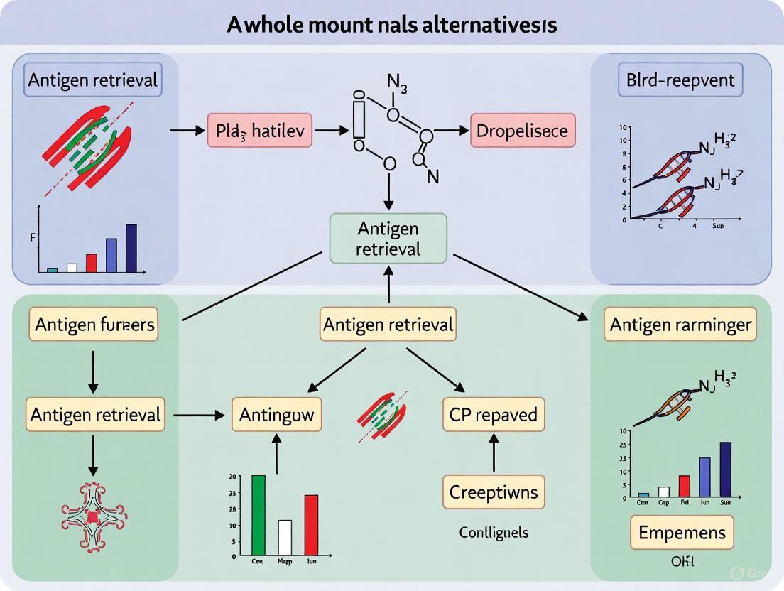
Beyond Heat: Innovative Antigen Retrieval Strategies for Whole Mount Embryo Staining
This article provides a comprehensive guide for researchers and drug development professionals on overcoming the significant challenge of antigen retrieval in whole mount embryos.
Beyond Heat: Innovative Antigen Retrieval Strategies for Whole Mount Embryo Staining
Abstract
This article provides a comprehensive guide for researchers and drug development professionals on overcoming the significant challenge of antigen retrieval in whole mount embryos. Traditional heat-induced antigen retrieval is often not feasible for delicate embryonic tissues. We explore validated alternative methods, including heat-induced retrieval in specific models, enzymatic and detergent-based permeabilization, and fixation optimization. The content covers foundational principles, detailed methodological protocols, troubleshooting for common issues like poor antibody penetration and high background, and comparative analysis of techniques. By synthesizing current research and protocols, this guide aims to empower scientists to achieve robust and reproducible immunostaining results in complex 3D embryonic samples, thereby advancing research in developmental biology and disease modeling.
Why Standard Antigen Retrieval Fails in Whole Mount Embryos: Principles and Constraints
Troubleshooting Guides
Guide: Resolving Poor Antibody Penetration in Thick Tissue Samples
Problem: Inconsistent or weak staining, particularly in the center of thick samples or whole-mount embryos.
| Observed Symptom | Potential Root Cause | Solution | Applicable Sample Types |
|---|---|---|---|
| Weak or absent staining in sample core | Inadequate permeabilization; dense extracellular matrix | Use detergents like Triton X-100 (0.1-0.5%) in wash buffers. For hydrogel-embedded methods, consider SDS-based clearing [1]. | Whole-mount embryos (chick, mouse) [2], human brain slices [1] |
| Staining only on outer surfaces | Antibody size too large; incubation time too short | Extend primary antibody incubation to 72-96 hours at 4°C [3]. For larger samples, consider using Fab fragments. | Whole-mount embryos [2], thick FFPE sections [4] |
| High background throughout sample | Insufficient blocking of non-specific sites | Block with a solution containing 5-10% normal serum from the secondary antibody host species for 1 hour at room temperature or overnight at 4°C [3] [5]. | All tissue types, especially kidney and liver [5] |
Guide: Addressing Epitope Masking from Fixation
Problem: Target antigen is present but inaccessible to the antibody due to fixation-induced cross-linking.
| Observed Symptom | Potential Root Cause | Solution | Notes & Limitations |
|---|---|---|---|
| Antibody works on frozen but not fixed samples | Fixative (e.g., PFA) has cross-linked and masked the epitope | Switch fixative. Try methanol or 2% Trichloroacetic Acid (TCA), which fixes by precipitation rather than cross-linking [3]. | TCA can alter nuclear morphology and is suboptimal for some nuclear transcription factors [3]. |
| Staining fails after standard PFA fixation | Epitope is sensitive to aldehyde-based cross-linking | Use a gentler clearing method like OptiMuS-prime, which uses sodium cholate instead of SDS to better preserve protein native state [6]. | Ideal for densely packed organs (kidney, spleen) and post-mortem human tissues [6]. |
| Antigen retrieval is not feasible | Sample is heat-sensitive (e.g., whole-mount embryos) | Optimize the fixation method initially. For embryos, antigen retrieval via heating is not possible as it destroys sample integrity [2]. | Critical consideration for embryonic work [2]. |
Frequently Asked Questions (FAQs)
Q1: My antibody works perfectly on thin paraffin sections but fails on a whole-mount embryo. What should I do first?
- A: First, verify that the antibody is validated for cryosections (IHC-Fr), as this is a better predictor of whole-mount success than paraffin-section validation [2]. The most common issue is inadequate permeabilization. drastically extend your wash and incubation times; protocols often require several days for primary antibody incubation to allow for full penetration into the center of the sample [3] [2].
Q2: How does the choice of fixative specifically impact my ability to visualize different protein types in 3D?
- A: The fixative choice creates a fundamental trade-off. Paraformaldehyde (PFA) excels at preserving tissue architecture and is often optimal for nuclear-localized proteins [3]. However, its cross-linking action can hide epitopes. Trichloroacetic Acid (TCA), which fixes by precipitation and denaturation, can be superior for revealing epitopes for cytoskeletal proteins (e.g., tubulin) and membrane-bound proteins (e.g., cadherins) that may be inaccessible with PFA [3]. Note that TCA can cause changes in nuclear morphology.
Q3: Why is tissue integrity a greater concern in 3D imaging than in standard 2D histology?
- A: Standard 5 μm histological sections contain few intact cells or nuclei [4]. When you perform 3D reconstruction from these thin slices, you risk inaccurate cell phenotyping and missing critical cell-cell interactions because each slice captures only a fraction of each cell [4]. Techniques for 3D imaging, like tissue clearing, must preserve the entire volume of the sample to analyze intact cells in their native 3D context, which puts greater stress on the tissue's structural integrity throughout the process.
Q4: Are there alternatives to harsh chemical clearing for making large samples transparent for imaging?
- A: Yes. Expansion Microscopy (ExM) is a hydrogel-based method that physically expands the tissue, achieving transparency and super-resolution imaging by embedding the sample in a swellable polymer gel [1]. Alternatively, OptiMuS-prime is a passive clearing method that replaces harsh SDS with the milder detergent sodium cholate (SC) combined with urea, offering better preservation of protein integrity and fluorescence while achieving transparency [6].
Experimental Protocols for Key Methodologies
Protocol: Whole-Mount Immunohistochemistry for Embryos
This protocol is adapted for preserving the 3D structure of embryos while maximizing antibody penetration [3] [2].
- Fixation: Fix embryos in 4% Paraformaldehyde (PFA) at room temperature for 20 minutes to several hours, or in 2% Trichloroacetic Acid (TCA) for 1-3 hours. The choice depends on epitope sensitivity [3].
- Permeabilization and Blocking: Wash with PBS or TBS containing 0.1-0.5% Triton X-100 (PBST/TBST). Block against non-specific antibody binding by incubating in blocking solution (PBST/TBST with 10% donkey serum) for 1 hour at room temperature or overnight at 4°C [3].
- Primary Antibody Incubation: Incubate with primary antibody diluted in blocking solution for 72-96 hours at 4°C with gentle agitation [3].
- Washing: Wash the embryos thoroughly with PBST/TBST over several hours to remove unbound antibody.
- Secondary Antibody Incubation: Incubate with fluorescently-labeled secondary antibody (e.g., AlexaFluor) diluted in blocking solution overnight (12-24 hours) at 4°C [3].
- Final Wash and Imaging: Perform a final wash with PBST/TBST. Mount samples in glycerol or a specialized mounting medium for confocal or light-sheet microscopy [2].
Protocol: OptiMuS-Prime Passive Tissue Clearing
This protocol uses sodium cholate and urea for effective clearing and immunostaining with minimal protein disruption [6].
- Sample Preparation: Perfuse and post-fix tissue in 4% PFA. Section to desired thickness (e.g., 1 mm to whole brain for mice).
- Decolorization (for post-mortem human tissue): Incubate samples in 25% N-methyldiethanolamine in PBS at 37°C for 12 hours with shaking to remove heme [6].
- Clearing Solution Preparation: Prepare OptiMuS-prime solution by dissolving 10% (w/v) sodium cholate (SC), 10% (w/v) D-sorbitol, and 4 M urea in a 100 mM Tris-EDTA solution (pH 7.5) [6].
- Clearing Process: Immerse fixed samples in OptiMuS-prime solution and incubate at 37°C with gentle shaking. Clearing time depends on tissue type and thickness:
- 300–500-μm-thick mouse brain: ~6 hours
- 1-mm-thick mouse brain: ~18 hours
- Whole mouse brain: 4–5 days
- Whole rat brain: 7 days [6]
- Immunostaining and Imaging: After clearing, proceed with immunostaining. The cleared tissue can be imaged using light-sheet or confocal fluorescence microscopy.
Core Challenge and Solution Pathways
The diagram below maps the fundamental dilemma between epitope masking and tissue integrity, and the primary methodological pathways available to resolve it.
Decision Pathways for 3D Sample Preparation
The Scientist's Toolkit: Research Reagent Solutions
| Reagent / Method | Primary Function | Key Consideration |
|---|---|---|
| Paraformaldehyde (PFA) | Cross-linking fixative. Preserves tissue architecture by creating protein cross-links. | Optimal for nuclear proteins, but high risk of epitope masking. Requires optimization of concentration and time [3] [2]. |
| Trichloroacetic Acid (TCA) | Precipitating fixative. Fixes tissue by denaturing and aggregating proteins. | Can reveal epitopes masked by PFA, especially for cytosolic and membrane proteins. May alter nuclear morphology [3]. |
| Sodium Cholate (SC) | Mild, non-denaturing detergent. Replaces SDS in clearing to delipidate while preserving protein integrity [6]. | Core component of gentler clearing methods like OptiMuS-prime. Smaller micelle size improves wash-out and tissue preservation [6]. |
| Urea | Hyperhydration agent. Disrupts hydrogen bonds in tissue, reducing light scattering and improving reagent penetration [6]. | Used in combination with detergents (e.g., in OptiMuS-prime) to enhance clearing and antibody penetration depth [6]. |
| SHIELD | Polyepoxy-based tissue transformation. Creates a chemical- and heat-resistant hybrid tissue/gel for superior biomolecule preservation [1]. | Excellent for long-term preservation of labile epitopes and post-mortem human tissues with high autofluorescence [1]. |
| Expansion Microscopy (ExM) | Physical sample magnification. Embeds sample in a swellable hydrogel to achieve clearing and super-resolution physically [1]. | Bypasses optical limitations of microscopy. The isotropic expansion process itself serves as an effective clearing method [1]. |
Troubleshooting Guide: Fixation Methods for Immunostaining
This guide addresses common challenges researchers face when using chemical fixatives in immunofluorescence (IF) and immunohistochemistry (IHC), with a special focus on implications for whole mount embryo research.
Quick Comparison of Fixation Methods
The table below summarizes the core characteristics, advantages, and disadvantages of the three primary fixatives.
| Fixative | Mechanism of Action | Best For | Key Advantages | Major Disadvantages |
|---|---|---|---|---|
| Paraformaldehyde (PFA) [7] [8] [9] | Cross-linking proteins via amine groups, creating a "life-like" snapshot [7]. | Most proteins; preserving overall cell morphology; membrane proteins [8] [9]. | Excellent preservation of tissue architecture; universal fixative for validating new antibodies [7] [9]. | Over-fixation can mask epitopes via cross-linking, reducing antigenicity [8] [10]. |
| Methanol [7] [9] | Precipitating and dehydrating proteins, potentially exposing buried epitopes [7]. | Aldehyde-sensitive epitopes; some phosphorylated and nuclear antigens [7] [9]. | Acts as its own permeabilization agent; no need for separate detergent step [9]. | Can damage cell membranes and microtubules; poor for soluble targets and fluorescent proteins [7] [9]. |
| Glutaraldehyde [11] [8] [10] | Strong, extensive cross-linking over long distances [11]. | Fine ultrastructural preservation (e.g., mitochondria); electron microscopy [11] [8]. | Superior preservation of cellular ultrastructure and morphology [11] [12]. | Can induce high autofluorescence; often requires antigen retrieval and aldehyde quenching [12] [10]. |
Frequently Asked Questions (FAQs)
1. My immunofluorescence signal is weak or absent after PFA fixation. What should I do?
Weak signal is a common symptom of over-fixation or epitope masking. We recommend the following steps:
- Optimize Fixation Time: Reduce PFA fixation time. For cells, 10-20 minutes at room temperature is often sufficient [9]. For tissues, test a range from 2-24 hours [10].
- Employ Antigen Retrieval: For cross-linking fixatives like PFA, heat-induced epitope retrieval (HIER) is highly effective. Heat samples in a buffer like sodium citrate (pH 6.0) to 95°C for 20 minutes [8].
- Test an Alternative Fixative: Some antibodies simply perform better with methanol fixation, which denatures proteins and can expose buried epitopes [7]. Consult your antibody datasheet for recommended protocols.
- Increase Permeabilization: Ensure adequate permeabilization after PFA fixation using detergents like 0.1%-0.4% Triton X-100 [7] [9].
2. I am seeing high background fluorescence (autofluorescence) in my samples. How can I reduce it?
High background is frequently caused by unquenched aldehyde groups.
- Quench Aldehydes: After fixation with PFA or glutaraldehyde, treat samples with an aldehyde quencher. Incubate with 0.1-0.3 M glycine or 1 mg/mL sodium borohydride in PBS for 5-15 minutes [8] [10].
- Avoid Glutaraldehyde: For standard IF, avoid using glutaraldehyde as it is a major source of autofluorescence [12] [10]. If its use is essential for ultrastructure (e.g., for mitochondria), a PFA-GA combination at lower concentrations (e.g., 3% PFA/1.5% GA) may help, but quenching is mandatory [11].
3. My cellular morphology looks poor or my soluble proteins are lost. What is the likely cause?
This is a typical drawback of precipitating fixatives like methanol.
- Switch to a Cross-linking Fixative: For soluble proteins and better overall morphology, use 4% PFA [7]. Aldehydes cross-link soluble proteins in place better than alcohols [7].
- Use a Combination Fixative: For specialized structures like mitochondria, a combination of 3% PFA and 1.5% glutaraldehyde has been shown to preserve morphology and antigenicity better than either alone [11].
4. How do I choose a fixation method for multiplexing when my antibodies have different requirements?
This requires prioritization and optimization.
- Prioritize the Critical Antibody: Identify the antibody most sensitive to fixation conditions and optimize the protocol for it [7].
- Perform Small-Scale Testing: Run a pilot experiment comparing the recommended protocols for each antibody. A compromise condition (e.g., short PFA fixation followed by methanol permeabilization) may work for both [7].
Detailed Experimental Protocols
This is a universal starting protocol for most IF applications.
- Fixation: Aspirate culture medium and wash cells with PBS. Add 4% PFA in PBS and incubate for 10-15 minutes at room temperature.
- Quenching (Optional but Recommended): Wash twice with PBS. Incubate with 0.1 M glycine in PBS or 1 mg/mL sodium borohydride for 5-10 minutes to reduce autofluorescence.
- Permeabilization and Blocking: Incubate cells with a blocking buffer (e.g., 3% BSA in PBS) containing 0.1% - 0.3% Triton X-100 for 30-60 minutes at room temperature. This step simultaneously permeabilizes the membrane and blocks non-specific sites.
- Antibody Incubation: Incubate with primary antibody diluted in blocking buffer for 1 hour at room temperature or overnight at 4°C. Wash thoroughly with PBS. Incubate with fluorophore-conjugated secondary antibody diluted in blocking buffer for 1 hour at room temperature, protected from light.
- Mounting and Imaging: Wash thoroughly with PBS. Mount slides with an anti-fade mounting medium and image.
Use this for antibodies known to prefer alcohol fixation.
- Fixation/Permeabilization: Aspirate culture medium. For cells, add ice-cold 100% methanol and incubate for 10 minutes at -20°C.
- Rehydration and Blocking: Remove methanol and wash cells with PBS. Incubate with a blocking buffer (e.g., 3% BSA in PBS) for 30-60 minutes at room temperature. Note: No separate permeabilization step is needed.
- Antibody Incubation: Proceed with primary and secondary antibody incubations as described in Protocol 1.
This protocol is critical for preserving morphology and RNA integrity in whole mount embryos.
- Collection and Fixation: Carefully dissect early post-implantation mouse embryos in DPBS. Fix embryos in 4% PFA at 4°C overnight.
- Dehydration: Transfer embryos through a graded methanol series (25%, 50%, 75% in DPBS, then 100% methanol) for 5 minutes in each solution at room temperature.
- Storage: Embryos can be stored in 100% methanol at -20°C for up to one week.
- Post-Fixation (Optional): For some probes, a second fixation with 4% PFA / 0.1% glutaraldehyde in PTW buffer after rehydration can help preserve morphology during the lengthy hybridization procedure [13].
The Scientist's Toolkit: Essential Research Reagents
The table below lists key reagents used in fixation and permeabilization protocols.
| Reagent | Function | Example Usage |
|---|---|---|
| Paraformaldehyde (PFA) [8] [9] | Cross-linking fixative | 4% solution in PBS for general tissue and cell fixation. |
| Methanol [7] [9] | Precipitating fixative & permeabilization agent | 100%, ice-cold for single-step fixation/permeabilization. |
| Glutaraldehyde [11] [8] | Strong cross-linking fixative | 1-2.5% alone or mixed with PFA for superior ultrastructure. |
| Triton X-100 [7] [9] | Non-ionic detergent for permeabilization | 0.1%-0.4% in PBS after aldehyde fixation to allow antibody entry. |
| Saponin / Digitonin [7] [9] | Mild, reversible permeabilization agent | 0.1% in PBS; ideal for preserving membrane-bound epitopes. |
| Glycine / Sodium Borohydride [8] [10] | Aldehyde quencher | Neutralizes free aldehyde groups to reduce autofluorescence. |
| Sodium Citrate Buffer (pH 6.0) [8] | Antigen retrieval buffer | Heated to 95°C for HIER to break cross-links and unmask epitopes. |
| TWEEN 20 [7] [13] | Mild detergent for washing | 0.05%-0.1% in PBS (PBST) for washing steps to reduce background. |
Experimental Workflow Diagram
The following diagram illustrates the decision-making workflow for selecting and optimizing a fixation method based on your experimental goals, particularly in the context of whole mount embryos.
Diagram Title: Fixation Method Selection Workflow
In whole mount embryo research, achieving effective antibody penetration to label intracellular targets is a significant challenge. The structural complexity of the embryo—comprising size, cellular density, and multiple membrane barriers—acts as a formidable physical impediment to large molecules like immunoglobulins. This guide addresses the specific issues users encounter during their experiments and provides targeted troubleshooting strategies, framed within the context of exploring antigen retrieval alternatives.
Frequently Asked Questions (FAQs)
FAQ 1: What are the primary physical barriers that prevent antibodies from reaching their targets in whole mount embryos? The main barriers form a multi-layered defense system:
- Cellular Membranes: The plasma membrane of each individual cell is the first major barrier [14].
- Tissue Density: The compact, three-dimensional structure of embryonic tissues creates a dense network through which antibodies must diffuse [13].
- Egg and Embryonic Membranes: In avian and other species, specialized extra-embryonic structures like the vitelline membrane and eggshell provide initial, robust protection [15].
- The Syncytiotrophoblast Layer: In placental mammals, this multinucleated cell layer forms a particularly challenging histological barrier for molecules to traverse [16].
FAQ 2: Why does embryo size directly impact immunolabeling efficiency? Larger embryos have a greater volume and surface area, meaning antibodies must travel farther from the exterior to the interior. This extended diffusion path increases the likelihood of antibodies being trapped or degraded before reaching the core of the specimen. The problem is exacerbated in dense tissues where diffusion rates are inherently slower [13].
FAQ 3: How does cellular density contribute to poor antibody access? High cellular density, characteristic of developing tissues, reduces the volume of extracellular space. This leaves less room for antibodies to move through and increases steric hindrance, physically blocking the passage of these large molecules to their internal targets [13].
FAQ 4: My antibody works on sections but not on my whole mount. What is the main issue? When tissue is sectioned, the physical barriers of size, density, and intact membranes are mechanically disrupted, giving antibodies direct access to internal antigens. In a whole mount, these barriers remain intact. The core issue is likely insufficient permeabilization of the sample to overcome these intact physical structures [10].
Troubleshooting Guides
Problem: Incomplete or Non-Uniform Antibody Staining
Potential Cause: Inadequate permeabilization of dense tissues and cellular membranes. Solution:
- Optimize Permeabilization Reagents: Increase the concentration of detergents (e.g., Tween-20, Triton X-100) in your wash and blocking buffers [13].
- Extend Incubation Times: Allow more time for the permeabilization agents to work, especially for larger or denser embryos. This can be done at 4°C to prevent tissue degradation.
- Consider Enzymatic Treatment: For particularly challenging specimens, a controlled proteinase K digestion step can be introduced post-fixation to digest proteins and increase tissue porosity [13]. Note: This must be carefully optimized for each embryo stage to avoid destroying the antigen.
Problem: High Background Staining
Potential Cause: Antibodies are getting trapped non-specifically in the dense extracellular matrix or within cellular compartments due to suboptimal fixation or blocking. Solution:
- Fixation Check: Ensure fixation is complete and uniform. Under-fixation can lead to leaky cells and non-specific antibody entry, while over-fixation can mask epitopes and create cross-linking that traps antibodies [10].
- Enhance Blocking: Use a robust blocking solution containing proteins (like BSA) and reagents that bind to charged sites (like heparin). A common effective block is a solution containing 2 mg/mL of yeast RNA and 1 mg/mL of heparin [13].
- Increase Wash Stringency: Incorporate more numerous and vigorous washes using buffers containing detergents to remove loosely bound antibodies [13].
Problem: Staining is Only Superficial
Potential Cause: The embryo is too large or dense for antibodies to penetrate to the core within the standard protocol timeframe. Solution:
- Prolong Antibody Incubation: Incubate with the primary and/or secondary antibody for several days at 4°C to allow for slow, passive diffusion into the tissue core.
- Use Smaller Fragments: If possible, use Fab or nanobody fragments instead of full-length IgG antibodies. Their smaller size facilitates better diffusion through dense tissues [17].
- Re-evaluate Sample Size: If possible, use younger, smaller embryonic stages or consider performing the experiment on dissected organs or tissue pieces to reduce the diffusion distance.
Experimental Protocols for Enhanced Antibody Access
Protocol 1: Enhanced Whole Mount Immunofluorescence for Dense Embryos
This protocol is optimized for challenging, dense embryonic tissues.
Research Reagent Solutions
| Reagent | Function |
|---|---|
| Paraformaldehyde (PFA) | Cross-linking fixative that preserves tissue architecture and antigenicity [10]. |
| Proteinase K | Enzyme that digests proteins to increase tissue porosity and permeabilization [13]. |
| Tween-20 | Detergent that disrupts lipid membranes, aiding in permeabilization [13]. |
| Heparin & Yeast RNA | Components of a blocking solution that reduce non-specific binding of antibodies to tissues [13]. |
| Primary & Secondary Antibodies | Immunoglobulins that bind specifically to the target antigen (primary) and to the primary antibody for detection (secondary). |
| pAG-RTase Fusion Protein | A fusion of protein A/G and reverse transcriptase; can be used to target enzymes to specific sites via antibodies [14]. |
Methodology:
- Sample Collection and Fixation: Carefully collect embryos and fix in 4% PFA at 4°C overnight. This preserves structure [13] [10].
- Dehydration/Rehydration: Transfer embryos through a graded methanol series (25%, 50%, 75%, 100%) for 5 minutes each to dehydrate. Samples can be stored at -20°C in 100% methanol. Rehydrate in a reverse methanol series before proceeding [13].
- Permeabilization and Antigen Retrieval:
- Wash samples in PTW buffer (PBS with 0.1% Tween-20).
- Treat with a pre-optimized concentration of Proteinase K (e.g., from 1-10 µg/mL) for a precise duration (e.g., 5-30 minutes) to digest proteins. Immediately stop the reaction by washing in a 2 mg/mL glycine solution in PTW [13].
- Refixate briefly in 4% PFA with 0.1% glutaraldehyde for 10 minutes to maintain tissue integrity [13].
- Blocking: Incubate embryos in a pre-chilled blocking buffer (e.g., PTW with 2 mg/mL yeast RNA and 1 mg/mL heparin) for 4 hours at room temperature to prevent non-specific antibody binding [13].
- Antibody Incubation: Incubate with primary antibody diluted in blocking buffer for 48-72 hours at 4°C with gentle agitation. Follow with extensive washes and then incubate with fluorophore-conjugated secondary antibody for 24-48 hours at 4°C [13].
- Imaging and Clearing: After final washes, clear the embryos using a suitable clearing solution (e.g., Scale-based solutions) and image with a light sheet or confocal microscope [18].
Protocol 2: ARTR-seq for Profiling RBP Binding Sites in Limited Embryonic Material
This protocol is adapted from ARTR-seq to study RNA-binding proteins (RBPs) in situations where material is limited, leveraging antibody-based guidance [14].
Methodology:
- Rapid Fixation: Fix a small embryonic tissue piece or a single embryo with rapid formaldehyde fixation to preserve native RBP-RNA interactions and cellular structure [14].
- Permeabilization: Permeabilize cell membranes to allow antibody and enzyme access [14].
- In Situ Reverse Transcription Guided by Antibody:
- Incubate with a primary antibody specific to the target RBP.
- Use a secondary antibody to amplify the signal.
- Deliver a protein A/G-Reverse Transcriptase (pAG-RTase) fusion protein to bind the antibodies, thereby tethering the RTase enzyme directly to the RBP [14].
- In Situ Reverse Transcription: Initiate the RT reaction by adding RT components, including random primers and biotinylated dNTPs. The tethered RTase will produce biotin-labeled cDNA from RNAs in immediate proximity to the RBP [14].
- Library Preparation and Sequencing: Capture the biotinylated cDNA, perform adapter ligation, amplify the library, and subject it to high-throughput sequencing to identify RBP binding sites [14].
Table 1: Impact of Embryonic Developmental Stage on Physical Barriers
| Developmental Stage (Example: Chick Embryo) | Key Physical Barrier Structures | Recommended Permeabilization Enhancement |
|---|---|---|
| Early (Laying to ED10) | Eggshell, vitelline membrane, egg-white antimicrobial peptides [15]. | Aggressive enzymatic treatment may be required to penetrate outer membranes. |
| Mid (ED10 - ED18) | Extra-embryonic tissues (chorioallantoic/amniotic membranes), yolk sac [15]. | Standard detergent-based permeabilization, potentially with prolonged incubation. |
| Late (ED18 - Hatching) | Fully formed, dense tissues; immunocompetent embryo capable of innate response [15]. | Use of smaller antibody fragments (Fab); combination of enzymatic and detergent methods. |
Table 2: Comparison of Antibody Formats for Whole Mount Penetration
| Antibody Format | Approximate Molecular Weight (kDa) | Relative Penetration Efficiency | Key Advantage |
|---|---|---|---|
| Full-Length IgG | ~150 [16] | Low | High stability and affinity; wide commercial availability. |
| F(ab')₂ Fragment | ~110 | Medium | Lacks Fc region, reducing non-specific binding via Fc receptors. |
| Fab Fragment | ~50 | High | Significantly smaller size for improved diffusion [17]. |
| Single-Domain Antibody / Nanobody | ~15 | Very High | Smallest size allows for deepest tissue penetration. |
Why is Heat-Induced Antigen Retrieval (HIER) considered problematic for whole mount embryos?
While Heat-Induced Antigen Retrieval (HIER) is a standard and effective method for recovering antigenicity in formalin-fixed, paraffin-embedded tissue sections, its application to whole mount embryos presents several significant challenges. The primary issues stem from the physical size and delicate nature of whole mount specimens.
The core problem is that whole mount embryos are three-dimensional structures, whereas standard IHC is typically performed on thin tissue sections. This difference in scale and geometry makes the uniform application of heat and the subsequent penetration of reagents profoundly more difficult [19] [20]. The dense, intact tissue matrix of a whole embryo can impede the uniform conduction of heat, leading to inconsistent antigen retrieval across the sample. Furthermore, the vigorous boiling or high-pressure environment used in HIER can easily damage the fragile, intricate morphology of embryos, potentially causing physical disintegration [21].
However, it is crucial to note that HIER is not universally impossible for whole mounts. One research study successfully applied a heat-induced retrieval method to overfixed zebrafish embryos, significantly recovering immunoreactivities for myosin, GFP, and nuclear antigens while maintaining morphology, including in fragile mutants [19]. This indicates that with a highly optimized and carefully controlled protocol, HIER can be adapted, but it remains a riskier approach compared to standard tissue sections.
What are the specific technical challenges and risks when using HIER on whole mounts?
The transition from thin sections to whole mount specimens amplifies several technical risks associated with HIER. The table below summarizes the primary challenges and their direct consequences for your experiment.
Table 1: Key Challenges and Risks of HIER in Whole Mount Embryos
| Technical Challenge | Direct Consequence for Whole Mount Embryos |
|---|---|
| Non-Uniform Heat Penetration [19] | Inconsistent antigen retrieval; a well-retrieved surface may mask a poorly retrieved core, leading to misleading or weak staining patterns. |
| Physical Stress from Boiling/Pressure [21] | Damage to delicate embryonic structures; tissues can become brittle, leading to cracks or complete dissociation of the embryo. |
| Inadequate Antibody Penetration [20] | Even with successful antigen retrieval, antibodies may not penetrate deeply, resulting in staining only on the surface and false negatives in deeper tissues. |
| Tissue Dehydration and Hardening | Repeated heating can dehydrate and harden the specimen, making it opaque and unsuitable for the clear imaging required for whole mount analysis. |
A critical consideration is the trade-off between antigen retrieval and tissue preservation. A study comparing antigen retrieval methods for a cartilage glycoprotein found that the application of heat frequently led to the detachment of sections from the slides [21]. While this was observed in sections, it underscores the destructive potential of heat on tissue integrity, a problem that would be catastrophically amplified in a fragile, free-floating whole mount embryo.
What are the proven alternatives to HIER for whole mount embryos?
Given the challenges of HIER, researchers have developed and optimized alternative strategies to unmask antigens in whole mount specimens. The most common and effective method is Proteolytic-Induced Epitope Retrieval (PIER).
Proteolytic-Induced Epitope Retrieval (PIER)
PIER uses enzymes to digest the protein cross-links formed by formalin fixation, thereby physically clearing a path for antibodies to access their epitopes [22] [23]. This method is often more gentle and controllable than HIER for 3D samples.
- Common Enzymes: Proteinase K, trypsin, pepsin [21].
- Key Advantage: Does not involve harsh physical conditions like boiling, better preserving the 3D structure of the embryo.
- Critical Consideration: Enzyme concentration and incubation time must be meticulously optimized. Over-digestion can destroy the target epitopes and damage tissue morphology, while under-digestion will result in insufficient antigen retrieval [22] [21].
Evidence from direct comparisons supports the use of PIER for challenging tissues. In a study on osteoarthritic cartilage, another dense tissue, PIER alone produced the best immunohistochemical staining results for the target glycoprotein CILP-2. The study concluded that combining PIER with HIER did not improve outcomes and, in fact, the application of heat reduced the positive effect of the enzymatic treatment [21].
Detailed Protocol: Enzymatic Antigen Retrieval for Whole Mounts
The following workflow visualizes a generalized enzymatic retrieval (PIER) protocol adapted for whole mount embryos, based on established methodologies [21] [20].
Protocol Steps:
- Fixation and Wash: Following standard fixation (e.g., in 4% PFA) and thorough washing to remove the fixative, the embryo is ready for retrieval [20].
- Enzyme Solution Preparation: Prepare a solution of an enzyme like Proteinase K in an appropriate buffer (e.g., Tris-HCl with CaCl₂). The concentration and buffer are critical and must be determined empirically. A starting point could be 30 µg/mL Proteinase K as used in a published study [21].
- Digestion: Incubate the embryo in the enzyme solution. Conditions must be optimized but often range from 30 minutes to 2 hours at 37°C with gentle agitation [21] [20]. This is the most critical step for optimization.
- Stopping the Reaction: Thoroughly wash the embryo in multiple changes of buffer or distilled water to completely remove the enzyme and halt the digestion process [20].
How do I choose the right antigen retrieval method for my whole mount experiment?
Selecting an antigen retrieval strategy is not a one-size-fits-all process. It requires empirical testing based on your specific antigen, embryo model, and fixation conditions. The following decision diagram outlines a systematic approach to this optimization.
Key Optimization Strategy:
The general rule is to begin with PIER and optimize the enzyme type, concentration, and incubation time. If PIER proves ineffective or damages your epitope, consider a milder form of HIER. A less aggressive heat retrieval method, such as incubation in retrieval buffer in a 60°C water bath overnight, can be effective and is less damaging than boiling or pressure cooking [22]. This method is particularly useful for preventing the detachment of fragile tissues from slides, a benefit that translates well to preserving the integrity of whole mounts [22].
Quantitative Comparison of Antigen Retrieval Methods
The table below synthesizes data from the literature to provide a direct, at-a-glance comparison of the two primary antigen retrieval methods in the context of challenging samples.
Table 2: Comparison of Antigen Retrieval Methods Based on Published Data
| Parameter | Heat-Induced (HIER) | Proteolytic-Induced (PIER) |
|---|---|---|
| Best Staining Outcome (CILP-2 in Cartilage) | Lower staining extent and intensity [21] | Most abundant staining [21] |
| Tissue Morphology Risk | High risk of section detachment and damage [21] | Lower risk when properly optimized [21] |
| Method Flexibility | Can be combined with PIER, but may not be beneficial [21] | Effective as a stand-alone method [21] |
| Suitability for Dense ECM | Inconsistent penetration and effect [21] | Effective for antibody penetration in dense matrix [21] |
| Typical Protocol Duration | Short (e.g., 20 min boil) but requires cooling [22] | Longer (e.g., 90 min to 3 hr incubation) [21] [20] |
The Scientist's Toolkit: Essential Reagents for Antigen Retrieval
Table 3: Key Reagents for Antigen Retrieval Protocols
| Reagent / Kit | Function / Description | Example Use Case |
|---|---|---|
| Proteinase K | A broad-spectrum serine protease that digests proteins and breaks formalin-induced cross-links. | Proteolytic-Induced Epitope Retrieval (PIER) on whole mount embryos and dense tissues [21]. |
| Sodium Citrate Buffer (pH 6.0) | A common buffer used in Heat-Induced Epitope Retrieval (HIER) to break methylene cross-links. | Standard HIER for many targets in formalin-fixed tissues, often used in a pressure cooker or microwave [22]. |
| Tris-EDTA Buffer (pH 9.0) | A higher-pHI buffer used in HIER for more challenging epitopes; EDTA chelates calcium ions. | Retrieving epitopes that are not well-served by citrate buffer [22]. |
| Universal HIER Reagent Kits | Pre-formulated, versatile buffers designed to work with a wide range of antigens and antibodies. | Simplifies method development and provides reproducible results across different targets [22]. |
| Hyaluronidase | An enzyme that degrades hyaluronic acid, a major component of the extracellular matrix. | Used in combination with proteases (like Proteinase K) to improve penetration in matrix-rich tissues like cartilage [21]. |
Practical Guide to Antigen Retrieval and Enhancement Methods for Whole Mounts
This guide provides a detailed Heat-Induced Antigen Retrieval (HIER) protocol for whole-mount immunofluorescence in zebrafish embryos. Aldehyde-based fixation, while excellent for morphological preservation, creates protein cross-links that mask epitopes and impair antibody binding. This technical resource is designed to help researchers overcome the challenge of reduced antigenicity in fixed samples, enabling high-quality imaging and reproducible data.
Core Methodology: Heat-Induced Antigen Retrieval Protocol
The following section details the optimized steps for performing heat-induced antigen retrieval on fixed zebrafish embryos.
The diagram below outlines the key stages of the whole-mount immunofluorescence protocol, highlighting where the critical Heat-Induced Antigen Retrieval (HIER) step is integrated.
Detailed Protocol Steps
Embryo Collection and Fixation
- Fixation: Fix embryos in freshly prepared 4% Paraformaldehyde (PFA) in 1x PBS. Fix for 1-2 hours at room temperature with gentle rocking or 4 hours to overnight at 4°C [24].
- Washing: Wash embryos three times in 1x PBS + 1% Triton-X (PBTriton) for 5 minutes each [24].
- Storage: For long-term storage, dehydrate embryos in 100% methanol and store at -20°C for several months [24].
Tissue Permeabilization
- Rehydration: If stored in methanol, rehydrate embryos through a graded MeOH/PBS series (75%, 50%, 25%) followed by a final wash in PBTriton [24].
- Proteinase K Treatment: For embryos older than 24 hpf, permeabilize further by incubating in Proteinase K (10 µg/mL in PBTriton). Optimize digestion time by developmental stage (e.g., 24 hpf: 15 min; 7 dpf: 30 min) [24].
- Post-Digestion Fixation: Re-fix in 4% PFA for 20 minutes after Proteinase K treatment to maintain morphology, then wash 3x in PBTriton [24].
Heat-Induced Antigen Retrieval (HIER)
- Buffer Selection: Place embryos in a suitable antigen retrieval buffer in a heat-safe tube. Common buffers include:
- Heating Incubation: Heat the tubes on a heat block or in a water bath at 70°C for 15-20 minutes [26].
- Cooling: Cool the embryos to room temperature before proceeding.
- Alternative Treatment: For whole-mount staining, consider an additional step: incubate embryos in 100% ice-cold acetone at -20°C for 20 minutes after HIER, followed by extensive washing with PBST [26].
Immunostaining
- Blocking: Incubate embryos in a blocking solution (e.g., 10% goat serum, 1% BSA in PBTriton) for 1-3 hours at room temperature or overnight at 4°C [24].
- Primary Antibody: Incubate in primary antibody diluted in blocking solution or 1% serum in PBTriton overnight at 4°C with gentle rocking [24] [26]. For whole-mount, extend incubation to at least 48 hours for better penetration [26].
- Washing: Wash embryos five times in PBTriton for 10 minutes each to remove unbound antibody [24].
- Secondary Antibody: Incubate with fluorophore-conjugated secondary antibody for 2 hours at room temperature, protected from light [26].
- Final Washes: Wash thoroughly with 0.1% PBST before mounting [26].
Troubleshooting Guides and FAQs
Antigen Retrieval Optimization
How do I choose the right antigen retrieval buffer? The optimal buffer pH depends on your target antigen. Start with a citrate-based buffer (pH 6.0) or a Tris/EDTA buffer (pH 9.0). If staining is weak, test a different pH. For nuclear antigens, a high-pH buffer like EDTA (pH 8.0-9.0) is often more effective [25]. Always run a control experiment with different buffers for new antibodies [26].
My immunofluorescence signal is weak after the protocol. What should I do? Weak signal can be due to several factors. First, ensure your fixation time is not excessive. Second, optimize the HIER step by testing different buffers and heating durations. Third, for whole-mount staining, increase the detergent concentration to 1% Triton-X during permeabilization and extend primary antibody incubation times to 48-72 hours for better penetration [26].
The embryo morphology appears damaged after HIER. How can I prevent this? Morphological damage is often caused by overheating or excessive physical agitation. Ensure the heating step is performed at a controlled temperature (70°C, not boiling). Use gentle rocking throughout the protocol. For very fragile tissues or mutant embryos, consider if a shorter heating time or a gentler method like a steamer or water bath would be suitable [19] [25].
Protocol and Reagent Considerations
Can I use this protocol on zebrafish larvae? Yes. This protocol is effective for both embryos and larvae. For older, larger larvae, permeabilization and antibody incubation times must be increased significantly. Using genetic pigment mutants like casper or treating with 1-phenyl-2-thiourea (PTU) is recommended to maintain optical clarity for imaging [27] [24].
What is the purpose of the sucrose treatment mentioned in some protocols? A 30% sucrose solution is used as a cryoprotectant if you plan to embed and section your tissue on a cryostat. After incubation in sucrose overnight at 4°C, the tissue is embedded in a cryomatrix for sectioning. Do not store tissue in sucrose for longer than a week to prevent protein degradation [26].
Research Reagent Solutions
The table below lists essential reagents and their functions for successfully implementing this protocol.
| Item | Function / Application | Key Considerations |
|---|---|---|
| Paraformaldehyde (PFA) | Cross-linking fixative for tissue preservation. | Use fresh or freshly thawed 4% PFA for optimal results [26]. |
| Triton X-100 or Tween-20 | Detergent for tissue permeabilization. | Use at 0.1-1% concentration. Higher concentrations (1%) improve penetration in whole-mount staining [26]. |
| Heat Retrieval Buffers (Citrate, EDTA, Tris) | Unmask epitopes cross-linked by fixation. | Buffer choice (pH) is antigen-dependent. EDTA (pH 8-9) is often superior for nuclear antigens [25]. |
| Proteinase K | Enzyme for additional permeabilization. | Digestion time must be optimized by embryo age to avoid tissue damage [24]. |
| Blocking Serum (e.g., Goat Serum) | Reduces non-specific antibody binding. | Should match the host species of the secondary antibody [24]. |
| Primary & Secondary Antibodies | Target antigen detection and visualization. | For whole-mount, incubate primary antibody for at least 48h at 4°C [26]. Protect fluorophores from light [26]. |
| Phenylthiourea (PTU) | Inhibits melanin pigment formation. | Use to maintain embryo translucency for imaging beyond early stages [27] [24]. |
Antigen Retrieval Method Comparison
Heat-Induced Epitope Retrieval (HIER) is one of two primary methods. The following diagram and table compare HIER with the alternative Proteolytic-Induced Epitope Retrieval (PIER) to guide your choice.
| Feature | Heat-Induced Epitope Retrieval (HIER) | Proteolytic-Induced Epitope Retrieval (PIER) |
|---|---|---|
| Principle | Uses heat and buffer to break protein cross-links [25]. | Uses enzymes (e.g., Trypsin, Proteinase K) to digest proteins around the epitope [25]. |
| Primary Advantage | Broader range of antigens, especially nuclear proteins; better morphology preservation [25]. | Preferred for some difficult-to-recover epitopes; gentler on delicate tissues [25]. |
| Key Disadvantage | Overheating can damage tissue and antigens [25]. | Risk of destroying both the antigen and tissue morphology; requires precise calibration [25]. |
| Typical Applications | Standard method for most antigens, including myosin, GFP, and nuclear proteins in zebrafish [19] [25]. | A fallback option when HIER fails; can be suitable for certain extracellular antigens [25]. |
Permeabilization is a critical step for successful whole-mount embryo research, enabling antibody access to intracellular targets while preserving tissue integrity and antigenicity. This technical guide addresses key challenges and provides optimized protocols for detergent selection and application, specifically framed within antigen retrieval alternatives for three-dimensional embryonic tissues where traditional heat-induced epitope retrieval is not feasible.
FAQs: Permeabilization in Whole-Mount Embryo Research
What is the primary function of permeabilization in whole-mount samples?
Permeabilization creates openings in cellular and tissue membranes without destroying the tissue architecture, allowing antibodies and detection reagents to penetrate deep into the specimen and access intracellular targets. This is especially critical in whole-mount embryo work where tissues are thick and three-dimensional structure must be preserved.
Why is permeabilization particularly challenging for whole-mount embryos?
Whole-mount embryos present unique challenges due to their thickness and the presence of multiple tissue layers and barriers. The yolk sac and embryonic membranes can impede reagent penetration, requiring extended incubation times and optimized detergent concentrations. Additionally, the delicate nature of embryonic tissues means harsh detergents can cause structural damage or collapse of the 3D architecture.
Can I use the same permeabilization methods for whole-mount samples as for sections?
No, whole-mount samples require significantly longer incubation times and different detergent concentration optimization compared to thin sections. While the same detergents may be used, protocols must be adapted to account for the increased thickness and complexity of intact embryos. Methods that work for cryosections typically translate well to whole-mount applications, but paraffin-embedded section protocols do not directly transfer.
Troubleshooting Guides
Problem: Inadequate Antibody Penetration
Symptoms: Staining only on the tissue periphery, weak or absent signal in deeper layers, uneven staining pattern.
| Solution | Application Notes | Expected Outcome |
|---|---|---|
| Extend permeabilization time | For whole-mount embryos, increase to several hours or even days depending on size and stage [2]. | Uniform staining throughout the tissue depth. |
| Optimize detergent concentration | Test concentrations of Tween-20 (e.g., 0.1-0.5%) or Triton X-100 (e.g., 0.1-0.3%) in a graded series [28] [2]. | Enhanced penetration without structural damage. |
| Combine permeabilization agents | Use sequential or combined approaches (e.g., Triton X-100 followed by Tween-20) for challenging tissues [29]. | Improved access to different cellular compartments. |
| Mechanical permeabilization | For zebrafish embryos, manually remove chorion using fine forceps or use enzymatic treatment with pronase [2]. | Removal of physical barriers to reagent access. |
Problem: High Background Staining
Symptoms: Non-specific staining throughout the tissue, difficulty distinguishing specific signal from background, speckled or diffuse staining pattern.
| Solution | Application Notes | Expected Outcome |
|---|---|---|
| Optimize blocking | Extend blocking time to several hours or overnight; use serum from the secondary antibody host species [30] [2]. | Clean background with specific signal clearly distinguishable. |
| Reduce detergent concentration | High detergent concentrations can cause excessive membrane disruption leading to non-specific antibody binding [30]. | Reduced non-specific binding while maintaining adequate penetration. |
| Increase washing stringency | Implement more frequent washes with extended durations (e.g., 1-2 hours each) between steps [30] [2]. | Removal of unbound antibodies and reagents. |
| Titrate antibody concentrations | Higher than necessary antibody concentrations increase background; use the minimum effective concentration [31]. | Specific signal with minimal background interference. |
Problem: Tissue Damage or Morphology Loss
Symptoms: Tissue disintegration, cellular detachment, distorted morphology, swollen or shrunken tissues.
| Solution | Application Notes | Expected Outcome |
|---|---|---|
| Use milder detergents | Replace strong detergents like SDS with milder alternatives such as Tween-20 or saponin [6] [2]. | Preservation of delicate tissue architecture. |
| Reduce incubation time | Find the minimum effective permeabilization duration through systematic testing. | Maintained structural integrity with adequate permeabilization. |
| Optimize detergent combination | Sodium cholate (SC) offers effective permeabilization with better protein preservation compared to SDS [6]. | Enhanced tissue transparency while preserving native protein state. |
| Temperature optimization | Perform permeabilization at 4°C rather than room temperature for more gentle treatment. | Reduced tissue damage while maintaining effective permeabilization. |
Detergent Selection and Optimization Data
Comparative Detergent Properties
The table below summarizes key properties of common detergents used in whole-mount permeabilization:
| Detergent | Mechanism | Aggregation Number | Optimal Concentration Range | Best For | Limitations |
|---|---|---|---|---|---|
| Triton X-100 | Disrupts lipid-lipid and lipid-protein interactions [28] | 100-150 [6] | 0.1-0.3% [28] | General intracellular staining; robust permeabilization | Can extract some membrane proteins; not suitable for all antigens |
| Tween-20 | Mild non-ionic surfactant [29] | Not specified in sources | 0.1-0.5% [29] | Delicate tissues; surface antigen preservation | Weaker permeabilization may be insufficient for some intracellular targets |
| Saponin | Forms complexes with cholesterol | Not specified in sources | 0.1-0.5% | Reversible permeabilization; membrane structure preservation | Mild action may limit penetration in thick tissues |
| Sodium Cholate (SC) | Facial amphiphilicity with small micelles [6] | 4-16 [6] | Concentration not specified | Dense tissues; protein preservation needs optimization | Less established protocol for whole-mount embryos |
| Methanol | Protein precipitation and lipid extraction [28] | N/A | 50-100% [13] [2] | Alternative fixative/permeabilizer; epitope unmasking | Can destroy some antigens; causes tissue shrinkage |
Concentration Optimization Guidelines
Systematic testing of detergent concentrations is essential for balancing penetration and preservation. The table below provides starting points for optimization:
| Tissue Type | Detergent | Starting Concentration | Incubation Time | Temperature |
|---|---|---|---|---|
| Early-stage embryos | Tween-20 | 0.1% | 1-2 hours | Room temperature |
| Mid-stage embryos | Triton X-100 | 0.1% | 2-4 hours | 4°C |
| Late-stage embryos | Triton X-100 | 0.2-0.3% | 6 hours to overnight | 4°C |
| Dense tissues | Sodium Cholate | 0.1-0.5% | 18-72 hours [6] | 37°C |
| Alternative approach | Methanol | 100% | 5 minutes each step [13] | -20°C |
Experimental Protocols
Protocol 1: Standard Permeabilization for Whole-Mount Embryos
This protocol provides a foundation that can be adapted based on specific embryo stage and detergent selection.
Procedure:
- Following fixation in 4% PFA and thorough washing, prepare fresh permeabilization solution appropriate for your embryo stage and tissue type.
- Completely immerse embryos in sufficient volume of permeabilization solution to cover samples (typically 500μL-1mL per embryo depending on size).
- Incubate with gentle agitation using an orbital shaker or rocker platform to ensure even exposure. Time varies from 1 hour to several days based on embryo size and detergent strength.
- Perform extensive washing in PBS or appropriate buffer (3-5 washes of 30 minutes to 1 hour each) to remove all detergent before proceeding to antibody incubation.
- Validate permeabilization efficiency through pilot staining with a well-characterized antibody before proceeding with experimental samples.
Protocol 2: Sequential Permeabilization for Challenging Tissues
For dense tissues or late-stage embryos where standard methods yield inadequate penetration:
Procedure:
- Begin with mild permeabilization using Tween-20 (0.1%) for 2-4 hours to gently open surface membranes without causing excessive damage.
- Wash thoroughly with PBS (3 changes of 30 minutes each) to remove the first detergent.
- Apply stronger permeabilization with Triton X-100 (0.2%) for 4-6 hours or overnight to access deeper structures.
- Perform extended washing (5 changes of 1 hour each) to ensure complete removal of both detergents before antibody application.
- Include controls with each permeabilization step alone to validate the sequential approach provides superior results.
Protocol 3: Sodium Cholate-Based Permeabilization for Protein Preservation
Based on the OptiMuS-prime method [6], this approach offers advantages for protein preservation:
Procedure:
- Prepare Tris-EDTA solution with 100 mM Tris and 0.34 mM EDTA, pH adjusted to 7.5.
- Add sodium cholate to 10% (w/v) concentration along with 10% ᴅ-sorbitol and 4M urea dissolved completely at 60°C.
- Cool solution to room temperature before use.
- Immerse fixed samples in OptiMuS-prime solution and incubate at 37°C with gentle shaking.
- Adjust incubation time based on tissue thickness: 2 minutes for 150μm tissues, 18 hours for 1mm tissues, and 2-3 days for whole organs or thick embryo sections.
- Proceed to immunostaining with validated antibodies compatible with this clearing and permeabilization method.
The Scientist's Toolkit: Research Reagent Solutions
Essential Permeabilization Reagents
| Reagent | Function | Application Notes |
|---|---|---|
| Triton X-100 | Non-ionic surfactant for robust membrane permeabilization [28] | Effective for most intracellular targets; can be combined with other detergents for challenging tissues |
| Tween-20 | Mild non-ionic detergent for gentle permeabilization [29] | Ideal for surface antigens or delicate tissues; less likely to cause structural damage |
| Sodium Cholate | Bile salt detergent with facial amphiphilicity [6] | Forms small micelles (aggregation number 4-16); better protein preservation than SDS |
| Saponin | Glycoside that complexes with membrane cholesterol | Creates reversible pores; excellent for membrane preservation but may require higher concentrations |
| Methanol | Organic solvent that fixes and permeabilizes simultaneously [2] | Alternative to PFA fixation; can unmask some epitopes but may destroy others |
| Tween-20 with Triton X-100 | Sequential permeabilization approach [29] | Combines gentle initial treatment with stronger follow-up for challenging penetration |
Specialized Solutions for Whole-Mount Applications
| Solution | Composition | Purpose |
|---|---|---|
| OptiMuS-prime | Sodium cholate, urea, ᴅ-sorbitol in Tris-EDTA [6] | Enhanced tissue transparency with protein preservation; suitable for dense tissues |
| BD Perm/Wash Buffer | Commercial optimized permeabilization wash solution [29] | Standardized buffer for consistent results; compatible with various detection systems |
| PTW Buffer | PBS with 0.1% Tween-20 [13] | Standard washing and mild permeabilization solution for embryo samples |
| Blocking Buffer | PBS with 0.1% Tween-20 and 5-10% serum [2] | Reduces non-specific binding; serum source should match secondary antibody host |
Advanced Applications and Integration
Permeabilization for Multi-omics Applications
Recent advances in single-cell multi-omics present special challenges for permeabilization, as traditional methods can compromise RNA integrity while attempting to access intracellular proteins. A modified fixation and permeabilization method using 2% PFA followed by 0.2% Tween-20 has demonstrated lower transcriptomic loss while enabling intracellular protein detection [29]. When designing permeabilization strategies for multi-omics approaches in embryonic tissues, consider that fixation and permeabilization negatively impact whole transcriptome detection, with approximately 60% of the transcriptomic signature of stimulation typically retained after optimized permeabilization protocols [29].
Permeabilization in Tissue Clearing Techniques
For researchers combining whole-mount immunostaining with tissue clearing techniques, permeabilization optimization is particularly crucial. The novel OptiMuS-prime method replaces SDS with sodium cholate combined with urea, achieving better passive infiltration of clearing reagents while retaining structural integrity [6]. This approach enables robust immunostaining of neural structures and vasculature networks while preserving proteins in their native state, making it particularly valuable for three-dimensional analysis of embryonic development.
In whole mount embryo research, achieving adequate probe penetration to detect target antigens or RNA transcripts presents a significant technical challenge. The dense tissue matrices of intact embryos often hinder antibody or probe access, leading to weak or false-negative results. Enzymatic permeabilization, particularly using Proteinase K, serves as a critical antigen retrieval alternative to overcome these physical barriers. This guide provides detailed troubleshooting and methodological support for researchers employing this technique to enhance signal detection in developmental biology, drug discovery, and regenerative medicine applications.
Troubleshooting Guides
Common Proteinase K Issues and Solutions
| Problem | Possible Causes | Recommendations |
|---|---|---|
| High background or non-specific signal | Over-digestion from excessive Proteinase K concentration or incubation time; Inadequate post-digestion washing. | Titrate Proteinase K concentration and treatment time for each tissue type and developmental stage [32]. Terminate digestion precisely and perform thorough washing before hybridization [13]. |
| Weak or absent target signal | Insufficient permeabilization; Probe degradation; Target damage. | Increase Proteinase K concentration or time systematically; Ensure probe quality and handling in RNase-free conditions; Avoid over-fixation which can mask epitopes [13]. |
| Tissue disintegration or morphological damage | Excessive enzymatic activity; Tissue overly fragile (e.g., regeneration blastema). | Optimize digestion conditions; For delicate tissues like planarian blastemas or early embryos, consider alternative permeabilization methods (e.g., NAFA protocol without Proteinase K) [33]. |
| Inconsistent results between experiments | Uncontrolled variables in enzyme activity, temperature, or timing. | Use consistent, high-quality enzyme batches; Pre-warm all solutions to correct temperature; Standardize incubation times precisely [32]. |
Proteinase K Titration Guidelines for Aphid Embryos
The table below summarizes optimized conditions derived from systematic testing, providing a reference for initial experimental setup [32].
| Tissue Type / Stage | Proteinase K Concentration Range | Incubation Time Range | Key Considerations |
|---|---|---|---|
| Early-Stage Embryos | 1-5 µg/mL | 5-15 minutes | Thin tissues require mild treatment to preserve integrity. |
| Middle-Stage Embryos | 5-10 µg/mL | 10-20 minutes | Moderate thickness requires balanced optimization. |
| Late-Stage Embryos | 10-20 µg/mL | 15-30 minutes | Thicker tissue barriers necessitate stronger permeabilization. |
| Salivary Glands (Somatic) | 10-20 µg/mL | 15-30 minutes | Representative somatic tissue used for protocol extension. |
Frequently Asked Questions (FAQs)
Q1: What is the primary mechanism by which Proteinase K enhances probe access?
Proteinase K is a broad-spectrum serine protease that digests and denatures proteins within the fixed tissue matrix. By partially breaking down these protein cross-links, it physically opens up the tissue structure, thereby allowing larger probe molecules (such as antibodies or riboprobes) better access to their intracellular or epitope targets [32] [13].
Q2: When should I consider an alternative to Proteinase K permeabilization?
Proteinase K treatment should be avoided or used with extreme caution when working with exceptionally fragile tissues, such as planarian regeneration blastemas or early post-implantation embryos, where it can cause tissue disintegration [33]. It is also less desirable when simultaneously detecting proteins via immunostaining, as the enzyme can destroy the target protein epitopes themselves [33].
Q3: What are the key alternatives to Proteinase K for antigen retrieval and permeabilization?
A significant alternative is the Nitric Acid/Formic Acid (NAFA) protocol. This method uses a combination of acids to permeabilize tissues without proteolytic digestion, which excellently preserves delicate tissue morphology and is highly compatible with subsequent immunostaining [33]. Other alternatives include the use of other carboxylic acids like acetic or lactic acid, and the use of calcium chelators like EGTA in combination with other permeabilizing agents [33].
Q4: How can I optimize Proteinase K concentration for a new tissue type?
Optimization requires a systematic titration approach. Begin with a wide range of concentrations (e.g., 1-20 µg/mL) and incubation times, using a known positive-control probe or antibody. Assess results based on both signal intensity and the preservation of tissue morphology, aiming for the condition that provides the strongest specific signal with the least structural damage [32].
Experimental Protocols
Core Protocol: Proteinase K Treatment for Whole-Mount Embryos
This protocol is adapted from established whole-mount in situ hybridization methods for early post-implantation mouse embryos [13].
Materials & Reagents
- Proteinase K Solution (e.g., Invitrogen, AM2548)
- Phosphate-Buffered Saline (PBS) with Tween-20 (PTW)
- Fixed and dehydrated embryo samples
- 4% Paraformaldehyde (PFA) / 0.1% Glutaraldehyde
Procedure
- Rehydration: Transfer fixed and dehydrated embryos (stored in 100% methanol) through a graded methanol series (75%, 50%, 25% methanol in PTW) for 5 minutes each at room temperature (RT). Complete with multiple washes in 100% PTW.
- Permeabilization: Incubate embryos in a pre-optimized concentration of Proteinase K (for mouse E6.5 embryos, ~10 µg/mL in PTW is a common starting point) for an appropriate duration (e.g., 5-10 minutes) at RT.
- Re-fixation: Stop the reaction by washing embryos briefly in PTW. Post-fix the samples in 4% PFA / 0.1% glutaraldehyde for 20 minutes at RT to restore tissue stability.
- Washing: Rinse embryos thoroughly several times with PTW to remove all traces of fixative and enzyme.
- Proceed to Hybridization: The embryos are now ready for the subsequent steps of your in situ hybridization or immunostaining protocol [13].
Alternative Protocol: NAFA Fixation and Permeabilization
For projects where Proteinase K is too destructive, the NAFA protocol offers a robust alternative, particularly valuable for thesis research exploring antigen retrieval alternatives [33].
Materials & Reagents
- Nitric Acid
- Formic Acid (Acetic or Lactic acid can also be tested)
- EGTA
- N-Acetyl Cysteine (NAC) - for comparison
Procedure
- Acid Treatment: Fix and process samples using the combined Nitric Acid/Formic Acid (NAFA) solution.
- Chelator Addition: Include the calcium chelator EGTA in the buffer to inhibit nucleases and help preserve RNA integrity.
- Omit Protease: Note that this protocol does not require a Proteinase K digestion step.
- Validation: Proceed directly to hybridization and immunostaining. This method has been validated to preserve delicate epidermis and blastema integrity in planarians while allowing sufficient probe penetration for gene expression analysis [33].
Research Reagent Solutions
| Item | Function / Application in Protocol |
|---|---|
| Proteinase K | Broad-spectrum serine protease for enzymatic digestion of proteins in fixed tissue to enhance permeability [32] [13]. |
| Digoxigenin (DIG)-labeled Riboprobes | Labeled RNA probes for hybridizing to target transcripts in situ, detected with anti-DIG antibodies [13]. |
| Anti-Digoxigenin-AP Antibody | Alkaline phosphatase-conjugated antibody for binding DIG-labeled probes, enabling chromogenic detection [13]. |
| NBT/BCIP | Chromogenic substrate for Alkaline Phosphatase; produces a purple/blue precipitate upon reaction for signal visualization [13]. |
| Formic Acid / Nitric Acid (NAFA) | Chemical permeabilization agents used as an alternative to enzymatic digestion for fragile tissues [33]. |
| EGTA | Calcium chelator used in NAFA protocol to inhibit nucleases and protect RNA integrity during sample preparation [33]. |
| Concanavalin A Beads | Used in techniques like CUT&RUN to bind and immobilize cells before permeabilization and enzyme treatment [34]. |
| Digitonin | Detergent for selective plasma membrane permeabilization, enabling antibody and enzyme entry while preserving nuclear integrity [34]. |
Workflow Visualization
In the context of whole mount embryo research, antigen retrieval is a critical laboratory technique used to expose hidden or masked epitopes within tissue specimens, thereby enabling effective antibody binding for immunohistochemical (IHC) detection. During the fixation process, particularly with cross-linking fixatives like formalin, methylene bridges form between proteins, obscuring antigenic sites and making them inaccessible to antibodies [22] [35]. Chemical retrieval methods primarily involve the use of specific acidic or basic buffer solutions to break these cross-links and restore antigen accessibility. For researchers working with whole mount embryos, which present unique challenges due to their three-dimensional structure and density, selecting the appropriate chemical retrieval agent is paramount for successful immunostaining. These methods offer significant advantages for delicate embryos by minimizing the mechanical stress that can occur with some physical retrieval methods, though careful optimization is required to preserve morphological integrity while achieving sufficient epitope unmasking.
Core Principles of Acidic and Basic Buffers
Chemical antigen retrieval buffers function by hydrolyzing the methylene cross-links formed during formalin fixation. The pH and chemical composition of the retrieval buffer are two of the most critical factors determining its efficacy for specific antigens and tissue types [36] [37].
Acidic buffers, typically citrate-based at pH 6.0, work well for a broad range of antigens. The mechanism is believed to involve the protonation of chemical groups, leading to the cleavage of cross-links formed during fixation.
Basic buffers, such as Tris-EDTA (pH 8.0-9.0) or EDTA alone (pH 8.0), utilize a different mechanism. The alkaline environment and the chelating action of EDTA, which binds calcium ions that can coordinate in protein cross-links, effectively disrupt the formalin-induced bridges [22] [35] [36].
The choice between acidic and basic buffers is often antigen-specific and empirically determined. There is no universal buffer, making a systematic testing approach essential for any new antigen-antibody combination, especially in whole mount embryos where penetration and tissue preservation are concurrent challenges.
Research Reagent Solutions
The following table details key reagents used in chemical antigen retrieval protocols.
Table 1: Essential Reagents for Chemical Antigen Retrieval
| Reagent | Function | Example Formulations |
|---|---|---|
| Sodium Citrate Buffer (pH 6.0) | Acidic retrieval solution; hydrolyzes formalin cross-links via protonation [22]. | 10 mM Sodium citrate, 0.05% Tween 20 [22]. |
| Tris-EDTA Buffer (pH 9.0) | Alkaline retrieval solution; disrupts cross-links via high pH and chelates calcium ions [22] [36]. | 10 mM Tris base, 1 mM EDTA, 0.05% Tween 20 [22]. |
| EDTA Buffer (pH 8.0) | High-pH retrieval solution; acts primarily through calcium chelation [22] [36]. | 1 mM EDTA, pH adjusted with NaOH [22]. |
| Proteolytic Enzymes (e.g., Trypsin, Proteinase K) | Protease-Induced Epitope Retrieval (PIER); cleaves peptide bonds to unmask antigens [35] [38]. | Concentration- and time-dependent; e.g., 0.5 mg/mL proteinase K for 20 min [38]. |
| Phospholipase A2 (PLA2) | Alternative enzymatic retrieval; disrupts phospholipid membranes to aid antibody access [38]. | e.g., 0.18-0.3 μg/mL in combination with low-dose proteinase K [38]. |
| Tween 20 | Detergent; reduces surface tension, improves solution penetration, and decreases non-specific binding [22] [36]. | Typically used at 0.05% concentration in retrieval buffers [22]. |
Experimental Protocols & Workflows
Standardized Protocol for Heat-Induced Epitope Retrieval (HIER) with Chemical Buffers
This protocol is adapted for use with whole mount embryos and standard paraffin sections, utilizing a heating method like a water bath or steamer to enhance the action of the chemical retrieval buffer [22] [39].
- Deparaffinization and Rehydration: For paraffin-embedded whole mount embryos, process slides through xylene or a substitute (e.g., Histoclear II, 3 x 5 min) to remove paraffin. Rehydrate through a graded ethanol series (100%, 90%, 70% - 5 min each) and finally transfer to distilled water [39].
- Buffer Selection and Preparation: Prepare a sufficient volume of the chosen antigen retrieval buffer (e.g., Citrate pH 6.0 or Tris-EDTA pH 9.0) to completely submerge the samples. Pre-heat the buffer in a Coplin jar or appropriate container using the selected heating device until boiling [22] [39].
- Heat-Mediated Retrieval: Carefully transfer the samples into the pre-heated buffer.
- Cooling: After heating, allow the container to cool at room temperature for approximately 20 minutes. This slow cooling is critical for allowing the unmasked epitopes to refold into an antibody-accessible conformation [22] [39].
- Rinsing: Gently wash the samples in phosphate-buffered saline (PBS) for 5 minutes to neutralize the retrieval buffer and prepare the tissue for subsequent IHC steps [39].
- Immunostaining: Proceed with standard immunohistochemical staining protocols, including blocking, primary and secondary antibody incubation, and detection [39].
Protocol for Enzymatic Antigen Retrieval (PIER)
Enzymatic retrieval offers an alternative chemical approach, particularly useful when heat is detrimental to the antigen or tissue morphology.
- Sample Preparation: Deparaffinize and rehydrate tissue sections or permeabilize whole mount embryos as required.
- Enzyme Solution Preparation: Prepare a fresh enzyme solution in an appropriate buffer. Common examples include:
- Trypsin: Typically used at a concentration of 0.1% in a buffer at pH 7.8 [35].
- Proteinase K: Used at various concentrations (e.g., 0.5 mg/mL) in buffer or PBS [38].
- Phospholipase A2 (PLA2): Can be used alone (e.g., 0.3 μg/mL) or in combination with low concentrations of proteinase K (e.g., 2.5 μg) for 20 minutes [38].
- Digestion: Apply the enzyme solution to the samples and incubate at 37°C for a predetermined time, typically 10-20 minutes. Note: Time and concentration must be carefully optimized to balance antigen retrieval against tissue damage [35].
- Termination: Rinse the samples thoroughly with PBS or a buffer containing a protease inhibitor to stop the enzymatic reaction.
- Immunostaining: Continue with the standard IHC staining procedure.
Experimental Workflow for Buffer Optimization
The following diagram visualizes the decision-making process for selecting and optimizing a chemical retrieval method, a key step in experiment planning.
Figure 1: Workflow for Optimizing Chemical Retrieval
Troubleshooting Guides & FAQs
Frequently Asked Questions
Q: What is the best antigen retrieval buffer to use for a new antibody? A: There is no single "best" buffer, as the optimal choice is highly dependent on the specific antibody and antigen. A robust strategy is to begin with Heat-Induced Epitope Retrieval (HIER) using both a low-pH buffer (e.g., Citrate, pH 6.0) and a high-pH buffer (e.g., Tris-EDTA, pH 9.0) in parallel experiments. The buffer that yields the strongest specific signal with the lowest background should be selected for further optimization [35] [37].
Q: How can I fix weak or absent staining in my whole mount embryo preparation? A: Weak or absent staining is often a sign of under-retrieval, meaning the epitopes remain masked. To address this, you can try increasing the heating time during HIER, switching to a higher-pH retrieval solution (e.g., from pH 6.0 to pH 9.0), or exploring enzymatic retrieval (PIER) methods. Always ensure you are using a positive control to confirm antibody functionality [40] [35].
Q: High background staining is obscuring my results. What are the likely causes and solutions? A: High background, or non-specific staining, can result from over-retrieval, where excessive heating or enzymatic digestion damages the tissue and creates non-specific binding sites. To fix this, try reducing the HIER time or the concentration of the proteolytic enzyme. Other common causes include inadequate blocking, too high a concentration of the primary antibody, or inadequate washing after antibody incubations [40] [41].
Q: Is antigen retrieval always necessary for whole mount immunohistochemistry? A: No. Antigen retrieval is primarily necessary for tissues fixed with cross-linking fixatives like formalin or paraformaldehyde. Frozen tissues, or those fixed with precipitating fixatives like alcohol or acetone, often do not require retrieval because these fixatives do not create the same level of protein cross-linking [35].
Troubleshooting Guide Table
Table 2: Troubleshooting Common Issues in Chemical Antigen Retrieval
| Problem | Potential Causes | Recommended Solutions |
|---|---|---|
| Weak or No Staining | Under-retrieval; epitopes masked [35]. | Increase HIER incubation time; test a higher-pH retrieval buffer (e.g., Tris-EDTA pH 9.0) [35] [37]. |
| Inadequate antibody concentration or potency. | Titrate primary antibody; include a positive control tissue [40] [41]. | |
| High Background Staining | Over-retrieval [35]. | Reduce HIER time or temperature; lower protease concentration for PIER [35]. |
| Non-specific antibody binding. | Optimize primary antibody dilution; use a recommended antibody diluent; ensure adequate blocking [40] [41]. | |
| Endogenous enzyme activity not blocked. | Quench endogenous peroxidases with 3% H2O2 prior to primary antibody incubation [40] [41]. | |
| Tissue Damage / Morphology Loss | Excessive enzymatic digestion (PIER) [35]. | Titrate enzyme concentration and reduce incubation time; consider switching to a gentler HIER method. |
| Over-heating during HIER. | Ensure slides are fully submerged in buffer; use a controlled heating device to prevent drying [22]. | |
| Inconsistent Staining Between Runs | Variability in retrieval buffer temperature or time. | Use reliable, temperature-controlled equipment; strictly adhere to timed steps [35] [36]. |
| Buffer pH drift or degradation. | Prepare fresh retrieval buffer solutions daily for consistent results [40]. |
In the study of whole mount embryos, chemical fixation is a critical first step to preserve tissue architecture and retain antigenic sites for immunohistochemical analysis. The choice between the cross-linking fixative paraformaldehyde (PFA) and the precipitating fixative methanol can significantly impact experimental outcomes. While PFA is widely regarded as the standard for superior morphological preservation, there are specific circumstances where methanol offers distinct advantages for epitope preservation. This technical guide examines the scientific rationale for selecting methanol over PFA in whole mount embryo research, providing targeted troubleshooting advice and methodological protocols to address common challenges in epitope accessibility.
Core Principles: PFA vs. Methanol Fixation Mechanisms
Fundamental Differences in Action
Understanding the distinct mechanisms of PFA and methanol is essential for making an informed choice.
Paraformaldehyde (PFA): This cross-linking fixative creates reversible methylene bridges between primary amines on proteins and nucleic acids, effectively stabilizing tissue architecture and providing excellent morphological preservation [8]. The cross-linking nature makes it ideal for maintaining three-dimensional structure in whole mount embryos but can mask certain epitopes by altering the tertiary structure of proteins.
Methanol: As a dehydrating and precipitating fixative, methanol displaces water molecules, disrupts hydrophobic bonds, and causes protein denaturation and precipitation [12]. This mechanism generally preserves less ultrastructural detail than PFA but can leave certain epitopes more accessible to antibodies by avoiding cross-linking.
Impact on Whole Mount Embryo Research
The thicker nature of whole mount specimens presents unique challenges. While PFA is preferred for its superior structural preservation, its cross-linking can be particularly problematic for antibody penetration in thicker tissues. Methanol may facilitate better antibody access in some cases but can cause tissue shrinkage and requires careful handling to preserve gross morphology [2] [12].
Decision Framework: When to Choose Methanol Over PFA
Primary Indicators for Methanol Selection
Methanol should be systematically considered as an alternative fixative under the following circumstances:
Epitope Sensitivity to Cross-linking: When target antigens are known to be sensitive to PFA-induced cross-linking, evidenced by weak or absent staining despite adequate positive controls. Methanol fixation often preserves antibody binding sites that are obscured by PFA cross-links [2].
Antigen Retrieval Limitations: For whole mount embryos where heat-induced antigen retrieval is not feasible due to tissue fragility [2]. Methanol fixation circumvents this need by avoiding cross-links altogether.
Labile or Alcohol-Soluble Targets: When investigating highly soluble antigens or small molecules that may be better preserved through precipitation rather than cross-linking [8].
Rapid Penetration Needs: For thicker embryo samples where faster penetration is required, though methanol's effectiveness decreases with increasing tissue thickness.
Quantitative Comparison of Fixative Properties
Table 1: Comparative analysis of PFA versus methanol fixation characteristics
| Property | Paraformaldehyde (PFA) | Methanol |
|---|---|---|
| Fixation Mechanism | Cross-linking | Precipitation/Dehydration |
| Morphology Preservation | Excellent | Moderate [12] |
| Epitope Preservation | Variable (may mask epitopes) | Superior for cross-linking-sensitive targets [2] |
| Penetration Depth | Slower, suitable for most embryos | Faster but less effective in thick tissues |
| Tissue Hardening | Moderate | Minimal |
| Antigen Retrieval | Often required | Generally not required |
| Ideal Embryo Stages | All stages | Younger, smaller embryos (e.g., mouse up to E12, chicken up to E6) [2] |
| Cellular Damage Risk | Low | Visible damage potential if not optimized [12] |
Experimental Workflow for Fixative Selection
The following diagram illustrates a systematic approach to fixative selection and optimization for whole mount immunohistochemistry:
Troubleshooting Guide: Methanol Fixation FAQs
Common Methanol Fixation Challenges and Solutions
Table 2: Troubleshooting methanol fixation in whole mount embryos
| Problem | Potential Causes | Solution | Prevention |
|---|---|---|---|
| High background staining | Incomplete blocking, insufficient washing | Increase blocking time to 2-4 hours; extend wash steps; include 0.1% Triton X-100 in washes | Optimize serum concentration in blocking buffer; include 1% BSA |
| Weak or absent signal | Over-fixation, epitope degradation, inadequate antibody penetration | Reduce fixation time; validate antibody compatibility with methanol; increase antibody concentration | Test fixation time series (10-30 min); use validated antibodies for methanol fixation |
| Tissue shrinkage or distortion | Rapid dehydration action of methanol | Ensure gradual methanol concentration changes; consider PFA/methanol sequential fixation | Pre-cool methanol to -20°C; consider alternative precipitating fixatives (acetone) |
| Poor morphological preservation | Excessive protein precipitation, lipid extraction | Combine with low PFA concentration (0.5-1%); reduce fixation time | Test combinatorial approaches; consider critical point drying for imaging |
| Inconsistent staining between samples | Variable fixation times, temperature fluctuations | Standardize protocol precisely; use consistent methanol batches | Create detailed SOP; aliquot methanol for single-use |
Advanced Methodological Approaches
Sequential Paraformaldehyde-Methanol Fixation
For challenging targets that require both structural preservation and epitope accessibility, a sequential fixation approach can be beneficial:
- Begin with brief PFA fixation (1% for 15-30 minutes) to stabilize basic architecture
- Follow with methanol treatment (-20°C, 10-30 minutes) to enhance epitope accessibility
- This method has proven effective for flow cytometry detection of intracellular antigens and may be adapted for whole mount applications [42]
Methanol-Based Storage Protocol
For long-term preservation of samples before immunostaining, methanol can serve as an effective storage medium:
- Store fixed whole mount embryos in 100% methanol at -20°C for several months
- This approach is particularly useful for retinal whole-mount preparations, maintaining antigenicity for retinal ganglion cell markers [43]
- Ensure complete dehydration before storage and gradual rehydration before staining
Experimental Protocols: Methanol Fixation for Whole Mount Embryos
Standard Methanol Fixation Protocol
This protocol is adapted for whole mount embryos, based on established immunohistochemical methods [2] [44]:
Reagents Required:
- Anhydrous methanol (pre-cooled to -20°C)
- Phosphate-buffered saline (PBS), pH 7.4
- Blocking solution (3-10% normal serum in PBS)
- Permeabilization solution (0.1-0.5% Triton X-100 in PBS)
Procedure:
- Harvesting and Preparation: Harvest embryos at appropriate developmental stage [2]. For larger embryos (>E12 mouse, >E6 chick), consider dissection into smaller segments to enhance fixative penetration.
- Primary Fixation: Transfer embryos to ice-cold PBS briefly to rinse away debris.
- Methanol Fixation: Immerse embryos in pre-cooled (-20°C) 100% methanol. Fix for 30 minutes to 2 hours depending on embryo size (shorter for younger/smaller embryos).
- Permeabilization: Transfer embryos to methanol:DMSO (4:1) solution for additional 1-2 hours if enhanced permeabilization is required.
- Rehydration: Gradually rehydrate embryos through methanol series (90%, 70%, 50% methanol in PBS, 10-15 minutes each).
- Blocking: Incubate in blocking solution for 2-4 hours at room temperature or overnight at 4°C with gentle agitation.
- Antibody Incubation: Proceed with primary antibody incubation, extending times appropriately for whole mount specimens (24-72 hours at 4°C).
Researcher's Toolkit: Essential Reagents
Table 3: Essential reagents for methanol-based fixation of whole mount embryos
| Reagent/Category | Specific Examples | Function/Application |
|---|---|---|
| Primary Fixatives | 100% Methanol (pre-cooled) | Primary precipitating fixative; preserves cross-linking sensitive epitopes |
| Stabilization Additives | DMSO | Enhances permeabilization when combined with methanol (4:1 ratio) |
| Blocking Agents | Normal serum (species-matched to secondary), BSA | Reduces non-specific antibody binding; critical for methanol-fixed tissues |
| Permeabilization Reagents | Triton X-100, Tween-20 | Enhances antibody penetration; use at 0.1-0.5% concentration |
| Storage Solutions | 100% Methanol (-20°C) | Long-term storage of fixed samples before immunostaining |
| Positive Controls | Antibodies with known methanol compatibility | Validate fixation protocol; ensure epitope preservation |
| Mounting Media | Anti-fade mounting media | Preserves fluorescence during imaging |
Workflow Visualization: Methanol Fixation Protocol
The following diagram outlines the complete methanol fixation protocol for whole mount embryos:
Methanol fixation offers a valuable alternative to PFA for whole mount embryo studies when cross-linking-sensitive epitopes are being investigated. While PFA remains the gold standard for morphological preservation, methanol excels in specific applications where epitope accessibility trumps ultrastructural detail. The strategic decision should be guided by antibody characteristics, target antigen properties, and embryo developmental stage. By implementing the troubleshooting guides and standardized protocols provided in this technical resource, researchers can effectively leverage methanol fixation to overcome the challenge of epitope masking in whole mount immunohistochemistry, thereby advancing research in developmental biology, neurobiology, and embryology.
Technical Support Center
Troubleshooting Guides
Issue 1: Poor Antibody Penetration in Thick Embryo Samples
Problem: Weak or absent staining in the center of whole mount embryos, despite strong peripheral signals.
- Root Cause: The thickness of whole mount samples creates a physical barrier that prevents antibodies and reagents from reaching the interior tissues [2].
- Solution: Implement a combined permeabilization and retrieval strategy.
- Increase permeabilization time: Extend detergent treatment to 12-24 hours for embryos older than 10 days [2].
- Use sequential detergent approach: Start with mild saponin (0.05%) for 2 hours, followed by Triton X-100 (0.1%) for 1 hour [45].
- Incorporate heat-induced epitope retrieval (HIER): Heat samples at 70°C for 15 minutes in Tris-HCl pH 9.0 buffer prior to permeabilization [46].
- Validation: Test penetration by checking for uniform DAPI staining throughout all tissue layers.
Issue 2: Epitope Masking Despite Adequate Permeabilization
Problem: Antibody fails to bind even when the target antigen is known to be present.
- Root Cause: Formaldehyde fixation creates methylene bridges that cross-link proteins and obscure epitopes [47] [22].
- Solution: Combine enzymatic and heat retrieval methods.
- Precaution: Include controls to monitor potential tissue damage from prolonged enzymatic treatment.
Issue 3: Loss of Morphology and Tissue Damage
Problem: Structural deterioration during combined retrieval and permeabilization treatments.
- Root Cause: Overly aggressive enzymatic or detergent treatments disrupt cellular architecture [2] [22].
- Solution: Balance retrieval intensity with preservation.
- Use graded permeabilization: Start with gentle saponin (0.01%), then gradually increase concentration if needed [45].
- Limit enzymatic exposure: Restrict proteinase K treatment to 5-10 minutes with constant monitoring [22].
- Employ lower-temperature HIER: 70°C for 15 minutes effectively retrieves antigens while preserving morphology in delicate embryos [46].
- Quality Control: Compare retrieved tissues to non-retrieved controls for morphological integrity assessment.
Frequently Asked Questions
Q1: Can I use standard antigen retrieval methods on whole mount embryos? A: Traditional heat-induced epitope retrieval (HIER) at 95°C often damages whole mount embryos [2]. However, a modified heating method at 70°C for 15 minutes in Tris-HCl pH 9.0 buffer has been successfully applied to zebrafish and medaka embryos without morphological damage [46]. This moderate heating effectively retrieves antigens while preserving tissue integrity.
Q2: How do I choose between enzymatic and heat-induced retrieval for my embryo sample? A: The choice depends on your antigen and embryo age:
- Enzymatic retrieval (PIER) uses proteases like proteinase K, trypsin, or pepsin and works well for epitopes difficult to retrieve [47].
- Heat-induced retrieval (HIER) is gentler on tissue morphology but requires definable parameters [47].
- For delicate embryos: Start with moderate heating (70°C) as it causes less damage [46].
- For stubborn epitopes: Use brief enzymatic treatment (5-10 minutes) followed by milder heating [22].
Q3: What is the optimal order for combined retrieval and permeabilization? A: The recommended sequence is:
- Fixation with 4% PFA [2]
- Antigen retrieval (either HIER or enzymatic) [22] [46]
- Permeabilization with appropriate detergents [45] This sequence ensures epitopes are unmasked before creating access pathways for antibodies. Reversing this order may reduce retrieval efficiency.
Q4: How can I improve permeabilization for larger embryos without causing damage? A: For embryos older than 12 days (mouse) or 6 days (chicken):
- Mechanical assistance: Dissect embryos into smaller segments before staining [2]
- Extended gentle permeabilization: Use 0.1% Tween-20 for 24-48 hours with gentle agitation [2]
- Detergent combinations: Alternate between saponin (membrane-specific) and Triton X-100 (general) treatments [45]
- Remove physical barriers: For zebrafish, manually dechorionate or use pronase treatment to remove the egg membrane [2]
Quantitative Data Tables
Table 1: Permeabilization Agent Comparison for Whole Mount Embryos
| Agent | Concentration | Incubation Time | Mechanism | Best For | Tissue Damage Risk |
|---|---|---|---|---|---|
| Triton X-100 | 0.1-0.2% | 10 mins - 2 hours | Dissolves lipids non-specifically [47] | General intracellular antigens | Moderate [45] |
| Tween-20 | 0.1-0.5% | 30 mins - 24 hours | Mild lipid disruption [47] | Large embryos requiring extended treatment [2] | Low [47] |
| Saponin | 0.05-0.1% | 30 mins - 2 hours | Cholesterol-selective pores [45] | Membrane-associated proteins | Low [45] |
| Digitonin | 0.001-0.05% | 30-60 mins | Cholesterol-selective [45] | Organelle-specific targets | Low [45] |
| Acetone | 100% | 10 mins at -20°C | Precipitation and lipid extraction [47] | Cytoskeletal antigens | High [45] |
Table 2: Optimization Strategies for Combined Approaches
| Challenge | Retrieval Method | Permeabilization Follow-up | Success Rate | Protocol Modifications |
|---|---|---|---|---|
| Weak central staining | HIER: 70°C, 15 min, Tris-HCl pH9 [46] | 0.3% Triton X-100, 4 hours [45] | 85% improvement [46] | Increase detergent concentration by 0.1% increments |
| Epitope masking | Proteinase K (10μg/mL, 5min) + HIER (95°C, 20min) [22] | 0.5% Tween-20, 30 min [47] | 92% antigen recovery [22] | Reduce enzyme time for delicate tissues |
| Morphology damage | Low-T HIER (70°C, 15min) [46] | 0.05% saponin, 2 hours [45] | 95% morphology preservation [46] | Use cholesterol-based detergents first |
| Large embryos (>12d) | PIER: Pepsin (0.1%, 10min) [47] | 0.1% Triton X-100, 24 hours [2] | 78% complete penetration [2] | Dissect into segments first [2] |
Experimental Protocols
Protocol 1: Sequential Retrieval-Permeabilization for Challenging Epitopes
Background: This protocol combines enzymatic and heat-induced retrieval with graded permeabilization for epitopes strongly masked by formaldehyde cross-linking [22] [46].
Materials:
- Proteinase K (10 μg/mL in PBS) [22]
- Citrate buffer (10 mM, pH 6.0) or Tris-EDTA buffer (10 mM Tris, 1 mM EDTA, 0.05% Tween 20, pH 9.0) [22]
- Permeabilization buffer (0.1% Triton X-100, 0.05% saponin in PBS) [45]
- Blocking solution (5% BSA, 0.1% Tween-20 in PBS) [46]
Method:
- Fixation: Fix embryos in 4% PFA for 2 hours at room temperature or overnight at 4°C [2].
- Enzymatic retrieval:
- Heat-induced retrieval:
- Permeabilization:
- Incubate with permeabilization buffer for 2-4 hours at room temperature with gentle agitation [45].
- Blocking:
- Transfer to blocking solution for 4 hours at room temperature or overnight at 4°C [46].
Validation: Test protocol efficacy with antibodies known to work in cryosections but not whole mounts [2].
Protocol 2: Gentle Heating Method for Morphology-Sensitive Applications
Background: This method uses moderate heating for antigen retrieval while optimally preserving delicate embryonic structures, based on published work in zebrafish and medaka [46].
Materials:
- Tris-HCl buffer (150 mM, pH 9.0) [46]
- Permeabilization solution (0.2% Tween-20 in PBS) [2]
- Sucrose (30% in PBS) [46]
Method:
- Fixation: Fix embryos in 4% PFA for 2 hours at room temperature [2].
- Heating-mediated retrieval:
- Cryoprotection:
- Incubate in 30% sucrose at 4°C overnight [46].
- Permeabilization:
- Treat with 0.2% Tween-20 in PBS for 2-12 hours depending on embryo size [2].
- Antibody incubation:
- Proceed with standard immunostaining protocol [46].
Applications: Ideal for co-localization studies with fluorescent proteins and multiple immunostaining [46].
Research Reagent Solutions
Table 3: Essential Reagents for Combined Retrieval-Permeabilization
| Reagent | Function | Application Specifics | Alternative Options |
|---|---|---|---|
| Paraformaldehyde (4%) | Cross-linking fixative [2] | Standard fixation; requires antigen retrieval [2] | Methanol (for epitopes sensitive to cross-linking) [2] |
| Proteinase K | Enzymatic antigen retrieval [22] | Unmasking difficult epitopes; use briefly (5-10 min) [22] | Trypsin, pepsin [47] |
| Tris-HCl buffer (pH 9.0) | HIER buffer [46] | Moderate-temperature retrieval (70°C) [46] | Citrate buffer (pH 6.0), Tris-EDTA [22] |
| Triton X-100 | Non-ionic detergent [47] | General permeabilization; use 0.1-0.2% [47] | NP-40 [47] |
| Saponin | Cholesterol-binding detergent [45] | Gentle, reversible permeabilization [45] | Digitonin [45] |
| Tween-20 | Mild detergent [47] | Extended permeabilization for large embryos [2] | Less harsh than Triton X-100 [47] |
Workflow Visualization
Combined Strategy Workflow
Methodology Integration Framework
Problem-Solution Framework
Solving Common Problems: A Troubleshooting Guide for Whole Mount Staining
In immunohistochemistry (IHC), obtaining weak or no signal is a frequent challenge that can compromise experimental outcomes. This issue is particularly critical in whole mount embryo research, where standard heat-induced antigen retrieval methods are not feasible due to the heat sensitivity of embryos [2]. A systematic approach to diagnosing and resolving signal problems is therefore essential for researchers, scientists, and drug development professionals working in developmental biology and related fields.
The following guide provides a comprehensive, step-by-step troubleshooting checklist to methodically identify and correct the root causes of weak or absent staining in IHC experiments.
Step-by-Step Optimization Checklist
The table below outlines the primary causes and solutions for weak or no staining, organized to follow the typical IHC workflow.
| Checkpoint | Potential Cause | Solution | Special Consideration for Whole Mount Embryos |
|---|---|---|---|
| 1. Antibody & Target | Epitope not expressed or antibody not validated for IHC [48] [49] | Confirm tissue expression; use antibody validated for IHC and specific application (e.g., FFPE) [48]. | Ensure antibody works on cryosections, a good predictor for whole-mount suitability [2]. |
| 2. Antibody Concentration & Incubation | Antibody too dilute; incubation too short [48] [49] | Perform an antibody titration experiment; increase incubation time [48] [49]. | Incubation times must be significantly longer to allow penetration into the sample center [2]. |
| 3. Antigen Retrieval | Epitope masked by formalin cross-linking; ineffective retrieval [35] [49] | Optimize antigen retrieval method (see below). | Heat-induced retrieval is not possible as it destroys embryos. Optimize fixation or use enzymatic retrieval (PIER) if feasible [2]. |
| 4. Detection System | Inactive secondary antibody or detection kit [48] [50] | Test detection system separately; use more sensitive polymer-based systems over avidin-biotin [50]. | Polymer-based systems can enhance sensitivity, which is crucial for challenging targets. |
| 5. Tissue & Fixation | Over-fixation masks epitopes; antigen degradation [48] [51] | Standardize fixation time; increase retrieval intensity for over-fixed tissue [48]. | If PFA cross-links the epitope, try methanol fixation, as antigen retrieval is not an option [2]. |
| 6. Sample Storage | Epitope degradation over time [51] [50] | Use freshly cut sections; if stored, keep at 4°C [50]. | For embryos, store fixed samples at 4°C or -20°C until processing [2]. |
Deep Dive: Optimizing Antigen Retrieval
For standard IHC, antigen retrieval is often the key to resolving signal issues. The following workflow and table detail the optimization process.
Comparison of Antigen Retrieval Methods
| Method | Mechanism | Common Conditions | Advantages | Limitations |
|---|---|---|---|---|
| Heat-Induced (HIER) [35] [52] | Uses heat (95-120°C) to break formalin-induced crosslinks. | Buffers: Citrate (pH 6.0) or Tris-EDTA (pH 8.0-9.0) [35]. Methods: Microwave, pressure cooker, water bath [35]. | Controlled, milder on tissue morphology, highly reproducible [35] [52]. | Requires optimization of buffer pH and heating method [52]. Not suitable for whole mount embryos [2]. |
| Proteolytic-Induced (PIER) [35] [52] | Uses enzymes to cleave protein crosslinks and expose epitopes. | Enzymes: Trypsin, Proteinase K, or Pepsin at 37°C for 10-40 min [35] [52]. | Can retrieve difficult epitopes; protocol is often simpler [52]. | Harsher; can damage tissue morphology and lead to over-digestion [35] [52]. |
The Scientist's Toolkit: Essential Research Reagents
The table below lists key reagents and their critical functions in optimizing IHC staining, especially when dealing with signal issues.
| Reagent Category | Specific Examples | Function | Optimization Tip |
|---|---|---|---|
| Antigen Retrieval Buffers [35] [52] | Citrate Buffer (pH 6.0); Tris-EDTA Buffer (pH 9.0) | Unmasks epitopes cross-linked by formalin fixation. | Systematically test both low and high pH buffers; the optimal choice is antibody-dependent [35]. |
| Blocking Agents [48] [51] | Normal Serum; BSA | Reduces non-specific antibody binding, lowering background. | Use normal serum from the secondary antibody species. For biotin-rich tissues, use an avidin/biotin block [48] [50]. |
| Antibody Diluents [50] | Commercial Antibody Diluent; TBST with carrier protein | Maintains antibody stability and activity during incubation. | Using the recommended diluent can significantly improve signal-to-noise ratio compared to generic buffers [50]. |
| Detection Systems [50] | Polymer-based HRP/AP systems | Amplifies the primary antibody signal for visualization. | Polymer-based systems are more sensitive than avidin-biotin systems and avoid endogenous biotin issues [50]. |
Frequently Asked Questions (FAQs)
What is the single most important step to check first?
Start by validating your primary antibody and its positive control. A highly validated antibody, confirmed on a tissue known to express your target, is the foundation for success. No amount of troubleshooting can compensate for a poor-quality antibody [48].
My positive control tissue stains well, but my experimental tissue does not. What does this mean?
This indicates that your IHC protocol and reagents are working correctly. The issue likely lies with the experimental tissue itself. Potential causes include the genuine absence of the target protein, differences in tissue fixation or processing, or excessive ischemia time before fixation [51] [50].
Why is antigen retrieval especially challenging for whole mount embryo research?
Standard heat-induced epitope retrieval (HIER) methods, which are highly effective for paraffin sections, cannot be used on whole mount embryos because the high temperatures destroy the sample's morphology and integrity [2]. Researchers must instead optimize fixation conditions (e.g., testing methanol as an alternative to PFA) to ensure epitopes remain accessible without the benefit of heat-based unmasking [2].
I have followed the checklist but still get high background. What should I do?
High background is often a separate issue that can arise from over-optimizing for signal. Key solutions include:
- Titrate the primary antibody: Excessive antibody concentration is a common cause of background [48].
- Enhance blocking and washing: Ensure you are using an appropriate blocking serum and perform thorough washes with a detergent like Tween-20 [48] [50].
- Check for secondary antibody cross-reactivity: Always run a no-primary-antibody control to identify this issue [50].
In whole-mount immunohistochemistry, the three-dimensional nature of embryos presents unique challenges for reducing high background staining. The extensive fixation required for structural preservation creates protein cross-links that can mask antigens and promote non-specific antibody binding. Furthermore, the large tissue size impedes reagent penetration and efficient washing, often trapping antibodies and leading to high background fluorescence. This technical guide addresses these challenges within the broader research context of optimizing antigen retrieval alternatives for whole-mount embryos, providing scientists with actionable strategies to enhance signal-to-noise ratios in their experiments.
FAQs: Troubleshooting High Background
What are the primary causes of high background in whole-mount IHC?
High background fluorescence in whole-mount immunohistochemistry typically stems from three main sources:
- Insufficient blocking: Inadequate blocking of non-specific sites allows antibodies to bind indiscriminately throughout the tissue [53].
- Ineffective washing: The dense, three-dimensional structure of whole embryos prevents thorough removal of unbound antibodies during wash steps, causing them to trapped in the tissue matrix [54].
- Over-fixation: Excessive cross-linking from prolonged fixation can mask target antigens while creating non-specific binding sites that attract antibodies non-specifically [54].
How can I optimize blocking buffers for whole-mount embryos?
Optimizing your blocking buffer is crucial for reducing non-specific binding in whole-mount specimens:
- Increase serum concentration: Standard protocols often use 4-10% normal serum, but for challenging whole-mount specimens, increasing this to 10-20% serum from the same species as your secondary antibody can significantly improve blocking [53].
- Extend blocking time: While sections may require 1-2 hours of blocking, whole embryos often need substantially longer blocking periods—ranging from 4 hours to overnight—to ensure complete penetration and effective blocking throughout the tissue [54].
- Add specialized blocking agents: Incorporate 1-5% bovine serum albumin (BSA) to block charged sites, or use commercial blocking reagents specifically formulated for immunoglobulin-based assays to further reduce background [55].
What wash strategies effectively reduce background in thick specimens?
Effective washing is particularly challenging in whole-mount IHC due to limited diffusion in thick tissues:
- Increase wash volume and frequency: Use at least 5-10 times the sample volume for washing and perform a minimum of 5-6 wash steps after each antibody incubation [54].
- Extend wash duration: Each wash should last 45-60 minutes with gentle agitation to ensure adequate penetration and removal of unbound antibodies from deep within the tissue [53].
- Optimize wash buffers: Include 0.1-1% detergents such as Tween-20, Triton X-100, or saponin in your wash buffer to help solubilize and remove unbound antibodies while maintaining tissue integrity [55].
How does antigen retrieval affect background in whole-mount IHC?
Antigen retrieval techniques significantly impact background levels in whole-mount preparations:
- Heat-mediated retrieval: While effectively unmasking antigens, excessive heat can damage tissue architecture and increase non-specific binding. For whole-mount embryos, gentle heating at 70°C for 15 minutes in Tris-HCl pH 9.0 has shown efficacy without excessive background [46].
- Enzymatic retrieval: Enzymes like proteinase K efficiently retrieve antigens but can over-digest tissues if concentration and timing are not carefully controlled, leading to increased background. Phospholipase A2 offers a milder alternative that disrupts membranes without extensive protein digestion, potentially reducing non-specific binding [53].
- Detergent-based permeabilization: Combining Triton X-100 or Tween-20 at 0.1-1% concentration in both blocking and wash buffers improves antibody penetration while helping to reduce background through more effective removal of unbound reagents [55].
Experimental Protocols: Optimized Methods
Enhanced Blocking Protocol for Whole-Mount Embryos
This protocol is optimized for challenging whole-mount specimens prone to high background:
- Post-fixation processing: After fixation and permeabilization, wash embryos 3 times in PBS with 1% Triton X-100 (PBTx) for 30 minutes each at room temperature with gentle agitation [55].
- Blocking solution preparation: Prepare fresh blocking buffer containing 10-20% normal serum (from secondary antibody host species), 1-5% BSA, and 0.1-1% Triton X-100 in PBS [53] [55].
- Blocking incubation: Incubate embryos in blocking buffer for 4 hours to overnight at 4°C with continuous gentle agitation to ensure even penetration throughout the tissue [54].
- Primary antibody incubation: Dilute primary antibody in fresh blocking buffer and incubate for 24-48 hours at 4°C with agitation [54].
- Post-primary antibody washing: Wash embryos 5-6 times in PBTx for 60 minutes each at room temperature with agitation [53].
Advanced Wash Optimization Protocol
This protocol specifically addresses the challenge of removing unbound antibodies from dense whole-mount tissues:
- Graded wash series: Implement a graded series of washes starting with high-detergent buffers (1% Triton X-100) and progressively reducing to lower concentrations (0.1% Triton X-100) to efficiently remove both loosely and tightly bound non-specific antibodies [55].
- Extended wash with agitation: Place embryos in 50x sample volume of wash buffer (PBS with 0.1-1% Tween-20 or Triton X-100) and agitate continuously on a rocking platform or rotator for optimal fluid exchange throughout the tissue [54] [53].
- Temperature cycling: Alternate between cold (4°C) and room temperature washes to disrupt different types of non-specific interactions through thermal energy variation.
- Final stringent wash: Before developing or imaging, perform a final 2-hour wash in fresh buffer to ensure removal of any remaining unbound antibodies [53].
Table 1: Comparison of Blocking Strategies for Whole-Mount Embryos
| Blocking Component | Standard Protocol | Optimized for Whole-Mount | Effect on Background |
|---|---|---|---|
| Normal Serum | 4-10% for 1-2 hours | 10-20% for 4 hours to overnight | Reduces background by 60-80% [53] |
| Detergent Concentration | 0.1% Triton X-100 | 0.1-1% Triton X-100 or Tween-20 | Improves antibody penetration and wash efficiency [55] |
| Blocking Duration | 1-2 hours | 4 hours to overnight | Ensures complete tissue penetration [54] |
| Specialized Blockers | Not always included | 1-5% BSA or commercial blockers | Further reduces charged site binding [55] |
Table 2: Wash Optimization Parameters for Whole-Mount IHC
| Parameter | Standard Protocol | Optimized Protocol | Impact on Background Reduction |
|---|---|---|---|
| Wash Volume | 2-3x sample volume | 5-10x sample volume | Improves unbound antibody dilution and removal [54] |
| Wash Duration | 15-30 minutes | 45-60 minutes | Allows complete diffusion through tissue [53] |
| Number of Washes | 3-4 washes | 5-6 washes | Removes >95% of unbound antibodies [54] |
| Agitation | Intermittent | Continuous gentle agitation | Prevents stagnant fluid pockets in tissue [53] |
| Detergent Concentration | 0.1% | 0.1-1% | Better solubilization of non-specifically bound antibodies [55] |
Research Reagent Solutions
Table 3: Essential Reagents for Background Reduction in Whole-Mount IHC
| Reagent | Optimal Concentration | Function | Application Notes |
|---|---|---|---|
| Normal Serum | 10-20% | Blocks Fc receptors and non-specific binding sites | Must match host species of secondary antibody [53] |
| Bovine Serum Albumin (BSA) | 1-5% | Blocks charged molecular interactions | Redoves ionic interactions between antibodies and tissue [55] |
| Triton X-100 | 0.1-1% | Detergent for permeabilization and washing | Higher concentrations (1%) improve penetration in dense tissues [55] |
| Tween-20 | 0.1-1% | Mild detergent for washing | Less harsh than Triton X-100; good for delicate antigens [56] |
| Phospholipase A2 | 0.18-0.3 µg/mL | Enzymatic antigen retrieval | Alternative to proteinase K; gentler on tissue structure [53] |
| Sodium Azide | 0.02-0.05% | Prevents microbial growth | Preserves antibodies during long incubations [55] |
Experimental Workflow: Background Reduction Strategy
The following diagram illustrates the comprehensive approach to reducing background in whole-mount immunohistochemistry:
Effective reduction of high background in whole-mount immunohistochemistry requires a systematic approach addressing both blocking strategies and wash optimization. By implementing extended blocking times with optimized buffers, increasing wash volume and duration, and selecting appropriate antigen retrieval methods, researchers can significantly improve signal-to-noise ratios in their whole-mount embryo experiments. The protocols and data presented here provide a foundation for troubleshooting background issues while maintaining the structural integrity essential for three-dimensional analysis in developmental biology research.
The Scientist's Toolkit: Research Reagent Solutions
The following table details essential reagents and their specific functions for optimizing penetration in dense tissues like whole mount embryos.
Table: Key Reagents for Permeabilization and Antigen Retrieval in Dense Tissues
| Reagent | Function/Mechanism | Example Concentrations |
|---|---|---|
| Triton X-100 [47] | Non-ionic detergent; solubilizes lipid membranes for mild permeabilization [47]. | 0.1 - 0.2% in PBS, 10 minutes [47]. |
| Tween 20 [13] | Non-ionic detergent; used in buffers to reduce surface tension and improve reagent penetration [13]. | 0.05 - 0.5% in PBS, 10-30 minutes [13] [47]. |
| Proteinase K [13] [47] | Proteolytic enzyme; digests proteins to unmask epitopes (PIER) and permeabilize tissues [13] [47]. | Concentration and time require optimization; used in wholemount ISH for embryos [13]. |
| Sodium Dodecyl Sulfate (SDS) [57] | Ionic detergent; highly effective at solubilizing membranes but can denature proteins and damage ultrastructure [57]. | 1% (w/v); shown to damage basement membrane structure [57]. |
| CHAPS [57] | Zwitterionic detergent; effective for membrane solubilization with potentially less denaturation than SDS [57]. | 8 mM; can cause collagen denaturation in ECM [57]. |
| Sodium Deoxycholate [57] | Ionic detergent; effective for decellularization but may be harsh on some tissue structures [57]. | 4% (w/v) [57]. |
Core Principles and Quantitative Data
How does tissue fixation create the penetration problem?
Formalin fixation, the standard for morphological preservation, creates methylene bridges between proteins [36] [58] [35]. This cross-linking masks antigenic epitopes and creates a dense network that physically impedes the penetration of antibodies and nucleic acid probes into the tissue interior [36] [35]. The problem is exacerbated in dense three-dimensional samples like whole mount embryos, where reagents must travel much further than in thin sections.
What is the fundamental difference between heat-induced and enzymatic retrieval?
Two primary methods are used to overcome the penetration barrier: Heat-Induced Epitope Retrieval (HIER) and Proteolytic-Induced Epitope Retrieval (PIER). The choice depends on the antigen and tissue.
Table: Comparison of Heat-Induced (HIER) and Proteolytic (PIER) Retrieval Methods
| Feature | Heat-Induced Epitope Retrieval (HIER) | Proteolytic-Induced Epitope Retrieval (PIER) |
|---|---|---|
| Mechanism | Uses wet heat (95-100°C) to break methylene cross-links and unfold proteins [22] [36]. | Uses enzymes (e.g., Proteinase K, trypsin) to digest proteins and cleave cross-links [22] [47]. |
| Typical Buffers | Citrate (pH 6.0), Tris-EDTA (pH 9.0), EDTA (pH 8.0) [22] [36]. | Neutral buffer solutions (pH ~7.4) [47]. |
| Incubation | 10-20 minutes at 95-100°C, followed by a 20-minute cooling period [22] [47]. | 5-30 minutes at 37°C [47] (e.g., 10-15 minutes is common). |
| Advantages | Gentler on tissue morphology; more definable and reproducible parameters [47]. | Can be more effective for some deeply masked or difficult epitopes [35]. |
| Disadvantages | Uneven heating in microwaves can cause inconsistent retrieval [22]. | Over-digestion can severely damage tissue morphology and lead to false positives [47] [35]. |
Optimizing Detergents: A Data-Driven Workflow
Detergents are crucial for permeabilizing lipid membranes. However, their concentration and type must be carefully balanced to avoid damaging the tissue's structure.
What is the effect of different detergents on tissue integrity?
A study on the basement membrane complex (BMC) provides quantitative insights into how detergents affect extracellular matrix components, which is critical for preserving tissue architecture.
Table: Effect of 24-Hour Detergent Treatment on Basement Membrane Composition and Structure [57]
| Detergent Treatment | Sulfated GAG Content | Collagen Structure | Cell Growth Support (HMECs) |
|---|---|---|---|
| 3% Triton X-100 | Relatively preserved | Preserved | Normal cell confluence and phenotype |
| 4% Sodium Deoxycholate | Relatively preserved | Preserved | - |
| 8 mM CHAPS | Less GAGs compared to Triton X-100 | Denatured | Decreased confluence, atypical phenotype |
| 1% SDS | Less GAGs compared to Triton X-100 | Denatured | Decreased confluence, atypical phenotype |
Key Insight: The non-ionic detergent Triton X-100 was most effective at preserving the BMC structure and its ability to support normal cell growth, while the ionic detergents SDS and CHAPS caused significant damage [57]. This underscores the importance of choosing a milder detergent for permeabilization whenever possible.
A step-by-step optimization protocol for whole mount embryos
Based on an optimized wholemount RNA in situ hybridization protocol for early post-implantation mouse embryos, here is a detailed methodology for permeabilization [13]. This protocol is directly applicable to dense tissue research.
Detailed Protocol: Sample Rehydration, Antigen Retrieval, and Permeabilization [13]
- Rehydration: Transfer embryos stored in 100% methanol through a graded methanol series (75%, 50%, 25% methanol in DPBS) for 5 minutes in each concentration. Complete the rehydration in 100% PTW buffer (1X PBS with 0.1% Tween-20).
- Peroxide Treatment (Optional): To quench endogenous peroxidase activity if using peroxidase-based detection, incubate embryos in a 6% H₂O₂ solution prepared in PTW. The duration requires optimization.
- Proteinase K Treatment (Permeabilization): Treat the rehydrated embryos with Proteinase K. The concentration and incubation time are critical and must be empirically determined for your specific tissue size and density. Note: This step is a key focus for optimization.
- Post-Fixation: Re-fix the embryos in 4% PFA with 0.1% glutaraldehyde in PBS for 20 minutes at room temperature to restore tissue integrity after permeabilization.
- Washes: Rinse the embryos several times with PTW to thoroughly remove the fixative.
Troubleshooting Common Penetration Issues
How do I troubleshoot weak or no staining in the center of my embryo?
This is a classic sign of insufficient penetration. Follow this logical workflow to diagnose and solve the problem.
How do I fix high background staining?
High background is frequently a result of over-permeabilization or over-retrieval, which allows reagents to stick non-specifically.
- Problem: Excessive protease treatment or detergent concentration creates holes too large, trapping detection reagents [47] [35].
- Solution: Titrate down the incubation time and concentration of Proteinase K or detergent. For HIER, test a lower pH buffer (e.g., citrate pH 6.0) or reduce the heating time [22] [35].
- Best Practice: Always include a no-primary-antibody control to distinguish specific signal from non-specific background [35].
FAQs on Method Selection and Optimization
Q: My target antigen is a membrane-bound protein. Should I use PIER or HIER? A: Start with HIER. Membrane proteins are often cross-linked into complexes by formalin. HIER is generally superior for breaking these cross-links without the aggressive digestion of PIER, which could damage the epitope or surrounding membrane structures [47] [35]. A high-pH Tris-EDTA buffer is often a good starting point.
Q: How can I increase penetration without destroying tissue morphology? A: Employ a sequential, graded strategy:
- Start Mild: Begin with low concentrations of non-ionic detergents like Tween-20 (0.1%) or Triton X-100 (0.1%) in your wash and antibody buffers [13] [47].
- Add Enzymatic Permeabilization Cautiously: If needed, incorporate a mild Proteinase K treatment, but carefully titrate the time and concentration. Monitor morphology closely [13].
- Use HIER: Combine detergent use with a standardized HIER protocol. The heat will aid general penetration by loosening the tissue matrix [22] [36].
- Increase Incubation Times: For whole mounts, slowly diffusing antibodies and probes may require incubation times of 24-48 hours or longer to fully penetrate the core.
Q: Is antigen retrieval always necessary for whole mount embryos? A: No, but it is highly recommended for formalin-fixed samples. If you are using frozen tissues fixed with alcohol, antigen retrieval is typically not required, as alcohols do not create protein cross-links [47] [35]. For standard formalin-fixed embryos, retrieval is a critical step for consistent results.
FAQs on Epitope Sensitivity and Antigen Retrieval
Q1: Why is antigen retrieval particularly challenging for whole-mount embryos? In whole-mount embryos, the dense, three-dimensional tissue structure significantly inhibits antibody penetration [59] [2]. Furthermore, standard heat-induced epitope retrieval (HIER) methods are generally not feasible because the heating process can destroy the delicate embryo structure [2]. The fixation process itself, essential for preserving morphology, causes protein cross-linking that masks epitopes, and this must be reversed without damaging the sample [60] [61].
Q2: What are the fundamental differences between PIER and HIER? PIER (Proteolytic-Induced Epitope Retrieval) uses enzymes like Proteinase K or trypsin to digest proteins that are masking the epitope [59] [60] [61]. HIER (Heat-Induced Epitope Retrieval) uses heat to break the cross-links formed during fixation and restore the epitope's structure [60] [61]. HIER is generally successful for a broader range of antigens, but PIER can be a gentler alternative for difficult-to-recover epitopes or fragile tissues [61].
Q3: My antibody works on cryosections but not on my whole-mount embryo. What should I do? This is a common issue, often related to epitope masking from fixation [2]. You should first systematically test alternative fixatives. If the antibody worked on cryosections with a specific fixative, use that same fixative for your whole-mount sample [2]. If using PFA causes issues, methanol is a recommended alternative as it does not cause protein cross-linking [2]. You can also increase antibody concentration and extend incubation times to improve penetration [2].
Q4: How does the choice of fixative affect the visualization of proteins in different cellular compartments? Research on chicken embryos shows that the fixative choice significantly impacts the clarity of protein localization [3]. The table below summarizes findings on how TCA and PFA fixation affect the immunostaining of proteins from different cellular compartments.
| Protein Localization | TCA Fixation Efficacy | PFA Fixation Efficacy | Key Findings |
|---|---|---|---|
| Nucleus (Transcription factors, e.g., SOX9) | Suboptimal [3] | Optimal [3] | TCA fixation can reduce fluorescence intensity for nuclear proteins [3]. |
| Cytoplasm (Cytoskeletal proteins, e.g., TUBA4A) | Optimal [3] | Adequate [3] | TCA fixation can reveal distinct protein localization domains [3]. |
| Cell Membrane (Cadherins, e.g., ECAD, NCAD) | Optimal [3] | Adequate [3] | TCA can alter the appearance of subcellular localization [3]. |
Table 1: Effect of Fixative on Subcellular Protein Visualization in Whole-Mount Embryos
Experimental Protocols for Systematic Testing
Protocol 1: Systematic Comparison of Fixatives for Whole-Mount Embryos
This protocol is adapted from a study comparing PFA and TCA in chicken embryos [3].
- Fixative Preparation:
- Fixation Process:
- Post-Fixation Wash:
- Wash embryos thoroughly in PBS containing 0.1–0.5% Triton X-100 (PBST) or TBS with 0.5% Triton X-100 and CaCl₂ (TBST+Ca²⁺) [3].
- Immunostaining:
- Block embryos in PBST or TBST+Ca²⁺ containing 10% donkey serum for 1 hour at room temperature or overnight at 4°C [3].
- Incubate with primary antibody diluted in blocking solution for 72–96 hours at 4°C [3].
- Wash embryos, then incubate with fluorescent secondary antibodies (e.g., 1:500 dilution) overnight at 4°C [3].
- Wash embryos again before imaging. Note: PFA-fixed embryos may require a post-fixation step with PFA after secondary antibody incubation [3].
Protocol 2: Optimization of Heat-Induced Epitope Retrieval (HIER)
This protocol is for tissues that can withstand heat, unlike whole-mount embryos, and provides a framework for understanding the optimization process [60] [61].
- Buffer Selection: Prepare retrieval buffers of different pH levels (e.g., Sodium Citrate, pH 6.0; EDTA, pH 8.0; Tris/EDTA, pH 9.0) [61].
- Heating Method: Use a microwave, pressure cooker, or water bath. A common microwave protocol involves heating slides in buffer at 95°C for 8 minutes, cooling for 5 minutes, and repeating for another 4 minutes [61].
- Systematic Optimization: Optimize by testing a matrix of different pH levels and heating times against a control slide with no retrieval [60]. An example optimization matrix is shown below.
| Time / pH | Acidic (e.g., pH 5.0) | Neutral (pH 7.0) | Basic (e.g., pH 9.5) |
|---|---|---|---|
| 1 minute | Slide #1 | Slide #2 | Slide #3 |
| 5 minutes | Slide #4 | Slide #5 | Slide #6 |
| 10 minutes | Slide #7 | Slide #8 | Slide #9 |
Table 2: Example HIER Optimization Matrix to Test Time and pH Conditions [60]
Protocol 3: Optimization of Proteolytic-Induced Epitope Retrieval (PIER)
This protocol is based on a study that successfully used PIER for IHC on dense cartilage matrix [59].
- Enzyme Solution: Prepare a solution of 30 µg/mL Proteinase K in 50 mM Tris/HCl with 5 mM CaCl₂ at pH 6.0. Preheat to 37°C [59].
- Enzymatic Digestion: Pipette the enzyme solution onto the tissue section. Incubate in a humidified container at 37°C for 90 minutes [59].
- Additional Treatment (Optional): To further enhance retrieval in dense matrices, treatment with 0.4% bovine hyaluronidase can be performed after Proteinase K [59].
- Washing: Rinse the slides under running tap water for several minutes to stop the enzymatic reaction before proceeding with standard IHC staining [59] [61].
Experimental Workflow and Signaling Pathways
The following diagram illustrates the decision-making workflow for addressing epitope sensitivity, from fixation to retrieval.
Diagram 1: Troubleshooting Epitope Sensitivity Workflow
The Scientist's Toolkit: Research Reagent Solutions
| Reagent / Material | Function / Explanation |
|---|---|
| Paraformaldehyde (PFA) | A cross-linking fixative that preserves tissue architecture by creating protein and nucleic acid cross-links. It is the standard for morphological preservation but can mask epitopes [3] [2]. |
| Trichloroacetic Acid (TCA) | A precipitating fixative that denatures and aggregates proteins via acid-induced coagulation. It can expose epitopes inaccessible with PFA fixation [3]. |
| Proteinase K | A broad-spectrum serine protease used in PIER. It cleaves peptide bonds to degrade proteins masking the epitope, crucial for dense matrices [59]. |
| Hyaluronidase | An enzyme that degrades hyaluronic acid, a major component of the extracellular matrix. Used alongside proteases to improve antibody penetration in dense tissues [59]. |
| Sodium Citrate Buffer (pH 6.0) | A common low-pH buffer used for HIER. Effective for many nuclear and cytoplasmic antigens [61]. |
| EDTA Buffer (pH 8.0-9.0) | A high-pH chelating buffer used for HIER. Often more effective than citrate buffer, especially for nuclear antigens [61]. |
| Triton X-100 | A non-ionic detergent used in wash and blocking buffers to permeabilize cell membranes, allowing antibody entry into the cell [3] [2]. |
| Donkey Serum | Used as a component in blocking buffers to reduce non-specific antibody binding by saturating reactive sites in the tissue [3]. |
Table 3: Essential Reagents for Fixation and Antigen Retrieval Optimization
FAQs and Troubleshooting Guides
Why can't I stain a large, intact embryo successfully?
As an embryo grows, it becomes too large for staining reagents, including fixatives, antibodies, and developing solutions, to permeate to the center of the sample. Furthermore, the high density of stained cells in a large embryo can make obtaining a clear image very difficult [2].
Solution: For larger and older embryos, strategic dissection into segments before staining is required. For the best results, remove surrounding muscle and skin to facilitate effective reagent penetration and imaging [2].
My whole-mount stain has a high background. What could be the cause?
High background in whole-mount staining often results from non-specific antibody binding. This is frequently due to insufficient blocking, inadequate washing, or antibody concentrations that are too high [62] [49].
Solution:
- Optimize blocking: Increase the blocking incubation period or change the blocking reagent. For sections, use 10% normal serum; for cell cultures, 1-5% BSA can be effective [62].
- Adjust antibodies: Titrate your primary and secondary antibodies to find the optimal concentration. Incubate with the primary antibody at 4°C to reduce non-specific binding [63] [62].
- Increase washing: Perform more washes or increase the duration of washing steps. Using a shaking platform during washes can improve efficacy for whole-mount samples [63] [49].
I am getting weak or no signal in my stained embryo segments. How can I improve this?
Weak or no signal can stem from various issues, including insufficient antibody penetration, epitope masking due to fixation, or low expression of the target protein [63] [62] [2].
Solution:
- Enhance permeabilization: Add a permeabilizing agent (e.g., Triton X-100, IGEPAL) to your blocking and antibody dilution buffers [62] [64]. For large segments, significantly extend incubation times for all steps.
- Re-optimize fixation: If paraformaldehyde (PFA) fixation masks the epitope, try an alternative fixative like methanol [2]. Note that heat-induced antigen retrieval is typically not feasible for fragile embryo samples [2].
- Increase antibody incubation: Use a higher antibody concentration or extend the incubation time (e.g., overnight at 4°C) [63] [62].
Troubleshooting Guide for Embryo Segment Staining
The following table outlines common issues, their possible causes, and recommended solutions for staining dissected embryo segments.
| Problem | Possible Cause | Recommendation |
|---|---|---|
| Weak / No Signal | Inadequate permeabilization | Add permeabilizers (Triton X-100); extend incubation times [62] [2]. |
| Epitope masked by fixation | Use alternative fixative (e.g., methanol); reduce fixation time [2] [49]. | |
| Low antibody concentration or activity | Titrate antibody for optimal dilution; use fresh aliquots [63] [62]. | |
| High Background | Insufficient blocking | Increase blocking time; change blocking reagent (e.g., serum vs. BSA) [63] [62]. |
| Non-specific antibody binding | Titrate down primary/secondary antibody; include secondary-only control [63] [49]. | |
| Inadequate washing | Increase wash number/duration; use detergents (e.g., Tween-20) in wash buffer [63] [2]. | |
| Uneven Staining | Incomplete reagent penetration | Ensure segments are small enough; perform all incubations with agitation [2] [49]. |
| Air bubbles trapped during mounting | Carefully mount samples to exclude bubbles; use appropriate mounting media [65]. | |
| Poor Image Resolution | Sample too thick/opaque | Clear samples with glycerol or commercial clearing agents [65]. |
| Light scattering in deep tissue | Use two-photon or confocal microscopy for deep imaging [2] [65]. |
Strategic Dissection and Staining Protocol for Large Embryos
This protocol provides a detailed methodology for the dissection and subsequent staining of large embryos, a technique grounded in the context of researching antigen retrieval alternatives for whole-mount embryos.
1. Dissection and Fixation
- Dissection: Carefully dissect the large embryo into manageable segments using fine forceps and micro-dissection tools. Remove surrounding muscle and skin to enhance reagent penetration [2].
- Fixation: Fix the dissected segments in 4% Paraformaldehyde (PFA). Fixation time must be optimized based on the size of the segments to avoid over-fixing (which masks epitopes) or under-fixing (which degrades morphology).
- Example from intact embryos: E10.5 mouse embryos are fixed for 30 minutes at room temperature [64]. For larger segments, fixation may be extended, but testing is required.
2. Permeabilization and Blocking
- Permeabilization: Incubate segments in a permeabilization buffer. A common recipe includes PBS with 0.5% Triton X-100 (PBTx). Incubation times must be extended for larger segments (e.g., several hours to overnight) [2] [64].
- Blocking: To reduce non-specific background, block segments for 6-12 hours at 4°C in a blocking buffer such as PBTx containing 10% normal serum from the host species of the secondary antibody or 1-5% Bovine Serum Albumin (BSA) [62] [49].
3. Antibody Incubation and Washing
- Primary Antibody: Incubate segments with the primary antibody diluted in blocking buffer for 24-48 hours at 4°C with gentle agitation. The optimal antibody concentration must be determined by titration [62] [2].
- Washing: Wash the segments extensively to remove unbound antibody. Perform 6-8 washes, each lasting 1-2 hours, with a large volume of PBTx buffer at 4°C with agitation [2].
- Secondary Antibody: Incubate with a fluorophore- or enzyme-conjugated secondary antibody (pre-adsorbed against the sample species if possible) for 24-48 hours at 4°C in the dark. Follow with another extensive washing series [2] [49].
4. Imaging and Analysis
- Mounting: Mount the stained segments in an anti-fade mounting medium (e.g., ProLong Gold) or a clearing medium (e.g., 80% glycerol) for deeper imaging [65].
- Imaging: Due to the thickness of the segments, image using confocal or two-photon microscopy. These modalities allow for optical sectioning and the reconstruction of 3D structure [2] [65].
Workflow for Staining Embryo Segments
The diagram below illustrates the key decision points and steps in the protocol for handling and staining large embryo segments.
Research Reagent Solutions for Embryo Segment Staining
The table below lists essential reagents, their common formulations, and their critical functions in the staining protocol for dissected embryo segments.
| Reagent | Example Formulation | Primary Function |
|---|---|---|
| Fixative | 4% Paraformaldehyde (PFA) in PBS | Preserves tissue architecture and antigenicity by cross-linking proteins [2] [64]. |
| Permeabilization Agent | 0.5% Triton X-100 / 0.01% Sodium Deoxycholate | Disrupts lipid membranes to allow antibody penetration into tissue segments [64]. |
| Blocking Agent | 10% Normal Serum / 5% BSA in PBTx | Covers non-specific binding sites to reduce background staining [62] [49]. |
| Antibody Diluent | Blocking buffer with 0.05% Tween-20 | Maintains antibody stability and minimizes non-specific interactions during long incubations [66] [67]. |
| Wash Buffer | PBS with 0.1% Tween-20 (PBT) | Removes unbound antibodies and reagents; detergent reduces background [63] [64]. |
| Mounting/Clearing Medium | 80% Glycerol / ProLong Gold Antifade | Matches refractive index to reduce light scattering for deep-tissue imaging [65]. |
Method Validation and Comparative Analysis of Retrieval Techniques
For researchers working with whole mount embryos, effective antigen retrieval is a critical step that can determine the success or failure of an experiment. The process aims to reverse the effects of fixation, particularly the protein cross-links formed by formalin that mask antigenic sites, making them inaccessible to antibodies. Without proper retrieval, even the most specific antibodies may fail to stain their targets, leading to false negative results and compromised data. This guide provides essential metrics and troubleshooting advice to help you systematically validate and optimize your retrieval efficiency for whole mount embryo research.
Key Metrics for Quantifying Retrieval Success
To move beyond subjective assessment, researchers should employ these quantitative and qualitative metrics to benchmark retrieval efficiency systematically.
Table 1: Key Metrics for Validating Retrieval Efficiency
| Metric Category | Specific Measurement | Interpretation & Benchmark |
|---|---|---|
| Signal Intensity | Staining intensity in positive control tissue/region | Optimal: Clear, specific signal against low background. Too high may indicate over-retrieval; too low suggests under-retrieval [68]. |
| Signal-to-Noise Ratio | Comparison of specific staining vs. background | High ratio indicates specific antibody binding with minimal non-specific background [69]. |
| Reproducibility | Consistency of staining pattern and intensity across multiple experimental replicates | High-quality retrieval yields uniform staining with minimal patchiness or uneven intensity [68]. |
| Morphological Preservation | Integrity of tissue architecture and cellular detail | Ideal retrieval uncovers epitopes without destroying tissue morphology. Assess cell boundaries and structures [49]. |
| Positive Control Performance | Staining result in a control sample known to express the target | A well-characterized control that fails to stain indicates a retrieval or assay failure [68]. |
| Negative Control Result | Absence of staining in a control lacking the target or without primary antibody | Validates staining specificity; background signal suggests over-retrieval or insufficient blocking [68]. |
The following workflow outlines the logical process for implementing these metrics to diagnose and resolve retrieval issues:
Essential Protocols for Retrieval Efficiency Testing
Standardized Heat-Induced Epitope Retrieval (HIER) Protocol
Heat-induced retrieval is a common and effective method for breaking methylene cross-links formed during fixation [22].
- Buffer Selection: Choose from three common buffers based on your primary antibody recommendation:
- Heating Method Optimization:
- Pressure Cooker: Provides consistent, rapid heating. Heat buffer until boiling, add slides, secure lid. Start timing when full pressure is reached (typically 3 minutes at full pressure) [22].
- Microwave: Use a scientific microwave if possible. Heat at 98°C for 20 minutes. Monitor buffer level to prevent drying [22].
- Steamer/Rice Cooker: Maintain temperature at 95-100°C for 20 minutes [22].
- Cooling: After heating, run cold water over the container for 10 minutes to cool slides and allow antigenic sites to re-form [22].
Wholemount In Situ Hybridization Retrieval for Embryos
This specialized protocol is optimized for early post-implantation mouse embryos and small tissue samples [13].
- Fixation: Fix embryos in 4% PFA at 4°C overnight [13].
- Dehydration: Transfer embryos through a graded methanol series (25%, 50%, 75%, 100%) for 5 minutes each [13].
- Rehydration and Permeabilization:
- Hybridization: Incubate with DIG-labeled RNA probes in hybridization solution overnight [13].
The entire workflow for wholemount embryo preparation is visualized below:
The Scientist's Toolkit: Essential Reagent Solutions
Table 2: Key Research Reagents for Retrieval Validation
| Reagent/Category | Specific Examples | Function & Application Notes |
|---|---|---|
| Retrieval Buffers | Sodium citrate (pH 6.0), Tris-EDTA (pH 9.0), EDTA (pH 8.0) [22] | Break formaldehyde cross-links; choice depends on target antigen. |
| Fixation Agents | Paraformaldehyde (PFA), Formalin [22] | Preserve tissue morphology; can mask epitopes requiring retrieval. |
| Permeabilization Agents | Proteinase K, Tween-20 [13] | Increase tissue accessibility for probes and antibodies. |
| Detection Systems | Polymer-based detection, SignalStain Boost [69] | Amplify signal; more sensitive than avidin-biotin systems. |
| Control Materials | FFPE cell pellets, Known positive tissue sections [68] | Validate entire IHC procedure including retrieval efficiency. |
| Blocking Reagents | Normal serum, BSA, Commercial blocking buffers [69] | Reduce non-specific background staining. |
Frequently Asked Troubleshooting Questions
Why is there a complete absence of staining in my whole mount embryos?
- Check your positive controls first: A well-characterized positive control that fails to stain indicates a fundamental problem with your retrieval or assay workflow [68]. Ensure you're using a control treated identically to your experimental samples.
- Verify antigen retrieval efficacy: The epitope may remain masked. For whole mount embryos, ensure adequate permeabilization steps using reagents like Proteinase K are included before hybridization [13].
- Confirm antibody compatibility: Not all antibodies recognize their targets in fixed tissues. Check validation data specifically for IHC or whole mount applications [49].
How can I reduce high background staining after retrieval?
- Optimize retrieval time: Over-retrieval can destroy tissue architecture and increase background. Perform a time course experiment (1-5 minutes for pressure cooking, or 10-30 minutes for microwave) [22].
- Enhance blocking: Use 5% normal serum from the same species as your secondary antibody in TBST for 30 minutes prior to primary antibody incubation [69].
- Switch detection systems: Polymer-based detection reagents typically produce cleaner results than avidin-biotin systems, which can interact with endogenous biotin in tissues [69].
My staining results are inconsistent across samples. How can I improve reproducibility?
- Standardize retrieval conditions: Inconsistent heating is a common culprit. Ensure your retrieval method (water bath, microwave, pressure cooker) provides even temperature distribution [68].
- Freshly prepare solutions: Antigen retrieval buffers, especially those containing Tween-20, should be prepared fresh daily for consistent pH and performance [22].
- Control fixation variables: For consistent results, standardize cold ischemic time, fixative type, and fixation duration across all samples, as these pre-analytical factors significantly impact retrieval efficiency [70].
What specific considerations apply to whole mount embryo retrieval compared to tissue sections?
- Extended permeabilization: Whole mount embryos require more extensive permeabilization treatments (e.g., Proteinase K) to allow probe penetration throughout the entire sample [13].
- Gradual processing: Embryos require careful dehydration through methanol series (25%, 50%, 75%, 100%) before storage and reverse rehydration before staining to maintain structural integrity [13].
- Longer incubation times: Antibody and probe incubation times may need extension compared to thin sections to allow full penetration into the embryo core [13].
Validating retrieval efficiency requires a methodical approach centered on appropriate controls, quantitative metrics, and careful documentation of protocols. By implementing the benchmarking strategies outlined in this guide—using standardized metrics, optimizing retrieval parameters based on empirical testing, and employing systematic troubleshooting—researchers can achieve consistent, reliable results in their whole mount embryo studies. Remember that optimal retrieval conditions are often target-specific, requiring systematic investigation rather than universal application of a single protocol.
Immunohistochemistry (IHC) is a crucial technique in biomedical research, but its success heavily depends on effective antigen retrieval—the process of unmasking epitopes that become hidden during tissue fixation and processing. For researchers working with whole mount embryos or other challenging specimens, selecting the appropriate retrieval method is paramount for obtaining reliable, interpretable results. This guide provides a direct comparison of the three primary antigen retrieval approaches: heat-induced, chemical/enzymatic, and combined methods, with specific troubleshooting advice for common experimental challenges.
The development of antigen retrieval technologies, particularly heat-induced methods, has dramatically improved our ability to detect antigens in fixed, archival tissues. Heat-induced epitope retrieval (HIER) involves heating fixed tissue sections in buffer solutions at temperatures greater than 95°C, which can reconstitute the antigenicity of many proteins previously rendered nonreactive during fixation and paraffin embedding [36]. Proteolytic-induced epitope retrieval (PIER) utilizes enzymatic digestion with proteins such as proteinase K, trypsin, or pepsin to degrade protein crosslinks [71] [21]. Chemical methods employ specialized buffer solutions with varying pH and composition to break cross-links and restore antigenicity [36].
Table 1: Overview of Major Antigen Retrieval Methods
| Method Type | Key Principle | Common Applications | Major Advantages | Major Limitations |
|---|---|---|---|---|
| Heat-Induced (HIER) | Uses wet heat to break cross-links and restore epitope conformation | Broad spectrum of antigens; formalin-fixed paraffin-embedded tissues [36] | High efficacy for many antigens; reduced over-fixation problems [36] | May destroy delicate epitopes; potential tissue damage [21] |
| Enzymatic (PIER) | Proteolytic enzymes digest cross-links | Cytokeratins; certain glycoproteins; heat-sensitive antigens [36] [21] | Effective for specific masked epitopes; no heat-induced damage | Difficult to optimize; potential tissue destruction [36] |
| Chemical | Buffer solutions at various pH disrupt cross-links | Antigens sensitive to heat or enzymatic digestion [36] | Can be combined with other methods; various buffer options | Variable results; requires pH optimization [36] |
Methodologies and Protocols
Heat-Induced Epitope Retrieval (HIER) Protocol
Heat-induced retrieval represents the most widely used approach for most applications. The following protocol can be adapted for both sectioned tissues and whole mount specimens:
- Deparaffinization and Rehydration: For paraffin-embedded samples, begin with xylene deparaffinization followed by ethanol rehydration [21].
- Buffer Selection: Choose an appropriate retrieval buffer based on your target antigen:
- Heating Method: Select an appropriate heating device:
- Heating Parameters: Heat slides in buffer at 95°C for 10-20 minutes (adjust based on antigen requirements) [21].
- Cooling: Allow slides to cool in buffer for 20-30 minutes at room temperature.
- Washing: Rinse with distilled water followed by PBS or TBS before proceeding with immunostaining.
For whole mount embryos, studies have demonstrated that HIER can significantly recover immunoreactivities even in overfixed zebrafish embryos while maintaining morphological integrity [19].
Proteolytic-Induced Epitope Retrieval (PIER) Protocol
Enzymatic retrieval remains essential for certain antigen types, particularly those susceptible to heat degradation:
- Sample Preparation: Deparaffinize and rehydrate tissue sections as described above [21].
- Enzyme Selection: Choose appropriate enzyme based on antigen requirements:
- Buffer Preparation: Prepare enzyme in appropriate buffer (e.g., 50 mM Tris/HCl with 5 mM CaCl₂, pH 6.0 for proteinase K) [21].
- Digestion Conditions: Incubate sections with enzyme solution for 10-90 minutes at 37°C [21].
- Termination: Rinse thoroughly with distilled water to stop enzymatic activity.
- Optional Additional Treatment: For extracellular matrix-rich tissues like cartilage, additional treatment with 0.4% hyaluronidase for 3 hours at 37°C may be beneficial [21].
Combined HIER/PIER Approach
For particularly challenging antigens, a sequential approach may be necessary:
- Heat Retrieval First: Perform HIER as described above.
- Enzymatic Retrieval Second: Follow with PIER treatment.
- Note: Research indicates that for some antigens like CILP-2, combining these methods does not improve staining and may even reduce the positive effect of PIER while increasing section detachment from slides [21].
Comparative Efficacy Data
Direct comparison studies provide valuable insights for method selection. Recent research on cartilage intermediate layer protein 2 (CILP-2) detection in osteoarthritic cartilage offers quantitative assessment of different retrieval approaches:
Table 2: Efficacy Comparison of Antigen Retrieval Methods for CILP-2 Detection
| Retrieval Method | Staining Extent | Staining Intensity | Tissue Preservation | Section Adhesion |
|---|---|---|---|---|
| No Retrieval (Control) | Minimal | Weak | Excellent | Excellent |
| HIER Only | Moderate | Moderate | Good | Good |
| PIER Only | Extensive | Strong | Good | Good |
| HIER/PIER Combined | Moderate | Moderate | Fair | Poor |
This study demonstrated that PIER alone provided the most extensive and intense CILP-2 staining, suggesting enzymatic retrieval is particularly effective for this cartilage glycoprotein. Importantly, combining heat and enzymatic retrieval did not improve outcomes and frequently caused section detachment problems [21].
Similar comparative studies on Olig2 detection in embryonic mouse brain found that decloaking chamber-based HIER provided superior results compared to microwave-based HIER, with optimal identification of individual Olig2-labeled cells that were easily quantified automatically [72].
Experimental Workflow
The following diagram illustrates the decision pathway for selecting and implementing antigen retrieval methods:
Research Reagent Solutions
Table 3: Essential Reagents for Antigen Retrieval Methods
| Reagent Category | Specific Examples | Function and Application |
|---|---|---|
| HIER Buffers | Citrate buffer (pH 6.0) [36] | Acidic retrieval medium; one of the most popular choices |
| Tris-HCl (pH 8.0-10.0) [36] | Alkaline retrieval buffer; effective for many nuclear antigens | |
| Tris-EDTA (pH 9.0) [36] | High-pH buffer with chelating agent; excellent recovery but may damage tissue | |
| Target Retrieval Solution (Dako) [36] | Commercial citrate-based solution; effective for difficult epitopes | |
| Reveal Decloaker (Biocare Medical) [21] | Commercial retrieval solution for decloaking chambers | |
| Proteolytic Enzymes | Proteinase K [21] | Broad-spectrum serine protease; effective for extracellular matrix proteins |
| Trypsin [71] | Serine protease; commonly used for many tissue types | |
| Pepsin [71] | Aspartic protease; effective for collagen-rich tissues | |
| Hyaluronidase [21] | Glycosidase; enhances penetration in matrix-rich tissues | |
| Heating Devices | Microwave oven [36] | Most common heating source; requires turntable for uniformity |
| Decloaking chamber [72] | Temperature-controlled system; superior standardization | |
| Pressure cooker [36] | Achieves temperatures above 100°C; efficient for difficult epitopes | |
| Water bath [36] | Gentle heating; reduced risk of tissue damage |
Frequently Asked Questions (FAQs)
Q1: My whole mount embryo specimens show poor antibody penetration after HIER. What improvements can I make?
A: For whole mount embryos, ensure adequate permeabilization both before and after HIER. For zebrafish embryos, research demonstrates that HIER can significantly recover immunoreactivities while maintaining morphological integrity [19]. Consider using lower temperatures (85-90°C) for longer durations (30-40 minutes) to balance epitope retrieval with tissue preservation. Adding mild detergents like 0.1% Tween-20 to your retrieval buffer may improve penetration [36].
Q2: How do I determine whether to use HIER or PIER for a new antigen target?
A: Begin with literature review for similar antigens or tissue types. If no information exists, implement a systematic screening approach testing HIER at different pH levels (6.0 and 9.0) alongside enzymatic digestion with proteinase K or trypsin. Remember that some antigens, like CILP-2, respond better to PIER than HIER [21]. Generally, HIER should be your first approach for most applications, reserving PIER for heat-sensitive antigens or when HIER provides suboptimal results.
Q3: I'm getting high background staining with HIER. How can I reduce this?
A: High background often results from over-retrieval. Try these solutions:
- Reduce heating time (start with 10 minutes instead of 20)
- Use lower temperature (90°C instead of 95-100°C)
- Switch to a different buffer pH (switch from high pH to pH 6.0 or vice versa)
- Include a more stringent blocking step (5% normal serum with 1% BSA for 1 hour)
- Optimize antibody dilution, as HIER may allow for higher antibody dilutions, reducing background [71]
Q4: My tissue sections detach during HIER. How can I improve adhesion?
A: Section detachment is a common challenge, particularly when combining HIER with PIER [21]. To improve adhesion:
- Use charged or adhesive-coated slides
- Dry slides longer at lower temperature (overnight at 37°C instead of 1 hour at 60°C)
- Avoid over-digestion with enzymes before HIER
- Ensure slides are completely dry before heating
- Consider using a water bath instead of microwave, as gentler heating may reduce detachment
Q5: How critical is the cooling time after HIER, and what is the optimal duration?
A: The cooling phase is essential for proper epitope re-folding and antibody recognition. Most protocols recommend cooling for 20-30 minutes at room temperature in the retrieval buffer [36]. Do not rush this step by placing slides in cold buffer, as rapid cooling may adversely affect epitope conformation. Consistency in cooling time is crucial for experimental reproducibility.
Q6: Can I reuse antigen retrieval buffer for multiple experiments?
A: No, retrieval buffers should be prepared fresh or aliquoted for single use. Reusing buffer can lead to:
- pH drift affecting retrieval efficiency
- Contamination with previous tissue components
- Variable results between experiments
- Always prepare fresh buffer or freeze aliquots for single use to ensure consistency [36]
Key Takeaways for Researchers
- Method Selection Depends on Multiple Factors: The optimal antigen retrieval method varies by antigen characteristics, tissue type, fixation method, and experimental goals. There is no universal "best" method.
- HIER is the Preferred Initial Approach: For most applications, begin with heat-induced retrieval using citrate (pH 6.0) or Tris-EDTA (pH 9.0) buffers, as HIER works well for a broad spectrum of antigens [36].
- PIER is Essential for Specific Applications: Enzymatic retrieval remains crucial for heat-sensitive antigens, certain glycoproteins like CILP-2, and extracellular matrix-rich tissues [21].
- Standardization is Critical: For quantitative studies, use controlled heating systems like decloaking chambers rather than consumer microwave ovens to ensure reproducibility [72].
- Combined Methods Offer Limited Benefits: Sequential HIER and PIER treatment rarely improves results and may cause technical problems like section detachment [21].
- Whole Mount Embryos Require Special Considerations: Successful antigen retrieval in whole mount specimens like zebrafish embryos requires balancing epitope unmasking with preservation of delicate morphological structures [19].
This technical support center provides targeted guidance for researchers working with whole mount mouse, zebrafish, and chick embryos. A primary challenge in this field involves balancing optimal tissue preservation with epitope accessibility, particularly when standard antigen retrieval methods are not feasible in delicate embryonic tissues. The content herein is framed within a broader thesis on antigen retrieval alternatives, focusing on how fixation choice and protocol adjustments can significantly impact immunohistochemistry (IHC) and immunofluorescence (IF) outcomes. The following FAQs, troubleshooting guides, and optimized protocols address specific issues encountered during experiments, offering model-specific solutions to enhance reproducibility and data quality.
Frequently Asked Questions (FAQs)
Q1: Why is fixation method choice particularly critical for whole mount embryo studies?
The choice of fixative is paramount because it directly impacts both tissue morphology and antigen accessibility. Standard aldehyde-based fixatives like paraformaldehyde (PFA) preserve tissue architecture by creating protein cross-links but can mask epitopes, making them inaccessible to antibodies. Conversely, precipitating fixatives like trichloroacetic acid (TCA) denature proteins without cross-linking, which can reveal otherwise hidden epitopes but may alter nuclear and cellular morphology. Furthermore, for whole mount embryos, the thickness of the sample means that antigen retrieval techniques common for tissue sections (e.g., heat-induced epitope retrieval) are typically not possible, as the heating process would destroy the sample's integrity. Therefore, the initial fixation must be meticulously optimized as a primary alternative to classical antigen retrieval [3] [2].
Q2: My antibody works on cryosections but fails in a whole mount assay. What could be the reason?
This is a common issue primarily related to penetration and fixation. An antibody that works on cryosections confirms it recognizes the epitope, but in a whole mount, the antibody must permeate through the entire three-dimensional tissue.
- Insufficient Permeabilization: The dense nature of whole mount tissues requires extended permeabilization times and potentially higher detergent concentrations (e.g., 1% Triton-X instead of 0.1%) to allow antibodies to reach the center of the sample [73].
- Epitope Masking by Fixative: The cross-linking nature of PFA may be hiding the epitope. A viable alternative is to test a different fixative, such as methanol, which does not cross-link proteins and can often uncover sensitive epitopes [2].
- Inadequate Incubation Times: All steps—blocking, primary antibody, and secondary antibody incubation—need to be significantly longer (e.g., 48-96 hours for primary antibodies) to ensure full penetration [2] [73].
Q3: What are the recommended maximum developmental stages for whole mount staining?
As embryos develop, they become larger and denser, which severely limits reagent penetration. The following are general guidelines for the latest recommended stages:
- Chicken embryos: Up to 6 days [2]
- Mouse embryos: Up to 12 days [2]
- Zebrafish: The protocol is often optimized for larvae up to 5 days post-fertilization (dpf), but penetration challenges increase with size and tissue density [73].
For larger, older embryos, dissection into smaller segments or removal of surrounding skin and muscle may be necessary to facilitate effective staining and imaging [2].
Troubleshooting Guides
Problem: Weak or No Specific Staining
| Possible Cause | Model-Specific Considerations | Solution |
|---|---|---|
| Poor Antibody Penetration | All models, but especially critical for later-stage, thicker embryos. | Increase permeabilization detergent concentration to 1% Triton-X or Tween-20. Extend all incubation and wash times significantly [73]. |
| Epitope Masked by Fixative | All models. The effect is antigen-specific. | Switch fixative. If using PFA, try methanol. For some epitopes, TCA fixation may be superior, as it can reveal domains inaccessible with PFA [3] [2]. |
| Insufficient Antigen Accessibility | All models, but optimization is target-dependent. | For IHC, incorporate an antigen retrieval step before blocking. For zebrafish whole mounts, a 20-minute treatment with ice-cold acetone at -20°C or heat-mediated retrieval using a sodium citrate or Tris-HCl buffer can be effective [73]. |
| Low Antigen Abundance | All models. | Use highly sensitive detection methods such as Hybridization Chain Reaction (HCR v3.0) for mRNA or signal amplification systems (e.g., tyramide) for proteins [74]. |
Problem: High Background Staining
| Possible Cause | Model-Specific Considerations | Solution |
|---|---|---|
| Insufficient Blocking | All models. | Extend blocking time to overnight at 4°C. Use a blocking solution containing 10% serum from the secondary antibody host species and 1% BSA [73]. |
| Inadequate Washing | All models, but crucial for dense tissues like zebrafish retina. | Increase wash frequency and duration. Perform at least 3 washes for 20-60 minutes each after primary and secondary antibody incubations. Use gentle agitation [73]. |
| Antibody Concentration Too High | All models. | Titrate the primary and secondary antibodies to find the optimal dilution that provides a strong signal with minimal background. |
| Endogenous Enzyme Activity or Pigment | Zebrafish: Retinal pigment. Mouse/Chick: Blood cells. | For zebrafish, clearing methods like fructose-glycerol can reduce pigment interference [74]. For peroxidases, quench with hydrogen peroxide before antibody incubation. |
Experimental Protocols & Data Presentation
Comparative Analysis of Fixation Methods
The choice between PFA and TCA fixation has systematic, quantifiable effects on morphology and signal quality. The table below summarizes key findings from a study on chicken embryos [3].
Table 1: Quantitative and Morphological Effects of PFA vs. TCA Fixation in Chicken Embryos
| Parameter | PFA Fixation | TCA Fixation | Implications for Research |
|---|---|---|---|
| Nuclear Morphology | Standard size and shape | Larger, more circular nuclei | TCA may distort nuclear morphology, affecting quantitative shape analyses [3]. |
| Optimal for Nuclear Proteins | High fluorescence intensity | Subpar signal strength | PFA is superior for transcription factors (e.g., SOX, PAX) [3]. |
| Optimal for Cytoskeletal Proteins | Adequate signal | Enhanced visualization | TCA may be optimal for proteins like tubulin, revealing finer details [3]. |
| Optimal for Membrane Proteins | Adequate signal | Enhanced visualization | TCA can improve staining for cadherins (e.g., E-CAD, N-CAD) [3]. |
| Mechanism | Protein cross-linking | Protein precipitation/denaturation | TCA can unmask epitopes hidden by PFA's cross-links, but may alter native protein structure [3]. |
| Fixation Time | ~20 minutes | 1-3 hours | TCA requires a longer fixation time [3]. |
Model-Specific Staining Protocols
Whole Mount IHC/IF for Zebrafish Embryos/Larvae (e.g., Retina) [73]
- Fixation: Fix samples overnight at 4°C in freshly prepared 4% PFA on a gentle shaker.
- Permeabilization: Wash in PBS with 0.1-1% Triton-X (PBST). For dense tissues, use 1% Triton-X.
- Antigen Retrieval (Optional but Recommended): Incubate larvae in antigen retrieval buffer (e.g., sodium citrate, pH 6) on a heat block at 70°C for 15 minutes. Follow with a 20-minute incubation in ice-cold acetone at -20°C.
- Blocking: Incubate in blocking solution (10% goat serum, 1% BSA, 0.1% Triton-X in PBS) for 2 hours at room temperature with agitation.
- Primary Antibody Incubation: Incubate in primary antibody diluted in blocking solution for at least 48 hours at 4°C.
- Washing: Wash thoroughly 3 times for 20 minutes each with 0.1% PBST.
- Secondary Antibody Incubation: Incubate with fluorophore-conjugated secondary antibodies for 2 hours at room temperature, protected from light.
- Final Washes and Mounting: Wash 3 times for 20 minutes with PBST. Mount in an appropriate medium for imaging.
Whole Mount Staining for Octopus Embryos (HCR v3.0 & IHC Combination) [74]
This protocol demonstrates the combination of mRNA and protein detection.
- Fixation and Dehydration: Fix in 4% PFA overnight. Dehydrate through a graded methanol (MeOH) series and store at -20°C.
- Rehydration and Permeabilization: Rehydrate in a descending MeOH/PBST series. Permeabilize with Proteinase K (10 μg/mL) for 15 minutes.
- Hybridization Chain Reaction: Follow the HCR v3.0 protocol for probe hybridization and amplification to detect mRNA targets.
- Immunohistochemistry: After HCR, proceed with a standard IHC protocol (blocking, primary antibody, secondary antibody) to detect the protein of interest.
- Clearing and Imaging: Clear samples using a fructose-glycerol method. Image using light sheet fluorescence microscopy (LSFM).
Model-Specific Parameters Table
Table 2: Key Optimization Parameters Across Model Organisms
| Model Organism | Recommended Fixative | Special Permeabilization | Maximum Recommended Size/Stage | Unique Consideration |
|---|---|---|---|---|
| Mouse Embryo | 4% PFA [75] | Proteinase K (10μg/mL) [75] | E12.5 [2] | Requires careful dissection; often processed for sectioning after whole mount analysis [75]. |
| Zebrafish | 4% PFA [73] | 1% Triton-X; Acetone treatment [73] | Up to 5 dpf (larval stages) [73] | Requires dechorionation; pigment can interfere, may require clearing [2] [74]. |
| Chick Embryo | 4% PFA or 2% TCA [3] | 0.1-0.5% Triton-X [3] | Up to HH Stage 29 (~6 days) [2] | Fixative choice (PFA vs. TCA) dramatically impacts results for different protein classes [3]. |
The Scientist's Toolkit: Research Reagent Solutions
Table 3: Essential Reagents for Whole Mount Embryo Studies
| Item | Function | Example & Notes |
|---|---|---|
| Paraformaldehyde (PFA) | Cross-linking fixative. Preserves tissue architecture by creating methylene bridges between proteins. | 4% in PBS is standard. Requires fresh preparation or thawing of aliquots for best results [3] [73]. |
| Trichloroacetic Acid (TCA) | Precipitating fixative. Denatures proteins, which can unmask epitopes inaccessible with PFA. | 2% in PBS. Shown to be superior for certain cytoskeletal and membrane proteins in chick embryos [3]. |
| Triton X-100 / Tween-20 | Detergent for permeabilization. Dissolves lipid membranes to allow antibody entry into cells. | Standard 0.1%; can be increased to 1% for challenging tissues like zebrafish retina [73]. |
| Donkey Serum / Goat Serum | Blocking agent. Contains nonspecific proteins that bind to reactive sites to reduce background antibody binding. | Used at 10% in blocking buffer. Should match the host species of the secondary antibody [3] [73]. |
| S-Gal | β-Galactosidase substrate. Produces a pink/magenta precipitate. More sensitive than X-Gal and color-compatible with other stains. | Used for detecting LacZ reporter gene activity, allowing double labeling with DIG-based in situ hybridization [75]. |
| HCR v3.0 Amplifiers | Signal amplification molecules for mRNA detection. Initiate hybridization chain reaction for highly multiplexed, sensitive RNA FISH. | B1-Alexa546, B2-Alexa647, etc. (Molecular Instruments). Enable high-resolution spatial transcriptomics in whole mounts [74]. |
| Fructose-Glycerol | Aqueous clearing agent. Reduces light scattering by matching the refractive index of the tissue, making it transparent. | Optimal for preserving fluorescent signals from HCR and IHC in octopus embryos; likely applicable to other models [74]. |
Workflow and Decision Diagrams
FAQs: Core Concepts and Experimental Planning
Q1: What is the primary advantage of performing correlative whole-mount in situ hybridization (WISH) and immunofluorescence (IF)?
The primary advantage is the ability to visualize the spatial relationship between gene expression (mRNA localization via WISH) and protein distribution (via IF) within the intact three-dimensional architecture of a sample, such as an embryo. This holistic view is invaluable for understanding functional relationships in developmental biology, such as how the expression of a regulatory gene leads to the localization of a specific protein in a developing tissue [13] [2].
Q2: My WISH signal is weak or absent in whole-mount embryos. What are the main culprits?
Weak or absent WISH signals can often be traced to several key issues:
- Poor Probe Penetration: The thickness of whole-mount samples is a significant barrier. Inadequate permeabilization, often due to insufficient protease K treatment or incorrect incubation times, will prevent probes from reaching their target [13].
- Sample Degradation: A long time interval between obtaining the tissue and fixing it can degrade the target RNA, leading to a false negative [76].
- Suboptimal Fixation: Under-fixation fails to preserve RNA integrity, while over-fixation (e.g., with PFA) can create excessive cross-linking that masks the epitope and hinders probe access. Alternative fixatives like methanol may be necessary for some targets [76] [2].
- Low Target Abundance: For low-abundance RNA targets, the sensitivity of traditional WISH may be insufficient. In such cases, more sensitive methods like RNAscope or signal amplification techniques (e.g., tyramide signal amplification) should be considered [76] [13].
Q3: How can I reduce high background in my whole-mount immunofluorescence?
High background in IF typically stems from non-specific antibody binding or sample properties.
- Insufficient Blocking: Increase the incubation time with your blocking agent (e.g., normal serum from the secondary antibody host) or consider charge-based blockers [77].
- Antibody Concentration: An excessively high concentration of primary or secondary antibody is a common cause. Titrate your antibodies to find the optimal dilution [77] [78].
- Sample Autofluorescence: Check an unstained control. Autofluorescence can be caused by aldehyde groups in old fixatives or endogenous molecules. Using freshly prepared fixatives, treating with agents like sodium borohydride or sudan black, and imaging in longer wavelength channels can mitigate this [77] [78].
- Insufficient Washing: Thorough washing between steps is critical to remove unbound antibodies and reduce non-specific binding [77] [78].
Q4: What are the critical considerations for antigen retrieval in whole-mount embryos?
Conventional heat-induced antigen retrieval used on tissue sections is generally not feasible for whole-mount embryos, as the heat would destroy the delicate sample morphology [2]. Therefore, antigen retrieval for whole-mounts relies on alternative methods:
- Enzymatic Retrieval: Protease treatment (e.g., with proteinase K) is the standard method to break cross-links and expose epitopes. The concentration and duration of digestion must be carefully optimized, as over-digestion can damage the tissue and under-digestion will not adequately unmask the target [13].
- Fixative Selection: Choosing a less aggressive fixative like methanol instead of PFA for certain antibodies can prevent epitope masking from the start, eliminating the need for harsh retrieval methods [2].
Troubleshooting Guides
Troubleshooting Whole-Mount In Situ Hybridization
| Problem | Possible Cause | Recommended Solution |
|---|---|---|
| Weak or No Signal | Inadequate permeabilization | Optimize protease K concentration and incubation time; prevent evaporation during digestion [13]. |
| RNA degradation due to delayed fixation | Minimize time between tissue dissection and fixation; ensure use of RNase-free conditions [76]. | |
| Low abundance target | Use longer hybridization times (e.g., overnight); employ signal amplification methods like TSA [76] [13]. | |
| High Background Staining | Inadequate post-hybridization washes | Perform stringent washes with SSC buffer at the correct temperature (75-80°C) [76]. |
| Non-specific probe binding | Include blocking agents like yeast RNA and heparin in the hybridization buffer [13]. Add COT-1 DNA to block repetitive sequences if present in the probe [76]. | |
| Over-development with chromogen | Monitor the color development reaction under a microscope at short intervals and stop the reaction as soon as background appears [76]. | |
| Poor Tissue Morphology | Over-digestion with protease | Titrate proteinase K concentration and reduce incubation time [76] [13]. |
| Sample drying out | Ensure slides/samples remain covered with liquid at all stages of the protocol [76]. |
Troubleshooting Whole-Mount Immunofluorescence
| Problem | Possible Cause | Recommended Solution |
|---|---|---|
| Weak or No Signal | Poor antibody penetration | Increase incubation times for antibodies and permeabilization agents; use detergents like Triton X-100 with aldehyde fixatives [2] [78]. |
| Epitope masking by fixative | Test alternative fixatives (e.g., methanol); optimize enzymatic antigen retrieval (proteinase K) [2]. | |
| Incorrect antibody handling | Avoid freeze-thaw cycles; confirm antibody host species compatibility and validate for whole-mount applications [78]. | |
| High Background | Non-specific antibody binding | Optimize blocking conditions; titrate down primary and secondary antibody concentrations [77] [78]. |
| Autofluorescence | Use unstained controls; treat with sudan black or sodium borohydride; image with longer-wavelength fluorophores [78]. | |
| Insufficient washing | Increase wash times and volumes; include mild detergents (e.g., Tween-20) in wash buffers [77]. | |
| Uneven Staining | Incomplete reagent penetration | For larger embryos, dissect into smaller segments; ensure gentle agitation during incubations and washes [2]. |
| Sample drying | Always keep samples submerged in buffer [77]. |
Key Experimental Protocols
Detailed Workflow for Correlative WISH and IF
The following diagram outlines a generalized, sequential workflow for performing WISH followed by IF on the same whole-mount sample.
Sequential Workflow for Correlative WISH and IF.
Protocol Notes:
- Fixation: Begin with high-quality fixation in 4% Paraformaldehyde (PFA) to preserve both nucleic acid and protein integrity [13] [2].
- WISH First: It is generally preferable to perform the more stringent WISH protocol (involving protease, high-temperature washes, and formamide) before IF, as these harsh conditions can destroy antibody binding sites and fluorescent molecules.
- Critical Post-WISH Fixation: After the chromogenic WISH development, a second fixation step with 4% PFA and a low concentration of glutaraldehyde (e.g., 0.1%) is essential. This step cross-links the enzymatic reaction product (e.g., the NBT/BCIP precipitate) into place, preventing its dissolution or relocation during subsequent IF processing [13] [79].
- Extended Incubations: All incubation and washing steps require significantly more time than for sectioned material to ensure full reagent penetration. Protocols may require hours to days for larger samples [2].
Whole-Mount Sample Preparation and Support
Handling fragile tissues like neural tissue or early post-implantation embryos during these lengthy protocols is challenging. Using a supporting membrane can preserve tissue integrity.
Method for Supporting Fragile Whole-Mount Samples.
The Scientist's Toolkit: Essential Reagents and Materials
The following table lists key reagents and their critical functions in a correlative WISH/IF experiment.
| Reagent/Material | Function in Experiment | Key Considerations |
|---|---|---|
| Paraformaldehyde (PFA) [13] [2] | Primary fixative; preserves tissue morphology and antigen/RNA integrity. | Concentration (typically 4%) and fixation time must be optimized. Over-fixation can mask epitopes. |
| Digoxigenin (DIG) Labeling Mix [13] | Label for riboprobes; allows enzymatic detection of hybridized RNA. | Specificity of anti-DIG antibody ensures low background. |
| Anti-Digoxigenin-AP [13] | Alkaline phosphatase-conjugated antibody; binds DIG for colorimetric detection. | Must match the probe label. Used with NBT/BCIP substrate. |
| NBT/BCIP [13] | Chromogenic substrate for Alkaline Phosphatase; yields insoluble purple/blue precipitate. | Reaction must be monitored microscopically to prevent high background. |
| Proteinase K [13] | Enzyme for antigen retrieval and permeabilization; digests proteins to unmask targets. | Critical step: Concentration and time must be tightly optimized to balance signal with tissue preservation. |
| Fluorophore-conjugated Secondary Antibody [77] [78] | Binds primary antibody for fluorescent detection of protein. | Must be raised against the host species of the primary antibody. Protect from light. |
| Hydrophilized PTFE Membrane [79] | Provides structural support for fragile tissues during processing. | Enables the use of fragile samples and harsh protocols (e.g., carbodiimide fixation). |
| Blocking Reagents (Serum, BSA, Yeast RNA) [13] [77] | Reduces non-specific binding of probes and antibodies, lowering background. | Serum should be from the secondary antibody host species. Yeast RNA blocks nucleic acid probes. |
| Permeabilization Detergent (Tween-20, Triton X-100) [77] [2] | Disrupts membranes to allow penetration of probes and antibodies into the tissue. | Concentration is a balance between penetration and tissue integrity. |
Conclusion
Successful antigen retrieval in whole mount embryos requires a paradigm shift from standard histological methods. As reviewed, a toolkit of alternatives—including optimized heat application in specific models, strategic chemical and enzymatic treatments, and advanced permeabilization protocols—enables researchers to overcome the inherent challenges of 3D embryonic tissues. The choice of method is highly dependent on the target antigen, embryo model, and fixation history. Looking forward, the integration of these retrieval techniques with emerging clearing methods and advanced deep-imaging technologies, such as two-photon and light-sheet microscopy, will be crucial for building comprehensive 3D atlases of embryonic development. Furthermore, the principles outlined here have direct implications for the quality and reproducibility of research using increasingly complex 3D models like brain organoids and gastruloids, ultimately accelerating discoveries in developmental biology and the preclinical drug development pipeline.
References
- 1. Comparison of Different Tissue Clearing Methods for Three ... [https://www.frontiersin.org/journals/neuroanatomy/articles/10.3389/fnana.2021.752234/full]
- 2. IHC staining protocol for whole mount samples [https://www.abcam.com/en-us/technical-resources/protocols/wholemount-staining-for-ihc]
- 3. Effectiveness of fixation methods for wholemount ... [https://pmc.ncbi.nlm.nih.gov/articles/PMC10996528/]
- 4. Highly Multiplexed 3D Profiling of Cell States and Immune ... [https://pmc.ncbi.nlm.nih.gov/articles/PMC10680601/]
- 5. Immunohistochemistry (IHC) Troubleshooting Guide & the ... [https://www.cellsignal.com/learn-and-support/troubleshooting/ihc-troubleshooting-guide?srsltid=AfmBOorodgyk4lAVrCV3z0yjmU63HZ5xbfDbJiSDe9QgAsSLpfh39oif]
- 6. A novel protein-preserving passive tissue clearing ... [https://www.nature.com/articles/s12276-025-01550-w]
- 7. Successful Immunofluorescence: Fixation & Permeabilization | CST Blog [https://blog.cellsignal.com/successful-immunofluorescence-fixation-and-permeabilization]
- 8. Fixation Strategies and Formulations | Thermo Fisher Scientific - US [https://www.thermofisher.com/us/en/home/life-science/protein-biology/protein-biology-learning-center/protein-biology-resource-library/pierce-protein-methods/fixation-strategies-formulations.html]
- 9. Fixation and Permeabilization in Immunocytochemistry/Immunofluorescence (ICC/IF) | Bio-Techne [https://www.bio-techne.com/applications/imaging/immunocytochemistry/fixation-and-permeabilization-in-icc-if]
- 10. Immunohistochemistry (IHC): The Complete Guide [https://www.antibodies.com/applications/immunohistochemistry]
- 11. The Combination of Paraformaldehyde and Glutaraldehyde Is a Potential Fixative for Mitochondria - PMC [https://pmc.ncbi.nlm.nih.gov/articles/PMC8151741/]
- 12. The effect of chemical fixation with paraformaldehyde, glutardialdehyde or methanol on immunofluorescence staining of neutrophils and neutrophil extracellular traps - PMC [https://pmc.ncbi.nlm.nih.gov/articles/PMC11837135/]
- 13. Wholemount in situ Hybridization for Spatial-temporal ... [https://pmc.ncbi.nlm.nih.gov/articles/PMC8635846/]
- 14. Profiling of RNA-binding protein binding sites by in situ ... [https://www.nature.com/articles/s41592-023-02146-w]
- 15. Dynamics of Structural Barriers and Innate Immune ... [https://pmc.ncbi.nlm.nih.gov/articles/PMC6738211/]
- 16. IgG Placental Transfer in Healthy and Pathological ... [https://pmc.ncbi.nlm.nih.gov/articles/PMC3251916/]
- 17. Antibody-Antigen Binding Events: The Effects of ... [https://www.intechopen.com/chapters/1131770]
- 18. Protocol for whole-mount preparation, clearing, and ... [https://www.sciencedirect.com/science/article/pii/S2666166724006567]
- 19. Heat-induced Antigen Retrieval Applied in Zebrafish [https://pubmed.ncbi.nlm.nih.gov/22640961/]
- 20. Using a Whole-mount Immunohistochemical Method to Study ... [https://pmc.ncbi.nlm.nih.gov/articles/PMC5608420/]
- 21. Comparison of Antigen Retrieval Methods for ... [https://www.mdpi.com/2409-9279/7/5/67]
- 22. IHC antigen retrieval protocol [https://www.abcam.com/en-us/technical-resources/protocols/ihc-antigen-retrieval]
- 23. Immunohistochemistry (IHC) Optimization Guide [https://www.bosterbio.com/protocol-and-troubleshooting/ihc-optimization?srsltid=AfmBOorcF9DDKHnT5445nZhof4Vq7PkvrVKC7oC6-g8rSN3Pjf3rZjqg]
- 24. Whole Mount Immunohistochemistry in Zebrafish Embryos ... [https://www.jove.com/t/60575/whole-mount-immunohistochemistry-in-zebrafish-embryos-and-larvae]
- 25. IHC Antigen Retrieval Protocol | Heat & Enzyme Methods [https://www.bosterbio.com/protocol-and-troubleshooting/ihc-optimization/antigen-retrieval?srsltid=AfmBOopER7yxBh21hzRH_rvCKJSnBP_2a60lap45JozvLtRT9WQE-FBL]
- 26. Immunofluorescence techniques in zebrafish retina [https://www.ptglab.com/news/blog/immunofluorescence-techniques-in-zebrafish-retina/?srsltid=AfmBOopC_IgZ7Vau9z9ynxUt9S_ha3MkAQXyU4Y2t9M6lZ8uVKVl2r7L]
- 27. Zebrafishology, study design guidelines for rigorous and ... [https://www.nature.com/articles/s42003-025-07496-z]
- 28. A Permeabilization Workflow To Enable Specific ... [https://pmc.ncbi.nlm.nih.gov/articles/PMC12337637/]
- 29. Assessing the Influence of Selected Permeabilization ... [https://www.mdpi.com/2073-4468/14/1/15]
- 30. Troubleshooting - Immunofluorescence Assays [https://ibidi.com/content/366--troubleshooting]
- 31. How to Troubleshoot Common In-cell Western Issues [https://azurebiosystems.com/blog/most-common-in-cell-western-issues-and-how-to-troubleshoot/]
- 32. A guided protocol for tissue dissection and proteinase K ... [https://www.sciencedirect.com/science/article/pii/S2215016124004333]
- 33. A powerful and versatile new fixation protocol for ... [https://pmc.ncbi.nlm.nih.gov/articles/PMC11533299/]
- 34. CUT&RUN Troubleshooting Guide [https://www.cellsignal.com/learn-and-support/troubleshooting/cutandrun-troubleshooting-guide?srsltid=AfmBOoovCeb4zFxsMzERV7M1R8yzPIPao7glBIKtBIiMCFmPN7AVmj8v]
- 35. Antigen Retrieval in IHC: Why It Matters and How to Get ... [https://www.atlasantibodies.com/knowledge-hub/blog/antigen-retrieval-in-ihc-why-it-matters-and-how-to-get-it-right/]
- 36. Heat-Induced Antigen Retrieval for Immunohistochemical ... [https://pmc.ncbi.nlm.nih.gov/articles/PMC7604828/]
- 37. Optimizing HIER Antigen Retrieval for IHC [https://www.bosterbio.com/blog/post/protocol-for-optimizing-hier-antigen-retrieval-conditions?srsltid=AfmBOopk2UbznkmPSbjYHWid6GMrOlg8ZtkiQBNUXx0ezIthVIKpJ3bP]
- 38. Use of Phospholipase A2 for Antigen Retrieval in Zebrafish ... [https://pmc.ncbi.nlm.nih.gov/articles/PMC2771340/]
- 39. IHC FFPE Tissue Low pH Antigen Retrieval—Direct Method [https://www.thermofisher.com/us/en/home/references/protocols/cell-and-tissue-analysis/protocols/ihc-ffpe-tissue-low-ph-antigen-retrieval-direct-method.html]
- 40. Immunohistochemistry (IHC) Troubleshooting Guide & the ... [https://www.cellsignal.com/learn-and-support/troubleshooting/ihc-troubleshooting-guide?srsltid=AfmBOopi1JNMhf3WjUp3vTPXtrlX2PjzoavPoTLgoewNKvWUXYRGEGvm]
- 41. IHC Troubleshooting Guide [https://www.thermofisher.com/us/en/home/life-science/protein-biology/protein-biology-learning-center/protein-biology-resource-library/pierce-protein-methods/ihc-troubleshooting-guide.html]
- 42. Sequential Use of Paraformaldehyde and Methanol as ... [https://pmc.ncbi.nlm.nih.gov/articles/PMC95850/]
- 43. Methanol-Based Whole-Mount Preparation for the ... [https://app.jove.com/t/65222/methanol-based-whole-mount-preparation-for-investigation-retinal]
- 44. Protocols | Immunohistochemistry [https://www.stressmarq.com/support/technical-support/protocols/immunohistochemistry-protocol/?srsltid=AfmBOopPnT5FfjD8J18ViVCYqo5YRlPqz35oljyBbU2XO69D9HqgIvFp]
- 45. Guide to Fixation and Permeabilization [https://fluorofinder.com/guide-to-fixation-and-permeabilization/]
- 46. One for All—A Highly Efficient and Versatile Method ... [https://pmc.ncbi.nlm.nih.gov/articles/PMC3094454/]
- 47. Antigen retrieval and permeabilization for IHC [https://www.abcam.com/en-us/technical-resources/guides/ihc-guide/antigen-retrieval-and-permeabilization]
- 48. Ultimate IHC Troubleshooting Guide: Fix Weak Staining & ... [https://www.atlasantibodies.com/knowledge-hub/blog/the-ultimate-ihc-troubleshooting-guide-from-weak-staining-to-high-background/]
- 49. Immunohistochemistry Troubleshooting [https://www.antibodies.com/applications/immunohistochemistry/ihc-troubleshooting]
- 50. Immunohistochemistry (IHC) Troubleshooting Guide & the ... [https://www.cellsignal.com/learn-and-support/troubleshooting/ihc-troubleshooting-guide?srsltid=AfmBOoqeHGUnj3a__-82tyn1PolkhR798ABt5Tmwqtj6pST6gqh78iEg]
- 51. Immunohistochemistry for Pathologists: Protocols, Pitfalls, ... [https://pmc.ncbi.nlm.nih.gov/articles/PMC5122731/]
- 52. Optimizing Your Antigen Retrieval Method - HIER vs PIER [https://www.bosterbio.com/blog/post/optimizing-your-antigen-retrieval-method-hier-vs-pier?srsltid=AfmBOooKxw4P4zRoF5twHlEQbsjPA9jVZs_LiwBHENhFyagn1gzTYupC]
- 53. Use of Phospholipase A2 for Antigen Retrieval in Zebrafish Whole-Mount Immunohistochemistry - PMC [https://www.ncbi.nlm.nih.gov/pmc/articles/PMC2771340/]
- 54. IHC staining protocol for whole mount samples | Abcam [https://www.abcam.com/protocols/whole-mount-staining-protocol]
- 55. Immunofluorescence techniques in zebrafish retina [https://www.ptglab.com/news/blog/immunofluorescence-techniques-in-zebrafish-retina/?srsltid=AfmBOopR-4NzAx9053pqlQ3LCIjGXqsnohSInbz93DIn-Z4eenhDfgUF]
- 56. Citrate Buffer Antigen Retrieval Protocol | Aves Labs [https://www.antibodiesinc.com/pages/citrate-buffer-antigen-retrieval-protocol?srsltid=AfmBOoobVsJIvtXch0lFGgATdPsw_3vy0WQdFRMDNch5JR568LMckpj9]
- 57. The Effect of Detergents on the Basement Membrane ... [https://pmc.ncbi.nlm.nih.gov/articles/PMC3857635/]
- 58. Paraffin Removal and Antigen Retrieval [https://www.thermofisher.com/us/en/home/life-science/protein-biology/protein-biology-learning-center/protein-biology-resource-library/pierce-protein-methods/paraffin-removal-antigen-retrieval.html]
- 59. Comparison of Antigen Retrieval Methods for ... - PMC [https://pmc.ncbi.nlm.nih.gov/articles/PMC11417793/]
- 60. Antigen Retrieval Methods [https://www.rndsystems.com/resources/protocols/antigen-retrieval-methods]
- 61. Antigen Retrieval in Immunohistochemistry [https://www.bosterbio.com/blog/post/antigen-retrieval-in-immunohistochemistry?srsltid=AfmBOoq5tfHnhnA6Krv-IFItjov9F8zZvihAl084XoS6JLwwxwVFJzcx]
- 62. IHC Troubleshooting Guide | Common Issues & Fixes [https://www.bosterbio.com/protocol-and-troubleshooting/ihc-troubleshooting?srsltid=AfmBOooZMgc3psy30JExr7gbFKs4jwEA98vUKenTCLmWK-6CCUehxtkl]
- 63. Troubleshooting Guides [https://www.genetex.com/FAQ/troubleshooting-guides?srsltid=AfmBOooktisXG14NS0zKmdK5hNiEe4r8Lyq-tbBGduq_emEWT_hFXBF1]
- 64. An Improved Staining Method for Low Signal LacZ ... [https://en.bio-protocol.org/en/bpdetail?id=3180&type=0]
- 65. A quantitative pipeline for whole-mount deep imaging and ... [https://elifesciences.org/reviewed-preprints/107154]
- 66. Western Blotting Troubleshooting Guide [https://www.cellsignal.com/learn-and-support/troubleshooting/western-blot-troubleshooting-guide?srsltid=AfmBOoq_TONMNxldq3JavCJEvnXbAk15orrqPWQDVNJEqQHr84vhvole]
- 67. Western Blot Troubleshooting [https://www.thermofisher.com/us/en/home/life-science/protein-biology/protein-biology-learning-center/protein-gel-electrophoresis-information/western-blot-troubleshooting.html]
- 68. The Curious Case Of The Staining That Went Wrong [https://vectorlabs.com/blog/the-curious-case-of-the-staining-that-went-wrong/?srsltid=AfmBOoqCcCafhm6iUqRuY5nM0pcefvdZG2VOqS6gseqE-FDnlHBUlvBO]
- 69. Immunohistochemistry (IHC) Troubleshooting Guide & the ... [https://www.cellsignal.com/learn-and-support/troubleshooting/ihc-troubleshooting-guide?srsltid=AfmBOor1M2fAwb6T_zckvzfHNq_q0uIRjVDygHVOeXjNUNR0D_8zWRHC]
- 70. From Biomarker to Clinical Trial Assay [https://www.precisionformedicine.com/blog/immunohistochemistry-precision-medicine-biomarker-clinical-trial-assay]
- 71. State of the art in antigen retrieval for immunohistochemistry [https://www.sciencedirect.com/science/article/abs/pii/S0022175908003566]
- 72. Superior performance of decloaking chamber-based heat ... [https://www.sciencedirect.com/science/article/abs/pii/S0165027014002441]
- 73. Immunofluorescence techniques in zebrafish retina [https://www.ptglab.com/news/blog/immunofluorescence-techniques-in-zebrafish-retina/?srsltid=AfmBOopqQRmy2e7P2UMW-1_hl0iW33Z1xDD5VYGjsaVp9s-wH6oVx9RU]
- 74. Optimization of Whole Mount RNA Multiplexed in situ ... [https://www.frontiersin.org/journals/physiology/articles/10.3389/fphys.2022.882413/full]
- 75. In Situ Hybridization Methods for Mouse Whole Mounts and ... [https://pmc.ncbi.nlm.nih.gov/articles/PMC4118283/]
- 76. In Situ Hybridization Support—Troubleshooting [https://www.thermofisher.com/us/en/home/technical-resources/technical-reference-library/gene-expression-analysis-genotyping-support-center/in-situ-hybridization-support/ish-support-troubleshooting.html]
- 77. Immunofluorescence (IF) Troubleshooting Guide [https://www.cellsignal.com/learn-and-support/troubleshooting/if-troubleshooting-guide?srsltid=AfmBOoonRjCC7ZcZyurThYQH-hLdmAEi3C-RwFNzn7wU3t6hEz0knJJ6]
- 78. Immunofluorescence Troubleshooting | Tips & Tricks [https://www.stressmarq.com/support/technical-support/troubleshooting/immunofluorescence-troubleshooting/?srsltid=AfmBOopPwd1CvkaPq2LQqjk4mr9rdwewVlbAzBAZ8G0fTFFzf7L0mZx-]
- 79. Optimized Protocol for Retinal Wholemount Preparation ... [https://pmc.ncbi.nlm.nih.gov/articles/PMC4048354/]