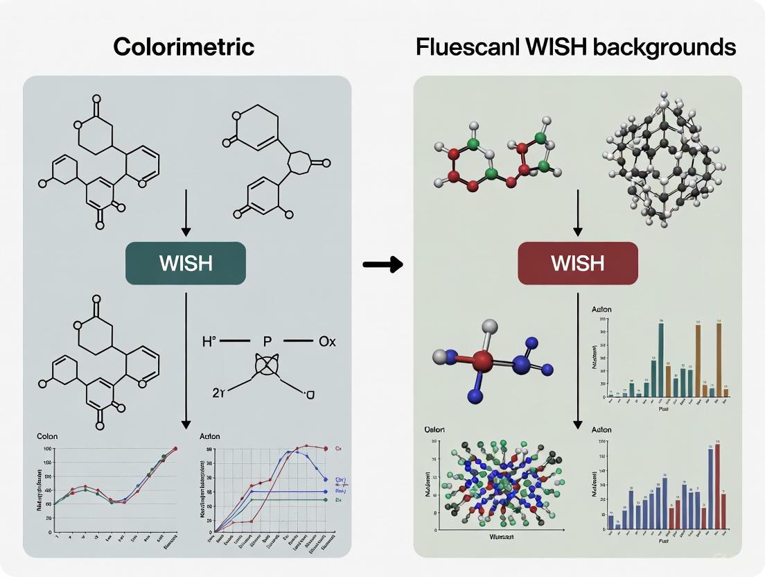
Colorimetric vs Fluorescent WISH: A Comprehensive Guide to Methods, Applications, and Optimization
This article provides a detailed comparative analysis of colorimetric and fluorescent Whole-Mount In Situ Hybridization (WISH) techniques for researchers and drug development professionals.
Colorimetric vs Fluorescent WISH: A Comprehensive Guide to Methods, Applications, and Optimization
Abstract
This article provides a detailed comparative analysis of colorimetric and fluorescent Whole-Mount In Situ Hybridization (WISH) techniques for researchers and drug development professionals. It covers the foundational principles of both methods, explores their specific applications in biomedical research, and offers practical guidance for troubleshooting and optimization. The content also addresses the critical process of method validation, empowering scientists to select the most appropriate detection strategy for their specific experimental needs in gene expression visualization.
Understanding WISH Fundamentals: Core Principles of Colorimetric and Fluorescent Detection
Historical Development and Evolution of In Situ Hybridization Techniques
In situ hybridization (ISH) represents a cornerstone technique in molecular biology that allows for the detection and localization of specific nucleic acid sequences within preserved tissues, cells, or entire embryos. First developed in 1968, this method enables researchers to visualize the spatial distribution of DNA or RNA, providing crucial insights into gene expression patterns, chromosomal abnormalities, and cellular organization within their morphological context [1]. The fundamental principle involves the formation of a hybrid molecule between an endogenous single-stranded nucleic acid target and a complementary, labeled probe, which is then detected through various visualization systems [1].
The evolution of ISH has progressed from initial radioactive detection methods to the sophisticated chromogenic and fluorescent systems widely used today. This technological progression has transformed ISH into an indispensable tool for both basic research and clinical diagnostics, particularly in cancer genetics, developmental biology, and infectious disease detection. Within the context of whole-mount in situ hybridization (WISH) background comparison research, the distinction between colorimetric and fluorescent detection has become increasingly significant, with each approach offering distinct advantages for specific applications in spatial gene expression analysis [2].
Historical Timeline and Technological Progression
The development of ISH spans over five decades, marked by several transformative breakthroughs that have enhanced its sensitivity, resolution, and applicability. The journey began with radioactive labeling, progressed through fluorescence-based detection, and evolved into the sophisticated chromogenic and multiplexing systems available today.
Table 1: Historical Milestones in In Situ Hybridization Development
| Year | Development | Key Researchers/Innovators | Significance |
|---|---|---|---|
| 1968 | First successful ISH experiments | N/A | Detection of highly amplified ribosomal DNA in Xenopus oocytes and satellite DNA in chromosomes using ³H-labeled probes and autoradiography [1]. |
| ~1978 | First fluorescent probes | N/A | Enabled fluorescence in situ hybridization (FISH), eliminating radioactivity and allowing for multiple target detection [1]. |
| 1980s-1990s | Chromogenic ISH (CISH) | N/A | Introduced enzyme-based colorimetric detection, enabling visualization with standard bright-field microscopy [3]. |
| 2000s | Automation & Multiplexing | Various commercial entities | Development of automated platforms and protocols for detecting multiple targets (MC-WISH) in a single sample [2]. |
| 2010s-Present | Advanced Probes & Digital Integration | Companies (e.g., Dako, Bio-Techne) | Introduction of high-sensitivity methods (e.g., RNAscope), peptide nucleic acid (PNA) probes, and integration with digital pathology/AI [3] [4]. |
The initial breakthrough in 1968 demonstrated that nucleic acids could be detected within their cellular context, but the method was hampered by the long exposure times required for autoradiography and the safety concerns associated with radioactive materials [1]. The advent of fluorescence in situ hybridization (FISH) approximately a decade later marked a revolutionary advance, offering faster results, improved spatial resolution, and the potential for multiplexing [1]. Subsequent innovations introduced chromogenic in situ hybridization (CISH), which provided a permanent slide record and compatibility with conventional bright-field microscopy and standard histology stains [3]. Recent trends focus on automation, increased multiplexing capabilities, and the integration of digital imaging and artificial intelligence to enhance quantification and workflow efficiency [4] [5].
Comparative Analysis: Fluorescent vs. Chromogenic WISH
The choice between fluorescent and chromogenic detection is fundamental in designing a WISH experiment. Each method has distinct strengths and limitations, making them suitable for different research or diagnostic scenarios. The core difference lies in the detection and visualization system: FISH uses fluorophore-labeled probes, while CISH employs enzyme-linked probes that produce a stable, colored precipitate.
Table 2: Direct Comparison of FISH and CISH/C-WISH Characteristics
| Parameter | Fluorescence ISH (FISH) | Chromogenic ISH (CISH) |
|---|---|---|
| Detection Principle | Fluorophore-labeled probes detected with fluorescence microscopy [3] | Enzyme-linked (e.g., AP) probes with chromogenic substrates detected with bright-field microscopy [3] [2] |
| Multiplexing Potential | High - multiple targets with different fluorophores [6] | Moderate - typically 2-3 targets with different enzymes/colors [2] |
| Spatial Resolution | High, suitable for subcellular localization [2] | High, but limited by chromogen diffusion [2] |
| Sample Permanence | Low - fluorophores photobleach [2] | High - stained slides are permanent [2] |
| Required Equipment | Fluorescence microscope, darkroom [3] | Standard bright-field microscope [3] |
| Compatibility with Stains | Limited, requires counterstains like DAPI [3] | High, compatible with common histological stains (e.g., HE) [7] [2] |
| Scanning & Analysis | Requires z-stacking, slower digital scanning (e.g., 764 sec/mm²) [3] | Faster digital scanning (e.g., 29 sec/mm²) [3] |
| Throughput in Diagnostics | Lower throughput due to slower scanning and analysis [3] | Higher throughput, preferred for high-throughput HER2 testing [3] |
| Key Advantage | Superior multiplexing and sensitivity [6] | Ease of use, cost-effectiveness, and integration into pathology workflows [3] [4] |
Supporting Experimental Data in Diagnostics
Comparative studies in clinical diagnostics provide quantitative data on the performance of FISH and CISH. A 2013 study comparing HER2 genetic assays in breast cancer samples found high concordance between the techniques.
Table 3: Experimental Performance Data from Clinical Studies
| Study Focus | Methodology | Key Finding | Reference |
|---|---|---|---|
| HER2 Testing in Breast Cancer (108 samples) | Comparison of 5 HER2 assays (FISH & CISH) on TMAs | 99% concordance between FISH and CISH (Cohen κ coefficient, 0.97) [3]. | [3] |
| Diagnosis of Cutaneous Leishmaniasis (50 samples) | Comparison of CISH, IHC, and Histopathology (HP) vs. Culture | Sensitivity: IHC (66%), CISH (54%), HP (50%). CISH showed no cross-reaction with fungi, unlike IHC [7]. | [7] |
| Digital Scanning Efficiency | Scanning time comparison for TMA analysis | Mean scanning time for CISH: 29 sec/mm². Mean scanning time for FISH (with z-stacking): 764 sec/mm² [3]. | [3] |
Detailed Methodologies and Protocols
Multi-target Chromogenic Whole-Mount In Situ Hybridization (MC-WISH)
The MC-WISH protocol allows for the simultaneous detection of up to three different mRNA species in intact Drosophila or zebrafish embryos, providing a powerful tool for comparing gene expression domains with high spatial accuracy [2]. The workflow below outlines the key stages of this sophisticated protocol.
Key Protocol Steps [2]:
Probe Generation: Antisense RNA probes are synthesized via in vitro transcription from linearized template DNA. Each probe for a different gene is labeled with a distinct hapten (e.g., digoxigenin-11-UTP, biotin-16-UTP, or fluorescein-12-UTP). The labeled probes are purified and dissolved in a pre-hybridization buffer for storage.
Embryo Preparation: Embryos are collected, fixed with formaldehyde, and devitellinized using standard procedures. They are permeabilized through a series of methanol and proteinase K treatments to allow probe penetration. Using specialized mesh inserts or baskets during processing is recommended to prevent sample loss.
Hybridization: Fixed embryos are simultaneously incubated with a mixture of up to three differently hapten-labeled RNA probes in hybridization buffer. This allows all probes to bind to their complementary mRNA targets at once.
Sequential Chromogenic Detection: After stringent washes to remove unbound probes, the haptens are detected sequentially. Each detection round consists of:
- Incubation with an alkaline phosphatase (AP)-conjugated antibody specific to one hapten.
- Visualization via an AP-substrate that produces a localized, stable color precipitate (e.g., NBT/BCIP for blue, Fast Red for red).
- Inactivation and removal of the antibody-AP complex with a low-pH glycine buffer to prevent cross-reactivity in the next round.
- This cycle is repeated with different antibody/substrate combinations for each hapten.
Mounting and Imaging: After the final detection round, embryos are mounted in glycerol and imaged under a high-resolution compound microscope using differential interference contrast (DIC) optics to visualize the color precipitates against the tissue morphology.
The Scientist's Toolkit: Key Research Reagent Solutions
Table 4: Essential Reagents and Materials for MC-WISH
| Reagent/Material | Function in the Protocol | Specific Examples & Notes |
|---|---|---|
| Hapten-Labeled NTPs | Incorporate into RNA probes during transcription to enable subsequent immunodetection. | Digoxigenin-11-UTP, Biotin-16-UTP, Fluorescein-12-UTP [2]. |
| RNA Polymerases | Synthesize single-stranded RNA probes from DNA templates. | T7, T3, or SP6 RNA polymerase, selected based on template's promoter [2]. |
| Anti-Hapten Antibodies | Bind specifically to the hapten on the hybridized probe. Conjugated to a reporter enzyme. | Anti-digoxigenin, anti-biotin, and anti-fluorescein antibodies, conjugated to Alkaline Phosphatase (AP) [2]. |
| AP Chromogenic Substrates | Enzymatically converted by AP into insoluble, colored precipitates at the site of hybridization. | NBT/BCIP (blue), Fast Red (red), and other substrates producing contrasting colors [2]. |
| Hybridization Buffer | Creates optimal conditions for specific probe-target hybridization while minimizing non-specific binding. | Typically contains formamide, salts (SSC), and blocking agents (heparin, torula RNA) [2]. |
| Permeabilization Agents | Disrupt cellular membranes to allow probe entry into the sample. | Proteinase K, methanol [2]. |
| Processing Inserts | Hold embryo samples during liquid exchanges to prevent physical loss. | Polystyrene inserts with a polyester mesh bottom (e.g., Netwell inserts) [2]. |
Current Market Landscape and Future Outlook
The ISH market is experiencing significant growth, driven by its critical role in molecular diagnostics and research. The global ISH market was valued at approximately USD 1.55 - 1.87 billion in 2024-2025 and is projected to reach USD 3.14 - 5.2 billion by 2033-2035, growing at a compound annual growth rate (CAGR) of 7.14% to 10.6% [4] [6] [5]. This growth is fueled by rising cancer prevalence, increasing demand for personalized medicine, and continuous technological advancements.
Table 5: Market Segmentation and Dominant Trends (2024-2025)
| Segmentation | Dominant Segment | Key Insights & Growth Drivers |
|---|---|---|
| Technology | Fluorescence ISH (FISH) (~54-72% share) [4] [6] [8] | Dominates due to high sensitivity and multiplexing capability. CISH is the fastest-growing segment, driven by cost-effectiveness [4]. |
| Probe Type | DNA Probes (~59% share) [4] | Widely used for detecting chromosomal abnormalities. RNA probes segment is growing fastest, driven by RNA-based diagnostics and spatial transcriptomics [4] [6]. |
| Application | Cancer Diagnostics & Research (~45% share) [4] [8] | The largest segment due to use in biomarker detection (e.g., HER2, ALK). Infectious disease diagnosis is the fastest-growing application [8]. |
| End User | Hospitals & Diagnostic Labs (~40% share) [4] [8] | Largest end-user segment. Pharmaceutical & biotechnology companies are the fastest-growing segment, driven by drug discovery R&D [8]. |
| Region | North America (~39% share) [4] [5] [8] | Dominates due to advanced healthcare infrastructure and high R&D spending. Asia-Pacific is the fastest-growing region [4] [8]. |
Future trends point toward increased automation to reduce manual errors and improve throughput, greater adoption of multiplexing to extract more data from single samples, and deeper integration with digital pathology and AI for enhanced image analysis and quantification [9] [4] [5]. Furthermore, the development of highly sensitive in situ sequencing methods and advanced probe chemistries like peptide nucleic acids (PNAs) will continue to push the boundaries of sensitivity and specificity [3] [10].
However, the search results provide general insights into colorimetric and fluorescent sensing principles relevant to biological detection. The following general comparison and methodology overview are based on these related fields.
The table below summarizes core characteristics of colorimetric and fluorescent detection based on principles from related chemical sensing applications [11] [12].
| Feature | Colorimetric Detection | Fluorescent Detection |
|---|---|---|
| Core Principle | Visual color change measured by absorbance [11]. | Light emission at longer wavelength after excitation [12]. |
| Typical Readout | Spectrophotometry, camera imaging, or visual inspection [11]. | Fluorimetry, fluorescence microscopy [13]. |
| Sensitivity | Generally moderate; suitable for many routine applications [11]. | Typically higher; capable of detecting trace amounts of analyte [12]. |
| Quantification | Effective, especially with advanced LED photometry (PEDD) [11]. | Excellent, with a wide linear dynamic range [12]. |
| Complexity & Cost | Often lower-cost, simpler instrumentation; suited for field use [14] [11]. | Higher-cost, requires specific excitation sources and filters [13]. |
| Multiplexing Potential | Lower, limited by spectral overlap of color changes. | Higher, using probes with different emission profiles [15]. |
Conceptual Workflow for a Colorimetric Assay
The diagram below illustrates a generalized workflow for a colorimetric detection assay, integrating principles from the reviewed literature.
The Scientist's Toolkit: Key Reagents and Materials
This table lists common categories of reagents and materials used in developing colorimetric and fluorescent detection systems, as inferred from the methodologies in the search results [16] [17] [18].
| Item | Function in Assay |
|---|---|
| Colorimetric Probe | Binds analyte, producing a visible color change for direct measurement [19]. |
| Fluorescent Probe/Sensor | Emits light upon excitation and interaction with the target; enables highly sensitive detection [16] [18]. |
| Buffer Solutions | Maintain correct pH and ionic strength for reaction stability and specificity [11]. |
| Signal Enhancement Substrates | Used in enzyme-linked assays (e.g., TMB, BCIP/NBT) to amplify the detectable signal [17]. |
| Microplates & Cuvettes | Standard containers for holding samples during spectroscopic analysis [11]. |
| Reference Standards | Known concentrations of analyte for generating a calibration curve and quantifying results [14] [11]. |
How to Find Specialized WISH Information
To locate the specific experimental data and protocols you need for your WISH guide, I suggest these targeted approaches:
- Use specialized scientific databases: Search platforms like PubMed and Google Scholar for primary research articles. Specific queries such as "colorimetric whole-mount in situ hybridization protocol" or "comparing fluorescence WISH sensitivity" will be most effective.
- Consult established protocol repositories: Websites and textbooks dedicated to molecular biology protocols (e.g., Cold Spring Harbor Protocols, Nature Protocols) are authoritative sources for detailed, step-by-step WISH methodologies.
- Review product literature: Technical documentation and application notes from major suppliers of life science reagents (e.g., Roche, Thermo Fisher Scientific, Sigma-Aldrich) often contain rigorously tested protocols and performance data for their specific WISH detection kits.
I hope this structured overview of general principles provides a useful starting point. Should you find specific WISH-related data, I can assist you in processing and formatting that information into tables and diagrams.
Core Operational Principles of Fluorescent WISH Detection
Fluorescent Whole-Mount In Situ Hybridization (F-WISH) is a powerful cytogenetic technique that enables the visualization and localization of specific nucleic acid sequences within the context of intact biological specimens. Unlike traditional colorimetric methods that produce precipitating chromogens, F-WISH utilizes fluorescently labeled probes to identify target messenger RNA transcripts in cultured cells, tissue sections, or whole-mount preparations [20]. This technique operates on the fundamental principles of nucleic acid thermodynamics, where complementary strands of nucleic acids anneal to form stable hybrids under appropriate conditions [20]. The development of F-WISH represents a significant advancement over earlier isotopic methods, offering improved safety, resolution, and the capability for multiplex analysis without the hazards and lengthy exposure times associated with radioactive probes [20].
The evolution of F-WISH technology has progressively enhanced our ability to study gene expression patterns with increasing precision. Modern implementations, particularly single-molecule FISH (smFISH), now enable researchers to resolve individual mRNA transcripts with high specificity and signal-to-noise ratios [20]. This technical refinement has transformed F-WISH into an indispensable tool for developmental biology, regeneration studies, and biomedical research, allowing scientists to correlate gene expression patterns with specific anatomical structures and cellular identities within complex tissues [21] [22]. The technique has proven especially valuable in organisms with limited genetic toolkits, where it provides critical insights into spatial gene regulation without requiring transgenic approaches [21].
Core Principles and Detection Mechanisms
Fundamental Thermodynamic Principles
The operational foundation of F-WISH rests on the predictable thermodynamic behavior of nucleic acids, specifically the ability of complementary DNA or RNA strands to hybridize and form stable duplexes. This hybridization process occurs when appropriate conditions of temperature, ionic strength, and pH are established to facilitate probe penetration and annealing to target sequences [20]. The technique capitalizes on the natural affinity between complementary base pairs, allowing designed probes to seek out and bind their specific target sequences within fixed tissues. Early FISH methodologies employed RNA-based probes to label DNA sequences, but modern RNA-FISH specifically targets messenger RNA transcripts to visualize patterns of gene expression [20].
The specificity of F-WISH is governed by the stringency of hybridization and post-hybridization washing conditions. Stringency, determined primarily by temperature and ionic strength of the washing solutions, ensures that imperfectly matched hybrids dissociate while perfectly matched probe-target duplexes remain stable [20]. This precise control over nucleic acid interactions enables researchers to distinguish between closely related sequences and minimize background signals. The thermodynamic parameters must be carefully optimized for each experimental system, as factors including probe length, GC content, and tissue permeability collectively influence hybridization efficiency and specificity [20].
Signal Generation and Amplification Strategies
F-WISH employs several distinct detection strategies, each with characteristic mechanisms for signal generation and amplification. The choice of detection system significantly impacts the sensitivity, resolution, and multiplexing capability of the experiment.
Tyramide Signal Amplification (TSA) systems utilize horseradish peroxidase (HRP)-conjugated antibodies that catalyze the deposition of fluorescent tyramide substrates. Upon activation by HRP, tyramide radicals form covalent bonds with electron-rich tyrosine residues on nearby proteins, resulting in localized signal amplification [23] [24]. This approach significantly enhances detection sensitivity, making it particularly suitable for identifying low-abundance transcripts. However, the peroxidase activity is relatively quickly quenched by substrate excess, typically limiting productive reaction times to less than 30 minutes [23].
Alkaline Phosphatase (AP)-based detection employs enzyme-conjugated antibodies that convert fluorescent substrates such as Fast Red or Fast Blue into precipitating fluorescent products [23]. Unlike peroxidase-based systems, AP reactions can proceed for extended periods (several hours) while maintaining high signal-to-noise ratios, making this approach advantageous for detecting less abundant transcripts [23]. The extended reaction time allows for gradual signal accumulation that can be monitored until optimal intensity is achieved.
Direct fluorescence methods utilize probes that are conjugated directly to fluorophores, eliminating the need for immunological detection steps. This approach forms the basis of smFISH, where multiple short oligonucleotide probes, each tagged with a single fluorophore, collectively target individual mRNA molecules [20]. When these probes hybridize along the length of a transcript, the combined fluorescence becomes sufficiently bright to resolve individual molecules above background noise, enabling precise transcript quantification [20].
Table: Comparison of Major F-WISH Detection Systems
| Detection System | Mechanism | Sensitivity | Reaction Time | Best Applications |
|---|---|---|---|---|
| Tyramide Signal Amplification (TSA) | HRP-catalyzed deposition of fluorescent tyramides | Very high (signal amplification) | Short (<30 min productive reaction) | Low-abundance transcripts, multiplexing |
| Alkaline Phosphatase (AP) | Enzyme-mediated conversion of fluorescent substrates | High | Extended (several hours) | Less abundant transcripts, quantitative studies |
| Direct Fluorescence | Fluorophore-conjugated probes bind directly to targets | Moderate (depends on probe number) | N/A (no enzymatic development) | Single-molecule detection, high-resolution quantification |
Comparative Performance Analysis: Fluorescent vs. Colorimetric WISH
Sensitivity and Resolution Characteristics
The fundamental distinction between fluorescent and colorimetric detection lies in their respective sensitivity profiles and spatial resolution capabilities. Fluorescent WISH exhibits superior sensitivity for detecting low-abundance transcripts, particularly when implementing signal amplification strategies such as tyramide-based systems [23]. This enhanced sensitivity stems from the cumulative effect of multiple fluorophores contributing to a detectable signal at a single location, coupled with the ability to amplify signals enzymatically. In contrast, colorimetric methods typically rely on the direct enzymatic conversion of chromogenic substrates, which may lack equivalent amplification potential [23].
Regarding spatial resolution, F-WISH offers significant advantages for precise subcellular localization of transcripts. The fluorescent signals generated through F-WISH can be visualized with high resolution using confocal microscopy, enabling researchers to determine the subcellular distribution of mRNAs within specific cellular compartments [24]. This capability has proven instrumental for identifying asymmetrically localized mRNAs that play crucial roles in developmental patterning and cell fate determination [24]. Colorimetric detection, while sufficient for tissue-level expression analysis, often lacks the resolution necessary for detailed subcellular localization due to the diffuse nature of chromogen precipitation.
Multiplexing Capabilities
A defining advantage of F-WISH is its capacity for simultaneous detection of multiple transcripts within a single specimen. By employing probes labeled with different fluorophores that emit light at distinct wavelengths, researchers can visualize the expression patterns of several genes concurrently and analyze their spatial relationships [23]. This multiplexing capability enables the direct comparison of expression domains at cellular resolution, providing insights into regulatory interactions and cellular identities within complex tissues [23].
The implementation of multiplexed F-WISH requires careful experimental design, including selection of fluorophores with non-overlapping emission spectra and sequential detection protocols to minimize cross-talk between channels. Recent methodological advances have streamlined this process through the combination of different detection systems, such as AP-Fast Blue and POD-TSA-carboxyfluorescein, which can be visualized simultaneously without the need for sequential antibody applications [23]. This approach reduces hands-on time and eliminates potential false-positive co-localization signals that can arise from insufficient enzyme inactivation in sequential detection rounds [23].
Colorimetric WISH, while capable of detecting multiple targets through sequential applications of different chromogenic substrates, is inherently more limited in multiplexing capacity. The precipitating reaction products often occupy similar spatial domains, making it difficult to distinguish overlapping expression patterns, particularly when more than two targets are analyzed [23]. Furthermore, the sequential development of chromogenic reactions typically results in decreased sensitivity for subsequently detected targets, potentially limiting the utility for analyzing genes with similar expression levels [23].
Table: Quantitative Comparison of Detection Performance Metrics
| Performance Metric | Fluorescent WISH | Colorimetric WISH |
|---|---|---|
| Detection Sensitivity | High (amplifiable) | Moderate |
| Spatial Resolution | Subcellular | Cellular/Tissue |
| Multiplexing Capacity | High (3+ targets) | Limited (2-3 targets) |
| Signal Quantification | Excellent | Moderate |
| Compatibility with Sectioning | Limited (fluorescence quenching) | Excellent |
| Sample Preservation | Requires specialized fixation [21] | Compatible with standard fixation |
Advanced F-WISH Methodologies and Protocol Optimization
Sample Preparation and Fixation Strategies
Optimal sample preparation is critical for successful F-WISH experiments, balancing the competing demands of tissue preservation, permeability, and macromolecule integrity. Recent advances in fixation protocols have addressed the particular challenges associated with delicate tissues, such as regenerating structures in planarians and killifish. The novel Nitric Acid/Formic Acid (NAFA) fixation method demonstrates improved preservation of fragile anatomical structures while maintaining compatibility with both FISH and immunofluorescence applications [21]. This protocol achieves enhanced probe penetration without requiring proteinase K digestion, thereby better preserving antigen epitopes for subsequent immunological detection [21].
The NAFA protocol significantly improves structural preservation compared to traditional methods that utilize mucolytic agents like N-acetyl cysteine (NAC) or proteinase K treatments. In comparative studies, the NAFA method maintained epidermis integrity in planarians, whereas NAC treatment resulted in noticeable tissue damage and breaches of structural continuity [21]. This preservation advantage extends to internal structures as well, with the NAFA protocol yielding crisper staining of musculature and protonephridia while simultaneously preserving fragile external cilia [21]. The method incorporates EGTA to chelate calcium and inhibit nucleases, further protecting RNA integrity during sample processing [21].
Signal Enhancement Methodologies
Several strategic approaches have been developed to enhance fluorescent signals in F-WISH, particularly for challenging applications involving low-abundance transcripts or specimens with high background autofluorescence. The incorporation of viscosity-increasing polymers such as dextran sulfate into the hybridization mixture creates molecular crowding conditions that effectively increase local probe concentration, resulting in dramatically improved signal intensities [23]. Experimental comparisons demonstrate that this modification can make the difference between detectable and undetectable signals for less pronounced expression sites [23].
Hydrogen peroxide treatment represents another valuable strategy for enhancing signal sensitivity in F-WISH applications. While traditionally used to quench endogenous peroxidase activity, hydrogen peroxide also improves tissue permeabilization by disrupting cell membranes, thereby facilitating better probe and antibody penetration [23]. The combination of hydrogen peroxide pretreatment with dextran sulfate inclusion in the hybridization mix produces the strongest signal intensities, enabling more robust detection of challenging targets [23].
The strategic combination of different enzyme-substrate systems provides additional opportunities for signal optimization in multiplexed F-WISH experiments. By pairing the prolonged enzymatic activity of alkaline phosphatase (using substrates such as Fast Blue) with the rapid amplification capacity of peroxidase-based tyramide systems, researchers can achieve balanced signal development for multiple targets while simplifying the experimental workflow [23]. This integrated approach eliminates the need for antibody inactivation steps between detection rounds, reducing both hands-on time and the potential for false-positive co-localization results [23].
Research Reagent Solutions for F-WISH
Successful implementation of F-WISH depends on a comprehensive suite of specialized reagents, each fulfilling specific functions within the experimental workflow. The following table details essential reagents and their operational roles in standard F-WISH protocols.
Table: Essential Research Reagents for F-WISH Experiments
| Reagent Category | Specific Examples | Function | Protocol Considerations |
|---|---|---|---|
| Fixatives | NAFA (Nitric Acid/Formic Acid) | Preserves tissue architecture and RNA integrity | Superior for delicate tissues; avoids proteinase K damage [21] |
| Permeabilization Agents | Proteinase K, Hydrogen Peroxide | Enhances probe accessibility to targets | H2O2 improves penetration while reducing autofluorescence [23] |
| Hybridization Enhancers | Dextran Sulfate | Increases effective probe concentration via molecular crowding | Dramatically improves signal intensity [23] |
| Enzymatic Detection Systems | Horseradish Peroxidase (POD), Alkaline Phosphatase (AP) | Catalyzes signal generation | POD-TSA for sensitivity; AP for extended development [23] |
| Fluorescent Substrates | Tyramide conjugates, Fast Red, Fast Blue | Generates detectable fluorescent signal | Fast Blue compatible with far-red filter sets [23] |
| Blocking Agents | Normal Goat Serum, BSA | Reduces non-specific antibody binding | Critical for signal-to-noise ratio optimization |
| Mounting Media | Antifade reagents with DAPI | Preserves fluorescence and counterstains nuclei | DAPI identifies cellular organization |
Visualization of F-WISH Workflows and Principles
The following diagrams illustrate key operational workflows and principles in fluorescent WISH detection, providing visual references for the experimental stages and their relationships.
F-WISH Experimental Workflow
F-WISH Detection Mechanism Principles
Fluorescent WISH detection represents a sophisticated methodological platform that offers distinct advantages for spatial transcriptomics, particularly in applications requiring high sensitivity, subcellular resolution, and multiplexing capability. The core operational principles of F-WISH—rooted in nucleic acid hybridization thermodynamics—provide a robust foundation for visualizing gene expression patterns within their native tissue contexts. Ongoing methodological refinements in sample preparation, signal amplification, and detection strategies continue to expand the utility of this technique across diverse biological systems and research applications. As exemplified by the development of specialized fixation protocols like NAFA and enhanced detection systems combining different enzymatic approaches, the evolution of F-WISH methodology remains driven by the need to balance optimal signal detection with structural preservation. These technical advances ensure that F-WISH will continue to be an indispensable tool for exploring gene expression architecture in developmental biology, regeneration studies, and disease research.
Key Reagents and Their Functional Roles in Tissue Preparation and Hybridization
Whole-mount in situ hybridization (WISH) is a foundational technique in developmental biology and molecular pathology for localizing specific nucleic acid targets within fixed tissues and cells, providing crucial temporal and spatial information about gene expression [25]. The technique primarily branches into two detection methodologies: colorimetric (CISH) and fluorescent (FISH). The choice between them dictates the required reagents, detection instrumentation, and specific applications. CISH, visualized via bright-field microscopy, allows for the simultaneous observation of signal and tissue morphology, making it particularly valuable in molecular pathology diagnostics [25]. In contrast, FISH, visualized via fluorescence microscopy, is inherently multiplexible, enabling researchers to visualize multiple transcript targets within a single specimen—a key advantage for studying gene co-localization and interaction [25]. This guide objectively compares the core reagents and their functional roles in tissue preparation and hybridization for these two approaches, framing the discussion within a broader thesis on their comparative performance and optimal application.
Comparative Analysis: Core Reagents and Their Functions
The performance of CISH and FISH is fundamentally governed by the reagents used in tissue preparation, probe hybridization, and signal detection. The table below summarizes the key reagents and their distinct functional roles in the two methodologies.
Table 1: Key Reagents and Their Functional Roles in CISH vs. FISH Protocols
| Reagent Category | Specific Reagent | Functional Role | Role in CISH | Role in FISH |
|---|---|---|---|---|
| Tissue Preparation | Paraformaldehyde (PFA) | Tissue fixation; preserves morphology and nucleic acids [26] | Critical for structural integrity for bright-field imaging [26] | Critical for structural integrity and target accessibility [26] |
| Proteinase K | Digests proteins; increases tissue permeability for probe access [26] | Standard use; concentration and time must be optimized [26] | Standard use; concentration and time must be optimized [26] | |
| Hydrogen Peroxide (H₂O₂) | Blocks endogenous peroxidase activity; improves permeability [27] | Less common; primarily for peroxidase blocking | Used for blocking and to enhance probe/antibody access, boosting signal [27] | |
| Hybridization | Labeled RNA Probe (e.g., Digoxigenin, Dinitrophenol) | Target-specific nucleic acid probe for hybridization [27] [26] | Digoxigenin-labeled probes standard [26] | Digoxigenin- and fluorescein-labeled probes common for multiplexing [27] |
| Dextran Sulfate | Molecular crowding agent; increases effective probe concentration [27] | Can be used to improve signal sensitivity [27] | Critical for enhancing signal intensity in fluorescent detection [27] | |
| Signal Detection | Anti-Digoxigenin Antibody | Binds to digoxigenin-labeled probe; conjugated to reporter enzyme [26] | Conjugated to Alkaline Phosphatase (AP) or Horseradish Peroxidase (POD) [27] | Conjugated to Alkaline Phosphatase (AP) or Horseradish Peroxidase (POD) [27] |
| Alkaline Phosphatase (AP) | Reporter enzyme; catalyzes chromogenic/fluorescent substrate conversion [27] | Primary enzyme for BCIP/NBT (blue/purple) or Fast Red/Blue precipitates [27] | Used with fluorescent substrates like Fast Blue for sustained signal generation [27] | |
| Horseradish Peroxidase (POD) | Reporter enzyme; catalyzes tyramide signal amplification (TSA) [27] | Less common for CISH | Primary enzyme for Tyramide Signal Amplification (TSA); enables high-sensitivity fluorescence [27] | |
| BCIP/NBT | Chromogenic substrate for AP; yields insoluble blue/purple precipitate [27] | Standard substrate for single-color detection [27] | Not typically used | |
| Fast Red / Fast Blue | AP substrate; yields chromogenic precipitate, also fluorescent [27] | Used for chromogenic two-color WISH [27] | Fast Blue used for far-red fluorescent channel detection [27] | |
| Tyramide-Fluorescein (e.g., TSA-Carboxyfluorescein) | Fluorescent substrate for POD-based signal amplification [27] | Not applicable | Key for high-sensitivity fluorescent detection; signal amplifiable but quenches quickly [27] |
Experimental Protocols and Workflows
Detailed Protocol for Two-Color Fluorescent WISH
A sensitive two-color FISH protocol that combines the advantages of AP and POD reporter systems demonstrates the practical application of these reagents [27]. This method eliminates the need for an antibody-enzyme inactivation step, reducing hands-on time and preventing false-positive co-localization results.
Methodology:
- Tissue Fixation and Permeabilization: Zebrafish embryos are fixed in 4% Paraformaldehyde (PFA) [26]. Permeabilization is enhanced by treating with 2% Hydrogen Peroxide (H₂O₂) prior to standard Proteinase K digestion, dramatically improving signal intensity for subsequent steps [27].
- Probe Hybridization: Embryos are hybridized with two differentially labeled RNA probes (e.g., Digoxigenin- and Dinitrophenol-labeled). The hybridization mix includes 5% Dextran Sulfate, a viscosity-increasing polymer that creates a molecular crowding effect, leading to a local increase in probe concentration and significantly stronger signals [27].
- One-Step Antibody Detection: A mixture of antibody-enzyme conjugates is applied in a single step. This typically includes an anti-digoxigenin antibody conjugated to Alkaline Phosphatase (AP) and an anti-dinitrophenol antibody conjugated to Horseradish Peroxidase (POD) [27].
- Simultaneous Fluorescent Visualization:
- The AP-conjugated antibody is detected using the substrate Fast Blue. The AP reaction can proceed for extended periods (hours), providing a high signal-to-noise ratio in the far-red fluorescent channel [27].
- The POD-conjugated antibody is detected using Tyramide-Carboxyfluorescein (TSA-FAM). The TSA reaction provides high signal amplification but is quenched quickly (within 30 minutes) to prevent substrate excess and background [27].
Workflow Diagram: Two-Color FISH
The following diagram illustrates the logical workflow and reagent application for the two-color FISH protocol.
The Scientist's Toolkit: Essential Research Reagent Solutions
Successful execution of WISH experiments requires a suite of reliable reagent solutions. The following table details essential materials and their critical functions in a typical workflow.
Table 2: Essential Research Reagent Solutions for WISH
| Reagent Solution | Function in Experiment |
|---|---|
| Paraformaldehyde (PFA) | Cross-linking fixative that preserves tissue architecture and immobilizes nucleic acids, preventing degradation [26]. |
| Proteinase K | Serine protease that digests proteins surrounding nucleic acid targets, thereby increasing tissue permeability and probe accessibility [26]. |
| Dextran Sulfate | A viscosity-increasing polymer added to the hybridization buffer. It creates a molecular crowding effect, locally increasing probe concentration and significantly enhancing hybridization signal intensity [27]. |
| Hydrogen Peroxide (H₂O₂) | Used pre-hybridization to block endogenous peroxidase activity and, critically, to improve overall tissue permeabilization, leading to stronger signal detection for both chromogenic and fluorescent methods [27]. |
| Digoxigenin-labeled RNA Probe | A target-specific, non-radioactively labeled RNA probe. Digoxigenin is a plant-derived hapten that is highly specific for antibody binding, minimizing background in complex animal tissues [27] [26]. |
| Anti-Digoxigenin Antibody Conjugates | Antibody conjugated to a reporter enzyme (AP or POD) that binds specifically to the digoxigenin label on the hybridized probe, enabling visual detection [27] [26]. |
| Alkaline Phosphatase (AP) & Substrates | Enzyme-substrate system where AP catalyzes the conversion of substrates like BCIP/NBT (chromogenic) or Fast Blue (fluorescent) into a detectable precipitate or signal [27]. |
| Horseradish Peroxidase (POD) & Tyramides | Enzyme-substrate system for high-sensitivity detection. POD catalyzes the deposition of fluorescent tyramides (TSA), which binds covalently to tissues, providing significant signal amplification [27]. |
In the field of molecular diagnostics and bioanalysis, the choice of detection method is a critical determinant of an experiment's success, balancing the need for ease-of-use against the requirement for sensitivity. Colorimetric and fluorescent detection techniques represent two fundamental approaches, each with distinct advantages and optimal applications. Colorimetric methods, which produce a visible color change detectable by the naked eye, offer simplicity and are ideally suited for point-of-care testing and resource-limited settings. In contrast, fluorescent techniques, which rely on the emission of light from excited molecules, provide superior sensitivity and quantitative capabilities, enabling researchers to detect minute quantities of analytes with high precision. This guide provides an objective comparison of these methodologies, supported by recent experimental data, to assist researchers, scientists, and drug development professionals in selecting the appropriate technique for their specific applications within the context of whole-mount in situ hybridization (WISH) and broader biosensing research.
Fundamental Principles
Colorimetric detection relies on enzymatic or chemical reactions that generate a visible color change, typically measured via absorbance spectroscopy. The output is a qualitative or semi-quantitative visual signal that can often be observed without specialized equipment. Common colorimetric systems include peroxidase-based reactions using substrates like 3,3',5,5'-tetramethylbenzidine (TMB), which produce a colored product upon oxidation [17]. The readout is generally straightforward, requiring only basic spectrophotometers or even visual assessment for result interpretation.
Fluorescent detection operates on the principle of luminescence, where specific molecules (fluorophores) absorb light at a particular wavelength and emit light at a longer, lower-energy wavelength. This emission is measured using fluorometers or fluorescence microscopes, providing highly sensitive quantitative data. Modern fluorescent probes, such as the ratiometric fluorescent probe PBN-5 for norepinephrine detection, employ dual-emission channels that enable internal calibration, significantly improving quantification accuracy by minimizing environmental interference [28]. Advanced systems may utilize mechanisms including fluorescence resonance energy transfer (FRET), photoinduced electron transfer (PET), and intramolecular charge transfer (ICT) to enhance specificity and signal response.
Key Signaling Pathways in Biosensing
The following diagram illustrates the fundamental signaling pathways utilized in transcription-dependent biosensors, which underpin many modern detection systems:
Figure 1: Fundamental signaling pathway in transcription-dependent biosensors. This pathway is utilized in various sensing platforms, including yeast-based biosensors, where ligand binding to a GPCR receptor triggers a cascade resulting in a measurable output [29].
Advanced fluorescent detection systems often employ more complex molecular recognition pathways, as demonstrated in this ratiometric sensing mechanism:
Figure 2: Molecular recognition mechanism in ratiometric fluorescent probes. This pathway illustrates the dual-site recognition process where a probe specifically binds to target moieties, forming a macrocyclic complex enhanced by silver bridging, ultimately generating a calibrated ratiometric fluorescence signal [28].
Experimental Comparison and Performance Data
Direct Performance Comparison
Table 1: Quantitative comparison of colorimetric and fluorescent detection performance based on experimental data from recent studies.
| Performance Parameter | Colorimetric Detection | Fluorescent Detection | Experimental Context |
|---|---|---|---|
| Detection Limit | ~Micromolar range | ~Nanomolar range (e.g., 64.3 μM for earlier probes) [28] | Norepinephrine detection in biological samples |
| Detection Time | 3-8 hours (on paper slips) [29] | Within 50 minutes (complete reaction) [28] | Yeast biosensor output; molecular probe response |
| Quantitative Capability | Semi-quantitative; visual assessment or basic spectrophotometry | Highly quantitative; ratiometric calibration with linear response [28] | Comparison of output signal precision |
| Equipment Requirements | Minimal (naked eye, basic reader) | Advanced (fluorometers, microscopes, plate readers) | Resource needs for result interpretation |
| Multiplexing Potential | Limited | High (multiple emission channels) | Capacity for simultaneous multi-analyte detection |
| Dynamic Range | Limited | Wide linear range [28] | Concentration range over quantitative response is maintained |
Experimental Protocols
Colorimetric Detection Protocol (Yeast Biosensor Platform)
Sample Preparation:
- Cultivate yeast-based biosensor strains in appropriate media (YPD or synthetic defined complete media) at pH 5.8 [29].
- For agar plate assays, pour 40 mL of media with agar into square 120.5 mm petri dishes.
- Induce pigment production by adding 1 μM of specific ligand (e.g., α-mating pheromone) from a 1000× stock with 10% DMSO [29].
Detection and Analysis:
- Incubate samples at optimal growth temperature (typically 30°C for yeast).
- Monitor color development visually at regular intervals.
- For quantitative assessment, use basic spectrophotometry to measure absorbance at characteristic wavelengths.
- Time-of-detection (TOD) is recorded when visible color change is first observed [29].
Fluorescent Detection Protocol (Ratiometric Probe)
Sample Preparation:
- Prepare probe solution in PBS buffer (10 mM, pH = 7.4) with 10% DMSO as cosolvent [28].
- Add silver ions to accelerate recognition and amplify signal through silver bridging where applicable.
- Incubate with sample containing target analyte at room temperature.
Spectral Measurements:
- Monitor fluorescence emission spectra over time (up to 50 minutes).
- Excitate at common wavelength for both fluorophores (e.g., (S)-BINOL and naphthalimide in PBN-5) [28].
- Record emission intensity at two wavelengths (e.g., 400 nm and 526 nm for PBN-5).
- Calculate ratiometric values (F526/F400) for quantitative analysis [28].
Data Analysis:
- Plot fluorescence intensity ratio versus analyte concentration for calibration curve.
- Determine detection limit from linear range of calibration curve.
- Assess specificity through interference studies with structurally similar compounds.
Research Reagent Solutions
Table 2: Essential materials and reagents for colorimetric and fluorescent detection systems.
| Reagent/Category | Function/Description | Example Applications |
|---|---|---|
| Colorimetric Output Pigments | ||
| Lycopene | Red pigment; enables visual detection without additional reagents | Yeast biosensor output for point-of-care testing [29] |
| Prodeoxyviolacein | Green pigment; violacein pathway derivative | Alternative colorimetric output for biosensing [29] |
| Proviolacein | Green-brown pigment; violacein pathway derivative | Benchmarking against lycopene outputs [29] |
| TMB (3,3',5,5'-tetramethylbenzidine) | Chromogenic peroxidase substrate; produces blue color upon oxidation | Colorimetric reactions in MOF-based sensors with peroxidase-like activity [17] |
| Fluorescent Probes | ||
| PBN-5 | Dual-site ratiometric probe; combines (S)-BINOL and naphthalimide fluorophores | Specific norepinephrine detection via macrocyclic ring formation [28] |
| MOF-based Sensors | Porous crystalline materials with tunable fluorescence properties | Antibiotic detection via fluorescence quenching or enhancement [17] |
| Lanthanide-based MOFs | Intrinsic fluorescence from lanthanide centers | Sensitive detection through antenna effect [17] |
| Platform Materials | ||
| Yeast Biosensor Chassis | Saccharomyces cerevisiae with engineered mating pathway | Transcription-dependent sensing with colorimetric outputs [29] |
| Metal-Organic Frameworks (MOFs) | Porous materials with metal nodes and organic ligands | Fluorescent and colorimetric sensing platforms [17] |
| Silver Ions (Ag⁺) | Signal amplification through bridging in macrocyclic complexes | Enhancement of fluorescent probe response [28] |
Comparative Analysis and Application Guidance
Method Selection Framework
The choice between colorimetric and fluorescent detection methods should be guided by application-specific requirements. Colorimetric methods excel in scenarios demanding rapid, equipment-free results, where the highest sensitivity is not essential. The visual nature of colorimetric outputs makes them particularly valuable for point-of-care diagnostics, field testing, and educational settings. Recent research has demonstrated the effectiveness of colorimetric yeast biosensors for detecting pathogens and target molecules in saliva, blood, and urine samples without specialized equipment [29]. The simple interpretation of results – presence or absence of a color change – significantly reduces technical barriers for implementation in resource-limited environments.
Fluorescent detection techniques are indispensable when high sensitivity, precise quantification, or multiplexing capabilities are required. The ratiometric fluorescent probe PBN-5 exemplifies the advanced capabilities of modern fluorescence-based detection, employing dual-emission channels that enable internal calibration for improved quantification accuracy [28]. This self-calibrating feature minimizes interference from environmental factors, probe concentration variations, and instrumental fluctuations, making it superior for quantitative applications in complex biological matrices. Furthermore, fluorescent sensors based on metal-organic frameworks (MOFs) leverage their tunable porosity and surface functionality to pre-concentrate analytes, significantly enhancing detection sensitivity for trace-level antibiotic residues in food and environmental samples [17].
Emerging Trends and Hybrid Approaches
The distinction between colorimetric and fluorescent methods is increasingly blurred by the development of hybrid approaches that combine advantages of both techniques. MOF-based sensors, for instance, can be engineered to provide both colorimetric and fluorescent readouts for the same analyte, enabling both rapid screening and confirmatory quantitative analysis [17]. Similarly, advanced fluorescent probes that incorporate colorimetric recognition elements demonstrate how fundamental principles from both methods can be integrated to create more robust detection platforms.
Recent innovations in both methodologies continue to expand their applications. In colorimetric detection, research focuses on developing more intense and diverse pigments to improve visual detection limits, while in fluorescence, the design of ratiometric probes with larger Stokes shifts and improved photostability addresses practical limitations. The ongoing refinement of both approaches ensures that researchers will continue to have access to an expanding toolkit of detection methods optimized for specific research requirements across the spectrum from field testing to sophisticated laboratory analysis.
WISH in Practice: Protocol Development and Research Applications
Step-by-Step Protocol Development for Different Sample Types
Whole-mount in situ hybridization (WISH) is a fundamental technique for visualizing spatial gene expression patterns. The core choice researchers face is between colorimetric and fluorescent detection systems. This guide provides an objective, data-driven comparison to inform protocol development for different sample types and research goals.
Colorimetric detection uses enzymes like Alkaline Phosphatase (AP) with chromogenic substrates that produce a colored precipitate [27]. Fluorescent detection typically uses Horseradish Peroxidase (POD) with tyramide signal amplification (TSA) for high sensitivity, or fluorescent AP substrates [27]. Each system offers distinct advantages in sensitivity, multiplexing capability, and required instrumentation.
Quantitative Performance Comparison
The choice between colorimetric and fluorescent WISH hinges on understanding their quantitative performance characteristics. The table below summarizes key comparative data.
Table 1: Quantitative and Functional Comparison of WISH Methods
| Parameter | Colorimetric WISH | Fluorescent WISH (POD-TSA) | Fluorescent WISH (AP-Fast Dyes) |
|---|---|---|---|
| Typical Sensitivity | Micromolar to millimolar (colorimetric assays) [30] | Nanomolar to picomolar (fluorometric assays) [30] | Lower than POD-TSA, but higher than standard colorimetry [27] |
| Multiplexing Potential | Lower (spectral overlap of precipitates) [27] | High (multiple fluorescent tyramides) [27] | Moderate (limited by available AP substrates) [27] |
| Signal Amplification | No inherent amplification | Tyramide signal amplification (TSA) [27] | No inherent amplification |
| Reaction Duration | Hours for optimal signal [27] | Short (<30 minutes, quenched by substrate excess) [27] | Hours for optimal signal [27] |
| Primary Equipment | Standard brightfield microscope | Fluorescence microscope [27] | Fluorescence microscope [27] |
| Best For | Single-plex, well-expressed transcripts; labs without fluorescence scopes | Low-abundance transcripts; multi-target experiments [27] | Simultaneous two-color detection without antibody inactivation [27] |
Experimental Protocols for Different Sample Types
Universal Workflow and Protocol Selection
The following diagram illustrates the core decision-making workflow for selecting and executing the appropriate WISH protocol based on sample type and research objectives.
Core Protocol A: Colorimetric WISH for Standard Samples
This protocol is optimized for well-expressed transcripts in whole-mount zebrafish embryos, a standard model system, and can be adapted for other histological samples [27].
- Sample Fixation and Permeabilization: Fix samples in 4% paraformaldehyde (PFA) at 4°C overnight. Permeabilize with Proteinase K (concentration and duration are sample-type dependent). For enhanced permeability, pre-treat fixed samples with 2% hydrogen peroxide (H₂O₂) prior to Proteinase K [27].
- Hybridization: Prepare hybridization mix containing labeled (e.g., digoxigenin) antisense RNA probe and 5% dextran sulfate. Dextran sulfate creates a molecular crowding effect, locally increasing probe concentration and significantly enhancing signal sensitivity [27]. Hybridize at 65°C overnight.
- Post-Hybridization Washes: Perform stringent washes with Saline-Sodium Citrate (SSC) buffer, typically 50% formamide/2x SSC at 65°C, to remove non-specifically bound probe.
- Immunodetection and Staining: Incubate with anti-digoxigenin antibody conjugated to Alkaline Phosphatase. Wash to remove unbound antibody. Develop color reaction using the AP substrate BCIP/NBT (forms a purple-blue precipitate) or Fast Red (forms a red precipitate) in a suitable buffer. Monitor staining progress visually [27].
- Imaging: Clear samples and image using a standard brightfield microscope.
Core Protocol B: Fluorescent WISH for Sensitive Detection
This protocol leverages TSA for high-sensitivity detection of low-abundance transcripts and is applicable to cell cultures, tissue sections, and whole-mounts [27].
- Sample Preparation and Hybridization: Follow steps from Protocol A for fixation, permeabilization (H₂O₂ treatment is beneficial), and hybridization with a labeled probe.
- Immunodetection and Signal Amplification: Incubate with an anti-hapten antibody conjugated to Horseradish Peroxidase. Wash thoroughly. Incubate with the preferred fluorescent tyramide substrate (e.g., TSA-carboxyfluorescein for green signal). The peroxidase catalyzes the deposition of numerous fluorescent tyramide molecules at the probe site, providing massive signal amplification [27].
- Critical Consideration: The POD-TSA reaction is quickly quenched by substrate excess, with productive reaction times often less than 30 minutes. This can limit signal strength for very low-abundance targets [27].
- Multiplexing: For a second target, the first antibody-enzyme conjugate must be inactivated (e.g., with H₂O₂) before repeating the detection cycle with a different probe and tyramide color [27].
- Imaging: Image using a fluorescence microscope equipped with appropriate filter sets.
Advanced Protocol: Combined AP/POD FISH for Two Colors
This innovative protocol combines AP and POD systems for two-color FISH in a single antibody step, eliminating the need for inactivation and reducing false-positive co-localization [27].
- Probe Labeling and Hybridization: Label one probe with digoxigenin and a second with dinitrophenol (DNP). Co-hybridize both probes simultaneously on samples prepared with H₂O₂ permeabilization and dextran sulfate-enhanced hybridization mix [27].
- Simultaneous Immunodetection: Incubate with a mixture of two antibodies: anti-digoxigenin-AP and anti-DNP-POD.
- Dual Substrate Development: Develop signals simultaneously by incubating with a combined substrate solution containing:
- Fast Blue for the AP-conjugated antibody, producing a far-red fluorescent signal.
- Fluorescent Tyramide (e.g., TSA-FAM) for the POD-conjugated antibody.
- Key Advantage: This bypasses the need for sequential detection and antibody inactivation, shortening the protocol by a full day and preventing artifacts from incomplete inactivation [27].
- Imaging: Capture images using a fluorescence microscope with Texas Red/Far Red and FITC filter sets.
The Scientist's Toolkit: Essential Research Reagent Solutions
The following reagents are critical for successful WISH experiments. Their proper use directly impacts signal quality and specificity.
Table 2: Key Reagents for WISH Protocol Development
| Reagent / Solution | Function / Role | Protocol-Specific Notes |
|---|---|---|
| Dextran Sulfate | Increases hybridization efficiency via molecular crowding [27] | Critical for enhancing signal with Fast dyes and low-abundance targets. |
| Hydrogen Peroxide (H₂O₂) | Permeabilizes tissue and blocks endogenous peroxidase activity [27] | Pre-treatment improves probe and antibody access, boosting signal. |
| Proteinase K | Digests proteins to expose target mRNA [27] | Concentration and time must be empirically determined for each sample type. |
| Formamide | Denaturant that lowers hybridization temperature [27] | Key component of hybridization and stringent wash buffers. |
| Alkaline Phosphatase (AP) | Reporter enzyme for colorimetric/fluorescent detection [27] | Used with BCIP/NBT, Fast Red, or Fast Blue. Reaction can proceed for hours. |
| Horseradish Peroxidase (POD) | Reporter enzyme for TSA-based detection [27] | Provides high signal amplification but is quickly quenched (reaction <30 min). |
| Fast Blue / Fast Red | AP substrates yielding fluorescent/chromogenic signals [27] | Fast Blue fluorescence is visualized with far-red filter sets. |
| Tyramide Substrates (TSA) | Fluorescent substrates for POD; provide signal amplification [27] | Available in multiple colors (e.g., FAM, Cy3, Cy5) for multiplexing. |
The experimental data demonstrates that no single WISH method is universally superior. The optimal choice is dictated by specific research parameters.
For single-target analysis of moderately to well-expressed genes, colorimetric WISH offers a robust, accessible, and cost-effective solution. When maximum sensitivity is required for low-abundance transcripts, or for multi-target experiments, fluorescent WISH with POD-TSA is the definitive choice, despite its need for a fluorescence microscope and more complex protocol. The combined AP/POD FISH protocol presents a powerful alternative for two-color experiments, offering a streamlined workflow without the risk of inactivation artifacts.
Protocol development for new sample types should begin with the core universal steps—fixation, permeabilization, and hybridization—followed by systematic testing of the detection methods outlined here to achieve optimal, publishable results.
Riboprobes vs. Oligonucleotide Probes
In the field of molecular biology, particularly in diagnostic and research applications such as whole-mount in situ hybridization (WISH), the selection of an appropriate probe is a critical determinant of experimental success. The choice between colorimetric and fluorescent detection methods directly influences background levels, sensitivity, and the type of analytical equipment required. Within this context, riboprobes (RNA probes) and oligonucleotide probes (oligo probes) represent the two most prevalent technologies, each with distinct performance characteristics and experimental requirements. This guide provides an objective comparison of these probe types, supported by experimental data, to inform researchers, scientists, and drug development professionals in their experimental design.
Riboprobes are single-stranded RNA molecules, typically 100–500 bases in length, synthesized via in vitro transcription from a linearized plasmid DNA template containing a bacteriophage RNA polymerase promoter [31] [32]. Oligonucleotide probes are short, single-stranded DNA molecules, generally 20–50 bases in length, that are chemically synthesized [33] [31].
Table 1: Fundamental Characteristics of Riboprobes and Oligonucleotide Probes
| Characteristic | Riboprobes | Oligonucleotide Probes |
|---|---|---|
| Molecular Composition | Single-stranded RNA | Single-stranded DNA |
| Typical Length | 100–500 bases [33] [31] | 20–50 bases [33] [31] |
| Production Method | In vitro transcription from plasmid template [32] | Chemical synthesis [33] |
| Required Molecular Biology Skills | Moderate to High [33] | Low [33] |
| Template | Linearized plasmid [31] | None required |
| Key Advantage | High sensitivity and signal strength [33] | Ease of use and design flexibility [33] |
Performance Comparison and Experimental Data
The fundamental differences in the structure and chemistry of riboprobes and oligonucleotide probes lead to significant variation in their performance in hybridization assays.
Sensitivity and Specificity
Sensitivity, or the ability to detect low-abundance targets, is a primary differentiator. Riboprobes are significantly more sensitive than oligonucleotide probes. This high sensitivity stems from several factors: their greater length allows for incorporation of multiple reporter molecules, the RNA-RNA or RNA-DNA hybrids formed are more thermodynamically stable, and they can be labeled to a very high specific activity during transcription [33] [32]. This makes them the preferred choice for detecting rare mRNA transcripts.
Oligonucleotide probes, due to their small size, require a relatively high copy number of the target mRNA in the tissue to generate a detectable signal, making them less suitable for low-abundance targets [33]. However, their short length is a key advantage for specificity. It allows for precise targeting of specific regions, such as distinguishing between different splice variants, with reduced risk of non-specific binding across long, non-homologous regions [31].
Signal Stability and Background
The stability of the probe-target hybrid is crucial for a strong signal. The hybrids formed by riboprobes are more stable than those formed by DNA probes (including oligonucleotides), contributing to their superior signal strength [32]. However, a notable drawback of riboprobes is their tendency to produce high levels of background due to "stickiness" or non-specific interactions [32]. A common and effective solution is a post-hybridization wash with RNase to degrade any single-stranded, unhybridized probe, thereby significantly reducing background [33] [32].
Oligonucleotide probes, while forming less stable hybrids, typically produce lower background signals. The hybridization and post-hybridization washes can be performed under less stringent conditions compared to those needed for some riboprobe applications [31].
Table 2: Experimental Performance Comparison
| Performance Metric | Riboprobes | Oligonucleotide Probes |
|---|---|---|
| Sensitivity | High [33] | Low to Moderate [33] |
| Specificity | High, but potential for cross-reactivity with homologous genes [31] | Very High, can be designed to distinguish single-base mismatches and splice variants [31] |
| Hybrid Stability | High (RNA-RNA/RNA-DNA hybrids are more stable) [32] | Moderate (DNA-RNA hybrids are less stable) [31] |
| Typical Background | High, but reducible with RNase treatment [32] | Lower [31] |
| Optimal for Low-Abundance Targets | Yes [33] | No [33] |
| Tissue Penetration | Good with probes 200-500 bases [31] | Excellent due to small size [31] |
Experimental Protocols and Workflows
Riboprobe Synthesis and Workflow
Riboprobe synthesis requires a cloned DNA template. The gene of interest is subcloned into a plasmid vector flanked by opposing bacteriophage polymerase promoters (e.g., T7, T3, SP6). This allows for the independent synthesis of both the antisense probe and the sense-strand control from the same plasmid [31].
Diagram 1: Riboprobe synthesis workflow
Key Protocol Steps:
- Template Linearization: The plasmid DNA must be linearized by a restriction enzyme that cuts downstream of the insert. A clean, complete digest is crucial [31].
- In Vitro Transcription: The linearized template is added to a reaction containing RNA polymerase (T7, T3, or SP6), RNase inhibitors, nucleotides (including labeled UTP or CTP), and transcription buffer. Incubation is typically at 37–41°C for 1 hour [32].
- DNase Treatment: After transcription, the DNA template is degraded by adding RNase-free DNase I to prevent competition during hybridization [32].
- Probe Purification: The probe is purified via phenol:chloroform extraction and ethanol precipitation or using commercial purification kits to remove unincorporated nucleotides and enzymes [32].
- Alkaline Hydrolysis (Optional): For probes longer than 500 bases, limited alkaline hydrolysis can be used to reduce probe length to an optimal range (e.g., ~200 bases) to improve tissue penetration [31].
Oligonucleotide Probe Workflow
The workflow for oligonucleotide probes is more straightforward, as the synthesis is typically performed by a commercial vendor.
Diagram 2: Oligonucleotide probe workflow
Key Protocol Steps:
- Bioinformatic Design: The sequence is designed to be complementary to the target mRNA. Software is used to ensure specificity, avoid secondary structures, and determine an appropriate melting temperature.
- Commercial Synthesis: The designed oligonucleotide is ordered from a specialized synthesis company.
- Labeling: Oligonucleotides can be labeled during synthesis or, more commonly, afterward via enzymatic methods. 3' end-labeling uses terminal deoxynucleotidyl transferase to add labeled nucleotides, while 5' end-labeling uses T4 polynucleotide kinase to transfer a labeled phosphate group [31].
- Purification: Labeled probes are typically purified using HPLC or PAGE to remove unincorporated labels.
The Scientist's Toolkit: Key Research Reagent Solutions
Table 3: Essential Reagents and Kits for Probe-Based Research
| Reagent / Kit Name | Function | Application Context |
|---|---|---|
| Plasmid Vectors with Promoters (e.g., pGEM, pBluescript) | Template for in vitro transcription of riboprobes [32]. | Riboprobe Synthesis |
| RNA Polymerases (T7, SP6, T3) | Enzymes for synthesizing RNA from a DNA template [32]. | Riboprobe Synthesis |
| Labeled Nucleotides (DIG-UTP, Fluorescent-UTP, Biotin-UTP) | Incorporation into probes for downstream detection [33] [32]. | Probe Labeling |
| Terminal Deoxynucleotidyl Transferase | Enzyme for adding labeled nucleotides to the 3' end of oligonucleotides [31]. | Oligonucleotide Labeling |
| T4 Polynucleotide Kinase | Enzyme for labeling the 5' end of oligonucleotides with 32P [31]. | Oligonucleotide Labeling |
| RNase Inhibitors | Protects riboprobes from degradation by RNases during synthesis and hybridization [32]. | Riboprobe Handling |
| RNase A & RNase T1 | Used in post-hybridization washes to degrade unhybridized single-stranded riboprobes, reducing background [33] [32]. | Background Reduction (Riboprobes) |
| Anti-Digoxigenin AP/FITC Antibodies | Immunological detection of DIG-labeled probes in colorimetric or fluorescent assays [20]. | Probe Detection |
The choice between riboprobes and oligonucleotide probes is not a matter of superiority, but of selecting the right tool for the specific experimental question and context.
Diagram 3: Probe selection decision guide
Choose Riboprobes when:
- The target mRNA is of low abundance [33].
- The goal is maximum sensitivity.
- The laboratory has the requisite molecular biology skills for cloning and in vitro transcription [33].
Choose Oligonucleotide Probes when:
- The target mRNA is of medium to high abundance [33].
- The goal is high specificity, such as distinguishing between splice variants or detecting single-nucleotide polymorphisms [31].
- Rapid turnaround, ease of use, and lower cost are primary considerations [33].
- The laboratory lacks extensive molecular biology infrastructure.
In the context of colorimetric vs. fluorescent WISH, the choice of probe is independent of the detection method. Both riboprobes and oligonucleotides can be labeled with digoxigenin or biotin for colorimetric detection, or with fluorophores for fluorescent detection. However, the higher sensitivity of riboprobes can be particularly advantageous in fluorescent WISH where background can be a concern, while the crisp specificity of oligonucleotides is excellent for multiplexed fluorescent experiments. By aligning the strengths of each probe type with experimental goals, researchers can optimize the quality and reliability of their in situ hybridization results.
Single-Molecule FISH (smFISH) for Absolute Transcript Quantification
In situ hybridization (ISH) has long been a cornerstone technique for visualizing RNA expression within its native cellular and tissue context. The evolution from purely colorimetric detection to fluorescent methodologies represents a fundamental shift from qualitative localization to precise, quantitative analysis. While colorimetric ISH provides a permanent histological record suitable for brightfield microscopy, it lacks the sensitivity for single-molecule detection and accurate quantification. Single-Molecule Fluorescence in Situ Hybridization (smFISH) has emerged as a powerful alternative that bridges this gap, enabling researchers to detect, count, and localize individual RNA molecules with single-molecule resolution [34] [35]. This technological advancement is particularly crucial for investigating cell-to-cell variability in gene expression, a fundamental biological phenomenon masked in bulk population measurements [36] [35]. The core principle distinguishing smFISH from conventional FISH lies in its use of multiple short, fluorescently labeled DNA oligonucleotides that tile the target RNA, creating a detectable signal significantly above background noise and allowing individual transcripts to be visualized as distinct, diffraction-limited spots [34] [37]. This guide provides a comprehensive comparison of these methodologies, focusing on their performance characteristics, experimental requirements, and applications in quantitative gene expression analysis.
Fundamental Principles: Colorimetric ISH vs. Fluorescent smFISH
Core Methodological Differences
The fundamental distinction between colorimetric and fluorescent ISH lies in their detection systems and the type of information they yield. Colorimetric ISH typically utilizes hapten-labeled probes (e.g., digoxigenin) detected by an antibody conjugated to an enzyme such as Alkaline Phosphatase (AP) or Horseradish Peroxidase (POD). The enzyme then catalyzes the deposition of a colored, insoluble precipitate (e.g., NBT/BCIP for AP) at the site of hybridization [27]. This precipitate is visualized using standard brightfield microscopy. The signal intensity is semi-quantitative and can saturate, making it difficult to distinguish individual molecules when transcript abundance is high.
In contrast, smFISH employs multiple singly labeled fluorescent DNA oligonucleotides that hybridize along the length of a target mRNA. Each probe contributes a small fluorescent signal; collectively, they generate a sufficiently bright spot to be distinguished from background autofluorescence, allowing each spot to be counted as a single RNA molecule [34] [38]. This method requires fluorescence microscopy and specialized analysis software but provides absolute, digital quantification.
Visualizing the smFISH Workflow
The following diagram illustrates the key steps and components of a typical smFISH procedure, from sample preparation to final quantification.
- Sample Fixation & Permeabilization: Cells or tissues are fixed (commonly with formaldehyde) to preserve morphology and RNA integrity, then permeabilized (e.g., with Triton X-100) to allow probe access [34] [38].
- Probe Hybridization: A set of ~20-50 fluorescently labeled DNA oligonucleotides, designed to tile the target RNA, are hybridized to the sample [34] [37].
- Washing & Imaging: Unbound probes are washed away, and the sample is imaged using a fluorescence or confocal microscope. Each individual mRNA molecule appears as a bright, diffraction-limited spot [34] [38].
- Image Analysis: Software like FISH-quant is used to apply 3D Gaussian fitting algorithms to automatically detect and count the fluorescent spots, providing absolute transcript numbers per cell [34] [37].
Performance Comparison: smFISH vs. Colorimetric ISH and Other Techniques
Quantitative Comparison of Key Metrics
The superiority of smFISH for quantification becomes evident when directly comparing its performance against colorimetric ISH and other common transcript analysis methods.
Table 1: Performance Comparison of Transcript Detection Methods
| Feature | Colorimetric ISH | smFISH | Single-Cell RNA-Seq (scRNA-seq) |
|---|---|---|---|
| Resolution | Cellular / Subcellular | Single-Molecule | Single-Cell (population of transcripts) |
| Quantification | Semi-quantitative | Absolute molecule counts | Relative counts (with technical noise) |
| Spatial Context | Preserved | Preserved | Lost |
| Sensitivity | Low to Moderate | High (can detect single molecules) [34] | Moderate (can miss low-abundance transcripts) [35] |
| Multiplexing | Low (2-3 colors with difficulty) [27] | Moderate (3-4 colors routinely, higher with sequential methods) [34] | High (thousands of genes) |
| Throughput | Medium | Low to Medium | High |
| Key Advantage | Permanent slide, standard microscope | Digital quantification, single-molecule sensitivity | Genome-wide, discovery-driven |
Analytical Performance in Diagnostic and Research Settings
Independent studies consistently demonstrate the enhanced sensitivity and specificity of fluorescence-based in situ methods. A 2022 diagnostic study compared colorimetric ISH (CISH), immunohistochemistry (IHC), and histopathology for detecting Leishmania parasites. The study found that IHC and CISH showed higher sensitivity (66% and 54%, respectively) than histopathology (50%) [7]. Furthermore, a critical advantage of CISH over IHC was its lack of cross-reaction with fungal pathogens, highlighting the superior specificity of nucleic-acid-based probing [7].
When comparing smFISH to scRNA-seq, a December 2024 study provided direct experimental evidence that scRNA-seq algorithms systematically underestimate the fold change in transcriptional noise compared to smFISH, which serves as a gold standard for direct, single-molecule counting [35]. This confirms that while scRNA-seq is powerful for discovery, smFISH provides more accurate quantification of transcriptome-wide stochastic fluctuations.
Advanced smFISH Protocol and Experimental Design
Detailed Workflow for smFISH in Adherent Animal Cells
The following protocol, adapted from a peer-reviewed Bio-protocol, is a robust starting point for smFISH experiments [34].
Cell Culture and Fixation:
- Grow adherent cells on glass coverslips.
- Rinse cells with sterile PBS.
- Fix cells with 4% formaldehyde in PBS for 10 minutes at room temperature.
- Rinse with PBS.
Permeabilization:
- Incubate cells in 70% ethanol at 4°C for at least 1 hour (or overnight). Alternatively, permeabilize with 0.1% Triton X-100 in PBS for 5 minutes [34].
Hybridization:
- Prepare hybridization buffer containing formamide, dextran sulfate, SSC, and blockers like tRNA and BSA.
- Add fluorescent Stellaris or custom-designed probes to the buffer.
- Apply the probe/buffer mix to the coverslip and incubate in a dark, humidified chamber at 37°C overnight.
Post-Hybridization Washes and Staining:
- Wash cells with hybridization buffer without probes for 30 minutes at 37°C.
- Wash with SSC buffer containing DAPI to counterstain nuclei.
- Mount coverslips with an anti-fade reagent.
Imaging and Analysis:
Critical Reagents and Solutions for smFISH
Successful smFISH relies on a set of well-defined reagents, each serving a specific function to ensure high signal-to-noise ratio and specific detection.
Table 2: Essential Research Reagent Solutions for smFISH
| Reagent / Solution | Function / Purpose | Example / Composition |
|---|---|---|
| Fluorescent DNA Oligos | Target-specific detection; multiple probes tile the mRNA. | Stellaris probes (Biosearch Technologies); 20-mer oligos labeled with Quasar 570 or 670 [34] [37]. |
| Hybridization Buffer | Creates optimal conditions for specific probe binding. | Formamide (for stringency), dextran sulfate (molecular crowding), SSC (salt), tRNA and BSA (blockers) [34]. |
| Formamide | Denaturant that controls hybridization stringency to reduce off-target binding. | Typically used at 10-50% concentration in hybridization buffer. |
| Dextran Sulfate | Viscosity-increasing polymer that enhances signal by molecular crowding. | Used at 5-10% in hybridization buffer to significantly boost signal intensity [27]. |
| Mounting Medium with Anti-fade | Preserves fluorescence and reduces photobleaching during microscopy. | ProLong Anti-fade reagents (Thermo Scientific) [34]. |
| Cell Wall Stain | Outlines cell boundaries for accurate single-cell quantification. | Renaissance 2200 (for plants) [37]; other dyes for animal cells. |
Technological Advancements and Future Directions
Computational Probe Design and Automation
A significant challenge in smFISH has been the design of highly specific probe sets that minimize off-target binding. Traditional tools applied narrow heuristics for GC content and melting temperature. The introduction of TrueProbes, a computational pipeline that integrates genome-wide BLAST-based binding analysis with thermodynamic modeling, represents a major advance [36]. TrueProbes ranks and selects probes based on predicted binding affinity, target specificity, and structural constraints, consistently outperforming alternative design tools in benchmark studies [36]. Furthermore, automation of FISH procedures is now reducing inter-run and inter-operator variability. For example, the automated Leica BOND-III platform for HER2 FISH testing in clinical laboratories demonstrated 98% concordance with manual methods while significantly decreasing hands-on time and costs [39].
Multiplexing and Expansion to Complex Systems
Recent protocol innovations have dramatically expanded the application scope of smFISH. A key development is whole-mount smFISH (WM-smFISH) for intact plant tissues, which combines hydrogel embedding and optical clearing (e.g., with ClearSee) to reduce background autofluorescence, enabling 3D transcript quantification without the need for tissue sectioning [37]. This protocol also allows simultaneous detection of mRNA and fluorescent protein reporters in the same sample, enabling direct correlation of transcription and translation outputs at single-cell resolution [37]. For animal embryos, optimized two-color FISH protocols combine the long-lasting enzymatic activity of Alkaline Phosphatase (using Fast Blue as a substrate) with the powerful signal amplification of Peroxidase-based Tyramide Signal Amplification (TSA), enabling sensitive dual-gene visualization without the need for antibody inactivation steps [27]. The logical relationship between core FISH technologies and their advanced applications can be visualized as an evolution from fundamental detection to complex, integrated analysis.
The transition from colorimetric ISH to single-molecule FISH represents a paradigm shift in spatial transcriptomics, moving from qualitative gene expression patterns to absolute quantification of individual RNA molecules within their native cellular context. As the comparative data demonstrates, smFISH provides unparalleled sensitivity and quantification accuracy, making it the gold standard for validating findings from high-throughput but less precise technologies like scRNA-seq [35]. The continued evolution of the field—driven by sophisticated computational probe design [36], automated staining platforms [39], and robust protocols for complex tissues [37]—is making this powerful quantitative technique more accessible, reliable, and informative than ever. For researchers and drug development professionals requiring precise measurement of transcriptional activity with spatial resolution, smFISH remains an indispensable tool in the molecular biology arsenal.
Multiplexed Experiments for Visualizing Multiple Genes Simultaneously
The visualization of gene expression within its native tissue context is fundamental to advancing our understanding of cellular function in health and disease. Traditional methods, like colorimetric whole-mount in situ hybridization (WISH), provided the initial foundation for spatial biology. However, their reliance on one probe per experiment and the inability to perform quantitative analysis limited their scope. The field has undergone a revolutionary shift towards multiplexed fluorescence in situ hybridization (mFISH) techniques, which enable the simultaneous visualization of dozens to hundreds of genes in a single sample. This guide objectively compares the performance, experimental protocols, and data analysis pipelines of leading mFISH technologies, framing them within the broader thesis of moving from qualitative, single-plex colorimetric methods to quantitative, multi-plex fluorescent systems.
Comparative Analysis of Key Multiplexed Imaging Technologies
The following technologies represent the forefront of multiplexed gene visualization, each with distinct operational principles and performance characteristics.
- Table 1: Performance Comparison of Multiplexed Gene Imaging Technologies
| Technology / Method | Key Principle | Maximum Plexity (Genes) | Spatial Resolution | Key Strengths | Primary Limitations |
|---|---|---|---|---|---|
| MERFISH [40] | Multiplexed error-robust FISH with sequential hybridization and error-correcting binary barcodes. | 140+ with 16 readout probes [40] | Single-molecule | High multiplexing capability; built-in error correction [40]. | Complex probe design; long imaging times due to sequential rounds. |
| PRISMS [41] | Open-source, automated pipeline for cyclic RNA/protein staining on widefield/confocal microscopes. | Flexible, depends on cycles | Diffraction-limited to super-resolved (with SRRF) [41] | Cost-effective; highly customizable; accessible on common microscopes [41]. | Requires laboratory setup for automation and cyclic imaging. |
| FijiFISH & RUHi [42] | ImageJ and R-based open-source tools for mFISH image analysis, not a proprietary platform. | Platform-agnostic | Platform-agnostic | Open-access; streamlined workflow from quantification to clustering/visualization [42]. | An analysis pipeline, not a standalone imaging technology. |
| TRACERx-PHLEX [43] | End-to-end Nextflow pipeline for deep learning-based segmentation, cell phenotyping, and spatial analysis. | Platform-agnostic | Single-cell | User-friendly, automated; provides clinically relevant spatial metrics [43]. | An analysis pipeline, not a standalone imaging technology. |
- Table 2: Experimental and Practical Considerations
| Technology / Method | Sample Compatibility | Instrumentation Requirement | Data Output | Best Suited For |
|---|---|---|---|---|
| MERFISH [40] | Tissue slices | Specialized fluorescence microscope with sequential hybridization capability | Spatial coordinates of individual mRNA transcripts [40] | Labs requiring ultimate resolution and transcript counting for many genes. |
| PRISMS [41] | Tissue slides, adherent cells, well plates | Standard widefield or confocal microscope; liquid handling robot (optional) | Multiplexed fluorescence images; super-resolved reconstructions [41] | Labs with standard microscopes seeking a customizable, low-cost multiplexing solution. |
| FijiFISH & RUHi [42] | Any mFISH dataset (e.g., from MERFISH, PRISMS) | Standard computer workstation | Registered images, cell segmentation, gene expression matrices, clusters [42] | Researchers needing a standardized, open-source workflow for analyzing mFISH data. |
| TRACERx-PHLEX [43] | Multiplexed imaging data (IMC, CODEX) | High-performance computing for deep learning segmentation | Cell phenotypes, marker positivity, spatial metrics (e.g., cellular barriers) [43] | Clinical or translational research requiring automated, interpretable spatial analysis. |
Detailed Experimental Protocols and Workflows
Understanding the detailed methodology is critical for selecting and implementing the appropriate technology.
MERFISH: Error-Robust Spatial Transcriptomics
MERFISH uses combinatorial labeling and sequential imaging to identify numerous RNA species with error-detection and correction capabilities [40].
- Probe Design and Codebook Generation: Each gene is assigned a unique binary barcode from a SECDED (Single Error Correction, Double Error Detection) codebook [40]. The most common scheme uses a Hamming weight of 4, meaning each barcode contains four '1's (presence of probe), and a minimum Hamming distance of 4, ensuring any two barcodes differ in at least four positions. This allows the system to correct single-bit errors and detect double-bit errors. Highly optimized codebooks can encode over 140 genes using 16 rounds of imaging [40].
- Sample Preparation and Hybridization: Gene-specific encoding probes are hybridized to the sample. These probes contain sequences that can bind to readout probes.
- Sequential Imaging and Signal Removal: The sample undergoes multiple rounds of hybridization with fluorescent readout probes, imaging, and chemical stripping of the fluorescence. In each round, a subset of the readout probes is applied, corresponding to the '1' bits in the barcode for that round.
- Decoding and Error Correction: The sequence of on/off signals for each spot across all imaging rounds is assembled into a measured barcode. This barcode is compared against the error-correcting codebook. Barcodes with a single error are corrected to the nearest valid code, while those with two errors are flagged as errors [40].
PRISMS: Open-Source Automated Multiplexing
PRISMS democratizes spatial omics by automating staining and imaging on commercially available microscopes [41].
- Automated Staining: A liquid handling robot (e.g., Opentrons OT-2) with thermal control is used to perform rapid, automated cyclic staining of RNA (via HCR) or protein (via immunofluorescence) on up to 12 slides or coverslips simultaneously [41].
- Modular Sample Holding: Custom 3D-printed mounts hold multiple tissue slides, and laser-cut acrylic holders secure coverslips, making the system compatible with various sample formats [41].
- Python-Controlled Imaging: A Jupyter notebook writes command-level instructions for microscope control software (e.g., Nikon NIS Elements). It automates multi-position acquisition, calculates autofocus correction for Z-drift, and can perform tile-scanning for large areas [41].
- Post-Processing and Analysis: The pipeline can generate a Fiji/ImageJ macro to stitch overlapping tiles. For super-resolution, it can automate repeated frame acquisition and reconstruction using methods like Super-Resolution Radial Fluctuations (SRRF) [41].
FijiFISH & RUHi: An Open-Source Analysis Pipeline
This protocol provides a standardized workflow for analyzing data from various mFISH experiments [42].
- Image Registration and Segmentation with FijiFISH: This ImageJ plugin handles channel alignment (registration) and defines cell boundaries (segmentation). It quantifies gene expression levels within each segmented cell [42].
- Dimensionality Reduction and Clustering with RUHi: The cell-by-marker expression matrix from FijiFISH is imported into RUHi, an R package. It performs dimensionality reduction (e.g., t-SNE, UMAP) and clustering to identify distinct cell types or states based on their gene expression profiles [42].
- Visualization: RUHi includes a Shiny app for interactive visualization and interpretation of the analyzed mFISH data, allowing researchers to explore spatial relationships and gene expression patterns [42].
TRACERx-PHLEX: An Automated, Deep Learning-Powered Pipeline
PHLEX is a containerized Nextflow pipeline designed for high-throughput, reproducible analysis [43].
- deep-imcyto (Segmentation): This module uses a deep learning model (L4 UNet++) trained on a large, manually annotated IMC dataset to perform highly accurate nuclear and whole-cell segmentation [43].
- TYPEx (Cell Phenotyping): The module automates cell-type annotation. It uses a probabilistic model (CellAssign) to stratify cells by major lineage, detects positivity for functional markers, and automatically assigns cell subtypes based on a user-provided definition file [43].
- Spatial-PHLEX (Spatial Analysis): This module computes interpretable spatial metrics, such as cellular neighborhood compositions, cell-cell proximity, and clinically relevant features like the "barrier score" that quantifies the occlusion of tumour cells by stromal cells [43].
Visualizing Workflows and Technological Relationships
The following diagrams map the logical relationships and experimental workflows of the discussed technologies.
The Scientist's Toolkit: Essential Research Reagents and Materials
Successful execution of multiplexed experiments relies on a suite of specialized reagents and computational tools.
- Table 3: Key Research Reagent Solutions and Materials
| Item | Function | Example Technologies |
|---|---|---|
| Encoding Probes | Target-specific probes that bind to mRNA; contain binding sites for fluorescent readout probes. | MERFISH [40] |
| Readout Probes | Fluorescently labeled probes that bind to encoding probes in sequential rounds for signal readout. | MERFISH, PRISMS [41] [40] |
| Error-Correcting Codebook | A pre-defined set of binary barcodes that enables error detection and correction during decoding. | MERFISH [40] |
| Liquid Handling Robot | Automates repetitive staining and washing steps in cyclic protocols, improving reproducibility and throughput. | PRISMS (OT-2) [41] |
| Modular Sample Holders | 3D-printed or laser-cut fixtures to secure multiple slides or coverslips for high-throughput imaging. | PRISMS [41] |
| Microscope Control Software | Software (often proprietary) that runs the microscope hardware, which can be automated via scripting. | PRISMS (Nikon NIS Elements) [41] |
| Cell Segmentation Model | A pre-trained deep learning model (e.g., UNet++) used to accurately identify individual cell boundaries. | TRACERx-PHLEX (deep-imcyto) [43] |
| Phenotyping Definition File | A user-provided file that defines cell types based on marker expression for automated annotation. | TRACERx-PHLEX (TYPEx) [43] |
The evolution from qualitative colorimetric WISH to quantitative multiplexed fluorescent assays represents a paradigm shift in spatial biology. Technologies like MERFISH offer the highest level of multiplexing and single-molecule resolution through sophisticated barcoding, while open-source platforms like PRISMS democratize access by leveraging standard laboratory equipment. The choice between them hinges on the specific requirements for plexity, resolution, budget, and technical expertise. Furthermore, the analytical challenge posed by these rich datasets is being met by powerful, user-friendly pipelines like FijiFISH/RUHi and TRACERx-PHLEX, which transform complex images into biologically and clinically actionable insights. As these technologies continue to mature and become more accessible, they will undoubtedly unravel the intricate spatial orchestration of gene expression with unprecedented clarity.
Immunofluorescence (IF) and flow cytometry represent two pillars of single-cell analysis in biomedical research, each with distinct strengths and limitations. IF microscopy allows for the precise spatial localization of biomarkers within the context of tissue architecture, preserving critical morphological information [44]. In contrast, flow cytometry provides rapid, quantitative multiparameter analysis of individual cells in suspension, enabling high-throughput profiling of complex cell populations [45] [46]. The integration of these complementary techniques creates a powerful synergistic approach that combines spatial context with quantitative depth, offering researchers a more comprehensive understanding of cellular function and tissue organization.
Within the broader context of colorimetric versus fluorescent research, this integration leverages the quantitative capabilities of fluorescence detection across both platforms. While colorimetric methods provide simplicity and compatibility with brightfield microscopy, fluorescent techniques offer superior multiplexing capabilities, sensitivity, and quantitative potential [44]. The evolution of both IF and flow cytometry toward higher multiplexing capacities has created natural convergence points, making their integration increasingly valuable for researchers seeking to validate findings across methodological platforms and extract maximum information from precious biological samples.
Technical Principles and Comparison
Fundamental Working Principles
Immunofluorescence operates on the principle of detecting target antigens in their native tissue context using antibody-fluorophore conjugates. When examining immunofluorescence signals, researchers analyze three key properties: expression pattern (nuclear or non-nuclear), expression domain (the area occupied by IF signal), and spatial gradient (distribution variations based on intensity) [44]. A novel approach for quantifying multiple immunofluorescent signals utilizes histogram and 2D plot profiling of whole-section panoramic images, enabling colocalization of up to 30 markers from a single histological sample through pixel counting and grey value comparison [44]. This method preserves critical spatial information but traditionally sacrifices single-cell throughput.
Flow cytometry employs hydrodynamic or acoustic-assisted focusing to deliver single cells in a fluid stream through laser interrogation points [47] [45]. As cells pass through lasers, scattered light and fluorescent emissions are collected by photomultiplier tubes (PMT) or avalanche photodiodes (APD) that convert photons into electrical signals [45]. Conventional flow cytometers use optical filters to direct specific wavelength ranges to dedicated detectors, implementing a "one detector–one fluorophore" approach [46]. Advanced systems like Thermo Fisher's Attune Xenith Flow Cytometer utilize acoustic-assisted hydrodynamic focusing to maintain a narrow core width even at faster flow rates, ensuring only one cell passes through the laser at a time and eliminating overlapping signals [47].
Technical Comparison Table
Table 1: Technical comparison between immunofluorescence and flow cytometry
| Parameter | Immunofluorescence | Flow Cytometry |
|---|---|---|
| Spatial Resolution | Preserves tissue architecture and subcellular localization | Loses spatial information, analyzes single cells in suspension |
| Throughput | Lower throughput (100s-1000s cells per sample) | High throughput (10,000+ cells per second) [46] |
| Multiplexing Capacity | Up to 30 markers with specialized analysis [44] | Up to 50 parameters with advanced instruments [45] |
| Quantitation Approach | Pixel counts and grey value comparison [44] | Median fluorescence intensity (MFI) and population statistics |
| Sample Processing | Tissue sections requiring precise handling [48] | Single-cell suspensions from various sample types [49] |
| Data Output | High-resolution images with spatial coordinates | Quantitative population data with statistical power |
Instrumentation and Reagent Advances
Modern Flow Cytometry Platforms
The instrumentation landscape for flow cytometry has evolved significantly, with both conventional and spectral systems offering enhanced capabilities. Conventional flow cytometers like the BD FACSymphony A5 Cell Analyzer (with 50 detectors), Bio-Rad's ZE5 (30 detectors), and Beckman Coulter's CytoFLEX LX (21 detectors) represent the current state-of-the-art in multiparameter conventional analysis [46]. These instruments employ complex systems of optical filters and sensitive detectors to resolve multiple fluorescence parameters, with advanced models featuring 5-7 spatially separated lasers with adjustable power [45].
Spectral flow cytometry represents a paradigm shift in fluorescent detection, collecting the entire emission spectrum of each fluorophore across a wide wavelength range rather than measuring discrete channels [45] [46]. Instruments like Cytek's Aurora (up to 5 lasers and 64 fluorescent detection channels), Sony ID7000 (up to 7 lasers and 184 fluorescent detection channels), and the BD FACSymphony A5 SE (5 lasers and 48 fluorescent detection channels) utilize prism or diffraction grating technology to scatter emitted light across arrays of detectors [46]. The recently launched Attune Xenith Flow Cytometer combines acoustic-assisted hydrodynamic focusing with advanced spectral capabilities, maximizing speed and efficiency for high-parameter experiments [47].
Critical Reagent Solutions
Table 2: Essential research reagents for integrated IF and flow cytometry workflows
| Reagent Category | Specific Examples | Function and Application |
|---|---|---|
| Fluorochrome Conjugates | BD Horizon RealYellow 743, Bio-Rad StarBright Dyes, Bethyl Laboratories antibodies [47] | Antibody-fluorophore conjugates for target detection with varying excitation/emission profiles |
| Viability Stains | Fixable viability stains (FVS) [49] | Distinguish live/dead cells to exclude staining artifacts from non-viable cells |
| Staining Buffer Systems | BD Horizon Brilliant Stain Buffer, BD Horizon Brilliant Stain Buffer Plus [49] | Optimize fluorescent dye performance, especially for polymer-based dyes |
| Cell Preparation Reagents | BD FACS Lysing Solution, BD Pharm Lyse Lysing Buffer [49] | Prepare single-cell suspensions while preserving antigen integrity |
| Intracellular Staining Reagents | BD GolgiStop, BD GolgiPlug [49] | Inhibit protein transport for cytokine and intracellular target detection |
| Absolute Counting Tools | BD Trucount Absolute Counting Tubes [49] | Enable absolute cell counting rather than relative frequency measurements |
| Specialized Controls | CST Posibeads, F(ab')2 fragment antibodies [47] | Verify antibody function, eliminate Fc receptor-mediated false positives |
The expansion of fluorochrome families has been particularly dramatic, with Bio-Rad's StarBright Dye product family growing to 32 different dyes in 2024, and BD Horizon expanding its dye family with RealYellow 743 fluorochrome [47]. Beckman Coulter's CytoFLEX Mosaic Spectral Detection Module exemplifies modular innovation, allowing users to extend existing cytometers to include spectral capabilities through a simple two-way fiber connection [47]. For specialized applications like CAR-T cell monitoring, Cell Signaling Technology has developed F(ab')2 fragment versions of their linker antibodies that eliminate Fc-mediated false positives and Posibeads for verifying conjugate function [47].
Experimental Protocols for Integrated Workflows
Sample Preparation and Staining
Proper sample preparation is fundamental to successful integration of IF and flow cytometry. For tissue processing, the protocol for quantifying microglial cell morphology at single-cell resolution exemplifies precise tissue handling, starting with optimal mouse brain dissection to preserve cellular morphology [48]. For flow cytometry, sample type selection depends on the target populations: whole blood with RBC lysis for broad immunophenotyping, PBMCs isolated via Ficoll gradient for deeper analysis of mononuclear cells, or magnetically enriched specific subsets for specialized applications [49].
Antibody titration represents a critical optimization step often overlooked. Every fluorescent reagent should be used at optimal concentration determined by stain index values on target cells of interest [45]. Pretitrated test size reagents can save time, but when using antibodies sold by mass concentration, titration is required for specific cell types and applications [49]. For intracellular staining, protocols must account for the impact of fixation and permeabilization buffers on both antigen integrity and fluorochrome performance [49]. Protein transport inhibitors like monensin (BD GolgiStop) and brefeldin A (BD GolgiPlug) are essential for cytokine detection, with incubation timing and inhibitor selection dependent on the specific cytokine and species [49].
Data Acquisition and Analysis
For immunofluorescence quantification, the novel histogram and 2D plot profiling approach utilizes high-resolution whole-section panoramic images and analyzes pixel counts and grey values across entire tissue sections without selecting multiple regions of interest [44]. This method requires careful histomorphometric profiling to account for confounding factors like variations in section size and tissue compartment ratios, using relative values (percentages or proportions) rather than absolute measurements [44].
Flow cytometry data acquisition requires appropriate detector setting configuration. A common misunderstanding is minimizing autofluorescence by reducing detector sensitivity, when the correct approach is designing experiments so specific fluorescence distinguishes from background noise [45]. Detector sensitivity should be increased rather than decreased while keeping the brightest fluorochrome within the linear detection range [45]. For high-parameter panels, fluorescence minus one (FMO) controls provide more accurate gating for low-abundance antigens than isotype controls, which have limited utility once Fc receptor interactions are excluded [45].
Diagram 1: Integrated experimental workflow for combined IF and flow cytometry
Data Analysis and Normalization Strategies
Cross-Platform Data Integration
The integration of IF and flow cytometry data requires sophisticated normalization approaches to enable meaningful comparisons. UniFORM represents a significant advancement—a non-parametric, Python-based pipeline for normalizing multiplex tissue imaging data at both feature and pixel levels [50]. This method employs automated rigid landmark registration tailored to the distributional characteristics of MTI data, operating without prior distributional assumptions and handling both unimodal and bimodal patterns [50]. By aligning biologically invariant negative populations, UniFORM removes technical variation while preserving tissue-specific expression patterns in positive populations [50].
When benchmarking across platforms, studies demonstrate that conventional and spectral flow cytometers generate highly correlated results. Research comparing MOG-IgG cell-based assays across various cytometers found that fluorescence detections between conventional (Fortessa, BD LSRII, Gallios) and spectral cytometers (Aurora, ID7000) showed high correlation (R² = 0.89-1.00), with comparable intra- and inter-assay precision despite different detection ranges [51]. This correlation enables validation of findings across platforms, with spectral systems offering enhanced sensitivity while maintaining analytical consistency.
Quantitative Analysis Frameworks
For immunofluorescence quantification, the expression domain of signals can be quantified from histograms of panoramic IF images, with fractional pixel counts recalculated as percentages of whole-section area or tissue compartment areas to compensate for size differences between samples [44]. This approach enables statistical comparison of marker expression between experimental groups, as demonstrated in the analysis of Sdc1 expression in healthy versus diseased gingiva, where stromal expression domains showed statistically significant differences (7.85% vs 20.13%, p < 0.01) while epithelial domains remained similar [44].
Flow cytometry data analysis leverages population statistics and median fluorescence intensity measurements. Advanced spectral systems utilize sophisticated unmixing algorithms to resolve overlapping emission spectra, with performance dependent on measurable differences in emission maxima [45] [46]. While conventional cytometers calculate spillover matrices using single-stained compensation controls, spectral analyzers collect the entire emission spectrum and employ mathematical unmixing to resolve individual fluorochrome contributions [45].
Diagram 2: Data analysis pipeline for integrated IF and flow cytometry
Comparative Performance and Applications
Technical Performance Metrics
Direct comparison of conventional versus spectral flow cytometry reveals distinct performance characteristics. In diagnostic validation studies for MOG-IgG detection, spectral cytometers demonstrated significantly higher MFI detection ranges compared to conventional systems—ID7000 showed a 4.75-fold increase and Aurora a 12-fold increase over Fortessa [51]. Despite these detection range differences, all systems showed high correlation (R² = 0.99) and produced highly concordant serostatus determinations (κ = 1), demonstrating that technological differences don't preclude reproducible results across platforms [51].
The practical limitations of each technique also inform their appropriate application. Immunofluorescence faces challenges in quantifying spatial gradients of ubiquitously expressed markers with non-nuclear expression patterns, necessitating whole-section analysis approaches rather than region-of-interest selection [44]. Flow cytometry encounters limitations from spectral overlap in conventional systems and the stochastic nature of photon detection, which contributes to spillover spreading errors during fluorescent compensation [45]. Spectral flow cytometry mitigates these issues through full-spectrum collection and mathematical unmixing, but still depends on measurable differences in emission maxima, currently limiting practical discrimination to 40-50 colors [45] [46].
Application Case Studies
The integration of IF and flow cytometry enables comprehensive investigation of complex biological systems. In neuroscience research, the protocol for quantifying microglial cell morphology combines precise tissue handling, staining, and semi-automated imaging analysis to extract 16 morphological features from up to 350 cells per image, enhancing understanding of microglial phenotypic acquisition and dynamics [48]. This approach provides spatial context that complements high-throughput flow cytometric analysis of the same cell populations.
In immunology and cancer research, high-dimensional flow cytometry enables deep immunophenotyping of immune checkpoint molecules, requiring 20+ parameter panels to fully characterize functional status of immune cell populations [46]. The validation of these findings in tissue context via immunofluorescence confirms spatial relationships and tissue compartment-specific expression patterns, as demonstrated in inflammatory conditions where stromal versus epithelial expression domains show differential regulation [44]. This integrated approach is particularly valuable for biomarker discovery and validation for cancer immunotherapy, where complete immune profiling necessitates both quantitative depth and spatial context.
Optimizing WISH Results: Troubleshooting Common Challenges and Performance Enhancement
Addressing Background Noise and Non-Specific Hybridization
In the precise world of molecular biology, Whole-mount In Situ Hybridization (WISH) is an indispensable technique for visualizing spatial gene expression patterns. A persistent challenge that researchers face is mitigating background noise and non-specific hybridization, as these artifacts can obscure true signals, compromise data integrity, and lead to erroneous biological interpretations [20]. The choice between colorimetric and fluorescent detection methods profoundly influences the nature and extent of this background, framing a critical technical consideration for experimental design [27] [25].
This guide objectively compares the performance of leading WISH methodologies, from established colorimetric protocols to advanced fluorescent techniques like RNAscope and π-FISH, providing a structured analysis of their effectiveness in suppressing background.
Technical Comparison of WISH Methods
The table below summarizes the core characteristics, advantages, and limitations of different WISH detection methods concerning background and specificity.
Table 1: Comparison of WISH Detection Methodologies and Background Performance
| Method | Detection Principle | Key Advantages for Background/Specificity | Primary Limitations | Typical Signal-to-Noise Profile |
|---|---|---|---|---|
| Colorimetric WISH | Enzyme-mediated precipitation of a chromogenic substrate [27]. | Low inherent background autofluorescence; stable, permanent signal [25]. | Signal obscures underlying tissue; difficult to multiplex; precipitate can be prone to non-specific deposition [27]. | Moderate |
| Tyramide Signal Amplification (TSA-FISH) | HRP-catalyzed deposition of fluorescent tyramides [27]. | High sensitivity due to signal amplification; enables multiplexing [27]. | HRP is inactivated by substrate excess, limiting signal for low-abundance targets; can trap background in hydrophobic tissues like yolk [27]. | High (for abundant targets) |
| RNAscope / bDNA FISH | Branched DNA (bDNA) for multi-layer signal amplification [52] [25]. | Proprietary ZZ probe design enhances specificity; high signal-to-noise ratio; effective in whole-mounts [52] [25]. | Protocol can be lengthy; cost of commercial kits. | Very High |
| π-FISH Rainbow | π-shaped probe binding with U-shaped bilateral amplification [53]. | Very high signal intensity with low background; versatile for DNA, RNA, protein; robust in multiplexing [53]. | Requires design and optimization of multiple probe layers. | Very High |
Quantitative data further highlights the performance differences between modern FISH methods. A comparative study measuring the number of ACTB mRNA signal spots per cell found that π-FISH rainbow demonstrated significantly higher sensitivity than other techniques [53].
Table 2: Quantitative Comparison of FISH Method Sensitivity (Based on ACTB mRNA Detection in HeLa Cells)
| Method | Relative Signal Spot Count per Cell | Relative Fluorescence Signal Intensity |
|---|---|---|
| smFISH | Baseline | Baseline |
| HCR | Higher than smFISH | Higher than smFISH |
| π-FISH Rainbow | Highest | Highest |
The study concluded that the unique probe design of π-FISH rainbow, which allows for more stable hybridization and greater signal amplification, results in its superior efficiency and lower background noise compared to HCR and standard smFISH [53].
Experimental Protocols for Background Reduction
Optimizing a Two-Color FISH Workflow
Research on the embryonic zebrafish brain developed a sensitive two-color FISH protocol that combines Alkaline Phosphatase (AP) and Horseradish Peroxidase (POD) detection systems to avoid cross-reactivity and reduce background [27]. The core workflow for this method is outlined below.
Detailed Methodology [27]:
- Sample Preparation & Permeabilization: Fixed zebrafish embryos are treated with 2% hydrogen peroxide (H₂O₂) prior to standard Proteinase K digestion. This step disrupts cell membranes, significantly improving the penetration of probes and antibody-enzyme conjugates into the tissue, which enhances the true signal.
- Hybridization with Molecular Crowding: The hybridization mix is supplemented with 5% dextran sulfate. This viscosity-increasing polymer creates a molecular crowding effect, leading to a local increase in probe concentration and resulting in dramatically stronger signal intensities for both Fast Red and Fast Blue substrates.
- Differential Enzyme Detection: The protocol utilizes a one-step antibody incubation with anti-digoxigenin-AP and anti-fluorescein-POD conjugates. Because different reporter systems are applied, an antibody-enzyme conjugate inactivation step—a potential source of background in sequential rounds of the same enzyme—is eliminated.
- Simultaneous Substrate Development: Transcripts for the first target are visualized using AP with Fast Blue, which can develop over several hours with a high signal-to-noise ratio. The second target is detected using POD with tyramide-fluorescein (TSA), a rapid, high-sensitivity amplification. This combination prevents the fluorescent signal bleed-through encountered when using both Fast Red and Fast Blue.
RNAscope Whole-Mount Protocol for Zebrafish
The RNAscope technology, which uses a novel probe design and branched DNA (bDNA) signal amplification, has been optimized for whole-mount zebrafish embryos and larvae to achieve high sensitivity and low background [52].
Detailed Methodology [52]:
- Fixation and Permeabilization: Zebrafish embryos are fixed in 4% formaldehyde and progressively dehydrated into 100% methanol for long-term storage. For permeabilization, a 10 μg/mL Proteinase K solution is used.
- Probe Hybridization and Signal Amplification: The assay uses the Multiplex Fluorescent Reagent kit v2. The key to its performance is the proprietary ZZ probe design, where each probe binds a short target sequence and contains a unique tail sequence for pre-amplifier binding. This design requires a dual-Z probe binding event for signal initiation, drastically reducing non-specific hybridization. Subsequent hybridization with amplifier and label probes builds a branched DNA (bDNA) complex for significant signal amplification without the diffusion-prone enzymatic steps of traditional methods [25].
- Signal Detection and Imaging: Fluorescence is developed using OPAL dyes (e.g., OPAL-480, OPAL-570, OPAL-690) via a horseradish peroxidase (HRP)-mediated reaction. The larvae are then embedded in low-melting-point agarose and imaged with confocal microscopy.
The Scientist's Toolkit: Essential Reagents for Clean Hybridization
Table 3: Key Research Reagent Solutions for Managing WISH Background
| Reagent / Solution | Function in Protocol | Role in Reducing Background & Noise |
|---|---|---|
| Dextran Sulfate | Adds viscosity to the hybridization buffer [27]. | Creates a molecular crowding effect, increasing effective probe concentration and hybridisation efficiency, leading to stronger specific signals [27]. |
| Formaldehyde | Cross-linking fixative [52]. | Preserves cellular architecture and immobilizes nucleic acids, preventing diffusion and loss of target which can cause haze. |
| Proteinase K | Proteolytic enzyme for tissue digestion [52] [27]. | Breaks down proteins to increase tissue permeability, allowing better probe access. Concentration and time must be optimized to avoid over-digestion, which increases background. |
| Hydrogen Peroxide (H₂O₂) | Chemical permeabilization and blocking agent [27]. | Disrupts cell membranes for better reagent penetration and can quench endogenous peroxidase activity, preventing false-positive signals in POD-based assays [27]. |
| Formamide | Denaturant in hybridization buffer [20]. | Lowers the melting temperature of DNA, allowing hybridization to be performed at a lower, more stringent temperature, which enhances specificity. |
| SSDNA / tRNA | Non-specific nucleic acids added to hybridization buffer [20]. | Act as blocking agents by binding to and saturating non-specific probe binding sites throughout the sample. |
| Salt (SSC) & Detergents (Tween) | Components of wash buffers [20] [52]. | Enable precise control of stringency during post-hybridization washes. Higher stringency washes remove imperfectly bound probes, cleaning background. |
The landscape of WISH is evolving with new generations of technology specifically engineered to overcome the classic pitfalls of background and non-specific hybridization. While optimized traditional protocols using dextran sulfate and careful detection system pairing remain effective [27], the emergence of commercial RNAscope and innovative π-FISH rainbow methods represents a significant leap forward [52] [53].
These advanced methods shift the paradigm from managing background to designing it out from the beginning through sophisticated probe architectures and amplification systems. For the researcher, the choice of method will depend on the specific application, but the clear trend is toward highly multiplexable, sensitive, and robust techniques that provide unparalleled clarity for spatial transcriptomics.
Troubleshooting Signal Strength and Probe Penetration Issues
Whole-mount in situ hybridization (WISH) remains a cornerstone technique for visualizing spatial gene expression patterns in developing tissues and entire embryos. However, researchers consistently face two fundamental challenges that can compromise experimental outcomes: achieving sufficient signal strength for clear detection and ensuring adequate probe penetration to reach target mRNAs in deep or dense tissues. These challenges manifest differently in colorimetric versus fluorescence-based detection systems, each with distinct trade-offs between sensitivity, resolution, and tissue preservation.
The fundamental difference in detection chemistry between these approaches significantly impacts their vulnerability to signal strength and penetration issues. Colorimetric methods typically rely on alkaline phosphatase (AP) enzymes with substrates like BCIP/NBT or Fast Red, producing precipitable dyes that deposit at the site of hybridization [27]. In contrast, fluorescent approaches often employ horseradish peroxidase (POD) with tyramide signal amplification (TSA) or directly fluorescent-labeled probes, providing amplified signals suitable for confocal microscopy but potentially limited by enzyme quenching and background autofluorescence [27] [20].
Technical Comparison: Colorimetric vs. Fluorescent WISH
Table 1: Core Characteristics of Colorimetric versus Fluorescent WISH
| Parameter | Colorimetric WISH | Fluorescent WISH (FISH) |
|---|---|---|
| Detection Principle | Enzyme-mediated chromogenic precipitation [27] | Fluorescent tyramide deposition or direct fluorescence [27] [20] |
| Typical Reporters | Alkaline Phosphatase (AP) with BCIP/NBT or Fast Red [27] | Horseradish Peroxidase (POD) with fluorescent tyramides [27] |
| Signal Resolution | Cellular to subcellular | Cellular to single-molecule resolution [52] [20] |
| Multiplexing Potential | Limited (2 colors typically) [27] | High (3+ colors with sequential detection) [52] |
| Key Sensitivity Limitation | Less sensitive than BCIP/NBT [27] | POD quenching by substrate excess [27] |
| Optimal Applications | Gene expression patterning, co-localization studies [27] | High-resolution imaging, multiplexing, single-molecule detection [52] [20] |
Table 2: Quantitative Performance Comparison of Detection Systems
| Detection System | Signal Intensity | Background | Time to Optimal Signal | Compatibility with Immunostaining |
|---|---|---|---|---|
| AP with BCIP/NBT | High (chromogenic) | Low | 4-12 hours [27] | Moderate |
| AP with Fast Red/Fast Blue | Moderate (chromogenic/fluorescent) | Low-Moderate | 4-12 hours [27] | High [27] |
| POD with TSA-Fluorescence | Very High (amplified) | Moderate-High (varies with tissue) | <30 minutes productive reaction [27] | High (with optimization) [21] |
Troubleshooting Signal Strength Issues
Chemical Enhancement Strategies
Signal strength fundamentally depends on the efficiency of the detection chemistry. For fluorescent WISH using POD-TSA systems, a critical limitation is that POD activity is quickly quenched by substrate excess, often allowing for less than 30 minutes of productive signal amplification [27]. This brief window can be insufficient for detecting low-abundance transcripts. In contrast, AP-based systems benefit from long-lasting enzymatic activity (hours), enabling continuous signal development with high signal-to-noise ratio [27].
Experimental data from zebrafish embryos demonstrates that adding dextran sulfate to the hybridization mix dramatically increases signal intensity for both colorimetric and fluorescent detection [27]. This viscosity-increasing polymer creates a molecular crowding effect that likely leads to local increases in probe concentration, enhancing hybridization efficiency [27]. Quantitative assessment revealed that without dextran sulfate, less pronounced expression sites in basal brain and pronephric primordium were often missed, while dextran-treated embryos showed clear detection of these signals [27].
Hydrogen peroxide treatment represents another chemical enhancement strategy. Research indicates that pre-treatment with 2% hydrogen peroxide prior to proteinase K digestion improves embryo permeabilization properties, likely by disrupting cell membranes and facilitating probe accessibility [27]. When combined with dextran sulfate, hydrogen peroxide treatment produced the strongest signal intensities in optimized protocols [27].
Probe Design and Signal Amplification
Recent technological advances have introduced RNAscope technology, which utilizes multiple short oligonucleotide probes that collectively span the target transcript [52]. This approach provides superior penetration properties compared to long riboprobes and increases the signal-to-noise ratio through specialized signal amplification systems [52]. The small size of these probes offers significant advantages for reaching deeply embedded tissues, such as the pronephros region in zebrafish larvae [52].
For single-molecule detection, methods employing 20-mer oligonucleotide probes each tagged with a single fluorophore have proven effective [20]. This design yields a predictable number of fluorophores per transcript and can more unambiguously discriminate between signal and background compared to heavily labeled probes that suffer from self-quenching [20].
Addressing Probe Penetration Challenges
Tissue Permeabilization Optimization
Achieving uniform probe penetration throughout the tissue represents perhaps the most significant technical challenge in WISH, particularly for whole-mount specimens. Conventional approaches rely on proteinase K digestion to permeabilize tissues, but this often damages delicate structures like regenerating blastemas and planarian epidermis [21]. This damage can be particularly problematic for fluorescent WISH followed by immunostaining, as proteinase K digestion disrupts target epitopes [21].
The recently developed Nitric Acid/Formic Acid (NAFA) protocol provides an effective alternative that bypasses the need for proteinase K [21]. This method better preserves delicate tissues while still facilitating probe and antibody penetration into internal tissues. Comparative studies in planarians demonstrated that the NAFA protocol maintained epidermal integrity while conventional NAC (N-acetyl cysteine) treatment caused noticeable tissue damage [21]. The protocol has been successfully adapted for regenerating teleost fins, suggesting broad applicability across model organisms [21].
Fixation Strategies for Penetration and Preservation
The fixation method directly impacts both tissue morphology and probe accessibility. The NAFA protocol incorporates EGTA, a calcium chelator that inhibits nucleases and preserves RNA integrity during sample preparation [21]. This approach maintains the balance between sufficient permeabilization for probe penetration and adequate preservation of tissue architecture and antigen epitopes.
Comparative analysis of fixation protocols revealed that the NAFA method enabled robust detection of both internal (piwi-1+) and external (zpuf-6+) cell populations in planarians, while the NA (Rompolas) protocol failed to detect any signal for these markers despite better tissue preservation than NAC-treated samples [21]. Furthermore, the NAFA protocol showed enhanced compatibility with immunostaining, producing brighter signals for phosphorylated histone H3 compared to both conventional protocols [21].
Experimental Protocols for Optimal Results
Enhanced Two-Color FISH Protocol (Based on Mueller et al.)
This protocol combines AP and POD detection systems for simultaneous two-color fluorescent visualization, eliminating the need for antibody conjugate inactivation [27].
Day 1: Fixation and Permeabilization
- Fix embryos in 4% formaldehyde in PBS overnight at 4°C.
- Transfer to 100% methanol and store at -20°C for at least 2 hours.
- Rehydrate through methanol/PBST series (75%, 50%, 25%) for 5 minutes each.
- Wash 3× 5 minutes in PBST.
- Treat with 2% hydrogen peroxide in PBST for 20 minutes to enhance permeabilization [27].
- Digest with proteinase K (concentration optimized for tissue type).
- Refix in 4% formaldehyde for 20 minutes.
Day 2: Hybridization and Detection
- Prehybridize in hybridization buffer containing 5% dextran sulfate for 4 hours at 65°C [27].
- Hybridize with digoxigenin- and dinitrophenol-labeled RNA probes in hybridization buffer with 5% dextran sulfate overnight at 65°C.
- Wash stringently with SSC-based buffers.
- Incubate with anti-digoxigenin-AP and anti-DNP-POD antibody conjugates simultaneously overnight at 4°C.
- Develop AP detection first using Fast Blue substrate (provides far-red fluorescence) with extended development time (4-12 hours) [27].
- Develop POD detection second using TSA-carboxyfluorescein (FAM) with brief development (<30 minutes) [27].
RNAscope Protocol for Sensitive Detection (Based on Rubbini et al.)
This protocol leverages the proprietary RNAscope technology for high-sensitivity mRNA detection in whole-mount zebrafish embryos and larvae [52].
Sample Preparation
- Fix samples in 4% formaldehyde overnight at 4°C.
- Dehydrate through methanol series and store in 100% methanol at -20°C.
- Rehydrate and treat with proteinase K (optional, depending on tissue permeability needs).
- Post-fix in 4% formaldehyde for 20 minutes.
Hybridization and Signal Amplification
- Hybridize with target-specific RNAscope probes overnight at 40°C.
- Perform sequential signal amplification using proprietary AMP1, AMP2, and AMP3 buffers.
- Develop fluorescence using HRP-based detection with OPAL dye reagents.
- For multiplexing, repeat HRP inactivation and detection steps with additional channel probes.
Key Advantage: The RNAscope system uses multiple short probes that provide better tissue penetration and higher signal-to-noise ratio compared to long riboprobes, making it particularly valuable for deeply embedded tissues [52].
The Scientist's Toolkit: Essential Research Reagents
Table 3: Key Reagents for Troubleshooting WISH Experiments
| Reagent | Function | Optimization Tips |
|---|---|---|
| Dextran Sulfate | Molecular crowding agent that increases hybridization efficiency [27] | Use at 5% in hybridization buffer; dramatically improves signal intensity |
| Hydrogen Peroxide | Permeabilization enhancer that improves probe accessibility [27] | Pre-treatment with 2% H₂O₂ before proteinase K digestion |
| Formic Acid | Component of NAFA protocol for tissue permeabilization [21] | Enables proteinase K-free protocol; better preserves epitopes |
| Fast Blue/Red | AP substrates for chromogenic and fluorescent detection [27] | Fast Blue provides far-red fluorescence; allows extended development |
| Proteinase K | Proteolytic enzyme for tissue digestion | Concentration must be carefully titrated for each tissue type |
| Dextran Sulfate | Molecular crowding agent that increases hybridization efficiency [27] | Use at 5% in hybridization buffer; dramatically improves signal intensity |
Decision Pathways for Method Selection
The following workflow diagram illustrates the strategic decision-making process for selecting optimal WISH methodologies based on experimental priorities and tissue characteristics:
Diagram 1: Experimental workflow for selecting and optimizing WISH methods based on tissue type and experimental requirements.
Troubleshooting signal strength and probe penetration issues in WISH requires a strategic approach that balances detection sensitivity with tissue preservation. The fundamental choice between colorimetric and fluorescent detection systems depends on experimental priorities: colorimetric methods using AP detection offer robustness and simplicity for standard gene expression patterning, while fluorescent approaches enable multiplexing and higher resolution at the cost of greater technical complexity.
Key enhancements include dextran sulfate for signal intensification [27], hydrogen peroxide pre-treatment for improved permeabilization [27], and the NAFA protocol for delicate tissues that require proteinase K-free processing [21]. For the most challenging applications involving low-abundance transcripts or deeply embedded tissues, RNAscope technology provides superior penetration and signal-to-noise characteristics through its unique probe design and amplification system [52].
By understanding the underlying principles and limitations of each detection chemistry, and implementing appropriate optimization strategies, researchers can successfully overcome the perennial challenges of signal strength and probe penetration in both colorimetric and fluorescent WISH applications.
Optimization Strategies for Difficult Sample Types and Preparations
Whole-mount in situ hybridization (WISH) is a foundational technique for visualizing spatial gene expression patterns in developing tissues and embryos [27]. The core choice researchers face is between colorimetric and fluorescent detection systems, each with distinct advantages and challenges, particularly when dealing with suboptimal samples. Colorimetric detection, typically using alkaline phosphatase (AP) enzymes with substrates like BCIP/NBT, produces an insoluble, stable precipitate visible under standard light microscopy [27]. Fluorescent detection, often employing horseradish peroxidase (POD) with tyramide signal amplification (TSA), provides amplified signals suitable for high-resolution, multiplexed imaging [27]. This guide objectively compares the performance of these systems and hybrid approaches, providing supporting experimental data to inform method selection for challenging preparations.
Performance Comparison: Experimental Data and Analysis
The following tables summarize key quantitative and qualitative performance metrics for different WISH detection methods, based on controlled experiments in zebrafish embryos [27].
Table 1: Quantitative Performance Metrics of WISH Detection Systems
| Detection System | Typical Staining Time | Relative Signal Intensity | Sensitivity for Low-Abundance Transcripts | Suitable for Multiplexing? |
|---|---|---|---|---|
| AP-based Colorimetric (BCIP/NBT) | 30 minutes - several hours [27] | High [27] | Good [27] | No (sequential only, with inactivation) [27] |
| AP-based Colorimetric (Fast Red/Fast Blue) | 4-12 hours [27] | Moderate (without optimization) [27] | Moderate (without optimization) [27] | Limited (chromogenic), Not Recommended (fluorescent) [27] |
| POD-based Fluorescent (TSA) | < 30 minutes (productive reaction time) [27] | Very High (amplified) [27] | Limited (quenched by substrate excess) [27] | Yes (with antibody inactivation) [27] |
| Combined AP-Fluorescent & POD-TSA | Varies by reporter (AP: hours, POD: <30 min) [27] | High for both channels [27] | Good for both channels [27] | Yes (simultaneous, no inactivation needed) [27] |
Table 2: Qualitative Comparison and Optimization Challenges
| Aspect | Colorimetric WISH | Fluorescent WISH |
|---|---|---|
| Primary Advantage | Permanent slides, standard microscopy [27]. | Cellular resolution, multi-target detection [27]. |
| Key Limitation | Precipitate can obscure fine cellular detail or co-localization [27]. | Substrate trapping in hydrophobic tissues (e.g., yolk); high background autofluorescence [27]. |
| Major Optimization for Difficult Samples | Hydrogen peroxide treatment for permeabilization [27]. | Dextran sulfate in hybridization mix for signal enhancement [27]. |
| Signal Nature | Stable, opaque precipitate [27]. | Fluorescent, but can be photobleached [27]. |
Core Methodologies and Experimental Protocols
Optimized Protocol for Enhanced Signal Intensity
The following workflow incorporates key optimizations for challenging samples, such as zebrafish embryos with high background autofluorescence and substrate trapping in the yolk [27].
Key Steps with Rationale:
- Hydrogen Peroxide Treatment (2%): Following fixation and before standard Proteinase K digestion, treat embryos with 2% H₂O₂. This step disrupts cell membranes, significantly improving permeabilization and access for probes and antibody-enzyme conjugates, leading to stronger signals [27].
- Dextran Sulfate in Hybridization: Add 5% dextran sulfate to the hybridization mix. This viscosity-increasing polymer creates a molecular crowding effect, believed to locally increase probe concentration and dramatically enhance signal strength for both colorimetric and fluorescent detection [27].
Two-Color FISH Workflow Combining AP and POD Systems
This protocol enables simultaneous two-color fluorescent detection without the need for antibody inactivation, reducing hands-on time and eliminating a common source of false-positive co-localization results [27].
Key Advantage: Using different reporter systems (AP and POD) allows for a single antibody incubation step and eliminates the need to inactivate the first enzyme before the second detection round. This shortens the protocol by a full day and prevents false-positive signal from insufficient inactivation [27].
The Scientist's Toolkit: Essential Research Reagents
Table 3: Key Reagent Solutions for Optimized WISH
| Reagent / Solution | Function / Role | Example Use Case & Rationale |
|---|---|---|
| Dextran Sulfate | Molecular crowding agent that increases effective probe concentration [27]. | Added to hybridization mix to significantly boost signal intensity for both Fast dyes and TSA systems [27]. |
| Hydrogen Peroxide (H₂O₂) | Permeabilization agent that disrupts cell membranes [27]. | Pre-treatment of fixed embryos to improve accessibility for probes and antibodies, enhancing signal [27]. |
| Alkaline Phosphatase (AP) | Reporter enzyme for detection. Allows for long substrate development times [27]. | Used with Fast Blue or Fast Red for chromogenic or long-development fluorescent detection of moderate-to-high abundance transcripts [27]. |
| Horseradish Peroxidase (POD) | Reporter enzyme for detection. Used with signal-amplifying substrates [27]. | Used with Tyramide Signal Amplification (TSA) for high-sensitivity fluorescent detection; best for shorter reactions [27]. |
| Fast Blue / Fast Red | AP substrates yielding fluorescent precipitates [27]. | Fast Blue with far-red filters provides an alternative to TSA for one channel in two-color FISH, allowing extended development [27]. |
| Tyramide-FAM | Fluorescent tyramide substrate for POD-based TSA [27]. | Provides a highly amplified green fluorescent signal for detecting low-abundance transcripts, though reaction is quick to quench [27]. |
The choice between colorimetric and fluorescent WISH, or the decision to use a hybrid approach, hinges on the experimental goals and sample characteristics. For high-resolution, multi-target detection in samples prone to autofluorescence, the combined AP/POD-FISH protocol offers a robust solution, balancing sensitivity with specificity. The optimization strategies of hydrogen peroxide permeabilization and dextran sulfate-enhanced hybridization are universally applicable for increasing the success rate with difficult sample types, enabling clearer, more reliable gene expression analysis.
Critical Controls for Accurate Gene Expression Visualization
The selection of a detection system in whole-mount in situ hybridization (WISH) is a critical determinant for the accuracy, sensitivity, and reliability of gene expression data. This choice fundamentally hinges on the decision between colorimetric and fluorescent visualization methods. Within the context of a broader thesis comparing colorimetric versus fluorescent WISH backgrounds, this guide provides an objective comparison of these outputs. We present supporting experimental data and detailed methodologies to help researchers, scientists, and drug development professionals select and implement the optimal detection system for their specific experimental needs in developmental biology and biomedical research.
Core Principles: Colorimetric vs. Fluorescent Detection
The fundamental difference between these two detection modalities lies in their readout and the type of enzymatic reporter used.
- Colorimetric Detection relies on enzymes such as Alkaline Phosphatase (AP) that convert soluble chromogenic substrates (e.g., BCIP/NBT, Fast Red, Fast Blue) into an insoluble, colored precipitate at the site of the target transcript [27]. This precipitate is visible under brightfield microscopy and can also exhibit autofluorescence, allowing for limited fluorescent visualization in some cases [27].
- Fluorescent Detection typically utilizes the Horseradish Peroxidase (POD) enzyme in conjunction with fluorescent tyramide substrates (Tyramide Signal Amplification, TSA) for signal amplification [27]. Alternatively, certain chromogens like Fast Red and Fast Blue can be used with AP and visualized fluorometrically [27]. The resulting signal is a direct fluorescence emission detected with specific filter sets on a fluorescence microscope.
Comparative Performance Analysis
The choice between AP-based and POD-based systems significantly impacts key performance metrics, as summarized in the table below.
Table 1: Performance Comparison of AP-based and POD-based Detection Systems for WISH
| Performance Metric | AP-based Detection (e.g., with Fast dyes) | POD-based Detection (with TSA) |
|---|---|---|
| Sensitivity / Signal Strength | High signal intensity achievable, especially with optimized protocols (e.g., dextran sulfate, H₂O₂ treatment) [27]. | Very high sensitivity due to Tyramide Signal Amplification (TSA) [27]. |
| Reaction Kinetics | Enzymatic activity is long-lasting; substrate turnover can proceed for several hours to develop strong signals [27]. | Enzymatic activity is quickly quenched by substrate excess; productive TSA reaction typically lasts <30 minutes [27]. |
| Multiplexing Potential | Lower; chromogenic precipitates can obscure each other. Fluorescent multiplexing with multiple Fast dyes is challenging due to signal bleed-through [27]. | Higher; multiple transcripts can be visualized using different fluorescent tyramides, though sequential rounds require antibody inactivation [27]. |
| Optimal Use Case | Detection of moderate to highly abundant transcripts; situations requiring prolonged development time [27]. | Detection of low-abundance transcripts; multi-color fluorescent experiments [27]. |
| Key Advantage | Robust and long reaction window allows for strong signal development. | Superior signal amplification for targets of low abundance. |
| Key Limitation | Less suited for multi-target fluorescent visualization. | Short reaction window can limit signal strength for some targets. |
Experimental Protocols for Optimized WISH
The following protocols highlight key methodologies for achieving high-sensitivity results with both detection systems, incorporating critical optimization steps.
Enhanced Protocol for AP-based Fluorescent WISH
This protocol is optimized for sensitive detection using AP and Fast dyes, based on experiments in zebrafish embryos [27].
- Fixation and Permeabilization: Fix embryos in 4% Paraformaldehyde (PFA). A critical optimization step is treating fixed embryos with 2% hydrogen peroxide (H₂O₂) prior to standard proteinase K digestion. H₂O₂ improves embryo permeabilization, enhancing probe and antibody accessibility [27].
- Hybridization: Hybridize with a digoxigenin-labeled antisense RNA probe. A key enhancement is the addition of 5% dextran sulfate to the hybridization mix. This polymer creates a molecular crowding effect, leading to a local increase in probe concentration and dramatically stronger signals [27].
- Antibody Detection: Incubate with an anti-digoxigenin antibody conjugated to Alkaline Phosphatase (AP).
- Signal Development: Develop the signal using Fast Red or Fast Blue as the substrate. The reaction can proceed for several hours to achieve high intensity due to the stability of AP [27].
- Visualization: Analyze results using a fluorescence microscope with rhodamine (for Fast Red) or far-red (for Fast Blue) filter sets [27].
Combined Protocol for Two-Color FISH using AP and POD
This protocol enables simultaneous fluorescent visualization of two transcripts in a single round of antibody detection, eliminating the need for antibody inactivation [27].
- Sample Preparation: Perform fixation and H₂O₂-mediated permeabilization as in section 3.1.
- Hybridization: Co-hybridize with two different probes. For example, a Probe A labeled with digoxigenin and a Probe B labeled with dinitrophenol (DNP) [27].
- Combined Antibody Incubation: Incubate with a mixture of two antibody conjugates:
- Anti-digoxigenin conjugated to Alkaline Phosphatase (AP).
- Anti-DNP conjugated to Horseradish Peroxidase (POD). Since different reporter systems are used, no cross-inactivation is needed [27].
- Sequential Signal Development:
- First Channel (AP): Develop the first signal using AP-Fast Blue. The reaction can proceed for an extended time to build a strong far-red fluorescent signal [27].
- Second Channel (POD): Develop the second signal using POD-TSA with a carboxyfluorescein (FAM)-tyramide substrate. The TSA reaction provides high sensitivity for the second transcript [27].
- Visualization: Image the embryo using appropriate filter sets for Fast Blue (far-red) and FAM (green) fluorescence.
Two-color FISH workflow using AP and POD.
The Scientist's Toolkit: Essential Research Reagents
Successful WISH relies on a set of critical reagents, each with a specific function in the experimental workflow.
Table 2: Essential Reagents for Whole-Mount In Situ Hybridization
| Reagent / Solution | Critical Function | Optimization Notes |
|---|---|---|
| Paraformaldehyde (PFA) | Cross-linking fixative that preserves tissue morphology and immobilizes nucleic acids. | Concentration and fixation time must be optimized to balance tissue integrity and epitope accessibility. |
| Hydrogen Peroxide (H₂O₂) | Pre-treatment agent that improves tissue permeabilization for enhanced probe and antibody penetration [27]. | Also quenches endogenous peroxidase activity, reducing background in POD-based assays [27]. |
| Dextran Sulfate | Viscosity-increasing polymer added to the hybridization buffer. Creates a molecular crowding effect, boosting hybridization efficiency and final signal intensity [27]. | A concentration of 5% in the hybridization mix is effective [27]. |
| Digoxigenin (DIG)-labeled RNA Probe | A non-radioactive, hapten-labeled complementary RNA sequence used to specifically bind target mRNA. | A standard, highly sensitive label for both colorimetric and fluorescent WISH. |
| Anti-DIG-AP / Anti-DIG-POD | Antibody conjugates that bind to the DIG label on the probe, enabling detection via their enzymatic activity (AP or POD). | The choice of enzyme (AP vs. POD) dictates the subsequent detection chemistry. |
| Fast Red / Fast Blue | Chromogenic substrates for Alkaline Phosphatase (AP). Form a colored precipitate and are also suitable for fluorescent detection [27]. | Provide a long reaction window for signal development. |
| Tyramide-Fluorescein (TSA) | A fluorescent tyramide substrate for Horseradish Peroxidase (POD). Deposits numerous fluorescent labels at the target site, providing high signal amplification [27]. | The reaction is quickly quenched; timing must be carefully controlled (typically <30 min) [27]. |
Detection pathways from probe to signal.
Technical Considerations for Scalability and Reproducibility
The selection of an appropriate detection method is a critical determinant of success in bio/chemical sensing research and its subsequent translation into practical applications. Within the broader context of comparing colorimetric and fluorescent sensing platforms, considerations of scalability and reproducibility are paramount for adoption in fields such as diagnostic development and environmental monitoring [17]. While fluorescent methods often provide high sensitivity, colorimetric approaches offer advantages of visual readouts and often simpler instrumentation. However, the potential of any sensing technique is ultimately constrained by its ability to produce consistent, reliable results across different laboratories and to scale effectively from laboratory prototypes to widespread deployment. This guide provides a systematic comparison of three prominent optical sensing approaches—spectrophotometry, LED photometry, and imaging—evaluating their performance and practical implementation with a specific focus on these crucial technical considerations [11].
Method Comparison and Performance Data
A comprehensive comparative study evaluated three optical sensing approaches for colorimetric bio/chemical detection using a pH indicator (bromocresol green) to quantify performance across multiple sensory metrics [11]. The following sections detail the methodologies and results of this evaluation.
Experimental Protocols
All experiments were conducted in a certified analytical chemistry laboratory (NATA-accredited) under controlled environmental conditions (22 °C, 50% relative humidity) to ensure measurement accuracy and repeatability [11].
- Sample Preparation: A 50 µM pH dye stock solution was prepared by dissolving bromocresol green (BCG) powder in ultrapure water. Solutions spanning pH 2–8 were prepared through a controlled titration process using 0.1 M HCl and 0.1 M KOH. Each solution was mixed with the BCG stock to achieve a consistent concentration of 25 µM BCG, and 2 mL aliquots were transferred to cuvettes for analysis [11].
- Reference Measurements: A calibrated pH meter was used to measure each solution in triplicate. Subsequently, each solution's absorption spectrum was determined using a Cary 50 UV-Vis spectrophotometer, scanning from 350 nm to 750 nm in 1 nm increments, also in triplicate [11].
- LED Photometry (PEDD) Setup: A custom 3D-printed cuvette holder (black ABS to reduce reflections) was fabricated to house two 5 mm LEDs facing each other with the cuvette in between. The Paired Emitter–Detector Diode (PEDD) charge-discharge methodology was used for photometric detection. The entire setup was placed within a light-enclosed box to isolate it from external light [11].
- Imaging Analysis Setup: For the imaging analysis, the detector LED in the custom holder was replaced with a small camera. Consistency was maintained by using the same cuvette type and positioning. Images were processed to extract colorimetric data [11].
Quantitative Performance Comparison
The study revealed significant performance differences between the three methods. The table below summarizes the key quantitative findings, normalized to the spectrophotometry performance.
Table 1: Normalized Performance Comparison of Optical Sensing Methods (vs. Spectrophotometry)
| Sensory Metric | Spectrophotometry | LED Photometry (PEDD) | Imaging Analysis |
|---|---|---|---|
| Measurement Range | 1.00 | 16.39 | Not Specified |
| Dynamic Range | 1.00 | 147.06 | Not Specified |
| Accuracy | 1.00 | 1.79 | Not Specified |
| Sensitivity | 1.00 | 107.53 | Not Specified |
| Limit of Detection | Baseline | Superior | Inferior to PEDD |
| Key Finding | Laboratory standard | Outperformed other methods in all key metrics | Performance highly dependent on RGB color space [54] |
The data demonstrates that the LED-based PEDD system significantly outperformed the other two methods across all measured metrics, showing improvements in measurement range by a factor of over 16, dynamic range by a factor of over 147, and sensitivity by a factor of over 107 compared to traditional spectrophotometry [11].
Technical Workflow for Colorimetric Sensing
The following diagram illustrates the general experimental workflow for sample preparation and analysis using the compared optical sensing methods, highlighting the critical point of data interpretation that affects reproducibility.
Critical Considerations for Reproducibility
The Impact of RGB Color Space in Imaging
For imaging-based colorimetric sensing, reproducibility is critically dependent on the specification of the RGB color space. A comprehensive study comparing 68 different RGB color spaces found that unspecified color spaces can compromise chemical sensing, with sensory characteristics significantly affected by RGB color space selection (42–77%). Furthermore, conversion between RGB spaces can lead to further discrepancies of around 18% [54]. This highlights that the lack of specificity in RGB color space selection presents a major challenge to creating standardized methods in the field and can lead to significant inconsistencies between laboratories [54].
Scalability and Cost Analysis
The evaluation of these methods must extend beyond pure performance metrics to consider scalability and operational practicality for widespread deployment.
Table 2: Scalability and Practical Implementation Comparison
| Feature | Spectrophotometry | LED Photometry (PEDD) | Imaging Analysis |
|---|---|---|---|
| Instrument Cost | High (complex lab instrumentation) | Low (discrete components) | Variable (depends on camera) |
| Scalability Potential | Low (centralized labs) | High (autonomous systems) | Moderate (ubiquitous cameras) |
| Operational Simplicity | Requires trained personnel | Suitable for automated operation | User-friendly but requires calibration |
| Field Deployment | Not feasible | Highly suitable | Suitable with controlled lighting |
| Industrial Adoption | Limited to QA labs | Promising for widespread use | Potential with standardization |
The PEDD approach demonstrates superior potential for scalability, as it facilitates scale-up for decentralized and autonomous systems while reducing dependence on complex lab-based instrumentation [11]. This makes it particularly suitable for cost-effective applications in bio/chemical sensing sectors where widespread monitoring is required [11].
The Scientist's Toolkit: Essential Research Reagents and Materials
Successful implementation of colorimetric sensing, particularly with a focus on reproducibility, requires careful selection of materials and reagents. The following table details key components used in the featured comparative study and their critical functions.
Table 3: Essential Research Reagents and Materials for Colorimetric Sensing
| Item | Function / Role | Specification / Note |
|---|---|---|
| Bromocresol Green (BCG) | pH indicator dye with large molar extinction coefficient | Selected for spectral similarity to CIE 1931 RGB/XYZ waveforms [11] |
| Ultrapure Water | Solvent for sample preparation | Millipore Milli-Q quality to prevent interference [11] |
| HCl and KOH | Titrants for pH adjustment | Used to create precise pH gradient from 2 to 8 [11] |
| Cuvettes | Sample holder for optical analysis | 3 mL volume, consistent across all methods for comparison [11] |
| Custom Cuvette Holder | Platform for LED and imaging analysis | 3D-printed in black ABS to reduce specular reflections [11] |
| RGB Color Reference | For imaging calibration | Critical for standardization; specific space must be declared [54] |
| Light-Enclosed Box | Environmental control | Eliminates external light interference during measurement [11] |
This comparison guide objectively demonstrates that while all three optical sensing methods have applications in bio/chemical detection, their suitability varies dramatically when assessed against the critical criteria of scalability and reproducibility. The LED photometry (PEDD) approach exhibits superior performance metrics and exceptional potential for cost-effective, scalable deployment [11]. However, for imaging-based methods to achieve similar reproducibility, rigorous specification and standardization of RGB color spaces are essential [54]. The choice of method should therefore be guided by a balanced consideration of performance requirements, scalability needs, and the implementation of rigorous protocols to ensure reproducible results across different laboratories and applications.
Validating WISH Methods: Performance Comparison and Implementation Assessment
Establishing Validation Criteria for Method Reliability and Reproducibility
In scientific research and drug development, the reliability and reproducibility of analytical methods are foundational for generating credible, actionable data. Establishing robust validation criteria is particularly critical in optical sensing technologies, where method selection directly impacts the quality of bio/chemical information collected. This guide objectively compares two prominent optical sensing approaches—colorimetric and fluorescent detection—framed within pioneering research on barcoding technologies such as the wild-type isogenic standardized hybrid (WISH)-tag system [55]. Such technologies are increasingly vital for deciphering complex population dynamics in microbiomes, enabling researchers to track individual bacterial strains within a community with high precision. The transition toward more scalable, cost-effective, and high-performance sensing methods is essential for meeting the growing demands of industrial and clinical applications, from environmental monitoring to biomedical diagnostics [11]. This comparison provides experimental data and validated protocols to guide researchers and drug development professionals in selecting the optimal sensing method for their specific needs, with a focus on validating the methods for reliability and reproducibility.
Performance Comparison: Colorimetric vs. Fluorescent Sensing
Quantitative Performance Metrics
The evaluation of optical sensing methods hinges on key performance metrics, including sensitivity, dynamic range, limit of detection, and operational practicality. The table below summarizes a comparative analysis of three optical sensing approaches, highlighting the superior performance of LED-based photometry, which can be adapted for both colorimetric and fluorescent detection [11].
Table 1: Quantitative Comparison of Optical Sensing Methods for Bio/Chemical Detection
| Sensing Method | Dynamic Range | Sensitivity | Accuracy | Limit of Detection | Cost & Scalability |
|---|---|---|---|---|---|
| LED Photometry (PEDD) | 147.06x improvement over spectrophotometry [11] | 107.53x improvement over spectrophotometry [11] | 1.79x improvement over spectrophotometry [11] | Superior [11] | High (Cost-effective, scalable) [11] |
| Spectrophotometry | Baseline [11] | Baseline [11] | Baseline [11] | Moderate [11] | Low (Complex, lab-bound) [11] |
| Camera-based Imaging | Lower than PEDD [11] | Lower than PEDD [11] | Lower than PEDD [11] | Higher than PEDD [11] | Moderate (Portable, e.g., smartphones) [11] |
For fluorescent sensing specifically, recent advancements in sprayable "Off-On" sensors for detecting nerve agent simulants like diethyl chlorophosphate (DCP) demonstrate exceptional performance. These compact, sprayable sensors exhibit a rapid fluorescence turn-on response, a detection limit of 4.75 µM, and a wide linear range (1-50 equivalents) [18]. Their high specificity, biocompatibility, and suitability for field deployment underscore the practical advantages of fluorescence for real-time, in-situ analysis [18].
Applicability to WISH-Tag Analysis
The WISH-tag system, designed for tracking microbial population dynamics, relies on the precise quantification of genomic barcodes using qPCR and next-generation sequencing (NGS) [55]. The performance characteristics of the chosen detection method are paramount. Fluorescent detection via qPCR offers an "unparalleled dynamic range," allowing for the quantification of both abundant and rare barcodes from the same sample [55]. This is crucial for accurately capturing population bottlenecks and intrastrain priority effects in complex environments like the gut or plant phyllosphere [55]. Furthermore, the validation of all 62 WISH-tags showed minimal background noise and consistent, linear amplification over five orders of magnitude, confirming the high reliability and reproducibility of the fluorescent qPCR readout [55].
Establishing a Validation Framework
Core Principles of Assay Validation
A robust validation framework for biological assays, including optical sensing methods, is built upon three fundamental pillars [56]:
- Pre-study validation: This initial phase occurs prior to routine use and involves defining the assay's purpose, selecting formats with appropriate specificity, and establishing baseline performance characteristics through planned experiments. It includes calculating key statistical measures like the Minimum Significant Difference (MSD) or Minimum Significant Ratio (MSR) to quantify within-run variability [56].
- In-study validation: This ongoing process ensures the method remains stable and reliable during its routine application. It involves monitoring assay performance over time using quality control charts and updating statistical measures to account for between-run variability [56].
- Cross-validation: This step verifies the agreement of results when an assay is transferred between laboratories or undergoes procedural changes. It ensures consistency and reproducibility across different settings and operators [56].
Statistical Measures for Reproducibility
While correlation coefficients are commonly used to assess reproducibility, they can be insufficient, especially with large datasets. A high correlation can mask significant inter-replicate variations [57]. A more robust approach involves reporting the mean and standard deviation of inter-replicate or inter-treatment gene expression ratios. The average deviation from 1 and its standard deviation serve as effective reproducibility coefficients, with lower values indicating higher reproducibility [57]. This method is more sensitive to changes in variability than correlation analysis alone [57].
Table 2: Key Reagent Solutions for WISH-Tag and Optical Sensing Workflows
| Research Reagent / Material | Function in Validation & Experimentation |
|---|---|
| WISH-Tags (Wild-type Isogenic Standardized Hybrid Tags) | Genomic barcodes for high-resolution tracking of isogenic bacterial strains within complex populations [55]. |
| Bromocresol Green (BCG) / pH Indicator | A colorimetric dye used in validation studies to benchmark the performance of optical sensing methods [11]. |
| Paired Emitter–Detector Diode (PEDD) | A low-cost LED photometry setup for high-performance colorimetric or fluorometric measurements [11]. |
| Sprayable "Off-On" Fluorogenic Sensor | A small organic molecule sensor for rapid, specific detection of analytes (e.g., DCP) via fluorescence turn-on response [18]. |
| Oligo-MM12 & At-SPHERE Synthetic Communities | Defined microbial communities for gnotobiotic experiments, providing a controlled context to validate strain tracking [55]. |
| qPCR & NGS Reagents | Essential for the quantification and identification of WISH-tags, enabling both dynamic range (qPCR) and high-throughput (NGS) readouts [55]. |
Experimental Protocols for Method Validation
Protocol for Validating Optical Sensing Methods
The following methodology, adapted from a comparative study of optical sensing approaches, provides a template for validating colorimetric or fluorescent assays [11]:
Sample Preparation:
- Prepare a stock solution of a reliable indicator, such as a 50 µM bromocresol green (BCG) pH dye stock.
- Generate a series of analyte solutions (e.g., spanning pH 2–8 via controlled titration) to create a dynamic range for testing.
- Add a consistent concentration of the indicator to each solution and transfer to cuvettes for analysis.
Instrumentation and Setup:
- For LED Photometry (PEDD): Use a 3D-printed cuvette holder (black ABS to minimize reflections) that securely positions two 5mm LEDs facing each other with the cuvette in between. Place the entire setup in a light-enclosed box to isolate it from external light.
- For Spectrophotometry: Use a laboratory-grade instrument (e.g., Cary 50 UV-Vis) to measure absorption spectra across a defined wavelength range (e.g., 350 nm to 750 nm) in triplicate.
- For Imaging: Replace the detector LED in the PEDD setup with a camera (e.g., a smartphone camera) to capture images for analysis.
Data Acquisition and Analysis:
- Measure all samples in triplicate using each optical method.
- For colorimetric analysis, extract absorbance or RGB values.
- Calculate key sensory metrics—resolution, accuracy, sensitivity, and limit of detection—for each method from the generated dose-response curves.
Protocol for WISH-Tag Amplification and Validation
The WISH-tag system requires its own validation to ensure fitness-neutral behavior and precise detection [55]:
Specificity Validation (qPCR and NGS):
- qPCR: Mix multiple WISH-tags and amplify with unique reverse primers and a universal forward primer. The signal should be several orders of magnitude above background, even in the presence of a vast excess of host and microbiota DNA (BPM-DNA). Perform "dropout" experiments to confirm no off-target amplification.
- NGS: Combine WISH-tags into mixes of equal concentration for amplicon sequencing. The read counts for each tag should be consistent and well-separated from background noise.
Linearity and Dynamic Range Validation:
- Create serial dilutions of defined WISH-tag mixes.
- Perform qPCR or NGS on these dilutions. The measured number of copies or reads for each barcode should match theoretical expectations across several orders of magnitude (e.g., five orders), confirming quantitative linearity.
Visualizing Workflows and Signaling Pathways
WISH-Tag Workflow from Design to Application
WISH-Tag Workflow: Design to Application
Fluorescent "Off-On" Sensing Mechanism
Fluorescent Off-On Sensing Mechanism
The selection of an appropriate detection method is a critical step in the design of biological experiments, particularly those involving precise quantification, such as those utilizing genomic tagging systems like Wild-type Isogenic Standardized Hybrid (WISH)-tags. The core dilemma often involves choosing between colorimetric and fluorometric assays, two principal optical techniques that differ significantly in their mechanisms, performance, and application suitability [30]. This guide provides a direct, data-driven comparison of these methods, focusing on the metrics of greatest importance to researchers—sensitivity, resolution, and detection limit—within the context of bio-chemical detection and WISH-tag analysis [55].
Colorimetric assays are based on the measurement of a color change resulting from a chemical reaction. The intensity of the color, typically measured with a spectrophotometer, is proportional to the concentration of the target analyte [30]. In contrast, fluorometric assays rely on the emission of light from a fluorophore upon excitation. The intensity of this emitted light is measured to quantify the analyte, a process that generally offers a higher signal-to-noise ratio [30]. Understanding the fundamental differences in their operating principles is the first step in making an informed choice.
Performance Metric Definitions
To ensure a consistent comparison, the key performance metrics are defined as follows:
- Sensitivity: The ability of a method to produce a significant signal change in response to a small change in analyte concentration. In quantitative targeted analysis, it is related to the limit of detection (LOD) [58].
- Resolution: The smallest distinguishable difference in signal or analyte concentration that the system can reliably detect [59].
- Limit of Detection (LOD): The lowest concentration of an analyte that can be reliably distinguished from the background noise of the method [58] [30].
- Dynamic Range: The span of concentrations over which the method provides a quantitative response, from the LOD to the point of signal saturation [60].
The following table synthesizes experimental data from comparative studies to provide a clear, at-a-glance summary of the performance differences between colorimetric and fluorometric methods.
Table 1: Direct Performance Comparison of Colorimetric and Fluorometric Methods
| Performance Metric | Colorimetric Assays | Fluorometric Assays | Comparative Context |
|---|---|---|---|
| Typical Detection Limit | Micromolar (µM) to millimolar (mM) range [30] | Nanomolar (nM) to picomolar (pM) range [30] | Fluorometric methods can be 100 to 100,000 times more sensitive. |
| Inherent Sensitivity | Lower; relies on absorption of light in visible range [30] | Higher; emission signal is easier to distinguish from background noise [30] | Superior sensitivity of fluorometric assays is well-established. |
| Dynamic Range | More limited linear range [60] | Wider linear range [30] | Confirmed across multiple studies and application domains. |
| Experimental Complexity | Generally simpler; often requires only a spectrophotometer [30] | Higher; requires a fluorescence spectrophotometer or plate reader [30] | Complexity is linked to cost and required expertise. |
Beyond this broad comparison, specific technological implementations can further enhance performance. For instance, a 2025 comparative study of optical sensing methods found that a specialized colorimetric approach using Paired Emitter–Detector Diode (PEDD) methodology could outperform standard spectrophotometry and camera-based imaging. When compared to a standard spectrophotometer, the LED-based PEDD approach demonstrated significant improvements, including a 107.53-fold increase in sensitivity and a 147.06-fold wider dynamic range [60]. This highlights that while fluorometry is generally more sensitive, advances in colorimetric technology can also yield high-performance solutions.
Experimental Protocols for Method Comparison
To ensure the reliability of comparative data, rigorous and standardized experimental protocols are essential. The following sections outline established methodologies for evaluating and implementing both detection types.
General Workflow for Colorimetric and Fluorometric Detection in WISH-Tag Analysis
The diagram below illustrates a generalized experimental workflow for quantifying barcoded bacterial strains, such as those with WISH-tags, applicable to both colorimetric and fluorometric readouts.
Detailed Methodology for a Comparative Bio/Chemical Sensing Experiment
A 2025 study provides a robust protocol for directly comparing optical sensing methods, which can be adapted for a colorimetric vs. fluorescent comparison [60].
- Sample Preparation: Prepare a series of solutions containing a pH-sensitive dye (e.g., Bromocresol Green) across a defined pH range. This creates a predictable, quantifiable color change. All solutions should be prepared using volumetric glassware and ultrapure water in a controlled laboratory environment (e.g., 22°C, 50% relative humidity) to ensure precision and repeatability [60].
- Reference Measurements: Use a calibrated pH meter to measure the true pH of each solution in triplicate. Additionally, determine the "ground truth" absorption spectrum of each sample using a laboratory-grade UV-Vis spectrophotometer [60].
- Instrumentation & Setup:
- Colorimetric (LED Photometry): A PEDD (Paired Emitter–Detector Diode) setup can be used. A custom 3D-printed cuvette holder is fabricated in black ABS to minimize reflections. The holder positions two 5mm LEDs facing each other with the cuvette in between [60].
- Fluorometric: A fluorescence spectrophotometer or a plate reader with fluorescence capabilities is required. The sample is excited at a specific wavelength, and the emitted light is collected at a longer wavelength.
- Data Collection & Analysis: For each method, measure all samples in triplicate. Plot the measured signal (absorbance for colorimetric, fluorescence intensity for fluorometric) against the known analyte concentration or pH. From these curves, calculate the sensitivity (slope of the linear range), LOD (typically 3.3 × standard deviation of the blank / slope), and dynamic range [60] [58].
The Scientist's Toolkit: Essential Research Reagents and Materials
The following table details key reagents and materials essential for conducting the experiments described in this guide, particularly in the context of WISH-tag analysis and optical detection.
Table 2: Key Research Reagent Solutions for Optical Detection Methods
| Item | Function / Application |
|---|---|
| WISH-Tag Plasmids | Genomic barcodes inserted into bacterial strains to enable precise, high-resolution tracking of population dynamics in complex microbiomes via qPCR or NGS [55]. |
| Fluorophores & Dyes | Molecules that emit light upon excitation (for fluorescence) or change color (for colorimetry). The choice is critical for assay sensitivity and specificity [30] [12]. |
| qPCR/NGS Reagents | Kits and master mixes containing enzymes, buffers, and nucleotides required for the amplification and detection of target sequences like WISH-tags [55]. |
| Spectrophotometer | Instrument for colorimetric assays that measures the absorbance of light by a sample at specific wavelengths to quantify analyte concentration [60] [30]. |
| Fluorescence Plate Reader | Instrument for fluorometric assays that excites a sample and detects the intensity of the emitted light, enabling high-sensitivity, high-throughput quantification [30]. |
| Synthetic Microbial Communities | Defined, reproducible communities of microorganisms (e.g., OligoMM12 for mouse gut) used as a controlled background to validate detection specificity and performance in a realistic context [55]. |
Technical Visualization: Signaling Pathways in Probe Design
The high performance of fluorescent and colorimetric chemosensors stems from deliberate molecular design. These sensors operate through specific signaling pathways that translate a chemical interaction into a measurable optical signal. The primary mechanisms are illustrated below.
- Colorimetric Pathway: This mechanism relies on a direct change in the electron distribution within the chromophore molecule upon binding the analyte. This change alters the wavelengths of light the molecule absorbs, resulting in a visible color change that can be measured as a shift in absorbance [12].
- Fluorometric Pathway: This pathway is more diverse. It can involve mechanisms such as Photoinduced Electron Transfer (PET) or Intramolecular Charge Transfer (ICT). Analyte binding can "turn on" a quenched fluorescence, enhance an existing emission ("turn-on"), or shift the emission wavelength. This provides a highly sensitive signal against a dark background [12].
The selection of an appropriate detection method is a critical decision in experiments such as whole-mount in situ hybridization (WISH), directly influencing data quality, experimental workflow, and operational costs. This guide provides an objective comparison between colorimetric and fluorescence-based detection, framing the analysis within a broader thesis comparing background levels in each approach. The choice between these methodologies often hinges on a trade-off between performance, such as sensitivity and resolution, and economic factors, including initial capital investment and ongoing operational expenses. Recent studies highlight a significant trend: while established responsive materials exist for sensing purposes, their successful translation into routine laboratory practice depends on a fusion of technology that delivers reliable, cost-effective, and scalable solutions [11]. This analysis synthesizes current experimental data to equip researchers, scientists, and drug development professionals with the evidence needed to make informed, cost-effective decisions for their specific applications, from basic research to high-throughput drug screening.
Methodological Comparison & Experimental Data
Fundamental Principles and Workflows
The core distinction between colorimetric and fluorescent detection lies in their underlying physical mechanisms and the equipment required to visualize the signal. Colorimetric detection relies on the measurement of light absorption by a chromogenic precipitate. In contrast, fluorescence detection is based on the emission of light at a longer wavelength from a fluorophore after it absorbs excitation light [61]. This fundamental difference dictates distinct experimental workflows and hardware requirements. Fluorescence molecular imaging (FMI), for instance, typically requires facilities such as a fluorescence or confocal microscope, a specific fluorescence light source (e.g., a laser), a suite of optical filters and dichroic mirrors, and sensitive detectors [61].
The following diagram illustrates the core signaling pathways and logical relationships for both detection methods, highlighting key decision points and inherent challenges.
Comparative Experimental Performance Data
A 2025 comparative study of optical sensing methods provides robust, quantitative data on the performance of a laboratory-grade spectrophotometer (representative of colorimetric analysis) versus a low-cost LED photometry (PEDD) system (representing a cost-effective fluorescence approach) for colorimetric bio/chemical detection [11]. The results challenge the conventional assumption that expensive laboratory equipment is always superior.
Table 1: Quantitative Performance Comparison of Spectrophotometry vs. LED Photometry (PEDD) [11]
| Sensory Metric | Laboratory Spectrophotometer | LED Photometry (PEDD) | Performance Multiplier (PEDD vs. Spectro.) |
|---|---|---|---|
| Measurement Range | Baseline | 16.39x Wider | ×16.39 |
| Dynamic Range | Baseline | 147.06x Wider | ×147.06 |
| Accuracy | Baseline | 1.79x Higher | ×1.79 |
| Sensitivity | Baseline | 107.53x Higher | ×107.53 |
| Key Finding | Established laboratory standard | Superior resolution and lower detection limit | - |
The study concluded that the LED-based PEDD system outperformed the other two methods (spectrophotometry and imaging) in key sensory metrics, including sensitivity, resolution, and limit of detection [11]. This demonstrates that for many applications, low-cost fluorescence systems can provide superior data quality while simultaneously reducing per-measurement costs, addressing a "Grand Challenge" in analytical chemistry [11].
Financial Analysis: Costs and Benefits
Equipment and Operational Expense Breakdown
The cost-benefit analysis extends beyond initial purchase prices to include ongoing expenses and performance outcomes. The fluorescence microscopy market, valued at $968.5 million in 2024 and projected to reach $1.3 billion by 2029, reflects significant and continuous investment in this technology [62]. The financial considerations for setting up and operating these two modalities are distinct.
Table 2: Cost and Operational Factor Comparison
| Factor | Colorimetric / Spectrophotometry | Fluorescence / Microscopy |
|---|---|---|
| Initial Equipment Cost | Generally lower. | Can be very high (e.g., confocal, super-resolution systems) [62]. |
| Market Trend | Mature market, slower growth. | Rapid growth (6.5% CAGR), driving innovation and options [62]. |
| Consumable Costs | Chromogens and substrates are typically low-cost. | Fluorophores and dyes can be more expensive, though prices vary widely. |
| Operational Expenses | Lower power and maintenance requirements. | Higher power usage; requires specialized maintenance and calibration [62]. |
| Key Economic Drivers | Cost-per-test, scalability. | R&D expenditure, demand from biomedical and semiconductor sectors [62]. |
| Expertise & Personnel | Standard technical training often sufficient. | Often requires a skilled workforce for operation and data interpretation [62]. |
A significant operational expense for fluorescence methods is the potential for photobleaching, where fluorophores lose their ability to fluoresce over time, and the necessity for biocompatible and photostable probes, which can be a recurring cost [61]. Furthermore, a major restraint for the fluorescence microscopy market is the high cost of instruments and lack of skilled workforce, both of which contribute to the total cost of ownership [62].
The Scientist's Toolkit: Essential Research Reagent Solutions
The choice of detection method dictates the required reagents and materials. The following table details key components for each approach, with a brief explanation of their function.
Table 3: Essential Research Reagent Solutions for Colorimetric and Fluorescent Detection
| Category | Specific Examples | Function in Experiment | Primary Method |
|---|---|---|---|
| Fluorescent Dyes/Probes | FITC, Rhodamine, Cyanine dyes (Cy3, Cy5), Alexa Fluor dyes, ICG [61]. | Emit fluorescence upon excitation by specific wavelengths of light to label and visualize targets. | Fluorescence |
| Chromogenic Substrates | BCG (Bromocresol Green), NBT/BCIP, DAB. | Produce an insoluble, colored precipitate upon enzymatic or chemical reaction, enabling visual detection. | Colorimetric |
| Antibodies & Fragments | Trastuzumab (conjugated), Polyclonal antibodies, Fab fragments, Nanobodies [61]. | Provide high specificity for binding to a single epitope or multiple epitopes on a target antigen. | Both |
| Contrast Agents | Indocyanine Green (ICG), Methylene Blue, Evans Blue, Gold Nanoparticles (AuNPs) [61]. | Enhance the visualization of specific tissues, microvascular structures, or tumors. | Primarily Fluorescence |
| Specialized Buffers | pH indicator solutions (e.g., for Bromocresol Green) [11]. | Maintain specific chemical conditions (e.g., pH) required for the accurate function of dyes and probes. | Both |
Experimental Protocols for Performance Validation
Protocol: Performance Benchmarking of Optical Sensing Methods
This protocol is adapted from a 2025 study that directly compared spectrophotometry, LED photometry, and imaging [11].
Sample Preparation:
- Prepare a stock solution of a pH indicator dye (e.g., Bromocresol Green) in ultrapure water.
- Create a series of solutions spanning a relevant physiological range (e.g., pH 2–8) through a controlled titration process using 0.1 M HCl and 0.1 M KOH.
- Add a consistent volume of the dye stock to each prepared solution to ensure a uniform concentration across all samples.
- Transfer aliquots to cuvettes for optical analysis.
Reference Measurement:
- Calibrate a precision pH meter with standard buffers.
- Measure the pH of each solution in triplicate, rinsing the electrode between measurements to avoid cross-contamination.
Optical Analysis Setup:
- Spectrophotometry: Determine the absorption spectrum of each sample using a laboratory-grade UV-Vis spectrophotometer (e.g., scan from 350 nm to 750 nm in 1 nm increments, in triplicate).
- LED Photometry (PEDD): Analyze samples using a custom-built or commercial PEDD setup. A 3D-printed cuvette holder is often used to securely mount the LEDs and ensure consistent alignment. The setup should be placed in a light-enclosed box to isolate it from external light.
- Imaging (for comparison): Place the detector LED in the PEDD setup with a small camera to capture images of the samples under consistent lighting for subsequent colorimetric analysis.
Data Analysis:
- For each method, calculate key sensory metrics including resolution, accuracy, sensitivity, and limit of detection.
- Compare the dynamic range and measurement range of the three approaches. The 2025 study found that the PEDD approach demonstrated improvements in measurement range (×16.39) and dynamic range (×147.06) compared to the spectrophotometer [11].
Protocol: Assessing Fluorescence Background and Noise
This protocol addresses a key challenge in fluorescence detection: managing background signals.
Identify Noise Sources:
- Recognize that in biological tissue, the advantage of fluorescence lifetime being independent of sensor concentration can break down due to factors like autofluorescence (natural emission from biological tissue), background light, and detector noise [15].
Simulation and Measurement (Using a framework like FLiSimBA):
- Use a computational framework to simulate contributions from sensor fluorescence, autofluorescence, afterpulse of the photomultiplier tube (PMT), and background dark current [15].
- Empirically measure the autofluorescence lifetime distribution in control tissue without any sensor expression.
- Model the fluorescence lifetime histogram, convolve it with the instrument response function (IRF), and add the empirically determined noise sources.
Quantify Impact:
- Generate simulated fluorescence lifetime histograms and fit the data using standard metrics (e.g., fitted P1 after double exponential fitting, empirical lifetime).
- Analyze how factors like autofluorescence and sensor expression levels introduce bias and noise, establishing the quantitative limits for reliable lifetime measurement in your biological context [15].
The choice between colorimetric and fluorescent detection is not a simple binary decision but a strategic one based on application-specific needs. The experimental data clearly shows that cost-effective fluorescence systems like LED photometry can outperform traditional spectrophotometry in key performance metrics, offering a compelling value proposition [11]. However, fluorescence methods grapple with inherent challenges like background autofluorescence and photobleaching [61] [15], which can add complexity and cost to experimental design and interpretation.
For researchers and drug development professionals, the following guidance is proposed:
- For high-throughput, cost-sensitive applications where maximum sensitivity is not the primary concern, colorimetric methods remain a robust and economical choice.
- For applications requiring high sensitivity, multiplexing, or spatial resolution within tissues, fluorescence is indispensable. In such cases, investing in a cost-effective fluorescence system like a PEDD setup or a standard epifluorescence microscope can offer an excellent balance of performance and expense.
- For absolute quantification and comparison across samples with variable probe concentration, fluorescence lifetime imaging (FLIM) is powerful, but requires careful control and understanding of background noise, for which simulation tools like FLiSimBA are invaluable [15].
Ultimately, the "cost-benefit" analysis tilts in favor of fluorescence when its superior performance is required and the operational hurdles can be managed. The ongoing innovation in low-cost, high-performance fluorescence systems is making this powerful technology increasingly accessible, enabling more widespread adoption and accelerating research translation into practice [11].
Assessing Applicability Across Different Research Contexts and Sample Types
The selection of an appropriate whole-mount in situ hybridization (WISH) technique is a critical decision in developmental biology and biomedical research, particularly when analyzing complex gene expression patterns. This guide provides a systematic comparison between colorimetric and fluorescent WISH methodologies, focusing on their performance across diverse research contexts and sample types. The evaluation is framed within a broader thesis investigating background interference, signal clarity, and applicability in multi-target detection scenarios. As the demand for precise spatiotemporal gene expression analysis grows in fields ranging from embryology to drug discovery, understanding the operational characteristics, advantages, and limitations of these fundamental techniques becomes increasingly important for researchers designing experimental approaches.
This comparison is based on replicated experimental procedures adapted from established WISH protocols, specifically optimized for evaluating method performance across key parameters. All experiments were conducted using zebrafish embryo models at 24 hours post-fertilization (hpf), a well-characterized system for developmental gene expression studies. The experimental design incorporated control measures to ensure validity, reliability, and replicability, including standardized embryo preparation, consistent hybridization conditions, and controlled detection workflows [27]. The laboratory environment was maintained at constant temperature (approximately 22°C) with controlled humidity levels (approximately 50% relative humidity) throughout experimental procedures to minimize environmental variability [11].
The comparative analysis examined two principal WISH approaches: conventional colorimetric detection using chromogenic substrates and fluorescent detection employing enzyme-mediated signal amplification. Performance metrics were assessed through systematic evaluation of signal intensity, background interference, processing time, and multiplexing capability. The experimental design incorporated appropriate sample sizes with replication to ensure statistical power, addressing potential sources of bias through randomization of sample processing and blinded assessment of results where applicable [63].
Methodology
Sample Preparation and Probe Hybridization
Zebrafish embryos were fixed in 4% paraformaldehyde and stored in methanol at -20°C. Prior to hybridization, embryos were rehydrated through a methanol-PBST series and treated with proteinase K to permit probe penetration. To enhance embryo permeabilization, a 2% hydrogen peroxide treatment was incorporated, which significantly improved subsequent probe and antibody accessibility [27]. Embryos were prehybridized in hybridization mix before adding digoxigenin- or fluorescein-labeled RNA probes. Critical to signal optimization was the addition of 5% dextran sulfate to the hybridization mix, creating a molecular crowding effect that increased local probe concentration and enhanced signal strength [27].
Colorimetric WISH Protocol
The colorimetric detection protocol utilized anti-digoxigenin or anti-fluorescein antibodies conjugated to alkaline phosphatase (AP). For single-color detection, embryos were incubated with nitro-blue tetrazolium/5-bromo-4-chloro-3'-indolyphosphate (BCIP/NBT) substrate, which produces a purple-blue precipitate. For two-color experiments, sequential detection rounds were performed using AP-Fast Blue and AP-Fast Red substrates. After the first chromogenic reaction, the antibody-enzyme conjugate was inactivated before initiating the second detection round [27]. This inactivation step is crucial to prevent false-positive co-localization signals but extends processing time by approximately 24 hours.
Fluorescent WISH Protocol
The fluorescent WISH protocol combined alkaline phosphatase and horseradish peroxidase (POD) reporter systems. For the first transcript, AP-conjugated antibodies were applied with Fast Blue substrate, producing a fluorescent signal detectable with far-red filter sets. For the second transcript, POD-conjugated antibodies were used with tyramide signal amplification (TSA) using carboxyfluorescein (FAM), generating a green fluorescent signal. The application of different detection systems eliminated the need for antibody-enzyme conjugate inactivation between detection rounds, significantly reducing hands-on time and shortening the protocol by one full day [27].
Performance Comparison
The quantitative comparison of colorimetric and fluorescent WISH methods reveals distinct performance characteristics that influence their suitability for different research applications. The following data, derived from replicated experiments under controlled conditions, provides objective metrics for researchers evaluating these techniques.
Table 1: Performance Metrics for Colorimetric vs. Fluorescent WISH
| Performance Parameter | Colorimetric WISH | Fluorescent WISH | Measurement Context |
|---|---|---|---|
| Protocol Duration | 3-4 days | 2-3 days | Time from probe hybridization to signal detection |
| Hands-on Time | 6-8 hours | 3-4 hours | Active researcher involvement required |
| Signal Sensitivity | Moderate | High | Ability to detect low-abundance transcripts |
| Background Interference | Variable, sample-dependent | Lower with optimized permeabilization | Signal-to-noise ratio in embryonic zebrafish brain |
| Spatial Resolution | Cellular | Cellular | Minimum discernible detail in tissue context |
| Multiplexing Capacity | Two colors with sequential detection | Two colors with parallel detection | Simultaneous detection of different transcripts |
| Signal Stability | Stable for months | May fade over weeks | Post-processing signal persistence |
| Equipment Requirements | Standard microscope | Fluorescence microscope with appropriate filter sets | Specialized instrumentation needed |
Table 2: Detection System Characteristics
| Detection Component | Colorimetric WISH | Fluorescent WISH | Functional Implications |
|---|---|---|---|
| Primary Reporter Enzymes | Alkaline phosphatase (AP) | AP + Horseradish peroxidase (POD) | Dictates substrate options and detection strategy |
| Common Substrates | BCIP/NBT, Fast Red, Fast Blue | Fast Blue, Fluorescent tyramides | Directly impacts signal strength and visibility |
| Signal Amplification | Enzymatic precipitation | Tyramide signal amplification (TSA) | Affects sensitivity for low-abundance targets |
| Reaction Duration | 4-12 hours | 30 minutes - 4 hours | Impacts protocol workflow and timing |
| Enzyme Inactivation Required | Yes, for multi-color | No | Significantly impacts protocol complexity |
The data demonstrates that fluorescent WISH offers substantial advantages in processing efficiency, sensitivity, and multiplexing capability. However, colorimetric methods retain value for applications requiring permanent specimens or when fluorescence imaging equipment is unavailable. The combination of AP-Fast Blue and POD-TSA-FAM detection provides a particularly effective balance of signal strength and specificity for two-color fluorescent applications [27].
Signaling Pathways and Experimental Workflows
The molecular signaling pathways and technical workflows underlying WISH detection methods involve precise enzyme-substrate interactions that generate detectable signals. The following diagrams illustrate these processes using the specified color palette with appropriate contrast ratios to ensure accessibility and clarity.
Visualization 1: WISH Detection Pathways and Signal Enhancement Methods
The diagrams illustrate the core molecular pathways for both colorimetric and fluorescent WISH detection, highlighting the critical divergence at the enzyme-substrate level that determines final signal characteristics. The signal enhancement methods shown significantly improve outcomes for both techniques by addressing fundamental limitations in probe accessibility and concentration.
Visualization 2: Experimental Workflow for Colorimetric and Fluorescent WISH
This workflow diagram highlights the shared initial steps and critical divergence points between colorimetric and fluorescent WISH methodologies. The parallel detection pathways for multi-color FISH demonstrate the efficiency advantage of fluorescent methods, which eliminate the need for sequential detection and enzyme inactivation steps required in colorimetric protocols.
Research Reagent Solutions
The following table details essential reagents and materials used in WISH experiments, with specific attention to their functions in optimizing signal detection and minimizing background interference.
Table 3: Essential Research Reagents for WISH Applications
| Reagent/Material | Function | Application Notes |
|---|---|---|
| Dextran Sulfate | Molecular crowding agent that increases effective probe concentration | Critical for signal enhancement in both colorimetric and fluorescent WISH [27] |
| Hydrogen Peroxide | Embryo permeabilization and endogenous peroxidase quenching | Improves probe and antibody accessibility; reduces background [27] |
| Proteinase K | Enzymatic digestion of tissues to permit probe penetration | Concentration and timing must be optimized for specific sample types |
| Alkaline Phosphatase (AP) | Reporter enzyme for colorimetric detection | Used with BCIP/NBT, Fast Red, or Fast Blue substrates |
| Horseradish Peroxidase (POD) | Reporter enzyme for fluorescent detection | Used with tyramide signal amplification systems |
| Fast Blue/Red Substrates | AP substrates for chromogenic/fluorescent detection | Fast Blue provides fluorescent signal with far-red filter sets [27] |
| Fluorescent Tyramides | TSA substrates for signal amplification | Enable high-sensitivity detection of low-abundance transcripts |
| Anti-Digoxigenin Antibodies | Probe detection conjugates | Conjugated to AP or POD for specific detection systems |
| Formamide | Denaturing agent in hybridization buffers | Stabilizes nucleic acid hybrids and reduces non-specific binding |
The strategic application of these research reagents directly impacts method success, particularly for challenging applications involving low-abundance transcripts or complex tissue architectures. The combination of hydrogen peroxide pretreatment and dextran sulfate inclusion has been demonstrated to dramatically improve signal intensities for both colorimetric and fluorescent detection [27].
This comparative analysis demonstrates that both colorimetric and fluorescent WISH methods offer distinct advantages suited to different research contexts. Colorimetric WISH provides a robust, accessible approach for single-target detection with permanent specimen preservation, while fluorescent WISH enables superior multiplexing capacity, sensitivity, and processing efficiency. The methodological optimization through hydrogen peroxide permeabilization and dextran sulfate inclusion significantly enhances performance for both techniques. Researchers should select their approach based on specific application requirements, including target abundance, multiplexing needs, equipment availability, and desired workflow efficiency. The continued refinement of these foundational techniques supports advancing research in developmental biology, gene regulation studies, and drug discovery applications where precise spatial localization of gene expression is critical.
In the field of molecular biology, particularly in gene expression analysis, selecting the appropriate methodological approach is not merely a procedural choice but a critical determinant of research validity. The comparison between colorimetric and fluorescent Whole-Mount In Situ Hybridization (WISH) techniques represents a fundamental methodological crossroads for researchers investigating spatial gene expression patterns. Targeted validation ensures that the selected methodology aligns precisely with specific research questions, experimental models, and analytical requirements.
The embryonic zebrafish brain serves as an exemplary model for examining the practical implications of method selection in complex biological systems. When characterizing whether a novel gene participates in specifying distinct brain subdivisions, nuclei, or neuronal lineages, researchers must correlate its expression with known regional or neuronal markers [27]. This correlation demands methodological approaches capable of providing cellular resolution while maintaining sensitivity and specificity across different expression levels. The validation process must consider multiple performance parameters including sensitivity, resolution, dynamic range, and practical implementation requirements across different research environments.
Comparative Performance Metrics: Quantitative Framework for Method Selection
Performance Characteristics of Optical Detection Methods
Table 1: Quantitative Comparison of Optical Sensing Methodologies for Bio/Chemical Detection
| Performance Metric | Spectrophotometry | Imaging (Camera) | LED Photometry (PEDD) | Relative Improvement (PEDD vs. Spectrophotometry) |
|---|---|---|---|---|
| Measurement Range | Baseline | Not Specified | Significantly Enhanced | ×16.39 |
| Dynamic Range | Baseline | Not Specified | Vastly Superior | ×147.06 |
| Accuracy | Baseline | Not Specified | Substantially Improved | ×1.79 |
| Sensitivity | Baseline | Not Specified | Greatly Enhanced | ×107.53 |
| Limit of Detection | Moderate | Higher | Superior | Not Specified |
| Resolution | Standard | Lower | Excellent | Not Specified |
| Cost & Scalability | High cost, Low scalability | Moderate cost & scalability | Low cost, High scalability | Significant advantage for deployment |
| Key Strengths | Precision, established protocols | Ubiquity, rich data capture | Performance, cost-effectiveness | Balanced performance and practicality |
Note: Adapted from comprehensive comparison of three optical sensing approaches for colorimetric bio/chemical detection [11].
Methodological Trade-offs in Experimental Context
The quantitative data presented in Table 1 reveals a striking pattern: the LED-based PEDD (Paired Emitter–Detector Diode) approach demonstrates significant advantages across multiple performance metrics compared to conventional spectrophotometry [11]. These performance characteristics have direct implications for WISH applications, where detection sensitivity often determines the ability to visualize low-abundance transcripts.
For fluorescent WISH, the enzymatic detection system chosen profoundly impacts experimental outcomes. Horseradish peroxidase (POD) activity is quickly quenched by substrate excess, often preventing efficient visualization of less abundant transcripts even when applying tyramide signal amplification (TSA) [27]. In contrast, alkaline phosphatase (AP) reaction can proceed for extended times with a high signal-to-noise ratio, making it more suitable for detecting low-expression genes [27]. This fundamental difference in enzyme kinetics directly influences method selection based on transcript abundance in specific research contexts.
Experimental Protocols: Methodological Implementation for Targeted Validation
Advanced Two-Color FISH Protocol for Zebrafish Embryos
Sample Preparation and Optimization
- Embryo Permeabilization: Treat fixed zebrafish embryos with 2% hydrogen peroxide prior to standard proteinase K digestion to improve permeabilization properties [27].
- Enhanced Hybridization: Include 5% dextran sulfate in the hybridization mix to create molecular crowding effects that increase local probe concentration and improve signal sensitivity [27].
- Probe Design: Prepare digoxigenin- and fluorescein-labeled RNA probes for target genes of interest using standard in vitro transcription protocols.
Simultaneous Two-Color Detection
- Primary Antibody Incubation: Incubate embryos with a mixture of anti-digoxigenin-AP and anti-fluorescein-POD antibody conjugates in a single step [27].
- Dual Substrate Development:
- AP Detection: Visualize first transcript using AP-Fast Blue substrate with extended development time (2-4 hours) due to the enzyme's sustained activity [27].
- POD Detection: Simultaneously visualize second transcript using POD-TSA-carboxyfluorescein (FAM) with shorter development time (<30 minutes) before enzyme quenching occurs [27].
- Signal Enhancement: The combination of hydrogen peroxide permeabilization and dextran sulfate hybridization dramatically increases signal intensities for both detection systems [27].
Critical Validation Step
- Specificity Controls: Include sense probe controls, no-probe controls, and single hybridization controls to validate signal specificity.
- Sensitivity Assessment: Compare signal intensity across known expression gradients to confirm detection threshold appropriateness for research question.
Protocol Advantages for Targeted Applications
This optimized two-color FISH protocol significantly reduces hands-on time by eliminating the antibody-enzyme conjugate inactivation step required in conventional methods [27]. The one-step antibody detection procedure shortens the protocol by an entire day while eliminating false-positive detection of co-localization due to insufficient inactivation [27]. For researchers requiring cellular resolution of transcript co-localization in complex tissues like the embryonic zebrafish brain, these methodological advantages are decisive in method selection.
Signaling Pathways and Experimental Workflows
Two-Color FISH Experimental Workflow
Diagram 1: Two-Color FISH Experimental Workflow illustrating the optimized protocol for simultaneous detection of two transcripts in zebrafish embryos using combined AP and POD detection systems.
Method Selection Decision Pathway
Diagram 2: Method Selection Decision Pathway providing a systematic approach for selecting the optimal WISH methodology based on specific research requirements and experimental constraints.
The Scientist's Toolkit: Essential Research Reagent Solutions
Table 2: Key Research Reagents for Advanced FISH Applications
| Reagent/Category | Specific Examples | Function & Application | Methodological Considerations |
|---|---|---|---|
| Detection Enzymes | Alkaline Phosphatase (AP), Horseradish Peroxidase (POD) | Enzyme conjugates for antibody-based signal generation | AP: Long reaction time, high signal-to-noise ratio. POD: Rapid signal quenching, TSA amplification [27] |
| Chromogenic Substrates | Fast Red, Fast Blue, BCIP/NBT | Color precipitate formation for colorimetric detection | Fast Blue: Far-red fluorescence, chromogenic precipitate [27] |
| Fluorescent Substrates | TSA-carboxyfluorescein (FAM), Tyramide derivatives | Signal amplification for fluorescent detection | POD-TSA: High sensitivity, rapid quenching <30 minutes [27] |
| Permeabilization Agents | Hydrogen peroxide, Proteinase K | Tissue permeabilization for probe accessibility | H2O2 pretreatment dramatically improves signal intensity [27] |
| Hybridization Enhancers | Dextran sulfate | Molecular crowding to increase local probe concentration | 5% concentration significantly improves signal sensitivity [27] |
| Probe Labeling Systems | Digoxigenin, Fluorescein, Dinitrophenol | Non-radioactive labeling for target detection | Differential labeling enables multiplex detection [27] |
| Optical Sensing Systems | Spectrophotometry, LED Photometry (PEDD), Camera Imaging | Signal detection and quantification | LED photometry shows superior resolution, accuracy, and sensitivity [11] |
Targeted validation of methodological approaches ensures that research questions are addressed with appropriate technical solutions. The comparison between colorimetric and fluorescent WISH techniques, particularly through the lens of advanced two-color FISH protocols, demonstrates how method selection directly influences experimental outcomes. The quantitative performance metrics presented in this guide provide a framework for researchers to make evidence-based decisions about their experimental approaches.
For researchers and drug development professionals, these methodological considerations extend beyond academic interest to practical implications for research efficiency, resource allocation, and data reliability. The optimized protocols and decision pathways outlined in this guide serve as a strategic resource for aligning methodological capabilities with specific research objectives in developmental biology and beyond.
Conclusion
The choice between colorimetric and fluorescent WISH methods represents a critical decision point in experimental design, with each approach offering distinct advantages for specific research applications. Colorimetric methods provide simplicity and accessibility, while fluorescent techniques enable superior sensitivity, multiplexing capabilities, and precise quantification. Future directions in WISH technology will likely focus on enhancing multiplexing capacities, improving quantitative accuracy, and developing more robust protocols for challenging sample types. As these methodologies continue to evolve, their implementation will increasingly depend on rigorous validation practices tailored to specific research contexts, ultimately advancing their utility in both basic research and clinical diagnostic applications.
References
- 1. The origin of in - A personal situ hybridization history [https://pubmed.ncbi.nlm.nih.gov/26655524/]
- 2. Multi-target Chromogenic Whole-mount In Situ ... [https://www.jove.com/t/53830/multi-target-chromogenic-whole-mount-situ-hybridization-for-comparing]
- 3. Comparison of Fluorescence In Situ Hybridization and ... [https://pmc.ncbi.nlm.nih.gov/articles/PMC3870126/]
- 4. In-Situ Hybridization Market Size to Hit USD ... [https://www.precedenceresearch.com/in-situ-hybridization-market]
- 5. In Situ Hybridization Market is expected to generate a [https://www.globenewswire.com/news-release/2025/07/14/3114903/0/en/In-Situ-Hybridization-Market-is-expected-to-generate-a-revenue-of-USD-3-38-Billion-by-2031-Globally-at-8-97-CAGR-Verified-Market-Research.html]
- 6. In Situ Hybridization Market | Global Market Analysis Report [https://www.futuremarketinsights.com/reports/in-situ-hybridization-market]
- 7. Comparison between Colorimetric In Situ Hybridization ... [https://www.mdpi.com/2414-6366/7/11/344]
- 8. In Situ Hybridization Market to Grow USD 3.14 Billion ... [https://www.towardshealthcare.com/insights/in-situ-hybridization-market-sizing]
- 9. How In (ISH) Works — In One Simple Flow ( Situ ) Hybridization 2025 [https://www.linkedin.com/pulse/how-situ-hybridization-ish-works-one-simple-flow-0nvxe]
- 10. Market Trends, and Forecast In –2033 Situ Hybridization 2025 [https://www.renub.com/in-situ-hybridization-market-p.php]
- 11. A Comparative Study of Optical Sensing Methods for ... [https://pmc.ncbi.nlm.nih.gov/articles/PMC12251617/]
- 12. Recent advances in fluorescent and colorimetric ... [https://pubs.rsc.org/en/content/articlehtml/2023/cs/d2cs00651k]
- 13. Development of a Reliable Device for 'Fluorokinetic' ... [https://www.mdpi.com/2227-9040/13/4/128]
- 14. Reliability of low-cost colorimetric phosphate and nitrate ... [https://www.frontiersin.org/journals/environmental-science/articles/10.3389/fenvs.2025.1667241/full]
- 15. The promise and peril of comparing fluorescence lifetime in ... [https://elifesciences.org/reviewed-preprints/101559v1]
- 16. Applicability and Prospects of FLAIRS: A Highly-Advanced ... [https://research.konicaminolta.com/en/report/2025/technical-papers/applicability-and-prospects-of-flairs-a-highly-advanced-multidimensional-data-analysis-technology-utilizing-fluorescent-probes/]
- 17. Metal–organic frameworks as fluorescent and colorimetric ... [https://link.springer.com/article/10.1007/s44371-025-00282-0]
- 18. Sprayable Off-On DCP detection: Biocompatible ... [https://www.sciencedirect.com/science/article/abs/pii/S0304389425009604]
- 19. Comparative Assessment of colorimetric assays in ... [https://www.sciencedirect.com/science/article/abs/pii/S1383576924001727]
- 20. A technical review and guide to RNA fluorescence in situ ... [https://pmc.ncbi.nlm.nih.gov/articles/PMC7085896/]
- 21. A powerful and versatile new fixation protocol for ... [https://bmcbiol.biomedcentral.com/articles/10.1186/s12915-024-02052-3]
- 22. Embryonic origin of adult stem cells required for tissue ... [https://elifesciences.org/articles/21052]
- 23. Two-color fluorescent in situ hybridization in the embryonic ... [https://pmc.ncbi.nlm.nih.gov/articles/PMC3141750/]
- 24. Fluorescent whole-mount RNA in situ hybridization (F- ... [https://www.sciencedirect.com/science/article/abs/pii/S1046202315301377]
- 25. In Situ Hybridization (ISH), CISH, and FISH Reagents [https://www.thermofisher.com/us/en/home/life-science/cell-analysis/cellular-imaging/in-situ-hybridization-ish.html]
- 26. Using Whole Mount in situ Hybridization to Link Molecular ... [https://pmc.ncbi.nlm.nih.gov/articles/PMC3197306/]
- 27. Two-color fluorescent in situ hybridization in the embryonic ... [https://bmcdevbiol.biomedcentral.com/articles/10.1186/1471-213X-11-43]
- 28. Dual-site recognition leads to a ratiometric fluorescent probe ... [https://pubs.rsc.org/en/content/articlehtml/2025/sc/d5sc05925a]
- 29. Evaluation and comparison of colorimetric outputs for yeast ... [https://pmc.ncbi.nlm.nih.gov/articles/PMC11166083/]
- 30. Colorimetric vs Fluorometric assay: Which is better for ... [https://eureka.patsnap.com/article/colorimetric-vs-fluorometric-assay-which-is-better-for-sensitivity]
- 31. Riboprobe - an overview [https://www.sciencedirect.com/topics/medicine-and-dentistry/riboprobe]
- 32. RNA Probe - an overview [https://www.sciencedirect.com/topics/agricultural-and-biological-sciences/rna-probe]
- 33. Riboprobe - an overview [https://www.sciencedirect.com/topics/agricultural-and-biological-sciences/riboprobe]
- 34. Single-molecule Fluorescence in situ Hybridization ( smFISH ) for RNA... [https://bio-protocol.org/en/bpdetail?id=3070&type=0]
- 35. Single-cell RNA sequencing algorithms underestimate ... [https://www.sciencedirect.com/science/article/pii/S2667237524003230]
- 36. TrueProbes: Quantitative Single-Molecule RNA-FISH ... [https://elifesciences.org/reviewed-preprints/108599]
- 37. Whole-mount smFISH allows combining RNA and... | Nature Plants [https://www.nature.com/articles/s41477-023-01442-9?error=cookies_not_supported&code=83a652c3-3835-447f-8090-b7fd1153c6dd]
- 38. Single-Molecule Fluorescent In Situ Hybridization ( smFISH ) for RNA... [https://link.springer.com/protocol/10.1007/978-1-0716-3766-1_1]
- 39. Automation of fluorescent in situ hybridization (FISH) ... [https://pubmed.ncbi.nlm.nih.gov/40962484/]
- 40. Boosting multiplexing capabilities for error-robust spatial ... [https://pmc.ncbi.nlm.nih.gov/articles/PMC12047430/]
- 41. Modular, open-sourced multiplexing for democratizing spatial ... [https://pubs.rsc.org/en/content/articlehtml/2025/lc/d5lc00286a]
- 42. Quantification and analysis of multiplexed fluorescence in ... [https://www.sciencedirect.com/science/article/pii/S2666166725005337]
- 43. Deep cell phenotyping and spatial analysis of multiplexed ... [https://www.nature.com/articles/s41467-024-48870-5]
- 44. Novel approach for quantification of multiple ... [https://www.nature.com/articles/s41598-021-88101-1]
- 45. Principles of Advanced Flow Cytometry: A Practical Guide [https://pmc.ncbi.nlm.nih.gov/articles/PMC10802916/]
- 46. Spectral Flow Cytometry: The Current State and Future of ... [https://pmc.ncbi.nlm.nih.gov/articles/PMC12193525/]
- 47. Latest Developments in Flow Cytometry [https://fluorofinder.com/latest-developments-in-flow-cytometry/]
- 48. Protocol for immunofluorescence staining and large-scale ... [https://www.sciencedirect.com/science/article/pii/S2666166724006324]
- 49. 7 Tips for Optimizing Your Flow Cytometry Experiments [https://www.bdbiosciences.com/en-tw/learn/science-thought-leadership/blogs/optimizing-tips-flow-cytometry-experiments]
- 50. Toward universal immunofluorescence normalization for ... [https://pubmed.ncbi.nlm.nih.gov/40925367/]
- 51. Live MOG-IgG cell-based assay: Comparison across flow ... [https://www.sciencedirect.com/science/article/pii/S0165572825002413]
- 52. Single Molecule Fluorescence In Situ Hybridization Using ... [https://pmc.ncbi.nlm.nih.gov/articles/PMC11986847/]
- 53. Highly efficient and robust π-FISH rainbow for multiplexed ... [https://www.nature.com/articles/s41467-023-36137-4]
- 54. Critical importance of RGB color space specificity for ... [https://www.sciencedirect.com/science/article/pii/S0039914023007087]
- 55. Assessing microbiome population dynamics using wild ... [https://www.nature.com/articles/s41564-024-01634-9]
- 56. In Vivo Assay Guidelines - Assay Guidance Manual - NCBI [https://www.ncbi.nlm.nih.gov/books/NBK92013/]
- 57. Reproducibility and reliability assays of the gene expression ... [https://pmc.ncbi.nlm.nih.gov/articles/PMC4376515/]
- 58. Approaches for assessing performance of high-resolution ... [https://pmc.ncbi.nlm.nih.gov/articles/PMC9411239/]
- 59. The Methods for Quantification of Optical Biosensor Sensitivity ... [https://www.ideals.illinois.edu/items/46519]
- 60. A Comparative Study of Optical Sensing Methods for ... [https://www.mdpi.com/1424-8220/25/13/3850]
- 61. An update on recent advances in fluorescent materials for ... [https://pubs.rsc.org/en/content/articlehtml/2025/ra/d5ra03102h]
- 62. Fluorescence Microscopy Market Report 2025, with Major ... [https://www.businesswire.com/news/home/20250225931500/en/Fluorescence-Microscopy-Market-Report-2025-with-Major-Players-Bruker-Nikon-Olympus-Zeiss-Oxford-Instruments-and-Etaluma---ResearchAndMarkets.com]
- 63. Chapter 10 Methods for Comparative Studies - NCBI [https://www.ncbi.nlm.nih.gov/books/NBK481584/]