Combatting Apoptosis in SCNT Mouse Embryos: Strategies for Enhanced Viability and Reprogramming Efficiency
Somatic cell nuclear transfer (SCNT) in mouse embryos is critically hampered by high rates of apoptotic cell death, leading to compromised developmental competence and low live birth rates.
Combatting Apoptosis in SCNT Mouse Embryos: Strategies for Enhanced Viability and Reprogramming Efficiency
Abstract
Somatic cell nuclear transfer (SCNT) in mouse embryos is critically hampered by high rates of apoptotic cell death, leading to compromised developmental competence and low live birth rates. This article synthesizes current research to address this challenge across four key areas. We first explore the foundational causes of apoptosis, including oxidative stress, aberrant epigenetic reprogramming, and DNA damage. We then detail methodological interventions, such as small molecule inhibitors and antioxidants, that directly target these pathways. The article further provides troubleshooting and optimization strategies for refining these protocols, and concludes with validation techniques for assessing efficacy through molecular and developmental benchmarks. This comprehensive overview is tailored for researchers and drug development professionals seeking to improve SCNT outcomes for applications in regenerative medicine and biomedical research.
Unraveling the Roots of Failure: Key Pathways and Triggers of Apoptosis in SCNT Embryos
Somatic Cell Nuclear Transfer (SCNT) represents a powerful reproductive technology with significant applications in regenerative medicine, agricultural science, and species conservation. However, its efficiency remains critically limited by low birth rates and high incidence of embryonic abnormalities. A primary factor contributing to these developmental failures is oxidative stress, an imbalance between the production of Reactive Oxygen Species (ROS) and the embryo's capacity to detoxify these reactive intermediates. In SCNT embryos, this imbalance is exacerbated by the in vitro manipulation of oocytes and the intrinsic stress of nuclear reprogramming.
Elevated ROS levels damage essential cellular components—including DNA, proteins, and lipids—triggering apoptotic pathways and compromising embryonic viability. This technical support article, framed within a thesis on addressing high apoptosis in mouse SCNT research, provides a targeted troubleshooting guide to help researchers identify, mitigate, and prevent oxidative damage in their experimental systems.
Troubleshooting Guide: Resolving High Apoptosis in SCNT Embryos
Frequently Asked Questions (FAQs)
Q1: Why are my mouse SCNT embryos exhibiting high rates of cellular apoptosis?
High apoptosis in SCNT embryos is frequently a consequence of cryo-injury and oxidative stress. Research has shown that the use of vitrified/warmed oocytes for SCNT leads to the upregulation of multiple pro-apoptotic genes (including Cyct, Dapk2, Dffb, and Gadd45g). This creates a molecular environment predisposed to programmed cell death. The primary instigator is an accumulation of Reactive Oxygen Species (ROS) that exceeds the embryo's innate antioxidant capacity [1].
Q2: How can I reduce ROS levels and prevent oxidative damage during in vitro culture?
Incorporating specific antioxidants into the culture medium is a validated strategy. The antioxidants melatonin and procyanidin B1 (PB1) have been demonstrated to significantly reduce ROS accumulation, enhance the blastocyst formation rate, and lower the incidence of apoptosis in mouse SCNT embryos. These compounds function by directly scavenging ROS and bolstering the embryo's intrinsic defense systems [1] [2].
Q3: Beyond adding antioxidants, what other strategies can improve SCNT embryo quality?
Optimizing the epigenetic reprogramming of the donor nucleus is crucial. Inefficient reprogramming creates epigenetic barriers, such as aberrant histone methylation (e.g., H3K9me3), which impede normal gene expression and development. Techniques like overexpression of histone demethylases (e.g., Kdm4a/Kdm4d) can help overcome these barriers and work synergistically with antioxidant treatments to improve embryonic development and the derivation of pluripotent stem cells [1] [3].
Troubleshooting Common Experimental Scenarios
| Scenario | Potential Cause | Recommended Solution |
|---|---|---|
| Poor blastocyst development rates | Oxidative stress damaging cellular structures and disrupting metabolism | Supplement culture medium with 10-9 M melatonin or 50 µM Procyanidin B1 (PB1) [1] [2]. |
| High DNA fragmentation in blastomeres | ROS-induced DNA damage and inadequate repair mechanisms | Use PB1 to enhance expression of DNA repair genes like OGG1 and increase catalase (CAT) levels to degrade H2O2 [2]. |
| Low derivation efficiency of ntESCs | Cumulative cryo-damage and apoptosis in the Inner Cell Mass (ICM) | Apply melatonin during embryo culture to improve ICM quality and enhance pluripotent stem cell derivation [1]. |
| Abnormal gene expression in 2-cell embryos | Incomplete epigenetic reprogramming and persistent oxidative stress | Combine antioxidant treatment (e.g., Melatonin) with mRNA injection of Kdm4a to improve transcriptional fidelity [1]. |
Research Reagent Solutions: Key Compounds for Mitigating Oxidative Stress
The following table details essential reagents used to combat oxidative stress and improve SCNT outcomes in mouse models.
Table 1: Key Reagents for Managing Oxidative Stress in SCNT Experiments
| Reagent | Primary Function | Working Concentration | Key Experimental Outcomes | Mechanism of Action |
|---|---|---|---|---|
| Melatonin [1] | Antioxidant / Anti-apoptotic agent | 10-9 M | • Enhanced blastocyst formation rate• Reduced ROS levels & apoptosis• Improved ntESC derivation | Scavenges ROS, regulates pro-apoptotic gene expression, reduces oxidative stress. |
| Procyanidin B1 (PB1) [2] | Small-molecule antioxidant | 50 µM | • Increased blastocyst rate & total cell count• Elevated GSH & CAT levels• Reduced ROS & enhanced DNA repair | Boosts intrinsic antioxidants (GSH, CAT), upregulates DNA damage repair gene OGG1. |
| Kdm4a/d mRNA [1] [3] | Epigenetic modulator | 2 µg/µL (injection) | • Overcomes 2-cell developmental block• Removes repressive H3K9me3 mark• Improves reprogramming | Encodes H3K9me3 demethylase, dissolves epigenetic barriers to nuclear reprogramming. |
Visualizing the Pathway: Oxidative Stress and Apoptosis in SCNT Embryos
The diagram below illustrates the core problem of oxidative stress in SCNT embryos and the primary intervention points for the key reagents discussed.
Detailed Experimental Protocols
Protocol: Using Melatonin to Ameliorate Cryo-Damage in SCNT Embryos
This protocol is adapted from studies demonstrating that melatonin enhances the developmental competence of cloned embryos constructed from vitrified/warmed oocytes [1].
- Oocyte Vitrification and Warming: Vitrify mouse metaphase II (MII) oocytes using a standard protocol. Warm the oocytes and allow them to recover in pre-equilibrated culture medium.
- Somatic Cell Nuclear Transfer (SCNT): Perform SCNT using standard techniques. Enucleate the vitrified/warmed oocytes, inject a cumulus cell nucleus, and artificially activate the reconstructed oocytes.
- mRNA Injection (Optional): To address epigenetic barriers, inject
Kdm4amRNA (2 µg/µL concentration) into the reconstructed oocytes after activation [1]. - Embryo Culture with Melatonin: Culture the SCNT embryos in KSOM medium supplemented with 10-9 M melatonin.
- Outcome Assessment: After 96-120 hours of culture, assess the blastocyst formation rate. The blastocyst quality can be further evaluated by:
- Cell Counting: Staining blastocysts to count the total cell number and the number of Inner Cell Mass (ICM) cells.
- Apoptosis Assay: Using TUNEL assay to quantify the number of apoptotic cells within the blastocyst.
- ROS Measurement: Staining embryos with ROS-sensitive dyes (e.g., DCFH-DA) to confirm reduced oxidative stress levels.
Protocol: Using Procyanidin B1 (PB1) to Reduce Oxidative Stress and DNA Damage
This protocol is based on research showing that PB1 improves the quality of mouse SCNT embryos by enhancing their antioxidant defense and DNA repair capabilities [2].
- SCNT Embryo Production: Produce mouse SCNT embryos using fresh MII oocytes and cumulus cells as donor nuclei.
- Culture Medium Preparation: Prepare KSOM culture medium supplemented with 50 µM PB1. Prepare a control group with KSOM medium only.
- Embryo Culture: Culture the SCNT embryos in the PB1-supplemented medium and the control medium.
- Efficacy Evaluation:
- Developmental Rates: Record the cleavage rate (2-cell) and the blastocyst formation rate.
- Blastocyst Quality: Fix and stain blastocysts to count the total cell number, which is a key indicator of embryo health.
- Biochemical Analysis: At specific stages (e.g., 2-cell, 8-cell, blastocyst), groups of embryos can be sampled for:
- ROS levels using fluorescent probes.
- Glutathione (GSH) levels using CellTracker Blue.
- Mitochondrial Membrane Potential (MMP) using JC-1 dye.
- Catalase (CAT) and OGG1 protein expression via immunostaining.
Table 2: Quantitative Outcomes of Antioxidant Treatments in Mouse SCNT Embryos
| Treatment Group | Blastocyst Formation Rate (% ± SEM) | Total Blastocyst Cell Number (Mean ± SD) | Key Molecular Changes |
|---|---|---|---|
| Control (No additive) [2] | 25.27% ± 3.78% | 76.00 ± 10.18 | Baseline ROS and apoptosis |
| 50 µM PB1 [2] | 32.65% ± 2.46% | 93.86 ± 17.52 | ↑ GSH, ↑ CAT, ↑ MMP, ↓ ROS |
| Melatonin [1] | Significant increase reported | Improved ICM cell count | ↓ Pro-apoptotic genes, ↓ ROS |
Troubleshooting Guide: Addressing High Apoptotic Cells in SCNT Mouse Embryos
Frequently Asked Questions
Q1: What is the primary epigenetic barrier causing high apoptosis and developmental failure in my SCNT mouse embryos? The primary barrier is persistent H3K9me3-dependent heterochromatin from the donor somatic cell. This repressive histone mark acts as a major reprogramming-resistant region (RRR), blocking the activation of crucial developmental genes during zygotic genome activation (ZGA) and leading to aberrant gene expression, developmental arrest, and elevated apoptosis [4] [5] [6]. Incomplete removal of this mark prevents the somatic cell nucleus from being fully reprogrammed back to a totipotent state.
Q2: My SCNT embryos arrest at the 2-cell stage. How is H3K9me3 involved?
Arrest at the 2-cell stage is frequently linked to defective ZGA. H3K9me3-enriched regions in the donor cell genome are resistant to reprogramming. These regions silence key totipotency genes like Zscan4d, Dux, and MT2/MERVL, which are essential for proper ZGA [5] [6]. The persistent H3K9me3 mark prevents transcription factors from accessing and activating these genes, leading to embryonic arrest and the initiation of apoptotic pathways [6].
Q3: Why do my SCNT blastocysts have high levels of cell death even after reaching the blastocyst stage? High apoptosis in blastocysts is often a consequence of cumulative stress and DNA damage from incomplete epigenetic reprogramming. Persistent epigenetic errors can lead to:
- Oxidative Stress: Increased reactive oxygen species (ROS) and decreased glutathione (GSH) levels cause DNA damage [2].
- Faulty Lineage Specification: SCNT blastocysts often show severely indistinct lineage-specific H3K9me3 deposition, which can disrupt the proper formation of the inner cell mass (ICM) and trophectoderm (TE), compromising embryo viability and increasing cell death [6].
Q4: What practical strategies can I use to reduce H3K9me3 levels and improve embryo survival? Several strategies have proven effective in reducing H3K9me3 and improving developmental outcomes, as summarized in the table below.
Table 1: Strategies to Modulate H3K9me3 and Improve SCNT Outcomes
| Strategy | Mechanism of Action | Key Experimental Results | Reference |
|---|---|---|---|
| Kdm4a/d mRNA Injection | Directly degrades H3K9me3 marks in the SCNT zygote. | Overcame 2-cell block; increased mouse SCNT blastocyst formation rate from ~26% to 83% [1]. | [1] |
| Vitamin C (Vc) Treatment of Donor Cells | Activates PI3K/PDK1/SGK1 signaling, upregulating the demethylase KDM4A. | Reduced H3K9me3 in buffalo fetal fibroblasts; increased chromatin accessibility [7]. | [7] |
| Small Molecule Inhibitors (e.g., Chaetocin) | Inhibits H3K9me3 methyltransferases (SUV39H1/H2). | Improved porcine SCNT blastocyst formation and quality; reduced H3K9me3 levels [8]. | [8] |
| Combined Epigenetic Modulation | Kdm4d mRNA injection + Xist gene knockout in donor cells. | Increased full-term development of mouse clones to ~20%, compared to ~1% with either method alone [9]. | [9] |
| Antioxidant Supplementation (e.g., Melatonin, Procyanidin B1) | Reduces ROS, increases GSH, and inhibits apoptosis. | Melatonin enhanced blastocyst formation in embryos from cryopreserved oocytes [1]. PB1 (50 μM) increased mouse SCNT blastocyst rate from 25.3% to 32.7% and increased total cell count [2]. | [1] [2] |
Detailed Experimental Protocols
Protocol 1: Microinjection of Kdm4a/d mRNA to Overcome the 2-Cell Block
This protocol is based on the highly effective method of directly depleting H3K9me3 in the SCNT embryo [1] [9].
- mRNA Preparation: Synthesize and polyadenylate capped mRNA for mouse Kdm4a or Kdm4d in vitro.
- SCNT and Injection: Perform standard somatic cell nuclear transfer using mouse oocytes and donor cells.
- Microinjection: Inject approximately 2 μg/μL of Kdm4a/d mRNA into the cytoplasm of the reconstructed SCNT zygote shortly after activation.
- Embryo Culture: Culture the injected embryos in KSOM medium under standard conditions (37°C, 5% CO2).
- Validation:
- Efficiency Check: Perform immunofluorescence (IF) on a subset of embryos at the 2-cell stage using an anti-H3K9me3 antibody to confirm a visible reduction in signal [1].
- Developmental Assessment: Monitor and record rates of cleavage, blastocyst formation, and total cell number in the resulting blastocysts.
Protocol 2: Treating Donor Cells with Vitamin C to Prime the Epigenome
Pre-treating donor cells can make their chromatin more amenable to reprogramming [7].
- Cell Culture: Culture your donor cells (e.g., mouse embryonic fibroblasts, cumulus cells) in standard medium.
- Treatment: Add 20 μg/mL of Vitamin C (L-ascorbic acid) to the culture medium for 48 hours prior to nuclear transfer.
- Mechanism Validation (Optional): To confirm the mechanism via the PI3K/PDK1/SGK1 axis, treat cells with a PI3K inhibitor (e.g., LY294002) alongside Vitamin C. This should block the downstream increase in KDM4A and the reduction of H3K9me3 [7].
- SCNT: Use the treated cells as nuclear donors for SCNT.
- Analysis: Assess H3K9me3 levels in the donor cells via IF or Western blot post-treatment.
Protocol 3: Using Combination Small-Molecule Treatment (Chaetocin & TSA)
Combining inhibitors can have synergistic effects on epigenetic reprogramming [8].
- Embryo Production: Produce porcine or mouse SCNT embryos.
- Post-Activation Treatment: After activation, culture the SCNT embryos in medium supplemented with:
- Chaetocin (HMTi, specific for H3K9me3) at 1 nM.
- Trichostatin A (TSA) (HDACi) at 50 nM.
- Treatment duration: 24 hours.
- Embryo Transfer: After treatment, wash embryos and continue culture in fresh medium.
- Outcome Measures: Evaluate blastocyst rate, hatching rate, cell number, and global levels of H3K9me3 and H3K9ac via IF.
Signaling Pathways and Experimental Workflows
The following diagram illustrates the primary signaling pathway through which Vitamin C treatment reduces H3K9me3 levels in donor somatic cells, based on findings in buffalo fetal fibroblasts [7].
Vitamin C Signaling Pathway for H3K9me3 Removal
The Scientist's Toolkit: Key Research Reagent Solutions
Table 2: Essential Reagents for Targeting H3K9me3 in SCNT Research
| Reagent / Tool | Function / Target | Key Application in SCNT |
|---|---|---|
| Kdm4a/d mRNA | H3K9me3/me2-specific demethylase | Direct microinjection into SCNT zygotes to erase donor-derived H3K9me3 marks and overcome ZGA failure [1] [9]. |
| Chaetocin | Histone methyltransferase inhibitor (HMTi) against SUV39H1/H2 | Post-activation treatment of SCNT embryos to reduce H3K9me3 levels and improve blastocyst quality [8]. |
| Trichostatin A (TSA) | Histone deacetylase inhibitor (HDACi) | Used alone or in combination with HMTis to increase histone acetylation, open chromatin, and synergistically enhance reprogramming [8]. |
| Vitamin C (L-Ascorbic Acid) | Antioxidant & co-factor for Fe2+/α-ketoglutarate-dependent dioxygenases | Pre-treatment of donor cells to activate cellular pathways that upregulate KDM4A, reducing H3K9me3 and priming cells for reprogramming [7]. |
| Melatonin | Potent antioxidant and anti-apoptotic agent | Supplementation in culture medium to reduce ROS, suppress apoptosis, and improve the developmental competence of SCNT embryos, especially those using cryopreserved oocytes [1]. |
| Procyanidin B1 (PB1) | Small-molecule antioxidant | Culture medium supplementation (50 μM) to reduce oxidative stress, increase DNA damage repair capacity (via OGG1), and lower apoptosis in SCNT blastocysts [2]. |
| JNJ-7706621 | Inhibitor of CDK1 and Aurora kinases | Post-activation treatment (10 μM) to improve cytoskeletal integrity, chromosome stability, and significantly increase implantation and live birth rates in mouse SCNT [10]. |
Somatic Cell Nuclear Transfer (SCNT), while a powerful tool for reprogramming differentiated cells, faces a significant challenge: high rates of genomic instability in resulting embryos. This instability manifests as DNA damage, elevated apoptotic cells, and poor developmental outcomes, presenting a major hurdle for researchers in the field. The process subjects the transferred somatic nucleus to immense stress during reprogramming, often overwhelming the embryo's native DNA repair mechanisms. This technical support article details the specific causes, consequences, and proven solutions for addressing these deficiencies, providing a structured guide for troubleshooting experiments aimed at reducing apoptosis and improving SCNT efficiency in mouse models.
Quantitative Evidence: Documenting DNA Damage and Intervention Outcomes
Data from key studies quantify the extent of DNA damage in SCNT embryos and the measurable benefits of various interventions. The following tables summarize this quantitative evidence for easy comparison.
Table 1: Impact of Antioxidants on SCNT Embryo Development and DNA Damage Markers
| Intervention | Concentration | Blastocyst Rate (vs. Control) | Key Effects on DNA Damage & Oxidative Stress | Study |
|---|---|---|---|---|
| Melatonin | 1 µM | Increased from 21.3% to 25.5% | ↓ ROS levels, ↓ γH2A.X expression (DNA damage marker), restored mitochondrial membrane potential [11] | |
| Procyanidin B1 (PB1) | 50 µM | Increased from 34.3% to 38.1% | ↓ ROS levels, ↑ GSH levels, ↑ Catalase (CAT) expression, ↑ DNA repair gene OGG1 [12] | |
| Melatonin (in bovine SCNT) | 10^-4 M | - | ↓ ROS levels, ↑ GSH levels, reduced apoptosis [11] |
Table 2: Consequences of DNA Damage and Epigenetic Barriers on SCNT Development
| Experimental Condition | Developmental Outcome | Associated Molecular Defects | Study |
|---|---|---|---|
| UV-treated Donor Cells (10 sec exposure) | ~65% reduction in blastocyst rate | Increased foci of H2AX139ph, RAD51, and 53BP1 (DNA damage/repair markers) [13] | |
| SCNT with Cryopreserved Oocytes | Inferior development vs. fresh oocytes | Upregulation of 8 pro-apoptotic genes (Cyct, Dapk2, Dffb, etc.) [1] | |
| Inherent SCNT Embryos (vs. IVF) | Lower developmental competence | Higher levels of DNA damage markers (γH2A.X) and replication stress [11] [14] | |
| H3K9me3 Barrier | Arrest at zygotic genome activation (ZGA) | Silencing of developmentally important genes in reprogramming-resistant regions (RRRs) [5] |
The genomic instability in SCNT embryos arises from several interconnected sources. Understanding these pathways is crucial for effective troubleshooting.
Oxidative Stress as a Primary Instigator
A major source of DNA damage in SCNT embryos is oxidative stress. The in vitro manipulation of oocytes and embryos generates excessive Reactive Oxygen Species (ROS), which attack DNA, leading to single- and double-strand breaks [12] [11]. Hydrogen peroxide (H₂O₂), a key ROS, can be converted into the highly damaging hydroxyl radical (•OH), causing DNA lesions [12]. SCNT embryos often have compromised anti-oxidant systems, reflected in reduced glutathione (GSH) levels, making them particularly vulnerable [11].
Defective DNA Repair and Replication Stress
SCNT embryos frequently fail to adequately repair DNA damage. Compared to IVF embryos, they show higher levels of the DNA damage marker phospho-histone H2A.X (γH2A.X) [11]. Furthermore, early embryonic development is characterized by a unique replication stress. In mouse 1- and 2-cell embryos, DNA replication occurs uniformly across the genome with extremely slow replication forks. A somatic-cell-like replication timing program emerges abruptly at the 4-cell stage, creating a transient period of high genomic instability, replication stress, and chromosome segregation errors [14].
Epigenetic Barriers Impeding Reprogramming
Incomplete epigenetic reprogramming of the somatic nucleus is a hallmark of SCNT. A key barrier is the persistence of repressive histone marks, such as H3K9me3, which is enriched in specific genomic regions from the donor cell. These "reprogramming-resistant regions" silence developmentally critical genes, blocking proper zygotic genome activation and leading to embryonic arrest [5].
The diagram below illustrates how these primary stressors lead to genomic instability and the points where interventions can be effective.
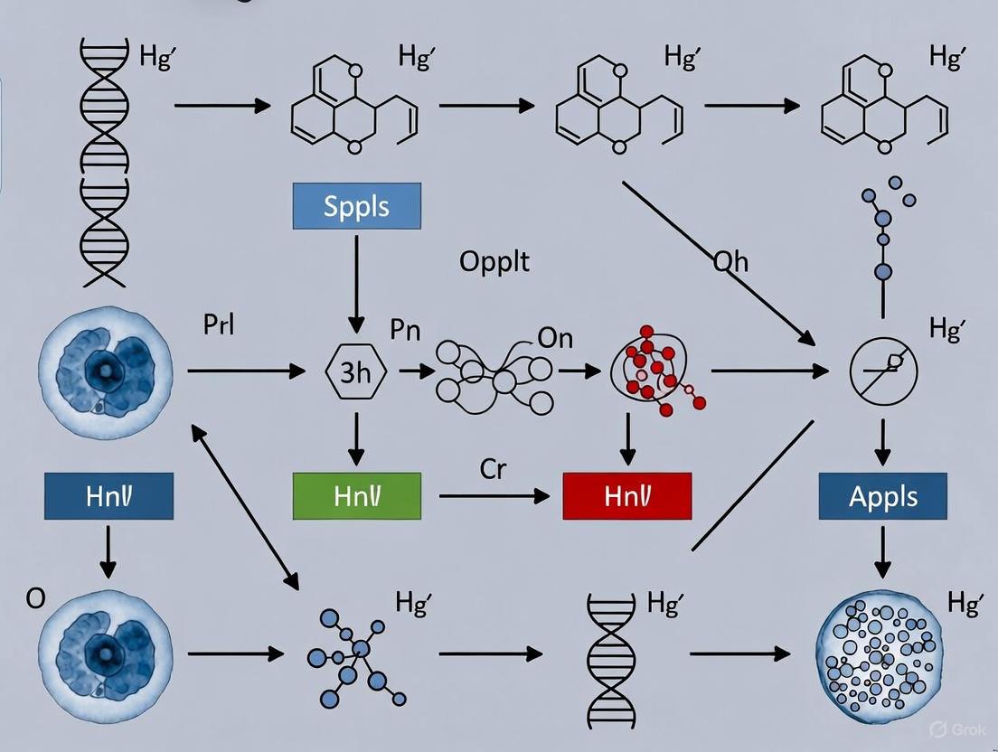
Diagram 1: Pathway of SCNT-Induced Genomic Instability and Intervention Points. The diagram shows how SCNT stressors lead to DNA damage through specific mechanisms, culminating in apoptosis. Key interventions that counteract these mechanisms are highlighted.
The Scientist's Toolkit: Essential Reagents for Troubleshooting
Table 3: Research Reagent Solutions for SCNT Embryo Improvement
| Reagent / Compound | Primary Function | Example Protocol & Concentration | Key Experimental Outcome |
|---|---|---|---|
| Melatonin | Potent antioxidant and free radical scavenger; reduces ROS and apoptosis. | Add to culture medium at 1 µM [11]. | Improves blastocyst rate and quality; reduces γH2A.X foci [1] [11]. |
| Procyanidin B1 (PB1) | Small-molecule antioxidant; boosts cellular catalase and DNA repair. | Add to KSOM culture medium at 50 µM [12]. | Increases blastocyst cell count; upregulates DNA repair gene OGG1 [12]. |
| Scriptaid | Histone deacetylase inhibitor (HDACi); enhances epigenetic reprogramming and DNA repair. | Treat SCNT embryos post-activation (e.g., 0.5 µM for 14-16h) [13]. | Increases blastocyst development; reduces DNA damage foci in embryos from damaged donor cells [13]. |
| Kdm4a mRNA | Histone demethylase; specifically removes H3K9me3 barrier. | Microinject into oocyte/embryo (e.g., 2 µg/µL) [1] [5]. | Overcomes 2-cell developmental block; activates key developmental genes [1]. |
| Nucleosides | Alleviates replication stress by providing substrates for DNA synthesis. | Supplement culture medium [14]. | Rescues chromosome segregation errors in 4-cell embryos by accelerating fork speed [14]. |
Frequently Asked Questions (FAQ) and Troubleshooting Guides
Q1: My SCNT mouse embryos show high rates of arrest at the 2-cell stage. What are the primary causes and solutions?
- Cause 1: Aberrant H3K9me3 Repression. The somatic H3K9me3 epigenetic mark persists, creating reprogramming-resistant regions that block zygotic genome activation [5].
- Solution: Microinject Kdm4a mRNA to demethylate H3K9me3. This has been shown to significantly overcome the 2-cell block in mouse SCNT embryos [1] [5].
- Cause 2: Severe DNA Damage. The reprogramming process and in vitro culture can induce DNA damage that triggers cell cycle arrest.
- Solution: Supplement culture medium with 1 µM Melatonin or 50 µM Procyanidin B1 to mitigate oxidative stress and enhance the embryo's DNA repair capacity [1] [12] [11].
Q2: I observe elevated DNA damage markers (like γH2A.X) in my blastocysts. How can I reduce this?
- Strategy 1: Target Oxidative Stress. Use antioxidants. Melatonin has been proven to directly reduce γH2A.X expression and comet tail formation in porcine SCNT embryos, indicating reduced DNA damage [11].
- Strategy 2: Enhance DNA Repair Machinery. Treatment with HDAC inhibitors like Scriptaid enhances the repair of double-strand breaks. It reduces foci of DNA damage repair proteins (RAD51, 53BP1) and increases blastocyst development, even when using UV-damaged donor cells [13].
Q3: My donor cells are healthy, but SCNT embryos still have poor development. Could the oocyte cytoplasm be a factor?
- Answer: Yes. Using cryopreserved oocytes for SCNT can introduce additional problems. Research shows that SCNT embryos from cryopreserved oocyte cytoplasm have upregulated pro-apoptotic genes and inferior developmental competence compared to those from fresh oocytes [1].
- Solution: If using cryopreserved oocytes is necessary, add Melatonin to the culture medium. It has been shown to enhance blastocyst formation rates and the derivation efficiency of pluripotent stem cells in this specific context by reducing apoptosis and ROS [1].
Q4: Beyond direct DNA damage, what other major epigenetic barrier should I account for?
- Answer: The H3K9me3 barrier. This is a conserved, major roadblock in mammalian SCNT. It not only silences genes but also leads to aberrant chromatin structure, preventing normal development. Targeting this mark is a primary strategy for improving cloning efficiency [5].
Somatic cell nuclear transfer (SCNT) represents a powerful technology for animal cloning and regenerative medicine, yet its application is significantly hampered by high rates of embryonic arrest and apoptosis. The developmental competence of cloned mammalian embryos remains extremely low, with birth rates of approximately only 1-2% in pigs and mice, primarily due to aberrant reprogramming errors and uncontrolled apoptotic pathways [15]. Transcriptomic analyses have consistently revealed that arrested cloned embryos exhibit distinct molecular signatures characterized by the upregulation of pro-apoptotic genes, improper epigenetic reprogramming, and nucleocytoplasmic incompatibility [16] [1]. This technical support center provides comprehensive troubleshooting guidance and experimental protocols to help researchers identify, analyze, and address pro-apoptotic gene upregulation in SCNT mouse embryos, specifically within the context of thesis research focused on addressing high apoptotic cells in SCNT embryos.
Troubleshooting Guide: FAQs on Pro-Apoptotic Gene Upregulation
FAQ 1: What are the key pro-apoptotic genes commonly upregulated in arrested SCNT embryos?
Multiple transcriptomic studies have identified consistent patterns of pro-apoptotic gene upregulation in developmentally compromised SCNT embryos. The specific genes affected can vary based on the experimental model and type of stress encountered by the embryos.
Table 1: Pro-Apoptotic Genes Commonly Upregulated in Suboptimal SCNT Conditions
| Gene Symbol | Full Name | Functional Category | Experimental Context of Upregulation |
|---|---|---|---|
| CASP3 | Caspase 3 | Executioner Caspase | Pig SCNT embryos with high PDCD6 expression [15] |
| GADD45g | Growth Arrest and DNA-Damage-Inducible 45 Gamma | DNA Damage Response | Mouse SCNT using cryopreserved oocytes [1] |
| PMAIP1 | Phorbol-12-Myristate-13-Acetate-Induced Protein 1 | BCL-2 Family Regulation | Mouse SCNT using cryopreserved oocytes [1] |
| DFFB | DNA Fragmentation Factor Beta | DNA Degradation | Mouse SCNT using cryopreserved oocytes [1] |
| CYCS | Cytochrome C | Mitochondrial Apoptosis Pathway | KEGG Apoptosis Pathway [17] |
| BAX | BCL2-Associated X Protein | Pro-Apoptotic BCL-2 Family | KEGG Apoptosis Pathway [17] |
| TP53 | Tumor Protein P53 | Apoptosis Regulation | KEGG Apoptosis Pathway [17] |
FAQ 2: What molecular pathways and processes are typically enriched in arrested SCNT embryos with high apoptosis?
Transcriptomic and proteomic analyses consistently implicate several key pathways in apoptotic-prone SCNT embryos. KEGG pathway analysis of developmentally arrested pig SCNT embryos reveals significant enrichment in cell cycle regulation, necroptosis, and protein processing in the endoplasmic reticulum [15]. Additionally, the NRF2-mediated oxidative stress response pathway represents a common node in both apoptotic and ferroptotic death modules, highlighting the interconnected nature of cell death mechanisms in compromised embryos [18]. The maternal-to-zygotic transition (MZT) and embryonic genome activation (EGA) processes are frequently dysregulated, contributing to developmental arrest [16].
FAQ 3: What experimental strategies can mitigate pro-apoptotic gene upregulation in SCNT embryos?
Several interventions have demonstrated efficacy in reducing apoptosis and improving SCNT outcomes:
Melatonin Supplementation: Adding melatonin (a potent antioxidant) to culture media reduces reactive oxygen species (ROS) and apoptosis in cloned mouse embryos using cryopreserved oocytes, leading to improved blastocyst formation rates [1].
Epigenetic Modulator Injection: Injection of Kdm4a mRNA (encoding H3K9me3 demethylase) overcomes developmental blocks and reduces apoptosis in both fresh and cryopreserved oocyte-derived SCNT mouse embryos [1].
PDCD6 Knockdown: siRNA-mediated knockdown of PDCD6 (a pro-apoptotic gene) reduces CASP3 expression and significantly improves cleavage and blastocyst rates in pig SCNT embryos [15].
Cell Cycle Regulator Treatment: JNJ-7706621 (an inhibitor of cyclin-dependent kinase 1 and aurora kinases) enhances cytoskeletal integrity, reduces DNA damage, and decreases blastomere fragmentation in mouse SCNT embryos, leading to higher implantation and live birth rates [10].
FAQ 4: What technical approaches are available for transcriptomic analysis of pro-apoptotic genes in limited SCNT embryo samples?
Advanced low-input sequencing technologies enable transcriptomic profiling even with limited embryonic material:
Low-input RNA Sequencing: This method successfully analyzed transgenic Asian elephant-pig inter-order cloned embryos, with approximately 25% of clean reads aligning to the donor genome, revealing apoptotic signatures [16].
Embryo Biopsy Combined with Microproteomics: This innovative approach isolates single blastomeres from two-cell stage embryos for proteomic analysis while tracking developmental fates, enabling identification of apoptosis-related proteins like PDCD6 in cloned pig embryos [15].
Single-Cell RNA Sequencing (scRNA-seq): Although not explicitly detailed in the provided results, this approach is mentioned as part of next-generation sequencing technologies for studying gene expressions during early development [16].
Experimental Protocols: Key Methodologies
Protocol 1: Transcriptomic Analysis of SCNT Embryos Using Low-Input RNA Sequencing
This protocol is adapted from studies on Asian elephant-pig inter-order cloned embryos [16]:
- Donor Cell Preparation: Transfect Asian elephant fibroblasts with pPGK-EGFP-NEO vector using Lipofectamine-LTX. Culture transfected cells in DMEM with 10% FBS at 37°C in 5% CO₂.
- Oocyte Collection and Maturation: Collect porcine cumulus-oocyte complexes from slaughterhouse ovaries. Mature in TCM-199 medium supplemented with pyruvic acid, L-cysteine, epidermal growth factor, porcine follicular fluid, eCG, and hCG for 42-44 hours at 38.5°C in 5% CO₂.
- iSCNT Embryo Production: Enucleate mature MII oocytes. Reconstruct with EGFP-expressing donor cells. Activate embryos and culture until desired stages.
- Embryo Selection and Classification: Select EGFP-positive embryos. Classify as developmentally normal (Dev group) if cleaving at expected timelines (24h for 2-cell, 48h for 4-cell) or arrested (Arr group) if delayed by 48h at each stage.
- RNA Extraction and Sequencing: Pool embryos (approximately 100 per sample). Extract RNA using low-input protocols. Prepare sequencing libraries and perform RNA-seq.
- Bioinformatic Analysis: Align clean reads to the donor species genome. Perform differential gene expression, pathway enrichment, and hub gene analyses to identify apoptotic signatures.
Protocol 2: siRNA-Mediated Knockdown of Pro-Apoptotic Genes in SCNT Embryos
This protocol is adapted from successful PDCD6 knockdown in pig SCNT embryos [15]:
- siRNA Design and Preparation: Design and synthesize effective siRNAs targeting your pro-apoptotic gene of interest. Validate knockdown efficiency in donor fibroblasts first.
- Microinjection Setup: Prepare working concentration of 20 μM siRNA in microinjection buffer. Set up micromanipulation system with holding and injection pipettes.
- Embryo Microinjection: Inject siRNA directly into the cytoplasm of reconstructed SCNT embryos shortly after activation. Include negative control siRNA injections for comparison.
- Culture and Assessment: Culture injected embryos in appropriate medium. Assess cleavage rates at 24-48 hours and blastocyst formation at 5-7 days.
- Validation: Collect knockdown embryos at desired stages for qPCR to verify target gene reduction and examine effects on downstream apoptotic markers like CASP3.
Protocol 3: Melatonin Supplementation to Reduce Apoptosis in SCNT Embryos
This protocol is adapted from studies on mouse SCNT embryos using cryopreserved oocytes [1]:
- Melatonin Preparation: Prepare stock solution of melatonin in ethanol or DMSO. Dilute to working concentration in embryo culture medium (typical effective concentration: 10⁻¹¹-10⁻⁷ M).
- Embryo Culture with Melatonin: Culture SCNT embryos in melatonin-supplemented medium from shortly after activation until blastocyst stage.
- Assessment of Outcomes: Evaluate blastocyst formation rates, total cell numbers, and apoptotic index (e.g., by TUNEL staining). Compare with untreated control embryos.
- Transcriptomic Analysis: Pool blastocysts from treated and control groups for RNA-seq to confirm downregulation of pro-apoptotic genes.
Signaling Pathways in SCNT Embryo Apoptosis
The following diagram illustrates the key apoptotic pathways and intervention points identified in SCNT embryos:
Diagram 1: Apoptotic Pathways and Intervention Points in SCNT Embryos. Dashed lines indicate inhibitory effects of interventions.
The Scientist's Toolkit: Essential Research Reagents
Table 2: Key Research Reagents for Studying Apoptosis in SCNT Embryos
| Reagent/Category | Specific Examples | Function/Application | Experimental Evidence |
|---|---|---|---|
| Apoptosis Inhibitors | Melatonin | Reduces ROS and apoptotic events; improves blastocyst quality | Enhanced blastocyst formation in mouse SCNT using cryopreserved oocytes [1] |
| Epigenetic Modulators | Kdm4a mRNA | H3K9me3 demethylase; overcomes developmental blocks and reduces apoptosis | Improved blastocyst rates in both fresh and cryopreserved oocyte SCNT mouse embryos [1] |
| Gene Knockdown Tools | PDCD6 siRNA | Suppresses pro-apoptotic PDCD6, reduces CASP3 expression | Increased cleavage and blastocyst rates in pig SCNT embryos [15] |
| Cell Cycle Regulators | JNJ-7706621 | CDK1 and Aurora kinase inhibitor; enhances cytoskeletal integrity | Improved implantation and live birth rates in mouse SCNT embryos [10] |
| Visualization Tools | EGFP-labeled donor cells | Enables selection of transgenic SCNT embryos for transcriptomic analysis | Successful tracking of donor genome in Asian elephant-pig iSCNT embryos [16] |
| Apoptosis Assays | TUNEL staining, Caspase 3/7 detection | Direct apoptosis assessment in embryos | Validated increased apoptosis in compromised SCNT embryos [1] [19] |
| Transcriptomic Platforms | Low-input RNA-seq, Microproteomics | Gene expression analysis from limited embryo material | Identified apoptotic signatures in arrested SCNT embryos [16] [15] |
Transcriptomic profiling provides powerful insights into the molecular basis of apoptotic upregulation in SCNT embryos. By implementing the troubleshooting strategies, experimental protocols, and reagent solutions outlined in this technical guide, researchers can systematically address the challenge of high apoptosis in cloned embryos. The integration of transcriptomic data with targeted interventions against specific pro-apoptotic pathways offers a promising approach to improve SCNT efficiency and advance both basic research and applied applications in cloning technology.
In the context of somatic cell nuclear transfer (SCNT) research, a primary challenge is the high rate of apoptotic cells in developing embryos. Mitochondria serve as the crucial link between metabolic failure and the initiation of apoptotic signaling in this process. These organelles function not only as cellular powerhouses but also as central hubs that integrate stress signals and execute fate-determining decisions. In SCNT embryos, mitochondrial dysfunction emerges from multiple stressors, including the physical enucleation process that removes mitochondria-rich ooplasm, the introduction of somatic cell mitochondria, and the immense stress of nuclear reprogramming. This dysfunction manifests as impaired oxidative phosphorylation, elevated reactive oxygen species (ROS) production, and disrupted mitochondrial dynamics, collectively triggering programmed cell death through the mitochondrial pathway. Understanding these connections provides the foundation for developing targeted interventions to improve SCNT outcomes.
Key Mechanisms Connecting Mitochondrial Dysfunction to Apoptosis
Metabolic Failure and Bioenergetic Deficit
The metabolic requirements of early embryonic development are substantial, and SCNT embryos often face critical bioenergetic deficits. Research has demonstrated that SCNT procedures can reduce mitochondrial DNA (mtDNA) content by approximately 50% in fetal tissues compared to in vitro fertilized (IVF) controls [20]. This depletion directly compromises the electron transport chain (ETC), where mtDNA-encoded subunits are essential for oxidative phosphorylation (OXPHOS) complexes [21] [22]. The resulting ATP deficiency impairs numerous energy-dependent processes critical for embryonic development, including protein synthesis, cytoskeletal reorganization, and calcium homeostasis. Furthermore, inefficient electron flow through compromised ETC complexes increases electron leakage, elevating ROS production and establishing a vicious cycle of oxidative damage to mitochondrial components [22] [23].
Mitochondrial Dynamics and Quality Control Disruption
Mitochondria exist in dynamic networks that undergo continuous fusion and fission, processes essential for maintaining functional integrity. In SCNT embryos, this balance is frequently disrupted toward excessive fission, creating fragmented mitochondrial populations [24]. Key regulators of these processes include:
- Fusion proteins: Mitofusins 1 and 2 (Mfn1/2) on the outer membrane and Optic Atrophy 1 (OPA1) on the inner membrane
- Fission protein: Dynamin-related protein 1 (Drp1), which translocates from the cytosol to mitochondria to execute fission [24]
When mitochondrial damage exceeds repair capacity, the PINK1-Parkin pathway marks damaged organelles for degradation via mitophagy [21]. In SCNT embryos, this quality control system may be overwhelmed, allowing compromised mitochondria to persist and propagate damage.
Direct Apoptotic Signaling
Mitochondria serve as gatekeepers of apoptotic execution through regulation of the intrinsic pathway. Key events in mitochondrial-mediated apoptosis include:
- Mitochondrial outer membrane permeabilization (MOMP): Triggered by excessive mitochondrial fission, calcium overload, or oxidative stress [24]
- Cytochrome c release: Upon MOMP, cytochrome c is released from the intermembrane space into the cytosol [24]
- Caspase activation: Cytochrome c forms the apoptosome with Apaf-1, activating caspase-9 and the downstream caspase cascade [24] [25]
- Additional pro-apoptotic factors: Mitochondria also release SMAC/DIABLO and Omi/HtrA2, which further promote apoptosis [25]
In SCNT embryos, RNA-sequencing has revealed upregulated expression of multiple pro-apoptotic genes, including Cyct, Dapk2, Dffb, Gadd45g, Hint2, Mien1, P2rx7, and Pmaip [1].
Table 1: Quantitative Assessment of Mitochondrial and Apoptotic Parameters in SCNT Embryos
| Parameter | SCNT Embryos | Normal Embryos | Functional Impact | Citation |
|---|---|---|---|---|
| mtDNA content in fetal tissues | ~50% reduction in liver and muscle | 100% baseline | Compromised OXPHOS, reduced ATP production | [20] |
| Blastocyst formation rate (with optimized protocols) | 61.4% ± 4.4 (with JNJ treatment) | 39.9% ± 6.4 (with CB treatment) | Improved preimplantation development | [10] |
| Total cell number in blastocysts | 70.7 ± 2.9 (with JNJ treatment) | 52.7 ± 3.6 (with CB treatment) | Enhanced embryonic quality | [10] |
| Donor cell mtDNA carryover in human NT embryos | <2% of total mtDNA | 0% in normal embryos | Potential for heteroplasmy-related effects | [26] |
| Apoptotic gene expression | Upregulation of 8 pro-apoptotic genes | Normal expression levels | Increased susceptibility to programmed cell death | [1] |
Troubleshooting Guides & FAQs
Frequently Asked Questions
Q1: Why do SCNT embryos show increased apoptosis compared to IVF embryos? SCNT embryos experience multiple stressors that converge on mitochondria: (1) enucleation removes mitochondria-rich ooplasm, reducing mtDNA content [20]; (2) introduced somatic cell mitochondria may be dysfunctional or create heteroplasmy [27] [26]; (3) nuclear reprogramming stress generates excessive ROS [1]; and (4) impaired mitochondrial dynamics lead to excessive fission, facilitating cytochrome c release [24]. These factors collectively activate the intrinsic apoptotic pathway.
Q2: What specific mitochondrial defects should I look for in SCNT embryos? Key defects to assess include:
- Reduced mtDNA copy number: Quantify via qPCR in embryos or derived tissues [20]
- Altered mitochondrial morphology: Evaluate fusion/fission balance through imaging of mitochondrial networks [24]
- ROS overproduction: Measure using fluorescent probes like MitoSOX Red [1] [22]
- Membrane potential dissipation: Assess with JC-1 or TMRM staining [21] [25]
- Cytochrome c localization: Monitor via immunostaining for release from mitochondria [24]
Q3: Can mitochondrial dysfunction explain the overgrowth phenotype in SCNT fetuses? Yes, mtDNA depletion in SCNT fetuses correlates strongly with organomegaly (particularly hepatomegaly) and muscle hypertrophy [20]. This parallels human mtDNA depletion syndromes, where tissue-specific mtDNA reduction causes metabolic disturbances that aberrantly affect growth patterns. The association suggests that mitochondrial perturbations, in interaction with incomplete nuclear reprogramming, drive abnormal epigenetic regulation of growth pathways.
Q4: How long do donor cell mitochondria persist in SCNT embryos? Persistence varies by species and technique. In human NT embryos, donor fibroblast mtDNA was undetectable in most embryos and accounted for <2% of mtDNA content in the remainder [26]. However, in bovine and porcine models, some clones showed higher levels of donor mtDNA, which could be transmitted to subsequent generations [27]. Optimal SCNT protocols minimize donor mitochondrial carryover.
Troubleshooting Common Experimental Problems
Problem: Poor blastocyst development despite successful nuclear transfer
- Potential Cause: Inefficient mitochondrial function due to mtDNA depletion or oxidative damage
- Solutions:
Problem: High fragmentation and apoptotic markers in early cleavage stages
- Potential Cause: Premature activation of apoptotic pathways due to mitochondrial dysfunction
- Solutions:
Problem: Inconsistent results between SCNT experiments
- Potential Cause: Variation in mitochondrial heteroplasmy or mtDNA content
- Solutions:
Experimental Protocols & Methodologies
Assessing Mitochondrial Function in SCNT Embryos
Protocol 1: mtDNA Quantification in SCNT-Derived Tissues
- Sample Collection: Isolate tissues (liver, muscle, brain) from SCNT and control fetuses at equivalent developmental stages [20]
- DNA Extraction: Use standard phenol-chloroform extraction with RNAse treatment
- qPCR Analysis:
- Amplify mtDNA genes (e.g., cytochrome b) and nuclear reference genes (e.g., β-actin)
- Use SYBR Green chemistry with the following cycling conditions: 95°C for 10 min, then 40 cycles of 95°C for 15 sec and 60°C for 1 min
- Calculate relative mtDNA copy number using the ΔΔCt method [20]
- Validation: Confirm with Southern blotting if quantitative accuracy is critical
Protocol 2: Melatonin Supplementation to Reduce Apoptosis
- Preparation: Dissolve melatonin in minimal DMSO and dilute in culture medium to final concentrations of 10⁻⁴ M to 10⁻⁹ M [1]
- Application: Add melatonin to culture medium immediately after oocyte activation and maintain throughout preimplantation development
- Assessment:
- Evaluate blastocyst formation rates at day 4-5 of culture
- Determine apoptotic index via TUNEL staining of blastocysts
- Analyze gene expression changes for apoptotic markers (e.g., Bax/Bcl-2 ratio) [1]
- Optimization: Titrate concentration for specific experimental conditions, as effectiveness varies with embryo species and strain
Protocol 3: Mitochondrial Dynamics Visualization
- Staining: Load embryos with MitoTracker Red CMXRos (100-200 nM) for 30 min at culture conditions
- Imaging: Capture z-stack images using confocal microscopy with appropriate filter sets
- Analysis:
- Classify mitochondrial morphology as fragmented, intermediate, or fused
- Quantify network connectivity using ImageJ plugins like MiNA
- Correlate morphological patterns with developmental competence [24]
Intervention Strategies to Improve SCNT Outcomes
JNJ-7706621 Treatment Protocol
- Preparation: Create stock solution of JNJ-7706621 in DMSO and dilute in culture medium to 10 μM working concentration [10]
- Application: Treat SCNT embryos post-activation for 4-6 hours, then transfer to standard culture medium
- Mechanism: JNJ-7706621 specifically inhibits cyclin-dependent kinase 1 and aurora kinases, enhancing cytoskeletal integrity and chromosome stability [10]
- Expected Outcomes:
- Significantly improved blastocyst formation rates (approximately 61% vs 40% with cytochalasin B)
- Increased total cell numbers (approximately 71 vs 53 in blastocysts)
- Reduced blastomere fragmentation and DNA damage [10]
Table 2: Research Reagent Solutions for Mitochondrial Dysfunction in SCNT Research
| Reagent | Concentration/Application | Function | Experimental Evidence |
|---|---|---|---|
| Melatonin | 10⁻⁴ M to 10⁻⁹ M in culture medium | Reduces ROS and apoptosis; upregulates anti-apoptotic genes | Improved blastocyst formation in SCNT mouse embryos [1] |
| JNJ-7706621 | 10 μM for 4-6 hours post-activation | Inhibits CDK1 and aurora kinases; enhances cytoskeletal integrity | Increased implantation (68.3% vs 50.8%) and live birth rates (10.9% vs 2.4%) in mouse SCNT [10] |
| MitoQ | 50-500 nM in culture medium | Mitochondria-targeted antioxidant that accumulates in mitochondria | Reduced oxidative stress in various disease models; potential application for SCNT [21] [23] |
| HVJ-E (Sendai virus extract) | Fusion agent in nuclear transfer | Promotes efficient fusion of donor cell with enucleated oocyte | Improved fusion rates in human SCNT experiments [26] |
| Kdm4a mRNA | 2 μg/μL injection into oocytes | Histone demethylase that enhances reprogramming | Overcame 2-cell block in cloned mouse embryos [1] |
Signaling Pathways and Experimental Workflows
Mitochondria-Mediated Apoptotic Signaling in SCNT Embryos
The following diagram illustrates the key molecular events connecting mitochondrial dysfunction to apoptosis in SCNT embryos:
Diagram 1: Mitochondria-mediated apoptotic signaling pathway in SCNT embryos. Key stress signals converge on mitochondria, leading to dysfunction that triggers the intrinsic apoptotic pathway through cytochrome c release and caspase activation. Potential intervention points are shown in white ovals.
Experimental Workflow for Mitochondrial Health Assessment
The following workflow provides a systematic approach to diagnose and address mitochondrial dysfunction in SCNT experiments:
Diagram 2: Systematic troubleshooting workflow for mitochondrial-related issues in SCNT embryo development. The approach progresses from comprehensive assessment through targeted intervention and re-evaluation.
Intervention Toolkit: Small Molecules and Additives to Suppress Apoptosis
JNJ-7706621 is a potent small-molecule inhibitor originally identified as a dual-targeting agent for cyclin-dependent kinases (CDKs) and Aurora kinases [28] [29]. Its mechanism of action is particularly relevant for addressing key challenges in somatic cell nuclear transfer (SCNT) embryo research, where high apoptotic rates and cytoskeletal defects frequently compromise developmental competence [30] [31]. By simultaneously targeting multiple regulators of cell division, this compound offers a strategic approach to improve the poor efficiency of SCNT technologies, which typically results in only 1-5% of reconstructed embryos developing to term [30].
In SCNT research, the profound structural and functional differences between somatic donor cells and gametes present significant obstacles. Differentiated somatic cells lack essential components normally contributed by sperm, including specific regulatory proteins and small noncoding RNAs, which creates deficiencies in nuclear reprogramming and cytoskeletal remodeling [31]. JNJ-7706621 addresses these limitations through its multi-kinase inhibition profile, potentially compensating for missing sperm-derived factors that normally ensure proper cell division and genomic stability during early embryonic development.
Mechanism of Action: Key Signaling Pathways
Primary Kinase Targets and Biological Effects
JNJ-7706621 exerts its effects through coordinated inhibition of several crucial kinase families involved in cell cycle progression and mitotic fidelity. The table below summarizes its primary targets and associated biological effects relevant to SCNT embryo improvement:
Table 1: Key Kinase Targets of JNJ-7706621 and Their Roles in SCNT Embryos
| Target Kinase | IC₅₀ Value | Primary Function | Effect of Inhibition in SCNT Context |
|---|---|---|---|
| CDK1/Cyclin B | 9 nM [32] | Promotes G2/M transition; regulates microtubule dynamics [28] | Slows cell cycle progression; allows DNA repair; reduces mitotic errors |
| CDK2/Cyclin E | 3 nM [32] | Drives G1/S transition; initiates DNA replication [28] | Prevents premature S-phase entry; maintains replication fidelity |
| Aurora A | 11 nM [32] | Centrosome maturation; spindle assembly; mitotic entry [33] | Prevents multipolar spindle formation; ensures proper centrosome function |
| Aurora B | 15 nM [32] | Chromosome bi-orientation; spindle checkpoint; cytokinesis [33] | Promotes chromosome mis-segregation; activates apoptosis in defective cells |
| JAK2 (JH2 domain) | Kd: 31-800 nM [34] | Regulatory pseudokinase domain; modulates JAK-STAT signaling [34] | Potential indirect effects on stress response pathways |
Beyond these primary targets, JNJ-7706621 demonstrates additional inhibitory activity against other kinases including CDK3 (IC₅₀ = 58 nM), CDK4/cyclin D1 (IC₅₀ = 253 nM), CDK6/cyclin D1 (IC₅₀ = 175 nM), and VEGFR2 (IC₅₀ = 154 nM) [32]. This broad specificity profile enables comprehensive modulation of cell division processes that are typically disrupted in SCNT embryos.
Pathway Integration in SCNT Embryos
The coordinated inhibition of CDK and Aurora kinase pathways by JNJ-7706621 addresses two fundamental weaknesses in SCNT embryos: cytoskeletal instability and apoptotic predisposition. The signaling relationships and intervention points can be visualized through the following pathway diagram:
Diagram 1: JNJ-7706621 signaling pathways in SCNT embryos. The inhibitor blocks multiple kinase targets that contribute to mitotic errors and DNA damage, ultimately promoting cytoskeletal integrity and reducing apoptosis.
In SCNT embryos, the abnormal cytoskeleton remodeling manifests as defective spindle-chromosome complexes, impaired pronuclear formation, and erroneous centrosome function [31]. These cytoskeletal deficiencies activate stress response pathways that ultimately trigger apoptosis. JNJ-7706621 intervenes at multiple critical points in this cascade by slowing cell cycle progression through CDK inhibition while simultaneously ensuring proper chromosome segregation and spindle function via Aurora kinase inhibition [28] [33].
Research Reagent Solutions
Table 2: Essential Research Reagents for JNJ-7706621 Experiments
| Reagent/Resource | Specifications | Primary Function | Key Considerations for SCNT Work |
|---|---|---|---|
| JNJ-7706621 (compound) | CAS: 443797-96-4; MW: 394.36; Formula: C₁₅H₁₂F₂N₆O₃S [32] | Dual CDK/Aurora kinase inhibitor | Reconstitute in DMSO; use freshly prepared solutions |
| Suitable solvent | DMSO (≥125 mg/mL) [32] | Compound solubilization | Final DMSO concentration should not exceed 0.1% in embryo culture |
| Embryo culture medium | KSOM-based [2] | Supports preimplantation development | Optimize with antioxidant supplements for SCNT embryos |
| Apoptosis detection | Comet assay, TUNEL, caspase-3 activation [30] [2] | Quantifies apoptotic cells | Comet assay detects early DNA fragmentation [30] |
| Cytoskeleton assessment | Immunofluorescence for tubulin, vimentin, actin [31] | Evaluates cytoskeletal organization | Monitor spindle morphology and chromosome alignment |
| DNA damage markers | γH2AX, OGG1 staining [2] | Detects DNA damage and repair | OGG1 upregulation indicates enhanced repair capacity |
| Antioxidant supplements | Procyanidin B1 (50 μM) [2] | Reduces oxidative stress | Synergistic effect with JNJ-7706621 possible |
Experimental Protocols
Determining Optimal Treatment Concentration and Timing
Purpose: To establish the effective non-toxic concentration range and treatment window for JNJ-7706621 in SCNT mouse embryos.
Materials:
- JNJ-7706621 stock solution (10 mM in DMSO)
- SCNT-derived mouse embryos
- Embryo culture medium (e.g., KSOM)
- Mineral oil for culture medium overlay
- CO₂ incubator (5% CO₂, 37°C)
Procedure:
- Prepare working concentrations of JNJ-7706621 (50-500 nM) by diluting stock solution in pre-warmed culture medium. Include DMSO-only controls.
- After SCNT reconstruction, randomly assign embryos to treatment groups (minimum n=30 per group).
- Culture embryos under standard conditions (37°C, 5% CO₂) with continuous JNJ-7706621 exposure.
- Assess embryonic development at 24h (2-cell), 48h (4-cell), 72h (8-cell), and 96h (morula) post-activation.
- Record blastocyst formation rates at 120h and evaluate total cell count via cell staining.
- For timing optimization, apply JNJ-7706621 during specific cell cycle phases:
- G1/S phase: 0-6h post-activation
- G2/M phase: 6-12h post-activation
- Extended exposure: 0-24h post-activation
Expected Outcomes: Based on prior cancer cell studies, JNJ-7706621 shows dose-dependent effects with low concentrations (≤100 nM) delaying cell cycle progression and higher concentrations (≥200 nM) inducing cytotoxicity [28]. In SCNT embryos, optimal concentrations typically balance cell cycle modulation without complete arrest.
Assessing Apoptotic Response
Purpose: To quantify the anti-apoptotic effects of JNJ-7706621 treatment in SCNT embryos.
Materials:
- TUNEL assay kit
- Comet assay reagents [30]
- Caspase-3 activity assay
- Fluorescence microscope with appropriate filters
- Bcl-2 expression analysis reagents (real-time RT-PCR) [30]
Procedure:
- Culture SCNT embryos with or without JNJ-7706621 until blastocyst stage.
- For TUNEL assay:
- Fix embryos in 4% paraformaldehyde for 30min
- Permeabilize with 0.5% Triton X-100 for 1h
- Incubate with TUNEL reaction mixture according to manufacturer's protocol
- Counterstain nuclei with Hoechst 33342
- Quantify apoptotic index: (TUNEL-positive cells/total cells) × 100%
- For Comet assay [30]:
- Embed individual embryos in low-melting-point agarose on microscope slides
- Lyse cells in neutral buffer (for double-strand breaks) or alkaline buffer (for single-strand breaks)
- Perform electrophoresis under appropriate conditions
- Stain with DNA-binding dye and analyze tail moment
- For gene expression analysis:
Interpretation: Effective JNJ-7706621 treatment should significantly reduce TUNEL-positive cells, decrease comet tail moment, and upregulate Bcl-2 and DNA repair gene expression compared to untreated SCNT controls.
Evaluating Cytoskeletal Integrity
Purpose: To analyze the effects of JNJ-7706621 on cytoskeletal organization in SCNT embryos.
Materials:
- Anti-α-tubulin antibody
- Anti-γ-tubulin antibody (centrosome marker)
- Phalloidin (actin stain)
- Vimentin antibody [31]
- Fluorescently-labeled secondary antibodies
- Mounting medium with DAPI
Procedure:
- Culture embryos to specific developmental stages (2-cell, 4-cell, 8-cell, morula).
- Fix embryos in 4% paraformaldehyde for 30min at room temperature.
- Permeabilize with 0.5% Triton X-100 for 1h.
- Block in 5% BSA for 2h to reduce non-specific binding.
- Incubate with primary antibodies (diluted in blocking solution) overnight at 4°C.
- Wash thoroughly and incubate with appropriate secondary antibodies for 2h at room temperature.
- Counterstain with DAPI to visualize nuclei.
- Image using confocal microscopy with consistent settings across treatment groups.
- Analyze for:
- Spindle morphology and bipolarity
- Chromosome alignment at metaphase plate
- Cortical actin distribution
- Vimentin persistence around nuclei [31]
Quality Metrics: Compare treated embryos to IVF controls for: (1) percentage of normal bipolar spindles, (2) proper chromosome congression, (3) absence of vimentin aggregates, and (4) normal actin cap formation.
Troubleshooting Guides
FAQ: Addressing Common Experimental Challenges
Table 3: Troubleshooting Common Issues with JNJ-7706621 in SCNT Experiments
| Problem | Potential Causes | Solutions | Preventive Measures |
|---|---|---|---|
| Complete developmental arrest | Concentration too high; prolonged exposure | Titrate concentration (start at 50 nM); reduce treatment window to 6-12h | Perform dose-response curve with IVF embryos first |
| No improvement in apoptosis rates | Insufficient target engagement; wrong treatment timing | Increase concentration (up to 200 nM); apply during G2/M phase (6-12h post-activation) | Verify kinase inhibition via phospho-histone H3 staining |
| Increased cytoskeletal abnormalities | Off-target effects; excessive Aurora B inhibition | Reduce concentration; combine with cytoskeletal stabilizers | Assess specific spindle defects to identify primary target failure |
| High embryo toxicity | DMSO concentration too high; compound precipitation | Ensure final DMSO ≤0.1%; filter compound solution before use | Prepare fresh stock solutions; avoid freeze-thaw cycles |
| Variable results between replicates | Inconsistent embryo quality; compound degradation | Standardize SCNT procedure; use fresh compound batches | Include internal controls in each experiment |
| Poor blastocyst quality despite reduced apoptosis | Cell cycle over-slowing; impaired proliferation | Shorten treatment duration; implement pulse-chase strategy | Monitor cell number and allocation in blastocysts |
Advanced Applications and Combination Strategies
The therapeutic potential of JNJ-7706621 can be enhanced through strategic combination with complementary approaches. The experimental workflow for evaluating these combinations is illustrated below:
Diagram 2: Experimental workflow for testing JNJ-7706621 combinations. Parallel treatment groups enable systematic evaluation of synergistic approaches for improving SCNT outcomes.
Synergistic Approaches for Enhanced SCNT Efficiency
Antioxidant Combination: Procyanidin B1 (PB1), a small-molecule antioxidant, has demonstrated efficacy in reducing oxidative stress and apoptosis in SCNT embryos [2]. When combined with JNJ-7706621, PB1 (50 μM) may address complementary pathways by:
- Reducing ROS accumulation and increasing glutathione levels
- Enhancing mitochondrial membrane potential
- Upregulating DNA damage repair genes (e.g., OGG1)
- Increasing catalase expression to degrade H₂O₂ [2]
Epigenetic Modulator Combination: Histone deacetylase inhibitors like Trichostatin A (TSA) have shown promise in improving SCNT efficiency by promoting chromatin remodeling [31]. Strategic combination with JNJ-7706621 could simultaneously address:
- Histone acetylation status (via TSA)
- Cell cycle regulation (via JNJ-7706621)
- Mitotic fidelity (via JNJ-7706621)
- Nuclear reprogramming efficiency (via both agents)
Validation Metrics: When implementing combination strategies, assess:
- Blastocyst formation rates and total cell counts
- Apoptotic indices (TUNEL and comet assay)
- Cytoskeletal organization (spindle morphology)
- Expression of pluripotency and implantation-related genes
- Post-implantation developmental competence
These advanced applications highlight the potential of JNJ-7706621 as a cornerstone in multi-faceted approaches to overcome the persistent challenges in SCNT research.
Somatic Cell Nuclear Transfer (SCNT) is a pivotal technology in reproductive biology, regenerative medicine, and drug development. A significant obstacle to its efficiency is the high rate of apoptotic cells in developing embryos, often triggered by excessive Reactive Oxygen Species (ROS). Oxidative stress occurs when the production of ROS, such as superoxide anion (O₂•⁻) and hydrogen peroxide (H₂O₂), surpasses the embryo's endogenous antioxidant defenses [35] [36]. This imbalance leads to damage of critical biomolecules like DNA, proteins, and lipids, disrupting normal development and leading to cell death [12] [36]. This technical support center provides targeted troubleshooting and protocols for employing potent antioxidants—Melatonin, Procyanidin B1, and Lycopene—to mitigate this oxidative damage and improve SCNT outcomes in mouse models.
FAQs: Antioxidants and Apoptosis in SCNT Research
Q1: Why are SCNT embryos particularly susceptible to oxidative stress and apoptosis? SCNT embryos experience significant stress during the micromanipulation and reprogramming processes. In vitro culture conditions expose them to higher levels of ROS and reduce levels of protective molecules like glutathione (GSH) compared to in vivo development [12]. Furthermore, the epigenetic reprogramming inherent to SCNT can disrupt the expression of genes involved in oxidative defense, making the embryos more vulnerable. The resulting oxidative damage can cause DNA strand breaks and lipid peroxidation, activating the apoptotic pathways and leading to high rates of developmental arrest [37] [36].
Q2: How do antioxidants like Procyanidin B1 improve SCNT embryo development? Procyanidin B1 (PB1) is a powerful antioxidant that acts through multiple mechanisms. It directly reduces intracellular ROS levels and boosts the levels of glutathione (GSH), a key endogenous antioxidant [12]. Furthermore, PB1 increases the expression and activity of antioxidant enzymes like Catalase (CAT), which degrades H₂O₂ into harmless water and oxygen. By reducing DNA damage (e.g., by enhancing the expression of the DNA repair gene OGG1), PB1 ultimately lowers the level of apoptosis in blastocysts, leading to improved blastocyst rates and higher total cell counts [12].
Q3: What is the evidence that modulating ROS can reduce apoptosis in embryos? Research on mouse gastrulation embryos has shown that DNA damage, including that induced by oxidative stress, can trigger a hypersensitive, p53-dependent apoptotic response specifically in embryonic cells [37]. This demonstrates a direct link between cellular damage and programmed cell death in early development. Conversely, interventions that reduce ROS, such as PB1 treatment, have been shown to directly result in a lower percentage of apoptotic cells in the resulting blastocyst, confirming that mitigating oxidative stress preserves cell viability [12].
Q4: Are there other small molecules that can improve SCNT efficiency? Yes, several classes of small molecules can enhance SCNT outcomes. Histone deacetylase inhibitors (HDACi) like Scriptaid are commonly used to improve epigenetic reprogramming, which indirectly can reduce stress and apoptosis [38]. Vitamin C (ascorbic acid) has also been shown to improve mouse SCNT blastocyst formation, acting as both an antioxidant and an epigenetic modulator [39]. Another molecule, JNJ-7706621, an inhibitor of cyclin-dependent kinase 1 and aurora kinases, was shown to improve cytoskeletal integrity, reduce DNA damage in two-cell embryos, and significantly increase both implantation and live birth rates in mouse SCNT [10].
Troubleshooting Guide: High Apoptosis in SCNT Embryos
| Problem | Potential Cause | Recommended Solution |
|---|---|---|
| High blastomere fragmentation | Severe oxidative stress damaging cellular structures [36]; Faulty cytoskeleton organization [10] | Supplement culture medium with 50 µM Procyanidin B1 [12]; Use 10 µM JNJ-7706621 as a post-activation treatment to support cytoskeletal integrity [10] |
| Low blastocyst rate | Accumulated DNA damage triggering cell cycle arrest or early apoptosis [37] [12] | Add 100 µM Vitamin C to culture medium for at least 16 hours post-activation to support reprogramming and reduce stress [39] |
| Low total cell count in blastocysts | Chronic, low-level oxidative stress impairing proliferation [12] | Utilize a combination of antioxidants (e.g., PB1) and epigenetic modifiers (e.g., Scriptaid on donor cells) for a synergistic effect [38] |
| Poor embryo development post-implantation | Inefficient nuclear reprogramming and persistent epigenetic abnormalities [38] | Treat reconstructed oocytes with low-toxicity HDACi like Scriptaid and ensure the use of Latrunculin A during micromanipulation to improve full-term development [39] [38] |
Detailed Experimental Protocols
Protocol: Using Procyanidin B1 in SCNT Mouse Embryo Culture
This protocol is adapted from a 2021 study demonstrating that PB1 reduces ROS and apoptosis, thereby improving the development of mouse SCNT embryos [12].
Objective: To improve the in vitro development of SCNT mouse embryos by reducing oxidative stress and DNA damage via supplementation of Procyanidin B1.
Key Reagents and Materials:
- SCNT-derived mouse embryos
- KSOM embryo culture medium
- Procyanidin B1 (PB1): Prepare a stock solution and store at -20°C.
- Mineral oil
- Culture incubator (37°C, 5% CO₂)
Methodology:
- Preparation of Culture Medium: On the day of use, prepare KSOM medium supplemented with 50 µM Procyanidin B1. Use a stock solution to ensure accurate dilution. A control group should be cultured in standard KSOM medium.
- Embryo Culture: After nuclear transfer and activation, wash the SCNT embryos and place them in drops of the prepared PB1-supplemented KSOM medium under mineral oil.
- Culture Duration: Culture the embryos in the PB1-medium for the entire in vitro development period, from one-cell stage to blastocyst (typically 3.5-4 days).
- Outcome Assessment: Evaluate the embryos for:
- Developmental Rate: Record the rates of cleavage, eight-cell formation, and blastocyst formation.
- Blastocyst Quality: At day 4, fix and stain blastocysts to count the total cell number (e.g., with Hoechst 33342).
- ROS and GSH Levels: Use specific fluorescent probes (e.g., H2DCFDA for ROS, CellTracker Blue for GSH) at various stages (two-cell, four-cell, eight-cell) for quantitative analysis.
- Apoptosis Assay: Use a TUNEL assay on blastocysts to quantify the number of apoptotic cells.
Expected Results: Treatment with 50 µM PB1 should significantly increase the blastocyst formation rate and total blastocyst cell number. It should also lead to a measurable decrease in ROS levels at the eight-cell and blastocyst stages, an increase in GSH levels at the two-cell and eight-cell stages, and a reduction in TUNEL-positive apoptotic cells [12].
Protocol: Assessing Antioxidant Effect via ROS/GSH Staining
This is a common methodology used to validate the efficacy of an antioxidant treatment in embryos [12].
Objective: To quantitatively measure the intracellular levels of reactive oxygen species (ROS) and reduced glutathione (GSH) in control and antioxidant-treated embryos.
Key Reagents and Materials:
- Control and antioxidant-treated embryos at specific stages (e.g., two-cell, eight-cell)
- ROS detection dye (e.g., 2',7'-Dichlorodihydrofluorescein diacetate - H2DCFDA)
- GSH detection dye (e.g., CellTracker Blue CMF2HC - CTB)
- Phosphate-Buffered Saline (PBS) with Polyvinylpyrrolidone (PVP)
- Incubator (37°C, 5% CO₂)
- Fluorescence microscope with appropriate filter sets
Methodology:
- Dye Preparation: Working solutions of H2DCFDA (e.g., 10 µM) and CTB (e.g., 25 µM) should be prepared in PBS-PVP.
- Staining Procedure: Wash groups of embryos and incubate them in the dark in the dye solutions for a specific time (e.g., 30 minutes for CTB, 20-30 minutes for H2DCFDA) at 37°C.
- Washing: After incubation, wash the embryos thoroughly in fresh PBS-PVP to remove excess dye.
- Imaging and Analysis: Immediately image the embryos under a fluorescence microscope using consistent exposure settings across all groups. Measure the fluorescence intensity (pixels per embryo) for each embryo using image analysis software (e.g., ImageJ). Compare the average fluorescence between treated and control groups.
Signaling Pathways: How Antioxidants Counter Oxidative Stress
The following diagram illustrates the core pathways through which oxidative stress leads to apoptosis in SCNT embryos, and the points of intervention for potent antioxidants like Melatonin, Procyanidin B1, and Lycopene.
Diagram Title: Antioxidant Mechanisms Against Apoptosis in SCNT Embryos
The Scientist's Toolkit: Key Research Reagents
| Reagent / Material | Function / Role in Research | Example Application in SCNT |
|---|---|---|
| Procyanidin B1 (PB1) | A potent antioxidant that reduces ROS, boosts glutathione (GSH), and enhances DNA damage repair capability [12]. | Added to KSOM culture medium at 50 µM for the entire in vitro culture period to improve blastocyst rate and reduce apoptosis [12]. |
| Melatonin | A hormone with strong free radical scavenging properties; suppresses UV-induced damage and shows antioxidant activity in exposed cells [40]. | Can be investigated for its potential to protect embryos from ambient light-induced oxidative stress in the lab environment. |
| Lycopene | A carotenoid antioxidant that is an efficient quencher of singlet oxygen and a potent scavenger of oxygen radicals [40]. | Topical application shown to reduce photodamage; its inclusion in culture media for SCNT embryos may mitigate specific ROS types. |
| Vitamin C (L-ascorbic acid) | A well-known antioxidant and epigenetic modulator that promotes histone and DNA demethylation [39]. | Added at 100 µM to culture medium for at least 16 hours post-activation to improve blastocyst formation in mouse SCNT [39]. |
| Scriptaid | A histone deacetylase inhibitor (HDACi) that improves epigenetic reprogramming by increasing histone acetylation levels [38]. | Treatment of donor cells (e.g., 250-750 nM for 24h) prior to SCNT to enhance the reprogramming ability of reconstructed embryos [38]. |
| JNJ-7706621 | An inhibitor of cyclin-dependent kinase 1 and aurora kinases that improves cytoskeletal integrity and chromosome stability [10]. | Used at 10 µM as a post-activation treatment to replace cytochalasin B, reducing blastomere fragmentation and DNA damage [10]. |
| Latrunculin A (LatA) | An actin polymerization inhibitor that prevents second polar body extrusion during SCNT activation [39]. | Used during micromanipulation and activation procedures; combined with other treatments, it improves both in vitro and full-term development [39]. |
| H2DCFDA dye | A cell-permeable fluorescent probe that is oxidized by ROS to a fluorescent compound, allowing ROS measurement [12]. | Used to quantify intracellular ROS levels in live embryos at various developmental stages (e.g., two-cell, eight-cell) [12]. |
| CellTracker Blue CMF2HC | A fluorescent dye used to detect and quantify intracellular levels of reduced glutathione (GSH) [12]. | Staining of embryos to confirm that antioxidant treatments like PB1 effectively increase the intracellular GSH pool [12]. |
Frequently Asked Questions (FAQs)
Q1: What are the primary epigenetic barriers that Kdm4a/d and TSA address in SCNT embryos? Kdm4a/d and TSA target two major epigenetic barriers that impede nuclear reprogramming. Kdm4a/d are histone demethylases that specifically remove the repressive histone H3 lysine 9 trimethylation (H3K9me3) mark. This mark is enriched in reprogramming-resistant regions (RRRs) of the somatic cell genome and prevents the activation of genes critical for embryonic development [41]. Trichostatin A (TSA) is a histone deacetylase inhibitor that promotes histone acetylation, leading to a more open chromatin state. This facilitates access to the DNA for transcriptional machinery and is crucial for the re-establishment of totipotency [42].
Q2: We are observing high rates of apoptotic cells in our mouse SCNT embryos. Can these modulators help? Yes, apoptosis is a common issue in SCNT embryos and evidence suggests these modulators can mitigate it. TSA treatment has been shown to reduce apoptosis in SCNT blastocysts and upregulate the expression of pluripotency and development-related genes, which contributes to improved embryo viability [42]. Furthermore, high apoptosis in SCNT embryos has been linked to cryo-damage in vitrified oocytes; in such cases, the antioxidant melatonin was effective in reducing apoptosis and ROS production, thereby enhancing development [1]. This indicates that addressing epigenetic barriers and oxidative stress can collectively improve embryo health.
Q3: What is a typical working concentration and exposure duration for TSA in mouse SCNT protocols? Based on established research, a common and effective treatment for mouse SCNT embryos is a concentration of 5-50 nM TSA for a duration of 10-24 hours following oocyte activation [42]. It is critical to optimize the timing and concentration for your specific experimental system, as prolonged exposure or higher doses can be cytotoxic.
Q4: How is Kdm4a/d mRNA delivered to the oocyte or embryo, and when? The standard method is microinjection of in vitro transcribed mRNA into the cytoplasm of the oocyte or single-cell embryo shortly after nuclear transfer. For instance, in mouse SCNT experiments, injection of Kdm4a mRNA at 2 μg/μL has been successfully used to overcome the 2-cell developmental block [1].
Q5: Are the effects of Kdm4a/d and TSA synergistic? While the search results do not provide a direct study on their combined use, they target different epigenetic layers. Kdm4a/d removes a specific repressive methylation mark, while TSA promotes general chromatin openness by increasing acetylation. Using them in a sequential manner—first relaxing chromatin with TSA, then directly removing the H3K9me3 barrier with Kdm4a/d—could theoretically provide a more comprehensive epigenetic reset. However, empirical validation and careful titration are necessary to avoid excessive embryonic stress.
Troubleshooting Guides
Problem: Poor Blastocyst Development After Kdm4a/d mRNA Injection
- Potential Cause 1: Inefficient mRNA Delivery or Quality.
- Solution: Verify the quality and concentration of the in vitro transcribed mRNA. Ensure it is capped, polyadenylated, and free of contaminants. Confirm microinjection technique proficiency to ensure consistent delivery.
- Potential Cause 2: Suboptimal Timing.
- Solution: The timing of injection is critical. Administer the mRNA shortly after nuclear transfer to ensure the demethylase is present during the initial reprogramming phase. Testing different time windows (e.g., immediately post-fusion vs. post-activation) is recommended for protocol optimization.
Problem: High Embryonic Lethality or Morphological Abnormalities After TSA Treatment
- Potential Cause 1: Toxicity from Overexposure.
- Solution: TSA is a potent drug with a narrow therapeutic window. Re-evaluate the concentration and duration of treatment. Start with the lower end of the known effective range (e.g., 5 nM for 10 hours) and perform a dose-response experiment [42].
- Potential Cause 2: Incompatibility with Your Specific SCNT System.
- Solution: The optimal TSA protocol can vary depending on the donor cell type and oocyte quality. Consider using an alternative histone deacetylase inhibitor like Scriptaid (e.g., 500 nM for 12-15 hours), which is reported to be less toxic while still effective in improving SCNT outcomes in other species [42].
Problem: Incomplete Resetting of RRRs Despite Treatment
- Potential Cause: Persistent Underlying Epigenetic Barriers.
- Solution: H3K9me3 is a major, but not the only, barrier. Consider a combined epigenetic approach. Investigate the status of other repressive marks, such as DNA methylation. A sequential treatment protocol or the use of donor cells with depleted H3K9 methyltransferases (e.g., Suv39h1/2) could be explored for a more robust reprogramming effect [41].
| Group | Kdm4a mRNA (μg/μL) | Number of 2-Cell Embryos | Number of Blastocysts (% ± SEM) |
|---|---|---|---|
| SCNT-FOC | - | 105 | 27 (26 ± 3.4) a |
| SCNT-FOC + K | 2 | 103 | 85 (83 ± 3.5) b |
| SCNT-CROC | - | 132 | 30 (23 ± 3.1) a |
| SCNT-CROC + K | 2 | 125 | 82 (66 ± 2.4) c |
SCNT-FOC: cloned embryos using fresh oocyte cytoplasm; SCNT-CROC: cloned embryos using cryopreserved oocyte cytoplasm; K: Kdm4a mRNA injection. Values with different superscript letters (a, b, c) are significantly different (p < 0.05).
| Species | TSA Concentration / Duration | Key Outcomes |
|---|---|---|
| Mouse | 5-50 nM / 10-24 hours | Increased blastocyst formation rates and embryo cell numbers; upregulated pluripotency genes; reduced apoptosis; produced pregnancies to term. |
| Pig | 5-50 nM / 10-24 hours | Increased blastocyst rates and embryo cell numbers; improved cloning efficiency. |
Experimental Protocols
Protocol 1: Microinjection of Kdm4a mRNA into Mouse SCNT Embryos
- Principle: Direct delivery of synthetic mRNA encoding the H3K9me3 demethylase Kdm4a to overcome reprogramming-resistant regions [1] [41].
- mRNA Preparation: Generate in vitro transcribed, capped, and polyadenylated Kdm4a mRNA. Dilute to a working concentration of 2 μg/μL in nuclease-free microinjection buffer.
- Embryo Handling: Perform somatic cell nuclear transfer using standard protocols.
- Microinjection: Using a micromanipulator, inject a few picoliters of the mRNA solution into the cytoplasm of the reconstructed oocyte shortly after nuclear transfer and activation.
- Embryo Culture: Wash embryos and transfer to pre-equilibrated embryo culture medium. Culture under standard conditions (37°C, 5% CO2).
Protocol 2: TSA Treatment of Reconstructed SCNT Embryos
- Principle: Transient inhibition of histone deacetylases to promote global histone acetylation and chromatin relaxation, facilitating epigenetic reprogramming [42].
- Drug Preparation: Prepare a stock solution of TSA in DMSO and dilute in embryo culture medium to a final working concentration (e.g., 50 nM). Ensure the final DMSO concentration is non-toxic (<0.1%).
- Treatment Window: After oocyte activation and confirmation of successful reconstruction, wash the SCNT embryos and transfer them into the TSA-containing medium.
- Incubation: Incubate embryos for the specified duration (e.g., 10-12 hours).
- Post-Treatment Wash and Culture: Thoroughly wash the embryos to remove TSA and then continue culture in standard embryo medium until blastocyst stage or embryo transfer.
The Scientist's Toolkit
Table 3: Key Research Reagent Solutions
| Reagent | Function in SCNT Reprogramming | Example Usage / Note |
|---|---|---|
| Kdm4a/d mRNA | Histone demethylase that specifically removes H3K9me3 marks from reprogramming-resistant regions. | Microinjected at 2 μg/μL to overcome the 2-cell block in mouse SCNT [1] [41]. |
| Trichostatin A (TSA) | Histone deacetylase inhibitor (HDACi) that increases histone acetylation, promoting open chromatin. | Used at 5-50 nM for 10-24 hours post-activation to improve blastocyst development [42]. |
| Scriptaid | An alternative HDACi reported to be less toxic than TSA while still effective. | A common dose is 500 nM for 12-15 hours of embryo exposure [42]. |
| Melatonin | An antioxidant that reduces ROS production and apoptosis, improving embryo quality. | Supplemented at 100 nM during embryo culture to mitigate cryo-damage and apoptosis [1]. |
| FUW-TetO-PIB Vector | Lentiviral vector for enforced expression of transcription factors (e.g., PU.1, IRF8, BATF3) in direct reprogramming studies. | Used in fibroblast-to-dendritic cell reprogramming protocols [43]. |
Signaling Pathways and Workflows
Kdm4a/d Mechanism in SCNT
TSA Treatment Workflow in SCNT
Frequently Asked Questions (FAQs)
Q1: What is the primary role of madecassic acid (MA) in improving SCNT embryos? A1: Madecassic acid is a natural compound that acts as a potent telomerase activator. In SCNT-derived embryos, it enhances developmental competence primarily by activating telomerase reverse transcriptase (TERT). This activation leads to improved telomere maintenance, reduced DNA damage, and enhanced embryonic genome activation (EGA), which are crucial for overcoming reprogramming inefficiencies in cloned embryos [44] [45].
Q2: How does MA treatment affect the developmental rates of SCNT embryos? A2: Treatment with an optimal concentration of MA significantly improves key developmental metrics. When bovine SCNT zygotes were treated with 3.0 μg/mL MA, the embryo cleavage rate increased to 71.5% and the blastocyst rate reached 28.1%, compared to non-treated control embryos [44] [45].
Q3: Through what mechanism does MA enhance Embryonic Genome Activation (EGA)? A3: MA activation of TERT significantly enhances the nuclear localization of key pluripotency and signaling factors, specifically β-catenin and c-Myc [44] [45]. This triose formation is thought to be a primary mechanism for improving the expression of EGA-related genes. The table below summarizes the genes whose expression was enhanced by MA treatment [44] [45]:
Table: EGA-Related Genes Enhanced by MA Treatment
| Gene | Reported Function/Note |
|---|---|
| NFYA | |
| SP1 | |
| DPRX | |
| GSC | |
| CTNNB1 | Encodes β-catenin [44]. |
| DUX | |
| ARGFX |
Q4: Can MA help reduce DNA damage in SCNT embryos? A4: Yes. Studies show that MA-treated cloned embryos exhibit substantially less DNA damage compared to non-treated control SCNT embryos. This is linked to enhanced telomerase activity, which contributes to chromosomal stability [44] [45].
Q5: Are there other small molecules that can improve SCNT efficiency through different mechanisms? A5: Yes, several other small molecules have shown benefits. For instance, Procyanidin B1 (PB1) acts as an antioxidant, reducing oxidative stress and apoptosis, while Melatonin is known to reduce reactive oxygen species (ROS) and suppress apoptotic events in SCNT embryos [2] [1].
Table: Comparison of Small Molecules Used to Improve SCNT Outcomes
| Compound | Primary Function | Key Outcome in SCNT Embryos | Effective Concentration |
|---|---|---|---|
| Madecassic Acid (MA) | Telomerase activator | ↑ Blastocyst rate, ↑ EGA genes, ↓ DNA damage | 3.0 μg/mL [44] |
| Procyanidin B1 (PB1) | Antioxidant | ↑ Blastocyst rate, ↑ GSH levels, ↓ ROS, ↓ Apoptosis | 50 μM [2] |
| Melatonin | Antioxidant, Anti-apoptotic | ↑ Blastocyst formation, ↓ ROS, ↓ Apoptotic genes | Varies by species/study [1] |
Troubleshooting Guides
Issue 1: Poor Blastocyst Development Rate
Potential Causes and Solutions:
- Cause: Suboptimal concentration or exposure time of MA.
- Cause: High levels of reactive oxygen species (ROS) inducing DNA damage and apoptosis.
- Solution: Combine MA with an antioxidant. Consider adding 50 μM Procyanidin B1 (PB1) to the culture medium, which has been shown to reduce ROS, increase glutathione (GSH) levels, and improve DNA damage repairability [2].
- Cause: Inefficient epigenetic reprogramming and EGA.
- Solution: Ensure the MA treatment protocol aligns with the timing of EGA in your species. The positive effects of MA on gene expression were observed at the 8-cell stage in bovine embryos, coinciding with EGA [44].
Issue 2: High Apoptotic Cells in SCNT Blastocysts
Potential Causes and Solutions:
- Cause: Accumulation of DNA damage that cannot be adequately repaired.
- Solution: Use MA to enhance genomic stability via telomerase activation. Concurrently, monitor the expression of DNA damage repair genes like OGG1, which can be upregulated by antioxidants like PB1 [2].
- Cause: Cryo-damage from using vitrified/warmed oocytes as recipients, leading to upregulated pro-apoptotic pathways.
- Solution: Supplement the culture medium with Melatonin. RNA-seq analysis has shown that cloned embryos from cryopreserved oocytes have upregulated pro-apoptotic genes, and melatonin treatment can mitigate this effect and reduce apoptosis [1].
Experimental Protocols
Protocol 1: Treatment of SCNT Embryos with Madecassic Acid
This protocol is adapted from studies on bovine SCNT embryos [44] [45].
- Preparation of MA Stock Solution:
- Dissolve Madecassic acid (≥99% purity) in dimethyl sulfoxide (DMSO) to create a concentrated stock solution.
- Dilute the stock in phosphate-buffered saline (PBS) or your base culture medium to create a working solution.
- Treatment Application:
- Add the MA working solution to the in-vitro culture (IVC) medium to achieve the final optimal concentration of 3.0 μg/mL.
- Culture the SCNT-derived zygotes in this MA-supplemented medium.
- Control Group:
- Culture a parallel group of SCNT embryos in IVC medium containing an equal volume of the vehicle (DMSO) without MA.
Protocol 2: Assessing Telomerase Activity via ELISA
This method can be used to confirm the efficacy of MA treatment [44] [45].
- Sample Collection: Collect treated and control embryos (e.g., at the blastocyst stage) or donor cells (e.g., bovine granulosa cells).
- Sample Lysate Preparation: Lyse the collected samples using an appropriate lysis buffer.
- ELISA Procedure:
- Use a commercial telomerase activity ELISA kit.
- Follow the manufacturer's instructions to incubate the samples, add detection antibodies, and develop the signal.
- Measure the absorbance using a microplate reader. A higher signal indicates greater telomerase activity in MA-treated samples compared to controls.
The Scientist's Toolkit: Research Reagent Solutions
Table: Essential Reagents for Investigating MA in SCNT
| Reagent / Kit | Function / Application | Example Source / Catalog # |
|---|---|---|
| Madecassic Acid (MA) | Primary telomerase activator for treatment. | ApexBio (Cat. #N2355) [44] |
| Anti-TERT Antibody | Immunofluorescence detection of telomerase. | Santa Cruz Biotechnology (Cat. # sc-393013) [44] |
| Anti-5-mC Antibody | Detection of global DNA methylation levels. | Thermo Fisher Scientific (Cat. # MA5-31475) [44] |
| Anti-C-Myc Antibody | Detection of nuclear c-Myc localization. | Thermo Fisher Scientific (Cat. # MA1-980) [44] |
| Cell Death Detection Kit | TUNEL assay to quantify apoptosis in blastocysts. | Sigma-Aldrich (Cat. #12156792910) [44] |
| Telomerase Activity ELISA | Quantitative measurement of telomerase activity. | Elabscience (Cat. #E-ELM1125) [44] |
| H2DCFDA dye | Detection of intracellular ROS levels. | Sigma-Aldrich (Cat. #D6883) [44] |
Visualizing the Mechanism of Madecassic Acid
The following diagram illustrates the molecular mechanism through which Madecassic Acid enhances the quality of SCNT embryos.
Frequently Asked Questions (FAQs)
FAQ 1: What are the primary causes of high apoptosis in SCNT mouse embryos? High apoptosis in SCNT embryos stems from two major categories of defects. Epigenetic reprogramming errors are a primary cause, including abnormal histone modifications (e.g., high H3K9me3), defective DNA methylation reprogramming, and loss of genomic imprinting, all of which disrupt normal gene expression during development [46]. Secondly, cellular and genetic instability plays a significant role. This includes widespread abnormal chromosome segregation (ACS) in over 90% of SCNT embryos before the morula stage, as well as cytoskeletal defects leading to aberrant spindles and DNA damage [10] [47].
FAQ 2: Which specific treatments can improve SCNT outcomes, and at what concentration? Several targeted treatments have demonstrated efficacy. The small molecule JNJ-7706621, a specific inhibitor of cyclin-dependent kinase 1 and aurora kinases, is used at 10 μM as a post-activation treatment. This replaces cytochalasin B (CB) and has been shown to significantly increase blastocyst formation, total cell count, and live birth rates while reducing cytoskeletal abnormalities [10]. For epigenetic modifiers, strategies include overexpressing histone demethylases like Kdm4d to remove H3K9me3 marks, or using histone deacetylase (HDAC) inhibitors. However, the optimal concentration for these epigenetic treatments can vary and should be determined from specific protocol references [46].
FAQ 3: When is the critical treatment window for these interventions? The treatment window is dependent on the target issue. For interventions addressing pre-implantation defects, such as those targeting H3K9me3 or facilitating Zygotic Genome Activation (ZGA), application should occur during early pre-implantation stages, typically before the 2-cell to 4-cell stage [46]. Treatments for cytoskeletal and chromosomal stability, such as with JNJ-7706621, are applied immediately after oocyte activation as a post-activation treatment [10]. It is crucial to note that abnormalities occurring before the 8-cell stage, such as ACS, severely inhibit post-implantation development, underscoring the importance of early intervention [47].
FAQ 4: How can I accurately detect and quantify apoptosis in my SCNT embryos? A combination of methods is recommended. Traditional biochemical assays include TUNEL assays for DNA fragmentation and caspase activity assays to detect the executioners of apoptosis [48] [49]. For more advanced, dynamic analysis, live-cell imaging is highly effective. This can utilize transgenic mice expressing FRET-based caspase reporters (e.g., SCAT3) or employ advanced computational tools like ADeS, a deep learning system that can automatically detect apoptotic events in live-cell imaging data with high accuracy [50] [51].
Troubleshooting Guides
Problem: Low Blastocyst Formation Rate and High Embryonic Arrest
Potential Cause 1: Defective Epigenetic Reprogramming.
- Solution: Implement epigenetic manipulation strategies.
- Protocol: To lower repressive histone marks, inject SCNT embryos with mRNA encoding histone demethylases like Kdm4d or Kdm5b. Alternatively, use small molecule inhibitors targeting histone methyltransferases (e.g., Suv39h1/2) or HDACs. The timing of injection or treatment should be shortly after SCNT, prior to the first cleavage [46].
- Solution: Transiently overexpress developmental transcription factors.
- Protocol: Microinject mRNA for genes like Dux into the SCNT embryo. This can enhance ZGA. The optimal window for injection is post-activation but before the 2-cell stage [46].
Potential Cause 2: Cytoskeletal Defects and Chromosomal Instability.
- Solution: Optimize the post-activation protocol.
- Protocol: Instead of using cytochalasin B (CB), treat reconstructed oocytes with 10 μM JNJ-7706621 for the recommended duration post-activation. This treatment has been shown to reduce abnormal spindles, decrease DNA damage in 2-cell embryos, and improve blastocyst quality and live birth rates [10].
Problem: Poor Post-Implantation Development and Placental Abnormalities
Potential Cause 1: Abnormal Chromosome Segregation (ACS) in Early Cleavage.
- Solution: Minimize cellular damage during the SCNT procedure.
- Protocol: Employ "less-damage" imaging and manipulation techniques to reduce physical stress on the embryo. Live-cell imaging with fluorescent labels (e.g., H2B-mRFP1 for chromosomes and EGFP-α-tubulin for microtubules) can help identify embryos that have undergone ACS. Since ACS before the 8-cell stage is particularly detrimental, selecting embryos that developed normally through the first cleavages can improve outcomes [47].
Potential Cause 2: Erasure of Genomic Imprinting.
- Solution: Target H3K27me3-dependent imprinted genes.
- Protocol: Genetically engineer donor somatic cells to have monoallelic deletions of specific imprinted genes such as Sfmbt2, Jade1, Gab1, or Smoc1. This corrective strategy can help prevent aberrant placental development and improve post-implantation survival [46].
Table 1: Efficacy of JNJ-7706621 Treatment on Mouse SCNT Embryo Development
| Development Parameter | Cytochalasin B (CB) Group | JNJ-7706621 (10 μM) Group |
|---|---|---|
| Blastocyst Development Rate | 39.9% ± 6.4 | 61.4% ± 4.4 |
| Total Cell Number in Blastocyst | 52.7 ± 3.6 | 70.7 ± 2.9 |
| Inner Cell Mass (ICM) Cells | 10.4 ± 0.7 | 15.4 ± 1.1 |
| Trophectoderm (TE) Cells | 42.3 ± 3.3 | 55.3 ± 2.5 |
| Implantation Rate | 50.8% ± 3.7 | 68.3% ± 4.3 |
| Live Birth Rate | 2.4% ± 2.4 | 10.9% ± 2.8 |
Source: Adapted from [10].
Table 2: Media Preference for Bovine IVF vs. SCNT Embryo Culture
| Embryo Type | Culture Medium | Apoptotic Index in Blastocyst | Expression of Hsp70 and Bax |
|---|---|---|---|
| IVF | mSOF | 4.7% ± 1.2 (Lower) | Lower |
| IVF | G1.5/G2.5 | 9.8% ± 0.9 (Higher) | Higher |
| SCNT | mSOF | 11.9% ± 1.5 (Higher) | Higher |
| SCNT | G1.5/G2.5 | 4.5% ± 1.2 (Lower) | Lower |
Source: Adapted from [52]. Note: This data from bovine models highlights that SCNT embryos may have different optimal culture conditions than IVF embryos, a principle that likely extends to mouse models.
Experimental Workflow & Pathway Diagrams
Troubleshooting High Apoptosis in SCNT Embryos
JNJ-7706621 Mechanism for Improving SCNT
The Scientist's Toolkit: Research Reagent Solutions
Table 3: Essential Reagents for Addressing Apoptosis in SCNT Research
| Reagent / Tool | Function / Application | Example Use in SCNT Context |
|---|---|---|
| JNJ-7706621 | Small molecule inhibitor of CDK1 and Aurora kinases. | Used at 10 µM post-activation to enhance cytoskeletal integrity and chromosome stability, reducing blastomere fragmentation [10]. |
| Kdm4d mRNA | Encodes a histone demethylase that removes H3K9me3 marks. | Injected into SCNT embryos to overcome epigenetic barriers and improve pre-implantation development by facilitating gene activation [46]. |
| HDAC Inhibitors | Small molecules that increase histone acetylation. | Applied to alleviate epigenetic repression; specific concentrations are protocol-dependent but crucial for improving reprogramming [46]. |
| SCAT3 Transgenic Mouse | Expresses a FRET-based caspase reporter (ECFP-Venus). | Enables live imaging of caspase activation and apoptosis in real-time within developing embryos [50]. |
| H2B-mRFP1 & EGFP-α-tubulin mRNA | Fluorescent labels for chromosomes and microtubules. | Co-injected into SCNT embryos for live-cell imaging to monitor abnormal chromosome segregation (ACS) [47]. |
| TUNEL Assay Kits | Detects DNA fragmentation in fixed cells. | Standard endpoint assay to quantify apoptotic cells in blastocysts [53] [48]. |
| Annexin V Probes | Binds to phosphatidylserine exposed on the outer leaflet of the apoptotic cell membrane. | Used in flow cytometry or live imaging to detect early apoptosis [48] [51]. |
| ADeS Software | Deep learning-based apoptosis detection system. | Analyzes live-cell imaging data to automatically detect and quantify the location and duration of apoptotic events with high accuracy [51]. |
Refining the Protocol: Strategies to Maximize Anti-Apoptotic Effects and Embryo Quality
Troubleshooting Guide: FAQs on Apoptosis in SCNT Embryo Research
FAQ 1: Why are my SCNT mouse embryos showing high rates of apoptosis, and how can small molecules help?
High apoptosis in SCNT embryos is a common reprogramming deficiency. Cryopreserved oocytes often show increased levels of reactive oxygen species (ROS) and upregulated pro-apoptotic genes [1]. Small molecules can counteract this by reducing ROS, inhibiting key apoptotic pathway proteins, or improving cytoskeletal integrity [1] [10] [54].
FAQ 2: Which small molecules are most effective for reducing apoptosis in SCNT mouse embryos?
Two molecules have shown significant efficacy in recent research:
- Melatonin: Functions as an antioxidant and anti-apoptotic agent [1].
- JNJ-7706621: An inhibitor of cyclin-dependent kinase 1 and aurora kinases that enhances cytoskeletal integrity [10].
FAQ 3: How do I determine the optimal dose for anti-apoptotic small molecules in my embryo culture?
Dose optimization requires a balance between efficacy and toxicity. The traditional Maximum Tolerated Dose (MTD) approach is often superseded by the Optimal Biological Dose (OBD), which offers a better efficacy-tolerability balance [55]. Start with established effective concentrations from literature, then design experiments to characterize the dose-response curve early in development [55] [56]. For definitive optimization, a randomized comparison of doses is recommended [56].
Table 1: Dose-Dependent Effects of Melatonin on SCNT Embryos Using Cryopreserved Oocytes [1]
| Parameter | SCNT-CROC (Untreated) | SCNT-CROC + Melatonin | Change |
|---|---|---|---|
| Blastocyst Formation Rate | 23.0% ± 3.1 | 66.0% ± 2.4 | +43.0% |
| Pro-apoptotic Gene Expression | Upregulated (8 genes) | Regulated/Reduced | Improved |
| Pluripotent Stem Cell Derivation | Lower efficiency | Increased efficiency | Improved |
Table 2: Effects of JNJ-7706621 (10 μM) vs. Cytochalasin B (5 μg/mL) on SCNT Mouse Embryos [10]
| Developmental Outcome | CB (5 μg/mL) | JNJ (10 μM) | Improvement |
|---|---|---|---|
| Blastocyst Development Rate | 39.9% ± 6.4 | 61.4% ± 4.4 | +21.5% |
| Total Blastocyst Cell Number | 52.7 ± 3.6 | 70.7 ± 2.9 | +18.0 cells |
| Inner Cell Mass (ICM) Cells | 10.4 ± 0.7 | 15.4 ± 1.1 | +5.0 cells |
| Trophectoderm (TE) Cells | 42.3 ± 3.3 | 55.3 ± 2.5 | +13.0 cells |
| Apoptotic Cells | Higher | Significantly Reduced | Improved |
| Implantation Rate | 50.8% ± 3.7 | 68.3% ± 4.3 | +17.5% |
| Live Birth Rate | 2.4% ± 2.4 | 10.9% ± 2.8 | +8.5% |
Table 3: Key Reagent Solutions for Apoptosis Intervention in SCNT Research
| Research Reagent | Function/Mechanism | Example Application in SCNT |
|---|---|---|
| Melatonin | Reduces ROS production and regulates apoptotic gene expression [1]. | Supplement in culture medium for embryos from cryopreserved oocytes [1]. |
| JNJ-7706621 | Inhibits CDK1 and Aurora kinases; improves chromosome stability and reduces cytoskeletal defects [10]. | Post-activation treatment (10 μM) to replace cytochalasin B [10]. |
| Kdm4a mRNA | Encodes H3K9me3 demethylase; overcomes epigenetic barriers and developmental blocks [1]. | Microinjection into reconstructed oocytes to improve reprogramming [1]. |
| BH3 Mimetics | Small-molecule inhibitors that induce apoptosis by inhibiting anti-apoptotic BCL-2 proteins [57]. | Research tool to study susceptibility of poorly developing embryos to apoptosis. |
| SMAC Mimetics | Antagonists of IAPs (Inhibitor of Apoptosis Proteins); can sensitize cells to death [54]. | Research tool to probe IAP function in embryo viability. |
Detailed Experimental Protocols
Protocol 1: Using Melatonin to Ameliorate Cryo-Damage in SCNT Embryos
This protocol is based on the work by Lee and colleagues [1].
- Oocyte Vitrification/Warming: Use standard vitrification and warming protocols for mouse oocytes.
- SCNT and Activation: Perform somatic cell nuclear transfer using standard techniques. Following nuclear transfer, activate the reconstructed oocytes.
- Melatonin Supplementation: After activation, culture the SCNT embryos in a medium supplemented with melatonin.
- The exact effective concentration should be determined empirically, as the specific dose was not explicitly stated in the summary. Testing a range from 10^-9 M to 10^-3 M is a common starting point for melatonin.
- Embryo Culture: Culture the embryos under standard conditions (e.g., 37°C, 5% CO2) until the blastocyst stage.
- Outcome Assessment:
- Blastocyst Formation: Record the rates of blastocyst development at day 3.5-4.0.
- Gene Expression Analysis: To confirm the anti-apoptotic effect, pool 2-cell embryos (e.g., 100 per sample) and perform RNA-sequencing or qPCR analysis. Look for the downregulation of key pro-apoptotic genes identified in the study, such as Cyct, Dapk2, Dffb, Gadd45g, Hint2, Mien1, P2rx7, and Pmaip [1].
- ROS Detection: Use a fluorescent ROS detection probe (e.g., H2DCFDA) to confirm a reduction in oxidative stress in the treatment group.
Protocol 2: Using JNJ-7706621 as a Post-Activation Treatment to Improve SCNT Outcomes
This protocol is adapted from the study demonstrating improved development and live birth rates [10].
- SCNT and Activation: Perform standard SCNT and oocyte activation procedures.
- Post-Activation Treatment:
- Control Group: Culture the reconstructed embryos in PZM-3 medium containing 5 μg/mL cytochalasin B (CB) for 4 hours.
- Experimental Group: Culture the reconstructed embryos in PZM-3 medium containing 10 μM JNJ-7706621 for 4 hours.
- Post-Treatment Culture: After the 4-hour treatment, wash all embryos and transfer them into fresh PZM-3 culture medium for continued incubation.
- Outcome Assessment:
- Preimplantation Development: Monitor and record cleavage and blastocyst formation rates.
- Blastocyst Quality: At the blastocyst stage, fix and stain embryos to count total cell numbers, inner cell mass (ICM), and trophectoderm (TE) cells (e.g., using differential staining with SOX2 and CDX2 antibodies).
- Apoptosis Assay: Perform a TUNEL assay on blastocysts to quantify the number of apoptotic cells.
- Cytoskeletal Analysis: In a separate experiment, fix one-cell and two-cell embryos and stain for F-actin and tubulin to assess cytoskeletal integrity and chromosome alignment.
Signaling Pathways and Experimental Workflows
Intrinsic Apoptosis Pathway in SCNT Embryos
Experimental Workflow for Dose Optimization
Frequently Asked Questions
FAQ 1: What is the most critical window for treatment to improve the developmental competence of my SCNT mouse embryos? The most critical window is immediately following oocyte activation. A single post-activation treatment can significantly improve outcomes. For example, treating reconstructed SCNT embryos with 10 µM JNJ-7706621 (a specific inhibitor of cyclin-dependent kinase 1 and aurora kinases) for a defined period post-activation, instead of using cytochalasin B (CB), has been shown to drastically improve development. This single treatment enhanced blastocyst development rates, increased inner cell mass and trophectoderm cell numbers, and led to significantly higher implantation and live birth rates [10] [58].
FAQ 2: My SCNT blastocysts have a high incidence of apoptotic cells. Is this a cause for concern, and can I stimulate the embryo's own clearance mechanisms? A low level of apoptosis is a normal physiological process for eliminating abnormal cells. The embryo has intrinsic capabilities to handle this via efferocytosis—where neighboring embryonic cells act as non-professional phagocytes to engulf and digest apoptotic cells [59]. You can stimulate this process. Experiments show that when apoptosis was experimentally increased using actinomycin D (4 nM), non-professional phagocytes in both mouse blastocysts and human trophoblast cells responded by significantly elevating their rates of phagocytosis and digestion of dead cells [59]. The capacity for clearance is not easily saturated under experimental conditions.
FAQ 3: I am working with iSCNT embryos. Why is developmental failure so common, and are the treatment windows different? The challenges in iSCNT are more complex due to nucleocytoplasmic and mitonuclear incompatibilities between species [60]. The critical window revolves around the formation of a functional nuclear pore complex (NPC) after reconstruction, which is essential for nucleocytoplasmic transport and embryonic genome activation (EGA) [60]. Because the recipient oocyte's cytoplasmic factors (proteins, RNAs) may not be fully compatible with the donor nucleus, the timing for EGA can be misaligned. Treatment strategies must therefore address these fundamental incompatibilities, often requiring a species-specific approach rather than a single, simple treatment [60].
FAQ 4: Beyond specific drugs, how do general culture conditions act as a continuous "treatment" during preimplantation? The entire preimplantation period is a sensitive window where culture conditions can program long-term health. The absence of key components like Bovine Serum Albumin (BSA) can induce immediate and lasting defects [61].
- Immediate Effects: Continuous culture without BSA from the zygote stage significantly reduces blastocyst formation and the cell number of both the trophectoderm (TE) and inner cell mass (ICM) [61].
- Long-Term Effects: Even if development to term appears normal, BSA deprivation can program sex-specific metabolic dysfunction in offspring, such as progressive glucose intolerance in males [61]. This underscores that the in vitro culture environment is an active and continuous treatment.
Troubleshooting Guides
Problem: Poor Blastocyst Quality and High Apoptosis in SCNT Embryos
Potential Cause: Incomplete nuclear reprogramming and cytoskeletal instability following the nuclear transfer procedure.
Solution 1: Apply a Post-Activation Treatment with JNJ-7706621 This targeted kinase inhibitor can enhance cytoskeletal integrity and chromosome stability, leading to healthier blastocysts with fewer apoptotic cells [10] [58].
- Treatment Protocol:
- Treatment: After oocyte activation, culture SCNT embryos in medium supplemented with 10 µM JNJ-7706621.
- Duration: The specific duration should be optimized for your lab's protocol, but the treatment is applied once post-activation.
- Outcome: This treatment replaces the use of cytochalasin B (CB) and leads to:
Solution 2: Rescue Key Gene Expression A known cause of preimplantation arrest in clones is the incomplete activation of specific genes due to persistent repressive epigenetic marks like H3K9me3 [62].
- Treatment Protocol:
- Target Genes: Supplementation of Alyref and Gabpb1 mRNA has been shown to support efficient preimplantation development of cloned mouse embryos [62].
- Method: Microinjection of synthetic Alyref and Gabpb1 mRNA into SCNT embryos to rescue their expression.
- Mechanism: Alyref is needed for proper inner cell mass formation by regulating Nanog, whereas Gabpb1 deficiency leads to apoptosis. Restoring these genes corrects these deficits [62].
Problem: Developmental Arrest in iSCNT Embryos
Potential Cause: Nucleocytoplasmic incompatibility disrupting nuclear pore complex (NPC) assembly and maternal factor transport, leading to failed Embryonic Genome Activation (EGA) [60].
Solution: A Multi-Faceted, Species-Specific Approach There is no single magic bullet for iSCNT. Strategies must be tailored to the specific donor-recipient species pair to address interconnected incompatibilities [60].
- Critical Window: Focus on the period immediately following nuclear transfer, during pronuclear formation and before the expected timing of EGA.
- Strategy: Research efforts should focus on optimizing the compatibility between the donor nucleus and recipient cytoplast. This could involve modulating the expression of species-specific factors critical for NPC assembly or using pharmacological agents to facilitate epigenetic reprogramming and synchronize the developmental clocks of the two different species [60].
Table 1: Efficacy of JNJ-7706621 Treatment in Mouse SCNT Embryos
This table summarizes the quantitative improvement in key developmental metrics when SCNT embryos are treated with 10 µM JNJ-7706621 post-activation compared to the use of Cytochalasin B (CB) [10] [58].
| Developmental Parameter | CB (Control Group) | JNJ-7706621 (10 µM) | Improvement |
|---|---|---|---|
| Blastocyst Development Rate | 39.9% ± 6.4 | 61.4% ± 4.4 | +21.5% |
| Total Blastocyst Cell Number | 52.7 ± 3.6 | 70.7 ± 2.9 | +18.0 cells |
| Inner Cell Mass (ICM) Cells | 10.4 ± 0.7 | 15.4 ± 1.1 | +5.0 cells |
| Trophectoderm (TE) Cells | 42.3 ± 3.3 | 55.3 ± 2.5 | +13.0 cells |
| Implantation Rate | 50.8% ± 3.7 | 68.3% ± 4.3 | +17.5% |
| Live Birth Rate | 2.4% ± 2.4 | 10.9% ± 2.8 | +8.5% |
Table 2: Impact of BSA Deprivation on Mouse Embryo Development
This table shows the consequences of removing Bovine Serum Albumin (BSA) from the culture media at different preimplantation windows, affecting both immediate blastocyst quality and long-term offspring health [61].
| Culture Condition | Blastocyst Development Rate | TE Cell Number | ICM Cell Number | Offspring Metabolic Phenotype |
|---|---|---|---|---|
| Control (CZB with BSA) | 97% ± 2.4 | Normal | Normal | Normal |
| BSA-free (entire 96h) | 91% ± 7.4 * | 41.4 ± 10.2 * | 10.5 ± 2.7 * | Male offspring: Progressive glucose intolerance by 16 weeks. |
| BSA-free (first 24h) | 99% ± 3.7 | 38.4 ± 11.5 * | 9.6 ± 2.2 * | Not reported |
*Statistically significant difference (p < 0.05) compared to control.
The Scientist's Toolkit: Key Research Reagents
Table 3: Essential Reagents for Investigating Preimplantation Development
A list of key chemicals, inhibitors, and molecules used in the featured research, with their primary functions and applications [10] [59] [62].
| Reagent / Molecule | Function / Mechanism | Example Application in Research |
|---|---|---|
| JNJ-7706621 | Specific inhibitor of cyclin-dependent kinase 1 (CDK1) and Aurora kinases. | Post-activation treatment to improve cytoskeletal integrity and developmental rates in SCNT mouse embryos [10] [58]. |
| Actinomycin D | Inhibits RNA synthesis by binding to DNA, inducing apoptosis. | Used to experimentally increase apoptosis in blastocysts to test the capacity of embryonic efferocytosis [59]. |
| Bafilomycin A1 | Specific inhibitor of V-ATPase, blocking lysosomal acidification. | Used to inhibit the digestion phase of efferocytosis in embryonic phagocytes, helping to study the process [59]. |
| Trichostatin A (TSA) | Histone deacetylase inhibitor (HDACi). | Added to culture medium to enhance histone acetylation and improve epigenetic reprogramming in SCNT embryos [62]. |
| Vitamin C (Ascorbate) | Cofactor for enzymes that demethylate DNA and histones. | Lowers H3K9me3 levels; used in combination with TSA to enhance SCNT embryo development by improving epigenetic reprogramming [62]. |
| Alyref & Gabpb1 mRNA | mRNA for genes critical for embryonic development. | Microinjected into SCNT embryos to rescue their expression, overcoming a reprogramming barrier and supporting development to the blastocyst stage [62]. |
Experimental Workflow & Signaling Pathways
SCNT Embryo Optimization Workflow
The following diagram outlines a streamlined workflow for troubleshooting and optimizing the development of SCNT mouse embryos, based on the evidence presented in this guide.
Key Signaling Pathways in Lineage Specification
The Hippo signaling pathway is a master regulator of the first cell fate decision, determining which cells become the trophectoderm (TE) or the inner cell mass (ICM). Its activity is controlled by cell polarity and position.
Within somatic cell nuclear transfer (SCNT) research, high levels of apoptosis present a significant barrier to successful embryonic development. Cloned mouse embryos derived from cryopreserved oocytes (SCNT-CROC) exhibit increased apoptosis and altered expression of pro-apoptotic genes, leading to reduced developmental competence [1]. This technical support center provides targeted guidance for researchers grappling with these challenges, outlining practical strategies to suppress aberrant cell death and enhance outcomes in SCNT mouse embryo studies.
FAQs: Apoptosis in SCNT Mouse Embryo Research
1. Why do my SCNT mouse embryos using cryopreserved oocytes show high apoptosis rates?
Cryopreservation induces stress in oocytes, leading to increased levels of reactive oxygen species (ROS) and the upregulation of pro-apoptotic genes. RNA-sequencing analyses have specifically identified the upregulation of eight pro-apoptotic genes (including Cyct, Dapk2, Dffb, Gadd45g, Hint2, Mien1, P2rx7, and Pmaip) in SCNT embryos using cryopreserved oocyte cytoplasm (SCNT-CROC) compared to those using fresh oocytes (SCNT-FOC) [1]. This molecular shift predisposes the embryos to activate programmed cell death pathways.
2. What are some practical strategies to reduce apoptosis in SCNT embryos?
- Antioxidant Supplementation: Adding the antioxidant melatonin to the culture medium has been shown to reduce ROS production and apoptosis, thereby enhancing blastocyst formation rates and the derivation efficiency of pluripotent stem cells from cloned embryos using cryopreserved oocytes [1].
- Kinase Inhibition: Post-activation treatment with JNJ-7706621, an inhibitor of cyclin-dependent kinase 1 (CDK1) and aurora kinases, can significantly improve development. In mouse SCNT embryos, 10 µM JNJ increased blastocyst development from 39.9% to 61.4%, increased total cell count, and reduced apoptotic cells compared to standard cytochalasin B treatment [10].
- Epigenetic Modulation: Injecting mRNA for the histone demethylase Kdm4a can help overcome developmental blocks and improve blastocyst formation rates in cloned embryos, although developmental quality from cryopreserved oocytes may still lag behind that of fresh oocytes [1].
3. How can I experimentally confirm that my intervention is reducing apoptosis?
Beyond monitoring improvements in blastocyst formation rates and total cell numbers, you can perform:
- Gene Expression Analysis: Use techniques like RNA-seq or RT-qPCR to track the expression levels of the key pro-apoptotic genes mentioned above.
- TUNEL Assay: This method directly labels DNA breaks that occur during apoptosis, allowing you to visualize and quantify apoptotic cells within the embryo.
- ROS Detection: Use fluorescent probes to measure levels of reactive oxygen species in the embryos.
Troubleshooting Guide: High Apoptosis Rates
| Problem | Potential Cause | Recommended Solution |
|---|---|---|
| Low Blastocyst Formation & High Apoptosis | Oxidative stress from cryopreservation | Supplement culture medium with 10 µM JNJ-7706621 [10] or melatonin (e.g., 10⁻⁶ M to 10⁻⁹ M) [1]. |
| Poor Embryo Quality Despite Kdm4a mRNA Injection | Underlying apoptotic gene expression | Combine Kdm4a mRNA injection with antioxidant supplementation (e.g., melatonin) to address both epigenetic and oxidative stressors [1]. |
| Low Total Cell Count in Blastocysts | Cytoskeletal defects & chromosome instability | Use 10 µM JNJ-7706621 post-activation to enhance cytoskeletal integrity and chromosome stability [10]. |
| Poor Implantation & Live Birth Rates | Cumulative stress and apoptosis during pre-implantation | Implement a combined strategy of epigenetic modulation (Kdm4a) and anti-apoptotic culture conditions (JNJ-7706621) to improve overall embryo health [1] [10]. |
Experimental Protocols for Key Assays
Protocol 1: Assessing the Anti-apoptotic Effect of Melatonin in SCNT-CROC Embryos
This protocol is adapted from studies showing melatonin enhances development by reducing apoptosis and ROS in cloned embryos [1].
- SCNT Embryo Production: Generate SCNT mouse embryos using standard protocols with vitrified/warmed oocytes.
- Experimental Groups:
- Control Group: Culture SCNT-CROC embryos in standard medium.
- Treatment Group: Culture SCNT-CROC embryos in standard medium supplemented with melatonin (e.g., 10⁻⁶ M to 10⁻⁹ M).
- Culture Conditions: Culture embryos for 4-5 days under standard conditions (37°C, 5% CO₂).
- Outcome Assessment:
- Developmental Rates: Record the rates of cleavage and blastocyst formation daily.
- Apoptosis Assay (TUNEL): On day 4, fix the blastocysts and perform a TUNEL assay according to the manufacturer's instructions to label apoptotic cells. Counterstain with Hoechst to count total cell numbers.
- ROS Measurement: Use a ROS-sensitive fluorescent dye (e.g., H₂DCFDA) on a separate set of embryos at the 2-cell or morula stage. Measure fluorescence intensity as a proxy for ROS levels.
- Expected Outcome: The melatonin-treated group should show significantly higher blastocyst formation rates, lower TUNEL-positive cell counts, and reduced ROS fluorescence compared to the control group.
Protocol 2: Evaluating the Efficacy of JNJ-7706621 on SCNT Embryo Development
This protocol is based on research demonstrating that JNJ-7706621 improves cytoskeletal integrity and reduces apoptosis [10].
- SCNT Embryo Production: Generate SCNT mouse embryos using fresh or cryopreserved oocytes.
- Post-Activation Treatment:
- Control Group: Treat SCNT embryos with 5 μg/mL cytochalasin B (CB) post-activation.
- Treatment Group: Treat SCNT embryos with 10 μM JNJ-7706621 post-activation.
- Culture and Analysis: Culture embryos for 4 days. Assess the following:
- Blastocyst Rate: Count the number of blastocysts formed.
- Cell Number and Lineage: Perform differential staining of the inner cell mass (ICM) and trophectoderm (TE) to count total, ICM, and TE cells.
- Cytoskeletal Analysis: Fix a subset of one-cell embryos and stain for F-actin and tubulin to assess cytoskeletal abnormalities. Stain two-cell embryos for DNA damage markers (e.g., γH2AX).
- Expected Outcome: The JNJ-treated group should yield higher blastocyst rates, increased total, ICM, and TE cell numbers, and a lower incidence of aberrant F-actin/tubulin and DNA damage.
Signaling Pathways in Apoptosis and Intervention Strategies
The following diagram summarizes key apoptotic pathways and the points of intervention for the discussed therapies.
Pathways of Apoptosis and Therapeutic Interventions in SCNT Embryos
The Scientist's Toolkit: Key Research Reagents
| Reagent | Function/Application in SCNT Research |
|---|---|
| Melatonin | An antioxidant that scavenges reactive oxygen species (ROS), reducing oxidative stress and apoptosis in embryos derived from cryopreserved oocytes [1]. |
| JNJ-7706621 | A specific inhibitor of cyclin-dependent kinase 1 (CDK1) and aurora kinases. Used as a post-activation treatment to improve cytoskeletal integrity, reduce chromosome instability, and lower apoptosis in SCNT embryos [10]. |
| Kdm4a mRNA | Encodes a histone 3 lysine 9 trimethylation (H3K9me3) demethylase. mRNA injection helps overcome epigenetic barriers and developmental blocks in SCNT embryos, improving blastocyst formation [1]. |
| Recombinant TRAIL | TNF-related apoptosis-inducing ligand; activates the extrinsic apoptosis pathway. While primarily studied in cancer, its potent mechanism is used in combination therapies to trigger cell death, illustrating the principle of targeting apoptotic pathways [63] [64]. |
| Dinaciclib / NVP-2 | Cyclin-dependent kinase 9 (CDK9) inhibitors. In combination with TRAIL, they potently induce apoptosis in cancer models by downregulating Mcl-1 and cFLIP, showcasing a powerful synergistic approach to overcome apoptosis resistance [63]. |
Frequently Asked Questions
FAQ 1: What specific metrics provide a more meaningful assessment of embryo quality than the blastocyst rate alone? While the blastocyst formation rate indicates whether an embryo can reach a critical developmental stage, more granular cellular metrics are superior indicators of its true health and implantation potential. The key quantitative measures include:
- Total Cell Number: A direct indicator of embryonic proliferation and health. A significantly reduced cell count often signals underlying developmental issues [65] [66].
- Inner Cell Mass (ICM) Cell Count: The ICM gives rise to the fetus itself. A low ICM cell number is strongly linked to reduced embryonic viability and failure to establish a pregnancy [65] [10].
- Trophectoderm (TE) Cell Count: The TE forms the placenta. An adequate TE cell number is crucial for successful implantation and subsequent placental function [65] [10].
- ICM/TE Ratio: This ratio reflects the proper allocation of cells to fetal versus placental lineages, which is critical for coordinated development [10].
FAQ 2: Why do SCNT embryos often exhibit high apoptotic cell death, and how can this be mitigated? SCNT embryos frequently experience elevated apoptosis due to cryo-damage in vitrified oocytes and inherent reprogramming stresses, which can increase reactive oxygen species (ROS) and dysregulate pro-apoptotic genes. Research shows that supplementation with melatonin (a potent antioxidant and anti-apoptotic agent) during in vitro culture can significantly rescue embryonic development. Melatonin reduces ROS production and modulates the expression of key apoptotic genes, thereby improving blastocyst formation rates and the quality of the resulting embryonic stem cell lines [1].
FAQ 3: Our mouse SCNT embryos form blastocysts, but the resulting cell counts are low. What experimental treatments can improve this? Low total cell number in SCNT blastocysts can be addressed by targeting specific biological pathways during culture. Evidence from recent studies points to two promising pharmacological interventions:
- JNJ-7706621: This small molecule inhibitor targets cyclin-dependent kinase 1 and aurora kinases. Treatment with 10 µM JNJ significantly improves the developmental competence of SCNT mouse embryos, leading to increased total cell numbers, as well as higher counts of both ICM and TE cells [10].
- IWR-1: An inhibitor of the canonical WNT signaling pathway. In goat models, treatment during the compact morula to blastocyst transition (days 4-7 of culture) enhanced blastocyst formation rates in both IVF and SCNT embryos, suggesting a beneficial role in supporting preimplantation development [67].
Troubleshooting Guide
Problem: Low Hatching Rate Despite Normal Blastocyst Formation
Potential Cause: Suboptimal integrity of the cytoskeleton or suboptimal assisted hatching (AH) technique. Solutions:
- Review Assisted Hatching Protocol: The size of the opening in the zona pellucida is critical. One mouse study found that while a 10µm opening hindered hatching, a larger 20µm opening significantly improved the rate of completely hatched blastocysts. However, note that any AH manipulation can potentially reduce blastocyst cell numbers, so technique must be optimized [66].
- Improve Cytoskeletal Integrity: Treatment with 10 µM JNJ-7706621 has been shown to enhance the integrity of F-actin and tubulin in SCNT embryos, reduce abnormal spindles, and decrease blastomere fragmentation. This improvement in cellular architecture supports better embryonic development and structural integrity [10].
Problem: High Incidence of Apoptosis in SCNT Blastocysts
Potential Cause: Oxidative stress and the upregulation of pro-apoptotic pathways, often exacerbated by the use of cryopreserved oocytes. Solutions:
- Supplement with Melatonin: Add 100 nM to 1 µM melatonin to the culture medium. This treatment has been demonstrated to suppress the expression of pro-apoptotic genes (such as
Cyct,Dapk2, andPmaip1) and reduce the overall level of apoptosis in SCNT blastocysts derived from vitrified oocytes [1]. - Ensure Proper Oocyte Handling: If using vitrified/warmed oocytes for SCNT, be aware that the cytoplasm is more susceptible to damage. Strategies to mitigate cryo-damage are essential.
Table 1: Effect of Embryo Stage on Post-Vitrification Development in Mouse Embryos [65]
| Embryo Stage at Vitrification | Blastocyst Formation Rate (%) | Hatching Rate (%) | Notable Findings |
|---|---|---|---|
| 2-Cell | 69.4 | 52.6 | Significantly lower development than 8-cell and control groups. |
| 4-Cell | 90.3 | 60.0 | Blastocyst rate similar to 8-cell, but hatching rate significantly lower. |
| 8-Cell | 91.2 | 78.4 | Demonstrated the best tolerance to vitrification among early stages. |
| Non-Vitrified Control | ~95 (approx. from data) | 84.1 | Used as a baseline for comparison. |
Table 2: Impact of JNJ-7706621 Treatment on Mouse SCNT Embryo Development [10]
| Developmental Parameter | Control (CB Treatment) | 10 µM JNJ-7706621 Treatment |
|---|---|---|
| Blastocyst Development Rate | 39.9% ± 6.4 | 61.4% ± 4.4 |
| Total Cell Number | 52.7 ± 3.6 | 70.7 ± 2.9 |
| ICM Cell Number | 10.4 ± 0.7 | 15.4 ± 1.1 |
| TE Cell Number | 42.3 ± 3.3 | 55.3 ± 2.5 |
| Live Birth Rate | 2.4% ± 2.4 | 10.9% ± 2.8 |
Experimental Protocols
Protocol 1: Differential Staining of ICM and TE Cells in Blastocysts
This protocol allows for the precise counting of inner cell mass and trophectoderm cells in a hatched blastocyst [65] [66].
- Preparation: Wash blastocysts three to four times in a calcium- and magnesium-free buffer.
- Antibody Incubation: Incubate embryos in rabbit anti-mouse antibody (e.g., Sigma M5774, diluted 1:50) for 30 minutes at 37°C.
- Complement Lysis: After washing, transfer embryos into guinea pig complement serum (e.g., Sigma S1639) supplemented with propidium iodide (for labeling TE nuclei) and bisbenzimide (Hoechst 33342, for labeling all nuclei) at 37°C for 10–15 minutes.
- Mounting and Counting: Wash the embryos, transfer to a glass slide to air dry, and mount in glycerol. Count the cells under an epifluorescence microscope:
- TE cells: Appear pink/red (propidium iodide).
- ICM cells: Appear blue (bisbenzimide only).
- Calculation: The ICM/TE ratio is calculated from the cell counts.
Protocol 2: Melatonin Supplementation to Reduce Apoptosis in SCNT Embryos
This protocol outlines the use of melatonin to improve the quality of SCNT embryos, particularly when using cryopreserved oocytes [1].
- Preparation of Solution: Prepare a stock solution of melatonin and dilute it in the embryo culture medium to a final working concentration of 100 nM to 1 µM.
- Culture Conditions: Add the melatonin-supplemented medium to the SCNT embryos after activation and reconstruction.
- Duration: Culture the embryos in the presence of melatonin for the entire preimplantation period (e.g., from day 0 to the blastocyst stage).
- Outcome Assessment: The treatment is expected to enhance blastocyst formation rates, reduce the number of apoptotic cells, and lower the expression levels of pro-apoptotic genes.
The Scientist's Toolkit
Table 3: Essential Research Reagents for Embryo Quality Assessment
| Reagent / Kit | Function / Application |
|---|---|
| KITAZATO Vitrification Kit | A standardized set of solutions for the ultra-rapid vitrification of oocytes and embryos using the cryotop carrier [65]. |
| JNJ-7706621 | A small molecule inhibitor of CDK1 and Aurora kinases. Used at 10 µM to improve cytoskeletal integrity, chromosome stability, and developmental outcomes in SCNT mouse embryos [10]. |
| Melatonin | A potent antioxidant and anti-apoptotic agent. Supplemented in culture medium (nM to µM range) to reduce ROS and apoptosis in SCNT embryos derived from vitrified oocytes [1]. |
| IWR-1 | A Tankyrase inhibitor that suppresses the canonical WNT/β-catenin signaling pathway. Used to improve blastocyst formation in goat IVF and SCNT embryos when applied during the morula-to-blastocyst transition [67]. |
| Anti-DNP BSA & Complement | Key reagents for differential staining. The antibody binds to exposed TE cells, which are then lysed by complement and stained with propidium iodide [65] [66]. |
Visual Workflows
Diagram 1: A decision-making workflow for diagnosing and addressing common quality issues in SCNT embryo culture.
Diagram 2: Key signaling pathways that can be targeted to improve SCNT embryo quality. IWR-1 inhibits the canonical WNT pathway, while melatonin suppresses the oxidative stress-induced apoptotic pathway.
Troubleshooting Guide: Addressing Common Challenges in SCNT Embryo Research
This guide provides targeted solutions for researchers facing specific issues in somatic cell nuclear transfer (SCNT) experiments, particularly those related to high apoptosis and poor developmental outcomes.
FAQ 1: How can I reduce high apoptosis levels in my SCNT mouse blastocysts?
High apoptosis is frequently linked to elevated oxidative stress from in vitro culture conditions [1] [2].
- Solution: Supplement your culture medium with antioxidants.
- Recommended Reagents:
- Melatonin: Add 1 μM melatonin to the culture medium. It functions as a potent antioxidant that reduces reactive oxygen species (ROS) and inhibits apoptotic pathways [1].
- Procyanidin B1 (PB1): Use at 50 μM. PB1 reduces ROS accumulation, increases glutathione (GSH) levels and mitochondrial membrane potential, and enhances the expression of the antioxidant enzyme catalase (CAT). This collectively improves DNA damage repair and reduces apoptosis [2].
- Experimental Protocol:
- After oocyte activation, culture SCNT embryos in KSOM medium.
- Supplement the medium with either 1 μM melatonin or 50 μM PB1.
- Culture the embryos until the blastocyst stage.
- Use a TUNEL assay to quantify apoptosis levels in treated versus control blastocysts. You should observe a significant reduction in apoptotic cells [1] [2].
Table 1: Antioxidant Reagent Effects on SCNT Embryo Quality
| Reagent | Concentration | Key Effects on SCNT Embryos |
|---|---|---|
| Melatonin | 1 μM | Reduces ROS and apoptosis; increases blastocyst formation rate [1]. |
| Procyanidin B1 | 50 μM | Decreases ROS, increases GSH and CAT; improves DNA damage repair and reduces apoptosis [2]. |
FAQ 2: What can be done to improve the low implantation rate of SCNT embryos?
Poor implantation potential often stems from inadequate blastocyst quality, including low total cell number and cytoskeletal defects [10].
- Solution: Post-activation treatment with a kinase inhibitor to improve cytoskeletal integrity.
- Recommended Reagent: JNJ-7706621, a specific inhibitor of cyclin-dependent kinase 1 and aurora kinases. Use at a concentration of 10 μM [10].
- Experimental Protocol:
- Following nuclear transfer and activation, treat SCNT embryos with 10 μM JNJ-7706621.
- Culture the embryos in vitro. The treatment will lead to a higher blastocyst formation rate.
- Compare blastocysts from treated and control groups. The treatment group will show a significant increase in total cell number, inner cell mass (ICM) cells, and trophectoderm (TE) cells [10].
- Transfer the resulting high-quality blastocysts to recipient females. The JNJ-7706621 treatment has been shown to significantly improve both implantation and live birth rates in mouse models [10].
Table 2: Effect of JNJ-7706621 on SCNT Embryo Development and Outcomes
| Development Parameter | Control (CB treated) | JNJ-7706621 (10 μM) Treated |
|---|---|---|
| Blastocyst Formation Rate | 39.9% ± 6.4 | 61.4% ± 4.4 [10] |
| Total Cell Number per Blastocyst | 52.7 ± 3.6 | 70.7 ± 2.9 [10] |
| Inner Cell Mass (ICM) Cells | 10.4 ± 0.7 | 15.4 ± 1.1 [10] |
| Implantation Rate | 50.8% ± 3.7 | 68.3% ± 4.3 [10] |
| Live Birth Rate | 2.4% ± 2.4 | 10.9% ± 2.8 [10] |
FAQ 3: Our SCNT embryos using vitrified oocytes show poor developmental competence. How can we rescue this?
The use of cryopreserved oocytes often introduces cryo-damage, leading to increased apoptosis and altered gene expression [1].
- Solution: A combined strategy of epigenetic modification and antioxidant defense.
- Recommended Reagents:
- Experimental Protocol:
- Perform SCNT using vitrified/warmed oocytes.
- Inject the reconstructed oocytes with Kdm4a mRNA (2 μg/μL).
- Culture the embryos in medium supplemented with 1 μM melatonin.
- This combined approach has been shown to significantly improve the blastocyst formation rate and the derivation efficiency of pluripotent stem cells from cloned embryos using cryopreserved oocytes [1].
The Scientist's Toolkit: Key Research Reagent Solutions
Table 3: Essential Reagents for Improving SCNT Outcomes
| Reagent | Function | Typical Working Concentration |
|---|---|---|
| JNJ-7706621 | Kinase inhibitor that improves cytoskeletal integrity, chromosome stability, and blastocyst quality [10]. | 10 μM [10] |
| Melatonin | Antioxidant that reduces ROS, inhibits apoptosis, and mitigates cryo-damage [1]. | 1 μM [1] |
| Procyanidin B1 (PB1) | Small molecule antioxidant that boosts cellular defense (GSH, CAT), repairs DNA damage, and reduces apoptosis [2]. | 50 μM [2] |
| Kdm4a mRNA | Epigenetic modulator that removes H3K9me3 marks to overcome developmental blocks [1]. | 2 μg/μL (for injection) [1] |
Visualizing the Workflow and Mechanisms
The following diagrams outline a recommended experimental workflow and the antioxidant signaling pathway that can be leveraged to improve SCNT outcomes.
SCNT Experimental Improvement Workflow
Antioxidant Signaling Pathway in SCNT Embryos
Measuring Success: Molecular and Functional Validation of Apoptosis Reduction
Within the context of research focused on addressing high apoptotic cells in Somatic Cell Nuclear Transfer (SCNT) mouse embryos, accurately quantifying cell death in blastocysts is a critical step for evaluating the efficacy of any intervention. The TUNEL (TdT-mediated dUTP Nick-End Labeling) assay is a cornerstone technique for this purpose, as it specifically labels the DNA fragmentation that is a hallmark of apoptotic cells. This guide provides detailed troubleshooting and experimental protocols to ensure the reliable and accurate quantification of apoptotic cells in mouse blastocysts, enabling researchers to precisely assess embryo quality.
Troubleshooting Common TUNEL Assay Problems
Here are solutions to frequently encountered issues when performing TUNEL assays on blastocysts.
Weak or Absent Fluorescence Signal
Problem: The blastocyst is expected to have apoptotic cells, but little to no TUNEL signal is detected.
| Cause | Solution |
|---|---|
| Insufficient permeabilization [68] [69] | Optimize Proteinase K concentration and incubation time. A typical starting point is 20 µg/mL for 10-30 minutes [68] [70]. |
| Inactive TdT enzyme [68] | Prepare the TUNEL reaction solution immediately before use and keep it on ice. Include a positive control (e.g., DNase I-treated blastocyst) to confirm reagent activity [71]. |
| Fluorescence quenching [69] | Perform all labeling and washing steps protected from light. Observe and image samples promptly after staining. |
| Over-fixation [70] | Fix blastocysts with 4% paraformaldehyde (in PBS, pH 7.4) for a recommended 25 minutes to 1 hour. Avoid prolonged fixation [68] [69]. |
High Background Fluorescence
Problem: Excessive non-specific staining makes it difficult to distinguish true positive signals.
| Cause | Solution |
|---|---|
| TUNEL reaction time too long [68] [69] | Optimize the incubation time with the TUNEL reaction solution. Start with 60 minutes at 37°C and do not exceed 2 hours [68]. |
| Incomplete washing [71] [69] | After the TUNEL reaction, increase the number of PBS washes (e.g., 5 times) to remove unbound reagent thoroughly [68]. |
| Excessive enzyme concentration [70] | Dilute the TdT enzyme 1:2 to 1:10 in the provided equilibration buffer if background is high. |
Non-Specific Staining (False Positives)
Problem: Signal is detected in areas or cells that are not undergoing apoptosis.
| Cause | Solution |
|---|---|
| Over-digestion with Proteinase K [68] | Excessive Proteinase K treatment can disrupt nucleic acid structure. Titrate the concentration and duration to the minimum required for permeabilization. |
| Necrotic cells [72] [71] | The TUNEL assay can also label random DNA fragmentation in necrotic cells. Correlate TUNEL results with morphological assessment (e.g., condensed nuclei, apoptotic bodies) to confirm apoptosis [73]. |
| Inappropriate fixative [69] | Acidic or aldehyde-based fixatives can cause DNA damage. Use a neutral, 4% paraformaldehyde solution prepared in PBS [68]. |
Frequently Asked Questions (FAQs)
Q1: Can TUNEL staining be combined with immunofluorescence (IF) for other proteins?
A: Yes, TUNEL can be successfully combined with multiplexed immunofluorescence. Recent research demonstrates that using a pressure cooker for antigen retrieval instead of Proteinase K preserves protein antigenicity, allowing for rich spatial proteomic contextualization of cell death. It is generally recommended to perform the TUNEL assay first, followed by the standard IF protocol [74] [71].
Q2: How should I set up my controls for a blastocyst TUNEL experiment?
A: Proper controls are essential for interpreting your results [68] [71]:
- Positive Control: Treat a blastocyst with DNase I (after permeabilization) to induce DNA breaks artificially. This confirms the assay is working.
- Negative Control: Omit the TdT enzyme from the TUNEL reaction solution. This identifies any non-specific staining or background.
- Experimental Group: All other blastocysts stained with the complete TUNEL reaction mixture.
Q3: Does a positive TUNEL signal always mean the cell is apoptotic?
A: Not exclusively. While DNA fragmentation is a key feature of apoptosis, other processes like necrosis [72] [71] or extensive DNA damage can also produce a TUNEL-positive signal. Therefore, it is crucial to interpret TUNEL results in conjunction with morphological analysis (e.g., nuclear condensation and fragmentation) to confirm apoptosis [73].
Q4: How is the apoptotic rate calculated from TUNEL images?
A: The apoptotic rate (or index) is typically calculated as the percentage of TUNEL-positive nuclei out of the total number of nuclei in the blastocyst [71]:
Apoptotic Rate = (Number of TUNEL-positive cells / Total number of DAPI-stained cells) × 100%
Quantitative Data from SCNT Embryo Research
The following table summarizes key quantitative findings from studies investigating apoptosis in SCNT mouse blastocysts, providing a benchmark for your own research.
Table 1: Apoptosis and Development in SCNT Mouse Blastocysts - Intervention Examples
| Intervention | Blastocyst Rate (Mean ± SD) | Total Cell Number (Mean ± SD) | Apoptotic Cells / TUNEL Signal | Key Finding |
|---|---|---|---|---|
| Procyanidin B1 (PB1, 50 µM) [12] | 38.1% ± 1.6% | 93.9 ± 17.5 | Significantly reduced | PB1, an antioxidant, reduced ROS, improved DNA damage repairability (increased OGG1), and decreased apoptosis. |
| JNJ-7706621 (10 µM) [10] | 61.4% ± 4.4% | 70.7 ± 2.9 | Significantly reduced | JNJ, a kinase inhibitor, improved cytoskeletal integrity, reduced DNA damage, and lowered blastomere fragmentation, leading to fewer TUNEL-positive cells. |
| Control (for PB1 study) [12] | 34.3% ± 1.6% | 76.0 ± 10.2 | Baseline level | Provides a reference point for typical apoptosis levels in untreated SCNT embryos. |
Optimized Experimental Protocol for Mouse Blastocysts
Workflow: TUNEL Assay for Mouse Blastocysts
Detailed Step-by-Step Methodology
This protocol is adapted for mouse blastocysts, with key steps optimized for these specific structures [68] [75] [70].
Fixation:
- Transfer blastocysts into a microcentrifuge tube.
- Fix with 4% paraformaldehyde (in PBS, pH 7.4) for 30 minutes at room temperature.
- Rationale: Neutral PFA adequately cross-links and preserves morphology without causing excessive DNA damage that can lead to false positives [69].
Permeabilization:
- Wash blastocysts twice with PBS.
- Permeabilize by incubating with 0.5% Triton X-100 in PBS for 15-20 minutes at room temperature. Alternatively, a working solution of 20 µg/mL Proteinase K for 15-20 minutes can be used, but timing must be carefully optimized to prevent blastocyst damage or detachment [68] [70].
- Wash twice with PBS.
Blocking (For HRP-based chromogenic detection only):
- To quench endogenous peroxidase activity, incubate blastocysts in 3% H₂O₂ in methanol for 10 minutes at room temperature [75].
- Wash thoroughly with PBS.
TUNEL Reaction:
- Apply the TdT Equilibration Buffer to the blastocysts and incubate for 10-30 minutes at room temperature.
- Prepare the TUNEL reaction solution according to the kit instructions. For the negative control, prepare a solution without the TdT enzyme.
- Remove the equilibration buffer and add the TUNEL reaction solution to the blastocysts.
- Incubate for 60 minutes at 37°C in a humidified dark chamber. Avoid prolonged incubation to prevent high background [68].
Washing and Detection:
- Terminate the reaction by washing the blastocysts 3-5 times with PBS [68].
- For Fluorescent Detection: Mount the blastocysts on a slide with an anti-fade mounting medium containing DAPI to stain all nuclei. Seal the coverslip and image using a fluorescence or confocal microscope [71] [75].
- For Chromogenic Detection (e.g., DAB): Follow kit instructions for the application of streptavidin-HRP and then the DAB substrate. After development, counterstain (e.g., with Methyl Green or Hematoxylin), dehydrate, clear, and mount for bright-field microscopy [75].
Quantification:
- Image multiple z-sections of each blastocyst to capture all cells.
- Count the total number of nuclei (DAPI-positive) and the number of TUNEL-positive nuclei.
- Calculate the apoptotic rate for each blastocyst as described in the FAQ section.
The Scientist's Toolkit: Essential Research Reagents
Table 2: Key Reagents for TUNEL Assay in Blastocysts
| Reagent | Function | Critical Parameters |
|---|---|---|
| Terminal Deoxynucleotidyl Transferase (TdT) [68] | Enzyme that catalyzes the addition of labeled dUTP to the 3'-OH ends of fragmented DNA. | Key enzyme; avoid repeated freeze-thaw cycles and ensure it is included in the reaction mix. |
| Labeled dUTP (e.g., FITC-dUTP, Biotin-dUTP) [68] [75] | The nucleotide that is incorporated into DNA breaks and provides the detectable signal. | FITC allows direct fluorescence; Biotin requires a secondary detection step but can offer signal amplification. |
| Proteinase K or Triton X-100 [68] [70] | Permeabilizing agent that allows reagents to enter the cell and nucleus. | Critical step. Over-digestion causes blastocyst loss/false positives; under-digestion causes weak signal. |
| DNase I [68] [71] | Used to create DNA breaks in the positive control sample to validate the assay. | Confirms the entire experimental workflow is functional. |
| Paraformaldehyde (4%) [68] [69] | Fixative that preserves cellular morphology and cross-links biomolecules. | Must be fresh and at a neutral pH (7.4) to prevent artificial DNA breakage. |
Frequently Asked Questions (FAQs)
Q1: Why is validating the downregulation of pro-apoptotic genes important in SCNT embryo research?
In SCNT research, cryopreserved oocyte cytoplasm often leads to increased apoptosis, compromising embryonic development. Research has shown that cloned mouse embryos using cryopreserved oocytes (SCNT-CROC) have significantly upregulated pro-apoptotic genes like Cyct, Dapk2, Dffb, Gadd45g, Hint2, Mien1, P2rx7, and Pmaip [1]. Successfully validating the downregulation of these genes confirms that experimental treatments (e.g., melatonin supplementation) are effectively countering this cryo-damage, which is crucial for improving blastocyst formation rates and the derivation of pluripotent stem cells [1].
Q2: What are the primary techniques for measuring pro-apoptotic gene expression? The primary method is Real-Time Quantitative Reverse Transcription-PCR (RT-qPCR) [76]. This technique is sensitive and quantitative, making it ideal for detecting changes in mRNA levels of target genes. For a broader profiling of multiple genes across key apoptotic pathways, custom qPCR arrays, like the canine-specific profiling array developed for retinal degeneration, can be highly effective [76]. Western blotting is also used to detect the resulting protein levels of key apoptotic markers like cleaved caspases and PARP [77].
Q3: My RT-qPCR shows no amplification for my pro-apoptotic gene targets. What could be wrong? Several factors can cause no amplification:
- Inhibitors in Sample: The RNA sample may contain contaminants that inhibit the reverse transcription or PCR reaction. Re-purify your RNA [78].
- Low Expression Level: The pro-apoptotic gene may be expressed at very low levels in your specific sample. Check literature or previous data to confirm expected expression levels [78].
- Faulty Reagents or Assay Design: Ensure your TaqMan assays or primers are designed correctly for the mouse genes and that all reagents are active and properly prepared [78].
Q4: How do I choose a reliable internal control (endogenous gene) for my SCNT embryo samples? Selecting a stable endogenous control is critical for accurate normalization. You should:
- Perform a Literature Search: Search databases like PubMed for publications on SCNT, mouse embryos, or apoptosis studies to see which endogenous controls (e.g.,
Gapdh,Actb,Hprt) other researchers commonly use [78]. - Empirically Screen Candidates: If possible, screen several potential endogenous control genes using your specific sample types (e.g., SCNT-FOC vs. SCNT-CROC embryos) to identify the most stable ones. Commercial multi-gene array plates are available for this purpose [78].
Troubleshooting Guide: Common Issues and Solutions
This guide addresses specific problems you might encounter when validating pro-apoptotic gene downregulation.
| Problem | Potential Cause | Recommended Solution |
|---|---|---|
| High variation between technical replicates | Pipetting inaccuracies, uneven reagent mixing | Calibrate pipettes, mix all reagents thoroughly, centrifuge tubes before run [78]. |
| Abnormal qPCR amplification plot ("waterfall" effect) | Incorrect baseline cycle setting | Set baseline manually; end cycle should be 1-2 cycles before amplification begins [78]. |
| Poor PCR efficiency (slope < -3.6) | Primer degradation, suboptimal reaction conditions, inhibitor presence | Redesign or obtain new primers, optimize reagent concentrations, re-purify RNA template [78]. |
| Unexpected gene upregulation | Poor sample quality, RNA degradation, inaccurate normalization | Check RNA Integrity Number (RIN), confirm stability of endogenous control genes, run genomic DNA contamination controls [78] [79]. |
| No significant change in target genes | Treatment ineffective, insufficient sample size/number of embryos | Include a positive control (e.g., staurosporine-treated cells), increase biological replication (n), verify treatment protocol [76] [79]. |
Experimental Protocols
Validating Downregulation Using RT-qPCR
This protocol is adapted from methods used to analyze gene expression in SCNT embryos and other apoptotic models [1] [76].
Key Reagents and Materials:
- RNA Extraction Kit: For small quantities (e.g., from pools of embryos).
- Reverse Transcription Kit: Includes reverse transcriptase, primers, and buffers.
- TaqMan Pro-apoptotic Gene Expression Assays: Mouse-specific assays for targets like
Bax,Bak, and genes identified in SCNT studies (e.g.,Pmaip) [1] [80]. - qPCR Instrument and Plates.
- Validated Endogenous Control Assays: e.g.,
GapdhorActb.
Step-by-Step Methodology:
- Sample Collection: Pool at least 50-100 SCNT embryos (e.g., at the 2-cell stage) per experimental group. Keep samples at -80°C until RNA extraction [1].
- RNA Extraction: Use a commercial micro-scale RNA extraction kit following the manufacturer's instructions. Include a DNase digestion step to remove genomic DNA.
- cDNA Synthesis: Use a high-capacity cDNA reverse transcription kit. Use identical amounts of total RNA from each sample for the reaction.
- qPCR Setup:
- Dilute cDNA to a consistent concentration.
- Prepare a master mix containing the TaqMan Universal PCR Master Mix, your target gene assay, and nuclease-free water.
- Aliquot the master mix into the qPCR plate and add diluted cDNA. Each sample should be run in triplicate for both the target pro-apoptotic gene and the endogenous control gene.
- Include a no-template control (NTC) for each assay.
- Run qPCR Program: Use standard cycling conditions: 50°C for 2 min, 95°C for 10 min, followed by 40 cycles of 95°C for 15 sec and 60°C for 1 min.
- Data Analysis: Calculate the average Ct (threshold cycle) for replicates. Use the comparative ΔΔCt method to determine the relative fold-change in gene expression between treatment and control groups, normalized to the endogenous control [78].
Supplementary Validation via Western Blot
Western blotting confirms downregulation at the protein level, providing functional insight [77].
Key Reagents and Materials:
- RIPA Lysis Buffer: For protein extraction from embryo pools.
- Primary Antibodies: Anti-caspase-3 (to detect cleaved form), anti-cleaved PARP, anti-BAX [80] [77].
- Loading Control Antibodies: Anti-β-actin or Anti-GAPDH.
- HRP-conjugated Secondary Antibodies.
- Chemiluminescent Detection Kit.
Step-by-Step Methodology:
- Protein Extraction: Lyse pools of embryos (e.g., blastocysts) in RIPA buffer supplemented with protease and phosphatase inhibitors. Centrifuge to remove debris and collect the supernatant.
- Protein Quantification: Use a BCA or Bradford assay to determine protein concentration. Normalize all samples to the same concentration.
- Gel Electrophoresis and Transfer: Load equal amounts of protein onto an SDS-PAGE gel. After separation, transfer proteins to a PVDF or nitrocellulose membrane.
- Blocking and Antibody Incubation:
- Detection and Analysis: Develop the membrane using a chemiluminescent substrate. Image the blot and use densitometry software (e.g., ImageJ) to quantify band intensity. Normalize the intensity of the target protein (e.g., BAX) to the loading control (e.g., β-actin) in each sample [77].
Signaling Pathways and Experimental Workflow
Apoptotic Signaling Pathways in SCNT Embryos
This diagram illustrates the key pathways involved in SCNT embryo apoptosis, highlighting genes that can be targeted for validation.
Experimental Workflow for Validation
This diagram outlines the key steps for validating the downregulation of pro-apoptotic genes in an SCNT embryo study.
Research Reagent Solutions
Essential materials and reagents for experiments validating pro-apoptotic gene downregulation.
| Reagent / Material | Function / Application |
|---|---|
| Melatonin | A treatment agent that reduces ROS and apoptosis; shown to enhance SCNT embryo development and reduce pro-apoptotic gene expression [1]. |
| JNJ-7706621 | A specific inhibitor of CDK1 and Aurora kinases; improves SCNT embryo development by enhancing cytoskeletal integrity and reducing apoptosis [10]. |
| TaqMan Gene Expression Assays | Mouse-specific probe-based assays for accurate quantification of mRNA levels of target pro-apoptotic genes via RT-qPCR [76] [78]. |
| Custom qPCR Array | A multi-well plate pre-configured with assays for multiple pro-apoptotic and pro-survival genes, enabling parallel profiling of key pathways [76]. |
| Anti-BAX Antibody | For detecting BAX protein levels via Western blot; BAX is a key pro-apoptotic executioner protein [80] [77]. |
| Anti-Cleaved Caspase-3 Antibody | For Western blot detection of activated caspase-3, a definitive marker of ongoing apoptosis [77]. |
| Anti-Cleaved PARP Antibody | For Western blot detection of cleaved PARP, a well-established hallmark of cells undergoing apoptosis [77]. |
Technical Support Center
FAQs on H3K9me3 Biology in SCNT Embryos
Q1: What is the functional relationship between H3K9me3 and DNA methylation in mouse embryonic systems? H3K9me3 and DNA methylation are repressive epigenetic marks that can co-localize at specific genomic regions to maintain silencing. During mouse development, certain CpG-rich regions, termed CHMs, are co-marked by both high H3K9me3 signals and DNA methylation. These regions are remarkably stable across multiple developmental processes and play important roles in silencing younger Long Terminal Repeats (LTRs) and maintaining genome integrity. The positive correlation between these two marks is most pronounced at CpG-rich regions. [81]
Q2: Why is H3K9me3 removal particularly important in SCNT embryo research? High levels of H3K9me3 constitute a significant barrier to successful somatic cell nuclear transfer (SCNT) as they impede proper reprogramming. Research shows that enzymatic removal of H3K9me3 via injection of Kdm4a mRNA (which encodes an H3K9me3 demethylase) can overcome the 2-cell developmental block in cloned mouse embryos and significantly improve blastocyst formation rates. This intervention is crucial because it facilitates the epigenetic reprogramming necessary for embryonic development. [1]
Q3: How do H3K9me3 levels relate to apoptosis in SCNT embryos? Elevated H3K9me3 is associated with increased transcriptional dysregulation that can promote apoptotic pathways. Studies have identified upregulation of multiple pro-apoptotic genes (including Cyct, Dapk2, Dffb, Gadd45g, Hint2, Mien1, P2rx7, and Pmaip) in SCNT embryos, which correlates with reduced developmental competence. The removal of H3K9me3 helps normalize gene expression and reduces apoptotic events, thereby improving embryo viability. [1]
Troubleshooting Guides
Problem: Inefficient H3K9me3 Removal in SCNT Embryos
| Potential Cause | Diagnostic Steps | Recommended Solution |
|---|---|---|
| Insufficient demethylase activity | - Quantify H3K9me3 levels via immunostaining post-treatment.- Check expression efficiency of Kdm4a mRNA in target cells. | - Optimize concentration and injection volume of Kdm4a mRNA. [1] |
| Compensatory activity of H3K9 methyltransferases | - Check expression of Suv39h1/h2 and other H3K9 KMTs. [82] | - Consider combining Kdm4a with inhibitors of H3K9 methyltransferases (e.g., small molecules targeting Suv39h1/h2). |
| Inherent stability of heterochromatin | - Perform time-course analysis to monitor H3K9me3 re-establishment. | - Extend the duration of demethylase activity or use multiple treatments; note that heterochromatin can be maintained passively and via active mechanisms. [83] |
Problem: High Apoptotic Rate in Post-Manipulation SCNT Embryos
| Potential Cause | Diagnostic Steps | Recommended Solution |
|---|---|---|
| Oxidative stress from in vitro culture | - Measure ROS and GSH levels in embryos (e.g., via specific fluorescent dyes).- Assess mitochondrial membrane potential (JC-1 assay). | - Supplement culture medium with antioxidants (e.g., 50 µM Procyanidin B1 or Melatonin). [2] [1] |
| Persistent DNA damage due to faulty reprogramming | - Perform γH2AX immunostaining to label DNA double-strand breaks. [82] | - Antioxidant treatment (e.g., PB1) can enhance DNA damage repair by increasing expression of repair genes like OGG1. [2] |
| Cryo-damage in vitrified/warmed oocytes | - Conduct RNA-seq to analyze pro-apoptotic gene expression profiles. | - Use Melatonin (e.g., 10^-7 M to 10^-9 M) during embryo culture to suppress apoptosis and ROS. [1] |
Problem: Inconsistent DNA Methylation Analysis
| Potential Cause | Diagnostic Steps | Recommended Solution |
|---|---|---|
| Incomplete bisulfite conversion | - Include unmethylated lambda phage DNA controls in the conversion reaction. | - Ensure sodium bisulfite solution is fresh and pH is correct. Extend incubation time if needed; target >99% conversion efficiency. [84] |
| Low resolution of affinity-based methods | - Compare results with a gold-standard method like Whole-Genome Bisulfite Sequencing (WGBS) for the same sample. | - For single-nucleotide resolution, use WGBS or Reduced Representation Bisulfite Sequencing (RRBS). [84] |
| Inadequate coverage of CpG-rich regions | - Analyze the depth of sequencing reads over CpG Islands (CGIs) and promoters. | - Use enrichment techniques or WGBS to ensure proper coverage of these critical regions. [84] [81] |
Experimental Protocols
Protocol 1: Assessing H3K9me3 Levels via Chromatin Immunoprecipitation (ChIP)
- Crosslink Chromatin: Fix cells or embryos in 1% formaldehyde for 10 minutes at room temperature. Quench with 125 mM glycine.
- Cell Lysis: Lyse cells in a suitable lysis buffer (e.g., containing SDS) and isolate nuclei.
- Chromatin Shearing: Sonicate chromatin to an average fragment size of 200-500 bp. Use optimized conditions to avoid over- or under-sonication.
- Immunoprecipitation: Incubate the sheared chromatin with a validated anti-H3K9me3 antibody overnight at 4°C.
- Recovery of Complexes: Add protein A/G beads to capture the antibody-chromatin complexes. Wash beads extensively with low-salt, high-salt, and LiCl wash buffers.
- Elution and Decrosslinking: Elute the immunoprecipitated DNA with elution buffer (e.g., containing 1% SDS, 100 mM NaHCO3) and reverse crosslinks at 65°C overnight.
- DNA Purification: Purify the DNA using a PCR purification kit. Analyze via qPCR for specific loci or prepare libraries for next-generation sequencing (ChIP-seq). [85]
Protocol 2: Whole-Genome Bisulfite Sequencing (WGBS) for DNA Methylation Analysis
- DNA Quality Control: Use high-quality, high-molecular-weight genomic DNA.
- Bisulfite Conversion: Treat 10-100 ng of DNA with sodium bisulfite using a commercial kit (e.g., Imprint DNA Modification Kit). This converts unmethylated cytosines to uracils, while methylated cytosines remain unchanged. [85] [84]
- Library Preparation: Construct sequencing libraries from the bisulfite-converted DNA. This often requires specialized protocols or kits compatible with bisulfite-converted, single-stranded DNA.
- Next-Generation Sequencing: Sequence the libraries on an appropriate platform (e.g., Illumina) to obtain sufficient coverage (typically 20-30x).
- Bioinformatic Analysis:
- Quality Control & Trimming: Assess read quality and adapter content. Trim low-quality bases and adapters.
- Alignment: Map the bisulfite-treated reads to a bisulfite-converted reference genome using aligners like Bismark or BS-Seeker.
- Methylation Calling: Calculate the methylation level at each cytosine position as the percentage of reads showing a cytosine (methylated) versus a thymine (unmethylated). [84]
Table 1: Efficacy of Kdm4a mRNA Injection in Overcoming SCNT Embryo Developmental Block [1]
| Embryo Group | mRNA Injected | 2-Cell Block Rate (% ± SEM) | Blastocyst Formation Rate (% ± SEM) |
|---|---|---|---|
| SCNT-FOC | -- | 33 ± 2.9 | 26 ± 3.4 |
| SCNT-FOC | Kdm4a | 1 ± 1 | 83 ± 3.5 |
| SCNT-CROC | -- | 30 ± 1.8 | 23 ± 3.1 |
| SCNT-CROC | Kdm4a | 2 ± 1.2 | 66 ± 2.4 |
Table 2: Impact of Antioxidant Supplements on SCNT Embryo Quality [2]
| Treatment Group | Blastocyst Rate (%) | Total Blastocyst Cell Number | ROS Level (8-cell, pixels/embryo) | GSH Level (8-cell, pixels/embryo) |
|---|---|---|---|---|
| Control (0 µM PB1) | 25.27 ± 3.78 | 76.00 ± 10.18 | 6.04 ± 2.12 | 38.03 ± 3.52 |
| 50 µM PB1 | 32.65 ± 2.46 | 93.86 ± 17.52 | 4.74 ± 1.12 | 41.99 ± 4.80 |
The Scientist's Toolkit
Table 3: Key Research Reagent Solutions
| Reagent / Tool | Function in Research | Example Application |
|---|---|---|
| Kdm4a mRNA | H3K9me3-specific demethylase; removes the repressive H3K9me3 mark. | Microinjection into SCNT embryos to overcome epigenetic reprogramming barriers and improve blastocyst development. [1] |
| Procyanidin B1 (PB1) | Small-molecule antioxidant; reduces ROS, increases GSH and CAT levels, enhances DNA damage repair. | Supplementing embryo culture media at 50 µM to reduce oxidative stress and apoptosis, thereby improving embryo quality. [2] |
| Melatonin | Potent antioxidant and anti-apoptotic agent; reduces ROS production and modulates gene expression. | Added to embryo culture medium (e.g., 10^-7 M) to mitigate cryo-damage and improve development of SCNT embryos using vitrified oocytes. [1] |
| Suv39h1/h2 Inhibitors | Chemical inhibitors targeting H3K9-specific methyltransferases. | Used to study and reduce H3K9me3 establishment. A-196 is an example that inhibits the related Suv4-20h enzyme. [82] |
| Sodium Bisulfite | Chemical for DNA conversion; distinguishes methylated from unmethylated cytosines. | Core reagent in bisulfite conversion for gold-standard DNA methylation analysis methods like WGBS and RRBS. [84] |
| Anti-H3K9me3 Antibody | Specific antibody for chromatin immunoprecipitation (ChIP). | Essential for mapping H3K9me3 genomic distributions via ChIP-qPCR or ChIP-seq. [85] [81] |
Signaling Pathways and Workflows
Relationship Between H3K9me3 and Apoptosis in SCNT Embryos
Experimental Workflow for Epigenetic Improvement
Troubleshooting Guide: Addressing High Apoptosis in SCNT Embryos
Problem: High levels of apoptotic cells in blastocysts. Question: My SCNT-derived blastocysts show elevated apoptosis following TUNEL assay. What strategies can reduce cell death and improve developmental outcomes?
Solution: Multiple chemical treatments have demonstrated efficacy in reducing apoptosis and improving developmental rates. The core mechanism involves inhibiting apoptotic pathways or reducing oxidative stress. Implement one of these evidence-based treatments:
1. JNJ-7706621 Treatment: This specific inhibitor of cyclin-dependent kinase 1 and aurora kinases enhances cytoskeletal integrity and chromosome stability.
- Protocol: Treat SCNT mouse embryos with 10 μM JNJ-7706621 as a post-activation treatment, replacing cytochalasin B [10].
- Outcome: Significantly reduces blastomere fragmentation and DNA damage in two-cell SCNT embryos, leading to improved blastocyst quality with higher total cell counts (70.7 ± 2.9 vs. 52.7 ± 3.6) and inner cell mass cells (15.4 ± 1.1 vs. 10.4 ± 0.7) compared to controls [10].
2. Melatonin Supplementation: This antioxidant reduces reactive oxygen species (ROS) and apoptotic events in embryos from cryopreserved oocytes.
- Protocol: Add melatonin to the culture medium of SCNT embryos [1].
- Outcome: Attenuates cryo-damage by reducing apoptosis and ROS production, enhancing blastocyst formation rates and the derivation efficiency of pluripotent stem cells from cloned embryos using cryopreserved oocytes [1].
3. Procyanidin B1 (PB1) Application: This small molecule antioxidant reduces oxidative stress and subsequent apoptosis.
- Protocol: Culture SCNT embryos in KSOM medium supplemented with 50 μM PB1 [2].
- Outcome: Significantly increases blastocyst rate (38.12% ± 1.55% vs. 34.26% ± 1.60%) and total blastocyst cell numbers (93.86 ± 17.52 vs. 76.00 ± 10.18) while reducing ROS levels and increasing glutathione and mitochondrial membrane potential [2].
4. Donor Cell Pretreatment: Apoptosis inhibitors applied to donor cells before SCNT can reduce embryo apoptosis.
- Protocol: Treat donor somatic cells with beta-mercaptoethanol (10 μM) or hemoglobin (1 μg/ml) before nuclear transfer [86].
- Outcome: Either treatment significantly reduces apoptosis in resulting embryos (0.058-0.068 ± 0.005 vs. 0.083 ± 0.006) and improves morula compaction (86%) and blastocyst cell numbers (131.3 ± 1.3 cells/blastocyst) [86].
Troubleshooting Steps:
- Confirm apoptosis via TUNEL assay and caspase activation markers.
- Verify treatment concentrations and exposure timing.
- Monitor ROS levels and mitochondrial membrane potential as secondary indicators.
- Assess multiple embryo quality metrics beyond apoptosis rates, including cell numbers and blastocyst morphology.
Quantitative Outcomes of Apoptosis-Targeting Treatments
The table below summarizes key developmental outcomes achieved through various apoptosis-reduction strategies in mouse SCNT embryos:
Table 1: Functional Outcomes of Apoptosis-Reduction Strategies in SCNT Mouse Embryos
| Treatment | Concentration | Blastocyst Development Rate | Total Blastocyst Cell Number | Implantation Rate | Live Birth Rate |
|---|---|---|---|---|---|
| JNJ-7706621 [10] | 10 μM | 61.4% ± 4.4 | 70.7 ± 2.9 | 68.3% ± 4.3 | 10.9% ± 2.8 |
| Control (Cytochalasin B) [10] | 5 μg/mL | 39.9% ± 6.4 | 52.7 ± 3.6 | 50.8% ± 3.7 | 2.4% ± 2.4 |
| Procyanidin B1 [2] | 50 μM | 38.12% ± 1.55 | 93.86 ± 17.52 | Not Reported | Not Reported |
| Control (No PB1) [2] | 0 μM | 34.26% ± 1.60 | 76.00 ± 10.18 | Not Reported | Not Reported |
Table 2: Cellular Effects of Apoptosis-Targeting Treatments
| Treatment | Apoptotic Cells | ROS Levels | GSH Levels | DNA Damage | Inner Cell Mass |
|---|---|---|---|---|---|
| JNJ-7706621 [10] | Significant reduction | Not Reported | Not Reported | Significant reduction | 15.4 ± 1.1 |
| Control [10] | Higher | Not Reported | Not Reported | Higher | 10.4 ± 0.7 |
| Procyanidin B1 [2] | Reduced | Decreased at 8-cell and blastocyst stages | Increased at 2-cell and 8-cell stages | Improved repairability | Not Reported |
| Melatonin [1] | Reduced | Decreased | Increased | Not Reported | Improved |
Experimental Protocols for Key Methodologies
Protocol 1: JNJ-7706621 Treatment for SCNT Embryos
Objective: To improve SCNT embryo development by enhancing cytoskeletal integrity and reducing apoptosis using JNJ-7706621.
Materials:
- SCNT mouse embryos
- JNJ-7706621 (10 μM working concentration)
- Cytochalasin B (5 μg/mL, for control group)
- Embryo culture media
Procedure:
- Perform somatic cell nuclear transfer following standard protocols.
- After activation, divide embryos into two groups: treatment and control.
- Culture treatment group with 10 μM JNJ-7706621.
- Culture control group with 5 μg/mL cytochalasin B (traditional approach).
- Culture embryos under standard conditions (37°C, 5% CO2).
- Assess development at 2-cell, 4-cell, 8-cell, morula, and blastocyst stages.
- At blastocyst stage, evaluate:
- Total cell count (via cell staining)
- Apoptotic cells (TUNEL assay)
- Inner cell mass and trophectoderm cell numbers
- Cytoskeletal integrity (F-actin and tubulin staining)
- For functional outcomes, transfer blastocysts to surrogate mothers and track implantation and live birth rates.
Key Measurements:
- Blastocyst development rate (%)
- Total cell numbers per blastocyst
- Inner cell mass cell count
- Incidence of apoptotic cells
- Implantation sites
- Live offspring [10]
Protocol 2: Assessing Apoptosis via TUNEL Assay
Objective: To quantify apoptotic cells in SCNT-derived blastocysts.
Materials:
- Blastocysts from experimental and control groups
- TUNEL assay kit
- Fluorescence microscope
- Permeabilization solution
- Counterstain
Procedure:
- Fix blastocysts in 4% paraformaldehyde.
- Permeabilize embryos with appropriate solution.
- Apply TUNEL reaction mixture according to manufacturer's instructions.
- Counterstain nuclei with appropriate dye.
- Mount embryos on slides and visualize under fluorescence microscope.
- Count total cells and TUNEL-positive cells.
- Calculate apoptosis index: (TUNEL-positive cells / total cells) × 100 [86].
Signaling Pathways in Apoptosis Regulation
The following diagram illustrates the key apoptotic pathways and intervention points in SCNT embryos:
Apoptosis Pathways and Interventions in SCNT Embryos
Research Reagent Solutions
Table 3: Essential Reagents for Apoptosis Research in SCNT Embryos
| Reagent | Function | Application Example | Key Findings |
|---|---|---|---|
| JNJ-7706621 | Inhibitor of cyclin-dependent kinase 1 and aurora kinases | Post-activation treatment of SCNT embryos (10 μM) | Improved blastocyst development (61.4% vs 39.9%), implantation (68.3% vs 50.8%), and live birth rates (10.9% vs 2.4%) [10] |
| Melatonin | Antioxidant and anti-apoptotic agent | Culture supplement for SCNT embryos using cryopreserved oocytes | Reduces ROS and apoptosis; enhances blastocyst formation and stem cell derivation efficiency [1] |
| Procyanidin B1 | Small molecule antioxidant | Culture medium supplement (50 μM) | Increases blastocyst rate (38.12% vs 34.26%) and cell numbers (93.86 vs 76.00); reduces ROS and increases GSH [2] |
| Beta-mercaptoethanol | Apoptosis inhibitor | Donor cell pretreatment (10 μM) | Reduces embryo apoptosis; improves morula compaction (86%) and blastocyst cell numbers [86] |
| Hemoglobin | Apoptosis inhibitor | Donor cell pretreatment (1 μg/ml) | Significantly reduces apoptosis in resulting SCNT embryos [86] |
| SCAT3 Transgenic Mouse | Caspase activation reporter | Live imaging of apoptosis during embryonic development | Enables real-time detection of caspase activation and apoptotic cells in living embryos [50] |
Frequently Asked Questions
Q: Why should I target apoptosis reduction rather than focusing solely on blastocyst development rates?
A: Apoptosis directly correlates with functional outcomes beyond blastocyst formation. Research shows that reducing apoptotic cells in blastocysts significantly improves implantation competence and live birth rates—the ultimate measures of SCNT success. For instance, JNJ-7706621 treatment not only improved blastocyst development (61.4% vs 39.9%) but more than quadrupled live birth rates (10.9% vs 2.4%) compared to controls [10]. Apoptosis directly impacts embryo quality and developmental potential.
Q: What is the most critical timing for applying apoptosis-reducing treatments?
A: Evidence supports intervention at multiple stages:
- Donor cell pretreatment (before SCNT): Using apoptosis inhibitors like beta-mercaptoethanol on donor cells reduces subsequent embryo apoptosis [86].
- Early embryonic stages: Treatments like JNJ-7706621 applied post-activation reduce abnormalities in one-cell embryos and decrease blastomere fragmentation and DNA damage in two-cell SCNT embryos [10].
- Throughout culture: Antioxidants like melatonin and PB1 provide protection during in vitro development [1] [2].
Q: How do I determine which apoptosis-reduction strategy to implement first?
A: Consider your specific experimental context:
- For cytoskeletal and chromosome stability issues, try JNJ-7706621.
- When using cryopreserved oocytes or dealing with oxidative stress, implement melatonin or PB1.
- If donor cell quality is a concern, pretreat cells with apoptosis inhibitors like beta-mercaptoethanol.
- Begin with assessment of apoptosis levels in your current system via TUNEL assay to establish a baseline.
Q: What are the key metrics beyond apoptosis I should monitor to predict functional outcomes?
A: Comprehensive assessment should include:
- Cellular metrics: Total cell number, inner cell mass to trophectoderm ratio
- DNA integrity: DNA damage markers, chromosomal stability
- Oxidative status: ROS levels, glutathione content, mitochondrial membrane potential
- Developmental competence: Blastocyst formation rate, implantation rate, live birth rate
- Gene expression: Apoptosis-related gene expression profiles [10] [1] [2]
Technical Support Center: Troubleshooting High Apoptosis in SCNT Mouse Embryos
Frequently Asked Questions
FAQ 1: What are the primary causes of high apoptosis rates in our SCNT mouse embryos?
High apoptosis in SCNT embryos is frequently due to epigenetic reprogramming failure and cryo-damage when using vitrified oocytes [87] [1]. Incomplete remodelling of the donor somatic nucleus after transfer leads to abnormal gene expression, which triggers apoptotic pathways [87]. Additionally, the mechanical stress of the SCNT procedure itself can cause cytoskeletal damage and genome instability, which are primary defects that precede transcriptional abnormalities and activate apoptosis [87].
FAQ 2: We observe high levels of blastomere fragmentation in our 2-cell SCNT embryos. What could be the cause and potential solution?
Blastomere fragmentation at the 2-cell stage is often linked to cytoskeletal defects and DNA damage [10]. Research demonstrates that replacing cytochalasin B (CB) with 10 µM JNJ-7706621, a specific inhibitor of cyclin-dependent kinase 1 and aurora kinases, during post-activation treatment can significantly reduce this fragmentation [10]. This treatment enhances cytoskeletal integrity (reducing aberrant F-actin and tubulin) and chromosome stability, thereby decreasing DNA damage in two-cell SCNT embryos [10].
FAQ 3: Our SCNT blastocysts form but have low cell counts and high apoptotic indices. How can we improve this?
Low cell counts and high apoptosis in blastocysts indicate poor embryonic health. Supplementation with melatonin during in vitro culture has been shown to effectively reduce apoptosis and reactive oxygen species (ROS) production [1]. Furthermore, using mesenchymal stem cells (MSCs) as donor cells, instead of fibroblasts, can enhance reprogramming efficiency. MSCs have higher growth rates, lower apoptosis levels, and higher expression of pluripotency genes, which collectively improve developmental competence and reduce cell death in the resulting embryos [88].
FAQ 4: What is a rapid method to quantify apoptosis progression in our cell samples to test intervention efficacy?
Dielectrophoresis (DEP) is a label-free, rapid method that can detect apoptosis within 30 minutes of drug incubation by measuring changes in cell electrophysiology, such as the translocation of phosphatidylserine [89]. This technique is low-cost and can be benchmarked against conventional assays like Annexin-V, MTT, and trypan blue, often proving faster and simpler while maintaining accuracy [89].
Quantitative Efficacy Data of Established and Novel Interventions
The following table summarizes key quantitative findings from recent studies on interventions aimed at reducing apoptosis in SCNT embryos.
Table 1: Efficacy of Interventions for Reducing Apoptosis in SCNT Mouse Embryos
| Intervention | Reported Efficacy on Blastocyst Formation | Effect on Apoptotic Cell Count | Key Measurable Outcomes |
|---|---|---|---|
| JNJ-7706621 (10 µM) [10] | Improved development from 39.9% (CB) to 61.4% [10] | Significant decrease [10] | • Increased total cell number (70.7 vs 52.7 with CB)• Higher live birth rate (10.9% vs 2.4% with CB) [10] |
| Melatonin Supplementation [1] | Enhanced blastocyst formation in SCNT embryos using cryopreserved oocytes [1] | Reduces apoptosis and ROS production [1] | Positively affects cloned embryo quality by regulating gene expression and apoptotic processes [1] |
| Donor Cell: MSCs (vs. Fibroblasts) [88] | Higher developmental competence in canine-porcine iSCNT embryos [88] | Lower apoptosis level in donor cells [88] | Enhanced reprogramming efficiency; higher expression of pluripotency genes [88] |
| Kdm4a mRNA Injection [1] | Improved blastocyst rate from 23% to 66% in SCNT-CROC group [1] | Associated with downregulation of pro-apoptotic genes [1] | Overcomes the 2-cell developmental block by removing H3K9me3 activity [1] |
Detailed Experimental Protocols
Protocol 1: JNJ-7706621 Treatment for Cytoskeletal Stabilization
This protocol is adapted from a study that significantly improved SCNT mouse embryo development and live birth rates [10].
- SCNT Embryo Production: Perform somatic cell nuclear transfer using standard mouse protocols for enucleation and donor cell fusion [87].
- Post-Activation Treatment: After oocyte activation, culture the reconstructed SCNT embryos in medium supplemented with 10 µM JNJ-7706621.
- Control Group: Culture a control group in parallel using the standard post-activation treatment, typically 5 µg/mL cytochalasin B (CB).
- Embryo Culture: Continue culture for the desired period (e.g., to the blastocyst stage) under standard conditions. The study reported improved preimplantation development and a significantly higher live birth rate in the JNJ-treated group compared to the CB group [10].
Protocol 2: Melatonin Supplementation to Counter Apoptosis and ROS
This protocol is based on research that used melatonin to recover the developmental potential of cloned embryos from cryopreserved oocytes [1].
- Preparation: Prepare a stock solution of melatonin and add it to the embryo culture medium. The specific concentration used should be optimized based on pilot studies, as the cited study used it as part of a recovery strategy [1].
- Culture: Cultivate the SCNT embryos in the melatonin-supplemented medium for the entire in vitro culture period.
- Assessment: Evaluate the outcomes by comparing blastocyst formation rates, total cell numbers, and apoptotic indices (e.g., via TUNEL assay) against a control group cultured without melatonin. The intervention is known to reduce apoptosis and ROS production [1].
Protocol 3: Apoptosis Detection via Dielectrophoresis (DEP)
This protocol offers a rapid, label-free method to quantify apoptosis for testing intervention efficacy [89].
- Sample Preparation: Expose cell samples (e.g., donor cells or embryo-derived cells) to the apoptotic agent or intervention. Terminate the treatment by centrifuging and resuspending the cells in a specific DEP medium (e.g., 8.5% sucrose, 0.5% dextrose, 100 µM CaCl₂, 250 µM MgCl₂, adjusted to a conductivity of 0.01 S/m with PBS) [89].
- DEP Analysis: Introduce the cell suspension into the DEP analysis system. The system applies a non-uniform electric field, and the movement of cells (dielectrophoresis) is measured, often by analyzing light absorbance changes.
- Quantification: The DEP properties of cells shift as apoptosis progresses. The system can quantify the proportion of apoptotic cells and even detect apoptotic bodies, allowing for the calculation of metrics like the IC₅₀ of a pro-apoptotic drug. This method has been benchmarked favorably against conventional assays like MTT and Annexin-V [89].
Apoptosis Signaling Pathways in SCNT Embryos
The diagram below illustrates the core apoptotic pathways relevant to SCNT embryo failure and the points where interventions act.
The Scientist's Toolkit: Key Research Reagents
Table 2: Essential Reagents for Apoptosis Intervention in SCNT Research
| Reagent / Material | Primary Function in Research | Example Application in SCNT |
|---|---|---|
| JNJ-7706621 [10] | Selective inhibitor of CDK1 and Aurora kinases. Promotes cytoskeletal integrity and chromosome stability. | Post-activation treatment of SCNT embryos to reduce blastomere fragmentation and DNA damage, improving blastocyst quality and live birth rates [10]. |
| Melatonin [1] | Potent antioxidant and anti-apoptotic agent. Reduces ROS and modulates gene expression. | Supplementation in culture medium to mitigate cryo-damage and improve the developmental competence of SCNT embryos, particularly those derived from vitrified oocytes [1]. |
| Kdm4a mRNA [1] | Encodes a histone demethylase that removes H3K9me3 marks. Facilitates epigenetic reprogramming. | Microinjection into SCNT embryos to overcome the 2-cell developmental block, a major barrier caused by incomplete reprogramming [1]. |
| Annexin V-FITC / PI Kit [90] | Fluorescent staining for flow cytometry or image cytometry to distinguish live, early apoptotic, and late apoptotic/necrotic cells. | Standard method to quantify apoptosis rates in cell populations, such as donor cells or cells from dissociated embryos, to validate intervention efficacy [90]. |
| JC-1 Dye [90] | Mitochondrial membrane potential sensor. Fluorescence shifts from red (high potential) to green (low potential) upon apoptosis. | Detecting early apoptosis in cells by measuring the loss of mitochondrial membrane potential, a key event in the intrinsic apoptotic pathway [90]. |
| Mesenchymal Stem Cells (MSCs) [88] | A superior donor cell type characterized by high growth rate, low apoptosis, and enriched pluripotency gene expression. | Used as a nuclear donor in SCNT to enhance reprogramming efficiency and the developmental competence of cloned embryos compared to fibroblasts [88]. |
Conclusion
The high incidence of apoptosis in SCNT mouse embryos is a multifaceted problem rooted in oxidative stress, epigenetic errors, and genomic instability. However, a targeted toolkit of small molecules—including kinase inhibitors like JNJ-7706621, antioxidants like melatonin and procyanidin B1, and epigenetic modulators—offers powerful strategies to significantly suppress cell death and enhance developmental competence. Success hinges on a holistic approach that combines these interventions with optimized protocols, careful timing, and rigorous molecular validation. Future research must focus on elucidating the precise mechanisms of these compounds, developing synergistic combination therapies, and translating these findings from murine models to other species, including humans. This progress is critical for advancing the applications of SCNT in therapeutic cloning, regenerative medicine, and the derivation of patient-specific stem cells.
References
- 1. Anti-apoptotic Regulation Contributes to the Successful ... [https://pmc.ncbi.nlm.nih.gov/articles/PMC6411484/]
- 2. Antioxidant Activity and Anti-Apoptotic Effect of the Small ... [https://www.mdpi.com/1420-3049/26/20/6150]
- 3. Chromatin architecture reorganization in murine somatic ... [https://www.nature.com/articles/s41467-020-15607-z]
- 4. -Dependent Heterochromatin: H to 3 Fate Changes K 9 me 3 Barrier Cell [https://pubmed.ncbi.nlm.nih.gov/26675384/]
- 5. Epigenetic manipulation to improve mouse SCNT embryonic ... [https://pmc.ncbi.nlm.nih.gov/articles/PMC9468881/]
- 6. Unreprogrammed H3K9me3 prevents minor zygotic genome ... [https://pmc.ncbi.nlm.nih.gov/articles/PMC10412629/]
- 7. Vitamin C down-regulates the H3K9me3-dependent ... [https://www.sciencedirect.com/science/article/abs/pii/S0093691X23000389]
- 8. Frontiers | Combined Chaetocin/Trichostatin A Treatment Improves the... [https://www.frontiersin.org/journals/cell-and-developmental-biology/articles/10.3389/fcell.2021.709574/full]
- 9. Scientists surmount epigenetic barriers to cloning with two- ... [https://www.riken.jp/en/news_pubs/research_news/pr/2018/20180720_1/index.html]
- 10. Enhancing embryonic and full-term development during ... [https://www.sciencedirect.com/science/article/pii/S0093691X25002018]
- 11. Melatonin enhances the developmental competence of ... [https://www.nature.com/articles/s41598-017-11161-9]
- 12. Antioxidant Activity and Anti-Apoptotic Effect of the Small ... [https://pmc.ncbi.nlm.nih.gov/articles/PMC8540974/]
- 13. Inhibition of histone deacetylases enhances DNA damage ... [https://pmc.ncbi.nlm.nih.gov/articles/PMC4111705/]
- 14. Embryonic genome instability upon DNA replication timing ... [https://www.nature.com/articles/s41586-024-07841-y]
- 15. Identification of Important Factors Causing Developmental ... [https://www.mdpi.com/1422-0067/23/24/15975]
- 16. Transcriptomic insights into developmental arrest in ... [https://pmc.ncbi.nlm.nih.gov/articles/PMC11907086/]
- 17. apoptosis Gene Set [https://maayanlab.cloud/Harmonizome/gene_set/apoptosis/KEGG+Pathways]
- 18. A snapshot into the transcriptomic landscape of apoptosis ... [https://www.nature.com/articles/s41419-024-06766-8]
- 19. Transcriptomic analysis reveals cell apoptotic signature ... [https://pmc.ncbi.nlm.nih.gov/articles/PMC6584584/]
- 20. Mitochondrial DNA Depletion in Granulosa Cell Derived ... [https://www.frontiersin.org/journals/cell-and-developmental-biology/articles/10.3389/fcell.2021.664099/full]
- 21. Mitochondrial dysfunction: mechanisms and advances in ... [https://www.nature.com/articles/s41392-024-01839-8]
- 22. Regulation of mitochondrial dysfunction induced cell ... [https://pmc.ncbi.nlm.nih.gov/articles/PMC9535092/]
- 23. Mitochondrial dysfunction and oxidative stress in metabolic ... [https://www.sciencedirect.com/science/article/pii/S0925443916302927]
- 24. Mitochondrial Dynamics: Functional Link with Apoptosis - PMC [https://pmc.ncbi.nlm.nih.gov/articles/PMC3320010/]
- 25. Mitochondria-associated programmed cell death as a ... [https://www.nature.com/articles/s12276-023-01046-5]
- 26. Therapeutic potential of somatic cell nuclear transfer for ... [https://www.nature.com/articles/srep03844]
- 27. Mitochondrial DNA transmission and confounding ... [https://pmc.ncbi.nlm.nih.gov/articles/PMC5904625/]
- 28. The in vitro and in vivo effects of JNJ-7706621 [https://pubmed.ncbi.nlm.nih.gov/16204078/]
- 29. JNJ-7706621 | Ligand page [https://www.guidetopharmacology.org/GRAC/LigandDisplayForward?ligandId=5932]
- 30. Analysis of apoptosis and methyltransferase mRNA ... [https://pmc.ncbi.nlm.nih.gov/articles/PMC2826623/]
- 31. Insights into the roles of sperm in animal cloning [https://stemcellres.biomedcentral.com/articles/10.1186/s13287-020-01599-6]
- 32. JNJ-7706621 | Aurora Kinase/CDK Inhibitor [https://www.medchemexpress.com/JNJ-7706621.html?srsltid=AfmBOoqrLpFZPnoqb8ZWQHs01bdv0Yl6-YB8cu1Bpgm6SNyPu-h8vUUT]
- 33. Aurora kinases signaling in cancer: from molecular perception ... [https://molecular-cancer.biomedcentral.com/articles/10.1186/s12943-025-02353-3]
- 34. 8B99: Crystal structure of JAK2 JH2-V617F in complex with ... [https://www.rcsb.org/structure/8B99]
- 35. Reactive oxygen species (ROS) scavenging biomaterials for ... [https://pmc.ncbi.nlm.nih.gov/articles/PMC10687997/]
- 36. Free radicals and their impact on health and antioxidant ... [https://www.nature.com/articles/s41420-024-02278-8]
- 37. Hypersensitivity to DNA damage leads to increased apoptosis ... [https://pmc.ncbi.nlm.nih.gov/articles/PMC316856/]
- 38. Scriptaid Improves Cashmere Goat Embryo ... [https://pmc.ncbi.nlm.nih.gov/articles/PMC11988105/]
- 39. Improved Development of Somatic Cell Cloned Mouse ... [https://journals.plos.org/plosone/article?id=10.1371/journal.pone.0120033]
- 40. Skin Health Properties of Lycopene and Melatonin [https://www.dermatoljournal.com/articles/skin-health-properties-of-lycopene-and-melatonin.html]
- 41. Embryonic Development following Somatic Cell Nuclear ... [https://www.sciencedirect.com/science/article/pii/S0092867414012434]
- 42. Enhancement of Chromatin and Epigenetic Reprogramming ... [https://pmc.ncbi.nlm.nih.gov/articles/PMC9309298/]
- 43. Direct Reprogramming of Mouse Embryonic Fibroblasts to ... [https://bio-protocol.org/en/bpdetail?id=3619&type=0]
- 44. Activation of telomerase via madecassic acid enhances the ... [https://pmc.ncbi.nlm.nih.gov/articles/PMC12289656/]
- 45. Activation of telomerase via madecassic acid enhances the ... [https://www.frontiersin.org/journals/cell-and-developmental-biology/articles/10.3389/fcell.2025.1628953/full]
- 46. Frontiers | Epigenetic manipulation to improve mouse SCNT embryonic development [https://www.frontiersin.org/journals/genetics/articles/10.3389/fgene.2022.932867/full]
- 47. Abnormal chromosome segregation at early cleavage is a ... [https://www.sciencedirect.com/science/article/pii/S0012160612000048]
- 48. Guidelines for Regulated Cell Death Assays: A Systematic ... [https://www.frontiersin.org/journals/cell-and-developmental-biology/articles/10.3389/fcell.2021.634690/full]
- 49. Guidelines for Regulated Cell Death Assays - PubMed Central [https://pmc.ncbi.nlm.nih.gov/articles/PMC7970050/]
- 50. Live imaging of apoptosis in a novel transgenic mouse ... [https://pmc.ncbi.nlm.nih.gov/articles/PMC3241723/]
- 51. Transformer-based spatial-temporal detection of apoptotic ... [https://elifesciences.org/reviewed-preprints/90502]
- 52. Different preferences of IVF and SCNT bovine embryos for culture media - PubMed [https://pubmed.ncbi.nlm.nih.gov/22784408/]
- 53. Apoptosis Protocols | Thermo Fisher Scientific - US [https://www.thermofisher.com/us/en/home/references/protocols/cell-and-tissue-analysis/apoptosis-protocol.html]
- 54. Targeting the inhibitors of apoptosis proteins (IAPs) to ... [https://www.frontiersin.org/journals/pharmacology/articles/10.3389/fphar.2025.1562167/full]
- 55. Review Dose optimization in cancer drug development [https://www.sciencedirect.com/science/article/pii/S0959804925003752]
- 56. Dose optimization during drug development [https://pmc.ncbi.nlm.nih.gov/articles/PMC10165487/]
- 57. Too much death can kill you: inhibiting intrinsic apoptosis to ... [https://pmc.ncbi.nlm.nih.gov/articles/PMC8280825/]
- 58. Enhancing embryonic and full-term development during ... [https://pubmed.ncbi.nlm.nih.gov/40367542/]
- 59. Elimination of apoptotic cells by non-professional ... [https://www.nature.com/articles/s41598-025-88800-z]
- 60. The Complexities of Interspecies Somatic Cell Nuclear Transfer [https://pmc.ncbi.nlm.nih.gov/articles/PMC11989385/]
- 61. The Absence of Bovine Serum Albumin (BSA) in ... [https://www.mdpi.com/1422-0067/26/14/6989]
- 62. Incomplete activation of Alyref and Gabpb1 leads to ... [https://www.life-science-alliance.org/content/6/11/e202302296]
- 63. Potent pro-apoptotic combination therapy is highly effective ... [https://www.nature.com/articles/s41418-021-00869-x]
- 64. Review TRIAL-based combination therapies in cancers [https://www.sciencedirect.com/science/article/abs/pii/S1567576924010919]
- 65. Vitrification of mouse at 2- embryos , 4- cell and 8- cell stages by... cell [https://pmc.ncbi.nlm.nih.gov/articles/PMC2799565/]
- 66. Effects of partial or complete laser-assisted hatching on the hatching of... [https://rbej.biomedcentral.com/articles/10.1186/1477-7827-11-21]
- 67. Developmental competence of IVF and SCNT goat embryos is ... [https://journals.plos.org/plosone/article?id=10.1371/journal.pone.0281331]
- 68. Common Problems and Solutions in TUNEL Assay for ... [https://www.arcegen.com/blogs/cell-anti-contamination-assembly/common-problems-and-solutions-in-tunel-assay-for-detecting-cell-apoptosis]
- 69. TUNEL FAQ and Solutions [https://www.linkedin.com/pulse/tunel-faq-solutions-elabscience]
- 70. The TUNEL detection principle and experimental summary ... [https://www.elabscience.com/resources/cell-function-guidelines-and-q-a/986?srsltid=AfmBOoqyMtsX2W3BA5ugIzdRkIDs3239rXH50IwQlxGBFVlVJt5KEozh]
- 71. How Much Do You Know About TUNEL Staining? A ... - Yeasen [https://www.yeasenbio.com/blogs/cell/how-much-do-you-know-about-tunel-staining-a-troubleshooting-guide-for-abnormal-staining-results]
- 72. TUNEL Assay - an overview [https://www.sciencedirect.com/topics/medicine-and-dentistry/tunel-assay]
- 73. Guidelines for the use and interpretation of assays ... [https://pmc.ncbi.nlm.nih.gov/articles/PMC2757140/]
- 74. Harmonizing TUNEL with multiplexed iterative ... [https://pmc.ncbi.nlm.nih.gov/articles/PMC12146639/]
- 75. TUNEL staining [https://www.abcam.com/en-us/technical-resources/guides/cell-health-guide/tunel-staining-or-tunel-assay]
- 76. Development and validation of a canine-specific profiling ... [https://pubmed.ncbi.nlm.nih.gov/22183353/]
- 77. Apoptosis western blot guide [https://www.abcam.com/en-us/knowledge-center/western-blot/western-blot-analysis-of-apoptosis]
- 78. Gene Expression Support—Troubleshooting [https://www.thermofisher.com/us/en/home/technical-resources/technical-reference-library/real-time-digital-PCR-applications-support-center/gene-expression-support/gene-expression-support-troubleshooting.html]
- 79. Apoptosis detection: a purpose-dependent approach ... [https://pmc.ncbi.nlm.nih.gov/articles/PMC8208110/]
- 80. Pro-apoptotic gene BAX is a pan-cancer predictive ... [https://www.aging-us.com/article/206003/text]
- 81. Characterization of H3K9me3 and DNA methylation co ... [https://bmcgenomics.biomedcentral.com/articles/10.1186/s12864-023-09758-8]
- 82. H3K9me3 and H4K20me3 represent the epigenetic ... [https://pmc.ncbi.nlm.nih.gov/articles/PMC6224238/]
- 83. Distinct H3K9me3 heterochromatin maintenance dynamics ... [https://pubmed.ncbi.nlm.nih.gov/39482359/]
- 84. A Practical Guide to the Measurement and Analysis of DNA ... [https://pmc.ncbi.nlm.nih.gov/articles/PMC6775954/]
- 85. Chromatin Immunoprecipitation (ChIP) Troubleshooting [https://www.sigmaaldrich.com/HU/en/technical-documents/technical-article/genomics/epigenetics/chp1-faqs?srsltid=AfmBOopb8jQTogmwq7TpMvd95vb2OdWxMXeLah9SyXD6W-WtLODqQf4k]
- 86. Incidence of apoptosis in clone embryos and improved ... [https://pubmed.ncbi.nlm.nih.gov/15039949/]
- 87. Lessons Learned from Somatic Cell Nuclear Transfer - PMC [https://pmc.ncbi.nlm.nih.gov/articles/PMC7177533/]
- 88. Effects of Donor Cell Type on Developmental Competence ... [https://pubmed.ncbi.nlm.nih.gov/40919765/]
- 89. Accurate quantification of apoptosis progression and toxicity ... [https://pubs.rsc.org/en/content/articlehtml/2016/an/c6an01596d]
- 90. Apoptosis Detection: Methods, Assays & Analysis ... [https://www.revvity.com/ask/apoptosis]