Decoding Gastrulation: A Comprehensive Guide to CRISPR Screening for Gene Function Analysis
This article provides a comprehensive overview of CRISPR screening methodologies for investigating gene function during gastrulation, a critical developmental process.
Decoding Gastrulation: A Comprehensive Guide to CRISPR Screening for Gene Function Analysis
Abstract
This article provides a comprehensive overview of CRISPR screening methodologies for investigating gene function during gastrulation, a critical developmental process. Aimed at researchers, scientists, and drug development professionals, it covers foundational principles from genetic perturbation to functional genomics (Foundational & Exploratory), details practical applications in advanced models like 3D organoids and stem cells (Methodological & Application), addresses common technical challenges and data analysis pitfalls (Troubleshooting & Optimization), and outlines robust validation frameworks and comparative analyses with other functional genomics tools (Validation & Comparative). By synthesizing current best practices and emerging trends, this guide serves as an essential resource for designing and interpreting gastrulation-focused CRISPR screens to uncover novel therapeutic targets and fundamental biological mechanisms.
From Perturbation to Phenotype: Core Principles of CRISPR Screening in Developmental Biology
Clustered Regularly Interspaced Short Palindromic Repeats (CRISPR) screening represents a transformative methodology in functional genomics, enabling the systematic interrogation of gene function across the entire genome. This high-throughput approach leverages the programmability of CRISPR-Cas systems to introduce precise genetic perturbations in pooled or arrayed formats, followed by phenotypic screening to identify genes involved in specific biological processes. Within the specialized context of gastrulation gene function research, CRISPR screening provides an unparalleled toolset for deciphering the complex genetic networks that orchestrate embryonic patterning and germ layer formation. This application note details experimental protocols, key reagent solutions, and analytical frameworks for implementing CRISPR screening to investigate gastrulation mechanisms, with particular utility for researchers in developmental biology and therapeutic discovery.
CRISPR screening has emerged as a powerful "phenotype-to-genotype" approach that enables unbiased discovery of gene function at a systems level [1]. This methodology represents a significant evolution beyond earlier genetic screening technologies, combining the precision of genome editing with the scalability of high-throughput automation. The fundamental principle involves creating a population of cells with diverse genetic modifications and subjecting them to selective pressures or phenotypic analyses to identify genes influencing specific biological processes [2].
The core components of a CRISPR screen include: (1) a customizable guide RNA (gRNA) library targeting thousands of genes simultaneously; (2) the CRISPR-Cas machinery to introduce targeted genetic perturbations; (3) a suitable cellular model system; (4) a selective pressure or assay to quantify phenotypic consequences; and (5) high-throughput sequencing and bioinformatic analysis to deconvolute results [3] [4]. Two primary screening formats have been established: pooled screens, where a heterogeneous mixture of gRNAs is introduced to a single cell population, and arrayed screens, where individual genetic perturbations are maintained in separate wells [2].
For gastrulation research, CRISPR screening offers particular advantages in deciphering the complex genetic hierarchies that govern embryonic patterning, epithelial-mesenchymal transitions, and germ layer specification. The technology enables researchers to systematically identify which genes within developmental pathways are essential for specific morphogenetic events, providing unprecedented insight into the molecular regulation of early embryogenesis [4].
Key Concepts and Terminology
Table 1: Essential CRISPR Screening Components and Their Functions
| Component | Function | Considerations for Gastrulation Research |
|---|---|---|
| CRISPR-Cas9 | RNA-guided nuclease that creates double-strand breaks in DNA [5] | Enables complete gene knockout; ideal for studying essential developmental genes |
| CRISPR-Cas12a | Alternative nuclease with different PAM requirements [6] [4] | Expands targetable genomic sites for comprehensive pathway coverage |
| Guide RNA (gRNA) | Short RNA sequence that directs Cas protein to specific genomic loci [5] | Design critical for targeting developmental gene isoforms |
| Base Editors | CRISPR fusion proteins that enable precise single-nucleotide changes without double-strand breaks [4] [7] | Models specific point mutations found in developmental disorders |
| CRISPRi/a | Modified CRISPR systems for gene inhibition or activation without altering DNA sequence [1] [4] | Studies dosage-sensitive genes without permanent genomic alteration |
| gRNA Library | Collection of gRNAs targeting genes across the genome or specific pathways [5] [2] | Custom libraries can focus on developmental gene networks |
| Lentiviral Vectors | Delivery system for introducing gRNA libraries into cells [3] [5] | Enables stable integration for studying long-term developmental processes |
CRISPR Screening Approaches: Comparative Analysis
Table 2: Comparison of Pooled vs. Arrayed Screening Formats
| Parameter | Pooled Screening | Arrayed Screening |
|---|---|---|
| Library Delivery | Lentiviral transduction of mixed gRNA library into bulk cell population [2] | Individual gRNAs delivered separately in multiwell plates [2] |
| Scale | Genome-wide (10,000+ genes) [3] | Typically focused libraries (100-1,000 genes) [2] |
| Phenotypic Assays | Limited to binary assays with cell sorting or survival selection [2] | Compatible with multiparametric assays (imaging, metabolomics) [4] [2] |
| Data Deconvolution | Requires NGS and computational analysis to link phenotypes to gRNAs [3] [2] | Direct genotype-phenotype linkage without sequencing [2] |
| Equipment Needs | Standard cell culture, NGS capabilities [5] | High-content imaging, liquid handling automation [4] |
| Cost Considerations | Lower cost per target for genome-wide screens [4] | Higher reagent costs but potentially lower analytical costs [2] |
| Gastrulation Applications | Identification of essential genes in survival-based differentiation assays | High-content analysis of morphological changes during differentiation |
The selection between pooled and arrayed screening formats depends on research objectives, available resources, and desired readouts. Pooled screens excel in comprehensive genome-wide interrogation with relatively simpler infrastructure requirements, making them ideal for initial discovery phases in gastrulation research. Arrayed screens facilitate more complex phenotypic assessments through high-content imaging and temporal monitoring of developmental processes, providing deeper mechanistic insights into specific gene functions [4] [2].
Experimental Protocol for Gastrulation Gene Function Screening
gRNA Library Design and Preparation
Effective CRISPR screening begins with meticulous gRNA library design. For gastrulation research, focus library designs targeting known developmental pathways (Wnt, TGF-β, Notch, etc.) supplemented with genome-wide coverage provide an optimal balance between depth and practical feasibility [5].
Step-by-Step Protocol:
- Target Selection: Identify genes of interest based on gastrulation-related pathways from existing literature and databases. Include 3-5 gRNAs per gene to ensure robust knockdown [5] [2].
- gRNA Design: Utilize bioinformatics tools (CRISPOR, CHOPCHOP) to design gRNAs with high on-target efficiency and minimal off-target effects. Prioritize gRNAs targeting early exons of protein-coding regions to maximize frameshift probability [5].
- Library Synthesis: Synthesize oligonucleotide pools encoding designed gRNAs followed by cloning into appropriate CRISPR vectors (lentiviral for pooled screens, plasmid-based for arrayed screens) [5].
- Quality Control: Sequence validate the library to ensure correct gRNA representation and complexity. Maintain at least 500x coverage throughout production to prevent bottleneck effects [5] [2].
Cellular Model System Selection and Preparation
Selecting appropriate cellular models is crucial for gastrulation research. Embryonic stem cells (ESCs) or induced pluripotent stem cells (iPSCs) capable of in vitro differentiation provide the most physiologically relevant systems [4].
Protocol:
- Cell Line Engineering: Stably express Cas9 in your chosen cell line (mouse ESCs, human iPSCs) via lentiviral transduction and antibiotic selection. Validate editing efficiency using surrogate reporters [5] [2].
- Library Delivery:
- For pooled screens: Transduce cells with lentiviral gRNA library at low multiplicity of infection (MOI ~0.3) to ensure most cells receive a single gRNA. Maintain at least 500x coverage for each gRNA [5] [2].
- For arrayed screens: Reverse transfect individual gRNAs into separate wells of multiwell plates using appropriate transfection reagents optimized for stem cells [2].
- Selection and Expansion: Apply appropriate selection (e.g., puromycin) for 3-5 days to eliminate non-transduced cells, then expand populations for screening [5].
Phenotypic Screening and Selection
Gastrulation research requires sophisticated phenotypic assays that capture developmental processes.
Protocol:
- Differentiation Induction: Initiate gastrulation-like differentiation using established protocols (e.g., embryoid body formation, directed differentiation) [4].
- Phenotypic Assessment:
- Pooled screens: Apply selective pressure (e.g., survival advantage for properly differentiated cells) or use FACS to isolate cells based on specific markers (e.g., germ layer-specific reporters) [2].
- Arrayed screens: Fix cells at specific differentiation timepoints and perform high-content imaging analysis of morphological features and marker expression [4] [2].
- Sample Collection: Harvest genomic DNA from selected populations (pooled) or individual wells (arrayed) for downstream analysis.
Sequencing and Data Analysis
Protocol:
- gRNA Amplification: Amplify integrated gRNA sequences from genomic DNA using PCR with barcoded primers to enable multiplexed sequencing [5].
- High-Throughput Sequencing: Sequence amplified products using appropriate NGS platforms (Illumina recommended) with sufficient depth to detect gRNA representation changes [3] [5].
- Bioinformatic Analysis:
- Quality Control: Process raw sequencing data to remove low-quality reads and trim adapters.
- gRNA Quantification: Count gRNA reads in each sample and normalize for sequencing depth.
- Hit Identification: Use specialized algorithms (MAGeCK, CRISPResso) to identify significantly enriched or depleted gRNAs between conditions [6].
- Pathway Analysis: Perform gene set enrichment analysis to identify gastrulation-relevant pathways among hit genes.
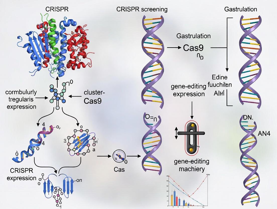
CRISPR Screening Workflow for Gastrulation Research. This diagram outlines the key steps in a functional genomics screen to identify genes regulating gastrulation processes.
Advanced Applications in Gastrulation Research
CRISPR screening technologies have evolved beyond simple gene knockout to enable more nuanced investigation of developmental mechanisms. For gastrulation research, several advanced applications are particularly valuable:
CRISPR Activation/Interference Screens: These approaches using catalytically dead Cas9 (dCas9) fused to transcriptional regulators allow modulation of gene expression without altering DNA sequence [1] [4]. This is especially useful for studying dosage-sensitive developmental genes where complete knockout may be embryonic lethal, or for investigating the effects of gene overexpression during lineage specification.
Single-Cell CRISPR Screening: Combining CRISPR screening with single-cell RNA sequencing (scRNA-seq) enables high-resolution mapping of how genetic perturbations affect transcriptional programs during differentiation [3]. This approach can reveal how specific genes influence cell fate decisions at critical gastrulation timepoints, providing unprecedented insight into developmental trajectories.
Spatial Functional Genomics: Emerging methods combine CRISPR screening with spatial transcriptomics to understand how genetic perturbations affect not only cell identity but also positional information and tissue patterning [3]. This is particularly relevant for gastrulation, where the spatial organization of germ layers is fundamental to proper embryogenesis.
In Vivo CRISPR Screening: While technically challenging, CRISPR screening in model organisms provides the most physiologically relevant context for studying gastrulation [3]. Recent advances enable screening in zebrafish, Xenopus, and mouse embryos, allowing direct assessment of gene function in developing embryos with native tissue architecture and signaling environments.
Advanced CRISPR Screening Modalities for Gastrulation Research. This diagram illustrates how different screening approaches provide complementary insights into developmental processes.
The Scientist's Toolkit: Essential Research Reagents
Table 3: Essential Research Reagents for CRISPR Screening in Gastrulation Research
| Reagent Category | Specific Examples | Application Notes |
|---|---|---|
| CRISPR Enzymes | SpCas9, Cas12a, dCas9-KRAB, dCas9-VP64 | Selection depends on desired perturbation: complete knockout (Cas9), transcriptional repression (dCas9-KRAB), or activation (dCas9-VP64) [1] [4] |
| Delivery Systems | Lentiviral vectors, lipid nanoparticles (LNPs), electroporation systems | Lentiviruses standard for pooled screens; LNPs show promise for difficult-to-transfect primary cells and in vivo applications [8] [5] |
| gRNA Libraries | Genome-wide (Brunello, GeCKO), focused (developmental pathways) | Validated genome-wide libraries provide coverage; custom libraries enable focus on gastrulation-relevant genes [5] [2] |
| Cell Culture Models | Mouse ESCs, human iPSCs, embryonic carcinoma cells | Pluripotent stem cells capable of in vitro differentiation optimally model gastrulation events [4] |
| Differentiation Reagents | BMP4, Wnt agonists/antagonists, Nodal mimics, FGF2 | Precisely control differentiation toward specific germ layers [4] |
| Analytical Tools | High-content imagers, FACS systems, NGS platforms | Arrayed screens require high-content imaging; pooled screens require FACS and NGS capabilities [4] [2] |
| Bioinformatics Software | MAGeCK, CRISPResso, CCTK, CRISPRMatch | Open-source tools for gRNA design, quality control, and hit identification [6] [9] |
Troubleshooting and Technical Considerations
Successful implementation of CRISPR screening for gastrulation research requires addressing several technical challenges:
Optimizing Delivery Efficiency: Pluripotent stem cells often exhibit low transduction efficiency with lentiviral vectors. Optimization approaches include: using vesicular stomatitis virus G-glycoprotein (VSV-G) pseudotyped lentiviruses, employing spinfection techniques, testing alternative envelope proteins, or utilizing newer delivery modalities such as lipid nanoparticles (LNPs) [8] [5].
Managing Cellular Heterogeneity: During differentiation, cellular heterogeneity can introduce noise in screening data. Strategies to address this include: incorporating cell surface markers for sorting specific progenitor populations, using reporter cell lines to isolate cells at specific differentiation stages, and employing single-cell sequencing approaches to resolve heterogeneity [3] [4].
Addressing Genetic Compensation: In developmental systems, knockout of one gene may be compensated by related family members, potentially masking phenotypes. Solutions include: targeting multiple family members simultaneously, using acute CRISPR systems for rapid protein depletion, and employing CRISPRi for stronger, more immediate knockdown [1] [4].
Mitigating Off-Target Effects: CRISPR systems can exhibit off-target activity. Control measures include: using validated gRNA designs with minimal predicted off-targets, incorporating multiple gRNAs per gene to confirm phenotype consistency, utilizing high-fidelity Cas variants, and employing careful bioinformatic filtering to eliminate off-target confounders [5] [7].
CRISPR screening represents a paradigm shift in functional genomics, providing an systematic framework for connecting genotypes to phenotypes at unprecedented scale and resolution. For gastrulation research, this methodology offers powerful approaches to decipher the complex genetic networks that coordinate embryonic patterning and tissue morphogenesis. As CRISPR technologies continue to evolve—with advances in precision editing, delivery systems, and analytical methods—their application to developmental biology will undoubtedly yield deeper insights into the fundamental principles governing embryogenesis. The protocols and considerations outlined here provide a foundation for implementing these powerful approaches to advance understanding of gastrulation and its implications for developmental disorders and regenerative medicine.
Gastrulation is a pivotal phase in embryonic development, where a single-layered blastula reorganizes into a multi-layered structure, giving rise to the primary germ layers. Understanding the genetic and cellular interactions within the gastrulation niche is fundamental to developmental biology and has profound implications for regenerative medicine. The advent of CRISPR-based screening technologies has provided an unprecedented toolset to systematically dissect these complex processes. By enabling high-throughput, precise functional genomic perturbations, CRISPR screening allows researchers to move from observing developmental phenomena to quantitatively analyzing the gene networks that control them. This Application Note details how these powerful screens are revolutionizing the study of gastrulation by uncovering gene functions within a physiologically relevant context.
The application of CRISPR screening in developmental studies leverages several distinct technological approaches, each with unique strengths for probing different biological questions. The table below summarizes the key quantitative aspects of these modalities.
Table 1: Key CRISPR Screening Modalities for Developmental Studies
| Screening Modality | Primary Mechanism | Key Application in Development | Notable Advantage |
|---|---|---|---|
| CRISPR Knockout (KO) | Cas9 nuclease induces double-strand breaks, leading to frameshift mutations and gene disruption. [10] | Uncovering essential genes for cell lineage specification and tissue morphogenesis. [11] | Permanent, complete gene disruption. |
| CRISPR Interference (CRISPRi) | Catalytically dead Cas9 (dCas9) fused to a repressor domain (e.g., KRAB) blocks transcription. [11] [12] | Studying essential gene function and dosage effects during differentiation. [12] | Reversible, titratable knockdown; avoids cell death from essential gene knockout. [12] |
| CRISPR Activation (CRISPRa) | dCas9 fused to a transcriptional activator (e.g., VPR) recruits transcriptional machinery to gene promoters. [11] | Probing gene overexpression effects and identifying sufficiency for cell fate changes. [11] | Gains-of-function without transgenic overexpression; titratable. |
| Single-Cell CRISPR Screening | Combines pooled CRISPR perturbations with single-cell RNA sequencing (scRNA-seq). [11] | Resolving cell-to-cell heterogeneity and mapping transcriptional networks in developing tissues. [11] | Simultaneously captures genotype and transcriptome state in complex populations. |
The selection of a screening modality is a critical first step in experimental design. CRISPRi is particularly valuable for gastrulation studies due to the high density of essential genes in developmental pathways; its titratable nature allows researchers to probe gene function without the confounding effect of cell death, which can distort the understanding of a niche. [12]
Experimental Protocols for CRISPR Screening in Developmentally Relevant Models
The following protocols are adapted from pioneering work in primary human 3D organoids, which recapitulate the cellular complexity and tissue architecture of developing systems far better than traditional 2D cell lines. [11]
Protocol 1: Establishing a CRISPR-Ready 3D Gastric Organoid Model
This protocol outlines the creation of a genetically defined, Cas9-expressing organoid line suitable for large-scale screening.
- Organoid Line Engineering: Start with a well-characterized primary human gastric organoid line. To minimize genetic heterogeneity, use a line with defined oncogenic backgrounds, such as a
TP53/APCdouble knockout (DKO) model. [11] - Lentiviral Transduction for Cas9 Stable Expression:
- Generate a replication-incompetent lentivirus encoding the
Cas9nuclease. - Dissociate organoids into single cells and incubate with the lentiviral supernatant in the presence of polybrene (8 µg/mL) to enhance infection efficiency.
- Culture the transduced cells in Matrigel domes to allow for 3D organoid reformation.
- Select for stable integrants using the appropriate antibiotic (e.g., puromycin at 1-2 µg/mL) for 7-10 days.
- Generate a replication-incompetent lentivirus encoding the
- Validation of Cas9 Activity:
- Transduce the stable Cas9-expressing organoids with a second lentivirus encoding a GFP reporter and a GFP-targeting sgRNA.
- After 96 hours, analyze the organoids by flow cytometry. A successful transduction and active Cas9 will result in >95% loss of GFP fluorescence, confirming robust genome editing capability. [11]
Protocol 2: Pooled CRISPRi Screening for Lineage Specification Genes
This protocol details the steps for performing a loss-of-function screen to identify genes critical for a specific developmental outcome, such as differentiation.
- CRISPRi Organoid Line Development:
- Generate organoid lines expressing the reverse tetracycline-controlled transactivator (rtTA).
- Introduce a doxycycline-inducible cassette containing
dCas9-KRABand a fluorescent reporter (e.g., mCherry) via lentiviral transduction. - Use fluorescence-activated cell sorting (FACS) to isolate a pure population of mCherry-positive cells, confirming tight inducible control. [11]
- sgRNA Library Transduction:
- Select a genome-wide or focused sgRNA library. For essential gene screening, consider a library incorporating mismatched sgRNAs to titrate knockdown efficacy. [12]
- Transduce the iCRISPRi organoids at a low Multiplicity of Infection (MOI < 0.3) to ensure most cells receive only one sgRNA.
- Maintain a cellular coverage of >1000 cells per sgRNA throughout the transduction and selection process to preserve library representation. [11]
- Select transduced cells with puromycin for 5-7 days. Harvest a reference sample (T0) for genomic DNA extraction.
- Screen Execution and Phenotypic Selection:
- Induce sgRNA expression with doxycycline (e.g., 1 µg/mL) to initiate gene knockdown.
- Apply the differentiation stimulus or maintain in the desired condition for the duration of the experiment (e.g., 14-28 days).
- For positive selection (e.g., survival), harvest the final population (T1). For negative selection (e.g., failure to differentiate), the depleted population is analyzed.
- Next-Generation Sequencing and Hit Analysis:
- Extract genomic DNA from T0 and T1 samples.
- Amplify the integrated sgRNA sequences with barcoded primers and subject them to high-depth sequencing.
- Use specialized software (e.g., MAGeCK) to compare sgRNA abundance between T0 and T1. Genes with a significant enrichment or depletion of targeting sgRNAs represent candidate hits that confer the screened phenotype.
Diagram: Workflow for a pooled CRISPRi screen in 3D organoids.
Visualizing Genetic Perturbation Outcomes in Complex Tissues
A major strength of CRISPR screening in developmental models is the ability to resolve outcomes at single-cell resolution. The following workflow illustrates how single-cell CRISPR screens can deconstruct heterogeneity within a gastrulation-like niche.
Diagram: Integrated single-cell CRISPR screening workflow.
The Scientist's Toolkit: Essential Research Reagent Solutions
Successful implementation of the protocols above relies on a suite of specialized reagents and tools. The following table catalogs the essential components for CRISPR screening in developmental models.
Table 2: Key Research Reagent Solutions for CRISPR Screening in Development
| Reagent / Tool | Function | Application Note |
|---|---|---|
| dCas9-KRAB (CRISPRi) | Fusion protein for targeted transcriptional repression. [11] [12] | Ideal for probing essential genes and fine-tuning gene dosage effects during lineage commitment. [12] |
| dCas9-VPR (CRISPRa) | Fusion protein for targeted transcriptional activation. [11] | Used to test gene sufficiency in driving cell fate changes and to rescue differentiation phenotypes. |
| Pooled sgRNA Library | A complex pool of vectors, each encoding a unique guide RNA targeting a specific gene. [11] | Genome-wide (e.g., ~12,500 sgRNAs) or focused (e.g., kinase library) sets enable unbiased or hypothesis-driven screening. |
| Primary Human Organoids | 3D in vitro cultures that mimic the in vivo tissue architecture, stem cell activity, and differentiation potential. [11] | Provides a physiologically relevant model for the gastrulation niche, superior to immortalized 2D cell lines. |
| Lipid Nanoparticles (LNPs) | Non-viral delivery vehicles for CRISPR components. [8] | Enables efficient, transient delivery of ribonucleoproteins (RNPs) for editing without genomic integration. |
| Single-Cell RNA-Seq Kit | Reagents for partitioning cells, barcoding cDNA, and preparing sequencing libraries. | Critical for deconvoluting heterogeneous screening outcomes and linking genetic perturbations to transcriptomic states. |
CRISPR screening technologies have fundamentally transformed our approach to studying development. By moving from static observations to dynamic, functional, and quantitative analyses, these methods allow researchers to systematically map the genetic wiring of the gastrulation niche. The protocols and tools outlined in this Application Note provide a roadmap for leveraging CRISPR knockout, interference, activation, and single-cell screening to uncover the master regulators, synthetic lethal interactions, and buffering relationships that govern embryogenesis. As these technologies continue to converge with advanced in vitro models like organoids, they pave the way for not only a deeper understanding of human development but also for identifying novel therapeutic targets in developmental disorders and cancer.
The advent of CRISPR-based technologies has revolutionized functional genomics, providing researchers with an unprecedented ability to decipher gene function. For investigators studying gastrulation gene function, selecting the appropriate CRISPR tool is paramount to experimental success. The three primary technologies—CRISPR knockout (CRISPRko), CRISPR interference (CRISPRi), and CRISPR activation (CRISPRa)—offer distinct mechanistic approaches and applications for genetic perturbation [13]. CRISPRko permanently disrupts gene function by creating double-strand breaks in DNA, while CRISPRi and CRISPRa enable reversible, tunable regulation of gene expression without altering the underlying DNA sequence [11] [14]. Understanding the capabilities, limitations, and optimal applications of each system is essential for designing robust experiments that can unravel the complex genetic networks governing gastrulation. This article provides a comprehensive comparison of these tools, along with detailed protocols tailored for research in developmental biology and gastrulation studies.
Tool Comparison: Mechanisms and Applications
The selection between CRISPRko, CRISPRi, and CRISPRa depends on the biological question, the nature of the target gene, and the desired outcome of the perturbation. The table below summarizes the core characteristics, advantages, and limitations of each system to guide appropriate tool selection.
Table 1: Comparative analysis of CRISPRko, CRISPRi, and CRISPRa technologies
| Feature | CRISPR Knockout (KO) | CRISPR Interference (i) | CRISPR Activation (a) |
|---|---|---|---|
| Mechanism of Action | Cas9-induced double-strand breaks repaired by error-prone NHEJ, leading to frameshift mutations and gene disruption [14]. | Catalytically dead Cas9 (dCas9) fused to transcriptional repressors (e.g., KRAB) blocks transcription initiation or elongation [11]. | dCas9 fused to transcriptional activators (e.g., VP64, VPR, SAM) recruits machinery to promote gene transcription [15] [16]. |
| Genetic Outcome | Permanent gene disruption; complete loss of function. | Reversible transcript knockdown; partial to strong reduction in gene expression. | Controlled gene upregulation; can activate silent or lowly-expressed genes. |
| Key Applications | Essential gene identification, validation of drug targets, functional screening of non-coding regions [13]. | Study of essential genes, temporal control of gene expression, dissecting gene-drug interactions [11]. | Functional study of redundant genes, gain-of-function screens, validation of silent gene reporters [15] [16]. |
| Efficiency | High (e.g., 75-99% deletion efficiency with qgRNA libraries) [17]. | High (e.g., 76-92% silencing efficiency with qgRNA libraries) [17]. | Variable, dependent on baseline expression and chromatin context; enhanced by multi-activator systems [17]. |
| Specificity & Off-target Effects | Potential for off-target cleavage at sites with sequence similarity [13]. | High specificity with minimal off-target effects as no DNA is cut [11]. | High specificity; potential for off-target activation exists but is limited. |
| Key Advantages | Complete and permanent loss-of-function; simple system. | Reversible, tunable knockdown; enables study of essential genes; low toxicity [11]. | Can overcome epigenetic silencing (e.g., with SAM-TET1 system); powerful for gain-of-function studies [15]. |
| Primary Limitations | Can be toxic to cells; unsuitable for essential gene study in proliferating populations; PAM site dependency [14]. | Knockdown may be incomplete; requires careful sgRNA design at promoter regions. | Activation levels can be variable; may not fully mimic endogenous expression. |
Guidance for Tool Selection in Gastrulation Research
- CRISPRko is ideal for investigating the non-redundant functions of genes critical for cell survival, differentiation, and migration during gastrulation. Its permanent nature is well-suited for long-term differentiation assays.
- CRISPRi is optimal for studying essential genes where complete knockout would be lethal to the progenitor cell pool, allowing for temporal control to dissect stage-specific functions.
- CRISPRa is particularly powerful for validating reporter knock-ins at silent loci, a common challenge in gastrulation research, and for performing gain-of-function screens to identify genes that drive cell fate transitions [15] [16]. The SAM-TET1 system, which combines transcriptional activation with DNA demethylation, is especially effective for activating genes in a repressive chromatin context [15].
Quantitative Performance Data
Recent advancements in library design, particularly the use of quadruple-guide RNA (qgRNA) vectors, have significantly enhanced the performance and reliability of CRISPR screens. The following table presents quantitative data on the efficacy of these improved systems.
Table 2: Performance metrics of advanced CRISPR perturbation systems
| Perturbation Modality | Library Design | Reported Efficiency/Robustness | Key Supporting Findings |
|---|---|---|---|
| Gene Knockout (KO) | Quadruple-sgRNA (qgRNA) library (T.spiezzo: 19,936 plasmids) [17] | 75% - 99% deletion efficiency [17] | Robust growth defect phenotypes identified in membrane protein screen; significant improvement over single sgRNA. |
| Gene Activation (CRISPRa) | Quadruple-sgRNA library (T.gonfio: 22,442 plasmids) with dCas9-VPR [17] | Massive increase in target gene activation vs. single sgRNAs; superior for genes with low basal expression [17]. | Synergistic activation with multiple sgRNAs; effectively activated both protein-coding and non-coding genes. |
| Gene Silencing (CRISPRi/CRISPRoff) | Quadruple-sgRNA library with dCas9-KRAB [17] | 76% - 92% silencing efficiency [17] | High consistency and potent silencing across target genes. |
| CRISPRa in hPSCs | SAM-TET1 system [15] [16] | Most potent for activating silent genes in hPSCs; enhanced activation of methylated genes. | Enabled verification of silent gene reporter knock-ins (e.g., KLF17) within 48 hours, bypassing need for differentiation. |
Detailed Experimental Protocols
Protocol 1: Rapid Verification of Silent Gene Reporters in hPSCs Using CRISPRa
This protocol is critical for gastrulation research, where genes are often silent in pluripotent states. It allows for rapid validation of reporter cell lines without complex differentiation protocols [15].
Key Resources:
- Cell Line: H1-KLF17-GFP reporter hPSCs (or similar silent gene reporter line)
- Plasmids: SAM-TET1 system plasmids (EF1α-TET1-dCas9 #235593 & EF1α-MVPH #235594 from Addgene) and LsgRNA-MS2 backbone (#235597) [15]
- Equipment: Nucleofector system, Flow cytometer, hPSC culture equipment
Workflow Steps:
Design and Cloning of sgRNA (Timing: 3-5 days)
- Design sgRNAs targeting the promoter region of your silent gene of interest.
- Clone the annealed oligonucleotides into the BsmBI-v2-digested LsgRNA-MS2 plasmid backbone.
- Transform the ligation product, pick colonies, and confirm correct sequence by Sanger sequencing.
Delivery of CRISPRa into Reporter Cells (Timing: 1 day)
- Culture H1-KLF17-GFP reporter hPSCs in StemFlex medium on Matrigel-coated plates.
- Dissociate cells into single cells using Accutase.
- For nucleofection, prepare a mixture containing the SAM-TET1 plasmids (EF1α-TET1-dCas9 and EF1α-MVPH) and the newly cloned sgRNA plasmid.
- Nucleofect cells using an appropriate program and plate them in pre-warmed medium supplemented with Y-27632 (ROCK inhibitor).
Detection and Analysis of Reporter Expression (Timing: 2-3 days)
- 48-72 hours post-nucleofection, analyze cells for GFP reporter signal using flow cytometry.
- The appearance of a GFP-positive population indicates successful activation of the silent locus and confirms proper reporter knock-in.
- For further validation, isolate GFP-positive cells by FACS for downstream molecular analysis (e.g., qPCR).
Protocol 2: Arrayed CRISPR Screening in 3D Gastric Organoids
This protocol demonstrates the application of multiplexed CRISPR tools in a physiologically relevant 3D model, which can be adapted to study gastrulation-like processes or endoderm-derived tissues [11].
Key Resources:
- Cell Model: TP53/APC double knockout (DKO) gastric organoid line
- Libraries: Arrayed qgRNA libraries for KO, i, or a modalities [17]
- Equipment: Lentiviral packaging system, Tissue culture setup for 3D organoids, FACS sorter, Next-generation sequencer
Workflow Steps:
Establishment of Cas9-Expressing Organoids (Timing: 2-3 weeks)
- Generate stable Cas9-, dCas9-KRAB- (for CRISPRi), or dCas9-VPR- (for CRISPRa) expressing TP53/APC DKO organoids via lentiviral transduction.
- Confirm Cas9 activity and protein expression via GFP-reporter assays and Western blotting.
Library Transduction and Selection (Timing: 1 week)
- Transduce the arrayed qgRNA library (one gene per well) into the engineered organoids at a low MOI to ensure single copy integration.
- 48 hours post-transduction, begin puromycin selection to eliminate untransduced cells. Maintain cellular coverage of >1000 cells per sgRNA.
Phenotypic Screening and Analysis (Timing: 2-4 weeks)
- For negative selection screens (e.g., identifying genes essential for growth/survival), harvest a reference sample (T0) and continue culturing the organoids for several weeks (T1).
- For drug-gene interaction screens, add the drug of interest (e.g., cisplatin) at the relevant concentration after selection.
- Isolate genomic DNA from T0 and T1 time points and amplify the integrated sgRNA cassettes for next-generation sequencing.
- Analyze sequencing data to quantify sgRNA abundance changes. Depleted sgRNAs in T1 indicate genes essential for growth or conferring drug sensitivity.
The Scientist's Toolkit: Essential Research Reagents
Successful implementation of CRISPR screens requires a carefully selected set of molecular tools and reagents. The following table catalogs key resources for setting up these experiments.
Table 3: Essential research reagents for CRISPR functional genomics
| Reagent / Resource | Function / Application | Example Sources / Identifiers |
|---|---|---|
| CRISPRko System | Wild-type Cas9 nuclease for creating targeted double-strand breaks and gene knockouts. | Addgene: #52962 (lentiCas9-EF1a-Blast) |
| CRISPRi System | dCas9-KRAB fusion protein for transcriptional repression without DNA cleavage. | Addgene: #71237 (pLV hU6-sgRNA hUbC-dCas9-KRAB-T2a-Puro) |
| CRISPRa System | Advanced activation systems for robust gene upregulation. | SAM-TET1: Addgene #235593, #235594 [15]dCas9-VPR: Various Addgene plasmids |
| Arrayed qgRNA Libraries | Pre-cloned, arrayed libraries for high-throughput, multi-sgRNA targeting per gene. | T.spiezzo (KO, 19,936 plasmids), T.gonfio (Activation/Silencing, 22,442 plasmids) [17] |
| sgRNA Backbone | Vector for cloning and expressing custom sgRNAs, often with MS2 modifications for recruitment. | LsgRNA-MS2 (Addgene: #235597) [15] |
| Delivery Vectors | Lentiviral or piggyBac transposon vectors for stable integration of CRISPR components. | pYJA5-derived vectors (Addgene) [17] |
| Specialized Cell Culture | Matrices and media optimized for sensitive cell types like hPSCs and organoids. | Matrigel (Corning #354277), StemFlex Medium (Gibco #A3349401) [15] |
The strategic selection of CRISPR tools—KO, i, or a—is fundamental to designing effective functional genomics studies in gastrulation research. CRISPRko remains the gold standard for complete loss-of-function studies, while CRISPRi offers precise temporal control for investigating essential genes. CRISPRa, particularly with advanced systems like SAM-TET1, provides a powerful method for gain-of-function experiments and validating edits at silent loci, a common challenge in developmental models. The emergence of highly efficient arrayed qgRNA libraries now enables more robust and comprehensive screening across all three modalities [17]. By leveraging the protocols and comparisons outlined here, researchers can systematically dissect the complex genetic networks that orchestrate gastrulation, accelerating discovery in fundamental developmental biology and its clinical applications.
The fundamental challenge in developmental biology and regenerative medicine lies in distinguishing genes that are universally essential for cell survival from those that exert their functions in a context-specific manner, directing lineage specification and cell fate decisions. The emergence of sophisticated CRISPR-based functional genomics tools has revolutionized our ability to systematically dissect this complex genetic landscape at an unprecedented scale and resolution [18]. These technologies enable researchers to move beyond correlative observations to establish causal relationships between genes and phenotypic outcomes in developmentally relevant models.
Within the conceptual framework of Waddington's epigenetic landscape, essential genes maintain the stability of the pluripotent state and core cellular machinery, while context-specific genes guide the trajectory of the differentiation process through the branching valleys of the landscape [19]. The identification and characterization of these two gene classes are particularly crucial for understanding gastrulation—a pivotal developmental stage where the three germ layers (ectoderm, mesoderm, and endoderm) are established through complex gene regulatory networks (GRNs). This application note provides detailed protocols and frameworks for employing CRISPR screening technologies to delineate these critical genetic regulators within experimentally tractable models of gastrulation.
Theoretical Framework: Distinguishing Gene Classes in Development
Conceptual and Practical Definitions
In functional genomics, genes can be categorized based on their perturbation phenotypes across biological contexts:
- Essential Genes: These genes are indispensable for fundamental cellular processes such as transcription, translation, DNA replication, and cell cycle progression. Their loss consistently results in profound fitness defects or lethality across diverse cellular contexts and lineages. They often encode components of core cellular machinery and are enriched for housekeeping functions [11].
- Context-Specific Genes: These genes display conditional essentiality, where their requirement is manifest only in particular lineage contexts, developmental stages, or environmental conditions (e.g., specific signaling environments or drug treatments). They frequently encode transcription factors, signaling receptors, and other regulatory proteins that govern cell fate decisions [20].
Analytical Framework for Classification
The differentiation between these gene classes relies on comparative CRISPR screening across multiple biologically distinct conditions. The core analytical approach involves:
- Perturbation: Introducing systematic genetic perturbations (e.g., knockout) across the genome.
- Selection: Applying distinct biological challenges (e.g., differentiation cues, lineage stressors).
- Quantification: Measuring the effect of each perturbation on cellular fitness or phenotype in each context.
- Comparison: Identifying genes whose perturbation effects differ significantly between conditions.
Table 1: Characteristic Features of Essential vs. Context-Specific Genes
| Feature | Essential Genes | Context-Specific Genes |
|---|---|---|
| Phenotype upon Loss | Lethality/fitness defect across contexts | Viable in some contexts, defect in others |
| Biological Process | Core cellular processes (e.g., RNA processing, transcription) [11] | Lineage specification, signal response, differentiation |
| Example Functional Classes | Ribosomal proteins, DNA replication factors | Developmental transcription factors, signaling pathway components |
| Therapeutic Implication | Poor drug targets; potential toxicity | High-value targets for precision medicine |
Figure 1: Experimental Workflow for Gene Classification. A pooled CRISPR screen is performed in a relevant model system across multiple biological contexts to distinguish gene classes based on differential fitness phenotypes.
Application Note: CRISPR Screening in Gastrulation Models
Advanced Model Systems
The physiological relevance of the model system is paramount for generating meaningful data on lineage specification. While conventional 2D cell lines have utility, primary human 3D organoids now offer a superior platform that preserves tissue architecture, cellular heterogeneity, and differentiation potential [11]. Recent work has established the feasibility of large-scale CRISPR screening in primary human gastric organoids, enabling the comprehensive dissection of gene-drug interactions in a pathophysiologically relevant context [11] [21]. These 3D cultures can be engineered with oncogenic mutations (e.g., TP53/APC double knockout) to provide a homogeneous yet genetically defined background for screening while maintaining developmental competence [11].
CRISPR Tool Selection Guide
The appropriate choice of CRISPR modality is critical for addressing specific biological questions about gene function during gastrulation:
- CRISPR Knockout (CRISPRko): Utilizes nuclease-active Cas9 to create double-strand breaks, resulting in insertions or deletions (indels) that disrupt the coding sequence. Ideal for identifying essential genes and generating complete loss-of-function mutations. May be confounded by compensatory adaptation or toxicity in amplified genomic regions [20].
- CRISPR Interference (CRISPRi): Employs catalytically dead Cas9 (dCas9) fused to a transcriptional repressor domain (e.g., KRAB) to block transcription without altering the DNA sequence. Enables reversible, tunable knockdown, making it suitable for studying essential genes and modeling pharmacological inhibition more accurately than knockout [11] [20].
- CRISPR Activation (CRISPRa): Uses dCas9 fused to transcriptional activation domains (e.g., VPR) to upregulate endogenous gene expression. Powerful for identifying genes whose overexpression drives specific lineage commitments or confers resistance to differentiation signals [11] [20].
Table 2: CRISPR Modalities for Dissecting Gene Function in Development
| Modality | Mechanism | Best Use Cases | Advantages | Limitations |
|---|---|---|---|---|
| CRISPRko | Nuclease-induced indels | Identification of essential genes; complete gene disruption | Strong, permanent loss-of-function; well-established | Potential toxicity; difficult to study essential genes |
| CRISPRi | dCas9-KRAB transcriptional repression | Fine-tuning gene dosage; studying essential genes; mimicking drug effects | Tunable, reversible; reduced off-target effects | Knockdown not complete; requires careful sgRNA design |
| CRISPRa | dCas9-VPR transcriptional activation | Gain-of-function studies; identifying lineage drivers | Endogenous gene activation; studies overexpression phenotypes | Can produce non-physiological expression levels |
Experimental Protocol: Pooled CRISPR Screening in 3D Organoids
This protocol outlines the steps for performing a pooled negative selection CRISPR screen in primary human 3D gastric organoids to identify genes essential for viability under standard conditions versus context-specific essential genes under a selective pressure (e.g., chemotherapeutic agent), based on established methodologies [11].
Library Design and Lentiviral Production
- Library Selection: Choose a genome-wide CRISPR knockout library (e.g., Brunello, human GeCKO v2) with high-quality sgRNA designs. Ensure the library includes ~750 non-targeting control sgRNAs distributed throughout for normalization [11].
- Lentiviral Production: Produce high-titer lentiviral particles of the sgRNA library in HEK293T cells using standard packaging plasmids. Concentrate virus via ultracentrifugation and determine functional titer by transducing target cells with a pilot virus and counting antibiotic-resistant colonies.
Cell Line Engineering and Viral Transduction
- Generate Cas9-Expressing Organoids:
- Use a human TP53/APC double knockout (DKO) gastric organoid line [11].
- Transduce with a lentiviral vector constitutively expressing S. pyogenes Cas9 and a puromycin resistance gene.
- Select with puromycin (1-2 µg/mL) for 5-7 days to generate a stable polyclonal Cas9-expressing line.
- Validate Cas9 activity using a GFP-reporter assay (>95% GFP knockout efficiency expected) [11].
- Library Transduction:
- Dissociate organoids into single cells.
- Transduce Cas9-expressing organoids with the sgRNA library lentivirus at a low multiplicity of infection (MOI ~0.3-0.4) to ensure most cells receive a single sgRNA.
- Include >1000 cells per sgRNA in the library to maintain library representation. For a 100,000 sgRNA library, transduce at least 100 million cells.
- 24 hours post-transduction, add fresh medium containing puromycin for selection.
- Culture for 5-7 days under puromycin selection to eliminate non-transduced cells. This is the baseline population (T0).
Screening and Phenotypic Selection
- Split and Challenge:
- Harvest a subset of the selected organoids for genomic DNA extraction (T0 reference sample).
- Split the remaining organoids into two experimental arms:
- Arm 1 (Control): Culture under standard conditions.
- Arm 2 (Context-Specific Challenge): Culture in medium containing a relevant selective agent (e.g., cisplatin for gastric cancer models) [11].
- Maintain cultures for 14-28 days, passaging organoids regularly and ensuring >1000x library coverage is maintained at all times.
- Harvest Endpoint Samples:
- Collect organoids from both arms at the endpoint (T1).
- Wash with PBS and store pellets at -80°C for genomic DNA extraction.
Genomic DNA Extraction and Next-Generation Sequencing
- Extract genomic DNA from all samples (T0, T1 Control, T1 Challenged) using a large-scale gDNA extraction kit. Typically, 10-20 million cells yield ~100 µg gDNA, sufficient for library preparation.
- Perform a two-step PCR to amplify integrated sgRNA sequences from the genomic DNA and add Illumina sequencing adapters and barcodes [2].
- Purify the PCR products and quantify using a high-sensitivity dsDNA assay.
- Pool libraries and sequence on an Illumina NextSeq or HiSeq platform to achieve >500 reads per sgRNA.
Bioinformatic Analysis
- sgRNA Quantification: Align sequencing reads to the reference sgRNA library and count reads per sgRNA for each sample.
- Normalization and Fold Change: Normalize read counts to the total reads per sample. Calculate log2 fold-changes for each sgRNA between T1 and T0 samples within each arm.
- Gene-Level Statistics: Using a tool like MAGeCK, aggregate sgRNA fold-changes to compute a gene-level score (e.g., β score) and statistical significance (e.g., FDR) for each experimental arm [11].
- Gene Classification:
- Essential Genes: Significant depletion (FDR < 0.05, negative β score) in BOTH control and challenged conditions.
- Context-Specific Essential Genes: Significant depletion ONLY in the challenged condition.
- Confidence Criteria: Gene-level p-value < 0.05 and at least 3/4 independent sgRNAs showing a consistent phenotype direction.
The Scientist's Toolkit: Key Research Reagents
Table 3: Essential Reagents for CRISPR Screening in Gastrulation Research
| Reagent / Solution | Function / Application | Example / Specification |
|---|---|---|
| Primary Human Gastric Organoids | Physiologically relevant 3D model for gastrulation and lineage specification studies | TP53/APC DKO line for homogeneous genetic background [11] |
| CRISPR Knockout Library | Genome-wide collection of sgRNAs for pooled screening | Human GeCKO v2, Brunello; ~75,000-100,000 sgRNAs |
| Lentiviral Packaging System | Production of viral particles for sgRNA library delivery | psPAX2, pMD2.G packaging plasmids |
| Cas9 Expression System | Constitutive or inducible nuclease expression | LentiCas9-Blast, pCW-Cas9 vectors |
| Next-Generation Sequencing Platform | High-throughput quantification of sgRNA abundance | Illumina NextSeq 500/550 |
| Bioinformatic Analysis Pipeline | Processing NGS data, normalization, and hit identification | MAGeCK, CERES |
Data Interpretation and Validation
Hit Confirmation and Follow-up
Genes identified in primary screens require rigorous validation:
- Secondary Validation: Select 3-5 top hits from each category (essential and context-specific). For each hit, design 3-4 independent sgRNAs and test them individually in the organoid model. Quantify the phenotype (e.g., growth defect via cell viability assays) relative to non-targeting control sgRNAs [11].
- Mechanistic Investigation: For context-specific essential genes, perform downstream assays to elucidate mechanism:
- Single-Cell RNA-Sequencing: Resolve transcriptomic consequences of perturbation at single-cell resolution. As demonstrated in gastric organoids, this can reveal how genetic alterations interact with environmental cues (e.g., cisplatin) and uncover novel biological links (e.g., between fucosylation and cisplatin sensitivity) [11].
- Lineage Tracing: Employ fluorescent reporters to track the fate of perturbed cells and their progeny during differentiation.
Integration with Gastrulation Research
To frame findings within the context of gastrulation gene function research:
- Cross-reference with Developmental Databases: Compare your list of context-specific essential genes with known expression patterns of key developmental genes (e.g., from mouse embryo studies [22]) during gastrulation.
- Pathway Enrichment Analysis: Input gene lists into enrichment tools (e.g., DAVID, Enrichr) to identify overrepresented developmental signaling pathways (e.g., Wnt, Nodal, BMP).
- Construct Gene Regulatory Networks (GRNs): Model your validated hits within a GRN framework to understand their position in the hierarchy controlling lineage specification. Associative GRN models (AGRN) can computationally predict how these genes maintain stable states or drive transitions [19].
Figure 2: Logical Framework for Gene Classification. The classification of a gene as essential or context-specific depends on the presence of its perturbation phenotype across different biological contexts.
Advanced Screening Models and Workflows: From Stem Cells to 3D Organoids
The study of gastrulation, a pivotal period in embryonic development where the three primary germ layers are formed, has been revolutionized by advanced in vitro model systems and CRISPR-based functional genomics. This application note provides a detailed comparison of three leading models—Induced Pluripotent Stem Cells (iPSCs), Synthetic Embryo Models (SEMs), and Primary Human 3D Gastric Organoids—for CRISPR screening applications aimed at deciphering gene function during gastrulation. Each system offers unique advantages in scalability, physiological relevance, and applicability to early human development, enabling researchers to dissect complex genetic networks governing cell fate decisions, tissue patterning, and morphogenesis.
Model System Comparison
The table below summarizes the key characteristics, applications, and technical considerations for the three model systems, aiding in the selection of the most appropriate platform for specific research goals in gastrulation gene function.
Table 1: Comparative Analysis of Model Systems for CRISPR Screening in Gastrulation Research
| Feature | iPSCs | Synthetic Embryo Models (SEMs) | Primary Human 3D Gastric Organoids |
|---|---|---|---|
| Core Description | Reprogrammed somatic cells with pluripotent potential [23] | Stem-cell-based embryo-like structures mimicking early developmental stages [24] [25] | 3D cultures derived from primary stomach tissue, preserving its architecture [11] [26] |
| Key Applications in Gastrulation Research | Dissecting pluripotency networks, early lineage specification, and germ layer differentiation [27] [28] | Modeling pre- to post-implantation development, tissue-tissue interactions, and self-organization [24] | Studying organ-specific morphogenesis and gene-drug interactions in a visceral organ derivative [11] |
| Physiological Relevance to Development | High for early lineage commitment and differentiation [27] | High for mimicking spatial and temporal aspects of early embryogenesis [24] [25] | High for stomach development and physiology [11] |
| Throughput for CRISPR Screening | High (amenable to genome-scale screens) [27] | Moderate (complexity of model can limit scale) | High (demonstrated for large-scale screens) [11] |
| Key Quantitative Readouts | Pluripotency factor expression (e.g., OCT4-GFP), cell fitness [27] | Blastoid formation efficiency, lineage marker expression, apoptotic index [29] | Cell growth/survival, gene-drug interaction scores, single-cell transcriptomic profiles [11] |
| Technical Complexity | Moderate | High | High |
| Major Advantage | Patient-specific, unlimited self-renewal, avoids ethical concerns of embryos [23] | Enables study of early developmental events otherwise inaccessible [24] | Captures tissue-specific complexity and patient-specific tumor responses [11] [26] |
Application Notes and Protocols
Induced Pluripotent Stem Cells (iPSCs)
Application Note: iPSCs serve as a powerful platform for unbiased discovery of genes regulating the dissolution of pluripotency and the onset of gastrulation. Genome-scale CRISPR knockout screens in iPSCs can disentangle the genetic networks controlling core pluripotent identity from those governing general cell fitness, which are often conflated in standard viability screens [27].
Experimental Protocol: CRISPR Screen for Pluripotency Exit Regulators
- Cell Line Engineering: Generate a doxycycline-inducible Cas9-expressing iPSC line with a knock-in OCT4-GFP reporter to monitor pluripotent status [27].
- Library Transduction: Transduce the cells at a low MOI (e.g., ~0.3) with a genome-scale lentiviral sgRNA library (e.g., Brunello library). Maintain a representation of >500 cells per sgRNA to ensure library coverage [27].
- Differential Differentiation: Split the transduced cell pool and differentiate them towards neuroectoderm (NE) and definitive endoderm (DE) lineages using established protocols [27].
- NE Differentiation: Use dual SMAD inhibition.
- DE Differentiation: Use Activin A and a WNT pathway activator.
- FACS Sorting: On the day of peak OCT4 downregulation (e.g., day 1.5 for NE, day 2.5 for DE), harvest cells and sort into OCT4-GFPhi (retained pluripotency) and OCT4-GFPlo (exited pluripotency) populations.
- Sequencing and Hit Calling: Extract genomic DNA from sorted populations and the initial plasmid library. Amplify sgRNA sequences and perform next-generation sequencing. Use the MAGeCK robust ranking aggregation (RRA) algorithm to identify sgRNAs enriched in either population [27].
- Pro-pluripotency hits: Genes whose knockout enriches sgRNAs in the OCT4-GFPlo population, hastening the loss of pluripotency.
- Anti-pluripotency hits: Genes whose knockout enriches sgRNAs in the OCT4-GFPhi population, impeding the loss of pluripotency.
The workflow for this protocol is illustrated in the following diagram:
Synthetic Embryo Models (SEMs)
Application Note: SEMs, or embryoids, are generated by programming stem cells to self-organize into structures that recapitulate key aspects of early post-implantation embryos. CRISPR-based perturbation in these models is ideal for studying tissue-tissue interactions and morphogenetic events critical to gastrulation, such as the role of hominoid-specific genetic elements [25] [29].
Experimental Protocol: CRISPRa Programming of Mouse Embryoid Formation
- Stem Cell Preparation: Culture mouse embryonic stem cells (mESCs) in standard naive conditions.
- Epigenome Engineering: Transfect cells with a plasmid expressing a CRISPR activation (CRISPRa) system, specifically a catalytically dead Cas9 (dCas9) fused to transcriptional activators (e.g., VPR), and sgRNAs targeting key developmental gene promoters [25] [30].
- Embryoid Aggregation: Dissociate transfected cells and aggregate them in low-attachment U-bottom plates to promote 3D self-organization.
- Morphogenesis Monitoring: Culture aggregates for 3-6 days, monitoring the formation of embryo-like structures via bright-field microscopy. A successful protocol can yield organized structures in >80% of aggregates [25].
- Phenotypic Validation:
- Immunofluorescence: Fix embryoids and stain for embryonic lineages (e.g., NANOG for epiblast, GATA3 for trophectoderm, SOX17 for hypoblast) [29].
- Gene Expression Analysis: Perform RNA-seq or qRT-PCR on embryoids to verify the activation of target genes and developmental pathways.
- Functional Assessment: Test the requirement of specific genes by introducing CRISPRi (e.g., dCas9-KRAB) and quantifying defects in embryoid formation efficiency [29].
Primary Human 3D Gastric Organoids
Application Note: Gastric organoids provide a physiologically relevant model to study how genes involved in gastrulation and patterning influence organ-specific development and function. Large-scale CRISPR screens in these organoids can systematically reveal genetic interactions underlying cellular fitness and response to environmental stimuli, such as chemotherapy [11] [26].
Experimental Protocol: Multiplexed CRISPR Screening for Gene-Drug Interactions
- Organoid Line Development: Establish Cas9-expressing human gastric organoids, ideally with a homogeneous genetic background (e.g., TP53/APC double knockout) to reduce screening noise [11].
- CRISPR Tool Selection:
- For gene knockout, use a lentiviral library (e.g., ~12,000 sgRNAs) transduced at high coverage (>1000 cells/sgRNA).
- For transcriptional modulation, use inducible CRISPRi (dCas9-KRAB) or CRISPRa (dCas9-VPR) systems.
- Pooled Screen with Drug Challenge: After puromycin selection of transduced organoids, split the culture. Treat one pool with the drug of interest (e.g., cisplatin) and maintain the other as an untreated control. Culture organoids for several passages, maintaining library representation [11].
- sgRNA Abundance Quantification: Harvest genomic DNA from baseline (T0), untreated (T1), and drug-treated (T1_Drug) organoids. Sequence the sgRNA locus to determine the relative abundance of each guide.
- Hit Identification: Genes whose sgRNAs are depleted upon drug treatment compared to the control are sensitizers, while those enriched are resistors. Single-cell RNA-seq coupled with CRISPR screening (Perturb-seq) can further resolve how genetic perturbations alter transcriptional networks in response to treatment [11].
Signaling Pathways and Workflows
The following diagram synthesizes the experimental workflow for conducting a CRISPR-based chemogenetic interaction screen in primary human gastric organoids, as detailed in the protocol above.
The Scientist's Toolkit: Research Reagent Solutions
Table 2: Essential Reagents for CRISPR Screening in Developmental Model Systems
| Reagent / Material | Function | Example Application |
|---|---|---|
| Inducible Cas9/iPSC Line | Enables precise temporal control of CRISPR activity for editing essential genes. | Dissecting pluripotency exit regulators without compromising cell fitness during maintenance [27]. |
| dCas9-KRAB (CRISPRi) & dCas9-VPR (CRISPRa) | Enables reversible gene knockdown or activation without altering the DNA sequence. | Probing the role of specific genes or non-coding elements (e.g., LTR5Hs) in blastoid development [11] [29]. |
| Validated sgRNA Library | A pooled collection of guides providing comprehensive coverage of the genome or a gene subset. | Genome-scale knockout screens to identify fitness genes and pluripotency regulators [11] [27]. |
| Primary Human Gastric Tissue | Source material for generating organoids that retain the genetic and cellular heterogeneity of the original tissue. | Creating patient-specific models for personalized drug response profiling [11]. |
| Programmable Embryoid Base Cells | Stem cells (e.g., mESCs) optimized for efficient self-organization into embryo-like models. | Studying the fundamental principles of self-organization and early lineage specification [25]. |
CRISPR-based functional genomics provides a powerful, unbiased method for systematically interrogating gene function during complex biological processes like gastrulation. The foundation of a successful CRISPR screen depends on three critical, interconnected pillars: the design of highly functional single guide RNA (sgRNA) libraries, the generation of robust Cas9-expressing cell models, and the efficient delivery of these components via lentiviral transduction. This application note details optimized protocols and design principles for implementing CRISPR knockout screens in gastrulation gene function research, enabling researchers to decipher the genetic networks governing early embryonic development and cell fate decisions. By leveraging recent advances in library design and delivery methodologies, scientists can overcome historical challenges associated with screening in complex, physiologically relevant models.
sgRNA Library Design
The sensitivity and specificity of a CRISPR screen depend fundamentally on the quality of the sgRNA library. Optimal library design involves selecting guides with high on-target efficiency and minimal off-target activity, while also considering the practical constraints of the screening model.
Principles of sgRNA Library Design
On-target efficiency prediction is paramount. Modern algorithms leverage machine learning models trained on large-scale screening data to score sgRNAs based on sequence features. The Vienna Bioactivity CRISPR (VBC) score is one such metric that has demonstrated superior performance in predicting sgRNA efficacy. Benchmark studies comparing publicly available genome-wide libraries (e.g., Brunello, Yusa v3, Gecko V2) have shown that libraries composed of guides selected using the top VBC scores achieve stronger gene depletion in essentiality screens [31].
Minimal library design is increasingly favored for complex screening models like organoids. A "top 3" design, selecting the three sgRNAs per gene with the highest VBC scores, can perform as well as or better than larger libraries with 6-10 guides per gene. This compression reduces reagent costs, increases screening throughput, and improves feasibility in systems with limited cell numbers [31].
Single vs. Dual Targeting Strategies
Dual-targeting libraries, where two sgRNAs are delivered per gene to create larger genomic deletions, can offer enhanced knockout efficiency. Recent benchmarking reveals that dual-targeting guides produce stronger depletion of essential genes and weaker enrichment of non-essential genes compared to single-targeting guides. However, a modest fitness reduction is sometimes observed even for non-essential genes, potentially due to an elevated DNA damage response from creating two double-strand breaks. Therefore, while dual-targeting can be beneficial, caution is warranted in screening contexts where DNA damage signaling could confound results [31].
Table 1: Benchmarking of sgRNA Library Performance in Essentiality Screens
| Library Name | Guides/Gene | Key Feature | Performance in Essentiality Screens |
|---|---|---|---|
| Vienna-single (Top VBC) | 3 | Minimal library design | Strongest depletion curve; optimal for complex models [31] |
| Yusa v3 | 6 | Standard large library | Consistently outperformed by minimal VBC-based designs [31] |
| Croatan | 10 | Dual-targeting focus | Strong performance, but larger library size [31] |
| Dual-targeting | 2 per gene | Creates genomic deletions | Stronger essential gene depletion; potential DNA damage response [31] |
Workflow for sgRNA Library Implementation
The following workflow outlines the key steps from sgRNA design to validation for a gastrulation-focused CRISPR screen.
Generation of Cas9-Expressing Cell Lines
Establishing a cell line with stable, robust Cas9 expression is a prerequisite for pooled CRISPR knockout screens using lentiviral sgRNA delivery.
Methodologies for Cas9 Expression
The choice of Cas9 delivery method depends on the target cell type and the desired application. The three primary biological formats are plasmid DNA (pDNA), messenger RNA (mRNA), and Ribonucleoprotein (RNP). For generating stable Cas9-expressing cell lines, lentiviral transduction with a pDNA construct is the most common approach, as it allows for stable genomic integration and selection of a homogeneous cell population [32].
Critical to this process is the inclusion of a selection marker, such as puromycin resistance or a fluorescent protein (e.g., GFP, mCherry), within the lentiviral vector. This enables antibiotic selection or fluorescence-activated cell sorting (FACS) to enrich for successfully transduced cells, ensuring a high percentage of Cas9-positive cells before proceeding with the sgRNA library screen [11].
Protocol: Lentiviral Generation and Transduction for Stable Cas9 Cell Lines
This protocol is adapted from methods successfully used in primary human gastric organoids [11] and hard-to-transfect suspension cells [33].
Materials & Reagents:
- Transfer Plasmid: lentiCRISPRv2 (Addgene #52961) or similar Cas9-P2A-PuroR vector.
- Packaging Plasmids: psPAX2 (Addgene #12260) and pMD2.G (Addgene #12259).
- Cell Lines: Lenti-X 293T cells (Takara #632180) for viral production; target cell line for gastrulation studies.
- Reagents: Lipofectamine 2000, Polybrene, Puromycin.
Procedure:
- Virus Production: Seed Lenti-X 293T cells in a 6-well plate to reach 70-80% confluency at time of transfection.
- Co-transfect the transfer plasmid (lentiCRISPRv2) with the packaging plasmids (psPAX2 and pMD2.G) using Lipofectamine 2000, following manufacturer's instructions.
- At 48-72 hours post-transfection, harvest the lentivirus-containing supernatant, centrifuge to remove cell debris, and filter through a 0.45μm PVDF filter. Aliquot and store at -80°C.
- Transduction: Seed target cells and transduce with the harvested lentiviral supernatant in the presence of 8μg/mL Polybrene via spinfection (centrifugation at 800-1000 x g for 30-60 minutes at 32°C) to enhance infection efficiency.
- Selection: Begin puromycin selection (e.g., 1-5μg/mL, concentration must be predetermined by a kill curve) 48 hours post-transduction. Maintain selection pressure for at least 3-7 days until all non-transduced control cells are dead.
- Validation: Confirm Cas9 expression and functionality via Western blot and a surrogate cleavage assay (e.g., GFP reporter disruption) [11] [33].
Lentiviral Transduction of sgRNA Libraries
Efficient and uniform delivery of the sgRNA library is critical to ensure each cell receives only one guide and that all guides are represented in the screened population.
Optimizing Transduction Efficiency
The key to a successful pooled screen is achieving a low Multiplicity of Infection (MOI), typically MOI < 0.3. This ensures that the vast majority of transduced cells receive only a single sgRNA, simplifying the interpretation of genotype-phenotype linkages. The functional titer of the lentiviral sgRNA library should be determined empirically on the Cas9-expressing cell line of interest via puromycin selection and cell counting [11] [33].
To maintain library representation, a high coverage of cells per sgRNA is required. A minimum of 200-500 cells per sgRNA is standard, but for more complex models like organoids, coverage of >1000 cells per sgRNA is recommended to account for heterogeneity and prevent stochastic loss of guides [11].
Protocol: Pooled sgRNA Library Transduction and Screening
This protocol outlines the steps for screening a pooled sgRNA library in a Cas9-expressing cell model.
Materials & Reagents:
- Pooled lentiviral sgRNA library (e.g., Vienna-single, custom gastrulation-focused).
- Stable Cas9-expressing cell line.
- Polybrene, Puromycin.
- Cell culture reagents for the specific model (e.g., organoid culture media).
Procedure:
- Titer Determination: Perform a small-scale pilot transduction with a range of viral volumes to determine the volume needed to achieve MOI ~0.3 with your Cas9-expressing cells under puromycin selection.
- Large-Scale Transduction: Scale up the transduction to infect a number of cells that provides >1000x coverage of the entire sgRNA library. Include polybrene and use spinfection if applicable.
- Selection: Begin puromycin selection 48 hours post-transduction. Continue selection for 3-5 days or until all cells in a non-transduced control well have died.
- Harvest T0 Sample: Upon completion of selection, harvest a representative sample of the cell population (at least 500 cells per sgRNA). This is the "Time Zero" (T0) reference point for sequencing.
- Phenotype Propagation: Culture the remaining cells, maintaining a minimum coverage of >1000 cells per sgRNA throughout the screening period. Passage cells as needed.
- Harvest Endpoint Sample: After a sufficient period for phenotypic selection (e.g., 14-28 days, or after a specific gastrulation differentiation stimulus), harvest the final cell population for genomic DNA extraction.
- Next-Generation Sequencing: Amplify the integrated sgRNA cassettes from the genomic DNA of the T0 and endpoint samples. Prepare sequencing libraries and sequence on an Illumina platform to determine sgRNA abundance [11] [31].
Application in Gastrulation Gene Function Research
The integration of these optimized protocols enables robust CRISPR screening in advanced models that recapitulate key aspects of gastrulation.
Screening in 3D Gastric Organoid Models
Primary human 3D organoids preserve tissue architecture, cellular heterogeneity, and differentiation potential, making them a superior model for studying developmentally relevant processes. Large-scale CRISPR knockout, interference (CRISPRi), and activation (CRISPRa) screens have been successfully implemented in primary human gastric organoids. For instance, such screens have been used to systematically identify genes that affect sensitivity to chemotherapeutic agents, uncovering novel links between biological pathways like fucosylation and drug response [11]. This same approach can be directly applied to unravel genetic interactions critical for gastrulation.
The Scientist's Toolkit: Essential Reagents
Table 2: Key Research Reagent Solutions for CRISPR Screening
| Item | Function/Description | Example Products/Sources |
|---|---|---|
| CRISPR Vectors | Plasmid backbone for expressing Cas9 and sgRNA; often includes selection marker. | lentiCRISPRv2 (Addgene #52961) [33] |
| Lentiviral Packaging Plasmids | Provide essential viral proteins in trans for producing replication-incompetent lentivirus. | psPAX2, pMD2.G (Addgene) [33] |
| sgRNA Libraries | Pooled collections of sgRNAs targeting genes of interest for functional genomics. | Custom Vienna-single libraries, Brunello, Yusa v3 [31] |
| Synthetic sgRNA | Chemically synthesized, high-purity guides; can include modifications to enhance stability. | Synthego (Research-use only, INDe, GMP grades) [34] |
| Transfection Reagent | Facilitates plasmid DNA delivery into packaging cells for virus production. | Lipofectamine 2000 [33] |
| Transduction Enhancer | Cationic polymer that increases viral attachment to cell membranes, boosting infection. | Polybrene [33] |
| Selection Antibiotics | Allows for the elimination of non-transduced cells, enriching the edited population. | Puromycin [11] [33] |
The meticulous design of minimal, highly functional sgRNA libraries, combined with the robust generation of Cas9-expressing cell models and optimized lentiviral transduction, forms the technical foundation for effective CRISPR screens. By adhering to the protocols and principles outlined in this application note, researchers can leverage these powerful tools to systematically dissect the genetic regulation of gastrulation in physiologically relevant models, accelerating discovery in early embryonic development and regenerative medicine.
In CRISPR screening research, particularly in the study of gastrulation gene function, phenotypic readouts are the measurable characteristics that reveal the functional consequences of genetic perturbations. The core principle of "perturbomics" is that the function of a gene can be inferred by systematically altering its activity and observing the resulting phenotypic changes [35]. In the context of gastrulation, this involves deploying diverse CRISPR-based tools—including knockout (CRISPR-KO), interference (CRISPRi), and activation (CRISPRa)—to modulate genes critical for early development, and then applying a suite of assays to capture changes in cell viability, lineage specification, and transcriptional networks [11] [35]. Advanced models like primary human 3D gastric organoids have proven invaluable for these studies, as they preserve tissue architecture and cellular heterogeneity, providing a physiologically relevant system for dissecting gene function [11].
This Application Note details the protocols and analytical frameworks for implementing these key phenotypic readouts, enabling the systematic identification and validation of genes governing gastrulation.
Core Phenotypic Readouts and Associated Metrics
The table below summarizes the primary phenotypic readouts used in CRISPR screens for gastrulation research, their measurement techniques, and the key quantitative metrics derived from them.
Table 1: Core Phenotypic Readouts and Metrics in Gastrulation CRISPR Screens
| Phenotypic Readout | Measurement Technique | Key Quantitative Metrics | Biological Insight |
|---|---|---|---|
| Cell Viability & Proliferation | Pooled CRISPR screen with sequencing of sgRNA abundance over time [11] [35] | - Gene-level phenotype score (e.g., z-score, MAGeCK score)- sgRNA fold-depletion/enrichment- Identification of essential/"drop-out" genes [11] | Genes essential for survival/proliferation under basal or selective (e.g., drug) conditions [11] |
| Lineage Specification | Single-cell RNA-seq (scRNA-seq) of perturbed cells [11] [36] | - Proportion of cells in distinct clusters- Differential expression of lineage-specific markers- RNA velocity trajectories [37] [36] | Lineage bias imposed by genetic perturbation; fate transitions [36] |
| Transcriptomic Changes | Single-cell CRISPR screens (CROP-seq, Perturb-seq) [11] [37] | - Differential expression (DE) analysis (e.g., log2 fold-change)- Gene Set Enrichment Analysis (GSEA)- Pathway activity scores [11] [37] | Gene regulatory networks and pathways downstream of a genetic perturbation [11] |
| Drug/Chemical Response | Viability-based CRISPR screens under drug treatment (e.g., Cisplatin) [11] | - Drug-sensitivity/resistance score (differential sgRNA abundance ±drug)- Identification of synthetic lethal interactions [11] | Genetic modifiers of drug response; therapeutic vulnerabilities [11] |
Experimental Protocols
Protocol 1: Pooled CRISPR Screening for Cell Viability and Drug Response
This protocol outlines the steps for performing a pooled CRISPR knockout screen in a 3D gastric organoid model to identify genes essential for viability or involved in drug response [11].
Research Reagent Solutions
- Cells: Cas9-expressing TP53/APC double knockout (DKO) human gastric organoids [11].
- CRISPR Library: Pooled lentiviral sgRNA library (e.g., targeting ~1000 genes with ~10 sgRNAs/gene and 750 non-targeting controls) [11].
- Culture Reagents: Appropriate organoid base medium (e.g., Advanced DMEM/F12) and culture supplements [11].
- Selection Agent: Puromycin.
- Drug: Chemotherapeutic agent (e.g., Cisplatin) for response studies [11].
- DNA Extraction Kit: For high-quality genomic DNA.
- PCR & NGS Reagents: Primers for sgRNA amplification, high-fidelity PCR mix, and NGS library preparation kit.
Procedure
- Library Transduction: Transduce the pooled lentiviral sgRNA library into the Cas9-expressing organoids at a low Multiplicity of Infection (MOI ~0.3) to ensure most cells receive a single sgRNA. Use a cell coverage of >1000 cells per sgRNA to maintain library representation [11].
- Selection and Harvest T0: At 48 hours post-transduction, select transduced cells with puromycin for 3-5 days. Harvest a representative cell population (at least 10 million cells) as the "Time Zero" (T0) control. Extract genomic DNA [11].
- Application of Selective Pressure:
- For viability screens: Culture the remaining organoids for 28 days, passaging them regularly while maintaining the >1000x sgRNA coverage.
- For drug-response screens: Split the organoids and treat one group with the drug of interest (e.g., Cisplatin) and maintain a parallel vehicle-treated control group for the duration of the experiment [11].
- Harvest Endpoint (T1): After the selection period (e.g., 28 days), harvest the organoids from all experimental conditions. Extract genomic DNA.
- sgRNA Amplification and Sequencing: Amplify the integrated sgRNA sequences from the genomic DNA of T0 and T1 samples via PCR. Prepare sequencing libraries and perform high-throughput sequencing [11].
- Data Analysis:
- sgRNA Counts: Map sequencing reads to the sgRNA library to obtain count data for each sgRNA in each sample.
- Differential Abundance: Using tools like MAGeCK, compare sgRNA abundances between T1 and T0 (for viability) or between drug-treated and control (for response) to identify significantly depleted or enriched sgRNAs [11].
- Gene-level Analysis: Aggregate scores from multiple sgRNAs targeting the same gene to identify high-confidence hit genes whose perturbation affects viability or drug response.
Protocol 2: Single-cell CRISPR Screening for Lineage Specification and Transcriptomics
This protocol describes how to couple CRISPR perturbations with single-cell RNA sequencing to simultaneously read the perturbation and its transcriptomic consequences in individual cells, ideal for studying lineage specification [11] [37].
Research Reagent Solutions
- Cells: Gastric organoids stably expressing dCas9-KRAB (for CRISPRi) or dCas9-VPR (for CRISPRa) in an inducible system [11].
- Perturbation Library: A pooled viral library of sgRNAs targeting candidate gastrulation genes.
- Single-Cell Platform: Equipment and reagents for single-cell partitioning (e.g., 10x Genomics Chromium Controller).
- Single-Cell Reagents: Single-cell kit (e.g., 10x Genomics Single Cell 3' Reagent Kits v4), which includes gels, buffers, and enzymes for barcoding and library construction.
- NGS Reagents: For final library amplification and sequencing.
Procedure
- Induction and Perturbation: Induce the expression of the dCas9 effector (e.g., with doxycycline) in the organoids. Transduce them with the pooled sgRNA library at an MOI of ~0.3-0.5.
- Differentiation and Culture: Culture the perturbed organoids under conditions that promote differentiation and lineage specification for a defined period (e.g., 5-10 days) to allow transcriptomic changes to manifest.
- Single-Cell Suspension: Dissociate the 3D organoids into a high-viability single-cell suspension. Filter the cells to remove clumps and count them.
- Single-Cell Partitioning and Library Prep: Load the cell suspension onto a single-cell partitioning system (e.g., 10x Genomics) to capture individual cells in droplets with barcoded beads. Proceed with the reverse transcription and library preparation steps according to the manufacturer's protocol. This creates two main libraries: a Gene Expression Library and a Feature (sgRNA) Barcode Library [11].
- Sequencing: Pool the libraries and sequence them on an Illumina sequencer.
- Data Analysis:
- Cell Ranger or Similar: Demultiplex the sequencing data and align reads to the combined human genome and custom sgRNA sequence reference to generate a feature-barcode matrix.
- Perturbation-Cell Linking: Assign each cell to its specific genetic perturbation based on the detected sgRNAs.
- Transcriptomic Analysis:
- Clustering and UMAP: Perform standard scRNA-seq analysis (clustering, UMAP visualization) to identify cell states.
- Differential Expression: Compare gene expression profiles between cells containing a target sgRNA and cells containing non-targeting control sgRNAs to find genes and pathways affected by the perturbation.
- Lineage Assignment: Project cells onto a reference atlas or use known marker genes to assign lineage identities. Assess if specific perturbations alter the proportion of cells in different lineages [36].
Visualizing Experimental Workflows and Data Relationships
The following diagrams, generated using DOT language, illustrate the logical flow of the key experimental and analytical processes described in this note.
Diagram 1: Integrated workflow for CRISPR-based phenotypic screening.
Diagram 2: Logical cascade from genetic perturbation to phenotype.
The Scientist's Toolkit: Research Reagent Solutions
Table 2: Essential Research Reagents for CRISPR-based Phenotypic Screening
| Item | Function/Description | Example Application |
|---|---|---|
| Cas9-Expressing Organoids | Engineered cell model providing the nuclease backbone for CRISPR-KO screens. | Parental line for knockout screens to identify essential genes [11]. |
| Inducible dCas9-KRAB/dCas9-VPR Organoids | Engineered cell models for CRISPRi (knockdown) or CRISPRa (activation) screens, allowing temporal control [11]. | Studying essential genes or interrogating non-coding elements with reduced toxicity [11] [35]. |
| Pooled Lentiviral sgRNA Library | A complex mixture of viral vectors, each encoding a unique sgRNA, enabling high-throughput parallel perturbation of many genes [11] [35]. | Screens targeting membrane proteins, the whole genome, or a custom gene set (e.g., gastrulation-related genes). |
| Single-Cell Partitioning System | Microfluidic platform (e.g., 10x Genomics) that encapsulates single cells with barcoded beads for parallel RNA-seq library construction [11] [37]. | Capturing transcriptomic states and assigned perturbations in thousands of individual cells simultaneously. |
| scRNA-seq Kit | Reagent kit containing all necessary enzymes, buffers, and primers for generating barcoded cDNA and sequencing libraries from single cells. | Preparing sequencing-ready libraries from a single-cell suspension of perturbed organoids. |
Integrating Single-Cell CRISPR Screens with Transcriptomics (Perturb-seq) to Resolve Cellular Heterogeneity
Single-cell CRISPR screens with single-cell RNA sequencing readout, collectively known as Perturb-seq, have emerged as a transformative platform in functional genomics. This technology enables researchers to deliver pooled genetic perturbations to cells and simultaneously measure the identity of the guide RNAs (gRNAs) and the resulting whole transcriptome response in thousands of individual cells [38]. The power of Perturb-seq lies in its ability to directly connect genetic perturbations to key molecular phenotypes, facilitating the exploration of gene function and systematic delineation of gene regulatory networks far beyond what was possible with traditional bulk screening methods that relied on singular phenotypes such as cell growth or fluorescent reporter expression [38].
For researchers investigating complex biological processes like gastrulation—a critical developmental stage characterized by emergent cellular heterogeneity—Perturb-seq offers an unparalleled toolset. It allows systematic dissection of how individual genes control cell fate decisions, lineage specification, and the emergence of diverse cellular states from initially more homogeneous populations. By pairing massively-parallel CRISPR screens with high-dimensional profiling of single cell states, scientists can now measure how individual genetic perturbations impact the cellular transcriptome, connecting developmental genes to their functional roles in shaping embryonic development [38].
Key Methodological Approaches
Core Experimental Framework
The foundational Perturb-seq workflow involves several critical steps that must be carefully optimized for successful implementation. First, a pooled lentiviral gRNA library is designed to target genes of interest, typically including non-targeting control gRNAs to establish baseline gene expression patterns. Cells are then transduced at a low multiplicity of infection (MOI) to ensure most cells receive only one gRNA, followed by selection and preparation for single-cell RNA sequencing [38] [39]. During scRNA-seq library preparation, both the cellular transcriptome and the gRNA identities are captured, sequenced, and then computationally linked to assign each transcriptional profile to its corresponding genetic perturbation.
A critical technical consideration in Perturb-seq experiments is the method of gRNA capture. Direct capture methods (used in ECCITE-seq and direct Perturb-seq) specifically capture the gRNA sequence itself using spike-in oligonucleotides, while indirect capture methods (used in CROP-seq and early Perturb-seq) employ polyadenylated barcodes transcribed separately from the gRNA [38]. Recent evaluations have demonstrated that direct capture methods significantly reduce barcode swapping artifacts, with indirect methods showing approximately 50% of gRNAs affected by barcode swapping between guides [38]. For developmental studies like gastrulation research where accurate perturbation assignment is crucial, direct capture methods are therefore recommended.
Advanced Methodological Variations
TAP-seq for Enhanced Sensitivity
Targeted Perturb-seq (TAP-seq) addresses key limitations of whole-transcriptome Perturb-seq by focusing sequencing coverage on genes of interest, dramatically increasing sensitivity and scalability while reducing costs [39]. Rather than sequencing the entire transcriptome, TAP-seq uses gene-specific primers in a multiplexed PCR approach to amplify pre-selected target genes after cDNA generation, enabling routine analysis of thousands of CRISPR-mediated perturbations within a single experiment [39].
The TAP-seq protocol involves:
- Primer Panel Design: Using provided computational tools to design primers targeting genes of interest while avoiding off-target amplification
- Library Preparation: Following standard droplet-based scRNA-seq protocols until cDNA amplification
- Targeted Amplification: Performing two semi-nested multiplex PCRs using gene-specific primers
- Sequencing: Preparing libraries with substantially reduced sequencing requirements
In comparative studies, TAP-seq detected 86% of true perturbations compared to 73% detected by whole-transcriptome Perturb-seq at 10-fold lower sequencing depth, with particularly improved detection of weak effects—a critical advantage when studying subtle phenotypic changes in developmental processes [39].
Compressed Perturb-seq for Scalability
Compressed Perturb-seq represents a breakthrough in experimental efficiency, leveraging principles from compressed sensing to reduce the number of required measurements by an order of magnitude [40] [41]. This approach exploits the sparse nature of regulatory circuits, where most perturbations affect only a small number of genes or co-regulated gene programs [41]. Rather than measuring each perturbation individually, compressed Perturb-seq measures random combinations of perturbations and computationally deconvolves the individual effects using sparsity-exploiting algorithms like FR-Perturb (Factorize-Recover for Perturb-seq) [41].
Two primary experimental strategies generate the required composite samples:
- Guide-pooling: Infecting cells with a high MOI so each cell receives multiple gRNAs
- Cell-pooling: Overloading droplets during scRNA-seq so each droplet contains multiple cells, each with a single perturbation
Application of compressed Perturb-seq to 598 genes in a human macrophage cell line demonstrated equivalent accuracy to conventional Perturb-seq despite the significant cost reduction, with additional power to detect genetic interactions [41]. This approach enables new scales of interrogation for gastrulation research, where comprehensive screening of developmental gene networks would otherwise be prohibitively expensive.
In Vivo Application
Recent advances have extended Perturb-seq to in vivo contexts, as demonstrated by a groundbreaking study that mapped T cell fate regulomes in cancer using single-cell CRISPR screens in live mice [42]. This work utilized a retroviral vector optimized for primary T cell transduction and a customized sgRNA library targeting 180 transcription factors, successfully uncovering gene regulatory networks controlling T cell differentiation in tumor environments [42]. The ability to perform Perturb-seq in vivo opens exciting possibilities for developmental biologists studying gastrulation, as similar approaches could be adapted to probe gene function in developing embryos.
Table 1: Comparison of Perturb-seq Methodologies
| Method | Key Features | Advantages | Best Use Cases |
|---|---|---|---|
| Conventional Perturb-seq | Whole-transcriptome capture, direct or indirect gRNA detection | Comprehensive profiling, established protocols | Focused screens (<100 genes), discovery-oriented work |
| TAP-seq [39] | Targeted gene capture via multiplex PCR | 50x cost reduction, enhanced sensitivity for lowly-expressed genes | Large-scale screens (>1000 genes), focused hypothesis testing |
| Compressed Perturb-seq [41] | Composite sampling with computational decompression | 10x cost reduction, enhanced power for genetic interactions | Genome-scale screens, interaction mapping, primary cell systems |
| In Vivo Perturb-seq [42] | Retroviral delivery, adapted for animal models | Physiological context preservation | Microenvironment studies, immune cell function, developmental models |
Application to Gastrulation Research
Resolving Developmental Heterogeneity
Gastrulation represents a paradigm of rapid cellular diversification, making it an ideal application for Perturb-seq technology. During this process, a relatively homogeneous population of embryonic cells undergoes precisely coordinated differentiation into the three primordial germ layers—ectoderm, mesoderm, and endoderm—each giving rise to distinct tissues and organs. The ability to simultaneously perturb developmental genes and measure the resulting transcriptomic outcomes at single-cell resolution provides unprecedented insight into the gene regulatory networks controlling this fundamental process.
In a gastrulation context, Perturb-seq enables researchers to:
- Identify master regulatory transcription factors controlling germ layer specification
- Map lineage relationships and differentiation trajectories
- Discover genes that stabilize or destabilize cellular states
- Uncover compensatory mechanisms and redundant developmental pathways
- Characterize the effect of perturbations on the proportion of emerging cell types
The application of compressed Perturb-seq is particularly valuable for gastrulation studies, as the comprehensive screening of hundreds of developmental genes becomes feasible with reduced cell number requirements—a critical consideration when working with limited embryonic material [41].
Integration with Organoid Model Systems
The emergence of 3D organoid technologies has revolutionized developmental biology by providing experimentally tractable models that recapitulate key aspects of in vivo development. Recent work has demonstrated the successful integration of CRISPR screening with primary human 3D gastric organoids, including knockout, interference (CRISPRi), activation (CRISPRa), and single-cell approaches [43]. These organoid systems preserve tissue architecture, stem cell activity, multilineage differentiation, and genomic alterations of primary tissues, making them ideal models for studying gastrulation principles [43].
The implementation of inducible CRISPRi and CRISPRa systems (iCRISPRi and iCRISPRa) in organoids provides particularly powerful tools for gastrulation research [43]. These systems enable:
- Temporal control of gene expression during specific windows of differentiation
- Fine-tuning of expression levels rather than complete knockout
- Reversible perturbations to test requirements during specific developmental transitions
- Minimized compensatory adaptations due to shorter perturbation timelines
For gastrulation studies, organoids derived from pluripotent stem cells can be engineered with these inducible systems, then subjected to Perturb-seq at various timepoints during differentiation to systematically map the gene regulatory networks controlling germ layer formation and patterning.
Technical and Practical Implementation
Experimental Design Considerations
gRNA Library Design
Effective Perturb-seq experiments begin with careful gRNA library design. For gastrulation studies focusing on specific developmental processes, a targeted library approach is often most appropriate. Key considerations include:
- Inclusion of non-targeting control gRNAs (typically 10-30% of library)
- Multiple gRNAs per gene (minimum 3-5) to control for variable efficacy
- Library size balanced against desired cell coverage (≥500 cells per gRNA)
- Positive control gRNAs targeting known essential developmental genes
Cellular Coverage and Sequencing Depth
Adequate cellular coverage is essential for robust statistical power in Perturb-seq experiments. Based on demonstrated successful studies [39] [42], the following guidelines ensure sufficient coverage:
- Minimum of 200-500 cells per gRNA for confident phenotype detection
- Increased coverage (500-1000 cells/gRNA) for detecting subtle phenotypes
- Sequencing depth: 20,000-50,000 reads per cell for whole-transcriptome Perturb-seq
- Significantly reduced requirements for TAP-seq (∼5,000 reads/cell) [39]
Controls and Quality Metrics
Rigorous quality controls throughout the experiment are essential for generating interpretable data:
- Cell viability assessment before scRNA-seq
- gRNA detection rate (target >70% cells with detectable gRNA assignment)
- Correlation between replicate perturbations
- Non-targeting control distribution to establish baseline variability
- Positive control perturbations with expected phenotypes
The Scientist's Toolkit: Essential Research Reagents
Table 2: Key Research Reagent Solutions for Perturb-seq Experiments
| Reagent Category | Specific Examples | Function and Importance |
|---|---|---|
| gRNA Expression Vectors | CROP-seq [38], ECCITE-seq [38], direct-capture vectors [42] | Deliver gRNA and enable its capture during sequencing; direct-capture vectors reduce barcode swapping |
| CRISPR Effectors | Cas9 [38], dCas9-KRAB (CRISPRi) [43], dCas9-VPR (CRISPRa) [43] | Execute genetic perturbations; catalytically inactive versions enable transcriptional control without DNA damage |
| scRNA-seq Platforms | 10X Genomics [39], Drop-seq [39] | Partition single cells for barcoding and library preparation; platform choice affects cost and scalability |
| Specialized Primers | TAP-seq gene-specific panels [39] | Enrich sequencing coverage for genes of interest, dramatically increasing sensitivity and reducing costs |
| Inducible Systems | iCRISPRi, iCRISPRa [43] | Enable temporal control of perturbations through drug-inducible activation (e.g., doxycycline) |
Computational Analysis Pipeline
The analysis of Perturb-seq data requires specialized computational approaches distinct from conventional scRNA-seq analysis. A typical workflow includes:
Preprocessing and Quality Control
- Cell filtering based on RNA counts, detected genes, and mitochondrial percentage
- gRNA assignment to cells based on UMIs
- Removal of multiplets (cells with >2 gRNAs assigned)
Differential Expression Analysis
- Pseudobulk approaches aggregating counts by perturbation
- Model-based frameworks like MAST accounting for the zero-inflated nature of scRNA-seq data
- Testing for differential expression between each perturbation and non-targeting controls
Advanced Analytical Frameworks
- FR-Perturb for compressed Perturb-seq data decomposition [41]
- Differential abundance testing to identify perturbations altering cell state proportions
- Trajectory inference to map perturbation effects on differentiation paths
- Gene regulatory network inference to reconstruct causal relationships
For gastrulation studies, additional specialized analyses include:
- Germ layer scoring based on established marker genes
- Pseudotemporal ordering of cells along differentiation trajectories
- Cell fate bias quantification in response to perturbations
Visualization of Experimental Workflows
Core Perturb-seq Methodology
Diagram 1: Core Perturb-seq workflow showing key experimental and computational steps.
Compressed Perturb-seq Concept
Diagram 2: Conceptual comparison between conventional and compressed Perturb-seq approaches.
The integration of single-cell CRISPR screens with transcriptomics represents a powerful methodological convergence that is redefining our ability to resolve cellular heterogeneity in complex biological systems. For gastrulation research, Perturb-seq and its advanced variants offer unprecedented resolution for mapping the gene regulatory networks that control embryonic patterning and lineage specification. The continuing evolution of this technology—through enhanced sensitivity (TAP-seq), improved scalability (compressed Perturb-seq), and more physiological model systems (organoids)—promises to accelerate our understanding of developmental principles and their implications for both fundamental biology and therapeutic development.
As these methods become more accessible and widely adopted, they will undoubtedly uncover new layers of complexity in developmental processes while simultaneously providing the systematic frameworks needed to decipher them. For researchers embarking on gastrulation studies, the current toolkit of Perturb-seq methodologies provides multiple pathways to investigate this fundamental biological process with precision and depth previously unimaginable.
Navigating Technical Challenges: A Troubleshooting Guide for Robust Screening Data
In CRISPR-based functional genomics screens designed to investigate gastrulation genes, the reliability of phenotypic data is paramount. Off-target effects—unintended editing at genomic sites with sequence similarity to the intended target—represent a significant source of false positives and confounding results [44]. These effects arise from the innate biological mechanism of CRISPR-Cas9 systems, which can tolerate several base pair mismatches between the single guide RNA (sgRNA) and genomic DNA, particularly in the 5' end distal to the PAM sequence [45] [46]. For research exploring intricate developmental pathways such as those governing gastrulation, where precise spatiotemporal gene expression is critical, minimizing off-target editing is essential for generating biologically relevant insights. This application note provides a structured framework combining computational gRNA design with experimentally validated high-fidelity Cas9 variants to achieve superior specificity in sensitive screening contexts.
Mechanism and Impact of Off-Target Effects
The CRISPR-Cas9 system functions as an RNA-guided endonuclease, with specificity determined by complementary base pairing between a ~20-nucleotide sgRNA spacer sequence and the target DNA, which must be adjacent to a protospacer adjacent motif (PAM) [46]. Off-target activity occurs when the Cas9-sgRNA complex binds and cleaves at genomic loci with partial sequence homology to the intended target. Mismatch tolerance is not uniform; the "seed sequence" (8-10 bases at the 3' end of the gRNA targeting sequence) exhibits minimal tolerance for mismatches, while bases toward the 5' end demonstrate greater flexibility [46]. Additional factors influencing off-target rates include sgRNA sequence composition, chromatin accessibility, cellular state, and Cas9 expression levels [47].
The functional consequences of off-target editing are particularly problematic in pooled CRISPR screens targeting gastrulation genes. Unintended mutations can:
- Create false positive hits where phenotypes result from off-target editing rather than disruption of the intended gene
- Introduce confounding mutations in genes with related developmental functions
- Complicate the interpretation of gene interaction networks essential for understanding gastrulation
- Reduce the signal-to-noise ratio, necessitating larger sample sizes and more stringent validation
Figure 1: Mechanisms and consequences of CRISPR off-target effects in functional genomics research.
Strategic Framework for Enhanced Specificity
Computational Guide RNA Design and Optimization
Rational gRNA design represents the first and most critical barrier against off-target effects. Multiple algorithmic approaches have been developed to predict and minimize potential off-target activity during the design phase.
Key Design Principles:
- Uniqueness Assessment: Select target sequences with minimal homology to other genomic regions, prioritizing those with multiple mismatches in potential off-target sites, especially within the seed sequence [45] [46].
- Position-Specific Scoring: Utilize algorithms that weight mismatch positions, recognizing that mismatches proximal to the PAM sequence are more disruptive to Cas9 binding than those at the 5' end [44].
- Sequence Composition Optimization: Avoid GC-rich or GC-poor spacers; moderate GC content (40-60%) generally improves specificity while maintaining on-target efficiency [45].
Validated gRNA Design Tools:
- CRISPOR: Provides comprehensive off-target prediction with multiple scoring algorithms (MIT and CFD scores) and incorporates chromatin accessibility data [45].
- Cas-OFFinder: Allows customizable parameters including PAM sequences, mismatch numbers, and bulges for exhaustive off-target site identification [44].
- CCTop: Implements a consensus-constrained topology prediction algorithm that considers mismatch distances from the PAM sequence [44].
High-Fidelity Cas9 Variants: Mechanisms and Performance
Protein engineering approaches have yielded numerous Cas9 variants with dramatically improved specificity profiles. These variants employ distinct mechanistic strategies to reduce off-target editing while preserving robust on-target activity.
Table 1: Engineered High-Fidelity Cas9 Variants and Their Characteristics
| Variant | Engineering Strategy | Key Mutations | Specificity Improvement | Compatibility |
|---|---|---|---|---|
| eSpCas9(1.1) | Weakened non-specific DNA interactions | K848A, K1003A, R1060A | ~10-100× reduction in off-targets [47] | Standard sgRNAs |
| SpCas9-HF1 | Disrupted DNA phosphate backbone interactions | N497A, R661A, Q695A, Q926A | Undetectable off-targets at most sites [47] [48] | Standard sgRNAs |
| HypaCas9 | Enhanced proofreading capability | N692A, M694A, Q695A, H698A | ~10-100× reduction in off-targets [47] [46] | Standard sgRNAs |
| evoCas9 | Directed evolution in E. coli | M495V, Y515N, K526E, R661Q | ~10-100× reduction in off-targets [47] [48] | Standard sgRNAs |
| Sniper-Cas9 | Directed evolution with dual selection | F539S, M763I, K890N | High specificity with maintained on-target activity [48] | Extended/truncated sgRNAs |
These high-fidelity variants employ three primary mechanisms to enhance specificity:
- Reduced Non-Specific DNA Contacts: eSpCas9 and SpCas9-HF1 incorporate mutations that destabilize interactions with the non-target DNA strand, increasing energy requirements for DNA binding and cleavage [47].
- Enhanced Proofreading: HypaCas9 improves the natural ability of Cas9 to discriminate between perfectly matched and mismatched targets through allosteric regulation of nuclease activity [46].
- Directed Evolution: Sniper-Cas9 and evoCas9 were developed through bacterial selection systems that simultaneously pressure for both on-target activity and specificity, resulting in multiple cooperative mutations across Cas9 domains [48].
Additional Specificity-Enhancing Strategies
Dual Nickase System (Cas9n): Utilizing paired Cas9 nickases (D10A mutation) with offset sgRNAs (typically 20-100 bp apart) requires two adjacent single-strand breaks to generate a functional double-strand break. This approach dramatically improves specificity, as off-target nicks are efficiently repaired without introducing mutations [46]. This system is particularly valuable for introducing precise edits via HDR in gastrulation gene loci.
Ribonucleoprotein (RNP) Delivery: Direct delivery of preassembled Cas9-sgRNA ribonucleoprotein complexes rather than plasmid DNA encoding these components reduces temporal exposure to editing components, minimizing off-target effects while increasing editing efficiency [47] [48].
Titratable Systems: Implementing inducible or self-limiting Cas9 expression systems enables controlled duration and level of nuclease exposure, reducing the probability of off-target editing while maintaining on-target efficacy [47].
Experimental Protocols for Off-Target Assessment
Cell Culture and Transfection for Specificity Testing
Materials:
- Human gastric organoids or relevant cell model (e.g., TP53/APC DKO gastric organoids) [11]
- High-fidelity Cas9 expression plasmid (e.g., Sniper-Cas9, HypaCas9)
- Optimized sgRNA expression vector
- Lipofectamine CRISPRMAX or electroporation system
- Appropriate culture media and supplements
Procedure:
- Cell Preparation: Culture 3D gastric organoids to 70-80% confluence in appropriate matrix [11].
- RNP Complex Formation (Recommended):
- Complex 2 µg of purified high-fidelity Cas9 protein with 1 µg of synthetic sgRNA in serum-free medium
- Incubate 10-20 minutes at room temperature
- Transfection:
- For plasmid transfection: Deliver 1-2 µg of Cas9 expression plasmid and 0.5-1 µg of sgRNA plasmid using appropriate transfection reagent
- For RNP delivery: Introduce RNP complexes via electroporation (120-140V, 20ms pulse width)
- Post-Transfection Culture:
- Maintain cells in standard culture conditions for 48-72 hours
- Apply appropriate selection if using stable expression systems
- Harvesting: Collect cells for genomic DNA extraction and analysis
Comprehensive Off-Target Assessment Methods
Whole Genome Sequencing (WGS):
- Protocol: Extract high-molecular-weight genomic DNA from edited and control populations. Prepare sequencing libraries with minimum 30× coverage. Analyze sequencing data for indels at predicted off-target sites and genome-wide.
- Advantages: Unbiased comprehensive detection [45]
- Limitations: Expensive, requires sophisticated bioinformatics
GUIDE-Seq (Genome-wide Unbiased Identification of DSBs Enabled by Sequencing):
- Protocol: Transfect cells with CRISPR components alongside tagged double-stranded oligodeoxynucleotides (dsODNs). These tags integrate into DSB sites during repair. Enrich tag-integrated regions and sequence [44] [45].
- Advantages: Highly sensitive, genome-wide, works in multiple cell types
- Limitations: Requires efficient dsODN delivery, may not detect all off-target types
Targeted Amplicon Sequencing:
- Protocol:
- Identify potential off-target sites using computational tools (e.g., Cas-OFFinder)
- Design PCR primers flanking each predicted off-target site
- Amplify regions from edited cell populations
- Sequence amplicons using next-generation sequencing (Illumina MiSeq)
- Analyze indel frequencies using computational tools (e.g., CRISPResso2)
- Advantages: Cost-effective, sensitive detection at specific loci
- Limitations: Limited to predicted sites, may miss novel off-targets
Figure 2: Comprehensive workflow for experimental assessment of CRISPR off-target effects.
Table 2: Key Research Reagent Solutions for Specificity-Enhanced Genome Editing
| Reagent Category | Specific Examples | Function/Application | Source/Reference |
|---|---|---|---|
| High-Fidelity Cas9 Variants | Sniper-Cas9, HypaCas9, eSpCas9(1.1), SpCas9-HF1, evoCas9 | Reduce off-target editing while maintaining on-target efficiency [47] [48] | Addgene (# plasmids vary) |
| Cas9 Nickase | SpCas9 D10A mutant | Paired nickase system for enhanced specificity through dual recognition [46] | Addgene (#48140) |
| Off-Target Prediction Tools | Cas-OFFinder, CRISPOR, CCTop | Computational nomination of potential off-target sites for assessment [44] [45] | Web-based tools |
| Detection Kits | GUIDE-Seq kit, Targeted amplicon sequencing kits | Experimental identification and validation of off-target editing events [44] | Commercial suppliers |
| Delivery Reagents | CRISPRMAX, Neon Electroporation System | Efficient RNP or plasmid delivery to relevant cell types | Thermo Fisher Scientific |
| Control gRNAs | Non-targeting scrambled gRNAs | Control for non-specific cellular responses to transfection and Cas9 expression [45] | Custom synthesis |
For gastrulation gene function research employing CRISPR screening, a multi-layered approach to minimizing off-target effects yields the most reliable results:
- Implement Rigorous gRNA Design: Utilize multiple computational tools to select guides with minimal off-target potential, prioritizing uniqueness and optimal sequence composition.
- Select Appropriate High-Fidelity Cas9 Variants: Choose variants with demonstrated specificity improvements (e.g., Sniper-Cas9 or HypaCas9) that maintain robust on-target activity in your cellular system.
- Employ Optimal Delivery Methods: Utilize RNP delivery when possible to reduce temporal exposure to editing components.
- Conduct Comprehensive Off-Target Assessment: Employ a tiered validation approach combining computational prediction with empirical methods appropriate to your screening scale and requirements.
- Incorporate Proper Controls: Include non-targeting gRNAs and, when feasible, multiple targeting gRNAs per gene to control for off-target confounders.
This integrated framework enables researchers to conduct more reliable CRISPR screens for gastrulation gene discovery, reducing false positives and providing higher-confidence identification of genuine gene function in this critical developmental process.
In CRISPR screening, particularly in the context of gastrulation gene function research, ensuring comprehensive library representation is paramount to distinguishing true phenotypic hits from stochastic noise. The challenge of sgRNA dropout—where certain guide RNAs fall below detectable levels in the final sequencing output—can significantly compromise screen sensitivity and lead to false negatives. Effective management of this issue begins with rigorous a priori calculations of sequencing depth and coverage, ensuring that even guides with subtle but biologically relevant fitness effects can be accurately quantified [49]. This protocol establishes a standardized framework for these calculations and subsequent analytical corrections, specifically tailored for pooled CRISPR screens investigating complex developmental processes.
The fundamental principle underlying these calculations is that each transfected cell should contain a single sgRNA, and the final sequencing must capture the full complexity of the initial library. In practice, screens often involve complex phenotypes like those studied in ovarian cancer metastasis models, where technical challenges such as clonal heterogeneity and cell isolation difficulties can exacerbate representation issues [49]. The procedures outlined below address these challenges through computational and experimental means.
Quantitative Framework: Depth and Coverage Calculations
Key Parameters and Calculations
Table 1: Essential Parameters for Sequencing Depth Calculations
| Parameter | Symbol | Formula/Standard | Impact on Library Representation |
|---|---|---|---|
| Minimum Cell Coverage | C | 500-1000x per replicate | Ensures each sgRNA is represented in sufficient cells at screen start; reduces stochastic dropout |
| sgRNA Library Size | G | Number of unique sgRNAs in library | Directly determines total reads needed for specified coverage |
| Raw Sequencing Reads Required | R | R = G × D × F (where F is safety factor ~1.5-2) | Accounts for sequencing inefficiencies and ensures target depth is achieved |
| Target Sequencing Depth | D | 500-1000 reads per sgRNA | Balances cost with power to detect moderate fitness effects (typically 1.5-2 fold changes) |
| Read Distribution Threshold | T | 20-50% of mean sgRNA reads | Identifies critically underrepresented sgRNAs requiring statistical correction |
These parameters ensure that the screen has sufficient statistical power to detect genuine hits. For a typical library of 10,000 sgRNAs, the calculations would be:
- Cells to transduce: 10,000 sgRNAs × 500x coverage = 5,000,000 cells
- Target raw reads: 10,000 sgRNAs × 500 reads/sgRNA × 1.5 safety factor = 7,500,000 reads [49]
This coverage is particularly critical when studying gastrulation genes, where partial penetrance or subtle phenotypic effects are common. Screens with insufficient depth may miss these important but quantitatively modest hits.
Experimental Protocol for Library Amplification and Sequencing
Day 1: Genomic DNA Extraction and Quality Control
- Extract genomic DNA from a minimum of 5-10 million cells using a high-salt precipitation method to maximize DNA recovery and prevent loss of underrepresented sgRNAs.
- Quantify DNA concentration using fluorometric methods (e.g., Qubit dsDNA HS Assay) to ensure accurate input material for amplification.
- Assess DNA purity via spectrophotometry (A260/A280 ratio ~1.8-2.0); degraded or impure DNA disproportionately affects sgRNAs with lower representation [49].
Day 2-3: sgRNA Library Amplification and Sequencing Preparation
- Perform first-round PCR to amplify sgRNA sequences from genomic DNA using high-fidelity polymerase (e.g., NEBNext High-Fidelity 2X PCR Master Mix) with limited cycles (12-18) to maintain representation.
- Reaction setup: 2-5 µg gDNA, 10-20 cycles depending on starting material
- Critical: Include unique dual index barcodes for sample multiplexing
- Clean PCR products using solid-phase reversible immobilization (SPRI) beads to remove primers and contaminants.
- Execute second-round PCR to add Illumina sequencing adapters (P5/P7) and sample indices (4-8 cycles).
- Validate library quality via Bioanalyzer/TapeStation and quantify by qPCR for accurate pooling.
- Sequence on appropriate Illumina platform (e.g., NovaSeq) with 75bp single-end reads to cover sgRNA sequence completely [49].
Computational Analysis and sgRNA Dropout Management
Bioinformatic Processing and Quality Assessment
Following sequencing, raw data must be processed to evaluate library representation and identify potential dropout:
- Demultiplex sequences by sample-specific barcodes.
- Map reads to reference sgRNA library using alignment tools (Bowtie2) or direct sequence matching.
- Generate count matrix with reads per sgRNA for each sample.
- Calculate library representation metrics:
- Percent sgRNAs detected: >90% of sgRNAs should have >20% of mean read count
- Gini index: <0.2 indicates equitable sgRNA distribution
- Pearson correlation between replicates: >0.9 suggests technical reproducibility [49]
The MAGeCK software suite (accessed via conda installation: conda install -c bioconda mageck) provides specialized tools for these quality assessments and subsequent analysis [49].
Statistical Correction for sgRNA Dropout
Table 2: Strategies for Managing sgRNA Dropout
| Approach | Implementation | Advantages | Limitations |
|---|---|---|---|
| Robust Regression | MAGeCK test command with default parameters | Handles outliers without removing data; preserves statistical power | May over-correct for genuine biological effects |
| Iterative sgRNA Filtering | Remove sgRNAs with <10 counts in >50% samples | Reduces noise from completely lost sgRNAs | Potentially eliminates true hits with low abundance |
| Mean-Variance Modeling | VOOM normalization (MAGeCKFlute R package) | Accounts for count-dependent variability | Requires sufficient replicates (n≥3) for reliable estimation |
| Dimensionality Reduction | RRA (Robust Rank Aggregation) in MAGeCK | Reduces influence of outlier sgRNAs on gene-level scores | May miss effects detectable only by subset of sgRNAs |
For gastrulation screens where phenotypic heterogeneity is expected, we recommend a combination approach:
- Pre-screen filtering: Remove sgRNAs with poor representation in the initial plasmid library
- Post-sequencing normalization: Apply TMM (Trimmed Mean of M-values) normalization in MAGeCK to correct for composition bias
- Hit calling: Use both gene-level RRA and sgRNA-level tests to identify consensus hits [49]
Research Reagent Solutions
Table 3: Essential Reagents for CRISPR Screening
| Reagent Category | Specific Examples | Function in Screen | Considerations for Gastrulation Research |
|---|---|---|---|
| sgRNA Library | Custom metabolic mini-pool libraries; Brunello genome-wide | Targets genes of interest with minimal off-target effects | Prioritize libraries with coverage of developmental biology gene sets |
| CRISPR System | Lentiviral dCas9-KRAB (CRISPRi); ZIM3-KRAB for enhanced repression | Enables gene perturbation without DNA damage | CRISPRi preferred for gastrulation studies to avoid confounding DNA damage responses |
| Delivery Tools | Lentiviral packaging (psPAX2, pMD2.G); Polybrene (8 µg/mL) | Efficient sgRNA library delivery | Titrate carefully for sensitive primary gastrulation models |
| Selection Agents | Puromycin (1-5 µg/mL); Blasticidin (5-10 µg/mL) | Selects for successfully transduced cells | Determine kill curves for each new cell type |
| Enzymes | BsmBI-v2 for library cloning; Proteinase K for gDNA isolation | Molecular biology manipulations | Aliquot enzyme stocks to maintain activity through repeated use |
| Analysis Tools | MAGeCK (0.5.9+); R packages MAGeCKFlute, clusterProfiler | Computational analysis of screen results | Use developmental stage-specific gene sets for enrichment analysis [49] |
Workflow Visualization
Figure 1. Comprehensive CRISPR Screening Workflow. This diagram outlines the complete process from library preparation to hit validation, highlighting the critical dropout analysis step where library representation issues are identified and addressed. The color-coded nodes distinguish between experimental (yellow), computational (green), quality control (red), and validation (blue) phases.
Successful CRISPR screens for gastrulation gene function research require meticulous attention to library representation throughout the experimental process. By implementing the depth calculations, amplification protocols, and statistical corrections outlined here, researchers can significantly reduce false negatives resulting from sgRNA dropout. The provided reagent table and workflow offer a standardized approach that can be adapted to various screening scenarios, from in vitro models to more complex in vivo systems. As CRISPR screening technology continues to evolve, these foundational principles will remain essential for generating robust, reproducible results in developmental biology research.
The strategic application of selection pressure is fundamental to harnessing the full potential of CRISPR-based screening for functional genomics. In the context of gastrulation research, where understanding gene function is critical to unraveling early developmental processes, properly optimized screens can systematically identify genes essential for cell fate decisions, morphogenesis, and tissue specification. Negative and positive selection screens represent two powerful, yet methodologically distinct, approaches for uncovering different classes of gene functions. Negative selection screens identify genes essential for cell survival or proliferation under specific conditions, while positive selection screens reveal genes whose disruption confers a survival advantage when selective pressure is applied [50]. The optimization of parameters such as selection timing, cell library representation, and screening duration is critical for generating robust, interpretable data in both formats. This application note provides detailed protocols and strategies for implementing both screening approaches specifically within gastrulation gene function research, leveraging recent advances in CRISPR screening methodologies.
Core Principles of Selection Screen Design
Fundamental Differences in Screening Objectives
The choice between negative and positive selection strategies is dictated by the biological question under investigation. Negative selection screens (dropout viability screens) are designed to identify loss-of-function mutations that impair cellular fitness, leading to depletion of corresponding guide RNAs (gRNAs) from the population over time [50] [51]. These screens are ideally suited for identifying genes essential for gastrulation processes such as epithelial-mesenchymal transition, cell migration, or tissue patterning. In contrast, positive selection screens (rescue or enrichment screens) identify mutations that enhance cellular survival or proliferation under specific selective conditions, resulting in gRNA enrichment [50]. These screens can reveal genes that normally suppress specific developmental pathways or identify mechanisms of resistance to developmental toxins.
The molecular basis for these screens stems from the CRISPR-Cas9 system, which utilizes the Cas9 nuclease to create double-strand breaks at genomic locations specified by single guide RNAs (sgRNAs) [52] [35]. The resulting indel mutations frequently disrupt gene function, enabling genome-wide functional screening. Recent technical advances have expanded the CRISPR toolbox to include CRISPR interference (CRISPRi) and CRISPR activation (CRISPRa), which respectively repress or activate gene expression without altering DNA sequence, providing additional modalities for perturbing gene function during gastrulation [11] [35].
Workflow Comparison: Negative vs. Positive Selection
The experimental workflows for negative and positive selection screens share common elements but differ significantly in their application of selective pressure and endpoint analysis. The following diagram illustrates the parallel processes with their key distinctions:
Diagram: Comparative workflows for negative (blue) and positive (red) selection CRISPR screens. Both approaches begin with the same library transduction and validation steps but diverge in how selective pressure is applied and resulting sgRNA populations are analyzed.
Optimizing Negative Selection Screens
Protocol: Negative Selection Viability Screen in Gastric Organoids
Purpose: To identify genes essential for gastrulation processes by detecting sgRNAs that become depleted following gene knockout.
Materials:
- Cas9-expressing primary human gastric organoids [11]
- Pooled lentiviral sgRNA library (e.g., 12,461 sgRNAs targeting 1,093 genes) [11]
- Puromycin
- PureLink Genomic DNA Mini Kit [53]
- NGS-adapted primers with barcodes [53]
Method:
Library Transduction:
- Transduce Cas9-expressing gastric organoids with the lentiviral sgRNA library at a low multiplicity of infection (MOI ≤ 0.3) to ensure most cells receive only one sgRNA [50].
- Use a cell population that exceeds library complexity by ≥500-fold (e.g., ≥25 million cells for a 50,000 sgRNA library) [50].
Antibiotic Selection:
- Begin puromycin selection ≥72 hours post-transduction to eliminate non-transduced cells while allowing time for sgRNA integration [50].
- Titrate antibiotic concentration to achieve minimal killing of transduced cells while effectively removing non-transduced cells.
Reference Timepoint (T0) Harvest:
Extended Culture & Endpoint Harvest:
Next-Generation Sequencing (NGS) Library Preparation:
Data Analysis:
Key Optimization Parameters for Negative Selection
Table 1: Optimization Parameters for Negative Selection Screens
| Parameter | Recommended Condition | Rationale | Gastrulation-Specific Considerations |
|---|---|---|---|
| Screening Duration | 21 days post-transduction [50] | Allows sufficient time for gene knockout and phenotype manifestation | May extend for slower-developing gastrulation phenotypes |
| Library Coverage | Maintain >1,000 cells/sgRNA throughout screen [11] [50] | Prevents stochastic loss of sgRNAs and maintains library diversity | Use 3D organoid models that better mimic gastrulation [11] |
| MOI | ≤0.3 [50] | Ensures majority of cells receive single sgRNA | Optimize for primary gastric organoids [11] |
| Cell Population | ≥25 million infected cells for 50K library [50] | Maintains sufficient library representation | Scale up for organoid culture technical replicates |
| gDNA Input for NGS | 4μg per PCR reaction [53] | Ensures detection of low-abundance sgRNAs | Account for higher gDNA yield from 3D organoids |
Optimizing Positive Selection Screens
Protocol: Positive Selection Rescue Screen
Purpose: To identify genes whose disruption confers resistance to selective conditions relevant to gastrulation.
Materials:
- Cas9-expressing gastric organoids
- Pooled lentiviral sgRNA library
- Selective agent (e.g., developmental pathway inhibitor, cytotoxin)
- Flow cytometry equipment (if using FACS-based selection)
Method:
Library Transduction and Selection:
- Transduce organoids with sgRNA library as described in Section 3.1.
- Wait 1-2 weeks after transduction before applying selective pressure to allow gene knockout and phenotype development [50].
Application of Selective Pressure:
- Apply optimized concentration of selective agent (e.g., cisplatin for DNA damage response studies [11]).
- Continue selection until >90% of control cells are eliminated.
Harvest of Resistant Population:
- Harvest surviving cells as soon as selection is complete—do not expand clones after selection [50].
- Extract genomic DNA from resistant population.
NGS and Analysis:
- Prepare NGS libraries as described in Section 3.1.
- Sequence and identify significantly enriched sgRNAs compared to baseline control.
- Use pre-selection cells or plasmid library as baseline control for fold-enrichment calculation [50].
Key Optimization Parameters for Positive Selection
Table 2: Optimization Parameters for Positive Selection Screens
| Parameter | Recommended Condition | Rationale | Gastrulation-Specific Considerations |
|---|---|---|---|
| Pre-Selection Period | 1-2 weeks post-transduction [50] | Allows gene knockout before selection | Adjust based on gastrulation process kinetics |
| Selection Timing | Apply when knockout efficiency is maximal | Maximizes signal-to-noise ratio | Align with critical gastrulation windows |
| Baseline Control | Pre-selection cells or plasmid library [50] | Provides reference for enrichment calculation | Include unselected organoids as additional control |
| Post-Selection Culture | Minimal expansion after selection [50] | Prevents clonal dominance of fast-growing variants | Direct harvest maintains diversity of resistant clones |
| Selective Agent | Optimized to kill >90% of control cells | Creates strong selective pressure without complete population extinction | Use pathway-specific inhibitors relevant to gastrulation |
The Scientist's Toolkit: Essential Research Reagents
Table 3: Key Research Reagent Solutions for CRISPR Screening
| Reagent | Function | Application Notes |
|---|---|---|
| Lentiviral sgRNA Library | Delivers genetic perturbations to cells | Pooled formats enable high-throughput screening; genome-wide or focused libraries available [11] [51] |
| Cas9-Expressing Gastric Organoids | Provides screening platform with physiological relevance | Primary human 3D organoids preserve tissue architecture and disease signatures [11] |
| dCas9-KRAB (CRISPRi) | Transcriptional repression without DNA cleavage | Enables knockdown screens; useful for studying essential genes [11] [35] |
| dCas9-VPR (CRISPRa) | Transcriptional activation | Enables gain-of-function screens [11] [35] |
| PureLink Genomic DNA Mini Kit | Extracts gDNA for sgRNA amplification | Process ≤5 million cells per column to prevent clogging [53] |
| Qubit dsDNA BR Assay Kit | Quantifies gDNA concentration | Essential for calculating proper library representation [53] |
| NGS Barcoded Primers | Amplifies sgRNA regions for sequencing | Includes Illumina adapters and stagger sequences for diversity [53] |
Advanced Applications: Integrating Single-Cell Approaches
The combination of CRISPR screening with single-cell RNA sequencing (scRNA-seq) represents a powerful advancement for gastrulation research. This integrated approach enables simultaneous readout of genetic perturbations and transcriptomic profiles at single-cell resolution, revealing how individual genes regulate transcriptional networks during development [11] [35]. The following diagram illustrates this integrated workflow:
Diagram: Integrated single-cell CRISPR screening workflow. This approach enables simultaneous detection of sgRNAs and whole transcriptome profiles in individual cells, revealing how genetic perturbations affect transcriptional networks during gastrulation.
This method has been successfully applied to uncover how specific genes regulate recovery from cisplatin-induced DNA damage in gastric organoids, identifying TAF6L as a key regulator of cell recovery [11]. For gastrulation research, this approach can resolve how individual genes control lineage specification and morphogenetic processes at unprecedented resolution.
Optimized selection pressure strategies are fundamental to successful CRISPR screening in gastrulation research. Negative and positive selection screens offer complementary approaches for identifying essential genes and resistance mechanisms, respectively. The protocols outlined herein, incorporating appropriate screening durations, library representation, and validation strategies, provide a framework for generating robust datasets in physiologically relevant gastric organoid models. As CRISPR technologies continue to evolve, particularly with the integration of single-cell transcriptomic approaches, researchers are equipped to systematically dissect the genetic regulation of gastrulation with increasing precision and physiological relevance. These advances promise to uncover novel therapeutic targets for developmental disorders and provide fundamental insights into early human development.
In CRISPR screening for gastrulation gene function research, the power to systematically identify genes controlling fundamental developmental processes is unparalleled. However, this power hinges on accurately interpreting key analytical metrics—particularly log-fold change (LFC) and Robust Rank Aggregation (RRA) scores. Missteps in interpreting these values can lead to both false positives and overlooked true hits, potentially misdirecting research into embryonic development pathways. This application note addresses the most common pitfalls in CRISPR screen data analysis, providing structured frameworks and protocols to enhance interpretation accuracy specifically within developmental biology contexts. Proper interpretation is especially critical for gastrulation studies, where genes often exhibit complex, context-dependent effects and moderate but biologically essential phenotypes [54] [55].
Foundational Concepts: LFC and RRA Scores in CRISPR Screening
Log-Fold Change (LFC): The Measure of Biological Effect
In CRISPR screening, LFC quantifies the change in abundance of cells containing a particular sgRNA between experimental conditions, serving as a proxy for the fitness effect of a gene knockout [56].
Definition and Calculation: LFC represents the logarithm of the ratio of sgRNA abundances in the final cell population compared to the baseline reference (typically the plasmid library or Day 0 timepoint) [56]. A negative LFC indicates depletion of cells containing a specific sgRNA, suggesting the targeted gene is essential for cell survival or proliferation under the screening conditions. Conversely, a positive LFC suggests enrichment, where the knockout provides a selective advantage [55] [57].
sgRNA vs. Gene-Level LFC: Analysis begins at the sgRNA level, where each guide targeting a gene receives an individual LFC value. The gene-level LFC is typically calculated as the median of its constituent sgRNA LFCs [55]. This aggregation helps mitigate the inherent variability in individual sgRNA performance.
Biological Interpretation in Gastrulation Research: In gastrulation studies, negative LFCs may identify genes essential for mesoderm, endoderm, or ectoderm formation, where knockout impairs fundamental developmental processes. Positive LFCs might reveal genes whose inhibition promotes specific lineage differentiation or survival under differentiation pressures.
RRA Score: The Statistical Confidence Metric
The RRA algorithm addresses a fundamental challenge in CRISPR screen analysis: robustly aggregating signals from multiple sgRNAs targeting the same gene while accounting for variable guide efficiency [54] [58].
Statistical Foundation: RRA evaluates whether sgRNAs targeting a particular gene are non-randomly distributed toward the extremes (top or bottom) of the ranked list of all sgRNAs based on their LFC values [54] [58]. It calculates the statistical significance of this enrichment or depletion, generating both a score and a p-value for each gene.
Advantages for Gastrulation Screens: Unlike methods that simply average sgRNA effects, RRA is robust to outliers and accounts for the variable knockout efficiency of different sgRNAs targeting the same gene [55]. This is particularly valuable in developmental screens where some genes may exhibit mixed phenotypes due to complex biological roles.
Interpretation of Values: The RRA score provides a gene ranking, with more extreme scores (typically lower values for negative selection, higher for positive) indicating stronger evidence of biological effect. The associated p-value, when adjusted for false discovery rate (FDR), helps control for multiple testing across thousands of genes [54].
Table 1: Key Metrics and Their Interpretation in CRISPR Screen Analysis
| Metric | Calculation | Biological Meaning | Threshold Guidelines |
|---|---|---|---|
| sgRNA LFC | log₂(final abundance/reference abundance) | Fitness effect of individual guide | Varies by screen; typically ±0.5-1 for moderate effects |
| Gene LFC | Median of sgRNA LFCs | Aggregate fitness effect of gene knockout | Combined with statistical significance |
| RRA Score | Robust rank aggregation of sgRNA p-values | Confidence in non-random gene effect | Lower values (e.g., <0.05) for negative selection; FDR <0.1-0.25 often used |
| FDR | Adjusted p-value for multiple testing | Probability of false discovery | Typically <10-25% depending on screen goals [55] |
Common Pitfalls and Their Solutions in Gastrulation Research
Pitfall 1: Misinterpreting LFC Direction and Magnitude
Unexpected LFC signs frequently confuse researchers—observing positive LFC values in negative screens or negative values in positive screens [55].
Root Cause: The median-based calculation of gene-level LFC means that extreme values from individual sgRNAs can skew the final sign, especially when few sgRNAs show strong effects while others show weak or opposite effects [55]. This can occur with genes having complex biological roles or when off-target effects influence a subset of guides.
Solution Strategy:
- Examine individual sgRNA LFCs for each gene of interest
- Consider the distribution pattern rather than just the median value
- Integrate LFC with RRA scores rather than relying on either metric alone
- For gastrulation screens, be particularly cautious with genes known to have pleiotropic effects or context-specific functions
Pitfall 2: Overreliance on Single Metrics
Either LFC or RRA scores alone provide incomplete pictures of gene essentiality [55].
LFC-Only Limitations: Focusing solely on LFC magnitude without statistical confidence can elevate false positives, as large effects may occur by chance, especially with sgRNAs having variable efficiency.
RRA-Only Limitations: Prioritizing genes based solely on RRA ranking may miss biologically important effects with moderate but reproducible signals, particularly relevant for gastrulation genes that may have subtle but essential roles.
Integrated Approach: The most reliable candidate gene identification combines both LFC magnitude and RRA statistical significance, though the RRA ranking should generally take precedence in final prioritization [55].
Pitfall 3: Low or No Gene Enrichment
The absence of significant gene enrichment often stems from biological rather than statistical issues [55].
Primary Cause: Insufficient selection pressure during screening is the most common reason for lack of enrichment [55]. In gastrulation models, this might occur when differentiation conditions don't adequately challenge the developmental processes being studied, or when the screening duration is too short for phenotypes to manifest.
Optimization Strategies:
- Increase selection pressure by optimizing differentiation protocols or chemical concentrations
- Extend screening duration to allow phenotypes to develop fully
- Include validated positive control genes to benchmark screen performance
- Ensure adequate cell numbers and library coverage throughout the screen [55] [57]
Experimental Protocol: Comprehensive CRISPR Screen Analysis
Pre-Analysis Quality Control Framework
Robust analysis begins with stringent quality assessment to identify technical issues that might compromise results [59] [58].
Sequencing-Level QC:
- Assess base quality scores (median >25 recommended)
- Verify consistent GC content distribution across samples
- Confirm mapping rates and identify potential contamination [58]
Read Count-Level QC:
Sample-Level QC:
- Evaluate Pearson correlation between replicates (target >0.8)
- Perform PCA to identify batch effects and sample outliers
- Compare normalized read count distributions across samples [58]
Gene-Level QC:
- Assess negative selection of core essential genes as positive control
- Evaluate ribosomal gene depletion as indicator of screen functionality [58]
MAGeCK Analysis Workflow for Gastrulation Screens
The MAGeCK pipeline provides a comprehensive analysis solution, particularly well-suited for developmental biology applications [54] [59] [58].
Step-by-Step Protocol:
Read Mapping and Count Normalization
Condition Comparison and Essential Gene Identification
Bias Correction and Batch Effect Removal
- Apply copy number bias correction using available algorithms
- Remove batch effects when screens are conducted in multiple batches
- For drug-gene interaction studies in differentiation models, use specialized approaches like MAGeCK-VISPR [59]
Hit Identification and Prioritization
- Integrate LFC and RRA scores using both threshold-based and ranking-based approaches
- Prioritize genes with consistent sgRNA patterns and statistical significance
- For gastrulation research, pay special attention to genes with moderate but reproducible effects
Functional Enrichment Analysis
- Perform Gene Ontology and pathway enrichment analysis
- Utilize specialized developmental biology databases when available
- Conduct network analysis to identify functional modules [59]
Table 2: Quality Control Parameters and Thresholds for CRISPR Screening
| QC Metric | Target Value | Potential Issue | Corrective Action |
|---|---|---|---|
| Sequencing Depth | ≥200× coverage | Reduced power to detect effects | Increase read depth; ensure ≥10Gb/sample for genome-wide [55] |
| Mapping Rate | N/A (focus on absolute mapped reads) | Low absolute counts despite high % | Ensure sufficient mapped reads for statistical power [55] |
| Replicate Correlation | Pearson R > 0.8 | High technical variability | Check experimental consistency; consider pairwise analysis [55] [58] |
| Gini Index | Context-dependent | High in plasmid: synthesis issues | Use validated libraries; test transduction efficiency [58] |
| Ribosomal Gene Depletion | p < 0.001 (GO enrichment) | Weak negative selection | Optimize screening conditions; verify Cas9 activity [58] |
| sgRNA Zero Counts | <1% in plasmid library | Library representation issues | Rebuild library with better coverage [58] |
The Scientist's Toolkit: Essential Research Reagents and Computational Tools
Table 3: Key Research Reagent Solutions for CRISPR Screening
| Reagent/Tool | Function | Application Notes |
|---|---|---|
| Genome-wide sgRNA Library | Targets all genes for systematic knockout | Brunello library recommended for improved on-target efficiency [57] |
| Lentiviral Delivery System | Enables stable sgRNA integration | Essential for single-copy integration; control MOI for 30-40% transduction [57] |
| Cas9-Expressing Cell Line | Provides nuclease for gene editing | Stable expression preferred; verify optimal Cas9 levels before screening [57] |
| MAGeCK Software | Comprehensive CRISPR screen analysis | Implements both RRA and MLE algorithms; includes QC visualization [54] [59] [58] |
| MAGeCKFlute Pipeline | Downstream analysis and visualization | Performs batch effect removal, copy number correction, functional enrichment [59] |
| Positive Control sgRNAs | Benchmark screen performance | Target essential genes; verify expected depletion patterns [55] |
Successfully interpreting CRISPR screen data for gastrulation research requires both technical rigor and biological insight. The most reliable outcomes emerge from: (1) integrating multiple metrics rather than relying on single parameters; (2) implementing comprehensive quality control throughout the analytical process; and (3) applying biological context during candidate gene prioritization. By understanding the complementary strengths of LFC and RRA scores, recognizing common interpretation pitfalls, and implementing the structured protocols outlined here, researchers can significantly enhance the reliability of their functional genomics findings in developmental biology. These approaches transform CRISPR screening from a simple hit-generation tool to a powerful platform for discovering fundamental mechanisms governing gastrulation and early embryonic patterning.
From Hit to Biological Insight: Validation, Benchmarking, and Translational Potential
In CRISPR-based functional genomics, primary screening identifies candidate genes involved in biological processes. However, distinguishing true genetic dependencies from false positives caused by off-target effects or technical artifacts requires rigorous hit validation. This process is particularly crucial in gastrulation gene function research, where understanding early developmental pathways can illuminate fundamental biological mechanisms and disease states. The transition from a primary CRISPR screen to validated hits involves a multi-tiered approach, progressing from individual sgRNA validation to orthogonal functional assays that confirm phenotypic consequences through independent biological methods. This protocol outlines a comprehensive framework for establishing high-confidence gene hits, leveraging best practices from recent advances in CRISPR screening methodologies.
A robust hit validation strategy follows a sequential workflow that increases confidence at each stage. The initial phase focuses on confirming the technical efficacy of individual sgRNAs and their specific on-target effects. The middle phase assesses the reproducibility of phenotypes across multiple biological models and conditions. The final phase employs orthogonal methods to confirm gene function through independent biological mechanisms.
The following workflow diagram outlines the key decision points in this validation process:
Individual sgRNA Validation: From Pooled Screens to Targeted Confirmation
Protocol: Validation of Individual sgRNA Hits
Purpose: To confirm that phenotypes observed in primary screens are reproducible using individual sgRNAs rather than pooled libraries.
Materials:
- Candidate sgRNAs (2-4 per gene) from primary screen
- Non-targeting control sgRNAs
- Cas9-expressing cell line (e.g., TP53/APC DKO gastric organoids) [11]
- Lentiviral transduction reagents
- Appropriate cell culture materials for model system
Procedure:
- Select 2-4 sgRNAs per gene candidate from your primary screen, prioritizing those with high abundance depletion/enrichment scores.
- Clone individual sgRNAs into appropriate lentiviral vectors following established protocols [60].
- Transduce target cells at low MOI (<0.3) to ensure single-copy integration.
- Assess phenotypic effects using the same readout as your primary screen (e.g., growth assays, differentiation markers).
- Include controls: Non-targeting sgRNAs and positive control sgRNAs targeting essential genes.
- Quantify editing efficiency via T7E1 assay or targeted deep sequencing (≥80% efficiency recommended).
Validation Criteria: A valid hit should reproduce the primary screen phenotype with at least 2 independent sgRNAs showing statistically significant effects (p < 0.05) compared to non-targeting controls.
Quantitative Assessment of sgRNA Efficacy
The table below summarizes key parameters for evaluating individual sgRNA performance in validation experiments:
Table 1: sgRNA Validation Assessment Criteria
| Parameter | Target Value | Measurement Method | Acceptance Criteria |
|---|---|---|---|
| Editing Efficiency | ≥80% | T7E1 assay or targeted sequencing | All validated sgRNAs |
| Phenotype Effect Size | ≥2-fold change | Growth assay/flow cytometry | Consistent with primary screen |
| Inter-sgRNA Concordance | ≥2/3 sgRNAs show effect | Statistical analysis | p < 0.05 for replicating sgRNAs |
| Control Performance | Non-targeting sgRNA neutral | Comparison to untransduced | p > 0.05 vs. untransduced |
Multi-modal CRISPR Validation: CRISPRi and CRISPRa Approaches
Establishing Inducible CRISPRi and CRISPRa Systems
Purpose: To validate hits using complementary CRISPR modalities that alter gene expression without creating DNA double-strand breaks.
Materials:
- Doxycycline-inducible dCas9-KRAB (for CRISPRi) or dCas9-VPR (for CRISPRa) constructs [11]
- rtTA-expressing cell lines
- sgRNAs targeting promoter regions of candidate genes
- Doxycycline for induction control
- Flow cytometry equipment for reporter assessment (e.g., mCherry)
Procedure:
- Engineer stable cell lines with inducible dCas9-effector domains using sequential lentiviral transduction [11].
- Design promoter-targeting sgRNAs focusing on regions -50 to +300 bp relative to TSS for CRISPRi and -400 to -50 bp for CRISPRa [61].
- Validate system functionality using control sgRNAs targeting genes with known expression patterns (e.g., CXCR4, SOX2) [11].
- Induce CRISPRi/a with doxycycline and measure target gene expression changes 5-7 days post-induction via qRT-PCR or relevant functional readouts.
- Assess phenotypic concordance between CRISPR knockout, CRISPRi, and CRISPRa modalities.
Interpretation: High-confidence hits show concordant phenotypes across multiple CRISPR modalities (e.g., growth defect with knockout/CRISPRi and growth enhancement with CRISPRa).
Workflow for Multi-modal CRISPR Validation
The following diagram illustrates the integration of multiple CRISPR modalities in hit validation:
Orthogonal Functional Assays for Phenotypic Confirmation
Protocol: Orthogonal Assessment of Gastrulation-Relevant Phenotypes
Purpose: To confirm gene function using biological assays independent of CRISPR-based perturbations.
Materials:
- Validated sgRNA cell lines from earlier stages
- Differentiation media and components for gastrulation models
- Immunofluorescence reagents for germ layer markers
- Single-cell RNA sequencing reagents
- Drug treatments for functional rescue experiments (e.g., cisplatin for DNA damage response) [11]
Procedure:
- Differentiation capacity assessment:
- Differentiate validated mutant cells toward relevant germ layer lineages
- Quantify expression of germ layer markers (SOX17 for endoderm, BRA for mesoderm, etc.)
- Compare differentiation efficiency to control cells
Single-cell transcriptomic profiling:
Functional rescue experiments:
- Re-express wild-type cDNA in mutant cells
- Assess phenotypic reversion
- Test chemical inhibitors or activators of identified pathways
Dose-response and time-course analyses:
- Expose cells to varying concentrations of relevant compounds (e.g., cisplatin) [11]
- Monitor phenotypic changes over time
- Calculate IC50 values and compare to controls
Quantitative Metrics for Orthogonal Validation
Table 2: Orthogonal Assay Validation Parameters
| Assay Type | Readout | Validation Threshold | Experimental Timeline |
|---|---|---|---|
| Differentiation Capacity | Germ layer marker expression | ≥2-fold change in marker expression | 7-14 days |
| scRNA-seq | Pathway enrichment | FDR < 0.05, fold change > 1.5 | 2-3 weeks |
| Functional Rescue | Phenotype reversion | ≥50% reversion toward wild-type | 10-14 days |
| Drug Sensitivity | IC50 shift | ≥2-fold change in IC50 | 5-7 days |
The Scientist's Toolkit: Essential Research Reagents
Table 3: Key Research Reagents for CRISPR Hit Validation
| Reagent/Category | Specific Example | Function in Validation | Considerations |
|---|---|---|---|
| CRISPR Modalities | CRISPRko, CRISPRi (dCas9-KRAB), CRISPRa (dCas9-VPR) [11] | Multi-modal target confirmation | Use inducible systems for temporal control |
| Delivery Systems | Lentiviral vectors, AAVs [62] | Efficient sgRNA/dCas9 delivery | Match serotype to cell type, optimize MOI |
| Cell Models | Primary gastric organoids [11], hPSCs [60] | Physiologically relevant contexts | Ensure genomic stability during culture |
| sgRNA Design Tools | CHOPCHOP, CRISPR Design Tool [60] | Optimal sgRNA selection | Prioritize on-target efficiency, minimize off-targets |
| Phenotypic Assays | Growth quantification, scRNA-seq [11] | Functional endpoint measurement | Use multiple complementary readouts |
| Validation Reagents | T7E1 assay, sequencing primers [60] | Editing efficiency confirmation | Include both qualitative and quantitative methods |
Hit validation from individual sgRNA confirmation to orthogonal functional assays represents a critical pathway for establishing high-confidence gene candidates in gastrulation research. By implementing this multi-tiered approach—progressing from technical validation with individual sgRNAs through multi-modal CRISPR approaches to ultimately employing fully orthogonal biological assays—researchers can distinguish true genetic dependencies from technical artifacts. This rigorous framework is particularly important in gastrulation gene function studies, where understanding developmental pathways requires high-confidence genetic data. The protocols and methodologies outlined here provide a comprehensive roadmap for establishing robust, reproducible hit validation strategies that yield biologically meaningful insights into fundamental developmental processes.
The advent of functional genomics has revolutionized biological research, particularly in deciphering gene function during complex processes like gastrulation. For researchers and drug development professionals, selecting the appropriate gene perturbation technology is a critical strategic decision. This application note provides a systematic benchmarking of the four primary technologies—CRISPR, RNAi, ZFNs, and TALENs—framed within the context of gastrulation gene function research. We focus on quantitative performance metrics, practical experimental protocols for CRISPR screening in physiologically relevant models, and provide essential reagent solutions to guide your research planning and implementation.
Technology Comparison: A Detailed Breakdown
The table below provides a quantitative comparison of key performance metrics across the four major gene perturbation technologies.
Table 1: Performance Metrics of Gene Perturbation Technologies
| Feature | CRISPR | RNAi | TALENs | ZFNs |
|---|---|---|---|---|
| Mechanism of Action | DNA cleavage (Cas9) or targeted modulation (dCas9) [63] | mRNA degradation/post-transcriptional silencing [63] | DNA cleavage via FokI dimerization [63] | DNA cleavage via FokI dimerization [63] |
| Targeting Molecule | RNA (gRNA) [64] | RNA (siRNA/shRNA) [63] | Protein (TALE domain) [63] | Protein (Zinc Finger domain) [63] |
| Ease of Design & Use | Simple; gRNA design is fast and cost-effective [64] | Simple; no complex engineering required [63] | Difficult; requires labor-intensive protein engineering [64] | Difficult; requires specialized protein engineering expertise [64] |
| Primary Application | Knockout, knock-in, activation, repression, screening [65] [13] | Gene silencing (knockdown) [63] | Precise knockout, niche applications [64] | Precise knockout, niche applications[ccitation:7] |
| Specificity & Off-Target Effects | Moderate to high; subject to off-target effects, but improving with new variants [64] | Lower; known for sequence-specific and non-specific off-target effects [63] | High; lower off-target risk due to protein targeting and dimerization requirement [64] | High; lower off-target risk, but adjacent zinc fingers can influence specificity [63] |
| Multiplexing Capacity | High; enables simultaneous editing of multiple genes [64] | Possible, but can be challenging [63] | Low; challenging and costly to engineer multiple proteins [64] | Low; challenging and costly to engineer multiple proteins [64] |
| Typical Editing Efficiency | ~60% (varies by cell type) [65] | Varies; typically results in hypomorphic (partial loss) phenotypes [63] | High for intended target [66] | High for intended target [66] |
| Throughput | High; ideal for genome-wide screens [11] [13] | High [65] | Low [64] | Low [64] |
Table 2: Practical Considerations for Technology Selection
| Consideration | CRISPR | RNAi | TALENs | ZFNs |
|---|---|---|---|---|
| Development Time | Days (for gRNA design) [64] | Days | Weeks to months [64] | Weeks to months [64] |
| Relative Cost | Low [64] | Low | High [64] | High [64] |
| Best Suited For | Genome-wide screens, functional validation, therapeutic development [65] [13] | Rapid knockdown studies, essential gene testing, initial target identification [65] | Projects requiring validated, high-specificity edits where cost and time are less critical [64] | Well-validated, clinical-grade edits for specific therapeutic applications [64] |
Application in Gastrulation Research: A CRISPR Screening Protocol
To study gastrulation, a process characterized by dynamic cellular movements and fate decisions, researchers require models that capture spatial and temporal complexity. Primary human 3D gastric organoids have emerged as a powerful model system as they preserve tissue architecture, stem cell activity, and multilineage differentiation potential [11]. The following protocol details a large-scale CRISPR knockout screen in such organoids, adapted from a seminal 2025 study [11].
Experimental Workflow
The diagram below outlines the key stages of a CRISPR screening workflow in 3D gastric organoids.
Step-by-Step Protocol
Step 1: Establish a Cas9-Expressing Gastric Organoid Line
- Objective: Generate a stable, genetically homogeneous platform for screening.
- Procedure:
- Utilize a TP53/APC double knockout (DKO) human gastric organoid line to minimize genetic variability [11].
- Transduce organoids with a lentiviral vector encoding Cas9.
- Select and expand Cas9-positive clones and validate Cas9 activity via a GFP-reporter assay (e.g., >95% GFP loss upon GFP-targeting sgRNA transduction) [11].
Step 2: Pooled sgRNA Library Transduction
- Objective: Introduce a diverse set of genetic perturbations into the organoid population.
- Procedure:
- Select a pooled lentiviral sgRNA library (e.g., a library targeting 1,093 membrane proteins with 12,461 sgRNAs and 750 non-targeting controls) [11].
- Transduce the Cas9-expressing organoids at a low Multiplicity of Infection (MOI ~0.3) to ensure most cells receive a single sgRNA.
- Critical: Maintain a high cellular coverage (>1,000 cells per sgRNA) throughout the screen to ensure library representation [11].
Step 3: Selection and Baseline Collection
- Objective: Envelope successfully transduced cells and establish a baseline for sgRNA abundance.
- Procedure:
- Apply puromycin selection 48 hours post-transduction to eliminate non-transduced cells.
- Two days after selection, harvest a subset of organoids. This is the Time Point Zero (T0) sample.
- Extract genomic DNA from the T0 sample for subsequent sequencing.
Step 4: Phenotypic Selection and Endpoint Collection
- Objective: Apply selective pressure to enrich for phenotypes of interest.
- Procedure:
- Culture the remaining transduced organoids under the desired condition (e.g., normal growth media to identify essential genes, or media containing a drug like cisplatin to identify resistance/sensitivity genes) [11].
- Continue culture for multiple weeks (e.g., 28 days), passaging organoids while maintaining >1,000x coverage per sgRNA.
- Harvest the final organoids as the Time Point One (T1) sample and extract genomic DNA.
Step 5: Next-Generation Sequencing (NGS) and Data Analysis
- Objective: Quantify changes in sgRNA abundance to identify phenotype-associated genes.
- Procedure:
- Amplify the integrated sgRNA sequences from the genomic DNA of T0 and T1 samples via PCR.
- Perform NGS on the amplified pools.
- Bioinformatic Analysis:
- Map sequencing reads to the sgRNA library to count the abundance of each sgRNA in T0 and T1 samples.
- Normalize read counts and calculate a phenotype score (e.g., log2 fold-change) for each sgRNA and gene, comparing T1 to T0.
- Use statistical methods (e.g., MAGeCK) to identify significantly enriched or depleted genes. Depleted sgRNAs indicate a growth defect upon gene knockout [11].
Step 6: Hit Validation
- Objective: Confirm the functional role of candidate genes identified in the primary screen.
- Procedure:
- Independently validate top hits using individual sgRNAs (not a pooled library) in a secondary assay.
- Transduce Cas9-organoids with individual sgRNAs targeting candidate genes and a non-targeting control sgRNA.
- Measure the phenotype (e.g., organoid growth, size, or viability) over time to confirm the effect [11].
Essential Research Reagent Solutions
The table below lists key reagents required for establishing a CRISPR screening pipeline in organoid models.
Table 3: Key Reagents for CRISPR Screening in Organoids
| Reagent / Solution | Function | Example / Note |
|---|---|---|
| Cas9-Expressing Organoid Line | Provides the genome editing engine for the screen. | TP53/APC DKO gastric organoid line provides a stable, homogeneous genetic background [11]. |
| Pooled sgRNA Lentiviral Library | Introduces thousands of targeted genetic perturbations into the cell population. | Libraries can be genome-wide or focused (e.g., on kinase, membrane proteins) [11]. |
| Lentiviral Packaging Plasmids | Enables production of replication-incompetent lentiviral particles. | e.g., psPAX2 and pMD2.G. |
| Polybrene (or Equivalent) | Enhances lentiviral transduction efficiency. | |
| Puromycin (or Other Selectable Marker) | Selects for organoids that have successfully integrated the sgRNA vector. | |
| Organoid Culture Media & ECM | Supports the 3D growth and maintenance of organoids. | e.g., Matrigel or BME. |
| NHEJ Inhibitor | Can be used to bias DNA repair toward HDR for knock-in screens. | Not used in standard knockout screens. |
| gDNA Extraction Kit | High-quality genomic DNA is essential for faithful NGS library preparation. | |
| NGS Library Prep Kit | Prepares the amplified sgRNA sequences for high-throughput sequencing. |
Technology Selection Guide
The choice of technology is dictated by the specific research question. The following diagram illustrates the decision-making logic for selecting the most suitable gene perturbation tool.
Choose RNAi when the goal is rapid, transient gene knockdown and a partial reduction in gene function (hypomorph) is sufficient for the experiment. It remains valuable for initial target identification and testing essential genes where complete knockout could be lethal [65].
Choose CRISPR for the vast majority of applications, especially when requiring complete gene knockouts, conducting high-throughput or multiplexed screens, or when operational efficiency and cost are important [65] [64]. Its versatility for activation (CRISPRa), interference (CRISPRi), and base editing further expands its utility in dissecting complex genetic networks in gastrulation [11].
Consider TALENs (or ZFNs) for projects requiring the highest possible on-target specificity for a limited number of genomic loci, and where the extended development time and higher cost are justifiable [64]. These are typically used for well-defined, precision edits rather than exploratory screening.
The discovery of novel genetic regulators that control development and disease pathogenesis is a cornerstone of modern biomedical research. Unbiased genetic screening, particularly using CRISPR-Cas9 technology, has revolutionized our ability to identify these critical factors systematically. This case study examines two exemplary regulators—TAF6L and TCL1A—whose identification and characterization were made possible through innovative CRISPR screening approaches. These cases highlight how methodological advances in functional genomics are illuminating previously unknown pathways in stem cell biology, cancer development, and cellular stress response. Framed within the context of gastrulation gene function research, these findings provide crucial insights into the fundamental mechanisms governing cell fate decisions, self-renewal, and malignant transformation. The integration of CRISPR screening with physiologically relevant model systems, including primary human organoids and stem cell-derived lineages, represents a paradigm shift in how researchers investigate genetic function in development and disease.
Case Study 1: TAF6L in Stem Cell Pluripotency and Cancer Cell Recovery
Identification via CRISPR-Cas9 Screening
TAF6L was first identified as a critical epigenetic regulator through a CRISPR-Cas9 mediated loss-of-function genetic screen in mouse embryonic stem cells (mESCs). Researchers developed a pooled sgRNA library targeting 323 genes encoding various epigenetic regulators and ESC-specific transcription factors. This library included six sgRNAs per gene alongside non-targeting control sgRNAs and positive controls targeting GFP and known mESC-specific factors. The screen utilized an OCT4-GFP reporter mESC line constitutively expressing Cas9, enabling measurement of pluripotency changes through GFP fluorescence. After transfection and selection, cells were sorted into GFP-low and GFP-high populations, with sgRNA distribution determined by next-generation sequencing. This approach identified TAF6L as a top hit whose disruption compromised the mESC state [67] [68].
More recently, TAF6L was independently discovered in a large-scale CRISPR screening platform using primary human 3D gastric organoids. This sophisticated approach implemented multiple CRISPR modalities—including knockout, interference (CRISPRi), activation (CRISPRa), and single-cell methods—to investigate gene-drug interactions in oncogene-engineered human gastric tumor models. The screens identified TAF6L as a significant regulator of cell recovery from cisplatin-induced cytotoxicity, revealing its importance in cellular stress response mechanisms relevant to cancer therapy [11].
Molecular Function and Mechanisms
TAF6L functions as a key component/co-activator of the GNAT (GCN5 N-acetyltransferase) histone acetyltransferase (HAT) complexes. Molecular studies have demonstrated that TAF6L, along with its partner TAF5L, transcriptionally activates critical pluripotency factors including c-Myc and Oct4 (POU5F1). These factors in turn regulate their corresponding MYC and CORE regulatory networks, which maintain the embryonic stem cell state. The mechanistic actions of TAF6L include [67] [68]:
- Histone Modification: TAF5L/TAF6L predominantly regulate their target genes through H3K9ac (histone H3 lysine 9 acetylation) deposition, creating a chromatin environment permissive for transcription.
- Transcription Factor Recruitment: The complex facilitates c-MYC recruitment to target genes, establishing a positive feedback loop that amplifies the MYC regulatory network.
- Network Regulation: TAF5L/TAF6L primarily activate the MYC regulatory network, which controls self-renewal in mESCs through regulation of cell cycle, metabolism, ribosome biogenesis, and protein synthesis-associated genes.
In the context of gastric cancer organoids, TAF6L functions as a regulator of cellular recovery from cisplatin-induced DNA damage, though the precise molecular pathway in this context requires further elucidation [11].
Experimental Validation Workflow
The experimental approach for validating TAF6L function followed a multi-step process [67]:
- CRISPR Screen Hit Validation: Selected sgRNAs from the primary screen were tested individually to confirm growth phenotypes in mESCs.
- Molecular Phenotyping: Successful knockout of TAF6L was confirmed by Western blotting and RNA analysis.
- Functional Pluripotency Assays: TAF6L-deficient cells were assessed for self-renewal capacity via colony formation assays and differentiation markers.
- Genomic Localization Studies: Chromatin immunoprecipitation (ChIP) experiments determined TAF6L binding sites across the genome.
- Transcriptomic Analysis: RNA sequencing identified differentially expressed genes following TAF6L depletion.
- Rescue Experiments: Re-introduction of TAF6L expression complemented the knockout phenotype.
Table 1: Key Experimental Findings for TAF6L Function
| Experimental Approach | Key Finding | Biological Significance |
|---|---|---|
| CRISPR screening in mESCs | TAF6L disruption reduces OCT4-GFP expression | Identified TAF6L as essential for pluripotency maintenance |
| Colony formation assay | TAF6L knockout reduces self-renewal capacity | Established role in stem cell identity maintenance |
| RNA-seq transcriptomics | Downregulation of MYC network genes after TAF6L loss | Connected TAF6L to MYC regulatory network |
| ChIP-seq mapping | TAF6L enrichment at promoters of pluripotency genes | Demonstrated direct transcriptional regulation |
| Gastric organoid screening | TAF6L modulates recovery from cisplatin damage | Revealed context-specific function in stress response |
TAF6L Signaling Pathway
Diagram 1: TAF6L regulates stem cell self-renewal and stress response through chromatin modification and transcriptional networks.
Case Study 2: TCL1A in Germ Cell Development and Blood Cancer
Discovery Through Custom CRISPR Screening
TCL1A was identified as a critical regulator of human primordial germ cell-like cell (hPGCLC) development through a custom CRISPR loss-of-function screen in an in vitro model of human germline specification. Researchers established an inducible Cas9 (iCas9) human induced pluripotent stem cell (hiPSC) line containing a TFAP2C-2A-EGFP and DDX4/hVH-2A-tdTomato (AGVT) reporter system. A focused sgRNA library was designed targeting 422 coding genes differentially expressed during differentiation from iPSCs to oogonia-like cells, with 5 sgRNAs per gene plus 50 non-targeting controls. The screen involved generating 17C-2-CC hiPSCs with the custom library, inducing hPGCLC formation via iMeLCs with doxycycline-activated Cas9 expression, and comparing sgRNA abundance in AG+ hPGCLCs versus AG− somatic cells to identify genes essential for germ cell fate [69].
Parallel research identified TCL1A as a driver of clonal expansion in pre-cancerous blood cells using a distinct methodological approach called PACER (passenger-approximated clonal expansion rate). This technique analyzes passenger mutations to determine the growth rate of blood stem cell clones, similar to counting tree rings to determine age. A genome-wide association study of over 5,000 individuals with cancer-associated mutations but no blood cancer diagnosis revealed TCL1A as a driver of increased clonal expansion rates [70].
Molecular Function and Mechanisms
TCL1A functions as a pleiotropic oncogenic adapter molecule with multiple mechanisms of action:
- AKT Coactivation: TCL1A physically interacts with AKT as a coactivator, enhancing cell survival signaling through this critical pathway [71] [69].
- Cell Cycle Regulation: Proteomic screens identified novel TCL1A interactions with cell cycle regulators, particularly the mitotic checkpoint protein CDC20 (cell division cycle 20). TCL1A directly engages CDC20 in the mitotic checkpoint complex, accelerating cell cycle transition and driving genomic instability in B cells [72].
- Transcriptional Coregulation: TCL1A acts as a transcriptional coregulator with NF-κB p65, potentially influencing inflammatory and survival pathways [71].
- Post-transcriptional Regulation: TCL1A expression is itself regulated by microRNAs, particularly miR-484, which targets its 3' UTR and is frequently downregulated in chronic lymphocytic leukemia (CLL) [73].
In hPGCLCs, TCL1A promotes global protein synthesis and cell proliferation through AKT-mTOR signaling pathways, establishing its critical role in germ cell development [69].
Experimental Validation Workflow
The functional validation of TCL1A employed complementary approaches [69] [72]:
- CRISPR Screen Hit Confirmation: Individual sgRNAs targeting TCL1A were tested in the hPGCLC differentiation system to validate the screening phenotype.
- AKT-mTOR Signaling Analysis: Western blotting and phospho-specific antibodies assessed AKT and mTOR phosphorylation states in TCL1A-deficient cells.
- Protein Synthesis Measurement: Global protein synthesis rates were quantified using puromycin incorporation assays.
- Cell Proliferation Assessment: Flow cytometry-based cell cycle analysis and proliferation dyes determined replication rates.
- Proteomic Interaction Mapping: Mass spectrometry-based proteomics identified TCL1A-bound molecules in CLL cells and B-cell lymphoma lines.
- Functional Interaction Studies: CDC20 was validated as a direct TCL1A interactor through co-immunoprecipitation and in silico modeling.
- Genomic Stability Evaluation: Chromosome missegregation and aneuploidy were assessed by karyotyping and fluorescence in situ hybridization.
Table 2: Key Experimental Findings for TCL1A Function
| Experimental Approach | Key Finding | Biological Significance |
|---|---|---|
| hPGCLC CRISPR screen | TCL1A loss reduces hPGCLC maintenance | Established role in human germ cell development |
| AKT phosphorylation assay | TCL1A deficiency reduces p-AKT levels | Confirmed function as AKT coactivator |
| Protein synthesis measurement | TCL1A knockout decreases global translation | Connected TCL1A to translational control |
| Proteomic screening | Identified CDC20 as novel TCL1A interactor | Revealed mechanism for genomic instability |
| Clinical correlation analysis | Inherited TCL1A promoter variant protects against blood cancer | Supported therapeutic target potential |
| Murine CLL model | Cdc20 knockdown promotes aneuploidy and accelerates leukemia | Demonstrated functional interaction in vivo |
TCL1A Signaling Pathway
Diagram 2: TCL1A promotes oncogenesis through dual mechanisms of AKT activation and cell cycle disruption.
Comparative Analysis: Experimental Approaches and Findings
Methodological Comparison
While both TAF6L and TCL1A were discovered through CRISPR screening approaches, the specific methodologies reflected their distinct biological contexts. The TAF6L screens employed genome-wide or focused epigenetic libraries in established cell models (mESCs and gastric organoids), whereas TCL1A was identified through a custom, differentially-expressed gene library in a developmental differentiation system. Both approaches shared key technical considerations, including proper controls, adequate sgRNA coverage, and robust phenotypic readouts.
A particular innovation in the TAF6L study was the implementation of multiple CRISPR modalities (CRISPRi, CRISPRa) in the organoid system, enabling both loss-of-function and gain-of-function analyses in a physiologically relevant context. The TCL1A research demonstrated the power of combining CRISPR screening with specialized computational approaches like PACER for analyzing clonal expansion dynamics [11] [70].
Cross-Cutting Themes in Regulatory Mechanisms
Despite functioning in different biological contexts, both TAF6L and TCL1A exemplify several unifying themes in genetic regulation:
- Network Integration: Both factors function within broader regulatory networks—TAF6L within the MYC and CORE pluripotency networks, and TCL1A within AKT-mTOR and cell cycle networks.
- Epigenetic Coordination: TAF6L directly influences chromatin states through histone acetylation, while TCL1A's effects on translation and cell cycle progression indirectly reshape the epigenetic landscape.
- Context-Dependent Function: Both regulators demonstrate context-specific activities—TAF6L in stem cell maintenance versus cisplatin response, and TCL1A in germ cell development versus leukemogenesis.
- Therapeutic Implications: Both represent potential therapeutic targets, with TAF6L modulation potentially affecting cancer cell recovery from chemotherapy, and TCL1A inhibition offering prevention opportunities for blood cancers.
Research Reagent Solutions
Table 3: Essential Research Reagents for CRISPR Screening in Development and Disease
| Reagent / Tool | Function / Application | Key Features |
|---|---|---|
| CRISPR Library | Gene perturbation at scale | Pooled sgRNAs; 3-6 guides/gene; non-targeting controls |
| Reporter Cell Lines | Phenotypic tracking | Endogenous reporters (OCT4-GFP, TFAP2C-EGFP) |
| Inducible Cas9 Systems | Controlled gene editing | Doxycycline-regulated; temporal control; reduced toxicity |
| 3D Organoid Cultures | Physiologically relevant models | Primary human cells; tissue architecture; patient-derived |
| Single-Cell RNA-seq | High-resolution transcriptomics | Parallel sgRNA and transcriptome sequencing |
| Mass Spectrometry | Proteomic and interaction mapping | Identify protein complexes; post-translational modifications |
Detailed Experimental Protocols
CRISPR Screening in Human Gastric Organoids
This protocol adapts the methodology used to identify TAF6L in cisplatin response [11]:
Step 1: Organoid Line Engineering
- Generate TP53/APC double knockout (DKO) gastric organoids using sequential CRISPR editing.
- Introduce stable Cas9 expression via lentiviral transduction (pLenti-Cas9-Puro).
- Validate Cas9 activity using GFP reporter assay (>95% knockout efficiency expected).
Step 2: Library Transduction
- Use validated pooled lentiviral sgRNA library (e.g., 12,461 sgRNAs targeting membrane proteins).
- Transduce at MOI=0.3-0.5 to ensure single sgRNA incorporation.
- Maintain >1000 cells per sgRNA throughout screening to preserve library representation.
- Apply puromycin selection (2-5 μg/mL) 48 hours post-transduction for 5-7 days.
Step 3: Phenotypic Selection
- For cisplatin response screens: Treat organoids with IC50 cisplatin concentration (typically 5-20 μM).
- Harvest samples at multiple timepoints: T0 (post-selection), T1 (during treatment), T2 (recovery phase).
- For essential gene screens: Culture for 28 days with regular passaging.
Step 4: Sequencing and Analysis
- Extract genomic DNA using column-based methods.
- Amplify sgRNA regions with barcoded primers for multiplexing.
- Sequence on Illumina platform (minimum 50,000 reads per sgRNA).
- Analyze sgRNA abundance changes using MAGeCK or similar algorithms.
- Validate hits with individual sgRNAs in secondary assays.
Custom CRISPR Screen in hPGCLCs
This protocol follows the approach used to identify TCL1A [69]:
Step 1: Inducible Cas9 hiPSC Line Generation
- Utilize piggyBac vector (PB-iCas9-Neo) encoding FLAG-tagged Cas9 and rtTA.
- Transfect into reporter hiPSCs (TFAP2C-2A-EGFP/DDX4-2A-tdTomato).
- Isolate monoclonal colonies and validate inducible Cas9 expression by:
- qPCR for rtTA and Cas9 transcripts ± Dox (2 μg/mL)
- Western blot for FLAG-tagged Cas9 protein
- Functional testing with control sgRNAs
Step 2: Custom Library Design and Delivery
- Select target genes based on differential expression analysis (422 genes for hPGCLC screen).
- Design 5 sgRNAs per gene using optimized algorithms (e.g., Doench et al. rules).
- Include 50 non-targeting control sgRNAs.
- Clone into pLentiGuide-Puro vector and package as lentivirus.
- Transduce at MOI=0.3 to achieve 30-40% survival after puromycin selection.
Step 3: hPGCLC Differentiation and Screening
- Differentiate 17C-2-CC hiPSCs into iMeLCs for 2 days in specific medium.
- Induce hPGCLC formation by culturing with BMP4, SCF, EGF, and LIF for 5 days.
- Add doxycycline (2 μg/mL) during iMeLC stage to activate Cas9.
- Sort AG+ hPGCLCs and AG− somatic cells using FACS at day 5.
Step 4: Hit Identification and Validation
- Extract genomic DNA from sorted populations.
- Amplify sgRNA cassettes and sequence.
- Calculate enrichment scores comparing AG+ vs AG− populations.
- Validate top hits in individual differentiation assays.
- Perform mechanistic studies (AKT phosphorylation, protein synthesis rates).
The case studies of TAF6L and TCL1A exemplify how contemporary CRISPR screening approaches are revolutionizing the discovery of novel regulatory factors in development and disease. The integration of these functional genomic tools with physiologically relevant model systems—from primary human organoids to stem cell differentiation platforms—provides unprecedented insight into the genetic networks underlying cell fate decisions, tissue homeostasis, and pathological states. These approaches have revealed not only new biology but also potential therapeutic avenues, particularly for TCL1A in blood cancer prevention.
Future directions in this field will likely include the development of more sophisticated screening platforms incorporating single-cell multi-omics, spatial transcriptomics, and in vivo delivery systems. The continued refinement of CRISPR activation and interference technologies will enable more precise dissection of genetic networks, while advances in organoid and stem cell technology will enhance the physiological relevance of these screens. Within gastrulation research specifically, these approaches promise to illuminate the critical transitions during early development that establish the fundamental body plan and cellular diversity of the organism. As these methodologies mature, they will undoubtedly yield additional regulators with equally profound implications for understanding both normal development and disease pathogenesis.
CRISPR screening technologies have revolutionized functional genomics by enabling systematic interrogation of gene function at an unprecedented scale and precision. These powerful tools are now bridging fundamental biological discovery directly to therapeutic target identification, particularly in complex disease areas like cancer and genetic disorders. By applying CRISPR-based screens in physiologically relevant models, researchers can now comprehensively dissect gene-drug interactions, identify synthetic lethal relationships, and uncover novel vulnerabilities directly translatable to clinical development. This document provides detailed application notes and protocols for implementing CRISPR screening within the specific context of gastrulation gene function research, facilitating the direct translation of basic discoveries into validated therapeutic targets.
Application Notes: Current State and Key Findings
Advanced Screening Modalities for Therapeutic Discovery
The evolution of CRISPR technology beyond simple knockout has enabled multidimensional functional assessment, providing richer data for target identification and validation.
Table 1: CRISPR Screening Modalities for Therapeutic Discovery
| Screening Modality | Mechanism | Key Applications | Therapeutic Relevance |
|---|---|---|---|
| CRISPR Knockout (KO) | Cas9-induced double-strand breaks cause frameshift mutations | Identification of essential genes, synthetic lethality, drug resistance mechanisms | Direct target identification; biomarker discovery [11] [2] |
| CRISPR Interference (CRISPRi) | dCas9-KRAB fusion protein represses transcription | Functional assessment of essential genes, non-coding elements | Identification of dose-dependent targets; reduced toxicity screens [11] |
| CRISPR Activation (CRISPRa) | dCas9-VPR fusion protein activates transcription | Gain-of-function studies, gene suppressor screens | Oncogene validation; compensatory pathway identification [11] [74] |
| Single-Cell CRISPR Screening | Combines genetic perturbation with transcriptomic readout | Cell state transitions, heterogeneous responses, mechanism of action | Understanding tumor heterogeneity; resistance mechanisms [11] |
| Base Editing | Cas9 nickase fused to deaminase enables precise base conversion | Modeling of patient-relevant point mutations, functional variant screening | Personalized medicine; pathogenic variant correction [75] [76] |
| Epigenetic Editing | dCas9 fused to epigenetic modifiers alters chromatin state | Studying epigenetic regulation, long-term gene expression control | Targeting epigenetic drivers; sustained therapeutic effects [75] |
Quantitative Outcomes from Recent Translational Studies
Recent applications of CRISPR screening have yielded quantitatively validated therapeutic targets across multiple disease contexts.
Table 2: Quantified Therapeutic Targets from Recent CRISPR Studies
| Disease Context | Screen Type | Key Identified Target(s) | Validation Outcome | Citation |
|---|---|---|---|---|
| Gastric Cancer | Genome-wide KO in 3D organoids | TAF6L, Fucosylation pathway genes | Regulation of cisplatin recovery; 68 significant dropout genes identified [11] | |
| Uveal Melanoma | Chromatin regulator KO | SETDB1 | Knockout induced DNA damage, senescence; halted proliferation in vitro and in vivo [75] | |
| Acute Myeloid Leukemia (TP53-mutated) | Genome-wide KO | XPO7-NPAT pathway | Induced replication catastrophe; specific vulnerability in TP53-mutated cells [75] | |
| Prostate Cancer | Essential microRNA screen | miR-483-3p | Disruption triggered apoptosis via BCLAF1/PUMA/BAK1 network [75] | |
| Yellow Fever Virus | Surface protein KO | LRP4, LRP1, VLDLR | Identified entry receptors; soluble decoys protected mice in vivo [75] |
Experimental Protocols
Protocol: Pooled CRISPR Screening in Primary Human 3D Gastric Organoids
Application Context: This protocol is adapted from a recent Nature Communications study demonstrating large-scale CRISPR screening in primary human 3D gastric organoids to identify genes modulating cisplatin response [11]. It is particularly relevant for gastrulation gene function research due to the use of gastric organoids.
Reagents and Equipment
- Primary human gastric organoids (TP53/APC DKO background)
- Lentiviral Cas9 expression vector
- Pooled lentiviral sgRNA library (e.g., 12,461 sgRNAs targeting 1093 membrane proteins + 750 non-targeting controls)
- Matrigel for 3D culture
- Advanced DMEM/F-12 with growth factors (Noggin, R-spondin, EGF, Wnt)
- Puromycin for selection
- DNA extraction kit
- Next-generation sequencing platform
Procedure
Day 1: Cas9-Expressing Organoid Generation
- Dissociate primary human gastric organoids to single cells using TrypLE Express.
- Transduce with lentiviral Cas9 vector at MOI 10 in the presence of 8 μg/mL polybrene.
- Centrifuge at 600 × g for 60 minutes at 32°C (spinfection).
- Resuspend in Matrigel and culture in gastric organoid medium for 72 hours.
- Begin puromycin selection (1-2 μg/mL) and maintain for 7 days to generate stable Cas9-expressing lines.
Day 8: Library Transduction
- Dissociate Cas9-expressing organoids to single cells.
- Transduce with pooled lentiviral sgRNA library at MOI 0.3-0.4 to ensure ~1000x coverage of the library.
- Perform spinfection as above.
- Plate in Matrigel and culture for 48 hours.
- Initiate puromycin selection (1-2 μg/mL) for 5-7 days.
Day 15: Experimental Time Points
- Harvest a subset of organoids as T0 reference time point (minimum 1000 cells per sgRNA).
- Treat remaining organoids with therapeutic agent of interest (e.g., cisplatin at IC50 concentration).
- Culture organoids for 28 days, maintaining >1000x library coverage throughout.
- Harvest at endpoint (T1) for genomic DNA extraction.
Day 43: Sequencing and Analysis
- Extract genomic DNA using Qiagen DNeasy Blood & Tissue Kit.
- Amplify integrated sgRNA sequences with barcoded primers for multiplexing.
- Purify PCR products and quantify by Qubit fluorometer.
- Sequence on Illumina platform to obtain minimum of 500 reads per sgRNA.
- Analyze sequencing data using MAGeCK or similar tools to identify significantly enriched/depleted sgRNAs.
Critical Parameters
- Maintain >1000x library coverage throughout experiment to prevent stochastic sgRNA loss
- Optimize puromycin concentration for complete selection without excessive cell death
- Validate Cas9 activity prior to screening using GFP disruption assay
- Include minimum of 3 biological replicates per condition
Protocol: Inducible CRISPRi/a in 3D Organoid Models
Application Context: This protocol enables temporal control of gene expression in 3D organoids, allowing investigation of dose-dependent effects and dynamic processes relevant to gastrulation genes [11].
Reagents and Equipment
- TP53/APC DKO gastric organoid line
- Lentiviral rtTA expression vector
- Doxycycline-inducible dCas9-KRAB (for CRISPRi) or dCas9-VPR (for CRISPRa) vectors
- FACS sorter with mCherry capability
- Doxycycline hyclate
- Antibodies for target validation (e.g., anti-CXCR4 for demonstration)
Procedure
Stage 1: Generation of Inducible dCas9 Organoids
- Transduce TP53/APC DKO organoids with lentiviral rtTA vector and select with appropriate antibiotic.
- Transduce rtTA-expressing organoids with doxycycline-inducible dCas9-KRAB-mCherry or dCas9-VPR-mCherry.
- Induce with 1 μg/mL doxycycline for 72 hours.
- Sort mCherry-positive cells by FACS to establish stable polyclonal lines.
- Validate dCas9 expression by Western blot using anti-Cas9 antibodies.
Stage 2: Target-Specific sgRNA Transduction
- Design sgRNAs targeting promoter regions of genes of interest (typically -50 to +300 bp from TSS).
- Clone sgRNAs into appropriate lentiviral vectors.
- Transduce inducible dCas9 organoids with target-specific sgRNAs.
- Select with appropriate antibiotics if needed.
Stage 3: Induction and Phenotypic Analysis
- Induce dCas9-sgRNA complex formation with 1 μg/mL doxycycline.
- Assess gene modulation at 5-7 days post-induction:
- For CRISPRi/a efficiency: qRT-PCR for transcript levels
- For surface proteins: flow cytometry
- For functional assays: proliferation, apoptosis, drug sensitivity
- For temporal studies, remove doxycycline to reverse effects and monitor kinetics.
Critical Parameters
- Include non-targeting sgRNA controls and multiple sgRNAs per target
- Titrate doxycycline concentration for optimal induction with minimal toxicity
- For CRISPRa, confirm VPR fusion protein expression by Western blot
- Monitor potential cellular toxicity from prolonged dCas9 fusion expression
Visualization of Experimental Workflows
CRISPR Screening Workflow in 3D Organoids
Inducible CRISPRi/a System for Dose-Response Studies
The Scientist's Toolkit: Research Reagent Solutions
Table 3: Essential Research Reagents for CRISPR Screening in Gastrulation Research
| Reagent Category | Specific Product/System | Function and Application | Key Considerations |
|---|---|---|---|
| CRISPR Libraries | Genome-wide knockout (e.g., Brunello, GeCKO) | Identification of essential genes and synthetic lethal interactions | Ensure >1000x coverage; include non-targeting controls [2] |
| Specialized Libraries | Custom epigenetic regulator sets | Focused screening of chromatin modifiers in development | Target early exons; optimize sgRNA efficiency [75] |
| Cas9 Variants | High-fidelity SpCas9, Cas12f | Reduced off-target effects; improved specificity | Balance between activity and specificity [7] |
| CRISPRi/a Systems | dCas9-KRAB, dCas9-VPR | Precise transcriptional control without DNA damage | Titrate doxycycline for optimal induction [11] |
| Delivery Vehicles | Lentiviral vectors, LNPs | Efficient nucleic acid delivery to target cells | Optimize MOI to prevent multiple integrations [76] |
| Organoid Culture | Matrigel, defined growth factors | Physiologically relevant 3D culture models | Maintain stem cell population; batch test components [11] |
| AI Design Tools | OpenCRISPR-1, CRISPR-GATE | Computational design of optimized editors and guides | Leverage protein language models for novel editors [7] [77] |
| Analysis Platforms | MAGeCK, PinAPL-Py | Bioinformatic analysis of screen results | Account for multiple testing; use robust statistical methods [74] |
Conclusion
CRISPR screening has fundamentally transformed our ability to systematically decode the genetic regulation of gastrulation, moving beyond observational studies to establish direct causal links between genes and complex developmental phenotypes. By leveraging physiologically relevant models like primary human organoids and integrating single-cell multi-omics, researchers can now map gene functions with unprecedented resolution. While challenges such as off-target effects and data complexity persist, ongoing advancements in guide RNA design, computational tools, and editing specificity are steadily providing solutions. The future of gastrulation research lies in combining these powerful screening approaches with emerging technologies like artificial intelligence and spatial omics, which will accelerate the pace of discovery. This will not only deepen our fundamental understanding of human development but also powerfully illuminate the pathogenic mechanisms of developmental disorders and cancer, paving the way for a new generation of targeted therapies.
References
- 1. High-throughput functional genomics using CRISPR-Cas9 [https://pmc.ncbi.nlm.nih.gov/articles/PMC4503232/]
- 2. CRISPR Screens: Approaches, Strategies, and Workflow [https://www.synthego.com/guide/crispr-methods/crispr-screen/]
- 3. High-content CRISPR screening [https://www.nature.com/articles/s43586-021-00093-4]
- 4. High-throughput CRISPR approaches: New solutions for ... [https://www.sciencedirect.com/science/article/abs/pii/S016599362500113X]
- 5. CRISPR Screening Protocol: A Step-by-Step Guide [https://www.cd-genomics.com/biomedical-ngs/resource/crispr-screening-protocol-genetyping.html]
- 6. CRISPRMatch: An Automatic Calculation and Visualization ... [https://pmc.ncbi.nlm.nih.gov/articles/PMC6036748/]
- 7. Design of highly functional genome editors by modelling ... [https://www.nature.com/articles/s41586-025-09298-z]
- 8. CRISPR Clinical Trials: A 2025 Update [https://innovativegenomics.org/news/crispr-clinical-trials-2025/]
- 9. Rapid Identification, Visualization, and Analysis of CRISPR ... [https://pmc.ncbi.nlm.nih.gov/articles/PMC10457644/]
- 10. A CRISPR view of development - PMC [https://pmc.ncbi.nlm.nih.gov/articles/PMC4197953/]
- 11. Large-scale CRISPR screening in primary human 3D ... [https://www.nature.com/articles/s41467-025-62818-3]
- 12. Bacterial CRISPR screens for gene function - PMC [https://pmc.ncbi.nlm.nih.gov/articles/PMC8331264/]
- 13. CRISPR libraries: A powerful screening tool for function ... [https://www.sciencedirect.com/science/article/pii/S3050538025000389]
- 14. Exploring CRISPR-Cas: The transformative impact of gene ... [https://pmc.ncbi.nlm.nih.gov/articles/PMC12506487/]
- 15. Protocol to rapidly verify silent gene reporter in human ... [https://pmc.ncbi.nlm.nih.gov/articles/PMC12596206/]
- 16. Leveraging CRISPR activation for rapid assessment of ... [https://www.sciencedirect.com/science/article/pii/S2213671125001031]
- 17. Arrayed CRISPR libraries for the genome-wide activation ... [https://www.nature.com/articles/s41551-024-01278-4]
- 18. CRISPR-based functional genomics tools in vertebrate models [https://pmc.ncbi.nlm.nih.gov/articles/PMC12322193/]
- 19. A neural network-based model framework for cell-fate ... [https://www.nature.com/articles/s42003-024-05985-1]
- 20. CRISPR: A Screener's Guide - PMC [https://pmc.ncbi.nlm.nih.gov/articles/PMC7036479/]
- 21. Large-scale CRISPR screening in primary human 3D ... [https://pubmed.ncbi.nlm.nih.gov/40813572/]
- 22. Understanding the Molecular Circuitry of Cell Lineage ... [https://pmc.ncbi.nlm.nih.gov/articles/PMC3927619/]
- 23. Overcoming Barriers of CRISPR Gene Editing in iPS Cells [https://www.synthego.com/blog/ipsc-crispr-editing/]
- 24. Stem Cell and Synthetic Embryo Models [https://pmc.ncbi.nlm.nih.gov/articles/PMC12356733/]
- 25. Scientists use cellular programming to mimic first days of ... [https://www.sciencedaily.com/releases/2025/03/250320144800.htm]
- 26. Large-scale CRISPR screening in stomach organoids ... [https://www.mdanderson.org/newsroom/research-newsroom/large-scale-crispr-screening-in-stomach-organoids-reveals-gene-drug-interactions.h00-159778812.html]
- 27. Parallel genome-scale CRISPR-Cas9 screens uncouple ... [https://www.nature.com/articles/s41467-024-53284-4]
- 28. CRISPR Screening Reveals a Novel Role for FOXH1 in ... [https://pubmed.ncbi.nlm.nih.gov/40641457/]
- 29. A human-specific regulatory mechanism revealed in a pre- ... [https://www.nature.com/articles/s41586-025-09571-1]
- 30. An unbiased genomewide screen uncovers 7 genes that ... [https://www.sciencedirect.com/science/article/pii/S0006497125008626]
- 31. A benchmark comparison of CRISPRn guide-RNA design ... [https://bmcgenomics.biomedcentral.com/articles/10.1186/s12864-025-11386-3]
- 32. CRISPR/Cas Systems: Biological Formats and Delivery ... [https://www.yeasenbio.com/blogs/enzyme/crispr-cas-systems-biological-formats-and-delivery-strategies-for-genome-editing]
- 33. Protocol for Generation of Single-Gene Knockout in Hard-to ... [https://pmc.ncbi.nlm.nih.gov/articles/PMC12245624/]
- 34. CRISPR sgRNA for Successful Gene Editing: Discovery to ... [https://www.synthego.com/blog/crispr-gmp-sgrna/]
- 35. Perturbomics: CRISPR–Cas screening-based functional ... [https://pmc.ncbi.nlm.nih.gov/articles/PMC12322284/]
- 36. Cell lineage-specific transcriptome analysis for interpreting ... [https://www.nature.com/articles/s41467-020-15189-w]
- 37. Integrating single-cell transcriptomics with cellular phenotypes [https://pmc.ncbi.nlm.nih.gov/articles/PMC10937895/]
- 38. Uniting High-Throughput Perturbations with Single-Cell ... [https://pmc.ncbi.nlm.nih.gov/articles/PMC10872607/]
- 39. Targeted Perturb-seq enables genome-scale genetic screens ... [https://pmc.ncbi.nlm.nih.gov/articles/PMC7610614/]
- 40. Compressed Perturb-seq enables highly efficient genetic ... [https://www.nature.com/articles/s41587-023-02003-3]
- 41. Scalable genetic screening for regulatory circuits using ... [https://www.nature.com/articles/s41587-023-01964-9]
- 42. Single-cell CRISPR screens in vivo map T cell fate ... [https://www.nature.com/articles/s41586-023-06733-x]
- 43. Large-scale CRISPR screening in primary human 3D ... [https://pmc.ncbi.nlm.nih.gov/articles/PMC12354852/]
- 44. Off-target effects in CRISPR/Cas9 gene editing - PMC [https://pmc.ncbi.nlm.nih.gov/articles/PMC10034092/]
- 45. CRISPR 101: Off-Target Effects [https://blog.addgene.org/crispr-101-off-target-effects]
- 46. CRISPR Guide [https://www.addgene.org/guides/crispr/]
- 47. Recent Advances in Improving Gene-Editing Specificity ... [https://pmc.ncbi.nlm.nih.gov/articles/PMC9319960/]
- 48. Directed evolution of CRISPR-Cas9 to increase its specificity [https://www.nature.com/articles/s41467-018-05477-x]
- 49. In vivo CRISPR screening protocol to identify metastasis ... [https://pmc.ncbi.nlm.nih.gov/articles/PMC12478239/]
- 50. 6. CRISPR Screening Recommendations - Cellecta, Inc. [https://manuals.cellecta.com/crispr-pooled-lentiviral-sgrna-libraries/v3a/en/topic/crispr-screening-recommendations]
- 51. Application of CRISPR-Cas9 Based Genome-Wide ... [https://pmc.ncbi.nlm.nih.gov/articles/PMC5979383/]
- 52. qEva-CRISPR: a method for quantitative evaluation of ... [https://pmc.ncbi.nlm.nih.gov/articles/PMC6158505/]
- 53. An optimized NGS sample preparation protocol for in vitro ... [https://pmc.ncbi.nlm.nih.gov/articles/PMC9680194/]
- 54. Bioinformatics approaches to analyzing CRISPR screen data [https://pmc.ncbi.nlm.nih.gov/articles/PMC10019185/]
- 55. CRISPR Screen Analysis: Expert Troubleshooting Q&A [https://www.ubigene.us/application/crispr-screening-data-analytics-faq.html]
- 56. Question about log fold change of CRISPR data? - Q&A [https://forum.depmap.org/t/question-about-log-fold-change-of-crispr-data/1004]
- 57. Everything you need to know about CRISPR library ... [https://www.takarabio.com/learning-centers/gene-function/gene-editing/genome-wide-screening/crispr-library-screening?srsltid=AfmBOopsx2pSBaUv-LA3ZMJ41DdV7ZhqaKZO79FN_6tjB0V6YJFtTiit]
- 58. Quality control, modeling, and visualization of CRISPR ... [https://genomebiology.biomedcentral.com/articles/10.1186/s13059-015-0843-6]
- 59. Integrative analysis of pooled CRISPR genetic screens ... [https://pmc.ncbi.nlm.nih.gov/articles/PMC6862721/]
- 60. Comprehensive protocols for CRISPR/Cas9-based gene ... [https://pmc.ncbi.nlm.nih.gov/articles/PMC4988528/]
- 61. Orthogonal CRISPR screens to identify transcriptional and ... [https://pmc.ncbi.nlm.nih.gov/articles/PMC10187198/]
- 62. Protocol to Design, Clone, and Validate sgRNAs for In Vivo ... [https://www.sciencedirect.com/science/article/pii/S2666166720300575]
- 63. Choosing the Right Tool for the Job: RNAi, TALEN or ... [https://pmc.ncbi.nlm.nih.gov/articles/PMC4441801/]
- 64. Comparing Gene Editing Platforms: CRISPR vs. Traditional ... [https://blog.crownbio.com/comparing-gene-editing-platforms-crispr-vs-traditional-methods]
- 65. State of CRISPR Gene Editing in the Drug Discovery Sector [https://www.synthego.com/blog/survey-report-crispr-therapeutics/]
- 66. Gene Editing Techniques: ZFNs, TALENs or CRISPR? [https://www.news-medical.net/life-sciences/Gene-Editing-Techniques-ZFNs-TALENs-or-CRISPR.aspx]
- 67. TAF5L and TAF6L maintain self-renewal of embryonic stem ... [https://pmc.ncbi.nlm.nih.gov/articles/PMC6671628/]
- 68. TAF5L and TAF6L Maintain Self-Renewal of Embryonic ... [https://www.sciencedirect.com/science/article/pii/S1097276519302291]
- 69. CRISPR loss of function screening to identify genes ... [https://pmc.ncbi.nlm.nih.gov/articles/PMC10752514/]
- 70. New Target That May Prevent Blood Cancer Identified [https://www.insideprecisionmedicine.com/news-and-features/new-target-that-may-prevent-blood-cancer-identified/]
- 71. TCL1A TCL1 family AKT coactivator A [ (human)] [https://www.ncbi.nlm.nih.gov/gene/8115]
- 72. The proto-oncogene TCL1A deregulates cell cycle and ... [https://www.sciencedirect.com/science/article/pii/S0006497122015105]
- 73. The regulatory interaction of EVI1 with the TCL1A ... [https://pubmed.ncbi.nlm.nih.gov/25936528/]
- 74. CRISPR screening redefines therapeutic target ... [https://www.sciencedirect.com/science/article/pii/S2095177925001741]
- 75. CMN Weekly (31 October 2025) [https://crisprmedicinenews.com/news/cmn-weekly-31-october-2025-your-weekly-crispr-medicine-news/]
- 76. CRISPR/Cas9 therapeutics: progress and prospects [https://www.nature.com/articles/s41392-023-01309-7]
- 77. CRISPR-GATE: a one-stop repository and guide to ... [https://pmc.ncbi.nlm.nih.gov/articles/PMC12576957/]