Digoxigenin vs Fluorescein vs DNP: A Comprehensive Guide to Non-Radioactive Probe Labels
This article provides a definitive comparison of three foundational non-isotopic labels—digoxigenin, fluorescein, and 2,4-dinitrophenyl (DNP)—for nucleic acid probes. Tailored for researchers and drug development professionals, it covers the fundamental chemistry, synthesis, and detection methodologies for each hapten. The scope extends to practical applications across techniques like FISH, microarray, and in situ hybridization, offering direct performance comparisons, troubleshooting for common issues like background noise and low sensitivity, and evidence-based selection criteria to optimize experimental outcomes in biomedical and clinical research.
Digoxigenin vs Fluorescein vs DNP: A Comprehensive Guide to Non-Radioactive Probe Labels
Abstract
This article provides a definitive comparison of three foundational non-isotopic labels—digoxigenin, fluorescein, and 2,4-dinitrophenyl (DNP)—for nucleic acid probes. Tailored for researchers and drug development professionals, it covers the fundamental chemistry, synthesis, and detection methodologies for each hapten. The scope extends to practical applications across techniques like FISH, microarray, and in situ hybridization, offering direct performance comparisons, troubleshooting for common issues like background noise and low sensitivity, and evidence-based selection criteria to optimize experimental outcomes in biomedical and clinical research.
Understanding Probe Labels: The Chemistry and Properties of Digoxigenin, Fluorescein, and DNP
Nucleic acid probes are short, labeled fragments of DNA or RNA designed to seek out and bind to a specific complementary DNA or RNA sequence within a complex sample. This fundamental tool of molecular biology enables the detection, identification, and analysis of genetic material with high precision. The core purpose of these probes is to serve as a detectable signal for the presence of a target sequence, facilitating applications ranging from fundamental research and clinical diagnostics to drug discovery and forensic science [1]. The effectiveness of a probe hinges on two core components: the oligonucleotide sequence, which determines its specificity through Watson-Crick base pairing, and the label or tag (such as digoxigenin, fluorescein, or dinitrophenol [DNP]), which provides a means for detection and visualization.
This guide objectively compares the performance of two primary probe platforms—DNA and RNA probes—in the context of next-generation sequencing (NGS), providing researchers with experimental data to inform reagent selection.
Performance Comparison: DNA vs. RNA Probes in NGS
While DNA and RNA probes are often used interchangeably, a systematic evaluation reveals distinct performance trade-offs. A 2025 study directly compared custom-designed double-stranded DNA and RNA probes for mitochondrial DNA (mtDNA) enrichment, highlighting critical differences in efficiency, specificity, and output [2].
The table below summarizes the key quantitative findings from this comparative evaluation.
Table 1: Performance Comparison of DNA and RNA Probes in Capture-Based mtDNA NGS
| Performance Metric | DNA Probes | RNA Probes |
|---|---|---|
| Optimal Probe Quantity | 16 ng (tissue), 10 ng (plasma) / 500 ng WGS library | 5 ng (tissue), 6 ng (plasma) / 500 ng WGS library |
| Optimal Hybridization Temperature | 60°C (tissue), 55°C (plasma) | 55°C (tissue), 60°C (plasma) |
| mtDNA Enrichment Efficiency (in tissue) | 61.79% mapping rate; 3.24 × 10⁴ average depth | 92.55% mapping rate; 3.85 × 10⁴ average depth |
| mtDNA Enrichment Efficiency (in plasma) | 16.18% mapping rate; 9.18 × 10³ average depth | 42.95% mapping rate; 1.527 × 10⁴ average depth |
| Effect on NUMTs (Nuclear Mitochondrial DNA Segments) | More effective at reducing artifacts | Less effective at reducing NUMT-related artifacts |
| Captured Fragment Size Profile | Standard distribution | Broader distribution, higher prevalence of longer fragments |
Key Experimental Findings
- Superior Enrichment of RNA Probes: The data demonstrates that RNA probes consistently achieve higher mtDNA mapping rates and greater sequencing depth per gigabyte of data in both fresh tissue and plasma cell-free DNA samples [2]. This is attributed to the higher thermodynamic stability of RNA-DNA hybrids compared to DNA-DNA hybrids [2].
- Reduced Artifacts with DNA Probes: A critical finding for mutation detection is that DNA probes were more effective at suppressing false positives caused by nuclear mitochondrial DNA segments (NUMTs). This makes DNA probes potentially more suitable for applications where specificity against highly homologous sequences is paramount [2].
- Probe-Specific Hybridization Conditions: The study underscores that DNA and RNA probes require distinct optimal conditions for peak performance, including different probe masses and hybridization temperatures. This indicates that protocols are not directly transferable between probe types [2].
Experimental Protocols for Probe Evaluation
The following methodology outlines the key steps for a comparative evaluation of DNA and RNA probes, as described in the 2025 study [2].
Workflow for Probe-Based mtDNA NGS
The general workflow for evaluating probe performance in a targeted NGS application involves sample preparation, library construction, hybridization capture, and bioinformatic analysis.
Detailed Methodological Steps
Sample Preparation and Library Construction:
- Extract genomic DNA from fresh frozen tissue or plasma samples.
- For tissue DNA, perform random sonication to generate fragments between 300-500 bp.
- Prepare whole-genome sequencing (WGS) libraries using standard NGS library preparation kits from the fragmented DNA [2].
Hybridization Capture with Probes:
- Use custom-designed probe sets that comprehensively tile the entire mitochondrial genome.
- The cited study used sets of 554 biotinylated oligonucleotides, each 120 nucleotides long, designed to capture both strands of mtDNA. Adjacent probes were staggered by 60 nucleotides to ensure uniform coverage [2].
- Hybridize the probes with the WGS library. Critically, the optimal conditions differ:
- For DNA Probes: Use 16 ng probe per 500 ng library at 60°C for tissue; 10 ng at 55°C for plasma.
- For RNA Probes: Use 5 ng probe per 500 ng library at 55°C for tissue; 6 ng at 60°C for plasma [2].
Post-Capture Processing and Sequencing:
- Recover the probe-bound target DNA using streptavidin-coated magnetic beads.
- Wash to remove non-specifically bound material.
- Amplify the enriched library and subject it to high-throughput sequencing [2].
Bioinformatic Analysis:
- Map the sequencing reads to the reference human genome (including the mitochondrial genome).
- Calculate key performance metrics, including:
- mtDNA mapping rate: Percentage of total sequenced reads that map to mtDNA.
- Average mtDNA depth: Average coverage of each base in the mitochondrial genome per gigabyte of sequencing data.
- NUMT alignment: Assessment of reads mapping to nuclear mitochondrial segments to quantify artifact generation [2].
The Scientist's Toolkit: Essential Reagents for Probe-Based Detection
Successful probe-based detection relies on a suite of specialized reagents and tools. The following table outlines core components used in the featured NGS experiment and related probe-labeling techniques.
Table 2: Key Research Reagent Solutions for Nucleic Acid Probe Applications
| Reagent / Tool | Function / Description | Example Application |
|---|---|---|
| Biotinylated Probes | Probes labeled with biotin for capture or detection using streptavidin conjugates. | Enrichment of target DNA in hybridization capture for NGS [2]. |
| T4 Polynucleotide Kinase (PNK) | Enzyme that transfers a phosphate group to the 5' end of DNA or RNA. | 5' end-labeling of oligonucleotides with radioactive or chemical tags [3] [4]. |
| Terminal Deoxynucleotidyl Transferase (TdT) | A template-independent DNA polymerase that adds nucleotides to the 3' end of DNA. | 3' end-labeling for applications like TUNEL assays or adding affinity tags [3]. |
| Adenosine 5'-[γ-thio]triphosphate (ATPγS) | An ATP analog used by T4 PNK to introduce a phosphorothioate group at the 5' end. | Serves as a reactive handle for conjugating chemical tags like fluorophores via iodoacetamide chemistry [4]. |
| Tyramide Signal Amplification (TSA) Reagents | Powerful signal amplification system using horseradish peroxidase (POD) to deposit numerous fluorescent tyramide labels. | Dramatically increases sensitivity in fluorescent in situ hybridization (FISH) for detecting low-abundance transcripts [5]. |
| Dextran Sulfate | A viscosity-increasing polymer that creates macromolecular crowding. | Enhances hybridization efficiency and TSA reaction intensity in FISH by increasing local probe and reagent concentration [5]. |
The choice between DNA and RNA probes is not a matter of superiority but of strategic alignment with experimental goals. RNA probes offer clear advantages in sensitivity and enrichment efficiency, making them ideal for applications like liquid biopsy where target material is scarce [2]. Conversely, DNA probes provide higher specificity in complex genomes by better mitigating off-target artifacts from homologous sequences like NUMTs, which is critical for accurate mutation detection [2] [1].
Future developments will likely focus on integrating probes with emerging technologies such as CRISPR-Cas systems for enhanced mutation discrimination [1] and further optimizing probe chemistry for single-cell analysis and in situ sequencing. The ongoing research into labels like digoxigenin, fluorescein, and DNP will continue to expand the multiplexing and detection capabilities of this foundational tool, solidifying its role in advancing genomics and molecular diagnostics.
Digoxigenin (DIG) is a steroid hapten exclusively derived from plants of the genus Digitalis, which includes the purple foxglove (D. purpurea) [6]. As a specialized plant-derived compound, it has been widely adopted in molecular biology as a highly effective tag for detecting biomolecules. The core strength of the digoxigenin system lies in the high specificity of interaction between DIG and its corresponding antibody (anti-DIG), which results in exceptionally low background signals in various detection applications [7]. This high specificity, combined with its absence in most experimental biological systems, makes DIG an invaluable tool for researchers requiring precise localization and quantification of nucleic acids and proteins.
The fundamental advantage of digoxigenin-based detection systems stems from their hapten-based design. Unlike fluorescent molecules that directly emit signals, digoxigenin serves as a tag that is subsequently recognized by a detector molecule (anti-DIG antibody), which is then linked to a visualization system. This indirect detection approach provides significant benefits over direct labeling methods, including substantial signal amplification and reduced background interference. These characteristics have established DIG as a cornerstone technology in numerous molecular biology techniques, particularly in situ hybridization (ISH) and immunoassays, where spatial resolution and signal-to-noise ratio are critical [8] [6] [7].
Comparative Analysis of Hapten Labels in Molecular Detection
To objectively evaluate digoxigenin's performance against other common hapten labels, we conducted a systematic comparison based on key parameters including sensitivity, background levels, applicability across techniques, and suitability for multiplexing. The following sections present a detailed comparative analysis of digoxigenin, fluorescein, and dinitrophenol (DNP) as probe labels in molecular detection systems.
Table 1: Comprehensive Comparison of Hapten-Based Labels for Molecular Detection
| Parameter | Digoxigenin (DIG) | Fluorescein | Dinitrophenol (DNP) |
|---|---|---|---|
| Origin/Source | Plant-derived steroid hapten from Digitalis species [6] | Synthetic organic fluorophore | Synthetic hapten |
| Detection Method | Indirect (anti-DIG antibodies) [6] [7] | Direct fluorescence or indirect (anti-fluorescein antibodies) [8] | Indirect (anti-DNP antibodies) [9] |
| Key Advantage | Very low background; high specificity [7] | Direct visualization possible | Compatible with various quenching-based assays |
| Sensitivity | High (signal amplification via enzyme conjugates) [8] | Moderate to high (depends on application) | High in optimized systems [9] |
| Multiplexing Capability | Excellent (combines well with other labels) [8] | Good (broad excitation/emission) | Good with proper filter sets |
| Primary Applications | ISH, immunohistochemistry, nucleic acid blotting [8] [6] | Fluorescence microscopy, flow cytometry, ISH | Fluorescent probes, H₂S detection assays [9] |
| Signal Amplification | Enzymatic (ALP/HRP) with chromogenic/chemiluminescent substrates [6] | Limited amplification possible with tyramide systems | Yes, through enzymatic systems |
| Background Issues | Minimal due to plant origin [7] | Autofluorescence in some biological samples | Can vary depending on system optimization |
Performance Characteristics in Experimental Systems
The theoretical advantages of digoxigenin translate into measurable performance benefits in practical research settings. In developmental gene regulatory network studies using sea urchin embryos, multicolor fluorescent in situ hybridization with DIG-labeled probes provided accurate spatial resolution of multiple gene products within the same blastomeres [8]. This application highlights DIG's exceptional capability for precise localization, which is crucial when mapping dynamic gene expression patterns during embryogenesis.
For fluorescein-based detection, the dual capacity for both direct and indirect detection offers flexibility but comes with limitations. Biological samples frequently exhibit autofluorescence in the same spectral range as fluorescein emission, which can compromise signal-to-noise ratios [8]. While signal amplification is possible through anti-fluorescein antibodies coupled with enzyme-based detection systems, this approach essentially converts fluorescein into an indirect hapten-like tag similar to DIG.
DNP-labeled probes function exclusively through indirect detection mechanisms but have found specialized applications in novel detection systems. The DDX-DNP fluorescent probe for hydrogen sulfide detection exemplifies how the DNP group serves as both a recognition element and fluorescence quencher, with thiolysis of the DNP ether restoring fluorescence [9]. This sophisticated chemical design leverages DNP's properties as an effective fluorescence quencher in probe development.
Experimental Data and Methodologies
Quantitative Performance in Detection Assays
The sensitivity and specificity of digoxigenin-based detection have been quantitatively demonstrated across multiple experimental platforms. In a competitive homogeneous immunoassay utilizing fluorescence quenching by gold nanoparticles, the DIG system achieved detection sensitivities in the diagnostically relevant range of 0.5–3 ng mL⁻¹ for digoxigenin, corresponding to 1–6 ng mL⁻¹ for the cardiac drug digoxin [10]. This assay format capitalized on the highly specific DIG/anti-DIG interaction to generate a robust signal with minimal interference in complex matrices like blood serum.
A DNA-based homogeneous assay for digoxin detection employing digoxigenin conjugates demonstrated the versatility of this system. The assay utilized a strand displacement competition reaction monitored by Förster resonance energy transfer (FRET), where the equilibrium shift occurred upon binding of anti-digoxigenin antibody to a DIG-conjugated DNA strand [11]. This approach detected digoxin concentrations above 10 nM in just 30 minutes, highlighting both the speed and sensitivity achievable with DIG-based detection systems.
Table 2: Experimental Detection Limits for DIG-Based Assays Across Platforms
| Assay Format | Target Analyte | Detection Limit | Assay Time | Matrix |
|---|---|---|---|---|
| Gold nanoparticle quenching immunoassay [10] | Digoxigenin | 0.5 ng mL⁻¹ | <3 minutes | Buffer and serum |
| DNA strand displacement competition assay [11] | Digoxin | 10 nM | 30 minutes | Plasma |
| High-performance liquid chromatography with fluorescence detection [12] | Digoxin and metabolites | 0.25 ng mL⁻¹ | Not specified | Human serum |
| Bispecific antibody payload delivery [13] | Cell surface targets | N/A (imaging) | N/A | In vitro and in vivo |
Key Experimental Protocols
Multicolor Fluorescent In Situ Hybridization (FISH) with DIG-Labeled Probes
The methodology for employing DIG-labeled probes in developmental studies involves several critical steps [8]:
- Probe Synthesis: DIG-labeled RNA probes are synthesized via in vitro transcription using DIG-11-UTP and appropriate RNA polymerases (SP6 or T7). Templates can be appropriately restricted plasmids or PCR products containing the cDNA sequence of interest flanked by bacteriophage promoter sequences.
- Sample Fixation: Embryos or tissues are fixed in 4% paraformaldehyde in artificial sea water (ASW) with 0.1% Tween-20.
- Hybridization: Samples are incubated with DIG-labeled probes in hybridization buffer containing 50-70% formamide, 0.5 M NaCl, 0.1 M MOPS (pH 7.0), 1 mg/mL BSA, and 0.1% Tween-20.
- Detection: DIG-labeled probes are detected with anti-DIG antibodies conjugated to horseradish peroxidase (POD), followed by development with Tyramide Signal Amplification Plus (TSA Plus) fluorescence systems.
- Multiplexing: For multicolor FISH, the process can be repeated with probes labeled with different haptens (e.g., fluorescein, DNP) and detected with corresponding antibodies conjugated to distinct fluorophores.
This protocol enables precise spatial resolution of multiple gene products within the same sample, which is particularly valuable for constructing developmental gene regulatory networks [8].
Homogeneous Digoxigenin Immunoassay Based on Fluorescence Quenching
A streamlined protocol for rapid DIG detection using gold nanoparticles has been developed [10]:
- Nanoparticle Functionalization: 10 nm diameter gold nanoparticles are functionalized with monoclonal anti-DIG antibodies (clone 19-11) in Tris-HCl buffer (pH 7.8) with BSA as a stabilizer.
- Competitive Assay Setup: Functionalized AuNPs are incubated with samples containing unknown digoxigenin concentrations for 30 minutes.
- Marker Addition: DIG-BSA-Cy3B conjugate is added and incubated for 155 seconds.
- Fluorescence Measurement: Photoluminescence is measured, with fluorescence quenching indicating the presence of unbound DIG-BSA-Cy3B (inverse relationship to digoxigenin concentration).
- Quantification: Digoxigenin concentrations from 0.5 to 3 ng mL⁻¹ are quantified based on the degree of fluorescence quenching.
This homogeneous assay format requires no separation or washing steps, making it particularly suitable for rapid diagnostic applications [10].
Signaling Pathways and Detection Mechanisms
Molecular Basis of DIG Detection Specificity
The exceptional specificity of digoxigenin detection stems from the molecular structure of both DIG and its complementary antibody binding site. Structural analyses of anti-DIG Fab fragments complexed with digoxigenin reveal that the hapten is bound in a deep pocket formed primarily by the antibody's complementarity determining regions, with particularly significant contributions from a long CDR-H3 loop [13]. Hydrophobic interactions, especially involving four tyrosine residues from the VH domain, create a stacking arrangement around the digoxigenin molecule. Notably, digoxigenin becomes almost completely buried within this binding pocket, with approximately 10 Å of the molecule embedded within the Fv region [13]. This extensive buried surface area contributes to the high affinity and specificity of the DIG/anti-DIG interaction.
Comparative Detection Mechanisms Across Hapten Labels
The fundamental difference in detection mechanisms between these hapten labels significantly impacts their experimental applications. DIG and DNP exclusively utilize indirect detection requiring specific antibodies, while fluorescein offers dual detection capabilities. The following diagram illustrates the key detection pathways for each hapten label:
Essential Research Reagent Solutions
Successful implementation of digoxigenin-based detection systems requires specific reagent components that facilitate the labeling, detection, and visualization processes. The following table catalogues essential reagents for working with DIG labels in research applications:
Table 3: Essential Research Reagents for Digoxigenin-Based Detection
| Reagent Category | Specific Examples | Function & Application |
|---|---|---|
| Labeling Reagents | DIG-11-dUTP, DIG-11-UTP [6] | Nucleotide analogs for enzymatic incorporation into DNA (random priming) or RNA (in vitro transcription) probes |
| Antibody Conjugates | Anti-DIG-alkaline phosphatase, Anti-DIG-POD (peroxidase) [8] [6] | Enzyme-linked antibodies for signal generation in chromogenic/chemiluminescent detection |
| Detection Substrates | NBT/BCIP, CDP-Star, TSA Plus fluorescence systems [8] | Chromogenic or chemiluminescent substrates that produce detectable signals upon enzyme activation |
| Blocking Reagents | Lamb serum, normal goat serum, BSA [8] | Protein solutions that minimize non-specific antibody binding and reduce background |
| Hybridization Components | Formamide, MOPS buffer, Tween-20 [8] | Buffer components that optimize hybridization specificity and stringency |
| Microscopy Mounting | DAPI, glycerol-based mounting media [8] | Nuclear counterstains and preservation media for fluorescence microscopy |
For fluorescein-based detection systems, essential reagents include fluorescein-12-dUTP for probe labeling, anti-fluorescein antibodies for signal amplification, and specific mounting media that minimize fluorescein photobleaching. DNP-based systems require DNP labeling reagents (e.g., DNP-11-UTP), anti-DNP antibodies, and specialized reagents like the DDX-DNP probe for hydrogen sulfide detection [9].
The comprehensive comparison of digoxigenin, fluorescein, and DNP probe labels reveals distinct advantages for each system depending on experimental requirements. Digoxigenin emerges as the superior choice for applications demanding the highest specificity and lowest background, particularly in complex tissue samples or when signal amplification is necessary. The plant origin of DIG, combined with the high-affinity anti-DIG antibody interaction, provides an unmatched signal-to-noise ratio that is difficult to achieve with other hapten systems.
Fluorescein offers valuable flexibility for researchers who need both direct and indirect detection capabilities, though its utility may be limited by background autofluorescence in certain biological samples. DNP labels serve well in specialized applications where their unique chemical properties, such as fluorescence quenching capabilities, can be leveraged for specific detection mechanisms.
For the research and drug development professional, selection among these hapten labels should be guided by experimental priorities: digoxigenin for maximal sensitivity and specificity in localization studies, fluorescein for rapid direct detection or multicolor applications, and DNP for specialized probe designs requiring controlled fluorescence activation. The continued development of bispecific antibodies incorporating digoxigenin-binding domains [13] and innovative detection platforms utilizing DIG's unique properties [10] [11] ensures that this plant-derived hapten will remain an essential component in the molecular biology toolkit for the foreseeable future.
Fluorescein, one of the most historically significant and widely used fluorophores, maintains its status as a cornerstone reagent in modern biological detection technologies. This review systematically evaluates the performance of fluorescein-based detection systems against key alternatives such as digoxigenin (DIG) and 2,4-dinitrophenol (DNP) in both direct fluorescence and antibody-mediated applications. Through comparative analysis of experimental data from diverse methodologies including fluorescence in situ hybridization (FISH), immunoassays, and immunohistochemistry, we demonstrate that fluorescein continues to offer exceptional versatility despite the emergence of novel synthetic dyes with enhanced photostability. Within the context of probe label research, fluorescein provides a balanced profile of sufficient sensitivity, well-established protocols, and cost-effectiveness that ensures its ongoing utility across multiple scientific domains, particularly in multicolor experimental designs where it complements DIG and DNP labeling strategies.
First synthesized in the 19th century, fluorescein and its derivatives have evolved into indispensable tools for detecting biomolecules across numerous research and diagnostic applications. The core molecular structure of fluorescein consists of a xanthene ring system that provides its characteristic absorption maximum around 495 nm and emission at approximately 520 nm, yielding a green fluorescence that is readily detectable by standard laboratory equipment [14]. This intrinsic fluorescence enables its direct use as a tracer, while its capacity to serve as a hapten for antibody recognition facilitates highly sensitive, amplified detection systems in immunohistochemistry and in situ hybridization [15] [16].
The versatility of fluorescein stems from its multiple chemical configurations, including fluorescein isothiocyanate (FITC) for amine conjugation, 6-carboxyfluorescein (FAM) for oligonucleotide labeling, and fluorescein-dT for internal incorporation into nucleic acid probes [14]. Despite legitimate concerns regarding photostability and pH sensitivity, ongoing innovations in mounting media, antifade reagents, and signal amplification systems have maintained fluorescein's relevance in contemporary spatial biology research. When evaluated within the broader context of hapten-based detection systems—particularly against digoxigenin (derived from Digitalis plants) and the synthetic DNP—fluorescein occupies a unique niche that balances performance, established infrastructure, and cost considerations [16].
Performance Comparison of Detection Labels
Key Characteristics of Major Hapten Labels
Table 1: Comparison of major hapten labels used in biological detection
| Label | Source/Type | Primary Detection Method | Optimal Applications | Key Advantages | Key Limitations |
|---|---|---|---|---|---|
| Fluorescein | Synthetic organic fluorophore | Direct fluorescence or anti-fluorescein antibodies | FISH, immunofluorescence, flow cytometry, FRET | Direct detection possible; extensive established protocols; cost-effective | pH sensitivity; photobleaching; background autofluorescence |
| Digoxigenin (DIG) | Plant-derived steroid hapten | Anti-digoxigenin antibodies + enzymatic/fluorescent detection | FISH, Northern/Southern blotting, ISH (chromogenic) | Extremely low background in mammalian tissues; high sensitivity via amplification | Requires antibody detection step; no direct detection capability |
| DNP | Synthetic hapten | Anti-DNP antibodies + enzymatic/fluorescent detection | Multiplex FISH, ISH with tyramide amplification | Excellent for multiplexing; low background; compatible with high-level amplification | Requires antibody detection step; no direct detection capability |
The selection of an appropriate detection label represents a critical decision point in experimental design, with significant implications for sensitivity, specificity, and multiplexing capability. As illustrated in Table 1, each major hapten label offers distinctive characteristics that make it suitable for particular applications. Fluorescein's unique capacity for both direct fluorescence detection and antibody-mediated signal amplification provides researchers with exceptional methodological flexibility [14] [16]. Unlike DIG and DNP, which exclusively function through antibody recognition, fluorescein enables real-time detection in live-cell imaging and rapid assay development while retaining the option for tyramide signal amplification (TSA) in fixed preparations [8].
Digoxigenin remains the gold standard for sensitivity in chromogenic in situ hybridization applications, particularly owing to its complete absence from mammalian tissues, which eliminates background interference [16]. Meanwhile, DNP has emerged as a preferred choice for advanced multiplexing approaches, with its compact structure and high efficiency in tyramide-based amplification systems facilitating the simultaneous visualization of multiple targets [8]. Fluorescein occupies an intermediate position, offering sufficient sensitivity for most applications while providing the unique advantage of direct detection that streamlines protocol complexity and reduces experimental time in appropriate contexts.
Quantitative Performance Metrics
Table 2: Experimental performance data for fluorescein and alternative detection systems
| Application | Detection System | Signal Enhancement Method | Limit of Detection | Fold Improvement | Reference |
|---|---|---|---|---|---|
| Immunoassay (cTnI) | Fluorescein (BODIPY) | Protease-mediated fragmentation | 0.19 ng/mL | 4.1× vs direct detection | [17] |
| FISH | DIG-only | Anti-DIG-HRP + tyramide amplification | Single-copy mRNA | N/A | [16] |
| FISH | Fluorescein-only | Anti-fluorescein-HRP + tyramide amplification | Single-copy mRNA | N/A | [15] |
| Brain Tumor Surgery | Sodium fluorescein | Intraoperative fluorescence | Residual tumor <0.175 cm³ | Comparable to 5-ALA | [18] |
| DNA Staining | Ethidium bromide | DNA intercalation | ~1 ng DNA | Reference standard | [19] |
| DNA Staining | Crystal violet | DNA binding | ~16 ng DNA | 16× less sensitive than EtBr | [19] |
Quantitative comparisons of detection platforms reveal context-dependent performance characteristics across methodologies. In immunoassays, innovative approaches to address fluorescence self-quenching have demonstrated significant enhancements in fluorescein-based detection. As shown in Table 2, protease-mediated fragmentation of fluorophore-conjugated carriers can yield greater than 4-fold signal improvement compared to conventional direct detection, enabling sensitive detection of cardiac troponin I at sub-nanogram per milliliter concentrations [17].
In clinical applications, sodium fluorescein has proven statistically equivalent to 5-aminolevulinic acid (5-ALA) for intraoperative visualization of high-grade gliomas, with no significant difference in volumetrically assessed extent of resection (96.9% vs 97.4%, p=0.46) [18]. This demonstrates fluorescein's viability as a cost-effective alternative to more expensive specialized fluorophores in demanding clinical environments. For nucleic acid detection, fluorescein-based systems achieve single-copy sensitivity in optimized FISH protocols, though with potentially greater photobleaching concerns compared to more stable alternatives such as Alexa Fluor dyes [14] [16].
Experimental Applications and Protocols
Antibody-Mediated Detection in FISH
Fluorescein-labeled probes serve as fundamental components in both single-plex and multiplex FISH applications, often combined with DIG- and DNP-labeled probes for simultaneous multicolor detection. The standard workflow for dual-label FISH employing fluorescein and DIG incorporates tyramide signal amplification to achieve high sensitivity while maintaining spatial resolution within tissue architecture [15] [8].
Detailed Protocol: Dual-Label FISH with Fluorescein and DIG [8]
Probe Synthesis and Labeling:
- For DIG-labeled probes: Use 10× DIG labeling mix (Roche) containing ATP, CTP, GTP, and UTP with DIG-11-UTP incorporation during in vitro transcription.
- For fluorescein-labeled probes: Use 10× fluorescein labeling mix (Roche) with fluorescein-UTP incorporation.
- DNA templates (250-500 ng plasmid or 100-250 ng PCR product) are transcribed using SP6 or T7 RNA polymerases in 10 μL reactions.
- Purify probes using Turbo DNAse I treatment followed by column purification (Qiaquick PCR purification kit or Illustra ProbeQuant G-50 columns).
Tissue Preparation and Hybridization:
- Fix tissues in 4% paraformaldehyde in ASW (Artificial Sea Water) overnight at 4°C.
- Equilibrate in sucrose gradients (10%, 20%, 30%) for 24 hours each for cryoprotection.
- Embed in cryomatrix and section at 8 μm thickness.
- Perform antigen retrieval with 10 mM sodium citrate (pH 6.0) at sub-boiling temperature for 10 minutes.
- Hybridize with probe mixture (70% formamide, 0.5 M NaCl, 0.1 M MOPS, 1 mg/mL BSA, 0.1% Tween-20) at appropriate temperature for 12-16 hours.
Signal Detection and Amplification:
- Incubate with anti-fluorescein-HRP (PerkinElmer) and anti-DIG-POD (Roche) antibodies in blocking buffer (10% lamb/goat serum, 2.5 mg/mL BSA in MOPS wash buffer).
- Develop fluorescence using Tyramide Signal Amplification Plus (TSA Plus) system with appropriate fluorophores (Cy3, FITC, or Cy5).
- Counterstain nuclei with DAPI and mount with anti-fade mounting medium.
Figure 1: Workflow for dual-label FISH using fluorescein and DIG labels
Direct Fluorescence Detection in Immunoassays
Fluorescein enables simplified detection workflows in applications where direct fluorescence visualization is sufficient, though innovative approaches have been developed to overcome the inherent limitation of fluorescence self-quenching at high labeling densities. Recent methodologies employing enzymatic fragmentation of carrier proteins demonstrate significant enhancements in signal-to-noise ratios for fluorescein-based immunoassays [17].
Detailed Protocol: Fragmentation-Based Fluorescence Enhancement [17]
Fluorophore-Carrier Conjugation:
- Conjugate bovine serum albumin (BSA) with fluorescent BODIPY dye (Red BSA) using LC-SMCC crosslinker.
- Activate antibody (anti-cTnI clone 19C7) with LC-SPDP and reduce with DTT to generate thiol groups.
- Conjugate Red BSA and antibody at 5:1 molar ratio overnight at room temperature.
Protease-Mediated Signal Enhancement:
- Immobilize Red BSA-conjugated detection antibody in sandwich immunoassay format.
- Treat complexes with proteinase K (10 units/mL) for 30 minutes at room temperature.
- Measure fluorescence intensity at excitation/emission 590/620 nm.
Optimization and Quantification:
- Determine optimal proteinase K concentration (0.1-10 units/mL) and digestion time (0-120 minutes).
- Calculate detection limit as analyte concentration corresponding to three-fold standard deviation of zero dose signal.
Figure 2: Strategies to overcome fluorescence self-quenching
The Scientist's Toolkit: Essential Research Reagents
Table 3: Key reagents for fluorescein-based detection methodologies
| Reagent Category | Specific Examples | Application Function | Commercial Sources |
|---|---|---|---|
| Fluorescein Labels | FITC, FAM, Fluorescein-dT | Oligonucleotide and antibody conjugation | Eurofins Genomics, Thermo Fisher |
| Alternative Haptens | DIG, DNP, Biotin | Multiplexing and signal amplification | Roche, PerkinElmer |
| Detection Enzymes | Proteinase K | Fluorophore carrier fragmentation for signal enhancement | Sigma-Aldrich |
| In Vitro Transcription Kits | DIG Labeling Mix, Fluorescein Labeling Mix | Probe synthesis for FISH applications | Roche |
| Signal Amplification | Tyramide Signal Amplification Plus (TSA Plus) | Enhanced sensitivity in FISH and IHC | PerkinElmer |
| Blocking Reagents | Lamb serum, Normal goat serum, BSA | Reduction of non-specific antibody binding | Various suppliers |
| Mounting Media | Anti-fade reagents with DAPI | Fluorescence preservation and nuclear counterstaining | Invitrogen, Vector Labs |
Successful implementation of fluorescein-based detection methodologies requires access to specialized reagents optimized for specific applications. Table 3 summarizes essential research tools for establishing these protocols in laboratory settings. The selection of appropriate fluorescein conjugates represents a critical initial decision, with FITC remaining the preferred choice for protein labeling, while FAM and Fluorescein-dT offer superior performance for nucleic acid applications [14].
For advanced multiplexing applications, commercial probe systems such as the AMPIVIEW platform provide pre-optimized reagents for simultaneous detection of multiple targets using fluorescein, DIG, and DNP in combination [16]. Signal amplification systems, particularly tyramide-based approaches, substantially enhance detection sensitivity for low-abundance targets, with commercial kits available from multiple suppliers. Proper blocking reagents and specialized mounting media containing anti-fade compounds are essential for minimizing background and preserving fluorescence signal during microscopy, particularly for quantitative applications requiring extended exposure times.
Fluorescein maintains its position as a versatile and economically viable detection platform that continues to evolve alongside more recent additions to the fluorescent dye arsenal. When evaluated within the competitive landscape of hapten-based detection systems—particularly against digoxigenin and DNP—fluorescein offers the distinctive advantage of supporting both direct fluorescence detection and highly amplified antibody-mediated applications. While DIG remains superior for maximum sensitivity in chromogenic detection of low-abundance targets, and DNP provides enhanced capabilities in advanced multiplexing workflows, fluorescein delivers balanced performance across multiple parameters including ease of use, protocol establishment, and cost-effectiveness.
The development of innovative methodologies to address traditional limitations of fluorescein, such as protease-mediated signal enhancement for overcoming self-quenching phenomena, demonstrates ongoing potential for performance optimization. For research and diagnostic applications requiring multicolor detection capabilities, fluorescein serves as an essential component in conjunction with DIG and DNP labeling systems, enabling comprehensive spatial resolution of multiple biomolecules within their native morphological context. As spatial biology continues to advance, fluorescein-based detection methodologies will remain fundamental tools for elucidating gene expression patterns and protein localization within complex biological systems.
The selection of an appropriate probe label is a critical decision in molecular detection, directly influencing the sensitivity, specificity, and overall success of experimental outcomes. Among the numerous available tags, digoxigenin, fluorescein, and dinitrophenol have emerged as foundational haptens in research and diagnostic applications. These non-radioactive labels are conjugated to nucleotides, antibodies, or other biomolecules to enable the detection and localization of specific targets. This guide provides an objective comparison of these three haptens, focusing on their chemical origins, structural properties, and functional characteristics. Framed within the context of probe label research, this comparison draws upon experimental data to delineate the specific advantages and limitations of each system, thereby assisting researchers, scientists, and drug development professionals in making an informed choice tailored to their methodological needs.
Origin and Structural Characteristics
The distinct biological and chemical origins of these haptens underpin their unique properties and applications in research.
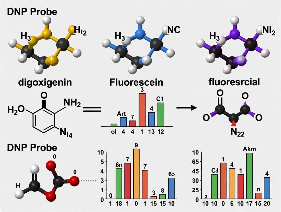
Digoxigenin (DIG) is a steroid hapten derived exclusively from plants of the Digitalis genus, most notably Digitalis purpurea (the purple foxglove) and Digitalis lanata [20]. This plant is also the source of the cardiac medication digitalis. Chemically, it is a cardenolide steroid. For probe synthesis, digoxigenin is typically incorporated into DNA via DIG-11-dUTP during random priming or into RNA as DIG-11-UTP via in vitro transcription [20]. The nucleotide is linked to the steroid hapten via a spacer arm.
Fluorescein, in contrast, is a fully synthetic organic molecule belonging to the xanthene dye family. Its core structure is based on a heterocyclic series of rings. The active form used for labeling is often fluorescein isothiocyanate (FITC), which reacts with aliphatic amine groups on proteins or nucleotides. The fluorescence mechanism of fluorescein-based probes can be explained by the Photoinduced Electron Transfer (PeT) mechanism [21]. In a typical "off-on" probe, the fluorescein fluorophore is connected to a recognition group. Upon binding to the target analyte, the PeT process is suppressed, restoring the fluorophore's intense green fluorescence.
Dinitrophenol (DNP) is another synthetic hapten, chemically known as 2,4-dinitrophenol. It is a small, aromatic compound characterized by its two nitro groups. In molecular biology, it is commonly conjugated to phosphoethanolamine for incorporation into lipids, such as DNP-PE, which can be reconstituted into lipid bilayers for biophysical studies [22].
Table 1: Comparative Overview of Probe Label Origins and Properties
| Characteristic | Digoxigenin (DIG) | Fluorescein | Dinitrophenol (DNP) |
|---|---|---|---|
| Origin | Natural (plant steroid) | Synthetic (xanthene dye) | Synthetic (aromatic compound) |
| Chemical Nature | Cardenolide Steroid | Xanthene Dye | Phenol Derivative |
| Primary Labeling Molecule | DIG-11-dUTP / DIG-11-UTP | Fluorescein Isothiocyanate (FITC) | DNP-PE (for lipids) |
| Detection Method | Colorimetric / Chemiluminescence | Fluorescence | Fluorescence / Other |
| Key Feature | High specificity with anti-DIG antibodies | Direct fluorescence; PeT-based sensing | Small size; well-characterized |
Detection Mechanisms and Experimental Protocols
The utility of each hapten is realized through its specific and high-affinity interaction with a detection molecule, which is then visualized through enzymatic or direct fluorescence methods.
Digoxigenin Detection Workflow
The DIG system relies on an antibody-based detection assay, which provides high specificity and low background [20]. A standard protocol for detecting DIG-labeled nucleic acid probes on a membrane is as follows:
- Hybridization: The DIG-labeled probe is hybridized to its target nucleic acid on a nitrocellulose or nylon membrane under standard conditions.
- Post-Hybridization Washes: The membrane is subjected to a series of stringency washes to remove any non-specifically bound probe.
- Immunological Detection: The membrane is incubated with an anti-DIG antibody conjugated to an enzyme, most commonly alkaline phosphatase (AP) [20].
- Signal Generation:
- Colorimetric Detection: The membrane is incubated with a substrate such as NBT/BCIP. AP dephosphorylates the substrate, resulting in an insoluble, colored precipitate at the probe-target site.
- Chemiluminescent Detection: For higher sensitivity, the membrane is incubated with a chemiluminescent substrate like CDP-Star or CSPD. AP cleavage of the substrate emits light, which can be captured on X-ray film or a digital imaging system.
Fluorescein and PeT-Based Sensing
Fluorescein enables both direct detection and sophisticated sensing strategies. The Photoinduced Electron Transfer (PeT) mechanism is a key design principle for creating "off-on" fluorescent probes [21]. A generalized experimental approach involves:
- Probe Design: A fluorescein derivative is connected to a recognition/activating group via a non-conjugated linker. In the absence of the target, PeT from the recognition group to the excited fluorophore (a-PeT) or from the fluorophore to the recognition group (d-PeT) quenches fluorescence.
- Sample Incubation: The probe is introduced to the sample (e.g., fixed cells, tissue sections, or protein solutions).
- Target Binding: Upon binding the target analyte, the HOMO-LUMO energy levels of the recognition group are altered, suppressing the PeT process.
- Fluorescence Imaging: The restoration of fluorescence is monitored using a standard fluorescence microscope or plate reader. The significant fluorescence enhancement provides a high signal-to-noise ratio for sensitive detection [21].
Dinitrophenol in Biophysical Assays
DNP is widely used as a hapten in immunology and model membrane studies. A classic experimental protocol using DNP-lipids is Contact Area Fluorescence Recovery After Photobleaching (FRAP) to study binding kinetics [22]:
- Bilayer Preparation: A supported planar lipid bilayer is created, incorporating a fraction of DNP-conjugated lipids (e.g., DNP-PE).
- Cell Binding: Cells expressing receptors for DNP (e.g., Fc receptors with anti-DNP antibodies) are brought into contact with the bilayer, forming an "immunological synapse."
- Ligand Accumulation: DNP-lipids bind to their receptors, leading to their accumulation in the contact area, visible as a region of higher fluorescence intensity.
- Photobleaching and Recovery: The fluorescence in the entire contact area is photobleached. The recovery of fluorescence over time, as unbleached DNP-lipids diffuse into the area and exchange with bound ones, is measured.
- Kinetic Analysis: The fluorescence recovery curve is fitted with a mathematical model to extract kinetic parameters, such as the two-dimensional (2D) off-rates and diffusion coefficients of the receptor-ligand interaction [22].
Diagram 1: Core detection workflows for fluorescein, digoxigenin, and DNP.
Comparative Experimental Data and Key Characteristics
Direct comparative studies between these three haptens in a single publication are rare. However, an analysis of their inherent properties and performance in established methodologies reveals a clear landscape of strengths and trade-offs.
Table 2: Comparative Experimental Characteristics and Performance Data
| Characteristic | Digoxigenin (DIG) | Fluorescein | Dinitrophenol (DNP) |
|---|---|---|---|
| Detection Specificity | Very High (Immunoassay) | High (Direct or PeT) | High (Immunoassay) |
| Signal-to-Noise Ratio | High (Low background) [20] | High (with PeT design) [21] | Context-dependent |
| Sensitivity | High (Chemiluminescence) | High (Fluorescence amplification) | Moderate |
| Multiplexing Potential | Lower (Indirect detection) | Excellent (with other fluorophores) | Possible with other haptens |
| Key Experimental Applications | In situ hybridization, Northern/Southern blotting [23] [20] | Intracellular imaging, live-cell sensing, PeT-based probes [21] | Model membrane studies, FRAP, immunological synapse research [22] |
| Quantitative Strength | End-point quantification | Real-time kinetic measurement (e.g., binding) | Real-time 2D kinetic parameter extraction (e.g., k_off, diffusion) [22] |
A critical performance metric in many detection systems is the signal-to-noise ratio. Both DIG and well-designed fluorescein probes excel in this area, albeit through different mechanisms. The DIG/anti-DIG system benefits from the high specificity of the antibody-antigen interaction and the fact that endogenous DIG is not found in animal tissues, leading to exceptionally low background [20]. For fluorescein, PeT-based probes are specifically engineered for a low fluorescence background, with a significant fluorescence enhancement (often over 100-fold) upon target binding, creating a very high signal-to-noise ratio during imaging [21].
For researchers requiring kinetic data, the choice of label dictates the possible approaches. Fluorescein is ideal for real-time monitoring of processes in live cells. DNP, when used in model systems like the supported bilayer contact area FRAP assay, provides a unique capability to resolve two-dimensional binding kinetics, such as on-rates and off-rates, directly in the synaptic interface [22]. DIG detection, being an indirect enzymatic method, is generally less suited for real-time kinetics and is more commonly used for end-point quantification.
The Scientist's Toolkit: Essential Research Reagents
Successful experimentation with these haptens requires a suite of specific reagents and materials. The following toolkit outlines the essential components for working with each system.
Table 3: Key Research Reagent Solutions and Their Functions
| Reagent / Material | Function in Experimentation |
|---|---|
| DIG-11-dUTP / DIG-11-UTP | Nucleotides for enzymatic incorporation of DIG into DNA (random priming) or RNA ( in vitro transcription) probes [20]. |
| Anti-DIG-AP Conjugate | Alkaline phosphatase-conjugated antibody for specific detection of DIG-labeled probes in colorimetric or chemiluminescent assays [20]. |
| NBT/BCIP | Chromogenic substrate for alkaline phosphatase; forms a purple precipitate for colorimetric detection [20]. |
| CDP-Star / CSPD | Chemiluminescent substrate for alkaline phosphatase; emits light upon cleavage for high-sensitivity detection on film or imagers. |
| FITC (Fluorescein Isothiocyanate) | Reactive derivative of fluorescein for covalently labeling proteins, peptides, and amines. |
| PeT-Based Fluorescein Probes | Specialized "off-on" probes that exploit Photoinduced Electron Transfer for sensing specific ions or biomolecules with low background [21]. |
| DNP-PE (DNP-phosphatidylethanolamine) | DNP-conjugated lipid for incorporating the hapten into liposomes or supported planar bilayers for biophysical binding studies [22]. |
| Supported Planar Lipid Bilayer | A synthetic membrane system on a solid support, used to study cell-surface interactions and 2D kinetics with DNP-lipids [22]. |
| Anti-DNP Antibody | Antibody for detecting and cross-linking DNP-conjugated molecules. |
The choice between digoxigenin, fluorescein, and dinitrophenol is not a matter of one being universally superior, but rather of selecting the right tool for the specific scientific question.
- Digoxigenin is the label of choice for high-sensitivity, low-background detection of nucleic acids in fixed-endpoint applications like in situ hybridization and blotting, where its immunoassay-based detection provides robust and reliable results.
- Fluorescein excels in dynamic, real-time imaging applications, particularly within living cells. The development of PeT-based probes has further enhanced its utility by enabling the detection of specific microenvironments and analytes with exceptional clarity.
- Dinitrophenol finds its niche in reductionist biophysical studies, such as the quantification of two-dimensional binding kinetics and diffusion in model membrane systems like the immunological synapse.
Understanding the fundamental properties, detection mechanisms, and experimental data associated with each of these haptens allows researchers to strategically design their experiments to achieve optimal sensitivity, specificity, and quantitative rigor.
Probe Labeling in Action: Synthesis Protocols and Key Applications
The selection of an appropriate enzymatic labeling method is a critical step in the construction of high-quality nucleic acid probes for research and diagnostic applications. These probes, which can be labeled with haptens such as digoxigenin (DIG), fluorescein, or 2,4-dinitrophenyl (DNP), are essential tools for detecting specific genetic sequences. Within the broader context of probe label research, each labeling method imparts distinct characteristics to the final probe, influencing its sensitivity, specificity, and suitability for various experimental conditions. This guide provides an objective comparison of three foundational enzymatic techniques—nick translation, random priming, and in vitro transcription—based on their procedural workflows, performance outcomes, and optimal applications.
Technical Comparison of Labeling Methods
The table below summarizes the core characteristics, advantages, and limitations of each enzymatic labeling method.
| Method | Key Principle | Best For | Key Advantages | Key Limitations |
|---|---|---|---|---|
| Nick Translation [24] [25] | DNase I creates nicks; DNA polymerase I simultaneously excises nucleotides from the 5' end and replaces them with labeled dNTPs from the 3' end. [24] | Generating a large amount of probe from a small amount of template. [24] | Rapid protocol (under 1 hour). [25] Produces uniformly labeled, highly sensitive probes. [24] | Probe size is variable and dependent on DNase I concentration, typically 400-800 bases. [24] |
| Random Priming [25] | A mixture of random-sequence oligonucleotides anneals to denatured DNA, acting as primers for DNA polymerase (e.g., Klenow fragment), which synthesizes new strands incorporating labeled nucleotides. [25] | Applications requiring a probe of consistent, shorter length. | Efficiently generates probes with high specific activity. [25] Consistent probe size. Less template DNA required compared to nick translation. [25] | The process requires single-stranded (denatured) DNA template. |
| In Vitro Transcription [25] | DNA template is cloned downstream of a bacteriophage RNA polymerase promoter (e.g., SP6, T7). The polymerase transcribes the DNA in vitro, producing single-stranded RNA probes incorporating labeled nucleotides. [25] | Generating high-specific-activity RNA probes (riboprobes) for sensitive hybridization. | Yields single-stranded probes, which often provide improved hybridization efficiency and a lower background. [25] Can generate sense and antisense RNA from a single construct. [25] | RNA probes are labile and susceptible to RNase degradation, requiring careful handling. [25] |
Quantitative Performance Data
The following table compares key performance metrics for the three methods, which are critical for selecting the right approach for a specific application.
| Performance Metric | Nick Translation | Random Priming | In Vitro Transcription |
|---|---|---|---|
| Typical Probe Length | 400 - 800 nucleotides [24] | Consistent, shorter fragments | Uniform length, defined by template |
| Probe Type | Double-stranded DNA [24] | Double-stranded DNA [25] | Single-stranded RNA [25] |
| Labeling Efficiency | High; efficient replacement with labeled nucleotides [24] | High specific activity [25] | Very high specific activity [25] |
| Template Requirement | As little as 20 ng [24] | Lower template requirement than nick translation [25] | DNA template with promoter sequence |
| Protocol Duration | < 1 hour [25] | Moderate | Moderate, includes transcription time |
Detailed Experimental Protocols
Nick Translation Labeling
This protocol is adapted from standard molecular biology procedures for labeling DNA with haptens like digoxigenin- [24].
Reagents:
- DNA to be labeled (1 µg in a 50 µL reaction) [24]
- DNase I (at a carefully optimized concentration, e.g., ~20 pg/0.5 µg DNA) [24]
- E. coli DNA Polymerase I [24]
- Labeled dNTP (e.g., Digoxigenin-11-dUTP, Fluorescein-12-dUTP, or DNP-dUTP) [24]
- Unlabeled dNTPs (dATP, dCTP, dGTP; 10X concentration)
- 10X Reaction Buffer (typically containing Mg²⁺ and DTT)
Procedure:
- Nick Translation Reaction:
- Combine the following in a microcentrifuge tube on ice:
- DNA template (1 µg)
- 10X Reaction Buffer (5 µL)
- Unlabeled dNTP mixture (e.g., 20 µM each of dATP, dCTP, dGTP)
- Labeled dNTP (e.g., 2 µM Digoxigenin-11-dUTP)
- DNase I (diluted to an appropriate working concentration)
- DNA Polymerase I (10 U)
- Nuclease-free water to a final volume of 50 µL.
- Mix gently and centrifuge briefly.
- Incubate the reaction at 16°C for 60-90 minutes [24].
- Combine the following in a microcentrifuge tube on ice:
- Reaction Termination:
- Stop the reaction by heating to 65°C for 10 minutes or by adding EDTA to a final concentration of 20 mM to chelate Mg²⁺ ions [24].
- Purification of Labeled Probe:
- Separate the labeled DNA from unincorporated nucleotides using ethanol precipitation or size-exclusion chromatography (e.g., with a Sephadex G-50 column) [24].
- For ethanol precipitation, add 2.5 volumes of cold ethanol and 0.1 volumes of 3M sodium acetate (pH 5.2) to the reaction. Precipitate at -20°C, then pellet the DNA by centrifugation. The labeled probe will form a pellet while unincorporated nucleotides remain in solution.
- Resuspend the purified probe in a suitable buffer (e.g., TE buffer or hybridization buffer) and store at -20°C.
Random Priming Labeling
This protocol uses the Klenow fragment of DNA Polymerase I to synthesize labeled DNA from a denatured template [25].
Reagents:
- DNA template (denatured)
- Random Hexamer Primers [25]
- Klenow Fragment of DNA Polymerase I [25]
- Labeled dNTP (e.g., Digoxigenin-11-dUTP)
- Unlabeled dNTPs (dATP, dCTP, dGTP)
- 10X Reaction Buffer
Procedure:
- Template Denaturation:
- Dilute the DNA template (25-100 ng) in nuclease-free water.
- Heat the DNA to 95-100°C for 5 minutes to denature it into single strands, then immediately place on ice.
- Primer Annealing and Extension:
- Combine the denatured DNA with:
- 10X Reaction Buffer
- Random hexamer primers
- Unlabeled dNTP mixture
- Labeled dNTP
- Klenow Fragment
- Mix gently and incubate at 37°C for 60 minutes.
- Combine the denatured DNA with:
- Reaction Termination and Purification:
- Stop the reaction by heating to 75°C for 10 minutes to inactivate the enzyme.
- Purify the labeled probe from unincorporated nucleotides using ethanol precipitation or a spin column, as described in the Nick Translation protocol.
In VitroTranscription for RNA Probe Synthesis
This method generates high-specific-activity single-stranded RNA probes (riboprobes) from a linearized DNA template [25].
Reagents:
- Linearized plasmid DNA (1 µg) containing the insert downstream of a bacteriophage RNA polymerase promoter (e.g., T7, SP6, or T3) [25]
- Appropriate RNA Polymerase (T7, SP6, or T3) [25]
- 10X Transcription Buffer
- Labeled NTP (e.g., Digoxigenin-11-UTP, Fluorescein-UTP)
- Unlabeled NTPs (ATP, CTP, GTP)
- RNase Inhibitor
- Nuclease-free water
Procedure:
- Transcription Reaction:
- Combine the following at room temperature (to prevent precipitation of SDS in some buffers):
- Linearized DNA template (1 µg)
- 10X Transcription Buffer (2 µL)
- Unlabeled NTP mixture (e.g., 10 mM each of ATP, CTP, GTP)
- Labeled NTP (e.g., 3.5 mM Digoxigenin-11-UTP)
- RNase Inhibitor (20 U)
- RNA Polymerase (20 U)
- Nuclease-free water to a final volume of 20 µL.
- Mix gently and centrifuge briefly.
- Incubate at 37°C for 2 hours.
- Combine the following at room temperature (to prevent precipitation of SDS in some buffers):
- DNA Template Removal:
- Add RNase-free DNase I (1 U) to the reaction and incubate at 37°C for 15 minutes to degrade the DNA template.
- Probe Purification:
- Purify the labeled RNA probe using ethanol precipitation (with ammonium acetate) or a dedicated RNA purification kit to remove unincorporated nucleotides and enzymes. Resuspend in RNase-free buffer containing EDTA and store at -80°C.
Workflow and Pathway Diagrams
Diagram 1: Comparative workflows for nick translation, random priming, and in vitro transcription labeling methods.
Diagram 2: Generalized detection pathway for hapten-labeled probes. The probe is detected via an enzyme-conjugated antibody specific to the hapten (e.g., anti-DIG), which then catalyzes a reaction to produce a measurable signal. [26]
Research Reagent Solutions
The following table details key reagents and their functions essential for performing the enzymatic labeling methods discussed in this guide.
| Reagent / Kit | Function in Labeling | Key Considerations |
|---|---|---|
| DNase I | Creates single-strand nicks in double-stranded DNA to initiate the Nick Translation reaction. [24] | Concentration must be carefully optimized to control final probe length (e.g., ~20 pg/0.5 µg DNA). [24] |
| DNA Polymerase I | Catalyzes Nick Translation; its 5'→3' polymerase activity adds nucleotides while its 5'→3' exonuclease activity removes them ahead of the nick. [24] | The Klenow fragment, which lacks exonuclease activity, is used in Random Priming. [25] |
| Klenow Fragment | Used in Random Priming; extends annealed primers to synthesize new DNA strands, incorporating labeled nucleotides. [25] | Lacks 5'→3' exonuclease activity, making it suitable for primer extension. [25] |
| Bacteriophage RNA Polymerase (T7, SP6) | Highly specific enzyme for in vitro transcription that initiates RNA synthesis from its corresponding promoter sequence. [25] | Requires a specific promoter sequence in the DNA template. |
| Labeled Nucleotides (DIG-/Fluorescein-/DNP-dUTP/dNTP) | Modified nucleotides that serve as the basis for incorporating the hapten label into the newly synthesized probe. [24] [25] [26] | The choice of hapten (DIG, Fluorescein, DNP) determines the required detection antibody and substrate. [26] |
| Random Hexamer Primers | A mixture of short oligonucleotides of random sequence that anneal to denatured DNA at multiple sites to serve as primers for DNA synthesis in Random Priming. [25] | Provides multiple initiation points for synthesis, ensuring efficient labeling. |
| Spin Columns / Size-Exclusion Resins | For rapid purification of the labeled probe from unincorporated nucleotides and reaction components after the labeling reaction is complete. [24] | Critical for reducing background signal in downstream hybridizations. |
| Anti-Hapten Antibody Conjugates | Enzyme-conjugated antibodies (e.g., anti-DIG-AP, anti-Fluorescein-HRP) that bind specifically to the hapten on the probe for subsequent detection. [26] | The conjugate enzyme (Alkaline Phosphatase or Horseradish Peroxidase) determines the suitable substrate. |
A Guide to Hapten-Labeled Nucleotides for Nucleic Acid Detection
The selection of an appropriate hapten-labeled nucleotide is a critical step in constructing sensitive and specific nucleic acid probes for applications ranging from in situ hybridization to diagnostic assay development. This guide provides a comparative analysis of three common haptens—Digoxigenin (DIG), Fluorescein, and Dinitrophenol (DNP)—based on current labeling methodologies, performance characteristics, and experimental data.
Hapten Properties and Incorporation Methods
The effectiveness of a labeled probe is fundamentally linked to the properties of the hapten and the efficiency with which it is incorporated into nucleic acids.
Table 1: Core Properties and Incorporation Methods for Hapten-Labeled Nucleotides
| Feature | Digoxigenin (DIG)-dUTP | Fluorescein-dUTP | Dinitrophenol (DNP)-11-UTP |
|---|---|---|---|
| Source & Specificity | Plant-derived steroid; high specificity with low background [27] | Synthetic organic dye; potential for higher non-specific binding | Synthetic hapten; specific antibody available |
| Primary Detection Method | Anti-DIG antibody conjugated to AP, HRP, or a fluorophore [27] | Anti-fluorescein antibody or direct fluorescence [15] | Anti-DNP antibody conjugated to AP, HRP, or a fluorophore [15] |
| Key Incorporation Enzymes | Taq polymerase (PCR), Klenow enzyme (random priming), TdT (end-labeling) [28] [27] | Similar to DIG; compatible with various polymerases | T7, SP6, T3 RNA polymerases (in vitro transcription) [15] |
| Recommended dNTP Ratio | 35% DIG-dUTP / 65% dTTP for PCR and nick translation [28] | Varies; optimization required | Information missing from search results; optimization required |
| Probe Stability | ≥1 year at -20°C to -70°C [27] | Information missing from search results | Information missing from search results |
Experimental Performance and Comparative Data
Objective performance data is essential for selecting the right hapten for a given application. The following section compares key operational characteristics.
Table 2: Experimental Performance and Detection Sensitivity
| Performance Metric | Digoxigenin (DIG) | Fluorescein | Dinitrophenol (DNP) |
|---|---|---|---|
| Detection Sensitivity | Can detect 0.10 – 0.03 pg of target DNA in random primed labeling [27] | Used in high-sensitivity assays (e.g., tear film break-up time) [29] | Enables detection of three genes simultaneously, though less robust than dual labels [15] |
| Signal Amplification | Compatible with tyramide signal amplification (TSA) [15] | Compatible with tyramide signal amplification (TSA) [15] | Compatible with tyramide signal amplification (TSA) [15] |
| Multiplexing Capability | Excellent for dual labeling with Fluorescein [15] | Excellent for dual labeling with DIG [15] | Suitable for third label in triple-plex experiments [15] |
| Key Applications | Southern/Northern blotting, in situ hybridization, library screening [27] | Clinical diagnostics (e.g., DED), fluorescence correlation spectroscopy [29] [30] | Specialized in situ hybridization, expanding multiplexing capacity [15] |
Detailed Experimental Protocols
A. DIG DNA Labeling by PCR
PCR is the preferred method for preparing DIG-labeled probes when template is limited. The reaction incorporates Digoxigenin-11-dUTP during amplification [27].
- Template: Use cloned inserts for best results. Genomic DNA can be more challenging.
- Primers: Standard, sequence-specific primers.
- Reaction Mix: Includes DIG-dUTP in a recommended ratio of 35% DIG-dUTP to 65% dTTP for optimal incorporation and product yield [28].
- Enzyme: A high-fidelity enzyme blend is often used for robustness and efficiency.
- Procedure:
- Optimize PCR conditions with unlabeled dNTPs first.
- Perform the PCR reaction with the DIG-dUTP/dTTP mixture.
- Verify incorporation and probe size by gel electrophoresis.
B. Transcriptional Labeling of RNA Probes with DNP-11-UTP
DNP-labeled RNA probes are generated by in vitro transcription, a method known for producing highly sensitive probes.
- Template Preparation: Clone the DNA fragment of interest into a transcription vector (e.g., containing T7, SP6, or T3 promoters). Linearize the plasmid downstream of the insert and purify thoroughly.
- Critical Consideration: The protocol for DNP is analogous to that for DIG-labeled RNA, which incorporates a hapten-UTP approximately every 25-30 nucleotides [27].
- In Vitro Transcription:
- Incubate the linearized template with the appropriate RNA polymerase, RNase inhibitors, nucleotides (ATP, CTP, GTP), and DNP-11-UTP.
- Purify the transcribed RNA probe.
- RNase Control: Use disposable plasticware, DEPC-treated water, and wear gloves to prevent RNase contamination.
- Yield: From 1 μg of template, approximately 10-20 μg of full-length DIG-labeled RNA can be generated; a similar yield is expected for DNP [27].
C. Dual-Color FluorescenceIn SituHybridization (FISH)
A standard protocol for detecting two genes simultaneously using a semi-automated, high-throughput platform [15].
- Probe Labeling: Label one probe with DIG and a second probe with Fluorescein (FITC).
- Hybridization: Apply both probes to the sample and co-hybridize.
- Signal Amplification & Detection:
- Use an anti-DIG antibody conjugated to Horseradish Peroxidase (HRP).
- Develop the signal with a Cy3-tyramide substrate.
- Use an anti-fluorescein antibody conjugated to HRP.
- Develop the signal with a FITC-tyramide substrate.
- Visualization: Analyze with fluorescence microscopy using appropriate filter sets for Cy3 and FITC.
Research Reagent Solutions
Table 3: Essential Reagents for Probe Construction and Detection
| Reagent Name | Function in Experiment | Key Characteristics |
|---|---|---|
| Digoxigenin-11-dUTP | Incorporated into DNA probes via enzymatic methods [28] [27] | Alkali-stable; attached via 11-atom linker for efficient incorporation [28] |
| Fluorescein-dUTP | Incorporated into DNA probes for direct fluorescence or antibody-based detection | Enables multiplexing and is compatible with standard fluorescence detection systems |
| DNP-11-UTP | Incorporated into RNA probes via in vitro transcription for high-sensitivity detection [15] | Allows for expansion of multiplexing capabilities beyond standard dual labels [15] |
| Anti-DIG-AP/HRP | Binds to DIG hapten for colorimetric, chemiluminescent, or fluorescent detection [27] | High specificity and affinity; conjugated to reporter enzymes (AP or HRP) [27] |
| Tyramide Signal Amplification (TSA) Reagents | Amplifies weak signals for low-abundance target detection [15] | Enzyme-activated tyramide precipitates at target site, greatly enhancing sensitivity [15] |
| T7/SP6/T3 RNA Polymerase | Synthesizes RNA probes from a linearized DNA template [27] | High-yield transcription; promoter-specific for directional probe synthesis [27] |
| Klenow Fragment | Synthesizes DNA probes via random primed labeling [27] | Lacks 5'→3' exonuclease activity; produces homogeneously labeled probes [27] |
Detection systems are fundamental to biomedical research and diagnostic assay development, enabling the visualization and quantification of biological molecules. Non-radioactive labeling and detection methods have largely superseded radioactive techniques due to their safety, stability, and versatility. Within this landscape, three hapten-based reporter systems—digoxigenin, fluorescein, and dinitrophenyl—offer distinct advantages for specific experimental applications. These systems facilitate the detection of nucleic acids, proteins, and other biomolecules in techniques such as in situ hybridization, immunofluorescence, and immunohistochemistry.
This guide provides an objective comparison of digoxigenin, fluorescein, and DNP probe labels, framing their performance within the context of modern detection methodologies that leverage antibody conjugates, streptavidin-biotin complexes, and enzyme substrates. We present experimental data to illustrate the relative strengths and limitations of each system, enabling researchers to make informed decisions based on their specific experimental requirements for sensitivity, specificity, and multiplexing capability.
Comparative Analysis of Hapten Labels
The selection of an appropriate hapten label depends on multiple factors, including the required sensitivity, the endogenous background of the sample, and the detection methodology. The table below provides a systematic comparison of three commonly used hapten labels.
Table 1: Characteristics of Common Hapten-Based Labels
| Feature | Digoxigenin | Fluorescein | Dinitrophenyl (DNP) |
|---|---|---|---|
| Chemical Nature | Steroid derived from the foxglove plant [31] [32] | Synthetic organic fluorophore | Synthetic aromatic compound with nitro groups [33] |
| Primary Detection Method | Anti-digoxigenin antibody conjugated to enzymes or fluorophores [34] [32] | Direct fluorescence or anti-fluorescein antibody | Anti-DNP antibody conjugated to enzymes or fluorophores [33] |
| Best-Performing Application | Non-radioactive in situ hybridization and immunohistochemistry [31] [34] | Direct fluorescent detection and multiplexing | Non-radioactive in situ hybridization with oligonucleotide probes [33] |
| Key Advantage | Very low endogenous background in mammalian tissues [31] | Direct detection without secondary reagents is possible | Strong signals in solid-phase oligonucleotide labeling [33] |
| Limitation | Requires a two-step detection system | Photobleaching can occur; some autofluorescence | Less commonly used than other systems |
Quantitative data from a seminal study directly comparing these labels in in situ hybridization (ISH) provides critical performance insights. Researchers synthesized oligonucleotides complementary to histone mRNA and labeled them with digoxigenin, DNP, or alkaline phosphatase, then used them for ISH detection in human tonsil tissue [33].
Table 2: Experimental Performance in In Situ Hybridization [33]
| Label Type | Labeling Method | Relative Signal Strength | Development Time |
|---|---|---|---|
| DNP | 3' and 5' solid-phase labeling with triple DNP groups | Strongest | Shortest |
| Alkaline Phosphatase | Direct conjugation to oligonucleotide | High (with spacer) | Short |
| Digoxigenin | Enzymatic 3' end-labeling | Moderate (improved with spacer) | Moderate to Long |
The study concluded that 3' and 5' solid-phase labeling of oligonucleotides with triple DNP groups produced the strongest signal, as determined by the highest cell signal intensity and shortest development time [33]. This demonstrates that the DNP system can offer superior performance for certain applications, particularly ISH with synthetic oligonucleotides.
The Streptavidin-Biotin System and Alternative Hapten Approaches
While not a hapten label itself, the streptavidin-biotin system is a cornerstone of modern detection and warrants discussion as a comparator. The interaction between biotin and streptavidin is one of the strongest non-covalent bonds in nature (K_d ~ 10⁻¹⁵ M), making it a powerful tool for detection and immobilization [31] [32]. Streptavidin, derived from Streptomyces avidinii, is preferred over avidin for many applications due to its near-neutral isoelectric point, which minimizes nonspecific electrostatic binding to tissues, and its lack of carbohydrate groups, which reduces non-specific affinity with tissue lectins [31].
However, this system has a critical vulnerability: endogenous biotin interference. High levels of supplemental biotin ingested by patients can bind to streptavidin reagents in immunoassays, causing falsely elevated or suppressed test results, a significant concern in clinical diagnostics [35]. Furthermore, endogenous biotinylated proteins in mitochondria can cause background staining in some cells [32].
Hapten-based systems like digoxigenin and DNP were developed, in part, to circumvent these issues. Since these molecules are not naturally present in biological systems, they offer exceptionally low background, leading to a clearer signal-to-noise ratio [31] [33]. The high-affinity antibodies developed against these haptens make them robust alternatives to the biotin-streptavidin system, particularly for applications like ISH and IHC where low background is paramount.
Experimental Protocols for Label Detection
Indirect Detection Method with Enzyme Conjugates
This is a generalized protocol for detecting hapten-labeled probes (e.g., digoxigenin or DNP) using an enzyme-conjugated antibody and a colorimetric substrate. The workflow is illustrated in the diagram below.
Diagram 1: Indirect detection with enzyme conjugates.
Detailed Protocol:
- Hybridization/Incubation: Apply the hapten-labeled nucleic acid probe or primary antibody to the sample (e.g., tissue section, cell smear, or membrane) under appropriate conditions for binding [34].
- Blocking: Incubate the sample with a blocking buffer (e.g., containing BSA, serum, or commercial blocking agents) to minimize non-specific binding of subsequent reagents [32].
- Primary Antibody Incubation: Apply a hapten-specific antibody (e.g., anti-digoxigenin or anti-DNP). This antibody can be directly conjugated to an enzyme like Horseradish Peroxidase (HRP) or Alkaline Phosphatase (AP) for a two-step protocol [33].
- Washes: Perform stringent washes with an appropriate buffer to remove any unbound antibody.
- Secondary Detection (If applicable): For increased sensitivity, a secondary detection step can be used. If the primary anti-hapten antibody is not enzyme-conjugated, apply an enzyme-conjugated secondary antibody (e.g., anti-mouse IgG-HRP) [32]. Alternatively, for signal amplification, use an unlabeled streptavidin bridge followed by a biotinylated enzyme [31].
- Washes: Perform another series of washes to remove unbound detection reagents.
- Substrate Addition: Add a chromogenic enzyme substrate. For AP, this is typically NBT (Nitro Blue Tetrazolium) and BCIP (5-Bromo-4-Chloro-3-indolyl Phosphate), which produces a blue-purple precipitate. For HRP, DAB (3,3'-Diaminobenzidine) is common, producing a brown precipitate [32] [33].
- Signal Detection: Observe and record the signal under a microscope.
Bridging Amplification Method
The bridging method, a form of the LSAB (Labeled Streptavidin-Biotin) method, uses the multi-valency of streptavidin to significantly amplify the detection signal [31] [32]. The process is as follows.
Diagram 2: Signal amplification via bridging.
Detailed Protocol:
- Biotinylated Primary Antibody: A primary antibody that is conjugated to biotin is applied and binds to the target.
- Unlabeled Streptavidin: Streptavidin is applied. Its tetravalent nature allows it to act as a bridge, binding to the biotin on the primary antibody while leaving other binding sites free.
- Biotinylated Enzyme: A biotinylated enzyme (e.g., HRP or AP) is applied, which binds to the remaining free sites on the streptavidin. This step concentrates multiple enzyme molecules at the target site.
- Substrate Addition: The appropriate substrate is added to generate a detectable signal. This method is reported to be 5–10 times more sensitive than the standard ABC method and results in a clearer background [31].
The Scientist's Toolkit: Essential Research Reagent Solutions
Successful implementation of these detection systems requires a suite of reliable reagents. The table below lists key materials and their functions.
Table 3: Essential Reagents for Detection Systems
| Reagent / Material | Function / Application Notes |
|---|---|
| Hapten-Labeling Kits | Kits for enzymatically (e.g., nick translation) or chemically incorporating digoxigenin, DNP, or fluorescein into DNA/RNA probes [34] [33]. |
| Anti-Hapten Antibodies | Highly specific monoclonal or polyclonal antibodies against digoxigenin, fluorescein, or DNP; often conjugated to enzymes or fluorophores [32] [33]. |
| Chromogenic Substrates | NBT/BCIP (for AP) and DAB (for HRP) are standard for colorimetric detection in ISH and IHC [32] [33]. |
| Tyramide Signal Amplification (TSA) Kits | Used for extreme signal amplification. Utilizes HRP-conjugated streptavidin or antibodies to catalyze the deposition of fluorescent or biotinylated tyramide at the target site [32]. |
| Streptavidin Conjugates | Streptavidin conjugated to HRP, AP, or fluorophores like Alexa Fluor dyes for use in biotin-based detection or bridging protocols [31] [32]. |
| Blocking Reagents | Solutions containing BSA, serum, or specialized commercial formulations to reduce non-specific binding and lower background noise. |
| Mounting Media | Aqueous or permanent mounting media, often with counterstains like DAPI, for preserving and visualizing samples under a microscope. |
The choice between digoxigenin, fluorescein, DNP, and the ubiquitous streptavidin-biotin system is not a matter of one being universally superior. Instead, it requires a careful analysis of experimental goals. The streptavidin-biotin system offers unparalleled affinity but is susceptible to interference from endogenous biotin. The DNP system can provide exceptional signal strength and speed in applications like ISH with oligonucleotides. Digoxigenin remains a gold standard for low-background ISH and IHC due to its absence from mammalian tissues. Fluorescein is indispensable for direct labeling and multicolor fluorescence experiments, though users must manage its susceptibility to photobleaching.
Understanding the principles, performance data, and protocols outlined in this guide empowers researchers to select the optimal detection system, thereby ensuring the highest levels of sensitivity, specificity, and reliability in their scientific investigations.
Fluorescence in situ hybridization (FISH) has evolved from a technique for detecting single genetic targets to a powerful tool for visualizing the spatial relationships of numerous gene products within their native cellular environment. This capability, known as multiplexing, allows researchers to simultaneously detect multiple DNA, RNA, or even protein targets in a single sample, preserving critical architectural context that is lost in bulk analysis methods. The core of this advancement lies in sophisticated probe design, signal amplification strategies, and precise decoding algorithms that distinguish individual targets from a complex fluorescence signal mixture.
The drive for higher multiplexing is fundamentally linked to the quest for greater biological understanding. In the unprecedented era of single-cell sequencing and spatial multiomics, FISH technologies with higher sensitivity and robustness are greatly desired to pinpoint cell subtypes and spatial distribution relationships at the single-cell or single-molecule levels [36]. The ability to resolve multiple gene products spatially is revolutionizing our comprehension of cellular heterogeneity, gene regulatory networks, and disease mechanisms, particularly in complex tissues like the brain and in tumor microenvironments.
Core Principles of Signal Resolution in Multiplexed FISH
The challenge in multiplexed FISH is not merely attaching different colored fluorophores to different probes, but reliably distinguishing their signals after the physicochemical stresses of hybridization and washing. Successful multiplexing requires a system that is both highly efficient and exhibits low background noise.
Probe Design for Enhanced Specificity and Efficiency
Innovative probe designs are crucial for overcoming the limitations of traditional FISH. The π-FISH rainbow method, for example, employs primary π-FISH target probes containing 2–4 complementary base pairs in the middle region. This "π-shaped" bond increases stability during hybridization and washing, ultimately improving efficiency and specificity. Studies have demonstrated that this design yields more signal spots compared to traditional split probes without complementary bases [36]. Following hybridization, secondary U-shaped and tertiary U-shaped amplification probes are used to amplify the signal before final visualization with a fluorescence signal probe.
Combinatorial Labeling and Spectral Discrimination
A fundamental strategy for expanding the multiplexing capacity beyond the number of available fluorophores is combinatorial labeling. In this approach, each target is assigned a unique binary code based on the presence or absence of different fluorophores. For instance, using four different fluorescence signal probes can generate 15 unique combinations (C14 + C24 + C34 + C44), theoretically enabling the differentiation of 15 genes in a single round of hybridization [36]. The accuracy of this method is high, with overlapping ratios of two and three fluorescence signals during in situ detection exceeding 99% in validated systems [36].
Comparative Analysis of Multiplexed FISH Technologies
Several advanced FISH methodologies have been developed to achieve high-order multiplexing. The table below summarizes the key features, performance metrics, and ideal use cases for several prominent technologies.
Table 1: Comparison of High-Plex FISH Technologies
| Technology | Core Principle | Multiplexing Capacity | Key Performance Metrics | Best Applications |
|---|---|---|---|---|
| π-FISH rainbow [36] | π-shaped probe design for stability; U-shaped signal amplification. | 15 targets in one round (with 4 colors). | - Sensitivity: Highest in comparison to HCR, smFISH.- Specificity: >99% signal overlap accuracy.- False-positive rate: <0.51%. | Detecting diverse biomolecules (DNA, RNA, protein) in frozen, paraffin, and whole-mount samples across species. |
| Spatial Genome Aligner [37] | Computational alignment of FISH signals to a Gaussian chain polymer model. | High (whole-genome chromatin tracing). | - Recapitulates Hi-C patterns at 5 kb, 25 kb, and 1 Mb scales.- Can predict chromosome ploidy de novo. | Analyzing 3D chromatin architecture and resolving sister chromatids in interphase. |
| USE-PCR [38] | Universal Signal Encoding PCR with amplitude modulation and multispectral encoding. | 32 unique targets. | - Target ID accuracy: 97.6% at low template copy.- Linear correlation: R² = 0.99 across 4 dPCR platforms. | High-throughput, multiplexed SNV detection in oncology and liquid biopsy. |
| Unified FISH Biodosimetry [39] [40] | Two-color FISH to quantify multiple cytogenetic markers from the same metaphase. | 4 markers (dic-F, BT, UT, acentric fragments). | - Dose estimate variation from true doses: 2-7%.- Streamlines process and reduces interexperimental variation. | Accurate radiation biodosimetry for recent and past exposures. |
Performance Benchmarking
When benchmarked against other sensitive methods, π-FISH rainbow demonstrates superior performance in detection efficiency. In a direct comparison using the same number of probes targeting ACTB mRNA in HeLa cells, π-FISH rainbow showed significantly higher signal spot counts per cell and fluorescence signal intensity than Hybridization Chain Reaction (HCR), smFISH, and smFISH-FL (which uses full-length transcript coverage) [36]. This high efficiency also holds for medium- and low-abundance transcripts, confirming its robustness [36].
For DNA targets, the Spatial Genome Aligner provides a computational framework for resolving chromosomal conformations from noisy multiplexed DNA FISH data. Its performance is validated by its ability to recapitulate patterns of chromatin organization found in Hi-C data across multiple genomic scales (5 kb, 25 kb, and 1 Mb) [37].
Experimental Protocols for Key Technologies
Protocol: π-FISH rainbow for RNA Detection
The following workflow details the application of π-FISH rainbow for multiplexed RNA detection [36].
- Sample Preparation: Fix cells or tissues according to standard protocols (e.g., 4% paraformaldehyde). Permeabilize cells to allow probe access (e.g., with 0.5% Triton X-100).
- Probe Hybridization:
- Design 10-15 π target probes per gene, each containing 2-4 complementary base pairs.
- Apply the probe mix to the sample and incubate overnight at 37°C in a humidified chamber to allow specific hybridization.
- Signal Amplification:
- Wash the sample to remove non-specifically bound probes.
- Apply secondary U-shaped amplification probes and incubate to bind the primary π probes.
- Wash again.
- Apply tertiary U-shaped amplification probes to further amplify the signal.
- Fluorescence Visualization:
- Apply fluorescence signal probes that hybridize to the amplification scaffold. Use a combinatorial mix of up to four fluorophores to encode different RNA targets.
- Perform final washes to reduce background.
- Image Acquisition and Decoding:
- Image the sample using an epifluorescence or confocal microscope with appropriate filter sets.
- Use software to decode the combinatorial fluorescence signals into the identity and location of individual RNA molecules.
Protocol: Unified FISH for Cytogenetic Biodosimetry
This protocol enables the simultaneous detection of multiple chromosomal aberration markers from the same metaphase spread [39] [40].
- Sample Collection and Culture: Collect whole blood in heparinized tubes and culture with RPMI 1640 medium supplemented with fetal calf serum, phytohemagglutinin (PHA), and antibiotics.
- Metaphase Arrest and Harvesting: At 24 hours, add colcemid to prevent a second cell division cycle. Harvest metaphases at 52 hours using a hypotonic solution and Carnoy's fixative (3:1 methanol:acetic acid).
- Slide Preparation: Prepare slides, treat with RNase, and store at -20°C.
- Two-Color FISH:
- Hybridize slides with FITC- and Texas Red-labeled probes for specific chromosome pairs (e.g., 1 and 2).
- Denature slides and probes, then hybridize overnight at 37°C.
- Post-hybridization, wash slides to remove excess probe and counterstain with DAPI.
- Image Acquisition and Analysis:
- Scan slides using an automated microscope system (e.g., Axio Imager Z2) and capture metaphase images at 63x magnification.
- Score chromosomal aberrations (dicentric chromosomes, balanced/unbalanced translocations, acentric fragments) according to IAEA guidelines.
- Use the Lucas formula to estimate whole-genome equivalent translocation frequencies.
Visualization of Workflows and Signaling Pathways
π-FISH Rainbow Probe Assembly Workflow
The following diagram illustrates the multi-step assembly process of the π-FISH rainbow signal amplification system.
Combinatorial Encoding Logic for Target Identification
This diagram outlines the logical process of assigning unique spectral codes to different biological targets for multiplexed identification.
The Scientist's Toolkit: Essential Research Reagents
Successful implementation of multiplexed FISH relies on a suite of specialized reagents and tools. The following table details the key components required for these experiments.
Table 2: Essential Reagents and Tools for Multiplexed FISH
| Item Name | Function/Description | Example Use Case |
|---|---|---|
| π-Target Probes [36] | Primary probes with 2-4 complementary base pairs for stable hybridization. | Core component of π-FISH rainbow for efficient target binding. |
| U-Shaped Amplification Probes [36] | Secondary and tertiary probes that bind sequentially to build a signal amplification scaffold. | Significantly boosting fluorescence signal in π-FISH and related methods. |
| Combinatorial Fluorescence Probes [36] | A mix of fluorophores (e.g., 4 colors) used to generate unique spectral barcodes. | Encoding and distinguishing up to 15 different targets in a single round. |
| Universal Probe Mixture (USE-PCR) [38] | A pre-optimized set of universal hydrolysis probes for amplitude and spectral multiplexing. | Enabling USE-PCR for portable, highly multiplexed dPCR assays on multiple platforms. |
| Chromosome Paint Probes [39] [40] | Fluorescently labeled probes that specifically bind entire chromosomes or regions. | Identifying chromosomal translocations and aberrations in biodosimetry. |
| Microscope with Automated Stage [39] [36] | An imaging system capable of multispectral acquisition and precise tile-scanning. | Essential for high-throughput, multi-channel imaging of large samples or many cells. |
| Digital PCR Platform [38] | Instrument for partitioning samples and quantifying nucleic acids (e.g., QIAcuity, QX600). | Running highly multiplexed USE-PCR assays for variant detection. |
The field of multiplexed multicolor FISH has moved far beyond simple two-color co-localization studies. Technologies like π-FISH rainbow, spatial genome alignment, and USE-PCR represent a paradigm shift towards highly multiplexed, quantitative, and spatially resolved genomic and transcriptomic analysis. The critical factors for success are the synergistic combination of robust biochemical probe design (such as π-shaped and U-shaped probes), powerful signal encoding strategies (combinatorial color coding, amplitude modulation), and sophisticated computational analysis.
As these technologies continue to mature, their integration with other omics modalities and their application to clinical diagnostics, such as detecting resistant marker ARV7 in circulating tumor cells [36], will further unravel the complex spatial architecture of molecular processes in health and disease. The ongoing development of universal probes and standardized analysis pipelines promises to make these powerful techniques more accessible and reproducible across research and clinical laboratories.
The selection of a non-radioactive reporter molecule—digoxigenin, fluorescein, or 2,4-dinitrophenyl (DNP)—is a foundational decision that influences the sensitivity, specificity, and multiplexing capability of molecular detection techniques. These core applications, including Fluorescence In Situ Hybridization (FISH), various forms of in situ hybridization, microarray analysis, and quantitative PCR (qPCR), form the backbone of modern genetic and cytogenetic analysis. The choice of hapten or fluorophore is not merely a technical detail but a strategic variable that can determine the success of an experiment. This guide provides an objective, data-driven comparison of these three key labeling systems, framing them within the broader thesis of optimizing detection workflows for research and diagnostic purposes. The evaluation is based on performance parameters such as detection sensitivity, signal-to-noise ratio, operational stability, and suitability for multiplexed assays, providing scientists and drug development professionals with the evidence needed to align their reagent choices with specific experimental goals.
Comparative Performance Data of Probe Labeling Systems
The quantitative and qualitative performance of digoxigenin, fluorescein, and DNP labels varies significantly across different applications. The following tables summarize key comparative data to inform experimental design.
Table 1: Overall Performance Profile of Labeling Systems
| Performance Parameter | Digoxigenin | Fluorescein | DNP |
|---|---|---|---|
| Primary Detection Method | Immunofluorescence / Colorimetry | Direct Fluorescence / Immunofluorescence | Immunofluorescence |
| Typical Signal Amplification | High (Multi-step) | Low (Direct) / High (Indirect) | Very High [33] |
| Inherent Background in Tissues | Low [33] | Moderate (Autofluorescence) | Low [33] |
| Suitability for Multiplexing | Excellent [41] | Excellent [41] | Good |
| Reported Shelf Life | >20 years [41] | Varies by conjugate | >20 years [41] |
Table 2: Experimental Performance in Key Applications
| Application & Metric | Digoxigenin | Fluorescein | DNP |
|---|---|---|---|
| FISH (Signal Robustness) | Strong, reliable signals [41] | Strong, but can fade [41] | Strong [41] |
| ISH (Signal Intensity) | High [33] | Moderate | Highest [33] |
| qPCR / Microarray | Compatible | Compatible | Compatible |
| Key Advantage | Well-established, low background | Direct detection possible | Superior signal intensity in ISH [33] |
A critical study that directly compared these haptens for in situ hybridization found that oligonucleotides labeled with triple DNP groups during solid-phase synthesis produced the strongest signal, characterized by the highest cell signal intensity and shortest development time in colorimetric detection [33]. This makes DNP a powerful label for applications requiring maximum sensitivity. Furthermore, long-term stability studies have demonstrated that hapten-labeled DNA probes, including those tagged with digoxigenin, fluorescein, and DNP, can be stored at -20°C in the dark and remain fully functional for decades, far exceeding typical manufacturer expiration dates [41].
Detailed Experimental Protocols and Data
Protocol: Single Molecule RNA FISH in Oocytes and Embryos
This optimized protocol for sensitive cell types illustrates the application of probe labeling for challenging, low-abundance targets [42].
- 1. Probe Design: Utilize proprietary probe sets (e.g., from Affymetrix or ACD) based on branched DNA (bDNA) technology. Each probe consists of 10-20 oligonucleotide pairs in a "double Z" configuration. One end (~40-50 bases) is complementary to the target mRNA, while the other end contains pre-amplifier binding sequences [42].
- 2. Sample Fixation and Permeabilization: Fix individual oocytes or embryos in 4% paraformaldehyde. Due to cell fragility, replace commercial permeabilization buffers with PBS-based buffers previously optimized for oocyte and embryo immunofluorescence to prevent lysis [42].
- 3. Hybridization: Hybridize fixed cells with the transcript-specific probe set in a proprietary hybridization buffer. Perform this and subsequent steps in Agtech 6-well plates to manage small, transparent samples. Using PBS-based buffers here can cause probe aggregation on the plasma membrane; proprietary buffers are essential for proper probe entry [42].
- 4. Signal Amplification: Perform sequential hybridization with pre-amplifier and amplifier DNA molecules in proprietary buffers. This assembly creates a branched DNA structure with ~400 binding sites for labeled oligonucleotides, yielding an ~8000-fold amplification of the signal for each target RNA molecule [42].
- 5. Detection and Imaging: Hybridize fluorophore-labeled oligonucleotides to the amplifier structure. Wash cells in PBS-based wash buffers and image using standard fluorescence microscopy. Quantify mRNA transcripts using localization software (e.g., Localize) [42].
Supporting Data: This method was validated by accurately quantifying the expression and localization of Gdf9, Pou5f1, and low-abundance Nanog mRNAs in mouse oocytes and embryos, with results consistent with published data. The technique provides absolute mRNA abundance and reveals subcellular localization changes, overcoming limitations of amplification-based methods like qPCR [42].
Protocol: Whole-Mount Fluorescent In Situ Hybridization (FISH) for Marine Species
This rapid protocol highlights the use of hapten-labeled RNA probes for developmental biology studies across diverse organisms [43].
- 1. Probe Synthesis: Synthesize antisense RNA probes by in vitro transcription from linearized DNA templates. Label probes with digoxigenin, fluorescein, or DNP during transcription using labeled nucleotides (e.g., from Roche). Alternatively, synthesize non-labeled RNA and label post-transcriptionally (e.g., using Mirus kits for DNP) [43].
- 2. Sample Collection and Fixation: Fix embryos or larvae in 4% Paraformaldehyde (PFA) in MOPS Buffer (0.1 M MOPS pH 7, 0.5 M NaCl) for 1 hour at room temperature or overnight at 4°C. This ensures mRNA integrity [43].
- 3. Pre-hybridization Processing: Wash specimens 3-5 times with MOPS buffer. Gradually dehydrate samples by passing through 50%, 60%, and 70% ice-cold ethanol. Store at -20°C in 70% ethanol until use. For use, gradually rehydrate in MOPS buffer [43].
- 4. Hybridization and Washes: Incubate rehydrated samples with the labeled antisense RNA probe in hybridization buffer overnight. Perform stringent washes to remove unbound probe [43].
- 5. Immunological Detection: For hapten-labeled probes (digoxigenin, DNP), incubate samples with fluorophore-conjugated antibodies (e.g., anti-digoxigenin, anti-DNP). For fluorescein-labeled probes, direct imaging is possible, or signal can be amplified with an anti-fluorescein antibody [43].
- 6. Imaging: Mount samples and image using a standard or confocal fluorescence microscope [43].
Supporting Data: This protocol has been successfully applied to visualize gene expression patterns in a wide range of marine embryos and larvae, including mollusks (Mytilus galloprovincialis), echinoderms (Strongylocentrotus purpuratus, Paracentrotus lividus), tunicates (Ciona robusta), and cephalochordates (Branchiostoma lanceolatum), demonstrating its broad applicability [43].
Research Reagent Solutions
The following table details key reagents essential for experiments utilizing digoxigenin, fluorescein, and DNP probe labels.
Table 3: Essential Reagents for Probe Labeling and Detection
| Reagent / Material | Function / Description | Application Examples |
|---|---|---|
| Branched DNA (bDNA) Probe Sets | Proprietary oligonucleotides that provide ~8000x signal amplification via sequential hybridization [42]. | Single-molecule RNA FISH [42]. |
| Hapten-labeled dUTP (Digoxigenin-, DNP-) | Modified nucleotides incorporated into DNA probes via enzymatic labeling (e.g., nick translation) for subsequent immuno-detection [41]. | Locus-specific FISH, chromosome painting [41]. |
| Fluorophore-labeled dUTP (SpectrumOrange/Aqua) | Nucleotides directly incorporating a fluorophore, enabling direct detection without antibodies [41]. | Commercial FISH probes for direct imaging [41]. |
| Anti-Hapten Antibodies (Conjugated) | Fluorophore- or enzyme-conjugated antibodies (e.g., anti-digoxigenin, anti-DNP) for detecting hapten-labeled probes [43]. | Immuno-detection in FISH and ISH [43]. |
| Proprietary Hybridization Buffers | Commercial buffers optimized for specific assay kits to ensure maximum probe binding and minimize non-specific aggregation [42]. | RNA-FISH on sensitive samples (oocytes/embryos) [42]. |
Detection Chemistries and Signaling Pathways
The fundamental difference between these labels lies in their detection methodology. Fluorescein is primarily a fluorophore, meaning it can be directly imaged upon excitation by light of a specific wavelength. In contrast, digoxigenin and DNP are haptens, small molecules that are detected indirectly via specific antibodies conjugated to a reporter enzyme or fluorophore. This indirect detection allows for significant signal amplification.
The choice between digoxigenin, fluorescein, and DNP labels is not a matter of identifying a single superior option, but rather of matching the probe's characteristics to the experimental needs. DNP offers exceptional signal intensity for maximum sensitivity in colorimetric ISH [33]. Digoxigenin remains a gold standard for low-background, highly robust FISH applications with proven long-term stability [41]. Fluorescein provides flexibility for both direct and indirect detection and is a cornerstone of multiplexed fluorescence panels [41]. The ongoing development of signal amplification technologies, such as branched DNA systems [42], and the proven long-term stability of properly stored hapten-labeled probes [41] further solidify the role of these labeling systems in modern laboratories. As molecular diagnostics and single-cell analyses continue to advance, the strategic selection and optimization of these core labeling molecules will remain integral to generating high-quality, reproducible data in research and drug development.
Troubleshooting Guide: Overcoming Background, Sensitivity, and Probe Stability Issues
In molecular detection techniques such as immunohistochemistry (IHC) and in situ hybridization (ISH), high background signal poses a significant challenge to achieving clear, interpretable results. This interference is frequently caused by endogenous components within biological samples, with endogenous biotin being a predominant culprit due to its high affinity for streptavidin-based detection systems. Simultaneously, non-specific antibody binding and suboptimal hybridization conditions can further obscure target-specific signals. Within research comparing hapten labels like digoxigenin (DIG), fluorescein (FAM), and dinitrophenol (DNP), the effectiveness of background mitigation strategies becomes a critical performance differentiator. This guide objectively compares experimental protocols and reagent solutions designed to suppress background, enabling researchers to select the optimal conditions for high-sensitivity, low-noise detection in their specific applications.
Understanding and Blocking Endogenous Interferences
The Problem of Endogenous Biotin
Biotin is a natural vitamin and coenzyme present in many tissues, with particularly high concentrations in organs like the liver, kidney, mammary gland, and adipose tissue [44]. In standard avidin-biotin complex (ABC) detection methods, the streptavidin conjugates used to detect biotinylated probes or antibodies will also bind to this endogenous biotin. This interaction generates false-positive signals and a high, diffuse background that can mask the specific target antigen [44]. This issue is often exacerbated by heat-induced epitope retrieval (HIER), which can unmask additional endogenous biotin [44].
Proven Blocking Methodologies
Effective blocking is a multi-step process aimed at saturating all endogenous biotin and its binding sites.
- Standard Streptavidin/Biotin Blocking: A two-step sequential block is widely recommended. First, the sample is incubated with an excess of free, unlabeled streptavidin (or avidin), which binds to all endogenous biotin. Second, an excess of free biotin is added, which occupies all remaining binding sites on the now-tethered streptavidin molecules. This ensures that subsequently added streptavidin-based detection reagents have no available binding sites [44].
- Alternative Blocking Agents: Beyond biotin-specific blocks, the choice of general blocking agent is crucial. While Bovine Serum Albumin (BSA) is popular, its large molecular size can lead to issues like stiction in MEMS devices and it may not bind strongly to all surfaces [45]. Research on DNA microarrays has identified smaller molecules, such as hexylamine, as highly effective blocking agents. Hexylamine provides high selectivity by efficiently blocking unreacted functional groups on silanized surfaces, potentially offering a superior signal-to-noise ratio for certain applications [45]. Other studies have found that incorporating Roche Western Blocking Reagent (RWBR) into blocking buffers dramatically reduces background for anti-DIG and anti-FAM antibodies in fluorescent ISH (FISH) without diminishing signal intensity [46].
Table 1: Summary of Common Blocking Agents and Their Applications
| Blocking Agent | Mechanism of Action | Primary Application | Key Advantages | Considerations |
|---|---|---|---|---|
| Free Streptavidin/Biotin | Sequentially saturates endogenous biotin and its binding sites. | IHC, ISH using streptavidin detection. | Highly specific for eliminating biotin-based background. | Requires a two-step process. |
| Bovine Serum Albumin (BSA) | Non-specifically coats hydrophobic surfaces and sites. | General IHC, ISH, immunoassays. | Widely available and effective for many protocols. | Can cause stiction; may not bind strongly to all surfaces. |
| Hexylamine | Blocks unreacted functional groups on activated surfaces. | DNA microarrays, surface-based biosensors. | Small molecule size reduces steric issues and stiction. | May be less effective in complex tissues. |
| Casein/RWBR | Protein-based solution that coats non-specific sites. | FISH, chromogenic IHC. | RWBR shows exceptional performance with anti-hapten antibodies. | May slightly reduce signal intensity for some antibodies. |
Comparative Analysis of Hapten-Labeled Probes
The choice of hapten label—DIG, FAM, or DNP—directly influences detection sensitivity and background, as the performance of their respective anti-hapten antibodies can vary.
Experimental Data on Antibody Performance
Optimization studies for FISH have systematically evaluated peroxidase-conjugated anti-hapten antibodies under different blocking conditions. The data indicates that the sensitivity and background levels are not uniform across labels. Specifically, research shows that while all antibodies benefit from optimized blocking, anti-DIG and anti-FAM antibodies showed the most dramatic improvement in signal-to-noise ratio when using advanced blocking reagents like RWBR combined with Triton X-100 in wash buffers [46]. This suggests that for these labels, non-specific binding is a more significant issue that can be effectively managed with the right protocol. The performance of anti-DNP antibodies was also improved, though the effect was sometimes less pronounced [46]. This empirical data is crucial for researchers selecting a label for low-abundance targets.
Protocol for Multicolor FISH with Peroxidase Quenching
A key application for hapten labels is multicolor FISH using tyramide signal amplification (TSA). This sequential method requires complete inactivation of the peroxidase enzyme between labeling rounds to prevent false-positive signal carryover.
- Probe Hybridization and First Label Detection: After hybridization with the first hapten-labeled probe (e.g., DIG-labeled), the sample is incubated with a peroxidase-conjugated anti-hapten antibody (e.g., anti-DIG-HRP). The signal is then developed with a tyramide substrate.
- Peroxidase Quenching: A critical step to inactivate the HRP from the first round. Direct comparison of quenching methods, including hydrogen peroxide and azide, has demonstrated that incubation with sodium azide is the most effective [46]. Treatment with azide efficiently quenches residual peroxidase activity without being detrimental to subsequent rounds of probe hybridization and detection.
- Subsequent Rounds of Detection: Steps 1 and 2 are repeated for the second (e.g., FAM-labeled) and third (e.g., DNP-labeled) probes, with a peroxidase quenching step after each development.
Table 2: Key Steps for Optimized Low-Background FISH
| Optimization Step | Standard Protocol | Enhanced Protocol | Impact on Background |
|---|---|---|---|
| Bleaching | Overnight in methanol/H2O2 | Short (1-2 hour) bleach in formamide/H2O2 [46] | Increases signal intensity and tissue permeability. |
| Blocking Buffer | BSA or standard protein blocks | Addition of Roche Western Blocking Reagent (RWBR) [46] | Dramatically reduces non-specific antibody binding. |
| Wash Buffer Detergent | Tween 20 | Substitute or add Triton X-100 (0.3%) [46] | Further improves signal specificity, especially for anti-DIG/FAM. |
| Autofluorescence Quenching | Not always included | Incubation with copper sulfate [46] | Virtually eliminates tissue autofluorescence. |
| HRP Quenching (Multicolor) | Hydrogen peroxide treatment | Incubation with sodium azide [46] | Prevents false-positive signal in sequential TSA rounds. |
The Scientist's Toolkit: Essential Research Reagent Solutions
The following table details key reagents and their functions for implementing the optimized protocols discussed in this guide.
Table 3: Essential Reagents for Background Mitigation Protocols
| Reagent / Kit | Function / Application | Key Experimental Consideration |
|---|---|---|
| Endogenous Biotin-Blocking Kit | Sequential streptavidin and biotin solution to pre-block endogenous biotin in tissue samples [44]. | Essential for IHC/ISH on tissues with high endogenous biotin (liver, kidney); use after HIER. |
| Streptavidin, NeutrAvidin | High-affinity biotin-binding proteins for detection. Prefer non-glycosylated NeutrAvidin or streptavidin over avidin to avoid lectin binding [44]. | Reduces non-specific binding from glycosylation on native avidin. |
| Roche Western Blocking Reagent (RWBR) | Protein-based blocking agent for non-specific sites in FISH and IHC [46]. | Particularly effective for reducing background with peroxidase-conjugated anti-DIG and anti-FAM antibodies. |
| Anti-Hapten Antibodies (Peroxidase-Conjugated) | Primary detection reagents for DIG, FAM, and DNP-labeled probes in TSA-based assays. | Performance is highly dependent on the blocking buffer and wash conditions used [46]. |
| Tyramide Signal Amplification (TSA) Kits | Provides high-sensitivity fluorescence or chromogenic detection for low-abundance targets. | Enables multicolor FISH; requires efficient peroxidase quenching between rounds. |
| Formamide | Component of hybridization buffers and the enhanced bleaching solution [46]. | A short bleach in formamide/H2O2 significantly improves probe penetration and signal intensity. |
| Copper Sulfate | Aqueous solution used to quench broad-spectrum tissue autofluorescence [46]. | Dramatically improves signal-to-noise ratio in fluorescent detection. |
| Sodium Azide | Effective quenching agent for peroxidase enzyme activity [46]. | Superior to H2O2 for quenching between TSA rounds in multicolor FISH. |
Visualizing the Workflows
The following diagrams illustrate the core protocols for mitigating endogenous biotin interference and performing sequential multicolor FISH.
Endogenous Biotin Blocking
Sequential FISH
Signal amplification is a critical requirement in modern biomedical research for detecting low-abundance targets in applications ranging from immunohistochemistry (IHC) to in situ hybridization (ISH). Among various techniques, tyramide signal amplification (TSA) has emerged as a powerful enzyme-mediated method that can enhance detection sensitivity by up to 100-fold compared to conventional methods. This review systematically compares TSA technology with alternative labeling approaches including digoxigenin, fluorescein, and 2,4-dinitrophenyl (DNP) systems, evaluating their performance characteristics, experimental applications, and suitability for different research contexts. By synthesizing current experimental data and technical specifications, we provide researchers with evidence-based guidance for selecting appropriate signal amplification strategies to overcome sensitivity limitations in diagnostic and drug development workflows.
The detection of low-abundance biomarkers, nucleic acid sequences, and rare cell populations represents a persistent challenge in biomedical research and clinical diagnostics. Conventional detection methods often lack the necessary sensitivity to identify targets present at extremely low concentrations, leading to false-negative results and incomplete data. This sensitivity limitation is particularly problematic in applications such as circulating tumor cell (CTC) detection, where fewer than 10 CTCs may be present per 10 mL of blood, and in the identification of low-expression receptors or rare mRNA transcripts.
Signal amplification technologies have been developed to address these limitations by enhancing the detectable signal generated from each binding event between a probe and its target. Among these technologies, enzyme-mediated amplification systems have demonstrated remarkable efficacy. Within this category, tyramide signal amplification (TSA), also known as catalyzed reporter deposition (CARD), has emerged as a particularly powerful approach that leverages the catalytic activity of horseradish peroxidase (HRP) to generate high-density labeling of target proteins or nucleic acid sequences in situ. The fundamental principle underlying TSA involves the HRP-mediated activation of tyramide derivatives that subsequently form covalent complexes with electron-rich residues on nearby proteins, resulting in significant signal amplification with excellent spatial resolution.
Simultaneously, researchers have continued to utilize and improve upon direct probe labeling systems employing haptens such as digoxigenin, fluorescein, and 2,4-dinitrophenyl, each offering distinct advantages in specific applications. This review provides a comprehensive comparison of these signal amplification technologies, focusing on their mechanistic principles, performance characteristics, and optimal applications within the context of modern research and diagnostic requirements.
Fundamental Principles of Tyramide Signal Amplification
TSA Mechanism and Workflow
Tyramide signal amplification is an enzyme-mediated detection method that utilizes the catalytic activity of horseradish peroxidase (HRP) to generate high-density labeling of a target protein or nucleic acid sequence in situ [47]. The TSA process comprises three fundamental steps that enable its exceptional signal amplification properties:
- Target Recognition: Initial binding of a specific probe (antibody for proteins, oligonucleotide for nucleic acids) to the target, followed by detection with an HRP-conjugated secondary reagent such as an antibody or streptavidin.
- Enzymatic Activation: In the presence of low concentrations of hydrogen peroxide (H₂O₂), the HRP enzyme catalyzes the oxidation of labeled tyramide derivatives, converting them into highly reactive, short-lived radical intermediates.
- Covalent Deposition: These activated tyramide intermediates form covalent bonds primarily with the phenol moiety of tyrosine residues on proteins in the immediate vicinity of the HRP-target interaction site, resulting in minimal diffusion-related loss of signal localization [47].
This mechanism enables the deposition of multiple tyramide labels per peroxidase enzyme, creating substantial signal amplification compared to conventional direct labeling methods. The covalent nature of the binding ensures excellent spatial resolution, as the deposition occurs within a limited diffusion radius from the enzyme site.
TSA can be implemented in both direct and indirect formats. In direct TSA protocols, the fluorescent signal can be immediately detected after tyramide deposition, offering both excellent spatial resolution and high signal intensity [47]. For indirect approaches, hapten-labeled tyramides (such as biotin-XX tyramide or DNP-tyramide) require a subsequent detection step with a fluorophore- or enzyme-conjugated recognition molecule (e.g., fluorescent streptavidin for biotin detection). This indirect approach enables additional layers of amplification and flexibility in detection methodology.
TSA Reagent Systems and Commercial Kits
Multiple commercial TSA systems are available, offering researchers various implementation options. Major suppliers provide comprehensive kits that typically include tyramide labeled with fluorophores or haptens, HRP-conjugated secondary antibodies or streptavidin, amplification reaction buffer, H₂O₂ reaction additive, and blocking reagents [47]. These kits are designed to streamline the implementation of TSA technology while ensuring reproducible results.
Biotium's TyraMax line represents a next-generation TSA reagent system offering an extensive selection of dye colors spanning from blue to near-infrared, providing exceptional multiplexing flexibility for spatial biology applications [48]. These dyes are engineered for enhanced chemical stability in amplification buffer, maintaining functionality for at least 24 hours—a critical advantage for automated staining workflows. Compared to conventional immunofluorescence, TyraMax dyes can provide up to 100-fold higher detection sensitivity when utilized in TSA protocols while maintaining sharp, photostable signals for multiplex imaging workflows like cyclic immunofluorescence (CycIF) [48].
Specialized tyramide conjugates are available for various detection modalities. Biotin-tyramide enables subsequent detection with streptavidin-enzyme or streptavidin-fluorophore conjugates, while DNP-tyramide provides an alternative hapten-based system. Fluorescent tyramides directly incorporate bright, photostable fluorophores such as Alexa Fluor dyes, Oregon Green 488, or various Cy dyes, allowing direct visualization without additional detection steps [47] [48].
Comparative Analysis of Signal Amplification Technologies
Performance Metrics of TSA Systems
Tyramide signal amplification technology has demonstrated remarkable efficacy across diverse applications, with documented sensitivity improvements ranging from 10-fold to 100-fold over conventional detection methods [47] [48]. This substantial enhancement enables researchers to detect targets previously beyond the sensitivity threshold of standard immunohistochemistry, in situ hybridization, and flow cytometry protocols.
Table 1: Performance Characteristics of TSA Systems in Various Applications
| Application | Sensitivity Enhancement | Detection Limit | Experimental Evidence |
|---|---|---|---|
| Circulating Tumor Cell (CTC) Detection | 2.6-4.0-fold increase in CTC counts | Enhanced detection of CTCs with weak marker expression | Clinical samples showed 2.6-fold change in lung cancer, 4.0-fold in breast cancer, and 3.7-fold in gastric cancer [49] |
| Multiplex Immunoassay | 10-fold improvement in sensitivity | 58 fg mL⁻¹ for cytokine detection | Hydrogel microparticle-based immunoassay with TSA enabled detection of cytokines IL-4, IL-5, IL-6, IL-9, and IL-17 at sub-pg/mL levels [50] |
| Low-Abundance Receptor Detection | Significantly increased detectability | Far greater sensitivity than directly labeled probes | Flow cytometry detection of low-abundance epidermal growth factor (EGF) and estrogen receptors with TSA versus conventional methods [47] |
| Immunohistochemistry | Up to 100-fold more sensitive than conventional methods | Enhanced detection of low-expression targets | Commercial TSA kits demonstrated 100-fold sensitivity increase for ICC, IHC, and FISH applications [48] |
The application of TSA has proven particularly valuable in clinical diagnostics where sensitivity limitations impact prognostic accuracy. In CTC detection, the extreme rarity of these cells (typically <10 CTCs per 10 mL of blood in most cancer patients) presents a significant detection challenge [51]. Conventional technologies relying on positive selection based on tumor markers or negative selection based on WBC-related antigens frequently exhibit insufficient sensitivity [51]. The TSA-CTC detection system addresses this limitation by depositing activated biotin and fluorescent dyes onto the cell membrane following catalysis by horseradish peroxidase (HRP), substantially enhancing detection signals even for CTCs with weak expression of tumor markers [51].
In multiplex immunoassays, TSA technology has enabled unprecedented sensitivity for protein quantification. Recent research demonstrates that combining TSA with encoded hydrogel microparticles achieves a limit of detection as low as 58 fg mL⁻¹ for cytokines—a 10-fold improvement over previous methods [50]. This enhanced sensitivity facilitates the multiplex detection of cytokines IL-4, IL-5, IL-6, IL-9, and IL-17 at concentrations down to several hundred fg mL⁻¹ within human serum, concentrations that were previously undetectable [50].
Direct Probe Labeling Systems: Digoxigenin, Fluorescein, and DNP
While TSA provides exceptional signal amplification, direct probe labeling systems continue to offer utility in many research contexts. Non-radioactive labels such as digoxigenin, fluorescein, and 2,4-dinitrophenyl (DNP) serve as reporter molecules that can be incorporated into nucleic acid probes or antibodies for subsequent detection with enzyme- or fluorophore-conjugated binding reagents.
Table 2: Comparison of Direct Probe Labeling Systems for Non-Radioisotopic ISH
| Label Type | Signal Strength | Advantages | Optimal Implementation | Reference |
|---|---|---|---|---|
| Triple DNP Groups | Strongest signal, shortest development time | High signal intensity, efficient solid-phase synthesis | 3' and 5' solid-phase labeling with triple DNP groups | [33] [52] |
| Digoxigenin | Moderate signal | Well-established, widely used | Use of spacer in 3' enzymatic labeling significantly increases ISH signal | [33] [52] |
| Alkaline Phosphatase | Moderate signal | Direct enzymatic detection | Use of spacer significantly increases ISH signal | [33] [52] |
| Fluorescein | Moderate signal | Direct fluorescence detection | Compatible with standard fluorescence detection systems | [19] |
A comparative study investigating optimal labeling approaches for non-radioisotopic in situ hybridization revealed that 3' and 5' labeling of oligonucleotides with triple DNP groups during solid-phase synthesis produced the strongest signal, as determined by highest cell signal intensity and shortest development time [33] [52]. The research demonstrated that the use of a spacer in 3' enzymatic labeling with digoxigenin and alkaline phosphatase significantly increased ISH signal, but the triple DNP labeling approach outperformed both digoxigenin and alkaline phosphatase systems.
The performance advantages of DNP labeling are attributed to its structural properties and detection characteristics. The dinitrophenyl group serves as an effective hapten with high affinity for anti-DNP antibodies, facilitating efficient detection. Additionally, in fluorescent probe design, the DNP group functions as an effective fluorescence quencher in molecular beacons and activation probes, enabling the development of highly sensitive turn-on detection systems [9].
Integrated Comparison of Amplification Technologies
Table 3: Comprehensive Comparison of Signal Amplification Technologies
| Parameter | TSA Systems | Digoxigenin Labeling | Fluorescein Labeling | DNP Labeling |
|---|---|---|---|---|
| Sensitivity | 10-100x conventional methods [47] [48] | Moderate | Moderate | High (especially with triple DNP groups) [33] |
| Multiplexing Capability | Excellent (with sequential staining) [48] | Good | Good (spectral dependent) | Good |
| Spatial Resolution | Excellent (covalent deposition) [47] | Good | Good | Good |
| Workflow Complexity | Moderate | Simple | Simple | Simple |
| Application Flexibility | High (IHC, ISH, flow cytometry, multiplex immunoassays) [47] [50] | Primarily ISH and ELISA | ISH, fluorescence applications | ISH, fluorescent probes |
| Cost Considerations | Higher (specialized reagents) | Moderate | Moderate | Moderate |
| Detection Method | Fluorescence, chromogenic, or additional amplification | Colorimetric, chemiluminescent, or fluorescent | Direct fluorescence | Colorimetric, chemiluminescent, or fluorescent |
When selecting an appropriate signal amplification technology, researchers must consider multiple factors including the abundance of the target, required sensitivity, multiplexing requirements, and technical constraints of the experimental system. TSA technology provides superior sensitivity for low-abundance targets and exceptional multiplexing capabilities but requires more complex optimization and implementation. Direct labeling systems offer simplicity and reliability for targets of moderate abundance and are particularly well-established for nucleic acid detection applications.
Experimental Protocols and Methodologies
Standard TSA Protocol for Immunohistochemistry
The implementation of TSA technology follows a systematic workflow that builds upon conventional immunohistochemistry protocols with specific modifications to accommodate the tyramide amplification step. The following protocol has been optimized for formalin-fixed, paraffin-embedded tissue sections:
Sample Preparation and Antigen Retrieval
- Deparaffinize and rehydrate tissue sections using standard protocols.
- Perform antigen retrieval using appropriate methods (heat-induced epitope retrieval with citrate or EDTA-based buffers, or enzyme-induced epitope retrieval).
- Block endogenous peroxidase activity by incubating with 3% H₂O₂ in methanol for 15 minutes at room temperature.
Primary and Secondary Antibody Incubation
- Apply species-appropriate blocking buffer (commercial TSA blocking reagent or serum-based blockers) for 30 minutes at room temperature to reduce nonspecific binding.
- Incubate with primary antibody diluted in antibody diluent overnight at 4°C or for 1-2 hours at room temperature.
- Wash sections 3×5 minutes with appropriate buffer (typically PBS or TBS with detergent).
- Incubate with HRP-conjugated secondary antibody (anti-mouse, anti-rabbit, or anti-rat depending on primary antibody host species) for 1 hour at room temperature.
- Wash sections 3×5 minutes with buffer.
Tyramide Signal Amplification
- Prepare tyramide working solution according to manufacturer's instructions, typically diluting fluorescent or hapten-labeled tyramide in amplification buffer containing H₂O₂.
- Apply tyramide working solution to sections and incubate for 2-10 minutes at room temperature (optimize incubation time for specific applications).
- Stop the reaction by washing sections 3×5 minutes with buffer.
Signal Detection and Counterstaining
- For fluorescent tyramides: Apply nuclear counterstain (DAPI, Hoechst, etc.) if desired, and mount with appropriate fluorescent mounting medium.
- For hapten-labeled tyramides (biotin, DNP): Perform additional detection step with fluorophore- or enzyme-conjugated streptavidin or anti-hapten antibody before counterstaining and mounting.
- Image samples using appropriate microscopy systems.
Critical optimization parameters for TSA include tyramide concentration (typically 1-50 μM), H₂O₂ concentration (generally 0.001-0.01%), and amplification time (2-10 minutes). Excessive amplification can increase background signal, while insufficient amplification may yield suboptimal sensitivity.
TSA-CTC Detection Protocol
The detection of circulating tumor cells using TSA technology follows a specialized protocol designed to maximize sensitivity while maintaining specificity:
CTC Enrichment
- Isolate peripheral blood mononuclear cells (PBMCs) from patient blood samples using density gradient centrifugation.
- Alternatively, use positive selection with magnetic beads conjugated to epithelial cell adhesion molecule (EpCAM) antibodies or negative selection with CD45 depletion to enrich CTC populations.
Cell Fixation and Permeabilization
- Fix enriched cells with 4% paraformaldehyde for 15 minutes at room temperature.
- Permeabilize cells with 0.1% Triton X-100 in PBS for 10 minutes if intracellular markers are targeted.
Immunostaining with TSA Amplification
- Block cells with appropriate blocking buffer (e.g., 3% BSA in PBS) for 30 minutes.
- Incubate with primary antibodies against CTC markers (e.g., anti-cytokeratin, anti-HER2) for 1 hour at room temperature or overnight at 4°C.
- Wash cells and incubate with HRP-conjugated secondary antibodies for 1 hour.
- Prepare tyramide working solution (fluorescent tyramide in amplification buffer with H₂O₂).
- Apply tyramide working solution to cells and incubate for 5-10 minutes.
- Wash cells thoroughly to stop the reaction.
Counterstaining and Identification
- Counterstain with CD45 antibody (leukocyte marker) to exclude hematopoietic cells.
- Counterstain nuclei with DAPI or Hoechst dyes.
- Mount cells on microscope slides and image using fluorescence microscopy.
- Identify CTCs as nucleated cells (DAPI+) that are CD45- and positive for epithelial markers.
This TSA-CTC protocol has demonstrated 2.6-4.0-fold enhancement in CTC detection efficiency across multiple cancer types compared to conventional detection methods [51].
DNP-Labeled Oligonucleotide Protocol for ISH
The implementation of DNP-labeled probes for in situ hybridization follows a specialized protocol optimized for signal intensity and specificity:
Probe Preparation
- Synthesize oligonucleotides with triple DNP groups incorporated during solid-phase synthesis at both 3' and 5' ends [52].
- Purify DNP-labeled oligonucleotides using HPLC or gel electrophoresis.
- Prepare hybridization buffer containing appropriate salts, Denhardt's solution, dextran sulfate, and formamide.
Tissue Preparation and Pre-hybridization
- Deparaffinize and rehydrate tissue sections using standard protocols.
- Perform proteinase K digestion (typically 1-10 μg/mL for 15-30 minutes at 37°C) to increase probe accessibility.
- Dehydrate sections through ethanol series and air dry.
- Apply pre-hybridization buffer to sections and incubate for 1-2 hours at hybridization temperature.
Hybridization and Stringency Washes
- Apply DNP-labeled oligonucleotide probes in hybridization buffer to sections.
- Cover with parafilm and incubate in a humidified chamber overnight at appropriate temperature (typically 37-42°C).
- Perform stringency washes with SSC solutions (typically 2× SSC and 0.1× SSC) at appropriate temperatures.
Immunological Detection
- Block nonspecific binding with blocking buffer (e.g., 2% normal sheep serum in TBS).
- Incubate with anti-DNP primary antibody for 1 hour at room temperature.
- Wash and incubate with HRP-conjugated secondary antibody for 1 hour.
- Detect using colorimetric (NBT/BCIP) or fluorescent tyramide substrates.
- Counterstain, mount, and image sections.
This protocol capitalizes on the signal enhancement provided by multiple DNP groups per oligonucleotide while maintaining excellent specificity through optimized stringency washing conditions.
The Scientist's Toolkit: Essential Research Reagents
Table 4: Research Reagent Solutions for Signal Amplification Workflows
| Reagent Category | Specific Examples | Function | Key Considerations |
|---|---|---|---|
| TSA Kits | Thermo Fisher TSA Kits, Biotium TyraMax Kits | Complete systems for tyramide signal amplification | Include tyramide reagents, HRP conjugates, buffers; suitable for 50-150 slide preparations [47] [48] |
| Fluorescent Tyramides | Alexa Fluor tyramides, Oregon Green 488 tyramide, TyraMax dye series | Direct fluorescence detection after amplification | Wide spectral range from blue to near-IR enables multiplexing; photostability varies [47] [48] |
| Hapten-Labeled Tyramides | Biotin-XX tyramide, DNP tyramide | Indirect detection with additional amplification steps | Enable further signal amplification through streptavidin or anti-hapten detection systems [47] |
| HRP Conjugates | HRP-anti-mouse IgG, HRP-anti-rabbit IgG, HRP-streptavidin | Enzymatic activation of tyramide substrates | Critical for TSA efficiency; concentration requires optimization [47] |
| Amplification Buffers | Tyramide Amplification Buffer Plus | Optimal reaction environment for TSA | Contains H₂O₂ or requires addition; stability varies among products [48] |
| DNP-Labeling Reagents | DNP oligonucleotide labeling kits, anti-DNP antibodies | Probe labeling and detection for ISH | Triple DNP groups at both 3' and 5' ends provide strongest signal [33] [52] |
| Digoxigenin System | DIG oligonucleotide labeling kits, anti-DIG antibodies | Non-radioactive labeling for hybridization | Well-established system; requires spacers for optimal signal [33] |
Signal amplification technologies represent critical tools for advancing biomedical research and clinical diagnostics, enabling the detection of low-abundance targets that were previously beyond the sensitivity limits of conventional methods. Among these technologies, tyramide signal amplification stands out for its remarkable capacity to enhance detection sensitivity by up to 100-fold while maintaining excellent spatial resolution. The covalent deposition mechanism of TSA enables exceptional signal localization and makes it particularly valuable for multiplexing applications through sequential staining protocols.
Comparative analysis reveals that while direct labeling systems with digoxigenin, fluorescein, and DNP provide simpler alternatives for moderate-sensitivity applications, TSA technology offers unparalleled performance for the most challenging detection scenarios. The experimental evidence demonstrates that DNP labeling, particularly with triple DNP groups at both 3' and 5' ends of oligonucleotides, produces the strongest signals in non-radioisotopic in situ hybridization, outperforming both digoxigenin and alkaline phosphatase systems [33] [52]. Nevertheless, for the most demanding applications requiring ultimate sensitivity, such as circulating tumor cell detection or quantification of low-abundance cytokines, TSA technology provides unmatched performance.
Future developments in signal amplification technology will likely focus on enhancing multiplexing capabilities, improving quantitative accuracy, and simplifying workflows for clinical translation. Advances in tyramide chemistry, including next-generation derivatives with improved stability and reduced background, will further expand the applications of TSA in automated diagnostic platforms and high-throughput screening environments. As the field progresses, the integration of signal amplification technologies with emerging spatial biology platforms and single-cell analysis methods will open new frontiers in our understanding of biological systems and disease processes.
In molecular biology and diagnostic research, the stability of labeled probes is a critical determinant of experimental success. The integrity of these probes directly impacts the sensitivity, specificity, and reproducibility of detection assays. Within the context of ongoing research comparing digoxigenin, fluorescein, and DNP (2,4-dinitrophenol) as probe labels, understanding and optimizing their storage conditions and degradation prevention strategies becomes paramount. Each label presents unique chemical properties that influence its stability profile, requiring researchers to implement tailored handling protocols. This guide objectively compares the stability performance of these common hapten labels under various conditions, drawing from experimental data to provide evidence-based recommendations for research and drug development applications.
Probe Stability: A Comparative Analysis of Key Labels
The stability of nucleic acid probes is influenced by multiple factors including label chemistry, conjugation method, storage buffer composition, and environmental conditions. Among the commonly used haptens for in situ hybridization and other detection methodologies, digoxigenin, fluorescein, and DNP each demonstrate distinct stability profiles that warrant systematic comparison.
Table 1: Comparative Stability Profiles of Common Probe Labels
| Stability Parameter | Digoxigenin | Fluorescein | DNP |
|---|---|---|---|
| Thermal Stability | High (stable at 4°C long-term) | Moderate (requires protection from heat) | High (stable across temperature ranges) |
| Photostability | High (minimal photosensitivity) | Low (pronounced photosensitivity) | High (minimal photosensitivity) |
| Hydrolytic Stability | High (resists acid/base degradation) | Moderate (susceptible to extreme pH) | High (maintains integrity across pH ranges) |
| Recommended Storage | 0-8°C in tightly sealed container [16] | 0-8°C in tightly sealed container, protected from light [53] | 0-8°C in tightly sealed container [16] |
| Oxidative Stability | High (resists oxidation) | Low (susceptible to oxidative degradation) | Moderate (generally resistant to oxidation) |
Experimental data from probe performance evaluations demonstrates that digoxigenin-labeled probes maintain superior signal clarity with minimal background across diverse applications. In direct comparisons, AMPIVIEW RNA probes labeled with digoxigenin showed sharper signals and cleaner backgrounds compared to competitor probes when detecting targets such as EGFR in prostate cancer tissue [16]. This performance advantage is partially attributable to the inherent chemical stability of the digoxigenin molecule, which lacks the photosensitive and oxidative degradation pathways that compromise fluorescein performance.
Fluorescein isothiocyanate (FITC), while widely used for fluorescent detection applications, demonstrates significant vulnerability to photodegradation and oxidative stress [53]. The conjugated ring structure of fluorescein, while providing excellent fluorescence properties, creates susceptibility to molecular degradation under light exposure, particularly in the presence of oxygen. This necessitates strict light-protected storage conditions and potentially the incorporation of antioxidant agents in storage buffers for optimal preservation.
DNP-labeled probes demonstrate stability characteristics similar to digoxigenin in many parameters, with high resistance to thermal degradation and photobleaching. The non-polar nature of the DNP moiety contributes to its stability across varied pH conditions, making it suitable for applications requiring stringent hybridization conditions [16].
Experimental Protocols for Stability Assessment
Thermal Stability Testing Protocol
To quantitatively assess thermal stability across label types, researchers can implement the following protocol:
Sample Preparation: Prepare identical probe solutions (100 μL aliquots) for each label type (digoxigenin, fluorescein, DNP) in standard hybridization buffer at concentration of 10 ng/μL.
Temperature Exposure: Incubate aliquots at controlled temperatures (4°C, 25°C, 37°C, and 55°C) for predetermined intervals (24 hours, 1 week, 2 weeks, 4 weeks).
Performance Evaluation: Assess probe integrity through:
- Hybridization Efficiency: Apply to standardized target sequences using dot-blot methodology
- Detection Sensitivity: Quantify signal intensity per unit concentration using spectrophotometry (fluorescein) or enzymatic detection (digoxigenin/DNP)
- Background Signal: Measure non-specific binding to negative control surfaces
Data Analysis: Calculate percentage retention of initial signal intensity for each time-temperature combination to determine degradation kinetics.
Experimental results from such protocols consistently demonstrate that digoxigenin and DNP labels maintain >90% initial activity after 4 weeks at 4°C, while fluorescein labels typically show 15-20% signal reduction under identical conditions [16].
Photostability Assessment Methodology
The pronounced photosensitivity of fluorescein necessitates rigorous photostability testing:
Light Exposure Setup: Place probe aliquots in clear sealed containers under standardized light conditions:
- Intense visible light (1000 lux)
- UV exposure (302 nm) for limited durations
- Dark conditions (control)
Exposure Duration: Subject samples to continuous illumination for 0, 1, 2, 4, 8, and 24 hours
Degradation Measurement:
- For fluorescein: Monitor fluorescence emission at 521 nm with excitation at 495 nm
- For digoxigenin/DNP: Evaluate post-hybridization detection sensitivity after light exposure
- HPLC Analysis: Quantify degradation products for all label types
Studies implementing these methodologies consistently identify fluorescein as having the fastest degradation rate under light exposure, with significant signal loss (>50%) occurring within 8 hours of intense illumination, while digoxigenin and DNP show minimal degradation even after 24 hours of exposure [16] [53].
Probe Storage and Handling Workflow
The following workflow outlines optimal procedures for maintaining probe stability from storage to application:
Figure 1: Probe storage and handling workflow to maintain stability throughout experimental use.
Research Reagent Solutions for Stability Optimization
Table 2: Essential Reagents for Probe Stability Maintenance
| Reagent/Category | Function in Stability Maintenance | Application Notes |
|---|---|---|
| Trehalose | Stabilizing agent preventing molecular aggregation [54] | Particularly valuable for long-term storage; maintains probe integrity |
| Antioxidants | Protection against oxidative degradation | Critical for fluorescein-containing probes; less essential for digoxigenin/DNP |
| Protease Inhibitors | Prevent enzymatic degradation of protein-based detection systems | Important for conjugated antibody detection components |
| Nuclease Inhibitors | Prevent nucleic acid probe degradation | Essential for all probe types; ensures target integrity |
| Light-Protected Storage | Prevention of photodegradation | Critical for fluorescein; recommended for all light-sensitive reagents |
| PVA (Polyvinyl Alcohol) | Stabilizing agent in nanoparticle formulations [54] | Can enhance stability in advanced delivery systems |
Impact of Stability on Experimental Outcomes
The practical consequences of probe stability manifest directly in experimental results. Comparative studies using AMPIVIEW technology demonstrate that digoxigenin-labeled probes consistently produce superior signal-to-noise ratios compared to alternative labeling systems. In evaluations detecting ubiquitin and EGFR in prostate cancer tissue, digoxigenin-based probes showed sharper signal localization with significantly less background interference [16].
This performance advantage translates to more reliable quantification, particularly in sensitive applications such as single-copy detection. Research quantifying HER-2 mRNA at single-cell resolution demonstrated nearly identical results between digoxigenin-based in situ hybridization (13-15 copies/cell) and absolute PCR quantification (11 copies/cell), validating both the sensitivity and quantification capability of stable probe systems [16].
For fluorescein-based applications, stability limitations can be mitigated through appropriate experimental design. These include:
- Implementing light-protected steps throughout the protocol
- Using fresh probe preparations for quantitative applications
- Incorporating antioxidant systems in hybridization buffers
- Validating signal intensity against standard curves in each experiment
Probe stability represents a critical factor in ensuring reproducible, reliable experimental outcomes across research and diagnostic applications. The comparative evidence indicates that digoxigenin provides superior overall stability characteristics, particularly in photostability and resistance to degradation under standard experimental conditions. While fluorescein remains valuable for fluorescent detection applications, its photosensitivity necessitates rigorous protection protocols. DNP labels offer stability profiles comparable to digoxigenin in many parameters, making both suitable for demanding applications requiring extended hybridization or high-temperature processing.
Implementation of the storage conditions, handling workflows, and stability assessment protocols outlined herein will significantly enhance experimental reproducibility. Researchers should select labeling strategies based on both detection requirements and stability considerations, with digoxigenin representing the optimal choice for applications requiring maximum signal preservation and minimal degradation-related artifacts.
The foundational property of nucleic acids—specific Watson-Crick hybridization—enables countless biological and biotechnological applications, from PCR and microarrays to fluorescent in situ hybridization (FISH) [55]. However, this specificity is not absolute. The significant thermodynamic gain from numerous correctly paired bases can overwhelm the penalty of a few mismatched bases, particularly with long nucleic acid strands [55]. This fundamental thermodynamic challenge makes optimized buffer composition and stringency washes critical for distinguishing between perfectly matched targets and closely related spurious sequences. Within this framework, the choice of reporter label—digoxigenin (DIG), fluorescein, or 2,4-dinitrophenyl (DNP)—adds another layer of optimization, as each label interacts differently with the detection system and can influence the final signal-to-noise ratio.
This guide objectively compares experimental approaches for maximizing hybridization specificity, focusing on the interplay between label chemistry, hybridization buffers, and stringency wash conditions. The data and protocols presented provide a foundation for researchers to make informed decisions in assay development.
Theoretical Foundations of Hybridization Specificity
The specificity of a hybridization reaction is quantified by its discrimination factor (Q), defined as the ratio of the hybridization yield of the intended target (χX) to that of a spurious target (χS) [55]. Thermodynamics sets a fundamental upper bound on this specificity: Q < Qmax ≡ e^(ΔΔG°/RT), where ΔΔG° is the difference in standard free energies of hybridization for the correct versus spurious target [55].
For optimal performance, a probe should operate near a concentration-adjusted standard free energy (ΔG′) of approximately zero, which represents a favorable balance between high yield and high specificity [55]. Figure 1 illustrates the thermodynamic relationship governing specificity and the design of a toehold exchange probe that approximates ideal robust properties.
Comparative Performance of Non-Isotopic Reporter Labels
While the core hybridization is driven by nucleic acid thermodynamics, the detection of bound probe relies on the efficient binding of an antibody-enzyme conjugate to its reporter label. The choice of label significantly impacts sensitivity and background. A comparative study of oligonucleotides labeled with DIG, DNP, or alkaline phosphatase for in situ hybridization found clear performance differences, summarized in Table 1 [33].
Table 1: Comparison of Non-Isotopic Reporter Molecules for In Situ Hybridization
| Reporter Molecule | Labeling Method | Relative Signal Strength | Key Characteristics |
|---|---|---|---|
| Digoxigenin (DIG) | 3' enzymatic labeling | Moderate | Good signal; use of a spacer significantly increased signal [33]. |
| 2,4-Dinitrophenyl (DNP) | 3' and 5' solid-phase labeling with triple DNP groups | Strongest | Highest cell signal intensity and shortest development time [33]. |
| Alkaline Phosphatase | Direct conjugation | Data not available | Use of a spacer significantly increased ISH signal [33]. |
This foundational study concluded that 3' and 5' solid-phase labeling with triple DNP groups produced the strongest signal, as determined by the highest cell signal intensity and shortest development time [33]. It is crucial to note that these performance characteristics are context-dependent. Newer fluorescent labels, while not directly compared in these results, are central to modern applications like super-resolution microscopy, where labeling density and linkage error are paramount concerns [56].
Optimizing Buffer Composition and Stringency Washes
The specificity of hybridization is critically refined by the post-hybridization stringency wash, which removes imperfectly matched duplexes. A multi-parametric study using 60-mer probes and a custom multi-stringency array washer demonstrated that the optimal stringency is not a single condition but depends on experimental design, particularly the probe's distance from the microarray surface [57].
Table 2: Effects of Stringency Wash and Probe Placement on Hybridization Performance
| Experimental Parameter | Condition | Effect on Hybridization | Optimal Condition for Genotyping |
|---|---|---|---|
| Wash Buffer Ionic Strength | High (4x SSC) | Required for probes proximal to surface | 4x SSC [57] |
| Low (0.35x SSC) | Required for probes distal from surface | 0.35x SSC [57] | |
| Probe Distance from Surface | Proximal to surface | Influenced by additional stringency from surface; required higher ionic strength wash [57] | Place gene-specific sequence away from surface [57] |
| Away from surface (via spacers) | Reduced surface electrostatic effects; required lower ionic strength wash [57] | 45-60 atom spacer (~8-10 nt) [57] |
The study found that probes close to the surface were influenced by an additional stringency effect from the microarray surface itself. Consequently, accurate genotyping for proximal probes required a high-salt wash (4x SSC), while probes placed further away using spacers required a low-salt wash (0.35x SSC) to achieve the same specificity [57]. Furthermore, multiple-step dissociation was frequently observed for probes placed furthest from the surface, indicating different stringency along the length of the 60-mer probe [57]. Figure 2 illustrates the experimental workflow and key findings of this study.
Detailed Experimental Protocols
Protocol: Multi-Stringency Wash for Microarray Analysis
This protocol is adapted from a study investigating 60-mer probes with varying spacer lengths [57].
- Array Platform: Custom 8x15K microarrays (Agilent Technologies).
- Washing Apparatus: A custom-built Multi-Stringency Array Washer (MSAW) with eight separate chambers, allowing identical sub-arrays to be washed under different stringency conditions simultaneously.
- Wash Buffers: Buffers of varying ionic strength were used. For example, 4x SSC and 0.35x SSC, among others.
- Procedure:
- Hybridize the entire microarray to the fluorescently labeled target under standard conditions.
- After hybridization, place the slide into the MSAW, ensuring a tight seal for each of the eight sub-arrays.
- Pump a different stringency wash buffer into each chamber of the MSAW. The study used a range of ionic strengths.
- Wash with gentle agitation for a predetermined time.
- Remove the slide, dry, and scan immediately using a standard microarray scanner.
- Data Analysis: Compare hybridization signals and specificity (e.g., ability to discriminate SNPs) across the eight sub-arrays to determine the optimal stringency condition for a given probe design.
General Guideline for Membrane-Based Hybridization Washes
For traditional membrane-based hybridizations (e.g., Southern blots), the washing process is a key determinant of specificity [58].
- Standard Wash: Typically, 2 washes of 15 minutes each with 2x SSC and 0.1% (w/v) SDS at the hybridization temperature.
- High-Stringency Wash: To increase specificity, decrease the ionic strength of the washing solution (e.g., to 0.1x SSC/0.1% SDS) or increase the washing temperature.
- Monitoring with Radioactive Probes: The process is simpler with radioactive probes, as the membrane can be checked intermittently with a contamination monitor to determine if washing has been sufficient [58].
The Scientist's Toolkit: Key Research Reagent Solutions
Table 3: Essential Reagents for Hybridization and Detection Optimization
| Reagent / Material | Function / Description |
|---|---|
| SSC Buffer (Saline-Sodium Citrate) | The standard buffer for hybridization and washing; stringency is controlled by its concentration (e.g., 2x SSC vs. 0.1x SSC) [58]. |
| SDS (Sodium Dodecyl Sulfate) | An anionic detergent added to hybridization and wash buffers (e.g., 0.1%) to reduce nonspecific binding [58]. |
| Formamide | A denaturing agent added to hybridization buffers (e.g., 50% v/v) to lower the effective melting temperature (Tm), allowing hybridization to be performed at lower, gentler temperatures [58]. |
| Herring Sperm DNA | Non-complementary DNA used in prehybridization and hybridization buffers to block nonspecific binding sites on the membrane [58]. |
| Iodogen (Chloramide-T Alternative) | A mild, water-insoluble oxidizing agent used for direct radioiodination of tyrosine residues in peptides/proteins; allows for easy removal after reaction [59]. |
| Spacer Molecules (e.g., Poly-dT, PEG) | Molecular linkers (optimal ~45-60 atoms) used to position capture probes away from a solid surface, reducing steric hindrance and electrostatic interference to improve hybridization yield and specificity [57]. |
In the intricate world of molecular detection, the selection of an appropriate probe label often dictates the success or failure of an experiment. Researchers navigating the landscape of available labels—including digoxigenin (DIG), fluorescein, and dinitrophenol (DNP)—face a complex decision matrix influenced by factors ranging from detection sensitivity and signal-to-noise ratio to experimental throughput and equipment availability. These small molecule haptens have become indispensable tools across various applications, from advanced microscopy and in situ hybridization to immunoassays and biosensor development. Within the broader thesis of probe label research, this guide provides an objective comparison of these three common labels, empowering scientists to make informed decisions based on empirical data and well-characterized performance metrics. The selection of an optimal label requires careful consideration of the specific experimental scenario, as each label presents a unique profile of advantages and limitations that may be suitable for different research contexts.
The three labels compared in this guide function as haptens—small molecules that become immunogenic when conjugated to larger carrier proteins. Their detection relies on highly specific antibody interactions, followed by various signal amplification and visualization strategies. Digoxigenin (DIG), a steroid derivative from the foxglove plant (Digitalis purpurea), is detected primarily via high-affinity anti-DIG antibodies conjugated to reporters such as enzymes, fluorophores, or other signal-generating molecules. Fluorescein, a synthetic organic compound, functions both as a direct fluorophore and as a hapten, allowing detection via anti-fluorescein antibodies or direct fluorescence measurement. Dinitrophenol (DNP), a synthetic compound featuring a benzene ring with two nitro groups, is recognized by specific anti-DNP antibodies, similar to the DIG system.
The fundamental detection workflows for these labels share common principles but differ in their specific antibody interactions and optimal signal development conditions. The following diagram illustrates the core detection mechanism shared by these hapten-based systems, while the subsequent sections will detail the specific performance characteristics that distinguish them.
Hapten-Based Detection Mechanism
Quantitative Performance Comparison
The selection of an appropriate label requires careful consideration of multiple performance parameters. The following tables provide a comprehensive comparison of key characteristics for DIG, fluorescein, and DNP labels across various experimental metrics, enabling researchers to make data-driven decisions for their specific applications.
Table 1: Fundamental Properties and General Performance Characteristics
| Parameter | Digoxigenin (DIG) | Fluorescein | Dinitrophenol (DNP) |
|---|---|---|---|
| Chemical Nature | Steroid glycoside from Digitalis purpurea | Synthetic organic compound | Synthetic aromatic compound |
| Molecular Weight | ~390 Da | ~332 Da | ~184 Da |
| Primary Detection Method | Anti-DIG antibodies | Direct fluorescence or anti-fluorescein antibodies | Anti-DNP antibodies |
| Native Biological Background | Essentially zero in mammalian systems | Low in most systems, but can vary | Low in most biological systems |
| Typical Conjugation Chemistry | NHS ester, hydrazine | NHS ester, isothiocyanate | NHS ester, maleimide |
| Signal Amplification Capability | Excellent (multiple antibody layers) | Good (limited by direct fluorescence) | Excellent (multiple antibody layers) |
Table 2: Sensitivity and Experimental Performance Metrics
| Performance Metric | Digoxigenin (DIG) | Fluorescein | Dinitrophenol (DNP) |
|---|---|---|---|
| Detection Sensitivity | Very high (sub-femtomolar in optimized systems) | High (nanomolar for direct detection) | High (femtomolar in optimized systems) |
| Photostability | N/A (not light-dependent) | Moderate to low (prone to photobleaching) | N/A (not light-dependent) |
| Background Interference | Very low | Autofluorescence in some tissues | Low to moderate |
| Multiplexing Compatibility | Excellent (with appropriate detection systems) | Excellent (multiple fluorophores) | Good (with appropriate detection systems) |
| Optimal Application Concentration | Probe-dependent (typically 1-10 ng/μL for hybridization) | Varies by application | Probe-dependent |
| Quantitative Capability | Good (with calibration) | Excellent (direct correlation with signal) | Good (with calibration) |
The exceptional sensitivity of DIG stems from its low endogenous levels in mammalian tissues, which minimizes non-specific background signals [23]. Fluorescein's primary advantage lies in its capacity for direct detection without antibody development steps, though this must be balanced against its susceptibility to photobleaching [60]. DNP offers a middle ground with good sensitivity and versatile application across multiple experimental platforms.
Decision Matrix for Common Experimental Scenarios
Selecting the optimal label requires matching label characteristics to specific experimental requirements. The following decision matrix provides tailored recommendations for common research scenarios, synthesizing performance data with practical experimental considerations.
Table 3: Label Selection Matrix for Specific Experimental Applications
| Experimental Scenario | Recommended Label | Rationale | Key Considerations |
|---|---|---|---|
| High-Resolution In Situ Hybridization | DIG | Superior sensitivity with low background in complex tissues [23] | Tyramide signal amplification (TSA) compatible; enables subcellular localization |
| Real-Time Live Cell Imaging | Fluorescein | Direct detection enables dynamic monitoring | Consider photobleaching; newer variants offer improved stability |
| Multiplexed Detection | Fluorescein or DIG | Fluorescein: multiple emission windows; DIG: sequential development | Spectral overlap must be managed with fluorescent labels |
| Electron Microscopy | DIG or DNP | Compatible with various electron-dense reporters | Nanogold conjugates provide excellent resolution |
| High-Throughput Screening | Fluorescein | Rapid detection without antibody steps | Compatible with plate readers and flow cytometers |
| Low-Abundance Target Detection | DIG | Superior sensitivity with enzymatic amplification [23] | Enzymatic development enables signal amplification |
| Field-Based Applications | DNP | Stable under various environmental conditions | Less temperature-sensitive than some alternatives |
For specialized applications requiring ultra-high sensitivity, the Tyramide Signal Amplification (TSA) system combined with DIG labeling provides exceptional detection capabilities for low-abundance targets. This method has proven particularly valuable for detecting coding and non-coding RNAs at subcellular levels in vertebrate tissues [23]. When experimental designs require multiple target visualization, fluorescent labels—particularly when paired with quantum dots—offer distinct advantages for multiplexed imaging due to their tunable emission profiles and capacity for simultaneous detection [61].
Detailed Experimental Protocols
High-Sensitivity In Situ Hybridization with DIG Labeling
The following protocol, adapted from established methodologies with modifications for optimal DIG detection, enables high-resolution RNA localization in tissue sections [23]:
Reagents Required:
- DIG-labeled RNA or DNA probes
- Proteinase K (10-20 μg/mL in TE buffer)
- Hybridization buffer (50% formamide, 5× SSC, 0.1% Tween-20, 50 μg/mL heparin)
- Anti-DIG antibodies conjugated to horseradish peroxidase (HRP)
- Tyramide signal amplification reagents
- Mounting medium for microscopy
Procedure:
- Tissue Preparation and Sectioning: Fix tissues with 4% paraformaldehyde in PBS for 3 hours to overnight at 4°C. Embed in paraffin and section at 5-10 μm thickness. Transfer sections to gelatin-coated slides.
- Pre-hybridization Treatments: Deparaffinize sections and rehydrate through graded ethanol series. Perform proteinase K treatment (15-30 minutes at 37°C) to increase probe accessibility. Post-fix in 4% PFA for 10 minutes and acetylate with 0.25% acetic anhydride in 0.1 M triethanolamine to reduce non-specific binding.
- Hybridization: Apply DIG-labeled probes in hybridization buffer (50-100 ng/μL). Denature at 80°C for 10 minutes (DNA probes only) and hybridize overnight at 55-65°C in a humidified chamber.
- Post-hybridization Washes: Perform stringent washes with 2× SSC at room temperature, followed by 0.2× SSC at 65°C for 1 hour each. Treat with RNase A (20 μg/mL) for 30 minutes at 37°C if using RNA probes to reduce non-specific signal.
- Immunological Detection: Block sections with 2% normal sheep serum and 2% BSA in TBST for 1 hour. Incubate with anti-DIG-HRP conjugate (1:1000 dilution) overnight at 4°C. Develop signal using tyramide-based amplification according to manufacturer's instructions.
- Counterstaining and Mounting: Counterstain with appropriate nuclear stain (DAPI, methyl green, etc.) and mount with aqueous mounting medium for microscopy.
This protocol has been successfully applied to detect precise distribution patterns of both coding and non-coding RNAs at subcellular levels in various vertebrate tissues and organs [23].
Fluorescein-Based Biosensor Assembly for Metal Detection
The following protocol details the construction of a fluorescence biosensor utilizing fluorescein-labeled DNA for ultrasensitive lead detection, adaptable for other metal ions with appropriate recognition elements [60]:
Reagents Required:
- Fluorescein (FAM)-labeled and BHQ-quenched DNA probes
- 8-17 DNAzyme sequence for target recognition
- Streptavidin-coated magnetic beads
- Hairpin DNA probes (H1, H2, H3, H4)
- Buffer solutions: 10 mM Tris-HCl, pH 7.5, 50 mM NaCl, 10 mM MgCl₂
Procedure:
- Probe Preparation: Resuspend all DNA probes in TE buffer to 100 μM stock concentration. Denature separately at 95°C for 5 minutes and slowly cool to room temperature to ensure proper folding.
- Recognition Complex Assembly: Immobilize biotinylated DNAzyme strand on streptavidin-coated magnetic beads. Hybridize with substrate strand containing ribonucleoside adenosine (rA) cleavage site to form the recognition complex.
- Target Recognition and Cleavage: Incubate the recognition complex with sample containing target metal ions (e.g., Pb²⁺) for 30 minutes at room temperature. The target recognition triggers cleavage at the rA site, releasing a trigger strand.
- Signal Amplification Assembly: Add hairpin probes H1, H2, and H3 to the reaction. The released trigger strand initiates a cascade of hybridization events through toehold-mediated strand displacement, forming a Y-shaped DNA nanostructure with active DNAzyme units.
- Fluorescence Detection: Add BHQ-FAM-labeled H4 substrate to the system. The active DNAzymes in the assembled structure cleave the H4 substrate, separating the FAM fluorophore from the BHQ quencher and generating a measurable fluorescence signal.
- Signal Measurement: Monitor fluorescence emission at 518 nm with excitation at 492 nm. The signal intensity correlates with target concentration, enabling quantification of available lead in the sample.
This biosensor demonstrates exceptional sensitivity, achieving detection limits in the picomolar range for available lead ions in complex environmental samples, highlighting the utility of fluorescein labels in quantitative detection platforms [60].
Research Reagent Solutions: Essential Materials for Probe Labeling Experiments
Successful implementation of probe labeling strategies requires access to specialized reagents and materials. The following table details essential research solutions for working with DIG, fluorescein, and DNP labeling systems.
Table 4: Essential Research Reagents for Probe Labeling Experiments
| Reagent Category | Specific Examples | Primary Function | Application Notes |
|---|---|---|---|
| Labeling Kits | DIG-NHS Ester Labeling Kit, Fluorescein-EX Labeling Kit | Covalent attachment of haptens to probe molecules | Kit format ensures reproducibility and efficiency |
| Detection Antibodies | Anti-DIG-POD Fab fragments, Anti-Fluorescein-AP, Anti-DNP-HRP | Specific recognition of hapten labels | Conjugate choice depends on detection method |
| Signal Amplification Systems | Tyramide Signal Amplification (TSA) kits, Enzyme-linked fluorescence | Signal enhancement for low-abundance targets | Critical for high-sensitivity applications [23] |
| Blocking Reagents | Blocking reagent for nucleic acids, BSA fraction V, Normal serum | Reduction of non-specific background | Species-specific serum matches detection antibodies |
| Hybridization Buffers | Formamide-based hybridization buffers, SALSA buffer | Optimal conditions for probe-target binding | Composition affects stringency and specificity |
| Mounting Media | Antifade mounting media, Aqueous mounting media | Preservation of signal for microscopy | Antifade agents crucial for fluorescent labels |
| Specialized Quantum Dots | Cd-free QDs, Graphene QDs [61] [62] | Alternative fluorescent labels with high photostability | Offer improved brightness and stability [61] |
Advanced Applications and Emerging Technologies
The field of probe labeling continues to evolve with emerging technologies that enhance detection capabilities. Quantum dots (QDs) represent one significant advancement, offering exceptional photostability and size-tunable emission spectra compared to traditional fluorescent labels [61]. These semiconductor nanocrystals provide brighter, more stable signals for prolonged imaging sessions and enable multiplexed detection without signal interference. Recent developments in cadmium-free QDs and graphene quantum dots (GQDs) address toxicity concerns while maintaining excellent optical properties, though careful evaluation of their interactions with biological systems remains essential [62].
FRET-based technologies enable researchers to monitor molecular interactions and conformational changes in real-time with exceptional spatial resolution [63] [64] [65]. By measuring non-radiative energy transfer between donor and acceptor fluorophores, FRET provides a "molecular ruler" capable of detecting distance changes in the 1-10 nm range [63]. This approach has been particularly valuable for studying protein-protein interactions, nucleic acid hybridization, and cellular signaling events in live cells. The following diagram illustrates the fundamental FRET mechanism and its application in molecular sensing:
FRET-Based Distance Measurement
Site-specific bioconjugation techniques represent another frontier in probe labeling technology, enabling precise attachment of labels to biomolecules with controlled stoichiometry and orientation [66]. These approaches, including bioorthogonal chemistry strategies, facilitate the generation of homogeneous conjugates with improved performance characteristics. The development of rapid, efficient conjugation methods that maintain biological activity while introducing detectable labels continues to expand the applications of probe technologies in both basic research and diagnostic development.
The selection of an appropriate probe label requires careful consideration of multiple experimental parameters, including sensitivity requirements, detection methodology, equipment availability, and sample characteristics. DIG labels offer exceptional sensitivity and low background for demanding applications such as in situ hybridization and low-abundance target detection. Fluorescein provides versatility for both direct detection and antibody-based systems, with particular strength in real-time monitoring and multiplexed applications. DNP serves as a reliable alternative with good sensitivity and stability across various experimental conditions. As labeling technologies continue to advance, incorporating innovations such as quantum dots, enhanced FRET pairs, and site-specific conjugation methodologies, researchers gain increasingly powerful tools for molecular detection and visualization. By applying the decision matrix and performance comparisons outlined in this guide, scientists can strategically select optimal labeling approaches for their specific experimental scenarios, ultimately enhancing the quality, reliability, and impact of their research outcomes.
Head-to-Head Comparison: Performance, Cost, and Experimental Validation
This guide provides a direct comparison of the performance metrics for three essential probe labels used in biological detection: digoxigenin, fluorescein, and 2,4-dinitrophenyl. For researchers and drug development professionals, understanding the trade-offs between sensitivity, resolution, and development time is crucial for selecting the appropriate reagent for diagnostic assays and imaging applications.
Performance Metrics Comparison
The table below summarizes the core performance characteristics of digoxigenin (DIG), fluorescein, and DNP labels based on experimental data.
Table 1: Direct performance comparison of DIG, fluorescein, and DNP labels.
| Performance Metric | Digoxigenin (DIG) | Fluorescein | 2,4-Dinitrophenyl |
|---|---|---|---|
| Sensitivity | High (wide dynamic range) [67] | Lower (subject to background interference) [67] | Very High (strongest signal intensity) [33] |
| Resolution | High (colorimetric/alkaline phosphatase) [33] | High (but confounded by autofluorescence) [68] [67] | High (colorimetric/alkaline phosphatase) [33] |
| Development Time | Requires enhancement step [67] | Fast (direct "always-on" signal) [69] | Fastest (shortest development time) [33] |
| Key Advantage | Low background in time-resolved formats [67] | Direct, real-time imaging [69] [70] | Superior signal strength and speed [33] |
Experimental Protocols for Performance Validation
The following section details key experimental methodologies cited in the performance comparison, providing a framework for reproducible results.
Time-Resolved Fluorescence Immunoassay (for DIG Sensitivity)
The Dissociation-Enhanced Lanthanide Fluorescent Immunoassay is a foundational protocol for achieving the high sensitivity associated with digoxigenin and other lanthanide labels [67].
- Coating: Bind a capture antibody to the bottom of a microplate well.
- Incubation & Wash: Incubate the sample in the well to allow the target molecule to be captured. Remove unbound material by washing.
- Detection: Add a second antibody covalently bound to a lanthanide chelate (e.g., Europium, Eu³⁺).
- Wash: Remove non-bound detection antibody through a series of washes.
- Signal Enhancement: Add an "enhancement solution" that dissociates the Eu³⁺ from the antibody into a new, highly fluorescent chelate within a protective micelle. This step is critical for amplifying the signal [67].
- Time-Resolved Detection: Excite the sample with a pulsed light source (e.g., 337 nm). After a short delay to allow short-lived background fluorescence to decay, measure the long-lived emission signal (e.g., at 615 nm for Eu³⁺) [67].
Solid-Phase Oligonucleotide Labeling (for DNP Performance)
This protocol outlines the synthesis of oligonucleotides labeled with multiple DNP groups, which was demonstrated to yield superior signal intensity and rapid development [33].
- Synthesis: Perform solid-phase synthesis of the desired deoxyoligonucleotide.
- Labeling: During synthesis, incorporate DNP-modified nucleotides at the 3' and 5' ends. The study found that using triple DNP groups at both ends produced the strongest signal [33].
- Hybridization: Use the pooled DNP-labeled oligonucleotide cocktails for in situ hybridization (ISH) on tissue sections (e.g., human tonsil for histone mRNA detection).
- Detection & Development: Detect hybrids using an enzyme-conjugated anti-DNP antibody and nitroblue tetrazolium/5-bromo-4-chloro-3-indolyl phosphate (NBT/BCIP) colorimetric development. The development time is notably short [33].
Signaling Pathways and Workflows
The diagrams below illustrate the core signaling mechanisms and experimental workflows for the key probe types discussed.
Figure 1: Signaling pathways comparing instantaneous fluorescence versus time-gated detection. Time-resolved probes exploit a delay to eliminate autofluorescence, thereby increasing effective sensitivity and resolution [67].
Figure 2: Experimental workflow for high-performance DNP-labeled probes. The key to performance is the multi-valent labeling strategy and direct colorimetric detection, which minimizes development time [33].
The Scientist's Toolkit
This table details essential reagents and their functions for working with the probe labels discussed in this guide.
Table 2: Key research reagents and their functions in probe-based detection.
| Reagent / Material | Function / Description | Primary Use Case |
|---|---|---|
| Europium (Eu³⁺) Chelate | A lanthanide chelate that provides long-lasting fluorescence for time-resolved detection [67]. | DIG-based TRF assays |
| Enhancement Solution | A solution that dissociates Eu³⁺ into a new, highly fluorescent chelate, dramatically boosting signal [67]. | DIG-based TRF assays (DELFIA) |
| NBT/BCIP | A colorimetric substrate for Alkaline Phosphatase; produces an insoluble purple precipitate [33]. | Colorimetric ISH (DNP/DIG) |
| BMEDA | A chelator (N,N-bis(2-mercaptoethyl)-N',N'-diethylethylenediamine) used to trap radionuclides like ⁹⁹ᵐTc/¹⁸⁶Re in liposomes [71]. | Radiolabeling of nanocarriers |
| SNAPol-1 | A polarization agent used in Dynamic Nuclear Polarization (DNP) to vastly enhance NMR sensitivity [72]. | DNP-supported ssNMR |
This guide provides a comparative analysis of three key non-radioactive labels—digoxigenin (DIG), fluorescein, and 2,4-dinitrophenyl (DNP)—used in molecular detection techniques, focusing on their safety, stability, and handling requirements to inform their use in research and drug development.
In the shift away from radioactive isotopes, digoxigenin, fluorescein, and 2,4-dinitrophenyl (DNP) have emerged as foundational labels for non-radioactive detection in techniques such as in situ hybridization (ISH), fluorescence immunoassays, and DNA strand break labeling [73] [33] [74]. These labels provide the critical link between a target molecule and a detectable signal, either directly or indirectly. Digoxigenin is a plant-derived steroid hapten typically detected with a specific antibody conjugate [73] [75]. Fluorescein is a fluorescent molecule that can be detected directly via its innate fluorescence or amplified with an antibody [74] [75]. DNP is a hapten that is recognized by anti-DNP antibodies [33] [52]. Their performance and suitability for a given experiment are heavily influenced by their respective handling requirements, shelf life, and stability under various environmental conditions.
Comparative Experimental Data
A direct comparison of these labels in standardized experimental settings provides the most valuable insights for selection. The data below summarize key findings from comparative studies.
Table 1: Performance Comparison of DIG, Fluorescein, and DNP Labels in Key Assays
| Assay Type | Label | Key Performance Findings | Signal Intensity & Development Time | Reference |
|---|---|---|---|---|
| In Situ Hybridization (ISH) | DNP | Strongest signal; shortest development time | Highest cell signal intensity | [33] [52] |
| In Situ Hybridization (ISH) | DIG | Good signal; enhanced with spacer | Moderate to high intensity | [33] [52] |
| In Situ Hybridization (ISH) | Alkaline Phosphatase | Direct enzyme label; signal enhanced with spacer | Moderate intensity | [33] [52] |
| Flow Cytometry (DNA Break Labeling) | DIG-dUTP (indirect) | Low background fluorescence | ~20-30 fold increase over background [73] | [73] |
| Flow Cytometry (DNA Break Labeling) | Biotin-dUTP (indirect) | Higher background than DIG | ~20-30 fold increase over background [73] | [73] |
| Flow Cytometry (DNA Break Labeling) | Fluorescein-dUTP (direct) | Less sensitive detection of DNA strand breaks | N/A | [73] |
| RT in situ PCR | DIG-dUTP (indirect) & Fluorescein (direct) | Similar sensitivity and reproducibility | Comparable results for transcript detection [75] | [75] |
Table 2: Stability and Handling Considerations
| Characteristic | Digoxigenin (DIG) | Fluorescein | DNP |
|---|---|---|---|
| Detection Method | Indirect (Immunoassay) | Direct or Indirect | Indirect (Immunoassay) |
| Primary Safety Consideration | Requires specific antibody for detection | Photobleaching | Limited data available |
| pH Stability | Information not available in search | Fluorescence signal stable from pH 6.0 to 9.0 (for a related probe structure) [9] | Information not available in search |
| Shelf Life | Information not available in search | Information not available in search | Information not available in search |
| Key Advantage | Very low background in hybridization [73] | Simplicity of direct detection [73] [75] | Strong signal output in ISH [33] |
Experimental Protocols for Key Comparisons
The comparative data presented are derived from standardized, peer-reviewed experimental methodologies. Below are detailed protocols for the key assays referenced.
Protocol for Non-Radioisotopic In Situ Hybridization (ISH) Comparison
This protocol is adapted from studies directly comparing DIG, DNP, and alkaline phosphatase labels [33] [52].
Probe Labeling:
- Oligonucleotides: Nine deoxyoligonucleotides complementary to histone mRNA are synthesized.
- Labeling Methods: Probes are labeled either enzymatically (3'-end labeling) or during solid-phase synthesis.
- Label Incorporation: For DNP, triple DNP groups are added to the 3' and 5' ends of oligonucleotides during synthesis. For DIG, enzymatic incorporation is used.
Hybridization:
- Tissue Preparation: Formalin-fixed, paraffin-embedded sections of human tonsil are used.
- Hybridization: Pooled cocktails of the labeled oligonucleotides are applied to the tissue sections and hybridized under standardized conditions.
Detection:
- Colorimetric Development: Hybridized probes are detected using nitroblue tetrazolium/5-bromo-4-chloro-3-indolyl phosphate (NBT/BCIP). This substrate produces a colored precipitate upon enzymatic reduction by alkaline phosphatase, which is conjugated to the detection antibody.
- Analysis: Signal intensity at the single-cell level and the time required for color development are recorded and compared.
Protocol for Flow Cytometric Analysis of Apoptosis
This protocol is adapted from a study comparing labels for detecting DNA strand breaks in apoptosis [73].
Cell Preparation and Fixation: Cells are fixed to preserve morphology. The choice of fixative and the extent of DNA extraction following fixation are critical, as this affects the discrimination between apoptotic and non-apoptotic cells.
Labeling DNA Strand Breaks:
- Enzymatic Reaction: Terminal deoxynucleotidyl transferase (TdT) catalyzes the addition of labeled deoxynucleotides (e.g., DIG-dUTP, biotin-dUTP, or fluorescein-dUTP) to the 3'-ends of DNA fragments.
- Label Comparison: The same cell population is divided and labeled with the different dUTP-conjugates in parallel reactions.
Detection and Analysis:
- Indirect Detection (for DIG and Biotin): Cells are incubated with a fluorescently-labeled antibody (anti-DIG) or streptavidin conjugate.
- Direct Detection (for Fluorescein): Cells are analyzed directly after the TdT reaction.
- Flow Cytometry: Cells are analyzed using a flow cytometer. The staining discrimination is calculated as the fold-increase in fluorescence of apoptotic cells compared to non-apoptotic cells.
Signaling Pathways and Workflows
The following diagrams illustrate the core detection mechanisms and experimental workflow for comparing these labels.
Direct vs. Indirect Detection
Comparative ISH Workflow
The Scientist's Toolkit: Essential Research Reagents
The following table lists key reagents and materials required for experiments utilizing DIG, fluorescein, or DNP labels, based on the cited protocols.
Table 3: Essential Reagents for Probe Labeling and Detection
| Reagent/Material | Function | Example Application |
|---|---|---|
| dUTP-conjugated Labels (e.g., DIG-dUTP, Fluorescein-dUTP) | Provides the hapten or fluorophore for enzymatic incorporation into DNA. | Labeling DNA strand breaks in apoptosis (TUNEL assay) [73]. |
| Terminal Deoxynucleotidyl Transferase (TdT) | Enzyme that catalyzes the template-independent addition of labeled nucleotides to the 3'-end of DNA. | Flow cytometric detection of apoptosis [73]. |
| Anti-Digoxigenin Antibody (enzyme-conjugated) | Binds specifically to DIG hapten for indirect, amplified detection. | Detecting DIG-labeled probes in ISH and PCR [73] [75]. |
| Anti-DNP Antibody (enzyme-conjugated) | Binds specifically to the DNP hapten for indirect detection. | Colorimetric development in ISH with DNP-labeled probes [33]. |
| NBT/BCIP Substrate | Colorimetric substrate for Alkaline Phosphatase (AP) enzyme. Produces an insoluble purple precipitate. | Detecting AP-conjugated antibodies in ISH [33] [52]. |
| Formalin-fixed, Paraffin-embedded (FFPE) Tissue Sections | Preserved tissue samples for morphological analysis and spatial gene expression. | Target for in situ hybridization using labeled oligonucleotide probes [33]. |
| Oligonucleotide Probes | Complementary DNA sequences designed to bind specific RNA or DNA targets. | Synthesized and labeled with DIG, DNP, or alkaline phosphatase for ISH [33] [52]. |
This guide provides an objective comparison of three essential non-radioactive labels—digoxigenin (DIG), dinitrophenyl (DNP), and fluorescein—used in molecular detection. The analysis focuses on direct costs, procedural complexity, and equipment requirements to inform decision-making for research and drug development. Data synthesized from product specifications and peer-reviewed studies indicate that while fluorescein systems offer simplicity for fluorescent applications, DIG and DNP provide superior performance in colorimetric and multiplexed assays, with DNP showing particular strength in high-sensitivity in situ hybridization.
Quantitative Comparison of Key Labeling Systems
The following table summarizes critical performance metrics and cost drivers for each labeling system based on available commercial kits and published data.
| Parameter | Digoxigenin (DIG) | Dinitrophenyl (DNP) | Fluorescein |
|---|---|---|---|
| Typical Labeling Cost per 100 µg Antibody | ~$340 (Kit with quantification) [76] | Information Missing | Varies (Numerous standard kits available) [77] |
| Detection Sensitivity (ISH) | High [78] [79] | Very High (with triple DNP groups) [33] | Moderate (can suffer from background autofluorescence) |
| Protocol Complexity | Moderate (requires multi-step purification and quantification) [76] | Moderate (similar to DIG) | Low (rapid 15-minute kits available) [77] |
| Hands-On Time | ~90 minutes [76] | Information Missing | 15 minutes or less [77] |
| Key Equipment Needs | Spectrophotometer (for quantification) [76] | Standard molecular biology equipment | Fluorescence imager, flow cytometer |
| Optimal Applications | In situ hybridization, filter hybridizations, proximity ligation [78] [76] [79] | High-sensitivity ISH with oligonucleotide probes [33] | Flow cytometry (FC), immunofluorescence (IF), western blot (WB) [77] |
| Multiplexing Compatibility | Excellent (distinct from biotin system) [76] | Excellent | Good (with spectral resolution) |
Experimental Protocols and Methodologies
DIG-Labeled Probe Hybridization and Chemiluminescent Detection
The DIG-High Prime DNA Labeling and Detection Starter Kit II protocol is a standard for sensitive, non-radioactive detection [79].
- Probe Labeling: DNA (10 ng to 3 µg) is labeled using the DIG-High Prime mixture, a ready-to-use solution containing Klenow enzyme, random primers, nucleotides, and DIG-11-dUTA in a optimized reaction buffer. The random primed labeling reaction is performed for 1-20 hours [79].
- Membrane Hybridization: Labeled probes are denatured and hybridized to target nucleic acids on a membrane using DIG Easy Hyb buffer. Post-hybridization, the membrane undergoes stringency washes to remove non-specifically bound probe [79].
- Immunodetection: The membrane is incubated with a Blocking Solution to prevent non-specific binding, followed by an Anti-Digoxigenin-Alkaline Phosphatase (AP) conjugate. The membrane is then incubated with the chemiluminescent substrate CSPD ready-to-use. Dephosphorylation of CSPD by AP produces a sustained light emission that is captured on X-ray film or a digital imager [79].
Solid-Phase Oligonucleotide Labeling with Triple DNP Groups
A pivotal study compared labeling strategies for in situ hybridization (ISH), with a key methodology being the solid-phase synthesis of oligonucleotides labeled with DNP [33].
- Probe Design: Oligonucleotides complementary to target mRNA (e.g., histone mRNA) are synthesized.
- Labeling: During solid-phase synthesis, triple DNP reporter groups are incorporated at either the 3' or 5' end of the oligonucleotide. This multi-hapten labeling is a key factor in increasing signal intensity [33].
- Hybridization and Detection: Pooled oligonucleotide cocktails are hybridized to tissue sections (e.g., human tonsil). Detection is performed with a primary anti-DNP antibody, followed by an AP-conjugated secondary antibody. The signal is developed colorimetrically using Nitroblue Tetrazolium/5-Bromo-4-chloro-3-indolyl phosphate (NBT/BCIP), and the signal intensity and development time are recorded [33].
Rapid Amine-Reactive Antibody Labeling with Fluorescein
Kits like the Zip Rapid Antibody Labeling Kits exemplify the streamlined protocols for fluorescein and similar fluorescent dyes [77].
- Labeling Reaction: Approximately 100 µg of antibody is mixed with the amine-reactive fluorescent dye (e.g., succinimidyl ester of the dye) in a provided buffer. The dye covalently binds to free lysine residues on the antibody [77].
- Purification: Unlike many kits, the Zip Rapid system requires no post-labeling purification, significantly simplifying the process.
- Completion: The entire protocol takes about 15 minutes, and the conjugate is stable for storage beyond 24 hours, making it suitable for quick turnaround experiments in flow cytometry or immunofluorescence [77].
Signaling Pathway and Workflow Visualization
The following diagram illustrates the core detection mechanisms for DIG and DNP hapten-based systems, which share a similar indirect immunodetection workflow.
Diagram 1: Hapten-Based Detection Workflow. This universal pathway shows the indirect detection method shared by DIG and DNP systems, involving hybridization, primary antibody binding, enzyme-conjugated secondary antibody binding, and final signal generation.
The Scientist's Toolkit: Essential Research Reagent Solutions
The table below lists key reagents and their functions for implementing the described labeling and detection methodologies.
| Reagent / Kit | Function | Key Feature / Application |
|---|---|---|
| ChromaLINK Digoxigenin One-Shot Kit [76] | Labels 100 µg of antibody with digoxigenin. | Includes UV-traceable linker for quantifying DIG incorporation; ideal for ISH and proximity ligation. |
| DIG-High Prime DNA Labeling Kit [79] | Enzymatically labels DNA probes with DIG-dUTP via random priming. | All-in-one kit for sensitive chemiluminescent detection in Southern/Northern blots. |
| DNP-11-UTP [80] | Hapten-labeled nucleotide for enzymatic incorporation into RNA probes. | Used for in vitro transcription to create DNP-labeled RNA probes for ISH. |
| Zip Rapid Antibody Labeling Kits [77] | Rapidly labels antibodies with amine-reactive fluorescent dyes. | 15-minute protocol with no purification needed; optimal for FC, IF, and WB. |
| Anti-Digoxigenin-AP Conjugate [79] | Antibody conjugate for detecting DIG-labeled probes. | Used with CSPD for chemiluminescent or NBT/BCIP for colorimetric detection. |
| Anti-DNP Antibody | Primary antibody for detecting DNP-labeled probes. | Essential for the immunological detection of DNP haptens in hybridization assays [33]. |
| CSPD ready-to-use [79] | Chemiluminescent alkaline phosphatase substrate. | Provides sustained light emission for high-sensitivity detection on film or digital imagers. |
The choice between DIG, DNP, and fluorescein labels is not merely a matter of reagent cost but a strategic decision balancing sensitivity, workflow efficiency, and application goals. DIG systems offer a robust, well-established platform for high-sensitivity filter hybridizations and ISH, with reagent costs justified by their performance and the unique benefit of quantitative labeling. DNP, particularly in multi-hapten configurations, demonstrates superior signal strength for challenging ISH applications, though it may require more specialized oligonucleotide synthesis. Fluorescein and similar fluorescent dyes provide unparalleled speed and simplicity for fluorescence-based applications like flow cytometry, though potentially at the cost of absolute sensitivity and with susceptibility to background autofluorescence. For researchers framing work within a broader thesis, this analysis underscores that the optimal label is contingent on the specific experimental question, with DIG and DNP providing powerful, complementary tools for multiplexed and high-sensitivity detection beyond the capabilities of simple fluorescent tags.
In situ hybridization (ISH) is a foundational technique in molecular biology and pathology, enabling the precise localization of specific nucleic acid sequences within cells and tissue samples. The performance of ISH is critically dependent on the probe labels used for detection, which directly influence the assay's sensitivity, specificity, and signal-to-noise ratio. This guide provides an objective comparison of three common non-isotopic haptens used in ISH probe labeling: digoxigenin (DIG), fluorescein (FITC), and 2,4-dinitrophenol (DNP). The evolution from radioactive probes to non-isotopic haptens has addressed significant limitations of isotopic methods, including probe instability, short half-lives, and potential hazards from radioactive exposure [81]. While biotin was the first non-isotopic hapten to achieve widespread use, its application is often hampered by endogenous biotin in tissue samples, leading to potential background staining and false positives [81]. This comparison evaluates DIG, FITC, and DNP as solutions to these challenges, focusing on their performance characteristics within the context of advanced, high-sensitivity ISH applications and their utility for researchers, scientists, and drug development professionals.
Comparative Analysis of Probe Label Performance
The selection of an appropriate probe label is multifaceted, requiring consideration of sensitivity, background interference, and compatibility with multiplexing and detection systems. The table below summarizes the core characteristics of the three probe labels based on current literature and application data.
Table 1: Performance Comparison of DIG, FITC, and DNP Probe Labels in ISH
| Probe Label | Sensitivity & Signal Amplification | Background & Specificity | Multiplexing Capability | Primary Detection Method |
|---|---|---|---|---|
| Digoxigenin (DIG) | High; enables strong signal amplification via antibody sandwich mechanisms [81]. | High specificity; digoxigenin is plant-derived and not present in animal tissues, minimizing background [81]. | Excellent; routinely used in dual-labeling protocols with FITC [15]. | Colorimetric (BCIP/NBT) or fluorescence (Cy3, Cy5) [15]. |
| Fluorescein (FITC) | Moderate; standard sensitivity sufficient for many targets. | Potential for higher background due to endogenous fluorescence (autofluorescence) in some tissues. | Excellent; standard choice for dual-labeling with DIG or other probes [15]. | Primarily fluorescence (FITC, Cy3, Cy5) [15]. |
| DNP (2,4-Dinitrophenol) | Reported high sensitivity; suitable for challenging multi-gene detection [15]. | Expected low background due to its synthetic nature and absence in biological systems. | Good; used in three-gene detection protocols, though reported to be less robust than dual-labeling [15]. | Fluorescence (requires specific anti-DNP antibodies and development systems) [15]. |
The data indicates that while all three haptens are viable, DIG consistently offers a balanced profile of high sensitivity and low background, making it a robust choice for many applications. DNP shows promise for expanding multiplexing capabilities, though protocols may require further optimization to match the robustness of established DIG/FITC systems.
Experimental Data and Protocols for Probe Evaluation
Key Reagent Solutions for ISH Workflows
The transition from research to reliable results in ISH depends on a core set of reagents and tools. The following table details essential components for probe-based ISH experiments, drawing from core laboratory and commercial workflows.
Table 2: Essential Research Reagent Solutions for ISH Experiments
| Reagent / Solution | Function / Application | Example Use Case |
|---|---|---|
| Nick Translation DNA Labeling System | Efficiently incorporates labeled nucleotides (DIG-, FITC-, or DNP-dUTP) into DNA probes [81]. | Generating labeled DNA probes for detecting chromosomal abnormalities or gene expression. |
| DIG- or FITC-Labeled RNA Probes | Target-specific probes for detecting mRNA or miRNA transcripts in tissue sections or cells. | Mapping gene expression patterns in complex tissues; verifying antibody specificity [82]. |
| Tyramide Signal Amplification (TSA) Reagents | Dramatically increases detection sensitivity via enzyme-mediated deposition of numerous fluorophore or chromogen labels [82] [15]. | Detecting low-abundance transcripts that are difficult to visualize with standard methods. |
| Anti-DIG/HRP or Anti-FITC/HRP Antibodies | Enzyme-conjugated antibodies for the immunodetection of hapten-labeled probes. | Core component of the detection cascade in chromogenic or fluorescent ISH [15]. |
| POLYVIEW PLUS Detection Kits | Antibody-based detection systems optimized for specific haptens, providing the substrates and reagents for colorimetric or fluorescent development. | Streamlined detection of DIG-labeled probes, such as in HPV genotyping [81]. |
| Automated Staining Platform & Software | Instruments (e.g., Tecan EVO, Roche BenchMark ULTRA PLUS) and software for automating ISH procedures, ensuring high throughput and reproducibility [15] [83]. | Running large-scale, reproducible ISH assays in a core facility or clinical pathology setting. |
Experimental Protocol for Dual-Label ISH with DIG and FITC
A standard protocol for dual-label fluorescence ISH, as implemented in core facilities, demonstrates the practical application of these probes. This protocol can be adapted for single-label or DNP-based assays.
Detailed Protocol Steps:
- Tissue Preparation and Pre-hybridization: Fresh-frozen or formalin-fixed, paraffin-embedded (FFPE) tissue sections are mounted on slides. Pre-hybridization involves a proteinase K digestion step to increase tissue permeability, which can be detrimental to some antigens for subsequent immunofluorescence [15].
- Hybridization: A mixture of DIG- and FITC-labeled probes is applied to the tissue section. Hybridization occurs at a specific temperature optimized for the probe set. High-sensitivity ISH variants often use shorter oligonucleotide probes and hybridize at lower temperatures, which better preserves antigen integrity for combined protein staining [82].
- Stringency Washes: Post-hybridization washes are performed with buffers of precise salinity and temperature to remove any non-specifically bound probe, thereby ensuring high signal specificity [15].
- Sequential Immunodetection and Amplification: The slide is incubated with a cocktail of antibodies conjugated to different enzymes—typically horseradish peroxidase (HRP) conjugated to an anti-DIG antibody and alkaline phosphatase (AP) conjugated to an anti-FITC antibody. A tyramide-based amplification step (e.g., using Cy3-tyramide) is often applied for the DIG/HRP channel first, which deposits numerous Cy3 labels at the site of the DIG-labeled probe, providing superior signal amplification [15]. This is followed by a standard fluorescent development for the FITC/AP channel.
- Imaging and Analysis: The slide is imaged using a fluorescence microscope equipped with appropriate filter sets for Cy3, FITC, and DAPI. The signals are analyzed for co-localization or distinct expression patterns.
Data Interpretation and the Case for DNP in Advanced Multiplexing
While the dual-label DIG/FITC protocol is highly robust, there is a growing need in research and diagnostics to visualize three or more genes simultaneously. This is where DNP labels provide a strategic advantage. The core facility at Baylor College of Medicine notes that while three-gene ISH is possible using DNP-labeled probes alongside DIG and FITC, it "works less robustly" than the two-gene protocol [15]. This indicates that while DNP expands multiplexing capacity, the experimental conditions are more demanding and may require extensive optimization regarding probe design, antibody specificity, and signal separation to achieve reliable results. The superior signal amplification of the DIG system, often enhanced by TSA, makes it the preferred anchor for high-sensitivity detection, while FITC and DNP serve as essential partners for multiplexed analysis.
Discussion and Research Implications
The comparative data and protocols presented here underscore a central thesis in probe label selection: the choice is a strategic trade-off between sensitivity, multiplexing needs, and protocol robustness. DIG remains the gold standard for sensitivity and reliability due to its potent signal amplification and minimal background. FITC is a versatile and well-established partner for dual-labeling experiments. DNP, while currently less robust in three-gene assays, represents the frontier for expanding multiplexing capabilities and is an area of active development.
The broader field of ISH is moving toward greater automation, higher multiplexing, and integration with protein detection [83]. Technologies like RNAscope have commercialized highly sensitive ISH variants that use a proprietary probe amplification system to visualize individual RNA molecules [82]. In this evolving landscape, the fundamental principles of hapten selection—high specificity, strong signal amplification, and low background—remain paramount. For researchers and drug development professionals, this means that while novel probe systems will continue to emerge, a deep understanding of the core characteristics of DIG, FITC, and DNP will continue to inform the design of rigorous and informative ISH experiments.
In the intricate world of molecular biology and diagnostic assay development, the selection of detection reagents represents a pivotal decision point that can determine the success or failure of an experiment. Among the array of available labels, digoxigenin (DIG), fluorescein, and dinitrophenol (DNP) have emerged as three cornerstone haptens in various detection methodologies. These non-isotopic labels have revolutionized molecular detection by providing safer, more stable, and highly versatile alternatives to radioactive probes while enabling sophisticated multiplexing capabilities essential for contemporary research.
The significance of these labels extends across multiple domains, from gene regulatory network (GRN) reconstruction, where they help visualize spatial gene expression patterns, to diagnostic assay development, where they form the detection basis for numerous clinical and research tests. Each label possesses distinct chemical properties, detection sensitivities, and experimental compatibilities that render them uniquely suited for specific applications. This guide objectively compares the performance characteristics of DIG, fluorescein, and DNP labels through structured analysis of experimental data and case studies, providing researchers with a evidence-based framework for informed reagent selection.
Technical Comparison of Probe Label Systems
The effective implementation of probe labeling strategies requires a fundamental understanding of their chemical properties, detection mechanisms, and performance characteristics across experimental conditions. The following section provides a systematic comparison of the three labeling systems, highlighting their respective advantages and limitations.
Table 1: Fundamental Properties of DIG, Fluorescein, and DNP Probe Labels
| Property | Digoxigenin (DIG) | Fluorescein | DNP |
|---|---|---|---|
| Chemical Nature | Steroid hapten from Digitalis plants | Fluorescent molecule (organic dye) | Nitro-substituted phenolic compound |
| Detection Method | Immunoenzymatic (AP/HRP) with colorimetric/chemiluminescent substrates | Direct fluorescence or immunoenzymatic | Primarily immunoenzymatic |
| Primary Applications | ISH (FISH), Northern/Southern blotting, ELISA | Direct fluorescence detection, FISH, flow cytometry, immunofluorescence | ISH, ELISA, Western blotting, oxidative damage detection |
| Labeling Approach | dUTP conjugation via linker arms | Direct incorporation or dUTP conjugation | Haptenization of proteins or dUTP conjugation |
| Key Advantage | High sensitivity/specificity; low background | Direct detection capability; suitable for live-cell imaging | Minimal endogenous interference; strong immune response |
Detection Mechanisms and Experimental Workflows
The fundamental detection pathways for these labels follow either direct fluorescence or enzyme-mediated amplification. Fluorescein offers the most straightforward detection pathway due to its intrinsic fluorescent properties, while DIG and DNP require secondary recognition elements for signal generation.
Performance Characteristics and Sensitivity Data
Empirical data from controlled studies provides crucial insights into the operational performance of each labeling system. The following table summarizes key performance metrics derived from published studies and technical specifications.
Table 2: Experimental Performance Comparison of DIG, Fluorescein, and DNP Systems
| Performance Metric | Digoxigenin (DIG) | Fluorescein | DNP |
|---|---|---|---|
| Detection Sensitivity | ~0.1 pg (chemiluminescent) | ~1 ng (direct fluorescence) | Comparable to DIG |
| Signal-to-Noise Ratio | Excellent (low endogenous background) | Moderate (autofluorescence concerns) | Excellent (minimal endogenous presence) |
| Multiplexing Compatibility | Excellent with enzyme substrates | Excellent with filter sets | Excellent in biotin-free systems |
| Stability | High (stable conjugation) | Moderate (photobleaching) | High (stable conjugation) |
| Typical Incubation Time | 2-4 hours (antibody steps) | Immediate or 1-2 hours | 2-4 hours (antibody steps) |
| Compatible Assays | FISH, blotting, ELISA, IHC | FISH, flow cytometry, live imaging | ELISA, Western, IHC, oxidative damage detection |
Case Study 1: Two-Color FISH in Gene Regulatory Network Studies
Experimental Context and Rationale
Gene regulatory network research requires precise spatial mapping of gene expression patterns to understand transcriptional control mechanisms. In a seminal study investigating embryonic zebrafish brain development, researchers implemented a two-color fluorescent in situ hybridization (FISH) protocol to simultaneously visualize complementary gene expression patterns with cellular resolution [84]. This approach enabled direct correlation of novel genes with established regional neuronal markers, providing critical insights into brain subdivision specification.
The experimental design required two distinct probe labeling and detection systems that could be visualized simultaneously without signal overlap or cross-reactivity. The study compared alkaline phosphatase (AP)-based detection using Fast Blue and Fast Red substrates with horseradish peroxidase (POD)-based tyramide signal amplification (TSA) systems to identify optimal signal intensity and differentiation.
Methodology and Protocol Optimization
The research team implemented several critical optimizations to enhance signal sensitivity while enabling clean spectral separation:
- Probe Labeling: Antisense RNA probes were labeled with either digoxigenin or dinitrophenol using RNA polymerase incorporation during in vitro transcription [84].
- Embryo Permeabilization: Treatment with 2% hydrogen peroxide prior to standard proteinase K digestion significantly improved probe and antibody accessibility to embryonic tissues.
- Hybridization Enhancement: Addition of 5% dextran sulfate to the hybridization mix created molecular crowding conditions that increased local probe concentration, dramatically improving signal intensity for both DIG and DNP systems.
- Sequential Detection: For two-color experiments, the first probe was visualized using AP-Fast Blue, followed by antibody-enzyme conjugate inactivation, then detection of the second probe with AP-Fast Red.
Performance Comparison and Results
The study demonstrated that the combination of AP-Fast Blue and POD-TSA-carboxyfluorescein (FAM) detection provided optimal two-channel fluorescent visualization [84]. Key findings included:
- Dextran sulfate enhancement increased Fast dye signal intensity approximately 3-4 fold compared to standard hybridization conditions.
- Hydrogen peroxide permeabilization reduced required staining times from 12 hours to 4 hours for the second detection round while maintaining signal intensity.
- AP-Fast Blue detection enabled extended development times (hours) without significant background increase, while POD-TSA systems were limited to 30-minute reaction windows due to enzyme quenching.
- The DIG/DNP detection system eliminated false-positive co-localization results that plagued conventional two-color FISH protocols with insufficient antibody inactivation.
Case Study 2: Diagnostic Assay Development with DNP Labels
Application in Bacterial Detection Systems
In diagnostic applications, DNP labels have demonstrated particular utility in rapid pathogen detection platforms. A 2023 study developed an opto-electronic tongue (opto-E tongue) for multiplex detection of pathogenic bacteria including Staphylococcus aureus, Streptococcus pyogenes, Escherichia coli, and Pseudomonas aeruginosa in serum samples [85]. The system employed DNP in conjunction with copper and gold nanoclusters stabilized with various proteins to create unique response patterns for each bacterial species.
The detection mechanism leveraged fluorescence quenching of nanoclusters when interacting with functional groups on bacterial cell walls. DNP's minimal endogenous presence in biological samples ensured low background interference, enabling detection limits below 50 CFU/mL with recovery rates of 93-107% in spiked serum samples [85].
DNP in Tuberculosis Diagnostics
A separate diagnostic application focused on detecting Myobacterium tuberculosis (Mtb) using a DNP-linked approach that targeted the naturally secreted BlaC enzyme [85]. Researchers developed a cephalosporin-based fluorogenic molecule that released a fluorophore upon enzymatic hydrolysis by BlaC. This reaction generated a 200-fold fluorescence increase upon probe activation, enabling detection of approximately 10 colony-forming units of live Mtb in human sputum within 10 minutes using a smartphone-based detection device.
The Scientist's Toolkit: Essential Research Reagent Solutions
Successful implementation of probe labeling strategies requires access to specialized reagents and detection systems. The following table catalogs essential research tools referenced in the case studies.
Table 3: Essential Research Reagents for Probe Labeling and Detection
| Reagent Category | Specific Examples | Function/Application | Commercial Sources |
|---|---|---|---|
| dUTP-Conjugated Labels | DIG-dUTP, DNP-dUTP, Fluorescein-dUTP | Probe labeling for FISH, microarray | Roche, Sigma-Aldrich, AAT Bioquest |
| Anti-Hapten Antibodies | Anti-DIG-AP, Anti-DIG-POD, Anti-DNP | Signal generation for hapten labels | Roche, SYnAbs, Abcam |
| Enzyme Substrates | Fast Red, Fast Blue, BCIP/NBT, Tyramides | Colorimetric/fluorescent signal generation | Roche, Thermo Fisher |
| Fluorescent Counterstains | DAPI, Propidium Iodide, Hoechst stains | Nuclear staining for fluorescence microscopy | Sigma-Aldrich, AAT Bioquest |
| Permeabilization Agents | Hydrogen peroxide, Proteinase K | Tissue pretreatment for enhanced probe access | Sigma-Aldrich, Thermo Fisher |
| Polymer Enhancers | Dextran sulfate | Molecular crowding for signal enhancement | Sigma-Aldrich |
Integrated Decision Framework for Probe Label Selection
Application-Specific Recommendations
Based on the experimental data and case studies analyzed, the following framework provides guidance for probe label selection:
- Gene Regulatory Network Studies (FISH): DIG labels with AP-based detection provide superior sensitivity for low-abundance transcripts while enabling clean multiplexing with DNP labels for comprehensive gene expression mapping [84].
- Rapid Diagnostic Assays: DNP systems offer excellent performance in complex biological matrices with minimal endogenous interference, particularly when paired with smartphone-based detection platforms [85].
- Live-Cell Imaging Applications: Fluorescein remains the preferred choice for dynamic studies requiring real-time visualization, though photostability limitations should be addressed through controlled imaging conditions.
- High-Throughput Screening: DNP's compatibility with biotin-free systems makes it ideal for automated platforms where streptavidin interference may compromise results [86].
Emerging Trends and Future Directions
The continuing evolution of probe labeling technologies points toward several promising developments:
- Advanced Multiplexing: Combinatorial labeling approaches using DIG, DNP, and fluorescein in conjunction with newly developed haptens will enable simultaneous visualization of 5+ targets in single specimens.
- Point-of-Care Integration: DNP-based detection systems show particular promise for resource-limited settings when paired with smartphone-based detection and portable fluorometers [85].
- Quantitative Digital Analysis: The high contrast ratios of DIG and DNP systems facilitate robust quantification through digital image analysis algorithms, enhancing objectivity in gene expression studies [87].
- Expanded Therapeutic Applications: DNP-labeled antibodies are increasingly employed in pharmaceutical development and therapeutic research, particularly for neurodegenerative disorders [86].
The comprehensive analysis of DIG, fluorescein, and DNP probe labels presented in this guide demonstrates that each system offers distinct advantages tailored to specific research contexts. DIG labels provide exceptional sensitivity for low-abundance targets in GRN mapping studies, fluorescein enables real-time dynamic visualization in living systems, and DNP excels in diagnostic applications requiring minimal background interference. By aligning label selection with experimental objectives and leveraging the optimized protocols detailed herein, researchers can significantly enhance the reliability, specificity, and information yield of their molecular detection assays.
The continued refinement of these labeling technologies, coupled with emerging detection platforms, promises to further expand our investigative capabilities across the spectrum of biological research and diagnostic applications. As the field progresses toward increasingly multiplexed and quantitative analyses, the strategic integration of these fundamental tools will remain essential for advancing our understanding of complex biological systems.
Conclusion
The choice between digoxigenin, fluorescein, and DNP labels is not one-size-fits-all but should be guided by specific experimental needs. Digoxigenin offers exceptional specificity with minimal background, making it ideal for complex tissue samples. Fluorescein provides versatility and is a cornerstone for multiplexed fluorescence assays. DNP demonstrates superior signal strength in certain ISH applications, as validated by comparative studies. The ongoing shift from radioactive to these safer, more stable non-isotopic labels has already empowered techniques like multicolor FISH, enabling profound discoveries in developmental biology and diagnostics. Future directions will likely focus on developing even more sensitive haptens, refining multiplexing capabilities for spatial transcriptomics, and creating integrated labeling systems that further simplify workflow for clinical and high-throughput drug screening applications.
References
- 1. Advances in nucleic acid probe-based detection of gene ... [https://www.frontiersin.org/journals/chemistry/articles/10.3389/fchem.2025.1672155/full]
- 2. Comparative evaluation of DNA and RNA probes for ... [https://pmc.ncbi.nlm.nih.gov/articles/PMC12333292/]
- 3. Methods for Labeling Nucleic Acids [https://www.thermofisher.com/us/en/home/life-science/protein-biology/protein-biology-learning-center/protein-biology-resource-library/pierce-protein-methods/methods-labeling-nucleic-acids.html]
- 4. End-Labeling Oligonucleotides with Chemical Tags After ... [https://pmc.ncbi.nlm.nih.gov/articles/PMC5026237/]
- 5. Multicolor fluorescent in situ hybridization to define abutting ... [https://link.springer.com/article/10.1186/1749-8104-6-10]
- 6. Digoxigenin - an overview [https://www.sciencedirect.com/topics/neuroscience/digoxigenin]
- 7. What is digoxigenin? [https://www.aatbio.com/resources/faq-frequently-asked-questions/What-is-digoxigenin]
- 8. Multicolor Labeling in Developmental Gene Regulatory ... [https://pmc.ncbi.nlm.nih.gov/articles/PMC4609530/]
- 9. A Turn-On Fluorescent Probe for Highly Selective ... [https://pmc.ncbi.nlm.nih.gov/articles/PMC10856155/]
- 10. Competitive homogeneous digoxigenin immunoassay ... [https://www.sciencedirect.com/science/article/abs/pii/S0003267009006709]
- 11. A DNA-Based Assay for Digoxin Detection - PMC [https://pmc.ncbi.nlm.nih.gov/articles/PMC5872067/]
- 12. Specific and sensitive determination of digoxin ... [https://pubmed.ncbi.nlm.nih.gov/8788139/]
- 13. Bispecific digoxigenin-binding antibodies for targeted ... [https://pmc.ncbi.nlm.nih.gov/articles/PMC3100918/]
- 14. Fluorescent Dyes - Select The Best Label For Your Assay! [https://eurofinsgenomics.co.in/en/dna-rna-oligonucleotides/custom-dna-oligos/dna-modifications/fluorescent-dyes/]
- 15. RNA In Situ Hybridization (ISH) [https://www.bcm.edu/research/atc-core-labs/rna-in-situ-hybridization-core/services-and-equipment/rna-in-situ-hybridization-ish]
- 16. Nucleic Acid Detection [https://www.enzo.com/basic-research/field-of-research/spatial-biology/nucleic-acid-detection/]
- 17. A High-Performance Fluorescence Immunoassay Based ... [https://www.mdpi.com/1424-8220/16/5/669]
- 18. Comparison of 5-aminolevulinic acid and sodium ... [https://pubmed.ncbi.nlm.nih.gov/31585425/]
- 19. The 10 Best DNA Dyes and Probes [https://resources.biomol.com/biomol-blog/the-10-best-dna-dyes-and-probes]
- 20. Digoxigenin - an overview [https://www.sciencedirect.com/topics/biochemistry-genetics-and-molecular-biology/digoxigenin]
- 21. Photoinduced electron transfer (PeT) based fluorescent ... [https://pubs.rsc.org/en/content/articlehtml/2023/cs/d1cs01097b]
- 22. Measuring Diffusion and Binding Kinetics by Contact Area ... [https://pmc.ncbi.nlm.nih.gov/articles/PMC2440437/]
- 23. High-Sensitivity and High-Resolution In Situ Hybridization ... [https://pmc.ncbi.nlm.nih.gov/articles/PMC5831722/]
- 24. DNA Nick Translation - an overview [https://www.sciencedirect.com/topics/medicine-and-dentistry/dna-nick-translation]
- 25. What Are the Differences Between DNA and RNA Probes? [https://www.clinicallab.com/what-are-the-differences-between-dna-and-rna-probes-22052]
- 26. Chemiluminescent and colorimetric detection of a ... [https://pubmed.ncbi.nlm.nih.gov/7877636/]
- 27. Digoxigenin (DIG) Labeling Methods [https://www.merckmillipore.com/CI/fr/technical-documents/technical-article/genomics/nucleic-acid-labeling-and-detection/dig-labeling-methods]
- 28. DIG-11-dUTP, Nucleotides labeled with Digoxigenin [https://www.jenabioscience.com/nucleotides-nucleosides/nucleotides-by-structure/nucleotides-labeled-with/digoxigenin/nu-803-digx-dig-11-dutp]
- 29. Comparison of non-invasive and fluorescein tear film break-up ... [https://pmc.ncbi.nlm.nih.gov/articles/PMC11987152/]
- 30. Fluorescence Correlation Spectroscopy as a Versatile ... [https://pmc.ncbi.nlm.nih.gov/articles/PMC10782416/]
- 31. Streptavidin - an overview [https://www.sciencedirect.com/topics/nursing-and-health-professions/streptavidin]
- 32. Avidin and Streptavidin Conjugates—Section 7.6 [https://www.thermofisher.com/us/en/home/references/molecular-probes-the-handbook/antibodies-avidins-lectins-and-related-products/avidin-streptavidin-neutravidin-and-captavidin-biotin-binding-proteins-and-affinity-matrices.html]
- 33. A comparative study of digoxigenin, 2,4-dinitrophenyl, and ... [https://pmc.ncbi.nlm.nih.gov/articles/PMC500121/]
- 34. Gene Probe - an overview [https://www.sciencedirect.com/topics/immunology-and-microbiology/gene-probe]
- 35. An Analysis of the Biotin–(Strept)avidin System in ... [https://pmc.ncbi.nlm.nih.gov/articles/PMC10670868/]
- 36. Highly efficient and robust π-FISH rainbow for multiplexed in ... [https://pmc.ncbi.nlm.nih.gov/articles/PMC9883232/]
- 37. A spatial genome aligner for resolving chromatin ... [https://www.nature.com/articles/s41587-022-01568-9]
- 38. Multiplexed, universal probe-based rare variant detection ... [https://www.nature.com/articles/s41598-025-08814-5]
- 39. FISH unveils a unified method for multi-marker biodose ... [https://pmc.ncbi.nlm.nih.gov/articles/PMC12084291/]
- 40. FISH unveils a unified method for multi-marker biodose ... [https://www.nature.com/articles/s41598-025-87549-9]
- 41. Hapten-labeled DNA probes can be stored and used in ... [https://www.frontiersin.org/journals/genetics/articles/10.3389/fgene.2025.1569308/full]
- 42. Using Single Molecule mRNA Fluorescent in Situ ... [https://pmc.ncbi.nlm.nih.gov/articles/PMC5962540/]
- 43. A Fast and Efficient Fluorescent In situ Hybridization (FISH) ... [https://pmc.ncbi.nlm.nih.gov/articles/PMC9062072/]
- 44. Methods to Block Endogenous Detection [https://www.thermofisher.com/us/en/home/life-science/protein-biology/protein-biology-learning-center/protein-biology-resource-library/pierce-protein-methods/methods-block-endogenous-detection.html]
- 45. Improving the selectivity by using different blocking agents ... [https://www.sciencedirect.com/science/article/abs/pii/S0167931713004589]
- 46. In situ hybridization protocol for enhanced detection of gene ... [https://pmc.ncbi.nlm.nih.gov/articles/PMC3610298/]
- 47. TSA and Other Peroxidase-Based Signal Amplification ... [https://www.thermofisher.com/us/en/home/references/molecular-probes-the-handbook/ultrasensitive-detection-technology/tyramide-signal-amplification-technology.html]
- 48. Tyramide Signal Amplification [https://biotium.com/technology/primary-secondary-antibody-conjugates/tyramide-signal-amplification/]
- 49. Tyramide signal amplification-based detection system [https://pubmed.ncbi.nlm.nih.gov/40946526/]
- 50. Tyramide signal amplification for a highly sensitive ... [https://pubs.rsc.org/en/content/articlelanding/2025/an/d5an00078e]
- 51. Tyramide signal amplification-based detection system [https://www.sciencedirect.com/science/article/abs/pii/S1386142525012053]
- 52. A Comparative Study of Digoxigenin, 2,4-dinitrophenyl ... [https://pubmed.ncbi.nlm.nih.gov/9301555/]
- 53. FITC (Fluorescein isothiocyanate) [https://tdblabs.se/polysaccharide-products/stand-alone-dyes/fitc/]
- 54. Development of donepezil hydrochloride-loaded PLGA ... [https://www.nature.com/articles/s41598-025-95792-3]
- 55. Optimizing the specificity of nucleic acid hybridization - PMC [https://pmc.ncbi.nlm.nih.gov/articles/PMC4238961/]
- 56. Fluorescent labeling strategies for molecular bioimaging [https://www.sciencedirect.com/science/article/pii/S2667074725000059]
- 57. Multi-stringency wash of partially hybridized 60-mer probes ... [https://pmc.ncbi.nlm.nih.gov/articles/PMC2582620/]
- 58. Solution Hybridization - an overview [https://www.sciencedirect.com/topics/immunology-and-microbiology/solution-hybridization]
- 59. Labeling Peptides with Radioiodine: An Overview of ... [https://www.mdpi.com/2076-3417/15/14/7803]
- 60. Fluorescence biosensor for ultrasensitive detection of the ... [https://www.sciencedirect.com/science/article/abs/pii/S0048969723058801]
- 61. The utilization of quantum dot labeling as a burgeoning ... [https://pmc.ncbi.nlm.nih.gov/articles/PMC11217725/]
- 62. Photostability and phototoxicity of graphene quantum dots ... [https://www.sciencedirect.com/science/article/pii/S1011134423001549]
- 63. Förster resonance energy transfer [https://en.wikipedia.org/wiki/F%C3%B6rster_resonance_energy_transfer]
- 64. Probing interdomain linkers and protein supertertiary ... [https://pmc.ncbi.nlm.nih.gov/articles/PMC8059107/]
- 65. Förster Resonance Energy Transfer (FRET): A Powerful ... [https://www.ossila.com/pages/forster-resonance-energy-transfer]
- 66. Site-Specific Bioconjugation and Multi-Bioorthogonal ... [https://pmc.ncbi.nlm.nih.gov/articles/PMC6585445/]
- 67. Time-Resolved Fluorescence Measurements [https://www.bmglabtech.com/en/time-resolved-fluorescence/]
- 68. Increasing fluorescence lifetime for resolution improvement in ... [https://pmc.ncbi.nlm.nih.gov/articles/PMC6555158/]
- 69. Chemical probes for enzyme imaging: challenges in design ... [https://pmc.ncbi.nlm.nih.gov/articles/PMC12598623/]
- 70. Fluorescent Probes as a Tool in Diagnostic and Drug ... [https://www.mdpi.com/1424-8247/16/3/381]
- 71. 186Re-Liposomal Doxorubicin (Doxil): In Vitro Stability ... [https://pmc.ncbi.nlm.nih.gov/articles/PMC2696057/]
- 72. A DNP‐Supported Solid‐State NMR Approach to Study ... [https://pmc.ncbi.nlm.nih.gov/articles/PMC12087828/]
- 73. Application of Biotin, Digoxigenin or Fluorescein ... [https://pubmed.ncbi.nlm.nih.gov/8580207/]
- 74. Optimisation of a heterogeneous non-competitive flow ... [https://www.sciencedirect.com/science/article/abs/pii/S0022175997001695]
- 75. Detection of phosphatidylinositol glycan class A gene ... [https://pubmed.ncbi.nlm.nih.gov/9389769/]
- 76. ChromaLINK® Digoxigenin One-Shot™ Antibody Labeling ... [https://vectorlabs.com/products/chromalink-digoxigenin-antibody-labeling-kit/?srsltid=AfmBOoooDjsZ8_iaxRFAaHqxSg7j6ii4v9WuEDCzFp2kCWplpC0uVHOg]
- 77. Antibody Labeling Kits [https://www.thermofisher.com/us/en/home/life-science/antibodies/antibody-labeling.html]
- 78. Digoxigenin (Lanadigenin) | DIG-Labeled Riboprobes [https://www.medchemexpress.com/Digoxigenin.html?srsltid=AfmBOop_IIj96uZL_ldWS7lyWF46WFTUPhqKBf-wf5BEy8INM1OHVGIG]
- 79. DIG-High Prime DNA Labeling and Detection Starter Kit II [https://www.sigmaaldrich.com/US/en/product/roche/11585614910?srsltid=AfmBOootbruUb0BZyIzUOr8Jslclppgx5eAbVz8ljUzFqSHfDqn3MenI]
- 80. DNP-11-UTP [https://www.revvity.com/product/dnp-11-utp-nel555001ea]
- 81. Shall you use Biotin or Digoxigenin for ISH Probe Labeling? [https://www.enzo.com/note/shall-you-use-biotin-or-digoxigenin-for-ish-probe-labeling/]
- 82. Recent Advances in High-sensitivity In Situ Hybridization and ... [https://pmc.ncbi.nlm.nih.gov/articles/PMC10323200/]
- 83. In Situ Hybridization Companies, Top ... [https://www.marketsandmarkets.com/ResearchInsight/in-situ-hybridization-market.asp]
- 84. Two-color fluorescent in situ hybridization in the embryonic ... [https://pmc.ncbi.nlm.nih.gov/articles/PMC3141750/]
- 85. Fluorescence-Based Portable Assays for Detection ... [https://pmc.ncbi.nlm.nih.gov/articles/PMC10255701/]
- 86. Understanding DNP and Its Applications in Research - SYnAbs [https://www.synabs.be/2019/09/25/three-letters-you-should-be-aware-of/]
- 87. Rapid Fluorescence in Situ Hybridization With Repetitive ... [https://pubmed.ncbi.nlm.nih.gov/8001456/]