Double Colorimetric In Situ Hybridization: A Comprehensive Guide to Methods, Optimization, and Validation
Double colorimetric in situ hybridization (dCISH) is a powerful technique for the simultaneous spatial localization of two distinct nucleic acid targets within cells and tissues, providing critical insights into gene...
Double Colorimetric In Situ Hybridization: A Comprehensive Guide to Methods, Optimization, and Validation
Abstract
Double colorimetric in situ hybridization (dCISH) is a powerful technique for the simultaneous spatial localization of two distinct nucleic acid targets within cells and tissues, providing critical insights into gene expression and regulation. This article provides a comprehensive resource for researchers and drug development professionals, covering the foundational principles of dCISH, detailed step-by-step protocols, and advanced troubleshooting strategies. We explore the critical role of pre-analytical factors, probe design, and detection systems in achieving high-quality, reproducible results. A dedicated comparative analysis evaluates dCISH against other ISH modalities and immunohistochemistry, highlighting its unique advantages in specific research and diagnostic contexts. The content synthesizes current methodologies and validation approaches to empower scientists in implementing this technique effectively for their biomedical research applications.
Understanding Double Colorimetric ISH: Core Principles and Technical Foundations
Double colorimetric in situ hybridization (ISH) is an advanced molecular technique that enables the simultaneous detection of two distinct nucleic acid sequences within a single cell or tissue sample. By employing chromogenic substrates that produce visually distinct color precipitates, this method allows researchers to visualize the spatial relationships and co-expression patterns of different genes while preserving tissue morphology. This guide provides a comprehensive comparison of double colorimetric ISH against alternative methodologies, examines its core principles, details experimental protocols, and explores its transformative applications across biomedical research fields including cancer diagnostics, developmental biology, and neuroscience.
Double colorimetric ISH represents a significant evolution from single-plex ISH methods, addressing the growing need for multiplexed spatial gene expression analysis in biomedical research. This technique utilizes two differently labeled probes—typically digoxigenin (DIG) and fluorescein (FLU)—that are detected sequentially using enzyme-conjugated antibodies and chromogenic substrates yielding contrasting colors [1]. The major advantage of colorimetric detection over fluorescent methods lies in the ability to monitor alkaline phosphatase (AP) colorimetric reactions in real-time for signal intensity and background, providing researchers with greater control over the staining process [1].
The fundamental principle underlying double colorimetric ISH involves the specific hybridization of labeled nucleic acid probes to complementary DNA or RNA sequences within intact cells or tissue sections, followed by enzymatic color development that produces stable, permanent stains visible under standard brightfield microscopy [2] [1]. This preservation of morphological context combined with the capability to detect multiple targets makes double colorimetric ISH particularly valuable for studying gene interactions, cellular heterogeneity, and complex biological systems in their native tissue environment.
Principles and Technical Foundations
Core Mechanism and Probe Design
Double colorimetric ISH relies on the specific binding of labeled riboprobes to target nucleic acid sequences, followed by antibody-mediated detection and chromogenic development. The process typically utilizes hapten-labeled probes (DIG and FLU) generated through in vitro transcription, which are hybridized to target sequences in fixed tissues [1]. Following hybridization, specific antibodies conjugated to enzymes such as alkaline phosphatase (AP) are applied, and distinct colorimetric signals are generated through substrates like NBT/BCIP (producing a purple precipitate) and Fast Red (yielding a red stain) [1].
The sequential staining process is crucial for successful double detection. The first probe is detected and developed completely before inactivating the antibody through acidic glycine treatment, which prevents cross-reactivity during the second detection round [1]. This serial approach ensures specific signal discrimination while maintaining tissue integrity throughout the extensive protocol.
Comparative Technical Advantages
Table 1: Comparison of Double Colorimetric ISH with Alternative Multiplexing Techniques
| Parameter | Double Colorimetric ISH | Immunofluorescence (IF) | Dual ISH-IHC |
|---|---|---|---|
| Detection Method | Chromogenic enzymes | Fluorescent dyes | Combined chromogenic ISH + IHC |
| Signal Stability | Permanent, archivable | Moderate (photobleaching risk) | Permanent for IHC component |
| Equipment Needs | Brightfield microscope | Fluorescence microscope | Brightfield microscope |
| Multiplexing Capacity | 2 targets | 2-8+ targets (conventional); 10-60 (ultra-high-plex) | RNA + protein simultaneously |
| Morphology Preservation | Excellent | Good | Excellent |
| Typical Turnaround | 2-4 days | 5-7 days | 3-5 days |
| Best Applications | Gene co-expression studies, spatial mapping | High-plex analysis, subcellular localization | RNA-protein correlation studies |
When compared to fluorescence-based ISH methods, colorimetric ISH offers distinct advantages for certain applications. While fluorescent methods typically provide higher sensitivity and greater multiplexing capacity, colorimetric detection generates permanent slides that do not require specialized fluorescence imaging equipment and are not susceptible to photobleaching [3] [1]. This makes double colorimetric ISH particularly suitable for laboratories with standard microscopy capabilities and for creating archival samples for long-term storage.
The integration of volume exclusion agents like polyvinyl alcohol (PVA) and dextran sulfate can significantly enhance double colorimetric ISH performance by locally concentrating reactants, thereby reducing staining time and minimizing nonspecific background [1]. These technical refinements have established double colorimetric ISH as a robust, accessible method for simultaneous detection of two genetic targets.
Experimental Protocols and Methodologies
Standardized Double ISH Workflow
The following workflow diagram illustrates the core procedural sequence for double colorimetric ISH:
Detailed Procedural Framework
Sample Preparation and Pre-treatment
- Tissue Fixation: Samples are fixed in 4% paraformaldehyde to preserve tissue architecture and nucleic acid integrity. For zebrafish embryos, fixation occurs for 20 minutes at room temperature followed by methanol storage at -20°C [1].
- Sectioning: Cryosections (4-10μm) or paraffin sections are prepared based on antigen preservation requirements. Thicker sections (10-40μm) are used for neural tissues [4].
- Permeabilization: Tissue sections are treated with proteinase K (10μg/ml for 5 minutes) or acetone (80% for 20 minutes) to enable probe penetration [1].
- Pigmentation Control: For pigmented embryos, bleaching with 3% H₂O₂ and 1.79mM KOH for 5 minutes reduces interference with colorimetric detection [1].
Probe Hybridization and Detection
- Probe Design: Digoxigenin (DIG) and fluorescein (FLU)-labeled riboprobes are generated from PCR-amplified templates using T7 or SP6 RNA polymerase with hapten-labeled nucleotides [1].
- Hybridization: Embryos or sections are incubated overnight at 65°C with both probes in hybridization buffer (50% formamide, 1.5× SSC, 5μg/ml heparin, 0.1% Tween20, 50μg/ml yeast tRNA) [1].
- Stringency Washes: Sequential washes at 75°C with increasing stringency remove unbound probes while retaining specifically hybridized probes [1].
Sequential Chromogenic Development
- First Detection: Incubation with AP-conjugated anti-DIG Fab fragments (1:5000) followed by NBT/BCIP development produces purple precipitate (2-4.5 hours) [1].
- Antibody Inactivation: Treatment with 0.1M glycine HCl (pH 2.2) removes the first antibody before second detection [1].
- Second Detection: Incubation with AP-conjugated anti-FLU Fab fragments (1:2000) followed by Fast Red development yields red stain (2-3 days) [1].
Performance Optimization Strategies
Table 2: Comparative Performance of Colorimetric Stain Pairings in Double ISH
| Stain Combination | First Stain Color | Second Stain Color | Staining Time | Signal Clarity | Background Interference |
|---|---|---|---|---|---|
| NBT/BCIP + Fast Red/BCIP | Purple | Red | 2-4.5h (first); 2-3 days (second) | Excellent | Low |
| NBT/BCIP + Vector Red | Purple | Red | Not detected | Poor | Not detected |
| Fast Red + BCIP | Red | Purple | Variable | Moderate | Moderate |
| DAB + Vector Red | Brown | Red | Incompatible | N/A | N/A |
Enhancement Methodologies
- Volume Exclusion Agents: Polyvinyl alcohol (PVA; 10% in NTMT buffer) and dextran sulfate (5% in hybridization buffer) significantly improve reaction kinetics by concentrating reactants, reducing staining time and background [1].
- Antigen Retrieval: For formalin-fixed paraffin-embedded tissues, heat-induced epitope retrieval (citrate buffer, pH 6.0, 100°C for 20 minutes) or enzyme-based retrieval (proteinase K, trypsin) enhances probe accessibility [4].
- Background Reduction: Blocking with normal sheep serum (5%) + BSA (2%) in PBTween minimizes non-specific binding. Endogenous phosphatase activity is quenched with levamisole [1].
Essential Research Reagent Solutions
Table 3: Key Research Reagents for Double Colorimetric ISH
| Reagent Category | Specific Products | Function & Application |
|---|---|---|
| Probe Labeling Systems | DIG-11-UTP, FLU-11-UTP (Roche) | Hapten labeling for riboprobe synthesis |
| Detection Enzymes | Alkaline Phosphatase-conjugated anti-DIG/FLU Fab fragments (Roche) | Antibody conjugates for specific probe detection |
| Chromogenic Substrates | NBT/BCIP, Fast Red, Vector Red | Enzyme substrates producing colored precipitates |
| Hybridization Enhancers | Dextran sulfate, Polyvinyl alcohol (PVA) | Volume exclusion agents to accelerate reactions |
| Permeabilization Agents | Proteinase K, Triton X-100, Acetone | Enable probe penetration into cells/tissues |
| Blocking Reagents | Normal sheep serum, BSA, yeast tRNA | Reduce non-specific binding and background |
| Fixation Solutions | 4% Paraformaldehyde, Acetone | Preserve tissue morphology and nucleic acid integrity |
Key Research and Diagnostic Applications
Gene Co-Expression and Interaction Studies
Double colorimetric ISH enables precise spatial mapping of gene co-expression patterns within complex tissues. In developmental biology, this technique has been instrumental in identifying overlapping expression domains of transcription factors that orchestrate tissue patterning. For example, in zebrafish embryos, simultaneous detection of atoh1b and Cabin1 genes revealed their distinct yet adjacent expression patterns in the developing brain, providing insights into cerebellar development and neuronal differentiation [1]. The ability to visualize two genes within the same tissue section eliminates interpretation ambiguities that arise when comparing serial sections hybridized separately.
In cancer research, double colorimetric ISH facilitates the study of oncogene and tumor suppressor gene interactions within tumor microenvironments. The technique allows researchers to correlate amplification events of different genes and assess their spatial relationship to pathological features. For heterogeneous tumors like diffuse large B-cell lymphomas (DLBLs), this approach has enabled researchers to develop new prognostic markers by delineating tumor endothelial characterization through simultaneous detection of STAT3 mRNA and other biomarkers [2].
Diagnostic Pathology Implementation
Double colorimetric ISH has found significant utility in clinical diagnostics, particularly for cancer biomarker assessment. The HER2/neu gene amplification status in breast cancer, a critical determinant for targeted therapy, can be accurately evaluated using dual-color dual ISH (D-DISH), which provides comparable results to fluorescence in situ hybridization (FISH) while offering permanent slides for archiving and simpler visualization via brightfield microscopy [2]. This chromogenic approach facilitates integration into routine histopathology workflows without requiring specialized fluorescence microscopy equipment.
In infectious disease pathology, double ISH enables simultaneous detection of pathogen nucleic acids and host response markers. For instance, in HPV-associated cancers, researchers have successfully combined HPV E6/E7 RNA ISH with p16INK4a protein detection to establish a robust diagnostic approach with "easy interpretation, feasibility, complete automation, and potential for widespread routine testing in several clinical laboratories" [5]. This dual detection strategy improves diagnostic accuracy by correlating viral presence with cellular transformation markers.
Neuroscience and Cell Typing Applications
The technique proves particularly valuable in neuroscience for characterizing neuronal subpopulations based on neurotransmitter receptor co-expression patterns. By simultaneously detecting mRNAs encoding different receptors, researchers can establish molecular profiles correlating with functional neuronal classes. Similarly, in immunology, double colorimetric ISH enables differentiation of immune cell subsets within tissue sections based on their signature gene expression profiles, advancing understanding of immune responses and regulatory mechanisms in physiological and pathological states [2].
The integration of double ISH with immunohistochemistry (dual ISH-IHC) further expands its application potential by enabling correlation of RNA and protein expression within the same cellular context. This combined approach helps bridge the gap between transcriptomic and proteomic analyses, providing insights into post-transcriptional regulation and protein localization [5] [6]. For example, dual RNAscope ISH-IHC has been employed to study cell-specific distribution and regulation of cannabinoid receptors (CB1, CB2), G protein-coupled receptor 55 (GPR55), and monoacylglycerol lipase (MAGL) mRNA in immune cells within inflammatory models [5].
Comparative Analysis with Alternative Techniques
Double Colorimetric ISH vs. Fluorescence ISH
While both techniques enable multiplex nucleic acid detection, they offer complementary advantages. Colorimetric ISH provides permanent, archivizable specimens compatible with standard histopathology workflows and brightfield microscopy. In contrast, fluorescence ISH (FISH) typically offers higher sensitivity and greater multiplexing capacity but requires specialized imaging equipment and suffers from photobleaching, limiting long-term slide preservation [3] [1]. Colorimetric signals also demonstrate superior performance in heavily pigmented tissues or when overlapping with endogenous fluorescence.
The development of advanced multiplex FISH technologies (such as 24-color spectral karyotyping and RNAscope with signal amplification) has significantly expanded the multiplexing capabilities of fluorescence-based approaches [2]. However, for routine two-target detection in diagnostic applications and resource-limited settings, double colorimetric ISH remains a robust, cost-effective solution that generates slides compatible with permanent archiving requirements in clinical laboratories.
Double Colorimetric ISH vs. Dual ISH-IHC
The combination of ISH with immunohistochemistry (dual ISH-IHC) represents a powerful extension of double colorimetric ISH, enabling simultaneous detection of nucleic acids and proteins within the same sample. This integrated approach provides unique insights into relationships between gene expression and protein localization, helping researchers distinguish between transcriptional regulation and post-translational events [5] [6]. For instance, dual ISH-IHC can identify cells producing secreted proteins (via ISH) while revealing the protein's localization (via IHC), offering a comprehensive view of synthesis and distribution patterns.
The technical execution of dual ISH-IHC requires careful optimization to balance the sometimes conflicting requirements of both techniques. ISH protocols involving protease treatment can compromise protein antigenicity, while certain fixatives optimal for IHC may reduce nucleic acid accessibility [5] [6]. Successful implementation typically involves running individual ISH and IHC protocols separately before combining them, using highly expressed protein targets, and thorough antibody validation [5]. Despite these challenges, the complementary data generated makes dual ISH-IHC increasingly valuable in research areas like immuno-oncology, developmental biology, and cell and gene therapy [5].
Double colorimetric ISH represents a robust, accessible methodology for simultaneous detection of two nucleic acid targets within their morphological context. While advanced multiplexing techniques continue to evolve, the permanent nature of chromogenic signals, compatibility with standard brightfield microscopy, and ability to provide spatially resolved gene expression data ensure double colorimetric ISH remains indispensable for both basic research and clinical diagnostics. As technical refinements continue to enhance its sensitivity and reproducibility, this technique will maintain its vital role in elucidating gene interactions and cellular heterogeneity within complex biological systems.
In Situ Hybridization (ISH) is a foundational technique in molecular pathology and research, enabling the precise localization of specific DNA or RNA sequences within individual cells in their native tissue context. This capability is crucial for linking molecular findings to histological structure. Among the various ISH methodologies, two platforms have become predominant: Fluorescence ISH (FISH) and Chromogenic ISH (CISH). Both techniques share the same fundamental principle—hybridization of a labeled nucleic acid probe to a complementary target sequence—but diverge significantly in their detection systems and practical application. FISH employs fluorescently labeled probes detected via fluorescence microscopy, while CISH uses probes labeled with haptens (e.g., biotin or digoxigenin) that are ultimately visualized with a chromogenic enzyme reaction under a standard bright-field microscope [7] [8].
The choice between FISH and CISH is more than a matter of simple preference; it influences laboratory workflow, equipment requirements, data interpretation, and the long-term utility of the results. This guide provides an objective, data-driven comparison of these two powerful platforms, focusing on their technical performance, experimental protocols, and suitability for different research and diagnostic scenarios, particularly in the context of modern multiplexing and spatial-omics applications.
Core Principles and Key Differentiators
The fundamental difference between FISH and CISH lies in the method of signal detection and visualization, which leads to a cascade of practical implications.
FISH (Fluorescence In Situ Hybridization): This method utilizes probes that are directly labeled with fluorophores or indirectly labeled with haptens that are then detected by fluorescently tagged antibodies or streptavidin. The signals are visualized as bright, distinct spots against a dark background using a fluorescence or confocal microscope [9] [8]. A key strength of FISH is its innate suitability for multiplexing, where multiple targets can be labeled with spectrally distinct fluorophores and detected simultaneously in the same sample [10].
CISH (Chromogenic In Situ Hybridization): In CISH, probes are labeled with haptens like biotin or digoxigenin. After hybridization, the signal is developed through an enzymatic reaction—typically involving horseradish peroxidase (HRP) or alkaline phosphatase (AP)—with a chromogenic substrate such as diaminobenzidine (DAB), which produces a permanent, brown or red precipitate [9] [7]. The results are viewed with a standard bright-field microscope, allowing for easy correlation of the genetic signal with tissue morphology and cytology, similar to routine immunohistochemistry (IHC) [9] [7].
Table 1: Fundamental Characteristics of FISH and CISH.
| Feature | FISH | CISH |
|---|---|---|
| Probe Label | Fluorophores (e.g., FITC, Rhodamine) or Haptens | Haptens (Biotin, Digoxigenin) |
| Detection System | Fluorescence | Chromogenic (Enzyme-based) |
| Microscopy | Fluorescence microscope | Bright-field microscope |
| Signal Nature | Ephemeral; fades over time | Permanent; does not fade |
| Multiplexing Capability | High (simultaneous multi-target detection) | Traditionally lower, but possible with variations like DuoCISH |
| Tissue Morphology | Difficult to visualize concurrently | Easily visualized alongside signal |
To illustrate the core procedural differences, the following workflow diagrams outline the key steps for each method.
Diagram 1: FISH Experimental Workflow. The process involves denaturing the sample DNA, hybridizing a fluorescent probe, and visualizing the results with a fluorescence microscope, often using a DAPI counterstain to identify nuclei [9].
Diagram 2: CISH Experimental Workflow. The CISH method involves additional steps after hybridization for chromogenic signal development, culminating in analysis using a standard bright-field microscope [9] [7].
Performance Comparison and Experimental Data
Extensive studies have compared the performance of FISH and CISH, particularly in clinical diagnostics like HER2 testing in breast cancer. The concordance between the two methods is consistently high.
- Analytical Concordance: A study of 79 breast carcinomas found a 96% concordance between FISH and CISH for determining HER2 gene amplification status. The CISH procedure was successful in 95% of the cases [11]. A larger study of 200 breast cancer cases further confirmed the strong correlation, with concordance rates between IHC, FISH, and CISH exceeding 97% for high-level amplifications and negative cases [12].
- Sensitivity and Specificity: When directly compared to FISH—often considered the gold standard for detecting chromosomal abnormalities—CISH has demonstrated a sensitivity of 97.5% and a specificity of 94% for the detection of HER2/neu gene amplification [7].
Table 2: Quantitative Performance Comparison of FISH vs. CISH from Validation Studies.
| Performance Metric | FISH | CISH | Study Details |
|---|---|---|---|
| Concordance with FISH | (Gold Standard) | 96% | 79 breast cancer cases [11] |
| Success Rate | >99% | 94.9% - 95% | 79 breast cancer cases [11] |
| Sensitivity | (Gold Standard) | 97.5% | HER2/neu amplification detection [7] |
| Specificity | (Gold Standard) | 94.0% | HER2/neu amplification detection [7] |
| Agreement with IHC 3+ Scores | High | >89% | 200 breast cancer cases [12] |
Analysis of Advantages and Limitations
The choice between FISH and CISH involves a trade-off based on the specific needs of the experiment or diagnostic test.
Advantages of FISH
- Superior Multiplexing: FISH is unparalleled in its ability to detect multiple genetic targets simultaneously within a single sample by using fluorophores with distinct emission spectra [10] [8].
- Direct Detection & Simpler Workflow: Probes directly conjugated to fluorophores can be visualized after hybridization with minimal additional steps, simplifying the protocol [9] [10].
- High Resolution & Sensitivity: FISH is highly sensitive and is considered the gold standard for detecting low-level amplifications and complex chromosomal rearrangements [9] [7].
- Compatibility with Advanced Analytics: FISH is the foundation for cutting-edge spatial-omics technologies. New AI-assisted tools like U-FISH, a deep learning-based spot detector, have been developed to enhance the analysis of complex, multiplexed FISH images, improving accuracy and throughput [13].
Advantages of CISH
- Familiar Microscopy and Permanent Slides: CISH uses standard bright-field microscopes, which are more accessible and less expensive than fluorescence microscopes. The chromogenic signal is permanent, allowing slides to be archived and re-examined years later [9] [12] [7].
- Excellent Morphological Context: The signal is viewed against a background where tissue architecture and cell morphology are clearly visible due to hematoxylin counterstaining, making it easier for pathologists to interpret results within the tissue context [9] [12].
- Cost-Effectiveness: The absence of need for a specialized fluorescence microscope and the stability of reagents make CISH a more cost-effective option for many laboratories [7].
- No Signal Fading: Unlike fluorescent signals, which are prone to photobleaching, CISH signals are stable over time [12].
Limitations of Each Platform
- FISH Limitations: Requires expensive fluorescence microscopy equipment, signals fade over time, and tissue morphology can be difficult to assess simultaneously due to the darkness of the fluorescence background and autofluorescence in some tissue types [9] [12] [7].
- CISH Limitations: Traditionally has lower multiplexing capability compared to FISH, though duplex methods (DuoCISH) exist. It may also show lower sensitivity for detecting low-level gene amplifications and generally involves a more complex, multi-step detection process [9] [7].
Detailed Experimental Protocols
To ensure reproducibility, below are detailed methodologies for key experiments comparing FISH and CISH, as derived from the literature.
- Slide Pretreatment: Cut 4-micron thick paraffin sections and mount them on charged slides. Bake slides at 60°C overnight. Deparaffinize in xylene and dehydrate through an ethanol series.
- Antigen Retrieval: Heat slides in Tris-EDTA solution (pH 7.0) using a microwave or waterbath at 97°C for 15-30 minutes.
- Enzymatic Digestion: Apply a pepsin-based digestion solution (e.g., Digest-All 3) to the sections and incubate at 37°C for 1-30 minutes (time is tissue-dependent) to permit probe access.
- Denaturation and Hybridization: Apply the labeled DNA probe to the tissue, cover with a coverslip, and seal. Co-denature the probe and specimen DNA at 97°C for 5-10 minutes. Incubate slides in a humidified chamber at 37°C overnight to allow for hybridization.
- Post-Hybridization Wash: Wash stringently with a solution of 0.5X SSC at 70-80°C to remove unbound probe.
- Detection (for indirect FISH): Apply fluorescently labeled detection reagents (e.g., streptavidin-Alexa 594 for biotinylated probes). Counterstain with DAPI and mount with an anti-fade medium.
- Analysis: Visualize signals using a fluorescence microscope equipped with appropriate filters.
- Slide Pretreatment and Digestion: Follow the same initial steps as for FISH (baking, deparaffinization, dehydration, antigen retrieval, and enzymatic digestion).
- Denaturation and Hybridization: Identical to the FISH protocol, using a hapten-labeled (biotin or digoxigenin) probe.
- Chromogenic Detection:
- Blocking: Incubate slides with hydrogen peroxide to suppress endogenous peroxidase activity. For biotin-labeled probes, block with bovine serum albumin (BSA).
- Apply Enzyme Conjugate: For a digoxigenin-labeled probe, apply an anti-digoxigenin-fluorescein primary antibody, followed by an HRP-conjugated anti-fluorescein secondary antibody. For a biotin-labeled probe, apply HRP-conjugated streptavidin directly.
- Develop Signal: Incubate slides with the chromogenic substrate DAB, which produces a brown precipitate at the site of probe hybridization.
- Counterstaining and Mounting: Counterstain lightly with hematoxylin to visualize nuclei. Dehydrate, clear, and mount with a permanent mounting medium.
- Analysis: Examine slides under a standard bright-field microscope. Gene amplification is indicated by dense, clustered brown signals within the nuclei.
Essential Research Reagent Solutions
The following table catalogues the key reagents and materials required to perform FISH and CISH experiments, highlighting their specific functions.
Table 3: Key Research Reagent Solutions for FISH and CISH.
| Reagent/Material | Function | Application |
|---|---|---|
| Formalin-Fixed Paraffin-Embedded (FFPE) Tissue Sections | Preserves tissue architecture and nucleic acids for analysis in a standardized format. | FISH & CISH |
| Locus-Specific DNA Probes | Binds complementary to the target DNA sequence of interest (e.g., HER2, ALK). | FISH & CISH |
| Fluorophore-Conjugated Probes/Antibodies | Generates a detectable fluorescent signal for visualization. | FISH |
| Biotin or Digoxigenin-Labeled Probes | Hapten tags that serve as binding sites for detection molecules in indirect methods. | CISH (primarily) |
| DAPI (4',6-Diamidino-2-Phenylindole) | Fluorescent nuclear counterstain that binds to A-T rich regions of DNA. | FISH |
| Hematoxylin | Histological nuclear counterstain for bright-field microscopy. | CISH |
| Horseradish Peroxidase (HRP) Conjugates | Enzyme that catalyzes the conversion of a chromogen (e.g., DAB) into an insoluble, colored precipitate. | CISH |
| Diaminobenzidine (DAB) Substrate | Chromogenic substrate for HRP, yielding a permanent brown reaction product. | CISH |
| Tyramide Signal Amplification (TSA) Reagents | System for significant signal amplification, useful for detecting low-abundance targets in both fluorescent and chromogenic formats [14]. | FISH & CISH |
Both FISH and CISH are robust, well-validated ISH platforms with high concordance for key diagnostic and research applications like HER2 amplification testing. The decision to use one over the other is not a question of which is superior, but which is more appropriate for a given context.
FISH is the undisputed choice for multiplexed assays, high-resolution analysis, and integration with the latest spatial-omics and computational pathology pipelines like U-FISH [13]. Its sensitivity and ability to detect multiple targets simultaneously make it indispensable for advanced research and complex genetic analyses.
CISH offers a practical, cost-effective, and morphologically intuitive alternative for detecting single gene amplifications or deletions in a routine diagnostic setting. Its compatibility with standard pathology laboratory equipment and workflows, coupled with the permanence of its results, ensures its continued relevance.
For researchers and drug development professionals, the evolving landscape suggests a complementary rather than competitive relationship between these techniques. FISH is pushing the boundaries of multiplexed spatial biology, while CISH provides a reliable and accessible method for validating biomarkers and integrating molecular data into traditional histopathological assessment.
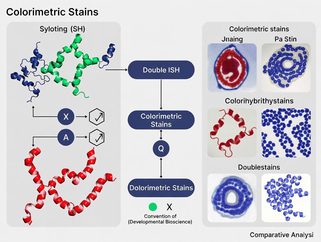
Double in situ hybridization (ISH) is a powerful technique that enables the simultaneous detection of two distinct nucleic acid sequences within tissue sections, preserving crucial spatial context in research. The technique's success hinges on the sophisticated interplay of its core components: probe chemistry for target recognition, enzymes for signal amplification, and chromogens for visualization. Chromogenic ISH (CISH) brings this technology within the reach of every pathology laboratory, offering the permanence of stained slides that can be viewed with a standard light microscope [15]. The selection of an optimal double ISH protocol involves careful consideration of detection rate, signal clarity, cost, and procedural time [16]. This guide provides a comparative analysis of the essential reagents and methodologies, supported by experimental data, to inform the design and execution of robust double ISH experiments.
Probe Chemistry and Labeling Techniques
Probes are the foundation of any ISH experiment, and their chemical labeling determines the subsequent detection strategy. The choice between DNA and RNA probes, and the type of hapten used, is critical for effective multiplexing.
- DNA vs. RNA Probes: DNA probes are more stable during hybridization, while RNA probes (riboprobes) often exhibit higher sensitivity due to stronger RNA-RNA hybrids but require more stringent conditions to prevent degradation [17] [18].
- Hapten-Based Labeling: Non-radioactive probes, labeled with haptens, are the standard for double ISH. The two most common haptens are digoxigenin (DIG) and fluorescein (FLUO) [17] [18]. These small molecules are incorporated into the probe sequence and are later recognized by specific antibodies conjugated to reporter enzymes.
- Dinitrophenyl (DNP) Labeling: DNP-labeled probes are frequently used in commercial kits, particularly for silver-enhanced ISH (SISH), where the detection results in a black metallic silver precipitate [18] [19].
Table 1: Comparison of Common Non-Radioactive Probe Haptens
| Hapten | Detection Antibody Conjugate | Common Applications | Key Characteristics |
|---|---|---|---|
| Digoxigenin (DIG) | Anti-DIG-AP or Anti-DIG-HRP | General purpose; used with NBT/BCIP, Fast Red, DAB [16] [17] | Low background; high sensitivity; versatile |
| Fluorescein (FLUO/FITC) | Anti-FLUORESCEIN-AP or Anti-FLUORESCEIN-HRP | Double ISH (paired with DIG) [17] | Allows sequential or simultaneous detection in multiplexing |
| Dinitrophenyl (DNP) | Anti-DNP-HRP [19] | Silver ISH (SISH) [19] | Used for automated platforms; generates black silver precipitate |
The following diagram illustrates the logical workflow for selecting and applying probes in a double ISH experiment.
Enzymes and Chromogen Systems
Following probe hybridization and immunodetection, reporter enzymes catalyze the conversion of soluble chromogens into insoluble, colored precipitates at the site of the target.
Reporter Enzymes
The two primary enzymes used in chromogenic ISH are:
- Horseradish Peroxidase (HRP): Catalyzes the oxidation of chromogens in the presence of hydrogen peroxide (H₂O₂). HRP is known for its rapid reaction kinetics [20].
- Alkaline Phosphatase (AP): Catalyzes the hydrolysis of phosphate groups from chromogenic substrates. AP is often used for its high sensitivity [16] [20].
Chromogen Substrates
Chromogens determine the final color of the signal and must be selected based on contrast, stability, and compatibility with multiplexing.
- NBT/BCIP: This substrate for AP produces a blue-purple precipitate. It is known for its high resolution but can be less intense than other chromogens and is soluble in organic solvents [16].
- Fast Red: Another AP substrate, Fast Red yields a red precipitate. It provides high contrast against a hematoxylin counterstain but is prone to fading and can be soluble in alcohol or xylene, requiring aqueous mounting media [16] [20].
- DAB (3,3'-Diaminobenzidine): The most widely used chromogen for HRP, DAB produces a brown precipitate. It is robust, highly stable, permanent, and insoluble in water and alcohol, making it suitable for permanent slides. Its broad absorption spectrum makes it an opaque chromogen [20] [21].
- Silver Precipitate: Used in SISH, this is not a dye but a metallic precipitate generated by HRP-catalyzed reduction of silver ions. It appears as a black dot and offers strong contrast [20] [19].
- New Chromogen Technologies: Newer chromogen systems, such as the DISCOVERY series from Roche, are based on tyramide signal amplification (TSA). TSA utilizes HRP to catalyze the deposition of fluorophore-based chromogens (e.g., Purple, Yellow, Teal), which are covalently bound to tissue proteins. These chromogens offer narrow absorption ranges, enabling translucent color mixing in co-localization studies, and are more stable in ethanol and xylene than traditional Fast Red [20].
Table 2: Enzyme-Chromogen Combinations and Properties
| Enzyme | Chromogen | Signal Color | Stability & Key Properties | Best For |
|---|---|---|---|---|
| Alkaline Phosphatase (AP) | NBT/BCIP | Blue-Purple | Soluble in organic solvents [16] | Single-plex assays; high-resolution imaging |
| Alkaline Phosphatase (AP) | Fast Red | Red | Alcohol-soluble; can fade; requires aqueous mounting [16] [20] | High-contrast single or dual assays |
| Horseradish Peroxidase (HRP) | DAB | Brown | Highly stable, permanent, and alcohol-insoluble [20] [21] | General purpose; gold standard for IHC; archival samples |
| Horseradish Peroxidase (HRP) | Silver | Black | Strong contrast, high sensitivity [20] [19] | SISH assays; bright-field microscopy |
| HRP (Tyramide) | DISCOVERY Purple | Purple | Stable in organics; translucent; narrow absorption [20] | Multiplexing with co-localization |
| HRP (Tyramide) | DISCOVERY Yellow | Yellow | Stable in organics; translucent; narrow absorption [20] | Multiplexing with co-localization |
The signaling pathways for the two primary enzyme systems are detailed below.
Experimental Data and Protocol Comparison
Performance Comparison of ISH Techniques
A 2018 study directly compared different ISH techniques for the detection of various viruses, providing quantitative data on their performance [16]. The results are summarized in the table below.
Table 3: Comparative Performance of ISH Techniques for Virus Detection [16]
| ISH Technique | Probe Type | Detection Method | Detection Rate | Key Findings |
|---|---|---|---|---|
| Chromogenic ISH (CISH) | Self-designed DIG-labelled RNA probes | AP with NBT/BCIP | 3/7 viruses | Successful for SBV, CBoV-2, PCV-2. Failed for APPV, BovHepV, EqHV, PBoV. |
| Chromogenic ISH (CISH) | Commercial DIG-labelled DNA probes | AP with NBT/BCIP | 2/3 viruses | Successful for CBoV-2 and PCV-2. Failed for PBoV. |
| Fluorescent ISH (FISH) | Commercial FISH-RNA probe mix | AP with Fast Red | 7/7 viruses | Highest detection rate and largest cell-associated positive area. Differences in cost and procedure time noted. |
Detailed Double ISH Protocol for Lymphoma Diagnostics
A robust double-staining CISH (DuoCISH) protocol was validated against FISH for detecting chromosomal breaks in lymphoma, showing 97% concordance [15]. The methodology can be adapted for general double ISH applications.
Materials and Rebesearch Reagent Solutions
- Probes: Split-signal DNA probes specific to the genes of interest (e.g., BCL6, MYC), labeled with distinct haptens (DIG and DNP) [15] [21].
- Detection Kit: DuoCISH Kit (e.g., Dako DuoCISH Kit, code no. SK108) [15].
- Antibodies: Primary antibodies against DIG and DNP, typically conjugated to HRP or AP.
- Chromogens: Sequential application of chromogens yielding different colors (e.g., purple and blue, or red and blue) [15].
- Counterstain: Hematoxylin (e.g., Dako Hematoxylin code S3301) [15].
- Equipment: Automated slide stainer or hybridizer, bright-field microscope.
Methodology:
- Sample Preparation: Cut 3–5 μm thick sections from formalin-fixed, paraffin-embedded (FFPE) tissue blocks and mount on charged slides.
- Deparaffinization and Pretreatment: Deparaffinize slides in xylene and rehydrate through a graded ethanol series. Perform heat-induced epitope retrieval in a suitable buffer (e.g., EDTA buffer, pH 8.0) using a microwave or automated platform.
- Protease Digestion: Treat slides with pepsin to digest proteins and expose target nucleic acids.
- Hybridization: Apply the mixture of hapten-labeled probes to the sections. Denature at 95°C for 5-10 minutes, then incubate at 37°C-44°C in a hybridizer for 2-6 hours to allow specific hybridization.
- Stringency Washes: Wash slides in a saline solution (e.g., 2x SSC) at elevated temperature (e.g., 72°C) to remove unbound and mismatched probes.
- Sequential Immunodetection:
- First Probe Detection: Apply the enzyme-conjugated antibody (e.g., anti-DIG-HRP) for the first hapten. Visualize with the first chromogen (e.g., ChromoMap DAB, yielding a brown signal).
- Second Probe Detection: Apply the enzyme-conjugated antibody (e.g., anti-DNP-AP) for the second hapten. Visualize with the second chromogen (e.g., Fast Red, yielding a red signal, or a Blue chromogen) [15] [20].
- Counterstaining and Mounting: Counterstain with hematoxylin to visualize nuclei. Apply coverslips with aqueous mounting media for chromogens like Fast Red, or permanent mounting media for DAB.
The selection of components for double ISH is a critical determinant of experimental success. While traditional chromogens like NBT/BCIP, Fast Red, and DAB are well-established, newer tyramide-based chromogens offer superior stability and multiplexing capabilities due to their translucent properties [20]. Experimental evidence strongly indicates that signal amplification-based methods, such as those using branched DNA or tyramide, can provide significantly higher detection rates and signal intensity compared to conventional CISH [16] [22].
For researchers, the optimal pathway involves:
- Validating Probes Individually: Before combining them in a double ISH assay, ensure each probe works optimally under single-plex conditions.
- Prioritizing Signal Amplification: For low-abundance targets, consider protocols with built-in signal amplification steps.
- Leveraging Automation: Automated staining platforms significantly enhance the reproducibility and reliability of complex double ISH protocols, minimizing manual variability [19].
By understanding the properties and performance data of these essential components, scientists can make informed decisions to design highly specific, sensitive, and robust double ISH assays that advance our understanding of gene expression and regulation within its native spatial context.
In the evolving field of anatomic pathology, colorimetric in situ hybridization (ISH) has emerged as a powerful technique for localizing specific nucleic acid sequences within tissue specimens, playing a pivotal role in companion diagnostics and personalized medicine. Unlike fluorescence ISH (FISH), colorimetric ISH, including dual hapten and dual color variants, produces a permanent, chromogenic signal visible with a standard light microscope, facilitating integration with histological morphology. The reliability of these assays, however, is not guaranteed by the protocol alone; it is profoundly dependent on the pre-analytical phase. Pre-analytical variables—including fixation, tissue processing, and sectioning—collectively form the foundation upon which all subsequent molecular analysis is built. Inconsistent pre-analytical practices can introduce significant variability, compromising staining intensity, signal-to-noise ratio, and ultimately, the accuracy of diagnostic results [23]. This guide objectively compares the impact of these variables on assay performance, providing researchers and drug development professionals with experimental data and standardized protocols to ensure the generation of robust, reproducible, and reliable ISH data.
The Foundation of Reliable ISH: A Comparative Analysis of Pre-Analytical Variables
The pre-analytical pathway is a multi-step process where decisions made at each stage directly influence the outcome of colorimetric ISH. The following sections provide a detailed, evidence-based comparison of how these variables affect staining quality.
Fixation: The Cornerstone of Macromolecular Preservation
Fixation stabilizes tissue architecture and biomolecules, but its execution is critical. The American Society of Clinical Oncology/College of American Pathologists (ASCO/CAP) has issued guidelines recommending fixation in 10% neutral-buffered formalin (NBF) for 6 to 48 hours for HER2 testing in breast carcinoma [23]. Experimental data from a model system using MCF7 xenograft tumors demonstrates the tangible impact of adhering to or deviating from these standards.
- Fixation Time: Insufficient fixation (under-fixation) fails to preserve nucleic acids adequately, leading to weak or absent ISH signals and poor morphology. Conversely, prolonged fixation (over-fixation) can cause excessive cross-linking, which impedes probe access to the target, resulting in similarly diminished signals [23] [24].
- Fixative Type: The choice of fixative is equally crucial. Experimental evidence indicates that certain fixatives, as well as some post-fixative treatments, are detrimental to molecular staining. For instance, the use of non-formalin-based fixatives can drastically reduce ISH performance [23].
Table 1: Impact of Fixation Variables on Colorimetric ISH Staining Quality
| Variable | Recommended Standard | Experimental Comparison | Impact on Staining Quality |
|---|---|---|---|
| Fixation Time | 6 to 48 hours in 10% NBF [23] | Compared to shorter (<6h) or longer (>48h) fixation. | Optimal (6-48h): Strong, specific signal with preserved morphology.Under-fixed (<6h): Weak signal, poor morphology, potential nucleic acid degradation.Over-fixed (>48h): Diminished signal due to impaired probe penetration. |
| Fixative Type | 10% Neutral-Buffered Formalin (NBF) | Compared to other common fixatives (e.g., non-buffered formalin, precipitating fixatives). | 10% NBF: Reliable, strong signals.Other Fixatives: Variable results; certain types produce consistently poor or failed staining [23]. |
| Post-Fixation Treatment | Avoid harsh decalcifying agents | Comparison of decalcifying solutions (e.g., HCl, formic acid, EDTA) on breast specimens [23]. | HCl-based: Severe damage to tissue and nucleic acids, unsuitable for ISH.Formic Acid & EDTA: Better preservation, with EDTA-based solutions showing superior cell morphology and antigenicity [23] [25]. |
Tissue Processing and Sectioning
Following fixation, tissue processing and sectioning introduce another set of critical variables.
- Section Thickness: The thickness of tissue sections is a key determinant of hybridization efficiency. For formalin-fixed, paraffin-embedded (FFPE) tissue, the ideal section thickness is 3-4 μm [23] [26]. Sections that are too thin (e.g., <3 μm) are difficult to handle and risk truncating the signal from the target nucleic acids. Sections that are too thick (e.g., >5 μm) can cause problems with probe penetration and efficient hybridization, and may lead to overlapping signals, making interpretation difficult [23] [26].
- Section Adhesion and Dewaxing: For ISH, charged slides are recommended to ensure tissue adherence [24]. Protein-based adhesives (e.g., glue, gelatin) should be avoided in the flotation bath as they can block the charged surface, leading to inconsistent adhesion and uneven staining due to reagent pooling beneath lifting sections [24]. Furthermore, incomplete removal of paraffin wax during dewaxing can create unstained or poorly stained areas in the sections [24].
Table 2: Impact of Tissue Processing and Sectioning on Colorimetric ISH
| Variable | Recommended Standard | Experimental Comparison | Impact on Staining Quality |
|---|---|---|---|
| Section Thickness (FFPE) | 3-4 μm [23] [26] | Compared to thinner (<3 μm) or thicker (>5 μm) sections. | Optimal (3-4 μm): Balanced signal intensity and morphological clarity.Too thin (<3 μm): Risk of truncated signal, handling difficulties.Too thick (>5 μm): Overlapping signals, reduced hybridization efficiency, higher background. |
| Slide Type | Charged Slides | Compared to uncharged slides. | Charged Slides: Superior tissue adhesion, preventing section loss during stringent washes.Uncharged Slides: Higher risk of section lift-off. |
| Section Adhesive | Avoid protein-based adhesives | Comparison of protein-based adhesives vs. no adhesive/appropriate adhesives. | Protein-based adhesives: Can cause uneven staining and section lifting.No adhesive/appropriate alternatives: Even staining and consistent adhesion [24]. |
| Dewaxing | Complete removal of paraffin wax | Compared to incomplete dewaxing. | Complete dewaxing: Uniform staining across the section.Incomplete dewaxing: Unstained or poorly stained areas [24]. |
Experimental Protocols for Validating Pre-Analytical Conditions
To generate the comparative data presented, standardized experimental protocols are essential. The following methodology, adapted from a study using a xenograft tumor model, provides a template for systematic evaluation [23].
Cell Line Xenograft Model for Controlled Analysis
A robust approach involves using a human breast carcinoma cell line (e.g., MCF7) generated as xenograft tumors in a murine model. This model system provides a homogeneous and reproducible source of tissue, allowing for the direct comparison of different pre-analytical conditions while minimizing the biological variability inherent in human patient samples [23].
Methodological Workflow
Key Experimental Steps
- Controlled Fixation: Immediately upon harvest, tissue samples are divided and subjected to different fixation conditions. This includes immersion in various fixatives (e.g., 10% NBF, other common fixatives) for a range of times (e.g., from 1 hour to 72 hours) [23].
- Post-Fixation Treatment (if applicable): For tissues requiring decalcification, such as those modeling bone metastases, samples are treated with different decalcifying solutions (e.g., hydrochloric acid, formic acid, EDTA) after the initial fixation step [23] [25].
- Standardized Processing and Sectioning: All tissues are processed to paraffin blocks using a standardized protocol. Subsequently, sections are cut at varying thicknesses (e.g., 3 μm, 4 μm, 5 μm, 8 μm) and mounted on charged slides [23] [26].
- Colorimetric ISH Assay: Dual hapten or dual color ISH assays (e.g., for HER2) are performed on all sections under identical conditions using a standardized protocol with careful optimization of pretreatment, hybridization, and washing steps [23] [24].
- Quantitative and Qualitative Scoring: Stained sections are evaluated by trained pathologists or scientists. Scoring includes assessment of signal intensity, signal-to-noise ratio (background), cellular morphology preservation, and the number of interpretable results. Statistical analysis (e.g., Mann-Whitney U test) can be used to compare the significance of differences between groups [23] [27].
The Scientist's Toolkit: Essential Research Reagent Solutions
Successful execution of colorimetric double ISH research requires a suite of reliable reagents and materials. The following table details key solutions and their functions.
Table 3: Essential Research Reagent Solutions for Colorimetric Double ISH
| Item | Function | Considerations |
|---|---|---|
| 10% Neutral-Buffered Formalin | Primary fixative that stabilizes proteins and nucleic acids while preserving morphology. | The gold-standard fixative; consistent pH and buffering prevent artifactual changes. |
| EDTA-Based Decalcifying Solution | Chelates calcium ions from mineralized tissue without damaging nucleic acids. | Preferred over strong acids (e.g., HCl) for molecular studies due to superior preservation of nucleic acid integrity and antigenicity [25]. |
| Charged Slides | Microscope slides with a permanent positive charge to enhance adhesion of tissue sections. | Prevents section loss during high-temperature pretreatment and stringent washing steps. |
| Specific Nucleic Acid Probes | Labeled DNA or RNA probes complementary to the target sequence (e.g., HER2). | Must be chosen for high sensitivity and specificity; always consult specification sheets for optimal hybridization conditions [24]. |
| Tyramide Signal Amplification (TSA) Reagents | Enzyme-mediated system that deposits numerous chromogen labels at the probe site. | Significantly enhances signal intensity, crucial for detecting low-abundance targets in multiplexed assays [28]. |
| Chromogenic Substrate Kits | Enzyme-substrate kits (e.g., for HRP or AP) that produce a stable, colored precipitate. | Allows for simultaneous detection of multiple targets with different colors in double ISH assays. |
| Appropriate Controls | Tissue samples with known positive and negative status for the target(s) of interest. | Essential for validating each staining run and interpreting results accurately [24]. |
The path to reliable and reproducible colorimetric double ISH results is paved long before the probes are applied. As the experimental data demonstrates, pre-analytical variables are not mere suggestions but strict prerequisites for assay success. Standardization of fixation in 10% NBF for 6-48 hours, use of EDTA for decalcification when necessary, and sectioning at 3-4 μm thickness are evidence-based practices that directly prevent staining failures and diagnostic inaccuracies. For researchers and drug developers, a rigorous and disciplined approach to the pre-analytical phase is the most effective strategy to ensure that their ISH data truly reflects the underlying biology, thereby fueling confident discovery and dependable diagnostic outcomes.
Advantages and Inherent Limitations of dCISH for Multiplex Target Detection
Dual-Color In Situ Hybridization (dCISH) is a advanced bright-field microscopy technique designed for the simultaneous detection of two distinct nucleic acid targets within the context of intact tissue morphology. This method has gained significant importance in both diagnostic pathology and research, particularly for assessing gene amplification status and chromosomal rearrangements. Unlike fluorescence-based methods, dCISH utilizes chromogenic signals that remain permanently stable, allowing for archiving of slides and direct correlation with conventional histology. The fundamental principle of dCISH involves hybridizing specifically designed DNA probes labeled with different haptens to their complementary DNA target sequences within formalin-fixed, paraffin-embedded (FFPE) tissue sections, followed by enzymatic detection systems that produce distinct colored precipitates at the target sites.
In the context of multiplex target detection, dCISH represents a crucial technological bridge between single-plex assays and higher-plex methodologies. While true "multiplexing" in dCISH is currently limited to two targets, this capability nonetheless provides critical advantages for co-localization studies and diagnostic algorithms requiring simultaneous assessment of multiple biomarkers. The technique has been most extensively validated in clinical diagnostics, particularly for human epidermal growth factor receptor 2 (HER2) testing in breast cancer, where it enables concurrent visualization of the HER2 gene and chromosome 17 centromere (CEP17) as an internal control [29] [30]. This review comprehensively examines the technical performance, advantages, and limitations of dCISH within the broader landscape of multiplex detection technologies, providing researchers with evidence-based guidance for its application in experimental and diagnostic contexts.
Technical Foundations of dCISH
Core Methodology and Workflow
The dCISH procedure follows a standardized workflow that ensures reproducible and reliable results. The process begins with slide preparation, where 4-5 μm thick sections from FFPE tissue blocks are mounted on charged slides and dried thoroughly. The sections undergo deparaffinization in xylene and rehydration through graded alcohols, followed by pre-treatment steps designed to expose target nucleic acids. This typically involves proteolytic digestion using enzymes such as pepsin to break cross-links formed during fixation and permit probe access to the target sequences [29] [30].
The hybridization step constitutes the core of the dCISH assay, where labeled DNA probes are applied to the tissue sections. In the VENTANA HER2 Dual ISH DNA Probe Cocktail assay—one of the most widely used commercially available dCISH platforms—the HER2 probe is labeled with dinitrophenyl (DNP) while the CEP17 probe is labeled with digoxigenin [30]. The slides are then denatured at high temperature (typically 82°C for 5-12 minutes) to separate DNA strands, followed by an overnight hybridization at 37°C to allow specific binding of probes to their complementary targets.
Post-hybridization washes remove unbound probe, after which the detection phase begins. The DNP-labeled HER2 probe is typically detected with a horseradish peroxidase (HRP)-conjugated anti-DNP antibody and visualized with silver chromogen that produces a black precipitate. The digoxigenin-labeled CEP17 probe is detected with an alkaline phosphatase (AP)-conjugated anti-digoxigenin antibody and visualized with Fast Red that produces a red precipitate [30]. The slides are then counterstained with hematoxylin to visualize nuclear morphology, dehydrated, cleared, and cover-slipped for permanent preservation.
Signaling Pathway and Detection System
The dCISH detection system relies on orthogonal detection pathways that operate simultaneously without cross-reactivity. The following diagram illustrates this process:
This orthogonal detection system ensures minimal cross-talk between the two signals, which is critical for accurate interpretation. The enzymatic precipitation creates discrete, chromogen-specific signals that can be distinguished by their distinct colors and, in some cases, by their subnuclear distribution patterns.
Performance Comparison: dCISH Versus Alternative Methodologies
Analytical Performance Data
The clinical validation of dCISH has been extensively documented, particularly for HER2 testing in breast cancer. The following table summarizes key performance metrics from recent studies comparing dCISH with established reference methods:
Table 1: Performance Metrics of dCISH for HER2 Detection in Breast Cancer
| Performance Parameter | dCISH Result | Comparative Method | Study Details |
|---|---|---|---|
| Concordance with FISH | 98.65% | FISH (ERBB2/CEN17 probe) | 148 cases of invasive breast cancer [29] |
| Sensitivity | 96.3% | FISH as reference standard | 55 FISH-amplified cases [29] |
| Specificity | 100% | FISH as reference standard | 93 FISH-nonamplified cases [29] |
| Interobserver Reproducibility | Almost perfect agreement (κ=0.97) | Multi-pathologist evaluation | 4 pathologists blinded to FISH results [29] |
| Positive Predictive Value | 100% | FISH as reference standard | 53/53 dCISH-amplified cases confirmed by FISH [29] |
| Negative Predictive Value | 97.9% | FISH as reference standard | 93/95 dCISH-nonamplified cases confirmed by FISH [29] |
Technical Comparison with Alternative Platforms
dCISH occupies a specific niche within the broader landscape of multiplex detection technologies. The following table provides a comparative analysis of its capabilities relative to other commonly used methods:
Table 2: Technical Comparison of dCISH with Alternative Detection Platforms
| Parameter | dCISH | FISH | Conventional IHC | High-Plex Spatial Proteomics (e.g., PathoPlex) |
|---|---|---|---|---|
| Maximum Targets | 2 | 2-4 (with spectral imaging) | 4-7 (with multiplex IF) | 140+ targets [31] |
| Resolution | Subcellular (80 nm/pixel possible) [31] | Subcellular | Cellular to subcellular | Subcellular (80 nm/pixel) [31] |
| Signal Permanence | Permanent | Fades over time | Permanent (chromogen) / Fades (fluorescence) | Stable after imaging cycles [31] |
| Equipment Requirements | Standard bright-field microscope | Fluorescence microscope with specific filters | Standard bright-field or fluorescence microscope | Specialized cyclic imaging system [31] |
| Integration with Morphology | Excellent | Moderate (fluorescence quenches morphology) | Excellent | Excellent with subcellular context [31] |
| Automation Potential | High (fully automated platforms available) [29] | Moderate | High | High with specialized computational tools [31] |
| Analysis Time | Moderate (signal counting required) | Lengthy (requires darkroom conditions) | Fast to moderate | Extensive (computational analysis of large datasets) [31] |
| Theoretical Basis | DNA-DNA hybridization | DNA-DNA hybridization | Antibody-antigen recognition | Antibody-antigen recognition with cyclic detection [31] |
Advantages of dCISH for Target Detection
Practical and Technical Benefits
dCISH offers several distinct advantages that make it particularly valuable for both research and clinical applications. The signal permanence represents one of its most significant benefits over fluorescence-based methods. Unlike FISH signals that fade over time, dCISH chromogenic signals remain stable for years, allowing for permanent archiving of slides and retrospective analyses [29] [30]. This feature is particularly valuable for clinical trials and longitudinal studies where documentation and re-evaluation may be necessary years after initial testing.
The compatibility with standard bright-field microscopy makes dCISH accessible to virtually any pathology laboratory without requiring specialized fluorescence equipment. This significantly lowers the barrier to implementation and allows for easier integration into established diagnostic workflows. Furthermore, the bright-field format enables precise correlation with tissue morphology, as pathologists can simultaneously assess chromogenic signals and histological features using familiar microscopic evaluation techniques [30].
The high degree of automation possible with dCISH platforms represents another substantial advantage. Fully automated systems such as the VENTANA platform enable standardized processing with minimal technical variability, enhancing reproducibility across different laboratories and operators [29]. This automation extends to the integration of digital pathology and artificial intelligence applications, where dCISH has demonstrated excellent performance in whole-slide imaging and automated signal counting algorithms [30].
From a performance perspective, dCISH demonstrates exceptional concordance with established reference methods. The 98.65% concordance rate with FISH for HER2 testing, coupled with almost perfect interobserver reproducibility (κ=0.97), provides strong validation of its analytical reliability [29]. This high level of agreement with the traditional gold standard, combined with the practical advantages of bright-field detection, positions dCISH as a robust alternative to FISH for routine diagnostic applications.
Inherent Limitations and Technical Constraints
Multiplexing Limitations and Technical Challenges
Despite its considerable advantages, dCISH faces several inherent limitations that restrict its application in more complex multiplexing scenarios. The most significant constraint is the inherent limitation to two targets due to the availability of only two orthogonal detection systems that can be distinguished by conventional bright-field microscopy. While methods like PathoPlex can simultaneously process 140+ markers through iterative cycling, dCISH is fundamentally restricted to duplex detection [31]. This limitation precludes its use in complex molecular profiling requiring simultaneous assessment of multiple genetic alterations.
Signal resolution and separation present additional technical challenges in dCISH. Overlapping signals, particularly in nuclei with high gene amplification or tight spatial clustering of signals, can complicate accurate enumeration. The discrete chromogen particles must be distinctly separated for precise counting, a requirement that may not always be met in suboptimally hybridized or highly amplified specimens. This limitation becomes particularly evident when comparing dCISH to fluorescence-based methods, where spectral separation can more effectively resolve closely spaced signals.
The analytical sensitivity of dCISH, while excellent for detecting high-level amplifications, may be suboptimal for identifying low-level gains or minor subclonal populations. The threshold for reliable detection is inherently limited by the chromogenic detection system, which may not achieve the same sensitivity as fluorescence-based amplification methods or advanced PCR-based techniques [32]. This constraint is particularly relevant for applications requiring detection of minimal residual disease or heterogeneous tumor populations with variable gene amplification.
Operational and Implementation Challenges
From a practical standpoint, dCISH presents several operational limitations that impact its implementation in research and diagnostic settings. The extended procedure time—typically requiring overnight hybridization—limits assay throughput and turnaround time compared to more rapid methodologies like IHC or targeted PCR. While the actual hands-on time may be reduced through automation, the overall process remains time-consuming, particularly when compared to emerging rapid multiplex platforms.
The technical complexity of assay optimization represents another significant implementation challenge. dCISH requires meticulous optimization of multiple parameters including protease digestion time, hybridization conditions, and detection system stringency. This optimization must be performed for each new probe combination and tissue type, creating substantial barriers to the development of novel assay configurations beyond the commercially available options.
Digital pathology integration, while promising, faces specific technical hurdles with dCISH. A recent study evaluating AI-integrated dCISH analysis demonstrated that scanning protocol optimization is critical for accurate automated signal enumeration, with suboptimal scanning parameters leading to nuclei detection failures and inaccurate classification [30]. The requirement for high-resolution scanning (0.12-0.17 μm/pixel) with extended focus capabilities to ensure signal clarity throughout the tissue section further complicates the digital pathology workflow and increases computational demands [30].
Essential Research Reagent Solutions for dCISH
Successful implementation of dCISH requires specific reagent systems optimized for chromogenic detection. The following table outlines key reagent solutions and their functional roles in the dCISH workflow:
Table 3: Essential Research Reagent Solutions for dCISH Applications
| Reagent Category | Specific Examples | Function | Technical Considerations |
|---|---|---|---|
| DNA Probe Systems | VENTANA HER2 Dual ISH DNA Probe Cocktail [30] | Target-specific hybridization for gene and chromosome enumeration | DNP-labeled HER2 probe; Digoxigenin-labeled CEP17 probe |
| Detection Kits | UltraView SISH Detection Kit [30] | HRP-based detection of DNP-labeled probes with silver chromogen | Silver deposition creates black signal at target site |
| UltraView Red ISH Detection Kit [30] | AP-based detection of digoxigenin-labeled probes with Fast Red | Red chromogenic precipitate at target site | |
| Proteolytic Enzymes | Pepsin, Proteinase K | Tissue pre-treatment to expose target nucleic acids | Concentration and incubation time require optimization per tissue type |
| Stringency Wash Solutions | Saline-sodium citrate (SSC) buffers | Remove non-specifically bound probes | Critical for minimizing background; concentration and temperature-dependent |
| Automated Platform Reagents | VENTANA reaction buffers [29] | Optimized for automated staining platforms | Ensure compatibility with specific automated systems |
| Nucleic Acid Controls | Synthetic DNA oligonucleotides (gBlocks) [32] | Assay validation and optimization | Confirm probe specificity and sensitivity |
Experimental Protocols for dCISH Validation
Standardized dCISH Protocol for HER2/CEP17 Detection
The following workflow diagram outlines the key steps in a standardized dCISH protocol:
This protocol follows the established methodology used in validation studies that demonstrated high concordance with FISH [29]. The critical steps include:
Section Preparation: 4-5 μm thick sections from FFPE tissue blocks are mounted on charged slides and baked at 60°C for 25-60 minutes to ensure adhesion.
Deparaffinization and Rehydration: Slides are treated with xylene (3 changes, 5-10 minutes each) followed by graded ethanol series (100%, 95%, 70%) and distilled water rinses.
Proteolytic Pretreatment: Enzymatic digestion with pepsin (0.5-1 mg/mL in 0.1N HCl) for 5-30 minutes at 37°C, optimized based on tissue fixation conditions.
Probe Application and Hybridization: DNP-labeled HER2 and digoxigenin-labeled CEP17 probes are applied simultaneously, followed by denaturation at 82°C for 5-12 minutes and hybridization at 37°C for 6-20 hours in a humidified chamber or automated platform.
Post-Hybridization Washes: Stringency washes with 2× SSC/0.3% NP-40 at 72°C for 3-5 minutes remove non-specifically bound probes.
Sequential Detection: HRP-conjugated anti-DNP antibody with silver chromogen detection for HER2 signals, followed by AP-conjugated anti-digoxigenin antibody with Fast Red detection for CEP17 signals.
Counterstaining and Mounting: Hematoxylin counterstaining (30-60 seconds) followed by dehydration, clearing, and permanent mounting.
Validation and Quality Control Procedures
Robust validation of dCISH requires implementation of comprehensive quality control measures. The validation study by Rathi et al. provides a exemplary framework [29]:
Sample Selection: Include 148 cases of invasive breast cancer representing the full spectrum of HER2 status (IHC scores 0, 1+, 2+, and 3+) to ensure comprehensive performance assessment across diagnostic categories.
Reference Method Comparison: Compare dCISH results with established FISH methodology using the ERBB2/CEN17 dual color probe on consecutive sections from the same tissue block.
Multi-observer Analysis: Engage four pathologists blinded to FISH and IHC results for independent assessment of dCISH signals to establish interobserver reproducibility.
Statistical Analysis: Calculate concordance rates, sensitivity, specificity, positive and negative predictive values, and Cohen's kappa statistic for interobserver agreement.
Discrepancy Resolution: Establish protocols for resolving discordant cases, including repeat testing and consideration of tumor heterogeneity.
For ongoing quality assurance, inclusion of both positive and negative control tissues in each run is essential. Internal controls such as non-neoplastic cells within the tissue section should demonstrate the expected disomic signal pattern (two HER2 and two CEP17 signals per nucleus).
dCISH represents a robust methodology for duplex target detection that successfully bridges the gap between single-plex assays and complex multiplexing platforms. Its principal advantages—including signal permanence, compatibility with standard microscopy, and high concordance with reference methods—make it particularly valuable for diagnostic applications requiring simultaneous assessment of two genetic targets. The technology demonstrates exceptional performance in clinical validation studies, with concordance rates exceeding 98% compared to FISH for HER2 amplification detection [29].
However, the inherent limitations of dCISH, particularly its restriction to two targets and technical challenges in signal resolution, constrain its utility in research contexts requiring higher-plex capabilities. For such applications, emerging technologies like PathoPlex [31] or highly multiplexed ddPCR [32] may offer more appropriate solutions. The ongoing integration of dCISH with digital pathology and artificial intelligence platforms promises to enhance its reproducibility and analytical precision, particularly through optimized scanning protocols and automated analysis algorithms [30].
The selection of dCISH versus alternative multiplex detection platforms should be guided by specific research objectives, technical requirements, and operational constraints. For clinical diagnostics requiring permanent documentation and straightforward morphological correlation, dCISH remains an excellent choice. For discovery-phase research demanding higher-plex capabilities or absolute quantification, alternative platforms may prove more suitable. As multiplexing technologies continue to evolve, dCISH will likely maintain its important niche in applications where reliable, permanent duplex detection aligns with experimental or diagnostic requirements.
Executing Double Colorimetric ISH: Step-by-Step Protocols and Workflow
Successful in situ hybridization (ISH), particularly in complex applications like colorimetric double ISH, hinges on effective tissue preparation. The primary goal of fixation and permeabilization is to preserve tissue morphology and nucleic acid integrity while allowing sufficient probe penetration for accurate detection. These steps are especially critical when visualizing delicate tissues or performing multiplexed assays, as they balance conflicting needs: over-fixation can mask epitopes, whereas under-fixation may lead to degradation and loss of morphological detail [33] [34].
The choice of strategy is often dictated by the specific research requirements. For instance, studying fragile regenerating tissues demands protocols that prevent degradation of the epidermis and blastema, while double ISH experiments require methods that preserve multiple target sequences without cross-reactivity. This guide compares the performance of various fixation and permeabilization methods, providing a structured framework for researchers to select and optimize protocols for their specific experimental contexts.
Fixation Strategies: Crosslinking vs. Precipitative Methods
Fixation stabilizes tissue architecture by inhibiting enzymatic degradation and preserving the spatial context of nucleic acids. The two primary classes of fixatives—crosslinking and precipitative—operate through distinct mechanisms and offer different advantages for ISH workflows.
Table 1: Comparison of Common Fixation Methods for ISH
| Fixative Type | Specific Agents | Mechanism of Action | Best For | Key Advantages | Key Limitations |
|---|---|---|---|---|---|
| Crosslinking | Formaldehyde (4% PFA), Formalin, Glutaraldehyde | Creates methylene bridges between proteins, hardening sample and preserving structure [34] [35]. | Most ISH applications; delicate tissues; preserving fine cellular structure [34]. | Excellent preservation of tissue morphology and subcellular structure [34] [36]. | Can mask epitopes through over-crosslinking; often requires antigen retrieval [33] [34] [35]. |
| Precipitative (Organic Solvents) | Methanol, Ethanol, Acetone | Dehydrates samples, precipitating proteins in situ and removing lipids [34] [37] [35]. | Targets where crosslinking impairs antigenicity; some intracellular epitopes [36]. | Does not require separate permeabilization step; can expose buried epitopes [36] [37]. | Poorer preservation of tissue morphology; can denature proteins of interest; may wash away soluble proteins [34] [35]. |
| Combined/Mixed | PFA followed by Methanol [38] | Sequential crosslinking and precipitation. | Double ISH/IF protocols; preserving both RNA and protein integrity [39] [38]. | Can offer a balance between structure preservation and epitope accessibility [38]. | Requires optimization of two fixation steps; potential for increased autofluorescence. |
| Specialized | Nitric Acid/Formic Acid (NAFA) [40] | Acid-based treatment that permeabilizes while fixing. | Delicate/fragile tissues (e.g., planarians, regenerating fins); whole-mount ISH [40]. | Eliminates need for harsh proteinase K, preserving tissue integrity and antigenicity for subsequent IF [40]. | A newer protocol that may require validation for other tissue types. |
Experimental Data on Fixative Performance
Comparative studies demonstrate that fixative choice directly impacts signal quality and tissue integrity. In tests on Drosophila ovaries, a 1-hour fixation in 4% paraformaldehyde (PFA) with 1% DMSO provided a foundation that preserved morphology during subsequent high-temperature ISH washes [39]. Furthermore, research on planarians showed that a novel Nitric Acid/Formic Acid (NAFA) protocol preserved epidermal integrity significantly better than traditional methods using the mucolytic agent N-Acetyl Cysteine (NAC), which caused noticeable tissue damage [40].
For combined protein and RNA detection, a simplified neuronal protocol employed sequential fixation: 4% PFA followed by cold methanol. This consecutive routine fixation preserved mRNA and protein targets effectively without requiring alterations in salt concentration, proving compatible with antibodies for both PFA and methanol-fixed targets [38].
Permeabilization Methods: Enabling Probe Access
Following fixation, permeabilization is essential to render membranes permeable to labeled probes and antibodies. This step is particularly crucial for whole-mount ISH where probes must penetrate thick tissues, and the method must be chosen based on the fixative used and the fragility of the tissue.
Table 2: Comparison of Common Permeabilization Methods
| Permeabilization Method | Specific Agents | Mechanism of Action | Best For | Key Advantages | Key Limitations |
|---|---|---|---|---|---|
| Detergents | Triton X-100, Tween-20, Saponin [36] [35] | Solubilizes lipid bilayers to create pores in membranes [35]. | General use after crosslinking fixatives (e.g., PFA) [36]. | Widely used and effective; controllable concentration. | Can disrupt native lipid-protein interactions; over-permeabilization can damage morphology. |
| Enzymatic | Proteinase K [39] | Digests proteins to break down tissue structure and increase permeability. | Tissues with significant barriers to penetration (e.g., Drosophila ovaries) [39]. | Highly effective for penetrating difficult tissues. | Can be too harsh, damaging tissue morphology and, critically, protein epitopes for subsequent IF [39] [40]. |
| Organic Solvents | Methanol, Ethanol, Acetone [36] [37] | Dissolves lipids and dehydrates cells. | Use after crosslinking fixatives or as a fixative/permebilizer combo. | Simple; no separate step required when used as a fixative. | Can precipitate proteins and alter tertiary structures; less controlled. |
| Combined Physical/Chemical | Xylenes + Detergents (RIPA) [39] | Organic solvent and detergent action. | Dual IF/FISH protocols where proteinase K is too destructive [39]. | Can provide effective permeabilization for FISH while preserving protein epitopes. | Requires use of hazardous organic solvents like xylenes. |
Key Experimental Findings on Permeabilization
In Drosophila ovaries, a standard ISH protocol using proteinase K (50 µg/ml for 1 hour) produced a strong signal within 15-45 minutes. However, for dual protein-RNA labeling (IF/FISH), proteinase K destroyed protein epitopes. Researchers found that omitting proteinase K and using alternative permeabilization with xylenes and detergents (RIPA) preserved a strong protein signal while still allowing a specific, albeit variable, FISH signal [39].
Another study demonstrated that the optimal permeabilization method can be antibody-dependent. For instance, in NIH/3T3 cells, antibodies against PDI and β-Actin showed superior performance with methanol permeabilization compared to Triton X-100 [36].
Detailed Experimental Protocols for Double ISH Research
Protocol 1: Standard Fixation and Permeabilization for ISH
This protocol is adapted from optimized methods for Drosophila ovaries and is a robust starting point for many tissue types [39].
- Dissection and Fixation: Dissect tissue in appropriate physiological buffer. Transfer to 4% Paraformaldehyde (PFA) with 1% DMSO. Fix for 1 hour at room temperature.
- Dehydration: Treat tissue with a graded ethanol series (e.g., 50%, 70%, 100%) to dehydrate. Samples can be stored at this stage at -20°C indefinitely.
- Rehydration and Permeabilization: Rehydrate tissue through a descending ethanol series to PBS. Permeabilize with Proteinase K (e.g., 50 µg/ml in PBS) for 1 hour at room temperature. The concentration and time must be empirically determined for different tissues.
- Post-fixation: Re-fix tissue in 4% PFA for 30 minutes to maintain morphology after protease treatment.
- Pre-hybridization and Hybridization: Proceed to standard pre-hybridization and hybridization steps with digoxigenin-labeled RNA probes.
Protocol 2: Combined IF/FISH with Permeabilization for Protein Preservation
This protocol reverses the traditional order, performing immunofluorescence before FISH to preserve protein antigenicity [39].
- Initial Fixation: Fix dissected tissue in 4% PFA for 20 minutes.
- Immunofluorescence (IF): Perform the entire standard IF procedure (blocking, primary antibody, secondary antibody) on the fixed tissue.
- Post-IF Fixation: Post-fix the tissue again in 4% PFA for 20 minutes. This cross-links the antibodies in place.
- Permeabilization for FISH (without Proteinase K):
- Dehydrate in ethanol and permeabilize with xylenes for 1 hour.
- Rehydrate and further permeabilize with a detergent-based buffer like RIPA.
- FISH Procedure: Continue with standard FISH steps, including high-temperature hybridization and stringent washes.
Protocol 3: NAFA for Delicate Tissues and Whole-Mount ISH
The NAFA protocol is designed for delicate tissues like planarians and regenerating killifish fins, eliminating the need for proteinase K [40].
- Fixation: Fix tissue in a solution containing Nitric Acid, Formic Acid, and EGTA.
- Permeabilization and Post-fixation: The acid treatment itself permeabilizes the tissue. A post-fixation step is included.
- Hybridization and Detection: Proceed with standard hybridization and detection steps. This protocol has been validated for both chromogenic and fluorescent detection and is highly compatible with subsequent immunostaining.
Workflow Visualization
The following diagram illustrates the key decision points and pathways for selecting an appropriate tissue preparation strategy for ISH-based research.
Research Reagent Solutions
Table 3: Essential Reagents for ISH Tissue Preparation
| Reagent Category | Specific Examples | Primary Function in Protocol |
|---|---|---|
| Crosslinking Fixatives | 4% Paraformaldehyde (PFA), 10% Neutral Buffered Formalin [34] [37] | Preserves tissue architecture by creating protein cross-links. |
| Precipitative Fixatives | Cold Methanol, Cold Ethanol, Acetone [36] [37] | Precipitates proteins and dehydrates tissue; often combines fixation and permeabilization. |
| Permeabilization Detergents | Triton X-100, Tween-20, Saponin [36] [35] | Solubilizes cell membranes to allow probe penetration after crosslinking fixation. |
| Enzymatic Permeabilization | Proteinase K [39] | Digests proteins to permeabilize tissues with significant barriers; use requires caution. |
| Probe Labeling & Detection | Digoxigenin (DIG)-labeled probes, Tyramide Signal Amplification (TSA) [39] [41] | Labels nucleic acid probes for hybridization and enables sensitive chromogenic/fluorescent detection. |
| Blocking Agents | Bovine Serum Albumin (BSA), serum from host species | Reduces non-specific binding of probes and antibodies, lowering background noise. |
| Specialized Solutions | NAFA Solution, N-Acetyl Cysteine (NAC) [40] [39] | Specialized for specific applications (e.g., whole-mount ISH, mucolysis) to improve penetration or preserve delicate tissues. |
In situ hybridization (ISH) stands as an essential molecular biology method for detecting and localizing specific nucleic acid sequences within cells and tissues, providing critical spatial context for gene expression analysis. The development of multiplexed detection systems, particularly those enabling simultaneous visualization of multiple targets using non-radioactive labels such as digoxigenin (DIG) and fluorescent (FLUO) markers, has significantly advanced our understanding of complex biological processes. These techniques allow researchers to decipher spatial relationships between different nucleic acid sequences within their native morphological context, providing insights into gene expression patterns, chromosomal abnormalities, and pathogen localization. For diagnostic professionals and research scientists, selecting appropriate probe labeling strategies requires careful consideration of sensitivity, resolution, compatibility, and operational practicality. This guide objectively compares DIG and FLUO labeling methodologies within the framework of double ISH research, providing experimental data and protocols to inform probe selection for specific research and diagnostic applications.
Comparative Analysis of DIG and FLUO Labeling Methodologies
The selection between DIG and FLUO labeling systems depends on multiple factors including target abundance, required sensitivity, equipment availability, and intended application. The table below provides a systematic comparison of their core characteristics:
Table 1: Performance Comparison of DIG and FLUO Labeling Systems
| Characteristic | DIG Labeling | FLUO Labeling |
|---|---|---|
| Detection Method | Colorimetric/Chromogenic | Fluorescence |
| Sensitivity | High | High [42] |
| Resolution | Limited by chromogen diffusion [39] | High, subcellular resolution possible [39] |
| Multiplexing Capability | Limited | Excellent, allows multicolor detection [42] |
| Background Interference | Low endogenous background [42] | Potential autofluorescence |
| Signal Permanence | Permanent slides | Photobleaching over time [42] |
| Equipment Needs | Standard brightfield microscope | Fluorescence microscope [42] |
| Best Applications | Single-target detection, low-equipment settings | Multiplexing, co-localization studies, live-cell imaging [43] |
Table 2: Operational Considerations for Probe Labeling Methods
| Parameter | DIG Labeling | FLUO Labeling |
|---|---|---|
| Ease of Operation | User-friendly, fewer steps [42] | Technically straightforward [42] |
| Protocol Duration | ~3 days for ISH [39] | ~3 days for FISH [39] |
| Permeabilization Requirements | Proteinase K treatment (e.g., 50μg/ml, 1 hour) [39] | Varies; may use detergents, organic solvents, or proteinase K [39] |
| Key Limitations | Chromogen diffusion limits resolution [39] | Signal diminishes over time due to photobleaching [42] |
Experimental Protocols for Double ISH Research
Probe Design and Synthesis Considerations
Effective double ISH requires careful probe design to ensure specific hybridization and minimal cross-reactivity. For DIG-labeled probes, RNA probes are often preferred over double-stranded DNA probes due to their increased sensitivity [39]. The length and GC content of the RNA probe significantly impact hybridization efficiency and signal intensity. For fluorescent probes, selection of fluorophores with non-overlapping emission spectra (e.g., Cy3, Cy5, Fluorescein) is crucial for multiplexing applications [42]. Both probe types require balancing conflicting needs for tissue permeabilization, fixation, and preservation of morphological integrity throughout the hybridization process.
Detailed Protocol for DIG-Labeled RNA ISH
The following protocol has been optimized for sensitive detection in challenging tissues:
- Tissue Fixation: Fix dissected tissues for 1 hour in 4% paraformaldehyde with 1% DMSO to ensure adequate penetration and morphology preservation [39].
- Permeabilization: Treat tissue with proteinase K (50μg/ml) for 1 hour at room temperature to enable probe access [39].
- Hybridization: Hybridize with DIG-labeled RNA probes using higher temperature prehybridization, hybridization, and subsequent washes optimized for RNA probes [39].
- Detection: Incubate with alkaline phosphatase-conjugated anti-DIG antibodies followed by color reaction development using NBT/BCIP or similar substrates [39].
Detailed Protocol for RNA FISH with Fluorescent Labels
The fluorescent ISH protocol shares initial steps with DIG-ISH but differs in critical detection phases:
- Tissue Preparation: Fixation and permeabilization steps mirror those in DIG-ISH [39].
- Hybridization: Hybridize with fluorescently labeled probes using similar temperature conditions [39].
- Signal Amplification: Employ tyramide signal amplification (TSA) to markedly enhance sensitivity compared to conventional IF and FISH methods [39].
- Imaging: Visualize using laser confocal microscopy for better resolution, subcellular localization, and optical sectioning capabilities [39].
Combined IF/FISH for Simultaneous Protein-RNA Detection
For experiments requiring simultaneous detection of protein and RNA targets:
- Protein Immunofluorescence: Perform complete protein IF staining first using a 20-minute initial fixation [39].
- Post-Fixation: Cross-link antibodies to tissue with a second fixation step to preserve complexes during subsequent FISH procedures [39].
- Alternative Permeabilization: Replace proteinase K treatment with a combination of xylenes and detergents (RIPA) to preserve protein epitopes while allowing RNA probe penetration [39].
- FISH Procedure: Continue with standard RNA FISH protocol as described above [39].
This reversed order (IF before FISH) markedly improves protein detection while maintaining strong FISH signals compared to traditional methods [39].
Signaling Pathways and Experimental Workflows
The following diagram illustrates the key decision pathways and experimental workflows for implementing simultaneous DIG and FLUO detection systems:
Diagram 1: Workflow for simultaneous DIG and FLUO probe detection
Research Reagent Solutions for Double ISH
Successful implementation of double ISH methodologies requires specific reagents and materials. The following table details essential components and their functions:
Table 3: Essential Research Reagents for DIG and FLUO Probe Experiments
| Reagent/Category | Specific Examples | Function/Application |
|---|---|---|
| Labeling Molecules | Digoxigenin (DIG) | Hapten for antibody-based detection in colorimetric ISH [42] |
| Fluorophores | Fluorescein, Cy3, Cy5 | Fluorescent labels for direct detection or amplification in FISH [42] |
| Detection Enzymes | Alkaline Phosphatase, Horseradish Peroxidase | Enzyme conjugates for chromogenic or chemiluminescent detection [42] [39] |
| Permeabilization Agents | Proteinase K, Xylenes, RIPA Detergent | Tissue treatment to enable probe penetration [39] |
| Chromogenic Substrates | NBT/BCIP, Fast Red | Enzyme substrates that produce insoluble colored precipitates [39] |
| Signal Amplification | Tyramide Signal Amplification | Greatly enhances sensitivity in FISH applications [39] |
| Microscopy Systems | Brightfield, Fluorescence, Confocal | Signal visualization and imaging [42] [44] |
The simultaneous detection of DIG- and FLUO-labeled probes represents a powerful approach for advanced ISH applications, each method offering distinct advantages that complement the other. DIG labeling provides high sensitivity with permanent records ideal for archival purposes and standard pathology workflows, while FLUO labeling enables multiplexed detection and superior resolution for detailed subcellular localization studies. Recent technological advances, including improved signal amplification methods and the integration of artificial intelligence for image analysis, are further enhancing the precision and reliability of both detection systems [44]. As molecular pathology continues to evolve toward increasingly multiplexed assays, the strategic combination of these labeling methodologies will enable researchers and diagnostic professionals to extract richer biological information from limited samples, ultimately advancing both basic research and clinical diagnostics in areas such as cancer genomics, infectious disease detection, and developmental biology.
In the context of colorimetric double in situ hybridization (dISH) research, achieving clear, specific, and simultaneous detection of multiple nucleic acid targets hinges on the precise optimization of the hybridization process. This guide objectively compares the performance and impact of different buffer conditions, temperatures, and hybridization durations, drawing on experimental data to provide a foundational framework for researchers. Proper optimization is critical for enhancing assay specificity and sensitivity, reducing background noise, and ensuring reproducible results in drug development and diagnostic applications.
Comparative Analysis of Hybridization Buffers
The composition of the hybridization buffer is a primary determinant of the assay's stringency, which controls the specificity of probe binding. We compare common buffer systems and their components below.
Table 1: Comparison of Common Hybridization Buffer Formulations
| Buffer Component | Function in Hybridization | Typical Concentration Range | Performance Impact & Experimental Data |
|---|---|---|---|
| Sodium Salts (SSC) [45] | Increases ionic strength, shielding the negative backbone of nucleic acids to promote hybridization. | 1x - 6x SSC [45] | Lower salt concentrations (e.g., 0.1x SSC) increase stringency for improved mismatch discrimination during washes [45]. |
| Formamide [45] [46] | A denaturant that lowers the effective melting temperature (Tm), allowing hybridization to proceed at lower temperatures. | 0 - 50% (v/v) [45] | Using 50% formamide enables specific hybridization at ~42°C instead of 65°C, preserving tissue morphology [45] [46]. |
| Blocking Agents (e.g., Denatured Salmon Sperm DNA, BSA, Ficoll) [45] [47] | Saturate non-specific binding sites on the membrane or tissue to reduce background signal. | 0.1 - 1.0% (w/v) protein; 100 µg/mL DNA [45] [47] | A simplified buffer with 1.0% protein blocker (casein or BSA) was shown to effectively minimize non-specific background [47]. |
| Detergents (e.g., SDS, N-lauroylsarcosine) [45] [47] | Solubilize components and reduce non-specific hydrophobic interactions. | 0.1 - 1.0% (w/v/w/v) [45] [47] | A buffer with 0.1% N-lauroylsarcosine and 0.02% SDS effectively minimized background in blotting assays [47]. |
| Dextran Sulfate [47] | An excluded volume polymer that increases the effective probe concentration, accelerating hybridization kinetics. | 0 - 10% (w/v) [47] | noted for its role in "forcing the probe closer to the membrane," thereby improving hybridization efficiency [47]. |
Optimization of Temperature and Duration
Temperature is one of the most critical and decisive factors in hybridization, guiding the specificity of the entire procedure [45]. The relationship between temperature, time, and specificity is complex, often requiring a trade-off between signal strength and discrimination.
Temperature and Specificity
The optimal hybridization temperature is traditionally calculated based on the melting temperature (Tm), the temperature at which half of the nucleotide duplexes dissociate. A standard formula for estimation is: Tm = 81.5°C - 16.6(log10[Na+]) + 0.41(%G+C) - 0.63(%formamide) - 600/L [45] Where [Na+] is the sodium concentration in mol/L, %G+C is the guanine-cytosine content, and L is the probe length in bases.
While the theoretical optimal hybridization temperature is often cited as 25°C below Tm, in practice, this is not always true [45]. For highly specific hybridization, a temperature of 65°C (or 42°C with 50% formamide) is a common starting point [45]. However, a universal criterion for temperature optimization is to aim for equilibrium reaction conditions, which can vary significantly from one probe to another [48]. A key experimental finding is that the position of secondary structure within a target sequence significantly impacts kinetics; structures in the middle of a sequence tend to slow hybridization more than those at the ends [49].
Kinetic Models and Duration
Hybridization is not always a simple two-state reaction. A study fitting kinetic data for 100 different DNA pairs found that a three-parameter model (H3) provided the best fit. This model accounts for both a fraction of non-functional probes and an alternative hybridization pathway leading to a frustrated, mis-paired state (TPbad) [49]. The duration of hybridization is often determined by practical timelines. While four hours may suffice for many samples, hybridizing overnight is common practice to maximize signals that are difficult to detect [45].
Table 2: Experimentally Observed Hybridization Kinetics and Yields [49]
| Experimental Condition | Observed Range of Hybridization Rate Constant (kHyb) | Observed Range of Asymptotic Yield (1 - Bad Fraction) | Key Finding |
|---|---|---|---|
| 100 different 36nt DNA pairs at 37°C | Spanned 3.2 orders of magnitude | 59% - 98% | Asymptotic yield and initial kinetics are generally uncorrelated; over 40% of reactions did not reach 85% yield. |
| Same pairs at 55°C | Spanned 2.3 orders of magnitude; generally 3x faster on average than at 37°C. | 60% - 98% | Inversions occurred where yield was lower at 55°C than at 37°C for the same oligonucleotides. |
Experimental Protocols for Key Experiments
Protocol 1: Optimizing DNA Hybridization for an Electrochemical Biosensor using RSM
This protocol, adapted from a study on dengue virus detection, uses Response Surface Methodology (RSM) to efficiently optimize multiple parameters simultaneously [50].
- Biosensor Fabrication: Modify a screen-printed gold electrode (SPGE) by drop-casting a suspension of silicon nanowires (SiNWs) decorated with gold nanoparticles (AuNPs). Cure the electrode to create a SiNWs/AuNPs nanocomposite sensing layer.
- Probe Immobilization: Drop-cast 10 µL of a 5 µM thiolated ssDNA probe onto the fabricated electrode surface. Incubate for 10 hours at room temperature to allow self-assembly. Rinse gently with TE buffer to remove unbound probe.
- Experimental Design: Using RSM software, design an experiment that varies key parameters such as pH buffer, NaCl concentration, hybridization temperature, and hybridization time.
- Hybridization: Apply 10 µL of the target DNA solution to the probe-modified electrode. Incubate under the conditions dictated by the experimental design.
- Detection and Analysis: Monitor hybridization electrochemically using differential pulse voltammetry (DPV) with methylene blue as a redox indicator. Fit the response data to a model to identify the optimal combination of parameters, which was found to be heavily influenced by NaCl concentration [50].
Protocol 2: Standard Membrane Hybridization for Blotting
This protocol outlines a classic method for nucleic acid hybridization on membranes, such as nylon or nitrocellulose [45] [47].
- Prehybridization: Seal the membrane containing the target DNA in a plastic bag or hybridization tube with a prehybridization buffer (e.g., 5x SSC, 1% protein blocker, 0.1% N-lauroylsarcosine, 0.02% SDS). Incubate with shaking for 30 minutes to 4 hours at the hybridization temperature to block non-specific binding sites.
- Probe Preparation: Denature double-stranded DNA probes by boiling for several minutes, then immediately snap-cooling on ice or in a dry-ice/ethanol bath to prevent re-annealing [47].
- Hybridization: Replace the prehybridization buffer with a fresh volume of the same buffer containing the denatured probe. Hybridize for 4 hours to overnight at the calculated temperature (e.g., 65°C, or 42°C with 50% formamide) [45].
- Post-Hybridization Washes: Perform a series of washes with increasing stringency to remove non-specifically bound probe. A typical regimen is 2 x 15 minutes with 2x SSC/0.1% SDS, followed by 2 x 15 minutes with 0.1x SSC/0.1% SDS, all at the hybridization temperature [45]. Specificity can be further tuned by adjusting the ionic strength, temperature, or duration of these washes.
Signaling Pathways and Workflows
Diagram 1: A logical workflow for optimizing a hybridization assay, showing key decision points and troubleshooting paths.
Diagram 2: The hybridization pathway for a toehold exchange probe, a design that enables robust single-base discrimination across diverse conditions [51].
The Scientist's Toolkit: Essential Research Reagent Solutions
Table 3: Key Reagents for Hybridization Assays
| Reagent Solution | Function & Explanation in Hybridization | Key Consideration |
|---|---|---|
| SSC Buffer (Saline-Sodium Citrate) [45] [47] | Provides the monovalent cations (Na+) essential for neutralizing the repulsion between the negatively charged DNA backbones, facilitating annealing. | Concentration is a primary lever for controlling stringency; lower concentrations in wash buffers increase specificity. |
| Formamide [45] [46] | A denaturing agent that lowers the effective Tm of nucleic acids, enabling specific hybridization to occur at lower, experimentally convenient temperatures that preserve tissue or cell integrity. | Allows hybridization to be performed at ~42°C instead of 65°C, which is crucial for maintaining morphology in ISH [45]. |
| Proteinase K [46] | A critical enzyme used in ISH to digest proteins and increase tissue permeability, allowing probes better access to their nucleic acid targets. | Requires careful titration; insufficient digestion weakens signal, while over-digestion destroys tissue morphology [46]. |
| Blocking Agents (BSA, Salmon Sperm DNA, Ficoll) [45] [47] | These molecules (proteins, polymers, or unrelated DNA) occupy non-specific binding sites on the membrane or tissue sample, effectively reducing background signal. | A simplified buffer with 1% protein blocker can be as effective as more complex historical formulations [47]. |
| Stringent Wash Buffers (e.g., low SSC with SDS) [45] | Used after hybridization to dissociate imperfectly matched (e.g., single-base mismatch) probe-target duplexes, thereby dramatically improving specificity. | The stringency is controlled by the salt concentration and temperature; "washing contributes to the specificity as much as the hybridization itself" [45]. |
| Toehold Exchange Probes [51] | Engineered nucleic acid probes that undergo a strand displacement reaction, designed to have ΔG' ≈ 0 for robust single-base discrimination across a wide range of conditions. | This rational design allows for high discrimination factors (median of 26) without the need for retuning conditions for temperature, salinity, or concentration [51]. |
In situ hybridization (ISH) is a pivotal technique for the in situ visualization of nucleic acids within cytological preparations and histological sections. [16] The development of chromogenic in situ hybridization (CISH) has been instrumental in virus discovery and validating associations between pathogens and tissue lesions. [16] Sequential chromogenic detection, particularly in double ISH, allows researchers to detect multiple nucleic acid targets within a single tissue sample by leveraging different colored chromogenic substrates in a specific sequence. This method is increasingly valuable in the context of modified Koch's postulates, where demonstrating the presence of viral nucleic acids within lesions is crucial for establishing causal relationships. [16] This guide objectively compares the performance of different sequential chromogenic ISH approaches, detailing protocols, substrate choices, and detection orders to optimize results for research and drug development applications.
Fundamental Principles and Key Reagents
Core Principles of Sequential Detection
Sequential chromogenic IHC/ISH techniques enable the identification of multiple target antigens or nucleic acid sequences that can be microscopically differentiated by cellular location and/or color. [52] Unlike multiplex methods where biomarkers are detected simultaneously, sequential protocols test biomarkers one at a time. [53] This approach allows for sampling tissues for numerous biomarkers while retaining the critical spatial relationships between cells and their molecular constituents. The technique's significant advantage lies in its use of commercially available consumables widely employed in clinical pathology laboratories, making it more accessible than some fluorescence-based alternatives. [53]
Essential Research Reagent Solutions
Successful sequential chromogenic detection relies on carefully selected reagents and materials. The table below details key components and their functions in experimental protocols.
| Reagent/Material | Function/Role in Experiment |
|---|---|
| Formalin-Fixed Paraffin-Embedded (FFPE) Tissue Sections | Preserves tissue architecture and nucleic acids for analysis; standard substrate for ISH. [16] [53] |
| Digoxigenin (DIG)-labelled Probes | Nucleic acid probes labeled for detection; hybridize to specific target sequences. [16] |
| Horseradish Peroxidase (HRP) | Enzyme conjugated to antibodies; catalyzes chromogenic substrate reaction. [54] |
| Alkaline Phosphatase (AP) | Enzyme conjugated to antibodies; catalyzes different set of chromogenic substrates. [54] |
| 3,3'-Diaminobenzidine (DAB) | HRP substrate producing a brown precipitate. [54] |
| Fast Red TR | AP substrate producing a red precipitate; can be fluorescent. [16] [54] |
| NBT/BCIP | AP substrate combination producing a blue-purple precipitate. [16] [54] |
| Protein Blocking Serum | Reduces non-specific background staining by blocking reactive sites. [53] |
| Heat Pretreatment Buffer (Citrate/EDTA) | Antigen retrieval step to unmask epitopes/nucleic acid targets in FFPE tissue. [53] |
Experimental Protocols for Sequential Chromogenic ISH
General Workflow for Sequential Detection
The following diagram outlines the core cyclical process for sequential chromogenic detection:
Detailed Step-by-Step Methodology
Based on established sequential IHC and ISH protocols, [53] a robust experimental workflow involves the following key stages:
- Initial Tissue Preparation: Consecutive sections (2-5 µm thick) are cut from FFPE tissue blocks. [16] [53] Slides are baked at 60°C for 1 hour, deparaffinized in xylene, and rehydrated through a graded ethanol series. [53]
- Initial Counterstain and Imaging (Optional but Recommended): An initial hematoxylin counterstain is applied for 1 minute, followed by washing and coverslipping. The slide is then digitally scanned to provide a baseline histological reference. [53] After imaging, the coverslip is removed in buffer.
- Heat-Mediated Antigen Retrieval: Slides are subjected to heat-mediated retrieval by steaming for 30 minutes in a citrate-based buffer (pH 6.0) or Tris-EDTA buffer (pH 9.0), followed by cooling to room temperature. [53]
- Proteolytic Digestion (for ISH): For DNA or RNA target retrieval, slides are incubated with a pretreatment enzyme (e.g., pepsin) at 37°C for 5-10 minutes. [16] [55]
- Hybridization with First Probe: A digoxigenin (DIG)-labeled probe specific to the first target is applied to the slides. Denaturation and hybridization are performed on a hot plate at 95°C for 5-10 minutes, followed by incubation at 37°C for 16-24 hours. [56]
- Signal Development for First Target: After stringency washes, endogenous peroxidase is blocked. The probe is detected sequentially with an anti-DIG antibody (e.g., FITC-sheep anti-digoxigenin), followed by an HRP-anti-FITC antibody. [56] The signal is visualized using a chromogen such as 3,3'-diaminobenzidine (DAB), which produces a brown precipitate. [56]
- Signal Inactivation or Stripping: This critical step prevents carryover of signal into the next round. For sequential IHC, this involves two blocks of dual endogenous peroxidase block for 10 minutes each. [53] Alternatively, antibodies can be stripped in heated citrate buffer (pH 6.0) to begin a new cycle. [53]
- Hybridization with Second Probe: The process of probe hybridization (steps 5-6) is repeated for the second target nucleic acid.
- Signal Development for Second Target: The second probe is detected using a different chromogenic substrate. When using alkaline phosphatase (AP)-based detection, this can be Fast Red (red precipitate) [16] or NBT/BCIP (blue-purple precipitate). [16]
- Final Processing: A final counterstain (e.g., hematoxylin) is applied, followed by dehydration, clearing, and mounting with a permanent medium. [56]
Comparison of Chromogenic Substrates and Detection Systems
Properties of Common Chromogenic Substrates
The choice of chromogen is critical for successful multiplexing. The table below compares key substrates used in sequential detection, with information gathered from comparative studies. [16] [54]
| Substrate | Associated Enzyme | Color Precipitate | Key Characteristics | Best Suited For |
|---|---|---|---|---|
| DAB (3,3'-Diaminobenzidine) | HRP | Brown | Durable, alcohol-insoluble, can be overstained. [54] | High-expression targets; later in sequence due to occlusion risk. [57] |
| Fast Red TR | AP | Red | Fluorescent and chromogenic, alcohol-soluble. [16] [54] | Preserving tissue antigenicity for subsequent rounds; fluorescent analysis. |
| NBT/BCIP | AP | Blue-Purple | Forms insoluble precipitate. [16] [54] | Creating high contrast against brown DAB stain. |
| AEC (3-amino-9-ethylcarbazole) | HRP | Red | Alcohol-soluble, requires aqueous mounting. [53] | Sequential protocols where it can be easily removed. [53] |
| TMB (3,3',5,5'-Tetramethylbenzidine) | HRP | Blue-Green | High sensitivity, superior color purity. [54] | Quantitative analysis, low-abundance targets. |
Performance Data: CISH vs. FISH
While this guide focuses on chromogenic methods, their performance is often benchmarked against fluorescence in situ hybridization (FISH). The following table summarizes comparative data from validation studies, primarily in HER2 testing for breast cancer. [56] [55] [58]
| Assay Comparison | Concordance Rate | Cohen's κ Statistic | Key Findings |
|---|---|---|---|
| CISH vs. FISH (HER2) | 94.1% (177/188 cases) [56] | Not Specified | CISH is a reliable alternative to FISH for HER2 detection. [56] |
| CISH vs. FISH (HER2) | 99% (94/95 cases) [55] | 0.9664 [55] | High concordance supports CISH for high-throughput testing. [55] |
| Dual-color SISH vs. FISH (HER2) | 97% [58] | 0.912 [58] | Dual-color SISH is a viable clinical alternative to FISH. [58] |
| FISH-RNA mix vs. DIG-RNA probes | Higher detection rate [16] | Not Specified | FISH-RNA probe mix showed the highest detection rate and cell-associated positive area. [16] |
Strategic Implementation and Optimization
Determining Detection Order and Substrate Application
The order of probe detection and substrate development is not arbitrary and should be guided by strategic considerations to ensure optimal results. The following diagram illustrates the decision-making logic for planning a sequential assay:
Practical Tips for Optimization
- Address Antibody Cross-Reactivity: A comprehensive guide for developing novel scIHC panels highlights the necessity of checking for cross-reactivity between secondary reagents. Use F(ab') fragment-specific secondary antibodies to minimize potential cross-reactivity with endogenous immunoglobulins or previously applied primary antibodies. [53]
- Manage Chromogen Compatibility: Consider the compatibility of chosen chromogens with mounting media and dehydration methods. [57] Alcohol-soluble chromogens like AEC and Fast Red require aqueous mounting media, while alcohol-insoluble precipitates like DAB can be dehydrated and mounted with permanent media. [53] [54]
- Antigen Retrieval Conditioning: For targets requiring different antigen retrieval buffers, "condition" the tissue with the new buffer in the final cycle. For example, after stripping antibodies from previous cycles, a second heat-mediated retrieval with a Tris-EDTA buffer (pH 9.0) can be performed to efficiently detect subsequent targets. [53]
- Color Selection for Colocalization: When investigating co-localized markers, select chromogens that will create a distinct third color when combined. [57] This requires prior testing of color combinations to ensure interpretability.
In situ hybridization (ISH) is a cornerstone technique in molecular pathology, allowing for the precise localization of specific nucleic acid sequences within cells and tissues. The advent of colorimetric detection methods has enabled the use of standard bright-field microscopy, making these techniques more accessible for routine diagnostics [59]. Dual ISH research, particularly when combined with immunohistochemistry (IHC), represents a powerful approach for correlating genetic information with protein expression in the same tissue section, preserving crucial spatial context [60] [22].
This integrated methodology is revolutionizing how researchers study complex biological systems, from cancer heterogeneity to infectious disease pathogenesis and developmental biology. By simultaneously visualizing RNA and protein targets, scientists can bridge the gap between genotype and phenotype, uncovering insights that would be lost if these analyses were performed separately [5]. The following sections explore specific case studies across these domains, comparing the performance of dual ISH-IHC with traditional methods and providing detailed experimental protocols.
Technical Comparison of ISH Methodologies
ISH Techniques and Their Characteristics
Various ISH techniques have been developed, each with distinct advantages and limitations for research and diagnostic applications.
Table 1: Comparison of Key ISH Techniques
| Technique | Target | Detection Method | Key Advantages | Limitations |
|---|---|---|---|---|
| FISH (Fluorescence in situ hybridization) | HER2 gene/CEP17 | Fluorescent probes | Considered gold standard for HER2 testing [59] | Requires fluorescence microscopy, fading over time |
| CISH (Chromogenic in situ hybridization) | HER2 gene/CEP17 | Chromogenic probes | Compatible with bright-field microscopy, permanent slides [59] | Potentially lower sensitivity than FISH |
| SISH (Silver-enhanced in situ hybridization) | HER2 gene | Silver deposition | Compatible with bright-field microscopy, reduced analysis time (6h vs 12-16h for FISH) [59] | Requires specialized silver enhancement steps |
| Dual ISH-IHC | RNA + Protein | Chromogenic/fluorescent | Simultaneous gene and protein expression analysis [60] | Complex protocol optimization required [22] |
Performance Metrics Across Diagnostic Applications
The diagnostic performance of colorimetric ISH methods varies significantly across different disease contexts, as demonstrated by comparative studies.
Table 2: Diagnostic Performance of Colorimetric ISH Across Disease Contexts
| Disease Context | Method | Target | Sensitivity | Specificity | Advantages Over Alternatives |
|---|---|---|---|---|---|
| New World Cutaneous Leishmaniasis [61] | CISH | Leishmania 5.8S ribosomal RNA | 54% | High (no cross-reaction with fungi) | No cross-reactivity with fungi unlike IHC [61] |
| New World Cutaneous Leishmaniasis [61] | IHC | Leishmania sp. proteins | 66% | Lower (cross-reacted with fungi) | Higher sensitivity than CISH and HP |
| New World Cutaneous Leishmaniasis [61] | Histopathology (HP) | Amastigote forms | 50% | High | Simple, widely available |
| Breast Cancer [59] | SISH | HER2 gene | Comparable to FISH | Comparable to FISH | Cost-effective, bright-field compatible |
| Diffuse Large B-Cell Lymphoma [60] | RNAscope ISH-IHC | STAT3 mRNA + FVIII protein | Not specified | Not specified | Enables tumor endothelial characterization for prognostic markers |
Case Studies in Cancer Diagnostics
HER2 Amplification in Breast Cancer
Background: Human epidermal growth factor receptor 2 (HER2) amplification occurs in 20-25% of breast cancers and is a critical prognostic and predictive marker [59]. While fluorescence in situ hybridization (FISH) is considered the gold standard, colorimetric methods like silver-enhanced ISH (SISH) provide comparable results with greater practicality for routine pathology.
Experimental Protocol: The SISH protocol for HER2 detection utilizes the Ventana HER2 silver ISH Probe Cocktail applied using the Ventana Benchmark automated device [59]. The workflow includes:
- Sample preparation: Tissue baking at 60°C for 20 minutes for adhesion
- Probe hybridization: HER2 DNA and chromosome 17 probes denatured at different temperatures
- Stringency washes: Removal of unbound probes
- Signal detection: Using ultraView SISH Detection Kit with silver deposition
- Counterstaining: Hematoxylin application for nuclear visualization [59]
Performance Data: Automated SISH analysis combined with bright-field microscopy provides a cost-effective and scalable solution for routine pathology, with integration of deep learning techniques showing promise in improving accuracy over conventional methods [59]. The method significantly reduces overall analysis time from 12-16 hours (traditional FISH) to approximately 6 hours.
Heterogeneous Tumor Analysis in Diffuse Large B-Cell Lymphoma
Background: For heterogeneous tumors like diffuse large B-cell lymphomas (DLBLs), the combination of ISH-IHC is particularly beneficial for both diagnosis and prognosis [60].
Experimental Protocol: Annese et al. utilized RNAscope ISH to detect STAT3 mRNA combined with IHC to determine FVIII protein expression in DLBLs [60]. This approach enabled detailed characterization of tumor endothelial cells, facilitating the development of new prognostic markers for patient selection in antiangiogenic treatments.
Performance Data: The multiplexing approach allowed researchers to achieve what they described as "easy interpretation," "feasibility," "complete automation," and potential for widespread "routine testing in several clinical laboratories" [60]. This highlights the practical advantages of dual ISH-IHC in complex tumor microenvironments.
HPV Detection in Squamous Cell Carcinomas
Background: Human papillomavirus (HPV) plays a causative role in certain cervical and oropharyngeal squamous cell carcinomas, with E6/E7 oncogene expression serving as a key marker of viral involvement.
Experimental Protocol: Marino et al. performed HPV RNA ISH alongside classic p16 IHC on the same slide to simultaneously detect HPV E6/E7 transcripts and p16INK4a overexpression [60]. This multiplex approach provided comprehensive information about viral presence and its functional impact on host cell cycle regulation.
Performance Data: The integrated detection method demonstrated high clinical utility, with researchers noting its feasibility for complete automation and potential for widespread routine testing in clinical laboratories [60].
Case Studies in Infectious Disease
New World Cutaneous Leishmaniasis Diagnosis
Background: New world cutaneous leishmaniasis (NWCL) is an infectious disease caused by protozoa of the Leishmania species, with laboratory confirmation requiring sensitive and specific detection methods. Traditional approaches like parasitological culture have sensitivity limitations, especially when parasite loads are low [61].
Experimental Protocol: A comprehensive study compared CISH, histopathology (HP), and immunohistochemistry (IHC) for diagnosing NWCL caused by L. (V.) braziliensis in 50 human cutaneous lesion specimens [61]. The CISH protocol included:
- Sample preparation: 5μm sections from formalin-fixed, paraffin-embedded tissue on silanized slides
- Proteolytic treatment: Pepsin application for 5 minutes at 37°C
- Cell conditioning: Sodium citrate buffer (pH=6.0) at 98°C for 30 minutes
- Hybridization: Using a digoxigenin-labeled oligonucleotide probe specific for the Leishmania 5.8S ribosomal RNA gene
- Detection: ZytoFastPlus CISH implementation kit AP-NBT/BCIP [61]
Performance Data: IHC showed the highest sensitivity at 66%, followed by CISH at 54%, and histopathology at 50% [61]. However, CISH demonstrated a critical advantage in specificity—unlike IHC, it showed no cross-reactivity with different fungal species (Sporothrix sp., Candida sp., and Histoplasma sp.), making it particularly valuable in regions where differential diagnosis between leishmaniasis and fungal infections is challenging [61].
Case Studies in Developmental Biology and Neuroscience
Brain Mapping in Mouse Models
Background: Spatial omics approaches are revolutionizing neuroscience by preserving molecular context within complex tissue architectures. Dual ISH-IHC enables researchers to correlate gene expression patterns with protein abundance across different brain regions [22].
Experimental Protocol: Thermo Fisher Scientific R&D experts developed an optimized workflow for multiplexing IHC and ISH in mouse brain tissue using ViewRNA Tissue Assay Kits with antibody-based IHC labeling [22]. Critical modifications included:
- RNase inhibition: Using recombinant ribonuclease inhibitors to preserve RNA during antibody incubation
- Antibody crosslinking: Protecting protein signals from degradation during ISH pretreatments
- RNA detection: Branched-DNA ISH probes capable of resolving up to four mRNA targets simultaneously
- Tissue preparation: Both cryopreserved (higher RNA integrity) and FFPE (lower RNase activity) samples [22]
Performance Data: The optimized protocol successfully retained both RNA and protein signals, enabling:
- High-plex IHC: 8+1 antibody panel in mouse brain using iterative labeling
- Simultaneous visualization: Four mRNA targets (Gad2, Ppib, Polr2a, Gapdh) with high sensitivity
- Dual ISH+IHC: Preservation of GFAP and HuC/HuD protein signals while detecting Gad2 and Ppib RNA The approach revealed intricate neuronal patterns in hippocampal regions without requiring specialized equipment [22].
Cell-Specific Distribution of Receptors in Inflammation Models
Background: Understanding the spatial distribution of receptors in inflammatory processes requires precise cellular localization of both gene expression and protein production.
Experimental Protocol: Grill et al. performed RNAscope ISH-IHC on mouse models of inflammation to demonstrate cell-specific distribution and regulation of cannabinoid receptors (CB1, CB2), G protein-coupled receptor 55 (GPR55), and monoacylglycerol lipase (MGL) mRNA in immune cells [60] [5].
Performance Data: RNAscope technology, as an advanced ISH technique, simultaneously suppresses noise and amplifies signal to detect even tiny amounts of RNA in individual cells [60]. This sensitivity is crucial for detecting low-abundance transcripts in complex tissues.
Experimental Protocols for Dual ISH-IHC
Standardized Workflow for Dual Detection
The sequential dual ISH-IHC protocol typically spans three days and requires careful optimization to preserve both RNA and protein targets [60] [5].
Table 3: Essential Research Reagent Solutions for Dual ISH-IHC
| Reagent Category | Specific Examples | Function | Application Notes |
|---|---|---|---|
| ISH Probes | RNAscope Probes (e.g., Hs-CD4, STAT3) | Target-specific RNA detection | Designed for high specificity and signal amplification [60] |
| IHC Antibodies | Polyclonal anti-Carbonic Anhydrase IX, Anti-FOXP3 | Protein target detection | Require validation for compatibility with ISH steps [60] [5] |
| Detection Systems | BOND Polymer Refine Detection, ultraView SISH Detection Kit | Signal visualization | Enzymatic (HRP/AP) systems with chromogenic substrates [59] [60] |
| Chromogens | DAB, Fast Red, Fast Blue, Silver deposition | Generate visible signals | Must be spectrally distinct for multiplexing [59] [61] |
| Tissue Pretreatment | Pepsin, sodium citrate buffer | Antigen retrieval and permeability | Critical for probe/antibody access; requires optimization [61] [22] |
| RNase Inhibitors | RNaseOUT recombinant inhibitor | Preserve RNA integrity during IHC | Essential for signal preservation in dual protocols [22] |
Day 1: Permeabilization and RNA ISH
- Permeabilize cells/tissues with detergents to remove membrane lipids
- Hybridize ISH probe to target RNA sequences
- Use protease treatment to enable probe access (can damage protein epitopes) [60] [22]
Day 2: RNA ISH Detection and IHC Primary Antibody
- Detect bound ISH probes using appropriate detection systems
- Apply primary IHC antibody to target protein (often incubated overnight) [60]
Day 3: IHC Secondary Antibody and Visualization
- Add secondary IHC antibody with appropriate label
- Simultaneous visualization of both targets [60]
Researchers highly recommend optimizing individual ISH and IHC protocols separately before combining them, as ISH procedures can lead to protein degradation due to protease treatment, and antibody reagents can introduce RNases that degrade RNA targets [22] [5].
Technical Diagrams and Workflows
Dual ISH-IHC Experimental Workflow
Technical Challenges and Solutions in Dual ISH-IHC
Dual ISH-IHC methodologies represent a significant advancement in spatial biology, enabling researchers to correlate gene expression with protein production while maintaining crucial tissue context. The case studies presented demonstrate broad applicability across cancer diagnostics, infectious disease, and developmental biology.
The integration of colorimetric ISH methods with IHC provides a powerful tool for understanding complex biological systems, though it requires careful protocol optimization to address the technical challenges inherent in combining these techniques. As automated staining platforms and computational analysis methods continue to advance [59] [22], dual ISH-IHC approaches are likely to see increased adoption in both research and clinical diagnostics, particularly for heterogeneous tissues and complex disease processes where spatial context is essential for accurate interpretation.
Future directions will likely focus on improving multiplexing capabilities, enhancing signal detection sensitivity, and developing more robust computational tools for analyzing the complex data generated by these integrated approaches.
Troubleshooting dCISH: Solving Common Problems and Optimizing Signal Quality
High background staining presents a significant challenge in colorimetric double in situ hybridization (ISH), potentially obscuring genuine signals and compromising data interpretation. This artifact can arise from numerous sources, including nonspecific probe binding, inadequate blocking of reactive sites, or inefficient washing stringency. For researchers employing double ISH to visualize the spatial expression of multiple genes or miRNAs, optimizing these parameters is not merely beneficial—it is essential for producing reliable, publication-quality results. The strategies outlined herein compare various blocking agents and washing protocols, providing a structured approach to suppress background effectively while preserving specific signal intensity.
Comparative Analysis of Blocking and Stringency Strategies
The table below summarizes key strategies and reagents for managing background staining, drawing from optimized ISH and related immunohistochemistry (IHC) protocols.
Table 1: Strategies for Reducing Background Staining
| Strategy Category | Specific Method/Reagent | Protocol Details | Key Experimental Findings |
|---|---|---|---|
| Blocking Agents | Normal Serum [62] | Incubate with 1-5% (w/v) serum from the secondary antibody host species for 30 minutes to overnight [62]. | Blocks reactive sites via serum antibodies and proteins, preventing nonspecific secondary antibody binding [62]. |
| Blocking Agents | Protein Solutions (BSA, Gelatin) [62] | Use 1-5% (w/v) BSA or gelatin in blocking buffer [62]. | Inexpensive proteins compete with antibodies for nonspecific binding sites [62]. |
| Blocking Agents | Commercial Blocking Buffers [62] | Apply proprietary protein-based or protein-free buffers per manufacturer's instructions. | Can offer superior performance and consistency compared to homemade preparations [62]. |
| Stringency Washes | Formamide Wash [63] | Wash with 50% formamide in 1x SSC buffer at elevated temperatures post-hybridization [63]. | Critical for removing excess probe; 50% formamide significantly improved specificity for miRNA detection [63]. |
| Stringency Washes | Standardized Washing Steps [64] | Use consistent duration, volume, and agitation for all washes (e.g., PBTween) [64] [1]. | Eliminates operator-dependent variability and ensures reproducible, clean results [64]. |
| Signal Enhancement Aids | Dextran Sulfate [1] | Add 5% dextran sulfate to the prehybridization and hybridization solutions [1]. | A volume exclusion agent that increases probe concentration, reducing staining time and nonspecific background [1]. |
| Signal Enhancement Aids | Polyvinyl Alcohol (PVA) [1] | Add 10% PVA to the staining buffer (e.g., NTMT) [1]. | A polymer that locally concentrates reactants, leading to faster staining and reduced background [1]. |
Detailed Experimental Protocols for Double ISH
Optimized Double ISH Workflow with Key Steps
The following diagram illustrates a generalized workflow for a double colorimetric ISH experiment, highlighting the critical stages where blocking and stringency are applied.
Protocol 1: Double ISH with Additives for Background Reduction
This protocol, adapted from a zebrafish embryo study, incorporates volume exclusion agents to enhance signal-to-noise ratio [1].
- Sample Preparation: Rehydrate fixed samples through a methanol series. Permeabilize by digesting with 10 µg/ml proteinase K for 5 minutes, followed by post-fixation in 4% paraformaldehyde [1].
- Pre-hybridization: Incubate samples in a prehybridization solution containing 5% dextran sulfate for several hours at the appropriate temperature [1].
- Hybridization: Replace the solution with a fresh prehybridization buffer containing both DIG- and FLU-labeled riboprobes. Hybridize overnight at 65°C [1].
- Post-Hybridization Washes: Perform a series of high-stringency washes. This includes a wash at 75°C and a final wash in a solution of 50% formamide in 2x SSC at 65°C [1].
- Blocking: Incubate samples in a blocking solution (e.g., 5% normal sheep serum, 2% BSA, and 1% DMSO in PBTween) for 1-2 hours at room temperature [1].
- Antibody Incubation & Detection:
- Incubate with the first AP-conjugated antibody (e.g., anti-DIG, 1:5000) overnight at 4°C [1].
- Wash thoroughly to remove unbound antibody.
- Develop the first signal in an NTMT staining buffer supplemented with 10% PVA, NBT, and BCIP. Monitor staining in the dark until the desired intensity is achieved [1].
- Antibody Inactivation: To detect the second probe, the first antibody must be removed. Incubate samples in 0.1 M glycine HCl, pH 2.2, for a defined period, followed by extensive washing [1].
- Second Antibody Incubation & Detection:
Protocol 2: miRNA ISH with Focus on Stringency
This protocol, optimized for detecting low-abundance miRNAs in mouse brain, emphasizes stringent wash conditions [63].
- Tissue Fixation: Use fresh tissues that are rapidly frozen or perfused-fixed with 4% PFA for optimal miRNA preservation [63].
- Proteinase K Digestion: Treat sections with proteinase K to increase probe accessibility. Concentration and time must be empirically determined [63].
- Hybridization: Hybridize with LNA-modified probes at a temperature of 37°C below the probe's melting temperature (Tm) to ensure specificity [63].
- High-Stringency Washes: After hybridization, perform post-hybridization washes with high stringency. A key step is using a solution containing 50% formamide in 1x SSC buffer to remove imperfectly matched probes [63].
- Blocking and Detection: Proceed with standard blocking and chromogenic detection steps.
The Scientist's Toolkit: Essential Research Reagent Solutions
The following reagents are critical for implementing the protocols described above and achieving low-background staining.
Table 2: Essential Reagents for Effective Blocking and Washes
| Reagent Solution | Function | Example Use Case |
|---|---|---|
| Normal Serum | Blocks nonspecific binding sites by providing non-immune antibodies and proteins that adsorb to reactive sites [62]. | Used in blocking buffer prior to primary or secondary antibody incubation; must be from the secondary antibody species [1] [62]. |
| Bovine Serum Albumin (BSA) | A general blocking protein that competes with antibodies for hydrophobic and charge-based interactions on tissue and slides [62]. | A 1-5% solution is a common component of blocking buffers [62]. |
| Dextran Sulfate | A volume exclusion agent that increases the effective probe concentration by taking up solvent space, accelerating hybridization [1]. | Added at 5% to prehybridization and hybridization solutions to improve signal intensity and reduce background [1]. |
| Polyvinyl Alcohol (PVA) | A polymer that increases the local concentration of chromogen and enzyme in the detection reaction, leading to faster signal development [1]. | Added at 10% to the NBT/BCIP chromogen solution to speed up reaction times and reduce nonspecific precipitate formation [1]. |
| Formamide | A denaturing agent that reduces the thermal stability of nucleic acid duplexes. It allows for high-stringency washes at manageable temperatures [63]. | Used in post-hybridization wash buffers (e.g., 50% formamide in SSC) to remove nonspecifically bound probe without damaging tissue morphology [63]. |
| Proteinase K | A broad-spectrum serine protease that digests proteins and increases tissue permeability, enhancing probe access to the target nucleic acid [1] [63]. | Used for permeabilization before hybridization; concentration and time must be optimized to avoid over-digestion [1] [63]. |
Success in double ISH hinges on a meticulous balance between signal amplification and background suppression. As the data demonstrates, the combination of effective blocking with agents like normal serum or BSA and the application of high-stringency washes with formamide are universally effective strategies. Furthermore, incorporating polymers like dextran sulfate and PVA can significantly enhance performance by improving kinetics. Researchers are encouraged to use the provided protocols as a starting point, recognizing that empirical optimization for specific tissue types and probes is indispensable. A systematic approach to blocking and stringency is the most direct path to achieving the clarity and specificity required for robust spatial gene expression analysis.
In the field of spatial biology, double in situ hybridization (ISH) experiments are powerful tools for visualizing the expression and localization of nucleic acids within their native tissue context. The reliability and interpretability of these assays are fundamentally dependent on achieving an optimal signal-to-noise ratio (SNR), a metric that differentiates specific staining from non-specific background. A critical, yet often underestimated, factor in optimizing SNR is the precise titration of probes and antibodies. Using excessively high concentrations can elevate background noise to unacceptable levels, while overly dilute reagents may fail to generate a detectable signal, leading to false negatives. This guide provides a comparative analysis of probe and antibody titration strategies within double ISH workflows, presenting experimental data and standardized protocols to assist researchers in achieving consistent, publication-quality results.
The Critical Role of Titration in Dual ISH and IHC
Combining ISH with immunohistochemistry (IHC) in a dual protocol allows for the simultaneous detection of RNA and protein from the same tissue section. However, this integration introduces unique challenges for titration. The sequential application of multiple detection systems increases the risk of cross-reactivity and nonspecific binding. The ISH component of the workflow often involves a protease digestion step (e.g., with Proteinase K) to permeabilize the tissue and allow probe access, which can compromise protein epitopes and subsequent antibody binding in the IHC phase [5] [65]. Therefore, a titration strategy that works for a standalone IHC or ISH assay may not be effective for a combined protocol.
Successful dual ISH-IHC requires extensive antibody validation and often works best for highly expressed proteins, as the protocol can lead to some degree of protein degradation [5]. Experts strongly recommend establishing and optimizing the IHC and ISH protocols individually before combining them into a single workflow [5]. This approach allows for the independent titration of probes and antibodies, identifying the ideal concentrations that yield a strong specific signal with minimal background for each method before addressing the complexities of their combination.
Comparative Analysis of Probe Systems and Titration
The selection of a probe system is a primary decision that dictates the titration strategy. Different technologies offer varying levels of sensitivity and amplification, which directly influences the optimal working concentration of the probe.
Canonical and Advanced Probe Systems
- Long Hapten-Labeled Probes: Traditional ISH often uses long RNA or DNA probes labeled with haptens like digoxigenin (DIG) or fluorescein. These require careful titration of both the probe and the enzyme-conjugated anti-hapten antibody (e.g., alkaline phosphatase (ALP)-linked anti-DIG) [65]. The concentration of Proteinase K used for tissue permeabilization (e.g., 15 µg/mL) also requires optimization, as over-digestion damages tissue morphology while under-digestion limits probe access [65].
- RNAscope (ACD): This commercially available technology uses a novel design of "Z-probes" that facilitate signal amplification without the notorious off-target binding of traditional probes. Its standardized, pre-optimized protocols significantly reduce the need for in-laboratory probe titration, making it a robust but less flexible option [66].
- OneSABER: An emerging open platform that uses a single set of short DNA probes extended in vitro to form long concatemers. A key advantage is that signal amplification strength is tunable not only by probe concentration but also by controlling the length of the concatemer through reaction time, offering a highly customizable titration parameter [67].
Table 1: Comparison of Key ISH Probe Technologies
| Probe Technology | Probe Type | Key Titration Parameters | Sensitivity | Multiplexing Capability | Best Suited For |
|---|---|---|---|---|---|
| Canonical Hapten-Labeled | Long RNA/DNA probes | Probe concentration, antibody concentration, protease concentration & time [65] | Variable, requires optimization | Moderate (limited by antibody host species) | Researchers needing low-cost, customizable protocols |
| RNAscope | Short, proprietary "Z-probes" | Largely pre-optimized; focus on sample preparation [66] | High, single-molecule sensitivity | High with multiplex kits | Standardized, high-sensitivity applications in drug R&D |
| OneSABER | Short ssDNA concatemers | Probe number (15-30 per target), concatemer length (reaction time) [67] | Tunable, from moderate to very high | High, flexible signal development | Complex models, non-canonical organisms, highly customizable needs |
Supporting Experimental Data from the Literature
External quality assessment (EQA) programs highlight the impact of reagent selection on assay performance. A recent national EQA for HER2-low expression in breast cancer found that the choice of antibody, staining platform, and detection system significantly influenced diagnostic accuracy [68]. For instance, institutions using the 4B5 antibody achieved an 86% success rate, while those using the Ultraview detection kit reached a 91% success rate, underscoring that not all commercially available reagents are equivalent and that optimal combinations must be identified [68].
Experimental Protocols for Titration
The following protocols provide a framework for systematic titration of key reagents in dual ISH-IHC workflows.
Protocol 1: Titration of ISH Probes on FFPE Tissue Sections
This protocol is adapted from established ISH methods and is foundational for any dual detection experiment [66] [65].
- Tissue Preparation: Cut 3-5 µm sections from FFPE tissue blocks. Adhere to strict fixation controls (e.g., 24±12 hours in 10% NBF) [66]. Use freshly cut slides or store them at -20°C to preserve RNA integrity.
- Deparaffinization and Hydration: Deparaffinize in xylene and rehydrate through a graded ethanol series.
- Permeabilization (Titration Point): Titrate the concentration of Proteinase K (e.g., test 5, 10, 15, 20 µg/mL) at 37°C for 10 minutes to find the optimal balance between RNA accessibility and tissue morphology [65].
- Hybridization: Apply a dilution series of the hapten-labeled probe (e.g., 1:50, 1:100, 1:200, 1:500) in a standardized hybridization buffer. Perform hybridization at the optimized temperature (e.g., 48-55°C) for 2 hours [65].
- Stringency Washes: Wash slides in saline-sodium citrate (SSC) buffer at the hybridization temperature to remove unbound probe.
- Detection: Incubate with a pre-titrated concentration of an enzyme-conjugated anti-hapten antibody (e.g., ALP-anti-DIG at 1:400) [65].
- Color Development: Add enzyme substrate (e.g., NBT/BCIP for ALP) and monitor development. Stop the reaction appropriately.
- Analysis: Evaluate slides to identify the probe dilution that yields the strongest specific signal with the cleanest background.
Protocol 2: Integrated Titration for Dual ISH-IHC
This sequential protocol is recommended for co-detection studies [5].
- Day 1: ISH Phase
- Perform steps 1-5 from Protocol 1 using the optimized probe concentration.
- Develop the ISH signal using a chromogen (e.g., Fast Red).
- Day 2: IHC Phase
- Apply a dilution series of the primary antibody (e.g., anti-FOXP3) in a suitable blocking buffer. Incubate overnight at room temperature [5].
- Day 3: IHC Detection
- Apply a labeled secondary antibody and visualize with a contrasting chromogen (e.g., DAB).
- Counterstain (e.g., with Hematoxylin) and mount.
The optimal condition is the highest dilution of the primary antibody that produces a crisp, specific IHC signal without amplifying the ISH background.
Table 2: Essential Research Reagent Solutions for Dual ISH-IHC
| Reagent Category | Specific Example | Function in the Workflow |
|---|---|---|
| Permeabilization Agent | Proteinase K [65] | Enzymatically digests proteins to expose nucleic acid targets for probe hybridization. |
| ISH Probes | DIG-labeled LNA probes [65] | Target-specific nucleic acids conjugated to a hapten for subsequent immunological detection. |
| Anti-Hapten Antibody | ALP-linked anti-DIG Fab fragments [65] | Binds to the hapten on the ISH probe and carries an enzyme for colorimetric detection. |
| IHC Primary Antibody | Rabbit anti-FOXP3 polyclonal antibody [5] | Binds specifically to the target protein antigen within the tissue. |
| Detection Kit | BOND Polymer Refine Detection (DAB) [5] | A polymer-based detection system that amplifies the IHC signal while minimizing background. |
| Chromogens | NBT/BCIP (ISH), DAB (IHC), Fast Red (ISH) [5] [65] | Enzyme substrates that produce an insoluble, colored precipitate at the site of target localization. |
Workflow Visualization and Pathway Diagram
The following diagram illustrates the key decision points and titration steps in a sequential dual ISH-IHC workflow.
Dual ISH-IHC Titration Workflow
Titration is a non-negotiable, iterative process in the development of a robust double ISH or dual ISH-IHC assay. As demonstrated, the move towards more advanced, pre-optimized probe systems like RNAscope can reduce the burden of in-house titration, while open platforms like OneSABER offer unparalleled customization at the cost of greater initial optimization. The presented data and protocols underscore that there is no universal "correct" concentration for probes or antibodies; the optimal dilution is a function of the specific reagents, tissue type, and fixation conditions used in each laboratory. A disciplined, systematic approach to titration, founded on the principle of serial dilution and rigorous validation against appropriate controls, is the most reliable path to achieving a superior signal-to-noise ratio. This, in turn, ensures the generation of spatially accurate, quantifiable, and biologically meaningful data in complex experimental paradigms.
In colorimetric double in situ hybridization (ISH), the accurate detection of multiple RNA targets within their native tissue context is paramount for advanced gene expression studies. However, a predominant challenge faced by researchers is weak or absent signal, which often compromises data integrity. The root of this issue frequently lies not in the probes or detection chemistry, but in the initial sample preparation steps—specifically, the efficacy of tissue permeabilization and the precision of protease digestion. These steps are crucial for enabling probe access to intracellular RNA targets while preserving tissue morphology and RNA integrity. Inadequate permeabilization results in insufficient probe penetration, whereas over-digestion degrades cellular structure and nucleic acid targets. This guide objectively compares various permeabilization and protease optimization strategies, drawing on experimental data to provide a clear framework for resolving signal detection challenges in double ISH workflows. The protocols and data presented herein are contextualized within a broader research thesis comparing colorimetric stains for double ISH, providing actionable intelligence for scientists requiring robust, reproducible results.
Permeabilization Method Comparison: Balancing Signal and Integrity
The choice of permeabilization method directly influences the trade-off between signal intensity and the preservation of tissue architecture. The following experimental data, compiled from comparative studies, illustrates the performance of various techniques.
Table 1: Comparison of Permeabilization Methods for Double ISH
| Method | Key Components | Reported Advantage | Best For | Tissue Integrity Preservation |
|---|---|---|---|---|
| Two-Step Permeabilization [69] | Lysozyme → Lysostaphin (in PBS) | Higher signal intensity; Minimized cell lysis | Gram-positive bacteria (e.g., S. aureus); Biotinylated probes | Good |
| Proteinase K Digestion [1] [61] | Enzyme digestion of proteins | Standard, widely applicable method | General use on many tissue types | Variable; risk of over-digestion |
| Acid-Based Permeabilization (NAFA) [40] | Nitric Acid & Formic Acid; No protease | Preserves delicate epitopes & tissue structure | Fragile tissues (e.g., planarian epidermis); Combined ISH/IHC | Excellent |
| Detergent-Only Permeabilization [70] | Tween-20, Triton X-100, or SDS | Simpler, gentler process | Cell cultures; Standard single-plex ISH | Good |
The experimental workflow for optimizing these methods typically involves parallel processing of tissue sections with different permeabilization regimes, followed by double ISH with probes for constitutively expressed genes. Signal intensity is quantified via image analysis, and tissue integrity is assessed histologically.
Key Research Reagent Solutions
The execution of these protocols relies on several critical reagents, each with a specific function in the permeabilization and detection process.
Table 2: Essential Research Reagents for Permeabilization and Signal Detection
| Reagent | Function in Protocol | Example Application |
|---|---|---|
| Lysostaphin [69] | Enzymatically digests the cell wall of Staphylococcus species. | Specific detection of S. aureus in bacterial FISH. |
| Proteinase K [1] [61] | Nonspecifically digests proteins to unmask nucleic acid targets and increase tissue permeability. | General pretreatment for FFPE tissues in ISH and IHC. |
| Formamide [69] [70] | Reders the energy barrier for nucleic acid hybridization, allowing the reaction to occur at lower temperatures. | Component of hybridization buffers to control stringency. |
| Dextran Sulfate [1] [70] | A volume exclusion agent that crowds the solution, increasing the effective probe concentration and hybridization efficiency. | Added to hybridization buffer to enhance signal intensity. |
| Polyvinyl Alcohol (PVA) [1] | A polymer added to the staining reaction to locally concentrate reactants, reducing stain time and background. | Used with NBT/BCIP in colorimetric ISH to improve signal. |
| RNase Inhibitors [22] | Protects RNA targets from degradation by RNases introduced during sample handling or from antibodies. | Critical for dual ISH-IHC protocols to preserve RNA signal. |
Protease Digestion Optimization: A Data-Driven Approach
Protease digestion, particularly with Proteinase K, is a cornerstone of ISH sample pretreatment but is a common source of failure. Optimization is highly dependent on tissue type, fixation duration, and archival age. A study on dual ISH (DISH) for HER2 testing in mucinous epithelial ovarian cancer provides a compelling dataset for optimizing this step based on tissue age [71].
Table 3: DISH Optimization Protocol Based on Tissue Age
| Sample Age | Archival Condition | Recommended Optimization | Success Rate Post-Optimization |
|---|---|---|---|
| ≤ 1 year | In-house | Nominal protocol (ISH Protease 3, standard time) | High |
| 1 - 10 years | In-house | Extended protease digestion time | High |
| > 10 years | Off-site | Significantly extended protease digestion and/or altered protease concentration | Variable (higher failure frequency) |
The experimental protocol from this study involved a graded approach. For samples older than one year, researchers systematically increased the incubation time with ISH Protease 2 or 3 on an automated stainer (Ventana BenchMark ULTRA). The key metric for success was distinct nuclear morphology with unambiguous red (CEN17) and black (HER2) signals, without obscuring background staining [71]. Of 92 archival samples treated with this optimized workflow, 79 (86%) were successfully assayed, underscoring the necessity of moving beyond a one-size-fits-all protease treatment.
Advanced Workflow: Integrating Permeabilization for Dual ISH-IHC
Combining ISH with immunohistochemistry (IHC) to simultaneously detect RNA and protein presents unique permeabilization challenges, as optimal conditions for each technique can be contradictory [22]. The standard ISH workflow requires proteases that destroy protein epitopes, while IHC reagents can introduce RNases that degrade RNA targets.
Recent advanced protocols have solved this conflict through specific modifications. The critical innovation involves reversing the standard order and performing IHC first, but with crucial protective steps [22]:
- RNase Inhibition: Including potent RNase inhibitors (e.g., RNaseOUT) in all IHC antibody solutions to protect RNA integrity.
- Antibody Crosslinking: After IHC labeling, applying a crosslinking agent (e.g., formaldehyde) to covalently link antibodies to their epitopes, making them resistant to subsequent ISH protease digestion.
- Controlled Protease Digestion: Applying a optimized Proteinase K step post-IHC to permeabilize the tissue for ISH probes without dissolving the crosslinked antibody complexes.
This refined workflow, validated in mouse brain tissue, allows for clear co-detection of proteins like GFAP and HuC/HuD alongside mRNAs such as Gad2 and Ppib, demonstrating that robust multiplexing is achievable with careful protocol adjustment [22].
The experimental data and protocols presented demonstrate that managing signal in double ISH is a multifaceted problem requiring a tailored approach. The optimal permeabilization strategy is not universal but depends critically on the biological sample, the target accessibility, and the desired multiplexing capabilities.
For standard ISH on robust tissues, a carefully optimized Proteinase K digestion remains the gold standard. For delicate tissues or whole-mount specimens, acid-based permeabilization (NAFA) offers superior preservation without sacrificing probe penetration [40]. For specific challenging cellular targets like Gram-positive bacteria, a tailored two-step enzymatic approach is most effective [69]. Finally, for the advanced challenge of dual ISH-IHC, a modified workflow incorporating RNase inhibition and antibody crosslinking is essential for success [22]. By understanding the principles and trade-offs outlined in this guide, researchers can systematically troubleshoot and eliminate the pervasive issue of weak or absent signal, thereby ensuring the reliability and quality of their spatial gene expression data.
Preventing Cross-Reactivity and Ensuring Specificity in Multiplex Assays
Multiplex assays provide the powerful advantage of measuring multiple analytes simultaneously from a single sample, saving time, resources, and precious specimen volume [72]. However, this advantage comes with a significant challenge: the risk of cross-reactivity, where detection reagents such as antibodies or probes unintentionally bind to non-target molecules, compromising data accuracy and reliability. Cross-reactivity arises from shared structural features among different proteins or nucleic acids, such as similar epitopes or homologous sequences, and its prevention is paramount for generating valid results in research and diagnostic applications [73].
The issue is not merely theoretical. Comparisons between multiplex and singleplex immunoassays reveal poor correlations and significant proportional and constant biases, which can distort epidemiologic relationships and complicate data interpretation when results from different platforms are merged [74]. This guide objectively compares strategies and performance data across platforms to equip researchers with the knowledge to select, validate, and implement multiplex assays that deliver high specificity.
Comparative Performance of Multiplex Platforms
Different multiplex platforms employ distinct physical formats and detection mechanisms, each with inherent strengths and weaknesses regarding specificity and susceptibility to cross-reactivity. The table below summarizes the core characteristics of the major platforms.
Table 1: Comparison of Major Multiplex Assay Platforms
| Platform Type | Key Feature | Pros | Cons | Reported Cross-reactivity Issues |
|---|---|---|---|---|
| Planar Microarrays [72] | Capture ligands spotted on a rigid 2D surface | High-density spotting (>1000 spots/cm²); High dynamic range (~5 logs) | Printing errors can cause variability; Manual processing can affect robustness | Antibodies validated for monoplex may show cross-reactivity in multiplex format [72] |
| Suspension (Bead-Based) Arrays [72] [75] | Capture ligands on color-coded microspheres in solution | Readily automated; Multiple independent measurements per bead population improve precision | Microsphere lot-to-loat variability can cause signal imprecision; Potential for bead aggregation | Filter-bottom plates can cause nonspecific binding; Cross-reactivity between antibodies a concern [72] [74] |
| Multiplex Immunofluorescence/ Chromogenic IHC [76] | Visual detection of multiple markers on tissue | Provides spatial context; Permanent slide for brightfield imaging | Color selection and sequence are critical to prevent signal occlusion | Careful color choice needed for spatially close targets; Antigen retrieval can damage susceptible antigens |
Quantitative comparisons highlight the tangible impact of platform choice. A study comparing a Mesoscale Discovery (MSD) 4-plex electrochemiluminescence assay to singleplex R&D Systems ELISAs for inflammatory markers (IL-1β, IL-6, TNF-α, IFN-γ) found significant differences. The multiplex assay showed proportional biases, producing significantly different results that could alter conclusions in epidemiological studies [74]. Furthermore, the coefficient of variation (CV) for IL-6 was significantly higher in the multiplex format (7.2%) than in the singleplex (4.4%), indicating greater imprecision [74].
Experimental Protocols for Assessing Cross-Reactivity
Rigorous experimental validation is the cornerstone of ensuring specificity. The following protocols are essential for characterizing any multiplex assay.
Two-Sided Inhibition Test for Cross-Reactivity Assessment
This protocol, adapted from research on the α-Gal syndrome, directly tests whether two allergens share cross-reactive epitopes by observing inhibition in both directions [77].
Materials:
- Solid-phase supports (e.g., ImmunoCAP membranes, magnetic beads)
- Target antigens (e.g., α-Gal, beef extract)
- Patient sera containing antibodies of interest
- Detection reagents (e.g., enzyme-conjugated anti-human IgE)
- Appropriate substrate for signal generation
Method:
- Immobilize Antigens: Couple the two antigens of interest (Ag A and Ag B) to separate solid supports.
- Pre-inhibitions: For each test serum, prepare two pre-incubation mixtures:
- Mixture 1: Serum + soluble Ag A
- Mixture 2: Serum + soluble Ag B
- Include a control: Serum + buffer (no inhibitor)
- Incubation: Incube the mixtures to allow soluble antigen to bind and potentially inhibit serum antibodies.
- Transfer and Capture: Transfer each pre-incubated mixture to tubes containing the solid-phase antigens (Ag A and Ag B).
- Detection: Wash away unbound material and add the detection antibody conjugate, followed by the substrate. Measure the signal.
Interpretation: If Ag A and Ag B share cross-reactive epitopes, pre-incubation with soluble Ag A will inhibit antibody binding to solid-phase Ag B, and vice versa. A lack of inhibition suggests the antigens are recognized by distinct antibody populations [77].
Bead-Based Multiplex Validation for Coronaviruses
A 17-plex bead-based assay for antibodies against all human coronaviruses exemplifies a thorough validation workflow to ensure specificity amidst highly similar targets [75].
Materials:
- Paramagnetic xMAP beads, internally color-coded
- Recombinant viral antigens (e.g., SARS-CoV-2 spike trimer, S1, RBD, nucleocapsid; S1 from SARS-CoV, MERS-CoV; RBD from endemic HCoVs)
- Coupling buffers (e.g., containing N-hydroxysuccinimide ester)
- Patient serum/plasma and negative control (pre-pandemic) samples
- Anti-human IgG/IgM detection antibodies with fluorescent labels
- A flow-based detection instrument (e.g., Luminex scanner)
Method:
- Bead Coupling: Covalently couple each antigen to a distinct bead region using standard NHS-ester chemistry.
- Assay Run: Combine the mixed bead set with a diluted patient sample. After incubation and washing, add the fluorescent detection antibody.
- Specificity Analysis:
- Cross-reactivity Check: Test sera against the full panel of coronavirus antigens. True SARS-CoV-2 infection should show high reactivity to its S and N proteins, with minimal signal against endemic coronaviruses unless there is a past infection or cross-reactivity.
- Pre- vs. Post-Pandemic Sera: Use pre-pandemic sera to establish a baseline and define a specific cut-off. A highly specific assay will show minimal false positives in pre-pandemic samples [75].
- Correlation with Reference Methods: Validate results against commercial serological assays and virus neutralization tests to confirm specificity correlates with biological activity [75].
Visualization of Experimental Workflows
The following diagrams illustrate the logical flow of the key experimental protocols described above.
Two-Sided Inhibition Test Workflow
Bead-Based Multiplex Assay Validation
The Scientist's Toolkit: Essential Reagents and Materials
Successful multiplexing relies on high-quality, well-characterized reagents. The following table details essential materials and their critical functions in ensuring assay specificity.
Table 2: Key Research Reagent Solutions for Multiplex Assays
| Reagent / Material | Function | Specificity Considerations |
|---|---|---|
| Capture Ligands (e.g., Monoclonal Antibodies, Aptamers) [72] | Bind specifically to target analyte | Must be validated for use in multiplex; affinity and specificity can differ from singleplex [72]. |
| Engineered Protein Scaffolds / Aptamers [72] | Alternative capture molecules to antibodies | Can offer high specificity, distinguishing between protein isoforms and conformations [72]. |
| Color-coded Microspheres [72] [75] | Solid support for suspension arrays; coding allows multiplexing | Lot-to-lot variation in size/color can cause imprecision; stringent QC by manufacturer is required [72]. |
| Tyramide Signal Amplification (TSA) Reagents [76] | Amplify weak signals in IHC/fluorescence | Can increase sensitivity but may also amplify background; requires careful optimization to prevent spillover. |
| Chromogens (DAB, VIP, AEC etc.) [76] | Produce visible color in chromogenic IHC | Colors must be distinct and compatible; sequence of application (DAB last) is vital to prevent occlusion [76]. |
| Fluorophore-Labeled Probes (FAM, HEX, ROX etc.) [78] | Detection in real-time PCR and fluorescence assays | Spectral overlap must be minimized; using Tm as a second identification dimension boosts multiplexing [78]. |
| Artificial Tm-Tag Probes [78] | Enable 2D (Color + Tm) identification in PCR | A library of probes with varying Tms allows highly multiplexed, specific detection in a single tube [78]. |
Preventing cross-reactivity and ensuring specificity is not a single step but a comprehensive process integral to the development and execution of any multiplex assay. As the data shows, platform choice has a direct and measurable impact on analytical performance, with bead-based and planar arrays each presenting unique challenges. The implementation of rigorous validation protocols, such as the two-sided inhibition test and comprehensive panel testing with pre-characterized samples, is non-negotiable for generating reliable data. By carefully selecting reagents from the scientist's toolkit and adhering to detailed experimental workflows, researchers can harness the full power of multiplexing while maintaining the specificity required for robust scientific discovery and diagnostic accuracy.
In colorimetric double in situ hybridization (ISH) research, achieving optimal signal detection while preserving perfect tissue morphology presents a significant technical challenge. The fundamental tension arises from the need for sufficient permeabilization to allow probe access against the risk of compromising tissue integrity through over-digestion. This balance is particularly critical in multiplexed assays where sequential probing and stringent washes can exacerbate morphological degradation. Researchers must navigate these competing demands through systematic optimization of fixation chemistry, permeabilization parameters, and retrieval techniques. This guide compares current methodologies through experimental data to identify strategies that successfully reconcile these opposing requirements for reliable double ISH outcomes.
Critical Factors Affecting Morphology and Permeabilization Efficiency
Fixation Methods: The Foundation of Preservation
The choice of fixation method fundamentally determines both macromolecule preservation and subsequent permeabilization requirements. Different fixatives create distinct cross-linking patterns that directly impact tissue architecture and nucleic acid accessibility.
Table 1: Comparison of Fixation Methods for Morphology and Biomolecule Preservation
| Fixation Method | Tissue Morphology | RNA Integrity | Protein Antigenicity | Permeabilization Requirements | Best Applications |
|---|---|---|---|---|---|
| 10% NBF (Formalin) | Excellent preservation [79] | High quality [79] | Variable due to cross-linking [79] | Enzymatic (proteinase K) or heat-induced [80] | Routine histology, clinical archives [79] |
| 4% PFA | Good preservation [79] | High quality [79] | Better than NBF [79] | Moderate enzymatic treatment [81] | Immunohistochemistry, ISH [79] |
| Alcohol-based (BE70) | Good, but may cause cell contraction [82] [79] | Excellent, avoids formalin-induced degradation [82] | Improved for some targets [79] | Mild detergents often sufficient [82] | RNA-FISH, smFISH [82] |
| Acetone/Methanol | Poor structural detail [79] | Potentially degraded [79] | Well preserved for many epitopes [83] | Minimal additional permeabilization needed [83] | Immunofluorescence, cell cultures [83] |
The BE70 fixative (70% ethanol, 30% glycerol, and glacial acetic acid) represents a specialized approach that preserves RNA integrity exceptionally well while maintaining adequate morphology for high-resolution imaging [82]. As a coagulative fixative, BE70 does not cause overfixation, which reduces the risk of damaging sensitive samples and eliminates formalin-induced artifacts that can complicate ISH analysis [82].
Permeabilization Techniques and Their Impact
Permeabilization methods must be tailored to both the fixation approach and the tissue type. The goal is to create sufficient porosity for probe penetration while minimizing structural damage.
Table 2: Permeabilization Methods and Morphological Consequences
| Permeabilization Method | Mechanism of Action | Tissue Impact | Optimal Fixation Pairing | Concentration/Time Guidelines |
|---|---|---|---|---|
| Detergent-based (Triton X-100, Tween-20) | Dissolves membrane lipids [80] | Mild effect at low concentrations; can extract nuclear material at high concentrations [80] | Alcohol fixatives, acrolein [80] | 0.1-0.5% for 10-30 minutes [80] |
| Proteinase K | Proteolytic digestion [80] | High risk of over-digestion with loss of morphology; must be carefully titrated [80] [84] | Formalin, PFA-fixed tissues [80] | 1-20 μg/mL for 5-30 minutes (highly tissue-dependent) [80] |
| Heat-induced Epitope Retrieval (HIER) | Breaks protein cross-links [85] | Can improve accessibility with minimal structural damage [85] | Formalin-fixed tissues [85] | Citrate buffer (pH 6.0) or Tris-EDTA (pH 9.0) at 95-100°C for 10-45 minutes [85] |
Recent research highlights that permeabilization significantly impacts transcriptomic data quality in single-cell analyses. Over-permeabilization can reduce RNA integrity, with one study showing fixation and permeabilization negatively affected detection of approximately 40% of the transcriptome [81]. However, a modified protocol using 2% PFA with 0.2% Tween-20 demonstrated lower transcriptomic loss while enabling intracellular protein detection [81].
Experimental Protocols for Optimized Double ISH
BE70 Fixation and Processing for RNA Preservation
The BE70 protocol represents an optimized approach specifically designed for RNA-targeting applications where formalin-induced degradation poses problems:
Tissue Collection and Fixation: Immediately after excision, divide tissue into sub-samples not exceeding 20mm × 30mm area or 3mm thickness. Place samples in processing cassettes and immerse in BE70 fixative (10:1 volume ratio of fixative to tissue) at room temperature [82].
Fixation Duration: Fix small samples for approximately 6 hours; larger samples require 24 hours. Unlike aldehyde-based fixatives, BE70 does not produce over-fixation, so samples may remain in fixative for extended periods without damage [82].
Tissue Processing: Use a vacuum infiltration tissue processor with graded ethanol series (70%, 95%, 100%) followed by xylene clearing and paraffin infiltration. Program the processor to maintain temperatures below the mRNA denaturation point [82].
Sectioning and Mounting: Cut sections at 4-7μm thickness using a microtome. Mount on charged or adhesive slides to prevent detachment during subsequent ISH procedures [82].
Proteinase K Titration for Formalin-Fixed Tissues
For formalin-fixed tissues, enzymatic permeabilization requires careful optimization:
Deparaffinization and Rehydration: Treat sections with xylene to remove paraffin, followed by graded ethanol series (100%, 95%, 70%) and distilled water [80].
Proteinase K Concentration Gradient: Prepare a dilution series (1, 5, 10, 20 μg/mL in TE buffer or PBS) and apply to serial sections for 10 minutes at room temperature [80].
Rapid Assessment: Stop reaction by rinsing in distilled water. Assess morphology by rapid H&E staining or by observing tissue integrity under phase contrast microscopy [80].
ISH Validation: Perform pilot ISH with positive control probes. Optimal concentration provides strong specific signal with minimal background while preserving nuclear detail and tissue architecture [80] [84].
Acetylation Blocking for Background Reduction
Following permeabilization, acetylation treatment chemically blocks positively charged amines in tissue, preventing nonspecific probe and antibody binding that is particularly problematic in chromogenic double ISH [80]:
Solution Preparation: Prepare 0.25% acetic anhydride in 0.1M triethanolamine, prepared fresh immediately before use [80].
Application: Treat sections for 10 minutes at room temperature with gentle agitation.
Rinsing: Rinse briefly in distilled water before proceeding to pre-hybridization blocking [80].
Visualization of Optimization Workflow
The relationship between key variables in balancing permeabilization and morphology can be visualized through the following workflow:
Research Reagent Solutions for Morphology Preservation
Successful double ISH requires specific reagents tailored to maintain tissue integrity while enabling sufficient probe penetration:
Table 3: Essential Reagents for Morphology-Preserving ISH
| Reagent Category | Specific Products | Function | Morphology Considerations |
|---|---|---|---|
| Fixatives | BE70 solution [82], 10% NBF [79], 4% PFA [79] | Preserve tissue structure and nucleic acids | BE70 optimal for RNA; NBF best morphology [82] [79] |
| Permeabilization Agents | Proteinase K [80], Triton X-100 [80], Tween-20 [81] | Enable probe access to intracellular targets | Proteinase K highest risk; detergents milder [80] |
| Blocking Reagents | BSA [80], Casein [80], Normal serum [85] | Reduce non-specific probe binding | Critical for background reduction in chromogenic ISH [80] |
| Hybridization Buffers | Formamide-based [80], SSC-based [80] | Maintain specific hybridization conditions | Stringency controls signal-to-noise ratio [80] |
| Detection Systems | NBT/BCIP [80], DAB [85], Fast Red [44] | Visualize hybridized probes | Precipitation pattern affects resolution [44] |
Discussion and Future Directions
The optimal balance between permeabilization and morphology preservation in double ISH requires systematic optimization rather than universal formulas. Tissue-specific characteristics significantly influence outcomes—lymphoid tissues with delicate architecture demand different treatment than fibrous or mineralized tissues. Recent advances in multiplexed error robust FISH (MERFISH) demonstrate that signal brightness depends on probe design and hybridization conditions, with studies showing that target region length between 20-50 nucleotides produces similar assembly efficiencies when conditions are optimized [86].
Emerging methodologies for single-cell multi-omics face similar challenges, with studies showing that fixation and permeabilization can preserve approximately 60% of the transcriptomic signature while enabling intracellular protein detection [81]. This suggests that future innovations may leverage modified permeabilization approaches that maintain structural context while enabling comprehensive molecular profiling.
For colorimetric double ISH, the critical takeaway remains that iterative optimization using positive controls and morphological assessment provides the most reliable path to robust results. Researchers should document and standardize successful protocols specific to their tissue types and research questions to ensure reproducibility across experiments.
Troubleshooting morphology preservation during permeabilization for double ISH requires understanding the competing demands of nucleic acid accessibility and structural integrity. Through systematic comparison of fixation methods, permeabilization techniques, and blocking strategies, researchers can identify optimal conditions for their specific applications. The experimental protocols and quantitative data presented here provide a foundation for developing robust, reproducible double ISH methods that maintain tissue context while enabling precise spatial localization of multiple nucleic acid targets. As spatial transcriptomics continues to evolve, these fundamental principles of balancing preservation with permeability will remain essential for generating biologically meaningful results.
Validating and Comparing dCISH: Performance Against Gold Standards
Establishing Rigorous Internal Controls and Interpretation Criteria for dCISH
Double colorimetric in situ hybridization (dCISH) is a powerful technique for assessing the spatial expression of two genes within the same biological sample. This methodology provides more information than comparing separate tissue samples, enabling researchers to directly visualize potentially overlapping gene expression patterns [1]. However, as a long and labor-intensive protocol, dCISH presents significant challenges that require extensive troubleshooting without established standards [1]. The development of rigorous internal controls and interpretation criteria is therefore paramount for producing reliable, reproducible data that can be confidently compared across experiments and laboratories. This guide establishes a standardized framework for dCISH implementation, focusing on experimental protocols, performance benchmarks, and quality control measures essential for scientific rigor.
Experimental Protocols: Methodological Foundations for dCISH
Core dCISH Workflow and Procedural Specifications
The dCISH protocol involves a sequential staining process to detect two distinct genes within the same specimen. The following workflow and detailed methodology provide a reproducible framework for implementation:
Sample Preparation: Begin with fixation of embryos or tissues in 4% paraformaldehyde, followed by storage at -20°C in methanol. For whole-mount zebrafish embryos, rehydrate through a methanol/PBTween series, then digest for 5 minutes in 10μg/ml proteinase K. Post-digestion, refix in 4% paraformaldehyde for 20 minutes at room temperature, followed by PBTween washes [1].
Probe Synthesis and Hybridization: Generate digoxigenin (DIG) and fluorescein (FLU)-labeled riboprobes from PCR-amplified templates. The synthesis reaction should contain a final concentration of 250ng purified PCR product, 1mM adenosine triphosphate, 1mM cytidine triphosphate, 1mM guanosine triphosphate, 0.65mM uridine triphosphate, 0.35mM DIG-11-UTP or FLU-11-UTP, 40U RNase OUT, 0.4U thermostable inorganic pyrophosphatase, 20U T7 or SP6 phage RNA polymerase, 1× transcription buffer, and 10mM dithiothreitol (DTT). Incubate specimens overnight at 65°C in both DIG and FLU-labeled probes in prehybridization solution (50% formamide, 1.5× SSC, 5μg/ml heparin, 9.25mM citric acid, 0.1% Tween20, and 50μg/ml yeast tRNA) [1].
Serial Staining Protocol: After hybridization, wash embryos in increasing stringency solutions. Block samples in 5% normal sheep serum + 2% bovine serum albumin + 1% dimethylsulfoxide in PBTween, then incubate in the first AP-conjugated antibody (1:5000 anti-DIG or 1:2000 anti-FLU Fab fragments) overnight at 4°C. Develop the first stain using the appropriate chromogenic substrate. Following development, remove the first antibody by incubating in 0.1M glycine HCl pH 2.2, wash with PBTween, then incubate in the second antibody and develop the second stain [1].
Enhancement Methodologies for Signal Optimization
Volume Exclusion Agents: To improve staining kinetics and reduce background, incorporate polyvinyl alcohol (PVA) at 10% final concentration to the NTMT buffer, or add dextran sulfate to prehybridization and hybridization solutions at 5% concentration. These polymers occupy solvent space and locally concentrate reactants, potentially reducing stain times and nonspecific background [1].
Permeabilization Methods: Compare standard proteinase K digestion (10μg/ml for 5 minutes) with alternative methods such as 80% acetone/20% water treatment for 20 minutes at room temperature to determine optimal permeabilization for specific sample types [1].
Pigmentation Control: For pigmented embryos, either prevent pigmentation by incubating in 0.2mM 1-phenyl-2-thiourea (PTU) beginning at gastrulation, or reduce existing pigment by bleaching fixed embryos in 3% H₂O₂ and 1.79mM KOH for 5 minutes [1].
Comparative Performance Analysis of dCISH Methodologies
Colorimetric Stain Pair Performance Metrics
Table 1: Comparative Performance of Chromogen Pairings in dCISH
| Stain Combination | First Stain Color | Second Stain Color | Stain Time (First/Second) | Signal Clarity | Background Interference | Differentiation Ease |
|---|---|---|---|---|---|---|
| NBT/BCIP + Fast Red/BCIP | Purple | Red | 2-4.5 hours / 2-3 days | Strong, definitive | Low with proper monitoring | Excellent, distinct colors |
| NBT/BCIP + Vector Red | Purple | Red | 2-4.5 hours / Not detected | Strong first stain | No second stain detected | Not applicable |
| NBT/BCIP + DAB | Purple | Brown | 2-4.5 hours / Variable | Strong first stain | Moderate with DAB | Moderate, may blend |
Research indicates that NBT/BCIP paired with Fast Red/BCIP produces the most effective stain pairing, with NBT/BCIP generating an indigo precipitate with relatively strong signal and low background, making it the most commonly used substrate [1]. The Fast Red counterstain requires extended development time (2-3 days) but provides distinct color differentiation from the NBT/BCIP signal [1].
Diagnostic Sensitivity Comparisons Across Histological Methods
Table 2: Sensitivity Comparison of Histological Detection Methods
| Methodology | Target | Sensitivity | Specificity | Cross-Reactivity with Fungi | Implementation Complexity |
|---|---|---|---|---|---|
| dCISH (NBT/BCIP + Fast Red) | Gene Expression | 54% (amastigote detection) | High | None observed | High, requires serial staining |
| IHC (DAB) | Protein Antigens | 66% (amastigote detection) | Moderate | Cross-reacts with various fungal species | Moderate, single staining |
| Histopathology (H&E) | Morphological Features | 50% (amastigote detection) | Low | High with histomorphologically similar fungi | Low, standard staining |
| Parasitological Culture | Live Parasites | 100% (reference standard) | 100% | Not applicable | High, requires viable specimens |
When evaluated for detection of amastigote forms of Leishmania spp. in human cutaneous lesions, CISH demonstrated 54% sensitivity compared to 66% for immunohistochemistry and 50% for histopathology, using parasitological culture as the reference standard [87]. A significant advantage of CISH over IHC is the absence of cross-reactivity with different fungal species (Sporothrix sp., Candida sp., Histoplasma sp.), whereas IHC showed cross-reactivity with these organisms [87].
Internal Control Framework for dCISH Validation
Systematic Control Implementation Strategy
Probe Validation Controls: Generate both sense and antisense probes for each target gene to differentiate specific hybridization from nonspecific background. Verify probe quality through NanoDrop spectrophotometry, nondenaturing gel electrophoresis, and an abbreviated in situ dot blot protocol by crosslinking probes to a positively charged nylon membrane [1]. Sequence PCR-amplified templates to confirm target specificity before probe synthesis.
Staining Specificity Controls: Include no-probe controls to identify endogenous phosphatase activity or nonspecific antibody binding. Perform single stain controls for each probe separately to establish individual performance characteristics before attempting dual detection. Implement antibody-only controls to confirm the specificity of immunodetection.
Signal Authenticity Controls: Use tissues with known expression patterns as biological positive controls. Verify that cellular localization patterns match expected biological contexts and previously published expression data when available.
Technical Reproducibility Controls: Process inter-assay control samples across multiple experimental runs to monitor consistency. Establish inter-observer validation procedures when interpreting results, particularly for novel gene expression patterns.
Color Differentiation and Visualization Controls
Effective dCISH requires unambiguous discrimination between two distinct color signals. The ANSI/AAMI HE75:2009 standard recommends using the minimum number of colors needed to make information adequately distinctive, typically no more than five colors across labeling systems [88]. For dCISH, this translates to selecting chromogen pairs with maximal perceptual differentiation while maintaining high contrast against background tissues.
When implementing color-coded detection systems, confirmation that colors are visually distinct is essential [88]. This is particularly critical for dCISH, where overlapping expression patterns may produce mixed colors that must be distinguishable from pure signals. Avoid problematic color combinations that are challenging for colorblind individuals, such as green and red or blue and purple [88]. Test proposed color pairs using grayscale conversion or color blindness simulators to ensure accessibility for all potential users.
Interpretation Criteria and Quality Assessment Metrics
Standardized Scoring Framework for dCISH Results
Establishing objective interpretation criteria is essential for reproducible dCISH analysis. The following scoring system provides a structured approach to result evaluation:
Signal Specificity Assessment:
- True Positive Signal: Discrete cellular localization corresponding to expected expression pattern; appropriate subcellular compartmentalization (nuclear, cytoplasmic, or membrane); absent in sense probe controls; demonstrates consistent staining across tissue structures.
- Non-specific Background: Diffuse, non-cellular staining pattern; present in sense probe controls and no-probe controls; uneven distribution across tissue sections; inconsistent cellular localization.
Signal Intensity Scoring System (0-3+ Scale):
- 0 (Negative): No detectable signal above background levels
- 1+ (Weak): Faint but definitively positive staining requiring careful examination at high magnification
- 2+ (Moderate): Readily visible staining at low-to-intermediate magnification with clear cellular localization
- 3+ (Strong): Intense, crisp staining visible at low magnification with sharp cellular definition
Dual Signal Interpretation Guidelines:
- Non-overlapping Expression: Distinct cellular populations showing exclusive staining with each chromogen
- Co-expression: Individual cells demonstrating both color signals, potentially with color blending in overlapping regions
- Indeterminate Overlap: Ambiguous staining patterns requiring additional validation or technical repetition
Troubleshooting and Technical Failure Criteria
Procedure Rejection Criteria:
- High background staining in negative controls compromising result interpretation
- Absent positive control signal in tissues with known expression
- Incomplete or patchy staining across tissue sections suggesting technical artifacts
- Precipitate formation unrelated to specific hybridization patterns
- Color bleed-between compromising signal discrimination
Quality Assurance Metrics:
- Stain Development Time: Monitor development progression regularly to prevent over-development. NBT/BCIP typically requires 2-4.5 hours, while Fast Red may need 2-3 days for optimal signal [1].
- Background Thresholds: Terminate reactions when background just begins to appear in sense controls unless otherwise noted for specific applications [1].
- Morphological Preservation: Ensure tissue integrity remains sufficient for accurate histological assessment throughout the procedure.
Essential Research Reagent Solutions for dCISH
Table 3: Key Research Reagents for dCISH Implementation
| Reagent Category | Specific Examples | Function | Implementation Notes |
|---|---|---|---|
| Probe Synthesis | DIG-11-UTP, FLU-11-UTP | Hapten labeling for immunodetection | Use at 0.35mM in transcription reaction |
| Polymerase Enzymes | T7, SP6 RNA Polymerase | Riboprobe generation from DNA templates | 20U per transcription reaction |
| Chromogenic Substrates | NBT/BCIP, Fast Red | Colorimetric signal generation | NBT/BCIP: 4.5μl/ml NBT + 3.5μl/ml BCIP in NTMT |
| Detection Enzymes | AP-conjugated anti-DIG/FLU Fab fragments | Antibody-based signal detection | Anti-DIG: 1:5000, Anti-FLU: 1:2000 |
| Hybridization Enhancers | Dextran sulfate, PVA | Volume exclusion for reaction concentration | Dextran sulfate: 5% in hybridization solutions |
| Permeabilization Agents | Proteinase K, Acetone | Tissue penetration enhancement | Proteinase K: 10μg/ml for 5 minutes |
| Blocking Reagents | Normal sheep serum, BSA | Non-specific binding reduction | 5% NSS + 2% BSA + 1% DMSO in PBTween |
| Stringency Washes | SSC, Formamide | Specificity control through wash stringency | 75°C with increasing stringency |
The establishment of rigorous internal controls and interpretation criteria for dCISH represents a critical advancement in molecular histology. By implementing the standardized protocols, control frameworks, and assessment metrics outlined in this guide, researchers can significantly improve the reliability and reproducibility of dual-gene expression analysis. The comparative data presented demonstrates both the capabilities and limitations of current dCISH methodologies, providing a foundation for further technical refinement. As colorimetric detection systems continue to evolve, adherence to these standardized approaches will facilitate more meaningful cross-study comparisons and accelerate the validation of novel gene expression patterns across diverse experimental contexts.
In the field of molecular pathology, accurately determining biomarker status is crucial for both diagnostic and therapeutic decisions, particularly in oncology. For breast cancer, the assessment of the Human Epidermal Growth Factor Receptor 2 (HER2) status represents a paradigm for the critical role of precise biomarker testing. HER2 amplification or overexpression occurs in approximately 20-30% of invasive breast cancers and is associated with a more aggressive disease phenotype and decreased survival [89]. The benefit of targeted therapy using humanized anti-HER2 monoclonal antibody trastuzumab (Herceptin) in HER2-positive breast cancers has been well documented, with studies showing prolonged survival, but this therapeutic effectiveness is entirely dependent on accurate detection of HER2 status [89] [90].
Several methodological approaches have been developed to evaluate HER2 status, each with distinct advantages and limitations. Immunohistochemistry (IHC) detects protein overexpression on the cell membrane, while fluorescence in situ hybridization (FISH) identifies gene amplification at the DNA level. Chromogenic in situ hybridization (CISH) and dual-color CISH (dCISH) have emerged as alternatives that combine molecular detection with the practical advantages of bright-field microscopy. This guide provides a comprehensive comparison of these techniques, focusing specifically on the relative sensitivity, specificity, and practical implementation of dCISH against IHC and single-plex CISH, providing researchers and drug development professionals with evidence-based insights for methodological selection.
Technical Principles and Mechanisms
Immunohistochemistry (IHC)
IHC is a widely used technique that leverages antibody-antigen interactions to detect specific protein expression within tissue sections. The procedure involves the use of antibodies conjugated to an enzyme or a fluorophore that bind to target proteins. Visualization occurs through enzyme-mediated chromogenic reactions or direct fluorescence, allowing for the assessment of protein expression levels and cellular localization within morphological context [91]. For HER2 testing, IHC scoring follows a standardized scale (0, 1+, 2+, 3+) based on the intensity and completeness of membrane staining, with 0 and 1+ considered negative, 3+ positive, and 2+ equivocal requiring reflex testing by in situ hybridization methods [89] [90].
Chromogenic In Situ Hybridization (CISH)
CISH is a molecular technique that utilizes labeled nucleic acid probes to visualize specific DNA sequences within cells and tissues. The detection method involves enzymatic conversion of a chromogenic substrate to produce a visible signal at the site of hybridization, enabling precise localization of nucleic acid targets while preserving tissue morphology [90] [91]. Single-plex CISH typically detects only the HER2 gene, while dual-color CISH (dCISH) simultaneously detects both the HER2 gene and the centromere of chromosome 17 (CEP17) using two distinct chromogens, allowing for direct ratio calculation without the need for separate assays [55].
Fluorescence In Situ Hybridization (FISH)
FISH employs fluorescently labeled nucleic acid probes to detect specific DNA sequences, with visualization under a fluorescence microscope. FISH has been considered the "gold standard" for HER2 gene amplification detection due to its high sensitivity and specificity [89] [90]. However, FISH requires specialized fluorescence microscopy equipment, the signals fade over time, and the method provides limited morphological context compared to bright-field techniques [89] [44].
Figure 1: Fundamental Principles of HER2 Detection Techniques. This diagram illustrates the core detection mechanisms and primary targets for each method, highlighting the key distinction between protein-based (IHC) and DNA-based (CISH, FISH, dCISH) approaches.
Comparative Performance Data
Sensitivity and Specificity Profiles
The diagnostic accuracy of HER2 testing methods has been extensively studied through comparison with FISH as the reference standard. The table below summarizes key performance metrics from multiple studies:
Table 1: Comparative Sensitivity and Specificity of HER2 Detection Methods
| Method | Target | Sensitivity | Specificity | Concordance with FISH | Key Advantages |
|---|---|---|---|---|---|
| IHC | Protein overexpression | Moderate to high [91] | High [91] | 82.0% overall [89] | Low cost, familiar workflow, clear morphology |
| Single-plex CISH | HER2 gene copy number | High [91] | High [91] | 97.9-99.0% [55] | Permanent slides, bright-field microscopy |
| dCISH | HER2 & CEP17 ratio | High [91] | High [91] | 97.9-99.0% [55] | Internal control, no separate CEP17 assay needed |
Discordance Analysis by IHC Categories
A prospective study analyzing 50 cases of invasive ductal carcinoma revealed significant discordance rates between IHC and FISH across different IHC scoring categories:
Table 2: Discordance Rates Between IHC and FISH Across IHC Scoring Categories
| IHC Score | Case Distribution | Discordance Rate with FISH | Clinical Implications |
|---|---|---|---|
| 0/1+ | 26/50 cases (52%) | 19.2% (5/26 cases) [89] | Potential false negatives requiring reflex testing |
| 2+ | 10/50 cases (20%) | 30.0% (3/10 cases) [89] | Equivocal category always requires ISH confirmation |
| 3+ | 14/50 cases (28%) | 7.1% (1/14 cases) [89] | High concordance but not perfect |
The overall concordance rate between IHC and FISH in this study was 82.0% with a kappa coefficient of 0.640 (P < 0.001), indicating substantial but not perfect agreement [89]. These findings underscore the importance of reflex testing with ISH methods for equivocal and selected negative cases.
Bright-Field ISH Method Comparisons
Studies directly comparing different bright-field ISH methodologies have demonstrated high concordance rates:
Table 3: Comparison of Bright-Field ISH Method Performance Characteristics
| Parameter | Single-plex CISH | dCISH | Traditional FISH |
|---|---|---|---|
| Microscope Requirements | Standard bright-field | Standard bright-field | Fluorescence with specific filters |
| Signal Permanence | Permanent record | Permanent record | Fades over time |
| Morphology Correlation | Excellent | Excellent | Limited |
| CEP17 Integration | Separate assay required | Simultaneous detection | Simultaneous detection |
| Scanning Efficiency | 29 sec/mm² [55] | Similar to CISH | 764 sec/mm² [55] |
| Automation Compatibility | High with AI integration [44] | High with AI integration [44] | Moderate |
A comprehensive study evaluating 108 breast carcinomas using five different HER2 genetic assays found that the mean digital imaging scanning time for CISH was significantly faster (29 seconds per mm²) compared to FISH (764 seconds per mm²) due to the need for multiple focal layers in FISH imaging [55]. The concordance between CISH and FISH assays was exceptionally high (97.9-99.0%), demonstrating that bright-field ISH methods provide comparable results to the traditional FISH gold standard while offering practical advantages in workflow efficiency [55].
Experimental Protocols and Methodologies
IHC Protocol for HER2 Detection
The standard IHC protocol involves multiple critical steps to ensure accurate antigen detection [89] [91]:
- Tissue Preparation: Formalin-fixed, paraffin-embedded tissue sections (2-4 μm thick) are mounted on charged slides and baked.
- Deparaffinization and Rehydration: Slides are treated with xylene and graded ethanol series to remove paraffin.
- Antigen Retrieval: Tissue sections are incubated in citrate buffer at 90-95°C for 20 minutes to expose epitopes.
- Peroxidase Blocking: Endogenous peroxidase activity is blocked using peroxidase-blocking reagent for 5 minutes.
- Primary Antibody Incubation: HER2 primary antibody is applied and incubated for 30 minutes.
- Visualization: Antibody-antigen complexes are detected using enzyme-mediated chromogenic reaction with 3,3'-diaminobenzidine (DAB).
- Counterstaining and Mounting: Hematoxylin counterstain is applied followed by mounting for microscopic evaluation.
Critical considerations include strict adherence to incubation times, temperature control during antigen retrieval, and use of appropriate controls. The American Society of Clinical Oncology/College of American Pathologists (ASCO/CAP) guidelines provide specific recommendations for HER2 testing to ensure standardized performance and interpretation [89].
dCISH Protocol for HER2/CEP17 Detection
The dCISH methodology enables simultaneous detection of HER2 and CEP17 signals [55] [44]:
- Tissue Preparation: Formalin-fixed, paraffin-embedded tissue sections (4-5 μm) are mounted on charged slides.
- Deparaffinization: Slides are treated with xylene and ethanol series to remove paraffin.
- Pretreatment: Tissue sections are heated in pretreatment solution to expose target DNA.
- Enzymatic Digestion: Pepsin digestion for 8-15 minutes at room temperature to permit probe penetration.
- Probe Hybridization: HER2 and CEP17 probes are applied simultaneously:
- HER2 probe labeled with dinitrophenyl (DNP) detected with silver chromogen (black signals)
- CEP17 probe labeled with digoxigenin detected with Fast Red chromogen (red signals)
- Stringency Washes: Remove non-specifically bound probes.
- Signal Detection: Sequential application of detection systems for each probe.
- Counterstaining: Hematoxylin counterstaining for nuclear visualization.
- Mounting: Aqueous mounting medium and coverslipping.
The dual-color approach allows direct calculation of HER2:CEP17 ratio in the same cell population, eliminating potential errors from tumor heterogeneity or separate assays.
Figure 2: dCISH Experimental Workflow. This diagram outlines the key procedural phases in dual-color CISH testing, from tissue preparation through final analysis, highlighting the simultaneous detection of HER2 and CEP17 targets.
The Scientist's Toolkit: Essential Research Reagents and Materials
Successful implementation of HER2 detection methodologies requires specific reagents and materials optimized for each technique. The following table details essential components for establishing these assays in research and diagnostic settings:
Table 4: Essential Research Reagents and Materials for HER2 Detection Assays
| Reagent/Material | Function/Purpose | IHC | CISH | dCISH |
|---|---|---|---|---|
| Formalin-fixed Paraffin-embedded (FFPE) Tissue | Preserves tissue architecture and biomolecules | Required | Required | Required |
| HER2 Primary Antibody | Binds specifically to HER2 protein target | Required | Not applicable | Not applicable |
| HER2 DNA Probe | Hybridizes to HER2 gene sequence | Not applicable | Required | Required |
| CEP17 DNA Probe | Hybridizes to centromere region of chromosome 17 | Not applicable | Optional (separate assay) | Required |
| Chromogenic Substrate (DAB) | Enzyme-mediated signal generation for protein detection | Required | Not applicable | Not applicable |
| Dual Chromogen System | Simultaneous detection of two DNA targets | Not applicable | Not applicable | Required |
| Protease (Pepsin) | Enables probe penetration by digesting nuclear proteins | Not typically used | Required | Required |
| Hematoxylin Counterstain | Nuclear visualization for morphological context | Required | Required | Required |
| Bright-field Microscope | Signal visualization and interpretation | Required | Required | Required |
Discussion and Clinical Implications
The comparative analysis of dCISH, IHC, and single-plex CISH reveals a complex landscape where methodological selection depends on specific research or diagnostic requirements. IHC provides the most direct assessment of protein overexpression but demonstrates significant discordance with genetic methods, particularly in the equivocal (2+) and low-expression (0/1+) categories [89]. Single-plex CISH offers robust genetic information with the practical advantages of bright-field microscopy but requires separate analysis for chromosome 17 enumeration. dCISH emerges as a comprehensive solution that maintains the benefits of bright-field ISH while incorporating internal control through simultaneous CEP17 detection.
The integration of artificial intelligence and digital pathology platforms further enhances the utility of bright-field ISH methods. Recent studies demonstrate that AI-integrated assessment of dual ISH applications can achieve 94% concordance with manual ASCO/CAP ISH group results when optimal scanning protocols are employed [44]. Scanner selection and resolution parameters significantly impact analytical performance, with optimized resolutions of 0.12 μm/pixel and 0.17 μm/pixel with extended focus demonstrating the best performance for automated HER2 signal quantification [44].
For research and drug development applications, dCISH offers particular advantages in spatial biology contexts where simultaneous biomarker detection within morphological context is essential. The method's compatibility with automated scanning and analysis pipelines supports high-throughput screening applications, while the permanent slide preservation enables retrospective studies and biobank research. As therapeutic strategies increasingly target specific molecular subtypes, the precision and efficiency of dCISH position it as a valuable tool for comprehensive biomarker assessment in both basic research and translational applications.
Analytical validation is a critical process for any diagnostic technique, ensuring that an assay is reliable, reproducible, and fit for its intended purpose. A core component of this validation is cross-reactivity testing, which assesses whether an assay mistakenly identifies a non-target organism that is structurally or genetically similar to the true target. For in situ hybridization (ISH) methods, particularly emerging colorimetric double ISH techniques, rigorous cross-reactivity testing is paramount for accurate interpretation of results in complex samples. This guide objectively compares the performance of different histological and molecular methods in their ability to discriminate between morphologically similar pathogens, providing researchers with a framework for evaluating diagnostic specificity.
Comparative Performance of Diagnostic Methods
The ability to differentiate between pathogens with similar morphology varies significantly across diagnostic platforms. The table below summarizes the performance of several key techniques based on experimental data.
Table 1: Performance Comparison of Diagnostic Methods for Pathogen Identification
| Method | Reported Sensitivity | Specificity & Cross-Reactivity Profile | Key Experimental Findings |
|---|---|---|---|
| Colorimetric ISH (CISH) | 54% (for Leishmania amastigotes) [61] | High specificity; no cross-reaction with fungi (Sporothrix sp., Candida sp., Histoplasma sp.) in controlled studies [61] | Effectively distinguishes Leishmania from fungal pathogens that exhibit morphological similarity in tissue sections [61]. |
| Immunohistochemistry (IHC) | 66% (for Leishmania amastigotes) [61] | Good specificity, but can cross-react with different species of fungi; performance depends on antibody validation [92] [61] | Polyclonal antibodies, in particular, must be tested for possible cross-reactivity with other organisms [92]. |
| Histopathology (HP) | 50% (for Leishmania amastigotes) [61] | Limited specificity; difficult to reliably identify pathogens based on morphology alone [92] [93] | Lacks the specificity to distinguish between many viral infections or between similar-looking bacteria and fungi [92]. |
| Multiplex Ligation-based Probe Melting Analysis (MLMA) | >90% for 10 bacterial pathogens [94] | 100% specificity; no cross-reactions observed across a panel of 67 bacterial pathogens [94] | Capable of simultaneously distinguishing between multiple Gram-negative pathogens (Vibrio spp., Aeromonas spp., P. shigelloides) with high accuracy [94]. |
Experimental Protocols for Cross-Reactivity Testing
Protocol: Cross-Reactivity Assessment for CISH and IHC
This methodology is adapted from a study comparing the diagnosis of New World Cutaneous Leishmaniasis (NWCL) [61].
- Sample Preparation: Use formalin-fixed, paraffin-embedded (FFPE) tissue samples known to be infected with the target pathogen (e.g., Leishmania braziliensis). Include control samples with morphologically similar, non-target pathogens (e.g., Sporothrix sp., Candida sp., Histoplasma sp.) confirmed by culture and histopathology [61].
- In Situ Hybridization (CISH):
- Deparaffinize and rehydrate tissue sections.
- Perform proteolytic treatment with pepsin (5 min, 37°C).
- Condition cells using sodium citrate buffer (pH=6.0) at 98°C for 30 min.
- Hybridize with a digoxigenin-labeled, species-specific oligonucleotide probe targeting the 5.8S ribosomal RNA gene.
- Detect hybridization signals using enzyme-conjugated anti-digoxigenin antibodies and a chromogenic substrate (e.g., NBT/BCIP) [61].
- Immunohistochemistry (IHC):
- Perform serial sections from the same FFPE blocks.
- After deparaffinization, conduct antigen retrieval in sodium citrate buffer (pH=6.0).
- Block nonspecific binding with a protein-blocking serum.
- Incubate sections overnight with polyclonal rabbit anti-Leishmania sp. serum (e.g., at 1:500 dilution).
- Develop the reaction using an HRP Polymer System and diaminobenzidine (DAB) [61].
- Analysis: Evaluate slides microscopically for the presence of amastigote forms. A true-specific test will show positive staining only in the target pathogen samples and not in the fungal samples [61].
Protocol: Specificity Validation for a Multiplex Nucleic Acid Assay
This protocol is based on a multiplex ligation-based probe melting analysis (MLMA) developed for ten bacterial pathogens [94].
- Panel Design: Test the assay against a wide panel of reference strains (e.g., 67 bacterial pathogens) that include the target organisms and near-neighbor, morphologically similar, or genetically related non-target organisms [94].
- Multiplex Ligation and Amplification:
- Design specific probes for each target gene, labeled with distinct fluorophores (FAM, ROX, Cy5).
- Perform a multiplex ligation reaction on the target DNA sequences.
- Carry out a Linear-After-The-Exponential (LATE)-PCR using a real-time PCR instrument.
- Melting Curve Analysis:
- Following amplification, perform a melting curve analysis (MCA).
- Identify each pathogen by its unique melting temperature (Tm) value, which serves as a fingerprint.
- The assay is considered specific if it detects all target genes with their expected Tm values and produces no signal for any non-target reference strains [94].
Workflow for Cross-Reactivity Testing
The following diagram illustrates the logical workflow for designing and executing a cross-reactivity validation study.
The Scientist's Toolkit: Key Research Reagent Solutions
Successful cross-reactivity testing relies on specific, high-quality reagents. The table below details essential materials and their functions based on the cited experimental methods.
Table 2: Essential Reagents for Cross-Reactivity Validation Assays
| Reagent / Solution | Function in Validation | Specific Example from Literature |
|---|---|---|
| Species-Specific Oligonucleotide Probes | Hybridizes to unique nucleic acid sequences of the target pathogen; fundamental for ISH specificity [61]. | Digoxigenin-labeled probe targeting the 5.8S ribosomal RNA gene of Leishmania spp. for CISH [61]. |
| Polyclonal or Monoclonal Antibodies | Binds to specific protein antigens (IHC); requires validation to minimize cross-reactivity [92]. | Polyclonal rabbit anti-Leishmania serum for IHC; cross-reactivity with fungi noted [61]. |
| Fluorophore-Labeled Probes (for multiplex assays) | Allows simultaneous detection of multiple targets in one reaction via distinct melting temperatures (Tm) [94]. | Probes labeled with FAM, ROX, and Cy5 fluorophores to identify 10 bacterial pathogens via MLMA [94]. |
| Proteolytic Enzymes (e.g., Pepsin) | Unmasks target nucleic acids or epitopes in FFPE tissue; concentration and time are critical to preserve signal [61] [22]. | Used for 5 minutes at 37°C during CISH protocol for Leishmania detection [61]. |
| RNase Inhibitors | Protects RNA targets from degradation during combined IHC and ISH workflows; essential for dual assays [22]. | Critical for spatial multi-omics protocols combining IHC and mRNA ISH to preserve RNA integrity [22]. |
| Antibody Crosslinking Reagents | Stabilizes antibody-antigen complexes after IHC to prevent dissociation during subsequent ISH steps [22]. | Used in dual IHC-ISH protocols to protect protein signals from ISH pretreatments [22]. |
In modern biomedical research, techniques for analyzing gene expression are fundamental for discovery and diagnostics. In situ hybridization (ISH) provides a unique advantage by localizing specific nucleic acid sequences within the context of intact tissue or cells, thereby preserving precious spatial information [95] [65]. This spatial context is crucial for understanding complex biological processes, such as tumor heterogeneity in cancer research [65]. However, to validate and contextualize findings from ISH, researchers frequently correlate them with other established molecular techniques. Two of the most prominent are polymerase chain reaction (PCR) and DNA microarrays.
PCR, particularly quantitative real-time PCR (qPCR), is often considered the "gold standard" for gene expression measurement due to its high sensitivity, specificity, and large dynamic range [96]. DNA microarrays, on the other hand, provide a powerful, high-throughput tool for simultaneously screening the expression of thousands of genes [97]. This guide objectively compares the performance, applications, and limitations of double ISH with these cornerstone techniques, providing researchers with a clear framework for selecting the right tool for their experimental needs.
Technical Comparison of Core Methodologies
Fundamental Principles and Workflows
The core methodologies of double ISH, PCR, and DNA microarrays are distinct, each with unique workflows that influence their application.
Double ISH (dISH) is a bright-field microscopy technique that allows for the detection of two different nucleic acid targets within the same tissue section. Building on principles of hybridization between complementary nucleic acid strands [97], it uses colorimetric stains to visualize targets. A key application is in HER2/neu testing in breast cancer, where dual-colour dual ISH (D-DISH) offers an automated alternative to fluorescence ISH (FISH) that is faster and allows for easier interpretation and archiving [98]. The double-staining chromogenic ISH (DuoCISH) method has demonstrated high reliability, showing 97% concordance with split-signal FISH in detecting chromosomal breaks in lymphoma diagnostics [15].
Quantitative Real-Time PCR (qPCR) measures the amplification of target DNA in real-time as a proxy for its initial abundance in a sample. The widely used TaqMan assay employs the 5' nuclease activity of DNA polymerase to hydrolyze a target-specific probe, releasing a fluorescent reporter and allowing for highly precise quantitation [96]. Its high precision and large dynamic range of 6–8 orders of magnitude make it a preferred method for validating gene expression results from other platforms [96].
DNA Microarrays involve hybridizing fluorescently labeled nucleic acids from a sample to thousands of immobilized DNA probes on a solid surface [97]. This allows for a comprehensive, genome-wide survey of gene expression or genetic variation. The technology is particularly useful for comparing closely related genomes, detecting amplifications, deletions, and base-pair changes [97]. However, unlike ISH, it lacks spatial context as the input material is typically homogenized tissue or cells.
Table 1: Core Characteristics of Double ISH, PCR, and Microarrays
| Feature | Double ISH | Quantitative PCR (qPCR) | DNA Microarrays |
|---|---|---|---|
| Primary Function | Localization and quantitation of nucleic acids in tissue context | Quantification of specific nucleic acid sequences | High-throughput profiling of thousands of genes simultaneously |
| Spatial Context | Yes, preserved in tissue architecture | No, requires tissue homogenization | No, requires tissue homogenization |
| Throughput | Low to medium (multiple targets per slide) | Medium (dozens to hundreds of targets) | High (thousands to tens of thousands of targets) |
| Detection Sensitivity | High (can detect single transcripts with advanced methods) | Very High (can detect a few copies) | Moderate (3-4 orders of magnitude) [96] |
| Dynamic Range | Varies with detection method | Very Large (6-8 orders of magnitude) [96] | Limited (3-4 orders of magnitude) [96] |
| Quantitation | Semi-quantitative to quantitative (with image analysis) | Highly quantitative and precise | Semi-quantitative (relative expression) |
| Tissue Requirement | Formalin-fixed, paraffin-embedded (FFPE) or fresh frozen | Homogenized tissue, cell lysates | Homogenized tissue, cell lysates |
| Key Application | Diagnostic pathology, tumor heterogeneity, cell typing | Target validation, absolute quantification, biomarker verification | Discovery screening, biomarker identification, signature development |
Correlation of Experimental Data
Studies directly comparing these techniques reveal important patterns of concordance and divergence. While different platforms often show consistent trends for significantly differentially expressed genes, the numerical correlation of expression measurements can vary.
A large-scale validation study using TaqMan qPCR as a reference standard found that microarray platforms showed acceptable reliability for genome-wide screening. However, the coefficient of variation (CV) for microarray data was significantly higher than for qPCR across the entire dynamic range, underscoring the superior precision of qPCR [96]. Another study focusing on miRNA profiling also reported a somewhat low correlation between microarray (NCode platform) and qPCR (TaqMan Array) measurements, though a subset of miRNAs did show good correlation [99].
For ISH, correlation with PCR-based methods can be challenging due to the fundamental difference between localized and bulk measurement. However, studies have shown good concordance. For instance, automated quantification of RNA-ISH (using methods like QuantISH) demonstrates strong agreement with manual RNAscope scoring, while showing less concordance with bulk RT-droplet digital PCR, which loses spatial information [95]. This highlights that ISH and PCR provide complementary data: one gives spatial context, the other offers high quantitative precision from a lysate.
Table 2: Performance Metrics from Comparative Studies
| Comparison | Metric of Agreement | Result / Correlation | Context and Notes |
|---|---|---|---|
| Microarray vs. qPCR [96] | Correlation of Expression | Variable; platform-dependent | Trends for large changes are generally consistent, but exact fold-changes can differ. |
| Microarray vs. qPCR [96] | Intra-platform Reproducibility (CV) | qPCR CV < Microarray CV | qPCR demonstrates significantly lower coefficients of variation, indicating higher precision. |
| Double ISH vs. FISH [15] | Diagnostic Concordance | 97% | Comparison of DuoCISH with FISH for detecting chromosomal breaks in lymphoma. |
| Algorithm vs. Manual dISH [98] | Concordance (Kappa) | κ = 0.83 (Near-perfect) | Validation of an automated image analysis tool for interpreting HER2 D-DISH in breast cancer. |
| Automated vs. Manual RNA-ISH [95] | Concordance | Good / Robust | Automated QuantISH shows good concordance with manual RNAscope scoring, even for low-expressed genes. |
Figure 1: A decision workflow for selecting between double ISH, PCR, and microarrays based on research goals. The path highlights the complementary nature of these techniques.
Detailed Experimental Protocols for Correlation Studies
To ensure robust and reproducible correlation between double ISH, PCR, and microarray data, standardized protocols are essential. Below are detailed methodologies for performing such integrative analyses, drawing from established procedures in the literature.
Protocol: Dual ISH-IHC for MicroRNA and Protein Co-Localization
This protocol, adapted from Rajthala et al. [65], allows for precise delineation of tumor compartments by simultaneously visualizing miRNA (via ISH) and a protein marker like pan-cytokeratin (via IHC). This is critical for accurately correlating stromal miRNA expression with bulk data from PCR or microarrays.
Materials & Reagents:
- FFPE Tissue Sections: 3 µm thick sections on charged slides.
- miRCURY LNA miRNA Probes: DIG-labeled probes (e.g., for miR-204-5p, miR-21-5p) (Exiqon).
- Primary Antibody: e.g., anti-pan-cytokeratin for epithelial cell demarcation.
- Proteinase K: For antigen retrieval (15 µg/mL).
- Sheep Serum and BSA: For blocking.
- ALP-conjugated anti-DIG antibody: For ISH detection.
- NBT/BCIP Substrate: Yields a blue/purple chromogenic precipitate for ISH signal.
- HRP-conjugated secondary antibody & DAB Substrate: Yields a brown chromogen for IHC detection.
Procedure:
- Deparaffinization and Rehydration: Bake slides, then deparaffinize in xylene and rehydrate through a graded ethanol series to water.
- Antigen Retrieval and Permeabilization: Incubate sections with Proteinase K solution (15 µg/mL) at 37°C for 10 minutes to expose miRNA targets and unmask protein epitopes.
- Pre-hybridization: Apply ISH buffer and incubate for 30 minutes at room temperature.
- Hybridization: Apply DIG-labeled LNA miRNA probe in hybridization buffer. Incubate at the probe-specific temperature (e.g., 53°C for miR-204) for 2 hours.
- Stringent Washes: Wash stringently with SSC buffers at the hybridization temperature to remove non-specifically bound probe.
- ISH Signal Detection: a. Block with 2% sheep serum and 1% BSA. b. Incubate with ALP-conjugated anti-DIG antibody (1:400 dilution) overnight at room temperature. c. Develop the signal with NBT/BCIP substrate, leading to a blue/purple stain. Levamisole should be used to block endogenous ALP activity.
- IHC Staining: a. After ISH development, block again if necessary. b. Apply the primary antibody (e.g., anti-pan-cytokeratin) for the required time. c. Apply the HRP-conjugated secondary antibody. d. Develop the signal with DAB substrate, yielding a brown stain.
- Counterstaining and Mounting: Counterstain lightly with Nuclear Fast Red or Hematoxylin, dehydrate, clear, and mount with a permanent mounting medium.
Quantification: The stained slides can be digitally scanned and analyzed using image analysis software (e.g., Aperio ImageScope, ImageJ) to quantify the miR signal area percentage or integrated optical density specifically within the IHC-delineated stromal or epithelial compartments [65].
Protocol: Validating Microarray Data with TaqMan qPCR
This protocol is a standard approach for confirming gene expression changes identified in microarray screens, as detailed in Canales et al. [96].
Materials & Reagents:
- Total RNA: The same RNA samples used for microarray analysis.
- TaqMan Gene Expression Assays: Sequence-specific primer and probe sets for genes of interest and endogenous controls.
- Reverse Transcription Kit: For synthesizing cDNA.
- TaqMan Universal PCR Master Mix: Contains AmpliTaq Gold DNA Polymerase, dNTPs, and optimized buffer.
- Real-Time PCR Instrument: e.g., Applied Biosystems 7900HT.
Procedure:
- RNA Quality Control: Assess the concentration and integrity of the total RNA samples using a spectrophotometer and/or bioanalyzer.
- Reverse Transcription: Convert equal amounts of total RNA (e.g., 1 µg) from each sample into cDNA using a high-capacity cDNA reverse transcription kit.
- Real-Time PCR Setup: a. Prepare PCR reactions in a 384-well plate. Each reaction contains TaqMan Universal PCR Master Mix (2X), the specific TaqMan Gene Expression Assay (20X), cDNA template, and nuclease-free water. b. Include technical replicates (at least triplicates) for each gene and sample, as well as no-template controls (NTCs).
- PCR Amplification: Run the plate on the real-time PCR instrument using the standard cycling conditions: 50°C for 2 min, 95°C for 10 min, followed by 40 cycles of 95°C for 15 sec and 60°C for 1 min.
- Data Analysis: a. Determine the quantification cycle (Cq) for each reaction. b. Use the comparative Cq method (ΔΔCq) to calculate the relative fold-change in gene expression between experimental groups, normalizing to one or more stable endogenous control genes.
Research Reagent Solutions for Integrated Workflows
Successful correlation studies depend on reliable, high-quality reagents. The following table outlines essential solutions for the techniques discussed.
Table 3: Key Research Reagents and Their Functions
| Reagent / Kit | Function / Principle | Common Examples / Providers |
|---|---|---|
| LNA-based miRNA ISH Probes | Locked Nucleic Acid (LNA) technology enhances probe affinity and specificity, allowing for discrimination of single-nucleotide differences and detection of short miRNA sequences. | miRCURY LNA miRNA Probes (Exiqon, a Qiagen company) [65] |
| TaqMan Gene Expression Assays | Provide pre-optimized, highly specific primer-probe sets for qPCR. The 5' nuclease chemistry ensures high specificity and accurate quantitation over a wide dynamic range. | TaqMan Gene Expression Assays (Applied Biosystems, Thermo Fisher Scientific) [96] |
| Chromogenic Detection Kits (ISH) | Enable visualization of hybridized probes in tissue sections using enzymes like Alkaline Phosphatase (AP) or Horseradish Peroxidase (HRP) with precipitating chromogens (e.g., NBT/BCIP, DAB, Fast Red). | Dako DuoCISH Kit [15]; RNAscope Detection Kits (ACD, a Bio-Techne brand) |
| Whole Genome Oligo Microarrays | High-density arrays with long (60-mer-70-mer) oligonucleotide probes provide a balanced sensitivity and specificity for genome-wide expression profiling or CGH. | Agilent Whole Human Genome Oligo Microarrays [96]; Applied Biosystems Human Genome Survey Microarrays [96] |
| Automated Image Analysis Software | Digital pathology tools for quantifying chromogenic or fluorescent signals, counting cells, and compartmentalizing analysis within specific tissue regions. | Ventana uPath HER2 Dual ISH IA algorithm [98]; Aperio ImageScope; QuPath [95] |
Double ISH, PCR, and DNA microarrays are not competing techniques but rather complementary pillars of molecular analysis. The choice of which to use depends squarely on the research question.
- Double ISH is unparalleled when spatial context is paramount, such as in diagnostic pathology, studying tumor microenvironment heterogeneity, or validating the specific cellular source of a gene expression signal found in bulk analyses [98] [65].
- qPCR serves as the gold standard for precise, sensitive, and absolute quantification of specific nucleic acid sequences. It is the preferred method for validating targets from high-throughput screens and for clinical assay development [96].
- DNA Microarrays excel in the hypothesis-generating phase of research, enabling unbiased, genome-wide discovery of differentially expressed genes or genomic alterations [97].
As the field moves towards more integrative and spatially resolved biology, the correlation of data from these diverse platforms will only grow in importance. By understanding their respective strengths, limitations, and the protocols for their correlation, researchers can design more robust studies and generate findings that are both quantitively accurate and biologically contextualized.
In situ hybridization (ISH) has evolved from a primarily research-oriented technique to an indispensable tool in both clinical diagnostics and drug development. This evolution is marked by the advent of brightfield, dual-color ISH methods, which allow for the simultaneous visualization of two DNA targets on a single slide using standard light microscopy [66] [100]. These methods, including Dual ISH and Dual-Color Dual-Hapten ISH (DDISH), were developed to overcome the practical limitations of fluorescence in situ hybridization (FISH), such as signal fading, the need for specialized fluorescence microscopy, and difficulty in correlating results with tissue morphology [100] [101]. The clinical utility of these techniques is most prominently demonstrated in breast cancer, where the accurate assessment of HER2 gene amplification is critical for determining patient eligibility for targeted therapies [102] [100]. Concurrently, in research, ISH provides powerful spatial transcriptomics data, enabling the quantification of RNA expression within the morphological context of tissues, which is vital for understanding tumor heterogeneity and drug mechanism of action [66] [103]. This guide objectively compares the performance of dual ISH methodologies against alternatives, providing a foundation for researchers and clinicians to select the optimal platform for their specific diagnostic or investigative needs.
Technical Comparison of ISH Methodologies
Core ISH Techniques and Characteristics
The landscape of ISH techniques is diverse, encompassing methods based on different detection systems and probe technologies. The table below summarizes the key characteristics of major ISH types, including the established FISH method and its brightfield alternatives.
Table 1: Comparison of Major ISH Methodologies
| Technique | Detection System | Key Features | Primary Applications | Advantages | Disadvantages |
|---|---|---|---|---|---|
| FISH (Fluorescence ISH) [59] | Fluorescence | Fluorescently labeled probes | HER2 gene amplification, cytogenetics [59] | Quantitative, easier to interpret [100] | Requires fluorescence microscope, signal fades, labor intensive [100] [101] |
| CISH (Chromogenic ISH) [61] | Brightfield | Chromogenic probes, colorimetric reaction | Detection of infectious agents (e.g., Leishmania), target RNA [61] [103] | Uses standard light microscope, permanent slides [59] [61] | Lower sensitivity for some targets compared to FISH [61] |
| SISH (Silver-Enhanced ISH) [59] | Brightfield | Silver deposition to visualize DNA targets | HER2 gene amplification [59] | Compatible with brightfield microscopy, good for automation [59] | Originally required separate slides for HER2 and CEP17 [100] |
| Dual ISH / DDISH [100] [104] | Brightfield | Dual-color, dual-hapten probes on a single slide | HER2 and CEP17 assessment in breast cancer [100] [101] | Single-slide assay, brightfield microscopy, stable signal, automated [100] [104] [101] | Relatively newer technology with evolving validation data |
Performance Data: Diagnostic Concordance and Reproducibility
The transition of any new diagnostic assay into clinical practice requires rigorous validation against the established gold standard. For HER2 testing, multiple studies have demonstrated the high performance of dual ISH methods compared to FISH.
Table 2: Diagnostic Concordance of Dual ISH with FISH for HER2 Testing
| Study Reference | ISH Method | Number of Cases | Concordance with FISH | Cohen's Kappa (κ) / Other Metrics |
|---|---|---|---|---|
| Tanioka et al. (2013) [100] | DDISH | 105 | 95.9% | High inter-scorer reproducibility reported |
| Kumar et al. (2024) [104] | D-DISH | 148 | 98.65% | κ = 0.97 (Almost perfect agreement) |
| Chan et al. (2013) [101] | INFORM HER2 Dual ISH | 101 | 98.8% | N/A |
These studies consistently show concordance rates exceeding 95%, which is the threshold recommended by the ASCO/CAP guidelines for validating new ISH technologies [100]. Furthermore, the interobserver reproducibility for interpreting dual ISH assays is excellent, with one study reporting intraclass correlation coefficients between 0.93 and 0.97 and kappa values between 0.98 and 1.0 among four pathologists [104]. This high level of agreement is crucial for minimizing diagnostic variability in clinical practice.
Experimental Protocols for Validation
Protocol for Validating Dual ISH Against FISH in Breast Cancer
The following detailed methodology is synthesized from key validation studies to serve as a template for laboratories establishing their own dual ISH assays [100] [104] [101].
Sample Selection and Preparation: The validation is typically performed on a cohort of archived FFPE (Formalin-Fixed, Paraffin-Embedded) tissue blocks from patients with invasive breast cancer. Tissues are fixed in 10% Neutral Buffered Formalin. Sections are cut at 4-5 μm thickness and mounted on charged slides. The same tissue block used for ISH should also be used for parallel IHC testing (e.g., with Ventana PATHWAY anti-HER2/neu (4B5) antibody) to ensure a comprehensive comparison [104] [101].
Dual ISH Staining Protocol: The staining is performed on a fully automated staining platform, such as the Ventana Benchmark XT or Benchmark Ultra [100] [101]. The INFORM HER2 Dual ISH DNA Probe Cocktail assay is used, which contains probes for HER2 (labeled with dinitrophenol) and the chromosome 17 centromere (CEP17, labeled with digoxigenin). Key steps after deparaffinization include:
- Cell Conditioning: Slides are treated with cell conditioning solution to expose the target nucleic acids.
- Protease Digestion: Application of protease III for a defined period (e.g., 16-28 minutes) to digest proteins and permit probe access [100] [101].
- Denaturation and Hybridization: The probes and target DNA are denatured, and hybridization is carried out for 2-6 hours at a specific temperature (e.g., 52°C or 76°C for DDISH) [100].
- Stringency Washes: Washes are performed to remove unbound probes.
- Signal Detection: Sequential application of reagents detects the haptens. HER2 signals are typically visualized as black (silver-based detection), while CEP17 signals are visualized as red (alkaline phosphatase reaction) [100] [101].
- Counterstaining: Slides are counterstained with hematoxylin to provide nuclear detail.
FISH Protocol: The gold standard FISH assay is performed in parallel using an FDA-approved method, such as the PathVysion assay (Abbott Molecular) or the ZytoLight SPEC ERBB2/CEN17 Dual Color Probe. The protocol involves deparaffinization, pretreatment, protease digestion, probe denaturation and hybridization, stringency washes, and DAPI counterstaining [104].
Interpretation and Scoring: Dual ISH slides are interpreted by multiple pathologists blinded to the FISH and IHC results. The HER2 and CEP17 signals are enumerated in at least 20 non-overlapping tumor cell nuclei. The HER2/CEP17 ratio and the average HER2 copy number are calculated and interpreted according to the latest ASCO/CAP guidelines [102] [104]. FISH slides are scored similarly using fluorescence microscopy. The results are then compared to calculate the concordance rate and statistical measures of agreement.
Computational Analysis Framework for RNA-ISH (QuantISH)
For research applications, particularly in spatial transcriptomics, automated image analysis is key to reproducible quantification. The QuantISH framework provides an open-source pipeline for analyzing chromogenic or fluorescent RNA-ISH images [103].
Image Pre-processing: Whole-slide images (e.g., in MIRAX format) are processed to extract individual tissue spots, particularly from Tissue Microarrays (TMAs). A critical step is color demultiplexing, which separates the superimposed marker RNA stain (e.g., brown) from the nuclear counterstain (e.g., blue) into separate channels using color deconvolution. Background noise in the RNA signal channel is filtered out using thresholding methods [103].
Cell Segmentation and Classification: The processed nuclear channel is used to segment individual cell nuclei. This is often done with software like CellProfiler, using adaptive thresholding methods (e.g., Otsu's method) to identify primary objects. Segmented nuclei are then classified into cell types—such as carcinoma, immune, and stromal cells—based on their nuclear morphology, eliminating the need for separate cell type-specific markers [103].
RNA Quantification and Heterogeneity Analysis: The RNA signal dots are quantified within each classified cell. The pipeline can calculate average expression levels per cell type. Furthermore, it introduces a variability factor that quantifies expression heterogeneity within a sample, independent of the mean expression level. This allows researchers to compare the biological variability of gene expression between samples, which is a key advantage of spatial transcriptomics technologies [103].
The Scientist's Toolkit: Key Research Reagent Solutions
Successful implementation and interpretation of ISH assays rely on a suite of specific reagents and tools. The following table details essential components used in the featured experiments.
Table 3: Essential Reagents and Tools for Dual ISH Research
| Item | Function / Description | Example Products / Assays |
|---|---|---|
| Automated Staining Platform | Provides standardized, high-throughput staining with minimal hands-on time, critical for reproducibility. | Ventana Benchmark XT/Ultra [100] [101] |
| Dual ISH Probe Cocktail | A ready-to-use mixture of probes for the target gene (HER2) and reference chromosome (CEP17). | INFORM HER2 Dual ISH DNA Probe Cocktail (Ventana) [101] |
| IHC Antibody | Used for parallel assessment of protein overexpression on consecutive tissue sections. | Ventana PATHWAY anti-HER2/neu (4B5) [105] [101] |
| FISH Probe Set | The gold-standard assay against which new brightfield ISH methods are validated. | PathVysion HER2/CEP17 FISH Kit (Abbott) [100] |
| Image Analysis Software | For automated cell segmentation, classification, and signal quantification; enhances objectivity. | QuantISH pipeline [103], Ariol system [105] |
Signaling Pathways and Workflow Visualization
HER2 ISH Testing Clinical Decision Pathway
The following diagram outlines the standard clinical workflow for HER2 status assessment in breast cancer, integrating IHC and ISH methods as per current guidelines [102].
Dual ISH Validation and Analysis Workflow
This diagram illustrates the logical sequence of experiments and analyses involved in validating a dual ISH assay, as described in the cited protocols [100] [104] [101].
Dual ISH technologies represent a significant advancement in molecular pathology, successfully addressing the key limitations of FISH while maintaining exceptional diagnostic concordance and interobserver reproducibility. Validation studies consistently show agreement rates with FISH well above 95%, supported by almost perfect kappa statistics, making them a robust and reliable alternative for clinical HER2 testing [100] [104] [101]. The integration of these brightfield methods into fully automated platforms streamlines workflow, reduces hands-on time, and enhances standardization. For research applications, the development of sophisticated computational frameworks like QuantISH enables high-throughput, cell type-specific quantification of RNA expression, unlocking deeper insights into gene expression variability and tumor heterogeneity [103]. As the field moves towards more nuanced biomarker classifications, such as HER2-low, the precision, reproducibility, and integration capabilities of dual ISH will solidify its role as an indispensable tool in both diagnostic laboratories and drug development research.
Conclusion
Double colorimetric ISH stands as a robust and accessible methodology for the simultaneous visualization of multiple nucleic acid targets within their morphological context. Its success hinges on meticulous attention to pre-analytical conditions, optimized probe detection sequences, and thorough validation. When properly executed, dCISH offers a permanent record viewable by standard bright-field microscopy, making it particularly valuable for clinical diagnostics and long-term research archives. Future directions will likely focus on enhancing multiplexing capabilities beyond two targets, further improving probe sensitivity, and integrating digital pathology and computational analysis for automated, quantitative signal interpretation. As personalized medicine advances, the role of dCISH in validating complex gene expression patterns and spatial relationships in tissue microenvironments is poised to expand significantly, solidifying its place in the molecular pathology and research toolkit.
References
- 1. in situ hybridization protocols in zebrafish - PMC Comparative [https://pmc.ncbi.nlm.nih.gov/articles/PMC9490454/]
- 2. What are Single, Dual, and Multiplex ISH? [https://www.creative-bioarray.com/support/what-are-single-dual-and-multiplex-ish.htm]
- 3. IHC vs IF: Key Differences, Use Cases & Which to Choose [https://www.ihisto.io/post/ihc-vs-if-comparison]
- 4. Double staining immunohistochemistry - PMC [https://pmc.ncbi.nlm.nih.gov/articles/PMC3347652/]
- 5. What is Dual ISH-IHC? [https://www.bio-techne.com/resources/blogs/dual-ish-ihc-better-together]
- 6. Insights From the Experts on Dual ISH-IHC [https://www.bio-techne.com/resources/literature/insights-on-dual-ish-ihc-guidance-from-the-experts-app-note]
- 7. Chromogenic in situ hybridization [https://en.wikipedia.org/wiki/Chromogenic_in_situ_hybridization]
- 8. In Situ Hybridization (ISH): Techniques, Applications & ... [https://www.baseclick.eu/science/glossar/in-situ-hybridization/]
- 9. Chromogenic In Situ Hybridization and FISH in Pathology [https://www.mybiosource.com/learn/testing-procedures/chromogenic-situ-hybridization-fish-pathology/]
- 10. FISH or CISH? - Creative Bioarray [https://www.histobiolab.com/fish-or-cish.html]
- 11. Agreement between chromogenic in situ hybridisation (CISH ... [https://pmc.ncbi.nlm.nih.gov/articles/PMC2377115/]
- 12. HER-2 gene amplification by chromogenic in situ ... [https://www.sciencedirect.com/science/article/abs/pii/S0960977605002511]
- 13. U-FISH: a fluorescent spot detector for imaging-based spatial ... [https://genomebiology.biomedcentral.com/articles/10.1186/s13059-025-03736-x]
- 14. Signal Amplification for Fluorescent Staining of Single ... [https://pmc.ncbi.nlm.nih.gov/articles/PMC12508258/]
- 15. Double-staining chromogenic in situ hybridization as a useful ... [https://pmc.ncbi.nlm.nih.gov/articles/PMC2817027/]
- 16. Comparison of Different In Situ Hybridization Techniques for ... [https://pmc.ncbi.nlm.nih.gov/articles/PMC6071121/]
- 17. Combining Double Fluorescence In Situ Hybridization with ... [https://pmc.ncbi.nlm.nih.gov/articles/PMC4841319/]
- 18. What are single and dual ISH? [https://www.enzo.com/note/what-are-single-and-dual-ish/]
- 19. Dual-color silver-enhanced in situ hybridization for ... [https://www.nature.com/articles/modpathol20119]
- 20. Chromogenic IHC and ISH Resources [https://diagnostics.roche.com/us/en/products/product-category/immunohistochemistry--ihc-/chromogenic-resources.html]
- 21. Colorimetric In Situ Hybridization Identifies MYC Gene ... [https://pmc.ncbi.nlm.nih.gov/articles/PMC3971877/]
- 22. Spatial Omics x Neuroscience: Dual ISH-IHC Brain Mapping [https://www.thermofisher.com/blog/life-in-the-lab/spatial-omics-dual-ish-ihc-brain-mapping/]
- 23. The impact of pre - analytical on staining quality for... processing [https://pubmed.ncbi.nlm.nih.gov/20807574/]
- 24. Steps to Better ISH Staining [https://www.leicabiosystems.com/us/knowledge-pathway/steps-to-better-ish/]
- 25. Comparison of Methods for the Histological Evaluation of ... [https://pmc.ncbi.nlm.nih.gov/articles/PMC7222732/]
- 26. What is the recommended thickness of the section to ... [https://www.enzo.com/kb/what-is-the-recommended-thickness-of-the-section-to-perform-ish/]
- 27. Comparison of staining of mitotic figures by haematoxylin ... [https://pubmed.ncbi.nlm.nih.gov/17687171/]
- 28. Society for Immunotherapy of Cancer - PubMed Central - NIH [https://pmc.ncbi.nlm.nih.gov/articles/PMC11749220/]
- 29. Validation of Dual-Color Dual ISH for HER2/neu Gene in ... [https://www.creative-bioarray.com/support/validation-of-dual-color-dual-ish-for-her2-neu-gene-in-breast-cancer.htm]
- 30. Optimization of Scanning Protocol for AI-Integrated ... [https://pmc.ncbi.nlm.nih.gov/articles/PMC12190017/]
- 31. Pathology-oriented multiplexing enables integrative ... [https://www.nature.com/articles/s41586-025-09225-2]
- 32. One Assay, Nine Targets: Advancing Viral Surveillance ... [https://pmc.ncbi.nlm.nih.gov/articles/PMC12529474/]
- 33. Ten Approaches That Improve Immunostaining: A Review of ... [https://pmc.ncbi.nlm.nih.gov/articles/PMC8836139/]
- 34. Immunohistochemistry (IHC): The Complete Guide [https://www.antibodies.com/applications/immunohistochemistry]
- 35. Guide to Fixation and Permeabilization [https://fluorofinder.com/guide-to-fixation-and-permeabilization/]
- 36. Successful Immunofluorescence: Fixation & Permeabilization [https://blog.cellsignal.com/successful-immunofluorescence-fixation-and-permeabilization]
- 37. Cell and Tissue Fixation [https://www.creative-bioarray.com/support/cell-and-tissue-fixation.htm]
- 38. article A Simple Method for Combined Fluorescence In Situ ... [https://www.sciencedirect.com/science/article/pii/S1016847823107588]
- 39. Optimized RNA ISH, RNA FISH and protein-RNA double ... [https://pmc.ncbi.nlm.nih.gov/articles/PMC4126239/]
- 40. A powerful and versatile new fixation protocol for ... [https://bmcbiol.biomedcentral.com/articles/10.1186/s12915-024-02052-3]
- 41. Review of In Situ Hybridization (ISH) Stain Images Using ... [https://www.mdpi.com/2075-4418/14/18/2089]
- 42. ISH probe labeling method [https://www.creative-bioarray.com/support/ish-probe-labeling-method.htm]
- 43. A Critical and Comparative Review of Fluorescent Tools for ... [https://pmc.ncbi.nlm.nih.gov/articles/PMC12034319/]
- 44. Optimization of Scanning Protocol for AI-Integrated ... [https://www.mdpi.com/2306-5354/12/6/569]
- 45. Solution Hybridization - an overview | ScienceDirect Topics [https://www.sciencedirect.com/topics/immunology-and-microbiology/solution-hybridization]
- 46. 10 Tips for Optimizing In Situ Hybridization (ISH) [https://www.enzo.com/note/10-tips-for-optimizing-in-situ-hybridization-ish/]
- 47. Considerations in DNA Hybridization Assays [https://opsdiagnostics.com/notes/ranpri/rpdnahyb.htm]
- 48. Microarray temperature optimization using hybridization ... [https://pubmed.ncbi.nlm.nih.gov/19381979/]
- 49. Predicting DNA Hybridization Kinetics from Sequence - PMC [https://pmc.ncbi.nlm.nih.gov/articles/PMC5739081/]
- 50. Strategies in the optimization of DNA hybridization conditions ... [https://pmc.ncbi.nlm.nih.gov/articles/PMC10286818/]
- 51. Optimizing the specificity of nucleic acid hybridization - PMC [https://pmc.ncbi.nlm.nih.gov/articles/PMC4238961/]
- 52. Creating Sequential Chromogenic IHC Multiplexing ... [https://biocare.net/blog/creating-sequential-chromogenic-ihc-multiplexing-procedures/]
- 53. Sequential Chromogenic IHC: Spatial Analysis of Lymph ... [https://pmc.ncbi.nlm.nih.gov/articles/PMC10361537/]
- 54. Chromogenic detection in western blotting [https://www.abcam.com/en-us/knowledge-center/western-blot/chromogenic-detection-in-western-blotting]
- 55. Comparison of Fluorescence In Situ Hybridization and ... [https://pmc.ncbi.nlm.nih.gov/articles/PMC3870126/]
- 56. Comparing Fluorescence In Situ Hybridization and ... [https://www.nature.com/articles/3880868]
- 57. How to choose chromogen colors for multiplex [https://www.leicabiosystems.com/es/articles/ls-articles/tips-tricks-to-multiplexing-how-to-choose-chromogen-colors-for-multiplex-and/]
- 58. Article Dual-color silver-enhanced in situ hybridization for ... [https://www.sciencedirect.com/science/article/pii/S0893395222028575]
- 59. Review of In Situ Hybridization (ISH) Stain Images Using ... [https://pmc.ncbi.nlm.nih.gov/articles/PMC11430898/]
- 60. Dual ISH-IHC: Better Together [https://www.novusbio.com/antibody-news/dual-ish-ihc-better-together?srsltid=AfmBOophIL5bDIu9bg6qD4uyNMV3C5H-Bk4oHII0tYHE3DXYrG3u7jpS]
- 61. Comparison between Colorimetric In Situ Hybridization, ... [https://pmc.ncbi.nlm.nih.gov/articles/PMC9695648/]
- 62. Blocking Strategies for IHC [https://www.thermofisher.com/us/en/home/life-science/protein-biology/protein-biology-learning-center/protein-biology-resource-library/pierce-protein-methods/blocking-strategies-ihc.html]
- 63. Double In situ Hybridization for MicroRNAs and mRNAs in ... [https://pmc.ncbi.nlm.nih.gov/articles/PMC5118840/]
- 64. Intro to Immunohistochemistry (IHC) Staining: Steps & Best ... [https://www.leicabiosystems.com/us/knowledge-pathway/immunohistochemistry-an-overview-steps-to-better-ihc-staining/]
- 65. Combined In Situ Hybridization and Immunohistochemistry ... [https://www.mdpi.com/2072-6694/13/6/1307]
- 66. Review of In Situ Hybridization Techniques for Drug Research ... [https://pmc.ncbi.nlm.nih.gov/articles/PMC10467011/]
- 67. One probe fits all: a highly customizable modular RNA in situ ... [https://pmc.ncbi.nlm.nih.gov/articles/PMC12148028/]
- 68. Report of a national external quality assessment program ... [https://link.springer.com/article/10.1007/s00428-025-04139-w]
- 69. Optimization of a two‐step permeabilization fluorescence ... [https://pmc.ncbi.nlm.nih.gov/articles/PMC6647555/]
- 70. Overview of fluorescent in-situ hybridization for RNA ... [https://blog.biodock.ai/overview-of-rna-fish/]
- 71. Optimizing Ventana chromogenic dual in-situ hybridization for ... [https://pmc.ncbi.nlm.nih.gov/articles/PMC3892083/]
- 72. Antibody-Based Protein Multiplex Platforms - PubMed Central [https://pmc.ncbi.nlm.nih.gov/articles/PMC2901849/]
- 73. Cross-reactive epitopes and their role in food allergy [https://www.sciencedirect.com/science/article/pii/S0091674923001483]
- 74. A multiplex immunoassay gives different results than ... [https://www.sciencedirect.com/science/article/abs/pii/S0009912012001725]
- 75. A bead-based multiplex assay covering all coronaviruses ... [https://www.nature.com/articles/s41598-023-48581-9]
- 76. How to choose chromogen colors for multiplex [https://www.leicabiosystems.com/us/articles/ls-articles/tips-tricks-to-multiplexing-how-to-choose-chromogen-colors-for-multiplex-and/]
- 77. The Two-Sided Experimental Model of ImmunoCAP ... [https://pmc.ncbi.nlm.nih.gov/articles/PMC9955645/]
- 78. Combination of fluorescence color and melting ... [https://pmc.ncbi.nlm.nih.gov/articles/PMC3627564/]
- 79. Comparison of Fixation Methods for Preservation of ... - PMC [https://pmc.ncbi.nlm.nih.gov/articles/PMC3201123/]
- 80. ISH Protocol [https://www.bostonbioproducts.com/resources/ish-protocol/]
- 81. Assessing the Influence of Selected Permeabilization ... [https://www.mdpi.com/2073-4468/14/1/15]
- 82. Optimizing Tissue Preservation for High-Resolution ... [https://pmc.ncbi.nlm.nih.gov/articles/PMC7202135/]
- 83. Immunocytochemistry/Immunofluorescence Guide - ICC/IF ... [https://www.antibodies.com/applications/immunofluorescence]
- 84. RNAscope common questions, troubleshooting, and tips ... [https://indicalab.com/news/rnascope-common-questions-troubleshooting-and-tips-and-tricks-2/]
- 85. A Guide to Better IHC Staining: Steps, Troubleshooting, ... [https://www.celnovte.com/solution/a-guide-to-better-ihc-staining-steps-troubleshooting-and-optimization/]
- 86. Protocol optimization improves the performance of ... [https://www.nature.com/articles/s41598-025-17477-1]
- 87. Comparison between Colorimetric In Situ Hybridization ... [https://www.mdpi.com/2414-6366/7/11/344]
- 88. Using color to differentiate medication labels [https://www.emergobyul.com/news/using-color-differentiate-medication-labels-5-guidelines-selecting-drug-label-colors]
- 89. Comparison of immunohistochemistry (IHC) and fluorescence ... [https://pmc.ncbi.nlm.nih.gov/articles/PMC2776594/]
- 90. Chromogenic in situ hybridization compared with other ... [https://pmc.ncbi.nlm.nih.gov/articles/PMC3854374/]
- 91. Comprehensive Comparison of IHC, CISH, and FISH ... [https://www.creative-bioarray.com/support/comprehensive-comparison-of-ihc-cish-and-fish-techniques.htm]
- 92. Immunohistology of Infectious Diseases - PMC [https://pmc.ncbi.nlm.nih.gov/articles/PMC7152119/]
- 93. Fluorescence in situ hybridization (FISH) for direct ... [https://www.sciencedirect.com/science/article/abs/pii/S0167701200001524]
- 94. Simultaneous Identification of Ten Bacterial Pathogens ... [https://pmc.ncbi.nlm.nih.gov/articles/PMC5517457/]
- 95. Regular article Comparative Analysis of Gene Expression ... [https://www.sciencedirect.com/science/article/pii/S1525157824001636]
- 96. Large scale real-time PCR validation on gene expression ... [https://bmcgenomics.biomedcentral.com/articles/10.1186/1471-2164-7-59]
- 97. Comparing whole genomes using DNA microarrays [https://www.nature.com/articles/nrg2335]
- 98. Validation of uPath HER2 dual-colour dual in-situ ... [https://pubmed.ncbi.nlm.nih.gov/40921618/]
- 99. Comparison of microarray and quantitative real-time PCR ... [https://pmc.ncbi.nlm.nih.gov/articles/PMC4442613/]
- 100. Comparison of dual-color dual-hapten brightfield in situ ... [https://www.sciencedirect.com/science/article/abs/pii/S0344033813000071]
- 101. Validation and workflow optimization of human epidermal ... [https://www.sciencedirect.com/science/article/abs/pii/S004681771300292X]
- 102. Improving HER2 testing reproducibility in HER2-low breast ... [https://pmc.ncbi.nlm.nih.gov/articles/PMC9771736/]
- 103. QuantISH: RNA in situ hybridization image analysis ... [https://www.nature.com/articles/s41374-022-00743-5]
- 104. Validation of Dual-Color Dual In Situ Hybridization for ... [https://pubmed.ncbi.nlm.nih.gov/37490416/]
- 105. Inter-observer reproducibility of HER2 immunohistochemical ... [https://bmccancer.biomedcentral.com/articles/10.1186/1471-2407-9-165]