High-Sensitivity In Situ Hybridization: Advanced Methods for Spatial Biology and Precision Research
This article provides a comprehensive overview of modern high-sensitivity in situ hybridization (ISH) methodologies, tailored for researchers and drug development professionals.
High-Sensitivity In Situ Hybridization: Advanced Methods for Spatial Biology and Precision Research
Abstract
This article provides a comprehensive overview of modern high-sensitivity in situ hybridization (ISH) methodologies, tailored for researchers and drug development professionals. It explores the foundational principles of next-generation ISH techniques that enable single-molecule detection, compares commercial and custom methods like RNAscope and HCR FISH, and offers practical troubleshooting guidance for optimization. The scope includes validation frameworks and cost-benefit analyses to inform method selection for diverse research and clinical applications, from biomarker discovery to diagnostic assay development.
The Evolution of In Situ Hybridization: From Basic Principles to Single-Molecule Sensitivity
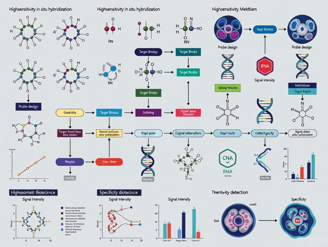
In situ hybridization (ISH), a fundamental histological method for detecting and visualizing nucleic acids within tissues and cells, has undergone significant evolution since its invention over 50 years ago [1]. The drive to simplify complex procedures and enhance detection sensitivity for low-expression genes or short transcripts has led to the development of several high-sensitivity ISH variants [1]. These advanced methods provide researchers with powerful tools to visualize spatial gene expression with single-molecule sensitivity, offering a wider range of options for pathological diagnosis and biological research [1] [2]. Understanding the signal-amplification principles, characteristics, and cost considerations of these methods is essential for their effective application within a broader research context on high-sensitivity in situ hybridization methods [1].
The Need for Enhanced Sensitivity in In Situ Hybridization
Conventional ISH methods, particularly digoxigenin (DIG)-labeled RNA probes, have been a long-standing standard [1]. Sensitivity in these methods can be increased by designing multiple probes to cover different regions of the target mRNA and by employing signal amplification techniques such as Tyramide Signal Amplification (TSA) [1] [3]. However, they possess inherent limitations:
- Complexity and Time-Consumption: ISH procedures are often more complex and time-consuming than immunostaining, limiting their clinical application [1].
- Limited Sensitivity for Low-Abundance Targets: Detecting low-expression genes or short transcripts can be challenging with conventional sensitivity [1].
- Difficulty with Multiplexing and Combination with Immunostaining: Double ISH for two gene transcripts is difficult, especially for genes with high homology or different GC percentages. Combining ISH with immunostaining is also hampered by proteinase treatments and hybridization temperatures that denature proteins and decrease antigen reactivity [1].
High-sensitivity ISH methods address these shortcomings by offering simplified protocols, superior sensitivity capable of detecting single transcripts, and easier multiplexed fluorescence staining [1].
Principles of High-Sensitivity In Situ Hybridization Methods
Recent high-sensitivity ISH variants generally share a common underlying strategy, moving away from long, single probes. They typically employ a two-step process: 1) the use of a synthetic oligonucleotide primary probe that binds to the target sequence, and 2) the hybridization of multiple secondary probes or amplifiers to the primary probe, resulting in a substantial signal enhancement [1]. Key methods include:
RNAscope
RNAscope utilizes a proprietary probe design where pairs of "Z" probes bind to the target RNA. Each Z probe contains a unique tail sequence that serves as a binding site for pre-amplifier and amplifier molecules, which in turn hybridize with multiple labeled probes. This branched DNA structure creates a large signal amplification complex at each target site [1]. The commercial kit simplifies the experimental process, making it user-friendly.
Hybridization Chain Reaction (HCR) In Situ Hybridization
In HCR, an initiator probe bound to the target triggers a cascading, self-assembling reaction between two stable species of fluorescently labeled hairpin DNA molecules [1]. This reaction results in the formation of a long nicked double-stranded DNA polymer, which tethers numerous fluorophores to the site of the target molecule, providing programmable signal amplification [1].
clampFISH
clampFISH (cyclic oligonucleotide-based amplification method) uses padlock probes that are circularized upon hybridization to the target RNA via ligation [1] [2]. This circular structure is then fixed to the target. Fluorescently labeled probes are repeatedly hybridized to the loop portion of the primary probe, achieving high sensitivity through signal accumulation over multiple hybridization cycles [1].
SABER FISH
SABER (Signal Amplification By Exchange Reaction) FISH employs a primer exchange reaction to enzymatically add a long concatemer of identical short repeating sequences to the end of the primary probe before hybridization [1]. A short fluorescent probe is then hybridized to this repeating sequence, and the degree of signal amplification can be tuned by varying the length of the concatemers [1].
Comparative Analysis of High-Sensitivity ISH Methods
The table below summarizes the key characteristics, including monetary and time costs, of different high-sensitivity ISH variants compared to conventional methods [1]:
Table 1: Comparative Characteristics of In Situ Hybridization Methods
| Method | DIG-RNA ISH | RNAscope | HCR ISH | clampFISH | SABER FISH |
|---|---|---|---|---|---|
| Difficulty of Procedures | Difficult | Easy | Moderate | Moderate | Moderate |
| Detection Method | Fluorescent, Chromogenic | Fluorescent, Chromogenic | Fluorescent | Fluorescent | Fluorescent |
| Multiplex Staining | Difficult | Easy | Easy | Easy | Easy |
| Probe Design & Synthesis | By user (can be outsourced) | Provided by manufacturer only | By user (can be outsourced) | By user | By user |
| Monetary Cost (Total) | Low | High | Moderate | Moderate | Moderate |
| Monetary Cost (Per Sample) | Low | High | Decreases with sample size | Decreases with sample size | Decreases with sample size |
| Time Cost (Examination of Conditions) | Necessary | Mostly unnecessary | Necessary | Necessary | Necessary |
| Staining Time | 2–3 days | 1 day | 1–3 days | 1–3 days | 2–3 days |
| Detection of microRNA | Difficult | Applicable | Applicable | — | — |
Table 2: Key Advantages and Considerations for Method Selection
| Method | Key Advantages | Key Considerations |
|---|---|---|
| RNAscope | Simple, standardized protocol; fast (1-day staining); suitable for automated equipment; detects short targets like miRNA. | Highest monetary cost per sample; probe design is restricted to the manufacturer. |
| HCR FISH | Amplification is tunable by reaction time; cost per sample decreases with scale; detects short targets like miRNA. | Requires user optimization and probe design. |
| clampFISH | Probe ligation enhances specificity; cost-effective for large sample numbers. | Requires user optimization and probe design; not yet reported for miRNA. |
| SABER FISH | Signal amplification is tunable via concatemer length; cost-effective for large sample numbers. | Requires user optimization and probe design; not yet reported for miRNA. |
Experimental Protocols for Key High-Sensitivity ISH Methods
RNAscope Protocol (Fluorescent Detection)
- Sample Preparation: Fix tissues in 10% neutral buffered formalin and embed in paraffin (FFPE). Prepare thin sections (5-10 µm) on slides.
- Pretreatment: Deparaffinize slides and perform heat-induced epitope retrieval in a specific retrieval solution. Treat with a mild protease to permeabilize the tissue without destroying RNA.
- Probe Hybridization: Apply target-specific "Z" probe pairs and hybridize for 2 hours at 40°C.
- Signal Amplification:
- Apply pre-amplifier oligos that bind to the "Z" probe tails. Incubate.
- Apply amplifier oligos that bind to the pre-amplifier. Incubate.
- Apply fluorescently labeled probe oligos that bind to the amplifier. Incubate in the dark.
- Detection: Counterstain with DAPI and mount with a fluorescent mounting medium. Image using a fluorescence microscope.
HCR FISH Protocol
- Probe Design: Design and synthesize initiator probes complementary to the target mRNA.
- Sample Preparation and Hybridization: Fix and permeabilize cells or tissues. Hybridize the initiator probes to the target.
- Washing: Wash to remove unbound initiator probes.
- Amplification: Apply the two fluorescent hairpin DNA species (H1 and H2). Incubate for several hours (typically 4-16) to allow the chain reaction to propagate. The duration of this step can be adjusted to control amplification.
- Detection: Wash, counterstain, and mount for fluorescence microscopy.
The Scientist's Toolkit: Essential Research Reagents and Materials
Table 3: Key Research Reagent Solutions for High-Sensitivity ISH
| Reagent/Material | Function | Example Application |
|---|---|---|
| "Z" Probe Pairs | Synthetic oligonucleotides that specifically bind target mRNA and provide a scaffold for signal amplification. | RNAscope [1] |
| Hairpin DNA Oligos (H1 & H2) | Fluorescently labeled, metastable hairpins that self-assemble into a polymerization chain upon initiation. | HCR FISH [1] |
| Padlock Probes | Linear DNA probes whose ends are complementary to adjacent target sequences, allowing circularization via ligation. | clampFISH [1] [2] |
| Primer Exchange Reaction (PER) Scaffolds | DNA templates used to enzymatically synthesize long, single-stranded DNA concatemers onto primary probes. | SABER FISH [1] |
| Tyramide-Based Reagents | Compounds used for enzymatic signal amplification (e.g., TSA), depositing numerous fluorophores at the probe site. | Conventional and smFISH sensitivity enhancement [3] [2] |
| Tissue Clearing Reagents | Chemical solutions that reduce light scattering and opacity in thick tissue samples, improving probe penetration and image quality. | Enhancing specificity in multiplex FISH of thick tissues [2] |
High-sensitivity in situ hybridization methods represent a significant advancement over conventional ISH, providing the capability to visualize gene expression at the single-molecule level with high specificity. Methods such as RNAscope, HCR FISH, clampFISH, and SABER FISH leverage innovative signal amplification principles to overcome the sensitivity and multiplexing limitations of their predecessors. The choice of method depends on the specific research needs, weighing factors such as ease of use, cost, throughput, and the need for customization. As these technologies continue to evolve and integrate with other spatial biology techniques, they will play an increasingly vital role in fundamental research, biomarker discovery, and clinical diagnostics [1] [2].
The ability to detect individual RNA molecules within their native cellular environment has revolutionized our understanding of gene expression, cellular heterogeneity, and regulatory mechanisms. This technical guide explores the core principles by which signal amplification strategies enable single-molecule RNA detection, with a specific focus on high-sensitivity in situ hybridization methods. We examine the technological evolution from conventional ensemble measurements to digital counting of individual transcripts, detailing the amplification chemistries, probe design considerations, and imaging platforms that make single-molecule sensitivity possible. Within the broader context of thesis research on high-sensitivity detection platforms, this review provides researchers and drug development professionals with a comprehensive framework for selecting, optimizing, and implementing these powerful techniques in their experimental workflows.
The spatial and temporal distribution of RNA molecules within cells and tissues provides critical information about gene regulation that is lost in bulk analysis methods. Single-molecule RNA detection represents the ultimate sensitivity limit for transcriptional analysis, allowing researchers to quantify absolute RNA copy numbers, identify rare splice variants, and map subcellular localization patterns with nanometer-scale precision [4]. The fundamental challenge in achieving this sensitivity lies in the fact that a single nucleic acid molecule provides an extremely weak signal that is typically drowned out by background noise. Signal amplification strategies overcome this limitation by dramatically enhancing the detectable output from each individual target molecule, enabling its visualization and quantification against the cellular autofluorescence and nonspecific binding background [5].
The development of these methods has revealed that gene expression is highly stochastic at the cellular level, with individual cells within a seemingly homogeneous population exhibiting dramatic differences in transcript abundance [4]. Understanding this heterogeneity is particularly crucial in drug development, where rare cell populations with distinct expression profiles may drive treatment resistance or disease progression. This technical guide examines the core amplification principles that have enabled this resolution, focusing specifically on their implementation in high-sensitivity in situ hybridization workflows for both basic research and clinical applications.
Fundamental Principles of Signal Amplification
Signal amplification strategies for single-molecule RNA detection operate on a simple but powerful principle: each target molecule must generate a signal sufficiently bright to be distinguished from background fluorescence and reliably detected by microscopy systems. This is achieved through various mechanisms that multiply the number of fluorophores associated with each target RNA.
Signal-to-Noise Considerations
At the heart of all single-molecule detection methods lies the critical relationship between signal intensity and background noise. The signal-to-noise ratio (SNR) determines the detection reliability, with SNR >10 generally required for confident identification of individual molecules [6]. Background signals arise from multiple sources, including cellular autofluorescence, nonspecific probe binding, and impurities in the substrate or reagents. Effective signal amplification strategies must therefore not only enhance the target-associated signal but also minimize these background components through careful probe design, optimized hybridization conditions, and effective blocking strategies [5].
The Single-Molecule Brightness Threshold
For a single RNA molecule to be detectable, it must be associated with sufficient fluorophores to generate a diffraction-limited spot whose intensity exceeds the background by a statistically significant margin. Conventional fluorescent in situ hybridization (FISH) with singly-labeled probes falls short of this threshold, as the approximately 20-50 fluorophores from typical probe sets produce insufficient signal [4]. Amplification strategies overcome this limitation by associating hundreds to thousands of fluorophores with each target molecule, either through enzymatic deposition or hybridization chain reactions, pushing the signal well above the detection threshold [7].
Key Signal Amplification Technologies
Several sophisticated signal amplification technologies have been developed to achieve single-molecule sensitivity for RNA detection. Each employs distinct mechanisms and offers unique advantages for specific applications.
Hybridization Chain Reaction (HCR)
HCR is an enzyme-free amplification method that uses metastable DNA hairpins that undergo a chain reaction of hybridization events initiated by a specific probe bound to the target RNA [4]. The initiator probe triggers the sequential opening and hybridization of fluorophore-labeled hairpins, forming a long nicked double helix that accumulates hundreds of fluorophores at the site of the target RNA.
Table 1: Performance Characteristics of HCR and Variants
| Method | Probe Pairs Required | Approximate Fluorophores per Target | Detection Limit | Key Advantages |
|---|---|---|---|---|
| Standard HCR | 20-40 | 200-500 | ~1.3 fM [6] | Enzyme-free, high multiplexing capability |
| Yn-situ HCR | 3-5 [7] | 400-800 [7] | <1.3 fM [7] | Fewer probes required, cost-effective |
| HCR with preamplifier | 3-5 [7] | 1000+ [7] | Single molecules [7] | Highest sensitivity, small puncta size |
Diagram 1: HCR Amplification Mechanism
Tyramide Signal Amplification (TSA)
Also known as catalyzed reporter deposition (CARD), TSA utilizes horseradish peroxidase (HRP) conjugated to a detection probe. The enzyme catalyzes the deposition of numerous tyramide-fluorophore conjugates in the immediate vicinity of the target RNA [5]. This method provides extremely high amplification factors (up to 100-fold) but can sometimes suffer from reduced spatial resolution due to diffusion of the reactive tyramide intermediates.
Rolling Circle Amplification (RCA)
RCA is an isothermal amplification technique that generates long single-stranded DNA molecules containing hundreds of tandem repeats complementary to a secondary detection probe [8]. In the context of RNA detection, padlock probes are first hybridized to the target RNA and then circularized by ligation. DNA polymerase then extends repeatedly around the circular template, producing a long concatemer that can be detected with fluorescent probes [4].
Branched DNA (bDNA) Signal Amplification
The bDNA method, commercialized as RNAscope, employs a series of sequential hybridizations to build a branched nucleic acid structure on the target RNA. This scaffold then supports the binding of hundreds of labeled oligonucleotides, providing strong amplification without enzymatic steps [4]. The method is known for its exceptional specificity and robustness, making it particularly valuable for clinical samples.
Table 2: Comparison of Major Signal Amplification Technologies
| Amplification Method | Amplification Mechanism | Typical Amplification Factor | Spatial Resolution | Key Applications |
|---|---|---|---|---|
| HCR | Enzyme-free hybridization chain reaction | 200-500x | High (∼20 nm) | Multiplexed imaging, live-cell applications |
| TSA | Enzymatic deposition of tyramide-fluorophores | 50-100x | Moderate (∼50 nm) | Low-abundance targets, immunohistochemistry |
| RCA | Enzymatic DNA polymerization | 100-1000x | High (∼20 nm) | microRNA detection, short transcripts |
| bDNA/RNAscope | Sequential hybridization of branched structure | 400-800x | High (∼20 nm) | Clinical diagnostics, archival samples |
Advanced Methodologies and Protocols
Yn-Situ: An Enhanced HCR Approach
The Yn-situ method represents a significant advancement in HCR technology by introducing a preamplifier structure that dramatically increases the amplification efficiency while reducing the number of target-specific probes required [7]. The key innovation is a Y-shaped DNA preamplifier containing multiple initiator sequences that simultaneously trigger numerous HCR reactions.
Yn-Situ Experimental Protocol
Probe Design and Synthesis:
- Target Probe Design: Design 3-5 pairs of target-specific probes (52 nt each) with complementary regions to both the target RNA and the preamplifier sequence [7].
- Preamplifier Synthesis: Clone the preamplifier sequence (∼1 kb with 20 HCR initiator repeats) into a plasmid vector. Amplify using LongAmp Taq DNA polymerase with the following optimized conditions: 0.5 μM primer concentration, 0.05 ng/μL template concentration, and annealing temperature of 60-68°C [7].
- Single-Strand Generation: Generate single-stranded preamplifier using asymmetric PCR with a 5'-phosphorylated reverse primer, followed by strandase digestion to remove the antisense strand [7].
Sample Preparation and Hybridization:
- Tissue Fixation: Fix tissues with 4% formaldehyde followed by crosslinking with 1-ethyl-3-(3-dimethylaminopropyl) carbodiimide (EDC) to immobilize RNA molecules and reduce degradation [7].
- Hybridization Conditions: Hybridize target probes at 0.002-0.2 ng/μL concentration in appropriate hybridization buffer. For Yn-situ, 5 probe pairs typically yield optimal results, though detection is possible with as few as 1-3 pairs [7].
- Signal Amplification: Apply preamplifier at 0.2 ng/μL concentration, followed by HCR hairpins with fluorophores appropriate for your detection system.
- Imaging: Acquire images using epifluorescence or confocal microscopy with appropriate filters. For single-molecule quantification, use high numerical aperture objectives and sensitive detectors [7].
Diagram 2: Yn-situ Workflow
Quantitative Analysis of Single-Molecule Data
The transition from ensemble measurements to digital counting fundamentally changes the analytical approach for gene expression quantification. Instead of measuring average fluorescence intensity across a population, single-molecule methods involve identifying and counting individual diffraction-limited spots, each representing one RNA molecule [6].
Image Analysis Pipeline:
- Preprocessing: Apply background subtraction and flat-field correction to account for uneven illumination.
- Spot Detection: Use Laplacian of Gaussian or similar algorithms to identify local intensity maxima corresponding to individual RNA molecules.
- Quantification: Count spots within cellular or subcellular regions of interest. For high-density situations where spots overlap, estimate molecule numbers by dividing total fluorescence intensity by the characteristic single-molecule brightness [6].
- Statistical Analysis: Account for the stochastic nature of molecular detection by modeling counting statistics as Poisson distributions, particularly important for low-abundance transcripts [6].
The Scientist's Toolkit: Essential Research Reagents
Successful implementation of single-molecule RNA detection methods requires careful selection of reagents and materials. The following table outlines key components and their functions in typical experimental workflows.
Table 3: Essential Research Reagent Solutions for Single-Molecule RNA Detection
| Reagent/Material | Function | Key Considerations | Example Applications |
|---|---|---|---|
| Target-specific probes (20-50 nt) | Hybridize specifically to target RNA sequence | Optimize length for specificity and penetration; modify with haptens for detection | All in situ hybridization methods |
| HCR hairpin systems | Amplification through hybridization chain reaction | Design metastable hairpins to minimize background; fluorophore selection critical | HCR, Yn-situ [7] |
| Tyramide-fluorophore conjugates | Enzyme-mediated signal deposition | Optimize concentration to balance signal and diffusion; HRP activity critical | TSA/CARD amplification [5] |
| Padlock probes/RCA components | Circularizable probes for rolling circle amplification | Requires precise design for efficient ligation; high-fidelity polymerase needed | RCA-based detection [8] |
| Preamplifier constructs | Intermediate amplification scaffold | Contains multiple initiator sequences; optimized length for penetration | Yn-situ method [7] |
| EDC crosslinking reagent | RNA immobilization to proteins | Preserves RNA integrity; particularly important for archived samples | Tissue pretreatment [7] |
| Blocking reagents (e.g., BlockAid) | Reduce nonspecific background | Species-specific blocking for immuno-detection; RNA-grade reagents | All methods to improve SNR [5] |
| High-performance polymerases (e.g., LongAmp) | Amplification of preamplifier constructs | High processivity for long amplicons; minimal sequence bias | Preamplifier synthesis [7] |
Performance Metrics and Validation
Sensitivity and Detection Limits
The ultimate sensitivity of single-molecule RNA detection methods is determined by the combination of amplification efficiency and background suppression. The theoretical detection limit is a single RNA molecule, but practical sensitivity depends on the specific implementation and sample type.
Table 4: Performance Metrics of Single-Molecule RNA Detection Methods
| Performance Metric | Typical Range | Factors Influencing Performance | Optimization Strategies |
|---|---|---|---|
| Detection limit | 1.3 fM - 39,000 molecules [6] | Probe affinity, background fluorescence, amplification efficiency | Optimize hybridization stringency, improve fixation [7] |
| Dynamic range | 4.7-6 orders of magnitude [6] | Signal saturation at high abundances, background at low abundances | Adjust probe concentration, use multiple detection channels |
| Spatial resolution | 20-50 nm [4] | Diffusion of amplification products, probe size | Use smaller probes, optimize deposition time [7] |
| Quantification accuracy | >95% for well-separated molecules [6] | Molecular crowding, labeling efficiency | Account for Poisson statistics, correct for detection efficiency |
| Multiplexing capacity | 3-5 colors routinely [4] | Spectral overlap, cross-reactivity | Sequential hybridization, spectral unmixing |
Validation Approaches
Validating single-molecule detection data requires multiple complementary approaches:
- Control experiments: Include samples without primary probes, with sense-strand probes, or with RNase pretreatment to establish specificity.
- Comparison with orthogonal methods: Correlate results with qPCR or RNA-seq data from the same sample types [6].
- Spike-in controls: Use synthetic RNA targets at known concentrations to establish quantification accuracy.
- Cross-platform validation: Compare results across different amplification methods (e.g., HCR vs. RNAscope) to confirm detection robustness [7].
Applications in Research and Drug Development
Single-molecule RNA detection methods have enabled breakthrough applications across multiple domains:
Cellular Heterogeneity Studies: By revealing the complete transcriptome variation within cell populations, these methods have transformed our understanding of development, cancer progression, and neural circuitry [4].
Subcellular Localization Analysis: The high spatial resolution enables precise mapping of RNA localization to specific cellular compartments, providing insights into localized translation and RNA transport mechanisms [4].
Diagnostic Applications: In clinical settings, these methods enable detection of rare transcripts or viral RNAs in patient samples, with potential applications in cancer diagnosis, infectious disease monitoring, and personalized medicine [9].
Drug Discovery and Development: Pharmaceutical researchers utilize these techniques to monitor drug-induced changes in gene expression at single-cell resolution, identify rare resistant cell populations, and validate therapeutic mechanisms of action.
Signal amplification technologies have fundamentally transformed our ability to detect and quantify individual RNA molecules within their native cellular contexts. The core principles underlying these methods—exponential signal enhancement through enzymatic or hybridization-based amplification, combined with sophisticated background suppression strategies—have enabled researchers to push detection sensitivity to the ultimate physical limit. As these technologies continue to evolve, we anticipate further improvements in multiplexing capability, spatial resolution, and compatibility with complex clinical samples. The integration of single-molecule RNA detection with other omics technologies and functional assays will provide increasingly comprehensive views of cellular function in health and disease, offering unprecedented opportunities for basic research and therapeutic development.
Key Technological Advancements Driving the ISH Sensitivity Revolution
In situ hybridization (ISH) has undergone a revolutionary transformation from a qualitative histological tool to a highly sensitive, quantitative spatial biology platform. This whitepaper examines the key technological advancements—including advanced probe designs, signal amplification strategies, and automated quantification frameworks—that have collectively driven this sensitivity revolution. These innovations now enable researchers to detect single RNA molecules within their native tissue context, providing unprecedented insights into gene expression patterns, cellular heterogeneity, and disease mechanisms. The integration of these technologies has positioned ISH as an indispensable tool for biomedical research, clinical diagnostics, and drug development programs requiring precise spatial localization of nucleic acid targets.
In situ hybridization, a fundamental method for visualizing nucleic acids within tissues and cells, has experienced remarkable technological evolution since its inception. The global ISH market, valued at USD 1.82 billion in 2023 and projected to reach USD 3.43 billion by 2032, reflects the growing adoption of these advanced methodologies across research and clinical diagnostics [10]. This growth is fundamentally driven by revolutionary improvements in detection sensitivity, which have transformed ISH from a technique capable of identifying only highly abundant transcripts to one that can visualize individual RNA molecules.
The sensitivity revolution addresses a critical limitation of conventional ISH: its inability to reliably detect low-expression genes or short transcripts. Traditional methods using digoxigenin (DIG)-labeled RNA probes, while valuable, presented complex procedures and limited sensitivity despite optimization attempts through increased target sequence coverage or enzymatic signal amplification [1]. Recent technological shifts have overcome these limitations through three interconnected advancements: novel probe architectures, sophisticated amplification strategies, and automated quantification frameworks. These innovations have collectively established a new sensitivity paradigm that preserves the spatial context essential for understanding tissue heterogeneity and cellular function in development, homeostasis, and disease.
Core Technological Advancements
Advanced Probe Design Architectures
Modern ISH methodologies have fundamentally reimagined probe architecture, moving from long riboprobes to sophisticated short oligonucleotide designs that enable unprecedented specificity and signal amplification.
Dual Z-Probe Technology (RNAscope) utilizes paired probes that bind adjacent to each other on the target RNA. This design requires both probes to hybridize correctly for signal generation, dramatically reducing false positives from non-specific binding. The system employs a pre-amplifier that only binds when both probes are present, followed by hybridization of multiple amplifier molecules and enzyme-linked label probes, creating substantial signal amplification [1].
Padlock Probes (clampFISH) circularize upon hybridization to their target sequences. Once circularized, they are permanently fixed to the target via click chemistry, allowing repeated hybridization of fluorescent probes to the circular structure. This creates a stable, amplifiable signal that withstands stringent washing conditions, significantly enhancing signal-to-noise ratio [1].
Primer Exchange Probes (SABER FISH) employ a primer exchange reaction that adds a short repeating sequence to the end of the primary probe before hybridization. This concatenated sequence serves as a docking site for multiple fluorescently labeled oligonucleotides, enabling tunable signal amplification by varying the length of the concatemers [1].
Hybridization Chain Reaction (HCR) utilizes initiator probes that trigger the self-assembly of fluorescently labeled hairpin DNA molecules into long amplification polymers. The degree of amplification can be precisely controlled by reaction time, offering researchers flexibility in optimizing signal intensity for different applications [1].
Table 1: Comparative Analysis of High-Sensitivity ISH Probe Technologies
| Technology | Signal Amplification Mechanism | Detection Limit | Multiplexing Capability | Implementation Complexity |
|---|---|---|---|---|
| RNAscope | Branched DNA (bDNA) cascade | Single RNA molecules | High (up to 12-plex) | Low (commercial kit) |
| HCR FISH | Hybridization chain reaction | Single RNA molecules | High | Moderate |
| clampFISH | Circular probe with multiple labeling | Single RNA molecules | Moderate | High |
| SABER FISH | Primer exchange reaction with concatemers | Single RNA molecules | High | High |
| Conventional ISH | Enzymatic (TSA) or direct labeling | ~10 RNA molecules | Limited | Moderate |
Signal Amplification Strategies
Sensitivity enhancements in ISH have been achieved through innovative signal amplification strategies that dramatically increase the signal generated from each target molecule while minimizing background noise.
Tyramide Signal Amplification (TSA) employs an enzymatic reaction where horseradish peroxidase (HRP) catalyzes the deposition of multiple fluorescent or chromogenic tyramide molecules near the hybridization site. This approach significantly enhances detection sensitivity for low-abundance targets but can cause signal diffusion, potentially limiting spatial resolution [3].
Branched DNA (bDNA) Amplification, utilized in RNAscope, creates a tree-like structure through sequential hybridization steps. This multi-layer amplification system can incorporate hundreds of label molecules per target, enabling single-molecule detection without RNA degradation or enzymatic reactions that might compromise tissue morphology [1].
Hybridization Chain Reaction (HCR) offers an enzyme-free, isothermal amplification alternative. The metastable DNA hairpins remain stable until initiated by target-bound probes, then self-assemble into fluorescent amplification polymers. This method provides uniform signal amplification and straightforward multiplexing through orthogonal hairpin systems [1] [2].
Nanoparticle-Based Amplification utilizes quantum dots or gold nanoparticles as fluorescence signal carriers. These nanomaterials offer exceptional brightness and photostability compared to conventional fluorophores. The large surface area of nanoparticles allows multiple detection probes to bind to a single particle, enhancing signal intensity, particularly when combined with catalytic deposition of additional signal-generating molecules [2].
Multiplexing and Throughput Enhancement
Modern ISH technologies have evolved to enable highly multiplexed analysis, allowing simultaneous detection of numerous targets within a single sample.
Barcode-Based Approaches assign unique oligonucleotide sequences to different targets. Through sequential hybridization, imaging, and signal removal cycles, these systems can detect dozens to hundreds of distinct RNA species in the same tissue section. Techniques like seqFISH and MERFISH exemplify this approach, creating a high-dimensional spatial transcriptomic profile while maintaining single-cell resolution [2].
Multi-fluorescence Strategies employ spectrally distinct fluorophores that can be discriminated through spectral imaging. By combining different fluorophores in various ratios, a large number of targets can be identified simultaneously. This approach benefits from advanced fluorescence microscopy systems with high spectral resolution and sophisticated unmixing algorithms [2].
Automated Platforms have been developed to standardize ISH workflows and increase reproducibility. Companies including Roche, Biocare Medical, and Leica Biosystems have introduced automated systems that reduce manual processing time from 2-3 days to a single day while maintaining high accuracy and enabling high-throughput applications [11] [10].
Specificity Enhancement Methodologies
Enhanced specificity has been as crucial as improved sensitivity in the ISH revolution, particularly for discriminating highly homologous sequences and reducing background signal.
Split-Probe Designs, exemplified by RNAscope, require two independent probe binding events for signal generation. This approach essentially creates a logical "AND" gate at the molecular level, where both probes must correctly hybridize to adjacent target regions to form a complete binding site for the pre-amplifier molecule. This dramatically reduces non-specific signal compared to single-probe systems [1].
Tissue Clearing Methods improve probe penetration and reduce light scattering in thick tissue samples. Techniques such as CLARITY, CUBIC, and various hydrogel-based methods render tissues optically transparent while preserving nucleic acids for hybridization. This enhances signal-to-noise ratio by reducing autofluorescence and enabling more complete probe access throughout the sample [2].
Strand Displacement and Toehold-Mediated Reactions provide additional specificity through sequence-specific probe activation. These systems employ probes that remain inactive until they encounter the correct target sequence, which then triggers a conformational change or displacement reaction that enables signal generation. This approach minimizes off-target binding and background signal [2].
Experimental Framework and Analytical Validation
Quantitative Image Analysis Framework
The sensitivity revolution in ISH necessitated parallel development of sophisticated computational tools for accurate signal quantification and interpretation.
The QuantISH Framework represents a comprehensive open-source pipeline specifically designed for RNA-ISH image analysis. This modular system quantifies marker expressions in individual carcinoma, immune, and stromal cells from chromogenic or fluorescent ISH images. Its processing workflow includes:
- Image Pre-processing: Extracting contiguous images from tiled microscope scans and cropping tissue microarray spots using customized modules [12].
- Color Demultiplexing: Separating marker RNA stain from nuclear counterstain using color deconvolution algorithms, followed by Renyi entropy thresholding to filter background noise [12].
- Cell Segmentation: Identifying individual cells using intensity-based algorithms with shape information to separate clumped objects [12].
- Cell Type Classification: Categorizing cells based on nuclear morphology features including area, mean intensity, eccentricity, and solidity [12].
- Expression Quantification: Calculating normalized signal intensity per cell and deriving sample-level metrics including average expression and variability factors [12].
Variability Factor Analysis leverages the single-cell resolution of modern ISH to quantify expression heterogeneity within tissue samples. This approach characterizes biological variability independently of mean expression levels, enabling quantitative comparison of expression heterogeneity between samples—a crucial capability for understanding tumor evolution and treatment resistance [12].
Table 2: Key Research Reagent Solutions for High-Sensitivity ISH
| Reagent Category | Specific Examples | Function & Importance |
|---|---|---|
| Probe Systems | RNAscope probes, HCR initiator probes, padlock probes | Target-specific hybridization for precise nucleic acid detection |
| Amplification Reagents | Branched DNA amplifiers, fluorescent tyramides, DNA hairpins | Signal enhancement for low-abundance target detection |
| Enzymatic Systems | HRP-conjugated antibodies, polymerase for primer exchange | Facilitate signal generation and amplification cascades |
| Labeling Molecules | Fluorophore-conjugated nucleotides, quantum dots, chromogenic substrates | Direct signal generation with various detection modalities |
| Tissue Processing Reagents | Protease treatments, permeabilization buffers, tissue clearing solutions | Enable probe access to targets while preserving morphology |
| Hybridization Buffers | Formamide-based hybridization solutions, salinity adjustment buffers | Optimize stringency conditions for specific hybridization |
| Automation Consumables | Cartridges for automated platforms, standardized reagent kits | Ensure reproducibility and high-throughput application |
Cross-Platform Validation Studies
Rigorous validation has been essential for establishing the reliability of high-sensitivity ISH methods. A landmark study comparing genome-scale colorimetric ISH data from the Allen Brain Atlas with microarray data from Teragenomics and GNF revealed significant correlations across six major brain structures (striatum, cortex, cerebellum, hippocampus, olfactory bulb, and hypothalamus) [3]. This comprehensive analysis demonstrated that properly quantified ISH data provides biologically relevant expression measurements comparable to established quantitative platforms, while adding the crucial dimension of spatial resolution.
The development of standardized relative quantification methods for colorimetric ISH signal has enabled meaningful cross-platform comparisons. These approaches use cellular resolution image segmentation corroborated by tissue optical density to define expression levels analogous to microarray data, facilitating integration of spatial transcriptomics with bulk expression profiling datasets [3].
Research Applications and Impact
Cancer Research and Biomarker Discovery
High-sensitivity ISH has transformed cancer research by enabling precise localization of oncogenic transcripts within tumor microenvironments. In high-grade serous carcinoma (HGSC), QuantISH analysis has revealed that CCNE1 average expression and DDIT3 expression variability serve as candidate biomarkers with potential prognostic significance [12]. The ability to quantify expression heterogeneity at single-cell resolution provides unique insights into tumor evolution, therapeutic resistance, and intratumoral heterogeneity that are obscured in bulk analyses.
The technology has proven particularly valuable for detecting low-abundance transcripts driving cancer pathogenesis, including non-coding RNAs, splice variants, and mutated alleles. In clinical diagnostics, RNAscope has demonstrated superior sensitivity for detecting viral RNAs in infectious diseases and chromosomal rearrangements in hematological malignancies, enabling more accurate patient stratification and therapy selection [1].
Neuroscience and Neurological Disorders
The complexity of the nervous system, with its extraordinary cellular diversity and spatially organized circuitry, makes it particularly suited to ISH-based analysis. Multiplexed ISH technologies have enabled comprehensive molecular profiling of neuronal subtypes and mapping of transcript distribution within specialized subcellular compartments. These applications have provided insights into localized translation in dendrites and axons, RNA transport mechanisms, and activity-dependent gene expression changes with implications for learning, memory, and neurological diseases [11] [3].
The application of high-sensitivity ISH in neurology is expanding rapidly, with the segment poised to grow at a solid CAGR of 22% during the forecast period [11]. This growth reflects increasing adoption of these methods for examining gene expression patterns and biomarkers in neurological tissues, both for basic research and diagnostic applications.
Drug Development and Therapeutic Validation
In pharmaceutical research, high-sensitivity ISH has become invaluable for target validation, biodistribution studies, and pharmacodynamic biomarker assessment. The technology enables precise localization of target mRNA expression within specific cell types and tissue compartments, informing drug candidate selection and potential toxicity concerns. During preclinical development, ISH methods are used to monitor therapy-induced changes in gene expression, providing mechanistic insights into drug action and resistance mechanisms.
Spatial transcriptomic profiling using multiplexed ISH technologies helps identify novel biomarkers for patient stratification and treatment response prediction. The growing adoption of these approaches in contract research organizations reflects the pharmaceutical industry's increasing reliance on spatial biology for drug discovery and development [10].
Future Perspectives and Emerging Directions
The ISH sensitivity revolution continues to evolve, with several emerging trends shaping future development. Integration of artificial intelligence and machine learning for image analysis is accelerating quantification workflows and enabling more sophisticated pattern recognition in complex tissue architectures. Computational approaches are increasingly being used to distinguish specific signal from background, identify rare cell populations, and extract spatially resolved gene expression networks [12].
Multi-omics integration represents another frontier, with methods being developed for simultaneous detection of RNA, protein, and epigenetic markers in the same tissue section. Techniques like RNAscope-ISH combined with immunohistochemistry or CODEX multiplexed tissue imaging are providing comprehensive molecular profiling while maintaining spatial context [2]. These approaches offer unprecedented insights into the complex relationships between different molecular layers in health and disease.
Nanotechnology applications continue to advance, with novel nanomaterials including quantum dots, upconversion nanoparticles, and DNA nanostructures being explored for further sensitivity enhancements. These materials offer improved photostability, multiplexing capacity, and signal amplification potential compared to conventional fluorophores [2].
As the field progresses, challenges remain in standardization, reproducibility, and accessibility. Automated platforms and commercial reagent kits are addressing some of these concerns by making high-sensitivity ISH more accessible to non-specialist laboratories. The continued development of open-source analysis tools like QuantISH will be crucial for ensuring broad adoption and methodological standardization across the research community [12].
The ISH sensitivity revolution has fundamentally transformed spatial transcriptomics from a qualitative histological technique to a precise quantitative platform capable of single-molecule detection. Through innovations in probe design, signal amplification, multiplexing, and computational analysis, modern ISH methods now provide unprecedented insights into gene expression patterns within their native tissue context. These advancements have enabled new discoveries in cellular heterogeneity, tumor biology, neural circuitry, and disease mechanisms that were previously inaccessible.
The integration of high-sensitivity ISH into research and clinical workflows continues to accelerate, driven by ongoing technological refinements and expanding applications across biomedical disciplines. As these methods become increasingly sophisticated and accessible, they will further illuminate the spatial architecture of gene expression, advancing our understanding of biological systems and enhancing our ability to diagnose and treat human diseases.
Comparative Advantages of ISH Versus Immunostaining for Spatial Biology
In situ hybridization (ISH) and immunostaining (including immunohistochemistry - IHC - and immunofluorescence - IF) represent two foundational pillars of spatial biology. While ISH directly targets nucleic acids (DNA/RNA) to reveal transcriptional activity, immunostaining detects proteins, providing insight into translational output and post-translational modifications. The selection between these methodologies hinges on the research question, target biomolecule, and required sensitivity. Emerging spatial multi-omics approaches now strategically integrate both to gain a more comprehensive understanding of cellular function within tissue architecture, correlating gene expression patterns with protein abundance in the same biological context [13].
Fundamental Technical Principles and Applications
Core Methodologies and Targets
The fundamental distinction between these techniques lies in their molecular targets and detection mechanisms.
In Situ Hybridization (ISH): This technique uses labeled complementary DNA or RNA probes to bind specific nucleic acid sequences within cells or tissue sections. ISH is ideally suited for localizing genes on chromosomes, detecting viral DNA, and visualizing mRNA transcripts to map active gene expression patterns, providing insights into cellular heterogeneity and function [14] [15]. Probes can be labeled with fluorescent tags (FISH) or enzymes that produce a colorimetric reaction, allowing visualization via microscopy [14].
Immunostaining (IHC/IF): This method relies on antibodies that bind specifically to protein targets (antigens) within tissue sections. The antibodies are tagged with fluorophores for fluorescence detection (IF) or enzymes that generate colorimetric signals (IHC). Immunostaining reveals the final protein products of gene expression, defining cell neighborhoods, states, and tissue architecture through protein localization [13].
Table 1: Core Characteristics of ISH and Immunostaining
| Feature | In Situ Hybridization (ISH) | Immunostaining (IHC/IF) |
|---|---|---|
| Primary Target | Nucleic Acids (DNA, RNA) [14] | Proteins (Antigens) [13] |
| Detection Mechanism | Complementary probe hybridization [14] | Antigen-antibody binding [13] |
| Key Application | Gene localization, mRNA expression, viral detection [15] | Protein localization, abundance, post-translational modifications [13] |
| Spatial Context | Preserves tissue architecture for nucleic acid localization [14] | Preserves tissue architecture for protein localization [13] |
| Typical Output | Transcript location and activity [13] | Protein presence and cellular distribution [13] |
Visualizing the Fundamental Detection Principles
The following diagram illustrates the core detection mechanisms that differentiate ISH from Immunostaining.
Performance and Operational Comparison
A critical step in experimental design is understanding the performance characteristics and practical requirements of each technique.
Quantitative Performance Metrics
Sensitivity, resolution, and multiplexing capacity are key differentiators.
Sensitivity and Resolution: Standard ISH, particularly single-molecule FISH (smFISH), can achieve single-molecule sensitivity, making it possible to detect individual RNA transcripts [2]. However, directly labeled fluorescent signals can be weak, often necessitating signal amplification strategies to detect low-abundance targets [2]. Immunostaining generally benefits from higher target copy numbers, as proteins are often more abundant than their corresponding mRNA transcripts, which can lead to more robust detection [16].
Multiplexing Capacity: Both techniques have seen significant advances in multiplexing. For ISH, combinatorial coding strategies (e.g., MERFISH, seqFISH+) now enable the simultaneous detection of hundreds to thousands of RNA species [17]. Immunostaining has also progressed, with multiplexed IHC/IF (mIHC/mIF) allowing for the concurrent detection of multiple proteins, typically in the range of 4-9 markers for clinically applicable assays, and even higher with specialized technologies [18].
Table 2: Performance and Operational Comparison
| Parameter | In Situ Hybridization (ISH) | Immunostaining (IHC/IF) |
|---|---|---|
| Sensitivity | High (can achieve single-molecule resolution) [2] | Variable (generally robust due to higher protein copy numbers) [16] |
| Spatial Resolution | Single-cell and subcellular resolution [17] | Single-cell and subcellular resolution (∼200nm) [18] |
| Multiplexing Potential | Very High (theoretically thousands of targets with iterative methods) [17] | Medium to High (typically 4-9 markers for robust clinical panels) [18] |
| Dynamic Range | Lower for direct detection, improved with amplification | Medium (∼3 log) [18] |
| Throughput | Lower for high-plex methods; faster with newer tech (e.g., TDDN-FISH: ~1h) [17] | Medium to High (platform dependent) [18] |
| Primary Challenge | Signal strength for low-copy RNA, protocol harshness [13] [2] | Antibody specificity, spectral overlap, autofluorescence [18] |
Protocol and Workflow Considerations
Practical implementation involves several critical steps.
Tissue Preparation and Pre-analytical Variables: Both techniques commonly use Formalin-Fixed Paraffin-Embedded (FFPE) tissue, but optimal conditions conflict. ISH requires protease treatments and stringent washes that can destroy protein epitopes, while IHC reagents may introduce RNases that degrade RNA targets [13]. For immunostaining, autofluorescence enhanced by formalin fixation is a significant source of noise that can compromise detection of low-abundance targets [18].
Protocol Modifications for Integration: To enable dual RNA-protein detection in the same section, specific workflow modifications are essential. These include:
- RNase Inhibition: Using recombinant ribonuclease inhibitors during antibody incubation to preserve RNA integrity [13].
- Antibody Crosslinking: Crosslinking antibodies to the tissue after IHC labeling to protect them from degradation during subsequent ISH protease treatments [13].
- Signal Amplification: ISH often requires amplification for sufficient signal. Branched DNA systems (e.g., ViewRNA) or novel DNA nanostructures (e.g., TDDN-FISH) provide high sensitivity and low background [13] [17].
Advanced Methodologies and Integrated Workflows
High-Sensitivity ISH Experimental Protocols
Recent breakthroughs have produced ISH methods with dramatically improved speed and sensitivity.
Tetrahedral DNA Dendritic Nanostructure–Enhanced FISH (TDDN-FISH): This enzyme-free protocol uses self-assembling DNA nanostructures for exponential signal amplification [17].
- Probe Design: Engineer a hierarchical Tetrahedral DNA Dendritic Nanostructure (TDDN) from three tetrahedral DNA monomers (T0, T1, T2) with sticky ends for layer-by-layer assembly.
- Hybridization: A bifunctional primary probe with a target-specific sequence binds endogenous mRNA. The TDDN, functionalized with complementary sticky ends, is then hybridized to the primary probe's readout sequence.
- Signal Amplification & Imaging: The multi-layered TDDN structure carries a high density of fluorophores, generating a dramatically amplified signal. This allows for rapid imaging (~1 hour post-hybridization), which is approximately eightfold faster than HCR-FISH, and enables detection of short RNAs like miRNAs [17].
FISHnCHIPs for Spatial Transcriptomics: This protocol enhances sensitivity by simultaneously imaging multiple co-expressed genes.
- Gene Selection: Using a reference scRNA-seq dataset, identify groups of correlated genes (modules or expression programs).
- Probe Pool Design: Design thousands of oligonucleotide probes against all transcripts in a selected module.
- Hybridization and Detection: The pooled probes are hybridized, and the combined signal from multiple co-localized genes provides a ~2-20-fold higher sensitivity than single-gene FISH, enabling robust cell typing and large field-of-view tissue profiling [16].
Integrated Spatial Multi-Omics Workflow
Combining ISH and IHC provides a powerful multi-omics view, as shown in the following workflow.
The Scientist's Toolkit: Key Research Reagent Solutions
Successful implementation of these advanced techniques relies on specific, high-quality reagents.
Table 3: Essential Research Reagents for Spatial Biology
| Reagent / Solution | Function | Example Application |
|---|---|---|
| RNase Inhibitors (e.g., RNaseOUT) | Protects RNA integrity during antibody incubation steps in combined workflows [13] | Dual ISH-IHC protocols |
| Branched DNA ISH Kits (e.g., ViewRNA) | Amplifies signal for RNA detection with high sensitivity and low background; enables multiplexing [13] | Spatial transcriptomics |
| Antibody Crosslinkers | Stabilizes antibody-antigen bonds to withstand harsh ISH conditions [13] | Dual ISH-IHC protocols |
| Multiplex IHC Amplification Reagents | Enhances signal for detecting low-abundance proteins in multiplex panels [13] | Multiplex immunofluorescence (mIF) |
| Tetrahedral DNA Dendritic Nanostructures (TDDN) | Enzyme-free, self-assembling nanostructures for exponential signal amplification in FISH [17] | TDDN-FISH for rapid, sensitive RNA detection |
| Superchaotropes & Macrocyclic Hosts (e.g., [B12H12]²⁻ / γ-cyclodextrin) | Enables homogeneous, deep penetration of macromolecular probes in 3D tissues for volumetric imaging [19] | 3D spatial biology (INSIHGT method) |
| Spectral Unmixing Software | Separates overlapping fluorescence signals and reduces autofluorescence [18] | Analysis of multiplex fluorescence data |
ISH and immunostaining are complementary, not competing, technologies in the spatial biology arsenal. ISH excels in mapping transcriptional activity with high multiplexing potential, while immunostaining directly quantifies protein expression and modification. The choice depends fundamentally on the biological question: study the genetic blueprint with ISH or the functional machinery with immunostaining. The most powerful insights often come from their integration, enabled by robust protocols that overcome their technical conflicts. As methods like TDDN-FISH and FISHnCHIPs continue to enhance the speed, sensitivity, and multiplexing of ISH, and as 3D volumetric staining becomes more accessible, the combined application of these techniques will be pivotal in building a holistic, multi-omic understanding of tissue organization and function in health and disease.
Understanding the Molecular Basis of Probe Design and Hybridization Specificity
In situ hybridization (ISH) has evolved into a powerful tool for visualizing nucleic acids within tissues and cells, becoming indispensable in both basic histology and clinical pathology [1]. Over the five decades since its invention, the core challenge has remained achieving sufficient sensitivity and specificity to detect low-expression genes or short transcripts while simplifying complex experimental procedures [1]. The molecular basis of probe design sits at the heart of this challenge, as the predictable nature of nucleic acid hybridization offers a significant advantage over antibody-dependent methods: its sensitivity and reliability can be predicted from the target nucleic acid sequence upon which probes are designed [1]. Within the context of high-sensitivity ISH methods research, rational probe design enables not only the precise localization of specific DNA and RNA molecules but also the verification of specificity for newly developed antibodies when combined with immunostaining [1] [20]. This technical guide examines the fundamental principles governing probe design and hybridization specificity that underpin advanced ISH methodologies, providing researchers with the theoretical and practical framework necessary for developing robust assays across diverse applications.
Theoretical Foundations of Nucleic Acid Hybridization
Thermodynamic Principles of Specificity
The specific hybridization of complementary sequences represents an essential property of nucleic acids that enables diverse biological and biotechnological functions [21]. However, this specificity faces a fundamental thermodynamic constraint: the energetic gain of many correctly paired bases can override the thermodynamic penalty of a few mismatches, meaning that hybridization of long nucleic acids may be nonspecific except near the melting temperature (Tm) [21]. The equilibrium constant (Keq = [XC]/([X][C])) for the hybridization reaction between target X and complement C can be calculated from the standard free energy (ΔG°), which is itself predictable from the sequences of X and C [21]. The hybridization yield (χ = [XC]/([XC] + min{[X], [C]})) denotes the fraction of the limiting reagent that exists in duplex form and can be analytically calculated from Keq and initial concentrations [21].
Specificity in hybridization is quantitatively defined by the discrimination factor Q = χX/χS, where χX and χS are the hybridization yields of the intended target X and a spurious target S, respectively [21]. Thermodynamics establishes a fundamental upper bound on this discrimination: Q < Qmax ≡ e^(ΔΔG°/RT), where ΔΔG° represents the difference in standard free energies of the hybridization reaction for X and S, R is the ideal gas constant, and T is temperature [21]. For a typical single-base change with ΔΔG° = 4 kcal mol^(-1), Qmax reaches 853 at 25°C, illustrating the impressive potential discrimination power available through proper probe design [21].
Design Requirements for Specific and Robust Hybridization
A hybridization probe optimized for both specificity and robustness across varying experimental conditions should exhibit several key thermodynamic properties [21]:
- Near-zero concentration-adjusted free energy (ΔG′ ≈ 0): This ensures near-optimal specificity while maintaining reasonable hybridization yields, representing the optimal trade-off between sensitivity and specificity.
- No net change in molecularity (Δn = 0): Reactions with no change in the number of nucleic acid molecules in solution yield hybridization yields independent of concentration, providing robustness to variations in target and probe concentrations.
- No net enthalpy change (ΔH° ≈ 0): Probes with minimal enthalpy change during hybridization maintain consistent performance across temperature variations.
- No net change in paired bases (ΔN = 0): This ensures hybridization yields remain unaffected by salt concentration variations, as entropic adjustment terms in hybridization thermodynamics depend on the total number of phosphates in double-stranded form.
Table 1: Thermodynamic Parameters for Optimal Probe Specificity and Robustness
| Parameter | Target Value | Functional Significance | Impact on Assay Performance |
|---|---|---|---|
| ΔG′ | ≈ 0 kcal mol^(-1) | Balances specificity and signal yield | Enables near-optimal single-base discrimination |
| Δn | 0 | No change in molecularity | Makes hybridization yield independent of concentration |
| ΔH° | ≈ 0 kcal mol^(-1) | Minimal enthalpy change | Provides robustness across temperature variations |
| ΔN | 0 | No change in paired bases | Ensures consistent performance across salinity conditions |
Practical Implementation of Probe Design Principles
Conventional Probe Design Strategies
Conventional ISH approaches typically utilize digoxigenin (DIG)-labeled RNA probes ranging from 200-1000 base pairs in length, with multiple probes often designed to target different regions of the same transcript to increase sensitivity [1] [22]. For RNA probes, the optimal length is approximately 800 bases, balancing sensitivity with cell penetration capability [22]. Probe specificity critically depends on sequence complementarity, with even minimal mismatching (>5% non-complementary base pairs) significantly reducing hybridization stability and increasing wash susceptibility [22].
The fundamental parameters governing conventional probe design include:
- Melting temperature (Tm) optimization: The Tm of the probe-target hybrid represents a critical variable that must be carefully optimized based on factors including GC content, probe length, and salt concentration [20].
- Permeability considerations: While longer probes increase potential signal, they result in reduced cell penetration, necessitating a balance between sensitivity and accessibility [1].
- Specificity controls: Proper experimental design requires both positive controls (e.g., ubiquitously expressed genes like actin) and negative controls (e.g., scramble probes or RNAse pretreatment) to verify assay specificity [20].
Advanced Strategies for Hypervariable Targets
For challenging applications involving diverse or hypervariable viral taxa, conventional probe design strategies often prove inadequate due to extensive genomic diversity [23]. Bioinformatics tools like ProbeTools address this challenge through k-mer based approaches that enumerate probe-length sub-sequences from reference sequences, then cluster them based on nucleotide sequence identity to minimize redundancy [23]. More sophisticated incremental design strategies further improve coverage of hypervariable regions by iteratively focusing probe design on poorly covered target space regions, effectively redistributing probes from deeply covered to shallowly covered positions [23].
Table 2: Comparison of Probe Design Strategies for Variable Targets
| Design Strategy | Methodology | Advantages | Limitations | Applications |
|---|---|---|---|---|
| Basic k-mer clustering | Enumerates all k-mers from references, clusters by identity | Simple implementation, comprehensive coverage | Probe redundancy, diminishing returns with added probes | Conserved genomic targets |
| Incremental design | Iterative batch design focused on low-coverage regions | Efficient probe utilization, improved coverage of variable regions | Computational complexity, multiple design iterations | Hypervariable viral taxa, diverse gene families |
| Consensus sequencing | Design from multiple sequence alignments | Captures conserved regions, reduced redundancy | May miss rare variants, depends on alignment quality | Highly conserved regions across taxa |
| Exhaustive enumeration | Include all possible k-mers from diverse references | Maximum sequence diversity included | Significant redundancy, large panel sizes | Small target spaces with extreme diversity |
High-Sensitivity ISH Methods and Their Probe Design Requirements
RNAscope Technology
RNAscope represents one of the most widely adopted commercial high-sensitivity ISH platforms, utilizing a unique probe design strategy that employs synthetic oligonucleotides as primary probes, followed by hybridization of multiple secondary probes that substantially amplify signals [1]. The specific signal enhancement mechanism remains proprietary, but the method enables visualization of individual transcript molecules as granular fluorescent signals under ideal conditions [1]. Key advantages include:
- Simplified workflow: Experimental procedures are streamlined with reagents provided in drop bottles, reducing hands-on time and complexity [1].
- Multiplexing capability: Simultaneous detection of multiple targets is significantly easier than with conventional RNA probe ISH [1].
- Preserved antigen reactivity: Lower hybridization temperatures maintain protein integrity, facilitating combination with immunostaining [1].
The primary limitation of RNAscope is its high monetary cost per sample, making it most suitable for narrowly focused analyses rather than large-scale screening studies [1].
Hybridization Chain Reaction (HCR) FISH
HCR in situ hybridization utilizes an innovative signal amplification mechanism where two fluorescently labeled hairpin DNA strands undergo hybridization and elongation through a self-folding reaction using a partial sequence of the primary probe as a scaffold [1]. This method offers researchers direct control over amplification degree, as signal intensity is proportional to the chain reaction time [1]. The thermodynamic properties of HCR probes can be finely tuned, potentially approximating the ideal characteristics of toehold exchange probes that enable robust single-base discrimination across diverse conditions [21].
Diagram 1: HCR FISH Amplification Workflow
SABER FISH and clampFISH
SABER FISH (Signal Amplification by Exchange Reaction) employs a primer exchange reaction to add short repeating sequences to the primary probe before hybridization, creating concatenated sequences that serve as scaffolds for multiple fluorescent probes [1]. The degree of signal amplification is tunable by varying concatemer length, though longer concatemers may reduce tissue penetration [1]. clampFISH utilizes padlock probes that hybridize to form circular structures, which are then fixed to the target sequence via ligation using click chemistry [1]. High sensitivity is achieved through repeated hybridization and chemical fixation of fluorescently labeled probes to the loop portion of the primary probe [1].
Both methods exemplify the trend toward modular probe design strategies that separate target recognition elements from signal amplification components, enabling more flexible and multiplexed assays while maintaining high specificity through thermodynamic optimization.
Experimental Protocols for High-Specificity Hybridization
Toehold Exchange Probe Implementation
Toehold exchange probes represent a rationally designed system that approximates the ideal thermodynamic properties for specific and robust hybridization [21]. Their implementation follows a precise experimental protocol:
Probe Design Phase:
- Toehold design: Create 5' single-stranded regions (typically 7 nucleotides) that initiate hybridization.
- Branch migration domain: Design the central region for strand displacement.
- Spontaneous dissociation toehold: Incorporate a 3' toehold similar in length, composition, and thermodynamic binding strength to the initiation toehold to ensure ΔN ≈ 0 and ΔH° ≈ 0.
Experimental Validation:
- Specificity testing: Validate against a panel of spurious targets with single-base changes (replacements, deletions, and insertions).
- Condition robustness testing: Verify performance across temperature ranges (10°C to 37°C), salt concentrations (1 mM Mg²⁺ to 47 mM Mg²⁺), and nucleic acid concentrations (1 nM to 5 μM).
- Kinetic characterization: Confirm rapid hybridization kinetics (up to 1 × 10⁶ M⁻¹ s⁻¹ rate constants) enabled by toehold-mediated strand displacement.
Experimental results demonstrate that properly designed toehold exchange probes produce discrimination factors between 3 and 100+ (median: 26) against energetically representative single-base changes without requiring retuning for different conditions [21].
RNAscope Experimental Workflow
The RNAscope platform follows a standardized protocol optimized for consistent high-sensitivity detection:
Sample Preparation:
- Tissue fixation: Use formalin-fixed paraffin-embedded (FFPE) tissues or appropriate alternatives with ideal fixation times of 6-48 hours to avoid excessive cross-linking.
- Deparaffinization and rehydration: Process slides through xylene and ethanol series as follows:
- Xylene: 2 × 3 minutes
- Xylene:1:1 with 100% ethanol: 3 minutes
- 100% ethanol: 2 × 3 minutes
- 95% ethanol: 3 minutes
- 70% ethanol: 3 minutes
- 50% ethanol: 3 minutes
- Antigen retrieval: Digest with 20 μg/mL proteinase K in pre-warmed 50 mM Tris for 10-20 minutes at 37°C.
Hybridization and Detection:
- Probe hybridization: Apply target-specific RNAscope probes and incubate at 40°C for 2 hours in a humidified chamber.
- Signal amplification: Perform sequential amplifier hybridization (Amp 1-6) with appropriate washes between steps.
- Detection: Use chromogenic or fluorescent detection methods compatible with the label system.
- Counterstaining and mounting: Apply appropriate counterstains and mounting media for microscopic analysis.
Diagram 2: RNAscope Experimental Workflow
Research Reagent Solutions for Hybridization Assays
Table 3: Essential Research Reagents for High-Sensitivity ISH
| Reagent/Category | Specific Examples | Function and Application | Technical Considerations |
|---|---|---|---|
| Probe Types | DIG-labeled RNA probes, oligonucleotide probes, padlock probes | Target sequence recognition with varying sensitivity/specificity profiles | RNA probes (250-1500 bases) offer highest sensitivity; oligonucleotides (20-50 bases) provide better penetration |
| Labeling Systems | Digoxigenin, Biotin, Fluorescent tags (FITC, Cy3, Cy5) | Signal generation through enzyme conjugates or direct fluorescence | Digoxigenin reduces background in tissue with endogenous biotin; fluorescent tags enable multiplexing |
| Hybridization Buffers | Formamide-based buffers, Saline Sodium Citrate (SSC) | Control stringency and hybridization efficiency | Formamide concentration (typically 50%) and temperature determine stringency; SSC concentration affects duplex stability |
| Detection Systems | Tyramide Signal Amplification (TSA), Enzyme-alkaline phosphatase (AP), horseradish peroxidase (HRP) | Signal amplification and visualization | TSA provides significant amplification but requires optimization; enzyme-based systems offer simpler workflow |
| Tissue Preservation | Formalin, Paraformaldehyde, RNA stabilization reagents | Nucleic acid and morphology preservation | Paraformaldehyde superior for nucleic acid preservation; formalin provides better morphology but increases cross-linking |
| Stringency Washes | SSC solutions (0.1x to 2x), Formamide solutions | Remove non-specifically bound probes | Temperature and salt concentration critical for specificity; higher temperature and lower salt increase stringency |
The molecular basis of probe design and hybridization specificity represents a fundamental aspect of high-sensitivity ISH methodologies that continues to evolve through both theoretical advances and practical innovations. The thermodynamic principles governing nucleic acid interactions establish both the possibilities and limitations of what can be achieved through rational design, while novel probe architectures like toehold exchange systems demonstrate how these principles can be leveraged to create robust, specific hybridization tools. As ISH technologies progress toward increasingly multiplexed and quantitative applications, the precise control over hybridization specificity enabled by sophisticated probe design will remain essential for extracting meaningful biological insights from complex tissue environments. The integration of computational design tools with experimental validation provides a powerful framework for developing next-generation ISH assays that combine the sensitivity required for detecting low-abundance targets with the specificity necessary for accurate spatial localization in heterogeneous samples.
Implementing High-Sensitivity ISH: A Practical Guide to Methods and Applications
Spatial transcriptomics has emerged as a pivotal field for understanding gene expression within its native tissue context, bridging the gap between single-cell RNA sequencing and traditional histology. Among the various in situ hybridization (ISH) technologies, RNAscope has established itself as a commercially streamlined platform that addresses critical limitations of conventional ISH methods, including inadequate sensitivity, high background noise, and limited multiplexing capability. This technical guide explores the core technology, performance characteristics, and experimental protocols of the RNAscope platform, with a particular focus on its Multiplex Fluorescent V2 and HiPlex v2 assays. Through its proprietary double-Z probe design and signal amplification system, RNAscope achieves single-molecule detection sensitivity while preserving tissue morphology, enabling researchers to identify, confirm, and characterize cellular heterogeneity in complex tissues. Framed within the broader context of high-sensitivity ISH method development, this review demonstrates how RNAscope's standardized workflow and robust performance make it particularly valuable for drug development professionals and translational researchers requiring reproducible, spatially resolved biomarker analysis.
In situ analysis of biomarkers is critically important in molecular pathology as it enables the examination of biomarker status within the histopathological context of clinical specimens [24]. While immunohistochemistry (IHC) and DNA in situ hybridization (ISH) are widely adopted in clinical settings for protein and DNA biomarker assessment respectively, clinical use of in situ RNA analysis has remained limited despite the abundance of RNA biomarkers discovered through whole-genome expression profiling [24]. This disparity primarily stems from the technical challenges associated with conventional RNA ISH techniques, including insufficient sensitivity and specificity for detecting low-abundance transcripts, high background noise, and extensive technical complexity that hinders robust implementation [24].
The emergence of spatial transcriptomics has further highlighted the need for advanced ISH platforms that can provide quantitative, multiplexed gene expression data with cellular and subcellular resolution. Understanding gene expression in its spatial context is essential for unraveling the functional complexities of multicellular structures, identifying distinct cell subpopulations, and elucidating their interaction networks [17]. These capabilities offer crucial insights into tissue development, differentiation, morphogenesis, and disease mechanisms [17].
Within this technological landscape, RNAscope represents a commercialized solution that addresses many fundamental limitations of traditional ISH methods. Its development was motivated by the need to achieve single-molecule sensitivity while simultaneously suppressing background noise, thereby enabling reliable detection of low-abundance RNA targets in routine formalin-fixed, paraffin-embedded (FFPE) tissue specimens [24]. This technical guide examines the core technology, performance benchmarks, and implementation considerations for the RNAscope platform, positioning it within the broader spectrum of high-sensitivity ISH methodologies.
Core Technology: The RNAscope Platform
Proprietary Double-Z Probe Design
The foundational innovation underlying RNAscope's performance is its unique double-Z probe design strategy, which enables simultaneous signal amplification and background suppression [24]. This design differs fundamentally from traditional linear probes used in conventional ISH and represents a significant advancement in hybridization-based detection technology.
The RNAscope system employs a series of target probes specifically designed to hybridize to the target RNA molecule. Each probe follows a structured architecture [24]:
- A 18-25 base region complementary to the target RNA sequence
- A spacer sequence that provides structural flexibility
- A 14-base tail sequence (conceptualized as "Z") that serves as a binding site for the amplification system
The critical innovation requires that pairs of these probes (double Z) must bind contiguously to adjacent target regions (approximately 50 bases combined) on the RNA molecule. Only when both probes hybridize correctly do their tail sequences align to form a single 28-base hybridization site for the preamplifier molecule [24]. This paired-probe requirement dramatically increases specificity because it is statistically improbable that nonspecific hybridization events would position two independent probes adjacently on off-target sequences.
Signal Amplification System
Following successful probe hybridization, RNAscope employs a cascade amplification system that generates strong detectable signals while maintaining high specificity. The complete amplification pathway involves sequential binding of multiple components [25] [24]:
Preamplifier Binding: The correctly formed 28-base site binds a single preamplifier molecule, which contains 20 binding sites for amplifier molecules.
Amplifier Assembly: Each preamplifier recruits multiple amplifier molecules, with each amplifier containing 20 binding sites for label probes.
Signal Generation: Label probes conjugated with either fluorescent dyes or enzymes (horseradish peroxidase or alkaline phosphatase) bind to the amplifiers, creating a highly amplified signal.
This hierarchical structure theoretically generates up to 8000 labels for each target RNA molecule when 20 probe pairs are employed [24]. In practice, the system is designed with significant redundancy (typically 10-20 probe pairs per target) to ensure robust detection even when dealing with partially degraded RNA or targets with limited accessibility [25]. This redundancy is particularly valuable for clinical samples where RNA integrity may be compromised.
The signal amplification occurs without enzymatic steps, as the process relies entirely on hybridization chain reaction, making it more consistent and less susceptible to cellular environmental variations compared to enzyme-dependent methods like rolling circle amplification [17]. The result is a dramatically improved signal-to-noise ratio that enables visualization of individual RNA molecules as distinct punctate dots when viewed under a microscope.
Performance Characteristics and Benchmarking
Sensitivity and Specificity Metrics
RNAscope technology achieves single-molecule sensitivity, allowing detection of individual RNA transcripts within cells [26] [24]. This exceptional sensitivity enables researchers to study low-abundance transcripts that were previously undetectable with conventional ISH methods. The proprietary double-Z probe design ensures high specificity by requiring two independent probe binding events for signal generation, effectively minimizing off-target hybridization and false-positive signals [24].
Validation studies demonstrate that RNAscope can reliably detect RNA targets with high specificity across various sample types, including cell cultures and FFPE tissues [24]. Control experiments using bacterial dapB genes as negative controls show minimal background staining, while housekeeping genes like PPIB (Cyclophilin B) and UBC (Ubiquitin C) serve as effective positive controls for RNA integrity and assay performance [27] [25]. The typical scoring system evaluates the number of dots per cell rather than signal intensity, with successful staining demonstrating a PPIB/POLR2A score ≥2 or UBC score ≥3, and a dapB negative control score <1 [27].
Comparative Performance Against Alternative Technologies
When benchmarked against other FISH technologies, RNAscope demonstrates significant advantages in several key performance parameters:
Table 1: Performance Comparison of RNAscope with Alternative FISH Technologies
| Technology | Detection Sensitivity | Multiplexing Capacity | Assay Time | Key Advantages | Key Limitations |
|---|---|---|---|---|---|
| RNAscope | Single-molecule detection [24] | Up to 12-plex (HiPlex v2) [26] | 9-14 hours [26] | High sensitivity/specificity, FFPE compatibility, standardized workflow | Commercial platform, cost considerations |
| smFISH | Single-molecule detection [2] | Limited (typically 1-3 plex) [2] | ~1 hour [17] | Rapid, simple protocol | Requires many probes, limited for short RNAs [17] |
| HCR-FISH | Moderate to high [17] | Medium to high [2] | ≥8 hours [17] | Enzyme-free, customizable | Slow amplification step [17] |
| TDDN-FISH | High (short RNA detection) [17] | High (combinatorial encoding) [17] | ~1 hour/round [17] | Enzyme-free, rapid, detects short RNAs | Emerging technology, less established |
Recent advancements in FISH methodologies have introduced alternative approaches with complementary strengths. The emerging TDDN-FISH (Tetrahedral DNA Dendritic Nanostructure-Enhanced FISH) technology demonstrates approximately eightfold faster processing time per round compared to HCR-FISH and generates stronger signals than smFISH [17]. However, as a relatively new approach, TDDN-FISH lacks the extensive validation and standardized commercial implementation that RNAscope offers through ACD Bio's complete reagent ecosystem.
Multiplexing Capabilities and Applications
RNAscope provides graduated multiplexing solutions tailored to different research needs, from targeted validation to comprehensive cellular phenotyping:
Table 2: RNAscope Multiplexing Kits and Specifications
| Parameter | RNAscope HiPlex v2 | RNAscope Multiplex Fluorescent v2 |
|---|---|---|
| Plexing Capability | 12-plex [26] | 4-plex [26] [28] |
| Reagent Kit | 324400 RNAscope HiPlex12 Reagents Kit (488, 550, 650, 750) v2 [26] | 323100 RNAscope Multiplex Fluorescent Reagent Kit v2 [26] |
| Primary Research Areas | Neuroscience, Oncology, Immuno-Oncology, Immunology [26] | Neuroscience, Oncology, Immuno-oncology (CD8, PD1, PDL1) [26] |
| Tissue Compatibility | FFPE, Fresh Frozen, Fixed Frozen [26] | FFPE, Fixed Frozen (low expressors), Cells and Fresh Frozen (if highly autofluorescent) [26] |
| Assay Timeline | 9 hours [26] | 14 hours [26] |
| Fluorophore System | Cleavable fluorophores (enabling sequential hybridization) [26] | Non-cleavable, requires Opal dyes purchased separately [26] |
| Probe Designation | T1, T2, T3, T4, ... T12 [26] | C1, C2, C3, C4 [26] |
The HiPlex v2 system represents the cutting edge of RNAscope multiplexing, employing cleavable fluorophores that enable sequential detection of up to 12 different RNA targets in the same tissue section [26]. This advanced capability supports comprehensive cellular characterization studies, such as mapping multiple cell subtypes within complex tissues like tumors or brain regions. The Multiplex Fluorescent v2 assay provides a more accessible 4-plex solution suitable for most validation studies and targeted biomarker detection [28].
Experimental Implementation Guide
Sample Preparation Requirements
Proper sample preparation is critical for successful RNAscope assays. The technology supports various sample types, each with specific preparation requirements [27] [25]:
- FFPE Tissues: Tissue specimens should be fixed for 24 ± 8 hours in 10% neutral-buffered formalin at room temperature, processed through standard dehydration series, and embedded in paraffin. Sections should be cut at 5 ± 1μm thickness and mounted on charged slides (e.g., Fisher Scientific SuperFrost Plus) [27].
- Fresh Frozen Tissues: Optimal section thickness ranges between 10-20μm [27].
- Fixed Frozen Tissues: Recommended section thickness is 7-15μm [27].
- Cultured Cells: Cells should be fixed in 4% formaldehyde for 60 minutes, followed by appropriate protease digestion to permeabilize membranes and unmask RNA targets [24].
For all sample types, adherence to standardized fixation protocols is essential. Deviation from recommended fixation conditions (particularly over-fixation) may require optimization of retrieval conditions to ensure optimal results [27].
Assay Workflow
The RNAscope assay follows a structured workflow that can be completed within a single day:
Critical steps in the workflow include:
- Pretreatment: Combining heat-induced target retrieval with protease treatment to partially reverse formalin cross-links and permeabilize cellular membranes without damaging RNA integrity [25].
- Probe Hybridization: Incubating samples with target-specific probe pairs in a controlled hybridization oven (2 hours at 40°C) [24].
- Signal Amplification: Sequential application of preamplifier, amplifier, and label probes with stringent washes between steps to minimize background [25] [24].
- Detection: Using either fluorescent labels for multiplex detection or chromogenic substrates for brightfield microscopy [24].
For multiplex assays, the protocol incorporates sequential hybridization and development steps when using the HiPlex system, with fluorophore cleavage between rounds to enable detection of additional targets [26].
Essential Research Reagent Solutions
Successful implementation of RNAscope requires specific reagents and equipment:
Table 3: Essential Research Reagents for RNAscope Implementation
| Reagent Category | Specific Examples | Function | Notes |
|---|---|---|---|
| Core Assay Kits | RNAscope Multiplex Fluorescent Reagent Kit v2 (Cat. No. 323100) [28] | Provides essential reagents for detection | Includes pretreatment reagents, detection kit, wash buffer |
| Control Probes | Species-specific positive control probes (PPIB, POLR2A, UBC), Negative control (dapB) [27] [25] | Verify RNA quality, assay performance, and specificity | Essential for experimental validation |
| Detection Dyes | TSA Vivid Dyes (520, 570, 650), Opal dyes (520, 570, 620, 690) [26] [28] | Fluorophore conjugation for signal detection | Must be purchased separately for multiplex fluorescent v2 |
| Instrumentation | HybEZ Hybridization System [28] | Precision temperature control for hybridization | Critical for assay reproducibility |
| Microscopy | Fluorescent microscope with appropriate filter sets [26] | Signal visualization and imaging | Must match fluorophore emission spectra |
Applications in Biomedical Research
RNAscope has enabled significant advances across multiple research domains by providing spatially resolved gene expression data that complements other omics technologies. In neuroscience, the platform has been instrumental for mapping cell-type-specific gene expression in complex brain regions, characterizing neuronal subtypes, and understanding spatial organization of neural circuits [26]. The ability to co-detect multiple RNA targets simultaneously makes it particularly valuable for classifying diverse cell populations in heterogeneous neural tissues.
In oncology and immuno-oncology, RNAscope facilitates comprehensive characterization of the tumor microenvironment by simultaneously detecting immune cell markers (CD4, CD8, FOXP3), checkpoint inhibitors (PD-1, PDL-1), and tumor-specific transcripts [26] [28]. This application is particularly valuable for validating biomarkers discovered through bulk or single-cell RNA sequencing and for understanding spatial relationships between immune cells and tumor cells that correlate with treatment response or resistance.
The technology also supports drug development by enabling pharmacokinetic and pharmacodynamic studies that localize drug targets and assess therapeutic effects within specific cell types or tissue regions. For biomarker validation, RNAscope provides a crucial bridge between discovery-based omics approaches and clinical assay implementation, allowing researchers to confirm gene expression patterns within morphological context and develop companion diagnostics [24].
Integration with Broader Research Methodologies
Within the comprehensive landscape of biomarker analysis technologies, RNAscope occupies a unique niche that complements other profiling platforms. While multiplex immunoassays (e.g., Luminex, MULTI-ARRAY, Bio-Plex) excel at quantifying soluble proteins in serum or supernatants with high throughput [29] [30] [31], and spatial proteomics methods like CODEX enable highly multiplexed protein detection in tissues [2], RNAscope provides unmatched sensitivity for RNA detection within morphological context.
The platform integrates effectively with single-cell RNA sequencing (scRNA-seq) data, as noted by researchers: "Adding a histological context to scRNAseq data is important for fully understanding the biology of distinct cell populations, and HiPlex RNAscope is a great way to do this" [26]. This integration enables researchers to first identify candidate cell populations and biomarkers through unbiased scRNA-seq profiling, then validate and spatially localize these findings using RNAscope, creating a powerful complementary workflow for comprehensive tissue analysis.
Recent advancements in spatial transcriptomics technologies continue to push the boundaries of multiplexing capacity and sensitivity. Methods like MERFISH, seqFISH+, and the emerging TDDN-FISH offer dramatically higher plexing capabilities [17]. However, RNAscope maintains distinct advantages in accessibility, standardized workflow, and compatibility with routine clinical specimens, positioning it as a versatile tool for targeted spatial gene expression analysis that bridges basic research and clinical translation.
RNAscope represents a mature, commercially streamlined platform that addresses longstanding challenges in RNA in situ hybridization. Through its innovative double-Z probe design and hierarchical amplification system, it achieves exceptional sensitivity and specificity while preserving tissue morphology. The platform's graduated multiplexing options, from 4-plex to 12-plex configurations, provide flexibility for research applications ranging from focused biomarker validation to comprehensive cellular phenotyping.
As spatial biology continues to evolve, RNAscope maintains relevance through its robust performance, standardized workflow, and compatibility with routine clinical specimens. While emerging technologies promise higher plexing capacities and faster processing, RNAscope's established validation framework and commercial support make it particularly valuable for translational research and drug development applications requiring reliable, reproducible spatial gene expression analysis. By enabling precise spatial localization of RNA biomarkers within tissue context, RNAscope continues to empower researchers exploring complex biological systems, disease mechanisms, and therapeutic development.
Hybridization Chain Reaction (HCR) represents a paradigm shift in signal amplification for molecular detection, offering researchers an enzyme-free, isothermal, and highly programmable tool for nucleic acid and protein analysis. This technical guide explores the core principles of HCR, which leverages conditional self-assembly of metastable DNA hairpins to create tunable amplification polymers. Within the broader context of high-sensitivity in situ hybridization methods, HCR stands out for its unique combination of multiplexing capability, quantitative signal output, and high spatial resolution. We provide a detailed examination of HCR mechanics, comparative analysis against competing technologies, optimized experimental protocols, and practical guidance for assay design tailored to the needs of drug development professionals and research scientists seeking to implement customizable detection platforms.
In situ hybridization (ISH) has evolved substantially from its origins with radiolabeled probes to encompass various highly sensitive methods for visualizing nucleic acids in tissues and cells [1]. Among these advanced techniques, Hybridization Chain Reaction (HCR) has emerged as a powerful enzyme-free alternative since its initial development by Dirks and Pierce in 2004 [32]. HCR operates through a triggered self-assembly mechanism rather than enzymatic catalysis, distinguishing it from traditional amplification methods and positioning it as a versatile tool for researchers requiring precise spatial information in complex biological samples.
The fundamental appeal of HCR lies in its programmability and conditional nucleic acid self-assembly, concepts that form the cornerstone of dynamic nucleic acid nanotechnology [33]. This engineering framework enables researchers to design systems where DNA molecules coexist metastably until exposed to specific initiator sequences, at which point they autonomously assemble into amplification polymers tethered to biological targets. For drug development professionals, this translates to a detection platform that offers exceptional signal-to-background ratios even in highly autofluorescent samples like whole-mount vertebrate embryos and formalin-fixed, paraffin-embedded (FFPE) tissue sections [34].
Core Mechanism and Principles of HCR
Molecular Mechanism of Hybridization Chain Reaction
The HCR amplification system comprises two key components: metastable DNA hairpins (typically designated H1 and H2) and an initiator strand that triggers the assembly process [35]. In the absence of the initiator, the hairpin structures remain kinetically trapped in their stable configurations due to complementary base pairing within each molecule [33]. This metastable state is crucial for preventing non-specific amplification and maintaining low background signal.
The initiation phase begins when a target molecule (e.g., a specific RNA sequence) hybridizes with a probe that contains or releases the initiator strand. This initiator then binds to a complementary "toehold" region on the first hairpin (H1), triggering a strand displacement reaction that opens the hairpin and exposes a previously sequestered single-stranded region [35]. The newly exposed domain on H1 then serves as a toehold to open the second hairpin (H2), which in turn exposes a sequence identical to the original initiator [35]. This regenerative initiation domain enables a chain reaction wherein H1 and H2 hairpins alternately open and hybridize to form a long, nicked double-stranded DNA polymer [32] [35].
Diagram 1: Molecular mechanism of Hybridization Chain Reaction showing the four key stages from initial metastable state to polymer formation.
Signal Amplification and Detection
The HCR polymer serves as a scaffold for signal generation, typically through fluorophore labels attached to the hairpin monomers [36]. As the polymer grows, it incorporates numerous fluorophores, dramatically amplifying the signal at the target site. This amplification gain is user-tunable by controlling the reaction time or hairpin concentration, enabling researchers to optimize signal intensity based on experimental needs [1]. The quantitative nature of HCR stems from the approximately linear relationship between the number of target molecules and the amount of amplified signal, allowing for accurate relative quantitation of RNA and protein targets with subcellular resolution [34].
For multiplexed experiments, orthogonal HCR amplifiers can be designed with unique initiator sequences that trigger self-assembly of distinct polymer systems without cross-talk [36] [33]. Each orthogonal system can be labeled with a different fluorophore, enabling simultaneous detection of multiple targets within the same sample. This programmability allows researchers to design custom assay panels without changing the fundamental amplification protocol, as the experimental timeline for multiplex HCR remains independent of the number of targets [34].
Comparative Analysis of HCR Against Other High-Sensitivity ISH Methods
The landscape of high-sensitivity in situ hybridization methods includes several prominent technologies, each with distinct advantages and limitations. Understanding HCR's position within this ecosystem enables researchers to select the optimal method for their specific applications.
Table 1: Comparative analysis of high-sensitivity in situ hybridization methods
| Method | Amplification Principle | Multiplexing Capability | Sensitivity | Resolution | Experimental Complexity | Cost Considerations |
|---|---|---|---|---|---|---|
| HCR ISH [1] [34] | Enzyme-free, hybridization chain reaction | High (simultaneous, orthogonal amplifiers) | Single-molecule detection | Subcellular to single-molecule | Moderate (user-designed probes) | Moderate per sample, decreases with scale |
| RNAscope [1] | Proprietary branched DNA signal amplification | High (commercial multiplex kits) | Single-molecule detection | Subcellular | Easy (commercial kit format) | High (proportional to sample number) |
| clampFISH [1] | Click chemistry-assisted padlock probes | High (sequential rounds possible) | Single-molecule detection | Subcellular | Moderate (requires ligation) | Moderate, decreases with scale |
| SABER FISH [1] | Primer exchange reaction concatemers | High (concatemer length tuning) | Single-molecule detection | Subcellular | Moderate (requires pre-concatenation) | Moderate, decreases with scale |
| Conventional DIG-ISH [1] | Immunological detection of haptens | Difficult (limited by antibody species) | Moderate (challenging for low-expression) | Subcellular (diffusion limited) | Difficult (lengthy protocol) | Low per sample |
HCR distinguishes itself through its unique combination of user-programmability, simultaneous multiplexing, and quantitative output. Unlike enzyme-mediated methods like catalytic reporter deposition (CARD), HCR maintains the target-proximal localization of signal, preventing diffusion-related resolution loss [34]. The enzyme-free nature of HCR eliminates batch-to-batch enzyme variability and simplifies reaction conditions, while the tunable amplification time allows researchers to balance signal intensity with background based on sample autofluorescence levels [1] [33].
For research applications requiring customization beyond commercially available probe sets, HCR offers particular advantages. The modular design enables researchers to develop assays for novel targets or non-model organisms by designing appropriate initiator sequences and corresponding hairpin amplifiers [33]. This flexibility makes HCR especially valuable for drug development professionals working with novel targets or needing to validate target engagement in complex tissue environments.
Experimental Framework and Protocols
HCR Implementation Workflow
Implementing HCR follows a structured workflow that can be divided into pre-hybridization, hybridization, and amplification stages. The unified protocol structure remains consistent regardless of the number of targets, significantly simplifying experimental planning for multiplex studies [33].
Diagram 2: HCR experimental workflow showing the key stages from sample preparation through imaging and analysis.
HCR RNA-FISH Protocol
The following protocol outlines a standardized approach for HCR RNA fluorescence in situ hybridization (RNA-FISH), adaptable for various sample types including cells, tissue sections, and whole-mount embryos:
Stage 1: Sample Preparation and Probe Hybridization
- Sample Fixation and Permeabilization: Fix samples with appropriate fixative (typically 4% paraformaldehyde for 15-60 minutes depending on sample thickness). Permeabilize with detergent solution (e.g., 0.5% Triton X-100 in PBS) to enable probe access. For challenging samples, proteinase K treatment may be required [1] [34].
- Probe Hybridization: Design DNA probes containing a target-binding region and an HCR initiator sequence. Hybridize probes to target RNA in appropriate buffer (typically containing formamide to adjust stringency) at 37°C for 4-16 hours. Probe concentration and hybridization time can be optimized for specific targets [33] [34].
- Post-hybridization Washes: Remove unbound probes through serial washes with decreasing stringency (typically SSC-based buffers with detergent). Washes are critical for reducing background signal [34].
Stage 2: HCR Signal Amplification
- Hairpin Preparation: Resuspend fluorophore-labeled H1 and H2 hairpins in amplification buffer. Heat to 95°C for 90 seconds then cool to room temperature for 30 minutes to ensure proper hairpin formation [36] [33].
- Amplification Reaction: Add prepared hairpins to samples and incubate at room temperature for 4-16 hours. Amplification time can be adjusted based on target abundance and desired signal intensity [1] [33].
- Post-amplification Washes: Remove unincorporated hairpins with multiple buffer washes. For multiplex experiments, all orthogonal amplifier systems can be applied simultaneously [34].
Stage 3: Imaging and Analysis
- Sample Mounting: Mount samples in appropriate anti-fade mounting medium for fluorescence preservation.
- Spectral Imaging: For multiplex experiments (up to 10-plex), acquire images using spectral imaging systems. Apply linear unmixing algorithms to separate fluorescence signals from different fluorophores [34].
- Quantitative Analysis: Analyze signal intensity and spatial distribution. HCR signal scales approximately linearly with target abundance, enabling relative quantitation of RNA expression with subcellular resolution [34].
HCR Immunofluorescence (IF) Protocol
HCR can be adapted for protein detection through HCR immunofluorescence, which combines antibody specificity with HCR signal amplification:
- Primary Antibody Incubation: Apply specific primary antibodies to fixed and permeabilized samples using standard immunofluorescence protocols [34].
- HCR-Compatible Secondary Probe: Use secondary probes conjugated to HCR initiator sequences instead of fluorophores. These can be direct conjugates or hapten-labeled antibodies detected by hapten-specific initiator probes [34].
- HCR Amplification: Proceed with HCR amplification as described for RNA-FISH, using hairpins labeled with appropriate fluorophores [34].
Simultaneous HCR RNA-FISH/IF enables unified multiplex detection of RNA and protein targets within the same sample, with one-step HCR signal amplification performed for all targets simultaneously [34].
Research Reagent Solutions for HCR Implementation
Successful implementation of HCR requires careful selection and preparation of key reagents. The following table outlines essential components and their functions in HCR assays.
Table 2: Essential research reagents for HCR implementation
| Reagent Category | Specific Examples | Function and Characteristics | Implementation Notes |
|---|---|---|---|
| HCR Hairpins [36] [33] | H1 and H2 amplifiers (e.g., B1, B2, B3 systems) | Metastable DNA hairpins storing energy for conditional self-assembly; fluorophore-labeled for detection | Designed for orthogonality in multiplexing; kinetically trapped until initiation |
| Detection Probes [1] [33] | Target-specific probes with initiator sequences | Hybridize to target biomolecules and contain initiator to trigger HCR amplification | Can be DNA or RNA; typically 20-40 nt target-binding region |
| Amplification Buffer [33] | Salt solution with Mg²⁺ | Provides optimal ionic conditions for hybridization kinetics and hairpin stability | Magnesium concentration critical for proper hairpin folding and reaction kinetics |
| Hybridization Buffer [1] [34] | Formamide-containing buffer with SSC | Controls stringency of probe binding to target sequences | Formamide concentration adjusted based on probe Tm and target accessibility |
| Wash Buffers [34] | SSC buffers with detergent | Remove non-specifically bound probes and hairpins to minimize background | Stringency adjusted by SSC concentration and temperature |
| Mounting Media [34] | Anti-fade mounting media | Preserve fluorescence signal and maintain sample integrity during imaging | Commercial anti-fade reagents essential for signal preservation in spectral imaging |
Discussion and Future Perspectives
HCR represents a significant advancement in the toolkit available for high-sensitivity molecular detection, particularly for applications requiring spatial context in complex samples. The technology's programmability enables researchers to move beyond standardized commercial assays to develop custom detection platforms tailored to specific research questions. This flexibility is particularly valuable in drug development, where targets may be novel or require validation in physiologically relevant model systems.
Recent developments in HCR spectral imaging have demonstrated robust 10-plex imaging of RNA and protein targets in challenging samples like whole-mount zebrafish embryos and mouse brain sections [34]. This multiplexing capability, combined with the quantitative nature of HCR signal, enables researchers to map complex biological circuits and interactions within their native anatomical context. The ability to perform simultaneous RNA and protein detection with unified amplification further enhances the technology's utility for comprehensive molecular profiling.
As the field of dynamic nucleic acid nanotechnology continues to evolve, future iterations of HCR will likely offer enhanced orthogonal amplifier systems for higher levels of multiplexing, improved kinetics for faster results, and integration with emerging spatial omics technologies. For researchers and drug development professionals, mastering HCR principles and protocols provides a foundation for developing highly customizable detection assays that balance sensitivity, specificity, and spatial resolution in the increasingly complex landscape of molecular analysis.
Fluorescence in situ hybridization (FISH) has served as a foundational technique for localizing nucleic acids within fixed cells and tissues, with applications ranging from diagnosing chromosomal abnormalities to analyzing gene expression [37]. A significant limitation of conventional FISH, however, is its limited signal intensity, particularly when detecting low-abundance targets. This challenge has driven the development of signal amplification methods to enhance sensitivity and enable multiplexing [38]. Among these, concatemer-based signal enhancement strategies represent a major advance. These methods build long, repetitive DNA scaffolds on FISH probes, creating a massive landing platform for fluorescent molecules and dramatically increasing the signal per target molecule.
Two prominent techniques in this category are SABER (Signal Amplification By Exchange Reaction) and clampFISH. Both methods forgo enzymatic amplification in situ in favor of programmable, hybridization-based assembly of DNA concatemers, offering high specificity and signal gain [37] [39]. This technical guide details the core principles, methodologies, and practical considerations for these two powerful techniques, providing a resource for researchers engaged in high-sensitivity spatial transcriptomics and genomics.
Core Principles of SABER-FISH
The Primer Exchange Reaction (PER) Mechanism
The core innovation of SABER is the Primer Exchange Reaction (PER), an isothermal enzymatic method for synthesizing long, single-stranded DNA (ssDNA) concatemers in vitro [37]. As illustrated in the diagram below, PER employs a catalytic hairpin and a strand-displacing DNA polymerase to repeatedly add a specific, short nucleotide sequence to a primer sequence attached to a FISH probe.
This recursive process is highly programmable; the length of the concatemer can be tuned by varying reaction conditions such as polymerase concentration, magnesium level, and extension time [37]. The resulting concatemers are composed of a "three-letter code" of A, T, and C nucleotides to minimize secondary structure, facilitating efficient penetration into biological samples [37] [40].
Signal Readout and Multiplexing via DNA-Exchange
Following in situ hybridization of the concatemerized probes, fluorescent signal is generated by short, fluorophore-conjugated "imager" strands that bind to the repetitive sequences within the concatemer [37] [40]. This design decouples target binding from signal readout, enabling key applications:
- Signal Amplification: A single concatemerized probe can bind hundreds of imager strands, achieving signal amplification from 5 to 450-fold compared to non-amplified probes [37].
- Multiplexing: Using orthogonal concatemer sequences allows different probe sets to be read out with spectrally distinct fluorescent imagers simultaneously.
- Exchange Imaging: The imager strands can be gently stripped off (using methods like DNA-Exchange) without disturbing the underlying hybridized probes. This enables sequential re-probing of the same sample with different imager sets, vastly increasing the number of targets that can be visualized beyond the number of available fluorescence channels [37] [40].
Core Principles of clampFISH
Inverted Padlock Probes and Click Chemistry
clampFISH employs a distinct, enzyme-free strategy for building amplification scaffolds directly on the target in situ. The key component is an inverted padlock probe (or "clamp" probe) that hybridizes to the target nucleic acid. As shown in the workflow below, a "circularizer" oligo then brings the two ends of the probe into close proximity, enabling a copper-catalyzed "click chemistry" reaction (azide-alkyne cycloaddition) to covalently ligate them [39]. This creates a DNA circle that is topologically locked around the target strand, providing exceptional stability.
Exponential Signal Amplification
The power of clampFISH lies in its capacity for exponential signal amplification through successive rounds of hybridization. The circularized primary probe contains binding sites for two secondary "amplifier" probes. Each secondary probe can, in turn, bind two tertiary probes. With each amplification round, the number of available binding sites for fluorescent readout probes doubles, theoretically yielding an exponential increase in signal [39]. A click chemistry step after every two rounds of hybridization stabilizes the growing scaffold. This branching strategy allows clampFISH to achieve very high signal gains, making it suitable for detecting low-abundance targets.
Comparative Technical Analysis
Table 1: Quantitative Performance Comparison of SABER-FISH and clampFISH
| Feature | SABER-FISH | clampFISH 2.0 |
|---|---|---|
| Amplification Principle | Primer Exchange Reaction (PER) | Click chemistry & branched hybridization |
| Amplification Factor | 5 to 450-fold [37] | Exponential (theoretical 2^n per round) [39] |
| Maximum Simultaneous Orthogonality | 17 demonstrated [37] | 10 demonstrated [39] |
| Key Innovation | In vitro concatemer synthesis | Covalent probe circularization |
| Typical Workflow Duration | ~1-3 hours for PER [37] | ~18 hours pre-readout [39] |
| Multiplexing Method | DNA-Exchange Imaging | Sequential readout & stripping |
| Targets Demonstrated In | Cells, thick tissues [37] | Cells, tissue sections [39] |
Table 2: Key Reagents and Materials for Concatemer-Based FISH
| Reagent/Material | Function in Protocol | Technical Specification |
|---|---|---|
| Strand-displacing Polymerase | Extends FISH probe primer in PER reaction for SABER [37]. | High strand displacement activity (e.g., Bst large fragment). |
| Catalytic Hairpin | Template for repetitive concatemer synthesis in SABER's PER [37]. | Chemically synthesized DNA oligo with stable stem-loop structure. |
| Click Chemistry Reagents | Covalently ligates ends of clampFISH probes [39]. | Copper(I) catalyst, azide, and alkyne-modified oligonucleotides. |
| Fluorescent Imager Strands | Short oligos that bind concatemers to generate signal [37] [40]. | 20nt DNA oligos conjugated to fluorophores (e.g., Cy3, Alexa Fluor). |
| Orthogonal Probe Sets | Target multiple genes or genomic loci simultaneously [37] [39]. | Designed computationally for minimal cross-hybridization. |
Detailed Experimental Protocols
SABER-FISH Workflow
The SABER protocol can be broken down into three main stages, as detailed below [37] [40]:
In Vitro Concatemer Synthesis (PER):
- Combine FISH probes (bearing a 3' primer sequence) with the PER reaction components: catalytic hairpin, strand-displacing polymerase, and dNTPs (dATP, dTTP, dCTP).
- Incubate at 37°C for 1-3 hours. Concatemer length can be controlled by adjusting incubation time or reagent concentrations.
- The reaction can be stopped by heat inactivation.
In Situ Hybridization:
- Fix and permeabilize cells or tissue samples.
- Hybridize the PER-extended probes to the sample overnight under standard FISH conditions.
- Perform post-hybridization washes to remove unbound probes.
Signal Readout and Imaging:
- Hybridize fluorescent imager strands (20nt) to the concatemers for 30-60 minutes.
- Wash to remove excess imagers.
- Image using widefield or confocal microscopy.
- For multiplexing with Exchange Imaging, strip imager strands in a low-salt buffer or using a strand-displacement buffer, then re-probe with the next set of imagers [37] [40].
clampFISH 2.0 Workflow
The optimized clampFISH 2.0 protocol significantly reduces the time and cost of the original method [39]:
Primary Probe Hybridization and Circularization:
- Hybridize unmodified, gene-specific primary probes to the target RNA in fixed samples.
- Add the modified circularizer oligo and perform a click chemistry reaction to covalently circularize the primary probes.
Exponential Amplification:
- Hybridize secondary amplifier probes to the circularized primary probes. Each secondary probe is designed to bind two tertiary probes.
- Perform a second click reaction to lock the secondary probes in place.
- Hybridize tertiary amplifier probes. This step can be repeated with additional amplifier generations to achieve the desired level of signal amplification.
Sequential Readout:
- Hybridize fluorescent readout probes complementary to the amplifier probes.
- Image the sample.
- Strip the readout probes using a stringent wash (e.g., 65% formamide) without removing the covalently locked amplifier scaffold.
- Re-probe with the next set of readout probes targeting a different amplifier sequence. This cycle allows for rapid multiplexing of up to 10 targets [39].
SABER-FISH and clampFISH represent two sophisticated yet distinct approaches to overcoming the sensitivity limitations of traditional FISH. SABER leverages programmable in vitro synthesis to generate concatemers, offering a highly flexible and tunable system. Its strengths include a simple workflow, high orthogonality, and compatibility with rapid DNA-Exchange imaging, making it ideal for highly multiplexed mapping projects [37]. clampFISH, conversely, relies on covalent stabilization and exponential branching in situ, providing a robust scaffold capable of withstanding stringent washes, which is beneficial for sequential multiplexing and achieving very high signal gains for challenging targets [39].
The choice between these methods depends on the specific experimental requirements. Key considerations include the desired level of multiplexing, required signal amplification, sample type (e.g., thick tissues), and available resources regarding time and cost [38]. SABER's pre-synthesis of probes allows for bulk production and quality control, while clampFISH 2.0's redesigned probes have substantially reduced its cost and complexity [37] [39].
In the broader context of high-sensitivity in situ hybridization research, both techniques offer powerful, enzyme-free alternatives to methods like rolling circle amplification (RCA) or hybridization chain reaction (HCR). Their continued development and adoption will undoubtedly accelerate discoveries in fields such as developmental biology, neurobiology, and pathology by enabling precise, quantitative, and multiplexed spatial gene expression analysis.
The integration of complete workflows—from initial sample preparation to final automated staining—represents a paradigm shift in the application of high-sensitivity in situ hybridization (ISH) methods. In the context of a broader thesis on advancing spatial biology, this systematic integration is not merely a logistical improvement but a fundamental requirement for achieving the reproducibility, throughput, and sensitivity demanded by modern research and clinical diagnostics. Automated ISH has emerged as a pivotal tool, allowing scientists to visualize specific nucleic acid sequences within tissue sections while providing the spatial context that bulk extraction methods destroy [41]. The transition from manual, artisanal protocols to integrated, automated systems directly addresses critical challenges of inter-run and inter-operator variability, which have long plagued traditional fluorescence in situ hybridization (FISH) [42].
The pursuit of precision medicine necessitates technological revolutions in how we detect and quantify molecular targets within their native tissue environments. FISH, in particular, is undergoing rapid evolution to meet stringent demands for detection performance, including enhanced sensitivity, throughput, and specificity [2]. This whitepaper provides an in-depth technical guide to constructing a seamless, integrated ISH workflow, focusing on practical implementation for researchers, scientists, and drug development professionals engaged in high-sensitivity spatial transcriptomics and biomarker discovery.
Core Components of an Integrated ISH Workflow
A fully integrated ISH workflow comprises several interdependent stages, each requiring meticulous optimization and seamless handoff to the next. The entire process, from sample receipt to analysis, must be viewed as a single, continuous system to maximize data quality and operational efficiency.
Critical Initial Phase: Sample Preparation and Validation
Sample preparation is the foundational stage upon which all subsequent analysis depends. Inconsistent pre-analytical variables are a primary source of error and irreproducibility in ISH.
- Tissue Fixation and Preservation: The choice of fixative and fixation time must balance optimal nucleic acid preservation with maintenance of tissue morphology and target accessibility. Over-fixation can cross-link proteins and mask target sequences, reducing hybridization efficiency.
- Sectioning and Mounting: Tissue sections must be of consistent, appropriate thickness (typically 4-5 µm for FFPE samples) and mounted on charged slides to prevent detachment during stringent washing and hybridization steps.
- Sample Quality Control: Prior to hybridization, implement a quality control step using UV spectrophotometry or fluorometry to assess RNA integrity. For formalin-fixed, paraffin-embedded (FFPE) tissues, a minimal RNA Integrity Number (RIN) is a strong predictor of successful FISH.
The Hybridization Engine: Probe Design and Signal Amplification
The core of any ISH experiment is the specific hybridization of a labeled probe to its target sequence. Integrated workflows leverage advanced probe chemistries and amplification strategies to push the boundaries of sensitivity.
Probe Design Principles: Modern high-sensitivity ISH moves beyond simple, directly labeled probes. Key design strategies include:
- Split Probes / Toehold Probes: These are short, singly-labeled probes that bind adjacently on a target RNA. Signal is only generated when all probes co-localize, drastically reducing background noise and enabling single-molecule resolution [2].
- Amplification-Ready Probes (HCR, TDDN): Primary probes contain a sequence that serves as an initiator for a secondary, catalytic amplification step. This provides an exponential increase in signal without a corresponding increase in background.
- Tetrahedral DNA Dendritic Nanostructure (TDDN) Probes: A cutting-edge enzyme-free method using self-assembling DNA nanostructures. A bifunctional primary probe binds the target mRNA, and a pre-assembled TDDN, functionalized with multiple fluorophores, hybridizes to the primary probe's readout sequence, resulting in massive signal amplification [17].
The Automation Hub: Automated Staining Platforms
Automated staining platforms are the physical nexus of the integrated workflow, executing the complex series of hybridization, washing, and detection steps with robotic precision.
- Platform Integration: Systems like the Leica BOND-III or the Roche Ventana series are designed to integrate into existing laboratory ecosystems, including digital pathology systems and Laboratory Information Management Systems (LIMS) [41]. This connectivity is vital for tracking samples, associating results with patient data, and maintaining regulatory compliance (e.g., CLIA, CAP, ISO).
- Process Optimization: Automation allows for precise control over critical variables:
- Temperature: Maintaining exact incubation temperatures during denaturation, hybridization, and washing.
- Timing: Consistent application of reagents and wash buffers for programmed durations.
- Reagent Volume: Uniform coverage of the tissue section without wastage or drying.
- Operational Benefits: Adoption of an automated Leica BOND-III platform for HER2 FISH testing demonstrated a significant reduction in technical hands-on time and overall supply costs while maintaining a 98% concordance with manual methods [42].
The Analytical Terminal: Digital Imaging and Quantitative Analysis
The final component transforms the physical signal into quantifiable, spatially resolved data.
- Digital Slide Scanning: High-throughput slide scanners create whole-slide images that can be archived, shared, and re-analyzed.
- AI-Driven Image Analysis: Machine learning algorithms can automatically identify cell boundaries, segment tissue regions, and count fluorescent signals with a speed and consistency unattainable by human scorers. This is crucial for multiplexed assays where co-localization patterns of multiple targets are analyzed.
- Data Integration: The quantitative output (e.g., transcript counts per cell, gene amplification ratios) is integrated with other omics data and clinical metadata to build a comprehensive biological picture.
Quantitative Performance of Integrated Automated Systems
The benefits of workflow integration are not merely theoretical; they are quantifiable in key performance metrics critical for both research and clinical applications.
Table 1: Performance Metrics of Automated vs. Manual FISH
| Performance Metric | Manual FISH | Automated FISH | Measurement Context |
|---|---|---|---|
| Diagnostic Accuracy | Baseline | 30% Increase [41] | Breast cancer HER2 testing |
| Processing Time | Baseline | 20% Reduction [41] | Hands-on technical time |
| Inter-Observer Variability | Higher | Significantly Reduced [42] | Concordance between technologists |
| Assay Sensitivity | 95% (Manual HER2 FISH) | 95% (Automated) [42] | Breast cancer cases (n=77) |
| Assay Specificity | 97% (Manual HER2 FISH) | 97% (Automated) [42] | Breast cancer cases (n=77) |
| Cost per Test | Baseline | Significant Reduction [42] | Includes reagents and labor |
Table 2: Comparison of High-Sensitivity FISH Methodologies
| FISH Methodology | Key Feature | Probe Count (e.g., ACTB) | Single-Round Time | Relative Signal Intensity |
|---|---|---|---|---|
| smFISH | Single-molecule resolution | 48 probes [17] | ~1 hour [17] | Baseline |
| HCR-FISH | Enzyme-free amplification | Fewer than smFISH | ≥8 hours [17] | Higher than smFISH |
| TDDN-FISH | DNA nanostructure amplifier | 3 probes [17] | ~1 hour [17] | Significantly higher than smFISH & HCR [17] |
| SmiFISH | Cost-effective secondary probes | Reduced primary probes [2] | ~24 hours [2] | Limited for low-abundance targets [2] |
The Scientist's Toolkit: Essential Reagents and Materials
Successful implementation of an integrated, high-sensitivity ISH workflow requires a suite of specialized reagents and tools.
Table 3: Research Reagent Solutions for High-Sensitivity ISH
| Item Category | Specific Examples | Function & Importance |
|---|---|---|
| Automated Staining Platforms | Leica BOND-III, Roche Ventana | Executes complex staining protocols with minimal hands-on time, ensuring run-to-run reproducibility [42]. |
| Specialized Probes | HER2 IQFISH pharmDx (Agilent), TDDN Probes | Target-specific detection reagents. Advanced probes (TDDN) enable enzyme-free, exponential signal amplification [17]. |
| Signal Amplification Kits | RNAscope Kits (ACD Bio-Techne), HCR Kits | Provide the necessary components (e.g., pre-amplifiers, amplifiers, labeled oligos) for signal enhancement, crucial for detecting low-abundance RNAs [2]. |
| Tissue Clearing Reagents | CUBIC, ScaleS | Reduce light scattering and autofluorescence in thick tissue sections, enhancing signal-to-noise ratio and imaging depth [2]. |
| Fluorophore-Conjugated Nucleotides | Cy3, Cy5, Alexa Fluor dyes | Directly label probes or amplification structures. Bright, photostable fluorophores are essential for multiplexing and sensitive detection. |
| Nucleic Acid Denaturants & Buffers | Formamide, Saline-Sodium Citrate (SSC) | Control the stringency of hybridization and washing, critical for maximizing specific binding and minimizing background [17]. |
Experimental Protocol: TDDN-FISH for High-Speed Spatial Transcriptomics
The following detailed protocol for TDDN-FISH exemplifies a modern, high-performance integrated workflow, highlighting its enzyme-free, rapid, and sensitive nature [17].
Principle: A bifunctional primary probe hybridizes to the target mRNA. A pre-assembled, multi-layered Tetrahedral DNA Dendritic Nanostructure (TDDN), functionalized with multiple fluorophores, then hybridizes to the primary probe's readout sequence, providing massive signal amplification without enzymatic steps.
Procedure:
Sample Preparation:
- Culture and plate cells on chambered coverslips or prepare frozen/FFPE tissue sections (4-5 µm).
- Fix cells/tissues with 4% paraformaldehyde for 15-20 minutes at room temperature.
- Permeabilize with 0.5% Triton X-100 for 10-15 minutes.
- For FFPE sections, perform deparaffinization and antigen retrieval as required.
Pre-hybridization:
- Equilibrate samples in a suitable hybridization buffer.
Hybridization with Primary Probe:
- Apply the target-specific bifunctional primary probe in hybridization buffer.
- Incubate in a humidified chamber at 37-42°C for 30-60 minutes. Optimize temperature and formamide concentration (10-30%) for specific target and probe.
- Wash stringently to remove unbound probe.
Amplification with TDDN:
- Apply the fluorescently labeled TDDN solution to the sample.
- Incubate in a humidified chamber at 37°C for 20-30 minutes.
- Wash stringently to remove unbound TDDNs.
Counterstaining and Mounting:
- Counterstain nuclei with DAPI (0.5 µg/mL) for 5 minutes.
- Wash and mount with an anti-fade mounting medium.
Imaging and Analysis:
- Image using a confocal or epifluorescence microscope equipped with appropriate filter sets.
- Quantify signals using image analysis software capable of single-particle detection.
The integration of sample preparation, advanced probe chemistry, automated staining, and digital analysis into a seamless workflow is no longer optional for cutting-edge research and robust clinical diagnostics in the field of in situ hybridization. This integrated approach directly addresses the core challenges of sensitivity, throughput, and reproducibility that define the current frontiers of spatial biology. As platforms become more sophisticated and methods like TDDN-FISH mature, the ability to precisely map complex gene expression patterns within intact tissues will fundamentally accelerate discovery in cell biology, disease mechanisms, and the development of novel therapeutics.
Application Case Studies in Cancer Research, Virology, and Developmental Biology
In situ hybridization (ISH) is a powerful technique that enables researchers to detect and visualize specific DNA or RNA sequences directly within cells and tissues, preserving crucial spatial context that is lost in most other molecular biology methods. The technique has evolved significantly from its initial development in the 1960s, with recent advances focusing on increasing sensitivity, specificity, and multiplexing capabilities [1] [43]. High-sensitivity ISH variants now allow researchers to visualize single transcript molecules as granular fluorescent signals under ideal conditions, making them indispensable tools for exploring gene expression, developmental biology, and disease research [1]. This technical guide explores the application of these advanced ISH methods across cancer research, virology, and developmental biology, providing detailed methodologies and data analysis frameworks for researchers, scientists, and drug development professionals.
The fundamental principle of ISH involves designing labeled probes that bind to complementary genetic material within tissue samples, enabling precise localization of specific sequences under a microscope [43]. Unlike techniques such as PCR or sequencing which remove genetic material from its natural environment, ISH provides spatial context that allows scientists to study not just the presence of a gene, but also its precise activity within tissue architecture [43]. This unique advantage has established ISH as a core practice in both clinical diagnostics and biomedical research, particularly with the development of highly sensitive variants that can detect low-expression genes or short transcripts that were previously challenging to visualize [1].
Technical Foundations of High-Sensitivity ISH
Comparison of ISH Methodologies
The landscape of in situ hybridization technologies has expanded significantly, offering researchers multiple options with varying sensitivity, complexity, and cost profiles. Understanding the characteristics of each method is crucial for selecting the appropriate technology for specific research applications.
Table 1: Comparison of High-Sensitivity In Situ Hybridization Methods
| Method | Difficulty of Experimental Procedures | Coloration Method | Multiplex Staining | Probe Design and Synthesis | Monetary Cost | Staining Time |
|---|---|---|---|---|---|---|
| DIG-RNA ISH | Difficult | Fluorescent, Chromogenic | Difficult under some conditions | Done by user (can be outsourced) | Low | 2–3 days |
| RNAscope | Easy | Fluorescent, Chromogenic | Easy | Provided by manufacturer only | High | 1 day |
| HCR ISH | Moderate | Fluorescent | Easy | Done by user (can be outsourced) | Moderate | 1–3 days |
| clampFISH | Moderate | Fluorescent | Easy | Done by user | Moderate | 1–3 days |
| SABER FISH | Moderate | Fluorescent | Easy | Done by user | Moderate | 2–3 days |
Among these methods, RNAscope has emerged as a particularly significant commercial platform due to its proprietary probe design and signal amplification system that results in high specificity and sensitivity [1] [44]. The method uses a synthetic oligonucleotide with a relatively short strand as a primary probe, followed by hybridization of multiple secondary probes against a partial sequence of the primary probe, resulting in substantial signal amplification [1]. This approach enables visualization of a single transcript molecule as a granular fluorescent signal under ideal conditions [1].
Essential Research Reagent Solutions
Successful implementation of high-sensitivity ISH requires careful selection of reagents and materials. The following table outlines key components essential for ISH experiments.
Table 2: Essential Research Reagent Solutions for High-Sensitivity ISH
| Reagent/Material | Function | Technical Considerations |
|---|---|---|
| Formalin-Fixed Paraffin-Embedded (FFPE) Tissue | Preserves tissue architecture and nucleic acids for analysis | Enables assessment of genomic status in primary tumor or metastasis populations [45] |
| RNAscope Probes | Target-specific probes for nucleic acid detection | Proprietary design enables high specificity and signal amplification [44] |
| Digoxigenin (DIG)-Labeled Probes | Non-radioactive labeling system for probe detection | Provides high sensitivity and specificity; preferred over historical radioactive probes [43] [22] |
| Proteinase K | Enzyme for tissue digestion and permeabilization | Concentration and incubation time require optimization for different tissue types [22] |
| Hybridization Solution | Medium for probe-target interaction | Contains formamide, salts, Denhardt's solution, dextran sulfate, heparin, and SDS [22] |
| Saline-Sodium Citrate (SSC) Buffer | Stringency washes to remove non-specific binding | Concentration and temperature critical for minimizing background signal [22] |
| Anti-Digoxigenin Antibody | Enzyme-conjugated antibody for probe detection | Binds to DIG-labeled probes for colorimetric or fluorescent signal generation [22] |
Cancer Research Applications
Molecular Diagnostics in Personalized Oncology
Fluorescence in situ hybridization (FISH) has become a standard technique in routine diagnostics of genetic aberrations in cancer, enabling recognition of tumor-specific abnormalities that guide treatment decisions [45]. The applications of FISH in oncology are diverse, spanning detection of gene rearrangements (e.g., ALK, ROS1), deletions of critical regions (e.g., 1p and 19q), gene fusions (e.g., COL1A1-PDGFB), genomic imbalances (e.g., 6p, 6q, 11q), and amplifications (e.g., HER2) [45]. Confirmation of these genetic markers frequently serves as a direct indication to initiate specific, targeted treatment, forming the foundation of personalized oncology approaches that improve patient outcomes.
In breast and gastric cancer diagnostics, HER2 FISH testing represents a critical application, though it presents technical challenges in clinical laboratories [42]. Recent advances in automation have addressed some of these challenges, with platforms like the Leica BOND-III automated staining platform demonstrating 95% sensitivity and 97% specificity in HER2 FISH testing for breast cancer cases, and 100% sensitivity and specificity for gastric carcinoma cases [42]. This automated approach significantly reduces technical hands-on time while maintaining a 98% concordance rate with manual methods, addressing the inter-run and interoperator variability that has historically complicated HER2 testing [42].
Case Study: Distinguishing Melanoma from Dysplastic Nevus
The diagnostic challenge of differentiating early-stage acral and cutaneous melanomas from dysplastic junctional or compound nevus represents an ideal application for high-sensitivity FISH methodologies. A recent study demonstrated the utility of a four-color FISH approach targeting specific genetic loci to address this diagnostic problem [46].
Table 3: Four-Color FISH Performance in Distinguishing Melanoma from Dysplastic Nevus
| Parameter | Result | Experimental Details |
|---|---|---|
| Target Loci | RREB1, CCND1, MYB genes, and centromere of chromosome 6 | Probe design based on known genetic alterations in melanoma |
| Sample Size | 50 dysplastic nevi, 58 melanoma cases | Included 37 melanomas in situ and 22 melanomas in Clark level 2 |
| Sensitivity | 94.9% | According to Gerami's criteria |
| Specificity | 94.0% | According to Gerami's criteria |
| Most Sensitive Criterion | CCND1 gain | Particularly prominent in acral melanoma subgroup |
| FISH Positivity Rate | 3/50 (6.0%) dysplastic nevi, 56/58 (94.9%) melanomas | Confirmed by histological review by three pathologists |
The experimental protocol for this study involved collecting formalin-fixed paraffin-embedded (FFPE) tissue samples from both dysplastic nevus and melanoma cases [46]. Histological features were reviewed independently by three pathologists, with concordant morphologic diagnosis serving as the gold standard [46]. The four-color FISH testing was performed using specific probes for the RREB1, CCND1, and MYB genes, along with a probe for the centromere of chromosome 6 [46]. The samples were evaluated using Gerami's criteria for FISH positivity, which provided the high sensitivity and specificity observed in the study [46].
Four-Color FISH Diagnostic Workflow for Melanoma
Virology Applications
Infectious Disease Pathogenesis and Diagnosis
In situ hybridization provides powerful capabilities for viral detection and pathogenesis studies by enabling direct visualization of viral nucleic acids within the context of infected tissues. This spatial information is crucial for understanding viral tropism, replication sites, and host-pathogen interactions. The technology has proven particularly valuable when traditional methods provide ambiguous results, as ISH can directly associate viral presence with specific histological lesions, providing more definitive etiologic diagnoses [44].
The versatility of ISH in virology is enhanced by its robustness across various sample types. For DNA viruses, ISH has demonstrated remarkable stability, successfully detecting viral sequences in archived FFPE tissues that have been stored for over 20 years [44]. This durability makes ISH particularly valuable for retrospective studies and diagnostic investigations where fresh tissue may not be available. In some cases, ISH can exceed the sensitivity of PCR assays because it can label fragmented DNA or RNA that might not amplify efficiently in PCR-based methods [44].
Case Study: Equine Herpesvirus Detection
Equine herpesvirus 5 (EHV-5) has been associated with equine multinodular pulmonary fibrosis (EMPF), a progressive fibrotic lung disease in horses. Establishing a definitive diagnosis has been challenging due to the high seroprevalence of EHV-5 in normal horses, making mere detection of the virus insufficient for diagnosis. The development of a highly sensitive and specific ISH assay for EHV-5 has significantly improved diagnostic capabilities for this condition.
The experimental protocol for EHV-5 detection begins with collection of FFPE lung tissue samples from horses with suspected EMPF [44]. Tissue sections are mounted on positively charged glass slides and subjected to deparaffinization and rehydration using a series of xylene and ethanol washes [44]. Following protease treatment to expose nucleic acids, the samples are hybridized with EHV-5-specific probes [44]. The ISH assay visually localizes EHV-5 nucleic acid primarily within pulmonary alveolar macrophages located within the characteristic fibrotic nodules of EMPF, providing a definitive association between viral presence and pathological lesions [44].
Table 4: ISH Applications in Veterinary Virology
| Viral Pathogen | Associated Disease | ISH Detection Pattern | Diagnostic Utility |
|---|---|---|---|
| Bovine Papillomavirus (BPV1/2) | Equine sarcoids and soft tissue sarcomas | Nuclear signal within epidermal keratinocytes and dermal fibroblasts | Differentiates sarcoids from other spindle cell tumors [44] |
| Equine Herpesvirus 5 (EHV-5) | Equine multinodular pulmonary fibrosis (EMPF) | Nucleic acid in pulmonary alveolar macrophages within fibrotic nodules | Confirms EMPF diagnosis with high specificity [44] |
| Equine Herpesvirus 5 (EHV-5) | Lymphohistiocytic interface dermatitis | Viral nucleic acid in keratinocytes with inclusion bodies | Associates EHV-5 with specific dermatological conditions [44] |
| Canine Vesivirus (CaVV) | Hemorrhagic gastroenteritis and systemic vasculopathy | Nucleic acid in endothelial cells of blood vessels and capillaries | Confirms novel fatal disease association [44] |
Developmental Biology Applications
Spatial Gene Expression Analysis
Developmental biology relies heavily on techniques that can precisely localize gene expression patterns during embryogenesis and tissue differentiation. High-sensitivity ISH enables researchers to visualize the spatial and temporal expression patterns of specific nucleic acid sequences directly within tissue samples or whole mounts, providing crucial insights into where and when particular genes are active during development [22]. This capability is fundamental to understanding how genes function and are regulated within different biological systems, particularly during the complex processes of embryonic patterning and organ formation.
The use of RNA probes, especially antisense RNA probes, has become a preferred approach in developmental studies due to their high sensitivity and specificity for target RNA sequences [22]. Optimal probe design is critical for success in these applications, with RNA probes typically ranging from 250–1,500 bases in length, and probes of approximately 800 bases exhibiting the highest sensitivity and specificity [22]. Proper handling and storage of embryonic tissue samples is equally important, as RNA degradation by RNases present on glassware, in reagents, and on operators can quickly destroy the target RNA, compromising experimental results [22].
Experimental Protocol for Developmental Studies
A standardized Digoxigenin (DIG)-labeled RNA probe in situ hybridization protocol for developmental biology applications involves several critical stages that must be carefully optimized for successful gene expression visualization in embryonic tissues [22].
Stage 1: Sample Preparation and Storage Proper storage of embryonic tissue samples is critical for preserving nucleic acids and ensuring reliable ISH results [22]. To prevent RNA degradation, tissues should be flash-frozen in liquid nitrogen immediately after collection or fixed in formalin followed by paraffin embedding (FFPE) [22]. For whole mount ISH applications, such as those used in zebrafish embryo studies, careful fixation using agents like paraformaldehyde preserves tissue structure while maintaining RNA integrity [22]. Fixed samples can be stored in 100% ethanol at -20°C or in covered containers at -80°C for several years without significant loss of RNA quality [22].
Stage 2: Probe Design and Synthesis Selecting the appropriate probe is a key factor in successful developmental ISH experiments [22]. DIG-labeled RNA probes are commonly generated by in vitro transcription from a DNA template, with transcription templates designed to allow for transcription of both probe (antisense strand) and negative control (sense strand) RNAs [22]. Clone into a vector with opposable promoters enables this capability. The specificity of the probe is paramount, as even minimal mismatches (>5% of base pairs not complementary) can result in loose hybridization to the target sequence, causing the probe to wash away during subsequent steps [22].
Stage 3: Hybridization and Detection The hybridization process begins with deparaffinization and rehydration of tissue sections, followed by antigen retrieval using proteinase K digestion (typically 20 µg/mL in pre-warmed 50 mM Tris for 10–20 minutes at 37°C) [22]. Optimal proteinase K concentration must be determined for each tissue type through titration experiments, as insufficient digestion reduces hybridization signal while over-digestion compromises tissue morphology [22]. Following protease treatment, samples are hybridized with DIG-labeled probes in hybridization solution at temperatures between 55–62°C for several hours or overnight [22]. Post-hybridization, stringency washes remove non-specific binding, with conditions adjusted based on probe characteristics - lower temperatures (up to 45°C) and lower stringency (1–2x SSC) for short probes, and higher temperatures (around 65°C) with higher stringency (below 0.5x SSC) for single-locus or large probes [22].
Developmental Biology ISH Workflow
Emerging Trends and Future Directions
The global market for ISH probes continues to evolve, with an estimated value of approximately $800 million in 2025 and projected growth to $1.5 billion by 2033, representing a compound annual growth rate of 7% [47]. This expansion is fueled by several key drivers, including the increasing prevalence of chronic diseases necessitating advanced diagnostic tools, expanding adoption of personalized medicine approaches, and continuous technological advancements leading to improved probe sensitivity, specificity, and multiplexing capabilities [47]. The market concentration remains heavily skewed toward research applications, which account for approximately 75% of the market, with diagnostics comprising the remaining 25% [47].
Innovation in the ISH sector focuses on increasing probe sensitivity and specificity, particularly through the development of branched DNA (bDNA) and multiplexed ISH probes that enable simultaneous detection of multiple targets [47]. Advanced probe chemistries, such as locked nucleic acid (LNA) probes, are being increasingly adopted to enhance probe stability and hybridization efficiency [47]. The integration of artificial intelligence and machine learning for image analysis represents another emerging trend, improving both the speed and accuracy of data interpretation while reducing observer variability [47]. These technological advances, combined with growing application in new clinical areas such as oncology, infectious diseases, and neurodegenerative disorders, position ISH as an increasingly important tool in both research and diagnostic settings.
Despite these advancements, challenges remain in the widespread adoption of high-sensitivity ISH methods. The high cost of probes and specialized equipment can limit accessibility, particularly for researchers in resource-constrained settings [47]. Complex experimental procedures requiring specialized training and expertise also present barriers to implementation [47]. Additionally, ISH faces competition from alternative molecular techniques such as PCR and next-generation sequencing, though ISH maintains distinct advantages due to its spatial resolution and visualization capabilities that these other methods cannot provide [47]. Ongoing research and development efforts focusing on cost-effective and user-friendly ISH technologies will likely address these limitations in the coming years, further expanding the application of these powerful techniques across biological research and clinical diagnostics.
Optimizing ISH Performance: Troubleshooting Common Challenges and Enhancing Signal Quality
In high-sensitivity in situ hybridization (ISH) research, the integrity of spatial RNA information is paramount. The pursuit of single-molecule detection sensitivity in methods such as RNAscope and the novel Yn-situ technique hinges on the meticulous preservation of nucleic acid targets within their native morphological context [7] [48]. Even the most advanced signal amplification technologies are rendered ineffective if the target RNA is degraded or inaccessible. This technical guide details the critical pre-hybridization steps—fixation, protease digestion, and RNA degradation prevention—that form the foundation of reliable, quantitative ISH, enabling groundbreaking research in spatial biology and drug development.
The Scientist's Toolkit: Essential Research Reagents
The following reagents are fundamental for executing a successful and sensitive ISH protocol.
Table 1: Key Research Reagent Solutions for High-Sensitivity ISH
| Reagent/Material | Function/Description | Key Considerations |
|---|---|---|
| Formaldehyde | Primary fixative that crosslinks proteins, trapping RNA in situ. | Standard concentration is 4% in a neutral buffer; over-fixation can reduce probe accessibility [7]. |
| 1-ethyl-3-(3-dimethylaminopropyl) carbodiimide (EDC) | Carbodiimide fixative that crosslinks RNA directly to proteins. | Used as a post-fixation step after formaldehyde; dramatically improves RNA immobilization, especially in archived tissues [7]. |
| Protease (e.g., Protease IV) | Enzyme that digests proteins, permeabilizing the tissue and unmasking target RNA for probe access. | Concentration and digestion time are critical; must be titrated to avoid over-digestion (morphology loss) or under-digestion (poor signal) [48]. |
| PPIB Probe | Positive control probe targeting a moderately expressed housekeeping gene. | Verifies RNA integrity is sufficient for detection in the sample [48]. |
| dapB Probe | Negative control probe targeting a bacterial gene. | Assesses non-specific background signal and hybridization specificity [48]. |
| Preamplifier & Amplifier Probes | DNA probes that provide signal amplification (e.g., in RNAscope, Yn-situ). | Enable single-molecule detection sensitivity; designed to target specific RNA sequences with multiple labeling sites [7] [48]. |
| Hybridization Buffers | Solutions creating optimal ionic and pH conditions for specific probe-target binding. | Often proprietary; include denaturants like formamide and salts to control stringency [7]. |
Foundational Principles and Quantitative Data
Optimizing ISH requires balancing multiple, often competing, factors. The primary goal is to maximize the signal-to-noise ratio, which is achieved by preserving the target, ensuring probe accessibility, and maintaining morphological integrity.
Table 2: Impact of Technical Parameters on ISH Outcomes
| Parameter | Objective | Consequence of Under-Treatment | Consequence of Over-Treatment |
|---|---|---|---|
| Fixation | Immobilize RNA | RNA degradation and diffusive signals [7] | Masking of target epitopes; reduced probe accessibility [7] |
| Protease Digestion | Unmask RNA targets | Poor probe penetration; weak or false-negative signal [48] | Destruction of tissue morphology and loss of cellular structure [48] |
| Probe Design | Ensure specificity & sensitivity | High background noise; non-specific binding | Reduced signal if too few probes are used for short transcripts [7] |
| Hybridization Stringency | Promote specific binding | High background from non-specific binding | Loss of specific signal from true targets |
Detailed Experimental Protocols
Optimized Fixation for Superior RNA Preservation
The fixation protocol below incorporates a crucial EDC crosslinking step, as validated in the Yn-situ method, to significantly enhance signal quality, particularly in challenging samples [7].
Workflow Diagram: RNA Preservation Fixation Protocol
Protocol Steps:
- Primary Fixation: Immerse tissue immediately in freshly prepared 4% formaldehyde in a neutral, ice-cold Tris buffer (e.g., 10 mM Tris-HCl, 10 mM Na₂-EDTA, 100 mM NaCl, pH 7.5). Apply vacuum infiltration for 5 minutes, then incubate on ice for 25 minutes without vacuum [7].
- Washing: Rinse the tissue twice in ice-cold Tris buffer for 5 minutes each to remove excess formaldehyde.
- EDC Post-Fixation (Critical for RNA Retention): Treat tissue with EDC solution to crosslink the phosphate backbone of RNA to surrounding proteins. This step is especially vital for long-term archived tissues or those with partially degraded RNA [7].
- Final Processing: Wash the tissue and proceed to standard Formalin-Fixation and Paraffin-Embedding (FFPE) or freeze for cryosectioning.
Titration of Protease Digestion for Optimal Signal
Protease digestion is not a one-size-fits-all step and must be empirically optimized for each tissue type and fixation condition. The following protocol uses controls to guide titration.
Workflow Diagram: Protease Digestion Optimization
Protocol Steps:
- Sample Preparation: Cut 4-5 µm thick sections from the FFPE block. Dewax in xylene and rehydrate through a graded ethanol series to water.
- Titration Series: Set up a series of slides for digestion. For example, treat slides with a fixed concentration of Protease IV (e.g., 1x) for 5, 10, 15, and 20 minutes. Alternatively, use a fixed time (e.g., 15 minutes) with varying dilutions of the protease (e.g., 0.5x, 1x, 2x).
- Digestion and Inactivation: Apply the protease solution to the slides and incubate in a humidified chamber at the manufacturer's recommended temperature (often 40°C). Rinse thoroughly to inactivate the enzyme.
- Control Staining: Perform the ISH assay on all slides in the titration series, including the essential PPIB (positive control) and dapB (negative control) probes [48].
- Analysis: The optimal condition is identified on the slide that exhibits:
- Strong, punctate PPIB signal, confirming successful RNA detection.
- Minimal to no dapB signal, indicating low background noise.
- Well-preserved tissue morphology under light microscopy.
Advanced Methodologies: Integration with High-Sensitivity ISH
The foundational steps above directly enable the performance of advanced ISH platforms. The Yn-situ method, for instance, leverages an optimized fixation and a unique probe design to achieve high sensitivity with fewer probes.
Workflow Diagram: Yn-situ High-Sensitivity Detection
This method demonstrates that with superior sample preparation (fixation and permeabilization), a set of just five target probes can produce quantitative results with a higher signal-to-noise ratio and smaller puncta than the 20-probe sets required by other methods [7]. This highlights the critical synergy between robust sample preparation and advanced probe chemistry.
The path to achieving high-sensitivity, publication-quality in situ hybridization data is built upon the mastery of its foundational steps. As evidenced by modern platforms like RNAscope and Yn-situ, the rigorous application of optimized fixation incorporating EDC crosslinking, and the meticulous titration of protease digestion are not merely preliminary tasks—they are decisive factors that define the upper limit of an assay's sensitivity and reliability. For researchers pushing the boundaries of spatial genomics and transcriptomics in drug development, a disciplined focus on preserving and preparing the sample is the surest strategy to unlock the full potential of ISH technology and reveal the intricate molecular landscape of cells and tissues.
In the pursuit of visualizing molecular expression within its native spatial context, researchers engaged in high-sensitivity in situ hybridization and immunohistochemistry face a persistent obstacle: weak or absent staining. This failure in signal detection can undermine experimental validity, compromise data interpretation, and stall research progress in drug development and basic biological research. Effective signal amplification is not merely a technical concern but a fundamental prerequisite for generating reliable, reproducible data in spatial biology.
The emergence of sophisticated spatial transcriptomics platforms has intensified the demand for robust signal detection methodologies. While traditional immunohistochemistry (IHC) and in situ hybridization (ISH) techniques provide powerful approaches for localizing biomolecules within tissues and cells, their effectiveness hinges on multiple interdependent factors—from tissue preparation and antigen integrity to antibody specificity and amplification efficiency. This technical guide examines the root causes of signal failure across these methodologies and presents systematic, evidence-based solutions to overcome these limitations, with particular emphasis on advanced amplification strategies pushing the boundaries of detection sensitivity in modern research contexts.
Core Principles of Signal Generation and Amplification
Understanding the fundamental mechanisms of signal generation is essential for diagnosing and resolving detection failures. Both IHC and ISH rely on the specific recognition of target molecules by labeled probes, followed by some form of signal amplification to render these interactions detectable.
Immunohistochemistry Signal Pathways
IHC leverages antibody-antigen interactions to localize protein targets. The primary antibody specifically binds to the target epitope, while a secondary antibody (conjugated to enzymes like horseradish peroxidase (HRP) or alkaline phosphatase (AP)) provides the detection capability. Upon addition of a chromogenic substrate, the enzyme catalyzes a reaction that produces a colored precipitate at the antigen site [49] [50]. Signal intensity depends on multiple factors including antibody affinity, epitope accessibility, and enzymatic activity.
In Situ Hybridization Detection Mechanisms
ISH utilizes complementary nucleic acid probes to detect specific DNA or RNA sequences within cells and tissues. Probes may be labeled directly with fluorophores or haptens (e.g., biotin, digoxigenin) that subsequently require detection with conjugated binding proteins [51]. Signal amplification in ISH has evolved significantly, with newer methods employing branched DNA structures and catalytic deposition to dramatically enhance sensitivity, particularly for low-abundance targets [17].
Advanced Signal Amplification Technologies
Recent advances have introduced sophisticated signal amplification strategies that push detection sensitivity to unprecedented levels:
Tyramide Signal Amplification (TSA): This enzyme-mediated method utilizes HRP to catalyze the deposition of fluorescent or chromogenic tyramide substrates at the target site, resulting in substantial signal amplification [52] [51]. The activated tyramide radicals form covalent bonds with electron-rich amino acids nearby, allowing for 100-fold or greater signal enhancement compared to conventional methods.
Nanogold-Silver Staining: This technique employs nanogold-labeled probes followed by silver precipitation on the gold particles. The metal deposition creates an electron-dense signal detectable by both light and electron microscopy [52]. When combined with other methods like catalyzed reporter deposition, it can provide exceptional sensitivity for challenging targets.
DNA Nanostructure-Enhanced Detection: A groundbreaking approach described in 2025 utilizes tetrahedral DNA dendritic nanostructures (TDDN) for signal amplification in FISH applications [17]. This enzyme-free system employs hierarchical self-assembly of DNA monomers to create branched nanostructures with exponential signal amplification capacity, achieving approximately eightfold faster processing than hybridization chain reaction (HCR)-FISH while generating stronger signals than single-molecule FISH (smFISH).
Systematic Troubleshooting of Weak or Absent Staining
When confronted with weak or absent staining, researchers should adopt a systematic approach to identify the root cause. The following comprehensive troubleshooting guide addresses the most common failure points across IHC and ISH workflows.
Table 1: Comprehensive Troubleshooting Guide for Weak or Absent Staining
| Problem Category | Specific Issue | Recommended Solutions | Supporting Techniques |
|---|---|---|---|
| Sample Preparation | Over-fixation leading to epitope masking | Increase duration/intensity of antigen retrieval; optimize fixation time [53] | Heat-induced epitope retrieval (HIER) with citrate (pH 6.0) or Tris-EDTA (pH 9.0) buffers [49] |
| Incomplete deparaffinization | Use fresh xylene and extend deparaffinization time [54] | Ensure complete removal of paraffin before antigen retrieval | |
| Tissue degradation | Ensure rapid processing after collection; use proper freezing methods for frozen sections [50] | Snap-freeze in chilled isopentane; store at -80°C | |
| Antigen/Antibody Issues | Primary antibody concentration too low | Perform antibody titration; test serial dilutions (e.g., 1:50, 1:100, 1:200) [53] | Use positive control tissue; follow manufacturer's recommendations [54] |
| Antibody degradation or improper storage | Aliquot antibodies to avoid freeze-thaw cycles; store according to manufacturer specifications [55] | Include positive controls to verify antibody activity | |
| Epitope inaccessibility | Optimize antigen retrieval method and buffer; compare HIER vs. proteolytic-induced retrieval [49] [54] | Test different retrieval buffers (citrate vs. EDTA); use microwave vs. pressure cooker [54] | |
| Detection System | Insensitive detection chemistry | Switch to polymer-based detection systems; avoid avidin/biotin with high endogenous biotin [54] | Use enzyme-polymer conjugates for enhanced sensitivity |
| Inactive enzymes or substrates | Verify activity of enzyme-conjugated antibodies; prepare fresh substrate solutions [55] | Test detection system separately with known controls | |
| Endogenous enzyme activity | Block endogenous peroxidases with 3% H2O2; use levamisole for phosphatases [56] [55] | Incubate with peroxidase blocker before primary antibody | |
| Technical Procedure | Insufficient incubation times | Extend primary antibody incubation; overnight at 4°C often improves signal [54] | Use humidity chamber to prevent evaporation |
| Inadequate washing | Increase wash stringency with detergents (0.05% Tween-20) [54] | Perform 3-5 minute washes with agitation | |
| Chromogen over-development | Optimize development time; monitor under microscope [53] | Stop reaction when specific signal emerges |
Experimental Protocols for Enhanced Detection
Protocol: Tyramide Signal Amplification for IHC
Based on the method described by Köhler et al. [52] with adaptations for modern applications:
- Tissue Preparation: Process formalin-fixed paraffin-embedded (FFPE) tissues through standard deparaffinization and rehydration series.
- Antigen Retrieval: Perform heat-induced epitope retrieval using 10mM sodium citrate buffer (pH 6.0) in a microwave oven for 15-20 minutes.
- Endogenous Blocking: Incubate sections with 3% H2O2 in methanol for 30 minutes to quench endogenous peroxidase activity.
- Protein Blocking: Apply normal serum from the secondary antibody host species (e.g., 5% normal goat serum) for 1 hour at room temperature.
- Primary Antibody Incubation: Apply specific primary antibody at optimized concentration (typically 10-fold lower than conventional IHC) overnight at 4°C.
- Secondary Antibody Incubation: Apply HRP-conjugated secondary antibody for 1 hour at room temperature.
- Tyramide Amplification: Prepare tyramide working solution (1:50-1:100 dilution in amplification buffer) and apply to sections for 2-10 minutes.
- Signal Detection: Develop with appropriate chromogen (DAB or fluorescent tyramide) and counterstain as needed.
This combined amplification approach enabled detection of cathepsin B protein that was previously undetectable with standard methods, with the additional benefit of reducing primary antibody consumption [52].
Protocol: Tetrahedral DNA Dendritic Nanostructure-Enhanced FISH (TDDN-FISH)
Adapted from the 2025 publication by Wang et al. [17]:
- Probe Design: Design bifunctional primary probes with target-specific sequence (for endogenous mRNA) and readout sequence (for TDDN attachment).
- TDDN Assembly: Hierarchically assemble tetrahedral DNA monomers (T0, T1, T2) through layer-by-layer self-assembly:
- T0 monomer forms structural core with one sticky end for primary probe and three for Shell-0 growth
- T1 monomer (Shell-1) with one end complementary to T0 and three complementary to T2
- T2 monomer (Shell-2) with one end complementary to T1 and three for fluorophore attachment
- Sample Fixation: Fix cells or tissue sections with 4% paraformaldehyde for 15 minutes at room temperature.
- Hybridization: Hybridize primary probe to target mRNA (e.g., ACTB) for 2 hours at 37°C in hybridization buffer containing 10-30% formamide.
- TDDN Binding: Incubate with pre-assembled TDDN for 1 hour at 37°C.
- Imaging: Visualize with standard fluorescence microscopy or confocal imaging.
This enzyme-free method achieved significantly higher signal intensity than both smFISH and HCR-FISH while reducing processing time to approximately 1 hour post-hybridization [17].
Visualization: Advanced Signal Amplification Workflows
Figure 1: Workflow for Enhanced Signal Detection in IHC. The diagram illustrates the sequential steps for achieving high-sensitivity detection, with the Tyramide Signal Amplification (TSA) mechanism highlighted as a critical enhancement step. TSA provides exponential signal amplification through enzyme-mediated deposition of tyramide molecules, enabling detection of low-abundance targets [52].
Figure 2: TDDN-FISH Workflow for Spatial Transcriptomics. This diagram outlines the enzyme-free signal amplification method using self-assembling DNA nanostructures. The hierarchical assembly of tetrahedral DNA monomers (T0, T1, T2) creates a branched nanostructure with exponential signal amplification capacity, enabling high-speed, sensitive RNA detection with single-molecule resolution [17].
The Scientist's Toolkit: Essential Reagents and Materials
Table 2: Research Reagent Solutions for Enhanced Signal Detection
| Reagent Category | Specific Products | Function & Application | Performance Benefits |
|---|---|---|---|
| Signal Amplification Systems | Tyramide Signal Amplification (TSA) | Enzyme-mediated deposition for signal enhancement | 100-fold increase in sensitivity; enables use of dilute primary antibodies [52] |
| Tetrahedral DNA Dendritic Nanostructures (TDDN) | Hierarchical DNA assembly for FISH applications | 8x faster than HCR-FISH; stronger signal than smFISH; enzyme-free [17] | |
| Nanogold-Silver Staining | Metal deposition for light/electron microscopy | Combined with TSA for maximum sensitivity; excellent for low-abundance targets [52] | |
| Antigen Retrieval Buffers | Citrate Buffer (pH 6.0) | Heat-induced epitope retrieval | Effective for most epitopes; standard first choice [49] |
| Tris-EDTA (pH 9.0) | High-pH retrieval for challenging epitopes | Superior for phosphorylated epitopes and nuclear antigens [49] | |
| Blocking Reagents | Peroxidase Blockers (3% H2O2) | Quench endogenous peroxidase activity | Essential for HRP-based systems; reduces false positives [55] [54] |
| Avidin/Biotin Blocking Kits | Block endogenous biotin | Critical for kidney, liver, brain tissues with high biotin [55] [57] | |
| Species-Specific Serum | Reduce non-specific antibody binding | Matched to secondary antibody host species; typically 5-10% concentration [50] | |
| Detection Systems | Polymer-Based Detection | Enzyme-polymer conjugates for enhanced sensitivity | Superior to avidin-biotin systems; avoids endogenous biotin issues [54] |
| SuperBoost Technology | Proprietary polymer system for high sensitivity | Enables detection of low-abundance targets; reduces background [55] |
Addressing weak or absent staining requires both systematic troubleshooting of existing protocols and strategic implementation of advanced amplification technologies. The fundamental principle underlying all successful detection is the preservation of target accessibility combined with specific, amplified signal generation. For conventional IHC applications, tyramide-based amplification provides robust signal enhancement, while for cutting-edge spatial transcriptomics, DNA nanostructure-based methods like TDDN-FISH offer unprecedented sensitivity and speed.
The selection of appropriate signal enhancement strategy should be guided by target abundance, tissue characteristics, and experimental objectives. For low-abundance proteins in challenging tissues, combined approaches such as biotinylated tyramide with nanogold-silver staining can provide the necessary sensitivity [52]. For RNA detection with single-molecule resolution, enzyme-free methods utilizing programmed DNA nanostructures enable rapid, highly multiplexed spatial profiling [17]. By understanding the principles underlying these amplification technologies and implementing systematic troubleshooting approaches, researchers can overcome the challenge of weak staining and generate robust, reproducible data that advances our understanding of spatial biology in both basic research and drug development contexts.
Non-specific probe binding (NSB) presents a significant challenge in molecular biology, particularly in techniques such as fluorescence in situ hybridization (FISH) and flow cytometry. This background interference can obscure true signals, reduce the signal-to-noise ratio, and compromise the accuracy of experimental results, especially when detecting low-abundance targets [2] [58]. In the context of high-sensitivity in situ hybridization methods, managing NSB is paramount for achieving the specificity and sensitivity required for precise spatial mapping of nucleic acids within cells and tissues. This technical guide synthesizes current methodologies and innovative approaches for identifying, quantifying, and mitigating NSB, providing researchers with a comprehensive framework for enhancing assay reliability in biomedical research and drug development.
Understanding Non-Specific Binding
Non-specific binding occurs when probes interact with non-target molecules or surfaces through mechanisms other than the designed specific complementary sequence recognition. In flow cytometry assays, for instance, cell membranes may exhibit relatively weak, non-specific affinity to fluorescent probes, resulting in false positive signals that can confound data interpretation [58]. Similarly, in microarray experiments, approximately 30-50% of probes may show signals primarily attributable to NSB rather than specific target binding, significantly impacting normalization and statistical analysis [59].
The fundamental challenge lies in distinguishing true signal from background noise, especially when targeting low-expression genes or working with complex samples such as thick tissues with high autofluorescence [2] [60]. The consequences of unaddressed NSB include reduced assay sensitivity, inaccurate quantification of target molecules, and potentially erroneous biological conclusions. Understanding the sources and characteristics of NSB is therefore essential for developing effective strategies to mitigate its effects.
Systematic Strategies for Reducing Non-Specific Binding
Probe Design and Engineering
Split-Probe Ligation Approaches: Techniques such as clampFISH and HybriSeq utilize split probes that are only ligated upon simultaneous hybridization to adjacent target sequences [1] [61]. This dual recognition requirement dramatically enhances specificity. In the HybriSeq method, for example, split single-strand DNA probes are ligated specifically upon adjacent hybridization to RNA targets using SplintR ligase, which acts exclusively on DNA-RNA hybrids. This approach has demonstrated a significant reduction in nonspecific ligation events, which accounted for only 0.20% of unique molecular identifiers (UMIs) per cell [61].
Concatemerized Probes: Methods like SABER (Signal Amplification by Exchange Reaction) and OneSABER employ primer exchange reactions to generate long concatemers containing repeating sequences that accommodate multiple secondary probes [1] [60]. This architecture allows for substantial signal amplification while maintaining specificity through initial target recognition by complementary sequences. The OneSABER platform utilizes a pool of 15-30 short ssDNA oligonucleotides (35-45 nt) complementary to an RNA target, each extended with a concatemeric landing-pad sequence whose length can be controlled to adjust signal amplification strength [60].
Table 1: Probe Design Strategies for Reducing NSB
| Strategy | Mechanism | Key Features | Reported Specificity |
|---|---|---|---|
| Split-Probe Ligation (HybriSeq, clampFISH) | Two-part probes ligated only upon adjacent target binding | Requires dual recognition; enzyme-dependent ligation | Nonspecific ligation: ~0.20% of UMIs/cell [61] |
| Concatemerized Probes (SABER, OneSABER) | Primer exchange reaction creates repeating sequences | Tunable amplification length; modular secondary probes | Enables single-molecule detection [60] |
| Commercial Systems (RNAscope) | Proprietary probe design with pre-amplifiers | Standardized workflow; simplified implementation | Single-molecule resolution under ideal conditions [1] |
Signal Amplification and Background Suppression
Signal Amplification Strategies: Enhancing the specific signal relative to background noise is a crucial strategy for managing NSB. Tyramide Signal Amplification (TSA) utilizes enzyme-catalyzed deposition of fluorescent tyramide molecules, providing exponential signal enhancement that can help distinguish true signals from background [2] [60]. Hybridization Chain Reaction (HCR) employs hairpin DNA probes that undergo controlled self-assembly upon recognition of target sequences, creating amplified fluorescent signals without enzymatic components [1] [60].
Tissue Clearing Methods: For thick tissue samples with high autofluorescence (e.g., brain, kidney, lung), tissue clearing techniques enhance specificity by reducing background signals. These methods improve probe penetration and enable better visualization of target molecules within three-dimensional structures [2].
Enzymatic Background Quenching: In the HybriSeq protocol, selective release of hybridized probes using RNase H digestion of cellular RNA has been explored to reduce background signal. While this approach showed limitations in completely resolving specificity issues, it represents one of several enzymatic strategies to discriminate between specifically bound and nonspecifically adhered probes [61].
Experimental and Computational Approaches
Optimized Hybridization Conditions: Specificity in hybridization-based assays can be significantly improved by carefully controlling experimental parameters such as hybridization temperature, buffer composition, wash stringency, and probe concentration [61] [58]. In HybriSeq, optimization of barcode ligation times and washing conditions after each ligation step proved critical for minimizing barcode hopping between cells [61].
Computational Correction Methods: For data already affected by NSB, computational approaches can help distinguish specific from non-specific signals. In microarray analysis, methods such as the Position-Dependent Nearest Neighbor (PDNN) model and GC-based NSB correction utilize probe sequence characteristics to estimate and subtract non-specific background [59]. These computational tools are particularly valuable for analyzing data from highly parallel assays where empirical optimization of each probe is impractical.
Kinetic Modeling: The application of kinetic models, such as the Langmuir adsorption model adapted for flow cytometry data, allows researchers to analyze the confounding effects of non-specific binding and develop more accurate gating strategies for distinguishing true positive signals [58].
Table 2: Experimental Protocols for NSB Management
| Method | Key Steps | Optimization Parameters | Outcome Measures |
|---|---|---|---|
| HybriSeq Ligation | 1. Hybridize split probes2. SplintR ligase treatment3. Barcode ligation in split-pool fashion4. Sequencing library preparation | Ligation time (long)Barcode oligo concentration (high)Wash steps between ligations | Specific signal >1000x higher than nontargeting probes [61] |
| OneSABER Platform | 1. PER extension to create concatemers2. Hybridize to target3. Bind hapten-labeled secondary probes4. Signal development (TSA, HCR, or colorimetric) | Concatemer length (reaction time)Number of primary probes (15-30)Signal development method | Adaptable to multiple detection methods; suitable for challenging samples [60] |
| HCR FISH | 1. Hybridize initiator probes2. Add fluorescent hairpin pairs3. Self-assembly amplification4. Image acquisition | Hairpin concentrationAmplification timeSample permeability | Enzyme-free amplification; multiplexing capability [1] [60] |
The Researcher's Toolkit: Essential Reagents and Materials
Table 3: Research Reagent Solutions for NSB Management
| Reagent/Material | Function | Application Examples |
|---|---|---|
| SplintR Ligase | Ligates DNA probes hybridized to RNA templates | HybriSeq: specific ligation of adjacent split probes [61] |
| Padlock Probes | Circularizable probes for targeted amplification | clampFISH: fixed circular structure for repeated hybridization [1] |
| Primer Exchange Reaction (PER) Components | Catalytic DNA hairpin and strand-displacing polymerase for concatemer synthesis | SABER/OneSABER: generating long repeating probe sequences [60] |
| Tyramide Reagents | Enzyme-activated fluorescent depositors for signal amplification | TSA-FISH: exponential signal enhancement [2] [60] |
| HCR Hairpins | Fluorophore-labeled DNA hairpins for cascade amplification | HCR FISH: enzyme-free, multiplexed signal amplification [1] [60] |
| RNase H | Specifically digests RNA in DNA-RNA hybrids | HybriSeq: selective release of hybridized probes [61] |
| Tissue Clearing Reagents | Reduce light scattering and autofluorescence in thick samples | Whole-mount FISH: improved penetration and background reduction [2] |
Effective management of non-specific probe binding requires a multifaceted approach integrating innovative probe designs, strategic signal amplification, rigorous experimental optimization, and computational correction methods. The emergence of split-probe systems, concatemeric amplification strategies, and standardized commercial platforms provides researchers with an expanding toolkit for achieving high specificity in sensitive detection applications. As in situ hybridization methods continue to evolve toward higher sensitivity and multiplexing capabilities, the fundamental principles outlined in this guide will remain essential for distinguishing true biological signals from experimental background, ultimately enabling more accurate insights into cellular and molecular processes in health and disease.
Optimization of Hybridization Conditions and Stringency Washes
In situ hybridization (ISH) has become an indispensable tool in biomedical research, providing precise spatial localization of nucleic acid sequences within cells and tissues. The technique's utility spans genomics, spatial transcriptomics, biomarker discovery, tumor diagnosis, and drug target screening [2]. Hybridization conditions and stringency washes represent the core determinants of ISH success, directly impacting signal specificity, sensitivity, and ultimately, experimental reliability. Within the context of high-sensitivity ISH methods research, systematic optimization of these parameters is not merely procedural but fundamental to achieving meaningful results, particularly when detecting low-abundance targets or working with suboptimal sample types [62].
The underlying principle of ISH relies on the complementary base pairing of a labeled nucleic acid probe to a specific target sequence within a biological sample. The hybridization efficiency and specificity are governed by complex kinetic and thermodynamic interactions that must be carefully controlled through precise manipulation of chemical and physical parameters [63]. This technical guide provides an in-depth examination of these critical factors, offering evidence-based optimization strategies framed within the rigorous demands of contemporary research and drug development environments.
Fundamental Principles of Nucleic Acid Hybridization
Nucleic acid hybridization operates on the principle of Watson-Crick complementarity, where single-stranded DNA or RNA molecules form stable double-stranded hybrids through hydrogen bonding between adenine-thymine (or adenine-uracil in RNA) and guanine-cytosine base pairs [64] [63]. The stability of these hybrids is a function of multiple factors including probe length, GC content, sequence complementarity, and the chemical environment during hybridization.
The denaturation and renaturation processes are critical to successful hybridization. DNA double strands can be separated by several methods: (1) elevated temperatures (typically 30-100°C, with higher GC content requiring higher denaturation temperatures); (2) reduced salt concentrations that remove ions compensating for negative charges in the DNA backbone; and (3) extreme pH values or denaturing agents like formamide and dimethyl sulfoxide that disrupt hydrogen bonding [63]. Following denaturation, restoration of appropriate conditions allows the complementary probe and target sequences to renature, forming the specific hybrids that will be detected.
A key phenomenon accompanying denaturation is the hyperchromic effect, where UV absorbance at 260nm increases as double-stranded DNA transitions to single-stranded configuration due to changes in base arrangement [63]. This property has been utilized to enhance the sensitivity of DNA visualization methods.
Critical Factors in Hybridization Optimization
Probe Design and Properties
Probe selection constitutes the foundational decision in designing an ISH experiment. The type, length, and labeling strategy of the probe directly influence optimal hybridization conditions:
- RNA probes (riboprobes), typically 250-1500 bases in length with approximately 800 bases offering optimal sensitivity and specificity, are synthesized via in vitro transcription from DNA templates [22]. These single-stranded probes provide high sensitivity and strong hybridization to target mRNA, but require careful handling to prevent RNase degradation.
- DNA probes can be used but hybridize less strongly to target mRNA compared to RNA probes, necessitating adjustment of post-hybridization washes (formaldehyde should be avoided) [22].
- Probe specificity demands precise complementarity; if more than 5% of base pairs are mismatched, hybridization becomes unstable and probes are likely washed away during stringency steps [22].
The probe must be labeled with a detectable marker, with digoxigenin (DIG) being a widely used hapten in non-radioactive ISH due to its high sensitivity and low background [22] [3]. Alternatively, fluorescent labels (for FISH) or biotin systems are employed depending on detection requirements.
Hybridization Temperature and Buffer Composition
The hybridization temperature must be carefully optimized to balance specificity with signal intensity. Typically, hybridization occurs between 55°C and 75°C, generally a few degrees below the calculated melting temperature (Tm) of the probe-target hybrid [62]. Standard hybridization temperatures often range between 55-62°C [22], but must be determined empirically for each specific probe and tissue type.
The hybridization buffer composition creates the chemical environment that facilitates specific hybridization while suppressing non-specific binding. A standard hybridization solution includes several key components [22]:
Table 1: Essential Components of Hybridization Buffer
| Component | Final Concentration | Function |
|---|---|---|
| Formamide | 50% | Denaturing agent that lowers effective Tm, enabling lower hybridization temperatures |
| Salts (SSC) | 5x | Provides monovalent cations (Na+) to neutralize phosphate backbone negative charges |
| Denhardt's solution | 5x | Blocking agent (Ficoll, PVP, BSA) to reduce non-specific probe binding |
| Dextran sulfate | 10% | Volume excluder that increases effective probe concentration |
| Heparin | 20 U/mL | Anionic polymer that reduces non-specific binding to tissues |
| SDS | 0.1% | Ionic detergent that reduces non-specific hydrophobic interactions |
The formamide concentration is particularly critical as it directly affects hybridization stringency; higher concentrations destabilize AT-rich hybrids more than GC-rich hybrids, allowing fine-tuning of specificity [22] [64].
Chemical and Physical Parameters
Multiple interdependent parameters must be optimized to achieve specific hybridization:
- Ionic strength: Higher salt concentrations (e.g., SSC) shield the negative charges on phosphate backbones, stabilizing hybrids. Lower salt increases stringency by destabilizing imperfect matches [64] [63].
- pH: Most hybridization buffers use neutral pH (7.0-7.5) to maintain proper base pairing, though some specialized applications may deviate.
- Time: Hybridization typically proceeds overnight (12-16 hours) to ensure sufficient probe-target interaction, especially for low-abundance targets.
- Probe concentration: Must be high enough to drive hybridization kinetics but not so high as to increase non-specific background. This requires empirical titration for each probe [64].
The relationship between these parameters can be visualized in the following experimental workflow:
Stringency Washes: Principles and Optimization
Stringency washes represent the most powerful tool for controlling hybridization specificity after the hybridization reaction itself. These washes remove imperfectly matched probes while retaining perfectly matched hybrids through precise manipulation of temperature and salt concentration [22] [64].
Fundamental Concepts
Stringency refers to the conditions that influence the stability of nucleic acid hybrids, with higher stringency (higher temperature and lower salt concentration) favoring dissociation of mismatched hybrids. The strategic application of increasingly stringent washes allows researchers to progressively eliminate non-specifically bound probe while preserving specifically bound probe [64].
The relationship between wash conditions and hybrid stability follows predictable thermodynamic principles. Each 1% of mismatched bases decreases the melting temperature (Tm) of a DNA-DNA hybrid by approximately 1°C, while RNA-RNA hybrids are more stable [22]. This principle enables the discrimination of even single-nucleotide differences under carefully controlled conditions.
Standard Stringency Wash Protocols
A typical stringency wash protocol employs a graded approach [22]:
- Initial wash: 50% formamide in 2x SSC, 3×5 minutes at 37-45°C, to remove excess probe and hybridization buffer.
- Primary stringency wash: 0.1-2x SSC, 3×5 minutes at 25-75°C, to remove non-specific and/or repetitive DNA/RNA hybridization.
The exact parameters must be tailored to the specific probe characteristics and experimental goals:
Table 2: Stringency Wash Conditions Based on Probe Type
| Probe Type | Temperature | SSC Concentration | Rationale |
|---|---|---|---|
| Short or complex probes (0.5-3 kb) | Lower (up to 45°C) | Lower stringency (1-2x SSC) | Preserves potentially weaker specific hybrids |
| Single-locus or large probes | Higher (~65°C) | Higher stringency (below 0.5x SSC) | Removes non-specific binding while maintaining strong specific signal |
| Repetitive probes (e.g., alpha-satellite repeats) | Highest | Highest stringency | Eliminates cross-hybridization to repetitive elements |
For delicate applications, gentler buffers like MABT (maleic acid buffer with Tween 20) may be substituted for SSC, as MABT is less harsh and may better preserve nucleic acid integrity during extended wash procedures [22].
Quantitative Framework for Parameter Optimization
Successful optimization requires a systematic approach to parameter adjustment based on quantitative relationships. The following table summarizes key optimization parameters and their quantitative effects:
Table 3: Quantitative Optimization Parameters for Hybridization and Washes
| Parameter | Standard Range | Effect on Specificity | Effect on Sensitivity | Optimization Guidelines |
|---|---|---|---|---|
| Hybridization Temperature | 55-75°C | Increases with higher temperature | Decreases with higher temperature | Start 5°C below calculated Tm, adjust based on background |
| Formamide Concentration | 0-50% | Increases with higher concentration | Decreases with higher concentration | Use 50% for standard applications, reduce for AT-rich targets |
| SSC Concentration (Hybridization) | 2-5x | Decreases with higher concentration | Increases with higher concentration | Use 5x for standard applications |
| SSC Concentration (Washes) | 0.1-2x | Increases with lower concentration | Decreases with lower concentration | Titrate downward until acceptable signal-to-noise achieved |
| Wash Temperature | 25-75°C | Increases with higher temperature | Decreases with higher temperature | Critical parameter; requires empirical optimization |
| Probe Concentration | 0.5-2.0 ng/μL | Decreases with higher concentration | Increases with higher concentration | Titrate to find minimum concentration giving robust signal |
| Hybridization Time | 2-16 hours | Minimal effect after saturation | Increases until saturation | Overnight (12-16 hr) standard for complex targets |
The melting temperature (Tm) can be estimated using various formulas that account for probe length, GC content, and salt concentration [62]. While numerous algorithms exist, a standard approximation for DNA-DNA hybrids is:
Tm = 81.5°C + 16.6(log10[Na+]) + 0.41(%GC) - 675/length - 0.65(%formamide)
This formula provides a starting point for experimental optimization, which remains essential due to the complex cellular environment in which ISH occurs.
Troubleshooting Common Hybridization Issues
Even with careful optimization, hybridization artifacts may occur. The following table addresses common challenges and evidence-based solutions:
Table 4: Troubleshooting Guide for Hybridization and Wash Problems
| Problem | Potential Causes | Solutions | Supporting References |
|---|---|---|---|
| High background signal | Insufficient stringency washes, probe concentration too high, inadequate blocking | Increase wash temperature, decrease SSC concentration, titrate probe concentration, extend blocking time | [22] [64] |
| Weak or absent signal | Excessive stringency, insufficient probe penetration, target degradation | Decrease wash stringency, optimize permeabilization (e.g., proteinase K titration), verify RNA integrity | [22] [62] |
| Non-specific nuclear staining | Incomplete RNAse removal, probe binding to nuclear proteins | Use RNAse inhibitors, ensure proper fixation, include competitive DNA (e.g., salmon sperm DNA) in hybridization buffer | [22] [63] |
| Patchy or uneven staining | Incomplete tissue permeabilization, uneven heating during hybridization | Optimize proteinase K concentration and time, ensure even thermal distribution during steps | [62] |
| Poor cellular resolution | Over-digestion with proteases, excessive signal amplification | Titrate proteinase K concentration, reduce amplification cycles or time | [22] |
Research Reagent Solutions Toolkit
The following essential reagents represent the core toolkit for optimizing hybridization conditions and stringency washes:
Table 5: Essential Research Reagents for Hybridization Optimization
| Reagent/Category | Specific Examples | Function in Optimization | Technical Notes |
|---|---|---|---|
| Denaturing Agents | Formamide, DMSO | Lower effective Tm, enabling specific hybridization at lower temperatures | Formamide concentration (30-50%) critically affects stringency; quality varies by supplier |
| Salt Solutions | SSC (Saline Sodium Citrate), SSPE | Neutralize nucleic acid backbone charges, stabilizing legitimate hybrids | SSC concentration (0.1-5x) primary wash stringency control; citrate chelates divalent cations |
| Blocking Agents | Denhardt's solution, BSA, heparin, salmon sperm DNA | Reduce non-specific probe binding to tissue and cellular components | Combination approaches often most effective; heparin particularly useful for nuclear targets |
| Detergents | SDS, Tween-20, Triton X-100 | Reduce hydrophobic interactions, improve probe penetration | SDS (0.1-1%) in hybridization buffer; Tween-20 (0.1%) in washes |
| Permeabilization Agents | Proteinase K, pepsin, citraconic anhydride | Enable probe access to target sequences | Requires precise titration; over-digestion damages morphology, under-digestion reduces signal |
| Nucleic Acid Probes | DIG-labeled riboprobes, biotinylated oligos, fluorescent DNA probes | Target-specific detection | RNA probes generally higher sensitivity; direct vs. indirect labeling affects background |
Advanced Applications and Future Directions
Optimized hybridization and stringency protocols enable advanced ISH applications that push the boundaries of spatial biology. The relationship between core optimization parameters and advanced applications can be visualized as follows:
Single-molecule FISH (smFISH) techniques represent a significant advancement made possible by exquisitely controlled hybridization conditions. These methods use multiple short oligonucleotides targeting individual RNA molecules, requiring extremely high specificity to minimize false positives [2]. The optimization principles outlined in this guide are fundamental to successful smFISH implementation, particularly for distinguishing closely related splice variants or low-abundance transcripts.
For multiplexed imaging, sequential hybridization approaches demand particularly rigorous stringency controls to prevent cross-talk between detection rounds. Recent innovations incorporate computational normalization of hybridization efficiency across targets, further extending quantitative capabilities [2] [3]. These methods enable comprehensive cellular phenotyping and analysis of complex biological systems.
The growing application of quantitative digital analysis to ISH data further emphasizes the need for standardized, optimized hybridization protocols [3] [65]. As ISH transitions from purely qualitative to quantitatively robust measurement, minimizing technical variability through controlled hybridization and washing becomes essential for reliable inter-laboratory reproducibility and valid statistical analysis.
In drug development contexts, optimized ISH protocols enable tracking of therapeutic oligonucleotides, assessment of biomarker expression, and evaluation of drug effects on gene expression patterns with cellular resolution [62]. These applications demand exceptional specificity to distinguish subtle treatment effects from background variability.
The optimization of hybridization conditions and stringency washes represents a critical foundation for rigorous, reproducible ISH research. As the technique continues to evolve toward higher sensitivity, greater multiplexing capacity, and quantitative applications, precise control over these fundamental parameters becomes increasingly essential. The framework presented in this guide provides researchers with evidence-based strategies for achieving specific, sensitive detection across diverse experimental contexts.
Future developments will likely focus on further standardization of these protocols, computational prediction of optimal conditions for novel probes, and integration with complementary spatial omics technologies. Through continued refinement of these core techniques, ISH will maintain its essential role in bridging molecular analysis with morphological context across basic research, diagnostic applications, and drug development.
Preserving Tissue Morphology While Maximizing Target Accessibility
In the realm of high-sensitivity in situ hybridization methods research, a fundamental tension exists between preserving pristine tissue architecture and achieving complete accessibility for molecular probes. This balance is not merely a technical concern but a determinant factor in the accuracy, reliability, and translational power of spatial biology findings. Formalin-fixed paraffin-embedded (FFPE) samples represent an invaluable resource for biomedical research, comprising billions of archived specimens worldwide that maintain histological architecture over time [66]. However, the formalin fixation process creates protein-nucleic acid cross-links that physically obscure target epitopes, while the paraffin embedding matrix creates a physical barrier to probe penetration [66] [67]. The central challenge, therefore, lies in developing methodologies that can sufficiently reverse these effects without degrading the morphological information that provides essential spatial context.
This technical guide examines the current landscape of optimization strategies for maximizing target accessibility while preserving tissue integrity, with particular emphasis on their application in advanced fluorescence in situ hybridization (FISH) and related spatial technologies. We present a systematic framework for researchers seeking to unlock the full potential of archived biospecimens for high-sensitivity molecular profiling, with direct implications for biomarker discovery, diagnostic assay development, and therapeutic target validation.
Fundamental Principles of Tissue Preservation and Antigen Masking
The FFPE Preservation Process and Its Molecular Consequences
The standard process of formalin fixation and paraffin embedding, while excellent for preserving tissue morphology, creates significant barriers to molecular accessibility through two primary mechanisms:
- Cross-linking: Formaldehyde creates methylene bridges between proteins, and between proteins and nucleic acids, which physically mask epitopes and binding sites [67].
- Macromolecular Barrier: Paraffin infiltration creates a hydrophobic matrix that limits the diffusion of aqueous-based probes and detection reagents [67].
The resulting masking effect presents a substantial challenge for in situ detection methods. For immunohistochemistry (IHC), this means antibodies cannot access their protein targets; for FISH, nucleic acid probes cannot efficiently hybridize to their DNA or RNA sequences [67] [2]. The degree of masking varies with fixation time, temperature, and tissue type, creating variability that must be addressed through standardized retrieval methods [68].
Impact on Detection Sensitivity and Specificity
The consequences of inadequate target accessibility are particularly pronounced in high-sensitivity applications:
- Reduced Signal Intensity: Incomplete unmasking results in diminished probe binding and consequently weaker detection signals [2].
- Increased Background: Partial probe penetration can cause non-specific binding and elevated background noise [68].
- False Negatives: Critical low-abundance targets may remain undetected, compromising assay sensitivity [2].
- Inconsistent Results: Variable fixation across tissue regions creates staining heterogeneity that complicates interpretation [68].
These effects are especially problematic in drug development contexts where accurate quantification of biomarker expression directly informs clinical decisions. Thus, optimization of retrieval conditions is not merely a technical refinement but a essential prerequisite for reliable data generation.
Methodological Approaches for Optimal Target Retrieval
Heat-Induced Epitope Retrieval (HIER) Optimization
Heat-induced retrieval remains the gold standard for reversing formalin cross-links. The critical parameters for HIER optimization include buffer composition, pH, temperature, and duration [67]. Recent systematic investigations have quantified the impact of these variables on chromatin accessibility profiling in FFPE tissues, revealing that Tris-EDTA buffer (pH 9.0) at 65°C with proteinase K digestion (10 ng/µl for 45 minutes) yielded the highest transcription start site (TSS) enrichment scores (~4) in spatial FFPE-ATAC-seq applications [66].
Table 1: Comparative Performance of HIER Buffer Systems in FFPE Tissues
| Retrieval Buffer | pH | TSS Enrichment Score | Unique Fragments per 50-µm Pixel | Recommended Applications |
|---|---|---|---|---|
| Tris-EDTA | 9.0 | ~4.0 | 7,695 | Chromatin accessibility, DNA targets |
| Sodium Citrate | 6.0 | ~4.0 | 5,305 | General IHC, phospho-epitopes |
| Tris-HCl | 8.0 | ~4.0 | 4,340 | RNA targets, cell membrane proteins |
| Citrate Buffer | 6.0 | ~4.0 | 5,305 | Nuclear antigens, transcription factors |
The mechanism of HIER involves breaking protein cross-links through a combination of thermal energy and alkaline hydrolysis, with higher pH buffers generally proving more effective for nucleic acid targets [66] [67]. The optimal temperature balance is critical – sufficient to reverse cross-links without causing tissue degradation or excessive DNA fragmentation.
Enzymatic Retrieval and Permeabilization Strategies
Protease-induced epitope retrieval (PIER) offers an alternative approach, particularly valuable for heavily cross-linked specimens. Enzymatic methods utilize proteases such as proteinase K, pepsin, or trypsin to digest cross-linking proteins and restore antigenicity [67]. The key advantage of enzymatic retrieval is its ability to specifically cleave protein bonds without the DNA denaturation that can occur with aggressive heat treatment.
However, enzymatic approaches require precise optimization, as excessive treatment damages tissue morphology and can destroy the very antigens researchers seek to detect [67] [68]. Generally, PIER is reserved for targets that prove refractory to HIER or when working with delicate epitopes vulnerable to heat denaturation.
For FISH applications, additional permeabilization steps are often incorporated using detergents such as Triton X-100 or SDS to facilitate probe penetration through cellular membranes [2]. These are typically applied after cross-link reversal but prior to hybridization, with concentration and duration carefully balanced to maintain structural integrity.
Advanced Enhancement Strategies for High-Sensitivity FISH
Signal Amplification Methodologies
Recent advances in FISH sensitivity have been driven by sophisticated signal amplification strategies that enable detection of low-abundance targets while maintaining spatial resolution. These approaches can be broadly categorized as nucleic acid-based and non-nucleic acid-based amplification systems [2].
Table 2: Signal Amplification Strategies for High-Sensitivity FISH Applications
| Amplification Strategy | Mechanism | Sensitivity Gain | Morphology Preservation | Best Applications |
|---|---|---|---|---|
| Tyramide Signal Amplification (TSA) | Enzyme-deposited haptens | 10-100x | Excellent | Low-abundance mRNA, tissue biomarkers |
| Hybridization Chain Reaction (HCR) | Self-assembling DNA nanostructures | 50-100x | Excellent | Whole-mount specimens, developmental biology |
| Branched DNA (bDNA) | Sequential hybridization | 100-500x | Good | Viral detection, quantitative RNA analysis |
| Rolling Circle Amplification (RCA) | Isothermal DNA amplification | 1000x+ | Moderate | Single-copy DNA, microRNA targets |
| Polymerase Chain Reaction (PCR) in situ | Thermal cycling | 100-1000x | Challenging | Viral integration sites, gene rearrangements |
These amplification strategies directly address the sensitivity limitations of conventional FISH, with each offering distinct advantages for specific applications. Tyramide-based systems provide substantial gain with excellent morphological preservation, making them particularly valuable for clinical specimens [2]. Branching DNA technologies offer exceptional linearity for quantitative applications, while isothermal methods like HCR maintain tissue integrity better than thermal cycling approaches.
Throughput Enhancement via Multiplexing Strategies
Modern FISH applications increasingly demand simultaneous assessment of multiple targets, necessitating strategies that boost informational content without compromising signal quality or morphological integrity. Two primary approaches have emerged: barcode-based and non-barcode multiplexing [2].
Barcode strategies employ combinatorial labeling with fluorescent tags or sequential hybridization rounds to dramatically expand the multiplexing capacity. Techniques such as seqFISH and MERFISH can distinguish hundreds to thousands of distinct RNA species within a single specimen through proprietary barcoding systems [2]. Non-barcode approaches typically utilize spectral imaging to distinguish multiple fluorophores with overlapping emission spectra, generally accommodating 5-10 targets simultaneously.
The critical consideration for multiplexed FISH is that retrieval conditions must be compatible with all targets in the panel. Overly aggressive retrieval may damage more sensitive epitopes, while insufficient retrieval will limit hybridization efficiency. Therefore, panel design should group targets with similar retrieval requirements, or employ sequential retrieval optimized for each target subset.
Quantitative Assessment of Retrieval Efficiency
Metrics for Method Validation
Rigorous quantification of retrieval efficiency is essential for method optimization and quality control. In chromatin accessibility studies, the transcription start site (TSS) enrichment score and fragment size distribution provide standardized metrics for comparing retrieval conditions [66]. For FFPE-ATAC-seq, TSS enrichment scores of approximately 4.0 indicate effective retrieval, though optimal values vary by tissue type and fixation history [66].
Fragment size distributions offer additional insight into DNA integrity following retrieval, with spatial FFPE-ATAC-seq typically yielding fragments under 100 bp due to formalin-induced fragmentation [66]. The mapping rate to the reference genome (>85% in optimized protocols) further validates retrieval success while indicating the specificity of the detected signals [66].
For RNA targets, the percentage of reads aligning to coding regions, 3'/5' bias metrics, and detection sensitivity for housekeeping genes provide analogous quality measures. Correlation with fresh-frozen controls (r = 0.61-0.89 in validated protocols) establishes the fidelity of FFPE-based spatial profiling [66].
Automated Platforms for Enhanced Reproducibility
Automated staining platforms significantly improve reproducibility in retrieval and detection procedures. Recent validation of the Leica BOND-III system for HER2 FISH testing demonstrated 98% concordance with manual methods while reducing technical hands-on time and supply costs [42]. The platform achieved sensitivity of 0.95 and specificity of 0.97 for breast cancer cases, with perfect (1.0) sensitivity and specificity for gastric carcinoma [42].
Such systems standardize the critical variables of temperature, timing, and reagent application that often introduce variability in manual protocols. For high-sensitivity applications, this reproducibility is essential for distinguishing biological signals from technical artifacts, particularly in multi-institutional studies or clinical trial contexts.
The Scientist's Toolkit: Essential Reagents and Methodologies
Table 3: Research Reagent Solutions for Optimized Target Retrieval and Detection
| Reagent Category | Specific Examples | Function | Application Notes |
|---|---|---|---|
| Retrieval Buffers | Tris-EDTA (pH 9.0), Sodium Citrate (pH 6.0), Tris-HCl (pH 8.0) | Reverse formalin cross-links | Higher pH generally better for nucleic acid targets [66] |
| Enzymatic Retrieval | Proteinase K, Pepsin, Trypsin | Digest cross-linking proteins | Requires careful titration to preserve morphology [67] |
| Permeabilization Agents | Triton X-100, Tween-20, SDS | Enhance probe penetration | Concentration critical for membrane integrity [2] |
| Blocking Reagents | BSA, Normal Serum, tRNA, Salmon Sperm DNA | Reduce non-specific binding | Target-specific blocking improves signal:noise [67] |
| Signal Amplification | Tyramide systems, Branched DNA, HCR | Enhance detection sensitivity | Selection depends on abundance and accessibility [2] |
| Detection Systems | HRP/AP polymers, Fluorophore conjugates | Visualize target-probe binding | Polymer systems offer superior sensitivity [68] |
Integrated Workflows for Optimal Results
The complex interplay between fixation, retrieval, and detection necessitates integrated workflows that maintain consistency from specimen collection through final imaging. The following diagram illustrates the optimized pathway for high-sensitivity in situ applications:
Diagram 1: Integrated workflow for high-sensitivity in situ analysis. Critical optimization points highlighted in red, essential procedures in blue, and decision points in green.
The synergistic optimization of tissue morphology preservation and target accessibility represents a cornerstone of robust in situ hybridization research. Through methodical optimization of retrieval conditions, implementation of appropriate signal amplification strategies, and rigorous quality control, researchers can reliably extract meaningful molecular information from archived specimens while maintaining essential spatial context. As spatial technologies continue to evolve, these fundamental principles will underpin increasingly sophisticated analyses, ultimately enhancing our understanding of biological systems in health and disease while accelerating the development of novel therapeutic interventions.
Validation and Selection Framework: Comparing ISH Methods for Research and Diagnostics
In the field of spatial biology, the ability to visualize nucleic acids within their native cellular and tissue context is indispensable. High-sensitivity in situ hybridization (ISH) methods have evolved to meet the stringent demands of modern research, enabling the detection and quantification of individual RNA molecules. This technical guide provides an in-depth comparison of four prominent amplified ISH methods: RNAscope, Hybridization Chain Reaction (HCR), SABER FISH, and clampFISH. These techniques address the core challenge of conventional single-molecule RNA FISH—generating sufficient signal from a single transcript to be detectable over background—while each employing a distinct signal amplification philosophy [39] [1]. Understanding their principles, performance characteristics, and practical considerations is essential for researchers and drug development professionals aiming to map gene expression with single-cell and subcellular resolution in complex tissues.
Core Principles and Methodologies
RNAscope
RNAscope utilizes a proprietary probe design that facilitates a branched DNA (bDNA) amplification system. The method employs "Z"-probe pairs, where two separate probes must bind adjacent to each other on the target RNA for successful amplification to proceed. This requirement for dual recognition confers high specificity, minimizing non-specific signal. Once bound, a pre-amplifier molecule attaches to the Z-probes, serving as a scaffold for the hybridization of multiple amplifier molecules. Each amplifier, in turn, binds numerous enzyme-conjugated or fluorescently labeled oligonucleotides, resulting in a large, branched complex that generates a strong, punctate signal for each detected RNA molecule [1] [69]. The commercial availability of standardized kits and probes is a significant advantage, making it accessible for diagnostic and routine research applications.
Hybridization Chain Reaction (HCR)
HCR is an enzyme-free, isothermal amplification method based on the triggered self-assembly of metastable DNA hairpins. The process initiates from a single-stranded "initiator" probe that is hybridized to the target RNA. This initiator then catalyzes the sequential, alternating hybridization of two fluorescently labeled hairpin oligonucleotides, leading to the formation of a long, nicked DNA polymer. The amplification is autonomous and controlled, continuing until the hairpin supply is depleted [1]. The degree of amplification can be tuned by varying the hairpin concentration or reaction time. A key characteristic is that the resulting fluorescent polymer remains tethered to the initiator site, localizing the signal to the location of the target RNA without diffuse background amplification [1] [2].
SABER FISH
SABER (Signal Amplification by Exchange Reaction) FISH decouples the probe hybridization step from the signal amplification step, offering unique flexibility. The core innovation is a primer exchange reaction (PER) that occurs in vitro prior to hybridization. Primary probes, bound to their target RNAs, are designed with a constant "concatemerization" domain. Using a DNA polymerase and a special primer, the PER enzymatically extends this domain, generating long, single-stranded DNA concatemers that are complementary to a specific, short sequence. Fluorescently labeled "imager" probes are then hybridized to these concatemers. The degree of amplification is precisely tunable by controlling the length of the concatemer synthesized during the PER step, allowing researchers to balance signal strength and probe size for different applications [1] [17].
clampFISH
clampFISH employs an inverted padlock probe design and click chemistry to create a stable, circularized probe structure that is topologically locked around the target RNA. The primary probe hybridizes to the target, and its ends are brought into proximity by a "circularizer" oligonucleotide. A copper(I)-catalyzed azide-alkyne cycloaddition (CuAAC) "click" reaction then covalently ligates the ends, forming a circular probe that resists stringent washes. Signal amplification is achieved through successive, exponential rounds of hybridization with secondary and tertiary probes that bind to the previous layer. Each round is stabilized by a click chemistry step, building a large, looping scaffold. Finally, fluorescent readout probes are hybridized to this stable amplification scaffold [39]. The latest iteration, clampFISH 2.0, significantly reduces protocol time and cost through probe re-design [39].
Table 1: Core Methodological Principles and Signal Amplification Strategies
| Method | Amplification Mechanism | Key Enzymes/Chemistry | Probe Design |
|---|---|---|---|
| RNAscope | Branched DNA (bDNA) | None (Hybridization) | Paired "Z"-probes |
| HCR | Polymerization of DNA Hairpins | None (Isothermal Self-Assembly) | Initiator Probe |
| SABER | Concatemer Synthesis | DNA Polymerase (Primer Exchange Reaction) | Primary Probe with Constant Domain |
| clampFISH | Exponential Scaffolding & Click Chemistry | Click Chemistry (CuAAC) | Inverted Padlock Probe |
Performance Comparison and Technical Specifications
Sensitivity, Multiplexing, and Operational Characteristics
The performance metrics of these methods vary significantly, influencing their suitability for different experimental goals. Sensitivity is universally high, with all methods capable of detecting single RNA molecules under optimal conditions [1]. However, the practical degree of multiplexing—the number of RNA species that can be detected simultaneously—differs. RNAscope and basic HCR are typically used for detecting a handful of targets (e.g., 3-5) spectrally, while SABER and clampFISH 2.0 are designed for higher-plex applications through sequential barcoding and stripping of fluorescent readout probes [39] [1].
Protocol time is another critical differentiator. RNAscope offers a streamlined, one-day protocol, whereas HCR, SABER, and clampFISH protocols can span 1-3 days, though clampFISH 2.0 has reduced its hands-on time considerably [39] [1]. From a cost perspective, RNAscope has the highest per-sample monetary cost but the lowest time cost for experimental setup. In contrast, the other methods have moderate monetary costs that decrease with increasing sample number, but require a higher initial investment of time for probe design and condition optimization [1].
Table 2: Performance and Practical Application Comparison
| Parameter | RNAscope | HCR | SABER FISH | clampFISH |
|---|---|---|---|---|
| Single-Molecule Sensitivity | Yes [1] | Yes [1] | Yes [1] | Yes [39] |
| Theoretical Multiplexing | Medium (Spectral) | Medium (Spectral) | High (Sequential) | High (Sequential) [39] |
| Typical Protocol Duration | ~1 Day [1] | 1-3 Days [1] | 2-3 Days [1] | 1-3 Days [39] [1] |
| Monetary Cost (per sample) | High | Moderate | Moderate | Moderate [39] [1] |
| Ease of Use | Easy (Commercial Kit) | Moderate (User-designed) | Moderate (User-designed) | Moderate (User-designed) [1] |
| Key Advantage | Robustness & Simplicity | Tunable Amplification | Tunable Concatemer Length | Covalently Locked Probes |
RNAscope Workflow: The standard RNAscope protocol on fixed cells or frozen/FFPE tissue sections involves a series of hybridization and wash steps. After sample pretreatment and permeabilization, the target-specific Z-probes are hybridized. This is followed by sequential hybridization of the pre-amplifier, amplifier, and finally, the enzyme-conjugated (for chromogenic detection) or fluorescently labeled (for fluorescence detection) oligonucleotides. Washes between each step remove unbound reagents. The entire process is highly standardized and can be completed within a single day [1].
HCR FISH Workflow: Sample preparation and permeabilization are followed by hybridization of the initiator-bearing probes to the target RNA. After washing, the two fluorescent DNA hairpins are added simultaneously and allowed to undergo the polymerization reaction for a specified period (e.g., several hours to overnight). The reaction is stopped by washing, and the sample is imaged. For multiplexing with spectrally distinct hairpins, orthogonal initiator and hairpin sets must be designed and used [1] [2].
SABER FISH Workflow: The SABER protocol is a two-part process. First, the primary probes are extended via the Primer Exchange Reaction (PER) in a test tube to synthesize concatemers of the desired length. These pre-amplified probes are then hybridized to the sample. After washing, the fluorescent imager probes are hybridized to the concatemers, the sample is washed again, and imaged. For multiplexing, different targets can be assigned concatemers with orthogonal imager-binding sequences, and sequential hybridization and imaging cycles are performed [1] [17].
clampFISH 2.0 Workflow: The clampFISH 2.0 protocol begins with hybridization of the unmodified primary probes. The secondary probe and circularizer oligo are then added, and a click reaction is performed to circularize the primary probe. This cycle of hybridization and click chemistry can be repeated with tertiary probes for exponential signal amplification. After amplification rounds, fluorescent readout probes are hybridized. A key feature for multiplexing is the ability to use stringent washes to strip the readout probes without dissoci the covalently locked amplification scaffold, enabling sequential re-probing [39].
Workflow and Signaling Pathways
RNAscope and bDNA Amplification Logic
The following diagram illustrates the sequential, branched DNA (bDNA) amplification process used in RNAscope, which results in a significant signal gain at the target site.
HCR Self-Assembly Signaling Pathway
This diagram depicts the initiator-triggered, autonomous self-assembly of DNA hairpins that characterizes the Hybridization Chain Reaction.
SABER FISH Primer Exchange Reaction Workflow
The diagram below outlines the two-stage SABER FISH process, highlighting the pre-hybridization concatemer synthesis via the Primer Exchange Reaction.
clampFISH Click Chemistry Scaffolding
This diagram illustrates the inverted padlock probe design and the iterative, click-chemistry-stabilized amplification process central to clampFISH 2.0.
The Scientist's Toolkit: Essential Reagents and Materials
Successful implementation of these high-sensitivity ISH methods relies on a suite of specialized reagents and materials. The following table details key components and their functions in a typical experimental workflow.
Table 3: Key Research Reagent Solutions for High-Sensitivity ISH
| Reagent/Material | Core Function | Application Notes |
|---|---|---|
| Fixed Cells or Tissue Sections | Biological substrate containing target nucleic acids. | Formalin-fixed paraffin-embedded (FFPE) or frozen tissues are common; requires optimization of permeabilization [2]. |
| Target-Specific Primary Probes | Hybridize to the RNA of interest; foundation for amplification. | Design is critical for specificity and efficiency. RNAscope uses pre-designed Z-probes; other methods require user design [1] [69]. |
| Fluorophore-Labeled Readout Probes | Generate the detectable signal. | Bind directly to the target (smFISH) or to an amplification scaffold (all others). Photostability and brightness are key [2]. |
| Amplification Scaffolds | Significantly increase the signal per target molecule. | Includes bDNA amplifiers (RNAscope), DNA hairpins (HCR), concatemers (SABER), or click-chemistry scaffolds (clampFISH) [39] [1] [69]. |
| Hybridization Buffers | Create optimal conditions for specific probe binding. | Typically contain formamide, salts, and blocking agents to manage stringency and reduce background [2]. |
| Enzymes for Enzymatic Methods | Catalyze key reactions (e.g., ligation, polymerization). | Required for SABER (DNA polymerase for PER) and padlock/RCA methods (ligase, polymerase) [1] [17]. |
| Click Chemistry Reagents | Enable covalent circularization of probes. | Copper catalyst, azide, and alkyne-modified oligonucleotides are used in clampFISH to lock probes [39]. |
The choice between RNAscope, HCR, SABER, and clampFISH is not a matter of identifying a single superior technique, but rather of selecting the most appropriate tool for a specific biological question and experimental context. RNAscope stands out for its robustness, ease of use, and integration into clinical and automated workflows, making it ideal for focused studies where cost-per-sample is less concerns than time investment. HCR offers user-tunable amplification and is entirely enzyme-free, providing great flexibility for research labs comfortable with probe design. SABER FISH's unique pre-hybridization concatemerization allows for precise control over signal strength and is highly amenable to complex multiplexing schemes. clampFISH 2.0 combines covalent probe locking with sequential readout, enabling high-fidelity, highly multiplexed imaging with dramatically improved efficiency over its predecessor [39] [1].
The field of spatial transcriptomics continues to advance rapidly, with emerging trends pointing towards further integration of these methods. The combination of ISH with immunofluorescence for simultaneous protein detection is becoming more routine [1] [2]. Newer methods like Tetrahedral DNA Dendritic Nanostructure–Enhanced FISH (TDDN-FISH) promise even greater speed and sensitivity [17]. Furthermore, the drive towards higher throughput and full transcriptome coverage in situ is pushing the development of more sophisticated barcoding and sequencing-based approaches that build upon the fundamental amplification principles outlined in this guide. As these technologies mature and become more accessible, they will undoubtedly unlock deeper insights into cellular heterogeneity, tissue organization, and the molecular mechanisms of disease.
Within the rapidly advancing field of molecular pathology and spatial biology, high-sensitivity in situ hybridization (ISH) methods have become indispensable tools for visualizing nucleic acids within their native cellular and tissue contexts. These techniques, which include RNAscope, HCR FISH, clampFISH, and SABER FISH, enable researchers and drug development professionals to detect low-abundance transcripts, analyze single-molecule expression, and unravel complex gene expression patterns with unprecedented clarity [1]. However, implementing these sophisticated methodologies requires significant investment of both financial resources and time, creating a critical need for systematic cost-benefit analysis (CBA) tailored to different research scales and objectives. A well-structured CBA provides a quantitative framework for decision-making, allowing research teams to align their methodological choices with project goals, budgetary constraints, and operational capabilities [70]. This technical guide examines the monetary and time considerations of advanced ISH variants, providing a structured framework for researchers to optimize their investment in these powerful spatial biology tools.
The global market dynamics underscore the importance of these considerations. The fluorescent in situ hybridization (FISH) probe market is projected to grow from USD 1.14 billion in 2025 to approximately USD 2.27 billion by 2034, reflecting a compound annual growth rate (CAGR) of 7.93% [71]. Similarly, the broader ISH market is expected to reach USD 2.35 billion by 2030, growing at a CAGR of 7.4% from 2025 [72]. This expansion is driven by increasing adoption in precision medicine and cancer diagnostics, where these techniques provide critical insights for therapeutic development and patient stratification [72]. Navigating this evolving landscape requires a nuanced understanding of both technical capabilities and economic factors.
High-Sensitivity ISH Methods: Principles and Technical Considerations
High-sensitivity ISH methods share a common principle: the use of relatively short synthetic oligonucleotides as primary probes, followed by hybridization of multiple secondary probes that substantially amplify signals [1]. Despite this shared foundation, significant differences exist in their amplification mechanisms and experimental requirements. The following sections provide detailed methodologies for the key high-sensitivity ISH techniques cited in recent literature.
RNAscope (Advanced Cell Diagnostics/Bio-Techne)
Experimental Protocol: RNAscope employs a patented technology using "Z-shaped" probe pairs that hybridize to the target RNA. Each probe pair contains two adjacent hybridization sites for preamplifier molecules, followed by sequential hybridization with amplifiers and enzyme-labeled probes, ultimately enabling chromogenic or fluorescent detection [1].
- Sample Preparation: Fix tissues in 10% neutral buffered formalin for 6-24 hours. Paraffin-embed using standard protocols. Cut 5 μm sections onto positively charged slides. Bake slides at 60°C for 1 hour.
- Pretreatment: Deparaffinize slides in xylene and ethanol series. Perform antigen retrieval in citrate or EDTA buffer (15-40 minutes at 95-100°C). Treat with protease (10-30 minutes at 40°C).
- Probe Hybridization: Apply target-specific probe pairs (drop bottles provided in kits). Hybridize for 2 hours at 40°C.
- Signal Amplification: Perform sequential 30-minute incubations at 40°C with:
- Preamplifier molecules
- Amplifier molecules
- Enzyme-labeled probes (HRP or AP)
- Detection: Develop with chromogenic substrates (DAB for HRP, Fast Red for AP) or fluorescent labels. Counterstain, dehydrate, and mount.
- Critical Steps: Optimal protease treatment time must be determined empirically for each tissue type. Over-fixation (>24 hours) can reduce signal intensity.
Hybridization Chain Reaction (HCR) FISH
Experimental Protocol: HCR FISH utilizes hairpin DNA strands that undergo a hybridization chain reaction upon initiation by a specific DNA probe, resulting in fluorescent polymer amplification [1].
- Sample Preparation: Fix cells/tissues in 4% paraformaldehyde (PFA) for 30-60 minutes. Permeabilize with 0.5% Triton X-100 for 15-30 minutes. Prehybridize in hybridization buffer for 30 minutes.
- Probe Hybridization: Apply primary probe(s) complementary to target RNA. Hybridize overnight at 37°C.
- Amplification: Wash to remove unbound probes. Apply two fluorescent hairpin DNA sequences (H1 and H2) in amplification buffer. Incubate for 4-6 hours at room temperature. The degree of amplification is proportional to the chain reaction time.
- Detection: Wash to remove unreacted hairpins. Counterstain with DAPI and mount for fluorescence microscopy.
- Critical Steps: Hairpin sequences must be carefully designed to minimize self-dimerization. Amplification time can be optimized to balance signal intensity and background.
Signal Amplification by Exchange Reaction (SABER) FISH
Experimental Protocol: SABER FISH employs a primer exchange reaction to concatenate short repeating sequences to primary probes before hybridization, enabling substantial signal amplification through hybridization of fluorescent probes to these concatemers [1].
- Sample Preparation: Fix with 4% PFA for 15 minutes. Permeabilize with 0.5% Triton X-100 for 15 minutes.
- Probe Concatenation: Design primary probes with primer sequences. Perform primer exchange reaction using DNA polymerase to extend concatemers of defined length (typically 5-20 repeats) at 37°C for 2-4 hours.
- Hybridization: Hybridize concatemerized probes to samples overnight at 37°C.
- Detection: Hybridize fluorescently labeled imager strands to concatemers for 30-60 minutes at 37°C. Wash, counterstain, and mount.
- Critical Steps: Concatemer length affects both signal intensity and tissue penetration. Longer concatemers provide greater amplification but may reduce penetration.
clampFISH (Circularization Probes)
Experimental Protocol: clampFISH uses padlock probes that hybridize to form a circular structure, which is then fixed to the target sequence by ligation, enabling repeated hybridization of fluorescent probes [1].
- Sample Preparation: Fix with 4% PFA for 10 minutes. Permeabilize with 0.7% Triton X-100 for 10 minutes.
- Probe Hybridization and Ligation: Hybridize padlock probes (30-50 minutes at 45°C). Perform ligation using click chemistry (60 minutes at 37°C) to circularize and fix probes to target.
- Signal Amplification: Hybridize multiple fluorescent probes to the loop portion of the padlock probe through repeated cycles (typically 5-10 cycles) of hybridization and washing.
- Detection: After final hybridization, wash, counterstain, and mount for super-resolution or standard fluorescence microscopy.
- Critical Steps: Ligation efficiency is critical for signal specificity. Multiple hybridization cycles increase signal intensity but prolong experiment duration.
Figure 1: A decision framework for selecting appropriate high-sensitivity ISH methods based on research objectives and constraints.
Comprehensive Cost-Benefit Analysis Framework
A rigorous cost-benefit analysis for high-sensitivity ISH implementation requires systematic evaluation of both monetary investments and time expenditures across different research scales. The following framework adapts standard CBA methodology to the specific context of spatial genomics [70].
CBA Methodology for Research Method Selection
Step 1: Build the Analysis Framework
- Define the Specific Question: Determine what the analysis will answer (e.g., "Which high-sensitivity ISH method should our core facility implement for a 2-year research program on cancer biomarkers?").
- Current Situation Overview: Document existing capabilities, equipment, and expertise.
- Analysis Scope: Establish boundaries including timeframe (typically 1-3 years), types of costs and benefits to include, and measurement metrics [70].
Step 2: Identify and Categorize Costs and Benefits
- Direct Costs: Probes, kits, reagents, specialized equipment.
- Indirect Costs: Facility overhead, equipment maintenance, administrative support.
- Intangible Costs: Training time, protocol optimization efforts, opportunity costs.
- Direct Benefits: Increased publication output, grant funding, diagnostic revenue.
- Indirect Benefits: Enhanced research reputation, collaboration opportunities, methodological expertise development [70].
Step 3: Estimate Values Assign monetary values to tangible categories and key performance indicators (KPIs) to intangible categories. Use historical data from similar projects when available.
Step 4: Analyze Costs vs. Benefits Calculate total costs, total benefits, and net cost-benefit (total benefits minus total costs). Determine cost-benefit ratio and payback period.
Step 5: Make Recommendations and Implement Based on the analysis, recommend the optimal course of action. Establish monitoring mechanisms to track actual versus projected outcomes [70].
Comparative Analysis of High-Sensitivity ISH Methods
Table 1: Monetary and Time Cost Comparison of High-Sensitivity ISH Methods
| Method | Monetary Cost per Sample | Implementation Complexity | Staining Time | Multiplexing Capability | Optimal Use Case |
|---|---|---|---|---|---|
| RNAscope | High [1] | Easy [1] | 1 day [1] | Easy [1] | Clinical diagnostics, low-throughput studies with limited optimization time [1] |
| HCR FISH | Moderate [1] | Moderate [1] | 1-3 days [1] | Easy [1] | Academic research, high-resolution imaging, studies requiring signal amplification control [1] |
| clampFISH | Moderate [1] | Moderate [1] | 1-3 days [1] | Easy [1] | Super-resolution imaging, single-molecule detection, fixed samples [1] |
| SABER FISH | Moderate [1] | Moderate [1] | 2-3 days [1] | Easy [1] | Highly multiplexed studies, spatial transcriptomics, custom probe panels [1] |
| Conventional DIG-ISH | Low [1] | Difficult [1] | 2-3 days [1] | Difficult under some conditions [1] | Large-scale screening, budget-limited projects, established protocols [1] |
Table 2: Cost Structure Analysis for Different Research Scales
| Cost Category | Small-Scale (1-10 samples) | Medium-Scale (10-100 samples) | Large-Scale (>100 samples) |
|---|---|---|---|
| Equipment/Instrumentation | High initial cost | Moderate per-sample cost | Low per-sample cost |
| Probes/Reagents | Premium pricing | Volume discounts possible | Significant volume discounts |
| Personnel Time | High per-sample optimization | Moderate per-sample time | Low per-sample time |
| Training/Expertise | Significant investment | Moderate investment | Low incremental cost |
| Data Analysis | Variable depending on existing infrastructure | Economies of scale | Significant economies of scale |
| Total Cost per Sample | Highest | Moderate | Lowest |
Scaling Considerations and Strategic Implications
The cost-benefit equation shifts significantly with project scale. For small-scale studies (1-10 samples), RNAscope often provides the most favorable balance despite higher per-sample costs, as it minimizes optimization time and leverages commercial reliability [1]. For medium-scale projects (10-100 samples), HCR FISH and SABER FISH become increasingly advantageous, with moderate monetary costs that decrease with scale and offer greater experimental flexibility [1]. For large-scale screening (>100 samples), conventional DIG-ISH may remain cost-effective despite longer staining times, particularly for well-established targets [1].
The time cost of method development represents a frequently underestimated factor. RNAscope requires minimal optimization time, while research-grade methods like HCR FISH, clampFISH, and SABER FISH necessitate substantial upfront investment in protocol optimization [1]. This time investment, however, yields long-term benefits through enhanced capabilities and lower reagent costs.
Figure 2: Comparative experimental workflows for three major high-sensitivity ISH methods, highlighting differences in time requirements and procedural complexity.
The Scientist's Toolkit: Essential Research Reagent Solutions
Table 3: Key Research Reagent Solutions for High-Sensitivity ISH
| Reagent/Category | Function | Technical Considerations |
|---|---|---|
| Padlock Probes (clampFISH) | Circularizable probes for targeted amplification | Enable single-molecule detection via click chemistry ligation; require careful design of hybridization sites [1] |
| Hairpin DNA (HCR FISH) | Self-assembling fluorescent amplifiers | Form hybridization chain reactions; offer tunable amplification duration; require purification to minimize background [1] |
| Concatemerization Enzymes (SABER FISH) | Polymerases for primer exchange reaction | Generate repeating sequences on primary probes; amplification degree adjustable via concatemer length [1] |
| Multiplex FISH Probes | Simultaneous detection of multiple targets | Enable spatial transcriptomics; require spectral separation; commercial panels available for common targets [71] |
| Signal Amplification Kits | Enhanced detection sensitivity | Tyramide-based systems (TSA) compatible with some ISH variants; increase signal-to-noise ratio [1] |
| Automated Staining Systems | Standardized protocol execution | Reduce variability in large studies; compatible with RNAscope; significant initial investment but lower per-sample labor [72] |
Strategic implementation of high-sensitivity ISH technologies requires careful consideration of both immediate needs and long-term research directions. The cost-benefit framework presented here enables researchers and drug development professionals to make informed decisions that align methodological capabilities with operational constraints. As the ISH market continues to evolve—projected to reach USD 2.35 billion by 2030—several trends are likely to influence this calculus [72]. The ongoing automation of ISH workflows and development of more stable probe formulations are reducing operational bottlenecks and improving reproducibility [73]. Simultaneously, the integration of artificial intelligence for image analysis and the emergence of spatial transcriptomics as a standard research tool are expanding the applications for these techniques while creating new cost structures [72].
For research organizations navigating this landscape, a phased implementation strategy often provides the optimal balance of risk and capability. Beginning with commercial platforms like RNAscope for validation studies establishes robust workflows with minimal optimization time, while gradually incorporating research-grade methods like HCR FISH and SABER FISH for specialized applications builds long-term capability and cost efficiency. This approach, guided by continuous cost-benefit analysis, ensures that investments in high-sensitivity ISH methodologies deliver maximum scientific return across the spectrum of basic research, translational studies, and clinical diagnostics.
The integration of high-sensitivity in situ hybridization (ISH) methods into molecular diagnostics and research requires robust validation frameworks to ensure analytical reliability. This technical guide examines validation strategies establishing concordance between ISH and polymerase chain reaction (PCR) methodologies, alongside the emerging role of automated quantification systems. Within high-sensitivity ISH research, the core challenge involves verifying that spatial detection of nucleic acids maintains high agreement with established amplification-based techniques like PCR, while leveraging automation to improve reproducibility and throughput. This is particularly critical for applications in oncology and drug development, where precise biomarker localization and quantification directly influence diagnostic and therapeutic decisions [74]. The evolution towards single-molecule sensitivity in techniques like digital PCR and single-molecule FISH (smFISH) further necessitates stringent validation protocols to account for rare analyte detection [74] [75]. This guide details the experimental designs, statistical frameworks, and practical protocols for validating ISH assays against PCR benchmarks and implementing automated quantification, providing a comprehensive resource for researchers and drug development professionals.
Core Principles of Analytical Validation for Nucleic Acid Detection
Analytical validation ensures that an assay consistently meets predefined performance standards for its intended use. For high-sensitivity ISH methods, key performance characteristics include sensitivity, specificity, precision, and accuracy. The College of American Pathologists (CAP) guidelines, updated in 2024, provide a framework for validating immunohistochemical and in situ assays, many principles of which are transferable to ISH [76]. A cornerstone of this process is establishing concordance with a validated comparator method, such as PCR.
The CAP guidelines outline a hierarchy of comparator models for validation study design, ordered from most to least stringent [76]:
- Comparison to results from cell lines with known quantities of the target (calibrators).
- Comparison with a non-immunohistochemical method (e.g., flow cytometry or FISH).
- Comparison with results from testing the same tissues in another laboratory using a validated assay.
- Comparison with prior testing of the same tissues with a validated assay in the same laboratory.
For ISH validation against PCR, the first two models are most relevant. The guidelines harmonize concordance requirements for predictive markers, recommending a minimum 90% concordance rate for all IHC assays, a benchmark that can be reasonably applied to ISH-PCR validation [76]. Validation must also account for pre-analytical variables, especially tissue fixation. Specimens fixed in alternative fixatives (e.g., those often used in cytology) require separate validation studies with a minimum of 10 positive and 10 negative cases to ensure performance parity with formalin-fixed, paraffin-embedded (FFPE) tissues [76].
Establishing Concordance with PCR
Methodological Comparisons: ISH vs. PCR
PCR and ISH offer complementary insights into nucleic acid presence. PCR excels at sensitive and specific amplification of target sequences from homogenized samples, providing excellent quantification but losing all spatial context. ISH preserves the topological information of nucleic acids within tissues and cells, allowing localization to specific subcellular compartments or cell types, but has traditionally been less quantitative. The thesis of high-sensitivity ISH research is to bridge this gap, achieving PCR-level sensitivity and quantitation while retaining spatial data.
Table 1: Core Characteristics of PCR and ISH Methodologies
| Feature | Polymerase Chain Reaction (PCR) | In Situ Hybridization (ISH) |
|---|---|---|
| Spatial Context | Destroys tissue architecture; no spatial information | Preserves tissue architecture and subcellular localization |
| Sensitivity | High (can detect single molecules with digital PCR) [74] | Variable; high-sensitivity methods (e.g., smFISH, RNAscope) can achieve single-molecule detection [75] |
| Quantification | Excellent (quantitative and digital PCR are highly precise) | Semi-quantitative to quantitative with automated imaging and analysis |
| Throughput | High-throughput analysis of many samples for few targets | Medium throughput; analysis of fewer samples for single or multiple targets spatially |
| Primary Application | Detecting sequence presence/absence, abundance, mutations | Mapping gene expression and genomic loci within a tissue context |
| Key Limitation | Loss of morphological context | Longer workflow, more complex image analysis, potential for background noise |
Experimental Design for Concordance Studies
A robust concordance study requires carefully matched samples and a clear statistical plan.
Sample Selection and Preparation: The foundation of a valid concordance study is using the same biological source for both ISH and PCR analysis. For tissue studies, serial sections from the same FFPE tissue block are ideal—one for ISH and an adjacent section for nucleic acid extraction and PCR. This controls for tumor heterogeneity and fixation artifacts. The sample cohort should encompass the expected range of target expression (negative, low, medium, high) and include at least 20-30 cases to power statistical analysis adequately. Consistent fixation protocols are critical, as noted in the CAP guidelines [76].
PCR Platform Selection: The choice of PCR platform depends on the required sensitivity.
- Quantitative PCR (qPCR): Suitable for targets with moderate to high abundance. It provides a continuous measure (Ct values) that can be correlated with ISH signal intensity.
- Digital PCR (dPCR): Preferred for validating high-sensitivity ISH methods, especially for low-abundance targets or rare mutations. dPCR partitions a sample into thousands of nanoreactions, providing absolute quantification without a standard curve and demonstrating a limit of detection (LoD) of 0.1% variant allele frequency, an order of magnitude better than qPCR [74]. For even rarer targets, techniques like BEAMing can achieve a LoD of 0.01% [74].
Statistical Analysis and Concordance Metrics: The primary endpoint is the concordance rate, calculated as the number of concordant results (both ISH and PCR positive or both negative) divided by the total number of cases. As per CAP guidelines, the target is typically ≥90% [76]. Additional statistical measures should include:
- Sensitivity: (True Positives) / (True Positives + False Negatives)
- Specificity: (True Negatives) / (True Negatives + False Positives)
- Cohen's Kappa (κ): A measure of agreement that accounts for chance, where κ > 0.6 indicates substantial agreement.
Table 2: Example Concordance Results for HER2 FISH vs. PCR (Hypothetical Data)
| Case # | ISH (FISH) Result | PCR Result | Concordance |
|---|---|---|---|
| 1 | Positive | Positive | Yes |
| 2 | Positive | Positive | Yes |
| 3 | Negative | Negative | Yes |
| 4 | Positive | Negative | No (False Positive ISH) |
| 5 | Negative | Negative | Yes |
| ... | ... | ... | ... |
| Total (n=85) | |||
| Sensitivity | 95% | ||
| Specificity | 97% | ||
| Overall Concordance | 98% |
Table inspired by validation approaches in Kwon et al. [42]
The following workflow outlines the key steps in a formal ISH-PCR concordance study, from sample selection through final statistical analysis.
Automated Quantification in ISH
The Drive Towards Automation
Manual ISH scoring is labor-intensive, prone to inter-observer variability, and a bottleneck in high-throughput research and diagnostics. Automation addresses these limitations by improving consistency, reducing hands-on time, and enabling precise quantification. A 2025 study validating the Leica BOND-III automated staining platform for HER2 FISH testing demonstrated a 98% concordance with manual methods while significantly decreasing technologist hands-on time and overall supply costs [42]. Automation spans two domains: sample preparation/staining and signal quantification/image analysis.
Automated Image Analysis and AI-Assisted Tools
The analysis of ISH images, particularly those from high-sensitivity methods generating hundreds of signal spots per cell, requires advanced computational tools. Traditional rule-based algorithms for spot detection often need manual parameter tuning for different datasets. Recently, deep learning models have revolutionized this field.
U-FISH is a deep learning method that acts as a universal spot detector. It uses a U-Net model trained on a vast dataset of over 4,000 images and 1.6 million signal spots from seven different spatial-omics methods. This allows U-FISH to transform diverse raw FISH images into enhanced images with uniform signal characteristics, enabling consistent spot detection without manual parameter adjustment [75]. In benchmark analyses, U-FISH achieved a superior median F1 score of 0.924 (a measure of accuracy) and a low distance error of 0.290 pixels, outperforming other deep learning and rule-based methods [75]. This tool can be applied to both 2D and 3D FISH data and is integrated with large language models (LLMs) to simplify its use via a chatbot interface, facilitating AI-assisted diagnostics [75].
Validation of Automated Systems
Validating an automated quantification system follows the same core principles as validating the assay itself. The process involves comparing the results from the automated system against a gold standard, which is typically manual scoring by an experienced pathologist or scientist.
- Study Design: A set of samples is independently assessed by the automated tool and by multiple human readers in a blinded fashion.
- Statistical Metrics: The key metrics are the concordance rate and the inter-observer agreement between the automated system and the human readers, as well as among the human readers themselves. A successful validation will show that the agreement between the automated system and the human consensus is as good as or better than the agreement between individual human readers.
- Correlation with PCR Data: For a comprehensive validation, the output of the automated ISH quantification (e.g., transcript counts per cell or gene copy number) should be directly correlated with the quantitative data from PCR (e.g., Ct values from qPCR or copy numbers from dPCR) using statistical tests like Pearson or Spearman correlation.
The diagram below illustrates the integrated workflow of an automated ISH platform, from sample processing through AI-driven analysis and final validation.
The Scientist's Toolkit: Essential Reagents and Platforms
Table 3: Key Research Reagent Solutions for High-Sensitivity ISH Validation
| Item | Function & Technical Specification | Application Note |
|---|---|---|
| Digoxigenin (DIG)-Labeled Probes | Non-radioactive label for RNA probes; detected by anti-DIG antibodies conjugated to enzymes for colorimetric or fluorescent signal. Probes of ~800 bases offer high sensitivity and specificity [22]. | Ideal for detecting mRNA in FFPE sections; requires careful optimization of proteinase K concentration for antigen retrieval [22]. |
| Fluorescently Labeled DNA Probes (e.g., for FISH) | Designed to hybridize to specific DNA sequences for gene localization, amplification, or translocation detection. The DNA probes segment held a 59% market share in 2024 [11]. | The gold standard for clinical HER2 testing; compatible with automated staining platforms [42]. |
| RNAscope Probes | A proprietary RNA ISH technology using a branched DNA (bDNA) signal amplification system to achieve single-molecule sensitivity without the need for custom probe optimization. | Enables highly sensitive and specific detection of RNA in FFPE tissues. New versions, like the RNAscope ISH Protease Free Assays launched in 2025, simplify workflows on automated platforms like the Roche Discovery ULTRA [11]. |
| Proteinase K | Proteolytic enzyme used for antigen retrieval to permeabilize cells and allow probe access to nucleic acids [22]. | Concentration and incubation time (e.g., 20 µg/mL for 10-20 min at 37°C) must be titrated for each tissue type to balance signal and morphology [22]. |
| Formamide-based Hybridization Buffer | A key component of the hybridization solution (typically 50% formamide) that lowers the melting temperature (Tm), allowing for specific hybridization at manageable temperatures (e.g., 55-65°C) [22]. | Reduces non-specific binding and increases the stringency of the hybridization reaction. |
| Automated Staining Platform (e.g., Leica BOND-III) | Integrated instrument that automates the steps of deparaffinization, hybridization, and stringency washes for ISH assays [42]. | Significantly reduces hands-on time and inter-run variability, leading to consistent high-quality results and cost savings [42]. |
| U-FISH Software | A deep learning-based spot detection tool for universal, rapid, and precise identification of FISH signal spots in diverse image datasets [75]. | Eliminates the need for manual parameter tuning; achieves high accuracy (F1 score ~0.924) in 2D and 3D spatial-omics data analysis [75]. |
The convergence of high-sensitivity ISH methods with rigorous validation frameworks and automated technologies marks a significant advancement in spatial biology. Establishing a minimum of 90% concordance with PCR methodologies, particularly with sensitive platforms like digital PCR, provides a robust statistical foundation for trusting ISH data in both research and clinical settings. Furthermore, the adoption of automation—from staining platforms that standardize sample preparation to AI-driven image analysis tools like U-FISH that eliminate scoring subjectivity—is critical for enhancing reproducibility, throughput, and quantitative precision. For researchers and drug developers, adhering to these detailed validation strategies is paramount for reliably leveraging the unique power of ISH: the precise spatial mapping of nucleic acids within the complex tissue microenvironment, ultimately accelerating biomarker discovery and therapeutic innovation.
In the evolving landscape of biomedical research, particularly within the context of high-sensitivity in situ hybridization (ISH) methods, three performance metrics stand as critical determinants of technological utility: sensitivity, specificity, and multiplexing capability. These parameters collectively define the boundaries of what can be detected, how accurately it can be distinguished from background, and how many targets can be simultaneously analyzed within a single sample. For researchers and drug development professionals, optimizing this triad represents the fundamental challenge in developing next-generation diagnostic and research tools.
Sensitivity determines the threshold of detection for low-abundance molecular targets, which is particularly crucial for identifying rare transcripts, early disease biomarkers, or subtle genetic alterations. Specificity ensures that the detected signal genuinely represents the target of interest rather than background noise or off-target binding, thereby guaranteeing the reliability of experimental conclusions. Multiplexing capability governs the throughput and comprehensiveness of analysis, enabling complex biological systems to be deciphered through simultaneous observation of multiple interacting components.
This technical guide examines current methodologies and innovations that enhance these core performance metrics, with particular emphasis on fluorescence in situ hybridization (FISH) and its derivatives. We present quantitative comparisons, detailed experimental protocols, and strategic frameworks to assist researchers in selecting and optimizing appropriate methodologies for their specific applications in spatial genomics, transcriptomics, and biomarker validation.
Performance Metrics Comparison of FISH Methodologies
The evolution of FISH technologies has yielded diverse methodological approaches with distinct performance characteristics. The table below provides a systematic comparison of key FISH methodologies based on their sensitivity, specificity, multiplexing capabilities, and operational parameters.
Table 1: Performance metrics comparison of major FISH methodologies
| Methodology | Detection Sensitivity | Signal-to-Noise Ratio | Multiplexing Capacity | Assay Time | Key Applications |
|---|---|---|---|---|---|
| Conventional FISH | Limited for low-copy targets | Moderate | Low (1-3 targets) | 8-24 hours | Karyotyping, gene mapping |
| smFISH | Single-molecule detection | High | Low (1-5 targets) | 6-12 hours | Single-cell transcriptomics, RNA localization |
| HCR-FISH | Moderate to high | High | Moderate (10-30 targets) | 16-24 hours | Developmental biology, spatial transcriptomics |
| TDDN-FISH | High (enables short-RNA detection) | Very high | High (dozens to hundreds via iterative hybridization) | ~1 hour post-hybridization | High-speed spatial transcriptomics, short-RNA detection |
| Multiplexed FISH (MERFISH, seqFISH+) | Requires long transcripts (>1.5 kb) | Variable (background accumulation challenges) | Very high (hundreds to thousands) | Multiple rounds (days) | Cellular heterogeneity mapping, tissue atlas construction |
As evidenced in Table 1, significant methodological advances have addressed specific limitations in FISH applications. For instance, TDDN-FISH (Tetrahedral DNA Dendritic Nanostructure-Enhanced FISH) demonstrates exceptional sensitivity and speed, achieving approximately eightfold faster processing than HCR-FISH while generating stronger signals than smFISH. This enables detection of challenging targets such as short RNAs, including miRNAs as short as 72 nucleotides, with just a single primary probe. [17]
Alternatively, highly multiplexed approaches like MERFISH and seqFISH+ achieve remarkable multiplexing capacity through combinatorial encoding strategies but face inherent sensitivity constraints, typically requiring long RNA transcripts (>1.5 kb) for optimal performance. These methods also contend with background noise accumulation from multiple hybridization rounds, posing signal discrimination challenges in dense tissues. [17]
Sensitivity Enhancement Strategies
Sensitivity in molecular detection refers to the minimum detectable quantity of a target molecule. Enhancement strategies primarily focus on signal amplification while maintaining target specificity.
Signal Amplification Approaches
Multiple signal amplification strategies have been developed to enhance detection sensitivity, particularly for low-abundance targets:
Table 2: Signal amplification strategies for sensitivity enhancement in FISH
| Amplification Strategy | Mechanism | Key Advantages | Limitations |
|---|---|---|---|
| TyrAMPLIFY (Tyramine Signal Amplification) | Enzyme-mediated deposition of fluorescent tyramine | High signal amplification, compatible with standard fluorescence microscopy | Enzyme-dependent variability, potential diffusion artifacts |
| HCR (Hybridization Chain Reaction) | Initiated DNA self-assembly creating elongated polymers | Enzyme-free, programmable amplification | Slow kinetics (≥8 hours), moderate amplification |
| Rolling Circle Amplification (RCA) | Isothermal enzymatic amplification generating long concatenated DNA | Extreme signal amplification, high specificity | Enzyme-dependent, time-consuming, variability in cellular environments |
| TDDN (Tetrahedral DNA Dendritic Nanostructure) | Layer-by-layer self-assembly of 3D DNA nanostructures | Enzyme-free, rapid (~1 hour), exponential signal multiplication, robust in biological matrices | Complex probe design, requires optimization of assembly parameters |
| Quantum Dot-Based Detection | Nanocrystals with high brightness and photostability | Superior photophysical properties, multiplexing via spectral separation | Potential blinking, larger probe size may limit accessibility |
TDDN-FISH Experimental Protocol for Sensitivity Enhancement
The exceptional sensitivity of TDDN-FISH makes it a valuable protocol for detecting low-abundance targets. The following workflow details its implementation:
Primary Probe Design and Hybridization:
- Design bifunctional primary probes comprising: (1) a target-specific sequence (20-25 nucleotides) complementary to the mRNA of interest, and (2) a readout sequence for subsequent TDDN attachment.
- Fix cells or tissue sections using standard paraformaldehyde protocols (4% PFA, 10 minutes at room temperature).
- Permeabilize with 0.1% Triton X-100 for 5-10 minutes.
- Hybridize with primary probes (50-100 nM in hybridization buffer containing 10-30% formamide) for 2-4 hours at 37-42°C.
- Wash stringently with saline-sodium citrate buffer to remove unbound probes. [17]
TDDN Assembly and Application:
- Prepare three distinct tetrahedral DNA monomers (T0, T1, T2) from complementary oligonucleotide strands, each with a side length of 17 base pairs and single-stranded overhangs for hierarchical assembly.
- Assemble T0 monomer functionalized with four sticky ends: one for primary probe conjugation and three for dendritic growth initiation.
- Add T1 monomers to form the first dendritic layer (Shell-1) via complementary sticky ends.
- Add T2 monomers to form the second dendritic layer (Shell-2) with three sticky ends for fluorophore-labeled oligonucleotide coupling.
- Validate assembly using 2% agarose gel electrophoresis and atomic force microscopy.
- Apply assembled TDDNs to samples for 1 hour at optimized temperature.
- Wash and mount for imaging. [17]
Parameter Optimization for Maximum Sensitivity:
- Systematically optimize hybridization temperature (37-42°C) and formamide concentration (10-30%) to balance signal intensity and background.
- Validate signal specificity through control experiments omitting primary probes, TDDNs, or fluorophore-labeled components.
- For multiplexed applications, employ iterative hybridization with different fluorophore combinations. [17]
Figure 1: TDDN-FISH signal amplification workflow demonstrating hierarchical assembly of DNA nanostructures for exponential sensitivity enhancement.
Specificity Enhancement Strategies
Specificity ensures that detected signals genuinely represent intended targets rather than background noise or off-target binding. Enhancement approaches focus on reducing non-specific probe interactions and improving target discrimination.
Methods for Optimizing Specificity
Probe Design Optimization:
- Employ bioinformatic tools to ensure target-specific probe sequences with minimal homology to non-target genes.
- For smFISH, use 20-nucleotide probe sequences with minimal secondary structure potential.
- Incorporate modified nucleotides (e.g., LNA) to enhance binding specificity and thermal stability.
- Design short probes (17-22 bp) with balanced GC content (40-60%) to minimize nonspecific binding. [2]
Stringency Control Techniques:
- Optimize formamide concentration (10-30%) in hybridization buffers to maximize specificity.
- Implement temperature gradients during hybridization and washing steps.
- Utilize ionic strength adjustments to discriminate perfectly matched versus mismatched hybrids.
- Employ computational prediction of hybridization kinetics to guide stringency conditions. [2]
Tissue Clearing and Penetration Enhancement:
- Apply tissue clearing methods to reduce autofluorescence and improve probe accessibility.
- Utilize optimized detergents and permeabilization agents to maintain tissue architecture while allowing probe penetration.
- For thick tissues, implement specialized clearing protocols (e.g., hydrogel-based methods) to preserve RNA integrity while enabling full tissue penetration. [2]
Split-FISH Technology:
- Implement split-probe designs where binding occurs only when multiple probe segments hybridize adjacently.
- Utilize binary probe systems that generate signal only upon co-localization.
- Apply this approach particularly in multiplex FISH detection to mitigate severe off-target binding issues. [2]
Experimental Protocol for Specificity Validation
Dual-Fluorescence Co-localization Assay:
- Transfect cells with two constructs: (1) mRNA encoding target protein with engineered PP7 hairpin sequence, and (2) plasmid expressing PCP fused to mCherry for fluorescent labeling.
- After fixation, perform TDDN-FISH with green fluorescent probes targeting mCherry-labeling mRNA.
- Acquire dual-channel confocal images and quantify co-localization percentage.
- Validate specificity when co-localization exceeds 90%, confirming target-specific binding. [17]
Background Signal Quantification:
- Include control samples without primary probes to measure autofluorescence and nonspecific TDDN binding.
- Implement noise reduction algorithms during image processing.
- Calculate signal-to-noise ratios across multiple biological replicates.
- Establish threshold values for positive signal detection based on control measurements. [17]
Multiplexing Capability Enhancement
Multiplexing refers to the simultaneous detection of multiple distinct targets within a single sample. Enhancement strategies employ encoding schemes to expand the detection capacity.
Multiplexing Approaches and Technologies
Spectral Barcoding Strategies:
- Utilize combinatorial fluorescence color codes to increase the number of detectable targets.
- Employ sequential hybridization and dye stripping for target identification.
- Implement error-correction codes to minimize misidentification in highly multiplexed assays.
- Combine spatial and spectral encoding for enhanced multiplexing capacity. [2]
Combinatorial Coding Platforms:
- MERFISH: Utilizes binary barcodes with error-robust encoding schemes to detect thousands of RNA species simultaneously.
- seqFISH+: Employs sequential hybridization with fluorescent probes to achieve high multiplexing capacity.
- Split-FISH: Uses split-probe designs to enhance specificity in multiplexed detection environments. [17]
TDDN-FISH Cyclic Encoding-Decoding:
- Implement cyclic encoding-decoding framework enabling simultaneous detection of FN RNA types via multiplexed fluorescence (F) and iterative hybridization (N).
- Employ reference scRNA-seq data for simultaneous labeling of multiple mRNAs in single imaging sessions.
- Reduce imaging time and complexity while maintaining high resolution through color-coded readouts. [17]
Experimental Protocol for High-Plex Spatial Transcriptomics
Sample Preparation for Multiplexed FISH:
- Fix tissue sections or cells under conditions that preserve RNA integrity (fresh frozen or PFA fixation with RNAase inhibitors).
- Permeabilize using optimized protocols to allow probe access while maintaining morphological structure.
- Pre-hybridize with blocking agents to reduce nonspecific binding.
Multiplexed Hybridization Workflow:
- Design probe panels with orthogonal sequences to minimize cross-hybridization.
- Divide targets into multiple hybridization rounds with spectrally distinct fluorophores.
- For each round, hybridize probes (2-4 hours), wash stringently, and image.
- Chemically inactivate or strip probes between rounds while preserving sample integrity.
- Repeat for 5-15 rounds to achieve desired multiplexing level.
- Computational reconstruction of expression patterns for all targets. [17]
Figure 2: Multiplexed FISH workflow utilizing combinatorial encoding and sequential hybridization for high-throughput spatial transcriptomics.
The Scientist's Toolkit: Essential Research Reagents and Materials
Successful implementation of high-sensitivity FISH methodologies requires specific reagents and materials optimized for performance metrics. The following table details essential components for establishing these protocols.
Table 3: Research reagent solutions for high-performance FISH applications
| Reagent Category | Specific Examples | Function and Importance | Performance Considerations |
|---|---|---|---|
| DNA Nanostructure Components | T0, T1, T2 tetrahedral monomers | Form hierarchical amplification structures for sensitivity enhancement | Ensure proper stoichiometry (1:3:3 ratio); verify assembly via gel electrophoresis |
| Primary Probes | Target-specific oligonucleotides with readout sequences | Bridge target RNA to amplification system | Design with minimal secondary structure; optimize length (20-25 nt) and GC content |
| Fluorophore-Conjugated Reporters | Cy3, Cy5, Alexa Fluor dyes, quantum dots | Signal generation and detection | Select based on photostability, brightness, and spectral separation for multiplexing |
| Hybridization Buffers | Formamide-based buffers with dextran sulfate | Enhance hybridization efficiency and specificity | Optimize formamide concentration (10-30%) for stringency control |
| Tissue Clearing Reagents | Hydrogel-based compounds, specialized detergents | Reduce background and improve probe penetration | Preserve RNA integrity while enabling full tissue access |
| Mounting Media | Antifade reagents (e.g., ProLong Diamond) | Preserve signal and reduce photobleaching | Match refractive index to imaging modality and maintain pH stability |
| Commercial Platforms | Luminex, RNAscope, MERFISH platforms | Provide standardized workflows for specific applications | Balance between convenience and customization flexibility |
The optimization of sensitivity, specificity, and multiplexing capabilities in FISH methodologies requires careful consideration of research objectives and sample characteristics. For applications demanding ultra-sensitive detection of low-abundance or short RNA targets, TDDN-FISH offers exceptional performance with rapid processing times. When maximum multiplexing capacity is paramount for comprehensive spatial atlas generation, combinatorial approaches like MERFISH provide unprecedented scale despite longer processing requirements.
Future methodological developments will likely focus on integrating the strengths of these approaches—combining the sensitivity of nanostructure-enhanced detection with the encoding capacity of highly multiplexed platforms. Additionally, standardization of protocols and rigorous benchmarking across diverse sample types will be essential for translating these advanced methodologies into routine research and clinical practice. As these technologies mature, they will increasingly enable researchers to decipher complex biological systems with unprecedented resolution and precision, accelerating discoveries in basic biology and therapeutic development.
For researchers implementing these methodologies, we recommend systematic validation using appropriate controls, careful optimization for specific sample types, and consideration of computational resources required for data analysis, particularly in highly multiplexed approaches. By strategically selecting and optimizing methodologies based on these core performance metrics, scientists can maximize the quality and impact of their spatial genomics and transcriptomics research.
In situ hybridization (ISH) has become an indispensable tool in biomedical research due to its unique ability to provide precise molecular information within the native spatial context of cells and tissues [2]. The technique has evolved significantly from its initial radioactive implementations to modern fluorescence-based methods (FISH) that enable visualization of specific DNA or RNA sequences with high specificity and sensitivity [78]. Amid the ongoing pursuit of precision medicine in both scientific research and clinical practice, FISH is undergoing a technological revolution to meet increasingly stringent demands for detection performance [2]. This guide provides a comprehensive framework for selecting appropriate high-sensitivity ISH methodologies based on specific experimental needs, sample types, and performance requirements, with a focus on recent technological advancements that enhance sensitivity, throughput, and specificity.
The fundamental principle of ISH relies on the hybridization of a labeled complementary DNA or RNA probe to a specific nucleic acid target within fixed cells or tissue sections [79]. For RNA detection, the technique, known as RNA-FISH, allows researchers to visualize the spatial distribution and relative abundance of target messenger RNA transcripts, providing crucial insights into gene expression patterns at the cellular level [78]. A notable advancement within this field is single-molecule FISH (smFISH), which enables the detection and quantification of individual RNA molecules through the use of multiple short, singly-labeled oligonucleotide probes that collectively span the target transcript [78]. This technical guide examines the current landscape of high-sensitivity ISH methods, their applications, and selection criteria to optimize experimental outcomes across diverse research contexts.
Core FISH Methodologies and Technical Specifications
Established FISH Platforms and Their Characteristics
Table 1: Comparative Analysis of High-Sensitivity FISH Methodologies
| Method | Core Principle | Detection Sensitivity | Multiplexing Capacity | Assay Time | Optimal Application Context |
|---|---|---|---|---|---|
| smFISH | Multiple singly-labeled oligonucleotide probes hybridize to target mRNA [78] | Single-molecule detection [78] | Low (typically 1-3 targets per round) [2] | ~1 hour hybridization [17] | Absolute transcript quantification; simple probe design [78] |
| HCR-FISH | Enzyme-free hybridization chain reaction for signal amplification [2] | High (amplified signal) | Moderate [2] | ≥8 hours amplification [17] | Detection of low-abundance targets; high background samples [2] |
| TDDN-FISH | Tetrahedral DNA dendritic nanostructures for exponential signal amplification [17] | Very high (short RNA detection with single probe) [17] | High (FN RNA types via cyclic encoding) [17] | ~1 hour post-hybridization [17] | High-throughput spatial transcriptomics; short RNA detection [17] |
| MERFISH | Multiplexed error-robust FISH with combinatorial barcoding [79] | Single-molecule detection | Very high (hundreds to thousands of RNA species) [17] | Multiple rounds of hybridization [79] | Cellular heterogeneity mapping; complex tissue environments [79] |
| CISH | Chromogenic detection with peroxidase- or phosphatase-labeled antibodies [79] | Moderate | Low to moderate | Variable | Standard pathology labs; long-term archiving [79] |
Performance Metrics and Technical Requirements
Table 2: Quantitative Performance Metrics of FISH Methodologies
| Method | Signal Amplification Strategy | Probe Requirements | Resolution | Background Considerations | Equipment Needs |
|---|---|---|---|---|---|
| smFISH | None (direct detection with multiple fluorophores) [78] | 20-48 probes per target [17] | Single-molecule [78] | Non-specific binding can cause high background [2] | Standard fluorescence microscope [78] |
| HCR-FISH | Enzymatic hybridization chain reaction [2] | Fewer primary probes than smFISH [2] | Subcellular to single-molecule | Background accumulation in multiplexed experiments [17] | Standard fluorescence microscope [2] |
| TDDN-FISH | Self-assembling DNA nanostructures (exponential amplification) [17] | Minimal (3 probes for mRNA, 1 for miRNA) [17] | Single-cell and subcellular [17] | Low background due to precise engineering [17] | Confocal microscope [17] |
| MERFISH | Combinatorial barcoding with sequential hybridization [79] | Multiple encoding probes per target | Single-molecule [79] | Background noise from probe accumulation [17] | Automated fluidics, high-content imager [79] |
| CISH | Enzyme-mediated chromogenic reaction [79] | Standard DNA probes | Cellular to subcellular | Endogenous enzyme activity may increase background [79] | Bright-field microscope [79] |
Experimental Design and Workflow Optimization
Sample Preparation and Optimization
Sample preparation is a critical determinant of FISH experiment success, requiring careful optimization based on sample type and biological questions. Fixation preserves cellular morphology and nucleic acid integrity, with common fixatives including formaldehyde for cryostat sections and formalin for paraffin-embedded tissues [79]. Precipitating fixatives like acetic acid and ethanol are less favorable as they may render the cell matrix impermeable to probes [79]. Permeabilization procedures remove proteins surrounding target nucleic acids and facilitate probe diffusion into cells [79]. Optimized proteinase K concentration, hydrochloric acid treatment, or detergent application (e.g., Triton X-100) is essential, as excessive concentrations may damage tissue integrity [79]. For thick tissues with high autofluorescence (e.g., brain, kidney), tissue clearing methods can reduce background signal and improve probe penetration [2]. The recent development of convective flow approaches using microfluidic devices actively delivers probes to targets, significantly reducing incubation times from >48 hours to much shorter durations while minimizing reagent consumption [79].
Hybridization and Detection Optimization
Hybridization efficiency represents the rate-limiting step in many FISH assays, influenced by multiple parameters that require systematic optimization [79]. Hybridization solution composition, including monovalent cation concentrations, pH, organic solvents (e.g., formamide), and probe concentration, must be calibrated to minimize nonspecific binding while maximizing on-target hybridization [79]. The incubation temperature and duration significantly impact signal-to-noise ratios, with recent advancements enabling real-time monitoring of hybridization kinetics to determine optimal endpoints [79]. For signal detection, both direct and indirect methods are available. Direct detection employs probes with integrated fluorophores or radioisotopes, allowing immediate visualization after hybridization washes [79]. Indirect methods utilize hapten-labeled probes (e.g., biotin, digoxigenin) that are subsequently detected with enzyme-conjugated or fluorescent antibodies, providing signal amplification for low-abundance targets [79]. Chromogenic detection (CISH) offers the advantage of permanent slides that do not require specialized fluorescence microscopy, though with generally lower multiplexing capability compared to fluorescent methods [79].
Research Reagent Solutions and Experimental Components
Table 3: Essential Research Reagents for FISH Experiments
| Reagent Category | Specific Examples | Function | Application Notes |
|---|---|---|---|
| Fixatives | Formaldehyde, Methanol/Acetic Acid, Bouin's Fixative [79] | Preserve cellular morphology and nucleic acid integrity | Formalin optimal for paraffin-embedded sections; precipitating fixatives may reduce permeability [79] |
| Permeabilization Agents | Proteinase K, Pronase, Triton X-100, 0.2M HCl [79] | Remove proteins masking target nucleic acids; enable probe penetration | Concentration optimization critical to avoid tissue damage [79] |
| Probe Types | Oligonucleotide, cDNA, cRNA [79] | Complementary nucleic acids for target sequence recognition | Oligonucleotide probes (20-mers) common for smFISH; RNA probes offer high sensitivity [78] [79] |
| Labeling Systems | Biotin, Digoxigenin, Fluorescent dyes (Alexa488, CY3, CY5) [79] | Enable probe detection | Non-radioactive labels safer than 32P, 3H, 35S; fluorescent dyes allow direct detection [79] |
| Signal Amplification Systems | TDDN, HCR, Tyramine Signal Amplification [2] [17] | Enhance detection sensitivity for low-abundance targets | Enzyme-free methods (TDDN, HCR) reduce batch variability [17] |
| Detection Reagents | Enzyme-conjugated antibodies, Fluorescent secondary antibodies [79] | Visualize hybridized probes | HRP or AP conjugates for chromogenic detection; fluorophore conjugates for fluorescence [79] |
Advanced Signal Amplification Strategies
Enhancement strategies for FISH imaging have evolved substantially, addressing the core challenges of sensitivity, throughput, and specificity. Nucleic acid-based amplification strategies include hybridization chain reaction (HCR), rolling circle amplification (RCA), and the recently developed tetrahedral DNA dendritic nanostructures (TDDN) [2] [17]. These methods enable exponential signal multiplication without enzymatic processes, reducing batch-to-batch variability. The TDDN approach employs a layer-by-layer self-assembly strategy with tetrahedral DNA monomers (T0, T1, T2) that create hierarchical branching structures with precisely engineered sticky ends for fluorophore attachment [17]. This architecture achieves approximately eightfold faster processing than HCR-FISH while generating stronger signals than conventional smFISH, enabling detection of short RNAs like miRNAs with minimal probe requirements [17].
Non-nucleic acid amplification strategies incorporate advanced nanomaterials and enzymatic systems to enhance signal output. Quantum dots provide superior photostability and brightness compared to conventional fluorophores, though they may present challenges for tissue penetration [2]. Enzyme-mediated fluorescence amplification using horseradish peroxidase (HRP) or alkaline phosphatase (AP) conjugates enables significant signal enhancement, though endogenous enzyme activity may increase background in some tissue types [79]. For throughput enhancement, barcode approaches assign unique fluorescent signatures to different targets, while multi-fluorescence strategies use combinatorial color coding to expand multiplexing capacity [2]. To address specificity challenges, tissue clearing methods reduce background autofluorescence, while split-FISH technology divides binding sequences between minimally binding probes that only produce signal upon co-localization, dramatically reducing off-target binding [2].
Selection Guidelines for Experimental Scenarios
Method Selection Based on Experimental Priorities
The optimal FISH methodology depends heavily on experimental goals, sample characteristics, and technical constraints. For absolute quantification of individual transcripts in homogeneous cell populations, smFISH remains the gold standard due to its well-characterized relationship between signal intensity and transcript count [78]. When investigating cellular heterogeneity in complex tissues with requirements for mapping dozens to hundreds of genes simultaneously, multiplexed approaches like MERFISH or TDDN-FISH with combinatorial coding provide the necessary throughput [17]. For detection of low-abundance targets or short RNA species (e.g., miRNAs), signal amplification methods such as TDDN-FISH or HCR-FISH offer superior sensitivity, with TDDN-FISH demonstrating capability to detect miR-21 using just a single primary probe [17]. In clinical diagnostic settings where permanent archival and bright-field microscopy are preferred, CISH provides a practical solution with robust chromogenic signals that do not fade over time [79].
Sample-Specific Considerations
Sample type and quality significantly influence method selection. For cell culture models with optimal preservation, most FISH variants perform well, though rapid protocols like TDDN-FISH (∼1 hour) enable higher throughput [17]. Fresh-frozen tissues generally yield better RNA preservation and probe accessibility compared to formalin-fixed paraffin-embedded (FFPE) tissues, though newer antigen retrieval methods have improved FFPE compatibility [79]. Challenging tissues with high autofluorescence (e.g., brain, kidney, lung) benefit from tissue clearing methods or enzymatic bleaching during sample preparation [2]. Thick tissue sections require special consideration for probe penetration, which can be addressed through extended permeabilization, tissue clearing, or active probe delivery methods using microfluidic systems [2] [79]. The integration of reference single-cell RNA sequencing data can guide target selection and validate detection efficiency, particularly in heterogeneous samples where expression levels may vary substantially across cell types [17].
Future Perspectives and Emerging Technologies
The evolution of FISH technologies continues to address persistent challenges in spatial transcriptomics. Current development focuses on further reducing incubation times through active probe delivery methods and optimized hybridization kinetics [79]. Enhanced multiplexing capabilities remain a priority, with approaches like TDDN-FISH's cyclic encoding-decoding framework demonstrating potential for simultaneously detecting numerous RNA types through iterative hybridization [17]. Integration with complementary methodologies such as immunofluorescence for parallel protein detection, or mass spectrometry for spatial proteomics, provides opportunities for comprehensive molecular profiling [2]. Computational advances in image analysis and signal decomposition are enabling more accurate transcript quantification in dense tissue environments where overlapping signals present challenges [17]. As these technologies mature, they will increasingly support applications in clinical diagnostics, drug development, and fundamental biological research by providing unprecedented views of gene expression within native tissue architecture.
Conclusion
High-sensitivity in situ hybridization methods represent a transformative advancement for spatial biology, providing researchers with powerful tools to visualize nucleic acids at single-molecule resolution. The landscape offers diverse options, from commercially streamlined platforms like RNAscope to highly customizable approaches like HCR FISH, each with distinct advantages in sensitivity, multiplexing capability, and cost structure. Successful implementation requires careful optimization of tissue processing and hybridization conditions, while validation remains crucial for both research and clinical applications. Future directions will likely focus on increasing multiplexing capabilities, enhancing throughput via automation, and further integrating ISH with complementary omics technologies, solidifying its role in precision medicine and advanced diagnostic development.
References
- 1. Recent Advances in High-sensitivity In Situ Hybridization and ... [https://pmc.ncbi.nlm.nih.gov/articles/PMC10323200/]
- 2. High-content enhancement strategies for fluorescence in ... [https://www.sciencedirect.com/science/article/abs/pii/S0165993625002225]
- 3. Quantitative methods for genome-scale analysis of in situ ... [https://pmc.ncbi.nlm.nih.gov/articles/PMC2395252/]
- 4. Strength in numbers: quantitative single‐molecule RNA ... [https://pmc.ncbi.nlm.nih.gov/articles/PMC5024021/]
- 5. Introduction to Signal Amplification—Section 6.1 [https://www.thermofisher.com/us/en/home/references/molecular-probes-the-handbook/ultrasensitive-detection-technology/introduction-to-detection-methods.html]
- 6. RNA expression profiling at the single molecule level - PMC [https://pmc.ncbi.nlm.nih.gov/articles/PMC1524863/]
- 7. Robust and sensitive in situ RNA detection using Yn-situ [https://pmc.ncbi.nlm.nih.gov/articles/PMC9046451/]
- 8. Signal Amplification - an overview [https://www.sciencedirect.com/topics/engineering/signal-amplification]
- 9. Advanced Signal-Amplification Strategies for Paper-Based ... [https://pmc.ncbi.nlm.nih.gov/articles/PMC8150371/]
- 10. In Situ Hybridization Market Size to Reach USD 3.43 Billion [https://www.globenewswire.com/news-release/2025/10/07/3162661/0/en/In-Situ-Hybridization-Market-Size-to-Reach-USD-3-43-Billion-by-2032-Owing-to-the-Surging-Incidence-of-Cancer-and-Genetic-Diseases-Globally-S-S-Insider.html]
- 11. In-Situ Hybridization Market Size to Hit USD ... [https://www.precedenceresearch.com/in-situ-hybridization-market]
- 12. QuantISH: RNA in situ hybridization image analysis ... [https://www.nature.com/articles/s41374-022-00743-5]
- 13. Spatial Omics x Neuroscience: Dual ISH-IHC Brain Mapping [https://www.thermofisher.com/blog/life-in-the-lab/spatial-omics-dual-ish-ihc-brain-mapping/]
- 14. In-situ hybrization (ISH) resources [https://www.abcam.com/en-us/technical-resources/applications/in-situ-hybridisation]
- 15. Review of In Situ Hybridization (ISH) Stain Images Using ... [https://pmc.ncbi.nlm.nih.gov/articles/PMC11430898/]
- 16. Highly sensitive spatial transcriptomics using FISHnCHIPs ... [https://www.nature.com/articles/s41467-024-46669-y]
- 17. Tetrahedral DNA dendritic nanostructure-enhanced FISH ... [https://www.nature.com/articles/s41467-025-64294-1]
- 18. Companion diagnostic requirements for spatial biology using ... [https://pmc.ncbi.nlm.nih.gov/articles/PMC9948403/]
- 19. INSIHGT: an accessible multi-scale, multi-modal 3D spatial ... [https://www.nature.com/articles/s41467-024-55248-0]
- 20. In situ hybridization: Introduction to techniques ... [https://www.sciencedirect.com/science/article/abs/pii/S0740257019300620]
- 21. Optimizing the specificity of nucleic acid hybridization - PMC [https://pmc.ncbi.nlm.nih.gov/articles/PMC4238961/]
- 22. ISH: in situ hybridization protocol [https://www.abcam.com/en-us/technical-resources/protocols/in-situ-hybridization]
- 23. ProbeTools: designing hybridization probes for targeted ... [https://bmcgenomics.biomedcentral.com/articles/10.1186/s12864-022-08790-4]
- 24. RNAscope: A Novel in Situ RNA Analysis Platform for ... [https://pmc.ncbi.nlm.nih.gov/articles/PMC3338343/]
- 25. Learn How RNAscope Technology Works [https://www.bio-techne.com/reagents/rnascope-ish-technology/get-started]
- 26. RNAscope™ Fluorescent Multiplex Assays by ACD [https://acdbio.com/rnascope-fluorescent-multiplex-assays]
- 27. RNAscope Assay Technical Support and FAQ [https://acdbio.com/technical-support/getting-started]
- 28. RNAscope Multiplex Fluorescent V2 Assay | ACD [https://acdbio.com/rnascope-multiplex-fluorescent-v2-assay]
- 29. Validation and comparison of luminex multiplex cytokine ... [https://pmc.ncbi.nlm.nih.gov/articles/PMC5738327/]
- 30. Validation and comparative analysis of a multiplexed assay ... [https://www.sciencedirect.com/science/article/abs/pii/S0022175904001565]
- 31. Comparison of multiplex immunoassay platforms - PubMed [https://pubmed.ncbi.nlm.nih.gov/20022982/]
- 32. Hybridization chain reaction | Nature Methods [https://www.nature.com/articles/nmeth1204-186a]
- 33. HCR™ Amplification Technology [https://www.molecularinstruments.com/technology]
- 34. HCR spectral imaging: 10-plex, quantitative, high- ... [https://pmc.ncbi.nlm.nih.gov/articles/PMC10941662/]
- 35. Hybridization Chain Reaction - an overview [https://www.sciencedirect.com/topics/biochemistry-genetics-and-molecular-biology/hybridization-chain-reaction]
- 36. The HCR™ Platform: How it Works [https://www.hcrimaging.com/articles/hcr-overview]
- 37. SABER enables amplified and multiplexed imaging of RNA ... [https://pmc.ncbi.nlm.nih.gov/articles/PMC6544483/]
- 38. Recent Advances in High-sensitivity In Situ Hybridization ... [https://pubmed.ncbi.nlm.nih.gov/37425096/]
- 39. ClampFISH 2.0 enables rapid, scalable amplified RNA ... [https://pmc.ncbi.nlm.nih.gov/articles/PMC9838136/]
- 40. SABER-FISH – Signal amplification for multiplexed ... [https://www.protocols.io/view/saber-fish-signal-amplification-for-multiplexed-fl-dzp975r6.html]
- 41. Automated In Situ Hybridization (ISH) in the Real World: 5 ... [https://www.linkedin.com/pulse/automated-situ-hybridization-ish-real-0mvqc/]
- 42. Automation of fluorescent in situ hybridization (FISH) ... [https://pubmed.ncbi.nlm.nih.gov/40962484/]
- 43. ISH In Situ Hybridization: Techniques, Applications & Benefits [https://www.betalifesci.com/blogs/news/ish-in-situ-hybridization?srsltid=AfmBOoo98aeYDJSN2m1X_D-db1bhO-KuE_2UI2l7ttgc05RCft_4tDtd]
- 44. In situ hybridization [https://www.vet.cornell.edu/animal-health-diagnostic-center/testing/testing-protocols-interpretations/situ-hybridization]
- 45. Use of Fluorescence In Situ Hybridization (FISH) in Diagnosis ... [https://pmc.ncbi.nlm.nih.gov/articles/PMC7221545/]
- 46. Four-color fluorescence in-situ hybridization is useful to ... [https://pubmed.ncbi.nlm.nih.gov/32393283/]
- 47. In Situ Hybridization Probes 2025 Trends and Forecasts ... [https://www.archivemarketresearch.com/reports/in-situ-hybridization-probes-783552]
- 48. Validation of a DKK1 RNAscope chromogenic in situ ... [https://www.nature.com/articles/s41598-021-89060-3]
- 49. Immunohistochemistry (IHC) [https://www.cellsignal.com/learn-and-support/overview-of-ihc?srsltid=AfmBOoqKaq2SYSDAO9eFxhfoDg0qyv-HJ14_thUVSpld6o088Jyy5BP1]
- 50. Tips and Techniques for Troubleshooting ... [https://www.sigmaaldrich.com/US/en/technical-documents/protocol/protein-biology/immunohistochemistry/antibody-immunohistochemistry-expert-tips-and-techniques?srsltid=AfmBOoosvDeuSIUHRA65dgv3FYMjNieQfGuj31VgxZiL5VqvcJXlZEJ9]
- 51. High-sensitivity whole-mount in situ Hybridization of Mouse ... [https://biologicalproceduresonline.biomedcentral.com/articles/10.1186/s12575-024-00250-5]
- 52. Signal amplification in immunohistochemistry at the light ... [https://pubmed.ncbi.nlm.nih.gov/10858270/]
- 53. Ultimate IHC Troubleshooting Guide: Fix Weak Staining & ... [https://www.atlasantibodies.com/knowledge-hub/blog/the-ultimate-ihc-troubleshooting-guide-from-weak-staining-to-high-background/]
- 54. Immunohistochemistry (IHC) Troubleshooting Guide & the ... [https://www.cellsignal.com/learn-and-support/troubleshooting/ihc-troubleshooting-guide?srsltid=AfmBOoof8BNZ0C3w2oe-9bE6Lg5MKtKF4Y-9ts_-RRVd6PqQYuaz2ccv]
- 55. IHC Troubleshooting Guide [https://www.thermofisher.com/us/en/home/life-science/protein-biology/protein-biology-learning-center/protein-biology-resource-library/pierce-protein-methods/ihc-troubleshooting-guide.html]
- 56. How to Reduce Background Noise in IHC [https://www.azolifesciences.com/article/How-to-Reduce-Background-Noise-in-IHC.aspx]
- 57. Background Staining In IHC And IF [https://vectorlabs.com/blog/my-staining-didnt-work-part-1-background-staining-in-ihc-and-if/?srsltid=AfmBOooSTeoLWC5jV9pi8JnKjaWNgYr2s9v7dFiQkwrioMX15CvjWzJ-]
- 58. Nonspecific probe binding and automatic gating in flow ... [https://www.aimspress.com/article/10.3934/mbe.2019223]
- 59. Estimation and correction of non-specific binding in a large ... [https://pmc.ncbi.nlm.nih.gov/articles/PMC2394775/]
- 60. One probe fits all: a highly customizable modular RNA in situ ... [https://pmc.ncbi.nlm.nih.gov/articles/PMC12148028/]
- 61. HybriSeq: probe-based device-free single-cell RNA profiling [https://www.nature.com/articles/s42003-025-08702-8]
- 62. Review of In Situ Hybridization Techniques for Drug Research ... [https://pmc.ncbi.nlm.nih.gov/articles/PMC10467011/]
- 63. Selected In Situ Hybridization Methods: Principles and ... [https://www.mdpi.com/1420-3049/26/13/3874]
- 64. Hybridisation - Bioinformatics [https://www.bioinformatics.nl/molbi/SimpleCloningLab/Hybridisation.htm]
- 65. Comparative study between quantitative digital image ... [https://www.sciencedirect.com/science/article/pii/S2153353922004862]
- 66. Spatial profiling of chromatin accessibility in formalin-fixed ... [https://www.nature.com/articles/s41467-025-60882-3]
- 67. Immunohistochemistry-IHC | Thermo Fisher Scientific - US [https://www.thermofisher.com/us/en/home/life-science/cell-analysis/cellular-imaging/ihc.html]
- 68. Intro to Immunohistochemistry (IHC) Staining: Steps & Best ... [https://www.leicabiosystems.com/us/knowledge-pathway/immunohistochemistry-an-overview-steps-to-better-ihc-staining/]
- 69. Multiplexed detection of RNA using MERFISH and ... [https://www.nature.com/articles/s41598-019-43943-8]
- 70. Cost-benefit analysis: 5 steps to make better choices [https://asana.com/resources/cost-benefit-analysis]
- 71. Fluorescent In Situ Hybridization (FISH) Probe Market Size, ... [https://www.precedenceresearch.com/fluorescent-in-situ-hybridization-probe-market]
- 72. In Situ Hybridization Market: Growth, Size, Share, and Trends [https://www.marketsandmarkets.com/Market-Reports/in-situ-hybridization-market-212028829.html]
- 73. Fluorescent in Situ Hybridization Probe Market Size, 2025- ... [https://www.coherentmarketinsights.com/industry-reports/fluorescent-in-situ-hybridization-probe-market]
- 74. Analytical techniques for nucleic acid and protein detection ... [https://www.nature.com/articles/s12276-025-01453-w]
- 75. U-FISH: a fluorescent spot detector for imaging-based spatial ... [https://genomebiology.biomedcentral.com/articles/10.1186/s13059-025-03736-x]
- 76. Principles of Analytic Validation of Immunohistochemical ... [https://www.cap.org/protocols-and-guidelines/cap-guidelines/current-cap-guidelines/principles-of-analytic-validation-of-immunohistochemical-assays]
- 77. Multiplex assays for biomarker research and clinical ... [https://pmc.ncbi.nlm.nih.gov/articles/PMC12248403/]
- 78. A technical review and guide to RNA fluorescence in situ ... [https://pmc.ncbi.nlm.nih.gov/articles/PMC7085896/]
- 79. In Situ Hybridization: Tips, Techniques, and Expert Insights [https://www.the-scientist.com/tips-and-tricks-for-in-situ-hybridization-71928]