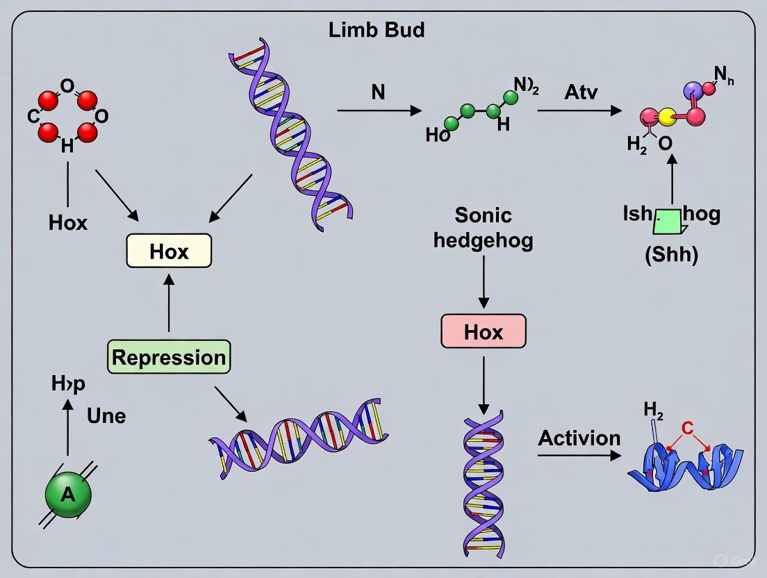
Hox Genes and Sonic Hedgehog: Decoding the Regulatory Network in Vertebrate Limb Bud Development
This article synthesizes current knowledge on the intricate regulatory interplay between Hox genes and Sonic hedgehog (Shh) signaling, a cornerstone of vertebrate limb bud development.
Hox Genes and Sonic Hedgehog: Decoding the Regulatory Network in Vertebrate Limb Bud Development
Abstract
This article synthesizes current knowledge on the intricate regulatory interplay between Hox genes and Sonic hedgehog (Shh) signaling, a cornerstone of vertebrate limb bud development. We explore the foundational principles of how Hox genes from the A and D clusters establish limb fields and directly regulate Shh expression in the Zone of Polarizing Activity (ZPA). The piece delves into advanced methodological approaches, including recombineering and RNA-Seq, for mapping this network and discusses the phenotypic consequences of its disruption, from skeletal malformations to ectopic Shh signaling. By comparing findings across model organisms and genetic models, we validate core principles and highlight species-specific adaptations. Finally, we examine the translational potential of this knowledge for understanding congenital limb defects and informing regenerative medicine strategies.
Blueprint of the Limb: How Hox Genes Establish Fields and Initiate Shh Signaling
The precise patterning of the vertebrate limb bud remains a paradigm for understanding the mechanisms of embryonic development. Central to this process is the Hox code, a combinatorial expression of Hox transcription factors that instructs cellular identity along the anterior-posterior (AP) axis. This code is characterized by two fundamental principles: its organization into paralogous groups and its spatiotemporal collinear expression. Operating within a network of key morphogens, particularly Sonic hedgehog (Shh), the Hox code integrates positional information to guide limb growth and patterning. This review provides an in-depth analysis of the Hox code's architecture, its regulatory dynamics with Shh, and the experimental methodologies that have deciphered its role, offering critical insights for developmental biology and regenerative medicine research.
Hox genes are an evolutionarily conserved family of homeodomain-containing transcription factors that orchestrate embryonic patterning along the anteroposterior axis in bilaterians [1]. They were first discovered in Drosophila melanogaster due to dramatic homeotic transformations—where one body segment developed the identity of another—resulting from mutations in the Antennapedia and Bithorax complexes [2]. In vertebrates, including humans, the Hox gene family has expanded to 39 genes organized into four clusters (HOXA, HOXB, HOXC, and HOXD) located on different chromosomes [2]. A foundational concept in Hox biology is the "Hox code," which refers to the unique combinatorial expression of Hox proteins within a field of cells that provides a molecular specification of positional identity [3]. This code is fundamental for the regionalization of the body plan, including the determination of where limbs should form and how they should be patterned.
Architectural Principles of the Hox Code
Paralogous Groups
Despite being distributed across four clusters, Hox genes are classified into 13 paralogous groups (PG1-PG13) based on their sequence similarity and relative position within their clusters [2]. Genes belonging to the same paralogous group (e.g., Hoxa4, Hoxb4, Hoxc4, Hoxd4 in PG4) are more closely related to each other than to other genes in the same cluster and often exhibit functional redundancy. This classification is critical for understanding the molecular logic of the Hox code in the limb bud, where members of specific paralogous groups act in concert to determine morphological outcomes.
Table 1: Key Hox Paralogous Groups in Limb Development
| Paralogous Group | Chromosomal Location (Human) | Primary Expression Domain in Limb Bud | Functional Role in Limb Patterning |
|---|---|---|---|
| PG4 & PG5 | HOXA/B/C/D@: 7p15, 17q21, 12q13, 2q31 | Lateral Plate Mesoderm (LPM) at cervical-thoracic boundary | Establishes a permissive field for forelimb bud initiation; necessary for Tbx5 activation [3]. |
| PG6 & PG7 | HOXA/B/C/D@: 7p15, 17q21, 12q13, 2q31 | Posterior LPM within the PG4/5 domain | Provides instructive signals for precise forelimb positioning; sufficient to induce ectopic limb buds [3]. |
| PG9-13 (5′ Hoxd) | Primarily HOXD cluster | Distal limb bud (autopod) posteriorly, later expanding anteriorly | Governs autopod (hand/foot) development and digit identity; regulated by Shh signaling [4]. |
Temporal and Spatial Collinearity
The expression of Hox genes during development is not random but follows the principle of collinearity, a remarkable phenomenon where the order of genes on the chromosome corresponds to both their sequence of activation in time and their spatial domains of expression along the AP axis [1] [4]. In the context of the limb bud, this is observed as a sequential activation of Hoxd genes.
- Spatial Collinearity: Hox genes located at the 3' end of the cluster (e.g., PG1-PG3) are expressed in more anterior body regions, while genes at the 5' end (e.g., PG9-PG13) are expressed in more posterior regions, including the posterior limb bud [4].
- Temporal Collinearity: Genes are activated in a sequential order from 3' to 5' during development. This results in a dynamic progression of Hox gene expression domains in the growing limb bud, which is essential for the proximal-to-distal patterning of limb structures [4].
Research in mouse models has revealed that the collinear expression of Hoxd genes during limb development occurs in two distinct waves, controlled by different regulatory mechanisms. The first wave is time-dependent and patterns the proximal limb (stylopod and zeugopod), while the second wave, crucial for digit (autopod) formation, involves a different regulatory landscape and is tightly linked to Shh signaling [4].
The Hox Code in Limb Bud Patterning and Positioning
The initiation and positioning of the limb bud are governed by a precise Hox code within the lateral plate mesoderm (LPM). Recent functional studies in chick embryos have elucidated that this code operates through a system of permissive and instructive signals [3].
- Permissive Role of Hox4/5: The expression of Hox4 and Hox5 paralogous groups establishes a broad permissive field in the LPM of the neck and thorax. Within this field, cells are competent to respond to limb-inducing signals. Loss-of-function experiments demonstrate that Hox4/5 genes are necessary for the initiation of the forelimb genetic program, including the expression of the key limb identity gene Tbx5 [3].
- Instructive Role of Hox6/7: The final, precise position of the forelimb is determined by the more posteriorly expressed Hox6 and Hox7 genes. Gain-of-function experiments, where Hox6/7 was mis-expressed in the anterior LPM (within the Hox4/5 domain), resulted in the reprogramming of neck mesoderm and the formation of ectopic limb buds anterior to the normal limb. This demonstrates that Hox6/7 provides an instructive cue that is sufficient to trigger limb formation [3].
This model, where a permissive Hox4/5 background is refined by an instructive Hox6/7 signal, provides a mechanistic explanation for the consistent positioning of the forelimb at the cervical-thoracic boundary across vertebrate species.
Integration of the Hox Code and Sonic Hedgehog (Shh) Signaling
Limb bud patterning requires the integration of information along three axes: proximal-distal (PD), dorsal-ventral (DV), and anterior-posterior (AP). The Hox code's function is deeply intertwined with signaling centers that govern these axes, most notably the Sonic hedgehog (Shh) pathway, which controls AP patterning [5].
Shh is secreted from a small group of mesenchymal cells at the posterior margin of the limb bud, known as the Zone of Polarizing Activity (ZPA). Its key functions include:
- Specifying Digit Identity: Shh acts as a morphogen, forming a concentration gradient across the AP axis. Cells exposed to different Shh concentrations adopt different fates, leading to the specification of distinct digit identities (e.g., thumb vs. little finger) [5].
- Controlling Limb Bud Outgrowth: Shh signaling is essential for maintaining the Apical Ectodermal Ridge (AER), a signaling center that drives proximal-distal outgrowth. It does this by controlling the expression of Gremlin1, a BMP antagonist that protects the AER from regression [5].
- Regulating 5' Hox Gene Expression: There is a critical regulatory loop between Shh and the Hox code, particularly the 5' members of the Hoxd cluster (PG10-PG13). Shh signaling is required for the maintenance and expansion of these Hoxd genes in the distal limb bud, which in turn are essential for digit morphogenesis [6] [4].
The following diagram illustrates the core regulatory network integrating the Hox code, Shh, and other key signaling centers in the limb bud:
Figure 1: Signaling Network Integrating the Hox Code and Shh in Limb Development. The Hox code in the Lateral Plate Mesoderm (LPM) initiates the limb program via Tbx5. Tbx5 induces FGFs in the Apical Ectodermal Ridge (AER), which maintains Shh expression in the Zone of Polarizing Activity (ZPA). Shh, in a positive feedback loop via Gremlin1 (Grem1), maintains the AER. Shh also drives the expression of 5' Hoxd genes, which are critical for specifying digit identity.
The essential nature of Shh is highlighted by mutant analysis. In Shh-/- mutant mice, limb development is severely truncated. While the most proximal structures (humerus/femur) form with AP polarity, the zeugopod (forearm/shank) lacks AP identity, and the autopod (hand/foot) is reduced to a single, digit-like structure, often with a digit-one identity [6]. This demonstrates that the initial limb prepattern, potentially governed by early Hox expression, can generate basic structures, but Shh is required for the elaboration of distal AP pattern and the full complement of digits.
Key Experimental Protocols and Methodologies
Deciphering the Hox code has relied on sophisticated genetic and embryological techniques in model organisms. The following workflow outlines a key experiment for determining the instructive role of Hox genes in limb positioning.
Figure 2: Workflow for Functional Hox Gene Analysis in Chick Embryos. A key protocol involves (1) constructing plasmids for dominant-negative (DN) or wild-type Hox genes with an EGFP reporter, (2) preparing early chick embryos, (3) using electroporation to target the Lateral Plate Mesoderm (LPM), (4) culturing embryos to allow development, and (5) analyzing results via fluorescence and gene expression assays [3].
This protocol is used for both loss-of-function and gain-of-function studies to determine Hox gene function in the limb-forming LPM.
Plasmid Construction:
- For Loss-of-Function: Generate a plasmid expressing a dominant-negative (DN) Hox gene (e.g., DN-Hoxa4, a5, a6, a7). The DN variant is engineered to lack the C-terminal portion of the homeodomain, preventing DNA binding while retaining the ability to sequester essential transcriptional co-factors, thereby blocking native Hox protein function.
- For Gain-of-Function: Generate a plasmid for the full-length, wild-type coding sequence of the Hox gene of interest (e.g., Hox6/7).
- Reporter System: Both constructs must be co-expressed with a fluorescent reporter protein, such as Enhanced Green Fluorescent Protein (EGFP), to enable precise tracking of transfected cells and tissues.
Embryo Preparation: Fertilized chick eggs are incubated to reach Hamburger-Hamilton (HH) Stage 12, a developmental stage where the LPM is accessible and the limb field is being established.
Electroporation: Embryos are accessed in ovo. The plasmid solution is injected into the region surrounding the LPM of the prospective wing field. Using a specialized electrode, a series of electrical pulses is applied (electroporation) to transiently permeabilize the cell membranes, allowing the plasmid DNA to enter the LPM cells.
Embryo Culture and Analysis: Electroporated embryos are cultured ex ovo for 8-10 hours, allowing them to develop to ~HH Stage 14 and for the transfected constructs to be expressed.
- Visualization: The transfected area is identified by EGFP fluorescence.
- Phenotypic Analysis: Embryos are analyzed for morphological changes, including the formation of ectopic limb buds in gain-of-function experiments.
- Molecular Analysis: In situ hybridization or quantitative PCR (qPCR) is performed to assess the expression of key downstream target genes, such as Tbx5. In loss-of-function experiments, a reduction or loss of Tbx5 signal is expected on the transfected side, while gain-of-function should induce ectopic Tbx5 expression.
The Scientist's Toolkit: Essential Research Reagents
Table 2: Key Reagents for Investigating the Hox Code in Limb Development
| Research Reagent | Function and Application in Hox/Limb Research |
|---|---|
| Dominant-Negative (DN) Hox Constructs | Used to inhibit the function of specific Hox paralogous groups in a cell-autonomous manner, allowing for the dissection of necessity without global gene knockout [3]. |
| Hox Gain-of-Function Constructs | Used to mis-express Hox genes in ectopic locations to test their sufficiency in reprogramming cell fate and inducing new signaling centers (e.g., limb buds) [3]. |
| Fluorescent Reporter Plasmids (e.g., EGFP) | Crucial for lineage tracing and visualizing successfully transfected or transduced cells and tissues in real-time during embryonic development. |
| Shh Pathway Agonists/Antagonists | Small molecules (e.g., SAG, cyclopamine) used to chemically manipulate the Shh signaling pathway to investigate its epistatic relationship with the Hox code. |
| Mouse Genetic Models (Knockouts/Conditional) | Engineered strains (e.g., Shh-/- [6], Hox cluster deletions [4]) that provide foundational evidence for gene function and genetic interactions in vivo. |
The Hox code, with its foundational principles of paralogous group function and collinear expression, provides a robust genetic framework for limb bud positioning and patterning. Its intricate integration with the Shh signaling pathway creates a dynamic system that translates embryonic positional information into complex three-dimensional morphology. Understanding this code is not merely an academic exercise; it has profound implications.
- Evolutionary Biology: Changes in Hox regulatory elements and expression domains are a major driver of morphological evolution, explaining the diversity of limb shapes and positions across vertebrates, including the elongated bodies of snakes [1].
- Regenerative Medicine and Drug Development: Recapitulating the Hox code is a significant challenge in the field of limb regeneration. Furthermore, since dysregulation of HOX genes is implicated in various cancers [2], understanding their normal function during development can inform the development of targeted therapies. The experimental tools and conceptual frameworks outlined here provide a roadmap for researchers in developmental biology, evolution, and biomedicine to continue exploring the profound implications of the Hox code.
The identification of Sonic Hedgehog (Shh) as the morphogen produced by the Zone of Polarizing Activity (ZPA) established a foundational paradigm in developmental biology. This whitepaper details the historical discovery and core functions of Shh in orchestrating anteroposterior (AP) limb patterning. We synthesize key embryological and genetic evidence, emphasizing the integration of Shh signaling with the Hox gene regulatory network and other signaling centers to direct limb bud outgrowth and skeletal element specification. The discussion is framed within the context of ongoing research into the transcriptional and epigenetic regulation of limb development, with implications for congenital disorders and evolutionary morphology.
Vertebrate limb development is a classical model for understanding how complex three-dimensional structures emerge from a simple bud of mesenchyme cells encased in ectoderm. The fundamental axes—proximal-distal (shoulder to digits), dorsal-ventral (knuckles to palm), and anteroposterior (thumb to little finger)—are patterned by tightly coordinated signaling centers. Among these, the Zone of Polarizing Activity (ZPA), a small group of mesenchyme cells at the posterior margin of the limb bud, is the primary regulator of AP patterning [5]. The seminal discovery that Sonic Hedgehog (Shh) encodes the long-sought morphogen secreted by the ZPA unified decades of embryological experimentation with modern molecular genetics [5]. Shh operates as a classic morphogen, specifying distinct cellular fates in a concentration- and time-dependent manner. This process is deeply integrated with the Hox gene regulatory network, which interprets the Shh signal to assign positional identity and ultimately determine the morphology of limb skeletal elements, from the zeugopod (ulna/radius, tibia/fibula) to the autopod (digits) [7].
Historical Discovery of the ZPA and the Shh Morphogen
The journey to identifying Shh began with groundbreaking embryological experiments in the chick wing bud in the 1960s. Saunders and Gasseling discovered that grafting tissue from the posterior margin of one limb bud to the anterior margin of a host limb bud resulted in a mirror-image duplication of the digits [5]. For example, a normal chick wing with three digits (1, 2, 3) would develop a pattern such as 3-2-1-1-2-3. This powerful assay defined the region as the "polarizing region" or ZPA and led to the hypothesis that it produced a diffusible substance—a morphogen—that specified positional values across the AP axis [5].
The search for the molecular identity of this morphogen culminated in 1993 when Riddle et al. demonstrated that Shh transcripts are localized specifically to the ZPA [5]. The critical evidence was that Shh-expressing cells grafted to the anterior margin of a chick wing bud could recapitulate the full mirror-image duplication caused by a ZPA graft, definitively establishing Shh as the ZPA morphogen [5]. Furthermore, the functional conservation of this mechanism across vertebrates, from sharks to mammals, was shown when posterior tissue from mammalian limb buds was grafted to chick wings and elicited the same duplicative response, explained by the conserved posterior expression of Shh [5].
Table 1: Key Historical Experiments Establishing Shh as the ZPA Morphogen
| Experiment | Model System | Key Observation | Interpretation |
|---|---|---|---|
| ZPA Grafting [5] | Chick wing bud | Mirror-image digit duplication (e.g., 4-3-2-2-3-4) | Posterior tissue produces a signal specifying AP identity |
| Shh Expression Localization [5] | Chick/Mouse limb bud | Shh gene expression exclusively in posterior mesenchyme | Shh is the prime candidate for the ZPA signal |
| Ectopic Shh Grafting [5] | Chick wing bud | Shh-expressing cells induce mirror-image duplication | Shh is sufficient for ZPA activity |
| Cross-Species Grafting [5] | Mouse donor to Chick host | Mouse ZPA tissue duplicates chick wing digits | Shh signaling mechanism is evolutionarily conserved |
Core Signaling Pathways and Molecular Mechanisms
The Shh Signaling Cascade
The Shh protein is synthesized as a 45-kDa precursor that undergoes autocleavage and lipid modification to produce a secreted, active form (Shh-N) [8]. The canonical signaling pathway is initiated when Shh binds to its receptor, Patched1 (Ptch1), on the surface of target cells. In the absence of Shh, Ptch1 represses the activity of a seven-transmembrane protein called Smoothened (Smo). Shh binding relieves this repression, allowing Smo to accumulate and initiate an intracellular cascade that prevents the proteolytic processing of Gli transcription factors (Gli2 and Gli3) into repressors (GliR) [8] [7]. This leads to the nuclear translocation of full-length Gli activators (GliA) and the transcriptional activation of target genes, including Gli1 itself (a faithful readout of pathway activity) and Ptch1 (creating a feedback loop) [8] [7].
Figure 1: Canonical Sonic Hedgehog (Shh) Signaling Pathway. The pathway is shown in the OFF (Shh absent) and ON (Shh present) states, illustrating the key roles of Patched1 (Ptch1), Smoothened (Smo), and Gli transcription factors.
Integration with Hox Genes and Other Signaling Centers
Shh signaling does not operate in isolation. Its activity is integrated with the Hox gene network, particularly the HoxD cluster, which is critical for translating the Shh morphogen gradient into distinct transcriptional codes for each digit [7]. Furthermore, Shh signaling is intricately linked to the other two major limb signaling centers:
- Apical Ectodermal Ridge (AER): Shh signaling in the posterior mesenchyme upregulates the expression of Gremlin1, a BMP antagonist that maintains the AER and its production of Fibroblast Growth Factors (FGFs). FGFs, in turn, promote limb bud outgrowth and maintain Shh expression, creating a positive feedback loop (the Shh-Grem1-FGF loop) [5] [7].
- Wnt7a from Dorsal Ectoderm: This signal patterns dorsal structures and also contributes to regulating Shh expression. Loss of Wnt7a in mice leads to a loss of posterior digits, consistent with its role in modulating the Shh pathway [5].
Quantitative Data and Experimental Evidence
The morphogen function of Shh is characterized by quantifiable parameters of concentration and time. Research in chick and mouse models has defined how different Shh signaling levels and durations specify distinct digit identities.
Table 2: Shh Signaling Parameters and Skeletal Outcomes in Limb Patterning
| Digit Identity (Chick Wing) | Required Shh Concentration | Required Signaling Duration | Key Genetic Dependencies |
|---|---|---|---|
| Digit 1 | None (forms in Shh⁻/⁻ mutants) | Not required | Gli3 repressor activity [7] |
| Digit 2 | Low | Short duration | Low GliA, High Gli3R [5] [7] |
| Digit 3 | Medium | Medium duration | Balanced GliA/Gli3R [5] [7] |
| Digit 4 | High | Long duration | High GliA, Low Gli3R [5] [7] |
| Digit 5 | Highest | Longest duration | Sustained GliA, Grem1 expression [5] [7] |
Key Experimental Methodologies
The following protocols have been fundamental to elucidating the role of Shh in AP patterning.
Protocol 1: Chick Limb Bud Micromass Grafting Assay (Classical ZPA/Shh Graft)
- Preparation: Harvest posterior margin tissue (the ZPA) from a donor chick wing bud at Hamburger-Hamilton (HH) stage 19-22.
- Grafting: Make a small incision in the anterior margin of a host chick wing bud at the same developmental stage.
- Implantation: Secure the donor tissue into the anterior incision using a fine glass needle, ensuring contact with the host's apical ectodermal ridge (AER).
- Culture: Allow the embryo to develop further in ovo or in ex ovo culture for 4-7 days.
- Analysis: Fix the limb and analyze the skeletal pattern after Alcian blue and Alizarin red staining to visualize cartilage and bone. A positive result is a mirror-image digit duplication [5].
Protocol 2: Genetic Ablation of Shh Pathway Components in Mouse
- Model Generation: Generate conditional knockout mice for genes of interest (e.g., Srg3/mBaf155 [7], Gli3) using limb-specific Cre drivers (e.g., Prx1-Cre).
- Phenotypic Analysis:
- Skeletal Preparation: At P0 (postnatal day 0), stain the skeleton with Alcian blue (cartilage) and Alizarin red (bone) to assess skeletal patterning defects.
- Whole-mount In Situ Hybridization (WISH): On E10.5-E11.5 limb buds, use DIG-labeled RNA probes for genes like Shh, Ptch1, Gli1, and Grem1 to visualize spatial expression patterns.
- Western Blot/Immunohistochemistry: Confirm protein-level downregulation of the target gene and assess effects on downstream pathway components [7].
The Scientist's Toolkit: Essential Research Reagents
Table 3: Key Reagents for Investigating Shh in Limb Development
| Reagent / Tool | Function / Application | Example Use Case |
|---|---|---|
| Prx1-Cre Mouse Line [7] | Drives Cre recombinase expression specifically in early limb bud mesenchyme. | Conditional gene knockout studies in limb mesenchyme (e.g., Srg3 [7]). |
| Shh-N pDNA / Retrovirus | For ectopic expression of the active N-terminal fragment of Shh. | Functional tests in chick limb buds to mimic ZPA activity [5]. |
| DIG-labeled RNA Probes (Shh, Ptch1, Gli1) | For spatial localization of gene expression via in situ hybridization. | Mapping the ZPA and Shh signaling range in wild-type vs. mutant limb buds [7]. |
| Phospho-Smo Antibodies | Detect the active, phosphorylated form of Smoothened. | Confirming pathway activation status in response to Shh. |
| Cyclopamine (Smo Antagonist) | Small molecule inhibitor of the Shh pathway. | Perturbing Shh signaling in ex vivo limb bud cultures to study its necessity. |
| Gli-Luciferase Reporter | Cell-based reporter system for measuring Gli-mediated transcriptional activity. | High-throughput screening of compounds that modulate Shh pathway. |
Epigenetic Control and Future Directions: The SWI/SNF Complex
Recent research has expanded beyond the core genetic pathway to uncover essential epigenetic regulators. The SWI/SNF chromatin remodeling complex plays a critical bifunctional role in Shh-driven limb patterning. Genetic inactivation of its core subunit, Srg3/mBaf155, in the limb bud mesenchyme results in failure to upregulate Shh target genes (e.g., Ptch1, Gli1) in the posterior limb, while simultaneously causing ectopic activation of the Hedgehog pathway in the anterior mesenchyme [7]. This disrupts the normal AP asymmetry, leading to zeugopod malformations and preaxial polydactyly. This demonstrates that the SWI/SNF complex is essential both for activating the Shh response posteriorly and for repressing it anteriorly, highlighting a sophisticated layer of epigenetic control over the morphogen gradient [7].
Figure 2: Bifunctional role of the SWI/SNF chromatin remodeling complex in Shh pathway regulation, essential for normal AP patterning [7].
The definitive identification of Sonic Hedgehog as the ZPA morphogen was a watershed moment in developmental biology, providing a molecular basis for a classic embryological phenomenon. Its core function in generating a concentration- and time-dependent gradient to specify AP identity is now a central tenet of morphogen theory. The intricate integration of Shh signaling with the Hox gene code, FGFs from the AER, and BMP signaling, creates a robust network ensuring precise limb patterning. Emerging research on epigenetic regulators like the SWI/SNF complex reveals additional layers of control, ensuring the spatiotemporal precision of the Shh response. Understanding these mechanisms provides critical insights into the etiology of congenital limb defects and the evolutionary diversification of limb morphology.
The establishment of the anterior-posterior axis in the developing vertebrate limb is a fundamental process in morphogenesis, governed by a precise genetic hierarchy. At the core of this hierarchy lies the strategic positioning of Hox genes upstream of Sonic hedgehog (Shh), a key morphogen. This review synthesizes current evidence demonstrating that Hox transcription factors directly initiate and modulate the expression of Shh in the limb bud's zone of polarizing activity (ZPA). We detail the mechanistic basis of this regulation through specific binding to the Shh limb enhancer (ZRS), explore the functional outcomes of this interaction on limb patterning and growth, and discuss the implications of this regulatory cascade for understanding congenital limb defects and evolutionary limb diversification. The integration of quantitative data from genetic, genomic, and biochemical experiments provides a comprehensive model of this critical pathway in developmental biology.
The vertebrate limb bud is a classical model for studying the coordination of growth and pattern formation. The limb's three primary axes—anterior-posterior (AP), proximal-distal (PD), and dorsal-ventral (DV)—are established through the interaction of specialized signaling centers [9]. The zone of polarizing activity (ZPA), located at the posterior margin of the limb bud, is responsible for AP patterning through the secretion of Sonic hedgehog (Shh) protein, which acts as a morphogen [9] [10]. Simultaneously, the apical ectodermal ridge (AER) at the distal tip produces fibroblast growth factors (FGFs) that drive PD outgrowth. A critical link between these centers is the Hox family of transcription factors, which are now established as key upstream regulators of the Shh signaling pathway.
The genetic hierarchy governing this process places Hox genes in a commanding position. They are expressed earlier than Shh in the limb bud and are required for the activation of Shh expression, thereby initiating the cascade that patterns the limb skeleton [11] [12] [13]. This review will dissect the experimental evidence supporting this hierarchy, focusing on the molecular mechanisms, quantitative relationships, and functional consequences of Hox-mediated Shh regulation.
Molecular Mechanisms of Hox-Dependent Shh Activation
Direct Regulation of the Shh Limb Enhancer (ZRS)
The fundamental link between Hox genes and Shh expression is the zone of polarizing activity regulatory sequence (ZRS), a highly conserved enhancer located nearly one megabase upstream of the Shh promoter [13]. This enhancer is exclusively responsible for driving Shh expression in the limb bud.
- HOX Binding Site Requirements: In vivo genome editing in mice has demonstrated that the ZRS contains multiple binding sites for HOX transcription factors. Systematic mutagenesis of these sites reveals an incremental relationship between the number of functional HOXD binding sites and the level of Shh expression. A reduction in binding sites leads to corresponding decreases in Shh transcription and progressively severe limb phenotypes, including digit loss [13].
- Spatial Restriction: The ZRS integrates both activating and repressive inputs. While HOX proteins provide positive activation, a discrete repressor module within the ZRS is responsible for restricting Shh expression to the posterior limb bud, ensuring the precise spatial domain of the ZPA [13].
- Functional Domains: The enhancer activity is a consolidation of distinct functional domains. Substantial portions of the conserved sequence are dispensable, indicating the presence of sequence redundancy that ensures robust Shh expression despite sequence variation [13].
Threshold-Dependent and Heterochronic Activation
Beyond direct binding, the timing of Shh activation is critically dependent on reaching a threshold level of Hox gene expression, a phenomenon observed across vertebrate species.
- Heterochronic Shifts in Evolution: Comparative studies in dogfish and zebrafish show that the onset of Shh expression is coupled to the expression of specific 5' Hox genes. In chick and mouse, Shh is activated early, concomitant with Hoxd10 expression. In contrast, in dogfish, Shh transcription begins late in fin development, concomitant with Hoxd13 expression [11].
- Quantitative Threshold: Experiments in zebrafish demonstrate that quantitative changes in hox expression can alter the timing of shh expression. This heterochronic shift directly affects the size of the endoskeletal elements, providing an evolutionary mechanism for modulating limb morphology [11]. The core principle is that a threshold level of Hox protein is a prerequisite for Shh activation.
Experimental Evidence and Functional Validation
Genetic Uncoupling of Hox and Shh Function
A pivotal line of evidence comes from experiments designed to decouple the functions of Hox genes and Shh signaling in mouse models. Sheth et al. (2013) demonstrated that Hoxa and Hoxd genes are required for proper limb bud growth independently of their role in activating Shh [12].
- Control of AER-FGFs: Hox genes are necessary for the maintenance of Fgf expression in the AER. This control is achieved through the regulation of key mesenchymal signals like Gremlin1 (Grem1) and Fgf10, which mediate epithelial-mesenchymal interactions. This Hox-dependent function persists even when Shh signaling is absent, confirming a direct role for Hox genes in the signaling network beyond Shh activation [12].
- Multiple Inputs on Growth: The study revealed that Hox genes have multiple inputs on limb bud growth, including the initial activation of Grem1 and its subsequent anterior expansion, thereby ensuring coordinated patterning and outgrowth [12].
In Vitro Validation of Synergistic Signaling
The downstream relationship where Shh and FGFs act synergistically to control Hox gene expression has been validated in vitro. Studies using cultured limb bud mesenchymal cells show that Shh and Fgf8 act synergistically to activate posterior Hoxd genes (e.g., Hoxd13) during the second wave of Hoxd expression (phase II) [14].
- Dose-Response Relationships: Limb progenitors treated with Shh and Fgf8 show a dose-dependent activation of Hoxd13. Shh induces Hoxd13 over a concentration range that plateaus, consistent with a derepression mechanism, while the response to Fgf8 is linear [14].
- Synergistic Requirement: Hoxd13 expression is maximally induced only when both Shh and Fgf8 signals are supplied simultaneously. The presence of cycloheximide, a translation inhibitor, heavily dampens this synergistic increase, indicating that the full Hoxd13 response requires protein synthesis and likely involves a positive feedback loop [14]. This feedback mechanism exemplifies the complex regulatory network that fine-tunes limb patterning.
Table 1: Key Research Reagent Solutions for Studying the Hox-Shh Hierarchy
| Research Reagent | Function/Application in Experimental Protocols |
|---|---|
| RCAS Virus (Chick) | A replication-competent avian retrovirus system used for targeted gene overexpression (e.g., of Hox genes) or expression of dominant-negative constructs in the chick limb bud [15]. |
| Shh-N Terminal Fragment | The active, purified ligand used in in vitro limb bud cell culture assays to activate the Shh pathway and study dose-response relationships of target genes like Hoxd13 [14]. |
| Cycloheximide | A pharmacological inhibitor of protein translation. Used in vitro to determine if gene activation (e.g., of Hoxd13 by Shh/Fgf8) is direct or requires synthesis of intermediary proteins [14]. |
| ZRS Reporter Constructs | Genomic constructs containing the Shh enhancer (ZRS) linked to a reporter gene (e.g., LacZ). Used in transgenic assays or with genome editing to identify functional transcription factor binding sites [13]. |
| Dominant-Negative Hox Constructs | Engineered Hox proteins lacking the DNA-binding domain. Used in electroporation studies (e.g., in chick) to inhibit the function of specific Hox genes and assess their requirement for limb initiation and Shh expression [15]. |
Integrated Signaling Network and Broader Implications
The Hox-Shh-FGF Regulatory Module
The Hox-Shh relationship is not a simple linear pathway but is embedded within a complex, self-reinforcing signaling module that integrates patterning and growth. This module involves critical feedback loops and interactions with the FGF pathway from the AER [14] [9] [12].
- Initiation: Hox genes (particularly Hoxd members) in the posterior mesenchyme reach a threshold and activate Shh expression via the ZRS enhancer [11] [13].
- Feedback and Maintenance: Shh protein signals to adjacent mesenchyme to maintain the expression of Hox genes in a positive feedback loop. Simultaneously, Shh signaling upregulates Grem1, which inhibits BMPs, thereby maintaining Fgf expression in the AER [12].
- Integration: AER-FGFs, in turn, maintain the expression of Shh in the ZPA and also contribute to the activation of posterior Hox genes, closing the feedback loop and ensuring coordinated growth and patterning [14] [9].
Diagram 1: The core Hox-Shh-FGF regulatory module in the limb bud. Hox genes directly activate Shh via the ZRS enhancer, initiating a network of positive feedback loops that integrate AER-FGF signaling to coordinate patterning and growth.
Implications for Congenital Disorders and Evolution
The precise regulation of the Hox-Shh axis has direct clinical and evolutionary relevance.
- Limb Malformations: Mutations in the ZRS are a major cause of congenital limb malformations in humans, such as polydactyly. These mutations often disrupt the binding sites for transcription factors, including HOX proteins, or alter the repressor elements that confine Shh expression to the posterior limb bud [13]. Recent single-cell atlases of human embryonic limbs confirm the spatial segregation of genes linked to brachydactyly and polysyndactyly, underscoring the importance of precise spatial control in this network [16].
- Evolutionary Diversification: Variations in the Hox-Shh timer mechanism have contributed to the morphological diversity of vertebrate limbs and fins. Heterochronic shifts in the onset of Shh expression, controlled by the attainment of Hox expression thresholds, can directly affect the size and number of skeletal elements, as demonstrated in fin evolution [11].
Table 2: Quantitative Relationships in Hox-Mediated Shh Regulation from Key Studies
| Experimental Context | Quantitative Relationship | Functional Outcome |
|---|---|---|
| ZRS HOX Binding Site Editing (Mouse) [13] | Incremental reduction in Shh expression levels with progressive loss of HOXD binding sites. | Progressive digit loss; phenotype severity correlates with number of sites mutated. |
| Shh & Fgf8 Dose-Response (Limb Cell Culture) [14] | Hoxd13 activation: Shh dose-response plateaus at ~0.5 ng/mL; Fgf8 dose-response is linear. | Maximal Hoxd13 expression requires synergistic input from both pathways. |
| Heterochronic Shift (Dogfish vs. Chick) [11] | Shh onset coupled to Hoxd13 expression (dogfish) vs. Hoxd10 expression (chick). | Late Shh onset in dogfish correlates with a smaller fin endoskeleton compared to the chick limb. |
Experimental Protocols for Key Methodologies
In Vitro Limb Mesenchyme Culture and Stimulation Assay
This protocol, adapted from [14], is used to quantitatively assess the response of target genes to signaling molecules.
- Cell Culture Establishment: Dissociate mesenchymal cells from early embryonic limb buds (e.g., mouse E10.5-E11.5). Culture cells in the presence of Wnt3a to maintain a proliferative, undifferentiated state.
- Ligand Treatment: Treat cells with recombinant signaling proteins.
- For Shh dose-response: Apply increasing concentrations of the active N-terminal fragment of Shh (e.g., 0-2.0 ng/mL).
- For Fgf8 dose-response: Apply increasing concentrations of Fgf8 protein.
- For synergy experiments: Co-treat with a fixed concentration of one ligand and variable concentrations of the other.
- Inhibition of Translation (Optional): To test for direct vs. indirect gene regulation, include a condition with cycloheximide (e.g., 10 µg/mL) to block new protein synthesis.
- Analysis: Harvest cells after 24-40 hours of exposure. Isolve RNA and analyze gene expression of targets (e.g., Hoxd13, Ptch1, Gli1) using quantitative PCR (qPCR).
Chick Embryo Electroporation for Hox Gene Manipulation
This protocol, based on methods in [15], is used for functional analysis of Hox genes in vivo.
- Construct Preparation: Prepare plasmid DNA for electroporation. This can be:
- Gain-of-Function: Full-length Hox genes (e.g., Hoxa6, Hoxa7).
- Loss-of-Function: Dominant-negative forms of Hox genes lacking the DNA-binding domain.
- Embryo Preparation: Incubate fertilized chick eggs to the desired stage (e.g., HH12 for early limb field specification). Window the egg and visualize the embryo.
- DNA Delivery and Electroporation: Inject the DNA solution into the target region (e.g., lateral plate mesoderm of the prospective wing field or neck region). Orient electrodes and apply electrical pulses to facilitate DNA uptake into cells.
- Analysis: Allow embryos to develop for a further 24-48 hours. Analyze phenotypes by:
- In situ hybridization: For expression of marker genes (Tbx5, Fgf10, Shh).
- Immunohistochemistry: For protein detection.
- Histology: For morphological assessment of ectopic or reduced limb structures.
Diagram 2: Workflow for chick embryo electroporation to manipulate Hox gene function in the limb field.
The genetic hierarchy positioning Hox genes upstream of Shh in the limb bud represents a cornerstone of developmental biology. Through direct transcriptional activation of the Shh enhancer (ZRS), establishment of expression thresholds, and integration within a broader signaling network with FGFs, Hox genes initiate and sustain the regulatory cascade that orchestrates limb patterning. The experimental data from genetic, biochemical, and evolutionary studies provide a consistent and robust model. A deep understanding of this hierarchy is not only essential for explaining fundamental morphogenetic principles but also for interpreting the genetic basis of human congenital limb defects and the evolutionary mechanisms that generate anatomical diversity among vertebrates. Future research will likely focus on further elucidating the protein complexes at the ZRS and how this pathway interacts with other regulatory landscapes to achieve ultimate morphological precision.
The 39 Hox genes in mammals, organized into four clusters (A-D) and 13 paralogous groups, constitute a critical system of developmental regulators. A defining characteristic of this family is the extensive functional redundancy between paralogous genes (members of the same group across clusters) and flanking genes (neighbors within a cluster), which has complicated their genetic analysis. This whitepaper synthesizes evidence from targeted mutagenesis studies demonstrating that this redundancy is not merely backup but can give rise to synergistic interactions, where the phenotypic severity of multi-gene mutants far exceeds the sum of individual mutations. Framed within the context of limb bud development, we detail how Hox genes from the A and D clusters exhibit this functional overlap in the regulation of key signaling centers, notably the Sonic hedgehog (Shh) pathway, to coordinate the patterning and growth of the musculoskeletal system. This guide provides a comprehensive resource for researchers, featuring consolidated quantitative data, detailed experimental protocols, and essential reagent solutions for probing the complexities of Hox gene function.
Hox genes are a family of highly conserved homeodomain-containing transcription factors that act as master regulators of positional identity along the anterior-posterior body axis during embryonic development [17]. In mammals, 39 Hox genes are arranged in four clusters (HoxA, HoxB, HoxC, and HoxD) on separate chromosomes, a result of cluster duplication during vertebrate evolution. Genes located at equivalent positions within different clusters are termed "paralogs" and are grouped into 13 paralogous groups (1-13) based on sequence similarity [17] [18]. A fundamental principle of Hox biology is their collinear expression—genes at the 3' end of a cluster are expressed earlier and in more anterior regions than genes at the 5' end [17] [19].
This genomic organization underlies a significant challenge in functional genetics: pervasive functional redundancy. Paralogous genes often share similar expression domains and possess overlapping functions, a relic of their evolutionary origin [20] [18]. Consequently, mutating a single Hox gene frequently results in mild or subtle phenotypes, as related paralogs can compensate for its loss. Similarly, flanking genes within a cluster can also exhibit redundancy due to shared regulatory elements and similar biochemical functions [18]. Uncovering the full developmental role of Hox genes therefore necessitates the generation and analysis of complex mutant combinations, which has revealed that their interactions are not merely additive but can be profoundly synergistic.
Hox Gene Function and Regulatory Networks in Limb Development
The vertebrate limb has served as a premier model for dissecting Hox gene function and redundancy. The limb's skeletal pattern is organized along three primary axes: the proximal-distal (PD) axis (shoulder to fingertips), the anterior-posterior (AP) axis (thumb to little finger), and the dorsal-ventral axis.
Hox Genes in Limb Patterning
The HoxA and HoxD clusters are the primary architects of limb patterning. Their roles are segregated along the PD axis in a manner reflecting gene order within the clusters [17] [19]:
- Hox9 and Hox10 paralogs are essential for patterning the stylopod (humerus/femur).
- Hox11 paralogs are required for the formation of the zeugopod (radius/ulna, tibia/fibula).
- Hox12 and Hox13 paralogs are critical for the development of the autopod (wrist, hand, and digits) [18].
A key difference from axial skeleton patterning is that in the limb, these paralogous groups often function in a non-overlapping manner. Loss of a paralogous group, such as Hox11, can lead to a complete failure to form the corresponding limb segment, rather than a homeotic transformation [17].
Regulation of Signaling Centers: The Shh and Fgf Connection
Hox genes do not act in isolation; they exert their patterning effects by regulating and responding to key signaling centers in the limb bud [19].
- Zone of Polarizing Activity (ZPA): The ZPA is a signaling center in the posterior limb bud mesenchyme that secretes Sonic Hedgehog (Shh). Shh is a morphogen that patterns the AP axis; its loss results in a limb with symmetric, anterior digits [14] [21].
- Apical Ectodermal Ridge (AER): The AER is a thickening of the ectoderm at the distal tip of the limb bud. It secretes Fibroblast Growth Factors (FGFs), such as Fgf8, which are essential for limb outgrowth along the PD axis [14] [18].
A critical regulatory loop exists between these centers: Shh from the ZPA helps maintain Fgf expression in the AER, and FGFs from the AER help maintain Shh expression in the ZPA [14]. Hox genes are integral components of this network. For instance, Hox9 genes promote posterior Hand2 expression, which inhibits the Shh repressor Gli3, thereby permitting the initiation of Shh expression [17]. Conversely, Hox5 genes repress Shh in the anterior limb bud, confining it to the posterior domain [17]. Furthermore, the posterior Hox genes (Hoxd11-d13) are themselves direct targets of Shh and Fgf signaling, creating a complex feedback system that ensures coordinated limb growth and patterning [14].
Table 1: Key Signaling Pathways in Limb Bud Patterning
| Signaling Pathway | Source | Primary Function | Key Hox Gene Interactions |
|---|---|---|---|
| Sonic Hedgehog (Shh) | Zone of Polarizing Activity (ZPA) | Anterior-Posterior Patterning | Regulated by Hox9, Hox5; activates Hoxd11-13 expression [17] [14] |
| Fibroblast Growth Factor (Fgf) | Apical Ectodermal Ridge (AER) | Proximal-Distal Outgrowth | Maintained by Shh; synergizes with Shh to activate Hoxd13 [14] [18] |
| Bone Morphogenetic Protein (BMP) | Limb Bud Mesenchyme | Chondrogenesis, Digit Specification | Bmp2 is a shared target of Shh/Fgf; regulated by Hoxa13 [14] [19] |
Quantitative Evidence of Functional Redundancy and Synergy
The extent of Hox gene redundancy and synergy has been systematically revealed through multi-gene mutagenesis. The following table summarizes phenotypic data from key studies, illustrating the escalating severity of defects as more paralogous and flanking genes are inactivated.
Table 2: Quantitative Analysis of Limb Phenotypes in Hox Gene Mutants
| Genotype | Key Limb Phenotype | Severity & Nature of Defect | Molecular Alterations |
|---|---|---|---|
| Hoxa11-/- | Mildly misshapen ulna/radius; fused carpal bones [18] | Mild, specific zeugopod/autopod defects | Not specified in search results |
| Hoxd11-/- | Modest defects in distal ulna/radius [18] | Mild, specific zeugopod defects | Not specified in search results |
| Hoxa11-/-; Hoxd11-/- | Striking reduction in size of ulna and radius (zeugopod) [18] | Severe, synergistic zeugopod defect | Reduced Shh expression; altered chondrocyte differentiation [18] |
| Hoxa9,10,11-/-; Hoxd9,10,11-/- (Sextuple Mutant) | Reduced ulna/radius more severe than Hoxa11/d11 double mutant [18] | Very severe, synergistic stylopod & zeugopod defect | Severely reduced Shh in ZPA; decreased Fgf8 in AER [18] |
| Hoxa13-/-; Hoxd13-/- | Complete loss of autopod (wrist and paw) skeletal elements [18] | Severe, synergistic autopod agenesis | Disruption of endochondral bone formation pathways [18] |
The data in Table 2 underscore several critical concepts:
- Paralogous Redundancy: The mild phenotype of single Hoxa11 or Hoxd11 mutants versus the severe zeugopod defect in the double mutant demonstrates clear redundancy between paralogs [18].
- Flanking Gene Synergy: The sextuple mutant (Hoxa9,10,11/Hoxd9,10,11) exhibits a more severe zeugopod phenotype than the Hoxa11/d11 double mutant. This indicates that the flanking Hox9 and Hox10 genes, whose primary role is in stylopod patterning, also contribute synergistically to zeugopod development [18].
- Control of Signaling Centers: The genetic hierarchy is evident from the molecular analysis. The severe reduction of Shh and Fgf8 expression in the sextuple mutant reveals that these Hox genes sit upstream of the key signaling pathways, and their combined loss disrupts the core regulatory feedback loops necessary for limb bud outgrowth and patterning [18].
Detailed Experimental Protocols for Analyzing Hox Redundancy
To empower researchers in this field, this section outlines key methodologies used to generate and analyze complex Hox mutants, as cited in the literature.
Protocol 1: Generation of Multi-Gene Hox Mutants Using Recombineering
This protocol describes a method for introducing frameshift mutations into multiple flanking Hox genes simultaneously, preserving endogenous regulatory landscapes [18].
Application: Simultaneous mutation of flanking genes (e.g., Hoxa9, Hoxa10, Hoxa11) without deleting intergenic regions, thus avoiding misexpression of remaining genes due to enhancer loss. Reagents & Materials:
- Targeting Vectors: BAC-based vectors designed for homologous recombination, containing selectable markers (e.g., neomycin resistance).
- Embryonic Stem (ES) Cells: Mouse ES cells (e.g., 129/SvEv line).
- Recombineering System: Inducible recombinase (e.g., RecE/RecT) system in E. coli for precise genetic engineering in BACs.
- PCR Primers: Flanking primers for verification of correct integration.
Step-by-Step Workflow:
- Vector Design: Using recombineering in BAC-containing bacteria, introduce frameshift mutations (e.g., via loxP-flanked stop cassettes or small indels) into the open reading frames of multiple target Hox genes (e.g., Hoxa9, a10, a11) on a single BAC.
- ES Cell Targeting: Linearize the modified BAC targeting vector and electroporate into mouse ES cells.
- Selection & Screening: Select for successfully targeted ES cell clones using antibiotics (e.g., G418). Screen clones via long-range PCR and Southern blotting to confirm correct homologous recombination.
- Mouse Generation: Inject verified ES cell clones into mouse blastocysts to generate chimeric mice. Breed chimeras to obtain germline-transmitted mutant mice.
- Crossbreeding: Cross single-cluster mutants (e.g., Hoxa9,10,11-/-) with other cluster mutants (e.g., Hoxd9,10,11-/-) to generate compound mutants for phenotypic analysis.
Key Considerations: This method is superior to whole-cluster deletions for studying redundancy, as it maintains the integrity of shared enhancers and non-coding RNAs, preventing compensatory dysregulation of adjacent genes [18].
Protocol 2: Limb Mesenchyme Cell Culture and Signaling Pathway Assay
This in vitro protocol is used to dissect the direct response of Hox genes to Shh and Fgf signaling [14].
Application: To quantitatively assess the synergistic requirement of Shh and Fgf for Hox gene activation (e.g., Hoxd13) in a controlled environment. Reagents & Materials:
- Limb Bud Mesenchymal Cells: Dissected from mouse embryonic day ~11.5 (E11.5) limb buds.
- Culture Media: DMEM/F12 supplemented with Wnt3a-conditioned medium to maintain progenitor state.
- Recombinant Proteins: Purified N-terminal Shh peptide (Shh-N), recombinant Fgf8 protein.
- Inhibitors: Cycloheximide (protein synthesis inhibitor).
- qPCR Reagents: SYBR Green, primers for Hoxd13, Ptch1, Gli1, Sprouty1.
Step-by-Step Workflow:
- Cell Isolation: Dissect limb buds from E11.5 mouse embryos, dissociate tissues enzymatically (e.g., with trypsin/collagenase) to obtain a single-cell suspension of mesenchymal progenitors.
- Ligand Treatment: Plate cells and treat with:
- A. A gradient of Shh-N doses (e.g., 0-0.5 µg/mL) in the presence of a fixed concentration of Fgf8.
- B. A gradient of Fgf8 doses with a fixed concentration of Shh-N.
- C. Combined Shh and Fgf8 at synergistic concentrations.
- D. Ligands in the presence of cycloheximide to test for direct transcriptional activation.
- RNA Extraction & qPCR: After 24-40 hours of exposure, harvest cells, extract total RNA, and synthesize cDNA. Perform quantitative PCR (qPCR) for target genes (Hoxd13) and control direct targets (Ptch1 for Shh, Sprouty1 for Fgf8).
- Data Analysis: Plot dose-response curves. The synergistic effect is demonstrated when co-stimulation with Shh and Fgf8 produces Hoxd13 expression levels far exceeding the sum of the levels induced by each factor alone [14].
The Scientist's Toolkit: Key Research Reagents and Models
This section catalogs essential genetic models, reagents, and molecular tools for investigating Hox gene redundancy and function.
Table 3: Research Reagent Solutions for Hox Gene Studies
| Reagent / Model | Description | Primary Application | Key Study |
|---|---|---|---|
| Hoxa9,10,11-/-; Hoxd9,10,11-/- Sextuple Mutant | Mouse with frameshift mutations in six flanking/paralogous Hox genes. | Modeling severe combined deficiency; studying limb signaling centers (Shh, Fgf) [18] | [18] |
| HoxA & HoxD Cluster Deletion Mutants | Mouse models with large genomic deletions of entire Hox clusters using Cre-LoxP. | Assessing the total functional input of a cluster; revealing cross-cluster compensation [18] | [18] |
| Limb Mesenchyme Cell Culture System | Primary cell culture from E11.5 mouse limb buds. | Quantitative analysis of Shh/Fgf synergy on Hox gene expression in vitro [14] | [14] |
| Regulatory Landscape Deletions (e.g., Del(5DOM)) | Mouse/Zebrafish with deletion of centromeric (5') HoxD regulatory domain. | Dissecting enhancer function in autopod-specific Hox gene expression [22] | [22] |
| Hoxd13-/-; Hoxa13-/- Double Mutant | Mouse with combined loss of key autopod Hox genes. | Modeling complete autopod agenesis; identifying shared target genes [18] | [18] |
Visualization of Signaling Pathways and Logical Workflows
The following diagrams, generated using Graphviz DOT language, illustrate the core signaling pathways and experimental logic discussed in this guide.
Hox-Shh-Fgf Regulatory Network in Limb Bud
Diagram 1: Hox-Shh-Fgf Regulatory Network. This diagram illustrates the feedback loop between Hox genes and key signaling centers. Early Hox expression (yellow) establishes the Shh-producing ZPA. Subsequently, Shh and Fgf signals (green) act synergistically (red) to activate late Hox gene expression (blue) in the distal limb bud [17] [14].
Experimental Logic for Demonstrating Synergy
Diagram 2: Experimental Logic Flow. This workflow outlines the two primary methodological approaches for demonstrating Hox gene redundancy and synergy: the genetic approach (blue) using multi-gene mutants, and the in vitro signaling approach (green) using ligand stimulation assays [14] [18].
The evidence from paralogous and flanking Hox gene mutants unequivocally demonstrates that functional redundancy is a foundational principle of this gene family's biology. More importantly, the severe, synergistic phenotypes observed in multi-gene mutants reveal that Hox genes operate in complex, interconnected networks where the whole is greater than the sum of its parts. In the context of limb development and Shh regulation, this synergy is critical for the robust control of fundamental signaling centers like the ZPA and AER.
Future research must continue to leverage sophisticated genetic models that circumvent compensatory mechanisms to uncover the full scope of Hox function. The integration of cutting-edge genomic techniques—such as single-cell RNA-Seq on specific mutant limb compartments [18] and high-resolution chromatin conformation capture on engineered regulatory landscapes [22]—will be essential to map the complete regulatory networks downstream of Hox genes. For drug development professionals, understanding these networks and their inherent redundancies is crucial, as they may inform therapeutic strategies for congenital limb malformations or regenerative medicine approaches, where modulating entire functional modules rather than single genes could yield more effective and robust outcomes.
The formation of the vertebrate limb is a classic model for understanding the coordination of growth and patterning. While the roles of key signaling centers like the Zone of Polarizing Activity (ZPA), producing Sonic hedgehog (Shh), and the Apical Ectodermal Ridge (AER), producing Fibroblast Growth Factors (FGFs), are well-established, the regulatory hierarchy governing their interaction has been a central question. This review focuses on the pivotal role of Hox genes, specifically from the HoxA and HoxD clusters, as critical upstream regulators that orchestrate the signaling cross-talk between the ZPA and AER. We synthesize recent evidence demonstrating that Hox genes are required not only for the initial induction of Shh in the ZPA but also for the sustained expression of AER-FGFs, both indirectly via the Shh-Grem1 loop and through Shh-independent pathways. By integrating quantitative data from key genetic and cell culture studies, this whitepaper provides a mechanistic framework for understanding how Hox genes coordinate the signaling networks that ensure harmonious limb bud outgrowth and patterning, with implications for congenital limb syndrome research and regenerative strategies.
The vertebrate limb bud is patterned along three principal axes: proximal-distal (PD), anterior-posterior (AP), and dorsal-ventral (DV). Two major signaling centers control this process: the Zone of Polarizing Activity (ZPA), a mesenchymal population in the posterior limb bud that secretes Sonic hedgehog (Shh) to pattern the AP axis, and the Apical Ectodermal Ridge (AER), a thickened epithelial structure at the distal tip that secretes FGFs to promote outgrowth and patterning along the PD axis [14] [23].
These centers do not operate in isolation; they are linked by a critical epithelial-mesenchymal feedback loop [24]. Shh from the ZPA induces the expression of Gremlin1 (Grem1), a BMP antagonist, in the distal mesenchyme. Grem1, in turn, protects the AER from BMP-mediated repression, thereby maintaining AER-FGF expression. FGFs from the AER then support the survival and maintenance of the ZPA [12] [18]. The precise regulation of this loop is essential for coordinating growth with patterning, and evidence now places Hox genes as master regulators of this intricate network.
Hox Genes as Master Regulators of Limb Signaling Centers
Hox genes, particularly those from the HoxA and HoxD clusters, are expressed in dynamic patterns during limb development. Their functions extend beyond conferring regional identity to directly controlling the activity of the key signaling centers.
Hox Regulation of the ZPA and Shh Expression
The expression of Shh in the ZPA is directly controlled by a limb-specific enhancer, the ZPA Regulatory Sequence (ZRS), located nearly one megabase upstream of the Shh promoter [25]. Recent research has identified that the 3' subdomain of the ZRS, containing a critical E-box, is absolutely necessary for its activity, while the 5' and central E-boxes appear to have repressive roles [25]. The transcription factors Hand2 and Hoxd13 bind the ZRS and can synergistically transactivate it in vitro [25]. In vivo, genetic ablation of multiple Hox genes (Hoxa9,10,11/Hoxd9,10,11) results in a severe reduction of Shh expression, highlighting the critical requirement for Hox proteins in initiating and maintaining ZPA activity [18].
Direct and Indirect Hox Regulation of the AER and FGF Signaling
The Hox-dependent control of limb bud growth is significantly mediated through the regulation of AER-FGFs. This regulation occurs through two interconnected mechanisms:
- Indirect Regulation via the Shh/Grem1 Loop: By controlling Shh expression, Hox genes indirectly sustain the Shh/Grem1/FGF feedback loop that maintains the AER [12].
- Direct, Shh-Independent Regulation: Genetic experiments that uncouple Hox function from Shh have revealed that Hox genes are required for proper AER-FGFs expression independently of their role in controlling Shh [12]. This direct regulation is achieved through the control of key mesenchymal signals such as Grem1 and Fgf10, which are essential for proper epithelial-mesenchymal interactions [12]. Specifically, HoxA and HoxD genes contribute to both the initial activation of Grem1 and its subsequent anterior expansion within the limb bud mesenchyme.
Table 1: Phenotypic Consequences of Multi-Hox Gene Mutations on Limb Signaling Centers
| Genetic Manipulation | Effect on Shh Expression | Effect on AER-FGF Expression | Major Limb Skeletal Defects | Primary Reference |
|---|---|---|---|---|
| Hoxa11/Hoxd11 DKO | Reduced | Not Reported | Reduced ulna/radius; misshapen zeugopod | [18] |
| Hoxa9,10,11/Hoxd9,10,11 Hexa-KO | Severely reduced | Decreased Fgf8 | Severe reduction of stylopod and zeugopod | [18] |
| Hox/Shh uncoupled | N/A | Reduced (Shh-independent) | Disrupted limb outgrowth | [12] |
Quantitative Analysis of Signaling Pathway Integration
The integration of Shh and FGF signaling at the level of target gene expression has been quantitatively analyzed in limb bud mesenchymal cell cultures. These studies reveal a synergistic relationship between the two pathways in activating key target genes.
Dose-Response and Synergy in Target Gene Activation
In cultured limb progenitor cells, the activation of direct targets like Ptch1 (Shh pathway) and Spry1 (FGF pathway) requires only their respective ligands [14]. However, genes central to limb patterning, such as Hoxd13 and Bmp2, require simultaneous input from both pathways [14].
- Shh Dose-Response: Hoxd13 activation by Shh follows a saturating dose-response curve, plateauing at higher concentrations (0.25-0.5 ng/mL), consistent with a mechanism of derepression via the Gli3 repressor [14].
- FGF8 Dose-Response: The response of Hoxd13 to FGF8 is linear over the concentration range tested, indicative of a direct activating signal [14].
- Synergy: When limb progenitors are exposed to Shh in the absence of FGF8, Hoxd13 activation is negligible. While FGF8 alone can activate Hoxd13 slightly, the combination of both signals produces a synergistic response far exceeding the sum of the individual responses [14]. This synergy is heavily dampened by the translation inhibitor cycloheximide, suggesting a protein-dependent feedback mechanism is necessary for a full transcriptional response [14].
Table 2: Quantitative Dose-Response of Key Genes to Shh and FGF8 in Limb Mesenchyme Cultures
| Gene | Response to Shh | Response to FGF8 | Synergistic Effect | Implied Regulatory Logic |
|---|---|---|---|---|
| Ptch1 | Saturating (plateaus at ~0.25-0.5 ng/mL) | No change with FGF8 | No | Direct Shh target; derepression |
| Spry1 | No change with Shh | Linear | No | Direct FGF target; activation |
| Hoxd13 | Saturating (requires FGF8 context) | Linear (requires Shh context) | Yes | Integrated input from both pathways |
| Bmp2 | Saturating (requires FGF8 context) | Linear (requires Shh context) | Yes | Integrated input from both pathways |
Experimental Protocols for Investigating Hox-AER-FGF Regulation
In Vitro Limb Mesenchyme Cell Culture and Signaling Assay
This protocol is used to dissect the direct and synergistic effects of Shh and FGF on Hox gene expression and other targets, as detailed in [14].
Key Reagent Solutions:
- Limb Bud Mesenchymal Progenitor Cells: Isolated from mouse embryonic limb buds at E10.5-E11.5.
- Wnt3a: Maintains cells in a proliferative and undifferentiated state.
- Recombinant Signaling Ligands: Purified N-terminal Shh peptide (active fragment) and Fgf8 protein.
- Cycloheximide: A pharmacological inhibitor of translation used to test for requirements for new protein synthesis.
Detailed Methodology:
- Cell Culture Setup: Dissociate limb buds from mouse embryos and plate mesenchymal cells in culture media supplemented with Wnt3a to maintain progenitor status.
- Ligand Treatment: Treat cells with a range of concentrations of Shh (e.g., 0-1.0 µg/mL) and Fgf8 (e.g., 0-500 ng/mL), both individually and in combination. Include controls with vehicle alone.
- Inhibition Assay: To test for dependency on new protein synthesis, pre-treat cells with cycloheximide (e.g., 10 µg/mL) for 1 hour before adding Shh and/or Fgf8.
- Time-Course Analysis: Harvest cells at multiple time points (e.g., 6, 12, 24, 40 hours) post-stimulation to assess the kinetics of target gene activation.
- Quantitative Analysis: Extract total RNA and perform quantitative PCR (qPCR) to measure steady-state mRNA levels of target genes (e.g., Hoxd13, Ptch1, Gli1, Spry1, Bmp2). Normalize data to housekeeping genes.
In Vivo Genetic Uncoupling of Hox and Shh Function
This genetic approach determines whether Hox genes regulate AER-FGFs independently of their role in activating Shh [12].
Key Reagent Solutions:
- Conditional Mutant Mice: Mice carrying floxed alleles of Hox gene clusters (HoxA and HoxD) and/or Shh.
- Cre Recombinase Drivers: Tissue-specific Cre lines active in the early limb bud mesenchyme.
- In Situ Hybridization (ISH) Reagents: Digoxigenin-labeled RNA probes for Shh, Fgf8, Fgf4, Grem1, and Fgf10.
Detailed Methodology:
- Mouse Crosses: Generate mutant embryos where Hox gene function is ablated in the limb bud, but Shh expression is either intact or genetically restored. This often requires complex breeding strategies to combine multiple alleles.
- Phenotypic Analysis: Harvest embryos at key developmental stages (E9.5-E11.5) for analysis.
- Whole-Mount In Situ Hybridization (WMISH): Analyze the expression patterns of key signaling molecules (e.g., Shh, Fgf8, Grem1) in mutant versus control embryos. This reveals dependencies and interactions within the network.
- Limb Skeletal Analysis: Process later-stage embryos (E15.5-E18.5) for skeletal staining (Alcian Blue for cartilage, Alizarin Red for bone) to assess the ultimate phenotypic outcome on limb patterning.
Pathway Visualization and Molecular Toolkit
Hox Gene Regulatory Network in Limb Bud Signaling
The following diagram summarizes the complex regulatory interactions between Hox genes, Shh, and FGF signaling, as established by the cited research.
Diagram Title: Hox Gene Regulation of Limb Bud Signaling Centers
The Scientist's Toolkit: Essential Research Reagents
Table 3: Key Reagent Solutions for Investigating Hox-AER-FGF Signaling
| Reagent / Tool | Function / Application | Example Use Case |
|---|---|---|
| Limb Bud Mesenchyme Cell Culture System | In vitro model to test direct effects and synergy of signaling pathways. | Quantifying Hoxd13 dose-response to Shh and Fgf8 [14]. |
| Multi-Hox Gene Mutant Mice (e.g., Hoxa9-11/Hoxd9-11) | Model to study functional redundancy and combined gene function. | Revealing severe Shh and Fgf8 downregulation and skeletal defects [18]. |
| Conditional Gene Knockout (Cre/loxP) Systems | Enables tissue-specific and temporally controlled gene deletion. | Uncoupling Hox function from Shh expression to identify independent roles [12]. |
| ZRS Reporter Constructs | Reports enhancer activity; mutated versions identify critical TF binding sites. | Identifying the critical 3' E-box and Hoxd13 binding sites in the ZRS [25]. |
| Recombinant Signaling Proteins (Shh-N, Fgf8) | To activate specific pathways in cell culture or via bead implantation ex vivo. | Stimulating limb mesenchyme cells to map signaling responses [14]. |
| Cycloheximide | Inhibitor of protein synthesis. | Testing if Hoxd13 activation by Shh/FGF requires new protein synthesis [14]. |
The evidence is compelling that Hox genes sit atop the regulatory hierarchy controlling limb bud signaling centers. They act as crucial integrators, directly initiating and maintaining Shh expression via the ZRS enhancer and ensuring robust AER-FGF activity through both Shh-dependent and Shh-independent mechanisms. The quantitative synergy between the Shh and FGF pathways at the level of target gene activation like Hoxd13 underscores the complexity of this regulatory network.
Future research should focus on elucidating the direct transcriptional targets of Hox proteins in the limb mesenchyme that mediate their control over signals like Grem1 and Fgf10. Furthermore, the application of single-cell RNA sequencing and chromatin profiling in Hox mutant backgrounds will provide a higher-resolution view of the disrupted gene regulatory networks. Understanding these fundamental mechanisms will not only resolve outstanding questions in developmental biology but also illuminate the pathogenic basis of human congenital limb syndromes caused by mutations in HOX genes or the ZRS, paving the way for novel diagnostic and therapeutic strategies.
Mapping the Circuitry: Advanced Techniques for Dissecting Hox-Shh Genetic Interactions
The intricate process of vertebrate limb development serves as a powerful model for understanding the genetic regulation of organogenesis. At the heart of this process lies a complex molecular network, with Hox genes and the Sonic hedgehog (Shh) signaling pathway acting as master regulators of patterning and growth along the limb's anterior-posterior (AP) axis [26]. Deciphering the precise interactions within this network has been entirely dependent on the parallel evolution of genetic engineering technologies. This whitepaper traces this technological journey, from the initial use of single-gene knockouts to the modern era of multi-gene recombineering, framing it within the context of limb bud research and its implications for understanding congenital diseases and guiding therapeutic development.
The Foundational Role of Single-Gene Knockouts
The systematic dissection of limb patterning mechanisms began with the advent of gene targeting, which allowed for the functional analysis of individual genes. Studies focused on single-gene knockouts revealed the non-redundant and critical functions of specific Hox genes and Shh in establishing the limb blueprint.
- Phenotypic Analysis of Hox Mutants: Loss-of-function studies established that posterior Hox genes (paralogs 9-13) are primary determinants of proximodistal (PD) patterning. For instance, mutations in
Hoxa11andHoxd11lead to severe malformations of the zeugopod (forearm/leg), whileHoxa13andHoxd13mutants exhibit defects in the autopod (hand/foot) [27]. Beyond the PD axis, the inactivation of the entireHox5paralog group (anterior genes) was found to cause specific anterior forelimb defects, including a truncated or absent radius and loss of digit 1, revealing a novel role for non-AbdB Hox genes in AP patterning [27]. - The Centrality of Shh Signaling: The knockout of the
Shhgene unequivocally demonstrated its role as the key morphogen secreted by the Zone of Polarizing Activity (ZPA).Shh-null mutants fail to form posterior limb elements, resulting in a severe truncation of the limb [27]. Furthermore, mutations in theShhlimb-specific enhancer (ZRS) were linked to both loss-of-function phenotypes and ectopic anteriorShhexpression, which leads to preaxial polydactyly [27]. - Key Insights and Limitations: A pivotal finding from single-gene studies was the positive feedback loop between Hox genes and Shh. While Hox genes are required for the initiation and maintenance of
Shhexpression, Shh signaling, in turn, anteriorizes the expression ofHoxd10-13genes [12] [27]. However, the high degree of genetic redundancy among Hox paralogs often meant that single-gene knockouts yielded subtle phenotypes, masking the full functional significance of these genes [27]. This highlighted the necessity for models that could address genetic compensation and complex epistatic interactions.
Table 1: Key Single-Gene Knockout Phenotypes in Limb Development
| Gene(s) | Genetic Engineering Model | Primary Phenotype | Functional Insight |
|---|---|---|---|
| Shh | Homologous recombination | Loss of posterior elements (ulna/fibula, digits) [27] | Shh is essential for posterior limb patterning and growth. |
| Hoxa11/Hoxd11 | Compound double knockout | Malformed zeugopod (radius/ulna) [27] | 11th paralog Hox genes specify zeugopod identity. |
| Hoxa13/Hoxd13 | Compound double knockout | Defective autopod (hand/foot) [27] | 13th paralog Hox genes are critical for autopod formation. |
| Hoxa5/b5/c5 | Triple paralog knockout | Anterior forelimb defects: lost/truncated radius, missing digit 1 [27] | Anterior Hox genes restrict Shh expression to the posterior limb. |
| Gli3 | Spontaneous mutant (Extra-toes) | Severe polydactyly [28] | Gli3 acts as a repressor of Shh-target genes; its processing is inhibited by Shh signaling. |
The Shift to Multi-Gene Recombineering Frameshift Mutations
To overcome the limitations of single-gene models, the field has increasingly adopted multi-gene recombineering approaches. These technologies enable the simultaneous disruption of multiple genes, allowing researchers to model complex genetic interactions and achieve more penetrant phenotypes.
Core Technologies: CRISPR/Cas9 and Beyond
The CRISPR/Cas9 system has become the cornerstone of modern genetic recombineering due to its high efficiency and programmability [29]. The core mechanism involves the induction of sequence-specific double-strand breaks (DSBs) in the genome, which are subsequently repaired by the error-prone non-homologous end joining (NHEJ) pathway. This repair often results in small insertions or deletions (indels) at the target site, effectively creating frameshift mutations that lead to premature stop codons and gene knockout [29].
A significant innovation for multi-gene knockout is the use of a linear donor fragment containing a reporter gene (e.g., puromycin resistance or EGFP) flanked by sequences homologous to the DSB site. This donor is integrated via an NHEJ-mediated mechanism, enriching for cell clones that have undergone successful gene targeting. This method allows for the one-step generation of single- or multiple-gene knockouts with markedly improved efficiency compared to conventional protocols that rely on antibiotic selection alone [29].
Application in Dissecting Hox and Shh Genetic Networks
The power of multi-gene recombineering is exemplified by studies that have dissected the functional overlap and interaction between Hox genes and their downstream effectors.
- Uncovering Redundancy: The generation of
Hox5triple mutants demonstrated the profound redundancy within this paralog group, as only the complete loss of all six alleles (Hoxa5, Hoxb5, Hoxc5) resulted in a limb phenotype [27]. Similarly, the requirement for multiple posteriorHoxAandHoxDgenes in maintainingShhexpression was only fully revealed through compound mutant analysis [12]. - Decoupling Genetic Pathways: A landmark study used multi-gene targeting to decouple the functions of Hox genes and Shh. By genetically manipulating the network, researchers showed that
HoxAandHoxDgenes are required for the proper expression of apical ectodermal ridge-fibroblast growth factors (AER-FGFs) independently of their role in controllingShhexpression. This work established that Hox genes regulate key mesenchymal signals likeGrem1andFgf10to ensure proper epithelial-mesenchymal interactions and limb bud growth [12]. - Epistatic Analysis: The generation of compound mutants, such as those lacking both
Gli3and multipleHoxdgenes (Hoxd10-13), has been instrumental in ordering genetic pathways. The severe polydactyly observed inGli3mutants was not rescued by the simultaneous removal ofHoxdgenes, indicating that the polydactylous phenotype is mediated through the ectopic activation of other genes, potentiallyHoxd9andHoxd10[28]. This suggests a complex regulatory balance where Hox genes can exert both positive and negative effects on digit number.
Diagram 1: Hox-Shh Regulatory Network in Limb Bud. This diagram illustrates the core genetic interactions. Hox genes activate Hand2 and Grem1. Hand2, in turn, activates Shh in the posterior ZPA. Shh and AER-FGFs engage in a positive feedback loop maintained by Grem1, which is crucial for sustained limb bud outgrowth and patterning [12] [30] [27].
Detailed Experimental Protocol: Multi-Gene Knockout via CRISPR/Cas9
The following protocol, adapted from a study demonstrating high-efficiency multi-gene editing, provides a methodology applicable to limb bud research [29].
Reagent Preparation
- sgRNA/pgRNA Design and Cloning: Design single guide RNAs (sgRNAs) or paired guide RNAs (pgRNAs) targeting exonic regions of the target Hox or Shh pathway genes. Cloning involves annealing synthesized oligonucleotides and ligating them into a sgRNA expression vector (e.g., containing an mCherry reporter) using a Golden Gate assembly method [29].
- Linear Donor Construction: A universal donor template is used, containing a CMV-driven puromycin resistance gene (or EGFP) and sequences with stop codons in all three reading frames. This donor is amplified via PCR with primers that add sgRNA protection sequences and homology arms corresponding to the target genomic locus [29].
- Cas9 Plasmid: A plasmid constitutively expressing the Cas9 nuclease is required for cell lines that do not stably express it.
Cell Transfection and Selection
- Cell Culture: Maintain relevant cells (e.g., HeLa, HEK293T) in standard Dulbecco's Modified Eagle Medium (DMEM) supplemented with 10% fetal bovine serum at 37°C with 5% CO₂ [29].
- Transfection: Seed cells on a 6-well plate. For transfection, use a reagent like X-tremeGENE HP. The typical DNA mix per well includes 1 µg of linear donor PCR product, 0.5-1 µg of sgRNA/pgRNA plasmid, and 0.5 µg of Cas9 plasmid (if needed). Incubate the DNA-lipid complex in Opti-MEM before adding to the cells [29].
- Enrichment and Selection: Two weeks post-transfection, begin selection with 1 µg/mL puromycin (for puromycin-resistant donors) or perform Fluorescence-Activated Cell Sorting (FACS) to isolate EGFP-positive cells. This step enriches for clones that have successfully integrated the donor and, therefore, have a high probability of carrying the desired frameshift mutations on both alleles [29].
Validation and Genotyping
- T7 Endonuclease I (T7E1) Assay: Extract genomic DNA from selected pools and perform PCR amplification of the target regions. Digest the heteroduplexed PCR products with T7E1 enzyme, which cleaves mismatched DNA, and analyze the fragmentation pattern on a gel to confirm mutation efficiency [29].
- Splinkerette PCR: To map the precise genomic integration site of the donor cassette, digest genomic DNA with restriction enzymes, ligate a splinkerette adaptor, and perform nested PCR followed by sequencing [29].
- Quantitative RT-PCR (qRT-PCR): Validate knockout success at the transcriptional level by extracting RNA, synthesizing cDNA, and performing qRT-PCR with primers specific to the target genes, using
GAPDHas a normalization control [29].
Diagram 2: Multi-Gene KO Workflow. The experimental pipeline for generating multi-gene knockouts using a donor-assisted CRISPR/Cas9 system, from sgRNA design to final validation [29].
The Scientist's Toolkit: Essential Research Reagents
Table 2: Key Reagents for Genetic Engineering in Limb Research
| Research Reagent | Function / Explanation | Example Use Case |
|---|---|---|
| sgRNA/pgRNA Plasmids | Drive expression of guide RNA(s) for CRISPR/Cas9 to target specific genomic loci. | Targeting Hox gene clusters or the Shh enhancer (ZRS) [29]. |
| Cas9 Nuclease | Creates double-strand breaks (DSBs) at DNA sites specified by the sgRNA. | Effector enzyme for all CRISPR-mediated knockout strategies [29]. |
| Linear Donor Fragment | Contains a selectable marker (e.g., PuroR, EGFP); enriches for knockout cells via NHEJ-integration [29]. | Improves efficiency of generating clean, biallelic frameshift mutations. |
| Homology-Directed Repair (HDR) Template | A DNA template with long homology arms for precise knock-in of mutations or reporters. | Generating Hand2-EGFP knock-in axolotls to study endogenous expression [30]. |
| T7 Endonuclease I (T7E1) | Detects induced mutations by cleaving mismatched heteroduplex DNA [29]. | Initial genotyping to confirm editing efficiency in a cell pool. |
| Cre-loxP System | Enables tissue-specific or inducible gene knockout and lineage tracing. | Fate-mapping embryonic Shh-expressing cells in limb regeneration [30]. |
The evolution from single-gene knockouts to sophisticated multi-gene recombineering has been transformative for developmental biology. In the context of limb bud research, these technologies have moved us from a linear understanding of gene function to a network-based perspective, revealing the profound redundancy, feedback, and interaction between the Hox gene family and the Shh signaling pathway. The ability to model complex genetic interactions in vivo is not only refining our fundamental understanding of embryogenesis but also paving the way for sophisticated models of human congenital limb syndromes and informing future regenerative strategies.
Laser Capture Microdissection (LCM) coupled with RNA-Seq analysis represents a powerful methodological approach for profiling transcriptional outputs from histologically defined tissue regions. This technical guide details the application of this integrated technology within the specific context of limb bud development research, focusing on the intricate regulatory networks governed by Hox genes and Sonic hedgehog (Shh) signaling. By enabling precise isolation of specific signaling centers like the zone of polarizing activity (ZPA) and distinct progenitor cell populations, LCM/RNA-Seq provides unprecedented resolution for investigating the spatial and temporal dynamics of gene expression patterns that orchestrate limb patterning and outgrowth. This whitepaper comprehensively outlines detailed experimental protocols, data analysis frameworks, and practical applications of this technology for researchers and drug development professionals investigating pattern formation in vertebrate embryos.
Vertebrate limb development serves as a paradigmatic model system for studying pattern formation in embryogenesis, characterized by precisely coordinated interactions between signaling centers that govern growth and patterning along three principal axes: anterior-posterior (AP), proximal-distal (PD), and dorsal-ventral (DV). Two key signaling centers regulate these processes: the apical ectodermal ridge (AER), which produces fibroblast growth factors (FGFs) controlling PD outgrowth, and the zone of polarizing activity (ZPA), located at the posterior margin, which secretes Sonic hedgehog (Shh) and specifies AP positional values (e.g., thumb to little finger) [9] [31].
The Hox gene family, particularly HoxA and HoxD clusters, plays an integral role in limb bud positioning, growth, and patterning through complex regulatory interactions with Shh signaling [12] [31]. These genes exhibit nested expression patterns along the body axis, with specific combinations determining the positioning of both forelimb and hindlimb buds [31]. A critical positive feedback loop between Shh from the ZPA and FGFs from the AER maintains limb bud growth and patterning, while Hox genes contribute to the regulation of this network by controlling the expression of key mesenchymal signals including Gremlin1 (Grem1) and Fgf10 [12] [31].
Traditional transcriptomic approaches using bulk tissue homogenates inevitably obscure the spatial resolution essential for understanding localized signaling events within specific limb bud regions. LCM technology overcomes this limitation by enabling precise isolation of histologically defined cell populations from tissue sections, which, when coupled with RNA-Seq, allows for comprehensive transcriptional profiling of specific functional domains such as the ZPA, AER, and interdigital regions [32]. This technical guide provides a comprehensive framework for applying LCM/RNA-Seq to investigate Hox gene and Shh regulatory networks in developing limb buds, with detailed methodologies, data analysis workflows, and practical applications for developmental biologists and translational researchers.
Fundamental Principles
Laser Capture Microdissection is a microscale isolation technique that uses laser energy to selectively separate specific cells or tissue regions from heterogeneous sections under microscopic visualization. When coupled with RNA-Seq, this approach enables genome-wide transcriptional analysis of precisely defined cell populations while preserving their spatial context. The integration of these technologies is particularly powerful for investigating spatial heterogeneity in complex tissues like the developing limb bud, where signaling events are highly localized to specific regions such as the Shh-expressing ZPA [32] [9].
The developmental mechanisms governing limb bud formation are highly conserved across vertebrate species, with chick embryos serving as a primary model system due to their accessibility for experimental manipulation and observation [31]. Mouse models provide complementary genetic approaches for investigating gene function through knockout and lineage tracing studies [12]. The LCM/RNA-Seq workflow can be adapted for both model systems, allowing researchers to leverage the unique advantages of each while maintaining methodological consistency.
Workflow Integration
The integrated LCM/RNA-Seq workflow comprises multiple critical stages, each requiring stringent quality control measures to ensure the reliability of downstream transcriptional data. The process begins with tissue preparation and cryosectioning, followed by histological staining to visualize anatomical landmarks, laser microdissection of regions of interest (ROIs), RNA extraction and amplification, library preparation, and finally sequencing and bioinformatic analysis. Each step presents unique technical challenges, particularly when working with limited RNA quantities from small microdissected samples, necessitating specialized protocols for RNA stabilization and amplification.
Table 1: Key Technical Challenges and Solutions in LCM-RNA-Seq
| Technical Challenge | Impact on Data Quality | Recommended Solutions |
|---|---|---|
| RNA Degradation During Tissue Processing | Reduced RNA integrity affects transcript detection | Rapid freezing, RNase inhibitors, minimal fixation |
| Limited RNA Yield from Small ROIs | Low sequencing coverage and sensitivity | Linear amplification techniques, reduced cycle amplification |
| Cellular Contamination from Adjacent Regions | Loss of transcriptional specificity | High-precision laser settings, careful ROI selection |
| Amplification Bias | Distorted quantitative expression values | Unique Molecular Identifiers (UMIs), spike-in controls |
| Batch Effects | Reduced reproducibility | Standardized protocols, randomized processing |
LCM Experimental Protocol for Limb Bud Analysis
Tissue Preparation and Sectioning
Proper tissue preparation is critical for preserving RNA integrity while maintaining morphological features necessary for accurate microdissection. For embryonic limb bud analysis, optimal results are obtained using fresh-frozen specimens rather than formalin-fixed paraffin-embedded (FFPE) tissues, as the cross-linking induced by formalin can compromise RNA quality and recovery.
Protocol:
- Dissect limb buds from embryonic specimens at appropriate developmental stages (e.g., Hamburger-Hamilton stages 18-25 for chick; E10.5-E12.5 for mouse) in cold PBS.
- Embed tissues immediately in optimal cutting temperature (OCT) compound and snap-freeze in liquid nitrogen-cooled isopentane to prevent ice crystal formation.
- Section tissues at 5-10 μm thickness using a cryostat and transfer to specialized LCM membrane slides.
- Store slides at -80°C until use, minimizing freeze-thaw cycles to preserve RNA integrity.
For histological visualization, perform rapid staining protocols using RNase-free conditions. Hematoxylin and eosin (H&E) staining provides excellent cellular detail, while specific immunohistochemical markers for Shh or Hox proteins can guide dissection of signaling centers when combined with compatible RNA preservation methods [32].
Laser Microdissection of Limb Bud Regions
The identification and isolation of specific limb bud regions requires thorough understanding of limb bud morphology and the spatial distribution of key signaling centers.
Protocol:
- Briefly fix cryosections in 70% ethanol (30-60 seconds) and stain with hematoxylin (30 seconds) followed by eosin (15 seconds) using RNase-free conditions.
- Dehydrate through graded alcohols (70%, 95%, 100%; 30 seconds each) and xylene (2 × 2 minutes) to enhance tissue transparency and laser cutting efficiency.
- Identify regions of interest based on anatomical landmarks:
- ZPA: Posterior mesenchyme adjacent to the ectoderm
- AER: Thickened ectodermal ridge at the distal tip
- Progress zone: Undifferentiated mesenchyme immediately beneath AER
- Anterior mesenchyme: Region opposite ZPA
- Interdigital regions: Mesenchyme between developing digits
- Perform microdissection using laser settings optimized for precise cutting with minimal collateral damage to adjacent tissues.
- Capture dissected tissues using adhesive caps or into collection tubes containing RNA lysis buffer.
Table 2: Recommended LCM Parameters for Limb Bud Regions
| Limb Bud Region | Laser Spot Size | Laser Power | Capture Method | Approximate Cell Yield |
|---|---|---|---|---|
| ZPA | 3-5 μm | 40-60 mW | IR capture | 500-1000 cells |
| AER | 1-3 μm | 30-50 mW | UV cutting | 200-500 cells |
| Progress Zone | 5-7 μm | 50-70 mW | IR capture | 1000-2000 cells |
| Anterior Mesenchyme | 7-10 μm | 60-80 mW | IR capture | 1500-3000 cells |
| Interdigital Mesenchyme | 5-7 μm | 50-70 mW | IR capture | 800-1500 cells |
RNA Extraction, Amplification and Sequencing
RNA Isolation and Quality Assessment
RNA extraction from LCM-derived samples requires specialized protocols optimized for small cell numbers. Commercial kits specifically designed for microdissected samples generally provide the most consistent results.
Protocol:
- Lyse microdissected tissues in chaotropic guanidinium-based buffer containing β-mercaptoethanol.
- Extract RNA using silica-based membrane columns with reduced binding capacities to maximize recovery from limited samples.
- Include carrier RNA during binding steps to improve yields when working with fewer than 500 cells.
- Elute in small volumes (5-10 μL) of nuclease-free water.
- Assess RNA quality using sensitive platforms such as the Agilent Bioanalyzer Pico Chip, with RNA Integrity Number (RIN) >7.0 considered acceptable for downstream applications.
For low-input samples, linear amplification approaches such as T7-based in vitro transcription (IVT) provide superior coverage compared to PCR-based methods. The Arcturus PicoPure RNA Isolation Kit and REPLI-g WTA Single Cell Kit have demonstrated efficacy for LCM-derived samples, with protocols capable of generating sufficient material for sequencing from as few as 50-100 cells.
Library Preparation and Sequencing
Library preparation for LCM-derived RNA requires specialized kits optimized for degraded or limited input material while maintaining representation of transcript diversity.
Protocol:
- Convert amplified RNA to cDNA using reverse transcriptase with oligo(dT) and random hexamer primers.
- Incorporate Unique Molecular Identifiers (UMIs) during cDNA synthesis or second strand synthesis to correct for amplification biases and enable accurate transcript quantification.
- Use transposase-based tagmentation (e.g., Illumina Nextera) or ligation-based approaches for library construction.
- Amplify libraries with limited cycles (8-12) to minimize duplication artifacts.
- Perform quality control using capillary electrophoresis or fragment analyzers to confirm appropriate size distributions.
- Sequence on appropriate platforms (Illumina NextSeq 500 or NovaSeq 6000) with recommended read lengths of 75-150 bp paired-end to balance cost and mapping accuracy.
For limb bud developmental studies, sequencing depth of 20-50 million reads per sample typically provides sufficient coverage for transcript quantification and differential expression analysis, though deeper sequencing may be required for isoform-level analysis or detection of low-abundance transcripts.
Bioinformatic Analysis Framework
Preprocessing and Quality Control
Raw sequencing data requires comprehensive quality assessment and preprocessing to ensure reliable downstream analysis. The bioinformatic workflow should incorporate multiple checkpoints to identify technical artifacts and batch effects.
Key Steps:
- Quality Assessment: FastQC or MultiQC for base quality scores, adapter contamination, and GC content.
- Adapter Trimming: Tools such as Cutadapt or Trimmomatic to remove adapter sequences and low-quality bases.
- Read Alignment: Spliced aligners like STAR or HISAT2 against appropriate reference genome (e.g., galGal6 for chick, mm10 for mouse).
- UMI Processing: Deduplication using UMI-tools to correct for PCR amplification biases.
- Quantification: FeatureCounts or HTSeq for gene-level counts, or Salmon for transcript-level abundance estimation.
Differential expression analysis can be performed using specialized packages such as edgeR or DESeq2, which employ statistical models accounting for the mean-variance relationship in count data [33]. For temporal studies of limb development, pseudotime analysis tools like Monocle can reconstruct gene expression dynamics along developmental trajectories.
Specialized Analyses for Limb Development
Beyond standard differential expression, several specialized analytical approaches provide particular insight into limb development mechanisms:
Spatial Reconstruction: Computational methods such as trend analysis and spatial correlation mapping can infer expression gradients from microdissected regions, revealing patterns similar to those captured by spatial transcriptomic technologies [32].
Pathway Analysis: Gene set enrichment analysis (GSEA) and pathway topology tools can identify signaling pathways active in specific limb regions, particularly the Shh, FGF, Wnt, and BMP pathways known to regulate limb patterning.
Regulatory Network Inference: Algorithms like GENIE3 or SCENIC can reconstruct gene regulatory networks from expression data, identifying potential transcriptional targets of Hox genes and Shh signaling.
Trajectory Analysis: Pseudotime ordering methods can reconstruct cellular differentiation pathways within the limb bud, revealing transitions from progenitor populations to differentiated cell types.
Application to Hox and Shh Regulation in Limb Buds
Investigating Shh-Mediated Patterning
The application of LCM/RNA-Seq to limb bud research has dramatically advanced our understanding of Shh function in AP patterning. Traditional models proposed that Shh acts as a concentration-dependent morphogen, forming a posterior-to-anterior gradient that specifies digit identity (digit 1 anteriorly to digit 5 posteriorly) [9]. However, LCM/RNA-Seq studies have revealed additional complexity in how Shh signaling patterns the limb bud.
Key findings enabled by LCM/RNA-Seq approaches include:
- Identification of distinct transcriptional responses to Shh signaling in anterior versus posterior mesenchyme, revealing region-specific competence factors.
- Discovery of feedback regulators that modulate the duration and intensity of Shh signaling in the ZPA and surrounding regions.
- Characterization of the transcriptional cascade downstream of Shh signaling, including targets involved in cell proliferation, survival, and differentiation.
By microdissecting subregions of the limb bud at progressive developmental stages, researchers have delineated how Shh signaling evolves over time and how its transcriptional outputs vary across spatial domains.
Decoding Hox Gene Functions
Hox genes play multifaceted roles in limb development, influencing limb bud initiation, positioning, growth, and patterning. LCM/RNA-Seq has been instrumental in dissecting these functions by enabling transcriptional profiling of specific regions in genetic mutants. Sheth et al. [12] demonstrated that Hox genes regulate limb bud growth through multiple mechanisms, including:
- Direct regulation of Shh expression in the ZPA
- Control of Grem1 expression, a key component of the Shh-FGF feedback loop
- Regulation of Fgf10 expression in the limb bud mesenchyme
- Independent effects on AER-FGF expression beyond their role in Shh signaling
The ability to profile transcriptomes of specific limb bud regions in Hox mutant embryos using LCM/RNA-Seq has revealed that HoxA and HoxD genes contribute to both the initial activation of Grem1 and its subsequent anterior expansion, highlighting the dynamic nature of their regulatory inputs [12].
Diagram 1: Hox and Shh Gene Regulatory Network in Limb Development. This diagram illustrates the complex interactions between Hox genes, signaling centers (ZPA and AER), and key transcriptional targets in limb bud development.
Integrating Hox and Shh Regulatory Networks
LCM/RNA-Seq studies have been particularly illuminating in deciphering the intricate cross-regulation between Hox genes and Shh signaling. The technology has enabled researchers to:
- Identify direct transcriptional targets of Hox proteins in specific limb bud regions
- Characterize the spatial domains of Shh responsiveness by profiling expression of pathway components and targets
- Elucidate how Hox mutations affect the transcriptional landscape of signaling centers
- Reveal temporal dynamics of gene regulatory network activation and resolution
These findings have led to a more sophisticated understanding of how limb pattern emerges from the integration of multiple signaling inputs and transcriptional responses, with Hox genes acting as critical interpreters of positional information and coordinators of growth and patterning.
The Scientist's Toolkit: Essential Research Reagents
Table 3: Essential Research Reagents for LCM-RNA-Seq in Limb Development Studies
| Reagent/Category | Specific Examples | Function/Application |
|---|---|---|
| Tissue Preservation | OCT compound, RNAlater | Embedding medium for cryosectioning; RNA stabilization |
| LCM Consumables | PEN membrane slides, adhesive caps | Specialized slides for laser cutting; tissue capture |
| RNA Isolation Kits | PicoPure, Arcturus | Optimized for low-input samples from microdissection |
| RNA Amplification Kits | REPLI-g, NuGEN Ovation | Whole transcriptome amplification from limited RNA |
| Library Preparation | Illumina Nextera, SMARTer | Construction of sequencing libraries from small inputs |
| Quality Assessment | Bioanalyzer Pico Chip, Qubit | RNA and DNA quantification and quality control |
| Spatial Markers | Shh antibody, Digoxigenin probes | Identification of signaling centers for microdissection |
| Bioinformatic Tools | STAR, DESeq2, edgeR | Read alignment; differential expression analysis [33] |
Experimental Design Considerations
Temporal and Spatial Resolution
Effective application of LCM/RNA-Seq to limb development studies requires careful consideration of both temporal and spatial sampling strategies. The dynamic nature of limb bud development necessitates closely staged embryos and precise timing of tissue collection to capture critical transitions in patterning and differentiation.
Recommended Approach:
- Collect samples at 12-24 hour intervals across key developmental windows (e.g., HH stages 18-31 for chick limb development)
- Include multiple biological replicates (minimum n=3-5) for each stage and region to account for biological variability
- Microdissect matched regions across developmental stages to enable longitudinal comparisons
- Include adjacent control regions in each experiment to control for local environmental effects
Validation Strategies
Orthogonal validation of LCM/RNA-Seq findings is essential for confirming transcriptional patterns and spatial expression domains.
Validation Methods:
- In situ hybridization: Provides spatial confirmation of gene expression patterns in intact embryos
- Immunohistochemistry: Allows protein-level validation and colocalization studies
- Spatial transcriptomics: Emerging technologies like 10X Genomics Visium provide complementary spatial data [32]
- Single-cell RNA-Seq: Resolves cellular heterogeneity within microdissected regions [34] [35]
- qRT-PCR: Enables rapid quantification of key targets in independent samples
Diagram 2: LCM-RNA-Seq Experimental Workflow. This diagram outlines the complete experimental pipeline from tissue collection to bioinformatic analysis and key applications in limb development research.
The integration of Laser Capture Microdissection with RNA-Seq analysis provides a powerful methodological platform for investigating the transcriptional networks underlying limb development, with particular utility for deciphering the complex regulatory interactions between Hox genes and Sonic hedgehog signaling. This technical guide has outlined comprehensive protocols and analytical frameworks for applying this technology to limb bud research, emphasizing practical considerations for experimental design, sample processing, and data interpretation.
As sequencing technologies continue to evolve toward single-cell resolution and spatial transcriptomics matures, LCM/RNA-Seq remains a valuable approach for hypothesis-driven investigation of specific tissue regions and signaling centers. The precise spatial resolution afforded by this methodology continues to provide unique insights into the mechanistic basis of pattern formation in developing limb buds, with potential applications for understanding congenital limb defects and developing regenerative therapeutic strategies.
For researchers investigating Hox gene and Shh regulation in limb development, LCM/RNA-Seq offers the spatial precision necessary to resolve localized signaling events and transcriptional responses within this dynamically patterned tissue. When coupled with appropriate bioinformatic tools and validation approaches, this technology provides a comprehensive framework for elucidating the gene regulatory networks that coordinate growth and patterning during vertebrate limb development.
Hox genes, a family of evolutionarily conserved transcription factors, are master regulators of embryonic morphogenesis, providing positional identity along the anterior-posterior body axis. Within the developing vertebrate limb bud, the coordinated activity of Hox proteins, particularly from the HoxA and HoxD clusters, establishes a complex regulatory network that directs patterning, growth, and integration of musculoskeletal tissues. This whitepaper delineates the key downstream signaling pathways and candidate target genes directly regulated by Hox genes, with a specific focus on the limb bud and its interplay with the Sonic hedgehog (Shh) pathway. We synthesize current research to detail how Hox-dependent regulation of effectors such as Gremlin1, Bmpr1b, and Fgf10 orchestrates the activity of critical signaling centers—the Zone of Polarizing Activity (ZPA) and the Apical Ectodermal Ridge (AER). Furthermore, we provide structured experimental data and methodological protocols to equip researchers with tools for the continued elucidation of the Hox-regulated gene network.
The vertebrate limb bud is a paradigmatic system for understanding the principles of embryonic morphogenesis. Its development is governed by interactions between three principal signaling centers: the AER, which controls proximal-distal outgrowth via Fibroblast Growth Factors (FGFs); the ZPA, which patterns the anterior-posterior axis through Shh; and the non-ridge ectoderm, which specifies dorsal-ventral identity [36]. Hox genes, specifically the 39 genes found in mammalian genomes, are expressed in dynamic, overlapping domains within the limb bud mesenchyme and are indispensable for its patterning [17] [19]. The posterior Hox genes (paralog groups 9-13) of the HoxA and HoxD clusters exhibit a biphasic expression pattern that correlates with the specification of the limb's proximal-distal segments: the stylopod (humerus/femur), zeugopod (radius-ulna/tibia-fibula), and autopod (hand/foot) [19]. A foundational model posits that Hox9 and Hox10 paralogs pattern the stylopod, Hox11 paralogs pattern the zeugopod, and Hox12 and Hox13 paralogs pattern the autopod [18]. Loss-of-function studies reveal that the combinatorial and redundant actions of these genes are required for the precise formation of limb structures, with single-gene knockouts often showing mild phenotypes and compound mutants exhibiting severe truncations or homeotic transformations [37] [18]. This technical guide explores the direct molecular links between Hox gene activity and the downstream pathways executing their patterning instructions.
Key Downstream Pathways and Direct Targets of Hox Regulation
The functional execution of Hox transcription factors in limb development is mediated through their regulation of a hierarchy of downstream signaling pathways. These pathways, in turn, control the behavior of the key signaling centers and the differentiation of mesenchymal cells into cartilage, bone, and tendon. The following sections and summary table detail the critical downstream targets.
Table 1: Key Downstream Pathways and Targets of Hox Genes in Limb Development
| Target Gene/Pathway | Function in Limb Development | Regulating Hox Genes | Experimental Evidence |
|---|---|---|---|
| Sonic Hedgehog (Shh) | Morphogen secreted by the ZPA; establishes anterior-posterior polarity and digit identity [10]. | Hox9, HoxA/D clusters [17] [18]. | Loss of Hox9 paralogs prevents Shh initiation [17]. Combined HoxA/D cluster deletion severely reduces Shh expression [18]. |
| Fibroblast Growth Factor 10 (Fgf10) | Key mesenchyme-derived factor; initiates and maintains limb bud outgrowth and AER function [36]. | Upstream of Hox; Hox genes regulate T-box factors that control Fgf10 [36]. | Fgf10 beads induce ectopic limb buds; Tbx4/5 (Hox-targets) are required for Fgf10 expression [36]. |
| Fibroblast Growth Factor 8 (Fgf8) | Major AER-derived factor; maintains mesenchymal proliferation and survival [18]. | HoxA9-11/HoxD9-11 [18]. | RNA-Seq and expression analysis in Hox mutants show decreased Fgf8 signaling [18]. |
| Gremlin1 (Grem1) | BMP antagonist; part of the self-regulatory SHH/GREM1/FGF feedback loop that maintains AER activity [18]. | HoxA and HoxD clusters [18]. | Deletion of entire HoxA and HoxD clusters disrupts Grem1 expression [18]. |
| Bone Morphogenetic Protein Receptor 1B (Bmpr1b) | Receptor for BMP signaling; crucial for chondrogenesis and digit formation [18]. | HoxA9-11/HoxD9-11 [18]. | Laser capture microdissection and RNA-Seq of Hox mutant zeugopods identified Bmpr1b as a downregulated target [18]. |
| Growth Differentiation Factor 5 (Gdf5) | BMP family ligand; expressed in joint interzones and critical for joint formation [18]. | HoxA9-11/HoxD9-11 [18]. | Identified as a significantly altered gene in Hox mutant limbs via RNA-Seq analysis [18]. |
| Hand2 | Transcription factor; promotes posterior identity and inhibits Gli3 to enable Shh expression [17]. | Hox5, Hox9 [17]. | Hox9 promotes posterior Hand2 expression; Hox5 interacts with Plzf to restrict Shh, indirectly involving Hand2 circuitry [17]. |
Regulation of the Shh and AER Signaling Centers
Hox genes are critical upstream regulators of the two primary signaling centers in the limb bud. The posterior Hox genes, particularly from the Hox9 paralog group, are essential for the initiation of Shh expression in the ZPA. Hox9 promotes the expression of the transcription factor Hand2 in the posterior limb bud, which in turn inhibits the hedgehog pathway repressor Gli3, thereby creating a permissive environment for Shh expression [17]. Furthermore, combined loss of Hoxa9,10,11 and Hoxd9,10,11 results in severely reduced Shh expression, highlighting the functional redundancy and combined role of these genes in maintaining the ZPA [18]. This establishes a direct genetic link where Hox input is a prerequisite for Shh signaling.
Simultaneously, Hox genes are vital for sustaining the AER. The deletion of both HoxA and HoxD clusters leads to a failure in AER function, in part through the disruption of Gremlin1 (Grem1) expression [18]. Grem1 is a BMP antagonist that is a core component of the SHH/GREM1/FGF feedback loop, which maintains the AER's secretory activity. Hox mutants also show decreased expression of Fgf8 in the AER, a key mitogen for the underlying mesenchyme [18].
Targets in Chondrogenesis and Endochondral Ossification
During the second phase of limb development, Hox genes directly regulate pathways governing cartilage formation and bone differentiation. High-throughput RNA-Seq analysis of wild-type versus Hoxa9,10,11/Hoxd9,10,11 mutant limb zeugopods identified several key genes involved in endochondral ossification as being downstream of Hox regulation [18]. These include:
- Gdf5 and Bmpr1b: Members of the BMP signaling pathway, which is fundamental for chondrocyte differentiation and joint patterning.
- Runx3: A transcription factor essential for chondrocyte maturation.
- Igf1: A growth factor promoting bone growth.
- Lef1: A mediator of the Wnt signaling pathway, which interacts with BMP signaling during skeletogenesis.
The altered expression of these targets in Hox mutants provides a molecular explanation for the observed chondrodysplasia and failure of long bone outgrowth, particularly in the zeugopod of compound Hox11 mutants [18].
Experimental Protocols for Identifying Hox Targets
Elucidating the direct targets of transcription factors like Hox proteins requires a multi-faceted approach to distinguish direct from indirect effects. Below is a detailed protocol based on methodologies cited in the literature.
Protocol: Laser Capture Microdissection (LCM) and RNA-Seq Analysis of Hox Mutant Limb Compartments
This protocol is adapted from a study that defined the transcriptome of specific zeugopod compartments in Hox mutants [18].
Objective: To identify differentially expressed genes in specific, histologically defined tissue compartments (e.g., resting, proliferative, and hypertrophic chondrocytes) of the developing limb in wild-type versus Hox mutant mice.
Materials and Reagents:
- Hox Mutant Mouse Models: e.g., Hoxa9,10,11-/- / Hoxd9,10,11-/- generated via recombineering-based frameshift mutations [18].
- Wild-type Control Mice: Of the same genetic background.
- Tissue Preparation: Optimal Cutting Temperature (O.C.T.) compound, liquid nitrogen, cryostat, membrane-coated glass slides.
- Staining: Ethanol gradients, hematoxylin stain, RNAse-free water.
- LCM System: e.g., Arcturus XT or equivalent.
- RNA Extraction & Amplification Kit: e.g., PicoPure RNA Isolation Kit, Arcturus RiboAmp HS PLUS Kit.
- RNA-Seq Library Prep Kit: e.g., Illumina TruSeq Stranded mRNA Kit.
- Bioinformatics Tools: FastQC (quality control), HISAT2/STAR (alignment), Cufflinks/DESeq2 (differential expression).
Methodology:
- Tissue Harvesting and Preparation: Dissect embryonic day (E) 15.5 forelimbs from Hox mutant and wild-type embryos. Embed limbs in O.C.T. and flash-freeze in liquid nitrogen-cooled isopentane. Store at -80°C.
- Cryosectioning: Section frozen tissue at 5-10 µm thickness using a cryostat and transfer sections to membrane-coated slides. Keep slides frozen at -20°C until staining.
- Rapid Staining and Dehydration: Briefly stain sections with hematoxylin to visualize histological zones. Rapidly dehydrate through a graded ethanol series (75%, 95%, 100%) and clear with xylene. Air-dry completely.
- Laser Capture Microdissection: Using the LCM system, separately microdissect cells from the resting, proliferative, and hypertrophic chondrocyte zones of the developing zeugopod. Capture approximately 1,000-3,000 cells per compartment onto LCM caps.
- RNA Extraction and Amplification: Extract total RNA using the PicoPure kit. Perform linear amplification of the RNA using the RiboAmp kit to obtain sufficient material for sequencing.
- RNA-Seq Library Preparation and Sequencing: Construct cDNA libraries from the amplified RNA using the Illumina kit. Perform high-throughput sequencing on an Illumina platform to a depth of at least 20 million reads per sample.
- Bioinformatic Analysis:
- Quality Control: Assess raw sequence data quality with FastQC.
- Alignment: Map reads to the reference mouse genome (e.g., GRCm39) using HISAT2.
- Differential Expression: Quantify gene expression levels and identify statistically significant differentially expressed genes (DEGs) between mutant and wild-type samples for each compartment using DESeq2. A fold-change of >1.5 and an adjusted p-value (FDR) of <0.05 are typical thresholds.
- Pathway Analysis: Subject the list of DEGs to pathway enrichment analysis using tools like DAVID or GSEA to identify perturbed biological processes (e.g., BMP signaling, chondrocyte differentiation).
Visualization of Hox-Regulated Signaling Networks
The following diagram synthesizes the complex regulatory relationships between Hox genes, key signaling centers, and critical downstream targets in the developing limb bud.
Diagram 1: Hox gene regulatory network in limb development. Hox transcription factors (red/yellow group) directly regulate key effectors in the Zone of Polarizing Activity (ZPA) and Apical Ectodermal Ridge (AER) signaling centers (green group), as well as critical downstream targets (blue/white group) involved in chondrogenesis and feedback signaling.
The Scientist's Toolkit: Essential Research Reagents
Table 2: Key Research Reagents for Investigating Hox Gene Function and Targets
| Reagent / Model System | Function and Application | Key References |
|---|---|---|
| Hox Cluster Mutant Mice | Models for assessing functional redundancy and identifying phenotypes. E.g., Hoxa9,10,11-/-/ Hoxd9,10,11-/- mice generated via recombineering. | [18] |
| Conditional Knockout Mice (Cre/LoxP) | Enables tissue-specific and temporally controlled Hox gene deletion to bypass embryonic lethality and study late-stage functions. | [17] [19] |
| Laser Capture Microdissection (LCM) | Allows for precise isolation of specific cell populations (e.g., chondrocyte zones) from heterogeneous tissue for transcriptomic analysis. | [18] |
| RNA Sequencing (RNA-Seq) | Unbiased, genome-wide profiling of gene expression changes in mutant versus wild-type tissues to identify downstream targets. | [18] |
| ChIP-Sequencing (ChIP-Seq) | Identifies genome-wide binding sites of Hox transcription factors, helping to distinguish direct from indirect targets. | [38] |
| FGF-coated Beads | Used in experimental embryology (e.g., chick embryos) to test the sufficiency of FGF signaling to induce ectopic limb buds or maintain AER function. | [36] |
The intricate patterning of the vertebrate limb is a quintessential example of Hox gene-mediated morphogenesis. Through their role as transcriptional regulators, Hox genes directly control a hierarchy of downstream pathways that orchestrate the activity of the Shh-expressing ZPA and the Fgf-expressing AER, while simultaneously regulating the intrinsic differentiation programs of the limb mesenchyme. Key targets such as Grem1, Bmpr1b, Gdf5, and Fgf8 have been identified as critical nodes in this network, linking Hox function to the core signaling feedback loops and skeletogenic processes.
Future research must continue to refine our understanding of the direct versus indirect targets of Hox proteins, leveraging techniques like ChIP-Seq. Furthermore, the post-transcriptional regulation of Hox genes and the mechanisms underlying their extensive functional redundancy remain fertile ground for investigation. A more complete mapping of the Hox-regulated gene network will not only deepen our fundamental understanding of developmental biology but also illuminate the pathogenic mechanisms of congenital limb malformations and inform regenerative medicine strategies.
The development of the vertebrate limb bud is a classic model for understanding how complex signaling pathways interact to orchestrate pattern formation and morphogenesis. At the core of this process lies a sophisticated regulatory network involving Hox genes and the Sonic hedgehog (Shh) signaling pathway [14] [12]. These systems do not operate in isolation; instead, they engage in intricate cross-regulatory interactions that have complicated traditional genetic approaches to delineating their specific functions. The zone of polarizing activity (ZPA), which secretes Shh, and the apical ectodermal ridge (AER), producing fibroblast growth factors (FGFs), form two key signaling centers that mutually sustain one another through positive feedback loops [14]. Simultaneously, Hox genes, particularly the 5' members of the HoxA and HoxD clusters, are essential for interpreting these signals to direct anterior-posterior (A-P) and proximal-distal (P-D) patterning [14] [39].
A significant challenge in developmental biology has been to disentangle the primary functions of Hox genes from their regulatory interactions with Shh signaling. While it is established that Shh is necessary for the expression of 5' Hoxd genes during the second phase of limb patterning [14], Hox genes themselves also contribute to the maintenance of Shh expression and the AER-FGF signaling center [12]. This review synthesizes current experimental strategies that successfully uncouple these functions, providing researchers with a methodological toolkit for probing the discrete roles of these essential developmental regulators. By employing temporal control systems, quantitative in vitro assays, and chromatin-level analyses, recent research has begun to isolate the specific contributions of Hox genes and Shh to limb growth and patterning.
Experimental Strategies for Functional Uncoupling
Temporal Control of Gene Function
The most direct approach for dissecting the temporal requirements of Shh signaling involves the use of inducible Cre-loxP systems. This method enables precise genetic deletion at defined developmental timepoints, effectively separating early patterning functions from later growth requirements.
Key Experimental Protocol: The
Hoxb6/CreERtransgenic mouse line expresses a tamoxifen-dependent Cre recombinase under control of aHoxb6promoter, driving recombination specifically in limb bud mesenchyme [40]. When crossed with mice carrying a conditional (floxed)Shhallele, administration of tamoxifen at different embryonic days (e.g., E9.5, E10, E10.5) results in Shh deletion within a precise temporal window. The efficiency of deletion can be monitored by assessing the loss ofShhtranscripts and the downregulation of direct target genes likePatched1 (Ptch1), which typically occurs within 12 hours post-injection [40].Uncoupled Phenotypes: This temporal uncoupling revealed that Shh's role in specifying digit identity is remarkably transient, required only during a brief early window. In contrast, its function in promoting the expansion of digit progenitor cells persists over an extended period [40]. The sequential loss of digits upon progressively earlier Shh removal (following the order d3, d5, d2, d4) directly mirrors the reverse sequence of their initial mesenchymal condensation in wild-type embryos (d4, d2, d5, d3), highlighting Shh's primary role in ensuring sufficient cell numbers for digit formation rather than patterning their identity [40].
Direct Pathway Manipulation in Isated Systems
In vitro limb bud mesenchymal cell culture systems provide a reduced environment where specific signaling pathways can be manipulated in isolation, free from the complex feedback loops present in the intact embryo.
Key Experimental Protocol: Limb mesenchymal progenitor cells are isolated and cultured in the presence of
Wnt3ato maintain their proliferative, undifferentiated state [14]. Cells are then treated with precise doses of recombinant signaling proteins:- Shh pathway activation: Treatment with the active N-terminal fragment of Shh protein.
- Fgf pathway activation: Treatment with
Fgf8protein. - Dose-response analysis: Cells are exposed to a range of ligand concentrations (e.g., Shh from 0-1.0 ng/mL) to quantify the sensitivity of target genes.
- Inhibition of protein synthesis: Use of
cycloheximideto test for direct versus indirect transcriptional responses [14].
Uncoupled Signaling Readouts: This approach demonstrated that while direct Shh targets (
Ptch1,Gli1) and Fgf targets (Spouty1) respond to their respective ligands independently, theHoxd13gene requires synergistic input from both pathways for full activation [14]. The dose-response ofHoxd13to Shh plateaus at higher concentrations, characteristic of a derepression mechanism, whereas its response to Fgf8 is linear, indicative of direct activation [14]. The dampened synergistic response in the presence of cycloheximide further suggests that the fullHoxd13response requires protein synthesis-dependent feedback [14].
Genetic Dissection of Epistatic Relationships
Classic genetic experiments can be designed to disrupt specific components of the Hox-Shh network, thereby establishing hierarchical relationships between these factors.
Key Experimental Protocol: Genetic uncoupling involves creating mutant backgrounds where Hox gene function is disrupted while Shh signaling remains intact. This can be achieved through:
- Conditional knockout models for HoxA and HoxD cluster genes.
- Analysis of readouts including AER-FGF expression (
Fgf4,Fgf8), Shh pathway activity (Ptch1), and key mesenchymal signals (Grem1,Fgf10) [12].
Uncoupled Regulatory Functions: Studies using this approach revealed that HoxA and HoxD genes are required for proper
AER-FGFexpression independently of their role in controllingShhexpression [12]. This demonstrates that Hox genes exert dual controls on limb growth: one mediated through Shh and another acting directly or through other mesenchymal signals likeGrem1andFgf10to maintain epithelial-mesenchymal interactions [12].
Chromatin-Level Analysis of Regulatory Landscapes
Examining the chromatin environment of Hox loci provides a mechanistic basis for understanding how their regulation is uncoupled from continuous Shh signaling.
Key Experimental Protocol:
- Cell line derivation: Immortalized mesenchymal cell lines are established from separate anterior and posterior regions of distal E10.5 mouse limb buds [39].
- Chromatin analysis: Native Chromatin Immunoprecipitation (nChIP) is performed for Polycomb repressive marks like
H3K27me3andRing1B. - Chromatin looping assays: Chromosome Conformation Capture (3C) or fluorescence in situ hybridization (FISH) is used to detect physical interactions between the
HoxDcluster and distant enhancers like theGlobal Control Region (GCR)[39].
Uncoupled Chromatin States: These analyses revealed that the distal posterior limb bud shows loss of H3K27me3 and chromatin decompaction over the
HoxDcluster compared to the anterior region [39]. Furthermore, the GCR enhancer spatially colocalizes with the 5'HoxDgenomic region specifically in the distal posterior limb, forming a chromatin loop that initiatesHoxd13expression [39]. This establishes a stable chromatin configuration that may maintainHoxdexpression independent of continuous Shh signaling after the initial activation.
Quantitative Analysis of Uncoupled Functions
Dose-Response Relationships of Shh and Fgf Targets
Table 1: Quantitative dose-response profiles of key genes to Shh and Fgf8 ligands in limb mesenchymal cells [14].
| Gene | Response to Shh | Response to Fgf8 | Synergistic Effect | Proposed Mechanism |
|---|---|---|---|---|
| Ptch1/Gli1 | Non-linear; plateaus at ~0.25-0.5 ng/mL | No response | None | Derepression (via Gli3 repressor inactivation) |
| Spouty1 | No response | Linear increase with dose | None | Direct transcriptional activation |
| Hoxd13 | Non-linear; plateaus (requires Fgf8 co-stimulation) | Linear increase (requires Shh co-stimulation) | Strong synergy; >sum of individual responses | Integrated input + protein synthesis-dependent feedback |
| Bmp2 | Non-linear; plateaus (requires Fgf8) | Linear increase (requires Shh) | Strong synergy | Integrated input from both pathways |
Temporal Requirements for Shh Signaling
Table 2: Phenotypic outcomes following timed deletion of Shh using a Hoxb6/CreER system [40].
| Tamoxifen Injection Time | Functional Shh Loss | Digits Formed (Hindlimb) | Digit Identity Patterning | Primary Process Affected |
|---|---|---|---|---|
| E11.0 | ~E11.5 | All 5 digits (d1-d5) | Normal | Late chondrogenesis (minor effects) |
| E10.5 | ~E11.0 | 4 digits (d1, d2, d4, d5) | Normal | Progenitor pool expansion (loss of d3) |
| E10.0 | ~E10.5 | 3 digits (d1, d2, d4) | Normal | Progenitor pool expansion (loss of d5) |
| E9.5 | ~E10.0 | 1-2 digits (d1, ± d4) | Normal | Severe reduction in progenitor pool |
Visualization of Experimental Approaches and Signaling Networks
Experimental Workflow for Temporal Uncoupling
Diagram Title: Genetic Strategy for Temporal Shh Deletion
Integrated Hox-Shh-Fgf Signaling Network
Diagram Title: Hox-Shh-Fgf Regulatory Network in Limb Development
The Scientist's Toolkit: Essential Research Reagents
Table 3: Key research reagents for uncoupling Hox and Shh functions in limb development.
| Reagent / Tool | Type | Primary Function in Uncoupling Experiments |
|---|---|---|
| Hoxb6/CreER Transgenic Mouse | Genetic Model | Enables tamoxifen-inducible, limb mesenchyme-specific Cre recombination for temporal gene deletion [40]. |
| Shh floxed (conditional) Allele | Genetic Model | Provides target for Cre-mediated deletion to remove Shh function at defined timepoints [40]. |
| Noggin-LacZ Knockin Allele | Reporter Model | Sensitive marker for early digit mesenchymal condensations; allows visualization of condensation sequence [40]. |
| Limb Bud Mesenchymal Cell Culture System | In Vitro Assay | Provides reduced system for applying isolated signaling ligands (Shh, Fgf8) and quantifying gene responses [14]. |
| Recombinant Shh-N Terminus | Protein Reagent | Active fragment of Shh protein used for direct pathway activation in dose-response studies [14]. |
| Recombinant Fgf8 Protein | Protein Reagent | Purified Fgf8 ligand for direct activation of Fgf signaling in cultured limb mesenchyme [14]. |
| Cycloheximide | Small Molecule Inhibitor | Inhibitor of protein synthesis; tests whether gene activation requires newly synthesized proteins [14]. |
| H3K27me3 Antibody | Chromatin Reagent | For ChIP analysis to map repressive Polycomb domains and assess chromatin state changes [39]. |
The Zone of Polarizing Activity Regulatory Sequence (ZRS) is a crucial limb-specific enhancer that controls the spatiotemporal expression of Sonic hedgehog (Shh), a master regulator of anterior-posterior patterning in the developing limb bud [25]. This in-depth technical guide examines how Hox transcription factors, particularly Hoxd13, interact with the ZRS to precisely regulate Shh expression. We synthesize recent mechanistic insights into ZRS subdomain function, present quantitative data on transcription factor binding sites, and detail experimental methodologies for investigating this critical enhancer. Within the broader context of Hox gene and Shh regulation in limb development, this analysis provides researchers with comprehensive protocols, visual signaling pathways, and essential research reagents to advance studies in developmental biology and therapeutic design for congenital limb disorders.
ZRS Genomic Context and Conservation
The ZPA Regulatory Sequence (ZRS) is a limb-specific enhancer located approximately one million bases upstream of the Shh promoter within the fifth intron of the Lmbr1 gene [25]. Despite this considerable genomic distance, the ZRS is both necessary and sufficient for directing Shh expression specifically in the Zone of Polarizing Activity (ZPA) of the developing limb bud. The ZRS is highly conserved across vertebrate species, highlighting its fundamental role in limb patterning [25]. Structural analysis reveals that the ZRS can be divided into three conserved subdomains: 5′, central, and 3′ regions, each containing critical transcription factor binding sites that collectively ensure precise spatial and temporal control of Shh expression during limb development [25].
Biological Significance in Limb Patterning
The ZPA is a key signaling center located in the posterior distal mesoderm of the developing limb bud that orchestrates anterior-posterior patterning through secretion of Shh protein [36] [41]. Shh signaling from the ZPA determines the identity of digits and long bones in the developing limb; loss of Shh function results in severe limb malformations including absence of posterior structures like the ulna and fibula, and reduction to a single digit in the autopod [25]. Proper ZRS function is therefore critical for normal limb development, as demonstrated by the catastrophic effects of ZRS disruption: spontaneous microdeletions in chickens or targeted knockout in mice completely abolish Shh expression in the limb bud, leading to profound patterning defects [25].
Hox Transcription Factors in Limb Development and ZRS Regulation
Hox Genes in Limb Bud Positioning and Patterning
Hox transcription factors play multiple essential roles in limb development beyond ZRS regulation. They initially determine limb field positioning along the anterior-posterior axis, with forelimb buds consistently forming at the most anterior expression domain of Hoxc6 across vertebrate species [42]. During later stages, Hox genes participate in proximal-distal patterning of the limb bud, with their expression patterns dividing into three distinct phases that correspond to the development of the stylopod, zeugopod, and autopod [36]. This phased expression allows natural selection to modify each limb segment independently and represents a crucial mechanism for the evolutionary diversification of limb structures [36].
Table: Hox Gene Functions in Limb Development
| Hox Gene | Expression Domain | Role in Limb Development | Mutant Phenotype |
|---|---|---|---|
| Hoxc6 | Lateral plate mesoderm | Specifies forelimb position | Altered limb positioning |
| Hoxd13 | Distal posterior limb bud | Regulates Shh expression via ZRS | Digit reduction, shortened bones |
| Multiple Hox genes | Phased expression in limb bud | Patterns stylopod, zeugopod, autopod | Segment-specific defects |
Hoxd13 as a Direct ZRS Regulator
Hoxd13 exerts direct regulatory control over Shh expression through specific binding to the ZRS enhancer. Molecular studies have identified two Hoxd13 binding sites within the ZRS – one in the 5′ subdomain and another in the central subdomain [25]. Hoxd13 contributes to anterior-posterior polarity, with misexpression leading to ectopic anterior Shh expression and consequent digit patterning defects [25]. The mechanism of Hoxd13 action involves cooperative binding with other transcription factors, particularly the bHLH factor Hand2, enabling precise targeting despite the relatively similar DNA-binding specificities of different Hox proteins [43]. This cooperativity helps resolve the "homeodomain paradox" wherein Hox proteins with highly similar homeodomains achieve functional specificity in vivo [43].
Quantitative Analysis of ZRS Functional Domains
Functional Hierarchy of ZRS Subdomains
Recent experimental evidence has revealed a functional hierarchy among the three ZRS subdomains. Contrary to initial assumptions that the central subdomain was most critical, systematic analysis demonstrates that the 3′ subdomain is necessary for ZRS activity, while the 5′ and central fragments show no activity alone or in combination [25]. However, the 5′ subdomain does play a supportive role, as combining the 3′ fragment with the 5′ fragment restores robust enhancer activity [25]. This indicates a cooperative relationship between these regions, with the 3′ subdomain serving as the core functional unit while the 5′ region provides ancillary support for full enhancer potency.
Transcription Factor Binding Site Contributions
The ZRS contains five key transcription factor binding sites: three E-boxes (5′, central, and 3′) that bind basic helix-loop-helix factors like Hand2, and two Hoxd13 binding sites (5′ and central) [25]. Mutational analysis of these sites reveals their distinct contributions to ZRS function. Simultaneous mutation of all five binding sites markedly reduces ZRS activity, confirming their collective importance [25]. However, individual sites contribute unequally to enhancer function, with the 3′ E-box proving both necessary and sufficient for robust activity, while the 5′ and central E-boxes appear to have repressive functions [25].
Table: Functional Assessment of ZRS Transcription Factor Binding Sites
| Binding Site | Location | Mutation Effect | Functional Role |
|---|---|---|---|
| 3′ E-box | 3′ subdomain | Abolishes ZRS activity | Critical activator; sufficient for robust activity |
| 5′ E-box | 5′ subdomain | Little effect when mutated alone | Mild repressive function |
| Central E-box | Central subdomain | Little effect when mutated alone | Repressive function |
| 5′ Hoxd13 site | 5′ subdomain | Reduces focal activity | Promotes localized ZPA expression |
| Central Hoxd13 site | Central subdomain | Reduces focal activity | Promotes localized ZPA expression |
Experimental Protocols for ZRS Analysis
In Vivo Chicken Limb Bioassay
The chicken embryo model provides a powerful system for functional analysis of ZRS variants through direct in ovo electroporation.
Protocol:
- Plasmid Construction: Clone ZRS test fragments (wild-type or mutated) into ptk-EGFP reporter vector containing minimal HSV TK promoter upstream of enhanced GFP [25].
- Embryo Preparation: Incubate fertilized chicken eggs to Hamburger-Hamilton stage 18-22 (approximately 3-4 days) corresponding to active limb bud outgrowth.
- Electroporation: Inject plasmid DNA into developing forelimb buds and deliver electrical pulses (5 pulses of 20V, 50ms duration) to facilitate DNA uptake by limb mesenchyme cells.
- Reporter Analysis: Harvest embryos 24-48 hours post-electroporation and analyze GFP expression patterns under fluorescence microscopy.
- Quantification: Measure GFP intensity normalized to co-electroporated transfection control (typically RFP) to account for variation in transfection efficiency. Compare experimental groups using non-parametric statistical tests (Kruskal-Wallis with Dunn's post-test) [25].
Validation: For critical constructs, validate findings in transgenic mouse models to confirm conservation of function across species [25].
Site-Directed Mutagenesis of ZRS Binding Sites
Systematic mutation of transcription factor binding sites enables determination of their relative contributions to ZRS function.
Protocol:
- Site Identification: Identify conserved E-box (CANNTG) and Hox binding sites within ZRS through sequence alignment across multiple species.
- Mutagenesis Design: Design primers incorporating 2-4 nucleotide substitutions in core binding motifs to disrupt transcription factor binding while maintaining overall sequence composition.
- PCR Mutagenesis: Perform overlap extension PCR using wild-type ZRS as template with mutated primers.
- Cloning: Digest PCR products with appropriate restriction enzymes and ligate into reporter vectors.
- Functional Testing: Analyze mutant constructs in chicken limb bioassay as described above, comparing to wild-type ZRS activity [25].
In Vitro Transactivation Assay
Cell-based reporter assays provide a complementary approach to assess direct transcriptional activation.
Protocol:
- Reporter Construction: Clone wild-type or mutant ZRS sequences upstream of minimal promoter driving luciferase reporter gene.
- Expression Vectors: Co-transfect with Hoxd13 and Hand2 expression plasmids or empty vector controls.
- Cell Culture and Transfection: Use appropriate cell lines (e.g., COS-7, HEK293) with lipid-based transfection methods.
- Luciferase Assay: Harvest cells 48 hours post-transfection, measure luciferase activity using commercial assay systems.
- Data Analysis: Normalize luciferase values to co-transfected control (e.g., β-galactosidase), calculate fold activation relative to empty expression vector [25].
Signaling Pathways and Molecular Interactions
Hox-ZRS-Shh Regulatory Axis
The precise control of limb patterning requires integrated signaling between Hox transcription factors, the ZRS enhancer, and Shh signaling. The following diagram illustrates the core regulatory circuit:
Hox-ZRS-Shh Regulatory Axis: This circuit depicts how Hoxd13 and Hand2 transcription factors bind the ZRS enhancer to activate Shh expression, initiating a signaling cascade that patterns the limb bud.
Integration with Limb Bud Signaling Centers
The ZPA functions in coordination with other key signaling centers in the limb bud, particularly the Apical Ectodermal Ridge (AER), to ensure proper three-dimensional limb development. The AER, a thickened epithelial structure at the distal tip of the limb bud, secretes Fibroblast Growth Factors (FGFs) including FGF4 and FGF8 that maintain ZPA function and promote limb outgrowth [36] [42]. This creates a reciprocal signaling loop where Shh from the ZPA maintains FGF expression in the AER, which in turn supports continued Shh expression [36]. This self-sustaining feedback system ensures coordinated proximal-distal and anterior-posterior patterning throughout the period of limb bud outgrowth.
Research Reagent Solutions
A comprehensive toolkit of research reagents is essential for investigating ZRS function and Hox transcription factor activity. The following table details key materials and their applications:
Table: Essential Research Reagents for ZRS and Hox Studies
| Reagent/Category | Specific Examples | Function/Application | Experimental Use |
|---|---|---|---|
| Reporter Vectors | ptk-EGFP, luciferase reporters | Measure enhancer activity | In vivo bioassays, in vitro transactivation |
| Expression Plasmids | Hoxd13, Hand2 expression vectors | Provide transcription factors | Co-transfection, functional rescue |
| Animal Models | Chicken embryos, transgenic mice | In vivo functional testing | Electroporation, germline modification |
| Mutagenesis Kits | Site-directed mutagenesis systems | Introduce specific mutations | Binding site analysis, functional mapping |
| Antibodies | Anti-GFP, anti-Hoxd13, anti-Shh | Detection and localization | Immunostaining, Western blot, ChIP |
| PCR Reagents | High-fidelity polymerases | Amplify and clone ZRS fragments | Construct generation, mutagenesis |
The ZRS enhancer represents a paradigm for understanding how discrete genomic elements coordinate precise spatiotemporal gene expression during embryonic development. Our analysis demonstrates that the 3′ subdomain constitutes the core functional unit of the ZRS, with its critical E-box serving as the primary platform for Hand2 binding and enhancer activation [25]. Hoxd13 operates through distinct binding sites to refine the spatial pattern of ZRS activity within the posterior limb bud mesoderm. The emerging model reveals a sophisticated regulatory architecture where combinatorial transcription factor inputs, subdomain interactions, and chromatin environment collectively ensure robust Shh expression specifically in the ZPA.
Future research should prioritize characterizing the protein complexes that assemble on the ZRS, defining the chromatin architecture of the Shh locus in limb development, and elucidating how non-coding mutations in the ZRS cause human congenital limb disorders. The experimental frameworks and reagents detailed herein provide foundational methodologies for these investigations. As our understanding of ZRS mechanics deepens, so too will opportunities for therapeutic intervention in limb patterning disorders and regenerative medicine applications aimed at recapitulating developmental patterning programs.
When Development Fails: Phenotypes and Mechanisms of Hox-Shh Network Disruption
The formation of the vertebrate limb is a classic model for understanding the genetic control of organogenesis. Within this process, Hox genes play a paramount role in patterning skeletal elements along the limb's proximal-distal axis. Mutations in these genes result in a spectrum of malformations, ranging from zeugopod (forearm/lower leg) reduction to complete autopod (hand/foot) loss. This whitepaper synthesizes current research on how Hox genes, through their intricate regulation of Sonic hedgehog (Shh) signaling and other pathways, control limb patterning. We provide a comprehensive analysis of mutant phenotypes, detailed experimental methodologies, and visualization of signaling networks to serve as a resource for researchers and therapeutic developers working in skeletal development and congenital disorders.
Hox genes, encoding a family of evolutionarily conserved transcription factors, are fundamental regulators of embryonic patterning along the primary body axis. During limb development, members of the HoxA and HoxD clusters (particularly paralog groups 9-13) are predominantly employed to specify the identity and size of the limb's skeletal components [19]. Their expression follows complex, dynamic patterns that prefigure the formation of specific limb segments: the stylopod (upper arm/thigh), zeugopod (forearm/lower leg), and autopod (hand/foot) [44]. A critical function of Hox genes is their regulation of key signaling centers, most notably the Zone of Polarizing Activity (ZPA), which secretes the morphogen Sonic Hedgehog (Shh) to pattern the anterior-posterior (thumb-to-little-finger) axis [9]. The genetic interplay between Hox genes and the Shh pathway creates a robust system that coordinates growth and patterning. Disruption of this system, through either loss-of-function or gain-of-function mutations, leads to a predictable yet diverse spectrum of limb malformations, the characterization of which provides deep insight into the fundamental mechanisms of vertebrate development.
Molecular Genetics of Hox-Guided Limb Patterning
Hox Gene Expression Dynamics and Functional Domains
The expression of Hox genes in the developing limb bud is not static but occurs in temporally and spatially distinct phases that correspond to the specification of different limb segments.
- Early Phase (Specification of Proximal Elements): The initial wave of Hox gene expression (involving genes from paralog groups 9-11) follows the principle of temporal and spatial colinearity, mirroring their expression along the main body axis. This phase is crucial for establishing the initial limb bud and patterning the stylopod and zeugopod [44] [19].
- Late Phase (Specification of the Autopod): A second, distinct wave of expression, primarily from the 5' members of the HoxA and HoxD clusters (Hoxa13 and Hoxd10-13), is critical for autopod formation. During this phase, Hoxd gene expression violates colinearity, forming a dynamic domain that patterns the digits [44]. The establishment of these expression domains is not a passive process but is actively regulated by signaling from the limb's key organizing centers in response to Shh signaling [44].
Critical Signaling Pathways and Hox Interactions
Hox genes do not function in isolation but are embedded in a complex regulatory network with other signaling pathways.
| Signaling Pathway/Molecule | Regulatory Interaction with Hox Genes | Functional Outcome in Limb Development |
|---|---|---|
| Sonic Hedgehog (Shh) | Posterior HoxA/D (9-13) genes collectively maintain Shh expression in the ZPA. Anterior Hox5 proteins interact with Plzf to restrict Shh expression. [27] | Establishes anteroposterior polarity; controls digit number and identity; regulates limb bud width. |
| Fibroblast Growth Factors (FGFs) | Hox genes help maintain the Apical Ectodermal Ridge (AER), a source of FGFs. In turn, FGFs from the AER maintain Shh expression. [9] | Promotes limb bud outgrowth along the proximodistal axis; forms a positive feedback loop with Shh. |
| GLI3 | Hox gene function is required to counter the repressive activity of GLI3. Loss of Hox function leads to an excess of Gli3 repressor (Gli3R). [45] | Processes the Shh signal; Gli3R represses Shh target genes; balance between Gli3 activator and repressor forms patterns the limb. |
| Indian Hedgehog (Ihh) | Hox genes are required for proper Ihh expression in the growth plate of endochondral bones. [45] | Regulates chondrocyte proliferation and differentiation during ossification. |
The following diagram illustrates the core genetic regulatory network governing limb patterning, highlighting the central role of Hox genes:
Figure 1: Core Genetic Network in Limb Patterning. Hox genes (yellow) are central regulators that maintain Shh expression (green) and are in turn regulated by Shh signaling. The Hox5-Plzf complex restricts Shh to the posterior limb bud. FGFs from the AER (red) and the transcription factor Gli3 (blue) are key components of this feedback network.
Spectrum of Hox Mutant Phenotypes: Quantitative Analysis
Genetic analyses, primarily in mouse models, have revealed that the severity and nature of limb malformations depend on the specific Hox genes affected, the number of paralogous genes mutated, and the type of mutation (loss-of-function vs. gain-of-function). The following table summarizes the phenotypic spectrum resulting from various Hox gene perturbations.
| Genetic Manipulation | Zeugopod Phenotype | Autopod Phenotype | Molecular Signature |
|---|---|---|---|
| Hox5 (a5/b5/c5) Triple Null Mutant [27] | Radius: truncated or lost. | Digit 1: missing or transformed to triphalangeal digit. Digit 2: distal portion occasionally bifurcated. | Ectopic anterior Shh expression; anteriorized Ptch1, Gli1, and Fgf4. |
| Hoxd12 Point Mutation (A-to-C) [46] | Radius/Ulna: smaller, misshapen, thinner; wider interosseous space. | Microdactyly: shortening of all digits; missing tip of digit I; widely expanded digits. | Fgf4 and Lmx1b dramatically increased; Shh expression unchanged. |
| HoxdDel(11–13) Mutant [45] | Metacarpals: delayed chondrocyte maturation; ossification postnatally, resembling short bones. | Syndactyly (fused digits); poor, misplaced primary ossification center in phalanges. | Strong Ihh downregulation; associated increase in Gli3 repressor form (Gli3R). |
| Hoxd13 Missense Mutation (Q317K) [47] | Not specified. | Brachydactyly/Oligodactyly: severe shortening of digits; only 4 fingers and 3-4 toes; absent terminal phalanges and nails. | Global shift in DNA binding profile toward a bicoid/PITX1 motif. |
Phenotypic Severity and Genetic Redundancy
A key concept in Hox biology is the profound level of genetic redundancy among paralogous genes. Single mutants for Hoxa5, Hoxb5, or Hoxc5 display no reported limb patterning defects, and even compound mutants missing up to five of the six Hox5 alleles develop normally. It is only the complete inactivation of all three Hox5 paralogs that results in severe anterior forelimb defects [27]. This demonstrates that the Hox5 paralog group acts with a high degree of functional redundancy to ensure the fidelity of anterior patterning. Similarly, the coordinated function of posterior HoxA and HoxD genes is essential, as the loss of both clusters leads to a complete arrest of limb development before the stage normally affected by the loss of Shh alone [19].
Experimental Protocols for Key Studies
To empower the research community, we detail the methodologies from seminal studies that have elucidated the relationship between Hox genes and limb malformations.
This protocol established the role of anterior Hox genes in restricting Shh expression.
- Objective: To determine the functional requirement of the Hox5 paralog group (Hoxa5, Hoxb5, Hoxc5) in mouse forelimb patterning.
- Genetic Crosses: Intercross mice with single null alleles for Hoxa5, Hoxb5, and Hoxc5 to generate compound mutants. Analysis requires homozygosity for all three mutant alleles (triple mutant).
- Phenotypic Analysis:
- Skeletal Staining: Use Alcian Blue (cartilage) and Alizarin Red (bone) staining on E18.5 embryos or newborns to visualize the skeletal pattern.
- In Situ Hybridization: Analyze gene expression on sectioned or whole-mount limb buds from somite-matched embryos (e.g., E10.5). Key probes include: Shh, Ptch1, Gli1 (readouts of Hh signaling), Hand2, Gli3, and Alx4 (upstream regulators), and Hoxd10-13 (posterior Hox genes).
- Molecular/Genetic Interaction: Use co-immunoprecipitation and/or genetic crosses with Plzf mutants to test for physical and genetic interaction between Hox5 and Plzf proteins.
- Key Outcome: Hox5 triple mutants, but not single or double mutants, show anterior expansion of Shh expression and corresponding skeletal defects, revealing functional redundancy and a novel role for Hox5 in repressing Shh.
This protocol details how to assess the genome-wide binding profile of wild-type and mutant HOX proteins.
- Objective: To determine how a missense mutation (Q317K) in the HOXD13 homeodomain alters its DNA binding specificity and causes limb malformations.
- Cell System: Use chicken limb bud mesenchymal (chMM) micromass culture system, where HOXD13 is physiologically expressed.
- Ectopic Expression: Employ a retroviral expression system to deliver N-terminally tagged versions of Hoxd13WT, Hoxd13Q317K, and Hoxd13Q317R into chMM cells.
- Chromatin Immunoprecipitation (ChIP): Crosslink cells; sonicate chromatin; immunoprecipitate DNA-protein complexes using an antibody against the N-terminal tag. This ensures specific pulldown of the ectopic protein, distinguishing it from endogenous Hox proteins.
- Sequencing & Bioinformatic Analysis: Sequence the immunoprecipitated DNA (ChIP-seq). Map reads to the reference genome. Identify peaks of binding and perform de novo motif discovery to determine the preferred DNA binding sequence for each protein.
- Key Outcome: The HOXD13Q317K mutant shows a shifted binding profile, recognizing a bicoid/PITX1-like motif instead of the canonical HOX motif, representing a neomorphic (gain-of-function) mechanism.
This protocol describes a genetic interaction study to dissect the pathway downstream of Hox genes.
- Objective: To test the hypothesis that excess Gli3 repressor (Gli3R) mediates the chondrogenic phenotype in HoxdDel(11-13) mutants.
- Genetic Crosses: Generate double mutant mice by crossing HoxdDel(11-13) homozygous mutants with Gli3 homozygous mutants (e.g., Gli3^Xt/^J).
- Histological Analysis: Section limb buds and stained bones from embryos and newborns. Analyze the progression of chondrocyte maturation (proliferation, hypertrophy) and the position and quality of the primary ossification center.
- Molecular Analysis: Perform in situ hybridization or immunohistochemistry for key markers like Ihh and its receptor Ptch1 on mutant and rescued limb buds.
- Key Outcome: The HoxdDel(11-13); Gli3 double mutants show a complete rescue of the phenotype in hindlimb metatarsals and a partial rescue in forelimb metacarpals, confirming that a significant part of the Hoxd phenotype is mediated through Gli3R.
The Scientist's Toolkit: Essential Research Reagents
The following table catalogs critical reagents and models used in the featured studies, providing a resource for designing related experiments.
| Research Reagent / Model | Function in Limb Research | Example Use Case |
|---|---|---|
| Hox5 Triple Null Mutant Mouse [27] | Model for studying functional redundancy among Hox paralogs and the repression of ectopic Shh expression. | Revealed that Hox5 genes interact with Plzf to restrict Shh to the posterior limb bud. |
| Hoxd12 Point Mutant (ENU-induced) Mouse [46] | Model for microdactyly; demonstrates that point mutations can have distinct effects from null alleles. | Showed upregulation of Fgf4 and Lmx1b without Shh misregulation. |
| HoxdDel(11–13) Mutant Mouse [45] | Animal model for human Synpolydactyly (SPD); used to study Hox gene requirement in bone ossification. | Uncovered the role of Hox genes in suppressing Gli3R to allow proper Ihh expression and long bone development. |
| Chicken Limb Bud Micromass (chMM) Culture [47] | In vitro system derived from embryonic limb mesenchyme; recapitulates chondrogenesis and Hox gene expression. | Used for retroviral delivery and ChIP-seq analysis of mutant HOXD13 binding profiles. |
| Gli3 Null/Allelic Series Mutants [45] | Tool for genetic interaction studies; used to dissect the Hox-Gli3 genetic pathway. | Rescued the ossification defects in HoxdDel(11-13) mutants. |
The study of Hox genes in limb development provides a paradigm for understanding how a finite set of transcription factors can generate complex morphological structures. The spectrum of malformations—from zeugopod reduction to complete autopod loss—arises from discrete disruptions in a coordinated genetic network where Hox genes regulate, and are regulated by, the Shh signaling pathway. Future research must focus on several fronts. First, identifying the direct transcriptional targets of different Hox proteins in specific limb compartments will clarify their precise mechanistic roles. Second, understanding the epigenetic landscape that governs the dynamic expression of Hox genes in the limb bud remains a challenge. Finally, translating these fundamental discoveries into therapeutic strategies for human congenital limb syndromes, perhaps by modulating downstream pathways like HH signaling as demonstrated in the Merlin-Nf2 model [48], represents the ultimate goal. The integration of detailed mutant phenotyping, advanced genomics, and genetic interaction studies, as outlined in this whitepaper, will continue to drive progress in this fascinating field.
The precise regulation of Sonic hedgehog (SHH) signaling within the limb bud is fundamental for correct digit number and identity. Disruption of this tightly controlled process, particularly the ectopic expression of SHH in the anterior limb bud, is a recognized mechanism leading to preaxial polydactyly. This whitepaper synthesizes current research demonstrating that deficiencies in Hox genes, key developmental regulators, can directly cause such ectopic signaling. We detail the molecular pathogenesis, summarize quantitative findings from pivotal studies, and provide standardized experimental protocols for investigating these phenomena. The evidence underscores a critical role for Hox genes in maintaining the integrity of limb bud patterning by restricting SHH signaling to the posterior zone of polarizing activity (ZPA).
The vertebrate limb is a classic model for understanding the principles of embryonic patterning. Its development along the anteroposterior (AP) axis—from thumb to little finger—is orchestrated by a signaling center in the posterior mesenchyme known as the zone of polarizing activity (ZPA) [9] [49]. The morphogen secreted by the ZPA is Sonic hedgehog (SHH), which functions in a concentration- and time-dependent manner to specify digit identity and promote limb bud outgrowth [50] [9]. A fundamental principle of normal limb development is the strict restriction of SHH expression to the posterior ZPA.
The Hox family of transcription factors are master regulators of embryonic patterning. In the limb, members of the 5' HoxA and HoxD clusters (paralogs 9-13) are crucial for patterning the different segments of the limb along the proximodistal axis [17]. Recent work has revealed that a primary function of Hox genes is not in the skeletal elements themselves, but in the surrounding stromal, tendon, and muscle connective tissues, where they act to pattern and integrate the entire musculoskeletal system [17]. A key aspect of this regulatory role involves ensuring the precise spatial localization of morphogenetic signals like SHH.
This review focuses on the growing body of evidence that deficiencies in Hox gene function can lead to a breakdown in this precise regulatory network, resulting in the ectopic anterior expression of SHH and the consequent formation of extra digits on the anterior side (preaxial polydactyly). We explore the molecular mechanisms, present consolidated data, and provide methodologies for probing this phenotype.
Molecular Pathogenesis: How Hox Deficiencies Lead to Ectopic SHH Signaling
The establishment and maintenance of the SHH expression domain is a complex process involving multiple transcriptional regulators and signaling feedback loops. Hox genes act at critical nodes within this network.
The Core SHH Signaling Pathway and Limb Bud Patterning
SHH signaling is initiated when the SHH ligand binds to its receptor, Patched-1 (PTCH1). This binding releases the inhibition of Smoothened (SMO), leading to the activation of GLI family transcription factors (primarily GLI2 and GLI3) which then modulate the expression of target genes [51] [52] [9]. In the limb bud, SHH signaling is integrated into a self-regulating feedback loop known as the SHH/GREM1/FGF feedback loop, which coordinates patterning with outgrowth [50] [9]. The limb-specific expression of SHH is controlled by a long-range, highly conserved enhancer known as the ZPA Regulatory Sequence (ZRS) [50] [49]. Mutations in the ZRS are a known cause of polydactyly, often by driving ectopic SHH expression in the anterior limb bud.
Hox Genes as Upstream Regulators of the SHH Landscape
Hox genes function upstream to create a permissive or repressive environment for SHH expression. The following mechanisms have been elucidated:
- Direct Transcriptional Regulation of the ZRS and SHH: Hox proteins can directly or indirectly influence the activity of the ZRS enhancer. For instance, studies in mouse models have shown that HOXD13 can bind to and regulate the expression of Shh [53]. Loss of Geminin (Gmnn), a protein that modulates Hox gene expression, leads to ectopic expression of Hoxd13 in the anterior limb bud. This ectopic Hoxd13, in turn, is sufficient to activate an ectopic SHH signaling center, complete with expression of Shh and its target gene Ptch1 [53].
- Establishing Limb Bud Polarity Upstream of SHH: Prior to the initiation of SHH signaling, the limb bud mesenchyme is prepatterned by a genetic antagonism between the transcription factor dHAND (expressed posteriorly) and the GLI3 repressor (GLI3R, expressed anteriorly) [49]. This antagonism is critical for positioning the ZPA. Hox genes, particularly from the Hox5 and Hox9 paralogous groups, are involved in this early patterning. Loss of Hox9 paralogs disrupts the initiation of posterior Shh expression, while loss of Hox5 paralogs can lead to a failure to repress Shh in the anterior limb bud, resulting in anterior patterning defects [17].
- Interaction with Epigenetic Regulators: The activation of SHH target genes involves a complex interplay with epigenetic machinery. Poised SHH target genes are often marked by bivalent chromatin domains (simultaneous presence of active H3K4me3 and repressive H3K27me3 histone marks) [54]. SHH signaling induces an epigenetic switch, recruiting demethylases like Jmjd3 to remove the repressive H3K27me3 mark and activate transcription. While the direct link to Hox genes in this context requires further exploration, it represents a layer of regulation where Hox factors could potentially influence chromatin accessibility at SHH target loci.
The diagram below illustrates the core signaling pathways and their disruption in Hox deficiencies.
Figure 1: Molecular Pathways in Normal and Hox-Deficient Limb Development. Normal Hox function (green) helps maintain anterior-posterior polarity by promoting GLI3 repressor and properly regulating the ZRS enhancer, restricting SHH to the posterior. Hox deficiency (red) disrupts this, leading to failed repression of activators like dHAND and misregulation of the ZRS, causing ectopic anterior SHH and preaxial polydactyly.
Key Experimental Evidence and Data Presentation
The link between Hox deficiencies and ectopic SHH signaling has been demonstrated through precise genetic models and embryological experiments.
Table 1: Experimental Evidence Linking Hox Deficiencies to Ectopic SHH and Polydactyly
| Experimental Model / System | Genetic Manipulation | Observed Phenotype | Molecular Outcome (SHH Signaling) | Primary Reference |
|---|---|---|---|---|
| Mouse Conditional Knockout | Geminin (Gmnn) deletion in limb bud | Hindlimb polydactyly; shortened skeletal elements | Ectopic posterior SHH signaling center in anterior hindlimb; ectopic Hoxd13 expression; reduced GLI3 repressor | [53] |
| Mouse Genetic Knockout | Hoxa5-/-, Hoxb5-/-, Hoxc5-/- (Hox5 paralog group) | Anterior patterning defects in the limb | Loss of repression of anterior Shh expression; interaction with Plzf to restrict Shh to posterior | [17] |
| Mouse Genetic Knockout | Hoxa9-/-, Hoxb9-/-, Hoxc9-/-, Hoxd9-/- (Hox9 paralog group) | Disrupted AP patterning; single skeletal element per segment | Failure to initiate Shh expression; disrupted Shh/Gremlin/FGF feedback loop | [17] |
| Silkie Chicken Model (Natural Variant) | SNP in ZRS enhancer (ZRS mutation) | Preaxial polydactyly in the leg | Increased posterior SHH expression leading to ectopic anterior signaling; upregulated FGF signaling | [50] |
Quantitative Data from Pivotal Experiments
Table 2: Quantitative Findings from Key Polydactyly and Signaling Studies
| Measured Parameter | Experimental Group | Control Group | Significance/Notes | Source |
|---|---|---|---|---|
| SHH protein concentration in posterior leg mesenchyme (relative units) | Silkie (Slk) Chicken: Increased | White Leghorn: Baseline | Confirmed increased SHH signaling from the native ZPA as an initiating event. | [50] |
| Cell counts of specific neurons in hypothalamic nuclei (E14.5) | ShhΔhyp (hypothalamic knockout) | Control | Severe reduction or absence of neurons: POMC: 1.0 vs 140.8; TH: 12.3 vs 104.0; Sst: 0.3 vs 63.0 (all p<0.001) - Demonstrates profound effect of Shh loss. | [55] |
| H3K27me3 levels at Gli1 regulatory region (ChIP) | Shh-treated MEFs: Significantly reduced | Untreated MEFs: Baseline | Epigenetic switch is a key mechanism in SHH target gene activation. | [54] |
| Gene expression activation of Shh targets (e.g., Gli1) | Jmjd3-/- MEFs + Shh: Impaired activation | Wild-type MEFs + Shh: Strong activation | Jmjd3 demethylase activity is required for resolving bivalent chromatin and activating SHH targets. | [54] |
Experimental Protocols: Methodologies for Investigation
To investigate the relationship between Hox genes and ectopic SHH signaling, the following experimental approaches, derived from the cited literature, are fundamental.
Protocol 1: Generating Conditional Gene Deletion in Mouse Limb Buds
This protocol is used to study gene function spatiotemporally, as exemplified by studies on Geminin and Hox genes [53] [55].
- Animal Models: Utilize transgenic mouse lines expressing Cre recombinase under the control of limb bud-specific promoters (e.g., Prx1-Cre for early limb mesenchyme). Cross these with mouse lines carrying loxP-flanked ("floxed") alleles of the Hox gene of interest.
- Genotyping: Perform PCR on genomic DNA extracted from embryo tail or yolk sac biopsies to identify embryos carrying both the Cre transgene and the homozygous floxed alleles (conditional knockout). Littermates lacking the Cre transgene serve as critical controls.
- Phenotypic Analysis:
- Skeletal Staining: At E18.5 or postnatally, eviscerate and skin embryos/pups, then fix in 95% ethanol. Stain skeletons with Alcian Blue (for cartilage) and Alizarin Red (for bone) to visualize skeletal patterning defects and polydactyly.
- Whole-Mount In Situ Hybridization (ISH): Harvest embryos at key stages (e.g., E10.5-E12.5 for limb patterning). Fix embryos and use digoxigenin-labeled RNA probes to detect the spatial expression of key genes (e.g., Shh, Ptch1, Hoxd13, Gli1).
- Tissue Analysis: Section the stained embryos or embed fixed limb buds in paraffin for histological analysis (e.g., H&E staining) or immunohistochemistry to detect protein localization.
Protocol 2: Functional Interrogation of SHH Signaling in Chick Embryos
The chick embryo is a premier model for its accessibility to surgical and pharmacological manipulation [50] [9] [49].
- Embryo Preparation: Incubate fertilized chick eggs to Hamburger-Hamilton (HH) stage 18-22 (corresponding to early limb bud development). Create a small window in the eggshell to access the embryo.
- Pharmacological Modulation:
- SHH Pathway Agonist/Antagonist: Soak AG1-X2 ion-exchange beads in a solution of the SMO agonist SAG (0.2 µg/µl) or the antagonist cyclopamine (1 µg/µl). Implant the bead into the anterior region of the limb bud.
- FGF Signaling Inhibition: Soak beads in SU5402 (10 mM), an FGF receptor inhibitor, to disrupt the SHH/GREM1/FGF feedback loop.
- Genetic Manipulation via Electroporation:
- Plasmid Preparation: Prepare plasmids for overexpression (e.g., Hox genes) or knockdown (e.g., via shRNA).
- Microinjection and Electroporation: Inject the plasmid solution into the anterior mesenchyme of the limb bud. Use platinum electrodes and a square-wave electroporator to deliver pulses, driving the DNA into the mesenchymal cells.
- Phenotypic Assessment: Re-incubate the eggs for 24-72 hours. Harvest the embryos and analyze limb phenotypes via skeletal staining or ISH for molecular markers.
Protocol 3: Analyzing Epigenetic Changes via Chromatin Immunoprecipitation (ChIP)
This molecular protocol is used to investigate changes in histone modifications at SHH target genes, as demonstrated in fibroblast models [54].
- Cell Culture and Treatment: Culture responsive cells (e.g., Mouse Embryonic Fibroblasts - MEFs or NIH3T3 cells). Treat with recombinant SHH protein (e.g., 3 µg/ml) for a predetermined period (e.g., 24-48 hours) to activate signaling. Include untreated controls.
- Crosslinking and Chromatin Preparation: Fix cells with formaldehyde to crosslink proteins to DNA. Quench the reaction, lyse cells, and shear the chromatin to fragments of 200-500 bp using sonication.
- Immunoprecipitation: Incubate the sheared chromatin with antibodies specific to the histone mark of interest (e.g., anti-H3K27me3 for repression, anti-H3K4me3 for activation). Use protein A/G beads to pull down the antibody-chromatin complexes. Include a control with non-specific IgG.
- DNA Purification and Analysis: Reverse the crosslinks, purify the DNA, and analyze by quantitative PCR (qPCR) using primers designed for the regulatory regions of key SHH target genes (e.g., Gli1, Ptch1).
The Scientist's Toolkit: Essential Research Reagents
Table 3: Key Reagents for Investigating Hox/SHH in Limb Development
| Reagent / Resource | Function and Application | Example Use in Context |
|---|---|---|
| Conditional Knockout Mice (e.g., floxed Hox alleles) | Enables tissue- and time-specific gene deletion to study gene function without embryonic lethality. | Studying the role of specific Hox paralogs in limb patterning by crossing with limb-specific Cre drivers [17] [53]. |
| Cre-driver Mouse Lines (e.g., Prx1-Cre) | Expresses Cre recombinase in specific cell populations, enabling spatial control of gene deletion in conditional models. | Targeting the early limb bud mesenchyme to delete Hox genes or signaling components [53] [55]. |
| SAG (Smoothened Agonist) | Small molecule activator of the SHH pathway; used to mimic SHH signaling. | Implanting SAG-soaked beads in chick limb buds to induce ectopic SHH signaling and probe responsiveness [50]. |
| Cyclopamine | Plant-derived alkaloid that inhibits SMO, thereby blocking SHH signaling. | Testing the dependency of a polydactylous phenotype on SHH signaling [50]. |
| Silkie (Slk) Chicken | A natural genetic model carrying a ZRS enhancer mutation, leading to preaxial polydactyly. | Studying the quantitative and temporal dynamics of ectopic SHH expression and its secondary consequences on FGF signaling and growth [50]. |
| RNA Probes for In Situ Hybridization (Shh, Ptch1, Gli1, Hoxd13) | Detect the spatial mRNA expression patterns of key pathway genes in whole embryos or tissue sections. | Molecular characterization of ectopic signaling centers in mutant limb buds [53] [9]. |
| Antibodies for ChIP (H3K27me3, H3K4me3) | Used to immunoprecipitate specific histone-modified chromatin fragments for epigenetic analysis. | Investigating the bivalent chromatin state of SHH target genes and its resolution upon pathway activation [54]. |
The evidence is compelling: Hox gene deficiencies are a direct cause of ectopic SHH signaling and preaxial polydactyly. They function not as simple on/off switches for SHH, but as integral components of a complex regulatory network that maintains the precision of the limb bud's morphogen landscape. Through mechanisms ranging from direct transcriptional regulation of the Shh enhancer (ZRS) to establishing the pre-pattern that defines the ZPA territory, Hox genes ensure SHH is expressed in the right place, at the right time, and at the right level.
Future research will benefit from a deeper exploration of the epigenetic interface between Hox proteins and the SHH pathway. Furthermore, the role of Hox genes in the stromal and connective tissue compartments of the limb [17] suggests that their influence on SHH may be partially non-cell-autonomous, adding another layer of complexity. Understanding these detailed mechanisms not only clarifies fundamental principles of developmental biology but also provides a pathogenic framework for diagnosing and understanding the etiology of human congenital limb syndromes, several of which are already directly linked to HOX gene mutations [56]. As research progresses, the potential for targeting downstream components of this dysregulated pathway for therapeutic intervention in certain congenital disorders remains an exciting, albeit long-term, prospect.
The Hox gene family, encoding evolutionarily conserved transcription factors, represents a fundamental regulatory system governing anterior-posterior patterning and limb development across bilaterians [57] [56]. In vertebrate limbs, the HoxA and HoxD clusters are particularly crucial for proper growth and patterning along the proximal-distal and anterior-posterior axes [19]. A significant challenge in elucidating their precise functions stems from the phenomenon of functional redundancy, wherein paralogous Hox genes (genes within the same paralog group resulting from cluster duplication) can compensate for each other's loss, thereby masking phenotypic consequences in single mutant studies [58] [57]. This redundancy is thought to have arisen from the two rounds of whole-genome duplication (2R-WGDs) that occurred during early vertebrate evolution, generating multiple copies of critical developmental genes [57]. Consequently, while Hox genes are expressed in overlapping domains during limb bud development, single gene knockouts often fail to reveal their full functional importance, necessitating sophisticated genetic strategies to uncover their integrated roles in the regulatory networks controlling limb development, particularly their interplay with the Sonic hedgehog (Shh) pathway [58] [19].
Mechanisms of Hox Redundancy and Compensation
Molecular Basis of Functional Overlap
Functional redundancy among Hox genes primarily occurs between members of the same paralog group (e.g., Hoxa5 and Hoxb5) due to their high sequence similarity, particularly in the homeodomain region responsible for DNA binding specificity [57]. This structural conservation enables paralogous proteins to recognize and regulate common sets of target genes, allowing one gene to partially or fully compensate for the loss of another. As described in lung development studies, Hoxa5 and Hoxb5 are both expressed in the lung mesenchyme, and the less severe phenotype in Hoxb5 single mutants compared to Hoxa5 mutants suggests compensation by Hoxa5 [58]. The similarity in protein structure and expression pattern among paralogs supports the hypothesis that they perform partially overlapping functions, a concept reinforced by knock-in experiments where one Hox paralog can substitute for another [58].
Beyond simple redundancy, Hox genes also exhibit collinear regulation in limb buds, where their sequential activation in time and space follows their chromosomal order [19]. This regulatory strategy, co-opted from axial patterning systems, creates complex expression dynamics with two distinct phases: an early phase involved in proximal-distal patterning and a late phase crucial for distal limb (autopod) formation [19]. The interplay between these temporally distinct functions further complicates genetic analysis, as removing one function may be buffered by the other.
Threshold Models and Phenotypic Expression
The functional relationships between Hox paralogs are often governed by threshold effects, wherein a critical combined dosage of HOX proteins from multiple paralogs is required for specific developmental functions [58]. In this model, the total concentration of functionally equivalent HOX proteins determines phenotypic outcomes rather than the presence or absence of any single gene. This explains why compound mutants often display more severe phenotypes than single mutants—the combined loss reduces the total HOX dosage below critical thresholds necessary for normal development [58]. For instance, in the Hox5 paralog group, mutation of both Hoxa5 and Hoxb5 was necessary to reveal the full extent of their combined role in lung morphogenesis, resulting in neonatal lethality, a phenotype not observed in either single mutant [58].
Table 1: Types of Genetic Redundancy in Hox Gene Function
| Type of Redundancy | Molecular Basis | Example in Limb Development |
|---|---|---|
| Paralog Compensation | High sequence similarity in DNA-binding domains enables binding to similar cis-regulatory elements | Hoxa11 and Hoxd11 jointly pattern the zeugopod (forearm) [19] |
| Dosage Threshold Effects | Combined protein concentration determines transcriptional output | Hoxa5;Hoxb5 compound mutants show aggravated phenotypes [58] |
| Regulatory Network Buffering | Multiple genes regulate common downstream targets | Hoxa13 and Hoxd13 both regulate BMPs in autopod formation [19] |
| Temporal Phase Shifting | Different temporal expression windows with overlapping functions | Early and late phases of Hoxd gene expression in limb buds [19] |
Advanced Genetic Strategies to Overcome Redundancy
Compound Mutant Approaches
The generation of compound mutant mice with combinations of mutated Hox alleles remains the most direct approach to address functional redundancy. This strategy involves intercrossing single mutants to create animals lacking multiple Hox genes, thereby removing compensatory mechanisms and revealing essential functions. The experimental workflow for this approach typically follows these stages:
Selection of Target Paralogs: Identify Hox genes with overlapping expression patterns and structural similarity, typically within the same paralog group (e.g., Hoxa5 and Hoxb5) or between clusters with similar expression domains (e.g., Hoxa11 and Hoxd11) [58] [19].
Generation of Double Mutants: Cross single heterozygous mutants to obtain double heterozygous animals (Hoxa5+/−;Hoxb5+/−), then intercross these to generate mice of all possible allelic combinations [58].
Phenotypic Analysis: Compare phenotypes across genotypic combinations using morphological assessment, histological analysis, and molecular markers to establish gene dosage effects and functional hierarchies.
In one exemplary study, researchers systematically characterized Hoxa5;Hoxb5 compound mutants, revealing an aggravated lung phenotype that resulted in perinatal lethality—a outcome not observed in either single mutant [58]. This demonstrates how compound mutagenesis can uncover essential developmental functions masked by redundancy.
Tissue-Specific and Inducible Genetic Systems
Conditional knockout approaches using Cre-loxP or similar site-specific recombination systems enable spatial and temporal control of gene inactivation, allowing researchers to bypass embryonic lethality and investigate gene function in specific tissues or developmental stages. For limb bud studies, this typically involves:
- Limb-Specific Cre Drivers: Utilizing promoters such as Prrx1-Cre (targeting limb mesenchyme) or Shh-Cre (targeting posterior limb bud) to restrict gene deletion to developing limbs [30].
- Temporal Control: Employing inducible systems (e.g., tamoxifen-inducible CreERT2) to activate gene deletion at specific developmental timepoints, enabling separation of early and late Hox functions [30].
- Combinatorial Deletion: Combining multiple conditional alleles to achieve tissue-specific removal of several Hox genes simultaneously.
These approaches were instrumental in revealing that Hoxa and Hoxd genes are collectively essential for early limb development, with double conditional mutants showing complete arrest of limb development before the establishment of the apical ectodermal ridge (AER) and zone of polarizing activity (ZPA) [19].
Interaction with Shh Signaling Pathway
The functional integration between Hox genes and the Sonic hedgehog (Shh) pathway creates a critical nexus for limb patterning that can be exploited experimentally. The Shh pathway, initiated by the binding of the Shh ligand to its receptor Patched1 (Ptch1), relieves suppression of Smoothened (Smo) and activates Gli family transcription factors that regulate target gene expression [59] [60]. Hox genes interact with this pathway at multiple levels:
Regulation of Shh Expression: Posterior Hox genes (particularly Hoxd11-Hoxd13) directly regulate Shh expression in the ZPA through binding to the ZRS limb enhancer [30] [19]. This was demonstrated through gain-of-function experiments where anterior expression of "posterior" Hoxd genes induced ectopic Shh expression and mirror-image digit patterns [19].
Modulation of Shh Response: Hox proteins influence cellular competence to respond to Shh signaling by regulating expression of pathway components, including Ptch1 and Gli factors [19].
Integration of Signaling Gradients: Hox genes help interpret Shh morphogen gradients to establish distinct anterior-posterior identities in the limb bud [19].
Table 2: Experimental Strategies to Overcome Hox Redundancy
| Strategy | Methodological Approach | Key Advantage | Technical Challenge |
|---|---|---|---|
| Compound Mutants | Sequential crossing of single mutants; comprehensive phenotypic analysis | Reveals complete functional requirements | Complex breeding schemes; neonatal lethality |
| Conditional Knockouts | Cre-loxP system with tissue-specific promoters | Bypasses embryonic lethality; spatial control | Incomplete recombination; promoter specificity |
| Paralog Group Targeting | Simultaneous targeting of multiple paralogs (e.g., Hox9, Hox10 groups) | Addresses compensation within paralog groups | Large genomic modifications required |
| Shh Pathway Modulation | Pharmacological inhibition (e.g., cyclopamine) or genetic Shh manipulation | Tests functional integration with key pathway | Pleiotropic effects beyond Hox interaction |
| CRISPR-Cas9 Multiplexing | Simultaneous targeting of multiple Hox genes with guide RNA cocktails | Rapid generation of complex mutants; applicable across species | Off-target effects; mosaic founders |
Quantitative Assessment of Hox Gene Function
Phenotypic Severity Metrics
Robust quantitative assessment is essential for evaluating functional redundancy and deciphering genotype-phenotype relationships in Hox mutant studies. Key approaches include:
Morphometric Analysis: Precise measurement of limb elements (stylopod, zeugopod, autopod) using skeletal preparations, radiographic imaging, or 3D reconstruction to detect subtle patterning alterations [58] [19]. This includes digit number, length, and identity assessments, as well as analysis of long bone proportions.
Radial Alveolar Count: In respiratory system studies, this metric quantifies complexity of respiratory acini by counting the number of alveolar septa intersected by a perpendicular line drawn from the center of a respiratory bronchiole to the edge of the acinus [58].
Molecular Phenotyping: Quantitative assessment of downstream targets using techniques such as qRT-PCR, RNA in situ hybridization quantification, and immunohistochemical analysis of protein expression patterns [58]. For example, studies have quantified the ratio of phospho-histone H3 (pHH3) positive cells to total cell number to assess proliferation changes in mutant tissues [58].
Table 3: Quantitative Phenotypic Analysis in Hox Compound Mutants
| Parameter | Assessment Method | Application in Hox Studies | Representative Finding |
|---|---|---|---|
| Proliferation Index | pHH3 immunostaining; cell counting | Lung branching morphogenesis | Reduced branching in Hoxa5;Hoxb5 mutants [58] |
| Differentiation Markers | IHC for CC10, FOXA2, pro-SP-C | Lung epithelial cell fate | Goblet cell metaplasia in Hoxa5 mutants [58] |
| Digit Pattern Score | Skeletal staining; digit number/identity | Limb anterior-posterior patterning | Digit reduction in Hoxd13 mutants [19] |
| Gene Expression Ratio | qRT-PCR; RNA in situ quantification | Target gene regulation | Altered BMP2/7 in Hoxa13 mutants [19] |
| Neonatal Viability | Survival rate at birth | Essential developmental functions | Perinatal lethality in Hoxa5;Hoxb5 mutants [58] |
Transcriptomic Profiling
Comprehensive gene expression analysis through RNA sequencing of specific cell populations provides unbiased assessment of Hox gene functions and their downstream pathways. Key methodological considerations include:
- Cell Population Purification: Isolation of specific cell types using fluorescent-activated cell sorting (FACS) of transgenic reporter lines or antibody-based selection to reduce cellular heterogeneity [30]. For limb studies, Prrx1+ connective tissue cells have been identified as key carriers of positional memory [30].
- Spatial Transcriptomics: Preserving spatial information while obtaining transcriptome-wide data to correlate gene expression with anatomical position, particularly important for interpreting Hox and Shh gradients [30].
- Temporal Analysis: Sampling across multiple developmental timepoints to distinguish primary from secondary transcriptional changes and understand the dynamics of genetic compensation.
In axolotl limb regeneration studies, transcriptomic comparison of anterior and posterior connective tissue cells identified approximately 300 differentially expressed genes, with the transcription factor Hand2 dominating the posterior cell signature and functioning upstream of Shh in the regeneration process [30].
The Hox-Shh Regulatory Circuit in Limb Patterning
Molecular Architecture of the Hox-Shh Network
The functional integration between Hox genes and Shh signaling constitutes a fundamental regulatory circuit governing limb patterning along the anterior-posterior axis. This network can be visualized through the following pathway diagram:
Diagram 1: Hox-Shh Regulatory Circuit in Limb Patterning. This diagram illustrates the positive feedback loop between posterior Hox genes and Shh signaling that patterns the anterior-posterior axis of the developing limb.
This circuit functions as a positive-feedback loop where posterior Hox genes (including Hoxd11-d13 and the bHLH factor Hand2) activate Shh expression through binding to the ZRS limb enhancer [30] [19]. The secreted Shh protein then forms a morphogen gradient that patterns the limb bud through activation of Gli transcription factors, which in turn reinforce Hox gene expression and regulate downstream targets involved in growth and patterning [30] [60]. This reciprocal relationship creates a stable system for positional information that persists into adulthood as a form of "positional memory" [30].
Experimental Dissection of the Circuit
Several key approaches have been developed to functionally interrogate this network:
Genetic Epistasis Analysis: Determining hierarchical relationships through combination of Hox mutations with Shh pathway manipulations. For example, removing Shh function in Hox compound mutants tests whether Hox phenotypes are mediated through Shh [19].
Enhancer Deletion Studies: Specifically targeting the ZRS enhancer to disrupt Hox-mediated Shh regulation without affecting other Shh functions [30]. This approach confirmed that Hox proteins directly regulate Shh through this conserved enhancer.
Lineage Tracing and Fate Mapping: Using inducible genetic labeling to track the fate of cells that express Hox genes or Shh during development and regeneration [30]. This revealed that many Shh-expressing cells during regeneration originate from outside the embryonic Shh lineage, indicating that posterior positional information is maintained independently of transient Shh expression [30].
Research Reagent Solutions Toolkit
Table 4: Essential Research Reagents for Hox Redundancy Studies
| Reagent Category | Specific Examples | Research Application | Key Features |
|---|---|---|---|
| Genetic Mouse Models | Hoxa5-/-; Hoxb5-/- compound mutants [58] | Functional redundancy analysis | 129/Sv inbred background; comprehensive phenotypic spectrum |
| Conditional Alleles | Hoxd11flox/flox; Prrx1-Cre [19] | Tissue-specific gene deletion | Limb mesenchyme-restricted recombination; avoids embryonic lethality |
| Shh Pathway Modulators | Cyclopamine (Smo inhibitor) [59]; SAG (Smo agonist) | Pathway interaction studies | Chemically tests functional integration; temporal control |
| Lineage Tracing Systems | ZRS>TFP; Hand2:EGFP knock-in [30] | Cell fate mapping during regeneration | Tamoxifen-inducible Cre; persistent labeling of embryonic lineages |
| Transcriptional Reporters | ZRS-lacZ [30]; Hoxd11-GFP | Monitoring gene expression dynamics | Visualizes expression domains; quantifies transcriptional activity |
| Antibodies for Analysis | Anti-pHH3, anti-CC10, anti-FOXA2 [58] | Phenotypic characterization | Cell proliferation and differentiation markers |
| CRISPR Tools | Multiplexed gRNAs targeting Hox paralogs | Rapid generation of complex mutants | Simultaneous targeting of multiple genes; species-flexible |
Overcoming the challenges posed by Hox gene redundancy requires integrated approaches combining sophisticated genetic strategies with detailed molecular and phenotypic analysis. The development of compound mutants, tissue-specific deletion systems, and precise transcriptional profiling methods has dramatically advanced our understanding of Hox gene functions in limb development and their intricate relationship with the Shh signaling pathway. Looking forward, several emerging technologies promise to further accelerate this field:
Single-Cell Multi-omics: Applying single-cell RNA sequencing and ATAC-seq to Hox compound mutants will reveal cell-type-specific responses to gene loss and identify rare populations critical for phenotype manifestation [57].
CRISPR-Cas9 Multiplexing: Simultaneous targeting of multiple Hox paralogs across clusters using CRISPR-based approaches will enable more comprehensive functional mapping than traditional breeding schemes [57].
Live Imaging of Hox Dynamics: Advanced microscopy techniques combined with fluorescent reporters will allow real-time visualization of Hox expression and function in developing limbs [30].
Computational Modeling: Integrating quantitative data into predictive models of Hox-Shh network behavior will help explain robustness and plasticity in limb patterning [30].
By employing the systematic approaches outlined in this technical guide, researchers can effectively overcome the challenges of Hox gene redundancy, leading to deeper insights into the fundamental mechanisms governing limb development and the evolutionary origins of morphological diversity.
The development of the vertebrate limb is a fundamental model for understanding the principles of organogenesis. Central to this process is a precise epithelial-mesenchymal feedback loop between the apical ectodermal ridge (AER), which produces fibroblast growth factors (Fgfs), and the zone of polarizing activity (ZPA), which secretes Sonic hedgehog (Shh), with the BMP antagonist Gremlin1 (Grem1) acting as a critical relay between them [9] [61]. This Shh-Grem1-FGF signaling network drives limb bud outgrowth and patterns the limb's skeletal elements along its anteroposterior axis (thumb to little finger). Disruption of this tightly regulated system leads to severe malformations, including polydactyly and skeletal truncations. This whitepaper synthesizes current research on the mechanisms of this feedback loop, the consequences of its perturbation, and the experimental methodologies used to investigate it, providing a technical guide for researchers and drug development professionals in the field of developmental biology and congenital disorders.
Vertebrate limb development is orchestrated by three key signaling centers that coordinate growth and patterning along the three primary axes [24]. The apical ectodermal ridge (AER), a thickened epithelial structure at the distal limb bud margin, produces FGF ligands (including Fgf4, Fgf8, Fgf9, and Fgf17) that are essential for proximodistal outgrowth (shoulder to fingertip) and cell survival [9] [61]. In the posterior mesenchyme, the zone of polarizing activity (ZPA) secretes the morphogen Sonic hedgehog (Shh), which specifies anteroposterior positional identity (e.g., digit identity from thumb to pinky) and stimulates proliferation [9]. A third signaling system, involving WNT7A from the dorsal ectoderm and BMPs from the ventral ectoderm, controls dorsoventral patterning [24] [9].
The integration of these axes is achieved through a core positive feedback loop: Shh from the ZPA upregulates the expression of Gremlin1 (Grem1) in the posterior mesenchyme. Grem1, a secreted BMP antagonist, in turn, protects the AER and enables the expression of AER-Fgfs, particularly Fgf4. These FGFs then feed back to maintain Shh expression in the ZPA [62] [61]. This self-sustaining circuit, often termed the the Shh-Grem1-FGF loop, is the primary engine of limb bud outgrowth. Its precise termination is equally critical, as failure to shut it down results in excessive growth and polydactyly [62] [61].
Core Signaling Loops: Anatomy of a Self-Regulating System
The Positive Feedback Loop: Shh, Grem1, and FGF
The positive feedback loop is initiated by the early interplay between FGF and Shh signals. The AER-FGFs (Fgf8 initially) are involved in establishing and maintaining Shh expression in the ZPA [63] [9]. Subsequently, Shh signaling induces the expression of Grem1 in the posterior mesenchyme. The core function of Grem1 is to antagonize BMP signaling in the mesenchyme underlying the AER. Since BMP activity can repress Fgf expression in the AER, Grem1-mediated BMP inhibition is permissive for the expression of key AER-Fgfs, notably Fgf4, in the posterior part of the AER [62] [61]. This Fgf4 then acts to reinforce the expression of Shh in the ZPA, thereby closing the positive feedback circuit that drives progressive limb bud outgrowth [62].
The Inhibitory Feedback Loop and Termination Mechanisms
To prevent uncontrolled growth, the positive loop is counterbalanced by intrinsic inhibitory mechanisms. A crucial element is an Fgf/Grem1 inhibitory feedback loop [62]. As limb development proceeds, the collective level of AER-FGF signaling progressively increases. High levels of FGF signaling act to repress Grem1 expression in the distal mesenchyme. This repression is dose-sensitive and is not mediated through BMP signaling [62]. The downregulation of Grem1 allows BMP signaling to rise in the mesenchyme, which ultimately leads to the cessation of Fgf4 expression in the AER, followed by the shutdown of Shh in the ZPA [62] [61].
The termination of the signaling loop is also actively regulated by transcription factors. T-box transcription factor 2 (Tbx2) plays a pivotal role by directly repressing Grem1 in the distal posterior mesenchyme [61]. Since Tbx2 is itself a target of BMP signaling, this establishes a growth-inhibiting positive feedback loop (Bmp/Tbx2/Grem1). The expansion of Tbx2-expressing cells during limb bud growth creates a Grem1-negative zone that, upon reaching the source of Grem1, disrupts the epithelial-mesenchymal signaling and terminates outgrowth [61]. The following diagram illustrates the complex interactions within this self-promoting and self-terminating circuit.
Figure 1: The core signaling network driving limb bud outgrowth and its termination. Solid lines and green arrows indicate positive, growth-promoting interactions. Dashed lines and red/blue arrows indicate inhibitory, growth-terminating interactions. The circuit integrates a positive FGF/Shh/Grem1 loop with negative FGF/Grem1 and Bmp/Tbx2/Grem1 feedback.
Consequences of Signaling Perturbations
Experimental disruption of the Shh-Grem1-FGF loop, through genetic or chemical means, leads to predictable and often severe limb defects. The specific phenotype depends on which component of the loop is perturbed and the developmental timing of the intervention.
Perturbation of Shh Signaling: Genetic ablation of Shh in mice results in a severe loss of posterior limb structures; the forelimbs form only a single digit (interpreted as a digit 1, the thumb), while hindlimbs are slightly less affected [9]. Pharmacological inhibition of Hh signaling with cyclopamine in the channel catfish dorsal fin—a median fin that shares this core signaling loop—resulted in reduced size of endoskeletal elements and a decreased number of proximal radials along the anteroposterior axis [64]. This demonstrates the conserved role of Shh in promoting growth and patterning posterior structures.
Perturbation of FGF Signaling: Inhibition of FGF signaling using the chemical inhibitor SU5402 in catfish dorsal fin buds led to a reduction or complete absence of the dorsal fin endoskeleton [64]. In mouse models, conditional inactivation of Fgfr1 and Fgfr2 in the limb bud mesenchyme caused ectopic and intensified Grem1 expression, confirming the role of FGF signaling in repressing Grem1 [62]. This disruption prevented the normal termination of the feedback loop.
Perturbation of Grem1 or its Regulators: Loss of Grem1 function in mice leads to a failure to maintain AER-Fgfs, resulting in severe limb truncations [62] [61]. Conversely, loss of Tbx2, a transcriptional repressor of Grem1, causes prolonged Shh/Fgf signaling, increased limb bud size, and postaxial polydactyly (duplication of digit 4) in the mouse hindlimb [61]. This phenotype directly links failed loop termination to excess digit formation.
Table 1: Quantitative Data from Pharmacological Perturbation of FGF and Shh Signaling in Channel Catfish Dorsal Fin [64]
| Treatment | Concentration | Stage Assessed | Phenotypic Effect on Endoskeleton | Molecular Effect |
|---|---|---|---|---|
| SU5402 (FGF inhibitor) | 50 μM | Early Stage | N/A | shha expression diminished (60% of embryos) |
| SU5402 (FGF inhibitor) | 25 μM | 11 days post-fertilization (dpf) | Reduction or absence (38% of embryos) | N/A |
| Cyclopamine (Hh inhibitor) | 50 μM | Early Stage | N/A | fgf8a expression diminished (78% of embryos) |
| Cyclopamine (Hh inhibitor) | 10 μM | 11 dpf | Reduced element size (100% of embryos); Reduced element number (71% of embryos) | N/A |
Key Experimental Models and Methodologies
Investigating the Shh-Grem1-FGF loop requires a combination of genetic, embryological, and chemical biological approaches. The following section details key protocols and models used in the field.
Pharmacological Inhibition in Fish Median Fins
The discovery of the Fgf-Shh feedback loop in the catfish dorsal fin provides a powerful evolutionary and experimental model [64].
- Objective: To test the functional interdependence of Fgf and Shh signaling in a developing median fin.
- Protocol:
- Embryo Acquisition & Staging: Obtain channel catfish (Ictalurus punctatus) embryos from commercial suppliers and maintain them in appropriate aquarium water. Stage embryos accurately, with treatments beginning at stage 39.
- Pharmacological Treatment: Prepare stock solutions of small molecule inhibitors. Treat larvae by immersing them in aquarium water containing:
- SU5402 (Fgf inhibitor) at 50 μM for molecular analysis or 25 μM for later morphological analysis.
- Cyclopamine (Hh inhibitor) at 50 μM for molecular analysis or 10 μM for morphological analysis.
- Control Groups: Incubate control sibling embryos in vehicle solution (e.g., DMSO) only.
- Molecular Analysis (In Situ Hybridization): After 8 hours of treatment, fix embryos. Analyze gene expression patterns for fgf8a and shha using digoxigenin-labeled riboprobes and standard in situ hybridization protocols.
- Morphological Analysis: For lower-dose treatments, allow embryos to survive to 11 or 15 days post-fertilization (dpf). Fix and stain cartilage using Alcian Blue to visualize the endoskeletal elements of the dorsal fin.
- Key Outcome Measures: Reduction or loss of shha or fgf8a expression domains; reduction in size and number of proximal radials and other endoskeletal structures.
Genetic Loss-of-Function in Mouse Models
Mouse genetics remains the gold standard for analyzing gene function in mammalian limb development.
- Objective: To determine the requirement of a specific gene (e.g., Tbx2, Grem1, Shh) in the establishment and termination of the feedback loop.
- Protocol:
- Mouse Line Generation: Generate or acquire mouse lines with conditional (floxed) or conventional null alleles of the target gene.
- Lineage-Specific Knockout: Cross with Cre-driver mouse lines (e.g., Prx1-Cre for limb mesenchyme, Msx2-Cre for the AER) to achieve tissue-specific gene deletion [62] [61].
- Phenotypic Analysis:
- Skeletal Preparation: Stain E16.5-E18.5 fetuses with Alcian Blue (cartilage) and Alizarin Red (bone) to visualize the complete limb skeleton.
- Molecular Profiling: Analyze gene expression in mutant vs. control limb buds at key stages (e.g., E9.5-E12.5) via in situ hybridization or RNA-seq. Key markers include Shh, Fgf4, Grem1, and Tbx2.
- Lineage Tracing: Use Cre-dependent reporter alleles (e.g., Rosa26-lacZ or Rosa26-tdTomato) to fate-map the descendants of specific cell populations, such as Shh-expressing cells [62] [61].
- Key Outcome Measures: Limb bud size, digit number and identity (polydactyly, oligodactyly), persistence or premature cessation of Shh, Grem1, and Fgf4 expression.
Bead Implantation in Chick Limb Buds
The chick embryo is a classic, accessible model for performing precise surgical manipulations.
- Objective: To test the ability of a signaling molecule to mimic or alter the feedback loop.
- Protocol:
- Embryo Preparation: Incubate fertilized chick eggs to Hamburger-Hamilton (HH) stage 20-25. Create a small window in the eggshell to access the embryo.
- Bead Preparation: Soak heparin acrylic beads in a solution of the protein of interest (e.g., FGF2, FGF4, SHH-N) at a defined concentration (e.g., 1 mg/mL or 0.1 mg/mL for dose-testing) [62].
- Surgical Implantation: Using fine forceps, implant the soaked bead into a specific region of the limb bud (e.g., anterior mesenchyme). Implant control beads soaked in PBS or BSA.
- Incubation and Analysis: Re-seal the egg and incubate for 6-48 hours. Analyze the embryos by in situ hybridization for target genes (e.g., Grem1 repression by FGF beads) [62].
- Key Outcome Measures: Ectopic induction or repression of gene expression, changes in limb bud morphology, digit duplications.
The Scientist's Toolkit: Essential Research Reagents
Table 2: Key Reagents for Investigating the Shh-Grem1-FGF Feedback Loop
| Reagent / Tool | Function / Target | Key Application and Rationale |
|---|---|---|
| SU5402 | Small molecule inhibitor of FGF receptor tyrosine kinase activity. | To chemically perturb FGF signaling in vivo (e.g., in fish or chick embryos) and test its requirement for Shh expression and limb outgrowth [64]. |
| Cyclopamine | Plant-derived alkaloid that inhibits Hedgehog signaling by binding to Smoothened (SMO). | To chemically inhibit Shh pathway activity and assess its role in maintaining Grem1 and Fgf expression [64]. |
| Conditional Knockout Mice (e.g., Tbx2, Grem1, Fgfr1/2) | Tissue-specific deletion of a gene of interest. | To study gene function in a spatiotemporally controlled manner, avoiding early embryonic lethality and revealing limb-specific roles [62] [61]. |
| Cre-driver Mouse Lines (e.g., Prx1-Cre, Msx2-Cre) | Expresses Cre recombinase in specific tissues (limb mesenchyme or AER). | To achieve lineage-specific gene deletion or reporter activation when crossed with floxed mouse lines [62] [61]. |
| Heparin Acrylic Beads | Slow-release delivery system for signaling proteins. | Used in chick embryology to deliver precise amounts of proteins (FGF, SHH) to localized regions of the limb bud and assess their effects [62]. |
| In Situ Hybridization Riboprobes (e.g., for Shh, Fgf4, Grem1) | RNA probes that bind to specific mRNA transcripts. | To visualize the spatial expression patterns of key genes in wild-type and mutant embryos, providing a readout of signaling pathway activity [64] [61]. |
Integration with Broader Context: Hox Gene Regulation
The positioning and regulation of the Shh-Grem1-FGF loop are embedded within a broader transcriptional landscape, where Hox genes play a master regulatory role. While posterior AbdB Hox genes (Hox9-13) are known to activate and maintain Shh expression, recent evidence implicates more anterior Hox genes in its restriction. Loss of the entire Hox5 paralog group (Hoxa5, Hoxb5, Hoxc5) in mice leads to ectopic Shh expression in the anterior forelimb bud, resulting in anterior patterning defects such as a triphalangeal thumb [27]. Mechanistically, Hox5 proteins interact with the transcriptional regulator Promyelocytic leukemia zinc finger (Plzf) to repress Shh transcription, likely through the limb-specific ZRS enhancer [27]. This reveals a complex regulatory system where different Hox gene cohorts establish the anterior-posterior field by both activating and repressing the core signaling feedback loop in specific domains.
The Shh-Grem1-FGF feedback loop represents a paradigm of self-regulating organogenesis. Its dual nature—incorporating both growth-promoting and growth-terminating components—ensures the precise control of limb size and pattern. Perturbations to this loop, whether through mutation, chemical inhibition, or genetic manipulation, consistently lead to congenital limb dysplasias, underscoring its clinical relevance. Future research, building on the experimental frameworks outlined here, will continue to dissect the finer details of this network, including its modulation by Hox genes and other regulatory factors. A deeper understanding of these mechanisms will not only illuminate fundamental principles of developmental biology but also provide a foundation for diagnosing and potentially treating human congenital limb disorders.
The precise patterning of the vertebrate limb requires exquisite coordination of multiple signaling pathways along the anteroposterior (AP), proximodistal (PD), and dorsoventral (DV) axes. A key orchestrator of AP patterning is Sonic Hedgehog (Shh), a morphogen secreted from a specialized region in the posterior limb bud mesoderm known as the zone of polarizing activity (ZPA) [27] [10]. Shh expression must be tightly restricted to the posterior domain to ensure proper formation of skeletal elements and digits; its ectopic expression in the anterior limb bud leads to severe patterning defects [27] [25]. While significant research has focused on the activation of Shh, understanding its repression is equally critical. This review focuses on the Hox-Plzf regulatory axis, a key repression mechanism that confines Shh expression to the posterior limb bud, thereby ensuring normal forelimb development [27] [65].
For over two decades, the paradigm in limb development held that only the five most posterior Hox paralog groups (Hox9-Hox13) were involved in limb patterning [27] [65]. However, groundbreaking research has revealed that an anterior paralog group, Hox5, plays an indispensable role in anterior forelimb patterning by restricting Shh expression [27] [65]. This repression is not achieved by Hox5 alone but through a critical biochemical and genetic interaction with the transcriptional regulator promyelocytic leukemia zinc finger (Plzf) [27] [65]. This partnership represents a sophisticated mechanism for silencing the potent Shh morphogen outside its intended domain.
Molecular Anatomy of the Shh Limb Enhancer (ZRS)
The spatial restriction of Shh expression in the limb bud is governed by a long-range, limb-specific enhancer known as the ZPA Regulatory Sequence (ZRS). Located approximately one megabase upstream of the Shh coding sequence within the intron of the Lmbr1 gene, the ZRS is both necessary and sufficient for driving Shh expression in the ZPA [25] [10].
Functional Architecture of the ZRS
Recent structural and functional analyses have delineated the ZRS into three conserved subdomains: 5′, central, and 3′. A 2025 study used a series of deletion constructs and site-directed mutagenesis in chicken and mouse reporter assays to dissect the functional contribution of each subdomain. The findings revealed that the 3′ subdomain is necessary for ZRS activity and contains a critical E-box (a DNA motif recognized by basic helix-loop-helix transcription factors) [25]. Contrary to previous models, the central E-box was found to be less critical, while the 5′ and central E-boxes appear to have repressive roles. The study also confirmed that Hoxd13 binding sites within the ZRS help localize its activity to the distal posterior mesoderm [25].
Table 1: Key Functional Domains of the ZPA Regulatory Sequence (ZRS)
| Subdomain | Key Binding Sites | Functional Role | Effect of Mutation |
|---|---|---|---|
| 5′ Subdomain | Hoxd13 site, E-box | Repressive role; helps localize activity | Loss of localized activity |
| Central Subdomain | Hoxd13 site, E-box | Repressive role; susceptible to SNVs | Multiple human polydactyly syndromes |
| 3′ Subdomain | Critical E-box | Necessary for ZRS activation | Complete loss of ZRS activity |
The Hox5-Plzf Repressive Complex: Mechanism and Specificity
Genetic Evidence for Hox5 Function in the Forelimb
The involvement of Hox5 genes (Hoxa5, Hoxb5, Hoxc5) in limb development was demonstrated through the generation of triple-knockout mice. Only the complete loss of all six Hox5 alleles (from the three genes) resulted in visible anterior forelimb defects, indicating a high degree of functional redundancy within this paralog group [27]. The phenotype included a truncated or absent radius, loss of digit 1, transformation of the thumb into a triphalangeal digit, and occasional bifurcation of digit 2 [27]. Notably, the hindlimb developed normally despite early Hox5 expression, revealing a fundamental difference in how the AP axis is established in forelimbs versus hindlimbs [27].
Table 2: Limb Phenotypes in Hox5 Triple-Mutant Mice
| Skeletal Element | Phenotype in Hox5 Mutants | Severity |
|---|---|---|
| Humerus | Variably affected | Mild-Moderate |
| Radius | Truncated or absent | Severe |
| Ulna | Normal | None |
| Digit 1 | Missing or transformed into triphalangeal digit | Severe |
| Digit 2 | Distal portion occasionally bifurcated | Variable |
| Hindlimb | No defects observed | None |
Molecular Phenotype: Ectopic Shh Activation
Molecular analysis of the Hox5 triple mutants revealed the underlying cause of the skeletal defects: a derepression of Shh expression in the anterior forelimb bud [27]. In situ hybridization showed an anterior expansion, and in some cases, clear ectopic foci of Shh expression. This was accompanied by an anteriorization of downstream Shh signaling targets, including Ptch1 and Gli1, and a corresponding anterior expansion of Fgf4 (but not Fgf8) expression in the AER [27]. These findings positioned Hox5 genes as key upstream restrictors of the Shh expression domain.
Plzf as an Essential Cofactor
The Plzf protein, encoded by Zbtb16, is a transcriptional repressor characterized by BTB/POZ and zinc-finger domains. Mouse mutants for Plzf exhibit anterior limb defects that phenocopy those seen in Hox5 triple mutants and in human syndromes like Holt-Oram and Okihiro syndromes [27]. This genetic similarity suggested a potential interaction. Further biochemical and genetic analyses confirmed that Hox5 proteins interact directly with Plzf to form a repressive complex on the Shh locus [27]. This complex is believed to act at the level of the ZRS enhancer to prevent ectopic activation, ensuring Shh remains confined to the ZPA.
The Repressive Mechanism and Its Integration with Limb Signaling Networks
The Hox5-Plzf repressive complex does not function in isolation but is integrated into a broader network of limb patterning signals.
- Upstream Regulators: The Hox5-Plzf pathway operates independently of several early AP patterning regulators. The expression patterns of Hand2 (a key initiator of posterior identity), Gli3 (a major repressor of Shh), and Alx4 (an anterior marker) were all found to be normal in Hox5 mutant forelimb buds [27]. This indicates that Hox5 and Plzf act downstream or parallel to these factors to fine-tune the Shh expression domain.
- Downstream Consequences: The ectopic Shh in Hox5 mutants leads to a cascade of patterning errors. One significant effect is the anteriorization of Hoxd10-13 expression [27]. This creates a feed-forward loop, as these posterior Hox genes are themselves involved in maintaining Shh expression, potentially exacerbating the initial defect.
The following diagram illustrates the proposed regulatory network integrating the Hox5-Plzf repressive complex within the limb bud signaling environment.
Experimental Protocols for Elucidating the Hox5-Plzf-Shh Axis
Genetic Mouse Models and Phenotypic Analysis
The core evidence for the Hox5-Plzf interaction comes from the generation and analysis of compound mutant mice [27].
- Animal Models: Single, double, and ultimately triple mutants for Hoxa5, Hoxb5, and Hoxc5 were generated. Plzf mutant mice were also studied. The genetic background of these models is critical, as the high redundancy of Hox5 genes requires the elimination of all three family members to observe the limb phenotype.
- Skeletal Staining: Embryonic skeletal phenotypes were analyzed at E18.5 using Alcian Blue and Alizarin Red staining to visualize cartilage and bone, respectively. This allowed for detailed assessment of malformations in the radius, digits, and other skeletal elements.
- In Situ Hybridization (ISH): Whole-mount ISH was performed on somite-matched mutant and control embryos at stages E9.5-E11.5. Digoxigenin-labeled RNA probes were used to detect the spatial expression of Shh, Ptch1, Gli1, Fgf4, Fgf8, and Hoxd10-13. This technique was essential for revealing the anterior expansion of Shh and its target genes.
Molecular Interaction Studies
- Biochemical Assays: The direct physical interaction between Hox5 and Plzf proteins was confirmed using co-immunoprecipitation (Co-IP) experiments. Protein lysates from transfected cells or embryonic tissues were immunoprecipitated with antibodies against one protein and subsequently probed for the presence of the other via Western blotting.
- Reporter Assays: To test the functional impact of Hox5 and Plzf on the ZRS, luciferase reporter constructs driven by the ZRS enhancer were transfected into cells along with expression vectors for Hox5 and/or Plzf. Measuring luciferase activity provided a quantitative readout of the repressive capability of the Hox5-Plzf complex on this specific enhancer.
The following diagram outlines the key experimental workflow used to establish this repression mechanism.
The Scientist's Toolkit: Essential Research Reagents
Table 3: Key Research Reagents for Investigating Hox/Shh Interactions
| Reagent / Model | Key Function/Feature | Application in Hox/Shh Research |
|---|---|---|
| Hox5 Triple KO Mice | Lacking all functional copies of Hoxa5, Hoxb5, Hoxc5 | Essential for revealing the redundant role of Hox5 in forelimb patterning and Shh repression [27]. |
| Plzf KO Mice | Lacking functional Zbtb16/Plzf gene | Model for studying the cofactor's role; phenocopies anterior limb defects of Hox5 mutants [27]. |
| ZRS Reporter Constructs | Luciferase or GFP reporter under control of the ZRS enhancer | Functional testing of ZRS activity and its repression by Hox5/Plzf in cell-based assays [25]. |
| Digoxigenin-Labeled RNA Probes | For in situ hybridization (e.g., for Shh, Ptch1, Gli1) | Spatial mapping of gene expression patterns in mutant vs. wild-type limb buds [27] [66]. |
| Alcian Blue & Alizarin Red | Cartilage (blue) and bone (red) stains | Visualization and analysis of skeletal patterning defects in embryonic mice [27]. |
The discovery that the anterior Hox5 paralog group interacts with Plzf to repress Shh represents a significant expansion of our understanding of Hox gene function in limb development. It establishes a crucial repressive mechanism that acts as a counterbalance to the well-characterized activating signals, ensuring that the potent morphogen Shh remains precisely confined to the ZPA. The high redundancy among Hox5 genes underscores the critical importance of this repression system for normal morphogenesis.
Future research in this area will likely focus on several key questions. The precise DNA binding site of the Hox5-Plzf complex within the ZRS and whether its repressive action involves histone deacetylase (HDAC) recruitment or other chromatin-modifying enzymes remains to be fully elucidated. Furthermore, the potential involvement of this repressive axis in human congenital limb syndromes, particularly those with anterior defects like triphalangeal thumb-polysyndactyly syndrome, warrants deeper investigation. Finally, understanding how this mechanism is integrated with other repressive signals, such as those involving Gli3, will provide a more complete picture of the regulatory network that ensures the faithful patterning of the vertebrate limb.
Conservation and Variation: Validating the Hox-Shh Axis Across Models and in Evolution
The study of limb bud development has provided foundational insights into the universal principles governing embryonic patterning, organogenesis, and tissue integration in vertebrates. This whitepaper synthesizes evidence from chick and mouse model systems to delineate the conserved genetic circuitry orchestrated by Hox genes and the Sonic hedgehog (Shh) signaling pathway. Cross-species validation in these models has been instrumental in deciphering the complex regulatory networks that ensure precise spatiotemporal coordination of limb growth and patterning. We present integrated quantitative data, experimental protocols, and visualizations of signaling pathways to establish a robust technical framework for researchers and drug development professionals. The consistent principles emerging from this comparative analysis provide not only a deeper understanding of developmental biology but also a template for investigating human congenital disorders and regenerative medicine applications.
Vertebrate limb development is a classical model system for studying pattern formation, relying heavily on two key model organisms: the chicken (Gallus gallus) and the mouse (Mus musculus). The chick embryo offers unparalleled accessibility for live observation and surgical manipulation, such as tissue grafting and bead implantation for localized factor delivery [67]. The mouse, conversely, provides powerful genetic tools for precise loss-of-function and gain-of-function studies. The convergence of findings from these two systems has cemented core principles in developmental biology.
Central to these principles are the Hox genes, a family of transcription factors that provide positional information along the anterior-posterior (AP) and proximal-distal (PD) axes of the embryo [17] [19], and the Sonic hedgehog (Shh) signaling pathway, a key morphogenetic cue [68] [21]. Their interdependent functions form a critical regulatory module that is conserved across amniotes, governing limb bud outgrowth, skeletal patterning, and the integration of musculoskeletal tissues [12] [19]. This whitepaper details the conserved mechanisms, quantitative differences, and experimental methodologies that underpin this cross-species validation, providing a resource for basic and translational research.
Conserved Regulatory Logic: Hox Genes and Shh Signaling
Hox Gene Function in Axial Patterning
Hox genes are master regulators of embryonic patterning. In mammals, 39 Hox genes are arranged in four clusters (HoxA, B, C, and D), exhibiting a property known as collinearity, where their order on the chromosome corresponds to their spatial and temporal expression domains in the embryo [17] [19].
- Axial Skeleton: Along the main body axis, a combinatorial code of Hox gene expression determines vertebral identity. Loss of entire paralog groups typically results in anterior homeotic transformations, where vertebrae assume a more anterior morphological fate [17].
- Limb Skeleton: In the limb, the posterior HoxA and HoxD genes (paralogs 9-13) pattern the stylopod (e.g., humerus), zeugopod (e.g., radius/ulna), and autopod (hand/foot) in a non-overlapping manner. Genetic ablation studies in mice show that loss of Hox10, Hox11, and Hox13 paralogs leads to severe malformations or complete absence of the stylopod, zeugopod, and autopod, respectively [17].
Sonic Hedgehog as a Morphogen
The Shh pathway transmits information for cell differentiation and patterning. The pathway is activated when the Shh ligand binds to its receptor, Patched (Ptch1), relieving the inhibition of Smoothened (Smo) and triggering a downstream cascade that leads to the activation of Gli family transcription factors and the expression of target genes [68] [21] [59]. In the limb bud, Shh is produced by the Zone of Polarizing Activity (ZPA) in the posterior mesenchyme and acts as a morphogen to specify digit identities along the AP axis [68] [19].
The Integrated Hox-Shh Regulatory Network
The interplay between Hox genes and Shh is not unidirectional but a tightly woven network of mutual reinforcement essential for limb growth and patterning.
- Hox genes initiate Shh expression: In the early limb bud, Hox9 paralogs promote posterior expression of Hand2, which inhibits the hedgehog pathway inhibitor Gli3, thereby allowing for the induction of Shh expression [17]. Simultaneously, Hox5 paralogs function to restrict Shh expression to the posterior limb bud by repressing it in the anterior region [17].
- Shh regulates Hox gene expression: Once established, Shh signaling reinforces and modulates the expression of posterior 5' Hoxd genes (e.g., Hoxd11-d13) in a positive feedback loop, which is critical for autopod development [12] [19].
- Coordinating Growth and Patterning: This network directly controls the maintenance of the Apical Ectodermal Ridge (AER), a signaling center that drives limb outgrowth. HoxA and HoxD genes are required for the expression of key mesenchymal signals like Fgf10 and Gremlin1 (a BMP antagonist), which in a feedback loop with Shh maintain the AER and its production of Fibroblast Growth Factors (FGFs) [12]. This "liminatory" feedback loop ensures coordinated growth and patterning.
The following diagram illustrates the core conserved genetic circuitry integrating Hox genes and the Shh pathway in vertebrate limb bud development.
Quantitative Data: Cross-Species Conserved Phenotypes
Genetic loss-of-function studies in mice reveal the essential, segment-specific roles of Hox genes in limb patterning. The table below summarizes the quantitative phenotypic outcomes of combinatorial gene deletions, demonstrating the profound requirement for Hox function in limb formation.
Table 1: Limb Patterning Defects in Mouse Hox Gene Loss-of-Function Models
| Gene Paralog Group Targeted | Limb Segment Affected | Phenotypic Outcome in Mouse Mutants | Key Genetic Evidence |
|---|---|---|---|
| Hox10 (Hoxa10/d10) | Stylopod (e.g., Humerus/Femur) | Severe mis-patterning or loss of proximal skeletal elements [17]. | Compound mutants show malformed or absent stylopod structures [17]. |
| Hox11 (Hoxa11/d11) | Zeugopod (e.g., Radius/Ulna) | Severe mis-patterning or loss of zeugopod elements [17]. | Loss of Hoxa11 and Hoxd11 results in absence of radius and ulna [19]. |
| Hox13 (Hoxa13/d13) | Autopod (Hand/Foot) | Complete loss of autopod (digit) elements [17]. | Double mutants fail to form any digit structures [17] [19]. |
| HoxA & HoxD Clusters | Entire Limb | Forelimb development arrested early; severely truncated skeletal elements [17] [12] [19]. | Combined loss of HoxA and HoxD function prevents Shh expression and AER-FGF maintenance, halting outgrowth [12]. |
The Shh pathway's activity is quantified through its gradient effect and the functional consequences of its disruption, which are consistent across species.
Table 2: Conserved Phenotypes of Shh Pathway Disruption in Vertebrate Limb Development
| Experimental Manipulation | Model System | Quantitative/Phenotypic Readout | Biological Significance |
|---|---|---|---|
| Loss of Shh Function | Mouse / Chick | Single skeletal element in each limb segment (e.g., single digit) [17] [19]. | SHH is required for patterning across the anterior-posterior axis and for robust limb outgrowth. |
| Ectopic Anterior Shh Expression | Chick / Mouse | Mirror-image digit duplications (e.g., 4-3-2-2-3-4 pattern) [19]. | SHH acts as a morphogen to specify distinct digit identities based on concentration. |
| Hox5 Paralogous Group Loss | Mouse | Anterior patterning defects in the limb [17]. | Hox5 genes are required to repress Shh in the anterior limb bud, confining its organizing activity to the posterior. |
| Hox9 Paralogous Group Loss | Mouse | Failure to initiate Shh expression; loss of AP patterning [17]. | Hox9 genes are upstream regulators required for the initiation of the Shh expression domain. |
Detailed Experimental Methodologies
The following protocols are standardized for cross-species validation of gene regulatory interactions in limb development.
Protocol: In situ Hybridization for Gene Expression Analysis
This technique is used to visualize the spatial localization of specific mRNA transcripts in embryonic tissues.
- Probe Synthesis: Clone a fragment of the gene of interest (e.g., Shh, Hoxd13, Fgf8) into a transcription vector. Generate digoxigenin (DIG)-labeled antisense RNA probes via in vitro transcription.
- Tissue Preparation: Dissect embryonic limb buds (e.g., mouse E10.5-E11.5; chicken HH20-HH25) and fix in 4% paraformaldehyde (PFA). Dehydrate through a graded methanol series and store at -20°C.
- Hybridization: Rehydrate embryos, permeabilize with proteinase K, and pre-hybridize in a buffer containing formamide. Incubate with the DIG-labeled probe overnight at 65-70°C.
- Immunodetection: Wash stringently to remove unbound probe. Incubate with an anti-DIG antibody conjugated to alkaline phosphatase. After washes, develop the colorimetric reaction using NBT/BCIP as a substrate.
- Analysis: Document results using whole-mount microscopy or after sectioning. Expression patterns in mutant vs. wild-type, or chick vs. mouse, can be directly compared [69] [19].
Protocol: Genetic Fate-Mapping and Lineage Tracing
This method tracks the progeny of specific cell populations over time to understand patterning and morphogenesis.
- Driver Line Selection: Use a transgenic mouse line where Cre recombinase is under the control of a tissue- or stage-specific promoter (e.g., Prx1-Cre for limb mesenchyme, Shh-Cre for ZPA cells).
- Reporter Line Crossing: Cross the driver line with a reporter line (e.g., Rosa26-lacZ, Rosa26-tdTomato, Rosa26-mTmG) that expresses a detectable marker upon Cre-mediated recombination.
- Embryo Collection and Analysis: Collect embryos at desired time points. For lacZ, fix and stain with X-gal to visualize blue cells. For fluorescent reporters, image directly after fixation or sectioning.
- Interpretation: The labeled cells and their descendants reveal the contribution of the progenitor population to the final limb structure, providing insights into cell migration and the autonomy of patterning events [17].
Protocol: Chromatin Accessibility and Epigenomic Analysis (ATAC-seq)
This protocol identifies open chromatin regions, which often correspond to active transcriptional enhancers and promoters.
- Nuclei Isolation: Dissect and pool limb bud tissues. Lyse cells with a mild detergent to isolate intact nuclei.
- Tagmentation: Incubate nuclei with the Tn5 transposase enzyme, which simultaneously fragments DNA and inserts sequencing adapters into accessible genomic regions.
- Library Preparation and Sequencing: Purify the tagmented DNA and amplify by PCR to create a sequencing library. Sequence on a high-throughput platform.
- Bioinformatic Analysis: Map sequence reads to the reference genome to identify peaks of accessibility. Compare peaks across stages (e.g., E9.75, E10.5, E11.5 in mouse) or between species (mouse vs. chick) to identify dynamically regulated and conserved or species-specific cis-regulatory elements [70].
The Scientist's Toolkit: Essential Research Reagents
The following table catalogues critical reagents used in the featured experiments for cross-species limb bud research.
Table 3: Key Research Reagent Solutions for Limb Development Studies
| Reagent / Material | Function in Experimental Design | Specific Application Example |
|---|---|---|
| Cre-lox Transgenic Mouse Lines | Conditional gene knockout or lineage tracing. | Using Shh-Cre to ablate genes specifically in the ZPA or to fate-map ZPA-derived cells [12]. |
| Chicken Embryos (Fertilized Eggs) | In vivo model for surgical manipulation and electroporation. | Grafting posterior limb bud tissue (ZPA) to the anterior margin to induce mirror-image digit duplications [19]. |
| DIG-Labeled RNA Probes | Detection of specific mRNA transcripts via in situ hybridization. | Visualizing the spatial expression domains of Hoxd13 or Shh in wild-type vs. mutant embryos [69]. |
| Recombinant Shh Protein | Direct activation of the Hedgehog signaling pathway. | Implanting Shh-soaked beads into the anterior limb bud to test its polarizing activity and its effect on Hoxd gene expression [19]. |
| ATAC-Seq Kits | Genome-wide mapping of open chromatin regions. | Profiling chromatin accessibility dynamics during mouse and chick limb bud development to identify active enhancers [70]. |
| Gli-Luciferase Reporter Constructs | Quantifying Hedgehog pathway activity. | Transfecting cell lines or electroporating limb bud mesenchyme to measure Shh-dependent transcriptional activation [59]. |
Visualization of Signaling Pathways and Workflows
The canonical Shh pathway is a cornerstone of limb development. The diagram below details the molecular mechanism of Shh signal transduction from membrane reception to nuclear gene regulation, a process conserved from flies to mammals.
The following diagram outlines a generalized experimental workflow for validating a candidate human enhancer sequence using the chicken embryo model, a key cross-species validation technique.
The synergistic use of chick embryology and mouse genetics has been indispensable in defining the conserved principles of limb development governed by Hox genes and Shh signaling. The consistency in core genetic circuitry—from the Hox-driven initiation of Shh expression to the feedback loops coordinating growth and patterning—underscores the power of cross-species validation in establishing fundamental biological mechanisms. Emerging technologies, particularly in comparative epigenomics [70] and high-resolution chromatin architecture analysis [69], are now revealing how conserved genomic landscapes facilitate both the robustness and evolvability of these regulatory programs.
For the research and pharmaceutical development community, these models provide a validated framework. The chick embryo remains an efficient and powerful system for high-throughput in vivo screening of regulatory elements and potential teratogens [71]. The mouse model is irreplaceable for precise in vivo functional genetics and disease modeling. Integrating data from both is key to translating basic developmental biology into clinical insights, particularly for understanding congenital limb malformations and for designing novel regenerative strategies. The consistent principles elucidated through this cross-species dialogue will continue to guide future discoveries in developmental biology and beyond.
The development of morphologically distinct forelimbs and hindlimbs is a hallmark of tetrapod evolution, enabling specialized locomotion and manipulation. This in-depth technical guide examines the divergent requirements for Hox genes in establishing forelimb and hindlimb identity, focusing on insights gained from mutant analyses across model organisms. Within the broader context of Hox genes and Sonic hedgehog (Shh) regulation in limb bud research, we synthesize evidence demonstrating that despite using a conserved bimodal regulatory system, species-specific and limb-type-specific modifications in Hox gene expression and function underlie morphological diversification. This review provides a comprehensive framework for researchers and drug development professionals, integrating quantitative data, experimental protocols, and signaling pathways to elucidate the mechanistic basis of limb specificity.
Hox genes, a subset of homeobox genes, encode transcription factors that specify positional identity along the anterior-posterior body axis during embryonic development [72]. In vertebrates, the 39 Hox genes are organized into four clusters (A, B, C, and D) on different chromosomes, arising from duplication events of an ancestral cluster [73]. Their protein products contain a conserved 60-amino-acid DNA-binding domain known as the homeodomain, which enables sequence-specific binding to regulatory elements of target genes [72] [73].
During limb development, Hox genes from the HoxA and HoxD clusters play particularly crucial roles in patterning the three main limb segments: the stylopodium (humerus/femur), zeugopodium (radius/ulna or tibia/fibula), and autopodium (carpals/tarsals and digits) [74]. A fundamental concept in the field is that the morphological differences between forelimbs and hindlimbs—a phenomenon known as serial homology—are specified by differential Hox gene expression and function [75] [76]. This review synthesizes evidence from mutant analyses revealing how divergent Hox gene requirements establish forelimb versus hindlimb identity, with implications for congenital limb disorders and evolutionary morphology.
The Bimodal Regulatory System of Hox Genes in Limb Development
Conserved Regulatory Architecture
In tetrapod limbs, Hox genes are regulated by a complex bimodal regulatory system involving two large chromatin domains flanking the HoxD cluster [74]. The telomeric domain (T-DOM) contains enhancers that primarily regulate genes at the 3' end of the cluster (e.g., Hoxd1 to Hoxd8) during early limb bud stages for proximal patterning. Conversely, the centromeric domain (C-DOM) contains enhancers that regulate genes at the 5' end of the cluster (e.g., Hoxd12 to Hoxd13) during later stages for distal patterning [74].
This regulatory switch is partly controlled by HOX13 proteins, which inhibit T-DOM activity while reinforcing C-DOM enhancer function [74]. The transition between these regulatory domains creates a cellular region with low Hoxd expression that gives rise to the wrist and ankle articulations [74]. While this bimodal system is conserved across tetrapods, comparative studies between mouse and chick reveal species-specific modifications in enhancer activity, boundary regions, and regulatory strategies that correlate with morphological differences [74].
Forelimb-Hindlimb Divergence in Regulatory Implementation
Despite conservation of the overall bimodal regulatory mechanism, important differences exist in its implementation between forelimbs and hindlimbs. In chicken embryos, which exhibit striking morphological differences between wings and legs, the duration of T-DOM regulation is significantly shortened in hindlimb buds compared to forelimb buds [74]. This reduction accounts for the concurrent decrease in Hoxd gene expression in the zeugopod region of hindlimbs and correlates with their distinct morphology.
Mutant mouse embryos lacking large portions of T-DOM revealed regulatory differences between forelimbs and hindlimbs, demonstrating that the general principles of Hox gene regulation are implemented with limb-type-specific modifications [74]. These findings establish that morphological divergence between forelimbs and hindlimbs arises not from fundamentally different regulatory mechanisms, but from subtle modifications in the timing, duration, and spatial extent of a conserved regulatory system.
Table 1: Key Hox Genes in Forelimb and Hindlimb Development
| Gene | Chromosomal Cluster | Forelimb Expression | Hindlimb Expression | Mutant Phenotype |
|---|---|---|---|---|
| Hoxd9 | HoxD | Proximal and distal domains | Proximal and distal domains | Altered proximal skeleton |
| Hoxd13 | HoxD | Distal autopod (high) | Distal autopod (high) | Synpolydactyly (SPD) in humans |
| Hoxa13 | HoxA | Distal autopod | Distal autopod | Hand-foot-genital syndrome (HFGS) |
| Hoxc10 | HoxC | Low or absent | Strong hindlimb expression | Hindlimb-specific defects |
| Tbx5 | N/A | Strong early expression | Absent | Forelimb agenesis |
| Tbx4 | N/A | Absent | Strong early expression | Hindlimb growth defects |
Establishing Limb Field Identity: Hox Genes in Lateral Plate Mesoderm Patterning
Specification of Limb-Forming Fields
The positioning of limb buds along the body axis is determined by the regionalization of the lateral plate mesoderm into anterior (ALPM) and posterior (PLPM) domains, with the latter containing the presumptive limb-forming fields [75] [76]. Retinoic acid signaling plays a pivotal role in this process, establishing a permissive environment for forelimb induction by delimiting cardiac and epiblast Fgf8-positive domains [75].
Within the PLPM, Hox genes are expressed in a nested fashion along the anterior-posterior axis, providing positional information that regionalizes the mesoderm into forelimb, interlimb flank, and hindlimb fields [75]. In zebrafish, retinoic acid signaling induces the expression of hoxb5b, which restricts the posterior extension of the heart field and determines the anterior boundary of forelimb-forming fields [75]. Mouse genetics studies have confirmed that Hox proteins directly activate transcription of the forelimb initiation gene Tbx5, providing a molecular link between axial positioning and limb initiation [75].
Evolutionary Perspectives on Limb Field Specification
Comparative studies with limbless chordates provide insights into the evolutionary acquisition of limb-forming fields. In amphioxus, a cephalochordate lacking paired appendages, the ventral mesoderm shows no molecular regionalization into cardiac versus posterior mesoderm, as evidenced by uniform expression of markers like AmphiHand, AmphiNkx2-tin, and AmphiTbx20 [75]. In contrast, lampreys—jawless vertebrates that also lack paired fins—exhibit regionalized lateral plate mesoderm with distinct ALPM and PLPM domains, similar to gnathostomes [75]. These observations suggest that the regionalization of lateral plate mesoderm was a crucial evolutionary step preceding the acquisition of paired appendages in vertebrates.
Mutant Analyses Revealing Divergent Hox Gene Requirements
Hox Gene Mutants with Limb-Type-Specific Effects
Mutant analyses across multiple species have revealed striking differences in how Hox genes regulate forelimb versus hindlimb development. In duck embryos, transcriptome analyses show that different clusters of Hox genes play distinct roles in regulating skeletal development of forelimbs versus hindlimbs [77]. Specifically, HOXD genes exhibit higher expression in forelimbs (humerus) compared to hindlimbs (tibia), while HOXA and HOXB genes show the opposite pattern, with low or no expression in the humerus [77].
This differential expression correlates with allometric growth patterns, where hindlimb bones (tibia/femur) show advanced development compared to forelimb bones (humerus) in duck embryos [77]. Endochondral ossification occurs earlier in the tibia than in the humerus, and the number of differentially expressed genes between these elements increases throughout development, consistent with the growing phenotypic disparity [77].
Table 2: Comparative Hox Gene Expression in Duck Embryonic Limbs
| Gene Family | Representative Genes | Forelimb (Humerus) Expression | Hindlimb (Tibia) Expression | Functional Implications |
|---|---|---|---|---|
| HOXD | HOXD3,8,9,10,11,12 | Higher | Lower | Promotes forelimb-type patterning |
| HOXA | HOXA11 | Low or absent | Higher | Promotes hindlimb-type patterning |
| HOXB | HOXB8,9 | Low or absent | Higher | Specifies hindlimb identity |
| TBX | TBX4 | Absent | High | Hindlimb morphogenesis |
| TBX | TBX5 | High | Absent | Forelimb morphogenesis |
| Other | SHOX2, MEIS2 | Varies | Varies | Limb-type-specific modulation |
T-Box Gene Interactions with Hox Pathways
The limb-type-specific transcription factors Tbx5 and Tbx4 play crucial roles in implementing Hox-directed limb identity. Tbx5 is selectively expressed in forelimb buds and directly activated by Hox proteins defining the forelimb field [75], while Tbx4 is preferentially expressed in hindlimb buds. Mutant analyses demonstrate that these factors operate downstream of or in parallel to Hox genes to execute limb-type-specific developmental programs.
Protein-protein interaction network analyses reveal strong interactions between members of HOX and TBX gene families, along with other transcription factors like SHOX2 and MEIS2 [77]. These molecular interactions represent key nodes in the gene regulatory networks that translate positional information encoded by Hox genes into limb-type-specific morphological outcomes.
Hox-Shh Regulatory Interplay in Limb Patterning
Sonic Hedgehog as a Key Hox Target in Limb Patterning
A crucial aspect of Hox gene function in limb development involves the regulation of Sonic hedgehog, which encodes the primary morphogen specifying anteroposterior patterning in limb buds [78] [5] [9]. Shh is expressed in the zone of polarizing activity at the posterior margin of limb buds and controls digit identity, limb width, and outgrowth through concentration- and time-dependent signaling mechanisms [5].
The limb-bud-specific expression of Shh is controlled by an ancient enhancer called the zone of polarizing activity regulatory sequence that exhibits remarkable evolutionary conservation across vertebrates [13]. This enhancer contains multiple functional modules, including HOX binding sites that directly link Hox gene function to Shh regulation [13].
Mechanistic Insights from Enhancer Mutagenesis
Genome editing approaches in mice have elucidated how the ZRS integrates regulatory information to control precise spatial expression of Shh. The enhancer activity represents a consolidation of distinct functional domains, with spatial restriction of Shh expression mediated by a discrete repressor module, while expression levels are controlled by overlapping domains containing varying numbers of HOX binding sites [13].
Notably, the number of HOXD binding sites in the ZRS incrementally regulates Shh expression levels, demonstrating a quantitative relationship between Hox gene input and Shh signaling output [13]. This mechanism directly couples the Hox-determined positional information with the morphogen gradient that patterns the limb's anteroposterior axis.
Diagram 1: Hox-Shh Regulatory Axis in Limb Patterning. HOXD proteins bind to the ZRS enhancer to activate Shh expression, establishing a morphogen gradient that patterns digits along the anteroposterior axis.
Experimental Approaches and Methodologies
Chromatin Conformation Analysis
To investigate the bimodal regulatory system controlling Hox gene expression, chromosome conformation capture techniques (e.g., 4C, Hi-C) are employed to map interactions between the HoxD cluster and its flanking regulatory domains [74]. The standard protocol involves:
- Crosslinking: Formaldehyde fixation of embryonic limb bud cells to capture chromatin interactions
- Digestion: Restriction enzyme cleavage of crosslinked chromatin
- Ligation: Proximity-based ligation of interacting DNA fragments under diluted conditions
- Reverse Crosslinking: Purification of ligated DNA fragments
- Quantitative Analysis: High-throughput sequencing and computational analysis to identify interacting regions
This approach has revealed that the region between Hoxd1 and Hoxd8 constitutively interacts with T-DOM, while Hoxd13 to Hoxd12 predominantly contact C-DOM, with Hoxd9 to Hoxd11 switching between domains during development [74].
Transcriptomic Profiling of Limb-Type Specificity
RNA sequencing of microdissected embryonic forelimb and hindlimb tissues at multiple developmental stages provides comprehensive expression profiles of Hox genes and their targets [77]. The standard workflow includes:
- Tissue Collection: Microdissection of forelimb and hindlimb buds at equivalent developmental stages
- RNA Extraction: High-quality RNA isolation with integrity number (RIN) >8.0
- Library Preparation: Strand-specific RNA-seq library construction
- Sequencing: High-depth sequencing (recommended >30 million reads per sample)
- Bioinformatic Analysis: Read alignment, quantification, and differential expression testing
This approach has identified distinct roles for different Hox gene clusters in forelimb versus hindlimb development, with HOXD genes showing higher expression in forelimbs and HOXA/HOXB genes preferentially expressed in hindlimbs [77].
In Situ Hybridization and Expression Pattern Analysis
Whole-mount in situ hybridization (WISH) provides spatial resolution of Hox gene expression patterns in developing limbs [74]. The protocol involves:
- Probe Synthesis: Antisense RNA probe labeling with digoxigenin-UTP
- Tissue Fixation: Embryo fixation in 4% paraformaldehyde
- Hybridization: Incubation with labeled probes under stringent conditions
- Immunodetection: Anti-digoxigenin antibody conjugated to alkaline phosphatase
- Color Reaction: NBT/BCIP substrate development for signal detection
- Imaging: Documentation using stereomicroscopy with consistent lighting
This method has revealed important deviations in Hoxd gene expression domains between chick forelimb and hindlimb buds compared to their mouse counterparts [74].
The Scientist's Toolkit: Essential Research Reagents
Table 3: Key Research Reagents for Hox Gene and Limb Development Studies
| Reagent/Category | Specific Examples | Function/Application |
|---|---|---|
| Mutant Mouse Models | Hoxd13-/-, Hoxa13-/-, Tbx4-/-, Tbx5-/- | Functional analysis of gene requirements in limb patterning |
| Chromatin Conformation Tools | 4C, Hi-C, Capture-C | Mapping 3D genome architecture and enhancer-promoter interactions |
| Gene Expression Analysis | RNA-seq, scRNA-seq, WISH | Spatiotemporal transcriptome profiling |
| Genome Editing Systems | CRISPR/Cas9, TALENs | Targeted mutagenesis of Hox genes and regulatory elements |
| Lineage Tracing Tools | Cre-loxP, Fluorescent reporters | Fate mapping of Hox-expressing cell populations |
| Antibodies | Anti-HOXD13, Anti-SHH, Anti-TBX5 | Protein localization and quantification |
| Signal Pathway Modulators | Cyclopamine (Shh inhibitor), RA agonists/antagonists | Manipulation of key signaling pathways |
Mutant analyses have unequivocally demonstrated that Hox genes have divergent requirements in forelimb versus hindlimb development, despite utilizing a conserved bimodal regulatory architecture. These differences manifest at multiple levels: the specification of limb fields in the lateral plate mesoderm, the timing and duration of regulatory domain engagement, the quantitative expression of specific Hox paralogs, and the implementation of limb-type-specific gene regulatory networks involving Tbx factors.
The integration of Hox gene function with Sonic hedgehog signaling creates a mechanistic bridge between positional identity and morphogen-mediated patterning, with the ZRS enhancer serving as a critical integration point. Future research directions should focus on:
- Elucidating the complete gene regulatory networks downstream of Hox genes in forelimb versus hindlimb contexts
- Investigating the epigenetic mechanisms that maintain limb-type-specific expression patterns
- Exploring how modifications to the Hox-Shh regulatory axis contribute to evolutionary diversification of limb morphology
- Developing human organoid models to study human-specific aspects of Hox gene function in limb development
These advances will not only deepen our understanding of limb development and evolution but also provide insights into congenital limb disorders and regenerative medicine approaches.
The evolution of paired limbs from fins represents a major transition in vertebrate history, enabling life on land. This whitepaper synthesizes current research demonstrating that this evolutionary leap was achieved not through the invention of new genes, but largely via the co-option of ancestral gene regulatory networks (GRNs), particularly those involving Hox genes and their interaction with the Sonic hedgehog (Shh) signaling pathway. We explore the mechanistic basis by which pre-existing genetic circuits, originally governing unrelated biological processes, were rewired to control limb development. The emerging paradigm reveals that the deep ancestry of tetrapod digits lies in surprisingly unrelated structures, providing a framework for understanding the developmental principles underlying evolutionary innovation and morphological diversity.
The fin-to-limb transition facilitated the colonization of terrestrial environments by vertebrates, a evolutionary innovation orchestrated by changes in the regulatory architecture of highly conserved genes. Central to this process are the Hox genes, master regulators of embryonic patterning, and the Shh signaling pathway, which provides critical positional information during limb bud development. Rather than evolving novel genetic components, tetrapods extensively co-opted and rewired pre-existing gene regulatory networks. This co-option event involved recruiting ancestral GRNs from other developmental contexts to pattern the nascent autopod (hand/foot). Understanding this mechanistic basis of limb evolution provides profound insights for evolutionary developmental biology and offers frameworks for regenerative medicine and therapeutic development.
Core Concepts: Hox Genes, Shh Signaling, and Network Co-option
Hox Genes and Their Bimodal Regulation in Limb Patterning
Hox genes encode transcription factors that determine positional identity along the anterior-posterior body axis. During limb development, Hox genes exhibit a bimodal expression pattern critical for proper patterning:
- Early Phase (Proximal Patterning): Hoxd genes are activated sequentially from 3' to 5' in the cluster under the control of a 3' regulatory landscape (3DOM). This phase patterns the stylopod (upper arm) and zeugopod (forearm) [22].
- Late Phase (Distal/Digit Patterning): Subsequently, the locus undergoes a regulatory switch, with 5' Hox genes (particularly Hoxa13 and Hoxd13) coming under the control of a 5' regulatory landscape (5DOM). This second phase is essential for autopod (hand/foot) and digit formation [79] [22].
This bimodal regulation partitions limb development into proximal and distal domains, with Hoxa13 and Hoxd13 serving as key specifiers of digit identity [80].
Sonic Hedgehog as the Polarizing Morphogen
The Sonic hedgehog (Shh) gene encodes the secreted protein that functions as the primary morphogen specifying anterior-posterior pattern in limb buds. Expressed in the zone of polarizing activity (ZPA) at the posterior limb bud margin, Shh controls:
- Digit Identity: Shh diffuses to form a concentration gradient that specifies distinct digit identities (e.g., thumb to little finger) [9].
- Limb Bud Outgrowth: Shh regulates cell proliferation and maintains the apical ectodermal ridge (AER) through a feedback loop with FGF and Gremlin1, controlling limb bud width and enabling proper digit formation [9].
- Integration with Hox Patterning: Shh signaling interacts with Hox gene expression, particularly influencing the posterior restriction of Hoxd13 expression during digit formation [9] [30].
Gene Regulatory Network Co-option in Evolution
Network co-option describes the evolutionary process where pre-existing gene regulatory networks are redeployed to new developmental contexts, generating novel morphological structures without inventing new genetic circuitry. This process involves the recruitment of ancestral enhancers, transcription factors, and their target relationships to pattern emerging structures [81] [82]. The flexibility of GRNs allows for their rewiring through changes in cis-regulatory elements, providing a mechanism for evolutionary change that minimizes pleiotropic effects compared to coding sequence mutations [82].
Major Evidence for Regulatory Co-option in Limb Evolution
Co-option from Cloacal Regulatory Landscapes
A landmark 2025 study demonstrated that the entire regulatory landscape controlling Hoxd gene expression in tetrapod digits was co-opted from an ancestral program controlling cloacal development [22]. Key findings include:
Table 1: Evidence for Cloacal Co-option in Digit Evolution
| Evidence Type | Finding | Implication |
|---|---|---|
| Functional Genetics | Deletion of zebrafish 5DOM (digit-control region) disrupts cloacal hoxd13a expression but not fin development. | The 5DOM landscape had an ancestral role in cloacal patterning predating its function in appendages. |
| Comparative Genomics | Mouse urogenital sinus Hoxd expression relies on the same 5DOM enhancers active in developing digits. | Deep regulatory homology exists between mammalian urogenital structures and digits. |
| Phylogenetic Analysis | The 5DOM regulatory architecture predates the divergence of ray-finned fishes and tetrapods. | The regulatory potential for digit development existed in aquatic ancestors before land colonization. |
This regulatory co-option represents a profound example of "deep homology," where seemingly unrelated structures share deep developmental genetic foundations [22].
Co-option from Embryonic Signaling Centers
Evidence from Drosophila reveals how Hox-regulated networks can be co-opted for novel structures. Research demonstrates that a recently derived morphological novelty in Drosophila genitalia—the posterior lobe—employed an ancestral Hox-regulated network previously deployed in the embryo to generate the larval posterior spiracle [81]. The critical finding was that transcriptional enhancers and their constituent transcription factor binding sites were used in both ancestral and novel contexts, illustrating network co-option at the level of individual regulatory connections [81].
Developmental Constraints on Digit Number and Identity
The co-option of specific regulatory systems imposed developmental constraints on limb morphology. Research indicates that Hox gene expression patterns divide the embryonic limb bud into five sectors along the anterior/posterior axis, with the expression of specific Hox genes in each domain specifying developmental fate [83]. Because there are only five distinct Hox-encoded domains across the limb bud, a developmental constraint exists that prohibits the evolution of more than five different types of digits in most tetrapods [83].
Table 2: Hox Gene Expression and Digit Specification
| Hox-Encoded Domain | Position in Limb Bud | Developmental Fate |
|---|---|---|
| Domain 1 | Anterior | Digit 1 (Thumb) |
| Domain 2 | Anterior-Central | Digit 2 |
| Domain 3 | Central | Digit 3 |
| Domain 4 | Posterior-Central | Digit 4 |
| Domain 5 | Posterior | Digit 5 (Little finger) |
Experimental Approaches and Methodologies
Regulatory Landscape Deletion and Analysis
To determine the function of putative regulatory landscapes, researchers employ chromosome engineering to create large-scale deletions of these regions in model organisms:
Protocol: Functional Analysis of Hox Regulatory Landscapes [22]
- Identification of Regulatory Domains: Define topologically associating domains (TADs) flanking Hox clusters using chromatin conformation capture techniques.
- CRISPR-Cas9 Deletion: Design guide RNAs targeting boundaries of 3DOM (proximal limb regulator) and 5DOM (distal limb/cloacal regulator).
- Generation of Mutant Lines: Create stable mutant lines carrying full deletions of each regulatory landscape (e.g., hoxdadel(5DOM)).
- Phenotypic Characterization:
- Analyze hox gene expression patterns via whole-mount in situ hybridization (WISH) at key developmental stages (e.g., 36-72 hpf in zebrafish).
- Assess morphological consequences on fin/cloaca development.
- Compare with mouse deletion phenotypes to infer evolutionary conservation.
This approach revealed that deletion of 3DOM abrogates proximal hoxd gene expression in both zebrafish and mice, while 5DOM deletion affects distal digit development in mice but cloacal development in zebrafish [22].
Enhancer Co-option Analysis
Tracing the evolutionary history of specific enhancers demonstrates how ancestral regulatory elements are repurposed:
Protocol: Testing Enhancer Co-option [81]
- Identification of Novel Structure Enhancer: Map regulatory regions controlling genes critical for novel morphological structures (e.g., Poxn for Drosophila posterior lobe).
- Reporter Construct Design: Clone enhancer regions from multiple species (with and without the novel structure) into GFP reporter vectors.
- Transgenic Testing: Introduce reporter constructs into species possessing the novel structure to test enhancer activity.
- Functional Rescue: Test whether enhancer regions from species lacking the novel structure can rescue mutants of the cognate species.
- Ancestral Function Identification: Determine the ancestral developmental function of the enhancer through further subdivision and expression analysis.
This methodology demonstrated that the posterior lobe enhancer of Poxn had ancestral activity in the embryonic posterior spiracle and was co-opted during the evolution of the novel genital structure [81].
Retinoic Acid-Induced Homeotic Transformation
Vitamin A (retinoic acid) treatment in anuran tadpoles induces homeotic transformation of regenerating tails into limbs, providing a model to study Hox gene regulation in ectopic limb formation:
Protocol: Ectopic Limb Induction and Hox Analysis [84]
- Tadpole Preparation: Use Rana ornativentris tadpoles at appropriate developmental stages.
- Tail Amputation: Surgically remove tail tissue to induce regeneration.
- Retinoic Acid Administration: Apply all-trans retinoic acid to the regeneration site.
- Gene Expression Analysis: Quantify expression of Hox genes and limb patterning genes (e.g., pitx1) using qRT-PCR and in situ hybridization at specific time points post-amputation.
- Temporal Analysis: Correlate Hox expression changes with morphological transformation.
This approach revealed that downregulation of posterior Hox genes precedes both pitx1 upregulation and ectopic limb bud appearance, suggesting Hox genes act upstream of hindlimb genes in this transformation process [84].
Signaling Pathways and Regulatory Networks
The Hox-Shh Regulatory Circuit in Limb Patterning
The integration of Hox gene regulation with Shh signaling creates a core circuit governing limb patterning along the anterior-posterior axis. The following diagram illustrates this regulatory architecture:
Diagram 1: Hox-Shh regulatory circuit in limb patterning. The transcription factor Hand2 binds to and activates the ZRS enhancer, driving Shh expression in the posterior limb bud. Shh signaling subsequently reinforces Hand2 expression (positive feedback) and patterns the expression of 5' Hox genes (Hoxa13/d13), which specify digit identity. Based on findings from [22] [30].
Regulatory Archipelago Controlling Hox Transcription in Digits
Hox gene transcription in developing digits integrates inputs from multiple enhancer-like sequences dispersed throughout flanking gene deserts, forming what has been termed a "regulatory archipelago" [79]. This decentralized regulatory structure provides flexibility that may underlie digit diversity across tetrapods.
Diagram 2: Regulatory archipelago controlling Hox digit expression. Multiple enhancer elements dispersed throughout the gene desert 5' to the HoxD cluster collectively regulate Hox gene transcription in developing digits. Each element contributes either quantitatively or qualitatively to gene expression, creating a flexible system that can tolerate variation and facilitate evolutionary change [79].
The Scientist's Toolkit: Key Research Reagents and Methods
Table 3: Essential Research Reagents and Methods for Studying Hox Co-option
| Reagent/Method | Function/Application | Key Findings Enabled |
|---|---|---|
| CRISPR-Cas9 Chromosomal Deletion | Large-scale deletion of regulatory landscapes (3DOM/5DOM) | Revealed conserved 3DOM function in proximal limbs; divergent 5DOM function in digits vs. cloaca [22] |
| Transgenic Reporter Assays (GFP/lacZ) | Testing enhancer activity across species | Demonstrated ancestral enhancer potential predating morphological novelty [81] |
| Whole-Mount In Situ Hybridization | Spatial mapping of gene expression patterns | Revealed altered Hox13 expression domains in marsupial syndactylous digits [80] |
| CUT&RUN/Tri-Modality Analysis | Mapping histone modifications and 3D chromatin architecture | Identified active enhancer landscapes and conserved TAD structures across species [22] |
| Lineage Tracing (Cre-loxP Systems) | Fate mapping of embryonic cell populations | Showed that non-embryonic Shh lineage cells can activate Shh during regeneration [30] |
| Retinoic Acid Induction | Experimental homeotic transformation | Established Hox gene downregulation as upstream event in ectopic limb formation [84] |
Discussion and Research Implications
The evidence for co-option of Hox gene regulation in limb evolution underscores a fundamental principle: evolutionary innovation often works by repurposing existing genetic tools rather than inventing new ones. The discovery that digit development co-opts a cloacal regulatory landscape [22] reveals that deep homology can connect seemingly unrelated structures. Similarly, the demonstration that novel Drosophila structures borrow from embryonic respiratory networks [81] shows this is a universal evolutionary strategy.
This paradigm has important implications for drug development and regenerative medicine. Understanding how regulatory networks are naturally rewired provides insights for therapeutic strategies aiming to redirect developmental pathways in regenerative contexts. For instance, the identification of the Hand2-Shh positive-feedback loop maintaining positional memory in axolotl regeneration [30] suggests potential approaches for modulating cellular identity in human regenerative therapies.
Furthermore, research linking Merlin protein to primary cilium-Hedgehog signaling in thumb formation [48] identifies potential therapeutic targets for congenital limb defects like brachydactyly and thumb hypoplasia. The ability to correct such defects in mouse models by pharmacologically enhancing HH signaling [48] demonstrates the translational potential of understanding these fundamental developmental pathways.
The evolution of paired limbs and digit diversity exemplifies how major morphological innovations arise through the co-option and rewiring of ancestral gene regulatory networks. The integration of Hox gene regulation with Shh signaling, coupled with the repurposing of regulatory landscapes from non-limb contexts, provided the developmental flexibility necessary for the fin-to-limb transition. This mechanistic understanding of evolutionary innovation not only illuminates fundamental biological principles but also opens new avenues for therapeutic intervention in congenital disorders and regenerative medicine. The continued dissection of these co-option events will undoubtedly yield further insights into both our evolutionary history and our future clinical capabilities.
The harmonious development of mammalian limbs is orchestrated by a sophisticated molecular dialogue between Hox genes and the Sonic hedgehog (Shh) signaling pathway. This intricate regulatory network operates across developing limb buds to precisely coordinate patterning along the anterior-posterior (A-P) axis (thumb to little finger) and proximal-distal (P-D) axis (shoulder to digits) [5]. The molecular etiology of many congenital limb syndromes remained elusive for decades, often classified as "genetic cold cases" until advances in genomics revealed that a significant number stem from mutations affecting non-coding regulatory elements rather than protein-coding sequences themselves [85]. These investigations have revealed that the Hox-Shh network constitutes a critical developmental module whose disruption leads to characteristic spectra of limb malformations.
This review synthesizes current understanding of how mutations within the Hox-Shh network manifest in human congenital limb syndromes, with particular emphasis on the molecular mechanisms uncovered through both mouse models and emerging human genomic data. We further provide technical guidance for investigating these conditions, including experimental approaches and essential research tools that have enabled recent advances in the field.
Molecular Architecture of the Hox-Shh Network
Hox Gene Regulation and Limb Patterning
The Hox gene family encodes evolutionarily conserved transcription factors that determine positional identity along body axes. During limb development, the HoxD cluster plays particularly crucial roles through a bimodal regulatory strategy [85]. Genes at the 5' end of the HoxD cluster (including Hoxd13-d11) are sequentially activated in distal limb bud mesenchyme through interactions with a large regulatory landscape positioned telomeric to the cluster [4]. This activation occurs in two distinct phases:
- An early phase controlling development of the zeugopod (forearm/leg)
- A late phase directing autopod (hand/foot) patterning [4]
This complex regulation ensures collinear expression of Hoxd genes in both space and time, a phenomenon essential for proper limb patterning. The HoxA cluster also contributes to limb development, with paralogous genes from both clusters often serving complementary functions [85].
Sonic Hedgehog as the Polarizing Morphogen
The Shh gene is expressed in the zone of polarizing activity (ZPA) at the posterior margin of the limb bud and encodes the key morphogen responsible for patterning along the A-P axis [5] [9]. Shh protein acts as a classical morphogen, forming a concentration gradient that specifies distinct digit fates across the A-P axis [5]. Beyond its patterning function, Shh simultaneously controls limb bud expansion by stimulating mesenchymal cell proliferation and regulating the anteroposterior length of the apical ectodermal ridge (AER), the signaling center required for limb outgrowth [5] [9].
Integrated Regulatory Circuitry
The Hox-Shh network operates through a sophisticated reciprocal regulation system where Hox genes help establish and maintain the ZPA by regulating Shh expression, while Shh signaling subsequently influences the expression domains of specific Hox genes in the developing limb [12]. This creates a precise feedback system that coordinates growth with patterning:
- Anterior-posterior polarization begins prior to Shh expression, with factors like GLI3 and HAND2 establishing initial asymmetry [86]
- HAND2, itself regulated by Hox genes, activates Shh expression in the posterior limb bud [30]
- SHH signaling then maintains the expression of Hoxd genes in the distal limb bud through a positive feedback loop [86]
- HoxA and HoxD genes contribute to maintaining AER-FGF expression, independently of their role in controlling Shh expression, by regulating key mesenchymal signals including Gremlin1 and Fgf10 [12]
This intricate network ensures that limb buds grow to appropriate sizes while simultaneously establishing the precise pattern of skeletal elements characteristic of each species.
Clinical Correlations: Molecular Etiologies of Specific Limb Syndromes
Mesomelic Dysplasias and Regulatory Mutations
Mesomelic dysplasias (MD) represent a group of skeletal disorders characterized by severe shortening and malformation of the middle limb segments (zeugopod). Molecular investigations have revealed that a subset of MD cases results from disruptions in the regulatory landscape controlling Hox gene expression [85]:
Table 1: Mesomelic Dysplasias Linked to Hox-Shh Network Disruptions
| Syndrome/Condition | Genetic Locus | Molecular Lesion | Key Clinical Features |
|---|---|---|---|
| Ulnaless (mouse model) | HoxD cluster (chr2) | Genomic inversion containing entire HoxD cluster | Severe zeugopod defects, absent ulna, disrupted Hoxd expression |
| Human mesomelic dysplasia | 2q31 (syntenic to mouse Ul) | Regulatory mutations affecting HoxD | Shortened middle limb segments, ill-formed radius/ulna or tibia/fibula |
The Ulnaless (Ul) mouse mutant provides a seminal example of how regulatory mutations cause limb malformations. Initially described in 1990, Ul was eventually characterized in 2003 as an inversion containing the entire HoxD cluster plus flanking regions [85]. This structural variant causes ectopic expression of Hoxd13 in the zeugopod region while reducing its normal expression in autopods, effectively posteriorizing the intermediate limb segment and disrupting normal zeugopod patterning [85].
Brachydactylies and Digit Patterning Defects
Brachydactyly refers to shortening of the digits and represents one of the most common congenital limb malformations. Recent single-cell transcriptomic profiling of human embryonic limbs has revealed distinct mesenchymal populations in the autopod, with clear anatomical segregation between genes linked to brachydactyly and polysyndactyly [16]. Mutations affecting the SHH signaling gradient or the responsiveness of distal mesenchyme to SHH can result in brachydactyly by reducing the number or size of digital progenitors.
The recent identification of Merlin (Nf2) as a regulator of HH signaling through ciliary trafficking of SMO provides new insights into the molecular basis of some brachydactyly syndromes [48]. Conditional knockout of Merlin in mouse limb mesenchyme results in dwarfism, brachydactyly, and thumb hypoplasia due to disrupted HH signaling activation [48]. Mechanistically, Merlin interacts with ARF6 to regulate ciliary transport of Smoothened via RAB11+ vesicles [48].
Thumb Hypoplasia and AP Patterning Defects
Thumb abnormalities, including hypoplasia or aplasia, represent particularly instructive examples of Hox-Shh network disruption. The thumb forms at the anterior margin of the limb bud, where SHH signaling is minimal, and its development requires repression of posterior patterning programs [48] [86].
Table 2: Digit Patterning Defects Linked to Hox-Shh Pathway Mutations
| Phenotype | Affected Pathway | Molecular Mechanism | Associated Genes |
|---|---|---|---|
| Thumb hypoplasia | SHH signaling disruption | Defective ciliary SMO trafficking | NF2 (Merlin), ARF6 |
| Polydactyly | Ectopic SHH signaling | Regulatory mutations in ZRS enhancer | SHH, ZRS enhancer |
| Brachydactyly | Reduced SHH signaling | Impaired mesenchymal response | HHAT, DISP, GLI3 |
Recent research demonstrates that thumb development is more dependent on proper HH signaling than previously appreciated [48]. Merlin deficiency specifically affects the anterior limb margin, suggesting that thumb progenitors are particularly sensitive to perturbations in HH pathway activity, possibly due to their requirement for precise low-level signaling activation.
Syndactyly and Polydactyly
Syndactyly (fused digits) and polydactyly (extra digits) represent some of the most common congenital limb malformations, frequently associated with disrupted SHH signaling:
- Posterior polydactyly typically results from increased SHH signaling activity or expanded ZPA function
- Preaxial polydactyly often involves ectopic SHH expression in the anterior limb bud due to mutations in the ZRS limb-specific enhancer
- Syndactyly can result from disrupted interdigital apoptosis, a process regulated by SHH signaling duration and intensity
Mutations in the ZRS enhancer of SHH represent a classic example of how single nucleotide changes in regulatory elements can cause congenital limb syndromes. These mutations create ectopic SHH expression in the anterior limb bud, leading to mirror-image digit duplications that recapitulate the effects of ZPA grafting experiments [5] [9].
Experimental Approaches for Investigating Hox-Shh Network Mutations
Analyzing Regulatory Landscapes and 3D Genome Architecture
Many "genetic cold cases" in limb development were solved by investigating the regulatory landscapes surrounding key developmental genes [85]. The following protocol outlines key methodologies:
Protocol 1: Mapping Regulatory Mutations in Congenital Limb Syndromes
Chromatin Conformation Capture (3C-based methods)
- Apply Hi-C or 4C to limb bud tissues from model organisms or in vitro differentiated human cells to map topologically associating domains (TADs) and chromatin loops
- Identify interactions between Hox clusters and their distal enhancers
- Compare wild-type and mutant configurations to detect structural variants disrupting regulatory architecture
Functional Validation of Non-coding Variants
- Clone candidate regulatory elements with patient-derived mutations into reporter vectors (e.g., lacZ, GFP)
- Assess enhancer activity in transgenic mouse or chick embryos via electroporation
- Quantify spatial and temporal activity changes compared to wild-type elements
Single-cell RNA Sequencing of Developing Limbs
- Process human embryonic limb samples (PCW5-PCW9) using 10x Genomics platform
- Cluster cells by transcriptional identity and map to spatial reference atlases
- Identify aberrant gene expression trajectories in mutant versus control cells [16]
Spatial Transcriptomics Integration
- Perform 10x Visium spatial transcriptomics on developing limb sections
- Deconvolve spots using single-cell data to map distinct mesenchymal populations
- Visualize expression gradients of SHH pathway components and Hox genes across anatomical regions [16]
Functional Analysis of SHH Pathway Mutations
The primary cilium serves as a critical signaling hub for SHH pathway transduction, making it essential to assess ciliary function when investigating potential SHH pathway mutations:
Protocol 2: Assessing SHH Pathway Function in Limb Development
Ciliary Localization Assays
- Immunofluorescence staining for SMO, GLI proteins, and PTCH1 in limb bud mesenchymal cultures
- Quantify ciliary trafficking defects in response to SHH stimulation
- Assess primary cilium structure and integrity using acetylated tubulin staining
SHH Signaling Activity Readouts
- Analyze expression of pathway targets (Gli1, Ptch1) via RNA in situ hybridization or qRT-PCR
- Use GLI-responsive luciferase reporters to quantify pathway activity in limb-derived cells
- Monitor SHH-dependent proliferation changes via EdU incorporation assays
Genetic Interaction Studies
- Cross mouse mutants to create compound genotypes assessing genetic interactions
- Test for phenotypic rescue or enhancement between Hox and Shh pathway mutants
- Employ limb-specific conditional mutagenesis to circumvent embryonic lethality
The Scientist's Toolkit: Essential Research Reagents
Table 3: Key Research Reagents for Investigating Hox-Shh Network in Limb Development
| Reagent/Category | Specific Examples | Research Application | Key Functions |
|---|---|---|---|
| Animal Models | Ulnaless mice, Shh-/- mice, Hoxd cluster deletions | Phenotypic characterization, genetic interaction studies | Modeling human regulatory mutations, pathway dissection |
| Lineage Tracing Tools | ZRS>TFP axolotls, Hand2:EGFP knock-in | Fate mapping of Shh-expressing cells, tracking posterior lineage | Identifying embryonic origins of regenerative cells [30] |
| Spatial Transcriptomics | 10x Visium, RNA-ISH | Mapping gene expression gradients in developing limbs | Anatomical localization of distinct mesenchymal populations [16] |
| HH Signaling Modulators | SAG, cyclopamine, robotnikinin | Pathway activation/inhibition studies | Testing therapeutic rescue of limb defects [48] |
| Ciliary Markers | Anti-ARL13B, anti-acetylated tubulin | Visualizing primary cilia structure and composition | Assessing ciliary trafficking defects in HH signaling [48] [87] |
Signaling Pathway Diagrams
Diagram 1: Hox-Shh regulatory network in limb development. This schematic illustrates the core genetic interactions between Hox genes and SHH signaling during limb patterning, highlighting the reciprocal feedback loops that coordinate growth and patterning.
Diagram 2: Ciliary HH signaling and limb defects. This diagram details the molecular pathway of SHH signal transduction at the primary cilium, highlighting the role of Merlin-ARF6 complex in SMO trafficking and how its disruption leads to limb malformations such as brachydactyly and thumb hypoplasia.
Therapeutic Perspectives and Future Directions
Recent advances in understanding the Hox-Shh network have opened promising therapeutic avenues for congenital limb disorders. Notably, pharmacological enhancement of HH signaling has demonstrated potential for correcting limb defects caused by Merlin deficiency [48]. Small molecule SMO agonists such as SAG can bypass trafficking defects and partially restore downstream signaling, suggesting that pathway modulation may offer therapeutic strategies for specific classes of limb dysplasias.
The assembly of a human embryonic limb cell atlas using single-cell and spatial transcriptomics represents a transformative resource for the field [16]. This comprehensive map of cellular diversification during human limb development provides a critical reference for interpreting the spatial context of disease-associated genes and will accelerate the identification of pathogenic mechanisms underlying congenital limb syndromes.
Future research directions should focus on:
- Developing more precise models of human regulatory mutations using genome editing in human pluripotent stem cells
- Investigating the potential of in utero therapeutic interventions for severe limb malformations
- Exploring the evolutionary modifications of the Hox-Shh network that underlie natural morphological diversity
- Leveraging single-cell multi-omics to dissect the epigenetic basis of positional memory in limb mesenchyme
The intricate regulatory dialogue between Hox genes and Sonic hedgehog signaling constitutes a fundamental developmental module whose disruption underlies a diverse spectrum of human congenital limb syndromes. From mesomelic dysplasias caused by regulatory mutations affecting Hox gene expression to brachydactylies and thumb hypoplasias resulting from impaired SHH signal transduction, clinical correlations consistently reflect the underlying molecular pathology. The ongoing integration of developmental genetics with human genomics continues to transform our understanding of these conditions, providing not only diagnostic insights but also promising avenues for future therapeutic interventions.
The intricate regulation of limb bud development represents a cornerstone of developmental biology, providing critical insights into the molecular mechanisms governing patterning, growth, and morphogenesis. Central to these processes is the sophisticated crosstalk between key signaling pathways, including Sonic Hedgehog (Shh), Wnt, Bone Morphogenetic Protein (BMP), and Retinoic Acid (RA). These pathways form an integrated signaling network that directs the precise spatial and temporal expression of Hox genes, which in turn orchestrate the identity and morphology of limb structures [48] [60]. Disruption of this delicate regulatory equilibrium can result in significant developmental anomalies, including brachydactyly, thumb hypoplasia, and limb dwarfism, as evidenced by studies on Merlin-deficient mice [48]. Conversely, the reactivation of these developmental pathways in regenerative contexts, such as newt tail and limb regeneration, underscores their therapeutic potential [88]. This review synthesizes current understanding of how Hox-Shh signaling integrates with BMP, Wnt, and RA pathways to control limb development, with emphasis on quantitative interactions, experimental methodologies, and translational applications for therapeutic intervention.
Pathway Components and Molecular Mechanisms
Core Components of the Hox-Shh Signaling Network
The Hox-Shh signaling axis operates through a precisely regulated molecular cascade. Hedgehog signaling initiates with one of three lipid-modified ligands—Sonic Hedgehog (Shh), Indian Hedgehog (Ihh), or Desert Hedgehog (Dhh)—which undergo autocatalytic cleavage and dual lipidation (cholesterol and palmitate) mediated by HH acyltransferase (Hhat) [60]. These processed ligands bind to the 12-transmembrane receptor Patched (Ptch1), relieving its inhibition of the 7-pass transmembrane protein Smoothened (Smo) [60]. In vertebrates, this signaling event occurs primarily at the primary cilium, where Smo accumulation triggers the activation and nuclear translocation of Gli transcription factors (Gli1-Gli3) [48] [60]. The transcriptional output of this pathway directly influences Hox gene expression patterns that determine anterior-posterior limb patterning [48].
Table 1: Core Components of Hedgehog Signaling Pathway
| Component | Type | Function | Limb Expression/Effect |
|---|---|---|---|
| Shh | Ligand | Morphogen for AP patterning | Zone of Polarizing Activity (ZPA) |
| Ihh | Ligand | Chondrocyte differentiation | Growth plate regulation |
| Dhh | Ligand | Gonadal development | Limited role in limb |
| Ptch1 | Receptor | Inhibits Smo in absence of ligand | Ubiquitous |
| Smo | Signal Transducer | Activates downstream signaling | Localizes to primary cilium |
| Gli1 | Transcription Factor | Primary activator | Limb mesenchyme |
| Gli2 | Transcription Factor | Primary activator | Limb mesenchyme |
| Gli3 | Transcription Factor | Predominantly repressor | Critical for digit patterning |
| Sufu | Negative Regulator | Inhibits Gli proteins | Modulates signaling amplitude |
Integration with Wnt, BMP, and Retinoic Acid Pathways
The Hox-Shh network does not function in isolation but rather integrates inputs from multiple signaling pathways to coordinate limb development. Wnt signaling, particularly through the canonical β-catenin pathway, exhibits a hierarchical relationship with Hh signaling during regeneration, where Wnt activation precedes and activates Hh signaling components including Ihh and Ptch-1 [88]. This coordination is essential for blastemal proliferation and tissue patterning in regenerative contexts. BMP signaling interacts with Hox-Shh in determining digit identity and chondrogenic differentiation, often through antagonistic relationships that refine patterning boundaries [89]. Retinoic acid serves as a critical upstream modulator of the entire network, with studies demonstrating that 13-cis retinoic acid (13cRA) treatment can ameliorate dysregulation of both Hh and Wnt pathways in fibrotic disease models, preserving cilial structures and moderating chronic tissue damage [89].
Table 2: Pathway Interactions in Limb Development and Regeneration
| Pathway Interaction | Biological Context | Functional Outcome | Experimental Evidence |
|---|---|---|---|
| Wnt upstream of Hh | Tail regeneration | Activates ihh and ptc-1 expression | Pharmacological inhibition [88] |
| RA modulation of Hh/Wnt | Chronic allograft dysfunction | Ameliorates pathway dysregulation | 13cRA treatment in rat model [89] |
| Merlin regulation of Hh | Limb bud development | Controls SMO ciliary trafficking | Conditional KO mice [48] |
| BMP-Hh crosstalk | Digit patterning | Jointly regulates chondrogenesis | Genetic and molecular studies |
| FGF-Wnt-Hh network | Limb bud outgrowth | Coordinated proliferation | Multiple inhibitor studies |
Quantitative Data Analysis
Quantitative studies of signaling pathway dynamics have revealed critical concentration thresholds, temporal requirements, and hierarchical relationships that govern their functional integration. During newt tail regeneration, quantitative PCR analyses demonstrated that transcripts of Hh pathway components (shh, ihh, ptc-1) and Wnt pathway members (wnt-3a, β-catenin, axin2, frzd-1, frzd-2) are significantly induced following amputation, with distinct temporal expression profiles [88]. Pharmacological inhibition experiments established that Hh signaling is required continuously for blastemal progenitor proliferation, while its patterning function is restricted to early regenerative phases [88].
Table 3: Quantitative Analysis of Pathway Activation in Regeneration Models
| Parameter | Hedgehog Signaling | Wnt Signaling | BMP Signaling | Retinoic Acid |
|---|---|---|---|---|
| Transcript Induction | shh, ihh, ptc-1 induced | wnt-3a, β-catenin, axin2 induced | Not quantified in sources | Target gene modulation |
| Temporal Requirement | Early (patterning) and continuous (proliferation) | Early phase activation | Not specified | Early modulator |
| Pharmacological Inhibition | Cyclopamine (2μg/mL) blocks regeneration | IWR-1-endo (2.5μM) perturbs regeneration | Not specified | 13cRA ameliorates dysfunction |
| Pharmacological Activation | SAG (5μg/mL) enhances regeneration | BIO (150nM) promotes regeneration | Not specified | Therapeutic administration |
| Functional Hierarchy | Downstream of Wnt in regeneration | Upstream of Hh in regeneration | Not specified | Upstream modulator |
Experimental Protocols and Methodologies
Pharmacological Modulation of Signaling Pathways
The functional relationships between signaling pathways have been elucidated through carefully designed pharmacological interventions. For investigation of Hh and Wnt signaling in regeneration models, the following protocol has been employed [88]:
Materials:
- Cyclopamine (Hh inhibitor, 2μg/mL working concentration)
- Smoothened Agonist (SAG, Hh activator, 5μg/mL)
- IWR-1-endo (Wnt inhibitor, 2.5μM)
- BIO (Wnt activator, 150nM)
- Dimethyl sulfoxide (DMSO) as vehicle control
Methodology:
- Following tail amputation in adult newts (Notophthalmus viridescens), place animals in aquarium water containing specified inhibitors/activators
- Change aquarium water containing pharmacological agents daily throughout experiment
- At defined time points post-amputation, sacrifice subsets of animals for tissue collection
- Process tissues for (a) histological analysis (4% PFA fixation, decalcification, paraffin embedding, 10μm sections), (b) RNA isolation and qPCR, or (c) whole-mount skeletal staining (Alcian blue for cartilage, Alizarin red for bone)
- For proliferation studies, administer BrdU prior to tissue collection and perform immunohistochemistry with anti-BrdU and anti-PCNA antibodies
This approach demonstrated that combined Wnt activation and Hh inhibition can rescue regenerative defects caused by single pathway inhibition, establishing their functional coordination [88].
Genetic and Molecular Analysis of Limb Development
Studies investigating the role of Merlin in limb development employed the following experimental approach [48]:
Materials:
- Conditional Merlin knockout mice (Nf2 flox/flox)
- Prx1-Cre transgenic mice for limb mesenchyme-specific deletion
- Antibodies for Merlin, Smoothened, Gli proteins
- Small molecule SMO agonists (e.g., SAG)
Methodology:
- Generate limb bud-specific Merlin knockout mice via Cre-loxP recombination
- Analyze limb phenotypes at various developmental stages (E12.5-E18.5)
- Perform transcriptomic profiling (RNA-seq) of limb buds to identify dysregulated pathways
- Conduct co-immunoprecipitation and immunohistochemistry to assess protein interactions and localization
- Test functional rescue by administering SMO agonists to pregnant dams and evaluating embryonic limb development
This methodology identified Merlin's crucial role in regulating SMO trafficking to the primary cilium via RAB11+ endosomes, with deletion resulting in short-limb dwarfism, brachydactyly, and thumb hypoplasia due to disrupted Hh signaling [48].
Research Reagent Solutions
Table 4: Essential Research Reagents for Pathway Analysis
| Reagent/Category | Specific Examples | Function/Application | Experimental Use |
|---|---|---|---|
| Hh Pathway Modulators | Cyclopamine, SAG, GDC-0449 | Inhibit or activate Hh signaling | Functional studies in development and regeneration [88] |
| Wnt Pathway Modulators | IWR-1-endo, BIO, XAV939 | Inhibit or activate Wnt signaling | Establish hierarchy with Hh signaling [88] |
| Genetic Models | Conditional KO mice (Nf2, Ptch1), Cre lines | Tissue-specific gene deletion | Merlin studies in limb development [48] |
| Antibodies for IHC | Anti-PTCH1, Anti-Smo, Anti-β-catenin, Anti-BrdU, Anti-PCNA | Protein localization and proliferation analysis | Regeneration studies [88] |
| Transcriptional Reporters | Gli-luciferase, TOPFlash Wnt reporter | Pathway activity quantification | In vitro screening |
| Retinoic Acid Compounds | 13-cis retinoic acid, all-trans retinoic acid | Modulate RA signaling | Amelioration of fibrotic pathways [89] |
Signaling Pathway Visualizations
Hh Signaling Cascade - This diagram illustrates the core Hedgehog signaling pathway from ligand binding to target gene regulation, highlighting the critical role of the primary cilium in signal transduction.
Pathway Integration Network - This visualization depicts the hierarchical relationships and integration points between the four major signaling pathways that regulate Hox gene expression and limb development.
Regeneration Workflow - This experimental workflow diagrams the key stages and signaling pathway requirements during tail regeneration, based on pharmacological inhibition studies.
Conclusion
The regulatory dialogue between Hox genes and Sonic hedgehog is a fundamental module orchestrating vertebrate limb development. This review establishes that Hox genes are not merely positional markers but active regulators that initiate, maintain, and restrict the Shh signaling domain to ensure proper limb bud growth and patterning. The functional redundancy among Hox paralogs and their integration with other key pathways like FGF and BMP underscores the robustness of this system. Methodological advances continue to reveal a complex network of downstream effectors, providing a more granular understanding of how positional information is translated into morphology. Looking forward, the precise manipulation of this Hox-Shh network holds immense promise for regenerative medicine, offering potential strategies for tissue engineering and novel therapeutic interventions for the multitude of human congenital limb defects rooted in its dysregulation.
References
- 1. Hox genes and evolution - PMC [https://pmc.ncbi.nlm.nih.gov/articles/PMC4863668/]
- 2. Molecular implications of HOX genes targeting multiple ... [https://pmc.ncbi.nlm.nih.gov/articles/PMC8789642/]
- 3. Permissive and instructive Hox codes govern limb positioning [https://elifesciences.org/reviewed-preprints/100592]
- 4. Control of Hoxd Genes' Collinearity during Early Limb ... [https://www.sciencedirect.com/science/article/pii/S1534580705004739]
- 5. Sonic Hedgehog Signaling in Limb Development - PMC [https://pmc.ncbi.nlm.nih.gov/articles/PMC5328949/]
- 6. Limb Development in the Absence of Sonic Hedgehog ... [https://www.sciencedirect.com/science/article/pii/S001216060190346X]
- 7. Anteroposterior Limb Skeletal Patterning Requires the ... [https://journals.plos.org/plosgenetics/article?id=10.1371/journal.pgen.1005915]
- 8. Sonic hedgehog patterning during cerebellar development [https://pmc.ncbi.nlm.nih.gov/articles/PMC11108499/]
- 9. Sonic Hedgehog Signaling in Limb Development [https://www.frontiersin.org/journals/cell-and-developmental-biology/articles/10.3389/fcell.2017.00014/full]
- 10. Sonic hedgehog protein [https://en.wikipedia.org/wiki/Sonic_hedgehog_protein]
- 11. Heterochronic Shift in Hox-Mediated Activation of Sonic ... [https://journals.plos.org/plosone/article?id=10.1371/journal.pone.0005121]
- 12. Decoupling the function of Hox and Shh in developing limb ... [https://pubmed.ncbi.nlm.nih.gov/23633510/]
- 13. The Conserved Sonic Hedgehog Limb Enhancer Consists ... [https://www.sciencedirect.com/science/article/pii/S2211124717310033]
- 14. Integration of Shh and Fgf signaling in controlling Hox ... [https://pmc.ncbi.nlm.nih.gov/articles/PMC5373353/]
- 15. Permissive and instructive Hox codes govern limb positioning [https://elifesciences.org/reviewed-preprints/100592/reviews]
- 16. A human embryonic limb cell atlas resolved in space and ... [https://www.nature.com/articles/s41586-023-06806-x]
- 17. Hox Genes and Limb Musculoskeletal Development - PMC [https://pmc.ncbi.nlm.nih.gov/articles/PMC4216602/]
- 18. Key pathways regulated by HoxA9,10,11/HoxD9,10,11 during ... [https://bmcdevbiol.biomedcentral.com/articles/10.1186/s12861-015-0078-5]
- 19. The role of Hox genes during vertebrate limb development [https://www.sciencedirect.com/science/article/abs/pii/S0959437X07001165]
- 20. Mutations in paralogous Hox genes result in ... - PubMed [https://pubmed.ncbi.nlm.nih.gov/7750651/]
- 21. Sonic hedgehog signalling pathway: a complex network - PMC [https://pmc.ncbi.nlm.nih.gov/articles/PMC4117150/]
- 22. Co-option of an ancestral cloacal regulatory landscape ... [https://www.nature.com/articles/s41586-025-09548-0]
- 23. John Saunders' ZPA, Sonic hedgehog and digit identity [https://www.sciencedirect.com/science/article/pii/S0012160616307631]
- 24. Coordination of limb development by crosstalk among axial ... [https://www.sciencedirect.com/science/article/pii/S0012160616307394]
- 25. The 3′ region of the ZPA regulatory sequence (ZRS) is ... [https://www.frontiersin.org/journals/cell-and-developmental-biology/articles/10.3389/fcell.2025.1569573/full]
- 26. Genetic Regulation of Embryological Limb Development with ... [https://pmc.ncbi.nlm.nih.gov/articles/PMC2656784/]
- 27. Hox5 interacts with Plzf to restrict Shh expression in the ... [https://pmc.ncbi.nlm.nih.gov/articles/PMC3845161/]
- 28. Hoxd and Gli3 interactions modulate digit number in the ... [https://www.sciencedirect.com/science/article/pii/S0012160607012201]
- 29. Simultaneous generation of multi‐gene knockouts in ... [https://pmc.ncbi.nlm.nih.gov/articles/PMC5396285/]
- 30. Molecular basis of positional memory in limb regeneration [https://www.nature.com/articles/s41586-025-09036-5]
- 31. Current research on mechanisms of limb bud development ... [https://www.jstage.jst.go.jp/article/ggs/99/0/99_23-00287/_html/-char/en]
- 32. Advances of spatial omics in the individualized diagnosis ... [https://pmc.ncbi.nlm.nih.gov/articles/PMC10722126/]
- 33. RNA-seq之基因水平差异 [https://xiaoqizheng.github.io/biological_data_analysis/RNA-seq2_yating.html]
- 34. 单细胞RNA测序技术之入门指南- 王闯wangchuang2017 [https://www.cnblogs.com/wangprince2017/p/9832129.html]
- 35. 【论文阅读】Multi-level multi-view network based on ... [https://blog.csdn.net/dundunmm/article/details/154148963]
- 36. Limb bud [https://en.wikipedia.org/wiki/Limb_bud]
- 37. Hox Genes in Development: The Hox Code [http://www.nature.com/scitable/topicpage/hox-genes-in-development-the-hox-code-41402]
- 38. Hox Genes and Their Candidate Downstream Targets in the ... [https://pmc.ncbi.nlm.nih.gov/articles/PMC11529567/]
- 39. Anterior-posterior differences in HoxD chromatin topology ... [https://pmc.ncbi.nlm.nih.gov/articles/PMC3413162/]
- 40. Uncoupling Sonic Hedgehog Control of Pattern and ... [https://pmc.ncbi.nlm.nih.gov/articles/PMC8284562/]
- 41. Development of the Limbs - Formation of Limb Buds [https://teachmeanatomy.info/the-basics/embryology/development-limbs/]
- 42. Formation of the Limb Bud - Developmental Biology - NCBI - NIH [https://www.ncbi.nlm.nih.gov/books/NBK10003/]
- 43. Hox transcription factors and their elusive mammalian ... [https://www.nature.com/articles/6800847]
- 44. Analysis of Hox gene expression in the chick limb bud [https://pubmed.ncbi.nlm.nih.gov/8625833/]
- 45. Long bone development requires a threshold of Hox function [https://www.sciencedirect.com/science/article/pii/S0012160614003005]
- 46. Point Mutation of Hoxd12 in Mice - PMC [https://pmc.ncbi.nlm.nih.gov/articles/PMC2628036/]
- 47. Distinct global shifts in genomic binding profiles of limb ... [https://pmc.ncbi.nlm.nih.gov/articles/PMC3847778/]
- 48. Merlin controls limb development and thumb formation ... [https://www.sciencedirect.com/science/article/pii/S2211124725006205]
- 49. Patterning the limb before and after SHH signalling - PMC [https://pmc.ncbi.nlm.nih.gov/articles/PMC1571053/]
- 50. Direct functional consequences of ZRS enhancer mutation ... [https://pmc.ncbi.nlm.nih.gov/articles/PMC4111902/]
- 51. The role of Sonic Hedgehog (SHH) in the formation ... [https://link.springer.com/article/10.1007/s44337-025-00480-w]
- 52. Pharmacological modulation of Sonic Hedgehog signaling ... [https://www.sciencedirect.com/science/article/pii/S001216062300163X]
- 53. Geminin is required for Hox gene regulation to pattern the ... [https://www.sciencedirect.com/science/article/pii/S0012160620301500]
- 54. An epigenetic switch induced by Shh signalling regulates ... [https://www.nature.com/articles/ncomms6425]
- 55. Distinct temporal requirements for Sonic hedgehog signaling ... [https://pmc.ncbi.nlm.nih.gov/articles/PMC6240319/]
- 56. Minireview Human HOX gene disorders [https://www.sciencedirect.com/science/article/pii/S1096719213003600]
- 57. Diversification and Functional Evolution of HOX Proteins [https://www.frontiersin.org/journals/cell-and-developmental-biology/articles/10.3389/fcell.2022.798812/full]
- 58. Partial functional redundancy between Hoxa5 and Hoxb5 ... [https://pmc.ncbi.nlm.nih.gov/articles/PMC3680751/]
- 59. A highlight on Sonic hedgehog pathway [https://biosignaling.biomedcentral.com/articles/10.1186/s12964-018-0220-7]
- 60. Hedgehog signaling in tissue homeostasis, cancers and ... [https://www.nature.com/articles/s41392-023-01559-5]
- 61. Tbx2 Terminates Shh/Fgf Signaling in the Developing Mouse ... [https://journals.plos.org/plosgenetics/article?id=10.1371/journal.pgen.1003467]
- 62. An Fgf/Gremlin Inhibitory Feedback Loop Triggers ... [https://pmc.ncbi.nlm.nih.gov/articles/PMC2840222/]
- 63. Sonic hedgehog and Fgf-4 act through a signaling cascade ... [https://pubmed.ncbi.nlm.nih.gov/8001146/]
- 64. An Fgf–Shh positive feedback loop drives growth in ... [https://pmc.ncbi.nlm.nih.gov/articles/PMC8916008/]
- 65. Hox5 interacts with Plzf to restrict Shh expression in ... - PubMed [https://pubmed.ncbi.nlm.nih.gov/24218595/]
- 66. LHX2 Mediates the FGF-to-SHH Regulatory Loop during ... [https://pmc.ncbi.nlm.nih.gov/articles/PMC6027391/]
- 67. Current research on mechanisms of limb bud development ... [https://pubmed.ncbi.nlm.nih.gov/38382923/]
- 68. Hedgehog signaling pathway [https://en.wikipedia.org/wiki/Hedgehog_signaling_pathway]
- 69. Developmental and evolutionary comparative analysis of a ... [https://pmc.ncbi.nlm.nih.gov/articles/PMC9307994/]
- 70. Conserved and species-specific chromatin remodeling ... [https://www.nature.com/articles/s41467-021-25935-3]
- 71. Leveraging chicken embryos for studying human enhancers [https://www.sciencedirect.com/science/article/pii/S0012160625001320]
- 72. Hox gene [https://en.wikipedia.org/wiki/Hox_gene]
- 73. HOX GENES: Seductive Science, Mysterious Mechanisms [https://pmc.ncbi.nlm.nih.gov/articles/PMC1891803/]
- 74. Similarities and differences in the regulation of HoxD ... [https://pmc.ncbi.nlm.nih.gov/articles/PMC6283595/]
- 75. Developmental Mechanism of Limb Field Specification along ... [https://pmc.ncbi.nlm.nih.gov/articles/PMC5831784/]
- 76. Subdivision of the lateral plate mesoderm and specification ... [https://www.sciencedirect.com/science/article/pii/S1084952115300082]
- 77. Transcriptome-based comparison reveals key genes ... [https://www.sciencedirect.com/science/article/pii/S0032579123008362]
- 78. Sonic Hedgehog Signaling in Limb Development [https://pubmed.ncbi.nlm.nih.gov/28293554/]
- 79. Article A Regulatory Archipelago Controls Hox Genes ... [https://www.sciencedirect.com/science/article/pii/S0092867411012736]
- 80. HOXA13 and HOXD13 expression during development of the ... [https://bmcdevbiol.biomedcentral.com/articles/10.1186/1471-213X-12-2]
- 81. Co-option of an ancestral Hox-regulated network underlies ... [https://pmc.ncbi.nlm.nih.gov/articles/PMC4573913/]
- 82. Evolution of Gene Regulatory Networks Controlling Body ... [https://www.sciencedirect.com/science/article/pii/S0092867411001310]
- 83. Hox Genes and the Evolution of Paired Limbs [https://pubmed.ncbi.nlm.nih.gov/1363084/]
- 84. Expression of hox Genes and Genes Related to Limb ... [https://pubmed.ncbi.nlm.nih.gov/41057282/]
- 85. Genetic cold cases: lessons from solving complex ... [https://pmc.ncbi.nlm.nih.gov/articles/PMC10153463/]
- 86. The many lives of SHH in limb development and evolution [https://www.sciencedirect.com/science/article/abs/pii/S1084952115300331]
- 87. Hedgehog Signal and Genetic Disorders [https://www.frontiersin.org/journals/genetics/articles/10.3389/fgene.2019.01103/full]
- 88. Hedgehog and Wnt Signaling Pathways Regulate Tail ... [https://pmc.ncbi.nlm.nih.gov/articles/PMC6205047/]
- 89. Modulation of Wnt and Hedgehog Signaling Pathways Is ... [https://www.sciencedirect.com/science/article/pii/S1600613522272670]