Hox Genes in Limb Patterning: Molecular Mechanisms Governing Stylopod, Zeugopod, and Autopod Formation
This article synthesizes current research on the critical functions of Hox genes in specifying the proximal-distal axis of the vertebrate limb.
Hox Genes in Limb Patterning: Molecular Mechanisms Governing Stylopod, Zeugopod, and Autopod Formation
Abstract
This article synthesizes current research on the critical functions of Hox genes in specifying the proximal-distal axis of the vertebrate limb. We explore the unique genetic programs governing stylopod (upper limb), zeugopod (forearm/shank), and autopod (hand/foot) development, from foundational regulatory mechanisms to advanced methodological applications. The content details how phase-specific Hox expression, particularly from HoxA and HoxD clusters, directs segment identity through interactions with key signaling pathways like FGF and Shh. For a research and clinical audience, we further examine the implications of Hox gene dysregulation in disease, the power of modern genomic engineering (including CRISPR/Cas9 and synthetic biology) for functional analysis, and emerging therapeutic strategies targeting Hox networks in cancer and regenerative medicine.
The Genetic Blueprint: How Hox Genes Establish Limb Segment Identity
The Three-Phase Model of Hox Gene Expression in Limb Buds
The formation of paired appendages represents a cornerstone of vertebrate evolutionary morphology, and the genetic regulation of this process provides critical insights into developmental biology and congenital disorders. Central to this regulation is the tri-phasic expression of Hox genes, a fundamental mechanism orchestrating the segmental patterning of limb skeletons from their proximal to distal elements. This whitepaper synthesizes current research on the three-phase model of Hox gene expression, detailing its role in specifying the stylopod (upper arm/thigh), zeugopod (forearm/shank), and autopod (hand/foot). We provide a comprehensive analysis of the experimental evidence, regulatory networks, and methodological approaches that define this model, with particular emphasis on its implications for understanding evolutionary developmental biology and informing therapeutic strategies for limb dysmorphogenesis.
Hox genes, a subset of homeobox genes, encode transcription factors fundamental for establishing the anterior-posterior body axis in animal embryos [1] [2]. These genes are characterized by a conserved 180-base-pair DNA sequence, the homeobox, which encodes a protein domain capable of binding DNA and regulating downstream target genes [2]. In vertebrates, the 39 Hox genes are organized into four clusters (HOXA, HOXB, HOXC, and HOXD) on different chromosomes [3]. A key feature of Hox gene function is colinearity—the phenomenon whereby the order of genes on the chromosome corresponds with their temporal and spatial expression along the embryonic axis [4]. Genes at the 3' end of the clusters are expressed earlier and more anteriorly, while 5' genes are expressed later and more posteriorly.
In the context of limb development, Hox genes belonging to paralogous groups 9-13 are particularly crucial. Their expression occurs in three distinct, spatiotemporally regulated phases that correlate with the establishment of the limb's three primary segments [5]. This tri-phasic expression is not merely descriptive; it is functionally critical. Alterations in this precise pattern, such as the down-regulation of HoxA-11 and HoxC-11, are directly associated with severe hindlimb dysmorphogenesis in experimental models, underscoring the model's biological significance [6].
The Three-Phase Model of Hox Gene Expression
The patterning of the vertebrate limb along the proximal-distal (P-D) axis is governed by a sequential and dynamic program of Hox gene activation. The following table summarizes the core attributes of each phase.
Table 1: The Three Phases of Hox Gene Expression in Limb Patterning
| Phase | Key Hox Genes Involved | Limb Segment Specified | Primary Regulatory Influences |
|---|---|---|---|
| Phase 1 (Early/Proximal) | Hoxd9, Hoxd10, and other early-expressed Hoxa/d genes [5] | Stylopod (e.g., Humerus/Femur) [3] | Initiated by early patterning signals; independent of Shh signaling at this stage [5] |
| Phase 2 (Intermediate/Middle) | Hoxa11, Hoxd11 [5] | Zeugopod (e.g., Radius/Ulna, Tibia/Fibula) [3] | Coincides with the onset of Sonic hedgehog (Shh) signaling from the ZPA [5] |
| Phase 3 (Late/Distal) | Hoxa13, Hoxd12, Hoxd13 [5] | Autopod (e.g., Wrist/Ankle, Digits) [7] | Dependent upon Shh signaling; involves long-range enhancers [5] |
This model, first established in tetrapod limb development, is evolutionarily conserved. Research on zebrafish pectoral fin development has confirmed that Hoxa and Hoxd genes are also expressed in three distinct phases, with the third, distal phase correlating with the development of the fin's most distal structure, the fin blade [5]. This suggests that the genetic machinery for distal appendage patterning predates the origin of limbs and was co-opted during the fin-to-limb transition for autopod formation [7] [5]. Furthermore, transcriptomic comparisons between shark fins and mouse limbs reveal an hourglass-shaped conservation, where mid-stage development (when the three phases are established) is most constrained and evolutionarily conserved, highlighting its fundamental importance [8].
Regulatory Mechanisms and Signaling Networks
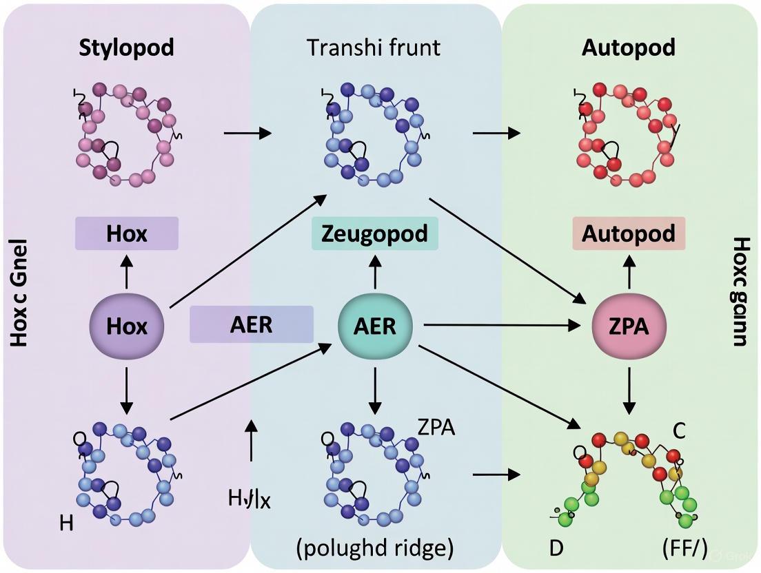
The precise execution of the three-phase Hox expression program is governed by a complex, interactive gene regulatory network. The following diagram illustrates the core signaling pathways and their logical relationships in establishing the tri-phasic pattern.
Diagram 1: Regulatory network for tri-phasic Hox expression.
Key Signaling Centers and Pathways
Sonic Hedgehog (Shh) Signaling: The Zone of Polarizing Activity (ZPA) secretes Shh, which is a critical regulator of the posterior Hox gene expression, particularly in the second and third phases [5]. Shh binds to its receptor Patched (Ptc), releasing Smo (Smoothened) and subsequently promoting the expression of downstream targets, including BMP, WNT, and Hox genes [3]. The dependency of third-phase Hoxa and Hoxd gene expression on Shh signaling is a key experimental finding that links this morphogen to autopod specification [5].
Fibroblast Growth Factor (FGF) Signaling: The Apical Ectodermal Ridge (AER), a thickened epithelium at the distal tip of the limb bud, secretes various FGFs (e.g., FGF-4, FGF-8) [3]. These signals promote the undifferentiated growth of the underlying mesenchyme in the progress zone and are involved in a positive feedback loop with Fgf10, which is instrumental in initiating and maintaining limb outgrowth [3]. This signaling is crucial for the progressive, distalward patterning of the limb.
Retinoic Acid (RA): In the initial limb fields, specific Hox genes are upregulated by retinoic acid, which helps initiate the downstream genetic signaling that ensures synchronized growth along all three axes [3]. RA is a potent morphogen that plays a foundational role in establishing the early Hox code that prefigures limb position and identity.
The integration of these signals ensures the precise spatiotemporal activation of Hox genes. For instance, the third phase of Hox gene expression is not only dependent on Shh but also involves the action of long-range enhancers, specific to the Hoxa cluster, that are conserved across vertebrates [5].
Experimental Evidence and Methodologies
The three-phase model is supported by rigorous experimental data from key model organisms, leveraging advanced genetic and molecular techniques.
Core Experimental Findings
Table 2: Key Experimental Evidence Supporting the Three-Phase Model
| Experimental Approach | Model Organism | Key Finding | Reference |
|---|---|---|---|
| Expression Analysis (ISH, RNA-seq) | Zebrafish, Mouse, Bamboo Shark | Documented three distinct waves of Hoxa9-13 and Hoxd9-13 expression during pectoral fin/limb bud development. | [5] [8] |
| Genetic Manipulation (Knockout/Misexpression) | Mouse, Chick, Beetle | Loss-of-function of Hox11 paralogs leads to zeugopod defects; misexpression of Hox genes results in homeotic transformations. | [3] [9] |
| Regulatory Disruption (Enhancer Deletion) | Mouse | Deletion of the "digit enhancer" downstream of HoxD disrupts phase 3 Hoxd gene expression and autopod formation. | [5] |
| Pathway Inhibition (Shh) | Zebrafish, Mouse | Inhibition of Shh signaling ablates the second and third phases of Hox gene expression, disrupting zeugopod and autopod formation. | [5] |
Detailed Experimental Protocol: Analyzing Hox Expression in Zebrafish
The following workflow outlines a standard methodology for validating the three-phase model, as employed in foundational studies [5].
Diagram 2: Workflow for analyzing Hox gene expression phases.
This protocol allows researchers to:
- Document Expression Phases: Visually identify the three distinct, overlapping domains of Hox gene expression through ISH or quantify them via RNA-seq.
- Establish Regulatory Dependence: By inhibiting the Shh pathway, researchers can confirm the requirement of this signal for the initiation and maintenance of the second and third phases.
- Evolutionary Comparison: Applying this protocol to multiple species (e.g., zebrafish, shark, mouse) allows for the conclusion that the tri-phasic mechanism is a deeply conserved feature of gnathostome appendage development [5] [8].
The Scientist's Toolkit: Essential Research Reagents
Research in this field relies on a suite of specialized reagents and model systems. The following table details key resources for studying Hox gene function in limb development.
Table 3: Essential Reagents and Resources for Hox Gene Research
| Reagent / Resource | Function / Application | Example Use Case |
|---|---|---|
| Specific Hox Riboprobes | Detection of specific Hox mRNA transcripts via in situ hybridization. | Mapping spatial and temporal expression domains of Hoxa11 and Hoxd13 in limb buds [5]. |
| Shh Pathway Inhibitors (e.g., Cyclopamine) | Chemically inhibit Shh signaling to test dependency of Hox expression phases. | Demonstrating the requirement of Shh for Phase 2 and 3 Hox gene expression [5]. |
| Anti-Hox Antibodies | Immunohistochemical detection of Hox protein localization. | Validating transcription factor presence in specific nuclear domains of the limb bud. |
| CRISPR/Cas9 Gene Editing Systems | Targeted knockout of specific Hox genes or their regulatory elements. | Generating Hoxa13/Hoxd13 double mutants to study complete autopod loss [7]. |
| Transgenic Reporter Lines (e.g., Hoxd-GFP) | Visualizing the activity of Hox gene regulatory elements in live embryos. | Tracking the dynamic activation of the HoxD cluster throughout limb development. |
| Slowly Evolving Model Organisms (e.g., Bamboo Shark) | Facilitates direct genetic comparison with tetrapods due to lower evolutionary rates. | Comparative transcriptomics (RNA-seq) to identify deeply conserved genetic programs [8]. |
The three-phase model of Hox gene expression provides a powerful conceptual framework for understanding the molecular patterning of the limb. This model elegantly links dynamic gene regulation to morphological output, explaining how proximal-distal segments are sequentially specified. The conservation of this mechanism from fish fins to tetrapod limbs underscores its evolutionary deep homology and highlights how modifications to this regulatory cascade—such as changes in the timing, level, or domain of Hox gene expression—can drive morphological evolution [7] [5] [8].
Future research will continue to refine this model by:
- Deciphering Full Regulatory Networks: Utilizing single-cell RNA sequencing and open-chromatin analysis (e.g., ATAC-seq) in various models to identify all components of the Hox-dependent gene regulatory networks (GRNs) and their interactions in different cell lineages [8] [10].
- Understanding Human Disease Correlates: Translating findings from model organisms to human congenital conditions. For example, mutations in HOXA13 and HOXD13 are known to cause synpolydactyly and hand-foot-genital syndrome, directly linking the disruption of the third phase to human autopod malformations [3].
- Exploring Non-Canonical Hox Functions: Investigating roles of Hox genes beyond early patterning, such as in neurogenesis, autophagy, and oogenesis, which may reveal deeper, more ancient functions of these genes and offer new insights into their overall functional logic [4].
A comprehensive understanding of the three-phase model is therefore not only essential for developmental biologists but also provides a critical foundation for clinical researchers and drug development professionals aiming to diagnose, prevent, or treat congenital limb deformities and understand the fundamental principles of morphological evolution.
HoxA and HoxD Cluster Roles in Proximal-Distal Patterning
The formation of the vertebrate limb, with its precise organization into stylopod, zeugopod, and autopod, represents a fundamental process in developmental biology. The HoxA and HoxD gene clusters play indispensable and evolutionarily conserved roles in patterning these proximal-distal segments. As master regulatory genes encoding transcription factors, Hox genes specify positional identities along developing body axes through nested and overlapping expression domains—a phenomenon known as the "Hox code" [11]. In the context of limb development, the paralogs 9-13 of the HoxA and HoxD clusters are particularly critical for establishing segment identity and promoting outgrowth [12]. The coordinated expression of these genes occurs in temporally and spatially distinct phases that correlate with the specification of the three main limb compartments, providing a sophisticated genetic framework for building diverse vertebrate appendages [11] [13]. This technical guide examines the complex roles of HoxA and HoxD clusters in proximal-distal patterning, synthesizing current molecular understanding with experimental evidence to inform ongoing research and therapeutic development.
Genomic Organization and Evolutionary Conservation
Cluster Organization and Paralog Relationships
Hox genes are arranged in tightly linked clusters on chromosomes, a genomic organization that is fundamental to their regulated expression. Most mammals possess four Hox clusters (HoxA, HoxB, HoxC, and HoxD) located on different chromosomes, resulting from two rounds of whole-genome duplication during early vertebrate evolution [14] [15]. The HoxA cluster is found on chromosome 7, while HoxD resides on chromosome 2 in humans [3]. Each cluster contains up to 11-13 genes arranged in a 3' to 5' orientation that corresponds with their expression patterns along the body axes—a property termed colinearity [16] [14].
Zebrafish, as a model organism for limb development studies, possess seven hox clusters due to an additional teleost-specific whole-genome duplication event. These include two HoxA-derived clusters (hoxaa and hoxab) and one HoxD-derived cluster (hoxda), as the hoxdb cluster has been largely lost except for a single microRNA [12]. Despite these differences in cluster number, the fundamental principles of Hox gene function in appendage patterning remain conserved across vertebrate species.
Table 1: Hox Cluster Organization Across Vertebrate Species
| Species | Total Hox Clusters | HoxA-related Clusters | HoxD-related Clusters | Notable Features |
|---|---|---|---|---|
| Mouse/Human | 4 | HoxA | HoxD | Standard mammalian complement |
| Zebrafish | 7 | hoxaa, hoxab | hoxda | Teleost-specific duplication |
| Chicken | 4 | HoxA | HoxD | Key model for limb patterning studies |
| Paddlefish | 4 | HoxA | HoxD | Basal ray-finned fish model |
Evolutionary History and Functional Conservation
The evolutionary trajectory of Hox genes reveals deep conservation of function. Hox genes originated early in animal evolution, with cnidarians possessing Hox genes but lacking their clustered arrangement [14]. The emergence of tightly linked Hox clusters in bilaterians facilitated the evolution of complex body plans through coordinated gene regulation. The ANTP class homeobox genes, to which Hox genes belong, are present across the animal kingdom, highlighting their fundamental role in development [14].
Despite species-specific modifications, the function of Hox genes in limb patterning demonstrates remarkable evolutionary conservation. Mouse Hox genes can substitute for their Drosophila homologs, and when activated in ectopic segments, can induce homeotic transformations in flies [15]. Similarly, the roles of HoxA and HoxD clusters in paired appendage formation are conserved between zebrafish and mice, despite approximately 400 million years of evolutionary divergence [12]. This functional conservation underscores the fundamental importance of these gene clusters in establishing animal body plans.
Three-Phase Model of Hox Expression in Limb Patterning
Phase I: Stylopod Patterning
The initial phase of Hox gene expression in the developing limb bud correlates with specification of the stylopod (upper arm/thigh). During this phase, Hoxd9 and Hoxd10 are expressed across virtually the entire limb bud, establishing the foundation for proximal limb development [13]. This expression pattern is regulated by enhancer elements located on the telomeric (3') side of the HoxD cluster, which drive broad expression throughout the early limb bud mesenchyme [11] [17]. Genetic studies in mice demonstrate that Hoxa9 and Hoxd9 are essential for proper stylopod formation, with double mutants exhibiting specific abnormalities in these most proximal limb elements [12] [3].
Phase II: Zeugopod Patterning
The second phase of Hox expression is characterized by a nested, collinear pattern centered around the zone of polarizing activity (ZPA), which secretes Sonic hedgehog (Shh) [13]. During this phase, Hoxd11 and Hoxd12 are expressed in progressively restricted domains, with Hoxd13 showing the most limited expression pattern [11]. This phase correlates with specification of the zeugopod (forearm/calf) and depends on Shh signaling from the ZPA [18] [13]. The transition from phase I to phase II is marked by the introduction of Shh signals, which modify Hox expression patterns through complex regulatory interactions [18]. In this phase, Hoxa11 plays a particularly important role, as evidenced by its specific expression pattern in the zeugopod-region of developing limbs [13].
Phase III: Autopod Patterning
The third and final phase of Hox expression represents a dramatic shift to what has been termed the "distal phase" (DP) or "reverse collinear" pattern [11]. During this phase, associated with autopod (hand/foot) formation, the expression patterns of 5' Hoxd genes invert, with Hoxd13 now exhibiting the broadest expression domain across the developing autopod, while Hoxd12 and Hoxd11 show progressively more restricted expression [11] [17]. This phase is regulated by enhancer elements located on the centromeric (5') side of the HoxD cluster, representing a distinct regulatory landscape from the earlier phases [17] [19]. A similar distal phase expression has also been documented for HoxA genes, indicating that this regulatory module is not exclusive to HoxD [11]. This late phase is crucial for digit formation, with Hoxa13 and Hoxd13 playing particularly vital roles in autopod morphogenesis [12] [19].
Table 2: Characteristics of the Three Phases of Hox Expression in Limb Patterning
| Phase | Limb Segment | Key Hox Genes | Regulatory Region | Principal Regulators |
|---|---|---|---|---|
| I | Stylopod | Hoxd9, Hoxd10, Hoxa9 | Telomeric (3') | Early limb bud signals |
| II | Zeugopod | Hoxd11, Hoxd12, Hoxa11 | Transitioning | Shh from ZPA |
| III | Autopod | Hoxd13, Hoxa13 | Centromeric (5') | Late-phase enhancers (e.g., GCR) |
Regulatory Mechanisms Governing Hox Expression
Chromatin Topology and 3D Genome Architecture
The regulation of Hox gene expression during limb development involves sophisticated chromatin topology and three-dimensional genome architecture. The HoxA and HoxD clusters are embedded within topologically associated domains (TADs) that define their interactions with distinct regulatory landscapes [14]. During limb development, enhancers on either side of TAD boundaries coordinate two transcriptional waves that permit limb patterning—the early wave patterns the stylopod and zeugopod, while the late wave patterns the digits [14].
Research has revealed that the transition from early to late transcriptional waves for Hoxd13 is facilitated by enhancers positioned in telomeric gene deserts within two TADs outside the Hox gene clusters [14]. In the distal posterior limb, where Hoxd13 expression is highest, there is a loss of polycomb-catalyzed H3K27me3 histone modification and chromatin decompaction over the HoxD locus, making it more accessible for transcription [17]. Simultaneously, the global control region (GCR), a long-range enhancer located 180 kb 5′ of Hoxd13, spatially colocalizes with the 5′ HoxD genomic region specifically in the distal posterior limb, forming a chromatin loop that activates expression [17].
The Role of HOX13 in Chromatin State Transitions
The HOX13 proteins (HOXA13 and HOXD13) play a particularly important role in regulating chromatin state transitions during the shift from zeugopod to autopod patterning. Genomic studies have revealed that HOX13 proteins are required for proper termination of the early limb transcriptional program and activation of the late-distal limb program [19]. In Hoxa13−/−;Hoxd13−/− mutant limbs, the early transcription program persists while expression of late-distal-specific genes is largely abolished [19].
HOX13 proteins coordinate this transition through dual action on cis-regulatory modules, regulating H3K27 modification at regulatory elements [19]. They promote an open chromatin conformation in the distal limb bud, facilitating the transition from early/proximal to late/distal limb patterning [14]. This function makes HOX13 proteins crucial gatekeepers of the distal limb program, with loss of function leading to severe truncations of autopod elements [12] [19].
Experimental Approaches and Key Findings
Gene Targeting and Loss-of-Function Studies
Targeted gene disruption in model organisms has been instrumental in elucidating the specific functions of HoxA and HoxD genes in limb patterning. The generation of loss-of-function alleles for all 39 Hox genes in mice has revealed the profound importance of these genes in skeletal patterning [16]. Several key findings have emerged from these studies:
Functional redundancy: Single Hox gene mutations often produce subtle phenotypes due to functional overlap between paralogs. For example, inactivation of either Hoxa11 or Hoxd11 alone has limited effects, but simultaneous inactivation of both produces dramatic limb abnormalities [15].
Compound mutant analyses: Mice lacking both Hoxa13 and Hoxd13 show specific defects in the autopod, with severe digit agenesis [19]. Similarly, simultaneous deletion of the entire HoxA and HoxD clusters leads to severe truncation of forelimbs, particularly distal elements [12].
Zebrafish cluster deletions: Recent research has generated zebrafish mutants with various combinations of deletions in hoxaa, hoxab, and hoxda clusters. Triple homozygous mutants (hoxaa−/−;hoxab−/−;hoxda−/−) display significantly shortened pectoral fins, with the endoskeletal disc and fin-fold both affected [12].
Expression Profiling and Genomic Approaches
Modern genomic techniques have provided unprecedented insights into Hox gene regulation and function:
Chromatin Immunoprecipitation (ChIP): Studies profiling HOXA13 and HOXD13 binding genome-wide have identified thousands of binding sites in the developing limb bud, revealing how these transcription factors regulate downstream targets [19].
RNA-sequencing: Transcriptome analysis of wild-type versus Hox13-mutant limbs has identified genes involved in the early to late-distal program transition, highlighting pathways controlled by these key regulators [19].
Chromatin conformation capture: Techniques such as Hi-C have revealed the dynamic chromatin architecture of Hox clusters during limb development, demonstrating physical interactions between genes and distal enhancers [14] [17].
Table 3: Key Phenotypes from HoxA and HoxD Loss-of-Function Experiments
| Genotype | Species | Stylopod | Zeugopod | Autopod | Reference |
|---|---|---|---|---|---|
| Hoxa9−/−;Hoxd9−/− | Mouse | Abnormalities | Normal | Normal | [12] |
| Hoxa11−/−;Hoxd11−/− | Mouse | Normal | Abnormalities | Normal | [15] |
| Hoxa13−/−;Hoxd13−/− | Mouse | Normal | Normal | Severe digit agenesis | [19] |
| HoxA cluster−/−;HoxD cluster−/− | Mouse | Present | Truncated | Severe truncation | [12] |
| hoxaa−/−;hoxab−/−;hoxda−/− | Zebrafish | N/A | N/A | Severely shortened pectoral fin | [12] |
The Scientist's Toolkit: Essential Research Reagents and Methods
Key Research Reagent Solutions
Table 4: Essential Research Reagents for Studying Hox Gene Function in Limb Patterning
| Reagent/Method | Category | Function/Application | Example Use |
|---|---|---|---|
| CRISPR-Cas9 system | Gene editing | Cluster-specific deletions | Generating hoxaa/hoxab/hoxda zebrafish mutants [12] |
| ChIP-seq | Epigenomic profiling | Mapping transcription factor binding sites | Identifying HOXA13/HOXD13 target regions [19] |
| RNA-seq | Transcriptomics | Genome-wide expression profiling | Comparing wild-type vs mutant limb transcriptomes [19] |
| Whole-mount in situ hybridization | Spatial gene expression | Visualizing gene expression patterns | Detecting shha expression in zebrafish fin buds [12] |
| Immortomouse cell lines | Cell culture | In vitro model of limb development | Studying anterior-posterior chromatin differences [17] |
| FGF-coated beads | Experimental embryology | Ectopic limb induction | Testing limb initiation competence [18] |
| Tamoxifen-inducible systems | Temporal gene control | timed gene inactivation | Studying Shh signaling requirements at specific stages [18] |
Experimental Workflows for Hox Gene Analysis
Beyond the Limb: Expanded Roles for HoxA and HoxD Clusters
While traditionally studied in the context of limb development, HoxA and HoxD clusters play important roles in patterning diverse structures beyond paired appendages. Research has revealed that the distal phase expression pattern is not confined to fins and limbs, but occurs in a variety of body plan features, including paddlefish barbels (sensory adornments that develop from the first mandibular arch) and the vent (a medial structure analogous to a urethra) [11]. This suggests that the DP expression module represents an ancient genetic program that has been co-opted in a variety of distally elongated structures throughout vertebrate evolution [11].
Furthermore, Hox genes continue to be expressed and functional at postnatal and adult stages, playing roles in homeostasis, tissue repair, and regeneration [16] [14]. For example, Hox genes are maintained in adult skeletal stem cells required for bone maintenance and repair, and in subsets of tendon and muscle stromal cells [16]. This post-developmental expression suggests ongoing functions for these genes beyond their classical roles in embryonic patterning.
The HoxA and HoxD gene clusters represent master regulators of proximal-distal patterning in vertebrate limbs, operating through a sophisticated three-phase model that sequentially specifies the stylopod, zeugopod, and autopod. Their function relies on dynamic chromatin architecture, precise regulatory interactions, and complex relationships with signaling centers such as the AER and ZPA. While significant progress has been made in understanding the roles of these genes, important challenges remain.
Future research directions include: (1) elucidating the specific downstream targets of Hox transcription factors in different limb segments; (2) understanding how Hox proteins achieve functional specificity in different developmental contexts; (3) exploring the potential therapeutic applications of Hox gene manipulation in regenerative medicine; and (4) investigating how alterations in Hox regulation contribute to evolutionary diversity in limb morphology. As research continues to unravel the complexities of Hox gene function, our understanding of their roles in development, evolution, and disease will undoubtedly expand, opening new avenues for scientific discovery and clinical application.
The development of the vertebrate limb is a fundamental model for understanding the genetic regulation of organogenesis. The limb's segmented structure—comprising the proximal stylopod (humerus/femur), middle zeugopod (radius-ulna/tibia-fibula), and distal autopod (hand/foot)—is orchestrated by precise spatial and temporal gene expression patterns [3]. Among these regulatory factors, Hox genes encode evolutionarily conserved transcription factors that are paramount for patterning the anterior-posterior body axis and for specifying the identity of individual limb segments [20]. The 39 Hox genes in mammals are organized into four clusters (HoxA, B, C, and D) and are expressed in a colinear fashion, with genes at the 3' end of the clusters influencing more anterior/proximal structures and 5' genes influencing more posterior/distal structures [20]. This whitepaper synthesizes current research on the phase-specific phenotypes resulting from targeted Hox gene disruptions, framing the findings within the broader context of Hox gene function in limb patterning. The insights gained are crucial for researchers and drug development professionals aiming to understand the genetic basis of congenital limb deformities and potential regenerative therapies.
Hox Gene Function in Limb Segment Patterning
The Genetic Logic of Proximal-Distal Patterning
The formation of the limb bud and its subsequent segmentation into discrete morphological units is governed by a network of signaling centers and transcription factors. The apical ectodermal ridge (AER), a thickened epithelium at the limb bud tip, produces fibroblast growth factors (FGFs) that maintain a underlying progress zone of proliferating mesenchymal cells [3]. As cells leave this zone, their positional values are fixed, and they begin to differentiate. The specific identity of each segment—stylopod, zeugopod, or autopod—is determined by the unique combination of Hox genes expressed [20]. This regulatory system exhibits a high degree of functional redundancy, wherein multiple genes within a paralogous group (e.g., Hoxa11 and Hoxd11) perform similar functions, ensuring developmental robustness [20].
Table 1: Hox Gene Paralogue Function in Limb Segments
| Limb Segment | Hox Paralogues Involved | Major Phenotype from Loss of Function |
|---|---|---|
| Stylopod | Hox9, Hox10 [20] | Transformation of lumbar and sacral vertebrae to a rib-bearing thoracic identity; malformations of proximal limb bones [20]. |
| Zeugopod | Hox11 [20] | Transformation of the lumbosacral region to a lumbar morphology; malformations of the radius/ulna or tibia/fibula [20]. |
| Autopod | Hox12, Hox13 [20] | Severe malformations of the hands and feet, including synpolydactyly and loss of digit identity [21]. |
Beyond the Hox genes, other transcription factors establish the initial limb type. Tbx5 is essential for forelimb initiation, while Tbx4 and Pitx1 are critical for hindlimb identity [3]. Mutations in TBX5 cause Holt-Oram syndrome, characterized by forelimb abnormalities and cardiac defects [3]. Recent research has further identified Sall1 and Sall4 as master upstream regulators of the hindlimb initiation cascade, activating key markers like Isl1, Pitx1, and Tbx4 [22].
Regulatory Landscapes and Evolution
The transcriptional control of Hox genes during limb development is managed by two large, distinct regulatory landscapes. The 3' regulatory domain (3DOM) controls the proximal expression of Hoxd genes (up to Hoxd10) in the stylopod and zeugopod, while the 5' regulatory domain (5DOM) activates distal genes (particularly Hoxd13) in the emerging autopod [23]. This bimodal regulatory switch is an evolutionarily conserved mechanism. Interestingly, recent evidence suggests that the 5DOM landscape active in tetrapod digits was co-opted from an ancestral regulatory program used for the development of the cloaca, a finding that provides a novel perspective on the fin-to-limb transition [23].
Phase-Specific Knockout Phenotypes and Experimental Data
Genetic knockout experiments across different model organisms have revealed the specific and often redundant functions of Hox genes. The following table summarizes quantitative phenotypic data from key studies.
Table 2: Quantitative Phenotypes of Hox Gene Knockouts in Limb Development
| Gene(s) Knocked Out | Model Organism | Major Phenotypic Consequences | Severity & Penetrance |
|---|---|---|---|
| Hox9 & Hox10 (compound KO) | Newt (Pleurodeles waltl) | Substantial loss of stylopod and anterior zeugopod/autopod elements, specifically in the hindlimbs [24]. | Phenotype specific to hindlimbs. |
| Hox11 | Newt (Pleurodeles waltl) | Skeletal defects in the posterior zeugopod and autopod of both forelimbs and hindlimbs [24]. | Affects both fore- and hindlimbs. |
| Hoxd11, Hoxd12, Hoxd13 (triple KO) | Mouse | Synpolydactyly (fusion and duplication of digits); defective cortical bone formation in the autopod, replaced by trabecular ossification [21]. | Milder than spdh/spdh mutants; mineralization appears earlier (P2) [21]. |
| Hoxd13spdh/spdh (poly-Ala expansion) | Mouse | Severe synpolydactyly; complete lack of cortical bone and joint formation in the autopod; transformation of metacarpals to a carpal bone morphology [21]. | Severe; no mineralization at P0; mineralization present only within cartilage at P7 [21]. |
| Hoxd13-/-; Hoxa13+/- | Mouse | Severe autopod reduction with 6 digits and no joints; complete lack of mineralization at P0; no cortical bone [21]. | More severe than Hoxd13 single KO. |
| Sall1 & Sall4 (double KO) | Mouse | Failure of hindlimb initiation; loss of expression of hindlimb progenitor markers (Isl1, Pitx1, Tbx4) [22]. | 100% penetrance of hindlimb loss [22]. |
| Gmnn (Geminin) | Mouse | Model 1: Loss or severe shortening of forelimb elements, expanded 5'Hox expression. Model 2: Shortened hindlimb elements and polydactyly, ectopic SHH signaling [25]. | Model- and limb-specific effects. |
The data reveal several key principles. First, the loss of 5' Hox genes (Hox9-13) leads to region-specific malformations rather than homeotic transformations, as seen in the axial skeleton [20]. Second, there is significant functional redundancy, particularly among paralogous groups, as single knockouts often yield milder phenotypes than compound knockouts [24] [21]. Finally, the same genes can govern multiple processes, from initial patterning to later aspects of bone formation and joint specification [21].
Figure 1: Genetic Regulatory Network of Limb Patterning. This diagram illustrates the core genetic pathway initiating limb outgrowth and patterning. Key transcription factors like Tbx5 (forelimb) and Isl1 (hindlimb), regulated by Sall1/Sall4, activate Fgf10. This triggers a feedback loop with Fgf8 from the Apical Ectodermal Ridge (AER), driving outgrowth and activating the bimodal Hoxd regulatory landscapes (3DOM and 5DOM) that pattern the stylopod, zeugopod, and autopod.
Detailed Experimental Methodologies
CRISPR-Cas9 Mediated Multiple Gene Knockout in Newts
Objective: To investigate the functional conservation and redundancy of 5' Hox genes (Hox9-Hox13) in limb development and regeneration [24].
Protocol:
- Animal Model: Iberian ribbed newt (Pleurodeles waltl), selected for its robust regenerative capabilities.
- Guide RNA Design: Design and synthesis of multiple CRISPR guide RNAs (gRNAs) targeting conserved exonic regions of Hox9, Hox10, Hox11, and Hox12 genes to disrupt all paralogous copies.
- Microinjection: Injection of a complex of Cas9 protein and pooled gRNAs into the cytoplasm of single-cell stage newt embryos.
- Phenotypic Screening: Raise injected embryos (F0 generation) to larval stages and analyze limb skeletal patterns post-metamorphosis.
- Skeletal Staining: Fix mutant and control specimens, then perform Alcian Blue (for cartilage) and Alizarin Red (for bone) staining to visualize the skeletal morphology.
- Phenotype Analysis: Compare the skeletal patterns of knockout newts to wild-type controls, focusing on the presence, absence, or malformation of specific elements in the stylopod, zeugopod, and autopod.
Key Insight: This protocol revealed that Hox9 and Hox10 function redundantly to pattern the stylopod and anterior zeugopod/autopod in hindlimbs, a novel finding that suggests functional diversification of 5' Hox genes in tetrapod evolution [24].
Conditional Double Knockout of Sall Genes in Mice
Objective: To determine the functional redundancy of Sall1 and Sall4 in the initiation of hindlimb development [22].
Protocol:
- Genetic Crosses: Generate compound mutant embryos by crossing mice with floxed alleles of Sall1 (Sall1fl) and Sall4 (Sall4fl) with a TCre driver line, which expresses Cre recombinase in early hindlimb progenitor cells (from ~E7.5).
- Genotype Analysis: Confirm genetic recombination and deletion of the target alleles in embryonic tissues via PCR.
- Whole-Mount In Situ Hybridization (WISH):
- Collect mutant and control embryos at E9.5.
- Fix embryos and hybridize with digoxigenin (DIG)-labeled RNA probes for key hindlimb progenitor markers: Isl1, Pitx1, and Tbx4.
- Detect signal using an anti-DIG antibody conjugated to alkaline phosphatase and a chromogenic substrate.
- Phenotypic Validation: Analyze the gross morphology of older embryos (E14.5 to birth) and perform skeletal staining (Alcian Blue/Alizarin Red) to assess the completeness of hindlimb loss.
Key Insight: This conditional knockout approach demonstrated that Sall1 and Sall4 are master regulators acting upstream of the core hindlimb transcription factor cascade, as their combined loss leads to a complete failure of hindlimb initiation [22].
Figure 2: Workflow for Analyzing Limb Knockout Phenotypes in Mice. A standard experimental pipeline for generating and characterizing limb-specific knockout models, integrating genetic, molecular, and morphological techniques.
The Scientist's Toolkit: Key Research Reagents and Models
Table 3: Essential Research Reagents for Investigating Hox Gene Function in Limb Development
| Reagent / Model | Type | Primary Function in Research |
|---|---|---|
| Conditional Knockout Mice (e.g., Sall1fl/fl; Sall4fl/fl) | In vivo model | Enables tissue- and time-specific gene inactivation to study gene function after early embryonic lethality [22]. |
| Cre Driver Lines (e.g., TCre, Hoxb6Cre) | Genetic tool | Controls the spatiotemporal pattern of Cre recombinase activity, defining where and when conditional alleles are activated [22]. |
| Synpolydactyly Homolog Mouse (spdh) | Disease model | Carries a polyalanine expansion in Hoxd13, modeling human synpolydactyly and revealing dominant-negative mechanisms affecting bone formation [21]. |
| CRISPR-Cas9 System | Gene editing tool | Allows for efficient knockout of single or multiple genes in model organisms like mice and newts, facilitating functional analysis [24]. |
| Alcian Blue & Alizarin Red Staining | Histological stain | Visualifies cartilage (blue) and mineralized bone (red) in cleared skeletal preparations, enabling detailed morphological analysis of skeletons [22] [24]. |
| Whole-Mount In Situ Hybridization (WISH) | Molecular technique | Maps the spatial expression patterns of target mRNAs (e.g., Isl1, Fgf10, Hox genes) in intact embryos, crucial for understanding gene function [22]. |
Discussion and Research Implications
The systematic analysis of phase-specific knockout phenotypes solidifies the model that Hox genes are master regulators of limb segment identity. The findings extend beyond a simple patterning role, however. Research in mouse models demonstrates that 5' Hox genes, particularly Hoxd13, directly regulate bone formation by controlling key osteogenic factors like Runx2 [21]. Mutations lead to a failure of cortical bone development in the autopod, which instead undergoes trabecular ossification, and a transformation of metacarpal identity towards a carpal-like morphology [21]. This reveals that Hox genes govern the cellular differentiation programs that execute the pre-patterned skeletal blueprint.
From an evolutionary perspective, comparative studies in zebrafish and mice indicate that the regulatory machinery for the autopod (the 5' Hox landscape) was co-opted from an ancestral program used for cloacal development [23]. This finding provides a powerful explanation for the genetic origin of novel structures during the fin-to-limb transition.
For translational research, the maintained regional expression of Hox genes in adult mesenchymal stem/stromal cells (MSCs) suggests their potential role in guiding region-specific skeletal repair and regeneration [20]. Understanding the phase-specific functions of these genes is therefore not only fundamental for developmental biology but also holds promise for advancing therapeutic strategies in regenerative medicine and for diagnosing complex congenital limb syndromes.
Collinearity and Genomic Organization of Hox Clusters
The development of the tetrapod limb, with its precisely organized segments—the stylopod (upper arm/thigh), zeugopod (lower arm/calf), and autopod (hand/foot)—serves as a paradigm for understanding the fundamental principle of Hox gene collinearity. This principle describes the remarkable correlation between the physical order of Hox genes on the chromosome and their sequential expression in time and space during embryonic development [26]. In the context of limb formation, collinearity is not merely a curious observation but a fundamental operational mechanism directing the patterning of skeletal elements along the proximal-distal axis [27]. The genomic organization of Hox clusters, particularly HoxA and HoxD, underlies a sophisticated bimodal regulatory system that orchestrates the formation of distinct limb compartments through phased interactions with specific topological associating domains (TADs) [28]. This in-depth technical guide synthesizes current research to elucidate how collinearity and genomic architecture direct Hox function in limb development, providing researchers and drug development professionals with a detailed framework of the underlying mechanisms, experimental methodologies, and key reagents essential for advancing this field.
Core Principles of Hox Gene Collinearity
The collinear expression of Hox genes is a multiscale phenomenon, linking genomic organization to morphological patterning. In vertebrate limbs, this manifests through three principal forms of collinearity.
- Spatial Collinearity: The genes located at the 3' end of a Hox cluster (e.g., Hox9, Hox10) are expressed in more proximal limb regions (stylopod), while genes at the 5' end (e.g., Hox13) are expressed in more distal regions (autopod) [29] [13]. This creates a genomic-to-anatomical map that guides segment identity.
- Temporal Collinearity: The activation of Hox genes follows a sequential timeline. Genes at the 3' end of the cluster are transcribed first, followed by a wave of activation that progresses towards the 5' genes [26] [30]. This temporal sequence ensures that the proximal limb segments are specified before the distal ones.
- Quantitative Collinearity: At a given anatomical position where multiple Hox genes are co-expressed, the gene positioned most 5' in the cluster (e.g., Hoxd13) typically exhibits the strongest expression level [26] [31]. This dose-dependent effect is critical for determining the specific morphological identity of skeletal elements, such as distinguishing different digit identities.
Table 1: Forms of Hox Gene Collinearity in Vertebrate Limb Development
| Form of Collinearity | Genomic Correlate | Developmental Expression | Functional Role in Limb |
|---|---|---|---|
| Spatial | Gene order (3' to 5') | Proximal to distal axis (Stylopod to Autopod) | Patterning of segment identity [29] [13] |
| Temporal | Gene order (3' to 5') | Sequential timing of activation | Progressive specification of limb segments [26] |
| Quantitative | Gene position (3' to 5') | Expression level at a given location | Determination of morphological identity (e.g., digit "thumbness") [26] [31] |
Genomic Architecture and Bimodal Regulation of Hox Clusters
The collinear expression of Hox genes, particularly in the HoxD cluster, is governed by a sophisticated bimodal regulatory system based on large chromatin domains known as Topologically Associating Domains (TADs). This mechanism is highly conserved across tetrapods but shows species-specific modifications that correlate with morphological differences, such as those between the mouse and the chick [28].
The Two-Phase Model and Underlying Regulatory Landscapes
Limb development proceeds through two principal regulatory phases, controlled by distinct enhancer landscapes located on either side of the HoxD cluster [27] [31].
- Phase I - Early Proximal Regulation (T-DOM Control): During early limb bud development, genes from Hoxd1 to Hoxd11 are activated by a series of enhancers located in the telomeric regulatory domain (T-DOM). This phase is crucial for the patterning of the stylopod and zeugopod [28] [13]. The activity of this domain is partly regulated by FGF signaling from the Apical Ectodermal Ridge (AER) [13].
- Phase II - Late Distal Regulation (C-DOM Control): Later in development, a regulatory switch occurs. Genes from Hoxd9 to Hoxd13 establish interactions with enhancers in the centromeric regulatory domain (C-DOM). This phase drives the development of the autopod (digits) [28]. This switch is facilitated by HOX13 proteins, which simultaneously inhibit T-DOM activity while reinforcing C-DOM function [28].
- The Articulation Domain: The transition between these two phases creates a domain of low Hoxd gene expression where both T-DOM and C-DOM are silent. This domain gives rise to the future wrist and ankle articulations [28].
The following diagram illustrates the sequential and antagonistic activities of the two regulatory landscapes during limb development.
Comparative Regulation and Morphological Diversity
While the core bimodal mechanism is conserved, comparative studies between species reveal how modifications contribute to morphological diversity. For instance, in chicken hindlimb buds, the duration of T-DOM regulation is significantly shortened compared to the forelimb, correlating with a concurrent reduction in Hoxd gene expression and the distinct morphology of the leg [28]. Furthermore, enhancer elements within these regulatory landscapes can exhibit differential activity; a conserved enhancer in the T-DOM shows stronger activity in chick forelimbs than hindlimbs, a pattern reversed in mice [28]. These findings underscore that evolutionary changes in the implementation of a conserved regulatory strategy are a key source of morphological variation.
Experimental Approaches for Analyzing Hox Regulation
Dissecting the complex regulation of Hox clusters requires a multidisciplinary approach, combining genetic engineering, molecular biology, and advanced genomic techniques.
Genetic Engineering and Mutational Analysis
Targeted manipulation of the mouse genome has been instrumental in unraveling the function of Hox genes and their regulatory elements.
- Systematic Deletion and Duplication: The production of mouse strains with systematic deletions or duplications of specific Hox genes or entire regulatory domains has allowed researchers to parse the functional importance of genomic position and gene dosage [27] [31]. For example, deletion of a large part of the T-DOM results in severe malformations of the stylopod and zeugopod [28].
- Reporter Transgene Scanning: Insertion of reporter transgenes (e.g., LacZ) at various locations within the HoxD cluster allows for the mapping of responsiveness to the telomeric (T-DOM) and centromeric (C-DOM) regulatory landscapes. This technique revealed that the physical position of a gene within the cluster determines its final expression pattern by setting its proximity to these antagonistic regulatory influences [13].
- Cluster Inversion: Engineering large inversions that reposition the centromeric neighborhood of the Hoxd cluster has demonstrated a "landscape effect," where the genomic context, rather than a specific enhancer, exerts a dominant influence on the global regulation of the cluster [26].
Table 2: Key Genetic Engineering Models and Their Outcomes in Hox Limb Research
| Experimental Model | Key Manipulation | Observed Phenotype / Outcome | Functional Insight |
|---|---|---|---|
| T-DOM Deletion [28] | Deletion of telomeric regulatory domain | Severe reduction of stylopod and zeugopod elements; autopod relatively spared. | T-DOM is essential for proximal limb patterning. |
| Hoxd11 Reporter Insertion [13] | Targeted LacZ transgene at Hoxd11 locus | Reporter gene recapitulates the early (zeugopod) phase of Hoxd11 expression. | The Hoxd11 locus is responsive to early phase T-DOM enhancers. |
| Hoxd13 Mutation [31] | Deletion or mutation of Hoxd13 | Malformations of the autopod, including digit loss and fusion. | Hoxd13 is critical for digit growth and patterning; exhibits posterior prevalence. |
| Hoxa11/Hoxd11 Double Mutant [29] | Compound loss of Hox11 paralogs | Severe shortening and malformation of zeugopod (radius/ulna, tibia/fibula). | Hox11 genes are essential and redundant in zeugopod patterning. |
Molecular and Genomic Techniques
A suite of molecular assays is used to probe the expression and chromatin architecture of Hox clusters.
- Chromosome Conformation Capture (3C/4C/Hi-C): These techniques are used to identify the physical, long-range interactions between Hox gene promoters and their distal enhancers located within the T-DOM and C-DOM. This has been pivotal in linking the bimodal expression pattern to distinct topological associating domains [28].
- Histone Modification Analysis (ChIP-seq): Mapping the enrichment of specific histone marks (e.g., repressive H3K27me3 and active H3K4me3) reveals the dynamic chromatin state of the Hox cluster. The transition from a repressed to an active state involves the clearance of H3K27me3 from the 5' end of the cluster in the posterior limb bud mesenchyme [32].
- Single-Cell and Spatial Transcriptomics: Modern techniques like scRNA-seq and spatial transcriptomics (e.g., Visium) allow for the high-resolution mapping of Hox expression patterns directly in human fetal tissues, capturing both temporal and spatial dynamics across different cell types [10].
- Whole-Mount In Situ Hybridization (WISH): A classic technique that provides a spatial map of gene expression in the developing embryo. It has been fundamental for comparing Hoxd gene expression patterns between species like mouse and chicken, and between fore- and hindlimbs [28].
The Scientist's Toolkit: Essential Research Reagents and Models
Advancing research in this field relies on a standardized set of model organisms, molecular reagents, and genetic tools.
Table 3: Essential Research Reagents and Resources for Hox Gene Studies
| Category / Reagent | Specific Example | Function / Application | Reference |
|---|---|---|---|
| Model Organisms | Mouse (Mus musculus) | Primary model for genetic engineering (KO, KI, Cre-Lox); allows detailed limb phenotyping. | [28] [27] [31] |
| Chicken (Gallus gallus) | Model for comparative studies of forelimb/hindlimb differences and evolutionary morphology. | [28] | |
| Genetic Tools | Cre-Lox System (e.g., Prx1-Cre) | Enables tissue-specific (e.g., limb mesenchyme) deletion of floxed target genes. | [32] |
| Reporter Alleles (e.g., Hoxa11eGFP) | Visualizes expression domains of specific Hox genes in live or fixed tissues. | [29] | |
| Molecular Reagents | RNAscope Probes | For high-sensitivity, single-molecule RNA in situ hybridization to localize Hox mRNAs. | [29] |
| H3K27me3 Antibodies | For ChIP-seq to map repressive Polycomb domains on Hox clusters. | [32] | |
| Cell Lines | Limb Bud Mesenchyme Cells (Primary) | Used for in vitro studies of chondrogenesis and Hox gene function. | N/A |
Research Applications and Future Directions
Understanding Hox collinearity and genomic regulation has profound implications beyond basic developmental biology. The precise control of 5' Hox genes (like HOXD13) and their target networks is essential for limb patterning, and disruptions can lead to congenital malformations such as synpolydactyly [32] [31]. Furthermore, the discovery that Hox genes like Hoxa11 remain expressed in postnatal articular cartilage and are involved in its zonal morphogenesis opens new avenues for research into joint regeneration and repair [29]. The conservation of these mechanisms also makes them a valuable framework for studying evolutionary adaptations, such as the elongation of digits in bats or the reduction of anterior elements during the fin-to-limb transition [28] [7]. Future work will continue to leverage single-cell multi-omics and high-resolution chromatin imaging to further decode the dynamic and complex regulation of Hox clusters in development and disease.
The precise spatiotemporal control of gene expression is fundamental to embryonic development, and nowhere is this more evident than in the patterning of the vertebrate limb. This whitepaper explores the critical role of regulatory landscapes—specifically, enhancers such as the ZPA Regulatory Sequence (ZRS)—in orchestrating the complex expression of Hox genes across the proximal-distal (PD) axis of the developing limb. Hox genes, encoding transcription factors, provide the instructional code for the formation of the stylopod (upper arm/thigh), zeugopod (forearm/shin), and autopod (hand/foot). We detail how enhancers respond to overarching morphogen gradients like retinoic acid (RA) to direct this process, and how their perturbation is linked to congenital limb deformities. This guide provides an in-depth analysis of the underlying mechanisms, summarizes key quantitative data, outlines essential experimental protocols for studying these elements, and visualizes the core regulatory networks. Aimed at researchers and drug development professionals, this resource underscores the potential of targeting regulatory landscapes in therapeutic development for limb pathologies.
The development of the limb from a small bud of mesenchymal tissue into a complex, patterned structure is a classic model of morphogenesis. This process is governed by a network of transcription factors, most notably the Hox gene family, which are expressed in overlapping domains along the PD axis to specify the identity of the limb segments [3] [33]. The stylopod is patterned primarily by Hox9 and Hox10 paralogs, the zeugopod by Hox11 paralogs, and the autopod by Hox12 and Hox13 paralogs [34] [33]. However, the expression of these genes is not autonomous; it is controlled by an intricate regulatory landscape consisting of enhancers, silencers, and chromatin modifiers that ensure each gene is activated at the correct time and place [35].
Enhancers are short regions of DNA, often located at a considerable distance from the genes they regulate, that can be bound by transcription factors to enhance gene transcription levels. The concept of a regulatory landscape refers to the full suite of these cis-regulatory elements that control a genomic locus. In the context of the Hox clusters, these landscapes are particularly complex. For instance, the HoxD cluster is flanked by two global regulatory regions that are used sequentially during limb development: a 3' Early Limb Control Region (ELCR) that drives the early phase of Hoxd gene expression in the zeugopod, and a 5' regulatory region that drives a later phase of expression in the autopod [35]. A quintessential example of a specific enhancer within this landscape is the ZPA Regulatory Sequence (ZRS), a long-range enhancer essential for the expression of Sonic hedgehog (Shh) in the zone of polarizing activity, which is critical for anterior-posterior patterning [33]. The RXI in the user's prompt can be understood as a placeholder for such critical, specific enhancers, with the ZRS serving as a prime real-world exemplar.
Understanding the function of these landscapes is not merely an academic exercise. Errors in enhancer function can lead to severe congenital limb malformations [3]. Furthermore, as the field of regenerative medicine advances, deciphering the code that controls limb patterning is a crucial step toward potential therapies for limb loss or malformation. This guide delves into the mechanisms by which enhancers like the ZRS achieve spatiotemporal control, framed within the essential context of Hox-directed limb patterning.
Theoretical Framework: Enhancer Mechanisms and Hox Gene Regulation
The Logic of Spatiotemporal Control
The regulatory landscapes controlling Hox genes in the limb bud implement a sophisticated spatiotemporal program. This control operates on several levels:
- Temporal Colinearity and Sequential Activation: Hox genes are activated in a sequential manner from 3' to 5' within their clusters, a phenomenon known as temporal colinearity. In the early limb bud, the 3' ELCR drives the initial, sequential activation of 3' Hox genes [35]. This phase is associated with patterning the more proximal structures of the limb.
- Spatial Colinearity and Bimodal Control: The spatial expression of Hox genes along the PD axis often mirrors their genomic order. This is facilitated by a bimodal regulatory strategy. Following the early phase, a switch in regulatory control occurs. The later phase of Hoxd gene expression in the autopod is controlled by a separate set of enhancers located on the 5' side of the cluster. This phase occurs in a "reverse colinear" manner, with the most 5' genes (e.g., Hoxd13) being expressed most strongly throughout the developing autopod, while more 3' genes are expressed in progressively more restricted domains [35]. This mechanism ensures that distinct combinations of Hox proteins are present in the stylopod, zeugopod, and autopod to instruct their specific morphologies.
- Self-Regulation and Network Stability: A critical layer of control involves Hox self-regulation. Evidence from mutant studies shows that HOX proteins themselves are required to establish and maintain the precise spatial boundaries of Hox gene expression. The loss of a subset of HOX proteins, particularly from the 5' end of the clusters (e.g., HOXA13 and HOXD13), leads to a global deregulation of both HoxA and HoxD expression patterns. This suggests that a "self-regulatory" mechanism helps to lock in and stabilize the HOX code after the initial signals from global enhancers have been established [35].
Integration with Morphogen Gradients
Enhancers integrate positional information from global morphogen gradients to refine Hox expression domains. The two most critical gradients in limb development are:
- Retinoic Acid (RA): RA, synthesized in the trunk of the embryo, is a key signal for proximal identity. It promotes the expression of proximal Hox genes (like Hoxa9) and patterning genes like Meis1/2 [36] [33]. The RA signal is antagonized in the distal limb bud by CYP26B1, an enzyme that degrades RA. This breakdown is essential for establishing the distal identity of the autopod, marked by the expression of Hoxa13 [36]. Thus, the balance between RA synthesis and degradation creates a PD gradient that is read by the enhancers of various Hox genes and other transcription factors.
- Fibroblast Growth Factors (FGFs): FGFs, secreted by the Apical Ectodermal Ridge (AER) at the distal tip of the limb bud, maintain a zone of proliferating undifferentiated cells and promote distal fates [3] [33]. FGFs from the AER reinforce the distal domain by supporting the expression of Cyp26b1 and Hoxa13, thereby antagonizing the proximalizing signal of RA [36].
Table 1: Key Morphogen Gradients Patterning the Proximal-Distal Axis
| Morphogen | Source | Primary Function | Target Genes/Pathways |
|---|---|---|---|
| Retinoic Acid (RA) | Lateral plate mesoderm / Proximal blastema | Specifies proximal identity; establishes proximal Hox code (e.g., Hox9/10) [36] | Induces Meis1/2; represses distal genes (e.g., Hoxa13) |
| FGFs (e.g., FGF4, FGF8) | Apical Ectodermal Ridge (AER) | Promotes distal identity; maintains cell proliferation in progress zone [3] | Antagonizes RA signaling; induces Cyp26b1 and Hoxa13 |
| Sonic Hedgehog (Shh) | Zone of Polarizing Activity (ZPA) | Patterns anterior-posterior axis; interacts with PD patterning [3] | Regulated by ZRS enhancer; supports FGF expression in AER |
The enhancers controlling Hox genes are designed to respond to specific thresholds of these morphogens, thereby translating a continuous gradient of positional information into discrete domains of gene expression that define the limb segments.
Case Study: The ZRS Enhancer in Limb Development and Evolution
The ZPA Regulatory Sequence (ZRS) is one of the most well-characterized enhancers in biology and serves as a paradigm for understanding enhancer function. It is located nearly one megabase away from the Shh gene it regulates and is exclusively responsible for driving Shh expression in the ZPA of the developing limb bud [33].
Mechanism and Functional Significance
The ZRS is bound by a combination of transcription factors (e.g., ETS1, HAND2) that activate Shh expression in a precise posterior domain. Shh protein then acts as a morphogen, diffusing across the limb bud to pattern the anterior-posterior axis and ensure the correct number and identity of digits form in the autopod. The activity of the ZRS is not isolated; it is part of a feedback loop with the FGF-secreting AER, which ensures the coordinated outgrowth and patterning of the limb [3] [33].
Experimental Evidence from Evolutionary Loss and Mutation
The critical nature of the ZRS is highlighted by studies of its perturbation:
- Mutation in Humans and Model Systems: Point mutations or deletions within the ZRS are a direct cause of congenital limb malformations in humans, such as preaxial polydactyly, where extra digits form on the thumb side of the hand [33].
- Limb Loss in Snakes: The evolution of limblessness in snakes provides a powerful natural experiment. Genomic analyses reveal that while snakes retain the Shh gene and most other limb-patterning genes, their ZRS enhancer has accumulated snake-specific nucleotide changes. In basal snakes like pythons (which retain pelvic rudiments), the ZRS is largely conserved. In contrast, in advanced snakes like vipers (which have no limb structures), the ZRS is highly degraded, with mutations in critical transcription factor binding sites. This suggests that the loss of limbs during snake evolution was driven, at least in part, by the degeneration of this crucial enhancer rather than the loss of the protein-coding genes themselves [33].
This case demonstrates that enhancers like the ZRS are not only essential for proper development but are also key substrates for evolutionary change. The integrity of the regulatory landscape is as important as the integrity of the genes it controls.
Quantitative Data and Experimental Analysis
Research into Hox gene function and regulatory landscapes relies heavily on quantitative assessments of phenotypic severity and molecular changes. The following table synthesizes data from key knockout studies, illustrating the functional redundancy and specific roles of Hox genes in limb patterning.
Table 2: Quantitative Phenotypic Analysis of Hox Gene Knockouts in Limb Development
| Genotype | Model System | Structures Affected (Phenotype) | Key Molecular Changes |
|---|---|---|---|
| Hoxa11-/- / Hoxd11-/- | Mouse | Severe reduction of ulna/radius (zeugopod) [34] | Altered expression of Gdf5, Bmpr1b, Igf1, Shox2 [34] |
| Hoxa9,10,11-/- / Hoxd9,10,11-/- | Mouse | Reduced ulna/radius (more severe than Hoxa11/d11 DKO); defects in stylopod [34] | Severe reduction in Shh (ZPA) and Fgf8 (AER) expression [34] |
| Hox11 KO | Newt (P. waltl) | Skeletal defects in posterior zeugopod and autopod [24] | Not Specified |
| Hox9/Hox10 compound KO | Newt (P. waltl) | Substantial loss of stylopod & anterior zeugopod/autopod (hindlimb-specific) [24] | Indicates redundant function of Hox9/10 in hindlimb stylopod formation |
| Hox13 KO | Newt/Mouse | Complete loss of autopod (digit) elements [24] [34] | Deregulation of other Hox genes ("self-regulation" loss) [35] |
Detailed Experimental Protocol: CRISPR-Cas9 Knockout of Limb Enhancers
To functionally validate the role of a putative enhancer (e.g., the RXI or ZRS) in a model organism like the axolotl or mouse, the following protocol can be employed.
Objective: To delete a specific genomic enhancer and assess its impact on Hox gene expression and limb morphology.
Materials and Reagents:
- CRISPR-Cas9 System: Cas9 protein or mRNA; single-guide RNAs (sgRNAs) designed to flank the target enhancer region.
- Microinjection Setup: Micropipettes and microinjector for delivering components into single-cell embryos.
- Genotyping Reagents: PCR primers flanking the deletion site; gel electrophoresis or sequencing equipment.
- In Situ Hybridization (ISH) Reagents: Digoxigenin-labeled RNA probes for target Hox genes (e.g., Hoxa13, Hoxd13, Shh); anti-digoxigenin antibodies; NBT/BCIP substrate for colorimetric detection.
- Skeletal Staining Reagents: Alcian Blue (for cartilage) and Alizarin Red (for bone).
Methodology:
- sgRNA Design and Validation: Design two sgRNAs with targets located precisely upstream and downstream of the enhancer element to be deleted. Validate the efficiency and specificity of each sgRNA in vitro.
- Embryo Microinjection: Co-inject Cas9 protein/mRNA and the two sgRNAs into the cytoplasm of freshly fertilized embryos. For axolotls, this is typically performed in 0.1x Modified Barth's Saline (MBS) [36].
- Rearing and Screening: Raise the injected embryos (F0 generation) to the desired developmental stage. A non-invasive tissue biopsy (e.g., tail tip) is performed for genomic DNA extraction.
- Genotypic Analysis: Perform PCR using primers that span the sgRNA target sites. A successful deletion will result in a smaller PCR product compared to the wild-type allele, resolvable by gel electrophoresis. Sequence the PCR product to confirm precise deletion.
- Phenotypic Analysis:
- Molecular Phenotyping (ISH): Fix control and mutant embryos at various stages (e.g., limb bud stage, early digit formation). Process for whole-mount ISH using probes for genes predicted to be regulated by the enhancer (e.g., Shh for ZRS) and for Hox genes (e.g., Hoxa13) to assess changes in their spatial expression domains.
- Morphological Phenotyping (Skeletal Staining): For later stages, euthanize larvae or juveniles and process for Alcian Blue and Alizarin Red staining to visualize the cartilage and bone architecture of the entire limb skeleton, noting any homeotic transformations or truncations.
The Scientist's Toolkit: Essential Research Reagents
Table 3: Key Reagent Solutions for Studying Limb Regulatory Landscapes
| Reagent / Solution | Function / Application |
|---|---|
| CRISPR-Cas9 with sgRNAs | Targeted knockout of specific enhancers or Hox genes in model organisms [24] |
| Digoxigenin (DIG)-labeled RNA Probes | Detection of specific mRNA transcripts via in situ hybridization to visualize gene expression patterns [36] |
| Alcian Blue & Alizarin Red | Histological stains for cartilage and bone, respectively; used for clear visualization of the skeletal pattern in cleared specimens [34] |
| Retinoic Acid (RA) & CYP26 Inhibitors | Pharmacological tools to manipulate the RA signaling gradient; used to test proximalization or distalization of limb identity [36] |
| Laser Capture Microdissection (LCM) | Isolation of specific cell populations (e.g., progress zone, chondrogenic condensations) from tissue sections for transcriptomic analysis [34] |
| scRNA-seq Library Prep Kits | Generation of libraries for single-cell RNA sequencing to profile the heterogeneous transcriptional states within the limb bud mesenchyme [36] |
Visualization of Regulatory Networks and Workflows
The following diagrams, generated with Graphviz DOT language, illustrate the core regulatory network controlling PD patterning and a standard workflow for enhancer validation.
Regulatory Network of Limb PD-Patterning
Diagram 1: Core network of proximal-distal limb patterning. This diagram illustrates how the morphogens Retinoic Acid (RA) and FGFs, along with the RA-degrading enzyme CYP26B1, interact to establish the domains of key transcription factors (Meis, Hox9/10/11, HoxA13) that specify the stylopod, zeugopod, and autopod. Arrowheads indicate activation; flat heads indicate repression.
Experimental Workflow for Enhancer Analysis
Diagram 2: A standard pipeline for the identification and functional validation of a limb enhancer. The process begins with computational identification of conserved non-coding sequences, followed by testing their ability to drive reporter gene expression in a limb-specific pattern. Functional impact is then assessed by deleting the enhancer in vivo and analyzing the resulting changes in gene expression and limb skeleton morphology.
The study of regulatory landscapes, exemplified by enhancers like the ZRS, has fundamentally advanced our understanding of how Hox genes achieve the precise spatiotemporal control necessary for limb formation. It is clear that the genomic context and long-range regulatory inputs are as critical as the Hox protein-coding sequences themselves. The emerging concept of Hox self-regulation adds a fascinating layer of stability to this system, ensuring that once established, the HOX code is maintained.
Future research will continue to unravel the complexities of the 3D chromatin architecture that brings these distant enhancers into contact with their target gene promoters. Furthermore, the integration of single-cell multi-omics (transcriptomics, epigenomics) will provide an unprecedented resolution view of the dynamic regulatory states in the developing limb mesenchyme. For drug development professionals, these regulatory elements represent potential therapeutic targets. While targeting transcription factors directly is notoriously difficult, understanding the pathways they control (e.g., BMP, FGF, RA) can identify druggable nodes for modulating limb development and regeneration. As we decode the regulatory grammar of the genome, we move closer to the possibility of reprogramming cellular fate for regenerative medicine, potentially instructing a blastema to rebuild a complete and patterned limb.
From Gene to Function: Advanced Techniques for Deciphering Hox Biology
CRISPR-Cas9 and the Generation of Cluster-Wide Deletion Mutants
In the study of Hox gene function, particularly regarding their roles in patterning the vertebrate limb into stylopod, zeugopod, and autopod, the ability to interrogate entire gene clusters represents a powerful approach for functional genomics [37]. Hox genes are often arranged in clusters, and their coordinated expression in time and space is critical for proper axial patterning [38] [39]. Traditional single-gene knockout strategies can be insufficient for deciphering the complex, overlapping functions and regulatory mechanisms within these tightly linked gene families. The development of CRISPR-Cas9 technology has revolutionized this process, enabling researchers to generate cluster-wide deletion mutants with a precision and efficiency previously unattainable [40] [41]. This technical guide outlines the core principles, methodologies, and applications of using CRISPR-Cas9 for creating large-scale chromosomal deletions, with a specific focus on its impact on research into limb formation.
Technical Foundations of CRISPR-Cas9 Mediated Deletion
Mechanism of Action
The CRISPR-Cas9 system functions as a versatile and programmable genome engineering tool. The core system consists of two fundamental components [40] [42]:
- The Cas9 endonuclease, which creates double-strand breaks (DSBs) in DNA.
- A single-guide RNA (sgRNA), a synthetic fusion of CRISPR RNA (crRNA) and trans-activating crRNA (tracrRNA), which directs Cas9 to a specific genomic locus complementary to a 17-20 nucleotide spacer sequence within the sgRNA.
The Cas9 nuclease is directed by the sgRNA to a target DNA sequence and induces a DSB 3-4 base pairs upstream of a Protospacer Adjacent Motif (PAM), which for the commonly used Streptococcus pyogenes Cas9 (SpCas9) is the sequence 5'-NGG-3' [40] [43]. The cellular repair of this DSB is exploited to generate deletions.
Generating Deletions via Dual sgRNA Strategy
While a single DSB is typically repaired to create small insertions or deletions (indels), the coordinated use of two sgRNAs targeting distant sites on the same chromosome can result in the excision of the entire intervening sequence [41]. The cell repairs these concurrent DSBs primarily through the error-prone non-homologous end-joining (NHEJ) pathway, which often ligates the two distal ends, thereby excising the fragment between the two cut sites as a linear DNA molecule that is subsequently degraded. This process allows for the programmable deletion of genomic regions ranging from a few kilobases to over hundreds of kilobases, encompassing entire gene clusters.
Table 1: Key CRISPR-Cas Systems for Generating Genomic Deletions
| System Feature | CRISPR/Cas9 (SpCas9) | CRISPR/Cpf1 (Cas12a) |
|---|---|---|
| PAM Sequence | 5'-NGG-3' [43] | 5'-TTN-3' [40] |
| Guide RNA | Single-guide RNA (sgRNA) [40] | CRISPR RNA (crRNA) only [40] |
| Cleavage Type | Blunt ends [40] | Staggered ends with 5' overhangs [40] |
| Protein Size | ~1368 amino acids [43] | Smaller than Cas9, beneficial for delivery [40] |
Experimental Design and Workflow
A robust workflow is critical for the successful generation of cluster-wide deletion mutants. The process can be broken down into key stages, from initial design to the isolation and validation of mutant lines.
Target Selection and sgRNA Design
The first step involves the careful selection of the target genomic region to be deleted. For Hox cluster analysis, this entails defining the precise boundaries of the cluster, including promoter regions and potential cis-regulatory elements [38] [44].
- sgRNA Design Principles: Two sgRNAs are designed to bind to sequences flanking the target cluster. The selection of sgRNA target sites should prioritize [41]:
- High predicted efficiency using validated scoring algorithms.
- Minimal off-target potential by performing a BLAST search against the target genome.
- The presence of a canonical PAM sequence immediately downstream of the target site.
- Deletion Size Considerations: The efficiency of deletion formation can decrease with increasing size of the intervening fragment, though deletions of several hundred kilobases are feasible.
sgRNA Efficiency Validation
Before committing to a full-scale experiment, it is highly advisable to validate the cutting efficiency of the designed sgRNA pairs. This can be achieved through [41]:
- Protoplast Transfection: Delivering the CRISPR constructs into plant or mammalian protoplasts and using PCR with primers flanking the target site to detect deletion events.
- Surveyor or T7 Endonuclease I Assay: Detecting Cas9-induced indels at the individual target sites in vitro.
- Next-Generation Sequencing: Providing a quantitative measure of editing efficiency at both on-target and potential off-target sites.
Delivery and Regeneration
For the creation of stable mutant lines, the CRISPR/Cas9 components must be delivered into the nucleus of a host cell, which is then regenerated into a whole organism.
- Delivery Methods: Common methods include Agrobacterium-mediated transformation (plants), microinjection into zygotes (animals), or electroporation of embryonic stem cells [38] [41].
- Vector Systems: All-in-one binary vectors are often used, which harbor expression cassettes for Cas9, one or more sgRNAs, and a selectable marker (e.g., herbicide or antibiotic resistance) [41]. To enhance heritability, the use of egg-cell specific promoters (e.g., EC1.2) to drive Cas9 expression has been shown to dramatically increase the efficiency of recovering heritable mutations [41].
Screening and Validation
Following transformation, a multi-step screening process is employed to identify successful deletion mutants.
- Somatic Screening: Initial screening of primary transformants (T0 in plants, F0 in animals) via PCR using primers that flank the deletion boundaries. A successful deletion will result in a smaller PCR product alongside the wild-type band [41].
- Germline Transmission: Somatic mutants are outcrossed to wild-type individuals. The progeny are screened to identify those that have inherited the deletion but lack the Cas9 transgene. Fluorescent seed markers can facilitate this non-destructive selection [41].
- Homozygous Line Isolation: Self-pollination or sibling crossing of heterozygous mutants yields progeny that are homozygous for the deletion. These lines are identified by PCR genotyping.
Table 2: Quantitative Outcomes from a Representative Deletion Study in Arabidopsis [41]
| Targeted Region | Somatic Deletion Efficiency (T1) | Heritable Deletion Efficiency (T2) | Vector Used |
|---|---|---|---|
| ISU1 Locus (~12 kb) | 51% (77/152 lines) | ~0.5% (2/396 plants) | pUbiCAS9-Red |
| Non-Coding Region | 69% | 5% | pUbiCAS9-Red |
| Gene Cluster X | 4% | 0% | pUbiCAS9-Red |
| Gene Cluster Y | 60% | 100% (Homozygous) | pEciCAS9-Red |
Case Study: Deletion of the Hoxbb Cluster in Zebrafish
The power of this approach is exemplified by its application to dissect the role of the Hoxbb cluster in vertebrate development and disease [38].
- Objective: To model a human congenital heart defect (CHD) associated with a microdeletion of the HOXB cluster on chromosome 17q21.32 and to identify the specific gene(s) responsible [38].
- Methodology:
- Target Selection: Two sgRNAs were designed to target sequences flanking the entire ~25.5 kb hoxbb cluster on zebrafish chromosome 12, which contains four genes: hoxb1b, hoxb5b, hoxb6b, and hoxb8b.
- Delivery: Both sgRNAs and Cas9 mRNA were co-injected into single-cell zebrafish embryos.
- Screening: Founders (F0) were screened, showing an ~80% knockout efficiency. These fish were outcrossed to establish a stable heterozygous line.
- Phenotypic and Genotypic Analysis:
- Homozygous mutants (hoxbb-/-) were viable initially but developed severe cardiac abnormalities by 5 days post-fertilization (dpf), including pericardial edema, heart looping failure, and atrioventricular regurgitation [38].
- All homozygous mutants died by 11 dpf, indicating the essential role of this cluster in embryonic development.
- Follow-up studies using isolated gene knockouts revealed that the cardiac phenotype was primarily driven by the loss of hoxb1b, pinpointing it as the key causal gene within the cluster [38].
The Scientist's Toolkit: Essential Research Reagents
Table 3: Key Reagents for CRISPR-Cas9 Mediated Cluster Deletion
| Reagent / Solution | Function / Explanation | Example |
|---|---|---|
| Cas9 Nuclease | Engineered version of the bacterial enzyme that creates double-strand breaks in DNA. | Streptococcus pyogenes Cas9 (SpCas9) [40] [43] |
| sgRNA Expression Construct | A DNA template for in vitro transcription or a plasmid expressing the chimeric guide RNA. | Vectors with U6 or U3 RNA polymerase III promoters [41] |
| Binary Vector System | A plasmid for delivering Cas9 and sgRNAs into the host organism. | pUbiCAS9-Red, pEciCAS9-Red (for plants) [41] |
| Delivery Tools | Methods for introducing CRISPR components into target cells. | Agrobacterium tumefaciens (plants), microinjection (animal zygotes), electroporation [38] [41] |
| Selection Marker | A gene that allows for the enrichment of successfully transformed cells or organisms. | Fluorescent proteins (e.g., RFP), antibiotic/herbicide resistance genes (e.g., BAR) [41] |
| Genotyping Primers | Oligonucleotides designed to flank the target deletion site for PCR-based screening. | Primers that bind outside the sgRNA target sequences [38] [41] |
Application to Hox Gene Research in Limb Patterning
The case for using cluster-wide deletions is particularly strong in Hox gene research. In a seminal study on Xenopus limb regeneration, transcriptomic analysis revealed that hoxc12 and hoxc13 exhibited the highest regeneration-specific expression [37]. CRISPR/Cas9-mediated knockout of each gene demonstrated that they were critical for rebooting the developmental program during regeneration, specifically impacting autopod (digit) formation without affecting initial limb development or blastema formation [37]. This highlights a regeneration-specific, non-redundant function that could only be fully understood by targeting individual genes within the cluster, a task for which CRISPR is ideally suited. This approach allows for the functional dissection of paralogous genes (e.g., HOXA6 vs. HOXB6), which have been shown to have surprising non-redundant functions in processes like caudal neurogenesis [39].
The ability to generate cluster-wide deletion mutants using CRISPR-Cas9 has provided researchers with an unparalleled tool for deconstructing the functional complexity of tightly linked gene families like the Hox clusters. The technical framework outlined herein—from meticulous sgRNA design and efficiency validation to the use of optimized delivery systems and screening protocols—enables the precise deletion of genomic regions to model human diseases and uncover fundamental mechanisms in development, such as the patterning of the stylopod, zeugopod, and autopod. As CRISPR technology continues to evolve with the development of novel Cas variants with altered PAM specificities and reduced off-target effects, its power to illuminate the collective function of gene clusters will only grow, driving forward our understanding of vertebrate development and regenerative biology.
The precise spatial and temporal expression of Hox genes is fundamental for embryonic development, particularly in patterning structures along the anterior-posterior axis and in the formation of limb segments, including the stylopod, zeugopod, and autopod [35]. These genes are organized in tight clusters, and their regulation involves a complex interplay of distal enhancers, local transcription factor binding, and higher-order chromatin organization [45]. Traditional gene-editing methods struggle to decipher this complex regulatory logic because Hox clusters are isolated in 'gene deserts' and conventional edits often affect multiple genes or disrupt the intricate regulatory landscape [46]. Synthetic biology approaches, which involve the bottom-up construction of genetic modules, offer a solution by enabling the deconvolution of this regulatory complexity through the design and integration of artificial Hox genes and clusters into model systems [45] [47].
Foundational Principles of Hox Gene Biology and Limb Patterning
Hox Gene Function in Stylopod, Zeugopod, and Autopod Patterning
During vertebrate limb development, Hox genes from the A and D clusters are expressed in two phases, which are crucial for the proper formation of the limb's proximal-to-distal segments [35]. The initial phase involves genes that help pattern the more proximal structures, including the stylopod (upper limb) and zeugopod (lower limb). A subsequent phase of expression, particularly of 5' Hox genes, is essential for the development of the autopod (hand/foot) [35]. This process relies on distinct cis-regulatory elements located on both sides of the Hox clusters. Importantly, Hox proteins themselves contribute to the establishment and maintenance of their own expression domains, a process referred to as "self-regulation" [35]. For instance, the presence of HOX paralogous group 13 proteins is a prerequisite for the clear separation of zeugopod and autopod expression domains of HoxA and HoxD genes [35].
Core Methodologies for Constructing Artificial Hox Genes
Synthetic Regulatory Reconstitution of Hox Clusters
A groundbreaking methodology termed "synthetic regulatory reconstitution" has been developed to dissect the regulatory architecture of Hox clusters. This bottom-up approach involves the de novo assembly of large, variant HoxA cluster constructs (130-170 kb) using yeast homologous recombination. These synthetic constructs are then delivered as a single copy into a defined ectopic location (the Hprt1 locus) in the mouse genome [45]. This platform allows for the testing of various regulatory module combinations:
- SynHoxA: A minimal cluster lacking distal enhancers.
- Enhancers+SynHoxA: The minimal cluster plus distal enhancer elements.
- RAREΔ: A cluster with mutated Retinoic Acid Response Elements (RAREs) [45].
Table 1: Key Constructs in Synthetic HoxA Reconstitution
| Construct Name | Size | Key Features | Primary Research Objective |
|---|---|---|---|
| SynHoxA | 134 kb | Minimal rat HoxA cluster; lacks distal enhancers | Test sufficiency of intracluster regulation for domain specification |
| Enhancers+SynHoxA | 130-170 kb | SynHoxA with addition of distal enhancers | Determine enhancer role in boosting transcriptional output |
| RAREΔ | 130-170 kb | SynHoxA with mutated RAREs | Assess necessity of intracluster transcription factor binding |
Fabrication of Artificial Hox Genes in Pluripotent Stem Cells
A complementary strategy involves fabricating synthetic DNA strands from the Hox genes of one species (e.g., rat) and inserting them into pluripotent stem cells of another (e.g., mouse) [46] [47]. The use of different species allows researchers to clearly distinguish the synthetic DNA from the host cell's native genes. This method demonstrated that the Hox clusters alone contain all necessary information for cells to decode a positional signal and retain that positional memory, confirming that the compact nature of the cluster is key to this function [46] [47].
Figure 1: Workflow for Building and Integrating Artificial Hox Genes. This diagram outlines the key steps for creating synthetic Hox genes and introducing them into stem cells for functional analysis.
Key Experimental Workflows and Protocols
Protocol for Bottom-Up Hox Cluster Assembly and Integration
The following detailed protocol is adapted from synthetic regulatory reconstitution studies [45]:
- DNA Amplicon Production: Generate polymerase chain reaction (PCR) amplicons covering the entire target Hox cluster (e.g., rat HoxA) from a Bacterial Artificial Chromosome (BAC) template. Design amplicons with overlapping ends to facilitate homologous recombination.
- Yeast Homologous Recombination: Co-transform the pool of overlapping amplicons into yeast cells. The endogenous homologous recombination machinery assembles these into a complete, large-scale "assemblon" (e.g., 134 kb SynHoxA).
- Assemblon Modification (Optional): Introduce specific mutations (e.g., RARE deletions) by replacing wild-type amplicons with synthetic DNA containing the desired edits or by using CRISPR/Cas9-based engineering directly in yeast.
- DNA Purification: Recover the assembled, large DNA construct from yeast culture.
- Mammalian Cell Integration: Deliver the purified assemblon, alongside a targeting vector, into mouse embryonic stem cells (mESCs) via electroporation. Utilize a positive-negative selection system to isolate clones with a single-copy integration at the defined Hprt1 locus.
- Validation: Employ long-range PCR and Southern blotting to confirm the correct structure and single-copy integration of the synthetic Hox cluster.
Protocol for Functional Analysis in Differentiated Cells
Once integrated, the functional output of the synthetic cluster is assessed [45]:
- Directed Differentiation: Differentiate mESCs containing the synthetic Hox cluster into neuronal or other relevant lineages using established protocols, often involving treatment with retinoic acid (RA) to induce caudal identities.
- Transcriptomic Analysis: At specific differentiation timepoints, harvest cells and perform RNA sequencing (RNA-seq) to quantify the expression of genes from the synthetic cluster. Compare this to the expression from the endogenous cluster.
- Epigenetic Profiling: Conduct Chromatin Immunoprecipitation followed by sequencing (ChIP-seq) for histone modifications (e.g., H3K27me3 for repressed domains, H3K4me3 for active domains) to determine if the synthetic cluster recapitulates native chromatin states and boundary formation.
- Topological Analysis: Use Hi-C or related methods to assess whether the synthetic cluster forms the characteristic active (HoxA1–HoxA5) and inactive (HoxA6–HoxA13) topological domains observed at the endogenous locus.
Critical Findings from Synthetic Hox Gene Studies
Decoding the Regulatory Logic of the Hox Cluster
Synthetic approaches have yielded fundamental insights into how Hox clusters operate. The key finding from synthetic reconstitution is that a minimal HoxA cluster, even in the absence of distal enhancers, is sufficient to recapitulate the correct spatial pattern of gene activation, chromatin boundary formation, and 3D topological reorganization in response to retinoic acid [45]. Distal enhancers were found to be dispensable for specifying which genes are active but were necessary for achieving full transcriptional levels. Most critically, the study demonstrated that intracluster transcription factor binding sites (RAREs) are the primary module for initiating the response to morphogenetic signals; mutating these sites nearly abolished both gene activation and chromatin remodeling [45].
Table 2: Quantitative Findings from Synthetic Hox Cluster Studies
| Experimental Condition | Gene Expression vs. Wild-Type | Chromatin Boundary Formation | Key Conclusion |
|---|---|---|---|
| SynHoxA (Minimal Cluster) | Correct pattern, reduced level | Recapitulated | Intracluster info is sufficient for patterning |
| Enhancers+SynHoxA | Correct pattern, near wild-type level | Recapitulated | Distal enhancers boost transcriptional output |
| RAREΔ (Mutant RAREs) | Severely reduced or abolished | Disrupted | Intracluster TF binding is necessary for initiation |
Figure 2: Regulatory Logic of the Hox Cluster. This diagram illustrates the hierarchical relationship between the retinoic acid signal, intracluster regulatory elements, chromatin reorganization, and gene expression, with distal enhancers playing a secondary, synergistic role.
Elucidating Self-Regulation and Non-Redundant Functions
Research using artificial genes and loss-of-function screens has further clarified Hox gene function. Studies in limb development revealed a "self-regulation" mechanism, whereby HOX proteins themselves help establish and maintain the spatial domains of Hox gene expression, ultimately shaping the final HOX code that patterns the limb [35]. Furthermore, genome-wide loss-of-function screens in human stem cells undergoing neuronal differentiation have uncovered surprising non-redundancy between paralogous Hox genes, such as HOXA6 and HOXB6, indicating that even highly similar Hox genes have unique and essential roles in specific developmental contexts [48].
The Scientist's Toolkit: Essential Research Reagents and Solutions
Table 3: Key Reagent Solutions for Synthetic Hox Gene Research
| Reagent / Tool | Function in Research | Specific Application Example |
|---|---|---|
| Yeast Artificial Chromosome (YAC) System | Facilitates homologous recombination for large DNA assembly | Assembly of 130-170 kb SynHoxA assemblons [45] |
| Bacterial Artificial Chromosomes (BACs) | Source of native Hox cluster DNA for amplification | Template for rat HoxA cluster amplicons [45] |
| Pluripotent Stem Cells (mESC/hESC) | Differentiable host for synthetic gene integration | Mouse ESCs for ectopic integration; haploid hESCs for LOF screens [45] [48] |
| CRISPR-Cas9 System | Enables targeted genome editing and engineering | Yeast-based editing of assemblons; genome-wide LOF screens [45] [48] |
| Retinoic Acid (RA) | Key morphogen for inducing Hox gene expression | Differentiation agent for caudal neuronal cell fates [45] [48] |
| Species-Specific DNA | Allows tracking of synthetic vs. endogenous genes | Rat HoxA DNA inserted into mouse cells for clear distinction [46] [47] |
Synthetic biology has provided a powerful and direct means to test long-standing hypotheses about Hox gene regulation and function. By building artificial Hox genes and clusters, researchers have moved beyond correlation to causation, demonstrating that the cluster itself is a sufficient regulatory unit for patterning and that its internal regulatory elements are the primary drivers of colinearity [45] [47]. These approaches have clarified the synergistic roles of distal enhancers and the critical importance of self-regulatory feedback in systems like the developing limb [35]. Looking forward, the ability to synthetically reconstruct and manipulate large genomic loci will not only continue to illuminate the principles of developmental biology but also provides a framework for creating more accurate models of human diseases driven by HOX gene misregulation, such as cancer, and for engineering cells with specific positional identities for regenerative medicine applications [46] [49] [45].
A foundational concept in evolutionary developmental biology is the "genetic toolkit"—a set of highly conserved genes and regulatory networks that underlie the formation of homologous structures across distantly related species [50]. In the context of tetrapod limb development, the Hox gene family represents a paradigmatic example of such a toolkit, directing the patterning of the three primary limb segments: the stylopod (upper arm/thigh), zeugopod (lower arm/calf), and autopod (hand/foot) [51]. The regulatory logic governing Hox gene expression is broadly conserved across tetrapods, yet species-specific modifications to this core program have enabled the striking morphological diversification of limbs, from the wings of birds and bats to the legs of mice and humans [28]. Cross-species functional assays are therefore critical for disentangling conserved genetic functions from evolutionary innovations, revealing how ancient genetic toolkits are modified to produce diverse anatomical structures.
This technical guide outlines the core principles and methodologies for designing cross-species functional assays to test gene conservation, with a specific focus on Hox gene function in limb patterning. We frame this discussion within a broader thesis that the evolution of limb morphology is largely attributable to changes in the regulation of conserved Hox genes and their downstream networks, rather than the evolution of entirely new genes. The guide is structured to provide researchers with actionable experimental frameworks, supported by comparative data and detailed protocols.
Hox Gene Regulation and Limb Patterning: A Conserved Framework
The development of the tetrapod limb relies on a complex, bimodal regulatory system controlling Hox gene expression [28] [51]. Genes of the HoxA and HoxD clusters are particularly crucial, with their expression occurring in dynamic, phase-specific patterns that correspond to the formation of the three limb segments.
- Phase I (Stylopod): The initial phase involves expression of genes such as Hoxd9 and Hoxd10 across the early limb bud. This phase correlates with the specification of the stylopod, the most ancient limb segment [51]. In knockout mice, deficiencies in Hoxd9 lead to specific defects in the stylopod [51].
- Phase II (Zeugopod): A second phase is initiated in response to the secreted factor Sonic hedgehog (Shh). This phase results in a nested expression pattern of Hoxd11, Hoxd12, and Hoxd13 centered around the Shh-producing cells. This pattern coincides with zeugopod specification [51]. The combined inactivation of Hoxd11 and its paralog Hoxa11 in mice results in limbs essentially missing the zeugopod [51].
- Phase III (Autopod): A final phase occurs later in development, where the expression of 5'HoxD genes (particularly Hoxd13) expands across the distal limb bud, patterning the autopod [51]. Mice deficient in Hoxd13 primarily display defects in the autopod [51].
This regulatory mechanism is orchestrated by two large, flanking regulatory landscapes (T-DOM/3DOM and C-DOM/5DOM) that control Hox gene transcription in a domain-specific manner via topologically associating domains (TADs) [28] [23]. While this bimodal system is conserved from fish to mammals, modifications in its implementation—such as variations in enhancer activity, the timing of regulatory shifts, and the width of TAD boundaries—contribute to morphological differences between species and between forelimbs and hindlimbs [28].
Table 1: Core Hox Gene Functions in Tetrapod Limb Patterning
| Gene | Primary Phase | Limb Segment | Knockout Phenotype (Mouse) | Functional Conservation |
|---|---|---|---|---|
| Hoxd9 | Phase I | Stylopod | Defects in stylopod (humerus/femur) [51] | Conserved proximal function [23] |
| Hoxd10 | Phase I | Stylopod | n/a specified in sources | Conserved proximal function [23] |
| Hoxd11 | Phase II | Zeugopod | Loss of zeugopod (with Hoxa11 KO) [51] | Role in zeugopod conserved; novel hindlimb role in newts [24] |
| Hoxa11 | Phase II | Zeugopod | Loss of zeugopod (with Hoxd11 KO) [51] | Expression dynamics differ in chick vs. mouse hindlimbs [28] |
| Hoxd13 | Phase III | Autopod | Defects in autopod (digits) [51] | Essential for digit formation in newts; distal fin development in fish [23] [24] |
Experimental Strategies for Assessing Functional Conservation
Testing the functional conservation of a gene across species requires a multi-faceted approach that moves beyond simple sequence comparison to assess functional capacity in different biological contexts. The following experimental strategies form the cornerstone of this analysis.
Genomic and Cis-Regulatory Element Analysis
A critical first step is to identify and characterize the cis-regulatory elements controlling the gene of interest in each species.
- Comparative Genomics: Identify putative enhancers and other regulatory elements through cross-species genomic sequence alignment to locate conserved non-coding sequences [23]. Tools like the ENSEMBL multiple species comparison utility are essential for this [52].
- Epigenetic Profiling: Map active regulatory elements using techniques like CUT&RUN or ChIP-seq for histone modifications such as H3K27ac (marking active enhancers) and H3K27me3 (marking repressed chromatin) [23]. This confirms the functional state of conserved sequences.
- Chromatin Conformation Capture: Use techniques like Hi-C to assess the 3D genome architecture and confirm the conservation of Topologically Associating Domains (TADs) that encompass the gene and its regulatory landscape [28] [23].
Gene Expression Profiling and Perturbation
Directly measuring and manipulating gene expression is fundamental to establishing function.
- Cross-Species Transcriptomics: Compare gene expression patterns at high spatial and temporal resolution using RNA-seq on micro-dissected tissue or single-cell RNA-seq (scRNA-seq) [52]. Cross-species integration algorithms (e.g., scANVI, SeuratV4) are used to identify homologous cell types [52].
- Spatial Localization: Validate expression patterns using in situ hybridization. For example, whole-mount in situ hybridization (WISH) was used to show that deleting the 3DOM region in zebrafish abrogated hoxd4a and hoxd10a expression in pectoral fin buds, mirroring the effect in mouse limbs [23].
- Gene Knockouts: Use CRISPR-Cas9 to generate targeted knockouts of the gene of interest in multiple model organisms. For example, knocking out Hox11 in newts caused skeletal defects in the posterior zeugopod and autopod, revealing a function partially conserved with mice [24].
- Regulatory Landscape Deletion: Delete entire regulatory landscapes (e.g., 3DOM or 5DOM) to assess their global requirement. The deletion of 5DOM in zebrafish, unlike in mice, did not disrupt hoxd13a expression in fins, suggesting a divergent evolutionary history for this landscape's function [23].
Table 2: Quantitative Analysis of Hox Gene Expression in Limb Buds
| Species | Gene | Limb Type | Expression Level (Relative) | Technical Method | Key Finding |
|---|---|---|---|---|---|
| Mouse | Hoxd13 | Forelimb & Hindlimb | High in distal autopod | RNA-seq / WISH | Conserved distal expression phase [28] |
| Chicken | Hoxd13 | Forelimb (Wing) | High | RNA-seq / WISH | Strong correlation with bat regulatory strategy [28] |
| Chicken | Hoxd13 | Hindlimb (Leg) | Low | RNA-seq / WISH | Shortened duration of T-DOM regulation [28] |
| Zebrafish | hoxd13a | Pectoral Fin | Maintained in postaxial cells | WISH | Unaffected by 5DOM deletion (vs. mouse) [23] |
| Xenopus | Hoxa11 | Hindlimb | Prolonged & spatially expanded | WISH | Heterochronic shift suggests zeugopodial identity of tarsals [13] |
Cross-Species Integration and Computational Analysis
Computational methods are indispensable for robust cross-species comparison, especially when dealing with complex transcriptomic data.
- Gene Homology Mapping: Rigorously define orthologous relationships between genes using tools like OrthoFinder [53]. This is a prerequisite for any comparative analysis.
- Cross-Species scRNA-seq Integration: Use benchmarked pipelines like the BENGAL pipeline to integrate scRNA-seq data from different species [52]. This involves:
- Mapping genes via homology (one-to-one orthologs, or including in-paralogs for distant species).
- Applying integration algorithms (e.g., scVI, scANVI, or SeuratV4) to correct for "species effect" [52].
- Assessing integration quality using metrics for species-mixing and biology conservation to ensure homologous cell types cluster together without losing biologically meaningful heterogeneity [52].
- Co-Functional Network Analysis: Construct gene co-expression or co-functional networks to identify conserved genetic modules. This approach successfully identified the mitochondrial unfolded protein response (UPRmt) as a central hub in leaf senescence across plant species [53], and can be analogously applied to limb development networks.
Detailed Experimental Protocols
Protocol: Cross-Species scRNA-seq Integration and Analysis
This protocol is adapted from benchmarking studies on integrating single-cell data from different species to identify homologous cell types and compare gene expression programs [52].
I. Experimental Workflow
II. Step-by-Step Procedure
Sample Preparation and Sequencing:
- Isolate single-cell suspensions from developing limb buds of the species under study (e.g., mouse E11.5, chicken HH29) at matched developmental stages.
- Construct scRNA-seq libraries using a platform like 10x Genomics and sequence on an Illumina instrument. Aim for a minimum of 5,000 cells per sample and a sequencing depth of 50,000 reads per cell.
Quality Control and Pre-processing:
- Process raw sequencing data through an alignment tool (e.g., Cell Ranger) specific to each species' reference genome.
- Filter cells with high mitochondrial gene counts (>20%) and low unique gene counts, and remove doublets using tools like Scrublet.
Orthology Mapping and Data Concatenation:
- Obtain one-to-one orthologs between the species from ENSEMBL BioMart or a similar database [52].
- For evolutionarily distant species, consider including one-to-many or many-to-many orthologs, selecting those with high homology confidence or expression levels [52].
- Create a merged gene expression matrix containing only the mapped orthologous genes.
Data Integration:
- Input the merged matrix into a high-performing integration algorithm. The BENGAL benchmark recommends scANVI, scVI, or SeuratV4 for achieving a balance between species-mixing and biology conservation [52].
- Run the integration to generate a shared latent space where cells cluster by type, not by species.
Assessment of Integration Quality:
- Calculate established batch correction metrics (e.g., LISI, ARI) to evaluate species-mixing.
- Compute biology conservation metrics (e.g., ALCS - Accuracy Loss of Cell type Self-projection) to ensure cell type distinguishability is preserved and overcorrection has not occurred [52].
- Visually inspect UMAP plots to confirm that known homologous cell types (e.g., distal mesenchymal progenitors) form shared clusters.
Downstream Comparative Analysis:
- Identify conserved and divergent gene expression programs by performing differential expression analysis within clusters of homologous cells between species.
- Transfer cell type annotations from a well-annotated reference species (e.g., mouse) to a less-annotated species using a classifier trained on the integrated data.
Protocol: Functional Validation Using CRISPR-Cas9 in Non-Model Tetrapods
This protocol outlines the steps for testing gene function via CRISPR-Cas9 in a non-model organism, such as the newt, informed by recent functional studies [24].
sgRNA Design and Synthesis:
- Identify target sequences in the exon of the gene of interest (e.g., Hox11) from the target species' genome.
- Design and synthesize single-guide RNAs (sgRNAs) in vitro using the T7 polymerase system. For paralogous genes, design sgRNAs targeting conserved regions to generate multiple knockouts with a single injection [24].
Embryo Microinjection:
- Collect freshly laid embryos and orient them in an injection mold.
- Prepare an injection mix containing Cas9 protein (e.g., 300-500 ng/μL) and sgRNA(s) (e.g., 50-100 ng/μL each).
- Microinject the mixture into the cytoplasm of one-cell stage embryos or the nucleus of two-cell stage embryos.
Screening and Validation of Mutants:
- Allow injected embryos to develop to the desired stage.
- Genotype a subset of embryos by extracting genomic DNA and performing PCR on the target region. Confirm mutagenesis efficiency via Sanger sequencing and tracking of indels by decomposition (TIDE) analysis.
- Raise the remaining embryos to later stages for phenotypic analysis.
Phenotypic Analysis via Skeletal Staining:
- Fix mutant and control embryos at a stage when cartilage and bone are well-formed (e.g., late larval stage).
- Perform Alcian Blue (cartilage) and Alizarin Red (bone) staining according to standard protocols to visualize the skeletal pattern.
- Compare the skeletal elements of mutants (e.g., missing posterior zeugopod elements in Hox11 KO newts [24]) against controls to assess the gene's role in limb patterning.
The Scientist's Toolkit: Essential Research Reagents
Table 3: Key Reagents for Cross-Species Functional Genomics
| Reagent / Resource | Function / Application | Example Use in Hox/Limb Research |
|---|---|---|
| CRISPR-Cas9 System | Targeted gene knockout and genome editing. | Generating knockout newts to assess functional conservation of Hox9, Hox10, Hox11, and Hox12 [24]. |
| scRNA-seq Platform | Profiling gene expression at single-cell resolution. | Comparing transcriptomes of mouse and chick forelimb and hindlimb buds to identify regulatory differences [28] [52]. |
| CUT&RUN Assay Kits | Mapping histone modifications and transcription factor binding. | Profiling H3K27ac and H3K27me3 in zebrafish hoxda loci to identify active regulatory landscapes [23]. |
| Orthology Databases (ENSEMBL) | Defining homologous genes across species. | Mapping one-to-one orthologs for cross-species scRNA-seq integration [52]. |
| Integration Algorithms (scANVI, SeuratV4) | Computational integration of datasets to remove "species effect". | Aligning mouse, stickleback, and honeybee brain cell transcriptomes to find shared responses [50] [52]. |
| In Situ Hybridization Reagents | Spatial localization of mRNA expression. | Characterizing the expression domains of Hoxd genes in mouse and chick limb buds [28] [23]. |
Cross-species functional assays provide a powerful means to dissect the evolutionary principles of development. The integrated approach outlined here—combining genomic, transcriptomic, and functional perturbation techniques—allows researchers to rigorously test the conservation of gene function. Applied to Hox genes in limb development, these methods reveal a core, deeply conserved toolkit for patterning the stylopod, zeugopod, and autopod. However, they also illuminate how subtle modifications in the regulation of this toolkit, such as changes in the timing of gene expression or the redeployment of ancestral regulatory landscapes, have driven the evolution of limb diversity. As single-cell technologies and genome editing continue to advance, their application across an ever-wider range of species will undoubtedly yield further insights into the mechanistic basis of evolutionary change.
The formation of the vertebrate limb, with its precise organization into stylopod (upper arm/thigh), zeugopod (forearm/calf), and autopod (hand/foot), represents a classic model of embryonic patterning [3] [13]. This process is orchestrated by an evolutionarily conserved genetic hierarchy in which Hox genes act as key regulatory hubs that initiate and coordinate downstream signaling networks [3] [54] [13]. The Hox gene family, comprising 39 members arranged in four clusters (HOXA, HOXB, HOXC, HOXD) in mammals, encodes transcription factors that provide positional information along the embryonic axes [3]. Among their most critical functions is the activation and modulation of three principal signaling pathways: Fibroblast Growth Factor (FGF), Sonic Hedgehog (Shh), and T-box (Tbx) networks [3] [55] [54]. These Hox-downstream networks operate through complex reciprocal feedback loops and cross-regulatory interactions to translate initial positional cues into the three-dimensional morphology of the limb [3] [33]. This review dissects the architecture, functional relationships, and experimental evidence defining the FGF, Shh, and Tbx5 networks downstream of Hox genes, with specific focus on their roles in patterning the discrete proximal-distal limb compartments.
Hox-Directed Patterning of Limb Compartments
Hox genes exhibit a dynamic, phase-specific expression pattern that correlates with the sequential formation of limb segments. The developing limb can be divided into three principle proximal-distal zones: the proximal stylopod, the middle zeugopod, and the distal autopod [3]. In the developing human, the stylopod region becomes the arm or thigh, the zeugopod becomes the forearm or leg, and the autopod becomes the hand or foot [3]. The genetic regulation of these compartments occurs through a collinear expression of Hox genes, particularly from the HoxA and HoxD clusters, in distinct temporal phases [13]:
- Phase I (Stylopod patterning): Hoxd9 and Hoxd10 are expressed across the entire early limb bud, correlating with stylopod specification [13].
- Phase II (Zeugopod patterning): A second phase of expression establishes a nested pattern centered around Shh-expressing cells, with Hoxd11 playing a pre-eminent role in zeugopod formation [13] [56].
- Phase III (Autopod patterning): A third phase displays broad distal expression with Hoxd13 having the broadest domain, crucial for autopod patterning [13].
Table 1: Hox Gene Expression Phases and Limb Compartment Specification
| Expression Phase | Key Hox Genes | Limb Compartment | Major Skeletal Elements Formed |
|---|---|---|---|
| Phase I | Hoxd9, Hoxd10 [13] | Stylopod | Humerus, Femur [3] |
| Phase II | Hoxd11, Hoxa11 [13] [56] | Zeugopod | Radius/Ulna, Tibia/Fibula [3] |
| Phase III | Hoxd13, Hoxa13 [13] | Autopod | Carpals/Tarsals, Metacarpals/Metatarsals, Phalanges [3] |
This phased expression is not merely correlative; loss-of-function studies demonstrate requirements for these genes in the development of their respective compartments. For instance, loss of Hoxa11 and Hoxd11 results in dramatic mispatterning of the zeugopod, while loss of Hoxa13 and Hoxd13 leads to severe autopod defects [55].
The Hox-FGF Signaling Network
Core Architecture and Molecular Interactions
The FGF signaling network represents a crucial proximodistal outgrowth pathway directly regulated by Hox genes. The core architecture of this network centers on a positive feedback loop established between Fgf10 expressed in the lateral plate mesoderm and Fgf8 expressed in the apical ectodermal ridge (AER) [3] [57]. The AER is a thickened epithelium at the limb bud tip that guides progression of limb development [3] [33]. Hox genes, particularly from the HoxA and HoxD clusters, directly stimulate the expression of Fgf10 in the early limb mesenchyme [57]. Subsequently, Fgf10 signals to the overlying ectoderm to induce Fgf8 expression, which is instrumental in the formation and maintenance of the AER [3] [57]. Once established, a reciprocal feedback loop is created where Fgf10 promotes Fgf8 expression and Fgf8 promotes Fgf10 expression, ensuring sustained limb outgrowth [3].
The molecular interactions within this network are precisely regulated. Tbx5 and Tbx4, themselves positioned downstream of Hox input, activate Fgf10 in the forelimb and hindlimb respectively [3] [54]. Evidence for this includes the identification of Tbx5 binding sites in the Fgf10 promoter sequence in mice and humans [3]. The critical nature of this signaling axis is demonstrated by severe phenotypes in loss-of-function models; Fgf10 knockout mice display a complete failure of limb formation beyond rudimentary scapulae and pelvis, indicating the essential role of this pathway in limb initiation and outgrowth [3] [57].
Experimental Evidence and Protocols
Key insights into the Hox-FGF network have been derived from both gain-of-function and loss-of-function experiments:
Ectopic Limb Induction: Implantation of FGF-soaked beads or cells expressing Fgf10 in the flank of chick embryos leads to formation of ectopic limbs, demonstrating the sufficiency of FGF signaling to initiate limb development [57]. This protocol involves surgical implantation of heparin acrylic beads soaked in recombinant FGF protein into the interlimb flank region of chick embryos at Hamburger-Hamilton (HH) stage 14-17, followed by incubation and analysis of resulting structures [57].
Genetic Ablation Studies: Analysis of Fgf10 and Fgfr2b knockout mice reveals identical severe limb phenotypes, confirming that Fgf10 primarily signals through the Fgfr2b receptor isoform during limb development [57]. The standard protocol involves histological and skeletal staining (e.g., Alcian Blue and Alizarin Red) of E17.5 embryos to characterize skeletal defects [57].
Conditional Inhibition: Transgenic mouse lines allowing doxycycline-inducible expression of a soluble dominant-negative Fgfr2b (sFgfr2b) have been used to temporally inhibit signaling. Administration of doxycycline at different gestational timepoints revealed that FGF signaling is required both pre- and post-AER induction, with early inhibition causing complete limb agenesis and later inhibition causing progressive distal truncations [57].
Figure 1: Hox-FGF Signaling Network for Limb Outgrowth. Hox genes initiate the network by activating Tbx5 and Fgf10. A core positive feedback loop between mesenchymal Fgf10 and AER-expressed Fgf8 drives proximal-distal outgrowth.
The Hox-Shh Signaling Network
Anteroposterior Patterning and Restrictive Control
The Shh pathway governs anteroposterior (anterior-posterior) patterning of the limb, determining digit identities and number. Shh is secreted from a specialized region in the posterior limb bud mesenchyme known as the Zone of Polarizing Activity (ZPA) [55] [33]. A crucial finding is that Hox genes not only activate the Shh pathway but also play a vital role in its spatial restriction, ensuring Shh expression is confined to the ZPA and not expressed in the anterior limb bud [55].
The Hox5 paralog group (Hoxa5, Hoxb5, Hoxc5) exemplifies this restrictive function. While single mutants for any of these genes show no limb defects, triple mutants deficient for all six Hox5 alleles exhibit severe anterior forelimb defects including loss of the radius and transformation of digit 1 [55]. Molecular analysis reveals that this phenotype results from a derepression of Shh expression, which expands anteriorly in Hox5 mutant forelimb buds [55]. This demonstrates that a primary function of anterior Hox genes is to repress Shh in anterior compartments, working in concert with posterior Hox genes that activate Shh to create a precisely bounded signaling domain.
The restrictive mechanism involves a biochemical and genetic interaction between Hox5 proteins and the transcriptional regulator Promyelocytic Leukemia Zinc Finger (Plzf) [55]. Hox5 and Plzf cooperate to restrict the activity of the Shh limb-specific enhancer, known as the ZPA Regulatory Sequence (ZRS), located approximately 1 Mb from the Shh coding sequence [55]. Mutations in this enhancer in both humans and mice lead to ectopic anterior Shh expression and similar anterior limb defects, highlighting the critical importance of this restrictive control [55].
Experimental Analysis of Hox-Shh Interactions
Genetic Redundancy Mapping: The identification of Hox5 function required generating compound mutants with increasing numbers of inactivated alleles. Only the simultaneous inactivation of all three Hox5 paralogs (six alleles) produced the limb phenotype, demonstrating significant functional redundancy within this paralog group [55]. The standard protocol involves crossing single heterozygous mutants, genotyping progeny, and analyzing phenotypes at E12.5-E18.5.
Gene Expression Analysis via In Situ Hybridization: Analysis of Shh expression in Hox5 triple mutants compared to wild-type controls using whole-mount in situ hybridization revealed anterior expansion of the Shh expression domain [55]. The standard protocol involves harvesting embryos at E10.5, fixing in paraformaldehyde, hybridizing with digoxigenin-labeled Shh RNA probes, and developing with alkaline phosphatase-conjugated antibodies and colorimetric substrates [55].
Electrophoretic Mobility Shift Assay (EMSA) for DNA Binding: To test direct binding of Hox proteins to the Shh enhancer, EMSAs were performed using in vitro translated Hox proteins and radiolabeled oligonucleotides containing predicted Hox binding sites from the Tbx5 and Shh regulatory regions [55] [54]. Binding reactions are separated by non-denaturing PAGE and visualized by autoradiography [54].
Table 2: Phenotypic Consequences of Disrupted Hox-Shh Network Components
| Gene/Pathway Manipulated | Molecular Consequence | Resulting Limb Phenotype | Human Syndrome Correlation |
|---|---|---|---|
| Hox5 Triple Knockout [55] | Ectopic anterior Shh expression | Anterior forelimb defects: truncated/lost radius, missing digit 1 | Werner Mesomelic Syndrome [55] |
| ZRS Enhancer Mutations [55] | Ectopic anterior Shh expression | Preaxial polydactyly | Polysyndactyly [55] |
| Shh Loss-of-Function [55] | Loss of posterior Shh signaling | Absence of posterior limb elements | --- |
| Plzf Mutation [55] | Derepression of Shh expression | Anterior limb patterning defects | --- |
The Hox-Tbx5 Signaling Network
Limb-Type Specification and Initiation of Outgrowth
The Tbx5 pathway represents a critical determinant of limb-type identity and initiation of outgrowth, particularly for the forelimb. Tbx5 is expressed in the prospective forelimb territory, while its paralog Tbx4 is expressed in the hindlimb territory [3] [54]. This striking mutually exclusive expression pattern initially suggested roles in specifying limb-type morphologies (wing/arm vs. leg). However, gene deletion experiments in mice revealed that Tbx5 and Tbx4 are instead essential for the initiation of limb outgrowth in their respective territories, rather than determining limb-type identity [54].
The connection between Hox genes and Tbx5 establishes the axial positioning of limb formation. Different combinations of Hox proteins expressed in rostral and caudal domains of the lateral plate mesoderm regulate the limb type-restricted expression of Tbx5 and Tbx4 [54]. Specifically, a "rostral Hox code" directly activates Tbx5 expression in the forelimb field, while a "caudal Hox code" activates Tbx4 in the hindlimb field [54]. This mechanism ensures that limbs form at the correct axial levels along the body.
The molecular link was definitively established through identification of a 361 bp enhancer located in the second intron of the mouse Tbx5 gene that is sufficient to drive forelimb-restricted expression [54]. This enhancer contains six predicted Hox binding sites that are required for its regulatory activity [54]. Electroporation of Hox expression constructs into chick hindbrain and neural tube demonstrated that Hox proteins directly regulate this Tbx5 enhancer in vivo [54]. Furthermore, electrophoretic mobility shift assays confirmed that Hox proteins bind directly to these sites in vitro [54].
Experimental Dissection of Hox-Tbx5 Regulation
Enhancer Identification and Analysis: The minimal Tbx5 forelimb-specific enhancer was identified through comparative genomics and tested using transgenic reporter assays in mice [54]. The standard protocol involves cloning candidate regulatory elements upstream of a lacZ reporter gene in the BGZA vector, generating transgenic mouse lines, and analyzing β-galactosidase expression patterns at E9.5-E11.5 [54].
Chick Electroporation Functional Assays: To test Hox responsiveness, the Tbx5 enhancer:reporter construct was co-electroporated with Hox expression vectors (pCIG) into the neural tube of HH stage 10 chick embryos [54]. Embryos were harvested after 22 hours and analyzed for β-galactosidase activity to assess enhancer activation [54].
Site-Directed Mutagenesis of Hox Binding Sites: The six predicted Hox binding sites in the Tbx5 enhancer were systematically mutated using the QuikChange XL Site-Directed Mutagenesis Kit [54]. The mutated enhancers were then tested in transgenic mice, revealing that mutation of these sites abolished forelimb-specific reporter expression [54].
Figure 2: Hox-Dependent Activation of Tbx5 in Forelimb Specification. A rostral combination of Hox proteins directly binds to a specific enhancer within the Tbx5 gene, driving its expression in the forelimb field. Tbx5 then activates Fgf10 to initiate the outgrowth cascade.
Integrated Network Crosstalk and Coordination
The Hox-downstream networks do not operate in isolation but engage in extensive crosstalk and feedback regulation to coordinate growth and patterning along all three limb axes. The most significant integration point connects the Shh (anteroposterior) and FGF (proximodistal) pathways through a regulatory loop involving Shh, Gremlin1 (a BMP antagonist), and FGF signaling [33] [57]. In this loop, Shh from the ZPA maintains FGF expression in the AER by inducing Gremlin1, which inhibits BMPs that would otherwise repress FGF expression [57]. In turn, FGFs from the AER maintain Shh expression in the ZPA [33]. This reciprocal signaling ensures coordinated growth and patterning.
Furthermore, Tbx5 integrates with both FGF and Shh pathways at multiple levels. As previously described, Tbx5 directly activates Fgf10 expression [3]. Additionally, Tbx5 interacts with the retinoic acid (RA) signaling pathway to regulate Shh expression indirectly [58]. Tbx5 directly maintains expression of Aldh1a2, the RA-synthesizing enzyme, in the foregut lateral plate mesoderm via a conserved intronic enhancer [58]. This Tbx5/Aldh1a2-dependent RA signaling subsequently directly activates Shh transcription in the adjacent foregut endoderm through a conserved MACS1 enhancer [58]. This establishes a Tbx5-RA-Shh-Wnt signaling cascade that coordinates cardiopulmonary development, demonstrating how these networks are reused in multiple developmental contexts [58].
The Scientist's Toolkit: Key Research Reagents and Applications
Table 3: Essential Research Reagents for Profiling Hox-Downstream Networks
| Reagent / Model System | Key Application / Function | Example Use Case |
|---|---|---|
| Fgf10 Knockout Mice [3] [57] | Model for complete limb agenesis; defines FGF10 requirement | Studying initiation of limb outgrowth [57] |
| Hox5 Triple Mutant Mice [55] | Model for anterior Shh derepression; reveals Hox restrictive function | Analyzing anteroposterior patterning defects [55] |
| Tbx5 Enhancer:lacZ Reporter (BGZA vector) [54] | Visualizing forelimb-specific enhancer activity | Mapping Hox-responsive regulatory elements [54] |
| sFgfr2b Inducible Mouse Line [57] | Temporal inhibition of FGF signaling | Defining critical windows for FGF function pre-/post-AER induction [57] |
| Chick Electroporation (pCIG-Hox vectors) [54] | Testing gene function and regulatory elements in vivo | Validating Hox responsiveness of Tbx5 enhancer [54] |
| Shh Limb Enhancer (ZRS) Probes [55] | Analyzing spatial control of Shh expression | Identifying ectopic Shh expression in mutants via in situ hybridization [55] |
The hierarchical genetic architecture with Hox genes at the apex, directing integrated FGF, Shh, and Tbx5 networks, provides a robust framework for limb patterning. The phased expression of Hox genes establishes the initial blueprint for limb compartmentalization, which is then executed through the coordinated actions of these downstream pathways. The FGF network drives proximodistal outgrowth, the Shh network patterns the anteroposterior axis, and the Tbx network specifies limb-type identity and position, with extensive crosstalk ensuring harmonious development. Continued dissection of these networks using advanced genomic, genetic, and biochemical approaches will further elucidate how master regulatory genes like Hox coordinate complex morphogenetic processes through their downstream targets, with significant implications for understanding evolutionary biology and congenital limb deformities.
The formation of the vertebrate limb, with its precise patterning along the proximal-distal (stylopod-zeugopod-autopod) and anterior-posterior axes, represents a fundamental process in developmental biology. Homeobox (Hox) genes, encoding evolutionarily conserved transcription factors, are central regulators of this process. They provide positional information during embryogenesis, determining the identity of limb segments and the specific skeletal elements that form within them. Research into the functions of Hox genes has relied heavily on established model organisms, primarily mice (Mus musculus) and zebrafish (Danio rerio). This whitepaper provides an in-depth technical guide on utilizing these two model systems for limb research, framed within the context of investigating Hox gene function in stylopod (upper arm/thigh), zeugopod (forearm/shank), and autopod (hand/foot) formation. The complementary strengths of mouse—a quintessential tetrapod model for direct limb studies—and zebrafish—a powerful system for genetic manipulation and visualization—offer a robust framework for deciphering the molecular mechanisms governing limb patterning and their implications for evolutionary biology and human congenital disorders.
Hox Gene Clusters and Limb Development: A Comparative Framework
In vertebrates, Hox genes are typically organized into four clusters (HoxA, HoxB, HoxC, and HoxD), located on different chromosomes. The posterior genes of the HoxA and HoxD clusters (paralogs 9-13) are particularly crucial for limb development [12] [16]. They exhibit nested and collinear expression domains in the limb bud mesenchyme, establishing a combinatorial code that specifies regional identities.
- Mouse Model: As a mammal, the mouse possesses the standard four Hox clusters, with 39 Hox genes in total. Its limb development closely mirrors that of other tetrapods, making it an ideal model for studying the genetic basis of stylopod, zeugopod, and autopod formation [16].
- Zebrafish Model: Zebrafish, a teleost fish, experienced an additional whole-genome duplication, resulting in seven hox clusters (e.g.,
hoxaa,hoxab,hoxba,hoxbb,hoxda). Its pectoral fins are homologous to tetrapod forelimbs, sharing conserved genetic programs for patterning the endoskeletal disc, which corresponds to the tetrapod limb bud [12] [59]. The pelvic fins are homologous to hindlimbs.
Table 1: Key Hox Clusters and Their Roles in Mouse Limb and Zebrafish Fin Development
| Cluster | Mouse (Tetrapod) Role | Zebrafish (Teleost) Role | Key References |
|---|---|---|---|
HoxA / hoxaa, hoxab |
Cooperatively pattern limb along PD axis; critical for autopod formation. | Redundantly function in pectoral fin growth and endoskeletal disc development; hoxab has highest contribution. |
[12] |
HoxD / hoxda |
Cooperatively pattern limb along PD axis; critical for autopod formation. | Required for normal pectoral fin growth, cooperating with hoxaa and hoxab. |
[12] |
HoxB / hoxba, hoxbb |
Implicated in forelimb positioning (e.g., Hoxb5). | Essential for anterior-posterior positioning of pectoral fins; double mutants lack fins entirely. | [60] [59] |
| HoxC | Required for global patterning of the mammalian skeleton (with Hox10/Hox11). | (Less studied in fin development) | [24] |
The following diagram illustrates the conserved and species-specific functions of Hox genes in limb and fin patterning across these two model organisms.
The Mouse Model: Deciphering Hox Function in Tetrapod Limbs
The mouse model provides the gold standard for understanding genetic functions in a context directly relevant to tetrapod, including human, limb morphology. The ability to generate sophisticated genetic knockouts has been instrumental in dissecting the roles of specific Hox genes and their functional redundancies.
Key Experimental Approaches and Findings
1. Compound Gene Knockouts: A powerful strategy in mice involves systematically deleting combinations of Hox genes to unravel functional redundancy. For example, while single knockouts of Hox9, Hox10, or Hox12 may show no apparent phenotype, compound knockouts of Hox9 and Hox10 cause substantial loss of the stylopod and anterior zeugopod/autopod elements specifically in the hindlimbs [24]. This reveals a novel, redundant role for these genes in proximal limb formation that was masked by genetic compensation.
2. Cluster-Wide Deletions: To overcome the extensive redundancy within the Hox clusters, researchers have deleted entire genomic regions. The simultaneous deletion of both the HoxA and HoxD clusters results in severe truncation of forelimbs, particularly the distal autopod elements [12] [16]. This dramatic phenotype underscores the cooperative and essential nature of these two clusters in orchestrating limb development.
3. Phenotypic Analysis of Limb Skeletons: The analysis of mutant skeletons is a cornerstone of mouse limb research. This involves staining the cartilage and bone of embryonic or postnatal limbs (e.g., with Alcian Blue and Alizarin Red) to visualize defects in specific skeletal elements, allowing researchers to assign function to genes in patterning the stylopod (humerus/femur), zeugopod (radius-ulna/tibia-fibula), and autopod (digits) [24] [16].
Detailed Experimental Protocol: Generating Hox Compound Knockout Mice
Objective: To investigate the redundant functions of HoxA and HoxD cluster genes in limb patterning.
Materials and Methods:
- Genetic Models: Cross existing floxed alleles of
Hoxa9-13andHoxd9-13with a limb mesenchyme-specific Cre driver (e.g.,Prx1-Cre). - Genotyping: Isolate genomic DNA from tail biopsies. Perform PCR using allele-specific primers to identify mice with the desired homozygous floxed and Cre-positive genotypes.
- Phenotypic Analysis:
- Skeletal Staining: Euthanize embryos at E18.5. Skin and eviscerate specimens. Fix in 95% ethanol.
- Cartilage Staining: Stain with Alcian Blue solution (0.03% in 80% ethanol/20% acetic acid) for 8-12 hours.
- Bone Staining: Clear tissue with 1% KOH and stain bones with Alizarin Red S solution (0.005% in 1% KOH).
- Image and Analyze: Store in 100% glycerol for imaging. Quantify the length and presence/absence of stylopod, zeugopod, and autopod elements under a stereomicroscope compared to wild-type controls.
- Molecular Analysis:
- Whole-Mount In Situ Hybridization (WISH): Fix embryos at key stages (E10.5-E12.5). Use digoxigenin-labeled riboprobes for genes like
Shh(posterior signaling center) andFgf8(Apical Ectodermal Ridge) to visualize patterning disruptions. - RNA Sequencing: Isolve total RNA from limb buds of mutant and control embryos. Construct libraries and sequence. Perform differential gene expression and pathway analysis to identify downstream targets of Hox genes.
- Whole-Mount In Situ Hybridization (WISH): Fix embryos at key stages (E10.5-E12.5). Use digoxigenin-labeled riboprobes for genes like
The Zebrafish Model: Genetic Dissection of Fin Development
Zebrafish offers unparalleled advantages for large-scale genetic screening and real-time imaging. Its pectoral fins are homologous to tetrapod forelimbs, and studies have revealed deep conservation of the Hox gene code in paired appendage patterning, alongside zebrafish-specific genetic expansions.
Key Experimental Approaches and Findings
1. Multi-Cluster Mutagenesis: Due to teleost-specific genome duplication, functional redundancy is even more pronounced in zebrafish. Researchers generate mutants with combinations of deletions across the hoxaa, hoxab, and hoxda clusters. Triple homozygous mutants (hoxaa-/-;hoxab-/-;hoxda-/-) display severely shortened pectoral fins, with defects in both the endoskeletal disc and the fin-fold, demonstrating redundant roles for these clusters in fin outgrowth [12].
2. Hox Genes in Appendage Positioning: A landmark finding in zebrafish is the role of the HoxB-derived clusters (hoxba and hoxbb) in determining the anterior-posterior position of pectoral fins. Double knockout mutants completely lack pectoral fins due to a failure to induce tbx5a expression in the lateral plate mesoderm, the earliest known marker of the fin/limb field [60] [59]. This provides the clearest genetic evidence to date for Hox genes in specifying where limbs should form.
3. Analysis of Fin Development: Zebrafish research focuses on analyzing the larval pectoral fin, which consists of a cartilaginous endoskeletal disc (homologous to the tetrapod limb bud skeleton) and a non-cartilaginous fin-fold. Measurements of these structures in mutants, combined with gene expression analysis (e.g., of shha), reveal defects in fin growth and patterning after the initial bud formation [12].
Detailed Experimental Protocol: CRISPR-Cas9 Generation of Zebrafish Hox Cluster Mutants
Objective: To create and phenotype zebrafish lacking multiple Hox clusters to assess their roles in pectoral fin development.
Materials and Methods:
- Guide RNA (gRNA) Design: Design multiple gRNAs targeting the genomic regions of the
hoxaa,hoxab, andhoxdaclusters to delete large segments. - Microinjection: Inject a mixture of Cas9 protein and pooled gRNAs into the yolk of one-cell stage zebrafish embryos.
- Founder (F0) Screening: Raise injected embryos to adulthood. Outcross F0 fish and genotype their F1 progeny via PCR to identify individuals carrying large deletions. Establish stable mutant lines.
- Phenotypic Analysis:
- Larval Imaging: Anesthetize larvae at 3-5 days post-fertilization (dpf) and image under a compound microscope. Measure pectoral fin length, endoskeletal disc length (after Alcian Blue cartilage staining), and fin-fold length using image analysis software.
- Adult Skeletal Analysis: Fix adult fins and perform micro-CT scanning to visualize the 3D skeletal structure of the pectoral fin, focusing on defects in the posterior endoskeletal elements [12].
- Molecular Analysis:
- Whole-Mount In Situ Hybridization (WISH): Fix wild-type and mutant embryos at key stages (24-48 hours post-fertilization). Hybridize with
tbx5a(for fin field specification) andshha(for posterior fin bud signaling) riboprobes. The absence oftbx5aindicates a failure of fin field initiation, while reducedshhaindicates a defect in subsequent fin outgrowth [12] [59]. - Genotyping of Stained Embryos: Perform PCR genotyping on individual WISH-stained embryos to directly correlate genotype with gene expression phenotype.
- Whole-Mount In Situ Hybridization (WISH): Fix wild-type and mutant embryos at key stages (24-48 hours post-fertilization). Hybridize with
Quantitative Data Comparison Across Models
The phenotypic outcomes of Hox gene manipulation in mice and zebrafish can be systematically quantified to allow cross-species comparison. The tables below summarize key quantitative findings from recent studies.
Table 2: Quantitative Phenotypes of Hox Mutations in Mouse Limb Development
| Genotype | Phenotype in Forelimbs | Phenotype in Hindlimbs | Key Affected Limb Regions | Reference |
|---|---|---|---|---|
| Hox11 KO | Skeletal defects in posterior zeugopod and autopod. | Skeletal defects in posterior zeugopod and autopod. | Zeugopod, Autopod | [24] |
| Hox9/Hox10 DKO | No apparent abnormalities. | Substantial loss of stylopod and anterior zeugopod/autopod. | Stylopod, Zeugopod, Autopod | [24] |
| HoxA/HoxD Cluster DKO | Severe truncation, particularly of distal elements. | N/R | Autopod (Severe) | [12] |
Table 3: Quantitative Phenotypes of Hox Cluster Mutations in Zebrafish Fin Development
| Genotype | Pectoral Fin Phenotype (Larval) | Phenotype Penetrance | Key Molecular Markers | Reference |
|---|---|---|---|---|
hoxba-/-;hoxbb-/- |
Complete absence of pectoral fins. | 100% in double homozygotes | tbx5a: Absent |
[59] |
hoxab-/-;hoxda-/- |
Significantly shortened endoskeletal disc and fin-fold. | 100% in double homozygotes | shha: Markedly down-regulated |
[12] |
hoxaa-/-;hoxab-/-;hoxda-/- |
Severely shortened endoskeletal disc and fin-fold (most severe). | 100% in triple homozygotes | shha: Markedly down-regulated |
[12] |
This section details critical reagents and materials for designing experiments in mouse and zebrafish limb research, as evidenced by the cited literature.
Table 4: Essential Research Reagents for Hox Gene and Limb Research
| Reagent / Resource | Function and Application | Example Use Case |
|---|---|---|
| CRISPR-Cas9 System | Targeted genome editing for generating knockout mutants and cluster deletions. | Creating hoxaa/hoxab/hoxda triple mutant zebrafish [12]. |
| Conditional Alleles (Floxed) | Spatially and temporally controlled gene knockout in mice. | Using Prx1-Cre to delete Hox genes specifically in limb bud mesenchyme. |
| Specific Cre-driver lines | Enables recombination of floxed alleles in specific tissues. | Prx1-Cre (limb mesenchyme) [16]. |
| Alcian Blue / Alizarin Red | Histological stains for cartilage and bone, respectively. | Visualizing skeletal patterns in mouse embryos and adult zebrafish fins [24] [12]. |
| Micro-CT Imaging | High-resolution, non-destructive 3D imaging of mineralized tissues. | Analyzing skeletal defects in the posterior portion of adult zebrafish pectoral fins [12]. |
| RNAscope / WISH Reagents | High-resolution detection of mRNA in tissue sections or whole-mount embryos. | Analyzing expression of shha and tbx5a in zebrafish fin buds [12] [59]. |
| Anti-Hox Antibodies | Immunohistochemistry to visualize Hox protein expression and localization. | Validating loss of Hox protein in knockout models. |
| Zebrafish Mutant Lines | Established stable lines with mutations in specific Hox genes or clusters. | hoxba;hoxbb cluster-deleted mutant for studying fin positioning [59]. |
Integrated Signaling Pathways in Limb Patterning
Hox genes do not function in isolation; they are embedded within complex signaling networks that coordinate limb patterning. The following diagram synthesizes the key pathways and their interactions with Hox genes in both model systems.
The synergistic use of mouse and zebrafish models has profoundly advanced our understanding of Hox gene function in limb development. The mouse provides an irreplaceable tetrapod context for studying the formation of the stylopod, zeugopod, and autopod, revealing intricate functional redundancies among Hox genes. Conversely, zebrafish, with its optical clarity and genetic tractability, has unveiled deeply conserved principles of appendage patterning and, critically, has definitively established the role of Hox genes in determining the initial position of paired appendages. The experimental frameworks and tools detailed in this whitepaper provide a roadmap for researchers to address remaining questions, such as the identity of key downstream targets of Hox proteins and the mechanistic basis of their functional specificity. Future work will increasingly leverage single-cell technologies in both models to decode the transcriptional landscapes governed by Hox codes and explore their reactivation and roles in the context of limb regeneration, bridging fundamental developmental biology with regenerative medicine.
Navigating Complexity: Redundancy, Penetrance, and Phenotypic Analysis
Addressing Functional Redundancy in Hox Paralogous Groups
The Hox family of transcription factors represents a fundamental evolutionary conserved system governing anterior-posterior patterning in bilaterian animals. These genes are organized into paralogous groups—sets of genes across the four Hox clusters (HoxA, HoxB, HoxC, and HoxD) that share highest sequence similarity due to origin from common ancestral genes through genome duplication events. A defining characteristic of this system is the widespread functional redundancy observed between paralogous genes, wherein the loss of a single Hox gene often produces minimal phenotypic consequences due to compensation by other family members. This redundancy has complicated genetic studies of Hox function for decades, necessitating the generation of compound mutants to unravel their complex roles in development, particularly in vertebrate limb formation spanning the stylopod, zeugopod, and autopod.
The evolutionary origin of this redundancy traces back to the two rounds of whole-genome duplication (2R-WGD) early in vertebrate evolution, which generated the four Hox clusters from a single ancestral cluster [61]. Subsequent gene loss and diversification created the modern complement of 39 Hox genes in mammals, organized into 13 paralog groups. Teleost fishes, including zebrafish, experienced an additional teleost-specific whole-genome duplication (3R-WGD), resulting in seven hox clusters and further expanding opportunities for functional overlap and specialization [59] [62]. This gene duplication and divergence history has produced a genetic system where paralogous genes often share overlapping expression domains and biochemical capabilities, creating a robust developmental system resistant to single gene perturbations.
Quantitative Evidence of Hox Redundancy in Limb Patterning
Empirical Data from Genetic Studies
Table 1: Functional Redundancy in Hox Mutants Across Model Organisms
| Gene(s) Targeted | Organism | Phenotypic Severity | Limb Skeletal Defects | Compensatory Mechanisms |
|---|---|---|---|---|
| Hoxb4 single KO | Mouse | Mild/none | Normal HSC number [63] | Upregulation of other Hoxb cluster genes |
| Hoxb1-b9 cluster KO | Mouse | Moderate | Fully competent HSCs [63] | Variation in Hoxa4, a11, and c4 expression |
| Hoxa5 single KO | Mouse | Severe | Tracheal and lung dysmorphogenesis [64] | Limited compensation by other Hox5 paralogs |
| Hoxb5 single KO | Mouse | Mild/none | No reported organ defects [64] | Compensation by Hoxa5 |
| Hoxa5;Hoxb5 double KO | Mouse | Severe/Lethal | Aggravated lung phenotype [64] | Loss of both major Hox5 paralogs |
| hoxba cluster KO | Zebrafish | Moderate | Pectoral fin abnormalities [59] [62] | Partial compensation by hoxbb cluster |
| hoxbb cluster KO | Zebrafish | Mild | Normal pectoral fins [59] [62] | Compensation by hoxba cluster |
| hoxba;hoxbb double KO | Zebrafish | Severe/Lethal | Complete absence of pectoral fins [59] [62] | Loss of both HoxB-derived clusters |
Genetic evidence across model organisms reveals that functional redundancy follows distinct patterns across different Hox paralog groups and developmental contexts. In many cases, single Hox gene knockouts produce minimal phenotypes, as demonstrated by the normal hematopoietic stem cell activity in Hoxb4-/- mice and the absence of reported organ defects in Hoxb5-/- mice [64] [63]. This contrasts sharply with the severe consequences of multi-gene deletions, exemplified by the complete absence of pectoral fins in zebrafish hoxba;hoxbb double mutants and the neonatal lethality observed in Hoxa5;Hoxb5 compound mutants [59] [64] [62].
Quantitative analysis of Hox gene expression in mutant backgrounds provides direct evidence for compensatory regulation between paralogs. In Hoxb4-/- fetal liver cells, moderately higher expression of several other Hoxb cluster genes was observed, suggesting that compensatory upregulation may maintain normal function in single mutants [63]. Similarly, purified Hoxb1-b9-/- fetal liver cells showed variation in expression levels of Hoxa4, Hoxa11, and Hoxc4, indicating complex cross-regulatory interactions between different Hox clusters [63].
Redundancy Patterns Across Limb Segments
Table 2: Hox Gene Requirements Across Limb Segments
| Limb Segment | Hox Genes Involved | Redundancy Pattern | Phenotype of Compound Mutants |
|---|---|---|---|
| Stylopod (upper arm/thigh) | Hoxd9, Hoxd10 [13] | Phase I expression | Altered proximal patterning |
| Zeugopod (forearm/calf) | Hoxa11, Hoxd11 [13] | Phase II expression | Loss of zeugopod elements |
| Autopod (hand/foot) | Hoxa13, Hoxd12, Hoxd13 [13] | Phase III expression | Digit reduction/loss |
The developing vertebrate limb is patterned along the proximodistal axis into three main segments: the stylopod (upper arm/thigh), zeugopod (forearm/calf), and autopod (hand/foot). Hox genes function in temporally distinct phases corresponding to these physical compartments [13]. During phase I, genes including Hoxd9 and Hoxd10 are expressed across the entire limb bud as the stylopod is specified. Phase II expression features a nested set of Hoxd genes centered around Sonic hedgehog-expressing cells, with Hoxd11 playing a pre-eminent role in zeugopod formation. Phase III involves a reversal of expression patterns, with Hoxd13 exhibiting the broadest domain during autopod specification.
Genetic studies reveal that redundancy is most pronounced for genes functioning in earlier developmental phases, while later-acting Hox genes often show more specialized functions. For example, in zebrafish, the combined deletion of hoxb4a, hoxb5a, and hoxb5b results in absent pectoral fins with incomplete penetrance, demonstrating both cooperative function and residual redundancy with other Hox genes [59] [62]. The paralogous genes Hoxa5 and Hoxb5 exhibit partial functional redundancy during lung morphogenesis, with Hoxa5 playing a predominant role but being partially compensated by Hoxb5 in single mutants [64].
Molecular Mechanisms Underlying Hox Redundancy
DNA Binding Specificity and Cofactor Interactions
At the molecular level, functional redundancy between Hox paralogs is facilitated by the conservation of DNA-binding domains and similar DNA-binding specificities. Paralogs within the same group recognize similar DNA sequences, particularly in the core binding site, allowing them to regulate common target genes [65]. However, recent evidence suggests that paralogs may exhibit differential preferences for lower-affinity binding sites, creating paralog-specific binding patterns that determine genomic occupancy [65].
Hox proteins do not function in isolation but form complexes with cofactor proteins such as PBX and MEIS, which influence DNA-binding specificity and affinity. The interaction with these cofactors is often conserved within paralog groups, enabling similar regulatory capabilities. However, sequence variations outside the DNA-binding domain can allosterically modulate binding specificity, contributing to functional diversification between paralogs [65]. This creates a spectrum of redundancy, where some functions are fully interchangeable while others have diverged.
Target Gene Regulation in Limb Development
In the context of limb development, Hox proteins directly regulate key patterning genes, with different paralogs often targeting the same crucial developmental regulators. For example, at the molecular level, Hox proteins directly bind to the Tbx5 limb enhancer and regulate its expression, providing a mechanistic link between Hox activity and forelimb initiation [62]. In zebrafish, hoxba;hoxbb cluster-deleted mutants exhibit a complete absence of tbx5a expression in pectoral fin buds, demonstrating that these paralogous clusters cooperatively determine pectoral fin positioning through induction of tbx5a expression [59] [62].
The competence to respond to retinoic acid, a key proximal signal in limb patterning, is lost in hoxba;hoxbb cluster mutants, indicating that Hox genes establish the fundamental competence of lateral plate mesoderm to interpret limb-patterning signals [62]. This highlights that apparent redundancy may reflect shared functions in establishing cellular competence rather than merely regulating the same downstream targets.
Figure 1: Genetic pathway of HoxB-mediated pectoral fin positioning in zebrafish. The hoxba and hoxbb clusters encode key paralogs (hoxb4a, hoxb5a, hoxb5b) that cooperatively induce tbx5a expression and establish retinoic acid competence in the lateral plate mesoderm, thereby specifying pectoral fin position.
Experimental Approaches for Dissecting Hox Redundancy
Genetic Strategy: Compound Mutant Generation
The primary methodological approach for addressing Hox redundancy involves the systematic generation of compound mutants of increasing complexity. This strategy begins with single gene knockouts, progresses through paralog group deletions, and ultimately targets multiple clusters to comprehensively eliminate genetic compensation.
Protocol 1: Generation of Hox Compound Mutants Using CRISPR-Cas9
Target Design: Design single-guide RNAs (sgRNAs) flanking entire Hox clusters or specific paralogous genes. For zebrafish hox clusters, target regions showing highest conservation across paralogs.
Mutant Generation:
- Microinject Cas9 mRNA and sgRNAs into single-cell stage zebrafish embryos.
- For cluster deletions, use dual sgRNAs targeting regions upstream and downstream of the cluster.
- Screen for founders carrying large deletions by PCR and sequencing.
Compound Mutant Breeding:
- Cross single cluster mutants to generate double heterozygous animals.
- Intercross double heterozygotes to obtain all possible allelic combinations.
- Genotype embryos using Southern blot analysis or multiplex PCR [64].
Phenotypic Analysis:
This approach revealed that while single hoxba or hoxbb cluster mutants exhibited only mild pectoral fin abnormalities, the double homozygous mutants showed complete absence of pectoral fins at the expected Mendelian ratio (15/252; 5.9%), consistent with redundant functions [59] [62].
Molecular Assessment of Compensatory Mechanisms
Protocol 2: Quantitative Analysis of Compensatory Hox Expression
Cell Purification: Isolate relevant cell populations using fluorescence-activated cell sorting (FACS). For hematopoietic studies, purify c-Kit+ fetal liver cells [63].
RNA Extraction and QC: Extract high-quality RNA using column-based methods with DNase treatment. Assess RNA integrity using bioanalyzer.
Quantitative RT-PCR:
- Design primers for all Hox genes within and across clusters.
- Perform reverse transcription with standardized RNA input.
- Run quantitative PCR with reference genes for normalization.
- Use ΔΔCt method for relative quantification [63].
Expression Profiling: Compare expression patterns between wild-type and mutant cells to identify compensatory upregulation of paralogous genes.
This methodology demonstrated that Hoxb4-/- c-Kit+ fetal liver cells express moderately higher levels of several other Hoxb cluster genes, providing molecular evidence for compensatory mechanisms that maintain normal HSC function in single mutants [63].
Figure 2: Experimental workflow for identifying functional redundancy in Hox paralogs. The strategy progresses from single to compound mutants with integrated expression analysis to identify compensatory mechanisms.
The Scientist's Toolkit: Essential Research Reagents
Table 3: Key Research Reagents for Studying Hox Redundancy
| Reagent/Category | Specific Examples | Function/Application | Experimental Use |
|---|---|---|---|
| CRISPR Tools | Cas9 mRNA, sgRNAs targeting hox clusters | Targeted gene and cluster deletion | Generation of single and compound Hox mutants [59] |
| Genotyping Assays | Southern blot analysis, multiplex PCR | Mutant identification and characterization | Genotyping of complex compound mutants [64] |
| Expression Analysis | RNA in situ hybridization, qRT-PCR | Spatial and quantitative gene expression | Detecting tbx5a expression changes in mutants [59] |
| Antibodies | Anti-Tbx5, anti-Hox proteins | Protein localization and detection | Immunostaining of limb bud sections |
| Lineage Markers | c-Kit for hematopoietic cells | Cell population isolation | FACS purification of HSCs for expression profiling [63] |
| Signaling Modulators | Retinoic acid, FGF proteins | Pathway activation/inhibition | Testing competence to signaling cues [59] |
| Transgenic Reporters | Hoxd11-lacZ, Tbx5a-GFP | Visualizing expression domains | Live monitoring of gene expression patterns |
This toolkit enables researchers to systematically address Hox redundancy through genetic manipulation, molecular characterization, and functional validation. The combination of CRISPR-based mutagenesis with sensitive detection methods has been particularly powerful in revealing the extent of functional overlap between Hox paralogs.
The study of functional redundancy in Hox paralogous groups has evolved from a technical challenge in genetic analysis to a fundamental aspect of understanding the robustness and evolvability of developmental systems. The evidence from multiple model organisms consistently demonstrates that redundancy is not complete but exists as a spectrum, with some functions fully interchangeable while others have diverged through subfunctionalization or neofunctionalization.
Future research directions should focus on several key areas. First, the development of temporally controlled compound mutants will help resolve stage-specific requirements of Hox paralogs that may be masked in constitutive mutants. Second, single-cell transcriptomic approaches applied to compound mutants will reveal cell-type-specific compensation patterns and identify critical downstream effectors. Third, advanced proteomic methods can characterize the interactomes of different Hox paralogs to determine how cofactor interactions influence functional specificity.
From a therapeutic perspective, understanding Hox redundancy has important implications for congenital limb disorders and regenerative medicine. While redundancy complicates genetic analysis, it also represents a protective mechanism against mutations, explaining why many Hox-related birth defects require compound genetic lesions or environmental insults. As we deepen our understanding of these paralogous relationships, we may identify opportunities for targeted interventions that modulate specific Hox functions without disrupting the entire system.
The progressive elimination of Hox gene function through compound mutagenesis continues to reveal the remarkable complexity of this developmental regulatory system and its capacity to maintain function through overlapping activities. As research progresses, we can expect to uncover not only the mechanisms of redundancy but also the principles that govern the evolution and maintenance of genetic backup systems in development and disease.
Strategies for Analyzing Incomplete Penetrance in Mutant Models
Incomplete penetrance—the phenomenon where a genetic mutation does not always produce the expected phenotypic outcome in a population—presents a significant challenge in developmental genetics and disease modeling. This technical guide provides a comprehensive framework for analyzing incomplete penetrance, with specific application to Hox gene function in limb patterning. Through population-scale genomics, selective breeding approaches, and molecular pathway analysis, researchers can systematically dissect the mechanisms underlying phenotypic variability. These strategies are essential for accurate gene function interpretation, particularly in the context of stylopod, zeugopod, and autopod formation, where Hox genes play critical patterning roles and often exhibit variable expressivity.
Incomplete penetrance occurs when not all individuals carrying a disease-causing mutation express the associated disease phenotype, while variable expressivity refers to the variation in phenotype severity among individuals who do express it [66] [67]. This phenomenon represents a fundamental challenge in genetic research, particularly in developmental biology where precise spatiotemporal gene expression patterns dictate morphological outcomes. In the context of Hox gene research, incomplete penetrance can obscure genotype-phenotype relationships in limb development, where these transcription factors orchestrate the formation of the stylopod (upper limb), zeugopod (lower limb), and autopod (hand/foot) [68] [7].
The biological basis of incomplete penetrance involves complex interactions between primary mutations and modifying factors, including genetic background effects, epigenetic regulation, environmental influences, and stochastic developmental processes [66] [69]. For Hox gene mutants, this may manifest as variable skeletal phenotypes despite identical mutations, complicating the interpretation of gene function in limb patterning. Understanding these mechanisms requires specialized methodological approaches that can account for and exploit this variability to uncover fundamental principles of developmental robustness and evolutionary change.
Population-Scale Genomic Approaches
Large-Scale Variant Analysis in Human Populations
The advent of massive genomic databases has revolutionized our ability to study penetrance by providing unprecedented statistical power to detect variants and assess their population frequency. The Genome Aggregation Database (gnomAD), which includes data from 807,162 individuals as of version 4, enables systematic assessment of clinically relevant variants in apparently healthy populations [70]. This approach has revealed that approximately 30.0% of pathogenic/likely pathogenic (P/LP) variants in ClinVar are present in gnomAD, with 97.6% of these variants having an allele frequency of less than 0.01% [70].
Table 1: Prevalence of ClinVar Variants in gnomAD v4 (807,162 individuals)
| ClinVar Classification | Unique Variants in ClinVar | Variants Present in gnomAD | Representation in gnomAD |
|---|---|---|---|
| Pathogenic/Likely Pathogenic (P/LP) | 221,975 | 66,571 | 30.0% |
| Variants of Uncertain Significance (VUS) | 792,521 | 579,283 | 73.1% |
| Benign/Likely Benign (B/LB) | 1,228,471 | 1,027,009 | 83.6% |
| Conflicting Interpretations | 71,264 | 63,301 | 88.8% |
Source: Adapted from Nature Communications 16, 9623 (2025) [70]
Protocol for Population-Based Penetrance Analysis
Variant Selection and Prioritization: Focus on predicted loss-of-function (pLoF) variants in haploinsufficient genes associated with severe, early-onset, highly penetrant disorders. These variants provide the clearest functional interpretation because they typically result in nonsense-mediated decay of mRNA [70].
Variant Verification: Implement rigorous quality control measures to eliminate false-positive variant calls. This includes checking for sequencing artifacts, misalignment, and technical errors that may misrepresent variant presence [70].
Annotation Assessment: Apply specialized rules to evaluate whether annotated pLoF variants truly result in protein loss. Consider mechanisms such as non-canonical splicing, translational reinitiation, or escape from nonsense-mediated decay that may rescue gene function [70].
Inheritance Pattern Validation: Confirm the reported inheritance pattern for each variant through manual curation, as errors in autosomal dominant versus recessive classification significantly impact penetrance estimates [70].
Phenotype Correlation: When possible, correlate variant presence with available phenotype data, though this is often limited in population databases due to privacy restrictions and incomplete phenotyping [70].
Selective Breeding and Genetic Background Manipulation
Experimental Selection for Penetrance Modifiers
Selective breeding represents a powerful approach to isolate genetic modifiers of penetrance. In a zebrafish model, selective breeding over multiple generations successfully created strains with consistently low or high penetrance of craniofacial phenotypes caused by mef2ca mutations [69]. Strikingly, this approach converted the mef2ca mutant allele from homozygous lethal to homozygous viable in the low-penetrance strain, while converting it from fully recessive to partially dominant in the high-penetrance strain [69].
Table 2: Selective Breeding Outcomes for mef2ca Mutant Zebrafish
| Parameter | Low-Penetrance Strain | High-Penetrance Strain |
|---|---|---|
| Penetrance of ectopic opercle bone | Consistently low | Consistently high |
| Homozygous viability | Viable | Lethal |
| Inheritance pattern | Recessive | Partially dominant |
| Genetic circuitry | Modified Notch signaling | Enhanced mutant phenotype |
| Developmental gene expression | Initial similarity, then divergence | Sustained mutant expression pattern |
Source: Adapted from PLOS Genetics 15(12): e1008507 [69]
Protocol for Selective Breeding Experiments
Founder Population Establishment: Begin with a genetically diverse founder population carrying the mutation of interest. For Hox gene studies, this would involve maintaining mutant lines with appropriate balancer chromosomes or genotyping protocols [69].
Phenotype Scoring System: Develop a quantitative, reproducible scoring system for the phenotype of interest. For limb patterning defects, this may include skeletal preparation, staining, and morphometric analysis of stylopod, zeugopod, and autopod elements [68] [7].
Breeding Scheme Implementation: Implement a bidirectional selection scheme where individuals with the most extreme phenotypes (both unaffected and severely affected) are selectively bred to establish high- and low-penetrance lines [69].
Generational Monitoring: Track penetrance rates across generations, maintaining careful pedigree records and genotype confirmation at each generation.
Genetic Analysis: After establishing divergent lines, employ linkage analysis, transcriptomic profiling, or genome-wide association studies to identify genetic loci contributing to penetrance differences [69].
Molecular Pathway and Genetic Circuitry Analysis
Signaling Pathway Dissection in Zebrafish
Research on mef2ca mutants revealed that selective breeding altered the genetic circuitry downstream of the mutated gene, particularly in the balance between Endothelin (Edn1) and Jagged/Notch (Jag/N) signaling pathways [69]. In wild-type development, Mef2c functions as a downstream effector of Edn1 signaling to pattern neural crest cells, while Jag/N signaling opposes this pathway [69].
Diagram 1: Genetic circuit modification after selective breeding. Notice how Jagged/Notch signaling influence on both dlx5a expression and penetrance emerges in the mutant context after selection.
Protocol for Genetic Circuitry Analysis
Temporal Expression Profiling: Collect samples at multiple developmental timepoints to identify when gene expression differences emerge between high- and low-penetrance strains. For Hox gene studies in limb development, this would focus on key patterning stages during stylopod, zeugopod, and autopod specification [69].
Pathway-Specific Manipulation: Test specific signaling pathways pharmacologically or genetically to determine their role in modifying penetrance. In the zebrafish model, manipulation of Notch signaling phenocopied the effects of selective breeding [69].
Transcriptomic Analysis: Perform RNA sequencing on developing tissues to identify differentially expressed genes between strains with different penetrance levels.
In Situ Hybridization Validation: Confirm spatial expression patterns of key pathway components to ensure changes occur in the relevant developmental contexts [69].
Epistasis Analysis: Determine genetic hierarchy through crossing experiments with mutations in pathway components to establish ordering within genetic networks.
The Scientist's Toolkit: Research Reagents and Materials
Table 3: Essential Research Reagents for Penetrance Analysis
| Reagent Category | Specific Examples | Function in Penetrance Analysis |
|---|---|---|
| Animal Models | Zebrafish mef2ca mutants, Mouse Hox mutants [68] [69] | Provide in vivo systems for studying genetic background effects and pathway interactions |
| Genomic Databases | gnomAD, ClinVar, DECIPHER [70] | Enable population-scale assessment of variant frequency and clinical associations |
| Variant Annotation Tools | VEP, LOFTEE, ANNOVAR [70] | Predict functional consequences of genetic variants and filter false-positive pLoF calls |
| Gene Expression Analysis | RNA-seq reagents, in situ hybridization probes [69] | Characterize transcriptional changes associated with different penetrance levels |
| Pathway Modulators | Notch pathway inhibitors/activators, Endothelin signaling modulators [69] | Experimentally test candidate modifying pathways identified through genetic studies |
| Imaging & Morphology | Alcian Blue/Alizarin Red staining, micro-CT [68] [7] | Quantitatively assess skeletal phenotypes in limb development studies |
Application to Hox Gene Research in Limb Patterning
The strategies outlined above can be directly applied to investigate incomplete penetrance in Hox gene mutants during limb development. Research has established that targeted disruption of Hoxa11 and Hoxd11 causes gross mispatterning of the zeugopod (radius and ulna), while disruptions of Hoxa13 and Hoxd13 severely affect autopod development [68] [7]. The multiple roles of Hox genes at different stages of limb formation provide numerous potential points for modifying factors to influence phenotypic outcomes.
When applying penetrance analysis strategies to Hox genes, several specific considerations emerge:
Stage-Specific Analysis: Hox genes function at multiple timepoints during limb development. Penetrance analysis should therefore account for potential stage-specific modifiers that might affect early patterning versus later differentiation events [68].
Genetic Redundancy Considerations: The extensive paralogous relationships among Hox genes mean that compensation by related genes may be a particularly important mechanism for incomplete penetrance in this gene family [68].
Expression Boundary Precision: Small variations in the precise boundaries of Hox gene expression domains may significantly influence phenotype penetrance, requiring high-resolution spatial analysis techniques [68] [7].
Epigenetic Regulation: Given the complex regulatory landscape of Hox clusters, epigenetic modifications likely contribute significantly to penetrance variability and should be incorporated into comprehensive analysis strategies.
By implementing the population-scale, selective breeding, and molecular pathway approaches described in this guide, researchers can systematically dissect the mechanisms underlying incomplete penetrance in Hox mutant models, ultimately leading to more accurate interpretations of gene function in stylopod, zeugopod, and autopod patterning.
Interpreting Axial Deflection and Altered Morphology in Autopods
The formation of the autopod, the most distal segment of the vertebrate limb, is a complex process governed by precise genetic programs. Within the broader framework of Hox gene function in limb patterning, the autopod presents a unique paradigm for understanding how transcription factors orchestrate the development of intricate skeletal structures. This technical guide delves into the molecular mechanisms underlying axial deflection and morphological alterations in the autopod, synthesizing current research on the critical roles of Hox genes, particularly the posterior HoxA and HoxD cluster genes, in specifying digit identity and patterning. We provide a comprehensive analysis of experimental approaches, quantitative phenotypic data from genetic perturbations, and detailed signaling pathway visualizations to equip researchers with the methodologies necessary for investigating autopod malformations in both developmental and regenerative contexts.
The vertebrate limb is partitioned into three major proximal-distal segments: the stylopod (upper arm/leg), zeugopod (forearm/shank), and autopod (hand/foot) [71]. This organization is established through the coordinated activity of Hox genes, which encode evolutionarily conserved transcription factors that specify positional identity during embryonic development [29]. In the developing limb, members of the HoxA and HoxD clusters are expressed in overlapping domains that correlate with these segments: Hox9 and Hox10 genes pattern the stylopod, Hox11 genes pattern the zeugopod, and Hox13 genes pattern the autopod [29] [71]. The autopod, being the most evolutionarily novel and morphologically complex segment, requires particularly precise regulatory control for the proper formation of its constituent digits and carpals/tarsals.
Table 1: Hox Gene Roles in Limb Segment Patterning
| Limb Segment | Skeletal Elements | Primary Hox Genes | Major Functions |
|---|---|---|---|
| Stylopod | Humerus/Femur | Hox9, Hox10 | Proximal patterning, muscle attachment [29] |
| Zeugopod | Radius/Ulna, Tibia/Fibula | Hox11 | Elongation, joint formation, musculoskeletal integration [29] |
| Autopod | Carpals/Tarsals, Metacarpals/Metatarsals, Digits | Hox13 (Hoxa13, Hoxd13) | Digit specification, chondrogenic pattern, distal growth [71] |
Molecular Mechanisms of Autopod Patterning
Regulatory Logic of the HoxD Cluster
A pivotal mechanism in autopod formation involves the biphasic regulation of the HoxD cluster, governed by distinct sets of regulatory elements [71]. During early limb development, 3'-situated early regulatory elements drive the expression of Hoxd genes in the stylopod and zeugopod. Later, a switch occurs to 5'-situated global control regions (GCRs) that activate Hoxd10-13 genes specifically in the autopod. This late phase of expression is directly regulated by the Sonic hedgehog (Shh) morphogen gradient emanating from the zone of polarizing activity (ZPA) in the posterior limb bud. High Shh concentrations induce the expression of all Hoxd10-13 genes, while progressively lower concentrations turn off Hoxd10, Hoxd11, and Hoxd12 expression in an anterior-to-posterior wave, resulting in the future thumb expressing only Hoxd13 [71]. This graded expression pattern is fundamental to specifying digit identity and number.
Turing-Type Patterning in Digit Formation
The development of the characteristic pattern of digit bones (phalanges) is governed by a Turing-like reaction-diffusion mechanism [71]. This system involves three key molecular players: Hoxd13, expressed in the limb mesenchyme; Fibroblast Growth Factors (Fgfs) from the apical ectodermal ridge (AER); and the interplay between Wnt and BMP signaling that regulates the expression of Sox9, the master regulator of cartilage development. The model proposes that Hoxd13 and Fgf signals modulate this interplay, creating a self-organizing system that generates the stereotypical five-digit pattern from initially homogeneous mesenchymal tissue. Disruption of this finely balanced system, particularly through alterations in Hox gene function, can lead to axial deflection (misalignment of digit elements) and profound morphological alterations such as syndactyly (fused digits), polydactyly (extra digits), or digit reduction.
Diagram 1: Molecular patterning of the autopod. The Sonic hedgehog (SHH) gradient from the Zone of Polarizing Activity (ZPA) activates global control regions (GCRs) that drive HoxD gene expression. HoxD proteins, modulated by FGF signaling from the Apical Ectodermal Ridge (AER), regulate BMP signaling and Sox9 expression to establish the digit pattern through a Turing-type mechanism.
Experimental Analysis of Autopod Defects
Genetic Perturbation Approaches
The functional requirement for Hox genes in autopod development has been unequivocally demonstrated through genetic perturbation experiments. Concomitant mutation of paralogous 5' genes in the HoxA and HoxD clusters produces severe abnormalities in autopod size and shape [71]. For instance, combined loss of Hoxa13 and Hoxd13 results in dramatic digit reduction and malformation. Similarly, triple mutants for Hoxa11, Hoxc11, and Hoxd11 display autopod defects including missing and fused wrist and ankle bones, indicating that zeugopod-patterning Hox genes also influence adjacent autopod structures [29]. These genetic studies reveal the hierarchical nature of Hox gene function along the proximal-distal axis, where perturbation of more proximal patterning genes can indirectly affect distal structures through disrupted signaling environments or physical constraints.
Table 2: Quantitative Phenotypic Data from Hox Gene Mutants
| Genetic Manipulation | Autopod Phenotype | Penetrance | Additional Defects |
|---|---|---|---|
| Hoxa13/Hoxd13 double mutant | Severe digit reduction, fused carpals/tarsals | >95% | Shortened zeugopod, joint fusions [71] |
| Hoxa11/Hoxc11/Hoxd11 triple mutant | Missing/fused wrist and ankle bones | ~80% | Knee disruption with fibular inclusion, ectopic elbow elements [29] |
| HoxD cluster GCR deletion | Digit loss, altered digit identity | 100% | Normal stylopod and zeugopod [71] |
| AER-specific Fgf knockout | Digit agenesis, reduced autopod size | Variable | Progressive distal truncations [72] |
Lineage Tracing and Morphogenetic Analysis
To understand how Hox gene expression directs cellular behaviors during autopod formation, researchers employ genetic lineage tracing and detailed morphometric analysis. These approaches have revealed that postnatal articular cartilage morphogenesis involves a distinct mechanism of chondrocyte column formation where cells translocate and become realigned into patterned stacks, unlike the clonal columns of the growth plate [29]. This process is intimately coupled to the maintained expression of Hox11 genes even in adulthood, suggesting that Hox genes play ongoing roles in tissue maintenance and organization beyond initial patterning. In conditional mutant models, altered Hox expression leads to axial deflection through disrupted cell orientation and organization within developing skeletal elements, ultimately producing misaligned digits and joints.
Methodologies for Investigating Autopod Defects
Molecular Profiling Techniques
RNAscope In Situ Hybridization: This highly sensitive method allows for precise localization of Hox gene mRNA expression in limb bud sections with single-cell resolution. The protocol involves: (1) collecting and fixing embryonic limb buds at specific developmental stages (e.g., E11.5-E13.5 for autopod initiation); (2) embedding in OCT compound and cryosectioning at 10-14μm thickness; (3) hybridizing with target-specific probes for Hoxa13, Hoxd13, or other genes of interest; (4) signal amplification and development; (5) counterstaining and imaging. This technique is particularly valuable for correlating spatial expression patterns with morphological changes in mutant backgrounds [29].
Genetic Lineage Tracing: To fate-map autopod progenitor cells, researchers use Cre-loxP systems under the control of Hox gene promoters or interzone markers (e.g., Gdf5-Cre). The protocol involves: (1) crossing driver lines with reporter lines (e.g., Rosa26-confetti); (2) harvesting embryos at multiple developmental timepoints; (3) performing whole-mount or section immunofluorescence; (4) confocal microscopy and 3D reconstruction of lineage contributions. This approach has demonstrated that chondrocyte columns in developing articular cartilage comprise non-daughter cells, indicating active rearrangement rather than clonal expansion [29].
Phenotypic Assessment Methods
Skeletal Staining and Morphometry: For quantitative analysis of autopod morphology, embryonic or postnatal limbs are processed with cartilage (Alcian blue) and bone (Alizarin red) stains. The protocol includes: (1) skin removal and fixation of specimens; (2) staining in acidic Alcian blue solution; (3) clearing in potassium hydroxide; (4) counterstaining with Alizarin red; (5) storage in glycerol; (6) imaging and morphometric measurement of digit length, angle, and element number using image analysis software (e.g., ImageJ). This method provides comprehensive data on skeletal patterning defects in mutant models.
Whole-Mount Immunohistochemistry: For 3D visualization of protein distribution in developing autopods, whole-mount IHC is employed. Key steps include: (1) embryo collection and fixation; (2) permeabilization with Triton X-100; (3) blocking with serum; (4) incubation with primary antibodies (e.g., anti-Sox9, anti-Hoxd13); (5) incubation with fluorescent secondary antibodies; (6) clearing using ScaleA2 or similar reagents; (7) light-sheet or confocal microscopy. This technique reveals the 3D geometry of gene expression domains relative to emerging morphological features.
Diagram 2: Experimental workflow for autopod analysis. Genetic models are established and processed through parallel molecular and phenotypic assessment pathways, with data integration for comprehensive interpretation of axial deflection and morphological defects.
Research Reagent Solutions
Table 3: Essential Research Reagents for Autopod Development Studies
| Reagent/Tool | Function | Example Application |
|---|---|---|
| Hoxa11eGFP reporter mouse | Live reporter for Hoxa11 expression | Visualizing zeugopod/autopod boundary dynamics [29] |
| Gdf5-Cre mouse line | Targets joint interzone progenitors | Fate mapping of autopod joint formation [29] |
| Rosa26-confetti reporter | Multicolor lineage tracing | Clonal analysis of digit chondrocyte origins [29] |
| RNAscope probes | High-resolution mRNA detection | Spatial mapping of Hox gene expression in digit primordia [29] |
| Conditional Hox alleles | Tissue-specific gene ablation | Analyzing Hox function in late autopod morphogenesis [71] |
| Shh signaling inhibitors | Perturb morphogen gradient | Testing digit identity specification models [71] |
| Phalloidin stains | Visualize actin cytoskeleton | Analyzing cell polarity in digit deflection models |
The interpretation of axial deflection and altered morphology in autopods requires a multifaceted understanding of Hox gene function within the hierarchical framework of limb patterning. The molecular mechanisms governing autopod development, particularly the biphasic regulation of the HoxD cluster and the Turing-type patterning of digits, provide a conceptual foundation for investigating both developmental defects and evolutionary transformations. The experimental methodologies outlined herein—from genetic perturbation and lineage tracing to molecular profiling and phenotypic quantification—offer a comprehensive toolkit for researchers seeking to decipher the complex etiology of autopod malformations. As we continue to integrate these approaches with emerging technologies in single-cell genomics and live imaging, our capacity to interpret and ultimately correct axial defects will be greatly enhanced, with significant implications for both developmental biology and regenerative medicine.
Resolving Confounding Factors in Multi-Gene Knockout Studies
Multi-gene knockout studies are pivotal for deciphering complex genetic functions, particularly in patterning processes like vertebrate limb development. However, the interplay of functional redundancy, compensatory mechanisms, and complex genetic interactions introduces significant confounding factors that can obscure experimental results. This technical guide provides a comprehensive framework for identifying, controlling, and resolving these confounders within the specific context of Hox gene function in stylopod, zeugopod, and autopod formation. We present advanced methodological approaches, including optimized CRISPR design, stratified analytical techniques, and validated experimental protocols to enhance the reliability and interpretation of multi-gene perturbation studies in developmental genetics research.
The coordinated expression of 5' Hox genes (paralogs 9-13) along the proximal-distal axis is essential for proper limb segmentation in tetrapods. During limb development, the stylopod (humerus/femur) forms first, followed by the zeugopod (radius-ulna/tibia-fibula), and finally the autopod (hand/foot), with each phase characterized by specific Hox gene expression patterns [73]. However, functional redundancy among Hox genes presents substantial challenges for genetic dissection. For instance, in newts, individual knockouts of Hox9, Hox10, or Hox12 display no apparent limb skeletal abnormalities, while compound knockouts of Hox9 and Hox10 reveal their redundant functions in hindlimb stylopod formation [24]. Similarly, in zebrafish, only triple mutants lacking hoxaa, hoxab, and hoxda clusters exhibit severe pectoral fin truncation, demonstrating significant functional overlap [12]. These redundant relationships constitute primary confounding factors that must be systematically addressed through careful experimental design and analytical methods.
Core Methodological Framework
Optimized CRISPR Design for Multi-Gene Targeting
The CRoatan algorithm represents a significant advancement in sgRNA selection by integrating multiple predictive components to maximize knockout efficiency. This approach combines:
- Random-forest-based potency prediction using 3mer nucleotide frequencies surrounding the sgRNA binding site
- Functional domain targeting through amino acid conservation scoring (PROVEAN algorithm)
- Frameshift mutation likelihood calculation based on homology-guided repair resolutions [74]
For multi-gene knockout experiments, we recommend a multiplexed sgRNA expression strategy that simultaneously targets multiple sites within a single gene or across different genes. Empirical validation demonstrates that this approach increases functional impact compared to single sgRNA targeting [74]. The computational selection of sgRNAs with CRoatan scores >7 (group C sgRNAs passing both conservation and frameshift likelihood thresholds) ensures optimal targeting efficiency for critical Hox gene functional domains.
Experimental Design Principles for Confounding Control
Table 1: Experimental Groups for Comprehensive Hox Gene Functional Analysis
| Experimental Group | Genetic Composition | Expected Phenotype | Purpose in Confounding Control |
|---|---|---|---|
| Single gene knockouts | Individual Hox gene KO | Often mild or absent [24] | Establish baseline effects |
| Paralogue group KOs | All members of one PG KO | Variable based on redundancy | Identify intra-group redundancy |
| Compound knockouts | Multiple PG KOs | Often severe (e.g., stylopod loss) [24] | Reveal inter-group functional overlap |
| Cluster deletions | Entire Hox cluster KO | Severe truncation [12] | Define total functional capacity |
| Wild-type controls | Unmodified organisms | Normal limb patterning | Reference for phenotypic assessment |
Analytical Approaches for Stratified Analysis
Stratification analysis provides a powerful method for controlling confounding variables in genetic association studies. Unlike subgroup analysis that divides studies into groups, stratification divides study populations into strata based on potential confounding characteristics [75]. The Mantel-Haenszel stratified analysis approach allows researchers to:
- Calculate odds ratios within homogeneous strata
- Combine stratum-specific estimates while controlling for confounding factors
- Test for interaction effects between genetic and confounding variables [75]
For Hox gene studies, stratification by limb segment (stylopod, zeugopod, autopod) or developmental timing enables researchers to distinguish direct genetic effects from secondary consequences of earlier developmental perturbations.
Case Studies: Resolving Hox Gene Confounders in Limb Development
Newt Hindlimb Patterning and Functional Redundancy
A systematic knockout study in newts (Pleurodeles waltl) revealed distinct functional relationships among 5' Hox genes. While individual Hox11 knockouts caused skeletal defects in the posterior zeugopod and autopod, only compound Hox9/Hox10 knockouts revealed their redundant role in hindlimb stylopod formation and anterior zeugopod/autopod development [24]. This demonstrates the critical importance of combinatorial approaches for uncovering the complete functional repertoire of Hox genes.
Table 2: Phenotypic Spectrum in Newt Hox Gene Knockouts
| Genetic Manipulation | Forelimb Phenotype | Hindlimb Phenotype | Functional Interpretation |
|---|---|---|---|
| Hox9 KO | No apparent abnormalities | No apparent abnormalities | Functional redundancy |
| Hox10 KO | No apparent abnormalities | No apparent abnormalities | Functional redundancy |
| Hox11 KO | Skeletal defects in posterior zeugopod/autopod | Skeletal defects in posterior zeugopod/autopod | Specific patterning role |
| Hox12 KO | No apparent abnormalities | No apparent abnormalities | Functional redundancy |
| Hox9/Hox10 compound KO | No apparent abnormalities | Substantial loss of stylopod and anterior zeugopod/autopod | Redundant patterning function |
Zebrafish Hox Cluster Deletion Analysis
In zebrafish, which possess duplicated Hox clusters (hoxaa, hoxab, hoxda), only triple cluster mutants recapitulate the severe limb truncation observed in mouse HoxA/HoxD cluster knockouts [12]. Detailed analysis revealed that:
- hoxab cluster deletion produces the most severe single-mutant phenotype
- hoxab⁻/⁻;hoxda⁻/⁻ double mutants show significant shortening of both endoskeletal disc and fin-fold
- Triple mutants (hoxaa⁻/⁻;hoxab⁻/⁻;hoxda⁻/⁻) exhibit the most severe truncation [12]
This hierarchical redundancy underscores the necessity of comprehensive genetic perturbation to resolve the complete functional contribution of Hox genes to appendage development.
Advanced Technical Protocols
Workflow for Multi-Gene Knockout Confounding Control
Statistical Analysis Pathway for Confounding Control
The Scientist's Toolkit: Research Reagent Solutions
Table 3: Essential Research Reagents for Multi-Gene Knockout Studies
| Reagent/Tool | Primary Function | Application in Hox Studies | Key Features |
|---|---|---|---|
| CRoatan Algorithm | sgRNA selection and optimization | Predicting optimal targets for Hox gene functional domains | Integrates potency, conservation, and frameshift likelihood [74] |
| Multiplex sgRNA Vectors | Simultaneous expression of multiple guides | Targeting redundant Hox paralogs and clusters | Enables combinatorial knockout in single delivery [74] |
| scTenifoldKnk | Virtual knockout computational tool | Predicting Hox gene function from scRNA-seq data | Uses GRN perturbation without physical KO animals [76] |
| Mantel-Haenszel Statistics | Stratified analysis of genetic associations | Controlling for limb segment confounding | Isolates true genetic effects from confounding variables [75] |
| SWATH-MS Proteomics | Label-free quantitative protein analysis | Assessing downstream effects of Hox perturbations | Comprehensive protein quantification without labeling [77] |
Interpretation Guidelines and Validation Approaches
Distinguishing Direct from Indirect Effects
In multi-gene knockout studies, phenotypic outcomes must be carefully interpreted to distinguish direct developmental functions from secondary consequences. Key validation approaches include:
- Temporal analysis of gene expression and phenotype manifestation
- Spatial mapping of expression patterns relative to phenotypic defects
- Pathway-specific rescue experiments to test genetic hierarchy
For example, in zebrafish Hox cluster mutants, tbx5a expression remains normal in triple mutants, indicating that initial fin bud establishment is intact, while shha expression is markedly downregulated, explaining the subsequent truncation phenotype [12]. This temporal-spatial analysis helps distinguish primary from secondary effects.
Cross-Species Validation Strategies
Comparative analysis across model systems provides powerful validation of Hox gene function:
- Mouse: Severe limb truncation in HoxA/HoxD double cluster knockouts [12]
- Newt: Hindlimb-specific defects in Hox9/Hox10 compound knockouts [24]
- Zebrafish: Pectoral fin truncation in hoxaa/hoxab/hoxda triple mutants [12]
These cross-species observations confirm the conserved essential function of HoxA/D-related clusters in appendage development while revealing species-specific modifications of their regulatory networks.
Resolving confounding factors in multi-gene knockout studies requires integrated experimental design, advanced computational tools, and sophisticated statistical approaches. Within Hox gene research, controlling for functional redundancy, compensatory mechanisms, and hierarchical genetic interactions is essential for accurate interpretation of gene function in stylopod, zeugopod, and autopod patterning. The methodologies outlined in this guide provide a comprehensive framework for overcoming these challenges, enabling researchers to dissect complex genetic networks with increased precision and biological relevance. As CRISPR technologies and analytical methods continue to advance, the systematic resolution of confounding factors will remain fundamental to extracting meaningful biological insights from multi-gene perturbation experiments.
{# whitepaper}
Optimizing Detection of Subtle Patterning Defects in Zeugopod Elements
An In-depth Technical Guide within the Context of Hox Gene Function in Limb Formation
The precise patterning of the zeugopod—comprising the radius/ulna in forelimbs and tibia/fibula in hindlimbs—is a fundamental process in vertebrate limb development, orchestrated by a complex network of 5' Hox genes. Recent research has uncovered both conserved and novel functions of these genes, particularly Hox11, in zeugopod formation. Defects in this genetic network can lead to subtle but significant skeletal abnormalities. This whitepaper provides a comprehensive technical guide for researchers, detailing advanced methodologies for the detection, analysis, and quantification of these subtle patterning defects. We integrate current findings on Hox gene function with state-of-the-art, quantitative detection protocols, offering a refined toolkit for advancing research in developmental biology and therapeutic screening.
The vertebrate limb is a classic model for understanding the genetic control of organogenesis. Its development is patterned along three principal axes: proximal-distal (shoulder-to-fingertip), anterior-posterior (thumb-to-little finger), and dorsal-ventral (knuckle-to-palm). The 5' Hox genes (Hox9-Hox13), located at the end of the Hox clusters, play indispensable and often functionally redundant roles in determining the identity of structures along these axes [24].
Within the context of a broader thesis on Hox gene function, this guide focuses on the zeugopod. While Hox13 is critical for autopod (digit) formation and Hox9/Hox10 are crucial for the stylopod (humerus/femur), emerging evidence solidifies the role of Hox11 as a key regulator of zeugopod identity [24]. Disruption of these genes does not always result in the complete absence of elements but can lead to subtle, quantifiable defects in size, shape, and articulation. Accurately detecting these phenotypes is paramount for understanding gene function and modeling human congenital limb syndromes.
Core Hox Gene Functions in Zeugopod Development
Understanding the genetic basis of zeugopod patterning is a prerequisite for optimizing defect detection. The following table summarizes the specific roles of 5' Hox genes, as revealed by recent knockout studies in model organisms.
Table 1: Phenotypic Consequences of 5' Hox Gene Knockouts in Limb Development
| Gene(s) | Model Organism | Key Phenotypes in Stylopod/Zeugopod/Autopod | Functional Insight |
|---|---|---|---|
| Hox11 Knockout | Newt (Pleurodeles waltl) | Skeletal defects in the posterior zeugopod and autopod of both forelimbs and hindlimbs [24]. | Hox11 is essential for the proper formation of posterior zeugopod elements (e.g., ulna, fibula). |
| Compound Hox9/Hox10 Knockout | Newt (Pleurodeles waltl) | Substantial loss of stylopod and anterior zeugopod/autopod elements, specifically in the hindlimbs [24]. | Hox9 and Hox10 genes act redundantly to pattern the proximal (stylopod) and anterior limb structures, with a pronounced role in hindlimb development. |
| Altered Hand1 Phosphoregulation | Mouse (Mus musculus) | Severe truncation of proximal-anterior limb elements; changes in proximal-anterior gene regulation (e.g., reduction in Irx3, Irx5, Gli3, Alx4) [78]. | Demonstrates that the balance of bHLH transcription factors (like Hand1/2) and their dimerization states are critical for proximal-anterior patterning, interacting with the Hox gene network. |
The molecular relationship between these key regulators can be visualized through the following signaling pathway:
Diagram 1: Hox and bHLH Gene Network in Limb Patterning. This diagram illustrates the genetic interactions critical for zeugopod patterning, highlighting the anterior (yellow) and posterior (green) regulatory pathways, and their modulation by Twist-family bHLH factors (red) and key signaling molecules (blue).
Advanced Detection Methodologies for Subtle Defects
Detecting the often-subtle phenotypes resulting from genetic perturbations in the zeugopod requires a combination of high-resolution imaging and quantitative analysis.
Skeletal Phenotyping and High-Resolution Imaging
The foundational step for analyzing skeletal defects is the detailed visualization of the cartilage and bone.
Protocol for Alcian Blue/Alizarin Red Skeletal Staining:
- Fixation: Euthanize mouse (E18.5-P0) or newt neonates and fix in 95% ethanol for 4-7 days.
- Cartilage Staining: Transfer specimens to Alcian Blue solution (0.03% in 80% ethanol/20% glacial acetic acid) for 12-24 hours.
- Tissue Clearing: Wash in 95% ethanol and transfer to 2% potassium hydroxide (KOH) solution to clear soft tissue.
- Bone Staining: Transfer to Alizarin Red S solution (0.005% in 1% KOH) for 12-24 hours until bone is sufficiently stained.
- Glycerol Storage: Gradually transition specimens to 100% glycerol via a series of glycerol/KOH solutions (20%, 50%, 80%) for long-term storage and imaging [78].
Micro-Computed Tomography (Micro-CT): For quantitative 3D analysis of mineralized bone, micro-CT is the gold standard. Specimens are scanned at a high resolution (typically 5-20 µm voxel size). The resulting 3D models allow for precise measurements of bone volume, thickness, density, and morphology, which are critical for quantifying subtle zeugopod defects [78].
Holographic Non-Destructive Testing (HNDT) for Internal Defect Analysis
While traditional methods image the mature skeleton, HNDT offers a powerful, non-invasive means to detect internal defects and micro-deformations in developing limb buds or ex vivo cultures by measuring surface strain.
Principle: The differential double-exposure holographic interferometry method records two holograms of an object under different thermal loading conditions. Internal defects disrupt heat flow, causing localized anomalies in the interference fringe pattern when the two holograms are compared. These anomalies are then qualitatively and quantitatively analyzed [79].
Experimental Workflow for HNDT:
Diagram 2: Holographic Defect Detection Workflow. The process involves recording holograms before and after a thermal load to reveal internal defects through interference fringe analysis.
- Detailed HNDT Protocol [79]:
- Optical Setup: Configure an off-axis holographic detection path using a single longitudinal mode laser (e.g., 639 nm). The beam is split into a reference beam and an object beam that illuminates the sample.
- First Exposure: Record the first hologram of the sample at ambient temperature using a CCD camera.
- Thermal Loading: Apply a controlled, non-uniform thermal load to the sample.
- Second Exposure: Record the second hologram of the sample at the altered temperature.
- Numerical Reconstruction: Use the Fresnel diffraction algorithm to reconstruct the complex amplitude of both recorded holograms.
- Differential Analysis: Subtract the two complex amplitudes to generate an interference fringe pattern. Internal defects will manifest as localized distortions in these fringes.
- Quantification: Apply digital image processing (e.g., filtering, thresholding, region fitting) to the anomalous fringe regions to determine the size and location of the defect. This method has been validated to achieve errors as low as 3.5% for regular defects and 9.6% for irregular defects compared to structured light 3D measurements [79].
Molecular Phenotyping viaIn SituHybridization
To correlate skeletal phenotypes with underlying molecular changes, analyzing gene expression patterns is essential.
- Protocol for Whole-Mount In Situ Hybridization (WMISH) on Limb Buds:
- Probe Synthesis: Generate digoxigenin (DIG)-labeled RNA antisense probes for genes of interest (e.g., Irx3, Gli3, Shh).
- Fixation & Permeabilization: Dissect embryonic limb buds and fix in 4% paraformaldehyde (PFA). Wash and treat with proteinase K to permeabilize tissues.
- Hybridization: Incubate limb buds with the DIG-labeled probe overnight at high temperature (e.g., 65-70°C).
- Immunodetection: Wash stringently and incubate with an anti-DIG antibody conjugated to alkaline phosphatase (AP).
- Color Reaction: Develop the color reaction using NBT/BCIP as a substrate. The expression pattern appears as a blue-purple precipitate.
- Imaging: Clear the stained limb buds in glycerol and image under a stereomicroscope.
The Scientist's Toolkit: Essential Research Reagents
The following table catalogues critical reagents and tools for conducting research on zeugopod patterning and defect detection.
Table 2: Key Research Reagent Solutions for Zeugopod Patterning Studies
| Reagent / Tool | Function / Application | Example Use-Case |
|---|---|---|
| CRISPR-Cas9 System | Targeted gene knockout in model organisms. | Generating Hox11 and compound Hox9/Hox10 knockout newts to study gene function [24]. |
| Prrx1-Cre Transgenic Mouse Line | Limb mesoderm-specific Cre expression for conditional genetics. | Driving conditional expression of Hand1 phosphomutant alleles specifically in the developing limb [78]. |
| Hand1 Phosphomutant Alleles (Hand1PO4-, Hand1PO4+) | To study the role of post-translational regulation (dimer choice) of bHLH factors. | Investigating how Hand1 phosphoregulation affects proximal-anterior limb patterning without systemic lethality [78]. |
| Alcian Blue & Alizarin Red | Histological stains for cartilage and bone, respectively. | Differentiating between cartilaginous and ossified elements in whole-mount skeletal preparations of neonates [78]. |
| DIG-Labeled RNA Probes | Detection of specific mRNA transcripts via in situ hybridization. | Visualizing the expression domains of key patterning genes like Shh, Gli3, and Irx3 in mutant limb buds [78]. |
| Holographic Interferometry Setup | Non-destructive, quantitative detection of internal defects and micro-deformations. | Detecting and measuring internal strain anomalies in developing limb buds or biomaterial scaffolds induced by genetic defects [79]. |
The optimization of detection methods for subtle zeugopod patterning defects is intrinsically linked to a deeper understanding of the Hox gene regulatory network. As evidenced by recent studies, the functional output of this network is complex, involving redundancy (Hox9/Hox10), specific regulatory roles (Hox11 in the posterior zeugopod), and intricate interactions with other transcription factor families like the Twist-family bHLH proteins. By employing an integrated approach—combining classic skeletal phenotyping with modern genetic tools and quantitative physical detection methods like holographic interferometry—researchers can uncover previously cryptic phenotypes. This multi-faceted strategy provides a powerful framework for elucidating the mechanisms of limb development and the etiologies of congenital limb defects, ultimately informing future therapeutic strategies.
Evolution and Translation: Validating Hox Functions Across Species and in Disease
Functional Conservation of HoxA/D Clusters in Zebrafish Pectoral Fins
The HoxA and HoxD gene clusters play indispensable, evolutionarily conserved roles in patterning the paired appendages of jawed vertebrates. While their functions have been extensively characterized in tetrapod limb development, recent research employing advanced genetic tools in zebrafish has illuminated their crucial contributions to pectoral fin formation. This whitepaper synthesizes current evidence demonstrating that the zebrafish hoxaa, hoxab, and hoxda clusters—orthologs of the tetrapod HoxA and HoxD clusters—exhibit functional conservation in patterning the proximal-distal axis of paired appendages. Through comprehensive mutant analysis, this review establishes that these genes operate in a redundant yet hierarchical manner to direct posterior fin development, primarily by regulating cell proliferation after fin bud establishment. The findings solidify the zebrafish pectoral fin as a powerful model for deciphering the fundamental genetic principles governing stylopod, zeugopod, and autopod formation, with significant implications for understanding evolutionary biology and congenital limb disorders.
In jawed vertebrates, the Hox family of homeodomain-containing transcription factors provides crucial positional information along the body axes during embryonic development. These genes are typically organized in clusters, and their spatial and temporal expression follows the principle of collinearity, where gene order within clusters corresponds to their expression domains along the embryonic axes [12] [3]. Among the 39 Hox genes in tetrapods, those belonging to paralog groups 9-13 in the HoxA and HoxD clusters have been identified as master regulators of limb development [12] [13].
In the developing tetrapod limb, these posterior Hox genes exhibit nested, collinear expression patterns that specify the three main limb segments: the stylopod (upper arm/thigh), zeugopod (forearm/calf), and autopod (hand/foot) [3] [13]. The regulatory mechanisms controlling Hox expression are complex, involving distinct phases of expression controlled by different enhancer regions. Particularly crucial is the transition to a "distal phase" (DP) expression pattern in the autopod, characterized by an inverted collinearity where 5' Hox genes like HoxD13 display broader expression domains than their 3' neighbors [80].
Zebrafish, as a model teleost species, possess seven Hox clusters resulting from teleost-specific whole-genome duplication. These include two HoxA-derived clusters (hoxaa and hoxab) and one HoxD-derived cluster (hoxda), providing a unique system to investigate functional conservation and divergence [12] [59]. While zebrafish pectoral fins lack the clear skeletal segmentation seen in tetrapod limbs, they exhibit analogous proximal-distal patterning, making them ideal for studying the evolutionary origin of limb developmental mechanisms.
Experimental Evidence for Functional Conservation
Phenotypic Analysis of Hox Cluster Mutants
Recent CRISPR-Cas9 mediated mutagenesis studies have generated zebrafish with various combinations of hoxaa, hoxab, and hoxda cluster deletions, revealing striking parallels with tetrapod limb phenotypes [12].
Table 1: Quantitative Phenotypic Data from Hox Cluster Mutants in Zebrafish
| Genotype | Endoskeletal Disc Length | Fin-fold Length | Overall Fin Phenotype |
|---|---|---|---|
| Wild-type | Normal | Normal | Normal pectoral fin development |
hoxab-/- |
Mild reduction | Significant shortening | Shortened pectoral fin |
hoxaa-/-;hoxab-/- |
No significant difference | Shortened | Moderate fin shortening |
hoxab-/-;hoxda-/- |
Significantly shorter | Significantly shorter | Severe fin truncation |
hoxaa-/-;hoxab-/-;hoxda-/- |
Significantly shorter | Shortest | Most severe truncation |
The triple homozygous mutants (hoxaa-/-;hoxab-/-;hoxda-/-) displayed the most severe phenotype, with significantly shortened pectoral fins in larvae [12]. This truncation affected both the cartilaginous endoskeletal disc and the non-cartilaginous fin-fold, with the latter showing greater sensitivity to Hox cluster deletions. The phenotypic gradient across different mutant combinations suggests a hierarchical contribution of the clusters, with hoxab having the strongest influence, followed by hoxda and then hoxaa [12].
In surviving adult mutants, micro-CT scanning revealed specific defects in the posterior portion of the pectoral fin, which corresponds to latent regions of the limb that require Hox input for proper formation [12] [81]. This posterior-specific defect mirrors the requirement for 5' Hox genes in autopod formation in tetrapods, providing compelling evidence for deep conservation of this genetic program.
Molecular Marker Analysis
To elucidate the mechanisms underlying the observed phenotypes, researchers analyzed expression patterns of key developmental genes:
- tbx5a: Expression patterns in 30 hpf embryos were indistinguishable between wild-type and triple mutants, indicating normal initial establishment of pectoral fin buds [12].
- shha: Expression in 48 hpf fin buds was markedly downregulated in
hoxab-/-;hoxda-/-and triple mutants, suggesting defective Sonic hedgehog signaling after fin bud formation [12].
These findings indicate that the functional conservation of HoxA/D-related clusters primarily involves regulating fin growth and patterning after the initial bud formation, rather than bud initiation itself.
Table 2: Gene Expression Patterns in Hox Cluster Mutants
| Gene | Expression in Wild-type | Expression in Triple Mutants | Functional Implications |
|---|---|---|---|
tbx5a |
Strong in early fin buds (30 hpf) | Unchanged | Normal fin bud initiation |
shha |
Strong in posterior fin bud (48 hpf) | Markedly downregulated | Defective posterior proliferation and patterning |
hoxa13a/b and hoxd13a |
Posterior fin bud domain | Lost or severely reduced | Impaired distal fin development |
Tri-Phasic Expression of Hox Genes in Zebrafish Fins
Detailed expression analysis has revealed that Hox genes in zebrafish pectoral fins are expressed in three distinct phases, mirroring the expression dynamics observed in tetrapod limbs [5]:
- Early Phase: Characterized by collinear expression of
hoxd9-12genes across the entire fin bud, corresponding to stylopod specification. - Intermediate Phase: Involves nested expression centered around the zone of polarizing activity, with
shhadependency, corresponding to zeugopod patterning. - Late/Distal Phase: Features broad distal expression of
hoxa13,hoxd11-13, and other 5' Hox genes, associated with autopod-like development of the fin blade.
This tri-phasic expression pattern, particularly the conserved distal phase, strongly suggests that the fundamental genetic program for patterning the distalmost appendage elements predates the divergence of ray-finned and lobe-finned fishes [5] [80]. The regulatory mechanisms underlying these phases, including shha dependency and long-range enhancer function, appear remarkably conserved despite the morphological differences between fins and limbs [5].
Detailed Experimental Protocols
Generation of Hox Cluster Mutants Using CRISPR-Cas9
The experimental evidence for Hox gene conservation derives from sophisticated genetic manipulation techniques. Below is a detailed methodology for creating and analyzing Hox cluster mutants:
Materials Required:
- Zebrafish (AB wild-type strain)
- CRISPR-Cas9 ribonucleoprotein complexes
- Guide RNAs targeting conserved regions of
hoxaa,hoxab, andhoxdaclusters - Microinjection apparatus
- PCR genotyping primers flanking target regions
- Alcian Blue for cartilage staining
- RNA in situ hybridization reagents
Procedure:
Design of gRNAs: Select multiple gRNAs targeting the 5' regions of each Hox cluster to ensure complete functional knockout. For example, target the promoter regions and early exons of key genes like
hoxa13andhoxd13orthologs.Microinjection: Inject CRISPR-Cas9 ribonucleoprotein complexes into single-cell zebrafish embryos. Optimize concentration to minimize off-target effects (typically 100-200 pg Cas9 mRNA and 20-50 pg per gRNA).
Founder Identification: Raise injected embryos (F0) to adulthood and outcross to identify germline-transmitting founders. Screen F1 progeny for indel mutations at target sites.
Establishment of Mutant Lines: Intercross heterozygous F1 fish to generate homozygous F2 mutants. For multiple cluster deletions, create single cluster mutants first, then cross different lines to generate compound mutants.
Genotypic Validation: Confirm deletions using PCR with primers flanking the target regions, followed by sequencing of amplified products. Quantitative PCR can assess deletion sizes in large cluster deletions.
Phenotypic Analysis:
- Morphological assessment: Image live larvae at 3-5 dpf under standardized conditions. Measure pectoral fin length from body wall to distal tip.
- Cartilage staining: Fix 5 dpf larvae in 4% PFA, stain with Alcian Blue for cartilage, and image endoskeletal discs.
- Whole-mount in situ hybridization: Analyze gene expression patterns for
shha,tbx5a, and Hox genes at key developmental stages (24, 48, 72 hpf).
Micro-CT Imaging: For adult skeletal analysis, fix specimens in 4% PFA, stain with phosphotungstic acid, and scan using micro-CT at high resolution (5-10 μm voxel size).
Troubleshooting Notes:
- High mortality in triple mutants may require careful maintenance and early sampling.
- Incomplete penetrance of phenotypes may necessitate larger sample sizes.
- Functional redundancy may mask phenotypes in single mutants, requiring compound mutant analysis.
Signaling Pathways and Genetic Interactions
The conservation of Hox function in zebrafish pectoral fins involves complex genetic interactions within well-defined signaling pathways. The following diagram illustrates the key genetic network governing proximal-distal patterning:
This genetic hierarchy demonstrates how Hox genes integrate positional information from retinoic acid signaling to initiate appendage formation via Tbx5a, then subsequently pattern the growing fin through SHH-dependent mechanisms. The conservation of this network architecture between zebrafish and tetrapods underscores the deep evolutionary origin of appendage patterning programs.
The Scientist's Toolkit: Essential Research Reagents
Table 3: Key Research Reagents for Studying Hox Function in Zebrafish Pectoral Fins
| Reagent/Category | Specific Examples | Function/Application |
|---|---|---|
| CRISPR Tools | Cas9 protein, gRNAs targeting hoxaa, hoxab, hoxda clusters |
Generation of precise cluster deletions and specific gene knockouts |
| Transgenic Lines | Tg(hsp70l:ca-fgfr1), Tg(shha:GFP), TgBAC(hoxaa:eGFP) |
Tissue-specific manipulation of signaling pathways and live imaging of gene expression |
| Morpholinos | hoxa13a/b, hoxd13a, shha, tbx5a splicing or translation blockers |
Transient gene knockdown for rapid functional assessment |
| Antibodies | Anti-HoxA13, Anti-HoxD13, Anti-Shh, Anti-Tbx5 | Protein localization and expression analysis via immunohistochemistry |
| In Situ Probes | RNA probes for shha, tbx5a, hoxa13a, hoxa13b, hoxd13a |
Spatial mapping of gene expression patterns during fin development |
| Cartilage Stains | Alcian Blue, Alizarin Red | Visualization of skeletal elements in larvae and adults |
| Chemical Inhibitors | Cyclopamine (Shh inhibitor), DEAB (retinoic acid synthesis inhibitor) | Pathway-specific manipulation to dissect genetic interactions |
Discussion and Research Implications
The functional conservation of HoxA/D clusters in zebrafish pectoral fins provides fundamental insights into the evolutionary origin of limb patterning mechanisms. The evidence demonstrates that the genetic program for patterning the proximal-distal axis of paired appendages was already established in the common ancestor of ray-finned and lobe-finned fishes approximately 420 million years ago [12] [5] [80]. This conservation extends beyond simple gene expression to encompass regulatory architectures, including the distal phase expression controlled by centromeric enhancer elements [80].
From a biomedical perspective, these findings solidify zebrafish as a relevant model for investigating congenital limb disorders in humans. Conditions like Hand-Foot-Genital Syndrome (caused by HOXA13 mutations) and synpolydactyly (caused by HOXD13 mutations) can be effectively modeled in zebrafish, leveraging their genetic tractability and external development to dissect disease mechanisms [3] [13]. The hierarchical redundancy observed among Hox clusters in zebrafish may also explain the variable penetrance and expressivity of human limb malformations.
Future research directions should focus on:
- Identifying the precise enhancer elements controlling distal phase Hox expression in zebrafish fins
- Elucidating the downstream targets of Hox proteins in fin morphogenesis
- Exploring how Hox genes interact with other patterning systems (BMP, Wnt) to shape fin diversity
- Investigating potential roles of Hox genes in fin regeneration
The continued integration of zebrafish genetics with tetrapod developmental biology will undoubtedly yield further insights into the fundamental principles of appendage patterning and their perturbations in congenital disorders.
Hox genes, which encode a family of evolutionarily conserved transcription factors, constitute a fundamental regulatory system for patterning the anterior-posterior (A-P) body axis in bilaterian animals [14]. These genes are uniquely characterized by their genomic organization into tightly linked clusters, with their spatial and temporal expression during development following the principle of colinearity—their order on the chromosome corresponds to their sequential expression domains along the embryonic A-P axis [82]. In vertebrates, the ancestral Hox cluster underwent two rounds of whole-genome duplication, resulting in four clusters (HoxA, HoxB, HoxC, and HoxD) in most jawed vertebrates, and up to seven in teleost fish like zebrafish due to an additional teleost-specific duplication [12] [83]. The posterior genes of the HoxA and HoxD clusters, specifically paralogs 9-13, play deeply conserved and cooperative roles in patterning the paired appendages, which include the forelimbs and hindlimbs of tetrapods like mice and the homologous pectoral and pelvic fins of zebrafish [12] [7]. This whitepaper examines the shared and divergent functions of Hox genes in stylopod (proximal), zeugopod (middle), and autopod (distal) formation through a comparative analysis of mutant phenotypes in mouse and zebrafish models, providing critical insights for researchers in evolutionary developmental biology and regenerative medicine.
Fundamental Principles of Hox-Driven Limb Patterning
The Genomic Organization and Expression of Hox Clusters
The genomic arrangement of Hox genes is not random; it is intrinsically linked to their function. A key regulatory feature is that during development, the timing and anterior-posterior domains of Hox gene expression are correlated with their relative order along the cluster, a property termed collinearity [84]. Genes at the 3' end of a cluster (e.g., paralog groups 1-4) are expressed earlier and more anteriorly, while genes at the 5' end (e.g., paralog groups 9-13) are expressed later and more posteriorly [14] [84]. This results in a nested series of expression domains that create a combinatorial 'Hox code' specifying regional identity along the axis [82] [84].
The transition from a fin to a limb during vertebrate evolution involved two major morphological changes: the appearance of the autopod (hand/foot) and the reduction of anterior skeletal elements [7]. This transformation is governed by modifications in gene regulatory networks involving 5'Hox genes, Gli3, and Sonic hedgehog (Shh) [7]. In the developing tetrapod limb, the functional domains of Hox genes are colinear with their genomic positions. The HoxD cluster, for instance, is expressed in distinct, independently regulated phases corresponding to the three limb compartments [13]. An initial phase involving Hoxd9 and Hoxd10 expression across the limb bud correlates with stylopod specification. A second phase, in response to Shh, establishes a nested pattern centered around the zone of polarizing activity and coincides with zeugopod specification. A final phase of expression across the distal limb bud is essential for autopod formation [13].
Functional Redundancy and the Hox Code
A critical difference between invertebrate and vertebrate Hox gene function is the degree of redundancy. In Drosophila, each segment largely expresses a single Hox gene, so mutating it causes a clear homeotic transformation (e.g., legs developing where antennae should be) [82]. In vertebrates, however, each body segment expresses a combination of Hox genes from different clusters. This creates a system where functional redundancy is a key feature [82]. For example, in mice, the HoxA and HoxD clusters work cooperatively, and the deletion of a single Hox gene may yield no phenotype or only a partial one because paralogous genes from other clusters compensate for its loss [12] [82]. Consequently, uncovering the full role of a specific Hox paralog group often requires generating "paralogous knockouts"—simultaneously deleting all related genes across the different clusters [82].
Table 1: Key Hox Clusters and Their Roles in Mouse vs. Zebrafish
| Hox Cluster | Mouse Role in Appendages | Zebrafish Role in Appendages | Functional Conservation |
|---|---|---|---|
| HoxA / hoxaa, hoxab | Cooperates with HoxD in limb patterning; critical for autopod (Hoxa13) [14]. | Cooperates with hoxda in pectoral fin development; hoxab cluster has major role [12]. | High (Shared role in appendage outgrowth and patterning) |
| HoxD / hoxda | Cooperates with HoxA in limb patterning; phased expression controls stylopod (early), zeugopod, and autopod (late) [13]. | Required for posterior pectoral fin development; redundant with hoxaa/hoxab [12]. | High (Phased expression and posterior dominance) |
| HoxB / hoxba, hoxbb | Involved in forelimb positioning (Hoxb5); single mutants show mild phenotypes [59]. | Essential for pectoral fin positioning; double mutants lack tbx5a expression and fins [59]. | Divergent (More critical for initiation in zebrafish) |
| HoxC | Less prominent role in limb development. | Not detailed in appendage context in results. | N/A |
Comparative Phenotypic Analysis of Hox Mutants
The most profound evidence for the conserved role of HoxA and HoxD-related genes in appendage formation comes from the severe truncation phenotypes observed in compound mutants in both mouse and zebrafish.
Mouse Hox Mutant Phenotypes
In mice, the systematic knockout of paralogous groups has revealed a Hox code for the axial skeleton where specific gene combinations define vertebral identity [82]. For instance, a complete knockout of all Hox6 genes (Hoxa6, Hoxb6, Hoxc6) results in a complete homeotic transformation of the first thoracic vertebra (T1) into a morphology resembling the seventh cervical vertebra (C7) [82]. Similarly, the combined loss of Hoxa13 and Hoxd13 leads to severe defects in the autopod [14]. The most extreme phenotype is observed when the entire HoxA and HoxD clusters are deleted, resulting in a significant truncation of the limb, particularly the distal elements [12] [85]. This demonstrates that these two clusters together are essential for the outgrowth and patterning of the entire limb.
Zebrafish Hox Mutant Phenotypes
In zebrafish, which possess two HoxA-derived clusters (hoxaa, hoxab) and one HoxD-derived cluster (hoxda) due to teleost-specific genome duplication, the functional picture is similarly complex. Mutations in individual hox13 genes (hoxa13a, hoxa13b, hoxd13a) cause severe truncation of the pectoral fin in adults [12]. Recent research generating mutants with various combinations of cluster deletions shows that the triple homozygous mutant (hoxaa-/-; hoxab-/-; hoxda-/-) exhibits significantly shortened pectoral fins in larvae [12]. This truncation affects both the endoskeletal disc (future endoskeleton) and the fin-fold. The phenotype is more severe than any single or double cluster deletion, demonstrating functional redundancy among the three clusters, with the hoxab cluster making the largest contribution, followed by hoxda and then hoxaa [12].
Table 2: Quantitative Phenotypes in Compound Hox Mutants
| Organism / Genotype | Pectoral Fin/Limb Phenotype | Molecular/Cellular Defect |
|---|---|---|
| Mouse: HoxA & HoxD cluster deletion [12] [85] | Severe truncation of forelimbs, especially distal elements. | Failure to pattern autopod and more proximal elements. |
| Zebrafish: hoxaa-/-; hoxab-/-; hoxda-/- [12] | Significant shortening of larval pectoral fin; endoskeletal disc and fin-fold affected. | Downregulation of shha expression in fin bud; defective fin growth post-bud formation. |
| Zebrafish: hoxab-/-; hoxda-/- [12] | Shortened endoskeletal disc and fin-fold. | Marked downregulation of shha. |
| Zebrafish: hoxba-/-; hoxbb-/- [59] | Complete absence of pectoral fins. | Failure to induce tbx5a expression in lateral plate mesoderm; loss of fin precursor cells. |
A striking example of a divergent role is found in the zebrafish HoxB-derived clusters. Double mutants for hoxba and hoxbb show a complete absence of pectoral fins, a phenotype not observed in mouse HoxB single or compound mutants [59]. This defect arises from a failure to induce tbx5a expression in the pectoral fin field of the lateral plate mesoderm, meaning the initial precursor cells for the fin are not specified [59]. This highlights a critical role for these clusters in determining the anteroposterior position of fin initiation in zebrafish, a function that appears to have been supplemented or diverged in tetrapods like the mouse.
Detailed Experimental Protocols for Key Studies
Generation of Zebrafish Hox Cluster Mutants Using CRISPR-Cas9
The comprehensive genetic analysis in zebrafish was enabled by the targeted disruption of all seven hox clusters via the CRISPR-Cas9 system [83] [12].
Protocol:
- gRNA Design: Design multiple guide RNAs (gRNAs) targeting conserved exonic regions at the 5' end of each hox cluster (e.g., hoxaa, hoxab, hoxda, hoxba, hoxbb) to generate frameshift mutations and functionally null alleles [83] [12].
- Microinjection: Co-inject Cas9 mRNA and a pool of gRNAs into single-cell stage zebrafish embryos [83].
- Mutant Isolation: Raise injected embryos (F0) to adulthood and outcross to identify germline-transmitting founders. Incross the heterozygous (F1) carriers to generate homozygous mutant lines for phenotypic analysis [83] [12].
- Genotyping: Confirm deletions and frameshift mutations by PCR amplification of the target loci followed by sequencing or gel electrophoresis [12] [59].
- Phenotypic Analysis:
- Larval: Image live larvae at 3-5 days post-fertilization (dpf) for gross morphology. Perform whole-mount cartilage staining (e.g., Alcian Blue) to visualize the endoskeletal disc [12].
- Adult: Use high-resolution micro-CT scanning to analyze the skeletal structures of the pectoral fins in surviving adult fish [12] [83].
- Molecular Analysis: Conduct whole-mount in situ hybridization (WISH) on mutant and sibling embryos to examine the expression of key patterning genes like tbx5a and shha [12] [59].
Functional Analysis of Hox Genes in Mouse Limb Patterning
The role of Hox genes in mice has been extensively studied through the generation of targeted knockout models.
Protocol (Paralogous Group Knockout):
- Targeting Vector Construction: Create targeting vectors for homologous recombination designed to disrupt a specific Hox gene (e.g., Hoxa13) in mouse embryonic stem (ES) cells. For paralogous knockouts, this process is repeated for all members of the group (e.g., Hoxa13, Hoxb13, Hoxc13, Hoxd13) [82].
- ES Cell Culture and Selection: Introduce the targeting vector into ES cells and select for successfully recombined clones using positive-negative selection (e.g., neomycin resistance) [82].
- Generation of Chimeric Mice: Inject targeted ES cells into mouse blastocysts and implant them into pseudo-pregnant females. The resulting chimeric mice are bred to achieve germline transmission of the mutant allele [82].
- Breeding to Compound Mutants: Cross single heterozygous and homozygous mutants to generate mice carrying mutations in multiple Hox genes (e.g., Hoxa13-/-;Hoxd13-/-) [14].
- Phenotypic Analysis:
- Skeletal Preparation: Stain intact skeletons of newborn or embryonic mice with Alcian Blue (cartilage) and Alizarin Red (bone) to visualize the entire skeletal pattern [82].
- Histology: Process and section limb buds for histological analysis (e.g., Hematoxylin and Eosin staining) to examine tissue morphology and differentiation [82].
- Gene Expression Analysis: Perform RNA in situ hybridization on sectioned or whole-mount limb buds to determine the spatial expression patterns of the targeted Hox genes and their downstream targets [82].
Diagram Title: Comparative Experimental Workflows for Hox Functional Analysis
Signaling Pathways and Genetic Interactions in Limb Patterning
The formation of a patterned limb or fin relies on a complex interaction between signaling centers and the transcriptional response orchestrated by Hox genes. The apical ectodermal ridge (AER), a thickened epithelium at the limb bud tip, secretes Fibroblast Growth Factors (FGFs) that promote proximal-distal outgrowth. The zone of polarizing activity (ZPA) in the posterior mesoderm secretes Sonic hedgehog (Shh), which patterns the anterior-posterior axis [3] [13].
Hox genes are integral components of this network. In zebrafish, the loss of hoxaa, hoxab, and hoxda function does not affect the initial formation of the fin bud, as tbx5a expression is normal [12]. However, it leads to a significant downregulation of shha expression in the posterior fin bud at later stages (48 hpf), which is associated with the observed failure of fin outgrowth [12]. This places these Hox clusters downstream of or parallel to the initial tbx5a-mediated bud induction but upstream or within the pathway maintaining shha expression for continued growth and patterning.
In contrast, the zebrafish HoxB-derived clusters (hoxba and hoxbb) act much earlier. They are required for the initial induction of tbx5a expression in the lateral plate mesoderm, thereby determining the very position where the fin will form [59]. This function involves establishing the competence of the mesoderm to respond to retinoic acid, a key signal in appendage initiation [59].
Diagram Title: Hox Gene Integration in Limb/Fin Development Pathway
The Scientist's Toolkit: Essential Research Reagents and Models
Table 3: Key Research Reagents and Models for Hox Limb Research
| Reagent / Model | Function/Description | Application in Hox-Limb Research |
|---|---|---|
| CRISPR-Cas9 System | RNA-guided genome editing technology for generating targeted knockouts. | Efficiently creating deletions of entire hox clusters or specific paralogs in zebrafish and mice [12] [83]. |
| Zebrafish hox Cluster Mutants | A set of seven zebrafish strains, each lacking one of the seven hox clusters [83]. | Enables functional dissection of sub/neofunctionalization after genome duplication; e.g., hoxaa-/-;hoxab-/-;hoxda-/- triple mutant [12] [83]. |
| Mouse Paralogous Knockout Models | Mice with combined deletions of all Hox genes in a specific paralogy group (e.g., Hox5, Hox6, Hox10, Hox11) [82]. | Reveals the complete function of a paralog group, overcoming functional redundancy (e.g., Hox6 knockout transforms T1 to C7) [82]. |
| Whole-Mount In Situ Hybridization (WISH) | A technique to localize specific mRNA transcripts in intact embryos. | Used to map expression patterns of Hox genes and key patterning genes like tbx5a and shha in mutant vs. wild-type embryos [12] [59]. |
| Micro-Computed Tomography (Micro-CT) | High-resolution 3D X-ray imaging for visualizing mineralized tissues. | Non-destructive analysis of skeletal phenotypes in adult zebrafish and mice, allowing detailed quantification of bone and fin structures [12] [83]. |
| Alcian Blue & Alizarin Red Staining | Cartilage (blue) and bone (red) stains for whole-mount skeletal preparations. | Standard method for visualizing the entire skeletal pattern of newborn mice or larval zebrafish to identify homeotic transformations and truncations [82]. |
The comparative analysis of Hox mutant phenotypes in mouse and zebrafish reveals a core evolutionary conserved function for the HoxA and HoxD-related clusters in promoting the outgrowth and patterning of the paired appendages. The severe truncation observed upon their combined loss in both species underscores this deep homology [12] [85]. However, significant divergences exist, most notably the acquisition of a critical, non-redundant role for the HoxB-derived clusters in initiating tbx5a expression and determining the anteroposterior position of the pectoral fins in zebrafish [59]. This suggests that the genetic network governing appendage positioning was rewired after the teleost-tetrapod split.
Future research should focus on several key areas:
- Identifying Downstream Targets: A major gap in our understanding is the identity of the direct downstream target genes through which Hox proteins execute their patterning functions in the limb. Genomic approaches like ChIP-seq in specific limb compartments are needed.
- Regulatory Evolution: Comparative studies of Hox cis-regulatory elements in zebrafish and mouse can elucidate how changes in enhancer logic contributed to the divergence of their roles in appendage development.
- Modeling Human Disease: Understanding the principles of redundancy in the Hox code is vital for modeling human congenital limb syndromes, as the phenotypic severity in humans may depend on the combined haploinsufficiency of multiple Hox genes.
This detailed comparison provides a robust framework for developmental biologists and drug development professionals to interpret Hox-related phenotypes and design targeted interventions for limb pathologies. The shared principles highlight fundamental mechanisms of vertebrate development, while the divergences offer a powerful lens through which to view the evolutionary plasticity of genetic networks.
{Abstract} The homeobox (HOX) genes, master regulators of embryonic patterning and limb development along the anterior-posterior and proximal-distal axes, are frequently dysregulated in human cancers. Their re-expression in malignancies aligns with the oncogerminative theory, which posits that tumorigenesis mirrors a distorted embryonic developmental process. This whitepaper synthesizes current evidence on HOX gene dysregulation across cancers, details experimental methodologies for their study, and discusses the therapeutic implications of targeting these pivotal developmental genes in oncology. The profound roles of 5' Hox genes in patterning the stylopod, zeugopod, and autopod provide a critical functional context for understanding their oncogenic potential.
{Introduction}
HOX genes are an evolutionarily conserved family of transcription factors containing a 180-base-pair homeodomain sequence that governs body patterning and cell fate during embryogenesis [86] [87]. In humans, 39 HOX genes are organized into four clusters (HOXA, HOXB, HOXC, HOXD) located on chromosomes 7, 17, 12, and 2, respectively [88] [86] [87]. A fundamental principle of their function is collinearity, where the spatial and temporal order of gene expression along the 3' to 5' direction of the clusters correlates with anterior-to-posterior positional identity in the embryo [86] [10].
Research in model organisms like the newt (Pleurodeles waltl) has clarified the specific roles of 5' Hox genes in limb development. Hox9 and Hox10 genes function redundantly to regulate stylopod (e.g., femur, humerus) formation, while Hox11 is essential for the development of the posterior zeugopod (e.g., tibia/fibula, radius/ulna) and autopod (the hand/foot plate) [24]. The dysregulation of these precisely controlled developmental genes is a hallmark of cancer, supporting the oncogerminative theory. This theory conceptualizes cancer as an active, self-organized process where somatic cells, through mutations and epigenetic changes, are reprogrammed into "oncogerminative cells" (cancer stem cells). These cells subsequently recapitulate a distorted, blastocyst-like developmental program, culminating in tumor formation and metastasis [86].
{HOX Gene Dysregulation in Human Cancers}
HOX genes function as context-dependent oncogenes or tumor suppressors. Their dysregulation influences all classical cancer hallmarks, including sustained proliferation, evasion of apoptosis, invasion, metastasis, and therapeutic resistance [86] [87].
Table 1: Examples of HOX Gene Dysregulation in Solid Tumors
| Cancer Type | Dysregulated HOX Gene(s) | Reported Function/Role | Clinical/Experimental Impact |
|---|---|---|---|
| Glioblastoma (GBM) | HOXA9, HOXA10, HOXC4, HOXD9 | Overexpressed; promote tumor progression & therapy resistance [88] | Correlates with poor survival; predicts resistance to Temozolomide [88] |
| Acute Myeloid Leukemia (AML) | HOXA7, HOXA9, HOXB4 | Overexpressed, often with cofactor MEIS1; critical for leukemogenesis [89] [90] | Associated with NPM1 mutations; negative impact on disease-free survival; potential therapeutic target [89] [90] |
| Breast Cancer | HOXB7 | Acts as an oncogene; promotes bFGF expression & epithelial-mesenchymal transition (EMT) [87] | Drives aggressive tumor phenotypes [87] |
| Colorectal Cancer | HOXA13, HOXB5 | HOXA13 promotes metastasis via Wnt/β-catenin & TGF-β signaling; HOXB5 mediates metastasis via CXCR4/ITGB3 [88] [87] | HOXA13 upregulation correlates with higher glioma grade [88] |
| Prostate & Ovarian Cancer | HOXB13 | Acts as an oncogene; promotes cell proliferation, invasion, and resistance to apoptosis [87] | Regulates prostate-derived ETS factors [87] |
The mechanism of dysregulation is multifaceted, involving:
- Epigenetic Alterations: Changes in DNA methylation and histone modifications (e.g., loss of repressive H3K27me3 marks) can lead to widespread HOX cluster activation [88] [91].
- Altered Upstream Signaling: Mutations in genes like NPM1 in AML cause cytoplasmic sequestration of transcriptional repressors like FOXM1, leading to HOX gene derepression [90].
- Non-Coding RNA Regulation: Long non-coding RNAs (lncRNAs) such as HOTTIP and HOXD-AS2 can regulate HOX gene expression by modulating chromatin architecture or acting as promoter-enhancer RNAs [88] [90].
{Experimental Methodologies for Investigating HOX Genes}
Studying HOX gene function and dysregulation requires a combination of high-throughput omics technologies, precise genetic manipulation, and functional assays.
Table 2: Key Experimental Protocols for HOX Gene Research
| Methodology | Key Procedure Steps | Application in HOX Research | Example from Literature |
|---|---|---|---|
| In Silico Analysis (Bioinformatics) | 1. Data retrieval from databases (TCGA, CGGA, GEO).2. Differential expression analysis.3. Survival (Kaplan-Meier) analysis.4. Epigenetic data integration (DNA methylation, chromatin accessibility). | Identifying HOX dysregulation patterns, correlation with prognosis, and association with molecular subtypes. | Analysis of TCGA and CGGA datasets to link HOXA9 overexpression with poor GBM survival [88]. |
| CRISPR-Cas9 Gene Knockout | 1. Design of sgRNAs targeting specific HOX genes.2. Delivery of Cas9-sgRNA ribonucleoprotein complex into cells.3. Validation of knockout via sequencing (Sanger, NGS).4. Phenotypic screening (proliferation, invasion assays). | Determining the functional role of specific HOX genes in tumor progression and therapy resistance. | Generation of Hox9, Hox10, Hox11, and Hox12 knockout newts to study limb skeleton defects [24]. |
| Gene Expression Analysis (qRT-PCR) | 1. RNA extraction from patient samples/cell lines.2. cDNA synthesis via reverse transcription.3. Quantitative PCR with SYBR Green or TaqMan probes.4. Normalization to housekeeping genes and fold-change calculation. | Validating HOX gene overexpression or suppression in patient cohorts and experimental models. | Assessment of HOXA7 and HOXA9 expression in NPM1-mutated AML patients versus healthy controls [89]. |
| Single-Cell & Spatial Transcriptomics | 1. Single-cell suspension or tissue sectioning.2. Library preparation (e.g., 10X Genomics).3. Sequencing and data alignment.4. Cluster identification and spatial mapping of gene expression. | Defining HOX expression codes at single-cell resolution across different cell types within a tumor and understanding spatial organization. | Creating an atlas of HOX gene expression in the developing human spine, revealing cell-type-specific codes [10]. |
| Methyl-Capture Sequencing | 1. Library preparation with target enrichment.2. Bisulfite conversion of DNA.3. High-throughput sequencing.4. Alignment and methylation percentage calculation per CpG site. | Profiling locus-specific DNA methylation changes across HOX clusters to identify epigenetic drivers of dysregulation. | Identification of constitutively unmethylated regions in HOX genes in oral cancer, linked to open chromatin [91]. |
Diagram Title: HOX Gene Dysregulation Mechanisms in Acute Myeloid Leukemia
{The Scientist's Toolkit: Essential Research Reagents}
Table 3: Key Reagent Solutions for HOX Gene Research
| Reagent / Material | Function & Application | Specific Example / Target |
|---|---|---|
| CRISPR-Cas9 Systems | Precise knockout of specific HOX genes to study loss-of-function phenotypes. | Knockout of Hox9, Hox10, Hox11 in newt models [24]. |
| Small Interfering RNA (siRNA) | Transient knockdown of HOX gene expression for functional validation studies. | Validating the role of HOXA13 in glioma proliferation [88]. |
| Menin-MLL Interaction Inhibitors | Small molecule inhibitors (e.g., Revumenib) that disrupt a key complex driving HOX expression in AML. | Targeting NPM1-mutated and MLL-rearranged AML [90]. |
| Methyl-Capture Sequencing Kits | Target-enrichment for bisulfite sequencing to profile DNA methylation in HOX clusters. | Identifying hypomethylated regions in HOX promoters in oral cancer [91]. |
| Single-Cell RNA-Seq Kits | Profiling the transcriptome of individual cells to decipher HOX codes in heterogeneous tumors. | Creating a HOX expression atlas in the developing human spine [10]. |
| Pathway-Specific Chemical Inhibitors | Inhibiting signaling pathways downstream of HOX genes (e.g., PI3K, Wnt/β-catenin). | PI3K inhibition to reverse HOXA9-mediated poor survival in GBM [88]. |
{Therapeutic Implications and Future Directions}
The critical role of HOX genes in cancer has made them attractive therapeutic targets. Promising strategies include:
- Epigenetic Therapies: Targeting the DNA and histone modification machinery that controls HOX gene expression.
- Transcriptional Targeting: Using menin inhibitors to disrupt the menin-MLL complex, a key regulator of HOXA9 expression, has shown clinical efficacy in AML trials [90].
- Immunotherapy and Beyond: Investigations into monoclonal antibodies and other modalities are ongoing, though targeting transcription factors remains challenging [87].
Future research must focus on delineating the complex regulatory networks of specific HOX genes, understanding their paradoxical roles as both oncogenes and tumor suppressors in different tissues, and developing more sophisticated methods to target their oncogenic functions selectively.
{Conclusion}
HOX gene dysregulation is a cornerstone of the oncogerminative theory, providing a mechanistic link between embryonic developmental programs and carcinogenesis. The same 5' Hox genes that meticulously pattern the stylopod, zeugopod, and autopod during limb development are frequently re-activated to drive tumor progression, metastasis, and therapeutic resistance. A deep understanding of their regulation and function, enabled by the advanced methodologies detailed herein, is paving the way for a new class of targeted cancer therapies aimed at these master regulators of cellular identity.
HOX Genes as Biomarkers and Therapeutic Targets in Oncology
The HOX family of transcription factors, master regulators of embryonic patterning and limb development, has emerged as a critical player in oncogenesis. While these genes are essential for the precise spatial organization of the stylopod, zeugopod, and autopod during vertebrate development, their dysregulation in adult tissues drives tumor initiation, progression, and therapeutic resistance. This whitepaper synthesizes current evidence establishing HOX genes as valuable prognostic biomarkers and promising therapeutic targets across diverse cancer types. We provide a comprehensive analysis of their aberrant expression patterns, detailed experimental methodologies for functional validation, and emerging strategies for targeting HOX networks in oncology drug development.
Developmental Roles of HOX Genes: From Limb Patterning to Cancer
HOX genes are evolutionarily conserved transcription factors fundamental to body plan specification and organogenesis. In the context of limb development, they provide positional information along the anterior-posterior (A-P) and proximal-distal (P-D) axes, directly controlling the formation of the stylopod (upper limb), zeugopod (lower limb), and autopod (hand/foot) [24] [7]. This intricate patterning function is critical for understanding their roles in cancer, as malignant processes often reactivate embryonic developmental pathways.
Recent research in newt models (Pleurodeles waltl) using CRISPR-Cas9 knockout technology has revealed novel, functionally diversified roles for 5' Hox genes. While Hox13 is critical for digit (autopod) formation, compound knockouts of Hox9 and Hox10 caused substantial loss of stylopod and anterior zeugopod/autopod elements specifically in hindlimbs, demonstrating their redundant function in proximal limb patterning. Conversely, Hox11 knockout led to skeletal defects in the posterior zeugopod and autopod [24]. This complex, region-specific functionality mirrors the context-dependent roles HOX genes play in cancer, where they can act as both oncogenes and tumor suppressors depending on the cellular environment [86] [92].
Table 1: Functional Roles of 5' Hox Genes in Limb Patterning and Analogy in Cancer
| Hox Gene | Role in Limb Patterning | Proposed Analogous Role in Cancer |
|---|---|---|
| Hox9/Hox10 | Redundant regulation of stylopod formation; anterior zeugopod/autopod development [24] | Potential regulators of primary tumor formation and initial tissue specification in metastasis |
| Hox11 | Development of posterior zeugopod and autopod [24] | Potential role in defining invasive and metastatic character of cancer cells |
| Hox13 | Essential for digit formation in both development and regeneration [24] | Associated with terminal differentiation block and stemness in cancer cells |
The principle of "self-regulation"—where HOX proteins establish and maintain their own spatial expression domains—further underscores their potential for dysregulation in cancer. Evidence suggests that HOX proteins can autoregulate their expression, creating a stable transcriptional code that, if corrupted, could lock cells in a proliferative, undifferentiated state reminiscent of cancer stem cells [35].
HOX Genes as Clinical Biomarkers in Oncology
The aberrant expression of HOX genes is a hallmark of numerous malignancies. Their expression profiles provide significant prognostic and diagnostic value, often correlating with specific clinical outcomes, immune microenvironment composition, and therapeutic responses [93] [92].
Pan-Cancer Expression and Prognostic Significance
A comprehensive pan-cancer analysis reveals that HOX genes play divergent, context-dependent roles across cancer types. Many exhibit elevated expression in tumors compared to normal tissues and are statistically significant predictors of poor survival.
Table 2: HOX Genes as Prognostic Biomarkers in Selected Cancers (Based on Pan-Cancer Analysis)
| HOX Gene | Cancer Type | Expression Change | Prognostic Value | Clinical Association |
|---|---|---|---|---|
| HOXB7 | Lung Adenocarcinoma (LUAD), others | Upregulated | Risk Factor [92] | Lower Overall Survival; promotes cell proliferation & migration |
| HOXC6 | Lung Adenocarcinoma (LUAD), others | Upregulated | Risk Factor [92] | Lower Overall Survival; promotes cell proliferation & migration |
| HOXA1 | Multiple Cancers | Upregulated | Risk Factor [94] | Associated with carcinogenesis and tumor progression |
| HOXA Cluster (A1-A11, A13) | Glioblastoma (GBM), Lower-Grade Glioma | Upregulated | Risk Factor [95] | Advanced stage, poor survival, therapy resistance |
| HOXB9 | Gastric Cancer | Downregulated | Protective Factor [92] | Inhibits proliferation, migration, and invasion |
In glioblastoma (GBM), the most aggressive primary brain tumor, HOX gene dysregulation is particularly pronounced. HOX genes are virtually absent in the healthy adult brain but are markedly upregulated in GBM tissues. A multi-study analysis confirms that 11 HOXA genes (HOXA1-HOXA11, HOXA13) are significantly overexpressed in GBM and lower-grade gliomas, strongly correlating with advanced tumor stage, IDH wild-type status, and unfavorable response to primary therapy [95]. This has led to the development of HOXA-based nomogram models that effectively predict survival outcomes in GBM patients [95].
Correlation with Tumor Microenvironment and Immunotherapy
The expression of HOX genes is intricately linked to the tumor immune microenvironment (TIME). A pan-cancer study found that high expression of most HOX genes is primarily related to specific immune subtypes (C1-C4 and C6), which are characterized by different dominant immune cell populations and varying responses to immunotherapy [92]. This connection positions HOX genes as potential biomarkers for predicting response to immune checkpoint inhibitors (e.g., anti-PD-1, anti-CTLA-4) and other immunotherapies, enabling more precise patient stratification.
Experimental Protocols for Validating HOX Gene Function
To establish HOX genes as bona fide biomarkers and targets, rigorous functional validation is required. Below are detailed methodologies for key experiments cited in this field.
Protocol: CRISPR-Cas9 Mediated Multiple Hox Gene Knockout
This protocol, adapted from a study on limb development, is used to investigate functional redundancy among Hox genes [24].
- Objective: To generate single and compound knockout mutants of 5' Hox genes (Hox9, Hox10, Hox11, Hox12) in a model organism.
- Materials:
- CRISPR-Cas9 System: Cas9 protein, synthetic single-guide RNAs (sgRNAs) designed against conserved exons of Hox9, Hox10, Hox11, Hox12 paralogs.
- Model Organism: Embryos of the Iberian ribbed newt (Pleurodeles waltl) or other suitable models.
- Microinjection Equipment: Micropipette puller, microinjector, micromanipulator.
- Genotyping Reagents: PCR primers flanking target sites, DNA sequencing reagents.
- Phenotypic Analysis: Skeletal staining reagents (Alcian Blue, Alizarin Red), histology equipment.
- Methodology:
- sgRNA Design and Synthesis: Design multiple sgRNAs targeting the second exon of each Hox gene to ensure disruption of all paralogous genes. Synthesize sgRNAs in vitro.
- Microinjection: Co-inject Cas9 protein and a mixture of sgRNAs into the cytoplasm of one-cell stage embryos. For compound knockouts, inject sgRNAs for multiple gene groups (e.g., Hox9 and Hox10).
- Rearing and Screening: Raise the injected embryos (F0 generation). Extract genomic DNA from tail clips or larval tissue.
- Genotypic Analysis: Perform PCR amplification of the targeted genomic regions. Analyze the PCR products by sequencing to detect insertion/deletion (indel) mutations. Confirm biallelic mutagenesis.
- Phenotypic Screening: Analyze the skeletal patterns of the resulting juveniles and adults using cartilage (Alcian Blue) and bone (Alizarin Red) staining. Compare the stylopod, zeugopod, and autopod structures of mutants to wild-type controls.
- Key Output: Identification of specific skeletal defects (e.g., loss of stylopod in Hox9/Hox10 double mutants, posterior zeugopod/autopod defects in Hox11 mutants), revealing novel and redundant gene functions [24].
Protocol: Pan-Cancer Bioinformatics Analysis of HOX Family
This pipeline is used for the systematic evaluation of HOX genes' clinical relevance across multiple cancer types [92].
- Objective: To analyze HOX family expression, prognostic correlation, and association with the tumor immune microenvironment in pan-cancer.
- Materials:
- Data Sources: TCGA (The Cancer Genome Atlas), UCSC Xena database, cBioPortal.
- Software/Tools: R statistical programming language with packages (
pheatmap,survival,survminer,corrplot).
- Methodology:
- Data Acquisition: Download HTSeq-FPKM RNAseq data and corresponding clinical data (overall survival, disease-specific survival, progression-free interval) for all TCGA cancers from UCSC Xena.
- Expression Analysis: Process and log2-transform FPKM values. Generate a pan-cancer heatmap using the
pheatmappackage, clustering cancers and HOX genes based on expression patterns. - Survival Analysis: For each HOX gene in each cancer type, divide patients into high and low-expression groups based on median expression. Perform Kaplan-Meier survival analysis and univariate Cox regression using the
survivalpackage to calculate Hazard Ratios (HR) and p-values. - Immune Correlation: Integrate data on immune subtypes and tumor mutation burden. Correlate HOX gene expression levels with these features to assess the relationship with the tumor microenvironment.
- Therapeutic Prediction: Use the Connectivity Map (CMap) database to predict compounds whose treatment signatures are negatively correlated with the HOX gene expression signature in specific cancers.
- Key Output: Identification of HOX genes (e.g., HOXB7, HOXC6) with higher expression and lower overall survival in specific cancers, association with immune subtypes, and prediction of potential anti-tumor compounds like HDAC inhibitors [92].
Targeting HOX Networks in Cancer Therapy
The oncogenic functions of HOX genes make them attractive therapeutic targets. Several strategic avenues are being actively explored.
Direct and Indirect Targeting Strategies
- Targeting the HOX/PBX Dimer: A prominent approach involves disrupting the interaction between HOX proteins and their essential cofactor PBX. Small molecules and peptides that block this protein-protein interaction can inhibit the transcriptional activity of oncogenic HOX proteins [94].
- Epigenetic Modulation: Since the dysregulation of HOX genes in cancer is frequently driven by epigenetic alterations, epigenetic drugs represent a viable strategy. Histone Deacetylase (HDAC) inhibitors and drugs targeting DNA methyltransferases can reverse the aberrant epigenetic state of HOX clusters, potentially restoring normal expression patterns [86] [92].
- Targeting Downstream Pathways: HOX proteins regulate a network of downstream target genes involved in critical oncogenic processes. Identifying and targeting these key effectors (e.g., CDCA3, a target of HOXB3 in prostate cancer) offers an alternative to direct HOX inhibition [86].
- Targeting Cancer Stem Cells (CSCs): Given their role in maintaining cancer stem cells, targeting HOX genes could eradicate this therapy-resistant cell population. This approach aims to impair CSC self-renewal and sensitize tumors to conventional treatments [86].
The Scientist's Toolkit: Key Research Reagents
Table 3: Essential Reagents for HOX Gene and Cancer Research
| Research Reagent | Function/Application | Example in Context |
|---|---|---|
| CRISPR-Cas9 System | Targeted gene knockout for functional validation of HOX gene roles in vivo and in vitro [24]. | Generating Hox9/Hox10 compound knockout newts to study redundancy [24]. |
| siRNA/shRNA Libraries | Transient or stable gene knockdown to study loss-of-function phenotypes in cell models. | Validating the effect of HOXB7/HOXC6 knockdown on lung adenocarcinoma cell proliferation/migration [92]. |
| cBiopPortal/TCGA Data | Bioinformatics platforms for analyzing HOX gene mutations, copy-number alterations, and expression across cancer types [92]. | Pan-cancer analysis of HOX family alterations and prognostic significance [92]. |
| HDAC Inhibitors | Epigenetic compounds predicted to reverse aberrant HOX gene expression signatures [92]. | Identified via CMap analysis as potential therapeutics to downregulate oncogenic HOX networks [92]. |
| Polyclonal/Monoclonal Antibodies | Detection of HOX protein expression in tissue samples (IHC, Western Blot) and for chromatin immunoprecipitation (ChIP). | Immunohistochemistry validation of HOX protein levels in cancer tissues [92]. |
Signaling Pathways and Logical Workflows
The following diagram illustrates the core regulatory network involving HOX genes, from developmental patterning to oncogenic transformation, and the subsequent experimental and therapeutic workflows.
Epigenetic Control of HOX Expression in Development and Disease
The Homeobox (HOX) gene family comprises 39 evolutionarily conserved transcription factors that are paramount orchestrators of embryonic development, tissue patterning, and cell differentiation in vertebrates [86]. These genes are organized into four clusters (HOXA, HOXB, HOXC, and HOXD) located on different chromosomes and exhibit a unique genomic phenomenon known as collinearity, where their order on the chromosome correlates with their spatial and temporal expression during development [86]. In recent decades, it has become evident that the precise regulation of HOX genes extends beyond development, and their aberrant expression is a hallmark of numerous diseases, particularly cancer. The dysregulation of HOX genes in malignancies is frequently driven by epigenetic mechanisms, especially alterations in DNA methylation, which silence or activate these powerful developmental regulators without changing the underlying DNA sequence [86]. This whitepaper delves into the intricate epigenetic control of HOX genes, framing our discussion within the context of their fundamental roles in limb patterning—specifically in the formation of the stylopod, zeugopod, and autopod—and explores the implications of their dysregulation in human disease, offering a technical guide for researchers and drug development professionals.
HOX Genes in Limb Patterning: A Paradigm of Spatial Organization
The developing tetrapod limb, partitioned into the proximal stylopod (e.g., humerus/femur), the intermediate zeugopod (e.g., radius-ulna/tibia-fibula), and the distal autopod (wrist/ankle and digits), serves as a premier model for understanding HOX gene function [71]. The functional allocation of 5' Hox genes (paralogs 10-13) along the proximal-distal (PD) axis is critical for specifying these segments. A seminal body of work has established that Hox10 paralogs are essential for stylopod formation, Hox11 paralogs for zeugopod formation, and Hox13 paralogs for autopod development [71]. This model is supported by knockout studies in mice, where combined mutation of Hox10 paralogs results in the absence of the stylopod, loss of Hox11 affects the zeugopod, and disruption of Hox13 leads to severe autopod defects [71].
Recent research on the Iberian ribbed newt (Pleurodeles waltl) has refined our understanding of this paradigm. While confirming the crucial role of Hox13 in digit formation, multiple gene knockout experiments using CRISPR-Cas9 revealed novel and redundant functions for other 5' Hox genes [24]. Specifically:
- Hox9 and Hox10 were found to redundantly regulate stylopod formation in the hindlimbs. Compound knockouts of Hox9 and Hox10 led to a substantial loss of stylopod and anterior zeugopod/autopod elements, but specifically only in the hindlimbs [24].
- Hox11 knockout newts exhibited skeletal defects in the posterior zeugopod and autopod in both forelimbs and hindlimbs [24].
These findings indicate that Hox9/Hox10 and Hox11 contribute to the development of the anterior and posterior regions of the zeugopod/autopod in the hindlimbs, respectively. This suggests a functional diversification and redundancy among 5' Hox genes in tetrapod limb development that is more complex than previously understood [24]. The specification of the autopod, particularly the determination of digit identity, is governed by a dynamic wave of 5' HoxD gene expression (Hoxd10-13) regulated by a 5'-situated global control region (GCR) and modulated by the Sonic hedgehog (Shh) morphogen gradient from the Zone of Polarizing Activity (ZPA) [71].
Diagram: HOX Gene Regulation in Limb Bud Development
The following diagram illustrates the key signaling centers and the nested expression of HOX genes during tetrapod limb development.
Epigenetic Mechanisms Governing HOX Expression
Epigenetic regulation, particularly DNA methylation, is a fundamental mechanism for controlling the compact and complex HOX clusters. DNA methylation involves the addition of a methyl group to the fifth carbon of a cytosine residue, primarily in CpG dinucleotides, catalyzed by DNA methyltransferases (DNMTs) [96]. This modification typically leads to gene silencing by promoting a closed chromatin state.
DNA Methylation and Transcriptional Control
The promoter regions of HOX genes are frequent targets of epigenetic dysregulation. Abnormal hypermethylation of promoter CpG islands is associated with the functional shutdown of various HOX genes across cancer types. For instance, in breast cancer, HOXA2 exhibits significant hypermethylation and concomitant downregulation, identifying it as a novel tumor suppressor [97]. Similarly, hypermethylation of HOXA10 and HOXA11 has been observed in the endometrium of women with conditions associated with infertility, such as chronic endometritis and polycystic ovary syndrome, where it disrupts endometrial receptivity (ER) [96].
Conversely, hypomethylation can lead to aberrant HOX gene activation. In acute myeloid leukemia (AML), HOXA9 hypomethylation is linked to its overexpression, which is a driver of leukemogenesis and is associated with adverse prognosis [98]. This locus-specific CpG hypomethylation, particularly within the HOXA and HOXB clusters, is a feature observed in oral squamous cell carcinoma (OSCC), contributing to the clustered dysregulation of HOX genes [99].
Multi-Layered Epigenetic Regulation
Beyond DNA methylation, HOX gene expression is fine-tuned by a multi-layered epigenetic landscape that includes:
- Histone Modifications: Post-translational modifications of histones, such as methylation and acetylation, alter chromatin accessibility.
- Antisense RNAs: Embedded long non-coding RNAs (lncRNAs) can mediate post-transcriptional regulation through antisense mechanisms, as observed in the HOX clusters [99].
- Chromatin Architecture: The spatial organization of the HOX clusters within the nucleus and the activity of distinct global control regions (GCRs) are critical for their precise temporal and spatial expression [71].
Dysregulation in Disease: From Infertility to Cancer
The epigenetic dysregulation of HOX genes has profound pathophysiological consequences, linking early developmental pathways to adult disease.
Endometrial Receptivity and Infertility
In the endometrium, the expression of HOXA10 and HOXA11 is crucial for establishing the window of implantation (WOI). Their expression peaks during the mid-secretory phase, facilitating stromal decidualization, leukocyte infiltration, and pinopode development [96]. Abnormal hypermethylation of these genes' promoters leads to their functional shutdown, resulting in impaired endometrial receptivity and is a significant cause of repeated implantation failure (RIF) in assisted reproductive technology (ART) [96]. Demethylating agents like epigallocatechin-3-gallate and indole-3-carbinol have shown promise in restoring HOXA10 and HOXA11 expression, offering a potential therapeutic avenue to improve ER [96].
Hematological Malignancies
The role of epigenetically dysregulated HOX genes is particularly evident in acute leukemias. In AML, the HOXA cluster, especially HOXA7 and HOXA9, is frequently overexpressed due to genetic alterations like NPM1 mutations or KMT2A rearrangements, but also through hypomethylation of its locus [89] [98]. This overexpression promotes self-renewal of leukemic stem cells and blocks differentiation. HOXA9 hypomethylation is positively correlated with specific AML subtypes, including FAB-M5/M7, normal karyotype, and FLT3, NPM1, and DNMT3A mutations [98]. Its methylation status may even guide treatment choices, as patients with HOXA9 hypomethylation appear to benefit more from transplantation, while those with hypermethylation do not [98]. The development of menin inhibitors, which disrupt the Menin-KMT2A interaction critical for HOXA9 expression, represents a novel targeted therapeutic strategy for NPM1-mutated and KMT2A-rearranged AML [89].
Solid Tumors
In solid tumors, HOX genes can act as either oncogenes or tumor suppressors in a context-dependent manner. In breast cancer, HOXA2 hypermethylation and subsequent downregulation correlate with increased tumor aggressiveness and unfavorable patient survival [97]. Functional studies demonstrate that HOXA2 suppression heightens cell proliferation, migration, and invasion, while its overexpression suppresses these processes and promotes apoptosis [97]. Similarly, in oral cancer, locus-specific CpG methylation changes within the HOXA and HOXB clusters, such as within the intron of HOXB9, may serve as potential biomarkers for distinguishing premalignant and advanced tumors [99].
Table 1: Summary of HOX Gene Epigenetic Dysregulation in Disease
| HOX Gene | Disease Context | Epigenetic Change | Functional Consequence | Clinical Association/Prognostic Impact |
|---|---|---|---|---|
| HOXA9 [98] | Acute Myeloid Leukemia (AML) | Hypomethylation | Overexpression, driving leukemogenesis | Adverse prognosis; correlates with NPM1/FLT3 mutations; may guide transplant decisions |
| HOXA10/ HOXA11 [96] | Endometrial Receptivity (Infertility) | Promoter Hypermethylation | Silencing, impairing implantation | Repeated Implantation Failure (RIF) |
| HOXA2 [97] | Breast Cancer | Promoter Hypermethylation | Silencing, loss of tumor suppressor function | Increased aggressiveness, unfavorable survival |
| HOXB9 [99] | Oral Squamous Cell Carcinoma | Intronic CpG Methylation | Dysregulation, potential biomarker | Distinguishes premalignant from advanced tumors |
| HOXA5 [97] | Breast Cancer | Promoter Hypermethylation | Silencing | Promotes tumorigenesis via p53-dependent and independent pathways |
Experimental Approaches and Methodologies
Studying the epigenetic control of HOX expression requires a combination of high-throughput genomic techniques and targeted molecular biology assays.
Genome-Wide Methylation Profiling
Techniques like Methyl-Capture Sequencing (MC-seq) and genome-wide DNA methylation arrays (e.g., Illumina 450k/EPIC arrays) are used to generate comprehensive methylomes. In a typical workflow [99] [97]:
- DNA Extraction & Shearing: Genomic DNA is isolated and sheared into fragments (150-200 bp).
- Library Preparation & Target Enrichment: Libraries are prepared with adapters and enriched using probes targeting specific genomic regions (e.g., HOX clusters).
- Bisulfite Conversion: Treatment with bisulfite converts unmethylated cytosines to uracils, while methylated cytosines remain unchanged.
- High-Throughput Sequencing & Analysis: Converted DNA is sequenced, and aligned reads are analyzed to determine methylation status at single-CpG resolution.
This approach has been successfully used to define 38 distinct DNA methylation classes for acute leukemia classification, demonstrating its clinical diagnostic utility [100].
Targeted Methylation and Expression Analysis
To validate findings for specific genes, targeted methods are employed:
- Real-time Quantitative Methylation-Specific PCR (RT-qMSP): Used to quantify the methylation level of specific CpG-rich regions in a gene's promoter after bisulfite conversion [98].
- RNA Sequencing & RT-qPCR: Assess the transcriptional outcome of epigenetic changes. RNA is extracted, reverse-transcribed to cDNA, and quantified via real-time PCR to measure HOX gene expression levels [89] [98].
- Demethylation Treatment: To establish a causal link between methylation and expression, cell lines are treated with hypomethylating agents like 5-aza-2'-deoxycytidine (5-aza-dC). Subsequent analysis of gene expression and methylation confirms reactivation of silenced HOX genes [98].
The Scientist's Toolkit: Essential Research Reagents
Table 2: Key Reagents for Epigenetic HOX Gene Research
| Research Reagent / Tool | Primary Function | Example Application |
|---|---|---|
| CRISPR-Cas9 [24] | Targeted gene knockout | Functional validation of Hox genes in limb development (e.g., in newt models) |
| Hypomethylating Agents (e.g., 5-aza-dC) [98] | DNA methyltransferase inhibitor | Experimental reactivation of epigenetically silenced HOX genes in cell lines |
| Bisulfite Conversion Kits [99] [98] | Converts unmethylated C to U; distinguishes methylated/unmethylated DNA | Essential pre-treatment for DNA methylation analysis (sequencing or MSP) |
| Methyl-Capture Sequencing Kits [99] | Target enrichment for methylation sequencing | Profiling methylation across HOX gene clusters and other genomic regions |
| Methylation-Specific PCR Primers [98] | Amplify and detect methylated vs. unmethylated DNA sequences | Quantifying HOX gene promoter methylation status in patient samples |
| Menin Inhibitors (e.g., Revumenib) [89] | Disrupts Menin-KMT2A interaction | Targeted therapy for HOXA9-driven AML in clinical trials |
Diagram: Experimental Workflow for HOX Epigenetic Analysis
The following diagram outlines a standard integrated workflow for analyzing the epigenetic regulation of HOX genes.
The intricate epigenetic control of HOX gene expression represents a critical interface between developmental biology and disease pathology. The fundamental principles gleaned from studying their role in patterning the stylopod, zeugopod, and autopod provide a conceptual framework for understanding how their dysregulation can lead to catastrophic outcomes like cancer and infertility. The reversibility of epigenetic marks makes HOX genes attractive therapeutic targets. The ongoing development of epigenetic therapies, such as DNA methyltransferase inhibitors and menin inhibitors, underscores the clinical potential of this research. Future efforts will likely focus on refining the specificity of these therapies, identifying novel epigenetic biomarkers within the HOX clusters for early diagnosis and prognostication, and further elucidating the complex interplay between DNA methylation, histone modifications, and non-coding RNAs in controlling this powerful gene regulatory network. For researchers and drug developers, the HOX epigenome remains a fertile ground for discovery and innovation.
Conclusion
The intricate, phase-specific regulation of Hox genes provides the fundamental genetic logic for partitioning the limb into its primary segments—the stylopod, zeugopod, and autopod. This process, deeply conserved yet adaptable, is driven by the collaborative action of HoxA and HoxD clusters, with precise control exerted by their genomic architecture and regulatory elements. The advent of sophisticated genomic tools has been instrumental in untangling the functional redundancy and complexity within these gene networks. Looking forward, the translational impact of this research is immense. Understanding how Hox genes maintain cellular identity and direct patterning offers critical insights for regenerative medicine, with the goal of recapitulating developmental programs for tissue repair. Furthermore, the prominent role of HOX gene dysregulation in cancer, particularly in maintaining cancer stem cells, opens promising avenues for novel biomarkers and targeted therapies. Future research must focus on comprehensively mapping Hox-regulated downstream effectors and leveraging synthetic biology to harness their patterning power for clinical applications.
References
- 1. Biology of Hox Genes: Questions and Technological ... [https://pubmed.ncbi.nlm.nih.gov/39745601/]
- 2. Hox gene [https://en.wikipedia.org/wiki/Hox_gene]
- 3. Genetic Regulation of Embryological Limb Development with ... [https://pmc.ncbi.nlm.nih.gov/articles/PMC2656784/]
- 4. Irreducible Complexity of Hox Gene: Path to the Canonical ... [https://link.springer.com/article/10.1134/S0006297924060014]
- 5. Tri-phasic expression of posterior Hox genes during ... [https://www.sciencedirect.com/science/article/pii/S0012160608010166]
- 6. Altered Hox Gene Expression and Cellular... [https://www.ovid.com/journals/txpt/fulltext/10.1080/019262301317226294~altered-hox-gene-expression-and-cellular-pathogenesis-of]
- 7. Fins into limbs: Autopod acquisition and anterior elements ... [https://www.sciencedirect.com/science/article/pii/S001216061630029X]
- 8. Developmental hourglass and heterochronic shifts in fin ... [https://elifesciences.org/articles/62865]
- 9. Divergent Hox cluster collinearity in horned beetles reveals ... [https://www.sciencedirect.com/science/article/abs/pii/S0012160625001873]
- 10. HOX gene expression in the developing human spine [https://www.nature.com/articles/s41467-024-54187-0]
- 11. HoxA and HoxD expression in a variety of vertebrate body ... [https://pmc.ncbi.nlm.nih.gov/articles/PMC4407844/]
- 12. and HoxD-related clusters in the pectoral fin development [https://www.nature.com/articles/s41598-024-74134-9]
- 13. Interactive Fly, Drosophila [https://www.sdbonline.org/sites/fly/segment/abdmlb2c.htm]
- 14. Hox genes in development and beyond - PMC - PubMed Central [https://pmc.ncbi.nlm.nih.gov/articles/PMC10216783/]
- 15. Homeotic Genes and Body Patterns - Learn Genetics Utah [https://learn.genetics.utah.edu/content/basics/hoxgenes/]
- 16. Hox genes and patterning the vertebrate body [https://www.sciencedirect.com/science/article/abs/pii/S0070215324000322]
- 17. Anterior-posterior differences in HoxD chromatin topology ... [https://pmc.ncbi.nlm.nih.gov/articles/PMC3413162/]
- 18. Limb bud [https://en.wikipedia.org/wiki/Limb_bud]
- 19. Article Distal Limb Patterning Requires Modulation of cis ... [https://www.sciencedirect.com/science/article/pii/S2211124716315972]
- 20. Hox Genes in the Adult Skeleton: Novel Functions Beyond ... [https://pmc.ncbi.nlm.nih.gov/articles/PMC5508556/]
- 21. Homeobox genes d11–d13 and a13 control mouse autopod ... [https://pmc.ncbi.nlm.nih.gov/articles/PMC2877951/]
- 22. Sall genes regulate hindlimb initiation in mouse embryos - PMC [https://pmc.ncbi.nlm.nih.gov/articles/PMC11075541/]
- 23. Co-option of an ancestral cloacal regulatory landscape ... [https://www.nature.com/articles/s41586-025-09548-0]
- 24. Multiple gene knockouts in newts reveal novel functions of ... [https://pubmed.ncbi.nlm.nih.gov/41020479/]
- 25. Geminin is required for Hox gene regulation to pattern the ... [https://www.sciencedirect.com/science/article/pii/S0012160620301500]
- 26. Comparison of Models for the Collinearity of Hox Genes in ... [https://pmc.ncbi.nlm.nih.gov/articles/PMC3382278/]
- 27. Control of Hoxd Genes' Collinearity during Early Limb ... [https://www.sciencedirect.com/science/article/pii/S1534580705004739]
- 28. Similarities and differences in the regulation of HoxD ... [https://pmc.ncbi.nlm.nih.gov/articles/PMC6283595/]
- 29. Hox11 expression characterizes developing zeugopod ... [https://pmc.ncbi.nlm.nih.gov/articles/PMC8277736/]
- 30. Hox Gene Collinearity: From A-P Patterning to Radially ... [https://pmc.ncbi.nlm.nih.gov/articles/PMC5267470/]
- 31. Modeling Hox gene regulation in digits: reverse collinearity ... [https://genesdev.cshlp.org/content/22/3/346.short]
- 32. Geminin is required for Hox gene regulation to pattern the ... [https://pmc.ncbi.nlm.nih.gov/articles/PMC8362291/]
- 33. Limb development [https://en.wikipedia.org/wiki/Limb_development]
- 34. Key pathways regulated by HoxA9,10,11/HoxD9,10,11 during ... [https://bmcdevbiol.biomedcentral.com/articles/10.1186/s12861-015-0078-5]
- 35. “Self-Regulation,” A New Facet of Hox Genes' Function - PMC [https://pmc.ncbi.nlm.nih.gov/articles/PMC4482672/]
- 36. Retinoic acid breakdown is required for proximodistal ... [https://www.nature.com/articles/s41467-025-59497-5]
- 37. hoxc12/c13 as key regulators for rebooting the ... [https://pmc.ncbi.nlm.nih.gov/articles/PMC11035627/]
- 38. Modeling of large-scale hoxbb cluster deletions in ... [https://pmc.ncbi.nlm.nih.gov/articles/PMC11072906/]
- 39. Genome-wide screening reveals essential roles for HOX ... [https://www.sciencedirect.com/science/article/pii/S2213671124002881]
- 40. Frontiers | Progress and Prospects of CRISPR/Cas Systems in Insects and Other Arthropods [https://www.frontiersin.org/journals/physiology/articles/10.3389/fphys.2017.00608/full]
- 41. Highly efficient heritable targeted deletions of gene clusters and non-coding regulatory regions in Arabidopsis using CRISPR/Cas9 | Scientific Reports [https://www.nature.com/articles/s41598-018-22667-1]
- 42. What are genome editing and CRISPR-Cas9?: MedlinePlus Genetics [https://medlineplus.gov/genetics/understanding/genomicresearch/genomeediting/]
- 43. of the Application / CRISPR -based Cas editing technique in... 9 gene [https://molecular-cancer.biomedcentral.com/articles/10.1186/s12943-021-01431-6]
- 44. Functional interrogation of HOXA9 regulome in MLLr leukemia via... [https://elifesciences.org/articles/57858]
- 45. Synthetic regulatory reconstitution reveals principles of ... [https://pmc.ncbi.nlm.nih.gov/articles/PMC9648154/]
- 46. Synthesizing Hox genes to understand how bodies are built [https://www.biotechniques.com/molecular-biology/synthesizing-hox-genes-to-understand-how-bodies-are-built/]
- 47. Building artificial Hox genes [https://www.drugtargetreview.com/news/103915/building-artificial-hox-genes/]
- 48. Genome-wide screening reveals essential roles for HOX ... [https://pmc.ncbi.nlm.nih.gov/articles/PMC11589199/]
- 49. Quantitative Analysis of Differential Expression of HOX Genes ... [https://pmc.ncbi.nlm.nih.gov/articles/PMC7352544/]
- 50. Cross-species systems analysis of evolutionary toolkits of ... [https://pmc.ncbi.nlm.nih.gov/articles/PMC6314924/]
- 51. Fossils, Genes and the Evolution of Animal Limbs, Figure 2 [https://press.uchicago.edu/books/gee/shubin2h.html]
- 52. Benchmarking strategies for cross-species integration of ... [https://www.nature.com/articles/s41467-023-41855-w]
- 53. A cross-species co-functional gene network underlying leaf ... [https://pmc.ncbi.nlm.nih.gov/articles/PMC9832971/]
- 54. Hox genes regulate the onset of Tbx5 expression in the forelimb [https://pmc.ncbi.nlm.nih.gov/articles/PMC3413163/]
- 55. Hox5 interacts with Plzf to restrict Shh expression in the ... [https://pmc.ncbi.nlm.nih.gov/articles/PMC3845161/]
- 56. Hox11 expression characterizes developing zeugopod ... [https://www.sciencedirect.com/science/article/pii/S0012160621001214]
- 57. Fibroblast Growth Factor 10 and Vertebrate Limb ... [https://www.frontiersin.org/journals/genetics/articles/10.3389/fgene.2018.00705/full]
- 58. Tbx5 drives Aldh1a2 expression to regulate a RA- ... [https://elifesciences.org/articles/69288]
- 59. HoxB-derived hoxba and hoxbb clusters are essential for ... [https://elifesciences.org/articles/105889]
- 60. HoxB-derived hoxba and hoxbb clusters are essential for ... [https://pubmed.ncbi.nlm.nih.gov/41268905/]
- 61. Diversification and Functional Evolution of HOX Proteins [https://www.frontiersin.org/journals/cell-and-developmental-biology/articles/10.3389/fcell.2022.798812/full]
- 62. HoxB-derived hoxba and hoxbb clusters are essential for ... [https://pmc.ncbi.nlm.nih.gov/articles/PMC12638046/]
- 63. Analysis of HSC activity and compensatory Hox gene ... [https://www.sciencedirect.com/science/article/pii/S0006497120649278]
- 64. Partial functional redundancy between Hoxa5 and Hoxb5 ... [https://pmc.ncbi.nlm.nih.gov/articles/PMC3680751/]
- 65. TF paralogs—Natural experiments in DNA binding [https://www.sciencedirect.com/science/article/abs/pii/S0959440X25001897]
- 66. Incomplete Penetrance and Variable Expressivity [https://pmc.ncbi.nlm.nih.gov/articles/PMC9380816/]
- 67. Same genetic mutation, different genetic disease phenotype. [http://www.nature.com/scitable/topicpage/same-genetic-mutation-different-genetic-disease-phenotype-938]
- 68. Multiple roles of Hoxa11 and Hoxd11 in the formation ... [https://pubmed.ncbi.nlm.nih.gov/14668414/]
- 69. Selective breeding modifies mef2ca mutant incomplete ... [https://journals.plos.org/plosgenetics/article?id=10.1371/journal.pgen.1008507]
- 70. Exploring penetrance of clinically relevant variants in over ... [https://www.nature.com/articles/s41467-025-61698-x]
- 71. Gene expression changes during the evolution of the ... [https://link.springer.com/article/10.1007/s42977-022-00136-1]
- 72. Rethinking the proximodistal axis of the vertebrate limb in the ... [https://genesdev.cshlp.org/content/21/12/1433.long]
- 73. The Role of BMP in Cell Death and Hox Gene Patterns - CSIR ... [https://www.letstalkacademy.com/incorrect-statements-tetrapod-limb-development-bmp-hox/]
- 74. A CRISPR Resource for Individual, Combinatorial ... [https://pmc.ncbi.nlm.nih.gov/articles/PMC5526787/]
- 75. Controlling for confounding factors and revealing their ... [https://www.oncotarget.com/article/24335/text/]
- 76. scTenifoldKnk: An efficient virtual knockout tool for gene ... [https://www.sciencedirect.com/science/article/pii/S2666389922000010]
- 77. Label-free Quantitative Analysis of Protein Expression ... [https://www.nature.com/articles/s41598-018-34904-8]
- 78. Defective Hand1 phosphoregulation uncovers essential ... [https://pmc.ncbi.nlm.nih.gov/articles/PMC5536869/]
- 79. Quantitative detection of internal defects in objects using ... [https://www.sciencedirect.com/science/article/abs/pii/S0030399223008782]
- 80. HoxA and HoxD expression in a variety of vertebrate body ... [https://evodevojournal.biomedcentral.com/articles/10.1186/2041-9139-5-44]
- 81. and HoxD-related clusters in the pectoral fin development [https://pubmed.ncbi.nlm.nih.gov/39384796/]
- 82. Hox Genes in Development: The Hox Code [http://www.nature.com/scitable/topicpage/hox-genes-in-development-the-hox-code-41402]
- 83. An atlas of seven zebrafish hox cluster mutants provides ... [https://pubmed.ncbi.nlm.nih.gov/34096572/]
- 84. An atlas of anterior hox gene expression in the embryonic sea ... [https://pmc.ncbi.nlm.nih.gov/articles/PMC6667299/]
- 85. Overexpression of CDX2 perturbs HOX gene expression in ... [https://pubmed.ncbi.nlm.nih.gov/17855634/]
- 86. Role of HOX genes in cancer progression and their ... [https://www.sciencedirect.com/science/article/abs/pii/S0378111924003822]
- 87. Homeobox Genes in Cancers: From Carcinogenesis to ... [https://www.frontiersin.org/journals/oncology/articles/10.3389/fonc.2021.770428/full]
- 88. HOX gene dysregulation in glioblastoma: a narrative review of ... [https://pmc.ncbi.nlm.nih.gov/articles/PMC12092875/]
- 89. Variability in expression of homeobox genes (HOXA9) and ... [https://www.sciencedirect.com/science/article/abs/pii/S2452318625000121]
- 90. Regulation of HOX gene expression in AML [https://www.nature.com/articles/s41408-024-01004-y]
- 91. Multifaceted regulation of the HOX cluster and its implications ... [https://clinicalepigeneticsjournal.biomedcentral.com/articles/10.1186/s13148-025-01933-w]
- 92. A pan-cancer analysis of homeobox family: expression ... [https://www.frontiersin.org/journals/oncology/articles/10.3389/fonc.2025.1521652/full]
- 93. The HOX Gene Family's Role as Prognostic and Diagnostic ... [https://pubmed.ncbi.nlm.nih.gov/39858044/]
- 94. The role of HOXA1 in cancer and targeted therapy [https://pubmed.ncbi.nlm.nih.gov/40760270/]
- 95. HOX gene dysregulation in glioblastoma: a narrative ... [https://link.springer.com/article/10.1007/s12672-025-02684-z]
- 96. HOXA10 and HOXA11 Methylation: Epigenetic Barriers to ... [https://pmc.ncbi.nlm.nih.gov/articles/PMC12563246/]
- 97. Epigenetic regulation of HOXA2 expression affects tumor ... [https://www.nature.com/articles/s41418-024-01430-2]
- 98. Identification of HOXA9 methylation as an epigenetic ... [https://bmccancer.biomedcentral.com/articles/10.1186/s12885-025-13633-y]
- 99. Multifaceted regulation of the HOX cluster and its implications ... [https://pmc.ncbi.nlm.nih.gov/articles/PMC12273044/]
- 100. Rapid epigenomic classification of acute leukemia [https://www.nature.com/articles/s41588-025-02321-z]