ISH Probe Labeling Techniques: A Comprehensive Guide from Fundamentals to Advanced Applications
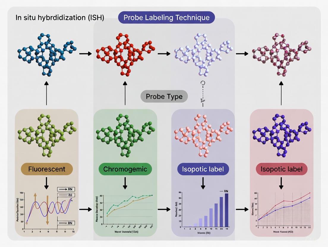
This article provides a thorough evaluation of in situ hybridization (ISH) probe labeling techniques, tailored for researchers, scientists, and drug development professionals.
ISH Probe Labeling Techniques: A Comprehensive Guide from Fundamentals to Advanced Applications
Abstract
This article provides a thorough evaluation of in situ hybridization (ISH) probe labeling techniques, tailored for researchers, scientists, and drug development professionals. It explores the foundational principles of probe chemistry and label selection, details methodological applications across various research and diagnostic scenarios, offers practical troubleshooting and optimization strategies for common experimental challenges, and discusses validation frameworks and comparative analyses of emerging technologies. The synthesis of these core intents delivers a critical resource for selecting, implementing, and validating optimal ISH probe labeling strategies to enhance accuracy and reliability in biomedical research and clinical diagnostics.
The Building Blocks of ISH: Understanding Probe Chemistry and Label Selection
In situ hybridization (ISH) has undergone a remarkable transformation since its initial development, evolving from a technically challenging procedure reliant on radioactive isotopes to a versatile toolkit of chromogenic and fluorescent methods that enable precise spatial localization of nucleic acids within tissues and cells. The technique was first described in 1969 when Gall and Pardue introduced radioactive ISH using tritium-labelled RNA to visualize ribosomal RNA hybridization in Xenopus oocytes [1] [2]. This pioneering work established the fundamental principle of hybridizing labeled nucleic acid probes to complementary sequences within biological specimens, but the method faced significant limitations including safety hazards, limited resolution, and lengthy exposure times.
The subsequent development of non-radioactive probe labeling in 1981, particularly with haptens like biotin detected by avidin-fluorochrome conjugates, marked a critical turning point that eventually enabled the fluorescence in situ hybridization (FISH) techniques widely used today [1]. This evolution has continued with the refinement of chromogenic in situ hybridization (CISH), which offers the advantage of conventional bright-field microscopy while maintaining the capability to detect gene amplification and chromosomal translocations [3] [4]. The ongoing innovation in ISH technologies provides researchers and clinicians with an expanding arsenal of tools for investigating gene expression, chromosomal abnormalities, and viral infections within their morphological context.
Radioactive Isotope Labeling
The earliest ISH methodologies employed radioactive isotopes such as ³²P or ³⁵S, which provided high sensitivity but introduced significant handling complexities and safety concerns [5]. While this approach demonstrated the fundamental feasibility of in situ nucleic acid detection, the practical limitations restricted its widespread adoption in routine laboratories. The requirement for specialized facilities for radioactive material handling, lengthy exposure times, and limited spatial resolution prompted the search for alternative labeling strategies that would eventually revolutionize the field.
Fluorescent Labeling
Fluorescent labeling represents one of the most significant advancements in ISH technology, forming the basis for FISH. This method utilizes fluorochrome-conjugated probes with molecules such as Fluorescein, Cy3, Cy5, SpectrumGreen, SpectrumOrange, and Texas Red [1] [5]. FISH provides several distinct advantages: exceptional sensitivity, capacity for multicolor detection enabling the visualization of multiple targets simultaneously, technical straightforwardness, and compatibility with automated systems for high-throughput applications [5].
The limitations of fluorescent labeling primarily include photobleaching, where fluorescent signals diminish over time with light exposure, potentially affecting the long-term preservation and re-evaluation of samples [5]. Despite this limitation, FISH has become indispensable in both research and clinical diagnostics, particularly for characterizing chromosomal rearrangements in congenital diseases and malignancies [1]. The technique's versatility is evidenced by its adaptation to various applications including metaphase and interphase FISH, fiber-FISH, RNA-FISH, 3D-FISH, and immuno-FISH [5].
Chromogenic Labeling (CISH)
Chromogenic in situ hybridization (CISH) emerged as a powerful alternative that combines the genetic detection capabilities of FISH with the practical advantages of conventional bright-field microscopy. Developed to address the limitations of fluorescence microscopy requirements in routine diagnostic laboratories, CISH utilizes enzyme-based detection systems (typically peroxidase or alkaline phosphatase) with chromogenic substrates like diaminobenzidine (DAB) or Fast Red [3] [2].
In a seminal 2000 study evaluating CISH for HER-2/neu oncogene amplification in breast cancer, researchers found that the method enabled easy discrimination of gene copies using a standard ×40 objective in hematoxylin-stained tissue sections [3]. HER-2/neu amplification typically appeared as "large peroxidase-positive intranuclear gene copy clusters" [3]. The study demonstrated excellent correlation between CISH and FISH (kappa coefficient of 0.81) across 157 breast cancers, establishing CISH as a valuable method for confirming immunohistochemical staining results, particularly in paraffin-embedded tumor samples [3].
Hapten-Based Labeling Methods
Biotin Labeling
Biotin labeling employs the strong interaction between biotin and streptavidin or avidin, typically conjugated to enzymatic reporters for signal generation. This method offers high sensitivity and specificity but requires careful validation due to potential interference from endogenous biotin present in some tissues [5]. The system allows for significant signal amplification through enzyme-substrate reactions, making it suitable for targets with lower abundance.
Digoxigenin (DIG) Labeling
Digoxigenin labeling, derived from a plant steroid molecule, provides an alternative hapten-based approach that minimizes background interference from endogenous biotin [5] [2]. DIG-labeled probes are detected using specific anti-DIG antibodies conjugated to fluorescent tags or enzymes, resulting in high sensitivity with reduced nonspecific signal. This method has proven particularly effective in viral detection studies, where self-designed DIG-labeled RNA probes successfully identified Schmallenberg virus in goat cerebrum, canine bocavirus 2 in dog intestine, and porcine circovirus 2 in pig tissues [2].
Table 1: Comparison of Major ISH Probe Labeling Techniques
| Labeling Method | Detection System | Sensitivity | Advantages | Limitations |
|---|---|---|---|---|
| Radioactive Isotopes (³²P, ³⁵S) | Autoradiography | High | Pioneering method, sensitive | Safety hazards, long exposure times, limited resolution [5] [2] |
| Fluorescent Labeling (Fluorescein, Cy3, Cy5) | Fluorescence microscopy | High | Multicolor detection, technically straightforward, automatable | Photobleaching, requires fluorescence microscope [1] [5] |
| Chromogenic (CISH) | Enzyme + chromogen, bright-field microscopy | Moderate-High | Permanent slides, conventional microscopy, cost-effective | Limited multiplexing capability [3] |
| Biotin | Streptavidin/Avidin-enzyme conjugate | High | High sensitivity, signal amplification | Endogenous biotin interference [5] |
| Digoxigenin (DIG) | Anti-DIG antibody-enzyme conjugate | High | Low background, high specificity | Requires specific antibodies [5] [2] |
Experimental Comparisons and Performance Data
Direct Methodological Comparisons in Viral Detection
A comprehensive 2018 study directly compared different ISH techniques for detecting various RNA and DNA viruses, providing valuable experimental data on their relative performance [2]. The researchers evaluated three approaches: (1) CISH with self-designed DIG-labelled RNA probes, (2) CISH with commercially produced DIG-labelled DNA probes, and (3) a commercial FISH method using fluorescent RNA probe mixes (ViewRNA ISH Tissue Assay Kit).
The results demonstrated striking differences in detection capabilities. For RNA virus detection, the FISH-RNA probe mix achieved successful detection of all tested viruses (atypical porcine pestivirus, equine hepacivirus, bovine hepacivirus, and Schmallenberg virus), while self-designed DIG-labelled RNA probes only detected Schmallenberg virus [2]. Similarly, for DNA viruses, the FISH-RNA probe mix identified all targets (canine bocavirus 2, porcine bocavirus, and porcine circovirus 2), whereas the other methods showed variable detection rates [2].
The study further quantified the cell-associated positive area, finding that the detection rate using the FISH-RNA probe mix was highest compared to other probes and protocols [2]. This enhanced sensitivity comes with trade-offs in cost and procedure time, highlighting the importance of matching technique to experimental requirements.
Probe Stability and Shelf-Life Assessment
A critical practical consideration for ISH laboratories is probe stability and shelf-life. Current diagnostic guidelines typically mandate expiration dates of 2-3 years for FISH probes [1]. However, a comprehensive 2025 study challenged this convention by evaluating 581 FISH probes that had been stored for 1-30 years.
Remarkably, all probes, including both self-labeled homemade and commercial varieties, remained functionally active and produced "bright, analyzable signals" regardless of age [1]. The study documented successful FISH experiments using probes labeled with various haptens including biotin, digoxigenin, SpectrumGreen, and SpectrumOrange, with some remaining effective after 30 years of storage at -20°C in the dark [1].
Some fluorochrome-specific variations were noted: "Commercial probes labeled with SpectrumOrange had shorter exposure times and maintained them over the years," while "DNA probes labeled with SpectrumAqua/diethylaminocoumarin showed bright labeling for the first 3 years and then faded" [1]. These findings have significant practical implications, suggesting that properly stored FISH probes may remain usable far beyond their official expiration dates, potentially reducing costs for diagnostic laboratories.
Table 2: Performance Comparison of ISH Techniques in Viral Detection Studies [2]
| Virus | Genome Type | CISH with Self-Designed DIG RNA Probes | CISH with Commercial DIG DNA Probes | FISH with RNA Probe Mix |
|---|---|---|---|---|
| Atypical Porcine Pestivirus (APPV) | RNA | Not detected | Not tested | Detected |
| Equine Hepacivirus (EqHV) | RNA | Not detected | Not tested | Detected |
| Bovine Hepacivirus (BovHepV) | RNA | Not detected | Not tested | Detected |
| Schmallenberg Virus (SBV) | RNA | Detected | Not tested | Detected |
| Canine Bocavirus 2 (CBoV-2) | DNA | Detected | Detected | Detected |
| Porcine Bocavirus (PBoV) | DNA | Not detected | Not detected | Detected |
| Porcine Circovirus 2 (PCV-2) | DNA | Detected | Detected | Detected |
Diagnostic Concordance in Clinical Settings
The comparative performance of ISH techniques has significant implications for clinical diagnostics. A 2025 retrospective cohort study of 104 glioma patients systematically compared FISH, next-generation sequencing (NGS), and DNA methylation microarray (DMM) for detecting copy number variations [6]. While all three methods showed high consistency in epidermal growth factor receptor (EGFR) assessment, "FISH demonstrated relatively low concordance with NGS/DMM in detecting other parameters" including cyclin-dependent kinase inhibitor 2A/B (CDKN2A/B), 1p, 19q, chromosome 7, and chromosome 10 [6].
In contrast, NGS and DMM exhibited strong concordance across all six parameters [6]. These findings highlight both the utility and limitations of FISH in modern integrated diagnostics, particularly noting that discordant cases were associated with high-grade gliomas and high genomic instability [6].
Advanced Applications and Future Directions
Machine Learning-Enhanced FISH Analysis
The integration of machine learning with FISH represents a cutting-edge advancement in the evolution of ISH technologies. A 2023 study demonstrated the application of ML algorithms to prioritize FISH probes for differentiating primary sites of neuroendocrine tumors [7]. Using FISH assay metrics from 85 small bowel NET and 59 pancreatic NET samples, trained models achieved up to 93.1% classification accuracy on held-out test sets [7].
Notably, the ERBB2 FISH probe emerged as the most important variable for primary site prediction, followed by MET and CDKN2A probes [7]. This approach demonstrates how computational advances can enhance the diagnostic utility of established ISH methods, providing "probabilistic guidance for FISH testing" and enabling more precise tumor classification [7].
Custom Probe Generation Protocols
The limited commercial availability of gene-specific probes for CISH has prompted development of robust protocols for generating laboratory-designed probes. Researchers have established methods using bacterial artificial chromosomes (BACs) containing human DNA fragments, which are amplified with φ29 polymerase and random primer labeled with biotin [4].
This protocol enables generation of probes mapping to any gene of interest that can be applied to formalin-fixed paraffin-embedded tissue sections (FFPETS), allowing correlation of morphological features with gene copy number changes [4]. The reliability of these custom probes has been validated through multiple strategies including comparison with commercial probes, confirmation of amplifications identified by microarray-based CGH, and demonstration of specific translocations in breast secretory carcinoma [4].
Diagram 1: ISH Experimental Workflow. This flowchart outlines the key steps in a standard ISH procedure, from specimen preparation through result interpretation, highlighting the critical decision point in selecting appropriate labeling methods.
Essential Research Reagent Solutions
Successful implementation of ISH techniques requires access to specific reagents and tools. The following table outlines key materials and their functions based on current methodologies:
Table 3: Essential Research Reagents for ISH Techniques
| Reagent/Tool | Function | Application Examples |
|---|---|---|
| Fluorochrome-conjugated probes (SpectrumGreen, SpectrumOrange, Texas Red) | Direct labeling for FISH; visualizable with fluorescence microscopy | Multicolor FISH, gene rearrangement detection [1] |
| Biotin-labeled probes | Hapten-based labeling; detected with streptavidin-enzyme conjugates | High-sensitivity detection with signal amplification [5] |
| Digoxigenin (DIG)-labeled probes | Hapten-based labeling with low background; detected with anti-DIG antibodies | Viral detection in tissue sections [2] |
| BAC clones | Source of DNA for custom probe generation | Laboratory-designed probes for specific genes [4] |
| φ29 polymerase | Multiple displacement amplification for probe production | Generating high-quality probes from BAC DNA [4] |
| Formalin-fixed paraffin-embedded tissue sections | Standard specimen preservation for morphological correlation | Archival tissue analysis, clinical diagnostics [3] [4] |
| Proteolytic digestion enzymes (Proteinase K, pepsin) | Tissue pretreatment for probe accessibility | Enhancing hybridization efficiency in FFPE tissues [3] [2] |
| ViewRNA ISH Tissue Assay Kit | Commercial system for FISH with signal amplification | Sensitive detection of low-abundance viral RNAs [2] |
Diagram 2: ISH Technique Selection Guide. This decision pathway illustrates key considerations when selecting appropriate ISH methodologies based on sensitivity requirements, equipment availability, and application purpose.
The evolution of ISH from its radioactive origins to modern fluorescent and chromogenic tags represents a compelling narrative of scientific innovation driven by the dual needs of technical performance and practical utility. While radioactive methods established the fundamental principle of in situ hybridization, the development of non-radioactive alternatives has dramatically expanded the applications and accessibility of this powerful technology.
Current ISH methods offer researchers a diverse toolkit, with FISH providing exceptional sensitivity and multiplexing capabilities, while CISH enables genetic analysis using conventional microscopy platforms essential for many diagnostic settings. The demonstrated long-term stability of properly stored FISH probes further enhances their practical value in both research and clinical contexts [1].
As ISH technologies continue to evolve, integration with computational approaches like machine learning [7] and ongoing refinement of probe design methodologies [4] promise to further enhance their diagnostic precision and research applications. This continuous innovation ensures that ISH remains an indispensable methodology for spatial genomics and transcriptomics, bridging the critical gap between molecular analysis and morphological context.
Nucleic acid probes are single-stranded DNA or RNA fragments engineered with a strong affinity for a specific complementary DNA or RNA target sequence [8]. These essential tools in molecular biology and diagnostics allow for the precise detection and localization of genetic material, enabling applications ranging from basic research to clinical diagnostics and drug development [9] [8]. The core principle involves the hybridization of the probe to its target sequence, facilitated by the degree of homology between them, which allows for the visualization and analysis of specific nucleic acid regions within complex biological samples [8].
Within the context of in situ hybridization (ISH) techniques, which allow for precise localization of specific nucleic acid segments within histologic sections, the choice of probe type is critical [9]. The three primary categories—DNA probes, RNA probes (riboprobes), and synthetic oligonucleotides—each possess distinct characteristics, advantages, and limitations that determine their suitability for specific experimental and clinical needs [10]. This guide provides an objective comparison of these core probe types, supported by experimental data and detailed protocols, to inform strategic probe selection in research and development.
Core Characteristics and Comparison
The fundamental differences in the chemical structure of DNA and RNA probes lead to variations in their stability, hybridization efficiency, and optimal applications. DNA probes are composed of deoxyribonucleic acid, featuring a backbone that makes them relatively chemically stable and less prone to degradation [10]. In contrast, RNA probes (riboprobes) are made of ribonucleic acid; the presence of a 2' hydroxyl group in their structure makes them more chemically unstable and susceptible to alkaline hydrolysis compared to DNA [10]. A key functional difference lies in the stability of the hybrid formed with the target: RNA-RNA hybrids formed by riboprobes are more stable than DNA-DNA hybrids, contributing to the higher sensitivity often observed with RNA probes [11].
Synthetic oligonucleotides represent a broader category that includes not only standard DNA oligonucleotides but also various analogs with modified backbones, such as Peptide Nucleic Acids (PNA) and Locked Nucleic Acids (LNA) [12]. These analogs are engineered to overcome some limitations of natural nucleic acids. PNA, for instance, has an uncharged peptide-like backbone, which allows for stronger binding to complementary sequences and greater resistance to nucleases and proteases [12].
Table 1: Core Characteristics of DNA, RNA, and Synthetic Oligonucleotide Probes
| Characteristic | DNA Probes | RNA Probes (Riboprobes) | Synthetic Oligonucleotides |
|---|---|---|---|
| Chemical Structure | Deoxyribose sugar-phosphate backbone; Thymine | Ribose sugar-phosphate backbone; Uracil; 2' OH group [10] | Includes analogs with modified backbones (e.g., PNA, LNA) [12] |
| Primary Synthesis Method | Chemical synthesis, PCR, Nick translation [8] | In vitro transcription (IVT) from DNA templates [10] [8] | Automated chemical synthesis [10] |
| Typical Length | 20 - 1000 bp (some FISH probes 1-10 Kb) [10] | 200 - 500 bases (optimal for ISH) [11] | 20 - 50 bases (common for synthetic) [11] |
| Thermal Stability & Hybrid Strength | DNA-DNA hybrids are less stable than RNA-DNA/RNA-RNA hybrids [11] | RNA-RNA/DNA hybrids are more stable, leading to higher sensitivity [11] | PNA and LNA exhibit enhanced binding affinity and thermal stability [12] |
| Native Chemical Stability | High; resistant to alkaline hydrolysis [10] | Lower; susceptible to RNase degradation and hydrolysis [10] | Generally high; PNA is resistant to nucleases and proteases [12] |
| Key Applications in ISH | Locus-specific FISH, whole chromosome painting, CISH [10] | RNA ISH, high-sensitivity gene expression localization [10] [11] | miRNA detection (LNA probes), rapid diagnostic assays [10] [12] |
Performance Data from Comparative Studies
A systematic comparative evaluation of DNA and RNA probes for mitochondrial DNA (mtDNA) next-generation sequencing (NGS) provides robust, quantitative data on their performance differences [13]. Under optimized hybridization conditions, the study directly compared probes based on metrics critical for NGS, such as enrichment efficiency and robustness against artifacts.
The findings revealed a clear trade-off: RNA probes demonstrated superior enrichment efficiency, characterized by significantly higher mtDNA mapping rates and greater average mtDNA depth per gigabyte of sequencing data in both fresh tissue and plasma samples [13]. However, DNA probes were more effective at reducing artifacts caused by nuclear mitochondrial DNA segments (NUMTs) at both the read and mutation levels, a crucial factor for accurate mutation detection [13]. Furthermore, RNA probes captured a broader fragment size distribution in plasma cell-free mtDNA, indicating a potential bias towards longer fragments [13].
Table 2: Experimental Performance Comparison of DNA vs. RNA Probes in mtDNA NGS [13]
| Performance Metric | DNA Probes | RNA Probes |
|---|---|---|
| Average mtDNA Mapping Rate (Fresh Tissue) | 61.79% | 92.55% |
| Average mtDNA Depth per GB (Fresh Tissue) | ~32,400X | ~38,500X |
| Average mtDNA Mapping Rate (Plasma) | 16.18% | 42.95% |
| Average mtDNA Depth per GB (Plasma) | ~9,180X | ~15,270X |
| Interference from NUMTs | Lower (more effective at reducing artifacts) | Higher |
| Fragment Size Distribution | Narrower, more uniform | Broader, prevalence of longer fragments |
The efficacy of synthetic oligonucleotide analogs was highlighted in a study probing bacterial ribosome assembly [12]. The research found that while DNA antisense oligonucleotides (ASOs) showed only a subtle inhibitory effect on ribosome assembly, their synthetic analogs—particularly PNA and LNA—demonstrated significantly improved inhibitory effects, consistent with their well-characterized superior in vitro hybridization free energies (LNA > PNA > DNA) [12]. This underscores the value of synthetic probes in applications requiring very high binding affinity and disruptive potential.
Experimental Protocols and Workflows
Probe Synthesis and Labeling Methodologies
The processes for generating different probe types are distinct and have implications for cost, complexity, and probe quality.
DNA Probe Synthesis (Nick Translation): This is a classic method for generating labeled double-stranded DNA probes [8]. The protocol involves randomly "nicking" the backbone of a double-stranded DNA template with dilute DNase I. The enzyme DNA polymerase I then simultaneously removes nucleotides from the probe molecules in the 5'→3' direction (exonuclease activity) and replaces them with labeled dNTP precursors (polymerase activity) [8]. This method is efficient and can be completed in less than an hour, accommodating fluorophore-, biotin-, or digoxigenin-labeled nucleotides [8].
RNA Probe (Riboprobe) Synthesis (In Vitro Transcription): This is the primary and most reliable method for producing RNA probes [11] [8]. The process starts with a purified DNA template (either linearized plasmid or PCR product) containing a bacteriophage RNA polymerase promoter (e.g., T7, SP6, T3) upstream of the sequence of interest [11]. The template is incubated with the appropriate RNA polymerase and a mixture of nucleotides, including labeled ones (e.g., biotin- or digoxigenin-UTP), to generate large amounts of uniformly labeled, single-stranded RNA probes [11] [8]. A common strategy for ISH is to clone the DNA sequence between two opposing promoters to independently generate antisense (detection) and sense (control) probes from the same template [11] [8].
Diagram 1: Riboprobe Synthesis Workflow via In Vitro Transcription
DNA vs. RNA Probe Hybridization Workflow for NGS
A detailed 2025 study on mtDNA NGS provides a clear experimental workflow for comparing probe performance [13]. The protocol begins with the extraction of genomic DNA from samples (e.g., fresh frozen tissue or plasma), followed by the construction of whole-genome sequencing (WGS) libraries. For tissue samples, DNA is typically sheared to fragments of 300-500 bp [13]. These WGS libraries are then subjected to hybridization-based capture using custom-designed double-stranded DNA and RNA probes that comprehensively cover the mitochondrial genome. After enrichment, the libraries are sequenced via NGS, and the resulting data is analyzed bioinformatically to compare performance metrics such as mapping rate, depth of coverage, and NUMT interference [13].
Diagram 2: Probe Comparison Workflow for Targeted NGS
The Scientist's Toolkit: Essential Research Reagents
Successful probing experiments, particularly in ISH, rely on a suite of essential reagents and tools. The following table details key components for probe-based research.
Table 3: Essential Research Reagents for Probe-Based Applications
| Reagent / Tool | Function / Description | Application Notes |
|---|---|---|
| Bacteriophage RNA Polymerases (T7, SP6, T3) | Enzymes for synthesizing RNA probes (in vitro transcription) from specific promoters on DNA templates [11]. | Essential for riboprobe generation; choice depends on promoter in cloning vector. |
| DNA Polymerase I / Klenow Fragment | Used for DNA probe synthesis via nick translation (full enzyme) or random priming (Klenow fragment) [8]. | Incorporates labeled nucleotides into DNA probe sequences. |
| Modified Nucleotides (dNTPs/UTPs) | Nucleotides conjugated to labels (e.g., biotin, digoxigenin, fluorophores) for probe detection [8]. | Key for non-radioactive detection; different labels offer varying sensitivity. |
| DNase I | Enzyme used in nick translation to create initial nicks in double-stranded DNA template backbone [8]. | Critical for initiating the nick translation DNA labeling process. |
| RNase Inhibitors | Protects sensitive RNA probes from degradation by ubiquitous RNases during synthesis and handling [10]. | Crucial for maintaining integrity of riboprobes. |
| Cloning Vectors with Promoters | Plasmid templates containing phage promoters (e.g., pGEM-T) for inserting target sequence and producing riboprobes [11]. | Provides a renewable source for consistent riboprobe production. |
| Restriction Enzymes | Enzymes for linearizing plasmid DNA templates prior to in vitro transcription [11]. | Ensures production of defined-length RNA transcripts. |
| Locked Nucleic Acids (LNA) / Peptide Nucleic Acids (PNA) | Synthetic nucleotide analogs with modified backbones that confer enhanced binding affinity and stability [12]. | Used in synthetic oligonucleotide probes for challenging targets like miRNAs or for disruptive probing. |
The comparative data and protocols presented in this guide underscore that there is no single "best" probe type; the optimal choice is dictated by the specific experimental requirements and trade-offs.
- Choose RNA Probes (Riboprobes) when the highest sensitivity and hybridization strength are the primary goals, such as in detecting low-abundance mRNA transcripts in RNA ISH or for achieving maximum enrichment efficiency in targeted NGS, even at the cost of potential artifacts from homologous sequences like NUMTs [13] [11].
- Choose DNA Probes for applications where robustness, chemical stability, and lower cost are prioritized, or when minimizing artifacts (e.g., NUMT interference in mtDNA studies) is critical for accurate mutation detection [13] [10].
- Choose Synthetic Oligonucleotides (including standard oligos, LNAs, and PNAs) for flexibility, high affinity, and nuclease resistance. They are ideal for standardized diagnostic assays, detecting short targets like miRNAs, and for applications requiring exceptional binding strength to disrupt native structures, as demonstrated in ribosome assembly studies [10] [12].
This structured comparison of DNA, RNA, and synthetic oligonucleotide probes, grounded in recent experimental findings, provides a framework for researchers to make informed decisions, thereby enhancing the precision and reliability of their scientific and diagnostic outcomes.
In situ hybridization (ISH) is a fundamental molecular technique for localizing and detecting specific nucleic acid sequences in cells, tissue sections, and entire tissues [14]. The technique relies on hybridizing a target nucleotide sequence with a complementary probe that is labeled to enable visualization [14]. The choice of labeling methodology profoundly impacts assay sensitivity, specificity, resolution, and applicability across different research and diagnostic scenarios. This guide provides a comprehensive comparative analysis of three principal probe labeling chemistries—nick translation, in vitro transcription, and chemical synthesis—to inform researchers and drug development professionals in selecting the optimal approach for their specific experimental needs within the broader context of evaluating ISH probe labeling techniques.
Methodological Principles and Comparative Analysis
Nick Translation
Principle: Nick translation enzymatically labels double-stranded DNA probes by simultaneously utilizing two enzymes: DNase I to introduce single-strand "nicks" in the DNA backbone, and DNA Polymerase I to remove nucleotides from the 5' end of the nick while incorporating new labeled nucleotides at the 3' end [15] [16]. This process effectively replaces unlabeled nucleotides with labeled ones along the DNA template.
Protocol Outline:
- Reaction Setup: Combine purified DNA template (>1 kb), nick translation buffer, dNTP mix (including labeled dUTP, e.g., biotin-, digoxigenin-, or fluorophore-conjugated), and the Nick Translation Enzyme Mix (DNase I and DNA Polymerase I) [15] [16].
- Incubation: Incubate the reaction mixture at 15-16°C for approximately 1 hour [15].
- Termination & Purification: Stop the reaction with EDTA and purify the labeled probe to remove unincorporated nucleotides.
This method is recommended for labeling double-stranded DNA larger than 1kb for fluorescent in situ hybridization (FISH) applications [15].
In Vitro Transcription
Principle: In vitro transcription (IVT) generates labeled RNA probes (riboprobes) from a linearized DNA template cloned downstream of a bacteriophage RNA polymerase promoter (e.g., T3, T7, SP6) [17]. The RNA polymerase synthesizes a single-stranded RNA transcript while incorporating labeled nucleotides.
Protocol Outline:
- Template Linearization: Digest a plasmid containing the insert of interest and the appropriate promoter with a restriction enzyme that cuts downstream of the insert [17].
- Transcription Reaction: Combine the linearized DNA template, transcription buffer, RNase inhibitor, DTT, NTP mix (including DIG- or FITC-labeled UTP), and the specific RNA polymerase [18] [17].
- Incubation: Incubate at 37°C for 2 hours [17].
- Template Removal & Purification: Digest the DNA template with DNase I. Precipitate the synthesized RNA probe with LiCl and ethanol [17].
- Optional Hydrolysis: Hydrolyze long RNA transcripts into smaller fragments (200-300 bases) using carbonate buffer to improve tissue penetration [17].
DIG-labeled RNA probes are noted for their stability, with a shelf life of over six years [18].
Chemical Synthesis
Principle: This method involves the automated solid-phase synthesis of short oligonucleotide probes (20-50 bases) with labels directly incorporated via modified phosphoramidites during synthesis or conjugated to the probe post-synthesis. While less detailed in the provided search results, it is the primary method for generating oligonucleotide FISH probes [14].
Common Labels: Synthetic oligonucleotides are commonly labeled with fluorochromes such as Alexa Fluor dyes, CY3, CY5, and Fluorescein [14]. These probes are often used in techniques like single-molecule FISH (smFISH) and multiplexed error-robust FISH (MERFISH) [14].
Comparative Performance Data
The table below summarizes the key characteristics and performance metrics of the three labeling methods based on experimental data and established protocols.
Table 1: Comprehensive Comparison of ISH Probe Labeling Methods
| Feature | Nick Translation | In Vitro Transcription | Chemical Synthesis (Oligonucleotides) |
|---|---|---|---|
| Probe Type | Double-stranded DNA (dsDNA) [15] | Single-stranded RNA (ssRNA, riboprobes) [17] | Single-stranded DNA (ssDNA oligonucleotides) [14] |
| Typical Probe Length | >1 kilobase (kb) [15] | 200-300 bases (after hydrolysis) [17] | 20-50 bases [14] |
| Primary Use Case | DNA target detection (e.g., gene loci, chromosomes) [15] [16] | RNA target detection (mRNA localization) [17] | RNA and DNA target detection, high-throughput multiplexing [14] |
| Key Advantage | Simple protocol, strong signals, ideal for long DNA probes [16] | High sensitivity and specificity for RNA; probe stability [18] | High specificity for short targets; designed for multiplexing [14] |
| Label Incorporation | Enzymatic incorporation of labeled dUTP [15] | Enzymatic incorporation of labeled UTP [17] | Direct during synthesis or post-synthesis conjugation [14] |
| Typical Assay Duration | ~1 hour labeling time [15] | ~4-6 hours (excluding cloning) [17] | N/A (commercially synthesized) |
| Probe Stability | Stable for decades when stored at -20°C in the dark [1] | DIG-labeled: 6+ years; FITC-labeled: ~2 years [18] | Varies by label; generally high |
Table 2: Experimental Performance in Diagnostic Contexts
| Parameter | Nick Translation (FISH) | In Vitro Transcription | Alternative Methods (DMM/NGS) |
|---|---|---|---|
| Concordance with NGS/DMM | Relatively low for CDKN2A/B, 1p, 19q, Chr7, Chr10; high for EGFR [6] | Not directly comparable (different targets) | Strong concordance between NGS and DMM [6] |
| Associated with Discordance | Discordant cases linked to high-grade gliomas and high genomic instability [6] | Not Applicable | Not Applicable |
| Best Application | Targeted DNA CNV detection in integrated diagnostics [6] | High-sensitivity RNA detection | Genome-wide CNV assessment [6] |
Workflow Visualization
The following diagrams illustrate the core procedural workflows for each labeling method.
Nick Translation Workflow
In Vitro Transcription Workflow
Chemical Synthesis Workflow
The Scientist's Toolkit: Essential Reagents and Materials
Successful implementation of ISH probe labeling techniques requires specific reagent solutions. The following table details key materials and their functions.
Table 3: Essential Research Reagent Solutions for ISH Probe Labeling
| Reagent / Kit | Function | Specific Application |
|---|---|---|
| Nick Translation Kit [15] [16] | Provides optimized enzymes and buffer to label DNA via nick translation. | Core reagent for generating FISH/CISH DNA probes. Compatible with fluorophore-, biotin-, and digoxigenin-labeled dUTPs. |
| dUTPs (Biotin-, DIG-, Fluorophore-labeled) [1] [15] | The labeled nucleotide directly incorporated into the probe. | The "label" in the probe. Choice determines detection method (e.g., antibody for DIG/biotin, direct for fluorophore). |
| In Vitro Transcription Kit [17] | Supplies buffers, RNA polymerase, and RNase inhibitor for synthesizing RNA probes. | Core reagent for generating single-stranded riboprobes for high-sensitivity RNA detection. |
| Labeled NTP Mix (e.g., DIG-UTP) [17] | The labeled ribonucleotide incorporated during transcription. | The "label" for RNA probes. DIG-UTP is common and offers high sensitivity via antibody detection. |
| RNA Polymerase (T3, T7, SP6) [17] | Enzyme that transcribes RNA from a DNA template with its specific promoter. | Drives the synthesis of the RNA probe. Must match the promoter sequence in the DNA template. |
| Anti-Digoxigenin Antibody [5] [14] | Antibody conjugated to a reporter enzyme (HRP/AP) or fluorophore to detect DIG-labeled probes. | Key detection reagent for indirect methods using DIG-labeled probes. |
| Streptavidin-HRP/Streptavidin-Fluorophore [5] [19] | Binds to biotin-labeled probes for detection, often with signal amplification. | Key detection reagent for indirect methods using biotin-labeled probes. |
| Tyramide Signal Amplification (TSA) Reagents [19] | Provides enzymatic signal amplification for low-abundance targets, boosting sensitivity 10-200x. | Used with HRP-conjugated antibodies/streptavidin for detecting very rare targets. |
The selection of a probe labeling method is a critical determinant of success in ISH experiments. Nick translation remains the robust, standard choice for generating long DNA probes to assess genomic DNA and copy number variations. In vitro transcription is the gold standard for sensitive and specific RNA detection, offering exceptional probe stability. Chemical synthesis of oligonucleotides provides unparalleled flexibility for multiplexed assays and the detection of short targets, powering advanced techniques like smFISH and MERFISH.
Emerging trends point toward increased automation, integration with microfluidics to reduce assay times and reagent volumes [14], and sophisticated computational analysis, particularly deep learning, to automate the interpretation of complex ISH images [20]. As the field progresses, the integration of these robust labeling chemistries with novel delivery platforms and analytical algorithms will further solidify ISH's role as an indispensable tool in both basic research and clinical diagnostics.
In situ hybridization (ISH) is a foundational technique in molecular biology and diagnostic pathology for detecting and localizing specific nucleic acid sequences within cells or tissues, all while preserving tissue integrity [21]. The technique operates on the principle of complementary binding, where a labeled nucleic acid probe anneals to a specific target sequence of DNA or RNA [22]. The choice of probe label is a critical decision that profoundly influences the sensitivity, specificity, safety, and workflow of an experiment. Historically, radioactively labeled probes were the standard; however, the development of non-radioactive labels like biotin, digoxigenin, and fluorophores has dramatically expanded the toolkit available to researchers [23] [21]. This guide provides an objective comparison of these labeling techniques, framed within the context of modern research and drug development.
Technical Comparison of Labeling Technologies
Fundamental Differences and Key Characteristics
Probe labels are fundamentally categorized as either radioactive or non-radioactive. Radioactive probes are tagged with radioactive isotopes (e.g., ³⁵S) and detected by autoradiography [24] [23]. Non-radioactive probes use chemical or fluorescent tags, which are detected through enzymatic reactions (chromogenic detection) or directly via fluorescence microscopy [25] [23]. The core differences in their properties and handling requirements are summarized in the table below.
Table 1: Fundamental Characteristics of Radioactive vs. Non-Radioactive Probes
| Characteristic | Radioactive Probes | Non-Radioactive Probes |
|---|---|---|
| Label Type | Radioactive isotopes (e.g., ³⁵S, ³²P) [24] | Biotin, Digoxigenin, Fluorophores (e.g., SpectrumOrange) [1] [25] |
| Detection Method | Autoradiography, scintillation counting [23] | Chromogenic (CISH) or Fluorescent (FISH) microscopy [25] |
| Sensitivity | Historically high sensitivity [24] | High sensitivity, enhanced by signal amplification [22] |
| Resolution | Lower, due to scatter from radiation [24] | High, allowing for precise subcellular localization [22] |
| Hazard Profile | High; requires special safety protocols and waste disposal [23] | Low; generally safer and easier to handle [23] |
| Shelf Life | Short, limited by isotope half-life | Long; stable for decades when stored properly at -20°C [1] |
| Experiment Duration | Long (exposure for autoradiography) [24] | Relatively shorter [22] |
| Multiplexing Capability | Low or none | High, especially with fluorescent probes [26] |
Quantitative Performance Data from Experimental Studies
Direct comparisons in research studies highlight practical performance differences. A seminal 1993 study directly compared radioactive and non-radioactive ISH for localizing calretinin mRNA in inner ear tissues and found radioactive ISH to be more sensitive, successfully revealing positive signals in inner hair cells that were not detected with the non-radioactive method under their experimental conditions [24]. The table below summarizes key experimental findings.
Table 2: Experimental Performance Data from Key Studies
| Study / Context | Probe Label & Type | Key Experimental Finding | Implication for Research |
|---|---|---|---|
| Localization of calretinin mRNA, Rat & Guinea Pig Inner Ear [24] | Radioactive (³⁵S) vs. Non-radioactive (Digoxigenin) | Radioactive ISH was more sensitive, detecting positive structures in inner hair cells that non-radioactive ISH did not. | Radioactive labels may be necessary for detecting low-abundance mRNA targets. |
| Long-term Probe Stability, Human Cytogenetics [1] | Non-radioactive (Biotin, Digoxigenin, Fluorophores) | 581 FISH probes, both self-labeled and commercial, stored at -20°C for 1-30 years, all functioned perfectly upon reuse. | Non-radioactive probes are a cost-effective, long-term resource; expiration dates can be conservative. |
| Clinical Diagnostics & Market Trends [26] | Fluorescent Dyes (e.g., SpectrumOrange) | The fluorescent probe segment holds 50% of the FISH probe market, driven by oncology and genetic disorder diagnostics. | Fluorescent probes are the established standard for clinical and high-throughput research applications. |
Experimental Protocols and Methodologies
Core Workflow for In Situ Hybridization
The following diagram outlines the generalized ISH workflow, highlighting steps where the choice of label introduces procedural variations.
Detailed Methodological Considerations
Tissue Preparation: Optimal tissue fixation is critical. 10% Neutral Buffered Formalin (NBF) for 24±12 hours is the standard for FFPE tissues, providing a balance between nucleic acid preservation and morphology [22]. Over-fixation can cause excessive cross-linking, leading to false-negative results, while under-fixation risks RNA degradation and poor morphology [22] [21].
Probe Hybridization: The hybridization temperature, typically between 37°C and 65°C, must be optimized for specificity and is often lower than the probe-target melting temperature (Tm) when formamide is used to conserve sample morphology [25] [21]. Post-hybridization, washes of increasing stringency are performed to remove nonspecifically bound probes [25].
Signal Detection and Visualization:
- Radioactive Probes: Hybridized samples are exposed to X-ray film in a process called autoradiography, which can take from hours to days, depending on signal strength [24].
- Non-Radioactive Probes:
- Biotin is detected using streptavidin or avidin conjugated to alkaline phosphatase (AP) or horseradish peroxidase (HRP) [25] [21]. Note that endogenous biotin can cause background and requires blocking [25].
- Digoxigenin is detected with high specificity using anti-digoxigenin antibodies conjugated to AP or HRP, avoiding issues with endogenous molecules [25] [21].
- Fluorophores allow for direct detection without an enzymatic step and are visualized via fluorescence microscopy [25].
Essential Research Reagent Solutions
The following table details key reagents and their functions for performing ISH, based on protocols from the search results.
Table 3: Essential Reagents for In Situ Hybridization Experiments
| Reagent / Material | Function / Purpose | Key Considerations |
|---|---|---|
| 10% Neutral Buffered Formalin (NBF) | Standard tissue fixative that preserves nucleic acids and morphology [22]. | Fixation time should be optimized; over-fixation reduces probe accessibility [21]. |
| Proteinase K | Enzyme used for tissue permeabilization; digests proteins to allow probe penetration [25]. | Concentration is critical. Must be titrated (e.g., 1-5 µg/mL) to balance signal with tissue integrity [25]. |
| Formamide | A component of hybridization buffers that allows for lower hybridization temperatures [25]. | Helps preserve tissue morphology by lowering the melting temperature of the probe-target hybrid [21]. |
| Digoxigenin-dUTP | A non-radioactive label incorporated into probes via nick translation or random priming [25]. | High specificity due to lack of endogenous digoxigenin in mammalian tissues [25]. |
| Biotin-dUTP | A non-radioactive label incorporated into probes [25]. | Requires blocking of endogenous biotin to prevent high background in some tissues [25]. |
| Fluorophore-dUTP (e.g., SpectrumOrange) | A fluorescent label for direct detection in FISH [1]. | Enables multiplexing. Stable for decades at -20°C [1]. |
| Anti-Digoxigenin Antibody (conjugated to AP/HRP) | Detection antibody for digoxigenin-labeled probes in chromogenic ISH (CISH) [25]. | Conjugate choice (AP vs. HRP) depends on the substrate and tissue type. |
| Streptavidin (conjugated to AP/HRP or a fluorophore) | Detection molecule for biotin-labeled probes [25]. | Binds with high affinity to biotin. |
| Positive & Negative Control Probes | Essential for validating assay performance and RNA integrity [27]. | A housekeeping gene probe confirms technique; a bacterial gene (e.g., dapB) checks background [27]. |
The choice between radioactive and non-radioactive probes is not a matter of one being universally superior, but rather of selecting the right tool for the specific research question. Radioactive probes still hold value for their high sensitivity in detecting low-abundance targets, as demonstrated in specialized research applications [24]. However, for the vast majority of modern research and clinical diagnostics, non-radioactive probes offer a compelling combination of safety, stability, and flexibility. The powerful capabilities of digoxigenin for sensitive chromogenic detection and fluorophores for multiplexed spatial biology are driving their widespread adoption [22] [26]. As the market for FISH probes continues to grow, fueled by advancements in precision medicine [26], the trend is firmly set towards the continued refinement and application of non-radioactive labeling technologies.
In situ hybridization (ISH) is a foundational technique in molecular biology that allows for the precise detection and localization of specific nucleic acid sequences within cells or tissue sections [28]. The core principle of this method relies on the hybridization dynamics—the specific annealing of a labeled nucleic acid probe to a complementary DNA or RNA target sequence under stringent conditions [29]. The efficiency and stability of this probe-target binding are critical for the sensitivity and specificity of numerous diagnostic and research applications, from detecting infectious agents to profiling cancer biomarkers [30] [31]. Understanding the factors that govern these fundamental dynamics is essential for developing robust assays, especially when detecting variable targets such as viral genomes or when designing probes for multiplexed spatial biology applications [30] [32].
This guide objectively compares the performance of different probe labeling and design strategies by synthesizing experimental data from current research. The analysis is framed within a broader thesis on evaluating ISH probe labeling techniques, providing researchers and drug development professionals with validated protocols and comparative data to inform their experimental design.
Core Factors Governing Hybridization Efficiency
The binding between a probe and its target is a complex process governed by both the probe's sequence composition and the physicochemical environment of the hybridization reaction. Key factors include the following.
Probe Design and Sequence Composition
- Contiguous Matching Stretches: Experimental work with 70-nucleotide (nt) long DNA probes demonstrates that the distribution of mismatches is more critical than their total number. When mismatches interrupt contiguous matching stretches of 6 nt or longer, hybridization is significantly disrupted. Conversely, the same number of matching nucleotides separated into several smaller regions by mismatches results in weaker binding [30].
- Universal Bases and Wobbles: To counteract sequence variation, incorporation of universal bases like deoxyribose-Inosine (dInosine) can remarkably stabilize hybridization. In 70-mer probes, dInosine substitutions at mismatching positions performed comparably to wobble bases (N, representing a mixture of all four nucleotides), and both were more effective than another universal base analog, 5-nitroindole [30].
- Probe Type and Backbone: The stability of the hybrid formed follows the order: RNA-RNA > RNA-DNA > DNA-DNA [25]. Furthermore, synthetic backbones like Peptide Nucleic Acid (PNA) offer higher affinity for complementary sequences and greater resistance to nucleases and proteases due to their uncharged chemical structure. This is particularly beneficial for penetrating bacterial cell walls and accessing structured ribosomal RNA targets [31].
Hybridization Conditions and Buffer Chemistry
- Stringency Control: Hybridization specificity is primarily driven by temperature and the concentration of monovalent cations in the hybridization buffer. Post-hybridization washes of increasing stringency are critical for dissociating imperfect matches and reducing background [25].
- TMAC Buffer: The use of 3M tetramethylammonium chloride (TMAC) buffer is a key strategy to reduce the sequence composition bias. TMAC selectively raises the stability of A:T base pairs to approximately that of G:C base pairs, allowing for more consistent hybridization performance across probes of different sequences [30].
- Formamide and Temperature: Formamide is commonly included in hybridization buffers as it allows the reaction to proceed at temperatures significantly lower than the actual melting temperature (Tm) of the probe-target hybrid, thereby helping to preserve tissue morphology [25].
Comparative Analysis of Probe Technologies and Performance
The following tables summarize experimental data comparing the performance of various probe design strategies and labeling techniques, highlighting their specific advantages and validated applications.
Table 1: Comparative Performance of Probe Design Strategies for Mismatch Tolerance
| Probe Design Strategy | Experimental Findings | Key Applications | Reference Model/System |
|---|---|---|---|
| Long DNA Probes (70-mer) | Tolerant to naturally occurring synonymous mutations when mismatches do not break contiguous matching stretches ≥6 nt. | Detection of highly variable viral genomes (e.g., Influenza A, Norovirus). | Microsphere-linked probes in Luminex system [30] |
| dInosine-Substituted Probes | Remarkable mismatch tolerance; stabilized hybridization comparable to N wobbles. Preserved specificity. | Broadly targeted yet specific detection of viral variants. | 3M TMAC buffer hybridization [30] |
| PNA (Peptide Nucleic Acid) Probes | Higher affinity for DNA/RNA; better cell wall penetration; resistant to nucleases. Sensitivity: 80.0-93.8%, Specificity: 90.9-93.8%. | Detection of H. pylori and its clarithromycin resistance in gastric biopsy specimens. | Paraffin-embedded tissue; PNA-FISH validation [31] |
| Multiplex Oligonucleotide Probes (smFISH) | ~20 singly-labeled 20-mer probes per transcript enable precise localization and semi-automated quantification of individual mRNA molecules. | Single-molecule RNA detection and quantification in cultured cells and tissues. | Raj et al. (2006, 2008) method [28] |
Table 2: Comparison of Common Probe Labeling and Detection Systems
| Labeling/Detection System | Key Characteristics | Performance Considerations | Example Use Cases |
|---|---|---|---|
| Fluorescent Dyes (Direct) | Fluorophore (e.g., SpectrumOrange, Cyanine 5) directly attached to probe. | Enables multiplexing; stable for decades at -20°C [1]; may be subject to photobleaching. | FISH for metaphase/interphase cytogenetics [1] |
| Biotin (Indirect) | Detected by streptavidin- or avidin-conjugated reporters (AP/HRP). | Strong signal amplification; potential for endogenous biotin background. | Chromogenic ISH (CISH) [25] |
| Digoxigenin (Indirect) | Detected by high-affinity anti-digoxigenin antibodies conjugated to reporters. | High sensitivity and specificity; minimal endogenous background. | RNA ISH; high-resolution CISH [25] |
| Dual-Hapten (Biotin/Digoxigenin) | Self-labeled homemade probes using dUTPs tagged with haptens. | Proven functionality after 30 years of storage at -20°C. | Home-brew FISH probes for diagnostics [1] |
Experimental Protocols for Validation and Optimization
Protocol: Validation of Mismatch-Tolerant Probe Hybridization
This protocol is adapted from studies on long DNA probes and dInosine substitution, designed to quantify hybridization tolerance to mismatches [30].
Step 1: Probe Design and Synthesis
- Select a conserved 70-nt target region from a gene of interest (e.g., Influenza A matrix protein gene).
- Design a set of probes with varying numbers and distributions of mismatches. Include probes with substitutions of dInosine or wobble bases (N) at mismatch-prone positions.
- Synthesize 5' amine-modified 70-mer oligonucleotide probes for coupling to microspheres.
Step 2: Probe Coupling and Target Preparation
- Couple amine-modified probes to color-coded carboxylated microspheres using a carbodiimide coupling method (e.g., EDC chemistry) [30].
- Prepare biotinylated single-stranded DNA targets that are complementary to the probe consensus sequence but contain defined mismatches.
Step 3: Hybridization in TMAC Buffer
- Hybridize microsphere-linked probes (0.2 nM target concentration) in 3M TMAC buffer at a standardized temperature (e.g., 45-55°C).
- The TMAC buffer minimizes the impact of variable GC-content on hybridization stability.
Step 4: Detection and Analysis
- Incubate with streptavidin-phycoerythrin and analyze hybridization signals using a flow cytometry-based system (e.g., Luminex 200).
- Quantitative Data Analysis: Measure the median fluorescence intensity (MFI) for each probe-target pair. Normalize signals to a perfectly matched control. Probes with dInosine or wobbles at mismatch sites should show significantly higher normalized MFI compared to unmodified probes with the same mismatch pattern.
Protocol: PNA-FISH for Antimicrobial Resistance Detection
This protocol outlines the method for detecting H. pylori and its clarithromycin resistance directly from paraffin-embedded biopsy specimens, as validated in clinical studies [31].
Step 1: Sample Preparation and Sectioning
- Obtain formalin-fixed, paraffin-embedded (FFPE) gastric biopsy specimens.
- Cut 3-μm-thick sections and mount on slides.
Step 2: Deparaffinization and Permeabilization
- Deparaffinize slides in xylol and rehydrate through a graded ethanol series.
- Digest samples with Proteinase K (e.g., 1-5 μg/mL for 10 minutes at room temperature). Optimization Note: A titration experiment is crucial. Insufficient digestion diminishes signal, while over-digestion destroys tissue morphology [25].
Step 3: PNA Probe Hybridization
- Hybridize with PNA probes (typically 13-18 bp) targeting the wild-type or mutant (A2142G, A2143G) sequences of the H. pylori 23S rRNA gene.
- Use a standardized hybridization buffer and incubate at a defined temperature for a specific duration.
Step 4: Stringency Washes and Detection
- Perform post-hybridization stringency washes to remove non-specifically bound probes.
- Detect fluorescently labeled PNA probes and counterstain with DAPI.
Step 5: Microscopy and Interpretation
- Visualize under a fluorescence microscope. The presence of H. pylori is confirmed by specific fluorescent signals. The resistance genotype is determined by which probe set (wild-type vs. mutant) produces the signal.
- Validation Metrics: Compared to culture and Etest, this PNA-FISH method demonstrated a sensitivity of 80.0-84.2% and a specificity of 90.9-93.8% in clinical validations [31].
Signaling Pathways and Experimental Workflows
The following diagram illustrates the nucleation-zipping model and the critical experimental steps for a robust FISH assay, integrating the key factors discussed.
The Scientist's Toolkit: Essential Research Reagent Solutions
Table 3: Key Reagents and Materials for Hybridization Experiments
| Research Reagent / Material | Critical Function | Experimental Consideration |
|---|---|---|
| TMAC (Tetramethylammonium Chloride) | Hybridization buffer that neutralizes sequence composition bias, making A:T and G:C base pairs equally stable. | Essential for comparing probes of different sequences or when analyzing targets with variable GC-content [30]. |
| dInosine (Deoxyribose-Inosine) | Universal base that pairs with all four canonical bases (I:C > I:A > I:T ≈ I:G). | Used in probe synthesis to introduce mismatch tolerance at variable positions, preserving specificity in long probes [30]. |
| PNA (Peptide Nucleic Acid) Probes | Synthetic DNA mimics with a neutral backbone, conferring higher affinity and nuclease resistance. | Ideal for challenging FISH applications, such as penetrating Gram-negative bacterial cell walls (e.g., H. pylori) [31]. |
| Proteinase K | Protease enzyme that digests proteins, permeabilizing the sample to allow probe access to nucleic acids. | Concentration and time must be titrated for each tissue type; over-digestion destroys morphology [25]. |
| Locked Nucleic Acids (LNA) | Modified RNA nucleotides with a bridged sugar, increasing duplex stability and thermal affinity (Tm). | Incorporated into oligonucleotide probes (e.g., primers) to increase hybridization strength and specificity [30]. |
| Hapten-Labeled dNTPs (Biotin-, Digoxigenin-dUTP) | Modified nucleotides incorporated into probes via Nick Translation or Random Priming for indirect detection. | Probes labeled with these haptens and stored at -20°C in the dark have been shown to remain functional for over 30 years [1]. |
The fundamental dynamics of probe-target binding and stability are governed by an interplay of probe design, chemical composition, and hybridization environment. Data confirms that strategies employing long probes with preserved contiguous matching stretches or stabilizing modifications like dInosine offer remarkable tolerance to sequence variation without sacrificing specificity. Furthermore, the choice of probe chemistry—such as PNA for challenging cellular targets or hapten-labeled DNA for long-term stability—directly impacts assay performance.
These findings provide a solid foundation for the evaluation of ISH probe labeling techniques. For researchers and drug development professionals, this comparative guide underscores that there is no single optimal solution; rather, the choice of probe and hybridization strategy must be tailored to the specific biological question, target accessibility, and required performance metrics. As the field moves towards higher multiplexing and spatial resolution, these fundamental principles of hybridization dynamics will continue to underpin the development of next-generation diagnostic and research tools.
From Theory to Bench: Implementing Labeling Techniques in Research and Diagnostics
Fluorescence in situ hybridization (FISH) represents a pivotal molecular cytogenetics technique for localizing specific nucleic acid sequences within fixed tissues and cells. This method provides crucial temporal and spatial information about gene expression and genetic loci, offering researchers and clinicians a powerful tool for diagnostic and research applications [33]. Within the broader context of in situ hybridization (ISH) probe labeling techniques, direct detection methods utilizing fluorescent dyes have emerged as a preferred approach for many applications due to their capacity for multiplexing, rapid visualization, and high sensitivity compared to non-fluorescent alternatives [34]. As technological advancements continue to refine FISH methodologies, understanding the workflow considerations and performance characteristics of direct fluorescent detection becomes essential for optimizing experimental design in research and diagnostic settings.
This guide objectively compares the performance of direct fluorescence detection FISH with other ISH labeling techniques, providing supporting experimental data and detailed methodologies to inform researchers, scientists, and drug development professionals in their selection of appropriate molecular cytogenetics approaches.
Technical Foundations of FISH and Alternative Labeling Methods
In situ hybridization encompasses various techniques for localizing nucleic acid sequences within biological samples. The fundamental difference between FISH and other ISH methods lies in the detection system employed. FISH utilizes fluorescently labeled probes that can be directly visualized through fluorescence microscopy, whereas other ISH approaches typically employ non-fluorescent hapten-labeled probes detected through enzymatic reactions or immunohistochemistry [34].
The evolution of ISH began with radioactive isotope labeling using 32P or 35S isotopes, which provided high sensitivity but posed significant safety concerns and is rarely used in modern laboratories [5]. Non-radioactive methods have largely replaced these techniques, with fluorescent labeling emerging as a leading approach due to its sensitivity, versatility, multicolor detection capability, and technical straightforwardness [5].
Table 1: Comparison of Major ISH Probe Labeling Methods
| Labeling Method | Detection Principle | Primary Advantages | Primary Limitations | Typical Applications |
|---|---|---|---|---|
| Fluorescent Labeling | Direct fluorescence microscopy | High sensitivity, multiplexing capability, technically straightforward | Photobleaching, requires fluorescence microscope | Gene presence, copy number, location; mutation analysis [5] [33] |
| Biotin Labeling | Streptavidin/Avidin binding with enzymatic or fluorescent detection | High sensitivity and specificity | Potential interference from endogenous biotin | General purpose ISH, often with signal amplification [5] |
| Digoxigenin (DIG) Labeling | Anti-DIG antibodies with enzymatic detection | High sensitivity, low endogenous background | Requires antibody detection step | General purpose ISH, especially where background is concern [5] |
| Chemiluminescent Labeling | Substrate-induced chemiluminescent emission | High sensitivity | Complex procedure, precise timing required | Gene expression studies, oncogene detection [5] |
| Nanoparticle Labeling | Optical properties of quantum dots or gold nanoparticles | Great optical stability, multi-color ability | High cost, technically challenging | Specialized applications requiring extreme photostability [5] |
Workflow Considerations for Direct Fluorescence Detection FISH
Fundamental FISH Principles and Procedural Steps
The basic principle of FISH involves hybridization of nuclear DNA of either interphase cells or metaphase chromosomes affixed to a microscopic slide with a nucleic acid probe. These probes are labeled directly through incorporation of a fluorophore or indirectly with a hapten. The labeled probe and target DNA are denatured and allowed to anneal, enabling complementary DNA sequences to form hybrids. For indirectly labeled probes, an additional enzymatic or immunological detection step is required, while direct detection methods allow immediate visualization after hybridization [35]. The signals are ultimately evaluated by fluorescence microscopy.
The FISH methodology consists of several critical stages, each with specific workflow considerations that impact the efficiency, reliability, and interpretation of results. These stages include cytological preparation, probe labeling, hybridization, post-hybridization washing, and signal detection/visualization [35].
Diagram 1: Fundamental FISH experimental workflow showing key procedural steps from sample preparation through final analysis.
Direct Versus Indirect Detection Pathways
A critical distinction in FISH methodologies lies between direct and indirect detection approaches. Direct detection incorporates fluorophores immediately into the probe, allowing visualization after hybridization without additional steps. Indirect detection uses hapten-labeled probes (biotin, digoxigenin) that require subsequent detection with fluorophore-conjugated affinity reagents (streptavidin, antibodies) [19] [35]. While indirect methods can provide signal amplification beneficial for low-abundance targets, direct detection offers simplified workflows, reduced procedural time, and lower background signal.
Diagram 2: FISH detection pathways comparing direct fluorophore labeling with indirect hapten-based approaches requiring secondary detection steps.
Performance Comparison: Experimental Data and Technical Parameters
Sensitivity and Multiplexing Capabilities
Direct fluorescence detection FISH demonstrates distinct advantages in sensitivity and multiplexing capability compared to other ISH methods. The technique's high sensitivity stems from the direct fluorophore incorporation and the capacity for signal amplification using tyramide-based systems [19]. For low-abundance targets, SuperBoost signal amplification kits can provide sensitivity 10-200 times that of standard methods, generating superior signal definition and clarity for high-resolution imaging [19].
The multiplexing capacity of direct fluorescence FISH represents one of its most significant advantages. Using spectrally distinct fluorophore labels for different hybridization probes enables researchers to resolve several genetic elements or multiple gene expression patterns within a single specimen [33] [19]. Experimental data demonstrates successful simultaneous detection of up to five different genes in whole-mount Drosophila embryos using five distinct RNA probes with different fluorophore combinations [19].
Table 2: Workflow Efficiency Comparison Between FISH and Alternative Methods
| Parameter | Direct FISH | Chromogenic ISH (CISH) | Radioactive ISH |
|---|---|---|---|
| Time to Result | Rapid (hours to 1 day) [36] | Longer due to extra detection steps [34] | Extended (days to weeks for autoradiography) [5] |
| Multiplexing Capacity | High (multiple targets simultaneously) [33] [19] | Limited (typically single color) [34] | Very limited |
| Sensitivity | High [34] | Moderate [34] | High [5] |
| Resolution | Excellent (single molecule detection possible) [28] | Good | Limited by autoradiography |
| Specimen Archiving | Permanent with digital scanning [36] | Permanent | Temporary (signal fades) |
| Equipment Requirements | Fluorescence microscope | Bright-field microscope | Darkroom, autoradiography equipment |
Probe Stability and Shelf-Life Considerations
Workflow efficiency is significantly impacted by reagent stability, and recent experimental data challenges conventional limitations regarding FISH probe shelf life. A comprehensive study evaluating 581 FISH probes labeled 1-30 years prior found that all probes stored at -20°C in the dark functioned perfectly, regardless of official expiration dates [1]. This research demonstrated that self-labeled homemade and commercial FISH probes maintain stability for at least 30 years when properly stored, suggesting that expensive probes need not be discarded due to age alone [1].
Not all fluorophores demonstrate equal stability over extended periods. Studies indicate that DNA probes labeled with SpectrumAqua/diethylaminocoumarin show bright labeling for approximately three years before signal fading, while commercial probes labeled with SpectrumOrange maintained consistent performance with shorter exposure times over many years [1].
Optimized FISH Workflow Protocols
Digital FISH Analysis Implementation
Recent advancements in FISH workflow digitalization have significantly improved efficiency and standardization. An optimized protocol implementing rapid hybridization and automated whole-slide fluorescence scanning reduced hybridization time from 18 hours to just 4 hours while maintaining excellent signal-to-noise ratios [36]. This approach utilized the IntelliFISH Hybridization buffer and resulted in strong, distinct signals with substantially shortened turnaround times.
Digital slide scanning with appropriate profile selection dramatically impacts workflow efficiency. Research comparing "low profile" (150ms exposure time) and "high profile" (2000ms exposure time) scanning settings found that the low profile setting resulted in significantly shorter scanning times (mean 15min vs 170min) and reduced storage volumes while maintaining sufficient signal quality for most routine applications [36].
Automated Signal Counting and Analysis
Workflow efficiency is further enhanced through automated signal counting approaches. Comparative studies between manual counting and software-based counting (FISHQuant) demonstrated similar results and cut-off values, with automated processing providing graphically represented results within seconds [36]. However, limitations persist with densely packed tissues where nuclear discrimination remains challenging, necessitating manual verification in certain sample types [36].
Applications and Method Selection Criteria
Diagnostic and Research Applications
Direct fluorescence detection FISH has proven particularly valuable in clinical diagnostics, especially for hematologic malignancies where it provides sensitive detection of chromosomal abnormalities that may be missed by conventional cytogenetics [37]. In chronic lymphocytic leukemia/small lymphocytic lymphoma, FISH enables patient stratification into prognostic categories based on deletion 13q14 (good prognosis), trisomy 12 (intermediate prognosis), and deletions of ATM or TP53 (poor prognosis) [37].
In glioma diagnostics, FISH has been traditionally employed for copy number variation assessment, though emerging technologies like next-generation sequencing (NGS) and DNA methylation microarray (DMM) now provide alternative approaches. Comparative studies demonstrate that while all three methods show high consistency in epidermal growth factor receptor (EGFR) assessment, FISH exhibits relatively low concordance with NGS/DMM in detecting other parameters like CDKN2A/B, 1p, 19q, chromosome 7, and chromosome 10 [6].
Selection Criteria for Appropriate Method Implementation
Choosing between direct fluorescence FISH and alternative ISH methods requires consideration of multiple experimental parameters:
- Target abundance: For low-abundance targets, highly sensitive labeling methods such as fluorescent labeling combined with signal amplification techniques are recommended [5]
- Sample type: Cell samples are particularly well-suited for fluorescent labeling, while tissue sections require greater consideration of probe penetration and background interference [5]
- Multiplexing requirements: Applications requiring detection of multiple targets simultaneously are ideally served by direct fluorescence FISH [33]
- Equipment availability: Fluorescent labeling necessitates access to fluorescence microscopy, while other methods may require different detection systems [5]
- Turnaround time: Direct FISH provides more rapid results than many alternative ISH methods [34]
Essential Research Reagent Solutions
Successful implementation of direct fluorescence FISH workflows requires specific reagent systems optimized for particular applications and sample types.
Table 3: Essential Research Reagents for Direct Fluorescence FISH Workflows
| Reagent Category | Specific Examples | Function and Application |
|---|---|---|
| Fluorophore Conjugates | Alexa Fluor dyes (488, 555, 594, 647) [19] | Direct probe labeling for multiplex detection with high photostability |
| Signal Amplification Systems | SuperBoost Tyramide Signal Amplification Kits [19] | Enzyme-mediated deposition of fluorescent tyramides for low-abundance targets |
| Hybridization Buffers | IntelliFISH Hybridization Buffer [36] | Rapid hybridization reducing process time from 18h to 4h with strong signals |
| Mounting Media | VECTASHIELD HardSet with DAPI [36] | Antifade mounting medium with nuclear counterstain, minimal hardening time |
| Probe Labeling Kits | FISH Tag DNA and RNA Kits [19] | Enzymatic incorporation of amine-modified nucleotides for consistent labeling |
| Nucleic Acid Labels | ChromaTide dUTP conjugates (biotin, Texas Red, Oregon Green) [19] | Modified nucleotides for direct enzymatic incorporation during probe synthesis |
| Automated Analysis Software | FISHQuant [36] | Automated quantification of structural and numerical FISH signal abnormalities |
Direct detection methods using fluorescent dyes represent a powerful approach within the broader spectrum of ISH techniques, offering significant advantages in multiplexing capability, workflow efficiency, and sensitivity when appropriately implemented. While alternative methods including chromogenic ISH and radioactive ISH maintain relevance for specific applications, direct fluorescence FISH provides unparalleled capacity for simultaneous visualization of multiple targets with rapid turnaround times.
The evolving landscape of FISH methodology continues to benefit from workflow optimizations including reduced hybridization times, digital slide analysis, and enhanced probe stability. Understanding the comparative performance characteristics and appropriate application contexts for these various techniques enables researchers and clinical laboratory professionals to select optimal approaches for their specific experimental or diagnostic requirements. As molecular cytogenetics advances, direct fluorescence detection methods remain essential tools for spatial genomic analysis and expression profiling across diverse research and clinical settings.
In situ hybridization (ISH) is a foundational molecular technique that localizes and detects specific nucleotide sequences within cells, tissue sections, or entire tissues [14]. The technique, first successfully demonstrated in 1969, relies on hybridizing a complementary DNA or RNA probe to a target sequence [1] [14] [38]. Hapten-based detection systems represent a sophisticated approach where probes are labeled with non-radioactive small molecules (haptens) that are subsequently recognized by high-affinity reporter molecules, enabling both direct visualization and significant signal amplification. Within molecular cytogenetics and diagnostic pathology, the two most prominent haptens are biotin and digoxigenin (DIG), which serve as critical tools for indirect detection in fluorescence in situ hybridization (FISH) and chromogenic in situ hybridization (CISH) [1] [38]. These systems are prized for their ability to balance high sensitivity, specificity, and flexibility, making them indispensable for research, clinical diagnostics, and drug development.
Performance Comparison: Biotin vs. Digoxigenin
The choice between biotin and digoxigenin hinges on experimental requirements for sensitivity, background signal, and sample type. The following table summarizes their core characteristics and performance metrics.
Table 1: Direct comparison of biotin and digoxigenin hapten-based systems.
| Feature | Biotin System | Digoxigenin (DIG) System |
|---|---|---|
| Hapten Origin | Vitamin; endogenous in many organisms [39] | Plant-derived steroid from Digitalis plants; exogenous to animals [39] |
| Detection Ligand | Streptavidin or Avidin [1] | Anti-Digoxigenin Antibody [1] |
| Binding Affinity | Very high (Ka ~10^15 M⁻¹) [39] | High (Ka ~10^11 M⁻¹) [39] |
| Endogenous Interference | Possible, due to endogenous biotin in some tissues [39] | Very low; no endogenous background in animal tissues [39] |
| Primary Application Scope | Versatile; used in ISH, ELISA, and pull-down assays [40] | Highly robust in ISH and immunoassays [39] |
| Signal Amplification Potential | Very high; multiple layers of streptavidin-biotin conjugation possible | High; relies on enzyme-antibody conjugates for catalytic amplification [38] |
| Stability & Shelf Life | Proven stability for decades when stored at -20°C in the dark [1] [41] | Proven stability for decades when stored at -20°C in the dark [1] [41] |
Key Performance Insights from Experimental Data
- Long-Term Stability: A landmark study screening 581 FISH probes demonstrated that both biotin- and digoxigenin-labeled DNA probes stored at -20°C in the dark maintained full functionality for over 30 years. This finding challenges regulatory shelf-life guidelines of 2-3 years and indicates these haptens are exceptionally stable [1] [41].
- Background and Specificity: A critical advantage of the digoxigenin system is the virtual absence of endogenous background in human and animal tissues, leading to a higher signal-to-noise ratio in many applications [39]. In contrast, endogenous biotin can cause nonspecific staining in certain tissues (e.g., liver, kidney), requiring additional blocking steps [39].
- Multiplexing Capability: The distinct immunological identity of digoxigenin and biotin makes them ideal for dual-target detection in a single sample. Probes labeled with different haptens can be simultaneously detected using ligands conjugated to different enzymes (e.g., HRP and AP) or fluorophores, enabling complex co-localization studies [38].
Experimental Protocols for Hapten-Based ISH
Successful implementation of hapten-based ISH requires meticulous protocol adherence. The workflow below outlines the core steps, from probe preparation to final detection.
Probe Labeling and Hybridization
Hapten Incorporation into DNA Probes The most common method for labeling DNA probes is nick translation using hapten-conjugated deoxyuridine triphosphates (dUTPs) [1] [38]. A standard reaction mixture includes:
- DNA Template: 1 µg of purified DNA (e.g., from BACs, flow-sorted chromosomes, or PCR products).
- Labeling Mix: 0.5 mM each of dATP, dGTP, dCTP; 0.3 mM dTTP; and 0.2 mM hapten-labeled dUTP (Biotin-dUTP or DIG-dUTP).
- Enzymes: DNA Polymerase I and DNase I in an appropriate reaction buffer.
- Incubation: 1.5 to 2 hours at 15-16°C. The reaction is stopped with EDTA, and the labeled probe is purified via ethanol precipitation or column filtration [38].
Sample Preparation and Hybridization
- Fixation: Preserve tissue or cell morphology using appropriate fixatives. For metaphase chromosomes, use methanol/acetic acid. For tissue sections, use formalin or formaldehyde. Precipitating fixatives like acetic acid/ethanol may reduce hybridization efficiency by making the matrix impermeable [14].
- Permeabilization: Treat samples with proteinase K (e.g., 0.1-10 µg/mL) or detergents like Triton X-100 to remove proteins surrounding the target nucleic acid and allow probe diffusion. Over-digestion can damage tissue integrity [14].
- Pre-hybridization (Optional): This step can reduce background noise by blocking nonspecific binding sites, particularly when using enzymatic detection [14].
- Hybridization: Denature the probe and target DNA, then incubate together in a hybridization solution. Key parameters to optimize include:
- Temperature: Typically 37-42°C for DNA probes.
- Time: 4-16 hours, or longer for low-copy targets.
- Probe Concentration: Must be titrated to balance signal strength and background [14].
- Post-Hybridization Washes: Perform stringent washes with buffer solutions (e.g., 2x SSC with 0.1% SDS) at specific temperatures to remove unbound and loosely hybridized probes, thereby reducing background [14].
Signal Detection and Amplification
The indirect detection process is where the hapten-based systems unlock their potential for high sensitivity and multiplexing.
Core Detection Workflow:
- Blocking: Incubate the sample with a blocking buffer (containing, for example, BSA and serum) to prevent nonspecific binding of the detection ligands.
- Ligand Binding:
- For Biotin: Apply streptavidin (or avidin) conjugated to a reporter enzyme like Horseradish Peroxidase (HRP) or Alkaline Phosphatase (AP). Streptavidin is preferred over avidin due to its near-neutral isoelectric point, which minimizes nonspecific electrostatic interactions [1] [39].
- For Digoxigenin: Apply a monoclonal or polyclonal anti-digoxigenin antibody conjugated to HRP or AP [1].
- Signal Generation:
- For Chromogenic Detection (CISH): Add an enzyme-specific substrate that produces a colored, insoluble precipitate at the site of the probe. For example, DAB (3,3'-Diaminobenzidine) for HRP (brown precipitate) or NBT/BCIP for AP (blue-purple precipitate) [38].
- For Fluorescent Detection (FISH): Use a fluorophore-conjugated ligand (e.g., streptavidin-Cy3) or an enzyme-activated fluorescent substrate (tyramide signal amplification) [14] [42].
Advanced Amplification: Tyramide Signal Amplification (TSA) For low-abundance targets, the signal can be dramatically enhanced using TSA, also known as CARD (Catalyzed Reporter Deposition) [42]. In this method:
- The enzyme (HRP) catalyzes the deposition of numerous labeled tyramide molecules onto proteins near the enzyme itself.
- This creates a massive localized accumulation of haptens (e.g., biotin-tyramide) or fluorophores, which can then be detected directly or with another round of ligand binding.
- TSA can increase sensitivity by 10- to 100-fold, enabling the detection of single-copy genes or low-expression RNA transcripts [42].
Essential Research Reagent Solutions
The following table catalogs the critical reagents required for implementing hapten-based ISH protocols.
Table 2: Key research reagents for hapten-based ISH experiments.
| Reagent Category | Specific Examples | Function & Importance |
|---|---|---|
| Hapten-Labeled Nucleotides | Biotin-dUTP, Digoxigenin-dUTP [1] [38] | The foundational reagent for incorporating haptens into DNA probes during enzymatic labeling. |
| Labeling Enzymes & Kits | Nick Translation Kit, DNA Polymerase I, DNase I [38] | Enzymatic systems for efficient and controlled incorporation of labeled nucleotides into DNA probes. |
| Detection Ligands | Streptavidin-HRP, Streptavidin-AP, Anti-DIG-HRP, Anti-DIG-AP [1] [38] | High-affinity molecules that bind the hapten and carry the reporter enzyme for signal generation. |
| Chromogenic Substrates | DAB (for HRP), NBT/BCIP (for AP) [38] | Enzyme substrates that yield a colored, insoluble precipitate for bright-field microscopy. |
| Fluorescent Reporters | Streptavidin-Cy3, Anti-DIG-FITC, Fluorophore-conjugated Tyramides [14] [42] | Used for direct fluorescent detection or in amplification systems like TSA for FISH. |
| Blocking Agents | BSA, Salmon Sperm DNA, Serum (e.g., from the same species as the detection antibody host) [39] | Critical for reducing nonspecific background binding of probes and detection ligands. |
| Stringency Wash Buffers | Saline-Sodium Citrate (SSC) buffer with detergents (e.g., SDS) [14] | Used post-hybridization to remove imperfectly matched and unbound probes, ensuring specificity. |
Biotin and digoxigenin hapten-based systems remain cornerstones of reliable and highly sensitive nucleic acid detection in ISH. The experimental data confirms that both systems offer exceptional probe longevity and robust performance. The choice between them is not a matter of overall superiority but of strategic application: biotin offers versatile and powerful amplification, while digoxigenin provides superior specificity with minimal endogenous background. For the most challenging targets, such as low-abundance RNA transcripts or single-copy genes, both systems can be integrated with advanced signal amplification technologies like TSA to achieve the necessary detection sensitivity. Their inherent compatibility also makes them the preferred choice for sophisticated multiplexing experiments. As ISH technologies continue to evolve, these well-characterized hapten-based systems will continue to be vital tools for researchers and clinicians deciphering spatial gene expression and genomic architecture.
Chromogenic in situ hybridization (CISH) has emerged as a powerful technique for visualizing specific DNA and RNA sequences within the context of tissue morphology using conventional bright-field microscopy. Unlike fluorescence in situ hybridization (FISH), which requires specialized fluorescence microscopy and suffers from signal fading over time, CISH generates permanent, chromogenic signals that can be archived for long-term storage and review [43] [44]. The fundamental principle underlying CISH involves the hybridization of labeled nucleic acid probes to specific cellular targets, followed by enzymatic detection systems that produce insoluble colored precipitates at the site of hybridization. This technique is particularly valuable in both research and diagnostic settings, with well-established applications in HER2 gene amplification testing in breast cancer [43] [44], as well as in pathogen detection [45]. The performance of CISH assays is critically dependent on the enzyme-substrate systems employed for signal detection and visualization, with horseradish peroxidase (HRP) with 3,3'-diaminobenzidine (DAB) and alkaline phosphatase (AP) with 5-bromo-4-chloro-3-indolyl-phosphate/nitro-blue tetrazolium (BCIP/NBT) representing the most widely utilized combinations in research and clinical practice.
Fundamental Principles of Enzyme-Substrate Systems in CISH
HRP/DAB Chemistry and Mechanism
The HRP/DAB enzyme-substrate system represents one of the most robust and widely adopted detection methods in CISH applications. This system operates through an enzymatic reaction where horseradish peroxidase (HRP) catalyzes the oxidation of the DAB chromogen in the presence of hydrogen peroxide (H₂O₂) to generate an insoluble brown precipitate [46]. The reaction mechanism proceeds as follows: HRP + H₂O₂ + DAB → DAB•+ + H₂O + oxidized HRP. The resulting DAB•+ intermediate is highly reactive and polymerizes to form a dark brown precipitate that deposits at the site of the target nucleic acid sequence [46]. This reaction product exhibits several advantageous properties, including robustness, dynamic range, high stability, and permanence, being insoluble in both water and alcohol [46]. The DAB precipitate appears as sharp, dense deposits under bright-field microscopy, making it particularly suitable for applications requiring detection of intracellular targets or highly delineated locations [47].
AP/BCIP Chemistry and Mechanism
The AP/BCIP enzyme-substrate system provides an alternative detection methodology based on alkaline phosphatase (AP) enzyme activity. In this system, AP catalyzes the hydrolysis of the BCIP substrate, which subsequently reduces NBT to form an insoluble indigo-blue precipitate at the site of hybridization [48]. The resulting reaction product differs significantly from HRP/DAB in its physical characteristics, producing more diffuse and translucent precipitates that are preferred for applications requiring visualization of underlying tissue structure [47]. This translucent quality enables better observation of cellular morphology beneath the signal precipitate. Additionally, BCIP/NBT substrates offer the benefit of increased sensitivity with longer incubation times, with some formulations allowing incubation periods of up to 24 hours to enhance detection of low-abundance targets [47].
Tyramide Signal Amplification (TSA)
A significant advancement in CISH detection methodology involves the implementation of tyramide signal amplification (TSA) technology. This powerful technique amplifies the signal by utilizing horseradish peroxidase (HRP) to catalyze the deposition of tyramide-conjugated chromogens [46]. The process begins with the primary probe binding to the target, followed by incubation with an HRP-conjugated secondary reagent. When the HRP comes into contact with a solution containing hydrogen peroxide and the tyramide-chromogen conjugate, the tyramide group becomes activated and forms covalent bonds with nearby tyrosine residues on tissue proteins, resulting in localized deposition of the chromogenic dye [46]. This technology has enabled the development of novel chromogens with narrow absorption spectra, such as DISCOVERY Purple, Red, Yellow, Blue, Green, and Teal, which significantly expand multiplexing capabilities for CISH applications [46].
Comparative Performance Analysis of Enzyme-Substrate Systems
Technical Specifications and Performance Characteristics
The selection of an appropriate enzyme-substrate system for CISH applications requires careful consideration of multiple technical parameters. The table below provides a comprehensive comparison of the key characteristics of major enzyme-substrate systems used in CISH:
Table 1: Comparative Analysis of Enzyme-Substrate Systems for CISH Applications
| Enzyme-Substrate System | Signal Color | Precipitate Characteristics | Sensitivity | Stability/Archival Quality | Compatibility with Tissue Pigmentation | Multiplexing Capability |
|---|---|---|---|---|---|---|
| HRP/DAB | Brown | Sharp, dense, opaque | High | Excellent (permanent) | Poor in melanin-rich tissues [49] | Limited with opaque chromogens |
| AP/BCIP-NBT | Indigo/Blue | Diffuse, translucent | Moderate to High | Good | Good | Good with translucent properties |
| HRP/Fast Red | Red | Diffuse, translucent | Moderate | Prone to fading [46] | Excellent | Excellent with translucent properties |
| AP/Vector Red | Magenta | Diffuse, translucent | Moderate | Good (alcohol insoluble) [48] | Excellent | Excellent with translucent properties |
| HRP/VIP | Purple | Sharp, dense | High | Excellent (permanent) [49] | Excellent in pigmented tissues [49] | Good with narrow absorption |
Analytical Performance in Diagnostic Applications
The analytical performance of CISH enzyme-substrate systems has been rigorously evaluated in diagnostic settings, particularly for HER2 gene amplification testing in breast cancer. A comprehensive study comparing five different HER2 genetic assays, including CISH and FISH methodologies, demonstrated that CISH technology achieved a 97.6% scanning success rate compared to 91.7% for FISH methods, with CISH failures primarily attributed to mechanical issues rather than analytical limitations [43]. The mean digital imaging scanning time for CISH was significantly faster at 29 seconds per mm² compared to 764 seconds per mm² for FISH stained slides using multiple focus layers [43]. Most importantly, when comparing HER2 ratios obtained from CISH and FISH, the study demonstrated 99% concordance (94/95 cases) with a Cohen κ coefficient of 0.9664, indicating excellent agreement between the methodologies [43].
Further validation comes from studies of automated bright-field double in situ hybridization (BDISH), which combines HRP-based silver in situ hybridization (SISH) for HER2 detection with AP-based Fast Red for chromosome 17 centromere (CEN17) detection. This approach demonstrated a high consensus concordance of 98.9% (Simple Kappa = 0.9736) with manual dual-color HER2 FISH results based on historical scoring methods [44]. When applying the ASCO/CAP scoring method, the concordance remained high at 95.7% (Simple Kappa = 0.8993) including equivocal FISH cases, and reached 100% (Simple Kappa = 1.0000) when equivocal cases were excluded [44].
Detection Efficacy Across Viral Targets
The performance of enzyme-substrate systems varies significantly depending on the target organism, as demonstrated in a comprehensive study comparing different ISH techniques for virus detection. Research evaluating the detection of various DNA and RNA viruses revealed that while traditional chromogenic ISH with digoxigenin-labeled RNA probes successfully detected Schmallenberg virus (SBV), canine bocavirus 2 (CBoV-2), and porcine circovirus 2 (PCV-2), it failed to detect atypical porcine pestivirus (APPV), equine hepacivirus (EqHV), bovine hepacivirus (BovHepV), and porcine bocavirus (PBoV) under the same conditions [45]. Similarly, commercially produced digoxigenin-labeled DNA probes detected CBoV-2 and PCV-2 but failed to detect PBoV [45]. In contrast, fluorescent ISH (FISH) RNA probe mixes successfully identified nucleic acids of all tested viruses, demonstrating superior detection efficacy albeit with differences in costs and procedure time [45].
Experimental Protocols for Key CISH Applications
Automated Bright-Field Double ISH (BDISH) for HER2 and CEN17
The development of automated BDISH represents a significant advancement in CISH technology, enabling simultaneous detection of two DNA targets on a single tissue section. The following protocol outlines the key steps for HER2 and CEN17 BDISH:
Table 2: Key Steps in Automated BDISH Protocol for HER2 and CEN17
| Step | Process | Reagents/Parameters | Outcome |
|---|---|---|---|
| 1. Tissue Preparation | 4μm paraffin sections placed on charged slides | Superfrost Plus glass slides [44] | Optimal tissue adhesion |
| 2. Deparaffinization and Pretreatment | Automated on staining system | BenchMark XT system; protease digestion [44] | Target accessibility |
| 3. Probe Hybridization | Sequential hybridization | DNP-labeled HER2 DNA probe and CEN17 oligoprobe [44] | Target-specific binding |
| 4. Signal Detection | Enzymatic development | HER2: silver acetate, hydroquinone, H₂O₂ with HRP [44] | Black metallic silver dots (HER2) |
| CEN17: Fast Red with AP [44] | Red signals (CEN17) | ||
| 5. Counterstaining | Nuclear staining | Hematoxylin [44] | Tissue morphology visualization |
| 6. Interpretation | Bright-field microscopy | 40x objective without oil immersion [44] | HER2/CEN17 ratio calculation |
This protocol enables simultaneous visualization of HER2 signals as discrete black metallic silver dots and CEN17 signals as slightly larger red dots within the same cells, facilitating accurate HER2/CEN17 ratio calculation [44]. The entire process can be completed on automated staining platforms, significantly reducing manual processing time and potential errors compared to traditional FISH methods.
Chromogenic IHC/ISH Multiplexing with Translucent Chromogens
Advanced multiplexing applications require careful selection of enzyme-substrate systems with compatible spectral characteristics. The following workflow enables simultaneous detection of multiple targets:
- Primary Target Detection: Apply first primary probe and detect using an HRP-driven translucent chromogen (e.g., DISCOVERY Purple, Yellow, or Teal) with tyramide signal amplification [46].
- Antibody Inactivation: Treat slides with denaturing reagents to remove primary and secondary antibodies while preserving the deposited chromogen [46].
- Secondary Target Detection: Apply second primary probe and detect using a different HRP-driven translucent chromogen with distinct spectral properties [46].
- Counterstaining: Apply hematoxylin or other nuclear counterstain to visualize tissue architecture [46].
- Interpretation: Analyze using bright-field microscopy; note that co-localized translucent chromogens produce distinctive color shifts (e.g., Purple + Yellow = fiery red/orange) enabling identification of cells expressing both targets [46].
This approach leverages the narrow absorption spectra of next-generation chromogens, which occupy limited portions of the CYMK color space, thereby enabling color mixing effects when deposited in the same cellular compartment [46].
Visualization of CISH Signaling Pathways and Workflows
HRP/DAB and AP/BCIP Reaction Pathways
Figure 1: CISH Enzyme-Substrate Reaction Pathways. This diagram illustrates the distinct chemical reactions underlying HRP/DAB and AP/BCIP detection systems, highlighting their different enzymatic mechanisms and resulting precipitates.
Tyramide Signal Amplification Workflow
Figure 2: Tyramide Signal Amplification Workflow. This diagram outlines the sequential steps in TSA-based CISH detection, highlighting the covalent binding mechanism that provides superior signal amplification and stability.
Research Reagent Solutions for CISH Applications
The successful implementation of CISH methodologies depends on the availability of specialized reagents and detection systems. The following table outlines essential research reagent solutions for establishing robust CISH assays:
Table 3: Essential Research Reagents for CISH Applications
| Reagent Category | Specific Products | Manufacturer/Supplier | Primary Applications |
|---|---|---|---|
| HRP Substrates | ImmPACT DAB, Vector VIP, Vector NovaRED | Vector Laboratories [48] | High-sensitivity detection, pigmented tissues |
| AP Substrates | Vector Red, Vector Blue, BCIP/NBT | Vector Laboratories [48] | Multiplexing, translucent staining |
| Tyramide Amplification | DISCOVERY Purple, Red, Yellow, Blue | Roche/Ventana [46] | Signal amplification, multiplex CISH |
| Detection Systems | OmniMap HRP, UltraMap HRP anti-Rb | Roche/Ventana [46] | Automated staining platforms |
| Probe Labeling | DNP-labeled DNA probes, digoxigenin-labeled probes | Various manufacturers [44] [45] | Target-specific hybridization |
| Tissue Marking | Tissue marking dyes (multiple colors) | Cardinal Health [50] | Specimen orientation and identification |
Chromogenic ISH enzyme-substrate systems represent critical tools for nucleic acid visualization in biomedical research and diagnostic applications. The HRP/DAB system offers exceptional sensitivity and permanent staining capabilities, making it ideal for archival studies and routine diagnostics, while AP/BCIP systems provide excellent translucent staining for morphological analysis. The emergence of tyramide signal amplification technology and novel chromogens with narrow absorption spectra has significantly expanded CISH multiplexing capabilities, enabling researchers to simultaneously visualize multiple targets within the same tissue section while maintaining compatibility with conventional bright-field microscopy. Automated CISH platforms have further enhanced reproducibility and standardization, particularly in clinical diagnostics such as HER2 testing in breast cancer. As CISH technology continues to evolve, ongoing developments in enzyme-substrate chemistry and detection methodologies will likely expand applications across diverse research fields and improve the precision of molecular pathological analysis.
Table of Contents
- Introduction
- Technical Principles and Workflows
- Performance Comparison and Experimental Data
- Protocol Optimization for Enhanced Performance
- Integrated Workflows and Complementary Techniques
- Conclusion and Future Perspectives
In situ hybridization (ISH) has evolved from a method for detecting single RNA transcripts to a powerful technology for spatial transcriptomics. This guide objectively compares single-molecule FISH (smFISH) and its highly multiplexed successor, Multiplexed Error-Robust FISH (MERFISH), which are central to obtaining high-content data in cell biology and drug development. While smFISH remains the gold standard for quantifying a limited number of RNA species with single-molecule sensitivity, MERFISH enables the simultaneous profiling of hundreds to thousands of genes within their native spatial context [51] [52]. For researchers evaluating ISH probe labeling techniques, understanding the performance characteristics, experimental requirements, and ongoing innovations for each method is critical for selecting the right tool for their specific application, whether it's validating a handful of biomarkers or mapping entire cellular ecosystems.
Technical Principles and Workflows
The fundamental difference between smFISH and MERFISH lies in their approach to multiplexing and barcode detection.
smFISH relies on hybridizing tens of fluorescently labeled DNA oligonucleotide probes to a single RNA species. The concentration of multiple fluorophores on one RNA molecule produces a bright, diffraction-limited spot that can be easily visualized and counted, providing precise copy number and spatial distribution [51] [53]. However, due to the spectral overlap of fluorophores, traditional smFISH is practically limited to imaging a few RNA species simultaneously.
MERFISH overcomes this limitation through combinatorial barcoding and sequential imaging [54] [52]. It uses a two-step probing strategy:
- Encoding Probes: Unlabeled DNA "encoding probes" bind to the target RNA. Each probe contains a region complementary to the RNA and a "barcode region" with a series of readout sequences.
- Readout Probes: Fluorescently labeled "readout probes" are hybridized in multiple sequential rounds. Each round reveals a different part of the barcode [51] [55].
The specific sequence of fluorescence (on) and no signal (off) across these imaging rounds forms a unique binary barcode for each RNA species. MERFISH employs error-robust barcodes (e.g., Hamming distance of 2 or 4), which allow the system to detect and correct errors that may occur during the measurement process [52] [56]. The following diagram illustrates the core workflow of MERFISH.
Performance Comparison and Experimental Data
The choice between smFISH and MERFISH involves trade-offs between simplicity, multiplexing capability, and detection efficiency. The following table summarizes their key characteristics based on current literature and experimental data.
Table 1: Comparative Analysis of smFISH and MERFISH Technologies
| Feature | smFISH | MERFISH |
|---|---|---|
| Multiplexing Capacity | Low (typically 1-5 RNAs) [52] | High (hundreds to thousands of RNAs) [51] [54] |
| Detection Efficiency | Very high (~90%), considered the gold standard [51] | High (~80-90% for optimized, low-density libraries) [57] |
| Spatial Resolution | Single-molecule, subcellular [53] | Single-molecule, subcellular [54] |
| Key Strength | High sensitivity and accuracy for low-plex studies [51] | Unparalleled multiplexing while preserving spatial information [55] |
| Primary Limitation | Low throughput for genomic-scale studies [52] | RNA density limitations and complex protocol [57] |
| Best Applications | Validation of small gene panels, absolute RNA quantification | Cell typing, tissue mapping, discovering spatial organizations |
The performance of MERFISH is quantitatively robust. A 2022 technical comparison demonstrated that MERFISH bulk and single-cell RNA statistics were highly reproducible between technical replicates (R = 0.99 in liver tissue) and correlated well with both bulk RNA-seq and single-cell RNA-seq data, with the added benefit of improved dropout rates and sensitivity [55].
A critical factor in MERFISH performance is the RNA density—the total number of RNA molecules per unit volume. High RNA density can lead to overlapping signals, reducing the detection efficiency. This was clearly demonstrated in an experiment where a high-abundance RNA library (~130 genes) with a total RNA density 14-fold higher than previous measurements resulted in a dramatic drop in MERFISH detection efficiency to just ~21%, compared to the ~80-90% efficiency for lower-density libraries [57]. This underscores the importance of library design and the need for strategies to mitigate molecular crowding.
Protocol Optimization for Enhanced Performance
Recent systematic investigations have identified key protocol modifications that enhance MERFISH performance in both cell cultures and tissues [51]. These optimizations are crucial for researchers seeking to maximize data quality.
Table 2: Key Protocol Optimizations for MERFISH
| Protocol Aspect | Optimization | Impact on Performance |
|---|---|---|
| Probe Hybridization | Modified hybridization conditions to enhance the rate of probe assembly [51] | Can lead to brighter single-molecule signals. |
| Encoding Probe Design | Target region length (20-50 nt) showed a weak effect on brightness when of sufficient length [51] | Suggests flexibility in probe design parameters. |
| Buffer Composition | Introduction of new imaging buffers [51] | Improves photostability and effective brightness of fluorophores. |
| Reagent Stability | Methods to ameliorate reagent "aging" during long experiments [51] | Improves signal consistency throughout multi-day measurements. |
| Background Reduction | Prescreening readout probes against the sample of interest [51] | Mitigates tissue- and readout-specific non-specific binding, reducing false positives. |
Beyond chemical optimization, physical sample expansion has proven to be a powerful strategy. Combining MERFISH with Expansion Microscopy (ExM) addresses the fundamental challenge of RNA density. In this approach, the sample is embedded in a swellable polyelectrolyte gel and physically expanded, increasing the distance between RNA molecules [57]. This separation drastically reduces signal overlap, enabling accurate identification and counting. For the high-density RNA library where unexpanded MERFISH achieved only 21% detection efficiency, the MERFISH-ExM combination restored the detection efficiency to near 100% [57]. The following diagram illustrates this integrated workflow.
The Scientist's Toolkit: Key Research Reagent Solutions
Successful execution of smFISH and MERFISH experiments relies on a suite of specialized reagents and tools.
Table 3: Essential Research Reagents and Materials for smFISH/MERFISH
| Reagent / Material | Function | Example & Notes |
|---|---|---|
| Encoding Probes | Binds target RNA and provides a unique barcode for identification. | Library of unlabeled DNA oligonucleotides; design is critical for specificity [51] [56]. |
| Readout Probes | Fluorescently labeled probes that hybridize to barcode sequences in sequential rounds. | Determines the "on" bits for each imaging round; photostability is key [51] [52]. |
| Poly(dT) Anchoring Probes | Acrydite-modified probes that bind poly(A) tails of mRNA to tether them to a polymer matrix. | Enables sample clearing and integration with expansion microscopy [57]. |
| Formamide | Chemical denaturant used in hybridization buffers. | Concentration is optimized to balance specificity and signal intensity [51] [22]. |
| Matrix Imprinting/Clearing Reagents | Polyacrylamide gel and digestion enzymes (e.g., Proteinase K) to remove cellular components. | Reduces background fluorescence by removing proteins and lipids [57] [56]. |
| Expandable Gel Monomers | Sodium acrylate, acrylamide, and cross-linker for Expansion Microscopy. | Creates a swellable hydrogel for physical sample expansion [58] [57]. |
smFISH and MERFISH represent two powerful points on the spectrum of ISH technologies. smFISH remains the method of choice for high-precision, low-plexitY quantification, while MERFISH is unparalleled for high-content, spatially resolved transcriptomic profiling. The ongoing optimization of protocols—ranging from buffer chemistry to integration with physical expansion techniques—continually pushes the boundaries of their sensitivity, throughput, and application breadth.
The future of these techniques lies in further increasing their accessibility and integration. As protocols become more robust and commercial platforms more widespread [54], these methods will move beyond specialized labs. Furthermore, the combination of MERFISH with immunofluorescence for simultaneous protein detection [57] [55], and the emerging development of live-cell multiplexed RNA imaging techniques [52], promise a more dynamic and multi-omic view of cellular machinery. For researchers in drug development and basic science, a clear understanding of the capabilities and requirements of each method is essential for leveraging them to uncover new biology and advance therapeutic discovery.
In situ hybridization (ISH) serves as an essential molecular biology technique for detecting and localizing specific nucleic acid sequences within cells and tissues, providing critical spatial context for gene expression analysis [5]. The core principle of ISH relies on the complementary binding of a labeled nucleic acid probe to a specific DNA or RNA target sequence within a sample, enabling precise localization [59]. The choice of probe labeling strategy—encompassing the type of label, detection method, and experimental protocol—directly determines the sensitivity, multiplexing capability, and resolution of an experiment. This guide provides a systematic comparison of ISH probe labeling techniques, supported by experimental data, to empower researchers in selecting the optimal approach for their specific biological questions in drug development and basic research.
The evolution of ISH from its initial reliance on radioactive labels to the current diversity of non-isotopic methods has significantly expanded its application potential [5] [45]. While radioactive labeling with isotopes such as 32P or 35S offers high sensitivity, its use has diminished due to associated hazards and regulatory burdens [5] [60]. Contemporary research and diagnostics now predominantly utilize fluorescent, hapten-based, and enzymatic labeling systems, each with distinct performance profiles [5] [45]. Furthermore, technological advancements have given rise to highly multiplexed and amplification-based techniques that push the boundaries of detection sensitivity and multiplexing capacity [61] [53].
Comparative Analysis of ISH Probe Labeling Techniques
Core Labeling Technologies and Their Characteristics
The performance of an ISH experiment is fundamentally governed by the selected labeling method. The table below provides a quantitative and qualitative comparison of the primary labeling techniques, synthesizing data from numerous experimental studies [5] [45] [60].
Table 1: Comprehensive Comparison of ISH Probe Labeling Techniques
| Labeling Method | Sensitivity | Multiplexing Capacity | Spatial Resolution | Key Advantages | Primary Limitations |
|---|---|---|---|---|---|
| Fluorescent (FISH) | High [45] | High (4+ plex with commercial systems) [62] | High (subcellular) [63] | Technically straightforward, allows multicolor detection, versatile [5] | Photobleaching, requires fluorescent microscope [5] |
| Digoxigenin (DIG) | High [5] [60] | Low to Medium (with sequential staining) | High (cellular) [60] | High sensitivity & specificity, low endogenous background, stable probes [5] [60] | Requires antibody detection, additional steps [5] |
| Biotin | High [5] | Low | High (cellular) | High sensitivity and specificity [5] | Potential interference from endogenous biotin [5] |
| Enzymatic (HRP/AP) | High (with amplification) [5] | Low | Medium (tissue level) | High sensitivity, compatible with standard brightfield microscopes [5] | Complex procedure, potential background interference [5] |
| Branched DNA (bDNA) | Very High (single-molecule) [62] | High (3-4 plex with commercial kits) [62] | High (cellular) | No reverse transcription or PCR needed, highly robust and sensitive [62] | Proprietary probe design, kit-dependent |
| Hybridization Chain Reaction (HCR) | High [64] | High (3-plex demonstrated) [64] | High (cellular, 3D intact tissues) [64] | Antibody-free, low background, effective in whole mounts [64] | Requires multiple hybridization steps |
Experimental Data and Performance Benchmarks
Direct comparisons of these techniques in validation studies reveal critical performance differences. A 2018 study systematically compared chromogenic ISH (CISH) with DIG-labeled RNA probes, CISH with commercially produced DIG-DNA probes, and a fluorescent ISH (FISH) method using a commercial FISH-RNA probe mix (ViewRNA) for detecting various viruses [45]. The FISH-RNA probe mix demonstrated a superior detection rate and the largest cell-associated positive area compared to the other methods, representing a major benefit for detecting low-abundance targets [45]. However, the study also noted significant differences in costs and procedure time among the techniques, which are important practical considerations [45].
A more recent comparative benchmark analysis of six multiplexed in situ gene expression profiling technologies, including commercial platforms like Xenium (10x Genomics) and MERSCOPE (Vizgen), highlighted that standard sensitivity metrics like molecules per cell can be confounded by variations in off-target artifacts [63]. To address this, the authors proposed a novel metric, the "mutually exclusive co-expression rate" (MECR), to better quantify specificity. Their analysis found that technologies with the highest raw sensitivity sometimes also exhibited elevated MECR, indicating that a portion of their signal originated from non-specific binding [63]. This underscores the necessity of evaluating both sensitivity and specificity when selecting a method.
Table 2: Experimental Performance Metrics from Comparative Studies
| Technique (Example) | Target | Reported Sensitivity / Detection Rate | Key Experimental Finding |
|---|---|---|---|
| FISH (ViewRNA) | Various RNA viruses [45] | Highest detection rate in comparative study [45] | Successfully detected all tested viruses (APPV, EqHV, BovHepV, SBV, CBoV-2, PBoV, PCV-2) where other methods failed for some targets [45]. |
| CISH (DIG-RNA Probe) | SBV, CBoV-2, PCV-2 [45] | Positive signal for 3/7 tested viruses [45] | Effective for specific viruses but failed to detect APPV, BovHepV, EqHV, and PBoV in the same study [45]. |
| CISH (DIG-DNA Probe) | CBoV-2, PCV-2 [45] | Positive signal for 2/3 tested DNA viruses [45] | Failed to detect Porcine Bocavirus (PBoV), indicating potential limitations for some DNA targets [45]. |
| HCR RNA-FISH | Plant mRNA (e.g., CLV3, WUS) [64] | High (detected known spatial patterns) [64] | Demonexpected spatiotemporal gene expression pattern with low background in whole mount Arabidopsis inflorescences [64]. |
| Xenium | Mouse brain transcriptome [63] | High raw counts (avg. 297 transcripts/cell) [63] | Also exhibited a high MECR, suggesting a component of the high count may originate from non-specific signals [63]. |
Decision Framework: Matching the Method to the Goal
Selecting the optimal probe labeling method requires a balanced consideration of several experimental parameters. The following framework, derived from the consolidated literature, guides this decision-making process [5] [45] [53].
Goal 1: Maximizing Sensitivity for Low-Abundance Targets
For targets with low expression levels, such as rare transcripts, single-copy genes, or viral sequences at early stages of infection, sensitivity is the paramount concern.
- Recommended Methods: Branched DNA (bDNA) assays, HCR, and tyramide signal amplification (TSA) are excellent choices [62] [53] [64]. These methods employ multi-layer signal amplification that enables single-molecule sensitivity without the need for RNA isolation or PCR [62].
- Supporting Data: The ViewRNA platform, which utilizes bDNA technology, demonstrated a superior detection rate for low-abundance viral RNAs compared to standard DIG-labeled probes [45]. Similarly, HCR FISH has been successfully used to detect low-abundance transcripts in deep tissue layers of whole-mount plant samples [64].
- Protocol Consideration: When using amplification methods, precise optimization of protease digestion time and concentration is critical. Over-digestion damages tissue morphology, while under-digestion reduces hybridization efficiency and signal [65].
Goal 2: Achieving High-Plex Multiplexing
Understanding complex cellular interactions often requires simultaneously visualizing multiple genes or biomarkers within the same sample.
- Recommended Methods: Multiplexed FISH technologies (e.g., MERFISH, seqFISH) and commercial platforms (Xenium, MERSCOPE) are designed for high-plex profiling [61] [63]. For more accessible multiplexing, HCR and bDNA assays reliably allow for 3-4 plex detection in standard lab settings [62] [64].
- Supporting Data: HCR v3.0 has been demonstrated for simultaneous detection of three transcripts (AP3, AG, and STM) in Arabidopsis inflorescences with minimal cross-talk [64]. The ViewRNA Tissue Fluorescence Assay is explicitly designed for multiplexing up to four RNA targets [62].
- Protocol Consideration: Successful multiplexing depends on careful probe design to avoid cross-hybridization and the use of fluorophores with spectrally distinct emission profiles. Computational tools are often required for image analysis and spectral deconvolution [63] [53].
Goal 3: Optimizing for Resolution and Specificity
Applications requiring precise subcellular localization or working with samples prone to high background demand high resolution and specificity.
- Recommended Methods: smFISH and its derivatives (e.g., smiFISH) provide high specificity and subcellular resolution by using multiple short oligonucleotides labeled with fluorescent dyes [53]. DIG labeling also offers high specificity with low background due to the absence of endogenous digoxigenin in animal tissues [5] [60].
- Supporting Data: A core challenge with in situ technologies is off-target binding. The MECR metric developed by Hartman et al. provides a way to quantify specificity, revealing significant differences between technologies that are not apparent from sensitivity metrics alone [63].
- Protocol Consideration: To enhance specificity, stringent post-hybridization washes are necessary. A common regimen includes washes with 50% formamide in 2x SSC at 37-45°C, followed by 0.1-2x SSC at higher temperatures (up to 65°C) to remove nonspecifically bound probes [65]. Tissue clearing methods can also reduce background autofluorescence in thick samples [53].
Detailed Experimental Protocols
Protocol 1: Branched DNA (bDNA) In Situ Hybridization
The ViewRNA ISH Assay is a representative and robust bDNA protocol for detecting RNA with single-molecule sensitivity [62]. The workflow is antibody-free and relies on a series of sequential hybridizations to achieve signal amplification.
Table 3: Key Reagent Solutions for bDNA ISH (ViewRNA Assay)
| Reagent / Solution | Function | Protocol Notes |
|---|---|---|
| Probe Set | Target-specific oligonucleotide probes | Designed to hybridize to the target RNA; different sets available for low, medium, and abundant targets [62]. |
| Pre-Amplifier Mix | Bridge between probe and amplifier | Hybridizes to the probe set; essential for signal amplification [62]. |
| Amplifier Mix | Signal amplification backbone | Hybridizes to the pre-amplifier; contains multiple binding sites for label probes [62]. |
| Label Probe | Fluorescent or chromogenic detection | Binds to the amplifier; conjugated to Alexa Fluor dyes or enzymes for colorimetric detection [62]. |
| Proteinase K | Tissue permeabilization | Digests proteins to allow probe penetration; concentration and time require optimization for each tissue type [65] [62]. |
Workflow Summary:
- Sample Preparation: Use formalin-fixed, paraffin-embedded (FFPE) or frozen tissue sections mounted on slides. Deparaffinize and rehydrate FFPE sections following standard histological methods [65] [62].
- Permeabilization: Treat slides with a diluted Proteinase K solution (e.g., 20 µg/mL) for 10-20 minutes at 37°C. Note: This step is critical and must be optimized via titration to balance signal strength with tissue morphology [65].
- Hybridization: Apply the target-specific probe set and incubate in a humidified hybridization oven at 40°C for 2-3 hours [62].
- Signal Amplification: Perform sequential 1-hour incubations at 40°C with the Pre-Amplifier and Amplifier solutions, followed by incubation with the Label Probe [62].
- Detection and Counterstaining: For fluorescence, apply DAPI and mount with an anti-fade medium. For colorimetric detection, incubate with the appropriate substrate (Fast Red, Fast Blue, or DAB) and counterstain with hematoxylin [62].
- Imaging: Analyze slides using a fluorescence or brightfield microscope, ensuring the use of appropriate filter sets for multiplexed detection [62].
Protocol 2: Digoxigenin (DIG)-Labeled RNA Probe ISH
This classic method is renowned for its high sensitivity and low background, making it a workhorse for gene expression localization studies [60] [65].
Workflow Summary:
- Sample Preparation and Deparaffinization: Process FFPE tissues as described in the bDNA protocol [65].
- Permeabilization and Post-fixation: Digest with Proteinase K (e.g., 20 µg/mL for 10-20 min at 37°C), then post-fix in 4% paraformaldehyde to maintain tissue integrity after digestion [65].
- Pre-hybridization: Apply a hybridization buffer (containing 50% formamide, 5x SSC, dextran sulfate, etc.) and incubate for 1 hour at the hybridization temperature (typically 55-62°C) to block non-specific sites [65].
- Hybridization: Denature the DIG-labeled RNA probe at 95°C for 2 minutes, chill on ice, then apply to the tissue. Incubate overnight at 65°C with a coverslip in place to prevent evaporation [65].
- Stringency Washes: Wash stringently to remove unbound probe. A typical regimen includes:
- Wash 1: 50% formamide in 2x SSC, 3x5 min at 37-45°C.
- Wash 2: 0.1-2x SSC, 3x5 min at 25-75°C (temperature and stringency depend on probe characteristics) [65].
- Immunological Detection: Block the tissue with a blocking buffer (e.g., MABT + 2% BSA), then incubate with an anti-DIG antibody conjugated to Alkaline Phosphatase (AP) for 1-2 hours at room temperature [65].
- Colorimetric Development: After washing, incubate with an AP substrate (e.g., NBT/BCIP) to produce a colored precipitate at the site of hybridization. Counterstain, dehydrate, and mount for brightfield microscopy [65].
Advanced Applications and Integrated Workflows
The utility of ISH is greatly enhanced when combined with other modalities, creating powerful integrated workflows for complex biological questions.
- Combination with Immunohistochemistry (IHC): Both bDNA and HCR FISH methods are compatible with protein co-detection. The ViewRNA Cell Plus Assay is explicitly designed for simultaneous visualization of RNA and protein [62]. Similarly, HCR FISH has been successfully combined with IHC in plant models to detect RNA and protein in the same sample, which is invaluable for studying transcription factors and their targets [64].
- Spatial Transcriptomics: FISH forms the technological foundation for many high-plex spatial transcriptomics methods like MERFISH and seqFISH [61]. These methods use combinatorial labeling and sequential imaging to profile hundreds to thousands of genes in situ, providing unprecedented views of tissue organization and cellular heterogeneity [61] [63].
- Whole Mount and 3D Imaging: Traditional ISH often requires tissue sectioning, which loses 3D context. HCR FISH has been successfully adapted for whole mount samples in plants and animals, enabling 3D reconstruction of gene expression patterns in intact tissues [64]. This protocol involves tissue permeabilization with enzymes and alcohols, followed by a multi-day HCR hybridization process that preserves tissue architecture [64].
The landscape of ISH probe labeling is rich with options, each offering a unique balance of sensitivity, multiplexing, and resolution. There is no single "best" technique; the optimal choice is a deliberate match between the method's capabilities and the experimental goals. For the most sensitive detection of single molecules, bDNA and HCR are superior. For high-plex transcriptomic mapping, imaging-based platforms like MERFISH are unparalleled. For robust, high-specificity single-plex localization, DIG-labeled probes remain a gold standard. As spatial biology continues to evolve, the principles outlined in this guide—rigorous benchmarking, careful consideration of specificity, and strategic integration with complementary techniques—will empower researchers to reliably extract meaningful spatial gene expression data.
Solving the Puzzle: Expert Troubleshooting and Optimization of ISH Labeling
In situ hybridization (ISH) is a powerful technique for localizing specific nucleic acid sequences within cells or tissues, playing a pivotal role in both research and clinical diagnostics, such as HER2 gene amplification testing in breast cancer [66]. However, the technique's effectiveness is heavily dependent on the choice of probe labeling and detection system, with common pitfalls including weak signal, high background, and non-specific staining often directly linked to probe-related parameters [67] [68]. The evaluation of different ISH probe labeling techniques reveals that no single method is universally superior; rather, each offers distinct trade-offs in sensitivity, specificity, resolution, and practical implementation. Radioactive labels, while highly sensitive for low-abundance targets, pose safety concerns and offer lower spatial resolution [67]. Non-radioactive labels such as biotin, digoxigenin (DIG), and fluorescein provide safer alternatives and are compatible with colorimetric or fluorescent detection, but require rigorous optimization of hybridization and washing conditions to minimize background [66] [65]. This guide objectively compares the performance of these probe labeling techniques, supported by experimental data, to help researchers navigate the complexities of ISH assay development.
Experimental Protocols for Probe Evaluation
Probe Design and Labeling Protocol
Effective ISH begins with meticulous probe design and labeling. Probes should target the 3' untranslated region (3' UTR) of mRNA for better sequence specificity and be between 50–150 bp in length to balance penetration and specificity; longer probes (up to 800–1,500 bases) can offer higher sensitivity [67] [65]. For DIG-labeled RNA probes, the recommended labeling density is 2–5 label molecules per 100 bp, verified via dot blot assays [67]. The protocol involves linearizing the plasmid template, synthesizing the antisense RNA probe via in vitro transcription, and purifying the product [65]. For hybridization, probes are typically diluted to 0.5–2 µg/mL for mRNA targets in a standardized hybridization solution containing 50% formamide, 5x salts, 10% dextran sulfate, and blocking agents [65]. The denatured probe is applied to tissue sections and hybridized overnight at temperatures optimized based on GC content: 37–42°C for DNA probes (GC 40–60%) and 45–55°C for RNA probes [67].
Tissue Preparation and Pre-treatment Methodology
Proper tissue preparation is foundational. Tissues should be fixed promptly in 4% paraformaldehyde or 10% neutral buffered formalin for 6–48 hours, depending on thickness, to preserve nucleic acids and morphology [67]. For paraffin-embedded tissues, sections of 3–4 µm thickness are recommended [69]. A critical pre-treatment step involves antigen retrieval in citrate buffer (pH 6.0) at 95–100°C for 10–20 minutes to reverse formaldehyde cross-links [67]. This is followed by controlled permeabilization with proteinase K (1–20 µg/mL) at 37°C for 10–20 minutes; concentration and time must be optimized for each tissue type and fixation condition, as over-digestion degrades morphology and under-digestion reduces signal [65] [67]. Endogenous enzymatic activity (e.g., peroxidases, alkaline phosphatases) should be blocked with 3% H₂O₂ or specific inhibitors to reduce background [70] [67].
Hybridization and Stringency Wash Conditions
Post-hybridization, stringent washes are crucial for removing non-specifically bound probes. Washes should be performed with increasing stringency, typically starting with 2x SSC + 0.1% SDS at room temperature, followed by 0.1x SSC at 60–65°C for 15–20 minutes [67]. The temperature, salt concentration, and detergent content can be adjusted to eliminate non-specific interactions without dislodging specific hybrids [65]. For radioactive probes, high-stringency washes are essential to minimize background, while for fluorescent probes, measures to reduce photobleaching (e.g., anti-fade mounting media) are necessary [67]. The optimal wash conditions vary with probe type and length. For repetitive sequences like alpha-satellite repeats, higher stringency (e.g., below 0.5x SSC at 65°C) is needed [65].
Comparison of ISH Probe Labeling Techniques
The performance of ISH probes is highly dependent on the labeling strategy. The table below provides a comparative overview of the primary techniques, highlighting their key performance metrics and optimal use cases.
Table 1: Performance Comparison of Major ISH Probe Labeling Techniques
| Labeling Technique | Effective Probe Length | Sensitivity (LoD) | Best For | Major Pitfalls |
|---|---|---|---|---|
| Radioactive | Varies | High (low-abundance targets) [67] | Low-abundance mRNA targets [67] | Safety concerns, lower resolution, specialized equipment [67] [66] |
| Biotin | 50–150 bp [67] | Moderate | General DNA/RNA detection, bright-field microscopy [71] | High background from endogenous biotin, requires blocking [67] |
| Digoxigenin (DIG) | 250–1500 bases (optimal ~800 bases) [65] | High | High-sensitivity RNA detection, multiplexing [65] [67] | Requires anti-DIG antibody, potential non-specific antibody binding [65] |
| Fluorescein/Fluorophores | 50–150 bp [67] | Moderate to High | Multiplex assays, live-cell imaging [67] [66] | Photobleaching, autofluorescence, signal overlap in multiplexing [67] |
Analysis of Performance Data
The experimental data reveals clear performance trade-offs. Radioactive labeling, while highly sensitive, is less practical for routine use due to regulatory and safety hurdles [67]. Among non-radioactive labels, DIG consistently demonstrates high sensitivity and low background, making it a robust choice for challenging targets, as evidenced by its widespread use in published protocols [65]. Biotin-based systems are prone to high background in tissues with endogenous biotin, necessitating additional blocking steps with an avidin/biotin blocking kit [70]. Fluorescent labels enable multiplexing but require careful spectral separation (emission peaks spaced ≥50 nm) and controls for autofluorescence, which can be quenched with reagents like Sudan Black B [67] [70].
Troubleshooting Common Pitfalls
Weak or Absent Signal
A weak or absent signal is frequently traced to issues with probe penetration, integrity, or detection. The following workflow outlines a systematic approach to diagnose and resolve this problem.
Figure 1: A systematic troubleshooting workflow for diagnosing weak or absent signals in ISH experiments.
- Probe-Related Causes and Solutions: A weak signal can result from using an overly dilute probe, a probe with low labeling efficiency, or a probe that has degraded due to improper storage [71] [70]. For DIG-labeled probes, confirm labeling efficiency via dot blot and perform a concentration gradient test (e.g., 0.1–5 µg/mL) to determine the optimal working concentration [67]. Ensure probes are stored in RNase-free conditions at –20°C or –80°C [65].
- Tissue Pre-treatment and Hybridization Causes: Over-fixation can mask epitopes, while under-digestion with proteinase K can limit probe access to the target [70] [71]. Optimize fixation time and proteinase K concentration (1–20 µg/mL) for each tissue type [67] [65]. Furthermore, verify that the hybridization temperature is appropriate for the probe's GC content and that incubation times are sufficient (often 12–16 hours for low-abundance targets) [67] [68].
High Background Staining
High background obscures specific signal and is a frequent challenge. The causes are often related to incomplete washing, non-specific probe binding, or over-development.
- Probe and Hybridization Issues: Background can be caused by probes containing repetitive sequences (e.g., Alu or LINE elements), which can be blocked by adding COT-1 DNA to the hybridization mixture [68]. Excessive probe concentration is another common cause; titration is essential to find a concentration that maintains signal while minimizing background [70].
- Washing and Detection Optimization: Insufficiently stringent washes fail to remove non-specifically bound probes [68] [71]. Ensure stringent washes use the correct buffer (e.g., SSC) at the proper temperature (75–80°C) [68]. During color development, monitor the reaction microscopically and stop it by rinsing with distilled water as soon as specific signal appears, before background develops [68] [70]. Over-development with chromogens like DAB or NBT/BCIP is a common cause of diffuse background [70].
Non-Specific Staining
Non-specific staining presents as signal in unexpected locations or in negative controls. A key biological source, especially in developing or diseased tissues, is fragmented nucleic acids in cells undergoing programmed cell death (PCD), which can bind probes indiscriminately [72]. This can be identified using controls like the TUNEL assay to visualize DNA fragmentation [72]. Other common causes include:
- Hydrophobic Interactions and Drying: Antibodies can stick non-specifically to tissue components. Including a gentle detergent like 0.05% Tween 20 in wash buffers can reduce this [70]. Allowing tissue sections to dry out at any point during the procedure causes irreversible non-specific binding and edge artifacts. Always perform incubations in a humidified chamber [70] [73].
- Insufficient Blocking and Counterstaining: Endogenous enzymes (peroxidases, phosphatases) or endogenous biotin must be blocked prior to detection to prevent false-positive signals [70] [67]. Furthermore, a dark hematoxylin counterstain can mask specific signal; limit counterstaining to 5–60 seconds [68].
The Scientist's Toolkit: Essential Research Reagent Solutions
Successful ISH relies on a suite of carefully selected reagents. The following table details key solutions and their critical functions in the experimental workflow.
Table 2: Essential Reagents for ISH and Their Functions
| Reagent / Solution | Function / Purpose | Key Considerations |
|---|---|---|
| Proteinase K | Digests proteins surrounding target nucleic acids, enabling probe access [65] [67]. | Concentration (1-20 µg/mL) and time must be optimized to avoid over-digestion [67]. |
| Formamide | Component of hybridization buffer; lowers the melting temperature (Tm), allowing hybridization at lower, gentler temperatures [65]. | Typically used at 50% concentration in hybridization solution [65]. |
| Dextran Sulfate | Component of hybridization buffer; increases probe effective concentration by excluding volume, enhancing hybridization efficiency [65]. | Typically used at 10% concentration [65]. |
| Saline Sodium Citrate (SSC) | Primary buffer for stringency washes; removes non-specifically bound probe [65] [67]. | Stringency is controlled by concentration (e.g., 0.1x SSC) and temperature (e.g., 60-65°C) [67]. |
| Anti-DIG-AP Antibody | Conjugate for detecting DIG-labeled probes; alkaline phosphatase (AP) enzyme catalyzes colorimetric reaction [65] [67]. | Typical dilutions range from 1:500 to 1:2000; incubation for 1-2 hours at room temperature [67]. |
| NBT/BCIP | Chromogenic substrate for alkaline phosphatase; produces an insoluble blue-violet precipitate [65] [68]. | Reaction must be monitored microscopically to prevent over-development and background [68]. |
The systematic evaluation of ISH probe labeling techniques reveals that achieving high-specificity, high-sensitivity results requires careful matching of the probe and detection system to the experimental question. While DIG-labeled RNA probes often provide an excellent balance of sensitivity and specificity for RNA localization, biotin and fluorescent systems offer compelling alternatives for DNA targets or multiplexed assays, provided appropriate blocking and counterstaining controls are implemented. The most critical factor for success is rigorous protocol optimization and validation, including the use of positive and negative control tissues in every run [73] [67]. By understanding the underlying causes of common pitfalls like weak signal, high background, and non-specific staining—and applying the detailed troubleshooting and experimental protocols outlined herein—researchers can generate robust, reliable, and reproducible ISH data to advance their scientific and drug development goals.
In situ hybridization (ISH) stands as a pivotal technique in molecular biology, enabling the precise localization of specific nucleic acid sequences within cellular structures, tissue sections, or whole-mount preparations. The reliability and accuracy of ISH outcomes are fundamentally dependent on the meticulous optimization of three critical procedural steps: proteinase K digestion, permeabilization, and hybridization stringency. Within the broader context of evaluating different ISH probe labeling techniques, understanding the interplay between these steps and various probe types becomes paramount. Each probe technology—ranging from hapten-labeled DNA to RNA probes and directly fluorescent-labeled oligonucleotides—interacts uniquely with tissue preparation and hybridization conditions, demanding tailored optimization approaches to achieve optimal signal-to-noise ratios, preserve morphological integrity, and ensure specific target detection. This guide systematically compares optimization strategies across these critical steps, providing researchers with experimental data and detailed methodologies to enhance their ISH protocols.
Proteinase K Digestion Optimization
Proteinase K digestion serves as a crucial pre-hybridization step that partially digests proteins and unmask nucleic acid targets, making them accessible for probe binding. The concentration and duration of proteinase K treatment require precise optimization as insufficient digestion diminishes hybridization signal, while over-digestion compromises tissue morphology and cellular integrity.
Experimental Approaches and Data
Table 1: Proteinase K Titration Experiment for Different Tissue Types
| Tissue Type | Fixation Duration | Recommended Proteinase K Concentration | Incubation Conditions | Optimal Signal-to-Morphology Balance |
|---|---|---|---|---|
| Standard Formalin-Fixed Paraffin-Embedded (FFPE) | 12-24 hours | 1-5 µg/mL | 10-20 minutes at 37°C | Preserved architecture with detectable signal |
| Prolonged Fixed Tissues | >48 hours | 5-20 µg/mL | 15-30 minutes at 37°C | Enhanced penetration without structural loss |
| Delicate Embryonic Tissues | 4-12 hours | 0.5-2 µg/mL | 5-15 minutes at 37°C | Maintained fragile structures with sufficient signal |
| Whole-Mount Preparations | 6-24 hours | 10-50 µg/mL | 30 minutes - 2 hours at 37°C | Balanced penetration throughout sample |
A standardized starting point for ISH applications utilizes 1-5 µg/mL Proteinase K for 10 minutes at room temperature [25]. However, the optimal concentration varies significantly depending on tissue type, fixation duration, and sample size. Researchers must perform titration experiments with the probe of interest to identify conditions yielding the highest hybridization signal with minimal morphological disruption [65] [25]. For FFPE tissues, a common protocol involves digestion with 20 µg/mL proteinase K in pre-warmed 50 mM Tris for 10-20 minutes at 37°C [65].
Permeabilization Strategies
Permeabilization creates access points for probes and detection reagents to reach intracellular targets. While proteinase K treatment provides enzymatic permeabilization, chemical methods offer complementary approaches for enhancing probe accessibility across different sample types and probe technologies.
Comparative Methodologies
Chemical permeabilization often follows proteinase K digestion in optimized protocols. For instance, treatment with ice-cold 20% (v/v) acetic acid for 20 seconds effectively permeabilizes cells after proteinase K digestion in RNA-FISH protocols [65]. Alternative permeabilization agents include detergents such as Triton X-100, which can be applied at concentrations of 0.1-2% for variable durations depending on tissue robustness [74]. The choice between enzymatic and chemical permeabilization often depends on the probe technology employed; hapten-labeled DNA probes may require less aggressive permeabilization compared to larger RNA probes or directly labeled oligonucleotides.
Table 2: Permeabilization Methods for Different ISH Applications
| Permeabilization Method | Concentration Range | Incubation Conditions | Applicable Sample Types | Compatible Probe Technologies |
|---|---|---|---|---|
| Proteinase K (Enzymatic) | 0.5-50 µg/mL | 5-30 minutes at 37°C | FFPE, frozen sections, whole mounts | All probe types |
| Acetic Acid (Chemical) | 10-20% (v/v) | 20 seconds on ice | FFPE, cell preparations | RNA probes, DNA probes |
| Triton X-100 (Detergent) | 0.1-2% | 15-60 minutes at RT | Tissue sections, whole mounts | Directly labeled probes |
| SDS (Detergent) | 0.1-1% | 30 minutes at RT | Dense tissues, whole mounts | Hapten-labeled probes |
The timing of permeabilization within the overall protocol significantly impacts results. For DNA probes, which don't hybridize as strongly to target mRNA molecules compared to RNA probes, formaldehyde should be avoided in post-hybridization washes to prevent over-fixing and reduced signal [65] [25]. For complex tissue architectures or whole-mount preparations, extended permeabilization times or combinatorial approaches (enzymatic followed by chemical) may be necessary to ensure uniform probe penetration throughout the sample.
Hybridization Stringency Control
Hybridization stringency determines the specificity of probe-target binding and is governed by factors including temperature, ionic strength, and denaturant concentration. Proper stringency control discriminates between perfectly matched and mismatched sequences, minimizing background and false-positive signals across different probe labeling technologies.
Parameter Optimization
Table 3: Stringency Conditions for Different Probe Types
| Probe Type | Hybridization Temperature | Hybridization Solution Composition | Post-Hybridization Washes | Applications and Specificity |
|---|---|---|---|---|
| Short DNA Oligonucleotides (0.5-3 kb) | 37-45°C | 50% formamide, 5x salts, 10% dextran sulfate | Lower temperature (up to 45°C), lower stringency (1-2x SSC) | High complexity targets |
| Single-Locus or Large Probes | ~65°C | 50% formamide, 5x salts, 10% dextran sulfate | Higher temperature (~65°C), high stringency (below 0.5x SSC) | Unique sequence detection |
| Repetitive Sequence Probes (e.g., alpha-satellite) | 65-75°C | 50% formamide, 5x salts, 10% dextran sulfate | Highest temperature and stringency | Centromeric, telomeric repeats |
| RNA Probes (riboprobes) | 55-62°C | 50% formamide, 5x salts, 10% dextran sulfate | RNase treatment, moderate stringency | mRNA localization, high sensitivity |
The composition of hybridization solution remains relatively consistent across probe types, typically containing 50% formamide, 5x salts, 5x Denhardt's solution, 10% dextran sulfate, heparin (20 U/mL), and 0.1% SDS [65]. Formamide plays a particularly important role by reducing the melting temperature of nucleic acid hybrids, allowing hybridization to occur at lower temperatures that better preserve tissue morphology [25].
Post-hybridization washes of increasing stringency dissociate imperfect matches, leaving only specifically bound probe on target sequences. For DNA probes, washing steps can be optimized by adjusting temperature, salt, and detergent concentration to minimize non-specific interactions [25]. When using RNA probes, background can be further reduced by digesting non-specifically bound probes with RNase A before detection [25].
Research Reagent Solutions
Table 4: Essential Reagents for ISH Optimization
| Reagent Category | Specific Examples | Function in ISH Protocol | Optimization Considerations |
|---|---|---|---|
| Permeabilization Enzymes | Proteinase K | Digests proteins to unmask target nucleic acids | Concentration and duration critical for signal vs morphology balance |
| Permeabilization Detergents | Triton X-100, SDS | Disrupts membranes to facilitate probe access | Concentration affects penetration and tissue integrity |
| Hybridization Components | Formamide, Dextran Sulfate, Salts | Modifies stringency and promotes specific hybridization | Formamide concentration affects hybridization temperature |
| Hapten Labels | Digoxigenin-dUTP, Biotin-dUTP | Provides detection handle for labeled probes | Digoxigenin offers higher specificity than biotin for low-background |
| Fluorescent Labels | SpectrumOrange, SpectrumGreen, Alexa Fluor dyes | Enables direct detection in FISH applications | Varying stability profiles; some fluorophores fade over time |
| Stringency Wash Solutions | SSC (Saline Sodium Citrate) | Controls specificity during post-hybridization washes | Concentration and temperature determine stringency level |
| Detection Reagents | Anti-digoxigenin antibodies, Streptavidin conjugates | Binds hapten labels for signal generation | Secondary antibody selection affects sensitivity and background |
The optimization of proteinase K digestion, permeabilization, and hybridization stringency represents a interconnected triumvirate determining ISH success. These steps must be carefully balanced against each other and tailored to specific probe technologies to achieve optimal results. Proteinase K digestion requires precise titration to unmask targets without destroying morphology. Permeabilization strategies must create sufficient access for different probe types while maintaining cellular integrity. Hybridization stringency parameters must be calibrated to probe characteristics to ensure specific binding. Through systematic optimization of these critical steps, researchers can significantly enhance the sensitivity, specificity, and reproducibility of their ISH experiments across diverse applications and probe technologies. The experimental data and methodologies presented herein provide a framework for researchers to develop robust, optimized ISH protocols tailored to their specific experimental needs and probe selection.
In the evaluation of in situ hybridization (ISH) probe labeling techniques, sample preparation is not merely a preliminary step but the foundational keystone determining experimental success. For researchers and drug development professionals, the integrity of morphological and nucleic acid preservation directly dictates the sensitivity, specificity, and reliability of ISH assays. This guide objectively compares the impact of different fixation and handling protocols on experimental outcomes, providing supporting data to underscore why sample preparation must be prioritized in any study aiming to localize DNA or RNA within tissue architecture.
Fixation Parameters: A Delicate Balance
Fixation preserves tissue structure and nucleic acids, but its execution is a critical balancing act. Under- or over-fixation can profoundly impact probe accessibility and target integrity.
Optimal Fixation Protocols
Adhering to standardized fixation protocols is paramount for preserving RNA integrity. 10% Neutral Buffered Formalin (NBF) is the most widely recommended fixative for ISH assays. For optimal results, tissues should be fixed in fresh 10% NBF for 16–32 hours at room temperature [75]. Fixation at 4°C or for durations outside this window is not recommended, as it leads to suboptimal results.
The Impact of Fixation Duration
Prolonged fixation presents a significant challenge for ISH, particularly for RNA detection. Experimental data demonstrates a clear inverse relationship between formalin fixation time and detectable RNA signal.
Table 1: Impact of Prolonged Formalin Fixation on RNAscope Signal
| Formalin Fixation Duration | Signal Intensity and Percent Area | Detectable Signal |
|---|---|---|
| Up to 180 days | Measurable decrease | Yes |
| 270 days | Significant loss | No [76] |
This signal loss is attributed to irreversible covalent bond formation and RNA fragmentation caused by extended cross-linking, which ultimately compromises probe accessibility [76]. Furthermore, under-fixation is equally detrimental, leading to protease over-digestion during pretreatment, loss of RNA, and poor tissue morphology [75].
Figure 1: Mechanism of Formalin Fixation Impact on RNA Detection. Prolonged fixation leads to irreversible cross-links that hinder ISH performance.
Tissue Handling and Storage: Preserving Long-Term Value
Proper handling after fixation is crucial for maintaining the analytical value of tissue specimens across extended storage periods, which is common in retrospective studies.
Paraffin-Embedded Tissue Storage
Formalin-fixed, paraffin-embedded (FFPE) tissue blocks represent a stable archive for nucleic acids. Evidence confirms that RNA can be detected in FFPE tissues stored at room temperature for up to 15 years [76]. This makes FFPE blocks an invaluable resource for long-term biomedical research.
Slide Storage for ISH
For tissue sections already mounted on slides, proper storage is critical to prevent RNA degradation and ensure reliable hybridization results. For best results on older slides, avoid dry storage at room temperature. Instead, store slides in 100% ethanol at -20°C, or in a plastic box covered with saran wrap at -20°C or -80°C. Such conditions can preserve slides for several years [65].
Probe Storage and Longevity
The stability of labeled DNA probes for Fluorescence In Situ Hybridization (FISH) is remarkable. A recent study of 581 FISH probes demonstrated that both self-labeled and commercial probes stored at -20°C in the dark remained fully functional for up to 30 years [1]. This finding challenges diagnostic guidelines that mandate a 2-3 year shelf life and suggests that properly stored probes can be used for decades without performance loss, though probes labeled with SpectrumAqua/diethylaminocoumarin may begin to fade after approximately 3 years [1].
Experimental Validation and Protocol Comparison
Robust experimental data allows for direct comparison of methodologies, guiding researchers in selecting optimal protocols for their specific ISH applications.
Comparison of CNV Detection Methods in Gliomas
A retrospective cohort study of 104 glioma patients systematically compared three technologies for detecting copy number variations (CNVs): Fluorescence In Situ Hybridization (FISH), Next-Generation Sequencing (NGS), and DNA Methylation Microarray (DMM).
Table 2: Performance Comparison of CNV Detection Assays in Glioma Diagnostics
| Assay Method | Concordance with NGS/DMM (for EGFR) | Concordance with NGS/DMM (for other parameters)* | Notable Findings |
|---|---|---|---|
| FISH | High | Relatively Low | Discordant cases associated with high-grade gliomas and high genomic instability. |
| NGS | N/A | Strong Concordance with DMM | Exhibited strong concordance for all 6 parameters assessed. |
| DMM | N/A | Strong Concordance with NGS | Exhibited strong concordance for all 6 parameters assessed. |
CDKN2A/B, 1p, 19q, chromosome 7, chromosome 10. Data sourced from [6].
The study concluded that conventional FISH has notable limitations and lower concordance compared to emerging, more comprehensive genomic platforms like NGS and DMM, particularly in high-grade tumors with complex genomic landscapes [6].
RNAscope Assay Pretreatment Optimization
For RNAscope, a highly sensitive RNA ISH assay, pretreatment of FFPE sections is a critical step that requires optimization based on fixation quality. The standard pretreatment workflow involves deparaffinization, rehydration, and antigen retrieval [65]. A key step is proteinase K digestion, which must be carefully titrated.
- Under-fixed Tissues: Are prone to over-digestion, leading to RNA loss and poor morphology.
- Over-fixed Tissues: Require more rigorous digestion but risk under-digestion, resulting in poor probe accessibility, low signal, and a low signal-to-background ratio, despite excellent preserved morphology [75].
Figure 2: RNAscope Pretreatment Optimization Workflow. Proteinase K digestion must be adjusted based on prior fixation history.
The Scientist's Toolkit: Essential Reagents for ISH
Successful ISH relies on a suite of specific reagents, each playing a critical role in the multi-step process.
Table 3: Essential Reagent Solutions for In Situ Hybridization
| Reagent / Solution | Function in the ISH Workflow |
|---|---|
| 10% Neutral Buffered Formalin (NBF) | Standard fixative that preserves tissue morphology and nucleic acids by forming cross-links. |
| Proteinase K | Enzyme used for controlled digestion of cross-linked proteins to unmask target nucleic acids and permit probe access. |
| Formamide | Component of hybridization buffer; reduces the melting temperature of double-stranded nucleic acids, allowing hybridization at lower, more specific temperatures. |
| Saline-Sodium Citrate (SSC) | Buffer used in post-hybridization stringency washes; higher temperature and lower SSC concentration increase stringency, reducing non-specific binding. |
| Digoxigenin (DIG)-labeled Probes | Hapten-labeled RNA or DNA probes that are detected by an enzyme-conjugated anti-DIG antibody in a subsequent detection step. |
| Dextran Sulfate | Component of hybridization solution that increases probe effective concentration by excluding it from the solution volume, accelerating hybridization kinetics. |
Detailed Experimental Protocol: DIG-Labeled RNA ISH
The following detailed methodology is adapted from a standard protocol for detecting gene expression in FFPE sections using digoxigenin (DIG)-labeled single-stranded RNA probes [65].
Deparaffinization and Rehydration:
- Incubate slides in xylene (2x 3 min).
- Transfer through a graded ethanol series: xylene:100% ethanol (1:1) for 3 min, 100% ethanol (2x 3 min), 95% ethanol for 3 min, 70% ethanol for 3 min, and 50% ethanol for 3 min.
- Rinse with cold tap water. From this point onward, the slides must not be allowed to dry out, as this causes non-specific antibody binding and high background.
Antigen Retrieval and Permeabilization:
- Digest with 20 µg/mL proteinase K in pre-warmed 50 mM Tris for 10–20 min at 37°C. The concentration and time require optimization for each tissue type and fixation history.
- Rinse slides 5x in distilled water.
- Immerse slides in ice-cold 20% (v/v) acetic acid for 20 seconds to permeabilize cells.
- Dehydrate through an ethanol series (70%, 95%, 100%), 1 min per wash, then air dry.
Hybridization:
- Apply 100 µL of hybridization solution (containing 50% formamide, 5x salts, 10% dextran sulfate) to each slide. Incubate for 1 hour in a humidified chamber at the desired hybridization temperature (typically 55–62°C).
- Denature the probe at 95°C for 2 minutes and chill immediately on ice.
- Drain the pre-hybridization solution, apply 50–100 µL of diluted probe per section, cover with a coverslip, and hybridize overnight at 65°C in a humidified chamber.
Stringency Washes and Detection:
- Wash 1: 50% formamide in 2x SSC, 3x 5 min at 37–45°C.
- Wash 2: 0.1–2x SSC, 3x 5 min at 25–75°C. The temperature and SSC concentration here are key to controlling stringency.
- Wash twice in MABT (Maleic Acid Buffer with Tween 20) for 30 min at room temperature.
- Block sections with MABT + 2% blocking reagent (BSA, milk, or serum) for 1–2 hours.
- Incubate with anti-DIG antibody diluted in blocking buffer for 1–2 hours at room temperature.
- Wash slides 5x 10 min with MABT.
- Proceed to colorimetric or fluorescent detection as per the antibody manufacturer's instructions.
The experimental data and comparisons presented solidify the premise that meticulous sample preparation is the keystone of successful ISH. The choice of fixative, the precision of fixation timing, and the rigor of storage conditions are not mere preliminary details but are as critical as the choice of probe or detection system. As ISH technologies continue to evolve, integrating more quantitative and multiplexed approaches, the foundational principles of optimal fixation and tissue handling will remain paramount. Researchers must invest the necessary time in optimizing these initial steps, for it is upon this keystone that the entire arch of their experimental validity rests.
In situ hybridization (ISH) is a powerful technique for localizing specific nucleic acid targets within fixed tissues and cells, providing invaluable temporal and spatial information about gene expression and genetic loci [33]. The reliability of this technique, however, is fundamentally dependent on rigorous management of its core components: probes and reagents. Effective management—ensuring probe stability through proper storage, determining optimal probe concentration through systematic titration, and preventing reagent evaporation during hybridization—directly determines the specificity, sensitivity, and reproducibility of ISH results. This guide objectively compares different probe labeling techniques and management strategies, providing supporting experimental data to frame these practical considerations within the broader thesis of evaluating ISH probe labeling techniques for research and drug development.
Probe Storage Stability: A Comparative Analysis of Longevity
Proper storage of probes is not merely a recommendation but a prerequisite for experimental consistency. The stability of probes varies significantly based on their composition (DNA vs. RNA), labeling method (hapten vs. fluorescent), and storage conditions.
DNA Probe Stability
A comprehensive longitudinal study analyzing 581 fluorescence in situ hybridization (FISH) probes, both self-labeled and commercial, demonstrated remarkable longevity for DNA probes. The study included probes labeled and approved for use 1 to 30 years prior to retesting. All probes stored at -20°C in the dark functioned perfectly upon reuse, indicating that DNA probes can remain viable for decades, far exceeding typical manufacturer expiration dates of 2-3 years [1] [41].
Table 1: DNA Probe Stability Under Different Storage Conditions
| Probe Type | Labeling Type | Storage Temperature | Storage Medium | Documented Stability | Key Findings |
|---|---|---|---|---|---|
| DNA Oligos (unmodified) | N/A | -20°C | Dry, Nuclease-free Water, or TE Buffer | 24 months [77] | Minimal loss of activity; temperature is the most critical factor. |
| DNA Oligos (unmodified) | N/A | 4°C | Dry, Nuclease-free Water, or TE Buffer | >60 weeks [77] | Stability is similar across storage mediums at this temperature. |
| DNA Oligos (unmodified) | N/A | 37°C | TE Buffer (IDTE, pH 8.0) | Up to 25 weeks [77] | TE buffer provides superior stability versus nuclease-free water at elevated temperatures. |
| FISH DNA Probes | Biotin, Digoxigenin, SpectrumOrange | -20°C in the dark | Not Specified | At least 30 years [1] [41] | 506 self-labeled and 75 commercial probes remained fully functional. |
| FISH DNA Probes | SpectrumAqua/Diethylaminocoumarin | -20°C in the dark | Not Specified | >3 years [1] | Signal intensity fades after approximately 3 years. |
For DNA oligonucleotides, stability is profoundly influenced by storage temperature and medium. Resuspending and storing DNA oligos in TE buffer (pH 7.5/8.0) is highly recommended, as the Tris maintains a constant pH and EDTA chelates magnesium ions, preventing nuclease digestion [77]. While repeated freeze-thaw cycles (up to 30) have minimal impact on functionality, aliquoting stock solutions is advised to prevent nuclease contamination [77].
RNA Probe Stability
In contrast to DNA, RNA probes are inherently less stable due to the chemical susceptibility of the ribose sugar and the ubiquity of ribonucleases (RNases). RNases are present on skin, glassware, and in the environment and are extremely difficult to inactivate [65]. Consequently, an RNase-free environment is non-negotiable. For long-term storage of months or years, RNA should be stored as an ethanol precipitate at -80°C [77]. For short-term use, resuspension in TE buffer is suitable.
Concentration Titration: Optimizing for Signal and Specificity
Probe concentration is a critical variable that directly impacts the signal-to-noise ratio. Using a predetermined concentration for all probes and tissue types often leads to suboptimal results, making empirical titration essential.
Experimental Protocol: Proteinase K and Probe Titration
The following combined protocol outlines the key steps for optimizing two interdependent variables: sample pretreatment and probe concentration.
Diagram 1: Integrated Workflow for Proteinase K and Probe Concentration Titration. This diagram outlines the empirical process required to establish optimal pretreatment and hybridization conditions for a specific probe and tissue type.
Step-by-Step Method:
- Sample Preparation: Begin with serial sections of control tissue (FFPE or frozen). For FFPE tissues, completely deparaffinize using xylene and rehydrate through a graded ethanol series to water [78] [65]. Incomplete dewaxing is a common cause of poor staining [73].
- Proteinase K Titration (Permeabilization):
- Objective: To digest proteins that coat nucleic acids, thereby providing the probe access to its target without destroying tissue morphology [78] [25].
- Method: Apply a range of Proteinase K concentrations (e.g., 1, 2, 5 µg/mL) in pre-warmed 50 mM Tris buffer to different sections for 10-20 minutes at 37°C [25] [65].
- Evaluation: The optimal concentration produces the strongest hybridization signal with the least disruption to tissue morphology. Direct evaluation can be done by staining with propidium iodide; distinct fluorescent nuclei without cytoplasmic staining indicate appropriate digestion [78].
- Probe Concentration Titration:
- Objective: To find the probe concentration that yields a strong specific signal with minimal background.
- Method: Dilute the probe in hybridization solution across a range of concentrations (e.g., from picomoles to high nanomoles) and apply to the pretreated control tissue sections [78].
- Evaluation: After hybridization and stringent washes, the concentration that gives the strongest specific signal with the cleanest background (low non-specific binding) should be selected.
Avoiding Evaporation: Preserving Assay Integrity
Evaporation of the probe solution during the often lengthy hybridization step is a common and critical problem. Drying of reagents on the section, particularly at the edges, causes heavy, non-specific staining and can ruin the experiment [73].
Strategies to Prevent Evaporation
- Use a Humidified Hybridization Chamber: This is the primary defense. The chamber should be adequately sealed and contain sufficient humidity-buffering solution (e.g., a towel soaked in 2x SSC or formamide-based buffer) to maintain a saturated atmosphere [65].
- Seal the Probe Under a Coverslip: Use a cover slip to cover the hybridization mixture on the slide and seal the edges with rubber cement or specialized sealant to create a physical barrier against evaporation [65].
- Employ Reliable Equipment: The use of good quality, well-sealing hybridization chambers or ovens is essential to maintain stable humidity over long incubation times (often overnight) [73].
- Ensure Even Reagent Application: Bubbles retained on the section surface during reagent application can create localized drying points and cause uneven staining [73].
Comparison of ISH Detection Methodologies
The choice between chromogenic (CISH) and fluorescence (FISH) detection methods involves trade-offs between multiplexing capability, signal permanence, and compatibility with standard pathology workflows.
Table 2: Performance Comparison of CISH vs. FISH
| Characteristic | Chromogenic ISH (CISH) | Fluorescent ISH (FISH) | Supporting Data / Context |
|---|---|---|---|
| Detection Method | Bright-field microscopy [33] | Fluorescence microscopy [33] | |
| Primary Advantage | View signal and morphology simultaneously [33] | Multiplexing of multiple targets [33] | |
| Probe Targets | DNA (nuclear) & mRNA (cytoplasmic) [78] | Mostly DNA (nuclear) [78] | mRNA detection via RNA-FISH is possible [33]. |
| Signal Permanence | Permanent, can be archived [78] [45] | Fades over time, cannot be archived [78] | |
| Multiplexing | Limited, typically 1-2 genes [78] | High, can detect multiple genes simultaneously [78] [33] | |
| Morphology | Excellent, high comfort level for pathologists [78] | Harder to read due to fluorescent counterstains [78] | |
| Detection Rate | Good | High (with FISH-RNA probe mix) [45] | A 2018 study found a FISH-RNA probe mix had the highest detection rate [45]. |
A 2018 comparative study highlighted that a specific fluorescent ISH method (ViewRNA FISH-RNA probe mix) demonstrated the highest detection rate and largest cell-associated positive area for various RNA and DNA viruses compared to chromogenic methods using self-designed or commercial DIG-labelled probes [45]. This superior sensitivity, however, must be balanced against factors like cost, procedure time, and equipment needs [45].
The Scientist's Toolkit: Essential ISH Reagent Solutions
Successful and reproducible ISH relies on a suite of critical reagents, each serving a specific function in the multi-step process.
Table 3: Key Research Reagent Solutions for ISH
| Reagent / Solution | Function | Key Considerations |
|---|---|---|
| Proteinase K | Proteolytic enzyme for tissue permeabilization; digests proteins coating nucleic acids [78] [25]. | Concentration and time are critical; requires titration for each tissue and fixative type [25] [65]. |
| Formamide | Denaturant in hybridization buffer; lowers the melting temperature (Tm) of nucleic acids, allowing hybridization to occur at lower temperatures that preserve morphology [78] [65]. | Typical working concentration is 50% in hybridization buffer [65]. |
| Dextran Sulfate | Polymer in hybridization buffer; increases effective probe concentration by excluding volume, enhancing hybridization kinetics [65]. | Typical working concentration is 10% [65]. |
| Saline-Sodium Citrate (SSC) | Buffer for post-hybridization washes; its ionic strength and temperature determine stringency, removing non-specifically bound probe [65]. | Higher temperature and lower SSC concentration (e.g., 0.1-2x SSC at 65°C) increase stringency [78] [65]. |
| Digoxigenin (DIG) | Hapten label for probes; detected by high-affinity anti-DIG antibodies conjugated to enzymes (AP/HRP) or fluorochromes [45] [25]. | Offers high sensitivity and specificity; avoids endogenous biotin background [25]. |
| Blocking Buffer | Solution (e.g., MABT + 2% BSA/milk/serum) applied before antibody incubation; reduces non-specific binding of detection antibodies [65]. | Essential for minimizing background staining. |
Effective management of probes and reagents is a cornerstone of robust and reliable ISH. The experimental data and comparisons presented confirm that DNA probes, when properly stored, possess exceptional longevity, while RNA probes require more stringent, RNase-free conditions. The pursuit of optimal results is not a matter of guesswork but of systematic empirical optimization through titration of key parameters like protease digestion and probe concentration. Furthermore, meticulous attention to practical details like preventing evaporation during hybridization is equally critical. By integrating these evidence-based practices for storage, titration, and handling, researchers can ensure the highest levels of performance and reproducibility from their ISH experiments, thereby solidifying the foundational data for research and drug development.
In situ hybridization (ISH) represents a cornerstone molecular technique for visualizing specific nucleic acid sequences within cells, tissue sections, or entire tissue preparations [14]. Since its initial development in 1969 using radioactive probes, ISH has evolved to encompass various methodologies, including fluorescence in situ hybridization (FISH), chromogenic in situ hybridization (CISH), and single-molecule FISH (smFISH) [28] [14]. The technique relies on the fundamental principle of nucleic acid thermodynamics, where complementary strands anneal under appropriate conditions to form stable hybrids [28]. The critical importance of proper probe validation extends beyond mere technical compliance—it ensures diagnostic accuracy, research reproducibility, and reliable interpretation of results across diverse applications from basic research to clinical diagnostics [79] [80].
Validation of ISH probes represents a multidimensional challenge that requires systematic assessment of both technical and biological variables. For clinical applications, regulatory bodies including the U.S. Food and Drug Administration (FDA), Clinical Laboratory Improvement Amendments (CLIA), and College of American Pathologists (CAP) strictly regulate commercially available FISH probes [79] [81]. Similarly, laboratory-developed "home-brewed" probes must undergo rigorous validation before implementation, despite being less formally regulated [81]. This comprehensive guide provides a structured framework for troubleshooting ISH experiments, comparing alternative methodologies, and implementing quality control measures throughout the experimental workflow.
Systematic Troubleshooting Framework
Pre-Hybridization Variables
The pre-hybridization phase establishes the foundation for successful ISH experiments, with tissue preparation and probe selection representing critical determinants of experimental outcome.
Sample Fixation and Preparation: Optimal sample preservation maintains morphological integrity while ensuring nucleic acid accessibility. Inconsistent fixation represents a frequent source of experimental failure. Formaldehyde and Bouin's fixative demonstrate particular efficacy for cryostat sections, while formalin proves superior for paraffin-embedded tissues [14]. Precipitating fixatives including acetic acid and ethanol may render the cellular matrix impermeable to probes and potentially modify target nucleic acids, thereby reducing hybridization efficiency [14]. For metaphase chromosome preparations, methanol/acetic acid solutions provide effective preservation following cell membrane disassembly [14].
Permeabilization Optimization: Inadequate permeabilization represents a common limitation for successful ISH. Fixative-induced protein crosslinking can mask target nucleic acids, necessitating optimized permeabilization protocols [14]. Proteinase treatments (e.g., proteinase K, pronase), hydrochloric acid (0.2M), or detergents (e.g., Triton X-100) effectively permeabilize samples; however, excessive concentrations may compromise cellular integrity and morphology [14]. Empirical optimization of permeabilization parameters establishes the crucial balance between nucleic acid accessibility and structural preservation.
Probe Selection Criteria: Probe configuration significantly influences hybridization dynamics and detection sensitivity. Researchers select from complementary RNA (cRNA), complementary DNA (cDNA), or synthetic oligonucleotides based on application-specific requirements including sensitivity, specificity, cellular permeability, hybrid stability, and methodological reproducibility [14]. For smFISH applications, multiple singly-labeled oligonucleotide probes collectively spanning the target transcript enable precise quantification of individual RNA molecules [28].
Hybridization Optimization
The hybridization phase represents the critical experimental stage where probes anneal to complementary target sequences, with efficiency determined by multiple interdependent parameters.
Hybridization Solution Composition: Solution characteristics profoundly influence hybridization stringency and specificity. Monovalent cation concentration, pH, organic solvent content, and probe concentration collectively determine the thermodynamic balance between specific hybridization and nonspecific background [14]. Standard saline citrate (SSC) concentration and formamide content primarily govern hybridization stringency, with elevated temperatures and reduced salt concentrations increasing stringency to enhance specificity.
Temperature and Temporal Parameters: Hybridization incubation parameters require empirical optimization for each probe-target system. While conventional diffusion-based hybridization typically requires 16-48 hours, innovative approaches utilizing microfluidic systems and convective flow significantly reduce incubation times through active probe delivery [14]. Continuous monitoring of hybridization solution volume prevents evaporation-induced concentration changes that compromise experimental consistency [14].
Probe Concentration Titration: Excessive probe concentrations increase nonspecific background, while insufficient probe diminishes signal intensity. Initial titration experiments establish the optimal probe concentration that maximizes signal-to-noise ratio for each experimental system.
Post-Hybridization Troubleshooting
Post-hybridization processing eliminates non-specifically bound probes while preserving valid signal detection.
Stringency Washes: Post-hybridization washing represents a critical determinant of signal specificity. Buffer composition, temperature, and duration must achieve optimal stringency to remove imperfectly matched hybrids while preserving valid signal. Elevated temperature and reduced ionic strength increase washing stringency, with requirements varying according to probe characteristics and hybridization conditions.
Signal Detection Issues: Signal detection challenges manifest as either excessive background or insufficient specific signal. Direct detection methods employ fluorophore- or radioisotope-labeled probes, while indirect approaches utilize haptens (e.g., biotin, digoxigenin) detected via enzyme-conjugated antibodies [14]. For enzymatic detection methods, prehybridization steps effectively reduce background by quenching endogenous enzyme activity [14].
Signal Fading and Preservation: Fluorophore degradation compromises signal intensity, particularly for suboptimal storage conditions. Proper storage at -20°C in darkness maintains probe functionality for decades, although fluorochromes demonstrate variable stability profiles [1] [41]. For example, SpectrumAqua/diethylaminocoumarin-labeled probes exhibit signal fading after approximately three years, while SpectrumOrange-labeled probes maintain intensity significantly longer [1].
Comparative Performance Analysis of ISH Methodologies
Methodological Comparison
The expanding repertoire of ISH methodologies enables researchers to select approaches optimized for specific experimental requirements. The table below provides a systematic comparison of major ISH techniques:
Table 1: Performance Comparison of Major ISH Techniques
| Method | Detection Principle | Resolution | Multiplexing Capacity | Key Applications | Limitations |
|---|---|---|---|---|---|
| FISH | Fluorescently labeled probes | Single molecule | Moderate to High | Gene mapping, chromosomal abnormalities, gene expression [14] | Signal fading over time [14] |
| CISH | Chromogenic enzymatic detection | Cellular | Limited | Gene deletion, amplification, chromosomal number [14] | Limited multiplexing capability |
| smFISH | Multiple oligonucleotide probes with single fluorophores | Single molecule | Moderate | Quantifying individual RNA molecules, analyzing cell-to-cell variation [28] [14] | Limited to highly expressed genes in early implementations |
| MERFISH | Sequential hybridization with error-resistant barcodes | Single molecule | High (thousands of genes) | Spatial transcriptomics, cellular diversity mapping [14] | Complex protocol, computational requirements |
Validation Standards Across Applications
Validation requirements vary significantly between research and clinical applications, with regulatory frameworks imposing rigorous standards for diagnostic implementations:
Table 2: Validation Standards for Different Probe Types
| Validation Parameter | Research Applications | Clinical Diagnostics | Key References |
|---|---|---|---|
| Regulatory Oversight | Minimal | FDA, CLIA, CAP [79] | [79] [81] |
| Shelf Life Requirements | Flexible | 2-3 years (official guidelines) [1] | [1] [41] |
| Analytic Sensitivity/Specificity | Often optimized per project | Rigorous establishment required [82] [80] | [82] [80] |
| Normal Reference Ranges | Project-dependent | Statistically established with confidence intervals [80] | [80] |
| Ongoing Verification | Variable | Regular proficiency testing [79] | [79] |
Performance Comparison with Emerging Technologies
Traditional FISH demonstrates particular utility for spatial localization but exhibits limitations in genomic coverage compared to emerging technologies:
Table 3: FISH Performance Compared to Alternative Genomic Technologies
| Technology | Spatial Context | Genomic Coverage | Concordance with FISH | Best Applications |
|---|---|---|---|---|
| FISH | Excellent | Limited to targeted regions | Reference standard | Targeted interrogation, routine diagnostics [6] |
| Next-Generation Sequencing (NGS) | None | Comprehensive | High for EGFR, lower for CDKN2A/B, 1p, 19q, chromosomes 7/10 [6] | Unbiased mutation discovery, comprehensive profiling [6] |
| DNA Methylation Microarray (DMM) | None | Genome-wide CNV profiling | Strong concordance with NGS [6] | CNV detection, methylation profiling [6] |
Recent comparative studies demonstrate that while FISH, NGS, and DMM show high consistency for certain targets like EGFR, FISH exhibits relatively low concordance with NGS/DMM for detecting other parameters including CDKN2A/B deletions, 1p/19q codeletion, and chromosomal gains/losses of chromosomes 7 and 10 [6]. Notably, discordant cases frequently associate with high-grade gliomas and elevated genomic instability, suggesting technical limitations in genomically complex contexts [6].
Experimental Protocols for Validation
Probe Localization and Specificity Testing
Probe localization experiments verify that signals occur exclusively at the expected chromosomal locus or cellular compartment. The following protocol establishes probe specificity:
Metaphase Validation for Chromosomal Probes:
- Obtain previously karyotyped normal peripheral blood samples (5 samples recommended)
- Capture coordinates of target chromosomes in metaphase spreads using G-banding
- Hybridize archived metaphases with probe mixture
- Reevaluate recorded coordinates for specific fluorescence signals
- Calculate specificity using: Specificity = (Number of FISH signals at expected chromosomal locus/Total number of FISH signals) × 100 [80]
This approach demonstrated 100% specificity for prenatal aneuploidy detection probes targeting chromosomes 13, 18, 21, X, and Y in validated clinical assays [80].
Preclinical Validation Framework
Rigorous preclinical validation follows a structured four-experiment framework for clinical implementations:
Experiment 1: Familiarization
- Test probe performance on metaphase cells from normal blood specimens
- Measure preliminary analytic sensitivity and specificity
Experiment 2: Pilot Study
- Evaluate various normal and abnormal specimens using intended tissue type
- Establish preliminary normal cutoff values
- Confirm analytic sensitivity
Experiment 3: Clinical Evaluation
- Test parameters in normal and abnormal specimen series
- Establish definitive normal cutoff and abnormal reference ranges
- Finalize standard operating procedure
Experiment 4: Precision Assessment
- Measure assay reproducibility over 10 consecutive working days
- Determine inter-observer and inter-run variability [82]
This systematic approach establishes analytic sensitivity, specificity, normal values, precision, and reportable reference ranges for clinical validation [82].
Establishing Normal Reference Ranges
For quantitative ISH applications, statistical establishment of normal reference ranges follows standardized procedures:
- Analyze a minimum of 20 normal specimens (e.g., uncultured amniotic fluid from normal fetuses)
- Score a minimum of 50-100 cells per specimen for signal distribution patterns
- Calculate the mean and standard deviation of cells exhibiting normal signal patterns
- Establish the lower cutoff value as mean - 3 standard deviations
- Determine 95% confidence intervals for reportable ranges [80]
Clinical validation for prenatal aneuploidy detection established normal disomic signal patterns exceeding 95% for all autosomes and sex chromosomes tested, with 95% confidence intervals ranging from 94.54% to 95.24% [80].
Visualization of Experimental Workflows
ISH Validation Workflow
The following diagram illustrates the comprehensive validation pathway for ISH probes:
Method Selection Decision Pathway
The following decision tree guides researchers in selecting appropriate ISH methodologies:
Research Reagent Solutions
The following table summarizes essential reagents and their functions in ISH experiments:
Table 4: Essential Reagents for ISH Experiments
| Reagent Category | Specific Examples | Primary Function | Technical Considerations |
|---|---|---|---|
| Fixatives | Formaldehyde, Methanol/Acetic acid, Bouin's fixative | Tissue preservation and morphology maintenance | Formalin optimal for FFPE; precipitating fixatives may reduce permeability [14] |
| Permeabilization Agents | Proteinase K, Triton X-100, HCl | Remove proteins masking target nucleic acids | Concentration optimization critical to balance access vs. morphology [14] |
| Labeling Haptens | Biotin, Digoxigenin, SpectrumOrange, SpectrumGreen | Enable probe detection | Indirect haptens allow signal amplification; direct fluorophores simplify protocol [1] [14] |
| Detection Systems | Fluorescent antibodies, Chromogenic substrates | Visualize hybridized probes | Fluorochrome choice affects sensitivity and photostability [14] |
| Hybridization Buffers | SSC with formamide, Dextran sulfate | Control stringency and kinetics | Composition affects specificity and signal intensity [14] |
Systematic troubleshooting of ISH experiments requires methodical attention to each experimental phase, from probe validation through final detection. The comprehensive framework presented here enables researchers to identify and resolve technical challenges while selecting optimal methodologies for specific applications. As ISH technologies continue evolving toward increasingly multiplexed applications and enhanced sensitivity, robust validation practices and systematic troubleshooting approaches will remain fundamental to experimental success and reliable data interpretation across diverse research and clinical contexts.
Ensuring Accuracy: Validation, Quality Control, and Comparative Technique Analysis
In the evolving landscape of molecular pathology, in situ hybridization (ISH) has emerged as a powerful technique for the spatial localization of specific nucleic acid sequences within cells and tissues. As emphasized in the 2023 review from the European Society of Toxicologic Pathology, ISH technologies have gained increasing interest in drug research and development due to improvements in specificity and sensitivity, innovative probe designs, and signal amplification methods [22]. The establishment of a robust validation framework is paramount for ensuring the accuracy, reliability, and reproducibility of ISH assays. This framework, built upon rigorous controls, specificity, and sensitivity testing, provides the foundation for generating high-quality data that informs critical decisions in both research and clinical diagnostics. As the College of American Pathologists (CAP) underscores in its 2024 guideline update, proper analytical validation ensures accuracy and reduces variation in laboratory practices, which is especially crucial for predictive markers with distinct scoring systems [83]. This guide objectively compares various ISH probe labeling techniques and presents experimental approaches for their systematic validation.
Core Principles of ISH Validation
Fundamental Components of Validation
The validation of ISH assays requires a structured approach that addresses pre-analytical, analytical, and post-analytical factors. According to CAP guidelines, the validation process should verify that an assay consistently performs according to its stated specifications and intended use [83]. For ISH assays, this encompasses multiple performance characteristics, with specificity and sensitivity serving as foundational pillars.
Specificity refers to the probe's ability to hybridize exclusively to its intended target sequence without cross-reacting with similar sequences. Sensitivity denotes the lowest concentration or copy number of a target nucleic acid that can be reliably detected by the assay. Other critical validation parameters include precision (repeatability and reproducibility), accuracy, and reportable range determination.
The Impact of Tissue Preparation on Validation
Tissue preparation represents a critical pre-analytical variable that significantly impacts ISH validation outcomes. As noted in the comprehensive ISH review, factors including ischemia time, postmortem interval, fixative-to-tissue ratio, and fixation duration profoundly influence RNA integrity and subsequent ISH results [22]. For formalin-fixed paraffin-embedded (FFPE) tissues, which represent the standard in pathology, fixation for approximately 24 hours at room temperature in 10% neutral buffered formalin at a 10:1 ratio of fixative to tissue has been demonstrated to provide optimal fixation [22]. Both under-fixation and over-fixation can adversely affect assay performance, necessitating appropriate optimization of pre-treatment conditions during validation.
Table 1: Critical Pre-Analytical Factors in ISH Validation
| Factor | Impact on Assay Performance | Validation Consideration |
|---|---|---|
| Fixation Time | Under-fixation: poor tissue preservation and RNA degradation; Over-fixation: reduced probe accessibility | Standardize fixation between 18-36 hours for FFPE tissues |
| Fixative Type | Different fixatives (e.g., Davidson's) may be needed for specific organs | Validate separately for alternative fixatives |
| Tissue Storage | RNA integrity decreases with prolonged paraffin block storage | Use freshly cut sections; establish storage limits |
| Permeabilization | Inadequate treatment reduces signal; excessive treatment damages morphology | Titrate proteinase K concentration and incubation time |
Comparative Analysis of ISH Probe Technologies
Probe Design and Labeling Approaches
ISH probes consist of nucleic acid strands complementary to specific target sequences, with their design and labeling strategies significantly influencing assay performance. Probes can be broadly categorized based on their composition (DNA, RNA, or synthetic oligonucleotides) and labeling methods (radioactive, fluorescent, or hapten-based) [22] [38].
Traditional DNA probes provide high sensitivity but hybridize less strongly to target mRNA compared to RNA probes [65]. RNA probes, typically ranging from 250-1,500 bases with optimal sensitivity around 800 bases, offer strong hybridization and high specificity [65]. Recent innovations include synthetic oligonucleotides and tandem oligonucleotide probes combined with signal amplification methods like branched DNA, hybridization chain reaction (HCR), and tyramide signal amplification (TSA) [22].
The innovative OneSABER platform represents a modular approach that uses a single type of DNA probe adapted from the signal amplification by exchange reaction (SABER) method, allowing integration with diverse signal development techniques [84]. This "one probe fits all" strategy utilizes custom user-defined short single-stranded DNA oligonucleotides (35-45 nt) that are extended in vitro through primer exchange reaction to generate long concatemerized probes, with extension length controlling signal amplification strength [84].
Performance Comparison of Major ISH Platforms
Table 2: Comparative Performance of ISH Probe Technologies
| Technology | Probe Type | Sensitivity | Specificity Control | Multiplexing Capacity | Best Applications |
|---|---|---|---|---|---|
| Traditional ISH | DNA/RNA probes (250-1500 bases) | Moderate | Sense strand control | Limited (typically 1-2 plex) | Basic research, developmental biology |
| FISH | Fluorescently-labeled DNA probes | High | Probe design (bioinformatics) | High (with spectral imaging) | Cytogenetics, clinical diagnostics |
| RNAscope | Double Z probes | Single-molecule detection | Proprietary design ensures only full hybridization yields signal | Moderate (typically up to 4-plex) | FFPE tissues, low abundance targets |
| OneSABER | PER-extended ssDNA concatemers | Adjustable via concatemer length | Probe design and secondary adapters | High with different fluorophores | Whole-mount samples, custom applications |
| HCR | Initiator-labeled probes | High through amplification | Hairpin design | High with orthogonal amplifiers | Thick samples, whole-mount embryos |
Probe Validation Requirements Across Applications
The stringency of validation requirements varies significantly based on the intended application of the ISH assay. For clinical applications, the CAP guidelines provide specific recommendations, including a minimum 90% concordance requirement for predictive markers [83]. Laboratories must separately validate each assay-scoring system combination, particularly for predictive markers like HER2 and PD-L1 that employ different scoring systems based on tumor site and/or tumor type [83].
For research applications, validation can be more flexible but should still address fundamental performance characteristics. The 2007 guidance on FISH testing for hematologic malignancies emphasizes that most probes used for clinical FISH testing are analyte-specific reagents whose safety and efficacy must be established by the user [37]. When implementing a new probe, extensive validation is needed, including both probe validation itself and analytical validation of the procedures using the probe [37].
Experimental Protocols for Validation Testing
Specificity Testing Methodologies
Specificity validation ensures that the probe binds exclusively to its intended target. Experimental approaches include:
Bioinformatic Analysis: Prior to probe synthesis, in silico specificity assessment using BLAST or similar tools against relevant genome databases verifies minimal cross-reactivity with non-target sequences [85].
Sense Strand Control: Using sense strand probes as negative controls helps identify non-specific hybridization. As demonstrated in zebrafish ISH protocols, comparison between antisense and sense probes distinguishes specific signal from background [86].
Target Knockdown Validation: Genetic approaches such as RNA interference or CRISPR-mediated knockout of the target gene provide definitive evidence of specificity through signal reduction or elimination.
Tissue Microarrays: Utilizing TMAs containing various tissue types assesses potential cross-reactivity across different biological contexts.
The following diagram illustrates the workflow for comprehensive specificity validation:
Sensitivity and Detection Limit Determination
Sensitivity validation establishes the lowest detectable target level and ensures consistent performance across the assay's dynamic range. Key experimental approaches include:
Cell Line Dilution Studies: Using cell lines with known target expression levels or copy numbers, create serial dilutions in negative cells to establish the detection limit. The CAP guidelines mention comparing new assay results to IHC results from cell lines that contain known amounts of protein as one of the most stringent comparators for validation study design [83].
Limit of Detection (LOD) Calculation: According to FISH validation guidance, determine LOD by testing a range of target concentrations and calculating the point at which the signal is distinguishable from background with 95% confidence [37].
Signal-to-Background Quantification: Measure signal intensity in positive cells versus negative cells or regions, establishing minimum acceptable ratios. As demonstrated in dual ISH applications, differential labeling and sequential detection allow for assessing signal separation in rearranged genes [38].
Titration of Probe and Detection Reagents: Optimize reagent concentrations to maximize signal while minimizing background. Commercial kits like RNAscope have standardized these parameters, but custom assays require empirical determination [22] [87].
Precision and Reproducibility Assessment
Precision validation evaluates assay consistency across operators, instruments, and time. Experimental design should include:
Intra-assay Precision: Repeat testing of the same samples within a single run assesses repeatability.
Inter-assay Precision: Testing the same samples across different days, by different operators, and using different reagent lots measures reproducibility.
Inter-instrument Precision: When applicable, running validation sets on different instruments identifies platform-specific variations. The CAP guidelines recommend that laboratory directors design validation plans that evaluate this variable by running the validation set on different instruments over a period of a few days [83].
For FISH assays specifically, the guidance emphasizes that rigorous technologist training programs relative to specific probe types are essential for consistent interpretation, including correct identification of typical and atypical abnormal results [37].
Quality Control and Troubleshooting
Establishment of Control Materials
Robust quality control requires appropriate control materials that are run with each assay batch. Controls should include:
- Positive Controls: Tissues or cell lines with known expression of the target
- Negative Controls: Tissues or cell lines lacking the target
- No-Probe Controls: Assess background and non-specific signal
- Sense Strand Controls: Verify specificity of hybridization
For clinical FISH testing, the use of external controls is especially helpful in confirming probe performance, though internal control nuclei are adequate if suitable non-neoplastic cells are present [37]. The guidance also emphasizes that each probe in a multiple probe FISH assay may have different signal strength and nonspecific noise patterns, necessitating individual validation [37].
Common Artifacts and Resolution Strategies
ISH assays are susceptible to various artifacts that must be recognized and addressed during validation:
Table 3: Common ISH Artifacts and Resolution Approaches
| Artifact Type | Causes | Impact on Interpretation | Resolution Strategies |
|---|---|---|---|
| Truncation Artifact | Sectioning through nuclei in tissue sections | Loss of signals, false-negative results for deletions | Establish counting criteria; use serial sections |
| Aneuploidy/Polyploidy | Genomic instability in tumor cells | Incorrect copy number assessment | Correlate with histology; establish baseline for normal cells |
| Autofluorescence | Endogenous fluorophores (e.g., in red blood cells) | Background in FISH; false positives | Use different filter sets; chemical bleaching |
| Off-target Hybridization | Repetitive sequences; low specificity probes | False-positive signals | Increase stringency; improve probe design |
| High Background | Inadequate washing; over-digestion | Reduced signal-to-noise ratio | Optimize wash stringency; titrate permeabilization |
Advanced Validation Considerations
Multiplex Assay Validation
For multiplex ISH applications, additional validation requirements include:
Spectral Cross-talk Assessment: In multiplex FISH, verify that detection channels do not have significant bleed-through between fluorophores.
Probe Interaction Testing: Ensure that multiple probes do not interfere with each other's hybridization efficiency.
Sequential vs. Simultaneous Detection Validation: For dual ISH methods using chromogenic detection, determine whether sequential or simultaneous detection provides optimal results [38].
The OneSABER approach addresses multiplexing challenges by using a unified probe system with different secondary adapters, allowing combination of multiple signal development methods while maintaining consistent probe performance [84].
Platform-Specific Validation Requirements
Different ISH platforms have unique validation considerations:
FISH: Requires validation of fluorescence intensity, photostability, and counting criteria. The cutoff value defining a positive FISH diagnosis depends on technical parameters, nuclear size, probe strategy, and probe affinity for the target locus [88].
Dual ISH: Chromogenic dual ISH requires validation of color separation and sequential detection efficiency. Studies have shown strong concordance between FISH and dual ISH for HER2 detection in breast cancer [38] [87].
RNAscope: Validation focuses on probe pair specificity and amplification efficiency. The technology's design, requiring simultaneous binding of two probe segments for signal amplification, inherently provides specificity controls [22] [87].
OneSABER: Requires optimization of primer exchange reaction conditions and concatemer length for different applications and target abundance levels [84].
The Scientist's Toolkit: Essential Research Reagents
Table 4: Key Research Reagent Solutions for ISH Validation
| Reagent Category | Specific Examples | Function in Validation | Technical Notes |
|---|---|---|---|
| Probe Labeling Systems | DIG-11-UTP, FLU-12-UTP, biotin-labeled nucleotides, fluorescent dUTPs | Hapten or fluorescent labeling of probes | DIG system often preferred for sensitivity and stability [65] [86] |
| Detection Enzymes | Alkaline phosphatase (AP), horseradish peroxidase (HRP) | Enzyme-conjugated antibodies for signal generation | AP used with NBT/BCIP or Fast Red; HRP for TSA [84] [86] |
| Chromogenic Substrates | NBT/BCIP, Fast Red, DAB | Produce colored precipitate at target site | NBT/BCIP yields purple precipitate; monitor development in real-time [86] |
| Signal Amplification | Tyramide signal amplification (TSA), hybridization chain reaction (HCR), branched DNA | Enhance sensitivity for low-abundance targets | TSA provides strong amplification but requires optimization [22] [84] |
| Blocking Agents | Normal serum, BSA, dextran sulfate, yeast tRNA | Reduce non-specific background | Dextran sulfate acts as volume exclusion agent to concentrate reactants [65] [86] |
| Permeabilization Reagents | Proteinase K, Triton X-100, Tween-20 | Enable probe access to intracellular targets | Titrate proteinase K concentration and time carefully [22] [65] |
The establishment of a comprehensive validation framework for ISH assays requires meticulous attention to controls, specificity, and sensitivity testing. As demonstrated through comparative analysis of various probe technologies, each platform offers distinct advantages and limitations that must be considered within the context of specific research or diagnostic applications. The experimental protocols outlined provide a systematic approach to validation, addressing critical parameters including probe specificity, detection limits, and precision. As ISH technologies continue to evolve with innovations such as the modular OneSABER platform and advanced multiplexing approaches, validation frameworks must similarly advance to ensure these powerful tools generate reliable, reproducible data. By implementing rigorous validation practices aligned with established guidelines from organizations like CAP, researchers and clinicians can confidently utilize ISH to advance our understanding of gene expression in health and disease.
In situ hybridization (ISH) technologies are foundational tools in molecular biology and diagnostic pathology, enabling the visualization of specific nucleic acid sequences within their cellular context. This guide provides a head-to-head evaluation of three principal ISH probe labeling techniques: Fluorescence In Situ Hybridization (FISH), Chromogenic In Situ Hybridization (CISH), and single-molecule FISH (smFISH). The selection of an appropriate method involves careful consideration of analytical sensitivity, throughput requirements, equipment availability, and application scope. FISH offers high sensitivity and multiplexing capabilities but requires specialized fluorescence microscopy. CISH provides a familiar immunohistochemistry-like workflow with permanent slides but typically lower multiplexing capacity. smFISH achieves single-molecule resolution for precise transcript quantification but can involve higher costs for probe sets. Understanding the performance characteristics, experimental requirements, and limitations of each technique is essential for researchers and drug development professionals to make informed decisions that align with their specific research objectives and technical constraints.
Core Characteristics and Applications
Table 1: Core characteristics and applications of FISH, CISH, and smFISH
| Feature | FISH | CISH | smFISH |
|---|---|---|---|
| Detection Method | Fluorescence | Chromogenic | Fluorescence |
| Resolution | Single gene to chromosomal | Single gene to chromosomal | Single RNA molecules |
| Multiplexing Capacity | High (multiple colors) | Low (typically 1-2 targets) | Moderate to High |
| Signal Permanence | Fades over time | Permanent | Fades over time |
| Equipment Needed | Fluorescence microscope | Standard bright-field microscope | High-sensitivity fluorescence microscope |
| Primary Applications | Gene amplification, translocation, aneuploidy | Gene amplification in diagnostics | Single-cell transcriptomics, stochastic transcription analysis |
| Quantification Ease | Moderate (requires specialized software) | Easy (visual assessment possible) | High (automated spot counting) |
| Cost per Assay | Moderate | Low | High (probe sets) |
Performance Metrics and Experimental Data
Table 2: Experimental performance comparison based on published studies
| Performance Metric | FISH | CISH | smFISH |
|---|---|---|---|
| HER2 Concordance with FISH | Gold Standard | 94-99% [43] [89] | Not typically used for HER2 |
| Diagnostic Throughput (Scanning Time) | 764 sec/mm² [43] | 29 sec/mm² [43] | Variable (complex analysis) |
| Detection Sensitivity | High (detects gene amplification) | High (detects gene amplification) | Single RNA molecules [90] [91] |
| Success Rate in Routine Diagnostics | ~97.6% [43] | ~97.6% [43] | N/A (primarily research) |
| Signal-to-Noise Ratio | Can suffer from autofluorescence [92] | High (chromogenic signal) | High with optimal probe design [90] |
| Single-Cell Resolution | No (averaged over nucleus) | No (averaged over nucleus) | Yes [93] |
Experimental Protocols and Methodologies
Standard FISH Protocol for Genetic Alterations
The FISH protocol for detecting genetic alterations like HER2 amplification involves several critical steps to ensure accurate hybridization and signal detection. The process begins with sample preparation where formalin-fixed, paraffin-embedded (FFPE) tissue sections are deparaffinized and pretreated. Specimens are then subjected to heat-induced epitope retrieval using a pretreatment buffer at 92–100°C for 15 minutes, followed by enzymatic digestion with pepsin to expose the target DNA [43] [89]. The denaturation step is performed by immersing slides in denaturation solution at 72°C for 5 minutes to separate the DNA strands [89]. Hybridization follows, where labeled DNA probes (e.g., LSI HER-2/CEP17 probe) are applied to the specimens, which are then incubated overnight at 37°C in a humidified chamber [89]. Post-hybridization, stringent washes are performed to remove non-specifically bound probes. For signal detection in fluorescence-based FISH, nuclei are counterstained with 4,6-diamino-2-phenylindole (DAPI), and signals are visualized using a fluorescence microscope with appropriate filter sets [89]. The interpretation involves enumerating the ratio of target gene signals (e.g., HER2/neu) to reference signals (e.g., CEP17), with amplification typically defined as a ratio greater than 2.2 [43].
CISH Protocol for High-Throughput Diagnostics
The CISH protocol shares initial steps with FISH but differs significantly in detection methodology. After deparaffinization and rehydration, tissue sections undergo heat-induced antigen retrieval in pretreatment buffer at 92–100°C for 15 minutes [89]. Proteolytic digestion with pepsin follows, typically for 8-10 minutes at room temperature to permeabilize the tissue [43]. The denaturation and hybridization steps are similar to FISH, using digoxigenin-labeled probes applied to the tissue and incubated overnight [89]. For signal detection, CISH employs an immunohistochemistry-like approach. After hybridization, slides are treated with a blocking solution followed by application of FITC-conjugated anti-digoxigenin antibody [89]. Subsequently, HRP-conjugated anti-FITC antibody is applied, and the signal is developed using 3,3-diaminobenzidine tetrahydrochloride (DAB) as a chromogen, which produces a brown precipitate at the site of hybridization [89]. Counterstaining is performed with hematoxylin, and the slides are dehydrated, cleared, and mounted with a permanent mounting medium. The interpretation is performed using a standard bright-field microscope, with amplification defined as either a high gene copy number (≥6 signals per nucleus) or the presence of large gene copy clusters in a significant proportion of cancer cells [89].
smFISH Protocol for Single-Cell Transcriptomics
smFISH employs a distinct approach optimized for detecting individual mRNA molecules with single-molecule resolution. The probe design is critical, typically utilizing 17-22 base pair long oligonucleotide probes targeting multiple regions of the same mRNA transcript [91]. For each target, 20-48 probes are commonly used, with GC content optimized around 45% for uniform binding efficiency [91]. Cell fixation is performed with formaldehyde (e.g., 3.7% formaldehyde for 15 minutes) to preserve cellular architecture and RNA integrity [94] [91]. For tissues or embryos, additional permeabilization steps may be required, such as freeze-cracking in liquid nitrogen for C. elegans embryos [94] or proteinase K treatment [90]. Hybridization is carried out in a specialized buffer containing formamide, dextran sulfate, and SSC, with the probe set applied to the sample and incubated at 37°C for several hours [94] [91]. Post-hybridization, stringent washes are performed with wash buffer containing formamide and SSC to reduce background signal [94]. For detection, samples are mounted with anti-fade mounting media, and imaging is performed using a high-sensitivity wide-field fluorescence microscope equipped with a high numerical aperture (NA) objective (typically 100×) [90] [91]. Image analysis involves automated detection of diffraction-limited spots using specialized software such as FISH-quant [90] or U-FISH [95], with each spot representing an individual mRNA molecule.
Diagram 1: Experimental workflow comparison of FISH, CISH, and smFISH techniques
Technical Insights and Performance Enhancement Strategies
Sensitivity Enhancement Approaches
Each ISH technique faces distinct sensitivity challenges that can be addressed through specialized enhancement strategies. For standard FISH, sensitivity limitations often stem from background autofluorescence and weak signal intensity. Tyramide Signal Amplification (TSA) systems, such as the SuperBoost kits, can increase sensitivity 10 to 200 times compared to standard FISH methods by utilizing poly-HRP mediated amplification of Alexa Fluor dyes [19]. For smFISH, the primary challenge involves detecting low-abundance transcripts without amplifying background noise. The smiFISH approach addresses this by using unlabeled primary probes recognized by fluorescently labeled secondary detector oligonucleotides, substantially reducing cost while allowing more probes per mRNA for increased detection efficiency [90]. For challenging samples with high autofluorescence, tissue clearing methods can significantly enhance specificity by reducing light scattering and improving probe penetration [53]. Additionally, advanced probe design strategies such as using peptide nucleic acids (PNA) probes or repeat-free oligonucleotides can minimize non-specific binding in FISH assays [43]. For multiplexed smFISH applications, barcoding approaches combined with hybridization chain reaction (HCR) amplification enable detection of multiple low-abundance targets while maintaining single-molecule resolution [53].
Computational Analysis Advances
Recent computational advances have significantly improved the accuracy and throughput of ISH data analysis, particularly for smFISH. Traditional rule-based detection methods struggle with varying imaging conditions and require extensive parameter tuning [95]. Deep learning approaches such as U-FISH address this challenge by using a U-Net model trained on diverse datasets to transform raw FISH images into enhanced images with uniform signal characteristics, achieving an F1 score of 0.924 and distance error of just 0.290 pixels across diverse datasets [95]. For smFISH quantification, fully automated software tools like FISH-quant provide complete workflows from probe design to quantitative analysis of smFISH images, including improved cell segmentation using focus-based projection to better determine cell boundaries [90]. These computational tools are particularly valuable for analyzing transcriptional heterogeneity, as smFISH provides more accurate quantification of cell-to-cell variability in gene expression compared to single-cell RNA sequencing, which can underestimate changes in transcriptional noise [93]. Integration of large language models with spot detection software further simplifies analysis, making advanced computational methods accessible to non-specialists [95].
Diagram 2: Application domains for FISH, CISH, and smFISH technologies
Research Reagent Solutions and Essential Materials
Table 3: Essential research reagents and materials for ISH techniques
| Reagent Category | Specific Examples | Function | Compatible Techniques |
|---|---|---|---|
| Probe Labeling Systems | FISH Tag Kits (Alexa Fluor dyes), SuperBoost Signal Amplification Kits [19] | Enzymatic incorporation of amine-modified nucleotides, signal amplification | FISH, smFISH |
| Enzymes for Pretreatment | Pepsin, Proteinase K [43] [89] | Tissue permeabilization, antigen retrieval | FISH, CISH, smFISH |
| Hybridization Buffers | Formamide-based hybridization buffer [94] | Maintain specific hybridization conditions | FISH, CISH, smFISH |
| Detection Systems | HRP-streptavidin, anti-digoxigenin antibodies, DAB substrate [89] [19] | Chromogenic signal generation and detection | CISH |
| Mounting Media | Anti-fade mounting media (e.g., Vectashield), permanent mounting media | Preserve fluorescence signals, permanent slide preparation | All techniques |
| Probe Sets | Stellaris smFISH probes, custom oligonucleotide libraries [94] [91] | Target-specific hybridization | smFISH |
| Image Analysis Software | U-FISH, FISH-quant, commercial image analysis packages [90] [95] | Automated spot detection, quantification, and analysis | All techniques (especially smFISH) |
This comparative analysis demonstrates that FISH, CISH, and smFISH each occupy distinct niches in the researcher's toolkit, with selection dependent on specific application requirements. FISH remains the gold standard for clinical genetic testing requiring multiplexing capability, while CISH offers a practical alternative for high-throughput diagnostics in resource-limited settings. smFISH provides unparalleled resolution for single-cell transcriptomics and quantitative gene expression studies. Future developments in ISH technologies will likely focus on increasing multiplexing capacity, enhancing sensitivity through novel signal amplification strategies, and improving computational analysis through deep learning approaches. The integration of large language models with analysis software, as demonstrated by U-FISH, represents a promising direction for making sophisticated analysis accessible to non-specialists [95]. Additionally, combinations of ISH with other modalities, such as expansion microscopy for super-resolution imaging [90] or mass spectrometry for multimodal spatial analysis, will further expand the applications of these powerful techniques in both basic research and clinical diagnostics.
In situ hybridization (ISH) is an indispensable tool for visualizing gene expression patterns within native tissue contexts, connecting transcriptional activity to specific cells and anatomical structures [84]. The core challenge in ISH, however, lies in detecting often low-abundance RNA targets with high sensitivity and specificity. This has driven the development of various signal amplification strategies, each with distinct strengths and limitations. This guide objectively compares three prominent techniques—Tyramide Signal Amplification (TSA), Hybridization Chain Reaction (HCR), and Branched DNA (bDNA)—framed within a broader thesis on evaluating ISH probe labeling techniques. We provide a detailed analysis of their performance characteristics, supported by experimental data and protocols, to aid researchers and drug development professionals in selecting the optimal method for their specific applications.
Core Amplification Technologies and Mechanisms
Tyramide Signal Amplification (TSA)
TSA is an enzyme-mediated amplification method that utilizes the catalytic activity of horseradish peroxidase (HRP) to deposit numerous fluorescent or hapten-labeled tyramide molecules at the target site [84]. In this process, HRP, conjugated to an antibody that recognizes a hapten on the ISH probe, activates tyramide substrates in the presence of hydrogen peroxide. The activated tyramide radicals form covalent bonds with electron-rich residues of tyrosine, tryptophan, and phenylalanine in nearby proteins, resulting in the localized accumulation of numerous labels and substantial signal amplification.
Hybridization Chain Reaction (HCR)
HCR is an enzyme-free, isothermal amplification method that uses metastable DNA hairpin probes [84]. Upon hybridization to an initiator sequence on the ISH probe, these hairpins undergo a chain reaction of alternating hybridization events, self-assembling into a long double-stranded DNA nanostructure. Each monomer of this nanostructure carries multiple fluorescent labels, providing linear signal amplification. The inherent orthogonality of different hairpin pairs allows for straightforward multiplexing by using different initiator sequences for different targets.
Branched DNA (bDNA)
The bDNA technique involves the sequential hybridization of a target RNA to a series of oligonucleotide probes that ultimately build a large branched, synthetic DNA structure [96]. This scaffold is then hybridized with many labeled probe molecules, significantly amplifying the signal. A key advantage of bDNA is its direct, non-enzymatic nature, which avoids potential artifacts from enzymatic reactions and can provide consistent, quantitative results.
Diagram 1: Core mechanisms of TSA, HCR, and Branched DNA signal amplification.
Performance Comparison and Experimental Data
Quantitative Comparison of Amplification Techniques
The table below summarizes the key performance characteristics of TSA, HCR, and bDNA based on head-to-head comparisons and reported data.
Table 1: Performance comparison of TSA, HCR, and Branched DNA amplification methods.
| Feature | TSA | HCR | Branched DNA (bDNA) |
|---|---|---|---|
| Amplification Mechanism | Enzymatic deposition [84] | Enzyme-free, isothermal self-assembly [84] | Multi-layer nucleic acid hybridization [96] |
| Sensitivity | Very high (sub-nanometer detection) [84] | Moderate to high [84] | High, quantitative [96] |
| Multiplexing Capacity | Limited (sequential, enzyme inactivation required) [84] | High (inherently orthogonal) [84] | High with spectral barcoding [96] |
| Resolution | Cellular/subcellular (signal diffusion possible) [84] | High, subcellular [84] | High, subcellular [96] |
| Signal-to-Noise Ratio | High, but can have high background [84] | High with optimized hairpins [84] | High, due to direct hybridization [96] |
| Protocol Complexity & Time | Moderate, includes antibody and enzymatic steps [84] | Simple, isothermal, one-step after probe hybridization [84] | Complex, multiple sequential hybridizations [96] |
| Best Suited For | Detecting very low-abundance targets, thick/autofluorescent samples [84] | Multiplexed experiments, applications requiring minimal background [84] | Quantitative analysis, highly autofluorescent tissues [96] |
| Key Limitation | Signal diffusion, limited multiplexing, enzyme-dependent [84] | Lower signal amplification compared to TSA [84] | Complex probe design, long protocol duration [96] |
Experimental Data from Comparative Studies
A comprehensive study comparing ISH methods in complex whole-mount samples (the regenerative flatworm Macrostomum lignano) provides direct performance insights [84]. The research positioned TSA and HCR within the unified "OneSABER" platform, allowing for direct comparison.
- Sensitivity in Demanding Samples: The study confirmed that "canonical AP-based and TSA FISH methods" remain valuable for their robustness and high signal, making them indispensable for achieving a reliable signal-over-noise ratio in thick, highly autofluorescent whole-mount samples where many modern multiplexing tools are impractical [84].
- Multiplexing Capability: The study highlighted HCR's primary advantage: "enzyme-free one-step hybridization chain reaction (HCR) multiplex FISH" [84]. This contrasts with TSA, which has "limited multiplexing capabilities" [84].
- Probe Design and Universality: The "OneSABER" approach demonstrated that a single set of SABER concatemer probes could be universally combined with both TSA and HCR for signal development, simplifying the process of comparing and deploying these methods [84].
Regarding bDNA, research on clarified tissues shows that DNA-based probes (the foundation of bDNA) offer excellent penetration and hybridization properties. One study found that "DNA probes diffused significantly faster into EDC-CLARITY tissue than corresponding RNA probes," consistently reaching the center of 2 mm tissue blocks within 3 hours, which is crucial for intact-tissue analysis [96].
Detailed Experimental Protocols
TSA-based Fluorescent ISH (TSA-FISH) Protocol
This protocol is adapted for use with SABER concatemer probes or traditional hapten-labeled probes on whole-mount samples [84].
- Sample Fixation and Permeabilization: Fix samples (e.g., M. lignano) in 4% PFA. Permeabilize using Proteinase K or detergent solutions (e.g., PBS with 1% Triton X-100).
- Pre-hybridization and Hybridization: Pre-hybridize in a standard ISH hybridization buffer to block non-specific sites. Hybridize with hapten-labeled (e.g., DIG, fluorescein) SABER concatemer probes or standard RNA probes overnight at the appropriate temperature (e.g., 55-65°C).
- Post-Hybridization Washes: Perform stringent washes with saline-sodium citrate (SSC) buffer (e.g., 2x SSC, 0.2x SSC) to remove unbound probe.
- TSA Signal Development:
- a. Block samples in a buffer containing 0.1% Triton X-100 and 2% normal goat serum.
- b. Incubate with HRP-conjugated anti-hapten antibody (e.g., anti-DIG-HRP) overnight at 4°C.
- c. Wash thoroughly to remove unbound antibody.
- d. Incubate with the chosen fluorescently labeled tyramide substrate (e.g., Cy3-, FITC-, or Cy5-tyramide) diluted in the provided amplification buffer for 10-60 minutes at room temperature.
- e. Stop the reaction by washing with PBS. For multiplexing, inactivate the HRP enzyme by treating with 1% H₂O₂ in PBS before proceeding to the next round of staining with a different probe and hapten combination.
HCR FISH Protocol
This protocol outlines HCR v3.0 for multiplexed detection using SABER or other initiator-labeled probes [84].
- Sample Preparation and Probe Hybridization: Follow steps 1-3 of the TSA-FISH protocol for fixation, permeabilization, and hybridization with initiator-labeled probes.
- HCR Amplification:
- a. Prepare a stock solution of snap-cooled DNA hairpins (HP1 and HP2 for each target) in nuclease-free water.
- b. After probe hybridization and washing, add the amplification buffer containing the fluorescently labeled hairpins (e.g., at 60 nM final concentration each) to the sample.
- c. Incubate the samples in the dark at room temperature for 4-16 hours to allow the HCR chain reaction to proceed.
- d. Wash the samples to remove unincorporated hairpins before mounting and imaging.
Protocol Workflow Visualization
Diagram 2: Experimental workflow for TSA-FISH, HCR FISH, and Branched DNA assays.
The Scientist's Toolkit: Essential Research Reagents
Successful implementation of these amplification strategies requires a suite of specific reagents. The following table details key solutions and their functions.
Table 2: Essential research reagents for signal amplification assays.
| Reagent / Solution | Function / Description | Example Use Case |
|---|---|---|
| SABER Concatemer Probes | Long, single-stranded DNA probes generated via Primer Exchange Reaction (PER), serving as a universal platform for TSA, HCR, and other detection methods [84]. | The core probe in the "OneSABER" platform; can be tagged with haptens for TSA or initiators for HCR [84]. |
| Hapten-Labeled Probes (DIG, Fluorescein) | Traditional probes labeled with haptens (digoxigenin, fluorescein) that are recognized by specific antibodies conjugated to reporter enzymes [84]. | Used as the primary probe in TSA-FISH, detected by anti-DIG-HRP or anti-Fluorescein-HRP [84]. |
| HRP-Conjugated Antibodies | Antibodies specific to haptens (e.g., anti-DIG-HRP) that catalyze the activation and deposition of tyramide substrates in TSA [84]. | Critical for the enzymatic amplification step in TSA-FISH [84]. |
| Fluorescently Labeled Tyramide | The substrate for HRP; upon activation, it covalently deposits numerous fluorescent molecules at the target site, providing high amplification [84]. | The signal generation molecule in TSA-FISH (e.g., Cy3-tyramide) [84]. |
| HCR DNA Hairpins (HP1, HP2) | Metastable, fluorescently labeled DNA hairpins that self-assemble upon initiation to form a amplification polymer in HCR FISH [84]. | Added after initiator-labeled probe hybridization to generate the amplified signal in HCR [84]. |
| Branched DNA Pre-Amplifier & Amplifier | A series of synthetic oligonucleotides that hybridize sequentially to the target probe and to each other, building a branched structure for labeling in bDNA assays [96]. | Used in multi-layer hybridization protocol to create a large scaffold for signal amplification in bDNA [96]. |
| Stringent Wash Buffers (SSC) | Saline-sodium citrate buffers of varying concentrations and temperatures used to remove imperfectly matched or unbound probes, ensuring specificity [84] [96]. | Used after probe hybridization in all protocols (e.g., 2x SSC, 0.2x SSC) [84]. |
The choice between TSA, HCR, and branched DNA amplification strategies is not a matter of identifying a single superior technology, but rather of matching the method's strengths to the experimental requirements. TSA remains the gold standard for achieving the highest possible sensitivity in challenging samples, such as thick, autofluorescent whole-mounts, albeit with limitations in multiplexing speed and potential for signal diffusion. HCR offers an elegant, enzyme-free path to highly multiplexed experiments with excellent resolution and signal-to-noise ratio, though its raw amplification power may be lower than TSA. Branched DNA provides a robust, direct hybridization-based method well-suited for quantitative analysis and penetrating thick tissues. The emergence of unified platforms like OneSABER, which decouples probe design from signal development, empowers researchers to flexibly apply TSA, HCR, or other methods with a single probe set, thereby accelerating the optimization of ISH for diverse research and drug development applications.
In situ hybridization (ISH) stands as a critical molecular technique for localizing specific nucleic acid sequences within cells and tissues, providing invaluable spatial context for gene expression and genomic alterations. Since its development in 1969, ISH has evolved into multiple variants including fluorescence in situ hybridization (FISH) and chromogenic in situ hybridization (CISH), each with distinct advantages and applications in research and clinical diagnostics [28] [14]. However, as with any analytical method, ensuring consistent and reproducible results across different laboratories and operators presents significant challenges that directly impact the reliability of scientific findings and clinical decisions.
The technique relies on hybridizing labeled complementary DNA or RNA probes to specific target sequences within biological samples, with detection achieved through fluorescent tags or enzymatic reactions [14]. While the fundamental principle remains consistent, variations in pre-analytical processing, hybridization conditions, detection methods, and interpretation criteria can introduce substantial variability. This article examines the current landscape of inter-laboratory reproducibility in ISH methodologies, analyzes factors contributing to variability, and explores standardization efforts aimed at enhancing reliability across research and diagnostic settings, with particular focus on probe labeling and detection techniques.
Inter-laboratory Variability: Evidence from Proficiency Testing
Recent multicenter studies reveal both progress and persistent challenges in achieving consistent ISH results across different laboratories. A 2024 proficiency-testing ring study conducted in China involving 169 laboratories provides compelling evidence of the current state of inter-laboratory variability in HER2 FISH testing for breast cancer [97]. The study employed ten carefully validated samples with distinct HER2 signal patterns and genetic heterogeneity, simulating real-world diagnostic challenges.
Table 1: Inter-laboratory Agreement in HER2 FISH Proficiency Testing
| Sample Characteristic | Number of Samples | Fleiss' Kappa Value | Agreement Level |
|---|---|---|---|
| Overall performance | 10 | 0.765-0.911 | Substantial to almost perfect |
| Typical HER2 amplification patterns | 6 | 0.811-0.911 | Almost perfect |
| Borderline cases near cutoff | 2 | ~0.582 | Moderate |
| Genetic heterogeneity | 2 | ~0.582 | Moderate |
Despite overall substantial agreement, the study identified specific scenarios where reproducibility significantly declined. Cases with HER2 signals near the critical cutoff range for clinical decision-making (HER2/CEP17 ratio of approximately 2.0) and those exhibiting genetic heterogeneity showed markedly lower congruence among laboratories (Fleiss' kappa ~0.582) [97]. This variability in challenging cases has direct clinical implications, potentially affecting patient eligibility for targeted therapies.
Questionnaire data from the same study identified potential root causes of this variability. Concerningly, 52.2% (86/168) of participating laboratories did not perform validation after updating their operating procedures or interpretation criteria, while 75.6% (121/160) lacked established standard interpretation procedures [97]. Laboratories without these quality assurance measures demonstrated significantly worse performance (P < 0.05), highlighting the critical importance of standardized protocols and validation procedures in maintaining testing consistency.
Standardization Efforts and Technical Optimization
Pre-analytical and Analytical Factors
Substantial evidence indicates that pre-analytical and analytical factors significantly impact ISH results and contribute to inter-laboratory variability. A comprehensive 2018 study systematically evaluating HER2 FISH testing in breast cancer identified several critical factors affecting hybridization efficiency and result interpretation [98]. These include tissue fixation methods, baking conditions, enzymatic digestion, and post-hybridization washing procedures. The study compared two different pretreatment kits – the Vysis/Abbott Paraffin Pretreatment Reagent Kit and the DAKO Histology FISH Accessory Kit – finding the latter more time-efficient and producing more uniform signals that were easier to interpret [98].
Table 2: Optimization of Critical FISH Protocol Steps
| Protocol Step | Variable Factors | Optimization Recommendations |
|---|---|---|
| Tissue fixation | Fixative type, duration, temperature | Standardize formalin fixation to 24 hours at room temperature [98] |
| Proteolytic digestion | Enzyme concentration, incubation time | Proteinase K titration: 1-5 µg/mL for 10 minutes at room temperature [25] |
| Hybridization | Temperature, time, stringency | 37°C to 65°C; optimize for specific probe-target homology [25] |
| Post-hybridization washes | Salt concentration, temperature, detergent | Adjust stringency to minimize non-specific binding [25] |
| Signal detection | Antibody concentration, incubation time | Standardize detection systems and development times [98] |
The importance of proteolytic digestion deserves particular emphasis. Proteinase K digestion represents a critical step for successful ISH, as insufficient digestion diminishes hybridization signals while over-digestion compromises tissue morphology [25]. Optimization experiments demonstrate that concentrations of 1-5 µg/mL Proteinase K for 10 minutes at room temperature typically provide optimal results, though tissue-specific titration may be necessary [25].
Probe Selection and Labeling Stability
Probe selection and labeling techniques fundamentally impact ISH reproducibility. Research comparing different ISH methodologies reveals that probe characteristics—including whether they are complementary DNA (cDNA), complementary RNA (cRNA), or synthetic oligonucleotides—significantly influence sensitivity, specificity, and hybrid stability [14] [25]. RNA probes (riboprobes) form more stable RNA-RNA hybrids compared to DNA-DNA hybrids, potentially enhancing signal robustness [25].
A remarkable study investigating the long-term stability of hapten-labeled DNA probes challenges conventional practices regarding probe shelf life [1]. Evaluating 581 FISH probes labeled 1-30 years prior, researchers demonstrated that both self-labeled homemade and commercial probes stored at -20°C in the dark remained fully functional decades after their initial validation and beyond official expiration dates [1]. This finding has significant implications for quality control practices, suggesting that proper storage conditions rather than arbitrary expiration dates determine probe usability.
Detection methods also substantially influence reproducibility. Bright-field methods like CISH and GOLDFISH show particular promise for standardized interpretation. A 2002 study evaluating GOLDFISH (gold-facilitated autometallographic bright field in situ hybridization) demonstrated exceptional interobserver reproducibility with an average kappa of 0.84 among five pathologists, surpassing the reproducibility of conventional histopathological assessment [99]. Similarly, a study comparing CISH with FISH for HER2 detection found nearly perfect agreement between the methods and high interobserver reproducibility among three pathologists [100].
Experimental Protocols for Reproducibility Assessment
Proficiency Testing Protocol
The 2024 multicenter proficiency study established a rigorous protocol for assessing inter-laboratory reproducibility [97]. This approach provides a template for systematic quality assessment:
Sample Preparation: Employ immortalized human breast carcinoma cell lines (BT474, HCC1954, MCF-7) with characterized HER2 status grown as orthotopic xenografts in nude or SCID mice to simulate clinical samples [97].
Validation: Validate samples through hematoxylin and eosin staining, FISH, and immunohistochemistry following established guidelines, with two experienced pathologists independently confirming results [97].
Distribution: Distribute sections (4μm thickness) from formalin-fixed, paraffin-embedded tumor blocks to participating laboratories with detailed clinical case scenarios [97].
Data Collection: Request raw data (HER2/CEP17 ratio, average HER2 signals/cell, counted cells) and final interpretation according to laboratories' routine procedures [97].
Analysis: Compare results with intended reference values, classifying discrepancies as false positives or false negatives, with statistical analysis using Fleiss' kappa for inter-rater agreement [97].
Protocol Standardization Experiment
A 2018 systematic optimization study provides a methodological framework for standardizing FISH protocols [98]:
Standardization Experimental Workflow
This systematic approach identified optimal conditions for each step, significantly improving hybridization efficiency and signal interpretation [98]. The DAKO pretreatment kit demonstrated particular advantages, being more time-efficient and producing more uniform signals compared to the Vysis/Abbott kit [98].
Research Reagent Solutions for Standardized ISH
Table 3: Essential Reagents for Reproducible ISH Experiments
| Reagent Category | Specific Examples | Function & Importance |
|---|---|---|
| Probe Labels | Biotin, Digoxigenin, SpectrumOrange, SpectrumGreen, Texas Red, Cyanine 5 [1] | Enable target detection; digoxigenin reduces endogenous background [25] |
| Proteolytic Enzymes | Proteinase K, Pronase [25] | Digest surrounding proteins to expose target nucleic acids; concentration critical [25] |
| Fixatives | Formaldehyde, Bouin's fixative, Methanol/Acetic acid [14] | Preserve tissue morphology and nucleic acid integrity; standardization essential [14] |
| Detection Systems | SAVIEW PLUS (biotin), DIGX linkers (digoxigenin), HIGHDEF IHC chromogens [25] | Convert hybridization events to detectable signals; consistency vital [25] |
| Pretreatment Kits | Vysis/Abbott Paraffin Pretreatment Reagent Kit, DAKO Histology FISH Accessory Kit [98] | Standardize tissue preparation; DAKO kit more time-efficient with uniform signals [98] |
| Blocking Reagents | Triethanolamine, acetic anhydride, neutral buffered formalin [25] | Reduce background noise and non-specific binding [25] |
Emerging Technologies and Future Directions
Technological advancements continue to address reproducibility challenges in ISH methodologies. Microfluidic approaches represent a promising direction, actively delivering probes to targets through convective flows rather than passive diffusion, potentially reducing assay times from 16-48 hours to significantly shorter durations [14]. This approach also enables real-time monitoring of hybridization kinetics, transforming FISH from an endpoint assay to a dynamic process [14].
Single-molecule FISH (smFISH) techniques further enhance quantification precision by resolving individual mRNA molecules, with multiplexed error-robust FISH (MERFISH) enabling simultaneous imaging of numerous RNA species while maintaining single-molecule sensitivity [28] [14]. These approaches reduce interpretive subjectivity through computational analysis and automated quantification.
Meanwhile, digital PCR technologies offer complementary nucleic acid quantification with single-molecule sensitivity, providing validation tools for ISH assays [101]. BEAMing (Bead, Emulsion, Amplification, and Magnetics), an advanced digital PCR technique, achieves a remarkable limit of detection of 0.01% for rare mutations—an order of magnitude improvement over conventional digital PCR [101]. While not yet widely adopted in clinical settings due to technical complexity, these technologies establish new benchmarks for detection sensitivity and quantification accuracy.
Inter-laboratory reproducibility in ISH remains a multifaceted challenge requiring systematic approaches to standardization. Evidence consistently demonstrates that variability predominantly arises from pre-analytical processing, protocol inconsistencies, and interpretation subjectivity rather than fundamental technical limitations. Successful standardization requires comprehensive quality management systems including validated protocols, standardized reagent quality control, personnel training, proficiency testing, and objective interpretation criteria. Emerging technologies such as microfluidic hybridization, automated image analysis, and highly multiplexed detection systems offer promising paths toward enhanced reproducibility, potentially transforming ISH into a more quantitative and robust methodology for both research and clinical applications.
In Situ Hybridization (ISH) is a cornerstone technique in molecular pathology and drug development, enabling the localization of specific nucleic acid sequences within cells and tissues [22]. The reliability of ISH experiments is intrinsically linked to the stability and performance of its core component: the hybridization probe. For researchers, scientists, and drug development professionals, selecting a probe technology is not merely a methodological choice but a strategic decision impacting data integrity, experimental reproducibility, and resource allocation. This guide objectively compares the long-term shelf-life stability and reliability of the primary ISH probe types—oligonucleotide, RNA, and DNA probes—by synthesizing data from published scientific literature and technical protocols. The evaluation is framed within the broader thesis that understanding the thermodynamic properties and practical handling requirements of each probe type is crucial for optimizing ISH in research and development pipelines [102].
The performance of an ISH experiment is fundamentally guided by the characteristics of the probe used. The main categories of probes each possess distinct advantages and drawbacks, which are summarized in the table below.
Table 1: Comparison of Major ISH Probe Types
| Probe Type | Typical Length | Key Advantages | Key Disadvantages |
|---|---|---|---|
| Oligonucleotide Probes [103] [104] | 20-50 bases | Excellent tissue penetration; RNase resistance; high specificity; high stability and long shelf-life; simplified, standardized protocols. | Limited target region coverage per probe; often requires a probe mixture for high sensitivity. |
| RNA Probes (Riboprobes) [65] [103] [104] | ~250-1500 bases (optimally ~800) | High sensitivity; RNA-RNA hybrids are thermostable; background reduction via RNase treatment. | High sensitivity to ubiquitous RNases; complex and expensive preparation; can show cross-hybridization; poorer tissue penetration. |
| Single-Stranded DNA Probes [103] [104] | 200-500 bases | Higher sensitivity than double-stranded probes; no self-annealing. | Requires more complex molecular biology techniques for production (e.g., reverse transcription PCR). |
| Double-Stranded DNA Probes [103] [104] | Varies | Can be produced in large quantities via bacterial expression or PCR. | Lower sensitivity due to probe self-hybridization; requires denaturation prior to use. |
Quantitative Data on Shelf-Life and Stability
Long-term reliability is a critical factor in reagent selection. The following table consolidates available quantitative and qualitative data on the stability of different probe types under various storage conditions.
Table 2: Shelf-Life Stability and Storage Conditions of ISH Probes
| Probe Type | Recommended Storage | Documented Shelf-Life | Key Stability Factors |
|---|---|---|---|
| Oligonucleotide Probes [103] | Lyophilized at -20°C or in solution at -20°C. | >3 years in solution at -20°C; potentially many years lyophilized. | Resistant to RNase degradation; primary risk is bacterial contamination from improper handling. |
| RNA Probes (Riboprobes) [65] | Requires RNase-free conditions; often stored at -20°C or -80°C with RNase inhibitors. | Not explicitly quantified, but considered low stability due to RNase sensitivity. | Extremely sensitive to degradation by ubiquitous RNases; requires scrupulous sterile technique and RNase-free solutions. |
| Pre-hybridized Tissue Slides [22] [65] | Stored dry at room temperature; in 100% ethanol at -20°C; or wrapped at -20°C / -80°C. | Room temperature: several months; -20°C: ~1 year; -80°C: several years. | Storage temperature critically impacts RNA integrity in fixed tissues on slides. |
Experimental Protocols for Assessing Probe Performance
Robust experimental protocols are essential for generating reliable data on probe performance. The following sections detail key methodologies cited in comparative studies.
Protocol for Oligonucleotide Probe ISH
A common protocol for using labeled oligonucleotide probes, which highlights their simplified workflow, involves the following key steps [65] [103]:
- Tissue Preparation: Formalin-fixed, paraffin-embedded (FFPE) tissues are sectioned, deparaffinized with xylene, and rehydrated through a graded ethanol series.
- Permeabilization: Tissue sections are digested with Proteinase K (e.g., 20 µg/mL for 10-20 minutes at 37°C) to allow probe access. Conditions must be optimized to balance signal and morphology.
- Hybridization: Denatured probe (50-200 µL), diluted in a standardized hybridization solution, is applied to the tissue section and incubated overnight (e.g., at 65°C).
- Stringency Washes: Non-specifically bound probe is removed through a series of washes. A typical regimen includes:
- Wash 1: 50% formamide in 2x SSC, 3x5 minutes at 37-45°C.
- Wash 2: 0.1-2x SSC, 3x5 minutes at 25-75°C (temperature and stringency are optimized based on probe characteristics).
- Detection: For hapten-labeled probes (e.g., DIG), slides are incubated with a blocking buffer, followed by an enzyme-conjugated antibody (e.g., anti-DIG-alkaline phosphatase). A colorimetric or fluorescent substrate is then applied for visualization.
Protocol for Comparative ISH Studies
A 2018 study directly compared different ISH techniques for virus detection, providing a model protocol for evaluating probe efficacy [45]:
- Sample Selection: Consecutive sections from FFPE tissue samples of PCR-positive and PCR-negative animals are used.
- Probe Application: The same tissue set is tested with different probe types and labeling systems:
- Chromogenic ISH (CISH) with self-designed digoxigenin (DIG)-labelled RNA probes.
- CISH with commercially produced DIG-labelled DNA probes.
- Fluorescent ISH (FISH) using a commercial FISH-RNA probe mix (e.g., ViewRNA ISH Tissue Assay Kit).
- Hybridization and Detection: Each system is followed according to its specific protocol. For CISH, detection often uses an alkaline phosphatase-labelled anti-DIG antibody with NBT/BCIP as a substrate. The commercial FISH system relies on signal amplification and Fast Red substrate.
- Evaluation: The detection rate (sensitivity) and cell-associated positive area are quantified and compared across the different methods. This study found that the FISH-RNA probe mix offered the highest detection rate compared to the other probes and protocols [45].
Diagram 1: Generalized ISH Experimental Workflow.
Thermodynamic Principles for Reliable Probe Design
Beyond physical stability, the in silico thermodynamic stability of a probe is a powerful predictor of its experimental performance and, by extension, its reliable function. Statistical analysis of over 1,000 antisense experiments revealed key criteria for high "hit rates" [102]:
- Stable Duplex Formation: Oligonucleotides that form stable duplexes with RNA, with a Gibbs free energy change (ΔG°37) of ≤ -30 kcal/mol, are statistically more likely to be active.
- Low Self-Interaction Potential: The probe should have minimal tendency to form inter- or intra-molecular structures. Optimal thresholds are:
- Inter-oligonucleotide pairing: ΔG°37 ≥ -8 kcal/mol.
- Intra-molecular pairing: ΔG°37 ≥ -1.1 kcal/mol.
Selecting probes meeting these criteria can increase the proportion of active oligonucleotides by up to 6-fold, directly impacting the reliability and efficiency of research and screening campaigns [102].
Diagram 2: Thermodynamic Criteria for Effective Probes.
The Scientist's Toolkit: Essential Reagents for ISH
A successful ISH experiment relies on a suite of critical reagents, each serving a specific function in the workflow.
Table 3: Essential Research Reagent Solutions for ISH
| Reagent / Solution | Function / Role in the Protocol |
|---|---|
| Formalin (10% NBF) [22] | Standard tissue fixative that preserves tissue morphology and nucleic acids by cross-linking proteins. |
| Proteinase K [65] | Proteolytic enzyme used for tissue permeabilization; digests proteins to expose target nucleic acids for probe access. |
| Formamide [65] | Component of hybridization buffers; lowers the melting temperature (Tm) of nucleic acids, allowing hybridization to occur at milder, non-destructive temperatures. |
| Saline Sodium Citrate (SSC) [65] | A buffer used in hybridization and stringency washes; the concentration (e.g., 2x vs. 0.1x SSC) and temperature control the stringency, removing non-specifically bound probe. |
| Digoxigenin (DIG) [45] [65] | A hapten commonly used for non-radioactive labeling of probes. It is detected post-hybridization by an antibody conjugated to an enzyme (e.g., alkaline phosphatase) for colorimetric or fluorescent signal generation. |
| Tyramide Signal Amplification (TSA) [22] [104] | A signal amplification system that utilizes the catalytic activity of horseradish peroxidase (HRP) to deposit numerous fluorescent or chromogenic tyramide labels at the probe site, dramatically increasing sensitivity. |
The choice of ISH probe type involves a direct trade-off between sensitivity, specificity, and long-term reliability. Oligonucleotide probes offer superior stability, RNase resistance, and simplified protocols, making them highly reliable for routine and high-throughput applications. In contrast, RNA probes, while potentially offering high sensitivity, require meticulous handling and have limited shelf-life due to RNase sensitivity. DNA probes present an intermediate option. The integration of thermodynamic design principles—prioritizing stable duplex formation and low self-interaction—with practical considerations of shelf-life and protocol robustness provides a solid framework for researchers to select the most reliable probe technology for their specific needs in drug research and development.
Conclusion
The strategic selection and meticulous implementation of ISH probe labeling techniques are paramount for generating reliable, high-quality spatial gene expression data. This review synthesizes key takeaways: the foundational choice of probe and label dictates experimental design; methodological application must be tailored to specific diagnostic or research questions; rigorous troubleshooting and optimization are non-negotiable for success; and robust validation is critical for clinical translation. Future directions point towards increased multiplexing capabilities, integration with artificial intelligence for image analysis, enhanced signal amplification for low-abundance targets, and the development of more stable and versatile probe chemistries. These advancements will further solidify ISH's indispensable role in advancing biomedical discovery and precision diagnostics.
References
- 1. Hapten-labeled DNA probes can be stored and used in ... [https://www.frontiersin.org/journals/genetics/articles/10.3389/fgene.2025.1569308/full]
- 2. Comparison of Different In Situ Hybridization Techniques ... [https://www.mdpi.com/1999-4915/10/7/384]
- 3. Chromogenic in Situ Hybridization [https://pmc.ncbi.nlm.nih.gov/articles/PMC1885742/]
- 4. a reliable method to generate probes for chromogenic and ... [https://www.sciencedirect.com/science/article/pii/S0023683722014799]
- 5. ISH probe labeling method [https://www.creative-bioarray.com/support/ish-probe-labeling-method.htm]
- 6. Comparison of Fluorescence In Situ Hybridization, Next- ... [https://www.sciencedirect.com/science/article/pii/S0023683725000789]
- 7. Prioritization of Fluorescence In Situ Hybridization (FISH ... [https://pubmed.ncbi.nlm.nih.gov/38139230/]
- 8. What are the Differences Between DNA and RNA Probes? [https://www.enzo.com/note/what-are-the-differences-between-dna-and-rna-probes/]
- 9. In Situ Hybridization (ISH) - NCBI - NIH [https://www.ncbi.nlm.nih.gov/probe/docs/techish/]
- 10. Differences Between DNA and RNA Probes [https://www.creative-bioarray.com/support/differences-between-dna-and-rna-probes.htm]
- 11. Riboprobe - an overview [https://www.sciencedirect.com/topics/medicine-and-dentistry/riboprobe]
- 12. Anti-sense oligonucleotide probing as a structural platform ... [https://www.nature.com/articles/s41467-025-61640-1]
- 13. Comparative evaluation of DNA and RNA probes for ... [https://pmc.ncbi.nlm.nih.gov/articles/PMC12333292/]
- 14. In Situ Hybridization: Tips, Techniques, and Expert Insights [https://www.the-scientist.com/tips-and-tricks-for-in-situ-hybridization-71928]
- 15. Nick translation DNA labeling system 2.0 [https://www.enzo.com/product/nick-translation-dna-labeling-system-2-0/]
- 16. Nick Translation Kit [https://empiregenomics.com/product/nick-translation-kit/]
- 17. In vitro Transcription for RNA in situ Probes [https://kingsley.stanford.edu/Lab%20Protocols-WEB%202003/Fish%20Methods/in_vitro_transcription.htm]
- 18. ISH Probe Generation [https://www.bcm.edu/research/atc-core-labs/rna-in-situ-hybridization-core/services-and-equipment/ish-probe-generation]
- 19. Fluorescence In Situ Hybridization (FISH) [https://www.thermofisher.com/us/en/home/life-science/cell-analysis/cellular-imaging/in-situ-hybridization-ish/fluorescence-in-situ-hybridization-fish.html]
- 20. Review of In Situ Hybridization (ISH) Stain Images Using ... [https://pmc.ncbi.nlm.nih.gov/articles/PMC11430898/]
- 21. In situ hybridization: Introduction to techniques ... [https://www.sciencedirect.com/science/article/abs/pii/S0740257019300620]
- 22. Review of In Situ Hybridization Techniques for Drug Research ... [https://pmc.ncbi.nlm.nih.gov/articles/PMC10467011/]
- 23. Vs. Nonradioactive Radioactive – Key Differences Probes [https://www.vedantu.com/neet/difference-between-radioactive-and-nonradioactive-probes]
- 24. Localization of calretinin mRNA in rat and guinea pig inner ear by in situ... [https://pubmed.ncbi.nlm.nih.gov/8226353/]
- 25. 10 Tips for Optimizing In Situ Hybridization (ISH) [https://www.enzo.com/note/10-tips-for-optimizing-in-situ-hybridization-ish/]
- 26. Fluorescent In Situ Hybridization (FISH) Probe Market Size, ... [https://www.precedenceresearch.com/fluorescent-in-situ-hybridization-probe-market]
- 27. Importance of In Situ Hybridization Controls | ACD [https://acdbio.com/ebook/introduction/imp-controls]
- 28. A technical review and guide to RNA fluorescence in situ ... [https://pmc.ncbi.nlm.nih.gov/articles/PMC7085896/]
- 29. Hybridization Probes - an overview [https://www.sciencedirect.com/topics/agricultural-and-biological-sciences/hybridization-probes]
- 30. Hybridization properties of long nucleic acid probes for ... [https://pmc.ncbi.nlm.nih.gov/articles/PMC2995084/]
- 31. Validation of a Fluorescence In Situ Hybridization Method ... [https://pmc.ncbi.nlm.nih.gov/articles/PMC3716114/]
- 32. In Situ Hybridization Market: Growth, Size, Share, and Trends [https://www.marketsandmarkets.com/Market-Reports/in-situ-hybridization-market-212028829.html]
- 33. In Situ Hybridization (ISH), CISH, and FISH Reagents [https://www.thermofisher.com/us/en/home/life-science/cell-analysis/cellular-imaging/in-situ-hybridization-ish.html]
- 34. What are the differences between FISH and ISH? [https://www.aatbio.com/resources/faq-frequently-asked-questions/what-are-the-differences-between-fish-and-ish]
- 35. Fluorescence In Situ Hybridization (FISH) and Its Applications [https://pmc.ncbi.nlm.nih.gov/articles/PMC7122835/]
- 36. Optimized workflow for digitalized FISH analysis in pathology [https://diagnosticpathology.biomedcentral.com/articles/10.1186/s13000-021-01103-5]
- 37. Guidance for Fluorescence in Situ Hybridization Testing ... [https://pmc.ncbi.nlm.nih.gov/articles/PMC1867444/]
- 38. What are single and dual ISH? [https://www.enzo.com/note/what-are-single-and-dual-ish/]
- 39. A novel homogeneous Biotin–digoxigenin based assay for ... [https://www.sciencedirect.com/science/article/abs/pii/S002217591000267X]
- 40. 10 Common Techniques For Nucleic Acid Research [https://www.goldbio.com/blogs/articles/10-techniques-nucleic-acid-research?srsltid=AfmBOorynkNvrT_eDMg-SffQFkywfLRxGlxzROsYkckXBBW-RXl1wvAr]
- 41. Hapten-labeled DNA probes can be stored and used in ... [https://pubmed.ncbi.nlm.nih.gov/40520232/]
- 42. Technical innovations of signal amplification in ... [https://www.sciencedirect.com/science/article/abs/pii/S0378111925004366]
- 43. Comparison of Fluorescence In Situ Hybridization and ... [https://pmc.ncbi.nlm.nih.gov/articles/PMC3870126/]
- 44. Development of automated brightfield double In Situ ... [https://diagnosticpathology.biomedcentral.com/articles/10.1186/1746-1596-3-41]
- 45. Comparison of Different In Situ Hybridization Techniques for ... [https://pmc.ncbi.nlm.nih.gov/articles/PMC6071121/]
- 46. Chromogenic IHC and ISH Resources [https://diagnostics.roche.com/us/en/products/product-category/immunohistochemistry--ihc-/chromogenic-resources.html]
- 47. A Simple Guide To Immunohistochemistry Substrate ... [https://vectorlabs.com/blog/simple-ihc-substrate-selection/?srsltid=AfmBOooTvje8haz3OiHiECQk1Vs1uV4ma0-B0Y2eZ1tyvkkv1nGkC-nU]
- 48. Enzyme Substrates [https://vectorlabs.com/browse/enzyme-substrates/?srsltid=AfmBOopRax7azkR3pXaHMyfRUHR1Ej4Km2yKzxeKjmxnHf7gJW9NXIv-]
- 49. High-contrast enzymatic immunohistochemistry of pigmented ... [https://pubmed.ncbi.nlm.nih.gov/27660801/]
- 50. Tissue Marking Dye [https://www.cardinalhealth.com/en/product-solutions/medical/laboratory-products/anatomic-pathology/histology-reagents/tissue-marking-dye.html]
- 51. Protocol optimization improves the performance of ... [https://www.nature.com/articles/s41598-025-17477-1]
- 52. and in situ profiling in living cells - Chemical... Multiplexed RNA imaging [https://pubs.rsc.org/en/content/articlehtml/2025/sc/d5sc06220a]
- 53. High-content enhancement strategies for fluorescence in ... [https://www.sciencedirect.com/science/article/abs/pii/S0165993625002225]
- 54. MERFISH Spatial Transcriptomics [https://vizgen.com/technology/]
- 55. Concordance of MERFISH spatial transcriptomics with bulk ... [https://pmc.ncbi.nlm.nih.gov/articles/PMC9760489/]
- 56. MERFISH Data and Protocols [https://zhuang.harvard.edu/merfish.html]
- 57. Multiplexed imaging of high-density libraries of RNAs with ... [https://www.nature.com/articles/s41598-018-22297-7]
- 58. Expansion microscopy using a single anchor molecule for high-yield... [https://pmc.ncbi.nlm.nih.gov/articles/PMC10511132/]
- 59. Selected In Situ Hybridization Methods: Principles and ... [https://pmc.ncbi.nlm.nih.gov/articles/PMC8270300/]
- 60. Digoxigenin as an alternative probe for in situ hybridization labeling [https://pubmed.ncbi.nlm.nih.gov/1342960/]
- 61. Spatial transcriptomics could unlock new treatments for... | CAS [https://www.cas.org/resources/cas-insights/spatial-transcriptomics]
- 62. ViewRNA ISH Assays | Thermo Fisher Scientific - RU [https://www.thermofisher.com/ru/ru/home/life-science/cell-analysis/cellular-imaging/in-situ-hybridization-ish/rna-fish/viewrna-assays.html]
- 63. Comparative analysis of multiplexed in situ gene ... [https://elifesciences.org/reviewed-preprints/96949]
- 64. A rapid and sensitive, multiplex, whole mount RNA ... [https://plantmethods.biomedcentral.com/articles/10.1186/s13007-023-01108-9]
- 65. ISH: in situ hybridization protocol [https://www.abcam.com/en-us/technical-resources/protocols/in-situ-hybridization]
- 66. Review of In Situ Hybridization (ISH) Stain Images Using ... [https://www.mdpi.com/2075-4418/14/18/2089]
- 67. 9 ISH Tips You Can't Ignore [https://www.creative-bioarray.com/9-ish-tips-you-cant-ignore.htm]
- 68. In Situ Hybridization Support—Troubleshooting [https://www.thermofisher.com/us/en/home/technical-resources/technical-reference-library/gene-expression-analysis-genotyping-support-center/in-situ-hybridization-support/ish-support-troubleshooting.html]
- 69. How do I reduce high background in my FISH assay? [https://www.ogt.com/us/resources/fish-resources-and-support/fish-resources/optimize-your-fish-assay-simple-fixes-for-reducing-high-background-signal/]
- 70. Ultimate IHC Troubleshooting Guide: Fix Weak Staining & ... [https://www.atlasantibodies.com/knowledge-hub/blog/the-ultimate-ihc-troubleshooting-guide-from-weak-staining-to-high-background/]
- 71. In Situ Hybridization Protocol & Troubleshooting [https://www.creativebiolabs.net/in-situ-hybridization.htm]
- 72. Dealing with the problem of non-specific in situ mRNA ... [https://pmc.ncbi.nlm.nih.gov/articles/PMC2829549/]
- 73. Steps to Better ISH Staining [https://www.leicabiosystems.com/us/knowledge-pathway/steps-to-better-ish/]
- 74. Optimizing immunofluorescence with high-dynamic-range ... [https://pmc.ncbi.nlm.nih.gov/articles/PMC11219731/]
- 75. Sample Preparation | In Situ Hybridization, RNA-ISH [https://acdbio.com/support-solution-category/sample-preparation]
- 76. Effect of formalin-fixation and paraffin-embedded tissue ... [https://pmc.ncbi.nlm.nih.gov/articles/PMC11185121/]
- 77. How to store oligonucleotides for greatest stability | IDT [https://www.idtdna.com/page/support-and-education/decoded-plus/storing-oligos-7-things-you-should-know/]
- 78. In situ hybridization ( ISH ) analyses | PPT [https://www.slideshare.net/slideshow/in-situ-hybridization-ish-analyses/99078187]
- 79. Fluorescence In Situ Hybridization Probe Validation for ... [https://pubmed.ncbi.nlm.nih.gov/27910018/]
- 80. Validation of Fluorescence in Situ hybridization (FISH) ... [https://www.lidsen.com/journals/genetics/genetics-07-01-177]
- 81. Fluorescence In Situ Hybridization Probe Validation for ... [https://link.springer.com/protocol/10.1007/978-1-4939-6703-2_10]
- 82. Preclinical validation of fluorescence in situ hybridization ... [https://www.sciencedirect.com/science/article/pii/S1098360021034031]
- 83. Principles of Analytic Validation of Immunohistochemical ... [https://www.cap.org/protocols-and-guidelines/cap-guidelines/current-cap-guidelines/principles-of-analytic-validation-of-immunohistochemical-assays]
- 84. One probe fits all: a highly customizable modular RNA in situ ... [https://pmc.ncbi.nlm.nih.gov/articles/PMC12148028/]
- 85. Five tips for in situ probe design [https://www.ogt.com/us/resources/fish-resources-and-support/fish-resources/fish-the-importance-of-in-situ-probe-design/]
- 86. Comparative in situ hybridization protocols in zebrafish [https://pmc.ncbi.nlm.nih.gov/articles/PMC9490454/]
- 87. What are Single, Dual, and Multiplex ISH? [https://www.creative-bioarray.com/support/what-are-single-dual-and-multiplex-ish.htm]
- 88. Genomic aberration detection by fluorescence in situ ... [https://www.sciencedirect.com/science/article/abs/pii/S0046817725001935]
- 89. Comparing Fluorescence In Situ Hybridization and ... [https://www.nature.com/articles/3880868]
- 90. smiFISH and FISH-quant – a flexible single RNA detection ... [https://pmc.ncbi.nlm.nih.gov/articles/PMC5159540/]
- 91. Single molecule fluorescent in situ hybridization (smFISH) of C ... [https://www.ncbi.nlm.nih.gov/books/NBK126649/]
- 92. Advantages and limitations of using fluorescence in situ ... [https://pubmed.ncbi.nlm.nih.gov/8544867/]
- 93. Single-cell RNA sequencing algorithms underestimate ... [https://www.sciencedirect.com/science/article/pii/S2667237524003230]
- 94. High-Temporal-Resolution smFISH Method for Gene ... [https://pmc.ncbi.nlm.nih.gov/articles/PMC10619480/]
- 95. U-FISH: a fluorescent spot detector for imaging-based spatial ... [https://genomebiology.biomedcentral.com/articles/10.1186/s13059-025-03736-x]
- 96. Multiplexed intact-tissue transcriptional analysis at cellular ... [https://pmc.ncbi.nlm.nih.gov/articles/PMC4775740/]
- 97. Interlaboratory variability of HER2 fluorescence in situ ... [https://diagnosticpathology.biomedcentral.com/articles/10.1186/s13000-024-01588-w]
- 98. Standardization and optimization of fluorescence in situ ... [https://pmc.ncbi.nlm.nih.gov/articles/PMC5988532/]
- 99. Interobserver Interpretative Reproducibility of GOLDFISH, a ... [https://pubmed.ncbi.nlm.nih.gov/12131158/]
- 100. Reliability of chromogenic in situ hybridization for detecting ... [https://www.sciencedirect.com/science/article/pii/S0893395222045719]
- 101. Analytical techniques for nucleic acid and protein detection ... [https://www.nature.com/articles/s12276-025-01453-w]
- 102. Thermodynamic criteria for high hit rate antisense ... [https://pmc.ncbi.nlm.nih.gov/articles/PMC212809/]
- 103. situ hybridization [https://www.genedetect.com/insitu.htm]
- 104. Overview of In Situ Hybridization [https://www.creative-bioarray.com/support/ish-fish.htm]