Mastering Xenopus Embryo Microinjection: A Comprehensive Guide from Fundamentals to Advanced Targeted Techniques
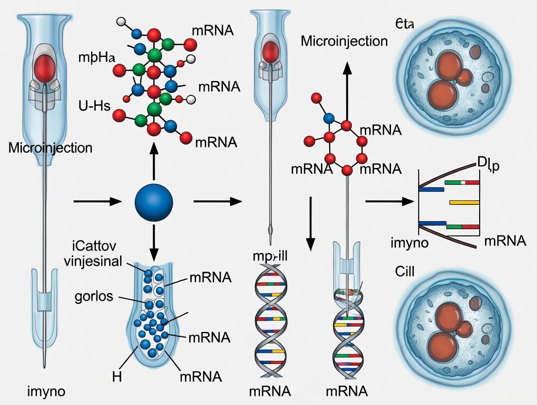
This comprehensive guide details the established microinjection techniques for Xenopus laevis embryos, a cornerstone methodology in developmental biology, cell biology, and drug discovery research.
Mastering Xenopus Embryo Microinjection: A Comprehensive Guide from Fundamentals to Advanced Targeted Techniques
Abstract
This comprehensive guide details the established microinjection techniques for Xenopus laevis embryos, a cornerstone methodology in developmental biology, cell biology, and drug discovery research. It covers foundational principles, including the rationale for using Xenopus as a model system and essential equipment setup. The article provides step-by-step methodological protocols for both standard and advanced targeted microinjection, utilizing fate maps for specific tissues like the pronephros. It further addresses critical troubleshooting and optimization strategies to enhance survival rates and experimental reproducibility. Finally, the guide explores validation techniques through lineage tracing and immunostaining, and discusses the comparative advantages of Xenopus microinjection for functional gene analysis and protein expression studies.
Xenopus Microinjection Foundations: Principles, Applications, and Model System Advantages
Why Xenopus? Key Advantages of the Frog Model System for Microinjection
The genus Xenopus, particularly the species Xenopus laevis (African clawed frog) and Xenopus tropicalis (Western clawed frog), has established itself as a cornerstone of biological research for decades. Its initial rise to prominence began in the 1930s with its use in the Hogben pregnancy test, a novel bioassay that was simpler and more reliable than previous methods [1]. Since then, Xenopus has evolved into an indispensable tool for developmental biology, genetics, and biomedical research. The external fertilization, large and readily manipulable eggs, and rapid embryonic development of these frogs make them uniquely suited for microinjection-based studies [1] [2]. This application note details the specific advantages of the Xenopus system for microinjection, provides a comparative analysis of the two primary species used, and outlines key protocols for researchers and drug development professionals engaged in high-throughput screening and disease modeling.
Key Advantages for Microinjection and Research
The choice of Xenopus as a model system is underpinned by a set of distinct biological and practical characteristics that facilitate experimental manipulation and enhance the translatability of research findings.
Large, Externally Developing Eggs and Embryos: Xenopus eggs are among the largest of all vertebrates, measuring about 1-1.3 mm in diameter [1]. This substantial size makes them exceptionally easy to visualize and manipulate under a stereomicroscope. Furthermore, their external development allows for direct access to all stages of embryogenesis without invasive procedures, enabling straightforward microinjection, surgical manipulation, and continuous observation [1] [3] [2].
Rapid and Synchronous Development: Following fertilization, Xenopus embryos develop rapidly, reaching the tadpole stage within a few days [1]. This rapid timeline allows for the efficient design and execution of experiments, significantly accelerating research cycles compared to other vertebrate models. Embryos from a single clutch develop synchronously, providing a large cohort of specimens at identical developmental stages for robust statistical analysis [2].
High Genetic and Physiological Conservation with Humans: Despite its evolutionary distance, Xenopus shares a high degree of genetic conservation with humans. Over 80% of known human disease-associated genes have orthologs in the Xenopus genome [4] [5]. Critical signaling pathways such as Wnt, BMP, and FGF, which govern fundamental processes like cell division, differentiation, and organogenesis, are highly conserved, making findings in Xenopus directly relevant to human biology and disease [1] [4].
Ease of Genetic Manipulation: The Xenopus system is highly amenable to a wide array of genetic manipulations. Techniques such as microinjection of mRNA, DNA, and morpholino oligonucleotides have been standard for decades [6] [7]. More recently, the advent of CRISPR/Cas9 technology has enabled precise genome editing, allowing researchers to model human genetic diseases with high efficiency and at a lower cost compared to mammalian models [2] [5].
Robustness and High Fecundity: Xenopus embryos are remarkably resilient and can withstand experimental manipulations such as microinjection and surgical procedures with high survival rates [1]. A single female can produce thousands of eggs in one laying, providing ample biological material for high-throughput screening of genetic or chemical factors [5].
Transparency for Live Imaging: The relative transparency of Xenopus embryos and tadpoles, particularly in early stages, permits high-resolution live imaging of developmental processes. This feature is invaluable for tracking cell migrations, such as those of cranial neural crest cells, and for visualizing organogenesis in real-time without the need for dissection [3] [4] [2].
Comparative Analysis of Xenopus Species
While both Xenopus laevis and X. tropicalis are widely used, they possess distinct characteristics that make them suitable for different research applications. The table below provides a detailed comparison to guide researchers in selecting the most appropriate species for their work.
Table 1: Comparative Analysis of Xenopus laevis and Xenopus tropicalis
| Feature | Xenopus laevis | Xenopus tropicalis |
|---|---|---|
| Ploidy | Allotetraploid (36 chromosomes) [5] | Diploid (20 chromosomes) [8] [5] |
| Adult Size | Larger (10-12 cm females) [1] | Smaller (4-6 cm) [1] [8] |
| Embryo Size | Larger (~1.3 mm) [1] [6] | Smaller [6] |
| Generation Time | ~12 months [8] | 4-6 months [8] |
| Genome Sequence | Sequenced in 2016 [1] | First frog genome sequenced (2010) [1] [5] |
| Ideal for | Embryological studies, microinjection training, protein/oocyte expression [1] [9] | Genetic studies, CRISPR/Cas9, comparative genomics [1] [6] [2] |
| Key Advantage | Large embryo size, robust for manipulation [1] [6] | Simpler genetics, shorter generation time [1] [8] |
Research Applications Enabled by Microinjection
The technical advantages of Xenopus microinjection have directly fueled breakthroughs across diverse fields of biomedical research.
Developmental Biology and Cell Fate Mapping: The ability to perform targeted microinjection into specific blastomeres at early cleavage stages (e.g., 4-cell, 8-cell) is a powerful application. Using established fate maps, researchers can inject materials specifically into the blastomere that gives rise to a particular organ, such as the kidney (pronephros), heart, or eyes [3]. This allows for tissue-specific overexpression or knockdown of genes, minimizing secondary effects in the rest of the embryo. Co-injection of a lineage tracer (e.g., fluorescent mRNA) enables verification of targeting and visualization of the progeny of the injected cell [3].
Disease Modeling: Xenopus is extensively used to model human genetic diseases. By injecting mRNAs carrying gain-of-function mutations or using morpholinos/CRISPR to create loss-of-function models, researchers have studied a wide spectrum of disorders [5]. These include congenital heart defects, kidney disease, ciliopathies, and various craniofacial malformations such as DiGeorge syndrome and Treacher Collins syndrome [4] [5]. The rapid development and easy scoring of phenotypes make it an efficient model for validating disease-associated genes.
Craniofacial Development Research: Xenopus is a premier model for studying craniofacial morphogenesis, which depends on the precise migration and differentiation of cranial neural crest cells (CNCCs) [4]. Microinjection is used to perturb genes involved in CNCC specification, migration, and differentiation, helping to elucidate the etiology of common birth defects like cleft lip and palate. The external development and large embryos allow for direct in vivo visualization of CNCC behaviors at a resolution often not achievable in mammalian models [4].
Pigmentation and Pigmentary Disorders: The melanocytes of Xenopus develop and function similarly to those in mammals. Changes in pigmentation are exceptionally easy to score in live embryos [5]. Microinjection of morpholinos or CRISPR components targeting pigmentation genes (e.g., MITF) can model human pigmentary disorders and melanoma. The system is also valuable for studying skin responses to ultraviolet radiation (UVR) and DNA repair mechanisms [5].
Ion Channel and Receptor Studies: While not performed in embryos, the microinjection of in vitro-transcribed mRNA into Xenopus oocytes is a classic technique for the heterologous expression and functional characterization of ion channels, receptors, and transporters [9]. This system allows for precise electrophysiological and pharmacological studies of human proteins in a controlled environment.
Essential Equipment and Reagents for Microinjection
A successful microinjection experiment requires specific equipment and reagents. The following table lists the core components of the "Researcher's Toolkit."
Table 2: Essential Research Reagent Solutions and Equipment for Xenopus Microinjection
| Item | Function/Description |
|---|---|
| Stereomicroscope | A microscope with good optics and a large working distance (at least 8-10 cm) is essential for visualizing embryos and manipulating injection needles [7]. |
| Micromanipulator & Microinjector | Apparatus for holding the injection pipette and delivering precise, controlled volumes of solution into embryos or oocytes [9] [7]. |
| Injection Pipettes | Fine, glass capillaries pulled to a sharp tip for piercing the embryo membrane without causing significant damage. |
| Lineage Tracers | Fluorescent dyes, dextrans, or mRNA encoding fluorescent proteins (e.g., MEM-RFP) used to trace the fate of injected cells and verify targeting [3]. |
| Morpholino Oligonucleotides | Antisense molecules used to transiently knock down gene expression by blocking mRNA translation or splicing [3] [5]. |
| CRISPR/Cas9 Components | Cas9 protein/RNA and guide RNAs for creating targeted, heritable gene knockouts or knock-ins [2]. |
| In vitro Transcription Kits | For synthesizing capped mRNA for overexpression studies or for creating lineage tracer RNA [7]. |
| Marc's Modified Ringer's (MMR) | A common saline solution for maintaining embryos and oocytes [3]. |
| Dejelly Solution (e.g., 2% Cysteine) | For removing the protective jelly coat surrounding fertilized eggs prior to microinjection [3]. |
Detailed Microinjection Protocol for Embryos
The following protocol outlines the key steps for microinjecting Xenopus embryos, with specific considerations for both X. laevis and X. tropicalis.
Protocol Workflow
Protocol Steps
1. Embryo Preparation and Selection
- Obtain embryos through natural mating or in vitro fertilization using hormones [3]. Once laid, transfer fertilized embryos into a Petri dish.
- Remove the protective jelly coat by incubating embryos in a 2% cysteine solution (pH 8.0) for a few minutes with gentle agitation. Rinse thoroughly with an appropriate buffer like 1x MMR [3].
- Select healthy, undamaged embryos and align them on an injection dish fitted with a nylon mesh or in grooves made of agarose to immobilize them during injection [3] [9].
2. Injection Needle Preparation and Loading
- Pull borosilicate glass capillaries to create fine-tipped injection needles using a pipette puller.
- Backfill the needle with a small amount of mineral oil using a syringe. Then, front-load the needle with your injection sample (e.g., morpholino, CRISPR mix, mRNA) by immersing the tip into the droplet and using the microinjector's suction function to draw up the desired volume [9].
3. Blastomere Targeting and Orientation
- For targeted injections, use the established Xenopus fate maps available on Xenbase [3]. At the 4-cell stage, the ventral blastomeres contribute significantly to the kidney and other ventral tissues. At the 8-cell stage, the ventral-vegetal blastomere (V2) is the primary contributor to the pronephros [3].
- Orient the embryos under the microscope so the targeted blastomere is accessible. The pigmented animal pole and the lighter vegetal pole serve as visual guides.
4. Microinjection Execution
- Mount the loaded needle onto the micromanipulator. Using the manipulator, position the needle at a shallow angle relative to the embryo.
- Gently insert the needle into the target blastomere. Use a brief pulse of pressure from the microinjector to deliver the solution. A visible clearing or slight expansion of the cell indicates a successful injection.
- Wait a few seconds before retracting the needle to prevent the backflow of the injected material [9].
5. Post-Injection Incubation and Analysis
- After injection, transfer the embryos to a fresh dish with incubation medium (e.g., 0.1x MMR).
- Critical Step: Regulate the incubation temperature tightly. Cooler temperatures (e.g., 14-16°C) slow development, providing a larger time window for injections at specific stages [3].
- Allow embryos to develop to the desired stage. Phenotypic analysis can include live imaging for lineage tracers, whole-mount immunostaining to visualize specific tissues (e.g., pronephric tubules), or molecular biology techniques to assess gene expression changes [3].
Research Applications Workflow
The general workflow for a functional genetics study in Xenopus from microinjection to analysis is summarized below.
The Xenopus frog system remains an powerful and versatile model for microinjection-based research. Its unique combination of large, robust embryos, rapid external development, and high genetic conservation with humans offers unparalleled advantages for developmental studies, disease modeling, and drug screening. The continued development of sophisticated genetic tools like CRISPR/Cas9, coupled with its inherent suitability for high-throughput approaches, ensures that Xenopus will continue to be a vital organism for answering fundamental biological questions and advancing human health.
Microinjection in Xenopus embryos and oocytes is a cornerstone technique for developmental biology and functional genomics. This application note details established and emerging protocols for targeted gene manipulation, leveraging the unique advantages of the Xenopus system, including externally developing embryos, a well-defined fate map, and high fecundity. We focus on specific methodologies for gene overexpression and knockdown, concluding with advanced applications for studying protein function.
Targeted Microinjection for Gene Manipulation
The power of microinjection in Xenopus is vastly enhanced by the use of fate maps, which allow researchers to target specific blastomeres that give rise to particular tissues and organs. This enables tissue-specific manipulation of gene expression and the creation of mosaic embryos where manipulated and unmanipulated tissues can be compared within the same organism [3] [10].
The table below summarizes the primary blastomeres targeted for specific tissues at the 4-cell and 8-cell stages.
Table 1: Blastomere Selection for Tissue-Targeted Microinjection
| Target Tissue | Stage | Blastomere Name | Blastomere Description |
|---|---|---|---|
| Kidney (Pronephros) | 4-cell | Ventral Blastomere | Large, darkly pigmented cell [3] |
| 8-cell | V2 Blastomere | Ventral, vegetal blastomere [3] | |
| Retina | 32-cell | - | Specific blastomeres identified via retina fate map [10] |
The following workflow outlines the key steps for performing targeted microinjection and subsequent analysis.
Protocol: Kidney-Targeted Microinjection at the 8-Cell Stage
This protocol describes how to target the developing pronephros (kidney) in Xenopus laevis embryos [3].
Materials:
- Dejellied Xenopus embryos at the 8-cell stage.
- Injection reagents: Morpholinos, mRNA for overexpression, or cDNA constructs.
- Lineage tracer: e.g., MEM-RFP or MEM-GFP mRNA.
- Microinjection setup: Injector, micromanipulator, pulled borosilicate glass needles.
- Solutions: 1x MMR, 2% cysteine (pH 8.0) for dejellying.
Method:
- Prepare Embryos: Collect embryos and remove the jelly coat by incubating in 2% cysteine (pH 8.0). Rinse thoroughly in 1x MMR [3].
- Identify Blastomere: Under a stereomicroscope, orient the embryo to locate the left ventral, vegetal (V2) blastomere. This cell is vegetal (yolky, less pigmented) and ventral (larger and darker than dorsal cells) [3].
- Prepare Injection Needle: Back-fill a needle with mineral oil, then front-fill with your injection solution mixed with lineage tracer [11].
- Inject: Penetrate the V2 blastomere and inject ≤ 15 nL of solution. A successful injection will show a slight dimpling and filling of the cell [3] [11].
- Incubate: Transfer injected embryos to a dish with 1x MMR and incubate at 14-16°C to slow development, allowing more time for subsequent manipulations if needed [3].
- Verify Targeting: At tailbud stages (stage 38-40), image the embryo to confirm the lineage tracer fluorescence is localized to the pronephric region. Fix embryos for whole-mount immunostaining with pronephric-specific antibodies for detailed analysis [3].
Gene Overexpression and Knockdown Techniques
Microinjection enables diverse strategies for modulating gene function. The table below compares the core techniques.
Table 2: Core Microinjection Techniques for Gene Manipulation
| Technique | Reagent Injected | Primary Mechanism | Key Applications |
|---|---|---|---|
| Gene Overexpression | Synthetic mRNA [10] or cDNA [11] | Introduces exogenous coding sequence for translation | Functional analysis, rescue experiments, dominant-negative effects |
| Knockdown (Morpholino) | Antisense Morpholino Oligomers (MOs) [12] | Blocks translation initiation or pre-mRNA splicing | Loss-of-function studies, phenocopy of mutations |
| Knockdown (CRISPRi) | dCas9-KRAB mRNA + gene-specific gRNAs [13] | Represses transcription by blocking RNA polymerase | Specific mRNA suppression, alternative to MOs |
| Protein Expression | cDNA or mRNA [14] [11] | Heterologous expression in oocytes/embryos | Studying channel/transporter function, protein localization |
Protocol: mRNA Overexpression and Knockdown with Morpholinos
Materials:
- For mRNA injection: Synthetic mRNA encoding the protein of interest, resuspended in nuclease-free water.
- For Morpholino injection: Antisense Morpholino oligonucleotide targeting the gene of interest.
- Injection equipment and materials as in Protocol 1.1.
Method:
- Reagent Preparation: Dilute synthetic mRNA or Morpholino to the desired working concentration in nuclease-free water. Co-inject with a fluorescent lineage tracer [10] [12].
- Microinjection: Follow the general microinjection and blastomere targeting steps outlined in Protocol 1.1. Inject into the cytoplasm of the target blastomere.
- Incubation and Analysis: Incubate embryos and monitor development. Analyze phenotypes, then fix embryos for in situ hybridization, immunostaining, or other techniques to assess molecular changes [12].
Emerging Protocol: Gene Knockdown with CRISPR Interference (CRISPRi)
While traditional Morpholinos are effective, CRISPRi offers a modern, DNA-targeting alternative for gene knockdown. A 2025 study in Xenopus tropicalis demonstrated that CRISPRi, specifically using dCas9 fused to a KRAB repressor domain (dCas9-KM), efficiently suppresses both exogenous and endogenous mRNA transcription. In contrast, CRISPR-Cas13 systems were found to be ineffective in this model [13].
Materials:
- mRNA encoding the dCas9-KM repressor fusion protein.
- Synthetic gRNAs targeting the gene of interest.
- Standard microinjection setup.
Method:
- Reagent Preparation: Co-inject a mix of dCas9-KM mRNA (e.g., 300 pg/embryo) and a pool of target-specific gRNAs (e.g., 100 pg/embryo each) into the fertilized egg at the one-cell stage [13].
- Incubation and Validation: Allow embryos to develop to the desired stage. Assess knockdown efficacy via quantitative PCR (qPCR), phenotypic analysis, or loss of specific markers (e.g., loss of pigmentation for tyrosinase) [13].
The Scientist's Toolkit: Essential Research Reagents
Table 3: Key Reagent Solutions for Xenopus Microinjection
| Reagent / Material | Function / Application | Example / Notes |
|---|---|---|
| Lineage Tracers | Visualizing progeny of injected blastomere | MEM-RFP, MEM-GFP, fluorescent dextrans [3] [15] |
| Morpholinos (MOs) | Gene knockdown; block translation/splicing | Antisense oligonucleotides; control MOs are critical [12] |
| Synthetic mRNA | Gene overexpression; express wild-type/mutant proteins | In vitro transcribed from plasmid templates (e.g., CS2+) [10] [15] |
| cDNA Constructs | Heterologous protein expression; mosaic analysis | Injected into oocyte nucleus or embryos [11] [15] |
| Microinjection Setup | Precise delivery of reagents | Injector, micromanipulator, micropipette puller, forceps [3] [11] |
| Fate Maps | Guide targeted blastomere injection | Available online at Xenbase [3] |
Protein Function and Live Imaging Applications
Beyond transcriptional manipulation, microinjection is pivotal for studying protein function, localization, and dynamic cellular processes.
Protocol: Heterologous Protein Expression in Oocytes
Xenopus oocytes are a premier system for expressing and studying proteins from diverse organisms [11].
Materials:
- Stage V-VI oocytes manually isolated from ovarian tissue.
- cDNA construct (50-200 µg/mL) for nuclear injection.
- Modified Barth's Solution (MBS).
- Microinjection setup with a plunger-based system (e.g., Drummond Nanoject).
Method:
- Oocyte Preparation and Isolation: Manually defolliculate oocytes using fine forceps. Allow oocytes to recover overnight at 16-20°C in MBS [11].
- Nuclear Injection (cDNA): Orient healthy oocytes with the animal (pigmented) pole upward. Load a needle with cDNA solution (<3 ng per oocyte). Penetrate the animal pole and inject into the germinal vesidium (nucleus) [11].
- Cytoplasmic Injection (mRNA): For mRNA injection, target the vegetal cytoplasm. This is more tolerant of larger injection volumes but requires more mRNA for equivalent expression [11].
- Incubation and Analysis: Incubate injected oocytes for 24-72 hours. Screen for protein expression using western blotting, electrophysiology, radioisotope flux, or imaging, depending on the protein and experimental goal [14] [11].
Protocol: Live Imaging of Cell and Developmental Processes
The large size and clarity of Xenopus embryonic cells make them ideal for high-resolution live imaging [15].
Materials:
- Embryos injected with mRNA encoding fluorescent fusion proteins (e.g., memGFP, τ-GFP).
- Low-melt agarose.
- Custom-made or commercial imaging chambers with a cover glass bottom.
- Confocal or spinning-disk microscope.
Method:
- Label Embryos: Inject low doses (15-500 pg, optimized empirically) of mRNA encoding fluorescently tagged proteins into embryos to label cellular structures (membrane, organelles, cytoskeleton) [15].
- Mount for Imaging: Embed live embryos in low-melt agarose within the imaging chamber, ensuring the tissue of interest is positioned close to the cover glass [15].
- Image Acquisition: Collect time-lapse Z-stacks on an inverted confocal microscope. Adjust acquisition speed and laser power to minimize photodamage while capturing the dynamic process [15].
The microinjection techniques outlined here—from targeted blastomere injections and various knockdown/overexpression strategies to live imaging—provide a comprehensive toolkit for probing gene and protein function in the versatile Xenopus model. Mastery of these protocols enables precise interrogation of developmental mechanisms, disease processes, and fundamental cell biology.
Microinjection is a foundational technique in developmental biology for introducing exogenous materials such as DNA, RNA, proteins, or drugs directly into cells and embryos. Within Xenopus embryo research, this method is indispensable for probing gene function, protein dynamics, and early developmental mechanisms [16] [17]. The success of these intricate experiments hinges on a properly configured workstation comprising a vibration-damped microscope, precise micromanipulators, and meticulously prepared microinjection needles [16] [18]. This application note details the essential equipment specifications and protocols for establishing a robust microinjection system tailored to Xenopus embryo research, providing a critical technical resource for the experimental foundation of a doctoral thesis.
Core Microinjection System Specifications
A reliable microinjection system rests on three core components: the microscope, micromanipulator, and microinjector. Their integration must provide stability, optical clarity, and precise control.
Microscope Selection and Key Specifications
For microinjection of Xenopus oocytes and embryos, an inverted microscope is generally recommended due to its superior working distance and accessibility for micromanipulators [16] [19]. The condenser must have a long or ultra-long working distance to accommodate manipulator arms without obstruction [16]. Stability is paramount; the microscope should be placed on a heavy, vibration-damped table in a quiet location to isolate it from environmental disturbances [16] [18]. The following table summarizes the critical microscope specifications and common models suitable for this research.
Table 1: Microscope Specifications for Xenopus Microinjection
| Microscope Type | Key Feature | Recommended Model Examples | Contrast Techniques | Typical Magnification |
|---|---|---|---|---|
| Inverted Microscope | Long working distance condenser | Nikon ECLIPSE Ti2 series, Ts2R/ Ts2R-FL [19] | Differential Interference Contrast (DIC), Phase Contrast [19] | 200x for cytoplasmic injection, 400x for nuclear injection [20] |
| Upright Microscope | Horizontal micropipette access | Zeiss Axioskop [18] | DIC, Nomarski [18] | Dependent on objective and eyepiece |
Micromanipulators and Microinjectors
The micromanipulator is responsible for the fine, three-dimensional positioning of the micropipette. Its stability directly governs the success of the entire procedure [16]. Electronic models are highly advantageous as they allow for programmable, precise movements to pre-determined coordinates [16] [20]. The microinjector controls pressure within the micropipette. Different applications require different pressures; for instance, injecting DNA solution into a pronucleus requires high pressure (>3000 hPa), while holding oocytes requires mild positive and negative pressures [16]. A digital microinjector allows for independent control of base pressure (to prevent medium backflow) and injection pressure pulses [16] [20].
Table 2: Micromanipulator and Microinjector Configurations for Key Applications
| Application in Xenopus Research | Recommended Micromanipulator | Recommended Microinjector | Pressure Requirements |
|---|---|---|---|
| Pronuclear DNA Injection | Two XenoWorks manipulators (Right/Left) [16] | XenoWorks Digital microinjector [16] | High injection pressure >3000 hPa; subtle negative holding pressure [16] |
| Oocyte Holding & Somatic Cell Transfer | Two XenoWorks manipulators (Right/Left) [16] | Two XenoWorks Analog microinjectors [16] | Mild positive and negative pressure for holding and transfer [16] |
| General Cytosolic/Nuclear Injection | Eppendorf TransferMan NK2 [20] | Eppendorf FemtoJet [20] [21] | Injection pressure (Pi): 600-1800 hPa; Compensation pressure (Pc): ~180 hPa [21] |
Microneedle Preparation Protocols
The quality of the micropipette is a decisive factor for cell viability and injection success. The preparation process involves pulling, back-filling, and carefully breaking the tip to the desired diameter.
Micropipette Pulling Procedure
- Equipment Setup: Use a horizontal, double-stage micropipette puller, such as a Sutter Instrument P-87 or P-97 [20] [21]. Turn the machine on at least 20 minutes before use to ensure thermal stability [21].
- Glass Capillary Selection: Use borosilicate glass capillaries with an outer diameter of 1.0 mm and an inner filament, which facilitates back-filling by capillary action (e.g., World Precision Instruments #1B100F-4) [20] [21].
- Pulling Parameters: Program the puller for a multi-step process. Example parameters for a Sutter P-87 are: Heat=600, Pull=100, Velocity=140, Time=150; followed by Heat=960, Pull=0, Velocity=40, Time=250; and a final step of Heat=960, Pull=27-30, Velocity=60, Time=252 [21]. The goal is a needle that tapers over 0.4-0.6 cm to a sharp, closed point [20].
- Storage: Store pulled needles in a covered storage jar or attached to the sticky side of foam strips in a Petri dish to protect them from dust and damage [18] [21].
Needle Back-Filling and Tip Breaking
- Solution Preparation: Prepare the injection solution (e.g., DNA in distilled water). Centrifuge it at maximum speed for 10 minutes to pellet particulate matter that could clog the needle [21]. Adding a visible dye like Fast Green (0.1% w/v) or a fluorescent marker like Dextran Texas Red helps visualize the solution during injection [20].
- Back-Filling: Using a fine, pulled pipette tip or a microloader, carefully aspirate a small volume of the prepared solution. Insert the tip into the back of the needle and expel the solution, allowing it to flow to the tip via capillary action over 2-3 minutes [20] [21]. Avoid introducing air bubbles.
- Breaking the Tip: Mount the filled needle on the manipulator. Under microscopic observation (40x objective), lower the needle tip towards the broken edge of a coverslip. Gently tap the needle against the edge to break off a tiny piece and open the tip [21]. The ideal tip diameter is approximately 0.5 µm for cytoplasmic injection and 0.2-0.5 µm for nuclear injection [20].
- Verifying Flow: Engage the microinjector and press the foot pedal to check for a rapid, laminar flow of injection solution from the needle tip. There should be no flow at the resting compensation pressure [21]. Adjust the compensation pressure (
Pc) to prevent backflow of medium into the needle, which would dilute the injection solution [20].
Experimental Workflow for Microinjection
The microinjection process is a systematic sequence from sample preparation to post-injection care. The following workflow diagram outlines the key stages for a typical experiment involving Xenopus oocytes or embryos.
Diagram 1: Microinjection Experimental Workflow
Research Reagent Solutions
Successful microinjection relies on a suite of specialized reagents and materials, each serving a specific function to ensure cell viability and experimental integrity.
Table 3: Essential Reagents and Materials for Xenopus Microinjection
| Reagent/Material | Function/Application | Example Specifications |
|---|---|---|
| Halocarbon Oil | Creates an inert, immiscible layer over samples on an injection pad to prevent desiccation [22] [21]. | Series 700 (e.g., Sigma-Aldrich H8898) [22] [21] |
| Agarose Pads | Provides a stable, non-toxic, and slightly adhesive surface for immobilizing oocytes or embryos during injection [23] [21]. | 2% agarose in water, baked onto coverslips [23] [21] |
| Marker Dyes | Co-injected with substances of interest to visually confirm successful delivery and estimate injection volume [20]. | Fast Green (0.1%), Dextran Texas Red [20] |
| Recovery Buffer | A physiological solution used to recover and rehydrate samples after the injection procedure [22]. | M9 buffer or specialized recovery buffer with salts and glucose [22] |
| Modified Barth's Saline (MBS) | A standard medium for holding and culturing Xenopus oocytes and embryos, maintaining osmotic balance and pH [17]. | 1x MBS with Penicillin/Streptomycin [17] |
Mastering the setup and operation of the microinjection workstation is a prerequisite for advanced research in developmental biology using the Xenopus model. The precise alignment of a vibration-resistant microscope, a stable micromanipulator, and a pressure-controlled microinjector, combined with consistently prepared micropipettes, forms the foundation for reproducible and high-yield experiments. By adhering to the detailed equipment specifications and standardized protocols outlined in this document, researchers can effectively troubleshoot their systems and reliably generate high-quality data for their investigations into gene function and embryonic development.
Understanding Early Xenopus Embryogenesis and Critical Developmental Stages for Injection
The African clawed frog (Xenopus) has served as a cornerstone model organism in developmental biology for decades, providing fundamental insights into embryonic development, cell signaling, and gene function. Its external development, large, readily manipulable embryos, and high fecundity make it an exceptional system for microinjection-based studies [24] [2]. Mastering the precise staging of early Xenopus embryogenesis is paramount for the success of these techniques, as the timing of developmental events is temperature-dependent and crucial for experimental reproducibility [25]. This application note details the critical early developmental stages of Xenopus laevis and provides a standardized protocol for targeted microinjection, serving as an essential resource for researchers employing these techniques in fundamental and applied biomedical research.
Normal Table of Development and Key Staging Landmarks
The Nieuwkoop and Faber (NF) staging system is the definitive standard for characterizing Xenopus development, defining 66 distinct stages based on discrete morphological features rather than temporal or size metrics [25]. This allows the system to be consistently applied across different Xenopus species and laboratory conditions.
For microinjection experiments, the early cleavage, blastula, and gastrula stages (NF 1-20) are most critical. The table below summarizes key morphological landmarks and experimental considerations for these stages.
Table 1: Critical Early Developmental Stages for Microinjection inXenopus laevis
| NF Stage | Name | Key Morphological Landmarks | Optimal Injection Target / Experimental Application |
|---|---|---|---|
| 1-3 | Fertilized Egg to 4-Cell | Single cell; first cleavage bisects left/right; second cleavage divides dorsal/ventral [3]. | All blastomeres are totipotent. Injection into a single blastomere at the 4-cell stage can target its progeny [3]. |
| 4 | 8-Cell | Third cleavage separates animal (darker, pigmented) and vegetal (yolky) hemispheres [3]. | Ventral-vegetal (V2) blastomeres contribute significantly to the kidney (pronephros) [3]. |
| 5-6 | 16- to 32-Cell | Cleavages become less synchronous. Blastomeres are named based on lineage (e.g., V2.2 at 16-cell) [3]. | The V2.2 blastomere (also known as C3 at the 32-cell stage) is the primary contributor to the pronephros [3]. |
| 6.5-9 | Morula to Early Blastula | "Mulberry" cluster of cells; formation of blastocoel cavity begins [2]. | Common stage for mRNA injection into the blastocoel or animal pole for widespread overexpression. |
| 10 | Early Gastrula | Dorsal lip of the blastopore forms, marking the start of gastrulation [25]. | Critical stage for manipulating Spemann-Mangold organizer signals. Injection near the dorsal lip can affect axial patterning. |
| 12.5 | Mid Gastrula | Blastopore is crescent-shaped [25]. | |
| 13-20 | Neurula | Blastopore closes; neural plate folds into neural tube [2]. | Stages for analyzing neural induction and patterning; injections are less common. |
Experimental Protocol: Targeted Microinjection of Early Embryos
This protocol describes the methodology for targeted microinjection into specific blastomeres of 4- and 8-cell stage Xenopus laevis embryos to manipulate gene expression in a tissue-specific manner, using the pronephros (kidney) as an example.
I. Materials and Reagents
Table 2: Research Reagent Solutions for Microinjection
| Item | Function/Description |
|---|---|
| Dejelly Solution (2% Cysteine, pH 8.0) | Removes the protective jelly coat from naturally laid embryos to enable manipulation and injection [3]. |
| Marc's Modified Ringers (MMR) | Standard saline solution for raising and maintaining Xenopus embryos post-injection [3]. |
| Testes Storage Solution (1x MMR, 1% BSA, Gentamycin) | Medium for storing isolated male testes used for in vitro fertilization [3]. |
| Lineage Tracer (e.g., MEM-RFP mRNA) | RNA encoding a fluorescent protein (e.g., membrane-targeted RFP) co-injected to confirm successful targeting and visualize progeny of the injected blastomere [3]. |
| Morpholino Oligonucleotides or mRNA | Agents for knocking down or overexpressing genes of interest, respectively [3] [24]. |
| Microinjection Needles | Fine glass capillaries pulled to a sharp point for piercing the vitelline membrane and blastomere. |
| Micromanipulator & Microinjector | Apparatus for holding the needle and delivering nanoliter-volume injections with high precision [3] [24]. |
II. Step-by-Step Procedure
A. Embryo Preparation
- Obtain embryos through natural mating or in vitro fertilization. For in vitro fertilization, squeeze eggs from a hormonally primed female into a dish and fertilize with a macerated testis [3].
- Once the jelly coats have swollen (approximately 15 minutes post-fertilization), pour off excess water and add ~50 ml of Dejelly Solution. Gently swirl for 3-5 minutes until embryos pack closely together.
- Rinse embryos thoroughly with 1x MMR (or similar medium) several times to remove all traces of cysteine [3].
- Temperature Control: Incubate dejellied embryos at 14-16°C to slow the rate of development. This provides a longer window for performing injections at the 4- and 8-cell stages [3].
B. Blastomere Identification and Targeting
- Using a dissecting microscope, orient the embryo with the animal pole (darkly pigmented) facing upward and the vegetal pole (light, yolky) downward.
- Identify the cleavage planes. The first cleavage typically separates the left and right sides. The second cleavage separates dorsal (smaller, less pigmented cells) from ventral (larger, darker cells) [3].
- For 4-cell injections: To target the left kidney, inject the left ventral blastomere.
- For 8-cell injections: To target the left kidney, inject the left ventral-vegetal (V2) blastomere [3].
- Refer to the following workflow diagram for the overall experimental process.
C. Microinjection Process
- Back-fill a pulled glass needle with the injection solution (e.g., morpholino/mRNA mixed with lineage tracer).
- Calibrate the injection volume (typically 5-10 nL per blastomere for early stage embryos) by measuring the diameter of the droplet expelled into oil [3] [24].
- Secure the embryo in a small well made of agarose or using a holding pipette.
- Carefully pierce the vitelline membrane and the targeted blastomere with the needle.
- Deliver the calibrated volume into the blastomere cytoplasm. Withdraw the needle smoothly.
D. Post-Injection Analysis
- Allow injected embryos to recover and develop in 1x MMR at the desired temperature (14-22°C) until the desired analysis stage [3].
- Validation of Targeting: At tailbud stages (e.g., NF 38-40), visualize the lineage tracer fluorescence using a fluorescence microscope. Successful targeting of the pronephros will show fluorescence in the bilateral tubules located along the trunk [3].
- Phenotypic Analysis: Assess the effects of your manipulation (e.g., morpholino knockdown) on pronephric development using whole-mount immunostaining with pronephros-specific antibodies and compare to the contralateral, uninjected side as an internal control [3].
Critical Signaling Pathways in Early Patterning
The cell fate decisions during early Xenopus development are directed by conserved signaling pathways. Key pathways active during the blastula and gastrula stages include BMP, Wnt, and Nodal/FGF signaling, which establish the dorsal-ventral and anterior-posterior axes. The following diagram summarizes the logical relationships of these key pathways in establishing the primary body axes.
Mastering the detailed staging and microinjection protocols for early Xenopus embryogenesis is a fundamental skill for researchers utilizing this powerful model system. The integration of the updated Normal Table of development with refined targeted microinjection techniques enables precise spatial and temporal control over gene expression. This precision, in turn, facilitates robust modeling of human genetic diseases, high-throughput drug screening, and the dissection of core conserved signaling pathways that govern vertebrate development. By adhering to these standardized application notes and protocols, researchers can ensure the reproducibility and reliability of their microinjection-based experiments in Xenopus.
Step-by-Step Microinjection Protocols: From Embryo Preparation to Targeted Tissue Delivery
Embryo Collection, Fertilization, and Jelly Coat Removal (Dejellying)
Within the field of developmental biology and biomedical research, the African clawed frog Xenopus laevis stands as a fundamental model organism. Its externally developing embryos, large size, and capacity for high-throughput experimentation make it an indispensable system for studying gene function, cell signaling, and organogenesis. A critical foundation for many advanced techniques, including microinjection for gene overexpression or knockdown, is the consistent production of high-quality, synchronously developing embryos. This application note details the essential upstream protocols for the robust generation and preparation of Xenopus embryos, framing them within the context of a broader research workflow centered on microinjection techniques. The successful execution of embryo collection, in vitro fertilization (IVF), and jelly coat removal is a prerequisite for ensuring that subsequent experimental manipulations yield reliable and interpretable data for researchers and drug development professionals.
The process of preparing Xenopus laevis embryos for microinjection and other analyses is a multi-stage workflow, beginning with animal husbandry and concluding with de-jellied, synchronously developing embryos ready for experimentation. The following diagram illustrates the key stages and decision points in this process.
Research Reagent Solutions
The following table catalogues the essential reagents and their specific functions in the embryo preparation protocol. Accurate preparation of these solutions is critical for experimental success.
Table 1: Key Reagents for Xenopus Embryo Collection and Preparation
| Reagent | Function/Application | Key Details & Composition |
|---|---|---|
| Human Chorionic Gonadotropin (hCG) | Induction of ovulation in females and mating behavior in males [26]. | Typically used at 1000 U/mL stock concentration; administered via subcutaneous injection into the dorsal lymph sac [26]. |
| Marc's Modified Ringer's (MMR) | Embryo culture medium post-fertilization and dejellying [27] [26]. | 1x MMR: 0.1 M NaCl, 2.0 mM KCl, 1 mM MgSO(4), 2 mM CaCl(2), 5 mM HEPES; pH 7.4-7.8 [27] [26]. Often used at 0.1x concentration for culturing embryos [27]. |
| L-Cysteine Dejelly Solution | Removal of the protective jelly coat surrounding the embryo [27] [26]. | 2% L-cysteine free base dissolved in 0.1x MMR; pH adjusted to 7.8-8.0 with NaOH [27] [26]. |
| Modified Barth's Saline (MBS) | Oocyte culture and storage [27] [28]. | 1x MBS: 88 mM NaCl, 1 mM KCl, 0.82 mM MgSO(4), 0.33 mM Ca(NO(3))(2), 0.41 mM CaCl(2), 10 mM HEPES, pH 7.5 [27] [29]. |
| Gentamicin Reagent | Antibiotic to prevent bacterial contamination in embryo cultures [26]. | Used as a 1000x stock solution (10 mg/mL) in culture media [26]. |
Detailed Experimental Protocols
Hormone-Induced Embryo Production
The reliable production of a large number of synchronous embryos is achieved through controlled hormonal induction.
- Animal Preparation and Priming: Sexually mature female frogs are identified by their larger, pear-shaped body and prominent cloacal labia, while males possess keratinized nuptial pads on their forearms [26]. Females receive a priming subcutaneous injection of Pregnant Mare Serum Gonadotropin (PMSG) into the dorsal lymph sac roughly 72 hours before the planned experiment. This pre-stimulates the ovaries [26].
- Ovulation and Egg Collection: To induce ovulation, females receive a boosting injection of Human Chorionic Gonadotropin (hCG) or ovine Luteinizing Hormone (oLH) in the evening [26]. Eggs are collected the following morning either by natural mating or, more commonly for IVF, by gently squeezing anesthetized females. Healthy eggs are yellowish and translucent [26] [30]. They should be collected in a dry Petri dish, as excess water can cause activation and swelling, preventing fertilization [30].
- Sperm Harvest and In Vitro Fertilization (IVF): Sacrifice or anesthetize a male frog and surgically remove the testes, which appear as yellowish, bean-shaped organs. A small piece of testis (approximately 1/4 to 1/3) can be stored in a salt solution like Testes Storage Solution or 1x MMR at 4°C for up to 7-10 days, though fertilization efficiency declines over time [26] [3]. For IVF, macerate a small piece of testis directly over the collected eggs using forceps. Gently swirl the dish to ensure contact, then add a small amount of water or buffer to activate the sperm and initiate fertilization [3] [30].
Jelly Coat Removal (Dejellying)
The jelly coat is a viscous, protective layer that physically impedes microinjection and must be removed shortly after fertilization.
- Solution Preparation and Timing: Prepare a 2% L-cysteine solution in 0.1x MMR and adjust the pH to 8.0 with NaOH. This solution should be made fresh or used the same day [27] [26]. The dejellying process should begin once the jelly coats have fully expanded, typically a few minutes post-fertilization.
- Dejellying Process: Carefully pour off the fertilization medium and completely cover the embryos with the 2% cysteine solution. Gently swirl the dish for even exposure. Monitor the embryos closely; the jelly coat will dissolve, and the embryos will pack closely together, typically within 3-5 minutes [27].
- Rinsing and Post-Procedure Care: Immediately upon dejellyling, pour off the cysteine solution and rinse the embryos thoroughly with multiple washes of 0.1x MMR. It is critical to remove all traces of cysteine, as prolonged exposure is toxic to the embryos [27]. After the final rinse, culture the clean, de-jellied embryos in fresh 0.1x MMR until they reach the desired developmental stage for microinjection.
Quantitative Data for Experimental Planning
Precise timing and resource planning are essential for efficient experimentation. The following tables provide key quantitative benchmarks.
Table 2: Key Temporal Metrics for Embryo Preparation Procedures
| Process Stage | Typical Duration | Key Indicators & Notes |
|---|---|---|
| Cysteine Dejellying | 3 - 5 minutes [27] | Embryos pack closely; avoid prolonged exposure. |
| Sperm Viability (Post-Harvest) | Up to 7 - 10 days [3] | Stored at 4°C in appropriate solution; efficiency declines over time. |
| Oocyte Refrigeration | Up to 1 week [29] | Stored in MBS + antibiotics at 4°C. |
| Development to 4-cell stage | ~4 hours [3] | When incubated at 16°C; useful for planning targeted injections. |
Table 3: Hormone and Reagent Formulations
| Hormone/Reagent | Typical Stock Concentration | Common Working Concentration/Dose |
|---|---|---|
| Human Chorionic Gonadotropin (hCG) | 1000 U/mL [26] | Administered via injection; exact dose is frog- and protocol-dependent. |
| L-Cysteine Dejelly Solution | 2% (w/v) [27] [26] | Used at full strength for dejellyling. |
| Gentamicin (Antibiotic) | 10 mg/mL [26] | Used as 1x (10 µg/mL) in culture media. |
Integration with Microinjection Workflow
The protocols described herein are the critical first steps in a pipeline designed for the functional genetic analysis of early vertebrate development. The successful output—synchronously developing, de-jellied embryos—is the direct input for microinjection. With the jelly coat removed, injection needles can easily penetrate the embryo's vitelline membrane and target specific blastomeres. Established fate maps for Xenopus laevis allow researchers to target injections to specific blastomeres at the 4-cell or 8-cell stage that are fated to give rise to particular tissues, such as the kidney (pronephros), thereby confining genetic manipulations to a specific lineage and reducing pleiotropic effects [3]. Following microinjection, these embryos can be cultured further and analyzed using a wide array of techniques, including immunoblotting, immunohistochemistry, and live imaging, to assess the phenotypic outcomes of the experimental manipulation [27] [3]. Thus, mastery of these foundational embryo preparation techniques is a non-negotiable prerequisite for generating high-quality, reproducible data in Xenopus research.
Within the broader framework of a thesis on microinjection techniques for Xenopus embryo research, the preparation of injection materials represents a critical foundational step. The ability to precisely manipulate gene expression and track cell lineages has cemented Xenopus as a premier model for vertebrate developmental biology and drug discovery research [31] [3]. This protocol details the preparation of three essential reagent classes: synthetic mRNA for gain-of-function studies, Morpholino oligonucleotides for gene knockdown, and lineage tracers for fate mapping. Mastery of these techniques enables researchers to investigate gene function, model human diseases, and dissect signaling pathways in a physiologically relevant context.
Research Reagent Solutions: A Core Toolkit
The following table catalogues the essential reagents required for microinjection experiments in Xenopus embryos, along with their primary functions and applications.
Table 1: Essential Reagents for Xenopus Microinjection Studies
| Reagent | Function and Application |
|---|---|
| Synthetic mRNA | Mediates gain-of-function by overexpressing proteins; can express wild-type, mutant, or dominant-negative proteins to perturb biological processes [32]. |
| Morpholino (MO) Oligonucleotides | Mediates loss-of-function by knocking down protein levels; blocks translation or pre-mRNA splicing of specific targets without significant off-target effects [31] [33]. |
| Lineage Tracers | Marks injected cells and their descendants for fate mapping; includes fluorescent dextrans or mRNAs encoding fluorescent proteins to verify targeting and trace lineage [34] [3]. |
| Capped mRNA | Ensures efficient translation of synthetic mRNA in the embryo, critical for robust protein expression [32]. |
| Fluorescently Tagged Dextrans | Serves as a neutral, non-diffusible lineage tracer that is not diluted by cell division and is easily detected [34] [35]. |
Quantitative Profiles of Key Reagents
Understanding the scope and quantitative reliability of these reagents is vital for experimental design. The following table summarizes key data profiles from foundational studies.
Table 2: Quantitative Data Profiles for Injection Reagents and Applications
| Aspect | Quantitative Data |
|---|---|
| Proteomic Coverage | Nearly 4,000 proteins quantified during early Xenopus development, providing a comprehensive background for knockdown/overexpression studies [36]. |
| Morpholino Specificity | MOs can be designed to target either translational initiation (blocking protein production) or splice sites (disrupting mRNA processing) [31]. |
| Lineage Tracer Requirements | Tracers must be small enough to diffuse through the cytoplasm before cell division, yet large enough to avoid transfer to adjacent cells via gap junctions [34] [35]. |
| Embryo Synchronization | Development from 1-cell to 4-cell stage takes ~2 hours at 22°C and ~4 hours at 16°C, defining the time window for early injections [3]. |
Protocol: Preparation of Synthetic mRNA
Background and Principle
The injection of synthetic, in vitro-transcribed mRNA is a powerful gain-of-function approach that allows researchers to investigate the consequences of protein overexpression during early development [32]. This method is particularly suited for studying cell cycle regulation, checkpoints, and apoptosis [32]. The principle involves transcribing a plasmid DNA template containing the gene of interest to produce mRNA that is subsequently capped for efficient translation in the embryo.
Materials and Equipment
- Plasmid DNA Template: A plasmid containing the gene of interest downstream of a bacteriophage promoter (e.g., T3, T7, or SP6).
- In Vitro Transcription Kit: Commercial kits are available that provide the necessary RNA polymerase, nucleotides, and buffers.
- Capping Analog: Such as m7G(5')ppp(5')G to synthesize 5'-capped mRNA for stability and efficient translation.
- RNase-Free Reagents and Consumables: Including water, tubes, and pipette tips to prevent RNA degradation.
- Purification Kit: Phenol-chloroform extraction reagents or commercial spin columns for purifying the transcribed mRNA.
Step-by-Step Methodology
Linearize the Plasmid DNA: Digest the circular plasmid DNA template with a restriction enzyme that cuts downstream of the gene insert and the RNA polymerase promoter. Purity the linearized DNA to remove enzymes and buffers.
Set Up the Transcription Reaction: Combine the following components on ice in an RNase-free tube:
- 1 µg of linearized plasmid DNA template
- 2 µL of 10x Transcription Buffer
- 2 µL of 10x Cap/Nucleotide Mix (containing ATP, CTP, GTP, UTP, and the capping analog)
- 2 µL of RNA Polymerase (e.g., T7, SP6)
- RNase-free water to a final volume of 20 µL
Incubate the Reaction: Incubate the reaction mixture at 37°C for 1-2 hours to allow for efficient RNA synthesis.
Remove DNA Template: Add 1 µL of DNase I (RNase-free) to the reaction and incubate for an additional 15 minutes at 37°C to digest the DNA template.
Purify the mRNA: Purify the transcribed mRNA using a phenol-chloroform extraction followed by ethanol precipitation or a commercial RNA purification spin column. Elute the purified mRNA in RNase-free water.
Quality Control and Quantification: Measure the concentration of the mRNA using a spectrophotometer. Analyze the integrity of the mRNA by running a small aliquot on a denaturing agarose gel; a single, distinct band should be visible. Aliquot and store the mRNA at -80°C.
Troubleshooting and Notes
- A low yield of mRNA can result from incomplete linearization of the plasmid DNA or degradation of RNA components. Ensure the plasmid is completely linearized and use fresh, RNase-free reagents.
- The absence of a single band on a gel may indicate RNA degradation or the presence of truncated transcripts, which can be caused by RNase contamination or secondary structures in the template.
Protocol: Preparation of Morpholino Oligonucleotides
Background and Principle
Morpholino oligonucleotides are antisense tools that enable researchers to reduce the levels of a specific protein without major financial or temporal investments [31] [33]. Their utility in Xenopus is enhanced by the organism's rapidly developing, synchronized embryos [31]. MOs function by binding to complementary mRNA sequences, thereby preventing either translation initiation or pre-mRNA splicing, which ultimately abrogates the function of the target gene [31].
Materials and Equipment
- Morpholino Oligonucleotide: Designed to be complementary to the translation start site or a splice junction of the target mRNA.
- Nuclease-Free Water: For resuspension and dilution.
- Sterile Filters: For filtering solutions if necessary.
- Pipettes and Sterile Tips.
Step-by-Step Methodology
Reconstitution and Dilution:
- Centrifuge the lyophilized Morpholino tube briefly.
- Resuspend the Morpholino in nuclease-free water to create a stock solution (typically 1-5 mM).
- Gently vortex and incubate at room temperature for 10-20 minutes to ensure complete dissolution.
- Prepare a working dilution in nuclease-free water based on the desired final injection concentration. A common working concentration is 200-500 µM.
Preparation for Injection:
- The Morpholino working solution can be mixed with a lineage tracer (e.g., fluorescent dextran) to identify successfully injected embryos [3].
- Centrifuge the injection solution briefly before loading it into the injection needle to pellet any particulate matter.
Troubleshooting and Notes
- Morpholinos are stable at room temperature for extended periods. Stock solutions can be stored at -20°C for long-term storage, but repeated freeze-thaw cycles should be avoided.
- A standard control is a standard control Morpholino provided by the manufacturer. The efficacy of a translation-blocking MO can be confirmed by western blot, while a splice-blocking MO can be validated by RT-PCR to detect mis-spliced transcripts [31].
Protocol: Preparation of Lineage Tracers
Background and Principle
Lineage tracing and fate mapping reveal the types of cells, tissues, and organs derived from specific embryonic cells [34] [35]. In Xenopus, intracellular injection of a lineage tracer into a single blastomere labels the injected cell and all its descendants [34] [3]. This is indispensable for verifying that targeted injections to specific blastomeres (e.g., those fated to form the kidney) are successful [3]. An ideal lineage tracer is neutral, non-diffusible to adjacent cells, not diluted by cell division, and easily detectable [34] [35].
Materials and Equipment
- Fluorescent Dextrans: Lysine-fixable, fluorescein (FITC) or tetramethylrhodamine-labeled dextrans (e.g., 10,000 MW).
- mRNA Encoding Fluorescent Proteins: Such as MEM-RFP mRNA for membrane-targeted red fluorescent protein [3].
- Nuclease-Free Water or Injection Buffer: For dilution.
- Pipettes and Sterile Tips.
Step-by-Step Methodology
Selection of Tracer:
- For short-term lineage tracing: Fluorescent dextrans are ideal as they are immediately visible after injection and do not require translation [34].
- For long-term lineage tracing: mRNA encoding a fluorescent protein (e.g., GFP, RFP) is suitable, as the translated protein persists through later developmental stages [3].
Preparation of Fluorescent Dextran Solution:
- Prepare a working solution of the fluorescent dextran in nuclease-free water or a suitable injection buffer. A concentration of 5-10 mg/mL is commonly used.
- Centrifuge the solution before loading into the needle.
Preparation of Fluorescent Protein mRNA:
- Prepare the synthetic mRNA encoding the fluorescent protein as described in Section 4.
- Dilute the mRNA to the desired working concentration (typically 50-200 pg/nL) in nuclease-free water.
Troubleshooting and Notes
- Fluorescent dextrans provide immediate feedback but may fade over time. Fluorescent proteins encoded by mRNA require time for translation and folding but offer sustained expression.
- Tracers are often co-injected with the experimental reagent (e.g., MO or mRNA) to confirm the site of injection and the contribution of the injected blastomere to developing tissues [3].
Integrated Experimental Workflow and Targeting Strategy
The prepared reagents are deployed within a coherent experimental workflow that leverages the well-defined fate maps of Xenopus embryos. The integration of reagent preparation with precise embryonic targeting is what enables high-quality, interpretable data.
Targeted Microinjection Using Fate Maps
- Blastomere Identification: For 4-cell embryos, the ventral blastomeres (large, dark cells) contribute significantly to the developing kidney and other ventral tissues. To target the left kidney, inject the left ventral blastomere [3].
- Higher Resolution Targeting: For 8-cell embryos, the ventral, vegetal blastomeres (V2) provide the greatest contribution to the kidney. The V2.2 blastomere at the 16-cell stage and the V2.2.2 (also known as C3) blastomere at the 32-cell stage provide even more precise targeting [3].
- Temperature Control: Slow embryonic development by incubating at 14-16°C to extend the time window available for injections at the 4-, 8-, and 16-cell stages [3].
Integrated Experimental Workflow
The meticulous preparation of mRNA, Morpholinos, and lineage tracers is a prerequisite for successful microinjection experiments in Xenopus embryos. When combined with the powerful approach of targeted blastomere injection guided by established fate maps, these reagents enable precise functional tests of genes in vertebrate development and disease. This protocol provides a reliable foundation for researchers to generate high-quality data, contributing to the advancement of developmental biology and biomedical research.
Standard Cytoplasmic Injection Technique in Single-Cell Embryos
Cytoplasmic microinjection in one-cell embryos is a foundational technique for delivering solutions such as genome editing tools, siRNA, mRNAs, or blocking antibodies directly into the zygotic cytoplasm. When applied to Xenopus research, this technique enables the study of gene function during early development and the generation of gene-edited animal models. The standard technique involves directly penetrating the plasma membrane with a sharp micropipette; however, the application of this method to non-rodent species, including Xenopus, presents specific challenges such as cytoplasmic darkness and membrane elasticity, necessitating protocol adaptations [37] [3]. This application note details a robust cytoplasmic microinjection protocol optimized for species with challenging embryo characteristics, framing it within the broader context of microinjection techniques for Xenopus embryo research.
Key Reagents and Equipment
The following tools and reagents are essential for successfully performing cytoplasmic microinjection.
Table 1: The Scientist's Toolkit: Essential Reagents and Equipment for Cytoplasmic Microinjection
| Item | Specification/Function |
|---|---|
| Micropipette Puller | Required to produce injection and holding pipettes with specific tip geometries (e.g., long taper, 5 µm inner diameter for injection needles) [37]. |
| Microforge | Used for precisely breaking and polishing pipette tips to create blunt ends and desired angles (e.g., ~30°) [37]. |
| Micromanipulators | Motorized manipulators (e.g., Eppendorf TransferMan 4r) allow for precise, vibration-free control of pipettes in three dimensions. The ability to store positions streamlines the workflow [38]. |
| Microinjectors | Programmable injectors (e.g., FemtoJet) manage injection pressure (pi) and compensation pressure (pc) for consistent delivery. Continuous flow mode is often recommended [38]. |
| Injection Chamber | Provides a microenvironment for the embryo during the procedure. A common setup involves a drop of medium (e.g., M2 or SOF-HEPES with 20% FBS) covered with mineral oil on a coverslip [37] [38]. |
| Borosilicate Glass Capillaries | Standard material for fabricating micropipettes (e.g., 1.0 mm outer diameter, 0.75 mm inner diameter) [37]. |
| Lineage Tracers | Fluorescent dextrans or mRNA encoding fluorescent proteins (e.g., MEM-RFP) are co-injected to verify the site of injection and track the progeny of the injected cell [3]. |
| Genome Editing Tools | CRISPR-Cas9 components (e.g., Cas9 protein or mRNA and sgRNA) are prepared in injection buffer for targeted genetic modifications [39]. |
Detailed Experimental Protocol
Micropipette Preparation
Injection Micropipette:
- Pulling: Place a borosilicate glass capillary in a micropipette puller. Use a program that produces a thin tip with a long taper (e.g., Heat: 825; Pull: 30; Velocity: 120; Time: 200; Pressure: 500) [37].
- Blunt-End Creation: Place the pulled pipette on a microforge. Bring the pipette to the filament at the point where the inner diameter is approximately 5 µm. Briefly activate the heater at ~45% power to melt and break the pipette, creating a straight, blunt tip [37].
- Angle Bending: Reposition the pipette about 10 µm from the filament and set the microforge temperature to ~60%. Activate the heater to bend the pipette, creating an approximately 30° angle near the tip. This ensures the tip is parallel to the injection dish surface during operation [37].
Holding Micropipette:
- Pulling: Use a program on the puller that creates a tip with a long, even taper and parallel walls (e.g., Heat: 815; Pull: 20; Velocity: 140; Time: 175; Pressure: 200) [37].
- Breaking and Polishing: Score the pipette at a diameter of 180 µm and break it for a straight cut. Fire-polish the tip on the microforge to achieve an inner diameter of ~40 µm [37].
- Angle Bending: Similar to the injection pipette, bend the holding pipette to create a 30° angle about 5 mm from the tip [37].
Embryo Preparation and Targeting inXenopus
- Embryo Generation: Obtain eggs and fertilize them in vitro using standard protocols for Xenopus [3].
- Dejellying: Remove the protective vitelline membrane by treating embryos with a 2% cysteine solution (pH 8.0) [3].
- Temperature Control: Regulate developmental temperature tightly. For injections at the 4- or 8-cell stage, incubate embryos at cooler temperatures (14-16 °C) to slow the cell cycle and provide more time for the procedure [3].
- Blastomere Targeting: Utilize the established Xenopus fate maps to target the blastomere that gives rise to the tissue of interest, such as the pronephros (kidney) [3].
- For a 4-cell embryo, the ventral blastomeres (larger, darker cells) contribute significantly to the developing kidney. Inject the left ventral blastomere to target the left kidney [3].
- For an 8-cell embryo, the ventral, vegetal blastomeres (V2) are the primary contributors. Inject the left V2 blastomere to target the left kidney [3].
Workstation Setup and Microinjection Procedure
- System Preparation: Ensure microinjectors are loaded with oil and free of air bubbles. Insert the holding and injection pipettes into their respective micromanipulators. Allow oil to enter the pipettes by capillary action and check for proper fluid movement [37].
- Injection Dish Preparation: Place a 50 µL drop of warmed injection medium (e.g., SOF-HEPES + 20% FBS) in the center of a dish lid. Place a 1-2 µL drop of the solution to be injected nearby. Cover all drops with ~10 mL of mineral oil to prevent evaporation [37].
- Loading Embryos: Transfer about 20-30 zygotes or staged embryos into the injection drop using a microdispenser [37].
- Cytoplasmic Injection: The following procedure, adapted from livestock zygotes, is effective for embryos with dark cytoplasm and elastic membranes [37].
- Use a laser to create an opening in the zona pellucida (if present). This step reduces mechanical stress on the embryo.
- Introduce the blunt-end injection pipette through the laser opening and advance it until the tip is positioned about three-fourths of the way across the embryo.
- Break the plasma membrane by gently aspirating a small amount of cytoplasmic content into the needle.
- Inject the aspirated cytoplasm followed by the solution of interest (e.g., CRISPR mix, morpholino, lineage tracer) back into the embryonic cytoplasm.
- Post-Injection Recovery: After injection, add a buffer solution to the embryo. Once the embryo becomes active again (typically within 2-5 minutes), transfer it to a fresh culture plate for regular cultivation [23].
Quantitative Injection Parameters
The table below summarizes key quantitative data for preparing injection mixes for genome editing in model organisms, which can serve as a reference for Xenopus studies.
Table 2: Genome Editing Reagent Concentrations for Embryo Microinjection
| Component | Organism | Stock Concentration (ng/µL) | Final Concentration (ng/µL) | Injection Buffer |
|---|---|---|---|---|
| Cas9 mRNA | Mouse | 1,000 | 100 | T10E0.1 |
| sgRNA | Mouse | 250 | 50 | T10E0.1 |
| Cas9 Protein | Mouse | 3,000 | 300 | T10E0.1 |
| sgRNA (each) | Mouse | 250 | 112.5 | T10E0.1 |
| Cas9 Protein | Zebrafish | 3,000 | 600 | T10E0.1 |
| sgRNA | Zebrafish | 1,500 | 200 | T10E0.1 |
Data sourced from demonstrated protocols for mouse and zebrafish [39]. Volumes are typically scaled to a total volume of 5-10 µL for the injection mix.
Workflow and Data Analysis Visualization
The following diagram outlines the complete experimental workflow for targeted cytoplasmic microinjection in Xenopus embryos, from preparation to analysis.
Critical Factors for Success
- Needle Quality: The injection needle must have a blunt tip and the correct inner diameter (~5 µm) to facilitate membrane aspiration without causing lysis. A needle that is too wide increases embryo death, while one that is too thin may clog [37] [23].
- Cytoplasmic Maturity: The cytoplasmic viscosity of the oocyte/zygote, indicative of maturity, can significantly influence injection dynamics. Metaphase II (MII) oocytes typically have more viscous cytoplasm, which can facilitate a more controlled injection process [40].
- Injection Solution Purity: The DNA or RNA solution must be pure and well-mixed to prevent needle clogs. High concentrations of DNA can be toxic and lead to unintended gene overexpression or embryo death [23].
- Developmental Control: When targeting a specific tissue (e.g., the left kidney), the non-injected contralateral side (e.g., the right kidney) serves as a perfect internal control for assessing the effects of the manipulation [3].
Targeted microinjection in Xenopus embryos is a powerful technique for investigating gene function during early vertebrate development. By leveraging well-established cell fate maps, researchers can deliver reagents—such as morpholinos for gene knockdown or mRNA for overexpression—specifically into blastomeres that are the progenitors of particular organs, like the kidney (pronephros) [41] [3]. This approach restricts genetic manipulations to a specific tissue, reducing pleiotropic effects in the rest of the embryo and allowing the contralateral side to serve as an internal control [41]. This protocol details the methodology for utilizing fate maps to perform targeted microinjection into specific blastomeres of 4-cell and 8-cell stage Xenopus laevis embryos to study the developing pronephros, a simple model for kidney disease [41].
Blastomere Selection and Fate Mapping
The first and most critical step is the accurate identification of the correct blastomere to inject, based on established fate maps available through resources like Xenbase [41] [28].
- 4-Cell Embryos: The second cleavage divides the embryo into dorsal and ventral halves. The ventral blastomeres (V) are larger and darker than the dorsal (D) ones and contribute more significantly to the developing pronephros [41]. To target the left kidney, inject the left ventral blastomere [41].
- 8-Cell Embryos: The third cleavage bisects the animal and vegetal poles. The ventral, vegetal blastomeres (V2) provide the highest contribution to the pronephros at this stage [41]. To target the left kidney, inject the left V2 blastomere [41].
- Later Stages: For even greater precision, injections can be targeted to the V2.2 blastomere at the 16-cell stage or the V2.2.2 (also known as C3) blastomere at the 32-cell stage, which contributes the most cells to the pronephros [41].
The following diagram illustrates the key blastomeres to target at the 4-cell and 8-cell stages for pronephros studies.
Materials and Reagents
The Scientist's Toolkit: Research Reagent Solutions
The following table details the essential reagents and materials required for this protocol [41] [3].
| Item Name | Function/Brief Explanation |
|---|---|
| Dejelly Solution (2% cysteine, pH 8.0) | Removes the protective vitelline envelope from the embryos to facilitate microinjection and visualization. |
| MEM-RFP mRNA (or other fluorescent protein mRNA) | Serves as a lineage tracer; its expression verifies successful targeting and shows descendant cells [41]. |
| Morpholinos or mRNA of Interest | The primary experimental reagents for knocking down or overexpressing genes, respectively [41]. |
| Testes Storage Solution (1x MMR, BSA, gentamycin) | Medium for storing isolated male testes used for in vitro fertilization of eggs [41]. |
| Marc’s Modified Ringer’s (MMR) | A standard saline solution for raising and maintaining Xenopus embryos. |
| Fixative (e.g., 4% PFA) | For fixing embryos at desired stages for subsequent immunostaining. |
| Primary and Secondary Antibodies | For whole-mount immunostaining to visualize pronephric tubules and assess development [41]. |
Experimental Protocol
The entire experimental procedure, from embryo preparation to final analysis, is outlined below.
Part 1: Embryo Preparation and Blastomere Identification
- Prepare Embryos: Generate embryos by in vitro fertilization according to standard protocols [41]. Once fertilized, remove the jelly coat by treating embryos with Dejelly Solution (2% cysteine, pH 8.0) for 2-5 minutes with gentle agitation. Wash the embryos thoroughly with 1/3x MMR [41].
- Control Developmental Rate: Transfer the dejellied embryos to incubation chambers containing 1/3x MMR. Maintain a temperature of 14-16°C to slow the rate of development. This provides a longer window for performing injections at the 4-cell and 8-cell stages before the embryos progress to the next cleavage [41] [3].
- Identify Blastomeres: Under a dissecting microscope, use the pigmentation and size of the blastomeres to orient the embryo. Refer to the fate maps and the guidelines in the "Blastomere Selection" section above to identify the correct target blastomere (e.g., left ventral for 4-cell; left V2 for 8-cell) [41].
Part 2: Microinjection Procedure
- Prepare Injection Needles and Mix: Pull glass capillary needles to a fine point. Backfill a needle with your injection mix. A typical mix includes the experimental reagent (morpholino or mRNA) co-injected with a lineage tracer (e.g., 50-100 pg of MEM-RFP mRNA) to mark the injected cells and their progeny [41] [15].
- Perform Microinjection: Calibrate the injection volume (typically 5-10 nL per blastomere at the 4-8 cell stages). Position the embryo using forceps and gently pierce the target blastomere with the needle. Deliver the solution into the cytoplasm. The lineage tracer in the mix allows for immediate visual confirmation of a successful injection [41].
- Post-Injection Care: After injection, return the embryos to the 14-16°C incubator and allow them to develop until the desired stage for analysis [41].
Part 3: Validation and Analysis
- Verify Targeting: Once embryos reach tailbud stages (e.g., stage 28+), visualize the lineage tracer (e.g., RFP fluorescence) using a fluorescence stereomicroscope. Successful targeting of the pronephros will show fluorescence in the vicinity of the developing tubules on the injected side [41] [15].
- Whole-Mount Immunostaining: To assess pronephric morphology and development, fix stage 38-40 embryos and perform whole-mount immunostaining using antibodies against pronephric tubule proteins (e.g., 3G8, 4A6) [41].
- Imaging and Scoring: Image the immunostained or live embryos using a compound microscope or confocal microscope [15]. The pronephric development on the injected side can be scored against the uninjected contralateral side, and a pronephric index can be calculated to quantify the effect of the genetic manipulation [41].
Quantitative Data and Timing
Precise timing is critical for successfully targeting the correct blastomeres. The following table summarizes key developmental timelines at different temperatures [41] [3].
| Developmental Event | Approx. Time at 22°C | Approx. Time at 16°C | Importance for Protocol |
|---|---|---|---|
| 1-cell to 4-cell (Stage 3) | ~2 hours | ~4 hours | Slower development at 16°C provides a larger window for preparation. |
| 4-cell to 8-cell (Stage 4) | ~15 minutes | ~30 minutes | The primary window for 8-cell stage injections. |
| 8-cell to 16-cell (Stage 5) | ~30 minutes | ~45 minutes | The window for more precise 16-cell stage injections. |
| Target Analysis Stage | Stage 38-40 | Stage 38-40 | Stage when the pronephros is fully formed and can be analyzed. |
Troubleshooting and Technical Notes
- Temperature Control is Critical: The rate of Xenopus development is highly temperature-dependent. Using a cooler incubation temperature (14-16°C) is strongly recommended to provide sufficient time for injections at the 8-cell stage [41] [3].
- Lineage Tracer is Essential: Always co-inject a fluorescent lineage tracer. This is the only way to definitively verify that you have injected the correct blastomere and that it has contributed to the tissue of interest [41] [35].
- Blastomere Nomenclature: Be aware of the different naming systems for blastomeres, especially at the 32-cell stage where the V2.2.2 blastomere is also known as the C3 blastomere [41].
- Contralateral Control: The uninjected side of the embryo provides a perfect internal control for developmental analysis and scoring phenotypic effects [41].
The African clawed frog (Xenopus laevis) has emerged as a powerful model organism for developmental biology and disease modeling, largely due to its externally developing embryos, high fecundity, and ease of manipulation [4]. Among its most studied organs is the embryonic kidney, or pronephros, which serves as a fundamental model for understanding kidney development and disease processes in vertebrates [3]. A significant technical advantage in Xenopus research is the ability to perform targeted microinjection into specific blastomeres that give rise to defined tissues and organs later in development [3]. This approach allows researchers to selectively manipulate gene expression within a restricted region, thereby decreasing potential secondary effects in other parts of the developing embryo [3].
The Xenopus pronephros is particularly amenable to such targeted approaches. It consists of a single nephron, making it an ideal simplified model for studying the genetic pathways that govern kidney development—pathways that are remarkably conserved from amphibians to mammals [3]. This application note details established protocols for targeting the developing Xenopus pronephros through microinjection, provides a detailed overview of the key signaling pathways involved in its patterning, and presents essential reagents for successful experimentation.
The Xenopus Pronephros as a Model System
The Xenopus pronephric kidney is the functional embryonic kidney and comprises a single nephron attached to a pronephric duct, which links to the cloaca [42]. Its basic structure includes proximal, intermediate, distal, and connecting tubules, along with a glomus that is analogous to the mammalian glomerulus [3]. This simple architecture, combined with the transparency of the tadpole-stage epidermis that allows for easy imaging, makes it an excellent system for observing developmental processes and the effects of genetic manipulation [3].
From a functional perspective, the pronephros serves as the primary filtration system for the embryo until the mesonephric kidney develops. The pronephros begins to degenerate around Nieuwkoop and Faber (NF) stage 53, while the mesonephros starts to form at NF stage 39 and continues to develop into the adult kidney [42]. The high degree of evolutionary conservation in the genes governing kidney development between mammals and amphibians underscores the value of the Xenopus pronephros as a relevant and efficient model for human renal disease [3] [4].
Technical Protocol: Targeted Microinjection for the Pronephros
This protocol describes how to utilize established Xenopus fate maps to target the developing pronephros through microinjection into specific blastomeres of 4- and 8-cell embryos. Co-injection of a lineage tracer is used to verify the accuracy of the targeting.
Pre-injection Preparation
- Consult Fate Maps: Before generating embryos, access the interactive Xenopus cell fate maps available on Xenbase [3] [28]. These maps are essential for identifying which blastomere will contribute most significantly to the pronephros.
- Prepare Embryos: Obtain eggs from a female frog and fertilize them in vitro using a testis sample according to standard protocols [3]. Once fertilized, remove the protective vitelline membrane using a 2% cysteine solution (pH 8.0).
- Temperature Control: Regulate developmental temperature tightly. For 4- and 8-cell injections, incubate embryos at cooler temperatures (14–16 °C) to slow the developmental rate, providing more time for the injection procedure [3].
Identification and Selection of Target Blastomeres
The selection of the correct blastomere is critical for successful pronephric targeting. Table 1 summarizes the blastomeres with the highest contribution to the pronephros at different developmental stages.
Table 1: Blastomere Selection for Pronephric Targeting
| Developmental Stage | Target Blastomere | Blastomere Identity & Characteristics | Key Contributor Progeny at Later Stages |
|---|---|---|---|
| 4-Cell | Left Ventral | Large, darkly pigmented cell [3]. | N/A |
| 8-Cell | Left V2 | Ventral, vegetal blastomere [3]. | N/A |
| 16-Cell | V2.2 | Progeny of the 8-cell V2 blastomere [3]. | Majority of pronephric cells. |
| 32-Cell | V2.2.2 (or C3) | Progeny of the 16-cell V2.2 blastomere [3]. | Largest contribution to the pronephros. |
For 4-cell embryos: The first cleavage divides the left and right sides, and the second cleavage divides the dorsal and ventral halves. The ventral blastomeres (large, dark cells) contribute more to the developing kidney than the dorsal (small, light) blastomeres. To target the left kidney, inject the left ventral blastomere [3].
For 8-cell embryos: The third cleavage bisects the animal and vegetal sides. The ventral, vegetal blastomeres (V2) contribute most significantly to the pronephros at this stage. To target the left kidney, inject the left V2 blastomere [3].
Microinjection Procedure
- Needle Preparation: Pull glass capillary needles to a fine tip suitable for piercing the embryonic membrane without causing excessive damage.
- Injection Solution: Prepare a solution containing your experimental material (e.g., morpholino for gene knockdown, mRNA for overexpression) and a lineage tracer, such as MEM-RFP mRNA, which encodes a membrane-targeted red fluorescent protein [3].
- Targeted Injection: Using a micromanipulator, guide the needle into the target blastomere (identified in Section 3.2) and deliver a controlled volume of the injection solution. The positive pressure should be maintained to prevent backflow.
- Post-injection Care: After injection, allow the embryos to develop in a suitable buffer (e.g., 0.1x Marc's Modified Ringer's solution). Regulate the temperature according to the desired developmental speed.
Validation and Analysis
- Lineage Tracing: After embryos have developed to stage 38–40, visualize the lineage tracer fluorescence to confirm that the injected blastomere contributed to the pronephric region. This step verifies the precision of your targeting.
- Immunostaining: Fix the embryos and perform whole-mount immunostaining using established antibodies that label specific segments of the pronephric tubules (e.g., 3G8 for proximal tubules, 4A6 for distal tubules) [3] [43]. This allows for detailed assessment of pronephric development and morphology.
- Phenotypic Scoring: Knockdown or overexpression effects can be scored against the contralateral, non-injected side of the embryo, which serves as an internal control. A pronephric index can be calculated to quantify the effects [3].
Signaling Pathways in Pronephric Patterning
The development of the pronephros is orchestrated by a complex interplay of several evolutionarily conserved signaling pathways. Research using Xenopus has been instrumental in delineating the roles of these pathways.
Key Signaling Pathways
The following dot code defines a diagram summarizing the key signaling pathways and their functional relationships in pronephros development.
Diagram 1: A simplified network of Enpp4-mediated signaling in pronephric patterning. The ectonucleotidase Enpp4 binds phosphatidylserine (PS), leading to the activation of the receptor S1pr5 in a non-catalytic manner. This activation influences the key patterning pathways—RA, Notch, and Wnt—which collectively regulate the proximal-distal tubule patterning necessary for correct pronephric morphogenesis [43].
The Enpp4 ectonucleotidase has been identified as a critical regulator that sits upstream of several key signaling pathways. It regulates pronephric patterning by binding to phosphatidylserine and exerting its effects through the receptor S1pr5, ultimately modulating the activity of Retinoic Acid (RA), Notch, and Wnt signaling pathways [43]. Gain- and loss-of-function experiments demonstrate that these pathways control the expression of pronephric marker genes and are essential for the correct specification of the proximal-distal axis of the pronephric tubules [43].
Research Reagent Solutions
A successful targeted microinjection experiment requires a suite of specific reagents and tools. The following table details essential materials and their functions.
Table 2: Key Research Reagents for Pronephric Targeting Experiments
| Reagent / Material | Function / Application | Specific Examples / Notes |
|---|---|---|
| Lineage Tracers | Visualizing the progeny of the injected blastomere to verify targeting. | Fluorescently labeled dextrans; MEM-RFP mRNA [3]. |
| Pronephric Markers (Antibodies) | Whole-mount immunostaining to visualize pronephric tubule structure and segmentation. | 3G8 (proximal tubules); 4A6 (distal tubules) [3] [43]. |
| Pronephric Markers (RNA) | Whole-mount in situ hybridization to assess gene expression patterns. | slc5a1.1 (proximal tubule); slc12a1 (intermediate tubule); pax8, lhx1 (early anlagen) [43]. |
| Gene Knockdown Tools | Selective inhibition of gene expression in the targeted tissue. | Antisense morpholino oligonucleotides (MOs) [43]. |
| Gene Overexpression Tools | Selective activation or ectopic expression of genes in the targeted tissue. | Synthetic mRNA (e.g., enpp4 mRNA) [43]. |
| Fate Maps | Identifying the correct blastomere to inject for pronephric targeting. | Available online via Xenbase [3] [28]. |
Troubleshooting and Technical Considerations
- Injection Volume Control: Precise control of injection volume is critical for quantitative experiments and for embryo viability. Methods using co-injected fluorescent tracers and fluorescence-volume calibration on superhydrophobic surfaces can be employed for high-precision volume measurement [44].
- Phenotype Variability: If ectopic pronephric structures are a desired experimental outcome, note that they are typically only observed when injections are performed into blastomeres fated to become the lateral region of the embryo (e.g., the V2 blastomere) [43].
- Specificity Controls: When using morpholinos, always include standard control morpholinos and, if possible, perform rescue experiments with synthetic mRNA to confirm the specificity of the observed phenotypes [43].
The protocol for targeted microinjection into specific blastomeres of Xenopus embryos provides a robust and precise methodological framework for investigating pronephric kidney development. The simplicity of the Xenopus pronephros, combined with the powerful tool of fate-mapping and microinjection, allows researchers to dissect the complex signaling networks—such as those involving Enpp4, RA, Wnt, and Notch—that govern kidney patterning [3] [43]. This approach not only advances our fundamental understanding of renal development but also serves as a rapid preclinical platform for modeling human kidney diseases and screening for potential therapeutic compounds.
Within the broader methodology of microinjection techniques for Xenopus research, the procedures following the injection itself are critical for experimental success. Proper post-injection handling, encompassing controlled incubation and precise temperature management, ensures that the manipulated embryos develop normally, allowing for accurate assessment of experimental outcomes. This protocol details the established methods for maintaining Xenopus embryos after microinjection, with a specific focus on leveraging temperature to control developmental rates and ensure embryo health during the crucial period between injection and analysis [3] [45].
The external development of Xenopus embryos and their availability in large numbers make them an ideal model for manipulation [3] [36]. A key feature of this system is the well-documented dependence of developmental tempo on environmental temperature [3]. By tightly regulating this parameter, researchers can precisely time developmental stages and create optimal conditions for the healing and continued development of embryos following microinjection.
The Critical Role of Temperature Control
The rate of Xenopus development is highly dependent upon incubation temperature [3]. Failure to control this parameter can lead to inconsistent developmental staging between embryos, complicating analysis and data interpretation. More importantly, regulating temperature is a direct experimental tool for managing the temporal window for manipulations.
For instance, slowing the developmental rate by incubating at cooler temperatures (14–16 °C) is essential when performing injections at specific early stages (e.g., 4- and 8-cell stages) [3]. This provides the researcher with a sufficiently long window to complete the injections before the embryos progress to the next, undesired stage.
Table 1: Impact of Temperature on Early Development Timing
| Developmental Transition | Duration at 22 °C | Duration at 16 °C |
|---|---|---|
| 1-cell to 4-cell (Stage 1 to 3) | ~2 hours | ~4 hours |
| 4-cell to 8-cell (Stage 3 to 4) | ~15 minutes | ~30 minutes |
| 8-cell to 16-cell (Stage 4 to 5) | ~30 minutes | ~45 minutes |
Data adapted from [3].
The workflow diagram below outlines the decision-making process for temperature control in a post-injection experiment.
Experimental Protocol: Post-Injection Incubation and Staging
Materials and Reagents
Table 2: Research Reagent Solutions for Post-Injection Handling
| Item | Function / Description |
|---|---|
| Incubation Solution | A simple salt solution, such as 0.1x or 1x Marc's Modified Ringers (MMR), is used to culture embryos after injection [3]. |
| Temperature-Controlled Incubator | An incubator or environmental chamber capable of maintaining stable temperatures between 14°C and 22°C is essential for controlling developmental rates [3]. |
| Stereomicroscope | For daily observation and staging of embryos. Requires good optics and a large working distance [7]. |
Step-by-Step Procedure
- Immediate Transfer: Following microinjection, promptly transfer the embryos to a fresh Petri dish containing the appropriate incubation solution (e.g., 0.1x MMR).
- Temperature Setting:
- If injections were performed at the 4- or 8-cell stage, place the dish in an incubator set to 14–16 °C [3].
- For injections at other stages, or for general incubation until analysis, a standard temperature of 22 °C is often suitable. The specific temperature can be modulated to speed up or slow down development as required by the experimental timeline [3].
- Healing and Recovery: The robust nature of Xenopus embryos typically means no special healing steps are required. The injection puncture will seal rapidly. Maintain embryos in the incubation solution and ensure they are submerged.
- Daily Monitoring and Staging: Observe embryos daily under a stereomicroscope. Stage the embryos according to the Normal Table of Xenopus Development [3]. Remove any dead or abnormally developing embryos to maintain a healthy environment.
- Solution Refreshment: Change the incubation solution every 24 hours to maintain water quality and prevent microbial growth.
- Proceed to Analysis: Once embryos reach the desired developmental stage (e.g., stage 38-40 for pronephros analysis [3]), they are ready for fixation, immunostaining, live imaging, or other analytical techniques.
The following diagram summarizes the post-injection workflow from incubation to analysis.
Troubleshooting and Best Practices
- Embryos Developing Too Quickly: If embryos advance beyond the desired stage before injections are complete, lower the incubation temperature further. Working in a cool room (4°C) during the injection session can also help.
- High Mortality Post-Injection: This is often related to damage during injection rather than incubation conditions. Ensure microinjection needles are fine enough and that the injection volume is not excessive. The cytoplasm of a Xenopus oocyte can withstand up to 50 nL, providing a reference for volume tolerance [46].
- Consistency is Key: For reproducible results, ensure that all embryos within a single experiment, and across experimental repeats, are incubated at the same, precisely controlled temperature.
- Leveraging Transparency: The relative transparency of tadpole-stage Xenopus epidermis allows for easy live imaging of internal structures like the pronephros without dissection, which is a significant advantage for post-injection analysis [3].
Troubleshooting Xenopus Microinjection: Maximizing Survival and Experimental Reproducibility
Microinjection is a cornerstone technique in developmental biology for delivering molecules into cells and embryos. However, researchers frequently encounter significant technical challenges that can compromise experimental outcomes. This application note details the primary pitfalls—needle clogging, embryo lysis, and poor survival rates—within the context of Xenopus laevis embryo research. We provide data-driven troubleshooting and optimized protocols to enhance reproducibility and success in gene expression studies and drug development research.
Troubleshooting Common Microinjection Pitfalls
The following table summarizes the primary challenges and their respective solutions, supported by quantitative data.
Table 1: Common Microinjection Pitfalls and Evidence-Based Solutions
| Pitfall | Primary Cause | Optimized Solution | Quantitative Evidence |
|---|---|---|---|
| Needle Clogging | Improper needle geometry or particulate matter. | Use borosilicate capillaries with an internal filament [22]. Pull needles to a fine, open point (≥1 µm) [22]. | Method dramatically improves flow consistency and reduces needle replacement frequency. |
| Embryo Lysis | Excessive injection volume or pressure. | Limit injection volume to <10% of embryo volume [47]. Use precise pressure and timing controls. | Survival rates >80% in zebrafish eggs with 4.2 nL injections vs. significant mortality at higher volumes [47]. |
| Poor Survival Rates | Large needle diameter; improper injection mode or location. | Reduce needle outer diameter; use semi-automatic mode for precise depth control [48]. Target yolk in Xenopus vegetal pole [9]. | Cell survival increased from 43% to 73% (manual) and 58% to 86% (semi-automatic) with smaller needles [48]. |
Detailed Experimental Protocols
Optimized Microinjection for Xenopus Embryos
This protocol is adapted from established methods for targeting the developing Xenopus pronephros [3].
Equipment and Reagents
- Pipette Puller: Sutter Instrument P-97 or equivalent [49] [22].
- Capillaries: Borosilicate glass with internal filament (1.0 mm OD, 0.75 mm ID) [22].
- Micromanipulator & Microinjector: Manual or semi-automatic systems (e.g., Eppendorf FemtoJet) [48].
- Injection Mold: Agarose injection pad (2%) on a coverslip [22].
- Lineage Tracer: MEM-RFP or similar fluorescent mRNA [3].
Step-by-Step Procedure
- Embryo Preparation: Obtain Xenopus eggs via in vitro fertilization [3]. Dejelly embryos using a 2% cysteine solution (pH 8.0) and maintain in an appropriate buffer (e.g., 1x MMR). Critical: Slow development by incubating at 14-16°C to provide a larger time window for 4-cell and 8-cell stage injections [3].
- Needle Preparation:
- Pull capillaries using a validated program on the P-97 puller. Parameters (Heat, Pull, Velocity, Delay) must be optimized to produce a needle with a symmetrical tail and a small tip [49].
- Under a microscope, gently break the tip by tapping it against a clean glass slide to create an opening of approximately 1 µm [22].
- Backfill the needle with mineral oil using a syringe, then front-load with your sample (e.g., morpholino, mRNA, lineage tracer) [9].
- Embryo Orientation and Targeting:
- Align dejellied embryos on the agarose injection pad in a small drop of buffer.
- Using a fate map (e.g., from Xenbase), identify the correct blastomere [3]. For the left pronephros at the 4-cell stage, target the left ventral blastomere. At the 8-cell stage, target the left ventral, vegetal blastomere (V2) [3].
- Microinjection:
- Mount the needle on the manipulator at a 15-20° angle [49].
- Using a semi-automatic injector, set the injection pressure (e.g., 20-30 hPa) and injection time (e.g., 0.1-0.5 s). For manual control, practice consistent timing.
- Pierce the blastomere and deliver the solution. A slight swelling of the cell indicates a successful injection. Wait 5-10 seconds before retracting the needle to prevent sample reflux [9].
- Post-Injection Care:
- Gently transfer injected embryos to fresh medium.
- Incubate at 14-18°C and monitor development. Fix embryos at desired stages (e.g., stage 38-40) for immunostaining to assess pronephric targeting and development [3].
Piezo-Assisted Microinjection for High Survival
This advanced technique, demonstrated in mouse oocytes, achieves survival rates close to 100% and can be adapted for challenging samples [49].
- Needle Preparation: Pull a needle with a symmetrical tip. Use a microforge to polish the tip to a blunt, flat end (not sharp). This minimizes damage when using the piezo pulse.
- Sample Holding: Use a holding pipette with a cut diameter of 30-70 µm [49].
- Injection Procedure: Position the injection pipette against the zona pellucida or cell membrane. Apply a few piezo pulses to drill through the membrane. Once inside, use a brief, low-pressure pulse to expel the sample and gently withdraw the pipette. The piezo action cleanly penetrates without shearing, drastically reducing mechanical damage.
Workflow and Signaling Pathways
Microinjection Optimization Workflow
The following diagram illustrates the decision-making process for optimizing a microinjection experiment, integrating the solutions to common pitfalls.
The Scientist's Toolkit: Essential Research Reagents and Materials
Table 2: Key Reagent Solutions for Xenopus Microinjection
| Item | Function/Application | Specific Example |
|---|---|---|
| Borosilicate Capillaries | Needle fabrication for precise sample delivery. | Capillaries with internal filament (e.g., 1.0 mm OD, 0.75 mm ID) [22]. |
| Lineage Tracers | Visualization of injected cell progeny and verification of targeting. | Fluorescent dextrans or mRNA (e.g., MEM-RFP) [3]. |
| Morpholino Oligos | Knockdown of specific gene expression. | Standard or Vivo-Morpholinos designed against target mRNA [3]. |
| In vitro Transcription Kits | Synthesis of capped mRNA for overexpression. | mMessage mMachine kits or HiScribe T7 ARCA mRNA Kit [49]. |
| Collagenase | Removal of follicular cell layers from oocytes. | Type I collagenase in solution (e.g., 1 mg/mL) [9]. |
| Micropipette Puller | Fabrication of consistent, fine-tipped injection needles. | Sutter Instrument P-97 or P-87 [22]. |
| Semi-Automatic Microinjector | Controlled, reproducible pressure application for injection. | Eppendorf FemtoJet [48]. |
| Halocarbon Oil | Coating injection pads to prevent embryo desiccation. | Series 700 Halocarbon oil [22]. |
Microinjection into Xenopus oocytes and embryos is a foundational technique for developmental biology research, enabling the functional study of genes through the introduction of mRNA, DNA, morpholinos, and CRISPR-Cas9 components [50] [3] [6]. The efficacy of these experiments is profoundly dependent on three critical physical parameters: injection volume, injection pressure/timing, and developmental timing of the injection. Optimal calibration of these factors is essential for ensuring embryo viability, achieving targeted tissue expression, and generating reproducible experimental data [50] [3]. This protocol provides detailed methodologies for calibrating these parameters and framing them within the practical context of a research experiment, serving as a critical resource for researchers and drug development professionals advancing a thesis in microinjection techniques.
The Scientist's Toolkit: Essential Research Reagent Solutions
The following table details key reagents and their specific functions in Xenopus microinjection experiments, as drawn from the cited protocols.
Table 1: Essential Reagents for Microinjection Experiments
| Reagent/Solution | Function/Explanation |
|---|---|
| Marc's Modified Ringers (MMR) | A physiological saline solution used for storing embryos and preparing other reagents [3]. |
| Dejelly Solution (2% Cysteine) | Removes the protective jelly layer surrounding the embryo to facilitate microinjection [3]. |
| Testes Storage Solution | Used for the storage of isolated male testes for in vitro fertilization of eggs [3]. |
| Collagenase Solution | Enzyme used for the removal of follicular cell layers from oocytes to improve experimental consistency in electrophysiology [9]. |
| Lineage Tracer (e.g., MEM-RFP mRNA) | Injected along with experimental molecules to visually confirm the successful targeting of specific blastomeres and their progeny [3]. |
| Modified Barth's Solution (MBS) | A standard solution used for the maintenance and incubation of oocytes and embryos [9]. |
Core Methodologies and Workflows
Protocol 1: Calibration of Injection Volume
Accurate volume delivery is the first and most critical step in standardizing microinjection protocols. This procedure outlines the calibration of a pressure injector [50].
Detailed Methodology:
- Pipette Preparation: Back-fill a clean microinjection pipette with paraffin oil using a 10 mL syringe with a thin needle. Mount the pipette securely onto the micromanipulator of the injection apparatus [9].
- Sample Loading: Place a droplet of the calibration solution (e.g., water or a colored dye) onto a clean, moisture-resistant surface. Immerse the tip of the injection pipette into the droplet and use the motor to retract the plunger, drawing the solution into the pipette to form a visible interface with the oil [9].
- Droplet Ejection: Retract the pipette tip from the droplet and apply a positive pressure to expel a single droplet into a mineral oil-filled chamber. The hydrophobic nature of the oil causes the aqueous droplet to form a perfect sphere [50].
- Volume Measurement: Under a microscope with a calibrated graticule, measure the diameter of the spherical droplet. The volume is calculated using the formula for the volume of a sphere, ( V = \frac{4}{3}πr^3 ), where ( r ) is the radius of the droplet.
- Parameter Adjustment: Adjust the injection pressure and time (pulse duration) on the injector. Eject subsequent droplets and measure their diameters until the desired volume is consistently achieved. For early Xenopus embryos, a typical injection volume ranges from 2 to 50 nL [9] [51].
Protocol 2: Targeted Microinjection into Specific Blastomeres
This protocol leverages established fate maps to target the developing pronephros (kidney) in 4-cell and 8-cell Xenopus embryos [3].
Detailed Methodology:
- Embryo Preparation: Obtain embryos through in vitro fertilization. Once fertilized, treat the embryos with Dejelly Solution to remove the jelly coat, then wash and maintain them in an appropriate saline solution like MMR or MBS [3] [9].
- Developmental Timing: Incubate embryos at 14-16°C to slow the rate of development. This provides a longer window for performing injections at the desired 4-cell or 8-cell stages [3].
- Blastomere Identification: Under a stereomicroscope, orient the embryo using pigmentation as a guide. The animal pole is darkly pigmented, while the vegetal pole is light and yolky.
- Microinjection Procedure: Line up prepared embryos on an injection dish coated with nylon mesh. Using a micromanipulator, position the injection needle at the center of the target blastomere. Penetrate the cell membrane and apply a brief pulse of positive pressure to deliver the calibrated volume. Wait 5-10 seconds before withdrawing the needle to prevent backflow of the injected material [9].
- Validation: Co-inject a lineage tracer (e.g., fluorescent dextran or mRNA for a fluorescent protein) with your experimental material. After the embryos have developed to later stages (e.g., stage 38-40), visualize the tracer to verify the successful targeting of the pronephric tissue [3].
Workflow Diagram: Microinjection Experiment Pathway
The following diagram illustrates the logical workflow and key decision points in a targeted microinjection experiment.
Quantitative Parameter Optimization
Successful microinjection requires the harmonization of several interdependent parameters. The following tables summarize key quantitative considerations for different experimental scenarios.
Table 2: Optimizing Injection Volume and Targeting by Developmental Stage
| Developmental Stage | Target Blastomere | Target Tissue (Example) | Typical Injection Volume | Key Considerations |
|---|---|---|---|---|
| 1-cell | N/A | Whole embryo | ~10 - 50 nL [9] [51] | Suitable for ubiquitous expression; less targeted. |
| 4-cell | Ventral | Pronephros (both sides) [3] | ~2 - 10 nL per blastomere | Ventral blastomeres contribute more to kidney. |
| 8-cell | V2 (Ventral-Vegetal) | Pronephros (highly targeted) [3] | ~2 - 10 nL per blastomere | V2 blastomere provides majority of kidney cells. |
| 16-cell | V2.2 | Pronephros (most targeted) [3] | ~1 - 5 nL per blastomere | Requires precise identification of smaller cells. |
| Oocyte | Cytoplasm (Vegetal Pole) | Protein Expression [9] | ~50 nL [9] | Used for electrophysiology/biochemical studies. |
Table 3: Species-Specific and Technical Parameters
| Parameter | Xenopus laevis | Xenopus tropicalis | Notes |
|---|---|---|---|
| Embryo Size | Large (~1.2 mm) [7] | Smaller [6] [51] | Smaller X. tropicalis embryos require finer needles. |
| Development Speed | Slower [6] | Faster [6] [51] | X. tropicalis proceeds to first division more quickly. |
| Injection Needle | Standard | Finer tip [51] | Adjusted based on embryo size and volume. |
| Injection Pressure | Experimentally calibrated | Experimentally calibrated | Must be determined during volume calibration [50]. |
| Incubation Temperature | 14-22°C [3] [9] | Specific protocol recommended [51] | Temperature controls development rate; critical for timing. |
The precision of microinjection experiments in Xenopus models is not a matter of chance but of rigorous optimization. As detailed in these application notes, the careful calibration of injection volume, the strategic selection of injection timing based on established fate maps, and the acknowledgment of species-specific differences are non-negotiable prerequisites for valid and interpretable results. By adhering to these structured protocols for parameter optimization—summarized in the provided tables and workflow—researchers can reliably target specific tissues like the pronephros, thereby strengthening the conclusions of their thesis work and contributing robust findings to the fields of developmental biology and drug discovery.
Within developmental biology research, precise temporal control over embryonic development is not merely convenient but is a fundamental requirement for reproducible experimentation. For the model organism Xenopus laevis, temperature is one of the most critical external factors governing the rate of embryogenesis. Researchers employing microinjection techniques to manipulate gene expression face the inherent challenge that these procedures must be performed at specific, narrow developmental windows. Without tight thermal regulation, embryos can progress through stages rapidly, rendering targeted injections into specific blastomeres impossible. This Application Note details evidence-based strategies for controlling developmental rates in Xenopus embryos through temperature manipulation, providing explicit protocols to integrate thermal control with microinjection workflows, thereby enhancing experimental precision and reproducibility for research and drug development applications.
The Critical Role of Temperature in Xenopus Development
The embryonic development of Xenopus laevis is highly dependent upon incubation temperature [3]. The rate of development is not linear but accelerates with increasing temperature within a viable range. This relationship means that small fluctuations in incubation temperature can lead to significant discrepancies in developmental timing between batches of embryos. For experiments involving microinjection, this variability poses a substantial risk to precision.
Cell fate maps for early Xenopus embryos, which are essential for targeted microinjection, are defined for specific cleavage stages (e.g., 4-cell, 8-cell, 16-cell) [3]. The ability to inject a particular blastomere, such as the ventral vegetal (V2) blastomere to target the pronephros, is contingent on the embryo being at the correct stage. If development proceeds too quickly, the opportunity for injection at the desired stage is lost. Consequently, controlling the developmental rate via temperature is not an optional refinement but a core component of the experimental design.
Table 1: Impact of Temperature on Developmental Timing in Early Xenopus Embryos
| Developmental Stage | Time to Stage at 22°C (Approx.) | Time to Stage at 16°C (Approx.) | Key Microinjection Target (Example) |
|---|---|---|---|
| 1-cell (Stage 1) | 0 hours | 0 hours | N/A |
| 4-cell (Stage 3) | ~2 hours | ~4 hours | Ventral blastomeres for kidney |
| 8-cell (Stage 4) | ~2.25 hours | ~4.5 hours | V2 blastomere for kidney |
| 16-cell (Stage 5) | ~2.75 hours | ~5.25 hours | V2.2 blastomere for kidney |
Protocol: Temperature-Controlled Microinjection for Kidney Targeting
This protocol provides a detailed methodology for leveraging temperature control to perform targeted microinjection into the blastomeres that give rise to the pronephros (kidney) in Xenopus embryos [3].
Research Reagent Solutions
Table 2: Essential Materials and Reagents
| Item | Function/Brief Explanation |
|---|---|
| Microinjection System | Consists of a micromanipulator, micropipette puller, and a pneumatic or hydraulic microinjector for precise delivery of reagents [52]. |
| Temperature-Controlled Incubator/Stage | A calibrated incubator or a microscope stage with a cooling plate is essential for maintaining embryos at the desired temperature. |
| Dejelly Solution (2% Cysteine, pH 8.0) | Removes the protective jelly coat from embryos to facilitate handling and microinjection [3]. |
| Marc's Modified Ringers (MMR) | A standard saline solution for maintaining Xenopus embryos. |
| Lineage Tracer (e.g., MEM-RFP mRNA) | Co-injected with experimental reagents (e.g., morpholinos, mRNA) to visually confirm the successful targeting of the desired blastomere and its progeny [3]. |
| Testes Storage Solution | For storing isolated male testes used for in vitro fertilization. |
Step-by-Step Methodology
Embryo Preparation and Temperature Setting:
- Obtain Xenopus embryos through natural mating or in vitro fertilization according to standard protocols [3].
- Once fertilized, transfer the embryos to a temperature-controlled incubator set to 14-16°C. This lower temperature range is crucial for slowing the developmental rate, thereby extending the time window available for 4-cell and 8-cell stage injections.
- Dejelly the embryos in 2% cysteine (pH 8.0) once they have reached the appropriate stage, then wash them thoroughly in 0.1x MMR.
Blastomere Identification:
- Place a group of dejellied embryos in a small Petri dish or on an agarose injection ramp. Maintain them at the cool temperature (14-16°C) throughout the injection procedure, using a cooled stage if available.
- Using a stereomicroscope, identify the cleavage planes to orient the embryo.
- For a 4-cell embryo, identify the ventral blastomeres, which are larger and more darkly pigmented than the dorsal blastomeres. To target the left kidney, the left ventral blastomere is injected.
- For an 8-cell embryo, identify the ventral, vegetal blastomere (V2). To target the left kidney, the left V2 blastomere is injected. Refer to fate maps on Xenbase for visual guidance [3].
Microinjection Setup:
- Pull borosilicate glass capillaries to create fine-tipped micropipettes.
- Back-fill the micropipette with mineral oil and then front-load it with the injection solution containing your experimental molecules (e.g., morpholinos, CRISPR/Cas9 components, mRNA) and a lineage tracer (e.g., MEM-RFP mRNA).
- Mount the micropipette onto the micromanipulator and calibrate the injection volume to the desired nanoliter or picoliter amount. Precise pressure control is vital to avoid lysing the blastomere [52].
Targeted Microinjection:
- Carefully position the micropipette adjacent to the target blastomere.
- Using a controlled motion, penetrate the blastomere membrane and deliver the injection solution with a brief pressure pulse.
- Withdraw the pipette carefully. A successful injection will be evidenced by a slight swelling of the blastomere and, if using a colored tracer, a visible color change.
Post-Injection Incubation and Analysis:
- After injection, return the embryos to the temperature-controlled incubator.
- The incubation temperature can be adjusted post-injection to either slow development further (e.g., for long-term observation) or accelerate it to reach later stages more quickly (e.g., for analysis of tadpole-stage organs). However, any temperature shifts should be gradual to avoid thermal shock.
- After embryos have developed to the desired stage (e.g., stage 38-40 for pronephros analysis), verify the targeting efficiency by visualizing the distribution of the fluorescent lineage tracer.
- Proceed with downstream analyses such as whole-mount immunostaining to visualize pronephric development [3].
The following workflow diagram illustrates the key stages of this temperature-controlled protocol.
Diagram: Integrating Temperature Control with the Microinjection Workflow
The following diagram synthesizes the core concepts of how temperature control is integrated into the entire microinjection experimental pipeline, from initial setup to final analysis, highlighting its role as a foundational strategy.
The strategic control of developmental rate through temperature regulation is a powerful and indispensable technique in Xenopus research. By deliberately slowing embryogenesis, researchers can reliably target specific blastomeres for microinjection, a capability that forms the foundation for high-precision gene manipulation studies. The protocols outlined herein provide a clear framework for integrating thermal control into standard microinjection workflows. Mastering this approach ensures the generation of robust, reliable, and reproducible data, thereby accelerating discoveries in developmental biology, disease modeling, and drug development.
Within the field of developmental biology, microinjection of Xenopus eggs and embryos is a foundational technique for probing gene function and protein activity. A critical, yet often challenging, aspect of this procedure is the precise and stable orientation of these delicate samples for targeted injection. This Application Note details two innovative support methods—methylcellulose solution and mesh-lined dishes—that significantly enhance the efficiency and precision of embryo manipulation. Framed within a broader thesis on advancing microinjection techniques for Xenopus research, this protocol provides detailed methodologies for researchers, scientists, and drug development professionals seeking to improve reproducibility and outcomes in functional studies.
Comparative Analysis of Support Methods
The choice of support method can greatly influence microinjection throughput, targeting accuracy, and embryo viability. The table below summarizes the key characteristics of the methylcellulose and mesh-lined dish techniques.
Table 1: Quantitative Comparison of Embryo Support Methods for Microinjection
| Feature | Methylcellulose Support Method [53] | Mesh-Lined Dish Method [9] |
|---|---|---|
| Setup Preparation | Prepare 1.5% methylcellulose solution in a hybridoma dish with wells. | Glue nylon mesh to the bottom of a standard Petri dish. |
| Embryo Orientation | Use a hair loop to rotate embryos; orientation is maintained for >20 minutes. | Line up embryos against the mesh structure. |
| Injection Throughput | High (>500 eggs/embryos per day). | Efficient for batches of oocytes/follicles. |
| Targeting Versatility | Excellent for any region (animal, vegetal, dorsal, lateral, ventral). | Ideal for injections at the vegetal pole. |
| Developmental Interference | No interference with normal development. | Not specified in the source. |
| Best Suited For | High-throughput injection of embryos at various stages. | Injection of oocytes or follicles for protein expression studies. |
Detailed Experimental Protocols
Microinjection Supported by Methylcellulose Solution
This protocol enables stable positioning of embryos for high-throughput, targeted microinjection [53].
Materials & Reagents
- Methylcellulose Solution: 1.5% (w/v) in appropriate buffer.
- Containers: Commercially available hybridoma dish with wells (approx. 2.5 mm x 2.5 mm x 0.8 mm per well).
- Orientation Tool: Hair loop.
- Microinjection Apparatus: Standard setup with micropipettes.
- Injection Samples: mRNAs, DNAs, proteins, or antisense morpholino oligonucleotides.
Methodology
- Preparation: Fill the wells of the hybridoma dish with the 1.5% methylcellulose solution.
- Loading: Transfer individual eggs or embryos into the wells containing the methylcellulose solution.
- Orientation: Using a hair loop, gently rotate the embryo within the viscous solution to the desired orientation (e.g., animal pole up for cytoplasmic injections, side-oriented for dorsal/ventral targeting).
- Stabilization: Allow the embryo to settle. The viscosity of the methylcellulose will maintain the set orientation for over 20 minutes.
- Microinjection: Perform the microinjection into the target region of the immobilized embryo.
- Post-injection Handling: Gently transfer the injected embryo out of the methylcellulose and into a clean dish with culture medium for further development. The methylcellulose does not need to be washed off and does not interfere with development [53].
Microinjection Using Mesh-Lined Dishes
This method is particularly well-suited for injecting oocytes or follicles, where securing them with the vegetal pole exposed is essential for protein expression studies [9].
Materials & Reagents
- Petri Dish: Standard 60 mm diameter.
- Mesh: Nylon mesh, cut to fit the bottom of the dish.
- Adhesive: Laboratory-grade glue (e.g., silicone-based).
- Medium: Appropriate oocyte culture medium (e.g., Modified Barth's Solution - MBS).
Methodology
- Dish Preparation: Glue a piece of nylon mesh to the bottom of a 60 mm Petri dish and allow it to cure fully.
- Dish Filling: Cover the mesh with the appropriate oocyte culture medium (MBS).
- Alignment: Line up the follicles or oocytes on the mesh with their pigmented vegetal poles pointing upwards. The mesh structure helps prevent the samples from rolling.
- Microinjection: Using a micromanipulator, position the injection pipette and insert it into the center of the exposed vegetal pole to deliver the sample (e.g., mRNA for ion channel expression) [9].
- Incubation: After injection, transfer the oocytes to a fresh dish with medium and incubate at a controlled temperature (e.g., 18°C) for protein expression.
Workflow for Embryo Orientation and Microinjection
The following diagram illustrates the key decision points and steps for selecting and implementing the appropriate support method.
The Scientist's Toolkit: Essential Research Reagents and Materials
Successful implementation of these techniques relies on specific, high-quality materials. The following table lists key reagent solutions and their functions in the microinjection workflow.
Table 2: Key Research Reagent Solutions for Xenopus Microinjection and Support
| Item | Function/Application | Key Notes |
|---|---|---|
| Methylcellulose [53] [54] | Viscous support matrix for embryo orientation. | Use at 1.5% concentration; forms a 3D gel that is non-interfering. |
| METHONOVA (Low-fiber Methylcellulose) [54] | Advanced cell-compatible matrix for easier sterilization. | Reduces filter clogging; autoclavable and filterable. |
| Lineage Tracers (e.g., MEM-RFP mRNA) [3] | Fluorescent markers to verify targeted injection and cell fate. | Injected with experimental samples to confirm tissue-specific targeting. |
| Modified Barth's Solution (MBS) [9] | Culture medium for maintaining oocytes and embryos. | Provides ionic and pH stability during and after injection. |
| Collagenase/Trypsin Inhibitor Solution [9] | Enzyme mixture for removing follicular cell layers from oocytes. | Critical for electrophysiology; requires precise temperature and timing. |
| Dejelly Solution (2% Cysteine) [3] | Removal of the protective jelly coat from fertilized eggs. | Must be pH-adjusted to 8.0 for effective and gentle action. |
| Testes Storage Solution [3] | Medium for storing isolated male testes for in-vitro fertilization. | Typically contains 1x MMR, BSA, and gentamycin; stored at 4°C. |
| Morpholino Oligonucleotides / mRNAs [53] [3] | Functional molecules for knock-down or over-expression studies. | Require precise targeting to specific blastomeres based on fate maps. |
The strategic use of methylcellulose support and mesh-lined dishes provides robust solutions to the practical challenge of embryo orientation in Xenopus microinjection. The methylcellulose method offers unparalleled flexibility for high-throughput, multi-directional targeting of early embryos, while the mesh-lined dish is optimal for polar injections in oocytes. By integrating these detailed protocols and utilizing the recommended reagents, researchers can significantly enhance the precision, efficiency, and reproducibility of their experiments, thereby strengthening the foundational techniques that underpin developmental biology and drug discovery research.
Microinjection of Xenopus oocytes and embryos is a fundamental technique for protein expression, functional gene analysis, and developmental biology studies. The large size and external development of Xenopus embryos make them particularly amenable to micromanipulation. However, the period following microinjection is critical for ensuring oocyte and embryo viability, which directly impacts experimental success. This application note details optimized protocols and best practices for maintaining the health and viability of Xenopus oocytes and embryos after microinjection, providing researchers with standardized methods to maximize experimental reproducibility and outcomes.
Key Reagent Solutions for Post-Injection Maintenance
The following reagents are essential for maintaining oocyte and embryo health following microinjection procedures.
Table 1: Essential Research Reagent Solutions for Post-Injection Care
| Reagent/Solution | Composition/Purpose | Application in Protocol |
|---|---|---|
| Modified Barth's Solution (MBS) | Sterile solution, often supplemented with penicillin and streptomycin [9]. | Primary incubation medium for injected oocytes and embryos; provides ionic stability and prevents bacterial contamination [9]. |
| Collagenase Solution | Contains collagenase (e.g., 1 mg/mL) and trypsin inhibitor (e.g., 0.1 mg/mL) in MBS [9]. | Enzymatic removal of follicular cell layers from oocytes post-injection and prior to electrophysiological recording [9]. |
| Dejelly Solution | 2% cysteine, pH adjusted to 8.0 with NaOH [3]. | Removal of the protective jelly coat from embryos prior to microinjection or other manipulations [3]. |
| Paraformaldehyde (PFA) | 4% solution in phosphate-buffered saline [55]. | Fixation of embryos for subsequent morphological analysis or whole-mount in situ hybridization [55]. |
| Lineage Tracers | mRNA encoding fluorescent proteins (e.g., MEM-RFP) or fluorescently labeled dextrans [3]. | Verification of targeted microinjection and tracing of cell lineages in developing embryos [3]. |
Quantitative Parameters for Viability Assessment
Critical parameters for incubation and manipulation directly influence survival rates and experimental outcomes. The following table summarizes key quantitative data.
Table 2: Critical Quantitative Parameters for Post-Injection Viability
| Parameter | Optimal Condition | Impact on Viability |
|---|---|---|
| Post-Injection Incubation Temperature | 18°C for oocytes [9]; 14-16°C for early embryos [3]. | Cooler temperatures slow development, providing a larger time window for manipulation and reducing metabolic stress [9] [3]. |
| Collagenase Treatment Temperature | 36°C [9]. | A critical parameter; a deviation of even 1°C more can kill the oocytes [9]. |
| Development Time (1-cell to 4-cell at 16°C) | ~4 hours [3]. | Informs the scheduling of injections; slowing development by using cooler temperatures provides more time for manipulations [3]. |
| Post-Injection Incubation Period | 1-7 days for oocyte protein expression [9]. | Duration depends on the identity and expression kinetics of the newly expressed protein [9]. |
| Oocyte Incubation Post-Follicle Stripping | 4 minutes in 2x MBS with EGTA [9]. | Critical time to ensure follicular layers detach from the oocyte [9]. |
Experimental Protocol: Post-Injection Care and Viability Testing
Protocol: Post-Injection Incubation and Maintenance ofXenopusOocytes
This protocol describes the steps following cytoplasmic RNA microinjection to ensure high survival and robust protein expression [9].
- Transfer Injected Follicles: Using a plastic Pasteur pipette with a tip diameter of 1.5 mm, gently transfer the injected oocytes to a new 35 mm Petri dish containing 2 mL of fresh, sterile Modified Barth's Solution (MBS) supplemented with antibiotics [9].
- Controlled Temperature Incubation: Place the dish in an incubator or wine cooler set to 18°C. This temperature is optimal for reducing microbial growth and supporting protein expression without undue stress [9].
- Incubation Duration: Incubate the injected follicles for one to seven days before proceeding to experimentation. The exact duration depends on the expression profile of the protein of interest [9].
- Removal of Follicular Cell Layers (Stripping): a. Transfer 10 injected follicles to a borosilicate glass tube containing 0.5 mL of MBS with collagenase (1 mg/mL) and trypsin inhibitor (0.1 mg/mL) [9]. b. Incubate the tube in a water bath at a precisely controlled 36°C for 20 minutes. Note: Temperature control is critical here [9]. c. Rinse the follicles by sequentially transferring them through tubes containing: (1) 1 mL of room temperature MBS (10-second soak), and (2) 0.5 mL of 2x MBS with 4 mM EGTA (4-minute incubation at room temperature) [9]. d. Perform a final rinse in 1 mL of MBS [9]. e. Under a stereo microscope, use a platinum loop to gently separate and push the naked oocyte away from the outer envelopes [9].
- Storage: Store the denuded oocytes in MBS at 18°C until use in electrophysiological or other functional assays [9].
Protocol: Assessing Pronephros Development After Targeted Blastomere Injection
This protocol outlines how to assess the success of targeted injections and the subsequent health and development of embryos, using the pronephros (kidney) as an example [3].
- Targeted Microinjection: At the 4- or 8-cell stage, inject the specific ventral blastomere (left ventral for 4-cell; left ventral vegetal V2 for 8-cell) with experimental constructs (e.g., morpholinos or mRNA) co-injected with a lineage tracer like MEM-RFP mRNA [3].
- Post-Injection Incubation: Incubate the injected embryos in a controlled temperature environment at 14-16°C to slow the rate of development, providing a larger window for analysis. Allow embryos to develop until stages 38-40 [3].
- Fixation: Fix the tadpoles by incubating them in a solution of 4% Paraformaldehyde (PFA) for 1-1.5 hours at room temperature with gentle rocking. Caution: PFA is toxic and should be handled under a fume hood [55].
- Whole-Mount Immunostaining: Perform whole-mount immunostaining using established antibodies (e.g., 3G8, 4A8) to visualize the pronephric tubules and assess their development [3].
- Visualization and Analysis: a. Due to the relative transparency of Xenopus tadpoles, the pronephros can often be imaged without dissection [3]. b. For higher resolution, dissect and flat-mount the anterior neural tube or target tissue. This involves dehydrating and rehydrating embryos, carefully dissecting the tissue with fine forceps and tungsten needles, and mounting it in glycerol for imaging [55]. c. The contralateral, non-injected side of the embryo serves as an internal developmental control. The pronephric index can be calculated to quantitatively score the effects of genetic manipulations [3].
The workflow for the entire process from preparation to analysis is outlined below.
Discussion
Maintaining the viability of Xenopus oocytes and embryos after microinjection hinges on meticulous attention to post-procedural care. Key factors include the use of sterile, supplemented media like MBS to maintain osmotic balance and prevent infection, and strict adherence to temperature regimes. Incubation at 18°C for oocytes and 14-16°C for embryos significantly improves survival by slowing metabolism and providing a larger window for manipulation [9] [3]. Furthermore, the precision of enzymatic treatments, such as the exact 36°C incubation for collagenase to remove follicular layers, is non-negotiable for oocyte health [9].
The use of targeted microinjection, guided by established fate maps and verified with lineage tracers, minimizes unnecessary widespread manipulation of the embryo, thereby preserving overall viability and enabling the use of the contralateral side as a robust internal control [3]. The protocols described herein—ranging from basic incubation to advanced morphological analysis—provide a framework for researchers to reliably assess both the health of their specimens and the specific outcomes of their experimental interventions. By standardizing these post-injection practices, the scientific community can improve the reproducibility and reliability of data generated from this powerful model system.
Validating Microinjection Efficacy: Lineage Tracing, Phenotypic Analysis, and Technique Comparison
In the field of developmental biology, targeted microinjection is a cornerstone technique for studying gene function in specific tissues and organs. The large, externally developing embryos of Xenopus laevis are particularly amenable to this approach, as their early blastomeres have predictable fates, detailed in established fate maps [3]. However, the physical act of microinjection is only the first step; confirming that the injected material (e.g., synthetic mRNAs, morpholinos, or CRISPR-Cas components) has been delivered to the intended precursor cells is paramount for a valid experimental outcome. This is where fluorescent lineage tracers become an indispensable tool in the researcher's arsenal.
Lineage tracing is a classical technique used to map the progeny of a progenitor cell, thereby revealing what tissues and organs it will give rise to [56]. In the context of targeted microinjection, a fluorescent lineage tracer is co-injected with the experimental material to provide a visual confirmation of the injection's accuracy. The ideal lineage tracer is a neutral compound that does not interfere with normal cellular processes or developmental fate. It must be small enough to diffuse rapidly throughout the injected blastomere before cell division, yet large enough to avoid dilution over subsequent divisions and to prevent transfer to non-descendant cells via gap junctions [56]. This protocol focuses on the use of MEM-RFP mRNA, which encodes a membrane-targeted Red Fluorescent Protein, as a robust tracer for verifying pronephros-targeted injections in Xenopus embryos [3]. The membrane-targeted nature of the protein ensures strong association with cell membranes, providing clear visualization of the derived tissues.
The Scientist's Toolkit: Essential Reagents and Equipment
The following table summarizes the key reagents and solutions required for this protocol.
Table 1: Essential Research Reagent Solutions for Microinjection and Lineage Tracing
| Item | Function/Description |
|---|---|
| MEM-RFP mRNA | Lineage tracer encoding a membrane-targeted red fluorescent protein; labels the injected cell and all its descendants [3]. |
| Capped, Synthetic mRNA | Experimental mRNA for overexpression; must be properly capped for efficient translation in the embryo. |
| Morpholino Oligonucleotides | Antisense reagents for knocking down gene expression; can be co-injected with lineage tracer [3]. |
| Dejelly Solution (2% Cysteine, pH 8.0) | Removes the protective jelly coat from the outside of the embryos to facilitate microinjection [3]. |
| Marc's Modified Ringers (MMR) | A standard saline solution for raising and maintaining Xenopus embryos [3]. |
| Testes Storage Solution | For storing isolated male testes used for in vitro fertilization of eggs [3]. |
| Fluorescent Dextran Conjugates | Alternative, inert fluorescent lineage tracers (e.g., 10,000 MW lysine-fixable fluorescein, Texas Red, or Cascade Blue dextran) [57]. |
Core Methodology: A Step-by-Step Protocol
The diagram below illustrates the complete experimental workflow for targeted microinjection and lineage tracing, from embryo preparation to final analysis.
Preparation of Embryos and Injection Materials
- Obtaining Embryos: Generate Xenopus embryos through standard methods of hormone-induced egg laying and in vitro fertilization using a macerated testis [3]. Maintain embryos in 1x MMR.
- Dejellying: Once embryos have reached the 1-cell stage, transfer them to a small Petri dish containing freshly prepared Dejelly Solution (2% cysteine, pH 8.0). Gently swirl for 2-5 minutes until the jelly coats are completely dissolved and the embryos pack closely together. Immediately wash the dejellied embryos thoroughly with several changes of 1x MMR [3].
- Preparation of Injection Needles: Using a micropipette puller, prepare fine-tipped glass needles for microinjection.
- Injection Mix Preparation: Dilute the MEM-RFP mRNA in nuclease-free water. For targeted experiments, it is typically used at a concentration of 50-100 pg per embryo. It can be mixed with the experimental mRNA or morpholino in the same injection cocktail [3].
Identifying and Injecting the Target Blastomere
The accuracy of the entire experiment hinges on correctly identifying the blastomere that gives rise to the tissue of interest. For the pronephros (kidney), this is the ventral, vegetal blastomere.
- Embryo Orientation: At the 4-cell or 8-cell stage, place embryos in an injection dish with small wells containing 3% Ficoll in 1x MMR. Orient the embryos under the stereomicroscope. The animal pole is darkly pigmented, while the vegetal pole is light and yolky.
- Blastomere Identification:
- For a 4-cell embryo, the two ventral blastomeres (V) are larger and more darkly pigmented than the two dorsal (D) blastomeres. To target the left kidney, inject the left ventral (V) blastomere [3].
- For an 8-cell embryo, the third cleavage divides the embryo into animal and vegetal tiers. The ventral, vegetal blastomere (V2) contributes most significantly to the pronephros. To target the left kidney, inject the left V2 blastomere [3].
- Microinjection Procedure: Using a micromanipulator, bring the injection needle into the same focal plane as the target blastomere. Penetrate the cell membrane and deliver a controlled nanoliter volume of the injection mix into the cytoplasm. A successful injection will show a slight distension of the blastomere.
Table 2: Summary of Target Blastomeres for Pronephros Lineage Tracing
| Embryonic Stage | Target Blastomere | Targeted Organ | Key Identifying Features |
|---|---|---|---|
| 4-Cell | Left Ventral (V) | Left Pronephros | Larger, more darkly pigmented cell on the left side [3]. |
| 8-Cell | Left Ventral, Vegetal (V2) | Left Pronephros | Vegetal-tier cell on the left, ventral side [3]. |
Post-Injection Culture and Validation
- Temperature Control: After injection, culture the embryos in 3% Ficoll/1x MMR at 14-16°C for 4-6 hours. This cooler temperature slows the rate of development, providing a larger time window for injections before the embryos progress to the next cell division [3].
- Transfer and Development: After the embryos have healed (approximately 1 hour post-injection) and reached the desired later stage, transfer them to 0.1x MMR for continued culture until the tailbud stages (stages 38-40).
- Visualizing the Tracer: At tailbud stages, visualize the red fluorescence from the MEM-RFP protein using a fluorescence stereomicroscope. Successful targeting of the pronephros will show clear RFP signal in the kidney tubules running along the dorsal-ventral axis of the tadpole. The contralateral (non-injected) side serves as an internal control [3].
Data Analysis and Interpretation
The primary success metric is the co-localization of the RFP fluorescence with the targeted organ. In the example of the pronephros, the signal should be clearly restricted to the tubules on the injected side of the embryo. This confirmation allows the researcher to then attribute any subsequent phenotypic changes (e.g., from mRNA overexpression or morpholino knockdown) specifically to the manipulation of gene function in the targeted tissue.
The lineage tracer also enables the calculation of a Pronephric Index, where the development of the injected side can be quantitatively scored against the uninjected control side [3]. For more detailed analysis, whole-mount immunostaining with antibodies against pronephric tubule proteins (e.g., 3G8 or 4A6) can be performed to correlate the lineage tracer location with the developmental state of the kidney [3].
Troubleshooting and Alternative Tracers
While MEM-RFP is an effective tracer, several issues can arise. The following table outlines common problems and their solutions.
Table 3: Troubleshooting Guide for Lineage Tracing Experiments
| Problem | Potential Cause | Solution |
|---|---|---|
| Weak or No Fluorescence | Tracer degraded; insufficient amount injected. | Prepare fresh mRNA aliquots, check injection needle calibration to ensure proper delivery volume. |
| Fluorescence in Wrong Tissues | Incorrect blastomere injected. | Carefully re-check embryo orientation and fate maps (available on Xenbase) before injection [3]. |
| Tracer Leaking to Adjacent Cells | Tracer is too small; gap junction communication. | Use a larger tracer (e.g., 10,000 MW fluorescent dextran) which is too large to pass through gap junctions [56] [57]. |
| Embryo Lysis | Injection needle too large; damage to blastomere. | Pull finer-tipped needles and practice injection technique on non-essential embryos. |
Alternative lineage tracers include fluorescently labeled dextrans. These are inert polysaccharides conjugated to fluorophores like Fluorescein, Texas Red, or Cascade Blue. They are available in different molecular weights (e.g., 10,000 MW) and can be "lysine-fixable," allowing the fluorescence to be preserved in fixed specimens for later immunohistochemistry [56] [57]. The choice between mRNA-encoded fluorescent proteins and dextrans depends on the experimental needs, such as whether live imaging or fixation is required.
Whole-Mount Immunostaining for Organ-Specific Markers
Within the broader framework of a thesis investigating microinjection techniques in Xenopus embryo research, the ability to precisely assess phenotypic outcomes is paramount. Targeted microinjection allows researchers to manipulate gene expression in specific organ precursors [3]. However, the full value of this technique is only realized with a robust method for visualizing the resulting phenotypes. Whole-mount immunostaining provides this critical capability, enabling the detailed visualization of protein localization and organ-specific structures within the three-dimensional context of the entire embryo [58]. This application note details a standardized protocol for whole-mount immunostaining, designed to validate the effects of organ-targeted microinjections, using the embryonic kidney (pronephros) as a primary example.
The Critical Role of Immunostaining in Targeted Microinjection Workflows
Targeted microinjection leverages the established fate maps of Xenopus embryos to introduce molecular constructs (e.g., morpholinos, mRNA, CRISPR components) into specific blastomeres that give rise to organs of interest [3]. For instance, injecting the ventral, vegetal blastomere (V2) at the 8-cell stage preferentially targets the developing pronephros [3]. A key advantage of this approach is that the uninjected contralateral side of the embryo serves as an internal control, allowing for direct comparison to the manipulated side [3].
The efficacy of such targeted manipulations must be confirmed by analyzing the resulting morphology and molecular composition of the target organ. While some structures can be visualized live using fluorescent lineage tracers, whole-mount immunostaining with well-characterized antibodies provides a far more detailed and definitive assessment of organ structure, cellular differentiation, and protein localization [58]. This technique is particularly powerful in Xenopus because the tadpole epidermis is relatively transparent, allowing for high-resolution imaging of internal organs like the pronephros without the need for dissection or complex clearing methods [3] [58].
Organ-Specific Immunostaining Protocol
The following protocol is adapted from established methods for whole-mount immunofluorescence in Xenopus embryos and tadpoles [58], with specific considerations for analyzing microinjected samples.
Materials and Reagents
Table 1: Essential Research Reagent Solutions for Whole-Mount Immunostaining
| Reagent/Solution | Function | Example/Note |
|---|---|---|
| Fixative | Preserves tissue architecture and antigenicity. | 4% Paraformaldehyde (PFA) in PBS. |
| Permeabilization Buffer | Allows antibodies to penetrate the embryo. | PBS with 0.5% Triton X-100 (PBT). |
| Blocking Solution | Reduces non-specific antibody binding. | PBT with 10% normal goat serum (or species-appropriate serum). |
| Primary Antibodies | Bind specifically to organ-specific markers. | e.g., Antibodies against pronephric tubule proteins [3]. |
| Fluorophore-Conjugated Secondary Antibodies | Detect bound primary antibodies for visualization. | Alexa Fluor 488, 555, or 647 are commonly used. |
| Mounting Medium | Preserves fluorescence and allows for imaging. | Commercially available anti-fade mounting media. |
Step-by-Step Methodology
Sample Preparation and Fixation:
- Allow microinjected embryos to develop to the desired stage (e.g., stage 38-40 for pronephros analysis [3]).
- Anesthetize tadpoles in Tricaine solution if necessary.
- Transfer embryos to a suitable vial and fix with 4% PFA in PBS for 2-4 hours at room temperature or overnight at 4°C. The fixation time depends on the size and stage of the embryo.
Permeabilization and Blocking:
- Wash the fixed embryos 3-5 times with PBS to remove all traces of PFA.
- Permeabilize the embryos by incubating in PBT (PBS with 0.5% Triton X-100) for 1-2 hours. For better penetration, you may incubate in PBS with 0.1% Triton X-100 overnight at 4°C.
- Block non-specific sites by incubating the embryos in blocking solution (e.g., PBT with 10% normal serum) for 4-6 hours at room temperature or overnight at 4°C.
Antibody Incubation:
- Incubate embryos with the primary antibody diluted in blocking solution. This incubation typically occurs for 24-48 hours at 4°C with gentle agitation.
- Wash the embryos extensively with PBT over 8-12 hours, with multiple solution changes, to remove unbound primary antibody.
- Incubate with fluorophore-conjugated secondary antibodies diluted in blocking solution, protected from light, for 12-24 hours at 4°C.
Imaging and Analysis:
- Perform a second series of extensive washes with PBT, again protected from light.
- For imaging, mount the embryos in an anti-fade mounting medium on a glass slide.
- Image using a confocal or fluorescence microscope. The relative transparency of Xenopus tadpoles often allows for high-resolution imaging of internal organs like the pronephros without sectioning [3] [58].
Workflow Visualization
The following diagram illustrates the complete experimental pipeline, from microinjection to phenotypic assessment.
Quantitative Phenotypic Assessment
Following immunostaining, phenotypic analysis often involves comparing the injected side of the embryo to the uninjected internal control. A structured scoring system, such as the pronephric index, can be used to quantify the severity of malformations [3].
Table 2: Phenotypic Scoring System for Organ-Specific Defects
| Phenotype Score | Description | Interpretation |
|---|---|---|
| 5 | Normal development, identical to control side. | No phenotype. |
| 4 | Mild defect (e.g., slight shortening or bending). | Mild phenotype. |
| 3 | Moderate defect (e.g., pronounced shortening or swelling). | Moderate phenotype. |
| 2 | Severe defect (e.g., highly dysmorphic tissue). | Severe phenotype. |
| 1 | Complete absence of the organ structure. | Extreme phenotype. |
Integration with Broader Microinjection Research
Whole-mount immunostaining is not an endpoint but a gateway to deeper mechanistic inquiry. A phenotypic defect identified via immunostaining can be further investigated using a suite of tools available in Xenopus.
For example, observing a malformed pronephros could lead to questions about altered cell signaling. The Wnt signaling pathway is critical for many developmental processes. The dynamics of Wnt ligands can be studied using fluorescently tagged proteins and quantitative imaging techniques like fluorescence correlation spectroscopy (FCS) [59]. The following diagram outlines a potential signaling and trafficking pathway for a key morphogen like Wnt, disruption of which could underlie a phenotype.
Furthermore, phenotypes affecting cellular composition, such as in hematopoietic lineages, can be quantitatively analyzed using flow cytometry. This method allows for the counting and sorting of different blood cell types (erythrocytes, leukocytes, thrombocytes) based on size, granularity, and specific surface markers or DNA/RNA staining with dyes like acridine orange [60].
Troubleshooting and Technical Considerations
- Poor Antibody Penetration: This is a common issue with later-stage embryos. Increasing permeabilization time, using a higher concentration of detergent (e.g., up to 1% Triton X-100), or incorporating a proteinase K step (with careful optimization) can improve penetration.
- High Background: Ensure thorough washing after each antibody incubation step. Titrate the primary and secondary antibodies to find the optimal dilution, and consider increasing the concentration of serum in the blocking solution.
- Weak or No Signal: Verify the quality and specificity of your primary antibody. Increase the primary antibody incubation time (e.g., up to 72 hours at 4°C). Ensure that the fluorophore on your secondary antibody is stable and that your imaging settings are correctly configured.
- Tissue Autofluorescence: Fixed Xenopus tissues, particularly yolk-rich cells, can be autofluorescent. Using secondary antibodies with bright, red-shifted fluorophores (e.g., Alexa Fluor 647) can help move the signal away from common autofluorescence wavelengths.
Whole-mount immunostaining for organ-specific markers is an indispensable technique for phenotypic assessment in microinjection-based Xenopus research. It bridges the gap between genetic manipulation and functional outcome, providing a direct, visual readout of organ morphology and protein expression. When integrated with the embryo's natural fate map and the power of targeted microinjection, it allows researchers to precisely dissect gene function in vertebrate organogenesis with high spatial and molecular resolution. This protocol provides a reliable foundation for such analyses, contributing to the broader understanding of developmental biology and disease mechanisms.
Within the field of developmental biology and toxicology, the embryonic kidney of Xenopus laevis, the pronephros, serves as a powerful, simplified model for studying nephrogenesis and kidney disease [3]. Its utility is greatly enhanced by the ability to perform targeted genetic manipulations via microinjection of specific blastomeres at early embryonic stages [3]. A critical component of interpreting the results of these experiments is the ability to quantitatively assess phenotypic outcomes. This Application Note details the use of the Pronephric Index and other analytical metrics, providing a standardized framework for quantifying pronephric development and function. These methods are framed within the context of a broader thesis on microinjection techniques, providing researchers with a complete workflow from embryo manipulation to data analysis.
The Pronephric System and the Rationale for Quantitative Metrics
The Xenopus pronephros is composed of a single, large nephron containing a filtration glomus and segmented tubules (proximal, intermediate, and distal) that drain into a collecting duct [3] [61]. This basic architecture is evolutionarily conserved with mammalian nephrons, making it a relevant model system [61]. Key to its experimental utility is the existence of detailed fate maps that identify which blastomeres at the 4-, 8-, and 16-cell stages will contribute progeny to the pronephros [3]. By microinjecting materials (e.g., morpholinos, mRNA) into these specific blastomeres—typically the ventral vegetal (V2) blastomere at the 8-cell stage—researchers can target the developing kidney on one side of the embryo [3]. This approach leaves the contralateral kidney as an internal, genetically matched control, enabling powerful and precise phenotypic comparisons [3].
The table below summarizes the key blastomeres targeted for pronephros research and the rationale for their selection.
Table 1: Blastomere Targeting for Pronephros Research in Xenopus
| Developmental Stage | Target Blastomere | Contribution to Pronephros | Key Advantage |
|---|---|---|---|
| 4-Cell | Ventral (V) Blastomere | Significant contribution to pronephric lineage [3] | Larger target for injection; broader tissue contribution. |
| 8-Cell | Ventral, Vegetal (V2) Blastomere | Major contribution to the developing pronephros [3] | More specific targeting, reducing off-target effects. |
| 16-Cell | V2.2 Blastomere | The single largest contributor to the pronephros [3] | Highest specificity for the pronephric lineage. |
Core Quantitative Metrics
The Pronephric Index
The Pronephric Index is a quantitative scoring system used to measure the effect of a genetic or chemical perturbation on pronephros development. It leverages the internal control provided by unilateral blastomere injection.
Calculation Protocol:
- Targeted Manipulation: Microinject the experimental reagent (e.g., a morpholino) into the kidney-fated blastomere (e.g., one V2 cell) of an 8-cell embryo, along with a lineage tracer (e.g., MEM-RFP mRNA) to confirm targeting [3].
- Immunostaining: Allow embryos to develop until tailbud stages (e.g., stage 38-40). Perform whole-mount immunostaining using antibodies that specifically label the pronephric tubules (e.g., monoclonal antibody 3G8) and duct (e.g., monoclonal antibody 4A6) [62] [3].
- Phenotypic Scoring: Compare the development of the pronephros on the injected side (I) to the uninjected control side (U) in the same embryo. Score the phenotype using a categorical system [61]:
- Score 2: Normal development. The pronephros on the injected side is indistinguishable from the uninjected side.
- Score 1: Mild to moderate defect. The injected side shows a clear reduction in the size of the pronephric tubule, failed convolution, or a partial loss of marker expression.
- Score 0: Severe defect. A complete absence or drastic reduction of the pronephric structure on the injected side.
- Index Calculation: The Pronephric Index for a single embryo is calculated as (I / U). For a population of injected embryos (n ≥ 20 is recommended), the overall Pronephric Index is the average of the individual scores. An index of 1 indicates no effect, while an index approaching 0 indicates a severe defect in pronephric development [63].
Complementary Analytical Metrics
Beyond the primary Pronephric Index, other quantitative and functional metrics provide a more comprehensive analysis.
Table 2: Complementary Analytical Metrics for Xenopus Pronephros Studies
| Metric | Description | Methodology | Interpretation |
|---|---|---|---|
| Tubule Length / Area Measurement | A continuous quantitative measure of pronephric tubule size [61] | 2D or 3D imaging of immunostained embryos followed by morphometric analysis using image analysis software (e.g., ImageJ). | A direct measure of hypoplasia (under-development) or growth inhibition. |
| Gene Expression Domain Analysis | Quantifies the effect on specific nephron segments [61] | Whole-mount in situ hybridization (WISH) using segment-specific probes (e.g., ssbp2 for proximal tubules, β1-NaK-ATPase for differentiated tubules) and measurement of the stained domain [61]. |
Identifies segment-specific defects in differentiation and patterning. |
| Functional Assay: Edema Formation | A physiological measure of kidney function [61] | Visually monitor tadpoles (stages 35-45) for the accumulation of body fluid (edema). | Edema indicates failure of the pronephros to properly regulate fluid and ion balance, a sign of functional deficit [61]. |
Detailed Experimental Protocol
Workflow for Targeted Pronephric Analysis
The following diagram outlines the complete experimental workflow, from embryo preparation to quantitative analysis.
Key Signaling Pathways in Pronephric Development
Understanding the molecular pathways governing pronephros formation is essential for interpreting experimental results. The Lhx1-Ldb1 complex, stabilized by Ssbp2, plays a critical role in terminal differentiation and morphogenesis.
Research Reagent Solutions
The following table catalogs essential reagents and their functions for conducting pronephros-targeted experiments in Xenopus.
Table 3: Essential Research Reagents for Xenopus Pronephros Studies
| Reagent / Material | Function / Application | Specifications & Examples |
|---|---|---|
| Monoclonal Antibody 3G8 | Specific marker for pronephric tubules and nephrostomes; used for immunostaining to visualize tubule structure [62]. | Allows positive identification and assessment of tubule integrity and convolution [62]. |
| Monoclonal Antibody 4A6 | Specific marker for the pronephric duct and nephrostomes; used for immunostaining [62]. | Critical for evaluating the development of the duct system [62]. |
| Lineage Tracer (e.g., MEM-RFP mRNA) | Fluorescent tracer to verify successful targeting of the pronephros-fated blastomere after microinjection [3]. | Ensures that subsequent phenotypic analysis is performed only on correctly targeted embryos. |
| Translation-Blocking Morpholino | Knocks down protein expression of a target gene by binding to mRNA and preventing ribosome assembly [61]. | Used for loss-of-function studies (e.g., ssbp2-MO) [61]. Requires careful controls, including rescue with a modified mRNA. |
| Ssbp2*Δ mRNA (Rescue Construct) | mRNA resistant to morpholino binding, used to confirm the specificity of a morpholino-induced phenotype [61]. | Contains a small deletion in the 5' UTR, preventing morpholino binding and restoring protein expression [61]. |
The integration of targeted microinjection with robust quantitative metrics like the Pronephric Index provides a powerful, standardized approach for investigating kidney development and disease in the Xenopus model. The detailed protocols and analytical frameworks outlined in this Application Note empower researchers to generate precise, reproducible, and biologically meaningful data. By applying these methods, scientists can systematically dissect gene function, model human kidney pathologies, and contribute to the broader field of developmental biology and drug discovery.
Microinjection represents a foundational technique for manipulating gene expression in Xenopus laevis embryos, a premier model for vertebrate developmental biology due to their large size, external development, and molecular accessibility [64]. A critical methodological distinction lies in choosing between whole-embryo injection at the one-cell stage and tissue-targeted injection into specific blastomeres at later cleavage stages. This application note provides a comparative analysis of these two approaches, detailing their respective advantages, optimal applications, and detailed protocols to guide researchers in selecting the most appropriate technique for their experimental objectives. The content is framed within a broader thesis on advancing microinjection techniques to achieve greater precision in functional genetic studies.
Technical Comparison: Whole-Embryo vs. Tissue-Targeted Injection
The choice between whole-embryo and tissue-targeted injection is dictated by the biological question. Each method offers distinct benefits and suffers from specific limitations, which are summarized quantitatively in the table below.
Table 1: Comparative Analysis of Whole-Embryo and Tissue-Targeted Microinjection Techniques
| Feature | Whole-Embryo Injection | Tissue-Targeted Injection |
|---|---|---|
| Injection Stage | 1-cell stage [64] | 4-cell to 32-cell stages [3] [64] |
| Spatial Precision | Low (uniform, whole-body distribution) | High (restricted to specific tissues/organs) [3] [15] |
| Primary Advantage | Simplicity; ensures every cell receives the reagent. | Enables tissue-specific gene manipulation; reduces secondary effects in non-target tissues [3] [64] |
| Ideal for | Knockout/knockdown of ubiquitously required genes; global overexpression [65] | Analyzing gene function in specific organs (e.g., kidney, heart, neural tube) [3] [66] [15] |
| Mosaicism | Creates mosaic F0 mutants [65] | Can create tissue-level or small, localized mosaics [15] |
| Key Consideration | Lethality or complex phenotypes from systemic effects can obscure tissue-specific roles. | Requires knowledge of established fate maps [3] [66]. |
| Injection Volume | ~10 nL [64] | ~10 nL per blastomere [66] |
Detailed Experimental Protocols
Protocol I: Whole-Embryo Microinjection for CRISPR-Cas9 Gene Knockout
This protocol is adapted for generating F0 mosaic mutants by injecting CRISPR-Cas9 components into the one-cell embryo [65].
Reagents and Equipment:
- Cas9 protein with nuclear localization signal (e.g., PNA Bio CP01) [65]
- reagents for sgRNA template synthesis (see Table 3)
- Xenopus laevis embryos in 1x MMR or MBS [65]
- Microinjector and accessories [65]
- Agarose gel electrophoresis equipment [65]
Procedure:
- sgRNA Design: Identify the gene of interest and its homeologs using Xenbase or the Francis Crick Institute genome browser. Design a single guide RNA (sgRNA) of 16-19 nucleotides targeting a conserved region within a critical protein domain, followed by an NGG PAM sequence. Use tools like CRISPRscan or CHOPCHOP to minimize off-target effects [65].
- sgRNA Template Synthesis: Generate a DNA template via PCR using a target-specific forward primer and a universal reverse primer.
- sgRNA Synthesis: Transcribe the sgRNA using the MEGAshortscript T7 Transcription Kit. Incubate the reaction at 37°C for 5 hours, treat with DNase, and purify the sgRNA via acid-phenol:chloroform and chloroform extraction [65].
- Microinjection: Co-inject the purified sgRNA and Cas9 protein into the fertilized egg at the one-cell stage.
- Mutation Analysis: Isolate genomic DNA from injected F0 embryos. Amplify the targeted region by PCR and sequence it. Use CRISPR analysis software to deconvolute the mosaic sequences and assess the nature of the mutations [65].
Protocol II: Tissue-Targeted Microinjection for Organ-Specific Manipulation
This protocol describes targeting the developing pronephros (kidney) in 4- or 8-cell embryos, a method adaptable to other tissues using established fate maps [3] [66].
Reagents and Equipment:
- mRNA for lineage tracer (e.g., Mem-RFP or Mem-GFP mRNA) [66] [15]
- Morpholino oligonucleotides or mRNA for gene of interest [64]
- Xenopus laevis embryos
- Microinjector and pulled glass capillary needles [66]
- Stereomicroscope
- Petri dishes lined with polyester mesh [66]
Procedure:
- Embryo Preparation: Obtain and fertilize embryos using standard protocols. Dejelly the embryos in a 2% cysteine solution (pH 8.0) and maintain them at 14-16°C to slow development, providing more time for injections [3] [66].
- Blastomere Identification: Use a stereomicroscope to identify the correct blastomere for injection based on established fate maps [3] [66].
- For 4-cell stage: The ventral blastomeres (larger, darker cells) contribute more to the kidney. Inject the left ventral blastomere to target the left kidney [3].
- For 8-cell stage: The ventral, vegetal blastomeres (V2) are the primary contributors. Inject the left V2 blastomere to target the left kidney [3] [66].
- Needle Preparation: Pull a glass capillary tube using a needle puller. Snip the tip with forceps to create a clean opening, back-fill the needle with mineral oil, and front-load with the injection solution containing the reagent and lineage tracer [66].
- Microinjection: Transfer embryos to a mesh-lined dish containing 5% Ficoll in MMR. Orient the embryos so the target blastomere faces the needle. Inject ~10 nL of solution into the target blastomere [66].
- Post-Injection Care: Allow injected blastomeres to heal for 1-2 hours at 16°C before transferring embryos to fresh MMR for continued development [66].
- Validation: At tailbud stages (stages 38-40), fix embryos and perform whole-mount immunostaining with pronephric tubule markers (e.g., antibodies 3G8 and 4A6). The co-localization of the lineage tracer (e.g., RFP) with the immunostained pronephros (e.g., green) confirms successful targeting [3] [66].
Visual Guide to Experimental Workflows
The following diagrams illustrate the key decision points and procedural steps for both injection methodologies.
Injection Strategy Selection
Tissue-Targeted Injection Workflow
The Scientist's Toolkit: Essential Research Reagents
Successful execution of these protocols relies on a suite of specialized reagents and tools.
Table 2: Essential Reagents and Resources for Xenopus Microinjection
| Reagent/Resource | Function/Description | Example Sources/Identifiers |
|---|---|---|
| Cas9 Protein | Bacterial enzyme that creates double-strand breaks in DNA guided by sgRNA for genome editing. | PNA Bio CP01; should include nuclear localization signal [65] |
| MEGAshortscript T7 Kit | High-yield in vitro transcription kit for synthesizing sgRNA from a DNA template. | ThermoFisher AM1354 [65] |
| Lineage Tracers | Fluorescent markers co-injected to visualize the progeny of the injected blastomere. | Membrane-targeted GFP or RFP mRNA; Rhodamine Dextran [65] [66] [15] |
| Morpholino Oligos | Antisense oligonucleotides for knocking down gene expression by blocking translation or splicing. | GeneTools, LLC [64] |
| mMESSAGE mMACHINE Kit | High-yield capped RNA transcription kit for synthesizing mRNA for overexpression. | Ambion [64] |
| Fate Maps | Diagrams predicting which tissues/organs derive from each blastomere at early stages. | Xenbase (http://www.xenbase.org) [3] [66] |
| sgRNA Design Tools | Web-based software for designing specific and efficient sgRNAs with minimal off-target effects. | CRISPRscan, CHOPCHOP [65] |
Both whole-embryo and tissue-targeted microinjection are indispensable techniques in the Xenopus researcher's arsenal. The choice is not a matter of which is superior, but of which is best suited to the specific biological question. Whole-embryo injection offers a straightforward approach for studying genes with fundamental, ubiquitous roles in development. In contrast, tissue-targeted injection provides the spatial precision necessary to deconstruct the function of a gene within a specific organ system, thereby avoiding confounding systemic effects and leveraging the uninjected side as a powerful internal control. Mastery of both protocols, including a deep understanding of embryonic fate maps and molecular reagent preparation, empowers researchers to design robust and interpretable experiments that advance our understanding of vertebrate development and disease.
Within the extensive toolkit of Xenopus research methodologies, the oocyte microinjection system represents a cornerstone technique that extends far beyond its well-known applications in embryonic studies. First established by Gurdon et al., the Xenopus oocyte expression system has been widely adopted for its remarkable capacity to translate foreign genetic information into functionally active proteins [67] [68]. This application note details the optimized methodologies and diverse research applications of Xenopus oocyte microinjection, framing this powerful system within the context of a broader thesis on microinjection techniques for Xenopus research. Unlike embryonic injection systems that target developmental processes, the oocyte system serves as a versatile platform for protein expression, ion channel characterization, and nucleocytoplasmic transport studies, offering researchers a robust heterologous expression system with unique advantages for molecular, cellular, and electrophysiological investigations [67] [29] [68].
The fundamental advantage of the Xenopus oocyte system lies in its biological properties: the large cell size (1.0-1.2 mm in diameter for stage V-VI oocytes) facilitates straightforward manipulation and microinjection, while the poor abundance of endogenous ion channels creates an optimized background for characterizing exogenously expressed channel proteins [67] [68]. Additionally, oocytes properly assemble and incorporate multisubunit proteins into their plasma membranes, enabling functional investigation of complex membrane proteins alone or in combination with other proteins [68]. This application note will provide detailed methodologies for key experimental approaches, quantitative data summaries, essential research reagents, and visual workflows to equip researchers with the practical knowledge required to implement these techniques effectively in their investigative work.
Major Research Applications
Xenopus oocyte microinjection serves as a foundational technique across diverse research domains, each leveraging the system's unique capabilities for specialized investigations. The following table summarizes the primary application areas, their specific implementations, and key research advantages.
Table 1: Major Research Applications of Xenopus Oocyte Microinjection
| Application Domain | Specific Implementation | Key Research Advantages |
|---|---|---|
| Ligand-Gated Ion Channel Studies | Expression of GABAA receptors, human chloride channels, Trypanosome potassium channels [67] [68] | Poor endogenous channel background; precise voltage control; fast solution exchange capability [67] [68] |
| Membrane Transporter Characterization | Expression and electrophysiological assessment of myo-inositol transporters [68] | Proper membrane integration; functional assembly of complex transporters; direct electrophysiological measurement [68] |
| Nucleocytoplasmic Transport Mechanisms | Cytoplasmic injection of viral capsids (AcMNPV, MVM) for EM visualization of nuclear import [29] | Large nucleus with high NPC density; direct visualization of transport pathways; capacity for large cargoes [29] |
| Protein Expression & Functional Screening | Microinjection of total poly(A)+ mRNA from tissue homogenates [69] | High translation efficiency; proper post-translational modification; functional activity assessment [69] |
| Drug Discovery & Pharmacology | Allosteric modulator screening (e.g., THDOC on GABAA receptors) [9] | Controlled membrane environment; precise compound application; high-throughput potential [67] [68] |
These application domains highlight the remarkable versatility of the Xenopus oocyte system, which continues to provide critical insights into protein function, molecular transport, and drug-receptor interactions nearly four decades after its initial development.
Experimental Protocols
Optimized Oocyte Preparation and Microinjection
The following protocol describes optimized methods for oocyte preparation and cytoplasmic RNA microinjection, representing significant improvements over traditional approaches that often result in low oocyte survival rates [67] [9] [68].
Oocyte Harvesting and Selection:
- Maintain Xenopus laevis frogs on a 12 h/12 h light/dark cycle in water strictly kept at 20°C [67] [68].
- Remove ovarian lobes from female frogs and place in sterile Modified Barth's Saline (MBS) supplemented with penicillin and streptomycin (88 mM NaCl, 1 mM KCl, 2.4 mM NaHCO₃, 0.82 mM MgSO₄, 0.41 mM CaCl₂, 0.33 mM Ca(NO₃)₂, 10 mM HEPES [pH 7.5], 100 µg/mL penicillin, 100 µg/mL streptomycin) [67] [68].
- Mechanically isolate stage V-VI follicles (1.0-1.2 mm diameter) using a platinum loop to disrupt connective tissue, avoiding prolonged collagenase treatment [67] [9] [68].
- Select healthy-looking follicles (perfectly spherical with intact pigmentation) and transfer using a plastic Pasteur pipette with tip opening diameter of 1.5 mm [67] [68].
Microinjection System Setup:
- Prepare microinjection pipettes from borosilicate glass capillaries (1.0 mm OD, 0.58 mm ID) using a micropipette puller, creating a tip diameter of 12-15 µm with a beveled tip [67] [68].
- Backfill pipettes with paraffin oil and mount onto a microinjection apparatus [67]. The system should be tested to deliver 45-55 nL per injection [67] [68].
- Prepare cRNAs from respective cDNAs by in vitro transcription, adding a poly(A+) tail, with quantification by gel electrophoresis [67] [68].
Cytoplasmic Microinjection:
- Line up follicles with vegetal poles pointing upwards in the interstices of a nylon mesh glued to a Petri dish [67] [68].
- Load 2 µL of mRNA solution into injection pipette by applying negative pressure [67] [68].
- Position injection pipette over an individual follicle using a micromanipulator and insert the needle into the center of the vegetal pole [67] [29] [68].
- Inject 50 nL of mRNA at a flow rate of 0.6 µL/min using positive pressure [67] [29] [68].
- Wait 5-10 seconds before removing the injection pipette tip to avoid mRNA escape [67] [68].
- Transfer injected follicles to fresh MBS and incubate at 18°C for 1-7 days before recording, depending on expressed protein characteristics [67] [68].
Table 2: Critical Injection Parameters and Variations
| Parameter | Standard Protocol | Alternative Applications |
|---|---|---|
| Injection Volume | 50 nL cytoplasmic [67] [68] | Up to 50 nL cytoplasmic; 20 nL nuclear [46] |
| Injection Site | Vegetal pole (cytoplasmic) [67] [68] | Near animal-vegetal border (45° angle) for nuclear transport studies [29] |
| Needle Characteristics | 12-15 µm tip diameter, beveled [67] [68] | Calibrated with dot marks (0.5 mm = 50 nL) [29] |
| Post-injection Incubation | 1-7 days at 18°C [67] [68] | 10 minutes to 4 hours for nuclear transport studies [29] |
Defolliculation for Electrophysiological Recording
For electrophysiological experiments, removal of surrounding follicular cell layers is essential to ensure direct access of solutions to the oocyte membrane [67] [68].
- Transfer 10 injected follicles to a borosilicate glass tube containing 0.5 mL MBS, 1 mg/mL collagenase, and 0.1 mg/mL trypsin inhibitor [67] [68].
- Immerse tube in a water bath maintained at 36°C (temperature is critical - 1°C higher may kill oocytes) and incubate for 20 minutes with occasional shaking [67] [9] [68].
- Rinse follicles by transferring to a second tube containing 1 mL MBS at room temperature for 10 seconds [67] [68].
- Transfer follicles to a third tube containing 0.5 mL of doubly concentrated MBS with 4 mM EGTA, incubate at room temperature for 4 minutes (critical for follicular layer detachment) with occasional shaking [67] [68].
- Rinse again in MBS for 10 seconds [67] [68].
- Under a stereo microscope, use a platinum loop to separate outer envelopes from follicles by gently pushing the naked oocyte away [67] [9] [68].
- Store denuded oocytes in MBS until use in electrophysiological experiments [67] [68].
Specialized Application: Nuclear Transport Studies
For investigating nucleocytoplasmic transport mechanisms, a modified injection approach is employed:
- Select mature stage VI oocytes characterized by good contrast between black animal hemisphere and creamy-colored vegetal hemisphere [29].
- Prepare import substrate by adding 1% bromphenol blue (1 µL to 10 µL substrate) to aid visualization [29].
- For cytoplasmic injection, insert tip of the needle into an oocyte in the vegetal hemisphere, close to the animal hemisphere, at approximately 45-degree angle [29].
- Microinject each oocyte with 50 nL of import substrate [29].
- Incubate at room temperature for appropriate time points (10-30 minutes for proteins; up to 4 hours for viruses) to allow observation of import substrate associated with nuclear pore complexes [29].
- Process for electron microscopy using glutaraldehyde fixation, osmium tetroxide post-fixation, and Epon embedding [29].
The Scientist's Toolkit: Essential Research Reagents
Successful implementation of Xenopus oocyte microinjection requires specific reagents and equipment optimized for this expression system. The following table details essential materials and their functions.
Table 3: Essential Research Reagents and Solutions for Xenopus Oocyte Microinjection
| Reagent/Equipment | Composition/Specifications | Function in Protocol |
|---|---|---|
| Modified Barth's Saline (MBS) | 88 mM NaCl, 1 mM KCl, 2.4 mM NaHCO₃, 0.82 mM MgSO₄, 0.41 mM CaCl₂, 0.33 mM Ca(NO₃)₂, 10 mM HEPES (pH 7.5), antibiotics [67] [68] | Oocyte maintenance medium; provides ionic stability and prevents bacterial contamination [67] [68] |
| Defolliculation Solution | MBS with 1 mg/mL collagenase + 0.1 mg/mL trypsin inhibitor [67] [68] | Enzymatic removal of follicular cell layers while maintaining oocyte viability [67] [68] |
| Hypertonic Stripping Solution | Double-concentrated MBS with 4 mM EGTA [67] [68] | Induces oocyte shrinkage to detach follicular layers; chelates calcium to inhibit collagenase [67] [68] |
| Microinjection Pipettes | Borosilicate glass (1.0 mm OD, 0.58 mm ID); pulled to 12-15 µm tip diameter [67] [68] | Precise cytoplasmic delivery of nucleic acids with minimal cell damage [67] [68] |
| Collagenase Solution | 5 mg/mL collagenase in calcium-free MBS [29] | Removal of follicle cells surrounding oocytes for nucleocytoplasmic transport studies [29] |
| Lineage Tracers | MEM-RFP mRNA, fluorescently labeled dextrans [3] | Visualization of tissue targeting and cell lineage determination [3] |
Workflow Visualization
The following diagram illustrates the complete experimental workflow for Xenopus oocyte microinjection, from initial preparation to final application, integrating the key protocols described in this document:
Diagram 1: Complete workflow for Xenopus oocyte microinjection
For researchers targeting specific embryonic tissues, the following diagram illustrates the strategic approach to blastomere selection based on established fate maps:
Diagram 2: Blastomere selection strategy for tissue targeting
Xenopus oocyte microinjection represents a sophisticated and versatile experimental system that complements embryonic approaches in the Xenopus research toolkit. The optimized protocols detailed in this application note—from mechanical follicle isolation and precise cytoplasmic injection to controlled defolliculation procedures—provide researchers with robust methodologies that maximize oocyte viability and experimental reproducibility [67] [9] [68]. The capacity to express and functionally characterize diverse proteins, particularly multisubunit ion channels and transporters, positions this system as an invaluable approach for mechanistic studies in molecular physiology, pharmacology, and structural biology [67] [68].
Beyond the immediate applications described, continuing technical innovations in areas such as high-throughput electrophysiology, advanced imaging modalities, and CRISPR-mediated genome editing ensure that the Xenopus oocyte system will remain a vital platform for addressing emerging research questions [28]. The integration of this mature heterologous expression system with contemporary molecular approaches creates powerful synergies that continue to push forward the frontiers of cellular and molecular biology, solidifying the position of Xenopus oocyte microinjection as an indispensable technique in the researcher's methodological arsenal.
Conclusion
Microinjection in Xenopus embryos remains a powerful, versatile, and accessible technique that enables precise manipulation of gene expression and protein function in a living, developing system. By integrating foundational knowledge with robust methodological protocols, researchers can effectively target specific tissues and organs, thereby enhancing the specificity of their functional analyses. The continued optimization of these methods, including the use of supportive matrices and precise temperature control, ensures high embryo viability and experimental reproducibility. As a model, Xenopus offers unparalleled advantages for high-throughput screening in drug discovery and the functional dissection of genes involved in human disease. Future directions will likely see further refinement of tissue-specific targeting and the integration of microinjection with cutting-edge genomic and imaging technologies, solidifying its critical role in advancing biomedical and clinical research.
References
- 1. of Advantages as a Model Organism in Research | Boster Bio Xenopus [https://www.bosterbio.com/blog/post/xenopus-as-a-model-organism]
- 2. Unlocking Xenopus Secrets [https://www.numberanalytics.com/blog/ultimate-guide-xenopus-developmental-genetics]
- 3. Technique to Target Microinjection to the Developing Xenopus Kidney - PMC [https://pmc.ncbi.nlm.nih.gov/articles/PMC4876824/]
- 4. Frontiers | The Xenopus model as a tool for investigating craniofacial developmental disorders [https://www.frontiersin.org/journals/medicine/articles/10.3389/fmed.2025.1671687/full]
- 5. Xenopus as a model system for studying pigmentation and pigmentary disorders - PMC [https://pmc.ncbi.nlm.nih.gov/articles/PMC11681847/]
- 6. Microinjection of Xenopus tropicalis Embryos - PubMed [https://pubmed.ncbi.nlm.nih.gov/34244348/]
- 7. cambridge.org/core/books/abs/key-experiments-in-practical... [https://www.cambridge.org/core/books/abs/key-experiments-in-practical-developmental-biology/microinjection-of-xenopus-embryos/4E944A6A968F0AD0E30A6BE4E034D482]
- 8. Advantages of using Xenopus Tropicalis as a Model Organism- Oxford Instruments [https://andor.oxinst.com/learning/view/article/advantages-of-using-xenopus-tropicalis-as-a-model-organism]
- 9. Video: Xenopus Oocytes: Optimized Methods for Microinjection ... [https://www.jove.com/v/55034/xenopus-oocytes-optimized-methods-for-microinjection-removal]
- 10. Targeted Microinjection of Synthetic mRNAs to Alter Retina Gene ... [https://link.springer.com/protocol/10.1007/978-1-61779-848-1_6]
- 11. Heterologous Protein Expression in the Xenopus Oocyte - PMC [https://pmc.ncbi.nlm.nih.gov/articles/PMC6139252/]
- 12. Reverse Genetic Studies Using Antisense Morpholino... | SpringerLink [https://link.springer.com/protocol/10.1007/978-1-61779-992-1_8]
- 13. CRISPR/Cas-mediated mRNA knockdown in the embryos of... [https://cellandbioscience.biomedcentral.com/articles/10.1186/s13578-025-01397-8]
- 14. Microinjection of RNA and preparation of secreted proteins from Xenopus oocytes - PubMed [https://pubmed.ncbi.nlm.nih.gov/21123425/]
- 15. High-Magnification In Vivo Imaging of Xenopus for Cell and... Embryos [https://gist.github.com/scientificprotocols/ff4dee949cdaf49ee90a]
- 16. The Microinjection System to Suit Any Application [https://www.news-medical.net/whitepaper/20180912/The-Microinjection-System-to-Suit-Any-Application.aspx]
- 17. Manipulation and In Vitro Maturation of Xenopus laevis Oocytes... [https://www.jove.com/pl/t/52496/manipulation-vitro-maturation-xenopus-laevis-oocytes-followed]
- 18. Quantitative Microinjection of Oocytes, Eggs, and Embryos - PMC [https://pmc.ncbi.nlm.nih.gov/articles/PMC1945233/]
- 19. | Life Sciences | Solutions | Nikon Instruments Inc. Microinjection [https://www.microscope.healthcare.nikon.com/applications/life-sciences/microinjection]
- 20. technique and Microinjection to single cells · GitHub protocol [https://gist.github.com/scientificprotocols/9a66d5aafcc740999354]
- 21. - Zamanian Lab Docs Microinjection [https://www.zamanianlab.org/ZamanianLabDocs/protocols/Microinjection/Ce_Microinjection/Ce_Microinjection/]
- 22. Transformation and microinjection - WormBook - NCBI Bookshelf [https://www.ncbi.nlm.nih.gov/books/NBK19648/]
- 23. C Elegans Microinjection Procedure Guide - RWD Science [https://www.rwdstco.com/c-elegans-microinjection-procedure/]
- 24. into Microinjection Oocytes and Xenopus | SpringerLink Embryos [https://link.springer.com/protocol/10.1385/1-59259-678-9:111]
- 25. Normal Table of Xenopus development: a new graphical resource - PMC [https://pmc.ncbi.nlm.nih.gov/articles/PMC9445888/]
- 26. Generation and care of Xenopus laevis and Xenopus tropicalis... [https://pmc.ncbi.nlm.nih.gov/articles/PMC6396978/]
- 27. Analysis of Proteins from Optimized Oocytes and Xenopus ... Embryos [https://www.jove.com/t/69139/optimized-analysis-proteins-from-xenopus-oocytes-embryos]
- 28. Protocols - XenWiki [https://wiki.xenbase.org/xenwiki/index.php/Protocols]
- 29. Microinjection of Xenopus Laevis Oocytes - PMC [https://www.ncbi.nlm.nih.gov/pmc/articles/PMC2789030/]
- 30. Production By In Vitro Embryo - Fertilization - Confluence Protocols [https://zfin.atlassian.net/wiki/spaces/prot/pages/352157844/Embryo+Production+By+In+Vitro+Fertilization?atl_f=content-tree]
- 31. in Morpholino | SpringerLink Injection Xenopus [https://link.springer.com/protocol/10.1007/978-1-61779-523-7_4]
- 32. Expression of Exogenous mRNA in Xenopus laevis Embryos for the... [https://link.springer.com/protocol/10.1007/978-1-59745-202-1_1]
- 33. in Morpholino injection Xenopus [https://pubmed.ncbi.nlm.nih.gov/22222519/]
- 34. Lineage Tracing and Fate Mapping in Xenopus Embryos [https://cshprotocols.cshlp.org/content/2018/12/pdb.prot097253.abstract]
- 35. Lineage Tracing and Fate Mapping in Xenopus Embryos - PubMed [https://pubmed.ncbi.nlm.nih.gov/29769388/]
- 36. Quantitative proteomics of Xenopus laevis embryos: expression kinetics of nearly 4000 proteins during early development | Scientific Reports [https://www.nature.com/articles/srep04365]
- 37. Video: Laser-assisted Cytoplasmic Microinjection in Livestock Zygotes [https://www.jove.com/t/54465/laser-assisted-cytoplasmic-microinjection-in-livestock-zygotes]
- 38. Eppendorf TransferMan® 4r – All-Encompassing Manipulator for... [https://www.news-medical.net/whitepaper/20160321/Eppendorf-TransferMan-4r-All-Encompassing-Manipulator-for-Genetic-Engineering-Techniques.aspx]
- 39. Embryo Microinjection With CRISPR-Cas9 in Mice and Zebrafish | Thermo Fisher Scientific - US [https://www.thermofisher.com/us/en/home/life-science/genome-editing/genome-editing-learning-center/genome-editing-resource-library/genome-editing-application-notes/protocol-embryo-microinjection-crispr-cas9-mice-zebrafish.html]
- 40. sciencedirect.com/science/article/pii/S0015028216613444 [https://www.sciencedirect.com/science/article/pii/S0015028216613444]
- 41. Technique to Target to the Developing Microinjection Kidney Xenopus [https://www.jove.com/t/53799/technique-to-target-microinjection-to-the-developing-xenopus-kidney]
- 42. Anatomy Ontology: Summary for Xenopus kidney [https://www.xenbase.org/xenbase/anatomy/showanatomy.do?method=displayAnatomySummary&anatomyId=821]
- 43. The enpp4 ectonucleotidase regulates kidney patterning signalling... [https://www.nature.com/articles/s42003-021-02688-9?error=cookies_not_supported&code=7681b1ec-3c74-4b0b-af41-b6e689612a89]
- 44. Quantitative microinjection using fluorescence calibration of streaming microdroplets on a superhydrophobic surface - ScienceDirect [https://www.sciencedirect.com/science/article/abs/pii/S0014482718303963]
- 45. Xenopus Research: Metamorphosed by Genetics and Genomics - PMC [https://pmc.ncbi.nlm.nih.gov/articles/PMC3601910/]
- 46. Microinjection of Xenopus Oocytes [https://cshprotocols.cshlp.org/content/2010/12/pdb.prot5536.abstract]
- 47. into zebrafish Microinjection (Danio rerio) - a useful tool in... embryos [https://enveurope.springeropen.com/articles/10.1186/s12302-014-0032-3]
- 48. The influence of microinjection parameters on cell survival and procedure efficiency - PMC [https://pmc.ncbi.nlm.nih.gov/articles/PMC10034491/]
- 49. Frontiers | High- Survival After Rate of Mouse Oocytes... Microinjection [https://www.frontiersin.org/journals/cell-and-developmental-biology/articles/10.3389/fcell.2021.735971/full]
- 50. Calibration of the injection volume for microinjection of Xenopus ... [https://pubmed.ncbi.nlm.nih.gov/21123424/]
- 51. Microinjection of Xenopus tropicalis Embryos [https://cshprotocols.cshlp.org/content/2022/4/pdb.prot107644]
- 52. Microinjection Techniques for Targeted Gene Delivery - BiologyInsights [https://biologyinsights.com/microinjection-techniques-for-targeted-gene-delivery/]
- 53. A novel method for microinjection into Xenopus eggs and embryos ... [https://pubmed.ncbi.nlm.nih.gov/19598122/]
- 54. Methylcellulose for cell culture | IFF Pharma Solutions - Pharma Solutions [https://pharma.iff.com/blogs/discover-5-benefits-of-methylcellulose-for-cell-culture-performance]
- 55. Stem cell-like Xenopus Embryonic Explants to Study Early Neural Developmental Features In Vitro and In Vivo - PMC [https://pmc.ncbi.nlm.nih.gov/articles/PMC4781719/]
- 56. and Fate Mapping in Lineage . (2018) Tracing Xenopus Embryos [https://scispace.com/papers/lineage-tracing-and-fate-mapping-in-xenopus-embryos-45hmb78tik]
- 57. Image Gallery Detail | Thermo Fisher Scientific - RU [https://www.thermofisher.com/ru/en/home/technical-resources/research-tools/image-gallery/image-gallery-detail.altid.g000510.html]
- 58. Imaging methods in Xenopus cells, embryos, and tadpoles - PMC [https://pmc.ncbi.nlm.nih.gov/articles/PMC9476831/]
- 59. analyses reveal extracellular dynamics of Wnt ligands in... Quantitative [https://elifesciences.org/articles/55108]
- 60. Flow cytometric analysis of Xenopus laevis and X. tropicalis blood cells using acridine orange | Scientific Reports [https://www.nature.com/articles/s41598-018-34631-0]
- 61. Ssbp2 is required for Xenopus pronephros morphogenesis... embryonic [https://www.nature.com/articles/s41598-023-43662-1?error=cookies_not_supported&code=815de35a-5bb7-4a75-a3e8-a785fbea298b]
- 62. Development of the Xenopus system pronephric [https://pubmed.ncbi.nlm.nih.gov/7556934/]
- 63. Microinjection of Xenopus embryos - PubMed [https://pubmed.ncbi.nlm.nih.gov/21123415/]
- 64. Manipulation of gene function in Xenopus laevis - PMC [https://pmc.ncbi.nlm.nih.gov/articles/PMC3911881/]
- 65. - Tissue CRISPR-Cas9-mediated genome editing of multiple... targeted [https://pmc.ncbi.nlm.nih.gov/articles/PMC10829535/]
- 66. Video: Technique to Target to the Developing... Microinjection [https://www.jove.com/v/53799/technique-to-target-microinjection-to-the-developing-xenopus-kidney]
- 67. : Optimized Methods for Xenopus , Removal... Oocytes Microinjection [https://www.jove.com/t/55034/xenopus-oocytes-optimized-methods-for-microinjection-removal]
- 68. Xenopus Oocytes: Optimized Methods for Microinjection, Removal of Follicular Cell Layers, and Fast Solution Changes in Electrophysiological Experiments - PMC [https://pmc.ncbi.nlm.nih.gov/articles/PMC5408650/]
- 69. : from gene to protein - ELSC Xenopus oocyte microinjection [https://elsc.huji.ac.il/research-and-facilities/publications/publications-of-elsc-members/xenopus-oocyte-microinjection-from-gene-to-protein/]