Optimized HCR-FISH Protocol for Low Abundance Transcripts: A Guide to Enhanced Sensitivity and Specificity
This article provides a comprehensive guide for researchers and drug development professionals on optimizing the Hybridization Chain Reaction Fluorescence In Situ Hybridization (HCR-FISH) protocol for detecting low abundance RNA transcripts.
Optimized HCR-FISH Protocol for Low Abundance Transcripts: A Guide to Enhanced Sensitivity and Specificity
Abstract
This article provides a comprehensive guide for researchers and drug development professionals on optimizing the Hybridization Chain Reaction Fluorescence In Situ Hybridization (HCR-FISH) protocol for detecting low abundance RNA transcripts. It covers the foundational principles of HCR's enzyme-free signal amplification, detailed methodological steps for application in challenging samples, proven troubleshooting and optimization strategies to boost signal-to-noise ratio, and a comparative analysis with emerging techniques like TDDN-FISH. The content synthesizes the latest advancements and practical tips to enable robust, high-sensitivity spatial transcriptomics in biomedical and clinical research.
Understanding HCR-FISH and the Challenge of Low Abundance Targets
Core Principles of Enzyme-Free HCR Signal Amplification
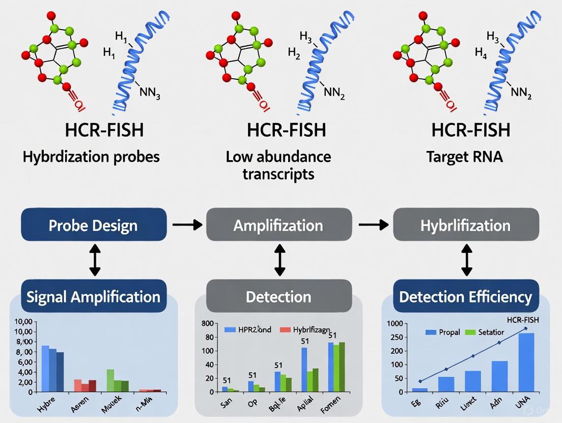
Hybridization Chain Reaction (HCR) is an enzyme-free, isothermal signal amplification method that utilizes DNA nanotechnology to detect specific nucleic acid sequences. Unlike polymerase-based amplification techniques, HCR operates through toehold-mediated strand displacement and autonomous self-assembly of metastable DNA hairpins, making it ideal for applications requiring robust, quantitative analysis without protein enzymes [1]. This technology has revolutionized molecular detection by providing high signal-to-background ratios even in challenging, autofluorescent samples like whole-mount vertebrate embryos and formalin-fixed paraffin-embedded (FFPE) tissue sections [2] [3].
The significance of HCR in modern biosensing and molecular imaging stems from its unique combination of features: straightforward multiplexing, quantitative capabilities, and high spatial resolution. These attributes make it particularly valuable for detecting low-abundance transcripts in their native anatomical context, addressing a critical need in genomics, clinical diagnostics, and drug development research [4] [5].
Core Mechanism and Molecular Principles
Fundamental HCR Mechanism
The fundamental HCR mechanism involves two kinetically trapped DNA hairpin species (H1 and H2) that coexist metastably until exposed to an initiator sequence (I1) complementary to the stem region of H1 [3]. The initiation process follows a precise molecular cascade:
- Initiator Binding: The DNA initiator (I1) hybridizes to the input domain of hairpin H1, opening the hairpin through strand displacement and exposing its output domain [3].
- Polymerization Cascade: The newly exposed output domain of H1 hybridizes to the input domain of hairpin H2, opening it to expose an output domain identical in sequence to the original initiator I1 [3].
- Self-Sustaining Assembly: This process repeats autonomously, forming long, nicked double-stranded DNA polymers through alternating H1 and H2 hybridization events [3] [1].
This enzyme-free chain reaction continues until all available hairpins are consumed, providing linear signal amplification that remains tethered to the initiating probe, thus preserving spatial information about the original target location [2].
Diagram 1: Fundamental HCR mechanism showing initiator-triggered self-assembly of DNA hairpins into amplification polymers.
Advanced HCR Systems
HCR v3.0 with Automatic Background Suppression
Third-generation HCR (v3.0) introduced split-initiator probes to achieve automatic background suppression throughout the protocol. This innovative approach replaces standard probes carrying full HCR initiators with cooperative probe pairs that each carry half of the initiator sequence [3] [5]. The background suppression mechanism operates through conditional initiation:
- Specific Target Binding: When both split-initiator probes hybridize adjacently to their target mRNA, they colocalize the two halves of the initiator sequence, enabling cooperative initiation of HCR amplification [3].
- Non-Specific Binding: Individual probes binding non-specifically within the sample cannot colocalize the two initiator halves, thus failing to trigger HCR and suppressing amplified background [3].
This automatic background suppression provides a typical 50-60 fold reduction in non-specific amplification compared to standard HCR probes, dramatically enhancing signal-to-background ratio without the need for extensive probe optimization [3].
Diagram 2: HCR v3.0 split-initiator probe system requiring adjacent binding for conditional initiation.
Integrated DNA Circuits
HCR can be integrated with other enzyme-free DNA circuits to create concatenated amplification systems with enhanced sensitivity. The CHA-HCR circuit combines catalytic hairpin assembly (CHA) with HCR in a two-stage amplification process [6] [1]:
- Target Recognition and CHA Amplification: A target molecule (e.g., miRNA) catalyzes the self-assembly of CHA hairpin substrates into double-stranded DNA products [6] [1].
- HCR Signal Amplification: The CHA reaction products contain connected segments of HCR triggers that autonomously cross-open HCR hairpins, forming tandem copolymeric double-stranded DNA nanowires for additional signal amplification [6].
This integrated approach enables highly sensitive detection of low-abundance biomarkers, making it particularly valuable for intracellular imaging of rare RNA targets in living cells [6].
Comparative Analysis of HCR Technologies
Table 1: Evolution of HCR Technologies and Their Key Characteristics
| HCR Version | Probe Design | Amplification Mechanism | Key Advantages | Limitations | Primary Applications |
|---|---|---|---|---|---|
| Basic HCR [1] | Standard DNA hairpins (H1, H2) | Initiator-triggered polymerization | Enzyme-free, isothermal, simple design | Background amplification from non-specific probe binding | Fluorescent detection, early biosensing |
| HCR v2.0 [3] | Probes with full initiator (I1) | Tethered fluorescent polymer formation | Straightforward multiplexing, quantitative imaging | Requires probe optimization to exclude "bad probes" | mRNA imaging in whole-mount embryos |
| HCR v3.0 [3] [5] | Split-initiator probe pairs | Conditional initiation with background suppression | Automatic background suppression, no probe optimization needed | Slightly reduced signal compared to full initiator probes | Multiplexed imaging in autofluorescent tissues, FFPE samples |
| Integrated CHA-HCR [6] [1] | CHA hairpins + HCR hairpins | Two-stage catalytic amplification | Ultra-sensitive detection, modular target recognition | More complex circuit design and optimization | Intracellular miRNA imaging, low-abundance biomarker detection |
Research Applications and Case Studies
HCR-FlowFISH for CRISPR Functional Genomics
HCR-FlowFISH represents a breakthrough application that combines CRISPRi-mediated perturbation of cis-regulatory elements (CREs) with HCR-amplified fluorescence in situ hybridization and flow cytometry [4]. This platform enables high-throughput functional characterization of non-coding genomic elements through accurate quantification of native transcripts.
In practice, HCR-FlowFISH has been applied to screen >325,000 perturbations, revealing that CREs can regulate multiple genes, skip over the nearest gene, and display both activating and silencing effects [4]. The methodology demonstrates particular strength for:
- Characterizing GWAS variants: At the cholesterol-associated FADS locus, HCR-FlowFISH enabled exhaustive characterization of multiple genome-wide association signals, functionally nominating causal variants and identifying their target genes [4].
- Quantifying CRE activity: When combined with CASA (CRISPR Activity Screen Analysis), a hierarchical Bayesian model, HCR-FlowFISH provides quantitative estimates of CRE effect sizes from flow cytometry data [4].
The technology reliably detects transcripts across an extensive expression range (1.2-2,734 TPM) with signal stability maintained for at least 21 days, enabling flexible experimental timing [4].
Subcellular Viral RNA Detection
HCR-based RNA FISH has proven invaluable for visualizing SARS-CoV-2 RNA distribution within infected cells and tissues [5]. This approach enables multiplexed detection of different viral RNA species with subcellular resolution, even in highly autofluorescent FFPE tissues.
Key applications in viral detection include:
- Genomic vs. subgenomic RNA discrimination: Using junction-specific split-initiator probes, researchers can distinguish viral genomic RNA from subgenomic mRNAs, enabling study of viral replication and transcription dynamics [5].
- Cell-type-specific tropism mapping: Multiplexing viral probes with cell-type-specific marker genes allows precise identification of infected cell types (e.g., alveolar type 2 cells vs. macrophages) in complex tissues [5].
- Subcellular localization analysis: Distinct subcellular patterns emerge for different viral RNA species, with ORF1a genomic RNA showing perinuclear localization consistent with replication/transcription complexes, while N region probes display more diffuse cytoplasmic staining [5].
Tumor Biomarker Detection
HCR-based strategies show significant promise for cancer diagnosis and monitoring through detection of tumor-associated biomarkers [1]. The enzyme-free nature of HCR enables development of robust, low-cost point-of-care testing (POCT) platforms for clinical applications.
Specific advancements in this field include:
- Nucleic acid biomarker detection: HCR circuits achieve ultrasensitive detection of trace nucleic acids, including microRNAs, circulating tumor DNA, and other low-abundance transcripts [1].
- Integration with multiple readout platforms: HCR amplification has been successfully coupled with fluorescent, colorimetric, electrochemical, and other signal transduction methods, enabling versatile assay development [1].
- Multiplexed biomarker profiling: The inherent multiplexing capability of HCR allows simultaneous detection of multiple cancer biomarkers, improving diagnostic accuracy and enabling molecular subtyping [1].
Experimental Protocols
HCR-FlowFISH for Transcript Quantification
The HCR-FlowFISH protocol enables robust transcript quantification across diverse cell types, including suspension and adherent cell lines [4]. The methodology involves the following key steps:
Cell Preparation and Fixation
- Culture cells under appropriate conditions (K562, Jurkat, GM12878, TF1, 293T, HepG2, SK-N-SH validated)
- Harvest and wash cells with PBS
- Fix with 4% paraformaldehyde for 30 minutes at room temperature
- Permeabilize with 70% ethanol overnight at 4°C or 0.5% Triton X-100 for 15 minutes
HCR Probe Hybridization
- Design DNA probes (20-50 probe pairs per target recommended for v3.0)
- Resuspend fixed cells in hybridization buffer containing probe sets (50 nM final concentration)
- Incubate at 37°C for 12-16 hours
Signal Amplification
- Prepare HCR hairpin working solution (60 nM in 5× SSCT)
- Wash cells twice with probe wash buffer
- Resuspend cells in HCR hairpin solution
- Incubate at room temperature for 4-6 hours (optimize duration based on target abundance)
- Wash twice with 5× SSCT before analysis
Flow Cytometry Analysis
- Resuspend cells in appropriate buffer for flow cytometry
- Analyze using standard flow cytometers (compatible with both air-cooled and water-cooled systems)
- Include negative controls (no probes) for background determination
- Use housekeeping genes (e.g., TBP) for normalization [4]
Critical Optimization Parameters:
- Probe concentration: Increasing from standard 50 nM to 100-200 nM can improve signal-to-noise ratio 5-fold for low-abundance targets [4]
- Amplification duration: Extending hairpin amplification time (4-8 hours) increases signal-to-noise ratio approximately 2-fold [4]
- Probe set size: For HCR v3.0, increasing from 5 to 20 split-initiator probe pairs improves signal-to-background ratio without increasing non-specific amplification [3]
HCR Immunohistochemistry (IHC) for Protein Detection
HCR signal amplification can be extended to protein detection through two complementary approaches [2]:
HCR 1°IHC (Direct Primary Antibody Labeling)
- Primary antibody probes are directly labeled with one or more HCR initiators
- Suitable for multiplexing with primary antibodies from the same host species
- Requires validation of each initiator-labeled primary antibody
HCR 2°IHC (Secondary Antibody Detection)
- Unlabeled primary antibodies detected by initiator-labeled secondary antibody probes
- Enables immediate use of large libraries of commercial primary antibodies
- Requires primary antibodies from different host species for multiplexing
Table 2: Performance Characteristics of HCR Detection Methods
| Parameter | HCR RNA FISH | HCR 1°IHC | HCR 2°IHC | Traditional CARD |
|---|---|---|---|---|
| Signal-to-Background Ratio | 15-609 (median 90) [2] | Similar to RNA FISH [2] | Similar to RNA FISH [2] | Variable, typically lower |
| Multiplexing Capacity | 5+ targets simultaneously [3] | Limited by available initiator-labeled primaries | Limited by host species diversity | 1 target per serial amplification |
| Quantitative Performance | Linear with transcript abundance [2] | Linear with antigen abundance [2] | Linear with antigen abundance [2] | Non-linear, qualitative |
| Spatial Resolution | Subcellular, tethered amplification [2] | Subcellular, tethered amplification [2] | Subcellular, tethered amplification [2] | Often compromised by diffusion |
| Assay Timeline | Independent of target number [2] | Independent of target number [2] | Independent of target number [2] | Increases with each target |
HCR v3.0 for Low-Abundance Transcripts in Autofluorescent Tissues
Detection of low-abundance transcripts in autofluorescent samples (whole-mount embryos, FFPE tissues) requires HCR v3.0 with automatic background suppression [3] [5]:
Split-Initiator Probe Design
- Design 20-50 probe pairs tiling target transcript
- Each probe: 25-nt target complementarity + half initiator sequence
- Avoid overlapping probe pairs to ensure even tiling
Sample Preparation and Pre-hybridization
- Fix tissues in 4% PFA overnight at 4°C
- Dehydrate through methanol series (25%, 50%, 75%, 100%)
- Rehydrate through methanol/PBST series
- Permeabilize with proteinase K (5-20 μg/mL, concentration optimization required)
- Post-fix with 4% PFA, then refix in glutaraldehyde/paraformaldehyde
Hybridization and Amplification
- Pre-hybridize in hybridization buffer for 1-4 hours at 37°C
- Hybridize with probe sets (2 nM each probe) in hybridization buffer at 37°C for 12-36 hours
- Wash with probe wash buffer (4× over 30 minutes at 37°C)
- Amplify with HCR hairpins (30-60 nM) in 5× SSCT at room temperature for 4-8 hours
- Wash with 5× SSCT (4× over 30 minutes at room temperature)
Imaging and Analysis
- Mount and image using confocal or epifluorescence microscopy
- For quantitative analysis (qHCR imaging), maintain identical imaging parameters across samples
- Use negative controls (no probes) to determine background threshold
The Scientist's Toolkit: Essential Research Reagents
Table 3: Key Research Reagent Solutions for HCR Experiments
| Reagent Category | Specific Examples | Function and Importance | Optimization Tips |
|---|---|---|---|
| HCR Hairpins | H1 and H2 metastable DNA hairpins | Core amplification components; fluorophore-labeled for detection | Aliquot and store at -20°C; avoid repeated freeze-thaw cycles |
| Detection Probes | Split-initiator probe pairs (v3.0) | Target recognition and conditional initiation | Design 20+ probe pairs per target for optimal signal-to-background |
| Hybridization Buffers | 4-6× SSC, 50% formamide, 0.1% Tween-20 | Maintain probe specificity during hybridization | Include yeast tRNA and heparin to reduce non-specific binding |
| Signal Amplification Buffers | 5× SSCT (SSC + 0.1% Tween-20) | Optimal environment for HCR polymerization | Adjust Mg²⁺ concentration (2-10 mM) to balance kinetics and specificity |
| Cell Permeabilization Agents | Triton X-100, Tween-20, methanol | Enable probe access to intracellular targets | Titrate concentration (0.1-1.0%) to balance signal and morphology |
| Fluorophore Systems | Alexa488, Alexa546, Alexa647, Cy3, Cy5 | Signal detection and multiplexing | Match fluorophore to microscope capabilities and sample autofluorescence |
| Mounting Media | ProLong Diamond, Vectashield | Preserve signal and enable high-resolution imaging | Choose anti-fade properties matching planned imaging duration |
Technical Specifications and Performance Metrics
Quantitative Performance Characteristics
HCR technology demonstrates robust performance across multiple application scenarios:
- Sensitivity Range: Successfully detects transcripts from 1.2 to 2,734 TPM, covering most biologically relevant expression levels [4]
- Detection Limit: Capable of detecting single RNA molecules with digital HCR (dHCR) imaging approaches [3]
- Signal-to-Background Ratio: Typically achieves ratios of 15-609 with a median of 90 across diverse protein and RNA targets [2]
- Amplification Kinetics: Linear signal accumulation over 4-8 hours, with approximately 2-fold signal-to-noise improvement with extended amplification [4]
- Multiplexing Capacity: Demonstrated 5-plex RNA imaging with simultaneous one-step amplification [3]
Comparison with Alternative Technologies
Table 4: HCR Versus Other Amplification Technologies
| Amplification Method | Enzyme Requirement | Turnaround Time | Detection Sensitivity | Multiplexing Capability | Spatial Resolution | Cost Considerations |
|---|---|---|---|---|---|---|
| HCR [1] | Enzyme-free | 3-8 hours | Single-molecule to high abundance | High (5+ targets) | Excellent (tethered) | Moderate (synthesis) |
| PCR/qPCR [1] | Thermostable polymerase | 1-3 hours | High (exponential amplification) | Moderate (4-5 plex) | None (homogeneous) | Low to moderate |
| Branch Chain Reaction (BCR) [7] | Enzyme-free | 2-4 hours | Moderate to high | Limited | Good | Moderate |
| Catalytic Hairpin Assembly (CHA) [1] | Enzyme-free | 1-3 hours | High | Moderate | Good | Moderate |
| PrimeFlow RNA Assay [4] | Enzyme-free (proprietary) | 6-8 hours | Limited for low abundance | Moderate | Good | High (proprietary) |
| Single-Cell RNA-seq [4] | Reverse transcriptase, polymerase | Days | High | Genome-wide | Limited (cell resolution) | Very high |
Troubleshooting and Optimization Guide
Common Experimental Challenges
- High Background Signal: For HCR v2.0, optimize probe sets by removing "bad probes" or switch to HCR v3.0 with split-initiator probes for automatic background suppression [3]
- Low Signal Intensity: Increase probe concentration (50-200 nM), extend amplification time (4-8 hours), or increase number of probe pairs per target (20+ recommended) [4] [3]
- Poor Cell Viability After Processing: Reduce permeabilization time or concentration; use milder detergents (0.1% Tween-20 instead of 0.5% Triton X-100)
- Inconsistent Results Between Replicates: Standardize hairpin folding protocols; ensure consistent temperature during amplification; aliquot reagents to avoid freeze-thaw cycles
Validation and Quality Control
- Probe Validation: Test new probe sets on positive control samples with known expression; include no-probe controls for background determination
- Signal Specificity: Verify with target-specific knockdown or knockout controls when possible; use multiple independent probe sets for the same target
- Quantitative Accuracy: Include housekeeping genes for normalization; use standardized imaging or cytometry settings across experiments
- Reagent Quality: Ensure oligonucleotide purity >85% by HPLC purification; verify hairpin folding by gel electrophoresis or lack of signal in negative controls [7]
Why Low Abundance Transcripts Pose a Unique Detection Challenge
The precise spatial localization of RNA molecules within tissues and cells is fundamental to advancing our understanding of cellular identity, function, and organization in both health and disease. Low abundance transcripts, often defined as those present at fewer than 10-20 copies per cell, represent a significant portion of the transcriptome, including key regulators such as transcription factors, signaling molecules, and non-coding RNAs. The detection of these rare molecules pushes the limits of conventional fluorescence in situ hybridization (FISH) techniques, which are often plagued by insufficient signal amplification and high background noise. This limitation creates a critical blind spot in spatial biology, obscuring a functionally important subset of the transcriptome from view.
The Hybridization Chain Reaction FISH (HCR-FISH) protocol, particularly in its third iteration (v3.0), has emerged as a powerful tool to address this challenge due to its high specificity and background suppression. However, despite its advantages, detecting low-copy-number RNAs remains technically demanding. This application note details the specific obstacles presented by low abundance transcripts and provides a optimized, detailed HCR-FISH framework to overcome them, enabling robust detection for research and drug development applications.
The Core Obstacles in Detecting Low Abundance Transcripts
The difficulty in visualizing low abundance transcripts stems from a combination of physical, technical, and optical limitations that collectively suppress the signal below a reliable detection threshold.
Physical and Technical Limitations
- Limited Probe Binding Sites: The signal amplitude in FISH is directly proportional to the number of fluorophores that can be localized to a single RNA molecule. Low abundance transcripts offer fewer target sequences for probe hybridization, inherently limiting the maximum achievable signal. This is exacerbated for short RNA targets, such as microRNAs or specific splice variants, which cannot accommodate the numerous probes required for strong amplification using traditional methods [8].
- Signal-to-Noise Ratio (SNR) Constraints: A fundamental challenge is achieving a sufficient SNR. The weak signal from a few RNA molecules is often drowned out by background autofluorescence from cellular components like lipofuscin and flavins, as well as non-specific probe binding. While HCR v3.0 improves SNR through split-initiator probes that reduce non-triggered amplification, background noise remains a significant barrier when the primary signal is exceptionally faint [4] [9].
- Sensitivity Ceilings of Standard Methods: Even sensitive, amplification-based methods like standard HCR v3.0 have practical detection limits. For example, in one study, HCR-FlowFISH demonstrated robust detection across a range of transcript levels, but its performance gradient reveals that sensitivity is not uniform, with lower-abundance targets producing a dimmer, less distinct signal [4]. This is quantified in Table 1, which compares the performance of various FISH methods.
Table 1: Performance Comparison of FISH Methods for Transcript Detection
| Method | Detection Limit (Approx. Transcripts/Cell) | Key Advantage | Major Limitation for Low Abundance Targets |
|---|---|---|---|
| smFISH | Moderate | Single-molecule resolution | Requires ~48 probes; ineffective for short transcripts [8] |
| HCR v3.0 | Moderate | High specificity, low background | Limited signal amplification for very low copy numbers [9] |
| Branched DNA (bDNA) | Low-High | Enzyme-free, good signal | Probe design complexity [8] |
| HCR-FlowFISH | Moderate | Scalable, quantitative | Relies on transcript level and probe efficiency [4] |
| π-FISH rainbow | Very Low | High signal intensity, robust | Newer method, requires validation [8] |
| Next-Gen HCR (HCR-Pro/Cat) | Very Low | Enzyme-enhanced amplification | More complex protocol [9] |
Methodological and Workflow Challenges
The entire experimental pipeline, from sample preparation to imaging, introduces variables that can disproportionately affect the detection of low abundance targets.
- Sample Preservation and Permeabilization: The integrity of the target RNA and the efficiency of probe access are paramount. Suboptimal fixation can lead to RNA degradation, directly reducing the already low signal. Inadequate permeabilization, especially in thick or complex tissues like whole-mount plant samples or cleared brain tissue, prevents probes and hairpins from reaching their targets, resulting in false negatives [10] [11].
- Probe Design and Hybridization Efficiency: The sensitivity of HCR is contingent on the initial binding of the split-initiator probes. For a low-abundance target, every binding event is critical. Inefficient hybridization due to suboptimal probe sequence, melting temperature, or secondary RNA structure can drastically reduce the initiation of the amplification cascade [12].
- Amplification Inefficiency: The HCR amplification process relies on the metastable hairpins assembling into a fluorescent polymer. Any deviation from ideal conditions—such as incorrect temperature, insufficient hairpin concentration, or overly short amplification times—can result in a shorter polymer and a dimmer signal, which is fatal for detecting rare transcripts [13].
Optimized HCR-FISH Protocol for Low Abundance Transcripts
The following protocol is a consolidated and optimized workflow based on established HCR v3.0 principles, incorporating specific enhancements to maximize sensitivity for low abundance targets.
The diagram below outlines the core steps and decision points in the optimized HCR-FISH workflow.
Detailed Stepwise Procedure
Step 1: Sample Preparation and Fixation
- Tissue Processing: For animal tissues, immediately embed and snap-freeze in Optimal Cutting Temperature (OCT) compound or immerse in 4% paraformaldehyde (PFA) for 12-36 hours at 4°C. For plant tissues, use vacuum infiltration with 4% PFA for consistent fixation [10] [11].
- Sectioning: Cryosection tissues at a thickness of 10-20 µm and mount on functionalized coverslips (e.g., silanized) to ensure tissue adhesion throughout multiple washes [12].
- Permeabilization: Treat sections with a combination of detergent (e.g., 0.5% Triton X-100 in PBS) and, for plant or tough tissues, a cell wall digestion enzyme mix (e.g., 0.1% pectolyase/cellulase) for 30-60 minutes at 37°C. This is critical for probe access [11].
Step 2: Probe Design and Hybridization
- Probe Set Design: Use a custom bioinformatic tool (e.g., like the Probegenerator web app) to design ~25-36 split-initiator probe pairs against the coding sequence (CDS) and 3'UTR of the target mRNA to create a "boosted" probe set. This maximizes the number of initiators per transcript [4] [12].
- Probe Hybridization:
- Prepare a high-concentration probe solution (20 nM in HCR Probe Hybridization Buffer) rather than the standard 4-5 nM [13].
- Apply the probe solution to the sample and incubate in a dark, humidified chamber overnight (12-16 hours) at 37°C to maximize binding kinetics.
Step 3: Stringency Washes
- Wash the sample 3-4 times with pre-warmed HCR Wash Buffer (or 5× SSCT) for 15 minutes each at 37°C. This step is crucial for removing unbound probes and reducing background.
Step 4: HCR Amplification
- Hairpin Preparation: Aliquot fluorescent HCR hairpins (H1 and H2) for the required initiator (B1-B5). Heat the hairpins to 95°C for 90 seconds in a thermocycler, then allow them to cool in the dark for 30 minutes to form metastable structures. Combine the snap-cooled hairpins in HCR Amplification Buffer to a final concentration of 60 nM each [12].
- Amplification Incubation: Apply the hairpin solution to the sample and incubate in the dark overnight (12-16 hours) at room temperature. The extended incubation allows the amplification polymer to grow longer, significantly enhancing the signal for low-copy targets [4] [13].
Step 5: Imaging and Analysis
- After amplification, perform a brief wash with amplification buffer or 5× SSCT to remove unbound hairpins.
- Mount the sample in an anti-fading mounting medium.
- Image using a confocal microscope or a widefield microscope with a high-quantum-efficiency camera. Acquire z-stacks to capture the full 3D distribution of signals. For quantification, ensure exposure times and laser powers are consistent across compared samples.
The Scientist's Toolkit: Essential Reagents
Table 2: Key Research Reagent Solutions for Sensitive HCR-FISH
| Reagent / Material | Function / Explanation | Optimization Tip for Low Abundance Targets |
|---|---|---|
| Split-Initiator Probe Pools | DNA oligonucleotides that bind target mRNA and trigger HCR amplification. | Use "boosted" designs with 25-36 probe pairs per mRNA to increase initiator density [13] [12]. |
| HCR Hairpins (H1 & H2) | Metastable DNA hairpins that self-assemble into a fluorescent polymer. | Use at 60 nM final concentration with overnight amplification for maximal polymer growth [13] [12]. |
| Hybridization & Amplification Buffers | Proprietary solutions controlling hybridization stringency and hairpin kinetics. | Use buffers from established sources (e.g., Molecular Instruments) for guaranteed performance [12]. |
| Permeabilization Agents | Detergents and enzymes that enable probe access to tissue interior. | For tough tissues, combine detergent (Triton X-100) with enzymatic treatment (cellulase/pectolyase) [11]. |
| Next-Gen HCR Kits (HCR-Pro) | Commercial kits combining HCR specificity with enzyme-based amplification. | Employ when standard HCR v3.0 fails, as it offers superior sensitivity for the most challenging targets [9] [13]. |
Advanced Strategies and Alternative Methods
When the optimized standard protocol is insufficient, researchers can turn to more advanced HCR strategies or alternative methods.
Next-Generation HCR and Combinatorial Approaches
- Enzyme-Enhanced HCR: Newer iterations, such as HCR-Cat (catalysis) and HCR-Immuno, integrate enzymatic amplification steps with the HCR framework. These methods combine the specificity of HCR v3.0 with the powerful signal generation of enzymes, providing a dramatic sensitivity boost necessary for very short or low-abundance targets [9].
- Combination with Immunohistochemistry (IHC): HCR-FISH can be combined with protein immunofluorescence to correlate the expression of a rare transcript with its protein product or specific cell markers. An improved protocol allows for simultaneous detection of RNA and protein in the same plant sample, a approach that can be adapted to animal tissues [11].
- HCR-FlowFISH for Quantitative Screening: For cell suspension-based studies, HCR-FlowFISH combines CRISPRi perturbation screens with HCR-based transcript detection and flow cytometry. This allows for the functional characterization of cis-regulatory elements (CREs) that regulate low-abundance genes in a high-throughput manner [4].
Comparison with Other Sensitive FISH Technologies
- π-FISH Rainbow: This method uses π-shaped target probes and multiple rounds of U-shaped amplification probes to generate very high signal intensity. It has been shown to have higher sensitivity compared to HCR and smFISH for both medium- and low-abundance transcripts, making it a powerful alternative [8].
- TDDN-FISH (Tetrahedral DNA Dendritic Nanostructure–Enhanced FISH): This rapid, enzyme-free method uses self-assembling DNA nanostructures for exponential signal amplification. It is reported to be ~8x faster per round than HCR-FISH and generates stronger signals, enabling the detection of short RNAs like miRNAs with very few primary probes [14].
The detection of low abundance transcripts via HCR-FISH remains a formidable challenge, primarily due to fundamental limitations in signal-to-noise ratio and the inefficiencies inherent in any multi-step biochemical process. However, through a deliberate and optimized protocol—emphasizing maximized probe binding, extended amplification times, and rigorous noise suppression—researchers can reliably push the boundaries of sensitivity. The ongoing development of even more sensitive next-generation HCR methods and powerful alternatives like π-FISH and TDDN-FISH promises to further illuminate the dark corners of the transcriptome, ultimately providing a more complete picture of gene expression in health and disease for the scientific and drug development communities.
Advantages of HCR over Traditional FISH for Sensitive Detection
Fluorescence in situ hybridization (FISH) has been a cornerstone technique for spatial genomics and transcriptomics, enabling researchers to visualize nucleic acid distribution within their native cellular and tissue contexts. However, traditional FISH methods often face significant limitations in sensitivity, especially for detecting low-abundance transcripts. The emergence of Hybridization Chain Reaction (HCR)-FISH represents a paradigm shift in signal amplification technology, addressing fundamental shortcomings of conventional FISH through an enzyme-free, isothermal amplification process. This application note details the technical advantages of HCR over traditional FISH methodologies, providing structured performance comparisons and detailed protocols to guide researchers in implementing this powerful technique for sensitive detection of low-expression targets.
Technical Advantages of HCR-FISH
Fundamental Mechanisms and Performance Benefits
HCR-FISH operates through a mechanism of triggered self-assembly, where initiator probes bound to target transcripts nucleate the formation of fluorescent amplification polymers from metastable DNA hairpins [2]. This core mechanism confers several distinct advantages over traditional FISH and other amplification methods:
Exceptional Signal-to-Background Ratio: The requirement for conjoint hybridization of adjacent 'half-probes' to achieve amplification provides greater specificity, significantly reducing background noise and off-target hybridization [15]. This characteristic is particularly valuable when working with autofluorescent samples or when detecting rare transcripts where signal distinction is challenging.
Preserved Spatial Resolution: Unlike enzyme-based amplification methods where diffusable reaction products can cause signal diffusion, HCR amplification polymers remain tethered to their initiating probes, maintaining subcellular or even single-molecule resolution [2]. This fidelity of signal localization is crucial for accurate interpretation of spatial expression patterns.
Linear Signal Quantification: HCR amplified signal scales approximately linearly with the number of target molecules, enabling accurate and precise RNA relative quantitation with subcellular resolution in anatomical context [2]. This quantitative capability represents a significant advancement over traditional FISH methods, which often provide only qualitative data.
Comparative Performance Data
Table 1: Quantitative comparison of HCR-FISH performance against traditional FISH methods
| Performance Metric | Traditional FISH | HCR-FISH | Experimental Context |
|---|---|---|---|
| Signal-to-Noise Ratio | Baseline | 2-5 fold improvement [4] | Detection of low-abundance transcripts (1.5-193 TPM) |
| Multiplexing Capacity | Limited by spectral overlap | 5-plex in single round [12] | Using orthogonal initiator/amplifier sets |
| Sample Compatibility | Model organisms | Robust across diverse species [12] [11] | Arabidopsis, maize, sorghum, Drosophila, axolotl |
| Processing Time | ~8 hours for amplification | ~1 hour post-hybridization [16] | Compared to HCR-variant methods |
| Probe Requirements | 48 probes for smFISH [16] | Minimal probes per target [16] | For effective target detection |
Table 2: HCR-FISH performance across different sample types and applications
| Sample Type | Application | Performance Outcome | Reference |
|---|---|---|---|
| Marine Sediments | Environmental microbe detection | Successful visualization despite background challenges | [17] |
| Plant Tissues | Whole-mount spatial transcriptomics | 3D spatial patterning with low background | [11] |
| Drosophila Brain | Neuronal activation mapping | Detection of low-abundance immediate early genes | [15] |
| FFPE Tissue Sections | Multiplexed protein/RNA co-detection | High signal-to-background (median: 90) | [2] |
| CRISPR Screens | Endogenous CRE characterization | Reliable detection across expression range (1.2-2734 TPM) | [4] |
Detailed HCR-FISH Protocol for Low-Abundance Transcripts
Probe Design and Preparation
Effective HCR-FISH begins with strategic probe design, particularly critical for low-abundance targets where sensitivity requirements are highest:
Split-Initiator Probes: Utilize the v3.HCR system with split-initiator DNA oligonucleotide pairs. Each probe pair (25 nucleotides) targets adjacent mRNA sequences, with each containing half of an HCR initiator sequence. Only when both probes hybridize correctly do they form a complete initiator, ensuring high specificity [12].
Probe Set Design: For each target, design 20-36 probe pairs targeting coding sequences first, followed by 3'UTR regions. Use bioinformatic tools like Probegenerator (available at probegenerator.herokuapp.com) to screen for off-target binding and ensure specificity [12].
Probe Concentration Optimization: For challenging samples, increase initiator probe concentration to 10 μmol/L in hybridization buffer to enhance signal intensity without significantly increasing background [17].
Sample Preparation and Hybridization
Fixation and Permeabilization: Fix samples with 4% paraformaldehyde for 30-60 minutes at room temperature. For whole-mount plant tissues, permeabilize with cell wall enzyme digestion (2% cellulase, 1% pectolyase) for 30-60 minutes [11]. For Drosophila brains, use proteinase K treatment (10 μg/mL, 15 minutes) to enhance probe penetration [15].
Hybridization Conditions: Hybridize with probe solution (5 nM in hybridization buffer) overnight at 37°C. For low-abundance targets, extend hybridization time to 16-24 hours and increase probe concentration to 10 nM [4].
Signal Amplification: Prepare fluorescently labeled HCR hairpins (60 nM in amplification buffer) by heat denaturation at 95°C for 90 seconds followed by 30-minute cooling in darkness. Incubate samples with hairpin solution for 4-8 hours at room temperature. For very low-abundance targets, extend amplification to 12-16 hours [4].
Optimization for Specific Applications
Environmental Samples (e.g., sediments): Incorporate additional washing steps with 1×SSCT + 0.1% SDS to reduce non-specific binding to abiotic particles [17].
Whole-Mount Tissues: Implement sample clearing with 4% SDS, 200 mM boric acid (pH 8.5) for 24-48 hours to enhance light penetration for imaging [12].
Multiplexed Detection: Use orthogonal HCR initiator/amplifier sets (B1-B5) with spectrally distinct fluorophores. For 3-plex detection in plants, successful combinations include B1-Alexa647, B2-Alexa594, and B3-Alexa488 [11].
Research Reagent Solutions
Table 3: Essential reagents and resources for HCR-FISH implementation
| Reagent/Resource | Function/Purpose | Source/Example |
|---|---|---|
| Split-Initiator Probes | Target-specific hybridization with high specificity | Custom-designed oligo pools (IDT oPools) |
| HCR Hairpin Amplifiers | Signal amplification via chain reaction | Molecular Instruments (B1-B5 with various fluorophores) |
| Hybridization Buffer | Optimal conditions for probe-target binding | Molecular Instruments or custom formulations |
| Probe Design Tools | Bioinformatics for specific probe design | Probegenerator web application |
| Image Analysis Software | Quantitative analysis of FISH signals | Open-source solutions (FIJI, CellProfiler) |
Workflow and Mechanism Visualization
Diagram 1: Complete HCR-FISH workflow from sample preparation to analysis
Diagram 2: Molecular mechanism of HCR signal amplification
Applications in Sensitive Detection Scenarios
HCR-FISH has demonstrated exceptional capability across diverse challenging applications, proving particularly valuable where traditional FISH methods fail:
Immediate Early Gene Detection: In Drosophila brains, HCR-FISH enables whole-brain mapping of neuronal activation through detection of Hr38 mRNA, a low-abundance immediate early gene transcript. The technique's sensitivity allows identification of functionally distinct neuronal populations activated during specific social behaviors with single-cell resolution [15].
Environmental Microbiology: For microbial communities in marine sediments where target organisms often have low metabolic activity and consequently limited rRNA content, optimized HCR-FISH successfully visualizes these challenging targets while minimizing false positives from abiotic particle adsorption [17].
Whole-Mount Plant Transcriptomics: In intact plant tissues, HCR-FISH enables 3D spatial transcriptomics without sectioning, detecting expression patterns even in deeply embedded tissue layers. The method maintains signal specificity despite the challenges of probe penetration through plant cell walls [11].
CRISPR Screening: HCR-FlowFISH combines CRISPR perturbation with HCR-based readouts, enabling sensitive detection of transcriptome changes in response to non-coding element modifications. This approach provides superior signal-to-noise compared to proprietary alternatives like PrimeFlow, especially for lowly expressed genes [4].
HCR-FISH represents a significant advancement over traditional FISH methodologies, particularly for sensitive detection of low-abundance transcripts. Its enzyme-free, isothermal amplification mechanism provides enhanced specificity, superior signal-to-background ratios, and preserved spatial resolution while maintaining quantitative capabilities. The technique's robustness across diverse sample types—from environmental specimens to complex whole-mount tissues—underscores its versatility and reliability. As spatial transcriptomics continues to drive discoveries in developmental biology, neuroscience, and disease mechanisms, HCR-FISH stands as an essential tool for researchers demanding high-sensitivity detection within native anatomical contexts.
The detection of low abundance transcripts is a significant challenge in spatial biology, necessitating signal amplification methods that are both highly sensitive and specific. In situ hybridization based on the mechanism of the hybridization chain reaction (HCR) provides a unified framework for multiplexed, quantitative, high-resolution RNA imaging, even in challenging samples such as whole-mount vertebrate embryos and thick tissue sections [3] [18]. Unlike enzyme-based amplification methods, HCR utilizes programmable DNA nanotechnologies for isothermal, enzyme-free signal amplification, preserving quantitative information and subcellular spatial resolution [18]. This application note details the key molecular components—initiator probes and DNA hairpin amplifiers—that form the basis of the HCR platform, with a specific focus on their application for researching low abundance transcripts.
The fundamental HCR mechanism involves two kinetically trapped DNA hairpin monomers (H1 and H2) that coexist metastably until exposed to a DNA initiator sequence (I1) [3] [19]. The initiator triggers a conditional chain reaction wherein H1 and H2 self-assemble into long, tethered amplification polymers. In the context of RNA fluorescence in situ hybridization (RNA-FISH), this initiator is appended to DNA probes that are complementary to a target mRNA, thereby tethering the amplified fluorescent signal to the site of the RNA molecule [3]. The latest iteration of this technology, known as third-generation in situ HCR (v3.0), incorporates probe and amplifier designs that provide automatic background suppression, a critical advancement for detecting low-expression genes with high confidence [3] [12].
Core Molecular Components
Initiator Probes: Standard vs. Split-Probe Designs
Initiator probes are the target-recognition elements of the HCR-FISH system, responsible for conferring specificity and triggering the amplification cascade.
Standard Probes (HCR v2.0): In the previous generation, each DNA probe was designed to hybridize to a target mRNA and contained a full HCR initiator sequence (I1). A critical limitation was that any probe binding non-specifically within the sample would still display the full initiator, triggering HCR and generating amplified background. This often necessitated laborious probe set optimization to identify and remove "bad probes" [3].
Split-Initiator Probes (HCR v3.0): To overcome this limitation, v3.0 employs a pair of cooperative split-initiator probes for each binding site. Each probe in the pair carries only half of the HCR initiator I1 and a shorter target-binding sequence (typically 25 nucleotides) [3] [12]. Signal amplification is triggered only when both probes bind adjacently to their target mRNA, successfully colocalizing the two halves of the initiator. If a single probe binds non-specifically, it cannot trigger the chain reaction, thus providing automatic background suppression [3]. This innovation dramatically enhances the signal-to-background ratio and allows researchers to use large, unoptimized probe sets for new targets with high robustness.
Table 1: Comparison of HCR Initiator Probe Generations
| Feature | Standard Probes (v2.0) | Split-Initiator Probes (v3.0) |
|---|---|---|
| Initiator Structure | Full initiator (I1) on a single probe [3] | Half-initiator on each of two cooperative probes [3] |
| Probe Binding Site Length | ~50 nucleotides [3] | ~25 nucleotides per probe [3] |
| Amplification Trigger | Hybridization of a single probe [3] | Co-hybridization of two adjacent probes [3] |
| Background Suppression | Low; non-specific binding leads to amplified background [3] | High (Automatic Background Suppression); non-specific binding does not trigger HCR [3] |
| Probe Set Optimization | Often required to remove non-specific probes [3] | Typically not required; enables use of large, unoptimized sets [3] |
| Typical HCR Suppression Factor | Not Applicable | ≈50-60 fold (in situ and in vitro) [3] |
DNA Hairpin Amplifiers
DNA hairpin amplifiers are the signal-generating components that undergo controlled self-assembly to produce a detectable signal.
Structure and Mechanism: Each HCR amplifier system consists of two species of DNA hairpins (H1 and H2) that are kinetically trapped in a meta-stable state [3] [19]. The hairpins are stored separately and are stable for lab time scales. Upon introduction of the initiator I1, it hybridizes to the input domain of H1, opening the hairpin to expose an output domain. This output domain is complementary to the input domain of H2, hybridizing to it and, in turn, exposing an output domain on H2 that is identical in sequence to the original initiator I1. This creates a chain reaction of alternating H1 and H2 polymerization steps, forming a long, nicked double-stranded DNA polymer [3].
Orthogonality and Multiplexing: A key strength of the HCR platform is its programmability. Multiple orthogonal HCR amplifiers (e.g., B1, B2, B3, B4, B5) have been engineered to operate simultaneously and independently within the same sample without cross-talk [12] [19]. Each amplifier system is triggered by a unique initiator sequence. This allows researchers to assign a different amplifier to each RNA target in a multiplexed experiment, with each amplifier labeled with a spectrally distinct fluorophore [19]. The orthogonality ensures that the experimental timeline for a multiplex experiment is independent of the number of targets [18].
Fluorophore Labeling: The DNA hairpins are conjugated with fluorophores (e.g., Alexa Fluor 488, 594, 647). During polymer assembly, these fluorophores are brought into close proximity, creating a localized, bright signal that can be imaged with standard fluorescence microscopy [19]. Recommendations for fluorophore selection include using lower-wavelength fluorophores (e.g., 488) for higher-expression targets and higher-wavelength fluorophores (e.g., 546, 647) for lower-expression targets, as autofluorescence is often more pronounced at lower wavelengths [19].
Table 2: Characteristics of DNA Hairpin Amplifiers
| Characteristic | Description | Experimental Implication |
|---|---|---|
| Reaction Conditions | Isothermal, enzyme-free [3] | Simple protocol; preserves sample morphology and RNA integrity. |
| Amplification Polymer | Tethered, nicked double-stranded polymer [3] | Prevents signal diffusion; enables subcellular and single-molecule resolution [18]. |
| Signal Linearity | Amplified signal scales ~linearly with target count [18] | Enables accurate relative quantitation (qHCR) and digital absolute quantitation (dHCR) [3] [18]. |
| Orthogonal Systems | Multiple, e.g., B1, B2, B3, B4, B5 [12] [19] | Enables straightforward multiplexing (5-plex with bandpass imaging; 10-plex with spectral imaging) [18]. |
| Kinetic State | Kinetically trapped; minimal leakage [3] | Low background from non-triggered hairpins; stable reagent storage. |
HCR v3.0 Mechanism with Split-Initiator Probes
The Scientist's Toolkit: Research Reagent Solutions
For researchers embarking on HCR-FISH, particularly for low abundance transcripts, the following core reagents and tools are essential.
Table 3: Essential Research Reagents and Materials for HCR-FISH
| Item | Function/Description | Source/Example |
|---|---|---|
| Split-Initiator Probe Pools | Pools of ~36 probe pairs targeting a single mRNA; each pair colocalizes halves of an HCR initiator for specific amplification [12]. | Custom-designed oPools (IDT); designed via web apps like Probegenerator [12]. |
| Orthogonal HCR Amplifiers | Fluorophore-labeled H1 and H2 hairpins for different initiators (B1, B2, etc.); kinetically trapped until triggered [3] [19]. | Molecular Instruments (e.g., B1-A647, B2-A594, B3-A488) [12] [19]. |
| Hybridization & Wash Buffers | Aqueous buffers containing salts, buffers, and detergents to control stringency during probe hybridization and washing [12]. | Molecular Instruments or custom-made recipes [12]. |
| Amplification Buffer | Aqueous buffer for the HCR self-assembly step; enables isothermal, enzyme-free polymerization of hairpins [12]. | Molecular Instruments [12]. |
| Nuclease-Free Water | Used for preparing solutions; ensures integrity of DNA probes and amplifiers. | DEPC-treated water or commercial nuclease-free water [12]. |
| Probe Design Software | Computational tool for designing specific split-initiator probe pairs against a target transcriptome. | Probegenerator web application (utilizes Oligominer and Bowtie2) [12]. |
Experimental Protocol for HCR-FISH (v3.0) on Tissue Sections
The following protocol is adapted for multiplexed detection of RNA in tissue sections, such as axolotl limb sections, with notes for optimizing for low abundance targets [12].
Sample Preparation and Fixation
- Tissue Sectioning: For cryosections, embed tissue in OCT compound and section at a thickness of 10-20 µm using a cryostat. Mount sections on functionalized coverslips or glass slides.
- Coverslip Functionalization (Optional but Recommended): For multiround staining or challenging samples, functionalize coverslips to enhance tissue adhesion. Submerge coverslips in 1 M KOH and sonicate for 20 min. Wash with DEPC-treated water and then 100% methanol. Incubate in a solution of glacial acetic acid, methanol, and 3-aminopropyltriethoxysilane [12].
- Fixation: Fix samples with an appropriate fixative (e.g., 4% paraformaldehyde in PBS) to preserve RNA and tissue architecture.
Hybridization with Split-Initiator Probes
- Probe Solution Preparation: Resuspend the lyophilized oPool probe set in TE buffer to create a 1 µM stock. Dilute this stock 1:200 in the commercial hybridization buffer to make a 5 nM working probe solution. For low abundance transcripts, this concentration can be increased (e.g., to 10 nM), and the probe set size should be maximized (e.g., 20-36 probe pairs) to deposit more initiators per transcript [3] [12].
- Hybridization: Apply the probe solution to the sample, ensuring full coverage. Incubate in a humidified hybridization chamber overnight (12-16 hours) at 37°C. This extended incubation ensures thorough penetration and hybridization.
Post-Hybridization Washes
- Remove the probe solution and perform a series of stringent washes to remove unbound and non-specifically bound probes.
- Wash the sample 4 times for 15 minutes each with pre-warmed wash buffer at 37°C. This step is critical for maintaining a low background.
HCR Signal Amplification
- Hairpin Preparation:
- Pipet the required fluorophore-labeled H1 and H2 hairpins (3 µM stock) into separate 0.2 mL PCR tubes. Note: Use orthogonal hairpins (e.g., B1, B2) with distinct fluorophores for different targets.
- "Snap-cool" the hairpins by heating to 95°C for 90 seconds in a thermocycler, then removing them to cool at room temperature in the dark for 30 minutes. This step ensures the hairpins are properly folded into their meta-stable state [12].
- Combine the cooled H1 and H2 hairpins into amplification buffer to create a final concentration of 60 nM for each hairpin.
- Amplification Reaction: Apply the hairpin solution to the sample. Incubate the sample in the dark at room temperature for 4-6 hours. For very low abundance targets, the incubation time can be extended overnight, and the hairpin concentration can be doubled (to 120 nM) to enhance signal amplification [12].
Post-Amplification Washes and Imaging
- Remove the hairpin solution and wash the sample 2 times for 5 minutes each with 5× SSCT (5× SSC with 0.1% Tween-20) at room temperature to remove un-polymerized hairpins.
- Counterstain nuclei with DAPI (if desired) and mount the sample for microscopy.
- Image using a confocal or light-sheet microscope. For high levels of multiplexing (beyond 5-plex), spectral imaging with linear unmixing is recommended [18].
HCR-FISH v3.0 Experimental Workflow
Performance Data and Advanced Applications
Quantitative Performance of HCR v3.0
The implementation of split-initiator probes in HCR v3.0 has led to a dramatic improvement in key performance metrics, which is paramount for the reliable detection of low abundance transcripts.
- Background Suppression: Gel studies and in situ validation have demonstrated that split-initiator probes provide typical HCR suppression of approximately 50 to 60-fold. This means that non-colocalized probes generate negligible background amplification compared to a successfully colocalized probe pair on the target [3].
- Signal-to-Background Ratio: In whole-mount chicken embryos, a challenging and autofluorescent sample, the use of standard probes led to a monotonic decrease in the signal-to-background ratio as the probe set size was increased with untested probes. In contrast, using split-initiator probes, the background remained unchanged with increasing probe set size, and the signal-to-background ratio increased monotonically. This allows researchers to confidently use large probe sets to enhance signal for low-copy targets without the risk of increasing background [3].
- Multiplexing and Quantitation: HCR v3.0 enables three distinct quantitative analysis modes: (1) qHCR imaging for analog mRNA relative quantitation with subcellular resolution; (2) qHCR flow cytometry for high-throughput expression profiling; and (3) dHCR imaging for digital mRNA absolute quantitation via single-molecule imaging [3]. Furthermore, when combined with spectral imaging and linear unmixing, HCR enables robust 10-plex imaging of RNA and protein targets in whole-mount vertebrate embryos and brain sections, with the amplified signal remaining quantitative in all channels [18].
Troubleshooting Guide for Low Abundance Targets
| Issue | Potential Cause | Solution |
|---|---|---|
| No Signal | Inefficient probe hybridization; degraded RNA; insufficient amplification. | Check RNA integrity. Increase probe concentration (e.g., to 10 nM) and hybridization time. Extend amplification incubation overnight. |
| High Background | Incomplete washes; non-specific probe binding; hairpin aggregation. | Optimize wash stringency (temperature, salt concentration). Ensure hairpins are snap-cooled properly before use. |
| Weak/Punctate Signal | Low probe set size for the target; low amplification efficiency. | Design a larger pool of probe pairs (aim for >20 pairs). Increase hairpin concentration to 120 nM during amplification. |
| Inconsistent Signal Between Channels | Variations in amplifier efficiency or fluorophore performance. | Use HCR amplifiers from the same commercial source, which are engineered for identical performance [19]. Confirm microscope laser power and detector settings for each channel. |
Step-by-Step Optimized HCR-FISH Protocol for Maximum Sensitivity
The detection of low abundance transcripts using Hybridization Chain Reaction Fluorescence in Situ Hybridization (HCR-FISH) presents significant challenges in signal-to-noise ratio and quantification reliability. Boosted probe designs represent a strategic advancement in HCR probe architecture, specifically engineered to enhance signal amplitude for challenging targets. These designs incorporate a higher density of HCR initiator sequences per target molecule, substantially improving the signal amplification cascade without compromising the enzyme-free, isothermal principles that make HCR-FISH so versatile [20]. For researchers investigating low abundance transcripts, leveraging boosted probes can mean the difference between undetectable background noise and quantifiable, specific signal, particularly in complex samples such as whole-mount tissues, clinical specimens, and single-cell preparations.
The fundamental challenge in low abundance transcript detection lies in accumulating sufficient signal above the sample's intrinsic autofluorescence and non-specific background. Traditional HCR-FISH employs probes that each carry a single initiator sequence, triggering the self-assembly of fluorescent hairpin amplifiers upon binding to the target mRNA [3]. While effective for moderately expressed transcripts, this approach provides limited signal amplification for low-copy RNA molecules. Boosted probes address this limitation by increasing the number of initiator sites per target-binding probe, effectively multiplying the amplification events triggered by each successful hybridization. This design strategy is particularly valuable for quantitative applications such as qHCR imaging and dHCR imaging, where signal linearity and dynamic range are critical for accurate expression analysis [3].
The Mechanism and Architecture of Boosted Probes
Fundamental Principles of HCR Signal Amplification
HCR-FISH operates through a mechanism of conditional self-assembly wherein DNA hairpin probes remain metastable until activated by an initiator sequence complementary to a target mRNA. The standard HCR system employs two species of fluorescently labeled hairpins (H1 and H2) that undergo a chain reaction of alternating hybridization events when triggered by an initiator (I1) [3]. This cascade results in the formation of extended amplification polymers that remain tethered to the initial probe binding site, providing localized signal amplification that enables precise mRNA localization. The enzyme-free nature of this process preserves RNA integrity and eliminates variability associated with enzymatic amplification methods, making it particularly suitable for quantitative applications and sensitive samples [4].
The introduction of split-initiator probes in HCR v3.0 represented a significant advancement in background suppression technology. Unlike traditional probes that carry a full initiator sequence, split-initiator probes employ pairs of complementary probes that each carry half of the initiator sequence. Only when both probes hybridize adjacently on the target mRNA are the initiator halves colocalized to form a complete, functional initiator [3]. This cooperative binding mechanism provides automatic background suppression, as individually bound probes cannot trigger the amplification cascade. Boosted probes build upon this foundation by incorporating multiple split-initiator pairs per target sequence, dramatically increasing the potential amplification events while maintaining the background suppression capabilities of the split-initiator system.
Boosted Probe Design Strategy
Boosted probes utilize an enhanced probe architecture that incorporates a higher density of HCR initiator sequences per target RNA molecule. While standard HCR probe sets typically include 20-40 probe binding sites, boosted designs maximize the target site coverage by selecting probe sequences that tile densely across the entire target transcript [20] [13]. This approach increases the number of potential amplification events per mRNA molecule, resulting in stronger fluorescence signals without compromising specificity.
The molecular architecture of boosted probes leverages the same automatic background suppression mechanism as HCR v3.0, wherein each probe pair carries split-initiator sequences that only trigger amplification when both probes hybridize adjacently to the target mRNA [3]. This design ensures that even with increased probe density, non-specific binding events remain suppressed. The quantitative nature of HCR signal amplification means that signal intensity scales approximately linearly with the number of target binding sites, making boosted designs particularly advantageous for low abundance targets where maximum signal amplification is required [20]. Research demonstrates that quantitative precision increases with the number of target binding sites, establishing a clear rationale for selecting boosted options when designing probes for challenging detection applications [20].
Performance Characterization and Quantitative Benefits
Signal Enhancement and Detection Thresholds
The transition from standard to boosted probe designs produces measurable improvements in key performance metrics essential for low abundance transcript research. The enhanced signal generation directly addresses the primary limitation in detecting sparse RNA molecules – insufficient signal amplitude over background autofluorescence. Studies comparing standard and boosted probe configurations demonstrate that increased target site coverage provides a substantial boost in signal intensity without proportional increases in background noise [20] [13]. This enhancement is particularly pronounced for transcripts expressed at or near the detection limit, where conventional probes may yield marginal or unreliable signals.
The quantitative benefits of boosted probes extend beyond simple intensity measurements to impact fundamental detection parameters. With boosted designs, researchers can achieve improved signal-to-noise ratios (SNR) that facilitate more accurate transcript quantification and localization. The signal amplification provided by boosted probes has been shown to increase the SNR by approximately two-fold through protocol optimization alone, with further enhancements achievable through increased probe concentration and the number of probes per target transcript [4]. These improvements directly translate to lower detection thresholds, enabling visualization and quantification of transcripts that would otherwise remain undetectable with standard probe sets.
Comparative Performance Metrics
Table 1: Performance Comparison of Standard vs. Boosted HCR Probes
| Performance Metric | Standard Probes | Boosted Probes | Experimental Basis |
|---|---|---|---|
| Target Binding Sites | 20-40 sites | Maximized coverage across transcript | [20] |
| Signal-to-Noise Ratio | Baseline | 2-5 fold improvement with optimization | [4] [13] |
| Detection Sensitivity | Moderate expression | Low abundance transcripts | [20] [13] |
| Quantitative Precision | Increases with binding sites | Enhanced precision | [20] |
| Background Suppression | Automatic with v3.0 | Maintained with enhanced signal | [3] |
| Optimization Requirement | Potential need for probe optimization | Reduced optimization needs | [3] |
The data presented in Table 1 illustrates the comprehensive advantages of boosted probe designs across multiple performance dimensions. The automatic background suppression inherent to the HCR v3.0 system is maintained in boosted configurations while achieving significantly enhanced signal output [3]. This combination addresses the fundamental challenge in low abundance transcript detection – the need for increased signal without compromising specificity. The quantitative nature of HCR amplification means that the signal intensity scales approximately linearly with the number of target binding sites, providing a rational basis for expecting the performance improvements observed with boosted designs [20].
Experimental Protocol for Boosted Probe HCR-FISH
Workflow Visualization
Detailed Protocol Specifications
Probe Selection and Design Considerations
When designing boosted probes for low abundance transcripts, prioritize target sequence selection that enables maximum probe coverage. For genes with multiple isoforms, target constitutive exons shared across all variants to ensure comprehensive detection. Aim for the highest possible number of probes in a set, ideally utilizing the full boosted configuration recommended by commercial providers such as Molecular Instruments' HiFi Probe architecture [20] [21]. The enhanced coverage provided by boosted designs is particularly critical for short transcripts where traditional probe sets may offer limited binding sites. For custom target sequences, work with proprietary design services that optimize probe sequences for thermodynamic stability and target specificity to maximize signal while maintaining the automatic background suppression features of split-initiator probes [3].
Sample Preparation and Hybridization
Proper sample preparation is essential for successful boosted probe HCR-FISH, particularly when working with challenging samples such as whole-mount tissues or clinical specimens. Begin with careful dissection in cold medium (e.g., Schneider's Drosophila Medium) to preserve RNA integrity [21]. Fix tissues in 4% paraformaldehyde for 20 minutes at room temperature with gentle agitation (24 rpm on a nutator) [21]. Following fixation, perform thorough washing with 1% PBTx (1X PBS with 1% Triton X-100 and 1mM glycine) to remove residual fixative and prepare samples for hybridization [21]. For boosted probe applications, extend the pre-hybridization step to 30-45 minutes using warm Probe Hybridization Buffer at 37°C to optimize sample conditions for maximum probe accessibility [21] [13].
For the hybridization reaction itself, prepare a boosted probe solution at 8nM concentration by adding 0.4 pmol of each probe mixture to warm Probe Hybridization Buffer (100μL per sample) [21]. Replace the pre-hybridization solution with the probe mixture and incubate at 37°C for a minimum of 12 hours, extending to 24-48 hours for particularly challenging low abundance targets [21] [13]. This extended hybridization time significantly enhances signal strength for sparse transcripts without increasing background, a key advantage of the automatic background suppression system in HCR v3.0 [13] [3].
Signal Amplification and Detection
Following hybridization, perform stringent washes to remove unbound probes while preserving specific hybridization events. Wash samples 4 times for 10 minutes each with pre-heated Probe Wash Buffer at 37°C with gentle agitation [21]. Follow with additional washing using 5X SSCT (5X SSC with 0.1% Tween-20) to prepare samples for the amplification step [21]. For the amplification reaction, equilibrate samples with Amplification Buffer for 10-30 minutes at room temperature [21]. During this equilibration, prepare the HCR hairpin amplifiers by snap-cooling the metastable hairpins: heat to 95°C for 90 seconds followed by incubation at room temperature in the dark for at least 30 minutes to ensure proper refolding [21].
Combine the prepared hairpins with Amplification Buffer and add to samples for overnight incubation at room temperature in the dark [13]. This extended amplification period maximizes signal development for low abundance targets. Following amplification, perform post-amplification washes with 5X SSCT (2 × 5 minutes followed by 2 × 15 minutes) to remove unincorporated hairpins [21]. For nuclear counterstaining, incubate samples with Hoechst 33258 (1:500 dilution from 5mg/mL stock) in SSCT, followed by final washes before mounting in antifade mounting medium [21]. The resulting samples are stable for imaging for at least 21 days when stored properly [4].
Research Reagent Solutions for Boosted Probe HCR-FISH
Table 2: Essential Reagents for Boosted Probe HCR-FISH Experiments
| Reagent Category | Specific Product | Function and Application Notes |
|---|---|---|
| Probe Systems | HCR HiFi Boosted Probes [20] | Engineered for maximum target coverage; provides enhanced signal for low abundance transcripts |
| Amplification Buffers | HCR Probe Hybridization Buffer [21] | Optimized for split-initiator probe hybridization; enhances signal-to-noise ratio |
| Detection Hairpins | HCR Fluorescent Hairpins (B1, B2, etc.) [21] | Fluorophore-labeled DNA hairpins for signal amplification; available in multiple channels |
| Sample Preservation | Paraformaldehyde (16%) [21] | Tissue fixation while preserving RNA integrity and accessibility |
| Permeabilization | Triton X-100 [21] | Cell membrane permeabilization for probe access to intracellular targets |
| Wash Buffers | 5X SSCT with Tween-20 [21] | Stringent washing to reduce non-specific binding while maintaining signal |
| Counterstains | Hoechst 33258 [21] | Nuclear counterstain for spatial context and cell identification |
| Mounting Media | SlowFade Gold Antifade [21] | Photobleaching protection for signal preservation during imaging |
The reagent system outlined in Table 2 represents the core components required for successful implementation of boosted probe HCR-FISH. The HCR HiFi Boosted Probes form the foundation of this approach, incorporating the split-initiator architecture that provides automatic background suppression while enabling enhanced signal amplification [20] [3]. When combined with the optimized HCR buffers specifically formulated for the hybridization and amplification steps, these systems provide robust detection of even challenging low abundance targets across diverse sample types including mammalian cells, bacteria, whole-mount embryos, and tissue sections [20] [4] [21].
Advanced Applications and Integration with Complementary Technologies
Multiplexed Detection with Boosted Probes
The application of boosted probes extends to multiplexed experimental designs where simultaneous detection of multiple low abundance transcripts is required. HCR-FISH technology inherently supports straightforward multiplexing using simultaneous one-stage signal amplification for multiple targets [20] [3]. In practice, researchers can perform multiplexed stainings with up to four different probe sets combined with nuclear counterstains, using amplifiers conjugated with spectrally distinct fluorophores such as Alexa Fluor 488, 546, 594, and 647 [21]. The automatic background suppression of boosted probes is particularly valuable in multiplexed applications where background accumulation from multiple probe sets could compromise signal discrimination.
When designing multiplexed experiments with boosted probes, careful attention to spectral compatibility is essential. While HCR systems support multiplexing with minimal bleed-through on confocal microscopes, simultaneously using fluorophores with partially overlapping spectra (e.g., AF 546 and 594) requires setting narrower detection ranges, which necessarily reduces the amount of signal detected [21]. For low abundance transcripts, this trade-off between spectral separation and signal intensity makes the enhanced amplification of boosted probes particularly valuable. The ability to detect clearly distinguishable signals with minimal bleed-through enables reliable co-localization analysis and expression profiling of multiple low-abundance targets within the same cellular context [21].
Integration with CRISPR Screening Methods
Boosted probe HCR-FISH provides an ideal readout platform for high-throughput functional genomics screens utilizing CRISPR perturbation. The HCR-FlowFISH methodology combines CRISPRi-mediated perturbation of cis-regulatory elements (CREs) with HCR-based transcript detection for flow cytometry-based single-cell measurements [4]. This approach enables direct quantification of native transcript abundance in response to genetic perturbations, overcoming limitations of indirect reporter systems that estimate transcriptional regulation through translation. When integrated with boosted probes, HCR-FlowFISH achieves enhanced sensitivity for detecting modest expression changes in response to CRE perturbation, particularly for low abundance transcripts that would otherwise fall below detection thresholds.
The application of boosted probes in CRISPR screening contexts addresses a critical limitation in functional genomics – the reliable detection of transcriptome-wide effects from non-coding perturbations. Traditional growth-based screens only characterize CREs regulating genes involved in specific cellular phenotypes, while single-cell RNA sequencing of CRISPR screens is bounded in scale to relatively few guide RNAs due to cost constraints [4]. Boosted probe HCR-FISH enables exhaustive screens for all genes in a locus with sensitivity sufficient to detect even modest expression changes. When combined with statistical frameworks such as CASA (CRISPR Activity Screen Analysis), this approach provides a powerful tool for comprehensively characterizing regulatory elements and identifying target genes for non-coding variants associated with disease [4].
Boosted probe designs represent a significant advancement in HCR-FISH technology, specifically addressing the challenges associated with low abundance transcript detection. Through enhanced target site coverage while maintaining the automatic background suppression of split-initiator probes, these systems provide researchers with a powerful tool for quantifying and localizing sparse RNA molecules in diverse biological contexts. The quantitative benefits of boosted probes – including improved signal-to-noise ratios, enhanced detection sensitivity, and greater quantitative precision – make them particularly valuable for applications requiring exacting expression analysis, such as characterization of subtle transcriptional changes, single-cell heterogeneity studies, and comprehensive functional genomics screens.
The robust performance of boosted probes across multiple sample types, including whole-mount embryos, tissue sections, and cultured cells, demonstrates their versatility for addressing diverse research questions in developmental biology, neuroscience, and disease mechanism studies. When integrated with complementary technologies such as multiplexed imaging, flow cytometry, and CRISPR screening, boosted probe HCR-FISH provides a sensitive and quantitative platform for elucidating complex gene regulatory networks. As spatial transcriptomics continues to advance toward higher multiplexing capabilities and single-molecule resolution, the signal enhancement and background suppression provided by boosted probe designs will remain essential for maximizing the information obtained from each experiment, particularly for the most challenging low abundance targets.
Sample Preparation and Fixation for Optimal Probe Penetration
Sample preparation and fixation are the foundational steps that ultimately determine the success of any Hybridization Chain Reaction Fluorescence in Situ Hybridization (HCR-FISH) experiment, particularly when investigating low abundance transcripts. Proper fixation preserves RNA integrity and cellular morphology while permitting sufficient probe penetration to achieve specific, amplified signals. This application note details optimized protocols and critical parameters for sample preparation, drawing from recent advancements in HCR-FISH methodology to ensure researchers can reliably detect even minimally expressed target genes.
Key Parameters for Sample Preparation
Successful sample preparation requires balancing several interdependent variables. The table below summarizes the optimized conditions for different sample types as established in recent literature.
Table 1: Optimized Fixation and Permeabilization Conditions for Various Sample Types
| Sample Type | Fixation Condition | Permeabilization Method | Key Considerations | Primary Reference |
|---|---|---|---|---|
| Whole-mount Zebrafish Embryos | 4% PFA for 1 hour at RT | Proteinase K (concentration & time titrated by embryo age) | Additional post-hybridization fixation step preserves integrity [22] | [22] |
| Drosophila Larvae Nervous Tissue | 4% PFA for 30 mins at RT | 0.3% Triton X-100 in PBS (PBSTx), two 20-min incubations [23] | Compatible with whole-mount 3D imaging [23] | [23] |
| Mammalian Cell Lines (K562, etc.) | Not explicitly detailed in results; fixation is a standard step | Not explicitly detailed in results | HCR-FlowFISH robust across suspension & adherent lines [4] | [4] |
| Marine Sediment Microbes | Not explicitly detailed in results; fixation is a standard step | Testing of various detachment methods and hybridization buffers [24] | Aim to reduce false positives from abiotic particle adsorption [24] | [24] |
| Mouse Brain Tissue Slices (250 µm) | 4% PFA overnight at 4°C | Delipidation step (optional) included in workflow [25] | Compatible with LIMPID optical clearing for deep imaging [25] | [25] |
Detailed Experimental Protocols
Protocol A: For Whole-Mount Zebrafish Embryos
This protocol, optimized from the RNAscope-based method, is critical for preserving the integrity of delicate embryonic structures while allowing deep probe penetration [22].
Workflow Diagram:
Step-by-Step Procedure:
- Fixation: Fix embryos in 4% Paraformaldehyde (PFA) in PBS for 1 hour at room temperature (RT). For older embryos (≥24 hpf), fixation time can be reduced to 30 minutes [22].
- Dehydration: Dehydrate embryos through a graded methanol (MeOH) series (e.g., 25%, 50%, 75% in PBS) and store in 100% MeOH at -20°C.
- Rehydration and Drying: Rehydrate through a descending MeOH series into PBS. Remove all MeOH and air-dry embryos for 30 minutes. This step is crucial for preventing embryo disintegration during subsequent washes [22].
- Permeabilization: Incubate embryos with a commercial Pretreat solution for 20 minutes at RT to permeabilize tissues.
- Post-Fixation: Re-fix embryos with 4% PFA for 20 minutes to maintain structural integrity during the stringent hybridization steps [22].
- HCR-FISH: Proceed with standard HCR-FISH probe hybridization and amplification steps.
Protocol B: For Drosophila Larvae Nervous Tissue
This protocol is designed for whole-mount nervous tissue, ensuring effective probe access to neuronal RNA targets [23].
Workflow Diagram:
Step-by-Step Procedure:
- Dissection and Fixation: Dissect larvae in Schneider's media, pinning them to expose the nervous system. Fix tissues in 4% PFA for 30 minutes at RT [23].
- Permeabilization: Rinse tissues 3x in PBS followed by two consecutive 20-minute incubations in permeabilization buffer (PBSTx: 0.3% Triton X-100 in PBS) at RT [23].
- Pre-hybridization Washes:
- Transfer tissues to 5X SSCT (5X SSC, 0.1% Tween) for 5 minutes.
- Replace with wash solution (5X SSC, 30% formamide, 0.1% Tween) and incubate for 30 minutes at 37°C.
- Perform two pre-hybridization steps for 20 minutes each at 37°C in hybridization solution (5X SSC, 30% formamide, 10% Dextran sulphate, 0.1% Tween) [23].
- Probe Hybridization: Incubate tissues overnight at 37°C in hybridization solution containing the HCR initiator probes at a final concentration of 10 nM [23].
The Scientist's Toolkit: Research Reagent Solutions
Table 2: Essential Reagents for HCR-FISH Sample Preparation and Their Functions
| Reagent / Solution | Function | Example Application / Note |
|---|---|---|
| Paraformaldehyde (PFA) | Cross-linking fixative that preserves cellular structure and RNA integrity. | Standard concentration is 4% in PBS. Over-fixation can mask epitopes and reduce signal [25]. |
| Formamide | A denaturing agent that lowers the melting temperature of RNA, allowing specific hybridization at manageable temperatures. | Used in hybridization and wash buffers; typical concentration is 30% [23]. |
| Triton X-100 / Tween-20 | Non-ionic detergents that permeabilize lipid bilers, enabling probe entry. | Critical for whole-mount samples; concentration must be optimized (e.g., 0.1-0.3%) [23] [22]. |
| Proteinase K | Enzyme that digests proteins for enhanced tissue penetration. | Used for challenging samples like whole embryos; requires careful titration to avoid tissue damage [22]. |
| Dextran Sulphate | A volume-excluding polymer that increases effective probe concentration, accelerating hybridization kinetics. | Added to hybridization buffer at ~10% [23]. |
| Saline-Sodium Citrate (SSC) | Provides ionic strength and pH control for optimal stringency during hybridization and washes. | Common working strength is 5X for hybridization and 0.2X-2X for washes [23] [22]. |
| HCR Sample Readiness Probe | A universal positive control probe to validate sample preparation, permeabilization, and RNA integrity. | Highly recommended when working with new sample types or protocols [26]. |
Troubleshooting and Quality Control
A rigorous quality control step is imperative before committing valuable target-specific probes. The HCR Sample Readiness Probe provides a universal positive control to validate that the sample has been properly permeabilized and that the RNA is well-preserved [26]. A strong, uniform signal confirms system readiness, while a weak signal indicates issues with fixation, permeabilization, or RNA degradation that must be addressed.
Common Issues and Optimizations:
- Low Signal Intensity: This can be addressed by optimizing permeabilization (duration, detergent concentration) and increasing the concentration of initiator probes to 10 µM, as demonstrated in environmental microbiology studies [24].
- High Background/Tissue Damage: For delicate samples like embryos, using mild wash buffers like 0.2x SSCT instead of SDS-containing buffers and incorporating air-drying and post-fixation steps are essential for preserving morphology and reducing background [22].
- Variable Performance Across Systems: HCR-FISH conditions, especially hybridization temperature, are not always directly transferable between organisms. Empirical optimization (e.g., testing a range from 37°C to 50°C) is often required for best results [22] [27].
The detection of low-abundance transcripts using Hybridization Chain Reaction RNA-FISH (HCR RNA-FISH) presents significant challenges in gene expression analysis. Achieving optimal sensitivity requires precise optimization of key hybridization parameters to maximize the signal-to-noise ratio while preserving tissue and RNA integrity. This application note provides a detailed protocol and critical parameter analysis for researchers aiming to reliably detect low-copy-number RNAs, framed within the broader context of advancing spatial transcriptomics for drug development and cellular heterogeneity studies. The guidelines herein synthesize established HCR methodologies with emerging technological advancements to address the fundamental sensitivity limitations in visualizing rare transcripts.
Critical Optimization Parameters
Successful detection of low-abundance targets depends on the meticulous adjustment of several interdependent physical and chemical conditions. The following parameters have been identified as most critical for assay performance.
Table 1: Critical Hybridization Parameters for Low-Abundance Transcript Detection
| Parameter | Optimal Range | Effect on Sensitivity | Consideration for Low Abundance Targets |
|---|---|---|---|
| Incubation Temperature | 37–42 °C [28] | Higher specificity at elevated temperatures; must be balanced with probe binding efficiency. | Precisely control ±1 °C; critical for short probes. |
| Formamide Concentration | 10–30% [28] | Reduces secondary RNA structure; lowers melting temperature. | Titrate for specific probe-set; higher % increases stringency. |
| Probe Design & Length | ~20-30 nt [28] | Longer probes improve binding energy but may reduce accessibility. | Target multiple regions per transcript; 3+ probes recommended [28]. |
| Mg²⁺ Concentration | 1–5 mM | Cofactor for HCR initiator; stabilizes DNA complexes. | Optimize to prevent non-specific hairpin amplification. |
| Hybridization Time | 2–18 hours | Longer incubation increases signal but also background. | 12-16 hours often optimal for low-copy targets. |
Experimental Protocol: HCR-FISH for Low-Abundance Targets
Reagent Preparation
- 20x SSC Buffer: 3.0 M NaCl, 0.3 M sodium citrate, adjust to pH 7.0 with HCl.
- Hybridization Buffer: 10-30% deionized formamide, 10% dextran sulfate, 1x Denhardt's solution, 0.1 mg/mL sheared salmon sperm DNA, 2x SSC [28].
- Probe Stocks: Resuspend HCR initiator probes in nuclease-free TE buffer to a final concentration of 100 µM. Store at -20 °C.
- Amplification Hairpins: Resuspend HCR hairpin H1 and H2 in nuclease-free water to 100 µM. Heat to 95 °C for 90 seconds and cool slowly to room temperature for proper folding. Store in the dark at 4 °C.
Step-by-Step Hybridization and Amplification Protocol
Sample Fixation and Permeabilization
- Fix cells or tissue sections with 4% paraformaldehyde in PBS for 30 minutes at room temperature.
- Wash 3x with PBS.
- Permeabilize with 0.5% Triton X-100 in PBS for 10 minutes on ice. For tissues, optimize concentration and time.
Pre-hybridization
- Equilibrate samples in pre-warmed Hybridization Buffer for 30 minutes at 37-42 °C.
Probe Hybridization
- Dilute HCR initiator probes to a working concentration of 1-5 nM in Hybridization Buffer.
- Apply probe solution to samples, cover with a parafilm coverslip, and incubate in a humidified, dark chamber for 12-16 hours at the optimized temperature (e.g., 37 °C).
Post-Hybridization Washes
- Wash with pre-warmed Probe Wash Buffer (2x SSC with 10-30% formamide) 4 times for 15 minutes each at 37 °C.
- Wash with 2x SSC twice for 5 minutes at room temperature.
HCR Amplification
- Pre-amplification: Incubate samples in Amplification Buffer (5x SSC, 0.1% Tween-20) for 10 minutes.
- Prepare hairpin solution: Dilute snap-cooled hairpins H1 and H2 to 60 nM each in pre-warmed Amplification Buffer.
- Apply hairpin solution to samples and incubate in the dark for 4-8 hours at room temperature.
- Note: The TDDN-FISH method, an enzyme-free alternative using self-assembling DNA nanostructures, completes this amplification step in approximately 1 hour, offering a significant speed advantage [28].
Final Washes and Imaging
- Wash with 5x SSC 4 times for 15 minutes each at room temperature.
- Counterstain nuclei with DAPI (1 µg/mL) in PBS for 5 minutes.
- Rinse with PBS and mount with antifade mounting medium for imaging.
Troubleshooting Low Signal
- High Background: Increase formamide concentration in post-hybridization washes by 5-10%; reduce hairpin concentration or amplification time.
- Weak or No Signal: Ensure probe sequence specificity and check RNA integrity using a poly(A) probe positive control [29]; increase probe concentration or hybridization time.
- Non-Specific Amplification: Verify hairpin folding by gel electrophoresis; include a no-probe control to test for hairpin self-aggregation.
Advanced Methodologies and Performance Comparison
For targets of very low abundance, such as short RNAs or single-copy transcripts, advanced signal amplification methods beyond standard HCR may be required. The recently developed Tetrahedral DNA Dendritic Nanostructure–Enhanced FISH (TDDN-FISH) demonstrates superior performance metrics for these challenging applications [28].
Table 2: Quantitative Performance Comparison of FISH Methodologies
| Method | Signal Amplification Mechanism | Processing Time per Round | Relative Signal Intensity (vs. smFISH) | Minimal Probe Count per Target | Effective for Short RNAs (e.g., miRNA) |
|---|---|---|---|---|---|
| smFISH | Linear (48+ probes) | ~1 hour [28] | 1x (Baseline) | 48 [28] | No |
| HCR-FISH | Enzyme-free, linear polymerization | ≥8 hours [28] | Moderate | 3+ | Limited |
| TDDN-FISH | Enzyme-free, exponential dendritic assembly | ~1 hour [28] | Significantly Higher [28] | 1-3 [28] | Yes (e.g., miR-21) [28] |
TDDN-FISH employs a hierarchical, layer-by-layer self-assembly of DNA nanostructures, creating a dendritic amplifier that binds multiple fluorophores. This design generates a stronger signal than smFISH while requiring far fewer primary probes and operating eightfold faster than HCR-FISH per imaging round [28]. This makes it exceptionally suitable for high-throughput studies of low-abundance targets.
Workflow Visualization
HCR-FISH Optimization Workflow
HCR Signal Amplification Mechanism
The Scientist's Toolkit: Essential Research Reagents
Table 3: Key Research Reagent Solutions for HCR-FISH
| Reagent / Solution | Function | Application Note |
|---|---|---|
| HCR Initiator Probes | Binds specifically to target mRNA sequence and initiates HCR amplification. | Design 3-5 probes per transcript for low abundance targets; HPLC purify. |
| HCR Amplification Hairpins | Fluorophore-labeled DNA hairpins that undergo chain reaction to form polymer. | Snap-cool before use; protect from light; titrate concentration (30-120 nM). |
| Formamide (Deionized) | Denaturant that increases stringency and probe accessibility. | Use high-purity grade; concentration is a critical optimization parameter (10-30%). |
| Dextran Sulfate | Crowding agent that increases effective probe concentration. | Include in hybridization buffer at 5-10% (w/v) to enhance kinetics. |
| Poly(A) Probe Set | Positive control for RNA integrity and staining quality [29]. | Validates protocol success; marks cells with high transcriptional activity [29]. |
| TDDN Probe System | Pre-assembled DNA nanostructure for exponential signal amplification [28]. | Superior for very short or low-copy RNAs; enables 1-hour detection [28]. |
In the field of molecular biology, detecting low-abundance transcripts is crucial for understanding cellular heterogeneity and function. Hybridization Chain Reaction Fluorescence In Situ Hybridization (HCR-FISH) has emerged as a powerful enzyme-free signal amplification method that provides high signal-to-background ratio, enabling precise RNA localization within an anatomical context. For researchers targeting low-expression genes, protocol optimization—specifically adjusting incubation times and reagent concentrations—can significantly enhance performance, moving otherwise undetectable signals above the detection threshold. This application note provides detailed methodologies and quantitative data to guide these critical adjustments within the broader context of HHR-FISH protocol development for low-abundance transcripts research [13] [18].
Key Optimization Parameters for HCR-FISH
Optimizing HCR-FISH for low-abundance transcripts primarily involves fine-tuning parameters that affect hybridization efficiency and signal amplification strength. The table below summarizes the core adjustable parameters and their optimization strategies for different versions of the HCR protocol.
Table 1: Key Optimization Parameters for HCR-FISH Protocols
| Parameter | HCR (v3.0) Protocol | HCR Gold Protocol | Expected Impact on Low-Abundance Transcripts |
|---|---|---|---|
| Probe Concentration | Increase from 4 nM to 20 nM [13] | Not typically required due to advanced probe design [13] | Increases the number of initiating probes bound to the target transcript. |
| Probe Hybridization Time | Extend to overnight [13] | Extend to overnight [13] | Enhances binding efficiency for targets with limited copies. |
| Amplification Step Time | Extend to overnight [13] | Extend to overnight [13] | Allows for more extensive polymer growth, amplifying signal per molecule. |
| Probe Design | Upgrade to a "Boosted" probe for longer targets [13] | Upgrade to a "Boosted" probe or switch to HCR Pro [13] | Increases the density of initiators per transcript, dramatically boosting signal. |
Detailed Experimental Protocols
Optimizing HCR (v3.0) for Low-Abundance Targets
This protocol is designed to maximize signal intensity in the legacy HCR v3.0 system by adjusting concentration and incubation times [13].
Materials:
- HCR (v3.0) Probe Set
- HCR Probe Hybridization Buffer
- HCR Amplification Buffer
- Fluorophore-labeled HCR Hairpins
- Wash Buffers (e.g., 5X SSCT, 2X SSCT)
Methodology:
- Sample Preparation: Fix, permeabilize, and pre-hybridize cells or tissues according to standard protocols for your sample type.
- Probe Hybridization:
- Prepare probe solution in HCR Probe Hybridization Buffer at a final concentration of 20 nM. For a 100 µL reaction volume, use 2 µL of probe stock in 100 µL of buffer.
- Apply the probe solution to the sample and incubate overnight at the recommended temperature.
- Post-Hybridization Washes: Wash the sample thoroughly to remove unbound probes (e.g., with 5X SSCT buffer).
- Signal Amplification:
- Snap-cool the fluorophore-labeled HCR hairpins.
- Prepare the amplification solution by adding the hairpins to HCR Amplification Buffer.
- Apply the amplification solution to the sample and incubate overnight in the dark.
- Post-Amplification Washes: Wash the sample (e.g., with 2X SSCT buffer) to remove unassembled hairpins.
- Mounting and Imaging: Mount the sample in an appropriate anti-fade mounting medium and image.
Fine-Tuning HCR Gold Performance
HCR Gold, with its built-in background suppression, can often be optimized by simply extending incubation times [13].
Materials:
- HCR Gold Probe Set (with HiFi Probe architecture)
- HCR Probe Hybridization Buffer
- HCR Amplification Buffer
- Fluorophore-labeled HCR Gold Hairpins
Methodology: The workflow is identical to the HCR v3.0 protocol above, with the following key distinctions:
- Probe Concentration: The standard probe concentration for HCR Gold is typically sufficient and does not require increasing.
- Incubation Times: Extend both the probe hybridization and amplification steps to overnight incubations.
- Further Options: If signal remains low after extended incubations, consider ordering a "Boosted" probe design (if the transcript is long enough) or upgrading to the more sensitive HCR Pro system.
Workflow and Logical Relationships
The following diagram illustrates the logical decision-making process for optimizing incubation times and concentrations in an HCR-FISH experiment, guiding researchers through the critical steps to enhance signal for low-abundance transcripts.
Diagram 1: HCR-FISH Optimization Workflow
The Scientist's Toolkit: Research Reagent Solutions
Successful implementation of an optimized HCR-FISH protocol relies on a set of core reagents. The table below details essential materials and their functions.
Table 2: Essential Research Reagents for HCR-FISH Optimization
| Reagent / Solution | Function / Application in Protocol |
|---|---|
| HCR Probe Sets (v3.0 or Gold) | Target-specific probes that bind to the RNA of interest and initiate the hybridization chain reaction. "Boosted" designs provide more initiation sites per transcript [13]. |
| HCR Probe Hybridization Buffer | Provides optimal ionic and formamide conditions for specific probe binding to the target RNA sequence during hybridization [13]. |
| Fluorophore-labeled HCR Hairpins | The building blocks of the amplification polymer. Upon initiation by the probe, they self-assemble into a tethered fluorescent polymer, providing signal amplification [18]. |
| HCR Amplification Buffer | Creates the chemical environment necessary for the efficient and specific self-assembly of HCR hairpins [13]. |
| Wash Buffers (e.g., SSCT) | Critical for removing unbound probes and unassembled hairpins, thereby reducing background noise and improving the signal-to-noise ratio. Typically contain saline-sodium citrate and a detergent like Tween-20 [13]. |
| HCR Pro System | A more advanced reagent system offering greater sensitivity, recommended as a next step when standard optimizations fail for very low-abundance targets or in highly autofluorescent samples [13]. |
Performance Comparison and Advanced Considerations
When optimizing for low-abundance transcripts, it is valuable to understand how HCR-FISH compares to other state-of-the-art methods. Recent advancements have introduced new techniques that offer different trade-offs in speed, sensitivity, and ease of use.
Table 3: Comparison of Signal Amplification Methods for RNA Detection
| Method | Key Principle | Reported Advantages / Performance | Suitability for Low-Abundance Transcripts |
|---|---|---|---|
| HCR-FISH (v3.0/Gold) | Enzyme-free, triggered self-assembly of fluorescent DNA hairpins [18]. | Quantitative, high-resolution, multiplexable. Benefits directly from optimizations in Table 1 [13] [18]. | Good to excellent with protocol optimization (e.g., overnight steps, boosted probes) [13]. |
| TDDN-FISH | Enzyme-free self-assembly of layered, tetrahedral DNA dendritic nanostructures [14]. | ~8x faster per round than HCR-FISH; stronger signal than smFISH; enables short-RNA detection [14]. | High sensitivity with minimal probe requirements, effective for short and low-abundance RNAs [14]. |
| π-FISH Rainbow | Uses π-shaped primary probes and U-shaped amplifiers for signal enhancement [8]. | Higher signal intensity and sensitivity compared to HCR and smFISH; robust multiplexing capability [8]. | Excellent, with studies showing superior detection efficiency for low and medium-abundance transcripts [8]. |
These advanced methods like TDDN-FISH and π-FISH provide alternative pathways for researchers who may have pushed the limits of conventional HCR-FISH optimization. The choice of method depends on the specific experimental requirements, including target abundance, required multiplexing level, and protocol duration.
The HCR HiFi Encoder represents a transformative advance in multiplexed bioimaging, enabling researchers to simultaneously detect RNA and protein targets within the same sample. This technology overcomes longstanding limitations in immunofluorescence (IF) by providing a robust system for encoding signal amplification directly to primary antibodies without affecting their target-binding affinity [30] [31]. For researchers investigating low abundance transcripts, this platform offers unprecedented capability to correlate transcriptional activity with protein expression at subcellular resolution, even in highly autofluorescent samples [30].
The core innovation lies in its ability to eliminate the traditional tradeoffs between flexibility and multiplexing. Unlike conventional IF approaches that require primary antibodies from different host species or use predetermined panels, the HCR HiFi Encoder enables simultaneous use of multiple primary antibodies from the same host species and isotype (e.g., multiple rabbit IgG antibodies) with no cross-reactivity [31] [32]. This flexibility, combined with best-in-class HCR Gold signal amplification, provides a unified platform for quantitative high-resolution imaging of both proteins and RNAs [30].
How HCR HiFi Encoder Works
The HCR HiFi Encoder system operates through a streamlined two-stage protocol that remains consistent regardless of the number of target RNAs or proteins being investigated [30]. The process begins with antibody binding, where researchers apply their trusted primary antibodies that have been pre-encoded with HCR compatibility. This encoding step does not interfere with the antigen-binding capacity of the antibodies but prepares them for subsequent signal amplification [32]. Following antibody binding, the protocol moves to amplification, where HCR Gold Amplifiers initiate a hybridization chain reaction that produces bright, quantifiable signal polymers exclusively at the sites of target recognition [30].
The mechanism leverages dynamic nanotechnology similar to what has redefined RNA imaging with HCR RNA-FISH [31]. The amplification components are small enough to penetrate deep into tissue samples (up to 1 cm reported) without interaction during the penetration phase, then execute triggered growth of amplification polymers only upon reaching their specific targets [31]. This approach achieves exceptional signal-to-background ratios critical for detecting low abundance targets where background noise often obscures specific signals [30].
Key Advantages for Low Abundance Transcript Research
Same-Species/Isotype Multiplexing: Researchers can perform multiplex IF using multiple 1º antibodies from the same host species/isotype (e.g., mouse IgG1 or rabbit IgG) with no cross-reactivity, enabling 10-plex experiments using rabbit IgG 1º antibodies for all 10 target proteins [30].
Unified Protein and RNA Imaging: The system combines HCR Gold IF with HCR Gold RNA-FISH, performing 1-step quantitative HCR Gold signal amplification for 10 protein and RNA targets simultaneously in the same sample [31].
Quantitative Capabilities: The technology enables protein relative quantitation with subcellular resolution in the anatomical context of highly autofluorescent samples, crucial for accurately measuring low abundance targets [30].
Simplified Workflow: The enzyme-free protocol requires only 2 stages (antibody binding and amplification) independent of the number of target proteins, reducing procedural complexity and potential sources of error [30].
Quantitative Performance Data
Table 1: Performance Characteristics of HCR HiFi Encoder for Multiplexed Detection
| Parameter | Capability | Significance for Low Abundance Targets |
|---|---|---|
| Multiplexing Capacity | Up to 10 targets simultaneously [31] | Enables comprehensive profiling of rare transcriptional events and corresponding protein expression |
| Signal Amplification | 1-step quantitative HCR Gold signal amplification [30] | Enhances detection sensitivity for low copy number transcripts and proteins |
| Spatial Resolution | Subcellular resolution in anatomical context [30] | Permits precise localization of low abundance molecules within cellular compartments |
| Sample Penetration | Up to 1 cm depth without signal degradation [31] | Enables study of low abundance targets in thick tissue sections and 3D samples |
| Dynamic Range | Suitable for both highly abundant (e.g., GAPDH) and low abundant transcripts (e.g., HMBS) [33] | Allows simultaneous detection of housekeeping genes and rare transcripts in the same experiment |
Table 2: Comparison of HCR HiFi Encoder with Alternative Multiplexing Technologies
| Technology | Multiplexing Limitations | Workflow Complexity | Compatibility with User's Antibodies |
|---|---|---|---|
| HCR HiFi Encoder | No limitations for same-species primaries [31] | Simple 2-stage protocol, enzyme-free [30] | Full compatibility with user's trusted primaries [32] |
| Traditional IF with Secondary Antibodies | Limited by host species and isotype [30] | Complex with multiple blocking and optimization steps | Restricted to predetermined combinations |
| PLAYR | Limited by probe design and amplification efficiency [33] | Requires multiple ligation and amplification steps | Compatible with protein detection but different mechanism |
| RNAscope | Limited to RNA targets unless combined with separate IF [34] | Specialized equipment often needed | Not designed for antibody-based protein detection |
Application Notes for Low Abundance Transcript Research
Experimental Design Considerations
When studying low abundance transcripts using HCR HiFi Encoder technology, several strategic considerations enhance experimental outcomes:
Probe and Antibody Validation: For low abundance targets, rigorous validation of both RNA probes and primary antibodies is essential. The collaboration between Molecular Instruments and Abcam ensures that many recombinant antibodies have been pre-validated for compatibility with the HCR HiFi Encoder system [32]. For novel targets, follow established validation protocols including knockout controls and comparison with orthogonal detection methods.
Signal-to-Noise Optimization: The exceptional signal-to-background ratios achieved by HCR Gold amplification are particularly beneficial for low abundance targets [30]. To further enhance detection, consider using multiple probe pairs per transcript (4-5 pairs recommended) as this approach has been shown to improve sensitivity and reproducibility by mitigating variability due to probe accessibility and RNA secondary structure [33].
Multiplexing Strategy: When designing multiplex panels for low abundance targets, include highly expressed housekeeping genes as internal controls. Research has demonstrated that HCR-based methods can simultaneously detect high-abundance transcripts (like GAPDH) and low-abundance transcripts (like HMBS at approximately 10 copies per cell) within the same experiment [33].
Integrated Workflow for Simultaneous RNA and Protein Detection
The following diagram illustrates the optimized workflow for combining RNA and protein detection using HCR HiFi Encoder:
Detailed Experimental Protocol
Materials and Reagents
Table 3: Essential Research Reagent Solutions for HCR HiFi Encoder Experiments
| Reagent/Kits | Function | Compatibility Notes |
|---|---|---|
| HCR HiFi Encoder | Encodes primary antibodies for HCR amplification | Compatible with most primary antibodies regardless of host species [31] |
| HCR Gold Amplifier | Signal amplification for detected targets | Available in different channels (X1, X2, ..., X10) for multiplexing [30] |
| HCR HiFi Antibody Buffer | Optimizes antibody binding during encoding | Maintains antibody stability and binding affinity [30] |
| HCR Gold Amplifier Buffer | Creates optimal environment for HCR polymerization | Ensures consistent amplification efficiency across targets [30] |
| Validated Primary Antibodies | Target protein detection | Abcam's recombinant RabMAb portfolio shows broad compatibility [32] |
| HCR RNA-FISH Probes | Target RNA detection | Compatible with existing HCR RNA-FISH probe sets [31] |
Step-by-Step Protocol
Stage 1: Sample Preparation and Antibody Binding
Sample Fixation and Permeabilization: Prepare samples using standard fixation methods appropriate for your tissue or cell type. For formalin-fixed paraffin-embedded (FFPE) tissues, follow standard deparaffinization and antigen retrieval protocols optimized for simultaneous RNA and protein preservation [34].
Primary Antibody Encoding: Prior to application, incubate your primary antibodies with the HCR HiFi Encoder according to manufacturer specifications. This encoding step typically takes 30-60 minutes and does not require purification before use [31].
Antibody Binding: Apply the encoded primary antibody mixture to samples. Incubation time may vary based on antibody affinity and target abundance, but typically requires 1-2 hours at room temperature. For low abundance targets, extended incubation at 4°C overnight may enhance binding.
Wash Steps: Remove unbound antibodies using HCR HiFi Antibody Wash Buffer with three 5-minute washes under gentle agitation [30].
Stage 2: Simultaneous Amplification and Detection
Amplifier Application: Prepare HCR Gold Amplifiers according to multiplexing needs. Apply amplifier mixture to samples, ensuring complete coverage. The amplification proceeds automatically through hybridization chain reaction [30].
Amplification Incubation: Incubate for 45-90 minutes at room temperature. This single amplification step simultaneously detects all encoded protein targets and RNA targets when combined with HCR RNA-FISH probes [31].
Final Washes: Perform three 5-minute washes with HCR Gold Amplifier Wash Buffer to remove unamplified components [30].
Imaging and Analysis: Mount samples using appropriate mounting medium and image using standard fluorescence microscopy. The bright, photostable signals are compatible with most fluorescence imaging systems, including confocal and super-resolution microscopy [31].
Troubleshooting Guidance
High Background: Ensure wash buffers are prepared correctly and washing steps are performed thoroughly. Consider optimizing wash buffer stringency by adjusting salt concentrations.
Weak Signal for Low Abundance Targets: Verify antibody encoding efficiency and consider increasing the number of probe pairs for RNA targets. Research indicates that using 4-5 probe pairs per transcript significantly improves detection sensitivity for low abundance targets [33].
Spectral Overlap in Multiplexing: Utilize the distinct amplification channels (X1-X10) and ensure proper filter sets on your imaging system. The HCR Gold system provides well-separated emission spectra to minimize cross-talk [30].
The HCR HiFi Encoder technology represents a significant advancement in multiplexed biomolecular detection, particularly for researchers investigating low abundance transcripts and their corresponding protein products. By enabling simultaneous detection of up to 10 RNA and protein targets in the same sample with subcellular resolution, this platform provides unprecedented capability to correlate transcriptional and translational activity in situ.
The simplified two-stage protocol, compatibility with researchers' existing antibody collections, and elimination of same-species multiplexing barriers make this technology particularly valuable for studying rare transcriptional events in complex tissues. As research continues to highlight the importance of low abundance transcripts in cellular regulation and disease processes, the HCR HiFi Encoder offers a powerful tool to uncover new insights into gene expression heterogeneity and protein-RNA relationships within the native tissue context.
Proven Strategies to Boost Signal and Reduce Background
Increasing Probe Concentration for Enhanced Signal Intensity
In the broader thesis of optimizing HCR-FISH (Hybridization Chain Reaction - Fluorescence In Situ Hybridization) for low-abundance transcript research, signal intensity emerges as a fundamental determinant of experimental success. Many transcripts of clinical and biological significance, including key regulatory non-coding RNAs and splice variants, exist in low copy numbers within cells, pushing conventional detection methods to their sensitivity limits [8]. The HCR-FISH protocol, with its amplification capability, provides a powerful framework for addressing this challenge, but requires precise optimization of critical parameters—among which probe concentration stands as a primary adjustable variable for maximizing signal-to-noise ratios.
The underlying principle is straightforward: increasing the number of initiator probes that successfully hybridize to target transcripts provides more sites for the subsequent HCR amplification process, thereby generating stronger fluorescent signals [24]. This relationship becomes particularly crucial when investigating low-abundance transcripts where the initial probe-binding events are statistically limited. However, this optimization must be balanced against potential increases in background noise, necessitating systematic approaches to determine ideal concentrations for specific experimental conditions.
Quantitative Optimization: Establishing Effective Probe Concentration Ranges
Empirical Evidence for Concentration-Dependent Signal Enhancement
Multiple independent studies have systematically investigated the relationship between probe concentration and signal intensity in HCR-based assays, providing researchers with validated starting points for their optimization workflows. The quantitative findings from these investigations are summarized in Table 1 below.
Table 1: Experimentally Validated Probe Concentration Effects on HCR Signal Intensity
| Initial Concentration | Optimized Concentration | Signal Improvement | Experimental Context | Citation |
|---|---|---|---|---|
| 4 nM | 20 nM >5-fold increase in SNR | Legacy HCR (v3.0) in various cell types | [13] | |
| 1 μM | 10 μM | Major intensity increase, clearer cell identification | HCR-FISH on E. coli and archaea | [24] |
| Not specified | 5-fold increase | 2-fold SNR improvement | HCR-FlowFISH in K562 cells | [4] |
The consistent theme across these independent studies is that increasing probe concentration beyond initial conventional levels produces substantial gains in signal detection capability. Particularly noteworthy is the finding that signal-to-noise ratio (SNR)—a critical metric for assay quality—improves significantly with higher probe concentrations, addressing a common concern that signal increases might be accompanied by problematic background elevation [4] [24].
Complementary Signal Enhancement Strategies
While probe concentration represents a powerful adjustable parameter, it functions within a broader optimization ecosystem. Research indicates that synergistic improvements can be achieved by combining concentration adjustments with other modifications:
- Increasing Amplification Time: Extending hairpin amplification incubation to overnight can yield an additional 2-fold improvement in SNR, particularly beneficial for thicker samples or challenging targets [4] [13].
- Utilizing Boosted Probe Designs: For targets with sufficient sequence length, employing boosted probes with increased binding site density elevates signal without requiring protocol changes, effectively working in concert with concentration optimization [13].
- Increasing Probe Pair Number: Expanding the number of probe pairs per target transcript provides more initiator sequences for amplification, resulting in an approximate 2-fold SNR enhancement [4].
These complementary approaches provide researchers with a multifaceted toolkit for addressing even the most challenging detection scenarios, such as low-copy-number transcripts in highly autofluorescent tissues or complex environmental samples.
Experimental Protocol: Systematic Optimization of Probe Concentration
Workflow for Probe Concentration Titration
The following diagram illustrates the complete optimization workflow, with probe concentration adjustment as its central element:
Figure 1: Systematic workflow for optimizing probe concentration in HCR-FISH experiments.
Step-by-Step Optimization Methodology
Sample Preparation and Division
Probe Solution Preparation
- Prepare a stock probe solution at 1 μM concentration by resuspending lyophilized oligonucleotide pools in TE buffer [12].
- Create a dilution series in hybridization buffer. Recommended test concentrations:
Hybridization and Washing
- Hybridize overnight at the appropriate temperature for your target and specimen (typically 37°C for many applications, though 40-50°C may be optimal for some samples) [13] [22].
- Wash thoroughly with appropriate buffers (e.g., 5× SSCT or 1× PBT) to remove unbound probes, crucial for maintaining low background with higher concentrations [22].
Signal Amplification and Detection
- Add fluorescently labeled hairpins at 60 nM in amplification buffer [12].
- Incubate overnight for maximum signal amplification, particularly important for low-abundance targets [4] [13].
- Image using appropriate microscopy platforms and quantify signal-to-noise ratios for each concentration condition.
The Scientist's Toolkit: Essential Reagents for HCR-FISH Optimization
Table 2: Key Research Reagent Solutions for HCR-FISH Experiments
| Reagent / Solution | Function / Purpose | Example / Specification |
|---|---|---|
| Split-Initiator DNA Oligonucleotide Pools | Bind target mRNA; contain half-initiator sequences | 25-nt probes, 36+ pairs per target; oPools (IDT) [12] |
| HCR Hairpin Amplifiers | Self-assemble into fluorescent polymers upon initiation | B1-B5 initiators with Alexa Fluor dyes (Molecular Instruments) [12] |
| Hybridization Buffer | Maintains proper stringency for specific probe binding | Commercial buffer (Molecular Instruments) or customized formulations [24] |
| Permeabilization Reagents | Enable probe access to intracellular targets | Cell wall enzymes (plant samples); alcohol series [11] |
| Wash Buffers | Remove unbound probes and hairpins to reduce background | 5× SSCT (SSC + Tween-20); 1× PBT (PBS + Tween-20) [22] |
Within the broader context of advancing HCR-FISH methodology for low-abundance transcript research, probe concentration optimization represents a precisely adjustable and powerfully effective parameter for enhancing signal intensity. The experimentally validated approach of increasing probe concentration to 20 nM in legacy HCR v3.0 or to 10 μM in certain HCR-FISH applications provides researchers with concrete starting points for their optimization workflows. When combined with complementary enhancements such as extended amplification times and boosted probe designs, this approach significantly advances our capability to visualize and study biologically critical low-abundance transcripts, ultimately accelerating discovery in both basic research and drug development programs.
In the pursuit of visualizing low abundance transcripts, researchers often face the challenge of balancing signal intensity with background noise. The Hybridization Chain Reaction-Fluorescence In Situ Hybridization (HCR-FISH) protocol has emerged as a powerful tool for this purpose, offering excellent signal amplification and high spatial resolution. The strategic implementation of overnight incubation steps within this protocol serves as a critical adjustment, particularly when targeting rare mRNA molecules or working with challenging sample types such as whole-mount tissues and environmental samples. This application note details how extending key incubation periods enhances detection sensitivity for low abundance transcripts, supported by quantitative data and optimized protocols.
Quantitative Impact of Extended Incubation Times
The extension of incubation times, particularly during the hybridization and amplification stages, has demonstrated a measurable effect on key performance metrics in HCR-FISH. The following table summarizes empirical findings from controlled studies:
Table 1: Quantitative Impact of Extended Incubation Times on HCR-FISH Performance
| Protocol Variation | Signal-to-Noise Ratio | Hybridization Efficiency | Application Context | Reference |
|---|---|---|---|---|
| Standard Hybridization (2 hours) | Baseline | 40-60% | Microbial detection in sediments | [35] |
| Overnight Hybridization (15-18 hours) | 2.5-fold increase | >95% | Microbial detection in sediments | [35] |
| Standard Protocol for E4.5 Chick Embryos | Failed expected expression patterns | Low | Gene expression in older embryos | [36] |
| Optimized Protocol with Overnight Steps | Successful, specific labeling | High | Gene expression in E4.5-E5.5 chick embryos | [36] |
Detailed Experimental Protocols
Protocol 1: HCR-FISH for Environmental Microbes in Sediments
This protocol was optimized to detect microbial cells with low metabolic activity or low rRNA content in complex sediment matrices, where autofluorescence and probe permeability are significant challenges [35].
Key Reagents:
- Probe Hybridization Buffer: Contains formamide at a concentration specific to the probe sequence (e.g., 25% v/v) to control stringency.
- Probe Wash Buffer: Composed of Tris-HCl, SDS, and NaCl; NaCl concentration is adjusted based on the formamide concentration used in hybridization.
- Amplification Buffer: Contains NaCl, Na₂HPO₄, and SDS to facilitate the HCR polymerization reaction.
Procedure:
- Sample Pretreatment: Apply appropriate detachment methods (e.g., mild sonication, pyrophosphate treatment) to microbial cells from sediment particles, followed by filtration onto membranes.
- Hybridization: Apply hybridization buffer containing initiator probes (e.g., 10 μmol/L). Incubate at 46°C for 2 hours (standard) or overnight (15-18 hours) in humidified conditions [35] [37].
- Washing: Wash the membrane with pre-warmed washing buffer at 48°C for 30 minutes to remove unbound probes.
- HCR Amplification:
- Snap-cool fluorescently labeled HCR hairpins (amplifier probes) by heating to 95°C for 90 seconds, then cooling to 25°C for 30 minutes in the dark.
- Apply the initialized hairpin solution (e.g., 2.5 μmol/L each) in amplification buffer to the sample.
- Incubate at 35°C for 20 minutes (standard) or extend incubation to 20-30 minutes.
- Counterstaining and Imaging: Counterstain with DAPI (1 μg/mL), wash, and mount for microscopy.
Protocol 2: HCR RNA-FISH for Whole-Mount Chicken Embryos
This protocol was adapted for later-stage chicken embryos (E3.5 to E5.5), where tissue opacity and permeability barriers impede probe access, making it a suitable model for optimizing low-abundance transcript detection [36].
Key Reagents:
- Proteinase K (10 μg/mL): Used to permeabilize tissues; incubation time must be optimized for embryo age to avoid damage.
- Probe Hybridization Buffer (Molecular Instruments): A commercial buffer optimized for HCR v3.0.
- Amplification Buffer (Molecular Instruments): A commercial buffer for the HCR reaction.
Procedure:
- Fixation and Permeabilization:
- Fix embryos overnight in 4% PFA at 4°C.
- Dehydrate through a methanol series and rehydrate.
- Treat with Proteinase K for 15 minutes at room temperature for E3.5 embryos. For older embryos (E4.5-E5.5), optimize the concentration and duration (e.g., 15-30 minutes) [36].
- Post-fix in 4% PFA for 20 minutes.
- Hybridization:
- Pre-hybridize in Probe Hybridization Buffer for 30 minutes at 37°C.
- Replace with fresh buffer containing initiator probes (e.g., 4 nM). Incubate overnight at 37°C [36].
- Post-Hybridization Washes: Wash samples 4 times in Probe Wash Buffer at 37°C for 15 minutes each, followed by two 5-minute washes in 5X SSCT.
- HCR Amplification:
- Prepare snap-cooled hairpins in Amplification Buffer (e.g., 60 nM total).
- Incubate samples in the hairpin solution overnight at room temperature in the dark [36].
- Counterstaining, Clearing, and Imaging:
- Counterstain with DAPI.
- For 3D imaging of older embryos, clear samples using ethyl cinnamate (ECi) after a 20-minute post-fixation step to preserve signal [36].
The Scientist's Toolkit: Research Reagent Solutions
Table 2: Essential Reagents for HCR-FISH with Extended Incubations
| Reagent | Function | Considerations for Overnight Steps |
|---|---|---|
| Initator Probes | Binds target mRNA; triggers HCR amplification. | Stability over long incubations is critical; use purified, high-integrity probes. |
| HCR Hairpins (Amplifiers) | Fluorescently labeled DNA oligos that self-assemble into polymers. | Must be snap-cooled correctly before use; stable for extended amplification. |
| Formamide-Based Hybridization Buffer | Creates stringent conditions for specific probe binding. | Concentration of formamide is probe-specific; ensure buffer stability over 18 hours. |
| Proteinase K | Digests proteins to increase tissue permeability. | Concentration and time must be tightly controlled to prevent tissue degradation, especially in sensitive samples. |
| Ethyl Cinnamate (ECi) | Clears tissue for deep imaging. | Compatible with HCR-FISH signal; requires post-fixation to prevent signal loss. |
HCR-FISH Workflow with Overnight Incubations
The following diagram illustrates the optimized HCR-FISH protocol, highlighting the critical overnight steps that enhance sensitivity for low abundance transcripts.
Molecular Mechanism of HCR Signal Amplification
The core of HCR-FISH's sensitivity lies in its isothermal, enzyme-free amplification mechanism, which is significantly enhanced by prolonged incubation times.
The strategic implementation of overnight incubation steps during the hybridization and amplification phases of the HCR-FISH protocol is a decisive factor for the successful detection of low abundance transcripts. Empirical evidence confirms that extended incubations significantly boost the signal-to-noise ratio and hybridization efficiency without compromising specificity. For researchers investigating rare RNA molecules or working with challenging sample architectures, integrating these prolonged steps—as detailed in the provided protocols—is recommended as a standard practice to ensure robust, reliable, and publication-quality results.
Optimizing Hybridization Buffer and Stringency Conditions
Within the broader scope of research on HCR-FISH protocols for low-abundance transcripts, optimizing hybridization buffer and stringency conditions emerges as a critical determinant of experimental success. The detection of low-abundance RNA species presents significant challenges, including weak signal intensity, poor signal-to-noise ratios, and limited accessibility to target sequences, particularly in complex tissue architectures or for short transcripts. Hybridization stringency—controlled primarily through buffer composition, temperature, and detergent additives—directly influences probe binding efficiency, specificity, and ultimately, the sensitivity required for reliable detection of rare RNA molecules. This application note provides a systematic framework for optimizing these essential parameters, supported by quantitative data and detailed protocols to enhance HCR-FISH performance in challenging experimental contexts.
Key Optimization Parameters and Quantitative Data
Optimization of hybridization conditions requires careful adjustment of multiple interconnected parameters. The following table summarizes the key variables and their empirically-determined optimal ranges for HCR-FISH applications, particularly when targeting low-abundance transcripts.
Table 1: Key Optimization Parameters for HCR-FISH Hybridization
| Parameter | Optimal Range | Impact on Performance | Experimental Evidence |
|---|---|---|---|
| Hybridization Temperature | 37°C - 42°C | Higher temperature increases stringency, reducing non-specific binding | Systematic testing revealed optimal signal intensity within this range [14] |
| Formamide Concentration | 10% - 30% | Denaturing agent that modulates hybridization stringency | Concentration-dependent effect on signal specificity observed [14] |
| Salt Concentration (SSC) | 2X - 5X SSC | Higher salt concentrations enhance probe hybridization efficiency | Nuclear RNA detection improved at 5X SSC versus 2X SSC [38] |
| Surfactant Addition | Triton X-100 | Enhances permeability for improved nuclear access | Significant signal enhancement for nuclear transcripts demonstrated [38] |
| Probe Concentration | 4 nM - 20 nM | Higher concentrations increase signal but may elevate background | Legacy HCR v3.0 performance boosted by 5-fold concentration increase [13] |
| Incubation Time | Overnight (both hybridization and amplification) | Extended incubation enhances signal intensity without major benefits beyond overnight | Recommended for both HCR Gold and v3.0 protocols [13] |
The quantitative relationship between these parameters is particularly important for low-abundance targets, where maximal signal-to-noise ratio is essential. Research demonstrates that systematic optimization of these variables can dramatically improve performance metrics. For example, increasing probe concentration from 4 nM to 20 nM in legacy HCR v3.0 provides a simple yet effective method for enhancing signal strength for challenging targets [13]. Similarly, the incorporation of Triton X-100 and elevated salt concentrations (5X SSC) significantly improves probe accessibility to nuclear transcripts, a common challenge in HCR-FISH applications [38].
Table 2: Troubleshooting Guide for Suboptimal HCR-FISH Performance
| Problem | Potential Causes | Recommended Optimization |
|---|---|---|
| Weak Signal | Low probe concentration, insufficient amplification, suboptimal stringency | Increase probe concentration to 20 nM; extend incubation times to overnight; adjust formamide concentration [13] |
| High Background | Non-specific binding, insufficient washing, excessive probe | Increase hybridization temperature; optimize formamide concentration; implement boosted probe designs [14] [13] |
| Poor Nuclear Access | Incomplete permeabilization, nuclear membrane barriers | Add Triton X-100; implement nuclear isolation protocols; increase salt concentration to 5X SSC [38] |
| Variable Performance | Inconsistent stringency conditions, enzymatic variability | Transition to enzyme-free methods (e.g., TDDN-FISH); standardize temperature and buffer conditions [14] |
Experimental Protocols
Protocol 1: Standard HCR-FISH with Enhanced Buffer Conditions
This protocol outlines the standard HCR-FISH procedure with integrated enhancements for low-abundance transcript detection, based on optimized parameters from recent studies.
Materials:
- HCR probes (4-20 nM in HCR Probe Hybridization Buffer)
- HCR hairpins
- Hybridization buffer (see formulation below)
- Wash buffer (see formulation below)
- 384-well imaging plates
- Permeabilization solution (0.1-0.5% Triton X-100)
- Mounting medium with DAPI
Optimized Buffer Formulations:
- Enhanced Hybridization Buffer: Standard HCR hybridization buffer supplemented with 10-30% formamide and adjusted to 2X-5X SSC concentration, depending on application requirements [14] [38].
- Stringency Wash Buffer: 2X-5X SSC with 0.1% Tween-20 for improved removal of non-specifically bound probes.
Procedure:
- Cell Preparation and Plating: Plate cells in 384-well imaging plates using appropriate adhesion substrates. For primary immune cells, test multiple attachment substrates (MS-1, MS-2, MS-3, PDL, or 3D Hydrogel) to optimize retention through the staining procedure [39].
- Fixation and Permeabilization: Fix cells with 4% formaldehyde for 15 minutes at room temperature. Permeabilize with 0.1-0.5% Triton X-100 in PBS for 15-30 minutes. For nuclear targets, consider enhanced permeabilization with higher Triton X-100 concentrations (0.5-1.0%) [38].
- Pre-hybridization: Equilibrate cells with hybridization buffer for 10 minutes at 37°C-42°C.
- Probe Hybridization: Apply HCR probes in optimized hybridization buffer. Incubate overnight at 37°C-42°C in a dark, humidified chamber.
- Stringency Washes: Perform four 15-minute washes with pre-warmed stringency wash buffer at 37°C.
- Amplification: Apply HCR hairpins in amplification buffer. Incubate overnight at room temperature.
- Final Washes: Perform four 15-minute washes with 5X SSC buffer.
- Counterstaining and Imaging: Apply DAPI nuclear stain and mounting medium. Image using appropriate microscopy systems.
Protocol 2: Nuclear-Targeted HCR-FISH (nuclampFISH)
For challenging nuclear targets, including transcription sites, this specialized protocol enhances accessibility through nuclear isolation and reversible crosslinking.
Materials:
- Nuclear isolation buffer (10 mM Tris-HCl, pH 7.4, 10 mM NaCl, 3 mM MgCl₂, 0.1% Triton X-100)
- Reversible crosslinkers (disuccinimidyl glutarate or similar)
- HCR or clampFISH probes
- Amplification reagents
Procedure:
- Cell Fixation with Reversible Crosslinkers: Fix cells with reversible crosslinkers (e.g., disuccinimidyl glutarate) instead of formaldehyde to maintain compatibility with downstream biochemical analyses [38].
- Nuclear Isolation: Incubate fixed cells with nuclear isolation buffer for 10 minutes on ice to remove cytoplasmic and membrane components [38].
- Enhanced Permeabilization: Treat isolated nuclei with 0.5% Triton X-100 in PBS for 30 minutes.
- High-Salt Pre-hybridization: Equilibrate nuclei with hybridization buffer containing 5X SSC for 15 minutes [38].
- Probe Hybridization and Amplification: Follow standard HCR-FISH protocol with extended overnight incubations and optimized probe concentrations.
Visualization of Optimization Workflow
The following diagram illustrates the logical workflow for optimizing hybridization and stringency conditions in HCR-FISH experiments:
This optimization workflow provides a systematic approach to addressing common challenges in HCR-FISH experiments. Researchers should begin by assessing signal quality, then follow the appropriate pathways based on their specific performance issues. The color-coded elements indicate optimization strategies (green) and decision points (blue), creating a clear visual guide for protocol improvement.
The Scientist's Toolkit: Research Reagent Solutions
The following table details essential materials and reagents for implementing optimized HCR-FISH protocols, particularly for challenging low-abundance targets.
Table 3: Essential Research Reagents for HCR-FISH Optimization
| Reagent Category | Specific Examples | Function and Application Notes |
|---|---|---|
| Probe Systems | HCR Gold HiFi Probes, Boosted Probes, HCR Pro | Enhanced specificity and signal amplification; Boosted Probes provide more binding sites for low-abundance targets [13] |
| Stringency Modulators | Formamide (10-30%), SSC Buffer (2X-5X) | Control hybridization specificity; higher formamide and appropriate salt concentrations reduce non-specific binding [14] [38] |
| Permeabilization Agents | Triton X-100 (0.1-1.0%), NP-40 | Enhance probe access to intracellular and nuclear targets; critical for transcription site detection [38] |
| Signal Amplification Systems | HCR Hairpins, TDDN Nanostructures | Exponential signal enhancement; enzyme-free TDDN systems offer 8x faster processing than HCR-FISH [14] |
| Crosslinking Agents | Formaldehyde, Reversible Crosslinkers (disuccinimidyl glutarate) | Tissue and cell structure preservation; reversible agents enable downstream biochemical analyses [38] |
| Cell Attachment Substrates | MS-1, MS-2, MS-3, PDL, 3D Hydrogel | Improve cell retention during processing; essential for primary cell analysis in high-throughput formats [39] |
Optimization of hybridization buffer and stringency conditions represents a fundamental aspect of HCR-FISH protocol development, particularly for research focused on low-abundance transcripts. Through systematic adjustment of temperature, buffer composition, probe concentration, and incubation parameters, researchers can significantly enhance signal intensity, reduce background noise, and improve target accessibility—especially for challenging nuclear targets. The protocols and optimization strategies presented here provide a roadmap for achieving robust, reproducible detection of rare RNA species, advancing our capacity to study cellular heterogeneity, gene regulation, and disease mechanisms at unprecedented resolution. As spatial transcriptomics continues to evolve, these fundamental optimization principles will remain essential for extracting maximum biological insight from complex experimental systems.
Addressing Autofluorescence and Non-Specific Background
Autofluorescence and non-specific background are significant challenges in fluorescence in situ hybridization (FISH), particularly when detecting low abundance transcripts using hybridization chain reaction (HCR) technologies. These artifacts can obscure true signals, reduce the signal-to-noise ratio (SNR), and compromise data quantification. Autofluorescence in biological tissues arises from endogenous fluorophores such as lipofuscin, collagen, and NADPH, while non-specific background often results from probe interactions with non-target molecules or abiotic surfaces [24] [40]. For researchers investigating rare transcripts in complex tissues, addressing these issues is paramount for obtaining reliable, quantitative data. This application note provides evidence-based strategies and optimized protocols to mitigate these challenges in HCR-FISH workflows, enabling more sensitive detection of low abundance transcripts.
Origins and Impact on Low Abundance Transcript Detection
Autofluorescence in biological samples presents a fundamental limitation for sensitive fluorescence detection. This intrinsic signal emanates from various molecular sources, including tissue components like collagen and elastin, metabolic co-factors, and accumulated pigments [40]. The problem is particularly acute when working with whole-mount specimens, such as embryos, tissues, and organs, where traditional sectioning approaches may not be feasible. For low abundance transcripts, this autofluorescence can completely obscure the specific signal from HCR amplification, making accurate quantification impossible.
Non-specific background in HCR-FISH arises from different sources, including:
- Partial complementarity between probes and non-target RNA sequences
- Adsorption of DNA probes to abiotic particles in complex samples like sediments [24]
- Non-specific binding of amplification hairpins even in the absence of initiator probes
- Incomplete penetration of probes and amplifiers in thick tissues
The impact of these artifacts is particularly severe when investigating low abundance transcripts, where the signal intensity may be inherently weak. Without effective countermeasures, researchers risk both false negatives (missed detection of genuine transcripts) and false positives (artifactual signals misinterpreted as true expression).
Table 1: Sources and Characteristics of Interference in HCR-FISH
| Interference Type | Primary Sources | Effect on Low Abundance Detection | Sample Types Most Affected |
|---|---|---|---|
| Tissue Autofluorescence | Endogenous fluorophores (lipofuscin, collagen, NADPH), fixatives | Obscures weak signals, reduces contrast | Whole mounts, aged tissues, highly structured organs |
| Probe-Based Non-Specificity | Off-target hybridization, probe aggregation | Creates false-positive signals, increases background noise | Samples with high RNA complexity, environmental samples |
| Amplifier-Based Background | Non-specific hairpin opening, incomplete washing | Masks genuine amplification signal | All sample types, particularly with suboptimal washing |
| Sample-Derived Interference | Light scattering, absorption, abiotic particle adsorption | Reduces signal detection efficiency | Thick tissues, sediment/soil samples, highly pigmented tissues |
Established Methods for Reduction of Autofluorescence
Photochemical Bleaching (OMAR Protocol)
The OMAR (Oxidation-Mediated Autofluorescence Reduction) method provides a robust approach to suppress autofluorescence at its source prior to fluorescent labeling. This photochemical treatment uses a high-intensity cold white light source in the presence of hydrogen peroxide to oxidize endogenous fluorophores, effectively eliminating their fluorescent properties [40].
Protocol Details:
- Sample Preparation: Fix tissues according to standard HCR-FISH protocols (e.g., 4% PFA fixation)
- Bleaching Solution: Prepare working solution of 1-3% hydrogen peroxide in appropriate buffer (e.g., PBS or SSC)
- Illumination Setup: Position samples under high-intensity LED spotlights or LED daylight panels (≥20,000 lumen) with flexible positioning to ensure even illumination
- Treatment Conditions: Illuminate samples for 1-2 hours at room temperature, monitoring bubble formation as an indicator of active oxidation
- Post-Treatment Processing: Rinse samples thoroughly with appropriate buffer before proceeding with HCR-FISH protocol
Key Optimization Parameters:
- Successful oxidation manifests as appearance of bubbles in the solution and around the sample during treatment
- LED light source efficacy should be validated for specific tissue types by comparing autofluorescence in treated vs. untreated controls
- The method preserves RNA integrity and compatibility with HCR v3.0 protocols [40]
- OMAR treatment improves SNR without requiring digital image post-processing, maintaining the quantitative nature of HCR-FISH
Chemical Reduction and Tissue Clearing Methods
Chemical treatments and tissue clearing provide complementary approaches to reduce autofluorescence and improve light penetration in thick samples. The LIMPID (Lipid-preserving refractive index matching for prolonged imaging depth) method represents a particularly valuable approach as it preserves tissue structure while reducing light scattering [25].
LIMPID Clearing Protocol:
- Clearing Solution Composition: saline-sodium citrate, urea, and iohexol
- Refractive Index Matching: Adjust iohexol percentage to fine-tune the tissue's refractive index to match the objective lens (approximately 1.515 for oil immersion objectives)
- Compatibility: Maintains compatibility with FISH probes and allows simultaneous imaging of mRNA and protein expression in 3D
- Processing Time: Single-step clearing through passive diffusion, typically requiring several hours to overnight
Additional Chemical Treatments:
- Hydrogen peroxide bleaching without light activation can reduce some autofluorescence sources
- Treatment with reducing agents such as sodium borohydride may address aldehyde-induced fluorescence from fixation
- Detergent-based permeabilization improves probe penetration while potentially reducing some background signals
Table 2: Quantitative Comparison of Autofluorescence Reduction Methods
| Method | Signal-to-Noise Improvement | Tissue Compatibility | Processing Time | Impact on RNA Integrity |
|---|---|---|---|---|
| OMAR Photochemical Bleaching | 3-5 fold reduction in autofluorescence [40] | Whole mounts, embryos, tissues | 1-2 hours + setup | Preserved, validated for RNA-FISH |
| LIMPID Clearing | Enables deep tissue imaging (>500 μm) with minimal aberrations [25] | Whole-mount tissues, adult brain slices | Several hours to overnight | Maintains RNA integrity for FISH |
| Chemical Bleaching (H₂O₂ only) | Moderate reduction | Most fixed tissues | 30-60 minutes | Potential degradation with prolonged exposure |
| Detergent-Based Permeabilization | Indirect improvement via enhanced probe access | Universal, especially beneficial for tough cell walls | 30 minutes to 2 hours | No significant impact when optimized |
Strategies for Minimizing Non-Specific Background
Probe Design and Validation
Probe design fundamentally influences the specificity and background levels in HCR-FISH. Advanced computational tools now enable more rigorous probe selection based on genome-wide specificity assessment.
TrueProbes Design Platform: This software platform integrates genome-wide BLAST-based binding analysis with thermodynamic modeling to generate high-specificity probe sets [41]. Key features include:
- Ranking candidates by predicted specificity before assembling probe sets
- Calculation of on-target and off-target binding energies for every oligonucleotide
- Simulation of expected smRNA-FISH outcomes under user-defined conditions
- Incorporation of gene expression data to weight off-target effects by their potential impact
Experimental Validation of Probes:
- Knockout Controls: Validate probe specificity in knockout cells or tissues to monitor background intensity attributable to off-target binding [41]
- No-Probe Controls: Process samples without probe addition to assess intrinsic autofluorescence
- No-Amplifier Controls: Include samples with probes but without amplification hairpins to detect non-specific probe retention
- Competition Assays: Use unlabeled competing oligonucleotides to confirm specific binding
Protocol Optimization for Specific Sample Types
Different sample types present unique challenges for HCR-FISH, requiring tailored approaches to minimize background.
For Environmental Samples (e.g., sediments):
- Increase initiator probe concentration to 10 μmol/L (compared to standard 1 μmol/L) to improve hybridization efficiency [24]
- Optimize detachment methods and extraction protocols to separate cells from abiotic particles
- Include rigorous washing steps with appropriate buffers to reduce probe adsorption to non-biological material
- Develop image processing methods to enhance DAPI signal of microbial cells against abiotic particles [24]
For Plant Tissues:
- Implement enzymatic cell wall digestion (cellulase/pectinase) to improve probe penetration [11]
- Combine alcohol treatment and enzymatic digestion for effective permeabilization
- Use "half-mount" approaches for young meristems buried inside rosette leaves by sectioning with a razor blade before RNA-FISH [11]
For Thick Animal Tissues:
- Combine HCR-FISH with tissue clearing methods like LIMPID for improved penetration and reduced background [25]
- Extend hybridization times to ensure equilibrium binding in deep tissue regions
- Optimize detergent concentrations and types for effective permeabilization without compromising tissue integrity
Integrated Workflow for Low Background HCR-FISH
The following diagram illustrates a comprehensive workflow integrating multiple strategies to address autofluorescence and non-specific background in HCR-FISH:
Table 3: Key Research Reagent Solutions for Low-Background HCR-FISH
| Reagent/Category | Specific Examples | Function/Purpose | Optimization Tips |
|---|---|---|---|
| Autofluorescence Reduction Reagents | Hydrogen peroxide (1-3%), High-intensity LED light source (≥20,000 lumen) [40] | Oxidizes endogenous fluorophores via photochemical reaction | Monitor bubble formation as indicator of effective treatment; optimize illumination time per tissue type |
| Tissue Clearing Reagents | LIMPID solution (SSC, urea, iohexol) [25], Fructose-glycerol mixtures | Reduces light scattering through refractive index matching | Adjust iohexol percentage to match objective lens RI (typically 1.515 for oil immersion) |
| High-Specificity Probe Design Tools | TrueProbes software [41], HCR v3.0 probe sets | Minimizes off-target binding and background through computational design | Utilize knockout controls for experimental validation; incorporate expression data in design |
| Permeabilization Reagents | Triton X-100, Tween-20, Proteinase K (for FP removal), Cell wall enzymes (for plants) [11] | Enhances probe penetration while maintaining tissue integrity | Titrate detergent concentration for specific tissue types; use enzymatic digestion for tough cell walls |
| HCR Amplification System | HCR Gold RNA-FISH kits [42], Custom HCR hairpins | Provides signal amplification with automatic background suppression | Use split-initiator designs to suppress non-specific amplification; optimize hairpin concentrations |
| Hybridization and Wash Buffers | Formamide-based hybridization buffers, SSC-based wash buffers [24] [11] | Controls stringency of hybridization and washing | Adjust formamide concentration and temperature for optimal stringency; increase wash rigor for problematic samples |
Troubleshooting and Quality Control
Monitoring and Validation Strategies
Effective implementation of low-background HCR-FISH requires systematic quality control throughout the experimental process. The following approaches enable researchers to identify and address sources of interference:
Signal Validation Controls:
- Biological Negative Controls: Include tissues or cells known to lack the target transcript
- Technical Negative Controls: Process samples without probes, without amplifiers, or with scrambled probe sequences
- Specificity Controls: Use competition with unlabeled probes or different probe sets targeting the same transcript
- Sensitivity Controls: Include samples with known expression levels to validate detection efficiency
Quantitative Assessment Metrics:
- Calculate signal-to-noise ratio (SNR) for each experimental batch
- Measure background fluorescence in non-cellular regions or negative control samples
- Quantify signal persistence over time to assess photostability
- Evaluate signal uniformity across different tissue regions
Troubleshooting Common Issues:
- High Background Throughout Sample: Increase washing stringency (temperature, salt concentration, duration); verify probe specificity; include additional detergent in washes
- Specific Background in Particular Regions: Consider tissue-specific autofluorescence; implement OMAR treatment; adjust permeabilization conditions
- Weak Specific Signal: Increase probe concentration (up to 10 μmol/L for challenging samples [24]); extend hybridization time; optimize permeabilization
- Inconsistent Results Between Replicates: Standardize sample processing timing; ensure consistent reagent temperatures; validate probe and amplifier quality
Addressing autofluorescence and non-specific background is essential for reliable detection of low abundance transcripts using HCR-FISH. The integrated approaches presented here—including photochemical bleaching, tissue clearing, computational probe design, and protocol optimization—provide researchers with a comprehensive toolkit to enhance signal quality. The OMAR protocol offers particularly effective autofluorescence reduction, while advanced probe design platforms like TrueProbes minimize off-target background. By implementing these strategies within a rigorous quality control framework, researchers can push the detection limits of HCR-FISH and obtain more accurate, quantitative data for low abundance transcripts across diverse sample types.
For researchers investigating low abundance transcripts, selecting the appropriate in situ hybridization technology is critical to experimental success. The HCR imaging platform offers two advanced solutions for demanding targets: HCR Gold for versatile, multiplexed fluorescence imaging, and HCR Pro for extreme-sensitivity detection in automated workflows. Understanding the capabilities, optimal applications, and technical requirements of each system is essential for effectively studying sparse RNA molecules in complex biological samples. This application note provides a detailed comparative analysis and experimental protocols to guide researchers in selecting and implementing the right HCR technology for their low abundance transcript research.
HCR Gold and HCR Pro represent distinct technological approaches within the HCR imaging platform, each engineered to address specific experimental challenges in RNA detection.
HCR Gold utilizes an enzyme-free, fluorescence-based system that employs triggered hybridization chain reactions for signal amplification. This platform is characterized by its unmatched multiplexing capacity, supporting simultaneous detection of up to 10 RNA and/or protein targets without the need for repeated staining, imaging, or stripping cycles. The technology leverages dynamic nanotechnology where small amplification components first penetrate biological samples without interaction, then execute triggered growth of bright amplification polymers specifically at target sites. This mechanism enables deep sample penetration and high signal-to-background ratios even in challenging samples like whole-mount vertebrate embryos, thick brain slices, and entire adult mouse brains [43]. The system uses HCR HiFi Probes with proprietary trigger mechanisms that provide automatic background suppression, while the modular amplifier system (X1-X10 with various fluorophore labels) allows researchers to design complex multiplex experiments with quantitative results [44] [45].
HCR Pro employs enzymatic amplification for maximum sensitivity, making it particularly suitable for low-abundance targets in highly autofluorescent samples. A groundbreaking advantage of HCR Pro is its entirely protease-free workflow, which fundamentally addresses a longstanding limitation in traditional RNA in situ hybridization. Conventional methods require protease pretreatment to enable reagent penetration, but this often compromises sample morphology and protein target integrity. HCR Pro's protease-free approach naturally preserves tissue architecture and maintains protein target integrity, enabling seamless integration with immunohistochemistry (IHC) and immunofluorescence (IF) assays [43]. This platform is optimized for automated workflows and provides both chromogenic (CISH) and fluorescent (FISH) detection options, with the enzymatic amplification delivering exceptional signal-to-background ratios when scanning low-expression targets over large fields of view at low magnification [44].
Table: Technical Specifications and Application Fit of HCR Gold vs. HCR Pro
| Parameter | HCR Gold | HCR Pro |
|---|---|---|
| Amplification Method | Enzyme-free hybridization chain reaction | Enzymatic amplification (HRP-based) |
| Detection Modes | Fluorescence (FISH) | Chromogenic (CISH) or Fluorescence (FISH) |
| Maximumplexing Capacity | 10-plex in single round [43] | 2-plex for RNA (plus additional proteins) [43] |
| Signal-to-Background | Enhanced, with automatic background suppression [13] | Extreme, premium signal-to-background [44] |
| Workflow Compatibility | Manual assays [43] | Automated, clinical-grade workflows [43] |
| Sample Preservation | Excellent in whole-mount and delicate specimens [43] | Superior morphology preservation; entirely protease-free [43] |
| Ideal Application | Multiplex, quantitative, high-resolution imaging in diverse challenging samples [43] | Low-abundance targets in highly autofluorescent samples; clinical specimens [44] |
| Protein Co-detection | Unified RNA and protein imaging with HCR Gold IF [44] | Native compatibility with IHC/IF without compromise [43] |
Experimental Design and Workflow Selection
HCR Gold Experimental Workflow
The HCR Gold platform follows a streamlined two-stage protocol that remains consistent regardless of the number of target RNAs. For low abundance transcripts, several optimization strategies can enhance signal detection while maintaining low background [13].
Probe Hybridization Stage:
- Sample Preparation: Fix and permeabilize tissues according to standard protocols appropriate for your sample type. For challenging samples like whole-mount embryos or thick brain sections, ensure adequate permeabilization to facilitate probe access.
- Probe Hybridization: Apply HCR HiFi Probes resuspended in HCR HiFi Probe Hybridization Buffer. For low abundance targets, extend hybridization time to overnight to improve binding efficiency. The proprietary HiFi probe architecture provides automatic background suppression through its split-initiator design, ensuring specific recognition of target transcripts [13] [12].
- Post-Hybridization Washes: Perform stringent washes using HCR HiFi Probe Wash Buffer to remove non-specifically bound probes, critical for reducing background in sensitive detection.
Signal Amplification Stage:
- Amplifier Application: Apply HCR Gold Amplifiers corresponding to your chosen initiator system (X1-X10) with fluorophore labels selected based on your experimental needs. For low abundance targets, overnight amplification can enhance signal strength without significantly increasing background [13].
- Wash and Mount: After amplification washes with HCR Gold Amplifier Wash Buffer, mount samples using appropriate mounting media for imaging.
For persistent low signal issues with HCR Gold, consider upgrading to boosted probes containing more binding sites to increase signal without protocol modifications. Additionally, select longer-wavelength labels (e.g., 647nm or 750nm) for low-abundance targets, as autofluorescence tends to be higher in shorter-wavelength channels [45].
HCR Pro Experimental Workflow
The HCR Pro protocol leverages enzymatic amplification for extreme sensitivity, making it particularly suitable for the most demanding low abundance targets in highly autofluorescent samples.
Protease-Free Pretreatment:
- Sample Preparation: Use standard FFPE or fresh/frozen tissue sections. The key advantage of HCR Pro is the elimination of protease treatment, which preserves protein epitopes and tissue morphology. This enables subsequent protein co-detection in the same sample [43].
- Target Retrieval: Follow standard antigen retrieval methods appropriate for your sample type without protease digestion.
Probe Hybridization:
- Probe Application: Apply HCR HiFi Probes in HCR HiFi Probe Hybridization Buffer. The same HCR HiFi Probes are interchangeable between HCR Gold and HCR Pro systems, providing experimental flexibility [43].
- Hybridization Conditions: Optimize hybridization temperature and duration based on sample characteristics. The protease-free pretreatment maintains RNA accessibility while preserving structural integrity.
Enzymatic Detection:
- Amplifier Application: Apply HCR Pro Amplifier kit containing the enzymatic detection system.
- Enzymatic Development: Process samples through the HCR Pro Detect series (A-F HRP) according to manufacturer specifications. For fluorescent detection, use recommended third-party fluorescent tyramides. For chromogenic detection, use Matisse Green or Matisse Brown chromogen kits [44].
- Post-Processing: Complete the protocol with appropriate washes and counterstaining if required.
The enzymatic amplification in HCR Pro provides exceptional signal strength for low-abundance transcripts that may be undetectable with other methods, particularly in samples with high autofluorescence that would normally overwhelm specific signal [44].
Research Reagent Solutions
Successful detection of low abundance transcripts requires careful selection and combination of specialized reagents. The following table outlines essential components for HCR experiments and their specific functions in sensitive RNA detection.
Table: Essential Research Reagents for HCR RNA Detection
| Reagent | Function | Considerations for Low Abundance Targets |
|---|---|---|
| HCR HiFi Probes | Split-initiator probes that bind target RNA and trigger amplification cascade | Interchangeable between Gold and Pro systems; boosted designs available for increased sensitivity [43] [13] |
| HCR Gold Amplifiers | Fluorophore-labeled hairpins that undergo chain reaction polymerization | For low abundance targets, select longer-wavelength labels (647nm, 750nm) to minimize autofluorescence [45] |
| HCR Pro Amplifier Kit | Enzymatic amplification system for extreme sensitivity | Includes HRP-based detection system for chromogenic or fluorescent output [44] |
| Hybridization & Wash Buffers | Optimized solutions for specific binding and background reduction | Gold buffers compatible with both Gold and v3.0 systems for transitional experiments [46] |
| Matisse Chromogen Kits | Chromogenic substrates for HCR Pro CISH applications | Available in Green and Brown for high-contrast visualization [44] |
| HCR HiFi Encoder | Enables multiplex protein detection with same-species primary antibodies | Facilitates unified RNA-protein imaging in same sample [32] |
Selecting between HCR Gold and HCR Pro for low abundance transcripts depends on multiple experimental factors including sample type, multiplexing requirements, and detection sensitivity needs.
Choose HCR Gold when:
- Your experiment requires multiplex detection of multiple RNA targets (up to 10-plex) [43]
- You are working with delicate or thick samples such as whole-mount embryos, organoids, or thick brain sections that are ill-suited for enzymatic methods [43]
- Quantitative imaging and high-resolution analysis are primary objectives
- Your target abundance is moderate and sufficient for fluorescence detection with signal amplification
- You require unified RNA and protein imaging in a single workflow with same-species primary antibodies [44]
Choose HCR Pro when:
- You are working with exceptionally low abundance transcripts at the sensitivity limit of detection [44]
- Your samples exhibit high autofluorescence that would compromise fluorescent signal detection [44]
- You require chromogenic detection for clinical pathology applications or integration with standard IHC workflows [43]
- Preservation of protein epitopes is essential for subsequent protein co-detection in the same sample [43]
- You are using automated staining systems and require clinical-grade, reproducible results [43]
For researchers transitioning from previous HCR versions, it's noteworthy that HCR Gold demonstrates significantly stronger signal compared to v3.0 while maintaining low background, as validated in independent studies using Drosophila embryos [46]. Furthermore, Gold and v3.0 reagents can be combined in the same experiment using Gold buffers, providing flexibility during platform transition [46].
In conclusion, both HCR Gold and HCR Pro offer powerful solutions for detecting low abundance transcripts, with complementary strengths that address different experimental priorities. HCR Gold provides unparalleled multiplexing capabilities and quantitative resolution for complex spatial transcriptomics, while HCR Pro delivers extreme sensitivity and clinical compatibility for the most challenging targets. By aligning platform capabilities with specific research objectives and sample characteristics, researchers can effectively overcome the technical barriers in low abundance RNA visualization, advancing our understanding of gene expression patterns in development, disease, and regeneration.
Validating Your Results and Comparing HCR with Next-Generation Methods
Benchmarking HCR Performance Against smFISH and Other Techniques
Within the field of spatial transcriptomics, detecting low abundance transcripts with high sensitivity and specificity remains a significant challenge. Fluorescence in situ hybridization (FISH) techniques provide the spatial context essential for understanding cellular heterogeneity, tissue organization, and disease mechanisms. Among these, the Hybridization Chain Reaction (HCR) protocol offers a robust method for visualizing endogenous RNA, particularly for low-copy targets such as pre-mRNA splice variants [47]. This application note provides a structured benchmark of HCR-FISH against other prominent techniques, including smFISH and the novel TDDN-FISH, focusing on their application in research concerning low abundance transcripts. We summarize critical performance metrics and provide detailed experimental methodologies to guide researchers in selecting and implementing the most appropriate method for their specific applications.
Performance Benchmarking of FISH Techniques
To objectively compare the capabilities of different FISH methods, we evaluated them based on key parameters critical for low abundance transcript detection, including sensitivity, speed, multiplexing capability, and resolution. The following table synthesizes the quantitative and qualitative findings from recent studies.
Table 1: Performance Comparison of FISH Techniques
| Technique | Signal Amplification Mechanism | Detection Time (per round) | Relative Signal Strength | Probe Count for Target mRNA | Short RNA Detection | Key Advantages |
|---|---|---|---|---|---|---|
| HCR-FISH | Enzyme-free, metastable DNA hairpins form polymerization chains [25] | ~2-8 hours [14] [25] | Moderate (Linear amplification) [25] | Varies (Optimizable) [4] | Possible with optimized probes [47] | Linear signal scaling for quantification; robust across cell types [25] [4] |
| smFISH | Direct binding of multiple fluorophore-labeled probes [14] | ~1 hour [14] | Lower (Limited by probe count) [14] | High (e.g., 48 probes) [14] | Challenging (Requires long transcripts) [14] | Fast, simple protocol; single-molecule resolution |
| TDDN-FISH | Enzyme-free, self-assembling tetrahedral DNA dendritic nanostructures [14] [28] | ~1 hour [14] [28] | High (Exponential amplification) [14] [28] | Low (e.g., 3 probes) [14] | Effective (e.g., miR-21 with 1 probe) [14] [28] | Highest speed and sensitivity; minimal probe requirements; single-cell resolution |
The benchmarking data reveals a clear evolution in FISH technology. While smFISH is a rapid and simple method, its reliance on a high number of probes and limited signal strength constrains its utility for short or low-abundance RNAs [14]. HCR-FISH addresses the sensitivity limitation through its linear amplification mechanism, which not only enhances the signal but also preserves the quantitative relationship between fluorescence intensity and RNA quantity, a crucial feature for accurate single-cell expression analysis [25]. Furthermore, HCR has been successfully modified into the HCR-FlowFISH protocol, demonstrating robust transcript quantification across various cell lines for genes with expression levels as low as 1.2 transcripts per million (TPM) [4]. The recent introduction of TDDN-FISH represents a significant leap forward, offering an eightfold faster processing time per round than HCR-FISH and significantly stronger signal amplification, enabling the detection of very short RNAs like miRNAs with a minimal number of probes [14] [28].
Experimental Protocols for Key Techniques
Detailed HCR-FISH Protocol for Whole-Mount Samples
The following protocol is adapted for whole-mount samples, such as brains, and is compatible with optical clearing techniques like LIMPID for 3D imaging [25] [48].
Sample Fixation and Permeabilization
- Fix tissues in 4% paraformaldehyde (PFA) for 1-2 hours at room temperature.
- Wash with phosphate-buffered saline (PBS).
- Permeabilize tissues using a detergent solution (e.g., 0.5% Triton X-100 in PBS) for several hours to days, depending on tissue size and density.
Pre-hybridization and Probe Hybridization
- Pre-hybridize samples in a hybridization buffer to reduce non-specific binding.
- Incubate with initiator probes (typically 10 µM in hybridization buffer) targeting the RNA of interest. The formamide concentration (e.g., 25%) should be optimized for probe specificity [37].
- Hybridize at 46°C for 2 hours in a humidified chamber [37].
Post-Hybridization Washes
- Remove excess initiator probes by washing in a pre-warmed washing buffer. The NaCl concentration in the washing buffer should be matched to the formamide concentration used in hybridization [37].
- Incubate at 48°C for 30 minutes.
HCR Signal Amplification
- Prepare the HCR hairpin amplifier probes. Dissolve each hairpin in amplification buffer.
- Heat the hairpins to 95°C for 90 seconds and then cool to room temperature for 30 minutes to initialize the formation of metastable hairpins.
- Mix the hairpins to a final concentration of 2.5 µM each in amplification buffer.
- Apply the hairpin solution to the sample and incubate at 35°C for 20 minutes to several hours in the dark. The amplification time can be adjusted to optimize the signal-to-noise ratio for low-abundance targets [4].
Washing and Counterstaining
- Wash the sample with 4°C PBS to remove excess fluorescent hairpins.
- Counterstain with DAPI (10 ng/mL) for at least 10 minutes to label nuclei.
- For 3D imaging, clear the sample using an aqueous clearing method like LIMPID, which preserves lipids and FISH signals while enabling deep-tissue imaging [25].
Microscopy and Imaging
- Image using confocal or light-sheet microscopy. For high-resolution imaging of cleared tissues, adjust the refractive index of the mounting medium (e.g., LIMPID solution with iohexol) to match the objective lens (e.g., 1.515) to minimize aberrations [25].
TDDN-FISH Protocol for High-Speed, Sensitive Detection
This protocol outlines the key steps for the TDDN-FISH method, which is notable for its speed and high signal amplification [14] [28].
Primary Probe Hybridization
- Design and hybridize a bifunctional primary probe to fixed cells or tissue sections. This probe contains a target-specific sequence for endogenous mRNA and a readout sequence for TDDN attachment.
- Optimize hybridization parameters, such as temperature (37–42°C) and formamide concentration (10-30%), for maximum binding efficiency and specificity.
TDDN Assembly and Hybridization
- Assemble the Tetrahedral DNA Dendritic Nanostructure (TDDN) in a layer-by-layer self-assembly strategy using T0 (core), T1 (first layer), and T2 (second layer) monomers.
- Hybridize the pre-assembled TDDN, which is functionalized with complementary sticky ends, to the readout sequence of the primary probe. This step requires only ~1 hour.
Fluorophore Attachment and Imaging
- The T2 monomer of the TDDN is designed with sticky ends for coupling fluorophore-labeled oligonucleotide strands.
- After a brief wash, the sample is ready for imaging. The dendritic structure provides exponential signal amplification, enabling detection with single-cell and subcellular resolution.
The Scientist's Toolkit: Essential Research Reagents and Materials
Successful implementation of FISH protocols, especially for challenging low-abundance targets, relies on a set of key reagents and tools. The following table lists essential components and their functions.
Table 2: Key Research Reagent Solutions for FISH Experiments
| Item | Function/Description | Example Use Case |
|---|---|---|
| Initiator Probes | DNA probes complementary to the target RNA; they initiate the amplification reaction. | Target-specific binding in HCR-FISH and TDDN-FISH [14] [37]. |
| HCR Hairpin Amplifiers | Fluorophore-labeled, metastable DNA hairpins that polymerize upon initiation. | Signal amplification in HCR-FISH [25] [4]. |
| Tetrahedral DNA Monomers (T0, T1, T2) | Self-assembling DNA nanostructures that form the core of the dendritic amplifier. | Exponential signal amplification in TDDN-FISH [14] [28]. |
| Formamide-Based Hybridization Buffer | A chemical buffer that lowers the melting temperature of nucleic acids, enhancing hybridization specificity. | Controlling stringency during probe hybridization [14] [37]. |
| LIMPID Clearing Solution | An aqueous optical clearing solution (containing SSC, urea, and iohexol) that preserves lipids and FISH signals. | Enabling deep-tissue 3D imaging of whole-mount samples [25]. |
| Modular Sample Holders (e.g., 3D-printed mounts) | Customizable hardware to secure various sample formats (slides, coverslips) during automated staining and imaging. | Used in open-source platforms like PRISMS for high-throughput cyclic imaging [49]. |
The choice of an appropriate FISH technique is pivotal for the successful detection and analysis of low abundance transcripts. While HCR-FISH remains a powerful and versatile method due to its quantitative linear amplification and proven utility in protocols like HiFENS and HCR-FlowFISH, the emerging TDDN-FISH technology offers a compelling alternative with superior speed and sensitivity. Researchers must weigh factors such as required sensitivity, transcript length, available time, and technical resources. The detailed protocols and benchmarks provided here serve as a guide for making an informed decision and implementing these advanced spatial transcriptomics methods effectively.
For researchers investigating low-abundance transcripts, the HCR-FISH (Hybridization Chain Reaction Fluorescence in Situ Hybridization) protocol has been a valuable tool for spatial transcriptomics. However, technical limitations in sensitivity, speed, and capacity for detecting short RNA species often restrict its application in studying subtle gene expression patterns. The emergence of Tetrahedral DNA Dendritic Nanostructure-Enhanced FISH (TDDN-FISH) represents a paradigm shift, offering an enzyme-free method that significantly accelerates imaging workflows while providing enhanced signal amplification for challenging targets. This application note details the implementation of TDDN-FISH, providing direct performance comparisons with HCR-FISH and practical protocols for adopting this advanced methodology in research on low-abundance transcripts.
Fundamental Principles
TDDN-FISH utilizes programmable, self-assembling DNA nanostructures to overcome the inherent limitations of enzyme-dependent amplification methods. The core innovation lies in the hierarchical assembly of tetrahedral DNA frameworks that create exponential signal amplification capacity through their branched dendritic architecture. This enzyme-free approach eliminates variability introduced by biochemical reactions, ensuring consistent performance across experimental conditions and tissue types. The system is engineered for rapid hybridization kinetics, substantially reducing processing time while maintaining high specificity for both long mRNA transcripts and short RNA species that have traditionally challenged conventional FISH methodologies. [28]
Structural Architecture
The TDDN probe system comprises three distinct tetrahedral DNA monomers (T0, T1, and T2), each with a side length of 17 base pairs and a calculated hydrodynamic diameter of 5.8 nm. The T0 monomer forms the structural core, featuring four strategically designed sticky ends: one for conjugation with the primary RNA-targeting probe and three for initiating the first layer of dendritic growth. The T1 monomer constitutes the first dendritic layer (Shell-1), engineered with four sticky ends—one complementary to T0 and three complementary to T2. The T2 monomer forms the second dendritic layer (Shell-2), designed with one sticky end complementary to T1 and three for coupling with fluorophore-labeled oligonucleotide strands. This modular, layer-by-layer assembly yields a highly branched DNA nanostructure with exponential signal amplification capacity. [28]
Table 1: Core Components of the TDDN-FISH System
| Component | Structure | Function |
|---|---|---|
| T0 Monomer | Tetrahedral DNA core | Structural foundation with one primary probe attachment site and three Shell-1 initiation sites |
| T1 Monomer | First dendritic layer | Interfaces between T0 and T2 monomers with branched connectivity |
| T2 Monomer | Second dendritic layer | Final amplification stage with three fluorophore attachment sites per monomer |
| Primary Probe | Bifunctional oligonucleotide | Target-specific sequence with readout domain for TDDN attachment |
Performance Comparison: TDDN-FISH vs. HCR-FISH
Quantitative Benchmarking
Rigorous comparative analysis demonstrates that TDDN-FISH achieves significantly improved performance metrics compared to conventional HCR-FISH, particularly relevant for studies of low-abundance transcripts. The implementation of dendritic signal amplification enables substantially enhanced sensitivity while simultaneously reducing processing time. [28]
Table 2: Performance Comparison Between TDDN-FISH and HCR-FISH
| Parameter | TDDN-FISH | HCR-FISH | Advantage Ratio |
|---|---|---|---|
| Processing Time per Round | ~1 hour | ≥8 hours | ~8x faster |
| Signal Intensity | Significantly stronger than smFISH | Lower than TDDN-FISH | Superior amplification |
| Probe Requirements | 3 primary probes for mRNA | Typically more probes | Minimal probe design |
| Short RNA Detection | Effective for miRNAs (e.g., miR-21) | Limited for short transcripts | Enabled with single probe |
| Enzyme Dependency | Enzyme-free | Enzyme-dependent | Eliminates variability |
| Multiplexing Capability | Supported via iterative hybridization | Supported | Comparable with speed advantage |
Applications for Low-Abundance Transcripts
The enhanced sensitivity of TDDN-FISH makes it particularly suitable for detecting low-abundance transcripts that challenge conventional HCR-FISH protocols. Experimental validation has confirmed successful detection of short RNA species including miR-21 (72 nucleotides in length) using just a single primary probe, demonstrating the method's capability for visualizing small RNAs with minimal probe requirements. Additionally, the technology has been effectively applied to map RNA distributions with high specificity in both cultured cells and tissue sections, revealing complex expression patterns that would be difficult to resolve with less sensitive methods. The combination of rapid imaging, high sensitivity, and minimal probe requirements underscores TDDN-FISH's superiority for high-throughput applications targeting rare transcripts. [28]
Experimental Protocol
TDDN Assembly Workflow
The assembly process begins with preparation of three distinct tetrahedral DNA monomers (T0, T1, and T2) from complementary oligonucleotide strands. Combine equimolar ratios of the four constituent oligonucleotides for each monomer in TM buffer (20 mM Tris, 50 mM MgCl2, pH 8.0). Anneal using a thermal cycler with the following program: heat to 95°C for 5 minutes, then gradually cool to 4°C at a rate of 1°C per minute. Validate successful assembly of each monomeric unit using 2% agarose gel electrophoresis, which should reveal distinct bands corresponding to each monomer and their intermediate assembly products. For structural confirmation, atomic force microscopy (AFM) imaging can be performed to visualize the layered DNA nanostructures and confirm their structural integrity and nanoscale dimensions. [28]
Sample Processing and Hybridization
For cell culture preparation, plate cells on appropriate imaging chambers and culture until 60-70% confluence. Fix cells with 4% paraformaldehyde in PBS for 15 minutes at room temperature, then permeabilize with 0.5% Triton X-100 in PBS for 10 minutes. For tissue sections, use fresh-frozen cryosections (8-10 μm thickness) and fix similarly. Prepare hybridization buffer containing 10-30% formamide, 10% dextran sulfate, 2× SSC, and 0.1% SDS. Add primary probes targeting specific RNA sequences (diluted in hybridization buffer) to samples and incubate at 37-42°C for 30 minutes. Wash three times with 30% formamide in 2× SSC for 5 minutes each at 37°C. Add assembled TDDN probes in hybridization buffer and incubate for 20 minutes at room temperature. Perform final washes with 2× SSC followed by PBS before imaging. For multiplexed detection, iterative hybridization cycles can be performed with different probe sets. [28]
Research Reagent Solutions
Table 3: Essential Materials for TDDN-FISH Implementation
| Reagent Category | Specific Examples | Function | Notes |
|---|---|---|---|
| DNA Oligonucleotides | T0, T1, T2 constituent strands | TDDN scaffold construction | HPLC-purified, modified with sticky ends |
| Primary Probes | Target-specific with readout domain | Target recognition | Bifunctional design: target-binding + TDDN attachment |
| Fluorophore Conjugates | Cy3, Cy5, Alexa Fluor dyes | Signal generation | Conjugated to T2 monomer sticky ends |
| Hybridization Buffers | Formamide, dextran sulfate, SSC | Hybridization environment | Optimized concentration critical for specificity |
| Mounting Media | Antifade reagents with DAPI | Sample preservation & counterstaining | Prolonged signal stability |
Implementation Considerations
Optimization Parameters
Systematic optimization of key hybridization parameters is essential for maximizing TDDN-FISH performance. Incubation temperature should be tested between 37-42°C to achieve optimal probe binding efficiency. Formamide concentration should be titrated across a range of 10-30% to balance signal intensity with background noise. The modular design allows adjustment of the amplification hierarchy by varying the ratio of T1:T2 monomers or incorporating additional dendritic layers for enhanced signal intensity. For multiplexed applications, establish a sequential hybridization and stripping protocol that maintains sample integrity while enabling detection of multiple targets. Validation experiments should include controls for probe specificity and background signal, particularly when applying the method to novel targets or tissue types. [28]
Technical Advantages
The enzyme-free nature of TDDN-FISH eliminates batch variability caused by fluctuations in DNA ligase or polymerase activity, ensuring robust and reproducible experimental results. The method's rapid processing time (~1 hour per round compared to ≥8 hours for HCR-FISH) enables high-throughput spatial transcriptomics applications. Customized dendritic probes enable subcellular resolution in complex samples including cells and tissues, providing detailed spatial information at nanoscale resolution. The technology's capacity for short RNA detection addresses a significant limitation in conventional spatial transcriptomics methods, opening new possibilities for studying microRNAs and other small regulatory RNAs in their spatial context. [28]
Utilizing Poly(A) Staining as a Positive Control for RNA Integrity
In the study of low abundance transcripts using Hybridization Chain Reaction RNA-Fluorescent in situ Hybridization (HCR RNA-FISH), verifying RNA integrity is a critical prerequisite for data reliability. The use of a probe set targeting the polyadenylated (poly(A)) tail of messenger RNA has emerged as an indispensable positive control in these experiments [50] [51]. This application note details the implementation of poly(A) staining within HCR RNA-FISH workflows, providing researchers with a robust methodological framework to confirm RNA preservation quality before proceeding to target-specific gene expression analysis.
The fundamental principle relies on the ubiquitous presence of poly(A) tails on most eukaryotic mRNAs. By hybridizing probes complementary to this conserved sequence, researchers can rapidly assess both the retention and accessibility of RNA molecules within their tissue specimens or cells [50]. A strong, uniform signal confirms successful sample preparation, while a weak or absent signal indicates RNA degradation, thereby preventing futile experiments on compromised samples and saving valuable resources.
Principle of Poly(A) HCR RNA-FISH
The HCR RNA-FISH technique for poly(A) RNA detection utilizes DNA probes designed to be complementary to the poly(A) tail sequence. These probes are conjugated to an HCR initiator sequence. In the presence of the target poly(A) RNA, the probes hybridize and bring the initiator into proximity. This initiator then triggers a cascade of hybridization events with fluorescently labeled DNA hairpins, resulting in a amplified, localized fluorescent signal that can be visualized by microscopy [12]. This amplification mechanism is particularly advantageous for confirming the presence of even low-abundance transcripts.
Unlike traditional FISH methods, HCR v3.0 employs split-initiator probes, which significantly enhance specificity. Two probes, each containing half of the initiator sequence, must hybridize adjacently on the target mRNA to form a complete, functional initiator [12]. This design drastically reduces non-specific amplification and background noise, making the poly(A) control exceptionally reliable. The protocol is compatible with a wide range of sample types, including whole-mount tissues and paraffin sections, allowing for flexibility in experimental design [50] [12].
Detailed Experimental Protocol
Reagent Solutions and Materials
The following table lists the essential reagents and equipment required for performing poly(A) HCR RNA-FISH.
Table 1: Key Research Reagent Solutions and Materials for Poly(A) HCR RNA-FISH
| Item | Function/Description | Example Source/Details |
|---|---|---|
| Poly(A) Probe Set | Targets polyadenylated RNA; contains HCR initiator sequence. | Molecular Instruments (v3.0), used at 4 nM final concentration [50]. |
| Fluorescently Labeled Hairpins | Amplification component; form fluorescent polymers upon initiator activation. | Molecular Instruments; B1-B5 initiator-compatible hairpins (e.g., Alexa Fluor 488, 594, 647) [12]. |
| Probe Hybridization Buffer | Provides optimal ionic and formamide conditions for specific probe binding. | Molecular Instruments [50] [12]. |
| Amplification Buffer | Provides ideal conditions for the hybridization chain reaction to proceed. | Molecular Instruments [50]. |
| Probe Wash Buffer | Removes excess, unhybridized probes to minimize background. | Molecular Instruments [50]. |
| 5X SSCT | Saline-sodium citrate buffer with Tween-20; used for post-amplification washes. | 5X SSC with 0.1% Tween-20 [50]. |
| Proteinase K | Digests proteins to unmask target RNA for improved probe accessibility. | Used at 10 μg/mL for 15 minutes at room temperature [50]. |
| 4% Paraformaldehyde (PFA) | Cross-linking fixative for tissue preservation. | In PBS [50]. |
| DAPI (4',6-diamidino-2-phenylindole) | Counterstain for nuclear visualization. | 1 μg/mL in 5X SSCT [50]. |
Workflow Diagram
The diagram below illustrates the key procedural stages of the poly(A) HCR RNA-FISH protocol.
Step-by-Step Procedure
This protocol is adapted for whole-mount tissues but can be modified for paraffin sections or cells [50] [12].
Sample Fixation and Permeabilization:
- Fix dissected tissues overnight in 4% PFA in PBS at 4°C.
- Wash 3 times in PBS for 5 minutes each.
- Dehydrate through an ice-cold methanol series (25%, 50%, 75%, 100% MeOH; 5 min each) and store at -20°C.
- Rehydrate through a reverse methanol series into PBS.
- Treat with Proteinase K (10 μg/mL) for 15 minutes at room temperature to permeabilize tissues and unmask RNA.
- Post-fix in 4% PFA for 20 minutes and wash twice in PBS.
Hybridization:
- Pre-hybridize tissues in Probe Hybridization Buffer for 30 minutes at 37°C with gentle agitation.
- Replace buffer with fresh Probe Hybridization Buffer containing 4 nM of the poly(A) probe set.
- Hybridize overnight at 37°C.
Stringency Washes:
- Remove probe solution and wash tissues 4 times in Probe Wash Buffer for 15 minutes each at 37°C to remove unbound probes.
- Wash twice in 5X SSCT for 5 minutes each at room temperature.
Signal Amplification:
- Prepare hairpins by snap-cooling: heat to 95°C for 90 seconds, then cool to room temperature in the dark for 30 minutes.
- Incubate tissues in Amplification Buffer containing 60 nM total of snap-cooled hairpins overnight at room temperature in the dark.
Counterstaining and Mounting:
- Wash samples twice in 5X SSCT for 5 minutes each.
- Counterstain with DAPI (1 μg/mL in 5X SSCT) for 30 minutes at room temperature in the dark.
- Perform final washes in 5X SSCT.
- Mount samples in 5X SSCT or an appropriate mounting medium and image using a confocal or fluorescence microscope.
Experimental Validation and Data Interpretation
Case Study: Marker for Specialized Cell Types
The utility of poly(A) HCR RNA-FISH extends beyond a simple control. In a study of the brown anole lizard (Anolis sagrei) ovary, the poly(A) probe produced an unexpectedly strong and specific signal in pyriform cells, a specialized lizard-specific nurse cell type [50] [51]. This pattern was consistent across both whole-mount samples and paraffin sections, suggesting that poly(A) signal intensity can serve as a robust molecular marker for this specific cell population.
This intense signal indicates that pyriform cells possess either exceptionally high transcriptional activity or are storage sites for abundant poly(A) transcripts, consistent with their role as nurse cells that support oocyte development [50]. Furthermore, pyriform cell nuclei exhibited unusually diffuse DAPI staining, suggesting underlying differences in chromatin structure that may be linked to their high RNA content [50].
Data Output and Quantitative Optimization
The signal from HCR-based poly(A) staining is quantifiable by fluorescence intensity. The protocol is highly robust across various cell types, including suspension (K562, Jurkat) and adherent (293T, HepG2) cell lines [4]. Key parameters can be tuned to optimize the signal-to-noise ratio for different applications:
Table 2: Key Optimization Parameters for HCR-FISH Signal [4]
| Parameter | Standard Condition | Optimization Strategy | Effect on Signal |
|---|---|---|---|
| Hairpin Amplification Time | Overnight (~12-16 hrs) | Increased duration | ~2-fold increase in SNR |
| Probe Concentration | 4 nM | Increased concentration | ~5-fold increase in SNR |
| Number of Probes per Target | Varies by transcript | Increased number of split-initiator probe pairs | ~2-fold increase in SNR |
Troubleshooting and Best Practices
Common Issues and Solutions
Table 3: Troubleshooting Guide for Poly(A) HCR RNA-FISH
| Problem | Potential Cause | Solution |
|---|---|---|
| Weak or No Signal | RNA degradation | Ensure careful tissue handling and prompt fixation [52]. Use RNase-free reagents. |
| Suboptimal permeabilization | Titrate Proteinase K concentration and incubation time for specific tissue types. | |
| Inadequate amplification | Ensure hairpins are correctly snap-cooled and that amplification is performed in the dark. | |
| High Background | Incomplete washing | Ensure sufficient number and duration of post-hybridization and post-amplification washes [52]. |
| Probe/hairpin concentration too high | Titrate probe and hairpin concentrations. | |
| Reagent evaporation during incubation | Seal incubation chambers properly to prevent drying of the sample [52]. | |
| Non-Specific Staining | Over-fixation | Standardize fixation conditions (time, temperature) [52]. |
| Protein-based adhesives | Avoid protein-based section adhesives; use charged slides instead [52]. |
Essential Controls
- Positive Control: The poly(A) stain itself serves as the primary positive control for RNA integrity.
- Negative Control: Include a sample hybridized with a non-targeting probe or subjected to the protocol without a probe to identify non-specific amplification or background fluorescence [52].
- Technical Control: Consistent staining across replicates and with known positive tissues validates the entire technical procedure.
Assessing Specificity and Signal-to-Noise Ratio in Complex Tissues
Within the framework of a broader thesis on optimizing HCR-FISH for low-abundance transcript research, assessing specificity and signal-to-noise ratio (SNR) in complex tissues is a critical challenge. Achieving high specificity ensures that the detected signal accurately represents the target RNA, while a high SNR is essential for distinguishing this true signal from background fluorescence, particularly for lowly expressed genes in autofluorescent tissues like whole-mount embryos and brain sections. The HCR-FISH protocol, especially its third iteration (v3.HCR-FISH), addresses these challenges through its inherent design and can be systematically optimized for robust performance in demanding environments [12] [18].
Quantitative Performance of HCR-FISH
The performance of HCR-FISH in quantifying transcript levels and its subsequent SNR has been rigorously benchmarked. The following table summarizes key quantitative data from empirical studies.
Table 1: Quantitative Performance Metrics of HCR-FISH
| Performance Metric | Value or Outcome | Experimental Context | Implication for Low-Abundance Transcripts |
|---|---|---|---|
| Transcript Detection Range | 1.2 - 2,734 TPM | K562 cells; 23/23 targets successfully detected [4] | Demonstrates capability down to very low expression levels. |
| Signal vs. Expression Correlation | R² = 0.7731 | Correlation between HCR-FlowFISH signal and transcript levels [4] | Confirms quantitative nature, enabling accurate relative quantitation. |
| Comparison to PrimeFlow | Improved SNR for all genes tested | LARGE1 (1.5 TPM), FADS3 (77 TPM), GATA1 (193 TPM) [4] | Superior dynamic range and detection threshold for low-abundance RNAs. |
| Multiplexing Capacity | Up to 10-plex with spectral imaging | Simultaneous imaging in whole-mount zebrafish embryos and mouse brain sections [18] | Enables mapping of complex gene networks from a single specimen. |
| Signal Stability | Stable for at least 21 days | HCR-FlowFISH signals for 23 genes [4] | Allows flexibility in experimental timing and repeated imaging. |
Beyond these core metrics, the quantitative signal in HCR-FISH scales approximately linearly with the number of target molecules, enabling precise relative quantitation of RNA with subcellular resolution in an anatomical context [18]. This linearity is crucial for accurately assessing expression levels of low-abundance transcripts.
Experimental Protocols for Optimization
The following detailed protocols are essential for achieving high specificity and SNR when applying HCR-FISH to complex tissues, particularly for low-abundance targets.
Protocol: HCR-FISH for Complex Tissue Sections
This protocol is adapted for multiplexed RNA imaging in challenging samples like axolotl tissue sections or whole-mount vertebrate embryos [12] [18].
Sample Preparation and Fixation
- For tissue sections: Embed tissue in OCT compound and section using a cryostat. Mount sections on functionalized coverslips.
- For whole-mount samples: Fix specimens in 4% paraformaldehyde (PFA) and consider embedding in 1.5% low-melt agarose for handling.
- Permeabilize samples using an appropriate detergent (e.g., 0.5% Triton X-100) to facilitate probe access.
Coverslip Functionalization
- Functionalization is critical for tissue adhesion, especially in multi-round staining.
- Submerge coverslips in 1 M KOH and sonicate for 20 minutes.
- Wash coverslips with DEPC-treated water 3 times for 5 minutes each.
- Submerge coverslips in 100% methanol for 10 minutes.
- Incubate coverslips in a functionalization solution (3-aminopropyltriethoxysilane in acetic acid and methanol) to coat the surface with amine groups.
- Wash again with DEPC-treated water and dry completely before use [12].
Probe Hybridization
- Probe Design: Use a tool like the Probegenerator web application to design ~36 split-initiator DNA oligonucleotide probe pairs per mRNA target. Probes should be 25 bases long, targeting the coding sequence first, then the 3' UTR [12].
- Hybridization: Apply 50 µL of probe solution (5 nM in hybridization buffer) to the tissue sample and incubate in a humidified chamber overnight (12-16 hours) at 37°C.
Post-Hybridization Washes
- Remove unbound probes by washing the sample with pre-warmed wash buffer (e.g., 5x SSCT) 3-4 times for 15-30 minutes each at 37°C to reduce background noise [12].
HCR Signal Amplification
- Hairpin Preparation: Aliquot fluorescently labeled H1 and H2 hairpins (e.g., B1-Alexa647, B2-Alexa594) for each initiator. Heat hairpins to 95°C for 90 seconds and then cool in the dark for 30 minutes to fold correctly. Dilute the hairpins to 60 nM in amplification buffer.
- Amplification: Apply the hairpin solution to the sample and incubate in the dark at room temperature for 4-8 hours, or optimally overnight. Longer amplification times can improve SNR for low-abundance targets [4] [12].
Final Washes and Imaging
- Wash the sample with 5x SSCT buffer 2-3 times for 5-30 minutes each at room temperature to remove unamplified hairpins.
- Mount the sample with an antifade mounting medium and image using a confocal or lightsheet microscope. For highly autofluorescent samples, spectral imaging with linear unmixing is recommended for 5-plex or higher experiments [18].
Protocol: Signal-to-Noise Ratio Optimization
Specific modifications can be made to the standard protocol to enhance SNR, which is paramount for detecting low-abundance transcripts [4].
- Increase Probe Concentration: A five-fold increase in probe concentration can boost SNR by a corresponding five-fold. Titrate probe concentration to find the optimal balance between signal intensity and background.
- Maximize Probe Number: Designing probes to cover the entire transcript length increases the number of initiator sites. Using a higher number of probes per target transcript can double the SNR.
- Extend Amplification Time: Doubling the duration of the HCR hairpin amplification process can result in a two-fold increase in SNR. Test amplification times from 4 hours to overnight.
Key Signaling Pathways and Workflows
The following diagram illustrates the core HCR-FISH mechanism and the workflow for its application in complex tissues, highlighting steps critical for ensuring specificity and a high SNR.
HCR-FISH Workflow and Mechanism
Workflow for Specificity and SNR Assessment
A standardized workflow is necessary to systematically evaluate the success of the HCR-FISH protocol in complex tissues.
Specificity and SNR Assessment
The Scientist's Toolkit: Research Reagent Solutions
The following table catalogs the essential reagents and materials required for implementing HCR-FISH, with a focus on achieving high performance in complex tissues.
Table 2: Key Research Reagents for HCR-FISH in Complex Tissues
| Reagent / Material | Function / Role | Specifications for Optimal Performance |
|---|---|---|
| Split-Initiator DNA Probes | Binds target mRNA; triggers HCR amplification. | Designed in pairs (25-36 pairs/target) using tools like Probegenerator. Target coding sequence first [12]. |
| HCR Hairpins (H1 & H2) | Self-assemble into fluorescent polymer for signal amplification. | Fluorescently labeled (e.g., Alexa Fluor 488, 594, 647). Must be heat-denatured and cooled before use [12] [18]. |
| Hybridization Buffer | Creates optimal conditions for specific probe-mRNA binding. | Typically contains formamide, salts, and blockers. Concentration can be optimized (e.g., 10-30% formamide) [12]. |
| Amplification Buffer | Medium for the HCR hairpin self-assembly reaction. | Provides appropriate salt and pH conditions for efficient, tethered polymerization [12]. |
| Functionalized Coverslips | Provides a charged surface for robust tissue adhesion. | Treated with 3-aminopropyltriethoxysilane to prevent tissue loss during stringent washes [12]. |
| Spectral Imaging Microscope | Enables high-plex detection by distinguishing overlapping fluorophores. | Microscope capable of linear unmixing is required for >5-plex experiments [18]. |
Spatial transcriptomics (ST) has revolutionized genomic science by mapping gene expression within intact tissue architecture. For researchers investigating low-abundance transcripts, the Hybridization Chain Reaction-Fluorescence In Situ Hybridization (HCR-FISH) protocol offers enhanced sensitivity and specificity. This application note details the integration of advanced ST methods with HCR-FISH to address key challenges in transcript detection, providing standardized protocols, reagent solutions, and computational tools for robust spatial genomics.
Quantitative Comparison of Spatial Transcriptomics Technologies
The selection of an appropriate ST method depends on resolution, throughput, and application suitability. The table below summarizes key techniques relevant to low-abundance transcript detection:
Table 1: Spatial Transcriptomics Technologies for Low-Abundance Transcripts
| Method | Resolution | Probes/Assay | Sample Type | Advantages | Limitations |
|---|---|---|---|---|---|
| HCR-FISH [24] | Single-molecule | Customizable probes | Fixed cells/tissues | High signal amplification; low background | Requires probe design |
| HCR-FlowFISH [4] | Single-cell | Multiplexed probes | Suspension/adherent cells | Quantification via flow cytometry | Limited to in vitro models |
| cycleHCR [53] | Subcellular (~310 μm depth) | 900+ barcodes | Thick tissues/whole embryos | 3D whole-transcriptome imaging | Complex automation needed |
| Visium HD [54] | 2 μm spots | Whole-transcriptome | FFPE/frozen/2D cultures | Compatible with planar cultures | Lower resolution for single cells |
| MERFISH [55] | Single-molecule | Error-robust barcodes | Fixed cells | High multiplexing capability | High imaging workload |
Experimental Protocol: HCR-FISH for Low-Abundance Transcripts
Workflow Diagram:
Title: HCR-FISH Experimental Workflow
Stepwise Protocol
Sample Preparation
- Fix cells/tissues with 4% paraformaldehyde (PFA) for 30 min at room temperature (RT).
- Permeabilize with 0.5% Triton X-100 for 15 min.
- Critical for low-abundance targets: Use collagen-coated slides for 2D cultures to prevent sample loss [54].
Probe Hybridization
- Design initiator probes (45–50 bp) complementary to target transcripts.
- Hybridize at 10 μM concentration in stringent buffer (30% formamide) at 32°C for 12–16 h [24].
HCR Signal Amplification
- Prepare fluorescent hairpin amplifiers (H1/H2) at 50 nM in amplification buffer.
- Incubate at RT for 1.5 h, protected from light [53].
- Optimization tip: Increase hybridization temperature to 40°C for high-GC content targets.
Imaging & Analysis
- Image using confocal microscopy with uniform laser illumination.
- Process with RS-FISH or Cellpose for spot detection and cell segmentation [53].
Key Research Reagent Solutions
Table 2: Essential Reagents for HCR-FISH Experiments
| Reagent | Function | Application Note |
|---|---|---|
| Split Initiator Probes [53] | Binds target mRNA; triggers HCR | Use 45-bp probes for high melting temperature (>90°C) |
| Fluorescent Hairpins (H1/H2) [24] | Signal amplification via chain reaction | Label with 488/561/640 nm fluorophores for multiplexing |
| Oxygen Scavenger Buffer [53] | Prevents photo-bleaching during imaging | Critical for >20 imaging cycles in cycleHCR |
| Collagen-Coated Slides [54] | Adhesive substrate for 2D cultures | Enables Visium HD integration for planar tissues |
| Formamide-Based Hybridization Buffer [24] | Stringency control for probe binding | Optimize concentration (20–30%) to reduce background |
Signaling Pathways in Transcript Detection
The molecular pathway of HCR-FISH involves precise probe-mRNA interactions leading to amplified signals:
Title: HCR-FISH Signal Amplification Pathway
Future Directions & Integration with Drug Discovery
- Multiplexed Imaging: cycleHCR enables 2,700-plex transcript detection using 30×30 barcode pairs, ideal for drug target screening [53].
- Clinical Translation: ST identifies spatially resolved biomarkers for oncology (e.g., tumor microenvironment) and inflammatory diseases [55].
- AI-Driven Analysis: Machine learning models predict gene expression from histopathology images, accelerating therapeutic discovery [56] [55].
Validation & Quality Control
- Sensitivity: HCR-FlowFISH detects transcripts as low as 1.2 TPM, outperforming branched DNA assays [4].
- Specificity: Use negative controls (e.g., non-targeting barcodes) to confirm false-positive rates <0.02 spots/cell [53].
- Reproducibility: Automated fluidics and Nextflow pipelines ensure cross-experiment consistency [53].
By integrating HCR-FISH with emerging ST platforms, researchers can achieve unprecedented resolution for low-abundance transcripts, advancing translational research in biomarker discovery and therapeutic development.
Conclusion
Optimizing the HCR-FISH protocol for low abundance transcripts is achievable through a multifaceted approach that includes careful probe design, adjusted hybridization conditions, and strategic signal amplification. By implementing the troubleshooting strategies outlined, researchers can significantly enhance sensitivity to visualize even the most elusive RNA targets. While HCR-FISH remains a powerful and accessible method, the field is advancing with emerging techniques like TDDN-FISH offering even greater speed and sensitivity. These continuous improvements in spatial transcriptomics hold profound implications for uncovering novel biomarkers, understanding disease mechanisms at the molecular level, and accelerating drug development by providing a more complete picture of gene expression within its native tissue context.
References
- 1. Enzyme-free isothermal amplification strategy for the ... [https://www.sciencedirect.com/science/article/abs/pii/S016599362300047X]
- 2. Hybridization chain reaction enables a unified approach to ... [https://pmc.ncbi.nlm.nih.gov/articles/PMC8645210/]
- 3. Third-generation in situ hybridization chain reaction [https://pmc.ncbi.nlm.nih.gov/articles/PMC6031405/]
- 4. Direct characterization of cis-regulatory elements and ... [https://pmc.ncbi.nlm.nih.gov/articles/PMC8925018/]
- 5. Subcellular Detection of SARS-CoV-2 RNA in Human ... [https://pmc.ncbi.nlm.nih.gov/articles/PMC8822351/]
- 6. Construction of an enzyme-free concatenated DNA circuit for ... [https://pubs.rsc.org/en/content/articlelanding/2018/sc/c8sc01981a]
- 7. Enzyme-free temperature resilient amplification assay with ... [https://pubs.rsc.org/en/content/articlehtml/2025/an/d4an01212g]
- 8. Highly efficient and robust π-FISH rainbow for multiplexed ... [https://www.nature.com/articles/s41467-023-36137-4]
- 9. Next-generation hybridization chain reaction tools with ... [https://sciety-labs.elifesciences.org/articles/by?article_doi=10.1101/2025.09.09.675218]
- 10. Optimization and evaluation of fluorescence in situ ... [https://pmc.ncbi.nlm.nih.gov/articles/PMC7962668/]
- 11. A rapid and sensitive, multiplex, whole mount RNA ... [https://plantmethods.biomedcentral.com/articles/10.1186/s13007-023-01108-9]
- 12. Hybridization Chain Reaction Fluorescence In Situ ... [https://pmc.ncbi.nlm.nih.gov/articles/PMC10949069/]
- 13. Tips to Boost Performance in Your HCR™ RNA ... [https://www.hcrimaging.com/articles/boost-performance]
- 14. Tetrahedral DNA dendritic nanostructure-enhanced FISH for ... [https://pmc.ncbi.nlm.nih.gov/articles/PMC12537822/]
- 15. Whole-brain in situ mapping of neuronal activation in Drosophila during... [https://elifesciences.org/articles/92380]
- 16. Tetrahedral DNA dendritic nanostructure-enhanced FISH for... [https://www.nature.com/articles/s41467-025-64294-1?error=cookies_not_supported&code=5c035918-4ef7-4bbf-bcd5-72a946ef35e7]
- 17. Optimizing the hybridization chain reaction-fluorescence in ... [https://link.springer.com/article/10.1007/s42995-021-00098-8]
- 18. HCR spectral imaging: 10-plex, quantitative, high- ... [https://pmc.ncbi.nlm.nih.gov/articles/PMC10941662/]
- 19. The HCR™ Platform: How it Works [https://www.hcrimaging.com/articles/hcr-overview]
- 20. HCR™ Gold RNA Flow Cytometry [https://www.molecularinstruments.com/hcr-gold-rna-flow-cytometry]
- 21. HCR RNA-FISH protocol for the whole-mount brains of ... [https://www.protocols.io/view/hcr-rna-fish-protocol-for-the-whole-mount-brains-o-g3h5byj87.html]
- 22. Simultaneous high-resolution detection of multiple transcripts ... [https://bmcbiol.biomedcentral.com/articles/10.1186/s12915-014-0055-7]
- 23. Optimization of hybridization chain reaction for imaging ... [https://pmc.ncbi.nlm.nih.gov/articles/PMC11446410/]
- 24. Optimizing the hybridization chain reaction-fluorescence in ... [https://pmc.ncbi.nlm.nih.gov/articles/PMC10077247/]
- 25. A simple and fast optical clearing method for whole-mount ... [https://pmc.ncbi.nlm.nih.gov/articles/PMC12342960/]
- 26. Introducing the HCR™ Sample Readiness Probe [https://www.hcrimaging.com/articles/hcr-samplereadiness-probe]
- 27. Developing FISH Microscopy Techniques in ... [https://curj.caltech.edu/2024/09/08/developing-fish-microscopy-techniques-in-methanosarcina-acetivorans-to-visualize-methanotrophic-archaeal-diversity/]
- 28. Tetrahedral DNA dendritic nanostructure-enhanced FISH ... [https://www.nature.com/articles/s41467-025-64294-1]
- 29. Poly(A) probe HCR RNA-FISH specifically marks pyriform ... [https://www.sciencedirect.com/science/article/pii/S0012160625003033?dgcid=rss_sd_all]
- 30. HCR™ Gold IF [https://www.molecularinstruments.com/hcr-gold-if]
- 31. Molecular Instruments® Introduces the HCR™ HiFi Encoder [https://www.businesswire.com/news/home/20250908322176/en/Molecular-Instruments-Introduces-the-HCR-HiFi-Encoder-Game-Changing-Multiplex-Immunofluorescence]
- 32. Unlocking new possibilities in multiplex immunofluorescence [https://www.abcam.com/en-us/stories/articles/abcam-and-molecular-instruments-collaborate-on-hcr-hifi-encoder]
- 33. Highly multiplexed simultaneous detection of RNAs and ... [https://pmc.ncbi.nlm.nih.gov/articles/PMC4767631/]
- 34. Automated, Multiplexed Co-detection of RNA and Protein [https://www.leicabiosystems.com/educational-resources/webinars/automated-multiplexed-co-detection-of-rna-and-protein/]
- 35. Optimizing the hybridization chain reaction-fluorescence in ... [https://pubmed.ncbi.nlm.nih.gov/37073263/]
- 36. 3D exploration of gene expression in chicken embryos ... [https://bmcbiol.biomedcentral.com/articles/10.1186/s12915-024-01922-0]
- 37. HCR-FISH [https://bio-protocol.org/exchange/minidetail?id=18938602&type=30]
- 38. NuclampFISH enables cell sorting based on nuclear RNA ... [https://bmcgenomics.biomedcentral.com/articles/10.1186/s12864-025-11818-0]
- 39. High-throughput imaging of mRNA at the single-cell level in ... [https://pmc.ncbi.nlm.nih.gov/articles/PMC9380748/]
- 40. Optimized protocol for whole-mount RNA fluorescent in situ ... [https://pmc.ncbi.nlm.nih.gov/articles/PMC10522992/]
- 41. TrueProbes: Quantitative Single-Molecule RNA-FISH ... [https://elifesciences.org/reviewed-preprints/108599]
- 42. HCR™ Gold RNA-FISH [https://www.molecularinstruments.com/hcr-gold-rnafish]
- 43. Molecular Instruments Launches the HCR™ Gold and ... [https://www.businesswire.com/news/home/20250129339295/en/Molecular-Instruments-Launches-the-HCR-Gold-and-HCR-Pro-Product-Lines-to-Significantly-Expand-the-HCR-Imaging-Platform]
- 44. Order HCR™ Gold RNA-FISH Kits [https://www.molecularinstruments.com/shop]
- 45. FAQ [https://www.molecularinstruments.com/faq]
- 46. Gold vs Version 3.0 HCR in situ reagents: a comparison [https://www.linkedin.com/posts/nipam-patel-726090148_hcr-in-situ-gold-versus-version-30-and-activity-7341072227129278465-alky]
- 47. High-Throughput RNA-HCR-FISH Detection of Endogenous ... [https://pubmed.ncbi.nlm.nih.gov/38502483/]
- 48. HCR RNA-FISH protocol for the whole-mount brains of ... [https://www.protocols.io/view/hcr-rna-fish-protocol-for-the-whole-mount-brains-o-81wgb7joyvpk/v1]
- 49. Modular, open-sourced multiplexing for democratizing spatial ... [https://pubs.rsc.org/en/content/articlehtml/2025/lc/d5lc00286a]
- 50. Poly(A) probe HCR RNA-FISH specifically marks pyriform ... [https://pmc.ncbi.nlm.nih.gov/articles/PMC12616890/]
- 51. Poly(A) probe HCR RNA-FISH specifically marks pyriform ... [https://www.sciencedirect.com/science/article/abs/pii/S0012160625003033]
- 52. Steps to Better ISH Staining [https://www.leicabiosystems.com/us/knowledge-pathway/steps-to-better-ish/]
- 53. Deep-tissue transcriptomics and subcellular imaging at high ... [https://pmc.ncbi.nlm.nih.gov/articles/PMC12005972/]
- 54. Protocol for single-cell spatial transcriptomic profiling of ... [https://pmc.ncbi.nlm.nih.gov/articles/PMC12491524/]
- 55. Clinical and translational values of spatial transcriptomics [https://www.nature.com/articles/s41392-022-00960-w]
- 56. Spatial Transcriptomics: A Powerful Tool in Disease ... [https://pmc.ncbi.nlm.nih.gov/articles/PMC11103497/]