PFA vs Methanol Fixation for Embryo Immunofluorescence: A Researcher's Guide to Optimal Sample Preparation
This article provides a comprehensive comparison of paraformaldehyde (PFA) and methanol fixation methods for immunofluorescence (IF) in embryonic samples.
PFA vs Methanol Fixation for Embryo Immunofluorescence: A Researcher's Guide to Optimal Sample Preparation
Abstract
This article provides a comprehensive comparison of paraformaldehyde (PFA) and methanol fixation methods for immunofluorescence (IF) in embryonic samples. Tailored for researchers and drug development professionals, it covers the foundational mechanisms of cross-linking and precipitating fixatives, outlines detailed protocols for various embryo stages and antigens, and presents troubleshooting strategies for common artifacts like poor epitope preservation and morphological disruption. By integrating validation data and current research, it offers a decisive framework for selecting the optimal fixation method to ensure reliable imaging and accurate data interpretation in developmental biology and biomedical research.
Understanding Fixation Mechanisms: How PFA and Methanol Preserve Embryonic Samples
Core Principles of Chemical Fixation in Immunofluorescence
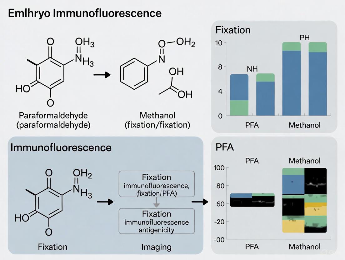
Chemical fixation is a critical first step in immunofluorescence (IF) that preserves cellular architecture and prevents degradation, enabling accurate visualization of biological structures. The choice of fixative fundamentally influences experimental outcomes by modifying protein structures and altering antigen accessibility. For researchers in embryology and drug development, selecting the appropriate fixation method is paramount for obtaining reliable data. Among the various options, paraformaldehyde (PFA) and methanol represent two fundamentally different fixation approaches with distinct advantages and limitations. This guide provides a comprehensive comparison of these fixation methods, supported by experimental data and detailed protocols to inform your experimental design.
Chemical fixatives stabilize biological specimens through two primary mechanisms: cross-linking and precipitation. Understanding these mechanisms is essential for selecting the appropriate fixative for your experimental goals.
Paraformaldehyde (PFA), an aldehyde-based cross-linking fixative, creates covalent methylene bridges between protein molecules, particularly reacting with primary amines [1]. This extensive network of protein cross-links preserves cellular architecture in a life-like state and maintains the spatial relationships between cellular components. However, this cross-linking can mask certain epitopes, making them inaccessible to antibodies.
Methanol, a dehydrating fixative, works through a completely different mechanism. It displaces water molecules around cellular macromolecules, causing protein denaturation and precipitation in situ [2]. This process can expose buried epitopes that might be inaccessible in native protein conformations, potentially improving antibody binding for certain targets.
The following diagram illustrates the fundamental differences in how these fixatives operate at a molecular level:
Experimental Protocols for PFA and Methanol Fixation
Standard PFA Fixation Protocol
The following protocol is adapted from established methodologies for formaldehyde fixation [3] [4]:
- Preparation: Use fresh 4% PFA in phosphate-buffered saline (PBS), pH 8.0.
- Fixation: Cover specimens to a depth of 2-3 mm with 4% PFA solution.
- Incubation: Allow fixation to proceed for 15 minutes at room temperature (20-25°C).
- Washing: Rinse three times with PBS for 5 minutes each.
- Permeabilization: Treat with 0.1% Triton X-100 in PBS for 5 minutes (required for intracellular targets).
- Blocking: Incubate with blocking buffer (e.g., 5% normal serum/0.3% Triton X-100 in PBS) for 60 minutes.
Critical Considerations: Lower PFA concentrations (0.25% or less) and temperatures below 37°C minimize alterations in DNA staining properties, which is particularly relevant for embryo research involving nuclear markers [5].
Standard Methanol Fixation Protocol
The methanol fixation protocol differs significantly [6] [2]:
- Preparation: Pre-chill 100% methanol to -20°C.
- Fixation: Aspirate culture media and immediately cover cells with ice-cold 100% methanol.
- Incubation: Fix for 15 minutes on ice or at 4°C.
- Washing: Rinse three times with PBS for 5 minutes each.
- Blocking: Incubate with blocking buffer (similar to PFA protocol) for 60 minutes.
Critical Considerations: Methanol simultaneously fixes and permeabilizes cells, eliminating the need for a separate permeabilization step with detergents like Triton X-100 [2].
The following workflow diagram compares the key steps in both protocols:
Performance Comparison: Experimental Data
The choice between PFA and methanol fixation involves trade-offs between morphological preservation, antigen accessibility, and macromolecular integrity. The table below summarizes key comparative data from multiple studies:
Table 1: Quantitative Comparison of PFA vs. Methanol Fixation Performance
| Parameter | PFA/Formaldehyde | Methanol | Experimental Context |
|---|---|---|---|
| Intracellular Antigen Detection | Variable performance; significantly lower antitubulin signal compared to sequential PFA/methanol [5] | Superior for certain targets; significantly higher antitubulin immunofluorescence (p < 0.002) [5] | Flow cytometric analysis of cellular proteins [5] |
| Cell Surface Antigen Detection | Unaffected by sequential PFA/methanol fixation [5] | Compatible with surface marker staining [5] | Flow cytometric analysis with anti-leukocyte antibodies [5] |
| DNA Quality & Analysis | Alters DNA staining; may produce spurious aneuploid peaks in normal cells [5] | Higher DNA yield, longer fragment size, more accurate copy-number calling [7] | Next-generation sequencing analysis [7] |
| Morphological Preservation | Well-preserved morphology; maintained light scatter properties [5] | Visible cellular damage reported in some studies [8] | Immunofluorescence microscopy [5] [8] |
| Single-cell RNA-seq Compatibility | Not recommended for neural cells; affects cellular composition [9] | Method of choice for neural cells; minimal effect on composition and gene expression [9] | Droplet-based single-cell transcriptomics of neural cells [9] |
| Epitope Preservation | Cross-linking may mask epitopes; requires antigen retrieval [1] | Denaturation may expose buried epitopes [2] | Immunofluorescence staining [2] |
The performance of each fixative varies significantly depending on the specific target antigen. For example, in direct comparisons:
- Keratin 8/18 shows optimal detection with methanol fixation [2]
- AIF (Apoptosis-Inducing Factor) performs best with formaldehyde fixation [2]
- PDI (Protein Disulfide Isomerase) and β-Actin work optimally with methanol permeabilization after formaldehyde fixation [2]
For DNA and genetic analyses, methanol demonstrates clear advantages. One study comparing fixation methods for next-generation sequencing found that methanol-based fixation yielded significantly longer DNA fragments (similar to snap-frozen samples) and more accurate copy-number calling compared to formalin-based fixation [7].
Research Reagent Solutions
Successful immunofluorescence requires specific laboratory reagents at each stage of the fixation and staining process. The table below outlines essential solutions and their functions:
Table 2: Essential Reagents for Immunofluorescence Fixation and Staining
| Reagent | Composition/Type | Function | Protocol Specificity |
|---|---|---|---|
| 4% Paraformaldehyde | 4% PFA in PBS, pH 8.0 | Cross-linking fixative; preserves morphology by creating protein cross-links | Primary fixative for PFA protocol [3] |
| Methanol | 100% methanol, ice-cold | Denaturing fixative; precipitates proteins by dehydration | Primary fixative for methanol protocol [6] |
| Permeabilization Buffer | 0.1% Triton X-100 in PBS | Non-ionic detergent; creates pores in membranes for antibody access | Required after PFA fixation [4] |
| Blocking Buffer | 5% normal serum, 0.3% Triton X-100 in PBS | Reduces non-specific antibody binding; blocks reactive sites | Common to both protocols [3] [6] |
| Antibody Dilution Buffer | 1% BSA, 0.3% Triton X-100 in PBS | Maintains antibody stability; reduces background during incubation | Common to both protocols [3] [6] |
| Wash Buffer | 1X PBS, pH 8.0 | Removes unbound antibodies and reagents; maintains physiological pH | Common to both protocols [3] [6] |
Advanced Applications and Strategic Considerations
Sequential Fixation Approaches
Research indicates that sequential fixation using both PFA and methanol can sometimes provide superior results compared to either method alone. One study demonstrated that sequential paraformaldehyde and methanol fixation was optimal for simultaneous flow cytometric analysis of DNA, cell surface proteins, and intracellular proteins, with significantly greater intracellular antitubulin immunofluorescence compared to either fixative used alone (p < 0.002) [5].
Fixation Effects on Nucleic Acids
For embryo research involving genetic analysis, fixation choice critically impacts nucleic acid integrity. Methanol fixation demonstrates superior performance for DNA preservation, with one study showing it provides "greater DNA yield, longer fragment size and more accurate copy-number calling" compared to formalin-based fixation [7]. This advantage is particularly relevant for developmental biology studies combining immunofluorescence with genetic analysis.
Multiplexing Considerations
When designing multiplex immunofluorescence experiments with antibodies requiring different fixation conditions, researchers must prioritize which targets to optimize. Testing small-scale pilot experiments comparing different protocols is recommended before scaling up experiments [2]. In some cases, sequential fixation approaches may provide a viable compromise for detecting multiple targets with different fixation requirements.
The selection between PFA and methanol fixation represents a critical methodological decision that directly influences experimental outcomes in immunofluorescence. PFA fixation generally provides superior morphological preservation and is ideal for cell surface markers and soluble proteins. In contrast, methanol fixation offers advantages for certain intracellular targets, DNA quality preservation, and single-cell transcriptomics applications. The optimal choice depends on the specific research goals, target antigens, and downstream applications. Embryo research specifically benefits from considering these trade-offs in the context of developmental stage and analytical requirements. As demonstrated by the experimental data presented, researchers should base their fixation strategy on empirical evidence specific to their biological system rather than relying on universal protocols.
In embryo immunofluorescence research, the choice of fixative is a critical determinant of experimental success. Chemical fixation aims to preserve a life-like snapshot of cellular architecture by halting degradation and immobilizing biomolecules. Among available methods, paraformaldehyde (PFA) and methanol represent two fundamentally different approaches to this challenge. PFA, an aldehyde-based cross-linking fixative, creates a stable protein matrix through covalent bonds, while methanol, an organic solvent, acts through protein denaturation and precipitation. This guide objectively compares the performance of PFA versus methanol fixation by examining their mechanisms, experimental outcomes, and implications for research accuracy, providing scientists with the evidence needed to select appropriate fixation protocols for embryonic and cellular studies.
Molecular Mechanisms of Fixation
The functional disparity between PFA and methanol fixation stems from their distinct biochemical mechanisms for immobilizing cellular components.
PFA: Cross-Linking via Aldehyde Chemistry
Paraformaldehyde functions as a cross-linking fixative that preserves structural relationships between biomolecules. Upon dissolution in aqueous solutions, PFA depolymerizes to monomeric formaldehyde, which reacts with the side chains of amino acids—primarily lysine—forming reactive hydroxymethyl groups. These groups subsequently create methylene bridges (-CH₂-) between nearby proteins, effectively trapping molecules within a cross-linked matrix [8] [10]. This matrix stabilizes the native organization of cellular structures, including membranes and organelles, by maintaining proteins in their relative positions.
The cross-linking process depends on PFA's small molecular size, enabling rapid diffusion throughout cells and tissues. However, as a monoaldehyde, PFA creates relatively short-distance cross-links, which may not fully immobilize all cellular components without extended fixation times or supplemental agents [11].
Methanol: Coagulation via Dehydration
Methanol employs a completely different mechanism based on protein denaturation and dehydration. By displacing water molecules around cellular macromolecules, methanol interferes with hydrogen bonds and hydrophobic interactions that maintain protein tertiary structures. This disruption causes proteins to denature and precipitate in situ, effectively freezing them in place [8] [2]. This coagulation process occurs rapidly but can significantly alter native protein conformations and extract cellular components, particularly lipids [12].
Table: Fundamental Mechanisms of PFA vs. Methanol Fixation
| Characteristic | Paraformaldehyde (PFA) | Methanol |
|---|---|---|
| Primary Mechanism | Cross-linking via methylene bridges | Dehydration and protein coagulation |
| Chemical Nature | Aldehyde-based cross-linker | Organic solvent |
| Effect on Structure | Stabilizes protein relationships | Denatures and precipitates proteins |
| Preservation Quality | Maintains structural context | May extract cellular components |
| Representation | Creates a protein "matrix" | Creates a protein "precipitate" |
Experimental Performance Comparison
Direct experimental comparisons reveal significant differences in how PFA and methanol preserve various cellular structures and antigens, with implications for data interpretation.
Preservation of Cellular Structures
Research demonstrates that PFA generally provides superior structural preservation compared to methanol-based fixation. In Raman spectroscopic imaging studies, aldehyde fixation methods "performed significantly better than organic solvents with less severe loss of biochemical information" [12]. Methanol fixation caused "severe loss of cell content, attributed to the loss of membrane integrity after the removal of lipids," compromising the preservation of cytoplasmic organization [12].
For delicate structures like membrane receptors, PFA alone may be insufficient. Studies of transmembrane receptors LYVE-1 and CD44 revealed that PFA alone inadequately immobilizes these proteins, leading to artefactual clustering during secondary antibody incubation. The addition of just 0.2% glutaraldehyde to PFA solutions provided the necessary cross-linking density to prevent this redistribution [11]. This finding highlights that for some membrane proteins, the limited cross-linking distance of PFA alone may permit residual mobility that compromises preservation accuracy.
Antigen Recognition and Epitope Integrity
The fixation method significantly influences antibody binding, with performance varying by specific target:
Table: Antibody Performance Across Fixation Methods
| Target Protein | Optimal Fixation | Performance Notes | Experimental Basis |
|---|---|---|---|
| Keratin 8/18 | Methanol | Superior epitope exposure | Demonstrated in HeLa cells [2] |
| AIF (Apoptosis-inducing factor) | 4% PFA | Better structural preservation | HeLa cell analysis [2] |
| PDI (Protein disulfide-isomerase) | PFA fixation + Methanol permeabilization | Improved signal for organelle targets | NIH/3T3 cell study [2] |
| β-Actin | PFA fixation + Methanol permeabilization | Enhanced cytoskeleton detection | NIH/3T3 cell study [2] |
| H3cit (Citrullinated histone H3) | PFA (15-30 min) | Signal decreased with 24h PFA fixation | Neutrophil NET formation study [8] |
| Myeloperoxidase (MPO) | PFA (15-30 min) | Unaffected by fixation time | Neutrophil NET formation study [8] |
Methanol's denaturing action can expose buried epitopes that cross-linking fixatives might obscure, explaining its superior performance for certain targets like keratins [2]. Conversely, PFA better preserves post-translational modification states, making it preferable for phospho-specific antibodies [2].
Liquid-Liquid Phase Separation (LLPS) Artifacts
Recent research reveals significant concerns about PFA fixation for studying biomolecular condensates formed through liquid-liquid phase separation. Comparing live and fixed cells expressing FET family proteins showed that "PFA fixation can both enhance and diminish putative LLPS behaviors" [13]. For specific proteins, fixation could even "cause droplet-like puncta to artificially appear in cells that do not have any detectable puncta in the live condition" [13]. These artifacts stem from the intricate balance between protein interaction dynamics and fixation rates, suggesting caution when interpreting punctate distributions solely from fixed samples.
Experimental Protocols for Comparative Studies
Standardized protocols enable direct comparison of fixation methods. The following methodologies are adapted from published experimental approaches.
PFA Fixation Protocol for Immunofluorescence
Reagents Needed:
- 4% Paraformaldehyde in PBS (freshly prepared or aliquoted from frozen stock)
- Phosphate-buffered saline (PBS)
- Quenching solution (100 mM glycine or 50 mM NH₄Cl in PBS)
- Permeabilization buffer (0.1-0.5% Triton X-100 in PBS)
- Blocking solution (3-5% BSA or serum in PBS)
Procedure:
- Culture Preparation: Grow cells on poly-L-lysine-coated coverslips to 60-80% confluence.
- Fixation: Aspirate culture medium and add 4% PFA in PBS. Incubate for 15-30 minutes at room temperature [8].
- Quenching: Remove PFA and wash 3× with PBS. Incubate with quenching solution for 10 minutes to neutralize residual aldehydes.
- Permeabilization: Incubate with permeabilization buffer for 10-15 minutes.
- Blocking: Apply blocking solution for 30-60 minutes to reduce nonspecific binding.
- Immunostaining: Proceed with primary and secondary antibody incubations.
Note: For difficult-to-fix membrane proteins, consider supplementing with 0.05-0.2% glutaraldehyde [11].
Methanol Fixation Protocol for Immunofluorescence
Reagents Needed:
- 100% methanol (pre-chilled to -20°C)
- Phosphate-buffered saline (PBS)
- Rehydration buffer (PBS with 0.05% Tween-20)
Procedure:
- Culture Preparation: Grow cells on coverslips to desired confluence.
- Fixation: Aspirate culture medium. Gently add pre-chilled 100% methanol and incubate for 10 minutes at -20°C [8].
- Rehydration: Gradually rehydrate cells by successive incubations in 70%, 50%, and 30% methanol in PBS (2-3 minutes each).
- PBS Wash: Wash 3× with PBS to remove residual methanol.
- Blocking and Staining: Proceed with blocking and immunostaining as described for PFA fixation.
Note: Avoid allowing cells to dry completely during medium aspiration and methanol addition, as this causes additional structural damage.
The Scientist's Toolkit: Essential Research Reagents
Successful fixation and immunofluorescence require specific reagents tailored to each method's biochemical requirements.
Table: Essential Reagents for Fixation Methods
| Reagent | Function | Application Notes |
|---|---|---|
| 4% Paraformaldehyde | Primary cross-linking fixative | Use fresh or frozen aliquots; 15-30 min fixation optimal for most antigens [8] |
| Glutaraldehyde | Supplemental cross-linker | Add 0.05-0.2% to PFA for membrane proteins; may increase autofluorescence [11] |
| 100% Methanol | Denaturing fixative | Pre-chill to -20°C; can extract lipids but exposes buried epitopes [8] [2] |
| Triton X-100 | Detergent for permeabilization | Use 0.1-0.5% after aldehyde fixation; creates pores for antibody access [8] |
| Glycine or NH₄Cl | Quenching agents | Neutralize unreacted aldehydes after PFA fixation to reduce background [11] |
| BSA or Serum | Blocking proteins | Reduce nonspecific antibody binding; use 3-5% in PBS or TBS buffer |
| Saponin | Mild permeabilization detergent | Preserves membrane structures while allowing antibody access to intracellular targets |
Discussion and Research Implications
The experimental evidence demonstrates that neither PFA nor methanol fixation universally outperforms the other across all research contexts. The optimal choice depends on specific research goals, target antigens, and cellular structures of interest.
PFA's cross-linking action creates a protein matrix that generally provides superior preservation of cellular architecture and structural relationships, particularly for soluble proteins and subcellular organelles. However, its potential to induce artifacts in membrane protein distribution and liquid-liquid phase separation studies necessitates careful validation. The finding that 24-hour PFA fixation decreases H3cit signal intensity while shorter fixation (15-30 minutes) preserves it highlights the importance of optimization even within a single method [8].
Methanol fixation offers advantages for certain epitopes that become exposed through denaturation, particularly cytoskeletal components and some keratins. However, its tendency to extract cellular components and alter membrane integrity limits its utility for comprehensive cellular studies. The visible cellular damage observed after 100% methanol fixation in neutrophil studies [8] underscores its potentially disruptive effects on delicate cellular structures.
For embryo immunofluorescence research, where preserving spatial relationships and developmental contexts is often paramount, PFA fixation frequently provides the necessary structural integrity. However, researchers investigating specific antigenic targets should consult antibody-specific validation data and consider performing pilot studies comparing fixation methods when establishing new protocols.
The cross-linking action of aldehydes like PFA creates a stabilized protein matrix that generally preserves cellular architecture more faithfully than methanol's denaturing approach. However, the experimental evidence clearly demonstrates that method selection must be application-specific, with particular attention to target antigens and research objectives. PFA excels in structural preservation and maintaining post-translational modification states, while methanol can provide superior results for certain epitopes through denaturation-mediated exposure. Researchers should prioritize rigorous validation using live-cell imaging when possible [13] and carefully optimize fixation conditions for each experimental system. By understanding the fundamental mechanisms and comparative performance of these fixation methods, scientists can make informed decisions that enhance the reliability and interpretability of their immunofluorescence data, particularly in complex systems like embryonic tissues where structural context is critical to biological insight.
In immunofluorescence (IF) research, particularly in embryo studies, fixation is a critical first step that preserves cellular architecture and enables the visualization of subcellular components. The choice of fixative fundamentally shapes experimental outcomes by determining which epitopes remain accessible for antibody binding. This guide focuses on the mechanistic action of organic solvents, with a specific emphasis on methanol's dehydrating effect, and provides a direct comparison with the cross-linking fixative paraformaldehyde (PFA). Within embryo research, this decision is paramount; it balances the superior morphological preservation offered by cross-linking fixatives against the potential for enhanced antigen detection with precipitating solvents. A thorough understanding of methanol's protein precipitation mechanism empowers researchers to make an informed choice, optimizing their immunofluorescence protocols for clarity, specificity, and reliability.
Mechanisms of Action: Cross-linking vs. Precipitation
Fixatives are categorized by their primary mechanism of action: cross-linking or precipitation. Understanding this distinction is key to selecting the appropriate method for a given experiment.
Aldehyde Cross-linking (PFA): PFA acts by forming covalent methylene bridges between the side chains of amino acids, primarily lysine, in proteins. This creates a three-dimensional network that stabilizes soluble and structural proteins in a "life-like" state, thereby hardening the entire cellular structure and excellently preserving morphology. A significant drawback of this extensive cross-linking is the potential masking of epitopes, which can prevent antibody binding and reduce the antigenic signal [8] [2] [14].
Organic Solvent Precipitation (Methanol): Methanol, an organic solvent, functions as a strong dehydrant. It displaces water molecules around cellular macromolecules, interfering with hydrogen bonds and hydrophobic interactions. This process disrupts the tertiary structure of proteins, leading to their denaturation and precipitation in situ. By displacing water and removing lipids, methanol also instantly permeabilizes the cells, eliminating the need for a separate permeabilization step. While this denaturation can destroy some epitopes, it can also expose others that are normally buried within the protein's native structure, which can be advantageous for certain antibodies [2] [15] [14].
The following diagram illustrates the core mechanistic differences and subsequent experimental steps between these two fixation methods.
Direct Experimental Comparison: PFA vs. Methanol
Theoretical mechanisms translate into practical performance differences. The following table summarizes the core characteristics of PFA and methanol based on experimental data.
Table 1: Core Characteristics of PFA and Methanol Fixation
| Parameter | Paraformaldehyde (PFA) | Methanol |
|---|---|---|
| Primary Mechanism | Cross-linking proteins [2] [14] | Protein precipitation & dehydration [15] [14] |
| Morphology Preservation | Excellent [14] | Good, but can cause damage [8] [14] |
| Cellular Permeabilization | Requires separate detergent step (e.g., Triton X-100) [2] [14] | Intrinsic; no additional step required [15] [14] |
| Impact on Epitopes | Can mask epitopes via cross-linking [2] [15] | Can denature/destroy some, expose others [2] [15] |
| Key Disadvantages | Potential for reduced antigenicity [2]; over-fixation can increase autofluorescence [8] | Loss of lipid/soluble components; can damage membrane & organelles [15] [14] |
Quantitative data from neutrophil NETosis studies provides a direct, head-to-head comparison. Researchers found that while 4% PFA fixation for 30 minutes was effective for staining myeloperoxidase (MPO) and DNA/histone-1-complexes, fixation for 24 hours decreased the signal for citrullinated histone H3 (H3cit) [8]. In contrast, 100% methanol fixation resulted in visible cellular damage [8]. Another study on brain tissue slices reported that 100% methanol caused severe deformations, including rolling and folding of the samples; this was mitigated by using lower methanol concentrations (e.g., 33.3% to 75%) [16].
Table 2: Experimental Performance in Key Studies
| Experimental Context | PFA Performance | Methanol Performance | Study Conclusion |
|---|---|---|---|
| NETosis Staining in Neutrophils [8] | 15-30 min fixation: Good for MPO and DNA/histone-1.24 h fixation: Decreased H3cit signal. | 100% MeOH: Caused visible cellular damage. | PFA (15-30 min) is recommended over methanol for this application. |
| Immunostaining in Brain Slices [16] | N/A | 100% MeOH: Severe tissue deformations.33.3-75% MeOH: No deformations, good staining. | Diluted methanol (33.3-75%) at room temperature is optimal for tissue slices. |
| General Epitope Performance [2] | Superior for AIF protein (soluble, mitochondrial). | Superior for Keratin 8/18 (cytoskeletal). | Antibody-specific performance dictates the optimal fixative. |
Detailed Methodologies for Embryo Immunofluorescence
To ensure reproducibility, below are detailed protocols for applying PFA and methanol fixation to embryo samples, compiled from the literature.
This protocol is recommended for general immunofluorescence and preserving morphology.
- Reagent Preparation: Prepare fresh RNase-free 4% Paraformaldehyde (PFA) in Phosphate-Buffered Saline (PBS). For fluorescent proteins like GFP, 2% PFA may be preferable.
- Embryo Collection: Harvest embryos and wash briefly with cold PBS to remove debris.
- Fixation: Immerse embryos in a sufficient volume of fixative (15-20x the volume of the tissue). Fixation should be performed at 4°C with gentle agitation for older embryos (E11.5+).
- Fixation Duration: The duration depends on the embryo age and staining method. For immunofluorescence (IHC) of E9.5-E12.5 embryos, an overnight fixation is typical. For E6.5-E8.5 embryos, fixation in the uterus overnight is common [17].
- Washing: After fixation, thoroughly rinse the embryos with PBS to remove residual PFA.
- Permeabilization and Blocking: Permeabilize the embryos by incubating with a detergent like 0.1% - 0.4% Triton X-100 in PBS for several hours to days depending on size. This is followed by blocking in a solution containing serum (e.g., 3% donkey serum) and a protein (e.g., 1% BSA) to prevent non-specific antibody binding [8] [14].
- Immunostaining: Proceed with incubation of primary and secondary antibodies.
Optimized Methanol Fixation Protocol
This protocol is recommended for antigens known to be sensitive to aldehyde cross-linking or when a cytoskeletal target is being stained.
- Reagent Preparation: Chill 100% methanol to -20°C. For tissue slices, consider using diluted methanol (50-75%) at room temperature to prevent deformation [16].
- Embryo Collection: Harvest and wash embryos in cold PBS.
- Fixation: Rapidly transfer embryos to the chilled methanol. Incubate at -20°C for 10-20 minutes [14] [16].
- Rehydration: Gradually rehydrate the embryos by passing them through a series of decreasing methanol concentrations in PBS (e.g., 75%, 50%, 25%) before a final wash in PBS.
- Blocking: Proceed directly to blocking. Note: A separate permeabilization step is typically unnecessary as methanol permeabilizes the cells [15] [14].
- Immunostaining: Proceed with standard immunostaining procedures.
The Scientist's Toolkit: Essential Research Reagents
The following table lists key reagents used in fixation and immunostaining protocols, along with their specific functions in the context of embryo research.
Table 3: Essential Reagents for Embryo Fixation and Immunostaining
| Reagent | Function/Application | Notes on Use |
|---|---|---|
| Paraformaldehyde (PFA) [14] [17] | Cross-linking fixative. The gold standard for morphology preservation and membrane protein staining. | Make fresh or use methanol-free, pre-mixed ampules. 4% is standard. |
| Methanol [15] [14] | Precipitating fixative and permeabilizing agent. Ideal for aldehyde-sensitive epitopes and cytoskeletal targets. | Use ice-cold for cells; diluted at RT for tissues. Avoid with fluorescent proteins. |
| Triton X-100 [8] [14] | Non-ionic detergent for permeabilizing PFA-fixed samples. Creates pores in all lipid bilayers. | Typical working concentration: 0.1%-0.4%. High concentrations can lyse cells. |
| Saponin / Digitonin [14] | Mild, reversible permeabilization agent. Ideal for preserving surface antigens and internal organelle structures. | Does not permeabilize the nuclear membrane. |
| Donkey Serum / BSA [8] | Component of blocking buffer. Reduces non-specific background antibody binding. | Serum from the host species of the secondary antibody is most effective. |
| Phosphate-Buffered Saline (PBS) | Isotonic buffer for washing, diluting fixatives, and reagent preparation. Maintains pH and osmotic balance. | --- |
| Primary Antibodies | Specifically bind to the target antigen of interest. | Performance is highly dependent on fixation method [2]. |
| Fluorophore-Conjugated Secondary Antibodies | Bind to primary antibodies and provide the detectable fluorescent signal. | Must be raised against the host species of the primary antibody. |
Decision Workflow for Embryo Research
Given the distinct advantages and drawbacks of each fixative, selecting the correct one is a critical decision point in experimental design. The following workflow provides a logical pathway to the optimal choice for a given embryo immunofluorescence project.
Impact on Cellular Morphology and Antigen Integrity
In embryo immunofluorescence research, the choice of fixation method is a critical determinant of experimental success. Fixatives preserve cellular structure and prevent degradation, but their chemical actions differentially impact the preservation of morphology and the integrity of antigen targets. Paraformaldehyde (PFA), a crosslinking agent, and methanol, a precipitating solvent, represent the two primary classes of fixatives used in laboratories. This guide provides an objective comparison of PFA versus methanol fixation, drawing on recent experimental data to outline their performance in preserving cellular morphology and antigen integrity for immunofluorescence studies.
Mechanistic Actions and Immediate Effects
The fundamental difference between these fixatives lies in their mechanism of action, which directly influences their effects on cellular structures.
- Paraformaldehyde (PFA): As an aldehyde-based crosslinker, PFA forms covalent methylene bridges between free amine groups of proteins, creating a interconnected molecular network. This process preserves cellular architecture in a life-like state and is particularly effective at stabilizing soluble and membrane-associated proteins [18] [2]. However, excessive crosslinking can mask antibody epitopes, reducing antigenicity.
- Methanol: Acting as a strong dehydrant, methanol precipitates cellular proteins by displacing water and disrupting hydrophobic bonds. This denatures proteins and can expose buried epitopes beneficial for some antibodies, but it often damages cellular structures like the cytoskeleton, microtubules, and organelles. It also removes lipids and can denature fluorescent proteins [8] [18].
The diagram below illustrates the core mechanisms and consequences of each fixation method.
Comparative Experimental Data
Direct comparisons in research settings reveal how these mechanistic differences translate to practical outcomes for morphology and antigen detection. The following tables summarize key experimental findings.
Table 1: Qualitative Comparison of PFA and Methanol Fixation
| Parameter | Paraformaldehyde (PFA) | Methanol |
|---|---|---|
| Mechanism of Action | Crosslinking proteins [18] | Protein precipitation & denaturation [18] |
| Morphology Preservation | Superior; excellent structural preservation [19] | Moderate; can cause cellular contraction and damage [8] [19] |
| Lipid Retention | Good | Poor; lipids are removed [18] |
| RNA Preservation | High-quality RNA suitable for PCR [19] | Degraded RNA [19] |
| Autofluorescence | Low | Can be high [8] |
| Suitable for Fluorescent Proteins | Yes | No; causes denaturation [18] |
Table 2: Quantitative Staining Intensity and Morphology Outcomes
| Experimental Context | Fixation Protocol | Key Findings | Source |
|---|---|---|---|
| Neutrophils (Human) | 4% PFA, 30 min | Optimal staining for H3cit; no cellular damage [8] | [8] |
| Neutrophils (Human) | 100% Methanol, 30 min | Visible cellular damage; autofluorescence [8] | [8] |
| Xenograft Tumors (Mice) | 4% PFA, 24 hrs | High-quality RNA & excellent morphology [19] | [19] |
| Xenograft Tumors (Mice) | 99% Ethanol, 24 hrs | Degraded RNA & cell contraction [19] | [19] |
| HeLa Cells | Formaldehyde | Optimal for AIF protein [2] | [2] |
| HeLa Cells | Methanol | Optimal for Keratin 8/18 [2] | [2] |
Detailed Experimental Protocols
To ensure reproducibility, below are detailed methodologies from cited experiments comparing fixation techniques.
Protocol: Neutrophil Fixation for NETosis Markers
This protocol is adapted from a study investigating the effect of fixation on staining neutrophil extracellular trap (NET) markers [8].
- Cell Preparation: Isolate neutrophils from fresh human blood using a density gradient (e.g., Polymorphprep). Seed cells onto poly-L-lysine-coated coverslips at a density of 2 × 10⁵ cells per well and stimulate with 25 nM PMA for 2 hours at 37°C and 5% CO₂ to induce NETosis.
- Fixation:
- PFA: Fix cells with 4% PFA in PBS for 15–30 minutes at room temperature (RT). Avoid prolonged fixation (e.g., 24 hours), which decreases the signal intensity for certain markers like citrullinated histone H3 (H3cit).
- Methanol: Fix cells with 100% methanol for 30 minutes at RT or -20°C.
- Post-Fixation: After fixation, wash plates three times with PBS. Samples can be stored at 4°C.
- Immunostaining: Permeabilize cells with 0.5% Triton X-100 for 5 minutes. Block with a buffer containing serum, BSA, and gelatine. Incubate with primary antibodies (e.g., anti-MPO, anti-H3cit) for 1 hour, followed by fluorophore-conjugated secondary antibodies.
Protocol: Fixation of Avian Embryos
A comparative study on chicken embryos highlights how fixation choice affects signal detection in complex tissues [20].
- Sample Preparation: Dissect and prepare chicken embryos at desired developmental stages.
- Fixation:
- PFA Fixation: Fix embryos with 4% PFA for a standard duration (e.g., 24 hours at 4°C).
- Trichloroacetic Acid (TCA) Fixation: Fix embryos with TCA solution (this study used TCA as an alternative to methanol, demonstrating another precipitating fixative).
- Analysis: Process samples for immunohistochemistry (IHC). TCA fixation was found to alter nuclear morphology and subcellular fluorescence intensity for various proteins (e.g., transcription factors, cadherins) compared to PFA, sometimes revealing protein signals in otherwise inaccessible tissues [20].
The Scientist's Toolkit: Essential Research Reagents
Successful immunofluorescence relies on a suite of key reagents. The table below lists essential materials and their functions based on the analyzed protocols.
Table 3: Essential Reagents for Immunofluorescence Fixation and Staining
| Reagent | Function/Application | Example Use-Case |
|---|---|---|
| Paraformaldehyde (PFA) | Crosslinking fixative for superior morphology preservation [8] [18] | Standard fixation for most membrane and soluble proteins [2] |
| Methanol | Precipitating fixative and permeabilization agent [18] | Staining for aldehyde-sensitive epitopes or certain cytoskeletal proteins [2] |
| Triton X-100 | Non-ionic detergent for permeabilizing lipid bilayers after crosslinking fixation [8] [2] | Standard permeabilization after PFA fixation to allow antibody access to intracellular targets |
| Saponin/Digitonin | Milder, reversible permeabilization agents that preserve membrane protein integrity [18] | Staining intracellular targets without disrupting the nuclear membrane |
| Donkey Serum / BSA | Components of blocking buffer to reduce non-specific antibody binding [8] | Blocking step before antibody incubation to lower background noise |
| Primary Antibodies | Target-specific binders for proteins of interest (e.g., MPO, H3cit) [8] | Visualizing specific antigens in the sample |
| Fluorophore-Conjugated Secondary Antibodies | Detect primary antibodies for visualization under a microscope [8] | Signal amplification and multiplexing with different fluorescent channels |
Choosing the correct fixative depends on the primary goal of the experiment. The following workflow diagram outlines the decision-making process.
In conclusion, the choice between PFA and methanol fixation involves a fundamental trade-off. PFA is generally the recommended choice for experiments where superior preservation of cellular morphology, RNA integrity, and overall architecture is paramount [8] [19]. Its crosslinking nature, however, necessitates optimization of fixation time and permeabilization to prevent epitope masking. Methanol fixation serves as a powerful alternative for specific antigens whose epitopes are exposed by denaturation, particularly in cases involving robust cytoskeletal markers or when a combined fixation/permeabilization step is desirable [2]. Researchers are advised to consult antibody datasheets and, when possible, perform small-scale pilot experiments to determine the optimal fixation protocol for their specific research context [2].
In embryonic research, where capturing delicate and dynamic biological processes is paramount, the choice of fixation method is a critical determinant of experimental success. Fixation preserves cellular architecture and biomolecules from degradation, providing a "life-like" snapshot of the tissue at a specific moment [2]. For embryonic tissues, two of the most common fixation methods are paraformaldehyde (PFA), an aldehyde-based crosslinking fixative, and methanol, an organic solvent-based precipitating fixative [21]. This guide objectively compares the performance of PFA and methanol fixation within the context of embryo immunofluorescence research, providing supporting experimental data and detailed methodologies to inform researchers and drug development professionals.
Mechanism of Action and Cellular Impact
The fundamental difference between PFA and methanol lies in their biochemical mechanism for preserving cellular contents, which directly influences their effect on embryonic structures.
Paraformaldehyde (PFA) acts as a crosslinking fixative. It creates covalent chemical bonds between proteins, primarily linking the amino acid lysine [21]. This process stabilizes and hardens the sample by anchoring soluble proteins to the cytoskeleton, effectively forming a molecular network that preserves the spatial relationships within the cell [22] [2]. The crosslinking action of PFA is excellent for maintaining overall cellular morphology and the native localization of proteins [23].
Methanol is a precipitating or denaturing fixative. It works by dehydrating the sample and disrupting hydrophobic interactions, causing proteins to denature and precipitate in situ [22] [24]. This mechanism does not create molecular crosslinks but rather coagulates cellular proteins. While this can preserve cellular architecture, it may also alter protein conformation and can lead to the loss of some soluble components [24].
A recent nanoscale study using atomic force microscopy demonstrated that both types of fixatives create artefacts by aggregating membrane proteins. The study found that treatment with PFA, glutaraldehyde, and methanol all significantly increased the size of nanoscale protrusions on the cell surface compared to living cells, with methanol creating the largest aggregates [25]. This finding calls for careful interpretation of membrane protein clustering in fixed samples.
Experimental Performance Comparison
The suitability of PFA or methanol varies significantly depending on the experimental goals, whether for immunofluorescence (IF), RNA analysis, or general morphology. The table below summarizes key performance metrics based on published experimental data.
Table 1: Comparative Performance of PFA vs. Methanol Fixation in Embryonic and Cellular Research
| Performance Metric | Paraformaldehyde (PFA) | Methanol | Supporting Experimental Evidence |
|---|---|---|---|
| General Morphology Preservation | Superior for overall cellular structure and membrane integrity [23]. | Can cause shrinkage, hardening, and damage to microtubules and organelles [24] [23]. | In oocytes/embryos, PFA provided reliable protein localization; methanol can be disruptive [23]. |
| Target Antigen Preservation | Can mask some epitopes via crosslinking, reducing antigenicity [2]. | May expose buried epitopes via denaturation, beneficial for some antibodies [2]. | Keratin 8/18 antibody worked best with methanol, while AIF antibody preferred PFA [2]. |
| Membrane Protein Detection | Ideal for preserving membrane proteins in situ [24]. | Can disrupt lipids and membrane-associated antigens [24]. | Methanol provided stronger, more distinct signals for Claudin 1 and E-cadherin in breast cancer cells [26]. |
| Nuclear and Cytoskeletal Antigens | Good preservation, but may require optimization [2]. | Often superior for many nuclear and cytoskeletal targets; can be recommended for phospho-antigens [2] [24]. | PDI and β-Actin antibodies showed improved performance with methanol permeabilization [2]. |
| RNA Integrity | Suitable, but can be challenging for some scRNA-seq workflows [22]. | Excellent for preserving nucleic acids; simple protocol integrates easily into scRNA-seq [22]. | Methanol-fixed cells showed high-quality cDNA and transcriptomic profiles highly correlated with live cells [22]. |
| Tissue Penetration | Good penetration, but slower than methanol; diffusion time must be considered [21]. | Faster tissue penetration due to smaller molecular size and dehydrating effect [21]. | Glutaraldehyde (similar to PFA) penetrates more slowly than methanol [21]. |
Detailed Experimental Protocols
To ensure reproducibility, below are standardized protocols for fixing embryonic samples with PFA and methanol, compiled from the literature. These protocols can be adapted for whole-mount embryos or embryonic sections.
Standard PFA Fixation Protocol for Embryos
This protocol is widely used for immunofluorescence and whole-mount RNA FISH on mouse embryos, providing excellent morphological preservation [23] [27].
- PFA Solution Preparation: Prepare a 4% PFA solution in phosphate-buffered saline (PBS). For a 10% stock solution, dissolve PFA powder in double-distilled water with a small amount of NaOH to dissolve, then adjust to pH 7.2-7.4 with HCl [23].
- Fixation Procedure:
- Harvest and Rinse: Collect embryos in a physiological buffer (e.g., PBS).
- Immerse in Fixative: Immerse embryos in a volume of 4% PFA at least 10 times greater than the tissue volume. For mouse oocytes and embryos, fixation at room temperature for 15-20 minutes is effective [23].
- Rinse: Wash the embryos thoroughly 3-5 times with PBS to remove all traces of PFA.
- Post-Fixation Processing (for IF): If permeabilization is required for antibody access, treat embryos with a detergent like Triton X-100 (e.g., 0.1-0.5% in PBS) for 5-30 minutes depending on size, followed by blocking [27].
Standard Methanol Fixation Protocol for Embryos
Methanol fixation is valued for its simplicity and effectiveness for certain targets, particularly in single-cell RNA sequencing and for some epitopes [22].
- Methanol Solution Preparation: Use 100% methanol, chilled to -20°C for best results [24].
- Fixation Procedure:
- Harvest and Rinse: Collect embryos in a physiological buffer.
- Dehydrate (Optional): Some protocols include a gradual dehydration step through a series of methanol/PBS solutions (e.g., 25%, 50%, 75% methanol) to reduce shock.
- Immerse in Fixative: Immerse embryos in ice-cold 100% methanol. Fixation time can vary from 10 minutes to several hours at -20°C [24]. For long-term storage, samples can be kept at -20°C in methanol.
- Post-Fixation Processing (for IF): Methanol acts as both a fixative and a permeabilization agent. Typically, no additional permeabilization step is needed. Rehydrate the embryos through a descending series of methanol/PBS solutions (e.g., 75%, 50%, 25%) before proceeding to blocking and immunostaining in PBS.
Decision Workflow and Visualization
The choice between PFA and methanol is not universal but depends on the primary experimental objective. The following workflow diagram outlines a logical decision process for researchers.
Figure 1: A decision workflow for choosing between PFA and methanol fixation based on primary research goals. This chart synthesizes data from multiple experimental comparisons. [2] [22] [23]
The Scientist's Toolkit: Essential Research Reagents
Successful fixation and immunostaining of embryonic tissues require a suite of specific reagents. The table below lists key solutions and their functions.
Table 2: Essential Reagents for Embryo Fixation and Immunostaining Protocols
| Reagent/Solution | Function | Key Considerations |
|---|---|---|
| Paraformaldehyde (PFA) | Crosslinking fixative that preserves morphology and protein spatial relationships. | Concentration is critical (typically 3-4%); pH must be buffered (e.g., to 7.2-7.4) for optimal fixation [23]. |
| Methanol | Precipitating fixative and permeabilizing agent; preserves nucleic acids well. | Ice-cold (-20°C) application is standard; can denature fluorescent proteins like GFP [24]. |
| Triton X-100 | Non-ionic detergent for permeabilizing membranes after PFA fixation. | Concentration (0.1-0.5%) and incubation time must be optimized to avoid complete membrane lysis [24]. |
| Saponin / Digitonin | Milder, reversible permeabilization agents that selectively complex with cholesterol. | Ideal for preserving membrane-associated antigens; does not permeabilize the nuclear membrane [24]. |
| Bovine Serum Albumin (BSA) | Blocking agent used to reduce non-specific antibody binding. | A concentration of 1-5% in PBS is standard; helps minimize background staining [26]. |
| Phosphate-Buffered Saline (PBS) | Isotonic buffer used for washing, diluting, and storing samples. | Maintains pH and osmotic balance, preventing tissue damage during processing. |
The comparison between PFA and methanol fixation reveals a clear trade-off: PFA is generally superior for preserving the native cellular architecture and spatial context of proteins, which is often critical in complex embryonic tissues [23]. In contrast, methanol excels in preserving nucleic acids and can provide superior antigen accessibility for certain protein targets, particularly those within the nucleus or cytoskeleton [2] [22] [26]. There is no universal "best" fixative; the optimal choice is dictated by the specific antigen, the research question, and the downstream analytical technique. For critical experiments, particularly when working with novel antibodies or embryonic stages, empirical testing of both fixation methods on a small scale is an indispensable step to ensure reliable and interpretable results [2].
Protocol Selection and Application: Step-by-Step Guides for Embryo Fixation
In embryo immunofluorescence research, sample fixation is a critical first step that profoundly impacts experimental outcomes. The choice between paraformaldehyde (PFA) and methanol fixation represents a fundamental methodological decision, balancing the preservation of cellular architecture against the retention of antigen recognition. This guide objectively compares the performance of PFA and methanol fixation protocols specifically for embryo research, supported by experimental data from current studies. While PFA works through protein cross-linking to maintain structural relationships, methanol fixation denatures proteins and can better preserve certain nucleic acid targets. Understanding these trade-offs enables researchers to select the optimal fixation approach for their specific embryonic research applications.
Performance Comparison: PFA vs. Methanol Fixation
The table below summarizes the key characteristics and recommended applications of PFA and methanol fixation methods based on current experimental evidence.
Table 1: Comparative analysis of PFA and methanol fixation for embryo research
| Parameter | PFA Fixation | Methanol Fixation |
|---|---|---|
| Standard Concentration | 4% [28] | 100% (ice-cold) [29] [30] |
| Typical Fixation Time | 30 minutes at room temperature [28] | 15 minutes at 4°C [29] |
| Mechanism of Action | Protein cross-linking via hydroxymethyl groups [8] | Protein denaturation and dehydration [8] |
| Cellular Structure Preservation | Excellent preservation of ultrastructure and spatial relationships [8] [31] | Can cause cellular damage and shrinkage; poor membrane preservation [8] |
| Antigen Accessibility | May mask some epitopes due to cross-linking [8] [32] | Better for nuclear antigens and some transcription factors [32] |
| Recommended Applications | Phosphorylated SMAD detection in human blastocysts [28]; subcellular localization studies | Single-cell RNA-seq of cardiomyocytes [30]; neural cell transcriptomics [9] |
| Compatibility with Downstream Applications | Immunofluorescence, immunohistochemistry [8] [28] | scRNA-seq, droplet-based transcriptomics [9] [30] |
| Signal Integrity | Decreased citrullinated histone H3 signal with prolonged fixation (>24h) [8] | Good RNA integrity for transcriptomics (RIN ~9) [9] |
Detailed Experimental Protocols
Standard PFA Fixation Protocol for Embryos
The following protocol is adapted from established methods for pre-implantation human embryos, particularly for detecting phosphorylated SMAD proteins [28]:
Reagent Preparation:
- Prepare 4% PFA solution in PBS with calcium and magnesium ions [28]
- Heat (60-70°C) and stir until PFA powder is entirely dissolved (2-3 hours)
- Filter through 0.1 μm sterile filter
- Store at 4°C for up to 7 days [28]
Fixation Procedure:
- Preparation: Perform all manipulations using a stereomicroscope. Handle embryos gently with prepared glass capillaries [28].
- Fixation: Transfer embryos to 4% PFA solution for 30 minutes at room temperature on a rocking platform [28].
- Washing: Rinse embryos three times in PBS for 5 minutes each to remove residual fixative [28].
- Permeabilization: Treat with 0.1% Triton X-100 in PBS for 20 minutes [28].
- Storage: Fixed samples can be stored in PBS at 4°C for short-term use or proceed directly to immunostaining.
Diagram: PFA fixation workflow for embryo immunofluorescence
Methanol Fixation Protocol for Embryo-Derived Cells
For specific applications like single-cell RNA sequencing of embryo-derived cardiomyocytes, methanol fixation follows this protocol [30]:
Fixation Procedure:
- Preparation: Aspirate culture media from cells
- Fixation: Cover cells to a depth of 2-3 mm with ice-cold 100% methanol
- Incubation: Fix for 15 minutes on ice or at 4°C [29]
- Rehydration: Rinse three times in 1X PBS for 5 minutes each
- Storage: Methanol-fixed cells can be stored in PBS at 4°C or processed immediately for downstream applications [30]
Experimental Evidence and Data Support
Structural Preservation Capabilities
PFA fixation demonstrates superior performance in preserving cellular ultrastructure, which is critical for analyzing embryonic subcellular localization. Studies investigating neutrophil extracellular traps (NETs) found that methanol fixation resulted in visible cellular damage, while PFA maintained structural integrity [8]. Furthermore, prefixation with formaldehyde before crosslinking mass spectrometry preserved actin cytoskeleton architecture with minimal distortion, unlike organic solvents [31].
Impact on Biomolecule Detection
The choice of fixative significantly affects detection capabilities for different biomolecules:
Table 2: Fixation effects on specific biomarker detection
| Biomarker | PFA Performance | Methanol Performance | Experimental Context |
|---|---|---|---|
| Phospho-SMAD proteins | Optimal detection [28] | Not recommended | Human blastocysts |
| Citrullinated histone H3 | Signal decrease after 24h fixation [8] | Variable performance | Neutrophil extracellular traps |
| RNA integrity | Moderate preservation | Superior preservation (RIN ~9) [9] | Single-cell transcriptomics |
| Transcription factors | Epitope masking concerns [32] | Better accessibility [32] | Flow cytometry |
Application-Specific Performance
In single-cell transcriptomics of neural cells derived from induced pluripotent stem cells, methanol fixation provided cellular composition similar to fresh samples with good cell quality and minimal expression biases [9]. Conversely, DMSO cryopreservation, while providing higher library complexity, strongly affected cellular composition and induced stress and apoptosis genes [9].
For whole-mount RNA-FISH on mouse embryonic limb buds, PFA fixation combined with oxidation-mediated autofluorescence reduction enabled high-quality imaging without digital post-processing [27].
The Scientist's Toolkit: Essential Research Reagents
Table 3: Key reagents for embryo fixation protocols
| Reagent | Function | Application Notes |
|---|---|---|
| Paraformaldehyde (4%) | Protein cross-linking fixative | Must be fresh (<7 days) for optimal nuclear factor detection [28] |
| Methanol (100%) | Denaturing fixative | Use ice-cold for better preservation [29] |
| Triton X-100 | Surfactant for permeabilization | Prepare fresh on day of use [28] |
| Phosphate-Buffered Saline | Washing and dilution buffer | With Ca2+/Mg2+ for PFA fixation [28] |
| Normal Serum | Blocking agent | Reduce non-specific antibody binding |
| Tween-20 | Detergent | Component of specialized fixation buffers [32] |
Diagram: Decision framework for selecting fixation methods
The selection between PFA and methanol fixation for embryo research depends primarily on the experimental objectives and downstream applications. PFA fixation at 4% for 30 minutes provides superior preservation of cellular ultrastructure and is recommended for protein localization studies, particularly for phosphorylated signaling molecules like SMAD proteins in developmental studies. Methanol fixation offers advantages for transcriptomic analyses and certain nuclear antigens but compromises cellular architecture. Researchers should align their fixation strategy with their primary research questions, considering that each method presents distinct trade-offs between structural preservation and biomolecule accessibility.
Methanol Fixation Protocol for Frozen Sections and Cell Cultures
The choice of cell fixation method is a critical determinant of success in immunofluorescence research, directly impacting the preservation of cellular morphology, accessibility of antigenic epitopes, and reliability of experimental results. Within the specific context of embryo research, where sample integrity is paramount, the debate between paraformaldehyde (PFA) and methanol fixation remains particularly relevant. Methanol fixation operates through a distinct mechanism of dehydration and protein precipitation, offering unique advantages for certain applications while presenting specific limitations for others [2]. This guide provides an objective comparison of methanol and PFA fixation protocols, drawing on current experimental data to equip researchers with the information necessary to select the optimal fixation strategy for their experimental needs, particularly when working with delicate samples such as embryos.
The following diagram illustrates the key decision points and primary characteristics researchers should consider when selecting between these fixation methods:
Performance Comparison: Quantitative Data Analysis
Direct comparison of methanol and PFA fixation requires examination of multiple performance metrics across different experimental contexts. The following tables summarize critical quantitative findings from recent studies to facilitate evidence-based protocol selection.
Table 1: scRNA-seq Performance Metrics Following Fixation
| Fixation Method | Cell Type | Mean Genes/Cell | Mean UMI/Cell | RNA Integrity Number | Study |
|---|---|---|---|---|---|
| Methanol | hiPSC-derived neural cells | 954-1364 | 1469-2532 | ~9.0 | [33] |
| PFA | hiPSC-derived neural cells | 889-1043 | 1390-1738 | ~9.0 | [33] |
| Methanol | HCT-116 & HepG2 cell lines | Comparable to live cells | Comparable to live cells | High (no severe degradation) | [34] |
| PFA-FD-seq | BC3 & 3T3 cell lines | 640 (median) | Reduced vs live | High (with crosslink reversal) | [35] |
Table 2: Immunofluorescence and Morphology Assessment
| Fixation Method | Tissue/Cell Type | Morphology Preservation | Epitope Accessibility | Notable Findings |
|---|---|---|---|---|
| Methanol | Neural tissue (Shank proteins) | Poor structural integrity | Excellent for buried epitopes | Superior to PFA for post-synaptic density proteins [36] |
| PFA | Neural tissue (Shank proteins) | Excellent structural integrity | Epitope masking concerns | Standard 4% PFA inadequate for Shank visualization [36] |
| PFA | Avian embryos | Superior nuclear & tissue morphology | Effective for HCR and IHC | Recommended for mRNA visualization [37] |
| TCA | Avian embryos | Altered nuclear shape | Protein-dependent efficacy | Revealed some protein signals inaccessible with PFA [37] |
Table 3: Embryo and Specialized Cell Applications
| Fixation Method | Cell Type | Protocol | Outcome | Recommendation |
|---|---|---|---|---|
| PFA/Triton X-100 | Mouse oocytes/embryos | 3.5% PFA + 0.1% TX | Consistent results, minimal background | Superior for embryonic material [23] |
| Glyoxal/Triton X-100 | Mouse oocytes/embryos | 3% Gly + 0.1% TX | Catastrophic consequences | Not recommended [23] |
| Methanol | Neutrophils (NET formation) | 100% MeOH, -20°C, 30min | Cellular damage observed | Not recommended for delicate structures [8] |
| PFA | Neutrophils (NET formation) | 4% PFA, 15-30min, RT | Preserved structure, good staining | Recommended for NET studies [8] |
Experimental Protocols: Detailed Methodologies
Standard Methanol Fixation Protocol for Cell Cultures
Application: General immunofluorescence staining of cultured cells, particularly suited for cytoskeletal targets and intracellular epitopes that may be masked by cross-linking fixatives.
Procedure:
- Preparation: Chill pure methanol to -20°C prior to fixation.
- Fixation: Aspirate culture medium and immediately add cold methanol to cover cells completely.
- Incubation: Incubate at -20°C for 10 minutes [36] [2].
- Rehydration: Remove methanol and wash cells 2-3 times with phosphate-buffered saline (PBS) or appropriate buffer.
- Storage: Fixed cells can be stored in PBS at 4°C for short-term or at -20°C for longer-term storage.
Note: No separate permeabilization step is typically required as methanol simultaneously fixes and permeabilizes cells [2].
Methanol Fixation for Frozen Tissue Sections
Application: Immunofluorescence staining of frozen tissue sections, particularly beneficial for challenging epitopes in densely packed cellular compartments.
Procedure:
- Section Preparation: Cut frozen tissues into 12μm sections using a cryostat (e.g., Leica CM3050 S) at -18°C to -20°C [36].
- Mounting: Mount sections on charged slides (e.g., Superfrost Plus) and air dry for 3-5 minutes at room temperature.
- Fixation: Fix slides in cold methanol (-20°C) for 10 minutes [36].
- Rehydration: Wash slides three times for 10 minutes each with Tris-buffered saline (TBS) or PBS.
- Blocking: Incubate sections with blocking buffer (e.g., 10% normal goat serum, 0.3% Triton X-100 in TBS) for 20 minutes prior to immunostaining.
Methanol Fixation for Single-Cell RNA Sequencing
Application: Cell preservation for droplet-based single-cell transcriptomics, particularly beneficial for maintaining cellular composition and minimizing stress signatures.
Procedure:
- Cell Preparation: Create single-cell suspension using appropriate dissociation protocol.
- Fixation: Resuspend cell pellet in chilled methanol to achieve final concentration of 80% methanol [33] [34].
- Storage: Store fixed cells at -80°C for up to several weeks.
- Processing: Prior to scRNA-seq, rehydrate cells by gradual addition of PBS or appropriate buffer [34].
- Library Preparation: Proceed with standard scRNA-seq protocols (e.g., Drop-seq, 10X Genomics).
Research Reagent Solutions: Essential Materials
Table 4: Key Reagents for Methanol Fixation Protocols
| Reagent/Category | Specific Examples | Function | Protocol Considerations |
|---|---|---|---|
| Fixation Agents | Pure methanol (100%) | Protein denaturation and precipitation through dehydration | Use pre-chilled to -20°C; concentration typically 80-100% [33] [2] |
| Buffers | Phosphate-buffered saline (PBS), Tris-buffered saline (TBS) | Maintain physiological pH and osmolarity during processing | Used for washing and rehydration after fixation [36] |
| Permeabilization Detergents | Triton X-100, Tween-20, Saponin | Create membrane pores for antibody access | Often unnecessary with methanol fixation alone [2] |
| Blocking Agents | Normal serum (goat, donkey), BSA, cold-water fish gelatin | Reduce non-specific antibody binding | Typically 3-10% concentration in buffer; incubation 20-60 minutes [36] |
| Section Support | Superfrost Plus slides, Tissue-Tek O.C.T. compound | Adhesion and support for tissue sections | Charged slides improve section adhesion [36] |
| Mounting Media | Aqua-Poly/Mount, commercial anti-fade media | Preserve fluorescence and support coverslip application | Use fluorescence-compatible media for long-term storage |
Discussion: Applications and Limitations in Embryo Research
Within embryo research specifically, the selection between methanol and PFA fixation requires careful consideration of experimental priorities. Recent comparative studies on mouse oocytes and embryos have demonstrated that PFA (3.5%) provides more consistent and reliable fixation outcomes compared to alternative aldehyde fixatives, with superior preservation of protein localization patterns [23]. This is particularly critical in embryonic systems where subtle spatial distributions of developmental regulators can have significant functional consequences.
However, methanol fixation maintains its utility for specific applications in embryonic research, particularly when:
- Target epitopes are known to be masked by PFA cross-linking
- Combined analysis of transcriptomics and protein localization is required
- Cytoskeletal elements are under investigation
Notably, studies on avian embryos have demonstrated that fixation method significantly impacts the visualization of key developmental markers, with PFA proving superior for mRNA detection while alternative fixatives like trichloroacetic acid (TCA) revealed different protein localization patterns in some tissues [37]. These findings underscore the importance of context-specific optimization rather than universal protocol application.
For researchers requiring intracellular protein staining prior to scRNA-seq analysis of embryonic cells, PFA fixation combined with crosslink reversal (as in FD-seq protocols) may offer a viable alternative, enabling phenotypic sorting while maintaining transcriptomic integrity [35]. This approach demonstrates the ongoing innovation in fixation methodologies to address the unique challenges posed by complex cellular systems including embryos.
Methanol fixation represents a valuable tool in the researcher's arsenal, offering distinct advantages for specific applications including the visualization of certain structurally-associated epitopes and preservation of samples for single-cell transcriptomics. However, within the context of embryo immunofluorescence research, PFA fixation generally provides superior morphological preservation and more consistent results across a broad range of cellular targets. The optimal fixation strategy ultimately depends on the specific research question, target antigens, and downstream applications, necessitating empirical testing when investigating novel targets or experimental systems.
Combined Fixation and Permeabilization with Methanol
In immunofluorescence research, the preparation of biological samples through fixation and permeabilization is a critical step that can determine the success or failure of an experiment. Among the various methods available, combined fixation and permeabilization with methanol offers a unique approach with distinct advantages and limitations compared to alternative techniques. This method, which utilizes the denaturing and dehydrating properties of methanol, is particularly relevant for embryo immunofluorescence studies where preserving specific antigen epitopes while maintaining structural integrity is paramount.
Methanol fixation operates through a different mechanism than crosslinking fixatives like paraformaldehyde (PFA). Rather than creating covalent bonds between proteins, methanol displaces water around cellular macromolecules, resulting in their denaturation and precipitation in situ [2]. This denaturation may expose normally buried epitopes, making this approach particularly advantageous for certain antibodies [2]. However, this same denaturing effect can compromise cellular structure and is less suited for soluble targets and modification state-specific antibodies such as phospho-antibodies [2].
Within the context of a broader comparison between PFA and methanol fixation for embryo research, understanding the specific applications, optimal protocols, and limitations of methanol-based methods is essential for researchers aiming to make informed methodological decisions. This guide provides an objective comparison of methanol's performance against alternative fixation methods, supported by experimental data and detailed protocols.
Performance Comparison: Methanol vs. Alternative Fixation Methods
Quantitative Comparison of Fixation Methods
Table 1: Comprehensive comparison of fixation methods for immunofluorescence applications
| Parameter | Methanol | Paraformaldehyde (PFA) | Glutaraldehyde | Methanol/Acetone |
|---|---|---|---|---|
| Primary Mechanism | Protein denaturation & dehydration [2] | Protein cross-linking [8] | Extensive protein cross-linking [8] | Protein denaturation & lipid extraction [38] |
| Fixation Time | 5-15 minutes [39] [38] | 15 minutes to 24 hours [8] | 24 hours to 5 days [8] | 5 minutes [38] |
| Cellular Structure Preservation | Moderate (can cause damage) [8] | Excellent [12] | Superior (best for ultrastructure) [8] | Moderate to poor (harsh extraction) [38] |
| Permeabilization | Built-in (during fixation) [2] | Requires separate detergent step [2] | Requires separate detergent step [8] | Built-in (during fixation) [38] |
| Autofluorescence | Low | Low | High [8] | Low |
| Epitope Accessibility | Excellent for some targets (exposes buried epitopes) [2] | Good (may mask some epitopes) [8] | Poor (extensive masking) [8] | Excellent for certain targets [38] |
| Recommended Applications | Cytoskeletal proteins, intracellular antigens [2] | General purpose, membrane proteins, soluble targets [40] | Electron microscopy, ultrastructure studies [8] | Rapid protocols, select antigens [38] |
Experimental Evidence and Case Studies
Recent studies provide quantitative comparisons of methanol versus alternative fixation methods. Research on neutrophil extracellular traps (NETs) demonstrated that methanol fixation resulted in visible cellular damage compared to PFA, which showed superior structural preservation [8]. Specifically, 100% methanol caused disruption of cellular architecture that could interfere with accurate morphological assessment in embryonic samples.
A comparative analysis of fixation techniques for avian embryos revealed significant differences in performance between methods [20]. While this study focused primarily on PFA versus trichloroacetic acid fixation, it underscored the broader principle that fixation choice dramatically impacts nuclear morphology, tissue architecture, and fluorescence signal intensity in embryonic tissues.
For specific molecular targets, the performance differences can be striking. Cell Signaling Technology provides direct comparisons showing that Keratin 8/18 works optimally with methanol fixation, while Apoptosis-Inducing Factor (AIF) displays superior detection with formaldehyde fixation under identical experimental conditions [2]. This target-dependent performance highlights the importance of matching fixation method to the specific antigen of interest in embryo research.
Figure 1: Decision pathway for selecting appropriate fixation methods based on research goals and target antigens. The flowchart illustrates key advantages and limitations of each approach to guide experimental design.
Experimental Protocols and Methodologies
Standard Methanol Fixation Protocol for Immunofluorescence
Table 2: Key research reagent solutions for methanol fixation protocols
| Reagent | Composition | Function | Storage Conditions |
|---|---|---|---|
| Fixative Solution | 100% methanol [39] | Cellular fixation and permeabilization | -20°C (ice-cold) [39] |
| Wash Buffer | 1X Phosphate Buffered Saline (PBS), pH 8.0 [39] | Removal of fixative and buffer exchanges | 4°C or room temperature |
| Blocking Buffer | 1X PBS / 5% normal serum / 0.3% Triton X-100 [39] | Blocking non-specific antibody binding | 4°C |
| Antibody Dilution Buffer | 1X PBS / 1% BSA / 0.3% Triton X-100 [39] | Diluting antibodies for staining | 4°C |
| Methanol-Acetone Fixative | Ice-cold methanol-acetone mix (1:1) [38] | Rapid fixation and permeabilization | -20°C |
The following protocol adapts established methanol fixation procedures for embryo immunofluorescence applications:
Preparation: Chill 100% methanol to -20°C for at least 2 hours before fixation [39]. Prepare culture plates or embryo samples for fixation.
Fixation: Aspirate culture media and immediately cover cells or embryos with ice-cold 100% methanol to a depth of 2-3 mm [39]. For embryo samples, ensure complete immersion in fixative.
Incubation: Allow samples to fix for 15 minutes on ice or at 4°C [39]. Longer fixation times may increase cellular damage based on NET formation studies [8].
Rehydration: Rinse samples three times in 1X PBS for 5 minutes each to gradually rehydrate and remove methanol [39].
Immunostaining: Proceed with standard immunostaining protocols, including blocking with an appropriate buffer (e.g., 5% normal serum in PBS with 0.3% Triton X-100) and antibody incubations [39].
For accelerated protocols, methanol-acetone fixation (1:1 mixture) can reduce fixation time to just 5 minutes at -20°C [38]. This approach may enhance epitope unmasking for certain targets but can cause more severe structural damage.
Comparative Experimental Designs
To objectively evaluate fixation methods in embryo research, consider these experimental approaches derived from published studies:
For structural preservation assessment: Fix identical embryo samples with both methanol (100%, 15 minutes, -20°C) and PFA (4%, 30 minutes, room temperature) [8]. Process samples in parallel through immunostaining using antibodies against your target antigen. Compare cellular integrity, nuclear morphology, and specific staining intensity using confocal microscopy.
For epitope accessibility testing: When working with a new antibody or antigen target, prepare replicate samples fixed with multiple methods: methanol, PFA, and methanol-acetone [38]. Use identical antibody concentrations and imaging parameters to compare signal-to-noise ratio and staining quality quantitatively.
For multiplexing experiments: When detecting multiple targets that require different fixation conditions, prioritize the fixation method for the most critical antigen [2]. Alternatively, test hybrid approaches such as brief PFA fixation followed by methanol treatment, though aldehyde fixation prior to organic solvent permeabilization may not adequately protect cytoplasmic content [12].
Figure 2: Comparative experimental workflow for PFA versus methanol fixation approaches. The diagram highlights key methodological differences, particularly the built-in permeabilization with methanol that eliminates a separate processing step.
Applications and Limitations in Embryo Research
Specific Considerations for Embryo Immunofluorescence
Embryo immunofluorescence presents unique challenges that influence fixation method selection. The presence of protective layers in various embryo types, such as the chorion and vitelline envelope in Drosophila embryos, requires special consideration for fixative penetration [41]. While methanol fixation can enhance permeability through these barriers, it may compromise the structural preservation of delicate embryonic tissues.
Research on bovine embryos demonstrates that fixation choice significantly impacts staining outcomes for specific protein targets. Intracellular proteins like caudal-type homeobox 2 (CDX2) show optimal detection with PFA fixation, while transmembrane proteins such as integrins may display superior staining with alcohol-based fixation methods like acetone [40]. This target-specific performance underscores the need for empirical testing when establishing new protocols for embryonic antigens.
Beyond immunofluorescence, the impact of methanol fixation on other analytical methods is noteworthy. In single-cell RNA sequencing studies, methanol fixation has been shown to preserve transcriptomic profiles with minimal degradation, maintaining cell-to-cell similarities and enabling accurate cell-type identification [34]. However, fixation-induced biases in library construction and transcript representation must be considered in multi-omics approaches.
Guidelines for Method Selection
Based on comparative experimental data, the following guidelines can inform fixation method selection for embryo immunofluorescence:
Choose methanol fixation when:
- Targeting cytoskeletal components or structural proteins [2]
- Working with antibodies known to require epitope unmasking
- Rapid processing is prioritized over ultrastructural preservation
- Simultaneous fixation and permeabilization is desirable
Choose PFA fixation when:
Avoid methanol fixation when:
- Using phospho-specific antibodies that may be sensitive to denaturation [2]
- Preserving lipid membranes or membrane microdomains is critical
- Sample integrity has been compromised by previous processing steps
For critical applications, particularly when validating new antibodies or working with precious embryonic samples, running parallel pilot experiments with multiple fixation methods is strongly recommended to determine the optimal conditions for your specific research context.
Combined fixation and permeabilization with methanol represents a valuable approach in the immunofluorescence toolkit, particularly for specific applications in embryo research. Its unique mechanism of action, which simultaneously fixes and permeabilizes samples through protein denaturation and dehydration, offers distinct advantages for certain molecular targets and experimental workflows. However, its tendency to cause cellular damage and poor performance with soluble targets necessitates careful consideration of research priorities.
When evaluated against alternative methods, particularly PFA fixation, methanol demonstrates a complementary rather than superior profile, with each approach exhibiting strengths for different applications. The experimental data and protocols presented in this guide provide researchers with a foundation for making evidence-based decisions regarding fixation strategies. Ultimately, method selection should be guided by the specific research questions, target antigens, and analytical priorities of each embryo immunofluorescence study, with empirical validation remaining the gold standard for protocol optimization.
Protocol Adaptation for Different Embryonic Stages (e.g., E3.5 to E5.5 Chicken Embryos)
The choice between paraformaldehyde (PFA) and methanol fixation for embryonic research represents a critical trade-off between morphological preservation and biomolecular accessibility. This guide objectively compares the performance of these fixation methods across embryonic stages E3.5 to E5.5 in chicken embryos, summarizing key experimental findings to inform protocol selection for specific research applications.
Table 1: Overall Performance Comparison of PFA vs. Methanol Fixation
| Performance Metric | PFA Fixation | Methanol Fixation |
|---|---|---|
| Morphology Preservation | Excellent tissue architecture preservation [42] | Moderate; can disrupt microtubules and organelles [43] [42] |
| Nuclear Protein Detection | Optimal for transcription factors (Sox9, PAX7) [42] | Suboptimal for nuclear proteins [42] |
| Cytoskeletal Protein Detection | Adequate for tubulin [42] | Superior for certain cytoskeletal targets [2] [42] |
| Membrane Protein Detection | Good for cadherins [42] | Enhanced for some membrane epitopes [42] |
| RNA Preservation Quality | Significant degradation; poor for downstream RT-qPCR [44] | High purity and quality; suitable for transcriptomics [44] [9] |
| Multiplexing Compatibility | Compatible with IF and HCR RNA-FISH [45] | Limited data for combined applications |
| Ease of Protocol Adaptation | Requires optimization of fixation time for different stages [45] [46] | Simplified protocol; no permeabilization needed [43] |
Stage-Specific Protocol Optimization and Experimental Evidence
Protocol Adaptation for Advancing Embryonic Stages
Successfully imaging older chicken embryos (E3.5 to E5.5) requires significant protocol adjustments compared to younger stages, primarily due to increased tissue size and opacity.
Table 2: Optimized HCR RNA-FISH Protocol for Chicken Embryos E3.5 to E5.5
| Protocol Step | HH10 (E1.5) | E3.5 | E4.5 | E5.5 |
|---|---|---|---|---|
| Maximum Embryos per Tube | 4 | 3 | 2 | 2 |
| PFA Fixation Time | 1 hour at RT | 1 hour at RT | 1 hour at RT | 1 hour at RT |
| Proteinase K Treatment | 2 minutes | 0 minutes | 0 minutes | 0 minutes |
| Probes Concentration | 2 pmol | 2 pmol | 2-4 pmol | 4 pmol |
| Hybridization Buffer Volume | 500 μL | 500 μL | 500 μL | 500 μL |
| Probes Incubation Time | 12-16 hours | Overnight | Overnight | Overnight |
| Additional Dissection | Not required | Not required | Head dissection recommended | Required |
For E4.5 and E5.5 embryos, tissue dissection becomes necessary to ensure adequate probe penetration, with particular attention to head regions [45] [46]. The combination of HCR RNA-FISH with whole-mount immunofluorescence and ethyl cinnamate (ECi) clearing enables detailed 3D gene expression analysis, allowing researchers to correlate gene expression with morphological differentiation in advanced embryonic stages [45].
Experimental Evidence: Fixation Effects on Protein Localization
A systematic comparison of PFA versus trichloroacetic acid (TCA) fixation in chicken embryos provides insights into how chemical fixation affects protein detection, with implications for methanol fixation as both are precipitating agents.
Nuclear Proteins: PFA fixation demonstrated significantly superior performance for nuclear transcription factors including Sox9, PAX7, and SNAI2, with brighter fluorescence intensity and clearer nuclear localization compared to precipitating fixatives [42].
Cytoskeletal Proteins: For α-tubulin (TUBA4A), TCA fixation provided enhanced signal intensity and more defined filamentous structures compared to PFA [42]. This advantage likely extends to methanol fixation, as both belong to the precipitating fixative category and may better expose cytoskeletal epitopes.
Membrane Proteins: Both E-cadherin (ECAD) and N-cadherin (NCAD) showed improved visualization with TCA fixation, displaying more continuous membrane localization patterns compared to the sometimes punctate staining observed with PFA [42].
Biomolecular Compatibility and RNA Preservation
For studies requiring subsequent RNA analysis, fixation method selection becomes particularly crucial:
Methanol-based fixation (including methacarn) yielded RNA with high concentration and purity comparable to unfixed frozen tissue controls, enabling successful RT-qPCR amplification [44].
PFA fixation resulted in statistically significant reduction in both RNA quality and quantity, with FFPE samples failing to produce correctly amplified gene products in RT-qPCR analyses [44].
In single-cell transcriptomics of neural cells, methanol fixation provided cellular composition most similar to fresh samples with good library complexity and minimal expression biases, while DMSO cryopreservation induced stress and apoptosis genes despite higher RNA quality metrics [9].
Decision Framework and Research Toolkit
Protocol Selection Workflow
The following diagram outlines a systematic approach for selecting the appropriate fixation method based on research objectives and embryonic stages:
Essential Research Reagent Solutions
Table 3: Key Reagents for Embryonic Fixation and Staining Protocols
| Reagent/Category | Specific Examples | Function/Purpose | Protocol Considerations |
|---|---|---|---|
| Crosslinking Fixatives | 4% Paraformaldehyde (PFA) [42] | Preserves tissue architecture via protein crosslinking | Standard: 20 min-1h at RT [42] [46]; Optimal for nuclear proteins |
| Precipitating Fixatives | 100% Methanol [43]; Methacarn [44] | Denatures and precipitates proteins | 10 min at -20°C [36]; Better for RNA preservation and some cytoskeletal targets |
| Permeabilization Agents | Triton X-100 [2]; Saponin/Digitonin [43] | Creates membrane pores for antibody access | 0.1-0.5% Triton X-100 [42]; Required after PFA but not methanol |
| Tissue Clearing Agents | Ethyl Cinnamate (ECi) [45] | Reduces light scattering in thick samples | Essential for 3D imaging of E4.5-E5.5 embryos |
| Hybridization Components | HCR RNA-FISH Probes [45] | Detects specific RNA sequences | Increase concentration from 2 to 4 pmol for E5.5 embryos [46] |
| Blocking Agents | Donkey Serum [42]; Normal Goat Serum [36] | Reduces non-specific antibody binding | 10% in buffer with detergent; crucial for reducing background |
The selection between PFA and methanol fixation for embryonic research depends fundamentally on research priorities. PFA fixation remains superior for studies prioritizing exceptional morphological preservation and detection of nuclear proteins, while methanol fixation offers distinct advantages for RNA preservation, cytoskeletal antigen detection, and transcriptomic applications. Successful protocol implementation requires careful consideration of embryonic stage, with E4.5-E5.5 embryos necessitating protocol adaptations including potential tissue dissection and increased probe concentrations. By aligning fixation methods with specific research objectives and accounting for developmental stage requirements, researchers can optimize experimental outcomes in embryonic studies.
Chemical fixation is a foundational step in immunofluorescence that preserves cellular architecture while enabling antibody access to target antigens. For embryo research, the choice between paraformaldehyde (PFA) and methanol fixation represents a critical decision point that directly impacts experimental outcomes. These fixatives operate through distinct mechanisms: PFA creates extensive protein cross-links that stabilize native cellular structures, while methanol dehydrates tissues and precipitates proteins through denaturation [8]. The growing emphasis on super-resolution microscopy and quantitative imaging has heightened the need for optimized fixation protocols that preserve ultrastructural details while maintaining antigen accessibility [47].
This guide objectively compares PFA and methanol fixation performance across different antigen classes, providing researchers with evidence-based recommendations for embryo immunofluorescence studies. The comparative analysis draws from recent investigations into cytoskeletal, nuclear, and membrane antigen preservation, offering detailed methodologies and quantitative assessments to inform experimental design.
Comparative Performance Analysis of Fixation Methods
Quantitative Comparison of Fixation Efficacy
Table 1: Comprehensive performance comparison of PFA versus methanol fixation for different antigen classes
| Antigen Category | Fixation Method | Performance Metrics | Structural Preservation | Key Limitations |
|---|---|---|---|---|
| Cytoskeletal | PFA in PEM buffer, 37°C | Superior actin preservation (97% protein immobilization) [47] | Maintains stress fibers and protrusive structures | Requires optimized buffers and temperature control |
| Methanol, -20°C | Induces actin cytoskeleton disruption [47] | Loss of fine cytoskeletal details | Extensive protein denaturation | |
| Nuclear | PFA (4%), 15-30 min | Optimal for histone citrullination (H3cit) detection [8] | Preserves nuclear morphology | Prolonged fixation (24h) decreases H3cit signal intensity |
| Methanol, 100% | Visible cellular damage and nuclear distortion [8] | Compromised nuclear integrity | Limited utility for nuclear antigens | |
| Membrane | PFA in PEM, 37°C | CD4 cluster size: 59nm; Density: 1.3/μm² [47] | Maintains native membrane organization | Temperature-sensitive results |
| PFA in PBS, 23°C | CD4 cluster size: 65nm; Density: 1.8/μm² [47] | Moderate membrane preservation | Induces actin cytoskeleton disruption | |
| General Markers | PFA (4%), 15-30 min | Stable MPO and DNA/histone-1-complex signals [8] | Excellent overall cellular preservation | Standard default for most applications |
| Methanol, 100% | Variable performance across epitopes | Cellular damage and shrinkage | High epitope dependency |
Fixation Mechanism and Impact on Antigen Preservation
The fundamental differences in fixation mechanics between PFA and methanol explain their varying performance across antigen classes. PFA creates covalent methylene bridges between amino acid side chains, generating a extensive protein network that stabilizes cellular structures in their native state [8]. This cross-linking action particularly benefits membrane receptors and cytoskeletal elements by maintaining their spatial relationships within the cellular architecture.
In contrast, methanol fixation operates through dehydration and protein precipitation, effectively displacing water molecules and disrupting hydrophobic interactions that maintain protein tertiary structures [8]. While this denaturing action can enhance accessibility to some epitopes by removing conformational barriers, it simultaneously risks extracting cellular components and collapsing delicate structures. This explains the documented cellular damage and cytoskeletal disruption associated with methanol fixation [8] [47].
Detailed Experimental Protocols
Standardized PFA Fixation Protocol for Embryo Immunofluorescence
Materials and Reagents:
- Paraformaldehyde (4% in PBS, methanol-free)
- Permeabilization buffer (0.5% Triton X-100 in PBS)
- Blocking solution (3% serum, 3% cold water fish gelatin, 1% BSA, 0.05% Tween-20 in PBS)
- Phosphate-buffered saline (PBS)
Procedure:
- Sample Preparation: Isolate embryos at desired developmental stage in ice-cold PBS.
- Fixation: Immerse samples in 4% PFA for 15-30 minutes at room temperature. Critical: Do not extend fixation beyond 30 minutes for citrullinated histone detection [8].
- Washing: Rinse three times with PBS (200μL per well for plate cultures).
- Permeabilization: Treat with 0.5% Triton X-100 for 5 minutes.
- Blocking: Incubate with blocking solution for 20 minutes.
- Antibody Staining: Proceed with primary and secondary antibody incubations.
Technical Notes: For cytoskeletal preservation, substitute PBS with PIPES-EGTA-Magnesium (PEM) buffer and pre-warm fixative to 37°C [47]. For membrane protein organization studies, maintain fixation temperature at 37°C to prevent artificial clustering.
Methanol Fixation Protocol
Materials and Reagents:
- Absolute methanol (pre-chilled to -20°C)
- Rehydration series (90%, 70%, 50% methanol in PBS)
- Blocking solution
Procedure:
- Sample Preparation: Place embryos in suspension plates or appropriate containers.
- Fixation: Incubate with 100% methanol at -20°C for 30 minutes.
- Rehydration: Gradually rehydrate through methanol series (5 minutes each).
- Washing: Rinse twice with PBS.
- Blocking and Staining: Proceed with standard blocking and antibody incubation.
Technical Notes: Methanol fixation is particularly suitable for certain transcription factors and cytoplasmic antigens but should be avoided for cytoskeletal and membrane organization studies [8] [47].
Advanced Protocol: Actin-Preserving Fixation for Super-Resolution Imaging
Rationale: Standard PFA fixation in PBS disrupts actin architecture, altering membrane protein organization. This optimized protocol preserves cytoskeletal-membrane relationships for accurate nanoscale analysis [47].
Materials:
- PEM buffer (75 mM PIPES, 10 mM EGTA, 5 mM MgCl₂, pH 7.4)
- Paraformaldehyde (4% in PEM buffer)
- Detergents (Triton X-100, Tween-20)
Procedure:
- Preparation: Pre-warm PEM buffer and 4% PFA in PEM to 37°C.
- Fixation: Immediately apply pre-warmed fixative to samples for 15 minutes at 37°C.
- Washing: Rinse with PEM buffer.
- Permeabilization and Blocking: Use standard protocols with PEM-based buffers.
Validation: Compare fixed-cell actin organization with live-cell imaging using NanoJ-SQUIRREL analysis to confirm preservation fidelity [47].
Decision Framework for Fixation Method Selection
Figure 1: Decision framework for selecting appropriate fixation methods based on antigen type and research objectives
Essential Research Reagent Solutions
Table 2: Key reagents and their functions in immunofluorescence protocols
| Reagent Category | Specific Examples | Function and Application |
|---|---|---|
| Primary Fixatives | Paraformaldehyde (4%) | Cross-linking fixative for general use; optimal for structure preservation [8] |
| Methanol (100%, -20°C) | Precipitating fixative for selected epitopes; may enhance antibody access for some targets [8] | |
| Stabilizing Buffers | PEM Buffer (PIPES-EGTA-Mg²⁺) | Preserves cytoskeletal architecture during fixation; critical for membrane protein studies [47] |
| Phosphate-Buffered Saline (PBS) | Standard physiological buffer; suitable for many nuclear and cytoplasmic antigens [8] | |
| Permeabilization Agents | Triton X-100 (0.1-0.5%) | Non-ionic detergent for membrane permeabilization; stronger than Tween-20 [41] |
| Tween-20 (0.05-0.1%) | Mild detergent for washing and low-level permeabilization; reduces background [41] | |
| Blocking Solutions | Serum-based (10% normal serum) | Reduces non-specific antibody binding; serum should match secondary antibody host [41] |
| Protein-based (BSA, gelatin) | Alternative blocking agents; often used in combination with serum [8] | |
| Detection Systems | Fluorophore-conjugated secondaries | Signal generation; choice depends on microscope capabilities and multiplexing needs [8] |
| DNA stains (DAPI, Hoechst) | Nuclear counterstaining; essential for structural orientation [41] |
The comparative analysis of PFA and methanol fixation demonstrates that target-specific optimization is essential for accurate immunofluorescence imaging in embryo research. PFA-based fixation, particularly when using cytoskeleton-stabilizing buffers at physiological temperatures, provides superior preservation for cytoskeletal and membrane antigens [47]. Methanol fixation remains valuable for certain epitopes but introduces significant structural artifacts that limit its utility for subcellular localization studies [8] [41].
Researchers should prioritize PFA fixation for most applications, reserving methanol for specific antigens known to benefit from denaturing fixation. The integration of fixation optimization with advanced imaging techniques will continue to enhance our understanding of embryonic development at molecular resolution.
Troubleshooting Common Artifacts and Optimizing Staining Outcomes
Addressing Epitope Masking and Reduced Antigenicity in PFA Fixation
In embryo immunofluorescence research, the choice of fixation method is a critical compromise between preserving morphological structure and maintaining target antigenicity. Paraformaldehyde (PFA) and methanol represent two fundamentally different fixation approaches—cross-linking versus precipitating—each with distinct advantages and limitations. While PFA fixation provides superior preservation of cellular ultrastructure through protein cross-linking, this same mechanism can mask epitopes and reduce antigenicity, potentially diminishing immunofluorescence signal intensity. This guide objectively compares the performance of PFA and methanol fixation, supported by experimental data, to help researchers select the optimal fixation strategy for embryonic research applications.
Mechanisms of Fixation and Epitope Masking
PFA (Cross-linking Fixation): PFA acts by forming methylene bridges (-CH₂-) between amino groups of adjacent proteins, creating a network of cross-links that stabilizes cellular structure but can physically block antibody access to epitopes [48] [49] [50]. This process involves the initial formation of highly reactive hydroxymethyl groups that lead to inter- and intramolecular methylene bridges [48]. The cross-linking not only affects the antigen-carrying protein itself but also proteins in close proximity, further contributing to epitope masking [48].
Methanol (Precipitating Fixation): Methanol displaces water molecules around cellular macromolecules, causing protein denaturation and precipitation in situ without creating cross-links [51] [2]. This dehydration process can sometimes expose normally buried epitopes, but may also alter protein conformation enough to disrupt antibody binding for some targets [2] [49].
Table 1: Fundamental Mechanisms of PFA vs. Methanol Fixation
| Characteristic | PFA/Formaldehyde | Methanol |
|---|---|---|
| Primary Mechanism | Protein cross-linking via methylene bridges | Protein dehydration and precipitation |
| Tissue Morphology | Excellent preservation | Moderate preservation, can cause shrinkage |
| Membrane Integrity | Maintained (requires permeabilization) | Disrupted (inherent permeabilization) |
| Epitope Masking Potential | High due to cross-linking | Lower, but can alter conformational epitopes |
| Antigen Retrieval Compatibility | Yes, often essential | Not recommended |
Comparative Experimental Data
Direct comparisons of PFA and methanol fixation reveal antigen-specific performance differences. In studies evaluating various cellular targets, the optimal fixative varies significantly depending on the epitope characteristics.
Table 2: Antibody Performance Across Fixation Methods
| Target Antigen | PFA Performance | Methanol Performance | Experimental Context |
|---|---|---|---|
| AIF (Apoptosis-inducing factor) | Optimal signal [2] | Reduced signal [2] | HeLa cells, immunofluorescence |
| Keratin 8/18 | Suboptimal detection [2] | Strong signal [2] | HeLa cells, immunofluorescence |
| PDI (Protein disulfide-isomerase) | Moderate signal with Triton X-100 [2] | Enhanced signal [2] | NIH/3T3 cells, immunofluorescence |
| ß-Actin | Moderate signal with Triton X-100 [2] | Enhanced signal [2] | NIH/3T3 cells, immunofluorescence |
| V5-epitope tag | Signal affected, requires optimization [52] | Better preservation for some applications [52] | Jurkat cells, flow cytometry |
| Insulin | Strong detection [49] | Mostly abolished staining [49] | Pancreas, paraffin-embedded IHC |
| Somatostatin | Strong detection [49] | Unaffected staining [49] | Pancreas, paraffin-embedded IHC |
Experimental evidence demonstrates that epitope masking in PFA-fixed material occurs in two distinct phases: initial masking during fixation itself, and additional masking during processing steps, particularly paraffin embedding [48] [53]. Importantly, most antigens benefit from longer PFA fixation times (>24 hours) for optimal detection after antigen retrieval [48] [53].
Antigen Retrieval Methods for PFA-Fixed Samples
For PFA-fixed tissues, antigen retrieval (AR) techniques are essential to reverse epitope masking. Heat-Induced Epitope Retrieval (HIER) is the most common approach, using elevated temperature in specific buffers to break methylene bridges and expose antigenic sites [54] [49].
Diagram: Antigen Retrieval Workflow for PFA-Fixed Tissues
Detailed Experimental Protocols
Standard PFA Fixation Protocol for Embryo Research
Materials Required:
- 4% PFA in PBS (pH 7.4)
- PBS buffer
- Sucrose (for cryoprotection)
- Normal serum from secondary antibody host species
- Triton X-100 or alternative permeabilization detergent
- Primary antibodies validated for PFA fixation
- Appropriate secondary antibodies
Procedure:
- Fixation: Immerse embryo samples in 4% PFA for 2-24 hours at 4°C. The optimal duration should be determined empirically for each antigen [54] [50].
- Washing: Rinse samples 3× with PBS for 15-30 minutes each to remove residual fixative [55].
- Permeabilization: Incubate with 0.1-0.5% Triton X-100 in PBS for 30 minutes to allow antibody penetration (required for intracellular targets) [2] [55].
- Blocking: Apply blocking buffer (10% normal serum, 0.1% Triton X-100 in PBS) for 1-2 hours to reduce non-specific binding [55].
- Primary Antibody Incubation: Incubate with primary antibody diluted in incubation buffer (5% normal serum in PBS) for 2 hours at room temperature or overnight at 4°C [55].
- Washing: Remove unbound antibody with 3× PBS washes for 10 minutes each [55].
- Secondary Antibody Incubation: Apply fluorescent-conjugated secondary antibodies for 1 hour at room temperature, protected from light [55].
- Final Washes and Imaging: Perform final PBS washes and proceed with mounting and imaging.
Methanol Fixation Protocol
Materials Required:
- 100% methanol (chilled to -20°C)
- PBS buffer
- Primary antibodies validated for alcohol fixation
- Appropriate secondary antibodies
Procedure:
- Fixation: Incubate samples with ice-cold 100% methanol for 10-15 minutes at -20°C [51] [2].
- Rehydration: Gradually rehydrate through methanol/PBS series (90%, 70%, 50% methanol in PBS) for 5 minutes each.
- Washing: Rinse with PBS 2× for 5 minutes each.
- Blocking and Staining: Proceed with blocking and antibody incubation steps as described in the PFA protocol.
Research Reagent Solutions
Table 3: Essential Reagents for Fixation and Epitope Recovery
| Reagent/Category | Specific Examples | Function in Experiment |
|---|---|---|
| Cross-linking Fixatives | 4% PFA, 10% Neutral Buffered Formalin | Preserves cellular structure via protein cross-linking |
| Precipitating Fixatives | 100% Methanol, 100% Ethanol, Acetone | Preserves antigens via dehydration and precipitation |
| Permeabilization Agents | Triton X-100, Tween-20, Saponin, Digitonin | Creates membrane pores for antibody access (essential after PFA) |
| Antigen Retrieval Buffers | Citrate (pH 6.0), EDTA/Tris (pH 9.0) | Breaks methylene cross-links to expose hidden epitopes |
| Blocking Agents | Normal Serum, BSA | Reduces non-specific antibody binding |
| Epitope Tags | V5 tag, other epitope tags | Enables detection of engineered proteins in synthetic biology |
Discussion and Research Implications
The choice between PFA and methanol fixation in embryo research should be guided by antigen characteristics, morphological requirements, and detection sensitivity needs. PFA fixation generally provides superior preservation of delicate embryonic structures but requires careful optimization of fixation time and antigen retrieval methods to mitigate epitope masking [48] [54]. Methanol fixation offers advantages for certain epitopes that are sensitive to cross-linking, particularly cytoplasmic and cytoskeletal targets, but may compromise morphological detail [51] [2].
For critical experiments, particularly when investigating novel antigens in embryonic development, empirical testing of both fixation methods is recommended. When multiplexing with multiple antibodies, researchers may need to prioritize the staining conditions for the most critical target or identify compromise conditions that provide adequate detection for all targets [2]. Recent developments in fixation chemistry, including optimized glyoxal formulations, may offer alternative approaches, though current evidence suggests PFA remains the gold standard for most applications [56].
Successful immunofluorescence in embryo research ultimately depends on matching the fixation strategy to the experimental priorities—whether the primary need is exceptional morphology or maximized antigen detection—while implementing appropriate retrieval and detection methods to address the inherent limitations of each approach.
Mitigating Morphological Disruption and Protein Loss in Methanol Fixation
Methanol fixation serves as a rapid, coagulative method for cellular preservation in immunofluorescence studies. However, its propensity for inducing significant morphological disruption and potential protein loss presents substantial challenges for research requiring high-fidelity structural preservation, particularly in delicate samples such as embryos. This guide objectively compares methanol fixation with paraformaldehyde (PFA) alternatives, presenting experimental data on their performance characteristics. By synthesizing evidence from multiple studies, we provide researchers with a framework for selecting appropriate fixation strategies and implementing practical mitigation techniques when methanol use is necessary.
Chemical fixation is a critical first step in immunofluorescence protocols, aiming to preserve cellular architecture and immobilize antigens while maintaining accessibility for antibody binding. The choice between cross-linking fixatives like paraformaldehyde (PFA) and coagulant fixatives like methanol involves significant trade-offs between structural preservation and antigen accessibility [8] [2]. For embryonic research, where structural relationships are paramount, these trade-offs become particularly consequential. Methanol fixation operates through a mechanism of protein denaturation and dehydration, displacing water molecules around cellular macromolecules and precipitating them in situ [8] [2]. While this process can expose buried epitopes beneficial for certain antibodies, it simultaneously risks extracting soluble components and disrupting delicate subcellular organizations [41]. Understanding these fundamental mechanisms provides the foundation for developing strategies to mitigate methanol's adverse effects.
Comparative Analysis of Fixation Methods
Mechanisms of Action and Structural Consequences
Paraformaldehyde (PFA) creates covalent cross-links between proteins, primarily forming methylene bridges between reactive side chains of amino acids [8]. This process stabilizes soluble proteins and generally provides excellent preservation of cellular ultrastructure, making it particularly suitable for visualizing intricate cellular relationships and organelle structures [41]. However, this extensive cross-linking can sometimes mask epitopes, requiring antigen retrieval methods for some antibodies [2].
In contrast, methanol fixation acts through rapid dehydration and protein precipitation. By disrupting hydrophobic bonds and displacing water, methanol causes changes in protein tertiary structure and solubility [8]. While this denaturation can benefit certain antigens by revealing normally buried epitopes, it often results in visible cellular damage, including cytoskeletal collapse and membrane disruption [8] [41]. One study specifically noted that "fixation with 100% MeOH resulted in visible cellular damage" alongside its potential to improve signal for some targets [8].
Quantitative Comparison of Fixation Performance
The table below summarizes experimental data comparing key performance metrics between methanol and PFA fixation:
Table 1: Quantitative Comparison of Methanol vs. PFA Fixation Performance
| Performance Metric | Methanol Fixation | PFA Fixation | Experimental Context |
|---|---|---|---|
| Structural Preservation | Visible cellular damage [8] | Excellent structural integrity [41] | Neutrophils and embryo imaging |
| H3Cit Signal Intensity | Not recommended | Maintained with 15-30 min fixation [8] | Neutrophil extracellular traps |
| RNA Preservation | RNA degradation observed [19] | High-quality RNA preservation [19] | Xenograft tumor tissues |
| Autofluorescence Levels | Low autofluorescence | Low autofluorescence | High autofluorescence with glutaraldehyde [8] |
| Optimal Fixation Time | 15 minutes on ice [57] | 15 minutes at room temperature [58] | Standard immunofluorescence protocols |
Antibody-Specific Performance Variations
The performance of fixation methods varies significantly depending on the target antigen. Research demonstrates that certain antibodies show markedly improved performance with methanol fixation, while others require PFA-based cross-linking for optimal results [2]. For instance, Cell Signaling Technology reports that their Keratin 8/18 (C51) Mouse mAb #4546 works best with methanol fixation, whereas AIF (D39D2) XP Rabbit mAb #5318 performs optimally with formaldehyde fixation [2]. Similarly, antibodies against PDI (Protein Disulfide Isomerase) and β-Actin show enhanced signal with methanol permeabilization following formaldehyde fixation [2]. These findings underscore the importance of consulting antibody-specific validation data when designing multiplexing experiments where fixation conditions must be prioritized for multiple targets.
Experimental Data: Morphological and Molecular Preservation
Direct Morphological Comparisons
Studies directly comparing fixation methods reveal substantial differences in morphological preservation. In research on human neutrophils, methanol fixation resulted in "visible cellular damage" compared to PFA alternatives [8]. Similarly, investigations using Drosophila embryos noted that while methanol treatment provided decent tissue permeability for antibody penetration, the "structural preservation however is severely affected by methanol treatment" [41]. Embryo studies further highlighted that physical removal of the vitelline envelope by methanol, while efficient for large sample sizes, comes at the cost of fine structural detail [41].
The challenges are particularly pronounced for membrane-associated proteins and organelles. As one researcher noted, "extraction of cytosolic proteins and membranes have to be considered when interpreting results of whole-mount stained embryos" when using protocols involving organic solvents like methanol [41]. For mitochondrial morphology studies, where structural integrity is paramount for accurate quantification, these preservation artifacts can significantly impact experimental conclusions [59].
Impact on Biomolecule Integrity
Beyond cellular morphology, fixation choice significantly affects the preservation of nucleic acids and proteins. A comparative study on xenograft tissues found that alcohol-based fixatives resulted in RNA degradation, while formalin-fixed tissues yielded high-quality RNA suitable for molecular analysis [19]. This degradation presents a substantial limitation for researchers interested in combining immunofluorescence with subsequent RNA or protein analysis from the same samples.
The differential effects on immunohistochemical staining are equally important. Research demonstrates that "immunohistochemical results differed markedly depending on fixation materials and antibodies" [19]. For example, 99% ethanol-fixed samples showed decreased immunoreactivity for Ki-67 and VEGF-A compared to formalin-fixed samples, while surprisingly improving cytokeratin signal [19]. These antibody-specific responses necessitate careful method validation for each target antigen.
Mitigation Strategies and Protocol Optimization
Modified Methanol Fixation Protocols
When methanol fixation is necessary for specific antigen detection, several strategy modifications can help mitigate its damaging effects:
- Temperature Control: Using ice-cold methanol and maintaining fixation at 4°C throughout the process reduces extraction artifacts and better preserves labile cellular components [57].
- Limited Exposure Time: Restricting methanol exposure to the minimum necessary duration (typically 10-15 minutes) rather than extended periods helps limit structural damage [57].
- Combined Fixation Approaches: Implementing brief PFA pre-fixation (5-10 minutes) followed by methanol post-fixation can provide a compromise, offering some structural stabilization while maintaining the epitope-exposing benefits of methanol for certain targets [2].
- Optimized Permeabilization: For antibodies requiring methanol, using it as a permeabilization agent after aldehyde fixation rather than a primary fixative can improve structural preservation while maintaining antigen accessibility [2].
Protocol for Methanol Fixation in Cell-Based Assays
The following protocol, adapted from Cell Signaling Technology, optimizes methanol fixation for immunofluorescence applications [57]:
- Preparation: Pre-chill 100% methanol to -20°C or on ice before fixation.
- Fixation:
- Aspirate culture media completely.
- Cover cells to a depth of 2-3 mm with ice-cold 100% methanol.
- Incubate for 15 minutes on ice or at 4°C.
- Washing:
- Rinse three times in 1X PBS (pH 8.0) for 5 minutes each.
- Proceed with immunostaining or store fixed samples at 4°C in PBS for short-term storage.
This protocol emphasizes temperature control and limited fixation duration to balance epitope preservation with structural maintenance.
Decision Framework for Fixation Method Selection
The choice between methanol and PFA fixation involves multiple considerations, which can be visualized through the following decision pathway:
Diagram 1: Fixation method decision pathway for embryo immunofluorescence.
Research Reagent Solutions
The table below outlines essential materials and their functions for implementing the discussed fixation protocols:
Table 2: Essential Research Reagents for Fixation Protocols
| Reagent Solution | Function & Application | Specific Examples & Notes |
|---|---|---|
| 4% Paraformaldehyde | Cross-linking fixative; preserves cellular structure | Methanol-free for best results; 15-30 min fixation [8] [58] |
| 100% Methanol | Coagulant fixative; exposes buried epitopes | Ice-cold for optimal results; 15 min fixation [57] |
| Triton X-100 | Detergent for permeabilization after aldehyde fixation | Creates pores in membranes for antibody access [2] |
| Blocking Buffer | Reduces non-specific antibody binding | Typically contains serum, BSA, and detergent [57] [58] |
| Phosphate Buffered Saline | Isotonic washing and dilution solution | Maintains pH and osmotic balance; typically pH 7.4-8.0 [57] |
| Normal Serum | Component of blocking solution | From same species as secondary antibody host [58] |
Methanol fixation presents a valuable but nuanced tool in the researcher's arsenal, offering distinct advantages for certain epitopes while posing significant challenges for morphological preservation. The experimental data clearly demonstrates that PFA fixation generally provides superior preservation of cellular structures and biomolecule integrity, particularly for delicate samples like embryos. However, methanol remains indispensable for certain antibody-antigen combinations where its denaturing action reveals otherwise inaccessible epitopes. The optimal fixation strategy must be determined through empirical validation of each antibody-epitope pair, with consideration given to the primary research objectives—whether structural fidelity or specific antigen detection takes precedence. By understanding the mechanisms, limitations, and mitigation strategies outlined in this guide, researchers can make informed decisions that balance these competing priorities in their experimental design.
In immunofluorescence and flow cytometry studies of intracellular targets, the choice of permeabilization agent is a critical determinant of experimental success. Permeabilization enables antibodies to access intracellular epitopes by disrupting cellular membranes, yet this process must be carefully optimized to balance epitope accessibility with cellular structure preservation. Within the context of embryo immunofluorescence research, where fixation methods (PFA vs. methanol) establish the foundational architecture, permeabilization strategies must be compatible with both the fixation method and the target antigens. This guide provides an objective comparison between two widely used permeabilization agents—Triton X-100 and saponin—drawing on experimental data to inform researchers in developmental biology, cell signaling, and drug development.
Mechanism of Action: How Permeabilization Agents Work
The fundamental mechanisms of Triton X-100 and saponin differ significantly, leading to distinct experimental applications and outcomes.
Triton X-100: Non-Ionic Detergent Action
Triton X-100 is a non-ionic surfactant that permeabilizes cells through solubilization of membrane lipids. It effectively dissolves lipid bilayers by integrating into membranes and disrupting lipid-lipid and lipid-protein interactions. This action creates permanent pores in cellular membranes, allowing high-molecular-weight antibodies to penetrate intracellular compartments. However, this mechanism is non-selective and can extract cellular proteins along with membrane lipids, potentially compromising the integrity of some intracellular structures and surface receptors [60].
Saponin: Cholesterol-Mediated Permeabilization
Saponin permeabilizes cells through a more targeted mechanism by complexing with membrane cholesterol. This interaction creates pores specifically in cholesterol-rich membrane regions while leaving protein components largely intact. Unlike Triton X-100, saponin's effects are typically reversible, requiring its continued presence in antibody incubation buffers to maintain permeability. This cholesterol-dependent mechanism makes saponin particularly valuable for preserving labile intracellular structures and membrane-bound organelles [61] [60].
Table 1: Fundamental Properties and Mechanisms of Permeabilization Agents
| Property | Triton X-100 | Saponin |
|---|---|---|
| Chemical Nature | Non-ionic detergent | Glycoside with sterol-complexing ability |
| Mechanism of Action | Solubilizes membrane lipids | Binds membrane cholesterol to form pores |
| Membrane Selectivity | Non-selective | Selective for cholesterol-rich membranes |
| Permeability Duration | Permanent | Reversible |
| Protein Extraction | Can extract membrane and cytoplasmic proteins | Minimal protein extraction |
Experimental Comparison: Performance Across Applications
Efficiency for Intracellular Targets
Direct comparative studies reveal significant differences in permeabilization efficiency between these agents. In flow cytometric analysis of intracellular 18S rRNA in HeLa cells, Triton X-100 at 0.1-0.2% concentration for 5-10 minutes provided effective permeabilization, though it was outperformed by Tween-20 in this specific application [61]. For transcription factor detection in embryo immunofluorescence, Triton X-100 at 0.1% concentration is commonly employed in protocols examining phosphorylated SMAD proteins in human blastocysts [28].
Saponin has demonstrated particular effectiveness for nuclear protein detection. In flow cytometry analysis of the nuclear transcription factor GFI1 during myeloid differentiation, saponin-based permeabilization provided good results, though it was outperformed by 70% ethanol in terms of background fluorescence and peak resolution [62]. Saponin is typically used at concentrations ranging from 0.1-0.5% with incubation times of 10-30 minutes [61].
Structural Preservation and Antigen Integrity
A critical consideration in permeabilization strategy is the impact on cellular structures and antigen preservation. Studies examining Notch 1 surface receptor expression demonstrated that Triton X-100 treatment can cause false protein expression patterns due to disruption of cellular membranes and surface receptors [60]. This non-selective action makes it suboptimal for studies where surface receptor integrity must be preserved alongside intracellular staining.
Saponin's cholesterol-complexing mechanism provides superior preservation of membrane structures and labile intracellular complexes. This makes it particularly valuable for delicate samples like pre-implantation embryos, where structural integrity is paramount for accurate developmental staging and analysis [41].
Compatibility with Fixation Methods
The interaction between fixation and permeabilization methods significantly impacts experimental outcomes:
Following PFA Fixation: Both Triton X-100 and saponin work effectively after cross-linking fixatives like PFA. Triton X-100 is commonly used at 0.1-0.5% in PBS for 10-30 minutes at room temperature following PFA fixation for embryo immunostaining [28] [8]. Saponin is typically used at 0.1-0.2% with longer incubation times potentially enhancing penetration.
Following Methanol Fixation: Methanol fixation itself permeabilizes membranes through dehydration and protein precipitation, potentially reducing the need for additional detergent treatment. However, for challenging intracellular targets, saponin is often preferred following methanol fixation as it provides additional access without excessive extraction of cellular components [63] [41].
Table 2: Experimental Performance Comparison for Intracellular Targets
| Parameter | Triton X-100 | Saponin |
|---|---|---|
| Optimal Concentration | 0.1-0.5% | 0.1-0.5% |
| Incubation Time | 5-30 minutes | 10-30 minutes |
| Temperature | Room temperature | Room temperature to 37°C |
| Nuclear Antigen Access | Excellent | Good to Excellent |
| Cytoplasmic Antigen Access | Excellent | Good |
| Membrane Structure Preservation | Poor | Good |
| Surface Receptor Compatibility | Not recommended | Compatible |
| Background Staining | Moderate | Low to Moderate |
Methodological Protocols
Standardized Triton X-100 Protocol for Embryo Immunofluorescence
This protocol follows established methods for detecting phosphorylated SMAD proteins in human blastocysts [28]:
- Fixation: Fix embryos in 4% PFA for 15-30 minutes at room temperature.
- Washing: Rinse 3× with PBS (without Ca2+ and Mg2+) for 5 minutes each.
- Permeabilization: Incubate with 0.1% Triton X-100 in PBS for 15-30 minutes at room temperature on a rocking platform.
- Washing: Rinse 2× with PBS to remove excess detergent.
- Blocking: Incubate with blocking buffer (e.g., 10% normal serum in PBS) for 60 minutes.
- Antibody Incubation: Proceed with primary and secondary antibody applications.
Critical Note: For surface receptor studies, omit Triton X-100 permeabilization as it disrupts membrane integrity and can cause false protein expression patterns [60].
Standardized Saponin Protocol for Intracellular Antigens
This protocol adapts methods from flow cytometry and immunofluorescence studies [61] [62]:
- Fixation: Fix cells/tissues with 4% PFA for 15 minutes at room temperature.
- Washing: Rinse 3× with PBS for 5 minutes each.
- Permeabilization: Incubate with 0.1-0.2% saponin in PBS for 20-30 minutes at room temperature.
- Saponin Maintenance: Include 0.1% saponin in all subsequent antibody and washing buffers to maintain permeability.
- Blocking: Incubate with blocking buffer containing saponin for 60 minutes.
- Antibody Incubation: Perform primary and secondary antibody steps in buffers containing 0.1% saponin.
Research Reagent Solutions Toolkit
Table 3: Essential Reagents for Permeabilization Studies
| Reagent/Category | Specific Examples | Function & Application Notes |
|---|---|---|
| Permeabilization Agents | Triton X-100 (Sigma #T8787) [28] [60] | Non-ionic detergent for general intracellular antigen access |
| Saponin (Sigma) [61] | Cholesterol-binding agent for membrane structure preservation | |
| Fixatives | Paraformaldehyde (4%) [28] [8] | Cross-linking fixative for structural preservation |
| Methanol (100%, ice-cold) [63] [8] | Precipitating fixative with inherent permeabilization | |
| Buffers | PBS with Ca2+/Mg2+ [28] | Maintains membrane integrity before permeabilization |
| PBS without Ca2+/Mg2+ [28] | Standard buffer for detergent-based permeabilization | |
| Blocking Solutions | Normal serum (5-10%) [28] [63] | Reduces non-specific antibody binding |
| BSA (1-5%) [63] [62] | Protein-based blocking agent | |
| Detection Systems | Fluorochrome-conjugated secondary antibodies [28] [63] | Target visualization with minimal background |
| DAPI (Sigma #D9564) [28] [60] | Nuclear counterstain |
The choice between Triton X-100 and saponin for intracellular targets depends primarily on research priorities:
Select Triton X-100 when investigating nuclear targets or when maximum permeability is essential, and membrane integrity is not a primary concern. Its permanent permeabilization action simplifies protocol workflow.
Choose saponin when preserving membrane structures, studying surface receptors alongside intracellular targets, or working with delicate samples like embryos where structural integrity is paramount.
For embryo immunofluorescence research specifically, the fixation method should guide permeabilization strategy. PFA fixation pairs well with either agent, while methanol fixation may benefit from saponin's gentler action for challenging intracellular epitopes. Ultimately, empirical validation with target-specific antibodies remains essential for protocol optimization, particularly in the nuanced context of developmental biology research.
Solving Artefactual Clustering of Membrane Receptors
In immunofluorescence research, the method of sample fixation is a critical determinant of experimental outcome. For the study of membrane receptors—whose precise localization and oligomerization are often key to understanding their function—the choice of fixative can introduce significant artifacts, particularly the apparent clustering of receptors that does not reflect their native biological state. Chemical fixation with paraformaldehyde (PFA) has long been the standard for immunofluorescence studies, but a growing body of evidence suggests that alcohol-based fixatives like methanol, or advanced cryofixation techniques, may better preserve the native organization of cellular components. This guide objectively compares the performance of PFA versus methanol fixation, with a specific focus on solving the challenge of artifactual clustering in membrane receptor studies, providing researchers with experimental data and protocols to inform their methodological choices.
Mechanisms of Artifact Formation in Membrane Receptor Studies
How Fixatives Induce Artifactual Clustering
The artifactual clustering of membrane receptors primarily stems from the fundamental mechanisms by which fixatives stabilize cellular structures. Different fixatives achieve preservation through distinct chemical processes, each with unique implications for membrane integrity and protein organization.
PFA and other aldehydes function by creating covalent cross-links between adjacent protein molecules. While effective at preserving global cellular architecture, this cross-linking can artificially aggregate membrane proteins that are naturally more dispersed. The process is relatively slow, allowing some molecular movement before complete immobilization occurs, which can further contribute to redistribution artifacts [8] [2]. The extensive cross-linking network formed by aldehydes can also mask antibody epitopes, reducing staining efficiency and potentially creating false-negative results or misinterpretation of protein density [36].
In contrast, methanol and other alcohols act through rapid dehydration and protein precipitation. By displacing water and disrupting hydrophobic interactions, methanol fixation denatures proteins and causes them to precipitate in situ. This process occurs almost instantaneously upon contact with cells, potentially providing a more accurate "snapshot" of rapidly dynamic structures like membrane receptors [2] [36]. However, this denaturing action can extract lipid components and collapse delicate membrane structures, potentially inducing alternative forms of artifactual clustering through physical disruption of the lipid bilayer.
The following diagram illustrates the mechanistic pathways through which PFA and methanol fixation differentially impact membrane receptor organization:
Comparative Analysis of Fixation Techniques
Direct Performance Comparison: PFA vs. Methanol
Systematic comparisons of fixation techniques reveal significant differences in their ability to preserve native cellular architecture. Research examining endoplasmic reticulum (ER) and microtubule organization demonstrates that PFA fixation results in fragmented, discontinuous structures, while methanol better preserves structural continuity, though with some alterations to fine morphology [64]. Similarly, studies on mitochondrial preservation show that both cryofixation and PFA/glutaraldehyde combinations maintain superior ultrastructure compared to methanol or PFA alone [64].
For membrane-specific studies, the accessibility of epitopes varies dramatically between fixatives. A comparative analysis of the mitochondrial outer membrane translocase TOMM20 revealed significantly greater labeling density with cryofixation compared to PFA, PFA/glutaraldehyde, or methanol fixation [64]. This suggests that aldehyde-based fixatives may reduce antibody accessibility to membrane protein epitopes, potentially through cross-linking-induced masking.
The following table summarizes key quantitative findings from comparative studies of fixation techniques:
Table 1: Quantitative Comparison of Fixation Effects on Cellular Structures
| Cellular Structure | Fixation Method | Preservation Quality | Quantitative Measurement | Study Reference |
|---|---|---|---|---|
| Endoplasmic Reticulum | PFA | Fragmented tubules | Qualitative assessment | [64] |
| Endoplasmic Reticulum | Methanol | Less fragmented | Qualitative assessment | [64] |
| Endoplasmic Reticulum | Cryo-ExM | Thin, continuous tubules | Qualitative assessment | [64] |
| Microtubules | PFA | Disrupted network | Qualitative assessment | [64] |
| Microtubules | Methanol | Preserved integrity | Qualitative assessment | [64] |
| TOMM20 Labeling | PFA | Reduced density | ~40% decrease vs. cryofixation | [64] |
| TOMM20 Labeling | Methanol | Intermediate density | Greater than PFA, less than cryo | [64] |
| H3cit Signal | PFA (24h) | Decreased intensity | Significant signal reduction | [8] |
| H3cit Signal | PFA (30min) | Preserved signal | No significant reduction | [8] |
| RNA Quality | Methacarn | High concentration/purity | Comparable to fresh frozen | [44] |
| RNA Quality | Formaldehyde | Degraded | Statistically significant reduction | [44] |
Special Considerations for Challenging Antigens
Certain cellular compartments present exceptional challenges for fixation due to their high protein density and dynamic nature. The post-synaptic density (PSD) in neuronal membranes, containing scaffold proteins like Shank1, Shank2, and Shank3, exemplifies this problem. Standard 4% PFA fixation for 24 hours often produces poor results for these antigens, likely due to epitope masking from cross-linking in densely packed protein environments [36].
Alternative fixation approaches have shown promise for these difficult targets. For Shank proteins, fresh frozen tissue sections fixed with acetone, methanol, or acetone-methanol mixtures (1:1) provide dramatically improved immunostaining results compared to PFA fixation [36]. This suggests that for densely packed membrane-associated protein complexes, alcohol-based fixation or fresh frozen approaches may superiorly expose epitopes that are inaccessible following aldehyde cross-linking.
Experimental Protocols for Methodological Comparison
Standardized Protocol for PFA Fixation
For immunofluorescence staining of membrane receptors, the following protocol represents a standard approach for PFA fixation:
- Cell Preparation: Culture cells on poly-L-lysine-coated coverslips to ensure adherence. For suspension cells, use coated coverslips or specialized chamber slides.
- Fixation: Apply 4% PFA in PBS for 15-30 minutes at room temperature. Avoid extended fixation times, as 24-hour PFA fixation has been shown to decrease signal intensity for certain epitopes like citrullinated histone H3 (H3cit) [8].
- Quenching: Incubate with 50mM NH₄Cl in PBS for 10 minutes to quench free aldehyde groups.
- Permeabilization: Treat with 0.1-0.5% Triton X-100 in PBS for 10-15 minutes. For membrane proteins, consider milder detergents like saponin or lower Triton X-100 concentrations.
- Blocking: Incubate with blocking buffer (e.g., 3% BSA, 5% normal serum) for 30-60 minutes.
- Antibody Incubation: Apply primary antibodies diluted in blocking buffer, followed by appropriate secondary antibodies.
- Mounting: Mount coverslips with antifade mounting medium.
Standardized Protocol for Methanol Fixation
For methanol-based fixation of membrane receptors:
- Cell Preparation: Culture cells as described for PFA fixation.
- Fixation: Chill 100% methanol to -20°C. Apply cold methanol to cells for 5-10 minutes at -20°C. Note that extended methanol fixation at room temperature can cause cellular damage and over-fixation artifacts [8] [65].
- Rehydration: Gradually rehydrate cells with PBS containing decreasing methanol concentrations (e.g., 90%, 70%, 50% methanol in PBS).
- Blocking: Proceed directly to blocking, as methanol fixation simultaneously permeabilizes cells.
- Antibody Incubation: Apply antibodies as described in the PFA protocol.
- Mounting: Mount coverslips with antifade mounting medium.
Advanced Approach: Cryofixation and Rehydration
For the highest preservation of native membrane organization, cryofixation followed by rehydration represents a cutting-edge alternative:
- Cryofixation: Rapidly plunge samples into liquid ethane-propane mixture to achieve vitreous ice formation [64].
- Freeze Substitution: Transfer to acetone containing 0.2% glutaraldehyde, 0.1% uranyl acetate, 2% methanol, and 1% water at -180°C, then gradually warm to 0°C over several hours [66].
- Rehydration: Gradually rehydrate through series of acetone/water or acetone/HEPES solutions on ice [66].
- Immunostaining: Proceed with standard immunostaining protocols.
The experimental workflow for comparing these fixation methods is illustrated below:
The Scientist's Toolkit: Essential Research Reagents
Table 2: Essential Reagents for Membrane Receptor Fixation Studies
| Reagent/Category | Specific Examples | Function & Application | Considerations for Membrane Receptors |
|---|---|---|---|
| Aldehyde Fixatives | 4% PFA, PFA/GA mixtures | Protein cross-linking, structural preservation | May induce artifactual clustering; optimize concentration and time |
| Alcohol Fixatives | 100% methanol, ice-cold acetone | Protein precipitation, rapid immobilization | Better for some epitopes; may disrupt membrane integrity |
| Permeabilization Agents | Triton X-100, Tween-20, Saponin | Membrane permeabilization for antibody access | Concentration critical for membrane protein retention |
| Blocking Agents | BSA, normal sera, fish gelatin | Reduce non-specific antibody binding | Essential for minimizing background with membrane targets |
| Primary Antibodies | Target-specific validated antibodies | Antigen detection | Validation in fixation method essential for membrane proteins |
| Secondary Antibodies | Fluorophore-conjugated antibodies | Signal amplification and detection | Choose high-quality conjugates for sensitive detection |
| Mounting Media | Antifade reagents with DAPI | Sample preservation and nuclear staining | Prolong signal stability for quantitative comparisons |
| Specialized Equipment | High-pressure freezer, cryostat | Advanced sample preparation | Enables cryofixation approaches for optimal preservation |
The methodological comparison presented in this guide demonstrates that no single fixation method is universally superior for all membrane receptor studies. PFA fixation risks introducing artifactual clustering through protein cross-linking, while methanol fixation may better preserve some epitopes but can disrupt membrane integrity. Cryofixation techniques offer the highest potential for native-state preservation but require specialized equipment and expertise.
For researchers investigating membrane receptor organization, the following evidence-based recommendations are provided:
- Pilot Multiple Methods: Initially compare PFA, methanol, and if possible, cryofixation for your specific receptor target.
- Optimize Fixation Duration: For PFA, limit fixation to 15-30 minutes to avoid epitope masking [8].
- Consider Alcohol Fixation for Dense Complexes: For challenging targets like post-synaptic density proteins, implement methanol or acetone-based fixation [36].
- Validate with Complementary Methods: Confirm findings with alternative techniques when possible to control for fixation artifacts.
- Prioritize Rapid Processing: For highly dynamic membrane events, consider cryofixation or rapid alcohol fixation to capture native states.
The optimal fixation strategy must be determined empirically for each specific membrane receptor system, with careful attention to the balance between structural preservation and epitope accessibility. By systematically comparing fixation techniques using the experimental approaches outlined in this guide, researchers can significantly reduce the risk of artifactual findings and produce more reliable data on membrane receptor organization and function.
The choice of fixation method, be it paraformaldehyde (PFA) or methanol, establishes a fundamental trade-off between structural preservation and epitope accessibility. This comparison guide examines how this initial decision directly impacts the efficacy of two crucial downstream processes: antigen retrieval to unmask hidden epitopes and tissue clearing for volumetric imaging. We provide supporting experimental data to help you determine the optimal path for your embryo immunofluorescence research.
Fundamental Mechanisms of Fixation and Downstream Implications
The initial fixation step permanently alters the tissue's chemical nature, creating distinct challenges for subsequent visualization.
- PFA (Crosslinking): PFA works by creating covalent bonds between proteins, effectively freezing protein structures in place and providing excellent preservation of cellular and tissue morphology [8] [67]. A significant drawback of this extensive crosslinking is the potential masking of epitopes, which often necessitates aggressive antigen retrieval techniques to break these crosslinks and allow antibody binding [68] [67].
- Methanol (Precipitation/Dehydration): Methanol fixes cells by dehydrating them and precipitating cellular components. It does not create protein crosslinks [67]. While this can preserve some linear epitopes that are sensitive to crosslinking, it is a harsher process that can disrupt protein structure, cause the loss of soluble proteins, and damage cellular morphology [8] [67]. Its mode of action also permeabilizes the cells simultaneously.
Experimental Data: Comparative Performance of Fixation Techniques
The following tables summarize quantitative and qualitative findings from studies directly comparing fixation techniques.
Table 1: Impact of Fixation on Staining Quality and Cellular Integrity
| Parameter | PFA Fixation | Methanol Fixation | Experimental Context |
|---|---|---|---|
| Nuclear & Tissue Morphology | Superior preservation; nuclei and neural tubes showed more natural shapes [37] | Inferior preservation; resulted in larger, more circular nuclei and altered neural tube shape [37] | Chicken embryo study [37] |
| Cellular Damage | Minimal cellular damage when used with standard 15-30 min protocols [8] | Visible cellular damage observed [8] | Human neutrophil study [8] |
| Autofluorescence | Low autofluorescence [8] | Low autofluorescence [8] | Human neutrophil study (Glutaraldehyde induced high autofluorescence) [8] |
| Epitope Accessibility | Variable; can mask epitopes, requires retrieval [67] | Can reveal some crosslink-sensitive epitopes; may destroy conformational epitopes [67] | General immunofluorescence principles [67] |
Table 2: Fixation Efficacy for Different Target Classes
| Target Molecule | PFA Performance | Methanol Performance | Citations & Notes |
|---|---|---|---|
| Citrullinated Histone H3 | Good with short fixation (30 min); signal decreased with over-fixation (24 hr) [8] | Not specifically tested in sources | Human neutrophil NETosis study [8] |
| Myeloperoxidase (MPO) | Robust signal; unaffected by fixation time (15 min - 5 days) [8] | Not specifically tested in sources | Human neutrophil study [8] |
| Keratin 8/18 | Poor signal in formaldehyde [2] | Superior signal and performance [2] | HeLa cell model [2] |
| Apoptosis-inducing Factor (AIF) | Superior signal and preservation [2] | Poor signal and morphology [2] | HeLa cell model [2] |
| mRNA Visualization (HCR) | Effective for mRNA visualization via in situ HCR [37] | Ineffective for mRNA visualization [37] | Chicken embryo study [37] |
Post-Fixation Protocol: Antigen Retrieval and Tissue Clearing
The initial fixation choice dictates the necessary downstream workflow. The following diagram and protocols outline the divergent paths for PFA-fixed versus methanol-fixed samples.
Antigen Retrieval for PFA-Fixed Samples
Antigen retrieval is typically mandatory for PFA-fixed tissues, especially after long fixation times or for sensitive epitopes.
- Heat-Induced Epitope Retrieval (HIER) Protocol:
- Following deparaffinization and rehydration of sections, place slides in a container with 10 mM Sodium Citrate Buffer (pH 6.0) [69].
- Heat the container in a microwave or water bath until the buffer begins to boil.
- Maintain the slides at a sub-boiling temperature for 10 minutes, ensuring they do not dry out.
- Remove the container from heat and let it cool at room temperature for 30 minutes to allow the crosslinks to fully break.
- Wash slides with distilled water before proceeding to immunostaining [69].
Tissue Clearing for Volumetric Imaging
Tissue clearing is highly advanced for PFA-fixed samples, while its application for methanol-fixed tissues is less common and may require optimization.
CLARITY-Based Clearing for PFA-Fixed Tissue:
- Hydrogel Embedding: Embed the PFA-fixed tissue in a hydrogel solution containing 4% acrylamide and 0.05% bisacrylamide in PBS. Incubate at 4°C for 7 days to allow the hydrogel to infiltrate [70].
- Polymerization: Degas with nitrogen and then incubate at 37°C to polymerize the hydrogel, creating a supportive matrix that protects proteins during delipidation.
- Passive Delipidation: Incubate the polymerized sample in a clearing solution containing 200 mM Boric Acid and 4% SDS (pH 8.5) at 37°C with gentle shaking for 7-10 days (longer for larger samples) to remove lipids [70].
- Refractive Index Matching: Rinse the sample in PBST to remove SDS and then immerse in a refractive index matching solution like OPTIClear (for human tissue) or similar reagents to achieve transparency [71].
Considerations for Methanol-Fixed Tissue: Standard delipidation clearing protocols like CLARITY are designed for crosslinked tissues. Methanol fixation does not create a crosslinked network, which may lead to a loss of structural integrity during aggressive lipid-removal steps. For methanol-fixed samples, milder clearing techniques that focus on refractive index matching without delipidation may be more appropriate.
The Scientist's Toolkit: Essential Research Reagents
Table 3: Key Reagents for Post-Fixation Techniques
| Reagent / Solution | Primary Function | Application Notes |
|---|---|---|
| Sodium Citrate Buffer (10 mM, pH 6.0) | Antigen Retrieval | Standard buffer for heat-induced epitope retrieval (HIER) to break PFA crosslinks [69]. |
| Acrylamide/Bis-Acrylamide Hydrogel | Tissue Stabilization | Forms a supportive mesh within tissue during harsh clearing protocols like CLARITY [70]. |
| SDS (Sodium Dodecyl Sulfate) | Lipid Removal | Ionic detergent used to delipidate tissue for clearing; key component of CLARITY [70]. |
| OPTIClear | Refractive Index Matching | Clearing solution optimized for human and archival tissues; modulates RI of hydrophilic/hydrophobic compartments [71]. |
| Triton X-100 | Permeabilization | Non-ionic detergent used to create pores in membranes for antibody access after PFA fixation [69] [2]. |
| Donkey Serum / BSA | Blocking | Used to block non-specific binding sites to reduce background fluorescence [8]. |
The choice between PFA and methanol fixation is dictated by the research priorities.
- Choose PFA fixation when the experimental goal requires the highest quality cellular morphology, compatibility with mRNA detection techniques, and volumetric imaging of intact structures using advanced clearing methods. This path accepts the added complexity and necessity of antigen retrieval.
- Choose methanol fixation primarily when the target antigen is known to be sensitive to crosslinking and is effectively detected with alcohol-based fixation, and when overall cellular structure is a secondary concern. This path offers a simpler, faster protocol but is less compatible with demanding downstream clearing.
For research focused on embryos, where complex morphology is often critical, PFA fixation followed by optimized antigen retrieval and clearing provides the most versatile and powerful foundation for a wide range of immunofluorescence applications.
Validation and Comparative Analysis: Making the Right Choice for Your Research
PFA vs. Methanol Fixation for Embryo Immunofluorescence Research
The choice of fixation method is a critical first step in immunofluorescence (IF) that profoundly impacts the preservation of cellular structure, accessibility of target epitopes, and the overall reliability of the experimental data. For researchers in embryology and drug development, selecting between the cross-linking fixative paraformaldehyde (PFA) and the precipitating fixative methanol requires a careful consideration of their distinct mechanisms and consequences. This guide provides a direct, data-driven comparison to inform this crucial methodological decision.
Mechanism of Action
The fundamental difference between these fixatives lies in how they preserve cellular material, leading to their distinct advantages and drawbacks.
- PFA (Cross-linking): PFA acts by creating covalent chemical bonds between proteins, primarily via the residues of basic amino acids like lysine [34]. This cross-linking stabilizes the three-dimensional network of cellular proteins, effectively "freezing" the sample in a life-like state and preserving its native morphology [72] [67].
- Methanol (Precipitation/Dehydration): Methanol is a strong dehydrant that removes water from the cellular environment. This process denatures proteins and causes them to precipitate in situ, which also extracts lipids [72] [34]. This method does not create covalent cross-links and can be reversed through rehydration [34].
Direct Comparison: PFA vs. Methanol
The table below summarizes the core characteristics, advantages, and disadvantages of PFA and methanol fixation, synthesizing data from empirical studies.
| Characteristic | Paraformaldehyde (PFA) | Methanol |
|---|---|---|
| Chemical Type | Cross-linking aldehyde [72] [67] | Precipitating organic solvent [72] [67] |
| Primary Mechanism | Creates intermolecular bridges between proteins [72] | Denatures and precipitates proteins; extracts lipids [72] [34] |
| Key Advantage | Excellent preservation of cellular and subcellular morphology; ideal for membrane proteins [72] [73] | Does not require a separate permeabilization step [72]; good for aldehyde-sensitive epitopes [72] |
| Key Disadvantage | Can mask epitopes through cross-linking, reducing antigenicity [72] [67] | Can disrupt cellular structure; damages membranes, microtubules, and organelles [72] [8] [73] |
| Effect on Lipids | Does not cross-link lipids, but surrounding proteins can stabilize membranes [73] | Extracts lipids, which can lead to loss of membrane integrity [73] |
| Permeabilization | Required after fixation (e.g., with Triton X-100 or saponin) [2] | Not required; acts as its own permeabilization agent [72] |
| Compatibility with Fluorescent Proteins | Generally good for overexpressed fluorescent proteins (e.g., GFP) [73] | Not recommended; can denature and destroy the fluorescence of proteins like GFP [72] |
| Cellular Component Specificity | Mitochondria: Best for preserving mitochondrial structure [73]Actin: Good with PFA, but glutaraldehyde is superior for single filaments [73]Nucleic Acids: Excellent for cross-linking nucleic acids for techniques like FISH [73] | Cytoskeleton: Traditionally good for microtubules and intermediate filaments [73]Nuclear Antigens: Often recommended for phosphorylated and nuclear antigens [72] |
| Safety | Toxic; requires careful handling [73] | Volatile and flammable, but generally safer than aldehydes [72] [73] |
Supporting Experimental Data & Protocols
The comparisons in the table are supported by specific experimental findings that highlight the practical implications of each method.
Experimental Protocol 1: Comparison for Cytoskeletal and Mitochondrial Markers
A side-by-side comparison in HeLa cells demonstrates how antibody performance is directly influenced by the fixation method [2].
- Staining for Keratin (Cytoskeleton):
- Methanol Fixation: Produced a strong, clear signal for Keratin 8/18.
- PFA Fixation (with Triton X-100 permeabilization): Resulted in a significantly weaker signal.
- Staining for AIF (Mitochondrial Protein):
- PFA Fixation: Produced a robust signal for Apoptosis-Inducing Factor (AIF).
- Methanol Fixation: Led to a poor signal for the same protein [2].
Conclusion: This experiment underscores the epitope-dependence of fixation, showing that methanol can be superior for some cytoskeletal components, while PFA is better for certain mitochondrial proteins.
Experimental Protocol 2: Artifacts in Neutrophil and NETs Studies
A 2025 study investigating the fixation of human neutrophils for imaging Neutrophil Extracellular Traps (NETs) provided critical data on fixation-induced damage.
- PFA Fixation (4%, 15-30 min): Recommended as the optimal method. It provided good staining intensity for markers like MPO and H3cit without inducing cellular damage [8].
- Methanol Fixation (100%, 30 min): Resulted in visible cellular damage, making it unsuitable for the delicate structures of NETs [8].
Conclusion: For fragile cellular structures like NETs, PFA fixation is superior for preserving structural integrity.
Experimental Protocol 3: Artifacts in Liquid-Liquid Phase Separation (LLPS)
A landmark study revealed that fixation can actively create artifacts when studying biomolecular condensates formed by LLPS.
- Finding: For specific proteins, PFA fixation can artificially enhance the appearance of droplet-like puncta or even cause them to appear in cells where they are not present in the live state. Conversely, fixation can also cause genuine puncta to disappear [13].
- Implication: This is a major caveat for immunofluorescence studies of LLPS. The study suggests that a fast fixation rate relative to protein interaction dynamics can help minimize these artifacts [13].
The Scientist's Toolkit: Essential Research Reagents
The following table lists key reagents used in fixation and permeabilization protocols, along with their primary functions in sample preparation for immunofluorescence.
| Reagent | Function |
|---|---|
| Paraformaldehyde (PFA) | A cross-linking fixative that preserves cellular morphology by creating covalent bonds between proteins [72] [67]. |
| Triton X-100 | A non-ionic detergent used for permeabilization after PFA fixation. It non-selectively permeabilizes all lipid bilayers, including the nuclear membrane [72] [2]. |
| Saponin | A milder, reversible permeabilization agent that interacts with cholesterol in membranes. It maintains the integrity of protein surface antigens and is ideal for studying membrane-associated proteins [72] [67]. |
| Digitonin | Similar to saponin, it is used for gentle permeabilization and does not permeabilize the nuclear membrane [72]. |
| Methanol | A precipitating fixative that also acts as a permeabilization agent. It denatures proteins and extracts lipids [72]. |
| Blocking Buffer (e.g., with BSA, serum) | A solution used to block non-specific binding sites on the sample, preventing antibodies from sticking to areas without the target antigen [8]. |
Experimental Workflow & Decision Pathway
The diagram below outlines a logical workflow for choosing and applying a fixation method based on your experimental goals and target antigens. Adhering to a structured process helps in minimizing artifacts and obtaining reliable data.
Workflow for Fixation Method Selection
This workflow emphasizes that the optimal fixation strategy is dictated by the biological question and the specific molecules being studied. Researchers should prioritize based on their primary target if multiplexing antibodies with different optimal fixation conditions [2].
Validation with Subcellular Markers and Co-staining
In embryo immunofluorescence research, the choice of fixation method is a critical determinant of experimental success, directly impacting the preservation of cellular architecture, accessibility of antigenic epitopes, and ultimate reliability of scientific conclusions. This process fundamentally involves chemically treating samples to maintain a "life-like" snapshot of cellular structures while halting degradative processes [2]. The fixation decision becomes particularly crucial when validating subcellular localization through co-staining with established markers, where improper fixation can generate misleading artifacts that compromise data interpretation.
Researchers primarily choose between two fixation philosophies: cross-linking aldehydes like paraformaldehyde (PFA) and precipitating alcohols like methanol. PFA works by creating covalent cross-links between cellular proteins, effectively stabilizing the native three-dimensional structure and providing excellent morphological preservation [2] [74]. In contrast, methanol acts as a dehydrating agent that denatures and precipitates cellular components in situ, which can sometimes expose buried epitopes but may disrupt delicate cellular structures [2] [74]. This methodological comparison guide objectively evaluates the performance of PFA versus methanol fixation specifically for embryo immunofluorescence applications, providing supporting experimental data and detailed protocols to inform researchers' experimental design.
Comparative Analysis of Fixation Performance
Quantitative Comparison of Fixation Methods
The table below summarizes key performance metrics for PFA and methanol fixation based on experimental data from multiple studies involving embryos and various cell types:
| Performance Metric | PFA Fixation | Methanol Fixation | Supporting Experimental Evidence |
|---|---|---|---|
| Morphological Preservation | Superior preservation of cellular architecture and membrane integrity [23] [74]. | Can disrupt membranes and organelles; may cause cytoplasmic loss [23] [74]. | In oocytes/embryos, PFA retained proteins in situ with minimal background staining [23]. |
| Epitope Accessibility | May mask some epitopes through cross-linking; antigen retrieval sometimes needed [67]. | Can expose buried epitopes through protein denaturation [2] [67]. | Keratin 8/18 antibody showed superior performance with methanol fixation [2]. |
| Signal Intensity | Generally reliable and consistent for most targets [23]. | Can produce stronger signals for specific targets, particularly cytoskeletal proteins [26]. | Claudin 1 and E-cadherin exhibited stronger signals in breast cancer cells with methanol fixation [26]. |
| Background Staining | Minimal background with proper washing [23]. | Generally low background, but can vary by target [26]. | PFA demonstrated little to no background staining in oocyte and embryo studies [23]. |
| Compatibility with Co-staining | Excellent for multiplexing when epitopes are preserved [2]. | May require protocol adjustments for multiplexing different targets [2]. | AIF antibody performed best with formaldehyde fixation in co-staining experiments [2]. |
Protein-Specific Fixation Efficiency
Experimental data demonstrates that fixation efficiency varies significantly depending on the target protein, as shown in the following comparative analysis:
| Target Protein | Optimal Fixation Method | Experimental Context | Performance Characteristics |
|---|---|---|---|
| Claudin 1 | Methanol [26] | Human breast cancer cell lines | Stronger and more distinct membrane signals compared to formaldehyde fixation [26]. |
| E-Cadherin | Methanol [26] | Human breast cancer cell lines | Enhanced membrane localization clarity versus formaldehyde fixation [26]. |
| AIF (Apoptosis-Inducing Factor) | Formaldehyde [2] | HeLa cells | Superior performance with formaldehyde fixation compared to methanol [2]. |
| Keratin 8/18 | Methanol [2] | HeLa cells | Optimal detection with methanol fixation, outperforming formaldehyde [2]. |
| PDI (Protein Disulfide Isomerase) | Methanol permeabilization [2] | NIH/3T3 cells | Best results with methanol permeabilization following formaldehyde fixation [2]. |
| β-Actin | Methanol permeabilization [2] | NIH/3T3 cells | Enhanced detection with methanol permeabilization post-formaldehyde fixation [2]. |
Fixation Method Decision Framework
The following diagram illustrates the decision-making process for selecting between PFA and methanol fixation methods, particularly when working with embryonic samples and planning co-staining experiments:
Detailed Experimental Protocols
Standard PFA Fixation Protocol for Embryo Immunofluorescence
The following protocol is adapted from established methods used in embryo research [23] [75]:
PFA Solution Preparation: Prepare 3.5-4% PFA working solution in phosphate-buffered saline (PBS), pH 7.2-7.4. For stock solution, dissolve PFA powder in double-distilled water with slight heating (60°C) and a minimal amount of NaOH to dissolve completely, then adjust to final concentration with PBS [23] [75].
Fixation Procedure:
Permeabilization and Blocking:
Antibody Incubation:
- Incubate with primary antibody diluted in blocking buffer overnight at 4°C [75].
- Wash three times with PBS for 5-10 minutes each [75].
- Incubate with fluorochrome-conjugated secondary antibody for 1-2 hours at room temperature, protected from light [75].
- Perform final washes (3×5-10 minutes) with PBS before mounting [75].
Methanol Fixation Protocol for Embryo Immunofluorescence
This protocol is optimized for targets that require alcohol fixation [38] [76]:
Fixation Procedure:
- Prepare ice-cold 100% methanol or methanol-acetone (1:1) mixture stored at -20°C [38] [76].
- For embryo samples, carefully aspirate culture media and immediately add ice-cold methanol to cover samples completely [76].
- Fix for 5-15 minutes at -20°C [38] [76].
- Rinse three times with PBS for 5 minutes each to rehydrate samples [76].
Blocking and Immunostaining:
- Block with 5-10% normal serum in PBS for 30-60 minutes at room temperature [38].
- Incubate with primary antibody diluted in incubation buffer (containing 1-5% normal serum) for 2 hours at room temperature or overnight at 4°C [38] [76].
- Wash three times with PBS for 10 minutes each [38].
- Incubate with fluorochrome-conjugated secondary antibody for 1 hour at room temperature, protected from light [38].
- Perform final washes (3×10 minutes) with PBS before mounting [38].
Combined PFA-Methanol Protocol for Challenging Targets
For targets that benefit from both cross-linking and denaturation:
- Initially fix with 3.5-4% PFA for 10 minutes at room temperature [2].
- Rinse with PBS (3×5 minutes) [2].
- Post-fix and permeabilize with ice-cold 100% methanol for 10 minutes at -20°C [2].
- Continue with standard blocking and immunostaining procedures [2].
Research Reagent Solutions
The following table details essential reagents and their specific functions in embryo immunofluorescence protocols:
| Reagent Category | Specific Examples | Function & Application Notes |
|---|---|---|
| Cross-linking Fixatives | 4% Paraformaldehyde (PFA) [75], Formalin [74] | Preserves cellular morphology through protein cross-linking; ideal for membrane proteins and delicate embryonic samples [23] [74]. |
| Precipitating Fixatives | 100% Methanol [76], Methanol-Acetone (1:1) [38] | Denatures and precipitates proteins; can expose buried epitopes; functions as both fixative and permeabilization agent [38] [74]. |
| Permeabilization Agents | Triton X-100 [75], Tween-20 [65], Saponin [74] | Creates membrane pores for antibody access; Triton X-100 (0.1-0.5%) provides robust permeabilization; Saponin (0.1%) is milder and reversible [74] [65]. |
| Blocking Agents | Bovine Serum Albumin (BSA) [75], Normal Serum [38] | Reduces non-specific antibody binding; BSA (1-5%) or normal serum (5-10%) in PBS; normal serum from secondary antibody host species is recommended [38]. |
| Wash Buffers | Phosphate-Buffered Saline (PBS) [75], PBS with Tween-20 (PBS-T) [75] | Removes unbound antibodies and reagents; PBS is standard; detergents like Tween-20 (0.1%) can reduce background in washing steps [75] [65]. |
| Mounting Media | Antifade mounting media with DAPI [75] [38] | Preserves fluorescence and provides nuclear counterstain; essential for long-term sample preservation and imaging [75] [38]. |
Optimization Strategies for Co-staining Validation
Fixation Conditions for Multiplexed Experiments
When conducting co-staining experiments with multiple subcellular markers, fixation strategy requires careful consideration:
Protocol Harmonization: If multiplexing with antibodies that require different fixation protocols, prioritize the optimal conditions for the primary antibody of greatest interest or the one with the most stringent requirements [2]. Performing small-scale test runs to compare different protocol combinations is recommended before scaling up experiments [2].
Sequential Fixation Approach: For challenging multiplexing scenarios, consider sequential fixation beginning with a brief PFA fixation (5-10 minutes) to stabilize basic architecture, followed by methanol treatment to expose refractory epitopes [2]. This hybrid approach can broaden the range of detectable targets while maintaining reasonable morphological preservation.
Validation with Reference Markers: Always include well-characterized subcellular markers (e.g., lamin for nuclear envelope, tubulin for cytoskeleton, calnexin for ER) as internal controls when establishing new co-staining protocols. This practice helps distinguish true biological localization from fixation artifacts [74].
Troubleshooting Common Fixation Issues
The following optimization strategies address frequent challenges in embryo immunofluorescence:
Antigen Retrieval for PFA-fixed Samples: For epitopes masked by PFA cross-linking, consider gentle antigen retrieval techniques including heat-mediated methods (citrate buffer, pH 6.0) or enzymatic treatments (proteinase K, pepsin) applied after fixation [67]. Always optimize retrieval conditions using positive control samples.
Mitigating Methanol-Induced Morphological Damage: The disruptive effects of methanol on cellular structures can be minimized by shorter fixation times (5 minutes versus 15 minutes), lower temperatures (-20°C versus room temperature), and including stabilizing agents like EGTA in the fixation solution [65].
Background Reduction Strategies: Excessive background staining often results from insufficient blocking or washing. Increase blocking time to 60-90 minutes, include 0.1% Tween-20 in wash buffers, and implement more frequent wash steps (4-5 times versus 3 times) between antibody incubations [65].
Fixation Time and Temperature Optimization: Avoid universal application of "standard" fixation times. Conduct time-course experiments testing 5, 10, 15, and 20-minute fixation durations at both room temperature and 4°C to identify conditions that maximize signal-to-noise ratio for specific target epitopes [65].
The choice of a fixation method is a foundational step in molecular embryology, critically influencing the preservation of tissue architecture, accessibility of nucleic acids for in situ hybridization, and the immunoreactivity of protein epitopes. For researchers investigating complex processes in avian embryos, such as neural crest cell migration, the fixation protocol can determine the success of sophisticated techniques like the hybridization chain reaction for RNA fluorescence in situ hybridization (HCR RNA-FISH) combined with immunofluorescence (IF). This case study objectively compares the performance of two common fixatives—paraformaldehyde (PFA) and methanol—within this specific application context. Framed within a broader thesis on fixation for embryo research, we present experimental data demonstrating that PFA is the superior fixative for HCR RNA-FISH, while the optimal choice for IF can be target-dependent, necessitating careful optimization. The findings underscore that there is no universal fixative; the choice must be aligned with the primary analytical goal of the experiment.
Fixation Mechanism and Cellular Impact
Chemical fixatives preserve cellular content through two primary mechanisms: cross-linking or precipitation. Understanding these mechanisms is key to predicting and interpreting experimental artifacts.
- Cross-linking Fixatives (Paraformaldehyde, PFA): PFA is an aldehyde that forms reversible methylene bridges between primary amines on proteins and nucleic acids, creating a stable network that immobilizes biomolecules and preserves cellular structure [11] [67] [1]. Its small molecular size allows for rapid penetration. However, excessive cross-linking can mask epitopes and reduce antibody or probe accessibility [67] [77].
- Precipitating Fixatives (Methanol): Methanol acts as a strong dehydrant, removing water and precipitating cellular proteins through disruption of hydrophobic bonds and protein denaturation [8] [77]. This process can efficiently permeabilize cells but may also extract lipids and soluble proteins, leading to potential loss of cellular content and altered morphology [12] [77].
The following diagram illustrates the workflow and molecular consequences of each fixation method.
Comparative Experimental Analysis in Avian Embryos
Quantitative Performance in HCR RNA-FISH and IF
A direct comparative analysis of PFA and trichloroacetic acid (TCA) fixation in chicken embryos provides critical, generalizable insights into how different chemical fixatives affect downstream analyses [37]. While this study used TCA, a precipitating fixative with mechanisms analogous to methanol, the findings are highly relevant for understanding the limitations of precipitating agents compared to PFA. The key quantitative and qualitative outcomes are summarized in the table below.
Table 1: Comparative analysis of PFA vs. TCA (a precipitating fixative) fixation effects in avian embryos.
| Parameter | PFA Fixation | TCA Fixation | Experimental Implication |
|---|---|---|---|
| HCR RNA-FISH Signal | Effective signal detection [37] | Ineffective for mRNA visualization [37] | PFA is essential for successful RNA-FISH |
| Immunofluorescence (IF) | Effective signal detection [37] | Effective signal detection [37] | Both can work, but signal intensity and localization vary by target |
| Nuclear & Tissue Morphology | Preserved native morphology [37] | Larger, more circular nuclei; altered neural tube shape [37] | PFA provides superior structural preservation |
| Protein Signal Localization | Standard subcellular localization [37] | Altered fluorescence intensity for transcription factors, cadherins, and tubulin [37] | TCA/methanol may reveal or mask specific protein pools |
Performance of Methanol in Combinatorial Protocols
While the data above establishes PFA's supremacy for RNA-FISH, evaluating methanol's role is crucial for a complete comparison.
- HCR RNA-FISH Compatibility: Methanol and other precipitating fixatives like TCA are not recommended for HCR RNA-FISH [37]. The precipitation process and potential extraction of cellular components likely disrupt the integrity and accessibility of mRNA targets, leading to failed or suboptimal detection.
- Immunofluorescence Performance: Methanol can be effective for certain protein targets, particularly those with epitopes sensitive to aldehyde cross-linking [67] [77]. However, it carries significant risks:
- Cellular Damage: Treatment with 100% methanol can cause severe cellular deformations, including rolling and folding of tissues, and damage to microtubules and organelles [16] [8] [77].
- Altered Morphology: It induces significant tissue shrinkage and loss of lipid content, which can distort cellular and subcellular architecture [12].
- Signal Artifacts: As a precipitating agent, methanol can create artefactual clustering of membrane receptors, giving a false impression of protein organization [11].
Table 2: Summary of PFA and methanol performance for key applications.
| Application | PFA | Methanol |
|---|---|---|
| HCR RNA-FISH | Recommended - Preserves mRNA integrity and allows effective probe access [37]. | Not Recommended - Ineffective for mRNA visualization [37]. |
| Immunofluorescence (IF) | Reliable for most targets - Excellent morphology; may mask some epitopes [67] [37]. | Variable - Good for aldehyde-sensitive epitopes; risks cellular damage and artifacts [8] [77]. |
| Tissue Morphology | Superior - Best preservation of native cellular and tissue structure [37]. | Poor - Causes shrinkage, dehydration, and potential deformation [16] [12]. |
| Membrane Protein Localization | Faithful - Especially when combined with low concentrations of glutaraldehyde to prevent artefactual clustering [11]. | Risky - Can cause artefactual clustering of receptors due to residual mobility during antibody labeling [11]. |
Detailed Experimental Protocol for Avian Embryos
The following protocol is optimized for the successful combination of HCR RNA-FISH and immunofluorescence on chicken embryos, based on the evidence presented.
Fixation and Sample Preparation
- Dissection and Fixation: Dissect embryonic tissues in chilled PBS. Immediately immerse tissues in fresh, 4% PFA in 0.1 M phosphate buffer (pH 7.4) [1]. For enhanced immobilization of membrane proteins and improved structural stabilization, consider adding 0.1-0.2% glutaraldehyde to the PFA solution [11] [12].
- Fixation Duration: Fix tissues for 15-30 minutes at room temperature for small embryos or tissues. Prolonged fixation (e.g., 24 hours) should be avoided as it can diminish antigenicity for certain targets, such as citrullinated histone H3 (H3cit) [8].
- Washing: Rinse tissues thoroughly with PBS (3 x 5 minutes) to remove all traces of PFA. At this stage, samples can be cryoprotected and frozen for sectioning or processed for whole-mount analysis.
Combined HCR RNA-FISH and Immunofluorescence Workflow
The sequential workflow for performing HCR RNA-FISH followed by IF is outlined below. This workflow leverages the superior mRNA preservation of PFA fixation.
Critical Protocol Notes
- Permeabilization: After PFA fixation, permeabilization is mandatory. Triton X-100 (0.1-0.4%) is a common and effective choice for penetrating all cellular membranes, including the nuclear envelope [77]. For studies aiming to preserve membrane-associated proteins more effectively, saponin (0.1%) can be used as a milder, reversible alternative, though it does not permeabilize the nuclear membrane as effectively [67] [77].
- Antigen Retrieval: For stubborn protein epitopes masked by PFA cross-linking, an antigen retrieval step (e.g., heat-induced epitope retrieval in citrate buffer) can be incorporated after permeabilization and before blocking [1].
- Methanol Optimization (If Required): If methanol must be used for a specific protein antigen, optimize the protocol to minimize damage. For tissue slices, using lower concentrations (33.3% to 75%) at room temperature can provide adequate fixation and permeabilization while reducing the severe deformation caused by 100% methanol [16].
The Scientist's Toolkit: Essential Research Reagents
The following table details key reagents and their functions for successfully executing the combined HCR RNA-FISH and IF protocol in avian embryos.
Table 3: Essential research reagents for combined HCR RNA-FISH and IF.
| Reagent | Function/Description | Recommended Usage & Notes |
|---|---|---|
| Paraformaldehyde (PFA) | Cross-linking fixative for preserving morphology and biomolecules. | Prepare fresh 4% solution in PBS or phosphate buffer; primary fixative for protocol [1]. |
| Glutaraldehyde | Strong cross-linker for enhanced structural stabilization. | Use as a supplement to PFA at 0.1-0.2% to immobilize membrane proteins [11] [12]. |
| Triton X-100 | Non-ionic detergent for permeabilizing lipid bilayers. | Use at 0.1-0.4% in PBS after fixation to allow antibody/probe entry [77]. |
| Saponin | Mild, cholesterol-binding detergent for gentle permeabilization. | Use at 0.1% for reversible permeabilization; ideal for preserving membrane proteins [67] [77]. |
| HCR Probe Set | DNA oligonucleotides designed against target mRNA for HCR RNA-FISH. | Follow manufacturer's or published design rules for high specificity and signal amplification [78]. |
| Normal Serum & BSA | Blocking agents to reduce non-specific antibody binding. | Use from secondary antibody host species; BSA adds further blocking in buffer [8]. |
| Primary Antibodies | Target-specific antibodies for immunofluorescence detection. | Must be validated for IHC/IF; check datasheet for PFA compatibility [67] [77]. |
| Fluorophore-conjugated Secondary Antibodies | Detect primary antibodies for visualization. | Select species-specific antibodies with minimal cross-reactivity; choose fluorophores compatible with HCR channels. |
This case study demonstrates that the choice between PFA and methanol fixation is not merely a technical step but a decisive factor determining the validity of experimental outcomes in avian embryonic research. The data lead to a clear, evidence-based conclusion: Paraformaldehyde (PFA) is the indispensable fixative for studies employing HCR RNA-FISH, either alone or in combination with immunofluorescence. Its ability to preserve mRNA integrity and tissue morphology is unmatched by precipitating fixatives like methanol or TCA.
For researchers, the following guidelines are proposed:
- For Combinatorial HCR RNA-FISH and IF: PFA should be the default and primary fixative.
- For Immunofluorescence-Only Studies: PFA remains the most reliable choice for overall structural preservation. Methanol should be reserved for specific cases where an antibody fails on PFA-fixed samples and only after rigorous validation to confirm that its artifacts do not lead to misinterpretation.
- For Optimal Morphology and Membrane Proteins: Supplementing PFA with a low concentration of glutaraldehyde (0.1-0.2%) provides the highest fidelity preservation of cellular structures and prevents artefactual clustering of membrane receptors.
This research affirms that fixation is a critical variable in experimental design. The pursuit of a "one-size-fits-all" protocol is less effective than a targeted, hypothesis-driven approach to fixation, ensuring that the method aligns with the primary analytical goal of the study.
Impact on Fluorescence Signal Intensity and Background
The choice of fixation method is a critical determinant in the success of immunofluorescence (IF) experiments, directly impacting signal intensity, background clarity, and the overall fidelity of the results. For researchers working with embryos, this decision is paramount, as the three-dimensional structure of these samples presents unique challenges for reagent penetration and epitope preservation. The debate often centers on the use of crosslinking aldehydes, primarily paraformaldehyde (PFA), versus organic solvents like methanol (MeOH). This guide provides an objective comparison of PFA and methanol fixation, drawing on current experimental data to outline their performance in preserving fluorescence signal and minimizing background in embryo research.
Mechanism of Action: How Fixatives Work
Understanding the fundamental mechanisms of PFA and methanol reveals the basis for their differing performances in immunofluorescence.
- Paraformaldehyde (PFA): As an aldehyde-based crosslinker, PFA reacts with the side chains of amino acids, forming reactive hydroxymethyl groups that create methylene bridges between nearby proteins. This process stabilizes cellular structures in a "life-like" snapshot by creating a protein matrix that traps cellular components. PFA is excellent at preserving soluble proteins and overall cell architecture [8] [2].
- Methanol: As a dehydrating and denaturing agent, methanol displaces water around cellular macromolecules. This action interferes with hydrogen bonds and hydrophobic interactions, leading to protein denaturation and precipitation in situ. This denaturation can sometimes expose buried epitopes, but it is less suited for preserving soluble targets or the native cellular architecture [8] [2].
The diagram below illustrates the core mechanisms and consequences of these two fixation methods.
Direct Comparison of PFA and Methanol Performance
Experimental data from direct comparisons provides a clear picture of how PFA and methanol affect staining outcomes. A 2025 study investigating neutrophil extracellular traps (NETs) systematically evaluated different fixatives and found that 100% methanol resulted in visible cellular damage, whereas PFA fixation preserved cell integrity. The same study noted that glutaraldehyde, another crosslinker, induced a high amount of autofluorescence, a common source of background noise [8].
Furthermore, a side-by-side comparison of HeLa cells using antibodies for different targets highlights the antigen-dependent nature of fixation success. The Keratin 8/18 antibody showed a strong, specific signal with methanol fixation but performed poorly with PFA. Conversely, the AIF (Apoptosis-Inducing Factor) antibody produced clean and specific nuclear staining with PFA fixation, while methanol fixation resulted in a diminished signal [2].
For research requiring the highest level of structural preservation, such as super-resolution microscopy, PFA has been shown to be superior. One study concluded that PFA is superior to glyoxal in retaining cellular proteins in situ with little to no background staining, providing more reliable and consistent results regarding protein quantity and cellular localization [79].
The table below summarizes key experimental findings comparing the impact of PFA and methanol on fluorescence signal and background.
Table 1: Comparative Performance of PFA and Methanol Fixation
| Performance Metric | PFA (4%) | Methanol (100%) | Experimental Context & Citation |
|---|---|---|---|
| Signal Intensity for H3cit | High (30 min fix) to Low (24 hr fix) | Not Tested | Human neutrophils; 24h PFA fixation decreased H3cit signal [8] |
| Signal Intensity for Keratin | Low | High | HeLa cells; methanol superior for Keratin 8/18 antibody [2] |
| Signal Intensity for AIF | High | Low | HeLa cells; PFA superior for AIF antibody [2] |
| Cellular Preservation | Good, intact structure | Visible cellular damage | Human neutrophils [8] |
| Background Autofluorescence | Low | Not Reported | Glutaraldehyde, not methanol, induced high autofluorescence [8] |
| Protein Localization Fidelity | High (Super-resolution) | Not Reported | Superior to glyoxal for super-resolution microscopy [79] |
Experimental Protocols for Embryo Immunofluorescence
The following are detailed methodologies for whole-mount immunofluorescence of zebrafish embryos, a common model system in developmental biology, incorporating both PFA and methanol fixation steps.
Standard Protocol for Whole-Mount Immunofluorescence
This standard protocol is adapted from commercial and peer-reviewed sources for whole-mount embryo staining [80] [81].
Fixation:
- Anesthetize embryos in system water.
- Fix embryos in fresh 4% PFA in PBS at room temperature for 3-4 hours. For larger embryos or tissues, fixation may be extended overnight at 4°C to ensure complete penetration [80] [81].
- Note: For zebrafish embryos, a dechorionation step (manual or enzymatic using pronase) is required before fixation to permit reagent penetration [80].
Permeabilization and Dehydration:
- Remove PFA and replace it with 100% methanol. Store embryos at -20°C for at least 48 hours. This combined PFA/methanol approach permeabilizes the tissue and improves antibody access for some targets [81].
Rehydration and Blocking:
- Rehydrate embryos using progressive washes of decreasing ethanol concentrations in PBTx (PBS with 0.1% Triton X-100).
- Remove the last wash and add 10% normal goat serum (NGS) in PBTx. Incubate at room temperature for at least 1 hour with gentle rocking to block non-specific antibody binding.
Primary Antibody Incubation:
- Incubate embryos in a diluted primary antibody (e.g., mouse anti-acetylated tubulin) in 10% NGS in PBTx with gentle rocking at room temperature overnight.
Washing and Secondary Antibody Incubation:
- Perform multiple washes with 2% NGS in PBTx.
- Add a fluorescently conjugated secondary antibody diluted in 10% NGS in PBTx. Incubate in the dark at room temperature for 4 hours with gentle rocking.
Washing and Imaging:
- Wash embryos thoroughly and store in PBTx or mount in 50% glycerol in PBS for imaging using a confocal microscope [81].
Alternative Fixation Workflow
For targets known to be sensitive to PFA cross-linking, a methanol-only fixation can be tested. However, this comes with the risk of losing soluble proteins or GFP-tagged fusion proteins, which can be leached or denatured by methanol [2] [81].
The Scientist's Toolkit: Essential Research Reagents
Successful immunofluorescence relies on a suite of carefully selected reagents. The table below lists key solutions and their functions in the staining protocol.
Table 2: Essential Reagents for Embryo Immunofluorescence
| Reagent / Solution | Function in the Protocol | Key Considerations |
|---|---|---|
| 4% Paraformaldehyde (PFA) | Primary fixative; crosslinks proteins to preserve cellular architecture. | Concentration and fixation time must be optimized to balance preservation and epitope masking [8] [80]. |
| 100% Methanol | Alternative fixative and permeabilizing agent; denatures and precipitates proteins. | Can cause cellular shrinkage and damage; may expose hidden epitopes [8] [2]. |
| Phosphate-Buffered Saline (PBS) | Ionic balancing and washing solution; maintains physiological pH. | Base for making PFA and other reagent solutions. |
| Triton X-100 / Tween-20 | Detergent for permeabilization; creates pores in membranes for antibody access. | Used after aldehyde fixation to allow antibodies to reach intracellular targets [2]. |
| Normal Goat Serum (NGS) | Blocking agent; reduces non-specific background binding of antibodies. | Other proteins like BSA or cold-water fish gelatin can also be used [8]. |
| Primary Antibody | Binds specifically to the target protein (antigen) of interest. | Must be validated for immunofluorescence; sensitivity to fixation method varies [2]. |
| Fluorescent Secondary Antibody | Detects the primary antibody; carries the fluorophore for visualization. | Must be raised against the host species of the primary antibody; incubation should be in the dark. |
Decision Workflow for Method Selection
Given the variable performance of fixatives, selecting the right method requires a strategic approach. The following workflow provides a logical path for researchers to optimize their immunofluorescence protocol.
The choice between PFA and methanol fixation is not a matter of one being universally better than the other. Instead, the optimal method is dictated by the specific experimental context. PFA is generally the recommended starting point for its superior preservation of cellular architecture and compatibility with a wide range of antibodies, particularly for soluble proteins and post-translational modifications. However, methanol is a powerful alternative for targets whose epitopes are masked by PFA cross-linking or for cytoskeletal components.
The most reliable approach is empirical testing. When possible, researchers should perform a small-scale pilot experiment, comparing fixation methods side-by-side to identify the protocol that delivers the highest signal-to-noise ratio for their specific antigen-antibody combination. This evidence-based strategy is the surest path to achieving high-quality, reliable immunofluorescence results in embryo research.
In embryo immunofluorescence research, the choice of a fixation method is a pivotal decision that fundamentally shapes the experimental outcome. Fixation serves to preserve cellular architecture and immobilize antigens, creating a stable snapshot of the embryo's biological state for accurate analysis. The two most widely employed fixatives—paraformaldehyde (PFA), an aldehyde-based crosslinking agent, and methanol, an organic solvent that precipitates proteins—operate through distinct mechanisms and present researchers with a critical trade-off between superior morphological preservation and optimal antigen accessibility. This guide provides an objective, data-driven comparison of PFA and methanol fixation, focusing on their performance relative to three key selection criteria: the antigen type being studied, the age and size of the embryo, and the ultimate goal of the experimental investigation. By synthesizing current research and quantitative findings, this analysis aims to equip scientists and drug development professionals with the evidence needed to select the most appropriate fixation protocol for their specific research context.
Mechanism of Action: How Fixatives Preserve Cellular Structure
The fundamental difference between PFA and methanol lies in their mechanism of action, which directly influences how well they preserve cellular structures and retain the antigenicity of various targets.
Paraformaldehyde (PFA): Crosslinking Fixative PFA acts as a crosslinking agent. It forms reactive hydroxymethyl groups that create covalent bonds, or crosslinks, between the side chains of amino acids in proteins [8]. This process stabilizes and hardens the cellular contents by forming a network of linked antigenic proteins, thereby preserving the native cellular architecture with high fidelity [82]. A significant advantage of aldehyde-based fixatives like PFA is their ability to cross the plasma membrane and effectively fix soluble proteins, which dehydrating fixatives like methanol struggle to retain [2]. However, a potential drawback of this extensive crosslinking is the masking of epitopes. The chemical modification of proteins can sometimes reduce antigenicity by preventing antibody binding, leading to diminished or false-negative signals [8] [2] [82].
Methanol: Precipitating Fixative Methanol functions as a dehydrating and precipitating agent. It displaces water around cellular macromolecules, causing proteins to denature and precipitate in situ [2]. This denaturation can, for some antibodies, expose epitopes that are normally buried within the protein's native tertiary structure, thereby enhancing the signal [2]. A key practical advantage of methanol is that it simultaneously fixes and permeabilizes cells, as it disrupts and extracts lipids from the plasma membrane [82]. This eliminates the need for a separate permeabilization step with detergents like Triton X-100 when using crosslinking fixatives. However, this very action can be detrimental, as it may damage cellular structures such as the cytoskeleton, remove soluble molecules, and is not recommended for use with overexpressed fluorescent proteins (e.g., GFP) due to denaturation [8] [2] [82].
Table 1: Fundamental Properties and Mechanisms of PFA and Methanol Fixation
| Property | Paraformaldehyde (PFA) | Methanol |
|---|---|---|
| Chemical Class | Aldehyde (Crosslinking) | Alcohol (Precipitating) |
| Primary Mechanism | Forms covalent crosslinks between proteins | Denatures and precipitates proteins; dehydrates tissue |
| Cellular Morphology | Excellent preservation of native structure | Good, but can cause cellular damage and shrinkage |
| Effect on Epitopes | May mask epitopes via crosslinking | May expose buried epitopes via denaturation |
| Membrane Integrity | Leaves membrane intact (requires permeabilization) | Disrupts and permeabilizes membrane |
| Compatibility with GFP | Good, preserves fluorescent proteins | Poor, denatures most fluorescent proteins |
Comparative Performance Analysis: Experimental Data
Direct comparisons in recent studies highlight how the fixation mechanism translates to tangible differences in experimental outcomes, affecting everything from subcellular structures to specific antigen signals.
Impact on Subcellular Structures
A landmark study comparing fixation techniques coupled with expansion microscopy (Cryo-ExM) provided striking visual and quantitative evidence of how PFA and methanol affect delicate cellular components [64]. When examining the endoplasmic reticulum (ER) and microtubules in U2OS cells, PFA fixation resulted in fragmented ER tubules and disrupted microtubule integrity. Methanol fixation also adversely affected ER morphology, though to a lesser extent than PFA, while better preserving microtubules. In contrast, cryofixation (the gold standard for structural preservation) revealed thin, continuous, and non-fragmented ER tubules and fully intact microtubules, illustrating the ideal benchmark that chemical fixatives strive to meet [64].
This study further demonstrated that cryofixation surpassed both PFA and methanol in preserving the intricate landscape of the cytoskeleton, including actin-based structures like filopodia and lamellipodia, as well as dynamic mitotic spindles in dividing cells [64].
Impact on Specific Antigen Signals
The effect of fixation is highly antigen-dependent. Research on neutrophil extracellular traps (NETs) found that for staining citrullinated histone H3 (H3cit), fixation with 4% PFA for 30 minutes provided a strong signal, whereas extending the PFA fixation time to 24 hours significantly decreased the signal intensity [8]. Conversely, the staining intensity for myeloperoxidase (MPO) and DNA/histone-1-complexes was unaffected by different PFA fixation times [8].
A comparative analysis in avian embryos revealed that the fluorescence intensity of cadherin and microtubule protein signals differed significantly between PFA and trichloroacetic acid (TCA) fixation, underscoring that the optimal fixative is target-specific [37]. Furthermore, the same study found that while both PFA and TCA allowed for protein detection via immunohistochemistry (IHC), TCA fixation was ineffective for mRNA visualization, highlighting a critical limitation for one fixative in a specific application [37].
Table 2: Quantitative and Qualitative Comparison of Fixation Effects on Cellular Structures and Antigens
| Experimental Target | PFA Fixation Outcome | Methanol Fixation Outcome | Key Finding |
|---|---|---|---|
| ER & Microtubules [64] | Fragmented ER; disrupted microtubules. | Less fragmented ER; preserved microtubules. | Both chemical fixatives cause artifacts compared to cryofixation. |
| H3cit Staining [8] | Signal intensity decreased with prolonged (24h) fixation. | Not specified in study. | Fixation time is a critical variable for some nuclear antigens. |
| MPO Staining [8] | No effect from different fixation times. | Not specified in study. | Some antigens are robust to PFA fixation time. |
| General Epitope Accessibility [64] | Crosslinking can decrease fluorescence intensity (e.g., 40% reduction for ER signal). | Denaturation can improve access for some antibodies. | Cryofixation provided greater labeling density for TOMM20. |
| Actin Cytoskeleton [64] | Preserved networks (best with PFA/GA mix). | Can damage fine structures. | PFA/GA is superior for preserving lamellipodia and filopodia. |
Selection Criteria and Decision Framework
The choice between PFA and methanol is not one-size-fits-all but should be guided by a careful consideration of experimental parameters. The following diagram illustrates a decision workflow based on the key criteria of antigen type, embryo age, and experimental goal.
Antigen Type
The nature of the target antigen is the most critical factor in selecting a fixative.
Choose PFA for:
- Soluble Proteins: PFA's crosslinking mechanism effectively immobilizes soluble cytoplasmic and nuclear proteins that might be washed away by methanol's dehydrating action [2] [64].
- Transmembrane Proteins: It is ideal for preserving membrane topology and staining transmembrane proteins, as it maintains lipid bilayer integrity prior to permeabilization [82].
- Fluorescent Proteins (e.g., GFP): PFA reliably preserves the fluorescence of overexpressed tags like GFP, whereas methanol denatures them, leading to a complete loss of signal [82].
Choose Methanol for:
Embryo Age and Size
The developmental stage and consequent size and permeability of the embryo directly impact fixative efficacy.
- Early-Stage/Small Embryos: For early embryos or small embryonic structures, both PFA and methanol can penetrate effectively. The choice can therefore be driven by other factors, such as antigen type [83] [84].
- Late-Stage/Large Embryos: With larger embryonic tissues, PFA penetration can become a significant issue, potentially leading to uneven fixation. While methanol generally penetrates faster, it can cause more tissue distortion and shrinkage. For large samples, perfusion fixation or careful immersion fixation with extended times may be necessary, and PFA is often preferred for its superior morphological preservation despite slower penetration [83].
Experimental Goal
The overarching aim of the study dictates the priority between structural fidelity and antigen detectability.
Choose PFA for:
- Superior Morphology and Ultrastructure: When the accurate preservation of cellular and subcellular architecture is paramount (e.g., for developmental staging, morphological analysis, or co-localization studies), PFA is the unequivocal choice [37] [64]. It is also the standard for techniques like expansion microscopy and electron microscopy.
- Multiplexing with Fluorescent Proteins: For studies involving transgenic embryo models expressing GFP or other fluorescent proteins, PFA is mandatory.
Choose Methanol for:
- Maximizing Signal for Challenging Antibodies: When working with an antibody known to be sensitive to PFA crosslinking or one that performs better with methanol fixation, it is logical to prioritize methanol to ensure a detectable signal [2]. This can be a practical solution for antigens whose epitopes are masked by aldehyde fixation.
Detailed Experimental Protocols
To ensure reproducibility, below are standardized protocols for immunofluorescence in embryos using PFA and methanol fixation.
Standard PFA Fixation Protocol for Embryos
This protocol is widely used for zebrafish, mouse, and avian embryos and provides a robust starting point [83] [37] [84].
- Dissection and Collection: Dissect embryos in cold phosphate-buffered saline (PBS). For late-stage embryos, consider perfusion for optimal fixation [83].
- Fixation: Immerse embryos in 4% PFA in PBS.
- Washing: Rinse embryos thoroughly with PBS (3 x 5 minutes) to remove all traces of PFA.
- Cryopreservation (Optional for sectioning): Infiltrate embryos with sucrose (10-30% gradient) for cryoprotection, embed in Optimal Cutting Temperature (OCT) compound, and freeze on dry ice [83].
- Sectioning: Section embryos using a cryostat or vibratome.
- Permeabilization and Blocking: Permeabilize sections with 0.1-0.5% Triton X-100 in PBS for 10-15 minutes. Block in a solution containing 1-5% serum (e.g., goat serum) and 1% BSA for 1 hour at RT [8] [83].
- Immunostaining: Incubate with primary antibody diluted in blocking solution, followed by fluorophore-conjugated secondary antibodies. Include nuclear counterstains (e.g., DAPI, Hoechst) if required [83].
Standard Methanol Fixation Protocol for Embryos
This protocol is simpler and faster but is best suited for antigens known to be compatible with methanol.
- Collection: Collect embryos as described for the PFA protocol.
- Fixation: Immerse embryos in 100% Methanol.
- Rehydration: Gradually rehydrate embryos by passing through a descending methanol series (e.g., 75%, 50%, 25% methanol in PBS) to prevent shock.
- Washing: Wash twice with PBS.
- Sectioning (Optional): Embryos can be sectioned, though methanol fixation can make tissues more brittle.
- Blocking: Proceed directly to blocking. Note: A separate permeabilization step is usually unnecessary as methanol permeabilizes during fixation [82].
- Immunostaining: Proceed with standard immunostaining steps.
Essential Research Reagent Solutions
A successful immunofluorescence experiment relies on a suite of critical reagents beyond the fixative itself. The following table details key solutions and their functions.
Table 3: Key Research Reagents for Embryo Immunofluorescence
| Reagent | Function | Example Use & Concentration |
|---|---|---|
| Paraformaldehyde (PFA) [83] [84] | Crosslinking fixative; preserves morphology. | 4% in PBS; immersion fixation for 2-24 hours depending on embryo size. |
| Methanol [8] [82] | Precipitating fixative and permeabilization agent. | 100%; immersion fixation for 10-30 min at RT or -20°C. |
| Triton X-100 [8] [83] | Non-ionic detergent for permeabilizing PFA-fixed samples. | 0.1-0.5% in PBS for 10-15 min after fixation. |
| Blocking Solution [8] [83] | Reduces non-specific antibody binding to minimize background. | 1-5% serum (e.g., goat, donkey) + 1% BSA in PBS. |
| Sucrose [83] | Cryoprotectant; prevents ice crystal formation during freezing. | 10-30% gradients in PBS; incubate until embryo sinks. |
| OCT Compound [83] | Water-soluble embedding medium for cryostat sectioning. | Embed sucrose-infiltrated tissue before freezing on dry ice. |
| Serum (e.g., Donkey, Goat) [8] | Component of blocking and antibody dilution solutions. | 3% serum in blocking buffer to reduce non-specific binding. |
The comparative analysis presented in this guide unequivocally demonstrates that the choice between PFA and methanol fixation is a strategic decision with no universal "best" answer. PFA is the superior choice for experiments demanding high-fidelity preservation of cellular ultrastructure, studies of soluble or transmembrane proteins, and any research involving fluorescent protein tags. Methanol, conversely, offers a valuable alternative for challenging antigens, particularly certain phospho-epitopes and nuclear markers, where its denaturing action can enhance antibody accessibility and signal strength.
Emerging methodologies are beginning to highlight the limitations of both chemical fixatives. Techniques like Cryo-ExM, which couples physical cryofixation with expansion microscopy, have demonstrated superior preservation of native cellular organization—such as intact ER tubules, microtubules, and mitochondrial cristae—compared to both PFA and methanol [64]. This suggests that the future of fixation in high-resolution imaging may lie in combining the principles of cryopreservation with accessible protocols.
For the contemporary researcher, the most reliable strategy remains empirical validation. When embarking on a new project, especially with a novel antibody or embryo model, testing both PFA and methanol fixation in a small pilot experiment is indispensable. This evidence-based approach, guided by the criteria of antigen type, embryo age, and experimental goal, is the most direct path to generating robust, reliable, and interpretable immunofluorescence data in embryo research.
Conclusion
The choice between PFA and methanol fixation is not one-size-fits-all but must be strategically aligned with the specific research question. PFA excels in preserving delicate embryonic morphology and is often indispensable for membrane proteins and studies requiring ultrastructural integrity. In contrast, methanol offers a rapid, combined fixation-permeabilization approach that can be superior for specific intracellular epitopes, particularly those sensitive to aldehyde cross-linking. The integration of tissue clearing and advanced 3D imaging techniques further extends the utility of these methods. Future directions will likely involve the refinement of multi-modal protocols that combine the strengths of both fixatives and the development of novel fixation agents that minimize artifacts, thereby enhancing the precision and reliability of developmental biology research and its applications in drug discovery.
References
- 1. Fixation Strategies and Formulations Used in IHC Staining [https://www.thermofisher.com/us/en/home/life-science/protein-biology/protein-biology-learning-center/protein-biology-resource-library/pierce-protein-methods/fixation-strategies-formulations.html]
- 2. Successful Immunofluorescence: Fixation & Permeabilization | CST Blog [https://blog.cellsignal.com/successful-immunofluorescence-fixation-and-permeabilization]
- 3. Immunofluorescence Protocol with Formaldehyde Fixation [https://www.cellsignal.com/learn-and-support/protocols/protocol-if?srsltid=AfmBOoqw4V45LezOcHgl7iqrNybvBQcaG_GD4JNC3bkTKrreLEOVfLCr]
- 4. Fixing and labelling cells for immunofluorescence (IF ... [https://imb.uq.edu.au/facilities/microscopy/2020-microscopy-resources/sample-prep/fixing-and-labelling-cells-immunofluorescence-if-microscopy]
- 5. Sequential paraformaldehyde and methanol fixation for simultaneous flow cytometric analysis of DNA, cell surface proteins, and intracellular proteins - PubMed [https://pubmed.ncbi.nlm.nih.gov/1382010/]
- 6. Immunofluorescence Protocol for Cell-based Assays ... [https://www.cellsignal.com/learn-and-support/protocols/protocol-if-methanol-fixation?srsltid=AfmBOorA47MT3BTTTDIjicnpRczzwAiWTgqdNDs9iPA9OOC7krgGVhGr]
- 7. Methanol-based fixation is superior to buffered formalin ... - PMC [https://pmc.ncbi.nlm.nih.gov/articles/PMC4769995/]
- 8. The effect of chemical fixation with paraformaldehyde ... [https://pmc.ncbi.nlm.nih.gov/articles/PMC11837135/]
- 9. Methanol fixation is the method of choice for droplet-based ... [https://www.nature.com/articles/s42003-023-04834-x]
- 10. Visualizing Protein Localizations in Fixed Cells: Caveats and ... [https://pmc.ncbi.nlm.nih.gov/articles/PMC10201523/]
- 11. Critical importance of appropriate fixation conditions for faithful ... [https://pmc.ncbi.nlm.nih.gov/articles/PMC5051640/]
- 12. An evaluation of fixation methods: Spatial and ... [https://www.sciencedirect.com/science/article/pii/S0924203116302995]
- 13. Fixation can change the appearance of phase separation ... [https://elifesciences.org/articles/79903]
- 14. Fixation and Permeabilization in ICC/IF [https://www.novusbio.com/support/fixation-and-permeabilization-in-icc-if?srsltid=AfmBOoplcaG_NRin6MmSL95AWc4mJ0xE_X9w0EyoUzpzGj6zErDNwEa1]
- 15. Comparing fixing agents: methanol vs acetone for IF [https://blog.citeab.com/comparing-fixing-agents/]
- 16. Optimized Protocol of Methanol Treatment for ... [https://pmc.ncbi.nlm.nih.gov/articles/PMC4848166/]
- 17. Fixation Protocol [https://www.creative-bioarray.com/support/fixation-protocol.htm]
- 18. Fixation and Permeabilization in Immunocytochemistry [https://www.bio-techne.com/applications/imaging/immunocytochemistry/fixation-and-permeabilization-in-icc-if]
- 19. Comparison of Fixation Methods for Preservation ... [https://pmc.ncbi.nlm.nih.gov/articles/PMC3201123/]
- 20. Comparative analysis of fixation techniques for signal ... [https://pubmed.ncbi.nlm.nih.gov/39245159/]
- 21. Fixation (histology) [https://en.wikipedia.org/wiki/Fixation_(histology)]
- 22. The effect of methanol fixation on single-cell RNA ... [https://pmc.ncbi.nlm.nih.gov/articles/PMC8180132/]
- 23. PFA is superior to glyoxal in preserving oocyte, embryo ... [https://pmc.ncbi.nlm.nih.gov/articles/PMC7056772/]
- 24. Fixation and Permeabilization in ICC/IF [https://www.novusbio.com/support/fixation-and-permeabilization-in-icc-if?srsltid=AfmBOopyjlE8ctlZe3-GHwz0LZVjxo3l_YBiWVepL-UJQmJUEDeHJ61r]
- 25. Chemical fixation creates nanoscale clusters on the cell ... [https://www.nature.com/articles/s42003-022-03437-2]
- 26. Comparison of Fixation Methods for the Detection ... [https://pmc.ncbi.nlm.nih.gov/articles/PMC8777374/]
- 27. Optimized protocol for whole-mount RNA fluorescent in situ ... [https://www.sciencedirect.com/science/article/pii/S2666166723005701]
- 28. Protocol for immunofluorescence detection and ... [https://pmc.ncbi.nlm.nih.gov/articles/PMC12156156/]
- 29. Immunofluorescence Protocol for Cell-based Assays ... [https://www.cellsignal.com/learn-and-support/protocols/protocol-if-methanol-fixation?srsltid=AfmBOoqlHrgi1a58AgYB1geWrm-FHiaN2BYMj14uxBXWG4_eRM9r7NBa]
- 30. Protocol to achieve high-resolution single-cell ... [https://www.sciencedirect.com/science/article/pii/S2666166724003599]
- 31. Cell fixation improves performance of in situ crosslinking ... [https://www.nature.com/articles/s41467-024-52844-y]
- 32. Introducing the Dish Soap Protocol: A Unified Approach for ... [https://pmc.ncbi.nlm.nih.gov/articles/PMC12447862/]
- 33. Methanol fixation is the method of choice for droplet-based ... [https://pmc.ncbi.nlm.nih.gov/articles/PMC10185690/]
- 34. The effect of methanol fixation on single-cell RNA sequencing ... [https://bmcgenomics.biomedcentral.com/articles/10.1186/s12864-021-07744-6]
- 35. High-throughput RNA sequencing of paraformaldehyde ... [https://www.nature.com/articles/s41467-021-25871-2]
- 36. Fresh Frozen Tissue and Post-fixation [https://www.sysy.com/postfixation]
- 37. Comparative analysis of fixation techniques for signal ... [https://www.sciencedirect.com/science/article/pii/S0012160624002276]
- 38. ICC: Staining Protocol - Methanol/Acetone Fixation [https://www.sysy.com/protocols/protocol-icc-methanol_acetone]
- 39. Immunofluorescence Protocol for Cell-based Assays ... [https://www.cellsignal.com/learn-and-support/protocols/protocol-if-methanol-fixation?srsltid=AfmBOoqJ5NpKFQx36cKPOoULiWV9dmwsHE8rmXOjcJouMm-TjyWIl8_h]
- 40. The importance of adequate fixation for immunofluorescent ... [https://pubmed.ncbi.nlm.nih.gov/21366723/]
- 41. Immunolabelling of embryos - PMC [https://pmc.ncbi.nlm.nih.gov/articles/PMC3513711/]
- 42. Effectiveness of fixation methods for wholemount ... [https://pmc.ncbi.nlm.nih.gov/articles/PMC10996528/]
- 43. Fixation and Permeabilization in ICC/IF [https://www.novusbio.com/support/fixation-and-permeabilization-in-icc-if?srsltid=AfmBOooTaOG0xXcgvAviF1hhq28ydWg_CtZssbDweZW8WRszctHKuwtr]
- 44. Comparison of Different Fixation Methods for Combined ... [https://pmc.ncbi.nlm.nih.gov/articles/PMC9326524/]
- 45. 3D exploration of gene expression in chicken embryos ... [https://pubmed.ncbi.nlm.nih.gov/38831263/]
- 46. 3D exploration of gene expression in chicken embryos ... [https://bmcbiol.biomedcentral.com/articles/10.1186/s12915-024-01922-0/tables/1]
- 47. Fix Your Membrane Receptor Imaging: Actin Cytoskeleton ... [https://pmc.ncbi.nlm.nih.gov/articles/PMC6460894/]
- 48. Antigen Masking During Fixation and Embedding, Dissected - PMC [https://pmc.ncbi.nlm.nih.gov/articles/PMC5256198/]
- 49. Immunohistochemistry (IHC): The Complete Guide [https://www.antibodies.com/applications/immunohistochemistry]
- 50. Appropriate Fixation of IHC/ICC Samples [https://www.rndsystems.com/resources/protocols/appropriate-fixation-ihcicc-samples]
- 51. Cell & Tissue Fixation Methods for IHC & IF [https://www.bosterbio.com/blog/post/all-you-need-to-know-about-cell-fixation-or-tissue?srsltid=AfmBOorWEnPGa8pnYkfntD88d52mjnjqYdzpxIvdNypZfJ-oxpsLVCZn]
- 52. The V5-Epitope Tag for Cell Engineering and Its Use in ... [https://pmc.ncbi.nlm.nih.gov/articles/PMC12292070/]
- 53. Antigen Masking During Fixation and Embedding, Dissected - PubMed [https://pubmed.ncbi.nlm.nih.gov/27798289/]
- 54. IHC-P protocols [https://www.abcam.com/en-us/technical-resources/protocols/ihc-with-samples-in-paraffin]
- 55. Synaptic Systems standard protocol for ICC on PFA fixed cells [https://www.sysy.com/protocols/protocol-icc-pfa]
- 56. Evaluation of Glyoxal fixation for immunohistochemistry of ... [https://www.nature.com/articles/s41598-025-04226-7]
- 57. Immunofluorescence Protocol for Cell-based Assays ... [https://www.cellsignal.com/learn-and-support/protocols/protocol-if-methanol-fixation?srsltid=AfmBOoqb8fyta-GgEM_k23gnkZQj7YDW1RawFlNcM3nETmYm2h5xQtEP]
- 58. Immunofluorescence Protocol with Formaldehyde Fixation [https://www.cellsignal.com/learn-and-support/protocols/protocol-if?srsltid=AfmBOorQPaV7uPNAwprNUiN9B4BX0_FFl8locT1poiM7_NZmzPInfKMW]
- 59. Methods for imaging mammalian mitochondrial morphology [https://pmc.ncbi.nlm.nih.gov/articles/PMC6322684/]
- 60. Impact of Triton X-100 on Notch 1 Surface Receptor ... [https://pmc.ncbi.nlm.nih.gov/articles/PMC12064322/]
- 61. Assessment of Different Permeabilization Methods ... [https://pmc.ncbi.nlm.nih.gov/articles/PMC3895578/]
- 62. An optimized permeabilization step for flow cytometry ... [https://www.sciencedirect.com/science/article/pii/S2215016119300226]
- 63. Immunofluorescence Protocol for Cell-based Assays ... [https://www.cellsignal.com/learn-and-support/protocols/protocol-if-methanol-fixation?srsltid=AfmBOoqMC5HZM6OUu96_hOu4k6ordvjw0XcIRcGw3h2086igk6jUbw3t]
- 64. Visualizing the native cellular organization by coupling ... [https://www.nature.com/articles/s41592-021-01356-4]
- 65. Cells - 5 Crucial Steps for Obtaining a Great ... [https://animalab.eu/knowledge-base/cells-5-crucial-steps-for-obtaining-a-great-immunofluorescent-cell-image]
- 66. High-quality ultrastructural preservation using cryofixation ... [https://elifesciences.org/articles/35524]
- 67. Deep Dive: Fixing and Permeabilizing for ... [https://blog.addgene.org/deep-dive-fixing-and-permeabilizing-for-immunofluorescence]
- 68. Ten Approaches That Improve Immunostaining: A Review of ... [https://pmc.ncbi.nlm.nih.gov/articles/PMC8836139/]
- 69. FFPE Immunofluorescence Protocol | IHC Staining of ... [https://www.bio-techne.com/resources/protocols-troubleshooting/fluorescent-ihc-staining-of-ffpe-tissue]
- 70. Comparison of Different Tissue Clearing Methods for Three ... [https://pmc.ncbi.nlm.nih.gov/articles/PMC8632656/]
- 71. Next generation histology methods for three-dimensional ... [https://www.nature.com/articles/s41467-018-03359-w]
- 72. Fixation and Permeabilization in ICC/IF [https://www.novusbio.com/support/fixation-and-permeabilization-in-icc-if?srsltid=AfmBOop9y3krHuZGu_p83UsZs0yDt1SH4ozlwZzY-xITgKy5kf2IPmIy]
- 73. Fixation artifacts and how to minimize them - FocalPlane [https://focalplane.biologists.com/2020/07/07/fixation-artifacts-and-how-to-minimize-them/]
- 74. Fixation and Permeabilization in ICC/IF [https://www.novusbio.com/support/fixation-and-permeabilization-in-icc-if?srsltid=AfmBOooCC2KHvo0UodXyyxxAscAlXl4nQGtgnW3gSpdMBJwyQH6YYnb9]
- 75. Application Verification Testing for Immunofluorescence ... [https://www.thermofisher.com/us/en/home/life-science/antibodies/antibodies-learning-center/antibodies-resource-library/antibody-application-testing-protocols/immunofluorescence-protocol-adherent-suspension-application-testing.html]
- 76. Immunofluorescence Protocol for Cell-based Assays ... [https://www.cellsignal.com/learn-and-support/protocols/protocol-if-methanol-fixation?srsltid=AfmBOorV5Sy6m0-DFy2vXRuSvM3knbOVEYAl91DQgaZ4ZglSIwllbt_K]
- 77. Fixation and Permeabilization in ICC/IF [https://www.novusbio.com/support/fixation-and-permeabilization-in-icc-if?srsltid=AfmBOop9YMchIAud1HwcbmaEbBCoulXfzTu3eRMwEPl1ocQZtzCNq6ue]
- 78. A technical review and guide to RNA fluorescence in situ ... [https://pmc.ncbi.nlm.nih.gov/articles/PMC7085896/]
- 79. is superior to glyoxal in preserving oocyte, PFA , and stem cell... embryo [https://pubmed.ncbi.nlm.nih.gov/31930433/]
- 80. IHC staining protocol for whole mount samples | Abcam [https://www.abcam.com/en-us/technical-resources/protocols/wholemount-staining-for-ihc]
- 81. Video: Immunostaining Phospho-epitopes in Ciliated Organs of Whole... [https://www.jove.com/v/53747/immunostaining-phospho-epitopes-ciliated-organs-whole-mount-zebrafish]
- 82. Fixation and Permeabilization in ICC/IF [https://www.novusbio.com/support/fixation-and-permeabilization-in-icc-if?srsltid=AfmBOooiyxZgCPQN9mK3BemoR7d1Cu_RLdCrqWJpNOcvCsFQSbTskeEu]
- 83. Optimized Immunostaining of Embryonic and Early ... [https://pmc.ncbi.nlm.nih.gov/articles/PMC7952938/]
- 84. PFA Fixation for Zebrafish Embryos [https://link.springer.com/protocol/10.1007/978-1-0716-4518-5_36]