RNAscope Probe Design: A Comprehensive Guide from Principles to Advanced Applications
This article provides a complete guide to RNAscope probe design, tailored for researchers, scientists, and drug development professionals.
RNAscope Probe Design: A Comprehensive Guide from Principles to Advanced Applications
Abstract
This article provides a complete guide to RNAscope probe design, tailored for researchers, scientists, and drug development professionals. It covers the foundational principles of the proprietary 'ZZ' probe design that enables single-molecule sensitivity and high specificity. The guide details methodological considerations for custom probe requests and advanced applications, including intronic probes for nuclear localization and multiplexing. It further offers troubleshooting protocols for assay optimization and a critical evaluation of RNAscope's validation against established techniques like IHC and qPCR, positioning it as an essential tool for robust spatial biology and biomarker validation.
Core Principles of RNAscope Probe Design: Unlocking Sensitivity and Specificity
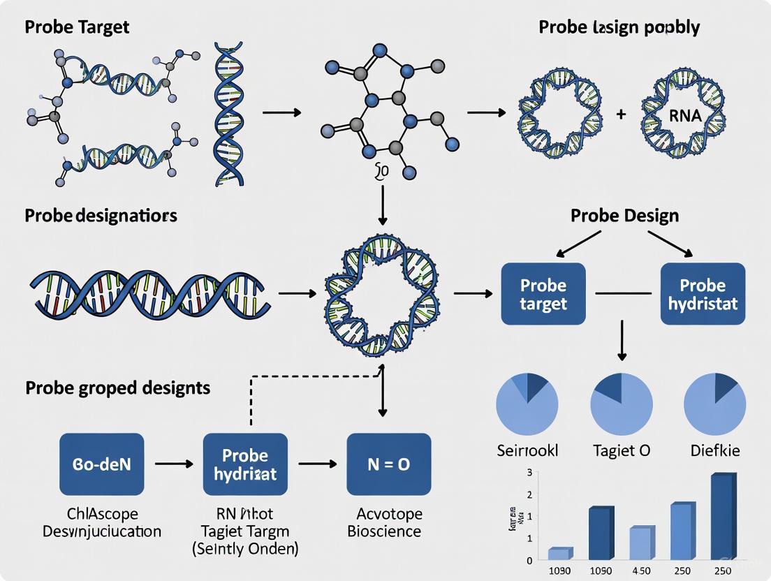
The RNAscope technology represents a groundbreaking advance in the field of in situ hybridization (ISH), enabling the highly sensitive and specific detection of target RNA within intact cells and tissues. At the core of this revolutionary platform is the proprietary 'ZZ' probe architecture, a unique design strategy that allows for single-molecule visualization while preserving tissue morphology. This probe design fundamentally improves the signal-to-noise ratio of RNA ISH by amplifying target-specific signals without amplifying background noise from nonspecific hybridization [1] [2].
Unlike traditional ISH techniques that use either labeled single oligonucleotides or cRNAs, the RNAscope approach employs a novel double-Z probe design strategy conceptually akin to fluorescence resonance energy transfer (FRET), wherein two independent probes must hybridize to the target sequence in tandem for signal amplification to occur [2] [3]. This requirement for physical proximity of two specific probes differentiates RNAscope from other traditional ISH hybridization protocols and provides the foundation for its exceptional specificity [4].
The Molecular Mechanism of ZZ Probes
Fundamental Architecture
The ZZ probe system functions through a meticulously engineered architecture where each target probe contains three distinct elements:
- Target Hybridization Region: An 18-25 base sequence complementary to the target RNA, selected for specific hybridization and uniform properties [5] [2]
- Spacer Sequence: A linking component that connects the hybridization region to the tail sequence [2]
- Tail Sequence: A 14-base region that facilitates the binding of amplification components [2]
For each target RNA species, approximately 20 double Z target probe pairs are designed to specifically hybridize to a ~1kb region of the target molecule [2]. The two tails from a double Z probe pair form a combined 28-base binding site for the pre-amplifier molecule [1] [2]. This requirement for adjacent hybridization makes it statistically improbable that nonspecific hybridization events would generate false positive signals, as it is highly unlikely that two independent probes would hybridize to a non-specific target right next to each other [2] [3].
Signal Amplification Cascade
The visualization of single RNA molecules is achieved through a cascade of highly specific hybridization events that provide exponential signal amplification:
- Double Z Target Probes Hybridization: Multiple ZZ probe pairs hybridize to the target RNA molecule [2]
- Preamplifier Binding: Each 28-base binding site formed by paired ZZ probes recruits a preamplifier molecule [4] [2]
- Amplifier Assembly: Multiple amplifier molecules bind to each preamplifier [2]
- Label Probe Attachment: Numerous labeled probes, containing fluorescent molecules or chromogenic enzymes, bind to each amplifier [1] [2]
This sequential amplification scheme can theoretically yield up to 8000 labels for each target RNA molecule when 20 probe pairs are used, providing sufficient signal intensity for single-molecule detection [4] [1]. The branching amplification structure creates independent "trees" for each successfully bound ZZ probe pair, enabling visualization of individual RNA molecules as distinct punctate dots under a standard microscope [2].
Table: Components of the RNAscope Signal Amplification System
| Component | Function | Binding Capacity |
|---|---|---|
| ZZ Probe Pair | Binds contiguously to target RNA | Creates 28-base preamplifier binding site |
| Preamplifier | Recognizes combined ZZ probe tails | Binds up to 20 amplifier molecules |
| Amplifier | Serves as secondary amplification stage | Binds up to 20 label probes |
| Label Probe | Delivers detectable signal | Contains fluorophore or enzyme |
Visualization of the ZZ Probe Mechanism
Comparative Analysis of RNAscope Assay Platforms
The fundamental ZZ probe architecture has been adapted into specialized assay platforms to address diverse research needs across molecular pathology and spatial genomics. Each platform maintains the core double-Z design principle while optimizing parameters for specific applications.
RNAscope Assay, the foundational platform, is designed to detect mRNA and non-coding RNA targets longer than 300 bases using a standard design of 20 ZZ pairs per target, though a minimum hybridization of just 7 ZZ pairs can generate detectable signal [6]. This platform provides robust detection against potential issues with partial target RNA accessibility or degradation, making it suitable for formalin-fixed paraffin-embedded (FFPE) tissues, fresh frozen tissues, and cultured cells [1] [6].
BaseScope Assay represents a refined version of the technology, optimized to detect shorter target sequences ranging from 50 to 300 bases using only 1-3 ZZ probes [4] [6]. This enhanced sensitivity enables specific detection of challenging targets including exon junctions, splice variants, highly homologous sequences, and point mutations with single-base discrimination capability [4] [6].
miRNAscope Assay further extends the technology to detect small RNAs between 17-50 bases, including microRNAs (miRNAs), small interfering RNAs (siRNAs), and antisense oligonucleotides (ASOs) [6] [7]. This advancement provides researchers with the ability to visualize the spatial distribution and functional efficacy of RNA therapeutic candidates within intact tissues alongside endogenous biomarkers [7].
Table: Comparison of RNAscope Technology Platforms
| Parameter | RNAscope Assay | BaseScope Assay | miRNAscope Assay |
|---|---|---|---|
| Target Length | >300 bases | 50-300 bases | 17-50 bases |
| ZZ Pairs per Target | 20 (minimum 7) | 1-3 | N/A (specialized design) |
| Primary Applications | mRNA, lncRNA detection | Exon junctions, splice variants, point mutations | miRNAs, siRNAs, ASOs |
| Multiplex Capability | Single to 12-plex | Single to duplex | Single-plex |
| Detection Methods | Chromogenic or fluorescent | Chromogenic | Chromogenic |
| Sample Compatibility | FFPE, fresh frozen, fixed frozen, cultured cells | FFPE, fresh frozen, fixed frozen, cultured cells | FFPE, fresh frozen, fixed frozen, cultured cells |
Experimental Protocol for Multiplex Fluorescent RNAscope
Materials and Reagent Solutions
The following protocol adapts the RNAscope Multiplex Fluorescent Assay for various sample types, incorporating critical steps to ensure optimal ZZ probe hybridization and signal development [8] [4].
Key Research Reagent Solutions:
Table: Essential Reagents for RNAscope Implementation
| Reagent/Category | Specific Examples | Function in Protocol |
|---|---|---|
| Biological Materials | Transgenic zebrafish embryos (Tg(kdrl:eGFP)), cell lines (SK-BR-3, MCF7), FFPE tissues | Provide biological context for target RNA detection |
| Fixation Reagents | Formaldehyde, Paraformaldehyde | Preserve tissue architecture and RNA integrity |
| Permeabilization Agents | Proteinase K, Pretreatment reagents | Unmask target RNA and permit probe access |
| Probe Systems | RNAscope Probe Dr-myb, Negative control DapB | Target-specific ZZ probe sets with channel assignments |
| Amplification Reagents | AMP1, AMP2, AMP3, HRP-C1, HRP-C2, HRP-C3 | Enable signal amplification cascade |
| Detection Dyes | OPAL-480, OPAL-570, OPAL-690 | Provide fluorescent signals for visualization |
| Buffer Systems | Wash Buffer, PBS, PBST, Hybridization buffers | Maintain optimal reaction conditions |
Step-by-Step Procedure
Sample Preparation and Fixation
Permeabilization and Pretreatment
- Treat samples with RNAscope Pretreatment Kit to unmask target RNA sequences
- For FFPE tissues: Incubate in citrate buffer (10 mmol/L, pH 6) at 100-103°C for 15 minutes, followed by protease digestion (10 μg/mL) at 40°C for 30 minutes [1]
- For cultured cells: Use protease digestion at 2.5 μg/mL at 23-25°C [1]
Probe Hybridization
Signal Amplification
Visualization and Analysis
Workflow Visualization
Technical Advantages and Applications
Key Performance Characteristics
The ZZ probe architecture provides several critical advantages over traditional ISH methods:
- Exceptional Sensitivity: Capable of detecting individual RNA molecules with as few as three double Z probes bound to the target RNA [2]
- Unmatched Specificity: The double Z probe design prevents background noise amplification since single Z probes binding to nonspecific sites cannot form the 28-base preamplifier binding site [2]
- Degradation Resistance: The relatively short target region (40-50 bases of the lower region of the double Z) allows successful hybridization even with partially degraded RNA, commonly encountered in FFPE samples [2]
- Multiplexing Capability: Independent amplification systems with different label probes enable simultaneous detection of multiple RNA targets within the same cell [4] [3]
Research and Clinical Applications
The unique capabilities of the ZZ probe system have enabled diverse applications across basic research and clinical diagnostics:
- Spatial Genomics: Mapping hematopoietic stem cell precursors in zebrafish embryos through detection of cmyb expression, revealing their emergence from arterial vessels and settlement in developmental niches [8]
- Neuroscience Research: Multiplex detection of up to three low-abundance mRNAs in single neurons, facilitating analysis of transcriptome complexity in heterogeneous neural cell populations [4]
- Cancer Diagnostics: Detection of RNA biomarkers in FFPE tissues for cancer diagnosis, prognosis, and therapy guidance, with preservation of histopathological context [1]
- Therapeutic Development: Visualization and quantification of oligonucleotide therapy delivery, spatial biodistribution, and efficacy with single-cell and single-molecule precision [7]
- Variant Detection: Discrimination of single nucleotide polymorphisms and splice variants through BaseScope technology, enabling study of RNA isoforms and polymorphisms in specific cell types [4]
The ZZ probe architecture represents a transformative advancement in in situ hybridization technology, providing an unprecedented combination of sensitivity, specificity, and technical robustness. Through its unique double-Z design and cascading amplification system, this platform enables researchers to visualize spatial gene expression patterns with single-molecule resolution within the native tissue context. As spatial biology continues to evolve, the ZZ probe foundation of RNAscope technology positions it as an essential tool for bridging molecular discoveries with morphological context across diverse research and clinical applications.
Within the broader context of RNAscope probe design guidelines research, selecting the appropriate in situ hybridization (ISH) technology is paramount for experimental success. The length of the target RNA sequence directly determines which proprietary ACD Bio platform—RNAscope, BaseScope, or miRNAscope—will deliver optimal detection sensitivity and specificity. These technologies, built upon the foundational RNAscope platform, employ a unique "ZZ" probe design that enables single-molecule visualization while preserving cellular and morphological context [6]. This application note provides a structured framework for researchers and drug development professionals to navigate the technical specifications of each platform, with particular emphasis on target length requirements, supported by comparative data, experimental protocols, and practical implementation guidelines.
Technology Comparison: Scope Assays at a Glance
The RNAscope, BaseScope, and miRNAscope assays share core technology but are optimized for different target classes based primarily on length. The following table summarizes the key specifications and applications of each platform to guide initial selection.
Table 1: Comparative Overview of RNAscope, BaseScope, and miRNAscope Assays
| Feature | RNAscope Assay | BaseScope Assay | miRNAscope Assay |
|---|---|---|---|
| Target Length | >300 bases [6] | 50–300 bases [6] | 17–50 bases [6] |
| Probe Design (ZZ Pairs) | 20 pairs (minimum of 7) [6] | 1 to 3 pairs [6] | N/A [6] |
| Primary Applications | mRNA, lncRNA [6] | Splice variants, point mutations, short indels, gene fusions, CRISP R edits [6] | microRNAs (miRNAs), ASOs, siRNAs [6] |
| Multiplex Capability | Single-plex up to 12-plex [6] | Single-plex to Duplex [6] | Single-plex [6] |
| Detection Methods | Chromogenic or Fluorescent [6] | Chromogenic [6] | Chromogenic [6] |
The ZZ Probe Design Principle
The core innovation behind these assays is the proprietary "ZZ" probe pair. Each "Z" oligonucleotide contains two hybridizing regions. The bottom region (18-25 bases) is complementary to the target RNA, while the upper region contains a preamplifier binding site. Each ZZ pair hybridizes to 36-50 bases of the target, and a standard RNAscope probe pool consists of 20 such pairs, creating a robust and redundant signal amplification system [5]. This design is refined in BaseScope for shorter targets using only 1-3 ZZ pairs [6] and adapted in miRNAscope for very small RNAs [6].
Decision Framework: Selecting the Right Assay Based on Target
The following diagram illustrates the decision-making workflow for selecting the appropriate ISH assay based on the characteristics of the target RNA.
Assay Selection Workflow for RNA In Situ Hybridization
Application Notes and Experimental Protocols
Protocol 1: BaseScope Assay for Short Viral RNA Targets
A recent study demonstrated the application of the BaseScope assay for detecting short RNA targets of the foot-and-mouth disease virus (FMDV) in African buffalo tissues, a context requiring high sensitivity in a carrier host [9].
- Objective: To detect and visualize FMDV RNA in formalin-fixed paraffin-embedded (FFPE) tissues from African buffalo, a natural wildlife reservoir.
- Rationale: BaseScope was selected due to its ability to detect short RNA sequences (50-300 nt) with high specificity and sensitivity, which is crucial for identifying low-abundance viral RNA in carrier animals [9].
- Key Findings: The optimized protocol was highly specific for FMDV RNA. A critical finding was that tissue preservation conditions, including formalin fixation for up to 7 days and storage of cut tissue sections for up to 3 months, did not negatively impact the assay's performance, demonstrating its robustness for retrospective studies [9].
- Protocol Summary:
- Tissue Preparation: FFPE tissues were sectioned at 5 µm thickness and mounted on SuperFrost Plus slides.
- Pretreatment: Slides were baked, deparaffinized, and subjected to antigen retrieval using the ACD Universal Pretreatment Kit, followed by protease digestion to permeabilize the tissue.
- Hybridization: BaseScope probes specific to FMDV were applied, and hybridization was performed using the HybEZ system.
- Signal Amplification & Detection: The proprietary BaseScope amplification steps were performed, followed by chromogenic development with Fast Red.
- Counterstaining & Mounting: Slides were counterstained with Gill's Hematoxylin and mounted with an appropriate mounting medium [9].
Protocol 2: miRNAscope Assay for Microvascular miRNA in Sepsis-Associated AKI
A 2025 study investigating sepsis-associated acute kidney injury (SA-AKI) employed the miRNAscope assay to spatially resolve microRNA expression in specific renal microvascular compartments [10].
- Objective: To identify and validate differentially expressed miRNAs in the renal microvasculature during SA-AKI in both mouse models and human patients.
- Rationale: The miRNAscope assay was essential for detecting short miRNAs (17-50 nt) with single-cell resolution, allowing the researchers to pinpoint compartment-specific miRNA responses within the complex tissue architecture of the kidney [10].
- Key Findings: The study identified 40 differentially expressed miRNAs in the renal microvasculature in response to SA-AKI. Notably, miR-21-5p was upregulated across all renal microvascular compartments in both mice and humans. Functional validation in HUVECs showed that inhibiting miR-21-5p exacerbated inflammatory activation, suggesting a protective role. Furthermore, plasma levels of miR-21-5p were elevated in SA-AKI patients, highlighting its potential as a biomarker [10].
- Protocol Summary:
- Sample Preparation: Fresh-frozen or FFPE human and mouse kidney tissues were sectioned.
- Probe Hybridization: The miRNAscope assay was performed according to the manufacturer's protocol, using specific probes for miR-21-5p and other targets.
- Signal Detection: Chromogenic detection was used to visualize miRNA expression.
- Analysis: miRNA expression was correlated with laser microdissection/RNA-seq data and validated in plasma samples via RT-qPCR [10].
Protocol 3: Novel BaseScope Validation Using Cell-Free Synthesized Controls
A 2025 protocol presented an innovative method for validating custom BaseScope probes using cell-free synthesized positive controls, bypassing the need for rare or difficult-to-obtain biological samples [11].
- Objective: To validate the functionality of custom-designed BaseScope probes for the rare human erythropoietin (EPO) splice variant hS3 before application to human brain samples.
- Rationale: Traditional validation requires cell lines or animal models expressing the target, which is time-consuming and costly. This protocol uses cell-free synthesized protein lysates and in vitro-transcribed mRNA as controlled sources of target RNA [11].
- Key Innovation: This is the first implementation of a duplex chromogenic BaseScope assay for cell-free, mRNA-containing solutions. It enables qualitative confirmation of probe functionality ("yes" or "no") with significantly reduced effort and cost [11].
- Protocol Highlights:
- Control Preparation: Cell-free synthesized EPO and hS3 protein lysates or purified in vitro-transcribed mRNAs are spotted onto slides.
- Assay Conditions: The standard BaseScope duplex assay is run with modifications: no target retrieval, H2O2, or protease steps are required for the liquid samples.
- Probe Validation: Successful signal generation from the controls confirms probe functionality, providing confidence before proceeding to tissue analysis [11].
The Scientist's Toolkit: Essential Research Reagent Solutions
Successful implementation of RNAscope technologies requires specific reagents and kits. The following table lists essential materials as referenced in the cited protocols.
Table 2: Key Reagents and Kits for Scope Assays
| Item Name | Function / Application | Example Catalog Number(s) | Source |
|---|---|---|---|
| HybEZ Hybridization System | Maintains optimum humidity and temperature during hybridization; required for manual assays. | 321461 (110V), 321462 (220V) | [12] |
| RNAscope 2.5 HD Reagent Kits | Chromogenic detection kits for RNAscope (Brown & Red) and BaseScope (Duplex). | 322300 (HD Brown), 322350 (HD Red), 322430 (Duplex) | [12] |
| Positive Control Probes | Species-specific housekeeping genes (e.g., PPIB, POLR2A, UBC) to verify RNA quality and assay performance. | Varies by species and gene | [13] |
| Negative Control Probe (dapB) | Bacterial gene probe used to assess non-specific background signal. | 310043 | [12] [13] |
| ImmEdge Hydrophobic Barrier Pen | Creates a barrier to prevent slide drying; the only pen validated for the RNAscope procedure. | 310018 | [12] [13] |
| Custom Probe Design Service | Service for designing target-specific probes for any gene, any species. | N/A (Online Request Form) | [14] |
The choice between RNAscope, BaseScope, and miRNAscope is fundamentally governed by the length of the target RNA, a critical consideration within any probe design guideline framework. RNAscope is the workhorse for standard mRNAs and long non-coding RNAs, BaseScope provides the sensitivity needed for shorter targets like splice variants and point mutations, and miRNAscope unlocks the visualization of the smallest RNA molecules, including miRNAs and therapeutic oligonucleotides. As demonstrated by the featured protocols, the precise application of these technologies, supported by appropriate controls and optimized workflows, enables researchers and drug developers to push the boundaries of spatial biology in diverse fields from infectious disease to therapeutic development.
In the evolving field of spatial transcriptomics, the ability to visualize multiple RNA targets simultaneously within their native tissue context is paramount. RNAscope in situ hybridization (ISH) technology has emerged as a powerful platform for this purpose, enabling highly sensitive and specific detection of RNA with single-molecule resolution [15] [16]. A critical component of this system's multiplexing capability is its probe channel designation system. These designations—C1, T1, S1, and others—are not arbitrary labels but are integral to the assay's architecture, dictating probe compatibility with specific detection kits and amplification channels. This guide provides a detailed exploration of these probe channel designations, framed within broader RNAscope probe design guidelines, to equip researchers and drug development professionals with the knowledge to effectively design and execute multiplexed experiments.
Core Principles of RNAscope Probe Design
The foundation of RNAscope's performance is its patented double Z (ZZ) probe design. This proprietary technology employs oligonucleotide pairs where each "Z" oligo contains an 18 to 25-base region complementary to the target RNA. Each ZZ pair hybridizes to 36-50 bases of the target, and a standard RNAscope probe pool typically consists of 20 ZZ pairs spanning approximately 1000 bases of unique sequence [5]. This design incorporates redundancy and robustness, resulting in high specificity and signal amplification.
The platform is adapted for different RNA target lengths through distinct assay types:
- RNAscope: Optimized for mRNAs or non-coding RNAs longer than 300 bases [5].
- BaseScope: Designed for short target sequences between 50-300 bases, utilizing 1-3 ZZ probe pairs [5].
- miRNAscope: Specialized for detecting small RNAs in the 17-50 base range [5].
Decoding Probe Channel Designations and Their Applications
Probe channel designations are a key aspect of the RNAscope probe nomenclature and indicate the amplification channel for which the probe was designed. The letter and number combinations define the assay compatibility and multiplexing potential.
Channel Designations and Assay Compatibility
Table 1: RNAscope Probe Channel Designations and Compatibility
| Channel Designation | Compatible Assays | Key Characteristics | Primary Applications |
|---|---|---|---|
| C1 | RNAscope 2.5 HD (BROWN/RED), Duplex, Multiplex Fluorescent; BaseScope; HiPlex [17] | Default channel; most abundant probe type; compatible with all manual detection platforms [17]. | Single-plex chromogenic or fluorescent detection; required channel in duplex assays. |
| C2 | RNAscope 2.5 HD Duplex, Multiplex Fluorescent [17] | Must be mixed with a C1 probe in a specific ratio (typically 1:50) for detection [18]. | Duplex chromogenic or lower-plex fluorescent assays. |
| C3 | RNAscope Multiplex Fluorescent Reagent Kit v2 [17] | Compatible only with fluorescent detection kits. | 3-plex or higher fluorescent multiplexing. |
| C4 | RNAscope 4-Plex Ancillary Kit for Multiplex Fluorescent v2 [17] | Requires an ancillary kit for detection. | 4-plex fluorescent assays. |
| T1-T12 | RNAscope HiPlex12 Reagents Kit [5] [17] | Used specifically with the high-plex HiPlex assay; probes are supplied in smaller volumes (for 10 slides) [17]. | Simultaneous detection of up to 12 RNA targets. |
| S1 | miRNAscope Assay [5] | Designed specifically for the miRNAscope platform. | Detection of small oligonucleotide sequences, including miRNAs. |
Probe Nomenclature and Interpretation
Understanding the probe naming convention is essential for selecting the correct reagents. The nomenclature follows a standardized pattern: Probe Type-Species-Gene-Specification-Channel [17].
Examples:
LS Probe - Mm-Rspo3: A probe for Leica automated systems (LS), targeting the Rspo3 gene in Mus musculus (Mm). The missing channel number indicates it is a C1 probe [17].Probe-Hs-GNRHR-5UTR-C2: A manual assay probe targeting the 5' untranslated region (5UTR) of the GNRHR gene in Homo sapiens (Hs), designed for channel C2 [17].BA-Mm-Nrg1-E1E2: A BaseScope Probe (BA) for Mus musculus, designed to span the junction of Exon 1 and Exon 2 (E1E2) of the Nrg1 gene [17].
Experimental Protocols for Multiplex Assays
Workflow for Multiplex RNAscope Assays
The following diagram illustrates the core workflow for a multiplex RNAscope experiment, from sample preparation to imaging and analysis.
Detailed Protocol: Fluorescent Multiplexing in Brain Tissue
This protocol, adapted from a peer-reviewed method for quantitative analysis in rat brain, outlines the steps for a 3-plex fluorescent assay [19].
A. Tissue Preparation (Fresh Frozen)
- Sacrifice & Dissection: Deeply anesthetize the animal and perform decapitation. Rapidly remove the brain from the skull.
- Snap-Freezing: Immediately submerge the brain in chilled 2-methylbutane (-30°C) for 25 seconds to snap-freeze. Avoid thawing.
- Sectioning: Embed the frozen brain in O.C.T. compound and section into 10 µm thick slices using a cryostat. Mount sections on Superfrost Plus slides, which are required to prevent tissue detachment [18].
- Fixation: Post-fix slides in 4% Paraformaldehyde (PFA) for 15 minutes at 4°C.
B. RNAscope Assay
- Pretreatment: Follow the RNAscope Fluorescent Multiplex kit instructions. For fresh frozen tissue, this includes a brief incubation with RTU Protease IV [19].
- Probe Hybridization:
- Probe Preparation: For a 3-plex assay, use target probes in three different channels (e.g., C1, C2, C3). C1 probes are ready-to-use (RTU), while C2 and C3 probes are 50X concentrates. Mix C2 and C3 probes with an RTU C1 probe or a Blank C1 Probe diluent at a 1:50 ratio [19].
- Hybridization: Apply the probe mixture to the tissue sections and incubate at 40°C for 2 hours in a HybEZ Oven. This system is essential for maintaining optimum humidity and temperature [18] [19].
- Signal Amplification & Development: Perform the series of amplifications (Amp 1-6) and development steps (HRP and fluorophores) as specified in the RNAscope Fluorescent Multiplex kit protocol. It is critical not to alter the protocol and to apply all amplification steps in the correct order [18].
- Counterstaining & Mounting: Counterstain with DAPI and mount with an appropriate anti-fade mounting medium like Fluoro-Gel II.
C. Image Acquisition and Quantitative Analysis
- Imaging: Acquire images using a slide scanner (e.g., Zeiss AxioScan) or a high-resolution confocal microscope.
- Automated Quantification with QuPath:
- Import images into the open-source software QuPath.
- Use built-in algorithms for cell detection based on DAPI counterstain.
- Establish mRNA signal thresholds using negative control (dapB) probes to define transcript-positive cells.
- Automate the quantification of dots (transcripts) per cell across the entire tissue section [19].
The Scientist's Toolkit: Essential Research Reagents
Table 2: Key Reagents and Equipment for RNAscope Multiplexing
| Item | Function | Example/Note |
|---|---|---|
| Catalog or Made-to-Order Probes | Target-specific detection for any gene in any species. | Over 40,000 targets across 400+ species; new probes designed in ~2 weeks [17]. |
| RNAscope Detection Kits | Chromogenic or fluorescent signal amplification. | Kit selection depends on probe channels (e.g., Multiplex Fluorescent for C1-C3) [17]. |
| HybEZ Hybridization System | Maintains optimal humidity and temperature during hybridization. | Mandatory for assay performance; includes oven, humidity tray, and paper [18] [19]. |
| Positive & Negative Control Probes | Assess sample RNA quality and assay specificity. | PPIB, POLR2A, UBC (positive); bacterial dapB (negative) [18]. |
| Superfrost Plus Microscope Slides | Provide superior tissue adhesion throughout the assay. | Other slide types may result in tissue detachment [18]. |
| Immedge Hydrophobic Barrier Pen | Creates a barrier to prevent reagent spread and tissue drying. | The only pen recommended for maintaining a barrier throughout the procedure [18]. |
| Automated Imaging Systems | High-throughput, automated image acquisition. | e.g., Xenium, Merscope, or slide scanners for high-plex analysis [20]. |
| Image Analysis Software | Quantification of transcript-positive cells and dot counting. | Open-source tools like QuPath enable automated, reproducible analysis [19]. |
Platform Selection and Experimental Design
Choosing the correct probe channel and assay platform is a critical strategic decision. The following decision tree guides researchers through the selection process based on their experimental goals.
Troubleshooting and Best Practices
- Sample Qualification: Always run positive and negative control probes on new sample types to assess RNA integrity and optimal permeabilization. Successful staining should yield a score of ≥2 for the low-copy positive control PPIB and <1 for the negative control dapB [18].
- Adherence to Protocol: Do not alter the protocol. Use all reagents and equipment as specified, including the HybEZ system, Superfrost Plus slides, and recommended mounting media to prevent assay failure [18].
- Probe Storage and Handling: Store probes at 4°C. Warm probes and wash buffer to 40°C before use to dissolve any precipitation that may have occurred during storage [18].
- Signal Interpretation: Score the number of dots per cell rather than signal intensity. The dot count correlates directly with RNA copy numbers [18].
Success with any in situ hybridization assay begins with good and consistent quality control (QC) practices. The RNAscope in situ hybridization (ISH) technology, a powerful method for detecting gene expression within the morphological tissue context, relies on rigorous controls that can be easily incorporated into every assay [21]. This application note details the essential protocols and guidelines for maintaining probe stability and implementing comprehensive quality control measures to ensure reproducible and reliable results in research and drug development settings. The proprietary "double Z" probe design, in combination with advanced signal amplification, enables highly specific and sensitive detection of target RNA with each dot visualizing a single RNA transcript [22] [23]. Within the broader context of RNAscope probe design guidelines research, proper QC practices are fundamental to leveraging the technology's full potential for spatial profiling of diverse mRNA markers at single-cell resolution.
Probe Stability and Design Specifications
Probe Stability and Storage
RNAscope probes demonstrate excellent stability when proper storage conditions are maintained. According to manufacturer testing, probes remain stable for up to 2 years from the date of manufacturing when stored as recommended at 4°C [5]. This extended shelf life provides researchers with consistent reagent performance across longitudinal studies and ensures experimental reproducibility.
Probe Design Architecture
The exceptional specificity and sensitivity of RNAscope probes stem from their proprietary design architecture. Each standard RNAscope probe consists of 20 ZZ pairs spanning approximately 1000 bases of unique target sequence [5]. Each "Z" oligo contains an 18-25 base region complementary to the target RNA, with each ZZ oligo pair collectively hybridizing to 36-50 bases of target RNA [5]. This redundant and robust design strategy provides the foundation for the technology's high specificity and single-molecule detection sensitivity.
Table 1: RNAscope Probe Design Specifications Based on Target Type
| Target Category | Minimum Sequence Length | Probe Design | Technology Platform |
|---|---|---|---|
| mRNA/ncRNA | >300 bases | 20 ZZ pairs | RNAscope |
| Short RNA Targets | 50-300 bases | 1-3 ZZ pairs | BaseScope |
| miRNA | 17-50 bases | Specialized design | miRNAscope |
For specialized applications, the BaseScope assay is designed to detect shorter target sequences ranging from 50 to 300 bases using 1-3 ZZ probe pairs, while miRNAscope detects RNAs between 17 to 50 bases [5]. This flexible design approach enables researchers to investigate a broad spectrum of RNA targets, from full-length mRNAs to short regulatory RNAs.
Comprehensive Quality Control Framework
Two-Tiered Quality Control Strategy
ACD recommends implementing a two-level quality control practice for RNAscope assays to ensure both technical proficiency and sample quality [21]:
Technical Assay Control Check: This verifies the assay is being performed with correct technique using cell pellet control samples tested with low-copy housekeeping gene positive control probes and non-specific bacterial negative control probes. Proper execution yields strong positive control probe staining and clean negative control probe staining [21].
Sample/RNA Quality Control Check: This assesses tissue RNA quality and fixation conditions using positive and negative control probes on actual experimental tissues to verify optimal pretreatment conditions [21].
Control Probe Selection Guidelines
Appropriate selection of control probes is critical for meaningful quality control assessment. ACD provides several positive control probes with varying expression levels to match different experimental needs [21]:
Table 2: RNAscope Positive Control Probe Selection Guide
| Positive Control Probe | Expression Level (copies per cell) | Recommended Application |
|---|---|---|
| UBC (Ubiquitin C) | Medium/High (>20) | Use with high expression targets only; not recommended for low-expressing targets due to risk of false negatives |
| PPIB (Cyclophilin B) | Medium (10-30) | Recommended for most tissues; provides rigorous control for sample quality and technical performance |
| Polr2A (RNA polymerase II) | Low (3-15) | Use with low expression targets; suitable for proliferating tissues like tumors, retinal, and lymphoid tissues |
For negative controls, ACD provides a universal negative control probe targeting the DapB gene (accession # EF191515) from the Bacillus subtilis strain SMY, which should not generate signal in properly fixed tissue specimens [21] [24]. Alternative negative control options include made-to-order sense direction probes or scrambled probes, though ACD notes that sense probes can occasionally produce ambiguous results if transcription occurs on the opposite strand [21].
Experimental Protocols for Quality Control
Recommended QC Workflow
Implementing a systematic QC workflow is essential for validating experimental conditions before proceeding with target-specific probes. The following diagram illustrates the recommended quality control workflow:
Diagram 1: RNAscope quality control workflow for testing samples prior to target gene expression evaluation.
RNAscope Scoring Guidelines
Proper interpretation of RNAscope staining is essential for accurate quality assessment. The assay uses a semi-quantitative scoring system that focuses on the number of dots per cell rather than signal intensity, as the dot count correlates directly with RNA copy numbers [24] [18]. The following table outlines the standardized scoring criteria:
Table 3: RNAscope Semi-Quantitative Scoring Guidelines
| Score | Staining Criteria |
|---|---|
| 0 | No staining or <1 dot/10 cells |
| 1 | 1-3 dots/cell (visible at 20-40X magnification) |
| 2 | 4-9 dots/cell; none or very few dot clusters |
| 3 | 10-15 dots/cell and <10% dots are in clusters |
| 4 | >15 dots/cell and >10% dots are in clusters |
Successful staining quality should demonstrate a PPIB score ≥2 or UBC score ≥3 with relatively uniform signal distribution throughout the sample, while the dapB negative control should score <1, indicating minimal background staining [24] [18]. The scoring relationship between positive and negative controls is visualized below:
Diagram 2: Control probe scoring criteria and their relationship to expression levels.
Protocol for Pretreatment Optimization
Tissue pretreatment often requires optimization, particularly when sample preparation history is unknown or deviates from recommended guidelines. ACD provides the following optimization protocol [18]:
Initial Assessment: Begin with standard pretreatment conditions (15 minutes Epitope Retrieval 2 at 95°C and 15 minutes Protease at 40°C for automated systems).
Control Probe Testing: Apply positive and negative control probes (PPIB and dapB) to assess initial staining quality.
Parameter Adjustment: If signal is low or background is high, adjust pretreatment times:
- For over-fixed tissues: Increase ER2 time in 5-minute increments and Protease time in 10-minute increments while maintaining constant temperatures (e.g., 20 min ER2 at 95°C and 25 min Protease at 40°C).
- For milder pretreatment: Use 15 min ER2 at 88°C and 15 min Protease at 40°C.
Iterative Testing: Repeat control probe testing with adjusted parameters until optimal staining is achieved (PPIB score ≥2 and dapB score <1).
The Scientist's Toolkit: Essential Research Reagent Solutions
Implementing robust RNAscope QC protocols requires specific reagents and materials. The following table details essential research reagent solutions for successful assay implementation:
Table 4: Essential Research Reagent Solutions for RNAscope QC
| Reagent/Material | Function/Application | Specific Recommendations |
|---|---|---|
| Control Slides | Technical assay validation | Human HeLa (Cat. No. 310045) or Mouse 3T3 (Cat. No. 310023) cell pellets [24] |
| Positive Control Probes | Sample RNA quality assessment | PPIB (medium expression), POLR2A (low expression), or UBC (high expression) [21] [24] |
| Negative Control Probe | Background staining assessment | dapB gene (bacterial) for verifying specificity [21] [24] |
| Slides | Tissue adhesion and retention | Fisher Scientific SuperFrost Plus slides required to prevent tissue loss [24] [18] |
| Barrier Pen | Liquid containment during manual assays | ImmEdge Hydrophobic Barrier Pen (Vector Laboratories Cat. No. 310018) - only pen compatible with entire procedure [18] |
| Mounting Media | Slide preservation and visualization | Xylene-based media (CytoSeal XYL) for Brown assay; EcoMount or PERTEX for Red and 2-plex assays [18] |
| Reference Standards | Assay performance validation | IHC HDx Reference Standards from Horizon Discovery for verifying sensitivity and specificity [25] |
| HybEZ System | Hybridization conditions | Maintains optimum humidity and temperature during critical hybridization steps [18] |
Advanced QC Applications in Drug Development
For drug development professionals, RNAscope technology offers unique advantages for biomarker validation and therapeutic development. The technology serves as a powerful orthogonal method for antibody validation, addressing the well-documented "reproducibility crisis" associated with antibody-based assays [26]. With custom probe development requiring just 3 weeks from sequence submission to delivery, compared to 6-9 months and $20,000 for custom antibody development, RNAscope provides an efficient solution for accelerating therapeutic programs [26].
The compatibility of RNAscope with automated clinical platforms like the Leica BOND III system enables seamless translation of research findings to clinical applications, with currently 16 Analyte-Specific Reagents (ASRs) available for diagnostic use [27]. This streamlined pathway from research to clinical application underscores the importance of robust QC practices implemented early in the drug development pipeline.
Implementing comprehensive quality control measures for RNAscope assays, including rigorous attention to probe stability, appropriate control selection, systematic workflow validation, and precise scoring interpretation, is fundamental to generating reproducible and reliable spatial gene expression data. By adhering to the protocols and guidelines outlined in this application note, researchers and drug development professionals can ensure the technical rigor of their RNAscope experiments, thereby generating high-quality data that advances scientific discovery and therapeutic development. The integration of these QC practices within the broader framework of RNAscope probe design guidelines establishes a foundation for excellence in spatial biology research.
In situ hybridization (ISH) technologies, particularly RNAscope, have revolutionized RNA visualization within intact cells and tissues, providing single-molecule sensitivity and high specificity through a unique double Z ("ZZ") probe design [28]. For researchers studying animal models of human diseases, evolutionary biology, or comparative biology, the ability to design probes that function accurately across species boundaries is paramount. Success in cross-species probe design hinges primarily on one critical factor: sequence homology between the target regions of different species [5].
This application note provides a structured framework for navigating sequence homology requirements in cross-species probe design. We detail the minimum homology thresholds, outline a computational and experimental workflow for homology assessment and validation, and present a reagent toolkit to support researchers in developing robust cross-species assays. Adherence to these guidelines ensures that probe design is both efficient and effective, generating reliable and interpretable data from pre-clinical and comparative studies.
Key Considerations for Cross-Species Probe Design
Sequence Homology Requirements
The fundamental requirement for a probe to hybridize to a target RNA in a species other than its original design target is a high degree of sequence similarity. Quantitative analysis confirms a strict threshold for cross-species compatibility.
Table 1: Sequence Homology Requirements for Cross-Species Probe Application
| Homology Level | Feasibility | Probe Performance | Recommended Action |
|---|---|---|---|
| >95% | High Feasibility | Expected high specificity and sensitivity | Proceed with standard probe design and validation [5] |
| 90–95% | Moderate Feasibility | Potential reduced sensitivity; requires empirical testing | Consider designing a species-specific probe; test performance rigorously [28] |
| <90% | Low Feasibility | High risk of failure; low signal or non-specific binding | Design a new, species-specific probe [5] |
The >95% sequence homology rule is a well-established benchmark for reliably using a probe designed for one species to detect its ortholog in another [5]. In practice, this means that for a standard RNAscope probe, which consists of 20 ZZ pairs spanning approximately 1000 bases of the target RNA, the sequences must be nearly identical to ensure all individual probe binding regions function correctly [5] [4]. For example, in a study of cynomolgus monkey tissues, human probes for the housekeeping genes PPIB and POLR2A were successfully used because they shared over 95% homology with the monkey sequences [28]. Conversely, for genes like CD68 and KI67, where homology between human probes and cynomolgus monkey targets fell between 90–95%, the probes were usable but required validation to confirm performance [28].
Probe Design and Technical Specifications
Understanding the core technology is essential for appreciating homology requirements.
Probe Design Fundamentals: RNAscope probes are not single, long molecules but pools of short, target-specific oligonucleotide pairs [5]. Each "ZZ" pair consists of two oligonucleotides (each 18–25 bases) designed to bind adjacent regions on the target RNA, spanning 36–50 bases collectively [5] [4]. A typical RNAscope probe set contains 20 such ZZ pairs, tiling a region of about 1000 bases of unique mRNA sequence [5]. This multi-pair approach creates a redundant and robust system where the simultaneous binding of both oligonucleotides in a pair is required for signal amplification, thereby conferring high specificity [5] [4].
Assay Versatility Based on Target Length: The RNAscope platform offers different assays tailored to the length of the target RNA, which influences probe design.
- RNAscope: Optimized for standard mRNAs and non-coding RNAs longer than 300 nucleotides (with 1000 bases being optimal) [5] [29].
- BaseScope: Designed for shorter targets between 50 and 300 nucleotides, such as splice variants or RNAs with limited unique sequence. BaseScope probes are built with 1-3 ZZ pairs [5] [4].
- miRNAscope: Targets small RNAs (e.g., miRNAs) as short as 17 nucleotides [5] [29].
Diagram 1: A decision workflow for cross-species probe design, highlighting the critical homology check.
Computational Pipeline for Homology Assessment
A systematic computational approach is vital for accurately determining sequence homology and selecting genomic regions for probe design.
Cross-Species RNA-Seq Analysis Protocol
The following pipeline, largely based on free open-source R and Bioconductor packages, provides a step-by-step method for cross-species gene expression analysis, which inherently involves homology assessment [30].
Short-Read Alignment and Quantification: Begin by aligning RNA-seq short reads from the query species to its respective genome using an aligner like SHRiMP, TopHat, or GSNAP [30]. The output in SAM/BAM format is then used to quantify gene expression based on the species' annotation.
Generation of Cross-Species Genome Annotations: This is the crucial step for homology assessment. Select one species as the reference (e.g., mouse mm10). Using pairwise genome alignments (e.g., from UCSC in AXT format), "lift" the constitutive exons from the reference annotation to their orthologous positions in the query species' genome [30]. The resulting annotation will contain only the exons that are orthologously present in all species being compared, using the reference species' gene IDs.
Differential Expression and Pathway Analysis: With a unified annotation, count reads aligning to each exon in each species using a tool like
Rsubread[30]. These counts are then analyzed for differential expression between species using a negative binomial model inedgeR[30]. Finally, pathway enrichment analysis of differentially expressed genes can be performed with tools like GAGE and SPIA, using databases like KEGG to understand biological implications [30].
Practical Workflow for Probe Selection
For researchers ordering probes from a vendor like ACD (a Bio-Techne brand), the process is streamlined but follows the same logical principles.
Table 2: Experimental Protocol for Cross-Species Probe Validation
| Step | Procedure | Purpose | Key Specifications |
|---|---|---|---|
| 1. Sample Prep | Fix tissues in fresh 10% NBF for 16–32 hrs at RT. Embed in paraffin (FFPE) or cryopreserve (frozen). | Preserve tissue morphology and RNA integrity. Prevents RNA degradation. | Section thickness: 5 μm (FFPE), 10-20 μm (frozen) [28] [4] |
| 2. Control Assay | Run parallel slides with species-specific positive control (e.g., PPIB, POLR2A) and negative control (dapB) probes. | Qualify sample RNA and assess assay performance on the sample. | A score of ≥2 for PPIB and 0 for dapB indicates success [18] [28] |
| 3. Target Assay | Perform RNAscope with the cross-species probe candidate. | Test the binding and signal generation of the candidate probe. | Follow manual or automated protocol precisely [18] |
| 4. Analysis | Score signal puncta per cell. Compare to controls. | Determine if the probe provides specific detection at expected expression levels. | Score 0-4 based on dots/cell; clusters indicate high expression [31] [28] |
Diagram 2: A computational pipeline for cross-species RNA-seq analysis, which forms the basis for homology assessment.
Essential Reagents and Research Tools
Successful implementation of cross-species probe experiments requires specific reagents and equipment. The following toolkit details the essential components.
Table 3: Research Reagent Solutions for Cross-Species RNAscope
| Item | Function in Protocol | Specification & Notes |
|---|---|---|
| Target Probes | Hybridize to the RNA of interest. The core reagent for detection. | C1 for single-plex/multiplex; C2, C3, C4 for multiplex only. 50x stocks for C2-C4 [4]. |
| Control Probes | Verify assay performance and sample quality. | Positive: Species-specific housekeeping genes (PPIB, POLR2A, UBC). Negative: Bacterial dapB gene [18] [28]. |
| RNAscope Kit | Contains all reagents for signal amplification and detection. | e.g., RNAscope Fluorescent Multiplex Kit (Cat. # 320851). Includes amplifiers, labels, and wash buffers [4]. |
| Pretreatment Kit | Prepares tissue for hybridization by permeabilizing cells and exposing target RNA. | Critical for accessing RNA. Includes H₂O₂ block, retrieval reagents, and protease [4]. |
| HybEZ Oven | Provides precise temperature (40°C) and humidity control during hybridization. | Essential for manual assays to ensure proper probe binding and prevent slide drying [18] [29]. |
| SuperFrost Plus Slides | Microscope slides for mounting tissue sections. | Required to prevent tissue detachment during the stringent assay steps [18] [29]. |
Navigating the requirements for cross-species probe design is a structured process that begins with a rigorous computational assessment of sequence homology. The >95% homology threshold is a non-negotiable starting point for attempting to use a probe across species. When this threshold is not met, the path forward is the design of a new, species-specific probe.
The robustness of the RNAscope platform, with its multi-ZZ-pair design and built-in controls, provides a solid foundation for reliable cross-species analysis when the sequence homology is sufficient [5]. By adhering to the recommended workflow—starting with in silico analysis, followed by careful sample preparation and qualification with control probes, and culminating in a rigorously scored experimental assay—researchers can confidently utilize this powerful technology to uncover meaningful biological insights across a wide range of species in both basic research and drug development contexts.
Advanced Methodologies and Custom Probe Applications
For researchers investigating novel genomic regions, specific transcript variants, or working with non-standard model organisms, the inability to find commercially available in situ hybridization (ISH) probes can significantly impede scientific progress. Custom probe design services bridge this critical gap, enabling investigation of any gene of interest across any tissue type from any species [14] [32]. This application note details the complete workflow for custom probe design, focusing primarily on the RNAscope platform (ACD, a Bio-Techne brand), from initial request submission through final delivery and experimental implementation. The proprietary ZZ probe design strategy employed in RNAscope utilizes oligo pairs where each "Z" oligo contains an 18-25 base region complementary to the target RNA, with a typical probe consisting of 20 ZZ pairs spanning approximately 1000 bases of unique sequence [5]. This design incorporates built-in redundancy and robustness, resulting in the high specificity and sensitivity that has made RNAscope a standard ISH approach in many fields, particularly neuroscience [33]. The protocol outlined herein provides researchers with a comprehensive framework for accessing and utilizing these custom design services to advance their investigative goals.
The Custom Probe Design Workflow
The journey from conceptualizing a custom probe to receiving a validated product follows a structured, collaborative pathway between the researcher and the probe design specialists. The process ensures that the final probes are precisely tailored to the researcher's specific experimental requirements, whether for detecting knockout constructs, episomal DNA viral vectors, specific transcript variants, or cross-species targets [14] [32]. The following diagram and table summarize the key stages in this workflow.
Custom Probe Design Workflow from Request to Delivery
Table 1: Key Stages in the Custom Probe Design and Delivery Process
| Workflow Stage | Timeline | Key Actions & Outputs |
|---|---|---|
| Request Submission | Immediate | Researcher submits details via online New Probe Request (NPR) form, specifying target gene, species, and special requirements [14]. |
| Feasibility Review | Within 48 hours | Probe design specialists evaluate sequence availability, homology (>95% needed for cross-species detection), and design feasibility [5] [34]. |
| Design Proposal | Within 72 hours | Specialist creates a tailored probe design, often with an ideogram, specifying target region and number of ZZ probe pairs [34]. |
| Collaborative Review | Variable | Researcher and specialist discuss design; up to three revisions are typically incorporated to ensure the probe meets research goals [34]. |
| Quote & Order | Immediate post-approval | Researcher receives and approves a customized quote, then places the formal order for probe manufacturing [14] [34]. |
| Manufacturing & QC | ~2 Weeks | Probes are manufactured; quality control includes specificity verification and performance testing [5] [34]. |
| Delivery | Overnight (U.S.) / International Priority | Final probes are delivered, stable for up to 2 years when stored at 4°C as recommended [5] [34]. |
Submission and Request Methods
Researchers can initiate the custom probe process through several flexible submission workflows tailored to different project scales and privacy needs [32]:
- Manual Entry: Ideal for up to 5 requests requiring advanced customization, allowing full specification of design parameters.
- Bulk Upload: Designed for submitting more than 5 requests simultaneously, suitable for panel applications or grant proposals.
- Proprietary Sequence Submission: Secured workflow for targets requiring confidentiality, such as therapeutic, novel, or modified targets.
Probe Design Specifications and Scenarios
Custom RNAscope probes are designed to integrate seamlessly with existing RNAscope assay kits, both chromogenic and fluorescent, for manual or automated configurations [14]. The design parameters vary significantly based on the target type and length, as outlined in the table below.
Table 2: Probe Design Specifications by Target Type and Application
| Assay Platform | Target Length | Probe Design | Typical ZZ Pairs | Primary Applications |
|---|---|---|---|---|
| RNAscope | >300 bases | 20 ZZ pairs spanning ~1000 bases | 20 | mRNA, ncRNA, long RNA targets [5] |
| BaseScope | 50-300 bases | 1-3 ZZ probe pairs | 1-3 | Short transcripts, splice variants, point mutations [5] |
| miRNAscope | 17-50 bases | Specialized design for small RNAs | N/A | microRNAs, small oligonucleotides [5] |
| Cross-Species | Varies | Dependent on >95% sequence homology | Varies | Detecting orthologs across multiple species [5] |
Advanced Customization Scenarios
The flexibility of custom probe design accommodates diverse and complex research needs [14] [32]:
- Specific Transcript Variants: Precisely target individual splice variants by designing probes against unique exon-exon junctions or variant-specific regions.
- Knockout Validation: Design probes that bind to the disrupted region of a gene to confirm knockout efficacy, or to the inserted marker to identify modified cells.
- Viral Vector Detection: Detect episomal DNA viral vectors or transgenes using probes specific to the vector backbone or transgene sequence.
- Xenograft Studies: Accommodate application-specific cross-reactivity requirements, such as designing human-specific probes to study human tumor cells in a mouse host environment.
- Oligonucleotide Therapeutics: Visualize and quantify the spatial biodistribution, uptake, and efficacy of synthetic oligonucleotide drugs (ASOs, siRNAs) alone or in combination with endogenous biomarkers [7].
Experimental Protocol: RNAscope Assay for Fresh-Frozen Tissue
The following detailed protocol, adapted from standardized methodologies [33] [35], outlines the application of custom RNAscope probes for transcript detection in fresh-frozen rodent brain tissue, a common application in neuroscience research.
Materials and Reagents
- Tissue Preparation:
- Fresh-frozen tissue samples (e.g., rodent brains)
- 2-methylbutane (isopentane), chilled on dry ice
- Tissue-Tek O.C.T. compound
- Fisherbrand Superfrost Plus microscope slides
- RNAscope Reagents:
- RNAscope Fluorescent Multiplex Reagent Kit v1 (ACD, #320850) for fresh-frozen applications
- RNAscope RTU Protease IV reagent (ACD, #322340)
- Custom-designed RNAscope target probes (e.g., C1, C2, C3 channel probes)
- RNAscope 3-plex negative control probes (ACD, #320871)
- Equipment:
- Cryostat (e.g., Thermo Scientific Cryostar NX50)
- HybEZ II System Hybridization Oven (ACD)
- Slide scanner (e.g., Carl Zeiss AxioScan Z.1) or fluorescence microscope
- Software for Analysis:
- QuPath 0.3.2 open-source software for automated quantification [33]
Detailed Procedure
A. Tissue Preparation and Sectioning
- Fresh-Frozen Tissue Collection: Deeply anesthetize the animal and perform sacrifice by decapitation. Rapidly remove the brain and immediately snap-freeze it by immersing it in chilled 2-methylbutane (-30°C to -40°C) for 25 seconds [33].
- Storage: Wrap the snap-frozen brain in aluminum foil and store it at -80°C for up to 12 months to prevent mRNA degradation. Avoid extended storage periods.
- Cryosectioning: Using a cryostat, prepare 10-20 μm thick sections and mount them on Superfrost Plus slides. Store slides at -80°C until ready for use.
B. RNAscope In Situ Hybridization
Fixation and Permeabilization:
- Fix frozen tissue sections in 4% formaldehyde for 30 minutes at 4°C.
- Dehydrate sections through a graded ethanol series (50%, 70%, 100%) [35].
- Treat sections with RNAscope Protease Plus reagent for 30 minutes at 40°C in the HybEZ oven to permeabilize the tissue and expose target RNA.
Probe Hybridization and Amplification:
- Apply the custom-designed RNAscope probe mixture to the tissue sections.
- Incubate slides for 2 hours at 40°C in the HybEZ oven to allow target-specific hybridization.
- Perform a series of amplifications using the provided AMP 1-6 reagents per the manufacturer's instructions (e.g., RNAscope 2.5 HD assay protocol) to amplify the signal [35].
- For fluorescent detection, develop the signal using fluorophores compatible with your probe channels (C1, C2, C3). For chromogenic detection, use reagents such as RNAscope Fast Red A & B.
Counterstaining and Mounting:
Image Acquisition and Quantitative Analysis
- Slide Scanning: Acquire high-resolution images of the entire tissue section using a slide scanner (e.g., Zeiss AxioScan Z.1) with consistent exposure settings across all samples [33].
- Automated Quantification in QuPath:
- Import whole-slide images into QuPath.
- Use the built-in cell detection algorithm to identify individual cells based on the nuclear counterstain (DAPI or Hematoxylin).
- Carefully optimize cell detection parameters (e.g., nucleus diameter, intensity threshold) using QuPath's interactive machine learning tools.
- Set fluorescence intensity thresholds for transcript positivity using negative control probes (e.g., 3-plex negative control) to establish a baseline and ensure reproducible, objective quantification [33].
- Export quantitative data (e.g., transcripts per cell, number of positive cells) for statistical analysis in software such as GraphPad Prism.
The Scientist's Toolkit: Essential Research Reagent Solutions
Table 3: Key Reagents and Materials for RNAscope Experiments
| Item | Function/Application | Example Catalog Number |
|---|---|---|
| RNAscope Fluorescent Multiplex Kit | Core reagents for probe hybridization, amplification, and detection in fresh-frozen tissue. | ACD #320850 [33] |
| Custom Target Probes | Target-specific ZZ probe pairs designed for your gene and species of interest. | Varies by request [14] |
| RNAscope Protease Plus/IV | Enzyme treatment for tissue permeabilization and target retrieval. | ACD #322330 [35] |
| HybEZ II Hybridization Oven | Provides precise temperature control (40°C) required for the hybridization steps. | ACD #240200ACD [35] |
| Negative Control Probe (3-plex) | Essential control to set background signal and threshold for quantification. | ACD #320871 [33] |
| Positive Control Probe | Validates assay procedure is working correctly (e.g., housekeeping gene). | Varies by species |
| ImmEdge Hydrophobic Barrier Pen | Creates a barrier around tissue sections to contain liquids during incubations. | Vector Labs #310018 [33] |
| QuPath Software | Open-source platform for automated, high-throughput quantification of RNAscope signal. | https://qupath.github.io/ [33] |
Custom probe design represents a powerful enabling technology for modern molecular research, breaking down barriers imposed by limited commercial probe availability. The structured workflow from initial request to final delivery ensures that researchers obtain high-quality, specific probes tailored to their unique experimental needs, including specialized applications like transcript variant analysis, xenograft studies, and oligonucleotide therapeutic development [32] [7]. When combined with robust experimental protocols, such as the RNAscope assay for fresh-frozen tissue, and advanced analytical tools like QuPath for automated quantification [33], these custom reagents provide a comprehensive solution for achieving precise, reproducible, and insightful spatial gene expression data. By leveraging these capabilities, researchers and drug development professionals can accelerate the pace of discovery and advance promising new therapies.
Accurate identification of nuclei belonging to specific cell types remains a fundamental challenge in tissue biology and regeneration research. This challenge is particularly acute in the heart, where cardiomyocyte nuclei constitute only 20-30% of total nuclei despite occupying 70% of tissue volume, making them difficult to distinguish from interstitial cell nuclei using conventional methods [36]. While antibodies against sarcomeric proteins have been widely used for this purpose, this approach provides only 43% sensitivity and 89% specificity for nuclear identification, figures that improve only marginally even with additional membrane staining [36].
Intronic RNAscope probes represent a transformative solution to this problem by targeting pre-mRNA transcripts before they undergo splicing and exit the nucleus. This innovative approach leverages the natural accumulation of intronic sequences within the nucleus, providing unprecedented precision for nuclear localization in a cell-type-specific manner [36]. This application note details the design principles, validation data, and optimized protocols for implementing this cutting-edge methodology in cardiac research and beyond.
Probe Design and Mechanism of Action
Fundamental Principles
The RNAscope platform utilizes a unique ZZ probe design strategy that enables simultaneous signal amplification and background suppression for single-molecule visualization while preserving tissue morphology [36]. Each probe consists of approximately 20 "ZZ" probe pairs, with each pair targeting 36-50 bases of the target RNA [5]. For intronic probes, this design is strategically targeted to intronic regions of pre-messenger RNA (pre-mRNA) that are naturally retained within the nucleus before splicing occurs [36].
The technology can be adapted for different detection needs through specialized probe configurations:
- RNAscope: Detects mRNAs or ncRNAs >300 bases using 20 ZZ pairs [5]
- BaseScope: Targets short sequences (50-300 bases) using 1-3 ZZ probe pairs [5]
- miRNAscope: Designed for small RNAs (17-50 bases) [5]
Design Considerations for Nuclear Localization
Intronic probes are designed to target unique sequences within intronic regions of cell-type-specific genes. For cardiomyocyte identification, probes against TnnT2 (cardiac troponin T), Myl2 (ventricular myosin light chain), and Myl4 (atrial myosin light chain) have demonstrated high specificity [36]. The table below summarizes key design parameters for successful intronic RNAscope probes.
Table 1: Key Design Parameters for Intronic RNAscope Probes
| Parameter | Specification | Technical Rationale |
|---|---|---|
| Target Region | Pre-mRNA intronic sequences | Enables nuclear localization before splicing and export [36] |
| Sequence Length | ~1000 bases for standard RNAscope | Accommodates 20 ZZ probe pairs for robust signal amplification [5] |
| Sequence Homology | >95% for cross-species detection | Ensures consistent hybridization across species [5] |
| Probe Specificity | Single-cell and single-molecule resolution | Unique ZZ probe design with background suppression [36] |
| Amplification Channels | C1-C4 for multiplexing | Enables simultaneous detection of multiple targets [5] |
Figure 1: Mechanism of Intronic RNAscope Probes. Probes target pre-mRNA intronic sequences retained in the nucleus before splicing, enabling precise nuclear localization.
Performance Validation and Comparative Analysis
Specificity and Sensitivity Metrics
The Tnnt2 intronic RNAscope probe demonstrated exceptional performance in validation studies, showing near-perfect colocalization with Obscurin-H2B-GFP in adult mouse hearts, confirming its cardiomyocyte specificity [36]. Unlike antibody-based approaches that struggle with accurate nuclear attribution, especially during cell division, the intronic probe technology maintained precise association with cardiomyocyte chromatin throughout all mitotic stages, including after nuclear envelope breakdown [36].
Table 2: Performance Comparison of Nuclear Identification Methods
| Method | Sensitivity | Specificity | Limitations | Advantages |
|---|---|---|---|---|
| Antibodies to Sarcomeric Proteins | 43% (65% with WGA) [36] | 89% (97% with WGA) [36] | Poor nuclear attribution; cannot detect during mitosis [36] | Widely available; established protocols |
| Nuclear Markers (Nkx2.5, Gata4, Mef2c) | Variable (Nkx2.5: low in adults) [36] | Variable (Gata4/Mef2c: expressed in non-CMs) [36] | Low expression; non-specificity; controversial markers [36] | Nuclear localization |
| Genetic Models (Obscurin-H2B-GFP) | High [36] | High [36] | Costly colony maintenance; potential cardiac phenotypes [36] | Unambiguous identification |
| Intronic RNAscope Probes | High (precise quantification underway) [36] | High (validated by colocalization) [36] | Requires RNA integrity; optimization needed [37] | Specific; works in mitosis; no genetic modification [36] |
Functional Applications in Cell Cycle Analysis
A critical advantage of intronic RNAscope probes is their ability to remain associated with cardiomyocyte chromatin throughout all stages of mitosis, enabling reliable detection of cell cycle activity even after nuclear envelope breakdown [36]. This capability has proven particularly valuable for investigating cardiomyocyte DNA synthesis and potential mitotic activity in border and infarct zones after myocardial infarction [36].
The technology has also enabled the development of subtype-specific identification through Myl2 (ventricular) and Myl4 (atrial) intronic probes, providing tools for characterizing cardiomyocyte subtypes generated during in vitro differentiation from ESCs or iPSCs [36].
Research Reagent Solutions
Table 3: Essential Reagents for Intronic RNAscope Implementation
| Reagent/Equipment | Manufacturer/Catalog Number | Function |
|---|---|---|
| RNAscope Multiplex Fluorescent Reagent Kit v2 | ACD / 323100 [37] | Core amplification reagents for signal detection |
| TSA Plus Fluorescence Detection Kits | Akoya Biosciences / NEL741001KT, NEL744001KT, NEL745001KT [37] | Fluorescent signal development (FITC, Cy3, Cy5) |
| HybEZ II Hybridization System | ACD / 321710/321720 [37] | Precision temperature control for hybridization |
| Protease III | ACD / Included in kits [37] | Tissue permeabilization for probe access |
| Target Probes (Tnnt2, Myl2, Myl4) | ACD / Custom design [36] | Cell-type-specific intronic target detection |
Experimental Protocols
Optimized RNAscope Protocol for Cryosections
This protocol has been specifically optimized for identifying cardiomyocyte nuclei in cardiac tissue sections [37].
Figure 2: Cryosection RNAscope Workflow. Two-day protocol for precise cardiomyocyte nuclei identification in tissue sections.
Day 1: Sample Preparation and Hybridization
- Refixation: Refix cryosections in 4% PFA/PBS at room temperature for 15 minutes, followed by a single wash with ddH₂O [37].
- Dehydration: Incubate sections in 50% EtOH for 5 minutes, 70% EtOH for 5 minutes, and two washes in 100% EtOH for 5 minutes each. Air dry sections completely [37].
- Peroxidase Blocking: Treat with H₂O₂ for 10 minutes at room temperature to quench endogenous peroxidase activity, followed by two washes with ddH₂O for 2 minutes each [37].
- Permeabilization: Circle sections with a pap pen and incubate with Protease III for 20 minutes at room temperature (extend to 40 minutes at 40°C if no antibody staining is required in subsequent steps) [37].
- Probe Hybridization: Apply Tnnt2 intronic RNAscope probe (or other channel-specific probes) and incubate for 2 hours at 40°C in a hybridization oven [37].
- Overnight Incubation: Wash twice with 1× wash buffer for 2 minutes each, then incubate in 5× SSC (pH 5.2) at room temperature overnight [37].
Day 2: Signal Amplification and Detection
- AMP1 Incubation: Incubate with AMP1 for 30 minutes at 40°C, followed by two 2-minute washes with 1× wash buffer [37].
- AMP2 Incubation: Incubate with AMP2 for 30 minutes at 40°C, followed by two 2-minute washes with 1× wash buffer [37].
- AMP3 Incubation: Incubate with AMP3 for 15 minutes at 40°C, followed by two 2-minute washes with 1× wash buffer [37].
- HRP Incubation: Incubate with HRP-C1 (or corresponding channel) for 15 minutes at 40°C, followed by two 2-minute washes with 1× wash buffer [37].
- Fluorescent Detection: Incubate with Cy3 (or FITC/Cy5)/TSA buffer (1:500 dilution) for 30 minutes at 40°C, followed by two 2-minute washes with 1× wash buffer [37].
- HRP Blocking: Incubate with HRP blocker for 15 minutes at 40°C, followed by two 2-minute washes with 1× wash buffer [37].
- Additional Probes: Repeat steps 4-6 for any additional C2/C3/C4 probes [37].
- Downstream Applications: Proceed with either EdU assay or antibody immunostaining steps, followed by DAPI staining for nuclear counterstaining [37].
RNAscope Protocol for Isolated Cardiomyocytes
This protocol adapts the RNAscope technology for use with isolated cardiomyocytes, enabling precise nuclear identification in cell culture or transplantation studies [37].
Day 1: Sample Preparation and Hybridization
- Rehydration: Incubate isolated cardiomyocytes (stored in 100% MeOH at -20°C) in 70% MeOH/PBT for 5 minutes, followed by 50% MeOH/PBT for 5 minutes, and two washes in PBT for 5 minutes each. After each step, centrifuge at 800 rpm for 1 minute, remove supernatant, and resuspend cells [37].
- Permeabilization: Resuspend cell pellet in 200 µL Protease III and incubate for 15 minutes at room temperature (or at 40°C if no subsequent antibody staining is planned) [37].
- Washing: Add 1 mL of PBT, centrifuge at 800 rpm for 1 minute, remove supernatant. Repeat with 1 mL PBT for 5 minutes [37].
- Probe Hybridization: Resuspend cells in 200 µL Tnnt2 intronic RNAscope probe and incubate overnight at 40°C in hybridization oven [37].
Day 2: Signal Amplification and Detection
- Washing: Add 1 mL of 1× wash buffer, centrifuge at 800 rpm for 1 minute, remove supernatant. Repeat wash [37].
- AMP1 Incubation: Resuspend cells in AMP1 and incubate for 30 minutes at 40°C [37].
- AMP2 Incubation: Wash as before, then incubate with AMP2 for 30 minutes at 40°C [37].
- AMP3 Incubation: Wash as before, then incubate with AMP3 for 15 minutes at 40°C [37].
- HRP Incubation: Wash as before, then incubate with HRP-C1 for 15 minutes at 40°C [37].
- Fluorescent Detection: Wash as before, then incubate with Cy3 (or FITC/Cy5)/TSA buffer (1:500) for 30 minutes at 40°C [37].
- HRP Blocking: Wash as before, then incubate with HRP blocker for 15 minutes at 40°C [37].
Troubleshooting and Optimization Guidelines
Critical Parameters for Success
- Protease Optimization: Protease III treatment duration must be carefully optimized based on tissue fixation and permeabilization. Over-treatment damages tissue morphology, while under-treatment reduces probe accessibility [37].
- Hybridization Specificity: The overnight incubation in 5× SSC following initial hybridization significantly enhances signal-to-noise ratio by reducing non-specific binding [37].
- Multiplexing Considerations: When designing multiplex experiments, assign probes to different channels (C1-C4) based on abundance levels, placing lower expression targets in higher sensitivity channels [5].
Adaptation for Different Sample Types
The basic protocol can be adapted for various applications:
- Whole-Mount Tissues: Extended protease treatment and hybridization times may be necessary for adequate probe penetration [38].
- Formalin-Fixed Paraffin-Embedded (FFPE) Sections: Additional dewaxing and antigen retrieval steps are required before proceeding with standard protocol [36].
- Combined Protein-RNA Detection: Reduce protease treatment to 20 minutes at room temperature to preserve antigenicity for subsequent antibody staining [37].
Intronic RNAscope probes represent a significant advancement in spatial biology, enabling unprecedented precision in cell-type-specific nuclear identification. By targeting naturally retained intronic sequences, this methodology overcomes the fundamental limitations of antibody-based approaches, particularly for studying cell cycle dynamics and regenerative processes.
The technology's ability to maintain association with chromatin throughout mitosis, including after nuclear envelope breakdown, provides researchers with a powerful tool for investigating cardiomyocyte proliferation in development, disease, and regeneration contexts [36]. Furthermore, the development of subtype-specific probes for ventricular and atrial cardiomyocytes opens new possibilities for characterizing cells generated through directed differentiation protocols [36].
As the field of spatial biology continues to evolve, with expanding probe menus now exceeding 70,000 unique probes across 450 species [39], intronic RNAscope methodology stands poised to become an essential tool for researchers requiring precise cellular identification in complex tissues.
The RNAscope in situ hybridization (ISH) technology represents a revolutionary advance in molecular detection, enabling highly sensitive and specific visualization of target RNA within intact cells and tissues while preserving spatial and morphological context. While standard probes are available for thousands of genes, many advanced research applications require custom probe design to address specific experimental needs. This application note provides detailed guidelines for designing RNAscope probes for three specialized applications: detecting specific transcript variants, validating genetic knock-outs, and detecting episomal DNA viral vectors. Each application presents unique challenges that can be addressed through ACD's proprietary probe design pipeline, which can be applied to public or proprietary sequences for use with chromogenic or fluorescent RNAscope reagent kits in either manual or automated assay configurations [14].
The fundamental technology underlying these applications relies on a patented ZZ probe design. Each ZZ probe pair consists of two oligonucleotides that hybridize to adjacent regions of the target RNA (18-25 bases each), creating a binding site for preamplifier molecules [5] [4]. This double-Z binding mechanism is crucial for the technology's exceptional specificity, as it requires two independent hybridization events for signal amplification to occur, effectively minimizing off-target binding [4]. A standard RNAscope probe typically contains 20 ZZ pairs targeting approximately 1000 bases of unique sequence, while the more sensitive BaseScope assay utilizes 1-3 ZZ pairs for shorter targets [5] [6].
Table 1: RNAscope Technology Platforms Comparison
| Assay Platform | Number of ZZ Pairs | Target Requirements | Multiplex Capability | Primary Applications |
|---|---|---|---|---|
| RNAscope Assay | 20 ZZ pairs (minimum of 7) | mRNA/ncRNA >300 bases | Single to 12-plex | Standard mRNA detection, lncRNAs |
| BaseScope Assay | 1-3 ZZ pairs | 50-300 bases | Single to duplex | Splice variants, point mutations, short sequences |
| miRNAscope Assay | N/A | 17-50 bases | Single-plex | miRNAs, siRNAs, ASOs |
Probe Design for Transcript Variants
Design Strategies and Considerations
Detection of specific transcript variants presents significant challenges due to high sequence similarity between variants. The BaseScope assay is particularly suited for this application as it can discriminate between splice variants that differ by as little as a single exon or a few nucleotides [4] [6]. For successful detection of splice variants, probes must be designed to target the specific exon-exon junction unique to the variant of interest [6]. This strategy ensures that only the specific splice variant is detected while closely related variants are excluded. When designing probes for transcript variants, the target region must be between 50-300 bases to be compatible with the BaseScope platform, which utilizes 1-3 ZZ probe pairs for highly specific detection [5] [6].
For research applications focusing on alternative splicing in neural tissues, BaseScope has proven particularly valuable due to the extreme heterogeneity of neural cells and transcriptome complexity of the brain [4]. The technology enables investigation at a single-cell level of previously undetectable RNAs that differ by short nucleotide stretches, providing crucial insights into brain function and disease mechanisms [4]. When designing probes for transcript variants, researchers must provide the exact variant sequence and specify the unique exon boundaries to ensure precise targeting.
Experimental Protocol for Transcript Variant Detection
Basic Protocol 2: BaseScope Assay for Transcript Variants Using Fresh-Frozen Sections
The following protocol is adapted from published methodologies for detecting splice variants in neuronal tissue [4]:
Sample Preparation: Cut 10-20μm thick fresh-frozen sections using a cryostat and mount on Superfrost slides. For formalin-fixed paraffin-embedded (FFPE) sections, follow standard deparaffinization procedures.
Fixation: Fix slides in chilled 4% paraformaldehyde (PFA) for 15 minutes at 4°C.
Dehydration: Dehydrate slides through a series of ethanol washes (50%, 70%, 100%) for 5 minutes each.
Pretreatment: Apply RNAscope Protease IV reagent for 30 minutes at room temperature. Note that fixed tissue requires additional pretreatment with RNAscope Target Retrieval and RNAscope Protease III (available in the RNAscope Universal Pretreatment kit) [33].
Probe Hybridization: Apply target-specific BaseScope probe solution to sections and incubate for 2 hours at 40°C in a HybEZ oven.
Signal Amplification: Perform sequential amplification steps per BaseScope reagent kit instructions:
- AMP 1: 30 minutes at 40°C
- AMP 2: 30 minutes at 40°C
- AMP 3: 15 minutes at room temperature
Detection: Apply appropriate chromogenic or fluorescent detection reagents for 10-30 minutes at room temperature.
Counterstaining and Mounting: Counterstain with hematoxylin (chromogenic) or DAPI (fluorescent), and mount with appropriate mounting medium.
Troubleshooting Note: BaseScope is currently limited to single-plex detection, unlike RNAscope which allows multiplexing of multiple targets [4]. For quantitative analysis, follow the automated quantification workflow using QuPath software as described in [33].
Probe Design for Knock-Out Validation
Strategic Approach and Probe Design
Validating genetic knock-out models requires careful probe design strategy to distinguish between wild-type, heterozygous, and homozygous knock-out cells. RNAscope ISH can be used to verify the specific excision of target genes in conditional knock-out models [40]. For effective knock-out validation, two complementary approaches can be employed:
First, design probes targeting the flanked region (floxed region) that is excised in the knock-out model. This approach allows visual confirmation of the absence of signal in knock-out cells while maintaining signal in wild-type cells [32]. Second, for CRISPR-based knock-out models, design probes that span the edited genomic region to detect disruption of the target sequence [6]. The BaseScope assay is particularly valuable for this application due to its ability to detect single-base changes and small indels with high specificity [6].
When designing probes for knock-out validation, researchers should request custom probes specifying the exact genomic modification. The RNAscope new probe request form accommodates knock-out validation as a frequent example of advanced custom targets [14] [32]. ACD's design algorithm is validated in silico to select oligos with compatible melting temperatures for optimal hybridization at RNAscope assay conditions and minimal cross-hybridization to off-target sequences [5].
Table 2: Probe Strategy Selection for Knock-Out Validation
| Knock-Out Type | Recommended Probe Strategy | Optimal Platform | Detection Method | Validation Approach |
|---|---|---|---|---|
| Conditional (Cre-Lox) | Target floxed region | RNAscope | Chromogenic or Fluorescent | Absence of signal in KO cells |
| CRISPR (Small Indels) | Span edited region | BaseScope | Chromogenic | Disruption of target sequence |
| Large Deletions | Target deleted sequence | RNAscope | Multiplex Fluorescent | Co-detection with control marker |
Experimental Protocol and Analysis
Alternate Protocol 1: Multiplex Fluorescent RNAscope for Knock-Out Validation
This protocol enables simultaneous validation of knock-out and identification of cell types:
Sample Preparation: Use 10-20μm thick fresh-frozen sections from wild-type and knock-out model tissues. Prepare as described in Basic Protocol 1.
Probe Selection and Assignment:
- Channel 1 (C1): Assign probe targeting the knocked-out region
- Channel 2 (C2): Assign cell type-specific marker (e.g., neuronal or glial marker)
- Channel 3 (C3): Assign reference gene as internal control
Probe Hybridization: Prepare probe mixture by diluting C2 and C3 probes (50× concentrated stock) into the C1 ready-to-use probe (serves as diluent) at 1:50 ratio [4].
Hybridization Conditions: Apply probe mixture to sections and incubate for 2 hours at 40°C in a HybEZ oven.
Signal Amplification: Perform amplification steps using RNAscope Fluorescent Multiplex kit according to manufacturer instructions [4] [33].
Image Acquisition and Analysis: Acquire images using a slide scanner or fluorescent microscope at 20-63× magnification. For quantitative analysis, use open-source software such as QuPath to automatically detect and quantify transcript-positive cells [33].
Critical Considerations: When assigning channels for multiplex experiments, note that Channel 1 probes are most sensitive, followed by Channel 3, while Channel 2 shows the lowest sensitivity [4]. Therefore, assign probes targeting the lower abundance transcripts (typically your gene of interest) to Channel 1, and probes targeting the most abundant transcripts (e.g., cell type-specific markers) to Channel 2.
Probe Design for Viral Vector Detection
Design Principles for Viral Sequences
Detecting episomal DNA viral vectors requires specialized probe design to distinguish vector-derived expression from endogenous genes. RNAscope technology offers significant advantages for viral detection, including single RNA molecule sensitivity, capability to design probes within 2 weeks, and flexibility to target either sense or antisense strands depending on the research question [41]. For viral vector detection, probes can be designed to target:
- Vector-specific sequences not found in the host genome
- Transgene expression from the viral vector
- Viral replication intermediates by designing strand-specific probes
The technology has been successfully applied to detect various viral elements, including adeno-associated viruses (AAV), lentiviral vectors, and other viral delivery systems [32] [41]. ACD offers a streamlined process for designing and manufacturing new probes for viral detection within approximately 2 weeks, making it a responsive solution for detection of engineered viral vectors [41].
When submitting requests for viral vector probes, researchers should provide the complete vector sequence and specify the specific region to be targeted. The versatile design strategies can adapt to tissue-specific target expression, and the platform can accommodate detection of virtually any viral sequence in any tissue of any species [32] [41]. For retroviral vectors, probes can be designed for detection of viral RNA in virus particles, viral genomic RNA or mRNA in infected cells, or proviral DNA integrated in the nucleus of infected cells [41].
Experimental Protocol for Viral RNA Detection
Basic Protocol 1: RNAscope for Viral Vector Detection in Infected Cells
This protocol is adapted from viral pathogen detection methodologies [41]:
Cell Culture Preparation: Plate infected cells on chamber slides or harvest cells for cytospin preparation. Fix cells in 4% PFA for 30 minutes at 4°C.
Permeabilization: Treat cells with RNAscope Protease IV for 10-20 minutes at room temperature. Optimization of protease treatment time may be necessary for different cell types.
Probe Hybridization: Apply viral-specific target probe (designed to target vector sequence or transgene) and incubate for 2 hours at 40°C.
Signal Amplification: Perform sequential amplifier steps per RNAscope kit instructions (AMP 1-6 for chromogenic detection or AMP 1-4 for fluorescent detection).
Detection and Counterstaining: Apply appropriate chromogenic development solution or fluorescent label. Counterstain with hematoxylin (chromogenic) or DAPI (fluorescent).
Microscopy and Analysis: Image slides using brightfield or fluorescence microscopy. For quantitative analysis, count punctate dots per cell representing individual viral RNA molecules.
Multiplexing Capability: The RNAscope multiplex platform enables simultaneous detection of viral vector and host cell markers in the same sample [41]. This allows researchers to identify which specific cell types are infected by the viral vector and assess transduction efficiency. When performing multiplex detection, ensure each target probe is in a different channel (C1, C2, or C3) and follow the channel assignment guidelines based on target abundance and channel sensitivity [4].
Research Reagent Solutions
Table 3: Essential Research Reagents for RNAscope Applications
| Reagent / Solution | Function | Example Catalog Number | Application Notes |
|---|---|---|---|
| RNAscope Fluorescent Multiplex Kit | Core reagent kit for detection | 320851 | Contains amplifiers and label probes for fluorescent detection |
| RNAscope Pretreatment Kit | Sample preparation for FFPE tissues | 322380 | Includes target retrieval and protease reagents |
| RNAscope RTU Protease IV | Protease treatment for fresh frozen samples | 322340 | Critical for cell permeability and probe access |
| Target Probes (C1, C2, C3) | Species-specific target detection | Varies by target | C1 probes are ready-to-use; C2/C3 are 50× stock |
| Positive/Negative Control Probes | Assay validation | 320881 (positive), 320871 (negative) | Essential for protocol optimization and troubleshooting |
| Probe Diluent | Dilution medium for concentrated probes | 300041 | Used when no specific C1 probe is needed in multiplex |
| HybEZ Oven | Specialized hybridization oven | 321710/321720 | Maintains precise temperature for hybridization |
| Hydrophobic Barrier Pen | Creates hydrophobic barrier around tissue | H-4000 | Prevents reagent spread and reduces volume needed |
The sophisticated probe design strategies outlined in this application note enable researchers to address complex experimental questions that extend beyond standard gene expression detection. Each application—transcript variant discrimination, knock-out validation, and viral vector detection—requires careful consideration of target sequence selection, appropriate technology platform (RNAscope vs. BaseScope), and optimal experimental design.
For all custom probe applications, ACD's proprietary design algorithm selects oligos with compatible melting temperatures for optimal hybridization and minimal cross-hybridization to off-target sequences [5]. While exact probe pair locations and sequences are considered proprietary information, ACD provides the 5' and 3' nucleotide positions of the target probe region and the number of probe pairs generated to that region [5]. This transparency allows researchers to verify target specificity while protecting intellectual property.
The future of spatial biology will continue to leverage these advanced probe design capabilities, particularly as the technology expands to include an ever-growing menu of probes—now including over 70,000 unique probes across more than 450 species [39]. By following the detailed protocols and design strategies outlined in this application note, researchers can confidently implement these advanced RNAscope applications to generate robust, publication-quality data with single-molecule sensitivity and single-cell resolution.
Multiplex probe panels represent a significant advancement in molecular pathology, enabling the simultaneous detection of multiple RNA or protein targets within a single tissue section. This technology provides crucial insights into complex biological processes by preserving the spatial context of biomarkers, which is often lost in bulk analysis methods like PCR or microarrays [15]. The design of these panels requires careful consideration of detection chemistry, whether chromogenic or fluorescent, as each approach offers distinct advantages and limitations for research and diagnostic applications.
The fundamental power of multiplexing lies in its ability to maximize data obtained from precious tissue resources, particularly when studying rare patient samples or complex microenvironments such as the tumor immune landscape [42] [43]. By detecting multiple markers simultaneously on the same section, researchers can analyze cellular interactions, functional states, and spatial relationships that drive disease progression and treatment response. The RNAscope platform exemplifies this approach with its proprietary double Z probe design, which enables highly specific RNA detection with single-molecule sensitivity across any species or tissue type [15] [5].
Fundamental Principles of Probe Design
Core Probe Design Architecture
The foundation of effective multiplex detection lies in meticulous probe design. The RNAscope platform utilizes a patented double Z (ZZ) probe design where each probe pair consists of two oligonucleotides that hybridize adjacent to each other on the target RNA [5]. This architectural approach creates a robust system where each "Z" oligonucleotide contains an 18-25 base pair region complementary to the target RNA, selected for specific hybridization properties and uniform melting temperatures [5].
A standard RNAscope probe typically comprises 20 ZZ pairs spanning approximately 1000 bases of unique sequence, building redundancy and robustness directly into the detection system [5]. This multi-pair approach ensures that even if some probe pairs encounter accessibility issues, sufficient binding occurs for reliable detection. For shorter targets, the BaseScope system employs 1-3 ZZ probe pairs designed to detect sequences between 50-300 bases, while miRNAscope is optimized for detecting small RNAs of 17-50 bases [5].
Channel Designation System for Multiplexing
A critical aspect of multiplex probe panel design involves the channel designation system that enables simultaneous detection of multiple targets:
- C1, C2, C3, C4 Probes: Designed for RNAscope and BaseScope assays with C1 probes typically ready-to-use [5]
- T-Series Probes: Designated for HiPlex assays with different amplification channels [5]
- S1 Probes: Specifically designed for miRNAscope applications [5]
In multiplex fluorescent assays, the amplification channel number (C2, C3, C4, etc.) allows researchers to assign different fluorophores to distinct targets, enabling spectral separation during imaging [5]. For 2-plex chromogenic assays, target probes must be in different channels, with a C1 probe always required in the mixture [18]. If no biological C1 target is included, a "Blank Probe - C1" can be used to maintain proper assay function [18].
Table: Probe Design Specifications by Application
| Application | Target Length | Probe Design | ZZ Pairs | Channel Options |
|---|---|---|---|---|
| RNAscope | >300 bases | 20 ZZ pairs | 20 | C1, C2, C3, C4 |
| BaseScope | 50-300 bases | 1-3 ZZ pairs | 1-3 | C1 |
| miRNAscope | 17-50 bases | Specialized design | N/A | S1 |
Detection System Architectures
Chromogenic Detection Systems
Chromogenic detection employs enzyme-mediated reactions that produce colored precipitates at the site of target expression. This approach typically utilizes horseradish peroxidase (HRP) or alkaline phosphatase (AP) enzymes conjugated to detection systems, which catalyze chromogens like DAB (brown) or VIP (purple) to form visible deposits [44] [45]. Chromogenic multiplexing produces permanently stained slides resistant to photobleaching, making them ideal for archiving and long-term storage [46] [42].
The sequential application of chromogens requires careful planning to prevent masking of previous signals and to ensure optimal color contrast. Key considerations include using lighter chromogen colors for easier visualization, optimizing chromogen sequence to prevent overstaining, and selecting color combinations that create distinct third colors when markers colocalize [46]. Chromogenic detection is particularly valuable for determining positive versus negative results in diagnostic settings and can be visualized with standard brightfield microscopes readily available in most laboratories [47] [42].
Fluorescent Detection Systems
Fluorescent detection relies on fluorophore-conjugated antibodies or tyramide signals that emit light at specific wavelengths when excited. This approach enables detection of 5-10+ markers simultaneously through spectral separation, making it ideal for complex panels requiring analysis of co-localized targets [47] [44]. Fluorescent multiplexing offers a higher dynamic range for quantification compared to chromogenic methods, allowing more precise measurement of protein expression levels [42].
Advanced imaging systems with multispectral capabilities can separate overlapping emission spectra, further expanding multiplexing capacity. However, fluorescent signals are susceptible to photobleaching over time and require more specialized imaging equipment, including fluorescence microscopes or slide scanners with appropriate filter sets [47] [42]. Tissue autofluorescence can also present challenges, particularly in formalin-fixed paraffin-embedded tissues, though computational unmixing and quenching reagents can mitigate these issues [44].
Signal Amplification Technologies
Both chromogenic and fluorescent detection benefit from signal amplification technologies that enhance sensitivity, particularly for low-abundance targets:
Tyramide Signal Amplification (TSA): Also known as Catalyzed Reporter Deposition (CARD), TSA utilizes HRP to catalyze the covalent deposition of tyramide-conjugated fluorophores or haptens onto electron-rich residues near the antigen-antibody complex [44]. This method can provide up to 100-fold greater sensitivity than conventional detection methods and is particularly valuable for detecting low-expression targets [44].
Polymer-Based Amplification: These systems link multiple enzyme molecules to polymeric backbone structures (often dextran), significantly increasing the number of enzymatic reactions per binding event [44]. Polymer systems are widely used in automated IHC platforms and provide enhanced sensitivity without the potential over-amplification risks of TSA.
Direct vs. Indirect Detection: Direct detection uses primary antibodies conjugated directly to reporters, minimizing cross-reactivity, while indirect detection uses unlabeled primary antibodies with species-specific secondary antibodies for signal amplification, though this introduces potential cross-reactivity in multiplex assays [44].
Comparative Analysis: Chromogenic vs. Fluorescent Detection
Selecting between chromogenic and fluorescent detection requires careful evaluation of research objectives, available infrastructure, and analytical requirements. Each method offers distinct advantages that make it suitable for specific applications.
Table: Chromogenic vs. Fluorescent Detection Comparison
| Feature | Chromogenic mIHC | Fluorescent mIHC |
|---|---|---|
| Marker Capacity | 3-5 markers [47] | 5-10+ markers [47] |
| Equipment Needs | Standard brightfield microscope [47] [42] | Fluorescence microscope or multispectral scanner [47] [42] |
| Signal Durability | Permanent, resistant to photobleaching [46] [42] | Fades over time [47] [42] |
| Quantitative Analysis | Basic counting, semi-quantitative [47] [44] | Highly precise quantification [47] [44] |
| Colocalization Studies | Limited due to color mixing [47] | Excellent with spectral separation [47] |
| Cost Considerations | Lower cost, widely accessible [47] | Higher cost, specialized equipment [47] |
| Best Applications | Diagnostic workflows, archival studies [47] [42] | High-plex research, spatial biology [47] [43] |
Chromogenic detection excels in clinical and diagnostic settings where permanent records, familiar workflows, and brightfield microscopy compatibility are prioritized [42]. The ability to use conventional pathology equipment and the stable, non-fading nature of chromogenic signals make this approach particularly valuable for standardized diagnostic assays and long-term tissue repository studies [47].
Fluorescent detection surpasses chromogenic methods in multiplexing capacity, quantification accuracy, and ability to resolve colocalized markers [47] [44]. The higher dynamic range of fluorescence signals enables more precise measurement of expression levels, while spectral unmixing techniques allow clear discrimination of multiple markers even when they occupy the same cellular compartment [44]. These advantages make fluorescent detection ideal for comprehensive spatial phenotyping, such as characterizing complex tumor microenvironments or immune cell populations [43].
Experimental Protocols
RNAscope Multiplex Fluorescent Protocol
The RNAscope multiplex fluorescent assay enables simultaneous detection of 2-4 RNA targets in formalin-fixed paraffin-embedded (FFPE) or fresh frozen tissues. This protocol utilizes the channel-specific probe design system with different amplifier sequences corresponding to C1, C2, C3, and C4 channels [18] [5].
Sample Preparation:
- Fix tissues in fresh 10% neutral buffered formalin for 16-32 hours at 4°C [18]
- Process through ethanol dehydration series (70%, 80%, 95%, 100%) and xylene clearing [45]
- Embed in paraffin and section at 4-5μm thickness [45]
- Mount on Superfrost Plus slides and dry at 60°C for 1 hour [18]
Pretreatment Protocol:
- Deparaffinize slides in xylene (3 × 15 minutes) followed by 100% ethanol (2 × 5 minutes) [18]
- Air dry slides completely and draw hydrophobic barrier around sections using ImmEdge pen [18]
- Perform antigen retrieval in citrate buffer (pH 6.0) or EDTA buffer (pH 8.0) using steam or pressure cooker method [45]
- Incubate with Protease Plus solution at 40°C for 30 minutes in HybEZ oven [18]
Hybridization and Detection:
- Apply probe mixtures containing C1, C2, C3, and C4 target probes diluted in probe diluent [18]
- Hybridize at 40°C for 2 hours in HybEZ Humidity Control Tray [18]
- Perform sequential amplifier applications (Amp 1, Amp 2, Amp 3) with buffer washes between steps [18]
- Apply fluorescent label probes corresponding to each channel (C1-FL, C2-FL, etc.) [5]
- Counterstain with DAPI (1:1000 dilution in PBS) for 1 minute at room temperature [18]
- Mount with anti-fade mounting medium and store at 4°C protected from light [18]
Critical Steps:
- Maintain 40°C temperature during hybridization and protease steps [18]
- Ensure hydrophobic barrier remains intact to prevent tissue drying [18]
- Use fresh reagents, particularly ethanol and xylene [18]
- Include positive control probes (PPIB, POLR2A, UBC) and negative control (dapB) [18]
Multiplex Chromogenic IHC Protocol
This protocol describes a multiplex chromogenic immunohistochemistry method using sequential staining with antibody stripping between rounds, adapted from PMC9720345 for the analysis of PD-L1 in tumor-associated macrophages [45].
Initial Staining Round:
- Deparaffinize and rehydrate tissue sections through xylene and graded ethanol series [45]
- Perform antigen retrieval in citrate buffer (pH 6.0) using pressure cooker (15 minutes at full pressure) [45]
- Block endogenous peroxidase with 2% hydrogen peroxide in ethanol for 20 minutes [45]
- Block nonspecific binding with 10% normal serum for 30 minutes at room temperature [45]
- Apply primary antibody (e.g., CD206) diluted in antibody diluent overnight at 4°C [45]
- Detect with HRP-polymer system and develop with VIP chromogen (purple) [45]
- Scan slides using brightfield slide scanner at 40x magnification [45]
Antibody Stripping and Subsequent Rounds:
- Incubate slides in stripping buffer (0.5M Tris-HCl pH 8.4, 9% NaCl, 0.1% Tween 20) at 95°C for 30 minutes [45]
- Cool slides to room temperature and wash thoroughly in distilled water [45]
- Repeat antigen retrieval step if necessary [45]
- Apply next primary antibody (e.g., PD-L1 clone E1L3N) and detect with different chromogen (DAB, brown) [45]
- Repeat scanning and image registration process [45]
Image Analysis and Quantification:
- Align sequential staining images using software such as QuPath or ImageJ [45]
- Segment tissue regions and cell types based on morphological features [45]
- Quantify marker expression and colocalization using digital analysis algorithms [45]
- Export quantitative data for statistical analysis in GraphPad Prism or similar software [45]
Research Reagent Solutions
Successful implementation of multiplex probe panels requires specific reagents and equipment optimized for each detection methodology. The following essential materials represent critical components for robust and reproducible multiplex experiments.
Table: Essential Research Reagents for Multiplex Detection
| Reagent Category | Specific Products | Function | Application Notes |
|---|---|---|---|
| Probe Systems | RNAscope Target Probes (C1, C2, C3, C4) [5] | Target-specific RNA detection | C1 probes are ready-to-use; C2 requires 1:50 dilution [18] |
| Detection Kits | RNAscope 2.5 HD Reagent Kit [18] | Signal amplification and detection | Includes amplifiers, HRP, and detection reagents |
| Chromogens | DAB (brown), VIP (purple), AEC (red) [44] [45] | Enzyme-mediated color precipitation | Fast-precipitating chromogens better for low expression [46] |
| Fluorophores | Alexa Fluor series, Cyanine dyes [44] | Fluorescent signal generation | Select fluorophores with minimal spectral overlap |
| Mounting Media | EcoMount, PERTEX (Red assays) [18] | Slide preservation and imaging | Xylene-based for Brown assay; specific media for Red assays [18] |
| Special Equipment | HybEZ Hybridization System [18] | Temperature and humidity control | Maintains 40°C during hybridization steps |
| Slide Type | Superfrost Plus slides [18] | Tissue adhesion | Required to prevent tissue detachment |
| Barrier Pens | ImmEdge Hydrophobic Barrier Pen [18] | Liquid containment | Only pen maintaining barrier throughout procedure |
Optimization and Troubleshooting
Chromogenic Multiplex Optimization
Implementing successful chromogenic multiplex assays requires addressing several technical challenges through systematic optimization:
Chromogen Selection Strategy: Choose lighter chromogen colors for easier visual discrimination and use strong color chromogens for low-expression targets while reserving weaker colors for highly expressed markers [46]. Consider using chromogens that create a distinct third color when combined for colocalized markers [46].
Staining Sequence Optimization: Place robust antigens later in the sequence and susceptible antigens earlier to minimize degradation from multiple retrieval cycles [46]. Consider the appropriate placement of DAB in the assay sequence as it can overstain and occlude previously stained sites [46].
Signal Balancing: Adjust antibody concentrations and development times to balance signal intensity across targets with different expression levels [46] [44]. Use faster-precipitating chromogens for low-abundance proteins and slower-precipitating options for highly expressed targets [46].
Fluorescent Multiplex Troubleshooting
Fluorescent multiplex assays present unique challenges that require specific troubleshooting approaches:
Photobleaching Mitigation: Use anti-fade mounting media and store slides at 4°C protected from light [47]. Image slides promptly after staining and consider using photostable fluorophores such as Alexa Fluor dyes [44].
Autofluorescence Reduction: Implement tissue treatment with reagents like Sudan Black or TrueBlack to reduce autofluorescence [44]. Use multispectral imaging and computational unmixing to separate specific signal from background autofluorescence [44].
Spectral Overlap Management: Carefully select fluorophore combinations with minimal spectral overlap [44]. Include single-stained controls for each channel to create spectral libraries for accurate unmixing [44].
Signal Sensitivity Issues: For low-abundance targets, implement signal amplification methods such as TSA [44]. Optimize protease digestion time and antigen retrieval conditions to improve target accessibility without damaging tissue morphology [18].
Multiplex probe panel design represents a powerful approach for comprehensive tissue analysis, enabling researchers to extract maximum information from limited samples while preserving crucial spatial context. The choice between chromogenic and fluorescent detection should be guided by research objectives, with chromogenic methods offering permanence and accessibility for diagnostic applications, while fluorescent approaches provide superior multiplexing capacity and quantification for research applications [47] [42].
Successful implementation requires meticulous attention to probe design principles, particularly the channel designation system that enables simultaneous detection of multiple targets [5]. Both chromogenic and fluorescent methodologies benefit from robust signal amplification technologies such as TSA and polymer-based systems, which enhance detection sensitivity for low-abundance targets [44]. As multiplex technologies continue to evolve, they promise to unlock deeper insights into complex biological systems, particularly in fields such as immuno-oncology, neuroscience, and developmental biology where spatial relationships between multiple cell types dictate functional outcomes [43].
By adhering to the optimized protocols, reagent specifications, and troubleshooting guidelines outlined in this document, researchers can implement robust multiplex detection assays that generate reproducible, quantitatively accurate data advancing both basic research and clinical translation.
Integrating Probe Design with Automated Workflows for High-Throughput Spatial Biology
Spatial biology has emerged as a transformative discipline in life sciences research, enabling the precise visualization of gene expression within intact tissue architecture. The integration of robust probe design with automated workflows represents a critical advancement for achieving high-throughput, reproducible spatial transcriptomic analysis. RNAscope technology, with its proprietary ZZ probe design, forms the foundation for highly specific and sensitive in situ hybridization, allowing single-molecule RNA detection at subcellular resolution [5]. This application note details the methodology for combining optimized probe design principles with automated instrumentation and computational analysis to establish robust pipelines for spatial biology applications in drug discovery and development.
The convergence of specialized probe architectures with automated systems addresses key bottlenecks in translational research, including workflow variability, data reproducibility, and analytical scalability [48] [49]. For contract research organizations and pharmaceutical companies validating novel biomarkers, this integration enables the generation of high-quality evidence for informed decision-making throughout the drug development pipeline, from early discovery to clinical assay development [48] [39].
Probe Design Fundamentals for Spatial Biology
RNAscope Probe Architecture
The RNAscope platform utilizes a unique ZZ probe design that confers exceptional specificity and signal amplification. Each probe consists of oligonucleotide pairs (ZZ pairs) where each oligo contains two hybridizing regions [5]. The "bottom" Z region comprises an 18-25 base sequence complementary to the target RNA, selected for target-specific hybridization and uniform hybridization properties [5]. Collectively, each ZZ oligo pair hybridizes to 36-50 bases of target RNA, with a standard RNAscope probe pool containing 20 ZZ pairs spanning approximately 1000 bases of unique sequence [5]. This redundant design strategy ensures both high specificity and robust detection sensitivity.
Table: RNAscope Probe Types and Specifications
| Probe Type | Target Length | ZZ Pairs | Application Platform | Detection Channel |
|---|---|---|---|---|
| RNAscope | >300 bases | 20 pairs | RNAscope, BaseScope | C1, C2, C3, C4 |
| BaseScope | 50-300 bases | 1-3 pairs | BaseScope | N/A |
| miRNAscope | 17-50 bases | Specialized | miRNAscope | S1 |
Design Considerations for Multiplexing
Effective multiplexed spatial biology requires careful planning of probe channel assignment and compatibility. The letter designation in probe names (C1, T1, S1) indicates the amplification channel and compatible assay platform [5]. Probes with "C" designation are used with RNAscope and BaseScope assays, while "T" designated probes work with the HiPlex assay, and "S1" probes are designed for miRNAscope [5]. In fluorescent assays, the amplification channel number (C2, C3, C4 or T1, T2, T3, etc.) enables multiplexing of multiple targets using different fluorophores [5]. This systematic channel assignment is crucial for designing automated multiplex experiments that can simultaneously detect numerous RNA species without cross-talk or signal interference.
For cross-species applications, sequence homology >95% is required for probes to reliably detect targets across multiple species [5]. The ACD probe design algorithm is validated in silico to select oligos with compatible melting temperatures for optimal hybridization under standardized RNAscope assay conditions while minimizing cross-hybridization to off-target sequences [5].
Figure 1: RNAscope ZZ Probe Signal Amplification System. The proprietary ZZ probe architecture enables highly specific target binding with built-in signal amplification for sensitive RNA detection.
Automated Probe Design Tools
While ACD provides custom probe design services [14], researchers developing novel FISH methodologies can leverage computational tools like ProbeDealer, an all-in-one application for designing probes for various multiplexed FISH techniques [50]. ProbeDealer simplifies the complex probe design procedure by integrating probe generation, BLAST filtering, and sequence modifications into a single program with a graphical user interface, eliminating the need for coding expertise [50]. The software generates primary probe sequences with customizable physical properties including melting temperature (Tm), GC content, and minimal secondary structure or cross-hybridization potential [50].
For automated workflows, ProbeDealer offers specific features for ensuring probe specificity in multiple design scenarios. For chromatin tracing applications, it includes options to target only antisense strands of gene regions and to avoid exon regions when combining chromatin tracing with RNA FISH in the same sample [50]. These features are particularly valuable for complex integrated spatial analysis approaches like Multiplexed Imaging of Nucleome Architectures (MINA) [50].
Automated Workflow Integration
Workflow Automation Components
Implementing robust automated spatial biology workflows requires the integration of several interconnected systems encompassing sample preparation, hybridization, imaging, and computational analysis. Key components include:
Automated Liquid Handling: Precision liquid handling systems with sub-microliter tolerances ensure consistent reagent dispensing across tissue sections, standardized washing protocols, and reduced operator-dependent variation [49]. This is particularly critical for complex techniques like MERFISH that involve multiple rounds of hybridization, imaging, washing, and reprobing [49].
High-Throughput Imaging: Automated slide scanning systems such as the ZEISS Axioscan 7 enable streamlined spatial imaging processes with integrated image management capabilities [48]. These systems address the critical bottleneck of whole-slide tissue scanning that can lead to unplanned reviews and repeat imaging in manual workflows [48].
Integrated Image Analysis: AI-powered image analysis platforms like Mindpeak provide reproducible and scalable data outputs, essential for high-throughput applications [48]. Open-source solutions such as QuPath offer automated quantification of RNAscope-labeled samples, enabling researchers to analyze large image files without manual counting [33].
Table: Automated Workflow Components and Their Functions
| Workflow Component | Representative Systems | Key Function | Impact on Throughput |
|---|---|---|---|
| Liquid Handling | Tecan Cavro, HybEZ II | Precise reagent dispensing, standardized washing | Reduces variability, enables complex protocols |
| Automated Imaging | ZEISS Axioscan 7, Lunaphore COMET | High-throughput slide scanning, multi-region capture | Eliminates manual scanning bottleneck |
| Image Management | SlideStream | Centralized image storage, LIMS integration | Streamlines data flow, improves traceability |
| Analysis Platforms | Mindpeak, QuPath, HALO | Automated cell detection, transcript quantification | Enables batch processing, reduces analysis time |
Integrated Workflow Implementation
A fully integrated automated workflow for high-throughput spatial biology combines these components into a seamless pipeline:
Figure 2: Automated Spatial Biology Workflow. Integrated pipeline from sample preparation to spatial analysis ensures reproducibility and throughput for large-scale studies.
For RNAscope assays, the automated workflow begins with tissue preparation, which can utilize either fresh-frozen or formalin-fixed paraffin-embedded (FFPE) samples [33]. For fresh-frozen tissues, optimal preservation of RNA integrity requires snap-freezing in chilled 2-methylbutane at -30°C for 25 seconds, followed by storage at -80°C for up to 12 months [33]. The HybEZ II system hybridization oven provides automated temperature and humidity control for consistent probe hybridization across multiple samples [33].
The precision of liquid handling directly impacts data quality, reproducibility, and the ability to detect rare transcripts across tissue sections [49]. Even minor variations in probe concentration or washing stringency can significantly affect signal-to-noise ratios and detection sensitivity in multiplexed assays [49]. Automated systems address this challenge by offering precise volume control down to nanoliter scales, consistent reagent dispensing across tissue sections, and standardized washing protocols [49].
Experimental Protocol: Automated Multiplexed RNA Detection
Sample Preparation and Pretreatment
Materials:
- RNAscope Fluorescent Multiplex Reagent Kit (ACD, #320850 for fresh frozen)
- RNAscope RTU Protease IV Reagent (ACD, #322340)
- RNAscope target probes (e.g., Rn-Hcrtr1-C1, Rn-Th-C2, Rn-Fos-C3)
- HybEZ II Hybridization System (ACD)
- Immedge Hydrophobic Barrier Pen (Vector Labs, #310018)
Procedure:
- Tissue Sectioning: Prepare fresh-frozen tissue sections at 10 μm thickness using a cryostat and mount on Superfrost Plus microscope slides [33].
- Fixation: Fix slides in pre-chilled 4% paraformaldehyde for 30 minutes at 4°C [33].
- Dehydration: Dehydrate through graded ethanol series (50%, 70%, 100%) for 5 minutes each.
- Protease Treatment: Apply RNAscope Protease IV reagent and incubate for 30 minutes at room temperature [33].
- Probe Hybridization: Apply target probe mixtures (C1, C2, C3 channels as required) and incubate in HybEZ oven at 40°C for 2 hours [33].
Automated Signal Amplification and Detection
Materials:
- RNAscope Amplification Reagents (provided in kit)
- Automated liquid handling system (e.g., Tecan systems with Cavro liquid handling)
- Fluorophore-conjugated label probes
Procedure:
- Amplification Steps: Program automated liquid handler to perform sequential amplifier applications:
- Amp 1: 30 minutes at 40°C
- Amp 2: 30 minutes at 40°C
- Amp 3: 15 minutes at 40°C
- Label Probe Hybridization: Apply fluorophore-conjugated label probes corresponding to each channel (C1, C2, C3) for 15 minutes at 40°C.
- Counterstaining: Apply DAPI solution (e.g., Fluoro-Gel II with DAPI) for 30 seconds at room temperature.
- Coverslipping: Apply mounting medium and coverslip using automated coverslipper.
Automated Imaging and Analysis
Materials:
- Automated slide scanner (e.g., ZEISS AxioScan Z.1)
- Image analysis software (QuPath, HALO, or Mindpeak)
Procedure:
- Slide Scanning: Program slide scanner to automatically image entire tissue sections at 20x magnification for all fluorescent channels.
- Cell Detection: Use QuPath's built-in algorithms for automated cell detection based on DAPI staining [33].
- Transcript Quantification: Apply customized scripts to count RNA puncta within detected cells using intensity thresholds derived from negative controls [33].
- Spatial Analysis: Perform spatial clustering and cell-type characterization based on transcript profiles and cellular positions.
Performance Benchmarking and Validation
Platform Comparison and Selection
Recent systematic benchmarking of high-throughput spatial transcriptomics platforms provides critical insights for selecting appropriate technologies for automated workflows. A comprehensive evaluation compared four advanced platforms with subcellular resolution: Stereo-seq v1.3, Visium HD FFPE, CosMx 6K, and Xenium 5K [51]. The study utilized uniformly processed samples from colon adenocarcinoma, hepatocellular carcinoma, and ovarian cancer, with ground truth validation through CODEX protein profiling and single-cell RNA sequencing of the same samples [51].
Table: Performance Comparison of High-Throughput Spatial Platforms
| Platform | Technology Type | Resolution | Gene Panel Size | Sensitivity | Specificity |
|---|---|---|---|---|---|
| Stereo-seq v1.3 | Sequencing-based | 0.5 μm | Whole transcriptome | High correlation with scRNA-seq | High |
| Visium HD FFPE | Sequencing-based | 2 μm | 18,085 genes | High correlation with scRNA-seq | High |
| CosMx 6K | Imaging-based | Subcellular | 6,175 genes | Moderate correlation with scRNA-seq | High |
| Xenium 5K | Imaging-based | Subcellular | 5,001 genes | Superior sensitivity for marker genes | High |
The benchmarking revealed that Xenium 5K demonstrated superior sensitivity for multiple marker genes including the epithelial cell marker EPCAM, which showed well-defined spatial patterns across all platforms [51]. Stereo-seq v1.3, Visium HD FFPE, and Xenium 5K showed high correlations with matched scRNA-seq profiles, while CosMx 6K detected a higher total number of transcripts but showed substantial deviation from scRNA-seq reference data [51].
Quantitative Image Analysis Validation
For rigorous validation of automated RNAscope assays, establishing appropriate quantification thresholds is essential. The following protocol describes a standardized approach for determining mRNA signal thresholds using negative controls [33]:
- Control Staining: Include RNAscope 3-plex negative control probes (ACD, #320871) in each experiment.
- Threshold Determination: Calculate mean puncta count per cell in negative control samples across multiple fields of view.
- Signal Threshold: Set positive signal threshold as mean negative control value + 3 standard deviations.
- Validation: Apply threshold to positive control samples to verify appropriate detection of expected expression patterns.
Automated image analysis platforms like QuPath enable the implementation of these thresholds through customizable workflows that include cell detection based on nuclear staining, puncta identification through intensity thresholding, and spatial analysis of transcript distribution [33]. This approach facilitates reproducible quantification across large datasets without manual counting bias.
Research Reagent Solutions
Table: Essential Reagents for Automated Spatial Biology Workflows
| Reagent/Catalog Number | Function | Application Note |
|---|---|---|
| RNAscope Fluorescent Multiplex Reagent Kit (#320850) | Core assay reagents | Optimized for fresh frozen tissues; includes all amplification reagents |
| RNAscope Target Probes (Various) | Target-specific detection | Designated by channel (C1-C4) for multiplexing; >70,000 probes available |
| RNAscope RTU Protease IV (#322340) | Tissue pretreatment | Enzyme treatment for tissue permeabilization and target accessibility |
| RNAscope 3-plex Negative Control (#320871) | Assay validation | Essential for establishing quantification thresholds and specificity controls |
| HybEZ II Oven System | Automated hybridization | Provides precise temperature and humidity control for reproducible results |
| Immedge Hydrophobic Barrier Pen (#310018) | Section demarcation | Creates liquid barriers around tissue sections for reagent containment |
Troubleshooting and Optimization Protocols for Robust Assay Performance
Systematic Workflow for Sample Preparation and Pretreatment Optimization
Sample preparation and pretreatment constitute the foundational and most critical phase in any successful RNA analysis using in situ hybridization (ISH) technologies like RNAscope. Within the context of spatial biology and biomarker validation, the integrity of this initial stage directly determines the assay's sensitivity, specificity, and reliability [52] [53]. Effective pretreatment is designed to preserve RNA integrity while simultaneously providing sufficient access for target probes to hybridize to their intended RNA sequences within intact cells [53]. This application note, framed within broader thesis research on RNAscope probe design guidelines, outlines a systematic workflow for the optimization of sample pretreatment. It provides detailed protocols and troubleshooting guides to assist researchers and drug development professionals in obtaining publication-quality data with single-molecule precision.
The Critical Role of Pretreatment in RNAscope Success
The RNAscope platform employs a patented signal amplification and background suppression technology that allows for the visualizsation of RNA with single-molecule sensitivity [13]. Its proprietary ZZ probe design, featuring pairs of oligonucleotides that must bind adjacently to the target RNA for amplification to occur, confers exceptional specificity and a high signal-to-noise ratio [53]. However, the physical accessibility of the target RNA to these probes is paramount.
Sample pretreatment performs three essential functions:
- Reversal of Cross-linking: Formalin fixation creates methylene bridges between proteins and nucleic acids, which can mask target RNA sequences. The target retrieval step uses a heated buffer system to partially reverse these cross-links [53].
- Permeabilization: Protease treatment is applied to degrade proteins bound to RNA and to permeabilize the cell membrane, thereby creating physical access for the ISH probes to reach their targets [53] [13].
- Blocking of Endogenous Enzymes: The hydrogen peroxide reagent blocks endogenous peroxidase activity, preventing non-specific signals in subsequent detection steps [53].
Failure to optimize these steps can result in false negatives due to inadequate probe access or high background from over-permeabilization.
Systematic Workflow for Pretreatment Optimization
A standardized workflow is crucial for qualifying samples and ensuring consistent, reliable results. The following systematic approach is recommended before evaluating target gene expression [13].
Recommended Optimization Workflow
The diagram below outlines the logical sequence for systematically testing and optimizing pretreatment conditions for your specific samples.
Key Optimization Parameters
Optimization primarily involves adjusting the duration of the target retrieval and protease steps based on the results from control probes. The table below summarizes the key parameters and their adjustment strategies.
Table 1: Key Parameters for Pretreatment Optimization in RNAscope Assays
| Parameter | Standard Condition | Purpose | Adjustment for Under-fixed/Over-fixed Tissue | Impact |
|---|---|---|---|---|
| Target Retrieval | 15 min at 95°C (ER2 buffer) [13] | Reverse formaldehyde cross-links | Over-fixed: Increase time in 5-min increments [13] | Insufficient retrieval causes low signal; excessive retrieval can damage RNA. |
| Protease Treatment | 15-30 min at 40°C (Protease Plus/III) [53] [37] | Permeabilize tissue & unmask RNA | Over-fixed: Increase time in 10-min increments [13].Under-fixed: Decrease time. | Insufficient protease causes low signal; excessive protease causes high background or tissue loss. |
| Positive Control (PPIB) | Target Score ≥ 2 [13] | Verify RNA quality & access | N/A | A low score indicates suboptimal pretreatment or poor RNA quality. |
| Negative Control (dapB) | Target Score < 1 [13] | Measure background | N/A | A high score indicates excessive protease or non-specific binding. |
Detailed Experimental Protocols
Optimized RNAscope Protocol for Cryosections
This protocol, adapted from a peer-reviewed resource, is designed for fluorescence detection on fresh-frozen tissue sections and includes options for protein co-detection [37].
Day 1:
- Refixation and Dehydration: Refix cryosections in 4% PFA/PBS at room temperature (RT) for 15 minutes. Wash once with ddH₂O. Then, dehydrate through a series of ethanol incubations: 50% EtOH (5 min), 70% EtOH (5 min), and two washes in 100% EtOH (5 min each). Allow sections to air dry completely [37].
- Hydrogen Peroxide: Treat slides with H₂O₂ reagent for 10 minutes at RT to block endogenous peroxidases. Wash twice with ddH₂O for 2 minutes each [37].
- Protease Digestion (Critical Optimization Step): Circle the sections with a hydrophobic barrier pen. Apply Protease III and incubate for 20 minutes at RT (for subsequent antibody staining) or 40 minutes at 40°C (for RNA detection only). Wash twice with ddH₂O for 2 minutes each [37].
- Probe Hybridization: Apply the desired RNAscope probe (e.g., Tnnt2 intronic probe) to the sections and incubate for 2 hours at 40°C in a hybridization oven. Wash twice with 1x wash buffer for 2 minutes each [37].
- Equilibration: Incubate slides in 5x SSC buffer (pH 5.2) at RT overnight [37].
Day 2:
- Signal Amplification: Perform a series of amplifier incubations in the HybEZ oven at 40°C, with washes between each step [37]:
- AMP1 for 30 minutes.
- AMP2 for 30 minutes.
- AMP3 for 15 minutes.
- Fluorescent Detection: Incubate with the appropriate HRP channel (C1, C2, etc.) for 15 minutes at 40°C. Wash, then incubate with a fluorophore-TSA conjugate (e.g., Cy3 at 1:500 dilution) for 30 minutes at 40°C. Wash again [37].
- HRP Blocking: To inactivate the HRP, apply HRP blocker for 15 minutes at 40°C. Wash twice. Repeat steps 6-7 for any additional channels [37].
- Post-Staining: Proceed with EdU assay or antibody immunostaining as required. Perform DAPI staining to visualize nuclei, then mount slides with a suitable fluorescent mounting medium [37].
Pretreatment for Oligonucleotide Therapeutic Biodistribution
For detecting synthetic oligonucleotide therapeutics (e.g., ASOs, siRNAs), the sample preparation requires specialized approaches. The miRNAscope and RNAscope Plus assays are optimized for the detection of these small RNAs [7]. The workflow often involves:
- Specialized Probe Design: Probes are tailored to the short, specific sequences of the synthetic oligonucleotide drug.
- Multiomic Co-detection: The assays can be multiplexed to simultaneously visualize the oligonucleotide therapeutic, its target mRNA, and relevant protein biomarkers within the same tissue section, providing a comprehensive view of biodistribution and efficacy [7].
- Service Partnerships: For drug developers, ACD's Professional Assay Services offer end-to-end support to visualize and quantify oligo therapy delivery and spatial biodistribution in pre-clinical and clinical samples [7].
The Scientist's Toolkit: Essential Research Reagent Solutions
The following table catalogs key reagents and equipment essential for implementing and optimizing the RNAscope pretreatment workflow.
Table 2: Essential Materials for RNAscope Sample Pretreatment and Optimization
| Item | Function / Description | Examples / Specifics |
|---|---|---|
| Control Probes | Verify sample RNA quality, pretreatment efficacy, and assay specificity. | Positive (PPIB, POLR2A, UBC): Assess RNA integrity.Negative (dapB): Measures background noise [53] [13]. |
| Protease Reagents | Enzymatically permeabilize tissue and unmask target RNA; primary parameter for optimization. | Protease III, Protease Plus: Standard for FFPE tissues [53] [13].PretreatPro: Protease-free alternative for multiomic workflows [53]. |
| Target Retrieval Reagents | Buffer system used with heat to reverse cross-links from formalin fixation. | RNAscope Target Retrieval Buffers (e.g., ER2) [13]. |
| Hydrogen Peroxide Reagent | Blocks endogenous peroxidase activity to prevent false-positive signals. | RNAscope Hydrogen Peroxide Reagent [53]. |
| HybEZ Oven | Maintains optimum humidity and temperature (40°C) during hybridization and amplification steps. | Required for all manual RNAscope assays [13]. |
| Specialized Slides | Microscope slides with enhanced adhesion properties to prevent tissue loss. | Superfrost Plus slides are mandatory [13]. |
| Hydrophobic Barrier Pen | Creates a well around the tissue section to retain reagents and prevent drying. | ImmEdge Pen (Vector Labs) is the only recommended product [13]. |
Data Interpretation and Scoring Guidelines
Accurate interpretation of RNAscope results is semi-quantitative and relies on scoring punctate dots per cell, not signal intensity [13]. The table below provides the standard scoring framework.
Table 3: RNAscope Assay Semi-Quantitative Scoring Guidelines [13]
| Score | Criteria (Dots per Cell) | Interpretation |
|---|---|---|
| 0 | No staining or <1 dot per 10 cells | Negative / No detectable expression |
| 1 | 1 - 3 dots/cell | Low expression |
| 2 | 4 - 9 dots/cell; very few dot clusters | Moderate expression |
| 3 | 10 - 15 dots/cell; <10% dots in clusters | High expression |
| 4 | >15 dots/cell; >10% dots in clusters | Very high expression |
A successful assay is indicated by a positive control probe (PPIB) score of ≥2 and a negative control (dapB) score of <1 on the test sample [13]. Signals should be punctate and localized to the expected cellular compartments.
The RNAscope in situ hybridization (ISH) technology represents a significant advancement in molecular pathology, enabling highly sensitive and specific detection of target RNA within the spatial and morphological context of tissue. This robust, high signal-to-noise technology facilitates the detection of gene transcripts at the single molecule level with single-cell resolution, with each punctate dot representing a single RNA transcript [54] [16]. However, the reliability of this powerful technique hinges on appropriate quality control practices, which are essential for accurate data interpretation and experimental validity. Control probes provide the necessary framework to verify technical workflow efficiency and assess sample RNA quality, serving as critical determinants for successful RNAscope experiments [21] [28].
Within this quality control framework, the positive control probes PPIB (cyclophilin B), POLR2A (RNA polymerase II subunit A), and UBC (ubiquitin C), along with the negative control probe DapB (dihydrodipicolinate reductase from Bacillus subtilis), form a fundamental system for assay validation [21] [55] [28]. These controls enable researchers to differentiate between true negative results due to absence of target expression and false negatives stemming from suboptimal sample quality or technical errors. The integration of these control probes into every RNAscope experiment is therefore not merely recommended but essential for generating scientifically rigorous and reproducible data in various research contexts, including drug development and preclinical studies [28].
Control Probe Characteristics and Selection Guidelines
Quantitative Expression Profiles and Functions
The positive control probes PPIB, POLR2A, and UBC are selected based on their stable expression as housekeeping genes across most tissue types, but they differ significantly in their expression levels, making them suitable for different validation scenarios. Understanding their quantitative expression profiles is crucial for appropriate probe selection and interpretation.
Table 1: Characteristics and Applications of RNAscope Positive Control Probes
| Probe Name | Expression Level (Copies/Cell) | Primary Function | Recommended Application Context |
|---|---|---|---|
| PPIB | Medium (10-30 copies) [21] | Sample and technical quality control [21] | Most flexible option; recommended for most tissues [21] |
| POLR2A | Low (3-15 copies) [21] | Rigorous sample quality control [21] | Low expression targets; proliferating tissues (e.g., tumors) [21] |
| UBC | Medium/High (>20 copies) [21] | Technical workflow verification [21] | High expression targets only [21] |
| DapB | Not expressed in mammalian tissue [55] | Background assessment [21] [55] | Universal negative control for all samples [21] |
PPIB serves as the most flexible positive control, expressed at a medium level (10-30 copies per cell) that provides a rigorous yet achievable benchmark for most tissue types [21]. In the vast majority of studies, if PPIB staining is positive, then any target probe with comparable or higher expression should successfully detect the target RNA [21]. POLR2A, expressed at lower levels (3-15 copies per cell), serves as a more stringent positive control, particularly valuable for validating assays targeting low-abundance transcripts or when working with challenging tissue types such as retinal or lymphoid tissues [21]. UBC, with its medium to high expression profile (>20 copies per cell), is best reserved for situations where the target of interest is also highly expressed [21]. Using UBC alongside a low-expression target may yield false negative results, as UBC may still produce detectable signals even under suboptimal conditions where lower expression targets would fail [21].
The DapB negative control probe targets the Bacillus subtilis dihydrodipicolinate reductase gene (GenBank accession #EF191515), a bacterial sequence not present in mammalian tissues [21] [55]. This probe provides a critical baseline for assessing non-specific background staining. A clean DapB signal (minimal to no staining) indicates appropriate assay stringency and proper tissue preparation [21]. Alternative negative control approaches include using sense-direction probes or scrambled probes for the target of interest, or applying probes from unrelated species (e.g., zebrafish probes on human tissue) [21].
Experimental Workflow for Control Probe Implementation
Implementing control probes within the RNAscope workflow follows a systematic process that parallels the target detection assay. The protocol begins with sample preparation, where tissue sections or cells are fixed and permeabilized to allow probe access while preserving RNA integrity [28]. For formalin-fixed, paraffin-embedded (FFPE) tissues, this typically involves baking, deparaffinization, epitope retrieval, and protease treatment [28]. The control probes are then hybridized to the samples, followed by a series of signal amplification steps using the proprietary RNAscope system [28].
Table 2: RNAscope Automated Assay Workflow (Adapted for Control Probes) [28]
| Step | Stage | Key Procedures | Quality Checkpoints |
|---|---|---|---|
| 1 | Pretreatment | Deparaffinization, H₂O₂ block, epitope retrieval, protease treatment | Tissue morphology preservation |
| 2 | Hybridize | Control probe application (PPIB, POLR2A, UBC, DapB) | Optimal hybridization conditions |
| 3 | Amplify | Sequential amplifier hybridization (AMP1-AMP6/7) | Signal amplification efficiency |
| 4 | Detect | Chromogenic (DAB) or fluorescent detection | Signal-to-noise assessment |
| 5 | Analyze | Microscopic evaluation and scoring | Control criteria verification |
A critical consideration in the workflow is that control probes must be run on separate slides from target probes when using chromogenic detection, or in different channels when using multiplex fluorescent detection [55] [33]. For automated staining systems such as the Leica BOND RX or Roche Discovery Ultra, the procedure follows a standardized protocol with minimal variation [28]. The entire process, from sample preparation to visualization, can be completed within a single day, making comprehensive quality control feasible even in high-throughput settings [28].
Diagram 1: RNAscope Quality Control Workflow. This diagram illustrates the sequential process for implementing control probes in RNAscope assays, highlighting key decision points for assay qualification.
Interpretation and Scoring of Control Probe Results
Establishing Scoring Criteria for Control Probes
The interpretation of control probe results relies on a standardized scoring system that evaluates both the presence and intensity of signals. The RNAscope platform provides a well-established scoring framework that categorizes staining results into five distinct grades based on the number of dots visualized per cell under standard magnification [28]:
- Score 0: No staining or less than 1 dot per 10 cells (40X magnification)
- Score 1+: 1-3 dots per cell (visible at 20-40X magnification)
- Score 2+: 4-10 dots per cell, very few dot clusters (visible at 20-40X magnification)
- Score 3+: >10 dots per cell, and less than 10% positive cells have dot clusters (visible at 20X magnification)
- Score 4+: >10 dots per cell, and more than 10% positive cells have dot clusters (visible at 20X magnification) [28]
This semiquantitative histological scoring methodology enables consistent interpretation across experiments and between researchers. For the positive control probes, the expected scores vary according to their expression levels: POLR2A typically scores 1+ to 2+, PPIB scores 2+ to 3+, and UBC scores 3+ to 4+ in properly fixed and processed tissues [21] [28]. The negative control probe DapB should consistently yield a score of 0 across all tissue types when the assay is performing optimally [21] [55].
Troubleshooting Based on Control Probe Patterns
Control probes not only validate assay performance but also provide diagnostic information for troubleshooting suboptimal results. Specific patterns of control probe signals indicate particular issues that require protocol adjustments.
Table 3: Troubleshooting Guide Based on Control Probe Results
| Control Pattern | PPIB/POLR2A/UBC | DapB | Interpretation | Recommended Action |
|---|---|---|---|---|
| Optimal | Strong, punctate staining appropriate for each probe's expression level [21] [28] | No staining (Score 0) [21] [55] | Assay conditions optimal | Proceed with experimental targets |
| Suboptimal | Weak or absent staining | No staining | Poor RNA quality or inadequate permeabilization | Optimize pretreatment conditions; verify fixation [21] [28] |
| High Background | Strong staining, potentially with non-punctate patterns | Elevated background staining | Excessive protease treatment or non-specific binding | Reduce protease incubation time; optimize antibody concentrations [21] |
| Inconsistent | Variable staining across tissue regions | Variable staining | Inconsistent tissue processing or fixation | Standardize fixation protocols; check reagent application |
The most common issue encountered in RNAscope assays is weak or absent staining in positive controls with a clean negative control, which typically indicates suboptimal RNA quality or inadequate tissue permeabilization [21] [28]. This problem often stems from improper fixation (either under-fixation or over-fixation) or delays in processing. Adjustment of pretreatment conditions, particularly protease treatment duration, frequently resolves this issue [28]. Conversely, when both positive and negative controls show elevated background staining, this suggests excessive protease treatment or non-specific binding, requiring reduction of protease incubation time or optimization of reagent concentrations [21].
Diagram 2: Control Probe Interpretation and Troubleshooting Decision Tree. This flowchart guides users through diagnostic decisions based on control probe staining patterns.
Advanced Applications and Integration with Experimental Goals
Control Probes in Multiplexed Imaging and Complex Assays
The utility of control probes extends beyond basic quality assessment into more sophisticated applications, particularly in multiplexed imaging scenarios where multiple RNA targets are detected simultaneously. In these complex assays, control probes provide critical validation for each detection channel and help troubleshoot cross-reactivity or signal bleed-through issues [54] [56]. For multiplex fluorescent RNAscope assays, control probes can be assigned to different channels to verify the specificity and efficiency of each detection pathway [33].
Recent methodological advances have integrated RNAscope with imaging mass cytometry (IMC), enabling simultaneous multiplexed detection of mRNA and proteins in tissues [56]. In one implementation, researchers detected three mRNA target species by RNAscope-based metal in situ hybridization while simultaneously detecting 16 proteins with metal-labeled antibodies [56]. In such sophisticated applications, control probes become even more critical for validating each detection modality and ensuring that the increased complexity does not compromise data integrity. The housekeeping genes POLR2A, PPIB, and UBC have been successfully employed in these contexts, demonstrating reproducible staining intensities with high signal-to-noise ratios [56].
Quantitative Analysis Frameworks Using Control Probes
Beyond qualitative assessment, control probes enable quantitative analysis of RNAscope results through image-based software tools. Open-source bioimage analysis software such as QuPath provides a platform for automated quantification of transcript-positive cells using control probes to establish signal thresholds [33]. This approach is particularly valuable for standardizing quantification across multiple experiments or research laboratories.
In one established protocol, negative control probes (DapB) are used to derive mRNA signal thresholds for automated analysis [33]. The mean signal intensity plus two or three standard deviations from the negative control is often set as the threshold for positive signal detection in subsequent target probe analysis [33]. This statistical approach minimizes false positive calls and standardizes quantification across different tissue sections or experimental batches. For rare cell detection or heterogeneous expression patterns, such quantitative frameworks are essential for generating reproducible and statistically robust data [54] [33].
The Scientist's Toolkit: Essential Research Reagent Solutions
Successful implementation of RNAscope quality control requires specific reagents and tools designed for optimal performance. The following table details essential materials and their functions in control probe experiments.
Table 4: Essential Research Reagents for RNAscope Control Probe Implementation
| Reagent/Tool | Function | Examples/Specifications |
|---|---|---|
| RNAscope Control Probes | Target-specific detection for quality assessment | PPIB, POLR2A, UBC (positive); DapB (negative) [21] |
| RNAscope Kit Reagents | Signal amplification and detection | RNAscope Fluorescent Multiplex Kit [33] |
| Automated Staining System | Standardized assay processing | Leica BOND RX, Roche Discovery Ultra [28] |
| Image Analysis Software | Quantification and analysis | QuPath, HALO, Aperio RNA ISH Algorithm [28] [33] |
| Slide Scanner | High-resolution image acquisition | Carl Zeiss AxioScan Z.1 [33] |
The systematic implementation of control probes PPIB, POLR2A, UBC, and DapB within RNAscope workflows provides an essential foundation for generating reliable, reproducible gene expression data within the morphological context of tissues. These controls enable researchers to verify technical assay performance, assess sample quality, troubleshoot suboptimal results, and establish quantitative thresholds for accurate data interpretation. As RNAscope technology continues to evolve, with expanding multiplexing capabilities and integration with proteomic imaging platforms [56], the role of well-characterized control probes becomes increasingly critical. By adhering to rigorous quality control practices outlined in this application note, researchers can maximize the robust capabilities of the RNAscope platform while minimizing technical artifacts and ensuring the validity of their scientific conclusions.
The RNAscope in situ hybridization assay represents a major advance over traditional RNA ISH methods, enabling highly sensitive and specific detection of target RNA within intact cells and tissues with single-molecule resolution [18]. A fundamental principle of this technology is that each punctate dot visualized represents a single RNA transcript, forming the basis for a robust, semi-quantitative scoring system [31]. This application note details the standardized scoring framework essential for accurate interpretation of RNAscope data, providing researchers and drug development professionals with a critical tool for spatial biology investigations.
The Principles of RNAscope Signal Detection
The RNAscope assay utilizes a proprietary signal amplification and background suppression system that generates discrete, punctate dots for each target RNA molecule. The core technology relies on "double Z" (ZZ) probe pairs, where each oligo contains an 18-25 base pair region complementary to the target RNA [5]. A standard RNAscope probe consists of approximately 20 such ZZ pairs, providing the redundancy and robustness necessary for high specificity and sensitivity [5].
A critical distinction in signal interpretation is that the number of dots correlates directly with RNA copy number, while dot intensity reflects only the number of probe pairs bound to each RNA molecule [24] [18]. Consequently, quantification should focus exclusively on dot enumeration rather than intensity measurements. Under optimal conditions, each distinct dot corresponds to an individual mRNA molecule, enabling true single-molecule detection and quantification at single-cell resolution [31] [54].
Standardized Scoring System
The semi-quantitative scoring system for RNAscope assays evaluates the average number of dots per cell within the cell population of interest. The table below outlines the standardized scoring criteria established by ACD.
Table 1: RNAscope Semi-Quantitative Scoring Guidelines [18]
| Score | Criteria | Interpretation |
|---|---|---|
| 0 | No staining or <1 dot per 10 cells | Negative/Nondetectable expression |
| 1 | 1-3 dots/cell | Low expression |
| 2 | 4-9 dots/cell; very few dot clusters | Moderate expression |
| 3 | 10-15 dots/cell; <10% dots in clusters | High expression |
| 4 | >15 dots/cell; >10% dots in clusters | Very high expression |
Scoring should be performed at 20x-40x magnification, with results interpreted in the context of appropriate control probes [18]. The positive control housekeeping genes PPIB (Cyclophilin B) or POLR2A should yield a score ≥2, and UBC should yield a score ≥3, while the negative control bacterial dapB should show a score of <1, indicating minimal background staining [24] [18].
Experimental Protocol for Scoring
Sample Preparation and Staining
Proper sample preparation is foundational to reliable scoring. For FFPE tissues, sections should be cut at 5±1μm thickness and mounted on SuperFrost Plus slides to prevent tissue loss [24]. Tissue fixation should ideally be performed in fresh 10% neutral-buffered formalin (NBF) for 16-32 hours at room temperature [24] [18]. The RNAscope multiplex fluorescent assay protocol extends over two days:
Day 1: Pretreatment and Probe Hybridization
- Bake slides at 60°C for 30 minutes, then post-fix in 4% PFA for 15 minutes at 4°C [57].
- Dehydrate through ethanol series (50%, 70%, 100%, 5 minutes each) and air dry [57].
- Perform antigen retrieval by steaming in Target Retrieval Reagent for 3 minutes (>99°C) [57].
- Treat with Protease Plus at 40°C for 20 minutes [57].
- Apply target probes (C1, C2, C3) diluted 1:50-1:2000 in probe diluent and incubate at 40°C for 2 hours [57].
Day 2: Signal Amplification and Development
- Apply AMP1, AMP2, and AMP3 reagents sequentially with 30-minute incubations at 40°C (except AMP3: 15 minutes), with wash steps between each [57].
- Develop fluorescence signals sequentially for each channel using TSA fluorophores (Fluorescein, Cy3, or Cy5) diluted 1:1000 [57].
- Apply HRP blocker between each channel development [57].
- Counterstain with DAPI and mount with fluorescence-compatible mounting media [57].
Imaging and Quality Control
Image acquisition should be performed using either epi-fluorescent or confocal microscopy with appropriate filters for the assigned fluorophores [31]. For consistent scoring, maintain uniform imaging parameters across compared samples. Essential quality control measures include:
- Always run positive control probes (PPIB, POLR2A, or UBC) and negative control probes (dapB) concurrently with target probes [24] [18] [31].
- Verify that positive controls show appropriate signal levels (PPIB/POLR2A ≥2; UBC ≥3) and negative controls show minimal background (dapB <1) [24] [18].
- Use control slides (Human Hela Cell Pellet #310045 or Mouse 3T3 Cell Pellet #310023) to validate assay conditions [24].
Research Reagent Solutions
Table 2: Essential Reagents for RNAscope Assay and Scoring [24] [57] [18]
| Reagent/Category | Specific Examples | Function/Purpose |
|---|---|---|
| Control Probes | PPIB, POLR2A, UBC (positive); dapB (negative) | Assess sample RNA quality, optimal permeabilization, and assay specificity [24] [18] |
| Specialized Slides | Fisher Scientific SuperFrost Plus | Prevent tissue loss during stringent assay conditions [24] [18] |
| Detection Kits | RNAscope Multiplex Fluorescent Reagent Kit v2 | Provide core amplification reagents for signal development [57] |
| Barrier Pens | ImmEdge Hydrophobic Barrier Pen (Vector Labs) | Maintain liquid boundary around sections, preventing drying [57] [18] |
| Mounting Media | EcoMount, PERTEX (chromogenic); fluorescence-compatible media | Preserve staining and enable visualization |
| TSA Fluorophores | Fluorescein (NEL741001KT), Cy3 (NEL744001KT), Cy5 (NEL745001KT) | Enable multiplex target detection [57] |
Advanced Data Analysis Applications
The RNAscope scoring system adapts to diverse experimental scenarios encountered in research and drug development:
Heterogeneous Expression Analysis
For tissues with heterogeneous gene expression, where cell populations show varying expression levels, the standard scoring can be extended using an H-score calculation [54]: H-score = Σ (ACD score × percentage of cells per bin) This generates a quantitative value from 0-400 that accounts for both expression intensity and distribution across the cell population [54].
Co-expression and Multiplex Analysis
When investigating multiple targets, scoring should be performed independently for each channel, followed by assessment of co-expression patterns [54]. For cells co-expressing two genes, calculate the percentage of dual-positive cells (number of cells positive for both Target 1 and Target 2 divided by total number of cells) [54].
Specialized Scenarios
- Rare cell populations: Focus on identifying the number of positive cells rather than average expression level [54].
- Subpopulation-specific expression: Apply scoring methodologies specifically to the relevant cell population or region of interest [54].
- Subcellular localization: Qualitatively assess distribution between nuclear and cytoplasmic compartments while acknowledging limitations of 2D representation of 3D structures [54].
Troubleshooting and Optimization
Suboptimal staining results frequently originate from sample preparation variations. Key optimization strategies include:
- Antigen retrieval conditions often require optimization depending on tissue type and fixation methods [24] [18].
- For over- or under-fixed tissues, adjust Pretreat 2 (boiling) and/or protease treatment times incrementally [18].
- When using automated platforms like the Leica BOND RX system, consider extended pretreatment conditions (e.g., 20 minutes ER2 at 95°C and 25 minutes Protease at 40°C) for challenging samples [18].
- Always use fresh reagents, including ethanol and xylene, and ensure the hydrophobic barrier remains intact to prevent tissue drying [18].
The standardized scoring framework presented enables robust, reproducible quantification of RNA expression within its native morphological context, advancing both basic research and translational drug development applications.
Within the broader context of RNAscope probe design guidelines, successful spatial biology research hinges not only on optimal probe design but also on meticulous assay execution. The patented ZZ probe design and signal amplification system provides a foundation for high sensitivity and specificity in RNA in situ hybridization [5]. However, researchers frequently encounter three pervasive technical challenges that can compromise data integrity: complete absence of signal, elevated background noise, and confounding tissue artifacts. This application note details the systematic troubleshooting of these issues, providing validated protocols and quantitative frameworks to ensure robust, reproducible results for researchers and drug development professionals.
Understanding the RNAscope Technology and Common Pitfalls
The RNAscope assay is based on a dual Z-probe system, where each "ZZ" oligonucleotide pair must bind in close proximity to a target RNA sequence to initiate a branched DNA (bDNA) signal amplification cascade [5] [58]. This design confers single-molecule sensitivity and high specificity, as it minimizes off-target binding. A standard RNAscope probe pool typically consists of 20 ZZ pairs spanning approximately 1000 bases of unique target sequence, while the BaseScope assay is optimized for shorter targets of 50-300 bases with 1-3 ZZ pairs [5].
Common challenges often stem from deviations in sample preparation or assay conditions:
- No Signal: Typically results from RNA degradation, inadequate permeabilization, or deviation from the protocol.
- High Background: Often caused by over-digestion with protease, insufficient washing, or use of suboptimal reagents.
- Tissue Artifacts: Frequently arise from improper tissue fixation, sectioning problems, or slides drying out during the procedure [18] [59].
Troubleshooting "No Signal" Conditions
A complete absence of expected staining requires methodical investigation of both sample quality and assay execution.
Systematic Diagnosis and Solutions
Table: Troubleshooting Guide for "No Signal" Conditions
| Potential Cause | Diagnostic Approach | Corrective Action |
|---|---|---|
| Sample RNA Degradation | Run positive control probes (PPIB, POLR2A, UBC); compare signal intensity to reference control slides [18] [59]. | For archival tissue blocks >5 years old, cut fresh sections and store at -20°C instead of storing blocks at room temperature [58]. |
| Inadequate Permeabilization | Assess if positive control signal is weak/absent; check tissue morphology after protease step [18]. | Optimize protease digestion time: increase in 10-minute increments for over-fixed tissues [18] [59]. |
| Protocol Deviation | Verify all amplification steps were performed in correct order; check reagent expiration dates [59]. | Follow protocol exactly; do not omit or alter any steps [59]. |
| Probe Handling Issues | Inspect probe for precipitation; confirm proper channel assignment for multiplex assays [18]. | Warm probes and wash buffer to 40°C before use to dissolve precipitates [18] [59]. |
Experimental Protocol: Sample Qualification
Before troubleshooting experimental probes, always qualify sample RNA integrity using this standardized protocol:
- Select Control Probes: Include a positive control (e.g., PPIB for moderate expression, 10-30 copies/cell; UBC for high expression) and a negative control (bacterial dapB) on the same sample [18] [59].
- Run Assay with Controls: Process test samples alongside ACD-provided control slides (e.g., Human HeLa Cell Pellet, Cat. No. 310045) [18] [59].
- Evaluate Results:
- Interpretation: If control probes perform as expected but target probe shows no signal, the issue may be with the target probe or its expression level. If control signals are weak, optimize pretreatment conditions for the sample [18].
Resolving High Background
Elevated background staining obscures specific signal and compromises data quantification. The primary causes are often related to sample pretreatment and reagent conditions.
Systematic Diagnosis and Solutions
Table: Troubleshooting Guide for High Background
| Background Type | Identifying Features | Corrective Action |
|---|---|---|
| Non-specific Probe Binding | Diffuse, speckled staining present with negative control (dapB) probe; score ≥1 [18]. | Ensure use of ImmEdge Hydrophobic Barrier Pen; do not let slides dry out; always use fresh ethanol/xylene [18] [59]. |
| Over-digestion with Protease | Poor tissue morphology, loss of nuclear detail, "torn" tissue appearance [60]. | Reduce protease digestion time; for automated systems, decrease protease time in 10-minute increments [18] [59]. |
| Insufficient Washes | High, even background across entire tissue section [18]. | Ensure adequate agitation during wash steps; use fresh 1X Wash Buffer; for automated systems, perform line decontamination every 3 months [18]. |
| Endogenous Enzyme Activity | Background in regions with high endogenous peroxidase or alkaline phosphatase [18]. | Apply endogenous enzyme-blocking steps prior to hybridization; do not skip pretreatment steps [18]. |
Experimental Protocol: Automated System Maintenance
For automated platforms, background issues often stem from instrument-specific factors:
For Roche DISCOVERY ULTRA/Xt Systems:
- Software Settings: Uncheck the "Slide Cleaning" option in the protocol [18].
- Bulk Solutions: Replace all bulk solutions with recommended buffers before running RNAscope assay; rinse containers thoroughly and purge internal reservoir several times [18] [59].
- Decontamination: Perform instrument decontamination protocol every three months to prevent microbial growth in fluid lines [18].
For Leica BOND RX Systems:
- Use 1X BOND Wash Solution in "Mock probe" and "Bond wash" open containers [59].
- Ensure the BOND Polymer Refine Detection kit is used for RNAscope LS assays; do not substitute other chromogen kits [59].
Managing Tissue Artifacts
Tissue artifacts can physically obstruct hybridization or lead to misinterpretation during image analysis.
Common Artifacts and Mitigation Strategies
Tissue Detachment:
Over-digestion and Morphology Loss:
- Cause: Excessive protease treatment [60].
- Solution: For Leica BOND RX systems, use standardized pretreatment: 15 minutes ER2 at 95°C and 15 minutes Protease at 40°C. For over-fixed tissues, increase ER2 in 5-minute increments and Protease in 10-minute increments while keeping temperatures constant [18] [59].
Chromogenic Saturation:
- Cause: Over-development of chromogen leading to "black" staining that obscures cellular morphology and complicates image analysis [60].
- Solution: Strictly follow recommended development times; do not exceed specified durations.
Image Analysis Protocol for Artifact Management
During image analysis, several tools can help manage residual artifacts:
- Exclusion Tools: Use manual annotation tools (e.g., exclusion scissors or magnetic pen while holding Ctrl) to draw exclusion layers around one-off artifacts or tissue folds [60].
- Tissue Edge Exclusion: Apply the "Tissue Edge Thickness" parameter in advanced analysis menus to remove edge artifacts [60].
- Advanced Classification: Utilize tissue classifiers or HALO AI neural network algorithms to automatically detect and exclude artifacts like tissue folds, red blood cells, or anthracotic pigments in lung tissue [60].
- Exclusion Stains: When a distinct color can be excluded without impacting stains of interest, use the Exclusion Stain tool to remove confounding signals [60].
RNAscope Assay Workflow and Scoring Guidelines
A visual representation of the recommended workflow and scoring system ensures consistent implementation and interpretation across experiments.
Quantitative Scoring Framework
Proper scoring is essential for accurate data interpretation. RNAscope uses a semi-quantitative approach based on dot counting per cell rather than signal intensity [18] [59].
Table: RNAscope Scoring Guidelines [18] [59]
| Score | Criteria | Biological Interpretation |
|---|---|---|
| 0 | No staining or <1 dot/10 cells | Negligible expression |
| 1 | 1-3 dots/cell | Low expression |
| 2 | 4-9 dots/cell; none or very few dot clusters | Moderate expression |
| 3 | 10-15 dots/cell and <10% dots are in clusters | High expression |
| 4 | >15 dots/cell and >10% dots are in clusters | Very high expression |
Scoring Notes: If <5% of cells score 1 and >95% score 0, assign score 0. If 5-30% of cells score 1 and >70% score 0, assign score 0.5. Perform scoring at 20X magnification [59].
The Scientist's Toolkit: Essential Research Reagent Solutions
The following reagents and materials are critical for successful RNAscope implementation, as deviations can introduce variability or assay failure.
Table: Essential Research Reagents and Materials
| Item | Function/Importance | Specific Recommendation |
|---|---|---|
| ImmEdge Barrier Pen | Maintains hydrophobic barrier throughout procedure | Vector Laboratories Cat. No. 310018; other pens may not maintain barrier [18]. |
| Superfrost Plus Slides | Prevents tissue detachment during stringent assay conditions | Fisher Scientific; other slide types may result in tissue loss [18] [59]. |
| Fixative | Preserves RNA integrity and tissue morphology | Fresh 10% Neutral Buffered Formalin (NBF); fix for 16-32 hours [18] [59]. |
| Mounting Media | Preserves staining for microscopy | Brown assay: xylene-based (e.g., CytoSeal) [18]. Red/Duplex assays: EcoMount or PERTEX only [18] [59]. |
| HybEZ System | Maintains optimum humidity and temperature during hybridization | Required for manual assays; critical for consistent results [18] [59]. |
| Control Probes | Assesses sample RNA quality and assay performance | Positive: PPIB, POLR2A, UBC; Negative: bacterial dapB [18] [59]. |
| Protease | Permeabilizes tissue for probe access | RNAscope Protease III; concentration and time require optimization [18]. |
Successfully addressing the common challenges of no signal, high background, and tissue artifacts in RNAscope requires a systematic approach that integrates validated probe design with rigorous assay execution. By implementing the detailed troubleshooting protocols, quantitative scoring frameworks, and essential reagent specifications outlined in this document, researchers can achieve the full potential of RNAscope technology for sensitive, specific spatial gene expression analysis. This comprehensive approach ensures reliable data generation crucial for both basic research and drug development applications.
The integration of RNAscope in situ hybridization (ISH) technology with fully automated staining platforms represents a significant advancement in molecular pathology and spatial biology research. These systems enable highly sensitive, single-molecule RNA detection within the morphological context of intact cells and tissues. The Roche DISCOVERY ULTRA and Leica BOND RX automated stainers provide distinct technological advantages for optimizing RNAscope assays, particularly for complex applications including multiplex detection and IHC-ISH co-detection. This application note details specific protocols and optimization strategies for both platforms, framed within a broader research thesis on RNAscope probe design guidelines. The standardized methodologies presented here are designed to assist researchers, scientists, and drug development professionals in achieving consistent, publication-quality results while accelerating biomarker validation and therapeutic development workflows.
Table 1: Platform Comparison and Capabilities
| Feature | Roche DISCOVERY ULTRA | Leica BOND RX |
|---|---|---|
| Slide Capacity | 30 independent drawers [61] | Up to 30 slides per run [62] |
| Temperature Control | Independent per drawer [61] | Customizable incubation time/temperature [62] |
| Multiplexing Capacity | Up to 8 sequential detection steps [63] | Up to 6 markers per slide [62] |
| Software Flexibility | Universal Procedure Software with manual touchpoints [61] | BOND RX Software 7.0 with customizable protocols [62] |
| Protease Treatment | Pretreatment A and B with adjustable times [13] | 15 minutes Enzyme at 40°C (standard) [13] |
| Antigen Retrieval | Adjustable Pretreat 2 (boiling) times [13] | 15 min ER2 at 95°C (standard) [13] |
| Third-Party Reagents | Fully open system [61] | Open reagents and detection kits [62] |
Platform-Specific Optimization Protocols
Roche DISCOVERY ULTRA System Optimization
The DISCOVERY ULTRA platform employs thirty independent slide drawers with individual temperature control, enabling simultaneous processing of diverse protocols and emergency access to high-priority samples without disrupting ongoing runs [61]. This continuous random access design maximizes workflow flexibility and instrument utilization.
Critical Optimization Parameters:
- Instrument Maintenance: Regular decontamination every three months is essential to prevent microbial growth in fluid lines. All bulk solutions should be replaced with recommended buffers before running RNAscope assays, with internal reservoirs purged several times [13].
- Software Configuration: Disable the "Slide Cleaning" option in software settings. For software version 2.0, the fully automated setting is specifically validated for brain and spinal cord samples only [13].
- Buffer Specifications: Use exclusively DISCOVERY 1X SSC Buffer diluted 1:10 prior to adding to the optional bulk buffer container. The Benchmark 10X SSC Buffer is not compatible with RNAscope assays [13].
- Pretreatment Optimization: For over- or under-fixed tissues, systematically adjust both Pretreat 2 (boiling) and protease treatment times (Pretreatment A and B). The standard protocol should be modified incrementally based on control probe performance [13].
The DISCOVERY Universal Procedure Software enables manual touchpoints at virtually any protocol stage, providing researchers exceptional flexibility for custom interventions during automated runs [61]. This capability is particularly valuable for developing novel assay combinations or troubleshooting challenging samples.
Leica BOND RX System Optimization
The BOND RX system features Covertile technology that preserves tissue morphology through gentle, consistent reagent application, with 91% of users reporting superior stain quality and reproducibility compared to competitive systems [62]. The open reagent platform allows researchers to pipette directly into containers, facilitating rapid automation of novel tests with minimal reagent volumes.
Standardized RNAscope Protocol:
- Antigen Retrieval: Standard pretreatment utilizes 15 minutes Epitope Retrieval 2 (ER2) at 95°C. For sensitive epitopes or delicate tissues, a milder condition of 15 min ER2 at 88°C is recommended [13].
- Enzyme Digestion: Standard protocol implements 15 minutes Protease treatment at 40°C. The BOND RX maintains precise temperature control throughout this critical permeabilization step [13].
- Extended Pretreatment: For suboptimally fixed tissues, increase ER2 time in 5-minute increments and Protease time in 10-minute increments while maintaining constant temperatures (e.g., 20 min ER2 at 95°C + 25 min Protease at 40°C) [13].
- Reagent Configuration: "Mock probe" and "Bond wash" Open containers must be user-filled with 1x Bond Wash Solution. The RNAscope 2.5 LS assays require specific Leica detection kits (Bond Polymer Refine Detection for Brown, Bond Polymer Refine Red Detection for Red) [13].
The BOND RX Software 7.0 supports enhanced chromogenic and fluorescent multiplexing, allowing visualization of up to 6 individual markers on a single slide through fully customized protocols from deparaffinization to counterstain [62].
Experimental Design and Workflow Optimization
Universal RNAscope Workflow Validation
Regardless of platform selection, rigorous workflow validation using control probes is essential before evaluating target gene expression. The recommended validation protocol ensures sample RNA integrity and optimal permeabilization [13].
- Control Slides: Process samples alongside ACD-provided control slides (Human Hela Cell Pellet Cat. No. 310045 or Mouse 3T3 Cell Pellet Cat. No. 310023) using positive and negative control probes [13].
- Positive Controls: Utilize housekeeping genes with varying expression levels: PPIB (10-30 copies/cell), POLR2A (5-15 copies/cell), or UBC (high copy). Successful staining should generate a score ≥2 for PPIB and ≥3 for UBC with relatively uniform signal distribution [13].
- Negative Control: The bacterial dapB probe should generate minimal signal (score <1) in properly fixed tissue, indicating specific hybridization [13].
- Pretreatment Adjustment: If control staining is suboptimal, systematically adjust pretreatment conditions based on the platform-specific guidelines in Sections 2.1 and 2.2.
This dot language script defines the workflow validation process:
RNAscope Workflow Validation
Sample Preparation Guidelines
Proper sample preparation is fundamental to successful RNAscope assays, regardless of the automation platform employed. Adherence to these guidelines ensures optimal RNA preservation and accessibility.
- Fixation: Fix samples in fresh 10% Neutral Buffered Formalin (NBF) for 16-32 hours. Deviation from these conditions requires extensive optimization of pretreatment protocols [13].
- Slide Selection: Use exclusively Superfrost Plus slides. Other slide types may result in tissue detachment during stringent hybridization steps [13].
- Barrier Pen: Apply ImmEdge Hydrophobic Barrier Pen (Vector Laboratories Cat. No. 310018) only, as other barrier pens may fail during the assay procedure [13].
- Mounting Media: For RNAscope 2.5 HD Brown assays, use xylene-based mounting media. For Red and 2-plex assays, use exclusively EcoMount or PERTEX media [13].
Table 2: RNAscope Scoring Guidelines for Quantitative Assessment
| Score | Criteria | Interpretation |
|---|---|---|
| 0 | No staining or <1 dot/10 cells | Negative/non-detectable expression |
| 1 | 1-3 dots/cell | Low expression level |
| 2 | 4-9 dots/cell, none or very few dot clusters | Moderate expression |
| 3 | 10-15 dots/cell, <10% dots in clusters | High expression |
| 4 | >15 dots/cell, >10% dots in clusters | Very high expression |
Scoring should focus on dot count per cell rather than signal intensity, as the number correlates directly with RNA copy numbers, while intensity reflects probe pairs bound per molecule [13].
Research Reagent Solutions and Materials
The successful implementation of automated RNAscope requires specific reagent systems and detection kits optimized for each platform. The following toolkit details essential components for establishing robust RNA-ISH workflows.
Table 3: Essential Research Reagent Solutions for Automated RNAscope
| Reagent Solution | Function | Platform Compatibility |
|---|---|---|
| RNAscope VS Universal HRP/AP Assays | Automated RNA ISH based on patented signal amplification | DISCOVERY ULTRA [64] |
| RNAscope VS Duplex Assay | Simultaneous detection of two RNA species | DISCOVERY ULTRA [64] |
| BaseScope VS Assay | Detection of short RNA targets (50-300 bases) and exon junctions | DISCOVERY ULTRA [64] |
| RNAscope 2.5 LS Assays | Automated RNA detection with specific chromogen kits | BOND RX [13] |
| Bond Polymer Refine Detection | Chromogenic detection for brown signal | BOND RX (required for LS Brown) [13] |
| Bond Polymer Refine Red Detection | Chromogenic detection for red signal | BOND RX (required for LS Red) [13] |
| DISCOVERY 1X SSC Buffer | Stringency buffer for wash steps | DISCOVERY ULTRA (required) [13] |
| RiboWash Buffer | Diluted 1:10 for bulk container | DISCOVERY ULTRA [13] |
Advanced Applications and Multiplexing Strategies
IHC-ISH Co-detection and Multiplexing
Both platforms support sophisticated IHC-ISH multiplexing enabling simultaneous detection of protein and RNA targets within the same tissue section. The DISCOVERY ULTRA permits up to 8 sequential detection steps [63], while the BOND RX with Software 7.0 enables up to 6 markers per slide [62].
Protease-Free Workflow Advantage: The DISCOVERY ULTRA offers a specialized protease-free workflow that combines RNAscope ISH with IHC or IF, particularly valuable for protease-sensitive epitopes. This enables comprehensive molecular profiling while preserving tissue architecture [64].
Multiplexing Applications:
- Spatial Relationships: Uncover cellular organization and interactions within the tissue microenvironment [62].
- Cellular Phenotypes: Determine complex cell identities through simultaneous marker detection [62].
- Biomarker Validation: Maximize data from precious tissue samples by detecting multiple targets on a single slide [62].
- Therapeutic Development: Understand mechanism of action and efficacy through parallel biomarker detection [64].
Probe Design Considerations for Automation
RNAscope probe design incorporates unique principles that directly impact automation compatibility and performance. Understanding these elements is essential for both selecting existing probes and designing custom targets.
Probe Architecture:
- ZZ Probe Pairs: Each probe consists of oligo pairs with two hybridizing regions (ZZ pairs). Each "Z" oligo has an 18-25-base region complementary to the target RNA [5].
- Target Coverage: A standard RNAscope probe includes 20 ZZ pairs spanning approximately 1000 bases of unique sequence, providing redundancy and robustness [5].
- Channel Designation: Probes are designed for specific amplification channels (C1, C2, etc.), which is critical for multiplexing experiments. C1 and C2 probes are used in duplex assays, while additional channels (C3, C4) enable higher-plex detection [5].
Target Requirements:
- RNAscope: Detects mRNAs or ncRNAs >300 bases [5].
- BaseScope: Optimized for short targets (50-300 bases) and exon junctions [64] [5].
- miRNAscope: Specifically designed for small RNAs (17-50 bases) [5].
This dot language script illustrates the probe design and detection system:
Probe Design and Detection
The optimization of RNAscope assays on Roche DISCOVERY ULTRA and Leica BOND RX automated platforms enables reproducible, high-quality RNA detection with single-molecule sensitivity. The distinct advantages of each system—individual slide processing and exceptional software flexibility with DISCOVERY ULTRA, versus Covertile technology and streamlined multiplexing with BOND RX—provide researchers with complementary tools for spatial biology research. As the RNAscope probe menu expands to over 70,000 unique probes across more than 450 species [39], these automated platforms will play an increasingly critical role in accelerating biomarker validation, therapeutic development, and our understanding of complex biological systems in their native morphological context.
Validation Against Gold Standards and Clinical Diagnostic Potential
The validation of in situ hybridization (ISH) techniques against established molecular methods represents a critical step in ensuring the accuracy and reliability of spatial biology research. For RNAscope, a advanced ISH technology, demonstrating strong concordance with quantitative PCR (qPCR) is essential for confirming its analytical performance in detecting and localizing RNA expression within intact tissues. This application note details the experimental protocols and validation data for establishing RNAscope as a spatially precise method that correlates strongly with qPCR results, providing researchers with a framework for analytical validation within their own laboratories. The synergy between these techniques—qPCR offering sensitive quantification and RNAscope providing spatial context—enables comprehensive understanding of gene expression dynamics, which is particularly valuable in both basic research and drug development contexts [5] [65].
The foundation of RNAscope's performance lies in its proprietary probe design strategy, which utilizes a unique ZZ probe architecture. Each probe is composed of ZZ oligonucleotide pairs, where the "bottom" of the Z oligo contains an 18 to 25-base region complementary to the target RNA. This design enables highly specific hybridization with uniform properties across different targets. A standard RNAscope probe pool typically consists of 20 ZZ pairs spanning approximately 1000 bases of unique target sequence, creating a redundant and robust system that ensures high specificity and sensitivity [5].
RNAscope probes are categorized by their amplification channels (C1, C2, C3, etc.), which enables multiplexing capabilities for detecting multiple targets simultaneously. The technology platform can accommodate diverse research needs, including detection of specific transcript variants, episomal DNA viral vectors, and knock-out validation. For targets shorter than 300 bases, the BaseScope assay employs 1-3 ZZ probe pairs, while miRNAscope is optimized for detecting small RNAs ranging from 17 to 50 bases in length [5].
Probe stability is rigorously maintained, with tested stability of up to 2 years from the manufacturing date when stored at recommended conditions (4°C). This ensures consistent performance across longitudinal studies and facilitates reproducible experimental outcomes [5].
Establishing Concordance: Experimental Design
Parallel Analysis Approach
To validate RNAscope against qPCR methodologies, we implemented a parallel analysis approach examining identical sample sets across both platforms. This design enables direct comparison of detection sensitivity, specificity, and quantitative correlation. The study incorporated blinded samples to eliminate assessment bias, with results interpreted independently before concordance evaluation [66] [65].
The experimental workflow (Figure 1) processes matched samples through both qPCR and RNAscope pathways, with subsequent correlation analysis to establish concordance metrics. This approach mirrors validation methodologies successfully employed in molecular diagnostics, where HER2 status determined by qPCR showed 94.4% concordance with immunohistochemical findings, demonstrating the reliability of molecular approaches in analytical validation [65].
Figure 1: Experimental workflow for establishing qPCR-RNAscope concordance
The Scientist's Toolkit: Essential Research Reagents
Table 1: Key research reagents for qPCR-RNAscope concordance studies
| Reagent Category | Specific Examples | Function & Application |
|---|---|---|
| RNAscope Probes | C1, C2, C3 channel probes; Target-specific ZZ probe pools | Hybridization to target RNA with signal amplification; multiplex detection [5] |
| Nucleic Acid Extraction Kits | FFPE RNA/DNA extraction kits; PCR inhibitor removal columns | Obtain high-quality nucleic acids from various sample types; remove contaminants that inhibit amplification [65] |
| qPCR Master Mixes | SYBR Green reagents; probe-based detection chemistries | Enable quantitative amplification with fluorescence detection [65] |
| Reverse Transcription Kits | Random hexamer primers; oligo-dT primers; gene-specific primers | Convert RNA to cDNA for qPCR analysis [65] |
| Reference Controls | Control Genomic DNA; housekeeping gene assays; RNA quality indicators | Normalize sample input; control for extraction efficiency; assess sample quality [65] |
| Tissue Processing Reagents | Fixatives; embedding media; permeabilization solutions | Preserve tissue architecture and nucleic acid integrity for ISH [38] |
Materials and Methods
Sample Preparation Protocol
Tissue Processing and Nucleic Acid Extraction
- Starting Material: Formalin-fixed, paraffin-embedded (FFPE) tissue sections or fresh frozen samples
- Nucleic Acid Extraction: Isolate total RNA/DNA using commercial extraction kits suitable for sample type
- RNA Quality Assessment: Determine RNA Integrity Number (RIN) or similar quality metrics
- DNAse Treatment: Include for RNA samples to eliminate genomic DNA contamination
- Concentration Measurement: Quantify nucleic acids using spectrophotometric or fluorometric methods
- Aliquot Preparation: Divide samples for parallel qPCR and RNAscope analysis [65]
Critical Considerations for FFPE Samples
- Implement nucleic acid repair strategies using specialized repair mixes (e.g., FFPE repair mix) to address formalin-induced damage
- Optimize extraction protocols for fragmented nucleic acids characteristic of archived FFPE material
- Assess tumor cell percentage in heterogeneous samples (minimum 20% recommended, though sensitive methods may detect targets in as few as 5% of cells) [65]
qPCR Experimental Procedure
Reverse Transcription and qPCR Setup
- Reaction Volume: 20-25 μL total volume
- Template Input: 1-100 ng total RNA (optimize for specific targets)
- Reverse Transcription: Random hexamers or gene-specific primers, 42°C for 30 minutes
- qPCR Conditions: 40-45 cycles of 95°C denaturation, 60°C annealing/extension
- Detection Chemistry: SYBR Green or TaqMan probes
- Replication: Minimum duplicate technical replicates; biological triplicates recommended [65]
Reference Gene Selection and Quantification
- Normalization: Use multiple reference genes (e.g., APP, GAPDH, β-actin) with stable expression
- Validation: Confirm consistent amplification efficiency between target and reference genes
- Quantification Method: Apply ΔΔCt method for relative quantification or standard curve for absolute quantification
- Quality Control: Include no-template controls and positive controls in each run [65]
RNAscope In Situ Hybridization Protocol
Tissue Preparation and Pretreatment
- Sectioning: Cut 5-10 μm thick FFPE or frozen sections
- Mounting: Use positively charged slides for optimal adhesion
- Deparaffinization: For FFPE sections, xylene treatment followed by ethanol series
- Pretreatment: Controlled protease digestion to expose target RNA while preserving tissue morphology
- Permeabilization: Optimize for specific tissue types to enable probe access [38]
Hybridization and Signal Detection
- Probe Hybridization: Apply target-specific ZZ probe pools, incubate at 40°C for 2 hours
- Signal Amplification: Perform sequential amplifier hybridization per manufacturer protocol
- Detection: Chromogenic or fluorescent detection based on application needs
- Counterstaining: Hematoxylin for chromogenic, DAPI for fluorescent detection
- Mounting: Apply appropriate mounting media for preservation [5] [38]
Multiplexing Capabilities
- Sequential Detection: For fluorescent multiplexing, perform sequential probe hybridization and signal development
- Channel Assignment: Designate specific targets to C1, C2, C3 channels for systematic detection
- Validation: Confirm absence of cross-channel bleed-through with control samples [5]
Data Analysis and Concordance Assessment
qPCR Data Analysis
- Threshold Determination: Set consistent threshold across all runs or use derivative maximum method
- Amplification Efficiency: Calculate from standard curve (90-110% acceptable)
- Expression Quantification: Determine relative expression (2^-ΔΔCt) or absolute copy number
- Statistical Analysis: Apply appropriate tests (t-test, ANOVA) for group comparisons [65]
RNAscope Analysis and Quantification
- Digital Pathology: Scan slides using high-resolution slide scanners
- Image Analysis: Employ automated or semi-automated algorithms for signal quantification
- Signal Thresholding: Establish consistent thresholds for positive signal detection
- Cellular Localization: Document subcellular distribution patterns (nuclear, cytoplasmic, membrane)
- Quantification Metrics: Calculate transcripts per cell or signal area percentage [7]
Concordance Evaluation
- Correlation Analysis: Perform linear regression between qPCR ΔCt values and RNAscope signal intensity
- Concordance Rate Calculation: Determine percentage agreement in positive/negative calls
- Statistical Measures: Calculate sensitivity, specificity, positive predictive value, and negative predictive value
- Discrepancy Investigation: Systematically analyze discordant results to identify methodological limitations [65]
Results and Validation Data
Analytical Performance Metrics
Table 2: Performance comparison between qPCR and RNAscope methodologies
| Performance Parameter | qPCR Method | RNAscope ISH | Concordance Metrics |
|---|---|---|---|
| Sensitivity | Detects minute nucleic acid quantities (fg-pg) [67] | Single-molecule detection capability [39] | 94.4% agreement in positive calls [65] |
| Target Length Requirements | 50-300 bp amplicons recommended [65] | >300 bases for standard probes; 50-300 bases for BaseScope [5] | Complementary range coverage |
| Multiplexing Capacity | Limited without specialized systems | Up to 12-plex demonstrated with channel system [39] | N/A |
| Tumor Cell Percentage Requirements | 20% recommended [65] | Can detect targets in as few as 5% of cells [65] | Enhanced detection in heterogeneous samples |
| Sample Preservation | Compatible with FFPE with repair treatment [65] | Optimized for FFPE archives [65] | Enables retrospective studies |
| Throughput | High (96-384 well formats) | Moderate (tissue section basis) | Complementary throughput profiles |
Concordance Validation in Model System
Validation studies implementing the parallel analysis approach demonstrated strong correlation between qPCR and RNAscope methodologies. In a HER2 status assessment model, samples showing strong positivity by qPCR (samples 3 and 7) consistently exhibited corresponding high-level detection via RNAscope, with complete agreement at both DNA and RNA levels. The dual-platform evaluation approach revealed 100% concordance in samples with unambiguous HER2 status, confirming the reliability of both methods for definitive cases [65].
Notably, the combined approach provided decisive clarification for equivocal cases. Sample 4, which demonstrated borderline positivity by IHC and qPCR, was resolved as negative by confirmatory FISH assessment, with RNAscope providing spatial context that supported the final classification. This demonstrates the value of the orthogonal validation approach in resolving diagnostically challenging cases that might otherwise lead to suboptimal treatment decisions [65].
Figure 2: Relationship between probe design and assay performance characteristics
Technical Notes and Troubleshooting
Addressing Methodological Challenges
Optimizing FFPE Sample Analysis
- Nucleic Acid Damage Mitigation: Implement pre-amplification repair treatments using specialized enzyme mixes to address formalin-induced damage
- Amplification Efficiency: Design amplicons shorter than 200 bp for qPCR to accommodate fragmented RNA from FFPE samples
- Inhibition Management: Include PCR inhibitor removal columns during nucleic acid purification to eliminate contaminants that compromise amplification [65]
Probe Design and Validation Considerations
- Sequence Homogeneity: Ensure >95% sequence homology for cross-species applications
- Target Region Selection: Consult ACD's probe design pipeline for optimal target region identification
- Validation Requirements: Utilize ACD's in silico validation and experimental verification procedures [5]
Troubleshooting Discordant Results
- RNA Quality Assessment: Verify RNA integrity prior to analysis; RIN >7 recommended for optimal results
- Tumor Content Re-evaluation: Reassess sample cellularity and tumor percentage when discordance occurs
- Analytical Sensitivity Differences: Recognize that qPCR may detect low-level expression not visually apparent by ISH in limited cell populations
- Spatial Heterogeneity: Consider that focal expression patterns may be diluted in bulk qPCR analysis [65]
Advanced Applications
The validated concordance between qPCR and RNAscope enables sophisticated research applications, particularly in the emerging field of RNA therapeutics. RNAscope's capability to detect both endogenous and synthetic small RNAs, including ASOs, siRNAs, and miRNAs, permits comprehensive evaluation of oligonucleotide therapy distribution and engagement. When combined with qPCR quantification, researchers can obtain both quantitative and spatial assessment of therapeutic efficacy and potential off-target effects [7].
Multiplexing advancements further enhance the utility of this orthogonal validation approach. Recent expansions in the RNAscope probe menu to over 70,000 unique probes across more than 450 species enable comprehensive biomarker validation from single-cell genomics and spatial discovery programs. This extensive coverage, coupled with demonstrated qPCR concordance, provides researchers with high-confidence tools for translational research and clinical assay development [39].
The established concordance between RNAscope and qPCR methodologies provides researchers with a validated framework for confident gene expression analysis across multiple technological platforms. The complementary strengths of these techniques—qPCR offering sensitive quantification and RNAscope delivering spatial context—create a powerful combination for comprehensive biomarker validation and therapeutic development. By implementing the detailed protocols and validation approaches described in this application note, researchers can rigorously establish analytical performance metrics for their specific applications, ensuring reliable and reproducible results that advance drug development and spatial biology research.
Immunohistochemistry (IHC) is a cornerstone technique in biomedical research and diagnostic pathology, enabling the visualization of protein expression within intact tissues. However, a significant challenge persists: the variable specificity of research antibodies can lead to unreliable or misleading results [68]. This reproducibility crisis has highlighted the critical need for rigorous, application-specific antibody validation. Orthogonal validation has emerged as a powerful strategy to address these challenges, defined as the process of cross-referencing antibody-based results with data obtained using non-antibody-based methods [69]. This approach is similar to using a calibrated reference weight to verify a scale's accuracy – it provides independent verification that helps control for experimental bias and delivers more conclusive evidence of target specificity [69].
RNAscope in situ hybridization (ISH) technology serves as an ideal orthogonal method for IHC validation because it uses an entirely different detection mechanism based on nucleic acid hybridization rather than antibody-epitope recognition [68]. This technology provides a direct method for visualizing target mRNA expression within the same tissue context, creating an independent standard against which protein localization patterns can be compared. The high sensitivity and specificity of RNAscope, combined with its ability to be performed on serial tissue sections alongside IHC, makes it particularly valuable for resolving discrepancies in antibody performance [68] [33]. This application note details how researchers can implement RNAscope-based orthogonal validation to verify IHC antibody specificity, with structured protocols and analytical frameworks suitable for translational research and drug development.
The Scientific Basis for Orthogonal Validation with RNAscope
The Antibody Specificity Challenge in IHC
The fundamental challenge in IHC stems from the fact that many commercial antibodies lack sufficient validation for specific applications [68]. As noted in a 2017 Science publication, this is not a new revelation but a persistent problem in biomedical research. The issues are compounded by the fact that many antibody suppliers operate as distributors rather than manufacturers, leaving researchers to perform extensive in-house validation that consumes significant time, resources, and precious tissue samples [68]. The consequences of using poorly validated antibodies include false positive and false negative results that can undermine research conclusions and drug development efforts.
The International Working Group on Antibody Validation has established five conceptual pillars for antibody validation, with orthogonal strategies representing a crucial component alongside genetic, independent antibody, and other validation approaches [69]. Orthogonal validation is particularly valuable because it does not rely on other antibody reagents, thereby avoiding circular reasoning where antibodies are validated only against other antibodies of uncertain specificity.
RNAscope is a novel in situ hybridization technology that enables visualization of RNA molecules within intact tissues at single-molecule sensitivity [33]. The method employs a proprietary probe design system using ZZ oligonucleotide pairs, where each "Z" represents an oligo with hybridizing regions that bind to contiguous segments of the target RNA [5]. A standard RNAscope probe consists of 20 ZZ pairs spanning approximately 1000 bases of unique sequence, creating a redundant and robust system that ensures high specificity [5].
Key technical features of RNAscope include:
- Single-molecule sensitivity: Each RNA molecule is visualized as a distinct punctate dot
- High specificity: The double-Z probe design prevents nonspecific signal amplification
- Preservation of tissue morphology: Enables direct correlation of expression patterns with tissue structure
- Multiplexing capability: Simultaneous detection of multiple RNA targets in the same section [33]
- Compatibility with FFPE tissues: Works effectively with archived clinical samples [70]
The workflow can be completed within a single day and is compatible with both manual and automated staining platforms, including systems from Roche, Leica, and Lunaphore [71] [39].
Correlation Between Protein and mRNA Expression
While mRNA expression does not always directly correlate with protein abundance due to post-transcriptional regulation, the spatial localization patterns provide critical validation information. When an antibody specifically recognizes its intended target, the protein distribution should generally align with the mRNA expression pattern in serial tissue sections, though intensity correlations may vary. Discrepancies between IHC and RNAscope results can indicate antibody cross-reactivity or other specificity issues, as demonstrated in the c-MYC validation example where the C-terminal antibody 9E10 showed a reciprocal pattern compared to both the N-terminal antibody Y69 and MYC mRNA distribution [68].
Experimental Evidence and Case Studies
Case Study: Validation of c-MYC Antibodies in Colorectal Neoplasia
A seminal study published in Histopathology (2016) provides a compelling example of RNAscope-based orthogonal validation for IHC antibodies targeting c-MYC [68]. Researchers performed IHC on human FFPE normal colon (n = 15), hyperplastic polyps (n = 4), and neoplastic colon samples (n = 55) using two different c-MYC antibodies: the N-terminally directed antibody Y69 and the C-terminal antibody 9E10, previously considered a "gold standard." RNAscope assay for MYC mRNA was performed on serial sections for direct comparison.
The study revealed that the localization of MYC mRNA correlated well with the protein distribution determined by the N-terminally directed antibody Y69, while the 9E10 antibody often showed a reciprocal pattern of expression. This discrepancy demonstrated that the 9E10 antibody, despite its widespread use, produced potentially misleading results in IHC applications. The authors concluded that "the significance of 9E10 in immunohistochemical staining is currently uncertain, and therefore should be interpreted with caution" [68]. This case highlights how orthogonal validation with RNAscope can identify problematic antibodies before they compromise research conclusions.
Case Study: Nectin-2/CD112 Validation Using Public Transcriptomics Data
Cell Signaling Technology (CST) provides an example of leveraging orthogonal data from public databases for antibody validation [69]. To validate their recombinant monoclonal antibody clone D8D3F targeting Nectin-2/CD112 for western blot, scientists first consulted RNA expression data from the Human Protein Atlas to identify cell lines with high and low expression of Nectin-2 mRNA.
Table 1: Orthogonal Validation of Nectin-2/CD112 Antibody Using Public Transcriptomics Data
| Cell Line | Tissue Origin | Normalized Nectin-2 mRNA Expression (nTPM) | Expected Protein Expression | Western Blot Result |
|---|---|---|---|---|
| RT4 | Urinary bladder cancer | High (>50 nTPM) | High | Strong band |
| MCF7 | Breast cancer | High (>30 nTPM) | High | Strong band |
| HDLM-2 | Hodgkin lymphoma | Low (<5 nTPM) | Low/absent | No band |
| MOLT-4 | Acute lymphoblastic leukemia | Low (<5 nTPM) | Low/absent | No band |
Based on this orthogonal data, researchers selected four cell lines representing high (RT4, MCF7) and low (HDLM-2, MOLT-4) expression models. Western blot analysis with the Nectin-2 antibody showed perfect correlation with the mRNA expression patterns, confirming antibody specificity [69]. This example demonstrates how publicly available orthogonal data can guide efficient experimental design for antibody validation.
Case Study: DLL3 Antibody Validation with Mass Spectrometry
For IHC applications, CST employed mass spectrometry as an orthogonal method to validate their DLL3 (Delta-like ligand 3) antibody clone E3J5R [69]. Liquid Chromatography-Mass Spectrometry (LC-MS) analysis of small cell lung carcinoma samples identified tissues with high, medium, and low DLL3 peptide counts. When these same tissues were analyzed by IHC using the DLL3 antibody, the protein expression levels precisely correlated with the peptide abundance measurements from mass spectrometry.
Table 2: Orthogonal Validation of DLL3 Antibody Using Mass Spectrometry Data
| Tissue Sample | DLL3 Peptide Count (LC-MS) | Relative Abundance Category | IHC Staining Intensity with DLL3 (E3J5R) |
|---|---|---|---|
| Sample A | 148 peptides | High | Strong positive staining |
| Sample B | 87 peptides | Medium | Moderate staining |
| Sample C | 12 peptides | Low | Minimal to no staining |
This orthogonal approach provided strong evidence that the DLL3 antibody specifically recognized its intended target in IHC applications, as the staining intensity directly corresponded to independently measured protein abundance [69].
Research Reagent Solutions
Table 3: Essential Research Reagents for RNAscope-Based Orthogonal Validation
| Reagent Category | Specific Examples | Function & Application Notes |
|---|---|---|
| RNAscope Kits | RNAscope Fluorescent Multiplex Reagent Kit v1 (Fresh Frozen) [33]; RNAscope 2.5 HD Reagent Kit-RED (FFPE) [70] | Core detection system providing amplifiers, enzymes, and chromogenic/fluorescent substrates |
| Target Probes | Species-specific target probes (e.g., Rn-Hcrtr1-C1, Rn-Th-C2, Rn-Fos-C3) [33]; Expanded human/mouse transcriptome menu (>70,000 probes) [39] | ZZ probe pairs designed against specific target mRNA sequences; select channel (C1-C4) based on multiplexing needs |
| Negative Controls | RNAscope 3-plex negative control probes [33] | Essential for establishing background signal thresholds and validating assay specificity |
| Tissue Preparation | Tissue-Tek O.C.T. compound (fresh frozen); 10% Neutral Buffered Formalin (FFPE) [33] [70] | Optimal tissue preservation is critical for successful RNA and protein detection |
| Protease Reagents | RNAscope RTU Protease IV (fresh frozen); Protease III (FFPE) [33] [70] | Controlled protease digestion is essential for target accessibility while preserving tissue morphology |
| Detection Systems | Chromogenic (Fast Red, DAB) or fluorescent detection modules [70] | Compatible with brightfield and fluorescence microscopy; select based on equipment and multiplexing needs |
| Automation Systems | HybEZ II Oven [33]; Roche Discovery Ultra; Leica BOND RX; Lunaphore COMET [71] [39] | Automated systems improve reproducibility and throughput for large-scale validation studies |
Experimental Protocols
Protocol 1: RNAscope on Fresh Frozen Tissue Sections
This protocol is adapted from the standardized method for quantitative analysis of gene expression in RNAscope-processed tissues, optimized for rodent brains but applicable to other fresh frozen tissues [33].
Tissue Preparation and Sectioning
- Tissue Freezing: In RNase-free conditions, deeply anesthetize the animal and rapidly dissect the tissue of interest. Snap-freeze the intact tissue in chilled 2-methylbutane (-30°C) for 25 seconds. Avoid thawing and store at -80°C for up to 12 months [33].
- Cryosectioning: Cut 10-20 μm sections using a cryostat and collect on Superfrost Plus microscope slides. Maintain sections at -20°C until use to prevent RNA degradation.
- Fixation: Fix slides in pre-chilled 4% paraformaldehyde for 15 minutes at 4°C.
- Dehydration: Dehydrate through graded ethanol series (50%, 70%, 100%) for 5 minutes each.
RNAscope Assay Procedure
- Protease Treatment: Apply RNAscope RTU Protease IV reagent and incubate for 30 minutes at room temperature [33].
- Target Probe Hybridization: Apply target-specific RNAscope probes (diluted as required) and incubate for 2 hours at 40°C in a HybEZ II oven [33].
- Signal Amplification: Perform the amplification steps according to the RNAscope Fluorescent Multiplex kit protocol:
- Amp 1: 30 minutes at 40°C
- Amp 2: 30 minutes at 40°C
- Amp 3: 15 minutes at 40°C
- Signal Development: For fluorescent detection, apply fluorophore-conjugated label probes for 15 minutes at 40°C.
- Counterstaining and Mounting: Counterstain with DAPI (1-2 minutes), rinse, and mount with Fluoro-Gel II [33].
Image Acquisition and Analysis
- Slide Scanning: Acquire whole-slide images using a slide scanner (e.g., Zeiss AxioScan Z.1) with consistent exposure settings across all samples.
- Automated Quantification: Use open-source software QuPath for automated cell detection and transcript counting [33]. The software allows for:
- Cell segmentation based on nuclear staining
- Punctate dot detection within cellular boundaries
- Establishment of signal thresholds using negative controls
- Statistical analysis of transcript-positive cells
Protocol 2: Dual RNAscope-IHC on FFPE Tissue Sections
This protocol enables simultaneous detection of mRNA and protein targets in the same FFPE tissue section, providing direct spatial correlation for orthogonal validation [70].
Tissue Pretreatment and Antigen Retrieval
- Deparaffinization and Hydration: Bake slides at 60°C for 30 minutes, followed by xylene treatment (2 × 10 minutes) and ethanol series (100%, 95%, 70%, 50%) rehydration.
- Target Retrieval: Incubate slides in Target Retrieval Reagent for 15 minutes at 99°C using a steamer [70].
- Protease Digestion: Apply RNAscope Protease III for 15-30 minutes at room temperature, adjusting time based on tissue density (shorter for less dense tissues like breast or lung, longer for dense tissues like liver) [70].
RNAscope ISH Procedure
- Probe Hybridization: Apply target-specific RNAscope probes and incubate for 2 hours at 40°C.
- Chromogenic Detection: Perform the RNAscope HD Detection Kit (RED) amplification steps according to manufacturer instructions [70].
- Washing: Between each step, wash slides twice for 2 minutes in Wash Buffer.
IHC Staining Procedure
- Blocking: Immediately following ISH, incubate tissue sections for 15 minutes in 200 μL of Normal Serum Blocking Reagent. Tap off solution without rinsing [70].
- Primary Antibody Incubation: Apply 200 μL of primary antibody solution at predetermined optimal concentration and incubation conditions.
- Secondary Antibody Incubation: Apply HRP polymer-conjugated secondary antibody and incubate for 30 minutes.
- Chromogen Development: Prepare green HRP chromogen working solution (1:50 dilution of Green B in Green A) and apply to slides for 10-15 minutes [70].
- Counterstaining and Mounting: Briefly rinse with distilled water, counterstain with 50% Hematoxylin I for 30 seconds, rinse, dry, and mount.
Protocol 3: Quantitative Image Analysis for Orthogonal Validation
Establishing Signal Thresholds
- Negative Controls: Include RNAscope 3-plex negative control probes and IHC isotype controls in every experiment [33].
- Threshold Calculation: In QuPath, establish fluorescence intensity thresholds for transcript detection using negative control sections. The threshold should be set at 3 standard deviations above the mean background signal.
- Cell Detection Optimization: Use QuPath's built-in algorithms to optimize cell detection parameters:
- Nuclear parameters based on DAPI staining
- Cytoplasmic expansion for whole-cell segmentation
- Validation of automated detection against manual counts
Correlation Analysis
- Regional Annotation: Annotate comparable regions of interest in serial sections or the same section for dual ISH-IHC.
- Quantitative Scoring: For IHC, use semi-quantitative scoring (0-3+) or quantitative digital pathology algorithms. For RNAscope, calculate transcripts per cell using punctate dot counts.
- Statistical Correlation: Perform correlation analysis between protein expression levels (IHC) and mRNA expression patterns (RNAscope) using appropriate statistical tests.
Orthogonal validation using RNAscope technology represents a robust approach for verifying IHC antibody specificity, addressing a critical need in biomedical research and diagnostic development. The case studies presented demonstrate how this approach can identify problematic antibodies, such as the c-MYC 9E10 antibody that showed discordant patterns compared to mRNA expression [68]. The expanding menu of RNAscope probes, which now includes over 70,000 unique probes across more than 450 species, makes this approach increasingly accessible for validating antibodies against diverse targets [39].
The implementation of orthogonal validation should be application-specific, as antibody performance can vary significantly across different techniques (e.g., western blot vs. IHC) [69]. As emphasized by Katherine Crosby of Cell Signaling Technology, "Just like one experiment is never enough to 'prove' a hypothesis, one test is not enough to confirm an antibody works" [69]. RNAscope-based orthogonal validation provides an additional layer of evidence that can be integrated with other validation strategies, including genetic approaches (knockout validation) and mass spectrometry.
For researchers implementing these protocols, several practical considerations are essential:
- Tissue quality: Optimal tissue preservation is critical for both RNA and protein detection
- Experimental controls: Always include appropriate positive and negative controls for both IHC and RNAscope
- Quantitative rigor: Establish objective thresholds for signal detection and analysis
- Correlation interpretation: Consider biological factors that may affect mRNA-protein correlation, including post-transcriptional regulation and protein turnover
The standardized protocols and analytical frameworks presented in this application note provide researchers with a comprehensive roadmap for implementing RNAscope-based orthogonal validation. By adopting these approaches, the scientific community can advance more reproducible research and accelerate the development of reliable diagnostic assays.
In the development and validation of novel research tools, such as RNAscope in situ hybridization (ISH) probes, understanding diagnostic accuracy metrics is paramount for translating experimental findings into reliable, clinically actionable insights. Systematic reviews and meta-analyses of diagnostic test accuracy (DTA) provide the methodological framework for this validation process, quantitatively assessing how well new technologies identify biological targets against reference standards [72] [73]. For researchers designing and implementing RNAscope probes, grasping the principles of sensitivity and specificity—the core measures of diagnostic accuracy—is not merely an academic exercise but a fundamental requirement for robust experimental design, data interpretation, and ultimately, scientific credibility.
Diagnostic test accuracy is not a fixed property; it can vary significantly based on clinical setting, patient spectrum, and technical execution [74]. This variability necessitates sophisticated statistical approaches in evidence synthesis to generate meaningful summary estimates. This application note bridges the gap between the statistical methodologies of DTA systematic reviews and their practical application in evaluating RNAscope technology. We provide detailed protocols for interpreting DTA evidence and demonstrate how these principles directly inform best practices in RNAscope probe validation, experimental design, and reporting, thereby enhancing the rigor and reproducibility of spatial biology research.
Key Concepts in Diagnostic Test Accuracy
Defining Sensitivity and Specificity
Sensitivity and specificity form the foundational pair of metrics for any diagnostic test, including molecular detection techniques like RNAscope.
- Sensitivity, or the true positive rate, is the proportion of subjects with the target condition (e.g., expression of a specific RNA biomarker) who correctly test positive. It is calculated as True Positives / (True Positives + False Negatives). A highly sensitive test is exceptional at "ruling in" the presence of a target when the test is positive and is crucial for applications where missing a true positive (e.g., a key biomarker) is unacceptable [75].
- Specificity, or the true negative rate, is the proportion of subjects without the target condition who correctly test negative. It is calculated as True Negatives / (True Negatives + False Positives). A highly specific test is reliable for "ruling out" the presence of a target when the test is negative, which is vital for minimizing false signals and confirming the absence of a molecule [75].
These two metrics are generally inversely correlated and can be influenced by a threshold effect; altering the stringency of a test to increase sensitivity often decreases specificity, and vice versa [72]. For RNAscope, this threshold could be related to signal intensity cutoffs, probe design stringency, or image analysis parameters.
Advanced Metrics for Test Performance
Beyond sensitivity and specificity, other metrics provide a more complete picture of a test's utility:
- Diagnostic Odds Ratio (DOR): A single indicator of test performance, representing the odds of a positive test in those with the disease compared to the odds of a positive test in those without the disease. While useful, it does not provide the practical, separate estimates of sensitivity and specificity needed for clinical decision-making [72].
- Likelihood Ratios (LRs): These metrics, which combine sensitivity and specificity, indicate how much a given test result will raise or lower the pre-test probability of the target condition. The Positive Likelihood Ratio (LR+) is calculated as
Sensitivity / (1 - Specificity), and the Negative Likelihood Ratio (LR-) as(1 - Sensitivity) / Specificity[75]. - Predictive Values: The Positive Predictive Value (PPV) and Negative Predictive Value (NPV) are influenced by disease prevalence (or target abundance, in the case of RNAscope) and indicate the probability that a positive or negative test result is correct [75].
Statistical Methods for Meta-Analysis of Diagnostic Test Accuracy
Meta-analysis of DTA studies is more complex than that of therapeutic interventions because it must account for the paired nature of sensitivity and specificity, their inherent correlation, and the potential for a threshold effect across studies [72]. The choice of statistical model is critical for generating unbiased summary estimates.
Table 1: Comparison of Meta-Analytic Methods for Diagnostic Test Accuracy
| Method | Summary Measures | Key Characteristics | Recommendation |
|---|---|---|---|
| Separate Pooling | Summary sensitivity, summary specificity | Conducts separate, univariate meta-analyses for each measure. Ignores the correlation between sensitivity and specificity and threshold effects. | Not recommended as it can produce inaccurate results [72]. |
| Moses-Littenberg SROC | Summary ROC (SROC) curve | An older method that does not account for between-study heterogeneity, does not weight studies optimally, and ignores correlation. | Not recommended by current standards [72]. |
| Hierarchical Models (Bivariate / HSROC) | Summary sensitivity/specificity with confidence regions, HSROC curve | Bivariate model: A random-effects model that directly models the paired logit-transformed sensitivity and specificity, accounting for their correlation and between-study heterogeneity. HSROC model: A random-effects model that provides a hierarchical summary ROC curve. | Recommended as the standard by the Cochrane Collaboration and other authoritative bodies. The bivariate model is preferred for summary points, while HSROC is preferred for constructing the SROC curve [72] [73]. |
The following diagram illustrates the decision-making process for selecting an appropriate meta-analytic method in a DTA systematic review.
Application to RNAscope Probe Validation
RNAscope Technology and Diagnostic Accuracy
The RNAscope ISH technology is an advanced platform for in situ RNA detection that utilizes a proprietary double Z (ZZ) probe design. This design enables single-molecule visualization with simultaneous signal amplification and background suppression, leading to high analytical sensitivity and specificity [15]. Probes are designed to target specific RNA sequences, and their performance is guaranteed by the manufacturer [76]. The fundamental workflow and key design feature that underpin its high accuracy are shown below.
Quantitative Evidence of RNAscope Performance
Systematic reviews and primary studies consistently demonstrate the high sensitivity and specificity of RNAscope. A prime example is the use of intronic RNAscope probes for the precise identification of cardiomyocyte (CM) nuclei, a challenging application in cardiac regeneration research.
Table 2: Performance of RNAscope in Key Applications
| Application / Probe Target | Reported Sensitivity | Reported Specificity | Comparative Method & Notes |
|---|---|---|---|
| Identification of CM Nuclei (Tnnt2 intronic probe) [36] | N/A (Used as a gold standard) | N/A (Used as a gold standard) | Highly colocalized with Obscurin-H2B-GFP in adult mouse hearts, demonstrating CM specificity. Superior to antibody-based methods (e.g., α-actinin), which have estimated sensitivity of 43-65% and specificity of 89-97% for CM nuclear identification [36]. |
| General RNAscope Platform [15] [76] | Single-molecule sensitivity | High specificity | Enabled by proprietary double Z probe design, which provides a universal solution for characterizing tissue distribution of drug targets and biomarkers. Over 70,000 unique probes across species [76]. |
The high accuracy of RNAscope, particularly its specificity, is critical for applications like identifying cycling CMs. Antibodies against sarcomeric proteins (e.g., α-actinin) have poor sensitivity (43%) for localizing CM nuclei because the protein cytoplasm does not always directly colocalize with the nucleus, leading to false negatives. The Tnnt2 intronic RNAscope probe, by targeting nuclear-retained pre-mRNA, overcomes this limitation and serves as a more reliable reference standard [36].
Detailed Protocol: Validating an RNAscope Probe for a Novel Biomarker
This protocol outlines the key experimental steps for validating a new RNAscope probe, framed within the context of DTA principles.
Objective: To determine the diagnostic accuracy (sensitivity and specificity) of a novel RNAscope probe for detecting a target RNA biomarker in formalin-fixed, paraffin-embedded (FFPE) tissue sections.
Materials and Reagents:
- RNAscope Probe: Target probe for the novel biomarker.
- Control Probes: Positive control probe (e.g., housekeeping gene Ppib, Polr2a) and negative control probe (e.g., bacterial DapB) from ACD [15].
- Tissue Samples: FFPE tissue blocks with known status of the target biomarker (positive and negative), as determined by an established reference standard (e.g., RT-qPCR, RNA-seq on matched samples).
- RNAscope Reagent Kit: e.g., RNAscope Multiplex Fluorescent Reagent Kit.
- Equipment: Microtome, oven, humidifying chamber, fluorescent microscope with appropriate filters.
Experimental Workflow:
Sample Preparation and Sectioning:
- Cut 5-10 μm thick sections from FFPE blocks and mount on charged slides.
- Bake slides at 60°C for 1 hour to ensure tissue adhesion.
Pretreatment Optimization (Critical Step):
- Dewax and Rehydrate: Immerse slides in xylene and graded ethanol series.
- Antigen Retrieval: Boil slides in RNAscope Target Retrieval Reagents for 15 minutes.
- Protease Digestion: Treat slides with RNAscope Protease for 30 minutes. Note: The optimal protease digestion time may vary by tissue type. Consult reference guides for recommendations [15].
Probe Hybridization and Amplification:
- Apply the target probe, positive control probe, and negative control probe to serial sections or in a multiplexed assay.
- Follow the manufacturer's protocol for the specific RNAscope kit. This typically involves:
- Probe hybridization at 40°C for 2 hours.
- A series of amplification steps (Amp 1-6) to build the signal.
- Application of fluorescent dye labels.
Counterstaining and Mounting:
- Counterstain with DAPI to visualize nuclei.
- Apply a fluorescent mounting medium and coverslip.
Image Acquisition and Analysis:
- Acquire images using a fluorescent microscope under standardized exposure settings.
- Use image analysis software to quantify punctate dots (representing individual RNA molecules) per cell.
Data Analysis and Interpretation:
- Establishing a Reference Standard: Classify samples as "true positive" or "true negative" based on the orthogonal method (e.g., RT-qPCR).
- Defining a Positive Test: Set a threshold for the RNAscope signal (e.g., ≥ 5 dots per cell) to classify a sample as "test positive."
- Constructing a 2x2 Table: Tabulate results against the reference standard.
- Calculating Accuracy Metrics:
- Sensitivity: (True Positives / (True Positives + False Negatives)) * 100
- Specificity: (True Negatives / (True Negatives + False Positives)) * 100
- PPV and NPV: Calculate based on the results in the 2x2 table.
The Scientist's Toolkit: Essential Reagents for RNAscope
Table 3: Key Research Reagent Solutions for RNAscope Experiments
| Item | Function / Description | Example / Manufacturer |
|---|---|---|
| RNAscope Target Probes | Sequence-specific probes designed to bind target RNA. The core reagent for detection. | Over 70,000 unique probes for human, mouse, and other species; guaranteed performance [76]. |
| Positive Control Probe | Probes for ubiquitously expressed genes to validate assay workflow and RNA quality. | Ppib, Polr2a [15]. |
| Negative Control Probe | Probes with no target in the species of interest to assess background noise and non-specific binding. | Bacterial DapB [15]. |
| RNAscope Assay Kits | Contain all necessary reagents for the multi-step hybridization, amplification, and detection process. | RNAscope Multiplex Fluorescent Reagent Kit; RNAscope 2.5 LS (for large samples) [15]. |
| Pretreatment Reagents | Critical for unmasking target RNA and permeabilizing tissue without degrading the target. | Target Retrieval Reagents, Protease [15]. |
| Intronic Probes | Specialized probes targeting intronic sequences to specifically label nuclear pre-mRNA, enabling precise nuclear identification. | Tnnt2, Myl2, Myl4 intronic probes for cardiomyocyte nuclei [36]. |
Systematic review evidence underscores that the accurate evaluation of diagnostic tests like RNAscope requires robust methodological approaches, specifically hierarchical models that respect the paired, inversely correlated nature of sensitivity and specificity. For researchers employing RNAscope technology, this translates to a rigorous framework for probe validation. By adhering to detailed protocols that include appropriate controls, optimized pretreatment, and quantitative analysis against a reference standard, scientists can confidently generate data with high diagnostic accuracy. This rigor is essential for advancing spatial biology, validating biomarkers from discovery programs, and ultimately developing next-generation therapeutics and diagnostics with a strong evidentiary foundation [76].
Role in Biomarker Validation and Translational Research Workflows
The integration of biomarker validation into translational research workflows has become a cornerstone of modern drug discovery and development, enabling a more rational and efficient path from laboratory discoveries to clinical applications. Biomarkers are objectively measured indicators of normal biological processes, pathogenic processes, or pharmacological responses to therapeutic intervention [77]. Their validation represents a critical bridge between basic research and clinical practice, particularly in the context of RNAscope probe design, where precise spatial biomarker detection within tissues provides invaluable insights into disease mechanisms and treatment responses.
The biomarker development process encompasses multiple structured phases, including discovery, qualification, verification, research assay optimization, clinical validation, and ultimately, commercialization [77]. Within this pipeline, translational research functions as the essential "bench-to-bedside" engine, taking basic research discoveries through preclinical and clinical stages to implementation in healthcare practice [78]. This process is often described in stages from T0 (basic research) through T4 (community implementation), with biomarker validation playing a pivotal role at each transition point [78]. The growing emphasis on biomarker-driven drug development is evidenced by the FDA's Critical Path Initiative and NIH Roadmap, both highlighting the urgent need for establishing standardized validation processes and regulatory pathways for efficient biomarker development [77] [79].
Biomarker Validation Framework and Principles
Key Definitions and Concepts
A clear understanding of biomarker terminology is fundamental to establishing effective validation workflows. According to regulatory definitions, a biomarker represents "a factor that is objectively measured and evaluated as an indicator of normal biological processes, pathogenic processes, or pharmacological responses to a therapeutic intervention" [77]. It is crucial to distinguish this from clinical endpoints, which measure how patients feel, function, or survive, and surrogate endpoints, which are biomarkers intended to substitute for clinical endpoints [77].
The validation process requires careful differentiation between analytical validation and clinical qualification. Analytical validation assesses the assay's performance characteristics and optimal conditions to ensure reproducibility and accuracy. In contrast, clinical qualification is the evidentiary process of linking a biomarker with biological processes and clinical endpoints [77]. This distinction guides biomarker development with the principle of linking the biomarker with its intended use through a "fit-for-purpose" approach [77].
Regulatory Classification of Biomarkers
The FDA has established a classification system for biomarkers based on their degree of validation and acceptance:
- Exploratory biomarkers form the foundation for future development and help address uncertainties about disease targets or variability in drug response [77].
- Probable valid biomarkers are "measured in an analytical test system with well-established performance characteristics and for which there is an established scientific framework or body of evidence that elucidates the physiologic, toxicologic, pharmacologic, or clinical significance of the test results" [77].
- Known valid biomarkers meet the criteria of probable valid biomarkers but have additionally achieved widespread acceptance in the medical or scientific community, typically through independent replication and cross-validation experiments [77].
Table 1: FDA Biomarker Classification and Examples
| Classification | Definition | Examples |
|---|---|---|
| Exploratory | Lay groundwork for future development; address uncertainties about disease targets or drug response | Gene panels for preclinical safety evaluation; VEGF for angiogenesis inhibitors |
| Probable Valid | Measured with well-established performance characteristics; scientific framework exists but requires broader consensus | Emerging predictive biomarkers in early-phase clinical trials |
| Known Valid | Widespread agreement in scientific community about clinical significance; independently validated | HER2/neu for breast cancer; EGFR mutations for NSCLC; K-RAS mutations for colorectal cancer |
Integrated Workflows for Biomarker Validation
Comprehensive Validation Workflow
Biomarker validation requires a systematic, multi-stage approach that integrates laboratory techniques, data analysis, and clinical correlation. The following workflow diagram illustrates the key stages in this process:
Critical Workflow Components
Study Design and Sample Preparation
The initial phase of biomarker validation requires meticulous planning and execution to ensure meaningful results. Study design must begin with precisely defined scientific objectives, detailed inclusion/exclusion criteria, and appropriate sample size determination to ensure adequate statistical power [80]. During sample collection, implementation of rigorous standard operating procedures (SOPs) is essential, encompassing every aspect of handling from collection through processing and storage [81]. Quality control measures form another critical layer, employing advanced analytical quality assessments including RNA integrity evaluation and protein quantification to ensure only highest quality samples progress to analysis [80] [81].
Data Acquisition and Integration
Modern biomarker validation leverages multiple technological platforms to generate comprehensive molecular profiles. High-throughput technologies such as whole exome sequencing, array expression profiling, and mass spectrometry enable simultaneous analysis of vast numbers of analytes [81]. Multi-omics integration combines datasets from genomics, transcriptomics, proteomics, and metabolomics to build complete pictures of disease processes, though this requires sophisticated analytical frameworks capable of handling diverse data types [80] [81]. Effective data harmonization across platforms is essential, as each technology brings unique biases and technical variations that require careful normalization strategies [81].
Bioinformatics Analysis and Validation
The computational component of biomarker validation transforms raw data into biologically meaningful insights. Bioinformatics pipelines begin with intensive data processing including quality assessment, normalization, and correction for batch effects [80] [81]. Feature selection employs statistical testing and machine learning algorithms to identify significant patterns within data, while pathway mapping and enrichment analyses help researchers understand how findings fit into known biological processes [81]. Technical validation establishes analytical sensitivity, specificity, and reproducibility across different laboratory settings, while clinical validation must consider practical implementation factors including cost-effectiveness, workflow integration, and regulatory compliance [81].
RNAscope Technology in Biomarker Validation
Probe Design and Technical Specifications
RNAscope technology represents a revolutionary approach for biomarker validation through in situ hybridization, enabling highly sensitive and specific spatial detection of RNA biomarkers within tissue contexts. The core innovation lies in its proprietary ZZ probe design, where each probe consists of oligo pairs containing two hybridizing regions (forming a Z shape) that target 36-50 bases of RNA [5]. A typical RNAscope probe pool includes 20 ZZ pairs spanning approximately 1000 bases of unique sequence, creating a system with built-in redundancy that ensures high specificity and robustness [5].
The technology supports multiple assay configurations through channel-specific probe designations. C1-C4 probes are used with RNAscope and BaseScope assays, T-series probes with HiPlex assays, and S1 probes with miRNAscope assays [5]. This flexible system enables researchers to perform multiplex experiments, detecting multiple biomarkers simultaneously within the same tissue section, which is particularly valuable for understanding complex biomarker signatures and cellular interactions [5] [32].
Table 2: RNAscope Probe Types and Applications
| Probe Type | Target Length | Design Features | Primary Applications |
|---|---|---|---|
| RNAscope Probes | >300 bases | 20 ZZ probe pairs; ~1000 base span | mRNA and ncRNA detection; high specificity and signal amplification |
| BaseScope Probes | 50-300 bases | 1-3 ZZ probe pairs | Short transcripts; splice variants; difficult targets |
| miRNAscope Probes | 17-50 bases | Specialized design for small RNAs | microRNA detection; preserved tissue morphology |
Custom Probe Design Workflow
For biomarkers not available in standard catalogs, ACD/Bio-Techne offers a structured custom probe design service with the following stages [14] [32]:
- Request Submission: Researchers submit specifications through the New Probe Request form, including target sequence information, species, and specific requirements such as transcript variants or knock-out validation needs.
- ACD Review and Quotation: ACD's design team reviews the request, assesses feasibility, and provides a formal quotation.
- Probe Design and Manufacturing: Using proprietary algorithms, ACD designs probes optimized for binding regions and other parameters, then manufactures the custom probes.
- Quality Control and Delivery: All probes undergo rigorous quality control before shipment, with documented stability of up to 2 years when stored at 4°C as recommended [5].
This custom design capability extends to various applications including detection of specific transcript variants, episomal DNA viral vectors, and validation of knock-out models, making it particularly valuable for translational research on novel biomarkers [32].
Research Reagent Solutions for Biomarker Validation
Successful implementation of biomarker validation workflows requires carefully selected reagents and platforms optimized for specific applications. The following table outlines key solutions relevant to RNAscope and associated biomarker validation technologies:
Table 3: Essential Research Reagent Solutions for Biomarker Validation
| Reagent/Platform | Function | Key Features |
|---|---|---|
| RNAscope Probe Pairs (ZZ pairs) | Core detection elements for target RNA | 18-25 base complementary regions; 20 pairs per target; high specificity and signal amplification [5] |
| Chromogenic Detection Kits | Visualize RNA targets in tissue sections | Enzyme-based color development; compatible with brightfield microscopy; permanent staining [5] |
| Fluorescent Multiplex Kits | Simultaneous detection of multiple RNA targets | Channel-specific probes (C1-C4); multiple fluorophore options; enables co-localization studies [5] [32] |
| HiPlex Assay System | High-plex RNA detection in single section | T-series probes; sequential detection; enables 12-plex or higher experiments [5] |
| BaseScope Detection | Short target RNA detection | 1-3 ZZ pairs; 50-300 base targets; ideal for challenging transcripts [5] |
| Automated Platform Reagents | Optimized for high-throughput applications | Compatible with automated stainers; standardized protocols; consistent performance [5] |
Protocol: RNAscope-Based Biomarker Validation in Translational Workflow
Sample Preparation and Quality Control
Objective: To prepare high-quality tissue specimens suitable for RNAscope analysis and biomarker validation.
Materials:
- Freshly collected or appropriately stored tissue specimens
- RNAscope fixation and embedding reagents
- RNAscope Pretreatment Reagents
- RNA integrity assessment tools (e.g., Bioanalyzer)
- Positive and negative control probes
Procedure:
- Tissue Collection and Fixation: Collect tissue specimens and immediately place in appropriate fixative. For optimal RNA preservation, fix within 20 minutes of collection using 10% neutral buffered formalin for 6-24 hours at room temperature [5].
- Processing and Embedding: Process fixed tissues through graded ethanol series, clear with xylene, and embed in paraffin using standard histological protocols. Store blocks at 4°C until sectioning.
- Sectioning: Cut 5μm sections using RNase-free techniques and mount on positively charged slides. Dry slides overnight at room temperature or for 1 hour at 60°C.
- RNA Quality Assessment: Assess RNA integrity using appropriate methods. For optimal RNAscope results, RNA should show minimal degradation with RNA Integrity Number (RIN) >5 or equivalent metrics [80].
- Control Selection: Include appropriate positive and negative control probes in each experiment to validate assay performance. Species-specific positive control probes and bacterial DapB negative control probes are recommended.
RNAscope Hybridization and Detection
Objective: To detect and visualize specific RNA biomarkers in tissue sections using RNAscope technology.
Materials:
- RNAscope Target Probe(s) (custom or catalog)
- RNAscope Detection Kit (chromogenic or fluorescent)
- HybEZ Oven or equivalent hybridization system
- Appropriate wash buffers
- Hematoxylin counterstain (for chromogenic detection)
- Mounting media
Procedure:
- Deparaffinization and Pretreatment: Deparaffinize slides in xylene, hydrate through graded ethanols, and perform pretreatment according to manufacturer's instructions, including target retrieval and protease digestion steps.
- Probe Hybridization: Apply target probes to tissue sections and incubate in HybEZ oven at 40°C for 2 hours. For multiplex experiments, apply channel-specific probes according to assay design [5] [32].
- Signal Amplification: Perform sequential amplifier applications according to RNAscope protocol (AMP1, AMP2, AMP3, etc.) with appropriate washing steps between amplifications.
- Signal Detection:
- For chromogenic detection: Apply enzyme-based chromogenic substrate and develop until desired signal intensity is achieved. Counterstain with hematoxylin, dehydrate, and mount with permanent mounting media.
- For fluorescent detection: Apply fluorophore-labeled probes, protect from light, and mount with anti-fade mounting media.
- Microscopy and Image Acquisition: Visualize stained slides using appropriate microscopy systems. For chromogenic detection, use brightfield microscopy; for fluorescent detection, use appropriate filter sets and capture images using digital slide scanner or confocal microscope.
Data Analysis and Interpretation
Objective: To quantitatively and qualitatively analyze RNAscope data for biomarker validation.
Materials:
- Digital slide imaging system
- Image analysis software (e.g., HALO, QuPath)
- Statistical analysis software
Procedure:
- Digital Image Acquisition: Scan slides at appropriate resolution (typically 20x or 40x) to create whole slide images for analysis.
- Image Analysis: Use specialized software to:
- Identify and segment tissue regions and cell types
- Quantify signal intensity and distribution
- Calculate biomarker expression levels
- For multiplex experiments, determine co-expression patterns and cellular neighborhoods
- Statistical Analysis:
- Correlate biomarker expression with clinical and pathological parameters
- Perform appropriate statistical tests based on experimental design
- Assess sensitivity, specificity, and predictive values of biomarker signatures
- Validation: Compare RNAscope results with orthogonal methods (e.g., RNA-seq, qPCR, IHC) to confirm biomarker validity.
- Data Integration: Integrate spatial biomarker data with other omics datasets and clinical information to build comprehensive biomarker signatures [80] [81].
The integration of RNAscope technology into biomarker validation workflows represents a powerful approach for translational research, providing spatially resolved molecular data that bridges the gap between laboratory discoveries and clinical applications. The structured validation framework outlined in this document—encompassing rigorous study design, multi-omics integration, sophisticated bioinformatics analysis, and clinical correlation—enables researchers to advance biomarker candidates through the development pipeline with increased confidence and efficiency. As personalized medicine continues to evolve, the ability to validate biomarkers within their morphological context through technologies like RNAscope will play an increasingly vital role in understanding disease mechanisms, developing targeted therapies, and ultimately improving patient outcomes. The standardized protocols and reagent solutions described here provide a foundation for implementing these approaches in diverse research settings, supporting the broader goal of accelerating translational progress from bench to bedside.
The translation of innovative molecular detection technologies, such as RNAscope, from research tools into clinically approved diagnostic assays requires careful navigation of both regulatory pathways and comprehensive cost analyses. Spatial biology technologies enabling single-cell resolution and multiomic detection are revolutionizing our understanding of disease pathology, yet their implementation in clinical settings demands rigorous validation and precise economic planning [23] [82]. This document provides a structured framework for researchers and developers aiming to advance RNA-based in situ hybridization assays through the complex transition from research-use-only (RUO) to clinically applicable diagnostic tools. The proprietary "double Z" probe design technology, which enables highly specific and sensitive detection of target RNA with each dot representing a single RNA transcript, forms the technological foundation for these applications [23] [22]. As regulatory scrutiny intensifies and healthcare systems face increasing cost pressures, a strategic approach to diagnostic development that simultaneously addresses compliance requirements and economic viability becomes paramount for successful clinical adoption.
Regulatory Considerations for Diagnostic Assays
CMS Clinical Laboratory Fee Schedule (CLFS) Payment Determination
The Centers for Medicare & Medicaid Services (CMS) establishes payment policies for clinical diagnostic laboratory tests (CDLTs) through a structured administrative process. For any test receiving a new or substantially revised Healthcare Common Procedure Coding System (HCPCS) code, CMS implements a formal procedure to determine the appropriate payment amount under the Clinical Laboratory Fee Schedule [83]. A code is considered substantially revised when substantive changes occur to the test definition or methodology, such as introduction of new analytes or detection mechanisms relevant to RNAscope technology advancements [83].
The payment determination process employs two primary methodologies:
- Crosswalking: Applied when a new CDLT is deemed comparable to an existing test, multiple existing test codes, or a portion of an existing test code. CMS assigns the new CDLT code the payment amount of the comparable existing test.
- Gapfilling: Utilized when no comparable existing CDLT is available. Medicare Administrative Contractors (MACs) determine individual payment amounts for their regions based on specific sources including charges, resources required, payment amounts by other payers, and comparable test data [83].
Table: CMS Payment Determination Process for New CDLTs
| Process Phase | Timeline | Key Activities | Stakeholder Engagement |
|---|---|---|---|
| Public Meeting | June 25, 2024 | CMS receives comments/recommendations on appropriate payment basis | Test developers, laboratories, professional associations |
| Proposed Determinations | Early September 2024 | CMS publishes proposed payment determinations with rationale | Public review period |
| Final Determinations | Calendar Year 2025 | CMS publishes final payment amounts with responses to comments | Implementation across Medicare system |
Compliance and Audit Preparedness for Clinical Laboratories
Clinical laboratories operating in 2025 face an increasingly intensive compliance landscape, with CMS improper payment audits expanding significantly. Key risk areas include modifier enforcement, CLIA compliance, and documentation requirements that laboratories must address through robust compliance programs [84].
Critical compliance focus areas for laboratories implementing RNAscope-based diagnostics:
- Modifier Usage: Strict enforcement of Modifier 91 (repeat tests) and Modifier 59 (unbundling) requires precise documentation and medical necessity justification. Automated systems checking ICD-10-to-modifier pairing before claim submission are recommended to prevent denials [84].
- CLIA Number and NPI Accuracy: Claims must accurately reflect performing laboratory CLIA numbers and National Provider Identifiers (NPIs), particularly for reference labs working across state lines or multi-site operations. Mismatches between billing and performing provider details represent a significant audit risk [84].
- Prior Authorization and Medical Necessity: Enhanced documentation requirements necessitate automated prior authorization checks during order intake and systematic storage of referring documentation tied to test identifiers, particularly for molecular diagnostics and specialized interpretations [84].
- Test Frequency and Documentation Alignment: Laboratories must ensure test frequency aligns with clinical documentation and payer medical necessity policies, with particular attention to high-volume genetic test claims and CPT panels [84].
Table: High-Risk Laboratory Categories and Associated Compliance Challenges
| Laboratory Type | Primary Compliance Risks | Mitigation Strategies |
|---|---|---|
| Toxicology/Urine Drug Testing | Modifier 91 misuse, frequency documentation | Implement automated modifier logic, enhance ICD-10 specificity |
| Reference/Multi-site Labs | CLIA/NPI mismatches, cross-state testing complications | Verify CLIA numbers by physical test site, accurate loop 2310 mapping |
| Genomics/Molecular Diagnostics | Prior authorization gaps, medical necessity documentation | Integrate prior authorization portals, utilize payer-specific checklists |
| High-volume Commercial Labs | Improper batching, automated claim editing errors | Conduct pre-submission analytics, implement claim scrubbing protocols |
CLIA Certification and Test Complexity Designation
Laboratories performing RNAscope-based diagnostic tests must obtain appropriate Clinical Laboratory Improvement Amendments (CLIA) certification based on test complexity designation. The expanded menu of RNAscope probes now includes over 70,000 unique probes across more than 450 species, requiring careful assessment of each test's complexity classification [39]. Laboratories must ensure tests performed align with their CLIA certificate's approved scope, with particular attention when implementing new probe configurations or automated platforms such as the Lunaphore COMET system [39] [84].
Cost Analysis for Diagnostic Development and Implementation
Clinical Trial Cost Considerations
The development pathway for diagnostic assays typically requires clinical trials to establish analytical and clinical validity, with costs varying significantly based on trial phase, therapeutic area, and geographic location. Understanding these cost structures is essential for effective resource allocation and trial planning [85].
Table: Average Clinical Trial Costs by Phase (2025)
| Trial Phase | Cost Range | Participant Numbers | Primary Cost Drivers |
|---|---|---|---|
| Phase I | $1–4 million | 20–100 participants | Investigator fees, safety monitoring, specialized testing |
| Phase II | $7–20 million | 100–500 participants | Increased participant numbers, endpoint analyses, monitoring |
| Phase III | $20–100+ million | 1,000+ participants | Large-scale recruitment, multi-site management, regulatory submissions |
| Phase IV | $1–50+ million | Varies widely | Long-term follow-up, adverse event monitoring |
The therapeutic area significantly influences trial costs, with oncology and rare disease trials typically incurring higher expenses due to complex protocols and challenging patient recruitment [85]. For RNAscope-based diagnostics, particularly those focusing on tumor microenvironment analysis or rare disease biomarkers, these cost factors must be carefully budgeted [82].
Geographic Cost Variations
Substantial geographic variations exist in clinical trial expenses, with the United States representing the highest-cost location globally. U.S.-based trials cost approximately 30-50% more than trials conducted in Eastern Europe or Asia, driven by higher labor costs, stringent regulatory requirements, and advanced infrastructure needs [85]. For diagnostic validation studies, per-participant costs in the U.S. average approximately $36,500 across all trial phases [85].
Western Europe presents a moderate-cost alternative for trial conduct, generally less expensive than the United States while maintaining robust regulatory oversight and research infrastructure. However, emerging regions offer potential cost savings, particularly for early-phase feasibility studies or trials requiring large participant cohorts [85].
Healthcare Cost Inflation and Reimbursement Trends
Healthcare cost trends significantly impact diagnostic test reimbursement environments, with medical cost trend projected to remain at 8.5% for commercial plans in 2026, maintaining elevated pressure on healthcare payers to manage expenditures [86]. This sustained inflation influences payer willingness to adopt new, potentially higher-cost diagnostic technologies without demonstrated improvements in clinical outcomes or care efficiency.
The shifting payer mix toward government programs affects reimbursement strategy development, with Medicare Advantage enrollment growth and Medicaid redeterminations creating complex pricing environments. By 2028, government segment EBITDA is estimated to be approximately 75% greater than commercial segments, highlighting the increasing importance of government reimbursement policies for diagnostic developers [87].
Experimental Protocols for Diagnostic Validation
RNAscope Multiomic Assay Protocol for Immune Cell Phenotyping
The RNAscope Multiomic LS assay enables simultaneous detection of up to six RNA and protein targets on a single slide, providing comprehensive tumor microenvironment characterization for diagnostic applications [82]. This protocol has been optimized for high-throughput applications on the BOND RX platform and validated for spatial analysis of immune cell populations.
Materials and Equipment
Table: Research Reagent Solutions for RNAscope Multiomic Assay
| Item | Function | Specifications |
|---|---|---|
| RNAscope Multiomic LS Assay Kit | Simultaneous detection of RNA and protein targets | TSA-based amplification, protease-free workflow |
| Pre-conjugated Antibody Panel | Immune cell phenotyping | Includes CD8, CD4, FoxP3, PanCK for TIL visualization |
| Unconjugated Primary Antibodies | Macrophage detection | CD68, CD163 for tumor-associated macrophages |
| BOND RX Platform | Automated processing | Standardized staining conditions, high-throughput capability |
| HALO Analysis Software | Spatial analysis | Quantification of cell phenotypes, prevalence, activation states |
Step-by-Step Procedure
Tissue Preparation
- Obtain fresh frozen or FFPE tissue sections (4-5 μm thickness)
- Mount on positively charged slides
- Bake FFPE slides at 60°C for 1 hour to ensure adhesion
Deparaffinization and Pretreatment
- Deparaffinize slides in xylene (2 × 10 minutes)
- Hydrate through ethanol series (100%, 95%, 70%, 50%; 2 minutes each)
- Perform antigen retrieval using specified retrieval solution at 98-100°C for 15 minutes
- Treat with protease for 30 minutes at 40°C
Hybridization and Signal Amplification
- Apply RNAscope target probes (diluted 1:50) for 2 hours at 40°C
- Perform sequential amplification steps (Amp 1-6) according to manufacturer specifications
- Develop signal using appropriate chromogenic or fluorescent substrates
Protein Co-Detection
- Apply pre-conjugated or unconjugated primary antibodies (diluted per optimization)
- Incubate for 1 hour at room temperature
- Apply HRP-conjugated secondary antibodies (if needed) for 30 minutes
- Develop using TSA-based amplification system
Analysis and Interpretation
- Scan slides using high-resolution imaging system
- Analyze using HALO or comparable image analysis software
- Quantify cell phenotypes, spatial relationships, and biomarker co-expression
Intronic RNAscope Protocol for Cardiomyocyte Nuclei Identification
The intronic RNAscope probe design enables precise identification of cardiomyocyte nuclei through detection of intronic RNAs, providing a specific method for nuclear localization in cardiac regeneration studies [88]. This approach overcomes limitations associated with antibody-based methods for sarcomeric proteins and offers advantages over genetically modified mouse models.
Materials and Equipment
- Tnnt2 intronic RNAscope probe (or Myl2/Myl4 for subtype identification)
- Obscurin-H2B-GFP mouse model (validation control)
- RNAscope Fluorescent Multiplex Kit
- Confocal microscope with appropriate filter sets
- Cryostat for section preparation (8-16 μm thickness)
Step-by-Step Procedure
Tissue Collection and Fixation
- Dissect cardiac tissue in RNase-free PBS
- Fix in 4% PFA for 1 hour (embryonic tissue) or overnight (adult tissue) at 4°C
- Saturate in sucrose gradient (5%, 10%, 15%, 20% in PBS)
- Embed in O.C.T. compound and freeze
Sectioning and Pretreatment
- Prepare cryosections at 8 μm (embryonic) or 16 μm (adult) thickness
- Post-fix in 4% PFA for 15 minutes at 4°C
- Dehydrate through ethanol series (50%, 70%, 100%)
- Perform protease treatment optimized for tissue type and thickness
Probe Hybridization and Signal Detection
- Apply Tnnt2 intronic RNAscope probe (diluted per manufacturer protocol)
- Hybridize for 2 hours at 40°C
- Perform sequential amplification steps
- Develop signal using appropriate fluorescent channel
Counterstaining and Imaging
- Apply DAPI (300 nM) for 5 minutes for nuclear counterstain
- Mount with anti-fade mounting medium
- Image using confocal microscopy with appropriate laser lines and filter sets
- Capture z-stack images for three-dimensional analysis
Validation and Quantification
- Validate probe specificity using Obscurin-H2B-GFP positive controls
- Quantify colocalization with cell cycle markers (Ki67, pH3, EdU)
- Analyze CM nuclei distribution in border zone, infarct zone, and remote zone post-MI
Visualization of Diagnostic Development Pathways
Regulatory Pathway Diagram
Regulatory Pathway for New CDLT Payment Determination
Cost Analysis Framework
Diagnostic Development Cost Analysis Framework
RNAscope Multiomic Workflow Diagram
RNAscope Multiomic Assay Workflow
The successful development and implementation of RNAscope-based clinical diagnostics requires methodical integration of regulatory strategy and cost analysis throughout the development lifecycle. The expanded probe menu now encompassing over 70,000 unique probes provides unprecedented opportunities for precise diagnostic applications, while simultaneously increasing the complexity of regulatory submissions and reimbursement strategies [39]. As CMS payment determinations increasingly rely on crosswalking and gapfilling methodologies, developers must generate robust analytical and clinical validation data that supports appropriate test classification and valuation [83].
The intensifying regulatory pressure on clinical laboratories underscores the importance of building compliance infrastructure early in the development process, with particular attention to modifier usage, CLIA certification alignment, and documentation requirements [84]. Simultaneously, the substantial costs associated with diagnostic validation—particularly for complex spatial biology assays requiring multi-site clinical trials—demand careful financial planning and strategic resource allocation [85]. By adopting the structured frameworks and protocols outlined in this document, researchers and diagnostic developers can navigate the complex transition from research innovation to clinically adopted diagnostic tools while optimizing regulatory success and economic sustainability.
Conclusion
RNAscope probe design, built on the robust 'ZZ' probe architecture, provides a powerful and versatile foundation for spatial biology. By mastering foundational principles, leveraging custom design for novel applications, adhering to rigorous troubleshooting protocols, and validating findings against established methods, researchers can fully harness this technology. The expanding probe menu and innovative applications, such as intronic probes for nuclear localization, position RNAscope as a critical tool for advancing biomarker discovery, therapeutic development, and our fundamental understanding of biology in its morphological context. Future efforts will focus on standardizing its integration into clinical diagnostic pathways.
References
- 1. : A Novel in Situ RNA Analysis Platform for Formalin-Fixed... RNAscope [https://pmc.ncbi.nlm.nih.gov/articles/PMC3338343/]
- 2. RNAscope Enables Spatial Genomic ISH ... [https://acdbio.com/science/how-it-works]
- 3. Choosing your In Situ project type [https://nif.hms.harvard.edu/choosing-your-situ-project-type]
- 4. Detection and Quantification of Multiple RNA Sequences ... [https://pmc.ncbi.nlm.nih.gov/articles/PMC7476812/]
- 5. Probe Design | In Situ Hybridization, RNA-ISH [https://acdbio.com/support-solution-category/probe-design]
- 6. RNAscope, BaseScope and miRNAscope Assays [https://acdbio.com/rnascope-basescope-and-mirnascope-assays]
- 7. 2025 Mar 25 - Visualize and Quantify Oligonucleotide ... [https://acdbio.com/2025-mar-25-visualize-and-quantify-oligonucleotide-therapy-delivery-spatial-biodistribution-and]
- 8. Single Molecule Fluorescence In Situ Hybridization Using ... [https://pmc.ncbi.nlm.nih.gov/articles/PMC11986847/]
- 9. The optimisation and application of a novel BaseScope™ ... [https://www.sciencedirect.com/science/article/pii/S0166093425001302]
- 10. Sepsis-associated acute kidney injury in mice and humans ... [https://www.nature.com/articles/s41598-025-17319-0]
- 11. Protocol to validate custom-designed BaseScope probes ... [https://pmc.ncbi.nlm.nih.gov/articles/PMC12615734/]
- 12. Innovative Uses of RNAscope [https://acdbio.com/innovations-rnascope]
- 13. RNAscope Troubleshooting Guide and FAQ [https://acdbio.com/technical-support/solutions/faq]
- 14. In Situ Hybridization Probe Design [https://acdbio.com/newprobes]
- 15. RNAscope, In Situ Hybridization Technique | ACD [https://acdbio.com/referenceguide]
- 16. RNAscope® technology | In Situ Hybridization, RNA-ISH [https://acdbio.com/dataanalysisguide]
- 17. Catalog and Made-to-Order Probes from ACD [https://acdbio.com/all-about-probes]
- 18. RNAscope Troubleshooting Guide and FAQ [https://acdbio.com/technical-support/solutions]
- 19. Quantitative Analysis of Gene Expression in RNAscope ... [https://en.bio-protocol.org/en/bpdetail?id=4580&type=0]
- 20. Comparison of spatial transcriptomics technologies using ... [https://genomebiology.biomedcentral.com/articles/10.1186/s13059-025-03624-4]
- 21. Control Slides and Control Probes for RNAscope [https://acdbio.com/control-slides-and-control-probes-rnascope]
- 22. Best of 2024: Overview of insightful publications in diverse ... [https://acdbio.com/2025-feb-11-best-2024-overview-insightful-publications-diverse-research-areas-using-rnascope]
- 23. 2025 Jan 14 - Unlock the role of spatial profiling in ... [https://acdbio.com/2025-jan-14-unlock-role-spatial-profiling-detecting-diverse-mrna-markers-and-short-targets]
- 24. RNAscope Assay Technical Support and FAQ [https://acdbio.com/technical-support/getting-started]
- 25. Reference Standards | In Situ Hybridization, RNA-ISH [https://acdbio.com/science/applications/research-solutions/reference-standards]
- 26. Antibody Validation & Challenges [https://acdbio.com/science/applications/research-solutions/antibody-validation-and-challenges]
- 27. RNAscope in situ hybridization (ISH) for Clinical Diagnostics [https://acdbio.com/diagnostics]
- 28. RNAscope ISH Reference Guide [https://www.bio-techne.com/reagents/rnascope-ish-technology/rnascope/rnascope-ish-reference-guide]
- 29. RNAscope in situ hybridization (ISH) FAQs [https://www.bio-techne.com/reagents/rnascope-ish-technology/getting-started/faqs]
- 30. A Computational Pipeline for Cross-Species Analysis of ... [https://pmc.ncbi.nlm.nih.gov/articles/PMC4668955/]
- 31. RNAscope Assay Visualization and Interpretation [https://acdbio.com/support-solution-category/rnascope-assay-visualization-and-interpretation]
- 32. RNAscope Custom Probe Design [https://www.bio-techne.com/reagents/rnascope-ish-technology/probes/custom]
- 33. Quantitative Analysis of Gene Expression in RNAscope ... [https://pmc.ncbi.nlm.nih.gov/articles/PMC9901451/]
- 34. Custom FISH and CISH Probe Design [https://empiregenomics.com/custom-fish-and-cish-probes/]
- 35. Expanded RNAscope™ In Situ Hybridization Assay [https://www.protocols.io/view/expanded-rnascope-in-situ-hybridization-assay-dp8p5rvn.html]
- 36. Intronic RNAscope probes enable precise identification of ... [https://www.nature.com/articles/s42003-025-08012-z]
- 37. Optimized RNAscope Protocol for Cardiomyocyte Nuclei ... [https://www.protocols.io/view/optimized-rnascope-protocol-for-cardiomyocyte-nucl-d5mf843n.html]
- 38. Protocol for multiplexed RNAscope-based imaging of ... [https://www.sciencedirect.com/science/article/pii/S2666166725000218]
- 39. Press Releases [https://investors.bio-techne.com/press-releases/detail/473/bio-techne-launches-expanded-menu-of-rnascope-probes-for]
- 40. Neuroscience Research, Neuronal Cell Markers [https://acdbio.com/science/applications/research-areas/neuroscience]
- 41. Viral RNA Detection using RNAscope® ISH | ACD [https://acdbio.com/science/applications/research-areas/infectious-diseases]
- 42. Why Multiplex & Chromogenic vs Immunofluorescent ... [https://www.leicabiosystems.com/us/articles/ls-articles/tips-tricks-to-multiplexing-top-5-reasons-to-multiplex-and-chromogenic-versus/]
- 43. Overview of multiplex immunohistochemistry ... [https://pmc.ncbi.nlm.nih.gov/articles/PMC7170662/]
- 44. Multiplex immunohistochemistry (mIHC): A technical deep ... [https://www.abcam.com/en-us/knowledge-center/immunohistochemistry/multiplex-immunohistochemistry-mihc]
- 45. Multiplex chromogenic immunohistochemistry to stain and ... [https://pmc.ncbi.nlm.nih.gov/articles/PMC9720345/]
- 46. How to choose chromogen colors for multiplex [https://www.leicabiosystems.com/us/articles/ls-articles/tips-tricks-to-multiplexing-how-to-choose-chromogen-colors-for-multiplex-and/]
- 47. Chromogenic vs. Fluorescent Techniques [https://www.celnovte.com/solution/multiplex-immunohistochemistry-chromogenic-vs-fluorescent-techniques/]
- 48. Advanced workflow automation and scalable image ... [https://www.zeiss.com/microscopy/en/about-us/newsroom/press-releases/2025/partnership-concept-life-sciences.html]
- 49. Automating spatial biology workflows: From complexity to ... [https://www.tecan.com/blog/automating-spatial-biology-workflows-from-complexity-to-reality]
- 50. ProbeDealer is a convenient tool for designing probes ... [https://www.nature.com/articles/s41598-020-76439-x]
- 51. Systematic benchmarking of high-throughput subcellular ... [https://www.nature.com/articles/s41467-025-64292-3]
- 52. Recent advancements in sample methods for... pretreatment [https://www.biophysics-reports.org/article/doi/10.52601/bpr.2025.240072]
- 53. Learn How RNAscope Technology Works [https://www.bio-techne.com/reagents/rnascope-ish-technology/get-started]
- 54. A Guide for RNAscope™ Data Analysis [https://www.bio-techne.com/reagents/rnascope-ish-technology/rnascope/guide-rnascope-data-analysis]
- 55. Using Single Molecule mRNA Fluorescent in Situ ... [https://www.nature.com/articles/s41598-018-26345-0]
- 56. Simultaneous Multiplexed Imaging of mRNA and Proteins with ... [https://pmc.ncbi.nlm.nih.gov/articles/PMC5791659/]
- 57. RNA In situ hybridization (ISH) using RNAscope® [https://www.protocols.io/view/rna-in-situ-hybridization-ish-using-rnascope-dydu7s6w.html]
- 58. Rapid Loss of RNA Detection by In Situ Hybridization ... [https://pmc.ncbi.nlm.nih.gov/articles/PMC5848261/]
- 59. RNAscope ISH Troubleshooting [https://www.bio-techne.com/resources/protocols-troubleshooting/rnascope-ish-troubleshooting]
- 60. RNAscope common questions, troubleshooting, and tips ... [https://indicalab.com/news/rnascope-common-questions-troubleshooting-and-tips-and-tricks-2/]
- 61. DISCOVERY ULTRA IHC/ISH research platform [https://diagnostics.roche.com/us/en/products/instruments/discovery-ultra-research.html]
- 62. BOND RX Fully Automated Research Stainer [https://www.leicabiosystems.com/us/ihc-ish/ihc-ish-instruments/bond-rx/]
- 63. DISCOVERY ULTRA [https://diagnostics.roche.com/gb/en/article-listing/health-topics/immunohistochemistry/discovery-ultra.html]
- 64. Automated RNAscope assays on Roche Discovery ULTRA [https://www.bio-techne.com/reagents/rnascope-ish-technology/roche-automation-assays]
- 65. Molecular Biological Determination of HER2 Status Using ... [https://www.mdpi.com/1422-0067/26/5/2148]
- 66. Molecular Biological Determination of HER2 Status Using ... [https://pubmed.ncbi.nlm.nih.gov/40076767/]
- 67. Application of droplet digital PCR for the detection of fish ... [https://www.sciencedirect.com/science/article/pii/S0963996925014012]
- 68. Resolve your antibody validation challenges with ... [https://acdbio.com/resolve-your-antibody-validation-challenges-rnascope%C2%AE-technology]
- 69. Antibody Validation Essentials: Orthogonal Strategy - CST Blog [https://blog.cellsignal.com/hallmarks-of-validation-orthogonal-strategy]
- 70. Dual ISH-IHC Protocol Using RNAScope [https://www.bio-techne.com/resources/protocols-troubleshooting/dual-ish-ihc]
- 71. RNAscope Workflow, In Situ Hybridization Steps | ACD [https://acdbio.com/ebook/introduction/rnascope-workflow]
- 72. Systematic Review and Meta-Analysis of Studies ... [https://pmc.ncbi.nlm.nih.gov/articles/PMC4644739/]
- 73. Systematic Reviews of Diagnostic Test Accuracy - PMC [https://pmc.ncbi.nlm.nih.gov/articles/PMC2956514/]
- 74. Variation in sensitivity and specificity of diverse diagnostic ... [https://www.sciencedirect.com/science/article/pii/S0895435625001490]
- 75. Diagnostic Testing Accuracy: Sensitivity, Specificity, Predictive ... [https://www.ncbi.nlm.nih.gov/books/NBK557491/]
- 76. Bio-Techne Launches Expanded Menu of RNAscope ... [https://acdbio.com/bio-techne-launches-expanded-menu-rnascope-probes-human-and-mouse-transcriptome-advance-spatial]
- 77. Validation of Analytical Methods for Biomarkers Employed ... [https://pmc.ncbi.nlm.nih.gov/articles/PMC2744124/]
- 78. Translational informatics in research: From inception to ... [https://cjni.net/journal/?p=14799]
- 79. Translational Researches Require Effective Protocols for ... [https://pmc.ncbi.nlm.nih.gov/articles/PMC3648956/]
- 80. Ten quick tips for biomarker discovery and validation ... [https://pmc.ncbi.nlm.nih.gov/articles/PMC9371329/]
- 81. Integrated Biomarker Discovery Workflow-Bioinformatics to ... [https://www.elucidata.io/blog/integrated-biomarker-discovery-workflow-bioinformatics-to-clinical-validation]
- 82. AACR 2025 - RNAscope Multiomics Assay for Spatial ... [https://www.bio-techne.com/resources/literature/identifying-immune-cell-phenotypes-using-spatial-multiomics]
- 83. Public Meeting on June 25, 2024 Regarding New and ... [https://www.federalregister.gov/documents/2024/04/16/2024-08005/public-meeting-on-june-25-2024-regarding-new-and-reconsidered-clinical-diagnostic-laboratory-test]
- 84. 2025 Will Be the Year of Regulatory Pressure for Labs [https://www.adsc.com/blog/the-year-of-regulatory-pressure-for-labs]
- 85. The Ultimate Guide to Clinical Trial Costs in 2025 [https://www.sofpromed.com/ultimate-guide-clinical-trial-costs]
- 86. Medical cost trend: Behind the numbers [https://www.pwc.com/us/en/industries/health-industries/library/behind-the-numbers.html]
- 87. What to expect in US healthcare in 2025 and beyond [https://www.mckinsey.com/industries/healthcare/our-insights/what-to-expect-in-us-healthcare-in-2025-and-beyond]
- 88. Intronic RNAscope probes enable precise identification of ... [https://pmc.ncbi.nlm.nih.gov/articles/PMC11977257/]