Self-Organization in Cerebral Organoids: Principles, Protocols, and Translational Applications
This article provides a comprehensive examination of the self-organizing principles that govern cerebral organoid development, a revolutionary three-dimensional model system derived from human pluripotent stem cells.
Self-Organization in Cerebral Organoids: Principles, Protocols, and Translational Applications
Abstract
This article provides a comprehensive examination of the self-organizing principles that govern cerebral organoid development, a revolutionary three-dimensional model system derived from human pluripotent stem cells. We explore the intrinsic and extrinsic cues that drive the spontaneous formation of complex neural architectures, mirroring early human brain development. The content details advanced methodologies for generating region-specific and assembloid models, their direct applications in disease modeling and drug screening, and critical frameworks for troubleshooting variability and validating model fidelity. Designed for researchers, scientists, and drug development professionals, this review synthesizes current knowledge to enhance the reproducibility and translational potential of brain organoid technology in biomedical research.
The Biological Blueprint: Unraveling the Core Principles of Self-Organization
Self-organization represents a fundamental principle in neural development, describing how complex, patterned tissues emerge from the interactions of stem cells without external guidance. This process is governed by intrinsic genetic programs and physicochemical constraints that direct symmetry breaking, cell fate specification, and tissue morphogenesis. The advent of cerebral organoid technology has provided an unprecedented experimental platform for studying these phenomena in human-specific development and disease. This technical review examines the core mechanisms of self-organization in neural systems, detailing the molecular drivers, experimental methodologies, and quantitative frameworks essential for investigating self-organizing principles in stem cell-derived neural tissues. We further synthesize emerging evidence demonstrating that cerebral organoids recapitulate not only structural but also functional aspects of neural development, including the emergence of preconfigured neuronal firing sequences and network-level activity patterns.
Self-organization in neural development describes the process by which stem cells spontaneously form complex, patterned tissues through local cell-cell interactions rather than external direction. This phenomenon enables the emergence of sophisticated neural architectures from pluripotent stem cells (PSCs), including induced pluripotent stem cells (iPSCs) and embryonic stem cells (ESCs) [1]. The concept positions neural organoids as "cut & paste" representations of developmental biological processes in vitro, providing living human neural tissues that offer unprecedented opportunities for studying human development, neuroscience, neurological disorders, and evolution [1].
The fundamental premise of neural self-organization hinges on the capacity of stem cells to recapitulate developmental processes and tissue-specific functions observed in vivo. This process generates three-dimensional (3D) tissues that exhibit remarkable similarity to developing brain regions, complete with region-specific cell types, layered organizations, and functional neural networks [1] [2]. The pioneering work establishing 3D cerebral tissues emerged in 2008, creating a foundation for current neural organoid research [1]. Since then, the field has rapidly advanced to demonstrate that self-organization extends beyond structural formation to include the emergence of coordinated electrical activity and information-processing capabilities [3].
Core Principles and Mechanisms of Neural Self-Organization
Molecular Drivers of Spontaneous Pattern Formation
The self-organization of neural tissues is orchestrated by conserved molecular programs that guide symmetry breaking and regional specification. Key signaling pathways including BMP, Wnt, and SHH establish morphogen gradients that pattern the emerging neural structures [4]. Recent research has identified p63, YAP, and Notch as critical regulators controlling symmetry breaking, cell positioning, and cell-fate decisions across various glandular epithelia, suggesting conserved mechanisms may operate in neural tissue development [5].
These molecular drivers operate through a combination of genetic circuits and physical constraints that enable cells to communicate and make collective decisions about their fate and organization. As cells differentiate, they exhibit positional plasticity, with their eventual fate and function determined by dynamic signaling states rather than rigid predetermined programs [4]. This plasticity enables the formation of complex tissues through self-organizing principles that integrate molecular information across multiple spatial and temporal scales.
Structural and Functional Emergence in Neural Organoids
Cerebral organoids demonstrate progressive acquisition of neural characteristics over a differentiation course of approximately two months. Research has documented increases in neural, glial, vascular, and channel-related gene expression during this period, culminating in the emergence of action potentials, multiple channel activities, and functional electrophysiological responses to neuromodulatory agents like propofol [2].
The self-organization process extends to functional network formation, with studies revealing that structured firing sequences appear in the spontaneous activity of human brain organoids. These temporally rigid and flexible firing patterns mirror those observed in ex vivo neonatal murine cortical slices, suggesting they arise from preconfigured architecture established during neurodevelopment rather than experience-dependent refinement [3]. This finding indicates that self-organization encompasses both structural and functional dimensions, with basic information-processing capabilities emerging intrinsically during development.
Table 1: Key Developmental Transitions in Cerebral Organoid Self-Organization
| Developmental Stage | Structural Features | Functional Capabilities | Key Molecular Markers |
|---|---|---|---|
| Early Neuroepithelium (Days 1-10) | Embryoid body formation, neuroepithelial emergence | Minimal electrical activity | SOX2, PAX6, Nestin |
| Regional Patterning (Days 11-30) | Ventricular zone formation, rudimentary layering | Sporadic action potentials | TBR1, CTIP2, N-Cadherin |
| Network Maturation (Days 31-60) | Complex cytoarchitectures, multiple brain regions | Coordinated firing sequences, oscillatory activity | MAP2, β3-tubulin, synaptic proteins |
Experimental Platforms for Studying Neural Self-Organization
Cerebral Organoid Generation and Maintenance
The standard protocol for generating cerebral organoids begins with pluripotent stem cell aggregation in low-attachment 96-well plates, where approximately 12,000 iPSCs are suspended in mTeSR1 medium to form embryoid bodies [2]. During the initial 6 days, embryoid bodies develop under normoxic conditions (21% O₂) with media changes every other day. On day 6, embryoid bodies transition to neuroepithelial induction media containing DMEM/F12, 1% N2 Supplement, 1% glutamine, 1% nonessential amino acids, and 1 μg/mL Heparin for 5 days [2].
Critical to successful neural differentiation is the embedding process on day 11, where neuroepithelial tissues are encapsulated in Matrigel droplets to provide a 3D scaffold that supports complex tissue morphogenesis. These embedded tissues are then plated in cerebral organoid differentiation media consisting of DMEM/F12, Neurobasal media, 0.5% N2 Supplement, 1% Glutamine, 0.5% nonessential amino acids, 1% penicillin/streptomycin, and 1% B27 without vitamin A [2]. On day 16, organoids transfer to spinner flasks for long-term culture with media supplemented with vitamin A to support continued maturation, with organoids typically cultured for up to 2 months for full development.
Advanced Methodologies for Investigating Self-Organization
Contemporary research employs sophisticated interdisciplinary approaches to decode self-organization principles. These include:
Signal Recording Techniques: Genetic circuits programmed into cells record memories of early signaling states, enabling retrospective analysis of how initial signals guide eventual cell positioning and fate decisions [4].
Optogenetics: Light-sensitive proteins from other organisms are wired to developmental signaling pathways, allowing researchers to control pattern formation by manipulating signals with light rather than relying solely on intrinsic signaling [4].
Voltage Imaging: Direct visualization of electrical signaling activity in cells provides new measures of neural activity during development and reveals novel roles for electrical signaling in other developmental processes [4].
Multiscale Phenotyping (SCOUT Pipeline): The Single-cell and Cytoarchitecture analysis of Organoids using Unbiased Techniques (SCOUT) enables automated multiscale comparative analysis of intact cerebral organoids through rapid clearing, labeling, and imaging of intact organoids [6]. This approach extracts hundreds of features characterizing molecular, cellular, spatial, cytoarchitectural, and organoid-wide properties from fluorescence microscopy datasets.
Table 2: Quantitative Characterization of Cerebral Organoid Development
| Parameter | 1 Month | 2 Months | Measurement Technique |
|---|---|---|---|
| Action Potential Generation | Sporadic | Regular | Patch clamp electrophysiology |
| Multiple Channel Activities | Limited | Diverse | Voltage imaging, pharmacological tests |
| Drug Response (e.g., to propofol) | Minimal | Functional electrophysiological response | Electrophysiological recording |
| Calcium Signaling Pathway Activity | Emerging | Mature | scRNA-seq, pathway analysis |
| CREB Signaling in Neurons | Developing | Established | scRNA-seq, pathway analysis |
| Synaptogenesis Signaling | Initial stages | Advanced | scRNA-seq, immunostaining |
Quantitative Analysis of Self-Organization Phenomena
Functional Maturation and Network Formation
Comprehensive characterization of cerebral organoids reveals dynamic development of electrophysiological properties over a 2-month differentiation course. At the molecular and cellular levels, organoids exhibit heterogeneous gene and protein markers of various brain cells, including neurons, astrocytes, and vascular cells [2]. Bioinformatics analysis of 20,723 gene expression profiles demonstrates that cerebral organoids maintain similar distances to both fetal and adult brain tissues, suggesting they capture essential aspects of human neural development [2].
Ingenuity Pathway Analysis of canonical pathways related to neural development reveals that calcium signaling, CREB signaling in neurons, glutamate receptor signaling, and synaptogenesis signaling are predicted to be downregulated in cerebral organoids relative to fetal samples [2]. Nearly all cerebral organoid and fetal pathway phenotypes are predicted to be downregulated compared with adult tissue, indicating that while organoids recapitulate developmental processes, they may not achieve full adult-like maturation under standard culture conditions.
Emergence of Preconfigured Neuronal Sequences
A landmark discovery in neural self-organization research demonstrates that structured firing sequences appear in the spontaneous activity of human brain organoids, mirroring patterns observed in unguided and forebrain identity-directed organoids, as well as ex vivo neonatal murine cortical slices [3]. These temporally rigid and flexible firing patterns are absent in dissociated primary cortical cultures, suggesting they arise from preconfigured architecture established during neurodevelopment rather than experience [3].
This finding fundamentally challenges traditional views of neural development by indicating that temporal sequences do not arise in an experience-dependent manner but are rather constrained by innate developmental programs. The presence of these sequences in brain organoids highlights their utility for studying the fundamental principles of neuronal circuit assembly and information processing in the human brain [3].
Research Reagent Solutions for Self-Organization Studies
Table 3: Essential Research Reagents for Neural Organoid Studies
| Reagent Category | Specific Examples | Function in Self-Organization Studies |
|---|---|---|
| Pluripotent Stem Cell Media | mTeSR1 | Maintenance of iPSCs prior to differentiation [2] |
| Neural Induction Supplements | N2 Supplement, B27 with/without vitamin A | Directing neural differentiation and regional patterning [2] |
| Extracellular Matrix Scaffolds | Matrigel | Providing 3D structural support for complex tissue morphogenesis [2] |
| Cell Type Markers | SOX2 (progenitors), TBR1 (early neurons), MAP2 (mature neurons) | Identification and tracking of cellular differentiation states [6] |
| Optogenetic Tools | Light-sensitive signaling proteins | Controlling developmental programs with temporal precision [4] |
| Voltage Sensors | Genetically encoded voltage indicators | Direct visualization of electrical signaling dynamics [4] |
The study of self-organization in neural development has been revolutionized by cerebral organoid technology, which provides a uniquely accessible window into human-specific developmental processes. The emerging consensus indicates that self-organization operates across multiple scales, from molecular signaling events to the emergence of complex neural networks capable of structured information processing. Future research will likely focus on enhancing the maturity and complexity of these models, perhaps through vascularization, longer-term culture, or incorporation of additional cell types. As these models continue to sophisticate, they promise to yield deeper insights into the fundamental principles governing how complex neural structures emerge from simple beginnings, with profound implications for understanding human development, disease, and evolution.
Organoid technology represents a pivotal advancement in stem cell research, providing an unprecedented experimental platform that mimics the morphology and function of human organs [7]. Cerebral organoids, specifically, are three-dimensional (3D) cell aggregates cultured in vitro that simulate key aspects of brain organization and development through intrinsic self-organization principles [8] [9]. These models are defined as stem cell-derived 3D tissues that recapitulate developmental processes and tissue-specific function in vivo, effectively operating as a "cut & paste" of developmental biological processes into a dish [1]. The core thesis of this field posits that the generation of complex, functional neural tissues from pluripotent stem cells is driven by a default program of self-organization, governed by intracellular gene expression and tissue autonomy [8]. This review comprehensively traces the key historical milestones in the evolution of cerebral organoid research, with a specific focus on how principles of self-organization have shaped the development of these revolutionary experimental models that now serve as indispensable tools for studying human brain development, disease modeling, and drug discovery.
Historical Foundations of Self-Organization Concepts
The conceptual foundations of organoid technology rest upon decades of research into cellular self-organization, with principles observed long before the term "organoid" was coined. The historical trajectory of key discoveries that established the core principles of self-organization is summarized in Table 1.
Table 1: Key Historical Milestones in Self-Organization Research
| Year | Researcher | Discovery | Significance |
|---|---|---|---|
| 1907 | Henry Van Peters Wilson [10] | Sponge cell reaggregation: Dissociated sponge cells self-organized into functional sponges. | First demonstration of inherent self-organization capacity in dissociated cells. |
| 1944 | Johannes Holtfreter [10] | Amphibian pronephros reaggregation: Dissociated amphibian kidney cells reformed organized structures. | Extended self-organization principle to vertebrate models. |
| 1960 | Paul Weiss & A.C. Taylor [10] | Chick embryo cell reorganization: Dissociated embryonic chick cells reconstituted organ-specific structures. | Confirmed complex self-organization in higher vertebrate embryos. |
| 1964 | Malcolm Steinberg [10] | Differential Adhesion Hypothesis: Proposed thermodynamic mechanism (differential surface adhesion) for cell sorting. | Provided first theoretical framework for self-organization mechanics. |
| 1980s | Multiple Groups [10] | Extracellular Matrix (ECM) importance: Scaffolds/hydrogels mimicking natural ECM enabled complex 3D growth. | Established critical role of ECM in supporting tissue-specific differentiation. |
| 1987 | Li et al. [10] | Matrigel demonstration: Used EHS-matrix to grow 3D mammary structures with functional secretion. | Provided key biochemical tool for enabling complex 3D organoid culture. |
The journey began in 1907 with Henry Van Peters Wilson's seminal discovery that dissociated siliceous sponge cells could self-organize and differentiate into perfect functional sponges, demonstrating for the first time that cells contain intrinsic information to create multicellular structures without external cues [10]. This fundamental principle of spontaneous self-organization was subsequently confirmed in vertebrate models, including Holtfreter's 1944 work with amphibian pronephros and Weiss and Taylor's 1960 experiments with embryonic chick cells [10]. In 1964, Malcolm Steinberg formulated the Differential Adhesion Hypothesis, proposing a thermodynamic mechanism mediated by differential surface adhesion to explain these self-organization phenomena, though later research would reveal that additional cellular mechanisms were required [10].
A critical advancement came in the 1980s with the investigation of cell-matrix interactions, particularly the use of scaffolds and hydrogels that mimic the natural extracellular matrix (ECM) [10]. The 1987 work by Li et al. using Engelbreth-Holm-Swarm (EHS) matrigel from mouse sarcoma cells to grow fully formed 3D mammary ducts capable of milk protein secretion established ECM as an essential component for supporting complex 3D tissue development [10]. These foundational studies collectively established the core principle that cells possess an innate capacity for self-organization, forming the theoretical basis for modern organoid technology.
The Rise of Stem Cell Biology and Modern Organoid Research
The isolation and manipulation of stem cells provided the essential cellular raw materials necessary for modern organoid research, creating a convergence between stem cell biology and self-organization principles that accelerated the field exponentially.
Pluripotent Stem Cell Breakthroughs
The development of the first human embryonic stem cell (hESC) lines by Thomson et al. in 1998 was instrumental, providing a pluripotent cell source capable of differentiating into any adult cell type [10]. This was followed by the groundbreaking 2006 discovery by Shinya Yamanaka's team that adult somatic cells could be reprogrammed into induced pluripotent stem cells (iPSCs) [10] [8]. The iPSC technology was particularly transformative as it enabled the generation of patient-specific organoids carrying mutated genes or disease phenotypes, opening unprecedented opportunities for disease modeling, drug efficacy screening, and personalized medicine [10].
Pioneering Neural Organoid Generation
The first pivotal breakthrough in 3D neural tissue generation came in 2008 from Yoshiki Sasai's group, which demonstrated the self-organization of human iPSCs into neural cells that formed polarized cortical tissues, establishing the foundational methodology for neural organoid generation [10] [1]. This was followed in 2013 by two landmark studies that propelled cerebral organoids into mainstream neuroscience research. Kadoshima et al. created guided forebrain organoids, while Lancaster et al. established the first protocol for self-patterned whole-brain cerebral organoids that could model human brain development and microcephaly [11]. These pioneering works demonstrated that stem cells possessed not only the capacity for differentiation but also for intrinsic spatial self-patterning that mimicked in vivo developmental processes.
Methodological Advances in Cerebral Organoid Generation
The development of cerebral organoids has been characterized by methodological refinements aimed at enhancing their physiological relevance, reproducibility, and scalability. Two primary approaches have emerged: self-organizing and directed differentiation protocols.
Core Experimental Protocols for Cerebral Organoid Generation
Unguided Self-Organizing Protocol (Lancaster Method, 2013)
This foundational protocol relies on the intrinsic self-patterning capacity of pluripotent stem cell aggregates without exogenous patterning factors [8] [11].
- Initial Formation: Human PSCs (iPSCs or ESCs) are aggregated into embryoid bodies using low-attachment 96-well plates in neural induction medium.
- Matrix Embedding: After 5-7 days, embryoid bodies are embedded in Matrigel droplets to provide a 3D extracellular matrix environment that supports complex tissue architecture.
- Differentiation and Maturation: Embedded organoids are transferred to dynamic culture conditions in spinning bioreactors or orbital shakers to enhance nutrient/waste exchange and promote oxygen availability, cultured for extended periods (up to months or years) to allow for spontaneous differentiation and self-organization into various brain regions [11].
- Key Applications: Modeling whole-brain development, studying microcephaly and other neurodevelopmental disorders, and exploring initial stages of neural circuit formation [11].
Directed Differentiation for Regionalized Organoids
This approach utilizes exogenous morphogenetic factors to guide differentiation toward specific neural lineages and brain regions, resulting in more reproducible and region-specific organoids [8].
- Dorsal Forebrain Organoids: Sequential application of SMAD inhibitors (e.g., Dorsomorphin, SB431542) to neural induction media, followed by Wnt antagonists (e.g., IWR-1) and growth factors (BDNF, GDNF) to promote cortical identity.
- Midbrain Organoids: Utilization of FGF8 and SHH pathway activators to pattern organoids toward midbrain fates, particularly dopaminergic neurons relevant for Parkinson's disease modeling.
- Ventral Forebrain Organoids: SHH activation to promote GABAergic neuronal fates, which can be assembled with dorsal organoids to study interneuron migration and network integration.
Advanced Culture and Automation Systems
Recent advances have focused on addressing technical challenges in organoid culture, particularly variability and scalability. The development of automated systems like the CellXpress.ai Automated Cell Culture System combines a liquid handler, imager, and rocking incubator to maintain constant motion essential for nutrient distribution and prevent necrotic core formation [12]. Studies demonstrate that such automation reduces manual workload by up to 90% while significantly improving reproducibility and reducing contamination risks [12].
Table 2: Research Reagent Solutions for Cerebral Organoid Generation
| Reagent/Category | Specific Examples | Function in Organoid Generation |
|---|---|---|
| Stem Cell Sources | Human iPSCs, ESCs [10] | Pluripotent starting material capable of self-organization and neural differentiation. |
| Extracellular Matrices | Matrigel, Laminin-111, Synthetic PEG Hydrogels [10] | Provides 3D scaffold mimicking brain ECM; supports polarization and structural organization. |
| Neural Induction Media | SMAD Inhibitors (Noggin, LDN-193189, SB431542) [8] | Directs differentiation toward neural ectoderm by inhibiting alternative lineage specification. |
| Patterning Factors | Wnt/β-catenin agonists, SHH, FGF8, BMPs [8] | Regional specification into forebrain, midbrain, hindbrain, or ventral/dorsal identities. |
| Culture Platforms | Spinning Bioreactors, Orbital Shakers, Rocking Incubators [12] [11] | Enhances nutrient exchange, gas diffusion, and prevents necrosis in long-term cultures. |
Visualization of Self-Organization Principles and Quality Assessment
The development of cerebral organoids follows a sequential process of self-organization that can be visualized through key developmental stages and quality assessment parameters.
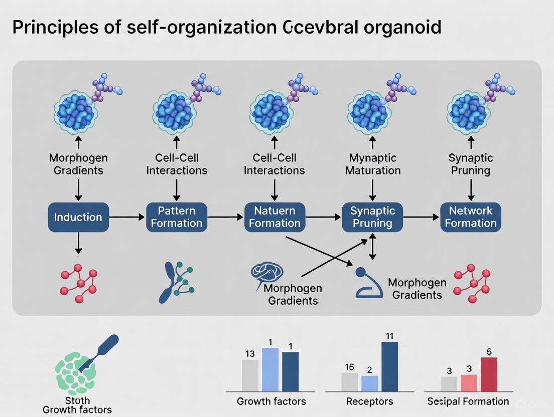
Self-Organization Pathway in Cerebral Organoid Development
Diagram Title: Self-Organization Pathway in Cerebral Organoid Development
Quality Control Framework for Cerebral Organoid Assessment
The intrinsic variability in cerebral organoid differentiation has necessitated the development of standardized quality control frameworks. Recent research has established comprehensive scoring systems that evaluate five critical criteria for 60-day cortical organoids: morphology, size and growth profile, cellular composition, cytoarchitectural organization, and cytotoxicity level [13]. This hierarchical framework begins with non-invasive assessments to exclude low-quality organoids before proceeding to more in-depth analyses, significantly enhancing experimental reproducibility and reliability [13].
Diagram Title: Quality Control Framework for Cerebral Organoid Assessment
Current Applications and Future Perspectives
Cerebral organoids have evolved from basic developmental models to sophisticated tools with diverse research and clinical applications, continuously expanding as technology advances.
Research and Clinical Applications
Disease Modeling: Cerebral organoids derived from patient-specific iPSCs have been successfully used to model a wide range of neurodevelopmental disorders (including microcephaly, autism spectrum disorder, and Timothy syndrome), neurodegenerative diseases (Alzheimer's and Parkinson's), and neurological cancers (such as glioblastoma) [8] [13]. These models recapitulate key pathological features, enabling the investigation of disease mechanisms in a human-specific context.
Drug Screening and Toxicology: Organoids provide a human-relevant platform for high-throughput drug screening and neurotoxicity testing, overcoming the limitations of animal models in predicting human-specific responses [13]. They have been utilized to study the effects of various chemicals and pharmaceuticals, including valproic acid, nicotine, cannabis, bisphenol S, cadmium, and nanoplastics on developing human neural tissue [13].
Regenerative Medicine and Cell Therapy: Transplantion studies have demonstrated that cerebral organoids can integrate with host brain circuits, extending axons along appropriate pathways and forming functional synaptic connections [14]. Recent research shows transplanted organoids drive behavioral changes in animal models, indicating their potential for repairing damaged neuronal circuits after injury or stroke [14].
Emerging Frontiers and Technical Challenges
The field continues to evolve with several emerging frontiers. The development of assembloids - fusion of region-specific organoids - enables the study of circuit formation and cell migration between different brain areas [8]. The integration of brain organoids with artificial intelligence, termed organoid intelligence, represents a cutting-edge frontier for exploring biological computing and learning mechanisms [8] [9]. Additionally, the incorporation of vascular networks and non-neural cell types (microglia, endothelial cells) enhances the physiological relevance of these models [8].
Despite rapid progress, significant challenges remain. Limitations include heterogeneity in size, cellular composition, and structural organization between individual organoids [14] [13]. The absence of a functional vascular system restricts nutrient diffusion, limiting organoid size and maturation [8] [9]. Further maturation of neuronal networks and the reproduction of human-specific brain features at later developmental stages continue to present ongoing challenges for the field [9].
The historical trajectory from sponge cell reaggregation to sophisticated 3D cerebral organoids demonstrates how the fundamental biological principle of self-organization has been harnessed to create revolutionary experimental models. This journey has been marked by key conceptual and technical breakthroughs: the initial discovery of cellular self-organization potential, the isolation of pluripotent stem cells, the development of 3D extracellular matrix environments, and the refinement of differentiation protocols that guide intrinsic self-patterning capacities. The emerging applications in disease modeling, drug development, and regenerative medicine underscore the transformative potential of cerebral organoid technology. As the field continues to address current limitations through technological innovations such as automation, vascularization, and standardized quality control frameworks, cerebral organoids are poised to further advance our understanding of human brain development and disorder, ultimately bridging the gap between in vitro models and human neurobiology.
The development of cerebral organoids, which recapitulate the complex architecture and functionality of the human brain, represents a groundbreaking advancement in neuroscience research. At the heart of this innovation lie pluripotent stem cells—notably embryonic stem cells (ESCs) and induced pluripotent stem cells (iPSCs)—which serve as the foundational starting material for generating these sophisticated three-dimensional models. The principles of self-organization intrinsic to both ESCs and iPSCs enable the recapitulation of human brain development in vitro, providing an unprecedented platform for studying neurodevelopment, disease mechanisms, and therapeutic interventions. This technical guide examines the distinctive properties, applications, and methodological considerations of ESCs and iPSCs as starting materials in cerebral organoid research, with a specific focus on their roles in self-organization and the generation of complex neural tissues.
Pluripotency is defined as the capacity of a single cell to differentiate into derivatives of all three embryonic germ layers: ectoderm, mesoderm, and endoderm [15]. This remarkable cellular plasticity, once considered an exclusive attribute of early embryonic cells, can now be induced in somatic cells through reprogramming techniques. The concept of self-organization—whereby complex patterns and structures emerge from initially homogeneous cells without external guidance—is fundamental to cerebral organoid development [1]. When pluripotent stem cells are provided with appropriate environmental cues, they undergo intrinsic morphogenetic processes that mirror in vivo brain development, including neuronal differentiation, migration, and spatial organization.
The emergence of human brain organoids (hBOs) has transformed how we study brain development, disease mechanisms, and therapy discovery [16]. These 3D in vitro neural models closely mimic the cellular diversity, spatial structure, and functional connectivity of the human brain, providing a groundbreaking platform that outperforms traditional 2D cultures and animal models in studying neurodevelopment and neurological disorders. The self-organizing capacity of pluripotent stem cells enables the generation of these complex structures, making them invaluable tools for both basic research and clinical applications.
Pluripotent Stem Cell Platforms: iPSCs vs. ESCs
Historical Development and Fundamental Biology
The isolation of mouse embryonic stem cells (ESCs) by Martin Evans and Matthew Kaufman in 1981 marked a pivotal moment in stem cell biology [17]. Human ESCs (hESCs) were subsequently isolated by James Thomson and colleagues in 1998, providing the first human pluripotent stem cell platform [17] [18]. However, the ethical concerns surrounding embryo destruction and immune rejection limitations associated with hESCs prompted the search for alternative approaches.
A series of seminal experiments laid the groundwork for cellular reprogramming. John Gurdon's somatic cell nuclear transfer (SCNT) experiments in 1962 demonstrated that a nucleus isolated from a terminally differentiated somatic cell contained all genetic information needed to generate an entire organism [17] [18]. This revealed that phenotypic diversity was achieved through reversible epigenetic mechanisms rather than irreversible genetic changes. Building on this concept, Shinya Yamanaka and colleagues identified a combination of four transcription factors—Oct4, Sox2, Klf4, and c-Myc (OSKM)—that could reprogram mouse fibroblasts into induced pluripotent stem cells (iPSCs) in 2006 [17] [18]. This discovery was rapidly followed by the successful generation of human iPSCs by both Yamanaka (using OSKM) and Thomson (using OCT4, SOX2, NANOG, and LIN28) in 2007 [17] [18].
Comparative Analysis of iPSCs and ESCs
iTable 1: Characteristics of Human Pluripotent Stem Cell Platforms
| Characteristic | Embryonic Stem Cells (ESCs) | Induced Pluripotent Stem Cells (iPSCs) |
|---|---|---|
| Origin | Inner cell mass of blastocyst-stage embryos [15] | Reprogrammed somatic cells (e.g., skin fibroblasts, blood cells) [19] |
| Reprogramming Method | Naturally occurring | Ectopic expression of transcription factors (OSKM or OSNL) [18] |
| Ethical Considerations | Controversial due to embryo destruction [19] | Minimal ethical concerns [19] |
| Immunogenicity | Allogeneic, risk of immune rejection | Autologous potential, reduced rejection risk [19] |
| Differentiation Potential | Pluripotent | Pluripotent [15] |
| Genetic Stability | Generally high | Variable, potential for genomic abnormalities from reprogramming [18] |
| Regulatory Status | Restricted research in some jurisdictions | Widely accessible for research [20] |
| Cost and Scalability | Moderate | High for autologous, improving with biobanking [18] |
The molecular mechanisms governing pluripotency involve profound remodeling of chromatin structure and the epigenome, alongside changes to nearly every aspect of cell biology, including metabolism, cell signaling, intracellular transport, and proteostasis [17] [18]. Reprogramming occurs in two broad phases: an early stochastic phase where somatic genes are silenced and early pluripotency-associated genes are activated, followed by a more deterministic late phase where late pluripotency-associated genes are activated [17].
Mitochondrial dynamics also play a crucial role in establishing and maintaining pluripotency. Pluripotent stem cells typically rely on glycolysis as their primary energy source, even under oxygen-rich conditions—a metabolic preference known as the "Warburg effect" [15]. This glycolytic metabolism supports rapid cell proliferation while limiting mitochondrial oxidative metabolism, thereby reducing oxidative stress. Upon differentiation, mitochondrial maturation and structural remodeling drive a metabolic shift toward oxidative phosphorylation (OXPHOS) [15].
Self-Organization Principles in Cerebral Organoid Development
Fundamentals of Neural Self-Organization
Self-organization in cerebral organoids refers to the innate capacity of pluripotent stem cells to form complex, patterned structures resembling the developing brain through cell-autonomous processes [1]. This phenomenon leverages the same genetic and epigenetic programs that guide embryonic brain development, resulting in the emergence of regional identities, layered cortical structures, and functional neural networks without extensive external guidance.
The first reports creating human 3D cerebral tissue emerged in 2008 and are considered pioneering works in the field of "neural organoid" research [1]. These initial demonstrations showed that pluripotent stem cells, when provided with appropriate 3D culture conditions, could spontaneously form polarized cortical tissues with discrete zones resembling the ventricular and subventricular zones of the developing neocortex.
Methodological Approaches to Cerebral Organoid Generation
iTable 2: Cerebral Organoid Generation Protocols Based on Pluripotent Stem Cells
| Protocol Type | Description | Key Applications | Advantages | Limitations |
|---|---|---|---|---|
| Unguided | Spontaneous self-organization without exogenous patterning signals [16] | Modeling disorders with widespread brain effects (e.g., microcephaly, Zika virus infection) [16] | Recapitulates early brain development; generates multiple brain regions | High batch variability; inconsistent regional identity [16] |
| Guided | Application of defined patterning cues to direct differentiation toward specific brain regions [16] [21] | Region-specific disorders (e.g., Parkinson's disease with midbrain organoids) [16] | Enhanced regional fidelity and reproducibility [16] | May oversimplify native brain environment [16] |
| Assembloids | Fusion of region-specific organoids to recreate inter-regional interactions [21] | Studying circuit formation and long-range connectivity [21] | Models complex neural circuits and connectivity | Technically challenging; requires precise timing |
| Vascularized | Incorporation of vascular-like networks | Enhancing nutrient delivery and maturation | Improved physiological accuracy; reduced hypoxia [16] | Increased complexity of generation |
The organoid generation process typically begins with pluripotent stem cells (either iPSCs or ESCs) aggregated into embryoid bodies, which are then embedded in extracellular matrix substitutes such as Matrigel to provide structural support [16]. These aggregates are subsequently transferred to spinning bioreactors or orbital shakers to enhance nutrient and oxygen exchange. Under appropriate culture conditions, the cells self-organize into neural rosettes—structures that resemble the developing neural tube—which then give rise to the various regional identities and cell types found in the mature brain.
The developmental recapitulation in brain organoids extends beyond cellular diversity to include functional network activity. Recent studies have demonstrated that human brain organoids exhibit structured neuronal firing sequences that mirror patterns observed in developing mammalian brains [3]. These preconfigured firing sequences emerge spontaneously and are thought to represent fundamental building blocks of neural computation, highlighting the remarkable self-organizing capacity of pluripotent stem cell-derived neural networks.
Experimental Protocols for Cerebral Organoid Generation
Essential Research Reagent Solutions
iTable 3: Key Research Reagents for Cerebral Organoid Generation
| Reagent Category | Specific Examples | Function | Considerations |
|---|---|---|---|
| Extracellular Matrix | Matrigel, Geltrex [16] | Provides structural support and biochemical cues | Lot-to-lot variability; complex composition |
| Neural Induction Media | SMAD inhibitors (e.g., Dorsomorphin, SB431542) [21] | Promotes neural differentiation | Concentration and timing critical |
| Patterning Factors | Wnt, BMP, FGF, SHH agonists/antagonists [21] | Directs regional specification | Combinatorial effects; specific temporal windows |
| Cell Dissociation Reagents | Accutase, Trypsin | Dissociates cell aggregates for passaging | Optimization required for 3D cultures |
| Cryopreservation Media | DMSO-based formulations | Long-term storage of organoids | Variable recovery efficiency |
Detailed Protocol for Generating Guided Cortical Organoids
Starting Material Preparation:
- Maintain human iPSCs or ESCs in feeder-free conditions using defined culture media such as mTeSR or StemFlex.
- Ensure pluripotent stem cells are in log-phase growth and have high viability (>90%) before initiating organoid differentiation.
- For iPSCs, verify pluripotency marker expression (OCT4, NANOG, SOX2) and normal karyotype prior to differentiation.
Organoid Generation Workflow:
- Aggregate Formation: Dissociate pluripotent stem cells to single cells using gentle dissociation reagent. Seed 3,000-9,000 cells per well in low-attachment U-bottom 96-well plates to promote aggregate formation through centrifugation (300-400 × g for 3 minutes).
- Neural Induction: Culture aggregates in neural induction media containing dual SMAD inhibitors (LDN-193189 and SB431542) for 10-14 days with media changes every other day.
- Embedding and Expansion: Transfer aggregates to Matrigel droplets and culture in differentiation media containing FGF2 for 7-14 days to promote neuroepithelial expansion.
- Patterned Differentiation: For cortical organoids, transfer to spinning bioreactors and culture in media containing the Wnt inhibitor IWR-1-endo (3-5 μM) and the TGF-β inhibitor A83-01 (5-10 μM) to promote dorsal telencephalic identity for 30-60 days.
- Maturation: Maintain organoids in neural maturation media containing BDNF, GDNF, and cAMP for up to several months to promote functional maturation, with media changes twice weekly.
Critical Considerations:
- The size of initial aggregates significantly influences organoid development and viability—optimize cell number per aggregate for specific applications.
- Monitor morphology regularly; well-formed organoids should exhibit clear ventricular zone-like structures with apical-basal polarity by day 20-30.
- For long-term cultures (>2 months), consider slice culture methods or vascularization strategies to enhance nutrient delivery to core regions.
Diagram 1: Cerebral Organoid Generation Workflow from Pluripotent Stem Cells
Applications in Disease Modeling and Drug Development
Disease Modeling with Patient-Specific iPSCs
The ability to generate cerebral organoids from patient-specific iPSCs has revolutionized modeling of neurological disorders. This approach enables researchers to recapitulate disease-specific phenotypes in a human genetic background, providing unprecedented insights into disease mechanisms [16] [19]. Notable applications include:
Alzheimer's Disease: iPSC-derived cortical organoids from patients with familial Alzheimer's disease mutations (PSEN1/PSEN2) exhibit accelerated amyloid-beta aggregation and tau hyperphosphorylation, enabling the study of disease progression and screening of therapeutic compounds [16] [19].
Parkinson's Disease: Midbrain organoids generated from Parkinson's patients contain dopaminergic neurons that display disease-relevant phenotypes such as alpha-synuclein accumulation and mitochondrial dysfunction, providing a platform for studying neurodegenerative mechanisms [16].
Neurodevelopmental Disorders: Cerebral organoids derived from individuals with autism spectrum disorder or schizophrenia have revealed alterations in neuronal migration, synaptogenesis, and network activity, offering insights into the cellular basis of these conditions [16] [21].
Drug Screening and Development
Cerebral organoids serve as powerful platforms for high-throughput drug screening and toxicity testing. The 3D architecture and human-specific physiology of organoids provide more clinically relevant models compared to traditional 2D cultures or animal models [19]. Key applications include:
Phenotypic Screening: Organoids can be used to screen compound libraries for modifiers of disease-relevant phenotypes, such as amyloid-beta accumulation in Alzheimer's models or mitochondrial dysfunction in Parkinson's models [19].
Toxicity Assessment: Brain organoids provide a human-based system for evaluating neurotoxicity of drug candidates, environmental toxins, or industrial chemicals, potentially reducing reliance on animal testing [19].
Personalized Medicine: Patient-specific organoids can be used to identify individualized therapeutic responses, particularly for neuropsychiatric medications where treatment efficacy varies significantly among individuals [19].
Current Challenges and Future Perspectives
Despite significant advances, several challenges remain in the use of pluripotent stem cell-derived cerebral organoids. These models typically recapitulate early to mid-fetal stages of brain development but struggle to achieve full maturity comparable to adult human brain tissue [16] [21]. The absence of vascularization limits nutrient and oxygen delivery to core regions, leading to necrotic centers in larger organoids [16]. Additionally, batch-to-batch variability caused by differences in stem cell source, reagent quality, and manual handling remains a critical challenge affecting reproducibility [16].
Future developments in the field are likely to focus on several key areas:
Enhanced Maturation: Combining bioengineering approaches with extended culture periods to achieve more adult-like neuronal phenotypes and network connectivity.
Vascularization: Incorporating endothelial cells and perfusion systems to enhance nutrient delivery and mimic the neurovascular unit.
Standardization: Developing standardized protocols and quality control metrics to reduce variability and enhance reproducibility across laboratories.
Multi-regional Integration: Creating more complex assembloid models that recapitulate interactions between distinct brain regions to study circuit-level phenomena [21].
Clinical Translation: Advancing the use of iPSC-derived neural cells for cell replacement therapies in neurological disorders, with several clinical trials already underway [20] [22].
The convergence of pluripotent stem cell technology, bioengineering, and neuroscience continues to push the boundaries of what can be modeled in vitro. As cerebral organoid methodologies become more sophisticated and reproducible, they hold increasing promise for unraveling the mysteries of human brain development, disease pathogenesis, and therapeutic intervention.
Diagram 2: iPSC Reprogramming and Cerebral Organoid Applications Workflow
The development of complex neural structures in cerebral organoids is governed by the precise interplay of cell-autonomous (intrinsic) programs and non-cell-autonomous (extrinsic) cues from the microenvironment. This whitepaper delineates the mechanisms by which intrinsic cellular functions and extrinsic signaling molecules orchestrate patterning events, drawing upon recent advances in proteomic and secretome analyses of dorsal forebrain organoids (DFOs). The synthesis of these mechanisms provides a framework for understanding self-organization in cerebral organoids, with significant implications for modeling neurodevelopmental disorders and advancing drug discovery.
The principles of self-organization in cerebral organoid development research hinge on the synergy between predetermined cellular blueprints and dynamic environmental signals. Cell-autonomous mechanisms encompass the effects of intrinsic cellular functions, largely dictated by a cell's genetic and epigenetic profile [23]. Conversely, non-cell-autonomous processes refer to cellular responses to extrinsic influences, such as secreted signaling molecules, extracellular matrix (ECM) components, and physical forces from the niche [23]. In the context of brain organoids, which are three-dimensional (3D) structures derived from human pluripotent stem cells (PSCs), these two mechanisms are intricately interconnected, cooperating to recapitulate the composition, organization, and function of the early human brain [24] [25]. This review explores this interplay, framing it within the broader thesis of self-organization and providing a technical guide for researchers and drug development professionals.
Conceptual Framework: Dissecting the Mechanisms
Cell-Autonomous (Intrinsic) Programs
Intrinsic cues are the internal directives of a cell that guide its fate and function:
- Genetic and Epigenetic Blueprint: The lineage specification of progenitor cells is intrinsically determined, leading to the emergence of distinct neuronal and glial cell types [24] [25].
- Proliferation and Differentiation Capacity: The inherent potential of radial glia and intermediate progenitor cells to proliferate or differentiate is a cell-autonomous property that changes over time, as evidenced by the reduction in proliferative markers in organoids [25].
Non-Cell-Autonomous (Extrinsic) Cues
Extrinsic cues constitute the cellular microenvironment, or niche, and provide critical signals for patterning:
- Secreted Signaling Molecules: Morphogens, growth factors, and cytokines form concentration gradients that guide cell migration, polarity, and regional identity [24].
- Extracellular Matrix (ECM) and Cell Adhesion Molecules: The ECM provides structural support and biochemical signals that influence cell proliferation, maturation, and the overall organoid architecture [25].
- Synaptic Proteins and Proteases: Recent secretome analyses reveal that synaptic proteins and metalloproteases are actively secreted during peak neurogenesis, suggesting a role in guiding neural circuit formation beyond their classical functions [25].
Interplay in Self-Organization
The formation of a complex, patterned structure like a cerebral organoid is not the result of either mechanism alone but of their continuous dialogue. Intrinsic programs define a cell's potential, while extrinsic signals from the niche modulate the realization of this potential. This interplay drives the self-organization of neural progenitor zones, such as ventricular zone-like regions, and their subsequent differentiation into layered cortical structures [23] [25]. Disruptions in this synergy can lead to modeling deficits and are implicated in neurodevelopmental disorders [23].
Experimental Approaches and Quantitative Profiling
Investigating the intrinsic-extrinsic interplay requires methodologies that can separately analyze the internal state of cells and their extracellular environment.
Workflow for Integrated Proteome and Secretome Analysis
The following diagram outlines a standard experimental pipeline for the concurrent profiling of intrinsic protein expression and extrinsic secreted factors in dorsal forebrain organoids (DFOs), as employed in recent studies [25].
Key Quantitative Findings from Temporal Analysis
Proteomic and secretome analyses of DFOs across developmental timepoints (Days 20, 35, and 50) reveal distinct dynamics for intrinsic and extrinsic components.
Table 1: Proteome Dynamics in Dorsal Forebrain Organoids (Intrinsic Profile) [25]
| Timepoint | Uniquely Identified Proteins | Key Functional Signatures | Implication for Patterning |
|---|---|---|---|
| Day 20 | 23 proteins | High proliferation markers | Establishment of progenitor pools |
| Day 35 | 0 unique proteins (57 shared with D20; 53 with D50) | Transitionary signature | Peak period of neurogenesis and fate specification |
| Day 50 | 7 proteins | Increased synaptic markers; decreased proliferation markers | Advanced neuronal maturation and circuit formation |
Table 2: Secretome Dynamics in Dorsal Forebrain Organoids (Extrinsic Profile) [25]
| Timepoint | Key Secreted Factor Classes | Quantitative Change | Proposed Role in Patterning |
|---|---|---|---|
| Day 35 | Cell Adhesion Molecules, Synaptic Proteins, Proteases | Significantly increased | Guides neurogenesis, cell migration, and initial synapse formation |
| Day 50 | Extracellular Matrix (ECM) Proteins | Predominantly secreted | Supports structural maturation and stabilization of neural circuits |
Detailed Methodologies for Key Experiments
Protocol 1: Liquid Chromatography-Mass Spectrometry (LC-MS) for Proteome/Secretome [25]
- Sample Preparation: For proteome, pool 3 organoids per sample (in triplicate per cell line). For secretome, collect conditioned media from organoids cultured in serum-free conditions for 24 hours.
- Protein Digestion: Dissolve samples in urea buffer, reduce with dithiothreitol (DTT), alkylate with iodoacetamide, and digest with trypsin overnight.
- LC-MS Analysis: Desalt peptides and separate using a nano-flow liquid chromatography system coupled to a high-resolution mass spectrometer operating in data-dependent acquisition mode.
- Data Processing: Identify and quantify proteins using software (e.g., MaxQuant) against a human protein database. Normalize protein intensities and perform statistical analysis for differential abundance.
Protocol 2: Immunohistochemistry (IHC) for Validation [25]
- Fixation and Sectioning: Fix organoids in 4% paraformaldehyde (PFA) for 2 hours, then embed in paraffin or optimal cutting temperature (OCT) compound. Section at 5-10 µm thickness.
- Antigen Retrieval and Staining: Perform antigen retrieval using citrate buffer (pH 6.0). Block sections with 5% normal serum, then incubate with primary antibodies (e.g., SOX2 for radial glia, CTIP2 for deep-layer neurons) overnight at 4°C.
- Visualization and Quantification: Incubate with fluorescently conjugated secondary antibodies and counterstain with DAPI. Image using a confocal microscope and quantify positive cell areas or counts using image analysis software (e.g., ImageJ).
The Scientist's Toolkit: Essential Research Reagents
Successful interrogation of intrinsic and extrinsic cues relies on a suite of specialized reagents and tools.
Table 3: Key Research Reagent Solutions for Cerebral Organoid Research
| Reagent / Material | Function | Application in Patterning Studies |
|---|---|---|
| Human Induced Pluripotent Stem Cells (hiPSCs) | Starting cell source for organoid generation; provides genetic background. | Enables modeling of patient-specific disorders and study of intrinsic genetic programs. KOLF2.1J, BIONi010-C are common lines [25]. |
| Matrigel / Basement Membrane Extract | Provides a 3D extracellular matrix scaffold for organoid growth. | Delivers extrinsic biochemical and biophysical cues that support self-organization and polarization [24]. |
| Small Molecule Inducers | Directs regional patterning (e.g., SMAD inhibitors for neural induction). | Provides controlled extrinsic signals to steer intrinsic differentiation programs toward specific fates (e.g., dorsal forebrain) [25]. |
| Mass Spectrometry Grade Solvents | Used for sample preparation and separation in LC-MS. | Essential for high-sensitivity profiling of the proteome (intrinsic) and secretome (extrinsic) [25]. |
| Validated Primary Antibodies | Marks specific cell types and proteins in IHC. | Validates proteomic findings and visualizes spatial patterning (e.g., SOX2, CTIP2 antibodies) [25]. |
Signaling Pathways in Patterning: A Visual Synthesis
The following diagram integrates key signaling interactions between intrinsic cellular states and extrinsic microenvironmental cues, as identified in recent organoid studies.
The study of cerebral organoids has unequivocally shown that patterning is an emergent property of the dynamic dialogue between cell-autonomous programs and non-cell-autonomous signals. The temporal decoupling of proteomic and secretome dynamics, with the former showing gradual maturation and the latter exhibiting stage-specific bursts of activity, underscores the complexity of this interplay [25]. For drug development, this implies that therapeutic strategies must consider both the intrinsic health of neurons and the integrity of their supporting microenvironment. Future research should prioritize the standardization of organoid cultures, improved vascularization methods, and the multi-omic integration of data from intrinsic and extrinsic compartments [24]. By refining our understanding of these fundamental principles of self-organization, cerebral organoids will continue to be an indispensable tool for modeling disease and screening therapeutics.
The development of the mammalian brain is a pinnacle of self-organization, where complex structures emerge through spatially and temporally coordinated processes without extensive external guidance. This innate capacity of cells to self-organize is now being harnessed in vitro through cerebral organoid models, providing unprecedented opportunities to study human-specific brain development and disorders [26]. This technical guide details the core processes of neural induction, polarization, and regionalization, framing them within the context of self-organizing principles that can be recapitulated in three-dimensional organoid systems.
Neural Induction: From Ectoderm to Neural Tissue
Neural induction represents the foundational step in nervous system development, where embryonic ectoderm is specified to form neural tissue. This process is governed by precise signaling dynamics that can be replicated in organoid differentiation protocols.
Molecular Mechanisms of Neural Induction
During gastrulation, presumptive mesodermal cells migrate through the dorsal blastopore lip and form a layer between endoderm and ectoderm. The notochord, which derives from these mesodermal cells, secretes key signaling molecules that inhibit BMP (Bone Morphogenetic Protein) signaling in the overlying ectoderm [27].
The default state of ectodermal cells is neural differentiation, which is suppressed by BMP4 in non-neural ectoderm. Neural induction occurs when BMP inhibition allows this default neural pathway to proceed. Key neural inducers include:
- Noggin and Chordin: Proteins produced by the dorsal mesoderm that bind to and inhibit BMP4 [27]
- Follistatin: Additional BMP antagonist that promotes neural specification
This inhibition of TGF-β and BMP signaling can efficiently induce neural tissue from pluripotent stem cells, forming the basis for many cerebral organoid protocols [27] [28].
Neural Tube Formation and Patterning
Following neural induction, the neural plate forms along the dorsal side of the embryo. The neural groove then forms along the long axis of the neural plate, folding to give rise to the neural tube through the process of neurulation [27].
The neural tube becomes patterned along its dorsoventral axis:
- Ventral patterning: Controlled by Sonic hedgehog (Shh) from the notochord
- Dorsal patterning: Regulated by BMPs from the epidermal ectoderm flanking the neural plate
The hollow interior of the neural tube forms the neural canal, which develops into the ventricular system of the central nervous system [27].
Polarization and Neural Tube Regionalization
Following neural tube closure, the anterior part expands and forms three primary brain vesicles, establishing the fundamental organization of the central nervous system.
Primary Brain Vesicle Formation
The neural tube differentiates into three primary brain vesicles [27]:
- Prosencephalon: Future forebrain
- Mesencephalon: Future midbrain
- Rhombencephalon: Future hindbrain
Secondary Brain Vesicle Formation
These simple, early vesicles further divide into more specialized structures [27]:
| Primary Vesicle | Secondary Divisions | Future Structures |
|---|---|---|
| Prosencephalon | Telencephalon | Cerebral cortex, basal ganglia |
| Diencephalon | Thalamus, hypothalamus | |
| Mesencephalon | Mesencephalon | Colliculi |
| Rhombencephalon | Metencephalon | Pons, cerebellum |
| Myelencephalon | Medulla oblongata |
The CSF-filled central chamber remains continuous from the telencephalon to the central canal of the spinal cord, forming the developing ventricular system of the CNS [27].
Signaling Pathways Governing Regionalization
Brain regionalization is controlled by sophisticated signaling gradients that pattern the anterior-posterior and dorsal-ventral axes. These patterning events can be replicated in organoid systems through precise temporal application of signaling molecules.
Anterior-Posterior Patterning
The anterior-posterior axis is established through the action of specific signaling molecules [27]:
- Anterior identity: Maintained by limited exposure to caudalizing factors
- Posterior identity: Induced by FGF and retinoic acid, which act in the hindbrain and spinal cord
- Hox genes: Expressed in overlapping domains along the anteroposterior axis under retinoic acid control
Dorsal-Ventral Patterning
The dorsal-ventral axis is regulated by opposing signaling centers [27]:
- Ventral neural tube: Patterned by Sonic hedgehog (Shh) from the notochord and floor plate
- Dorsal neural tube: Patterned by BMPs from the epidermal ectoderm
Chromatin Accessibility in Regionalization
Recent research using ATAC-seq (Assay for Transposase-Accessible Chromatin with sequencing) has revealed that chromatin accessibility defines neural progenitor identity [29]. This approach has identified:
- Neural sites: 5,584 regulatory regions accessible in all neural progenitor conditions
- Region-specific sites: Anterior NPs (1,863 sites), hindbrain progenitors (2,509 sites), and spinal cord progenitors (1,538 sites)
- Early commitment: Cells acquire axial identity prior to neural identity, contrary to the traditional "activation-transformation" model [29]
Recapitulating Development in Cerebral Organoids
Cerebral organoids harness the self-organizing capacity of pluripotent stem cells to generate complex neural tissues that mimic embryonic brain development.
Organoid Generation Protocol
The cerebral organoid protocol involves several key stages [28]:
Regional Diversity in Organoids
Cerebral organoids develop discrete brain regions with characteristic cellular organization [28]:
| Brain Region | Marker Genes | Cellular Organization Recapitulated |
|---|---|---|
| Dorsal Cortex | Emx1, Auts2, Tshz2, Lmo4 | VZ, oSVZ, IP zones, cortical layers |
| Ventral Forebrain | Nkx2.1 | Interneuron generation and migration |
| Hippocampus | Specific markers not listed | Early hippocampal identity |
| Choroid Plexus | Tissue morphology | CSF-producing epithelium |
| Retina | Retinal pigmented epithelium | Immature retinal tissue |
Organoids display a forebrain dominance with 100% forming dorsal cortical regions, 71% developing choroid plexus, 34% containing ventral forebrain, and 11% forming retinal tissue [28].
The Scientist's Toolkit: Essential Research Reagents
Successful recapitulation of brain development in organoid systems requires carefully selected reagents and materials.
Research Reagent Solutions
| Reagent Category | Specific Examples | Function in Neural Development |
|---|---|---|
| BMP Inhibitors | Noggin, Chordin, Follistatin | Neural induction by suppressing BMP signaling |
| WNT Agonists/Antagonists | CHIR99021 (agonist), DKK-1 (antagonist) | Anterior-posterior patterning, caudalization |
| FGF Signaling | FGF2, FGF19, FGF8 | Neural patterning, cerebellar development |
| SHH Pathway Modulators | Purmorphamine (agonist), Cyclopamine (antagonist) | Ventral neural tube patterning |
| Extracellular Matrix | Matrigel, Laminin, Collagen | 3D structural support, biomechanical cues |
| Cell Culture Supplements | B27, N2, Retinoic Acid | Neuronal survival, differentiation, patterning |
| Metabolic Selection Reagents | Glucose, Lactate | Cell fate specification and survival |
Quantitative Analysis of Organoid Protocols
Recent systematic analyses have quantified the effectiveness of different organoid protocols in recapitulating in vivo development.
Protocol Performance Metrics
A comprehensive study analyzing four brain organoid protocols across multiple cell lines provides quantitative assessment of protocol efficacy [30]:
| Protocol Type | Target Brain Region | Cell-Type Recapitulation | Key Strengths |
|---|---|---|---|
| Dorsal Forebrain Protocol | Cerebral cortex | High dorsal cortical neurons | Outer radial glia generation |
| Ventral Forebrain Protocol | Subpallium | Medium GABAergic interneurons | Ventral specification |
| Midbrain Protocol | Midbrain structures | Medium dopaminergic neurons | Midbrain patterning |
| Striatum Protocol | Basal ganglia | Medium striatal projection neurons | Striatal development |
This resource established a set of protocols that together recreate the majority of cell types in the developing brain and provides a reference of cell-type recapitulation across cell lines and protocols [30]. The study also introduced the NEST-Score to evaluate cell-line- and protocol-driven differentiation propensities and comparisons to in vivo references.
Applications in Disease Modeling
Cerebral organoids have significant utility in modeling neurodevelopmental disorders, particularly those difficult to recapitulate in animal models.
Microcephaly Modeling
When cerebral organoids were generated from patient-derived iPSCs with CDK5RAP2 mutations (associated with primary microcephaly), they demonstrated premature neuronal differentiation, providing a potential explanation for the reduced brain size phenotype [28]. This successful disease modeling highlights the value of organoid technology for studying human-specific neurodevelopmental disorders.
The processes of neural induction, polarization, and regionalization represent fundamental stages in brain development that can be effectively recapitulated in cerebral organoid systems. These self-organizing 3D models capture key aspects of human neurodevelopment, including the generation of distinct brain regions, the presence of human-specific neural progenitor populations like outer radial glia, and the complex signaling dynamics that pattern the embryonic brain. Continued refinement of organoid protocols, combined with advanced genomic technologies like ATAC-seq for assessing chromatin accessibility, provides researchers with powerful tools to investigate both normal development and disease states in human-specific contexts.
The Role of Morphogen Gradients and Signaling Pathways in Spontaneous Patterning
The human brain's remarkable cellular diversity arises from a tightly regulated developmental process orchestrated by morphogen gradients. These diffusible signaling molecules dictate cell fate in a concentration-dependent manner, partitioning the neural tube into distinct spatial domains [31]. Cerebral organoids, as three-dimensional in vitro models derived from pluripotent stem cells (PSCs), offer an unprecedented window into these early patterning events [1]. Their ability to self-organize and recapitulate aspects of human brain development hinges on the faithful replication of these intrinsic morphogen signaling dynamics. Understanding and controlling these gradients is therefore not merely a technical challenge but a fundamental prerequisite for harnessing organoids to study neurodevelopment, model disorders, and advance drug discovery. This whitepaper details the core principles, experimental methodologies, and key reagents for investigating morphogen-driven patterning in cerebral organoids.
Core Principles of Morphogen Gradient Patterning
Morphogens are secreted signaling molecules that form concentration gradients across a field of developing tissue, instructing naive progenitor cells to adopt distinct identities based on the signal strength they perceive.
- Key Morphogen Families: The principal families involved in neural tube patterning include the Hedgehog (e.g., Sonic Hedgehog, SHH), Transforming Growth Factor-β (TGF-β), Bone Morphogenetic Protein (BMP), Wingless (WNT), and Fibroblast Growth Factor (FGF) families, as well as retinoic acid (RA) [31].
- Mechanism of Action: A morphogen is produced from a localized source, or "organizing center," and diffuses through the tissue. Cells respond by activating specific gene regulatory networks, often involving cascades of transcription factors, leading to fate specification. Critical boundaries between progenitor domains are sharpened by mutual repression between transcription factors induced by opposing morphogen gradients [31].
- Spatial Patterning Centers: In the developing neural tube, distinct organizers secrete specific morphogens. For example, SHH from the notochord and floor plate patterns ventral identities, while BMP and WNT from the overlying ectoderm pattern dorsal fates. Secondary organizers, such as the cortical hem and anti-hem, further refine patterning in the forebrain [31].
Table 1: Major Morphogens in Neural Patterning and Their Roles
| Morphogen Pathway | Primary Source In Vivo | Major Role in Neural Patterning | Key Antagonists |
|---|---|---|---|
| SHH | Notochord, Floor Plate | Ventralization of neural tube; specification of motor neurons, striatal interneurons | – |
| TGF-β/BMP | Overlying Ectoderm, Cortical Hem | Dorsalization; specification of roof plate, neural crest | Noggin, Chordin, Follistatin |
| WNT | Paraxial Mesoderm, Cortical Hem | Caudalization, dorsal patterning, hippocampal specification | DKK1, Secreted Frizzled-Related Proteins |
| FGF | Anterior Neural Ridge, Isthmic Organizer | Forebrain specification, midbrain-hindbrain patterning | – |
| Retinoic Acid (RA) | Paraxial Mesoderm, Meninges | Rostro-caudal axis specification, hindbrain and spinal cord identity | – |
Experimental Methodologies for Gradient Control and Analysis
Protocol 1: Generating Single-Expanded Neuroepithelium Organoids (ENOs) via a Temporal TGF-β Gradient
Objective: To create brain organoids composed of a single, continuous neuroepithelium rather than multiple independent rosettes, thereby enhancing cortical identity and tissue architecture [32].
Detailed Workflow:
- Initial Aggregation: Dissociate feeder-free human embryonic stem cells (e.g., H1 hESCs) and reaggregate into embryoid bodies in stem cell medium.
- Temporal Gradient Neural Induction: Instead of a sudden medium switch, expose cells to a prolonged, stepwise gradient. Over several days, gradually decrease the stem cell medium (containing FGF2 and TGF-β) while concomitantly increasing the neural induction (NI) medium containing dual SMAD inhibitors.
- Expansion and Maturation: After the gradient period, switch to expansion medium supplemented with EGF and FGF2. From day 25 onwards, transfer organoids to maturation medium, which can include supplements like Matrigel.
Key Quantitative Findings from ENO Protocol [32]:
- Morphology: ENOs display significantly reduced circularity (decreasing to ~0.5 by day 25) and an increased organoid perimeter, indicating a convoluted, folded structure.
- Tissue Architecture: Immunostaining for N-Cadherin (NCAD) reveals a single, elongated, and radially organized neuroepithelium resembling a ventricular zone, in contrast to the multiple rosettes found in standard organoids.
- Cell Fate: ENOs maintain robust neural identity (NCAD, Nestin expression) without off-target lineages and show enhanced cortical specification.
Protocol 2: A Multiplexed Morphogen Screen for Neural Diversification
Objective: To systematically deconvolve the effects of morphogen identity, concentration, timing, and combination on the generation of neural cell diversity in organoids [33].
Detailed Workflow:
- Organoid Foundation: Generate neural spheroids from human iPSCs using dual SMAD inhibition.
- Arrayed Screen Setup: Culture organoids in an arrayed format and expose them to a panel of 14 different modulators targeting 8 core morphogen pathways (e.g., SHH, WNT, BMP, RA).
- Long-term Culture and Multiplexing: Maintain the organoids under these conditions for over 70 days. To enable pooled analysis, use multiplexed single-cell RNA sequencing (scRNA-seq) where cells from different conditions are tagged and sequenced together.
- Deconvolution and Analysis: Bioinformatically deconvolute the multiplexed scRNA-seq data to assign cells to their original screening condition. Map the resulting transcriptional profiles to reference atlases of human brain development to identify the neural subtypes generated.
- Functional Validation: Employ techniques like assembloid fusion and transplantation into neonatal rat brains (e.g., for Purkinje neuron maturation) to validate the function of specified neurons [33].
Key Quantitative Findings from Morphogen Screening [33]:
- Diversity: The screen enabled the generation of a wide range of regionalized neural organoids, collectively covering cell diversity across the neural axis.
- Rare Subtypes: Critical timing windows and specific morphogen combinatorics yielded rare neural subtypes, including TAC3-expressing striatal interneurons and Cajal-Retzius cells.
- Maturation: Transplantation was a key intervention to achieve hallmark complex dendritic branching in human Purkinje neurons, a level of maturation not attained in vitro.
Visualization of Signaling Pathways and Workflows
The following diagrams, generated using Graphviz DOT language, illustrate the core signaling pathways and a key experimental workflow described in this whitepaper.
The Scientist's Toolkit: Key Research Reagents
Successful patterning of cerebral organoids relies on a defined set of reagents to manipulate key signaling pathways. The table below catalogues essential tools based on the cited research.
Table 2: Key Research Reagents for Morphogen Patterning in Organoids
| Reagent / Tool | Function / Pathway Targeted | Application Example |
|---|---|---|
| Dual SMAD Inhibitors (e.g., SB431542, LDN193189) | Inhibits TGF-β and BMP signaling to induce default neural ectoderm fate. | Foundational step in most neural organoid protocols for efficient neural induction [32] [33]. |
| SAG / Purmorphamine | Small molecule agonists of the Sonic Hedgehog (SHH) pathway. | Used to ventralize organoids and generate striatal or spinal cord fates [33]. |
| CHIR99021 | Small molecule agonist of the WNT/β-catenin pathway. | Applied for caudalization, midbrain dopamine neuron specification, and dorsal patterning [33]. |
| Retinoic Acid (RA) | Morphogen that patterns the rostro-caudal axis, particularly hindbrain and spinal cord. | Critical for generating spinal cord and hindbrain organoids when combined with other morphogens [31]. |
| Noggin / Recombinant BMP Inhibitors | Recombinant proteins or small molecules that inhibit BMP signaling. | Enhances neural induction and promotes dorsal forebrain fates [31]. |
| FGF2 (bFGF) | Growth factor acting in FGF signaling pathway. | Maintains progenitor proliferation; used in expansion phases and for anterior patterning [32]. |
| Matrigel | Extracellular matrix (ECM) hydrogel. | Used to embed organoids, providing structural support and ECM cues that influence morphogen distribution and tissue architecture [32]. |
| Single-Cell RNA-Seq (e.g., 10x Genomics) | High-resolution transcriptional profiling technology. | Essential for characterizing the cellular diversity and identity of organoids and deconvoluting screening results [33] [30]. |
Discussion and Future Perspectives
The controlled application of morphogen gradients has profoundly advanced the fidelity of cerebral organoid models. The shift from static medium switches to dynamic temporal gradients, as demonstrated by the ENO protocol, highlights that the timing of morphogen exposure is as critical as its identity and concentration for achieving advanced tissue architecture [32]. Furthermore, systematic multiplexed screens are moving the field from a trial-and-error approach to a more principled, data-driven understanding of the combinatorial rules governing neural cell fate [33] [30].
Future research must tackle several frontiers. First, achieving spatiotemporally complex gradients that mimic the in vivo activity of secondary organizers remains a significant bioengineering challenge. Integrating microfluidics or biomaterial-based delivery systems could provide the necessary control. Second, the maturation of many neuronal subtypes in organoids is still limited. As shown with Purkinje neurons, transplantation into animal models may be a necessary interim step to study late developmental events [33]. Finally, standardization and reproducibility across cell lines and protocols are crucial for biomedical applications. Computational tools like the NEST-Score, which benchmarks organoids against in vivo references, are a promising development toward this goal [30]. As these models continue to evolve, they will undoubtedly deepen our understanding of human brain development and disease, ultimately accelerating the discovery of novel therapeutics.
Building Better Brains in a Dish: Advanced Protocols and Research Applications
The emergence of human brain organoid technology represents a paradigm shift in neuroscience research, offering unprecedented access to human-specific brain development and disease processes. Central to this revolution is the principle of self-organization—the inherent capacity of pluripotent stem cells to spontaneously form complex, three-dimensional structures that mirror the cytoarchitecture and cellular diversity of the developing human brain [1] [34]. This self-organizing capability harnesses the same morphogenetic processes that occur during embryonic development, providing a "cut & paste" of developmental biology into a dish [1].
The fundamental dichotomy in brain organoid generation lies in the degree of external intervention: unguided protocols maximize spontaneous self-organization, while guided approaches apply precise external patterning cues to direct regional specification [16] [34]. This technical guide provides a comprehensive comparative analysis of these two methodologies, examining their theoretical foundations, experimental protocols, and applications within the framework of self-organization principles. Understanding these approaches is essential for researchers to select appropriate models for studying neurodevelopment, disease mechanisms, and therapeutic interventions.
Theoretical Foundations: Self-Organization in Neural Development
Self-organization in neural organoids mirrors the processes of early embryogenesis, where complex structures emerge through spatially constrained spontaneous symmetry breaking and cell-to-cell communication [1]. This phenomenon originates from the capacity of pluripotent stem cells to recapitulate developmental trajectories when provided with appropriate environmental conditions.
The process begins with the formation of neuroepithelial tissues that spontaneously polarize and generate lumen-containing neural tube-like structures [35]. Through self-driven differentiation and self-patterning, these structures give rise to various neural progenitor zones, which in turn produce diverse neuronal and glial cell types in a spatially organized manner [1]. The resulting tissues exhibit remarkable similarity to early fetal brain development at molecular, cellular, and structural levels [24] [34].
Key principles governing self-organization in neural organoids include:
- Cell autonomy: The intrinsic programming of stem cells to follow developmental trajectories
- Local environment: The creation of signaling centers that pattern surrounding tissues
- Mechanical forces: Cell sorting and spatial constraints that influence tissue architecture
- Emergent connectivity: The spontaneous formation of functional neural networks
These principles operate across both unguided and guided protocols, though the balance between intrinsic and extrinsic influences varies significantly between these approaches.
Unguided Differentiation Protocols
Core Principles and Methodological Framework
Unguided brain organoid protocols rely exclusively on the spontaneous self-organization capacity of pluripotent stem cells without exogenous patterning signals [16] [34]. This approach aims to recapitulate the autonomous developmental processes of the early embryonic brain, resulting in organoids containing heterogeneous brain regions including forebrain, midbrain, and hindbrain tissues within a single structure [16].
The pioneering cerebral organoid method developed by Lancaster and Knoblich involves embedding embryoid bodies (EBs) derived from human pluripotent stem cell (hPSC) aggregates into an extracellular matrix (ECM), followed by culture in spinning bioreactors to promote tissue expansion and neural differentiation [34]. This methodology provides minimal external interference, allowing hPSCs maximum freedom for self-organization, sometimes yielding elongated neuroepithelial structures that exhibit remarkable cellular diversity [34].
Detailed Experimental Protocol
Initial Preparation and EB Formation
- Culture hPSCs to 80-90% confluence in feeder-free conditions
- Dissociate cells using enzymatic or gentle dissociation reagents
- Resuspend cells in EB formation medium supplemented with Rho kinase (ROCK) inhibitor to enhance survival
- Plate cells in low-attachment U-bottom 96-well plates (approximately 9,000 cells per well) or use AggreWell plates for uniform EB formation [36]
- Centrifuge plates at 100 × g for 3 minutes to aggregate cells at the bottom of wells
- Culture for 5-7 days with medium changes every other day until EBs reach 400-500 μm diameter
Neural Induction and Matrix Embedding
- Transfer EBs to neural induction medium containing DMEM/F12, N2 supplement, non-essential amino acids, and glutamine
- Culture for 3-5 days until neural ectoderm emerges (visible as translucent epithelial structures)
- Prepare Matrigel or similar ECM solution on ice
- Transfer individual EBs to cold Matrigel droplets (approximately 20-30 μL per EB)
- Polymerize Matrigel at 37°C for 20-30 minutes
- Transfer embedded EBs to differentiation medium in multi-well plates
Extended Differentiation and Maturation
- After 3-5 days of static culture, transfer organoids to spinning bioreactors or orbital shakers
- Maintain in cerebral organoid differentiation medium containing B27 without vitamin A
- Culture for extended periods (60-150+ days) with medium changes every 3-4 days
- For long-term maturation (>100 days), transition to neuronal maintenance media such as BrainPhys [37]
Table 1: Key Reagents for Unguided Organoid Protocols
| Reagent Category | Specific Examples | Function | Concentration/Timing |
|---|---|---|---|
| Basal Media | DMEM/F12, Neurobasal | Nutrient base for neural culture | 1:1 ratio typically used |
| Supplements | N2, B27 (-A), B27 (+A) | Provide hormones, antioxidants, and proteins | 0.5-1% for N2, 1-2% for B27 |
| Extracellular Matrix | Matrigel, Geltrex | Structural support, signaling cues | 20-30% for embedding |
| Enzymes | Accutase, Dispase, Gentle Cell Dissociation Reagent | Cell dissociation for passaging | Variable by product |
| Small Molecules | Y-27632 (ROCK inhibitor) | Enhance cell survival after dissociation | 10-50 μM for first 24-48h |
Advantages and Limitations
Unguided organoids offer several key advantages:
- Regional diversity: Simultaneous development of multiple brain regions enables modeling of inter-regional interactions [16] [34]
- Developmental fidelity: Closely mirrors early brain development processes [16]
- Discovery potential: Unbiased approach may reveal novel aspects of human neurodevelopment
However, significant limitations persist:
- Batch variability: Stochastic differentiation results in unpredictable regional proportions and arrangements [16]
- Heterogeneous architecture: Limited reproducibility for region-specific disease modeling [16]
- Structural inconsistencies: Variable cytoarchitecture and cellular composition across batches [34]
- Limited high-throughput application: Heterogeneity complicates quantitative analyses and drug screening [16]
Guided Differentiation Protocols
Core Principles and Methodological Framework
Guided brain organoid methodologies apply defined external patterning cues to direct differentiation toward specific brain regions, such as the cortex, midbrain, or hypothalamus [16] [34]. This approach, pioneered by the Sasai laboratory, utilizes small molecules and growth factors to instruct hPSCs to form tissues representative of particular brain areas with enhanced regional fidelity and reproducibility [1] [34].
Unlike unguided methods, guided protocols intentionally restrict developmental potential to generate more homogeneous populations of neural progenitors with defined regional identities. This strategy enhances experimental control and reduces inter-organoid variability, making guided organoids particularly suitable for studying region-specific disorders and conducting quantitative analyses [16].
Detailed Experimental Protocol
Regional Patterning and Neural Induction
- Begin with standardized EB formation as described for unguided protocols
- At day 2-5 of EB formation, add region-specific patterning factors:
- Continue patterning until neural rosettes appear (typically 10-20 days)
Extended Regional Specification and Maturation
- After initial patterning, transition to region-specific maturation media:
- Culture on orbital shakers or spinning bioreactors for long-term maturation (60-120+ days)
- For functional maturation, transition to BrainPhys neuronal medium in later stages (>day 60) [37]
Advanced Guided Protocol: Hi-Q Brain Organoids for Enhanced Reproducibility
- Use custom-designed spherical plates with 185 microwells (1×1mm opening, 180μm diameter base) [36]
- Seed dissociated hiPSCs directly into microwells with neural induction medium
- Culture for 5 days to form uniform neurospheres without EB formation or matrix embedding
- Transfer directly to spinner flasks with differentiation medium
- Generate thousands of highly reproducible organoids per batch [36]
Table 2: Regional Patterning Factors for Guided Organoid Differentiation
| Target Region | Key Patterning Factors | Timing | Function |
|---|---|---|---|
| Dorsal Forebrain | SB431542 (TGF-β inhibitor), LDN193189 (BMP inhibitor) | Days 2-14 | Dual SMAD inhibition for forebrain specification |
| Ventral Forebrain | SHH, SAG (SHH agonist), Purnorphamine | Days 5-21 | Ventralization and GABAergic neuron induction |
| Midbrain | SHH, FGF8, CHIR99021 (WNT activator) | Days 5-14 | Midbrain patterning and dopaminergic neuron induction |
| Hypothalamus | SHH, BMP7, WNT modulators | Days 3-10 | Hypothalamic specification |
| Striatum | Activin A, IWP-2 (WNT inhibitor), SR11237 | Days 6-50 | Striatal specification and medium spiny neuron differentiation |
Advantages and Limitations
Guided organoids offer several distinct advantages:
- Enhanced reproducibility: Reduced batch-to-batch variability enables more quantitative studies [16]
- Regional specificity: Targeted generation of brain regions relevant to specific diseases [16]
- Experimental control: Precise manipulation of developmental pathways for mechanistic studies
- High-throughput compatibility: Uniformity supports drug screening applications [36]
However, guided approaches also present limitations:
- Oversimplification: May lack the complexity and cellular diversity of native brain environments [16]
- Reduced inter-regional connectivity: Limited capacity to model network-level dysfunctions [16]
- Protocol complexity: Requires optimization of timing and concentration for patterning factors
- Developmental constraints: May not fully capture the emergent properties of unguided systems
Comparative Analysis and Technical Considerations
Direct Comparison of Protocol Methodologies
Table 3: Comprehensive Comparison of Unguided vs. Guided Organoid Protocols
| Parameter | Ungenerated Protocols | Guided Protocols |
|---|---|---|
| Core Principle | Spontaneous self-organization | Externally directed differentiation |
| Regional Diversity | High (multiple regions per organoid) | Low (targeted regional identity) |
| Reproducibility | Low (high inter-organoid variability) | High (consistent regional identity) |
| Technical Complexity | Moderate (fewer patterning factors) | High (optimized timing/concentrations) |
| Developmental Fidelity | High (recapitulates early brain development) | Moderate (region-specific aspects only) |
| Applications | Modeling multiregional interactions, neurodevelopment | Region-specific disease modeling, drug screening |
| Protocol Duration | 60-150+ days | 60-120+ days |
| Key Regional Markers | Mixed FOXG1 (forebrain), OTX2 (midbrain), HOXA2 (hindbrain) | Region-specific (e.g., FOXG1 for forebrain, LMX1A for midbrain) |
| Cell Type Diversity | Broad but variable | Restricted but consistent |
| Throughput Potential | Low (due to heterogeneity) | High (enables scalable production) [36] |
Assessment of Self-Organization Principles Across Protocols
Both unguided and guided protocols harness self-organization principles, but with different emphases:
In unguided protocols, self-organization operates with minimal constraints, resulting in emergent structures that reflect the innate developmental potential of stem cells. This approach maximizes the autonomous aspects of self-organization, including spontaneous symmetry breaking and the formation of self-patterned signaling centers.
In guided protocols, self-organization is channeled through external constraints that bias developmental trajectories toward specific outcomes. While this restricts the full expression of autonomous self-organization, it demonstrates how controlled microenvironments can guide self-organizing processes to achieve more predictable and reproducible tissue architectures.
Recent advancements like the Hi-Q brain organoid system represent a hybrid approach, using engineered physical constraints (microwells) to standardize the initial self-organization process while minimizing biochemical patterning [36]. This method generates organoids with consistent size and cellular composition while maintaining aspects of spontaneous differentiation.
Advanced Applications and Protocol Adaptations
Specialized Organoid Systems
Assembloids: To overcome the limitations of both unguided and guided approaches, researchers have developed fusion techniques to create "assembloids" by combining region-specific organoids [16] [34]. For example, dorsal and ventral forebrain organoids can be fused to form cortico-striatal assembloids that model inter-regional connectivity and network formation [34]. These systems enable studies of circuit formation and inter-regional interactions while maintaining regional specificity and reproducibility.
Microglia Integration: Since standard neural organoids lack microglia due to their different embryonic origin (yolk sac versus neuroectoderm), specialized protocols have been developed to create immune-competent models [39]. The μbMPS (immune-competent brain microphysiological system) incorporates hiPSC-derived microglia progenitors during the aggregation phase, allowing controlled and reproducible integration of functional microglia that persist throughout organoid development [39].
Vascularization: Current organoid models lack functional vasculature, limiting nutrient diffusion and maturation. Emerging approaches incorporate endothelial cells or use genetic engineering to induce vascular-like structures, enhancing organoid survival and maturation for modeling later developmental stages.
Functional Assessment and Phenotypic Screening
Advanced functional characterization techniques have become essential for validating organoid models:
Electrophysiological Assessment: Ultra-high-density CMOS microelectrode arrays (MEAs) with 236,880 electrodes enable large-scale field potential imaging of brain organoids at single-cell resolution [37]. This technology allows detailed analysis of network connectivity, propagation dynamics, and pharmacological responses, providing functional validation of organoid maturation.
High-Content Imaging: Automated imaging platforms combined with machine learning algorithms enable quantitative assessment of organoid morphology, cell-type composition, and structural features. These approaches are particularly valuable for screening applications using the reproducible Hi-Q organoids [36].
Multi-Omics Integration: Single-cell RNA sequencing, proteomics, and epigenomic analyses provide comprehensive molecular characterization of organoids [16]. These technologies enable rigorous comparison to in vivo reference datasets and detailed assessment of disease-related perturbations.
The choice between unguided and guided organoid protocols represents a fundamental trade-off between developmental complexity and experimental reproducibility. Unguided approaches maximize self-organization and emergent complexity, making them ideal for studying early neurodevelopmental processes and multiregional interactions. Guided approaches offer enhanced control and reproducibility, better serving reductionist studies of region-specific diseases and high-throughput applications.
Future directions in brain organoid technology will likely focus on integrating the strengths of both approaches through:
- Standardized platforms that combine controlled microenvironments with preserved self-organization capacity
- Multi-regional systems that link specific brain regions in reproducible configurations
- Enhanced physiological relevance through vascularization, immune component integration, and improved maturation
- High-throughput compatible protocols that maintain biological complexity while enabling scalable production
As the field advances, the principles of self-organization will continue to guide protocol development, enabling increasingly sophisticated models of human brain development and disease. The optimal approach will depend on the specific research question, with both unguided and guided protocols offering unique insights into the complex processes governing human brain development and function.
Diagram 1: Brain organoid generation workflows comparing unguided and guided protocols.
Diagram 2: Guided differentiation pathways for generating region-specific organoids.
Table 4: Research Reagent Solutions for Organoid Generation
| Reagent Category | Specific Products | Manufacturer/Source | Key Functions |
|---|---|---|---|
| Stem Cell Media | mTeSR Plus, StemFlex | STEMCELL Technologies | Maintenance of pluripotent stem cells |
| Neural Induction | STEMdiff Cerebral Organoid Kit | STEMCELL Technologies | Complete system for cerebral organoid generation |
| Regional Patterning | SMAD inhibitors, SHH, FGF8 | Multiple suppliers | Regional specification of neural progenitors |
| Extracellular Matrix | Matrigel, Geltrex | Corning | Structural support for 3D organization |
| Differentiation Media | BrainPhys Neuronal Medium | STEMCELL Technologies | Support neuronal maturation and function |
| Dissociation Agents | Gentle Cell Dissociation Reagent | STEMCELL Technologies | Gentle enzymatic dissociation for passaging |
| Cell Culture Platforms | AggreWell plates, Spherical plates | STEMCELL Technologies, Custom | Standardized 3D aggregation |
| Bioreactor Systems | Spinner flasks, Orbital shakers | Multiple suppliers | Enhanced nutrient diffusion and gas exchange |
The emergence of brain organoid technology represents a paradigm shift in developmental biology and neuroscience research, enabling unprecedented in vitro modeling of human-specific brain development. As three-dimensional (3D), self-organizing structures derived from pluripotent stem cells (PSCs), organoids recapitulate developmental processes and tissue-specific function by essentially cutting and pasting developmental biological processes into a dish [1]. The core principle underlying this technology is self-organization—the innate capacity of stem cells to spontaneously form complex, patterned structures reminiscent of native tissues when provided with appropriate environmental cues.
Region-specific neural organoids represent a sophisticated advancement beyond whole-brain models, offering enhanced reproducibility and regional fidelity by combining self-organization with guided differentiation using exogenous patterning factors [40] [16]. This technical guide provides a comprehensive resource for generating region-specific organoids targeting four critical brain regions: the cortex, midbrain, hippocampus, and striatum. We frame these methodologies within the broader thesis of self-organization principles, highlighting how external morphogenetic guidance interacts with intrinsic developmental programs to recapitulate regional neurodevelopment.
Theoretical Foundation: Principles of Self-Organization and Patterning
The generation of region-specific organoids requires a nuanced understanding of the interplay between self-organization and external patterning. Unguided protocols leverage the spontaneous self-organization potential of PSCs to generate heterogeneous whole-brain organoids containing multiple regional identities [16]. While these models capture global brain organization, they exhibit substantial batch-to-batch variability and inconsistent regional representation [41] [40].
Guided differentiation strategies address these limitations by providing precise spatiotemporal morphogen signaling that biases cell fate decisions toward specific regional identities. This approach harnesses the same signaling pathways active during embryonic development—including Wnt, BMP, TGF-β, FGF, and SHH—to direct regional patterning [21]. The resulting organoids demonstrate enhanced reproducibility and regional specificity, making them particularly valuable for modeling disorders associated with defined brain circuits [40] [16].
A critical consideration in region-specific organoid generation is the relationship between developmental timing and patterning. Research indicates that morphogen exposure during specific competency windows is essential for robust regional specification [21]. For example, early activation of FGF and TGF-β signaling at the undifferentiated stage can improve the reliability of cerebral organoid generation [21].
Key Signaling Pathways in Regional Patterning
The following diagram illustrates the core signaling pathways and their manipulation for directing regional fate in cerebral organoid development.
Regional Organoid Generation Protocols
Cerebral Cortical Organoids
Cortical organoids model the developing cerebral cortex, featuring characteristic dorsal forebrain identity with layers of excitatory glutamatergic neurons [40] [16].
Key Protocol Steps:
- Neural Induction: Begin with PSCs and employ dual SMAD inhibition (SB431542 and Dorsomorphin) to efficiently direct differentiation toward neuroectoderm [36].
- Dorsal Patterning: Apply TGF-β and BMP pathway inhibitors to promote dorsal telencephalic fate, marked by PAX6 expression [40] [16].
- Maturation: Maintain in 3D culture for extended periods (up to 150+ days) to achieve progressive cortical layering with deep-layer (TBR1+, CTIP2+) and upper-layer (SATB2+) neurons [42].
Quality Assessment:
- Structural: Presence of ventricular-like structures with apical-basal polarity [41].
- Molecular: Immunostaining for FOXG1 (forebrain), PAX6 (dorsal telencephalon), and cortical layer markers [42].
- Functional: Emergence of synchronized network activity measurable by multi-electrode arrays [21].
Midbrain Organoids
Midbrain organoids specifically recapitulate the dopaminergic system, making them particularly valuable for modeling Parkinson's disease [43] [16].
Key Protocol Steps:
- Neural Induction: Similar to cortical protocol with dual SMAD inhibition.
- Midbrain Patterning: Activate SHH and FGF8 signaling to direct midbrain identity, promoting generation of FOXA2+ and LMX1A+ dopaminergic progenitors [21].
- Dopaminergic Differentiation: Continue maturation to generate tyrosine hydroxylase-positive (TH+) dopaminergic neurons.
- Application: These organoids have been successfully used as a cellular source for transplantation in Parkinson's disease mouse models to restore motor function [43].
Quality Assessment:
- Molecular: Expression of midbrain markers (ENGRAILED-1, NURR1, PITX3) and TH [43] [16].
- Functional: Presence of electrophysiologically active dopaminergic neurons [21].
Hippocampal Organoids
Hippocampal organoids model the medial temporal lobe structures essential for memory and learning [1].
Key Protocol Steps:
- Neural Induction: Begin with standard dual SMAD inhibition.
- Medial Patterning: Modulate WNT signaling to promote medial telencephalic fate, which gives rise to hippocampal structures.
- Self-Organization: Allow spontaneous formation of hippocampal circuitry, emphasizing the principles of self-organization in generating region-specific cytoarchitecture [1].
Quality Assessment:
- Molecular: Expression of hippocampal markers (PROX1, CALB1, NEUROD1) [1].
- Structural: Formation of dentate gyrus-like and Cornu Ammonis (CA)-like regions [1].
Striatal Organoids
Striatal organoids model the ventral forebrain structures, specifically the striatum, which is rich in GABAergic medium spiny neurons and implicated in Huntington's disease and other movement disorders [30] [21].
Key Protocol Steps:
- Neural Induction: Standard dual SMAD inhibition.
- Ventral Patterning: Activate SHH signaling to promote ventral telencephalic fate, generating NKX2.1+ progenitors [16] [21].
- Striatal Specification: Further differentiation toward striatal identity marked by CTIP2+ and DARPP-32+ medium spiny neurons [21].
Quality Assessment:
- Molecular: Expression of striatal markers (GSX2, CTIP2, DARPP-32) and GABAergic neurons (GAD65/67) [30] [21].
- Functional: Presence of electrophysiologically active GABAergic neurons [21].
Quantitative Comparison of Regional Organoid Models
Table 1: Key Characteristics and Markers of Region-Specific Brain Organoids
| Brain Region | Patterning Factors | Key Progenitor Markers | Key Neuronal Markers | Functional Features |
|---|---|---|---|---|
| Cortex | TGF-β/BMP inhibition [36] [16] | PAX6, FOXG1 [42] [16] | TBR1 (deep layers), SATB2 (upper layers) [42] | Glutamatergic transmission, synchronized network activity [21] |
| Midbrain | SHH, FGF8 activation [21] | FOXA2, LMX1A [21] | Tyrosine Hydroxylase (TH), NURR1 [43] [16] | Dopaminergic activity, pacemaking [43] |
| Hippocampus | WNT modulation [1] | PROX1, NEUROD1 [1] | CALB1, CUX1 [1] | Glutamatergic transmission, synaptic plasticity [1] |
| Striatum | SHH activation [21] | GSX2, NKX2.1 [21] | CTIP2, DARPP-32, GAD65/67 [30] [21] | GABAergic inhibition, medium spiny neuron activity [21] |
Table 2: Protocol Selection Guide Based on Research Applications
| Research Goal | Recommended Organoid Type | Key Advantages | Limitations |
|---|---|---|---|
| Neurodevelopmental Disorders | Cortical organoids [40] [16] | Model cortical layering, recapitulate microcephaly [36] | Limited subcortical interactions |
| Parkinson's Disease Modeling | Midbrain organoids [43] [16] | Generate authentic dopaminergic neurons, suitable for transplantation [43] | Potential variability in TH+ neuron yield |
| Memory & Learning Circuits | Hippocampal organoids [1] | Recapitulate hippocampal circuitry, essential for memory function [1] | Technical challenges in protocol establishment |
| Huntington's Disease & Motor Circuits | Striatal organoids [21] | Produce GABAergic medium spiny neurons, model striatal pathology [21] | May require assembloids for full circuit functionality |
| Complex Circuit Analysis | Region-specific assembloids [40] [21] | Enable study of inter-regional connectivity and neuron migration [21] | Increased technical complexity |
Quality Control and Validation Methods
Robust quality control is essential for generating reliable and reproducible region-specific organoids. Recent research has identified the Feret diameter (maximal caliper diameter) as a key morphological parameter correlating with organoid quality, with optimal organoids typically measuring below 3050 μm at day 30 [41].
Molecular Validation
- Single-cell RNA sequencing (scRNA-seq): Enables comprehensive analysis of cellular heterogeneity and validation of regional identity [30] [42].
- Immunostaining: Confirms expression of region-specific transcription factors and neuronal markers [42].
- Bulk RNA sequencing: Useful for batch-to-batch quality assessment and identifying aberrant differentiation [41].
Functional Validation
- Multi-electrode arrays (MEAs): Record synchronized neuronal network activity [42] [21].
- Calcium imaging: Visualizes spatiotemporal activity patterns across neuronal populations [21].
- Patch-clamp electrophysiology: Provides detailed analysis of intrinsic neuronal properties and synaptic transmission [21].
A critical quality consideration is the presence of mesenchymal cells, which correlates positively with Feret diameter and negatively with organoid quality [41]. High-quality organoids consistently display lower mesenchymal cell contamination, highlighting the importance of cellular composition analysis in quality control.
The Scientist's Toolkit: Essential Research Reagents
Table 3: Key Reagent Solutions for Region-Specific Organoid Generation
| Reagent Category | Specific Examples | Function | Application Notes |
|---|---|---|---|
| Neural Induction | SB431542 (TGF-β inhibitor), Dorsomorphin (BMP inhibitor) [36] | Directs differentiation toward neuroectoderm | Essential first step for all regional protocols [36] [16] |
| Patterning Factors | SHH (ventralization), FGF8 (midbrain patterning), WNT modulators (hippocampal) [21] | Specifies regional identity | Concentration and timing critically important [21] |
| Extracellular Matrix | Matrigel, Geltrex [41] [16] | Provides structural support for 3D organization | Quality and batch consistency important [41] |
| Culture Platforms | Custom microwell plates [36], Spinner bioreactors [36] | Controls organoid size and improves nutrient exchange | Enables High-Quantity (Hi-Q) production [36] |
| Maturation Enhancers | Neurotrophic factors (BDNF, GDNF), Small molecules [21] | Promotes neuronal survival and functional maturation | Critical for achieving mature electrophysiological properties [21] |
Advanced Applications and Future Perspectives
Assemblod and Multi-Region Integration
To overcome the limitation of studying isolated brain regions, researchers have developed assembloid techniques that fuse region-specific organoids to model inter-regional connectivity [40] [21]. Cortical-striatal assembloids enable study of long-range axonal connections and neuron migration [40] [21], while midbrain-striatum-cortical assembloids model complex multi-regional circuits [21]. These advanced models provide unprecedented opportunities for studying circuit-level dysfunction in neurological disorders.
High-Quantity Production for Drug Screening
Recent protocol innovations such as the Hi-Q brain organoid system enable reproducible generation of thousands of organoids across multiple hiPSC lines [36]. This approach bypasses the traditional embryoid body stage, directly inducing iPSC differentiation into uniform-sized neurospheres using custom microwell plates [36] [40]. The resulting organoids exhibit reproducible cytoarchitecture, cell diversity, and functionality while being amenable to cryopreservation—making them particularly suitable for drug screening applications [36].
Vascularization and Maturation Enhancement
A significant limitation of current brain organoid technology is incomplete maturation, with organoids typically arrested at fetal-to-early postnatal stages even after extended culture [42]. Emerging strategies to enhance maturation include:
- Vascularization: Co-culture with endothelial cells or fusion with vascular organoids to improve nutrient delivery and mimic blood-brain barrier function [40] [42].
- Bioengineering approaches: Electrical stimulation and microfluidic devices to promote functional maturation [42].
- Transplantation: Engrafting organoids into rodent brains to enhance vascularization and maturation through in vivo integration [42] [21].
The generation of region-specific organoids represents a powerful convergence of developmental biology principles and stem cell technology. By strategically guiding the innate self-organization capacity of PSCs with precise patterning cues, researchers can now robustly generate in vitro models of specific brain regions that recapitulate key aspects of human neurodevelopment. The continued refinement of these protocols—focusing on enhanced reproducibility, maturation, and circuit integration—will further solidify the position of region-specific organoids as indispensable tools for studying human brain development, modeling neurological disorders, and advancing therapeutic discovery.
The study of the human brain, particularly the mechanisms underlying inter-regional communication and circuit formation, has been persistently challenged by limited access to functional human tissue and the inherent biological differences of animal models. The emergence of three-dimensional brain organoids derived from human induced pluripotent stem cells (iPSCs) marked a significant advancement, offering in vitro models that recapitulate aspects of human brain development and cellular diversity [44] [40]. However, a fundamental principle of brain development and function is self-organization that extends beyond single brain regions, involving dynamic interactions between distinct, often distant, cellular populations. Traditional region-specific organoids, while powerful, largely fail to capture these critical inter-regional dynamics, such as long-range neuronal migration, axon pathfinding, and the assembly of complex neural circuits [45].
To bridge this gap, the field has developed more sophisticated models: assembloids and connettoids. These systems are engineered to probe the emergent properties that arise from the interaction of multiple self-organizing units. Assembloids are 3D structures formed by the integration of multiple organoids or organoids with other specialized cell types, leveraging self-organization to enable complex cell–cell interactions and circuit maturation [45] [46]. Building on this, connettoids represent a further refinement—defined as organoids that are reciprocally connected via long-range axonal projections, without full physical fusion, thereby enabling the study of bidirectional long-range communication in a more controlled manner [21]. This whitepaper details the generation, application, and analysis of these advanced models, framing them within the broader thesis of self-organization as a guiding principle in cerebral organoid development research. These platforms now serve as powerful tools for discovering human-specific neurobiology, modeling neuropsychiatric disorders, and developing novel therapeutics [45] [40].
Core Concepts: Defining Assembloids and Connettoids
Assembloids: Modeling Cellular Interactions through Integration
Assembloids are three-dimensional preparations formed by the fusion and functional integration of different region-specific organoids with each other or with other specialized cell types, such as microglia or vascular cells [45] [21]. This model is designed to leverage the self-organizing capacity of each component to form complex structures that mimic both inter-regional and intra-regional cell-cell interactions within the nervous system [45]. The fundamental premise is that by bringing together distinct self-organizing systems, one can model processes that are otherwise inaccessible in single organoids, such as neuronal migration, axon guidance, and circuit formation between brain areas [45] [46]. For example, the assembly of pallial (dorsal forebrain) and subpallial (ventral forebrain) organoids creates a forebrain assembloid that models the migration of GABAergic interneurons from the ventral to the dorsal forebrain—a critical event in the development of balanced cortical circuits [45] [21].
Connettoids: Modeling Long-Range Bidirectional Connectivity
Connettoids represent a next-generation model defined by the establishment of robust, functional, and often engineered axonal projections between individual organoids, which remain as distinct physical entities [21]. The term "connettoid" specifically describes "organoids reciprocally connected via axonal projections," which opens new avenues for modeling bidirectional long-range communication [21]. This concept has been advanced using engineered microdevices that facilitate and control the formation of these inter-organoid connections, allowing for precise studies of complex circuit dynamics [21]. Whereas assembloids typically involve full fusion, connettoids emphasize the connectivity itself, potentially offering greater control over the specific neuronal populations being linked and the directionality of their connections. This makes them particularly suited for modeling brain-wide networks and ascending/descending neural pathways, such as the recently demonstrated cortico-diencephalic-spinal-sensory assembloid that models an ascending sensory pathway [21].
Table 1: Comparative Overview of Neural Circuit Models
| Feature | Single Region Organoid | Assembloid | Connettoid |
|---|---|---|---|
| Definition | A self-organizing 3D model of a single brain region. | A fused and integrated system of multiple organoids or cell types. | Organoids reciprocally connected via long-range axonal projections. |
| Core Principle | Self-organization and regional specification. | Integration and interaction of multiple self-organizing systems. | Engineered, long-range bidirectional axonal connectivity. |
| Key Applications | Studying regional development, cell fate, and intrinsic circuitry. | Modeling neuronal migration, local circuit integration, and cell-cell interactions. | Modeling long-range communication, complex circuit dynamics, and brain-wide networks. |
| Example Model | Cortical, striatal, or thalamic organoid. | Forebrain (cortical + subpallial) assembloid. | Cortico-thalamic or cortico-motor connettoids connected via microdevices. |
Applications in Modeling Neural Circuitry and Disease
The application of assembloid and connettoid technologies has provided unprecedented insights into human-specific neural development and the pathophysiology of neuropsychiatric disorders. These models capture emergent properties arising from cell-cell interactions, offering a window into previously inaccessible biological processes.
Modeling Neural Migration and Neurodevelopmental Disorders
A quintessential application of assembloids is the study of neuronal migration. During human cortical development, GABAergic interneurons originate in the ventral subpallial regions and migrate tangentially to integrate into the dorsal pallial circuits [45]. This process has been successfully recapitulated in forebrain assembloids, where subpallial organoids are fused with pallial organoids. Researchers have observed the unidirectional, saltatory migration of human interneurons from the ventral to the dorsal compartment, culminating in their functional incorporation into microcircuits [45]. These models have been instrumental in disease mechanism studies. For instance, using patient-derived forebrain assembloids for Timothy syndrome, a neurodevelopmental disorder linked to autism and epilepsy, researchers identified abnormal interneuron migration phenotypes. The study revealed underlying mechanisms involving increased calcium influx through L-type calcium channels and upregulated GABAergic receptor sensitivity, insights that were leveraged to develop an antisense nucleotide therapy that restored normal migration in the assembloid model [45]. Furthermore, pooled CRISPR screens conducted in hundreds of assembloids have identified critical genes like LNPK, which regulates endoplasmic reticulum displacement during migration, with mutations leading to severe epileptic encephalopathy [45].
Studying Axon Projection and Long-Range Circuit Formation
After neuronal migration, the extension of axons and the establishment of long-range connections are vital for neural circuit assembly. Assembloids and connettoids are uniquely positioned to study these processes. For example, the fusion of regionally specified organoids has been used to model cortico-striatal, cortico-thalamic, and cortico-motor pathways [21] [46]. A notable example is the generation of a cortico-diencephalic-spinal-sensory assembloid, which models an ascending neural sensory pathway, demonstrating the capability to reconstruct multi-synaptic circuits in vitro [21]. These models rely on precise communication between axon guidance and cell adhesion molecules, and defects in these processes, which are implicated in neurodevelopmental disorders, can be systematically investigated [45]. The connettoid approach, utilizing microdevices to link organoids, further allows for the detailed study of the rules governing axon pathfinding, synaptic partner selection, and the functional properties of these long-range connections without the confounding factors of full tissue fusion [21].
Incorporating Non-Neuronal Cells for Enhanced Physiological Relevance
A critical advancement in making these models more physiologically relevant is the incorporation of non-neuronal cell lineages. This aligns with the principle that self-organization in vivo involves interactions across germ layers. A key cell type is microglia, the brain's resident immune cells. Earlier brain organoids largely lacked microglia, as they are of mesodermal origin, whereas most neural cells are ectodermal [44]. Protocols have now been established to integrate microglia derived from iPSCs into brain organoids and assembloids. These microglia adopt a ramified morphology and display functional responses, such as migrating to sites of injury [44]. Their inclusion is crucial for modeling neuroinflammation, synaptic pruning, and diseases like schizophrenia and neurodegenerative disorders [44] [40]. Similarly, the construction of assembloids with vascular cells has led to models with a functional blood-brain barrier-like system, which improves nutrient delivery, reduces necrotic cores, and enhances the overall maturation and survival of the organoid [40]. These vascularized models more accurately mimic the brain's microenvironment and are essential for studying the brain-gut axis and for drug discovery applications where barrier permeability is a key factor.
Table 2: Key Assembloid Models and Their Applications in Disease Research
| Assembloid Model | Recapitulated Process | Disease Application | Key Insights |
|---|---|---|---|
| Forebrain (Pallial + Subpallial) | Cortical interneuron migration from ventral to dorsal forebrain. | Timothy Syndrome, Epilepsy, Autism Spectrum Disorder. | Revealed calcium signaling and GABA sensitivity defects in interneuron migration; used for ASO therapy testing [45]. |
| Cortico-Striatal | Long-range connectivity between cortex and striatum. | Obsessive-Compulsive Disorder, Huntington's Disease. | Models the corticostriatal circuit dysfunction central to compulsive behaviors and movement disorders [21] [46]. |
| Cortical + Glioblastoma Organoids | Invasion and migration of tumor cells within the neural tissue. | Glioblastoma. | Used to assess tumor cell compartmentalization, migration, and response to CAR-T cell immunotherapy [45]. |
| Brain + Vascular Organoids | Formation of a functional blood-brain barrier (BBB). | Neurovascular disorders, Drug delivery studies. | Mimics BBB structure and function, improving organoid survival and enabling permeability studies [40]. |
Experimental Protocols and Methodologies
The generation and analysis of assembloids and connettoids require a series of meticulous protocols, encompassing the generation of base organoids, their assembly, and the functional interrogation of the resulting neural circuits.
Generation of Region-Specific Organoids
The first step involves generating the individual building blocks—region-specific neural organoids—through guided differentiation protocols. This is typically achieved by supplementing culture media with specific morphogens and small molecules to pattern pluripotent stem cells toward desired regional fates.
- Dorsal Forebrain (Pallial) Organoids: These are generated to primarily contain glutamatergic excitatory neurons. Protocols often use dual SMAD inhibition (e.g., with LDN-193189 and SB-431542) to induce neural ectoderm, followed by exposure to factors like cyclopamine to ventralize the tissue and promote dorsal telencephalic identity [46]. The resulting organoids should express markers like PAX6 and FOXG1.
- Ventral Forebrain (Subpallial) Organoids: These are designed to generate GABAergic inhibitory neurons. After initial neural induction, the protocol incorporates Sonic hedgehog (SHH) pathway agonists (e.g., Purmorphamine or SAG) to ventralize the tissue, leading to the expression of markers such as NKX2.1 and the production of GABAergic interneurons [46].
- Striatal, Thalamic, and Midbrain Organoids: Similar principles apply, using specific morphogen combinations (e.g., FGF8 and WNT for thalamic; SHH and FGF8 for midbrain dopaminergic neurons) to direct differentiation toward these distinct regional identities [21].
Assembly and Fusion Techniques
Once the individual organoids have developed the requisite cellular identities (typically over 30-60 days), they are ready for assembly. The fusion process is often facilitated by placing the different organoids in close proximity within a low-attachment well, using a supportive extracellular matrix like Matrigel to encourage their physical integration [46]. The assembloid is then maintained in a culture medium that supports the continued health and maturation of all component cell types. The entire process, from the initial stem cell culture to the functional analysis of the fused assembloid, can take 3–4 months and requires expertise in stem cell culture, imaging, and electrophysiology [46]. For connettoids, the process involves engineering, such as using microfluidic devices with microchannels that physically separate the organoids while providing a confined space for axons to project and form connections between them [21].
Functional Circuit Interrogation
A suite of functional assays is employed to validate and interrogate the neural circuits within assembloids and connettoids.
- Viral Tracing and Labeling: AAVs (Adeno-Associated Viruses) or lentiviruses encoding fluorescent reporters (e.g., GFP, mCherry) are used to label specific neuronal populations. Retrograde tracing is particularly powerful; a retrograde AAV virus injected into one region of an assembloid (e.g., the dorsal forebrain) will label neurons in a connected region (e.g., the subpallium) that project to the injection site, definitively proving synaptic connectivity [46].
- Electrophysiology: Multi-electrode arrays (MEAs) are used to record extracellular electrical signals from neural populations, capturing spontaneous and evoked network activity patterns [21]. Whole-cell patch-clamp recordings provide high-resolution data on intrinsic neuronal excitability, action potential firing, and synaptic transmission at the single-cell level [21] [46].
- Calcium Imaging and Optogenetics: Using genetically encoded calcium indicators (e.g., GCaMP), researchers can visualize intracellular Ca2+ transients as a proxy for neuronal activity across large networks [21] [46]. When combined with optogenetics (where neurons are made to express light-sensitive ion channels like Channelrhodopsin), it becomes possible to precisely stimulate specific neuronal populations with light while recording the resulting activity in downstream regions, thereby mapping functional connectivity and probing circuit causality [46].
Diagram 1: Assembloid Generation & Analysis Workflow.
The successful implementation of assembloid and connettoid models relies on a suite of specialized reagents, tools, and technologies. The table below details key components of the research toolkit for this field.
Table 3: Research Reagent Solutions for Neural Circuit Engineering
| Tool/Reagent | Function | Example Use Case |
|---|---|---|
| Human Induced Pluripotent Stem Cells (iPSCs) | The foundational cell source for generating all patient-specific organoids. | Derivation of dorsal and ventral forebrain organoids from healthy controls or patients with neurodevelopmental disorders [46] [40]. |
| Regional Patterning Molecules | Small molecules and growth factors that direct regional fate. | Use of SHH agonists (Purmorphamine) for ventral organoids; TGF-β and BMP inhibitors for dorsal organoids [21] [46]. |
| Extracellular Matrix (e.g., Matrigel) | Provides a 3D scaffold that supports organoid growth, fusion, and self-organization. | Used to embed individual organoids to facilitate their fusion into assembloids [46]. |
| Viral Vectors (AAV, Lentivirus) | For gene delivery, neuronal labeling, and circuit tracing. | AAV-retrograde viruses injected into one part of an assembloid to trace afferent inputs from connected regions [46]. |
| Optogenetic Tools (e.g., Channelrhodopsin) | Enables light-based control of specific neuronal populations. | Expressed in ventral neurons to optically stimulate and test their synaptic drive onto dorsal neurons in a forebrain assembloid [46]. |
| Genetically Encoded Calcium Indicators (GECIs) | Reports neuronal activity as changes in fluorescence. | GCaMP expressed in assembloids to monitor network-wide activity dynamics during spontaneous or evoked states [21]. |
| Microfluidic Devices | Engineering platforms to control the cellular microenvironment and axonal connectivity. | Used to generate connettoids by guiding axonal projections between physically separated organoids through microchannels [21]. |
Current Limitations and Future Perspectives
Despite their transformative potential, assembloid and connettoid technologies face several limitations that represent the frontier of current research. A primary challenge is immaturity; even after extended culture, these models do not fully recapitulate the transcriptional, morphological, and functional states of adult human neurons and glia [21]. Ongoing strategies to overcome this include transplantation into rodent brains, co-culture with glial cells, and the use of physiologically optimized culture media [21]. Intra-organoid and inter-organoid variability remains a concern for quantitative studies, though protocols are becoming more standardized, and the development of "chimeroids"—organoids derived from multiple donors—can help capture interindividual variability at a population scale [21]. Furthermore, while models are incorporating non-neuronal cells, achieving the full cellular diversity and complex cytoarchitecture of the human brain in vitro remains a long-term goal.
Future advancements will likely focus on enhancing reproducibility, scalability, and functional maturity. The integration of these models with advanced engineering, such as high-density multi-electrode arrays and faster, more sensitive imaging techniques, will enable more detailed functional interrogation. As these models continue to evolve, they will solidify their role as indispensable platforms for elucidating the principles of self-organization in the human brain, deciphering disease mechanisms, and accelerating the development of next-generation therapeutics for neurological and neuropsychiatric disorders.
Diagram 2: Challenges & Future of Neural Circuit Models.
The study of cerebral organoids has revolutionized our understanding of human brain development and disease. Central to this revolution is the principle of self-organization – the process by which pluripotent stem cells spontaneously form complex, three-dimensional structures that mimic the developing brain [1]. Live imaging and morphodynamic analysis provide the essential tools to visualize and quantify this process in real-time, moving from static snapshots to dynamic records of development [47]. These techniques have revealed that organoid development proceeds through intrinsic self-patterning and morphogenetic mechanisms that reflect a latent intrinsic order emerging from the initial conditions of the system [47].
The significance of tracking organoid development dynamically lies in capturing the intricate cellular behaviors, tissue rearrangements, and signaling dynamics that drive self-organization. Unlike fixed endpoint analyses, live imaging reveals the temporal sequence of events, including neuroepithelial induction, maturation, lumenization, and brain regionalization [47]. Furthermore, understanding these processes provides crucial insights into developmental disorders and enables more accurate disease modeling for therapeutic development.
Live Imaging Platforms and Modalities
Advanced Microscopy Technologies
Light-Sheet Fluorescence Microscopy (LSFM) has emerged as a premier technique for long-term organoid imaging due to its high speed, low phototoxicity, and optical sectioning capabilities. A 2025 study established a protocol for imaging sparse, mosaically labeled brain organoids using inverted light-sheet platforms with controlled environmental conditions, enabling continuous imaging for weeks of development [47]. This system utilized a 25× objective demagnified to 18.5× with a 710-μm field of view, capturing entire organoids during early development stages, with tiling acquisition implemented as organoids grew larger [47]. The custom sample chamber featured fluorinated ethylene propylene bottoms with rounded cone pockets of 800 µm diameter (one organoid per microwell), divided into four sub-chambers to parallelly image up to 16 organoids simultaneously [47].
Two-Photon Microscopy provides superior tissue penetration for larger, denser organoids that challenge light-sheet and confocal systems. This technique utilizes longer excitation wavelengths to minimize scattering in thick tissues, enabling deep imaging of organoids up to 500 µm in diameter [48]. An integrated pipeline for two-photon imaging of gastruloids implemented sequential opposite-view multi-channel imaging of cleared samples, significantly improving signal quality at depth. When combined with 80% glycerol refractive index matching, this approach achieved a 3-fold reduction in intensity decay at 100 µm depth and 8-fold reduction at 200 µm depth compared to PBS mounting [48].
Bright-Field Imaging with Computational Analysis offers a non-invasive alternative that avoids potential phototoxicity and dye-related artifacts. The TransOrga-plus framework demonstrates how bright-field microscopic images can be analyzed through knowledge-driven deep learning to extract morphodynamic parameters without fluorescence [49]. This approach integrates biological knowledge – specifically image-based morphological characteristics of organoid-derived cells as recognized by domain experts – with multi-modal transformer-based segmentation to detect organoids from bright-field images [49].
Table 1: Comparison of Live Imaging Modalities for Organoid Research
| Imaging Modality | Optimal Organoid Size | Key Advantages | Primary Limitations |
|---|---|---|---|
| Light-Sheet Microscopy | <200 µm | Low phototoxicity, high speed, multi-position imaging | Limited penetration in dense tissues |
| Two-Photon Microscopy | 100-500 µm | Superior depth penetration, minimal photodamage | Lower speed, more complex instrumentation |
| Confocal Microscopy | <100 µm | High resolution, widely available | Photobleaching, limited depth penetration |
| Bright-Field + AI | Variable | Non-invasive, low-resource, no fluorescent labels | Lower contrast, requires computational analysis |
Sample Preparation and Labeling Strategies
Effective live imaging requires sophisticated labeling approaches to visualize cellular and subcellular structures without compromising viability. Multi-mosaic fluorescent labeling represents a cutting-edge strategy where multiple induced pluripotent stem cell lines, each expressing a single endogenously tagged protein representing specific organelles or cellular structures, are combined with unlabeled parental lines at low ratios (typically 2:100) [47]. This sparse mosaicism enables multiplexed profiling of multiple subcellular features – including plasma membrane (CAAX, RFP), actin cytoskeleton (ACTB, GFP), microtubules (TUBA1B, RFP), nucleus (HIST1H2BJ, GFP), and nuclear envelope (LAMB1, RFP) – while maintaining organoid viability for long-term imaging [47].
For nuclear tracking and lineage tracing, fluorescent histone tags (e.g., H2B-GFP) provide robust labeling of chromatin dynamics and cell division events. The dual-channel, multi-mosaic, and multi-protein labeling strategy combined with computational demultiplexing enables simultaneous quantification of distinct subcellular features during organoid development [47].
Extracellular matrix modulation significantly impacts organoid morphogenesis and imaging quality. Studies demonstrate that providing an extrinsic ECM (e.g., Matrigel) enhances lumen expansion and telencephalon formation, while unguided organoids grown without extrinsic matrix exhibit altered morphologies with increased neural crest and caudalized tissue identity [47]. Matrix-induced regional guidance and lumen morphogenesis are linked to WNT and Hippo (YAP1) signaling pathways, including spatially restricted induction of the WNT ligand secretion mediator (WLS) that marks earliest emergence of non-telencephalic brain regions [47].
Quantitative Morphodynamic Analysis
Computational Frameworks and Algorithms
The TransOrga-plus framework represents a knowledge-driven deep learning system that automatically analyzes organoid dynamics in a non-invasive manner [49]. This approach integrates three specialized modules:
- Biological knowledge-driven multi-modal segmentation module that utilizes visual and frequency domain clues to detect organoids from bright-field microscopic images, incorporating user-provided biological knowledge about morphological characteristics
- Lightweight tracking module that decouples identity features and visual features of organoids for efficient multi-organoid tracking
- Analysis module that outputs single-organoid analysis, bulk analysis, and time-course analysis [49]
When validated on a large-scale dataset encompassing diverse organoid types, TransOrga-plus demonstrated exceptional performance with Dice score of 0.919 ± 0.02, mIoU of 0.851 ± 0.04, precision of 0.819 ± 0.07, recall of 0.904 ± 0.01 and F1-score of 0.856 ± 0.04, significantly outperforming existing methods including SegNet, A-Unet, StartDist, CellPose, ilastik, and OrganoID [49].
For multi-layered organoid analysis, the Tapenade pipeline provides specialized tools for processing two-photon imaging data, including correction of optical artifacts, accurate 3D nuclei segmentation, and reliable quantification of gene expression [48]. This Python-based package with napari plugins enables joint data processing and exploration across scales, from cell-level correlations to coarse-grained tissue analysis [48].
Table 2: Quantitative Metrics for Organoid Morphodynamic Analysis
| Analytical Parameter | Measurement Approach | Biological Significance |
|---|---|---|
| Organoid Volume | 3D segmentation from image stacks | Overall growth and development |
| Lumen Number & Volume | Luminal space segmentation | Neuroepithelial formation and maturation |
| Nuclear Tracking | Segmentation and tracking algorithms | Cell division, migration, and lineage |
| Cell Morphometrics | Shape descriptors (elongation, orientation) | Tissue-state transitions and polarization |
| Gene Expression Patterns | Spatial transcriptomics or immunofluorescence | Regionalization and cell fate specification |
Key Morphodynamic Parameters and Their Significance
Long-term live imaging has identified three distinct morphodynamic phases in early brain organoid development [47]:
- Rapid Growth Phase (Days 4-8): Organoids experience a fourfold increase in overall volume, accompanied by increasing lumen number and volume
- Tissue Stabilization Phase (Days 6-7): Lumen number peaks then decreases through fusion events, while total lumen volume continues to increase
- Patterning Phase (After Day 7): Lumen number stabilizes while tissue regionalization commences
Quantitative analyses reveal that between day 5 and day 6, the average lumen number per organoid increases from 3.7 ± 2.5 to 13.4 ± 2.5, then decreases to approximately 5.4 lumens per organoid by day 7, indicating active fusion processes [47]. After day 7, lumen number remains stable while individual lumen volumes continue to expand [47].
Cell morphometric changes during neuroepithelial transitions include elongation of radial glial cells, interkinetic nuclear migrations, and establishment of apical-basal polarity. These changes in cellular architecture drive the formation of organized neural tissues and can be quantified through actin cytoskeleton dynamics, tubulin organization, and nuclear positioning [47].
Biological Insights from Live Imaging Studies
Self-Organization Principles in Cerebral Organoids
Live imaging has fundamentally advanced our understanding of self-organization in cerebral organoids by revealing how intrinsic cellular behaviors generate complex tissue patterns without external guidance. The "default program" of brain organoid development follows a trajectory driven by intracellular gene expression and tissue autonomy, wherein pluripotent stem cells spontaneously form neuroepithelial cells that further differentiate into neural progenitor cells and various neuronal types [8]. This process exemplifies self-organization through the spontaneous emergence of order from initial homogeneous conditions.
The transition from neuroepithelial sheets to polarized structures with multiple lumens represents a key self-organizing phenomenon. Imaging data shows that lumen formation begins with multiple cavitation spots that emerge throughout the organoid, which subsequently expand and fuse to form larger luminal structures [47]. This process mirrors aspects of ventricular system formation in the embryonic brain and occurs through a combination of coordinated cell death, apical membrane formation, and fluid pressure regulation.
Regional patterning in unguided organoids occurs through spatially restricted modulation of conserved developmental signaling pathways. Research demonstrates that WNT and Hippo (YAP1) signaling pathways play central roles in brain regionalization, with YAP-mediated upregulation of WNT ligand secretion mediator (WLS) marking the earliest emergence of non-telencephalic brain regions [47]. Furthermore, extracellular matrix components modulate these patterning events by inducing cell polarization and altering global tissue organization.
Extracellular Matrix and Mechanosensing in Morphogenesis
Live imaging has revealed the crucial role of extracellular matrix (ECM) and mechanosensing in guiding organoid development. Studies comparing organoids grown with and without extrinsic ECM (Matrigel) demonstrate that matrix exposure modulates tissue morphogenesis by inducing cell polarization and neuroepithelial formation, fostering lumen enlargement through fusions, and altering global patterning and regionalization [47]. These matrix-induced changes are associated with modulation of gene expression programs involving extracellular matrix pathway regulators and mechanosensing molecules.
The connection between ECM composition and regional identity is particularly striking: unguided organoids grown without extrinsic matrix show altered morphologies with increased neural crest and caudalized tissue identity, while matrix supplementation enhances telencephalon formation [47]. This demonstrates how biophysical cues from the extracellular microenvironment interact with biochemical signaling to shape emergent tissue patterns during self-organization.
Experimental Protocols and Methodologies
Protocol: Long-Term Live Imaging of Brain Organoids
Materials and Reagents:
- Fluorescently tagged iPSC lines (e.g., membrane-CAAX-RFP, actin-ACTB-GFP, tubulin-TUBA1B-RFP, nucleus-HIST1H2BJ-GFP, nuclear envelope-LAMB1-RFP)
- Neural induction medium (NIM)
- Extrinsic matrix (e.g., Matrigel)
- Custom imaging chamber with fluorinated ethylene propylene bottom and microwells
Methodology:
- Organoid Generation: Aggregate approximately 500 iPSCs into embryoid bodies at day 0 and culture in medium maintaining proliferation and multipotency until day 4
- Neural Induction: At day 4, transition organoids to neural induction medium containing extrinsic matrix
- Imaging Preparation: Transfer day 4 organoids to imaging chamber, cover with matrix to stabilize tissue location, and provide with NIM
- Microscopy Setup: Use inverted light-sheet platform with controlled environmental conditions, 25× objective demagnified to 18.5×, with 710-μm field of view
- Image Acquisition: Image organoids for 188 hours with 30-minute time resolution, using tiling acquisition as organoids grow larger
- Medium Exchange: At day 10, exchange media to enhance neural differentiation; at day 15, provide vitamin A to support maturation [47]
Protocol: Two-Photon Imaging of Dense Organoids
Materials and Reagents:
- Immunostained and cleared organoids
- Refractive index matching mounting medium (80% glycerol recommended)
- Dual-view mounting chamber with spacers (250-500 µm thickness)
Methodology:
- Sample Clearing: Mount immunostained organoids in 80% glycerol clearing medium, which provides 3-fold reduction in intensity decay at 100 µm depth compared to PBS
- Sample Mounting: Place organoids between two glass coverslips using spacers of defined thickness adapted to organoid size without compression
- Dual-View Imaging: Iteratively image samples from two opposing sides using two-photon microscope
- Spectral Unmixing: Apply computational unmixing to remove signal cross-talk in multi-color experiments
- Image Reconstruction: Perform dual-view registration and fusion to reconstruct in toto images
- Signal Normalization: Correct for intensity variations across depth and channels [48]
The Scientist's Toolkit: Essential Research Reagents and Materials
Table 3: Essential Research Reagents for Live Imaging of Organoids
| Reagent/Material | Function | Application Notes |
|---|---|---|
| Fluorescently Tagged iPSC Lines | Endogenous labeling of subcellular structures | Enable multiplexed profiling of organelles; use sparse mosaicism (2:100 labeled:unlabeled ratio) |
| Extrinsic Matrix (Matrigel) | Provides biophysical cues for morphogenesis | Enhances lumen expansion and telencephalon formation; modulates WNT and Hippo signaling |
| Neural Induction Medium | Directs differentiation toward neural lineages | Contains specific morphogens and growth factors; composition varies by protocol |
| Refractive Index Matching Media | Improves optical clarity for deep imaging | 80% glycerol provides superior clearing with 3-fold reduction in intensity decay at 100µm depth |
| Mounting Chambers with Spacers | Maintains sample integrity during imaging | Prevents compression while providing stability; adaptable to organoid size |
| Spectral Unmixing Algorithms | Resolves signal overlap in multi-color experiments | Essential for accurate quantification of multiple fluorescent labels |
Signaling Pathways in Organoid Self-Organization
The following diagram illustrates the core signaling pathways governing cerebral organoid self-organization, based on live imaging studies:
Signaling Pathways in Organoid Self-Organization
Workflow for Live Imaging and Analysis
The integrated experimental and computational workflow for organoid live imaging and analysis:
Live Imaging and Analysis Workflow
Live imaging and morphodynamic analysis have transformed cerebral organoid research by providing unprecedented access to the dynamic processes of self-organization. The integration of advanced microscopy platforms with computational analysis frameworks has enabled researchers to move beyond static observations to capture the continuous unfolding of developmental programs in real-time. These approaches have revealed how complex tissue patterns emerge through the interplay of intrinsic genetic programs, biophysical cues, and signaling dynamics.
Future developments in this field will likely focus on improving spatial and temporal resolution while minimizing phototoxicity for even longer-term imaging studies. The integration of multi-omics approaches with live imaging data will further enhance our understanding of the molecular mechanisms underlying observed morphodynamic patterns. As these technologies continue to evolve, they will undoubtedly yield deeper insights into human brain development and disorder, ultimately advancing both basic neuroscience and therapeutic development.
The study of human brain disorders has long been constrained by the limited accessibility of functional human brain tissue and the significant physiological differences between animal models and humans. The emergence of three-dimensional brain organoid technology represents a paradigm shift in neuroscience research, enabling unprecedented investigation into the pathological mechanisms of neurological and psychiatric diseases. These self-organizing structures, derived from human pluripotent stem cells (PSCs), recapitulate aspects of human brain development and organization with remarkable fidelity, providing a physiologically relevant platform for disease modeling [50] [51]. This technical guide examines the application of cerebral organoids across three major disease categories—neurodevelopmental, neurodegenerative, and psychiatric disorders—within the context of their capacity for self-organization, while providing detailed methodologies and resources for implementing these models in research settings.
The fundamental principle underlying brain organoids is their intrinsic ability to recapitulate developmental patterning through self-organization processes that mirror endogenous brain development [52]. Cerebral organoids generated without external patterning factors demonstrate the spontaneous formation of discrete ventral and dorsal regions, many of which establish interconnections within continuous neuroepithelia [52]. This self-organization extends to the formation of forebrain organizing centers that express secreted growth factors, potentially governing dorsoventral patterning events [52]. The timed generation of neurons with mature morphologies, followed by astrocytes and oligodendrocytes, further demonstrates the remarkable autonomous developmental capacity of these systems [52].
Brain Organoid Generation and Self-Organization Principles
Fundamental Methodologies
Current protocols for generating brain organoids fall into two primary categories: unguided and guided differentiation approaches. Unguided cerebral organoids rely on the intrinsic self-organizing capacity of PSCs to generate heterogeneous brain tissues with multiple regional identities, while guided region-specific organoids utilize defined morphogens to pattern tissues toward particular brain regions [50]. The classical cerebral organoid protocol involves embedding embryoid bodies in Matrigel to support 3D self-organization, followed by cultivation in spinning bioreactors to enhance nutrient and gas exchange [51].
Table 1: Core Brain Organoid Generation Protocols
| Protocol Type | Key Characteristics | Patterning Factors | Resulting Structures | Applications |
|---|---|---|---|---|
| Unguided Cerebral Organoids | Minimal external signaling; intrinsic self-organization | None | Heterogeneous brain regions (forebrain, midbrain, hindbrain) | Modeling overall brain development; disorders affecting multiple regions |
| Region-Specific Organoids | Directed differentiation with morphogens | SMAD inhibitors; WNT agonists/antagonists; SHH | Cerebral cortex, ventral telencephalon, midbrain, thalamus | Region-specific disorders; circuit formation in assembloids |
| Assembloids | Fusion of distinct region-specific organoids | Varies by component regions | Interconnected brain regions (cortico-striatal, thalamocortical) | Circuit analysis; neuronal migration; network dysfunction |
| Vascularized Organoids | Incorporation of endothelial cells or vascular induction | ETV2 expression; co-culture with endothelial cells | Vasculature-like structures; blood-brain barrier models | Neurovascular disorders; enhanced maturation; drug transport studies |
Self-Organization in Cerebral Organoids
The self-organizing capacity of cerebral organoids manifests through multiple developmental processes. Single-cell transcriptomic analyses reveal that organoids establish gene expression programs and differentiation trajectories that closely mimic those of the fetal brain [50] [51]. They form complex 3D architectures including polarized neuroepithelium, ventricular zones, and outer subventricular zones that resemble the developing human cortex [51]. Perhaps most remarkably, organoids develop functional neural networks with spontaneous electrical activity, synaptic connectivity, and mature neuronal and glial populations over extended culture periods [51].
Figure 1: Workflow for Brain Organoid Generation and Self-Organization. The diagram illustrates key decision points in organoid differentiation protocols and the subsequent self-organization processes that occur during maturation.
Modeling Neurodevelopmental Disorders
Applications and Key Findings
Brain organoids have proven particularly valuable for studying neurodevelopmental disorders, as they enable observation of pathological processes during critical developmental windows that were previously inaccessible in human tissue. The self-organizing properties of organoids allow researchers to track how disease-associated genetic variations disrupt typical developmental trajectories [50].
Table 2: Neurodevelopmental Disease Modeling with Brain Organoids
| Disease | Organoid Model | Key Phenotypes Observed | Biological Insights |
|---|---|---|---|
| Autosomal Recessive Primary Microcephaly (MCPH) | Cerebral organoids with MCPH mutations | Reduced organoid size; premature neuronal differentiation | Disruption of neural progenitor expansion; cell cycle abnormalities |
| Autism Spectrum Disorder (ASD) | iPSC-derived cortical organoids from ASD patients | Altered proliferation-differentiation balance; disrupted cortical layer organization | Overproduction of inhibitory neurons; excitation/inhibition imbalance |
| Rett Syndrome | Forebrain organoids with MECP2 mutations | Reduced neuronal size; decreased synaptic density; aberrant network activity | Impaired neuronal maturation and functional connectivity |
| Timothy Syndrome | Cortical neuron organoids | Delayed neuronal migration; defective corticogenesis | Abnormal calcium signaling disrupts neuronal migration and differentiation |
For microcephaly research, organoids generated from patient-derived iPSCs with MCPH-associated mutations recapitulate the characteristic reduced brain size phenotype, revealing premature neuronal differentiation at the expense of progenitor cell proliferation [50]. In ASD modeling, cortical organoids have demonstrated altered expression of genes associated with neuronal activity and synaptic function, providing insights into potential mechanisms underlying network dysfunction [50]. The emergence of multi-region brain organoids (MRBOs) that incorporate tissues from each major brain region connected and acting in concert represents a significant advancement for studying disorders like autism and schizophrenia that involve distributed neural systems [53].
Experimental Protocols for Neurodevelopmental Modeling
Protocol: Generating Cerebral Organoids for Microcephaly Studies
iPSC Culture and EB Formation: Maintain human iPSCs in feeder-free conditions using mTeSR medium. Dissociate cells with Accutase and aggregate 9,000 cells per well in low-attachment 96-well plates to form embryoid bodies (EBs) in neural induction medium [50] [51].
Neural Induction and Matrigel Embedding: After 5 days, transfer EBs to neural induction medium containing DMEM/F12, N2 supplement, MEM-NEAA, and heparin. On day 7, embed individual EBs in Matrigel droplets (15-20 μL each) and allow polymerization at 37°C [51].
Expansion and Differentiation: Transfer Matrigel-embedded EBs to orbital shaker in differentiation medium (DMEM/F12, N2 supplement, B27 without vitamin A, insulin, and MEM-NEAA). Culture for up to 90 days with medium changes every 3-4 days [51].
Phenotypic Analysis:
- Size measurement: Compare organoid diameter between mutant and control lines at multiple time points
- Immunostaining: Analyze neural progenitor markers (SOX2, PAX6), neuronal markers (TUJ1, MAP2), and proliferation markers (Ki67)
- Cell fate analysis: Quantify ratios of progenitor populations versus differentiated neurons [50]
Protocol: Generating Multi-Region Brain Organoids (MRBOs)
Regional Specification: Generate separate neural cultures for forebrain, midbrain, and hindbrain regions from the same iPSC line using established patterning protocols:
- Forebrain: Dual SMAD inhibition with SB431542 and dorsomorphin
- Midbrain: SHH activation with purmorphamine and FGF8
- Hindbrain: RA and FGF signaling [53]
Vascular Component Generation: Differentiate iPSCs toward endothelial lineage using defined endothelial growth factors [53].
Tissue Assembly: Combine the separately generated regional organoids and vascular components using sticky proteins that act as "biological superglue" to allow tissue fusion [53].
Network Maturation: Culture assembled tissues in spinning bioreactors to promote survival and integration. Monitor electrical activity using multi-electrode arrays to confirm functional network formation [53].
Modeling Neurodegenerative Disorders
Applications and Key Findings
While neurodegenerative diseases primarily manifest in adulthood, brain organoids provide unique insights into early pathological processes and cell-type-specific vulnerabilities. The extended culture capability of organoids (up to one year or more) enables modeling of age-related proteinopathies and progressive neuronal dysfunction [54] [55].
Table 3: Neurodegenerative Disease Modeling with Brain Organoids
| Disease | Organoid Model | Key Pathological Features Recapitulated | Therapeutic Insights |
|---|---|---|---|
| Alzheimer's Disease (AD) | Cortical organoids with familial AD mutations | Aβ aggregation; hyperphosphorylated tau; neuronal death | BACE inhibition reduces Aβ pathology; network hyperexcitability |
| Parkinson's Disease (PD) | Midbrain organoids with SNCA or LRRK2 mutations | Lewy body-like inclusions; dopaminergic neuron loss | Mitochondrial dysfunction; lysosomal impairment pathways |
| Huntington's Disease (HD) | Striatal organoids with HTT expansion | Nuclear huntingtin aggregates; synaptic dysfunction; MSN vulnerability | DNA repair mechanisms; autophagy enhancement strategies |
| Amyotrophic Lateral Sclerosis (ALS) | Cortical and spinal organoids | TDP-43 pathology; upper and lower motor neuron degeneration; astrocytic involvement | Glutamate excitotoxicity; neuroinflammation pathways |
For Alzheimer's modeling, cerebral organoids generated from iPSCs carrying familial AD mutations develop robust amyloid-beta pathology and hyperphosphorylated tau accumulation over extended culture periods [54]. These models have revealed early synaptic dysfunction and network hyperexcitability preceding overt neurodegeneration, suggesting potential mechanisms for cognitive decline [54]. In Parkinson's research, midbrain-specific organoids containing dopaminergic neurons develop α-synuclein pathology and demonstrate selective vulnerability of dopaminergic populations, providing a platform for testing neuroprotective strategies [54].
Experimental Protocols for Neurodegenerative Modeling
Protocol: Cortical Organoids for Alzheimer's Disease Modeling
iPSC Neural Induction: Use dual SMAD inhibition with 10 μM SB431542 and 100 nM LDN193189 in knockout serum replacement medium for 10 days to induce efficient neuroectodermal differentiation [55].
Cortical Patterning: Pattern neural progenitors toward dorsal forebrain fate using 2 μM XAV939 (WNT inhibitor) and 1 μM cyclopamine (SHH inhibitor) for 14 days to generate cortical progenitor populations [55].
3D Matrigel Culture and Maturation: Embed patterned cortical tissues in Matrigel and culture in cerebral organoid differentiation medium containing BDNF, GDNF, and cAMP to promote neuronal maturation. Maintain cultures for 4-6 months to allow development of AD-related pathology [54].
Pathological Analysis:
- Aβ detection: Immunostaining for Aβ42/Aβ40; Thioflavin S for fibrillar aggregates
- Tau pathology: Phospho-tau antibodies (AT8, PHF1)
- Neuronal function: Calcium imaging for network activity; multi-electrode arrays for synchronous bursting
- Cell death: TUNEL staining; caspase activation assays [54]
Protocol: Enhancing Organoid Maturation for Age-Related Pathology
Extended Culture Modifications: Supplement differentiation medium with antioxidants (N-acetylcysteine, vitamin E) and mitochondrial support compounds (carnitine, coenzyme Q10) to maintain organoid viability beyond 6 months [55].
Metabolic Stress Induction: Apply mild metabolic stress using mitochondrial complex I inhibitors (rotenone) or glucose restriction to accelerate age-related phenotypes [55].
Oxidative Stress Assessment: Measure reactive oxygen species using CellROX dye; quantify mitochondrial membrane potential with TMRE staining; assess glutathione levels [55].
Modeling Psychiatric Disorders
Applications and Key Findings
Psychiatric disorders present unique challenges for modeling due to their complex polygenic architectures and lack of defining neuropathology. Brain organoids offer opportunities to investigate how risk genes disrupt early neurodevelopmental processes that may predispose to later psychiatric illness [56].
Research using organoids to model schizophrenia has revealed aberrant migration of cortical interneurons, disrupted cortical layering, and altered excitation-inhibition balance in patient-derived organoids [56]. For autism spectrum disorder, organoid models have demonstrated accelerated cell cycle in neural progenitors and overproduction of GABAergic inhibitory neurons, potentially underlying the network hyperconnectivity observed in ASD [50] [56]. The development of assembloid models combining cortical and subcortical tissues has enabled study of interneuron migration defects relevant to both schizophrenia and ASD [50].
Figure 2: Modeling Psychiatric Disorders Using Brain Organoids. This diagram illustrates how genetic and environmental risk factors converge to disrupt neurodevelopment at cellular and network levels, ultimately contributing to behavioral manifestations of psychiatric illness.
Experimental Protocols for Psychiatric Disorder Modeling
Protocol: Cortico-Striatal Assembloids for Circuit Analysis
Generate Component Organoids:
- Cortical Organoids: Pattern iPSCs using dual SMAD inhibition followed by dorsalization with 2 μM XAV939 (WNT inhibitor)
- Striatal Organoids: Ventralize iPSCs using 1 μM purmorphamine (SHH agonist) and 100 ng/mL FGF8 to promote medial ganglionic eminence identity [50]
Assemblod Fusion: After 40 days of separate differentiation, bring cortical and striatal organoids into contact in low-attachment plates. Allow natural fusion over 3-5 days, then transfer to spinning bioreactors [50].
Circuit Validation:
- Anterograde tracing: Express GFP in cortical neurons to visualize cortico-striatal projections
- Functional connectivity: Use calcium imaging during optogenetic stimulation of cortical neurons while recording striatal responses
- Synaptic markers: Immunostaining for vGLUT1 and PSD95 at connection sites [50]
Protocol: High-Content Screening for Psychiatric Drug Discovery
Miniaturized Organoid Culture: Generate uniform cerebral organoids in 96-well U-bottom plates using 5,000 cells per well. Treat with candidate compounds from day 30-60 of differentiation [56].
Automated Phenotypic Screening:
- High-content imaging: Automated immunostaining for neuronal markers (TUJ1), progenitor markers (SOX2), and synaptic markers (SYNAPSIN, HOMER1)
- Multi-electrode array recording: 96-well MEA plates for parallel functional assessment of network activity
- RNA sequencing: Bulk or single-cell transcriptomics for pathway analysis [56]
Data Analysis:
- Morphometric parameters: Organoid size, ventricular-like structure formation, cortical rosette organization
- Neuronal maturation: Ratio of progenitors to neurons; dendritic complexity; spine density
- Network parameters: Bursting frequency, synchrony, network complexity measures [56]
The Scientist's Toolkit: Essential Research Reagents
Table 4: Key Research Reagents for Brain Organoid Studies
| Reagent Category | Specific Examples | Function | Application Notes |
|---|---|---|---|
| Stem Cell Maintenance | mTeSR1, StemFlex, Essential 8 | Maintain pluripotency; support iPSC expansion | Quality critical for reproducible organoid formation |
| Neural Induction | SB431542 (TGF-β inhibitor), LDN193189 (BMP inhibitor), DMH1 | Induce neuroectodermal differentiation; dual SMAD inhibition | Concentration and timing vary by cell line |
| Patterning Factors | XAV939 (WNT inhibitor), purmorphamine (SHH agonist), FGF8, retinoic acid | Regional specification; dorsoventral patterning | Combinatorial approaches for specific regions |
| Extracellular Matrix | Matrigel, Geltrex, synthetic hydrogels | 3D structural support; biomechanical cues | Lot-to-lot variability requires testing |
| Differentiation Media | N2 supplement, B27 supplement (with/without vitamin A), neurobasal medium | Support neuronal survival and maturation | Antioxidants enhance long-term viability |
| Functional Assays | Fluo-4 AM (calcium imaging), TTX (sodium channel blocker), CNQX (AMPA receptor antagonist) | Network activity assessment; functional validation | Multi-electrode arrays for long-term recording |
| Characterization Antibodies | SOX2, PAX6 (progenitors); TUJ1, MAP2 (neurons); GFAP (astrocytes); OLIG2 (oligodendrocytes) | Cell type identification; maturation assessment | 3D staining protocols require extended incubation |
Current Limitations and Future Perspectives
Despite the transformative potential of brain organoids, several challenges remain. Organoid variability represents a significant hurdle, with batch-to-batch differences potentially confounding disease phenotypes [50] [55]. The lack of vascularization in most current models limits nutrient exchange and organoid size, particularly affecting long-term maturation [50]. Additionally, the absence of immune cells and incomplete cellular diversity compared to native brain tissue may limit pathological recapitulation, particularly for diseases with significant neuroinflammatory components [55].
Future directions focus on addressing these limitations through engineered vascularization, with approaches including incorporation of endothelial cells, expression of vascular induction factors like ETV2, or in vivo transplantation to establish functional blood flow [50]. The development of assembloid technologies that combine multiple brain regions or incorporate non-neural cells like microglia represents another promising avenue for creating more physiologically relevant models [50] [53]. Standardization of culture protocols and analytical methods will be crucial for increasing reproducibility across laboratories [55].
The application of brain organoids in drug discovery pipelines offers potential to improve the notoriously high failure rate of neuropsychiatric therapeutics, which approaches 96% in some categories [53]. By providing human-specific, physiologically relevant models for target validation and toxicity testing, organoid technology may help bridge the translational gap between preclinical studies and clinical success [53] [55]. As these technologies continue to evolve, they will undoubtedly yield deeper insights into the pathological mechanisms of brain disorders and accelerate the development of effective therapeutic interventions.
High-Throughput Drug Screening and Personalized Medicine Platforms
The convergence of high-throughput drug screening (HTS) and personalized medicine represents a paradigm shift in therapeutic development, moving away from traditional one-size-fits-all approaches toward precisely tailored treatments. This transformation is critically enabled by advanced preclinical models, particularly cerebral organoids—three-dimensional, self-organizing tissues derived from human pluripotent stem cells that recapitulate key aspects of human brain development and disease. These organoids provide a physiologically relevant, human-based platform for large-scale compound screening that traditional models cannot offer. By integrating the principles of self-organization that guide cerebral organoid development with quantitative HTS methodologies and artificial intelligence-driven analysis, researchers can now accelerate the discovery of targeted therapies for complex neurological disorders while accounting for individual genetic variability. This technical guide examines the core methodologies, experimental protocols, and analytical frameworks underpinning this integrated approach, providing researchers with the practical tools needed to implement these transformative technologies.
Theoretical Foundation: Self-Organization in Cerebral Organoid Development
The utility of cerebral organoids in drug discovery stems directly from their remarkable capacity for self-organization—the ability of stem cells to spontaneously form complex, organized structures mimicking endogenous tissue architecture through cell-autonomous patterning mechanisms without external scaffolding.
Principles of Self-Organization in Neural Systems
During native brain development, a relatively simple set of basic 'building blocks' gives rise to extraordinary complexity through precisely orchestrated cellular and molecular events [57]. This process emerges from several key principles:
- Neural induction: A portion of the ectoderm becomes specified by the underlying mesoderm to form the neural plate, primarily through BMP signaling inhibition [57]
- Patterning and axis formation: Organizing centers secrete morphogens that induce brain-region specific transcription factors, establishing rostral-caudal and dorsal-ventral axes [57]
- Self-patterning capacity: Stem cells possess intrinsic signaling potential to spontaneously generate a variety of brain regions and CNS structures when removed from external influences [57]
Cerebral organoids recapitulate these developmental processes in vitro, providing a window into human-specific neurodevelopment that has been historically inaccessible to researchers [57].
Methodological Approaches to Cerebral Organoid Generation
Current protocols for generating brain organoids generally fall into two categories, each with distinct advantages for drug screening applications:
Table 1: Cerebral Organoid Generation Protocols
| Protocol Type | Key Characteristics | Differentiation Method | Regional Diversity | Reproducibility | Primary Applications |
|---|---|---|---|---|---|
| Unguided | Spontaneous differentiation without extrinsic patterning factors | Relies on intrinsic signaling potential | Multiple brain regions present | Lower variability between organoids | Studying inter-regional interactions, overall brain development [58] |
| Guided/Region-Specific | Directed differentiation using small molecules and morphogens | Controlled manipulation of signaling pathways (SMAD, WNT, SHH, RA) | Specific brain regions (cortical, hippocampal, thalamic, etc.) | Higher reproducibility | Disease-specific modeling, targeted pathway analysis [59] [58] |
The generation of brain organoids typically follows a multi-stage process beginning with 3D embryoid body (EB) formation, followed by neural induction, differentiation, and maturation [58]. Regional specification is achieved through precise manipulations of key signaling pathways:
- SMAD inhibition (BMP/TGF-β pathways) promotes neuroectodermal fate [58]
- Dorsal patterning utilizes BMP/WNT inhibition [58]
- Ventral patterning involves SHH activation [58]
- Rostralization and caudalization are controlled by inhibition or activation of RA, WNT, and FGF signaling pathways [58]
High-Throughput Screening Platforms for Cerebral Organoids
The integration of cerebral organoids with advanced screening technologies has created powerful platforms for drug discovery and validation.
Pharmacotranscriptomics-Based Drug Screening (PTDS)
Pharmacotranscriptomics-based drug screening represents a significant evolution beyond traditional target-based and phenotype-based approaches [60]. This method detects gene expression changes following drug perturbation in cells on a large scale and analyzes the efficacy of drug-regulated gene sets, signaling pathways, and complex diseases by combining artificial intelligence [60].
Key technological implementations include:
- Microarray-based PTDS: Established technology for gene expression profiling
- Targeted transcriptomics: Focused analysis of specific gene panels
- RNA-seq-based PTDS: Comprehensive transcriptome-wide analysis [60]
PTDS is particularly valuable for studying complex therapeutic agents like traditional Chinese medicine, where multi-target effects are common [60]. The approach enables pathway-based drug screening strategies and combination therapy design by providing systems-level insights into drug mechanisms.
Quantitative High-Throughput Screening (qHTS)
Quantitative HTS represents a major advancement over traditional single-concentration screening by performing multiple-concentration experiments in a low-volume cellular system [61]. This approach generates concentration-response data simultaneously for thousands of different compounds and mixtures, offering lower false-positive and false-negative rates than traditional HTS [61].
The Hill equation (HEQN) serves as the primary model for analyzing qHTS response profiles:
Where:
- Ri = measured response at concentration Ci
- E0 = baseline response
- E∞ = maximal response
- h = shape parameter (Hill slope)
- AC50 = concentration for half-maximal response [61]
Table 2: Key Parameters in qHTS Data Analysis
| Parameter | Biological Interpretation | Application in Screening | Estimation Reliability |
|---|---|---|---|
| AC50 | Compound potency | Chemical prioritization | Precise when concentration range defines both asymptotes [61] |
| Emax (E∞ - E0) | Compound efficacy | Candidate selection, allosteric effect assessment | Variable depending on signal-to-noise ratio [61] |
| Hill Slope (h) | Steepness of concentration-response relationship | Mechanism of action insights | Highly variable with limited concentration points [61] |
Critical considerations for qHTS implementation include:
- Concentration range selection: Must adequately define response curve asymptotes
- Replicate strategies: Improved precision through increased sample size
- Error sources: Well location effects, compound degradation, signal flare, and compound carryover can introduce bias [61]
- Alternative modeling approaches: Necessary for non-sigmoidal response profiles [61]
Experimental Protocols and Workflows
Core Protocol: Generation of Region-Specific Brain Organoids
The following detailed methodology outlines the production of patterned cerebral organoids suitable for HTS applications:
Phase 1: Embryoid Body Formation (Days 0-2)
- Culture human pluripotent stem cells (hPSCs) in feeder-free conditions
- Dissociate hPSCs to single cells using enzymatic digestion
- Resuspend cells in neural induction medium without BMP/TGF-β pathway agonists
- Plate 5,000-10,000 cells per well in ultra-low attachment (ULA) 96-well plates
- Centrifuge plates (100-200 × g, 3 minutes) to promote aggregate formation [59] [58]
Phase 2: Neural Induction and Patterning (Days 3-12)
- Supplement medium with SMAD inhibitors (e.g., LDN193189, SB431542) for efficient neural induction
- For dorsal forebrain organoids: Add WNT inhibitors (e.g., IWR-1-endo) and TGF-β inhibitors
- For ventral forebrain organoids: Include SHH pathway agonists (e.g., purmorphamine, SAG)
- Transfer emerging embryoid bodies to low-growth factor Matrigel droplets (optional) on day 5-7
- Maintain in neural induction medium with appropriate patterning factors [59] [58]
Phase 3: Organoid Maturation (Days 13-90+)
- Transfer organoids to spinning bioreactors or orbital shakers for improved nutrient/waste exchange
- Culture in differentiation medium containing BDNF, GDNF, and cAMP
- Maintain for extended periods (60-120+ days) to achieve advanced maturation states
- Medium changes: 50-75% every 3-4 days with fresh differentiation factors [59] [58]
Workflow: Integration of Organoids with HTS Platforms
The application of cerebral organoids in HTS requires specialized workflows to maintain viability while enabling high-content screening:
Signaling Pathways in Neural Patterning
The controlled differentiation of region-specific organoids requires precise manipulation of key developmental signaling pathways:
Advanced Applications and Methodological Innovations
Assembloid Systems for Complex Circuit Modeling
To overcome limitations in modeling inter-regional connectivity, researchers have developed assembloids—fused organoids with different regional identities that form functional connections [58]. Key applications include:
- Dorso-ventral forebrain assembloids: Model interneuron migration and cortical inhibition [57]
- Corticothalamic assembloids: Study sensory processing circuits [57]
- Multi-lineage assembloids: Incorporate non-neural cells (microglia, endothelial cells) to better model the brain microenvironment [59]
These advanced models enable screening of compounds that target circuit-level dysfunction rather than single-cell pathologies, particularly valuable for neuropsychiatric disorders.
Personalized Medicine Applications
The integration of patient-derived iPSCs with organoid technology enables truly personalized therapeutic screening:
- Patient-specific disease modeling: Create organoids from individuals with genetic neurological disorders
- Clinical trial stratification: Identify patient subgroups most likely to respond to investigational therapies
- Therapeutic personalization: Test multiple therapeutic options on a patient's own cells before clinical administration
The hyper-personalized medicine market is experiencing rapid growth, projected to expand from $2.77 trillion in 2024 to $5.49 trillion by 2029, driven largely by these technological advances [62].
The Scientist's Toolkit: Essential Research Reagents
Table 3: Key Reagents for Cerebral Organoid Culture and Screening
| Reagent Category | Specific Examples | Function | Application Notes |
|---|---|---|---|
| SMAD Inhibitors | LDN193189, SB431542, Noggin | Promote neural induction by inhibiting BMP/TGF-β signaling | Critical first step in neural differentiation; typically used in combination [59] |
| WNT Pathway Modulators | CHIR99021 (activator), IWR-1-endo (inhibitor) | Control dorsal-ventral and anterior-posterior patterning | Concentration and timing critically determine regional identity [59] |
| SHH Pathway Modulators | Purmorphamine, SAG (activators); Cyclopamine (inhibitor) | Promote ventral patterning | Essential for generating ventral forebrain organoids; concentration gradients important [59] |
| Extracellular Matrix | Matrigel, synthetic hydrogels | Provide structural support and biochemical cues for morphogenesis | Matrigel batch variability can affect reproducibility; synthetic alternatives in development [58] |
| Growth Factors | BDNF, GDNF, FGFs, EGF | Support neuronal survival, maturation, and proliferation | Required during extended maturation phases; combinations vary by target cell type [59] |
Current Limitations and Future Directions
Despite their significant promise, current cerebral organoid and HTS platforms face several technical challenges that require continued innovation:
- Heterogeneity and reproducibility: Variability between organoid batches remains a significant concern for HTS applications [58]
- Maturation limitations: Organoids primarily model early developmental stages rather than adult or aging brain processes [57]
- Throughput constraints: The extended timelines for organoid generation (60-120+ days) present logistical challenges for large-scale screening [58]
- Absence of key cell types: Certain neural populations, particularly microglia and vascular cells, are typically missing unless specifically co-cultured [59]
- Scalability issues: Traditional organoid culture methods face challenges in scaling to the thousands of compounds screened in qHTS [61]
Future directions focus on addressing these limitations through:
- Standardized protocols to reduce batch-to-batch variability
- Vascularization strategies to improve nutrient exchange and maturation
- Automated production and analysis to increase throughput and reproducibility
- Multi-omics integration combining transcriptomic, proteomic, and functional data
- Advanced biosensors for real-time functional assessment during screening
The integration of cerebral organoid technology with advanced high-throughput screening platforms represents a transformative approach in neuroscience drug discovery and personalized medicine. By leveraging the self-organizing principles of neural development, researchers can now generate human-specific neural tissues that recapitulate disease-relevant phenotypes in a controlled in vitro environment. When combined with quantitative HTS methodologies and AI-driven data analysis, these models enable the systematic evaluation of therapeutic compounds across genetically diverse backgrounds. While technical challenges remain, continued refinement of organoid protocols, screening methodologies, and analytical approaches will further enhance the predictive validity and clinical translatability of this integrated platform. As these technologies mature, they promise to accelerate the development of targeted therapies for neurological and psychiatric disorders while advancing the implementation of truly personalized treatment strategies.
Navigating Experimental Challenges: Strategies for Enhancing Reproducibility and Quality
The study of cerebral organoids has revolutionized our ability to model human brain development in vitro. As self-organizing systems, they recapitulate complex developmental processes, including the emergence of neural progenitor zones, neuronal differentiation, and the formation of layered structures [63]. However, this inherent self-organization introduces significant challenges for reproducible biomedical research. Organoids display considerable morphological and cellular heterogeneity, both between different pluripotent stem cell lines and even within organoids derived from the same cell line [41]. This variability stems from the complex, emergent nature of self-organizing systems and presents a critical bottleneck for disease modeling and drug screening applications. This technical guide outlines the principal sources of variability and provides evidence-based strategies for their identification and control, enabling researchers to harness the power of self-organization while ensuring experimental reproducibility.
Systematic analyses have identified key parameters that contribute to organoid heterogeneity. The table below summarizes the primary sources of variability and their impact on organoid phenotypes.
Table 1: Key Sources of Variability in Cerebral Organoid Development
| Variability Category | Specific Parameter | Impact on Organoid Phenotype | Quantitative Measurement |
|---|---|---|---|
| Morphological | Feret Diameter (Maximal caliper) | Correlates with neural vs. non-neural cell content; predictor of quality [41] | Threshold: 3050 μm (Youden Index: 0.68) [41] |
| Presence of Cysts | Disrupts architecture; indicator of failed neural specification [41] | Area, Perimeter, Cysts Amount [41] | |
| Cellular Composition | Mesenchymal Cell (MC) Abundance | Major confounder of neural differentiation; correlates with size [41] | Range: 0.5% to 74% of total cells (bulk RNA-seq) [41] |
| Neural Progenitor Diversity | Alters neuronal output and regional identity [30] [63] | Proportion of oRGCs, aRGCs, IPCs (scRNA-seq) [63] | |
| Protocol-Driven | Pluripotent Stem Cell Line | Influences differentiation propensity and MC content [41] [30] | Coefficient of variation for MC: 80.98% (across lines) [41] |
| Differentiation Method (Unguided vs. Guided) | Affects regional identity, stress pathway activation, and cell-type representation [30] [36] | NEST-Score for protocol- and cell-line-driven propensity [30] |
Experimental Protocols for Assessing and Controlling Variability
Protocol 1: Morphological Quality Control and Feret Diameter Analysis
This protocol provides a quantitative framework to objectify organoid selection, moving beyond subjective visual assessment.
Workflow:
- Image Acquisition: Capture brightfield images of day-30 organoids using a standardized microscopy setup.
- Parameter Measurement: Use ImageJ or similar software to measure nine morphological parameters: Feret diameter, area, perimeter, circularity, and cysts area/amount [41].
- Statistical Classification: Perform point-biserial correlation analysis to identify parameters most predictive of expert quality rating. Predefined thresholds: FDR-corrected p-value < 10⁻⁵ and |r-value| > 0.5 [41].
- Application of Threshold: Apply a Feret diameter threshold of 3050 μm for high-quality organoid selection, yielding a Positive Predictive Value (PPV) of 94.4% [41].
Protocol 2: Hi-Q Method for High-Quantity, Reproducible Organoids
The Hi-Q approach addresses variability by standardizing the initial formation of neurospheres, bypassing the heterogeneous embryoid body stage [36].
Workflow:
- Microwell Seeding: Dissociate hiPSCs and seed directly into a custom-designed spherical plate with 185 microwells (1x1mm opening, 180μm rounded base) pre-filled with neural induction medium. No pre-coating or centrifugation is required [36].
- Neurosphere Formation: Culture for 5 days. Uniform-sized neurospheres form via mutual adhesion in the confined geometry. ROCK inhibitor is used only for the first 24 hours to avoid meso-endodermal differentiation and cellular stress [36].
- Bioreactor Transfer: Transfer uniform, Matrigel-free neurospheres to spinner-flask bioreactors containing 75 ml of neurosphere medium [36].
- Neural Differentiation and Maturation: Switch to differentiation medium with SB431542 (TGF-β inhibitor) and Dorsomorphin (BMP inhibitor) to initiate undirected neural differentiation. After 21 days, switch to maturation medium and culture long-term at 25 RPM [36].
Table 2: Research Reagent Solutions for Hi-Q Protocol
| Reagent / Material | Function / Rationale | Key Details / Concentration |
|---|---|---|
| Custom COC Spherical Plate | Confined geometry for uniform neurosphere formation via mutual adhesion | 185 microwells/well; 180μm diameter rounded base; inert Cyclo-Olefin-Copolymer [36] |
| ROCK Inhibitor (Y-27632) | Reduces apoptosis after dissociation | Short-term use (24 hours) to prevent stress pathways and meso-endodermal bias [36] |
| SB431542 | TGF-β pathway inhibitor | Promotes neural differentiation (5 μM) [36] |
| Dorsomorphin | BMP pathway inhibitor | Promotes neural differentiation (0.5 μM) [36] |
| Spinner Bioreactor | Provides constant nutrient mixing and gas exchange | Prevents aggregation and disintegration; spinning rate of 25 RPM [36] |
Protocol 3: Transcriptomic Deconvolution for Cellular Heterogeneity
This analytical protocol identifies the cellular basis of variability, particularly the confounding influence of non-neural cells.
Workflow:
- Bulk RNA Sequencing: Perform bulk RNA-seq on individual organoids (e.g., 72 organoids from 12 hPSC lines) [41].
- Reference-Based Deconvolution: Employ computational tools like BayesPrism to estimate cellular composition. Use a reference single-cell RNA-seq dataset, such as the Human Neural Organoid Cell Atlas (HNOCA), to decompose the bulk RNA-seq signal into constituent cell types [41].
- Validation with Alternative Methods: Use Web-based Cell-type Specific Enrichment Analysis (WebCSEA) as a secondary method to validate the abundance of specific cell types, such as mesenchymal cells [41].
- Correlation with Morphology: Integrate transcriptomic data with morphological measurements to establish correlations (e.g., between Feret diameter and mesenchymal cell content) [41].
Integrating Control Strategies into a Coherent Framework
The relationship between the major sources of variability and the corresponding control strategies reveals a coherent framework for managing heterogeneity. The core principle is to impose defined constraints on the self-organizing system to guide it toward reproducible outcomes without completely suppressing its emergent properties.
Effective control of heterogeneity requires a multi-pronged approach:
- Standardize the Starting Conditions: The Hi-Q protocol demonstrates that controlling the initial size and formation of neurospheres is the most critical step for reducing batch-to-batch variability [36].
- Implement Quantitative Gates: Replace subjective visual assessment with quantitative morphological thresholds, such as the Feret diameter, for objective organoid selection prior to experiments [41].
- Monitor Cellular Composition Routinely: Use transcriptomic deconvolution as a quality control metric to identify organoid batches with excessive off-target differentiation, which is a major confounder in disease modeling [41].
- Select Protocols and Cell Lines Strategically: Acknowledge that different hPSC lines have inherent differentiation propensities. Use computational tools like the NEST-Score to select the most appropriate cell line and protocol combination for a specific research goal [30].
The journey toward robust and reproducible cerebral organoid research is not about suppressing the self-organizing properties that make these models so valuable. Instead, it requires a deep understanding of the sources of heterogeneity and the implementation of rigorous, quantitative frameworks to identify and control them. By standardizing initial conditions, applying morphological and molecular quality controls, and strategically selecting cellular and protocol resources, researchers can effectively manage variability. This disciplined approach allows the scientific community to fully exploit the potential of self-organizing systems to model human brain development and disease with unprecedented fidelity and reliability.
Brain organoids, three-dimensional (3D) structures derived from human pluripotent stem cells (hPSCs), have emerged as a transformative model for studying human brain development, neurological disorders, and evolutionary processes [64]. Their value lies in their capacity for self-organization—the ability to recapitulate in vivo developmental processes and form complex, polarized cortical tissues from dissociated cells without extensive external guidance [1]. This self-organizing property allows organoids to generate ventricular-like structures (VLS) and diverse neural cell types, mirroring early stages of neurodevelopment [64].
However, this same capacity introduces significant experimental challenges. Protocols that leverage self-organization, particularly "unguided" differentiations, often result in considerable heterogeneity and variability in organoid morphology and cellular composition [64]. This variability hinders experimental reproducibility and can confound disease modeling and drug screening applications. Therefore, robust morphological quality control (QC) is not merely a procedural step but a critical necessity for ensuring that the self-organizing potential of brain organoids is channeled into generating reproducible, high-fidelity models. This guide details a standardized framework for morphological QC, positioning the Feret diameter as a central, reliable parameter for objective quality assessment.
Core Morphological Quality Control Metrics
A systematic analysis of organoid morphology enables researchers to move beyond subjective visual inspection and establish quantifiable criteria for quality.
The Feret Diameter: A Primary Quality Indicator
The Feret diameter (or maximum caliper diameter) is defined as the longest distance between any two parallel planes bounding the object, effectively representing the organoid's greatest length [64]. Research has identified it as a single, highly reliable parameter for characterizing brain organoid quality.
- Correlation with Quality: Analysis has demonstrated that a lower Feret diameter is strongly correlated with high-quality organoids. A specific threshold of 3050 μm at day 30 of differentiation has been identified using Youden's J statistics, providing the best diagnostic performance for classifying organoid quality [64].
- Biological Significance: The power of the Feret diameter extends beyond simple morphology. Transcriptomic and cellular deconvolution analyses reveal that a high Feret diameter is positively correlated with an increased proportion of unintended mesenchymal cells (MCs). These MCs are a major confounder in unguided protocols, and their presence often disrupts proper neural differentiation and structure formation. Consequently, high-quality organoids consistently exhibit a lower Feret diameter and a correspondingly lower presence of these mesenchymal cells [64].
Supplementary Morphological Parameters
While the Feret diameter is a powerful single metric, a comprehensive QC assessment should include other morphological parameters that also correlate with expert evaluation [64]. The table below summarizes these key parameters.
Table 1: Key Morphological Parameters for Brain Organoid Quality Control
| Parameter | Description | Correlation with High Quality |
|---|---|---|
| Feret Diameter | The maximum caliper distance of the organoid [64]. | Lower values (<3050 μm at day 30) [64] |
| Area | The two-dimensional projected surface area [64]. | Lower values [64] |
| Perimeter | The outer boundary length of the organoid [64]. | Lower values [64] |
| Cysts Amount/Area | The number and total area of fluid-filled cavities [64]. | Absence or minimal area [64] |
| Spherical Shape | Overall roundness and symmetry, interrupted by neuroepithelial buds [64]. | Spherical with clear buds [64] |
| Neuroepithelial Buds | Visible structures indicating active, organized neurogenesis [64]. | Presence of multiple, clear buds [64] |
Experimental Protocol: Assessment of Organoid Morphology
This section provides a detailed methodology for the quantitative morphological assessment of brain organoids, adapted from established protocols [64].
Workflow for Morphological Quality Control
The following diagram illustrates the key stages in the morphological quality control pipeline, from organoid generation to final classification.
Materials and Reagents
Table 2: Essential Research Reagents and Materials for Organoid Generation and QC
| Item | Function / Description |
|---|---|
| hPSC Lines | Human pluripotent stem cell lines, including embryonic stem cells (e.g., H9, H1) or induced pluripotent stem cells (iPSCs). A diverse array of lines is recommended for robust findings [64]. |
| Matrigel | Basement membrane extract used for embedding organoids to support 3D structure and neuroepithelial bud formation [64]. |
| Neural Induction Media | Specific media formulations (e.g., based on the Lancaster protocol) to direct pluripotent stem cells toward a neural fate without regional specification (unguided) [64]. |
| Brightfield Microscope | For acquiring high-quality images of whole organoids for subsequent morphological analysis [64]. |
| Image Analysis Software | Software such as ImageJ or Fiji, used to measure morphological parameters from brightfield images [64]. |
Step-by-Step Procedure
- Organoid Generation and Culture: Generate brain organoids from your hPSC lines using an unguided differentiation protocol, such as an adaptation of the Lancaster method [64]. Culture the organoids for 30 days, embedding them in Matrigel to support self-organization and the formation of neuroepithelial structures.
- Image Acquisition: At the desired timepoint (e.g., day 30), acquire high-resolution brightfield images of each individual organoid. Ensure the images are taken consistently, with a scale bar for calibration.
- Image Analysis with ImageJ:
- Open the image in ImageJ/Fiji.
- Calibrate the image using the scale bar.
- Convert the image to 8-bit and adjust the threshold to clearly define the organoid's boundaries.
- Use the "Analyze Particles" function to automatically outline the organoid, or manually trace the outline if necessary.
- Ensure the "Feret's diameter" option is selected in the "Set Measurements" menu.
- Run the measurement. The results will include the Feret diameter, area, perimeter, and other shape descriptors.
- Data Interpretation and Quality Classification:
- Compile the measurements for all organoids.
- Apply the pre-determined threshold for the Feret diameter (e.g., 3050 μm). Organoids below this threshold are classified as high-quality.
- Supplement this single-parameter classification by visually confirming the presence of key morphological hallmarks: a spherical shape with clear neuroepithelial buds and an absence of large, fluid-filled cysts [64].
Molecular and Cellular Validation of Morphology
Morphological metrics gain profound significance when correlated with molecular and cellular data, validating their use as proxies for internal organoid structure and purity.
Transcriptomic and Cellular Deconvolution Analysis
Bulk RNA sequencing and subsequent computational analysis can be used to uncover the biological basis for morphological differences.
- Differential Gene Expression: Organoids classified as low-quality based on high Feret diameter show distinct transcriptomic profiles. Gene Ontology (GO) analysis of differentially expressed genes often points to the presence of non-neural lineages [64].
- Estimating Cellular Composition: Tools like BayesPrism can deconvolute bulk RNA-seq data using a reference single-cell RNA sequencing dataset (e.g., the Human Neural Organoid Cell Atlas). This analysis quantitatively reveals that organoids with a high Feret diameter contain a significantly larger fraction of mesenchymal cells (MCs). The proportion of MCs can range from 0.5% to 74% across a population of organoids and is positively correlated with the Feret diameter [64]. This provides a direct link between an easily measurable physical characteristic and the underlying cellular purity of the model.
Immunohistochemical Validation
Standard immunohistochemistry serves as a direct method to validate the cellular architecture predicted by morphological QC.
- Staining and Markers: Section the organoids and perform immunostaining.
- Use antibodies against SOX2 (a neural stem cell marker) and MAP2 (a mature neuronal marker) to verify the formation of ventricular-like structures (VLS) populated by SOX2+ cells and surrounded by MAP2+ neurons, indicating active neurogenesis [64].
- Quantify the presence of PAX6 (a CNS progenitor marker) via flow cytometry to further assess the neural progenitor population [64].
- Outcome Correlation: High-quality organoids (with a low Feret diameter) will consistently display well-organized VLS and a high proportion of PAX6+ neural progenitors. In contrast, low-quality organoids may lack defined VLS or show disorganized staining, corroborating the transcriptomic findings of excessive mesenchymal cell contamination [64].
The self-organizing nature of brain organoids is both their greatest strength and a primary source of experimental variability. To harness this potential for reproducible research, particularly in drug development, robust and objective quality control is indispensable. The framework outlined herein establishes that:
- The Feret diameter is a simple, yet highly reliable, single metric for predicting brain organoid quality, acting as a proxy for unwanted mesenchymal cell contamination.
- This morphological assessment can be easily integrated into standard lab workflows using brightfield imaging and open-source software like ImageJ.
- The correlation between morphology and internal cellular composition provides a biological rationale for this QC approach, ensuring that selected organoids are of high neural quality.
By adopting these standardized morphological QC practices, researchers can significantly reduce variability, enhance the reproducibility of their organoid models, and build a more solid foundation for exploring the principles of self-organization, modeling disease, and screening therapeutic compounds.
The Impact of Mesenchymal Cell Contamination on Organoid Quality and Fate
The principle of self-organization is foundational to cerebral organoid development, enabling pluripotent stem cells to differentiate and assemble into complex three-dimensional structures that mimic the developing human brain [1] [9]. This intrinsic capacity for self-patterning relies on precise spatiotemporal coordination of neural progenitor cells, yet this process is highly vulnerable to disruption by non-neural cell populations. Among these, mesenchymal cell contamination represents a critical and underappreciated variable that can significantly compromise organoid quality and developmental fate.
Mesenchymal cells, often introduced inadvertently through differentiation protocols or as residual populations in induced pluripotent stem cell (iPSC) cultures, can alter the delicate signaling microenvironment necessary for proper neural patterning. The presence of these cells interferes with the self-organization principles that govern cerebral organoid development, potentially leading to aberrant regionalization, disrupted cellular composition, and compromised functional maturation [47] [65]. Understanding and controlling for this contamination is thus essential for research reproducibility, disease modeling accuracy, and drug development applications.
This technical guide examines the multifaceted impact of mesenchymal contamination on cerebral organoids, providing detailed methodologies for detection, quantification, and mitigation. By framing this issue within the broader context of self-organization principles, we aim to equip researchers with the necessary tools to identify and address this significant challenge in neural organoid science.
Self-Organization Principles in Cerebral Organoid Development
The remarkable capacity of cerebral organoids to self-organize into structured tissues resembling the developing brain emerges from a complex interplay of intrinsic and extrinsic factors. Understanding these core principles provides essential context for recognizing how mesenchymal contamination disrupts this process.
Fundamental Mechanisms of Neural Self-Organization
At its core, self-organization in cerebral organoids recapitulates in vivo developmental processes through three primary mechanisms:
- Symmetric and asymmetric division of neuroepithelial cells establishes the foundational progenitor pool and initiates tissue patterning [65].
- Cell sorting and spatial arrangement driven by differential adhesion molecules and cortical tension enables the formation of distinct brain regions [55].
- Morphogen gradient establishment creates positional information that guides regional specification and cellular differentiation [47] [1].
Recent advances in live imaging have revealed the intricate morphodynamics of these processes, demonstrating how pluripotent stem cells transition through defined stages including neuroepithelial induction, lumen formation, and regional specification [47]. These transitions involve precise temporal coordination of gene expression programs, particularly those regulating extracellular matrix (ECM) organization and mechanosensing pathways.
Key Signaling Pathways in Cerebral Patterning
The self-organization of cerebral organoids is orchestrated by an intricate network of evolutionarily conserved signaling pathways that determine regional identity and cellular fate:
Figure 1: Key Signaling Pathways Governing Cerebral Organoid Self-Organization
The Wnt/β-catenin pathway plays a particularly crucial role in anterior-posterior patterning, with its activation promoting caudal identities while its inhibition permits telencephalic development [47]. Simultaneously, the Hippo pathway effector YAP1 mediates mechanosensitive responses to the extracellular matrix, influencing both lumen morphogenesis and regional specification [47]. These pathways represent potential targets for disruption when non-neural cell types, particularly mesenchymal cells, contaminate cerebral organoid cultures.
Origins of Mesenchymal Cells in Neural Cultures
Mesenchymal contamination in cerebral organoid systems typically arises from several sources:
- Incomplete neural induction during the initial differentiation phase allows persistence of mesodermal progenitor cells that subsequently differentiate into mesenchymal lineages [65].
- Heterogeneous iPSC populations containing spontaneously differentiated mesenchymal cells that proliferate under neural culture conditions.
- Inadequate purification of neural progenitor cells (NPCs) before 3D aggregation, enabling non-neural contaminants to incorporate into developing organoids.
- Culture system components such as serum-containing media or inappropriate matrix substrates that selectively support mesenchymal growth.
The differentiation trajectory of these contaminating cells typically follows mesodermal lineages, giving rise to fibroblasts, adipocytes, chondrocytes, and other connective tissue cell types that normally reside outside the neural lineage [65].
Functional Consequences on Organoid Development and Quality
Mesenchymal contamination exerts multifaceted detrimental effects on cerebral organoid development through both direct and indirect mechanisms:
Figure 2: Consequences of Mesenchymal Contamination on Organoid Quality
The most significant impact occurs through alteration of the extracellular matrix (ECM) microenvironment. Mesenchymal cells secrete abundant ECM components that modify the physical and biochemical properties of the organoid matrix, potentially disrupting the delicate balance of mechanosensitive pathways like Hippo/YAP that guide neuroepithelial formation and lumen expansion [47]. Additionally, contaminated cultures show aberrant activation of signaling pathways, particularly Wnt and BMP, which are critical for anterior-posterior patterning and often drive organoids toward caudalized fates at the expense of forebrain identities [47].
From a functional perspective, contaminated organoids demonstrate compromised electrophysiological activity with reduced network synchronization and impaired development of the structured neuronal firing sequences that characterize healthy neural networks [3]. These functional deficits directly impact the utility of organoids for disease modeling and drug screening applications.
Detection and Characterization Methods
Molecular and Cellular Markers for Identification
Accurate detection of mesenchymal contamination requires multimodal assessment using well-established markers that distinguish neural from non-neural lineages. The following table summarizes key markers for identifying contamination:
Table 1: Marker Analysis for Detecting Mesenchymal Contamination
| Cell Type | Positive Markers | Negative Markers | Detection Methods |
|---|---|---|---|
| Mesenchymal Cells | Vimentin (VIM), CD44, CD73, CD90, CD105, THY1, α-SMA | PAX6, SOX1, SOX2 | Immunofluorescence, Flow Cytometry, scRNA-seq |
| Neural Progenitor Cells | PAX6, SOX1, SOX2, NESTIN, MUSASHI | VIM, CD44 | Immunofluorescence, scRNA-seq |
| Neurons | TUJ1, MAP2, NeuN, SYN1 | Mesenchymal markers | Immunofluorescence, Electrophysiology |
| Astrocytes | GFAP, S100β | Mesenchymal markers | Immunofluorescence |
Comprehensive characterization should include assessment of both positive mesenchymal markers (e.g., Vimentin, CD44) and definitive neural markers (e.g., PAX6, SOX1) to confirm lineage purity [65]. Single-cell RNA sequencing (scRNA-seq) provides the most comprehensive analysis, enabling identification of rare contaminating populations and their potential impact on transcriptional programs in neighboring neural cells.
Morphological and Structural Assessment
Beyond molecular markers, mesenchymal contamination manifests through distinctive morphological features:
- Irregular organoid surfaces with protrusions or heterogeneous texture compared to the smooth, spherical appearance of pure neural organoids.
- Disorganized internal architecture with disrupted rosette formation and aberrant lumen morphology [47].
- Altered tissue density and mechanical properties detectable through brightfield imaging and biomechanical testing.
Advanced live imaging techniques, such as those employed to study organoid morphodynamics, can capture these abnormalities in real-time, providing dynamic information about how contamination influences self-organization processes [47].
Experimental Protocols for Quality Control
Flow Cytometry-Based Purification Protocol
To ensure the purity of neural progenitor populations before organoid formation, implement the following purification protocol:
Materials:
- Neural induction medium (NIM) [47]
- Accutase or gentle cell dissociation reagent
- Flow cytometry buffer (PBS + 2% FBS)
- Fluorescently-conjugated antibodies against CD44, CD73, CD271
- Magnetic beads or FACS sorter
- Matrigel or similar ECM matrix [55]
Procedure:
- Cell Dissociation: Harvest neural progenitor cells at day 10-15 of differentiation using Accutase incubation at 37°C for 5-7 minutes to create a single-cell suspension.
- Antibody Staining: Resuspend 1×10⁶ cells in 100μL flow buffer containing pre-titrated antibodies against mesenchymal markers (CD44, CD73) and neural progenitor markers (CD271). Incubate for 30 minutes at 4°C in the dark.
- Wash and Resuspend: Pellet cells at 300×g for 5 minutes, wash twice with flow buffer, and resuspend in 500μL buffer containing viability dye (e.g., DAPI or 7-AAD).
- Cell Sorting: Use FACS to collect the CD271+/CD44- neural progenitor population, excluding double-positive or mesenchymal-enriched fractions.
- Re-aggregation: Centrifuge purified cells at 300×g for 5 minutes and resuspend in neural organoid medium at a density of 3×10⁴ cells/μL. Plate 10μL drops onto non-adherent surfaces to form aggregates.
- Matrix Embedding: After 24 hours, transfer aggregates to Matrigel droplets and culture in neural differentiation media with periodic agitation [55] [47].
Quality Control: Assess purity post-sort by re-analyzing an aliquot of sorted cells. Expected purity should exceed 95% for neural progenitor markers.
scRNA-seq Quality Assessment Workflow
For comprehensive evaluation of contamination across experimental batches:
Sample Preparation:
- Pool 3-5 organoids from different batches at key developmental timepoints (day 30, day 60, day 90).
- Dissociate to single-cell suspension using papain-based neural tissue dissociation kit.
- Target viability >85% as determined by trypan blue exclusion.
Library Preparation and Sequencing:
- Use 10x Genomics Chromium platform for single-cell partitioning.
- Target 5,000-10,000 cells per sample with >20,000 reads per cell.
- Include sample multiplexing to process multiple conditions simultaneously.
Bioinformatic Analysis:
- Process raw data using Cell Ranger pipeline with standard parameters.
- Perform cluster analysis using Seurat or Scanpy workflows.
- Annotate cell types using reference datasets from developing human brain.
- Quantify mesenchymal contamination by assessing expression of marker genes (VIM, DCN, COL1A1, COL3A1) across clusters.
- Calculate contamination index as the percentage of cells in non-neural clusters.
Interpretation:
- <1% mesenchymal cells: Minimal contamination
- 1-5% mesenchymal cells: Moderate contamination, may affect reproducibility
- >5% mesenchymal cells: Significant contamination, requires protocol optimization
Research Reagent Solutions
Table 2: Essential Research Reagents for Mesenchymal Contamination Management
| Reagent/Category | Specific Examples | Function/Application | Considerations |
|---|---|---|---|
| Surface Markers for Sorting | Anti-CD271, Anti-CD44, Anti-CD73 | Purification of neural progenitors, depletion of mesenchymal cells | Validate species reactivity; titrate for specific cell types |
| Extracellular Matrices | Matrigel, Laminin-511, Synthemax | Support neural differentiation, inhibit mesenchymal expansion | Batch variability in Matrigel; defined matrices improve reproducibility |
| Small Molecule Inhibitors | SB431542, Dorsomorphin, CHIR99021 | Enhance neural induction, suppress mesodermal differentiation | Concentration-dependent effects; optimize for specific protocols |
| Cell Culture Media | Neural Induction Medium (NIM), B27, N2 | Support neural lineage commitment, discourage mesenchymal growth | Serum-free formulations minimize mesenchymal expansion |
| CRISPR Tools | Cas9, gRNAs targeting mesodermal genes | Genetic elimination of mesenchymal potential | Efficiency varies; requires careful validation |
| Live Cell Imaging Tools | Membrane dyes, Nuclear tags (H2B) | Tracking tissue morphodynamics, identifying morphological anomalies | Phototoxicity considerations; use low light intensities |
Signaling Pathways Affected by Contamination
Mesenchymal contamination disrupts core signaling pathways essential for proper brain organoid self-organization and regional patterning. The Wnt/β-catenin pathway is particularly vulnerable, with mesenchymal cells secreting Wnt ligands that promote caudal neural fates and disrupt anterior-posterior patterning [47]. Similarly, aberrant BMP signaling from contaminating cells can shift differentiation toward non-neural lineages and inhibit telencephalic specification.
The Hippo pathway effector YAP1, which mediates mechanosensing and ECM interactions, shows altered activation patterns in contaminated organoids [47]. Since YAP1 nuclear localization responds to ECM stiffness and composition, the modified matrix environment created by mesenchymal cells directly influences this mechanotransduction pathway, ultimately affecting neuroepithelial formation and lumen expansion.
These pathway disruptions manifest functionally through impaired neuronal network development. Contaminated organoids show deficits in the preconfigured neuronal firing sequences that emerge in pure neural cultures, demonstrating reduced temporal structure and synchronization in their spontaneous electrical activity [3].
Mitigation Strategies and Protocol Optimization
Preemptive Quality Control Measures
Successful management of mesenchymal contamination begins with preventative strategies implemented before organoid formation:
- Rigorous iPSC characterization including pluripotency verification and spontaneous differentiation potential assessment before neural induction.
- Optimized neural induction protocols using dual SMAD inhibition (SB431542 + Dorsomorphin) to suppress mesodermal differentiation while promoting neural fate commitment.
- Staged growth factor administration to selectively support neural progenitors while creating non-permissive conditions for mesenchymal expansion.
- Early purification steps implementing magnetic-activated cell sorting (MACS) at the neural progenitor stage to remove contaminating populations before 3D aggregation.
Protocol Adjustments to Minimize Contamination Risk
Based on current research, several specific protocol modifications can significantly reduce mesenchymal contamination:
Table 3: Protocol Optimization for Contamination Control
| Protocol Component | Standard Approach | Optimized Approach | Rationale |
|---|---|---|---|
| Initial Cell Aggregation | 10,000 cells/aggregate | 3,000-5,000 cells/aggregate | Smaller aggregates improve neural induction efficiency |
| Matrix Exposure Timing | Day 5-7 | Day 4 | Earlier matrix support enhances neuroepithelial specification |
| Medium Composition | B27 alone | B27 + defined growth factors | Creates selective pressure for neural populations |
| Oxygen Tension | Atmospheric O₂ (20%) | Physioxic conditions (3-5% O₂) | Mimics neural stem cell niche, inhibits mesenchymal expansion |
| Agitation Method | Orbital shaking | Spinner flask or bioreactor | Improves nutrient exchange, reduces necrotic cores |
These optimizations collectively create a selective environment that favors neural progenitor expansion while suppressing mesenchymal contamination. Regular quality control checkpoints at days 15, 30, and 60 of differentiation allow for ongoing monitoring and early detection of contamination issues.
Mesenchymal contamination represents a significant challenge in cerebral organoid research that directly impacts the self-organization capacity, regional patterning, and functional maturation of these complex 3D models. Through altered ECM composition, disrupted signaling pathways, and physical interference with tissue architecture, contaminating mesenchymal cells undermine the very principles that enable robust organoid development.
Implementation of rigorous quality control measures, including flow cytometric purification, single-cell transcriptomic assessment, and morphological screening, provides essential tools for detecting and quantifying contamination. Combined with optimized differentiation protocols that selectively support neural lineages while suppressing mesenchymal expansion, these approaches enable researchers to produce more reproducible, physiologically relevant cerebral organoids.
As the field advances toward increasingly complex organoid systems, including assembloids and organoid-intelligence platforms [9], maintaining lineage purity becomes ever more critical. By addressing the challenge of mesenchymal contamination through the systematic approaches outlined in this technical guide, researchers can enhance the reliability of their organoid models, ultimately accelerating progress in understanding human brain development, disease mechanisms, and therapeutic interventions.
Optimizing Extracellular Matrix and Culture Conditions for Robust Neuroepithelium
The emergence of neuroepithelium—the foundational tissue of the developing nervous system—represents a quintessential example of biological self-organization. In cerebral organoid models, pluripotent stem cells spontaneously organize into complex structures with lumen-containing neuroepithelial domains that mimic early brain development. This process is not solely governed by cell-intrinsic genetic programs but is profoundly shaped by the extracellular matrix (ECM) and culture environment, which provide essential biophysical and biochemical cues [47] [66]. The ECM serves as a structural scaffold and signaling platform that influences tissue polarity, lumenogenesis, and regional patterning through mechanotransduction pathways [47]. Understanding and optimizing these extracellular components is therefore critical for generating robust, reproducible neuroepithelium in vitro, with significant implications for disease modeling, developmental biology, and drug development.
Recent advances in brain organoid technology have highlighted how ECM composition and culture conditions can direct self-organization processes. Studies demonstrate that extrinsically provided matrix enhances lumen expansion and promotes specific brain regional identities, while its absence leads to altered morphologies with increased neural crest and caudalized tissue identities [47]. This technical guide synthesizes current evidence and methodologies for optimizing ECM and culture parameters to achieve consistent neuroepithelial formation within the broader context of self-organizing cerebral organoid systems.
ECM Composition and Neuroepithelial Dynamics
ECM Components and Their Functions
The ECM provides both structural support and biochemical signaling crucial for neuroepithelium formation. Different ECM components play distinct roles in this process:
- Basement Membrane Proteins (Matrigel, Laminin, Collagen IV): Form a structural scaffold that supports epithelial polarization and lumen formation. Matrigel, a complex basement membrane extract, contains laminin, collagen IV, and entactin, which promote neuroepithelial induction and expansion [47] [57].
- Glycoproteins (Fibronectin, Laminin): Mediate cell-ECM adhesion through integrin binding, activating intracellular signaling pathways that influence cell survival, proliferation, and differentiation.
- Proteoglycans (Heparan Sulfate Proteoglycans): Regulate morphogen distribution and gradient formation by binding signaling molecules such as FGFs and WNTs, thereby influencing regional patterning [47].
The presence of ECM components modulates the tissue mechanics and cellular microenvironments that guide self-organization. Research shows that ECM-linked mechanosensing dynamics have a central role during brain regionalization, particularly through the modulation of WNT and Hippo (YAP1) signaling pathways [47].
Quantitative Effects of ECM on Neuroepithelial Morphogenesis
Table 1: ECM-Dependent Morphometric Changes in Developing Brain Organoids
| Morphometric Parameter | With ECM (Matrigel) | Without ECM | Developmental Time Point |
|---|---|---|---|
| Average lumen number per organoid | 13.4 ± 2.5 | Not reported | Day 6 |
| Final lumen number per organoid | 5.4 ± N/A | Not reported | Day 7+ |
| Total lumen volume | Enhanced expansion | Reduced expansion | Days 5-8 |
| Tissue regionalization | Enhanced telencephalon formation | Increased neural crest and caudalized identities | Day 15+ |
| WNT pathway activity | Spatially restricted WLS induction | Altered WNT signaling | Day 11+ |
Data derived from live imaging and transcriptomic analysis of human brain organoids [47]
The quantitative effects of ECM on neuroepithelial morphogenesis are profound. Studies tracking organoid development over weeks using light-sheet microscopy reveal that ECM exposure induces cell polarization and fosters lumen enlargement through fusions [47]. Between days 5 and 8 of development, organoids grown with ECM support show a fourfold increase in overall volume accompanied by significant expansion of total lumen volume [47]. The lumen number per organoid first increases then decreases, indicating an active process of lumen fusion that results in a stable number of larger lumens by day 7 [47].
Signaling Pathways Linking ECM to Neuroepithelial Self-Organization
Mechanotransduction and YAP/TAZ Signaling
The Hippo pathway effector YAP (Yes-associated protein) serves as a primary mechanotransducer linking ECM properties to transcriptional programs. In stiff microenvironments with robust ECM integrity, YAP translocates to the nucleus and associates with TEAD transcription factors to regulate genes controlling cell proliferation and survival [47]. This mechanosensing mechanism allows cells to integrate biophysical cues from their extracellular environment into developmental programs. In brain organoids, YAP-mediated signaling directly influences neuroepithelial formation, as inhibition of YAP activity disrupts proper lumen formation and polarity establishment.
WNT Signaling and Regional Patterning
ECM composition directly influences WNT signaling dynamics during neural patterning. Studies identified that matrix-induced regional guidance is linked to the WNT signaling pathway, including spatially restricted induction of the WNT ligand secretion mediator (WLS) that marks the earliest emergence of non-telencephalic brain regions [47]. This patterning effect demonstrates how ECM provides not only structural support but also instructional cues that guide regional specialization during self-organization.
Figure 1: ECM-Driven Signaling in Neuroepithelium Formation. The diagram illustrates how extracellular matrix (ECM) components engage integrin receptors to activate YAP signaling and modulate WNT pathway activity, collectively promoting neuroepithelium formation and regional patterning.
Optimized Culture Conditions for Neuroepithelium Formation
Defined Culture Systems
The shift from undefined to defined culture conditions has significantly improved reproducibility in stem cell research. Comparative analyses of gene expression data from over 100 iPSC and ESC lines reveal that defined conditions significantly reduce inter-line variability irrespective of PSC cell type [67]. This reduction in variability is concurrent with decreased somatic cell marker expression and increased Ca2+-binding protein expression [67]. Importantly, research has highlighted a role for Ca2+ signaling in maintaining pluripotency under defined conditions, with SERCA pump inhibition experiments demonstrating the importance of intracellular Ca2+ activity in preserving pluripotency gene expression [67].
Table 2: Defined vs. Undefined Culture Conditions for Neural Differentiation
| Parameter | Defined Conditions | Undefined Conditions |
|---|---|---|
| Inter-line variability | Significantly reduced | High variability |
| Somatic cell marker expression | Reduced | Elevated |
| Pluripotency maintenance | Enhanced via Ca2+ signaling | Less standardized |
| Scalability | High | Limited |
| Risk of pathogen contamination | Low | High (e.g., FBS) |
| Reproducibility between labs | High | Low |
Comparison based on transcriptomic analysis of over 100 PSC lines [67]
Protocol for ECM-Based Neuroepithelial Induction
An optimized protocol for generating neuroepithelium from pluripotent stem cells incorporates ECM at specific developmental timepoints:
Initial Aggregation (Day 0): Aggregate approximately 500 human induced pluripotent stem cells (iPSCs) into spherical embryoid bodies in medium maintaining proliferation and multipotency [47].
ECM Exposure (Day 4): Transition organoids to neural induction medium (NIM) containing extrinsic matrix (Matrigel). This timing corresponds with the initiation of neural induction [47].
Neuroepithelial Expansion (Days 4-10): Maintain organoids in NIM with ECM support. During this period, neuroepithelial structures begin to form with characteristic lumenogenesis.
Neural Differentiation (Day 10): Exchange media to enhance neural differentiation while maintaining ECM support.
Maturation (Day 15+): Provide vitamin A to support further maturation and regional patterning [47].
This protocol generates organoids with earlier expansion of lumens surrounded by neuroepithelium compared to traditional methods, with single-cell transcriptomics revealing transitions from neuroectodermal progenitors to regionalized neural progenitors of telencephalon and diencephalon identity [47].
The Scientist's Toolkit: Essential Reagents and Materials
Table 3: Research Reagent Solutions for Neuroepithelium Formation
| Reagent/Material | Function | Example Application |
|---|---|---|
| Matrigel | Basement membrane extract providing structural and signaling cues | Embedding organoids at day 4 of differentiation to support neuroepithelium formation [47] |
| Laminin-521 | Defined ECM substrate for pluripotent stem cell attachment and neural differentiation | Xeno-free culture systems for enhanced reproducibility [67] |
| RepSox | SMAD signaling inhibitor that promotes neuroectoderm formation | Efficient induction of neural rosettes in defined protocols [68] |
| B-27 Supplement | Serum-free supplement supporting neuronal survival and growth | Component of neural differentiation medium [69] |
| CultureOne | Chemically defined supplement that controls astrocyte expansion | Used in hindbrain neuronal cultures to maintain neuronal populations [69] |
| Vitronectin | Defined attachment substrate for pluripotent stem cells | Alternative to Matrigel for xeno-free cultures [67] |
| Essential 8 (E8) Medium | Defined, xeno-free medium for pluripotent stem cell maintenance | Reducing variability in starting cell populations [67] |
Assessment and Quality Control of Neuroepithelium
Morphological and Molecular Benchmarks
Properly formed neuroepithelium should exhibit specific structural and molecular characteristics:
Structural Features: Polarized epithelial cells organized around centralized lumens, with apical-basal orientation visible through tight junction protein (TJP1/ZO-1) localization to the lumenal surface [68]. The neuroepithelium should form pseudostratified arrangements with interkinetic nuclear migration.
Molecular Markers: Expression of neuroepithelial progenitor markers including Nestin (NES), SOX1, SOX2, and PAX6 [68]. Regional identity markers such as FOXG1 (forebrain), OTX2 (rostral identity), and TLE4 (dorsal forebrain) provide evidence of proper patterning [68].
Functional Assessment: Evidence of lumen expansion over time, proper cell cycle progression with interkinetic nuclear migration, and eventual differentiation into neuronal lineages marked by TUBB3 (βIII-tubulin) [68].
Advanced Imaging and Analysis Techniques
Long-term live imaging using light-sheet microscopy enables tracking of tissue morphology, cell behaviors, and subcellular features over weeks of organoid development [47]. Computational demultiplexing approaches allow simultaneous quantification of distinct subcellular features including actin, tubulin, plasma membrane, and nuclear dynamics during tissue-state transitions [47]. These technologies provide unprecedented insight into the dynamic process of neuroepithelial self-organization.
Figure 2: Neuroepithelium Generation Workflow. The diagram outlines key stages in generating neuroepithelium from pluripotent stem cells (PSCs), highlighting the critical timing of extracellular matrix (ECM) addition and subsequent quality assessment.
The optimization of extracellular matrix and culture conditions represents more than a technical refinement—it provides a critical window into the fundamental principles governing self-organization during neural development. By recreating appropriate ECM microenvironments, researchers not only enhance the structural fidelity of neuroepithelium in organoid models but also unlock more reproducible and physiologically relevant systems for studying human brain development and disease. The continuing evolution of defined, xeno-free culture systems will further advance the field toward greater reproducibility and clinical relevance while deepening our understanding of how extracellular cues guide intrinsic self-organizing programs in neural development.
Advanced Bioreactor Systems and Scaffolds for Improved Nutrient Exchange and Growth
The development of cerebral organoids, which are self-organized three-dimensional aggregates derived from pluripotent stem cells, has provided an unprecedented in vitro model for studying human brain development and neurological disorders [70]. These structures recapitulate developmental processes and cellular architectures resembling the developing human brain, serving as a crucial translational link between two-dimensional cultures and in vivo models [70]. However, the inherent self-organization of these complex tissues—a process driven by intrinsic signaling and physical constraints—faces significant biological limitations. Spontaneous development often leads to extensive cell death in the organoid core due to diffusional limitations in oxygen and nutrient transfer, resulting in batch-to-batch variation and impaired structural maturation [71]. This whitepaper examines how advanced bioreactor systems and biomimetic scaffolds are being engineered to overcome these barriers, thereby enhancing nutrient exchange and growth while respecting the fundamental principles of self-organization that guide cerebral organogenesis.
Scaffold Design Principles for Neural Tissue Engineering
Scaffolds serve as the structural foundation for tissue development, providing mechanical support, biochemical cues, and a physical framework that guides cellular self-organization. In cerebral organoid culture, the optimal scaffold must balance multiple competing requirements: providing sufficient structural support while allowing for nutrient diffusion, presenting appropriate biological signals without constraining developmental plasticity, and maintaining mechanical integrity while degrading at a rate commensurate with tissue maturation.
Biomimetic Hydrogel Systems
The extracellular matrix (ECM) provides essential biochemical and biophysical cues that direct tissue morphogenesis. Traditional organoid culture often relies on Matrigel, a basement membrane matrix that supports initial tissue formation but lacks brain-specific compositional cues [71]. Recent advances have introduced brain-specific extracellular matrix (BEM) hydrogels derived from decellularized human brain tissue. Proteomic analysis reveals that BEM contains significantly more brain tissue-enriched proteins (352 proteins) compared to Matrigel (9 proteins), with compositions that closely mirror native brain tissue [71]. These brain-mimetic matrices are enriched with collagens, proteoglycans (heparan sulfate, neurocan, versican), and glycoproteins (laminin, tenascin) that actively support neurogenesis, neuronal migration, neurite outgrowth, and synapse development [71].
Table 1: Comparison of Scaffold Materials for Cerebral Organoid Culture
| Material | Composition | Key Advantages | Limitations | Impact on Neurogenesis |
|---|---|---|---|---|
| Matrigel | Basement membrane matrix (primarily glycoproteins) | Established protocol, supports neuroepithelial formation | Lacks brain-specific cues, batch variability | Standard neural differentiation |
| Brain ECM (BEM) | Brain-specific collagens, proteoglycans, glycoproteins | 94% similarity to native brain ECM, enhances neurogenesis | Sourcing challenges, requires characterization | Significantly improved cortical layer development and neuronal function |
| Gelatin/Collagen Blends | Crosslinked gelatin and collagen fibers | Cost-effective, tunable mechanical properties | Limited brain specificity | Increased proliferation of neural progenitors (20-55%) |
| Synthetic Polymers (PCL, PLA) | Polycaprolactone, polylactic acid | Reproducible, tunable mechanical properties | Requires functionalization for cell adhesion | Supports structural organization with added biochemical cues |
Electrospun and Synthetic Scaffolds
Beyond hydrogel systems, fibrous scaffolds created through electrospinning have shown promise in supporting three-dimensional neural cultures. Studies with gelatin and collagen scaffolds demonstrate their capacity to enhance cellular growth, with specific formulations boosting proliferation by 20-55% depending on cell type and polymer concentration [72]. These scaffolds provide high surface area-to-volume ratios and porous structures that facilitate cell adhesion and colonization while allowing for nutrient diffusion [72]. The mechanical properties and degradation rates of these scaffolds can be precisely tuned through cross-linking density and polymer composition, directly influencing cellular infiltration and tissue development [72].
Bioreactor Systems for Enhanced Nutrient Exchange
Bioreactor systems are designed to overcome the diffusion limitations that plague static organoid cultures, where necrotic cores frequently develop due to inadequate oxygen and nutrient penetration. Advanced bioreactors implement dynamic culture conditions that improve metabolite exchange while minimizing shear stress that could disrupt delicate neural structures.
Microfluidic Bioreactors
Microfluidic devices represent a significant advancement in organoid culture technology, enabling precise control over the cellular microenvironment with periodic flow regimes that mimic natural fluid dynamics [71]. These systems operate with considerably smaller medium volumes than traditional bioreactors, allowing for independent control of individual organoids with minimal fluid shear stress [71]. The gravity-driven flow in optimized microfluidic platforms mimics the gentle fluid flow present in cerebrospinal and interstitial spaces, facilitating efficient oxygen supply and nutrient/waste exchange without inducing mechanical damage [71]. Studies demonstrate that organoids cultured in these systems exhibit significantly reduced apoptosis, particularly in core regions, leading to more complex structures with elongated cortical layers and enhanced electrophysiological functionality [71].
Spinning Bioreactors and Orbital Shakers
Spinning bioreactors and orbital shakers provide dynamic culture conditions through bulk fluid movement, creating convective forces that enhance nutrient delivery to organoid surfaces. Lancaster et al. developed a miniature spinning bioreactor that allows for independent control of cerebral organoids in smaller volumes, improving reproducibility of neural induction and cortical development [70]. These systems generate more uniform organoids with reduced batch-to-batch variation compared to static cultures, though they may exert higher shear stress than microfluidic systems. The improved nutrient exchange in spinning bioreactors supports extended cultivation periods (6-9 months), enabling the study of later neurogenesis stages that were previously inaccessible [70].
Table 2: Performance Comparison of Bioreactor Systems for Cerebral Organoid Culture
| System Type | Shear Stress | Nutrient Exchange Efficiency | Scalability | Reproducibility | Key Applications |
|---|---|---|---|---|---|
| Static Culture | None | Low (diffusion-limited) | High | Low (high variability) | Basic protocol development |
| Spinning Bioreactors | Moderate | Medium | Medium | Medium | Large organoid formation, disease modeling |
| Orbital Shakers | Low to Moderate | Medium | High | Medium | Medium-throughput screening |
| Microfluidic Devices | Very Low | High (precise control) | Low (current limitation) | High | High-fidelity modeling, drug testing |
Integrated Experimental Protocols
Protocol for BEM-Enhanced Cerebral Organoid Culture in Microfluidic Systems
This integrated protocol combines brain-specific ECM with dynamic culture conditions to enhance the reproducibility and structural maturity of cerebral organoids.
Day 0-5: Embryoid Body (EB) Formation
- Dissociate human induced pluripotent stem cells (hiPSCs) to single cells using enzymatic digestion.
- Resuspend cells in neural induction medium supplemented with 10µM ROCK inhibitor.
- Plate 9,000 cells per well in 96-well U-bottom low-attachment plates.
- Centrifuge plates at 100×g for 3 min to enhance aggregate formation.
- Culture for 5 days, with medium change every other day.
Day 6-11: Neural Induction
- Transfer EBs to 24-well low-attachment plates in neural induction medium.
- Culture for 6 days to form neuroepithelial structures.
Day 12: BEM Embedding and Microfluidic Device Transfer
- Prepare BEM-Matrigel composite solution (0.4 mg/mL BEM in Matrigel) on ice.
- Carefully embed individual neural-induced organoids in 20µL BEM-Matrigel droplets.
- Polymerize droplets at 37°C for 30 min.
- Transfer polymerized gels to microfluidic chamber devices.
- Initiate gravity-driven periodic flow (0.1-0.5 Hz frequency) with cerebral organoid medium.
Day 12-30: Organoid Maturation
- Maintain dynamic culture with medium exchange every 3-4 days.
- Monitor organoid development microscopically for neuroepithelial bud formation.
- After 20-30 days, process for analysis or continue maturation for extended studies.
Assessment Methodologies
Structural Analysis: Fix organoids in 4% PFA, section, and immunostain for neural markers (PAX6, SOX2 for neural progenitors; TBR1, CTIP2 for cortical layers; MAP2, NeuN for mature neurons). Image using confocal microscopy and quantify cortical thickness and layer organization [71].
Functional Assessment: Perform multi-electrode array recordings or whole-cell patch clamping to assess electrophysiological activity. Measure spontaneous action potentials and synaptic currents as indicators of functional maturation [71].
Viability Analysis: Use live/dead staining (calcein-AM/ethidium homodimer) to quantify viability throughout organoid cross-sections, with particular attention to core regions where necrosis typically occurs [70].
The Scientist's Toolkit: Essential Research Reagents
Table 3: Key Research Reagent Solutions for Advanced Organoid Culture
| Reagent/Category | Specific Examples | Function | Technical Considerations |
|---|---|---|---|
| Stem Cell Sources | Human iPSCs, Embryonic Stem Cells (ESCs) | Self-renewing foundation for organoids | Use validated, karyotypically normal lines |
| Basal Media | DMEM/F-12, Neurobasal | Nutrient foundation | Optimize glucose concentration for neural tissue |
| Essential Supplements | N-2, B-27 | Provide hormones, antioxidants, and nutrients | Use minus vitamin A for dorsal patterning |
| Extracellular Matrices | Matrigel, Brain ECM (BEM), Collagen/Gelatin | Structural support, biochemical signaling | BEM enhances brain-specific development |
| Patterning Molecules | SB431542 (BMP inhibitor), Dorsomorphin (AMPK inhibitor) | Direct regional specification | Concentration and timing critical for fate determination |
| Bioreactor Systems | Microfluidic devices, Spinning bioreactors | Enhance nutrient exchange, reduce necrosis | Microfluidic allows precise control with low shear stress |
| Analysis Tools Single-cell RNA sequencing, Multi-electrode arrays, Confocal imaging | Characterization of cellular diversity and function | Fixation methods must be optimized for 3D structures |
Signaling Pathways in Scaffold-Guided Self-Organization
The development of cerebral organoids involves complex signaling pathways that guide self-organization. Engineering interventions through scaffolds and bioreactors modulate these pathways to enhance maturation and reproducibility.
Diagram 1: Engineering modulation of self-organization pathways. External engineering inputs (yellow) interact with intrinsic developmental signaling pathways (green) to guide self-organization outcomes (blue) in cerebral organoids.
The integration of advanced bioreactor systems with biomimetic scaffolds represents a paradigm shift in cerebral organoid technology, moving from purely observation-based approaches to actively guided self-organization. By reconstituting brain-mimetic microenvironments through brain-specific ECM and optimizing nutrient exchange via microfluidic culture, researchers can now generate more reproducible, structurally complex, and functionally mature neural tissues in vitro. These engineering solutions directly address the core limitations of diffusional constraints and batch-to-batch variability that have hampered the broader adoption of organoid technologies in drug development and disease modeling.
Future developments will likely focus on four key areas: (1) the creation of vascularized organoid systems to overcome size limitations, (2) the integration of multiple brain region-specific organoids to model circuit-level functionality, (3) the application of machine learning to predict and guide self-organization outcomes, and (4) the standardization of culture platforms for high-throughput drug screening. As these technologies mature, they will further bridge the gap between in vitro models and in vivo human brain function, offering unprecedented opportunities for understanding neurological diseases and developing novel therapeutics.
The study of cerebral organoids represents a frontier in neuroscience, offering unprecedented opportunities to model human brain development and disease in vitro. A central paradigm in this field is the principle of self-organization, wherein stem cells spontaneously form complex, three-dimensional structures that recapitulate aspects of embryonic brain development. However, this inherent self-organizing capacity often comes at the cost of experimental reproducibility, presenting significant challenges for standardized research and drug discovery. Traditional protocols that rely on spontaneous differentiation yield organoids with substantial batch-to-batch variability in size, cellular composition, and regional identity [73]. This technical introduction explores two transformative approaches that enhance the reliability of cerebral organoid systems while preserving their self-organizing properties: the Hi-Q (High Quantity) brain organoid method and refined feeder-free differentiation strategies. These protocol refinements represent a critical evolution in the field, moving from stochastic self-organization to guided morphogenesis that maintains biological relevance while achieving the reproducibility required for rigorous scientific investigation.
The Hi-Q Brain Organoid Method: Standardizing Self-Organization
Core Protocol Innovations
The Hi-Q brain organoid method addresses fundamental limitations in traditional organoid generation by introducing systematic controls over initial culture conditions. Unlike conventional approaches that proceed through an embryoid body (EB) stage with inherent size variability, the Hi-Q protocol bypasses the EB formation step entirely [36]. This innovation begins with dissociating human induced pluripotent stem cells (hiPSCs) and directly differentiating them into neurospheres using custom-designed, coating-free micropatterned plates made from Cyclo-Olefin-Copolymer (COC) [36].
Table 1: Key Reagents and Materials for Hi-Q Brain Organoid Generation
| Component | Specification | Function |
|---|---|---|
| Micropatterned Plates | 185 microwells/well (1×1mm opening, 180µm base) | Uniform neurosphere formation via geometric confinement |
| Base Matrix | Medical-grade Cyclo-Olefin-Copolymer (COC) | Inert surface eliminating pre-coating requirements |
| Neural Induction Medium | Specific combinatorial factors | Direct neural commitment bypassing EB stage |
| ROCK Inhibitor | Y-27632 (24 hours only) | Initial survival enhancement without meso-endodermal drift |
| Culture System | Spinner flask bioreactors (25 RPM constant) | Nutrient/waste distribution minimizing necrosis |
| Inhibitors | SB431542 (5 µM) + Dorsomorphin (0.5 µM) | TGF-β/BMP pathway inhibition for neural specification |
This methodological refinement enables precise control over neurosphere size, typically achieving uniform spheres of approximately 180µm diameter [36]. The geometric confinement provided by the micropatterned plates facilitates consistent cell-cell interactions and signaling gradients, essential for reproducible self-organization. Following the initial neural induction phase, neurospheres are transferred to spinner flask bioreactors maintaining a constant spinning rate of 25 RPM, which promotes uniform nutrient distribution and gas exchange while minimizing mechanical stress [36].
Quantitative Outcomes and Validation
The Hi-Q method demonstrates remarkable reproducibility across multiple hiPSC lines. In one comprehensive study, researchers generated approximately 15,373 organoids across 39 batches with minimal disintegration rates (1-2 organoids per batch of 300) [36]. Quantitative analysis revealed consistent size progression from day 20 to day 60 across all cell lines, indicating regulated growth patterns rather than stochastic expansion.
Table 2: Performance Metrics of Hi-Q Versus Traditional Organoid Methods
| Parameter | Hi-Q Method | Traditional EB-Based Methods |
|---|---|---|
| Batch Variability | Minimal size variation (CV < 15%) | High size heterogeneity |
| Organoid Yield | Hundreds to thousands per batch | Typically dozens per batch |
| Necrotic Cores | Rare occurrence | Frequent in larger organoids |
| Cellular Stress Pathways | Minimal activation | Often significantly activated |
| Cryopreservation | Supported with successful reculturing | Limited success reported |
| Transcriptomic Consistency | High similarity across batches (kNN analysis) | Substantial batch effects |
Single-cell RNA sequencing validation confirmed that Hi-Q brain organoids exhibit similar cell diversity across independent batches while minimizing ectopic activation of cellular stress pathways that can impair proper cell-type specification [36]. This reduction in cellular stress represents a significant advancement, as stress pathway activation has been a persistent challenge in earlier organoid models, potentially confounding disease modeling applications.
Feeder-Free Differentiation Strategies: Enhancing Defined Culture Conditions
Principles of Feeder-Free Systems
Feeder-free differentiation strategies represent a parallel innovation in organoid technology, eliminating the use of undefined biological substrates like mouse embryonic fibroblasts (MEFs) that introduce variability and complicate downstream analysis. These approaches leverage defined culture components including recombinant proteins, small molecules, and synthetic matrices to support stem cell maintenance and directed differentiation [74]. The fundamental principle involves recapitulating key developmental signaling pathways through precise temporal administration of morphogens and pathway modulators, thereby guiding the self-organizing process toward specific regional identities while maintaining experimental control.
In the context of brain organoid generation, feeder-free systems typically employ combinations of small molecule inhibitors targeting TGF-β/BMP (e.g., SB431542 and Dorsomorphin) and FGF signaling pathways to promote neural induction [73]. These defined conditions enhance protocol reproducibility while facilitating the investigation of specific molecular mechanisms governing brain development and disease. The elimination of feeder cells also simplifies the interpretation of electrophysiological recordings and omics analyses by removing confounding signals from non-neural cell types.
Application in Hematopoietic Differentiation
While this review focuses on cerebral organoids, insights from feeder-free differentiation in other systems offer valuable principles applicable to neural models. In hematopoietic differentiation, a direct comparison of four serum-free, feeder-free methods revealed that a multistep monolayer-based approach incorporating aryl hydrocarbon receptor (AhR) hyperactivation significantly outperformed other methods in efficiency, cost-effectiveness, and disease modeling capability [74].
The optimized "2D-multistep" method generated 7-fold greater numbers of CD34+ progenitors while reducing hands-on time by 40% and reagent costs by 50% compared to the original protocol [74]. This approach demonstrated enhanced sensitivity in modeling genetic hematopoietic disorders, including Down syndrome and β-thalassemia, highlighting the value of refined feeder-free systems for disease modeling. Similar principles of defined medium composition, sequential pathway activation, and monolayer culture may be adaptable to neural differentiation protocols to enhance efficiency and reproducibility.
Experimental Protocols for Method Implementation
Hi-Q Brain Organoid Generation Protocol
Day 0: Neural Induction Initiation
- Dissociate hiPSCs to single cells using enzyme-free dissociation buffer.
- Resuspend cells in neural induction medium supplemented with ROCK inhibitor (Y-27632).
- Seed cells into custom COC micropatterned plates at density of 10,000 cells per microwell.
- Centrifuge plates briefly (5 minutes at 300 × g) to ensure uniform cell settlement.
Day 1-5: Neural Commitment
- After 24 hours, replace medium with neural induction medium without ROCK inhibitor.
- Maintain cultures for 5 days total, with medium change on day 3.
- On day 5, confirm neurosphere formation with characteristic neural rosette organization.
Day 5-150: Maturation Phase
- Transfer uniform neurospheres to spinner flask bioreactors containing 75ml neurosphere medium.
- On day 9, switch to brain organoid differentiation medium with SB431542 (5μM) and Dorsomorphin (0.5μM).
- On day 30, transition to brain organoid maturation medium.
- Maintain cultures with constant spinning at 25 RPM, with partial medium changes twice weekly.
- Organoids can be harvested at various timepoints for analysis, with functional maturation typically observed by day 90-120.
Optimized Feeder-Free Monolayer Differentiation
For regional neural specification, the following feeder-free monolayer approach can be implemented:
- Plating: Seed hiPSCs as single cells onto defined substrate-coated plates (e.g., Geltrex or laminin-521) in mTeSR or equivalent defined maintenance medium.
- Neural Induction: At 70-80% confluence, switch to neural induction medium containing dual SMAD inhibitors (SB431542 + LDN193189).
- Patterning: Based on desired regional identity, add specific morphogens:
- Dorsal forebrain: FGF2 (20ng/mL) + Wnt inhibition (IWR-1, 3μM)
- Ventral forebrain: SAG (500nM) + FGF8 (100ng/mL)
- Midbrain: FGF8 (100ng/mL) + SHH (100ng/mL)
- 3D Formation: After 10-12 days, mechanically lift neural epithelium and transfer to low-adhesion plates in differentiation medium.
- Maturation: Maintain in spinning bioreactors with gradual maturation medium transitions.
Technical Diagrams
Hi-Q Method Workflow
Feeder-Free Signaling Pathway Control
The Scientist's Toolkit: Essential Research Reagents
Table 3: Essential Reagents for Advanced Organoid Generation
| Reagent Category | Specific Examples | Function & Application |
|---|---|---|
| Small Molecule Inhibitors | SB431542 (TGF-βi), LDN193189 (BMPi), IWR-1 (Wnti) | Pathway inhibition for neural induction and patterning |
| Growth Factors | FGF2, FGF8, SHH, BDNF, GDNF | Regional patterning and neuronal maturation |
| Extracellular Matrix | Geltrex, Cultrex BME, recombinant laminins | Feeder-free substrate for monolayer differentiation |
| Cell Culture Supplements | B-27, N-2, KnockOut Serum Replacement | Defined medium formulation |
| Bioreactor Systems | Spinner flasks, orbital shakers | Mass transfer optimization for 3D cultures |
| Characterization Tools | scRNA-seq, MEA, calcium imaging | Functional and molecular validation |
The refinement of cerebral organoid protocols through the Hi-Q method and feeder-free differentiation strategies represents a significant advancement in the field of developmental neuroscience and disease modeling. These approaches successfully balance the inherent self-organizing capacity of stem cells with the experimental reproducibility required for rigorous scientific investigation. The Hi-Q method specifically addresses limitations in traditional organoid generation by standardizing initial culture conditions, thereby reducing variability while enabling large-scale production suitable for drug screening applications [36]. Similarly, feeder-free systems enhance experimental control through defined culture conditions, facilitating the precise dissection of molecular mechanisms underlying brain development and disease [74].
These technological advances expand the applications of brain organoids in modeling neurodevelopmental disorders, neurodegenerative diseases, and neuropsychiatric conditions [73] [40]. The improved reproducibility and scalability of these refined protocols will accelerate the translation of brain organoid technology into personalized medicine and drug discovery, potentially addressing the current high failure rates of neuropsychiatric drugs in clinical trials [53]. As the field continues to evolve, further integration of bioengineering approaches with developmental biology principles will likely yield even more sophisticated models that better recapitulate the complexity of the human brain.
Benchmarking Fidelity: Validating Organoid Models Against Native Brain Tissue
Human neural organoids, which are three-dimensional tissues derived from pluripotent stem cells (PSCs) in vitro, have emerged as powerful tools for studying human brain development, evolution, and disease pathogenesis [75] [76]. A foundational principle driving the sophistication of these models is self-organization—the innate capacity of stem cell aggregates to spontaneously form complex, organized structures that mirror the cytoarchitecture of the developing brain without extensive external guidance [1]. This process recapitulates key developmental events, including the emergence of neural progenitor zones and the generation of diverse neuronal and glial cell types. While this self-organizing capacity is remarkable, it also introduces significant heterogeneity between individual organoids and protocols. This variability necessitates rigorous, high-resolution methods to validate the fidelity of organoid models, ensuring they accurately represent the in vivo cell types and states they are designed to mimic [75]. Single-cell RNA sequencing (scRNA-seq) has become the gold standard for this validation, enabling the quantitative assessment of cellular composition and transcriptomic states at a single-cell resolution. The recent creation of a comprehensive Human Neural Organoid Cell Atlas (HNOCA) marks a critical advancement, providing a integrated transcriptomic framework against which new organoid models can be benchmarked, thus bridging the gap between the self-organizing potential of organoids and the rigorous demands of reproducible scientific inquiry [75].
The Human Neural Organoid Cell Atlas (HNOCA): A Framework for Validation
The Human Neural Organoid Cell Atlas (HNOCA) represents a monumental effort to consolidate and harmonize transcriptomic data from a vast number of neural organoid studies. This resource was constructed by integrating 36 single-cell transcriptomic datasets, encompassing 26 distinct differentiation protocols and totaling over 1.7 million cells [75]. The primary goal of this initiative is to systematically address two fundamental challenges in the organoid field: determining which parts of the human brain are effectively recapitulated by existing protocols, and establishing quantitative metrics to assess organoid variation and transcriptomic fidelity.
Atlas Construction and Key Findings
The construction of the HNOCA involved a sophisticated bioinformatic pipeline to integrate data from numerous sources and map it to reference atlases of the developing human brain [75]. The process included:
- Data Curation and Integration: A collection of 36 scRNA-seq datasets (34 published and 2 unpublished) were subjected to consistent preprocessing and quality control.
- Batch Effect Correction: A three-step integration pipeline was employed, comprising:
- Projection to a primary developing human brain reference using Reference Similarity Spectrum (RSS).
- Preliminary marker-based hierarchical cell type annotation with a tool called
snapseed. - Label-aware data integration using
scPoli, which was benchmarked as the best-performing method for this task.
- Cell Type Annotation: Clusters were annotated based on canonical marker gene expression, organoid age, and automatically generated labels, revealing major neuronal differentiation trajectories.
Key insights from the initial analysis of the HNOCA include [75]:
- The atlas captures three primary neuronal trajectories: dorsal telencephalic, ventral telencephalic, and non-telencephalic populations, as well as trajectories from progenitors to glial cells like astrocytes and oligodendrocyte precursors.
- Cells from both unguided (self-patterning) and guided (morphogen-patterned) protocols contributed to all trajectories, though with different distributions.
- Reconstruction of a real-age-informed pseudotime for the dorsal telencephalic trajectory showed consistent expression dynamics of key marker genes such as SOX2 (neural progenitor cells), BCL11B (deeper layer cortical neurons), and SATB2 (upper layer cortical neurons).
Quantitative Fidelity Assessment of Organoid Protocols
A core function of the HNOCA is to enable a quantitative evaluation of how well different organoid protocols recapitulate the cellular diversity of the developing human brain. By projecting the HNOCA data onto a primary reference atlas of the developing human brain, researchers were able to transfer standardized cell class, subregion, and neurotransmitter transporter labels to the organoid cells [75]. This mapping allows for a systematic assessment of protocol capacity and precision.
Table 1: Representation of Primary Brain Cell Types in the HNOCA [75]
| Brain Region / Cell Type | Representation in HNOCA | Notes on Protocol Specificity |
|---|---|---|
| Dorsal Telencephalon (Cortex) | Strongly represented | A primary focus of many protocols; shows progression to more mature states in older organoids. |
| Ventral Telencephalon | Well represented | Often generated alongside dorsal identities, particularly in unguided protocols. |
| Hypothalamus | Moderately represented | |
| Hindbrain | Moderately represented | Often co-generated with targeted midbrain protocols, indicating imprecision in guidance. |
| Midbrain | Less represented | Targeted protocols exist but often also produce hindbrain neurons. |
| Thalamus | Least represented | Includes specific under-represented types like thalamic reticular nucleus GABAergic neurons. |
| Cerebellum | Least represented | Includes under-represented types like cerebellar Purkinje cells. |
| Non-Neuroectodermal | Absent | Includes erythrocytes, immune cells, and vascular endothelial cells. |
The analysis revealed that unguided protocols generate cells across a wide array of brain regions, though with high variability between datasets. In contrast, guided protocols are typically strongly enriched for cells of the targeted brain region, but frequently show an "impurity" of cells from neighboring regions. For instance, some midbrain organoid protocols also produce a high proportion of hindbrain neurons [75]. Furthermore, the HNOCA helped identify specific primary cell populations that are consistently under-represented across current organoid technologies, providing a roadmap for future protocol development.
Experimental Methodology: From Organoid Generation to scRNA-Seq Analysis
This section details the standard experimental and computational workflows for generating neural organoids and performing single-cell transcriptomic validation.
Neural Organoid Differentiation Protocols
The generation of neural organoids follows a multi-stage process that mirrors in vivo development, leveraging principles of self-organization [1] [76].
Table 2: Key Stages in Neural Organoid Differentiation [76]
| Stage | Description | Key Reagents/Methods |
|---|---|---|
| 1. Embryoid Body (EB) Formation | 3D aggregation of pluripotent stem cells (PSCs). | Forced aggregation in V-bottom plates; or dissociation and self-aggregation. |
| 2. Neural Induction | Directing EBs toward a neuroectodermal lineage. | Directed: Dual SMAD inhibition (dSMADi: SB431542 & LDN193189). Unguided: Minimal media allowing self-patterning. |
| 3. Tissue Patterning | Specifying regional brain identities. | Morphogens (e.g., SHH for ventral, FGFs for midbrain, WNTs for posterior, BMPs for dorsal). |
| 4. Maturation | Long-term culture for terminal differentiation and maturation. | Extended culture (>100 days); BDNF supplementation; bioreactors for nutrient exchange. |
The choice between guided and unguided protocols is critical. Unguided protocols, which rely entirely on self-organization, can produce a remarkable diversity of cell types but with higher batch-to-batch variability. Guided protocols use morphogens to bias development toward a specific brain region (e.g., cortex, midbrain, or hypothalamus), resulting in more reproducible and region-specific populations but potentially at the cost of overall cellular diversity [75] [76].
Single-Cell RNA Sequencing Workflow
The process of converting organoid tissue into quantitative transcriptomic data involves a series of critical steps.
Diagram 1: scRNA-seq workflow from cells to data.
- Wet-Lab Steps: Single-cell suspensions from dissociated organoids are loaded onto microfluidic devices (e.g., 10x Genomics) where each cell is partitioned into a droplet with a barcoded bead. Inside the droplet, cell lysis, reverse transcription, and barcoding of transcripts occur, creating sequencing libraries where every transcript is tagged with a Cell Barcode (CB) and a Unique Molecular Identifier (UMI) [77].
- Raw Data Processing: Sequencing output (FASTQ files) is processed through a pipeline that includes:
- Quality Control: Tools like
FastQCassess read quality, per-base sequence content, adapter contamination, and other metrics to identify potential issues [77]. - Alignment and Mapping: Reads are aligned to a reference genome (e.g., hg38) using tools like
STARorCellRangerto determine their genomic origin [77] [78]. - UMI Counting: For each cell barcode, the number of unique UMIs per gene is counted, generating a digital count matrix (genes × cells) that quantifies gene expression levels while mitigating PCR amplification bias [77].
- Quality Control: Tools like
Downstream Analytical Pipelines for Validation
The count matrix serves as the foundation for all subsequent validation analyses. A standard pipeline using the Seurat toolkit in R involves several stages [79]:
- Preprocessing and Quality Control: Filtering cells based on thresholds for:
- Number of detected genes (
nFeature_RNA) - Total UMI counts (
nCount_RNA) - Percentage of mitochondrial reads (
percent.mt) – a key indicator of cell stress or death.
- Number of detected genes (
- Normalization and Scaling: Normalizing counts for sequencing depth and scaling gene expression for downstream dimensionality reduction.
- Dimensionality Reduction and Clustering: Principal Component Analysis (PCA) is performed on highly variable genes, followed by graph-based clustering and non-linear dimensionality reduction (UMAP or t-SNE) to visualize cell populations.
- Cell Type Annotation: Clusters are annotated using:
- Marker Gene Expression: Identifying genes specifically enriched in each cluster (e.g., PAX6 for progenitors, TBR1 for neurons) [75].
- Reference-Based Annotation: Advanced tools like
SingleRorscANVIcompare organoid cell transcriptomes to reference transcriptomic atlases of the developing brain, automatically assigning cell type labels [75] [79].
- Quantitative Fidelity Metrics: The HNOCA enables the calculation of quantitative scores, such as the "presence score," to estimate how well a given organoid dataset represents specific primary cell types [75]. Furthermore, differential expression analysis can identify genes and pathways that are perturbed in organoid cells compared to their in vivo counterparts, a common finding being perturbed metabolic signatures related to glycolysis [75].
The Scientist's Toolkit: Essential Reagents and Computational Tools
Success in transcriptomic validation relies on a suite of well-established reagents and software.
Table 3: Research Reagent Solutions for Organoid scRNA-seq
| Item / Reagent | Function | Example Product/Code |
|---|---|---|
| Pluripotent Stem Cells (PSCs) | The starting material for organoid generation. | Human iPSCs/ESCs. |
| Neural Induction Media | Directs EBs toward neuroectoderm. | Dual-SMAD inhibitors (SB431542, LDN193189). |
| Morphogens | Patterns organoids to specific regions. | SHH (ventral), BMP/WNT (dorsal/posterior), FGF8 (midbrain). |
| Matrigel | Extracellular matrix to support 3D structure. | Corning Matrigel. |
| scRNA-seq Kit | Platform for barcoding and library prep. | 10x Genomics Chromium Single Cell 3' Kit. |
| Alignment Software | Maps reads to genome and generates count matrix. | 10x CellRanger, STAR. |
| Analysis Toolkit | Suite for QC, clustering, and visualization. | Seurat (R) or Scanpy (Python). |
| Signature Scoring Tool | Qualitatively scores pathway activity per cell. | Single-Cell Signature Explorer [80]. |
Advanced Applications: Disease Modeling and Atlas Projection
The HNOCA is not merely a descriptive resource; it provides a powerful framework for advanced experimental applications.
Disease Modeling and Mechanism Identification
A key application of organoids is to model neurodevelopmental and psychiatric diseases. The HNOCA serves as a diverse control cohort for these studies. The standard workflow involves:
- Generating organoids from patient-derived iPSCs.
- Performing scRNA-seq on these disease organoids.
- Projecting the disease organoid data onto the HNOCA to identify specific cell types that show transcriptomic deviations.
- Performing differential expression analysis within the annotated cell types to pinpoint dysregulated genes and pathways that may underlie disease pathology [75].
This approach moves beyond simply identifying differences in overall organoid composition to revealing cell-type-specific pathological mechanisms, which is a significant step toward understanding disease etiology.
Protocol Evaluation and Development
The atlas provides a quantitative benchmark for evaluating new organoid protocols or refinements to existing ones. Researchers can project their new scRNA-seq dataset onto the HNOCA to quickly and accurately annotate the cell types they have generated. They can then calculate fidelity metrics, such as the enrichment of target cell types and the absence of off-target populations, providing a data-driven assessment of their protocol's success and precision [75]. This creates a feedback loop where self-organization principles can be systematically refined and validated against a primary reference, accelerating the development of ever-more-accurate human brain models.
The integration of single-cell RNA sequencing with comprehensive reference atlases like the HNOCA has fundamentally transformed the field of human neural organoid research. It provides the essential quantitative framework needed to validate the self-organizing processes that are central to these models. By enabling rigorous assessment of cellular composition, transcriptional fidelity, and disease-related perturbations, scRNA-seq moves organoid technology beyond a qualitative, observational tool to a robust, reproducible platform for understanding human brain development and disease. As these atlases continue to expand and incorporate data from more protocols and longer timepoints, they will further solidify the role of organoids as indispensable bridges between in vitro self-organization and in vivo human neurobiology.
The emergence of human brain organoids represents a transformative advancement in neuroscience, providing an unprecedented in vitro platform for studying the intricate processes of human brain development and disease. A core principle driving this technology is self-organization—the innate capacity of pluripotent stem cells to spontaneously differentiate and organize into complex three-dimensional structures that mirror the embryonic brain. However, the fidelity of this process must be rigorously validated through systematic benchmarking against the gold standard of human fetal brain development. Such structural and cellular benchmarking is paramount to ensuring that organoids truly recapitulate the cytoarchitectural principles and cellular diversity of the in vivo brain, thereby solidifying their relevance for modeling neurodevelopmental processes and pathogenic mechanisms. This guide provides a detailed technical framework for comparing organoid cytoarchitecture to the fetal brain, a critical endeavor for advancing the reliability and translational application of these sophisticated models.
Principles of Self-Organization and Benchmarking Necessity
Brain organoids are generated from human pluripotent stem cells (hPSCs) through protocols that harness the self-organizing capabilities of developing neural tissue. This process typically begins with the formation of embryoid bodies, followed by neural induction and subsequent regional patterning guided by morphogen signaling pathways.
- Unguided vs. Guided Protocols: Unguided protocols rely on the spontaneous differentiation of stem cells without extrinsic factors, resulting in organoids containing multiple brain regions. In contrast, guided protocols use specific modulators of signaling pathways (e.g., SMAD, WNT, SHH) to generate region-specific organoids, such as those mimicking the cortex, midbrain, or cerebellum [58]. This guided approach enhances reproducibility and regional specificity, which is crucial for precise benchmarking.
- The Benchmarking Imperative: The self-organization process is inherently variable. Discrepancies in morphology, size, cellular composition, and cytoarchitectural organization can limit the reliability of organoids for applications in disease modeling and drug screening [13]. Therefore, a core challenge in the field is to ensure that the in vitro developmental trajectory of organoids aligns with that of the in vivo fetal brain. Benchmarking involves a multidimensional assessment to validate this recapitulation, focusing on transcriptional landscapes, cellular heterogeneity, and functional maturation [81].
Multidimensional Framework for Benchmarking Cytoarchitecture
A robust benchmarking strategy must evaluate organoids across structural, cellular, and molecular dimensions. The following sections outline the key parameters and methodologies for a comprehensive comparison against fetal brain benchmarks.
Structural Architecture and Patterning
The structural maturation of brain organoids is defined by the acquisition of layered cytoarchitecture, synaptic connectivity, and region-specific identities.
| Benchmarking Parameter | Fetal Brain Benchmark | Organoid Equivalent | Assessment Techniques |
|---|---|---|---|
| Cortical Lamination | Presence of deep (TBR1+, CTIP2+) and upper-layer (SATB2+) neurons [42]. | Sequential emergence and spatial distribution of layer-specific neuronal markers [42]. | Immunofluorescence (IF), Immunohistochemistry (IHC), Confocal Microscopy [42]. |
| Regional Patterning | Combinatorial transcription factor signatures (e.g., FOXG1 for forebrain, PAX6 for dorsal telencephalon) [42]. | Expression of region-specific transcription factors guided by protocol [42] [58]. | IF, IHC, scRNA-seq [42]. |
| Synaptic Integrity | Formation of presynaptic (SYB2) and postsynaptic (PSD-95) structures [42]. | Appearance and density of synaptic puncta [42]. | IF, Electron Microscopy (EM) [42]. |
| Ventricular/ Rosette Structures | Organized neural tube and ventricular zones [35]. | Formation of neural rosettes, lumens mimicking ventricular structures [13]. | Bright-field Microscopy, IF (for apical proteins like N-Cadherin) [13]. |
These structural features provide the foundation for functional neural circuitry. For instance, the presence of neural rosettes in organoids, which model the developing neural tube, is a key indicator of successful early patterning [13]. Furthermore, the emergence of barrier structures, such as a rudimentary glia limitans, represents an advanced milestone in structural maturation [42].
Cellular Diversity and Maturation
A critical aspect of benchmarking is verifying that organoids contain the diverse cell types of the fetal brain in appropriate proportions and developmental states.
| Cell Type | Key Markers | Fetal Brain Role | Organoid Assessment Method |
|---|---|---|---|
| Radial Glia / Progenitors | SOX2, PAX6, HOPX (oRGs) [42] [81] | Neurogenesis, structural scaffold | IF, IHC, scRNA-seq [42] [81] |
| Neurons (Immature) | DCX, NeuroD1, TUBB3 [42] | Neuronal migration & early differentiation | IF, IHC [42] |
| Neurons (Mature) | NEUN (RBFOX3), MAP2 [42] | Established neuronal networks | IF, IHC [42] |
| Glutamatergic Neurons | VGLUT1 [42] | Excitatory neurotransmission | IF, scRNA-seq [42] |
| GABAergic Neurons | GAD65/67, VGAT [42] | Inhibitory neurotransmission | IF, scRNA-seq [42] |
| Astrocytes | GFAP, S100β [42] | Homeostasis, synapse support | IF, IHC [42] |
| Oligodendrocytes | MBP, O4 [42] | Myelination | IF, IHC [42] |
Single-cell RNA sequencing (scRNA-seq) has become a cornerstone for this analysis, allowing for unbiased classification of cell types and direct comparison of organoid and fetal brain transcriptomes [42] [81]. Studies using scRNA-seq have shown that cortical organoids (CBOs) can robustly recapitulate the main cellular populations of the developing cortex, including the presence of outer radial glia, a cell type pivotal for human brain expansion [81]. However, benchmarking efforts have also revealed persistent challenges, such as heterochronicity, where the transcriptional state of cells in organoids may not perfectly align with the precise developmental timing of the fetal brain [81].
Transcriptomic Benchmarking
Gene co-expression network analysis provides a systems-level view of how well organoids model the transcriptional programs of fetal corticogenesis.
Transcriptomic Benchmarking Workflow
Research by [81] has applied this framework, identifying co-expression modules in the fetal cortex highly enriched for processes like glutamatergic transmission, synapse organization, and cell division. When organoid transcriptomes are projected onto these fetal-derived networks, it allows for a quantitative assessment of recapitulation. Findings indicate that while organoids strongly preserve neuronal maturation modules, they may show lower preservation of modules related to metabolic processes or exhibit protocol-specific transcriptional biases [81]. This analysis is vital for understanding the biological processes that are accurately modeled and those that require further protocol refinement.
Experimental Protocols for Benchmarking Analysis
This section details standard operating procedures for key benchmarking experiments.
Immunohistochemistry and Cytological Analysis of 3D Organoids
Objective: To characterize the spatial distribution of key cellular and structural markers within intact organoids. Reagents: Primary antibodies (e.g., SATB2, TBR1, CTIP2, PAX6, SOX2, GFAP), fluorescent dye-conjugated secondary antibodies, blocking buffer, permeabilization buffer, phosphate-buffered saline (PBS), 4% paraformaldehyde (PFA). Protocol:
- Fixation: Wash organoids in PBS and fix in 4% PFA for 15-60 minutes at room temperature.
- Permeabilization and Blocking: Permeabilize organoids in PBS with 0.5% Triton X-100 for 1-2 hours. Incubate in blocking buffer (e.g., PBS with 10% normal serum and 0.1% Triton) for at least 4 hours or overnight.
- Primary Antibody Incubation: Incubate organoids with primary antibodies diluted in blocking buffer for 24-72 hours at 4°C with gentle agitation.
- Washing: Wash organoids extensively with PBS containing 0.1% Tween-20 over 12-24 hours.
- Secondary Antibody Incubation: Incubate with fluorescent secondary antibodies and nuclear stains (e.g., DAPI) in blocking buffer for 12-24 hours at 4°C, protected from light.
- Imaging: Wash organoids as before. Mount and image using a confocal microscope. Z-stack imaging is essential for 3D reconstruction and analysis [42].
Quality Control Scoring for Cortical Organoids
A standardized QC framework is essential for pre-selecting high-quality organoids for benchmarking studies. The following protocol, adapted from [13], is designed for 60-day cortical organoids.
Organoid Quality Control Workflow
Scoring Criteria:
- Morphology (Score 0-5): Assess overall shape, surface integrity (smooth vs. irregular), and the presence of undesirable cystic structures [13].
- Size and Growth (Score 0-5): Measure diameter and ensure it falls within an expected range for the protocol and age. Track growth over time [13].
- Cellular Composition (Score 0-5): Quantify the proportions of key cell types (e.g., neurons, progenitors, glia) via IF or FACS, comparing to expected benchmarks [13].
- Cytoarchitectural Organization (Score 0-5): Evaluate the presence and organization of rosettes, the emergence of layered structures, and the overall tissue integrity [13].
- Cytotoxicity (Score 0-5): Measure cell death using assays like lactate dehydrogenase (LDH) release or staining for apoptotic/necrotic markers [13].
Organoids that pass the initial QC (Criteria A and B) are eligible for in-depth studies. The final QC provides a comprehensive quality score, ensuring only high-fidelity organoids are used for definitive benchmarking.
The Scientist's Toolkit: Essential Reagents and Materials
The following table catalogs critical reagents and their functions for generating and benchmarking brain organoids.
| Reagent/Material | Function | Example & Notes |
|---|---|---|
| Extracellular Matrix (ECM) | Provides a scaffold for 3D growth; influences cell fate and self-organization. | Matrigel is most common but has batch variability. Defined synthetic hydrogels are emerging alternatives [58]. |
| SMAD Inhibitors | Promotes neuroectodermal fate by inhibiting BMP/TGFß signaling. | Dorsomorphin, SB431542. Used in initial neural induction phase [58]. |
| Patterning Morphogens | Guides regional specification of organoids. | WNT agonists/antagonists, SHH agonists, Retinoic Acid. Concentrations and timing are protocol-specific [58]. |
| Cell Type-Specific Antibodies | Identifies and quantifies cellular diversity and maturity. | SATB2/TBR1/CTIP2 (cortical layers), PAX6/SOX2 (progenitors), GFAP (astrocytes) [42]. |
| scRNA-seq Kits | Unbiased profiling of cellular heterogeneity and transcriptomic states. | Used to compare organoid and fetal brain cell populations and gene modules [42] [81]. |
| Multi-Electrode Arrays (MEAs) | Records network-level electrophysiological activity. | Assesses functional maturation through spontaneous firing and network bursting [42] [21]. |
Discussion and Future Perspectives
Benchmarking studies have unequivocally demonstrated that brain organoids can recapitulate remarkable aspects of human fetal brain development, including the generation of diverse cell types and the activation of key transcriptional programs of corticogenesis [42] [81]. However, these comparisons have also illuminated significant challenges that must be addressed to fully realize the potential of this technology.
A primary limitation is the pervasive immaturity of organoids, which often arrest at fetal-to-early postnatal stages even after extended culture. This restricts their utility for modeling adult-onset neurodegenerative diseases [42] [38]. Furthermore, the lack of vascularization exacerbates metabolic stress and leads to necrotic cores, which impairs the survival and maturation of non-neuronal cells like astrocytes and microglia [42] [35]. Another key finding from transcriptomic benchmarking is heterochronicity, where the developmental timeline of organoids does not perfectly synchronize with that of the fetal brain [81].
Future advancements will rely on bioengineering interventions to overcome these hurdles. Strategies such as vascularization (via co-culture with endothelial cells or in vivo transplantation), microfluidics to improve nutrient exchange, and the generation of more complex assembloids are actively being pursued to enhance organoid maturation and reproducibility [42] [35] [58]. As these technologies converge, the principles of structural and cellular benchmarking outlined here will remain essential for rigorously validating each new iteration, ensuring that brain organoids continue to evolve as faithful and powerful models of the human brain.
Human brain organoids, as three-dimensional self-organizing structures derived from pluripotent stem cells, recapitulate key aspects of human brain development by establishing functional neural networks in vitro. The emergence of electrical activity and network oscillations represents a critical milestone in their functional maturation, demonstrating that these systems develop intrinsic patterning without external sensory input [82]. This progression from spontaneous spiking to synchronized network behavior provides a unique window into the self-organizing principles of human neural development. Functional validation of these dynamics is essential for establishing brain organoids as viable models for studying neurodevelopment, disease mechanisms, and drug screening [21].
The fundamental components necessary for functional neural circuitry emerge progressively in organoids. Transcriptomic analyses reveal that brain organoids contain excitatory and inhibitory neurons expressing the necessary ionotropic receptor subunits (including GABAergic, AMPA, and NMDA receptors) to support action potentials and synaptic transmission [83]. The presence of parvalbumin-positive GABAergic interneurons, a critical component for the function and timing of mammalian neuronal circuits, enables the generation of a diverse mosaic of spiking patterns [83]. This cellular diversity and the resulting oscillatory dynamics suggest that brain organoids contain neuronal assemblies of sufficient size and functional connectivity to co-activate and generate field potentials from their collective transmembrane currents [83].
Methodological Framework for Electrophysiological Characterization
High-Density Electrophysiological Recording Technologies
Comprehensive functional validation of brain organoids requires technologies capable of capturing neural activity across multiple spatial and temporal scales. The development of complementary metal-oxide-semiconductor (CMOS)-based microelectrode array (MEA) technology has been particularly transformative, enabling high-resolution readouts of extracellular field potentials generated by single neurons across network scales simultaneously [83].
Table 1: Electrophysiological Recording Platforms for Brain Organoid Analysis
| Technology Platform | Spatial Resolution | Temporal Resolution | Key Applications | Technical Considerations |
|---|---|---|---|---|
| High-density CMOS MEA (MaxOne) | 26,400 routable electrodes (1,024 simultaneous recording); electrode pitch ≈ neuronal soma size [83] | Microsecond resolution for spike detection [83] | Single-unit spike sorting, functional connectivity mapping, LFP analysis | Switch-matrix technology enables configurable routing across the array; optimized for organoid slices |
| Neuropixels CMOS Shank Probe | 960 electrodes with ≈20 µm pitch [83] | Microsecond resolution [83] | Recording from intact whole organoids, 3D activity mapping | Penetrating electrodes allow volumetric sampling in intact organoids |
| Standard Multielectrode Array (MEA) | 64-512 channels per well; 200 µm inter-electrode distance [84] | ~10 kHz sampling rate [84] | Multi-unit activity monitoring, long-term development studies, drug screening | Lower spatial resolution limits single-unit isolation but enables high-throughput applications |
| Whole-Cell Patch Clamp | Single-cell resolution [84] | Millisecond resolution for synaptic currents [84] | Intrinsic excitability, synaptic transmission, receptor characterization | Labor-intensive; low-throughput; provides detailed biophysical properties |
Spike Sorting and Functional Connectivity Analysis
The analysis pipeline begins with accurate identification of individual neuronal spiking activity. For high-density MEA recordings, automated spike-sorting algorithms such as Kilosort2 are employed, leveraging electrode redundancy and characteristic waveform shapes determined by each neuron's location relative to the recording electrode [83]. This approach offers optimal accuracy and precision for arrays with high electrode densities.
Once single-unit activity is isolated, functional connectivity maps can be constructed from pairwise correlations between spiking units. Analysis of interspike intervals (ISIs) reveals distinct firing patterns, with subsets of neurons exhibiting Poisson-like exponentially distributed ISIs while others demonstrate more regular firing patterns, reflecting facets of functional brain organization [83]. These emergent features in human brain organoids resemble dynamics observed in native neural circuits.
Diagram 1: Functional Validation Workflow for Brain Organoid Electrophysiology
Quantitative Dynamics of Electrical Activity Emergence
Developmental Timeline of Network Maturation
The emergence of electrical activity in brain organoids follows a consistent developmental program over several months, reflecting molecular changes in cellular composition and synaptic maturation. Cortical organoids exhibit consistent increases in electrical activity over 10 months of development, with distinct milestones marking functional maturation [84].
Table 2: Temporal Development of Electrical Activity in Brain Organoids
| Time Point | Electrophysiological Features | Cellular Correlates | Network Characteristics |
|---|---|---|---|
| 1-2 months | Spontaneous sporadic spiking [83] | Predominance of neural progenitor cells (>70%) [84] | Limited synchronization; random firing patterns |
| 2-6 months | Increasing firing rates; emergence of synchronized bursts [83] | Glutamatergic neurons predominant; GABAergic receptor expression precedes interneuron appearance [84] | Initial network synchronization; periodic oscillatory events |
| 6-10 months | Theta frequency oscillations; phase-locked neuronal ensembles [83] | Appearance of GABAergic interneurons (reaching ~15% of neuronal population) [84] | Complex spatiotemporal patterns; nested oscillatory network events |
| 10+ months | Irregular oscillatory patterns resembling preterm human EEG [84] | Increased glial population (30-40%); diverse neuronal subtypes [84] | Default mode network-like activity; sophisticated network dynamics |
Pharmacological Validation of Neural Mechanisms
Definitive validation that recorded electrical activity originates from bona fide neural mechanisms requires pharmacological interrogation. Blocking AMPA and NMDA receptors with NBQX (10 µM) and R-CPP (20 µM), alongside GABAA receptor blockade with gabazine (10 µM), typically reduces spiking activity by 72% ± 29% (mean ± STD), confirming the synaptic basis of most network activity [83]. Subsequent application of the sodium-channel blocker tetrodotoxin (TTX, 1 µM) results in a 98% ± 1% reduction in spiking activity, validating the neural origin of signals and establishing that falsely detected spikes represent only a small fraction of recordings [83].
The dependency of oscillatory activity on both glutamatergic and GABAergic signaling further confirms the establishment of balanced excitatory-inhibitory networks in mature organoids [84]. Application of benzodiazepines, which potentiate GABAergic signaling, produces characteristic shifts in network activity, increasing the uniformity of firing patterns and decreasing the relative fraction of weakly connected edges in functional networks [83].
Network Oscillations and Emerging Computational Capabilities
Structured Activity Patterns in the Absence of Sensory Input
A fundamental discovery enabled by brain organoid models is that structured electrical activity patterns emerge intrinsically, without sensory experience. Research demonstrates that organoids spontaneously develop patterned electrical activity with striking similarity to the brain's "default mode" network—a basic underlying structure for firing neurons that outlines the possible range of sensory responses [82]. These early observable patterns represent a complex repertoire of time-based sequences that have the potential to be refined for specific senses, hinting at a genetically encoded blueprint inherent to neural architecture [82].
The spontaneous network formation in cortical organoids displays periodic and regular oscillatory events that transition to more spatiotemporally irregular patterns over time, while synchronous network events resemble features similar to those observed in preterm human electroencephalography [84]. This suggests that the development of structured network activity in human neocortex models may follow stable genetic programming.
Signaling Pathways Underlying Oscillation Generation
Diagram 2: Signaling Pathways in Network Oscillation Generation
The Scientist's Toolkit: Essential Research Reagents and Solutions
Table 3: Essential Research Reagents for Functional Validation of Brain Organoids
| Reagent/Category | Specific Examples | Function/Application | Experimental Validation |
|---|---|---|---|
| Neural Induction Factors | SMAD inhibitors (e.g., Noggin, LDN-193189); BMP/TGF-β pathway inhibitors [57] | Promote neural induction from pluripotent stem cells; establish neural lineage | Essential for initial neural specification; confirmed via neural marker expression (SOX2, Nestin) |
| Patterning Morphogens | Wnt agonists/inhibitors; FGF8; SHH; BMPs [21] [57] | Regional specification (forebrain, midbrain, hindbrain identities); rostro-caudal patterning | Verified by region-specific transcription factors (FOXG1, OTX2, GBX2, HOX genes) |
| Electrophysiology Reagents | Tetrodotoxin (TTX, 1 µM) [83]; NBQX (10 µM) + R-CPP (20 µM) [83]; Gabazine (10 µM) [83] | Validate neural origin of signals; dissect synaptic mechanisms | TTX eliminates Na+-dependent spiking; receptor blockers isolate transmission components |
| Network Modulation Compounds | Benzodiazepines (e.g., diazepam) [83]; GABA receptor agonists/antagonists; Glutamate receptor modulators | Probe inhibitory/excitatory balance; manipulate network dynamics | Benzodiazepines increase firing uniformity; alter functional connectivity strength |
| Cell Type Markers | Antibodies: Parvalbumin [83]; MAP2 [83] [84]; CTIP2, SATB2 [84]; NeuN [84] | Identify neuronal subtypes; validate cellular composition | Immunostaining confirms diversity; correlates electrical properties with cell identity |
The functional validation of electrical activity and network oscillations in brain organoids represents a critical bridge between their cellular composition and utility as models of human brain development and disease. The demonstration that these systems develop structured network activity following consistent genetic programming enables new approaches to studying neurodevelopmental disorders, screening pharmaceutical compounds, and investigating the effects of toxins on neural circuitry [83] [84] [82].
For drug development, brain organoids offer a human-cell-based model that may improve the predictive validity of preclinical screening. With approximately 96% of neuropsychiatric drugs failing in clinical trials—often due to poor translation from animal models—whole-brain organoids that recapitulate functional network properties provide promising platforms for evaluating drug efficacy and toxicity [53]. The ability to monitor network-level responses to pharmacological manipulations, such as the characteristic changes in firing patterns induced by benzodiazepines, enables medium-to-high throughput screening of compounds for effects on neural circuit function [83].
As organoid technology continues to advance through innovations such as multi-region brain organoids, assembloids, and vascularized systems, the sophistication of their functional networks will increase, further enhancing their utility for understanding the self-organizing principles of the human brain and developing interventions for its disorders.
The historical reliance of biological research on two-dimensional (2D) cell cultures and animal models has created significant challenges for addressing questions specific to human biology and disease, particularly in neuroscience [85]. The advent of three-dimensional (3D) brain organoids—stem cell-derived culture systems that re-create the architecture and physiology of human brain tissue—represents a paradigm shift in how we model the human brain in vitro [85]. These self-organizing structures harness the intrinsic morphogenetic capability of pluripotent stem cells to generate organ-like tissues with remarkable cellular diversity and cytoarchitecture [1]. When framed within the broader thesis on principles of self-organization in cerebral organoid development, brain organoids essentially constitute a "cut & paste" of developmental biological processes into a dish, providing a unique window into human-specific brain development and pathology [1].
Unlike conventional models, brain organoids mimic human brain development not only at the cellular level but also in terms of general tissue structure and developmental trajectory, offering an unprecedented opportunity to investigate aspects of human brain development and function that are largely inaccessible to direct experimentation [34]. This technical guide provides a comprehensive comparative analysis of brain organoids against traditional 2D cultures and animal models, detailing methodologies, applications, and the fundamental principles of self-organization that underpin organoid technology.
Fundamental Model Characteristics and Technical Specifications
Defining Features of Each Model System
Table 1: Fundamental Characteristics of Brain Model Systems
| Characteristic | 2D Cell Cultures | Animal Models | Brain Organoids |
|---|---|---|---|
| Dimensionality | Two-dimensional monolayer [86] | Three-dimensional in vivo environment | Three-dimensional in vitro aggregates [86] |
| Cellular Complexity | Limited to specific cell types, often homogeneous [87] | Complete organism with native cell diversity | Multiple neural cell types; recapitulates developing fetal brain diversity [34] [13] |
| Architectural Organization | Lack tissue organization and polarity [86] | Native tissue organization and connectivity | Self-organized cytoarchitecture; formation of ventricle-like structures [34] |
| Human Relevance | Human cells but simplified environment | Species-specific differences in brain development and function [86] | Human genetic background; recapitulates human developmental trajectories [34] |
| Throughput & Scalability | High-throughput screening compatible [87] | Low throughput, time-consuming | Moderate throughput; scalable with advanced bioreactors [34] [9] |
| Experimental Accessibility | High for manipulation and imaging [87] | Limited by in vivo constraints | High for real-time monitoring and manipulation [13] |
| Cost & Infrastructure | Low cost, standard equipment [87] | High cost, specialized facilities | Moderate to high cost, specialized cell culture expertise [30] |
| Maturation Timeline | Days to weeks [87] | Months to years | Months, but limited late maturation [34] [13] |
Principles of Self-Organization in Cerebral Organoid Development
The formation of brain organoids is governed by intrinsic self-organization principles, harnessing the innate morphogenetic capabilities of pluripotent stem cells that mimic in vivo developmental processes [1]. When hPSCs are placed in 3D culture conditions permissive of neural differentiation, they undergo a sequence of fate decisions and spatial reorganization that remarkably parallels embryonic brain development [34]. This process begins with the formation of embryoid bodies, followed by neural induction and the appearance of neuroepithelial structures that polarize to form rosette structures resembling the developing neural tube [34] [13].
The subsequent emergence of distinct brain region identities and layered cortical structures occurs through a combination of autonomous patterning and response to extrinsic cues [34]. Key to this process is the establishment of signaling centers within the organoid that secrete morphogens, creating concentration gradients that guide regional specification and cellular differentiation in a manner analogous to the developing embryo [1]. The resulting structures exhibit not only the expected cell types but also fundamental aspects of tissue architecture, including the formation of ventricular zones, subventricular zones, and cortical plates that display appropriate layer-specific neuronal markers [34].
Quantitative Comparative Analysis of Model Capabilities
Recapitulation of Human Brain Features
Table 2: Quantitative Comparison of Key Brain Features Across Models
| Feature | 2D Cell Cultures | Animal Models | Brain Organoids |
|---|---|---|---|
| Cell-type Diversity | Limited (2-3 cell types) [87] | Species-specific complete repertoire | Diverse neural/glial types; recapitulates ~80% of early developmental types [30] |
| Layer Organization | Absent | Species-specific lamination | Rudimentary cortical layers; VZ, SVZ, CP present [34] |
| Neural Network Activity | Simplified synaptic connections | Functional complex networks | Emerging synchronous activity after extended culture [34] |
| Gene Expression Profile | Divergent from in vivo | Species-specific profiles | Correlates with fetal brain (0.6-0.8 correlation) [34] |
| Disease Modeling Fidelity | Limited to cell-autonomous effects | Species-specific pathogenesis | Human-specific pathological features [86] |
| Neurodevelopmental Processes | Limited neurogenesis | Complete but species-specific | Recapitulates neurogenesis, migration [13] |
| Blood-Brain Barrier | Absent or simplified | Intact species-specific BBB | Lacks functional vasculature without engineering [88] [9] |
| Myelination | Limited with additional cues | Complete developmental myelination | Limited, requires extended culture or co-culture [34] |
Experimental Workflow: From Pluripotent Stem Cells to Brain Organoids
The generation of brain organoids follows a structured workflow that can be adapted for either unguided or guided differentiation approaches, with critical quality control checkpoints to ensure reproducibility and reliability [13].
Methodological Approaches and Protocol Selection
Brain Organoid Generation Protocols
The generation of brain organoids generally follows two principal methodologies: unguided and guided differentiation protocols, each with distinct advantages and limitations [34]. The selection between these approaches represents a fundamental trade-off between cellular diversity and experimental consistency, with significant implications for self-organization principles and downstream applications [34].
Unguided methodologies rely exclusively on the spontaneous morphogenesis and intrinsic differentiation capacity of pluripotent stem cell aggregates with minimal external intervention [34]. In this approach, embryoid bodies derived from hPSC aggregates are embedded into an extracellular matrix and cultured in spinning bioreactors to promote tissue expansion and neural differentiation [34]. This methodology offers hPSCs the greatest freedom for self-organization, frequently giving rise to diverse cell lineage identities spanning forebrain, midbrain, hindbrain, retina, and choroid plexus within the same organoid [34]. The principal advantage of this approach is its ability to model interactions between different brain regions and capture the emergent properties of autonomous development. However, the stochastic nature of spontaneous differentiation results in substantial variability in the proportions and spatial arrangement of different lineages across batches and cell lines, presenting challenges for systematic and quantitative studies [34].
Guided methodologies incorporate external patterning factors at specific differentiation stages to direct hPSCs toward defined brain regional identities [34]. These protocols use small molecules and growth factors to instruct hPSC aggregates to form tissues representative of specific brain regions, such as cerebral cortex, hippocampus, midbrain, or hypothalamus [34]. This approach typically yields more consistent cell type proportions with reduced variation across batches and cell lines. Guided protocols can be precisely tailored to utilize external patterning factors only during initial differentiation stages, allowing subsequent development to follow intrinsic programs after successful regional specification [34]. This balanced approach has successfully generated forebrain organoids with elaborate laminar organization that recapitulates primate-specific features like an enlarged outer subventricular zone [34].
Advanced Organoid Technologies: Assembling Complexity
Recent methodological advances have enabled the generation of increasingly sophisticated organoid models that better capture the complexity of the human brain. Fused organoid technologies (assembloids) involve separately generating region-specific organoids and subsequently fusing them to form integrated structures with multiple distinct regional identities in a controlled manner [34]. For example, fusing dorsal and ventral forebrain organoids generates assembloids with two distinctive but interfacing domains that recapitulate the migration of interneurons from ventral to dorsal regions—a critical developmental process [34]. This approach provides a powerful platform for studying inter-regional interactions, neuronal migration, and circuit formation between defined brain regions.
Vascularization strategies represent another critical advancement, addressing the limitation of nutrient diffusion that restricts organoid size and maturation. Transplantation of human brain organoids into the mouse brain has demonstrated that in vivo integration enables vascularization by host blood vessels, enhancing graft survival, maturation, and functional integration [88]. This hybrid model system combines the human-specific genetics of organoids with the physiological environment of an animal host, facilitating disease modeling under more natural conditions [88]. In vitro vascularization approaches using microfluidic devices and co-culture with endothelial cells are also being developed to create blood-brain barrier models and improve organoid survival without animal hosts [9].
The Scientist's Toolkit: Essential Research Reagents and Materials
Table 3: Key Research Reagent Solutions for Brain Organoid Research
| Reagent Category | Specific Examples | Function & Application | Considerations |
|---|---|---|---|
| Stem Cell Sources | hiPSCs, hESCs [87] | Starting material for organoid generation; hiPSCs enable patient-specific models | Footprint-free reprogramming methods preferred [87] |
| Extracellular Matrix | Matrigel, synthetic hydrogels [34] | Provides 3D scaffold for structural support and morphogenetic cues | Batch variability in natural matrices; defined synthetic alternatives emerging |
| Patterning Factors | SMAD inhibitors, WNT agonists/antagonists, SHH, BMPs [34] | Direct regional specification in guided protocols | Concentration and timing critical for precise patterning |
| Culture Media | Neural induction media, differentiation media [34] | Supports neural differentiation and maintenance | Serum-free conditions standard; specific formulations for regional identities |
| Bioreactor Systems | Spinning bioreactors, orbital shakers, miniaturized multi-well systems [34] | Enhances nutrient/oxygen diffusion, enables long-term culture | Reduces necrotic core formation; improves reproducibility |
| Characterization Tools | scRNA-seq, immunohistochemistry, electrophysiology [30] [13] | Assess cellular composition, organization, and function | Multi-modal approach recommended for comprehensive validation |
| Quality Control Markers | Morphology scoring, size measurement, cytotoxicity assays [13] | Standardizes organoid assessment and selection | Critical for experimental reproducibility and reliability |
Applications in Disease Modeling and Drug Development
Advantages for Neurodevelopmental Disorder Research
Brain organoids have demonstrated particular utility for studying neurodevelopmental disorders (NDDs), conditions characterized by abnormal nervous system development that result in brain dysfunction, including intellectual disability, autism spectrum disorders, and attention deficit hyperactivity disorder [86]. The ability to recapitulate early human brain development makes organoids uniquely suited for investigating the pathogenic mechanisms of these disorders, which remain challenging to study in post-mortem tissue or animal models [86].
For genetically complex NDDs, patient-derived iPSCs can be reprogrammed into brain organoids that capture the individual's genetic background, enabling researchers to observe disease progression in vitro and identify underlying cellular and molecular mechanisms [86]. This approach has been successfully applied to study microcephaly, autism spectrum disorders, and Timothy syndrome, revealing defects in neural progenitor proliferation, neuronal migration, and balance between excitatory and inhibitory neurons that may contribute to disease pathophysiology [34]. The 3D architecture of organoids is particularly important for modeling phenotypes that involve radial organization, cortical layer formation, and cell-cell interactions over longer distances—features largely absent in 2D models [86].
Neurodegenerative Disease Modeling Applications
While brain organoids more naturally recapitulate early developmental stages, they also offer promising approaches for modeling neurodegenerative diseases such as Alzheimer's disease (AD), Parkinson's disease (PD), and Huntington's disease (HD) [87]. The 3D environment of organoids better mimics the restrictive brain extracellular space, allowing for the accumulation and aggregation of pathogenic proteins like amyloid-β and tau in AD, or α-synuclein in PD, which may more accurately reflect in vivo pathogenesis [87].
For late-onset neurodegenerative disorders, researchers are developing accelerated aging protocols and introducing genetic risk factors to model disease phenotypes within the timeframe feasible for organoid culture [87]. The ability to generate region-specific organoids (e.g., midbrain organoids containing dopaminergic neurons for PD modeling) enables the study of vulnerable neuronal populations in an appropriate cellular context [34]. Furthermore, the incorporation of microglia—either through endogenous differentiation or addition—is improving modeling of neuroinflammatory components increasingly recognized as crucial contributors to neurodegenerative disease progression [34].
Drug Screening and Toxicity Testing Applications
Brain organoids present significant advantages for pharmaceutical screening and neurotoxicity assessment [13]. Their human genetic background and 3D architecture offer a more physiologically relevant platform for predicting drug efficacy and safety, potentially bridging the gap between traditional 2D screening and animal testing [89]. Organoids allow for monitoring of complex phenotypes beyond simple cell viability, including effects on neural migration, network formation, and myelination—features particularly important for developmental neurotoxicity testing [13].
Standardized quality control frameworks have been developed to ensure the reliability and reproducibility of organoids in screening contexts, establishing objective criteria for morphology, size, cellular composition, cytoarchitectural organization, and cytotoxicity [13]. These quality control measures are essential for distinguishing true treatment effects from inherent organoid variability, particularly in high-content screening applications. Organoid-based screening platforms have been used to evaluate compounds for Zika virus infection, anti-epileptic drug efficacy, and chemical toxicity, demonstrating their potential to improve drug discovery pipelines and safety assessment [13].
Current Limitations and Future Perspectives
Technical Challenges and Standardization Needs
Despite their significant promise, brain organoid technologies face several important limitations that must be addressed to maximize their research potential. Batch-to-batch variability remains a substantial challenge, arising from the stochastic nature of stem cell differentiation and spontaneous self-organization processes inherent in current protocols [13]. This variability complicates quantitative comparisons and requires careful experimental design with appropriate sample sizes and rigorous quality control measures [13].
The absence of functional vascular networks in most organoid cultures limits their size and maturation potential, leading to the development of necrotic cores in larger structures [88] [9]. This restriction prevents organoids from achieving later developmental stages and fully functional neuronal circuits. Similarly, the lack of microglia and other non-neural cell types in many protocols creates an incomplete cellular environment that may not fully recapitulate in vivo conditions, particularly for modeling neuroinflammatory processes [34].
To address these challenges, the field is moving toward improved standardization through quantitative quality control frameworks that establish objective criteria for organoid assessment [13]. These frameworks integrate multiple evaluation parameters—including morphology, size, cellular composition, cytoarchitectural organization, and cytotoxicity—into standardized scoring systems that enable more reliable organoid selection and characterization [13].
Emerging Technologies and Methodological Innovations
Future advances in brain organoid technology will likely focus on enhancing structural complexity, functional maturation, and experimental reproducibility. Bioengineering approaches using microfilament scaffolds and specialized bioreactors are already demonstrating improved consistency in neuroepithelium formation and structural organization [34]. Microfluidic "organ-on-a-chip" platforms offer precise control over the cellular microenvironment and enable the creation of interconnected multi-region systems that better mimic brain connectivity [34].
The emerging field of organoid intelligence seeks to combine brain organoids with artificial intelligence systems, creating biological computing platforms that leverage the learning and information processing capabilities of neuronal networks [9]. This convergence of biological and computational systems may provide unprecedented insights into neural computation while potentially advancing computing paradigms.
Metabolic engineering and vascularization strategies represent another critical frontier, with approaches ranging from in vivo transplantation [88] to in vitro co-culture with endothelial cells [9]. These strategies aim to overcome the diffusion limitations that currently restrict organoid size and maturation, enabling longer-term cultures that may better recapitulate later developmental stages and more complex functional capabilities.
As these technologies mature, brain organoids are poised to become increasingly powerful model systems that complement traditional approaches, offering unique insights into human-specific brain development, disease mechanisms, and therapeutic interventions while reducing reliance on animal models for specific research applications [85]. The continued refinement of organoid systems will further establish their role as essential tools in the neuroscience research landscape.
The field of cerebral organoid research is fundamentally grounded in the principle of self-organization—the innate capacity of stem cells to choreograph their own assembly and differentiation into complex three-dimensional structures that mimic developing brain tissue [26]. This process, driven by self-assembly, self-patterning, and self-driven morphogenesis, has enabled the generation of organoids containing diverse neural cell types with appropriate spatial relationships and emerging functional properties [1] [26]. However, as the field advances toward modeling later developmental stages and complex disease pathologies, significant limitations have emerged at the frontiers of this self-organizing capability. Current cerebral organoid models notably falter in achieving late maturation milestones, establishing functional vascular networks, and incorporating critical immune components, particularly microglia [42] [24]. These limitations fundamentally constrain their utility for studying adult-onset neurodegenerative disorders and conducting high-fidelity drug screening, creating a pressing need for innovative strategies that can guide self-organization beyond its inherent boundaries without undermining its powerful emergent properties.
The Challenge of Late Maturation in Cerebral Organoids
Benchmarking Maturity: A Multidimensional Assessment Framework
A fundamental bottleneck in cerebral organoid technology is their arrested maturation at fetal-to-early postnatal stages, even after extended culture periods exceeding six months [42]. This immaturity severely limits their application in modeling late-onset neurological diseases such as Alzheimer's and Parkinson's disease. Evaluating organoid maturity requires a multimodal framework that assesses structural, functional, and molecular dimensions:
Table 1: Multidimensional Assessment of Brain Organoid Maturity
| Assessment Dimension | Key Markers & Techniques | Maturation Indicators |
|---|---|---|
| Structural Architecture | Immunofluorescence/IHC for SATB2 (upper layers), TBR1 (deep layers), CTIP2 (layer V); Electron Microscopy for synaptic vesicles | Cortical lamination; Synaptic formation with pre-synaptic (SYB2) and post-synaptic (PSD-95) proteins; Ultrastructural validation of synapses |
| Cellular Diversity | scRNA-seq; FACS; Markers: NEUN (mature neurons), DCX (immature neurons), GFAP/S100β (astrocytes), MBP/O4 (oligodendrocytes) | Presence and proportion of mature neuronal and glial cell types; Neurotransmitter identity (VGLUT1 for glutamatergic, GAD65/67 for GABAergic neurons) |
| Functional Maturation | Multielectrode arrays (MEAs; network activity); Patch clamp (single-cell electrophysiology); Calcium imaging (GCaMP reporters for neural/glial activity) | Synchronized neuronal network activity, γ-band oscillations; Action potentials; Calcium transients in astrocytes; Integrated network bursts |
| Molecular & Metabolic Profiling | scRNA-seq; Metabolic flux analysis | Transcriptional signatures matching postnatal stages; Mature metabolic patterns |
Experimental Protocols for Maturation Assessment
Protocol 1: Assessing Cytoarchitecture via Immunofluorescence
- Fix organoids in 4% PFA for 2-4 hours depending on size.
- Section using vibratome (40-100μm thickness).
- Block with 5% normal serum, 0.3% Triton X-100 in PBS for 2 hours.
- Incubate with primary antibodies (e.g., SATB2, TBR1, CTIP2) at 4°C for 24-48 hours.
- Incubate with fluorophore-conjugated secondary antibodies for 24 hours.
- Image using confocal microscopy with z-stack acquisition for 3D reconstruction.
- Quantify layer organization and cellular distribution using image analysis software (e.g., Imaris, Fiji) [42].
Protocol 2: Functional Network Analysis via Multielectrode Arrays
- Transfer individual organoids to MEA chambers pre-equilibrated with maturation medium.
- Record spontaneous activity for minimum 10 minutes at 37°C, 5% CO₂.
- Use sampling rate ≥10 kHz for extracellular action potential detection.
- Analyze spike sorting, burst detection, and network synchronization parameters.
- Compare activity patterns to known developmental milestones (e.g., presence of synchronized bursts indicates more mature networks) [42].
Diagram: The maturation arrest in self-organizing cerebral organoids and the requirement for bioengineering interventions to achieve late maturation stages.
Vascularization: Bridging the Metabolic Gap
The Vascularization Imperative and Current Limitations
The absence of functional vasculature represents a critical limitation in cerebral organoids, leading to hypoxia-driven central necrosis and restricted nutrient diffusion that impedes long-term culture and complete maturation [42]. This vascular deficiency creates a metabolic gap that manifests as:
- Necrotic cores: Hypoxia-induced cell death in organoid centers after 2-3 months in culture [42]
- Size restrictions: Organoid diameter typically limited to 3-4mm due to diffusion constraints [42] [36]
- Impaired maturation: Reduced oxygen and nutrient availability prevents full functional maturation [42]
- Absent blood-brain barrier (BBB): Lack of neurovascular unit prevents modeling of BBB function and drug penetration [42]
Emerging Vascularization Strategies and Protocols
Strategy 1: Co-culture with Endothelial Cells
- Isolate human umbilical vein endothelial cells (HUVECs) or use iPSC-derived endothelial cells.
- Mix with neural progenitor cells at ratio of 1:5 (endothelial:neural) during organoid formation.
- Culture in vascular endothelial growth factor (VEGF)-enriched medium (50ng/mL) to promote vessel formation.
- Result: Formation of endothelial networks within organoids, though often unstable without supporting pericytes [42] [40].
Strategy 2: Assembloid Fusion for Vascularization
- Generate separate cerebral organoids and vascular organoids from iPSCs.
- Fuse at specific developmental timepoints (typically day 30-40) by placing in close contact in low-adhesion plates.
- Culture in medium containing both neural and vascular factors (VEGF, BDNF, GDNF).
- Result: Functional vascular networks with BBB characteristics, including endothelial tight junctions and pericyte coverage [40].
Strategy 3: In Vivo Transplantation
- Transplant cerebral organoids into mouse brain (typically immunocompromised hosts).
- Surgical procedure: Stereotactic injection into defined brain regions (e.g., cortex, hippocampus).
- Allow 4-8 weeks for host vasculature to infiltrate organoid.
- Result: Robust vascularization, enhanced maturation, and extended survival [42] [40].
Table 2: Quantitative Comparison of Vascularization Strategies
| Strategy | Vessel Coverage | BBB Function | Technical Complexity | Impact on Maturation |
|---|---|---|---|---|
| Endothelial Co-culture | 15-25% coverage; Limited lumen formation | Partial barrier function (TEER: 30-50 Ω×cm²) | Moderate | Moderate improvement; extends culture by 2-3 weeks |
| Vascular Assembloids | 30-45% coverage; Perfusable lumens | Functional BBB (TEER: 80-150 Ω×cm²); transporter expression | High | Significant; enables 6+ month cultures |
| In Vivo Transplantation | 60-80% coverage; Host-derived perfusion | Host BBB integration; limited experimental access | Very high | Dramatic; accelerates maturation to postnatal stages |
Diagram: The critical limitations of non-vascularized organoids and the benefits achieved through various vascularization strategies.
Integrating Immune Components: The Missing Microglia
The Critical Role of Microglia in Neural Function
The absence of resident immune cells, particularly microglia, represents a significant gap in current cerebral organoid models. Microglia play essential roles in:
- Synaptic pruning: Refining neural circuits during development [40]
- Neuroinflammation: Key player in neurodegenerative diseases [40]
- Phagocytosis: Clearing cellular debris and dysfunctional synapses [40]
- Circuit maturation: Releasing factors that influence neuronal development [40]
Without microglia, organoids lack critical immune-surveillance functions and cannot fully model neurodevelopmental or neurodegenerative processes where neuroinflammation is implicated.
Methodologies for Microglia Integration
Protocol 1: Co-culture with iPSC-Derived Microglia
- Differentiate microglia from iPSCs using a modified protocol: Generate hematopoietic progenitor cells (day 0-7) then differentiate into microglia-like cells (day 7-28) using M-CSF, IL-34, and TGF-β.
- Add pre-differentiated microglia (10-15% of total cells) to day 30-40 cerebral organoids.
- Culture in medium containing IL-34 and M-CSF to support microglia survival.
- Outcome: Microglia integrate throughout organoid, display ramified morphology, and perform phagocytosis [40].
Protocol 2: Microglia-Assembloid Fusion
- Generate separate cerebral organoids and microglia organoids.
- Fuse at specific timepoints by placing in adjacent wells with limited barrier contact.
- Allow 2-3 weeks for microglia migration and integration.
- Outcome: More natural distribution and development of microglia within neural tissue [40].
The Scientist's Toolkit: Essential Research Reagents and Materials
Table 3: Research Reagent Solutions for Advanced Cerebral Organoid Research
| Reagent/Material | Function | Example Application | Key Considerations |
|---|---|---|---|
| Custom COC Microwell Plates | Size-controlled neurosphere formation | Hi-Q protocol for high-quantity organoid generation [36] | Enables ~15,000 organoids/batch; improves reproducibility |
| Spinner Flask Bioreactors | Enhanced nutrient/waste exchange | Long-term culture (150+ days) with constant 25 RPM agitation [36] | Reduces necrosis; supports extended maturation studies |
| VEGF Supplementation | Induction of vascularization | Vascular organoid generation; endothelial network formation | Typical concentration: 50-100ng/mL; critical for BBB development |
| IL-34 & M-CSF | Microglia differentiation and maintenance | Integration of immune components into cerebral organoids | Supports microglia survival and function in neural environments |
| SB431542 & Dorsomorphin | TGF-β/BMP pathway inhibition | Neural induction; dorsal forebrain patterning | Concentration: 5μM SB431542, 0.5μM Dorsomorphin [36] |
| ROCK Inhibitor (Y-27632) | Reduction of apoptosis in dissociated cells | Initial plating of dissociated iPSCs; typically used only first 24 hours [36] | Prolonged exposure may induce ectopic stress pathways |
| Matrigel | Extracellular matrix simulation | Early protocol embedding; some modern protocols are Matrigel-free [36] [24] | Batch-to-batch variability concerns; defined alternatives preferred |
The intrinsic self-organizing capacity of cerebral organoids provides a powerful foundation for modeling human brain development, but significant limitations remain at the frontiers of late maturation, vascularization, and immune component integration. Overcoming these challenges requires a sophisticated approach that respects the principles of self-organization while providing precisely timed bioengineering interventions to guide development beyond its intrinsic limits. The emerging toolkit of vascularization strategies, microglia integration protocols, and maturation-promoting technologies represents a promising path toward more physiologically relevant models that can bridge the critical gap between in vitro organoids and the complex reality of the human brain. As these technologies mature, they hold the potential to transform our ability to model late-onset neurological disorders and accelerate the development of effective therapeutic interventions.
The emergence of three-dimensional cerebral organoids has revolutionized the study of human brain development and disease modeling, offering unprecedented insights into processes previously inaccessible to scientific investigation [36] [1]. These self-organizing tissues derived from human pluripotent stem cells recapitulate aspects of human brain development through complex feedback interactions that drive differentiation and assembly of organized tissue patterns [90]. However, the field faces a significant validation crisis characterized by morphological and cellular heterogeneity, inter-organoid size differences, cellular stress, and poor reproducibility across laboratories [36] [90]. This whitepaper establishes comprehensive validation standards to harness the full potential of cerebral organoids for disease modeling and drug discovery, framed within the core principles of self-organization that govern their development.
The fundamental challenge lies in the tension between the innate self-organizing potential of pluripotent stem cells and the need for standardized, reproducible systems for scientific inquiry. Self-organization in organoids emerges through autonomous feedback interactions that drive the formation of highly ordered neural structures without external guidance [90]. While this process generates remarkable complexity, it also introduces substantial organoid-to-organoid variability that compromises experimental reproducibility [36]. The future of validation therefore requires standardized frameworks that respect the biological principles of self-organization while implementing rigorous quality controls at critical developmental junctures.
Establishing Precision Standards for Cerebral Organoid Generation
The Hi-Q Platform: A Paradigm for Standardized Organoid Generation
Recent advancements in cerebral organoid generation have demonstrated that standardization and scalability are achievable without compromising cellular diversity or functionality. The High-Quantity (Hi-Q) brain organoid platform addresses key reproducibility challenges through a simplified culturing method that induces direct differentiation of human induced pluripotent stem cells (hiPSCs) into neural epithelium, omitting the embryoid body stage and extracellular matrix embedding [36]. This approach leverages custom-designed, coating-free, pre-patterned microwells fabricated from medical-grade Cyclo-Olefin-Copolymer, providing ideal surface properties for uniform sphere formation.
Table 1: Hi-Q Organoid Generation Workflow and Key Parameters
| Development Phase | Time Frame | Key Components | Critical Parameters | Quality Checkpoints |
|---|---|---|---|---|
| Neurosphere Formation | Days 1-5 | Neural induction medium, spherical microwells | 10,000 hiPSCs per microwell, 185 microwells/well | Day 5: Neural rosette organization with primary cilia |
| Bioreactor Transition | Day 5 | Transfer to spinner-flask bioreactors | 25 RPM constant spinning rate | Uniform neurosphere size (180µm diameter) |
| Neural Differentiation | Days 5-26 | SB431542 (5µM), Dorsomorphin (0.5µM) | TGF-β and BMP pathway inhibition | Formation of neural tube-like structures |
| Organoid Maturation | Day 26-150 | Brain organoid maturation medium | Maintain 25 RPM spinning rate | Progressive size increase, cellular diversity |
The Hi-Q methodology has demonstrated exceptional reproducibility across multiple hiPSC lines, generating approximately 15,373 organoids across 39 batches with highly consistent size and cellular composition [36]. Quantitative analysis of 300 randomly selected Hi-Q brain organoids across four hiPSC lines revealed minimal size variation within batches and across cell lines, with consistent proportional size increase from day 20 to 60 [36]. This platform achieves the crucial balance between standardized generation and preservation of self-organizing principles, enabling reliable disease modeling and drug screening applications.
Quantitative Metrics for Organoid Validation
Validation of cerebral organoids requires multidimensional assessment strategies that quantify key aspects of development and functionality. Based on validation approaches from related fields [91], we propose the following precision standards for organoid validation:
Table 2: Quantitative Validation Metrics for Cerebral Organoids
| Validation Dimension | Metric | Threshold Standard | Measurement Technique |
|---|---|---|---|
| Structural Development | Organoid size consistency | Coefficient of variation <15% across batches | Brightfield imaging with automated analysis |
| Cellular Stress | Activation of stress pathways | Minimal ectopic activation | scRNA-seq for stress pathway genes |
| Cellular Diversity | Proportion of neural lineages | >85% neural cell types | scRNA-seq, immunostaining |
| Reproducibility | Batch-to-batch variation | >85% precision in cell type composition | scRNA-seq correlation analysis |
| Functionality | Neuronal activity | Electrically active neural networks | Multi-electrode arrays, calcium imaging |
These metrics provide a framework for establishing standardized validation criteria similar to those successfully implemented in other scientific domains requiring high precision, such as sensor validation in precision livestock farming [91]. The ≥85% precision threshold aligns with established validity criteria from other fields while accounting for the inherent biological variability in self-organizing systems.
Experimental Protocols for Validated Organoid Generation
Hi-Q Brain Organoid Generation Protocol
Materials and Reagents:
- hiPSC lines (multiple recommended for robustness testing)
- Custom spherical plates (Cyclo-Olefin-Copolymer, 185 microwells/well)
- Neural induction medium (see Supplementary Table 1 in [36])
- Spinner-flask bioreactors (75ml capacity)
- Neurosphere medium
- Brain organoid differentiation medium (with 5μM SB431542 and 0.5μM Dorsomorphin)
- Brain organoid maturation medium
Methodology:
- hiPSC Dissociation: Dissociate hiPSCs to single cells using standard enzymatic methods.
- Microwell Seeding:
- Seed 10,000 cells per microwell in neural induction medium.
- Allow cells to settle without centrifugation (24 hours).
- Include ROCK inhibitor only for the first 24 hours to prevent aberrant differentiation.
- Neurosphere Formation:
- Culture for 5 days in spherical plates.
- Verify neural rosette organization with apically emanating primary cilia.
- Bioreactor Transfer:
- Transfer uniform-sized neurospheres to spinner flasks containing neurosphere medium.
- Maintain constant spinning at 25 RPM.
- Neural Differentiation:
- Switch to differentiation medium containing TGF-β and BMP inhibitors.
- Culture for 21 days with continuous spinning.
- Organoid Maturation:
- Transfer to maturation medium.
- Culture until day 150 with maintained spinning at 25 RPM.
This protocol successfully generates organoids that exhibit reproducible cytoarchitecture, cell diversity, and functionality, while being free from ectopically active cellular stress pathways [36]. The elimination of extracellular matrix embedding and the standardized microwell approach significantly enhance reproducibility compared to conventional methods.
Regional Patterning Strategies for Enhanced Physiological Relevance
The physiological relevance of cerebral organoids depends critically on recapitulating regional specialization through precise patterning strategies. Building on the principle that intrinsic self-organizing properties control cellular diversity while external cues guide regional specification [90], we outline targeted patterning protocols:
Regional Patterning from Neural Ectoderm
Cortical Organoid Patterning:
- Initial Patterning: Apply dual SMAD inhibition (TGF-β and BMP inhibitors) to promote anterior neuroectoderm.
- Forebrain Specification: Implement WNT inhibition during early stages to repress mesodermal lineage and promote anterior identity.
- Maturation: Culture in differentiation medium supporting dorsal forebrain fates, yielding inner and outer radial glia, intermediate progenitors, and cortical neurons [90].
Hippocampal Organoid Patterning:
- Dorsomedial Specification: Modulate BMP4 and Wnt signaling (using CHIR) to mimic dorsal midline and cortical hem signaling centers.
- Granule Neuron Generation: Maintain specific timing and duration of morphogen treatment to produce hippocampal granule neurons and CA3 pyramidal neurons [90].
Midbrain Organoid Patterning:
- Ventralization: Apply SHH to promote ventral identity.
- Rostro-caudal Patterning: Use Fgf8 administration or WNT agonists to establish midbrain positional identity.
- Dopaminergic Neuron Generation: Optimize timing to robustly generate dopaminergic neurons with characteristic midbrain properties [90].
These patterning strategies exemplify how guiding self-organization with precise external cues enhances regional accuracy and cellular reproducibility, making patterned protocols increasingly preferable over self-patterned approaches for disease modeling and drug screening.
The Scientist's Toolkit: Essential Research Reagent Solutions
Table 3: Essential Research Reagents for Cerebral Organoid Research
| Reagent Category | Specific Examples | Function | Considerations for Standardization |
|---|---|---|---|
| Patterning Inhibitors | SB431542 (TGF-β inhibitor), Dorsomorphin (BMP inhibitor) | Induce neural differentiation via dual SMAD inhibition | Concentration standardization (5μM SB431542, 0.5μM Dorsomorphin) |
| Regional Patterning Molecules | SHH, Fgf8, BMP4, BMP7, WNT agonists/antagonists | Direct regional specification | Timing and concentration critical for regional identity |
| Extracellular Matrix | Matrigel (optional in Hi-Q method) | Support 3D structural integrity | Lot-to-lot variability requires validation |
| Bioreactor Systems | Spinner flasks, custom spherical plates | Enable uniform nutrient distribution and gas exchange | Standardized spinning rates (25 RPM) essential for reproducibility |
| Cell Lines | Multiple hiPSC lines (healthy and patient-derived) | Biological replicate essential | Regular karyotyping and pluripotency validation required |
| Validation Tools | scRNA-seq, electrophysiology, calcium imaging | Assess cellular diversity and functionality | Standardized analysis pipelines needed for cross-study comparisons |
This toolkit provides the foundation for implementing standardized organoid generation across laboratories. Particular attention should be paid to reagent concentration, timing of application, and validation of hiPSC lines, as these factors significantly impact the reproducibility of the resulting organoids.
Advanced Validation Techniques for Enhanced Translational Relevance
Molecular Validation Through Single-Cell Transcriptomics
Comprehensive validation of cerebral organoids requires molecular characterization to verify cellular composition and identify aberrant differentiation states. Time-resolved single-cell RNA-sequencing (scRNA-seq) enables detailed comparison of independent organoid batches and assessment of similarity to human brain development [36]. Standardized analytical approaches include:
- Batch Similarity Assessment: Compare multiple organoid batches using the 2000 most highly variable genes followed by k-nearest neighbor network analysis to quantify transcriptional differences [36].
- Cellular Diversity Benchmarking: Evaluate presence and proportion of expected neural cell types (radial glia, intermediate progenitors, neurons, glial cells) against reference human brain datasets.
- Stress Pathway Assessment: Monitor activation of cellular stress pathways that can impair cell-type specification and maturation [36].
This molecular validation provides crucial quality control evidence and ensures organoids accurately recapitulate developmental processes rather than in vitro artifacts.
Functional Validation Through Disease Modeling and Drug Screening
The ultimate validation of cerebral organoids comes from their ability to model human disease pathology and respond therapeutically in drug screening contexts. The Hi-Q platform has demonstrated efficacy in modeling neurogenetic diseases and conducting medium-throughput drug screens [36].
Glioma Invasion Modeling:
- Organoid-Glioma Fusion: Fuse patient-derived glioma stem cells (GSCs) with mature Hi-Q brain organoids.
- Invasion Quantification: Apply machine-learned algorithms and automated imaging to quantify reproducible invasion patterns for specific glioma cell lines.
- Drug Screening: Implement medium-throughput screening of compound libraries identifying invasion inhibitors like Selumetinib and Fulvestrant.
- In Vivo Validation: Confirm efficacy of identified compounds in mouse glioma xenografts [36].
Neurodevelopmental Disease Modeling:
- Patient-Specific hiPSCs: Generate organoids from patients with neurodevelopmental disorders (e.g., primary microcephaly due to CDK5RAP2 mutations, Cockayne syndrome).
- Phenotype Recapitulation: Verify expected pathological features (reduced size, developmental defects) in patient-derived organoids.
- Rescue Experiments: Test therapeutic interventions for their ability to ameliorate phenotypic abnormalities.
This functional validation pipeline establishes a closed-loop system from organoid generation through therapeutic discovery, providing a robust framework for translational applications.
The future of cerebral organoid research depends on establishing and implementing rigorous validation standards that balance the innate self-organizing capacity of neural tissues with the reproducibility requirements of scientific investigation. Through standardized generation platforms like Hi-Q, precise regional patterning strategies, comprehensive molecular and functional validation, and quantitative assessment metrics, the field can overcome current limitations in reproducibility and physiological relevance.
The principles outlined in this whitepaper provide a roadmap for enhancing organoid validation across multiple dimensions, enabling more reliable disease modeling and drug discovery applications. As the field progresses, continued refinement of these standards through interdisciplinary collaboration will be essential for fully harnessing the potential of cerebral organoids to illuminate human brain development and disease.
Conclusion
The self-organization of cerebral organoids represents a paradigm shift in our ability to model human-specific brain development and dysfunction in vitro. By synthesizing insights from foundational principles, advanced methodologies, troubleshooting frameworks, and rigorous validation, this review underscores the transformative potential of organoid technology. Key takeaways include the critical role of intrinsic self-organization programs guided by controlled extrinsic cues, the power of assembloids for modeling circuit-level disorders, and the necessity of standardized quality metrics like Feret diameter to combat heterogeneity. Future directions must prioritize the integration of vascular and immune components, achievement of later developmental stages, and the establishment of universal benchmarking standards. As these models continue to mature, they are poised to dramatically accelerate the discovery of disease mechanisms and the development of personalized therapeutic strategies for neurological and psychiatric disorders, ultimately bridging a critical gap between preclinical research and clinical application.
References
- 1. Self-organization and applications of neural organoids [https://www.sciencedirect.com/science/article/pii/S0171933525000214]
- 2. Dynamic Characterization of Structural, Molecular, and... [https://pmc.ncbi.nlm.nih.gov/articles/PMC7291286/]
- 3. Preconfigured neuronal firing sequences in human brain ... [https://www.nature.com/articles/s41593-025-02111-0]
- 4. The bioengineer investigating stem cell self-organization [https://fas.yale.edu/news-announcements/news/bioengineer-investigating-stem-cell-self-organization]
- 5. Common mechanisms regulate stem cell self-organization ... [https://www.sciencedirect.com/science/article/abs/pii/S1534580725004472]
- 6. Multiscale 3D phenotyping of human cerebral | Scientific... organoids [https://www.nature.com/articles/s41598-020-78130-7?error=cookies_not_supported&code=60214eb9-8302-4c62-9828-60a5ad4cd227]
- 7. Mapping the Trajectory of Organoid Research: Insights into ... [https://www.sciencedirect.com/science/article/pii/S2666138125000179]
- 8. Emerging brain organoids: 3D models to decipher, identify ... [https://pmc.ncbi.nlm.nih.gov/articles/PMC11869974/]
- 9. Emerging brain organoids: 3D models to decipher, identify ... [https://www.sciencedirect.com/science/article/pii/S2452199X25000258]
- 10. History of Organoids Research: From Sponge Cells to ... [https://www.moleculardevices.com/lab-notes/3d-biology/organoid-research-history]
- 11. Recent Trends and Perspectives in Cerebral Organoids ... [https://www.frontiersin.org/journals/neuroscience/articles/10.3389/fnins.2021.629067/full]
- 12. Automating Brain Organoid Culture Process [https://www.moleculardevices.com/lab-notes/3d-biology/automating-brain-organoid-culture]
- 13. Towards a quality control framework for cerebral cortical ... [https://www.nature.com/articles/s41598-025-14425-x]
- 14. Validation of non-destructive morphology-based selection ... [https://pmc.ncbi.nlm.nih.gov/articles/PMC11589179/]
- 15. Mitochondria and pluripotency: from established models to ... [https://pmc.ncbi.nlm.nih.gov/articles/PMC12441171/]
- 16. Human brain organoids: an innovative model for ... [https://pmc.ncbi.nlm.nih.gov/articles/PMC12417440/]
- 17. Induced pluripotent stem cells (iPSCs) [https://www.nature.com/articles/s41392-024-01809-0]
- 18. A comprehensive analysis of induced pluripotent stem cell ... [https://www.frontiersin.org/journals/cell-and-developmental-biology/articles/10.3389/fcell.2025.1593207/full]
- 19. Recent progress in the use of induced pluripotent stem ... [https://link.springer.com/article/10.1007/s44340-025-00042-x]
- 20. Current Landscape of FDA Stem Cell Approvals and Trials ... [https://www.reprocell.com/blog/current-landscape-of-fda-stem-cell-approvals-and-trials-2023-2025]
- 21. The emergence of electrical activity in human brain organoids [https://pmc.ncbi.nlm.nih.gov/articles/PMC12447339/]
- 22. Current Applications of Human Pluripotent Stem Cells in ... [https://pubmed.ncbi.nlm.nih.gov/40186708/]
- 23. Cellular interplay in brain organoids: Connecting cell- ... [https://www.sciencedirect.com/science/article/pii/S0959438825000492]
- 24. Modeling neurodegenerative diseases with brain organoids [https://www.frontiersin.org/journals/cell-and-developmental-biology/articles/10.3389/fcell.2025.1663286/full]
- 25. Human dorsal forebrain organoids show differentiation ... [https://pmc.ncbi.nlm.nih.gov/articles/PMC12270659/]
- 26. Building Complex Life Through Self-Organization - PMC [https://pmc.ncbi.nlm.nih.gov/articles/PMC6784492/]
- 27. Development of the nervous system [https://en.wikipedia.org/wiki/Development_of_the_nervous_system]
- 28. Cerebral organoids model human brain development and ... [https://pmc.ncbi.nlm.nih.gov/articles/PMC3817409/]
- 29. Nervous System Regionalization Entails Axial Allocation ... [https://pmc.ncbi.nlm.nih.gov/articles/PMC6218657/]
- 30. Quantitative profiling of human brain organoid cell diversity ... [https://www.sciencedirect.com/science/article/pii/S2211124725009398]
- 31. Engineering human neuronal diversity: Morphogens and stem ... [https://pmc.ncbi.nlm.nih.gov/articles/PMC12447334/]
- 32. Temporal morphogen gradient-driven neural induction ... [https://www.nature.com/articles/s41467-023-43141-1]
- 33. Generating human neural diversity with a multiplexed ... [https://www.sciencedirect.com/science/article/abs/pii/S1934590924003783]
- 34. Brain organoids: advances, applications and challenges - PMC [https://pmc.ncbi.nlm.nih.gov/articles/PMC6503989/]
- 35. Human cerebral organoids: Complex, versatile, and human ... [https://pmc.ncbi.nlm.nih.gov/articles/PMC12296421/]
- 36. Reliability of high-quantity human brain organoids for ... [https://www.nature.com/articles/s41467-024-55226-6]
- 37. Advanced neural activity mapping in brain organoids via ... [https://www.frontiersin.org/journals/neuroscience/articles/10.3389/fnins.2025.1634582/full]
- 38. Brain organoid model systems of neurodegenerative ... [https://www.frontiersin.org/journals/neuroscience/articles/10.3389/fnins.2025.1604435/full]
- 39. Microglia-containing neural organoids as brain ... [https://pmc.ncbi.nlm.nih.gov/articles/PMC12528120/]
- 40. Brain organoids: a new paradigm for studying human ... [https://www.frontiersin.org/journals/neuroscience/articles/10.3389/fnins.2025.1699814/full]
- 41. Deciphering brain organoid heterogeneity by identifying ... [https://www.nature.com/articles/s42003-025-08855-6]
- 42. Navigating Brain Organoid Maturation: From Benchmarking ... [https://pmc.ncbi.nlm.nih.gov/articles/PMC12383812/]
- 43. Protocol for transplantation of cells derived from human ... [https://www.sciencedirect.com/science/article/pii/S2666166724004167]
- 44. A Comprehensive Review on Utilizing Human Brain ... [https://pmc.ncbi.nlm.nih.gov/articles/PMC11846768/]
- 45. Assembloid models of cell-cell interaction to study tissue and disease biology - PMC [https://pmc.ncbi.nlm.nih.gov/articles/PMC12143640/]
- 46. Engineering brain assembloids to interrogate human neural circuits | Nature Protocols [https://www.nature.com/articles/s41596-021-00632-z]
- 47. Morphodynamics of human early brain organoid ... [https://www.nature.com/articles/s41586-025-09151-3]
- 48. A quantitative pipeline for whole-mount deep imaging and ... [https://elifesciences.org/reviewed-preprints/107154]
- 49. A knowledge-driven deep learning framework for organoid ... [https://bmcbiol.biomedcentral.com/articles/10.1186/s12915-025-02411-8]
- 50. Modeling human neurodevelopmental diseases with brain ... [https://pmc.ncbi.nlm.nih.gov/articles/PMC8727646/]
- 51. Modeling Neurological Diseases With Human Brain ... [https://www.frontiersin.org/journals/synaptic-neuroscience/articles/10.3389/fnsyn.2018.00015/full]
- 52. - Self patterning and differentiation in... organized developmental [https://pubmed.ncbi.nlm.nih.gov/28283582/]
- 53. Johns Hopkins scientists grow novel 'whole-brain' organoid [https://hub.jhu.edu/2025/07/25/hopkins-researchers-develop-whole-brain-organoid/]
- 54. Brain Organoids in Neurodegenerative Disease Modeling [https://pubmed.ncbi.nlm.nih.gov/41273627/]
- 55. Modeling neurodegenerative diseases with brain organoids [https://www.frontiersin.org/articles/10.3389/fcell.2025.1663286/full]
- 56. Advancing preclinical models of psychiatric disorders with ... [https://www.nature.com/articles/s41380-022-01708-2]
- 57. “Human Brain Development through the Lens of Cerebral ... [https://pmc.ncbi.nlm.nih.gov/articles/PMC6887101/]
- 58. Brain organoid protocols and limitations [https://www.frontiersin.org/journals/cellular-neuroscience/articles/10.3389/fncel.2024.1351734/full]
- 59. A review of protocols for brain organoids and applications for ... [https://pmc.ncbi.nlm.nih.gov/articles/PMC9803834/]
- 60. Advances in high-throughput drug screening based on ... [https://pubmed.ncbi.nlm.nih.gov/40945847/]
- 61. Quantitative high-throughput screening data analysis [https://pmc.ncbi.nlm.nih.gov/articles/PMC4375054/]
- 62. Hyper-Personalized Medicine Market Report 2025-2029 ... [https://finance.yahoo.com/news/hyper-personalized-medicine-market-report-150900035.html]
- 63. Human cerebral organoids — a new tool for clinical neurology ... [https://pmc.ncbi.nlm.nih.gov/articles/PMC9576133/]
- 64. Deciphering brain organoid heterogeneity by identifying key ... [https://pmc.ncbi.nlm.nih.gov/articles/PMC12488937/]
- 65. Brain Organoids and Assembloids—From Disease Modeling ... [https://pmc.ncbi.nlm.nih.gov/articles/PMC12155111/]
- 66. Emerging approaches to enhance human brain organoid ... [https://www.sciencedirect.com/science/article/pii/S096289242400254X]
- 67. Defined culture conditions robustly maintain human stem ... [https://www.nature.com/articles/s42003-025-07658-z]
- 68. Efficient Generation of Expandable Dorsal Forebrain ... [https://elifesciences.org/reviewed-preprints/107930]
- 69. Optimized Protocol for Culture of Mouse Fetal Hindbrain ... [https://pmc.ncbi.nlm.nih.gov/articles/PMC12153581/]
- 70. Brain Organoids : A Revolutionary Tool for Modeling Neurological... [https://pmc.ncbi.nlm.nih.gov/articles/PMC10842775/]
- 71. Microfluidic device with brain extracellular... | Nature Communications [https://www.nature.com/articles/s41467-021-24775-5?error=cookies_not_supported&code=c28eeef5-5fe3-4416-a392-5d9b45b5907f]
- 72. Low-cost gelatin/collagen scaffolds for bacterial growth in ... [https://pmc.ncbi.nlm.nih.gov/articles/PMC12062099/]
- 73. Brain organoids: a new paradigm for studying human ... [https://pmc.ncbi.nlm.nih.gov/articles/PMC12620469/]
- 74. Direct Comparison of Four Hematopoietic Differentiation ... [https://pmc.ncbi.nlm.nih.gov/articles/PMC7486192/]
- 75. An integrated transcriptomic cell atlas of human neural ... [https://www.nature.com/articles/s41586-024-08172-8]
- 76. Human neural organoids: Models for developmental ... [https://pmc.ncbi.nlm.nih.gov/articles/PMC8364509/]
- 77. 3. Raw data processing [https://www.sc-best-practices.org/introduction/raw_data_processing.html]
- 78. Comparative Transcriptomic Analysis of Cerebral Organoids ... [https://pmc.ncbi.nlm.nih.gov/articles/PMC7698857/]
- 79. 8 Single cell RNA-seq analysis using Seurat [https://www.singlecellcourse.org/single-cell-rna-seq-analysis-using-seurat.html]
- 80. Single-Cell Signature Explorer for comprehensive ... [https://pmc.ncbi.nlm.nih.gov/articles/PMC6868346/]
- 81. Benchmarking brain organoid recapitulation of fetal ... - PMC [https://pmc.ncbi.nlm.nih.gov/articles/PMC9767930/]
- 82. Evidence suggests early developing human brains are ... [https://news.ucsc.edu/2025/11/sharf-preconfigured-brain/]
- 83. Functional neuronal circuitry and oscillatory dynamics in ... [https://www.nature.com/articles/s41467-022-32115-4]
- 84. Complex oscillatory waves emerging from cortical ... [https://pmc.ncbi.nlm.nih.gov/articles/PMC6778040/]
- 85. Human organoids: model systems for human biology and ... [https://www.nature.com/articles/s41580-020-0259-3]
- 86. Brain organoids: A new tool for modelling of ... [https://pmc.ncbi.nlm.nih.gov/articles/PMC11388061/]
- 87. 2D versus 3D human induced pluripotent stem cell-derived ... [https://pmc.ncbi.nlm.nih.gov/articles/PMC5964712/]
- 88. An in vivo model of functional and vascularized human ... [https://www.nature.com/articles/nbt.4127]
- 89. Recent progress on the organoids: Techniques ... [https://www.sciencedirect.com/science/article/pii/S0753332225001362]
- 90. Upgrading the Physiological Relevance of Human Brain ... [https://pmc.ncbi.nlm.nih.gov/articles/PMC10042151/]
- 91. Review: Establishing precision, bias, and reproducibility ... [https://www.sciencedirect.com/science/article/pii/S175173112500196X]