Single-Cell vs Bulk RNA Sequencing in Developmental Biology: A Comprehensive Guide for Researchers
This article provides a detailed comparison of single-cell RNA sequencing (scRNA-seq) and bulk RNA sequencing for developmental biology studies.
Single-Cell vs Bulk RNA Sequencing in Developmental Biology: A Comprehensive Guide for Researchers
Abstract
This article provides a detailed comparison of single-cell RNA sequencing (scRNA-seq) and bulk RNA sequencing for developmental biology studies. It covers foundational principles, methodological workflows, and specific applications in embryonic and tissue development. Aimed at researchers and drug development professionals, the content addresses key technical challenges, offers optimization strategies, and delivers a practical framework for selecting the appropriate methodology. By synthesizing current research and technological advancements, this guide serves as a critical resource for designing robust developmental studies and interpreting complex transcriptomic data.
Core Principles: Why Cellular Resolution Revolutionized Developmental Biology
The journey to understand gene expression has been revolutionized by sequencing technologies, moving from a broad, population-level view to the precise examination of individual cells. Bulk RNA sequencing (bulk RNA-seq) provides a composite gene expression profile from a population of cells, offering a macroscopic view of transcriptional activity. In contrast, single-cell RNA sequencing (scRNA-seq) dissects this population to reveal the transcriptome of each individual cell, uncovering heterogeneity masked by averaged signals [1] [2]. This fundamental difference in resolution frames every subsequent choice between these technologies, from experimental design to biological interpretation. As research increasingly focuses on complex tissues and dynamic processes like development and disease progression, understanding the capabilities, applications, and limitations of each approach becomes essential for designing effective studies and generating meaningful biological insights.
Bulk RNA-seq analyzes the collective RNA from thousands to millions of cells simultaneously, producing an averaged gene expression profile for the entire sample [1] [3]. This approach is analogous to hearing the blended sound of a full orchestra—you perceive the overall musical piece but cannot distinguish individual instruments. The technology works by extracting total RNA from a tissue sample or cell culture, converting it to complementary DNA (cDNA), and sequencing it to quantify expression levels across all genes in the genome [1] [4].
Single-cell RNA-seq operates at a fundamentally different resolution, enabling researchers to measure gene expression in individual cells [1] [2]. Rather than analyzing a homogenized mixture, scRNA-seq first dissociates tissues into single-cell suspensions, isolates individual cells into separate reaction chambers (typically using microfluidic devices), barcodes each cell's RNA to track its origin, and then sequences the pooled libraries [1] [4]. This process is like giving each orchestra member an individual microphone—suddenly, you can identify exactly which instrument is playing each note and how their contributions blend to create the symphony.
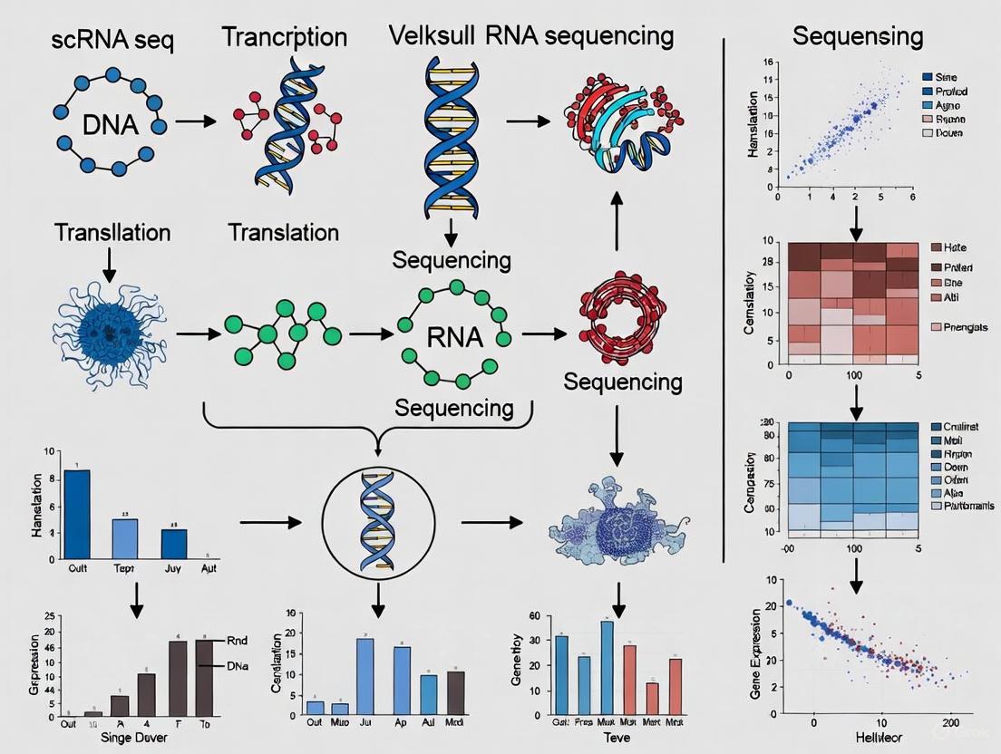
The following diagram illustrates the fundamental workflow differences between these two approaches:
Quantitative Comparison: Technical Specifications and Performance
The choice between bulk and single-cell RNA sequencing involves balancing multiple factors including resolution, cost, data complexity, and application suitability. The table below summarizes the key differences based on current technological capabilities:
| Feature | Bulk RNA Sequencing | Single-Cell RNA Sequencing |
|---|---|---|
| Resolution | Population average [1] | Individual cell level [1] |
| Cost per sample | Lower (~$300 per sample) [3] | Higher (~$500-$2000 per sample) [3] |
| Data complexity | Lower, more straightforward analysis [1] [3] | Higher, requires specialized computational methods [1] [3] |
| Cell heterogeneity detection | Limited, masks cellular diversity [1] [2] | High, reveals cellular subpopulations [1] [2] |
| Sample input requirement | Higher, typically micrograms of RNA [3] | Lower, single cells or nanograms of RNA [3] |
| Rare cell type detection | Limited, masked by abundant populations [3] | Possible, can identify rare populations [3] |
| Gene detection sensitivity | Higher, detects more genes per sample [3] | Lower due to transcript dropout [3] |
| Splicing analysis | More comprehensive [3] | Limited with standard methods [3] |
| Technical noise | Lower, averaged across cells [4] | Higher, includes amplification artifacts [4] |
| Experimental workflow | Simpler, established protocols [1] | More complex, requires single-cell suspension [1] |
Beyond these fundamental differences, scRNA-seq introduces unique technical considerations. The "transcriptome size" - the total number of mRNA molecules per cell - varies significantly across cell types and can impact data normalization and interpretation [5]. New computational methods like ReDeconv specifically address this challenge by incorporating transcriptome size into scRNA-seq normalization, improving accuracy in both single-cell analysis and bulk data deconvolution [5].
Experimental Design: Methodologies and Protocols
Bulk RNA-seq Experimental Protocol
Standard bulk RNA-seq follows a relatively straightforward workflow optimized for population-level analysis [1]:
Sample Collection and Homogenization: Tissue samples or cell pellets are collected and immediately stabilized using RNA preservation reagents. Samples are homogenized using mechanical disruption to lyse cells and release RNA.
RNA Extraction and Quality Control: Total RNA is extracted using column-based or magnetic bead-based methods. RNA quality is assessed using bioanalyzer systems to ensure RNA Integrity Number (RIN) > 8.0 for optimal sequencing results.
Library Preparation: Following poly-A selection or ribosomal RNA depletion, RNA is reverse transcribed into cDNA. Adapters containing sample-specific barcodes are ligated to enable multiplexed sequencing. Library concentration and quality are validated using quantitative PCR and fragment analyzers.
Sequencing and Data Analysis: Libraries are sequenced on platforms such as Illumina NovaSeq to a depth of 20-50 million reads per sample. Data analysis includes quality control, alignment to reference genomes, and differential expression analysis using tools like DESeq2 or edgeR.
Single-Cell RNA-seq Experimental Protocol
scRNA-seq requires more specialized procedures to preserve single-cell resolution [1] [4]:
Single-Cell Suspension Preparation: Tissues are dissociated using enzymatic (collagenase, trypsin) or mechanical methods optimized for specific tissue types. Cell viability is critical and typically maintained above 80% using cold-active proteases and rapid processing.
Cell Partitioning and Barcoding: Single-cell suspensions are loaded into microfluidic devices (e.g., 10X Genomics Chromium system) where individual cells are partitioned into nanoliter-scale droplets (GEMs) with barcoded beads. Each bead contains oligonucleotides with unique cell barcodes, unique molecular identifiers (UMIs), and poly-T sequences for mRNA capture.
Reverse Transcription and Library Construction: Within each droplet, cells are lysed and mRNA is captured by the barcoded oligos. Reverse transcription occurs in isolation, tagging each cDNA molecule with its cell-of-origin barcode. After breaking droplets, cDNA is amplified and sequencing libraries are constructed.
Sequencing and Computational Analysis: Libraries are sequenced to greater depth (typically 50,000-100,000 reads per cell). Data processing involves cell calling, demultiplexing using barcode information, UMI counting, and normalization. Downstream analysis includes clustering, cell type identification, and trajectory inference using tools like Seurat or Scanpy.
The following diagram illustrates the core single-cell partitioning and barcoding process that enables cellular resolution:
Applications in Research and Drug Development
Applications of Bulk RNA Sequencing
Bulk RNA-seq remains the preferred choice for several research scenarios [1] [3]:
Differential Gene Expression Analysis: Identifying genes that are upregulated or downregulated between different conditions (e.g., disease vs. healthy, treated vs. control) across entire tissues or populations.
Biomarker Discovery: Detecting molecular signatures for disease diagnosis, prognosis, or patient stratification. For example, bulk RNA-seq of cancer samples has identified gene expression signatures predictive of treatment response and survival outcomes [3].
Large-Scale Cohort Studies: Profiling transcriptomes across hundreds or thousands of samples in biobank projects where cost considerations make scRNA-seq prohibitive.
Pathway and Network Analysis: Studying how sets of genes change collectively under various biological conditions, providing systems-level insights.
Novel Transcript Characterization: Discovering and annotating isoforms, non-coding RNAs, alternative splicing events, and gene fusions, with applications in cancer research where fusion genes can drive oncogenesis [4].
Applications of Single-Cell RNA Sequencing
scRNA-seq enables applications that are impossible with bulk approaches [1] [6]:
Cellular Atlas Construction: Systematically cataloging all cell types and states within complex tissues, as demonstrated by the Human Cell Atlas project [2].
Rare Cell Population Identification: Discovering and characterizing rare cell types that may have crucial biological functions, such as cancer stem cells in tumors or rare immune cell subsets [3].
Developmental Trajectory Reconstruction: Mapping lineage relationships and differentiation pathways by ordering cells along pseudotemporal trajectories, revealing how progenitor cells give rise to specialized descendants [1] [2].
Tumor Heterogeneity Characterization: Resolving diverse cell populations within cancers, including cancer cell subtypes, immune infiltrates, and stromal components, providing insights into therapy resistance mechanisms [4].
Drug Discovery and Development: Informing target identification through improved disease understanding, identifying cell types most sensitive to perturbations, and providing insights into drug mechanisms of action [6]. scRNA-seq can profile drug responses at single-cell resolution, revealing how different cell subpopulations within tumors respond to therapies [7].
The integration of both approaches is increasingly powerful. For instance, a 2024 study on B-cell acute lymphoblastic leukemia (B-ALL) leveraged both bulk and single-cell RNA-seq to identify developmental states driving resistance and sensitivity to asparaginase chemotherapy [1]. Similarly, computational frameworks like scDEAL can transfer drug response knowledge from large-scale bulk databases to predict single-cell drug sensitivity, bridging the two technologies [7].
Research Reagent Solutions and Essential Materials
Successful implementation of RNA sequencing technologies requires specific reagents and platforms optimized for each approach:
| Item | Function | Bulk RNA-seq | Single-Cell RNA-seq |
|---|---|---|---|
| RNA Stabilization Reagents | Preserve RNA integrity post-collection | RNAlater, TRIzol | RPMI + FBS, Cold-active protease inhibitors |
| Cell Dissociation Kits | Tissue dissociation into single cells | Not typically required | Enzymatic mixes (collagenase/trypsin), GentleMACS dissociator |
| Viability Stains | Assess cell health and membrane integrity | Trypan blue | Propidium iodide, DAPI, Calcein AM |
| RNA Extraction Kits | Isolate high-quality RNA | Column-based (RNeasy), Magnetic beads | Same, but with DNase treatment |
| Library Preparation Kits | Prepare sequencing libraries | Illumina TruSeq, NEBNext Ultra II | 10X Genomics Chromium, SMART-seq kits |
| Quality Control Instruments | Assess RNA and library quality | Bioanalyzer, Fragment Analyzer | Bioanalyzer, Cell Counter (Countess) |
| Sequencing Platforms | Generate sequence data | Illumina NovaSeq, NextSeq | Illumina NovaSeq, HiSeq with high depth |
| Cell Partitioning Systems | Isolate individual cells | Not applicable | 10X Genomics Chromium, Fluidigm C1 |
| Barcoded Beads | Label cell-of-origin for transcripts | Not applicable | 10X Gel Beads, Drop-seq beads |
| Single-Cell Multiome Kits | Simultaneously profile multiple molecular layers | Not applicable | 10X Multiome (ATAC + Gene Exp.) |
Bulk and single-cell RNA sequencing represent complementary rather than competing approaches for transcriptome analysis. Bulk RNA-seq provides an economical, robust method for assessing overall transcriptional changes in homogeneous populations or when studying systemic responses. Single-cell RNA-seq reveals the cellular heterogeneity, rare populations, and dynamic transitions that underlie these population-level observations, albeit at higher cost and computational complexity [1] [8].
The choice between technologies depends fundamentally on the research question. Bulk sequencing remains ideal for differential expression analysis in well-defined systems, large-scale cohort studies, and when working with limited budgets. Single-cell approaches are essential for characterizing heterogeneous tissues, discovering novel cell types, reconstructing developmental trajectories, and understanding cellular responses to perturbations in complex systems [3].
Looking forward, the integration of both approaches through computational methods, along with emerging spatial transcriptomics technologies, promises a more complete understanding of biological systems across scales. As both technologies continue to evolve—with costs decreasing and methodologies improving—their synergistic application will further accelerate discoveries in basic biology, disease mechanisms, and therapeutic development.
For decades, bulk RNA sequencing (bulk RNA-seq) has been a fundamental tool in molecular biology, providing valuable insights into the average gene expression profile of entire tissue samples or cell populations [1] [4]. This method works by extracting RNA from a heterogeneous mixture of cells, processing it into a sequencing library, and generating a composite readout that represents the mean expression levels for each gene across all cells in the sample [1]. While this approach has proven invaluable for identifying differentially expressed genes between conditions and discovering biomarkers, it operates under a significant constraint: the complete loss of cellular resolution [2] [9].
The critical limitation of bulk RNA-seq becomes apparent when studying complex tissues—such as tumors, brain structures, or developing organs—where cellular heterogeneity is the rule rather than the exception [2] [4]. By providing only a "virtual average" of gene expression across diverse cell types, bulk RNA-seq fundamentally masks the cellular origins of transcriptional signals [2]. This averaging effect obscures rare but biologically important cell populations, blends distinct cellular states, and conceals the true complexity of transcriptional regulation that occurs at the single-cell level [9] [10]. As the field advances toward more precise analytical approaches, understanding this limitation becomes essential for researchers, scientists, and drug development professionals designing transcriptomic studies.
Quantitative Comparison: Bulk vs. Single-Cell RNA Sequencing
The technological differences between bulk and single-cell RNA sequencing translate directly into distinct analytical capabilities. The table below summarizes key performance metrics and characteristics that highlight the resolution gap between these approaches.
Table 1: Technical and Analytical Comparison of Bulk and Single-Cell RNA-Seq
| Parameter | Bulk RNA-Seq | Single-Cell RNA-Seq |
|---|---|---|
| Resolution | Population average [1] | Single-cell [1] |
| Cellular Heterogeneity | Masked [2] [11] | Revealed [2] [9] |
| Rare Cell Detection | Limited (>1% frequency) [12] | Excellent (<0.1% frequency) [12] |
| Required RNA Input | High (micrograms) [4] | Low (picograms per cell) [9] |
| Cost per Sample | Lower [1] [11] | Higher [1] |
| Data Complexity | Moderate [1] | High-dimensional [1] [9] |
| Ideal Applications | Differential expression, biomarker discovery [1] | Cell atlas construction, heterogeneity studies, developmental trajectories [1] [12] |
Table 2: Sensitivity Comparison in Complex Tissue Analysis
| Analysis Type | Gene Detection Rate | Rare Cell Population Detection | Identification of Novel Subtypes |
|---|---|---|---|
| Bulk RNA-Seq | Comprehensive for abundant transcripts [4] | Poor (signals diluted) [2] | Not possible [2] |
| Single-Cell RNA-Seq | Variable (3-20% mRNA recovery per cell) [9] | Excellent (theoretical limit: 1 in 10,000 cells) [12] | High (unbiased approach) [2] [10] |
The fundamental difference in data output can be visualized as a contrast between averaged and resolved transcriptional profiles:
Experimental Evidence: Case Studies Revealing Hidden Heterogeneity
Rheumatoid Arthritis Synovial Tissue Analysis
A compelling 2024 study integrated both bulk and single-cell RNA sequencing to investigate macrophage heterogeneity in rheumatoid arthritis (RA) synovial tissue [13]. Researchers began with bulk RNA-seq analysis of 213 RA samples and 63 healthy controls, which identified general inflammatory pathways but failed to resolve specific macrophage subpopulations driving disease progression [13].
When the same tissues were analyzed using scRNA-seq, researchers profiled 26,923 individual cells and identified previously obscured macrophage subsets, including a distinct Stat1+ macrophage population that expressed unique inflammatory signatures [13]. The experimental protocol followed these key steps:
- Tissue Processing: Synovial tissues were dissociated into single-cell suspensions using enzymatic digestion (collagenase IV/DNase I) [13]
- Cell Viability Assessment: Viable cell count and quality control to ensure >90% viability [13]
- Single-Cell Partitioning: Cells were loaded onto a Chromium X series instrument (10x Genomics) for droplet-based partitioning [13]
- Library Preparation: GEM (Gel Bead-in-emulsion) generation with cell barcoding and unique molecular identifiers (UMIs) [13]
- Sequencing: Illumina-based sequencing to a depth of 50,000 reads per cell [13]
- Bioinformatic Analysis: Cell clustering, differential expression, and trajectory analysis using Seurat (v5.0.1) and Monocle3 [13]
This scRNA-seq approach revealed that Stat1+ macrophages were enriched in inflammatory pathways and represented a key driver of RA pathology—a finding completely masked in the bulk analysis [13]. Functional validation confirmed that STAT1 activation modulated autophagy and ferroptosis pathways, suggesting new therapeutic targets for RA [13].
Cancer Stem Cell and Tumor Heterogeneity Studies
In oncology, bulk RNA-seq has historically provided averaged expression profiles that obscure critical rare cell populations. Studies comparing bulk and single-cell approaches in tumors have demonstrated that bulk sequencing consistently underestimates heterogeneity and misses biologically significant rare populations [4].
For example, scRNA-seq applications in glioblastoma, colorectal cancer, and head and neck squamous cell carcinoma have revealed:
- Rare stem-like cells with treatment-resistant properties that comprise <1% of tumor mass [4]
- Partial epithelial-to-mesenchymal transition (p-EMT) programs associated with metastasis [4]
- Distinct cancer cell states that develop drug resistance after targeted therapy [4]
The experimental workflow for such tumor heterogeneity studies typically involves:
Table 3: Key Methodological Steps in Tumor Heterogeneity Analysis
| Step | Description | Critical Considerations |
|---|---|---|
| Sample Preparation | Generate viable single-cell suspension from tumor tissue [1] | Maintain cell viability, minimize stress responses [1] |
| Cell Partitioning | Isolate individual cells using microfluidic devices [1] [4] | Optimize cell concentration to minimize doublets [2] |
| mRNA Capture & Barcoding | Lysed cells release mRNA captured with cell-specific barcodes [4] | Use UMIs to correct for amplification bias [9] |
| Library Preparation | Amplify cDNA and prepare sequencing libraries [1] | Maintain representation of low-abundance transcripts [9] |
| Bioinformatic Analysis | Cluster cells, identify subpopulations, reconstruct trajectories [13] | Address technical noise, batch effects [9] [13] |
Methodological Framework: Experimental Protocols for Heterogeneity Studies
Comprehensive scRNA-seq Workflow for Detecting Cellular Heterogeneity
The following diagram outlines the standardized workflow for single-cell RNA sequencing experiments designed to uncover cellular heterogeneity:
Researcher's Toolkit: Essential Reagents and Platforms
Table 4: Key Research Reagent Solutions for scRNA-seq Experiments
| Category | Specific Products/Platforms | Function & Application |
|---|---|---|
| Single-Cell Platforms | 10x Genomics Chromium [1] [4], BD Rhapsody [14], Fluidigm C1 [2] | Partition thousands of single cells with barcoding capabilities |
| Cell Barcoding Reagents | Gel Beads with cell barcodes & UMIs [4] | Label individual cell's RNA for multiplexing and quantification |
| Reverse Transcription Kits | Optimized enzymes with high efficiency [9] | Convert minute RNA amounts to stable cDNA with low bias |
| Amplification Reagents | Template-switching oligonucleotides [9] | Amplify cDNA while maintaining representation |
| Cell Viability Assays | Fluorescent viability dyes [1] | Distribute live cells for high-quality RNA recovery |
| Enzymatic Dissociation Kits | Tissue-specific collagenase blends [1] | Dissociate tissues into single cells with minimal RNA degradation |
The critical limitation of bulk RNA-seq in masking cellular heterogeneity has profound implications across biomedical research and therapeutic development. While bulk approaches remain valuable for population-level differential expression analysis in homogeneous samples or when budget constraints predominate [1] [11], their inability to resolve cellular diversity makes them insufficient for characterizing complex biological systems [2] [4].
The advent of robust single-cell RNA sequencing technologies has fundamentally transformed our investigative capabilities, enabling researchers to identify novel cell types, trace developmental trajectories, characterize tumor microenvironments, and uncover rare cell populations that drive disease progression [13] [12] [4]. For drug development professionals, these advances translate into improved target identification, better patient stratification strategies, and enhanced understanding of therapeutic mechanisms of action and resistance [15].
As the field continues to evolve, the strategic integration of both approaches—using bulk RNA-seq for initial screening and scRNA-seq for deep resolution of heterogeneity—represents the most powerful paradigm for comprehensive transcriptomic analysis in developmental studies and disease research [1] [13].
How scRNA-Seq Reveals Rare Cell Populations and Lineage Trajectories
For decades, developmental biology relied on bulk RNA sequencing (bulk RNA-seq), which averages gene expression across entire tissue samples. While valuable for identifying major expression shifts, this approach obscures a fundamental biological truth: cellular heterogeneity. The advent of single-cell RNA sequencing (scRNA-seq) has revolutionized the field by providing a high-resolution lens to observe the individual cells that constitute developing tissues, uncovering rare cell populations and mapping lineage trajectories that were previously invisible.
The Resolution Revolution: scRNA-seq vs. Bulk RNA-seq
At its core, the difference between these techniques is one of resolution. Bulk RNA-seq processes RNA from a population of cells, yielding a population-average gene expression profile [1] [3]. In contrast, scRNA-seq isolates individual cells, captures their RNA, and uses cellular barcodes to trace each transcript back to its cell of origin [1] [4]. This fundamental difference in approach dictates their respective applications and findings.
Table 1: Core Technical Differences Between Bulk and Single-Cell RNA-seq
| Feature | Bulk RNA-seq | Single-Cell RNA-seq |
|---|---|---|
| Resolution | Population average [1] [16] | Individual cell level [1] [3] |
| Cell Heterogeneity Detection | Limited; masks differences [1] [3] | High; reveals distinct subpopulations [2] [16] |
| Rare Cell Type Detection | Limited; signals are diluted [17] [3] | Possible; can identify very rare cells [17] [18] |
| Cost per Sample | Lower [3] | Higher [3] |
| Data Complexity | Lower; more straightforward analysis [1] [3] | Higher; requires specialized computational tools [3] |
| Ideal Application | Differential expression in homogeneous samples, biomarker discovery [1] | Deconstructing heterogeneous tissues, identifying novel cell types, lineage tracing [1] [2] |
The limitation of bulk sequencing becomes critical when studying development. As noted in one review, "analysis of pooled populations of progenitor cells does not enable distinction of the signals that drive a progenitor down a particular differentiation pathway; for instance, the signals that determine whether a nephron progenitor cell becomes a podocyte or a proximal tubule cell" [2]. scRNA-seq makes these critical early fate decisions visible.
Pinpointing the Needle in the Haystack: Identifying Rare Cell Populations
Rare cell types, such as stem cells, transitional progenitors, or drug-resistant clones, often play disproportionately important roles in development and disease. Their low abundance means their transcriptional signatures are diluted to undetectable levels in bulk analyses [17]. scRNA-seq excels at finding these needles in the cellular haystack.
Case Study: Uncovering Tumor-Initiating Cells
A compelling example comes from cancer research. A 2025 study of triple-negative breast cancer used a STAT-signaling reporter system to enrich for tumor-initiating cells (TICs), a rare population associated with therapy resistance and recurrence. Subsequent scRNA-seq revealed a distinct IFN/STAT1-associated transcriptional state in these TICs. Notably, most of these identified genes were absent from previously published TIC signatures derived using bulk RNA-seq, demonstrating scRNA-seq's unique power to uncover molecular drivers hidden in rare populations [18].
General Experimental Workflow for Rare Cell Analysis
The process for identifying rare cells typically involves the following steps, which can be adapted for various tissues and research questions [17]:
- Tissue Dissociation & Single-Cell Suspension: The starting tissue is dissociated using enzymatic or mechanical methods to create a viable single-cell suspension. This step is critical, as it must preserve cell integrity and minimize stress-induced transcriptional changes [1] [17].
- Cell Viability QC & Optional Enrichment: Cell concentration and viability are assessed. For pre-defined rare populations, Fluorescence-Activated Cell Sorting (FACS) using specific cell surface markers or fluorescent reporters can be used for enrichment prior to sequencing [17].
- Single-Cell Partitioning & Barcoding: Using platforms like the 10x Genomics Chromium, single cells are partitioned into nanoliter-scale droplets (GEMs) along with barcoded beads. Each bead contains oligonucleotides with a unique cell barcode (to tag all RNAs from one cell) and a unique molecular identifier (UMI) to quantify individual transcripts [1] [4].
- Library Prep & Sequencing: Within the droplets, cells are lysed, mRNA is barcoded, and sequencing libraries are constructed. These are then pooled for high-throughput sequencing [1].
- Bioinformatic Clustering & Rare Population Identification: Sequenced reads are demultiplexed using the cell barcodes. Dimensionality reduction and clustering algorithms (e.g., Seurat, Scanpy) group cells based on similar gene expression profiles. Rare populations appear as distinct, small clusters that can be characterized by their unique marker genes [17] [2].
Reconstructing Developmental Pathways: Lineage Tracing with scRNA-seq
Beyond identifying static cell types, scRNA-seq can be coupled with lineage tracing to reconstruct the dynamic history of cell fate decisions, answering the fundamental question: "How does a single progenitor cell give rise to diverse, differentiated progeny?"
The Emergence of Single-Cell Lineage Tracing (SCLT)
Traditional lineage tracing uses fluorescent proteins to mark progenitor cells and their descendants. However, its resolution is limited. Single-cell lineage tracing (SCLT) combines the principles of lineage tracing with the power of scRNA-seq to map lineage connectivity at single-cell resolution, making it "the best tool for exploring the heterogeneity of cellular differentiation" [19].
Methodologies for SCLT
Several innovative methods have been developed to record lineage information within a cell's genome or transcriptome.
Table 2: Key Single-Cell Lineage Tracing (SCLT) Techniques
| Technique | Mechanism | Key Advantage | Application Example |
|---|---|---|---|
| Integration Barcodes [19] | A library of viral vectors with random DNA "barcodes" is used to transduce progenitor cells. Each integration event provides a heritable, unique clonal marker. | Enables simultaneous tracking of thousands of clones, great for studying hematopoietic stem cell (HSC) dynamics. | Tracking HSC-derived clones in transplantation studies to understand clonal dynamics. |
| CRISPR Barcoding [19] | A CRISPR/Cas9 system introduces cumulative insertions/deletions (InDels) into a defined genomic "barcode" locus over cell divisions. The mutation pattern serves as a mitotic history record. | Creates a high diversity of lineage marks; mutations accumulate irreversibly over time. | Reconstructing developmental lineage trees in model organisms. |
| Base Editors [19] | Engineered base editors introduce point mutations at a high rate into a synthetic barcoding sequence, recording cell division events. | Allows for recording of many more mitotic divisions, enabling construction of high-resolution cell phylogenetic trees. | Quantifying the number of actively dividing parental cells and their division patterns during development. |
| Natural Barcodes [19] | Utilizes spontaneously acquired somatic mutations in the nuclear or mitochondrial genome that accumulate during development and aging. | Non-invasive and can be applied to human samples without genetic manipulation. | Retrospective lineage tracing in human tissues to understand clonal relationships. |
Visualizing a Combined SCLT and scRNA-seq Workflow
The following diagram illustrates how these methodologies are integrated with scRNA-seq to capture both cell state (the transcriptome) and cell history (the lineage barcode) simultaneously.
In this workflow, a heritable lineage marker (e.g., a DNA barcode) is introduced into progenitor cells. As these cells divide and differentiate, the marker is passed on to all progeny. When the tissue is harvested and subjected to scRNA-seq, both the transcriptome and the lineage barcode are sequenced. Bioinformatic analysis then reconstructs a lineage tree, where each branch point represents a fate decision, and each leaf (terminal cell) is annotated with its complete transcriptional profile. This reveals not only which cells are related but also the transcriptional programs that define each branch point in the lineage.
The Scientist's Toolkit: Essential Reagents and Technologies
Successful scRNA-seq and lineage tracing experiments rely on a suite of specialized tools and reagents.
Table 3: Key Research Reagent Solutions for scRNA-seq and Lineage Tracing
| Item / Technology | Function | Example Use Case |
|---|---|---|
| 10x Genomics Chromium | A microfluidics platform that partitions single cells into barcoded droplets (GEMs) for high-throughput scRNA-seq library preparation [1] [4]. | Standardized, scalable workflow for profiling gene expression in thousands of cells from complex tissues. |
| Fluorescent Reporter Cell Lines | Genetically engineered cells where a fluorescent protein (e.g., GFP) is expressed under the control of a cell-type-specific promoter or responsive element [17] [18]. | Visually identifying and isolating specific cell populations (e.g., STAT-responsive TICs) via FACS prior to scRNA-seq. |
| Cre-loxP System | A site-specific recombination system allowing for inducible, permanent genetic labeling of a cell and its descendants [19]. | Used in multicolor labeling (Brainbow) and polylox barcoding for fate mapping in model organisms. |
| Lentiviral Barcode Libraries | A diverse pool of lentiviral vectors, each containing a unique random DNA sequence, used to "barcode" progenitor cells [18] [19]. | Clonal tracking in transplantation assays, such as studying the output of individual hematopoietic stem cells. |
| CRISPR/Cas9 & Base Editors | Genome editing tools used to create evolving, cumulative mutations in a synthetic genomic barcode locus [19]. | Recording mitotic history with high information capacity to build detailed lineage trees during embryonic development. |
| Cell Ranger / Seurat | Standard bioinformatics software packages for processing, analyzing, and visualizing scRNA-seq data, including clustering and differential expression [4]. | Transforming raw sequencing data into interpretable clusters and marker genes to identify cell types and states. |
The transition from bulk RNA-seq to scRNA-seq represents a paradigm shift in developmental biology and oncology. By moving from a population-average view to a single-cell perspective, researchers can now identify rare but critical cell populations, such as therapy-resistant tumor-initiating cells, and reconstruct the precise lineage trajectories that guide development. While bulk RNA-seq remains a valuable tool for hypothesis generation and large-scale differential expression studies in homogeneous samples, scRNA-seq provides the indispensable resolution needed to deconstruct complex tissues and dynamic processes. The ongoing integration of scRNA-seq with other single-cell modalities and spatial technologies promises to further deepen our understanding of biological systems in health and disease.
The study of the transcriptome has been fundamentally reshaped by two pivotal technological approaches: bulk RNA sequencing (bulk RNA-seq) and single-cell RNA sequencing (scRNA-seq). Bulk RNA-seq, a long-standing cornerstone of genomics, provides a population-averaged gene expression profile from a tissue sample composed of numerous cells [4] [20]. In contrast, single-cell RNA sequencing represents a revolutionary advancement that enables researchers to investigate gene expression at the resolution of individual cells, uncovering the cellular heterogeneity that bulk methods inevitably mask [1] [10]. This guide objectively traces the key historical milestones in the evolution of these technologies, comparing their performance, applications, and experimental requirements to inform researchers and drug development professionals.
Historical Context and Technological Evolution
The journey into transcriptomics began with bulk sequencing approaches. Following the completion of the Human Genome Project in 2003, next-generation sequencing (NGS) technologies emerged as powerful tools for studying genomic traits and gene expression [20]. For over a decade, bulk RNA-seq served as the primary method for analyzing RNA extracted from populations of cells, revealing differences between sample conditions and providing averaged gene expression readouts [21] [20].
The conceptual and technical shift toward single-cell analysis began approximately two decades ago, pioneered by Norman Iscove who used polymerase chain reaction (PCR) for exponential amplification of single-cell cDNAs [10]. James Eberwine subsequently advanced this field by developing a method to amplify cDNAs using T7 RNA polymerase-based transcription in vitro [10]. These early innovations laid the groundwork for what would become a transformative technology in genomics.
The invention and mass production of high-density DNA microarray chips represented another critical milestone, enabling gene expression analysis with unprecedented precision at the individual cell level [10]. This technological progress revealed significant differences in the transcriptomes of genetically identical cells, highlighting the remarkable complexity of cellular behavior and the limitations of population-level analyses that average data across cell populations [10].
In 2013, single-cell RNA sequencing was named "Method of the Year" by Nature Methods, signaling its arrival as a transformative technology [20]. The subsequent development of commercial integrated platforms like the 10x Genomics Chromium system triggered rapid adoption of this revolutionized technology in translational and clinical research [4]. This system's core innovation lies in its ability to generate hundreds of thousands of single cell microdroplets (GEMs) on a microfluidics chip, with each GEM containing a single cell, reverse transcription mixes, and a gel bead conjugated with millions of oligo sequences featuring cell-specific barcodes [4].
Table 1: Key Historical Milestones in RNA Sequencing
| Year/Period | Milestone | Technological Significance |
|---|---|---|
| ~2000-2005 | Early single-cell cDNA amplification [10] | Foundation for single-cell analysis using PCR-based methods |
| 2005-2008 | Bulk RNA-seq optimization [20] | Established robust protocols for population-averaged transcriptomics |
| 2013 | scRNA-seq named "Method of the Year" [20] | Recognition of scRNA-seq as a transformative technology |
| 2017-Present | Commercial integrated platforms (e.g., 10x Genomics) [4] | Made scRNA-seq accessible and reproducible for broader research communities |
| 2019-Present | Spatial transcriptomics emergence [4] | Added spatial context to single-cell resolution data |
Technical and Experimental Comparisons
Fundamental Methodological Differences
The experimental workflows for bulk and single-cell RNA sequencing demonstrate fundamental differences in both philosophy and execution. In bulk RNA-seq, the biological sample is digested to extract total RNA or enriched mRNA, which is then converted to cDNA and processed into a sequencing-ready library [1]. This approach results in a average gene expression profile for the entire sample, analogous to obtaining the average color of a forest without seeing individual trees [1].
In contrast, scRNA-seq requires the generation of viable single cell suspensions from whole samples through enzymatic or mechanical dissociation [1]. This is followed by cell partitioning, where single cells are isolated into individual micro-reaction vessels. In the 10x Genomics platform, this occurs within Gel Beads-in-emulsion (GEMs) on a microfluidic chip [1] [4]. Within each GEM, cell lysis occurs, allowing RNA to be captured and barcoded with cell-specific barcodes that ensure analytes from each cell can be traced back to their origin [1]. This barcoding strategy is a cornerstone of modern scRNA-seq, enabling the multiplexing of thousands of cells in a single experiment.
Performance and Capability Comparison
The differing methodologies of bulk and single-cell RNA sequencing translate into distinct performance characteristics and experimental capabilities, which determine their appropriate applications in research and drug development.
Table 2: Technical Performance Comparison of Bulk vs. Single-Cell RNA Sequencing
| Parameter | Bulk RNA Sequencing | Single-Cell RNA Sequencing | Experimental Implications |
|---|---|---|---|
| Resolution | Population average [1] [3] | Individual cell level [1] [3] | scRNA-seq reveals cellular heterogeneity and rare cell types |
| Cell Heterogeneity Detection | Limited [3] | High [3] | Bulk masks rare populations; scRNA-seq identifies them |
| Rare Cell Type Detection | Limited [3] | Possible [3] | scRNA-seq can identify rare stem cells or circulating tumor cells |
| Gene Detection Sensitivity | Higher per sample [3] | Lower per cell [3] | Bulk detects more genes per sample; scRNA-seq has sparser data |
| Cost per Sample | Lower (~$300/sample) [3] | Higher (~$500-$2000/sample) [3] | Bulk more suitable for large cohort studies |
| Data Complexity | Lower, more straightforward [1] [3] | Higher, requires specialized analysis [1] [3] | scRNA-seq demands advanced computational resources and expertise |
| Sample Input Requirement | Higher [3] | Lower [3] | scRNA-seq can work with limited material (e.g., 10 pg total RNA) |
| Splicing Analysis | More comprehensive [3] | Limited [3] | Bulk better for detecting alternative splicing events |
Applications and Experimental Outcomes
Distinct yet Complementary Biological Applications
The choice between bulk and single-cell RNA sequencing is primarily determined by the research question, with each technology excelling in distinct application domains.
Bulk RNA-seq applications include:
- Differential gene expression analysis: Comparing gene expression profiles between different experimental conditions (disease vs. healthy, treated vs. control) to identify upregulated or downregulated genes [1]
- Biomarker discovery: Identifying RNA-based biomarkers and molecular signatures for disease diagnosis, prognosis, or stratification [1] [4]
- Tissue or population-level transcriptomics: Obtaining global expression profiles from whole tissues, organs, or bulk-sorted cell populations, particularly useful for large cohort studies or biobank projects [1]
- Gene fusion detection: Discovering novel gene fusions, with recent computational advances like DEEPEST algorithm minimizing false positives and improving detection sensitivity [4]
Single-cell RNA-seq applications include:
- Characterizing heterogeneous cell populations: Identifying novel cell types, cell states, and rare cell types within complex tissues [1] [4]
- Reconstructing developmental hierarchies: Tracing cellular differentiation pathways and lineage relationships during development or disease progression [1]
- Dissecting tumor heterogeneity: Revealing diverse transcriptional programs within tumors that provide plasticity to adapt to various environments and promote treatment resistance [4]
- Analyzing tumor microenvironment: Characterizing the diversity of immune and stromal cell populations within tumors and their roles in cancer progression and therapy resistance [4] [22]
Case Study: Integrated Analysis in Lung Adenocarcinoma
A 2025 study on lung adenocarcinoma (LUAD) exemplifies the powerful synergy between bulk and single-cell approaches [22]. Researchers first used scRNA-seq to identify seven distinct cell clusters within tumor samples, with epithelial cell cluster 1 (Epi_C1) showing the highest stemness potential based on CytoTRACE analysis [22]. They then leveraged bulk RNA-seq data from TCGA to construct a prognostic tumor stem cell marker signature (TSCMS) model incorporating 49 tumor stemness-related genes [22]. This integrated approach demonstrated that high-risk patients exhibited lower immune scores, increased tumor purity, and significant differences in chemotherapy sensitivity, while also identifying TAF10 as a potential therapeutic target linked to stemness and poor prognosis [22].
The Scientist's Toolkit: Essential Research Reagents and Materials
Successful execution of RNA sequencing experiments requires careful selection of reagents and materials optimized for each methodology.
Table 3: Essential Research Reagents and Materials for RNA Sequencing
| Reagent/Material | Function | Bulk RNA-seq Specifics | Single-Cell RNA-seq Specifics |
|---|---|---|---|
| RNA Isolation Kits | Extract high-quality RNA from samples | Focus on yield and purity; RIN >6 acceptable [20] | Critical for viable single cells; emphasis on preventing degradation |
| Poly(T) Oligos | Enrich for polyadenylated RNA | Standard mRNA enrichment [20] | Coated on gel beads for cell-specific barcoding [4] |
| rRNA Depletion Reagents | Remove abundant ribosomal RNA | Optional depending on study goals [20] | Often used to improve detection of non-polyadenylated RNAs |
| Cell Barcoding Beads | Tag molecules with cell-specific barcodes | Not required | Gel Beads with unique 10x barcodes essential for partitioning [1] |
| Library Prep Kits | Prepare sequencing-ready libraries | Standard NGS library preparation | Specialized kits with UMIs for unique transcript counting |
| Microfluidic Chips | Partition individual cells | Not used | Essential for single-cell isolation in platforms like 10x Chromium [1] |
| Viability Stains | Assess cell integrity and viability | Less critical | Critical for ensuring high-quality single cell suspensions |
Data Analysis and Computational Considerations
The data analysis workflows for bulk and single-cell RNA sequencing differ significantly in complexity and methodology. Bulk RNA-seq analysis typically involves quality control of raw data, read alignment using tools like STAR or TopHat, transcriptome reconstruction, and expression quantification [21]. The resulting data matrix is relatively straightforward, with genes as rows and samples as columns.
In contrast, scRNA-seq data analysis presents unique computational challenges due to the high dimensionality, technical noise, and sparsity of the data [21] [3]. The initial data matrix contains genes as rows and thousands of individual cells as columns. Analysis requires specialized pipelines for:
- Quality control: Filtering cells with high mitochondrial content or few detected genes [21] [22]
- Normalization and batch effect correction: Addressing technical variability between cells and experiments [13]
- Dimensionality reduction: Using PCA, UMAP, or t-SNE to visualize cell populations in two dimensions [22] [13]
- Cell clustering and marker identification: Defining cell populations based on transcriptional similarity [22]
- Trajectory inference: Reconstructing developmental pathways using tools like Monocle3 [13]
The high computational resources and specialized expertise required for scRNA-seq analysis represent a significant consideration for research planning [1] [3].
Future Directions and Emerging Technologies
The evolution of transcriptomics continues with the emergence of spatial transcriptomics, which preserves the spatial context of RNA expression within tissues [4] [10]. This technology represents a natural progression from bulk sequencing (which loses cellular resolution) to single-cell sequencing (which loses spatial context) to spatial methods that provide both single-cell resolution and spatial information [10].
Other emerging trends include:
- Multi-omics integration: Combining scRNA-seq with other single-cell modalities like chromatin accessibility (scATAC-seq) and protein profiling (CITE-seq) [3] [13]
- Machine learning applications: Leveraging deep learning for data denoising, dimensionality reduction, and trajectory inference [23]
- Cost reduction initiatives: Development of more affordable scRNA-seq platforms and reagents to increase accessibility [1] [3]
- Targeted scRNA-seq approaches: Focusing sequencing resources on predefined gene sets for superior sensitivity and quantitative accuracy in translational settings [15]
The revolution from bulk to single-cell RNA sequencing has fundamentally transformed our approach to studying transcriptomes. Bulk RNA-seq remains a powerful, cost-effective tool for population-level studies, differential expression analysis in homogeneous samples, and large cohort studies where cellular heterogeneity is not the primary focus [1] [3] [23]. In contrast, scRNA-seq provides unprecedented resolution for dissecting cellular heterogeneity, identifying rare cell populations, reconstructing developmental trajectories, and understanding complex tissue microenvironments [1] [4] [10].
The most impactful modern research often integrates both technologies, using their complementary strengths to generate comprehensive biological insights [22] [13]. As the field continues to evolve with spatial transcriptomics and multi-omics integration, this methodological synergy will likely drive the next wave of discoveries in basic research and therapeutic development.
For researchers planning transcriptomics studies, the decision between bulk and single-cell approaches should be guided by specific research questions, budget constraints, available expertise, and the biological complexity of the system under investigation.
In developmental biology, understanding the precise transcriptional programs that guide cell fate decisions is paramount. For over a decade, bulk RNA sequencing (bulk RNA-seq) has been the conventional approach for studying gene expression, providing a population-averaged view of the transcriptome across thousands to millions of cells [1] [20]. This method yields a composite gene expression profile that represents the collective RNA from all cells in a sample, much like viewing a forest from a distance without distinguishing individual trees. In contrast, single-cell RNA sequencing (scRNA-seq) has emerged as a revolutionary technology that enables researchers to measure gene expression at the resolution of individual cells, revealing the unique transcriptional identity of each cellular unit within a complex biological system [1] [4]. This fundamental difference in resolution—averaged expression versus cell-specific transcriptomes—has profound implications for how we study developmental processes, tumor heterogeneity, and cellular responses to therapeutic interventions [24] [4].
The distinction between these approaches extends beyond technical methodology to their very philosophical underpinnings. Bulk RNA-seq operates on the principle of collective measurement, where the signal represents the mean expression across all cells, potentially masking critical cell-to-cell variations. scRNA-seq, however, embraces cellular heterogeneity as a fundamental biological reality, recognizing that individual cells within a population may exist in distinct states, perform different functions, and respond uniquely to environmental cues [25]. This guide provides a comprehensive comparison of these technologies, with a specific focus on their applications in developmental studies research, to help scientists select the appropriate tool for their specific research questions.
Experimental Protocols and Workflows
Bulk RNA-Seq Experimental Workflow
The bulk RNA-seq workflow begins with tissue collection or cell culture, followed by RNA extraction from the entire sample population [1] [20]. Key steps include:
- Sample Lysis and RNA Extraction: The biological sample is digested to extract total RNA, which may include enrichment for mRNA via poly(A) selection or depletion of ribosomal RNA (rRNA) to improve sequencing efficiency [1].
- Library Preparation: Extracted RNA is converted to complementary DNA (cDNA) through reverse transcription. The cDNA undergoes fragmentation, adapter ligation, and amplification to create a sequencing-ready library. Library preparation strategies vary depending on the RNA species of interest—mRNA-only libraries typically use poly(T) enrichment, while whole transcriptome approaches employ rRNA depletion to capture non-coding RNAs and other RNA species [20] [4].
- Sequencing and Data Analysis: Libraries are sequenced using next-generation sequencing platforms, followed by alignment to a reference genome and quantification of gene expression levels. The final output represents the average expression level for each gene across all cells in the original sample [1] [21].
Single-Cell RNA-Seq Experimental Workflow
scRNA-seq introduces several critical steps to preserve and analyze cell-specific information [1] [26]:
- Single-Cell Suspension Preparation: Tissues are dissociated into viable single-cell suspensions using enzymatic or mechanical methods, with careful optimization to minimize stress-induced transcriptional artifacts and preserve cell viability [26].
- Single-Cell Isolation and Barcoding: Individual cells are partitioned into nanoliter-scale reactions using microfluidic devices (e.g., 10x Genomics Chromium) or plate-based systems. Each cell is lysed, and the released mRNA transcripts are tagged with cell-specific barcodes and unique molecular identifiers (UMIs) during reverse transcription. These barcodes enable bioinformatic tracing of each transcript back to its cell of origin, while UMIs facilitate accurate transcript quantification by correcting for amplification bias [1] [4].
- Library Preparation and Sequencing: Barcoded cDNA from all cells is pooled into a single library for efficient sequencing, despite originating from thousands of individual cells [25].
- Bioinformatic Processing and Analysis: Sequencing data undergoes demultiplexing (assigning reads to individual cells based on barcodes), quality control, normalization, and downstream analysis such as clustering, trajectory inference, and differential expression analysis at the single-cell level [24].
The following diagram illustrates the key procedural differences between these two approaches:
Comparative Analysis of Output Data
Nature of Transcriptome Information
The fundamental output differences between these technologies create distinct advantages and limitations for each approach:
Bulk RNA-seq Output Characteristics:
- Averaged Expression Signals: Gene expression measurements represent the mean across all cells in the sample, weighted by abundance and expression level of each cell type [1] [20].
- Population-Level Insights: Well-suited for identifying consensus expression patterns that characterize a tissue or condition [1].
- Masked Heterogeneity: Cellular subpopulations and rare cell types (<5-10% of total population) typically fail to detectably influence the averaged profile [4].
- Higher Depth per Gene: Sequencing depth is distributed across the transcriptome without need for cell barcoding, often enabling better detection of lowly-expressed genes within the population context [1].
scRNA-seq Output Characteristics:
- Cell-Specific Resolution: Each measurement is associated with a specific cell via barcoding, preserving individual transcriptomic identities [1] [4].
- Heterogeneity Mapping: Enables identification of distinct cell types, states, and continuous transitions within populations [24] [25].
- Rare Cell Detection: Can identify and characterize cell populations representing as little as 0.1-1% of the total sample [4] [25].
- Sparse Data: Each individual cell captures only 10-50% of its transcriptome due to limited RNA capture efficiency, creating data sparsity that requires specialized statistical approaches [24].
Quantitative Comparison of Output Data
The table below summarizes key differences in the data output and analytical capabilities:
| Parameter | Bulk RNA-seq | Single-Cell RNA-seq |
|---|---|---|
| Resolution | Population average | Individual cells |
| Heterogeneity Analysis | Indirect inference | Direct measurement |
| Rare Cell Detection | Limited (>5% abundance) | Excellent (0.1-1% abundance) |
| Transcriptome Coverage | ~40-70% of transcriptome per sample | ~10-50% of transcriptome per cell |
| Data Structure | Dense matrix (genes × samples) | Sparse matrix (genes × cells) |
| Key Deliverables | Differential expression, Pathway analysis | Cell clustering, Trajectory inference, Rare population identification |
| Ideal Applications | Biomarker discovery, Condition comparisons, Transcriptome annotation | Cell atlas construction, Developmental tracing, Tumor heterogeneity, Drug response mechanisms |
Experimental Evidence from Comparative Studies
Recent investigations have directly compared outputs from both technologies when applied to the same biological systems. A 2025 study of human pancreatic islets from the same donors demonstrated that while both scRNA-seq and single-nuclei RNA-seq (snRNA-seq) identified the same major cell types, they revealed significant differences in predicted cell type proportions [27]. This highlights how the choice of method can influence biological interpretations, particularly when studying complex tissues with nuanced cellular distributions.
In cancer research, integrated approaches have proven particularly powerful. A 2024 study on B-cell acute lymphoblastic leukemia (B-ALL) leveraged both technologies to identify developmental states driving chemotherapy resistance [1]. While bulk RNA-seq provided a global view of treatment-induced expression changes, scRNA-seq pinpointed rare resistant subpopulations that would have been masked in bulk averages. Similarly, in ovarian cancer, researchers combined scRNA-seq analysis of platinum-resistant cancer cells with bulk RNA-seq data from larger cohorts to develop a machine learning model that accurately predicted patient response to platinum-based chemotherapy [28].
Applications in Developmental Studies
Developmental biology represents a particularly compelling application for scRNA-seq, as it fundamentally involves understanding how a relatively homogeneous population of progenitor cells differentiates into diverse, specialized cell types.
Lineage Tracing and Developmental Trajectories
scRNA-seq enables reconstruction of developmental hierarchies and lineage relationships by capturing transient intermediate states that are impossible to resolve with bulk approaches [1] [24]. Computational methods like pseudotime analysis order individual cells along differentiation trajectories based on transcriptional similarity, effectively reconstructing the temporal sequence of developmental events from snapshot data [24]. This approach has been successfully applied to model cardiovascular lineage segregation in mice [26], embryonic development [25], and differentiation processes in various tissues [24].
In contrast, bulk RNA-seq of developing tissues typically identifies only the most abundant cell types and may completely miss critical transitional states that are short-lived or numerically rare. While bulk approaches can track gross expression changes across developmental timepoints, they cannot resolve whether these changes represent graded transitions across the population or the emergence of distinct subpopulations.
Cellular Heterogeneity in Developing Systems
The following diagram illustrates how each technology captures cellular heterogeneity during development:
Case Study: Cardiovascular Development
A compelling example comes from cardiac development research, where scRNA-seq has redefined understanding of heart formation. Traditional bulk approaches had identified major transcriptional changes between developmental stages but failed to resolve the precise lineage relationships between different cardiac cell types. scRNA-seq applied to developing mouse hearts revealed previously uncharacterized progenitor populations and delineated the transcriptional programs guiding specialization into cardiomyocytes, endothelial cells, and valve interstitial cells [20]. These findings have profound implications for understanding congenital heart diseases and developing regenerative therapies.
The Scientist's Toolkit: Research Reagent Solutions
Selecting appropriate reagents and platforms is crucial for successful RNA-seq experiments. The following table outlines key solutions for implementing each technology:
| Product Category | Specific Examples | Function & Application |
|---|---|---|
| Bulk RNA-seq Library Prep | Illumina Stranded mRNA Prep, NEBNext Ultra II RNA | Poly(A) enrichment, cDNA synthesis, library construction for population-level sequencing |
| Single-Cell Isolation | 10x Genomics Chromium X, BD Rhapsody, Fluidigm C1 | Microfluidic partitioning of single cells with barcoded beads for high-throughput capture |
| Single-Cell Library Prep | 10x Genomics 3' Gene Expression, SMART-Seq v4 | Cell barcoding, UMI incorporation, cDNA amplification from single cells |
| Sample Preservation | DNA/RNA Shield, RNAlater | Stabilizes RNA in intact tissues or cell suspensions before processing |
| Cell Type Enrichment | MACS Cell Separation, FACS | Isolation of specific cell populations prior to sequencing using surface markers |
| Single-Nuclei RNA-seq | 10x Genomics Nuclei Isolation Kits | Enables sequencing from frozen tissues or difficult-to-dissociate samples |
| Data Analysis Software | Cell Ranger, Seurat, Scanpy, Partek Flow | Processing, normalization, clustering, and visualization of single-cell data |
The choice between bulk RNA-seq and scRNA-seq should be guided by specific research questions and experimental constraints. Bulk RNA-seq remains the preferred method for hypothesis-generating exploration of expression differences between conditions, identification of biomarker signatures, and studies with limited budgets or sample material that precludes single-cell analysis [1] [20]. Its advantages include lower cost per sample, simpler data analysis, and established analytical frameworks.
scRNA-seq is indispensable when investigating cellular heterogeneity, developmental trajectories, rare cell populations, or complex tissues with diverse cellular composition [24] [4]. Despite higher per-sample costs and greater analytical complexity, its ability to resolve biological complexity at the fundamental unit of life—the individual cell—makes it increasingly essential for developmental biology, cancer research, and immunology [1] [25].
For comprehensive developmental studies, a synergistic approach often yields the deepest insights: using scRNA-seq to map cellular heterogeneity and identify key cell states, followed by bulk RNA-seq to validate findings across larger sample cohorts or experimental conditions. As single-cell technologies continue to evolve, becoming more accessible and cost-effective, they will undoubtedly reshape our fundamental understanding of developmental processes and provide new avenues for therapeutic intervention in developmental disorders.
Methodological Deep Dive: Applications in Developmental Systems
The fundamental difference between bulk RNA sequencing (bulk RNA-seq) and single-cell RNA sequencing (scRNA-seq) lies in their resolution. Bulk RNA-seq provides an average gene expression profile across a population of cells, while scRNA-seq reveals the transcriptome of individual cells, capturing cellular heterogeneity that is masked in bulk approaches [1] [4] [16]. This comparison guide will objectively examine the workflows of both technologies, from initial sample preparation to final data generation, providing researchers with a clear framework for selecting the appropriate method for their developmental studies.
Experimental Workflows and Methodologies
Sample Preparation and Cell Isolation
The initial steps of sample preparation mark the first major divergence between the two techniques, with scRNA-seq requiring significantly more complex processing to handle individual cells.
Bulk RNA-seq Workflow: The process begins with the collection of tissue or a cell population. The entire sample is processed to extract total RNA, which could involve enrichment for mRNA or depletion of ribosomal RNA. This pooled RNA is then converted to cDNA and prepared into a sequencing library, ultimately providing a composite gene expression profile for the entire sample [1].
scRNA-seq Workflow: This method requires the generation of a viable single-cell suspension from the starting material through enzymatic or mechanical dissociation. This is followed by critical cell counting and quality control steps to ensure appropriate cell concentration and viability, and to remove cell debris and clumps [1]. The method of single-cell isolation varies by platform:
- Droplet-based technologies (e.g., 10x Genomics Chromium, Drop-seq, inDrop) use microfluidic systems to encapsulate individual cells in nanoliter droplets along with barcoded beads [29] [30].
- Plate-based methods (e.g., SMART-seq2, Fluidigm C1) isolate cells into individual wells via fluorescence-activated cell sorting (FACS) or microfluidic chips [29] [30].
- Combinatorial indexing methods perform barcoding in plates without physical cell isolation [31].
Table: Comparison of Major scRNA-seq Platforms and Methods
| Platform/Method | Isolation Strategy | Cell Throughput | UMI Usage | Transcript Coverage |
|---|---|---|---|---|
| 10x Genomics Chromium | Microdroplets | High (thousands) | Yes | 3' or 5' end |
| Drop-seq | Microdroplets | High | Yes | 3' end |
| Smart-seq2 | FACS | Low | No | Full-length |
| Fluidigm C1 | Microfluidic | Medium | No | Full-length |
| SEQ-well | Nanowells | High | Yes | 3' end |
| MARS-seq | FACS | Low | Yes | 3' end |
Library Preparation and Sequencing
The core technological differences become especially apparent during library preparation, where scRNA-seq employs sophisticated barcoding strategies to track individual cells.
Bulk RNA-seq Library Prep: After RNA extraction, the entire pool of RNA is converted to cDNA. Libraries are prepared directly from this material without the need for cell-specific labeling. The resulting data represents the averaged transcript abundance across all input cells [1].
scRNA-seq Library Prep: A critical distinction is the incorporation of cell barcodes and unique molecular identifiers (UMIs). In droplet-based systems like 10x Genomics, single cells are partitioned into gel beads-in-emulsion (GEMs) containing barcoded oligos. Each GEM delivers a unique cell barcode to all transcripts from a single cell, and UMIs label individual mRNA molecules to correct for amplification bias [1] [4]. After reverse transcription, barcoded cDNA from all cells is pooled for library preparation and sequencing [1].
The following diagram illustrates the fundamental workflow differences between these two approaches:
Data Output and Analytical Approaches
Data Characteristics and Processing
The raw data output from sequencing requires substantially different computational processing pipelines, particularly in the initial stages where scRNA-seq data must be demultiplexed to assign reads to individual cells.
Bulk RNA-seq Data Processing: After sequencing, reads are typically aligned to a reference genome or transcriptome using tools like STAR, HISAT, or TopHat2. Gene expression is then quantified as read counts, which are normalized and analyzed for differential expression between conditions [29] [32].
scRNA-seq Data Processing: The analysis begins with demultiplexing—grouping reads by their cell barcodes and collapsing PCR duplicates using UMIs. This generates a gene expression matrix (cells × genes) where each value represents the UMI count for a gene in a cell [29] [31]. Subsequent quality control is more complex, requiring the removal of:
- Empty droplets (barcodes associated with ambient RNA)
- Low-quality cells (with high mitochondrial read percentage)
- Doublets (multiple cells with the same barcode)
- Background RNA from compromised cells [31]
Following QC, data undergoes normalization to address technical variability, then log-transformation to stabilize variance. Dimensionality reduction techniques like PCA and UMAP are applied before clustering cells and identifying marker genes [31].
Table: Quantitative Comparison of Technical Parameters
| Parameter | Bulk RNA-seq | scRNA-seq |
|---|---|---|
| Starting Material | Tissue or cell population | Single-cell suspension |
| Cells Analyzed | Population (millions) | 100 - 80,000 cells |
| Resolution | Average expression | Single-cell level |
| Key Metrics | Read depth (~50M reads/sample) | Cells sequenced, reads/cell, genes/cell |
| Data Structure | Gene expression matrix (samples × genes) | 3D expression matrix (cells × genes × UMIs) |
| Technical Noise | Lower, mainly from extraction and library prep | Higher, includes capture efficiency, amplification bias |
| Data Sparsity | Low | High (excess zeros) |
| Cost per Sample | Lower | Higher (reagents, sequencing, analysis) |
| Cell Type Information | Requires deconvolution | Directly observed |
| Rare Cell Detection | Masked by majority population | Possible with sufficient sequencing depth |
Analytical Output and Biological Insights
The analytical outputs and biological applications of these two methods differ substantially, with each providing unique and often complementary insights.
Bulk RNA-seq Applications:
- Differential gene expression analysis between conditions (e.g., disease vs. healthy, treated vs. control) [1]
- Biomarker discovery for diagnosis, prognosis, or stratification [1] [4]
- Transcriptome annotation including novel transcripts, isoforms, and gene fusions [1] [16]
- Pathway and network analysis to understand coordinated gene expression changes [32]
scRNA-seq Applications:
- Cellular heterogeneity characterization and novel cell type identification [1] [4]
- Developmental trajectories and lineage reconstruction [1]
- Tumor microenvironment characterization at single-cell resolution [4] [16]
- Rare cell population detection (e.g., stem cells, circulating tumor cells) [4]
- Gene regulatory network inference in specific cell types [32]
The diagram below illustrates the key stages and decision points in the computational analysis of scRNA-seq data:
Research Reagent Solutions and Experimental Considerations
Successful implementation of either RNA-seq technology requires careful selection of reagents and consideration of experimental parameters. The following toolkit outlines essential materials and their functions.
Table: Essential Research Reagents and Tools for RNA-seq Workflows
| Reagent/Tool | Function | Application |
|---|---|---|
| 10x Genomics Chromium | Microfluidic partitioning with barcoded gel beads | High-throughput scRNA-seq |
| SMARTer Ultra Low RNA Kit | cDNA synthesis from low-input RNA | Plate-based scRNA-seq (e.g., Fluidigm C1) |
| UMI Oligos | Unique molecular identifiers for transcript counting | scRNA-seq for quantification accuracy |
| Cell Barcodes | Nucleic acid sequences to label individual cells | Tracking cell origin in scRNA-seq |
| ERCC Spike-in RNA | Synthetic RNA controls for technical variance calibration | Both bulk and scRNA-seq normalization |
| Nextera XT DNA Library Prep | Library preparation for Illumina sequencing | Both bulk and scRNA-seq |
| Live/Dead Cell Stains | Viability assessment (e.g., Calcein AM/EthD-1) | scRNA-seq sample QC |
| STAR Aligner | Spliced transcript alignment to reference genome | Both bulk and scRNA-seq read mapping |
| Cell Ranger | Processing scRNA-seq data from FASTQ to count matrix | 10x Genomics platform data |
| Seurat | R toolkit for scRNA-seq data analysis | scRNA-seq clustering and visualization |
| UMI-tools | Network-based error correction for UMI deduplication | scRNA-seq data processing |
Practical Considerations for Experimental Design
Sample Quality Requirements: scRNA-seq demands high cell viability (>90%) and effective dissociation into single cells without activation of stress responses, which can significantly alter transcriptional profiles [1] [30]. Bulk RNA-seq is more forgiving of sample quality.
Cost Considerations: While bulk RNA-seq has lower per-sample costs, scRNA-seq provides significantly more biological information per experiment. Recent technological advances like the 10x Genomics GEM-X Flex assay are reducing the cost barrier for single-cell studies [1].
Technical Replication: Bulk RNA-seq typically requires multiple biological replicates for statistical power. In scRNA-seq, thousands of "mini-replicates" (individual cells) are captured in a single run, though technical replication across runs remains important for batch effect control [1] [31].
Cell Type Sensitivity: scRNA-seq can detect rare cell populations comprising as little as 0.1% of a sample, while such populations would be masked in bulk sequencing [1] [4]. However, some cell types (e.g., neutrophils) remain challenging for scRNA-seq due to low RNA content and high RNase levels [33].
Bulk RNA-seq and scRNA-seq offer complementary approaches to transcriptome analysis with fundamentally different workflows from sample preparation through data generation. Bulk RNA-seq remains a cost-effective method for population-level differential expression studies, while scRNA-seq provides unprecedented resolution for deconstructing cellular heterogeneity, developmental trajectories, and complex tissue environments. The choice between these technologies should be guided by research questions, budget constraints, and sample availability, with increasing opportunities to leverage both approaches in tandem for comprehensive biological insights. As evidenced by studies like Huang et al.'s 2024 investigation of B-cell acute lymphoblastic leukemia, integrating both bulk and single-cell approaches can powerfully synergize to reveal novel biological mechanisms and therapeutic targets [1].
Understanding cellular heterogeneity is a fundamental challenge in biological research. While conventional bulk analysis methods provide an average readout from a population of cells, they inevitably mask the unique characteristics of individual cells [34]. The same cell line or tissue can present different genomes, transcriptomes, and epigenomes during cell division and differentiation [34]. This is particularly critical when studying complex systems like a developing embryo, brain tissue, or a tumor microenvironment, where intricate structures consist of numerous, spatially separated cell types [34]. The isolation of distinct cell types is, therefore, an essential precursor to deeper analysis, enabling breakthroughs in diagnostics, biotechnology, and biomedical applications.
The choice of isolation technique directly influences the success of downstream applications, most notably single-cell RNA sequencing (scRNA-seq). scRNA-seq has revolutionized genomic research by allowing scientists to profile gene expression profiles of individual cells, dissecting heterogeneity that is completely inaccessible to bulk RNA-seq [1] [20] [4]. This high-resolution view is indispensable for discovering rare cell types, characterizing novel cell states, and reconstructing developmental lineages [1]. The transition from a bulk-level "view of a forest" to a single-cell "view of every tree" begins with the effective and efficient isolation of single cells, making the mastery of these techniques a cornerstone of modern developmental studies [1].
Technical Comparison of Single-Cell Isolation Techniques
The performance of cell isolation technology is typically characterized by key parameters: throughput (how many cells can be isolated in a certain time), purity (the fraction of target cells collected after separation), and recovery (the fraction of initially available target cells obtained) [34]. The following sections and tables provide a detailed, data-driven comparison of four major isolation methods.
Fluorescence-Activated Cell Sorting (FACS)
Experimental Protocol: A cell suspension is prepared, and target cells are labeled with fluorophore-conjugated monoclonal antibodies (mAbs) that recognize specific surface markers. The suspension is hydrodynamically focused into a stream of single cells that passes through a laser beam. Fluorescence detectors identify cells based on their light scatter and fluorescence characteristics. Immediately after detection, the stream is vibrated to break into droplets. Droplets containing a cell of interest are electrically charged and deflected into collection tubes using an electrostatic field [34] [35] [36].
Magnetic-Activated Cell Sorting (MACS)
Experimental Protocol: Target cells are labeled with superparamagnetic beads conjugated to specific antibodies, enzymes, or lectins. The cell suspension is then placed in a strong external magnetic field, typically by passing it through a column placed within a magnet. Cells bound to magnetic beads are retained within the column, while unlabeled cells are washed through. After removing the column from the magnetic field, the retained target cells are eluted in a buffer solution [34] [35]. The technique can be used in positive selection (isolating labeled cells) or negative selection (depleting labeled cells to isolate the unlabeled population) [34].
Microfluidics
Experimental Protocol: Microfluidic techniques for cell isolation are diverse. For cell-affinity chromatography, channels in a microfluidic chip are modified with specific antibodies. As the dissociated cell sample flows through these channels, cells expressing the target surface antigens bind and are immobilized. Non-target cells are washed away, and the bound cells are later released for analysis [35] [36]. Alternatively, label-free methods exploit intrinsic physical properties of cells—such as size, density, deformability, and electrical polarizability—to sort cells using mechanisms like deterministic lateral displacement (DLD) or dielectrophoresis [35]. A prominent application is in droplet-based scRNA-seq platforms (e.g., 10x Genomics Chromium), where a microfluidic "chip" partitions single cells into nanoliter-scale droplets (GEMs) along with barcoding beads and reagents [1] [4].
Laser Capture Microdissection (LCM)
Experimental Protocol: A tissue section (fresh-frozen or FFPE) is mounted on a microscope slide, often under a thin transparent thermoplastic film or membrane. The sample is visualized under a microscope, and individual cells or regions of interest are identified manually or in a semi-automated fashion. A pulsed laser (infrared or ultraviolet) is then used to either melt the film onto the underlying cells (infrared) or to cut around the cells of interest (ultraviolet). In the former case, lifting the film physically transfers the selected cells; in gravity-assisted systems, the dissected tissue falls into a capture device below [35] [37] [36]. This method is unique as it preserves the spatial context of the cells within the original tissue architecture.
Table 1: Technical Comparison of Major Single-Cell Isolation Techniques
| Technique | Throughput | Principle | Key Advantages | Key Limitations |
|---|---|---|---|---|
| FACS [34] [35] | High | Cell surface marker fluorescence | Multi-parametric analysis, high specificity, high purity | Requires large cell input (>10,000 cells), can damage cell viability, requires dissociated cells, high instrument cost |
| MACS [34] [35] | High | Cell surface markers with magnetic beads | Cost-effective, relatively simple, high purity (>90%) | Limited to surface markers, lower resolution than FACS, potential for non-specific binding, requires dissociated cells |
| Microfluidics [34] [35] | High | Physical properties or surface markers in micro-channels | Low sample consumption, low cost per run, high-throughput, integrable with downstream analysis | Requires dissociated cells, can be subject to channel clogging, shear stress on cells |
| LCM [34] [35] [37] | Low | Visual identification and laser dissection | Preserves spatial information, works with solid tissues (FFPE/frozen), no need for cell dissociation | Low throughput, requires high skill, potential for contamination from adjacent cells, high initial instrument cost |
Table 2: Performance Metrics and Suitability for Downstream Applications
| Technique | Cell Viability Post-Isolation | Purity | Typical Starting Material | Best Suited for Downstream Analysis |
|---|---|---|---|---|
| FACS | Variable (can be low due to shear stress) [35] | High [34] | Large-volume cell suspensions [34] | scRNA-seq, cell culture, functional assays [36] |
| MACS | Variable (magnetic fields may cause stress) [37] | >90% [34] | Large-volume cell suspensions [34] | Bulk RNA/DNA analysis, cell culture [34] |
| Microfluidics | Variable (shear forces in channels) [37] | High [35] | Small-volume cell suspensions [35] | Directly integrated with scRNA-seq (e.g., 10x Genomics) [1] |
| LCM | High (gentle laser dissection) [37] | High (if skilled operator) [37] | Solid tissue sections (fresh-frozen, FFPE) [35] | DNA/RNA/Protein analysis from specific tissue regions, spatial transcriptomics [37] |
The following workflow diagram illustrates the fundamental decision-making process for selecting an appropriate single-cell isolation method based on the experimental requirements and sample type.
The Broader Context: scRNA-seq vs. Bulk RNA-seq in Developmental Studies
The choice of single-cell isolation technique is intrinsically linked to the broader strategic decision between single-cell and bulk RNA sequencing. These are not competing but rather complementary technologies that, when used together, provide a more comprehensive biological understanding [38].
Bulk RNA-seq measures the average gene expression profile from a population of thousands to millions of pooled cells [1] [20]. It is a powerful, cost-effective tool for identifying global transcriptional differences between sample conditions—such as diseased versus healthy tissue, or treated versus control groups [1] [38]. Its applications include differential gene expression analysis, biomarker discovery, and characterizing novel transcripts or gene fusions [1] [4]. However, its fundamental limitation is that the "average" signal can obscure crucial biological events. It cannot resolve cellular heterogeneity, mask rare but critical cell populations (e.g., drug-resistant cancer stem cells), and provides no information on which specific cell type is expressing a gene of interest [1] [20] [4].
Single-cell RNA-seq overcomes these limitations by profiling the transcriptome of each individual cell [1]. This reveals the cellular composition of tissues, identifies novel and rare cell types, uncovers continuous transitional states (e.g., during differentiation), and enables the reconstruction of developmental trajectories [1] [4]. For instance, scRNA-seq has been instrumental in dissecting intra-tumor heterogeneity in cancers like glioblastoma and colorectal cancer, and in identifying rare cell populations that drive metastasis or therapy resistance, which are undetectable by bulk methods [4]. The following diagram outlines a synergistic experimental approach that leverages the strengths of both bulk and single-cell RNA-seq.
The Scientist's Toolkit: Essential Reagents and Materials
Successful execution of single-cell isolation and sequencing requires a suite of specialized reagents and tools. The table below details key solutions used in these workflows.
Table 3: Key Research Reagent Solutions for Single-Cell Isolation and Analysis
| Reagent / Material | Function | Example Use Case |
|---|---|---|
| Fluorophore-conjugated Antibodies [34] | Label specific cell surface proteins for detection and sorting. | Antibodies against CD45 for leukocytes in FACS. |
| Magnetic Bead-conjugated Antibodies [34] | Bind to specific cell surface proteins for magnetic separation. | CD19 microbeads for isolating B cells with MACS. |
| Viability Dyes | Distinguish live cells from dead cells during analysis. | Propidium iodide or DAPI to exclude dead cells in FACS. |
| Cell Lysis Buffer | Break open cells to release RNA while inhibiting RNases. | Part of single-cell lysis in droplet-based scRNA-seq [1]. |
| Barcoded Gel Beads [1] | Uniquely tag RNA from each individual cell with a cellular barcode and UMI. | 10x Genomics GemCode technology for scRNA-seq library prep. |
| MMI Membrane Slides [37] | Specialized slides for LCM that allow precise laser cutting and sample transfer. | Isolating specific tumor cells from a heterogeneous FFPE tissue section. |
| Enzymatic Dissociation Kits | Digest extracellular matrix to create single-cell suspensions from tissues. | Preparing a single-cell suspension from a tumor biopsy for FACS or microfluidics. |
The selection of a single-cell isolation technique is a critical, foundational decision that dictates the scope and resolution of subsequent biological inquiry. FACS, MACS, Microfluidics, and LCM each offer a distinct set of advantages and trade-offs regarding throughput, specificity, viability, and compatibility with sample type. The emerging paradigm is not to view bulk and single-cell sequencing as mutually exclusive, but as synergistic parts of a modern research strategy [38]. Bulk RNA-seq provides the broad, population-level context and statistical power for hypothesis generation, while scRNA-seq, enabled by advanced isolation methods, offers the granularity to pinpoint the exact cellular drivers of those hypotheses, revealing the profound heterogeneity that defines biological systems. As these technologies continue to evolve and integrate, they will undoubtedly unlock deeper insights into development, disease, and therapeutic intervention.
The transition from bulk RNA sequencing (RNA-seq) to single-cell RNA sequencing (scRNA-seq) represents a paradigm shift in developmental biology research. While bulk RNA-seq provides a population-average gene expression profile, it obscures the cellular heterogeneity that drives fundamental processes like embryogenesis, tissue patterning, and organ development [1]. In contrast, scRNA-seq enables researchers to dissect this complexity by profiling the transcriptomes of individual cells, revealing rare cell types, transient developmental states, and lineage relationships [4]. This comparative guide examines three principal technological approaches—10X Genomics Chromium, Fluidigm C1, and plate-based methods—evaluating their performance, applications, and suitability for different research scenarios within developmental studies.
The core differences between these platforms lie in their cell isolation principles, throughput, and the nature of the transcriptomic data they generate.
Table 1: Core Technology Comparison of scRNA-seq Platforms
| Feature | 10X Genomics Chromium | Fluidigm C1 | Plate-Based Methods (e.g., Smart-seq2) |
|---|---|---|---|
| Technology Strategy | Droplet-based microfluidics [39] | Microfluidic integrated circuit (IFC) [30] | Multi-well plate (e.g., FACS, manual picking) [40] |
| Throughput (Cells per Run) | High (1,000 - 80,000 cells) [39] | Medium to Low (100 - 800 cells) [39] | Low (96 - 1,000 cells) [40] |
| Transcript Coverage | 3' or 5' tagged (3' for gene expression) [30] | Full-length [30] | Full-length (e.g., Smart-seq2) [41] |
| Key Metric | Unique Molecular Identifier (UMI) counts [41] | Reads per kilobase million (TPM) [41] | Reads per kilobase million (TPM) [41] |
| Cell Isolation Method | Partitioning in oil-emulsion droplets (GEMs) [1] | Automatic capture in nanochannels [30] | Fluorescence-activated cell sorting (FACS) or manual [40] |
| Strand Specificity | Yes [40] | No (for on-chip Smart-seq protocol) [40] | Varies (e.g., No for Smart-seq2) [40] |
The following diagram illustrates the fundamental workflow differences between these platform types, from cell suspension to sequencing library.
Performance Benchmarking and Experimental Data
Direct comparative studies reveal critical performance trade-offs that directly impact experimental outcomes.
Gene Detection Sensitivity and Library Characteristics
A systematic comparison of the droplet-based 10X Genomics Chromium and the plate-based Smart-seq2 method on the same CD45- cell samples found distinct performance profiles [41]. Smart-seq2, with its deeper sequencing per cell, detected more genes per cell and was more sensitive in detecting low-abundance transcripts [41]. Conversely, 10X data exhibited a more severe "dropout" effect (failure to detect expressed genes), particularly for genes with lower expression levels [41].
Table 2: Performance Metrics from Experimental Comparisons
| Performance Metric | 10X Genomics Chromium | Fluidigm C1 | Plate-Based (Smart-seq2) |
|---|---|---|---|
| Typical Genes Detected per Cell | 4,000 - 7,000 [41] | ~6,000 [42] | 6,500 - 10,000+ [41] |
| Sensitivity for Low-Abundance Transcripts | Lower (Higher noise) [41] | Higher (High read depth) [39] | Higher [41] |
| Transcriptomic Coverage | 3' biased [30] | Full-length [30] | Full-length [41] |
| Detection of Non-Coding RNA | Higher proportion of lncRNAs [41] | Information limited | Lower proportion of lncRNAs [41] |
| Data Correlation with Bulk RNA-seq | Lower [39] | Information limited | Higher composite resemblance [41] |
| Doublet Rate | Low, but present with high cell loading [39] | Low (visual confirmation possible) [30] | Very low (manual/FACS sorting) [40] |
Platform-Specific Biases and Technical Artifacts
The choice of platform also influences the biological interpretation of data due to technical biases. Smart-seq2 data typically shows a higher proportion of mitochondrial genes, which may result from more thorough organelle lysis but can also be misinterpreted as cell stress [41]. In contrast, 10X data is enriched for ribosome-related genes and exhibits a stronger bias in the detection of high and low GC-content genes compared to other methods [39] [41]. The Fluidigm C1 system is constrained by cell size restrictions based on the nanochannel tolerance of its chips [30].
Application in Developmental Biology: Case Studies
The unique strengths of each platform make them differentially suited for specific questions in developmental biology.
Reconstructing Lineage Hierarchies and Identifying Rare Progenitors
In a study of the E14.5 mouse kidney, researchers used 10X Genomics Chromium, Fluidigm C1, and Drop-seq (a droplet-based method) in parallel. The high throughput of 10X was crucial for identifying 16 distinct cell populations present during active nephrogenesis, including rare progenitor types. This cross-platform validation confirmed that nephron progenitors exhibit multilineage priming, stochastically expressing genes associated with multiple future differentiated lineages [43]. For discovering such rare populations (<1% of total cells), high-throughput platforms like 10X are indispensable.
Deep Transcriptome Characterization of Critical Stages
The Fluidigm C1 system excels when deep sequencing of a limited number of cells is required. In the construction of a mouse embryo transcriptome atlas from E10.5 to birth, companion scRNA-seq data from the developing limb generated using the Fluidigm C1 SMART-seq protocol identified 25 candidate cell types. The full-length transcriptome data enabled the extraction of cell-type-specific transcription factor networks and the inference of lineage relationships between progenitor and differentiating states [44]. This deep, full-length data is ideal for analyzing alternative splicing and isoform usage during cell fate decisions.
Profiling Archived and Difficult-to-Dissociate Tissues
For developmental studies involving frozen tissues or particularly sensitive cells like neurons, single-nucleus RNA-seq (snRNA-seq) on the 10X platform has proven effective. An optimized nuclear isolation protocol for long-term frozen pediatric glioma tissue allowed for snRNA-seq analysis that successfully identified distinct tumor cell populations and infiltrating microglia [42]. This approach is highly relevant for developmental biologists working with archived tissue banks or studying tissues that are sensitive to enzymatic dissociation, such as the brain.
The Scientist's Toolkit: Essential Reagent Solutions
Table 3: Key Research Reagents and Kits for scRNA-seq Platforms
| Platform | Core Reagent/Kits | Primary Function |
|---|---|---|
| 10X Genomics Chromium | Chromium Single Cell Gene Expression Kit [1] | Contains microfluidic chips, barcoded gel beads, and enzymes for partitioning cells and generating barcoded cDNA libraries. |
| Fluidigm C1 | C1 Single-Cell Auto Prep System IFCs & Reagents [30] | Includes size-specific integrated fluidic circuits (IFCs) and SMARTer-based chemistry for on-chip cell capture, lysis, and cDNA synthesis. |
| Plate-Based Methods | SMARTer Ultra Low RNA Kit [30] | Used for full-length cDNA synthesis from single cells in wells, often combined with FACS sorting. |
| Universal | Viability Stains (e.g., Calcein AM, Propidium Iodide) [30] | Distinguish live/dead cells prior to processing, critical for data quality. |
| Universal | Nuclei Isolation Buffers (for snRNA-seq) [42] | For extraction of intact nuclei from frozen or sensitive tissues. |
The choice between 10X Genomics, Fluidigm C1, and plate-based methods is not one of superiority but of strategic alignment with research goals.
- Choose 10X Genomics Chromium when your primary aim is to comprehensively catalog cellular heterogeneity, discover rare cell types (<1%), or profile large numbers of cells (thousands to tens of thousands) as in constructing a cell atlas of a developing organ [43] [39].
- Choose Fluidigm C1 or Smart-seq2 when your focus is on detailed molecular characterization of a defined cell population, including analysis of splice variants, isoform usage, and lowly expressed key regulatory genes, with the trade-off of lower throughput [44] [41].
- The integration of both approaches is a powerful strategy, using 10X to map overall heterogeneity and then applying a deep full-length method to intensively characterize purified populations of interest identified from the initial map.
Within the broader thesis of scRNA-seq versus bulk RNA-seq in developmental studies, these platform technologies provide the tools to move beyond averaged transcriptomes and into the rich tapestry of individual cell states, trajectories, and interactions that build a complex organism.
Understanding the journey from a single fertilized egg to a complex, multi-organ organism remains one of the most profound challenges in biology. Developmental processes are driven by precisely orchestrated changes in gene expression across time and space. For decades, bulk RNA sequencing has been the cornerstone technology for profiling these transcriptomic changes, providing an average gene expression readout from entire tissues or populations of cells. While this approach has yielded significant insights, it inherently masks the cellular heterogeneity that is fundamental to development, where seemingly identical progenitor cells make divergent fate decisions. The recent advent of single-cell RNA sequencing (scRNA-seq) has revolutionized developmental biology by enabling researchers to deconstruct tissues into their constituent cellular components, revealing the precise transcriptional states of individual cells and capturing the dynamic transitions that underlie lineage commitment and organ formation [2] [4].
This guide provides an objective comparison of bulk and single-cell RNA sequencing methodologies as applied to developmental studies. We will evaluate their performance based on key parameters including resolution, applications, and technical requirements, supported by experimental data and detailed protocols. By framing this comparison within the context of mapping developmental trajectories, we aim to equip researchers with the information needed to select the optimal transcriptional profiling strategy for their specific biological questions.
Technical Comparison: Bulk vs. Single-Cell RNA Sequencing
Fundamental Methodological Differences
The core difference between these technologies lies in their starting material and the resulting resolution of the gene expression data.
Bulk RNA-Seq involves extracting RNA from a whole tissue or a large population of cells. This RNA is processed collectively to create a sequencing library, which yields a single, averaged gene expression profile representing all cells in the sample [1] [3]. The resulting data can be likened to a smoothie—you can discern the overall composition but cannot identify the individual pieces of fruit that contributed to it.
Single-Cell RNA-Seq (scRNA-seq) begins with the physical or computational isolation of individual cells from a tissue. Each cell's RNA is barcoded with a unique cellular identifier (cell barcode) during library preparation. After sequencing, these barcodes allow bioinformatic tools to trace each transcript back to its cell of origin, generating thousands of individual transcriptomes from a single experiment [1] [4]. A common high-throughput method like the 10x Genomics Chromium system uses microfluidics to encapsulate single cells in nanoliter-scale droplets (GEMs) containing barcoded beads, enabling parallel processing of thousands of cells [1] [2].
Performance and Application Comparison
The table below summarizes the objective differences between the two technologies, highlighting their respective strengths and limitations in the context of developmental research.
Table 1: Comparative Analysis of Bulk and Single-Cell RNA Sequencing Technologies
| Feature | Bulk RNA Sequencing | Single-Cell RNA Sequencing |
|---|---|---|
| Resolution | Population-average [1] [16] | Individual cell level [1] [16] |
| Key Applications in Development | - Differential expression between stages or conditions [1]- Global transcriptome profiling of whole embryos or organs [1]- Identifying novel transcripts and splicing variants [1] [4] | - Reconstructing developmental lineages and trajectories [1] [2]- Identifying novel/rare cell types and states [1] [10]- Dissecting cellular heterogeneity within tissues [2] [4] |
| Cellular Heterogeneity | Masks heterogeneity; cannot resolve distinct subpopulations [1] [2] | Precisely characterizes heterogeneity and reveals subpopulations [1] [10] |
| Rare Cell Type Detection | Limited; signals diluted by abundant cell types [3] | High; can identify rare populations constituting <1% of cells [3] |
| Cost per Sample | Lower [1] [3] | Higher [1] [3] |
| Data Complexity | Lower; established, straightforward analysis pipelines [1] [21] | Higher; requires specialized computational methods for noise reduction and dimensionality reduction [1] [3] |
| Sample Input & Viability | Higher RNA input; cell viability less critical [20] | Requires high cell viability and effective dissociation into single-cell suspension [1] |
| Gene Detection Sensitivity | Higher per sample; detects more genes per library [3] | Lower per cell; suffers from "dropout" effects where low-abundance transcripts are not detected [3] |
| Spatial Context | Lost (unless coupled with laser-capture microdissection) | Lost during cell dissociation (unless integrated with spatial transcriptomics) [10] |
Experimental Workflows and Protocols
Workflow Diagram
The following diagram illustrates the core procedural differences between the two sequencing approaches, from sample preparation to data output.
Detailed Key Experimental Protocols
Bulk RNA-Seq Protocol
The established bulk RNA-seq workflow involves several key steps optimized for population-level analysis [1] [20]:
- Sample Collection & RNA Extraction: Tissues or entire embryos are homogenized, and total RNA is extracted using column-based or phenol-chloroform methods. A critical quality control step is the measurement of RNA Integrity Number (RIN), with a value >6 generally considered suitable for sequencing [20].
- Library Preparation: The extracted RNA undergoes poly(A) enrichment to capture messenger RNAs or ribosomal RNA depletion to retain both coding and non-coding RNAs. The RNA is then fragmented, reverse-transcribed into cDNA, and sequencing adapters are ligated. This process generates a library representing the entire transcriptome of the sample [1] [20].
- Sequencing & Data Analysis: Libraries are sequenced on high-throughput platforms (e.g., Illumina). The resulting reads are aligned to a reference genome, and gene expression is quantified as counts per gene. Differential expression analysis is then performed to identify genes that change between different developmental stages or conditions [21].
Single-Cell RNA-Seq Protocol (10x Genomics Chromium)
The scRNA-seq protocol introduces specific steps to preserve single-cell resolution [1] [4]:
- Tissue Dissociation & Viability: The starting tissue is dissociated using enzymatic (e.g., collagenase) or mechanical methods into a viable single-cell suspension. Cell viability and concentration are critically measured, and dead cells are often removed to prevent ambient RNA contamination [1].
- Single-Cell Partitioning & Barcoding: The single-cell suspension is loaded onto a microfluidic chip (e.g., 10x Genomics Chromium). Within the instrument, thousands of Gel Beads-in-emulsion (GEMs) are generated. Each GEM contains a single cell, a single barcoded gel bead, and reverse transcription reagents. Each gel bead is coated with oligonucleotides containing a cell barcode (unique to each bead), a unique molecular identifier (UMI), and a poly(dT) sequence [1] [4].
- Reverse Transcription & Library Prep: Inside each GEM, the cell is lysed, and mRNAs are captured by the poly(dT) primers. Reverse transcription occurs, incorporating the cell barcode and UMI into the cDNA. The barcoded cDNA from all GEMs is then pooled for PCR amplification and library construction [1] [4].
- Sequencing & Bioinformatics: The final library is sequenced. The raw data is processed through pipelines (e.g., Cell Ranger) that demultiplex the data based on cell barcodes, align reads to the genome, and generate a gene expression matrix (genes x cells). Downstream analysis involves quality control, normalization, clustering, and pseudotime analysis to reconstruct developmental trajectories [21].
The Scientist's Toolkit: Essential Research Reagents and Solutions
Successful execution of developmental transcriptomics studies relies on a suite of specialized reagents and tools. The table below details key materials and their functions.
Table 2: Essential Reagents and Solutions for RNA-seq in Developmental Studies
| Category | Item | Function in Experiment |
|---|---|---|
| Sample Preparation | Collagenase/Dispase enzymes | Enzymatic dissociation of tissues into single-cell suspensions for scRNA-seq [1]. |
| Viability dyes (e.g., Propidium Iodide) | Distinguishing live from dead cells during flow cytometry to ensure high viability input [1]. | |
| RNA stabilization reagents (e.g., RNAlater) | Preserves RNA integrity in tissues prior to processing for both bulk and single-cell workflows [20]. | |
| Library Construction | Poly(T) Magnetic Beads | Enriches for polyadenylated mRNA during library prep for bulk RNA-seq and is integral to capture in many scRNA-seq assays [1] [20]. |
| Barcoded Gel Beads (10x Genomics) | Contains millions of oligonucleotides with unique cell barcodes and UMIs for transcript tagging in droplet-based scRNA-seq [1] [4]. | |
| Reverse Transcriptase (e.g., MMLV) | Synthesizes first-strand cDNA from RNA templates in both protocols [45]. | |
| Downstream Analysis | Cell Barcodes & UMIs | Bioinformatics tools use these to assign reads to individual cells (barcodes) and to count unique transcripts, correcting for PCR duplicates (UMIs) [45] [4]. |
| Clustering Algorithms (e.g., Seurat, Scanpy) | Identifies distinct cell populations from scRNA-seq expression matrices based on transcriptional similarity [21]. | |
| Trajectory Inference Algorithms (e.g., Monocle, PAGA) | Reconstructs developmental pathways and ordering of cells along a differentiation continuum from scRNA-seq data [2] [10]. |
The choice between bulk and single-cell RNA sequencing is not a matter of one being universally superior to the other, but rather of selecting the right tool for the specific biological question and experimental constraints.
Bulk RNA-Seq remains a powerful and cost-effective choice for hypothesis-driven research where the goal is to identify large-scale transcriptional shifts between well-defined conditions, such as comparing mutant versus wild-type entire embryos at specific stages or conducting large-scale cohort studies [1] [3]. Its strengths lie in its ability to provide a robust, quantitative profile of the average transcriptome with high gene detection sensitivity and simpler data analysis.
In contrast, Single-Cell RNA-Seq is indispensable for discovery-based research aimed at understanding cellular complexity. It is the unequivocal method for deconstructing heterogeneous tissues, identifying novel and rare cell types, and—most critically for developmental biology—mapping the continuous and branching trajectories of cell fate decisions [1] [2] [10]. This comes at the cost of greater experimental and computational complexity and a higher price point.
As the field advances, the most powerful strategies often involve a synergistic use of both technologies. Bulk RNA-seq can provide an initial survey, while scRNA-seq offers a high-resolution follow-up to dissect underlying cellular dynamics. Furthermore, the ongoing integration of scRNA-seq with spatial transcriptomics technologies promises to add the final, crucial dimension of location, ultimately providing a complete four-dimensional atlas of embryogenesis and organ formation [10].
The quest to understand how precursor cells commit to specific lineages is a fundamental pursuit in developmental biology. For decades, bulk RNA sequencing served as the standard approach for studying transcriptional changes during differentiation, providing population-averaged gene expression profiles that obscured cellular heterogeneity [1] [4]. The emergence of single-cell RNA sequencing (scRNA-seq) has revolutionized this field by enabling researchers to investigate gene expression at the resolution of individual cells, thereby uncovering the rare and transient progenitor states that drive cell fate decisions [2] [46]. This technological shift has been particularly transformative for studying complex biological systems where cellular heterogeneity plays a crucial role, including embryonic development, tissue regeneration, and disease progression [1] [4].
The fundamental difference between these approaches can be visualized as comparing a forest from a distance (bulk RNA-seq) versus examining every individual tree (scRNA-seq) [1]. While bulk sequencing reveals the average expression profile across thousands to millions of cells, it inevitably masks the cellular diversity within a sample [2] [4]. In contrast, scRNA-seq captures the distinct transcriptional profiles of individual cells, making it uniquely powerful for identifying rare cell populations, reconstructing developmental trajectories, and discovering previously unrecognized cell types and states [1] [2].
Technical Foundations: Methodological Comparison
Fundamental Workflow Differences
The experimental workflows for bulk and single-cell RNA sequencing diverge significantly from the initial sample preparation stage. In bulk RNA-seq, the biological sample is processed to extract RNA from the entire cell population, which is then converted to cDNA and prepared for sequencing [1]. This approach yields a composite gene expression profile representing the average across all cells in the sample [1] [16].
In contrast, scRNA-seq requires the generation of viable single-cell suspensions through enzymatic or mechanical dissociation of tissue samples, followed by rigorous quality control to ensure high cell viability and absence of clumps or debris [1]. The critical partitioning step, where individual cells are isolated into micro-reaction vessels, is typically achieved using automated microfluidic systems such as the 10x Genomics Chromium platform [1] [4]. Within these partitions, each cell's RNA is barcoded with a unique cellular identifier before library preparation and sequencing, enabling bioinformatic attribution of sequence reads to their cell of origin [1] [4].
Comparative Technical Specifications
Table 1: Technical comparison of bulk versus single-cell RNA sequencing approaches
| Feature | Bulk RNA-seq | Single-Cell RNA-seq |
|---|---|---|
| Resolution | Population average [1] | Individual cells [1] |
| Cell Input | Pooled cell population [1] | Hundreds to tens of thousands of single cells [1] [4] |
| Key Applications | Differential gene expression between conditions, transcriptome annotation, alternative splicing analysis [1] [16] | Cellular heterogeneity analysis, rare cell identification, developmental trajectory reconstruction, novel cell type discovery [1] [16] |
| Information on Cellular Heterogeneity | Limited; masks diversity [1] [2] | High; reveals diversity [1] [2] |
| Cost per Sample | Lower [1] | Higher [1] |
| Technical Complexity | Lower; established protocols [1] | Higher; specialized equipment and expertise needed [1] |
| Sensitivity to Rare Cell Populations | Low; rare cells (<5%) typically undetectable [2] | High; can identify rare populations [1] [2] |
| Data Complexity | Lower; standard analysis pipelines [1] | Higher; specialized computational methods required [1] |
scRNA-seq in Action: Capturing Transitional Progenitor States
Case Study: Reconstructing Myogenesis in Skeletal Muscle Regeneration
A landmark 2021 study demonstrated the power of large-scale scRNA-seq integration to overcome the limitations of individual experiments in capturing rare and transient cellular states [47]. Researchers integrated 23 newly generated and 88 publicly available scRNA-seq datasets encompassing over 365,000 cells from mouse skeletal muscle across diverse ages, injury, and repair conditions [47]. This unprecedented scale enabled robust profiling of sparsely expressed genes and identification of rare transitional progenitor states that were poorly represented in individual datasets [47].
The integrated analysis yielded a densely sampled transcriptomic model of myogenesis, tracing the developmental trajectory from stem cell quiescence through myofiber maturation [47]. Specifically, researchers identified rare transitional states of progenitor commitment and fusion that had previously eluded detection in smaller-scale studies [47]. By complementing scRNA-seq with spatial transcriptomics, the team achieved high-resolution localization of these cell subtypes within the regenerating muscle tissue, providing both transcriptional and spatial context for these critical transitional states [47].
Case Study: Revealing Human Embryonic Stem Cell Differentiation
In developmental biology, scRNA-seq has proven invaluable for deciphering the molecular mechanisms governing cell fate decisions. A comprehensive study of human embryonic stem cell (hESC) differentiation to definitive endoderm analyzed 1,018 single cells covering multiple lineage-specific progenitor states [46]. The researchers employed novel statistical tools including SCPattern to identify stage-specific genes and Wave-Crest to reconstruct differentiation trajectories from pluripotency through mesendoderm to definitive endoderm [46].
This approach enabled the detection of presumptive definitive endoderm cells characterized by CXCR4 and SOX17 expression as early as 36 hours post-differentiation [46]. Focusing on this critical transition window, the researchers identified KLF8 as a previously unrecognized pivotal regulator of the mesendoderm to definitive endoderm transition [46]. Functional validation through loss-of-function and gain-of-function experiments in a CRISPR/Cas9-engineered T-2A-EGFP reporter line confirmed that KLF8 specifically modulates the transition from Brachyury+ mesendoderm to CXCR4+ definitive endoderm without affecting mesodermal differentiation [46].
Case Study: Discovering Transient Retinal Progenitors
A 2024 study of human retinal development provided a comprehensive characterization of transcriptional and chromatin accessibility changes underlying retinal progenitor cell (RPC) specification and differentiation [48]. Through scRNA-seq analysis of 24 samples spanning 13 developmental timepoints, researchers demonstrated the presence of early retinal progenitors in the ciliary margin zone with decreasing occurrence from 8 post-conception weeks [48].
Lineage trajectory analysis revealed that early retinal progenitors transiently exist in the ciliary margin before transitioning to late progenitors and subsequently to transient neurogenic progenitors that give rise to all retinal neurons [48]. Complementing scRNA-seq with spatial transcriptomics enabled the researchers to precisely localize these early progenitor populations within the developing eye tissue [48]. Furthermore, integrated scATAC-seq identified key transcription factors and signaling pathways governing RPC proliferation and differentiation, including significant enrichment for transcriptional enhanced associate domain transcription factor binding motifs that are essential for maintaining retinal identity [48].
Experimental Design & Protocols
Core Methodologies for scRNA-seq Studies
Table 2: Key methodological approaches for identifying transitional progenitor states
| Method | Protocol Details | Application in Progenitor State Identification |
|---|---|---|
| High-Throughput scRNA-seq | Microdroplet-based encapsulation (10x Genomics Chromium system) enabling profiling of 20,000+ individual cells per run [4] | Comprehensive sampling of heterogeneous cell populations to capture rare transitional states [47] |
| Cell Partitioning & Barcoding | Microfluidic partitioning of single cells into GEMs (Gel Beads-in-emulsion) containing cell-specific barcoded oligonucleotides [1] [4] | Enables tracking of gene expression patterns back to individual cells of origin within complex mixtures [1] |
| Trajectory Inference | Computational reconstruction of differentiation paths using tools like Wave-Crest [46] | Maps continuous developmental processes from discrete single-cell snapshots [46] [48] |
| Multi-omics Integration | Combining scRNA-seq with spatial transcriptomics and scATAC-seq [47] [48] | Correlates transcriptional states with spatial localization and chromatin accessibility [47] [48] |
| Large-Scale Data Integration | Harmonization of multiple scRNA-seq datasets across conditions and studies [47] | Increases statistical power for identifying rare cell states and transitional populations [47] |
Protocol: Capturing Transitional States in Developing Systems
For studying developmental processes, researchers typically employ time-course scRNA-seq designs with密集 sampling at critical transition windows [46] [48]. The following protocol has been successfully used to identify transient progenitor states:
Sample Collection: Collect samples at multiple closely-spaced timepoints covering the developmental period of interest [46] [48]. For human embryonic stem cell differentiation to definitive endoderm, critical transitions were captured with sampling every 12-24 hours [46].
Single-Cell Suspension Preparation: Dissociate tissues using enzymatic or mechanical methods appropriate for the specific tissue type, with careful attention to preserving cell viability while achieving complete dissociation [1]. Filter suspensions through flow cytometry strainers to remove clumps and debris [1].
Cell Partitioning and Library Preparation: Load single-cell suspensions onto microfluidic platforms such as the 10x Genomics Chromium Controller or Chromium X series instruments [1] [4]. These systems automatically partition cells into nanoliter-scale GEMs together with barcoded gel beads [1].
Sequencing and Data Processing: Sequence libraries on appropriate Illumina platforms followed by alignment, quantification, and quality control using platform-specific software (e.g., Cell Ranger for 10x Genomics data) [4].
Bioinformatic Analysis:
Research Reagent Solutions
Table 3: Essential research reagents and platforms for scRNA-seq studies
| Reagent/Platform | Function | Specific Applications |
|---|---|---|
| 10x Genomics Chromium System | Microfluidic partitioning of single cells with cellular barcoding [1] [4] | High-throughput single-cell profiling of up to 20,000 cells per run [4] |
| Gel Beads with Barcoded Oligonucleotides | Cell-specific mRNA labeling with unique barcodes and UMIs [1] [4] | Tracing transcript origin to individual cells after pooling [1] |
| Enzymatic Dissociation Kits | Tissue-specific digestion to generate high-viability single-cell suspensions [1] | Preparation of diverse tissue types for scRNA-seq while preserving RNA integrity [1] |
| Fluorescence-Activated Cell Sorting (FACS) | Pre-enrichment of cell types of interest using surface markers [46] | Targeted analysis of specific progenitor populations [46] |
| CRISPR/Cas9 Reporter Cell Lines | Engineering endogenous promoters to drive fluorescent reporters [46] | Lineage tracing and live monitoring of differentiation status [46] |
Integrated Approaches: Combining Bulk and Single-Cell Methods
Rather than viewing bulk and single-cell RNA sequencing as competing technologies, leading researchers now recognize their complementary strengths [38]. Bulk RNA-seq remains invaluable for cost-effective screening of large sample cohorts, identifying global expression trends, and detecting low-frequency variants [38] [16]. Once key pathways or timepoints of interest are identified through bulk sequencing, researchers can employ targeted scRNA-seq experiments to resolve cellular heterogeneity and identify rare transitional states [38].
This synergistic approach was effectively demonstrated in a 2024 Cancer Cell study where researchers leveraged both bulk and single-cell RNA-seq in healthy human B cells and leukemia clinical samples [1]. The integrated approach identified developmental states driving resistance and sensitivity to asparaginase therapy in B-cell acute lymphoblastic leukemia, showcasing how these methods can work together to reveal clinically relevant insights [1].
For drug development professionals, this combination offers strategic advantages: bulk sequencing provides the statistical power for cohort studies and biomarker discovery, while single-cell sequencing pinpoints the specific cellular origins of those biomarkers and reveals mechanisms of action at the cellular level [38]. This integrated framework accelerates therapeutic development by providing both breadth of coverage and depth of resolution.
The emergence of single-cell RNA sequencing has fundamentally transformed our ability to decode cell fate decisions by revealing the rare and transient progenitor states that underlie developmental and disease processes. While bulk RNA sequencing maintains its utility for population-level studies, scRNA-seq provides unprecedented resolution for mapping developmental trajectories, identifying novel regulatory factors, and characterizing cellular heterogeneity [1] [2]. The integration of these approaches, along with complementary spatial and chromatin accessibility assays, represents the current state-of-the-art for comprehensive analysis of cellular differentiation and transformation [47] [48]. As these technologies continue to evolve and become more accessible, they promise to further illuminate the molecular mechanisms governing cell fate decisions across diverse biological contexts.
The construction of comprehensive cellular maps represents one of the most ambitious scientific endeavors since the Human Genome Project. Initiatives like the Human Cell Atlas (HCA) aim to catalog all cell types in the human body, while complementary model organism projects provide essential comparative biological insights. These atlas projects are revolutionizing our understanding of health and disease by creating reference maps that describe each cell's distinctive molecular signature, spatial location, and functional capabilities [49] [50].
The fundamental driver of this revolution has been the emergence of single-cell RNA sequencing (scRNA-seq) technologies, which enable researchers to dissect cellular heterogeneity at unprecedented resolution. In contrast to bulk RNA sequencing, which averages gene expression across thousands to millions of cells, scRNA-seq captures the transcriptional activity of individual cells, revealing rare populations, transitional states, and cellular hierarchies that were previously invisible [2]. This technological shift is transforming how we study development, disease mechanisms, and therapeutic interventions, providing a new dimensional understanding of biological systems.
Technical Comparison: scRNA-seq vs. Bulk RNA-seq
Fundamental Methodological Differences
Bulk RNA-seq and scRNA-seq employ fundamentally different approaches to transcriptome analysis. Bulk RNA-seq analyzes the collective RNA from a population of cells, providing a population-average gene expression profile. This approach is analogous to viewing an entire forest from a distance. In contrast, scRNA-seq isolates individual cells before RNA capture and sequencing, allowing researchers to examine the unique transcriptional profile of each cell—similar to examining every single tree in that forest [1].
The experimental workflows differ significantly between these approaches. In bulk RNA-seq, biological samples are digested to extract total RNA, which is then converted to cDNA and processed into sequencing libraries. This workflow provides a composite gene expression profile for the entire sample but obscures cell-to-cell variation. For scRNA-seq, samples must first be dissociated into viable single-cell suspensions, followed by precise counting and quality control to ensure cell viability and absence of clumps. Cells are then partitioned using microfluidic devices where each cell is lysed and its mRNA is barcoded with a unique cellular identifier before library preparation and sequencing [1] [4].
Performance Characteristics and Applications
Table 1: Technical and Performance Comparison Between Bulk and Single-Cell RNA Sequencing
| Feature | Bulk RNA Sequencing | Single-Cell RNA Sequencing |
|---|---|---|
| Resolution | Population average | Individual cell level |
| Cost per Sample | Lower (~$300/sample) | Higher (~$500-$2000/sample) |
| Data Complexity | Lower, more straightforward | Higher, requires specialized analysis |
| Cell Heterogeneity Detection | Limited | Excellent |
| Sample Input Requirement | Higher | Lower (can work with minimal material) |
| Rare Cell Type Detection | Limited, often masked | Possible, can identify rare populations |
| Gene Detection Sensitivity | Higher per sample | Lower per cell but higher resolution |
| Splicing Analysis | More comprehensive | Limited |
| Ideal Applications | Differential expression, biomarker discovery, pathway analysis | Cell type identification, developmental trajectories, tumor heterogeneity |
The choice between these technologies depends heavily on research goals. Bulk RNA-seq remains ideal for differential gene expression analysis between different conditions (e.g., disease vs. healthy, treated vs. control), discovering RNA-based biomarkers, investigating pathways and networks, and obtaining global expression profiles from whole tissues or organs [1]. Its higher sensitivity for detecting low-abundance transcripts and simpler data analysis make it suitable for large cohort studies where population-level insights are sufficient.
In contrast, scRNA-seq excels at characterizing heterogeneous cell populations, including novel cell types, cell states, and rare cell types. It enables reconstruction of developmental hierarchies and lineage relationships, revealing how cellular heterogeneity evolves during development or disease progression. The technology is particularly valuable for profiling complex tissues like tumors, where it can identify cell subpopulations driving disease biology or treatment resistance [1] [2]. The ability to resolve cellular heterogeneity has made scRNA-seq indispensable for atlas-building projects where comprehensive cellular cataloging is the primary objective.
Experimental Approaches for Atlas Building
The Human Cell Atlas: Technical Framework and Methodologies
The Human Cell Atlas employs sophisticated single-cell technologies to create comprehensive reference maps of all human cells. The project utilizes cutting-edge single-cell and spatial genomics at massive scale, combined with powerful computational methods and AI to uncover how the approximately 20,000 human genes shape cellular function [50]. A key innovation has been the development of microdroplet-based approaches that enable high-throughput processing of tens of thousands of cells in parallel at relatively low costs [2].
The Chromium system from 10x Genomics represents one of the most widely adopted platforms for large-scale atlas projects. This system uses microfluidics to generate hundreds of thousands of gel bead-in-emulsions (GEMs), where each GEM contains a single cell, a gel bead with cell-barcoded oligonucleotides, and reverse transcription reagents. Within each GEM, cell lysis occurs, followed by barcoding of individual cell's mRNA with a unique cellular barcode and molecular identifier. This allows sequencing reads to be traced back to their cell of origin, enabling simultaneous profiling of thousands of cells [4]. This platform achieves a much higher cell capture efficiency (~50% of input cells) compared to earlier methods like Drop-seq (~5% of input cells), making it particularly valuable when starting material is limited [2].
Addressing Technical Challenges in Atlas Construction
Building comprehensive cellular atlases presents significant technical challenges, particularly in sample acquisition and processing. Unlike bulk RNA-seq studies that can use fixed or frozen tissues, scRNA-seq traditionally required fresh samples processed immediately after collection to prevent RNA degradation, which causes non-random, transcript-dependent changes in gene expression patterns [51]. The HCA has addressed this through specialized tissue acquisition strategies including surgical biopsies, research organ donations, and partnerships with organ transplant networks. In donation after brainstem death, for instance, donors are perfused with cold organ preservation solution following ventilation withdrawal, reducing cell metabolism and transcriptome degradation [51].
Cell preservation methods have been crucial for enabling flexible experimental designs. Cryopreservation of dissociated cell suspensions has shown promising results for certain cell types, though recovery biases may occur for some populations. For tissues where dissociation is challenging, single-nucleus RNA sequencing (snRNA-seq) permits the use of frozen tissues and has been successfully applied to brain and other sensitive tissues [51]. Chemical fixation methods using formaldehyde or methanol have also been demonstrated, allowing split-pool indexing approaches that dramatically reduce costs per cell [51].
Spatial transcriptomics has emerged as a complementary technology that maps gene expression patterns within the native tissue architecture. While scRNA-seq reveals cellular heterogeneity, it loses spatial context during tissue dissociation. Spatial methods preserve this architectural information, creating true anatomical atlases that link molecular profiles with tissue location and function [51].
Diagram 1: Human Cell Atlas Experimental Workflow. The workflow begins with sample acquisition and progresses through tissue processing, single-cell capture, library preparation, sequencing, and computational analysis to create integrated spatial maps.
Table 2: Essential Research Reagents and Databases for Cellular Atlas Construction
| Resource Type | Specific Examples | Function and Application |
|---|---|---|
| Model Organism Databases | MGI (Mouse Genome Informatics), RGD (Rat Genome Database), ZFIN (Zebrafish Information Network), WormBase (C. elegans), FlyBase (Drosophila) | Provide reference genetic, genomic, and phenotypic data for comparative biology; support cross-species comparisons [52] [53]. |
| Single-Cell Platforms | 10x Genomics Chromium, Fluidigm C1, Drop-seq | Instrument systems for partitioning individual cells, barcoding transcripts, and preparing sequencing libraries [2] [4]. |
| Cell Isolation Reagents | Enzymatic dissociation kits, FACS antibodies, viability stains | Prepare high-quality single-cell suspensions from tissues with maximum viability and minimal stress responses [1]. |
| Library Prep Kits | 10x Genomics GEM-X, Smart-seq2 | Reagents for converting RNA to cDNA and adding sequencing adapters with cell barcodes [1] [3]. |
| Reference Databases | HCA Data Portal, GTEx, FANTOM5 | Provide baseline transcriptomic profiles, normal tissue references, and integrated data for comparison [1] [51]. |
The construction of cellular atlases relies on both experimental reagents and computational resources. Model organism databases play a crucial role in atlas projects by providing well-annotated genomic references and phenotypic data that facilitate cross-species comparisons. For example, the Mouse Genome Informatics (MGI) system documents phenotypic, expression, and genotypic data for laboratory mice, serving as a central resource for comparative mammalian biology [52] [53]. Similarly, Zebrafish Information Network (ZFIN) and WormBase provide specialized knowledge for their respective model systems, enabling researchers to leverage evolutionary insights across atlas projects.
Single-cell specific reagents have been optimized to address the unique challenges of working with minute RNA quantities from individual cells. The 10x Genomics GEM-X technology uses gel beads conjugated with millions of oligonucleotides containing Illumina adapter sequences, cell-specific barcodes, unique molecular identifiers (UMIs), and oligo-dT primers for mRNA capture [4]. These specialized reagents enable efficient capture and accurate quantification of transcripts from thousands of cells simultaneously. For the highest data quality, the Chromium system achieves significantly higher cell capture efficiency and detects more genes per cell compared to earlier methods like Drop-seq, though at higher reagent costs [2].
Comparative Analysis of scRNA-seq Technologies for Atlas Construction
Performance Benchmarking Across Platforms
Several studies have systematically compared scRNA-seq technologies to benchmark their performance for large-scale atlas projects. A notable comparison evaluated three major platforms: Drop-seq, Fluidigm C1, and DroNC-Seq [54]. This research assessed critical parameters including cell capture efficiency, sensitivity, accuracy of mRNA quantification, and ability to perform unbiased cell-type classification—all essential considerations for comprehensive atlas construction.
The findings revealed distinct performance characteristics across platforms. The Chromium system (10x Genomics) demonstrated advantages in data quality, detecting more genes per cell compared to Drop-seq methods. This increased sensitivity with reduced technical noise is particularly valuable when distinguishing closely related cell types, as subtle expression differences might otherwise be concealed. Additionally, the Chromium system provided gene expression profiles for a much higher percentage of input cells (over 50% compared to approximately 5% for Drop-seq), making it significantly more efficient when working with limited starting material [2].
Table 3: Performance Comparison of Major scRNA-seq Platforms for Atlas Projects
| Platform | Throughput (Cells per Run) | Cell Capture Efficiency | Sensitivity (Genes/Cell) | Cost per Cell | Best Applications in Atlas Projects |
|---|---|---|---|---|---|
| 10x Genomics Chromium | 10,000-20,000 | High (~50%) | High | Moderate | Large-scale cell typing, developmental atlases, tumor heterogeneity |
| Drop-seq | 10,000+ | Low (~5%) | Moderate | Low | Pilot studies, well-funded projects with abundant cells |
| Fluidigm C1 | 96-800 | Medium | High | High | Small-scale studies, full-length transcript analysis |
| Smart-seq2 | 96-384 | Medium | Very High | High | Detailed analysis of specific cell populations, alternative splicing |
Methodological Considerations for Atlas-Scale Studies
The choice of scRNA-seq methodology significantly impacts the design and execution of atlas projects. Throughput requirements must be balanced against data quality needs and budget constraints. For comprehensive atlases aiming to catalog all cell types, including rare populations, high-throughput methods that profile tens of thousands of cells are essential. The HCA typically employs droplet-based methods for their ability to process large cell numbers at manageable costs [54] [51].
Experimental design must also account for technical variability when combining datasets across laboratories and platforms. The HCA incorporates extensive benchmarking studies to evaluate protocol reproducibility and establish standards for data generation. These efforts include testing on "synthetic tissues" created from mixtures of multiple cell types at known ratios, which provides ground truth for assessing quantification accuracy and classification performance [54]. Such rigorous methodological validation is crucial for creating integrated, high-quality cellular maps that can be reliably used by the broader research community.
Diagram 2: Technology Selection Framework for Atlas Projects. The decision process for designing successful atlas experiments involves multiple considerations from project goals through data analysis and integration.
Impact on Biomedical Research and Therapeutic Development
The cellular atlas revolution is already transforming multiple areas of biomedical research. In developmental biology, scRNA-seq has enabled the molecular distinction of progenitor cells that are histologically identical but undergo different differentiation decisions. This has been particularly valuable for understanding organ development, where it reveals the signals that drive specific lineage choices—such as whether a nephron progenitor becomes a podocyte or proximal tubule cell [2].
In oncology, single-cell approaches have dramatically advanced our understanding of tumor heterogeneity and microenvironment complexity. Where bulk RNA-seq could only provide averaged expression profiles masking distinct cellular subpopulations, scRNA-seq can separate cancer cells from stromal and immune components, further subclassifying each population [2] [4]. This resolution has identified rare treatment-resistant cell populations and plasticity-induced changes in metastatic cancer that were previously undetectable [4]. For example, scRNA-seq of head and neck squamous cell carcinoma identified a partial epithelial-to-mesenchymal transition (p-EMT) program associated with lymph node metastasis, with cells expressing this program located specifically at the invasive tumor front [4].
The HCA is already generating clinically impactful discoveries, providing insights into COVID-19, cystic fibrosis, inflammatory bowel disease, and cancer [50]. The atlas approach has enabled identification of novel cell types implicated in disease, including CFTR-expressing pulmonary ionocytes (present at approximately 1 in 200 lung epithelial cells) that appear to mediate cystic fibrosis pathology [3]. Similarly, rare CAR T cell populations (approximately 1 in 10,000 cells) with high viral transcriptional activity have been identified, potentially influencing therapeutic efficacy [3].
The construction of comprehensive cellular atlases represents a paradigm shift in how we study biological systems, enabled primarily by the development of scRNA-seq technologies. As the Human Cell Atlas and model organism projects continue to evolve, they are moving from two-dimensional cellular catalogs to three-dimensional, spatially-resolved maps that capture architectural relationships and cellular neighborhoods within tissues and organs [51] [50].
Future advancements will likely focus on multi-omic integration, combining transcriptomic data with epigenetic, proteomic, and spatial information to create more comprehensive cellular portraits. The rapidly falling costs of single-cell technologies are making atlas construction increasingly accessible, while computational methods for data integration and analysis continue to sophisticate [3]. As these resources mature, they will undoubtedly accelerate the transition to precision medicine, providing the fundamental reference data needed to understand disease mechanisms, identify novel therapeutic targets, and develop more effective diagnostic strategies across the spectrum of human health and disease.
Overcoming Practical Challenges in Developmental Transcriptomics
Technical noise presents a fundamental challenge in RNA sequencing, significantly influencing data interpretation and biological conclusions. In bulk RNA sequencing (bulk RNA-seq), gene expression is measured across a population of cells, providing an averaged profile that inherently masks cell-to-cell variation [4] [1]. Single-cell RNA sequencing (scRNA-seq) resolves cellular heterogeneity but introduces substantial technical artifacts, primarily dropout events (stochastic undetection of truly expressed transcripts) and amplification bias (non-linear amplification of minute RNA quantities) [55] [56]. These technical variations often confound biological signals, particularly for low-abundance transcripts, demanding sophisticated computational and experimental approaches for accurate resolution [56]. Within developmental studies, distinguishing genuine biological heterogeneity from technical artifacts becomes paramount for reconstructing accurate differentiation trajectories and identifying rare progenitor cells [57].
Fundamental Differences in Noise Profiles Between Bulk and Single-Cell RNA-seq
The sources and impacts of technical noise differ dramatically between bulk and single-cell RNA-seq methodologies, necessitating distinct noise-handling strategies.
Table 1: Characteristics of Technical Noise in Bulk vs. Single-Cell RNA-seq
| Feature | Bulk RNA-seq | Single-Cell RNA-seq |
|---|---|---|
| Primary Noise Source | Sampling variation across replicates [3] | Dropout events and amplification bias [55] [56] |
| Impact of Dropouts | Minimal; averaged across thousands of cells [4] | Severe; can affect 50-90% of genes per cell [58] |
| Amplification Requirements | Minimal amplification needed | Extensive amplification required (up to 1 million-fold) [56] |
| Data Sparsity | Low (<10% zeros typically) | Extreme (>50% zeros common) [58] [59] |
| Noise Modeling Approach | Simple dispersion estimates | Complex zero-inflated models with spike-ins [55] [56] |
Bulk RNA-seq benefits from analyzing pooled RNA from numerous cells, where molecular counting effectively averages out stochastic expression variations [4]. In contrast, scRNA-seq protocols begin with minute RNA quantities (typically 10-50 pg per cell), necessitating amplification steps that introduce substantial technical noise [56]. This amplification, combined with inefficient mRNA capture (typically 10-40% efficiency), creates two predominant technical artifacts: (1) dropout events, where transcripts expressed in a cell remain undetected, and (2) amplification bias, where sequence-specific efficiency variations distort true abundance relationships [55] [56].
Figure 1: Workflow comparison highlighting sources of technical noise in scRNA-seq versus bulk RNA-seq. The extensive amplification requirements and low capture efficiency in scRNA-seq introduce significant technical artifacts that require specialized computational correction.
Dropout Events: Prevalence and Impact
Dropout events represent a critical challenge in scRNA-seq, where truly expressed transcripts fail to be detected, creating false zeros in the data [58]. The prevalence of dropouts is inversely correlated with transcript abundance, with lowly expressed genes experiencing dropout rates exceeding 90% in some protocols [56]. Comparative analyses reveal that in homogeneous cell populations, most zero counts in UMI-based scRNA-seq data align with expected Poisson sampling noise, suggesting that perceived "dropouts" may reflect natural stochastic expression variation rather than technical artifacts [59]. However, in heterogeneous populations, zero proportions significantly exceed Poisson expectations, indicating that cellular heterogeneity drives apparent dropout rates [59].
Table 2: Quantitative Impact of Technical Noise in Representative Studies
| Study | Technology | Key Finding | Biological Implication |
|---|---|---|---|
| Grün et al. 2015 [56] | scRNA-seq with UMIs | Only 17.8% of stochastic allelic expression is biological; remainder is technical noise | Allele-specific expression studies require rigorous noise correction |
| Hwang et al. 2020 [59] | 10X Genomics (UMI) | >95% of genes in homogeneous populations follow Poisson expectations | Dropout characterization must account for cell population homogeneity |
| Tzec-Interián et al. 2025 [60] | Multiple platforms | scRNA-seq reveals 3x more cell types than bulk in complex tissues | Cellular heterogeneity discovery requires single-cell resolution |
| Gong et al. 2024 [58] | Multiple imputation methods | Imputation improves clustering accuracy by 15-30% | Computational correction essential for downstream analysis |
Amplification Bias: Molecular Distortions
Amplification bias in scRNA-seq manifests as non-linear relationships between true transcript abundance and observed read counts, primarily due to sequence-specific amplification efficiencies [55]. The introduction of unique molecular identifiers (UMIs) has substantially mitigated this issue by enabling digital counting of individual molecules rather than amplification products [56] [59]. Experimental data demonstrates that UMI-based protocols reduce technical noise by 30-50% compared to non-UMI methods, particularly benefiting mid-abundance transcripts where amplification bias is most pronounced [59].
Experimental Protocols for Noise Quantification and Correction
Spike-In Based Normalization with ERCC Controls
The use of external RNA controls consortium (ERCC) spike-ins provides a robust experimental approach for quantifying technical noise. These synthetic RNAs are added in known concentrations to cell lysates, enabling precise modeling of technical variation across the dynamic range of expression [55] [56].
Detailed Protocol:
- Spike-In Addition: Add ERCC spike-in mix to cell lysis buffer at consistent concentrations across all samples [55]
- Library Preparation: Process samples following standard scRNA-seq protocols (e.g., 10X Genomics, Smart-seq2)
- Parameter Estimation: Model the relationship between observed spike-in counts and known concentrations using:
- Cross-Cell Shrinkage: Apply empirical Bayes methods to stabilize parameter estimates across cells [55]
The TASC (Toolkit for Analysis of Single Cell RNA-seq) framework implements this approach, demonstrating improved type I error control and enhanced sensitivity in differential expression analysis compared to methods without spike-in normalization [55].
Computational Imputation Strategies
Computational imputation addresses dropout events by inferring likely false zeros based on expression patterns in similar cells. The SinCWIm method exemplifies advanced imputation using weighted alternating least squares (WALS) to distinguish technical zeros from biological zeros [58].
SinCWIm Workflow:
- Weight Matrix Construction: Assign confidence weights to each expression value based on cell-to-cell correlations
- Matrix Factorization: Apply WALS to approximate the original expression matrix:
min┬(U,V)∑_(i,j)〖w_ij (x_ij-u_i v_j^T)〗^2[58] - Outlier Removal: Eliminate extreme imputed values that exceed biological plausibility
- Data Correction: Adjust imputed values to maintain global expression distribution
This approach demonstrates 15-25% improvement in clustering accuracy and superior preservation of differentially expressed genes compared to methods like ALRA and CDSImpute [58].
Figure 2: Computational frameworks for addressing technical noise in scRNA-seq data and their primary biological applications in developmental studies.
Alternative Framework: Clustering-First Analysis
Contrary to conventional pipelines, the HIPPO (Heterogeneity-Inspired Pre-Processing tOol) framework proposes clustering as the initial analysis step rather than following normalization [59]. This approach leverages the observation that most "dropout" signatures disappear when cellular heterogeneity is properly resolved, suggesting that perceived technical noise often reflects biological variation.
HIPPO Protocol:
- Zero Proportion Calculation: Compute gene-specific zero rates across all cells
- Iterative Clustering: Group cells based on zero patterns before any normalization
- Feature Selection: Identify informative genes within homogeneous clusters
- Downstream Analysis: Perform differential expression and trajectory inference within clusters
This method demonstrates particular efficacy for low-UMI datasets (e.g., 10X Genomics) where zero rates exceed 50% [59].
Table 3: Key Research Reagents and Computational Tools for Noise Mitigation
| Resource | Type | Function | Application Context |
|---|---|---|---|
| ERCC Spike-In Mix | Experimental Reagent | Quantifies technical noise across expression range | All scRNA-seq protocols [55] [56] |
| Unique Molecular Identifiers (UMIs) | Molecular Barcode | Distinguishes biological duplicates from technical duplicates | 10X Genomics, inDrops, Drop-seq [56] [59] |
| TASC | Computational Framework | Empirical Bayes approach for differential expression | Spike-in normalized studies [55] |
| SinCWIm | Imputation Algorithm | Weighted matrix factorization for dropout correction | High-dropout datasets [58] |
| HIPPO | Analytical Framework | Zero-proportion based clustering prior to normalization | Heterogeneous tissue analysis [59] |
| CytoTRACE | Computational Tool | Gene count-based developmental potential inference | Developmental biology studies [57] |
Implications for Developmental Biology Research
In developmental studies, distinguishing technical artifacts from genuine biological heterogeneity is crucial for accurate lineage reconstruction. The transcriptional diversity measured by scRNA-seq provides powerful insights into developmental potential, with the number of expressed genes per cell ("gene counts") serving as a robust indicator of cellular potency [57]. Computational tools like CytoTRACE leverage this relationship to reconstruct differentiation trajectories without prior knowledge of starting points or marker genes [57].
Technical noise correction enables identification of rare transitional states during development that would otherwise be obscured by dropout events. For example, in mouse embryonic stem cell differentiation, proper noise modeling revealed rare subpopulations comprising less than 1% of cells that exhibited distinct lineage commitment patterns [57]. Similarly, in human embryogenesis studies, noise-corrected scRNA-seq identified previously unrecognized progenitor states during gastrulation [57].
The choice between bulk and single-cell RNA-seq depends fundamentally on research objectives and the specific noise considerations each technology presents. Bulk RNA-seq remains preferable for quantitative differential expression analysis in homogeneous populations or when studying average transcriptional responses, offering superior cost-efficiency and simpler data interpretation [4] [3]. Conversely, scRNA-seq is indispensable for characterizing cellular heterogeneity, identifying rare cell types, and reconstructing developmental trajectories, despite requiring sophisticated noise correction approaches [1] [57].
For developmental biologists studying heterogeneous differentiating systems, scRNA-seq with rigorous noise mitigation provides unprecedented resolution of lineage relationships and cellular decision-making processes. As noise modeling approaches continue to advance, integration of multiple correction strategies—combining spike-in normalization, UMI-based quantification, and careful computational imputation—will further enhance the accuracy of developmental models derived from single-cell transcriptomics.
In the field of developmental biology, the choice between bulk RNA sequencing (bulk RNA-seq) and single-cell RNA sequencing (scRNA-seq) fundamentally shapes a researcher's ability to decipher complex biological processes. Bulk RNA-seq provides a population-level, averaged gene expression profile of entire tissue samples, making it suitable for identifying global transcriptional changes between different conditions or developmental stages [1] [4]. In contrast, scRNA-seq reveals the precise gene expression profiles of individual cells, enabling the resolution of cellular heterogeneity, identification of rare cell types, and reconstruction of developmental trajectories [1] [2]. While both methods are complementary, scRNA-seq is unparalleled for studying embryonic tissues where understanding lineage specification and cellular diversity is paramount [61] [38].
The primary technical challenge in scRNA-seq lies at the very beginning: obtaining high-quality, viable single-cell suspensions from embryonic tissues. The success of the entire experiment hinges on this initial step, as poor sample preparation can introduce biases that no downstream analysis can correct [62]. This guide objectively compares the hurdles and solutions for preparing embryonic tissues, providing a detailed roadmap for researchers navigating this complex landscape.
Fundamental Differences Between Bulk and Single-Cell RNA-Seq Approaches
Table 1: Core Methodological Differences Between Bulk and scRNA-seq for Embryonic Studies
| Parameter | Bulk RNA-seq | Single-Cell RNA-seq |
|---|---|---|
| Resolution | Tissue-level (averaged population) | Single-cell level |
| Sample Input | Whole tissue or large cell populations | Dissociated single-cell suspensions |
| Key Advantage | Cost-effective for large cohorts; detects global expression patterns [1] | Resolves cellular heterogeneity; identifies rare cell types [2] |
| Major Limitation | Masks cellular heterogeneity; cannot attribute expression to specific cells [1] | Technically challenging sample prep; higher cost per sample [1] |
| Ideal for Embryonic Studies | Comparing overall transcriptional states between stages or treatments [63] | Mapping lineage differentiation; constructing developmental atlases [61] |
Specific Hurdles in Embryonic Tissue Dissociation
Embryonic tissues present unique challenges that distinguish them from adult tissues when preparing single-cell suspensions:
- Enhanced Cellular Fragility: Embryonic cells are often more delicate and prone to rupture during mechanical dissociation, leading to RNA release and contamination [64].
- High Intercellular Adhesion: Developing tissues contain numerous adhesive junctions and connections that can resist standard dissociation protocols [64].
- Minimal Tissue Volume: Embryonic samples, especially from early stages, provide extremely limited starting material, necessitating highly efficient and optimized protocols [61] [62].
- Dynamic Extracellular Matrix: The composition of the extracellular matrix changes rapidly during development, requiring tailored enzymatic cocktails for effective dissociation [64].
Detailed Experimental Protocols for Embryonic Tissue Dissociation
Protocol 1: Enzymatic-Mechanical Dissociation for Embryonic Mouse Brain
This protocol, adapted from established methodologies, details the steps for generating single-cell suspensions from embryonic mouse brains [64].
Table 2: Key Solutions and Reagents for Embryonic Brain Dissociation
| Reagent/Solution | Function | Specific Example |
|---|---|---|
| Artificial Cerebral Spinal Fluid (ACSF) | Maintains physiological ionic balance during dissection and initial processing [64] | Contains NaCl, NaHCO₃, KCl, NaH₂PO₄, CaCl₂, MgCl₂, Glucose; carbogenated with 95% O₂/5% CO₂ [64] |
| Pronase Solution | Proteolytic enzyme that digests extracellular proteins and weakens tissue integrity [64] | Pronase from Streptomyces griseus dissolved in ACSF [64] |
| Cell Reconstitution Solution | Stabilizes dissociated cells and maintains viability in suspension [64] | Typically contains fetal bovine serum (FBS) and DNase I to degrade sticky DNA released from dead cells [64] |
| Density Centrifugation Medium | Separates viable cells from debris and myelin [62] | Ficoll or Optiprep gradients [62] |
Step-by-Step Workflow:
- Dissection and Collection: Isolate embryonic brains in ice-cold, carbogenated ACSF to maintain tissue viability and minimize hypoxia [64].
- Enzymatic Digestion: Transfer tissue to Pronase solution. Incubate for 20-30 minutes at 37°C with gentle agitation. The concentration and duration must be empirically determined for each embryonic stage [64].
- Mechanical Dissociation: Gently triturate the enzymatically treated tissue using fire-polished Pasteur pipettes with progressively smaller bore sizes (starting from ~600μm down to ~300μm). This step physically separates cells while minimizing shear stress [64].
- Enzyme Neutralization: Add reconstitution solution containing FBS to neutralize the protease activity.
- Debris Removal: Pellet cells by low-speed centrifugation (300-400 x g for 5 minutes at 4°C). Resuspend and filter through a 30-40μm strainer to remove clumps. For tissues with high lipid content (like brain), perform density gradient centrifugation to remove myelin debris [62].
- Quality Control (QC): Assess cell viability (>70-90% is ideal), count cells, and inspect suspension for absence of clumps and minimal debris under a microscope before proceeding to scRNA-seq [62].
Protocol 2: Single-Nuclei Suspensions as an Alternative
For tissues that are exceptionally difficult to dissociate or when working with archived frozen samples, single-nuclei RNA-seq (snRNA-seq) provides a valuable alternative [62].
Workflow Overview:
- Homogenization: Dounce homogenize the embryonic tissue in a chilled, isotonic lysis buffer. Mechanical lysis with a Dounce homogenizer is often preferred for embryonic brain tissue as it can yield better nuclei recovery [64].
- Nuclei Purification: Centrifuge the homogenate through a sucrose cushion or density medium to purify nuclei away from cellular debris [64].
- QC and Counting: Filter nuclei through a flow cytometry cell sorter (e.g., Sony SH800) or a 20-30μm strainer. Count using dyes like DAPI that stain nuclear DNA [64].
Comparative Performance Data: Evaluating Dissociation Outcomes
Table 3: Quantitative Comparison of Dissociation Outcomes Across Methods
| Performance Metric | Enzymatic-Mechanical (Cells) | Single-Nuclei Approach |
|---|---|---|
| Cell/Nuclei Viability | 70-90% (highly protocol-dependent) [62] | Generally high, less variable |
| RNA Quality/Integrity | Captures full-length transcripts (cytoplasmic + nuclear) | Biased towards nuclear transcripts; misses some cytoplasmic RNA [62] |
| Stress Response Gene Induction | Potential for artifactual induction due to processing time and temperature [62] | Minimal; metabolism is halted |
| Applicability to Frozen Tissue | Poor (requires fresh tissue) | Excellent [62] |
| Compatibility with Multiome Assays | Standard for scRNA-seq | Compatible with Multiome (RNA+ATAC) and other DNA-analyzing applications [64] |
The Scientist's Toolkit: Essential Reagents and Equipment
Table 4: Key Research Reagent Solutions for Embryonic Tissue Dissociation
| Item | Function | Example Brands/Types |
|---|---|---|
| Proteolytic Enzymes | Digest extracellular matrix to dissociate tissues | Pronase, Papain Dissociation System (Worthington), enzyme cocktails (Miltenyi Biotec) [64] [62] |
| RNase Inhibitors | Protect RNA from degradation during dissociation | Protector RNase Inhibitor [64] |
| Viability Stains | Distinguish live/dead cells for counting and sorting | DAPI, DRAQ5 [64] |
| Density Centrifugation Media | Separate viable cells/nuclei from debris | Ficoll, Optiprep, Sucrose Cushion Solution (Sigma NUC-201) [64] [62] |
| Automated Dissociators | Standardize and streamline tissue dissociation | gentleMACS Dissociator (Miltenyi Biotec), Singulator Platform (S2 Genomics) [62] |
| Cell Sorters | Purify specific cell types or remove debris | Sony SH800, other FACS systems [64] |
Decision Framework and Workflow Visualization
The following diagram illustrates the key decision points and workflows for preparing embryonic samples for single-cell or single-nuclei RNA sequencing:
The journey to successful scRNA-seq of embryonic tissues is fraught with technical challenges at the sample preparation stage. While bulk RNA-seq offers a more straightforward path for population-level studies, it cannot illuminate the cellular heterogeneity that defines embryonic development [1] [2]. The choice between single-cell and single-nuclei approaches depends on specific research questions, tissue availability, and technical constraints. Single-cell suspensions provide the most comprehensive transcriptome coverage but demand fresh, dissociable tissue and careful handling to minimize stress responses [62]. Single-nuclei suspensions offer a robust alternative for fragile or archived tissues, albeit with a different transcriptional bias [64] [62].
By implementing the detailed protocols, utilizing the recommended reagents, and following the decision framework outlined in this guide, researchers can systematically overcome the primary sample preparation hurdles. Mastering these techniques is essential for generating the high-quality data needed to build accurate developmental atlases and unravel the complexities of embryogenesis at single-cell resolution [61].
In the field of transcriptomics, researchers face a fundamental trade-off in experimental design: how to optimally allocate limited sequencing resources between the number of cells analyzed and the sequencing depth per cell. This budgeting decision carries significant implications for data quality, biological insights, and research costs. While bulk RNA sequencing provides a population-average view of gene expression at lower cost, single-cell RNA sequencing (scRNA-seq) resolves cellular heterogeneity at single-cell resolution but requires more sophisticated budget allocation between cell numbers and read depth. This guide examines the cost-benefit considerations of both approaches to help researchers make informed decisions for their specific study objectives, particularly in developmental biology contexts where understanding cellular transitions and heterogeneity is paramount.
Technical Comparison: Bulk RNA-seq vs. Single Cell RNA-seq
Fundamental Methodology and Applications
Bulk RNA-seq profiles the average gene expression across a population of cells, requiring RNA extraction from entire tissue samples or cell cultures followed by sequencing library preparation [1] [4]. This method is ideal for detecting overall expression differences between conditions but masks cellular heterogeneity [3]. In contrast, single-cell RNA-seq isolates individual cells before RNA capture and sequencing, enabling the resolution of distinct cell types, states, and rare populations within complex tissues [25] [4]. The core difference lies in resolution: bulk RNA-seq provides a "forest-level" view, while scRNA-seq reveals individual "trees" [1].
Quantitative Performance Comparison
Table 1: Technical and Performance Specifications of Bulk vs. Single Cell RNA-seq
| Feature | Bulk RNA-seq | Single-Cell RNA-seq |
|---|---|---|
| Resolution | Population average | Individual cell level |
| Cost per sample | ~$300 [3] | $500-$2,000 [3] |
| Cell input requirement | High (tissue piece) | Low (single cell suspension) |
| Gene detection sensitivity | Higher (detects more genes per sample) | Lower due to dropout events [3] |
| Rare cell detection | Limited | Possible (can identify populations at ~1:10,000 frequency) [3] |
| Data complexity | Lower, more straightforward analysis | Higher, requires specialized computational methods [1] [3] |
| Splicing analysis | More comprehensive | Limited primarily to 3' or 5' end [4] [3] |
| Experimental workflow | Simpler, established protocols | Technically challenging, requires single-cell isolation [1] |
Table 2: Optimal Application Scenarios for Each Technology
| Research Goal | Recommended Approach | Key Considerations |
|---|---|---|
| Differential expression | Bulk RNA-seq | Greater statistical power for population-level differences [1] |
| Cell type discovery | Single-cell RNA-seq | Can identify novel and rare cell types [25] [4] |
| Large cohort studies | Bulk RNA-seq | More cost-effective for large sample numbers [1] [3] |
| Lineage tracing | Single-cell RNA-seq | Can reconstruct developmental trajectories [1] [25] |
| Biomarker discovery | Bulk RNA-seq (homogeneous samples) | More sensitive for overall expression changes [4] |
| Tumor heterogeneity | Single-cell RNA-seq | Reveals subpopulations and rare resistant cells [4] [3] |
The Sequencing Budget Allocation Framework
Mathematical Foundation for scRNA-seq
The fundamental trade-off in scRNA-seq experimental design can be expressed as B = ncells × nreads, where B represents the total sequencing budget [65]. Research indicates that for estimating many important gene properties, the optimal allocation is achieved by sequencing at a depth of approximately one read per cell per gene, which generally means maximizing the number of cells while ensuring sufficient coverage for genes of biological interest [65].
This framework was applied to the 10x Genomics pbmc_4k dataset (4,340 cells), where analysis revealed that sequencing 10 times more cells at 10 times shallower depth would have reduced the estimation error for the memory T-cell marker gene S100A4 by twofold [65]. This demonstrates the critical importance of balancing these two parameters based on specific research questions rather than defaulting to maximum sequencing depth.
Experimental Design Workflow
The following diagram illustrates the key decision points in designing an RNA-seq experiment:
Diagram 1: Experimental Design Decision Workflow for RNA-seq Technologies
Experimental Protocols and Methodologies
Bulk RNA-seq Workflow
The standard bulk RNA-seq protocol begins with RNA extraction from intact tissue or cell pellets, followed by enrichment of polyadenylated mRNA using poly(dT) primers [1] [21]. The RNA is then reverse-transcribed into cDNA, which undergoes library preparation through fragmentation, adapter ligation, and amplification before sequencing [21]. Quality control steps include assessment of RNA integrity, library quantification, and sequencing depth optimization based on experimental goals [21]. For differential expression studies, 20-30 million reads per sample typically provide sufficient statistical power, though this varies by organism and gene complexity [21].
Single-Cell RNA-seq Workflow
The scRNA-seq workflow introduces several additional technical steps beginning with tissue dissociation into viable single-cell suspensions [1] [25]. This critical step requires optimization to minimize stress responses that can alter transcriptional profiles - working at 4°C or using single-nuclei RNA-seq (snRNA-seq) can reduce dissociation artifacts [66]. Following quality control (assessing viability, concentration, and debris), individual cells are partitioned using microfluidic platforms like the 10x Genomics Chromium system [1] [4].
Within nanoliter-scale reactions (GEMs), cells are lysed, mRNA is barcoded with cell-specific identifiers and unique molecular identifiers (UMIs), then reverse-transcribed into cDNA [1] [4]. The barcoded cDNA is amplified and prepared for sequencing. The following diagram illustrates this complex process:
Diagram 2: Single-Cell RNA-seq Experimental Workflow with Critical Quality Control Steps
Case Study: Integrated Approach in Myocardial Infarction Research
A 2024 study demonstrated the power of combining bulk and single-cell approaches to investigate myocardial infarction (MI) [67]. Researchers analyzed both scRNA-seq (GSE136088) and bulk RNA-seq (GSE153485) data from mouse MI models. The scRNA-seq analysis revealed shifts in cell type proportions post-MI, with decreased endothelial cells and increased macrophages/monocytes [67]. Furthermore, it identified three distinct fibroblast subpopulations, two of which were upregulated in MI conditions [67].
The bulk RNA-seq data validated these findings, confirming elevated expression of six endothelial ferroptosis-related genes in MI groups [67]. This complementary approach leveraged the heterogeneity resolution of scRNA-seq with the validation power and cost-efficiency of bulk RNA-seq, demonstrating how strategic technology selection provides more biologically meaningful insights than either method alone.
Essential Research Reagent Solutions
Table 3: Key Experimental Reagents and Platforms for RNA-seq Studies
| Reagent/Platform | Function | Application Notes |
|---|---|---|
| 10x Genomics Chromium | Microfluidic partitioning system | High-throughput scRNA-seq (up to 20,000 cells) [1] [4] |
| SMARTer chemistry | mRNA capture & cDNA amplification | Full-length transcript detection [25] |
| Unique Molecular Identifiers (UMIs) | Molecular barcoding | Corrects PCR amplification bias [66] [68] |
| Fluidigm C1 | Automated microfluidic system | Medium-throughput (800 cells), full-length transcripts [68] |
| Poly(dT) primers | mRNA enrichment | Selects for polyadenylated transcripts [25] |
| Demonstrated Protocols | Optimized tissue-specific methods | 40+ available from 10x Genomics [1] |
The decision between bulk and single-cell RNA-seq ultimately depends on research priorities, sample characteristics, and budget constraints. For developmental studies focused on lineage tracing, cellular heterogeneity, or rare cell populations, scRNA-seq provides irreplaceable insights despite higher per-sample costs. The optimal sequencing budget allocation for scRNA-seq generally favors maximizing cell numbers while maintaining sufficient depth (~1 read per cell per gene) for genes of primary interest [65]. Conversely, when studying homogeneous populations or requiring high sensitivity for differential expression, bulk RNA-seq remains the most cost-effective choice. As sequencing costs continue to decline and methodologies improve, hybrid approaches that leverage both technologies will increasingly provide the most comprehensive understanding of developmental processes and disease mechanisms.
In the context of developmental studies, the choice between single-cell RNA sequencing (scRNA-seq) and bulk RNA sequencing (bulk RNA-seq) dictates the entire analytical strategy, primarily due to the vast difference in data complexity each method generates. Bulk RNA-seq provides a population-averaged gene expression profile, resulting in a relatively straightforward data structure [1] [3]. In contrast, scRNA-seq captures the transcriptome of individual cells, unveiling cellular heterogeneity but producing high-dimensional, sparse datasets that require specialized computational tools for interpretation [69] [70]. This guide objectively compares the computational pipelines for both technologies, detailing the tools and methodologies used to manage their distinct data landscapes.
Experimental Workflows and Data Characteristics
The experimental journey from sample to biological insight differs significantly between bulk and single-cell RNA-seq, defining their unique data outputs and subsequent analysis challenges.
Bulk RNA-Seq Workflow and Data Output
Bulk RNA-seq begins with the extraction of RNA from a population of thousands to millions of cells. This RNA pool is converted into a sequencing library, sequenced, and the resulting reads are aligned to a reference genome to produce a gene expression matrix [1] [16]. The final output is a table where rows represent genes and columns represent samples. Each value is the average expression level of a gene across all cells in the sample [3]. This structure is computationally manageable, often analyzed with tools developed over the past decade for population-level differential expression.
Single-Cell RNA-Seq Workflow and Data Output
scRNA-seq requires the creation of a viable single-cell suspension. Individual cells are then partitioned, often using microfluidic devices like the 10x Genomics Chromium controller, into nanoliter-scale reactions [1] [71]. Within these reactions, each cell's RNA is barcoded with a unique cellular identifier (cell barcode) and a unique molecular identifier (UMI) to track which transcript came from which cell and to correct for amplification biases [4] [71]. The resulting data structure is a gene-cell matrix that is highly sparse, meaning it contains a high proportion of zero values. These "dropout" events occur when a lowly expressed transcript in a cell is not captured during sequencing [15] [70]. This sparsity and the sheer scale of data—from thousands to millions of cells—define the core computational challenge of scRNA-seq.
Table: Fundamental Differences in Data Output Between Bulk and Single-Cell RNA-Seq
| Feature | Bulk RNA-Seq | Single-Cell RNA-Seq |
|---|---|---|
| Data Structure | Gene-by-sample matrix | Gene-by-cell matrix |
| Data Sparsity | Low | High ("dropout" events are common) [15] |
| Resolution | Average expression across a cell population [3] | Expression level per individual cell [71] |
| Primary Challenge | Detecting population-level shifts in expression | Distinguishing biological signal from technical noise and interpreting cellular heterogeneity |
Computational Analysis Pipelines: A Step-by-Step Comparison
The analysis pipelines for bulk and single-cell RNA-seq diverge significantly after raw read generation. The table below summarizes the key stages and the tools commonly associated with each technology.
Table: Comparison of Computational Analysis Pipelines
| Analysis Stage | Bulk RNA-Seq Tools & Methods | Single-Cell RNA-Seq Tools & Methods |
|---|---|---|
| Raw Read Processing | FastQC, Trimmomatic, Cutadapt [70] | FastQC, Cell Ranger, STARsolo [70] |
| Alignment & Quantification | STAR, RSEM, HTSeq [70] | Cell Ranger, STARsolo (using cell barcodes and UMIs) [70] |
| Quality Control (QC) | Based on total reads, alignment rates, etc. | Cell-level QC (UMIs/gene counts, mitochondrial %) [70]; Doublet detection (Scrublet, DoubletFinder) [70] |
| Normalization | Methods like TPM, FPKM, DESeq2's median of ratios | Account for cell-specific capture efficiency (e.g., sctransform) [70] |
| Dimensionality Reduction | Principal Component Analysis (PCA) | PCA followed by graph-based methods (UMAP, t-SNE) [70] [13] |
| Downstream Analysis | Differential expression (DESeq2, edgeR) [1] | Clustering (Louvain, Leiden), cluster annotation, trajectory inference (Monocle3) [70] [13], cell-cell communication |
Detailed Methodologies for Single-Cell RNA-Seq Analysis
Given its greater complexity, the scRNA-seq pipeline requires a more detailed explanation of its methodologies.
Experimental Protocol 1: Standard scRNA-seq Bioinformatics Workflow
The following protocol is synthesized from established practices in the field [70].
- Pre-processing and Quantification: Raw sequencing reads (BCL files) are demultiplexed. Tools like
Cell Rangeror the fasterSTARsoloare used to align reads to a reference genome, while associating them with their cell barcode and UMI. This generates a digital gene expression matrix of counts [70]. - Quality Control (QC) and Filtering:
- Cell QC: Low-quality cells or empty droplets are filtered out based on thresholds for the number of UMIs per cell (library size), the number of genes detected per cell, and the percentage of mitochondrial reads. A typical filter might remove cells with <1000 UMIs, <500 genes, or >20% mitochondrial reads [70].
- Doublet Detection: Tools like
ScrubletorDoubletFinderare used to identify and remove droplets containing multiple cells, which appear as hybrids of two cell types [70]. - Gene QC: Genes detected in only a very small number of cells are often filtered out to reduce noise and computational load.
- Normalization and Feature Selection: Data is normalized to correct for differences in sequencing depth per cell. Highly variable genes (HVGs) that drive cell-to-cell variation are identified and used for subsequent analysis [70].
- Dimensionality Reduction: Principal Component Analysis (PCA) is applied to the HVGs. The top principal components are then used for non-linear dimensionality reduction using methods like UMAP (Uniform Manifold Approximation and Projection) or t-SNE (t-Distributed Stochastic Neighbor Embedding) to visualize cells in two or three dimensions [70] [13].
- Clustering and Annotation: Graph-based clustering algorithms (e.g., Louvain, Leiden) group cells based on their transcriptional profiles in the PCA space. These clusters are then annotated into cell types by comparing the expression of known marker genes [70].
- Downstream Analysis:
- Differential Expression: Identifying genes that are differentially expressed between clusters or conditions.
- Trajectory Inference: Using tools like
Monocle3to reconstruct cellular dynamics, such as developmental pathways or transition between states [13]. - Cell-Cell Communication: Predicting interactions between cell clusters based on ligand-receptor expression.
The following diagram visualizes this core analytical workflow.
Figure 1: Core scRNA-seq Computational Workflow
Experimental Protocol 2: Integrated Analysis of scRNA-seq and Bulk RNA-seq Data
A powerful approach for biomarker discovery involves integrating scRNA-seq with bulk RNA-seq data, as demonstrated in a 2025 study on Rheumatoid Arthritis (RA) [13]. The methodology can be summarized as follows:
- Data Collection: Publicly available scRNA-seq and bulk RNA-seq datasets (e.g., from GEO) for RA and healthy control tissues are acquired.
- scRNA-seq Processing: The scRNA-seq data is processed using the standard
Seuratworkflow (quality control, normalization, integration withHarmonyto remove batch effects, clustering, and cell type annotation) [13]. - Cell Subpopulation Analysis: A specific cell type of interest (e.g., macrophages) is extracted and re-clustered to identify novel subpopulations. Differentially expressed genes (DEGs) between subpopulations are identified.
- Bulk Data Validation: DEGs identified from scRNA-seq are validated using bulk RNA-seq datasets. Statistical models (e.g., LASSO regression, random forest) are applied to the bulk data to identify key genes with prognostic or diagnostic value [13].
- Functional Validation: The role of identified key genes is further confirmed in animal models and through functional experiments to elucidate underlying mechanisms [13].
This integrated approach leverages the high resolution of scRNA-seq for discovery and the robustness of bulk RNA-seq for validation.
The Scientist's Toolkit: Essential Research Reagents and Solutions
The following table details key reagents and materials central to performing RNA-seq experiments and their associated computational analyses.
Table: Key Research Reagent Solutions for RNA-Seq Studies
| Item | Function/Description | Example in Context |
|---|---|---|
| Single Cell 3' or 5' Kit | Reagent kit containing gel beads, partitioning oil, and enzymes for library preparation on a platform like 10x Genomics Chromium. | 10x Genomics Chromium Single Cell Gene Expression kits (e.g., GEM-X Flex) [1] [71]. |
| Cell Barcodes & UMIs | Oligonucleotide sequences on gel beads that uniquely label each cell's RNA and each individual transcript molecule. | Critical for deconvoluting scRNA-seq data and mitigating PCR amplification bias [4] [71]. |
| Reference Genome | An annotated digital sequence database for the species of interest. | Used by alignment tools (STAR, Cell Ranger) to map sequencing reads and quantify gene expression (e.g., GRCh38 for human) [70]. |
| Analysis Software Suite | Integrated software packages for processing and visualizing sequencing data. | Cell Ranger for pipeline processing; Loupe Browser for graphical visualization of scRNA-seq data [71]. |
| Marker Gene Panel | A pre-defined set of genes used to identify and annotate specific cell types or states. | Used to assign identity to cell clusters, e.g., using CD3E for T cells or CD19 for B cells [70] [13]. |
Signaling Pathways and Logical Workflows in Data Integration
The integrated analysis protocol reveals a logical flow of information between different data types and analytical steps. The following diagram maps this process, which resembles an analytical "signaling pathway" from raw data to biological insight.
Figure 2: Integrated scRNA-seq & Bulk RNA-seq Analysis Logic
Bulk RNA-seq remains a powerful, cost-effective tool for hypothesis testing in homogeneous samples or large cohorts where an average expression profile is sufficient. However, for dissecting cellular heterogeneity, discovering rare cell types, and understanding complex dynamics in development and disease, scRNA-seq is indispensable. The choice between them dictates the computational path: a relatively straightforward one for bulk data, and a more complex but richly rewarding journey for single-cell data. The emerging practice of integrating both approaches leverages their complementary strengths, using scRNA-seq for high-resolution discovery and bulk RNA-seq for robust, large-scale validation, ultimately providing a more comprehensive understanding of biological systems.
In the evolving landscape of single-cell RNA sequencing (scRNA-seq) versus bulk RNA-seq for developmental studies, researchers face a fundamental experimental design challenge: balancing the competing demands of cell throughput and transcript detection sensitivity. While bulk RNA-seq provides a population-averaged gene expression readout at lower cost, scRNA-seq resolves cellular heterogeneity by profiling individual cells, enabling breakthroughs in understanding development, disease, and tissue organization [1] [2]. However, not all scRNA-seq approaches are created equal, and their experimental outcomes are significantly influenced by the intrinsic trade-off between the number of cells that can be profiled and the ability to detect low-abundance transcripts—a critical consideration for identifying rare cell populations or capturing subtle transcriptional changes during developmental processes.
This technical comparison examines how different high-throughput scRNA-seq platforms and methodological innovations address this fundamental trade-off, providing researchers with objective performance data to inform their experimental designs in developmental biology and drug development research.
Technical Comparison of scRNA-seq Platforms
Performance Metrics Across Commercial Systems
Recent systematic comparisons of high-throughput scRNA-seq platforms reveal distinct performance characteristics. A 2025 study evaluating four commercial droplet-based systems—Chromium X series (10x Genomics), MobiNova-100 (MobiDrop), SeekOne (SeekGene), and C4 (BGI)—demonstrated variations in key metrics when processing peripheral blood mononuclear cells (PBMCs) [72].
Table 1: Platform Performance Comparison in PBMC Profiling
| Platform | Median Genes/Cell | Median UMI Counts/Cell | Cell Capture Efficiency | Key Strengths |
|---|---|---|---|---|
| Chromium X | 1,800 | 5,500 | ~50% [2] | High data quality, sensitive gene detection |
| MobiNova-100 | 1,750 | 5,200 | Not specified | Excellent differential gene expression significance |
| SeekOne | 1,500 | 4,800 | Not specified | Balanced performance |
| C4 | 1,200 | 3,900 | Not specified | Cost-effective |
The data indicates that platforms with higher capture efficiency and sensitivity, such as the Chromium X series, generally detect more genes and transcripts per cell, which is crucial for identifying subtle biological differences in developmental studies [72].
Impact of Experimental Design on Sensitivity
The choice between scRNA-seq methodologies significantly impacts transcript detection capabilities. Whole transcriptome sequencing provides an unbiased discovery approach but spreads sequencing reads across all ~20,000 genes, resulting in shallow coverage per gene and higher rates of "gene dropout" where low-abundance transcripts fail to be detected [15]. In contrast, targeted gene expression profiling focuses sequencing resources on a predefined gene set, achieving superior sensitivity for genes of interest while remaining blind to genes outside the panel [15].
Table 2: Methodological Approaches to Sensitivity Challenges
| Approach | Mechanism | Best Applications | Sensitivity Limitations |
|---|---|---|---|
| Whole Transcriptome | Unbiased capture of all polyadenylated RNAs | Novel cell type identification, discovery research | High gene dropout rate for low-abundance transcripts |
| Targeted Panels | Focused sequencing on predefined genes | Validation studies, pathway analysis, clinical assays | Limited to known genes in panel |
| scCLEAN | CRISPR/Cas9 removal of abundant transcripts | Enhancing detection of low-abundance biologically relevant transcripts | May remove some marker genes in specific tissues [73] |
| Metabolic Labeling | Chemical tagging of newly synthesized RNA | Studying RNA dynamics, transient transcriptional events | Requires optimized conversion chemistry [74] |
Methodological Innovations for Enhanced Detection
Computational Approaches to Isoform Resolution
Beyond transcript detection, isoform resolution presents additional sensitivity challenges. SCALPEL, a recently developed computational tool (2025), demonstrates how algorithmic innovations can enhance isoform quantification from standard 3' scRNA-seq data. In benchmark tests using synthetic datasets, SCALPEL showed higher sensitivity (correctly identifying 57% of differential isoform usage genes in the lowest expression quartile) compared to existing methods (scUTRquant: 19%, scUTRquant*: 22%) [75]. This approach enables researchers to extract more biological information from existing scRNA-seq data without additional experimental costs, particularly valuable for studying alternative polyadenylation in developmental processes.
Novel molecular techniques address sensitivity limitations by strategically modifying library composition. The scCLEAN method (2025) utilizes CRISPR/Cas9 to remove highly abundant transcripts (ribosomal, mitochondrial, and low-variance housekeeping genes) that typically consume ~58% of sequencing reads [73]. This redistribution approximately doubles the sequencing depth for remaining transcripts, significantly enhancing detection of biologically informative low-abundance genes without increasing total sequencing costs. However, tissue-specific analysis is recommended, as this approach may remove potential marker genes in certain contexts like whole blood [73].
Advanced Chemistry for Nascent Transcript Capture
For studying transcriptional dynamics in development, metabolic RNA labeling techniques represent another strategic approach. A 2025 benchmarking study compared ten chemical conversion methods for integrating 4-thiouridine (4sU) labeling with scRNA-seq, finding that on-beads conversion using mCPBA/TFEA chemistry achieved superior T-to-C substitution rates (8.40%) compared to in-situ approaches (2.62%) [74]. When applied to zebrafish embryogenesis, these optimized methods enhanced detection of zygotically activated transcripts during the maternal-to-zygotic transition, demonstrating their value for capturing rapid transcriptional changes in developing systems [74].
Experimental Design Considerations
Integrated Experimental Workflow
The diagram below illustrates a strategic workflow for optimizing scRNA-seq experiments, integrating platform selection with methodological enhancements:
The Scientist's Toolkit: Essential Research Reagents
Table 3: Key Reagents and Methods for scRNA-seq Optimization
| Reagent/Method | Function | Application Context |
|---|---|---|
| Chromium X Series (10x Genomics) | Microfluidic partitioning with high cell capture efficiency | Large-scale atlas projects requiring high data quality [72] |
| MobiNova-100 (MobiDrop) | Droplet-based partitioning with competitive sensitivity | Studies requiring significant differential expression detection [72] |
| SCALPEL | Computational isoform quantification from 3' scRNA-seq | Analyzing alternative polyadenylation in development [75] |
| scCLEAN | CRISPR/Cas9-mediated depletion of abundant transcripts | Enhancing low-abundance transcript detection [73] |
| scFLUENT-seq | Metabolic labeling with 5-EU for nascent transcription | Capturing transient transcriptional events [76] |
| mCPBA/TFEA chemistry | Optimized chemical conversion for metabolic labeling | Time-resolved studies of RNA dynamics [74] |
Optimizing the balance between cell throughput and transcript detection sensitivity requires careful consideration of experimental goals, biological systems, and analytical priorities. For large-scale atlas projects where capturing complete cellular heterogeneity is paramount, high-throughput systems like the Chromium X series provide the best balance of cell numbers and sensitivity. When studying specific developmental pathways or validating candidate biomarkers, targeted approaches offer superior quantitative accuracy for genes of interest. For investigating transcriptional dynamics or low-abundance regulatory genes, emerging methods like scCLEAN and optimized metabolic labeling provide enhanced detection without prohibitive cost increases.
The evolving scRNA-seq landscape continues to address the fundamental throughput-sensitivity trade-off through both technical improvements in platform design and innovative molecular methods that strategically redistribute sequencing resources. By selecting approaches aligned with specific biological questions, researchers can maximize insights into developmental processes while working within practical experimental constraints.
In developmental biology research, where samples are often precious and limited, choosing between single-cell RNA sequencing (scRNA-seq) and bulk RNA sequencing represents a critical decision point with significant implications for data integrity. Bulk RNA sequencing measures the average gene expression across a population of heterogeneous cells, providing a population-level perspective [1] [16]. In contrast, single-cell RNA sequencing resolves gene expression profiles at the individual cell level, capturing cellular heterogeneity that bulk methods inherently average out [2] [25]. This fundamental difference in resolution directly impacts how researchers must approach quality control when working with limited sample material, such as small embryonic tissues, rare progenitor populations, or precious clinical developmental specimens.
The choice between these technologies involves balancing multiple factors: the required resolution to answer the biological question, the available quantity of starting material, and the specific quality metrics that ensure data reliability. For developmental studies, where cellular heterogeneity dynamically changes over time and space, this decision becomes particularly crucial. This guide provides an objective comparison of quality control metrics between these platforms, specifically focusing on scenarios with limited input material to help researchers maintain data integrity throughout their experimental workflows.
Technical Comparison: Bulk vs. Single-Cell RNA Sequencing
Fundamental Technological Differences
Bulk RNA-seq utilizes RNA extracted from entire tissue samples or cell populations, processing the combined genetic material through library preparation and sequencing to generate an average expression profile [4] [1]. This approach effectively masks cellular heterogeneity but provides a comprehensive view of transcriptional activity across the entire sample population. The technology is particularly suited for detecting overall expression differences between conditions, identifying biomarkers, and analyzing splicing variants and gene fusions when material is not limiting [4] [3].
Single-cell RNA-seq employs specialized isolation techniques—either droplet-based microfluidics (e.g., 10X Genomics Chromium) or well-based platforms (e.g., Fluidigm C1)—to partition individual cells before library preparation [4] [25]. Each cell's RNA is barcoded with unique molecular identifiers (UMIs) and cell-specific barcodes during reverse transcription, enabling sequencing of thousands of cells in parallel while maintaining cell-of-origin information [4]. This approach reveals cellular heterogeneity, identifies rare cell types, and reconstructs developmental trajectories, but introduces distinct technical challenges including amplification bias, molecular dropout, and significantly more complex data structure [2] [77].
Experimental Workflows
The following workflow diagrams illustrate the fundamental differences in experimental approaches between these two technologies, highlighting critical quality control checkpoints:
Figure 1: Comparative experimental workflows for bulk and single-cell RNA sequencing. Critical quality control checkpoints specific to scRNA-seq are highlighted, reflecting the additional technical complexity introduced by single-cell resolution.
Quality Control Metrics: Direct Comparative Analysis
Comprehensive QC Metric Comparison
The quality control requirements for bulk and single-cell RNA sequencing differ substantially, particularly when working with limited sample material. The following table summarizes the key metrics researchers must monitor for each technology:
Table 1: Quality control metrics for bulk versus single-cell RNA sequencing with limited samples
| QC Category | Bulk RNA-Seq Metrics | Single-Cell RNA-Seq Metrics | Impact of Limited Material |
|---|---|---|---|
| Sample Input & Quality | Total RNA quantity (ng), RIN > 8, DV200 for degraded samples [3] [25] | Cell viability >80%, Concentration >500 cells/μL, Debris removal [25] | Limited material increases pre-amplification requirements; lower viability dramatically reduces usable cells |
| Sequencing Depth | 20-50 million reads per sample for standard DE analysis [3] | 20,000-50,000 reads per cell, adjusting for cell number [2] [77] | Shallower sequencing may be necessary with limited budgets, affecting rare cell detection and transcript coverage |
| Library Quality | >70% library complexity, insert size distribution, GC content uniformity [21] | cDNA amplification efficiency, UMI distribution, % mitochondrial reads [77] [25] | Limited samples show reduced library complexity; higher mitochondrial % may indicate stress during cell isolation |
| Data Output Quality | Mapping rate >80%, gene body coverage uniformity, 3'/5' bias assessment [21] | Genes/cell >1000, UMI counts/cell, doublet rate <5%, empty droplet removal [77] [25] | Lower starting material increases empty droplet rate and technical noise; requires more stringent filtering |
| Batch Effects | Technical replication, sample randomization, PCA visualization [21] | Integration across batches, sample multiplexing, platform-specific corrections [77] [25] | Limited samples increase batch effect vulnerability; multiplexing helps but requires more cells overall |
Platform-Specific Technical Considerations
For bulk RNA-seq with limited samples, the key challenge lies in obtaining sufficient high-quality RNA. Protocols such as SMARTer and NuGEN Ovation have been optimized for low-input (1-10 ng) and even ultra-low-input (100 pg) samples, though with potential 3' bias and reduced detection of low-abundance transcripts [3] [25]. Recent advancements in library preparation chemistry have significantly improved performance with degraded samples from formalin-fixed paraffin-embedded (FFPE) tissue, though ribosomal RNA depletion remains challenging with limited input [4].
For scRNA-seq, the microfluidic platform choice significantly impacts data quality with limited samples. Droplet-based methods (10X Genomics, inDrops) typically capture 2-50% of input cells, with the 10X Chromium system achieving up to 50% capture efficiency [2] [77]. This efficiency is particularly crucial with limited samples, as higher capture rates maximize information yield from precious material. Well-based platforms (Fluidigm C1, plate-based methods) offer higher sensitivity and full-length transcript coverage but with substantially lower throughput [25]. For developmental studies where cell numbers may be limited, technologies like the 10X Genomics Chromium Xo provide a lower-cost entry point while maintaining high data quality [1].
Experimental Protocols for Limited Sample Material
Bulk RNA-Seq Protocol for Low-Input Samples
Sample Preparation and QC: For limited tissue samples (e.g., embryonic tissue biopsies, laser-capture microdissected material), utilize column-based or magnetic bead RNA extraction protocols specifically optimized for small quantities. Assess RNA quality using Bioanalyzer or TapeStation, with particular attention to DV200 values (percentage of RNA fragments >200 nucleotides) for potentially degraded samples rather than relying solely on RNA Integrity Number (RIN) [25]. Consider whole transcriptome amplification methods when working with extremely limited input (<100 pg total RNA).
Library Preparation: Employ single-primer isothermal amplification protocols (e.g., SMART-Seq2) that show superior performance with low-input samples compared to standard polyA-selection methods [25]. These methods typically yield higher library complexity from limited material while maintaining strand specificity. Incorporate unique molecular identifiers (UMIs) even in bulk protocols to control for amplification bias and PCR duplicates when input material is limited.
Sequencing Considerations: Increase sequencing depth to 50-100 million reads per sample when working with low-input material to compensate for potential reduction in library complexity and ensure detection of low-abundance transcripts relevant to developmental processes [3].
scRNA-Seq Protocol for Limited Cell Numbers
Cell Preparation and Viability: For developmental tissues that yield limited cell numbers, optimize dissociation protocols to maximize viability while preserving native transcriptional states. Enzymatic dissociation at lower temperatures (30-32°C) with gentle mechanical agitation can improve viability for sensitive cell types [25]. Implement viability staining (e.g., propidium iodide, DAPI) combined with flow cytometry or microfluidic cell sorting to remove dead cells and debris that consume valuable sequencing resources.
Cell Capture and Library Preparation: With limited cell numbers (<10,000 total cells), utilize targeted cell capture platforms rather than high-throughput droplet systems to maximize capture efficiency. For droplet-based systems, carefully titrate cell concentration to minimize empty droplets while avoiding doublet formation [2] [25]. Incorporate sample multiplexing using lipid-tagged or hashtag oligonucleotides when processing multiple limited samples in parallel to reduce batch effects and improve cost efficiency.
Amplification and Quality Control: Implement rigorous quality checks at the cDNA amplification stage before library preparation. Use qPCR or fragment analyzer to assess amplification success and only proceed with samples showing sufficient cDNA yield and size distribution. For even the most limited samples (hundreds of cells), technologies like the 10X Genomics Chromium Single Cell 3' solution have demonstrated robust performance, though with potential compromises in detected genes per cell [1].
Quality Control Assessment Workflow
The following diagram illustrates the sequential quality control assessment process for both technologies, highlighting critical decision points:
Figure 2: Quality control decision workflow for bulk and single-cell RNA sequencing with limited sample material. Critical failure points that require protocol optimization are highlighted for each technology path.
Essential Research Reagent Solutions
The following table outlines key reagents and materials essential for implementing robust quality control with limited sample material:
Table 2: Essential research reagent solutions for RNA sequencing with limited samples
| Reagent Category | Specific Examples | Function in Limited Sample Context | Quality Considerations |
|---|---|---|---|
| Cell Viability & Selection | Propidium iodide, DAPI, Calcein AM, MACS dead cell removal kits [25] | Identifies and removes dead cells/debris that consume sequencing resources | Viability dyes must be compatible with downstream library prep; avoid RNA degradation |
| RNA Extraction & Amplification | SMARTer Ultra Low Input RNA kits, NuGEN Ovation systems, RNeasy Micro Kit [25] | Optimized for minimal input (100pg-10ng); whole transcriptome amplification | Verify minimal amplification bias; assess 3'/5' bias; ensure ribosomal RNA removal |
| Single-Cell Partitioning | 10X Genomics Chromium chips & reagents, Dolomite Bio systems [4] [25] | Microfluidic partitioning of single cells with barcoded beads for high-throughput capture | Lot-to-lot consistency in GEM formation; bead loading efficiency; cell lysis efficiency |
| Library Preparation | Illumina Nextera XT, Nextera Flex, Bioo Scientific NEXTflex [25] | Preparation of sequencing libraries with dual indexing to prevent cross-sample contamination | Insert size distribution; adapter dimer formation; PCR duplication rates |
| Quality Assessment | Agilent Bioanalyzer/TapeStation reagents, Qubit dsDNA HS assay, Kapa Library Quantification kits [21] [25] | Accurate quantification and quality assessment of input material and final libraries | Standard curve linearity; sensitivity for low-concentration samples; fragment size accuracy |
| Spike-In Controls | ERCC RNA Spike-In Mix, SIRV sets, Sequins synthetic standards [77] | Technical controls for normalization and quality assessment across samples | Proper dilution for limited samples; compatibility with species-specific probes |
When working with limited sample material in developmental studies, the choice between bulk and single-cell RNA sequencing fundamentally shapes the quality control strategy. Bulk RNA-seq offers a more straightforward QC pipeline with lower per-sample costs but masks biologically relevant heterogeneity. Single-cell RNA-seq reveals cellular complexity but introduces significant technical challenges and requires more rigorous quality assessment throughout the workflow. For the most comprehensive understanding, researchers increasingly employ a hybrid approach—using bulk sequencing to assess population-level changes while employing scRNA-seq to resolve cellular heterogeneity in selected key samples. This strategy maximizes information yield from precious developmental biology specimens while maintaining rigorous quality standards appropriate for each technological approach.
Strategic Selection: Choosing the Right Tool for Your Developmental Research Question
In the field of developmental biology, understanding the precise patterns of gene expression that guide the formation of organisms requires sophisticated transcriptomic tools. The choice between bulk RNA sequencing (bulk RNA-seq) and single-cell RNA sequencing (scRNA-seq) represents a fundamental strategic decision for researchers studying developmental processes. While bulk RNA-seq provides a population-average view of gene expression, scRNA-seq reveals the cellular heterogeneity and rare cell states that are hallmarks of developing systems. This guide provides a direct, data-driven comparison of these technologies, focusing on the critical performance metrics of sensitivity, cost, and throughput to inform experimental design in developmental studies, drug discovery, and basic research.
Performance Metrics: A Quantitative Comparison
The following tables synthesize key performance characteristics and quantitative benchmarking data for bulk and single-cell RNA sequencing, compiled from recent comparative studies.
Table 1: Key Characteristic Comparison between Bulk and Single-Cell RNA-Seq
| Feature | Bulk RNA-Seq | Single-Cell RNA-Seq |
|---|---|---|
| Resolution | Average of a cell population [3] | Individual cell level [3] |
| Cost per Sample | Lower (e.g., ~$300); ~1/10th of scRNA-seq [3] | Higher (e.g., $500 to $2000) [3] |
| Gene Detection Sensitivity | Higher (e.g., median 13,378 genes detected) [3] | Lower per cell (e.g., median 3,361 genes) [3] |
| Cell Heterogeneity Detection | Limited [3] | High [3] |
| Rare Cell Type Detection | Limited, masked by abundant cells [3] | Possible, can identify rare populations [3] |
| Sample Input Requirement | Higher amount of total RNA [3] | Lower; can work with single cells [3] |
| Data Complexity | Lower, simpler analysis [3] | Higher, requires specialized computational methods [3] |
| Ideal Application | Homogeneous tissues, differential expression in large cohorts [78] | Complex tissues, identifying novel/rare cell types, developmental trajectories [4] [78] |
Table 2: Experimental Benchmarking Data from Recent Studies
| Metric | Bulk RNA-Seq | 10x Genomics 3' v3 | Parse Biosciences | Source/Context |
|---|---|---|---|---|
| Cell Capture Efficiency | N/A | ~53% (of input cells) [79] | ~27% (of input cells) [79] | PBMC analysis [79] |
| Valid Barcoded Reads | N/A | ~98% [79] | ~85% [79] | PBMC analysis [79] |
| Genes Detected per Cell | N/A | Median: 1,884 - 1,984 [79] | Median: 2,283 - 2,319 [79] | PBMCs, sequenced to 20,000 reads/cell [79] |
| Transcripts Detected per Cell | N/A | 21,570 (3' v2) to 28,006 (3' v3) UMIs [80] | Not Reported | Immune cell line benchmark [80] |
| Multiplet Rate | N/A | Targetable (~5% in benchmark) [80] | Targetable (~5% in benchmark) [80] | Controlled by cell loading concentration [80] |
| Reads Required per Sample | ~20 million [81] | 50,000 - 150,000 per cell [81] | Varies by method | Typical experimental design [81] |
Experimental Protocols and Methodologies
To ensure the reproducibility of benchmarking data and experimental results, understanding the underlying methodologies is crucial. The workflows for bulk and single-cell RNA-seq differ significantly, influencing their performance metrics.
Bulk RNA-Seq Workflow
The standard bulk RNA-seq protocol involves the following key steps [78]:
- Sample Collection & Lysis: Tissue or a pool of cells is homogenized to release total RNA, using mechanical, chemical, or enzymatic methods while preserving RNA integrity.
- Total RNA Isolation: RNA is separated from other cellular components using reagents like TRIzol or column-based kits. Quality control (e.g., RIN > 7) is critical.
- mRNA Selection: Messenger RNA is typically enriched using poly(A) selection (for intact mRNA) or ribosomal RNA depletion (for degraded samples or non-coding RNA analysis).
- Library Preparation: RNA is fragmented, reverse-transcribed into cDNA, and sequencing adapters with sample barcodes are ligated. The library is then amplified by PCR.
- Sequencing: Libraries are pooled and sequenced on high-throughput platforms like Illumina NovaSeq.
Single-Cell RNA-Seq Workflow (Droplet-Based)
The 10x Genomics Chromium system, a widely used high-throughput scRNA-seq platform, follows this workflow [1] [4]:
- Single-Cell Suspension: Tissues are dissociated into a viable single-cell suspension, requiring careful optimization to avoid stress or RNA degradation.
- Partitioning & Barcoding: Single cells, reagents, and barcoded gel beads are co-encapsulated into nanoliter-scale droplets (GEMs) within a microfluidic chip. Each gel bead is coated with oligos containing a cell-specific barcode, a unique molecular identifier (UMI), and a poly(dT) sequence.
- Reverse Transcription: Within each droplet, cells are lysed, and mRNA is captured and reverse-transcribed, tagging each cDNA molecule with the cell barcode and UMI.
- Library Preparation: Droplets are broken, and barcoded cDNA is purified and amplified. Libraries are constructed for sequencing.
- Sequencing & Data Analysis: Libraries are sequenced. Bioinformatics tools use the cell barcodes to attribute reads to individual cells and UMIs to quantify unique transcripts.
Diagram 1: Contrasting bulk and single-cell RNA-seq experimental workflows.
The Scientist's Toolkit: Essential Research Reagents and Materials
Successful transcriptomic experiments rely on a suite of specialized reagents and tools. The following table outlines key solutions used in standard protocols.
Table 3: Essential Research Reagent Solutions for RNA Sequencing
| Item | Function | Example Use-Case |
|---|---|---|
| TRIzol/Column Kits | Chemical or solid-phase total RNA isolation from tissues or cell pellets [78]. | Initial RNA extraction in bulk RNA-seq and for preparing input material for some single-cell protocols. |
| RNase Inhibitors | Protect RNA molecules from degradation by ubiquitous RNase enzymes during sample processing [78]. | Critical for all steps from tissue dissection to cDNA synthesis in both bulk and single-cell workflows. |
| Oligo(dT) Primers/Magnetic Beads | Enriches for polyadenylated mRNA by binding to the poly-A tail, reducing ribosomal RNA background [78]. | Standard mRNA selection step in most bulk RNA-seq and many single-cell (e.g., 10x Genomics) kits. |
| Barcoded Gel Beads | Microbeads coated with millions of oligonucleotides containing cell barcodes and UMIs for labeling cellular origin of RNA [4]. | Core component of droplet-based single-cell systems (e.g., 10x Genomics Chromium) for multiplexing thousands of cells. |
| Reverse Transcriptase | Enzyme that synthesizes complementary DNA (cDNA) from an RNA template [78]. | Essential first step in converting RNA into a stable, sequenceable DNA library in all RNA-seq methods. |
| PCR Enzymes & Master Mixes | Amplify cDNA or final sequencing libraries to generate sufficient material for sequencing [78]. | Used in the library amplification stage of both bulk and single-cell protocols. |
| Library Quantification Kits | Accurately measure the concentration of the final DNA library (e.g., via qPCR or fluorometry) before sequencing. | Ensures balanced pooling and optimal loading on the sequencer for all RNA-seq methods. |
Cost and Throughput Analysis for Experimental Design
The financial and practical considerations of RNA-seq technologies are primary factors in experimental design.
Reagent and Sequencing Costs: Bulk RNA-seq is significantly more economical, with reagent costs estimated at 10-20 times lower than scRNA-seq. Furthermore, a standard bulk experiment may require only 20 million reads per sample, whereas a scRNA-seq project targeting 3,000 cells at 50,000 reads/cell needs 150 million reads, making sequencing costs 7.5 to 15 times higher [81].
Throughput and Efficiency: Bulk RNA-seq excels in sample throughput, easily processing dozens to hundreds of samples in a cost-effective manner for large-scale studies. In contrast, scRNA-seq provides cellular throughput, profiling thousands of cells per sample to capture heterogeneity. Notably, cell capture efficiency—the fraction of input cells recovered for sequencing—varies by platform, reported at approximately 53% for 10x Genomics and 27% for Parse Biosciences in a PBMC study [79]. This is a critical consideration for precious samples with limited cell numbers.
Diagram 2: Key factors influencing the cost and throughput of bulk versus single-cell RNA-seq.
The decision between bulk and single-cell RNA sequencing is not a matter of which technology is superior, but which is most appropriate for the specific biological question. Bulk RNA-seq remains the most powerful and cost-effective tool for identifying average gene expression differences between defined sample groups, such as diseased versus healthy tissue, or for large-scale transcriptional biomarker screens. Conversely, scRNA-seq is indispensable for interrogating cellular heterogeneity, discovering novel or rare cell types, and reconstructing dynamic processes like development and immune response, all at the resolution of the individual cell.
For developmental studies research, where cellular diversity and fate decisions are central, scRNA-seq offers an unparalleled view. However, a hybrid approach is often optimal: using bulk RNA-seq for initial, broad-scale screening of many tissue samples or time points, followed by targeted scRNA-seq on key samples to deconvolve the cellular sources of observed transcriptional changes. As both technologies continue to advance, with costs decreasing and sensitivities improving, their combined application will undoubtedly yield deeper insights into the complex programs that guide development and disease.
In the field of developmental biology, understanding the precise patterns of gene expression is fundamental to unraveling the mysteries of how a single cell develops into a complex organism. Transcriptomics, the study of all RNA molecules within a cell population, provides a snapshot of cellular activity, showing which genes are active and how strongly they are expressed under different conditions [60]. For decades, bulk RNA sequencing (bulk RNA-seq) has been the cornerstone method for this analysis, providing a population-averaged view of gene expression [4]. However, the emergence of single-cell RNA sequencing (scRNA-seq) has revolutionized the field by enabling researchers to probe gene expression at the resolution of individual cells, revealing the cellular heterogeneity that bulk methods inevitably average away [1] [10].
This guide provides an objective framework for choosing between these two powerful approaches, with a specific focus on applications in developmental studies. By comparing their performance, experimental protocols, and data outputs, we aim to equip researchers with the knowledge to select the most appropriate tool for their biological questions.
Fundamental Differences at a Glance
The core difference between these methodologies lies in their resolution. Bulk RNA-seq analyzes RNA extracted from an entire population of cells, resulting in an averaged gene expression profile for the whole sample [1] [3]. In contrast, scRNA-seq isolates and sequences RNA from individual cells, allowing for the detailed characterization of each cell's unique transcriptome within a complex tissue [10].
Table 1: Core Methodological Comparison of Bulk vs. Single-Cell RNA-Seq
| Feature | Bulk RNA-Seq | Single-Cell RNA-Seq |
|---|---|---|
| Resolution | Population-averaged gene expression [1] | Individual cell gene expression [10] |
| Key Strength | Cost-effective, global profiling [3] | Reveals cellular heterogeneity and rare cell types [1] |
| Typical Cost | Lower [3] | Higher [3] |
| Data Complexity | Lower, more straightforward analysis [1] | Higher, requires specialized computational methods [1] [3] |
| Sample Input | Requires more input RNA [3] | Can work with very few cells or low input [3] |
| Ideal For | Detecting average expression shifts, differential expression analysis [1] | Identifying novel cell types, reconstructing lineages, mapping cell states [1] [4] |
Delving Deeper: Experimental Workflows and Protocols
The choice between bulk and single-cell RNA-seq dictates specific laboratory procedures, from sample preparation to data acquisition. Understanding these workflows is critical for experimental planning.
Bulk RNA-Seq Workflow
The bulk RNA-seq protocol is a well-established and relatively streamlined process aimed at capturing the average transcriptome of a tissue or cell population.
- Sample Collection & Lysis: A biological sample (e.g., a piece of tissue) is collected and lysed as a whole, releasing RNA from all cells simultaneously [1].
- RNA Extraction: Total RNA is isolated from the lysate. This RNA pool may be further processed to enrich for messenger RNA (mRNA) or deplete ribosomal RNA (rRNA) [1].
- Library Preparation: The extracted RNA is converted into complementary DNA (cDNA) and processed with adapters to create a sequencing-ready library. This library represents the amalgamated transcripts of every cell in the original sample [1] [4].
- Sequencing & Analysis: The library is sequenced using next-generation sequencing (NGS) platforms. Subsequent bioinformatic analysis provides a global gene expression profile, identifying which genes are upregulated or downregulated across the entire sample [1].
Single-Cell RNA-Seq Workflow
The scRNA-seq workflow introduces critical steps to isolate and barcode individual cells, adding complexity but enabling cellular-resolution data.
- Tissue Dissociation: The starting tissue must be dissociated into a viable single-cell suspension using enzymatic or mechanical methods. Cell viability and the absence of clumps are crucial for success [1] [4].
- Single-Cell Partitioning: Individual cells are isolated into micro-reaction vessels. In platforms like the 10x Genomics Chromium system, this is achieved through a microfluidic chip that creates Gel Beads-in-emulsion (GEMs). Each GEM contains a single cell, a gel bead with cell-specific barcodes, and reverse transcription reagents [1] [4].
- Cell Lysis & Barcoding: Within each GEM, the cell is lysed, and its RNA is released. The gel bead dissolves, releasing barcoded oligos. Each RNA molecule from a single cell is tagged with the same unique cellular barcode, allowing bioinformaticians to trace all transcripts back to their cell of origin after sequencing [1].
- Library Preparation & Sequencing: The barcoded cDNA from all cells is pooled to create a sequencing library. After sequencing, computational methods use the barcodes to deconvolute the data and reconstruct the gene expression profile for each individual cell [1].
Quantitative Performance and Benchmarking Data
Selecting a method requires a clear understanding of its technical performance. A 2023 benchmarking study systematically compared several scRNA-seq methods against bulk RNA-seq, providing valuable quantitative data [82].
Table 2: Experimental Performance Benchmarking (Adapted from [82])
| Method Type | Detected Genes per Cell (Median) | Key Strengths | Key Limitations |
|---|---|---|---|
| Bulk RNA-Seq | ~13,000 - 15,000 (per sample) | High transcript detection sensitivity, comprehensive splicing data [82] [3] | No resolution of cellular heterogeneity [1] |
| FLASH-seq (sc) | High (among best) | Best overall metrics in features detected [82] | Requires automated equipment [82] |
| 10x Genomics (sc) | Good | High-throughput, robust commercial workflow [82] | Lower genes detected per cell than best performers [82] |
| HIVE (sc) | Good | Good for high cell numbers, minimal equipment [82] | - |
| Smart-seq3 (sc) | Varies | Full-length transcript information [82] | Lower throughput (plate-based) [82] |
This data highlights a critical trade-off: while scRNA-seq reveals cellular heterogeneity, even the best methods detect fewer genes per individual cell than bulk RNA-seq does for the entire sample. This is due to the technical challenges of capturing minute amounts of RNA from a single cell [82] [3]. Therefore, if the research goal is to achieve the most complete possible transcriptome for a tissue (without regard to cell types), bulk RNA-seq remains superior. However, for discovering distinct cell populations and their unique markers, scRNA-seq is indispensable.
The Scientist's Toolkit: Essential Research Reagents and Solutions
Successful execution of transcriptomic studies relies on a suite of specialized reagents and platforms.
Table 3: Key Research Reagent Solutions for RNA-Seq Workflows
| Item | Function | Example Use-Case |
|---|---|---|
| 10x Genomics Chromium Controller | Microfluidic instrument for partitioning thousands of single cells into GEMs [4]. | High-throughput single-cell profiling of complex tissues (e.g., whole embryo). |
| GEM-X Flex/Universal Assays | Reagent kits containing barcoded gel beads and enzymes for scRNA-seq library prep [1]. | Generating barcoded cDNA libraries compatible with the 10x Genomics platform. |
| CellenOne X1 | Automated system for dispensing single cells into well plates for low-throughput scRNA-seq [82]. | Precision isolation of a small number of rare cells (e.g., primordial germ cells). |
| TruSeq Stranded mRNA Kit | A standard kit for bulk RNA-seq library preparation, involving mRNA enrichment and strand-specific coding [82]. | Preparing high-quality RNA-seq libraries from homogeneous tissue samples. |
| CITE-Seq Antibodies | Antibodies conjugated to oligonucleotide barcodes, allowing simultaneous protein surface marker detection with scRNA-seq [38]. | Multi-modal analysis to define cell types by both transcriptome and surface proteome. |
| CellRanger / Seurat | Standard bioinformatics software packages for processing and analyzing scRNA-seq data [4] [13]. | Demultiplexing cells, quantifying gene expression, and performing clustering analysis. |
A Strategic Framework for Your Decision
The decision between bulk and single-cell RNA-seq is not a matter of which is objectively better, but which is the right tool for your specific research question. The following framework can guide this choice.
Choose Bulk RNA-Seq when your goal is to: Compare the average global gene expression between different conditions (e.g., diseased vs. healthy, treated vs. control) [1]. Work with a limited budget for processing a large number of samples [3]. Analyze homogeneous cell populations or when the biological question pertains to the tissue as a whole [3]. Conduct differential splicing or novel transcript identification where high per-transcript sensitivity is required [1] [82].
Choose Single-Cell RNA-Seq when your goal is to: Discover new cell types or cell states within a complex, heterogeneous tissue [1] [4]. Reconstruct developmental trajectories and understand lineage relationships as cells differentiate [1] [10]. Identify rare but critical cell populations, such as stem cells or drug-resistant clones, which are masked in bulk data [4] [3]. Characterize the tumor microenvironment (TME) or complex immune cell interactions [83] [13].
The Power of an Integrated Approach
Leading researchers increasingly recommend a hybrid strategy that leverages the strengths of both methods [38]. For instance, bulk RNA-seq can be used initially to screen large patient cohorts and identify global expression signatures associated with a developmental defect. Researchers can then apply scRNA-seq to a subset of key samples to pinpoint the exact cell type and transcriptional state driving the observed signature [83] [84] [13]. This synergistic approach efficiently combines the statistical power of bulk data with the high-resolution insights of single-cell technology.
In developmental biology, the journey from a single cell to a complex organism is a symphony of precisely orchestrated gene expression across diverse cell types. Bulk RNA-seq provides a recording of the entire orchestra playing together, useful for understanding the overall piece. Single-cell RNA-seq, however, lets you listen to each individual musician, revealing the unique contributions that create the harmonious whole. By applying the decision framework outlined in this guide—considering your research question, budget, and the specific biological complexity you aim to unravel—you can select the most appropriate transcriptional lens and accelerate your discoveries.
In the field of transcriptomics, researchers have traditionally chosen between bulk RNA sequencing (bulk RNA-seq) and single-cell RNA sequencing (scRNA-seq) based on their specific research questions and resources. Bulk RNA-seq measures the average gene expression across a population of heterogeneous cells, providing a comprehensive overview of the transcriptional landscape at the tissue or population level [1] [3]. In contrast, scRNA-seq analyzes gene expression profiles of individual cells, enabling the resolution of cellular heterogeneity, identification of rare cell types, and characterization of novel cell states [1] [10]. While each method has distinct advantages and limitations, a new paradigm is emerging: hybrid approaches that strategically integrate both methodologies can provide more comprehensive biological insights than either method alone [85] [13].
The fundamental difference between these technologies lies in their resolution and scale. Bulk RNA-seq processes RNA from many cells simultaneously, yielding population-averaged data that can mask cellular diversity [16] [10]. scRNA-seq partitions individual cells before RNA capture and sequencing, preserving cell-to-cell variation but introducing technical challenges such as data sparsity and higher costs [86] [3]. By integrating both approaches, researchers can leverage the global perspective of bulk sequencing with the high-resolution detail of single-cell analysis, creating a more complete understanding of complex biological systems [85] [13].
This integration is particularly valuable in developmental studies, where understanding cellular trajectories and population dynamics is essential. Hybrid strategies enable researchers to track both population-level changes through cost-effective bulk sequencing across multiple time points and cell-specific transitions through scRNA-seq of key developmental stages [85]. The following sections explore experimental designs, analytical frameworks, and practical applications of these integrated approaches, providing researchers with a roadmap for implementing hybrid strategies in their own investigations.
Experimental Design and Methodological Frameworks for Hybrid Approaches
Multiplexed Experimental Designs with Computational Demultiplexing
A powerful framework for integrating bulk and single-cell RNA-seq involves multiplexed coculture systems where multiple genetically distinct cell populations are cultured together throughout differentiation or treatment processes. This experimental design effectively minimizes batch effects by ensuring that compared cell lines experience identical environmental conditions and handling [85]. The key to deconvoluting these mixed populations lies in leveraging natural genetic barcodes—single nucleotide polymorphisms (SNPs) that differ between donors—to assign sequencing reads back to their cell line of origin.
The Vireo computational suite represents a significant advancement for analyzing data from such multiplexed experiments. This toolkit includes Vireo-bulk, a specialized method for deconvolving pooled bulk RNA-seq data using genotype references [85]. The algorithm models expressed allele counts in pooled bulk RNA-seq as a combination of donor-specific allelic expression weighted by the unknown proportion of cells from each donor. Through an Expectation-Maximization (EM) algorithm, Vireo-bulk obtains maximum likelihood estimates of donor abundances and can even identify differentially expressed genes between donors by comparing donor-level and gene-level abundance estimates [85].
This multiplexed approach enables a cost-efficient time-series experimental design where researchers can perform bulk RNA-seq at multiple time points to capture differentiation dynamics while reserving scRNA-seq for specific endpoints where cellular heterogeneity is of particular interest [85]. The computational demultiplexing of bulk data allows researchers to quantify donor-specific abundance changes over time and identify when genetic effects manifest during differentiation processes.
Integrated Analytical Workflows for Heterogeneity Characterization
Beyond multiplexed designs, another hybrid strategy involves the parallel application of bulk and single-cell RNA-seq to the same biological system, followed by computational integration of the resulting datasets. This approach is particularly valuable for characterizing complex tissues like rheumatoid arthritis synovium, where understanding macrophage heterogeneity is crucial for elucidating disease mechanisms [13].
A typical integrated workflow begins with quality control and preprocessing of both bulk and single-cell data, followed by cell type identification and annotation in the scRNA-seq data using clustering and marker gene expression [13]. The scRNA-seq data then serves as a reference for deconvolving bulk RNA-seq data, allowing researchers to estimate cell type proportions in bulk samples and identify cell-type-specific expression patterns that might be masked in bulk-level analyses [13].
This integrated analytical framework enables the identification of key regulatory genes and pathways that might be missed when using either method in isolation. For instance, in the rheumatoid arthritis study, researchers identified STAT1 as a crucial regulator in a specific macrophage subpopulation by combining scRNA-seq-based cluster identification with bulk RNA-seq validation [13]. Functional experiments then confirmed the role of STAT1 in modulating autophagy and ferroptosis pathways, demonstrating how hybrid approaches can bridge molecular discovery with functional validation [13].
Comparative Performance: Quantitative Data and Experimental Evidence
Technical Performance and Capability Comparison
Table 1: Technical Capabilities of Bulk RNA-seq, scRNA-seq, and Hybrid Approaches
| Feature | Bulk RNA-seq | scRNA-seq | Hybrid Approach |
|---|---|---|---|
| Resolution | Population average | Individual cell level | Multi-scale (population & single cell) |
| Cell Heterogeneity Detection | Limited | High | Comprehensive |
| Rare Cell Type Detection | Masked (<1% population) | Possible (≥0.01% population) | Optimized detection |
| Cost per Sample | Lower (~$300/sample) | Higher (~$500-$2000/sample) | Intermediate |
| Gene Detection Sensitivity | Higher (median ~13,378 genes) | Lower (median ~3,361 genes) | Complementary |
| Data Complexity | Lower | Higher | Integrated |
| Batch Effect Control | Technical replicates | Multiplexed designs | Intrinsic (multiplexing) |
| Temporal Resolution | Cost-prohibitive for dense time series | Limited by cost for time series | Cost-efficient time series |
The performance advantages of hybrid approaches are demonstrated in multiple experimental contexts. In a study of iPSC-to-macrophage differentiation, a hybrid strategy combining multiplexed bulk RNA-seq across time points with endpoint scRNA-seq revealed donor-specific differentiation efficiencies that would have been confounded by batch effects in conventional designs [85]. The Vireo-bulk method achieved remarkable accuracy (R² = 0.997) in estimating donor abundances from pooled bulk RNA-seq data, even at low sequencing depths equivalent to 1% coverage [85].
Another significant advantage of hybrid approaches is their robustness to technical artifacts. While scRNA-seq-based donor abundance estimation suffers significantly with increasing doublet rates (a common technical challenge in single-cell experiments), bulk RNA-seq-based estimation remains largely unaffected. At doublet rates of 40%, scRNA-seq demultiplexing accuracy deteriorates substantially, while Vireo-bulk maintains consistent performance [85].
Application-Specific Performance in Disease Modeling
Table 2: Performance Comparison in Disease Modeling Applications
| Application | Bulk RNA-seq Alone | scRNA-seq Alone | Integrated Approach |
|---|---|---|---|
| Disease Mechanism Elucidation | Identifies average expression changes | Reveals cell-type-specific changes | Links population and single-cell changes |
| Rare Cell Population Detection | Limited sensitivity | High sensitivity | Contextualized rare populations |
| Cell-Type-Specific DEG Identification | Indirect (deconvolution) | Direct identification | Validated identification |
| Developmental Trajectory Reconstruction | Limited resolution | High resolution | Multi-scale dynamics |
| Biomarker Discovery | Tissue-level biomarkers | Cell-type-specific markers | Stratified biomarkers |
| Therapeutic Target Identification | Population-level targets | Cell-type-specific targets | Precision targets |
In disease modeling, hybrid approaches have demonstrated particular utility for understanding complex genetic disorders. In a study of WT1 mutation-driven kidney disease using chimeric organoids, the integrated analysis of multiplexed scRNA-seq and bulk RNA-seq revealed how specific mutations disrupt differentiation trajectories in developing kidney organoids [85]. The hybrid design was essential to control for batch effects and line-to-line variability that could otherwise confound the subtle phenotypic differences caused by disease-associated mutations.
Similarly, in rheumatoid arthritis research, the integration of scRNA-seq and bulk RNA-seq enabled the identification of STAT1+ macrophages as a key cellular subset driving disease progression [13]. The scRNA-seq component revealed the heterogeneity of macrophage populations in synovial tissue, while bulk RNA-seq provided validation across larger patient cohorts. This combined approach facilitated the discovery that STAT1 activation upregulates LC3 and ACSL4 while downregulating p62 and GPX4, suggesting a role in modulating autophagy and ferroptosis pathways [13].
Experimental Protocols and Methodologies
Protocol for Multiplexed Hybrid Time-Series Experiments
The following detailed protocol outlines the steps for implementing a multiplexed hybrid time-series experiment, as described in the disease modeling with chimeric organoids study [85]:
Experimental Design and Genotyping Phase:
- Select multiple donor iPSC lines (including isogenic controls if available)
- Perform whole-genome sequencing or SNP array genotyping on all cell lines
- Identify high-confidence SNPs for each donor line for subsequent demultiplexing
Multiplexed Culture and Differentiation:
- Pool genetically distinct iPSC lines in known ratios
- Initiate differentiation protocol under controlled conditions
- Maintain coculture throughout differentiation process to minimize batch effects
Time-Series Bulk RNA-seq Sampling:
- Collect samples at multiple time points during differentiation
- Extract RNA and prepare sequencing libraries using standard bulk protocols
- Sequence at appropriate depth (typically 20-50 million reads per sample)
Endpoint Single-Cell RNA-seq:
- At key differentiation endpoints, prepare single-cell suspensions
- Process cells using appropriate scRNA-seq platform (e.g., 10X Genomics)
- Target sequencing depth of 50,000 reads per cell
Computational Demultiplexing and Analysis:
- Process scRNA-seq data with Vireo to assign cells to donors
- Analyze bulk RNA-seq data with Vireo-bulk to estimate donor proportions
- Perform differential expression analysis between donors
- Integrate temporal bulk data with cellular resolution single-cell data
This protocol emphasizes the importance of maintaining consistent culture conditions throughout the experiment and using genetic barcoding rather than artificial labels, which can potentially influence cell behavior.
Protocol for Integrated Analysis of Public and Novel Datasets
For researchers seeking to integrate existing public datasets with newly generated data, the following protocol provides a systematic approach [13] [87]:
Data Collection and Curation:
- Identify relevant scRNA-seq and bulk RNA-seq datasets from public repositories (GEO, SRA, Single Cell Portal, CZ Cell x Gene Discover)
- Apply stringent quality control measures
- For scRNA-seq: filter out cells with <250 detected genes, >10% mitochondrial content, or doublets
- For bulk RNA-seq: ensure consistent processing and normalization across datasets
scRNA-seq Processing and Cell Type Annotation:
- Normalize expression values using appropriate methods (e.g., SCTransform)
- Perform dimensionality reduction (PCA, UMAP)
- Cluster cells using graph-based methods (e.g., Seurat's FindClusters)
- Annotate cell types based on marker gene expression
Bulk RNA-seq Deconvolution:
- Use reference-based deconvolution methods (e.g., CIBERSORTx) with scRNA-seq data as reference
- Estimate cell-type proportions in bulk samples
- Identify sample-specific composition differences
Differential Expression Analysis:
- Perform pseudobulk analysis on scRNA-seq data for powerful DEG detection
- Validate findings in bulk RNA-seq datasets
- Cross-validate results across multiple cohorts
Trajectory Analysis and Regulatory Inference:
- Reconstruct differentiation trajectories using tools like Monocle3
- Identify genes varying along pseudotime
- Infer gene regulatory networks
This protocol leverages the growing abundance of public data while incorporating study-specific questions through targeted experiments or analyses.
Visualization and Data Integration Techniques
Workflow Diagram: Hybrid Experimental Design
Analytical Framework for Integrated Data Interpretation
Essential Research Reagents and Computational Tools
Table 3: Essential Research Reagent Solutions for Hybrid RNA-seq Studies
| Category | Item | Specification/Function | Example Applications |
|---|---|---|---|
| Cell Culture | iPSC Lines | Genetically diverse donors with SNP references | Disease modeling with isogenic controls |
| Differentiation Kits | Organoid differentiation reagents | Protocol-specific differentiation kits | Kidney, brain, liver organoid generation |
| Library Preparation | 10X Genomics Chromium Kit | Single-cell partitioning and barcoding | High-throughput scRNA-seq library prep |
| Bulk RNA-seq Kit | Poly-A selection or rRNA depletion | mRNA enrichment for bulk sequencing | Time-series transcriptome profiling |
| Genotyping | Whole-genome sequencing service | High-confidence SNP identification | Donor-specific genetic barcode creation |
| Computational Tools | Vireo Suite | Genotype-based demultiplexing | Donor abundance estimation in pooled samples |
| Data Integration | Seurat/Scanpy | scRNA-seq data analysis | Cell clustering, annotation, and analysis |
| Deconvolution | CIBERSORTx | Bulk data deconvolution | Cell-type proportion estimation from bulk data |
| Trajectory Analysis | Monocle3 | Pseudotime analysis | Developmental trajectory reconstruction |
The successful implementation of hybrid RNA-seq strategies depends on both wet-lab reagents and computational tools. For multiplexed experiments, the quality of genotyping data is paramount, as high-confidence SNP calls are essential for accurate demultiplexing [85]. The 10X Genomics Chromium system has emerged as a widely adopted platform for scRNA-seq due to its robust partitioning and efficient barcoding of individual cells [1] [88].
On the computational side, the Vireo suite provides specialized functionality for genotype-based demultiplexing of both bulk and single-cell data [85]. For more conventional integration of separate bulk and single-cell datasets, tools like Seurat and Scanpy offer comprehensive environments for scRNA-seq analysis, while deconvolution algorithms like CIBERSORTx enable the estimation of cell-type proportions from bulk data using scRNA-seq references [13].
As hybrid approaches become more sophisticated, we are seeing the development of integrated pipelines that combine multiple analytical steps. For example, in the rheumatoid arthritis study, researchers used a combination of Seurat for scRNA-seq analysis, Harmony for batch effect correction, Monocle3 for trajectory analysis, and clusterProfiler for functional enrichment analysis [13]. This multi-tool approach leverages the strengths of different specialized algorithms to extract maximum biological insight from integrated datasets.
Hybrid strategies that integrate bulk and single-cell RNA sequencing represent a powerful paradigm for comprehensive transcriptomic analysis in developmental studies and disease modeling. By leveraging the complementary strengths of both approaches—the cost-efficiency and population-level perspective of bulk sequencing with the high-resolution cellular heterogeneity mapping of scRNA-seq—researchers can overcome limitations inherent to either method used in isolation [85] [13].
The multiplexed experimental design, enabled by computational demultiplexing tools like Vireo, provides a particularly robust framework for controlled comparisons across genetic backgrounds or treatment conditions while minimizing batch effects [85]. This approach is especially valuable in disease modeling, where subtle phenotypic differences between isogenic lines must be distinguished from technical variability.
Looking forward, several emerging technologies promise to enhance hybrid strategies further. Spatial transcriptomics adds another dimension by preserving spatial context, which could be integrated with both bulk and single-cell data for even more comprehensive tissue characterization [10] [88]. Multi-omics approaches that combine RNA sequencing with measurements of chromatin accessibility, protein expression, or other molecular features at single-cell resolution will provide additional layers of biological information [89]. Finally, advances in computational integration methods and the growing availability of public data resources will make hybrid approaches more accessible and powerful [87].
As these technologies mature, the most insightful biological discoveries will likely come from studies that strategically combine multiple genomic perspectives, bridging scales from population-level patterns to single-cell dynamics and ultimately providing a more complete understanding of developmental processes and disease mechanisms.
The field of developmental biology has been transformed by advanced transcriptomic technologies that enable researchers to decipher the molecular mechanisms guiding embryogenesis, tissue differentiation, and organismal growth. For decades, bulk RNA sequencing (bulk RNA-seq) served as the foundational approach for studying gene expression patterns across developmental stages and systems [4]. This method provides a population-average view of transcriptomes, making it suitable for identifying global expression changes between different developmental conditions, time points, or experimental treatments [1] [3]. However, the inherent cellular heterogeneity within developing tissues and organs means that critical cell-type-specific expression patterns and rare transitional states remain obscured in bulk measurements.
The emergence of single-cell RNA sequencing (scRNA-seq) has revolutionized developmental studies by enabling researchers to investigate transcriptional programs at the resolution of individual cells [90]. This technological advancement has been particularly transformative for understanding developmental systems where cellular heterogeneity is fundamental to the biological process [4]. While both approaches share the common goal of quantifying gene expression, they differ significantly in their experimental designs, technical requirements, analytical approaches, and applications [1] [3]. This benchmarking study provides a comprehensive comparison of these complementary technologies, focusing on their performance characteristics across various developmental contexts to guide researchers in selecting appropriate methodologies for specific biological questions.
Technical Foundations and Methodological Comparisons
Experimental Workflows and Technical Considerations
The fundamental difference between bulk RNA-seq and scRNA-seq begins at the sample preparation stage. In bulk RNA-seq, the entire tissue or population of cells is processed together, with RNA extracted from a homogenized lysate of all cells present in the sample [1]. This approach yields a composite gene expression profile representing the average transcript levels across all cells in the sample. The subsequent library preparation converts the pooled RNA into cDNA, followed by sequencing and computational analysis to determine average expression levels for each gene across the cell population [1].
In contrast, scRNA-seq requires the initial dissociation of tissue into viable single-cell suspensions followed by precise isolation of individual cells [1] [90]. This dissociation process presents particular challenges for developmental systems, where tissues may be delicate or contain complex cell-cell interactions. Following dissociation, current high-throughput platforms like the 10X Genomics Chromium system use microfluidic technologies to partition individual cells into nanoliter-scale reactions [1] [4]. Each cell is encapsulated in a droplet containing barcoded beads, where cell lysis, RNA capture, and reverse transcription occur. The incorporation of unique molecular identifiers (UMIs) and cell barcodes enables precise tracking of which transcripts originated from which cell, facilitating the reconstruction of individual cell transcriptomes after sequencing [90] [4].
The following diagram illustrates the key procedural differences between these two approaches:
Performance Benchmarking Across Technical Parameters
The selection between bulk and single-cell RNA-seq approaches involves careful consideration of multiple performance characteristics that directly impact data interpretation and experimental design. The table below summarizes key benchmarking parameters based on current technological capabilities:
Table 1: Performance Benchmarking of Bulk vs. Single-Cell RNA-Seq
| Parameter | Bulk RNA-Seq | Single-Cell RNA-Seq | Developmental Implications |
|---|---|---|---|
| Resolution | Population average | Individual cell level | scRNA-seq identifies rare progenitor populations in developing tissues [3] |
| Cell Heterogeneity Detection | Limited | High | Enables reconstruction of developmental trajectories [1] [38] |
| Gene Detection Sensitivity | Higher per sample | Lower per cell | Bulk more sensitive for low-abundance transcripts; scRNA-seq reveals cell-type-specific expression [3] |
| Rare Cell Type Detection | Masked by averaging | Possible down to <0.1% prevalence | Critical for identifying stem cell niches in development [3] [4] |
| Cost per Sample | $300-$500 | $500-$2,000 per sample | Bulk enables larger cohort studies; scRNA-seq provides deeper mechanistic insights [3] |
| Data Complexity | Lower; established analysis pipelines | Higher; specialized computational methods required | scRNA-seq requires advanced bioinformatics expertise [32] [90] |
| Sample Input Requirement | Higher RNA amounts | Lower cell numbers (1-10,000 cells) | scRNA-seq suitable for limited material like embryonic tissues [3] |
| Technical Noise | Lower coefficient of variation | Higher due to amplification, dropout events | Impacts detection of subtle expression changes in development [90] |
| Multiplexing Capacity | High | Moderate | Bulk better for large-scale developmental time course studies [1] |
Additional considerations for developmental studies include the increased susceptibility of some cell types to dissociation-induced stress, which can lead to underrepresentation in scRNA-seq datasets [90]. The transcriptional responses to dissociation can particularly affect delicate developmental tissues, potentially biasing the resulting data. Single-nucleus RNA-seq (snRNA-seq) has emerged as a valuable alternative for tissues that are difficult to dissociate or for frozen archival samples, including those from developmental biobanks [90]. While snRNA-seq typically detects fewer genes per cell compared to scRNA-seq, it provides better representation of certain cell types and can be applied to previously inaccessible developmental specimens.
Applications in Developmental Systems Research
Experimental Applications and Use Cases
The complementary strengths of bulk and single-cell RNA-seq make them suitable for distinct but overlapping applications in developmental biology. The following table outlines their characteristic uses across different research scenarios:
Table 2: Application-Based Benchmarking in Developmental Contexts
| Research Application | Bulk RNA-Seq Advantages | Single-Cell RNA-Seq Advantages | Representative Findings |
|---|---|---|---|
| Lineage Tracing & Differentiation | Global expression changes across differentiation time courses | Reconstruction of developmental trajectories; identification of branch points | Discovery of rare transitional states in embryonic development [1] [4] |
| Stem Cell Biology | Monitoring population-level responses to differentiation cues | Identification of stem cell subpopulations; heterogeneity in pluripotency states | Rare stem cell populations with enhanced differentiation potential [3] |
| Tissue Patterning | Expression levels of patterning genes across tissue regions | Spatial reconstruction of patterning networks; identification of novel subtypes | Characterization of the partial EMT program in developing tissues [4] |
| Disease Modeling | Global transcriptomic changes in disease models | Cell-type-specific disease signatures; rare pathogenic populations | Drug-tolerant cell states in disease models [4] |
| Comparative Development | Cross-species expression differences | Conservation and divergence of cell types across species | Evolutionary changes in cell type composition [90] |
Integrated Analysis Approaches
Increasingly, sophisticated developmental studies are leveraging the complementary strengths of both approaches through integrated analysis frameworks [91] [13]. These hybrid designs typically use bulk RNA-seq to identify global expression patterns across many samples or conditions, followed by scRNA-seq to resolve the cellular sources and heterogeneity underlying these patterns. Computational methods such as deconvolution algorithms (e.g., Bisque) can infer cell type proportions from bulk data using scRNA-seq-derived reference profiles [91]. This approach is particularly valuable for reanalyzing existing bulk datasets from developmental studies to extract cellular composition information that was not accessible when the data were originally generated.
A prominent example of this integrated approach comes from studies of rheumatoid arthritis pathogenesis, where researchers combined scRNA-seq and bulk RNA-seq to elucidate macrophage heterogeneity and identify STAT1 as a key regulator of disease progression [13]. Similar strategies are being applied to developmental systems to understand how cellular composition changes during tissue maturation and how distinct cell populations contribute to organogenesis.
The following diagram illustrates this powerful integrated approach:
Experimental Design and Practical Implementation
The Scientist's Toolkit: Essential Research Reagents and Platforms
Successful implementation of RNA-seq studies in developmental research requires careful selection of appropriate reagents, platforms, and computational tools. The following table outlines key solutions for designing robust benchmarking studies:
Table 3: Essential Research Solutions for Transcriptomic Studies
| Category | Specific Solutions | Application Notes | Developmental Considerations |
|---|---|---|---|
| Single-Cell Platforms | 10X Genomics Chromium, DropSeq, inDrop | 10X offers higher sensitivity; DropSeq more cost-effective for large screens [90] | Platform choice affects gene detection rates in small embryonic cells |
| Library Preparation | Full-length protocols (SMART-seq2) vs. 3'-end counting (10X) | Full-length better for isoform detection; 3'-end more cost-effective for cell number [90] | Full-length valuable for alternative splicing analysis in development |
| Cell Dissociation Kits | Tissue-specific dissociation protocols | Optimization required for each developmental stage and tissue type [1] [90] | Embryonic tissues often require gentle, enzymatic dissociation |
| Viability Markers | Propidium iodide, DAPI, calcein-AM | Critical for assessing dissociation quality before scRNA-seq [1] | Developmental cells may be more sensitive to dissociation stress |
| UMI Barcoding | Cell barcodes + unique molecular identifiers | Enables accurate transcript counting and removes PCR duplicates [90] [4] | Essential for distinguishing true biological zeros from dropouts |
| Nuclear RNA-seq | snRNA-seq protocols | Alternative when cell dissociation is problematic [90] | Preserves more cell types from complex developmental tissues |
| Data Analysis Pipelines | Cell Ranger, Seurat, Scanpy, Monocle | Seurat and Scanpy enable trajectory inference and differential expression [90] [13] | Specialized packages needed for developmental trajectory analysis |
| Spatial Validation | smFISH, seqFISH, MERFISH | Validates scRNA-seq findings while preserving spatial context [90] | Critical for patterning studies where spatial organization matters |
Method Selection Guidelines for Developmental Researchers
Choosing between bulk and single-cell RNA-seq approaches requires careful consideration of research goals, biological system characteristics, and available resources. The following decision framework provides guidance for developmental biologists:
Research Objective: Studies focused on cell-type discovery, heterogeneity characterization, or lineage tracing should prioritize scRNA-seq, as it can resolve distinct cell populations and transitional states [38] [4]. Projects aimed at quantifying global expression differences between conditions, genotypes, or treatments may be more efficiently addressed with bulk RNA-seq, especially when expecting consistent changes across most cells in a population [1] [3].
Biological System: Developing tissues with high cellular heterogeneity (e.g., whole embryos, complex organs) benefit most from scRNA-seq approaches [4]. More homogeneous cell populations or systems where specific cell types can be purified may yield sufficient insights from bulk profiling. The availability of material also influences method selection, with scRNA-seq requiring fewer cells but more specialized processing.
Resource Considerations: Budget constraints often dictate experimental design, with bulk RNA-seq providing broader coverage of samples while scRNA-seq offers deeper cellular insights on fewer samples [3]. The computational infrastructure and expertise available for data analysis must also be considered, as scRNA-seq datasets require more specialized bioinformatics resources [32] [90].
For comprehensive developmental studies, a sequential approach often provides optimal value: using bulk RNA-seq to screen multiple conditions or time points, followed by targeted scRNA-seq on selected samples to resolve cellular heterogeneity and identify rare populations of interest [38] [13].
The benchmarking analysis presented here demonstrates that bulk and single-cell RNA-seq technologies offer complementary rather than competing approaches for developmental biology research. Bulk RNA-seq remains a powerful tool for hypothesis generation across large experimental designs, while scRNA-seq provides unprecedented resolution for dissecting cellular heterogeneity and reconstructing developmental trajectories. The ongoing development of integrated analysis frameworks that leverage both data types promises to further enhance our understanding of developmental systems.
Future methodological advances will likely focus on improving spatial transcriptomics, enhancing multi-omic single-cell approaches, and developing more sophisticated computational tools for data integration. As both technologies continue to evolve in cost-efficiency and accessibility, their synergistic application will undoubtedly uncover novel mechanisms governing development and disease, ultimately accelerating discoveries in basic developmental biology and translational applications.
In the pursuit of biological discovery and therapeutic innovation, researchers rely on advanced tools to decipher the language of gene expression. Bulk RNA sequencing (bulk RNA-seq) and single-cell RNA sequencing (scRNA-seq) represent two foundational approaches for profiling transcriptomes, each with distinct strengths and applications in the research and development pipeline [4] [3]. Bulk RNA-seq measures the average gene expression across a population of thousands to millions of cells, providing a consolidated view of the transcriptome. In contrast, scRNA-seq captures the gene expression profile of each individual cell, unveiling the cellular heterogeneity that is often central to biological complexity and disease mechanisms [1] [8].
The transition from basic research to clinical application requires a strategic selection of genomic tools. This guide provides an objective comparison of bulk and single-cell RNA sequencing, detailing their performance, protocols, and applications to inform decision-making for researchers and drug development professionals.
Technical Comparison and Performance Data
The choice between bulk and single-cell RNA-seq involves trade-offs between resolution, cost, sensitivity, and analytical complexity. The following table summarizes the core technical differences, supported by experimental data and common use cases.
Table 1: Technical and Application-Based Comparison of Bulk vs. Single-Cell RNA-Seq
| Feature | Bulk RNA-Seq | Single-Cell RNA-Seq |
|---|---|---|
| Resolution | Average expression from a population of cells [1] [3] | Individual cell level [1] [3] |
| Key Strength | Detecting consistent, population-wide expression shifts [1] | Identifying rare cell types, novel states, and cellular heterogeneity [1] [4] |
| Cost (Relative) | Lower (e.g., ~1/10th of scRNA-seq in some contexts) [3] | Higher [3] |
| Gene Detection Sensitivity | Higher per-sample sensitivity, detecting more genes per sample [3] | Lower per-cell sensitivity due to technical noise and dropout events [3] [15] |
| Data Complexity | Lower; more straightforward statistical analysis [1] [3] | Higher; requires specialized computational methods for noisy, sparse data [1] [3] |
| Ideal for | - Differential gene expression analysis [1]- Biomarker discovery [4]- Profiling homogeneous samples [3]- Large cohort studies [1] | - De novo cell type/state identification [1] [15]- Mapping developmental trajectories [57] [92]- Dissecting tumor microenvironments [4] [93]- Immune profiling [3] |
| Limitations | Masks cellular heterogeneity; cannot identify rare cell populations [1] [3] | Higher cost and data complexity; gene dropout effect for low-abundance transcripts [3] [15] |
Experimental Protocols and Workflows
Understanding the standard experimental protocols for each method is crucial for assessing their applicability and requirements.
Bulk RNA-Seq Workflow
The bulk RNA-seq protocol is designed to extract an average signal from a tissue sample or cell culture [1].
- Sample Preparation & RNA Extraction: A biological sample (e.g., a piece of tissue) is lysed, and total RNA is extracted. This RNA pool represents the averaged transcriptome of all cells in the original sample. Steps may be taken to enrich for messenger RNA (mRNA) or deplete ribosomal RNA (rRNA) [1] [4].
- Library Preparation: The purified RNA is converted into complementary DNA (cDNA). Adapters are ligated to the cDNA fragments to create a sequencing-ready library. This library is a mixture of nucleic acids derived from every cell in the sample [1].
- Sequencing & Analysis: The pooled library is sequenced using next-generation sequencing (NGS) platforms. Subsequent bioinformatic analysis quantifies gene expression levels, which represent the average across the initial cell population [1].
Single-Cell RNA-Seq Workflow
The scRNA-seq workflow introduces critical steps to isolate and barcode individual cells, preserving their unique identities throughout the process [1] [4].
- Generation of Single-Cell Suspension: The starting tissue is dissociated using enzymatic or mechanical methods to create a viable suspension of single cells. This is a critical step that requires careful optimization to maintain cell viability and integrity [1] [92].
- Single-Cell Partitioning and Barcoding: Single cells are isolated into individual reaction chambers. In high-throughput platforms like the 10x Genomics Chromium system, this is achieved through microfluidic partitioning to form Gel Beads-in-emulsion (GEMs). Within each GEM, a cell is lysed, and its mRNA is captured and tagged with a unique cellular barcode and a unique molecular identifier (UMI). This ensures all transcripts from a single cell can be traced back to their origin during analysis [1] [4].
- Library Preparation and Sequencing: The barcoded cDNA from all cells is pooled into a single library for sequencing. The cellular and molecular barcodes allow computational deconvolution of the sequencing data, assigning each transcript to its cell of origin and enabling the construction of a digital gene expression matrix for thousands of individual cells simultaneously [1] [4].
The workflow differences are summarized in the diagram below.
The Scientist's Toolkit: Essential Research Reagents
Successful execution of RNA-seq experiments depends on key reagents and platforms. The following table outlines essential solutions used in the featured protocols.
Table 2: Key Research Reagent Solutions for RNA-Seq Workflows
| Item | Function | Example Application |
|---|---|---|
| 10x Genomics Chromium Controller | A microfluidic instrument that partitions single cells into nanoliter-scale Gel Bead-in-Emulsions (GEMs) for high-throughput scRNA-seq library preparation [4]. | Enables parallel profiling of transcriptomes from hundreds to tens of thousands of single cells [4]. |
| Gel Beads with Barcoded Oligos | Microbeads conjugated with millions of oligonucleotides containing Illumina adapters, a cell-specific 10x barcode, a UMI, and a poly(dT) primer for mRNA capture [4]. | Unique labeling of all mRNA transcripts from a single cell during the scRNA-seq workflow, allowing for multiplexed sequencing and digital gene expression counting [4]. |
| Single Cell 3' or 5' Reagent Kits | Chemistry kits containing enzymes and buffers for reverse transcription, cDNA amplification, and library construction tailored for 3' or 5' counting of transcripts on the Chromium platform [1]. | Generation of sequence-ready libraries for Illumina systems from single-cell suspensions. |
| Cell Ranger Software Suite | A standardized bioinformatics pipeline package for demultiplexing, barcode processing, alignment, and UMI counting of 10x Genomics scRNA-seq data [4]. | Processes raw sequencing data (BCL files) into a gene-cell expression matrix, the fundamental data structure for downstream scRNA-seq analysis. |
| Seurat R Package | A comprehensive open-source R toolkit for quality control, normalization, clustering, and differential expression analysis of scRNA-seq data [94] [93] [13]. | Used for advanced computational analysis, including cell cluster identification, marker gene discovery, and visualizing cellular heterogeneity. |
Strategic Applications in Drug Development
The complementary strengths of bulk and single-cell RNA-seq can be leveraged across the drug development pipeline, from initial discovery to clinical translation.
Target Identification and Validation
- Bulk RNA-seq Application: In the early discovery phase, bulk RNA-seq is highly effective for identifying differentially expressed genes (DEGs) between healthy and diseased tissue samples on a large scale. This approach can reveal consistent transcriptional changes driving disease pathology and point toward potential therapeutic targets [3].
- Single-Cell RNA-seq Application: scRNA-seq can dissect the tumor microenvironment (TME) of a cancer biopsy, revealing which specific cell subpopulations (e.g., a rare cancer stem cell cluster or a specific immune cell type) express the target gene. This confirms the target's relevance in the context of cellular heterogeneity and helps validate its potential [15] [93]. For example, an integrated analysis of scRNA-seq and bulk RNA-seq data in gastric cancer identified SLC7A7 and VIM as key lysine metabolism-related genes driving carcinogenesis, highlighting novel metabolic targets [93].
Biomarker Discovery and Patient Stratification
- Bulk RNA-seq Application: Bulk RNA-seq has been widely used to develop RNA-based prognostic or diagnostic signatures from large patient cohorts [4]. However, its performance can be hampered by sampling bias inherent in tumor heterogeneity.
- Single-Cell RNA-seq Application: scRNA-seq can discover candidate cellular biosignatures, such as specific immune cell states or rare cell populations, that predict therapeutic response [4] [15]. For instance, scRNA-seq studies in non-small cell lung cancer and melanoma have linked specific CD8+ T cell states to better outcomes and response to immunotherapy [4] [3]. These finely resolved signatures can then be translated into targeted gene expression panels for robust clinical validation and patient stratification [15].
Understanding Mechanisms and Drug Resistance
- Single-Cell RNA-seq Application: scRNA-seq is unparalleled in uncovering the molecular mechanisms of drug resistance and tumor plasticity. It can identify rare, pre-existing drug-tolerant cell populations that are masked in bulk analyses [4]. For example, a minor population of melanoma cells expressing high levels of AXL was found to drive resistance to RAF and MEK inhibitors [4]. Similarly, in breast cancer, scRNA-seq identified drug-tolerant-specific RNA variants not seen in control cell lines [4]. This high-resolution view provides insights for designing combination therapies to overcome resistance.
The strategic integration of these technologies is visualized in the pathway below.
Bulk RNA-seq and single-cell RNA-seq are not mutually exclusive technologies but rather complementary tools in the translational researcher's arsenal. Bulk RNA-seq remains a powerful, cost-effective method for answering population-level questions, profiling homogeneous samples, and conducting large-scale studies. Single-cell RNA-seq provides an indispensable, high-resolution view into cellular heterogeneity, enabling the discovery of rare cell types, the deconstruction of complex tissues, and the elucidation of dynamic processes like development and drug resistance.
The most effective drug development strategies often leverage both. Discovery-phase insights from scRNA-seq can lead to the development of focused, robust assays using bulk or targeted RNA-seq for clinical validation. As the field advances, the integration of data from these two approaches will continue to be a powerful paradigm for bridging the gap from basic biological discovery to impactful clinical applications.
The evolution of transcriptomic technologies has progressively enhanced our resolution of biological systems. Bulk RNA sequencing (bulk RNA-seq) provides a population-averaged gene expression profile, serving as a cost-effective tool for identifying global transcriptomic changes between conditions [10] [60]. However, this approach obscures cellular heterogeneity, masking the contributions of rare cell types or continuous cell-state transitions [1] [38]. Single-cell RNA sequencing (scRNA-seq) overcame this limitation by enabling the profiling of gene expression in individual cells, revealing cellular diversity, novel cell types, and developmental trajectories [10] [60]. A key limitation of scRNA-seq is the loss of native spatial context due to tissue dissociation [10].
Spatial transcriptomics (ST) has emerged as a pivotal advancement, facilitating the identification of RNA molecules within their original spatial context in tissue sections [10]. This capability is transforming biomedical research by allowing scientists to study gene expression in situ, thereby uncovering the spatial organization of cells and their interactions within the tissue microenvironment [10] [95]. The frontier now lies in multi-omic integration, which combines spatial data with other molecular layers—such as the epigenome and proteome—to construct a more comprehensive understanding of complex biological processes and disease states [96] [97].
Benchmarking Imaging-Based Spatial Transcriptomics Platforms
A critical step for researchers is selecting the appropriate spatial omics platform. A seminal 2025 benchmark study compared the performance of three leading commercial imaging-based Spatial Transcriptomics (iST) platforms—10X Genomics Xenium, Vizgen MERSCOPE, and Nanostring CosMx—using Formalin-Fixed Paraffin-Embedded (FFPE) tissue microarrays (TMAs) containing 17 tumor and 16 normal tissue types [95]. The study evaluated their technical and biological performance on matched samples, providing key quantitative data for an objective comparison.
Experimental Protocol for Platform Benchmarking
The benchmark study was designed to reflect typical workflows for archival FFPE tissues [95]. The core methodology is summarized below.
Sample Preparation: The study utilized three multi-tissue TMAs constructed from clinical FFPE tissues. This included tumor TMAs with cores from 26 different cancer types and a normal TMA with cores from 16 normal tissue types. Serial sections from these TMAs were processed on each platform according to the manufacturers' best-practice protocols [95].
Panel Design: To enable a direct comparison, the gene panels were matched as closely as possible. The study used the commercial CosMx 1k panel, Xenium's off-the-shelf panels (breast, lung, multi-tissue), and custom-designed MERSCOPE panels to match the Xenium breast and lung panels [95].
Data Acquisition and Analysis: For each platform, standard base-calling and segmentation pipelines provided by the manufacturers were applied. The resulting data—including transcript counts and cell segmentation information—were aggregated and analyzed across all TMA cores. Key performance metrics were calculated, and concordance with orthogonal single-cell transcriptomics data (from 10x Chromium Single Cell Gene Expression FLEX) was assessed [95].
Quantitative Performance Comparison of iST Platforms
The benchmarking study provided quantitative data on the performance of the three platforms. The table below summarizes the key findings from the 2024 data, which is more representative of the platforms' current capabilities.
Table 1: Performance Comparison of Imaging Spatial Transcriptomics Platforms on FFPE Tissues [95]
| Performance Metric | 10X Genomics Xenium | Nanostring CosMx | Vizgen MERSCOPE |
|---|---|---|---|
| Transcript Amplification Chemistry | Padlock probes + Rolling circle amplification | Low number of probes + Branch chain hybridization | Direct hybridization + Many tiled probes |
| Sensitivity (Transcript Counts) | High | Highest (in 2024 data) | Lower |
| Specificity | High | High | Information Not Provided |
| Concordance with scRNA-seq | High | High | Information Not Provided |
| Cell Sub-clustering Capability | Slightly more clusters than MERSCOPE | Slightly more clusters than MERSCOPE | Fewer clusters |
| Key Considerations | Improved segmentation with membrane staining | Updated detection algorithms | Relies on sample pre-screening (DV200 > 60%) |
The study concluded that on matched genes, Xenium consistently generated higher transcript counts per gene without sacrificing specificity. Both Xenium and CosMx measurements showed strong concordance with orthogonal single-cell transcriptomics data. All three platforms enabled spatially resolved cell typing, with Xenium and CosMx identifying a slightly greater number of cell clusters than MERSCOPE, though with varying false discovery rates and cell segmentation error frequencies [95].
Spatial Integration of Multi-Omics Data
While transcriptomics provides a wealth of information, integrating it with other molecular data types offers a more holistic view of cellular states and regulatory mechanisms. A significant challenge is that spatial omics sequencing often focuses on a single modality, making it difficult to characterize multiple molecular layers on the same tissue section [96].
Computational Integration with SIMO
To address this, computational tools like SIMO (Spatial Integration of Multi-Omics) have been developed. SIMO is designed for the spatial integration of diverse single-cell modalities, such as transcriptomics (scRNA-seq), chromatin accessibility (scATAC-seq), and DNA methylation, which may not have been co-profiled spatially [96].
Methodology: SIMO uses a sequential mapping process. It first integrates spatial transcriptomics (ST) with scRNA-seq data by constructing spatial and modality graphs, using fused Gromov-Wasserstein optimal transport to calculate cell-to-spot mapping relationships. It then integrates non-transcriptomic data (e.g., scATAC-seq) by using gene activity scores as a bridge between RNA and ATAC modalities. Label transfer is performed using an Unbalanced Optimal Transport (UOT) algorithm, followed by cell alignment across modalities via Gromov-Wasserstein transport [96].
Performance: Benchmarked on simulated datasets with varying spatial complexity and noise, SIMO (with its key parameter α=0.1) demonstrated high accuracy and robustness, accurately recovering the spatial positions of over 91% of cells in simple patterns and maintaining stability in complex patterns with multiple cell types per spot [96].
Wet-Lab and Computational Integration from the Same Section
An alternative approach involves performing multiple spatial assays on the very same tissue section. A 2025 study developed an integrated wet-lab and computational framework to analyze spatial transcriptomics and spatial proteomics from the same FFPE lung cancer section [97].
Experimental Workflow: The protocol involved sequentially processing a single tissue section with 10x Genomics' Xenium for gene expression, followed by hyperplex immunohistochemistry (hIHC) using the COMET platform (Lunaphore) for protein expression, and finally H&E staining for histology [97].
Data Integration: The datasets from the different modalities were co-registered to the H&E image using an automated non-rigid registration algorithm in Weave software. By applying a cell segmentation mask, researchers could calculate the mean protein intensity and transcript count per gene for each cell, generating a unified dataset [97].
Key Finding: This approach confirmed the feasibility of multi-omic analysis on the same section without compromising data quality. It enabled direct, single-cell level comparisons of RNA and protein expression, revealing systematically low correlations between transcript and protein levels—a phenomenon now resolvable at cellular resolution [97].
The Scientist's Toolkit: Essential Reagents and Computational Tools
Success in spatial transcriptomics and multi-omic integration relies on a combination of wet-lab reagents and sophisticated computational resources.
Table 2: Key Research Reagent Solutions and Computational Tools
| Item Name | Type | Primary Function | Relevant Context |
|---|---|---|---|
| FFPE Tissue Sections | Biological Sample | Preserves tissue morphology for long-term storage at room temperature; enables use of vast clinical archives. | Standard sample format for clinical pathology; used in all cited benchmark studies [95] [97]. |
| Xenium/MERSCOPE/CosMx Panels | Gene Probe Panel | Targeted probe sets for specific genes of interest; required for imaging-based spatial transcriptomics. | Panels can be off-the-shelf or custom-designed; panel matching is crucial for cross-platform comparisons [95]. |
| COMET hIHC Platform | Instrument/Assay | Hyperplex immunohistochemistry for spatially resolving dozens of protein markers on a single tissue section. | Used for sequential spatial proteomics after Xenium run on the same section [97]. |
| Weave Software | Computational Tool | Registers, visualizes, and aligns different spatial omics readouts (e.g., Xenium, COMET, H&E) into a unified dataset. | Enabled integrated analysis of transcriptomic and proteomic data from the same cell [97]. |
| Seurat / Scanpy | Computational Tool | Comprehensive toolkits for the analysis and exploration of single-cell and spatial transcriptomics data. | Standard frameworks for data preprocessing, normalization, clustering, and visualization in multiple cited studies [98]. |
| SIMO | Computational Tool | A computational method for the Spatial Integration of Multi-Omics datasets through probabilistic alignment. | Enables integration of scRNA-seq, scATAC-seq, and other modalities into a spatial context [96]. |
The field of transcriptomics has evolved from bulk analysis to single-cell resolution and now to spatially resolved multi-omics. Benchmarking studies provide critical, data-driven guidance for selecting platforms based on performance in sensitivity, specificity, and clustering capability. The future lies in effectively integrating multiple molecular layers, either computationally or through innovative sequential assays on a single section. These advanced approaches are poised to deepen our understanding of tissue organization in health and disease, ultimately accelerating biomarker discovery and therapeutic development.
Conclusion
The choice between scRNA-seq and bulk RNA-seq is not merely technical but fundamentally shapes biological insights in developmental studies. Bulk RNA-seq remains valuable for hypothesis generation and large-scale cohort studies where average expression profiles are sufficient, while scRNA-seq is indispensable for dissecting cellular heterogeneity, identifying rare cell types, and reconstructing developmental lineages. Future directions point toward integrated approaches that combine these methodologies with emerging spatial transcriptomics and multi-omic technologies, promising unprecedented resolution in understanding developmental processes. For researchers, a strategic selection based on specific biological questions, sample availability, and computational resources will maximize the return on investigative efforts, ultimately accelerating discoveries in developmental biology and therapeutic development.
References
- 1. Bulk vs. single cell RNA-seq: Experimental differences and ... [https://www.10xgenomics.com/blog/bulk-vs-single-cell-RNA-seq-experimental-differences-and-advantages-of-single-cell-resolution]
- 2. Single-cell RNA sequencing for the study of development ... [https://pmc.ncbi.nlm.nih.gov/articles/PMC6070143/]
- 3. Understanding Bulk and Single-Cell RNA Sequencing [https://www.cd-genomics.com/resource-bulk-rna-sequencing-vs-single-cell-rna-sequencing.html]
- 4. From bulk, single-cell to spatial RNA sequencing [https://www.nature.com/articles/s41368-021-00146-0]
- 5. Transcriptome size matters for single-cell RNA-seq ... [https://pmc.ncbi.nlm.nih.gov/articles/PMC11787294/]
- 6. Applications of single-cell RNA sequencing in drug discovery ... [https://pmc.ncbi.nlm.nih.gov/articles/PMC10141847/]
- 7. Deep transfer learning of cancer drug responses by ... [https://www.nature.com/articles/s41467-022-34277-7]
- 8. Single-cell vs. bulk sequencing: Which one to use when? [https://lizard.bio/knowledge-hub/single-cell-vs-bulk-sequencing]
- 9. Dissecting Cellular Heterogeneity Using Single-Cell RNA ... [https://pmc.ncbi.nlm.nih.gov/articles/PMC6449718/]
- 10. Advancements in single-cell RNA sequencing and spatial ... [https://www.frontierspartnerships.org/journals/acta-biochimica-polonica/articles/10.3389/abp.2025.13922/full]
- 11. Challenges and Considerations in Bulk RNA-seq [https://alitheagenomics.com/blog/challenges-and-considerations-in-bulk-rna-seq]
- 12. Top Benefits of Using the Technique of Single Cell RNA-Seq [https://www.rna-seqblog.com/top-benefits-of-using-the-technique-of-single-cell-rna-seq/]
- 13. Integrated analysis of single-cell RNA-seq and bulk RNA-seq ... [https://pmc.ncbi.nlm.nih.gov/articles/PMC12548165/]
- 14. Performance comparison of high throughput single-cell ... [https://www.sciencedirect.com/science/article/pii/S2405844024132161]
- 15. Whole Transcriptome vs. Targeted Gene Expression Profiling [https://missionbio.com/company/blog/comparison-whole-transcriptome-vs-targeted-gene-expression-profiling/]
- 16. Bulk RNA Sequencing versus Single-Cell RNAseq: 2 Powerful... [https://sampled.com/bulk-rna-sequencing-vs-single-cell-rna-sequencing/]
- 17. Single Cell RNA Sequencing of Rare Immune ... [https://www.frontiersin.org/journals/immunology/articles/10.3389/fimmu.2018.01553/full]
- 18. Lineage Tracing and Single-Cell RNA Sequencing Reveal ... [https://pmc.ncbi.nlm.nih.gov/articles/PMC11997551/]
- 19. Single-cell lineage tracing techniques in hematology [https://translational-medicine.biomedcentral.com/articles/10.1186/s12967-025-06318-4]
- 20. Perspectives on Bulk-Tissue RNA Sequencing and Single ... [https://pmc.ncbi.nlm.nih.gov/articles/PMC11285642/]
- 21. Statistical and Bioinformatics Analysis of Data from Bulk and ... [https://pmc.ncbi.nlm.nih.gov/articles/PMC7771369/]
- 22. Integration of single-cell and bulk RNA sequencing to identify ... [https://translational-medicine.biomedcentral.com/articles/10.1186/s12967-025-06243-6]
- 23. New review decodes transcriptomics: bulk vs. single-cell ... [https://www.eurekalert.org/news-releases/1103818]
- 24. Complex Analysis of Single-Cell RNA Sequencing Data [https://pmc.ncbi.nlm.nih.gov/articles/PMC10000364/]
- 25. A practical guide to single-cell RNA-sequencing for ... [https://genomemedicine.biomedcentral.com/articles/10.1186/s13073-017-0467-4]
- 26. Experimental Considerations for Single-Cell RNA ... [https://pmc.ncbi.nlm.nih.gov/articles/PMC6131190/]
- 27. Evaluating cell-specific gene expression using single ... [https://www.nature.com/articles/s41598-025-21595-1]
- 28. Integrating bulk RNA-seq and scRNA-seq analyses with ... [https://pubmed.ncbi.nlm.nih.gov/40450069/]
- 29. scRNA‐seq data analysis method to improve ... [https://pmc.ncbi.nlm.nih.gov/articles/PMC10190501/]
- 30. Comparative Analysis of Single-Cell RNA Sequencing ... [https://pmc.ncbi.nlm.nih.gov/articles/PMC9258609/]
- 31. Getting Started with scRNA-seq: Post-Sequencing Data ... [https://www.parsebiosciences.com/blog/getting-started-with-scrna-seq-post-sequencing-data-analysis/]
- 32. A Framework for Comparison and Assessment of Synthetic ... [https://pmc.ncbi.nlm.nih.gov/articles/PMC9778097/]
- 33. Comparison of single-cell RNA-seq methods to enable ... [https://www.sciencedirect.com/science/article/pii/S2667237525002097]
- 34. Single Cell Isolation and Analysis - PMC - PubMed Central [https://pmc.ncbi.nlm.nih.gov/articles/PMC5078503/]
- 35. The 7 Single-Cell Isolation Techniques [https://www.atlantisbioscience.com/blog/the-7-single-cell-isolation-techniques/?srsltid=AfmBOorf5MfwaEiDs9k5bD8vO_qcG6s2rHF_S-3wHtqeHGSyXWI3fIar]
- 36. Top single cell isolation techniques [https://frontlinegenomics.com/top-single-cell-isolation-techniques/]
- 37. Cell Isolation Solutions [https://www.molecular-machines.com/solutions/cell-isolation]
- 38. Should I Choose Single Cell Sequencing or Bulk ... [https://singleron.bio/should-i-choose-single-cell-sequencing-or-bulk-sequencing/]
- 39. Battle of the Single-Cell Platforms: Which scRNA... - Bridge Informatics [https://bridgeinformatics.com/battle-of-the-single-cell-platforms-which-scrna-sequencing-technology-is-right-for-your-research/]
- 40. Single-cell RNA Sequencing (scRNA-Seq) Protocols [https://www.bgee.org/support/scRNA-seq-protocols-comparison]
- 41. Direct Comparative Analyses of 10X Genomics Chromium ... [https://pmc.ncbi.nlm.nih.gov/articles/PMC8602399/]
- 42. A simplified preparation method for single-nucleus RNA ... [https://www.nature.com/articles/s41598-025-97053-9]
- 43. Cross-platform single cell analysis of kidney development ... [https://pmc.ncbi.nlm.nih.gov/articles/PMC5930237/]
- 44. The changing mouse embryo transcriptome at whole tissue ... [https://www.nature.com/articles/s41586-020-2536-x]
- 45. Single-Cell Applications of Next-Generation Sequencing - PMC [https://pmc.ncbi.nlm.nih.gov/articles/PMC6771363/]
- 46. Single-cell RNA-seq reveals novel regulators of human ... [https://genomebiology.biomedcentral.com/articles/10.1186/s13059-016-1033-x]
- 47. Large-scale integration of single-cell transcriptomic data ... [https://pubmed.ncbi.nlm.nih.gov/34773081/]
- 48. Single-cell analyses reveal transient retinal progenitor ... [https://www.nature.com/articles/s41467-024-47933-x]
- 49. About the Human Cell Atlas - HCA Data Portal [https://data.humancellatlas.org/about/hca]
- 50. About the Human Cell Atlas [https://www.humancellatlas.org/learn-more/about-the-human-cell-atlas/]
- 51. The Human Cell Atlas: Technical approaches and challenges [https://pmc.ncbi.nlm.nih.gov/articles/PMC6063304/]
- 52. Model Organism Databases Supported by NHGRI [https://www.genome.gov/10001837/model-organism-databases]
- 53. Model Organism Databases [https://bioinfo.cd-genomics.com/model-organism-databases.html]
- 54. Comparison, calibration, and benchmarking of high ... [https://explore.data.humancellatlas.org/projects/a9c022b4-c771-4468-b769-cabcf9738de3]
- 55. Accounting for technical noise in differential expression ... [https://pmc.ncbi.nlm.nih.gov/articles/PMC5737676/]
- 56. Characterizing noise structure in single-cell RNA-seq ... [https://www.nature.com/articles/ncomms9687]
- 57. Single-cell transcriptional diversity is a hallmark of ... [https://pmc.ncbi.nlm.nih.gov/articles/PMC7694873/]
- 58. SinCWIm: An imputation method for single-cell RNA ... [https://www.sciencedirect.com/science/article/abs/pii/S0010482524003093]
- 59. Demystifying “drop-outs” in single-cell UMI data [https://genomebiology.biomedcentral.com/articles/10.1186/s13059-020-02096-y]
- 60. Bioinformatics perspectives on transcriptomics [https://www.rna-seqblog.com/bioinformatics-perspectives-on-transcriptomics-a-comprehensive-review-of-bulk-and-single-cell-rna-sequencing-analyses/]
- 61. A comprehensive human embryo reference tool using ... [https://www.nature.com/articles/s41592-024-02493-2]
- 62. Getting Started with scRNA-seq: Experimental Design and ... [https://www.parsebiosciences.com/blog/getting-started-with-scrna-seq-experimental-design-and-sample-preparation/]
- 63. A Whole-Tissue RNA-seq Toolkit for Organism-Wide ... [https://pmc.ncbi.nlm.nih.gov/articles/PMC7872736/]
- 64. Generation of single-cell and single-nuclei suspensions ... [https://pmc.ncbi.nlm.nih.gov/articles/PMC9791422/]
- 65. Determining sequencing depth in a single-cell RNA-seq ... [https://www.nature.com/articles/s41467-020-14482-y]
- 66. Single‐cell RNA sequencing technologies and applications [https://pmc.ncbi.nlm.nih.gov/articles/PMC8964935/]
- 67. A combined analysis of bulk RNA-seq and scRNA ... [https://pmc.ncbi.nlm.nih.gov/articles/PMC11448016/]
- 68. Single-cell RNA sequencing technologies and ... [https://www.nature.com/articles/s12276-018-0071-8]
- 69. An up-to-date overview of scRNA-seq data generation and ... [https://pmc.ncbi.nlm.nih.gov/articles/PMC9575985/]
- 70. Practical bioinformatics pipelines for single-cell RNA-seq data ... [https://pmc.ncbi.nlm.nih.gov/articles/PMC10189648/]
- 71. Single cell RNA-seq: An introductory overview and tools for ... [https://www.10xgenomics.com/blog/single-cell-rna-seq-an-introductory-overview-and-tools-for-getting-started]
- 72. Systematic comparison of high-throughput single-cell RNA- ... [https://www.sciencedirect.com/science/article/abs/pii/S2452014425000627]
- 73. A CRISPR/Cas9-based enhancement of high-throughput ... [https://pmc.ncbi.nlm.nih.gov/articles/PMC12089397/]
- 74. Benchmarking metabolic RNA labeling techniques for high ... [https://www.nature.com/articles/s41467-025-61375-z]
- 75. Quantification of transcript isoforms at the single-cell level ... [https://www.nature.com/articles/s41467-025-61118-0]
- 76. Single-cell nascent transcription reveals sparse genome ... [https://www.sciencedirect.com/science/article/abs/pii/S0092867425010347]
- 77. Quantitative single-cell transcriptomics - PMC - PubMed Central [https://pmc.ncbi.nlm.nih.gov/articles/PMC6063296/]
- 78. What is Bulk RNA Sequencing? [https://singleron.bio/what-is-bulk-rna-sequencing/]
- 79. Comparative Analysis of Single-Cell RNA Sequencing ... [https://pmc.ncbi.nlm.nih.gov/articles/PMC11011421/]
- 80. Systematic comparison of high-throughput single-cell RNA ... [https://bmcgenomics.biomedcentral.com/articles/10.1186/s12864-020-07358-4]
- 81. How much does single-cell sequencing cost? [https://www.scdiscoveries.com/blog/knowledge/cost-of-single-cell-sequencing/]
- 82. Comparison of Single Cell Transcriptome Sequencing ... [https://pmc.ncbi.nlm.nih.gov/articles/PMC10743076/]
- 83. Comprehensive Analysis of scRNA-Seq and Bulk RNA ... [https://pmc.ncbi.nlm.nih.gov/articles/PMC12599420/]
- 84. Integrating bulk RNA-seq and scRNA-seq data to explore ... [https://www.nature.com/articles/s41598-025-86148-y]
- 85. Multiplexed bulk and single-cell RNA-seq hybrid enables ... [https://www.nature.com/articles/s41467-024-48282-5]
- 86. A Hybrid Clustering Algorithm for Identifying Cell Types from ... [https://pmc.ncbi.nlm.nih.gov/articles/PMC6409843/]
- 87. Public RNA-Seq and scRNA-Seq Databases [https://bigomics.ch/blog/ultimate-guide-to-public-rnaseq-and-sc-rna-seq-databases/]
- 88. From bulk, single-cell to spatial RNA sequencing - PMC [https://pmc.ncbi.nlm.nih.gov/articles/PMC8593179/]
- 89. Advancing Automated Cell Type Annotation with Large ... [https://www.sciencedirect.com/science/article/pii/S2001037025004775]
- 90. Beyond bulk: A review of single cell transcriptomics ... [https://pmc.ncbi.nlm.nih.gov/articles/PMC6710112/]
- 91. Accurate estimation of cell composition in bulk expression ... [https://www.nature.com/articles/s41467-020-15816-6]
- 92. Applications and techniques of single-cell RNA ... [https://pmc.ncbi.nlm.nih.gov/articles/PMC12284766/]
- 93. Comprehensive analysis of scRNA-seq and bulk RNA-seq ... [https://bmccancer.biomedcentral.com/articles/10.1186/s12885-025-14051-w]
- 94. Integrated analysis of single-cell and bulk RNA-seq ... [https://pmc.ncbi.nlm.nih.gov/articles/PMC10401466/]
- 95. Systematic benchmarking of imaging spatial ... [https://www.nature.com/articles/s41467-025-64990-y]
- 96. Spatial integration of multi-omics single-cell data with SIMO [https://www.nature.com/articles/s41467-025-56523-4]
- 97. An integrated approach for analyzing spatially resolved ... [https://www.frontiersin.org/journals/molecular-biosciences/articles/10.3389/fmolb.2025.1614288/full]
- 98. Analysis Approaches for Spatial Gene Expression Data [https://www.10xgenomics.com/analysis-guides/spatial-gex-lit-review]