Solving Weak Signal in Whole Mount Embryo Staining: A Foundational to Advanced Troubleshooting Guide
Weak or absent signal is a pervasive challenge in whole mount embryo staining, hindering the visualization of gene and protein expression patterns crucial for developmental biology, drug discovery, and genetic...
Solving Weak Signal in Whole Mount Embryo Staining: A Foundational to Advanced Troubleshooting Guide
Abstract
Weak or absent signal is a pervasive challenge in whole mount embryo staining, hindering the visualization of gene and protein expression patterns crucial for developmental biology, drug discovery, and genetic research. This comprehensive guide addresses this problem by first exploring the core principles of staining techniques and the biological causes of signal failure. It then details optimized methodological protocols for RNA FISH and IHC, provides a systematic, step-by-step troubleshooting framework for signal optimization, and concludes with robust strategies for validating results and comparing methodological efficacy. Designed for researchers and drug development professionals, this article synthesizes current best practices and innovative techniques to ensure reproducible, high-quality staining outcomes in complex 3D embryonic tissues.
Understanding the Roots of Signal Failure: Principles and Pitfalls in Whole Mount Staining
FAQs: Troubleshooting Weak Signal
What are the primary causes of no staining or a very weak signal?
The most common causes for weak or absent signal involve issues with the primary antibody, suboptimal staining conditions, or problems with tissue processing.
- Primary Antibody Issues: The antibody may not be validated for whole mount applications or for your specific species. It could also be inactive due to improper storage or being past its expiration date [1].
- Incorrect Antibody Concentration: Using an antibody that is too dilute is a frequent cause of weak signal. Performing an antibody titration experiment is essential to determine the optimal concentration [1] [2].
- Ineffective Antigen Retrieval: In fixed tissues, epitopes (the parts of the antigen an antibody binds to) can be masked. The antigen retrieval step is critical to unmask them, and insufficient retrieval will result in weak signal [1].
- Over-Fixation: Prolonged fixation can over-crosslink tissues, making epitopes inaccessible even with antigen retrieval. Adjusting fixation times or intensifying the retrieval step can help [1].
- Incompatible Detection System: Ensure your secondary antibody is raised against the species of your primary antibody and that your detection system (e.g., HRP-based) is active [1] [2].
How can I reduce high background staining in my whole mount samples?
High background, which obscures specific signal, is often due to non-specific antibody binding or suboptimal blocking.
- Excessive Antibody Concentration: The most common cause of high background is using too high a concentration of the primary or secondary antibody. Titrate your antibodies to find a concentration that gives strong specific signal with low background [1] [2].
- Insufficient Blocking: Inadequate blocking allows antibodies to bind non-specifically. Ensure you are using an appropriate blocking serum (e.g., from the same species as your secondary antibody) and consider blocking endogenous enzymes like peroxidases or biotin if your detection system is susceptible [1].
- Hydrophobic Interactions: Antibodies can stick non-specifically to tissue. Include a gentle detergent like 0.05% Tween-20 in your wash buffers and antibody diluent to minimize this [1].
- Tissue Drying: Allowing tissue sections to dry out at any point during the staining procedure causes irreversible non-specific binding. Always perform incubations in a humidified chamber [1] [2].
- Over-Development: When using a chromogenic substrate like DAB, developing the reaction for too long can produce a high, diffuse background. Monitor development closely and stop the reaction as soon as the specific signal is clear [1].
Why is my staining uneven or patchy?
Uneven staining compromises interpretation and is typically an artifact of inconsistent experimental conditions.
- Inconsistent Reagent Coverage: Ensure that staining reagents fully and evenly cover the sample throughout incubation, using agitation if necessary [1].
- Inadequate Permeabilization: For whole mount samples and 3D cultures, antibodies must penetrate the entire structure. Insufficient permeabilization can lead to patchy staining, particularly in deeper regions [3] [4].
- Tissue Folding or Poor Adhesion: Check samples before staining to ensure they are fully flat and properly adhered, as folds create artifacts [1].
How can I manage autofluorescence in fluorescent whole mount staining?
Autofluorescence from endogenous tissue components (e.g., lipofuscin) or induced by fixatives like formaldehyde is a major challenge in fluorescence.
- Use of Quenching Reagents: Applying autofluorescence quenching reagents, such as Sudan Black B or commercial kits, before imaging can significantly reduce this background [1] [3].
- Spectral Unmixing: Use imaging software with spectral unmixing capabilities to mathematically separate the specific fluorescence signal from the background autofluorescence [1].
- Fluorophore Selection: Choose fluorophores with emission spectra in the red or infrared range, where tissue autofluorescence is often lower, especially when formalin-induced [2].
Table 1: Common Causes of Signal Loss and Their Solutions
| Cause of Signal Loss | Recommended Solution | Key Experimental Adjustment |
|---|---|---|
| Low Antibody Concentration | Antibody Titration | Test a series of concentrations (e.g., 1:50, 1:100, 1:200); use positive control tissue [1]. |
| Ineffective Antigen Retrieval | Optimize Retrieval Method | Adjust heat-induced epitope retrieval (HIER) buffer (Citrate pH 6.0 vs. Tris-EDTA pH 9.0), temperature, and incubation time [1]. |
| Over-fixation | Adjust Fixation Protocol | Standardize fixation time; increase duration or intensity of antigen retrieval for over-fixed samples [1] [2]. |
| Poor Antibody Penetration | Enhance Permeabilization | Optimize permeabilization step (e.g., detergent type, concentration, and incubation time) for whole mount samples [3] [4]. |
Table 2: Reagents for Managing Background and Autofluorescence
| Reagent / Solution | Function | Example Protocol Details |
|---|---|---|
| Normal Serum (from secondary host) | Blocks non-specific protein-binding sites | Incubate with 2-5% serum for 30-60 minutes prior to primary antibody application [1] [2]. |
| Endogenous Peroxidase Block | Eliminates background from endogenous peroxidases in tissue | Incubate with 3% H2O2 for 15-30 minutes before primary antibody [1]. |
| Avidin/Biotin Block | Prevents non-specific binding in avidin-biotin detection systems | Use a commercial kit, sequentially applying avidin and then biotin solutions before the primary antibody [1]. |
| Sudan Black B | Quenches lipofuscin and other autofluorescence | Prepare a dilute solution (e.g., 0.1% in 70% ethanol) and incubate with tissue before mounting [1]. |
| Detergent (e.g., Tween-20) | Reduces hydrophobic, non-specific binding | Add 0.05% Tween-20 to all wash buffers and antibody diluents [1]. |
Experimental Workflows and Protocols
Workflow for Standard Whole-Mount Immunofluorescence
The following diagram outlines the core workflow for a standard whole-mount immunofluorescence staining experiment, highlighting critical steps where signal is often generated or lost.
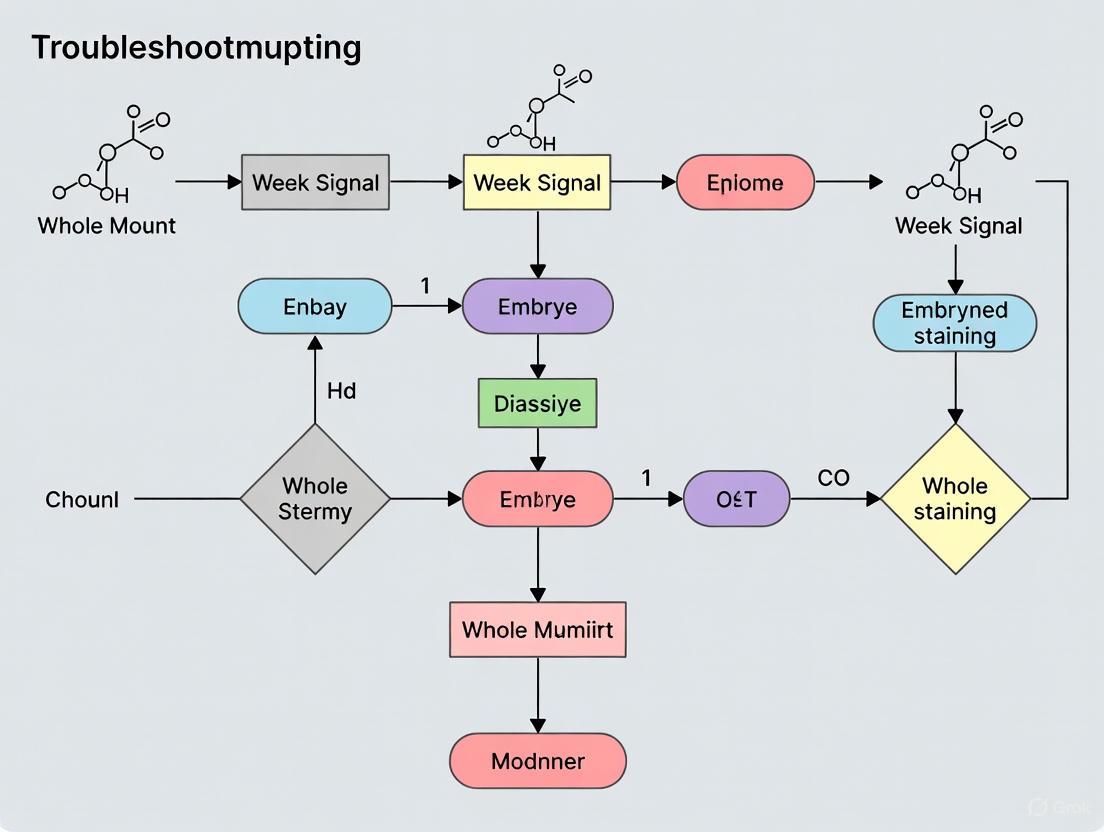
Troubleshooting Logic: Weak Signal
This decision diagram provides a systematic approach to diagnosing the root cause of a weak staining signal.
The Scientist's Toolkit: Essential Research Reagents
Table 3: Key Reagent Solutions for Whole Mount Staining
| Reagent / Material | Critical Function | Technical Notes |
|---|---|---|
| Validated Primary Antibodies | Binds specifically to the target protein/epitope. | Must be validated for whole mount IHC/IF in your species. Always run a positive control [1]. |
| Permeabilization Detergent | Creates pores in membranes for antibody penetration into whole tissues. | e.g., Triton X-100 or Tween-20. Concentration and time must be optimized for each sample type [3] [4]. |
| Blocking Serum | Reduces non-specific background by occupying non-target protein-binding sites. | Should be from the same species as the secondary antibody. Normal serum is commonly used [1] [2]. |
| Antigen Retrieval Buffers | Reverses formaldehyde-induced cross-linking to unmask epitopes. | Citrate (pH 6.0) and Tris-EDTA (pH 9.0) are common. The optimal buffer is antibody-dependent [1]. |
| Optical Clearing Agents | Reduces light scattering in thick samples, enabling deeper imaging. | e.g., LIMPID solution. Aqueous solutions (SCC, Urea, Iohexol) preserve lipids and are compatible with FISH and IHC [5]. |
| Autofluorescence Quenchers | Chemically reduces endogenous fluorescence from tissue components. | e.g., Sudan Black B. Applied after staining but before mounting [1] [3]. |
Common Biological and Technical Culprits for Weak Signal
FAQs: Addressing Weak Signal in Whole-Mount Staining
1. What are the most common biological reasons for a weak staining signal? Weak signals often originate from biological barriers within the embryo itself. Dense tissue architecture and the presence of pigments can significantly impede the penetration of antibodies or probes and quench the resulting signal. Pigment cells, such as melanophores filled with melanosomes, are a classic culprit as they can physically obscure the stain and exhibit strong autofluorescence, interfering with detection [6]. Furthermore, the small cell size in certain model organisms, like zebrafish, demands higher precision in staining and imaging to achieve adequate signal resolution [7].
2. My antibody works in other applications, so why is my whole-mount IHC signal weak? This is frequently a technical issue related to specimen preparation and protocol optimization. In whole-mount specimens, the primary challenges are inadequate penetration of reagents and epitope masking. The three-dimensional structure of the embryo can prevent antibodies from reaching internal targets. Furthermore, the fixation process, while necessary for preserving structure, can create cross-links that hide the epitope your antibody recognizes, making antigen retrieval a critical step [8] [9]. Always confirm that your antibody has been validated for IHC and specifically for the type of whole-mount samples you are using [9].
3. How can I improve the signal-to-noise ratio in my fluorescent whole-mount images? Improving the signal-to-noise ratio involves enhancing your specific signal while suppressing background. Key strategies include:
- Reducing Autofluorescence: Treat samples with photochemical bleaching (e.g., OMAR) or chemical quenchers like Sudan black to minimize inherent tissue fluorescence [10] [11] [12].
- Optimizing Tissue Transparency: Use validated tissue-clearing protocols (e.g., CUBIC, BABB) to homogenize the refractive index of the sample, which drastically improves light penetration and signal clarity for deep structures [12] [13].
- Ensuring Complete Penetration: Perform rigorous validation of antibody penetration and concentration. For thick tissues, physical sectioning or trimming may be necessary to ensure reagents reach their target [13].
Troubleshooting Guide: Weak or No Staining
The following table summarizes common issues and their solutions to help you systematically troubleshoot weak signals.
| Possible Cause | Underlying Reason | Recommended Solution |
|---|---|---|
| Inadequate Antigen Retrieval [8] [9] | Fixation-induced cross-links mask the target epitope, preventing antibody binding. | Optimize heat-induced epitope retrieval (HIER) method; use a microwave or pressure cooker instead of a water bath [8]. |
| Poor Antibody Penetration [7] [13] | The 3D structure and dense tissue prevent antibodies from reaching internal targets. | Use permeabilization agents (e.g., Triton X-100); titrate antibody concentration and extend incubation times; for large samples, consider physical sectioning [9] [13]. |
| Loss of Antibody Potency [11] [9] | Antibodies degrade due to improper storage, contamination, or repeated freeze-thaw cycles. | Aliquot antibodies; store according to manufacturer instructions; include a known positive control sample in your experiment [11]. |
| Inefficient Tissue Clearing [12] | Light scattering in opaque tissues prevents excitation and detection of fluorescence from deep structures. | Implement a suitable clearing protocol (e.g., organic solvent-based for high transparency, aqueous-based for fluorescence preservation) to homogenize the tissue's refractive index [12]. |
| Fluorophore Quenching or Poor Preservation [7] [3] | The fluorescent signal is lost during processing due to harsh dehydration or embedding media. | For fluorescence, use embedding resins like GMA that preserve fluorophores; ensure mounting media is compatible with your fluorophores [7] [3]. |
| Endogenous Pigment Interference [6] | Pigments like melanin absorb light and cause high background, obscuring a weak specific signal. | Incorporate a bleaching step to decolorize pigments prior to the hybridization or staining procedure [6]. |
Experimental Protocols for Signal Optimization
Protocol 1: Whole-Mount Immunostaining with Enhanced Penetration
This protocol is adapted for visualizing structures or rare cells deep within whole-mount embryos [13].
- Sample Preparation: Fix embryos in 4% PFA. To enhance antibody penetration into deep tissues (e.g., the dorsal aorta), carefully remove surrounding obstructive tissues like the lateral body wall to reduce the diffusion distance to ~120 µm [13].
- Blocking and Permeabilization: Incubate samples in a blocking buffer (e.g., 1X TBST with 5% normal serum) containing a permeabilizing agent (e.g., 0.2–1.0% Triton X-100) for several hours to reduce non-specific binding and facilitate reagent entry [8] [9].
- Primary Antibody Incubation: Incubate with the primary antibody, optimized for dilution and diluent, for 48–72 hours at 4°C with gentle agitation. For critical targets, use a biotinylated primary antibody followed by labeled streptavidin for signal amplification [13].
- Washing: Wash extensively with a buffer containing a mild detergent (e.g., 0.05% Tween-20 in PBS) over 24 hours, with multiple buffer changes, to remove unbound antibody [8].
- Secondary Antibody Incubation: Incubate with fluorophore-conjugated secondary antibodies for 24–48 hours at 4°C, protected from light.
- Clearing and Mounting: Render the sample transparent by immersing it in an organic solvent like BABB (benzyl alcohol/benzyl benzoate) or an aqueous-based clearing solution (e.g., CUBIC) to homogenize the refractive index for deep imaging [12] [13].
Protocol 2: Optimized Whole-Mount RNA In Situ Hybridization for Challenging Tissues
This protocol includes steps to minimize background and enhance signal clarity in pigment-rich or delicate tissues, such as regenerating tadpole tails [6].
- Fixation and Bleaching: Fix samples in MEMPFA. Rehydrate and then subject them to a photo-bleaching step to decolorize melanosomes and melanophores, which drastically improves signal-to-noise ratio [6].
- Tissue Notching: For loose, fin-like tissues prone to trapping reagents and causing background, make careful incisions (notching) in a fringe-like pattern at a safe distance from the area of interest. This allows solutions to wash in and out more effectively [6].
- Pre-hybridization and Hybridization: Follow standard proteinase K treatment and pre-hybridization steps. Hybridize with the labeled antisense RNA probe.
- Post-Hybridization Washes and Staining: Perform stringent washes. For colorimetric detection, develop the signal with BM Purple or a similar substrate. The combination of bleaching and notching allows for long staining incubations (3-4 days) to detect low-abundance transcripts without significant background [6].
Research Reagent Solutions
The following table lists key reagents and their functions for troubleshooting weak signals in whole-mount experiments.
| Item | Function in Troubleshooting | Example Use Case |
|---|---|---|
| Glycol Methacrylate (GMA) Resin [7] | An embedding medium for obtaining semithin (e.g., 3 µm) sections that superiorly preserve fluorescent protein signals and tissue morphology compared to paraffin. | Sectioning whole-mount stained transgenic zebrafish embryos for high-resolution cellular imaging without the need for advanced microscopy [7]. |
| BABB (Benzyl Alcohol/Benzyl Benzoate) [12] [13] | An organic solvent-based clearing agent that homogenizes the refractive index of tissues, making them transparent and enabling deep imaging of internal structures. | 3D imaging of hematopoietic stem cell clusters within the dorsal aorta of a whole-mount stained mouse embryo [13]. |
| Heat-Induced Epitope Retrieval (HIER) Buffers [11] [8] | Solutions (e.g., sodium citrate, pH 6.0) used with heat to break cross-links formed during fixation, thereby unmasking hidden epitopes for antibody binding. | Restoring immunoreactivity in over-fixed whole-mount specimens to recover a weak or lost signal [8]. |
| Polymer-Based Detection Reagents [8] | A highly sensitive non-biotin detection system that avoids background from endogenous biotin and provides superior signal amplification. | Detecting low-abundance targets in tissues with high endogenous biotin (e.g., liver, kidney) where ABC systems cause high background [8]. |
| Permeabilization Agents (Triton X-100) [9] | A detergent that creates pores in lipid membranes, facilitating the penetration of antibodies and probes into the interior of tissues and cells. | Enabling antibody access to intracellular or nuclear targets within the dense core of a whole-mount embryo [9]. |
Workflow Diagram for Signal Troubleshooting
The diagram below outlines a systematic, decision-tree workflow for diagnosing and resolving the causes of a weak signal.
The Impact of Tissue Autofluorescence and Background Staining on Signal-to-Noise Ratio
FAQs: Understanding and Identifying Autofluorescence
What is tissue autofluorescence and why is it a problem? Tissue autofluorescence is background fluorescence emanating from naturally occurring substances within tissue, which can severely hinder the detection of specific fluorescence signals from your target of interest [14] [15] [16]. It arises from structural tissue components like collagen and elastin, red blood cells, or as a result of aldehyde fixation [14]. Lipofuscin, an accumulation of proteins and lipids typically found in aged tissues like the brain and spinal cord, is another common source [14]. This background fluorescence obscures specific antigen staining, leads to a poor signal-to-noise ratio, can result in false positives, and introduces problems with assay validation and data credibility [14] [16].
How can I confirm that autofluorescence is affecting my experiment? A primary indication of an autofluorescence problem is if you see an almost uniform, unexpected signal throughout your tissue that appears consistent across different fluorescence channels when you image your sample [14]. If this background persists even after reducing exposure duration, especially when your target signal is weak, autofluorescence is likely the culprit [14]. An initial troubleshooting step of increasing primary or secondary antibody concentration sometimes only leads to higher background, not a cleaner signal [14].
What is the difference between Signal-to-Background Ratio (SBR) and Signal-to-Noise Ratio (SNR), and why does it matter for detecting microscopic disease? While Signal-to-Background Ratio (SBR) is useful when the tumor signal is significantly above the background, it becomes less applicable for microscopic disease, where the signal is often just at or slightly above the background level [17]. Signal-to-Noise Ratio (SNR) is a more robust metric for this scenario because it incorporates the ability to subtract out background by evaluating the signal itself relative to the sources of uncertainty, or "noise" [17]. This noise includes electronic sources, optical sources, and spatial sources (like heterogeneity in tumor marker expression). A high SNR is essential for the accurate digital subtraction of background to reveal true signal from microscopic foci [17].
Troubleshooting Guides & Experimental Protocols
Guide 1: Chemical Quenching of Autofluorescence
This protocol uses the TrueVIEW Autofluorescence Quenching Kit as an example of a chemical approach that requires minimal time and is compatible with common fluorophores [14].
- Principle: A hydrophilic, nonfluorescent molecule binds electrostatically to common sources of autofluorescence such as collagen, red blood cells, elastin, and aldehyde-fixed tissue, significantly reducing background emission [14].
- Key Materials:
- TrueVIEW Autofluorescence Quenching Kit (or similar commercial kit)
- Fixed tissue sections (compatible with FFPE tissues)
- Standard mounting medium, with or without DAPI
- Procedure:
- Complete your standard immunofluorescence protocol up to the final washing step before mounting.
- Prepare the TrueVIEW working solution by mixing the three kit reagents in a 1:1:1 ratio.
- Apply the working solution directly to your tissue section and incubate for 5 minutes at room temperature.
- Following incubation, carefully apply coverslips using the mounting medium provided in the kit.
- Proceed with visualization by microscopy [14].
- Advantages & Limitations:
Guide 2: Photochemical Bleaching for Whole-Mount Samples (OMAR Protocol)
This protocol, known as Oxidation-Mediated Autofluorescence Reduction (OMAR), is particularly suited for whole-mount samples like embryos, tissues, and organs, where eliminating autofluorescence at the source is preferable to digital post-processing [15].
- Principle: Uses a high-intensity cold white light source in the presence of chemicals to photo-chemically oxidize and bleach endogenous fluorophores prior to fluorescent labelling [15].
- Key Materials:
- High-intensity cold white light source (e.g., high-power LED spotlights or 20,000 lumen LED panels)
- Fixed whole-mount samples (e.g., mouse embryonic limb buds)
- Phosphate Buffered Saline (PBS)
- Hydrogen Peroxide (H~2~O~2~)
- Procedure:
- Sample Preparation: Collect and fix your embryos or tissues according to your standard protocol.
- First Photochemical Treatment:
- Place the fixed sample in a solution of 4% H~2~O~2~ in PBS.
- Illuminate the sample for 1-2 hours using your high-intensity LED setup. A successful reaction is indicated by the appearance of bubbles in the solution and around the sample.
- Wash: Rinse the sample thoroughly with PBS.
- Second Photochemical Treatment:
- Transfer the sample to a fresh solution of 4% H~2~O~2~ in PBS.
- Illuminate again for 1-2 hours.
- Post-Treatment: After the final wash, the samples are now ready for downstream applications such as whole-mount RNA-FISH or immunofluorescence [15].
- Advantages & Limitations:
- Advantages: Effectively suppresses autofluorescence in whole-mount samples, eliminating the need for complex digital image post-processing. Applicable to a variety of tissues and vertebrate embryos [15].
- Limitations: Requires specialized, high-intensity light equipment. The efficacy must be empirically tested for each tissue and light source. The protocol is time-consuming (several hours) [15].
Guide 3: Advanced Imaging - Fluorescence Lifetime Imaging Microscopy (FLIM)
FLIM is a powerful digital approach that separates signals based on the distinct lifetime properties of fluorophores, rather than relying on chemical or physical treatment of the sample [16].
- Principle: Fluorophores used for immunofluorescence and autofluorescence components often have distinct fluorescence lifetime decay curves. FLIM measures the time a fluorophore remains in the excited state, creating a "fingerprint" that can be used to separate the desired immunofluorescence signal from background autofluorescence in the phasor domain [16].
- Workflow (Phasor Analysis):
- Image Acquisition: Acquire time-resolved fluorescence images using a pulsed laser and a high-speed FLIM system.
- Reference Collection: Measure the lifetime phasor clusters for the pure immunofluorescence fluorophore (in solution) and for autofluorescence (from an unstained tissue section).
- Phasor Transformation: The fluorescence lifetime decay of each pixel is transformed into a coordinate (G, S) on a phasor plot.
- Signal Separation: The fractional contribution of immunofluorescence in each pixel is calculated based on its geometrical distance to the reference phasors for immunofluorescence and autofluorescence. This allows for the computational generation of an autofluorescence-free image [16].
- Advantages & Limitations:
- Advantages: Does not risk damaging the sample with chemicals or excessive light. Provides a robust, quantitative method for signal separation that can outperform chemical bleaching. Enhances correlation with immunohistochemistry data [16].
- Limitations: Requires specialized and costly instrumentation (pulsed lasers, high-speed detectors). Data acquisition and analysis are complex and demand specialized expertise [16].
Table 1: Comparison of Autofluorescence Reduction Methods
| Method | Key Principle | Typical Protocol Duration | Key Advantages | Major Limitations |
|---|---|---|---|---|
| Chemical Quenching [14] | Electrostatic binding of quenching molecules to autofluorescent structures. | ~5 minutes | Very fast and easy to implement; works on many tissue types. | May not be effective on all types of autofluorescence (e.g., some lipofuscins). |
| Photochemical Bleaching (OMAR) [15] | Chemical oxidation of fluorophores using high-intensity light. | 3-4 hours | Highly effective for whole-mount samples; eliminates need for post-processing. | Requires powerful light source; duration is long; must be optimized per tissue. |
| FLIM [16] | Computational separation based on fluorescence lifetime differences. | Acquisition time varies; computation can be ~3 seconds for a 512x512 image. | Non-invasive; does not alter sample; highly specific signal extraction. | Requires expensive, specialized equipment; complex data analysis. |
Table 2: Impact of Imaging Modality on Sample Health and Data Quality
| Imaging Modality | Volumetric Acquisition Time (Blastocyst) | Resulting Signal-to-Noise Ratio (SNR) | Impact on DNA Damage (γH2AX assay) |
|---|---|---|---|
| Confocal Microscopy [18] | ~30 minutes | 15.75 ± 1.90 | Significantly higher levels of DNA damage |
| Light Sheet Microscopy [18] | ~3 minutes | 15.45 ± 3.45 | No significant damage compared to non-imaged controls |
Visualizing the Experimental Workflow
The diagram below outlines a general decision-making workflow for selecting an autofluorescence troubleshooting strategy based on your sample type and experimental goals.
The Scientist's Toolkit: Essential Research Reagents & Materials
Table 3: Key Reagents for Autofluorescence Reduction
| Reagent/Material | Function/Benefit | Example Use Case |
|---|---|---|
| TrueVIEW / Sudan Black B [14] | Hydrophobic dye that binds to tissue, lowering autofluorescence in red and green channels. | Chemical quenching of autofluorescence in standard tissue sections. |
| Hydrogen Peroxide (H₂O₂) [15] | Key oxidizing agent in photochemical bleaching protocols. | Used in OMAR treatment for whole-mount embryo and tissue autofluorescence reduction. |
| Sodium Borohydride (NaBH₄) [16] | Chemical reducing agent used to quench autofluorescence caused by aldehyde fixation. | Reduction of aldehyde-induced background in immunofluorescence. |
| High-Intensity LED Light Source [15] | Provides the necessary high-intensity cold white light for effective photochemical oxidation in OMAR. | Illuminating samples during OMAR protocol for whole-mount RNA-FISH. |
| Fluorophore-Conjugated Antibodies [17] [16] | Provide specific signal for target antigens. Choosing bright, photostable fluorophores (e.g., AlexaFluor dyes) improves SNR. | All immunofluorescence and RNA-FISH experiments for target detection. |
The Critical Role of Tissue Permeabilization and Fixation in Preserving and Accessing Targets
Frequently Asked Questions (FAQs)
Q1: Why is my whole-mount staining signal weak or non-existent, even with a validated antibody? Weak or absent signal in whole-mount samples is a common challenge, often stemming from the inability of antibodies to penetrate deep into the tissue and access their targets. The primary causes and solutions are:
- Inadequate Permeabilization: The thickness of whole-mount samples (e.g., embryos) means standard permeabilization methods for thin sections are insufficient. Reagents must penetrate the entire tissue to allow antibodies access to intracellular targets [19].
- Fixative-Induced Epitope Masking: Over-fixation, particularly with cross-linking fixatives like paraformaldehyde (PFA), can create protein cross-links that physically block the antibody's binding site [19].
- Solution: Optimize fixation time and concentration. For some targets, switching to a non-cross-linking fixative like methanol or ethanol may be necessary. Note that antigen retrieval techniques used on paraffin sections are typically not feasible for heat-sensitive embryos [19].
- Low Antigen Preservation: The target protein may not be adequately preserved during the initial fixation step.
- Solution: Ensure fresh fixative is used and that tissues are fixed immediately after dissection. Follow a rigorously tested protocol, which for many antibodies involves overnight fixation at 4°C for optimal results [21].
Q2: I have high background staining. How can I improve my signal-to-noise ratio? High background occurs when antibodies bind non-specifically. This is especially problematic in whole-mount samples due to the large volume of tissue and extended incubation times.
- Cause: Insufficient Blocking. The blocking step may be too short or the blocking reagent may be ineffective for your tissue [21].
- Cause: Antibody Concentration is Too High. Excessive primary or secondary antibody can lead to non-specific binding [11].
- Solution: Titrate your primary and secondary antibodies to find the optimal dilution that provides a strong specific signal with minimal background. Using a fluorescently conjugated primary antibody can also reduce background by eliminating the secondary antibody step [11].
- Cause: Incomplete Washing. Loosely bound antibodies remain in the tissue.
- Solution: Increase the number, duration, and volume of washes between steps. Using a buffer with a mild detergent like Tween-20 can help remove non-specifically bound antibodies [21].
Q3: My staining is uneven, with strong signal on the outside and none in the center. What went wrong? This is a classic sign of poor reagent penetration, indicating that antibodies and other solutions are not reaching the interior of the sample.
- Solution: The core issue is the sample's size and density. For larger embryos or tissues, physical dissection may be necessary. You can try dissecting the sample into smaller segments before staining to ensure reagents permeate all areas [19]. Furthermore, ensure that all incubation times—for permeabilization, blocking, and antibodies—are extended for days, not just hours, to allow for diffusion into the center [19].
Troubleshooting Guide: Weak Signal
This guide helps diagnose and solve the most common issues leading to weak signal in whole-mount staining.
| Problem Area | Possible Cause | Recommendations | Key References |
|---|---|---|---|
| Tissue Preparation | Inadequate or delayed fixation Sample too large/thick | Fix tissue immediately post-dissection with fresh 4% PFA. For embryos older than recommended stages (e.g., mouse E12+, chick 6 days+), dissect into segments. | [21] [19] |
| Permeabilization | Standard protocols for sections used Insufficient detergent | Extend permeabilization time to several hours or overnight. Increase detergent concentration (e.g., 0.5-1.0% Triton X-100) or use alternative agents like methanol. | [19] |
| Antibody Incubation | Antibody concentration too low Incubation time too short | Titrate the primary antibody to find the optimal concentration. Incubate primary antibody at 4°C for 24-48 hours to allow deep penetration. | [21] [19] |
| Detection & Imaging | Signal fade due to light exposure Poor imaging depth | Store and mount samples in anti-fade mounting medium and keep in the dark. For thick samples, use clearing agents (e.g., glycerol) and image with confocal or two-photon microscopy. | [21] [22] |
Experimental Protocols for Enhanced Permeabilization
The following protocols provide detailed methodologies for effective permeabilization in challenging samples.
Protocol 1: Enhanced Permeabilization for Whole-Mount Embryos This protocol is adapted for whole-mount embryos where standard methods fail.
- Fixation: Fix samples in 4% PFA overnight at 4°C.
- Permeabilization: Wash samples in PBS (Phosphate Buffered Saline). Then, incubate in PBT (PBS with 1.0% Triton X-100) for 24-48 hours at 4°C with gentle agitation. For tougher tissues, a series of methanol dilutions (25%, 50%, 75% in PBT) can be used.
- Blocking: Incubate in a blocking solution (e.g., 5% serum in PBT) for 24 hours at 4°C.
- Antibody Incubation: Incubate with primary antibody diluted in blocking solution for 48-72 hours at 4°C. Perform extended washes (e.g., 6 x 2 hours) with PBT before applying the secondary antibody for another 24-48 hours [19].
Protocol 2: Permeabilization of Impermeable Structures: Drosophila Embryos The Drosophila eggshell is notoriously impermeable due to a waxy layer, requiring specialized treatment.
- Dechorionation: Remove the outer chorion by immersing embryos in 50% commercial bleach for 2 minutes, followed by extensive washing with water [23].
- Permeabilization: Immerse dechorionated embryos in a 1:5 to 1:40 dilution of Embryo Permeabilization Solvent (EPS) in a basic incubation medium. EPS is a water-miscible solvent composed of D-limonene and surfactants, less toxic than traditional heptane/octane. Treat for 30 seconds to 4 minutes [23].
- Washing and Staining: Quickly wash embryos 4 times in PBS, followed by 2 washes in PBS with 0.05% Tween-20. The embryos are now ready for dye uptake or antibody staining [23].
Visual Workflow for Troubleshooting Weak Signal
The following diagram outlines a logical pathway for diagnosing and resolving the issue of weak signal in whole-mount staining experiments.
Research Reagent Solutions
This table details key reagents essential for successful permeabilization and fixation in whole-mount staining.
| Reagent | Function | Example Usage & Notes |
|---|---|---|
| Paraformaldehyde (PFA) | Cross-linking fixative that preserves tissue architecture by creating protein bonds. | Standard is 4% in buffer. Fixation time must be optimized; over-fixation can mask epitopes. Always use fresh [19]. |
| Triton X-100 / NP-40 | Non-ionic detergents that solubilize lipid membranes, enabling antibody penetration. | Use at higher concentrations (0.5-1.0%) and for longer durations (24-48 hrs) for whole-mounts vs. thin sections [20] [19]. |
| Methanol | Organic solvent fixative that precipitates proteins; also acts as a permeabilizing agent. | An alternative to PFA for epitope-sensitive targets. Can be used for both fixation and permeabilization [19]. |
| Embryo Permeabilization Solvent (EPS) | Water-miscible solvent system to permeabilize impermeable barriers like the Drosophila waxy layer. | Composed of D-limonene and surfactants. Less toxic and easier to use than traditional heptane/octane [23]. |
| Dimethylformamide (DMFA) | Organic solvent used to dissolve substrates like X-gal for enzymatic detection (e.g., LacZ staining). | Used to prepare stock solutions of membrane-permeable substrates [20]. |
| Glycerol | Refractive Index Matching (RIM) mounting medium that clears tissue for deeper imaging. | 80% glycerol can provide a 3 to 8-fold reduction in signal decay at depth compared to PBS, significantly improving image quality [22]. |
Analyzing the Challenges of Probe and Antibody Penetration in Thick Embryonic Tissues
FAQs and Troubleshooting Guides
This guide addresses common challenges researchers face when working with whole-mount embryonic tissues, where the thickness of the sample is a primary obstacle to effective staining.
Why is my whole-mount embryo staining weak or uneven?
Weak or uneven staining in whole-mount embryos is most frequently caused by inadequate penetration of antibodies or probes into the center of the tissue. Unlike thin sections, the three-dimensional structure of whole embryos presents a physical barrier.
- Primary Cause: The sample is much larger and thicker than a conventional tissue section. Standard incubation times are insufficient to allow reagents to permeabilize fully into the center of the sample [19] [24].
- Solution: Drastically extend incubation times for all steps, including fixation, permeabilization, blocking, antibody incubation, and washing. Timings must be optimized for your specific embryo size and age, often requiring hours to days instead of minutes [19].
- Additional Factors:
- Insufficient Permeabilization: The fixation process can cross-link proteins and reduce permeability. Ensure your protocol includes effective permeabilization agents like Triton X-100 or methanol [19] [25].
- Fixative Choice: The common fixative 4% Paraformaldehyde (PFA) can sometimes mask epitopes. If this is suspected, methanol can be an alternative fixative to test [19] [24].
- Embryo Size: As an embryo grows, it becomes too large to stain effectively. For older, larger embryos, dissection into segments may be necessary before staining [19] [24].
How can I improve antibody penetration into the core of a thick embryo?
Improving penetration is a multi-faceted problem focused on overcoming the physical and chemical barriers of the dense tissue.
- Optimize Permeabilization: Use detergents like Triton X-100. A common concentration is 0.1% - 0.3% in PBS (PBT) [25] [24]. Incubation times must be extended significantly compared to sectioned samples.
- Use Methanol Treatment: A methanol treatment step can be highly effective for removing lipid membranes and enhancing permeability. A standard protocol for Drosophila embryos involves adding methanol and vortexing to remove the vitelline membrane [24].
- Increase Incubation Times: Antibody incubations, especially for the primary antibody, often need to be performed overnight at 4°C to aid perfusion into the tissue [24]. For some targets, incubations may need to extend to several days.
- Technical Tip: Perform all incubations on a rocking or rotating platform to ensure even exposure and prevent settling [25].
My negative control has high background. How do I reduce this?
High background signals are typically caused by non-specific antibody binding or inadequate washing.
- Enhanced Blocking: Use a robust blocking solution. A common and effective blocker is PBS with 0.1% Triton X-100 and 10-30% newborn calf serum (NCS) or other suitable serum [24]. Block for a minimum of several hours, or overnight for challenging samples.
- Thorough Washing: Implement a stringent washing protocol after primary and secondary antibody incubations. This should include multiple rinses and extended washes (e.g., 3-5 washes of 30-60 minutes each) with a washing buffer like PBT [24].
- Antibody Specificity: Always validate your primary antibody for use in whole-mount IHC. An antibody that works on cryosections is more likely to work for whole-mount staining than one validated only for paraffin sections [19].
- Check Secondary Antibody: Ensure your secondary antibody is pre-adsorbed and used at an appropriate dilution to minimize non-specific sticking.
Are there specific challenges with different types of embryos?
Yes, the species and developmental stage of the embryo introduce specific requirements.
- Zebrafish Embryos: A critical first step is dechorionation—removal of the chorion (egg membrane)—as it is a physical barrier to fixatives and antibodies. This can be done manually with forceps or enzymatically using pronase [19].
- Mouse/Chick Embryos: The main challenge is the increasing size with age. The table below provides general guidelines for the maximum recommended ages for whole-mount staining [19].
- Drosophila Embryos: The protocol requires a specific fixation mix (often containing n-heptane and formaldehyde) and a methanol step for devitellinization [24].
Table 1: Recommended Maximum Embryo Ages for Whole-Mount Staining
| Embryo Type | Recommended Maximum Age | Key Consideration |
|---|---|---|
| Chicken | Up to 6 days | Larger embryos may require dissection [19]. |
| Mouse | Up to 12 days | Removal of surrounding muscle and skin may be needed for effective staining [19]. |
What are the key fixation variables to optimize?
Fixation is critical for preserving antigenicity while still allowing antibody access.
- Fixative Type: 4% Paraformaldehyde (PFA) is the most common fixative. However, if it causes epitope masking, methanol is a popular second choice [19].
- Fixation Time: Fixation times must be extended for whole mounts. Protocols can range from 30 minutes at room temperature to overnight at 4°C [19].
- A Critical Limitation: Antigen retrieval is generally not feasible for whole-mount embryos, as the heating procedure would destroy the fragile sample. Therefore, fixation must be optimized correctly the first time [19].
Table 2: Fixation and Permeabilization Parameters
| Parameter | Standard Condition | Whole-Mount Consideration |
|---|---|---|
| Fixative | 4% PFA | If epitope is masked, try methanol fixation [19]. |
| Fixation Time | 30 min - 1 hr | Can require several hours to overnight [19]. |
| Permeabilization Agent | Triton X-100 (0.1-0.5%) | Essential; incubation times must be extended [25] [24]. |
| Permeabilization Time | 15-30 min | May require several hours [25]. |
The Scientist's Toolkit: Essential Research Reagents
Table 3: Key Reagents for Whole-Mount Embryo Staining
| Reagent | Function | Example |
|---|---|---|
| Paraformaldehyde (PFA) | Cross-linking fixative that preserves tissue structure and antigenicity. | 4% PFA in PBS [19] [25] |
| Methanol | Precipitating fixative; used as an alternative to PFA and for permeabilization. | 100% Methanol for devitellinization and permeabilization [19] [24] |
| Triton X-100 | Non-ionic detergent that permeabilizes lipid membranes to allow antibody penetration. | 0.1% - 0.3% in PBS (PBT) [25] [24] |
| Normal Serum | Used as a blocking agent to reduce non-specific antibody binding. | Newborn Calf Serum (NCS), Donkey Serum [25] [24] |
| Primary Antibody | Binds specifically to the target antigen of interest. | Rabbit anti-phospho-SMAD2 [25] |
| Secondary Antibody | Conjugated to a fluorophore or enzyme; binds to the primary antibody for detection. | Donkey-anti-rabbit, 488 conjugated [25] |
| Mounting Medium | Preserves the sample for microscopy; often contains an anti-fade agent. | Glycerol-based media or Vectashield with DAPI [25] [24] |
Experimental Workflow and Optimization Pathways
The following diagrams outline the logical workflow for a whole-mount staining experiment and the decision process for troubleshooting a weak signal.
Workflow for Whole-Mount Embryo Staining
Troubleshooting Weak Signal
Advanced Protocols for Robust Signal Detection: FISH, IHC, and Optical Clearing
Optimized Whole-Mount FISH Protocols for Enhanced RNA Visualization
Frequently Asked Questions (FAQs)
Q1: What are the most common causes of weak signal in whole-mount FISH? Weak signal often stems from inefficient tissue permeabilization, suboptimal probe hybridization, high tissue autofluorescence, or insufficient signal amplification. Protocol optimizations in probe design, hybridization buffers, and optical clearing can significantly enhance signal strength [26] [5] [10].
Q2: How can I reduce high background fluorescence in my embryo samples? High background can be addressed through oxidation-mediated autofluorescence reduction (OMAR) photochemical bleaching, which effectively suppresses tissue autofluorescence without the need for digital post-processing [10]. Using selective volume illumination (SVI) techniques during imaging can also confine excitation to the volume of interest, dramatically enhancing contrast [27].
Q3: Are there clearing methods compatible with RNA-FISH for 3D imaging? Yes, hydrophilic clearing methods like LIMPID (Lipid-preserving index matching for prolonged imaging depth) are compatible with RNA-FISH. LIMPID uses saline-sodium citrate, urea, and iohexol to match the tissue's refractive index, enabling high-resolution 3D imaging while preserving RNA and protein integrity [5].
Q4: Does the length of the probe target region affect signal brightness? Research shows that for smFISH, the signal brightness depends relatively weakly on the target region length for regions between 20-50 nucleotides, provided hybridization conditions (like formamide concentration) are optimized for that length [26].
Troubleshooting Guides
Problem: Weak or No Staining
Potential Causes and Solutions:
- Cause 1: Inadequate Tissue Permeabilization
- Cause 2: Suboptimal Hybridization Efficiency
- Solution: Systematically optimize hybridization conditions. A study on MERFISH showed that varying formamide concentration and hybridization temperature can maximize probe assembly efficiency. Encoding probe design, while important, had a weaker effect on performance than hybridization buffer composition [26].
- Cause 3: Probe Degradation or Inefficient Signal Amplification
Problem: High Background Fluorescence
Potential Causes and Solutions:
- Cause 1: Tissue Autofluorescence
- Solution: Implement Oxidation-Mediated Autofluorescence Reduction (OMAR) photochemical bleaching. This protocol effectively suppresses autofluorescence in whole-mount mouse embryonic limb buds and is suitable for other tissues and vertebrate embryos [10].
- Cause 2: Non-Specific Probe Binding
- Solution: Include pre-absorption steps with sample tissue if necessary. Screen readout probes for non-specific, tissue-specific binding, which can introduce false-positive counts, and exclude problematic probes [26].
- Cause 3: Out-of-Focus Light and Scatter in Thick Tissues
Problem: Poor Image Contrast in 3D Reconstructions
Potential Causes and Solutions:
- Cause: Scattering and Refractive Index Mismatch
- Solution: Employ optical clearing. The 3D-LIMPID-FISH method allows for fine-tuning of the mounting medium's refractive index (using iohexol) to match that of the microscope objective (e.g., 1.515). This decreases optical aberrations and maintains image quality across all z-sections in thick samples [5].
Table 1: Protocol Optimization for Improved Performance
This table summarizes key quantitative findings from optimization experiments.
| Parameter Optimized | Tested Conditions | Key Finding | Impact on Performance |
|---|---|---|---|
| Probe Target Region Length [26] | 20 nt, 30 nt, 40 nt, 50 nt | Signal brightness depends weakly on length for regions of sufficient length (20-50 nt). | Guidance: Focus on optimizing hybridization conditions over fine-tuning length within this range. |
| Hybridization Conditions [26] | Variable formamide concentration (37°C, 1-day hybridization) | Average single-molecule brightness has a weak dependence on formamide within an optimal range. | Guidance: A range of conditions can work well; systematic screening is recommended. |
| Imaging Modality [27] | Wide-field LFM vs. SVIM (Selective Volume Illumination) | SVIM achieved up to 50% better contrast for heart walls and 10% better for blood cells vs. wide-field LFM. | Recommendation: Use volume-restricted illumination for high-contrast imaging in dynamic tissues. |
| Excitation Mode for Functional Imaging [27] | 1-photon vs. 2-photon SVIM | 2-photon excitation led to better contrast and a larger number of resolved active neurons (up to 4x more than wide-field LFM). | Recommendation: Use 2-photon excitation for deep-tissue functional imaging when possible, despite slower rates. |
Experimental Protocols
Detailed Methodology: Whole-Mount RNA-FISH with OMAR
This protocol is adapted for whole-mount mouse embryonic limb buds and is applicable to other tissues [10].
Embryo Collection and Fixation:
- Collect embryos at the desired developmental stage in ice-cold PBS.
- Fix embryos in 4% paraformaldehyde (PFA) in PBS for 2 hours at room temperature or overnight at 4°C with gentle agitation.
Oxidation-Mediated Autofluorescence Reduction (OMAR):
- Incubate fixed embryos in a freshly prepared OMAR solution (e.g., based on H₂O₂) to photochemically bleach autofluorescent molecules.
- Expose the samples to light under standardized conditions to activate the bleaching process.
Permeabilization:
- Treat the embryos with a permeabilization solution containing a detergent (e.g., Triton X-100) to facilitate probe entry. Protease treatment can be incorporated if necessary to free up cross-linked molecules, but caution is warranted to avoid over-digestion [5].
Pre-hybridization and Hybridization:
- Pre-hybridize embryos in a hybridization buffer containing formamide to reduce nonspecific binding [5].
- Incubate with the specific RNA FISH probes (e.g., HCR initiator probes) in hybridization buffer for 12-48 hours at the optimal temperature (e.g., 37°C).
Post-Hybridization Washes:
- Perform a series of stringent washes with saline-sodium citrate (SSC) buffer containing formamide to remove unbound probes.
Signal Amplification (if using HCR):
- For HCR, incubate samples with amplification hairpins for a defined period (e.g., 2 hours for single-molecule resolution) to build fluorescent amplification polymers [5].
Optical Clearing and Mounting:
- Clear the samples using a compatible method like LIMPID [5].
- Mount the cleared samples in the matching refractive index medium for imaging.
Workflow Diagram: Integrated Troubleshooting Pathway
The Scientist's Toolkit: Research Reagent Solutions
Table 2: Essential Materials for Optimized Whole-Mount FISH
| Reagent / Material | Function / Purpose | Example / Note |
|---|---|---|
| Encoding Probes [26] | Target-specific DNA oligonucleotides that bind cellular RNA and carry customizable readout sequences. | Designed with target regions of 20-50 nt; performance is more sensitive to hybridization conditions than exact length. |
| Readout Probes [26] | Fluorescently labeled oligonucleotides that hybridize to readout sequences on encoding probes for detection. | Should be prescreened for non-specific, tissue-specific binding to minimize false positives. |
| HCR Initiation Probes [5] | DNA oligonucleotides that bind target RNA and initiate the hybridization chain reaction (HCR) for signal amplification. | Enable linear signal amplification, allowing fluorescence intensity to be quantified against RNA quantity. |
| LIMPID Solution [5] | Aqueous optical clearing medium that matches tissue refractive index via iohexol, urea, and SSC. | Preserves lipids and tissue structure; enables high-resolution 3D imaging without advanced sectioning microscopes. |
| OMAR Solution [10] | Photochemical bleaching solution (e.g., H₂O₂-based) to suppress tissue autofluorescence. | Reduces background without digital post-processing; compatible with whole-mount RNA-FISH and immunofluorescence. |
| Formamide [26] [5] | Chemical denaturant used in hybridization buffers and post-hybridization washes. | Increases signal intensity and stringency; concentration should be optimized for specific probe sets. |
Experimental Workflow Diagram: Whole-Mount FISH with Enhanced Visualization
In the analysis of low-abundance molecular targets within complex biological samples like whole mount embryos, signal amplification techniques are indispensable for achieving detectable signals. Hybridization Chain Reaction (HCR) and Rolling Circle Amplification (RCA) are two powerful, isothermal amplification methods that provide high sensitivity and specificity without requiring thermal cyclers. These techniques are particularly valuable in spatial biology, where preserving the three-dimensional architecture of the sample is crucial.
HCR is an enzyme-free, isothermal amplification technique that uses stable DNA hairpin probes which self-assemble into long double-stranded DNA polymers upon initiation by a specific target sequence. This process enables simultaneous recognition and signal amplification, producing ladder-shaped products with fragments of different sizes [28]. Its enzyme-free nature and robust performance in complex environments make it ideal for challenging applications like whole-mount imaging.
RCA is an isothermal enzymatic process where a short nucleic acid primer is amplified to form a long single-stranded DNA (ssDNA) using a circular template and a special DNA polymerase (e.g., Phi29). This reaction generates tens to hundreds of tandemly repeated copies of the circular template, creating a long, single-stranded DNA product that remains localized to the target site [29] [30]. The high processivity of Phi29 DNA polymerase allows for extensive strand displacement and the generation of long DNA products, making it exceptionally suitable for detecting single molecules.
Troubleshooting Guides
Weak or No Signal
A weak or absent signal is a common frustration when detecting low-abundance targets. The following table outlines the primary causes and evidence-based solutions.
Table: Troubleshooting Weak or No Signal
| Problem Cause | Recommended Solution | Underlying Principle |
|---|---|---|
| Low Target Abundance | Employ a cascade amplification strategy. Combine RCA with HCR or use hyperbranched RCA (HRCA). | Cascade reactions, such as using an RCA product to initiate a subsequent HCR reaction, can exponentially amplify the signal from a single binding event, dramatically boosting sensitivity [29] [31]. |
| Inefficient HCR Hairpin Opening | Re-design initiator sequence and optimize hairpin probe stability. Validate probe sets separately before combined use. | The efficiency of the initial strand displacement that opens the HCR hairpin is critical. Carefully designed initiators and validated hairpins ensure efficient polymerization [32] [28]. |
| Suboptimal RCA Primer Binding | Ensure the circular template is properly ligated and the primer binding site is accessible. | The RCA reaction is entirely dependent on the primer binding to the circular template. A successfully ligated circle and an unimpeded primer site are fundamental [29] [30]. |
| Fluorophore Quenching | Protect samples from direct light during and after amplification steps. Include photoprotective agents in mounting media. | Fluorophores are highly susceptible to photobleaching. Minimizing light exposure preserves signal integrity for detection [32]. |
The following workflow diagram illustrates a proven protocol combining RCA and HCR for robust signal detection in whole-mount samples, addressing several potential failure points.
High Background Fluorescence
Excessive background noise can obscure a genuine signal, making quantification difficult.
Table: Troubleshooting High Background Fluorescence
| Problem Cause | Recommended Solution | Underlying Principle |
|---|---|---|
| Non-Specific Hairpin Opening (HCR) | Use "Migrating HCR" designs. Increase stringency of wash buffers (e.g., adjust salt concentration, add formamide). | Migrating HCR significantly reduces the possibility of non-specific initiation by introducing a more complex activation mechanism, thereby lowering background [28]. |
| Sample Autofluorescence | Implement thorough sample clearing protocols (e.g., methanol treatment, ClearSee solution). Use fluorophores with emissions in spectral ranges with low autofluorescence (e.g., far-red). | Tissue autofluorescence is a major challenge in whole-mount samples. Chemical clearing and the use of far-red dyes like Quasar670 can dramatically improve the signal-to-noise ratio [33]. |
| Incomplete Washing | Increase wash number, duration, and volume. Include detergent (e.g., 0.05% Tween-20) in wash buffers. | Efficient removal of unbound probes and enzymes is critical. Detergents help reduce non-specific adhesion of molecules to the sample and container surfaces. |
| Polymerase Binding (RCA) | Include a protein block (e.g., BSA, goat serum) before and during the RCA reaction. | Proteins can non-specifically bind to tissues. A blocking step saturates these sites, preventing the polymerase from sticking and creating false-positive signals. |
Multiplexing Challenges
Simultaneous detection of multiple targets requires careful spectral and biochemical planning.
Table: Troubleshooting Multiplexing Experiments
| Problem Cause | Recommended Solution | Underlying Principle |
|---|---|---|
| Spectral Overlap | Use online tools (e.g., FPbase.org) to select fluorophores with minimal emission spectrum overlap during experimental design. | Crosstalk between channels can lead to false colocalization. Spectrally distinct fluorophores are a prerequisite for successful multiplexing [32]. |
| Cross-Talk Between Amplification Systems | Assign unique HCR B-isoforms or RCA circular templates to each target. Validate each probe set/antibody separately. | In HCR, each target must be paired with a unique initiator sequence (B-isoform) that polymerizes only its corresponding hairpins, preventing off-target amplification [32]. |
Frequently Asked Questions (FAQs)
Q1: Can HCR and RCA be combined with immunofluorescence for simultaneous detection of RNA and protein? Yes, both techniques can be successfully integrated with immunofluorescence. A detailed protocol, Whole-mount Immuno-Coupled HCR (WICHCR), has been established for zebrafish embryos and larvae. The key is to perform the HCR or RCA first, followed by the immunofluorescence steps, to prevent enzymatic or chemical damage to the protein epitopes. Protect samples from light throughout the process to prevent fluorophore quenching [32] [33].
Q2: What is the key difference between linear and exponential RCA? Linear RCA uses a single primer and a circular template to generate a long, single-stranded DNA concatemer. Exponential RCA (often called Hyperbranched RCA or HRCA) incorporates a second set of primers that can bind to the primary RCA product, using it as a template for further rounds of amplification. This creates a branched DNA network, resulting in exponential signal amplification and higher sensitivity, which is beneficial for ultra-low-abundance targets [29].
Q3: How do I choose between HCR and RCA for my experiment? The choice depends on your target and experimental needs. HCR, being enzyme-free, is highly robust to variable experimental conditions and is easier to implement in resource-limited settings. RCA, being enzymatic, can achieve higher amplification factors and is capable of single-molecule sensitivity. For the most challenging low-abundance targets, a cascade strategy that uses RCA to initiate an HCR reaction can be the most effective approach [31] [28].
Q4: My HCR signal is punctate instead of forming the expected long polymers. What might be wrong? Punctate signal often indicates inefficient hairpin opening or polymerization. This can be caused by hairpin probes that are too stable (high GC content) or an initiator sequence with low binding affinity. Re-design your hairpin probes to ensure the metastable state is correct and re-check the complementarity between the initiator and the hairpin sticky ends. Also, ensure the HCR reaction is carried out at the correct, constant temperature [28].
Q5: What are the best practices for storing and handling HCR hairpins and RCA circles? Resuspend all DNA probes in nuclease-free water or TE buffer. Aliquot and store at -20°C to avoid repeated freeze-thaw cycles. Before use, heat the hairpin probes to 95°C for 2-5 minutes and then allow them to cool slowly to room temperature to ensure proper secondary structure formation.
Essential Research Reagent Solutions
Successful implementation of HCR and RCA relies on high-quality, specific reagents. The following table catalogs the core components required for these assays.
Table: Key Reagents for HCR and RCA Experiments
| Reagent / Tool | Function / Description | Example Source / Citation |
|---|---|---|
| HCR v3.0 Probe Sets | Probe sets (~20 probe pairs) designed against specific mRNA target sequences. Each set is assigned a unique B-isoform for multiplexing. | Molecular Instruments, Inc. [32] |
| HCR Hairpin Amplifiers | Fluorescently labeled DNA hairpins that self-assemble upon initiation by the corresponding B-isoform. | Molecular Instruments, Inc. [32] |
| Phi29 DNA Polymerase | High-processivity DNA polymerase used for RCA. Possesses strong strand-displacement activity. | Commercially available (e.g., from manufacturers like Thermo Fisher) [29] [30] |
| Padlock Probes | Linear, single-stranded DNA probes whose ends are complementary to a target sequence. They are circularized by ligation upon target recognition to serve as RCA templates. | Custom synthesized [29] |
| T4 DNA Ligase | Enzyme used to ligate the ends of padlock probes into a circular template for RCA. | Common molecular biology suppliers [29] |
| Fluorophore Selection Tool | Online resource (e.g., FPbase.org) to compare emission spectra and select optimal fluorophore combinations for multiplexing. | FPbase.org [32] |
Visualization of Amplification Mechanisms
Understanding the fundamental mechanics of HCR and RCA is key to effective troubleshooting. The diagrams below illustrate the core processes of each technique.
Hybridization Chain Reaction (HCR) Mechanism
Rolling Circle Amplification (RCA) Mechanism
Whole-mount immunohistochemistry (IHC) enables researchers to visualize protein expression in intact tissue samples, preserving valuable three-dimensional structural information that is critical for developmental biology, neurobiology, and embryology research [19]. However, this technique presents unique challenges compared to traditional section-based IHC, particularly regarding antibody penetration through thick samples and achieving strong specific signals without high background [19] [34]. This guide addresses the most common issues researchers encounter and provides proven solutions to ensure reliable, reproducible results.
Core Principles of Whole-Mount IHC
Whole-mount IHC preserves the complete 3D architecture of tissues, typically embryos or intact tissue segments, without sectioning [19]. The fundamental difference from conventional IHC lies in the sample thickness, which necessitates significantly longer incubation times for fixatives, antibodies, and wash buffers to ensure complete permeabilization and reagent penetration to the sample's center [19]. Successful staining depends on carefully optimizing each step—from fixation and permeabilization to antibody incubation and imaging—to overcome diffusion barriers while maintaining tissue integrity and antigenicity.
Frequently Asked Questions (FAQs) & Troubleshooting Guide
FAQ 1: Why is my whole-mount staining weak or absent throughout the tissue?
Root Cause: Weak or absent staining typically results from inadequate antibody penetration, insufficient antibody concentration, or epitope masking due to fixation [19] [1] [2].
Solutions:
- Increase incubation times: Primary antibody incubation may require 1-4 days on gentle rotation at 4°C to ensure deep penetration [19] [35].
- Optimize antibody concentration: Perform titration experiments. If the antibody is too dilute, increase concentration systematically [1] [2].
- Verify antibody compatibility: Ensure your primary antibody works in IHC-Fr (cryosections), as this predicts whole-mount suitability [19].
- Re-evaluate fixation: If using 4% PFA causes epitope masking due to protein cross-linking, try methanol as an alternative fixative [19].
- Enhance permeabilization: Increase Triton X-100 concentration to 0.5-1% in wash and blocking buffers [35].
FAQ 2: How can I reduce high background staining in my whole-mount samples?
Root Cause: High background often stems from nonspecific antibody binding, insufficient blocking, or endogenous enzyme activity [1] [11] [2].
Solutions:
- Optimize blocking: Use fresh blocking buffer with 10% fetal calf serum in PBS with 1% Triton X-100, and extend blocking time to 1-2 hours [35].
- Reduce antibody concentration: High primary antibody concentration is a common cause of non-specific binding [1] [11].
- Include detergent in buffers: Add 0.05% Tween-20 to wash buffers to minimize hydrophobic interactions [1] [11].
- Prevent tissue drying: Perform all incubation steps in a humidified chamber to prevent irreversible non-specific binding [1] [2].
- Use appropriate controls: Always include a secondary antibody-only control to identify background from secondary antibody cross-reactivity [11] [36].
FAQ 3: Why is my staining uneven or limited to superficial tissue layers?
Root Cause: Uneven staining or limited penetration indicates inadequate permeabilization or insufficient washing throughout the tissue depth [19] [34].
Solutions:
- Extend washing steps: Wash 3 times for 30-60 minutes each with gentle agitation to ensure thorough reagent removal [19] [35].
- Ensure complete coverage: Use sufficient buffer volume (typically 5ml for embryos) and gentle rotation throughout incubations [35].
- Optimize permeabilization: For challenging tissues, consider combining Triton X-100 with alternative permeabilization agents.
- Size considerations: For larger embryos (>12-day mouse, >6-day chick), consider dissecting into segments before staining [19].
FAQ 4: What are the special considerations for different embryo types?
Root Cause: Different organisms and developmental stages present unique barriers to antibody penetration [19].
Solutions:
- Zebrafish embryos: Require dechorionation using fine forceps or enzymatic treatment with pronase (1-2 mg/mL for 5-10 minutes) to remove the egg membrane barrier [19].
- Mouse embryos: For embryos older than 12 days, remove surrounding muscle and skin to facilitate staining, or dissect into segments [19].
- Chicken embryos: Limit staining to embryos up to 6 days old for adequate penetration [19].
- All embryos: Respect size limitations—as embryos grow, reagents cannot penetrate to the center, making staining ineffective [19].
Troubleshooting Tables for Common Problems
Table 1: Troubleshooting Weak or Absent Staining
| Problem Cause | Specific Solution | Expected Outcome |
|---|---|---|
| Low antibody concentration | Perform antibody titration; increase concentration incrementally [1] [2] | Restored specific signal intensity |
| Inadequate incubation time | Extend primary antibody incubation to 1-4 days at 4°C with gentle rotation [19] [35] | Uniform staining throughout tissue depth |
| Epitope masking from PFA fixation | Switch to methanol fixation [19] | Improved antigen accessibility |
| Incompatible antibody | Verify antibody works on cryosections (IHC-Fr) [19] | Reliable target detection |
| Inactive detection system | Test detection system separately with positive control [1] | Confirmed system functionality |
Table 2: Addressing Background and Penetration Issues
| Problem Type | Solution Approach | Technical Implementation |
|---|---|---|
| High background staining | Optimize blocking | Use 10% FCS in PBS with 1% Triton X-100 for 1-2 hours [35] |
| Non-specific binding | Include detergents | Add 0.05% Tween-20 to antibody diluent and wash buffers [1] [11] |
| Incomplete penetration | Enhance permeabilization | Use 0.5-1% Triton X-100 in all buffers [35] |
| Uneven staining | Improve reagent exchange | Extend washes to 3 × 30-60 minutes with gentle agitation [19] [35] |
| Tissue-specific barriers | Modify sample preparation | Dechorionate zebrafish embryos; dissect older embryos [19] |
The Scientist's Toolkit: Essential Reagents and Materials
Table 3: Key Research Reagent Solutions for Whole-Mount IHC
| Reagent Category | Specific Examples | Function in Protocol |
|---|---|---|
| Fixatives | 4% Paraformaldehyde (PFA), Methanol [19] | Preserve tissue architecture and antigenicity |
| Permeabilization Agents | Triton X-100 (0.5-1%), Tween-20 (0.05%) [1] [35] | Enable antibody penetration through membranes |
| Blocking Reagents | Fetal Calf Serum (10%), Normal Serum [35] | Reduce non-specific antibody binding |
| Antibody Diluent | PBS with 1% Triton, 10% FCS, 0.02% sodium azide [35] | Maintain antibody stability during long incubations |
| Wash Buffers | PBS with 0.5-1% Triton X-100 [35] | Remove unbound antibodies while maintaining permeabilization |
| Mounting Media | 100% Glycerol, Gelatin [19] [35] | Preserve samples for imaging while maintaining transparency |
Best Practices for Reliable Whole-Mount IHC
Fixation Optimization: Standardize fixation time and conditions. For 4% PFA, test between 2 hours to overnight at 4°C [19] [35]. Avoid over-fixation which can mask epitopes.
Antibody Validation: Always use antibodies validated for IHC in similar sample types. Remember that antigen retrieval methods used in traditional IHC are generally not feasible for fragile whole-mount embryos [19].
Penetration Monitoring: Include a nuclear stain (e.g., DAPI) to verify reagent penetration throughout the tissue depth [19].
Appropriate Imaging: Use confocal microscopy to visualize staining in deeper tissue layers, as it can optically section through the sample [19] [35].
Size Considerations: Respect embryo size limitations—stain chicken embryos up to 6 days and mouse embryos up to 12 days for adequate reagent penetration [19].
By systematically addressing these common challenges and implementing the solutions outlined, researchers can overcome the technical hurdles of whole-mount IHC and reliably obtain high-quality data that preserves valuable three-dimensional biological context.
FAQs: Core Principles and Method Selection
Q1: What is the fundamental principle behind optical clearing? Optical clearing works primarily through refractive index (RI) matching. Biological tissues appear opaque because light scatters when it passes through various cellular components (lipids, proteins, organelles) that each have different RIs. Clearing methods use solutions to homogenize the RI throughout the tissue, minimizing light scattering and allowing light to penetrate deeply with minimal deviation, thus rendering the tissue transparent [37] [38].
Q2: How do I choose between LIMPID and glycerol-based clearing? The choice hinges on your experimental requirements for clearing speed, level of transparency, tissue morphology preservation, and compatibility with your stains.
- LIMPID is an aqueous, single-step method that uses a mixture of urea and iohexol. It is designed for speed and high transparency while preserving lipids and fluorescent signals. It is highly suitable for thick tissues (up to whole organs) and is compatible with RNA FISH, immunohistochemistry (IHC), and lipophilic dyes [5] [39].
- Glycerol is a simple, low-toxicity, aqueous-based solution. It is best for thinner samples or when minimizing tissue alteration is a priority. However, it achieves a lower RI (~1.47) and can take a long time to clear, potentially resulting in poorer image quality due to RI mismatch with microscope objectives (RI of oil is 1.51) [38] [13].
Table 1: Comparison of LIMPID and Glycerol-Based Clearing
| Feature | LIMPID | Glycerol |
|---|---|---|
| Chemical Basis | Aqueous (Urea, Iohexol) | Aqueous (Glycerol) |
| Mechanism | RI Matching & Hyperhydration | RI Matching |
| Refractive Index | Adjustable (~1.41 to ~1.57) [40] | ~1.47 [38] |
| Clearing Speed | Fast (Minutes for small embryos) [39] | Slow (Days for larger samples) [38] |
| Tissue Morphology | Minimal swelling/shrinking, good preservation [5] | Minimal shrinkage, but can be slow to penetrate [13] |
| Lipid Preservation | Yes [5] | Yes |
| Compatibility with Lipophilic Dyes (e.g., DiI) | Yes [5] | No, glycerol can interfere [38] |
| Best For | Thick tissues, whole-mount FISH/IHC, 3D reconstructions | Thin samples, simple immunofluorescence, low-toxicity requirements |
Q3: Can LIMPID be used with fluorescent in situ hybridization (FISH) and IHC simultaneously? Yes. A key advantage of LIMPID is its compatibility with multiplexed imaging. Research has demonstrated high-resolution 3D imaging of tissues co-labeled with FISH probes for mRNA and antibodies for protein detection (e.g., anti-beta-tubulin III) within the same sample [5].
Troubleshooting Guides
Weak or No Staining
Weak staining in cleared tissues can originate from multiple points in the experimental workflow.
Table 2: Troubleshooting Weak Staining
| Problem | Potential Cause | Solution |
|---|---|---|
| General Weak Signal | Over-fixation: Excessive cross-linking can mask antigens and RNA epitopes. | Reduce fixation time or use a protease treatment to free up cross-linked molecules [5]. |
| Antibody/Penetration Issues: Large antibody complexes cannot diffuse into dense whole-mount tissues. | Use validated primary antibodies. For thick tissues, consider using special buffers or polymer-based detection systems to enhance penetration [41]. | |
| Primary Antibody Potency: Antibodies can degrade due to improper storage or freeze-thaw cycles. | Aliquot antibodies and store correctly. Always include a known positive control tissue to verify antibody performance [11]. | |
| Specific to FISH | RNAse Contamination | Thoroughly clean all surfaces and equipment with an RNase decontamination solution (e.g., RNaseZap) before starting [40]. |
| Inadequate Probe Penetration or Amplification | Optimize hybridization conditions and ensure amplification times (e.g., for HCR) are sufficient [5]. |
Inadequate Clearing (Poor Transparency)
If your tissue remains opaque after clearing, consider the following adjustments.
- Cause: Incorrect Refractive Index. The RI of your LIMPID solution may not be properly matched to your microscope objective.
- Solution: Use an Abbe refractometer to measure the RI of your LIMPID solution. Adjust it by adding more Nycodenz/iohexol powder to increase the RI or more 50% urea solution to decrease it, using a calibration curve for guidance [40]. The target RI for high-magnification oil objectives is typically 1.51-1.52 [5] [38].
- Cause: Insufficient Clearing Time.
- Cause: Tissue Too Thick.
High Background Staining
Excessive background noise can obscure your specific signal.
- Cause: Endogenous Enzymes or Biotin.
- Cause: Nonspecific Antibody Binding.
- Solution: Optimize the concentration of your primary and secondary antibodies. If the concentration is too high, it can increase background. Increase the concentration of normal serum (from the host species of your secondary antibody) in your blocking buffer to as high as 10% [11].
- Cause: Autofluorescence.
- Solution: Treat tissue with fluorescence-quenching dyes like Sudan Black or Pontamine Sky Blue. Alternatively, use fluorophores that emit in the near-infrared range (e.g., Alexa Fluor 647, 750), as tissues have less inherent autofluorescence at these wavelengths [11].
Experimental Protocols
Detailed Protocol: LIMPID Solution Preparation and Clearing
The following protocol is adapted from published methods for 3D-LIMPID-FISH [5] [40].
Workflow Overview:
Materials:
- Urea
- Iohexol (commercially available as Nycodenz AG or OptiPrep)
- 20X SSC Buffer (for SSC-LIMPID) or MilliQ water (for H₂O-LIMPID)
- Magnetic stirrer and hot plate
- Abbe refractometer
SSC-LIMPID Synthesis (for FISH compatibility):
- Prepare 200 mL of the desired SSC buffer (e.g., 2X or 5X SSC) by diluting 20X SSC stock with deionized water [40].
- In a 500 mL glass beaker, combine 200 g of this SSC buffer with 200 g of urea powder. This creates a ~50% (w/w) urea-SSC solution [40].
- Heat to 60°C with gentle stirring until the urea is fully dissolved. Let the solution cool.
- Transfer 300 g of the 50% urea-SSC solution to a new beaker.
- Add 200 g of iohexol (Nycodenz) powder to the beaker. The final weight ratio of iohexol to urea solution is 2:3 [40].
- Heat again to 60°C with stirring. Note: Dissolving iohexol can take 3-6 hours.
- Once dissolved, measure the RI with a refractometer. Adjust as needed:
- To increase RI: Add more iohexol powder.
- To decrease RI: Add more 50% urea-SSC solution.
- The solution is now ready for use. Store in a sealed container to prevent evaporation.
Clearing Procedure:
- After completing your FISH and/or IHC staining protocol, briefly rinse the tissue in an appropriate buffer (e.g., PBS, SSC).
- Transfer the tissue to a sufficient volume of the prepared LIMPID solution.
- Allow clearing to proceed. The time varies with tissue size and density:
- Once transparent, mount the tissue in the LIMPID solution for 3D microscopy.
Researcher's Toolkit: Essential Reagents
Table 3: Key Research Reagent Solutions
| Reagent | Function/Purpose |
|---|---|
| Paraformaldehyde (PFA) | Standard fixative for preserving tissue structure and antigen/RNA integrity. |
| Urea | A key component in LIMPID; acts as a hyper-hydration agent to reduce light scattering and improve penetration of the clearing solution [5] [40]. |
| Iohexol (Nycodenz) | A non-ionic, tri-iodinated compound that provides a high refractive index to the LIMPID solution with low viscosity [40] [42]. |
| SSC Buffer (Saline-Sodium Citrate) | A buffer used in molecular biology that stabilizes RNA and is used in FISH protocols; it is the base for SSC-LIMPID [40]. |
| Formamide (Deionized) | Used in FISH hybridization buffers to control stringency and improve signal intensity [5]. |
| Hydrogen Peroxide (H₂O₂) | Used for chemical bleaching to reduce tissue autofluorescence before staining [5] [11]. |
| DAPI | A nuclear counterstain that binds to DNA, used for identifying cell locations in 3D space [40]. |
| HCR Probes | Hairpin-hybridization chain reaction (HCR) probes for RNA FISH, enabling signal amplification for sensitive mRNA detection [5]. |
Leveraging Milli-Fluidic Devices for Automated, Consistent Staining and Washing
Technical Support Center
Frequently Asked Questions (FAQs)
Q1: My whole mount embryo staining shows weak or no signal after using the milli-fluidic system. What could be the cause? Weak signal in milli-fluidic staining can result from multiple factors. Inadequate antibody penetration is a primary concern, especially for targets located more than ~150 µm deep within dense tissue; for embryos, partial dissection (e.g., removing lateral body walls) may be necessary to reduce this distance [13]. Insufficient washing after fixation can leave residual fixatives that increase background noise [43]. Excessive flow rates during staining or washing steps can dislodge cells or weakly bound antibodies, while overly brief incubation times prevent sufficient antibody binding [44] [13]. Autofluorescence from the tissue or using an incorrect antibody dilution can also mask or diminish the specific signal [43].
Q2: How can I prevent high background in my stained embryos processed with the milli-fluidic device? High background often stems from insufficient blocking; using normal serum from the secondary antibody species or charge-based blockers is recommended [43]. Incomplete washing to remove excess fixative or unbound antibodies is another common cause—ensure thorough washing between steps [44] [43]. Sample drying during the procedure must be avoided, and using freshly prepared formaldehyde can reduce autofluorescence [43]. For embryos rich in erythrocytes, note that heme can limit transparency and increase background [13].
Q3: I suspect my milli-fluidic channels are clogged. How can I clean them effectively? Cleaning protocols depend on your chip material. For glass chips, flush with isopropanol (IPA) or ethanol, then rinse with demineralized (DEMI) water; for organic residues, use concentrated sulfuric acid followed by thorough water rinsing [45]. For PDMS chips, flush with warm water and mild soap, then DEMI water; avoid strong solvents like acetone which can swell PDMS [45]. For thermoplastic polymer chips, use warm water with mild soap or a mild detergent like Tween 20, followed by DEMI water rinsing [45]. Using an ultrasonic bath can help dislodge stubborn particles for all material types [45]. Always ensure chemical compatibility with your chip material to prevent damage [45].
Q4: Can I reuse my milli-fluidic device for multiple staining experiments, and how should I store it? Yes, milli-fluidic devices can typically be reused with proper cleaning and maintenance [45]. After each use, immediately flush channels with a warm water and mild soap solution, followed by a thorough rinse with DEMI water [45]. Store completely dry chips in sealed containers or a clean, particle-free environment to prevent contamination [45]. Before reuse, sterilize if needed for cell culture work (e.g., with 70% ethanol and UV irradiation) [46]. Regularly inspect channels under a microscope for any residual debris or damage [45].
Troubleshooting Guides
Table 1: Troubleshooting Weak Signal in Whole Mount Staining
| Problem Category | Specific Issue | Possible Cause | Recommended Solution |
|---|---|---|---|
| Sample Preparation | Poor antibody penetration | Target located too deep (>150 µm) in tissue [13] | Trim embryo (e.g., remove body wall) to reduce distance to target [13] |
| Loss of antigenicity | Sample stored for too long [43] | Use freshly prepared slides/plates [43] | |
| Incomplete fixation | Inadequate fixation protocol [43] | Use at least 4% formaldehyde; remove media and wash quickly in fixative [43] | |
| Fluidic Operation | Low reagent delivery | Clogged channels or tubing [47] | Backflush channels; use ultrasonic cleaning; filter fluids before introduction [45] |
| Inconsistent flow | Air bubbles in system; unsteady pump [48] | Prime system thoroughly; use pulse damper; check pump calibration [49] | |
| Incorrect incubation | Flow rate too high; incubation time too short [44] [13] | Stop flow during incubation; ensure incubation times are not shortened [44] [48] | |
| Reagents & Detection | Antibody issues | Incorrect dilution; inappropriate storage [43] | Titrate antibodies; store aliquots properly [13] [43] |
| Signal detection | Light exposure fading fluorophores; wrong excitation wavelength [43] | Perform incubations in dark; use anti-fade mountant; verify microscope filters [43] | |
| Transparency treatment | Signal loss during clearing [13] | Ensure compatibility of fluorophores with clearing agents (e.g., BABB) [13] |
Table 2: Troubleshooting Automated Washing Consistency
| Problem | Possible Cause | Solution |
|---|---|---|
| High residual volume in wells after washing | Long needle not reaching well bottom; blocked aspiration needle [47] | Adjust cleaning head height; unclog long needle with fine wire [47] |
| Incomplete washing leading to high background | Insufficient wash cycles; incorrect wash buffer [44] [43] | Increase wash steps; ensure all washing uses provided Wash Buffer [44] |
| Low or no wash buffer dispensed | Loose tubing connections; blocked filter in wash bottle; pump failure [47] | Tighten bottle caps and connections; clean filter; check pump operation [47] |
| Uneven washing across the plate | Splashing due to excessive shaker speed; plate not level [44] | Calibrate orbital shaker to highest speed without splashing; level the plate [44] |
Experimental Protocols
Protocol 1: Whole Mount Embryo Staining in a Milli-Fluidic Device
This protocol is adapted for staining whole mount zebrafish embryos or similar specimens in a 3D-printed or PDMS milli-fluidic device [49] [13].
Key Reagent Solutions:
- Fixative: 4% Formaldehyde in PBS [43].
- Permeabilization Buffer: PBS with 0.1% Triton X-100 (PBT) [13].
- Blocking Buffer: 1-5% normal serum in PBT (use serum from species matching your secondary antibody) [43].
- Antibody Diluent: PBT with 1% BSA.
- Wash Buffer: PBS with 0.1% Tween 20 [44].
- Clearing Agent: Benzyl Alcohol Benzyl Benzoate (BABB) for making tissues transparent [13].
Procedure:
- Device Preparation: If reusable, clean the milli-fluidic device according to material-specific guidelines [45]. Sterilize if required.
- Sample Loading: Introduce fixed and permeabilized embryos into the device inlet. Use hydrodynamic trapping or a designed chamber to immobilize them [49].
- Blocking: Flow Blocking Buffer through the device at a low flow rate (e.g., 1-5 µL/min) for 1-2 hours at room temperature to prevent non-specific binding [43].
- Primary Antibody Incubation:
- Washing: Initiate flow again. Wash with Wash Buffer for several hours, using multiple buffer volumes (e.g., 10-20x the device volume) to ensure complete removal of unbound antibody [44].
- Secondary Antibody Incubation: Introduce the fluorophore-conjugated secondary antibody. Stop the flow and incubate for 4-6 hours at room temperature or overnight at 4°C, protected from light [13].
- Final Washing: Wash extensively with Wash Buffer, again using a high volume over several hours [49].
- Clearing (Optional): For deep imaging, dehydrate the embryos through an ethanol series, then clear with BABB or similar agent [13].
- Imaging: Mount the embryo and image immediately with a confocal microscope [43].
Protocol 2: Automated Cell Staining in a Perfusion Milli-Fluidic Chamber
This protocol details an automated sequence for staining cells cultured directly within a microfluidic or milli-fluidic chamber [48].
Setup Requirements:
- Pressure-based flow controller (e.g., OB1) [48].
- MUX Distributor for switching between different reagents [48].
- Milli-fluidic chip with perfusion chambers (e.g., µ-Slide) [48].
- Bubble trap to prevent air bubbles from entering and damaging the chip [48].
Automated Sequence: The following steps can be programmed into the flow control software [48].
- Rinse: Switch inlet to flow PBS (or appropriate buffer) for 5-10 minutes to wash cells.
- Add Stain 1: Switch inlet to flow the first staining solution (e.g., primary antibody or live dye) and gently fill the channel.
- Incubate: Stop the flow completely and hold for the required incubation time (e.g., 30 minutes).
- Wash: Switch inlet to flow PBS for 10-15 minutes to wash out unbound stain.
- Add Stain 2: Repeat steps for additional stains (e.g., secondary antibody, Hoechst).
- Final Wash: Perform a final, prolonged wash with PBS.
- Image: The chip is ready for on-chip microscopy [48].
The Scientist's Toolkit
Table 3: Key Research Reagent Solutions
| Reagent / Material | Function / Application | Example & Notes |
|---|---|---|
| BABB (Benzyl Alcohol Benzyl Benzoate) | Clearing agent for whole mount tissues. Renders opaque embryos transparent for deep imaging [13]. | Mix 1:1 ratio; superior transparency for confocal imaging of embryos [13]. |
| Tween 20 | Mild, non-ionic detergent. Used in wash buffers and for cleaning chips with coating residues [44] [45]. | Prevents bead aggregation in immunoassays; effective for cleaning polymer chips [44] [45]. |
| Formaldehyde (4%) | Crosslinking fixative. Preserves tissue architecture and antigen structure for staining [43]. | Use fresh stocks; inadequate fixation can lead to weak signal or loss of antigenicity [43]. |
| Normal Serum | Blocking agent to reduce non-specific antibody binding and lower background [43]. | Use serum from the species in which the secondary antibody was raised [43]. |
| PDMS (Sylgard 184) | Elastomeric polymer for fabricating flexible, gas-permeable milli-fluidic devices [49]. | Optically transparent and biocompatible; can adsorb hydrophobic molecules [46]. |
| SLA 3D Printing Resin | Material for rapid prototyping of custom milli-fluidic devices with complex designs [46] [49]. | Note: Standard resins may be cytotoxic; seek certified biocompatible resins for cell culture [46]. |
Workflow and Troubleshooting Diagrams
Whole Mount Staining Workflow
Troubleshooting Weak Signal Logic
A Systematic Troubleshooting Guide: Diagnosing and Fixing Weak Signal Step-by-Step
Troubleshooting Guide: Fixation-Related Weak Signal
| Problem & Possible Cause | Underlying Principle | Recommended Solution | Key Experimental Evidence |
|---|---|---|---|
| Weak or No Signal | |||
| Inadequate fixation method for specific antigen [50] | Different fixatives preserve epitopes differently; no single method works for all targets. | Test both formaldehyde and methanol fixation protocols in parallel. [50] | In cardiac myocytes, Kv1.5, Kv4.2, and Cav1.2 were detected with MeOH but not FA, while Kir6.2 and Nav1.5 showed the opposite pattern. [50] |
| Artifactual clustering and redistribution of membrane receptors [51] | PFA alone may not fully immobilize membrane proteins, allowing antibodies to induce post-fixation clustering. | For membrane proteins, use a combination of 1-4% PFA with 0.05-0.2% glutaraldehyde. [51] | In lymphatic endothelial cells, LYVE-1 displayed a diffuse pattern with PFA/GA fixation but artifactual clusters with PFA alone. [51] |
| Loss of antigenicity due to over-fixation [52] [53] | Prolonged fixation, especially with cross-linking aldehydes, can mask epitopes. | Optimize fixation time. For neutrophils, 15-30 min in 4% PFA is optimal; 24h fixation reduced H3cit signal intensity. [52] | A 24-hour PFA fixation decreased the staining intensity for citrullinated histone H3 (H3cit), whereas a 30-minute fixation had no such effect. [52] |
| Cellular damage and morphology loss [52] | Organic solvents like methanol dehydrate and precipitate proteins, which can disrupt cellular structures. | For delicate structures like NETs, use 4% PFA instead of 100% methanol. [52] | Fixation with 100% MeOH resulted in visible cellular damage in neutrophil preparations. [52] |
| High Background | |||
| Fixative-induced autofluorescence [54] [11] | Aldehydes, particularly glutaraldehyde, can generate fluorescent compounds that create high background. | For GA, use EM-grade, freshly diluted from ampules. Use borohydride treatment to reduce aldehyde-induced autofluorescence. [54] [11] | Glutardialdehyde induced a high amount of autofluorescence. [52] |
| Incomplete immobilization of antigens [51] | Residual mobility after inadequate fixation allows non-specific antibody binding and redistribution. | Ensure complete fixation. For non-permeabilized cells, PFA/GA combination is superior to PFA alone. [51] | Fluorescence Recovery After Photobleaching (FRAP) confirmed that artefactual receptor clusters were introduced by residual mobility after PFA-only fixation. [51] |
Detailed Experimental Protocols
Protocol 1: Comparative Fixation for Immunofluorescence
This protocol is adapted from studies on neutrophil extracellular traps (NETs) and membrane receptor clustering [52] [51].
Materials:
- Isolated cells (e.g., neutrophils, endothelial cells) or whole-mount embryos
- Fixatives: 4% Paraformaldehyde (PFA) in PBS, 100% Methanol (MeOH), 5% Glutaraldehyde (GA) in cacodylate buffer [52]
- Phosphate-Buffered Saline (PBS)
- Permeabilization Buffer (0.5% Triton X-100 in PBS)
- Blocking Buffer (e.g., with serum, BSA, or gelatin)
Method:
- Stimulation & Culture: Seed and treat cells as required for your experiment (e.g., stimulate neutrophils with 25 nM PMA for 2h to induce NETs) [52].
- Fixation: Remove culture medium and apply fixative. Use the conditions below in parallel:
- Wash: After fixation, wash the samples three times with 200 µL PBS.
- Permeabilization & Blocking: Permeabilize cells with 0.5% Triton X-100 for 5 minutes, then incubate with an appropriate blocking buffer for 20 minutes [52].
- Immunostaining: Proceed with standard immunofluorescence staining protocols.
Protocol 2: Fixation for Whole-Mount X-gal Staining of Embryos
This protocol is optimized for visualizing LacZ activity in whole-mount mouse embryos [20].
Materials:
- Embryos or adult tissues
- Fixative Solution: 1% PFA, 0.05% glutaraldehyde, 5 mM EGTA, 2 mM MgCl₂, 0.1% NP-40 in 0.1 M phosphate buffer [20].
- PBS with 2 mM MgCl₂
- X-gal Staining Solution: 1 mg/mL X-gal, 5 mM potassium ferricyanide, 5 mM potassium ferrocyanide, 2 mM MgCl₂, 0.01% sodium deoxycholate, 0.02% NP-40 in 0.1 M phosphate buffer [20].
Method:
- Perfusion & Dissection: Perfuse transcardially with PBS followed by the fixative solution. Dissect out the embryos or tissues of interest [20].
- Fixation: Immerse the tissues in the fixative solution for 1-2 hours at 4°C. Critical: Perform all steps involving aldehydes inside a chemical fume hood with protective gear [20].
- Wash: Rinse the tissues three times with PBS containing 2 mM MgCl₂ for 30 minutes each to remove all traces of fixative.
- Staining: Incubate tissues in the X-gal staining solution at 30°C for 4-16 hours, protected from light, until the blue precipitate is visible [20].
- Post-staining: Wash samples with PBS and store at 4°C or proceed with clearing protocols.
Fixation Troubleshooting Pathway
Frequently Asked Questions (FAQs)
Q1: I am getting a weak signal for my intracellular target in whole-mount embryos. Could fixation be the issue? Yes. Inadequate penetration of the fixative can prevent proper immobilization of intracellular antigens in large samples. Furthermore, over-fixation with PFA can cross-link proteins to such an extent that it masks antibody epitopes [52] [53]. Ensure your fixation time is optimized for your specific sample size and antigen. For larger whole-mount specimens, consider longer fixation times with gentle agitation, but avoid excessively long periods (e.g., multiple days) that can degrade signal [52].
Q2: Why does the literature recommend different fixatives for similar targets? The optimal fixative is highly dependent on the specific antigen-antibody pair and the cellular context. As demonstrated in cardiac myocytes, some ion channels (Kv1.5) were only detected with methanol fixation, while others (Kir6.2) were only detected with formaldehyde fixation [50]. This is because different fixatives preserve and expose epitopes in different ways. Therefore, it is critical to empirically test and optimize fixation for your specific target, even if it is well-studied.
Q3: I see a good signal but the cellular morphology is poor. What should I do? This is a common drawback of solvent-based fixatives like methanol. Methanol works by dehydrating cells and precipitating proteins, which often leads to cellular shrinkage and damage [52]. If preserving morphology is crucial, switch to an aldehyde-based fixative like 4% PFA. PFA cross-links proteins and provides superior structural preservation, making it the preferred choice for maintaining cellular architecture [52] [51].
Q4: How can I reduce high background caused by aldehyde fixation? Aldehydes, particularly glutaraldehyde, are known to cause autofluorescence [52] [54]. To mitigate this:
- Use high-purity, EM-grade glutaraldehyde freshly diluted from ampules [54].
- After fixation, treat samples with a reducing agent like sodium borohydride (1 mg/mL in PBS) to quench free aldehyde groups [11].
- Ensure thorough washing after fixation.
- When possible, choose a fluorophore that emits in the red/near-infrared spectrum, as tissue autofluorescence is lower in this range [11].
Research Reagent Solutions
| Reagent | Function | Application Note |
|---|---|---|
| Paraformaldehyde (PFA) | Cross-linking fixative that reacts with amino acids, stabilizing protein structures and providing excellent morphological preservation. | The recommended concentration is 1-4%. Fixation time should be optimized; 15-30 min is often sufficient, while prolonged fixation can mask epitopes [52] [51]. |
| Methanol (MeOH) | Organic solvent that dehydrates cells and precipitates proteins, thereby fixing them. Can be effective for revealing certain epitopes. | Use at 100%, often at -20°C. Can cause cellular shrinkage and damage. Ideal for certain intracellular and membrane targets, but test for morphological integrity [52] [50]. |
| Glutaraldehyde (GA) | A bifunctional cross-linker that creates extensive networks, providing superior immobilization of membrane proteins and ultrastructural detail. | Often used at low concentrations (0.05-0.2%) in combination with PFA. Induces significant autofluorescence, which requires subsequent quenching steps [52] [51]. |
| Triton X-100 | Non-ionic detergent used to permeabilize cell membranes after fixation, allowing antibodies to access intracellular targets. | A common concentration is 0.1-0.5% in PBS. Use after aldehyde fixation and before blocking [52] [20]. |
| Potassium Ferricyanide/Ferrocyanide | Redox agents used in the X-gal staining reaction to catalyze the oxidation of the X-gal substrate, producing a blue chromogenic precipitate. | Critical components of LacZ staining solutions. Solutions should be prepared fresh and protected from light for optimal activity [20]. |
FAQ: The Role of Detergents in Permeabilization and Proteinase K Resistance
How does the choice of detergent affect Proteinase K (pK) treatment?
The detergent in your homogenization or permeabilization buffer significantly influences the stability of your target antigen during subsequent pK digestion. Detergents can either enhance or diminish the detectability of your target after pK digestion. This effect depends not only on the type and concentration of the detergent but also on the specific properties of the target protein itself [55].
Why might my whole-mount embryo staining have a weak signal after pK treatment?
A weak signal can result from several factors related to permeabilization and pK digestion:
- Insufficient Permeabilization: Antibodies and enzymes cannot access the target epitope. Adding a permeabilizing agent like Triton X-100 to your buffers can help, especially for nuclear targets [56].
- Excessive pK Digestion: The pK concentration may be too high or the digestion time too long, destroying the antigen. The resistance of your target to pK can be strongly influenced by the detergents used in the tissue preparation [55].
- Antibody Penetration Limit: In whole-mount specimens, antibodies cannot penetrate beyond approximately 150 µm. For deep tissues, such as the dorsal aorta in E10.5 mouse embryos, trimming the specimen (e.g., removing the lateral body wall) may be necessary for effective staining [13].
What is a key consideration when designing a pK digestion experiment?
You must systematically optimize the pK concentration for your specific sample preparation. The resistance of your target to pK is not a fixed property; it is highly dependent on the detergent context of the lysate. Therefore, a pK concentration that works with one detergent may completely destroy the antigen when another detergent is used [55].
Troubleshooting Guide: Weak Signal in Whole-Mount Staining
| Problem | Possible Cause | Recommendation |
|---|---|---|
| Weak or No Staining | Inadequate permeabilization preventing antibody access. | Add a permeabilizing agent (e.g., Triton X-100) to blocking and antibody dilution buffers [56]. For whole embryos, ensure the specimen is trimmed to under 300 µm for effective antibody penetration [13]. |
| Proteinase K digestion is too harsh for the target-detergent combination. | Titrate the Proteinase K concentration. The stability of your target during pK digestion is highly dependent on the detergent used; optimize for each new condition [55]. | |
| The primary antibody is not suitable for the application. | Check the antibody datasheet for validation in whole-mount or IHC applications. Run positive controls to ensure antibody activity [56]. | |
| High Background | Non-specific antibody binding. | Increase blocking incubation time and/or change the blocking reagent (e.g., to 10% normal serum or 1-5% BSA). Use a secondary antibody that has been pre-adsorbed against the immunoglobulin of your sample species [56]. |
| Endogenous enzymatic activity is still present. | Quench endogenous peroxidase activity with a solution of H2O2 and methanol, or phosphatase activity with Levamisole [56]. |
Experimental Protocols
Protocol 1: Optimizing Proteinase K Concentration in Detergent Context
This protocol is designed to determine the optimal pK concentration for your specific antigen and detergent buffer system.
- Prepare Tissue Lysates: Homogenize your control tissue sample in phosphate-buffered saline (PBS) containing the detergent you have chosen for your experiment (e.g., 0.5% or 1% sodium deoxycholate (DOC), 1% Triton X-100, or 1% SDS) [55].
- Set Up pK Dilution Series: Prepare a series of tubes with identical volumes of tissue lysate. Add pK to each tube to achieve a final concentration range (e.g., from 12.5 µg/ml to 800 µg/ml) [55].
- Digest: Incubate all tubes for 1 hour at 37°C [55].
- Stop Reaction and Analyze: Boil samples in SDS-PAGE sample buffer to stop the reaction. Analyze the digestion pattern by Western blotting [55].
- Determine Optimal Concentration: The optimal pK concentration is the highest concentration that efficiently digests unwanted proteins while leaving your full-length target antigen intact.
Protocol 2: Permeabilization for Whole-Mount Embryo Staining
This protocol outlines the permeabilization steps for a standard whole-mount immunofluorescence staining procedure.
- Fixation and Trimming: Fix embryos with an appropriate fixative (e.g., 4% formaldehyde). For embryos older than E10.5, trim the specimen (e.g., remove head and lateral body walls) to ensure a thickness of less than 300 µm for adequate antibody penetration [13].
- Permeabilization: Incubate the whole-mount embryos in a solution of PBS containing a permeabilizing detergent. A common choice is 0.1% to 0.5% Triton X-100 (v/v). Incubate for several hours to overnight at 4°C with gentle agitation.
- Blocking: Replace the permeabilization solution with a blocking buffer (e.g., PBS with 1-5% BSA or 10% normal serum and 0.1% Triton X-100) for 2 hours to overnight at 4°C to reduce non-specific binding [56] [13].
- Antibody Staining: Proceed with incubation of your primary antibody diluted in the blocking buffer.
Signaling Pathways and Experimental Workflows
Fine-Tuning Permeabilization and pK Workflow
Detergent Impact on pK Resistance
The Scientist's Toolkit: Research Reagent Solutions
| Reagent | Function in Permeabilization & pK Treatment |
|---|---|
| Triton X-100 | A non-ionic detergent used to permeabilize cell and nuclear membranes by dissolving lipids, allowing antibodies and enzymes to access intracellular targets [56] [13]. |
| Proteinase K (pK) | A broad-spectrum serine protease used to digest proteins and eliminate non-specific background. Its effectiveness is modulated by detergents present in the buffer [55]. |
| Sodium Deoxycholate (DOC) | An ionic detergent commonly used in tissue homogenization. It can significantly alter the resistance of target proteins to subsequent pK digestion [55]. |
| Sodium Dodecyl Sulfate (SDS) | A strong ionic detergent that denatures proteins. It is known to decrease the proteinase K resistance of many targets and must be used with caution [55]. |
| Benzyl Alcohol Benzyl Benzoate (BABB) | A clearing agent used for whole-mount embryos. It renders tissues transparent by matching the refractive index of the solution, enabling deep-tissue imaging [13]. |
Frequently Asked Questions (FAQs)
Q1: What are the primary sources of autofluorescence in whole mount embryo staining? Autofluorescence in whole mount embryos often originates from chemical fixatives and endogenous biomolecules. Aldehyde-based fixatives, particularly glutaraldehyde, are known to induce high autofluorescence, though overfixation with formaldehyde can also be problematic [57]. Other endogenous sources include lipids and proteins within the tissue [10].
Q2: How does photochemical bleaching work to reduce autofluorescence? Photochemical bleaching, such as the Oxidation-Mediated Autofluorescence Reduction (OMAR) method, uses light-activated chemical reactions to break down fluorescent compounds within the tissue. This treatment leads to maximal suppression of autofluorescence, alleviating the need for digital image post-processing [10].
Q3: My autofluorescence is still high after bleaching. What should I check? First, review your fixation process. Ensure you are not using glutaraldehyde-containing fixatives and that formaldehyde fixation is not excessive. Second, for photochemical methods like OMAR, confirm the concentration of the oxidizing agent (e.g., H2O2) and the duration of light exposure, as these are critical parameters that may require optimization for your specific tissue [10] [57].
Q4: Are bleaching methods compatible with RNA-FISH and immunofluorescence? Yes, optimized protocols exist that combine autofluorescence reduction techniques with both whole-mount RNA-FISH and immunofluorescence (IF). The OMAR method is explicitly noted as being suitable for both applications [10]. Furthermore, chemical bleaching with H2O2 is a common step in workflows that are also compatible with FISH probes [5].
Q5: How do improved wash steps contribute to reducing background? Thorough washing is critical after fixation and between antibody incubations to remove unbound fixative, probes, or antibodies that contribute to non-specific background signal. Inadequate washing is a common source of high background in immunohistochemistry and FISH experiments [57].
Troubleshooting Guide
The following table outlines common problems, their potential causes, and recommended solutions.
| Problem | Possible Cause | Recommended Solution |
|---|---|---|
| High general background | Inadequate washing after fixation or between antibody/probe steps.Overfixation with cross-linking fixatives like formaldehyde. | Increase wash volume, duration, and frequency. Use recommended detergents (e.g., Tween-20) in wash buffers [57].Optimize fixation time and temperature; consider antigen retrieval for overfixed samples [57]. |
| Persistent autofluorescence after bleaching | Insufficient bleaching agent concentration or light exposure.Use of glutaraldehyde as a fixative. | Optimize concentration of H2O2 and duration of light exposure for photobleaching methods like OMAR [10].Avoid glutaraldehyde; use formaldehyde or alternative fixatives like glyoxal [57] [58]. |
| Specific punctate background | Non-specific binding of antibodies or FISH probes. | Include a blocking step with serum, BSA, or commercial blocking reagents. Titrate antibodies and probes to the minimum effective concentration [57]. |
| Loss of specific signal alongside autofluorescence | Over-bleaching, leading to fluorophore degradation.Over-permeabilization damaging tissue integrity. | Reduce bleaching time or intensity; ensure bleaching occurs before antibody/probe incubation [10].Titrate permeabilization conditions (e.g., detergent concentration, time) to balance access and preservation [10]. |
| High background in cleared samples | Incomplete clearing, leaving light-scattering elements.Probe leakage in 3D-FISH experiments. | Ensure proper clearing solution incubation and refractive index matching [5]. Follow protocols for passive diffusion of clearing agents and ensure adequate staining and washing before clearing [5]. |
The table below summarizes key parameters from established protocols for autofluorescence reduction.
| Method | Primary Agent | Typical Concentration | Incubation Conditions | Compatible Applications |
|---|---|---|---|---|
| OMAR (Photochemical Bleaching) [10] | Hydrogen Peroxide (H2O2) | Not explicitly stated | Treatment under light | Whole-mount RNA-FISH, Immunofluorescence (IF) |
| Chemical Bleaching [5] | Hydrogen Peroxide (H2O2) | Not explicitly stated | Standard incubation | Immunohistochemistry (IHC), RNA-FISH with HCR probes |
| Alternative Fixation [58] | Glyoxal | Prepared from stock | Fixation at room temperature or 4°C | Immunofluorescence, RNA FISH |
Experimental Protocols
Detailed Protocol: OMAR for Whole-Mount Embryos
This protocol is adapted for whole-mount mouse embryonic limb buds and is applicable to other tissues and vertebrate embryos [10].
- Embryo Collection and Fixation: Collect embryos and fix in 4% Paraformaldehyde (PFA) in PBS. The fixation time must be optimized to avoid both under- and over-fixation.
- Photochemical Bleaching (OMAR):
- Prepare the OMAR working solution.
- Submerge the fixed embryos in the solution.
- Expose the samples to light for the specified duration. The protocol achieves maximal autofluorescence suppression through this oxidation process.
- Permeabilization: Treat the embryos with a detergent-based permeabilization solution. The specific detergent and concentration are critical for probe penetration while preserving morphology.
- RNA-FISH/Immunofluorescence: Proceed with standard RNA-FISH or IF staining protocols. The OMAR-treated tissue is now suitable for these applications.
- Imaging and Analysis: Image the samples. The protocol is designed for both 2D and 3D image analysis, with the entire process from embryo collection to analysis taking approximately one week.
Detailed Protocol: Chemical Bleaching for 3D-LIMPID-FISH
This protocol is part of a simplified workflow for whole-mount tissues, compatible with subsequent optical clearing [5].
- Sample Extraction and Fixation: Extract the target tissue and fix it appropriately (e.g., with formaldehyde).
- Bleaching: Incubate the fixed tissue in hydrogen peroxide (H2O2) to eliminate autofluorescence. This is a standard step that can be incorporated into existing IHC or FISH workflows.
- Staining: Proceed with FISH staining using, for example, Hybridization Chain Reaction (HCR) probes for high signal-to-noise ratio and quantitative signal.
- Optical Clearing: Immerse the stained tissue in the LIMPID (Lipid-preserving index matching for prolonged imaging depth) clearing solution. This single-step, aqueous solution provides refractive index matching for deep-tissue imaging with minimal aberrations.
The Scientist's Toolkit: Research Reagent Solutions
| Reagent | Function/Benefit |
|---|---|
| Paraformaldehyde (PFA) | A cross-linking fixative that preserves tissue structure and antigenicity. Overfixation can mask epitopes and increase autofluorescence [57]. |
| Glyoxal | An alternative dialdehyde fixative that cross-links tissue faster than formaldehyde, retains higher antigenicity, and is less hazardous [58]. |
| Hydrogen Peroxide (H₂O₂) | The active oxidizing agent in both chemical and photochemical (OMAR) bleaching methods. It breaks down fluorescent compounds in tissues [10] [5]. |
| LIMPID Solution | A hydrophilic optical clearing solution. It uses saline-sodium citrate, urea, and iohexol for refractive index matching, preserving lipids and tissue structure while enabling deep 3D imaging [5]. |
| HCR (Hybridization Chain Reaction) FISH Probes | A signal amplification method for RNA detection. It provides linear amplification for quantitative analysis, high specificity, and penetrates tissues easily due to short oligonucleotide probes [5]. |
| TPP+-Modified Nanovesicles | An advanced research tool for mitochondrial targeting. These biomimetic nanovesicles can deliver inhibitors (e.g., BAI1) to prevent mitochondrial DNA leakage, a source of inflammation that could complicate background [59]. |
Signaling Pathways and Workflows
Autofluorescence Reduction Workflow
This diagram illustrates the key decision points in selecting and applying an autofluorescence reduction method.
Nucleic Acid-Induced Inflammation Pathway
This pathway shows how leaked intracellular and extracellular nucleic acids can contribute to background inflammation, a consideration for assay development.
Frequently Asked Questions
What are the most common causes of weak or no signal in whole mount experiments? Weak signal can stem from multiple factors during hybridization and antibody steps. Common issues include insufficient probe or antibody concentration, inadequate tissue permeabilization preventing reagent penetration, suboptimal incubation times or temperatures, and loss of antigenicity due to over-fixation [60] [61] [2].
How can I reduce high background staining in my embryo samples? High background often results from non-specific antibody binding. This can be mitigated by optimizing antibody concentrations, ensuring sufficient and effective blocking, increasing the stringency of wash steps, and confirming that the secondary antibody is compatible and does not cross-react with the tissue [60] [62] [63].
Why is my staining inconsistent across different embryo samples? Inconsistency can be caused by variable fixation times between samples, uneven application of reagents or presence of bubbles during incubations, differences in tissue permeabilization, and fluctuations in incubation temperatures [64] [63]. Strict protocol standardization is key.
Troubleshooting Guide: Weak Signal
The table below summarizes the common problems and solutions related to weak signal during hybridization and antibody incubation.
| Problem | Possible Cause | Recommended Solution |
|---|---|---|
| Weak Hybridization Signal | Inadequate probe concentration or activity [65] [64] | Titrate probe for optimal concentration; verify probe activity with a positive control [64]. |
| Inefficient tissue permeabilization [61] [10] | Optimize detergent concentration (e.g., Triton X-100) and incubation time for embryonic tissue [61] [10]. | |
| Suboptimal hybridization conditions [65] | Pre-warm probe, ensure precise temperature (e.g., 37°C) and a humidified chamber for overnight hybridization [65]. | |
| Insufficient stringency washing [65] | Perform post-hybridization stringent washes with appropriate buffer (e.g., SSC) at correct temperature (75-80°C) [65]. | |
| Weak Antibody Signal | Low primary antibody concentration or inactivity [60] [2] | Titrate antibody; use recommended concentration from datasheet as a starting point; avoid repeated freeze-thaw cycles [60] [2] [63]. |
| Insufficient incubation time or temperature [60] [62] | Increase incubation time (e.g., overnight at 4°C); ensure all incubations are performed with shaking for whole-mount samples [60] [62] [2]. | |
| Incompatible antibody pairs [61] [2] | Use a secondary antibody raised against the host species of the primary antibody (e.g., anti-rabbit secondary for a rabbit primary) [61]. | |
| Over-fixation masking the epitope [61] [2] | Reduce fixation duration or employ antigen retrieval methods suitable for whole-mount tissues to unmask the epitope [61] [2] [63]. |
Detailed Experimental Protocols
Protocol 1: Optimized Whole-Mount Hybridization
This protocol is adapted for whole-mount embryo samples to maximize specific signal detection [65] [10] [64].
- Dewaxing and Rehydration: For paraffin-embedded samples, ensure complete dewaxing using fresh xylene, followed by rehydration through a graded ethanol series [61] [64].
- Permeabilization: Treat embryos with a permeabilization buffer (e.g., containing Triton X-100). The concentration and time must be optimized for the embryo's size and developmental stage to allow probe penetration without damaging morphology [61] [10].
- Pre-hybridization: Equilibrate samples in a suitable hybridization buffer.
- Hybridization: Apply the probe carefully to avoid bubbles. Conduct hybridization in a humidified chamber at the probe-specific temperature (commonly 37°C) for 16 hours (overnight) [65].
- Stringent Washes: The following day, perform stringent washes to remove unbound probe. A standard method is to rinse slides briefly at room temperature with SSC buffer, then immerse them for 5 minutes in SSC at 75°C. The temperature may need to be adjusted slightly (not exceeding 80°C) based on the number of slides [65].
Protocol 2: Optimized Whole-Mount Antibody Staining
This protocol ensures effective antibody penetration and specific binding in whole-mount embryos [20] [2].
- Blocking: Incubate samples in an appropriate blocking buffer (e.g., containing normal serum from the secondary antibody host species and BSA) for at least 1 hour with agitation to prevent non-specific binding [2] [63].
- Primary Antibody Incubation: Incubate with the primary antibody diluted in blocking buffer. For whole-mount samples, incubate at 4°C overnight with constant shaking to ensure even distribution and penetration [62] [2].
- Washing: Wash the samples thoroughly with a buffer containing detergent (e.g., PBS with Tween-20) multiple times over several hours to remove unbound antibody [60] [2].
- Secondary Antibody Incubation: Incubate with the fluorophore- or enzyme-conjugated secondary antibody, protected from light, for the recommended time at room temperature or 4°C with shaking [62].
- Final Washing and Mounting: Perform a final series of washes before mounting the samples for imaging. Ensure the samples never dry out during the entire process [62] [2].
Workflow for Troubleshooting Weak Signal
The following diagram illustrates a logical, step-by-step decision process to diagnose and resolve the causes of weak signal in your experiments.
Decision Workflow for Weak Signal Issues
The Scientist's Toolkit: Key Research Reagents
The table below lists essential reagents and their critical functions for successful hybridization and antibody incubation.
| Reagent | Function | Application Note |
|---|---|---|
| Triton X-100 | Non-ionic detergent that permeabilizes cell membranes by dissolving lipids. | Critical for enabling penetration of probes and antibodies into whole-mount embryonic tissues [61] [10]. |
| Formamide | A denaturing agent that lowers the melting temperature (Tm) of nucleic acids. | Used in hybridization buffers to allow lower incubation temperatures, preserving tissue morphology [65]. |
| SSC Buffer (Saline-Sodium Citrate) | A buffer providing ionic strength and pH control for nucleic acid hybridization and washing. | Used in stringent washes; higher temperature and lower salt concentration increase stringency, reducing background [65]. |
| Blocking Serum | A protein solution (e.g., from normal serum, BSA) that binds to non-specific sites. | Prevents non-specific binding of antibodies to tissue, reducing background. Use serum from the secondary antibody host species [2] [63]. |
| Tween-20 | A non-ionic detergent used in wash buffers. | Helps remove unbound or loosely bound reagents during wash steps, lowering background staining [65] [2]. |
Frequently Asked Questions (FAQs)
FAQ 1: What is refractive index matching, and why is it critical for whole-mount imaging? Refractive index (RI) matching is the process of minimizing the difference in refractive indices between the tissue sample and the surrounding mounting medium. Light scatters when it passes through interfaces of materials with different RIs (such as from the mounting medium into the lipid-rich layers of a tissue), which distorts the image, reduces resolution, and limits how deep you can image within a sample. Proper RI matching suppresses this scattering, making the tissue more transparent and enabling high-resolution, deep-tissue imaging. [66] [67]
FAQ 2: I am getting a weak signal from deep within my stained whole-mount embryo. Could RI mismatch be the cause? Yes. A weak signal in deep tissue layers is a classic symptom of refractive index mismatch. The scattering of both the excitation light (on its way to the fluorophore) and the emitted fluorescence (on its way to the detector) significantly attenuates the signal. RI matching is a primary method to correct this issue and is often more effective than simply increasing laser power, which can exacerbate photobleaching and background noise. [66] [68]
FAQ 3: Are there RI matching methods compatible with live samples? Most optical clearing techniques are only for fixed tissues as they use harsh chemicals. However, Iodixanol (commercially available as OptiPrep) is a non-toxic compound that can be added to aqueous culture media to tune its RI for live imaging. It has been successfully used for live imaging of zebrafish embryos, planarians, and human cerebral organoids without adverse effects on viability or development. [66]
FAQ 4: What is the difference between dehydration-based and aqueous-based clearing and RI matching methods? The two primary approaches are:
- Dehydration-based methods: These use organic solvents (e.g., ethanol, tert-butanol) to dehydrate the tissue, often followed by delipidation, and then place the sample in a high-RI organic solvent (e.g., BABB, DBE) for imaging. They are relatively fast but can cause tissue shrinkage and are typically for fixed samples. [67]
- Aqueous/hydrophilic methods: These use water-soluble reagents (e.g., urea, sugar solutions, glycerol) to clear and match the RI. Methods like CUBIC and Scale fall into this category. They are often better for preserving fluorescent proteins and are necessary for subsequent immunohistochemistry. [69] [67]
Troubleshooting Guide: Weak Signal in Deep Tissue
| Problem & Possible Cause | Recommendations & Solutions |
|---|---|
| High background & low signal-to-noise | Use unstained controls to check for autofluorescence. Ensure sufficient washing steps and optimize blocking. For multiplexing, use longer-wavelength fluorophores to reduce background. [68] |
| RI mismatch between sample and medium | Determine the approximate RI of your tissue and select a matching medium. The table below provides common RI values. For live imaging, use Iodixanol; for fixed tissue, consider CUBIC reagent or phosphoric acid for rapid clearing. [66] [69] [67] |
| Incomplete tissue clearing | For fixed samples, ensure adequate permeabilization (e.g., with Triton X-100) and sufficient incubation time in the clearing reagent. For dense tissues, consider active methods like electrophoresis (CLARITY) or more aggressive delipidation protocols. [69] [67] |
| Fluorophore signal fading | Incubate and store samples in the dark. Mount samples in an anti-fade mounting medium and image immediately after staining. [68] |
| Inadequate permeabilization | Optimize the concentration of detergents (Triton X-100, Tween-20) in your staining and washing buffers to ensure antibodies and clearing reagents can penetrate the entire sample. [70] [67] |
Quantitative Data for Refractive Index Matching
Table 1: Refractive Indices of Common Biological Materials and Reagents
Table comparing the refractive indices of various components and reagents used in tissue clearing.
| Material / Reagent | Refractive Index (RI) | Notes / Application |
|---|---|---|
| Water | 1.333 | Baseline for aqueous media; significant RI mismatch with tissues. [66] |
| Lipids | ~1.43-1.48 | A major source of scattering in tissues. [67] |
| Proteins | ~1.50-1.57 | Another major source of scattering. [67] |
| Cytoplasm | ~1.38 | General RI of cellular interior. [66] |
| Matching Reagents | ||
| Iodixanol (60% stock) | 1.429 | Tunable RI (1.333-1.429) by dilution; suitable for live imaging. [66] |
| CUBIC Reagent | ~1.48-1.52 | Aqueous-based clearing for fixed tissues. [69] |
| Phosphoric Acid (14.2 M) | High | Rapid clearing (≈60 min for 3mm tissue); requires further validation for fluorescence. [69] |
| BABB | ~1.55 | Organic solvent for dehydration-based clearing. [67] |
| 2,2'-Thiodiethanol (TDE) | Tunable | Water-miscible, tunable RI for fixed samples. [69] |
| Silicone Oil | 1.40 | Often used as an immersion medium for objectives. [66] |
Table 2: Performance Comparison of Selected Clearing Methods
Table summarizing the key characteristics of different tissue clearing approaches.
| Method | Type | Typical Clearing Time | Key Advantages | Key Limitations / Notes |
|---|---|---|---|---|
| Iodixanol [66] | Aqueous (Live) | Immediate (after diffusion) | Live sample compatible, low toxicity, tunable RI. | Does not render opaque samples transparent. |
| Phosphoric Acid [69] | Aqueous (Fixed) | ~60 minutes | Extremely fast, simple immersion protocol. | Not yet validated for 3D fluorescence imaging. |
| CUBIC [69] [20] | Aqueous (Fixed) | Several hours to days | Good for immunostaining, decolorizes blood. | Can cause tissue expansion. |
| BABB/DISCO [67] | Solvent (Fixed) | Several hours | Relatively fast and easy, high transparency. | Causes tissue shrinkage, can quench some fluorophores. |
Experimental Protocols
Protocol 1: RI Matching for Live Samples Using Iodixanol
This protocol is adapted from Boothe et al. for using Iodixanol to improve image quality in live specimens like embryos and organoids. [66]
Key Resources:
- Iodixanol: Commercially available as a 60% w/v solution (e.g., OptiPrep, Sigma-Aldrich D1556).
- Appropriate live-imaging culture medium.
Step-by-Step Method:
- Prepare Iodixanol Working Solution: Dilute the 60% Iodixanol stock into your standard culture medium to achieve the desired RI. For many specimens (e.g., zebrafish embryos), a concentration of 20-30% (w/v) is effective. The osmolality of the medium must be checked and adjusted (e.g., by reducing salt concentration) to maintain physiological conditions. [66]
- Determine Optimal RI: The ideal Iodixanol concentration is specimen-dependent. It is recommended to test a range of concentrations and image fluorescent beads or a known structure to find the concentration that provides the best signal and resolution. [66]
- Mount the Sample: Transfer your live sample (e.g., embryo, organoid) into the Iodixanol-supplemented medium for imaging.
- Image: Proceed with imaging. The specimen can typically remain in this medium for extended periods (days to weeks, as validated) without significant toxicity. [66]
Protocol 2: Rapid Clearing of Fixed Tissue with Phosphoric Acid
This protocol is based on the 2019 study demonstrating rapid clearing with phosphoric acid. [69]
Key Resources:
- Phosphoric Acid (H₃PO₄): 8.5 M to 14.2 M aqueous solutions.
- Fixed tissue samples (e.g., 3 mm thick mouse organ slices).
Step-by-Step Method:
- Fixation: Fix tissue samples with a standard fixative like 4% Paraformaldehyde (PFA) and wash with 1X PBS. [70] [69]
- Clearing: Immerse the fixed tissue in a phosphoric acid solution (e.g., 14.2 M). For a 3 mm-thick specimen, transparency can be achieved within 60 minutes at room temperature with passive immersion. [69]
- Washing: After clearing, wash the sample thoroughly with 1X PBS or an appropriate buffer before imaging or further processing.
- Imaging: Mount the cleared tissue in the same phosphoric acid solution or a compatible RI-matched medium for microscopy.
Note: The authors indicate that this protocol has not yet been fully validated for 3D fluorescence imaging or immunohistochemistry, and further investigations are needed for these applications. [69]
Workflow Diagram for Troubleshooting Weak Signal
The Scientist's Toolkit: Essential Reagents
Table 3: Key Research Reagent Solutions
A table of essential reagents used for refractive index matching and tissue clearing.
| Reagent | Function | Example Applications |
|---|---|---|
| Iodixanol (OptiPrep) [66] | Non-toxic RI tuning agent for aqueous media. | Live imaging of embryos, organoids, and zebrafish. |
| Urea [69] [20] | Hydrophilic clearing agent in aqueous methods; hydrates and permeabilizes tissue. | A key component in CUBIC and Scale protocols. |
| Triton X-100 [70] [69] | Non-ionic surfactant for permeabilizing cell membranes. | Standard in immunostaining and many clearing buffers. |
| N,N,N',N'-Tetrakis(2-hydroxypropyl)ethylenediamine [20] | A component of CUBIC-1 reagent; acts as a solvent and RI matching agent. | Tissue clearing in the CUBIC protocol. |
| BABB (Benzyl Alcohol/ Benzyl Benzoate) [67] | High-RI organic solvent mixture for dehydration-based clearing. | Clearing of fixed whole-mount embryos and small tissues. |
| Phosphoric Acid [69] | Rapidly increases the RI of the medium for fast passive clearing. | Quick clearing of small, fixed tissue specimens. |
Ensuring Accuracy and Rigor: Validation, Controls, and Method Comparison
Implementing Essential Experimental Controls for Staining Specificity
Troubleshooting Guides & FAQs
Weak or No Staining
Q: I am observing a weak or absent signal in my whole mount embryo stains. What are the primary causes and solutions?
A: Weak staining often stems from issues with sample preparation, antibody handling, or imaging setup. The table below summarizes common causes and validated solutions.
Troubleshooting Weak Signal
| Possible Cause | Recommendations | Experimental Rationale |
|---|---|---|
| Inadequate Fixation | Use freshly prepared 4% formaldehyde (PFA); avoid over- or under-fixation. | Over-fixation can mask epitopes; under-fixation leads to degradation [71] [57]. |
| Antibody Incubation | Follow validated protocols; primary antibody incubation at 4°C overnight is often optimal [71]. | Ensures sufficient antibody-antigen interaction time for low-abundance targets. |
| Improper Antibody Dilution | Titrate the antibody; a concentration that is too dilute will not bind effectively [71]. | Refer to the manufacturer's datasheet for a recommended starting dilution. |
| Low Target Expression | Use a positive control; consider signal amplification methods or brighter fluorophores [71]. | Confirms the experimental protocol is working and enhances detection sensitivity. |
| Inappropriate Permeabilization | Optimize permeabilization agent (e.g., Triton X-100, Tween-20) and duration [71]. | Allows antibody access to intracellular targets without destroying tissue morphology. |
| Sample Age | Use freshly prepared samples; signal can fade if stored for too long [71]. | Antigenicity degrades over time, especially in non-optimally fixed samples. |
| Photobleaching/Fading | Perform incubations in the dark; mount samples in a commercial anti-fade reagent [71]. | Fluorophores lose intensity upon prolonged exposure to light. |
High Background Staining
Q: What leads to high background staining, making specific signal difficult to distinguish?
A: High background, or noise, reduces the signal-to-noise ratio. This is frequently caused by non-specific antibody binding or endogenous compounds in the tissue. Key fixes are outlined below.
Troubleshooting High Background
| Possible Cause | Recommendations | Experimental Rationale |
|---|---|---|
| Insufficient Blocking | Use 5-10% normal serum from the secondary antibody host species; consider charge-based blockers [71] [11]. | Blocks sites of non-specific protein binding to reduce off-target antibody adherence. |
| Autofluorescence | Include an unstained control; use longer wavelength fluorophores; treat with Sudan black or Pontamine sky blue [71] [11]. | Identifies and quenches signal from endogenous molecules like lipofuscin and elastin. |
| Primary Antibody Concentration | Reduce the concentration of the primary antibody [11]. | High antibody concentrations increase non-specific binding. |
| Secondary Antibody Cross-reactivity | Use a well-validated secondary antibody; include a no-primary-antibody control [71] [11]. | Confirms background is not due to the secondary antibody binding non-specifically. |
| Endogenous Enzymes | Quench endogenous peroxidases with 3% H₂O₂ in methanol for 15-30 minutes [11]. | Prevents enzymatic detection in the absence of your specific antibody-enzyme conjugate. |
| Insufficient Washing | Wash thoroughly with PBS containing a detergent like 0.05% Tween-20 (PBST) between steps [71]. | Removes unbound antibodies and reagents that contribute to background. |
The Scientist's Toolkit: Research Reagent Solutions
The following table details essential reagents and materials for achieving specific staining in whole mount embryo experiments.
| Item | Function & Application |
|---|---|
| Normal Serum | Used as a blocking agent to reduce non-specific binding of secondary antibodies. Should be from the same species as the secondary antibody host [71] [11]. |
| Sodium Borohydride (1 mg/mL) | Reduces aldehyde-induced autofluorescence by neutralizing unreacted aldehyde groups after fixation with PFA or glutaraldehyde [11]. |
| Heat-Induced Epitope Retrieval Buffers (e.g., 10mM Sodium Citrate, pH 6.0) | Reverses formaldehyde-induced cross-links that mask target epitopes, making them accessible to antibodies again [11]. |
| ProLong Gold Antifade Reagent | A mounting medium that contains reagents to slow photobleaching, preserving fluorescence signal during imaging and storage [71]. |
| Hydrogen Peroxide (3% H₂O₂ in Methanol) | Used to quench endogenous peroxidase activity, preventing false-positive signals in chromogenic detection (e.g., DAB) [11]. |
| Image-iT FX Signal Enhancer | A charge-based blocker that can be more effective than protein-based blockers at reducing non-specific background in certain samples [71]. |
Experimental Workflow & Controls
A rigorous imaging experiment requires careful planning from start to finish to ensure data is quantitative and reproducible [72]. The diagram below outlines a generalized workflow for a staining experiment, integrating key control points.
Establishing a Predefined Pipeline
To minimize experimenter bias, it is critical to establish a detailed acquisition and analysis pipeline before beginning the official experiment [72]. This includes predefining:
- Region of Interest (ROI) Selection: Use software to image predetermined random or systematic locations across the sample instead of manually selecting "representative" areas [72].
- Blinding: Label samples with codes so their identity is unknown during imaging and analysis [72].
- Pilot Experiments: Use preliminary data to determine optimal parameters (e.g., antibody dilution, laser power, sample size). These pilot data should not be reused for final analysis [72].
Essential Control Experiments for Specificity
To confidently interpret your staining results, specific control experiments are non-negotiable. The relationships between these controls and their role in verifying staining specificity are illustrated below.
Detailed Methodologies for Key Controls
1. No Primary Antibody Control
- Protocol: Process the sample identically to experimental ones, but omit the primary antibody from the dilution buffer during the incubation step. Apply the secondary antibody as usual.
- Interpretation: Any resulting signal indicates non-specific binding of the secondary antibody or the presence of endogenous enzymes that were not adequately blocked [71] [11].
2. Unstained Control for Autofluorescence
- Protocol: Process a sample without adding any primary or secondary antibodies.
- Interpretation: Image this control using the same settings as your experimental samples. Any signal detected represents the sample's intrinsic autofluorescence, which must be subtracted from your experimental signal [71] [11].
3. Biological Negative Control (Knockout/Knockdown)
- Protocol: Obtain or generate tissue (e.g., from a model organism) that is genetically null for your protein of interest. Process this tissue in parallel with your wild-type experimental samples using the identical staining protocol.
- Interpretation: The absence of signal in the knockout tissue confirms that the antibody is specifically recognizing your target protein and not other off-target epitopes [71]. This is considered the gold standard for confirming antibody specificity.
Validating Staining Patterns with Mutant Models and Agonists/Antagonists
Frequently Asked Questions
What are the first steps to take when I have a weak or no stain in my whole-mount embryo? The initial steps should always be to verify your reagents and controls. Check the potency and specificity of your primary antibody or detection reagent, confirm that your fixation and permeabilization methods are appropriate for your target antigen, and ensure you are using the correct positive and negative control tissues to validate your entire protocol [73] [11].
My staining has high background; how can I determine if it's specific or non-specific signal? High background is often due to non-specific antibody binding or endogenous enzyme activity. Include a no-primary-antibody control and an isotype control. If background persists, it may be caused by insufficient blocking, over-concentration of your primary or secondary antibody, or interference from endogenous enzymes (like peroxidases) or biotin, which require specific blocking steps [11].
How can agonist and antagonist compounds help validate my staining pattern? Agonists and antagonists are pharmacological tools that modulate the activity of your target protein. A true specific signal should change intensity or localization in a predictable way upon treatment. For instance, an agonist that activates a receptor may enhance a staining signal, while an antagonist should diminish or block it, helping to confirm the functional relevance of your observed pattern [74].
Why is a mutant model considered a gold standard for validation? Using a knockout (null) mutant model provides a definitive negative control. In a homozygous knockout embryo, any staining signal for the deleted target should be completely absent. This provides the strongest evidence that your staining protocol is specific for your target of interest [20].
Troubleshooting Guide for Weak or No Staining
The following table outlines common causes and solutions for weak or absent staining signals in whole-mount embryos.
| Problem Area | Possible Cause | Recommendations |
|---|---|---|
| Sample & Fixation | Inadequate fixation or antigen degradation [73]. | Use freshly prepared samples and follow optimized fixation protocols (e.g., 1% PFA). Ensure samples are not stored for too long before processing [20]. |
| Reagents | Antibody potency loss or incorrect dilution [73] [11]. | Aliquot and store antibodies properly. Titrate antibodies for optimal concentration and always include a positive control [11]. |
| Enzyme-substrate reaction failure. | Verify substrate activity and ensure buffers (e.g., for HRP) do not contain inhibitors like sodium azide [11]. | |
| Detection System | Low expression of target protein [73]. | Employ signal amplification methods (e.g., tyramide amplification) or switch to a brighter fluorophore [73]. |
| Equipment | Incorrect imaging settings for fluorescent detection. | Ensure the excitation wavelength and emission filters correctly match the fluorophore used [73]. |
Experimental Protocol: Validating with Mutant Models and Agonists/Antagonists
1. Validation Using Genetic Mutant Models
This protocol is adapted from methodologies used in whole-mount LacZ staining in mouse embryos [20].
Key Resources:
- Mutant mice: Homozygous and heterozygous LacZ knock-in or gene-of-interest knockout mice [20].
- Wild-type controls: Littermate siblings without the mutation [20].
- Fixative: 1% Paraformaldehyde, 0.05% Glutaraldehyde in PBS with 5mM EGTA [20].
- Staining Solution: 1 mg/mL X-gal, 5 mM Potassium ferricyanide, 5 mM Potassium ferrocyanide in PBS with 2mM MgCl₂ and 0.01% NP-40 [20].
Detailed Methodology:
- Breeding and Genotyping: Establish timed pregnancies with mutant mice. Perform genotyping to identify homozygous mutant, heterozygous, and wild-type embryos [20].
- Dissection and Fixation: Dissect embryos at the desired developmental stage (e.g., E15.5). Fix embryos in the specified fixative for 1 hour at 4°C to preserve morphology and antigenicity while maintaining enzyme activity [20].
- Washing: Rinse embryos thoroughly with a wash buffer (PBS with 2mM MgCl₂ and 0.01% NP-40) to remove residual fixative [20].
- Staining Reaction: Incubate embryos in the X-gal staining solution in the dark at 30°C for 48 hours. The solution must contain ferricyanide and ferrocyanide as catalysts for the reaction [20].
- Post-Staining and Analysis: Wash embryos with PBS. Clear the tissues using a clearing agent like CUBIC (25% N,N,N',N'-Tetrakis(2-hydroxypropyl)ethylenediamine, 25% Urea) for visualization. Image the entire embryos using a stereomicroscope [20].
- Interpretation: Specific, valid staining will be present in the expected pattern in heterozygous and wild-type embryos but should be completely absent in the homozygous knockout embryos, confirming protocol specificity.
2. Validation Using Pharmacological Agonists/Antagonists
This protocol is informed by research on the distinct unbinding mechanisms of TLR8 agonists and antagonists [74].
Key Resources:
- Agonist/Antagonist: Compounds with known and validated activity against your target protein.
- Vehicle Control: The solvent used to dissolve the compounds (e.g., DMSO, saline).
- Culture System: An ex vivo embryo culture or organ culture system that remains viable during treatment.
Detailed Methodology:
- Experimental Design: Divide explanted embryos or tissues into three groups: (1) treated with agonist, (2) treated with antagonist, (3) vehicle control.
- Treatment: Apply the agonist and antagonist at a range of pre-optimized concentrations to the culture medium. The treatment duration should be sufficient for the compound to exert its functional effect on the target.
- Fixation and Staining: Process all groups for staining simultaneously using the exact same protocol to ensure comparability.
- Interpretation: Analyze the staining patterns across groups. A specific and functionally relevant staining pattern should be modulated by the compounds. For example, an agonist may intensify or expand the staining, while an antagonist may reduce or refine it, confirming that the stain reflects the protein's functional state [74].
The workflow for integrating these validation methods is summarized below.
The Scientist's Toolkit: Research Reagent Solutions
The following table lists essential reagents and their critical functions in validation experiments.
| Reagent / Material | Function / Explanation |
|---|---|
| X-gal (5-Bromo-4-chloro-3-indolyl-β-D-galactopyranoside) | A chromogenic substrate for β-galactosidase (LacZ). When cleaved by the enzyme, it produces an insoluble blue precipitate, allowing visualization of gene expression [20]. |
| Potassium Ferricyanide/Ferrocyanide | Used as an oxidation catalyst in the X-gal staining reaction. They enhance the formation of the indigo dye and prevent its diffusion, leading to a sharper, localized signal [20]. |
| CUBIC Clearing Reagent | A tissue-clearing solution that renders biological tissues transparent by removing lipids and hydrating the sample. This enables deep-light microscopy and 3D imaging of stained patterns in whole-mount specimens [20]. |
| Paraformaldehyde (PFA) | A cross-linking fixative that preserves tissue architecture and immobilizes antigens by forming covalent bonds between proteins. It is crucial for maintaining morphological integrity [20]. |
| Agonists & Antagonists | Pharmacological tools used to probe protein function. Agonists activate the target, while antagonists inhibit it. Their use helps confirm that a staining pattern is biologically active and not an artifact [74]. |
| Mutant Model (e.g., Knockout) | Provides a genetically defined negative control where the target protein is absent. This is the definitive standard for testing the specificity of any staining protocol [20]. |
Troubleshooting Guides & FAQs
Frequently Asked Questions
1. My negative controls show high background staining with the S-gal/TNBT method. How can I reduce this? High background in S-gal/TNBT is a common limitation of this sensitive method [75] [76]. To mitigate this, ensure your fixation step uses the recommended formula (e.g., 4% PFA) and does not exceed the advised time for your embryo stage [76]. Furthermore, the improved protocol, which adds an initial X-gal/FeCN staining step, is specifically designed to consume non-specific enzymatic activity and create an oxidative environment, thereby significantly reducing background in subsequent S-gal/TNBT staining [76].
2. I am not detecting any signal with X-gal/FeCN staining, but my gene of interest is expected to be expressed. What should I do? A lack of signal with X-gal/FeCN can occur when the LacZ reporter is expressed at low levels [75]. You should first verify your genotype and experimental conditions. If these are correct, switch to a more sensitive method. The S-gal/TNBT method or the improved hybrid protocol is recommended for detecting low-level gene expression, as they produce a more readily visible precipitate from a weaker signal [75] [76].
3. What is the major advantage of combining the two staining methods? The combined method integrates the low background characteristic of the X-gal/FeCN protocol with the high sensitivity of the S-gal/TNBT protocol [76]. The initial X-gal/FeCN step helps reduce non-specific background, while the subsequent S-gal/TNBT step provides a dark-brown, sensitive precipitate ideal for visualizing weakly expressed genes [75] [76].
4. How long can I store stained embryos, and how? After the final wash step, stained embryos can be kept in Wash Buffer at 4°C for up to one week prior to imaging [76].
Troubleshooting Common Experimental Issues
| Problem | Possible Cause | Solution |
|---|---|---|
| High Background | Incomplete fixation or washing; over-staining in S-gal/TNBT step [75]. | Adhere strictly to fixation/wash times; closely monitor S-gal/TNBT reaction under a microscope and stop promptly [75] [76]. |
| Weak or No Signal | Low β-galactosidase expression; insufficient staining time; degraded substrates [75]. | Use the more sensitive S-gal/TNBT or improved method; ensure fresh staining solutions are prepared [76]. |
| Non-Specific Stain in Tissues | Endogenous enzymatic activity [76]. | The oxidizing environment from the initial X-gal/FeCN step in the improved method helps minimize this [76]. |
| Precipitate in Staining Solution | Contamination or improper solution preparation. | Filter the staining solution before use; ensure all reagents are properly dissolved [20]. |
Comparison of Staining Methods
The table below summarizes the core characteristics of the three primary staining methods based on experimental data [75] [76].
Table 1: Quantitative Comparison of LacZ Staining Methods
| Method | Sensitivity | Background | Optimal Staining Time | Precipitate Color |
|---|---|---|---|---|
| X-gal/FeCN | Low | Low | Overnight (∼12-16 hours) [75] | Blue [75] [76] |
| S-gal/TNBT | High | High | Up to 3 hours; must be closely monitored [75] | Dark-Brown [75] [76] |
| Improved (Hybrid) | High | Low | Staining(1): Overnight; Staining(2): Several minutes to 1 hour [75] [76] | Dark-Brown [75] |
Experimental Protocols
Detailed Improved Staining Protocol
This protocol is designed for high sensitivity and low background in whole-mount mouse embryos [75] [76].
Recipes and Reagents
- PBS: 137 mM NaCl, 2.7 mM KCl, 10 mM Na₂HPO₄, 1.8 mM KH₂PO₄ in distilled water, pH 7.4 [76].
- 4% PFA: Dissolve 4% PFA in PBS, heat to 50°C until fully dissolved, and adjust pH to 7.2–7.4 [76].
- Wash Buffer: 0.02% NP-40, 0.01% sodium deoxycholate in PBS [76].
- Staining Buffer 1 (X-gal/FeCN): 5 mM K₃Fe(CN)₆, 5 mM K₄Fe(CN)₆, 0.02% NP-40, 0.01% deoxycholate, 2 mM MgCl₂, 5 mM EGTA, 1 mg/mL X-gal in PBS [75] [76].
- Staining Buffer 2 (S-gal/TNBT): 1 mg/mL S-gal, 0.4 mM TNBT, 0.1% sodium deoxycholate, 0.2% IGEPAL, 2 mM MgCl₂ in 0.1 M phosphate buffer (pH 7.3) [75] [76].
Step-by-Step Procedure
- Fixation: Collect embryos and fix in 4% PFA at room temperature on an orbital shaker (65 rpm). Fixation time depends on the developmental stage (e.g., 15 minutes for E8.5 embryos, 30 minutes for E10.5 embryos) [75] [76].
- Wash: Transfer embryos to Wash Buffer. Wash three times for 10-15 minutes each at room temperature on an orbital shaker [75] [76].
- First Staining (X-gal/FeCN): Transfer embryos to Staining Buffer 1. Incubate overnight at 37°C in the dark within a humidified chamber [75] [76].
- Brief Wash: Transfer embryos back to Wash Buffer for a single 10-minute wash [76].
- Second Staining (S-gal/TNBT): Transfer embryos to Staining Buffer 2. Incubate at 37°C and monitor closely under a stereomicroscope. Stop the reaction (by proceeding to the final wash) as soon as specific staining appears, which may take from several minutes to one hour [75] [76].
- Final Wash: Wash embryos three times in Wash Buffer for 10 minutes each [76].
- Imaging: Image embryos directly. For thick embryos, use Z-stack processing to generate a final image. Embryos can be stored in Wash Buffer at 4°C for up to a week [76].
Workflow for Staining Method Selection
This diagram illustrates the decision-making process for selecting and applying the appropriate staining method.
Visualizing the Improved Staining Procedure
This diagram outlines the sequential steps of the improved hybrid staining protocol.
The Scientist's Toolkit
Table 2: Essential Research Reagents for LacZ Staining
| Reagent | Function/Brief Explanation |
|---|---|
| X-gal (5-Bromo-4-chloro-3-indolyl-β-D-galactopyranoside) | Chromogenic substrate hydrolyzed by β-galactosidase to produce a blue precipitate in the presence of ferricyanide/ferrocyanide [75] [76]. |
| S-gal (6-Chloro-3-indolyl-β-D-galactopyranoside) | A more sensitive chromogenic substrate. When used with TNBT, it produces a dark-brown formazan precipitate [75] [76]. |
| Potassium Ferricyanide/Ferrocyanide (K₃Fe(CN)₆ / K₄Fe(CN)₆) | An oxidizing agent pair required for the dimerization and precipitation of the indole product of X-gal hydrolysis [75] [76]. |
| TNBT (Tetranitro Blue Tetrazolium) | A tetrazolium salt that is reduced to an insoluble, dark-brown formazan compound in the S-gal reaction, providing high sensitivity [75] [76]. |
| NP-40 / IGEPAL | Non-ionic detergents used in wash and staining buffers to permeabilize cell membranes, allowing substrates to access β-galactosidase [75] [76]. |
| MgCl₂ | Provides magnesium ions, which act as a cofactor for β-galactosidase enzyme activity [20]. |
| EGTA | A chelating agent that can help reduce background by binding divalent cations that might be required for non-specific phosphatases [75] [76]. |
Single-molecule Fluorescence In Situ Hybridization (smFISH) has revolutionized gene expression analysis by enabling the visualization and quantification of individual RNA molecules within single cells. This technique provides absolute quantitative data on RNA abundance, subcellular localization, and cell-to-cell variability, which are often lost in population-averaged methods like RNA sequencing or qPCR. The fundamental principle relies on using multiple short, fluorescently-labeled DNA oligonucleotide probes that hybridize to target RNA sequences, creating detectable diffraction-limited spots where each spot corresponds to a single RNA molecule. Accurate quantification depends on robust signal detection, which can be challenging in whole mount embryo staining due to sample thickness, permeability issues, and high background autofluorescence. This technical support guide addresses the critical challenges in correlating FISH signals with reliable quantitative measures, providing researchers with comprehensive troubleshooting strategies for optimizing signal strength and quantification accuracy.
Essential Research Reagent Solutions
The following table outlines key reagents essential for successful quantitative FISH experiments, particularly in challenging samples like whole mount embryos:
| Reagent Type | Specific Examples | Function in FISH Protocol |
|---|---|---|
| Fixatives | Formaldehyde, Paraformaldehyde [77] | Preserves cellular morphology and maintains target nucleic acid integrity. |
| Permeabilization Agents | Triton X-100, Tween-20, Proteinase K [77] | Allows probe access to target nucleic acids by disrupting cellular membranes. |
| Blocking Agents | Bovine Serum Albumin (BSA), E. coli tRNA [78] | Reduces non-specific probe binding to minimize background signal. |
| Hybridization Buffers | Formamide, Dextran Sulfate, SSC [78] [79] | Creates optimal stringency conditions for specific probe-target hybridization. |
| RNase Inhibitors | Vanadyl Ribonucleoside Complex (VRC) [78] [79] | Preserves RNA integrity during sample processing and hybridization steps. |
| Probe Systems | Stellaris FISH Probes, custom-designed oligos [78] [80] | Fluorescently-labeled DNA oligonucleotides for target RNA detection. |
| Mounting Media | ProLong Anti-fade reagents [78] | Preserves fluorescence and reduces photobleaching during microscopy. |
Troubleshooting Weak Signal in FISH Experiments
FAQ: I am detecting a very poor or absent signal in my whole mount embryo FISH experiments. What are the primary factors I should investigate?
Weak or absent signal is one of the most common challenges in FISH, particularly in thick samples like whole mount embryos. The problem typically originates from issues in sample preparation, probe design, or hybridization efficiency. The following workflow diagram outlines a systematic approach to diagnose and resolve this issue:
Critical Optimization Parameters for Signal Enhancement
Sample Preparation and Fixation:
- Use healthy, actively growing cells to ensure good chromosome/nuclear morphology [77].
- Optimize fixation parameters: For whole mount embryos, fixation with 3-4% formaldehyde or paraformaldehyde is standard, but concentration and duration must be titrated. Over-fixation can mask target sequences and reduce probe accessibility, while under-fixation compromises morphology [77] [81].
- Employ gradual fixation strategies: For thicker specimens, consider gradual fixation methods and ensure complete penetration of fixative throughout the tissue.
Permeabilization Efficiency:
- Titrate detergent concentrations: Use Triton X-100 (0.1-0.5%) or Tween-20 to create pores for probe entry. Higher concentrations may be needed for whole mount embryos but can damage morphology [77].
- Consider enzymatic permeabilization: For challenging tissues, proteinase K treatment (1-10 μg/mL for 5-30 minutes) can enhance permeability but requires careful optimization to prevent over-digestion [77].
- Validate permeabilization: Test different conditions with a control probe targeting a highly expressed gene to establish optimal parameters.
Probe Design and Quality:
- Utilize advanced design tools: Software like TrueProbes, which integrates genome-wide BLAST-based analysis and thermodynamic modeling, can significantly improve probe specificity and signal strength compared to traditional methods [80].
- Employ multiple probes per target: In smFISH, using 20-48 short (20-mer) oligonucleotides tiling the target RNA dramatically improves signal-to-noise ratio by combining multiple weak signals into a detectable spot [78] [79].
- Verify probe labeling efficiency: Check fluorophore-to-probe ratios spectrophotometrically, as poor labeling directly reduces signal intensity.
Hybridization Conditions:
- Optimize stringency: Adjust formamide concentration (10-50% typically) and hybridization temperature to balance specificity and signal strength [78] [79].
- Ensure adequate hybridization time: While 4-16 hours is standard for many applications, whole mount embryos may require extended hybridization (24-48 hours) for complete probe penetration.
- Use hybridization enhancers: Include dextran sulfate in hybridization buffers to increase effective probe concentration through volume exclusion effects [78].
Quantitative Validation and Correlation Methods
FAQ: How can I validate that my FISH signal accurately reflects true biological expression levels?
Establishing a quantitative relationship between FISH signals and expression levels requires rigorous validation using orthogonal methods and careful experimental design. The correlation between FISH signal intensity and transcript abundance has been demonstrated in multiple studies, showing a log-linear relationship between ISH signals and protein levels measured by ELISA in transfected cells [82]. The following diagram illustrates the integrated approach to validation:
Quantitative Correlation Protocols
Establishing a Standard Curve for Quantification:
- Use Reference Cells: Include control cells with known expression levels (e.g., transfection series with increasing plasmid concentrations) to create a standard curve relating spot count to transcript number [82].
- Parallel qPCR Validation: Process identical samples for both FISH and qPCR analysis. Isolate RNA from a portion of the fixed cells and perform qPCR to establish correlation between signal intensity and transcript abundance [83].
- Internal Reference Genes: Include probes for constitutively expressed genes as internal controls for normalization across experiments and conditions.
Single-Cell Correlation Studies:
- Combined smFISH and Immunofluorescence: Perform simultaneous detection of RNA (smFISH) and protein (immunofluorescence) to directly correlate transcript and protein levels at single-cell resolution, as demonstrated in studies of Yarrowia lipolytica [83].
- Image Analysis Pipeline: Use specialized software like FISH-quant for automated spot detection and counting, ensuring consistent and objective quantification across samples [78].
Critical Statistical Considerations:
- Account for Experimental Unit: Recognize that in some experimental designs (e.g., treatment applied to entire aquaria containing multiple fish), the aquarium - not individual fish - constitutes the experimental unit. Mistaking sampling units for experimental units leads to pseudo-replication and invalid statistical conclusions [84].
- Proper Variance Estimation: Use Bayesian approaches with informative priors for variance estimation where possible, as they have been shown to outperform ad hoc methods in complex statistical models, providing more reliable estimates of key parameters [85].
Advanced smFISH Protocol for Challenging Samples
Optimized smFISH Protocol for Whole Mount Embryo Staining
The following protocol incorporates critical modifications for enhancing signal detection in thick specimens like whole mount embryos, based on established smFISH methodologies [78] [79] with specific adaptations for embryonic tissues:
Day 1: Sample Preparation and Fixation
- Dissection and Collection: Harvest embryos at desired developmental stage in cold PBS. For early-stage embryos (E12.5-E14.5), remove extraembryonic membranes but skin removal may be unnecessary. For older embryos, carefully remove skin to enhance probe penetration [81].
- Fixation: Fix embryos in 3-4% formaldehyde in PBS for 20-24 hours at 4°C with gentle rotation. Extended fixation at lower temperatures improves morphology preservation in thicker specimens.
- Permeabilization: Treat fixed embryos with proteinase K (1-10 μg/mL in Buffer B) for precisely optimized duration (typically 15-30 minutes). Include 20-40 mM VRC in all solutions to inhibit RNases [79].
- Ethanol Dehydration: Gradually dehydrate embryos through ethanol series (70%, 95%, 100%) with 1-2 hour incubations at each step. This enhances permeability while maintaining RNA integrity.
Day 2: Hybridization and Washes
- Pre-hybridization: Equilibrate samples in hybridization buffer (10% formamide, 2× SSC, 10% dextran sulfate) without probes for 2-4 hours at hybridization temperature.
- Probe Hybridization: Resuspend embryos in hybridization buffer containing smFISH probes (50-200 nM final concentration). Hybridize for 24-48 hours at 37°C in a humidified dark chamber with gentle rotation.
- Stringency Washes: Perform sequential washes with pre-warmed wash buffers:
- 10% formamide/2× SSC for 30 minutes at 37°C
- 5% formamide/2× SSC for 30 minutes at 37°C
- 1× SSC for 15 minutes at room temperature
- Counterstaining and Mounting: Stain with DAPI (1 μg/mL) for 15 minutes, then gradually clear embryos in glycerol series (50%, 80%, 100%) for long-term storage and imaging.
Troubleshooting High Background Signal
High background fluorescence is a common issue in whole mount FISH that can obscure specific signal. Key strategies include:
- Increase Wash Stringency: Implement additional washes with lower salt concentrations (e.g., 0.5× SSC) or higher temperatures (up to 42°C) to remove nonspecifically bound probes [77].
- Optimize Probe Concentration: While high probe concentration (200 nM) is standard for smFISH, reduce to 50-100 nM if background persists [79].
- Use Blocking Agents: Include tRNA (0.1-1 mg/mL) and BSA (1-5 mg/mL) in hybridization and wash buffers to compete for nonspecific binding sites [78].
- Employ Knockout Controls: Validate probe specificity using knockout tissues or cells whenever possible to distinguish specific from nonspecific signal [80].
Successful correlation of FISH signals with expression levels requires meticulous attention to both technical and biological variables. The most critical factors include rigorous probe validation using modern design tools like TrueProbes, careful optimization of permeabilization and hybridization conditions specifically for whole mount specimens, implementation of appropriate experimental controls including knockout validations, and use of orthogonal methods like qPCR for quantitative correlation. Furthermore, proper statistical design that correctly identifies experimental units and uses appropriate variance estimation methods is essential for drawing valid biological conclusions. By systematically addressing these elements, researchers can transform FISH from a qualitative localization tool into a powerful quantitative method for precise gene expression analysis in complex tissues and whole mount embryos.
Leveraging Advanced Imaging and Analysis Pipelines for 3D Quantitative Validation
Frequently Asked Questions (FAQs)
Q1: What are the primary causes of weak or absent staining in whole-mount embryos? Weak staining often results from inadequate tissue permeabilization, preventing antibody access; epitope masking due to improper fixative choice; or insufficient incubation times for antibodies or washing steps to penetrate the thick sample [19].
Q2: My antibody works on cryosections. Why does it fail in my whole-mount experiment? While an antibody that works on cryosections (IHC-Fr) is a good candidate for whole-mount staining, the main difference is sample thickness. The fixative, blocking buffer, antibody, and wash buffer require much longer incubation times to diffuse into the sample's center. The cross-linking formed by common fixatives like 4% PFA can also block epitope access over these prolonged periods, which is not an issue in thinner sections [19].
Q3: How can I improve antibody penetration into the core of a thick embryo? Ensuring thorough permeabilization is key. For some samples, like zebrafish embryos, a critical step is the complete removal of the chorion (egg membrane) via manual dechorionation using fine forceps or an enzymatic treatment with pronase [19].
Q4: Can I perform antigen retrieval on whole-mount embryos to recover a weak signal? Typically, antigen retrieval is not feasible for whole-mount embryos. The standard heating procedure used on tissue sections would destroy the delicate structure of an embryo [19].
Q5: What is a major advantage of using whole-mount staining over traditional sectioning? Whole-mount immunohistochemistry preserves the three-dimensional structure of the sample, allowing for comprehensive spatial analysis of protein expression and tissue architecture, which is often lost in section-based methods [19].
Troubleshooting Guide: Weak Signal in Whole-Mount Staining
The following table outlines common problems and verified solutions to address weak or failed staining in whole-mount embryos.
| Problem & Specific Symptoms | Root Cause | Solution & Optimization Steps | Supporting Experimental Context |
|---|---|---|---|
| Weak overall signal; faint or no staining. | Insufficient incubation times for antibodies or reagents to penetrate the thick sample. | Dramatically extend incubation times. For fixation, use 4% PFA at 4°C overnight. For antibody steps, incubate for 24-48 hours or longer. Ensure all washing steps are at least 10 minutes long [19]. | Protocols require extended durations to allow permeabilization to the center of the sample, unlike standard IHC [19]. |
| No signal despite using a validated antibody. | Epitope masking from protein cross-linking caused by the fixative. | Change the fixative. If 4% PFA fails, switch to methanol fixation, which does not cause the same degree of cross-linking and can improve antibody access [19]. | Methanol is a popular alternative fixative when optimizing whole-mount procedures after PFA failure [19]. |
| High background and non-specific signal. | Inadequate blocking or washing, leading to trapped, unbound antibodies. | Optimize blocking buffer and extend washing times. Perform thorough rinsing after each antibody incubation. For fluorescent detection, consider using confocal microscopy to optically "section" through the sample and improve signal clarity [19]. | High background may result from insufficient blocking or inadequate washing. Confocal microscopy is recommended for imaging thick samples [19]. |
| Failed staining in older, larger embryos. | The embryo is too large for reagents to permeate to the center effectively. | Dissect the embryo into smaller segments before staining. For larger embryos, removal of surrounding muscle and skin may be necessary to enable effective staining and imaging [19]. | For embryos that grow too large (e.g., chicken >6 days, mouse >12 days), dissection into segments is recommended [19]. |
| Poor signal in RNA FISH combined with whole-mount imaging. | Scattering from lipids and proteins limits imaging depth and signal resolution. | Use a compatible optical clearing method. Apply the 3D-LIMPID-FISH protocol, a single-step aqueous clearing technique that provides refractive index matching to increase transparency while preserving RNA FISH probes [5]. | The LIMPID method uses saline-sodium citrate, urea, and iohexol to clear tissue via passive diffusion, enabling high-resolution 3D FISH imaging [5]. |
Experimental Protocol: Enhanced Whole-Mount Staining for Weak Targets
This detailed protocol incorporates optimizations for challenging targets, based on established methodologies [19].
Stage 1: Fixation and Preparation
- Fixation: Immerse the intact embryo in 4% Paraformaldehyde (PFA). Incubate at 4°C overnight to ensure complete fixation throughout the sample.
- Alternative Fixation: If the primary antibody is sensitive to PFA, fix instead with cold Methanol.
- Permeabilization (for zebrafish): Remove the chorion (egg membrane) either manually with fine forceps or enzymatically using 1–2 mg/mL pronase for 5–10 minutes at room temperature, followed by thorough rinsing in PBS.
- Washing: Perform a final wash of at least 10 minutes in PBS before proceeding. Fixed samples can be stored at 4°C or -20°C.
Stage 2: Staining and Visualization
- Blocking: Incubate the sample in an optimized blocking buffer for several hours to overnight.
- Primary Antibody: Incubate with the primary antibody for 24-48 hours at 4°C with gentle agitation.
- Washing: Wash the sample extensively with a wash buffer over several hours, changing the solution frequently.
- Secondary Antibody: Incubate with the fluorophore- or enzyme-conjugated secondary antibody for 24-48 hours at 4°C, protected from light.
- Final Wash: Conduct a final extended wash to remove any unbound antibody.
- Nuclear Counterstaining (optional): If needed, stain with DAPI to visualize nuclei [19].
- Clearing (for fluorescence imaging): For deeper imaging, clear the sample using a compatible method like the LIMPID protocol [5].
Stage 3: Mounting and Imaging
- Mounting: Mount the whole embryo in a glycerol-based buffer or another suitable mounting medium on a petri dish or slide.
- Imaging: For comprehensive 3D analysis, image the sample using confocal microscopy. Scan through the embryo at multiple Z-levels to capture the entire volume [19].
The Scientist's Toolkit: Research Reagent Solutions
| Item | Function & Application |
|---|---|
| 4% Paraformaldehyde (PFA) | A standard cross-linking fixative that preserves tissue structure and antigenicity. Requires long incubation for whole-mounts. |
| Methanol | A precipitating fixative used as an alternative to PFA when epitope masking is suspected. |
| Pronase | An enzymatic solution used for dechorionation of zebrafish embryos to enable permeabilization. |
| Hybridization Chain Reaction (HCR) FISH Probes | Short oligonucleotide probes that provide high signal-to-noise ratio and linear amplification for quantitative RNA imaging in 3D samples [5]. |
| LIMPID Clearing Solution | An aqueous optical clearing solution containing saline-sodium citrate, urea, and iohexol. It reduces light scattering in thick tissues via refractive index matching, compatible with FISH and immunohistochemistry [5]. |
| DAPI (6-diamidino-2-phenylindole) | A fluorescent stain that binds to DNA, used to visualize nuclei within the whole-mount sample [19]. |
Experimental Workflow for 3D Whole-Mount Analysis
The following diagram maps the logical workflow from sample preparation to quantitative analysis, integrating key steps for ensuring signal strength and validation.
Conclusion
Successfully troubleshooting weak signal in whole mount embryo staining requires a holistic and systematic approach that integrates foundational knowledge, optimized methodologies, rigorous troubleshooting, and thorough validation. By understanding the root causes of signal failure—from fixation and permeabilization to probe penetration—and applying advanced techniques such as HCR amplification, optical clearing, and automated fluidic processing, researchers can achieve reliable, high-contrast visualization of molecular events. The future of this field lies in the continued development of more sensitive and specific probes, the integration of automated high-throughput platforms to reduce variability, and the adoption of sophisticated computational pipelines for 3D quantitative analysis. Mastering these elements will significantly advance our ability to decode complex spatiotemporal expression patterns, thereby accelerating discoveries in developmental biology, disease modeling, and therapeutic development.
References
- 1. Ultimate IHC Troubleshooting Guide: Fix Weak Staining & ... [https://www.atlasantibodies.com/knowledge-hub/blog/the-ultimate-ihc-troubleshooting-guide-from-weak-staining-to-high-background/]
- 2. Immunohistochemistry Troubleshooting [https://www.antibodies.com/applications/immunohistochemistry/ihc-troubleshooting]
- 3. Troubleshooting in Fluorescent Staining [https://www.creative-bioarray.com/support/troubleshooting-in-fluorescent-staining.htm]
- 4. Protocol for whole-mount immunofluorescence staining of ... [https://www.sciencedirect.com/science/article/pii/S2666166724002971]
- 5. A simple and fast optical clearing method for whole-mount ... [https://pmc.ncbi.nlm.nih.gov/articles/PMC12342960/]
- 6. An improved method for whole-mount in situ hybridization ... [https://www.frontiersin.org/journals/cell-and-developmental-biology/articles/10.3389/fcell.2024.1487644/full]
- 7. Beyond the whole-mount phenotype: high-resolution ... [https://pmc.ncbi.nlm.nih.gov/articles/PMC6550072/]
- 8. Immunohistochemistry (IHC) Troubleshooting Guide & the ... [https://www.cellsignal.com/learn-and-support/troubleshooting/ihc-troubleshooting-guide?srsltid=AfmBOorL7iJhAzBGFrynXOqIum8FSVzHZCykW_htivE3eCaM69PfiMLB]
- 9. IHC Troubleshooting Guide | Common Issues & Fixes [https://www.bosterbio.com/protocol-and-troubleshooting/ihc-troubleshooting?srsltid=AfmBOorSR4hTGl-1bUApmS8CygMe4DelPMX0mkyiRvhoDuXJNIdwqHeC]
- 10. Optimized protocol for whole-mount RNA fluorescent in situ ... [https://www.sciencedirect.com/science/article/pii/S2666166723005701]
- 11. IHC Troubleshooting Guide [https://www.thermofisher.com/us/en/home/life-science/protein-biology/protein-biology-learning-center/protein-biology-resource-library/pierce-protein-methods/ihc-troubleshooting-guide.html]
- 12. Tissue clearing and 3D imaging in developmental biology - PMC [https://pmc.ncbi.nlm.nih.gov/articles/PMC8497915/]
- 13. Whole-mount three-dimensional imaging of internally ... [https://pmc.ncbi.nlm.nih.gov/articles/PMC3629302/]
- 14. Reduce Tissue Autofluorescence And Dramatically ... [https://vectorlabs.com/blog/reduce-tissue-autofluorescence-and-dramatically-enhance-signal-to-noise-ratio/?srsltid=AfmBOoqHk0XAQVCuYozELWAc1wHVg5eQ4MAThtYe5bK8YYEaqGp7HPEW]
- 15. Optimized protocol for whole-mount RNA fluorescent in situ ... [https://pmc.ncbi.nlm.nih.gov/articles/PMC10522992/]
- 16. A robust method for autofluorescence-free ... [https://www.nature.com/articles/s41598-025-89142-6]
- 17. Signal to Noise Ratio as a Cross-Platform Metric for ... [https://pmc.ncbi.nlm.nih.gov/articles/PMC7139168/]
- 18. Quantifying DNA damage following light sheet and ... [https://www.nature.com/articles/s41598-024-71443-x]
- 19. IHC staining protocol for whole mount samples [https://www.abcam.com/en-us/technical-resources/protocols/wholemount-staining-for-ihc]
- 20. Protocol for whole-mount X-gal staining combined with ... [https://pmc.ncbi.nlm.nih.gov/articles/PMC8792446/]
- 21. Immunofluorescence (IF) Troubleshooting Guide [https://www.cellsignal.com/learn-and-support/troubleshooting/if-troubleshooting-guide?srsltid=AfmBOoojkjUeghyOLddBamBbRw8pH28cJVMc1tgCbaC2XQDJ1iMExf4d]
- 22. A quantitative pipeline for whole-mount deep imaging and ... [https://elifesciences.org/reviewed-preprints/107154v1]
- 23. Permeabilization of Drosophila embryos for introduction of ... [https://pmc.ncbi.nlm.nih.gov/articles/PMC3269133/]
- 24. Antibody Staining of Whole Mount Drosophila Embryos - Creative Biolabs [https://www.antibody-creativebiolabs.com/antibody-staining-of-whole-mount-drosophila-embryos.htm]
- 25. Protocol for immunofluorescence detection and ... [https://pmc.ncbi.nlm.nih.gov/articles/PMC12156156/]
- 26. Protocol optimization improves the performance of ... [https://www.nature.com/articles/s41598-025-17477-1]
- 27. High-contrast, synchronous volumetric imaging with ... [https://www.nature.com/articles/s42003-020-0787-6]
- 28. The Application of Hybridization Chain Reaction in ... [https://www.mdpi.com/2304-8158/12/22/4067]
- 29. Rolling Circle Amplification as an Efficient Analytical Tool ... [https://pmc.ncbi.nlm.nih.gov/articles/PMC8533700/]
- 30. Signal amplification by rolling circle amplification on DNA ... [https://pmc.ncbi.nlm.nih.gov/articles/PMC96711/]
- 31. Signal-amplification strategy and advances in ... [https://www.sciencedirect.com/science/article/abs/pii/S138589472501931X]
- 32. A protocol for whole-mount immuno-coupled hybridization ... [https://pmc.ncbi.nlm.nih.gov/articles/PMC8348268/]
- 33. Whole-mount smFISH allows combining RNA and protein ... [https://www.nature.com/articles/s41477-023-01442-9]
- 34. Ten Approaches That Improve Immunostaining: A Review of ... [https://pmc.ncbi.nlm.nih.gov/articles/PMC8836139/]
- 35. Whole-Mount Fluorescent IHC Protocol [https://www.creative-bioarray.com/support/whole-mount-fluorescent-ihc-protocol.htm]
- 36. Immunohistochemistry (IHC) Troubleshooting Guide & the ... [https://www.cellsignal.com/learn-and-support/troubleshooting/ihc-troubleshooting-guide?srsltid=AfmBOooIHm7UhWAbtLdmFnqWWuj7VKRPWovzPcbJQbwsdUIvtHEm55kr]
- 37. Clarifying Tissue Clearing - PMC [https://pmc.ncbi.nlm.nih.gov/articles/PMC4537058/]
- 38. Clearing reagents improve tissue and 3D cell culture ... [https://www.thermofisher.com/us/en/home/life-science/cell-analysis/cell-analysis-learning-center/cell-analysis-resource-library/cell-analysis-methods/tissue-clearing-methods.html]
- 39. A simple optical clearing method for investigating ... [https://www.spiedigitallibrary.org/conference-proceedings-of-spie/10472/104720P/A-simple-optical-clearing-method-for-investigating-molecular-distribution-in/10.1117/12.2291193.full]
- 40. Supplementary protocols for 'A simple and fast optical ... [https://www.protocols.io/view/supplementary-protocols-for-39-a-simple-and-fast-o-de2c3gaw.html]
- 41. Immunohistochemistry (IHC) Troubleshooting Guide & the ... [https://www.cellsignal.com/learn-and-support/troubleshooting/ihc-troubleshooting-guide?srsltid=AfmBOopPNT0adz8mU-Nmqqp4to_9iqSvRe_iGClSMLdTs_wAWY6RecpU]
- 42. Optimized single-step optical clearing solution for 3D ... [https://www.nature.com/articles/s42003-022-03388-8]
- 43. Immunofluorescence (IF) Troubleshooting Guide [https://www.cellsignal.com/learn-and-support/troubleshooting/if-troubleshooting-guide?srsltid=AfmBOorMN8mlJgEk-XUkIfiFu8F5H89DUrUCnX31S23FbqLxeObkqEnL]
- 44. Tips & Tricks | Immunoassay Protocols & Troubleshooting [https://www.sigmaaldrich.com/JP/en/technical-documents/product-supporting/milliplex/tips-tricks-immunoassay-protocol-troubleshooting-advice?srsltid=AfmBOoqIfQn5e6EukAN818TNoXk9TtBeqsPUvfMCPnct1yGexCXK2ZHu]
- 45. How to Clean Your Microfluidic Chip: A Comprehensive ... [https://blog.darwin-microfluidics.com/how-to-clean-your-microfluidic-chip/]
- 46. Towards a 3D-Printed Millifluidic Device for Investigating ... [https://pmc.ncbi.nlm.nih.gov/articles/PMC11596629/]
- 47. Common Problems And Solutions Of Microplate Washers [https://www.welsomed.com/blog/show-168.html]
- 48. How to stain cells cultured in a microfluidic chip? [https://elveflow.com/microfluidic-applications/how-to-stain-cells-cultured-in-a-microfluidic-chip/]
- 49. A multi-depth spiral milli fluidic device for whole mount ... [https://pmc.ncbi.nlm.nih.gov/articles/PMC10427545/]
- 50. Comparison of formaldehyde and methanol fixatives used ... [https://pubmed.ncbi.nlm.nih.gov/26050816/]
- 51. Critical importance of appropriate fixation conditions for faithful ... [https://pmc.ncbi.nlm.nih.gov/articles/PMC5051640/]
- 52. The effect of chemical fixation with paraformaldehyde ... [https://pmc.ncbi.nlm.nih.gov/articles/PMC11837135/]
- 53. Troubleshooting Flow Cytometry Issues [https://www.bosterbio.com/protocol-and-troubleshooting/flow-cytometry-troubleshooting?srsltid=AfmBOopohiJeV3cowSFQ4bWJvlgQhQ8nALtpacoN38WZGU0cTKNFpPLD]
- 54. Immunofluorescence (IF) Troubleshooting Guide [https://www.cellsignal.com/learn-and-support/troubleshooting/if-troubleshooting-guide?srsltid=AfmBOor96xjtNuZADLEpDlr7_9XIECyijhOyrVOcNDNBnsa8UxhiGpwx]
- 55. Detergents modify proteinase K resistance of PrPSc in ... [https://pmc.ncbi.nlm.nih.gov/articles/PMC3338006/]
- 56. IHC Troubleshooting Guide | Common Issues & Fixes [https://www.bosterbio.com/protocol-and-troubleshooting/ihc-troubleshooting?srsltid=AfmBOor9u0QzzHsRnk9rnnjd-BDyavdffOeenhRU29l437uIc-fGkVtT]
- 57. Immunohistochemistry (IHC): The Complete Guide [https://www.antibodies.com/applications/immunohistochemistry]
- 58. Protocol Glyoxal-based fixation of Drosophila embryos for ... [https://www.sciencedirect.com/science/article/pii/S2666166723003520]
- 59. A multifaceted strategy for intra- and extracellular nucleic ... [https://www.nature.com/articles/s41467-025-63194-8]
- 60. Troubleshooting Guides [https://www.genetex.com/FAQ/troubleshooting-guides?srsltid=AfmBOooS7TZaKwntGqZ0n6nvdGXBEIj47TavUwcD9D_mC9A5IiqCMCrT]
- 61. Immunohistochemistry Troubleshooting | Tips & Tricks [https://www.stressmarq.com/support/technical-support/troubleshooting/immunohistochemistry-troubleshooting/?srsltid=AfmBOoqFSGOWucZhwgyXYu7f4AmDPEe6BVQFp9AAL5YQpop8Am21FKgJ]
- 62. Immunofluorescence (IF) Troubleshooting Guide [https://www.cellsignal.com/learn-and-support/troubleshooting/if-troubleshooting-guide?srsltid=AfmBOopVYxdR-P_pfB5X6L5YLMba2snGDlvu2entISZ4ETys0EAgOhE5]
- 63. Troubleshooting Guide: Immunohistochemistry [https://www.rndsystems.com/resources/technical/troubleshooting-guide-immunohistochemistry]
- 64. Steps to Better ISH Staining [https://www.leicabiosystems.com/us/knowledge-pathway/steps-to-better-ish/]
- 65. In Situ Hybridization Support—Troubleshooting [https://www.thermofisher.com/us/en/home/technical-resources/technical-reference-library/gene-expression-analysis-genotyping-support-center/in-situ-hybridization-support/ish-support-troubleshooting.html]
- 66. A tunable refractive medium for live index cells... matching imaging [https://elifesciences.org/articles/27240]
- 67. Clearing Tissue for Microscopy: Part 1 - Dehydration | FluoroFinder [https://fluorofinder.com/clearing-tissue-for-microscopy-part-1-dehydration-protocols/]
- 68. Immunofluorescence (IF) Troubleshooting Guide [https://www.cellsignal.com/learn-and-support/troubleshooting/if-troubleshooting-guide?srsltid=AfmBOoqM039KtG9GzO631riRIWDuzQjWoG4xbBq32bEw3g9XF_TyeBS3]
- 69. Rapid increase in transparency of biological organs by matching ... [https://pmc.ncbi.nlm.nih.gov/articles/PMC9066346/]
- 70. Protocol Guide: Immunofluorescent Staining of Whole - Mount ... [https://www.sigmaaldrich.com/US/en/technical-documents/protocol/cell-culture-and-cell-culture-analysis/3d-cell-culture/organoid-antibody-staining]
- 71. Immunofluorescence (IF) Troubleshooting Guide [https://www.cellsignal.com/learn-and-support/troubleshooting/if-troubleshooting-guide?srsltid=AfmBOopCTKrGBHfoWm_Rq19oHlP6Al3dpkIJiG6r3o9nUV_ey51u8U0d]
- 72. A beginner's guide to rigor and reproducibility in fluorescence ... [https://pmc.ncbi.nlm.nih.gov/articles/PMC6080651/]
- 73. Immunofluorescence (IF) Troubleshooting Guide [https://www.cellsignal.com/learn-and-support/troubleshooting/if-troubleshooting-guide?srsltid=AfmBOopgYKz3AMijEhpUUkpq2hbsUl5ReiKnAmZoGokJb6nwe1wNC50q]
- 74. Agonists and Antagonists Show Different Unbinding Paths ... [https://pmc.ncbi.nlm.nih.gov/articles/PMC12308803/]
- 75. An improved method with high sensitivity and low background ... [https://pmc.ncbi.nlm.nih.gov/articles/PMC5419561/]
- 76. An Improved Staining Method for Low Signal LacZ ... [https://pmc.ncbi.nlm.nih.gov/articles/PMC7854205/]
- 77. FISH Tips and Troubleshooting [https://www.creative-bioarray.com/support/fish-tips-and-troubleshooting.htm]
- 78. Single-molecule Fluorescence in situ Hybridization ... [https://pmc.ncbi.nlm.nih.gov/articles/PMC8342053/]
- 79. Single Molecule Fluorescence In Situ Hybridization ... [https://pmc.ncbi.nlm.nih.gov/articles/PMC6101419/]
- 80. TrueProbes: Quantitative Single-Molecule RNA-FISH ... [https://elifesciences.org/reviewed-preprints/108599]
- 81. Whole-Mount Skeletal Staining - PMC [https://pmc.ncbi.nlm.nih.gov/articles/PMC5384832/]
- 82. Quantitative aspects of an in situ hybridization procedure ... [https://pubmed.ncbi.nlm.nih.gov/9819811/]
- 83. Quantitative multiplexed analysis of gene and protein ... [https://www.sciencedirect.com/science/article/pii/S2666517425000318]
- 84. 7.1 - Experimental Unit and Replication | STAT 502 [https://online.stat.psu.edu/stat502/lesson/7/7.1]
- 85. Evaluating methods for estimating process and observation ... [https://www.sciencedirect.com/science/article/abs/pii/S0165783608001926]