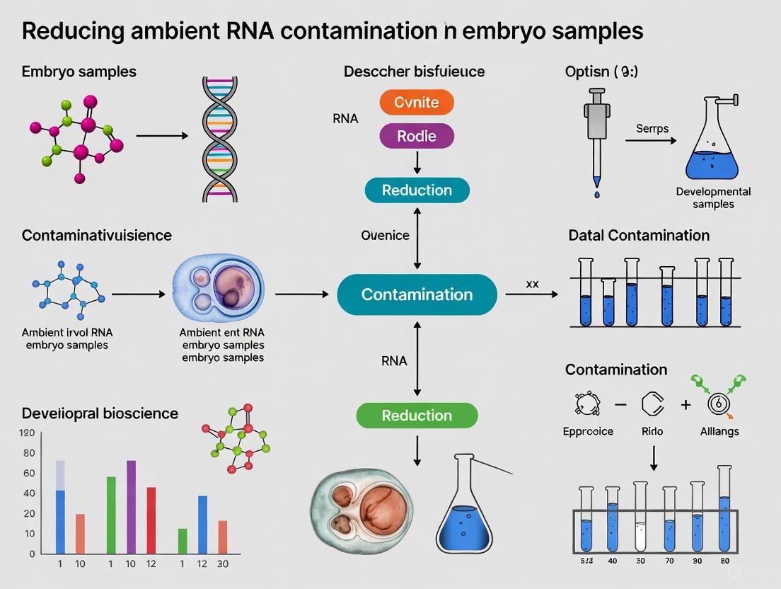
Strategies for Reducing Ambient RNA Contamination in Embryo Samples: A Guide for Reproductive Researchers
Ambient RNA contamination presents a significant challenge in single-cell and single-nucleus RNA sequencing of precious embryo samples, potentially compromising data integrity and leading to erroneous biological conclusions.
Strategies for Reducing Ambient RNA Contamination in Embryo Samples: A Guide for Reproductive Researchers
Abstract
Ambient RNA contamination presents a significant challenge in single-cell and single-nucleus RNA sequencing of precious embryo samples, potentially compromising data integrity and leading to erroneous biological conclusions. This article provides a comprehensive resource for scientists and drug development professionals working in reproductive medicine, covering the foundational understanding of contamination sources, practical methodological solutions for its reduction, troubleshooting for optimized workflows, and rigorous validation techniques. By synthesizing current research and emerging technologies, we offer a actionable framework to safeguard transcriptomic studies in early embryonic development, thereby enhancing the reliability of research outcomes for applications in regenerative medicine and assisted reproductive technology.
Understanding Ambient RNA: Sources, Impact, and Consequences for Embryo Research
Defining Ambient RNA Contamination in the Context of Embryo Samples
FAQs on Ambient RNA Contamination
What is ambient RNA contamination and how does it occur in single-cell RNA sequencing? Ambient RNA contamination refers to the phenomenon where cell-free mRNA molecules, released from stressed, apoptotic, or lysed cells, are present in the cell suspension and become indiscriminately co-encapsulated with intact cells during droplet-based single-cell RNA sequencing (scRNA-seq). This results in background RNA counts being added to the gene expression profile of individual cells, contaminating their true transcriptomic signals [1] [2] [3]. In the context of embryo samples, which are often limited and sensitive to handling, the process of dissociation to create single-cell suspensions is a well-known cause of such contamination [1].
Why is ambient RNA contamination a particular concern for embryo research? Embryo samples are especially vulnerable due to their small size, fragility, and the fact that researchers often work with limited material, sometimes even single embryos [4]. Pooling embryos to obtain sufficient RNA for sequencing has been a common practice, but this inherently confounds biological variation and can mask the true transcriptome of an individual embryo. Furthermore, the dissociation protocols required for embryo samples can induce significant cell stress and death, amplifying the release of ambient RNA into the suspension [1] [4]. This contamination can obscure crucial biological signals related to embryonic development.
What are the key experimental signs that my embryo scRNA-seq data is contaminated? Several indicators can signal high levels of ambient RNA contamination in your data [1] [3]:
- Web Summary Alert: A "Low Fraction Reads in Cells" alert in your 10x Genomics Web Summary.
- Barcode Rank Plot: A plot that lacks a characteristic steep inflection point ("steep cliff"), making it difficult to distinguish cell-containing barcodes from empty droplets.
- Gene Expression: Enrichment of mitochondrial genes among cluster marker genes, which can indicate the presence of dead or dying cells.
- Biological Implausibility: Expression of highly specific marker genes in unexpected or biologically implausible cell types within your embryo sample (e.g., a later-stage marker appearing in an early-stage cell cluster) [5] [6].
How can I proactively minimize ambient RNA contamination during my embryo sample preparation? Optimizing the wet-lab workflow is crucial for minimizing ambient RNA at the source [1]:
- Fixation: Consider cell fixation to stabilize cells before dissociation.
- Loading: Optimize cell loading concentrations to reduce stress.
- Microfluidics: Utilize microfluidic dilution where accessible on open platforms.
- Sample Quality: Prioritize sample preparation methods that maximize cell viability and minimize cell death and lysis, as these are primary sources of ambient RNA.
Quantitative Metrics for Assessing Contamination
The following table summarizes key quantitative metrics developed to assess ambient RNA contamination in unfiltered scRNA-seq data, providing an objective measure of data quality before any computational correction [1].
Table: Quantitative Metrics for Assessing Ambient RNA Contamination
| Metric Category | Metric Name | Description | Interpretation |
|---|---|---|---|
| Geometric (based on cumulative count curves) | Maximal Secant Distance | The largest distance between a point on the cumulative count curve and the diagonal. | A larger distance indicates a sharper slope change and higher data quality. |
| Standard Deviation of Secant Distances | The variability of all secant line distances. | A larger standard deviation indicates better separation between cells and empty droplets. | |
| AUC over Minimal Rectangle | The ratio of the area under the cumulative count curve to the area of its minimal bounding rectangle. | High-quality data occupies more of the rectangular area. | |
| Statistical (based on slope distributions) | Scaled Slopes Below Threshold | The sum of scaled slopes below a threshold (one standard deviation above the median slope). | A higher value indicates more data points are considered background, scaling with the contamination level. |
Computational Tools for Ambient RNA Correction
Several computational tools have been developed to estimate and remove ambient RNA contamination post-sequencing. The choice of tool depends on your data and needs.
Table: Comparison of Computational Tools for Ambient RNA Correction
| Tool Name | Primary Function | Key Mechanism | Considerations |
|---|---|---|---|
| SoupX [5] [3] | Removal of ambient RNAs from cell barcodes. | Estimates an ambient RNA profile from empty droplets and uses it to correct expression in cell barcodes. | Allows both auto-estimation and manual setting of contamination fraction using known marker genes. |
| CellBender [1] [3] [7] | Cell calling & ambient RNA removal. | Uses a deep generative model to learn the background noise profile and distinguish cell-containing from cell-free droplets. | Higher computational cost, but provides an end-to-end solution. |
| DecontX [2] [7] | Decontamination of individual cells. | A Bayesian method that models a cell's expression as a mixture of native and contaminating transcript distributions. | Designed to remove contamination in individual cells after cell calling. |
Experimental Protocol: Single-Embryo RNA Isolation
To mitigate the need for pooling and reduce opportunities for contamination, here is a robust RNA isolation method adapted for single embryos, based on a protocol validated in zebrafish [4]. This yields high-quality RNA suitable for scRNA-seq.
Key Reagent Solutions:
- Homogenization Medium: Liquid Nitrogen
- Lysis Reagent: Qiazol (or similar phenol-guanidine thiocyanate reagent)
- Phase-Separation Agent: Phase-lock gel (heavy)
- Purification Method: Silica-based column purification
Workflow:
- Sample Collection: Image individual embryo with stereo microscopy, transfer to a 1.5 ml tube, and remove all residual water.
- Snap-Freezing: Quickly snap-freeze the embryo in liquid nitrogen. Store at -80°C until ready for extraction.
- Homogenization: While the sample is still frozen, homogenize the embryo thoroughly in liquid nitrogen. Incomplete homogenization will result in lower RNA quality and yield.
- Lysis and Phase Separation:
- Add the appropriate volume of Qiazol to the homogenized powder and mix thoroughly.
- Use Phase-Lock Gel to facilitate clean separation of the aqueous phase containing RNA from the organic phase after chloroform addition. This step is critical for maximizing yield and minimizing reagent carry-over.
- RNA Precipitation & Purification: Precipitate the RNA from the aqueous phase and use a silica-column based method for final purification. This ensures the removal of impurities and results in RNA with high integrity.
- Quality Control: Assess RNA concentration and integrity. The protocol should yield ≥200 ng of RNA per embryo with a RNA Integrity Number (RIN) ≥ 8.0 [4].
Single-embryo RNA isolation workflow
The Scientist's Toolkit: Key Research Reagents
Table: Essential Reagents for Mitigating Ambient RNA
| Reagent / Material | Function in Mitigating Ambient RNA |
|---|---|
| Phase-Lock Gel | Maximizes RNA yield during phenol-chloroform extraction by creating a physical barrier, preventing carry-over of contaminants from the organic phase [4]. |
| Liquid Nitrogen | Enables effective mechanical homogenization of a single, frozen embryo. This is crucial for complete cell disruption and high RNA yield from a tiny, tough sample [4]. |
| Phenol-based Lysis Reagent (e.g., Qiazol) | Effectively lyses cells and denatures proteins, stabilizing the RNA and preventing degradation during the isolation process from a single embryo [4]. |
| Silica-column Purification Kits | Provides a reliable method for purifying high-integrity RNA from small-volume lysates, free of enzymes and inhibitors that can affect downstream applications [4]. |
Visualizing Contamination and Decontamination
The following diagram illustrates the source of ambient RNA contamination and the fundamental principle of computational correction.
Ambient RNA contamination and correction
Frequently Asked Questions (FAQs)
FAQ 1: What are the primary sources of contamination in single-cell RNA sequencing? The three primary sources are ambient RNA contamination (cytoplasmic leakage), barcode swapping during sequencing, and sample-to-sample (well-to-well) contamination during processing. Ambient RNA, released from dead or dying cells, is a major issue that lowers the signal-to-noise ratio in droplet-based scRNA-seq. Barcode swapping mislabels sequencing reads between samples on patterned flow-cell Illumina sequencers. Well-to-well contamination occurs during DNA extraction or library preparation in plate-based formats [1] [8] [9].
FAQ 2: How can I identify cells affected by cytoplasmic leakage in my single-cell proteomics data? Cells with compromised membranes can be identified using a cell-permeable dye like Sytox Green during sample preparation. Furthermore, a computational classifier has been developed that uses the abundances of the top 75 most significantly leaking proteins to accurately identify permeabilized cells. This classifier, available in the QuantQC R package, is based on a signature showing cytosolic and nuclear proteins are more prone to leakage compared to mitochondrial and membrane proteins [10].
FAQ 3: What is the estimated rate of barcode swapping on the HiSeq 4000, and how does it compare to older models? On the HiSeq 4000, approximately 2.5% of reads can be mislabelled between samples. This rate is about an order of magnitude higher than on the HiSeq 2500, where the swapped fraction was estimated at only 0.22% [8].
FAQ 4: How does well-to-well contamination behave in a 96-well plate? Well-to-well contamination is not random; it occurs primarily in neighboring samples. The highest rates are in immediately proximate wells, with rare events detected up to 10 wells apart. This effect follows a distance-decay relationship and is more prominent in plate-based extraction methods compared to single-tube methods [9].
FAQ 5: What is a key precaution to prevent cross-contamination during embryo cryopreservation? To prevent cross-contamination in liquid nitrogen, it is critical to use hermetically sealed, high-quality, shatter-proof freezing containers. The application of a secondary enclosure, such as "double bagging" or "straw-in-straw," provides an added layer of safety against direct contact of embryos with contaminated LN [11].
Troubleshooting Guides
Issue 1: High Levels of Ambient RNA Contamination
Problem: Your scRNA-seq data shows a low signal-to-noise ratio, with evidence of significant ambient RNA contamination from cytoplasmic leakage.
Solutions:
- Improve Cell Loading: Optimize the cell loading mechanism on your microfluidic platform, as this has been shown to have one of the biggest effects on minimizing ambient contamination [1].
- Assess Sample Quality: Use quantitative, contamination-focused metrics on your unfiltered sequencing data to evaluate the true level of ambient RNA before any computational correction [1].
- Consider Cell Fixation: In some experimental setups, cell fixation can help stabilize cells and reduce the release of RNA during processing [1].
- Computational Removal: Post-experiment, use computational tools like CellBender to algorithmically factor out ambient RNA. However, note that these methods are imperfect and work best when ambient contamination is not overwhelming [1].
Issue 2: Suspected Barcode Swapping in Sequencing Data
Problem: You observe unexpected gene expression in cells, or cell libraries that appear to be artificial mixtures, suggesting barcode swapping.
Solutions:
- Sequencing Platform: Where possible, use a sequencing machine that does not use a patterned flow-cell (e.g., HiSeq 2500 over HiSeq 4000/X/NovaSeq) to reduce swapping rates by an order of magnitude [8].
- Experimental Design: For plate-based experiments, leave a fraction of possible barcode combinations unoccupied. This creates "impossible" barcodes that can be used to robustly estimate the swapping fraction for quality control [8].
- Computational Correction: For droplet-based methods (e.g., 10x Genomics), employ specifically developed algorithms to exclude individual molecules that have swapped between samples [8].
- Unique Dual Indexing: If scalability is not a constraint, use unique dual indexing, where two unique barcodes are used for each sample. This prevents library mixing even if one barcode swaps [8].
Issue 3: Well-to-Well Contamination in Plate-Based Assays
Problem: In microbiome 16S sequencing or other plate-based assays, you detect sequences from high-biomass samples appearing in neighboring low-biomass or blank wells.
Solutions:
- Randomize Samples: Do not group low-biomass and high-biomass samples together on the same plate. Randomize samples across the plate to avoid systematic bias [9].
- Extraction Method: Choose manual single-tube extraction methods or hybrid plate-based cleanups, as these have been shown to have less well-to-well contamination compared to fully automated plate-based magnetic bead cleanups [9].
- Data Interpretation: Be cautious with simplistic removal of taxa found in negative controls. In cases of well-to-well contamination, these may be microbes from other samples in your experiment rather than reagent contaminants [9].
Table 1: Quantified Contamination Rates and Key Characteristics
| Contamination Type | Estimated Rate/Level | Key Identifying Feature | Primary Contributing Factor |
|---|---|---|---|
| Barcode Swapping (HiSeq 4000) [8] | ~2.5% of total reads | Mislabelled reads in "impossible" barcode combinations | Patterned flow-cell Illumina sequencers |
| Well-to-Well [9] | Highest in adjacent wells, decays with distance | Contaminants from specific neighboring wells, not random | Plate-based (vs. single-tube) DNA extraction |
| Cytoplasmic Leakage (Protein) [10] | ~2-fold depletion of cytosolic proteins (e.g., Gapdh) | Depletion of cytosolic/nuclear proteins in permeable cells | Cell membrane damage (e.g., from freezing) |
Table 2: Recommended Mitigation Strategies and Their Effectiveness
| Mitigation Strategy | Applicable Contamination Type | Effectiveness / Notes | Key Reference |
|---|---|---|---|
| Unique Dual Indexing | Barcode Swapping | Prevents mixing, but restricts multiplexing scalability | [8] |
| Single-Tube Extraction | Well-to-Well | Reduces cross-talk compared to plate-based methods | [9] |
| Hermetically Sealed Containers | Cryopreservation Cross-Contamination | Prevents direct contact with liquid nitrogen | [11] |
| Cell Loading Optimization | Ambient RNA | One of the biggest factors in reducing contamination | [1] |
| QuantQC Classifier | Cytoplasmic Leakage (Protein) | AUC = 0.92 for identifying permeable cells | [10] |
Experimental Protocols
Protocol 1: Quantifying Barcode Swapping in a Plate-Based scRNA-seq Experiment
This protocol allows for the robust estimation of barcode swapping frequency.
- Experimental Design: When designing your plate layout, ensure that two entirely different sets of row and column barcodes are used for different plates or sections of the plate. The goal is to create a set of barcode combinations that were never mixed experimentally ("impossible" barcodes) [8].
- Sequencing: Multiplex all libraries and sequence on the platform of interest (e.g., HiSeq 4000).
- Data Analysis:
- Generate a count matrix for all barcode combinations, including the impossible ones.
- For each impossible barcode combination, regress its library size against the summed library sizes of all real cell libraries that share exactly one barcode with it.
- The slope of the regression line provides an estimate of the swapped read fraction for the experiment [8].
Protocol 2: Identifying Protein Leakage in Single-Cell Proteomics
This protocol uses a fluorescent dye to directly identify permeabilized cells.
- Sample Preparation: Prior to single-cell isolation, stain the cell suspension with a cell-permeable dye like Sytox Green [10].
- Cell Sorting/Imaging: Record the stain intensity of each cell. The distribution of intensities is typically bimodal. Cells from the mode with high intensity are characterized as permeable (compromised membrane), while cells from the mode at low (near-zero) intensity are intact [10].
- Data Integration: Link the stain intensity measurements with the downstream single-cell proteomic data using tools like QuantQC. This allows for the direct exclusion of permeabilized cells from analysis or for the definition of a protein leakage signature [10].
Visualized Workflows and Relationships
The Scientist's Toolkit
Table 3: Essential Research Reagent Solutions
| Reagent / Tool | Function / Purpose | Specific Example / Note |
|---|---|---|
| Sytox Green | Fluorescent cell-impermeant dye used to identify cells with compromised plasma membranes. | Staining prior to single-cell isolation allows sorting or identification of permeabilized cells [10]. |
| QuantQC (R package) | Computational tool that includes a classifier for identifying cells affected by protein leakage based on their proteomic profile. | Uses the abundance of ~75 leaking proteins to accurately identify permeabilized cells (AUC = 0.92) [10]. |
| CellBender | Computational tool for removing ambient RNA contamination from droplet-based scRNA-seq data. | Uses a probabilistic model to subtract background noise and output corrected counts [1]. |
| Hermetically Sealed Straws | High-quality, shatter-proof containers for cryopreservation of embryos and other biologics. | Prevents direct contact with liquid nitrogen, the primary vector for cross-contamination during banking [11]. |
| DTT (Dithiothreitol) | Reducing agent that breaks disulfide bonds. | Useful in optimizing RNA extraction from challenging samples like spermatozoa by disrupting highly condensed chromatin [12]. |
Frequently Asked Questions
What are the signs of ambient RNA contamination in my data?
- Low Fraction Reads in Cells: An alert in your sequencing web summary (e.g., 10x Genomics Web Summary) is a primary indicator [3].
- Barcode Rank Plot: A plot that lacks a characteristic "steep cliff," making it difficult for algorithms to distinguish cell-containing barcodes from empty droplets [3].
- Unexpected Marker Gene Expression: The presence of highly expressed genes from abundant cell types (like hemoglobins in erythrocytes) in cell populations where they are biologically implausible, such as neural crest cells [6].
- Enrichment of Mitochondrial Genes: Specific cell clusters showing significant upregulation of mitochondrial genes can indicate dead or dying cells, which are a source of ambient RNA [3].
Can ambient RNA contamination lead to the misidentification of a new cell type? Yes. Failure to remove poor-quality cells, including those with significantly skewed gene expression profiles, can lead to misclustering. A cluster of poor-quality cells can be mistakenly interpreted as a novel cell type [13]. Furthermore, ambient RNA from one cell type can contaminate others, blurring the distinctions between populations and complicating annotation [14].
How does ambient RNA specifically affect differential expression (DE) analysis? Ambient contamination can cause the false detection of differentially expressed genes (DEGs) between conditions. For example, in a study comparing Tal1-knockout and wild-type neural crest cells, the most significant DEGs were hemoglobin genes, which these cells should not express. This was driven by differences in the ambient pool between samples rather than true biological changes [6]. After correction, these false DEGs are removed, leading to a more reliable list of genes [14].
My data has passed basic QC checks. Do I still need to worry about ambient RNA? Potentially, yes. Basic QC often filters cells based on library size or mitochondrial content but does not specifically account for the subtle yet widespread effects of ambient RNA. In studies aiming to profile rare cell subtypes or detect subtle transcriptional differences, applying specialized ambient RNA correction tools is highly recommended, even if basic QC metrics appear acceptable [3].
What is the difference between a tool that removes droplets and one that removes RNA?
- Droplet Removal Tools (e.g.,
CellBender,EmptyNN): These classify each barcode as containing a cell or being empty/background, and remove the entire barcode from the dataset [3]. - Ambient RNA Removal Tools (e.g.,
SoupX,DecontX): These estimate an ambient RNA profile and computationally subtract these counts from the expression matrix of the cell-containing barcodes, preserving the cells but cleaning their expression profiles [3] [14].
- Droplet Removal Tools (e.g.,
Troubleshooting Guides
Guide 1: Diagnosing and Correcting for Ambient RNA Contamination
Problem: Suspected ambient RNA contamination, as indicated by the FAQs above.
Solution: A step-by-step workflow for diagnosing and correcting contamination.
Detailed Steps:
- Initial Data Inspection: Thoroughly review your sequencing provider's summary report (e.g., the 10x Genomics Web Summary) for warnings about a low fraction of reads in cells [3].
- Identify Potential Contamination Markers: Use biological knowledge to identify genes that should be restricted to specific cell types (e.g., hemoglobin genes for red blood cells, immunoglobulin genes for B cells). Their presence in other cell types is a strong indicator of ambient RNA [6] [14].
- Estimate the Ambient Profile: Most correction tools require the raw gene-barcode matrix (including empty droplets) to estimate the background RNA profile. Tools like
SoupXandCellBenderuse these empty droplets to learn the composition of the ambient soup [6] [3]. - Apply a Correction Tool: Choose and run a computational correction tool. The table below summarizes key tools. For
SoupX, you may need to manually specify the contamination fraction or provide a list of genes known not to be expressed in certain cell populations to improve accuracy [3] [14]. - Re-analyze Data: Repeat your standard analysis pipeline (normalization, clustering, and differential expression) using the corrected count matrix.
- Compare Results: Critically compare the results before and after correction. Successful correction should reduce or eliminate the expression of implausible marker genes and may improve cell clustering [14].
Guide 2: Identifying and Filtering Cells with Skewed Gene Coverage
Problem: Technical artifacts causing skewed gene body coverage, which can be misinterpreted as biological heterogeneity [13].
Solution: Use the SkewC tool to identify and remove these poor-quality cells.
Protocol:
- Input Data: Prepare a gene-by-cell count matrix from your scRNA-seq experiment.
- Run SkewC: Execute the
SkewCalgorithm on your dataset. The tool calculates a skewness metric for each cell's gene coverage profile. It operates by:- Computing the gene body coverage for each cell.
- Assessing the skewness of this coverage as a quality measure.
- Classifying cells into "typical" (good quality) and "skewed" (poor quality) based on their coverage profiles [13].
- Filter Cells: Remove the cells classified as "skewed" from your dataset before proceeding with downstream biological analysis. This helps prevent misclustering and the formation of false cell populations [13].
Data Presentation: Computational Tools for Ambient RNA Correction
The following table summarizes community-developed tools for addressing ambient RNA contamination.
| Tool Name | Primary Mechanism | Key Inputs | Language | Key Advantages / Limitations |
|---|---|---|---|---|
| SoupX [3] [14] | Estimates & subtracts an ambient profile | Raw & filtered count matrices | R | Advantage: Allows manual guidance using known marker genes. Limitation: Contamination fraction estimation can be complex. |
| CellBender [3] [14] | Deep generative model; performs cell-calling and RNA removal | Raw count matrix | Python | Advantage: Fully unsupervised; does not require prior biological knowledge. Limitation: Computationally intensive; may require GPU. |
| DecontX [3] | Bayesian method to deconvolute native vs. contaminant counts | Count matrix & cell cluster labels | R | Uses a Bayesian framework to model the mixture of counts. |
| EmptyNN [3] | Neural network to classify empty vs. cell-containing droplets | Raw count matrix | R | A machine-learning-based approach for cell calling. |
| DropletQC [3] | Identifies empty droplets, damaged, and intact cells using nuclear fraction | Count matrix | R | Unique Feature: Can identify damaged cells, not just empty droplets. |
Experimental Protocols
Protocol 1: Using SoupX for Ambient RNA Correction
This protocol provides a detailed methodology for correcting data using SoupX [6] [3] [14].
Key Features:
- Allows for both automated and expert-guided correction.
- Directly corrects the count matrix for downstream analysis.
Materials and Reagents
- Software: R environment,
SoupXR package. - Data: The raw (unfiltered) and filtered gene-barcode matrices from Cell Ranger (or other alignment pipeline).
Procedure
- Load Data: In R, load both the raw and filtered gene-barcode matrices into a
SoupChannelobject. - Estimate Contamination: Automatically estimate the global background contamination fraction using the
autoEstContfunction. The formula is:contamination_fraction = (counts from ambient RNA) / (all counts in a cell) - Optional - Manual Guidance: To improve accuracy, provide a list of genes that are highly specific to a cell type and should not be expressed in other cell types (e.g.,
HbBfor non-erythrocytes).SoupXwill use the absence of these genes in a cell to more accurately estimate the contamination. - Correct Expression: Execute the
adjustCountsfunction to create a new, corrected count matrix where the estimated ambient RNA counts have been subtracted. - Output: Use the corrected matrix for all subsequent analyses in Seurat, Scanpy, or other frameworks.
Validation Validate the correction by visualizing the expression of known problematic genes (e.g., hemoglobin genes) before and after correction using dimensionality reduction plots (UMAP/t-SNE). Their expression should be drastically reduced in implausible cell types [14].
Protocol 2: Validating Correction with Differential Expression Analysis
This protocol confirms the effectiveness of ambient RNA correction by comparing differential expression results [6] [14].
Procedure
- Pre-correction DE Analysis: Perform a differential expression analysis between two conditions or clusters on the uncorrected data. Note all significant DEGs, particularly those that are biologically surprising.
- Post-correction DE Analysis: Repeat the identical DE analysis on the corrected data.
- Compare Gene Lists: Identify genes that were significant before correction but are no longer significant after correction. These were likely false positives driven by ambient RNA.
Data Analysis A quantitative comparison can be presented as follows:
| Condition | Total DEGs Pre-Correction | Total DEGs Post-Correction | Notable False Positives Removed |
|---|---|---|---|
| WT vs. KO (Neural Crest) | 769 (e.g., Hbb-bh1, Hba-x) | 769 (e.g., Xist, Erdr1) | Hemoglobin genes (Hbb-bh1, Hba-x, etc.) [6] |
| T cell Subpopulation | 150 | 120 | 30 ambient-driven genes removed, revealing biologically relevant pathways [14] |
The Scientist's Toolkit: Research Reagent Solutions
| Item / Resource | Function in Context of Ambient RNA |
|---|---|
| Chromium Next GEM Single Cell Kits (10x Genomics) | A widely used droplet-based scRNA-seq platform. Its cell-calling algorithm provides the first line of defense against ambient RNA, but additional correction is often needed [3]. |
| Dead Cell Removal Kit | Used in sample preparation to physically remove dead or dying cells, which are a major source of ambient RNA, thereby reducing the background contamination load before sequencing. |
| SoupX R Package | A key software tool for computationally estimating and subtracting the ambient RNA profile from cell expression data [3] [14]. |
| CellBender Software | A powerful tool that uses a deep learning model to perform joint cell-calling and ambient RNA background removal [3] [14]. |
| List of Marker Genes (e.g., Hemoglobins, Immunoglobulins) | A curated, biology-specific list of genes used to guide and validate ambient RNA correction algorithms. These genes serve as indicators of contamination [6] [14]. |
Frequently Asked Questions (FAQs)
FAQ 1: Why are embryo samples especially prone to ambient RNA contamination in single-nucleus RNA-seq (snRNA-seq) assays? Embryo samples are highly vulnerable due to their unique tissue architecture and composition. Tissues like the placenta, which is central to embryonic development, contain multinucleated syncytial structures (e.g., the syncytiotrophoblast) that are inherently fragile and difficult to dissociate without causing widespread rupture [15]. This rupture releases massive amounts of cytoplasmic RNA into the suspension medium, which then contaminates the nuclei of all cell types present [15]. Furthermore, embryonic tissues are often delicate and sensitive to the enzymatic and mechanical stress of dissociation, exacerbating cell death and RNA release [16].
FAQ 2: What is the tangible impact of this contamination on my research data? Ambient RNA contamination systematically biases your data by inflating the measured gene expression levels in your nuclei. This can:
- Obscure true cell-type identities: Well-known cell-type marker genes may appear to be expressed in nearly all cell types, confusing your cell type annotation [17] [15]. For example, in mouse mammary gland studies, lactating markers like Wap and Csn2 were detected globally across all cells due to contamination instead of being restricted to alveolar epithelial cells [17].
- Mask genuine biological signals: Contamination can hide true transcriptional dysregulation associated with developmental states or disease models, making it harder to identify differentially expressed genes [15] [5].
- Lead to incorrect biological interpretations: Pathway enrichment analyses can be significantly distorted, highlighting ambient-related pathways instead of biologically relevant ones [5].
FAQ 3: Can't I just use a standard computational tool to clean my data afterward? While computational decontamination tools (e.g., SoupX, CellBender, DecontX) are essential, they have limitations, especially with highly contaminated embryo data. Some methods may under-correct highly contaminating genes (like specific embryonic markers), leaving significant contamination in your data. Others may over-correct, erroneously removing the counts of genuine, lowly expressed genes, including housekeeping genes [17]. Therefore, relying solely on post-hoc computational correction is insufficient; optimizing the wet-lab protocol to minimize contamination at the source is critical.
FAQ 4: What is the most critical step in my protocol to minimize ambient RNA? The cell loading mechanism and the initial steps of nucleus isolation have been identified as having the biggest effect on ambient contamination levels [1]. A gentle, optimized nuclei isolation protocol that avoids excessive physical or enzymatic stress is paramount for preserving nucleus integrity and minimizing the release of RNA [16].
Troubleshooting Guide: Ambient RNA Contamination in Embryo Samples
| Problem | Possible Cause | Solution |
|---|---|---|
| Widespread expression of specific marker genes (e.g., trophoblast genes in all nuclei) | Rupture of fragile, RNA-rich embryonic structures (e.g., syncytiotrophoblast) during dissociation [15]. | • Optimize homogenization: Use gentle mechanical douncing instead of harsh enzymatic digestion. • Add RNase inhibitors: Include RNaseOut to protect RNA integrity during isolation [16]. • Use ice-cold buffers: Keep samples and buffers on ice at all times to slow RNase activity [16]. |
| Low sequencing sensitivity and gene detection | General RNA degradation and loss due to high RNase content in some embryonic tissues [16]. | • Use nuclease-free reagents and equipment. • Perform rapid dissection and processing to minimize sample degradation time. • Validate nucleus integrity with microscopy (e.g., DAPI staining) before proceeding to sequencing [16]. |
| Failure of computational decontamination | Under-correction of highly abundant contaminating transcripts [17]. | • Employ a targeted method: Use a method like scCDC, which specifically detects and corrects only the "contamination-causing genes," avoiding global over-correction [17]. • Combine methods: Use scCDC first to remove major contaminants, then a global method like DecontX to address low-level background [17]. |
| Poor cell type identification and clustering | High levels of ambient RNA blurring the distinctions between nuclear transcriptomes [15]. | • Apply contamination-focused QC metrics to your raw, unfiltered data to assess quality before analysis [1]. • Isolate nuclei from frozen tissue: This can sometimes be gentler than dissociating live cells from fresh, fragile embryos [16]. |
Evidence and Data: Quantifying Vulnerability in Embryonic Tissues
The table below summarizes key quantitative and observational evidence from studies highlighting the specific challenges of working with embryo-related tissues.
| Tissue / Sample Type | Observed Contamination Effect | Experimental Evidence | Source |
|---|---|---|---|
| Mouse Placenta | Nuclei of all placental cell classes suffered ambient trophoblast contamination. | snRNA-seq failed to detect molecular dysregulation in preeclampsia that was readily apparent with scRNA-seq, due to contamination and reduced sensitivity [15]. | [15] |
| Mouse Mammary Gland (Lactating) | Milk protein genes Wap and Csn2 (AlveoDiff markers) were detected globally across all cell types. | In snRNA-seq data, these specific genes showed unexpected expression in non-relevant cells, indicating systematic ambient RNA contamination [17]. | [17] |
| General Tissues | Cell loading mechanism identified as the factor with the biggest effect on ambient contamination. | Controlled experiments on an open-source platform (inDrops) showed that technical parameters behind the microfluidics significantly impact contamination levels [1]. | [1] |
The Scientist's Toolkit: Essential Reagents for Robust Nuclei Isolation
This table lists key reagents used in an optimized nucleus isolation protocol from frozen mouse embryonic tissues, as detailed in the search results [16].
| Reagent | Function in the Protocol |
|---|---|
| Bovine Serum Albumin (BSA) | Acts as a protein stabilizer and reduces nonspecific binding during the isolation process. |
| Dulbecco’s Phosphate-Buffered Saline (DPBS) | A balanced salt solution used for washing tissues and nuclei while maintaining osmotic balance. |
| NP-40 | A non-ionic detergent used in the lysis buffer to gently break down cellular membranes without damaging nuclear envelopes. |
| RNaseOut | A potent RNase inhibitor that is critical for protecting RNA from degradation during the isolation procedure. |
| DAPI (4',6-diamidino-2-phenylindole) | A fluorescent dye that binds to DNA, used for staining nuclei to assess their quantity, integrity, and purity via microscopy or flow cytometry. |
Workflow Diagram: Contamination in Embryonic snRNA-seq
The diagram below visualizes the pathway of ambient RNA contamination in embryonic single-nucleus RNA-sequencing, from sample preparation to data analysis, highlighting critical failure points and mitigation strategies.
Troubleshooting Guides
Guide 1: Identifying and Addressing Microbial Contamination in Embryo Cultures
Problem: Cloudy culture droplets or moving punctate/rod-shaped microorganisms observed under an inverted microscope.
Cause: Environmental bacterial contamination, such as Staphylococcus pasteuri, introduced through laboratory environmental sources like contaminated air handling systems or water leaks, rather than patient samples [18].
Solution:
- Immediate Embryo Rescue: Carefully remove embryos from contaminated droplets using a glass pipette (120-140 μm inner diameter) [18].
- Sequential Washing: Transfer embryos to organ-well culture dishes containing fresh, pre-equilibrated culture medium. Blow repeated from the bottom of the dish to ensure colonies detach [18].
- Repeated Monitoring: Replace the culture dish and medium every 8 hours until no contamination is observed. Observe cleavage and contamination clearance on day three for transfer, freezing, or blastocyst culture decisions [18].
- Laboratory Decontamination: Perform thorough disinfection using 0.5% hypochlorite for floors and instruments. Use 3% hydrogen peroxide for surfaces contaminated by blood or semen. Sterilize incubator components using high-temperature and damp-heat methods [18].
Guide 2: Overcoming RNA Degradation and Contamination in Sensitive Samples
Problem: Degraded RNA or contaminated RNA samples yielding poor results in downstream applications like sequencing or qRT-PCR.
Cause:
- RNase Contamination: Introduction of RNase enzymes from the user's skin, contaminated surfaces, or non-certified RNase-free consumables [19] [20].
- Improper Sample Handling: Failure to immediately stabilize RNA after sample collection, leading to rapid degradation by endogenous RNases [20].
- gDNA Carryover: Traces of genomic DNA co-purifying with RNA, causing false positives in PCR-based assays [21].
Solution:
- Create an RNase-Free Environment: Dedicate a section of your bench for RNA work, using RNase-decontamination solutions on surfaces, glassware, and pipettes. Always wear gloves and a lab coat, changing gloves after touching surfaces outside the clean zone [20].
- Stabilize RNA Immediately: Lyse samples in TRIzol or a dedicated lysis buffer immediately after collection and freeze at -80°C. Alternatively, use RNA stabilization reagents (e.g., RNAlater) to inactivate RNases at collection [20].
- DNase Treatment: Treat samples with a DNase enzyme, either on-column during purification or post-extraction, to remove contaminating gDNA [19] [21].
- Ensure Complete Lysis: For challenging samples (e.g., FFPE tissue, blood), incorporate mechanical lysis (bead-beating) or enzymatic pre-treatment (proteinase K) to ensure complete cell disruption and RNA release [20].
Frequently Asked Questions (FAQs)
FAQ 1: What are the proven clinical outcomes for embryos exposed to and rescued from microbial contamination?
One retrospective study of 15 IVF patients with embryo contamination found that with proper remediation (daily rinsing and avoidance of blastocyst culture), there were no significant differences in embryo laboratory outcomes, pregnancy outcomes, or maternal and infant complications compared to uncontaminated cycles, except for a slightly higher rate of fetal growth retardation. Ultimately, 11 live-born infants were successfully delivered from these cycles [18].
FAQ 2: How can I determine if contamination is affecting my gene expression data in developmental studies?
Monitor RNA quality metrics closely. Key indicators include:
- RNA Integrity Number (RIN): Use a microfluidics-based system (e.g., Agilent Bioanalyzer). A RIN below 8.0 can indicate degradation [20].
- Spectrophotometric Ratios: Use UV absorbance. Aim for A260/A280 between 1.8–2.2 and A260/A230 >1.7. Low ratios indicate protein or chemical salt contamination [19] [20].
- Downstream Application Failure: Poor performance in qRT-PCR or RNA-seq can indicate the presence of inhibitors or degraded RNA [19] [21].
FAQ 3: Our laboratory has passed all quality control checks. How could environmental contamination still occur?
Environmental contamination can originate from unexpected sources. One documented outbreak of Staphylococcus pasteuri was traced to water that had seeped from a leaky penthouse into the interlayer above the embryo culture room ceiling, contaminating the environment via the laminar flow purification system [18]. This highlights the need for environmental monitoring that extends beyond standard laboratory surfaces.
FAQ 4: What are the most critical steps to protect RNA samples from ambient contamination during isolation?
The most critical steps are [19] [20]:
- Immediate Lysis: Place samples in a denaturing lysis buffer immediately upon collection.
- Dedicated Workspace: Use a clean, dedicated RNase-free bench area with certified RNase-free tips and tubes.
- Additive Use: Include beta-mercaptoethanol (BME) in lysis buffers to inactivate RNases.
- Keep it Cold: Perform extractions using cold reagents and centrifuges to slow RNase activity.
Table 1: Summary of Contamination Incidence and Outcomes in Clinical Embryology
| Parameter | Reported Value | Context / Source |
|---|---|---|
| Incidence of Embryo Contamination | 0.60% (15/2490 cycles) | Retrospective analysis of IVF cycles; outbreak linked to environmental source [18]. |
| Live Birth Rate Post-Decontamination | 11 live-born infants | Result from 15 patients with contaminated embryos after remediation [18]. |
| Primary Contaminant Identified | Staphylococcus pasteuri | Identified in 15 cases of environmental contamination in an embryology lab [18]. |
| RNA Quality Indicator (A260/280) | 1.8 - 2.2 | Target range for pure RNA; indicates low protein contamination [19] [20]. |
| RNA Quality Indicator (A260/230) | > 1.7 | Target value for pure RNA; indicates low chemical salt contamination [20]. |
Experimental Protocols
Protocol 1: Embryo Culture Decontamination and Washing
Application: Remediation of microbially contaminated embryos during IVF procedures [18].
Materials:
- Pre-equilibrated organ-well culture dishes
- Fresh K-SIFM culture medium
- Glass pipette (120-140 μm inner diameter)
- Laminar flow hood
Methodology:
- Under a microscope, carefully draw the contaminated medium and embryos using the glass pipette.
- Transfer the embryos to a well of the organ-well dish containing fresh medium.
- Gently blow the embryos from the pipette to dislodge any attached bacteria from the bottom of the dish.
- Aspirate the embryos and transfer them to a second well of fresh medium, repeating the washing process.
- Continue this serial washing through multiple wells.
- Transfer the washed embryos to a new, clean culture droplet for further culture.
- Replace the culture medium and observe the embryos every 8 hours until contamination is absent.
Protocol 2: RNA Cleanup with DNase Treatment
Application: Purification of RNA and removal of genomic DNA contamination from cell or tissue lysates [19] [21].
Materials:
- RNA Cleanup Binding Buffer
- Ethanol (100% and 70-80%)
- RNA Cleanup Columns and collection tubes
- DNase I enzyme (e.g., NEB #M0303)
- Nuclease-free water
Methodology:
- Bind RNA: Mix the RNA sample with Binding Buffer and ethanol according to protocol. Apply the entire mixture to the RNA Cleanup Column and centrifuge [19].
- Wash: Centrifuge with wash buffer to remove salts and impurities. Ensure the column does not contact the flow-through [19].
- On-Column DNase Digestion (Optional): Apply a mixture of DNase I directly to the center of the column matrix and incubate at room temperature for 15 minutes [19].
- Final Wash: Perform a second wash step to remove the DNase enzyme and any residual contaminants [19] [21].
- Elute: Apply nuclease-free water directly to the center of the column matrix. Incubate for 1 minute, then centrifuge to elute pure RNA. Using larger elution volumes or multiple elutions can increase yield [19].
Diagram 1: Decontamination workflows for embryo and RNA samples.
The Scientist's Toolkit: Essential Research Reagent Solutions
Table 2: Key Reagents for Contamination Prevention and Management
| Reagent / Kit | Primary Function | Application Note |
|---|---|---|
| RNAlater Stabilization Solution | Inactivates RNases immediately upon sample collection for RNA work. | Allows flexibility for later RNA extraction without degradation; ideal for field work or busy labs [20]. |
| DNase I Enzyme | Degrades contaminating genomic DNA in RNA samples. | Can be used "on-column" during purification or in solution post-extraction for sensitive applications like qRT-PCR [19] [21]. |
| MagMAX RNA Kits | Magnetic bead-based purification of total RNA. | Suitable for high-throughput automated systems, reducing hands-on time and risk of human-borne RNase contamination [20]. |
| TRIzol Reagent | Monophasic solution of phenol and guanidine isothiocyanate for RNA isolation. | Gold-standard, effective for difficult-to-lyse samples and inactivates RNases during homogenization [21] [20]. |
| Proteinase K | Broad-spectrum serine protease for enzymatic lysis. | Digests proteins and inactivates nucleases; crucial for challenging samples like FFPE tissues or microbes [20]. |
| Beta-Mercaptoethanol (BME) | A reducing agent that denatures proteins by breaking disulfide bonds. | Added to lysis buffers to inactivate RNases (e.g., RNase A) that are stabilized by disulfide bonds [21] [20]. |
Practical Workflows and Techniques to Minimize Ambient RNA in Embryo Studies
This technical support center provides targeted guidance for researchers, especially those working with embryonic samples, to navigate the critical steps of tissue dissociation and nuclei isolation. The quality of this initial preparation is paramount for the success of downstream single-cell and single-nuclei RNA sequencing (scRNA-seq, snRNA-seq). A particular focus is placed on strategies to mitigate ambient RNA contamination, a significant challenge that can distort transcriptomic data by introducing background noise from transcripts released by broken cells [14]. The following FAQs, troubleshooting guides, and optimized protocols are designed to help you achieve high-quality, reliable data for your research.
FAQs and Troubleshooting Guides
Frequently Asked Questions
1. What is ambient RNA contamination and why is it a critical concern for embryo samples? Ambient RNA contamination refers to the cell-free mRNAs that are released from ruptured cells during tissue dissociation. These transcripts can be indiscriminately incorporated into droplets during droplet-based single-cell sequencing, leading to a distorted interpretation of a cell's true transcriptome [14]. For precious embryonic samples, which can be particularly sensitive to dissociation, this contamination can obscure rare cell types and lead to the misidentification of biological pathways [14].
2. When should I choose nuclei isolation (snRNA-seq) over single-cell dissociation (scRNA-seq) for my tissue? The choice depends on your tissue type and experimental constraints. The following table summarizes key decision points:
| Factor | Single-Cell RNA-seq (scRNA-seq) | Single-Nucleus RNA-seq (snRNA-seq) |
|---|---|---|
| Best For | Fresh, easy-to-dissociate tissues (e.g., spleen, lymph nodes). | Hard-to-dissociate tissues (e.g., brain, heart, adipose), frozen archives, and formalin-fixed paraffin-embedded (FFPE) samples [22]. |
| Tissue Viability | Requires high cell viability post-dissociation. | Does not require intact cells; works with frozen or fragile samples [22] [23]. |
| Transcript Coverage | Captures mature, cytoplasmic mRNA. | Captures both nascent (unspliced) and mature mRNA, providing a view of nuclear transcription [22]. |
| Dissociation Bias | Can be high, as some cell types are more susceptible to lysis. | Generally lower, often providing a more accurate representation of the original cell population in the tissue [23]. |
3. What are the most effective methods to reduce ambient RNA contamination? Proactive and computational strategies can be combined for best results:
- Proactive Experimental Mitigation: Optimize your dissociation protocol to minimize cell lysis. Using snRNA-seq instead of scRNA-seq can inherently reduce the impact of cytoplasmic ambient RNA [23]. Incorporate a nuclei purification step, such as fluorescence-activated nuclei sorting (FANS) or density gradient centrifugation (e.g., using iodixanol) to remove cellular debris and lysed contaminants [24] [23].
- Post-Hoc Computational Correction: After sequencing, apply specialized tools like SoupX or CellBender to estimate and digitally subtract the ambient RNA signal from your count matrices [14] [23]. These tools have been shown to improve the identification of differentially expressed genes and biologically relevant pathways [14].
Troubleshooting Common Experimental Issues
1. Problem: Low Nuclei Yield
- Potential Causes & Solutions:
- Incomplete Tissue Dissociation/Homogenization: Optimize mechanical disruption. For frozen tissues, mince on dry ice before Dounce homogenization [23]. The number of strokes and the tightness of the Dounce pestle should be optimized for each tissue type [23].
- Inefficient Lysis Buffer: Ensure your lysis buffer is fresh and contains the appropriate detergent concentration (e.g., 0.05%-0.1% NP-40) [25] [23]. Test different buffers if yield is consistently low.
- Sample Size Too Small: While protocols exist for low-input material (as low as 15 mg), starting with too little tissue can yield scant nuclei. Aim for 5-30 mg as an optimal range [22] [23].
2. Problem: Excessive Nuclei Clumping
- Potential Causes & Solutions:
- High Nuclei Concentration: Dilute the nuclei suspension further.
- Lack of BSA or RNase Inhibitor: Always include 0.5-1% BSA in wash and resuspension buffers to prevent sticking [22] [25]. Include an RNase inhibitor (e.g., 1 U/µL) in all solutions to protect RNA integrity [22] [25].
- Over-Lysis: Excessive lysis time or detergent concentration can damage nuclei, causing DNA release and clumping. Monitor lysis progress carefully, checking an aliquot every 1-2 minutes when optimizing a new protocol [22].
3. Problem: High Ambient RNA Contamination in Sequencing Data
- Potential Causes & Solutions:
- High Cell/Nuclei Lysis During Preparation: This is the primary source. Gentle handling during all steps is crucial. Switching to a gentler nuclei isolation protocol can be beneficial [14].
- Insufficient Purification: Implement a purification step. Fluorescence-activated nuclei sorting (FANS) can selectively isolate intact nuclei away from debris and lysed material [24] [23]. Density gradient centrifugation is another effective method [25] [23].
- Computational Correction Failure: Ensure you are using the latest versions of correction tools (e.g., SoupX v1.6.2 or later) and providing them with appropriate, tissue-specific "background" gene sets (e.g., hemoglobin genes for red blood cell contamination) [14].
Optimized Experimental Protocols
Detailed Workflow: Nuclei Isolation from Low-Input Cryopreserved Tissue
This protocol, adapted from a recent Scientific Reports publication, is designed for versatility across different tissue types, including embryonic samples, and is optimized to minimize ambient RNA [23].
1. Tissue Lysis and Homogenization
- Reagents: Ice-cold Lysis Buffer (10 mM Tris-HCl pH 7.4, 10 mM NaCl, 3 mM MgCl₂, 0.05% NP-40, 1 mM DTT, 1 U/µL RNase inhibitor) [25] [23].
- Procedure:
- Mince 15-30 mg of cryopreserved tissue on dry ice.
- Transfer to a pre-cooled Dounce homogenizer containing 3 mL of Lysis Buffer.
- Homogenize with the loose (A) pestle for 10-15 strokes, followed by the tight (B) pestle for 10-15 strokes. Note: The optimal number of strokes is tissue-dependent and requires pilot testing [23].
- Incubate on ice for 5 minutes.
- Stop the lysis by adding 5 mL of Ice-cold Nuclei Washing Buffer (0.5X PBS, 5% BSA, 0.25% Glycerol, 40 U/mL RNase inhibitor) [23].
2. Filtration and Purification
- Procedure:
- Filter the homogenate through a pre-wet 30 µm cell strainer [25] [23].
- Centrifuge at 1000 g for 10 minutes at 4°C.
- Resuspend the pellet in 1 mL of Washing Buffer.
- For purification, layer the suspension on top of a 2 mL cushion of 29% iodixanol solution.
- Centrifuge at 1000 g for 20 minutes at 4°C. Intact nuclei will form a pellet, while debris remains in the gradient.
3. Nuclei Sorting (FANS) and QC
- Procedure:
- Resuspend the pellet in 300 µL of Washing Buffer containing a nuclear dye (e.g., 7-AAD or Propidium Iodide) [23].
- Sort stained nuclei using a flow sorter (e.g., BD FACSAria Fusion) with a 70 µm nozzle. Gating on positive fluorescence and size excludes debris and lysed particles [23].
- Collect sorted nuclei and centrifuge at 1000 g for 10 minutes at 4°C.
- Resuspend in an appropriate buffer for your sequencing platform (e.g., Diluted Nuclei Buffer from 10x Genomics) [25].
- Perform quality control: Check under a microscope for intact, round nuclei with sharp borders. Use Trypan Blue or Acridine Orange/Propidium Iodide (AOPI) staining to assess integrity, aiming for ≥90% intact nuclei [22].
The following diagram summarizes the key steps of this workflow and the points at which ambient RNA is controlled.
The Scientist's Toolkit: Essential Reagents and Materials
The following table lists key reagents and their critical functions for successful nuclei isolation, based on the cited protocols.
| Reagent / Material | Function / Purpose | Example Citations |
|---|---|---|
| NP-40 / Triton X-100 | Non-ionic detergent that permeabilizes the cell membrane while leaving the nuclear envelope intact. | [22] [25] [23] |
| Dounce Homogenizer | Provides controlled mechanical disruption for tissue homogenization; loose and tight pestles allow for step-wise breakdown. | [23] |
| Protector RNase Inhibitor | Essential for preserving RNA integrity by inhibiting RNases released during tissue disruption. | [22] [25] [23] |
| BSA (Bovine Serum Albumin) | Reduces nuclei clumping and sticking to plastic surfaces in wash and resuspension buffers. | [22] [25] |
| Iodixanol (Optiprep) | Used for density gradient centrifugation to purify intact nuclei away from cellular debris. | [25] [23] |
| Propidium Iodide / 7-AAD | Fluorescent dyes that stain DNA, enabling visualization and sorting of intact nuclei via FANS. | [22] [23] |
| DTT (Dithiothreitol) | A reducing agent that helps break down disulfide bonds in tissues, aiding in homogenization. | [25] |
Data Presentation: Computational Correction of Ambient RNA
The impact of ambient RNA correction is quantifiable. The following table summarizes results from a study that applied SoupX and CellBender to scRNA-seq data from peripheral blood mononuclear cells (PBMCs) and human fetal liver tissues [14].
| Metric | Before Ambient RNA Correction | After Ambient RNA Correction |
|---|---|---|
| Ambient mRNA Levels | High | Significantly Reduced [14] |
| Differentially Expressed Genes (DEGs) | Ambient transcripts appeared among DEGs, leading to false positives. | Improved DEG identification with reduction in false positives [14]. |
| Biological Pathway Enrichment | Identification of significant ambient-related pathways in unexpected cell types. | Highlighting of biologically relevant and cell-type-specific pathways [14]. |
The process of computational correction can be visualized as a final, essential cleaning step in the data analysis pipeline, as shown below.
Troubleshooting Guides
FAQ: Addressing Ambient RNA Contamination
1. My RNA samples from embryos are degraded. What are the most likely causes? Degradation can occur at multiple points. If degradation is observed on a gel or bioanalyzer trace (e.g., smeared rRNA bands), the cause could be insufficient RNase inactivation during sample collection, improper storage, or RNase contamination during the extraction procedure itself [21]. Ensure embryos are lysed immediately after collection in a buffer containing RNase-inactivating agents like beta-mercaptoethanol (BME) and that all consumables and surfaces are confirmed RNase-free [21] [20].
2. How can I tell if my RNA sample is contaminated with genomic DNA, and how do I remove it? The presence of genomic DNA is often evidenced by high molecular weight smearing or, more subtly, by amplification in a PCR control reaction that omits the reverse transcriptase enzyme (-RT control) [21] [26]. The most effective and common removal method is treatment with DNase I, a specific enzyme that degrades DNA but not RNA [26]. This can be performed as an "on-column" step during purification or in a solution after RNA elution, followed by a cleanup step to inactivate and remove the enzyme [26] [27].
3. My RNA yields from pre-implantation embryos are consistently low. How can I improve this? Working with a small number of embryos is inherently challenging. Focus on complete and rapid lysis. Ensure homogenization is thorough, as any visible tissue debris represents lost RNA [21]. When using column-based kits, ensure you are not overloading the binding capacity and use the manufacturer's recommended elution volume to maximize recovery; using too small a volume can leave RNA bound to the membrane [21] [20].
4. What does a low A260/230 ratio in my RNA quantification indicate? A low A260/230 ratio (typically below 1.7) indicates carryover of contaminants such as guanidine salts from lysis buffers or residual organic compounds [21] [27]. To resolve this, perform additional wash steps with ethanol-based wash buffers during a column-based cleanup to ensure these salts are fully removed before elution [21] [27].
Essential Reagent Solutions for Contamination Control
The following table details key reagents and materials essential for maintaining RNA integrity and preventing contamination in embryo research.
| Reagent/Material | Function | Key Considerations |
|---|---|---|
| RNase Decontamination Solutions [28] | Spray or towelettes for decontaminating benches, pipettors, and other surfaces. | Use for weekly cleaning of lab surfaces and equipment [28]. |
| RNase-free Tubes and Tips [28] | Certified RNase-free consumables to prevent introduction of contaminants. | Use filter tips to prevent aerosol contamination and cross-contamination between samples [20]. |
| Ribonuclease Inhibitor Protein [28] | Added directly to enzymatic reactions (e.g., RT-PCR) to inhibit RNase A family enzymes. | Crucial for protecting RNA during in vitro reactions [28]. |
| DNase I, RNase-free [26] | Enzyme that selectively degrades genomic DNA contaminants in RNA samples. | Must be inactivated or removed after treatment to prevent interference with downstream applications [26]. |
| Beta-Mercaptoethanol (BME) [21] | Added to lysis buffers to denature proteins and inactivate RNases. | Use 10 µL of 14.3 M BME per 1 mL of lysis buffer [21]. |
| RNA Stabilization Reagents [20] | Reagents like RNAlater that immediately inactivate RNases in fresh samples. | Allows stabilization of RNA in samples prior to freezing or processing [20]. |
| DEPC-treated Water [28] | RNase-free water for resuspending and storing RNA, and preparing buffers. | Certain buffers (e.g., Tris) cannot be DEPC-treated; purchase certified RNase-free versions [28]. |
Experimental Protocol: RNA Isolation from Pre-implantation Embryos
Below is a detailed methodology adapted from a peer-reviewed protocol for isolating RNA from a small number of mouse pre-implantation embryos, incorporating critical steps for contamination control [29].
1. Embryo Collection and Lysis
- Isolate pre-implantation embryos (e.g., blastocysts) following established ethical and institutional guidelines [29] [30].
- Using a mouth pipette, quickly transfer a small cohort (e.g., 5-10 embryos) through wash drops to remove residual media.
- Transfer the embryos into a minimal volume of lysis buffer from a commercially available RNA isolation kit (e.g., Arcturus PicoPure RNA Isolation Kit) [29].
- Critical Step: Ensure complete and immediate lysis to inactivate endogenous RNases. Keep samples on ice whenever possible.
2. RNA Purification and DNase Treatment
- Follow the manufacturer's instructions for the RNA isolation kit. For low cell-number samples, the use of a carrier is not recommended unless specified, as it can interfere with downstream analysis.
- On-Column DNase Treatment: This is the preferred method to remove genomic DNA contamination. Prepare the DNase I incubation mix according to the kit instructions (e.g., one unit of DNase I per 1-2 µg of RNA) and apply it directly to the silica membrane after the wash steps. Incubate for 15 minutes at 15-25°C [26].
- After DNase treatment, perform additional wash steps to remove any residual enzymes or salts.
3. RNA Elution and Storage
- Elute the purified RNA in the recommended volume of RNase-free water or a low-EDTA TE buffer (e.g., 10 mM Tris, 1 mM EDTA, pH 7.5) [28].
- Storage: For short-term storage (up to one month), store RNA at -80°C in an aqueous solution. For long-term storage, precipitate the RNA in a salt/alcohol solution and store at -20°C, as the low temperature and presence of alcohol inhibit all enzymatic activity [28].
Scheduled RNAse Control Workflow
The diagram below outlines a proactive, scheduled approach to RNase control as recommended by Ambion scientists to maintain a contamination-free laboratory environment [28].
RNA Integrity and Storage Conditions
The following table summarizes key quantitative data and best practices for RNA storage to prevent degradation and chemical strand scission [28].
| Parameter | Short-Term Storage (up to 1 month) | Long-Term Storage (>1 month) |
|---|---|---|
| Solution | RNase-free water with 0.1 mM EDTA or TE Buffer (10 mM Tris, 1 mM EDTA) [28] | Salt/alcohol precipitation (e.g., in ethanol with sodium acetate) [28] |
| Temperature | -80°C [28] | -20°C (as a precipitate) [28] |
| Rationale | Chelating agent (EDTA) binds divalent cations (Mg²⁺, Ca²⁺) to prevent metal-induced strand scission [28]. | Low temperature and alcohol inhibit all enzymatic activity; lower pH stabilizes RNA [28]. |
| Post-Storage | Use directly after thawing on ice [28]. | Requires centrifugation to pellet RNA before use [28]. |
Embryo RNA Extraction and Contamination Control Workflow
This diagram illustrates the end-to-end workflow for isolating RNA from pre-implantation embryos, highlighting critical control points to prevent ambient RNA and DNA contamination.
Frequently Asked Questions (FAQs)
FAQ 1: How does microfluidic partitioning in 10x Genomics platforms specifically help reduce ambient RNA contamination in embryo samples?
Microfluidic partitioning creates nanoliter-scale water-in-oil droplets that act as independent micro-reactors. In the context of embryo samples, which can be particularly sensitive, this process individually encapsulates single cells and their RNA within Gel Beads in Emulsion (GEMs). This physical isolation prevents the cross-contamination of RNA transcripts between different cells, a critical source of ambient RNA. The confined environment ensures that the reverse transcription and barcoding reactions occur within each individual droplet, thereby preserving the true single-cell transcriptomic profile and significantly reducing the background noise caused by free-floating RNA molecules common in embryonic tissue preparations [31] [32].
FAQ 2: What are the key characteristics of an ideal single-cell suspension from embryo tissue for 10x Genomics assays?
The quality of the single-cell suspension is paramount for a successful experiment. For embryo-derived cells, such as those from mouse embryonic hearts, the following characteristics are crucial [32]:
- High Viability: The suspension should contain >90% live cells. High levels of dead cells can burst and release RNA, contributing significantly to ambient RNA contamination.
- Appropriate Concentration: The cell concentration must be accurately calibrated for the specific 10x Genomics chip being used to ensure optimal droplet occupancy and cell recovery.
- Single-Cell State: The suspension must be a true single-cell suspension, with minimal cell doublets or clumps, which can be misinterpreted as a single cell during data analysis.
FAQ 3: Beyond standard protocols, what specific reagent choices can enhance droplet stability and reduce ambient RNA in embryo samples?
The choice of surfactants in the carrier oil phase is critical for generating stable droplets that prevent coalescence and leakage. For embryo work, using advanced fluorinated oils with specialized perfluorinated surfactants (e.g., Drop-Surf fluoroil) is highly recommended. These surfactants form a stable monolayer at the water-oil interface, creating a robust barrier that minimizes the risk of droplet fusion and the potential exchange of contents (including ambient RNA) between droplets. This is superior to other systems like Span-80 in mineral oil, which can show poorer stability and higher fusion rates [33].
Troubleshooting Guides
Table 1: Troubleshooting Ambient RNA Contamination
| Observed Issue | Potential Cause | Recommended Solution |
|---|---|---|
| High levels of ambient RNA background in data | High cell death rate in the input suspension. | Optimize embryo tissue dissociation protocol; use a viability-enhancing buffer; filter cells through a 40μm strainer [32]. |
| Cell lysis occurring before partitioning. | Keep cells on ice after preparation; minimize mechanical stress; use gentle pipetting techniques. | |
| Droplet instability and fusion | Suboptimal surface chemistry or surfactant. | Use fresh, high-quality surface-active reagents; ensure the oil-surfactant mixture is properly formulated; consider fluorinated oils with perfluorinated surfactants for superior stability [31] [33]. |
| Low number of recovered cells | Clogged microfluidic chip. | Ensure the cell suspension is a true single-cell suspension by filtering it prior to loading. Follow manufacturer's guidelines for chip priming and loading [32]. |
Table 2: Troubleshooting Droplet Generation
| Observed Issue | Potential Cause | Recommended Solution |
|---|---|---|
| Unstable or inconsistent droplet generation | Bubbles in the microfluidic system. | Degas all solutions before use; employ bubble traps; use low gas-permeability materials for chips and tubing [34]. |
| Inaccurate flow rate control. | Use high-precision pressure pumps instead of syringe pumps to eliminate pulsation and provide stable, precise flow rates for both continuous (oil) and dispersed (cell suspension) phases [34]. | |
| Polydisperse (non-uniform) droplets | Incorrect flow rate ratio between oil and sample. | Optimize the flow rate ratio (Qd/Qc) of the dispersed phase (cell suspension) to the continuous phase (oil). Increase continuous phase flow rate to generate smaller, more uniform droplets [34]. |
| Chip geometry or surface wetting properties not optimal. | Select an appropriate chip design (e.g., flow-focusing geometry) and ensure proper surface treatment so that the channel walls are wetted by the continuous phase [31] [34]. |
The Scientist's Toolkit: Essential Reagents & Materials
Table 3: Research Reagent Solutions for Embryo Single-Cell RNA-seq
| Item | Function in the Experiment | Key Consideration for Embryo Samples |
|---|---|---|
| Chromium Single Cell 3' GEM Kit (10x Genomics) | Contains all necessary reagents for GEM generation, barcoding, and reverse transcription. | Ensures compatibility with the platform. Use the most recent version for improved sensitivity [32]. |
| High-Quality Surface-Active Reagents | Stabilizes the water-oil interface to prevent droplet coalescence. | Critical for reducing ambient RNA. Fluorinated surfactants (e.g., in Drop-Surf FluorOil) offer superior stability over Span-80 or EM-180-based oils [33]. |
| Cell Strainer (40μm) | Removes cell clumps and large debris from the single-cell suspension. | Essential for preventing microfluidic chip clogging and ensuring true single-cell input, which reduces doublets and artifacts [32]. |
| Viability Stain & Enhancement Buffers | Allows for assessment of cell health and can protect cells during processing. | Aim for >90% viability. High viability is directly correlated with lower ambient RNA [32]. |
| Nuclease-Free Water | Used in reagent preparation to prevent RNA degradation. | A foundational precaution to preserve RNA integrity from sample preparation through library construction [32]. |
Experimental Workflow & Diagrams
This protocol outlines the key steps for processing embryonic tissues, such as the heart, with integrated strategies to minimize batch effects and enhance reliability.
Embryo Dissection & Tissue Collection:
- Euthanize a pregnant CD1 mouse following IACUC-approved protocols.
- Dissect embryos and carefully isolate the target tissues (e.g., heart) in ice-cold PBS.
- Use fine tools to mince the tissues into small pieces.
Single-Cell Suspension Preparation:
- Centrifuge the tissue pieces and digest with 1 mL of 0.25% trypsin/EDTA at 37°C for 10 minutes.
- Gently triturate the tissue to dissociate cells.
- Critical Step: Pass the resulting cell suspension through a 40μm flowmi cell strainer to remove aggregates.
- Centrifuge to pellet cells and resuspend in a suitable buffer (e.g., PBS with 0.04% BSA).
- Count cells and assess viability. The suspension must have >90% viability.
Multiplexing (Optional - Cell Hashing or Lipid-Based Barcoding):
- Cell Hashing: Label cells from different samples (e.g., different embryos or heart chambers) with unique oligonucleotide-conjugated antibodies against a ubiquitous surface marker.
- Lipid-Based Barcoding: Use lipid-incorporated barcodes to tag cells from different samples.
- Pool all barcoded samples into a single tube. This allows processing of multiple samples in one single run, reducing technical batch effects and costs.
Microfluidic Partitioning on 10x Genomics Chromium Controller:
- Load the pooled cell suspension, Gel Beads, and partitioning oil into a Chromium chip.
- The controller generates GEMs, encapsulating single cells, a single Gel Bead (dissolving to release barcodes and reagents), and reaction mix into stable droplets.
- The use of a stable oil-surfactant system is crucial here to ensure droplet integrity.
GEM-RT & Library Construction:
- Perform reverse transcription inside the droplets to create barcoded cDNA.
- Break the emulsion, recover the cDNA, and proceed to construct sequencing libraries following the 10x Genomics standard protocol (e.g., using the Chromium Single Cell 3' Library Kit).
Sequencing & Data Analysis:
- Sequence the libraries on an appropriate Illumina platform.
- Use the 10x Genomics Cell Ranger pipeline for demultiplexing, barcode processing, and alignment.
- For multiplexed samples, use appropriate tools (e.g., Cell Ranger 'multi' or 'hash' pipelines, CITE-seq-Count) to assign cells to their original sample based on the barcodes before further bioinformatic analysis.
Workflow for Contamination Control
The following diagram illustrates the critical points for controlling ambient RNA contamination and ensuring droplet stability throughout the experimental workflow.
This diagram categorizes the primary sources of ambient RNA in single-cell RNA sequencing experiments, highlighting areas for proactive intervention.
Frequently Asked Questions
What is the main challenge when applying genotype-based demultiplexing to single-nucleus multiome data? A key challenge is ambient RNA/DNA contamination, which is especially prevalent in single-nucleus assays. This contamination introduces genetic variants from multiple donors into individual droplets, complicating accurate donor assignment and reducing the sensitivity and specificity of demultiplexing algorithms [35] [36].
How does ambient contamination specifically impact demultiplexing accuracy? Ambient contamination causes stable decreases in droplet-type accuracy (correctly identifying a droplet as a singlet or doublet) across most methods. For singleton-donor accuracy (correctly assigning a singlet to its donor), the effect is more variable, with genotype-free methods often showing greater instability as contamination increases [35] [36].
Should I choose a genotype-based or a genotype-free demultiplexing method? Simulation studies indicate that genotype-based methods (e.g., Demuxlet, Demuxalot) generally perform modestly better than genotype-free methods. Genotype-based methods also tend to correctly identify more doublets, while genotype-free methods may assign more singlets [35] [36].
What is an efficient way to benchmark demultiplexing methods for my specific experiment? You can use a simulation framework like ambisim, a genotype-aware read-level simulator that can flexibly control parameters like ambient molecule proportions, doublet rate, number of multiplexed donors, and sequencing coverage to generate realistic joint snRNA/snATAC data for benchmarking [35].
What can I do if different demultiplexing methods show low concordance on my dataset? Applying multiple methods to real data often reveals low between-method correlation. In such cases, employing a new metric like variant consistency can be helpful. This metric leverages cell-level allele counts to estimate ambient contamination and can help characterize differences in assignment quality between methods [35] [36].
Troubleshooting Guides
Poor Demultiplexing Accuracy
- Problem: Low singlet assignment accuracy or high doublet misclassification.
- Solutions:
- Check Ambient Contamination: High levels of ambient RNA/DNA are a major cause. Calculate the variant consistency metric for your singlets; it is correlated with cell-level ambient molecule fractions [35].
- Verify Input Genotypes: For genotype-based methods, ensure your variant call file (VCF) is of high quality. Filter for SNPs with a high imputation quality score (e.g., R² > 0.90) [35].
- Re-evaluate Sequencing Depth: Low-coverage data can disproportionately affect singleton-donor accuracy in ATAC-based, genotype-free methods. If your per-nucleus read depth is low, consider this a potential factor [36].
- Benchmark with ambisim: Use the ambisim simulator with parameters matching your experiment (donor number, doublet rate) to establish realistic performance expectations for different methods [35].
Handling Low-Concordance Results Between Methods
- Problem: Different demultiplexing tools assign the same cells to different donors.
- Solutions:
- Use a Consensus Metric: Apply the variant consistency metric to the assignments from each method. Assignments with higher variant consistency are likely more reliable [35].
- Inspect Ambient Profiles: Compare the estimated ambient contamination across the methods' results. Methods may perform differently under varying levels of ambient noise [35] [36].
- Combine Approaches: Consider using a combination of genetic demultiplexing and expression-aware demultiplexing (EAD), as their combination has been shown to enhance assignment accuracy [37].
Performance Data of Demultiplexing Methods
The following table summarizes the performance of various demultiplexing methods based on simulation studies, highlighting their behavior under key experimental parameters [35] [36].
| Method Type | Example Methods | Impact of Ambient Contamination | Impact of Low Sequencing Depth | Performance in snATAC vs. snRNA |
|---|---|---|---|---|
| Genotype-Based | Demuxlet, Demuxalot | Stable decrease in droplet-type accuracy; misclassified droplets have higher ambient contamination. | Similar performance across coverage, but with higher variance in accuracy. | Slightly better performance in ATAC. |
| Genotype-Free | Vireo (no genotypes), scSplit, Freemuxlet | Unstable singleton-donor accuracy; ambient distribution less different between accurate/inaccurate droplets. | Singleton-donor accuracy in ATAC is disproportionately affected. | Performance varies more between modalities. |
| Hybrid (Genotype & Expression) | EAD (scDIV) | Can be combined with genetic demultiplexing to improve accuracy by an average of ~1.4% [37]. | Information not available in search results. | Shown to work on non-immune cells (e.g., brain nuclei) [37]. |
Experimental Protocols
Protocol 1: Benchmarking Demultiplexing Tools Using ambisim
This protocol uses the ambisim simulator to evaluate the performance of different demultiplexing methods under controlled conditions [35].
- Input Preparation: Obtain a reference joint RNA/ATAC dataset and a reference genotype VCF file.
- Parameter Setting: Define simulation parameters:
- Number of droplets (e.g., 9,000).
- Number of multiplexed donors (e.g., vary between 2 and 16).
- Doublet rate (e.g., vary between 0% and 30%).
- Sequencing coverage (e.g., mean 25,000 reads for RNA and 40,000 for ATAC, or lower coverage of 7,000 reads for both).
- Ambient read fraction.
- Run ambisim: Execute the simulator. It will assign a droplet type (singlet, doublet, empty) to each barcode and sample reads from the genome. For reads overlapping SNPs, alleles are sampled from the individual's genotype (native) or from all individuals' genotypes (ambient).
- Generate Output: ambisim produces a set of FASTQ files mimicking a multiplexed experiment.
- Demultiplexing: Apply the demultiplexing methods of choice to the simulated RNA and ATAC BAM files independently.
- Evaluation: Calculate performance metrics:
- Droplet-type accuracy: The proportion of droplets correctly identified as singlet or unassigned.
- Singleton-donor accuracy: The proportion of singlets assigned to the correct individual.
Protocol 2: Calculating Variant Consistency to Assess Assignment Quality
This protocol outlines how to compute the variant consistency metric to gauge the level of ambient contamination in your demultiplexing results [35].
- Obtain Allele Counts: For each cell barcode and each SNP, get the count of reference and alternative alleles.
- Identify Assigned Donor: Using your demultiplexing tool's output, determine the donor identity assigned to each singlet.
- Determine Expected Genotype: For each singlet and each SNP, ascertain the expected genotype of its assigned donor from the reference VCF file.
- Calculate Consistency per SNP: For each SNP in a cell, check if the majority allele in the cell's reads matches the expected allele from the donor's genotype. Record a 1 for a match and 0 for a mismatch.
- Compute Cell-Level Metric: The variant consistency for a single cell is the average of these binary values across all SNPs covered in that cell. A higher value indicates lower ambient contamination and a more confident singlet assignment.
Workflow and Logic Diagrams
The Scientist's Toolkit
| Research Reagent / Tool | Function |
|---|---|
| ambisim | A genotype-aware read-level simulator that generates realistic, ambient-aware synthetic joint snRNA/snATAC sequencing datasets for benchmarking demultiplexing methods [35]. |
| Reference Genotypes (VCF) | A file containing known genetic variants for the donors in the pool. Required for genotype-based demultiplexing methods and for running the ambisim simulator [35] [36]. |
| Demultiplexing Software | Computational tools (e.g., Demuxlet, Vireo, Souporcell, scSplit) that assign cell barcodes to individual donors based on genetic variation or co-expression patterns [35] [37]. |
| Variant Consistency Metric | A computational metric derived from cell-level allele counts that correlates with ambient contamination, used to validate and quality-check demultiplexing assignments [35]. |
| Expression-Aware Demultiplexing (EAD/scDIV) | An R package that uses differential co-expression patterns between individuals to demultiplex pooled samples, which can be combined with genetic methods to enhance accuracy [37]. |
Leveraging Unique Molecular Identifiers (UMIs) and Barcode Strategies for Background Correction
Troubleshooting Guides and FAQs
Common UMI Problems and Solutions
Question: My UMI consensus sequences are poorly aligned, leading to failed consensus generation. What could be wrong? This is often caused by misaligned V-segment primers or multiple primers within a UMI read group. You can correct this using multiple alignment or a primer offset table [38].
- Solution 1: Multiple Alignment
- Use
AlignSets.py muscleto perform a full multiple alignment on each UMI read group. This also corrects for indels. - Command:
- Follow with
BuildConsensus.pyusing a permissive--maxgapthreshold (e.g.,0.5) to handle the inserted gap characters [38].
- Use
- Solution 2: Primer Offset Table (Faster)
- First, generate an offset table from your primer sequences:
- Then, use the generated table to align the reads:
- Use
--mode cutif you previously used--mode cutwithMaskPrimers.py[38].
Question: My UMI groups are not homogeneous, suggesting multiple original molecules share the same UMI. How can I resolve this? This indicates insufficient UMI diversity, often due to UMI sequence errors or short UMI length. Use clustering to subdivide the groups [38].
- Solution:
- Cluster sequences within each UMI barcode group:
- Merge the UMI barcode and new cluster annotations:
- Generate the final UMI consensus using the new concatenated field:
--bf CLUSTERinBuildConsensus.py[38].
Question: I suspect errors in the UMI region are inflating my molecular counts. How can I correct for this? PCR and sequencing errors in the UMI itself can create artificial diversity. A robust solution involves clustering UMIs and their associated sequences [38].
- Solution: A Two-Step Clustering Pipeline
- Cluster UMI Sequences: Use
EstimateError.pyto determine an optimal clustering threshold for the UMI sequences, then cluster them. - Cluster V(D)J Sequences: Within the new UMI clusters, cluster the actual sequence reads to resolve collisions.
The final
INDEX_SEQannotation represents error-corrected, collision-resolved groups for accurate consensus building [38].
- Cluster UMI Sequences: Use
Question: My UMIs are split across both paired-end reads. How do I combine them?
Use PairSeq.py to copy the barcode annotations between mate-pairs and concatenate them into a single UMI [38].
- Solution:
This command creates a single
BARCODEannotation in both reads that is the concatenation of the two original UMIs (e.g.,ATGTCGTTGGCTAGTC) [38].
Experimental Protocol: Validating UMI Error Correction Using a Common Molecular Identifier (CMI)
This protocol tests the accuracy of your UMI workflow by attaching an identical barcode to every RNA molecule, allowing precise quantification of overcounting due to errors [39].
1. Materials and Reagents
- Common Molecular Identifier (CMI): A single, known UMI sequence.
- Mouse and Human cDNA: Equimolar mix for a controlled experiment.
- Library Prep Kit: e.g., xGen cfDNA & FFPE Library Prep Kit or similar [40].
- Homotrimer UMI Adapters: For comparison against standard UMIs [39].
2. Step-by-Step Method
- Step 1: Tagging. Attach the CMI to the 3' end of all cDNA molecules.
- Step 2: Amplification. Perform PCR amplification on the CMI-tagged library. Consider splitting the sample and using different PCR cycle numbers (e.g., 20, 25) to assess error accumulation [39].
- Step 3: Sequencing. Split the amplified sample and sequence on your platforms of choice (e.g., Illumina, PacBio, ONT) [39].
- Step 4: Data Analysis.
- Calculate Accuracy: For each sequencing read, compute the Hamming distance between the observed CMI sequence and the expected sequence. The percentage of perfectly matched CMIs is your baseline accuracy.
- Apply Correction: Apply your chosen UMI error-correction tool (e.g., homotrimer majority vote, UMI-tools) and re-calculate the accuracy percentage [39].
3. Expected Results The table below summarizes typical CMI accuracy from a published experiment [39].
| Sequencing Platform | Baseline CMI Accuracy (%) | Accuracy After Homotrimer Correction (%) |
|---|---|---|
| Illumina | 73.36 | 98.45 |
| PacBio | 68.08 | 99.64 |
| ONT (Latest) | 89.95 | 99.03 |
Quantitative Data on UMI Error Correction Performance
The following tables summarize key quantitative findings from recent studies on UMI error correction.
Table 1: Impact of PCR Cycles on UMI Error Rate and Correction
| PCR Cycles | CMI Error Rate (Before Correction) | CMI Error Rate (After Homotrimer Correction) |
|---|---|---|
| 10 | Very Low | Near 100% correction |
| 20 | Low | ~96-100% correction |
| 25 | Increased | ~96-100% correction |
| 30 | High | ~96-100% correction |
| 35 | Very High | ~96-100% correction |
Source: Adapted from experiments with increasing PCR cycles on a CMI-tagged library, sequenced via ONT MinION [39].
Table 2: Comparison of Computational UMI Error Correction Methods
| Method Type | Example Tool | Key Mechanism | Effectiveness Against Substitutions | Effectiveness Against Indels |
|---|---|---|---|---|
| Graph-based | UMI-tools | Ed it distance clustering | Moderate to High | Low |
| Markov Clustering | mclUMI | Adaptive graph clustering with MCL algorithm | High | Moderate |
| Structure-aware | Homotrimer | Majority voting within nucleotide triplets | Very High | High |
Source: Synthesis of information from multiple methodology reviews and tool comparisons [39] [41].
The Scientist's Toolkit: Research Reagent Solutions
| Item | Function/Benefit |
|---|---|
| Homotrimer UMIs | Design that uses nucleotide triplets (e.g., AAA) for internal redundancy; enables high-accuracy correction of PCR and sequencing errors [39] [41]. |
| Anchor Sequences | Short, predefined oligonucleotide segment placed between the cell barcode and UMI; reduces misassignment from oligonucleotide synthesis truncation errors [41]. |
| CMI (Common Molecular Identifier) | A single, known UMI sequence used in validation experiments to directly measure and quantify the error rate of the wet-lab and computational workflow [39]. |
| Gel Bead-in-Emulsion (GEM) Kits | Commercial reagents (e.g., from 10x Genomics) containing barcoded oligos for partitioning single cells/ nuclei, which include cell barcodes and UMIs [42]. |
Workflow and Strategy Diagrams
Diagram 1: Standard UMI workflow for molecular counting and error introduction points.
Diagram 2: Error correction mechanism comparing Homotrimer and standard monomer UMIs.
Troubleshooting Common Pitfalls and Optimizing Your Experimental Protocol
Frequently Asked Questions (FAQs)
What is ambient RNA contamination and why is it a problem? Ambient RNA contamination refers to cell-free mRNA transcripts that are released from dead or dying cells into the solution during single-cell RNA sequencing (scRNA-seq) sample preparation. These free-floating transcripts are then co-encapsulated with intact cells into droplets, leading to a background contamination signal that can distort true biological signals, confound cell type annotation, and lead to incorrect biological interpretations [1] [3].
What are the first signs of high ambient RNA contamination in my data? Initial indicators include a low fraction of reads in cells, a barcode rank plot that lacks the characteristic steep inflection point separating cell-containing barcodes from empty droplets, and unexpected enrichment of mitochondrial genes or specific marker genes (like hemoglobin in non-erythroid cells) across cell clusters [3] [6].
Can I completely eliminate ambient RNA contamination? While complete elimination is challenging, both experimental optimizations and computational corrections can significantly mitigate its impact. Experimental improvements focus on reducing cell death and RNA leakage, while computational tools can estimate and subtract the contamination signal from your data during analysis [1] [3] [14].
Troubleshooting Guide: Identifying Ambient RNA Contamination
Problem: Suspected High Levels of Ambient RNA
| Diagnostic Approach | Key Metrics & Indicators | Interpretation & Thresholds |
|---|---|---|
| Barcode Rank Plot Inspection | Shape of UMI count vs. barcode rank curve [1] [3] | High-quality: Steep inflection point ("cliff").High contamination: Shallow curve, indistinct inflection. |
| Quantitative Contamination Metrics | Secant line distance (max & std dev) [1] | Higher values indicate better separation of cells from empty droplets. |
| AUC percentage over minimal rectangle [1] | Higher percentage indicates higher quality data. | |
| Scaled slope distribution [1] | A unimodal distribution suggests high contamination; multimodal suggests distinct cells. | |
| Differential Expression Analysis | Presence of unexpected marker genes in wrong cell types [6] [14] | Hemoglobin genes in neural cells, or immunoglobulin genes in T-cells suggest contamination. |
| Web Summary Metrics | "Low Fraction Reads in Cells" alert [3] | Direct warning of potential high background. |
| Mitochondrial gene enrichment in cluster markers [3] | Suggests contamination from dead/dying cells. |
Problem: Contamination Impacting Differential Expression Results
| Symptom | Case Study Example | Recommended Analysis |
|---|---|---|
| Top DEGs are surprising or biologically implausible. | In a Tal1-knockout study, hemoglobin genes appeared as top DEGs in neural crest cells [6]. | Calculate the maximum possible ambient contribution for each gene and filter out genes where this exceeds a threshold (e.g., 10%) from the DEG list [6]. |
| Pathway analysis highlights irrelevant biological processes. | Before correction in a dengue infection scRNA-seq study, contaminated DEGs led to identification of significant but biologically irrelevant pathways in T and B cell subpopulations [14]. | Re-run pathway enrichment on DEG lists obtained after computational ambient RNA correction (e.g., with SoupX or CellBender) [14]. |
Experimental Protocol: Quantitative Metric Calculation
This methodology allows for the assessment of data quality by specifically considering ambient contamination before any data filtering or algorithmic removal [1].
1. Input Data Preparation
- Use the unfiltered gene-barcode count matrix from your scRNA-seq experiment.
- Do not perform any cell calling or barcode filtering prior to this analysis.
2. Generate Cumulative Count Curve
- Rank all barcodes by their total UMI count in descending order.
- Calculate the cumulative sum of UMI counts across these ranked barcodes.
- Plot the cumulative UMI counts against the barcode rank.
3. Calculate Geometrical Metrics
- Secant Line Distance: For each point on the cumulative curve, draw a secant line to the diagonal connecting the curve's origin and end-point. The maximum distance of these secant lines, and the standard deviation of all distances, are your metrics. Higher values indicate better quality [1].
- AUC over Minimal Rectangle: Calculate the area under the cumulative count curve (AUC). Draw the smallest rectangle that can circumscribe the entire curve. The metric is the ratio (AUC / Area of Rectangle). A higher percentage indicates higher quality data [1].
4. Calculate Statistical Metric from Slope Distribution
- Calculate the slope (first derivative) at each point of the cumulative count curve.
- Create a histogram of these slopes. The bin widths are the slope ranges, and the bin heights are the number of data points in that range.
- For each bin, multiply the midpoint slope value by the bin height to create a scaled distribution.
- Normalize this scaled distribution to one.
- Set a threshold for "empty droplet" slopes (e.g., one standard deviation above the median of all slopes).
- The sum of the scaled slopes below this threshold is your contamination metric, which scales with the level of ambient RNA [1].
The Scientist's Toolkit: Research Reagent Solutions
| Item / Reagent | Primary Function in Mitigating Contamination |
|---|---|
| RNase-free reagents and consumables [20] [43] | Prevents introduction of external RNases that degrade RNA and create ambient background. |
| RNase inactivation solutions (e.g., RNase-X, RNaseZap) [20] [43] | Decontaminates work surfaces, pipettes, and equipment to maintain an RNase-free environment. |
| Sample stabilization reagents (e.g., RNAlater, DNA/RNA Shield) [20] [44] | Inactivates RNases immediately upon sample collection, preserving RNA integrity and reducing leakage. |
| Cell fixation reagents [1] | Stabilizes cells, reduces stress-induced death and RNA release during processing. |
| DNase I treatment set [45] [44] | Removes contaminating genomic DNA which can skew quantification and downstream analysis. |
| Mechanical lysis aids (e.g., bead beating) [20] [44] | Ensures complete lysis of tough samples, preventing incomplete RNA recovery and column clogging. |
| Column-based RNA cleanup kits [45] [44] [43] | Selectively binds and purifies RNA, removing contaminants like salts, proteins, and inhibitors. |
Computational Correction Workflow
When experimental optimization is not sufficient, computational tools can be applied to correct the data.
Key Computational Tools:
- SoupX: An R package that estimates the ambient RNA profile from empty droplets and uses it to subtract contamination from cell barcodes. It allows both automatic and manual estimation of the contamination fraction [3] [14].
- CellBender: A Python tool that uses a deep generative model to distinguish cell-containing from cell-free droplets, learn the background noise profile, and output a corrected matrix. It performs both cell-calling and ambient RNA removal but is computationally intensive [1] [3] [14].
- DecontX: A Bayesian method that models the observed expression in each cell as a mixture of counts from its native population and a contamination distribution from all other cells [3].
Applying these tools has been shown to remove implausible marker genes from DEG lists and subsequently lead to the identification of biologically relevant pathways specific to cell subpopulations [14].
Low cell viability in embryo samples presents a significant challenge for single-cell RNA sequencing (scRNA-seq), as it directly contributes to ambient RNA contamination. This technical artifact can severely distort transcriptomic data, leading to the misinterpretation of cell types and biological pathways [5] [14]. This guide provides troubleshooting strategies to preserve sample integrity and ensure data robustness.
Frequently Asked Questions (FAQs)
FAQ 1: How does low cell viability directly lead to ambient RNA contamination? Ambient RNA contamination arises from cell-free mRNA molecules released from ruptured or dead cells into the sample suspension. In droplet-based scRNA-seq, these free-floating mRNAs are co-encapsulated with intact cells and sequenced together, contaminating the transcriptomic data of viable cells with biological signals from other cell types [5] [35].
FAQ 2: What is an acceptable cell viability threshold for scRNA-seq experiments with embryo samples? While viability requirements can vary, it is crucial to minimize cell death. The impact of ambient RNA is proportional to the level of cell lysis. Employing rigorous quality control and utilizing computational tools for ambient RNA correction are essential steps, especially when working with sensitive samples where high viability is difficult to achieve [5] [14].
FAQ 3: My viability is low. Can I simply correct for ambient RNA computationally? Computational tools like SoupX and CellBender are effective for mitigating the effects of ambient mRNA and are recommended for use [5] [14]. However, they are not a substitute for good wet-lab practice. The best strategy is a combined one: optimize wet-lab protocols to maximize viability and then apply computational correction to clean the remaining noise from the data [5] [14].
FAQ 4: What are the best practices for handling and storing embryo samples to preserve RNA integrity? RNA is inherently vulnerable to degradation by RNases, which are ubiquitous and stable enzymes [46]. Key practices include:
- Designate a dedicated RNase-free workspace and use single-use, RNase-free consumables [46].
- Stabilize samples immediately after collection by flash-freezing in liquid nitrogen or using RNA stabilization reagents to halt enzymatic activity [46].
- Work quickly on ice to limit environmental exposure and keep tubes closed [46].
- Avoid repeated freeze-thaw cycles by dividing RNA into small aliquots for long-term storage at -70°C [46].
Troubleshooting Guide: Low Viability & Ambient RNA
The table below outlines common issues and specific strategies to address them.
| Problem Area | Specific Issue | Potential Solution |
|---|---|---|
| Sample Collection & Processing | Cell lysis during dissociation | Optimize enzymatic digestion time and mechanical force; use gentle pipetting [46]. |
| Delayed processing after collection | Flash-freeze samples in liquid nitrogen or add RNA stabilization reagents immediately post-collection [46]. | |
| Handling & Storage | RNA degradation during handling | Keep samples on ice; use RNase-free tubes and reagents; wear gloves and change them frequently [46]. |
| Loss of integrity during storage | For long-term storage, use stabilization reagents and store at -70°C; avoid -20°C for more than a few weeks [46]. | |
| Experimental Design & Analysis | High ambient RNA in data | Integrate computational correction tools (e.g., SoupX, CellBender) into the standard scRNA-seq analysis pipeline [5] [14]. |
| Misannotation of cell types post-correction | Use known canonical marker genes and validated reference datasets for cell type annotation after ambient RNA removal [14]. |
Experimental Workflow for scRNA-seq with Ambient RNA Correction
The following diagram illustrates an integrated experimental and computational workflow to obtain high-quality, reliable data from embryo samples.
Key Computational Correction Tools and Methods
The table below summarizes two prominent tools used for ambient RNA correction, as applied in recent studies.
| Tool | Method | Key Application Note |
|---|---|---|
| SoupX [5] [14] | Estimates a global "soup" profile from empty droplets and subtracts it from cell-containing droplets. | Can be enhanced by providing a predefined set of genes that are specific markers of the ambient RNA (e.g., hemoglobin genes for tissues, immunoglobulin genes for immune cells) [14]. |
| CellBender [5] [14] | Uses a deep generative model to automatically distinguish true cell-specific transcripts from ambient background noise. | Performs automated prediction and correction; requires raw count matrices as input [5] [14]. |
Example Computational Protocol: Ambient mRNA Correction with SoupX
This protocol is adapted from a 2025 study investigating ambient mRNA in scRNA-seq [14].
- Input Data Preparation: Obtain the raw (unfiltered) and filtered gene-barcode matrices from your Cell Ranger output.
- Estimate Contamination Fraction: Run the
autoEstContfunction with parameters such astfidfMin = 0.01,soupQuantile = 0.8, andforceAccept = TRUEto automatically estimate the level of ambient RNA contamination in your dataset. - Improve Estimation with Marker Genes: For higher accuracy, provide the function with a curated set of genes that are not natively expressed by the cells of interest in your sample. For example, provide a set of hemoglobin (Hb) genes for fetal liver tissues or immunoglobulin (Ig) genes for PBMC samples. This helps the algorithm more accurately calculate the contamination fraction.
- Correct Expression Matrix: Use the
adjustCountsfunction to subtract the estimated ambient RNA counts, generating a corrected count matrix for all subsequent analyses. - Downstream Analysis: Proceed with standard scRNA-seq analysis (normalization, clustering, differential expression) using the corrected matrix in Seurat or similar environments [14].
The Scientist's Toolkit: Research Reagent Solutions
| Item | Function in Context |
|---|---|
| RNA Stabilization Reagents (e.g., RNAprotect) | Preserves RNA integrity immediately after sample collection by inactivating RNases, preventing degradation and gene expression changes [46]. |
| Cryoprotectants (for Vitrification) | Replaces water in embryo cells during rapid freezing (vitrification) to prevent damaging ice crystal formation, crucial for long-term sample storage [47]. |
| RNase-free Consumables (tubes, tips) | Single-use, certified RNase-free plasticware prevents the introduction of external RNases during sample processing [46]. |
| Guanidine Isothiocyanate-based Lysis Buffers | A key component in many RNA extraction kits; effectively inactivates RNases during cell lysis to protect RNA [46]. |
| Divalent Cation Chelators (e.g., EDTA) | Added to stabilization buffers to chelate cations like Mg2+, which can catalyze the non-enzymatic hydrolysis of RNA [46]. |
Optimizing Reagent Kits and Isolation Methods for Sensitive Embryonic Material
Frequently Asked Questions (FAQs)
Q1: What is ambient RNA contamination and why is it a critical concern in embryonic single-cell RNA sequencing (scRNA-seq)? Ambient RNA contamination refers to the presence of cell-free mRNAs in the scRNA-seq reaction mixture that are not contained within a living cell. These mRNAs originate from lysed or damaged cells during sample preparation and can be co-encapsulated with intact cells in droplets, leading to a background contamination that distorts the true transcriptome of the cell being sequenced [14] [5]. This is particularly critical for embryonic material due to the sample's fragility, the often-limited cell numbers, and the dynamic nature of embryonic gene expression. Contamination can lead to the misidentification of cell types, false positives in differentially expressed genes, and the assignment of spurious biological pathways to unexpected cell subpopulations, thereby compromising data interpretation [14].
Q2: Which computational tools are recommended for correcting ambient RNA contamination, and how do they compare? Two widely used and effective tools for ambient RNA correction are SoupX and CellBender. The table below summarizes their key features and applications [14].
| Tool | Methodology | Key Features | Best Suited For |
|---|---|---|---|
| SoupX [14] [5] | Uses a predefined set of genes (e.g., immunoglobulins, hemoglobins) that are unlikely to be expressed in certain cell types to estimate and subtract the background contamination. | Requires some prior biological knowledge for the marker gene set. Generally fast and effective for clear contamination sources. | Projects where researchers have a strong hypothesis about which genes serve as good background indicators. |
| CellBender [14] [5] | Employs a deep generative model to automatically distinguish cell-containing droplets from empty droplets and learn the profile of the ambient RNA for automated correction. | More automated; does not require a pre-specified gene set. Can model and remove contamination in a more data-driven manner. | Larger, more complex datasets where contamination sources may be heterogeneous or not easily defined by a few marker genes. |
Q3: What are the downstream impacts of applying ambient RNA correction to my embryonic scRNA-seq data? Applying ambient RNA correction significantly improves the biological accuracy of downstream analyses. Before correction, ambient mRNA transcripts can appear as falsely significant differentially expressed genes (DEGs), leading to the identification of irrelevant biological pathways in certain cell clusters. After correction, studies show a marked reduction in ambient mRNA levels, which results in [14]:
- Improved DEG identification: DEG lists are refined to reflect biologically relevant expression changes specific to cell subpopulations.
- Accurate pathway enrichment: Biological pathway analysis highlights pathways that are truly specific and relevant to the cell subpopulations being studied, enhancing the robustness of your biological interpretation.
Q4: Beyond computational cleanup, what specific isolation techniques can minimize ambient RNA release from the start? A gentle and rapid isolation protocol is paramount. For early-stage plant embryos, a method has been developed that efficiently releases embryos by gently crushing seeds with a plastic pestle in an isolation buffer, followed by collection of specific embryonic stages using a glass microcapillary under a microscope [48]. This method minimizes mechanical stress and processing time, which helps preserve cell integrity. The core principle is to avoid harsh dissociation methods that lyse cells, thereby reducing the amount of free RNA in the solution that can become ambient contamination [48].
Troubleshooting Guide
High Levels of Ambient RNA Contamination
Problem: Your scRNA-seq data shows expression of marker genes in cell types where they are not biologically expected (e.g., hemoglobin genes in non-erythroid cells), indicating significant ambient RNA contamination.
| Possible Cause | Recommended Solution | Preventive Measure |
|---|---|---|
| Overly harsh tissue dissociation. | Apply computational correction tools like SoupX or CellBender to your count matrix post-sequencing [14] [5]. | Optimize dissociation protocols to be as gentle as possible. Use enzymatic blends designed for sensitive tissues, minimize digestion time, and use sharp mechanical tools to avoid crushing cells. |
| Prolonged sample processing time. | If possible, re-analyze the sample with a faster protocol from dissection to cell capture. | Pre-chill all buffers and equipment. Practice a streamlined, timed workflow to reduce the time cells spend in a vulnerable state. |
| Too many dead or dying cells in the initial sample. | Use a dead cell removal kit prior to library preparation. | Perform careful quality control after isolation. For embryonic tissues, use a validated, gentle isolation protocol like the one described for Arabidopsis embryos, which yields 25-40 embryos in 3-4 hours including washing steps [48]. |
| Insufficient washing steps after dissociation. | Computational correction is the primary recourse after sequencing. | Incorporate gentle centrifugation and resuspension in clean buffer to pellet and wash cells free of debris and soluble RNA. |
Low Cell Yield or Viability from Embryonic Material
Problem: The number of viable cells recovered from the sensitive embryonic tissue is too low for a successful scRNA-seq run.
| Possible Cause | Recommended Solution | Preventive Measure |
|---|---|---|
| Inefficient extraction from surrounding tissues. | For embryonic tissues, use a protocol designed for high yield. The plant embryo method releases embryos by gentle crushing and collects them with a microcapillary, achieving up to 40 embryos in a session [48]. | Practice the dissection and isolation technique extensively on practice material to improve efficiency and speed. |
| Cell loss during washing or handling. | Use low-protein-binding tubes and filter tips throughout the process. | When washing, be careful during aspiration to not disturb the cell pellet. Consider using cell carriers like bovine serum albumin (BSA) in buffers (e.g., 1 mg/ml BSA to coat capillaries and slides) to prevent adhesion [48]. |
| Unsynchronized embryonic development. | Sort cells based on viability dyes (e.g., Propidium Iodide) to enrich for live cells. | For developing embryos, carefully synchronize development by controlling pollination timing. For example, in the referenced protocol, seeds collected 2.5 days after pollination yielded specific embryonic stages [48]. |
Experimental Protocols
Protocol 1: Gentle Isolation of Early-Stage Embryos
This protocol, adapted from a method for Arabidopsis thaliana, is designed to efficiently isolate fragile early-stage embryos with minimal damage, thereby reducing the primary source of ambient RNA [48].
1. Material and Buffer Preparation
- Isolation Buffer: Prepare an appropriate ice-cold, nuclease-free physiological buffer. The exact composition may vary by organism.
- Microcapillaries: Use siliconized glass microcapillaries with a tip diameter of 50-100 μm.
- Slide Preparation: Use siliconized multi-well microscopic slides coated with BSA (0.5 μl of 10 mg/ml spread and air-dried) to prevent embryo adhesion.
- Setup: Use an inverted microscope with a 10x-20x objective and a micromanipulator to hold the capillary.
2. Seed Dissection and Rupture
- Dissect seeds from the embryonic tissue (e.g., siliques) under a stereomicroscope.
- Immerse ~250-750 seeds in 20 μl of isolation buffer in a 2 ml Eppendorf tube on ice.
- Gently crush the seeds with a plastic pestle to release the embryos. The solution should become cloudy.
- Rinse the pestle with 300 μl of isolation buffer to collect remaining embryos.
- Centrifuge the extract briefly at 5,000 x g for 5 seconds to pellet large debris.
- Filter the supernatant through a 30 μm nylon mesh to remove small debris and collect the flow-through containing the embryos.
3. Embryo Isolation
- Pipette 40-50 μl droplets of the filtered extract onto a prepared multi-well slide.
- Screen the droplets under the inverted microscope to identify embryos at the desired developmental stage.
- Using the micromanipulator, carefully collect individual embryos with the microcapillary, aspirating minimal fluid.
- For molecular applications (e.g., RNA sequencing), transfer collected embryos through a series of wash droplets (50 μl fresh isolation buffer) to remove any residual contaminants.
- Transfer the clean embryos in a minimal volume (<5 μl) to the destination buffer for downstream processing.
Protocol 2: Computational Removal of Ambient RNA with SoupX
This protocol provides a workflow to clean your raw cell-gene count matrix from 10x Genomics data using the SoupX package in R [14] [5].
1. Load Data and Estimate Contamination
2. Adjust Counts and Export Clean Matrix
Workflow Diagrams
Embryo to Analysis Workflow
Contamination Impact & Correction
The Scientist's Toolkit: Research Reagent Solutions
| Item | Function / Application |
|---|---|
| Siliconized Glass Microcapillaries | For the precise and non-destructive collection of individual embryos under a microscope, minimizing physical damage [48]. |
| Tripure or Tri Reagent | A commercially available RNA isolation reagent validated for efficient RNA extraction from very small tissue samples like single embryonic somites, ensuring high sensitivity [49]. |
| BSA (Bovine Serum Albumin) | Used to coat slides and capillaries to prevent the adhesion of embryos and cells, thereby reducing mechanical stress and cell loss [48]. |
| Trehalose/Sucrose Formulations | Disaccharides like trehalose and sucrose can be used as protectants to stabilize RNA in a dry state, potentially improving RNA integrity during storage outside the cold chain [50]. |
| SoupX Software Package | An R package used to estimate and subtract the ambient RNA contamination profile from scRNA-seq data, often using predefined marker genes [14] [5]. |
| CellBender Software Tool | A deep-learning-based tool that automatically models and removes ambient RNA contamination from scRNA-seq data in an unsupervised manner [14] [5]. |
| 30 μm Nylon Mesh | For filtering dissociated cell or embryo suspensions to remove large debris and clumps while allowing single cells/embryos to pass through, resulting in a cleaner sample [48]. |
Frequently Asked Questions
Q1: What are the primary technical artifacts that compromise scRNA-seq data quality in embryo samples? The primary technical artifacts are multiplets and ambient RNA contamination [51] [52]. Multiplets occur when two or more cells are captured within a single droplet or microwell, creating a mixed transcriptional profile that can be misinterpreted as a novel or intermediate cell state [52]. Ambient RNA contamination arises from cell-free mRNA or mRNA released from damaged or apoptotic cells, which can be encapsulated into droplets alongside intact cells, thereby distorting the true transcriptome of individual cells [51] [14]. This is particularly critical for embryo samples where the transcriptome is dynamic and sensitive.
Q2: How does cell loading concentration affect multiplet rates in droplet-based platforms? There is an approximately linear relationship between the number of cells loaded and the multiplet rate [52]. For every 1,000 cells recovered, the multiplet rate increases by about 0.4% [52]. The table below summarizes how target cell numbers translate to expected multiplet rates, based on data from 10x Genomics:
| Target Cells Loaded | Resulting Multiplet Rate |
|---|---|
| 7,000 | 5.4% |
| 10,000 | 7.6% |
| 20,000 | ~8% |
| 100,000 | Up to 30% |
Overloading cells to increase throughput, common in genetic demultiplexing experiments, leads to a sharp increase in multiplet rates, causing significant data loss and wasting sequencing resources [51] [52].
Q3: What computational strategies can identify and remove multiplets? Several computational tools are available, each with different strengths. The following table compares common doublet-detection methods:
| Tool Name | Algorithm Type | Key Strengths |
|---|---|---|
| DoubletFinder | Nearest Neighbor | High accuracy impacting downstream analyses like differential expression [51]. |
| Scrublet | K-Nearest Neighbor | Scalable for large datasets [51]. |
| DoubletDetection | Deep Learning | Identifies potential problematic cells for removal [52]. |
| Solo | Deep Learning | Employs semi-supervised deep learning [52]. |
It is recommended to use a combination of these tools with manual inspection, as even the best methods have variable performance across different datasets, with the highest multiplet-detection accuracy reported at around 0.537 [51].
Q4: How can I mitigate the impact of ambient RNA on my embryo scRNA-seq data? Mitigation requires both wet-lab and computational approaches. During sample preparation, minimize cell death and damage to reduce the source of ambient RNA [53]. Computationally, tools like SoupX and CellBender can estimate and remove ambient contamination [51] [14]. SoupX requires some prior knowledge of marker genes for manual input but performs well with single-nucleus data [51]. CellBender is suited for cleaning noisy datasets and provides accurate estimation of background noise [51]. Studies show that applying these corrections leads to improved identification of differentially expressed genes and more biologically relevant pathway analysis [14].
Q5: What specific quality control thresholds should I apply to filter single-cell data from embryo samples? Standard QC metrics include filters for genes/UMIs per cell and mitochondrial percentage. However, thresholds can vary by species, sample type, and experimental conditions [51]. The table below provides a general starting point:
| QC Metric | Typical Threshold | Considerations for Embryo Samples |
|---|---|---|
| Genes per cell | Minimum: 300-500 [54] | Expect variation based on developmental stage and cell complexity. |
| Mitochondrial Gene Percentage | 5% - 15% [51] | Highly metabolically active tissues may naturally have higher expression; set thresholds carefully [51]. |
| UMI per cell | Minimum: 500 [54] | Cells with very high counts may be multiplets [51]. |
For embryo samples, which may have varying RNA content, performing a pilot experiment is crucial to establish sample-specific thresholds [53].
Experimental Protocols for Key Procedures
Protocol 1: Computational Removal of Ambient RNA Using SoupX
This protocol is adapted from studies on PBMC and fetal liver tissue datasets [14] [5].
- Input Data: Requires the raw (unfiltered) and filtered gene-barcode matrices from Cell Ranger output.
- Estimate Contamination: Use the
autoEstContfunction with parameterstfidfMin = 0.01,soupQuantile = 0.8, andforceAccept = TRUEto estimate the global ambient RNA fraction. - Provide Marker Genes: To enhance accuracy, manually provide a curated set of genes that are not typically expressed by cells in a specific cluster. For embryo samples, this could include highly specific trophectoderm or inner cell mass markers that are mutually exclusive.
- Correct Expression: The
adjustCountsfunction is used to produce a corrected count matrix, removing the estimated ambient RNA signal.
Protocol 2: Doublet Detection Using DoubletFinder
This protocol is benchmarked in real-world analyses [14].
- Pre-process Data: First, perform standard Seurat preprocessing (normalization, scaling, PCA) on the QC-filtered dataset.
- Parameter Estimation: DoubletFinder requires an estimate of the doublet formation rate. This can be derived from the multiplet rate table (see FAQ Q2) based on the number of cells loaded.
- Run DoubletFinder: The algorithm simulates artificial doublets and uses a nearest-neighbor classifier to identify cells with transcriptomic profiles that resemble these simulated doublets.
- Remove Doublets: Filter out the cells identified as potential doublets from the dataset before proceeding to clustering and downstream analysis.
Signaling Pathways and Workflow Visualizations
Diagram 1: From Artifact to Solution: Technical Challenges in scRNA-seq.
Diagram 2: Single-Cell RNA-seq Quality Control Workflow.
The Scientist's Toolkit: Essential Research Reagents and Computational Tools
| Item | Function/Benefit |
|---|---|
| SoupX | Computational tool for ambient RNA contamination correction; requires user-provided marker genes [51] [14]. |
| CellBender | Computational tool using deep learning to remove ambient RNA and extract biological signal; provides accurate background estimation [51] [14]. |
| DoubletFinder | Computational doublet detection tool using nearest-neighbor classification; noted for high accuracy in downstream analyses [51]. |
| Scrublet | Computational doublet detection tool; scalable for large datasets [51] [52]. |
| PBS with 0.04% BSA | Recommended buffer for resuspending cells; free of components like high EDTA that inhibit reverse transcription [55]. |
| RNase Inhibitor | Essential component in lysis buffer during cell collection to minimize RNA degradation [53]. |
| BD FACS Pre-Sort Buffer | An EDTA-, Mg2+-, and Ca2+-free buffer for maintaining cell suspension and health during FACS sorting [53]. |
| 10x Genomics 3' Gene Expression Kit | Standard droplet-based scRNA-seq kit for 3' transcript counting [55]. |
In reproductive medicine and developmental biology, sample contamination presents a critical challenge that can compromise research integrity and clinical outcomes. This technical support guide addresses two significant contamination types: bacterial contamination in clinical embryo cultures and ambient RNA contamination in single-cell genomic analyses. Bacterial contamination, though occurring at a relatively low frequency of 0.35%-0.86% of in vitro fertilization (IVF) cycles, can be devastating when it happens, potentially resulting in complete loss of transplantable embryos and significant psychological and financial burdens for patients [18] [56]. Meanwhile, ambient RNA contamination in single-cell RNA sequencing (scRNA-seq) can substantially distort transcriptome data interpretation, leading to misidentified cell types and erroneous biological conclusions [14] [57]. This guide provides evidence-based troubleshooting protocols to rescue contaminated samples and prevent recurrence, with particular emphasis on their application to embryo research where sample preservation is paramount.
Troubleshooting Guides
Bacterial Contamination in Embryo Culture
Immediate Action Protocol:
- Remove embryos from contaminated droplets using an inner diameter glass tube (120-140 μm).
- Wash repeatedly by blowing embryos from the bottom of the microdrop dish to ensure colonies detach.
- Transfer embryos to pre-equilibrated organ-well culture dishes containing fresh medium with an oil overlay.
- Repeat washing every 8 hours until contamination is absent.
- Consider zona pellucida removal for persistent contamination using acidic Tyrode's solution for complete decontamination [56].
Laboratory Disinfection Protocol:
- Disinfect surfaces and instruments with 0.5% hypochlorite solution.
- Wipe laboratory tables contaminated by biological materials with 3% hydrogen peroxide.
- Sterilize incubator components using high-temperature and damp-heat methods.
- Clean glassware with high-temperature dry heat.
- Maintain continuous purification laminar flow for air environment disinfection [18].
Contamination Source Investigation:
- Sample collection: Collect contaminated culture droplets, blank culture droplets, follicular fluid, and semen for bacterial culture and identification.
- Environmental testing: Inspect and sample laboratory surfaces, personnel, culture media, incubators, petri dishes, and laminar flow system filters.
- Infrastructure check: Investigate potential environmental sources like water leaks or accumulation that could introduce microorganisms through ventilation systems [18].
Critical Consideration for Embryo Research:
When applying these protocols to research embryos, particularly stem cell-based embryo models (SCBEMs), adhere to the ISSCR Guidelines which recommend that all 3D SCBEMs must have a clear scientific rationale, defined endpoint, and be subject to appropriate oversight mechanisms. These models must not be cultured to the point of potential viability (ectogenesis) [30].
Ambient RNA Contamination in Single-Cell RNA Sequencing
Prevention During Sample Preparation:
- Physical separation: For brain tissue samples, physically separate glial and neuronal cells prior to sequencing to minimize cross-contamination [57].
- Protocol optimization: Use sample preparation protocols that minimize cell rupture and RNA release.
Computational Correction Methods:
- CellBender: Apply this automated correction tool using raw and filtered gene-barcode matrices as inputs to estimate and remove ambient RNA profiles [14] [5].
- SoupX: Implement this tool with a predefined set of genes not typically expressed by certain cell types (e.g., immunoglobulins for immune cells, hemoglobins for liver tissues) along with clustering information [14].
- DecontX: Consider this additional tool for improving expression matrices and enhancing cell type-specific marker genes [14].
Quality Control Assessment:
- Evaluate the percentage of mitochondrial genes (cells exceeding 10% should be excluded).
- Detect and remove doublets using tools like DoubletFinder.
- Verify cell type annotations using reference-based tools like Azimuth with appropriate references (e.g., "Human - PBMC" or "Human-Liver") [14].
Experimental Outcomes and Data
Bacterial Contamination Rescue Outcomes
Table 1: Comparison of Embryo Rescue Methods for Bacterial Contamination
| Rescue Method | Sample Size | Recontamination Rate | Blastocyst Development Rate | Successful Pregnancies |
|---|---|---|---|---|
| Zona Pellucida Removal [56] | 7 zygotes | 2/7 (28.6%) | 2/5 (40%) of uncontaminated embryos | 1 live birth reported |
| Repeated Washing Only [56] | 5 zygotes + 3 oocytes | 8/8 (100%) | 0/8 (0%) | None |
| Repeated Washing (Environmental Contamination) [18] | 15 patients | Not specified | 11 live-born infants from 15 cycles | 11 deliveries (2 premature) |
Ambient RNA Correction Outcomes
Table 2: Impact of Ambient RNA Correction on scRNA-seq Data Quality
| Analysis Metric | Before Correction | After Correction | Tool Used |
|---|---|---|---|
| Differentially Expressed Genes (DEGs) | Ambient mRNA transcripts appeared as false DEGs | Improved DEG identification with reduction of false positives | CellBender, SoupX [14] |
| Cell Type Annotation | Misannotation of cell types; "immature oligodendrocytes" were contaminated glia | Detection of rare, committed oligodendrocyte progenitor cells (COPs) | SoupX [57] |
| Pathway Enrichment Analysis | Identification of significant ambient-related biological pathways in unexpected cell types | Emergence of biologically relevant pathways specific to cell subpopulations | CellBender, SoupX [14] |
Visual Workflows
Bacterial Decontamination Pathway
Ambient RNA Correction Pathway
Research Reagent Solutions
Table 3: Essential Research Reagents for Contamination Management
| Reagent/Tool | Primary Function | Application Context |
|---|---|---|
| Acidic Tyrode's Solution [56] | Dissolves zona pellucida for complete bacterial decontamination | Embryo rescue from persistent bacterial contamination |
| G-1 PLUS/G-2 PLUS Medium [56] | Supports embryo development with antibiotic (gentamicin) protection | Routine embryo culture and washing procedures |
| CellBender [14] [5] | Automated estimation and removal of ambient RNA profiles | scRNA-seq data correction without prior gene knowledge |
| SoupX [14] [57] | Removes ambient RNA using predefined sets of marker genes | scRNA-seq data correction with known cell-type specific genes |
| LN-521/Laminin-521 [58] | Defined, xeno-free cell culture substrate for hESCs | Ethical embryo model research without animal components |
| Hypochlorite (0.5%) [18] | Surface disinfection for laboratory equipment and floors | Laboratory decontamination after bacterial contamination events |
Frequently Asked Questions (FAQs)
Bacterial contamination occurs in approximately 0.35%-0.86% of IVF cycles [56]. Primary sources include semen (positive bacterial culture rate of 63%-100%), follicular fluid (positive rate of 9%-27%), and environmental factors such as contaminated laminar flow systems or water leaks in laboratory infrastructure [18]. One investigation traced contamination to Staphylococcus pasteuri from accumulated water in the ceiling interlayer that entered through the ventilation system [18].
What are the clinical outcomes for embryos affected by bacterial contamination?
With prompt intervention, positive outcomes are possible. One study of 15 patients with environmentally contaminated embryos reported 11 live-born infants (2 premature), while 4 patients did not achieve pregnancy due to lack of transferable embryos [18]. A separate case study using zona pellucida removal successfully rescued contaminated embryos, resulting in a healthy 30-week pregnancy without intrauterine infection [56].
How does ambient RNA contamination affect single-cell RNA sequencing data, particularly for embryonic samples?
Ambient RNA contamination causes significant distortion of transcriptomic data by introducing cell-free mRNAs into droplet-based sequencing. This can lead to misannotation of cell types - for example, previously annotated "immature oligodendrocytes" were actually glial nuclei contaminated with neuronal RNAs [57]. In embryonic research, this is particularly problematic as it can mask rare cell populations and lead to incorrect identification of differentially expressed genes and biological pathways [14].
What computational tools are most effective for ambient RNA correction?
Both CellBender (automated correction) and SoupX (using predefined gene sets) effectively reduce ambient RNA contamination. Studies show these tools improve differential gene expression identification and reveal biologically relevant pathways specific to cell subpopulations after correction [14]. SoupX performed particularly well when provided with cell-type-specific gene sets (e.g., immunoglobulins for immune cells, hemoglobins for liver tissues) [14].
Are there ethical considerations when implementing these rescue protocols for embryo research?
Yes, the ISSCR Guidelines recommend that all 3D stem cell-based embryo models (SCBEMs) must have clear scientific rationale, defined endpoints, and appropriate oversight. These models must not be transplanted to a uterus or cultured to the point of potential viability (ectogenesis) [30]. These guidelines complement local regulations and promote ethical, transparent research practices.
Validating Clean Data: Metrics, Comparative Tools, and Benchmarking Success
Frequently Asked Questions (FAQs)
What are the most critical metrics for initial RNA quality assessment? The RNA Integrity Number (RIN) is a primary metric for RNA quality control, which uses an algorithm to assign integrity values from 1 (completely degraded) to 10 (perfectly intact) based on microcapillary electrophoretic RNA measurements. The traditional method of using the 28S:18S ribosomal RNA ratio (with 2.0 considered ideal) has been shown to be inconsistent and subjective compared to RIN [59] [60]. For mammalian RNA samples, RIN calculation considers multiple features from electropherogram traces, with the total RNA ratio (area under 18S/28S peaks versus total area) and height of the 28S peak being most significant [60].
How does ambient RNA contamination specifically affect single-cell RNA sequencing results? Ambient RNA contamination occurs when cell-free mRNAs are captured during droplet-based single-cell or single-nucleus RNA sequencing, systematically biasing gene expression quantification. This contamination is predominantly derived from more abundant cell types and can significantly distort transcriptome data interpretation, leading to misannotation of cell types and false differential expression results [61] [14] [5]. In brain tissue studies, for example, previously annotated neuronal cell types were actually distinguished by ambient RNA contamination, and immature oligodendrocytes were found to be glial nuclei contaminated with ambient RNAs [61].
What are the limitations of RIN for embryo sample research? While RIN is valuable for standard RNA quality assessment, it primarily reflects the integrity of ribosomal RNAs, which have different stability profiles from mRNAs and microRNAs that are often more relevant as biomarkers [60]. Additionally, in samples with mixed eukaryotic-prokaryotic cellular interactions, the RIN algorithm cannot differentiate between different types of ribosomal RNA, potentially leading to serious quality index underestimation [60]. For embryo research, where sample material is often precious and limited, these limitations necessitate complementary quality assessment approaches.
Troubleshooting Guides
Problem: Degraded RNA Ladder or Samples on Bioanalyzer
Issue Bioanalyzer RNA ladder and/or samples show degradation patterns, potentially compromising RIN calculations and downstream applications.
Background RNA degradation can occur either before or during chip preparation. Examples of degradation include partially degraded ladders (showing abnormal peak patterns) or fully degraded ladders (appearing as low molecular-weight smears) [62].
Solution
- Confirm degradation source: Check sample quality using an alternative method like TapeStation or Fragment Analyzer to determine if degradation occurred prior to Bioanalyzer analysis [62].
- RNase decontamination: Follow rigorous RNase decontamination protocols:
- Decontaminate electrode cartridges with RNaseZAP for 60 seconds followed by RNase-free water for 10 seconds (before each run for RNA Nano kits) [62]
- Use new electrode cleaner chips (provided with RNA kits) [62]
- Use new RNase-free pipette tips and fresh RNase-free water [62]
- Decontaminate lab benches and pipettes with RNaseZAP or equivalent [62]
- Use fresh ladder aliquots stored at -70°C [62]
- Preventative measures: Always wear gloves, use certified RNase-free consumables, and maintain a dedicated electrode cartridge for RNA assays [62].
Problem: Ambient RNA Contamination in Single-Cell/Nuclei RNA-seq
Issue Systematic contamination by ambient mRNAs inflates measured expression levels, impedes identification of true cell-type markers, and can lead to biological misinterpretation.
Background Ambient RNA contamination is particularly problematic in single-nuclei RNA-seq because nuclei extraction procedures release cytoplasmic RNAs into the solution [61] [17]. In brain snRNA-seq datasets, ambient RNAs have predominantly neuronal origin, leading to contamination of all glial cell types unless physically separated prior to sequencing [61].
Solution
- Experimental mitigation:
- Implement fluorescence-activated nuclei sorting (FANS) to physically separate cell types before sequencing [61]
- For embryo samples, consider physical separation techniques specific to embryonic cell types
- Computational correction:
- Apply dedicated decontamination tools: CellBender, SoupX, DecontX, or scCDC [14] [17]
- The recently developed scCDC method specifically detects "contamination-causing genes" and only corrects these, avoiding over-correction of other genes [17]
- For SoupX, provide a predefined set of potential ambient mRNA genes rather than relying solely on automated detection [14] [5]
- Quality assessment: Monitor intronic read ratios per cell barcode, as non-nuclear ambient RNAs typically show lower intronic read ratios [61].
RNA Quality Metric Comparison
Table 1: Comparison of RNA Quality Assessment Methods
| Method | Principle | Sample Requirement | Key Metrics | Advantages | Limitations |
|---|---|---|---|---|---|
| RIN | Microcapillary electrophoresis with Bayesian algorithm | 5-500 ng/μL (Nano assay) [63] | 1-10 scale based on entire electrophoretic trace [59] | Automated, reproducible, standardized | Reflects rRNA integrity, not necessarily mRNA [60] |
| RNA-IQ | Ratiometric fluorescence with two dyes | Varies by platform | 1-10 scale based on large/small RNA binding [64] | Quick, different degradation detection | Less characterized than RIN |
| Agarose Gel Electrophoresis | Size separation with denaturing agents | ≥1 μg total RNA [63] | 28S:18S ratio, band sharpness | Inexpensive, widely available | Subjective, requires more RNA [59] [60] |
| UV Spectroscopy | Absorbance measurement | Diluted sample within instrument range [63] | A260/A280 ratio (2.0 ideal) | Quick, simple | Doesn't assess integrity, DNA contamination interferes [63] |
Table 2: Performance of RNA Quality Metrics Under Different Degradation Conditions
| Degradation Method | RIN Performance | RNA-IQ Performance | Recommended Use Case |
|---|---|---|---|
| Heat Degradation | Shows trend corresponding to heating time [64] | Shows almost no change on time gradient [64] | Use RIN for heat-related degradation studies |
| RNase A Degradation | Less linear relationship [64] | Better linearity [64] | Use RNA-IQ for enzymatic degradation studies |
| General Quality Screening | Good repeatability and reproducibility [64] | Good repeatability and reproducibility [64] | Both suitable for standard quality control |
Experimental Protocols
Protocol 1: Comprehensive RNA Quality Assessment Workflow
Principle Combine multiple assessment methods to obtain complementary information about RNA quality, with particular attention to how different degradation mechanisms affect quality metrics.
Procedure
- Extract RNA using guanidinium isothiocyanate-based methods with organic extraction or solid-phase purification [63]
- Quantify yield using UV spectroscopy (A260/A280 with TE buffer at pH 8.0 for accuracy) or fluorescent dyes like RiboGreen for low-concentration samples [63]
- Assess integrity using Agilent Bioanalyzer system:
- Correlate with downstream applications: Recognize that RIN values >7 are generally recommended for RNA-seq, though stable miRNAs may be detectable even in severely degraded samples [64]
Protocol 2: Ambient RNA Contamination Correction for Single-Cell/Nuclei RNA-seq
Principle Leverage computational tools to estimate and remove ambient RNA contamination that systematically biases gene expression measurements.
Procedure
- Pre-processing:
Ambient RNA correction (choose one approach):
- CellBender approach: Run with default settings for automated contamination prediction and removal [14] [5]
- SoupX with manual curation: Provide predefined set of potential ambient mRNA genes (e.g., immunoglobulins for PBMCs, hemoglobins for liver tissues) [14] [5]
- scCDC approach: Use for gene-specific contamination detection and correction, particularly effective for highly contaminating genes without over-correction [17]
Validation:
- Check that previously identified "contamination-causing genes" (e.g., Wap and Csn2 in mammary gland studies) now show cell-type specific expression [17]
- Verify that housekeeping genes (e.g., Rps14, Rps8) are not over-corrected [17]
- Confirm improved separation of cell clusters in dimensionality reduction plots
Quality Control Workflow
Diagram 1: Comprehensive RNA Quality Control Workflow. This workflow integrates traditional RNA quality assessment with modern single-cell sequencing and computational correction approaches.
Research Reagent Solutions
Table 3: Essential Reagents for RNA Quality Control and Contamination Mitigation
| Reagent/Kit | Function | Application Notes |
|---|---|---|
| Agilent RNA 6000 Nano Kit | Microcapillary electrophoresis for RNA integrity assessment | Standard for RIN calculation; requires 5-500 ng/μL RNA [63] |
| Agilent RNA 6000 Pico Kit | Microcapillary electrophoresis for limited samples | Suitable for precious embryo samples; works with 200-5000 pg/μL [63] |
| RiboGreen Assay | Fluorescent RNA quantification | Detects as little as 1 ng/mL RNA; less susceptible to contaminants than UV spectroscopy [63] |
| RNaseZAP | Surface decontamination | Critical for eliminating RNase contamination during sample preparation [62] |
| CellBender Software | Computational ambient RNA removal | Automated correction; requires empty droplet data [14] [17] |
| SoupX Software | Computational ambient RNA removal | Can use predefined gene sets; requires empty droplet data [14] [5] |
| scCDC Software | Gene-specific contamination detection/correction | Doesn't require empty droplet data; avoids over-correction [17] |
Technical FAQ: Addressing Common Demultiplexing Challenges
Q1: Why does demultiplexing accuracy drop in my single-nucleus RNA/ATAC experiments on embryo samples, and which tools are most robust?
Ambient RNA/DNA contamination is particularly prevalent in single-nucleus assays and can significantly impact demultiplexing accuracy by introducing genetic variants from multiple donors into droplet readings [35]. Benchmarking studies reveal that performance varies substantially across tools under high ambient conditions.
- Vireo consistently achieves the highest accuracy and is computationally efficient. It performs well with increasing numbers of multiplexed samples [65] [66].
- Souporcell and Freemuxlet also provide high recall and precision, though performance can decrease with higher doublet rates or sample numbers [65] [66].
- scSplit generally demonstrates the poorest performance relative to other tools, with low proportions of correctly classified cells even in large pools [66].
For the highest confidence, consider ensemble methods like Ensemblex, which integrate multiple algorithms to improve accuracy and cell yield in complex conditions [66].
Q2: How does ambient contamination specifically affect my downstream biological interpretation?
Ambient mRNA contamination can lead to the false appearance of gene activity in cell types where it does not biologically occur. Before correction, ambient transcripts can be identified as differentially expressed genes (DEGs), leading to the enrichment of spurious biological pathways in unexpected cell subpopulations [67] [5]. After appropriate correction, these false signals are reduced, allowing biologically relevant, cell-type-specific pathways to be accurately highlighted [5]. This is critical in embryo research for correctly identifying true transcriptional signatures of different cell lineages.
Q3: What experimental and computational strategies can I use to mitigate ambient RNA effects in embryo research?
A two-pronged approach is recommended:
- Experimentally: Optimize sample washing protocols to reduce cell-free RNA in the suspension prior to partitioning [35].
- Computationally: Apply ambient RNA correction tools before demultiplexing.
Correcting the gene expression matrix first provides a cleaner input for genotype-based demultiplexing tools, improving their sensitivity and specificity [35].
Performance Benchmarking Tables
Table 1: Demultiplexing Tool Performance Overview
| Tool | Requires Genotypes? | Key Strength | Reported Singlet Accuracy (simulated data) | Impact of High Ambient Contamination |
|---|---|---|---|---|
| Vireo | Optional [66] | High accuracy & speed [65] | ~80-85% [65] | Moderately impacted; remains among top performers [35] |
| Souporcell | Optional [66] | Effective without reference genotypes | ~80-85% [65] | Performance decreases with more samples [66] |
| Freemuxlet | No [66] | Designed for no prior genotype data | ~80-85% [65] | Latent genotype inference is a driving factor [35] |
| Demuxalot | Yes [66] | High performance with known genotypes | High (specific figure not provided) | Misclassified droplets tend to have higher ambient contamination [35] |
| scSplit | Optional [66] | - | Lower than others [65] [66] | Less affected by ambient RNA, but overall performance is poor [35] |
Table 2: Benchmarking in Simulated High-Ambient Conditions (Based on ambisim) [35]
| Simulation Parameter | Impact on Demultiplexing Performance | Tool-Specific Notes |
|---|---|---|
| Increasing Ambient RNA/DNA | General decrease in droplet-type accuracy for most methods. | Genotype-based methods (e.g., Demuxalot) misclassify droplets with higher ambient levels. Genotype-free methods show unstable donor assignment. |
| Higher Doublet Rate (e.g., 0% to 30%) | Modest overall impact, but some methods are disproportionately affected. | Freemuxlet is more sensitive to doublet rate changes [35]. All tools see reduced accuracy with more doublets [65]. |
| More Multiplexed Donors (e.g., 2 to 16) | Modest overall impact, with performance decreases as samples scale. | Vireo's "no genotypes" mode is more sensitive to donor number [35]. Ensemble methods help maintain accuracy at scale [66]. |
Experimental Workflow & Signaling Pathway
Diagram 1: Integrated experimental and computational workflow for multiplexed single-nucleus sequencing of embryo samples, highlighting key steps to manage ambient RNA.
Research Reagent Solutions
Table 3: Essential Materials and Reagents for Robust Multiplexed Experiments
| Item | Function / Description | Considerations for Embryo Samples |
|---|---|---|
| 10x Genomics Chromium Chip | Microfluidic device for partitioning single nuclei into droplets. | Follow optimal loading concentrations (700-1200 cells/μL) to control multiplet rates (<5%) [68]. |
| Barcoded Gel Beads | Beads containing oligonucleotides with unique barcodes (UMIs) to label cellular mRNA. | Essential for sample multiplexing and post-sequencing computational demultiplexing [68]. |
| Cell Suspension Buffer | Buffer to maintain nucleus viability and integrity during loading. | Ensure viability >85%; crucial for reducing artifactual release of ambient RNA [68]. |
| Nuclei Isolation Kit | Reagents for extracting intact nuclei from solid embryo tissue. | Gentle isolation protocols are critical to minimize nuclear rupture and ambient RNA release [35]. |
| Reference Genotypes (VCF File) | File containing known genetic variants for each donor/sample. | Required for genotype-based demultiplexing tools (e.g., Demuxalot) to assign cells to specific samples [35] [66]. |
In embryonic tissue research, the quality of extracted RNA is paramount for accurate transcriptomic profiling. A significant challenge in this field, especially with sensitive single-cell RNA sequencing (scRNA-seq), is the distortion caused by ambient mRNA contamination. These are cell-free RNA molecules that are captured during sequencing and can be misassigned to cells, leading to inaccurate data interpretation [14] [67]. This technical support center provides targeted guidance to help researchers navigate RNA extraction from embryonic tissues, with a specific focus on methods that maximize yield and quality while mitigating the risk of ambient RNA contamination.
Section 1: Key Considerations for Embryonic Tissue RNA Extraction
The unique nature of embryonic tissues requires special attention during sample handling and processing. Key parameters to ensure success include:
- Sample Collection and Timing: For tissues like placenta, research indicates that a cut-off of 3 hours post-delivery is critical to ensure good RNA quality when sampling under clinical conditions. Storing samples on ice during this interval helps maintain more stable RNA Integrity Number (RIN) values [69].
- Immediate Stabilization: Embryonic tissues are rich in RNases. To preserve RNA integrity, stabilize samples immediately upon collection by snap-freezing in liquid nitrogen, storing at -80°C, or submerging in a commercial stabilization reagent (e.g., DNA/RNA Shield) that inactivates nucleases [70].
- Thorough Lysis and Homogenization: Complete sample lysis is non-negotiable for maximizing RNA yield and quality. For tough embryonic tissues, combining a detergent-based lysis buffer with mechanical methods (e.g., bead beating) or enzymatic treatment (e.g., proteinase K) is often necessary. Incomplete lysis leads to column clogging, buffer carryover, and reduced yields [70].
- Elimination of Genomic DNA Contamination: DNA contamination can skew RNA quantification and cause false positives in downstream applications like RT-qPCR and RNA-seq. The most effective solution is to use extraction kits with an on-column DNase I treatment step, which streamlines the process and ensures DNA-free RNA [70].
Section 2: Quantitative Comparison of RNA Extraction Kits
The choice of extraction kit significantly impacts the quantity and quality of RNA recovered. The following table summarizes findings from a systematic study that compared several commercial kits, providing a basis for selection [71].
Table 1: Performance Comparison of Commercial RNA Extraction Kits
| Kit Manufacturer | Reported Performance in Quantity (Yield) | Reported Performance in Quality (RQS/DV200) | Remarks |
|---|---|---|---|
| Promega (ReliaPrep FFPE Total RNA Miniprep) | Highest recovery for tonsil and lymphoma samples [71] | Good quality scores [71] | Provided the best overall ratio of both quantity and quality on tested tissue samples [71] |
| Roche | Not specified | Nearly systematic better-quality recovery [71] | Among the better-performing kits in terms of quality [71] |
| Thermo Fisher Scientific | Best recovery for two appendix samples [71] | Not specified | Performance can vary by tissue type [71] |
| Invitrogen (PureLink RNA Kit) | Efficient for young abaca plant tissues [72] | Suitable for RNA-seq (86.0%-90.4% genome mapping) [72] | Example of kit suitability for specific, difficult tissue types [72] |
| SDS-TRIzol Modified Method | Yield: 0.57-10.94 µg per 100 mg fresh weight [72] | RIN scores >7.0 for all mature abaca tissues [72] | A simple, modified method yielding good quality RNA from challenging mature tissues [72] |
Section 3: Detailed Experimental Protocol for Reliable RNA Extraction
This protocol is adapted from best practices for handling precious tissue samples, emphasizing steps that help reduce co-isolation of ambient RNA.
Materials Required:
- Fresh or stabilized embryonic tissue samples
- Recommended RNA extraction kit (e.g., Promega ReliaPrep, with on-column DNase)
- Liquid nitrogen or DNA/RNA Shield
- RNase-free consumables (tips, tubes)
- Centrifuge, bead beater or homogenizer
- Nucleic acid analyzer (e.g., Bioanalyzer) for quality control
Methodology:
- Tissue Dissection and Stabilization: Immediately after dissection, place the embryonic tissue into a pre-chilled, labeled tube and submerge it in a DNA/RNA stabilization reagent. Alternatively, flash-freeze the tissue in liquid nitrogen and store at -80°C until processing.
- Lysis and Homogenization:
- For frozen tissue, keep the sample on dry ice and quickly transfer it to a tube containing lysis buffer. Do not allow the sample to thaw.
- Homogenize the tissue completely using a bead beater or rotor-stator homogenizer. Perform homogenization in short bursts (e.g., 30-45 seconds) with cooling intervals to prevent heat-induced degradation [21].
- Ensure no visible tissue chunks remain. Incomplete homogenization is a major cause of low yield.
- RNA Extraction and DNase Treatment:
- Follow the manufacturer's instructions for your chosen kit.
- Critically, include the on-column DNase I digestion step to remove genomic DNA contamination [70].
- During wash steps, ensure the flow-through is completely discarded before proceeding. For extra purity, consider adding an additional wash step or extending the final centrifugation to 2 minutes to remove residual salts [73].
- Elution:
- Elute the RNA in the recommended volume of nuclease-free water. For higher concentrations, use the minimum elution volume specified by the kit.
- To maximize yield, after adding the water to the column membrane, incubate for 5-10 minutes at room temperature before centrifugation [73].
- Quality Control (QC):
- Quantify the RNA using a spectrophotometer (e.g., Nanodrop). Acceptable OD 260/280 and 260/230 ratios are ~2.0 and >2.0, respectively.
- Assess RNA integrity using a bioanalyzer to obtain an RNA Integrity Number (RIN) or similar metric (e.g., RQS, DV200). A RIN >7.0 is generally desirable for downstream sequencing [72].
Section 4: Troubleshooting Guide and FAQs
Table 2: Troubleshooting Common RNA Extraction Problems
| Problem | Potential Cause | Solution |
|---|---|---|
| Low Yield | Incomplete homogenization or lysis | Increase homogenization time; use a combination of mechanical and enzymatic lysis [70]. |
| RNA left on column membrane | Elute with a larger volume; incubate the column with elution buffer for 5-10 min before spinning [73]. | |
| RNA Degradation | Tissue not stabilized promptly; RNase contamination | Stabilize samples immediately; use RNase-inactivating buffers (e.g., with BME); ensure all consumables are RNase-free [21] [70]. |
| DNA Contamination | Inefficient DNA removal | Perform an on-column DNase I treatment. Visualize RNA on a gel to check for high molecular weight smearing [21] [70]. |
| Low A260/230 (Salt Carryover) | Incomplete washing of the column | Add an extra wash step with 80% ethanol and extend the spin time after the final wash [73] [21]. |
| Ambient RNA Contamination in scRNA-seq | Free-floating mRNA in solution being captured | Use computational correction tools (e.g., CellBender, SoupX) on scRNA-seq data to remove ambient signals [14] [67]. |
Frequently Asked Questions (FAQs):
Q1: How does ambient mRNA contamination specifically affect embryonic single-cell research? Ambient mRNA can obscure true cell-type-specific signatures. For instance, transcripts from one cell type can appear to be expressed in another, leading to misannotation of cell populations. Computational correction is essential to reveal biologically relevant pathways specific to actual cell subpopulations [14] [67].
Q2: My RNA is degraded. At which step did this most likely occur? Degradation can happen at multiple points: (1) during sample collection and storage if not stabilized immediately, (2) during homogenization if the sample is not kept cold or is overheated, or (3) after isolation if the RNA is handled with RNase-contaminated consumables [21] [70].
Q3: What is the minimum number of biological replicates recommended for a robust RNA-seq experiment? While it depends on the biological variability, at least 3 biological replicates per condition are typically recommended. For more reliable results and greater statistical power, especially in drug discovery studies, between 4-8 replicates per group is ideal [74].
Section 5: The Scientist's Toolkit
Table 3: Essential Research Reagent Solutions
| Item | Function | Example Use Case |
|---|---|---|
| DNA/RNA Stabilization Reagent | Inactivates nucleases immediately upon contact, preserving RNA integrity at ambient temperature. | Stabilizing precious embryonic tissue biopsies during extended collection periods in the field or clinic [70]. |
| On-Column DNase I | Digests and removes genomic DNA contamination during the RNA extraction process. | Essential for preparing RNA for sensitive downstream applications like RT-qPCR and RNA-seq to prevent false positives [70]. |
| Proteinase K | An enzyme that digests proteins and assists in breaking down crosslinks in fixed tissues. | Improving lysis efficiency and yield from tough-to-lyse tissues or FFPE samples [71]. |
| Beta-Mercaptoethanol (BME) | A reducing agent that helps inactivate RNases in lysis buffers. | Added to lysis buffer to stabilize RNA during extraction from RNase-rich tissues [21]. |
| Spike-in RNA Controls | Exogenous RNA added to samples to monitor technical performance and normalization in RNA-seq. | Quantifying technical variability and assessing the dynamic range of the assay in large-scale experiments [74]. |
Section 6: Visual Workflows and Diagrams
RNA Extraction and Contamination Mitigation Workflow
Ambient mRNA Contamination in scRNA-seq
Frequently Asked Questions (FAQs)
Q1: What is ambient RNA contamination and why is it a critical concern in single-cell and single-nucleus sequencing of embryo samples?
Ambient RNA contamination occurs when freely floating RNA molecules, released from stressed or lysed cells, are captured during the droplet-based sequencing process and incorrectly attributed to a cell's native mRNA profile [75]. In embryo samples, this is particularly problematic because it can:
- Cause biological misinterpretation by making it seem like certain genes or pathways are active in the wrong cell types, potentially leading to incorrect cell type annotations [61] [67].
- Skew differential expression analysis between conditions, as differences in the ambient profile can be mistaken for intrinsic gene regulation changes [6]. For instance, in a study of Tal1-knockout chimeras, the strongest differentially expressed genes detected in neural crest cells were hemoglobins—a clear sign of ambient contamination from erythroid lineage cells [6].
- Hinder the identification of rare or transient cell populations, which are often of high interest in developmental biology, because their subtle expression profiles can be masked by contamination from more abundant cell types [61].
Q2: How can simulation frameworks like ambisim help optimize my embryo sequencing project before wet-lab experiments?
ambisim is a genotype-aware read-level simulator that generates synthetic, realistic single-nucleus multiome (RNA+ATAC) sequencing data [35] [36]. It allows you to:
- Virtually benchmark and select the most appropriate demultiplexing method for your specific experimental design, taking into account factors like ambient contamination levels that significantly impact method performance [35].
- Model the impact of key experimental parameters on your expected results, including the number of multiplexed donors, doublet rate, and sequencing coverage [35] [36].
- Proactively identify potential pitfalls by testing your bioinformatics pipeline on data where the "ground truth" is known, allowing you to refine your analysis strategy before committing costly resources to a full-scale experiment [35].
Q3: What are the best computational methods to remove ambient RNA contamination from my existing dataset?
Several computational tools have been developed to address ambient contamination. The choice of tool can depend on your specific data and needs. Here is a comparison of two commonly used methods:
| Tool Name | Methodology | Key Application | Reference |
|---|---|---|---|
| DecontX | A Bayesian method that models a cell's observed expression as a mixture of counts from its native population and a contamination distribution from all other cells. | Estimates and removes contamination in individual cells to improve downstream clustering and analysis. | [75] |
| CellBender | A deep learning model that learns a sample-specific ambient RNA profile and removes those counts from cell barcodes. | Effectively removes ambient RNA to reveal biologically meaningful pathways specific to the correct cell populations. | [67] |
Q4: Besides computational cleanup, what wet-lab strategies can minimize ambient RNA contamination in embryo samples?
While computational correction is powerful, preventing contamination at the source is crucial. Key wet-lab strategies include:
- Physical Separation of Cell Types: If investigating a specific lineage, physically separating those cells (e.g., using fluorescence-activated nuclei sorting - FANS) before droplet capture can dramatically reduce neuronal-origin ambient contamination in glial cells, as demonstrated in brain tissue studies [61].
- Optimized Cell/Nuclei Loading: The cell loading mechanism on microfluidic devices has been identified as having a significant effect on ambient contamination levels [1].
- Cell Fixation: In some cases, fixing cells prior to sequencing can help minimize RNA leakage [1].
Troubleshooting Guides
Problem: Inconsistent Demultiplexing Results in a Multiplexed Experiment
Background: Sample multiplexing is a common design to reduce costs and technical variation. Genotype-based demultiplexing methods assign cells to their donor of origin, but their performance can be degraded by ambient RNA/DNA.
Investigation and Solution:
- Suspect Ambient Contamination: Recognize that ambient contamination introduces genetic variants from other donors into droplets, confusing demultiplexing algorithms [35] [36].
- Benchmark Methods with ambisim: Use the ambisim framework to simulate your experimental conditions. A study using ambisim revealed that demultiplexing methods are variably impacted by ambient contamination; no single method performs best under all conditions [35].
- Select the Optimal Method: Based on your ambisim simulation, choose a demultiplexing method that shows robust performance for your specific level of ambient contamination, number of multiplexed donors, and sequencing depth. The table below summarizes how key parameters affect method performance based on ambisim findings:
Table: Impact of Experimental Parameters on Demultiplexing Accuracy (as revealed by ambisim simulations)
| Experimental Parameter | Impact on Demultiplexing | Recommendation |
|---|---|---|
| Ambient Contamination Level | Higher contamination generally leads to stable decreases in droplet-type accuracy for most methods. Genotype-free methods can be unstable for singleton-donor accuracy. | Use ambisim to test method robustness at your expected contamination level. Genotype-based methods often perform modestly better [35]. |
| Number of Multiplexed Donors | Has a modest impact on many methods, though some genotype-free methods are disproportionately affected. | When multiplexing many donors (e.g., >8), validate that your chosen method maintains accuracy via simulation [35]. |
| Sequencing Depth | Lower depth disproportionately affects singleton-donor accuracy in ATAC-based genotype-free methods. | For low-coverage designs, prioritize methods that maintain performance in low-depth ambisim simulations [36]. |
Problem: Suspected False Cell Type Annotations in Embryo Dataset
Background: After annotation, you discover that a cluster of cells expresses marker genes that are highly specific to a different, abundant lineage (e.g., hemoglobin genes in a non-erythroid cluster).
Investigation and Solution:
- Check for Ambient Contamination Signatures:
- Calculate the intronic read ratio for the suspect cluster. A markedly lower intronic ratio compared to other clusters suggests contamination from non-nuclear (cytoplasmic) transcripts [61].
- Examine expression of long non-coding RNAs (e.g., MALAT1). Depletion of these nuclear-retained RNAs in the suspect cluster is another indicator of non-nuclear ambient RNA contamination [61].
- Estimate the Ambient Profile: Use the
emptyDropsmethod to generate an ambient RNA profile from empty droplets (barcodes with total counts below 100) in your dataset [6]. - Filter or Flag Affected Genes: For differential expression analysis, determine the maximum possible ambient contribution for each gene using a tool like
maximumAmbience[6]. Genes where over 10% of counts could be ambient-derived should be discarded from the analysis to prevent false conclusions [6]. - Apply a Decontamination Tool: Run a dedicated decontamination algorithm like DecontX or CellBender on your count matrix. Re-annotate cell types after decontamination and compare the results. Studies have shown that after decontamination, false annotations (e.g., "immature oligodendrocytes" that were actually glial nuclei contaminated with neuronal RNA) can disappear, revealing true rare cell types like committed oligodendrocyte progenitor cells (COPs) [61] [67].
Research Reagent Solutions
Table: Essential Materials for Investigating Ambient RNA Contamination
| Reagent / Resource | Function | Example Use Case |
|---|---|---|
| ambisim Software | A simulation framework to generate realistic, genotype-aware single-nucleus multiome data with controlled ambient contamination. | Benchmarking demultiplexing and analysis pipelines in silico before conducting wet-lab experiments on precious embryo samples [35] [76]. |
| Reference Genotype (VCF) File | A file containing known genetic variants for the samples being multiplexed. | Required input for ambisim and for genotype-based demultiplexing tools like demuxlet [35] [76]. |
| CellBender | A computational tool that uses deep learning to remove ambient RNA contamination from cell gene count matrices. | Cleaning sequencing data from a complex embryo sample to ensure accurate cell type identification and downstream analysis [1] [67]. |
| DecontX | A Bayesian method to estimate and remove contamination in individual cells within an scRNA-seq dataset. | Decontaminating a dataset where cell populations show aberrant expression of marker genes from other lineages [75]. |
| Fluorescence-Activated Nuclei Sorter (FANS) | Instrument for physically purifying nuclei based on markers (e.g., DAPI) or specific antigens (e.g., NeuN). | Generating a neuron-depleted sample to prevent neuronal ambient RNA from contaminating glial nuclei in brain tissue studies, a strategy applicable to embryo research [61]. |
Workflow and Conceptual Diagrams
Diagram: Integrated Strategy to Mitigate Ambient RNA
The following diagram outlines a comprehensive experimental and computational workflow to understand and mitigate ambient RNA contamination in single-cell/nucleus sequencing projects, with a focus on using the ambisim tool.
Technical Support Center
Troubleshooting Guides
Problem: Suspected Ambient RNA Contamination in Embryo Single-Cell RNA-seq Data
Question: My single-cell RNA sequencing data from embryo samples shows unexpected expression of known cell-type markers across all cells. What are the signs of ambient RNA contamination, and how can I confirm it?
Answer: Ambient RNA contamination presents specific technical footprints in your data. Key indicators and confirmation steps include:
Key Indicators:
- Unexpected Marker Expression: Well-established cell-type marker genes are detected in nearly all cell types or clusters. For example, in mouse mammary gland snRNA-seq, milk protein genes Wap and Csn2 (exclusive to alveolar epithelial cells) were found globally across adipocytes and fibroblasts [17].
- High Mitochondrial Gene Expression in Clusters: Specific cell clusters showing significant enrichment for mitochondrial genes can indicate the presence of dead or dying cells that are a source of ambient RNA [3].
- Suboptimal Barcode Rank Plot: The barcode rank plot, which helps distinguish cell-containing barcodes from empty droplets, may lack a characteristic steep drop-off, suggesting difficulty in clear cell calling due to background noise [3].
- Low Fraction Reads in Cells: An alert for a "Low Fraction Reads in Cells" in the sequencing web summary is a primary sign of potential ambient RNA [3].
Confirmation Steps:
- Inspect Empty Droplets: Analyze the gene expression profile from empty droplets (barcodes with very low total counts). A profile dominated by a few highly abundant genes (e.g., Wap, Csn2) strongly indicates the source of ambient contamination [17].
- Leverage Biological Knowledge: Use a priori knowledge of your sample. The co-expression of markers from biologically distinct cell types (e.g., neuronal and glial markers in the same cluster in mouse brain nuclei) is a classic sign of contamination from abundant cell types [3].
Problem: Computational Decontamination Method is Under-Correcting or Over-Correcting Gene Expression
Question: I applied a decontamination tool, but my highly contaminating cell-type markers are still present across all cells, or my housekeeping genes have been erroneously removed. What is happening and how can I fix it?
Answer: This is a common challenge where different computational methods have specific strengths and weaknesses. The core issue is that most methods correct all genes globally, which can lead to under-correction of highly abundant contaminants or over-correction of lowly/non-contaminating genes [17].
Diagnosis:
- Under-Correction: After applying a tool like DecontX or CellBender, known, highly contaminating marker genes (e.g., Wap, Glycam1) remain detectable in cell types where they are not biologically plausible [17].
- Over-Correction: After applying a tool like SoupX (manual mode) or scAR, the counts of lowly expressed or housekeeping genes (e.g., Rps14, Rpl37) are undesirably and drastically reduced in a large proportion of cells [17].
Solution Strategy:
- Use a Gene-Specific Method: Employ a tool like scCDC (single-cell Contamination Detection and Correction), which is designed to first identify "contamination-causing genes" and then correct only those, thereby avoiding widespread over-correction [17].
- Iterate and Combine Methods: Consider running multiple tools and comparing results. Furthermore, scCDC can be used in combination with DecontX to remove any remaining low-level, non-gene-specific contamination, leveraging the complementary advantages of both methods [17].
- Manual Curation with SoupX: If using SoupX, avoid relying solely on the automated mode. Use the manual mode to explicitly define a set of known contamination-causing genes based on your biological knowledge and inspection of empty droplets [3] [17].
Frequently Asked Questions (FAQs)
Q1: What is the primary source of ambient RNA in embryo samples? A1: In embryo research and single-nucleus RNA-seq assays, the nuclei or cell preparation protocol is a major factor. These procedures can cause the release of cytoplasmic RNA into the solution. This released RNA, along with RNA from any ruptured, dead, or dying cells in your sample, becomes the primary source of ambient contamination [3] [17].
Q2: My experiment involves single-nucleus RNA-seq from embryonic tissue. Is ambient RNA a bigger concern for me? A2: Yes. Ambient RNA contamination is often more pronounced in single-nucleus RNA-seq (snRNA-seq) than in single-cell RNA-seq (scRNA-seq). The nuclei extraction procedure itself frequently leads to the release of cytoplasmic RNA into the solution, significantly increasing the ambient RNA pool [17].
Q3: Can I completely remove ambient RNA contamination through experimental methods? A3: While you can minimize it by optimizing sample preparation to reduce debris and cell rupture, complete experimental removal is extremely challenging. Enzymatic degradation of ambient RNA is theoretically possible but often impractical because it is difficult to protect endogenous RNAs from degradation [17]. Therefore, computational correction is a necessary and standard step in most single-cell and single-nucleus RNA-seq analysis pipelines.
Q4: How does reducing ambient RNA contamination lead to better biological discovery? A4: Effective decontamination directly improves data integrity, which in turn enhances biological interpretation. As shown in the case studies, it enables:
- Accurate Cell Type Identification: Prevents misannotation of cell types due to contaminating markers [3] [17].
- Rare Cell Population Discovery: Allows for the identification of rare cell subtypes that would otherwise be masked by background noise [3].
- Reliable Differential Expression Analysis: Ensures that observed expression differences between conditions are driven by biology, not technical artifacts from varying ambient profiles [6].
- Improved Gene Co-expression Networks: Leads to more accurate reconstruction of biological networks by removing spurious correlations introduced by contamination [17].
Case Study Data & Experimental Protocols
Case Study 1: Mouse Mammary Gland Development
This study employed single-nucleus RNA-seq to profile virgin and lactating mouse mammary glands, where systematic ambient contamination was observed.
Experimental Workflow:
Summary of Key Findings:
- Contamination Observed: Canonical, cell-type-specific markers were detected globally. For example, Wap and Csn2 (markers for differentiated alveolar cells) were found in adipocytes, fibroblasts, and immune cells [17].
- Computational Correction Applied: Multiple decontamination tools were evaluated. A common finding was that methods like DecontX and CellBender under-corrected the highly contaminating genes, while SoupX and scAR over-corrected by removing counts from housekeeping genes like Rps14 and Rpl37 [17].
- Improved Discovery with scCDC: The scCDC tool, which corrects only the detected "contamination-causing genes," successfully removed the ambient signal for Wap and Csn2 without affecting housekeeping genes. This led to a more accurate representation of cell-type-specific gene expression [17].
Table 1: Performance of Decontamination Methods on Mouse Mammary Gland Data
| Method | Contamination Profile | Performance on Highly Contaminating Genes (e.g., Wap, Csn2) | Performance on Low/Non-Contaminating Genes (e.g., Rps14, Rpl37) | Overall Impact on Biological Discovery |
|---|---|---|---|---|
| DecontX | Global Correction | Under-corrected [17] | Minimal over-correction | Cell-type markers remain, confounding annotation |
| SoupX (Automated) | Global Correction | Under-corrected [17] | Minimal over-correction | Cell-type markers remain, confounding annotation |
| SoupX (Manual) | Global Correction | Good correction | Over-corrected (counts removed) [17] | Loss of informative, lowly expressed genes |
| CellBender | Global Correction | Under-corrected [17] | Minimal over-correction | Cell-type markers remain, confounding annotation |
| scAR | Global Correction | Good correction | Over-corrected (counts removed) [17] | Loss of informative, lowly expressed genes |
| scCDC | Gene-Specific Correction | Successfully corrected [17] | Unaffected (no over-correction) [17] | Accurate cell-type identification and improved gene networks |
Case Study 2: Tal1-Knockout Chimera Study
This study investigated the effects of a Tal1-knockout in chimeric mice, where ambient RNA led to false differential expression signals.
Experimental Workflow:
Summary of Key Findings:
- False Positive DEGs: Differential expression analysis between WT and KO neural crest cells incorrectly identified hemoglobin genes (Hbb-bh1, Hba-x) as the most significantly downregulated genes in KO cells. Since neural crest cells are not erythroid, this was a technical artifact [6].
- Source of Contamination: The ambient RNA profile was enriched for hemoglobin transcripts, likely due to leakage from erythroid cells during sample preparation. This profile contaminated the other cell types in the sample [6].
- Mitigation Strategy: The ambient profile was estimated from empty droplets. Genes for which over 10% of counts could be attributed to this ambient profile were filtered out prior to a new DE analysis. This successfully removed the hemoglobin genes from the DEG list, allowing the true, intrinsic transcriptomic differences to be identified [6].
Table 2: Impact of Ambient RNA on Differential Expression Analysis
| Analysis Step | Key Observation | Interpretation without Correction | Interpretation after Ambient Correction |
|---|---|---|---|
| Initial DE Analysis | Hemoglobin genes (Hbb-bh1, Hba-x) are top DEGs, downregulated in Tal1-KO neural crest cells [6]. | Misleading: Suggests a direct biological link between Tal1 and hemoglobin expression in neural crest cells. | Correct: Recognized as a technical artifact caused by differential ambient contamination between samples. |
| Post-Correction DE Analysis | Hemoglobin genes are removed from the DEG list. Other significant genes (e.g., Xist, Erdr1) are now revealed [6]. | N/A | Accurate: The analysis now reflects true, cell-intrinsic transcriptional changes caused by the Tal1 knockout. |
The Scientist's Toolkit: Research Reagent Solutions
Table 3: Essential Reagents and Tools for Ambient RNA Management
| Item / Tool Name | Type | Primary Function in Context of Ambient RNA |
|---|---|---|
| Nuclei Isolation Kits | Wet-lab Reagent | To gently isolate nuclei with minimal cytoplasmic RNA release, reducing the initial source of ambient RNA [3]. |
| DNA/RNA Shield | Wet-lab Reagent | A stabilization reagent that inactivates nucleases upon sample collection, protecting RNA integrity and preventing degradation that contributes to the ambient pool [77]. |
| Chromium Nuclei Isolation Kit | Wet-lab Reagent | A product specifically designed by 10x Genomics to optimize nuclei isolation for single-nucleus assays, aiming to minimize ambient RNA [3]. |
| CellBender | Computational Tool | A deep generative model that performs both cell-calling and learns the background noise profile to remove ambient RNA [3]. |
| SoupX | Computational Tool | Quantifies ambient mRNA contamination from empty droplets and corrects the cell expression matrix using this profile [3] [17]. |
| DecontX | Computational Tool | A Bayesian method that estimates and removes contamination in individual cells without requiring empty droplet data [3] [17]. |
| scCDC | Computational Tool | Detects "contamination-causing genes" and performs correction only on these, avoiding over-correction of other genes [17]. |
| DropletQC | Computational Tool | Identifies empty droplets, damaged, and intact cells using a nuclear fraction score, helping to assess sample quality [3]. |
Conclusion
Effectively reducing ambient RNA contamination in embryo samples is not a single-step fix but requires an integrated approach spanning meticulous wet-lab techniques, informed technology selection, and robust computational cleanup. The foundational understanding of contamination sources directly informs the application of effective methodological solutions, while proactive troubleshooting and rigorous validation ensure data reliability. As the field advances, the integration of novel multi-omics approaches, enhanced computational demultiplexing algorithms, and AI-driven analysis promises to further mitigate contamination challenges. For biomedical and clinical research, mastering these strategies is paramount for unlocking accurate insights into early embryonic development, ultimately strengthening the foundation for advancements in regenerative medicine, infertility treatments, and our fundamental understanding of life's earliest stages.
References
- 1. A contamination focused approach for optimizing the single ... [https://pmc.ncbi.nlm.nih.gov/articles/PMC10366499/]
- 2. Decontamination of ambient RNA in single-cell RNA-seq with ... [https://genomebiology.biomedcentral.com/articles/10.1186/s13059-020-1950-6]
- 3. Introduction to Ambient RNA Correction [https://www.10xgenomics.com/analysis-guides/introduction-to-ambient-rna-correction]
- 4. RNA isolation method for single embryo transcriptome ... [https://pmc.ncbi.nlm.nih.gov/articles/PMC2845602/]
- 5. Understanding and mitigating the impact of ambient mRNA ... [https://journals.plos.org/plosone/article?id=10.1371/journal.pone.0332440]
- 6. Chapter 5 Problems with ambient RNA | Multi-Sample ... [https://bioconductor.org/books/3.13/OSCA.multisample/ambient-problems.html]
- 7. Mitigating ambient RNA and doublets effects on single cell ... [https://www.sciencedirect.com/science/article/abs/pii/S0304383525002599]
- 8. Detection and removal of barcode swapping in single-cell ... [https://pmc.ncbi.nlm.nih.gov/articles/PMC6039488/]
- 9. Quantifying and Understanding Well-to- ... [https://pmc.ncbi.nlm.nih.gov/articles/PMC6593221/]
- 10. Limiting the impact of protein leakage in single-cell ... [https://pmc.ncbi.nlm.nih.gov/articles/PMC12053607/]
- 11. A review of the risk of contamination of semen and embryos during... [https://pubmed.ncbi.nlm.nih.gov/21958629/]
- 12. Development and Optimization of an RNA-Isolating ... [https://www.mdpi.com/1422-0067/26/22/11171]
- 13. SkewC: Identifying cells with skewed gene body coverage in ... [https://pmc.ncbi.nlm.nih.gov/articles/PMC8819117/]
- 14. Understanding and mitigating the impact of ambient mRNA contamination in single-cell RNA-sequencing analysis - PMC [https://pmc.ncbi.nlm.nih.gov/articles/PMC12459771/]
- 15. Single-nuclei RNA-sequencing fails to detect molecular ... [https://www.sciencedirect.com/science/article/pii/S0143400424008026]
- 16. Optimized nucleus isolation protocol from frozen mouse ... [https://www.frontiersin.org/journals/cell-and-developmental-biology/articles/10.3389/fcell.2023.1243863/full]
- 17. scCDC: a computational method for gene-specific ... [https://genomebiology.biomedcentral.com/articles/10.1186/s13059-024-03284-w]
- 18. Analysis of the clinical outcomes of microbial contamination ... [https://pmc.ncbi.nlm.nih.gov/articles/PMC10024385/]
- 19. Troubleshooting Guide for RNA Cleanup [https://www.neb.com/en-us/tools-and-resources/troubleshooting-guides/troubleshooting-guide-for-rna-cleanup?srsltid=AfmBOor5EyughpuzpuWI6TSa2SUlSOA4x3unb-Ux3s7xxPBZk9-5V88A]
- 20. 14 Tips for a Successful RNA Extraction [https://www.thermofisher.com/blog/life-in-the-lab/14-tips-for-a-successful-rna-extraction/]
- 21. Troubleshooting RNA Isolation [https://bitesizebio.com/2345/troubleshooting-rna-isolation/]
- 22. How to Isolate Nuclei for Single Cell Methods [https://singleron.bio/how-to-isolate-nuclei-for-single-cell-methods/]
- 23. A versatile and efficient method to isolate nuclei from low- ... [https://www.nature.com/articles/s41598-025-90070-8]
- 24. Protocol for isolating nuclei from human stem cell-derived ... [https://www.sciencedirect.com/science/article/pii/S2666166725006033]
- 25. Nuclei isolation from fresh frozen tissues for 10X Single ... [https://www.protocols.io/view/nuclei-isolation-from-fresh-frozen-tissues-for-10x-dxey7jfw.html]
- 26. Methods for removing DNA contamination from RNA samples [https://www.biosyn.com/tew/Methods-for-removing-DNA-contamination-from-RNA-samples.aspx]
- 27. Troubleshooting Guide for RNA Cleanup [https://www.neb.com/en-us/tools-and-resources/troubleshooting-guides/troubleshooting-guide-for-rna-cleanup?srsltid=AfmBOop4fJOuNHcxgREJpa6y-D4KLFkj8JodRgdpGYvGT-8t4gUrxhqn]
- 28. The Basics: RNase Control [https://www.thermofisher.com/us/en/home/references/ambion-tech-support/nuclease-enzymes/general-articles/the-basics-rnase-control.html]
- 29. Protocol for isolation of mouse pre-implantation embryos ... [https://pmc.ncbi.nlm.nih.gov/articles/PMC10394003/]
- 30. Guidelines — International Society for Stem Cell Research [https://www.isscr.org/guidelines]
- 31. 微流控专题| 液滴内混合:利用微流体技术解决分散问题 [https://dichbio.com/intra-droplet-mixing-solving-dispersion-problems-using-microfluidics/]
- 32. 小鼠胚胎细胞的多路复用单细胞mRNA测序分析 [https://app.jove.com/cn/t/60647/multiplexed-single-cell-mrna-sequencing-analysis-mouse-embryonic]
- 33. 一招教会你快速制备稳定的微液滴/微球! [https://www.fluidiclab.com/drop-surf/]
- 34. 10 条实现稳定微流控液滴生成的实用技巧 [https://www.fluigent.com/zh-hans/%E5%BE%AE%E6%B5%81%E4%BD%93%E5%8D%9A%E5%AE%A2/10-tips-for-droplet-generation/]
- 35. The impact of ambient contamination on demultiplexing ... [https://elifesciences.org/reviewed-preprints/106769]
- 36. The impact of ambient contamination on demultiplexing ... [https://pmc.ncbi.nlm.nih.gov/articles/PMC11844637/]
- 37. Demultiplexing of single-cell RNA-sequencing data using ... [https://pmc.ncbi.nlm.nih.gov/articles/PMC11193101/]
- 38. Fixing UMI Problems - pRESTO - Read the Docs [https://presto.readthedocs.io/en/stable/examples/umi.html]
- 39. Correcting PCR amplification errors in unique molecular ... [https://www.nature.com/articles/s41592-024-02168-y]
- 40. How do I use UMIs for error correction? | IDT [https://www.idtdna.com/pages/support/faqs/how-do-i-use-umis-for-error-correction]
- 41. Rethinking UMI Errors in the Era of High-Resolution ... [https://sctrends.org/p/rethinking-umi-errors-in-the-era]
- 42. Best Practices for Analysis of 10x Genomics Single Cell ... [https://www.10xgenomics.com/analysis-guides/best-practices-analysis-10x-single-cell-rnaseq-data]
- 43. Tech Tip: Tips & Best Practices for Working with RNA [https://biotium.com/tech-tips-protocols/tips-best-practices-for-working-with-rna/]
- 44. Tips & Tricks For RNA Isolation [https://www.zymoresearch.com/blogs/blog/tips-tricks-for-rna-isolation?srsltid=AfmBOoqHscsTy-Fa57MRtMT2XvNv3DBJmxtUzjpJt7RPUWg9FhR1Y267]
- 45. Troubleshooting Guide for RNA Cleanup [https://www.neb.com/en-us/tools-and-resources/troubleshooting-guides/troubleshooting-guide-for-rna-cleanup?srsltid=AfmBOopJzr_fj5tE6ILDxoZPkv_cpBVU-HgesxjAdY4iIO2Fg6W4o9oE]
- 46. Best practices for RNA storage and sample handling | QIAGEN [https://www.qiagen.com/us/knowledge-and-support/knowledge-hub/bench-guide/rna/introduction/general-remarks-on-rna-handling-storage-and-stabilization]
- 47. Best practices for storing frozen embryos long-term [https://www.herserenity.com/blog/best-practices-for-storing-frozen-embryos-long-term]
- 48. Efficient and Rapid Isolation of Early-stage Embryos from ... [https://pmc.ncbi.nlm.nih.gov/articles/PMC3735039/]
- 49. High Sensitivity Analysis of Gene Expression in Single ... [https://pubmed.ncbi.nlm.nih.gov/9592764/]
- 50. Properties governing dry-state stability of RNA in ... [https://www.nature.com/articles/s43246-025-00850-y]
- 51. A practical handbook on single-cell RNA sequencing data ... [https://pmc.ncbi.nlm.nih.gov/articles/PMC11374959/]
- 52. Multiplet rates explained: What every scRNA-seq researcher ... [https://blog.scale.bio/multiplet-rates-explained-what-every-scrna-seq-researcher-should-know]
- 53. 5 tips for successful single-cell RNA-seq [https://www.takarabio.com/about/bioview-blog/tips-and-troubleshooting/5-tips-to-make-your-single-cell-rna-seq-experiments-a-success?srsltid=AfmBOorWVhXwKBcz1p7Aug_BmZyuVJRnWoqK7IIcjzMSzq9t87lmVtso]
- 54. Single-cell RNA-seq: Quality Control Analysis - GitHub Pages [https://hbctraining.github.io/scRNA-seq/lessons/04_SC_quality_control.html]
- 55. Guide to Getting Started with 10X Genomics Single Cell RNA ... [https://biotech.wisc.edu/guide-to-getting-started-with-10x-genomics-single-cell-rna-seq/]
- 56. A new method to rescue embryos contaminated by bacteria [https://pmc.ncbi.nlm.nih.gov/articles/PMC9250145/]
- 57. sciencedirect.com/science/article/pii/S0896627322008157 [https://www.sciencedirect.com/science/article/pii/S0896627322008157]
- 58. Animal Substance-Free Human Embryonic Stem Cells Aiming ... [https://pmc.ncbi.nlm.nih.gov/articles/PMC4214851/]
- 59. The RIN: an RNA integrity number for assigning integrity ... [https://bmcmolbiol.biomedcentral.com/articles/10.1186/1471-2199-7-3]
- 60. RNA integrity number [https://en.wikipedia.org/wiki/RNA_integrity_number]
- 61. Neuronal ambient RNA contamination causes misinterpreted ... [https://pmc.ncbi.nlm.nih.gov/articles/PMC9789184/]
- 62. Troubleshooting Degraded Bioanalyzer RNA Ladder or ... [https://community.agilent.com/knowledge/automated-electrophoresis-portal/kmp/automated-electrophoresis-articles/kp1258.troubleshooting-degraded-bioanalyzer-rna-ladder-or-samples]
- 63. Analysis of RNA Yield, Integrity, and Purity [https://www.biopharminternational.com/view/validating-rna-quantity-and-quality-analysis-rna-yield-integrity-and-purity]
- 64. A preliminary study of RNA integrity number (RIN) and ... [https://www.sciencedirect.com/science/article/abs/pii/S0379073824000574]
- 65. Benchmarking of computational demultiplexing methods ... [https://pmc.ncbi.nlm.nih.gov/articles/PMC12286777/]
- 66. Ensemblex: an accuracy-weighted ensemble genetic ... [https://genomebiology.biomedcentral.com/articles/10.1186/s13059-025-03643-1]
- 67. Understanding and mitigating the impact of ambient mRNA ... [https://www.rna-seqblog.com/understanding-and-mitigating-the-impact-of-ambient-mrna-contamination-in-single-cell-rna-sequencing-analysis/]
- 68. Droplet-based single-cell RNA sequencing: decoding cellular ... [https://translational-medicine.biomedcentral.com/articles/10.1186/s12967-025-06996-0]
- 69. Optimal timing for RNA isolation [https://www.sciencedirect.com/science/article/pii/S0143400425000281]
- 70. Tips & Tricks For RNA Isolation [https://www.zymoresearch.com/blogs/blog/tips-tricks-for-rna-isolation?srsltid=AfmBOoqd28YqTPEgwlaSUmSRaryHDN5VCdvsFHJVDajUU9EEy5pY3TnP]
- 71. Systematic comparison of quantity and quality of RNA ... [https://translational-medicine.biomedcentral.com/articles/10.1186/s12967-024-05890-5]
- 72. Efficient RNA extraction method for acquiring high-quality ... [https://pubmed.ncbi.nlm.nih.gov/39688277]
- 73. Troubleshooting Guide for Total RNA Extraction & Purification [https://www.neb.com/en-us/tools-and-resources/troubleshooting-guides/troubleshooting-guide-for-total-rna-extraction-and-purification?srsltid=AfmBOopd8eTHNQCcsg_6sJDQ3Eb4pIToMDupxh97mtvDbyz1NU5r146G]
- 74. RNA-Seq Experimental Design Guide for Drug Discovery [https://www.lexogen.com/blog/planning-for-success-a-strategic-design-guide-for-rna-seq-experiments-in-drug-discovery/]
- 75. Decontamination of ambient RNA in single-cell RNA-seq ... [https://pmc.ncbi.nlm.nih.gov/articles/PMC7059395/]
- 76. marcalva/ambisim: single-cell multiome multiplex and ... [https://github.com/marcalva/ambisim]
- 77. Tips & Tricks For RNA Isolation [https://www.zymoresearch.com/blogs/blog/tips-tricks-for-rna-isolation?srsltid=AfmBOoqDRhBrWNbMs6x8xd6NVn42kXA04MAglAyvGpGhh7-4QFiEpSrb]