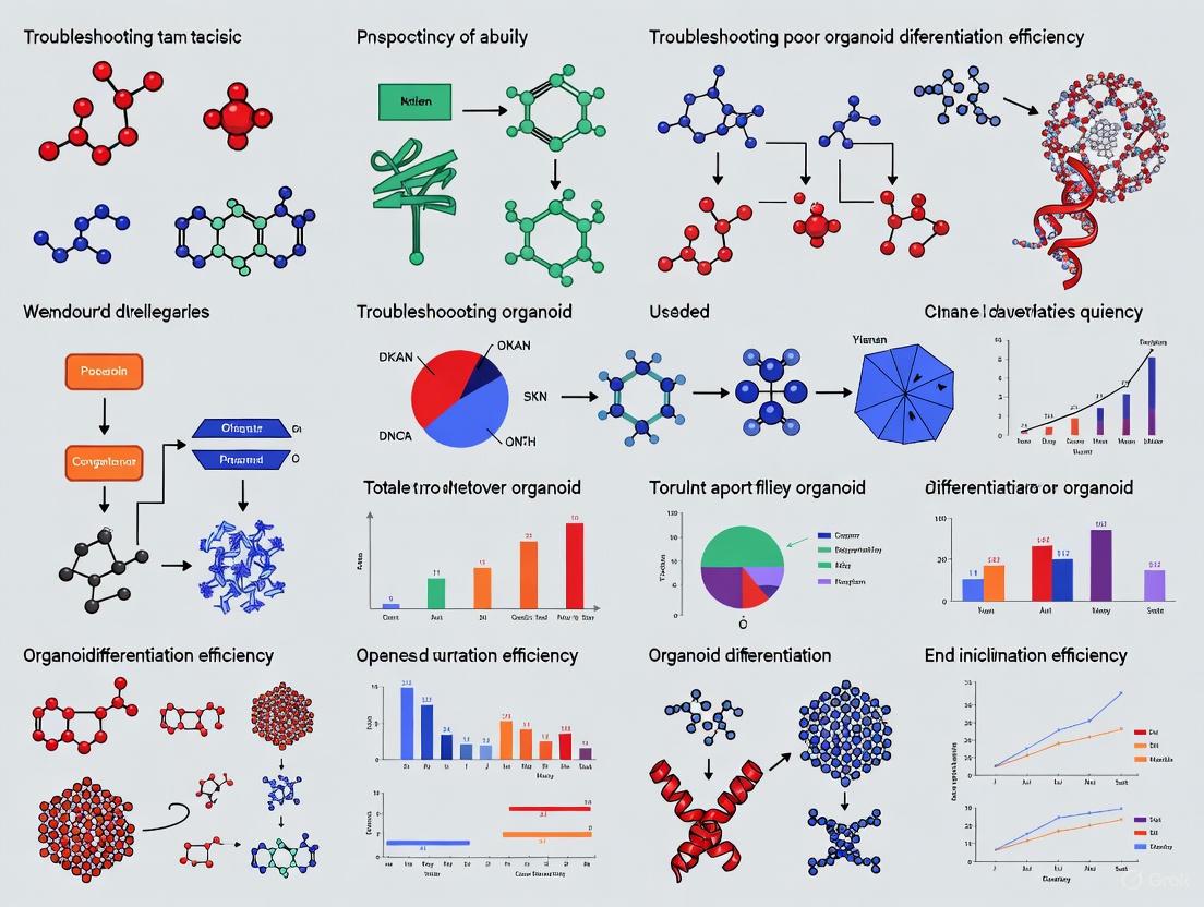
Troubleshooting Poor Organoid Differentiation Efficiency: A Researcher's Guide to Optimized Protocols and Assay Reproducibility
This article provides a comprehensive guide for researchers and drug development professionals facing challenges with organoid differentiation efficiency.
Troubleshooting Poor Organoid Differentiation Efficiency: A Researcher's Guide to Optimized Protocols and Assay Reproducibility
Abstract
This article provides a comprehensive guide for researchers and drug development professionals facing challenges with organoid differentiation efficiency. It explores the fundamental principles of stem cell self-renewal and differentiation, examines advanced culture methodologies and technological integrations, details systematic troubleshooting for common pitfalls like necrosis and low cellular diversity, and outlines robust validation frameworks. By synthesizing the latest 2025 research, this resource aims to equip scientists with practical strategies to enhance differentiation protocols, improve model physiological relevance, and increase the predictive power of organoid-based applications in disease modeling and drug screening.
Understanding Organoid Differentiation: Principles, Challenges, and Cellular Complexity
Troubleshooting Guide: Poor Organoid Differentiation Efficiency
Encountering poor differentiation efficiency is a common challenge in organoid research. The table below outlines frequent issues, their root causes, and evidence-based solutions to help you restore the balance between self-renewal and differentiation in your cultures.
| Problem Phenomenon | Potential Root Cause | Recommended Solution | Key Signaling Pathways Involved |
|---|---|---|---|
| Low cellular diversity; absence of key functional cell types (e.g., Paneth cells, enterocytes). | Overly potent stem cell self-renewal signals, suppressing differentiation. | Shift balance using a combination of small molecules (e.g., TpC: Trichostatin A, Vitamin C, CP673451) to enhance stemness and subsequent differentiation potential [1] [2]. | Wnt, Notch, BMP [1] [2] [3] |
| Limited proliferative capacity after induction of differentiation. | Over-driving differentiation depletes the progenitor/stem cell pool. | Apply reversible modulation. Use BET inhibitors to shift balance towards enterocyte lineage with enhanced proliferation [1] [2]. | Wnt, Notch [1] [3] |
| High organoid-to-organoid variability in size, structure, and cell composition. | Lack of controlled, homogeneous culture environment; inconsistent stem cell starting population. | Standardize organoid size and shape using quick reaggregation methods. Use a defined, optimized basal condition for homogeneous organoids [1] [4] [2]. | BMP (for retinal fate) [4] |
| Inefficient differentiation initiation; organoids remain in a progenitor state. | Incorrect or suboptimal concentration of morphogenic factors. | Perform concentration screening of key factors. Use low concentrations of factors like Angiogenin-4 to promote Lgr5+ intestinal stem cell growth, while high concentrations induce cell death [5]. | Depends on target lineage |
| Precancerous progression or abnormal growth in organoid cultures. | Dysregulation of stem cell self-renewal by external factors, such as microbial metabolites. | Investigate and modulate environmental cues. For cervical organoids, D-lactic acid (from Lactobacilli) suppresses growth via the PI3K-AKT pathway and YAP1 [6]. | PI3K-AKT, YAP1 [6] |
Frequently Asked Questions (FAQs)
Q1: Why is achieving a balance between self-renewal and differentiation so challenging in human organoid systems?
In vivo, this balance is maintained by precise spatial and temporal signaling gradients within the stem cell niche, which are difficult to replicate in a homogeneous culture dish. Conventional culture systems are often optimized for either expansion (high self-renewal, low differentiation) or differentiation (high heterogeneity, low proliferation), requiring separate steps that hinder scalability and reproducibility [1] [2]. The intrinsic plasticity of stem cells, where they can dynamically switch between states, adds another layer of complexity [1].
Q2: Beyond the factors in the table, what other key signals should I check if my intestinal organoids lack Paneth cells?
Paneth cell generation is particularly sensitive to culture conditions. While the TpC condition has proven effective [1] [2], you should also investigate IL-22 signaling. Previous research has used IL-22 to induce Paneth cells, though this can come at the cost of inhibited organoid growth [1] [2]. Ensure that your basal culture condition does not include factors like SB202190, Nicotinamide, or PGE2, which have been shown to impede the generation of secretory cell types [1] [2].
Q3: How can I confirm that the observed cellular changes in my organoids are due to successful differentiation and not cellular stress or death?
- Use Multiple Markers: Confirm differentiation using immunostaining for multiple, specific terminal differentiation markers (e.g., MUC2 for goblet cells, CHGA for enteroendocrine cells, ALPI for enterocytes) [1] [2].
- Functional Assays: Where possible, employ functional tests. For example, in intestinal organoids, the presence of dark granules in budding structures can indicate Paneth-like cell differentiation [1] [2].
- Track Dynamics: Utilize live-cell imaging and reporter systems (e.g., LGR5-mNeonGreen) to longitudinally track the loss and re-emergence of stem cell markers, which is indicative of dynamic differentiation and dedifferentiation processes [1] [2].
Q4: Our lab is new to organoid culture. What is a robust starting protocol for generating human intestinal organoids with good differentiation?
A highly optimized protocol for human small intestinal organoids (hSIOs) involves using a basal condition containing EGF, the BMP inhibitor Noggin (or DMH1), R-Spondin1, CHIR99021 (a Wnt agonist), A83-01 (an ALK inhibitor), IGF-1, and FGF-2. This is then supplemented with the TpC combination (Trichostatin A, 2-phospho-L-ascorbic acid, CP673451) to enhance stemness and subsequent differentiation under a single culture condition [1] [2]. This system has demonstrated high proliferative capacity and increased cellular diversity.
Experimental Protocol: Enhancing Differentiation in Human Intestinal Organoids
This detailed protocol is adapted from recent studies to establish intestinal organoids with a superior balance of self-renewal and differentiation [1] [2].
Objective
To generate human intestinal organoids (hSIOs) with high proliferative capacity and increased cellular diversity in a single culture condition using a combination of small molecule pathway modulators.
Materials
- Basal Medium: Advanced DMEM/F12, supplemented with GlutaMAX, HEPES, and Penicillin/Streptomycin.
- Essential Growth Factors:
- EGF (Epidermal Growth Factor)
- R-Spondin 1
- IGF-1 (Insulin-like Growth Factor 1)
- FGF-2 (Fibroblast Growth Factor 2)
- Small Molecule Inhibitors & Modulators:
- DMH1 (BMP inhibitor) or Noggin
- CHIR99021 (GSK-3β inhibitor, Wnt agonist)
- A83-01 (ALK inhibitor, TGF-β inhibitor)
- TpC Cocktail:
- Trichostatin A (TSA, HDAC inhibitor)
- 2-phospho-L-ascorbic acid (pVc, Vitamin C derivative)
- CP673451 (CP, PDGFR inhibitor)
- Matrix: Growth Factor Reduced Matrigel, or equivalent basement membrane extract.
- Dissociation Reagent: Gentle Cell Dissociation Reagent or Accumax [7] [8].
Method
Organoid Establishment and Expansion:
- Seed dissociated human intestinal crypts or single cells in Matrigel domes.
- Culture with basal intestinal organoid growth medium (e.g., containing EGF, Noggin, R-Spondin-1) for initial establishment and expansion.
- Passage organoids every 7-10 days using gentle dissociation reagent to maintain cultures.
Optimized Culture for Balance (TpC Condition):
- After passaging, switch to the optimized differentiation medium.
- Prepare the medium by supplementing the basal medium with the full set of growth factors (EGF, R-Spondin 1, IGF-1, FGF-2) and small molecules (DMH1, CHIR99021, A83-01).
- Crucially, add the TpC cocktail (Trichostatin A, 2-phospho-L-ascorbic acid, and CP673451) to the medium.
- Refresh the medium every 2-3 days.
Monitoring and Analysis:
- Proliferation: Monitor organoid growth and budding morphology over 7-14 days. Assess colony-forming efficiency from single cells and total cell count [1] [2].
- Differentiation: After 14-21 days, analyze organoids for diverse cell types.
- Immunofluorescence Staining: Fix organoids and stain for:
- Stem cells: LGR5, OLFM4
- Enterocytes: ALPI (Intestinal Alkaline Phosphatase)
- Goblet cells: MUC2 (Mucin 2)
- Enteroendocrine cells: CHGA (Chromogranin A)
- Paneth cells: LYZ (Lysozyme), DEFA5 (Defensin Alpha 5)
- Functional Assessment: Observe for crypt-like budding structures and the presence of Paneth-like cells with dark granules [1] [2].
- Immunofluorescence Staining: Fix organoids and stain for:
Signaling Pathways Governing the Balance
The core balance between self-renewal and differentiation is orchestrated by a handful of conserved signaling pathways. The diagram below illustrates how these pathways interact to determine cell fate.
The Scientist's Toolkit: Essential Reagents for Balance
This table provides a curated list of key reagents used to manipulate the core pathways for controlling self-renewal and differentiation in organoid research.
| Reagent Category | Example(s) | Primary Function in Organoid Culture |
|---|---|---|
| Wnt Pathway Agonists | CHIR99021, R-Spondin 1 | Promotes stem cell self-renewal and proliferation by stabilizing β-catenin [1] [2] [3]. |
| Notch Pathway Inhibitors | Dibenzazepine (DBZ) | Induces secretory lineage differentiation (e.g., goblet cells); decreases clonogenic potential of stem cells [3]. |
| BMP Pathway Inhibitors | Noggin, DMH1 | Promotes epithelial growth and is essential for establishing organoid cultures by counteracting differentiation signals [1] [2]. |
| Epigenetic Modulators | Trichostatin A (TSA, HDAC inhibitor) | Enhances stem cell stemness, which subsequently amplifies differentiation potential and cellular diversity [1] [2]. |
| ROCK Inhibitors | Y-27632 | Improves survival of single cells and dissociated organoids by inhibiting apoptosis, crucial for passaging and cloning [7] [8]. |
| Metabolic Modulators | 2-phospho-L-ascorbic acid (Vitamin C) | Serves as an antioxidant and co-factor for enzymes, supporting stem cell fitness and differentiation in combination therapies [1] [2]. |
Frequently Asked Questions (FAQs)
Q1: What are the primary consequences of poor vascularization in organoids? Poor vascularization limits the delivery of oxygen and nutrients and removal of metabolic waste, restricting organoid growth, causing central necrosis, and impairing maturation and physiological relevance [9] [10] [11]. This ultimately affects the accuracy of disease modeling and drug screening applications.
Q2: How can I prevent the formation of a necrotic core in my organoid cultures? The most direct method is to reduce the initial cell number to generate smaller, necrotic core-free organoids, which avoids diffusion limits and associated complications for large-scale screening [12]. Alternative approaches include promoting vascularization, or slicing larger organoids into smaller sections [12].
Q3: What are the main sources of batch-to-batch variability in organoid research? Variability arises from multiple sources, including: the inherent stochasticity of stem cell self-organization; differences in genetic background between cell lines; subtle variations in culture conditions, differentiation protocols, and reagents (e.g., extracellular matrix batches); and evolving epigenetic differences even between isogenic subclones [13] [14].
Q4: What strategies can minimize variability and improve reproducibility? Strategies include using a large panel of isogenic cell lines to average out line-to-line variation, implementing standardized and robust protocols, employing organoid array technologies for parallel processing and analysis, and transitioning to genetically more stable cell line-derived organoids for specific applications like high-throughput screening [15] [13] [14].
Q5: Are there methods to introduce functional vascular networks into existing organoid protocols? Yes, several main approaches exist:
- Co-culture with Endothelial Cells: Co-culturing organoids with human umbilical vascular endothelial cells (HUVECs) or endothelial cells derived from induced pluripotent stem cells (iPSCs), often with supportive growth factors, encourages the self-assembly of capillary-like structures within the organoid [9] [16].
- 3D Bioprinting: This allows the precise patterning of endothelial cells and supportive bioinks to create predefined vascular channel networks within tissue constructs [9] [10] [16].
- Organ-on-a-Chip Technologies: Microfluidic devices can be used to perfuse organoids, subject them to mechanical cues, and model the function of vascular barriers like the blood-brain barrier [16].
Troubleshooting Guides
Troubleshooting Necrotic Cores
Necrotic cores form when organoids exceed the diffusion limit for oxygen and nutrients (typically ~150-200 µm) [10].
Table: Identifying and Resolving Necrotic Core Formation
| Observed Problem | Potential Cause | Recommended Solutions |
|---|---|---|
| Central cell death in large organoids; positive staining for necrotic markers. | Organoid size has exceeded the natural diffusion limit for oxygen and nutrients. | 1. Reduce initial seeding density. Generate smaller, necrotic core-free organoids [12].2. Induce vascularization. Integrate a vascular network to support larger tissues [9] [10].3. Use organoid slicing. Physically section mature organoids to improve nutrient access [12]. |
| Viability issues even in smaller organoids. | Inefficient culture conditions or incorrect matrix composition. | 1. Optimize protocol. Ensure medium is changed frequently and organoids are properly embedded in ECM [17].2. Validate viability. Use Live/Dead assays (e.g., PI/AO staining) for quantitative assessment [12]. |
Troubleshooting Batch Variability
High variability can confound experimental results and reduce the statistical power of studies.
Table: Identifying and Mitigating Batch Variability
| Observed Problem | Potential Cause | Recommended Solutions |
|---|---|---|
| Inconsistent differentiation outcomes and cell type proportions between batches. | Inherent variability of stem cell self-organization; minor fluctuations in protocol execution. | 1. Increase sample size. Use large N-numbers to power through variability [15] [14].2. Implement batch processing. Use array technologies to process and analyze many organoids in parallel under identical conditions [15].3. Use defined reagents. Employ animal-free, recombinant growth factors to minimize lot-to-lot variability [16]. |
| Significant phenotypic differences between isogenic lines. | Epigenetic drift and clonal variation in iPSC lines, even within isogenic subclones. | 1. Use multiple lines. Employ several isogenic trisomic and disomic subclones to distinguish true trisomy effects from clonal variation [14].2. Consider alternative models. For high-throughput screening, use genetically more stable cell line-derived organoids [13]. |
Experimental Protocol: Generation of Necrotic Core-Free Spinal Cord Organoids
This protocol is adapted from a study that established a model for fetal neural ischemia using human spinal cord organoids (nf-hSCOs) free of necrotic cores [12].
Objective: To generate human spinal cord organoids of a controlled, small size that prevents the development of a necrotic core, making them suitable for large-scale screening assays.
Key Materials:
- Cell Line: H9 human pluripotent stem cells (hPSCs)
- Key Reagents:
- Matrigel-coated plates
- mTeSR1 medium
- Differentiation Medium (DM): DMEM/F12, 1% N2, 2% B27, 1% Penicillin/Streptomycin, 0.1% β-mercaptoethanol
- Small Molecules: SB431542 (10 µM), CHIR99021 (3 µM)
- Growth Factors: basic Fibroblast Growth Factor (bFGF, 20 ng/mL)
- Rock inhibitor (Y-27632)
- Retinoic Acid (RA, 0.1 µM)
- 96-well low attachment plates
Workflow:
- Culture hPSCs: Maintain H9 hPSCs on Matrigel-coated plates in mTeSR1 medium.
- Induce Caudal Neural Stem Cells (cNSCs): Treat high-density hPSCs clusters with 10 µM SB431542 and 3 µM CHIR99021 in DM for 3 days.
- Dissociate and Seed: Dissociate the resulting cNSCs into single cells using Accutase. Seed exactly 75 cells per well into a 96-well low attachment plate in DM supplemented with 20 ng/mL bFGF and 10 µM Rock inhibitor (day 1 only).
- Form 3D Aggregates: Culture for 4 days to allow 3D aggregate formation, with daily bFGF treatment.
- Pattern and Mature: After 4 days, culture the aggregates in DM with 0.1 µM RA (without bFGF) for 6 days, changing the medium every other day.
- Long-term Maturation: Transfer the organoids to a maturation medium (a 1:4 mix of DMEM/F12 and Neurobasal medium, supplemented with B27, Glutamax, and 0.1 µM RA), changing the medium every 4 days.
Validation:
- Viability Assay: Use a propidium iodide (PI) / acridine orange (AO) live/dead assay. After 1 hour of incubation with PI/AO, image the organoids. The absence of a red (PI+) core indicates a lack of necrosis [12].
- Immunostaining: Section and immunostain the organoids for relevant neural markers (e.g., SOX2, TUJ1) to confirm spinal cord identity and assess cellular heterogeneity.
Signaling Pathways and Workflows
Technical Hurdles and Solution Pathways
This diagram summarizes the core technical challenges covered in this guide and the primary strategies being developed to overcome them.
The Scientist's Toolkit: Research Reagent Solutions
Table: Essential Reagents for Addressing Organoid Technical Hurdles
| Reagent / Material | Function / Application | Example Use Case |
|---|---|---|
| Human Umbilical Vein Endothelial Cells (HUVECs) | Co-culture component for inducing vasculogenesis and forming capillary-like networks within organoids. | Vascularization of brain organoids to enhance growth and mimic native brain vasculature patterns [16]. |
| Rock Inhibitor (Y-27632) | Improves cell survival after dissociation and seeding by inhibiting apoptosis; critical for initial aggregate formation. | Added to medium on the first day of seeding single cells into low-attachment plates to support the formation of 3D organoid aggregates [12] [17]. |
| Recombinant Growth Factors (VEGF, FGF-2, BMP4) | Key signaling molecules to guide differentiation and promote vascular development. | A cocktail of VEGF, FGF-2, and BMP4 is used alongside endothelial cells to promote the formation of a penetrating vascular network in brain organoids [16]. |
| Extracellular Matrix (ECM / Matrigel) | Provides a 3D scaffold that mimics the native basement membrane, supporting cell organization, polarity, and signaling. | Used to embed organoids as "dome" cultures and for co-culture experiments with endothelial cells [17] [16]. |
| Low Attachment Plates | Prevents cell adhesion to the plastic surface, forcing cells to aggregate and form 3D structures. | Essential for the initial formation and subsequent maintenance of free-floating organoid cultures [12]. |
| Animal-Free / Recombinant Proteins | Defined, consistent-quality reagents that reduce batch-to-batch variability compared to serum-containing or animal-derived products. | Using recombinant growth factors for organoid cultures to greatly improve reproducibility [16]. |
Welcome to the Organoid Technology Support Center. A critical factor determining the success of your organoid research is the choice of starting material: pluripotent stem cells (PSCs), including embryonic (ESCs) and induced pluripotent stem cells (iPSCs), or adult stem cells (ASCs), also known as patient-derived organoids (PDOs). This guide provides a detailed, troubleshooting-focused comparison of these two cell sources to help you diagnose and resolve issues related to differentiation efficiency, protocol selection, and final organoid maturity.
FAQ: Core Concepts and Selection
1. What is the fundamental difference between PSC-derived and ASC-derived organoids? PSC-derived organoids are generated from cells that can differentiate into any cell type from the three germ layers. They are particularly valuable for modeling early human development and organs that are difficult to access, such as the brain and retina [18] [19]. In contrast, ASC-derived organoids are generated directly from dissociated healthy or diseased patient tissues. They faithfully recapitulate the original tissue's phenotype, making them exceptional for personalized medicine applications [18].
2. I am new to organoid culture. Which starting material is easier to use? For researchers seeking a more straightforward protocol that consistently reproduces the original tissue phenotype, ASC-derived organoids are often the better starting point. Their protocols generally require fewer steps and less time overall compared to PSC-derived methods [19]. However, if your target organ is inaccessible (e.g., brain) or you require a complex, multi-lineage structure, PSCs are the necessary choice, despite a more complex protocol.
3. My PSC-derived organoids seem immature. Is this a common issue? Yes. A typical challenge with PSC-derived organoids is that they very rarely reach an adult tissue stage in vitro and usually resemble fetal-stage tissues [19]. This can be due to extended culture time requirements beyond current methodology limits or the absence of crucial interactions with other co-developing cell types that are missing in vitro [19].
Troubleshooting Guides
Problem 1: Poor Efficiency in Early Differentiation Stages
Symptoms: Failure to form proper embryoid bodies (EBs) or neural rosettes; inconsistent emergence of progenitor cell zones.
Diagnosis and Solutions:
This problem is predominantly linked to cell line-specific differences when working with PSCs [20] [21]. Variations in differentiation efficiency between different human ES and iPS cell lines are a major factor in early success [20].
- Action 1: Optimize EB Formation. The method used to generate EBs is critical. Evidence from retinal organoid studies shows that a mechanical formation method under static conditions, supplemented with the ROCK inhibitor Y-27632 for the first 48 hours of differentiation, yields the most consistent results [20] [21]. Using low-attachment U-bottom plates also promotes consistent spheroid formation [22].
- Action 2: Critically Review Your Cell Line. If you are consistently facing early differentiation failure, consider switching to a different PSC line that has a proven track record of differentiating toward your germ layer of interest (ectoderm, mesoderm, endoderm). Do not assume all PSC lines are equivalent.
Table 1: Troubleshooting Early-Stage Differentiation Issues
| Symptom | Possible Cause | Solution |
|---|---|---|
| Low cell survival after dissociation to single cells for EB formation | Apoptosis due to dissociation | Supplement culture medium with a ROCK inhibitor (e.g., Y-27632) for the first 48-72 hours post-dissociation [20] [22]. |
| Inconsistent or failed EB formation | Cell attachment to culture vessel | Use low-attachment, U-bottom spheroid microplates to promote consistent self-aggregation [22]. |
| High variability in differentiation onset between experiments | Cell line-specific inherent differences | Select a PSC line with published efficiency for your target organoid; perform small-scale pilot differentiations to test new lines [20]. |
Problem 2: Failure in Organoid Maturation and Function
Symptoms: Organoids lack mature cell markers; absence of electrophysiological activity in neural organoids; no evidence of complex, laminated structures (e.g., in retinal organoids).
Diagnosis and Solutions:
While early stages are dominated by cell line choice, later differentiation and maturation are determined by the EB generation method and maintenance conditions [20] [21].
- Action 1: Re-evaluate Your 3D Culture Environment. For many organoid types, encapsulation in an extracellular matrix (ECM) like Matrigel or Geltrex is essential for structural maturation. For example, neural induction of EBs is often followed by encapsulation in a reduced-growth-factor basement membrane extract to support the formation of complex neuroepithelial structures [22].
- Action 2: Address Core Maturation Limitations. PSC-derived organoids often remain in a fetal-like state. To push maturation, consider:
- Extended Culture Times: Maturation can require many weeks or months in culture [22].
- Metabolic Maturation: For liver organoids, treatment with short-chain fatty acids has been shown to improve metabolic maturation, enabling better assessment of drug toxicity [23].
- Slice Culture: For large neural organoids, growing them as slice cultures instead of solid spheres can improve oxygen and nutrient perfusion to the core, reducing necrosis and supporting healthier maturation [24].
Table 2: Key Signaling Molecules and Their Functions in Organoid Culture
| Research Reagent | Function in Organoid Culture | Common Applications |
|---|---|---|
| Y-27632 (ROCK inhibitor) | Promotes cell survival after dissociation; aids in embryoid body formation [20] [25] [22]. | Standard in many initial PSC dissociation and EB formation protocols. |
| R-spondin 1 | Activator of Wnt signaling; crucial for stem cell self-renewal [25]. | Essential for ASC-derived organoids (intestine, liver); used in PSC-derived endodermal lineages. |
| Noggin | BMP pathway inhibitor; prevents differentiation toward non-target lineages [25]. | Key component in intestinal, cerebral, and other organoid media. |
| CHIR-99021 | GSK-3 inhibitor; activates Wnt signaling pathway [19]. | Used to maintain stem cells in a self-renewing state; directs mesendodermal differentiation. |
| A 83-01 (ALK5 inhibitor) | TGF-β pathway inhibitor; prevents differentiation and supports proliferation [25]. | Common in gastrointestinal and hepatic organoid cultures. |
| Recombinant Human EGF | Epithelial tissue growth factor; induces proliferative changes [25]. | Used in gastrointestinal, liver, thyroid, and brain organoid media. |
Problem 3: Choosing the Wrong Starting Material for Your Research Goal
Symptoms: The organoid model does not adequately answer your research question (e.g., lacks disease relevance, cannot model development, is missing key cell types).
Diagnosis and Solutions:
Selecting the appropriate stem cell source is paramount. This decision should be driven by your specific research objective.
- Action 1: Use the Following Workflow Diagram. To systematically determine the best starting material for your project, follow the logic below.
Table 3: Direct Comparison of PSC vs. ASC-Derived Organoids
| Parameter | Pluripotent Stem Cell (PSC) Organoids | Adult Stem Cell (ASC) Organoids |
|---|---|---|
| Developmental Stage | Typically model fetal-stage tissues; maturation is a key challenge [19]. | Model adult tissue phenotypes more consistently [18]. |
| Protocol Complexity | More complex, multi-step protocols requiring longer time [19]. | Generally simpler, fewer steps, less time overall [19]. |
| Cellular Complexity | Can form organoids with multiple lineage cells (e.g., epithelial and mesenchymal), closer to physiological status [19]. | Usually contain mainly organ-specific epithelial cells and stem cells [19]. |
| Genetic Fidelity | Retain genetic information of the donor; can model genetic disorders [18]. | Faithfully recapitulate patient-specific disease states and tumor phenotypes [18] [23]. |
| Primary Applications | Studying organogenesis, early development, genetic disorders [18] [19]. | Personalized medicine, drug screening, disease modeling (especially cancer) [18] [23]. |
Essential Protocols and Methodologies
Key Experimental Workflow: Generating Neural Organoids from PSCs
The following workflow, adapted from established protocols, outlines the critical steps for generating neural organoids [22]. Issues can arise at any of these stages.
Critical Steps for Success:
- EB Formation: Use a validated dissociation reagent (e.g., Accutase, TrypLE) and include a ROCK inhibitor like Y-27632 to significantly improve EB formation efficiency and cell survival [22].
- Neural Induction: Successful induction is often visible morphologically, with EBs forming a bright "ring" structure [22].
- Maturation: Transferring organoids to an orbital shaker during extended culture improves nutrient and oxygen exchange, promoting healthier growth and reducing core necrosis [22].
Key Experimental Workflow: Initiating ASC-Derived Organoid Culture
This protocol summarizes the standard embedded culture method for ASC-derived organoids, such as those from intestine or liver [17] [25].
- Tissue Processing: Healthy or diseased tissue samples are processed into a single-cell suspension or small fragments [17].
- Embedding in ECM: The cells/fragments are resuspended in a liquid extracellular matrix (e.g., Matrigel, Cultrex BME) and dispensed as domes in the center of a culture plate well. The ECM is allowed to polymerize at 37°C to form a solid gel [17] [25].
- Expansion with Tissue-Specific Medium: The ECM dome is overlaid with a complex medium containing a specific cocktail of growth factors, small molecules, and supplements that mimic the stem cell niche of the original tissue (e.g., including EGF, Noggin, R-spondin for intestine) [17] [25].
- Passaging: Organoids are expanded by mechanically and/or enzymatically dissociating the cultures and re-embedding the fragments into new ECM domes [17].
Frequently Asked Questions (FAQs)
Q1: What are the most critical metrics to confirm my organoids have successfully differentiated? A robust assessment requires a multi-parameter approach combining morphological, cellular, and functional data. You should routinely evaluate:
- Morphology: Use brightfield and high-content imaging to monitor for the emergence of expected 3D structures, such as budding, lumen formation, or tissue-specific architecture [4] [7].
- Gene Expression: Perform qPCR to quantify the expression of key lineage-specific markers. A successful differentiation should show upregulation of target genes and a corresponding downregulation of pluripotency markers (e.g., Oct4, Sox2) [26] [27].
- Protein Expression: Use immunohistochemistry (IHC) or immunofluorescence (IF) to validate the presence and spatial localization of key proteins, confirming the correct cellular composition and polarity [7] [28].
- Functional Assays: Employ tissue-specific functional tests, such as the presence of polarized transporters, electrophysiological activity, or the production of tissue-specific metabolites [7].
Q2: My differentiation efficiency is low and variable between batches. What are the main culprits? Low and variable efficiency often stems from inconsistencies in the initial differentiation signals or cell quality. Key areas to troubleshoot include:
- Cell Density and Health: Ensure you start with a consistent, high density of healthy, high-quality pluripotent stem cells. Excessive cell death at the initial stage can drastically reduce yield [27].
- Signaling Molecule Activity: Critically evaluate the quality and concentration of your growth factors and small molecules (e.g., CHIR99021). Use validated lots and confirm the activity of essential components like Wnt3a, which can be a major source of variability if not sourced or titrated properly [26].
- Protocol Adherence: Follow differentiation protocols meticulously, as the timing of growth factor addition and withdrawal is critical for precise lineage specification [4] [26].
Q3: How can I tell if my organoids are mature and functionally competent, not just differentiated? Maturity is a key challenge. Beyond marker expression, look for evidence of complex functionality and multi-cellular composition.
- Multi-cellularity: Confirm the presence of multiple, expected cell types within the organoid through single-cell RNA sequencing or multi-label IHC. For example, a mature kidney organoid should contain proximal tubules, podocytes, and distal tubules [27].
- Advanced Function: Move beyond basic viability assays. For respiratory organoids, this could mean demonstrating ciliary beating or mucus production; for kidney organoids, it could involve assessing albumin uptake or electrolyte transport [7].
- Benchmarking: Compare your organoid's transcriptomic or proteomic profile to datasets from native human tissue or primary cells to assess how closely they resemble the in vivo counterpart [27].
Q4: What tools are available for high-throughput, quantitative assessment of differentiation? The field is moving towards automation and standardization to improve quantification.
- High-Content Imaging Systems: Platforms like the ImageXpress Confocal HT.ai can automatically capture and analyze organoid size, shape, and marker expression in 96- or 384-well plates [28] [29].
- Automated Cell Culture: Automated workstations (e.g., Biomek i-Series, CellXpress.ai) can handle media changes and passaging, reducing manual error and improving batch-to-batch consistency [29].
- Flow Cytometry: Use systems like the CytoFLEX flow cytometer to quantitatively analyze the percentage of cells expressing specific differentiation markers in a dissociated organoid population [29].
Troubleshooting Guides
Issue 1: Poor Initial Differentiation Efficiency
This occurs when the majority of cells fail to commit to the target lineage at the early stages of differentiation.
| Observation | Potential Cause | Solution(s) | Validation Experiment |
|---|---|---|---|
| High cell death upon induction with CHIR99021 or similar molecules | Apoptosis induced by differentiation stimulus; poor stem cell quality [27] | - Optimize concentration and duration of GSK-3β inhibitor (CHIR) treatment.- Pre-treat with ROCK inhibitor (Y-27632) to enhance survival.- Co-culture with monocytes/macrophages or use extracellular vesicles (EVs) to suppress apoptosis and promote survival [27]. | Perform a Cell Titer-Glo or CCK-8 assay to quantify viability 24-48 hours post-induction. Check cleaved Caspase-3 via western blot for apoptosis [27]. |
| Differentiation defaults to incorrect lineage (e.g., forebrain instead of retina) | Incorrect balance of signaling pathways; default fate not overridden [4] | - Precisely regulate BMP signaling activation to direct retinal vs. forebrain fate.- Titrate the concentration of key morphogens like Noggin.- Use quick reaggregation methods to standardize organoid size and shape, improving reproducibility [4]. | Use qPCR at an early timepoint (e.g., day 4-8) to check for the expression of early lineage markers (e.g., TBX6 for mesoderm, PAX6 for neural ectoderm) [4] [27]. |
| High variability between replicates and cell lines | Inconsistent cell seeding density; variable activity of growth factors [26] | - Standardize initial cell seeding density using automated cell counters (e.g., Vi-CELL BLU).- Use conditioned media as a consistent source of Wnt3a instead of recombinant protein alone [26]. | Image organoids daily to track growth and morphology. Use software to quantify the coefficient of variation in organoid size across wells. |
Issue 2: Incomplete or Stalled Maturation
Organoids initiate differentiation but fail to develop the complex structures and functional properties of mature tissue.
| Observation | Potential Cause | Solution(s) | Validation Experiment |
|---|---|---|---|
| Lack of expected, complex cellular diversity (e.g., only one cell type present) | Differentiation signals not withdrawn or switched at the correct time; over-proliferation of progenitor cells [26] | - Systematically remove growth factors (e.g., Wnt, R-spondin, Noggin) to induce differentiation. For colon organoids, combined removal yields optimal results [26].- Introduce pro-maturation factors specific to your tissue type. | Perform single-cell RNA sequencing to map the full cellular repertoire and compare to in vivo data. Use multi-color IF to visualize multiple cell types simultaneously. |
| Organoids remain small and simple, failing to form intricate structures | Suboptimal 3D culture matrix; insufficient morphogen gradients | - Use a defined, engineered hydrogel instead of standard Matrigel to provide better control over mechanical and biochemical cues [30].- Incorporate microfluidic devices (Organs-on-Chips) to create controlled nutrient and morphogen gradients [30] [28]. | Use confocal microscopy (e.g., Leica Stellaris) to visualize 3D structure and quantify features like branching or lumen formation in 3D. |
| Low expression of terminal differentiation markers and functional proteins | Culture conditions do not support full functional maturation; immature transcriptomic profile | - Extend the maturation time in culture.- Use air-liquid interface culture or mechanical stimulation (e.g., flow, stretch) where appropriate.- Co-culture with stromal or immune cells (e.g., monocytes) to provide missing niche signals [27]. | Conduct a functional assay: calcium imaging for neurons, albumin uptake for kidney tubules, or cAMP assay for GPCR function in various organoids. |
Quantitative Assessment Tables
Table 1: Key Molecular Markers for Assessing Organoid Differentiation
This table lists common markers used to validate the identity and composition of various organoid types.
| Organoid Type | Pluripotency Marker | Early Progenitor Marker | Terminal Cell Type Markers | Key Functional Readout |
|---|---|---|---|---|
| Retinal [4] | Oct4, Sox2 | Pax6, Rx | Crx (photoreceptors), RHO (opsin), Recoverin (neurons) | Responsiveness to light stimuli |
| Kidney [27] | Oct4, Sox2 | OSR1, TBX6, PAX2 | Nephrin (podocytes), E-Cadherin (tubules), AQP1 (tubules) | Albumin uptake, formation of glomerulus-like structures |
| Intestinal/Colon [26] | Oct4, Sox2 | LGR5 (stem cells) | MUC2 (goblet cells), Chromogranin A (enteroendocrine), Sucrase-Isomaltase (enterocytes) | Barrier integrity (TEER), alkaline phosphatase activity |
| Lung/Airway [7] | Oct4, Sox2 | NKX2.1, SOX2, SOX9 | SFTPC (AT2 cells), MUC5AC (goblet cells), Acetyl-α-Tubulin (ciliated cells) | Ciliary beating, mucus production, ACE2 expression |
Table 2: Comparison of Tissue Dissociation Methods for Analysis
The method used to dissociate organoids for downstream analysis (e.g., flow cytometry, scRNA-seq) can impact results.
| Parameter | Mechanical Dissociation | Enzymatic Digestion |
|---|---|---|
| Principle | Physical disruption (pipetting, chopping) [31] | Chemical breakdown of ECM (collagenase, Accutase) [7] [31] |
| Pros | - Better preserves surface proteins for flow cytometry- Maintains more native cell clusters and TME components [31] | - Generates a more homogeneous single-cell suspension- Higher efficiency and yield for many tissues- Better for reproducible, high-throughput applications [31] |
| Cons | - Can be harsher, reducing cell viability- May yield incomplete dissociation, clogging instruments [31] | - Can cleave surface epitopes, affecting antibody binding- Over-digestion can damage cells and alter biology [7] [31] |
| Best For | - Analysis where cell surface marker integrity is critical- Preserving niche interactions [31] | - Single-cell RNA sequencing- Applications requiring high, consistent cell yield [31] |
Experimental Workflow & Signaling Pathways
Diagram 1: Generalized Organoid Differentiation and Assessment Workflow
This diagram outlines the key stages in generating and validating organoids, with critical quality control checkpoints.
Diagram 2: Core Signaling Pathways in Lineage Specification
This diagram simplifies the key signaling pathways involved in the early fate decisions of differentiating organoids, highlighting common targets for troubleshooting.
The Scientist's Toolkit: Essential Reagents & Materials
| Item | Function / Application | Example / Note |
|---|---|---|
| Extracellular Matrix (ECM) | Provides a 3D scaffold for organoid growth and self-organization. | Matrigel, Geltrex, BME; defined synthetic hydrogels are emerging for better control [32] [30]. |
| ROCK Inhibitor (Y-27632) | Enhances single-cell survival after passaging and thawing by inhibiting apoptosis. | Critical for initial plating of dissociated cells [7] [27]. |
| GSK-3β Inhibitor (CHIR99021) | Activates Wnt signaling by stabilizing β-catenin; used for mesoderm and endoderm lineage specification. | Concentration and duration are critical and must be optimized for each cell line and target tissue [27]. |
| Recombinant Growth Factors | Provide precise signals for proliferation and differentiation. | Wnt3a, R-spondin, Noggin, EGF, FGFs. Use consistent, high-quality sources to minimize variability [26]. |
| Conditioned Media | A cost-effective and often more consistent source of multiple growth factors. | e.g., Wnt3a-conditioned media for more robust support of intestinal organoids than recombinant protein alone [26]. |
| Small Molecule Inhibitors | Precisely block signaling pathways to direct cell fate. | A83-01 (TGF-β inhibitor), SB202190 (p38 MAPK inhibitor) [28]. |
| Digestion Enzymes | Dissociate tissues and organoids into single cells for passaging or analysis. | Accumax, Collagenase/Hyaluronidase, TrypLE; choice affects viability and surface protein integrity [7] [31]. |
| Cell Viability Assays | Quantify cell health and proliferation during differentiation. | CCK-8, ATP-based assays (e.g., Cell Titer-Glo) [27] [29]. |
Advanced Culture Systems and Protocols to Enhance Differentiation Outcomes
Achieving high differentiation efficiency in organoid cultures is a common challenge that can hinder research reproducibility and outcomes. The complex interplay of soluble cues—including growth factors, small molecules, and niche signals—dictates the delicate balance between stem cell self-renewal and lineage-specific differentiation. This guide addresses frequent issues and provides targeted troubleshooting strategies to help researchers control cell fate decisions in their organoid systems effectively.
FAQs and Troubleshooting Guides
FAQ 1: My organoids show poor cellular diversity and are mostly undifferentiated. What key signals should I check?
Answer: This issue often stems from an imbalance in core signaling pathways that maintain stemness versus those that initiate differentiation. You should systematically check your modulation of the Wnt, BMP, and EGF pathways.
- Check your Wnt signaling levels: Excessive Wnt activation can lock cells in a progenitor state. Ensure you are using the correct concentration and timing for Wnt agonists (like CHIR99021) and antagonists.
- Review BMP inhibition: The presence of the BMP inhibitor Noggin is often essential in expansion media to prevent premature differentiation. Verify its activity and concentration. However, for specific lineages like pit cells in gastric organoids, BMP4 signaling is required for differentiation [33].
- Confirm EGF activity: Epidermal Growth Factor (EGF) is a potent mitogen that supports the self-renewal of adult stem cell populations within organoids [34]. Its depletion or inhibition can induce quiescence and differentiation.
The table below summarizes the primary functions of these core factors:
Table: Core Growth Factors and Their Functions in Organoid Cultures
| Growth Factor / Signal | Primary Function in Organoids | Commonly Used In |
|---|---|---|
| R-spondin 1 (RSPO1) | Activates Wnt/β-catenin signaling; enhances stem cell self-renewal and expansion [34]. | Intestinal, gastric, hepatic, mammary organoids [34]. |
| Noggin | Inhibits BMP signaling; prevents differentiation and supports stemness [34]. | Intestinal, gastric, colonic organoids [34]. |
| Epidermal Growth Factor (EGF) | Induces proliferative signaling; supports self-renewal of adult stem cell populations [34]. | Gastrointestinal, liver, thyroid, brain organoids [34]. |
| BMP4 | Stimulates differentiation of specific lineages (e.g., pit cells, parietal cells) [33]. | Gastric organoids (differentiation phase) [33]. |
FAQ 2: How can I enhance the differentiation of a specific intestinal cell lineage, such as enterocytes or Paneth cells?
Answer: Directing differentiation toward a specific lineage requires a controlled shift in the equilibrium of cell fate, often achieved by combining small molecules with traditional growth factors.
- For General Enhanced Diversity: A combination of small molecules known as TpC (Trichostatin A, 2-phospho-L-ascorbic acid, and CP673451) has been shown to enhance the stemness of intestinal stem cells, which subsequently amplifies their differentiation potential. This leads to increased generation of enterocytes, goblet cells, enteroendocrine cells, and Paneth cells under a single culture condition without artificial gradients [1].
- For Enterocyte Lineage: Shifting the balance from secretory cell differentiation toward the enterocyte lineage can be achieved using BET inhibitors [1].
- For Paneth Cells: The cytokine IL-22 has been used to induce Paneth cell generation, but it may come at the cost of inhibited organoid growth [1].
FAQ 3: My gastric organoids fail to generate mature parietal or chief cells. How can I improve this?
Answer: Efficient differentiation of gastric epithelial cells, especially parietal and chief cells, is tightly regulated by BMP and EGF signaling.
- For Parietal Cell Differentiation: BMP4 signaling is a critical stimulant for parietal cell differentiation. Furthermore, combining BMP4 with the small molecule Isoxazole 9 has been shown to promote both parietal and enteroendocrine cell differentiation [33].
- For Chief Cell Differentiation: Inhibition of both TGF-β and BMP4 signaling promotes chief cell differentiation in gastric organoids [33].
- Synergistic Effects: BMP4 and EGF signaling cooperate to enhance the differentiation of pit cells [33], illustrating how pathways interact to fine-tune final cell fate.
The following diagram illustrates how manipulating these key signaling pathways directs cell fate decisions in gastric organoids:
FAQ 4: What are the critical initial steps for efficient kidney organoid differentiation from iPSCs?
Answer: The initial differentiation phase is the most critical for successful kidney organoid formation. Two parameters are paramount: initial cell density and the concentration of the WNT agonist (typically CHIR99021) [35].
- Challenge: Different iPSC lines show variances in cell proliferation and differentiation ability, making it necessary to optimize these parameters for each cell line.
- Solution: Researchers must perform titration experiments to determine the optimal initial cell density and CHIR concentration for their specific iPSC line. The optimal combination leads to iPSCs that rapidly expand and reach 90-100% confluence by day 7 of differentiation, forming aggregates that spontaneously generate nephron structures in suspension culture [35].
Table: Key Parameters for Kidney Organoid Differentiation from iPSCs
| Parameter | Impact on Differentiation | Troubleshooting Action |
|---|---|---|
| Initial Cell Density | Affects aggregate size, cell-cell contact, and patterning. | Test a range of cell densities during the initial seeding step. |
| WNT Agonist (CHIR) Concentration | Initiates the differentiation towards a renal lineage. | Titrate the concentration of CHIR99021 during the induction phase. |
| iPSC Line Variability | Different lines have varying proliferation and differentiation potentials. | Do not assume one protocol fits all; optimize for each new line. |
FAQ 5: How can I improve the reproducibility and efficiency of my retinal organoid differentiations?
Answer: Limitations in the efficiency and reproducibility of retinal organoid protocols are a known challenge. Two key strategies to address this are standardizing the size of the starting cell aggregates and timed activation of specific pathways.
- Standardize Organoid Size and Shape: Using quick reaggregation methods in concave microwells to control the size and shape of embryoid bodies (EBs) significantly improves reproducibility compared to traditional methods. For neuronal/retinal lineages, 500 µm microwells and the selection of cavity-like (over cystic-like) EBs have been shown to yield high efficiency [4] [36].
- Timed BMP Activation: The timed activation of BMP signaling during early development can generate pure populations of retinal organoids at 100% efficiency from multiple widely used cell lines. Inhibition of BMP signaling at this stage, conversely, leads to a default forebrain fate [4].
The Scientist's Toolkit: Essential Reagents for Organoid Differentiation
Table: Key Research Reagents for Optimizing Soluble Cues
| Reagent | Function / Mechanism of Action | Example Application |
|---|---|---|
| CHIR99021 | A GSK-3 inhibitor that activates Wnt/β-catenin signaling. | Used in initial differentiation of kidney organoids [35] and to promote self-renewal in intestinal organoids [1]. |
| Noggin | An endogenous inhibitor of BMP signaling. | Maintains stemness in intestinal and gastric expansion media [34] [33]. |
| A83-01 | An inhibitor of TGF-β signaling. | Promotes growth in human intestinal organoids [1] and chief cell differentiation in gastric organoids [33]. |
| Y-27632 (ROCK inhibitor) | Inhibits ROCK kinase, reducing apoptosis in dissociated cells. | Used during passaging to improve cell survival in gastric and other organoid cultures [33] [37]. |
| Trichostatin A (TSA) | A histone deacetylase (HDAC) inhibitor. | Part of the "TpC" cocktail to enhance stemness and subsequent diversity in intestinal organoids [1]. |
| Isoxazole 9 | A small molecule that promotes neurogenesis. | Works with BMP4 to enhance parietal and enteroendocrine cell differentiation in gastric organoids [33]. |
| BMP4 | A morphogen protein that activates BMP signaling. | Directs differentiation of pit and parietal cells in gastric organoids [33]. |
Researchers encountering poor organoid differentiation efficiency often trace the problem back to a single, ubiquitous component: the extracellular matrix (ECM). For decades, Matrigel, a basement membrane extract from mouse sarcoma, has been the default scaffold for 3D cell culture and organoid formation. However, its widespread use belies significant limitations that directly impede mechanotransduction—the critical process by which cells sense and respond to mechanical cues from their environment. This technical support guide outlines how transitioning to advanced, defined scaffolds can resolve common differentiation issues by providing superior control over the mechanobiological microenvironment.
The primary drawbacks of Matrigel include its ill-defined and variable composition, animal-derived nature, and non-physiological mechanical properties [38] [39]. Batch-to-batch variability introduces unacceptable uncertainty in cell culture experiments, making experimental results difficult to reproduce [38]. Furthermore, Matrigel's stiffness and composition do not accurately mimic the native extracellular matrix of many tissues, which is essential for proper cell differentiation and organoid function through mechanotransduction pathways [40] [39]. Finally, Matrigel is not conducive to physical or biochemical manipulation, making it nearly impossible to fine-tune the matrix to promote intended cell behaviors and achieve specific biological outcomes [38].
Troubleshooting Guide: FAQ on Matrix-Related Differentiation Failure
Q1: Our organoids consistently show poor structural organization and immature cell types. Could the matrix be the problem?
Yes, this is a classic symptom of matrix mismatch. Matrigel possesses a fixed biochemical and mechanical profile that may not provide the appropriate cues for your specific tissue type. The native ECM composition and stiffness vary significantly between organs [41] [39]. For instance, brain ECM is considerably softer and contains unique proteoglycans not found in Matrigel [42].
- Solution: Implement a tissue-specific scaffold. Consider decellularized ECM (dECM) from your target organ or synthetic hydrogels tuned to match your tissue's mechanical properties. Studies show that brain organoids grown on porcine brain-derived ECM hydrogels demonstrate improved differentiation outcomes compared to those in Matrigel [42].
Q2: Our differentiation efficiency is highly variable between experiments, even when using the same cell line and protocol. What steps can we take?
This points directly to the batch-to-batch variability inherent in natural matrices like Matrigel [38] [43]. The inconsistent concentration of growth factors, adhesion ligands, and other undefined components in Matrigel can lead to significant experimental drift.
- Solution: Transition to a chemically defined synthetic scaffold. Engineered matrices based on materials like polyethylene glycol (PEG) or synthetic peptides offer unparalleled batch-to-batch reproducibility and allow for precise control over individual matrix parameters [38] [44]. This eliminates a major source of uncertainty in your differentiation protocol.
Q3: We suspect aberrant mechanosignaling is affecting our organoid phenotypes. How can we directly test the role of matrix mechanics?
This requires a scaffold where stiffness can be independently controlled without altering biochemical cues—a key limitation of Matrigel.
- Solution: Utilize tunable synthetic hydrogels. By adjusting cross-linking density or polymer concentration, you can create matrices with a defined range of stiffness (elastic modulus) [41]. A foundational experiment involves culturing your cells across a stiffness gradient to observe phenotypic changes. For example, mesenchymal stem cells (MSCs) specify into neuronal, myogenic, and osteogenic lineages when cultured on substrates mimicking the stiffness of brain, muscle, and bone, respectively [41]. Furthermore, the Yap/Tead4 mechanotransduction pathway has been identified as a key mediator through which hydrogel mechanics directly influence multidifferentiation potential in organoids [45].
Q4: How can we enhance the reproducibility of our organoid cultures for drug screening?
The lack of standardization in Matrigel is a major hurdle for high-throughput, reproducible drug screening applications [43].
- Solution: Adopt a defined synthetic scaffold system. This ensures every screening plate and every experiment has an identical ECM environment. Systems like synthetic PEG-based hydrogels functionalized with adhesive ligands like RGD have shown success in growing various organoids, including intestinal and kidney models, with high reproducibility [38] [44]. This eliminates matrix variability as a confounding factor in drug response data.
Comparative Analysis of Scaffold Options
The table below summarizes the key characteristics of the main scaffold types, providing a clear comparison to guide your selection beyond Matrigel.
Table 1: Comparison of Scaffold Types for Organoid Culture and Mechanotransduction Studies
| Scaffold Type | Key Examples | Advantages | Disadvantages | Primary Application in Organoid Research |
|---|---|---|---|---|
| Basement Membrane Extracts | Matrigel, Geltrex | • Bioactive• Easy to use• Supports many cell types | • Poorly defined composition• High batch-to-batch variability• Non-tunable mechanics• Animal-derived | • Initial organoid establishment [46]• Cancer organoid culture [43] |
| Natural Polymer Hydrogels | Collagen I, Fibrin, Alginate | • More defined than BME• Some are human-derived• Biodegradable | • Limited tunability• Potential immunogenicity• Variable mechanical strength | • Intestinal organoid fusion into tubes [46]• Stromal co-culture models |
| Decellularized ECM (dECM) | Porcine brain ECM, Liver dECM | • Tissue-specific biochemical cues• Provides a native-like environment | • Complex decellularization process• Risk of residual cellular components• Composition can vary | • Brain organoids [42]• Tissue-specific differentiation |
| Synthetic Hydrogels | PEG, PVA, Peptide hydrogels (e.g., PuraMatrix) | • Fully defined and reproducible• Highly tunable mechanics & biochemistry• Xeno-free | • Often requires functionalization for cell adhesion• May lack native complexity | • High-throughput screening [44]• Mechanobiology studies [41]• Intestinal & kidney organoids [38] |
| Engineered Hybrid Hydrogels | PEG-Laminin, Protein-engineered polymers | • Tailored biochemical and mechanical properties• Dynamic and responsive | • Complex design and synthesis• Higher cost | • Advanced organoid models with controlled patterning [44]• Probing specific cell-ECM interactions |
Mechanotransduction Pathways in Scaffold Design
Understanding the molecular pathways that translate mechanical cues into biochemical signals is fundamental to troubleshooting differentiation failure. The diagram below illustrates the core mechanotransduction pathway activated by cell-matrix interactions.
The core mechanism involves integrin-mediated adhesion to the scaffold, force generation through the actomyosin cytoskeleton, and subsequent regulation of transcription factors like Yap/Taz, which shuttle into the nucleus to control genes governing cell fate and differentiation [40] [45] [41]. An inappropriate scaffold fails to activate this pathway correctly, leading to poor differentiation.
Experimental Protocol: Implementing a Defined PEG-based Hydrogel for Intestinal Organoid Culture
This protocol provides a methodology to replace Matrigel with a defined, synthetic PEG-based hydrogel for culturing intestinal organoids, thereby enhancing reproducibility and allowing for mechanistic studies on mechanotransduction [38] [44].
Objective: To encapsulate and grow intestinal organoids in a chemically defined, tunable PEG hydrogel system.
Materials:
- PEG-4MAL Macromer: 4-arm Polyethylene Glycol maleimide (e.g., from commercial suppliers).
- Crosslinker Peptide: A protease-degradable peptide (e.g., GPQ-W) containing two cysteine residues for thiol-mediated crosslinking.
- Adhesive Peptide: A cyclic RGD (cRGDfK) peptide to promote integrin-mediated cell adhesion.
- Cells: Intestinal crypts or single cells from dissociated organoids.
- Culture Medium: Advanced Intestinal Organoid Growth Medium.
Procedure:
- Hydrogel Precursor Solution Preparation:
- Dissolve PEG-4MAL in a cell-compatible buffer (e.g., PBS) to a final desired concentration (e.g., 3-5% w/v). This concentration will largely determine the hydrogel stiffness.
- Add the adhesive cRGDfK peptide to the PEG solution at a defined molar ratio.
- Add the intestinal crypts or cells to the PEG-ligand solution and mix gently.
Crosslinking and Gel Formation:
- Immediately before gelation, add the crosslinker peptide (GPQ-W) to the cell-polymer mixture in a stoichiometric ratio to the maleimide groups (e.g., 1:1 thiol:maleimide).
- Quickly pipette the mixture into the desired culture vessel (e.g., a multi-well plate).
- Incubate the plate at 37°C for 15-30 minutes to allow for complete gelation.
Culture and Maintenance:
- Once the hydrogel is formed, carefully overlay it with pre-warmed Intestinal Organoid Culture Medium.
- Culture the organoids in a standard cell culture incubator (37°C, 5% CO₂).
- Change the culture medium every 2-3 days. Monitor organoid formation and growth under a microscope.
Troubleshooting Notes:
- Poor Cell Viability: Ensure the crosslinking reaction is rapid to minimize pre-encapsulation stress. The use of a cell-compatible buffer is critical.
- Lack of Organoid Formation: Optimize the concentration of the adhesive RGD peptide. Too little may prevent sufficient adhesion, while too much may inhibit migration and morphogenesis.
- Adjusting Stiffness: To modify the hydrogel's elastic modulus, adjust the total polymer (PEG-4MAL) concentration. Higher concentrations yield stiffer gels.
The Scientist's Toolkit: Essential Reagents for Advanced Scaffolds
Table 2: Key Research Reagent Solutions for Defined Scaffold Systems
| Reagent / Material | Function | Example Application in Organoid Research |
|---|---|---|
| PEG-based Macromers (e.g., PEG-4MAL, PEG-DA) | Forms the backbone of a synthetic, inert hydrogel network whose mechanical properties can be precisely tuned. | Used as a chemically defined base for intestinal, kidney, and brain organoid cultures [38] [44]. |
| Adhesive Peptides (e.g., RGD, IKVAV, YIGSR) | Provides specific cell-binding sites to support cell adhesion, spreading, and survival within synthetic scaffolds. | RGD is widely used to functionalize PEG hydrogels. IKVAV (from laminin) promotes neuronal differentiation [38]. |
| Protease-Degradable Crosslinkers (e.g., GPQ-W, VPM) | Allows cells to remodel their microenvironment by secreting proteases (MMPs) to degrade the matrix and create space. | Essential for organoid expansion and morphogenesis in synthetic hydrogels [38] [44]. |
| Decellularized ECM (dECM) Powders | Provides a tissue-specific complex mixture of ECM proteins as a hydrogel base, offering native biochemical cues. | Used to create brain-specific hydrogels that support cerebral organoid formation [42]. |
| Self-Assembling Peptides (e.g., RADA-16, PuraMatrix) | Forms nanofiber scaffolds that mimic the physical nanostructure of native ECM upon exposure to physiological conditions. | Used for 3D encapsulation of neural cells and in tumor cell migration assays [46]. |
| Mechanosensing Inhibitors/Activators (e.g., Y-27632, Blebbistatin) | Chemical tools to perturb actomyosin contractility and probe the role of mechanotransduction in organoid development. | Y-27632 (ROCK inhibitor) is often used to enhance cell survival after passaging [43]. |
Troubleshooting Guides
Oxygen Control Troubleshooting
Problem: Poor Organoid Differentiation and Maturation
| Symptom | Potential Cause | Solution | Key References |
|---|---|---|---|
| Necrotic cores in organoids; reduced expression of mature cell markers. | Atmospheric (∼18-20% O₂) oxygen levels are non-physiological and disrupt differentiation pathways. | Implement physiological oxygen control (e.g., 30% dissolved oxygen for hepatic organoids). Use a workstation that controls the absolute partial pressure of oxygen (pO₂). | [47] [48] |
| High batch-to-batch variability in differentiation outcomes. | Uncontrolled oxygen levels introduce an unreported variable. | Standardize all differentiation protocols with defined, physiologically relevant oxygen tensions from the start. | [48] |
| Inadequate modeling of disease states like cancer or fibrosis. | The hypoxic microenvironments present in many diseases are not recapitulated. | Deliberately induce and control hypoxia (e.g., 1-5% O₂) to mimic pathological conditions for disease modeling. | [48] |
| Low cytochrome P450 and other metabolic enzyme activity in hepatic organoids. | Lack of zone-specific oxygen levels that drive functional maturation. | Apply a dissolved oxygen gradient (e.g., 20-40%) to mimic the periportal to perivenous gradient in the liver. | [47] |
Detailed Experimental Protocol: Optimizing Hepatic Organoid Differentiation with Oxygen Control
This protocol is adapted from a study that successfully generated functional hepatic organoids in a controlled bioreactor [47].
- hPSC Aggregate Formation: Generate human pluripotent stem cell (hPSC) aggregates using a standard method, such as in a stirred suspension system, to form uniformly sized embryoid bodies.
- Bioreactor Setup: Transfer aggregates to a fully controlled stirred-tank bioreactor. Ensure the system allows for precise, real-time monitoring and regulation of dissolved oxygen (DO).
- Oxygen Regime Application:
- Initiate differentiation under a defined DO concentration. The cited study found that 30% DO (corresponding to ~45 mmHg pO₂) was optimal for mesoderm induction and subsequent hepatic fate.
- Maintain this DO level throughout the critical early differentiation stages. Avoid using standard incubator atmospheric oxygen levels.
- Monitoring and Analysis:
- Monitor organoid size and morphology over time.
- At endpoint, assess outcomes via:
- Gene Expression: qPCR for key hepatic markers (e.g., Albumin, HNF4a).
- Function: Albumin/EIA production, urea secretion, and, crucially, inducible Cytochrome P450 activity.
- Histology: Immunofluorescence for hepatocyte markers and to identify structures like red blood cells within the organoids.
Mechanical Microenvironment Troubleshooting
Problem: Inconsistent Organoid Morphology and Function
| Symptom | Potential Cause | Solution | Key References |
|---|---|---|---|
| Organoids fail to form proper 3D architecture; cells display 2D-like morphology. | Suboptimal scaffold stiffness that does not mimic the native tissue's mechanical properties. | Tune hydrogel stiffness to match target tissue. Use synthetic hydrogels (e.g., PAA, PEG) with definable mechanical properties instead of poorly defined matrices like Matrigel. | [49] [50] |
| Limited organoid growth and emergence of necrotic cores after extended culture. | Diffusion limits of oxygen/nutrients; lack of mechanical stimulation from fluid flow. | Implement a perfused system (e.g., organ-on-chip) to enhance nutrient exchange and provide shear stress. | [51] [52] |
| Inability to direct stem cells toward specific lineages (e.g., osteogenic vs. endothelial). | Substrate stiffness is not appropriate for the desired lineage. | Direct differentiation by culturing on stiffness-matched substrates: use harder substrates (≥10 kPa) for osteogenic fate and softer substrates (∼0.5-2 kPa) for endothelial or neural fate. | [50] |
| High variability in organoid size and shape, leading to inconsistent data. | Lack of control over the initial cell aggregation process and subsequent growth. | Use micro-molded hydrogels or 3D-printed scaffolds to standardize the initial formation. Employ bioreactors for homogeneous culture conditions. | [51] [53] |
Detailed Experimental Protocol: Assessing the Impact of Scaffold Stiffness on Organoid Development
This protocol outlines how to systematically test the effect of hydrogel stiffness on organoid formation and differentiation [49].
- Hydrogel Preparation: Prepare a series of synthetic polyacrylamide (PAA) or polyethylene glycol (PEG) hydrogels with varying elastic moduli (e.g., 0.5 kPa, 10 kPa, 30 kPa). Functionalize them with cell-adhesion peptides like RGD.
- Cell Seeding: Seed a consistent number of stem cells or progenitor cells onto the surface of or within the pre-polymerized hydrogels.
- Differentiation: Induce differentiation using identical biochemical factors (growth factors, small molecules) across all stiffness conditions.
- Outcome Analysis:
- Morphology: Image organoids to quantify size and circularity.
- Lineage Specification: Use immunostaining for lineage-specific markers (e.g., TUJ1 for neural, Albumin for hepatic, Runx2 for osteogenic).
- Mechanotransduction Analysis: Perform qPCR or Western Blot to analyze the activation of pathways like YAP/TAZ or HIF-1α.
Bioreactor and Long-Term Culture Troubleshooting
Problem: Organoid Death and Size Limitation in Long-Term Cultures
| Symptom | Potential Cause | Solution | Key References |
|---|---|---|---|
| Necrotic core formation in large organoids (e.g., cerebral, hepatic). | Passive diffusion is insufficient; hypoxia and waste accumulation occur at the core. | Use regular physical cutting of organoids to reduce size and improve nutrient access. Employ 3D-printed cutting jigs for uniformity and sterility. | [52] |
| Organoids are heterogeneous in size and maturity within the same batch. | Static culture conditions lead to gradients of soluble factors and uneven growth. | Culture in stirred-tank bioreactors or spinner flasks to ensure homogeneous distribution of nutrients, oxygen, and cells. | [47] [52] |
| Limited functionality and maturity compared to adult human tissue. | Lack of integrated vasculature and physiological biomechanical cues. | Culture organoids in microfluidic "organ-on-chip" devices that provide perfusion, mechanical stretching, and even co-culture with endothelial cells to promote vascularization. | [51] |
| Low throughput for screening and analysis due to organoid handling challenges. | Manual methods for passaging and analysis are inefficient. | Implement high-throughput cutting and arraying methods using custom 3D-printed molds to create organized organoid arrays for uniform processing and analysis (e.g., spatial transcriptomics). | [52] |
Detailed Experimental Protocol: Long-Term Maintenance Using an Organoid Cutting Method
This protocol describes a method to sustain organoid viability and proliferation over months [52].
- Culture and Growth: Culture organoids (e.g., hPSC-derived gonad organoids) in a mini-spin bioreactor to promote uniform growth.
- Cutting Schedule: Begin cutting organoids on day 34-35 of culture, then repeat every three weeks.
- Cutting Procedure:
- Transfer organoids to a sterile cutting jig base (e.g., a 3D-printed flat-bottom design) placed in a culture dish.
- Align organoids in the jig's channel using fine-point tweezers.
- Position a sterile blade guide over the jig and use a razor blade to slice the organoids uniformly.
- Recovery and Continuation: Collect the cut organoid fragments, return them to the bioreactor, and allow them to recover for approximately 6 days before the next analysis or passage.
- Validation: Compare cut organoids to uncut controls for markers of proliferation (e.g., Ki67) and the absence of necrosis.
Frequently Asked Questions (FAQs)
Q1: Why should I control oxygen instead of using a standard cell culture incubator? Standard incubators maintain atmospheric oxygen levels (~18-20%), which are 2 to 20 times higher than those found in most human tissues. This non-physiological condition can alter cellular metabolism, disrupt differentiation signaling pathways (e.g., HIF-related), and reduce the translational relevance of your organoid models. Precise oxygen control is essential for physiological accuracy, reproducibility, and effective disease modeling [48].
Q2: My organoids keep developing a necrotic core. What are my main options to fix this? You have several options, which can be combined:
- Reduce Size: Implement a regular cutting protocol using a scalpel, vibratome, or a 3D-printed cutting jig to mechanically reduce organoid size and improve diffusion [52].
- Improve Perfusion: Transition from static culture to a dynamic system. Stirred-tank bioreactors improve mixing, while microfluidic organ-on-chip platforms actively perfuse medium through or around the organoids, mimicking vasculature [51] [47].
- Promote Vascularization: Co-culture with endothelial cells in permissive matrices to encourage the formation of internal vascular networks, though this remains a technical challenge [51].
Q3: How does the scaffold material influence my organoid differentiation? The scaffold is not just a structural support; it provides critical biochemical and biophysical cues. Its stiffness can directly steer stem cell fate—softer gels often promote neural/endothelial fates, while stiffer gels promote osteogenic fates [50]. Its viscoelasticity and ligand density affect how cells migrate, proliferate, and reorganize the matrix. Poorly defined matrices like Matrigel make it difficult to isolate these effects. Using defined synthetic hydrogels allows you to decouple and systematically study these parameters [49].
Q4: What are the advantages of using organ-on-chip systems over traditional organoid culture? Organ-on-chip (OOC) technology addresses key limitations of traditional organoid culture:
- Enhanced Maturation: Perfusion provides nutrient/waste exchange and introduces physiological shear stress, which promotes functional maturation.
- Reduced Variability: Automated, controlled microenvironments improve reproducibility between batches.
- Complex Modeling: OOCs allow for the integration of multiple cell types and even different organoids ("multi-organoids-on-chip") to study organ-organ interactions, something impossible in a standard well plate [51].
Q5: I need to run high-throughput drug screens. How can I standardize my organoid models? Key strategies include:
- Standardized Starting Points: Use bioreactors to generate large, homogeneous batches of organoids [47] or micropatterned plates to control initial aggregate size.
- Size Control: Employ the cutting methods described above to maintain uniform organoid size throughout long-term culture, preventing hypoxia-induced variability [52].
- Automated Analysis: Create organoid arrays using 3D-printed molds for embedding, allowing parallel processing and high-throughput imaging or sectioning for techniques like spatial transcriptomics [52].
Signaling Pathways and Experimental Workflows
Oxygen Sensing and Response Pathway in Organoids
This diagram illustrates the core cellular pathway activated by low oxygen conditions (hypoxia), a key mechanism influencing organoid development and disease modeling.
Workflow for Troubleshooting Organoid Differentiation
This workflow provides a logical, step-by-step process for diagnosing and resolving common issues in organoid differentiation efficiency.
The Scientist's Toolkit: Research Reagent Solutions
| Item | Function & Utility | Key Examples |
|---|---|---|
| Controlled Oxygen Workstations | Maintains physiological or pathological oxygen partial pressure (pO₂) for physiologically relevant differentiation and disease modeling. | HypoxyLab [48] |
| Stirred-Tank Bioreactors | Enables scalable production of organoids under homogeneous, controlled conditions (O₂, pH, temp) with improved reproducibility. | Mini-spin bioreactors [47] [52] |
| Microfluidic Organ-on-Chip | Provides dynamic perfusion, mechanical cues (shear stress, strain), and enables multi-tissue integration for enhanced maturation and complex modeling. | Various platforms supporting 3D organoid culture [51] |
| Defined Synthetic Hydrogels | Matrices with tunable stiffness, viscoelasticity, and biochemical composition to systematically study and direct mechanochemical differentiation. | Polyacrylamide (PAA), Polyethylene Glycol (PEG) [49] |
| 3D-Printed Cutting & Arraying Tools | High-throughput, uniform sectioning of organoids to prevent necrosis and create organized arrays for consistent high-content analysis. | Custom jigs and molds [52] |
| Decellularized ECM (dECM) Hydrogels | Provides a complex, tissue-specific biological scaffold that retains native ECM composition and signaling factors. | Liver dECM, Cartilage dECM [53] |
Troubleshooting Guide: FAQs on Co-culture Systems
FAQ 1: How can I improve the reproducibility and efficiency of neural organoid differentiation?
Challenge: High variability in organoid size and differentiation outcomes. Solution: Standardize organoid size and shape using quick reaggregation methods. Research shows that regulating these physical parameters leads to highly reproducible retinal organoids compared to traditional methods [4]. Protocol Enhancement: For neural induction from pluripotent stem cells (PSCs), form embryoid bodies (EBs) in U-bottom microplates that resist cell attachment to promote consistent spheroid formation. Seed a defined number of cells (e.g., 6–9 x 10³ viable cells per well) to directly control EB size. Including a supplement like RevitaCell can dramatically improve EB formation efficiency and cell survival [54].
FAQ 2: What are the key methods to confirm functional innervation in a 3D co-culture model?
Challenge: Demonstrating that neural co-cultures form mature, functional connections. Solution: Employ a combination of morphological, molecular, and functional assays [55]. Detailed Methodology:
- Immunofluorescence: Stain for pre-synaptic (e.g., from motor neurons) and post-synaptic (e.g., clustered AChRs on muscle fibers) markers. Critically, confirm the presence of the adult-specific epsilon acetylcholine receptor subunit (CHRNE), a key indicator of NMJ maturation, which may only be observed in 3D co-cultures [55].
- Calcium Imaging: Transfer cells with a calcium indicator (e.g., GCaMP6). Functional connectivity is confirmed by recording muscle fiber calcium transients in response to glutamate-induced motor neuron firing [55].
- Electrophysiology: Use sharp microelectrode recordings to capture postsynaptic potentials in muscle fibers following motor neuron stimulation. The response can be blocked with inhibitors targeting pre- and post-synapse function [55].
FAQ 3: My vascular smooth muscle cells (VSMCs) are dedifferentiating in culture. How can I maintain their contractile phenotype?
Challenge: VSMCs spontaneously shift from a functional, contractile phenotype to a synthetic, proliferative one in vitro. Solution: Implement a sympathetic neuron co-culture system. An indirect co-culture with rat neural-like cells (PC12) has been shown to significantly promote the expression of VSMC contractile genes [56]. Expected Outcomes: Quantitative real-time PCR in a 7-day co-culture showed marked fold-increases in key contractile genes compared to non-innervated controls [56]:
| Gene Marker | Protein | Fold Change (Innervated vs. Non-innervated) |
|---|---|---|
| acta2 | Smooth muscle actin | 3.5 |
| myh11 | Myosin heavy chain | 6.5 |
| eln | Elastin | 4.19 |
| smtn | Smoothelin | 4.0 |
FAQ 4: What are common sources of variation in organoid cultures, and how can they be minimized?
Challenge: Assay variability undermining experimental reliability. Solutions:
- Passaging Technique: For some organoids, like colon, using single-cell passaging with enzymes like TrypLE Express and seeding equivalent cell numbers per well produces more uniform cultures than mechanical "chunk" passaging [57].
- ROCK Inhibition: When passaging as single cells or with sensitive organoids, add a ROCK inhibitor (Y-27632) to the medium to enhance cell survival [7] [57].
- Morphological Selection: Manually select and maintain organoids of similar sizes and remove those with abnormal morphologies during culture maintenance [57].
Research Reagent Solutions
The table below lists essential reagents for establishing and maintaining advanced co-culture systems.
| Item | Function | Example Application |
|---|---|---|
| ROCK Inhibitor (Y-27632) | Promotes cell survival after passaging & freezing by inhibiting apoptosis. | Used in PSC passaging, organoid recovery post-thawing, and single-cell seeding [7] [57]. |
| Extracellular Matrix (e.g., Geltrex, Matrigel) | Provides a 3D scaffold that mimics the in vivo basement membrane, essential for organoid growth and morphogenesis. | Used for embedding neural EBs and forming "domes" for intestinal/lung organoid culture [57] [54]. |
| Gentle Dissociation Reagent (e.g., Accutase, TrypLE) | Enzyme-free or mild enzymatic reagents that dissociate cells while maintaining high viability. | Generating single-cell suspensions from PSCs for EB formation and passaging sensitive organoids [7] [54]. |
| Neural Induction Supplements (N-2, B-27) | Serum-free supplements providing hormones, antioxidants, and proteins essential for neural cell survival and differentiation. | Key components in media for differentiating PSCs into neural organoids [54]. |
| Defined Media Kits (e.g., 3dGRO) | Optimized, multi-stage media systems for efficient differentiation into specific organoid types. | Directed differentiation of human iPS cells into lung bud organoids and other lineages [7]. |
Experimental Workflow and Signaling Pathways
The following diagrams illustrate a generalized co-culture workflow and a key signaling pathway that directs cell fate, providing a visual guide for your experiments.
Co-culture Workflow for Neuromuscular Junction Models
BMP Signaling Directs Retinal vs. Forebrain Fate
Troubleshooting Guide: Poor Organoid Differentiation Efficiency
This guide addresses common failures in automated high-throughput organoid cultures, helping researchers identify and correct issues that compromise differentiation efficiency.
Troubleshooting Common Differentiation Failures
Table 1: Troubleshooting Poor Organoid Differentiation Efficiency
| Problem Phenomenon | Potential Root Cause | Diagnosis & Verification Methods | Corrective Action & Prevention |
|---|---|---|---|
| High size/shape variability | Inconsistent cell seeding density; Manual handling errors; Variable matrix polymerization [58] [28] | Automated image analysis (size, circularity); Quantify CV (target <5-10%) [58] | Use automated liquid handlers; Standardize BME/organoid mixture volumes [58] [25] |
| Low differentiation efficiency | Incorrect growth factor concentration; Inadequate matrix composition; Poor batch-to-batch reagent consistency [25] [28] | Immunofluorescence for key markers (e.g., SOX2); scRNA-seq to identify aberrant cell populations [59] [60] | Use pre-titrated commercial kits; Aliquot and standardize all growth factor stocks [25] |
| Necrotic cores | Organoids exceeding diffusion limits (>500 μm); Overly dense initial seeding [58] [28] | Whole-mount live/dead staining; Monitor growth kinetics with live-cell imaging [60] | Optimize seeding density; Use bioreactors or orbital shaking for improved nutrient access [28] |
| Failed matrix embedding | Premature BME/Matrigel polymerization; Improper plate coating [25] [53] | Visual inspection for dome integrity; Check polymerization temperature/time [25] | Pre-cool tips and plates; Use automated dispensers with temperature control [58] [61] |
| Poor inter-batch reproducibility | Manual protocol drift; Operator-dependent variability; Uncalibrated equipment [62] [28] | RNA-seq to compare global gene expression; Track key QC metrics across batches [58] [63] | Implement fully automated workflow; Use SOPs with defined QC checkpoints [58] [60] |
Essential Research Reagent Solutions
Table 2: Key Reagents for High-Throughput Organoid Culture
| Reagent Category | Specific Examples | Function in Workflow | High-Throughput Application Note |
|---|---|---|---|
| Basement Membrane Matrix | Cultrex UltiMatrix RGF BME, Matrigel [25] [53] | Provides 3D scaffold for stem cell growth and self-organization | Aliquot for single-use; Dispense with automated liquid handlers into 96-/384-well plates [25] [59] |
| Essential Growth Factors | Recombinant Human R-Spondin-1, Noggin, Wnt-3a, EGF [25] [28] | Directs stem cell fate toward specific lineages (e.g., intestinal, midbrain) | Prepare large, single-batch stocks; Use pre-mixed media supplements for consistency [25] |
| Small Molecule Inhibitors | A 83-01 (ALK5 inhibitor), SB 202190 (p38 MAPK inhibitor), Y-27632 (Rho kinase inhibitor) [25] [28] | Inhibits differentiation of unwanted cell lineages; Enhances stem cell survival | Add during initial seeding phase; Critical for establishing and maintaining organoid cultures [25] |
| Specialized Culture Media | Advanced DMEM/F-12; Chemically defined media with N2/B27 supplements [25] [60] | Provides nutritional base and defined components for consistent differentiation | Use media formulation recipes designed for specific organoid types (intestinal, kidney, cerebral) [25] [59] |
Frequently Asked Questions (FAQs)
Q1: Our automated system produces organoids with high heterogeneity in size and quality. How can we improve reproducibility? Implementing a fully automated, integrated system from cell seeding to analysis is key. A robotic liquid handling system can standardize every step, including media changes, feeding, and passaging, minimizing human error. Studies show that such systems can achieve a coefficient of variation (CV) for organoid size as low as 3.56% within a batch, a significant improvement over manual handling [58] [61]. Furthermore, integrating real-time, non-invasive imaging allows for continuous monitoring of organoid growth and morphology, enabling data-driven decisions and early detection of batch failures [60].
Q2: What are the critical checkpoints for monitoring organoid differentiation efficiency in a high-throughput screen? Establishing quantitative, multi-parameter checkpoints throughout the differentiation protocol is crucial. Key stages include:
- Pluripotency Exit: Loss of pluripotency markers (e.g., OCT4) early in differentiation [60].
- Lineage Commitment: Emergence of tissue-specific progenitor markers (e.g., SOX2 for neural lineages) at mid-differentiation [60].
- Functional Maturation: Expression of terminal differentiation markers (e.g., MAP2 for neurons) and evidence of function, such as spontaneous neural activity in midbrain organoids [58] [63]. High-throughput cytometry and live-cell imaging platforms are ideal for quantifying these markers without sacrificing the samples [59] [60].
Q3: How can we adapt a complex, manual organoid protocol for a high-throughput, automated workflow? Focus on standardizing and miniaturizing the most variable manual steps. This involves:
- Liquid Handling: Using 96-channel pipetting heads for simultaneous processing of organoids in standard well plates [58].
- Matrix Handling: Utilizing temperature-controlled decks to maintain Basement Membrane Extract (BME) in a liquid state during dispensing to prevent clogging [25] [61].
- Analysis: Replacing labor-intensive immunohistochemistry on sections with scalable whole-mount immunostaining, tissue clearing, and high-content imaging in a fully automated pipeline [58] [63]. The entire workflow, from generation to analysis, can be performed in a 96-well format [58] [59].
Q4: What scaffold-related issues most commonly cause organoid differentiation failure, and how can they be solved? The most common scaffold issues are batch-to-batch variability of natural matrices (like Matrigel) and suboptimal mechanical properties. Solutions include:
- Standardization: Using commercially available, recombinant matrices or designer peptide hydrogels that offer defined composition and tunable stiffness [53].
- Optimization: Exploring advanced scaffold materials like synthetic PEG-based hydrogels or decellularized extracellular matrix (dECM) that can be engineered with specific mechanical (stiffness, porosity) and biochemical (adhesion motifs) properties to better guide specific tissue morphogenesis [53].
Q5: Our organoids develop necrotic cores. Is this a differentiation efficiency issue and how can it be prevented? Necrotic cores are primarily a mass transport issue, but they secondarily impact differentiation by creating a non-physiological, hypoxic environment that alters cell fate. Prevention strategies include:
- Size Control: Optimizing seeding density and culture conditions to keep organoids below the critical diffusion limit (typically 500 µm) [58] [28].
- Enhanced Perfusion: Using organoid-on-a-chip platforms with microfluidic perfusion to enhance nutrient delivery and waste removal, effectively preventing core necrosis [28] [61].
- Agitation: Culturing organoids in bioreactors with gentle agitation to improve medium exchange around the organoid [28].
Workflow Visualization
The following diagram illustrates a robust, automated workflow for organoid culture and quality control, integrating key steps and checkpoints to ensure high differentiation efficiency.
The workflow ensures continuous quality control through non-destructive imaging and defined checkpoints, allowing for early rejection of suboptimal batches and enhancing overall reproducibility [58] [60].
Key Experimental Protocols
Protocol 1: Automated Passaging of Human Intestinal Organoids
This protocol is adapted for automated liquid handling systems in a 24-well plate format [25].
- Harvesting: Add 500 µL of pre-cooled Cultrex Organoid Harvesting Solution to each well. Incubate for 30-60 minutes on ice or at 4°C to dissolve the BME dome.
- Collection & Dissociation: Transfer the organoid suspension to a 15 mL conical tube. Mechanically break up the organoids by pipetting up and down with a serological pipette. Centrifuge at 500 × g for 3 minutes. Aspirate supernatant.
- Re-seeding: Resuspend the organoid pellet in chilled Cultrex UltiMatrix RGF BME. Using an automated liquid handler, dispense 50 µL of the BME/organoid mixture as a central dome in each well of a new plate.
- Polymerization: Incubate the plate for 25 minutes in a 37°C incubator to allow the BME to solidify.
- Feeding: Add 500 µL of pre-warmed Intestinal Organoid Culture Medium (see Table 2 for formulation) to each well. Return the plate to the incubator.
Protocol 2: Whole-Mount Immunostaining and Clearing for 3D Organoids
This HTS-compatible protocol enables single-cell resolution imaging within entire organoids [58].
- Fixation: Aspirate medium and wash organoids with PBS. Fix with 4% PFA for 1 hour at room temperature with gentle agitation.
- Permeabilization & Blocking: Permeabilize with 0.5% Triton X-100 in PBS for 2 hours. Block with a solution of 5% normal serum and 0.1% Triton X-100 for 4 hours.
- Antibody Staining: Incubate with primary antibodies diluted in blocking solution for 48-72 hours at 4°C with agitation. Wash extensively over 12 hours. Incubate with fluorescent secondary antibodies for 24-48 hours at 4°C. Wash again.
- Tissue Clearing: Treat organoids with a benzyl alcohol-based clearing reagent (e.g., BABB) or similar to render the organoid transparent for imaging.
- High-Content Imaging: Transfer the cleared organoids to an imaging-compatible plate (e.g., black-walled, clear-bottom 96-well plate). Acquire z-stack images using a confocal high-content imaging system.
A Step-by-Step Diagnostic and Remedial Guide for Common Differentiation Failures
Troubleshooting Guide: Addressing Necrotic Core Formation in Organoid Cultures
Why does a necrotic core form in my organoids, and why is it a problem?
Necrotic core formation is a common issue in long-term organoid cultures, primarily caused by limited diffusion of oxygen and nutrients into the center of the organoid as it increases in size [12] [52]. This creates hypoxic conditions and nutrient deprivation in the organoid core, leading to cell death and altered cellular behavior [52]. The presence of a necrotic core can compromise the organoid's ability to accurately model tissue function, particularly by affecting the viability and functionality of cells in the core regions [52]. This is a significant limitation for developmental studies, disease modeling, and drug screening, as it can distort experimental results and reduce reproducibility [12] [64].
What are the proven solutions to prevent or reduce necrotic cores?
Two primary approaches have been successfully demonstrated to mitigate necrotic core formation:
- Regular Cutting of Organoids: Mechanically sectioning organoids into smaller pieces reduces the diffusion distance for oxygen and nutrients, thereby maintaining cell viability and functionality in the core regions [52].
- Perfusion Bioreactor Systems: Culturing organoids in millifluidic or perfusion systems ensures a continuous supply of fresh medium and oxygen via laminar flow, which significantly improves organoid vitality and differentiation while reducing the size of the necrotic core [65].
How do the solutions quantitatively compare?
The table below summarizes the key characteristics and outcomes of the two main approaches.
| Solution Method | Key Implementation Details | Key Outcomes and Benefits | Throughput & Scalability |
|---|---|---|---|
| Regular Cutting [52] | • Use of 3D-printed cutting jigs for uniformity• Performed every 3 weeks (± 3 days)• Enables creation of organoid arrays for analysis | • Improved nutrient diffusion• Increased cell proliferation• Enhanced organoid growth during long-term culture (≥5 months)• Enables high-throughput analyses (e.g., spatial transcriptomics) | High-throughput potential; suitable for large-scale drug screening [52]. |
| Perfusion/Millifluidic Systems [65] | • Quasi Vivo system with continuous flow (240 µL/min)• Computational Fluid Dynamics (CFD) models oxygen levels• Reduced shear stress compared to microfluidics | • Significantly smaller "dead core"• Predicts higher oxygen levels in the core• Increased differentiation efficiency (e.g., to midbrain dopaminergic neurons)• Promotes metabolic maturation | Lower throughput in cited setup; may require specialized equipment, but allows for long-term maintenance with minimal manipulation [66] [65]. |
Detailed Experimental Protocols
Protocol 1: Regular Cutting of Organoids for Long-Term Culture
This protocol is adapted from the efficient organoid cutting method for long-term culture and high-throughput analyses [52].
Step 1: Preparation of Cutting Tools
- Design and Fabrication: Design a flat-bottom organoid cutting jig and blade guide using 3D modeling software (e.g., Autodesk Inventor). Fabricate the parts using a stereolithography (SLA) 3D printer and BioMed Clear resin. Sterilize all parts prior to use [52].
- Blade: Use a standard double-edge safety razor blade [52].
Step 2: Organoid Harvesting and Alignment
- Collect organoids from your culture system (e.g., mini-spin bioreactor) into a 50 mL conical tube containing an appropriate buffer like DMEM/F12 with HEPES.
- Using a cut pipette tip, aspirate approximately 30 organoids and deposit them into the channel of the cutting jig base.
- Remove excess medium from the channel with a 200 µL pipette tip.
- Using sterile fine-point tweezers, gently align the organoids so each sits at the bottom of the channel without touching adjacent organoids [52].
Step 3: Sectioning Organoids
- Position the blade guide onto the jig base.
- Push the razor blade down through the blade guide slots until it contacts the bottom of the channel, cleanly slicing all organoids in the channel.
- Remove the blade and guide, then flush the cut organoids with medium into a clean dish. Check for and collect any stuck organoid halves [52].
Step 4: Return to Culture
Protocol 2: Establishing Millifluidic Culture for Improved Organoid Vitality
This protocol is based on the millifluidic culture system used for human midbrain organoids (hMOs) [65].
Step 1: Organoid Generation and Pre-Culture
- Generate 3D organoid colonies according to your specific protocol. For the cited hMO protocol, 9000 single neuroepithelial stem cells (NESCs) were seeded per well in an ultra-low attachment 96-well plate and cultured for 8 days.
- On day 8, transfer the 3D colonies into droplets of LDEV-free, growth factor-reduced GelTrex matrix for differentiation, cultured in ultra-low attachment 24-well plates [65].
Step 2: Transfer to Millifluidic Bioreactor
- On day 10 of differentiation, transfer one half of the organoid droplets into a millifluidic system (e.g., Quasi Vivo 900 tray).
- Use one droplet (organoid) per chamber. Place a nylon plug at the bottom of each chamber to reduce the volume from 4 mL to 2 mL and lift the organoid closer to the medium inlet [65].
Step 3: Set Up Perfusion System
- Connect the tray to a peristaltic pump (e.g., PF 600 pump).
- Set the medium flow rate to 240 µL/min, a rate high enough for nutrient turnover without generating significant turbulence.
- Place the entire tray and pump setup in a standard cell culture incubator (5% CO2, 37°C) [65].
Step 4: Controls and Maintenance
- Maintain the other half of the organoids in the static 24-well plates with 600 µL of medium per well as a control.
- Change the medium in the control wells every 2-3 days, while the millifluidic system provides continuous perfusion, requiring less frequent manual intervention [65].
The Scientist's Toolkit: Essential Materials and Reagents
| Item Name | Function / Application | Specific Example / Citation |
|---|---|---|
| 3D-Printed Cutting Jig | Enables uniform, sterile sectioning of organoids to reduce size and prevent necrosis. | Flat-bottom design fabricated from BioMed Clear resin [52]. |
| Low Attachment Plates | Prevents adhesion, allowing for 3D aggregation and formation of organoids. | Ultra-low attachment 96-well and 24-well plates [12] [65]. |
| Millifluidic Bioreactor | Provides continuous laminar flow of medium for improved oxygen and nutrient supply. | Quasi Vivo 900 tray system [65]. |
| Peristaltic Pump | Generates consistent, controllable medium flow through the bioreactor chambers. | PF 600 cell culture tray pump [65]. |
| Extracellular Matrix (ECM) | Provides a 3D scaffold that supports organoid structure, growth, and differentiation. | GelTrex or Matrigel [12] [65]. |
| Rock Inhibitor (Y-27632) | Improves cell survival after dissociation into single cells, aiding in initial aggregate formation. | Used in nf-hSCO protocol on the first day of 3D culture [12]. |
Experimental Workflow Diagram
The following diagram illustrates the logical decision process and parallel workflows for implementing the two core solutions to necrotic core formation.
Diagram 1: Decision workflow for addressing necrotic core formation.
Experimental Implementation Workflow
The diagram below outlines the sequential steps for implementing the regular cutting protocol.
Diagram 2: Step-by-step workflow for the regular organoid cutting protocol.
Troubleshooting Guide: Improving Cellular Diversity in Organoid Cultures
This guide addresses the common challenge of low cellular diversity in organoid models, which can limit their physiological relevance and application in research and drug development.
Q: How can I tell if my organoids have low cellular diversity? A: Low cellular diversity is often indicated by a lack of expected mature cell types, confirmed by marker analysis. Key signs include:
- Dominance of progenitor or stem cell markers (e.g., Sox2, Lgr5) in late-stage organoids.
- Absence or low expression of key mature cell type markers (e.g., Muc5AC for goblet cells, acetyl-α-tubulin for ciliated cells in lung organoids, or taste receptors in taste organoids) [67] [68].
- Reduced heterogeneity in single-cell RNA sequencing data, showing a limited number of cell clusters.
Q: What are the primary signaling pathways controlling cell fate in organoids? A: The Wnt, Notch, and BMP pathways are core regulators of stem cell maintenance, proliferation, and differentiation. Their balanced and timed activation is crucial for generating diverse cell lineages [69] [68] [70].
Q: My organoids lack specific secretory cell types. Which pathways should I investigate? A: The efficient differentiation of secretory cell lineages, such as goblet cells or enteroendocrine cells (EECs), is highly dependent on the temporal regulation of Wnt and the status of the Notch pathway [69].
- Notch Inhibition: Often promotes differentiation toward secretory lineages.
- Wnt Signaling Duration: Following Notch inhibition, the duration of Wnt exposure can direct progenitor cells toward specific secretory fates.
Q: What is the most common mistake when modulating these pathways? A: Applying signaling modulators without considering the timing. These pathways function in a highly dynamic manner; their effects are stage-specific. A signal that promotes proliferation at an early stage might inhibit differentiation at a later stage [69] [70].
Detailed Experimental Protocols for Pathway Modulation
Protocol: Temporal Wnt Manipulation for Secretory Cell Differentiation
This protocol, adapted from intestinal organoid studies, demonstrates how timing Wnt signaling after Notch inhibition can direct cell fate [69].
Objective: To efficiently differentiate intestinal stem cells into secretory cell types (e.g., goblet cells, enteroendocrine cells). Key Insight: The duration of Wnt exposure following Notch pathway inactivation significantly influences the differentiation direction.
| Step | Process | Duration | Key Reagents & Their Functions |
|---|---|---|---|
| 1 | Notch Inhibition | 2-4 days | DAPT (or other γ-secretase inhibitor): Inhibits Notch signaling to bias cell fate toward the secretory lineage. |
| 2 | Temporal Wnt Activation | Varies (e.g., 1-4 days) | CHIR99021 (Wnt agonist): Activates Wnt signaling for a controlled duration to specify the type of secretory cell produced. |
| 3 | Analysis & Validation | - | Antibodies for MUC5AC (goblet cells), Chromogranin A (enteroendocrine cells); Single-cell RNA sequencing to assess diversity. |
Protocol: Multi-Stage Lung Organoid Differentiation
This protocol from a commercial system shows a structured, multi-stage approach to generate lung organoids with multiple cell types by sequentially modulating pathways [67].
Objective: To differentiate human iPS cells into mature airway epithelial lung organoids containing ATII cells, goblet cells, and ciliated cells. Key Insight: Precise, stage-specific media compositions guide cells through developmental milestones.
| Stage | Days | Process | Key Signaling Pathways Involved |
|---|---|---|---|
| 1. Definitive Endoderm | 0-4 | Differentiate iPS cells into definitive endoderm. | Activin/Nodal (TGF-β superfamily) |
| 2. Anterior Foregut Endoderm (AFE) | 4-8 | Induce anterior foregut fate from definitive endoderm. | BMP Inhibition, FGF Activation |
| 3. Lung Bud Organoids | 8-25 | Form and expand branching lung bud organoids. | Sustained FGF, Wnt modulation |
| 4. Organoid Maturation | 25+ | Mature organoids to express markers of functional cell types. | Complex interplay of Wnt, BMP, FGF |
Methodology:
- Starting Material: Use high-quality human iPS cells at ~70-80% confluency.
- Definitive Endoderm Induction (Day 0-4): Culture cells in Definitive Endoderm Induction Medium (e.g., SCM302). Validate on Day 4 by confirming >80% positivity for markers CXCR4, c-Kit, and Sox-17 via flow cytometry [67].
- AFE Induction (Day 4-8):
- Dissociate definitive endoderm cells using Accutase or Accumax.
- Plate cells on a fibronectin-coated surface in AFE Induction Medium I (e.g., SCM305) for 24 hours.
- Switch to AFE Induction Medium II (e.g., SCM306) for another 24 hours.
- Transition to 3dGRO Lung Organoid Branching Medium (e.g., SCM307) for the remaining days. By Day 8, cells should show confluent areas with cell clusters/aggregates and express AFE markers like Sox2 and Pax9 [67].
- 3D Lung Bud Organoid Culture (Day 8-25):
- Gently scrape the AFE monolayer to dislodge it as aggregates.
- Transfer aggregates to an ultra-low attachment plate (e.g., Costar Ultra-Low Attachment 24-well plate) in 3dGRO Lung Organoid Branching Medium.
- Exchange the medium every other day. Organoids will form and begin branching.
- Maturation: For further maturation into alveolar structures, transfer organoids to a specialized maturation medium (e.g., SCM308).
Signaling Pathway Diagrams and Experimental Workflow
Pathway Interaction Logic
This diagram summarizes the core logic of how Wnt, Notch, and BMP pathways interact to influence cell fate decisions in organoid cultures.
Lung Organoid Differentiation Workflow
This flowchart outlines the key stages and quality control checkpoints in the multi-week protocol for generating lung organoids from iPS cells [67].
The Scientist's Toolkit: Key Research Reagents
This table lists essential reagents used in the featured protocols and their functions in modulating key signaling pathways.
| Reagent Name | Function & Targeted Pathway | Example Application in Organoid Culture |
|---|---|---|
| CHIR99021 | Wnt Agonist: Activates Wnt/β-catenin signaling by inhibiting GSK-3β. Promotes stem cell proliferation or differentiation depending on timing [69] [71]. | Used to enhance otic vesicle induction in inner ear organoids [71] and to direct secretory cell fate in intestinal organoids [69]. |
| DAPT | Notch Inhibitor: A γ-secretase inhibitor that blocks Notch signaling. Promotes differentiation into secretory lineages [69] [70]. | Critical for generating secretory cells in intestinal and taste organoid models [69] [68]. Also used in biliary differentiation [70]. |
| LDN-193189 | BMP Inhibitor: Inhibits BMP signaling by targeting ALK2 and ALK3 receptors. Often used to pattern foregut endoderm and neural ectoderm. | Used in retinal organoid differentiation to inhibit default forebrain fate and promote retinal identity [4]. Also standard in foregut endoderm induction. |
| A83-01 | TGF-β/Alk Inhibitor: Inhibits TGF-β and Activin signaling by targeting ALK5, ALK4, and ALK7. Supports progenitor cell growth and inhibits differentiation. | A common component in biliary and other organoid culture media to maintain proliferation and prevent SMAD-mediated differentiation [70]. |
| Y-27632 (ROCKi) | ROCK Inhibitor: Inhibits Rho-associated kinase. Reduces apoptosis in dissociated single cells (anoikis). | Used during passaging of sensitive cells, like iPS cells and organoids, to improve cell survival and plating efficiency [67]. |
| Recombinant FGF | FGF Pathway Activator: Activates FGF signaling. A critical mitogen and patterning factor in early development. | Essential for anterior foregut endoderm induction from definitive endoderm in lung organoid protocols [67]. |
| Recombinant BMP4 | BMP Pathway Activator: Activates BMP signaling. Used in specific concentrations and time windows to induce particular fates. | Used in the initial stages of inner ear organoid differentiation to generate non-neural epithelium [71]. |
Welcome to the Organoid Differentiation Technical Support Center. This resource is dedicated to assisting researchers in troubleshooting one of the most common and critical challenges in organoid research: the persistence of a fetal phenotype. This immature state can significantly limit the translational relevance of organoid models for studying adult-onset diseases, such as neurodegenerative disorders and cancers.
This guide provides targeted troubleshooting advice, detailed protocols, and foundational knowledge to help you overcome maturation barriers. The content is structured to directly address specific experimental hurdles, enabling the generation of more physiologically relevant, mature organoid systems for advanced disease modeling and drug development.
Troubleshooting Guide: Resolving Fetal Phenotype Persistence
Troubleshooting Guide: Fetal Phenotype Persistence
| Symptom | Potential Cause | Solution | Key Signaling Pathways/Reagents to Investigate |
|---|---|---|---|
| Consistently small organoid size, limited structural complexity. | Passive nutrient diffusion limits growth, leading to hypoxic cores and cell death. | Integrate into microfluidic organoid-on-chip systems for perfusable nutrient supply. [51] | Microfluidic chips; Perfusion systems. |
| Lack of tissue-specific cellular diversity and organization. | Absence of critical biomechanical stimuli present in the native microenvironment. | Implement organoid-on-chip technology to apply relevant flow and pressure forces. [51] | Organoid-on-chip platforms for mechanical stimulation. |
| Expression of fetal, rather than adult, genes and protein markers. | Standard culture conditions lack the necessary cues for full functional maturation. | Optimize the timing and composition of maturation cocktails. [4] [70] | BMP, TGF-β, Notch, Wnt signaling pathways; A83-01, DAPT. |
| High variability in maturation outcomes between batches. | Poor reproducibility of standard suspension culture methods. | Employ automated, high-throughput organoid-on-chip platforms to standardize culture parameters. [51] | Automated microfluidic systems. |
| Inability to model late-onset disease pathology. | Fetal-state organoids do not possess the adult cellular environment required for pathogenesis. | Develop chronic, long-term maturation protocols to model disease progression over extended cultures. [72] | Long-term culture media supplements (e.g., Y-27632). |
Frequently Asked Questions (FAQs)
Q1: Why is the persistence of a fetal phenotype a significant problem in my organoid disease models? Many neurodegenerative diseases and adult-onset conditions involve pathological processes that occur in fully mature cells. Fetal-state organoids may not express the adult isoforms of proteins, possess mature metabolic functions, or have the complete synaptic networks necessary to accurately recapitulate these disease mechanisms. Overcoming this immaturity is essential for creating clinically predictive models. [72]
Q2: Beyond biochemical cues, what other factors are critical for driving organoid maturation? The cellular microenvironment is key. Biomechanical forces, such as fluid shear stress and compression, are potent drivers of maturation that are absent in traditional static cultures. Furthermore, the three-dimensional architecture and the introduction of vascularization cues are critical for achieving the complex tissue organization seen in adult organs. Microfluidic organoid-on-chip technology is a powerful approach to integrate these biochemical and biomechanical cues simultaneously. [51]
Q3: How can I quantitatively assess whether my maturation protocol is successful? Success should be measured using multiple metrics:
- Morphological: Use live-cell imaging systems (e.g., Incucyte) to kinetically track increases in organoid size and changes in complexity (e.g., eccentricity, darkness). [73]
- Molecular: Perform qPCR, immunostaining, or RNA-seq for well-established fetal vs. adult markers specific to your tissue of interest (e.g., fetal vs. adult hemoglobin in blood, or synaptic density markers in neural cultures).
- Functional: Assess electrophysical activity in neural organoids or metabolic function in hepatic organoids, comparing them to known adult tissue profiles. [72]
Core Signaling Pathways in Organoid Maturation
The transition from a fetal to a more mature state is governed by the precise manipulation of key developmental signaling pathways. The diagrams below illustrate the core pathways you can target in your maturation cocktails.
Key Pathways for Maturation
Experimental Workflow for Maturation
The Scientist's Toolkit: Research Reagent Solutions
Research Reagent Solutions for Organoid Maturation
| Reagent / Tool | Function / Mechanism | Example Application in Maturation |
|---|---|---|
| DAPT (γ-secretase inhibitor) | Inhibits Notch signaling pathway; regulates differentiation of progenitor cells. [70] | Promotes differentiation of hepatoblasts into cholangiocyte-like cells in biliary organoids. [70] |
| A83-01 (TGF-β receptor inhibitor) | Inhibits TGF-β/Smad signaling; helps maintain stemness and controls spatial specification. [70] | Used in biliary organoid culture to regulate TGF-β signaling for proper biliary lineage patterning. [70] |
| BMP4 | Bone Morphogenetic Protein; a key morphogen for lineage specification. [4] | Timed activation directs 100% efficient specification of human PSCs toward retinal (vs. forebrain) fate. [4] |
| Y-27632 (ROCK inhibitor) | Enhances cell survival; reduces anoikis; improves viability in single-cell passaging. [70] | Common supplement in organoid culture media to support long-term expansion and health. [70] |
| Recombinant Wnt3a | Activates canonical Wnt/β-catenin signaling; crucial for stem cell maintenance and proliferation. [70] | A foundational component in many organoid culture media to support self-renewal and growth. [70] |
| Matrigel | Basement membrane matrix extract; provides a 3D scaffold that mimics the extracellular matrix. [73] | Standard matrix for embedding organoids to support 3D structure, polarization, and growth. [72] [73] |
| Microfluidic Chips | Provides dynamic, perfusable culture environment with biomechanical cues. [51] | Enables long-term culture, improves nutrient/waste exchange, and enhances organoid maturation. [51] |
Detailed Experimental Protocols
Protocol: Enhancing Retinal Organoid Efficiency via BMP Signaling
Objective: To achieve highly reproducible and efficient differentiation of pure retinal organoid populations from human pluripotent stem cells (hPSCs). [4]
Key Principle: Regulation of organoid size and shape combined with timed BMP activation.
- Step 1: Standardized Aggregation. Use quick reaggregation methods to form uniformly sized embryoid bodies. This reduces variability and improves synchronization of differentiation.
- Step 2: Initial Differentiation. Culture aggregates in initial differentiation media to allow for the emergence of a default forebrain fate.
- Step 3: BMP Activation for Lineage Specification. At a critical time window, introduce BMP4 to the culture medium. This instructs the cells to adopt a retinal fate.
- Control Point: Inhibition of BMP signaling during this window will result in a pure forebrain fate, demonstrating the pathway's pivotal role.
- Step 4: Maturation and Analysis. Continue culture in retinal maturation media. Validate efficiency via mRNA-seq for early transcriptional changes and immunostaining for retinal-specific markers like OTX2/RAX.
Protocol: Real-Time Kinetic Analysis of Organoid Growth and Maturation
Objective: To objectively define the optimal maturation phase and passage frequency for organoids. [73]
Key Principle: Use live-cell imaging to track morphological metrics correlated with maturity.
- Step 1: Culture Setup. Embed organoids (e.g., hepatic, intestinal) in Matrigel domes in a multi-well plate.
- Step 2: Kinetic Imaging. Place the plate in a live-cell analysis system (e.g., Incucyte). Acquire Brightfield images every 6 hours for up to 10 days or more.
- Step 3: Quantitative Analysis. Use integrated software to calculate:
- Size (Area): Increase in total area indicates growth and proliferation.
- Eccentricity: Measures roundness. A decrease indicates organoids are forming and becoming more rounded and mature.
- Darkness: An increase often correlates with organoid collapse, signaling the end of the healthy maturation phase and the need for passaging.
- Step 4: Define Passaging Window. The optimal time for passaging is when organoids have reached maximal size, exhibit low eccentricity (round), and show no significant increase in darkness. This objective data prevents passaging organoids that are either too immature or have already begun to collapse.
High batch-to-batch variability is a common and significant challenge in organoid research, potentially leading to inconsistent experimental results and difficulties in data interpretation. The primary sources of this variability can be categorized as follows:
- Protocol Complexity and Manual Handling: Many traditional organoid protocols are lengthy, multi-stage processes that involve numerous manual handling steps, such as the precise embedding of organoids in extracellular matrix (ECM) domes. Inconsistencies in these manual techniques are a major source of variation [17] [74].
- Ill-Defined Culture Components: The use of complex, undefined biological materials like Matrigel, an ECM derived from murine sarcoma, is a key contributor. Matrigel has inherent batch-to-batch variability in its composition of over 1,800 unique proteins, which can unpredictably influence organoid development [74] [75]. Similarly, the use of conditioned media introduces another layer of variability [17].
- Stochastic Self-Assembly: Organoids rely on the self-organization and self-patterning of stem cells. This inherent stochasticity can lead to differences in morphology, size, and cellular composition between individual organoids, even within the same differentiation batch [74] [75].
- Temporal Maturation Differences: Significant variation can occur between experimental batches driven by differences in the rates of organoid maturation. Transcriptional analyses have shown that batch-to-batch variability is often associated with genes linked to temporal development, which can confound disease modeling studies [76] [77].
How can using a defined medium reduce variability?
Transitioning from poorly defined media supplements to a defined, serum-free medium formulation is a critical step toward enhancing reproducibility. Defined media function by:
- Eliminating Uncontrolled Variables: Serum is a complex mixture of thousands of components with unknown and variable concentrations. By removing serum and using a base medium supplemented with precisely quantified ingredients (e.g., growth factors, small molecules, and hormones), researchers establish a consistent biochemical environment for every batch [17].
- Precise Patterning Control: Defined media allow for the targeted manipulation of key signaling pathways (e.g., WNT, BMP, TGF-β, SHH) using specific agonists or antagonists at known concentrations. This guided differentiation protocol pushes organoids toward a specific regional identity (e.g., dorsal forebrain, midbrain, kidney) with greater consistency, as opposed to the spontaneous, mixed identities formed in unguided protocols [78] [74].
- Improved Interpretability: With a fully defined medium, the effect of any individual component can be systematically studied, and the protocol can be more easily transferred between laboratories without introducing variability from unique or proprietary reagent sources [78].
Can automation truly improve the reproducibility of my organoid experiments?
Yes, implementing automated workflows is one of the most effective strategies for mitigating variability. Automation addresses the issue of manual handling by performing critical steps with high precision and consistency.
- Evidence from Automated Midbrain Organoids (AMOs): One study developed a fully automated high-throughput workflow for human midbrain organoids in a 96-well format. This system used a robotic liquid handling system for all steps, from cell seeding and media changes to fixation and staining. The result was organoids with exceptionally low intra- and inter-batch variability in size (average coefficient of variation of 3.56%), morphology, and cellular composition [58].
- Minimized Manual Intervention: The automated system retained 99.7% of samples over 30 days of culture, demonstrating high process robustness. By standardizing mechanical stresses and liquid handling, it eliminates human error and technique-based differences between researchers and experiments [58] [75].
- Scalability for Screening: Automation transforms organoids from a niche tool into a platform suitable for high-throughput screening (HTS) by ensuring the homogeneous and reproducible phenotype needed for reliable drug testing and disease modelling [58].
Table 1: Quantitative Impact of Automation on Organoid Reproducibility
| Parameter | Traditional Manual Protocol | Automated Protocol | Significance |
|---|---|---|---|
| Size Variability (Coefficient of Variation) | High (Often >15-20%) | 3.56% (as reported in AMOs) [58] | Enables reliable quantitative analysis and pooling of data across batches. |
| Process Efficiency | Variable; dependent on technician skill | 99.7% sample retention during culture [58] | Reduces experimental waste and cost. |
| Inter-Batch Transcriptional Correlation | Lower correlation between batches | High correlation; batches cluster tightly [77] | Crucial for disease modeling where subtle transcriptional changes are studied. |
What is a sample protocol for generating cortical organoids with reduced variability?
The following simplified protocol for generating cortical brain organoids from feeder-independent iPSCs is designed to maximize robustness by minimizing handling and complex supplements [78].
Workflow Overview:
Detailed Materials and Reagents [78]:
- Cell Line: WTC-11 human iPSC line (or other feeder-independent iPSCs)
- Basal Media: StemFlex Medium, Advanced DMEM/F-12, Glasgow-MEM (GMEM)
- Dissociation Reagent: StemPro Accutase
- Supplements: N-2 Supplement, B-27 Supplement, MEM Non-Essential Amino Acids, GlutaMAX, Chemically Defined Lipid Concentrate
- Small Molecules: RevitaCell Supplement (for improving EB formation viability [22])
- Equipment: V-bottom 96-well ultra-low attachment plates, orbital shaker, non-adherent culture dishes.
Step-by-Step Methodology [78]:
- Embryoid Body (EB) Formation: Seed a consistent number of single-cell iPSCs (e.g., 9,000 cells) into each well of a V-bottom 96-well plate in their original culture medium. This promotes the rapid and uniform formation of EBs within 2 days.
- Neural Induction: On day 2, begin a 6-day neural induction in a simple medium formulation to stimulate neuroectoderm generation. This self-patterning approach, without added patterning factors, spontaneously favors dorsal forebrain identity.
- 3D Maturation: On day 18, transfer the formed organoids to non-adherent dishes on an orbital shaker. Agitation improves nutrient and oxygen exchange, supporting healthy growth.
- Harvest or Extended Culture: Organoids typically reach 2–3 mm in diameter by day 35 and can be harvested for analysis. For longer-term cultures, Matrigel can be added to the medium at this stage to act as a 3D scaffold.
What are the essential reagent solutions for standardizing organoid culture?
Using high-quality, consistent reagents is fundamental. The table below lists key materials used in robust organoid protocols.
Table 2: Key Research Reagent Solutions for Organoid Culture
| Reagent Category | Example Products | Function in Protocol |
|---|---|---|
| Base Medium | Advanced DMEM/F-12, Neurobasal Medium, StemFlex Medium [78] [22] | The nutrient foundation for cell growth and maintenance. |
| Induction & Patterning Supplements | N-2 Supplement, B-27 Supplement [78] [22] | Provide defined factors for neural induction and neuronal survival. |
| Small Molecule Inhibitors/Activators | SMAD inhibitors (e.g., LDN-193189), WNT activators (CHIR99021), ROCK inhibitor (Y-27632) [74] [79] | Precisely control differentiation fate and enhance cell survival after passaging. |
| Extracellular Matrix (ECM) | Geltrex, Matrigel [78] [22] | Provides a 3D scaffold that supports complex tissue structure and polarity. |
| Dissociation Enzymes | Accutase, TrypLE Select [78] [22] | Gently dissociate organoids into fragments or single cells for passaging. |
How do signaling pathways guide regional identity, and how can I control them?
Region-specific organoid patterning is achieved by manipulating key developmental signaling pathways during the early stages of differentiation. The diagram below summarizes the primary pathways and their effects on rostral-caudal and dorsal-ventral patterning.
Key Patterning Strategies [74]:
- SMAD Inhibition: Inhibition of BMP and TGF-β signaling is a critical first step to promote efficient neuroectodermal fate over mesodermal or endodermal fates.
- Rostralization (Forebrain): Inhibition of WNT and BMP signaling after SMAD inhibition promotes a default rostral/forebrain identity.
- Caudalization (Hindbrain/Spinal Cord): Activation of WNT, Retinoic Acid (RA), and FGF signaling pathways drives cells toward a caudal fate.
- Dorsal-Ventral Patterning: Within the forebrain, dorsal identities (cortical) are promoted by the absence of Sonic Hedgehog (SHH). Conversely, ventral identities are induced by SHH activation.
The Core Problem: Why Your Assay Results Are Inconsistent
A common frustration in organoid research is obtaining variable and irreproducible data from toxicity or viability assays. Often, the root cause is not the assay itself, but an uncontrolled variable: the differentiation state of the organoids. Using organoids at an inappropriate or unverified stage of maturation can lead to misleading results, as their cellular composition, metabolic activity, and response to insults are not stable or representative of the target tissue.
This guide will help you diagnose and correct for differentiation-related variability to ensure robust and interpretable screening data.
Troubleshooting Guide: Is Your Differentiation State the Culprit?
Follow this flowchart to identify if inconsistent results stem from differentiation issues. The path to a solution depends on your initial observations.
The Scientist's Toolkit: Essential Reagents & Materials
The following table lists key reagents used in robust organoid differentiation and characterization protocols, as cited in the literature [67] [80].
| Item | Function / Application | Example from Literature |
|---|---|---|
| ECM Gel / Geltrex | Provides a scaffold that mimics the extracellular matrix for feeder-free culture of pluripotent stem cells. | Used for coating culture dishes before seeding hPSCs [80]. |
| ROCK Inhibitor (Y-27632) | Improves survival of dissociated hPSCs and single cells by inhibiting apoptosis; used in passaging and thawing. | Added to media during cell passaging to increase cell survival [67] [80]. |
| Accutase / Accumax | Enzyme blends for gentle dissociation of cell colonies into single cells or small aggregates for passaging. | Used to dissociate definitive endoderm cells to a single-cell suspension [67]. |
| CHIR99021 | A small-molecule agonist of Wnt signaling. Used to direct differentiation toward specific lineages like kidney mesoderm. | Used in the first stage medium to promote mesoderm formation and initiate nephrogenesis [80]. |
| KnockOut Serum Replacement | A defined serum replacement used in differentiation media to support organoid maturation. | Used in the second stage of kidney organoid differentiation without needing additional growth factors [80]. |
| Stage-Specific Media | Tailored media formulations that provide specific signals to guide cells through each differentiation stage. | Examples: Definitive Endoderm Induction Medium, Anterior Foregut Endoderm Media, Lung Organoid Branching Medium [67]. |
Experimental Protocol: A Multi-Stage Differentiation & Quality Control Workflow
The diagram below outlines a generalized, multi-stage workflow for generating and validating organoids, adapted from established protocols for lung and kidney organoids [67] [80]. Consistency at each stage is critical for reliable downstream assays.
Key Protocol Details:
- Stage 1 - Starting Material: Begin with high-quality human induced pluripotent stem cells (iPS cells) that are ~70-80% confluent and contain <5% spontaneously differentiated cells [67]. Passage cells using gentle dissociation reagents (e.g., Accutase, Gentle Cell Dissociation Reagent) to maintain small aggregates, as single-cell passaging can reduce viability and increase selective pressure [80].
- Stage 2 - Definitive Endoderm Induction: Use a specialized induction medium for 4 days. Before proceeding, it is critical to validate that >80% of cells are positive for endoderm markers (CXCR4, c-Kit, Sox-17, FOXA2) and negative for markers like PDGFR [67].
- Stage 3 - Anterior Foregut Endoderm Induction: Plate the definitive endoderm cells at high density on a fibronectin-coated surface and use sequential media formulations over 4 days. Cells should show morphological changes and high expression of key markers like Sox2 and Pax9 (95-100% double positive) [67].
- Stage 4 - 3D Organoid Maturation: Gently scrape the anterior foregut endoderm monolayer to create cell aggregates and transfer them to ultra-low attachment plates to enable 3D structure formation. Culture for several weeks, exchanging media every other day. Mature lung organoids should express markers for multiple cell types, including SFTPC (ATII cells), MUC5AC (goblet cells), and Acetyl-α-Tubulin (ciliated cells) [67]. For kidney organoids, a simpler two-step protocol in suspension culture using a Wnt agonist (CHIR99021) can be employed for large-scale production [80].
Understanding Your Assay: The Pitfalls of Interpreting Viability Readouts
Even with well-differentiated organoids, understanding what your assay actually measures is vital for correct interpretation. The MTT and WST-1 assays, often used as "viability" assays, are actually measures of cellular metabolic activity [81] [82].
The diagram above shows how assay results can be misinterpreted. The table below compares two common tetrazolium-based assays to highlight their differences and potential pitfalls [82] [81].
| Feature | WST-1 Assay | MTT Assay |
|---|---|---|
| Principle | Measures metabolic activity via mitochondrial dehydrogenases; reduced extracellularly to a water-soluble formazan dye [82]. | Measures metabolic activity; reduced intracellularly to a water-insoluble formazan crystal [81]. |
| Key Advantage | Water-soluble product; no solubilization step required; more sensitive than MTT; suitable for time-course studies [82]. | Well-established, widely used protocol [81]. |
| Key Disadvantage | May require an intermediate electron acceptor; potentially higher background [82]. | Requires a solubilization step (e.g., DMSO); formazan crystals can be extruded from cells, confounding results [81]. |
| Interference from Differentiation State | High. The metabolic activity and enzyme levels of cells change dramatically during differentiation, directly affecting the rate of WST-1 reduction. | High. The amount of formazan produced depends on cellular metabolic activity, which is not constant across differentiation stages [81]. |
Frequently Asked Questions (FAQs)
Q1: My organoids look clumpy and vary greatly in size after the aggregation step. How does this affect my screening results? A: High size variability means individual organoids are at different stages of maturation and contain different numbers of cells. This leads to differential exposure to nutrients, oxygen, and test compounds, creating a massive source of variation. Solution: After aggregating cells in low-attachment plates, allow the organoids to settle by gravity and manually pick organoids of a uniform size range for your assays, or use specialized micro-molds to generate uniform aggregates.
Q2: I've confirmed my lung organoids express SFTPC, but my toxicity data for a known pulmonary toxin is still not matching published data. What else could be wrong? A: Expressing one marker is not enough. Your organoids may lack other critical cell types (e.g., ciliated cells) that are the primary target of the toxin, or the cell types may be present in immature, fetal-like states that do not fully recapitulate adult lung responses [80]. Solution: Perform a more comprehensive characterization using a panel of antibodies against major airway cell types (e.g., MUC5AC for goblet cells, Acetyl-α-Tubulin for ciliated cells) to ensure you have a representative cellular composition [67].
Q3: For high-throughput screening, I need to use organoids from multiple differentiation batches. How can I control for batch effects? A: Batch effects are a major challenge. Solution:
- Standardize: Use identical reagent lots and cell seeding densities across batches.
- Pool: If possible, generate a large master bank of anterior foregut endoderm (AFE) cells or equivalent progenitor stage, which can be cryopreserved [67]. Thaw a vial from this bank for each new batch of organoids.
- Benchmark: Include a set of reference compounds (positive and negative controls) in every screening batch to normalize for inter-batch variability.
Q4: Can I use a simple cytotoxicity assay to screen for general toxicity early in my drug development pipeline? A: Yes, but with caution. High-throughput cytotoxicity assays (like those measuring ATP content) are excellent for prioritization. The Tox21 program has successfully used hundreds of such cell-based assays to screen thousands of chemicals and set priorities for more extensive testing [83] [84]. However, their predictive value for specific organ toxicity in humans is higher when using physiologically relevant cells, like differentiated organoids, rather than conventional cell lines [83].
Ensuring Physiological Relevance: From Omics Validation to Clinical Translation
Troubleshooting Guides
Low Correlation Between Transcriptomic and Proteomic Datasets
Problem: Measured mRNA transcript levels show a weak or inconsistent correlation with their corresponding protein abundance levels in your organoid samples.
Why This Happens: Biological and technical factors create a natural disconnect. Biologically, post-transcriptional regulation and differing turnover rates for mRNA and protein are major contributors. Technically, platform-specific biases and incomplete datasets can obscure real relationships [85].
Solutions:
- Statistical Harmonization: Apply sophisticated statistical tools designed to compensate for methodological biases and capture the underlying relationship between the datasets [85].
- Leverage Protein Interaction Data: When correlation is poor, use protein-protein interaction (PPI) networks to identify functional clusters. Genes associated with diseases or specific lineages often form clusters in the interactome, providing biological insight beyond direct correlation [86].
- Targeted Multi-omics: Integrate additional data layers, such as phospho-proteomics, to bridge the gap between pathway activity (transcriptomics) and functional state (proteomics) [86].
Table: Common Causes and Solutions for Low Transcript-Protein Correlation
| Cause | Explanation | Solution |
|---|---|---|
| Biological Lag | Time delay between mRNA synthesis and protein translation/accumulation. | Implement longitudinal sampling to capture dynamic changes [86]. |
| Post-transcriptional Regulation | miRNAs, RNA-binding proteins, and translational efficiency control protein output independently of mRNA levels. | Integrate miRNA sequencing or ribosome profiling data. |
| Data Preprocessing | Raw data from different platforms have different scales, units, and technical noise. | Standardize and harmonize data through normalization and batch-effect correction [87]. |
Inconsistent Organoid Differentiation Efficiency
Problem: High heterogeneity in the size, morphology, and cellular composition of derived organoids, leading to variable multi-omic results.
Why This Happens: A major roadblock is the inherent heterogeneity in standard organoid culture protocols, often caused by variable and uncontrolled microenvironmental conditions [88].
Solutions:
- Optimize Matrix Cues: The basement membrane matrix is critical. Research on inner ear organoids found that isolating vesicles and embedding them in Matrigel droplets increased organoid production efficiency to over 90% [88].
- Precise Timing: The efficiency of organoid production can depend critically on the developmental stage of the isolated precursor structures [88].
- User-Centered Design: When integrating multi-omics data from organoid experiments, design the resource from the perspective of the end-user analyst to ensure it is usable and addresses real scientific problems [87].
Table: Key Reagents for Improving Organoid Differentiation Efficiency
| Research Reagent | Function | Example in Protocol |
|---|---|---|
| Matrigel | Basement membrane matrix providing structural and biochemical cues for 3D growth and differentiation. | Used as a 100% droplet to embed isolated otic vesicles for cyst formation [88]. |
| CHIR99021 | Small molecule inhibitor of GSK-3, activating Wnt/β-catenin signaling pathway. | Added to maintenance and maturation media at 3μM to support progenitor state [88]. |
| LDN193189 | Small molecule inhibitor of BMP signaling pathway. | Added at 1μM on day 4.25 to direct differentiation toward non-neural ectoderm/otic fate [88]. |
| FGF2 (bFGF) | Growth factor signaling molecule crucial for cell growth, patterning, and organogenesis. | Added at 100 ng/mL on day 4.25 to promote otic placode formation [88]. |
Frequently Asked Questions (FAQs)
Q1: My organoids show good transcriptomic similarity to in vivo tissue, but how can I further validate their functional and proteomic relevance?
A: Multi-omics analysis is key. You should integrate proteomic and phospho-proteomic data to confirm that the transcriptomic signature translates to the correct functional protein landscape. For example, studies on liver and prostate cancer organoids have confirmed that they recapitulate the genomic, transcriptomic, and proteomic features of the primary tumor even after long-term culture [86]. Furthermore, you can use high-throughput drug screening on your organoids to see if they display expected pharmacological responses based on their stated lineage, which serves as a functional validation [86].
Q2: What are the best computational practices for integrating my multi-omic data from organoids to verify lineage identity?
A: Follow these key tips for robust integration [87]:
- Preprocess Rigorously: Standardize and harmonize your data from different platforms (e.g., RNA-seq and mass spectrometry) to account for differences in measurement units, scale, and technical noise. This includes normalization and batch-effect correction.
- Use Ad-Hoc Software: Leverage established tools and libraries designed for multi-omics integration, such as
mixOmicsin R orINTEGRATEin Python. - Document Everything: Ensure full reproducibility by documenting every step of your analysis and making software code openly available.
Q3: Can multi-omic analysis of organoids help identify novel disease mechanisms?
A: Absolutely. By comparing chromatin state (e.g., from ChIP-seq) and gene expression (e.g., from RNA-seq) in organoids to human fetal samples, researchers have identified enhancer-gene links critical for cortical development and linked them to neurodevelopmental disorders like autism spectrum disorder (ASD) [86]. Organoids provide a platform to functionally validate these computationally identified links.
Experimental Protocols & Workflows
Detailed Protocol: Multi-Omic Sampling of Cardiac Organoids for Lineage Validation
This protocol is modified from established inner ear and general organoid differentiation practices [88] [89], adapted for a cardiac lineage context.
Key Materials:
- Culture Vessels: Round-bottomed 96-well Nunclon Sphera Microplates for aggregate formation.
- Basement Membrane Matrix: Growth Factor Reduced Matrigel.
- Essential Reagents:
- Advanced DMEM/F12 and Neurobasal media.
- B-27 and N-2 supplements.
- Non-essential amino acids and GlutaMAX.
- Small molecules: CHIR99021 (Wnt activator), LDN193189 (BMP inhibitor), Repsox (TGF-β inhibitor).
- Growth factors: BMP4, FGF2.
Methodology:
- Organoid Differentiation:
- Day 0: Dissociate human induced Pluripotent Stem Cells (hiPSCs) and plate at 3,000 cells/well in aggregation media.
- Day 1: Supplement media with 2% (v/v) Matrigel to support 3D structure.
- Days 3-4: Add patterning cues. For cardiac lineage, this typically involves sequential addition of BMP4, Repsox, LDN193189, and FGF2 to direct mesoderm and cardiac progenitor specification.
- Day 8 onward: Transfer aggregates to maturation media, potentially with a lower concentration of Matrigel (e.g., 1% v/v) and CHIR99021 to support progenitor expansion or withdrawal to allow differentiation.
Multi-Omic Sampling Point:
- Sample organoids at key developmental time points (e.g., day 10 for cardiac progenitors, day 30 for cardiomyocytes) determined by the research question.
- Collect a subset of organoids for immunohistochemistry to confirm morphology and initial lineage markers (e.g., MYH6 for cardiomyocytes).
Sample Processing for Omics:
- For Transcriptomics (RNA-seq/scRNA-seq): Pool multiple organoids, then dissociate into a single-cell suspension. Use a portion for immediate RNA extraction (bulk RNA-seq) and another for single-cell encapsulation and library preparation (scRNA-seq).
- For Proteomics: From the same pool of dissociated cells, lyse and prepare protein extracts for mass spectrometry-based proteomic or phospho-proteomic analysis.
Workflow for Data Integration and Analysis
This workflow outlines the computational steps after data generation.
Frequently Asked Questions (FAQs)
Barrier Integrity Assays
Q1: My TEER measurements are inconsistent across my organoid batches. What could be causing this?
Inconsistent Transepithelial Electrical Resistance (TEER) measurements often stem from high heterogeneity in organoid development or suboptimal assay conditions. TEER is a bioelectronic assay that quantifies barrier integrity by measuring electrical resistance across a cellular layer; a higher resistance indicates a tighter, more functional barrier [90] [91].
- Primary Cause: Variability in starting cell quality and differentiation efficiency. Ensure your progenitor cells are properly validated and that organoids are differentiated for a consistent duration before assaying [80] [92].
- Technical Checks:
- Cell Qualification: Perform TEER on progenitor cells prior to organoid generation to standardize starting material [92].
- Assay Configuration: For organoids, the assay may require adaptation into a monolayer system on specialized cell culture inserts to obtain reliable readings [92].
- Environmental Control: Ensure measurements are taken at a stable temperature and pH, as these factors influence ion transport and junctional integrity [91].
Q2: Besides TEER, what other methods can I use to confirm barrier dysfunction?
A multi-faceted approach is recommended to confirm barrier function.
- Tracer Permeability Assays: These involve applying fluorescent or labeled molecules (e.g., 10 kDa dextran) to the apical side of a barrier and measuring their appearance in the basolateral compartment over time. Increased flux indicates a leaky barrier [93] [91].
- Immunofluorescence for Tight Junctions: Fix and stain organoids or monolayers for key tight junction proteins (e.g., ZO-1, Claudins, Occludin). Discontinuous or fragmented staining under confocal microscopy visually confirms barrier disruption [91].
Metabolic Assays
Q3: My metabolic activity assay shows high signal, but my cell viability is low. Why is this discrepancy happening?
This common issue highlights the critical difference between metabolic activity and cell number. A high signal in a metabolic assay (like tetrazolium salts) reflects the overall redox potential of the culture but does not distinguish between a few highly active cells and many less active cells [94] [95].
- Interpreting Metabolic Assays: Metabolism is a rate. A single measurement of a metabolic proxy (e.g., formazan production) reflects the total activity at that time point, not the number of viable cells [94].
- Troubleshooting Steps:
- Normalize Your Data: Always correlate metabolic activity with a direct measure of cell number or viability, such as nuclei count (via Hoechst staining) or a live/dead assay [94].
- Check Assay Specificity: Some reagents, like tetrazolium salts, can be reduced abiotically by certain compounds. Run a blank with a fixed sample (e.g., with 1.5% formaldehyde) to rule out non-biological reduction [94].
- Consider the Growth Phase: Cells in late stationary phase can have high cell numbers but very low metabolic activity, which would yield a low signal [94].
Q4: How can I specifically probe mitochondrial function in my organoids?
To move beyond general metabolism, target mitochondrial integrity and respiration.
- Key Biomarkers: Assess the expression of mitochondrial proteins like SDHA (succinate dehydrogenase) and COX1 (cytochrome c oxidase) via western blot or use a Mitochondrial Membrane Potential Assay Kit [96].
- Seahorse Analysis: This technology directly measures the Oxygen Consumption Rate (OCR) and Extracellular Acidification Rate (ECAR) in real-time, providing a dynamic profile of mitochondrial respiration versus glycolysis [96].
- Monitor Metabolic Pathways: Use antibody sampler kits to probe the activity of key metabolic regulators like AMPK and mTOR, which integrate nutrient status and energy levels [96].
Electrophysiology Assays
Q5: What is the advantage of using bioelectronic assays for electrophysiology in organoids?
Bioelectronic assays, such as Microelectrode Arrays (MEAs), provide a label-free, non-invasive method to monitor the electrophysiological activity of living cells in real-time over extended periods, from minutes to days [90].
- High-Throughput Data: MEA systems capture the field potential from entire populations of electrically active cells (e.g., cardiomyocytes or neurons in an organoid) across multiple wells simultaneously, offering far more data points than traditional patch-clamp electrophysiology [90].
- Functional Insight: They are ideal for assessing parameters like spontaneous beating in cardiac organoids or network bursting in neural organoids, which are critical for evaluating maturation and functional responses in disease modeling or toxicity screening [90].
Q6: My neural organoids show no synchronous electrical activity. What should I optimize?
A lack of synchrony suggests immature network formation.
- Confirm Cell Types: Verify the presence and ratio of key neural cell types (e.g., neurons and glial cells) through immunostaining. Glial cells are essential for supporting and modulating neuronal network activity [90].
- Extend Maturation Time: Neural networks require sufficient time to establish synaptic connections and mature. Extending the culture period may be necessary.
- Check Culture Environment: Ensure the culture medium provides appropriate nutrients and signaling molecules to support network development and health.
Troubleshooting Guides
Guide 1: Troubleshooting Poor Organoid Differentiation Efficiency
Poor differentiation is a primary source of failure in subsequent functional assays.
| Symptom | Possible Cause | Solution |
|---|---|---|
| Low expression of lineage-specific markers in most organoids. | Inconsistent quality of starting hPSCs. | - Passage hPSCs at 70-80% confluency and use high-quality cells with <5% spontaneous differentiation [67].- Validate pluripotency and karyotype regularly [80]. |
| High batch-to-batch variability in differentiation. | Inefficient and variable induction of definitive endoderm (DE) or anterior foregut endoderm (AFE). | - For DE induction, confirm >80% of cells are positive for markers CXCR4, c-Kit, and Sox17 before proceeding [67].- Use controlled, high cell seeding densities as specified in protocols (e.g., 1x10^6 cells/well of a 6-well plate) [67]. |
| Organoids lack 3D structure and complex morphology. | Suboptimal 3D culture conditions. | - Use ultra-low attachment plates to promote free-floating 3D growth [67].- Employ specialized maturation media (e.g., 3dGRO Lung Organoid Maturation Medium) to support advanced structural development [67]. |
Guide 2: Troubleshooting No Signal in Metabolic Activity Assays
A lack of signal in a metabolic assay can be due to technical or biological failures.
| Step | Potential Issue | Corrective Action |
|---|---|---|
| Sample Preparation | Cell number is too low. | Determine the optimal cell seeding density through a pilot dilution series [94]. |
| The organoids are too large, preventing reagent penetration. | Gently dissociate organoids into smaller clusters or single cells to ensure uniform exposure to the assay reagent [97]. | |
| Assay Execution | The reagent is inactive or degraded. | Prepare fresh reagent and ensure proper storage conditions. |
| Incubation time is insufficient. | Extend the incubation time with the reagent, but perform a time-course experiment to avoid saturation [94]. | |
| Data Collection | The detection method is not aligned with the assay output. | Confirm the spectrometer or plate reader is set to the correct excitation/emission wavelengths for your assay's product (e.g., formazan or resorufin) [94] [95]. |
Data Presentation: Quantitative Assay Parameters
Table 1: Common Metabolic Activity Assays Based on Tetrazolium Salts
This table compares widely used tetrazolium salts, which are reduced by metabolically active cells to colored formazan products [94].
| Assay Name | Tetrazolium Salt | Key Characteristic | Typical Incubation | Solubility of Formazan |
|---|---|---|---|---|
| MTT | Thiazolyl Blue | Requires a solubilization step post-incubation. | 2-4 hours | Insoluble |
| XTT | 2,3-Bis(2-methoxy-4-nitro-5-sulfophenyl)-2H-tetrazolium-5-carboxanilide | Requires an electron-coupling reagent. | 1-4 hours | Soluble in Aqueous Buffer |
| WST-8 | 2-(2-methoxy-4-nitrophenyl)-3-(4-nitrophenyl)-5-(2,4-disulfophenyl)-2H-tetrazolium | Highly water-soluble and does not require a coupling reagent. Highly sensitive. | 1-4 hours | Soluble in Aqueous Buffer |
Table 2: Key Signaling Pathways and Metabolic Regulators
This table outlines central regulators of metabolism that can be probed to understand metabolic phenotypes in organoids [96].
| Pathway/Regulator | Primary Function | Key Readouts / Antibody Targets |
|---|---|---|
| mTOR Signaling | Master regulator of cell growth and metabolism; integrates signals from nutrients, growth factors, and energy status. | Phospho-mTOR (Ser2448), Phospho-p70 S6K (Thr389), Phospho-4E-BP1 (Thr37/46) |
| AMPK Signaling | Master sensor of cellular energy status; activated by low ATP to promote catabolic processes and inhibit anabolism. | Phospho-AMPKα (Thr172), Phospho-Raptor (Ser792), Phospho-ULK1 (Ser555) |
| The Warburg Effect | A metabolic shift in many cancers where cells favor glycolysis over oxidative phosphorylation, even in oxygen-rich conditions. | PKM2, Lactate production, PI3K/AKT pathway activation |
Experimental Protocols
Protocol 1: TEER Measurement for 2D Monolayers Derived from Organoids
This protocol adapts organoids for standardized TEER measurement.
- Generate Monolayers: Dissociate organoids into single cells or small clusters and seed them onto a permeable membrane support (e.g., a Transwell insert) coated with an appropriate extracellular matrix [92].
- Culture to Confluence: Allow cells to proliferate and form a confluent, polarized monolayer. This typically takes several days.
- Measure TEER: Use an EVOM voltohmmeter with a "chopstick" electrode set.
- Sterilize the electrodes with 70% ethanol and rinse with PBS.
- Add culture medium to both the apical and basolateral chambers to equivalent heights.
- Place the electrode in the apical chamber and the other in the basolateral chamber, ensuring they do not touch the monolayer.
- Record the resistance value (in Ω). Subtract the resistance of a blank insert (with medium but no cells) and multiply by the membrane surface area (in cm²) to obtain the TEER value (Ω × cm²) [92] [91].
Protocol 2: Assessing Metabolic Activity via Resazurin Reduction (Alamar Blue)
This is a simple, fluorescent-based assay for measuring metabolic activity.
- Prepare Organoids: Plate organoids in a 96-well plate with a clear bottom. Include a medium-only background control.
- Add Reagent: Add water-soluble resazurin dye (e.g., C12-resazurin from the Vybrant assay kit) to each well at a final concentration of 5-10% of the total media volume [95].
- Incubate: Protect the plate from light and incubate at 37°C for 1-4 hours. The incubation time may need optimization for your specific organoid type.
- Measure Fluorescence: Using a microplate reader, measure the fluorescence with excitation at 530–560 nm and emission at 580–590 nm. The fluorescent signal is proportional to the metabolic activity of the cells [95].
Signaling Pathway and Workflow Diagrams
mTOR and AMPK Signaling Pathway
Diagram Title: mTOR and AMPK Regulate Cell Growth and Metabolism
Organoid Differentiation & Functional Assay Workflow
Diagram Title: Stem Cell to Functional Assay Workflow
The Scientist's Toolkit: Research Reagent Solutions
Table 3: Essential Reagents for Organoid Functional Assays
| Reagent / Kit | Primary Function | Example Application in Organoids |
|---|---|---|
| ROCK Inhibitor (Y-27632) | Improves survival of dissociated hPSCs and single cells during passaging and plating. | Added to media when splitting hPSCs before differentiation or when dissociating organoids for re-plating [67] [80]. |
| Extracellular Matrix (ECM) Gel | Provides a biomimetic surface that supports cell attachment, polarization, and 3D organization. | Used to coat culture vessels for hPSC maintenance and to support the formation of complex organoid structures (e.g., Geltrex, Matrigel) [67] [80]. |
| AMPK/mTOR Sampler Kits | Contains antibodies to probe the activation status of key metabolic regulators via western blot. | Determining if poor organoid function is linked to dysregulated metabolic signaling pathways [96]. |
| TEER Measurement System (e.g., EVOM) | Provides a quantitative, non-invasive measure of epithelial/endothelial barrier integrity. | Functionally validating the maturity and quality of gut, lung, or blood-brain barrier organoids [90] [92]. |
| Multielectrode Array (MEA) System | Records extracellular field potentials from electrically active cells in a label-free, real-time manner. | Assessing the spontaneous beating of cardiac organoids or network activity in neural organoids for toxicity and disease studies [90]. |
Benchmarking Against In Vivo Data and Primary Tissues for Fidelity
FAQs on Organoid Fidelity and Benchmarking
Q1: What does it mean for an organoid to have "high fidelity," and how is it measured?
High fidelity means that the organoid model accurately recapitulates the molecular, cellular, and structural biology of its in vivo counterpart. This is not a single metric but is measured through several key analyses:
- Transcriptomic Similarity: This is a primary measure. Single-cell RNA sequencing (scRNA-seq) is used to compare the gene expression profiles of organoid cells to cells from primary tissue. Metrics include the presence and correlation of cell type-specific marker genes and broader gene co-expression patterns [98] [99] [100].
- Functional Capacity: This assesses whether the organoid cells perform their expected biological functions. Examples include the activity of drug-metabolizing enzymes (e.g., CYPs) in hepatic organoids comparable to Primary Human Hepatocytes (PHHs) [101], or the production of antimicrobials by Paneth cells in intestinal organoids [100].
- Cellular Composition and Architecture: The organoid should contain the expected proportions of cell types (e.g., progenitors, neurons, glia in neural organoids) and display relevant tissue organization, such as the formation of ventricular zone-like rosettes in cortical organoids [24].
Q2: My neural organoids show high cell stress and imperfect cell type specification. Is this normal for in vitro models?
Yes, this is a recognized, common challenge. scRNA-seq studies have consistently shown that neural organoids can exhibit:
- Increased metabolic and cellular stress, indicated by the chronic ectopic expression of stress-associated genes across cell types [24].
- Impaired cell type specification, where certain type-defining marker genes (e.g., PTPRZ1 for outer radial glia, SATB2 for upper-layer neurons) are expressed at lower levels than in primary tissue, even when other markers for that cell type are present [24]. This does not necessarily invalidate your model but highlights an important limitation to consider when interpreting results. Strategies to mitigate this include optimizing protocol selection and using slice cultures to improve oxygen and nutrient perfusion [24].
Q3: How do I choose the right reference data for benchmarking my organoids?
The choice of reference is critical for a meaningful assessment.
- Developmental Time Point Alignment: Fidelity is highest when comparing organoids to primary tissue from a matching developmental stage. For example, cortical organoids often best recapitulate fetal or early postnatal corticogenesis rather than adult brain tissue [99].
- Regional Identity: Ensure your reference data matches the intended regional identity of your organoid (e.g., dorsal vs. ventral forebrain, small intestine vs. colon) [98] [79].
- Robust, Meta-Analytic References: Whenever possible, use aggregated references built from multiple primary tissue datasets. This controls for technical and donor-specific variations and provides a more robust standard for comparison, as demonstrated in meta-analyses of neural organoids [98].
Q4: My intestinal organoids do not express key functional markers at the same level as in vivo tissue. How can I improve this?
This is a core objective of systematic fidelity improvement. A proven workflow involves:
- Diagnostic Benchmarking: Use scRNA-seq to directly compare your conventional organoids to a primary tissue reference to identify specific gene sets or pathways that are dysregulated [100].
- Hypothesis Generation: Analyze the discrepancies to form a testable hypothesis. For example, if Paneth cell antimicrobial genes are under-expressed, investigate the signaling pathways that regulate their maturation in vivo [100].
- Rational Intervention: Modify your differentiation protocol based on the hypothesis, such as adding a small molecule to modulate the identified pathway [100].
- Validation: Re-profile the modified organoids to confirm improved molecular fidelity and use functional assays to verify enhanced physiology [100].
Quantitative Fidelity Benchmarks Across Organoid Systems
The following tables summarize key quantitative metrics from published studies benchmarking various organoid models against primary tissues.
Table 1: Transcriptomic and Functional Benchmarks for Hepatic Organoids
| Organoid Type | Benchmark Metric | Result | Comparison to Primary Tissue (PHHs) | Source |
|---|---|---|---|---|
| PHH-derived Organoids (Org-HYDROX) | CYP3A4 Enzyme Activity | Significantly increased | Comparable to PHHs cultured for 48h | [101] |
| PHH-derived Organoids (Org-HYDROX) | Gene Expression (CYP3A4, CYP2C19, CYP2B6) | Significantly upregulated | Improved, but level relative to PHHs not specified | [101] |
| HepatiCult Organoids | Albumin Secretion, CYP3A4 Activity, Bile Acid Production | Detected | Present, but lower than PHHs for some toxins | [102] |
Table 2: Protocols and Fidelity Assessment in Neural Organoids
| Assessment Method | Key Finding | Implication for Fidelity | Source |
|---|---|---|---|
| Meta-analysis of scRNA-seq (51 primary/173 organoid datasets) | Primary tissue cell type markers (e.g., dividing progenitors, neural progenitors) can be identified with high accuracy (AUROC > 0.9) in organoids. | High fidelity in cell type identity is achievable. | [98] |
| Co-expression Network Analysis | Organoids lie on a spectrum from "virtually no signal" to co-expression "indistinguishable from primary tissue." | High degree of protocol-dependent variability in functional gene network recapitulation. | [98] |
| Longitudinal Transcriptomics | Cortical organoid differentiation occurs in a "two-step" process: a fast-evolving phase followed by a slow-evolving one. | Recapitulates temporal dynamics, but may exhibit heterochronicity (offset timing) vs. fetal development. | [99] |
Experimental Protocols for Benchmarking
Protocol 1: A Systematic Workflow for Improving Physiological Fidelity
This protocol outlines a general framework for diagnosing and improving the fidelity of any organoid model, as demonstrated with intestinal Paneth cells [100].
Generate a Primary Tissue Reference:
- Isolate cells from the primary tissue of interest (e.g., ileal epithelium).
- Perform massively parallel scRNA-seq (e.g., using the Seq-Well platform) to create a high-resolution transcriptomic map.
- Identify cell clusters and define marker genes for the cell type of interest (e.g., mature Paneth cells defined by Lyz1, Defa22, Ang4).
Profile Your Conventional Organoid Model:
- Culture your organoids using your standard protocol.
- Perform scRNA-seq on the organoids using the same platform and analysis pipeline as the primary reference.
Perform a Comparative Analysis:
- Integrate the primary and organoid datasets to directly compare cell states.
- Identify genes and pathways that are differentially expressed in the organoid-derived cell type compared to its in vivo counterpart.
Implement and Test a Rational Intervention:
- Based on the differential expression analysis, hypothesize which signaling pathways are deficient or overactive in the organoid environment.
- Modify the differentiation protocol to correct this (e.g., add a small molecule agonist/inhibitor).
- Generate new organoids with the modified protocol and re-profile them with scRNA-seq to assess improvement in transcriptomic fidelity.
- Validate the findings using functional assays (e.g., antimicrobial activity for Paneth cells, CYP activity for hepatocytes) [100] [101].
Protocol 2: Benchmarking Neural Organoid Co-expression Against a Primary Tissue Meta-Reference
This protocol describes a meta-analytic approach used to quantify the fidelity of neural organoids across many protocols and datasets [98].
Construct an Aggregate Primary Tissue Co-expression Network:
- Collate a large number of scRNA-seq datasets from human primary brain tissue across multiple developmental time points and regions.
- Identify robust cell type-specific marker genes that are consistent across these datasets using a framework like MetaMarkers, which ranks genes by their recurrent differential expression.
- Calculate gene co-expression relationships within these marker sets to build a reference network representing in vivo biology.
Calculate Co-expression in Target Organoids:
- Process scRNA-seq data from the neural organoids to be benchmarked.
- Calculate the co-expression patterns of the same marker gene sets within the organoid data.
Quantify Preservation:
- Statistically compare the organoid co-expression patterns to the primary tissue reference network.
- Assign a preservation score, which places the organoid on a spectrum of biological fidelity.
Signaling Pathways and Experimental Workflows
Diagram 1: Systematic Workflow for Improving Organoid Fidelity
Diagram 2: Key Signaling Pathways in Intestinal Organoid Culture
The Scientist's Toolkit: Research Reagent Solutions
Table 3: Essential Reagents for Organoid Culture and Benchmarking
| Reagent / Material | Function in Culture | Considerations for Fidelity |
|---|---|---|
| Recombinant Growth Factors (Wnt3a, R-spondin, Noggin, EGF) | Activate signaling pathways essential for stem cell maintenance and proliferation. | Using conditioned media vs. purified recombinant proteins can affect cost, scalability, and reproducibility of signaling strength [26]. |
| ROCK Inhibitor (Y-27632) | Inhibits Rho-associated kinase; reduces anoikis (cell death after detachment) during passaging and thawing. | Critical for initial cell survival but must be removed to prevent it from masking other phenotypic issues [17] [79]. |
| Extracellular Matrix (e.g., Matrigel) | Provides a 3D scaffold that mimics the native stem cell niche, supporting self-organization. | Poorly defined and exhibits batch-to-batch variability. Chemically defined synthetic hydrogels (e.g., HYDROX, other polymers) can improve differentiation and reproducibility [101] [103]. |
| Tissue-Specific Differentiation Media | Directs stem/progenitor cells toward mature, functional cell fates by modifying growth factors and additives. | Optimal differentiation often requires the combined removal of multiple growth factors (e.g., Wnt, R-spondin, Noggin). The exact combination may be species and tissue-dependent [26]. |
| Single-Cell RNA Sequencing (scRNA-seq) | The primary tool for unbiased transcriptomic benchmarking against primary tissue references. | Enables quantitative assessment of cell type composition, marker gene expression, and co-expression network fidelity [98] [100]. |
Organoid Differentiation Efficiency: Key Metrics & Targets
The following table outlines quantitative benchmarks for assessing healthy organoid differentiation, based on established protocols for colorectal tissues [104].
| Differentiated Cell Type | Expected Yield Range | Key Marker | Assessment Method |
|---|---|---|---|
| Enterocytes | 15-25% | Sucrase-isomaltase (SI) | Immunofluorescence (IF) |
| Goblet Cells | 10-20% | Mucin 2 (MUC2) | IF / Periodic acid–Schiff (PAS) stain |
| Enteroendocrine Cells | 1-5% | Chromogranin A (CHGA) | IF |
| Paneth Cells | 2-5% (Small Intestine) | Lysozyme (LYZ) | IF |
Troubleshooting Poor Differentiation Efficiency
FAQ: My organoids are not forming multiple differentiated cell types. What could be wrong?
This is often due to issues with the health of the starting material, incorrect growth factor balance, or physical culture conditions.
- Check Tissue Source and Quality: Successful organoid generation is highly dependent on the anatomical origin and integrity of the initial sample [104]. Right-sided (proximal) and left-sided (distal) colon cancers have distinct molecular profiles. Ensure tissue is processed promptly or cryopreserved correctly, as delays can reduce cell viability by 20-30% [104].
- Verify Growth Factor Formulation: The canonical culture system requires a precise cocktail including EGF, Noggin, and R-spondin1 to maintain stemness and enable differentiation [104]. Confirm the activity and concentration of your reagents. For directed differentiation, additional factors like BMP2 may be necessary to induce regional identity and maturation [104].
- Optimize Matrix and Environment: The 3D matrix (e.g., Matrigel) provides critical biochemical and structural cues. Ensure it is of high quality and properly handled. Also, verify that the incubator environment (CO2, temperature, humidity) is stable, as fluctuations can severely impact growth and differentiation.
FAQ: How can I confirm if my differentiation protocol was successful?
- Immunofluorescence Staining: Perform cellular characterization using antibodies against key lineage markers like MUC2 (goblet cells), Chromogranin A (enteroendocrine cells), and Sucrase-isomaltase (enterocytes) [104]. This is a standard quality control measure.
- Functional Assays: Consider assays for barrier function (e.g., transepithelial electrical resistance for apical-out models) or metabolic activity to confirm the functionality of the differentiated lineages [104].
Experimental Protocols for Key Assays
Protocol 1: Standard Immunofluorescence for Organoid Characterization
This procedure is adapted from established methods for the cellular characterization of patient-derived colorectal organoids [104].
- Fixation: Fix organoids in their matrix dome with 4% paraformaldehyde for 15-60 minutes at room temperature.
- Permeabilization and Blocking: Permeabilize cells with 0.5% Triton X-100 for 1 hour. Block nonspecific binding with a solution containing 1% BSA and 0.1% Tween-20 for 2 hours.
- Antibody Staining: Incubate with primary antibodies (e.g., anti-MUC2, anti-CHGA) diluted in blocking buffer overnight at 4°C. The following day, wash thoroughly and incubate with fluorophore-conjugated secondary antibodies for 2 hours at room temperature. Include DAPI for nuclear counterstaining.
- Imaging: Mount samples for confocal microscopy. Acquire z-stack images to capture the 3D structure of the organoids.
Protocol 2: Generating "Apical-Out" Organoids for Drug Exposure
This protocol describes the transition from basolateral to apical-out polarity, which provides direct access to the luminal surface for exposure-based studies like drug screening [104].
- Harvesting: Gently dislodge mature organoids from the Matrigel dome using a cold buffer.
- Polarity Reversal: Transfer the organoids to a low-adhesion plate containing culture medium. Maintain the organoids in suspension with gentle agitation for 24-48 hours. This process disrupt the basal-out polarity and promotes the re-orientation of the apical surface to face the exterior.
- Validation: Confirm the apical-out polarity by staining for apical-specific markers (e.g., Ezrin) that will now be localized on the external surface of the organoid.
The Scientist's Toolkit: Research Reagent Solutions
| Reagent / Material | Function in Organoid Culture |
|---|---|
| Matrigel / Basement Membrane Extract | Provides a 3D scaffold that mimics the extracellular matrix, supporting polarized growth and self-organization [104]. |
| Advanced DMEM/F12 | The base medium for most organoid cultures, providing essential nutrients and salts [104]. |
| Wnt-3A, R-spondin 1, Noggin | Critical growth factors that activate signaling pathways essential for intestinal stem cell maintenance and proliferation [104]. |
| BMP2 | A differentiation factor used in some protocols to promote regional colonic identity and maturation in iPSC-derived organoids [104]. |
| Y-27632 (ROCK inhibitor) | Improves cell survival, particularly after passaging or thawing, by inhibiting apoptosis. |
| TrypLE / Accutase | Gentle enzymes used to dissociate organoids into single cells or small clusters for passaging or analysis. |
Visualizing Organoid Workflows & Signaling
Organoid Development and Drug Testing Pipeline
Core Signaling Pathways in Colon Organoids
Conclusion
Achieving robust and efficient organoid differentiation is not a single-step fix but requires a holistic approach that integrates foundational biology, advanced engineering, systematic troubleshooting, and rigorous validation. Mastering the balance between self-renewal and differentiation is paramount, as highlighted by innovative culture conditions that enhance stemness to subsequently amplify differentiation potential. The future of the field lies in interdisciplinary convergence—leveraging bioprinting for structural precision, AI for protocol optimization, and assembloids for cellular complexity. By adopting the structured frameworks outlined here, researchers can overcome persistent challenges in vascularization, maturity, and reproducibility. This will accelerate the translation of organoid technology into more predictive disease models, reliable drug screening platforms, and ultimately, personalized therapeutic interventions, solidifying their role as indispensable tools in modern biomedical research.
References
- 1. A tunable human intestinal organoid system achieves controlled... [https://pmc.ncbi.nlm.nih.gov/articles/PMC11697020/]
- 2. A tunable human intestinal organoid system achieves controlled balance between self-renewal and differentiation | Nature Communications [https://www.nature.com/articles/s41467-024-55567-2]
- 3. Regulation of stem - cell and self by Wnt and... renewal differentiation [https://molecular-cancer.biomedcentral.com/articles/10.1186/1476-4598-12-126]
- 4. A highly reproducible and efficient method for retinal organoid ... [https://pubmed.ncbi.nlm.nih.gov/38870053/]
- 5. Frontiers | Editorial: Coordinated regulation of the balance between stem cell self-renewal and differentiation [https://www.frontiersin.org/journals/cell-and-developmental-biology/articles/10.3389/fcell.2024.1391626/full]
- 6. Microbial metabolites control self - renewal ... | Nature Communications [https://www.nature.com/articles/s41467-025-57323-6?error=cookies_not_supported]
- 7. In Vitro Differentiation of Human iPS Cells into Airway Epithelial Lung... [https://www.sigmaaldrich.cn/CN/zh/technical-documents/technical-article/cell-culture-and-cell-culture-analysis/mammalian-cell-culture/human-lung-organoids]
- 8. IntestiCult™ Intestinal Organoid Medium Differentiation [https://www.stemcell.com/products/intesticult-organoid-differentiation-medium-human.html]
- 9. : Strategies and Applications Organoid Vascularization [https://pubmed.ncbi.nlm.nih.gov/40285576/]
- 10. Frontiers | Engineering Organoid Vascularization [https://www.frontiersin.org/journals/bioengineering-and-biotechnology/articles/10.3389/fbioe.2019.00039/full]
- 11. Frontiers | Limitations of human brain organoids to study... [https://www.frontiersin.org/journals/cellular-neuroscience/articles/10.3389/fncel.2024.1419526/full]
- 12. Establishment and Validation of a Model for Fetal Neural Ischemia... [https://pmc.ncbi.nlm.nih.gov/articles/PMC10940837/]
- 13. The evolving paradigm of organoids: advances in cell line-derived organoids with 3-dimensional printing and emerging technologies [https://j-organoid.org/journal/view.php?doi=10.51335/organoid.2025.5.e9]
- 14. Frontiers | Large-scale organoid study suggests effects of trisomy 21... [https://www.frontiersin.org/journals/neuroscience/articles/10.3389/fnins.2022.972201/full]
- 15. Cerebral Organoid Arrays for Batch Phenotypic Analysis in Sections... [https://pmc.ncbi.nlm.nih.gov/articles/PMC10530571/]
- 16. The role of vascularization in Organoid Advancements | Qkine [https://qkine.com/2024/01/09/nurturing-complexity-the-role-of-vascularization-in-organoid-advancements/]
- 17. Organoid Culture Guide | ATCC [https://www.atcc.org/resources/culture-guides/organoid-culture-guide]
- 18. iPSC- derived and Patient- Derived : Applications and... Organoids [https://pmc.ncbi.nlm.nih.gov/articles/PMC12020925/]
- 19. culture & Organoid - all infos - LubioScience stem cells [https://www.lubio.ch/blog/organoid-culture]
- 20. Systematic Comparison of Retinal Organoid from... Differentiation [https://pubmed.ncbi.nlm.nih.gov/30916455/]
- 21. Systematic Comparison of Retinal Organoid from... Differentiation [https://eprints.ncl.ac.uk/255063]
- 22. Differentiation of Pluripotent Stem Cells Into Neural Organoids | Thermo Fisher Scientific - US [https://www.thermofisher.com/us/en/home/life-science/cell-culture/cell-culture-learning-center/cell-culture-resource-library/cell-culture-transfection-application-notes/differentiation-pluripotent-stem-cells-neural-organoids.html]
- 23. Challenges for the Applications of Human Pluripotent ... Stem [https://pmc.ncbi.nlm.nih.gov/articles/PMC8517247/]
- 24. Challenges of Organoid Research - PMC [https://pmc.ncbi.nlm.nih.gov/articles/PMC10559943/]
- 25. Human Intestinal Organoid Culture Protocol | Bio-Techne [https://www.bio-techne.com/resources/protocols-troubleshooting/human-intestinal-organoid-culture]
- 26. Frontiers | Optimized Culture Conditions for Improved Growth and... [https://www.frontiersin.org/journals/immunology/articles/10.3389/fimmu.2020.547102/full]
- 27. Monocytes prevent apoptosis of iPSCs and promote differentiation of... [https://stemcellres.biomedcentral.com/articles/10.1186/s13287-024-03739-8]
- 28. High-throughput solutions in tumor organoids: from culture to drug screening - PMC [https://pmc.ncbi.nlm.nih.gov/articles/PMC11811636/]
- 29. Developing Organoid for Small Molecule Testing | Danaher Life Sciences [https://lifesciences.danaher.com/us/en/solutions/small-molecule-drug-discovery/organoid-development.html]
- 30. Iteration of Tumor Organoids in Drug Development: Simplification and Integration [https://www.mdpi.com/1424-8247/18/10/1540]
- 31. The pros and cons of mechanical dissociation and enzymatic digestion... [https://www.sciopen.com/article/10.26599/CO.2024.9410009]
- 32. Modeling methods of different tumor organoids and their application in tumor drug resistance research [https://www.oaepublish.com/articles/cdr.2025.34]
- 33. The critical role of BMP signaling in gastric epithelial cell... [https://cellregeneration.springeropen.com/articles/10.1186/s13619-025-00237-x]
- 34. Advancing organoid research using highly validated growth factors... [https://www.news-medical.net/whitepaper/20240606/Advancing-Organoid-Research-Using-Highly-Validated-Growth-Factors-and-Cytokines.aspx]
- 35. Video: Author Spotlight: Optimizing iPSC Differentiation for Efficient ... [https://www.jove.com/v/65698/author-spotlight-optimizing-ipsc-differentiation-for-efficient]
- 36. Optimal Production of 3D Neuronal Lineage Population by... [https://link.springer.com/article/10.1007/s13770-025-00721-0]
- 37. 3D and organoid culture in research: physiology, hereditary genetic... [https://cellandbioscience.biomedcentral.com/articles/10.1186/s13578-022-00775-w]
- 38. Synthetic alternatives to Matrigel - PMC [https://pmc.ncbi.nlm.nih.gov/articles/PMC7500703/]
- 39. Frontiers | Matrix scaffolds for endometrium-derived organoid models [https://www.frontiersin.org/journals/endocrinology/articles/10.3389/fendo.2023.1240064/full]
- 40. Cell–extracellular matrix in 3D | Nature... mechanotransduction [https://www.nature.com/articles/s41580-023-00583-1?error=cookies_not_supported]
- 41. Harnessing mechanobiology for tissue engineering - PMC [https://pmc.ncbi.nlm.nih.gov/articles/PMC7855912/]
- 42. Brain organoid formation on decellularized porcine brain... | PLOS One [https://journals.plos.org/plosone/article?id=10.1371/journal.pone.0245685]
- 43. Reproducible extracellular matrices for tumor organoid culture... [https://translational-medicine.biomedcentral.com/articles/10.1186/s12967-025-06349-x]
- 44. Engineered materials for organoid systems - PMC [https://pmc.ncbi.nlm.nih.gov/articles/PMC7864216/]
- 45. Hydrogel Enhanced Organoid Multidirectional Differentiation via... [https://pubmed.ncbi.nlm.nih.gov/40550785/]
- 46. Organoid Models: Getting Started and Culture Tips [https://cellculturedish.com/organoid-models-getting-started-and-culture-tips/]
- 47. Dissolved oxygen concentration regulates human hepatic organoid ... [https://pubmed.ncbi.nlm.nih.gov/32725885/]
- 48. 5 Reasons to Control Oxygen in Organoid Models | Oxford Optronix [https://www.oxford-optronix.com/resources/5-reasons-to-control-oxygen-in-organoid-models]
- 49. Effects of hydrogel stiffness and viscoelasticity on organoid culture: a comprehensive review | Molecular Medicine | Full Text [https://molmed.biomedcentral.com/articles/10.1186/s10020-025-01131-7]
- 50. How the mechanical microenvironment of stem cell growth affects their differentiation: a review | Stem Cell Research & Therapy | Full Text [https://stemcellres.biomedcentral.com/articles/10.1186/s13287-022-03070-0]
- 51. Frontiers | Organoids -on-a-chip: microfluidic technology enables... [https://www.frontiersin.org/journals/bioengineering-and-biotechnology/articles/10.3389/fbioe.2025.1515340/full]
- 52. An Efficient Cutting Organoid for Long-Term Culture and... Method [https://link.springer.com/article/10.1007/s13770-025-00731-y]
- 53. Frontiers | Organoid scaffold materials: research and application [https://www.frontiersin.org/journals/bioengineering-and-biotechnology/articles/10.3389/fbioe.2025.1637456/full]
- 54. of Pluripotent Stem Cells Into Neural Differentiation Organoids [https://www.thermofisher.com/tw/zt/home/life-science/cell-culture/cell-culture-learning-center/cell-culture-resource-library/cell-culture-transfection-application-notes/differentiation-pluripotent-stem-cells-neural-organoids.html]
- 55. A 3D culture model of innervated human skeletal muscle enables... [https://elifesciences.org/articles/44530]
- 56. A novel co - culture assay to evaluate the effects of sympathetic... [https://pubmed.ncbi.nlm.nih.gov/36731293/]
- 57. Culture Organoid Frequently Asked Questions [https://www.sigmaaldrich.cn/CN/en/technical-documents/technical-article/cell-culture-and-cell-culture-analysis/3d-cell-culture/organoid-culture-faqs]
- 58. A fully automated high - throughput for 3D-based chemical... workflow [https://elifesciences.org/articles/52904]
- 59. - High Screening Enhances Kidney Throughput ... Organoid [https://www.sigmaaldrich.cn/CN/en/tech-docs/paper/1441868]
- 60. Comprehensive Workflow For Quantifying Cerebral Organoid ... [https://www.sartorius.com/en/applications/life-science-research/advanced-cell-systems/advanced-cell-systems-resources/comprehensive-workflow-for-quantifying-cerebral-organoid-differentiation]
- 61. Collaborations Improve Automation and Reproducibility For... [https://www.labx.com/resources/collaborations-improve-automation-and-reproducibility-for-3d-organoid-and-organoid-on-a-ch/3913]
- 62. Scaling 3 Biology: D , Imaging, and the Future Automation [https://www.news-medical.net/news/20250612/Scaling-3D-biology-Automation-imaging-and-the-future-of-high-throughput-research.aspx]
- 63. A fully automated high-throughput workflow for 3 -based chemical... D [https://pubmed.ncbi.nlm.nih.gov/33138918/]
- 64. Recent progress on the organoids : Techniques, advantages and... [https://pubmed.ncbi.nlm.nih.gov/40043462/]
- 65. Millifluidic culture improves human midbrain organoid vitality and... [https://pubs.rsc.org/en/content/articlehtml/2018/lc/c8lc00206a]
- 66. Bioreactor perfusion for the long-term system of... maintenance [https://pubmed.ncbi.nlm.nih.gov/9794221/]
- 67. In Vitro Differentiation of Human iPS Cells into Airway Epithelial Lung... [https://www.sigmaaldrich.com/NI/es/technical-documents/technical-article/cell-culture-and-cell-culture-analysis/mammalian-cell-culture/human-lung-organoids]
- 68. Transcriptome analyses of taste organoids reveal multiple pathways ... [https://www.nature.com/articles/s41598-017-04099-5?error=cookies_not_supported&code=ada3124b-e4ba-4e1b-8c29-aa758e0cf0c1]
- 69. Leveraging Temporal Wnt Signal for Efficient Differentiation of... [https://pubmed.ncbi.nlm.nih.gov/37897075/]
- 70. Frontiers | Organoids in biliary research: insights into developmental... [https://www.frontiersin.org/journals/cell-and-developmental-biology/articles/10.3389/fcell.2025.1656019/full]
- 71. of Modulation Signaling Enhances Inner Ear Wnt ... Organoid [https://journals.plos.org/plosone/article?id=10.1371/journal.pone.0162508]
- 72. Frontiers | Modeling neurodegenerative diseases with brain organoids: from development to disease applications [https://www.frontiersin.org/journals/cell-and-developmental-biology/articles/10.3389/fcell.2025.1663286/full]
- 73. 3D Organoid Growth: Live-Cell Analysis in Matrigel® Domes [https://www.news-medical.net/whitepaper/20201201/3D-Organoid-Growth-in-Matrigelc2ae-Domes-Real-Time-Live-Cell-Analysis.aspx]
- 74. Frontiers | Brain organoid and limitations protocols [https://www.frontiersin.org/journals/cellular-neuroscience/articles/10.3389/fncel.2024.1351734/full]
- 75. Strategies to overcome the limitations of current organoid technology - engineered organoids - PMC [https://pmc.ncbi.nlm.nih.gov/articles/PMC12033597/]
- 76. Evaluation of variability in human kidney organoids - PubMed [https://pubmed.ncbi.nlm.nih.gov/30573816/]
- 77. Evaluation of variability in human kidney organoids - PMC [https://www.ncbi.nlm.nih.gov/pmc/articles/PMC6634992/]
- 78. A simplified protocol for the generation of cortical brain organoids ... [https://pmc.ncbi.nlm.nih.gov/articles/PMC10110973/]
- 79. FAQs on Human Intestinal Organoid Cultures [https://www.stemcell.com/human-intestinal-organoid-faqs.html]
- 80. Protocol for Large-Scale Production of Kidney Organoids from Human... [https://pmc.ncbi.nlm.nih.gov/articles/PMC7757290/]
- 81. The MTT Assay : Utility, Limitations, Pitfalls, and Interpretation in Bulk... [https://pmc.ncbi.nlm.nih.gov/articles/PMC8657538/]
- 82. WST-1 Assay : principles, protocol & best practices for viability cell [https://www.abcam.com/en-us/knowledge-center/cell-biology/wst-1-assay-principles-protocols-and-best-practices-for-cell-viability]
- 83. Assays for Predicting Acute Toxicity - Application of Modern Toxicology Approaches for Predicting Acute Toxicity for Chemical Defense - NCBI Bookshelf [https://www.ncbi.nlm.nih.gov/books/NBK321424/]
- 84. Perspectives on Validation of High-Throughput Assays Supporting 21st Century Toxicity Testing - PMC [https://pmc.ncbi.nlm.nih.gov/articles/PMC3934015/]
- 85. Integrative analysis of transcriptomic and proteomic data... [https://pubmed.ncbi.nlm.nih.gov/17578703/]
- 86. Development of bioinformatics and multi - omics analyses in organoids ... [https://pmc.ncbi.nlm.nih.gov/articles/PMC9887100/]
- 87. Ten quick tips for avoiding pitfalls in multi-omics data integration analyses | PLOS Computational Biology [https://journals.plos.org/ploscompbiol/article?id=10.1371/journal.pcbi.1011224]
- 88. Matrigel is required for efficient of isolated, stem... differentiation [https://pmc.ncbi.nlm.nih.gov/articles/PMC8360351/]
- 89. Frontiers | Decoding congenital heart disease: a multi - omic framework... [https://www.frontiersin.org/journals/cell-and-developmental-biology/articles/10.3389/fcell.2025.1659884/full]
- 90. Bioelectronic assays | Axion Biosystems [https://www.axionbiosystems.com/bioelectronic-assays]
- 91. Engineering and monitoring cellular barrier models | Journal of... [https://jbioleng.biomedcentral.com/articles/10.1186/s13036-018-0108-5]
- 92. Organoids in Biomedical Research: Opportunities and Challenges [https://www.wpiinc.com/blog/post/organoids-in-biomedical-research-opportunities-and-challenges]
- 93. for Blood-brain Assay in Drosophila melanogaster Barrier Integrity [https://www.jove.com/v/60233/assay-for-blood-brain-barrier-integrity-in-drosophila-melanogaster]
- 94. A Review of Methods to Determine Viability, Vitality, and Metabolic Rates in Microbiology - PMC [https://pmc.ncbi.nlm.nih.gov/articles/PMC7705206/]
- 95. Metabolic Activity Assay - an overview | ScienceDirect Topics [https://www.sciencedirect.com/topics/medicine-and-dentistry/metabolic-activity-assay]
- 96. Assays to Measure Metabolism | CST Blog [https://blog.cellsignal.com/how-do-i-measure-metabolism]
- 97. A quantitative pipeline for whole-mount deep imaging and analysis of multi-layered organoids across scales [https://elifesciences.org/reviewed-preprints/107154]
- 98. Meta-analysis of single-cell RNA sequencing co-expression in human... [https://pmc.ncbi.nlm.nih.gov/articles/PMC11637388/]
- 99. brain Benchmarking recapitulation of fetal corticogenesis organoid [https://www.nature.com/articles/s41398-022-02279-0?error=cookies_not_supported&code=a2f27d47-4c7e-44e1-bd41-daaba5463494]
- 100. Harnessing single-cell genomics to improve the physiological fidelity of... [https://bmcbiol.biomedcentral.com/articles/10.1186/s12915-018-0527-2]
- 101. hepatocyte Efficient of differentiation human... primary [https://www.nature.com/articles/s41598-024-61544-y?error=cookies_not_supported&code=5e418aeb-f7f4-43e6-ba74-ba0d2d625798]
- 102. FAQs on Human Hepatic Organoid Cultures [https://www.stemcell.com/human-liver-organoid-faqs.html]
- 103. - PMC Organoids [https://pmc.ncbi.nlm.nih.gov/articles/PMC10270325/]
- 104. A Practical Guide to Developing and Troubleshooting ... [https://www.mdpi.com/2409-9279/8/5/121]