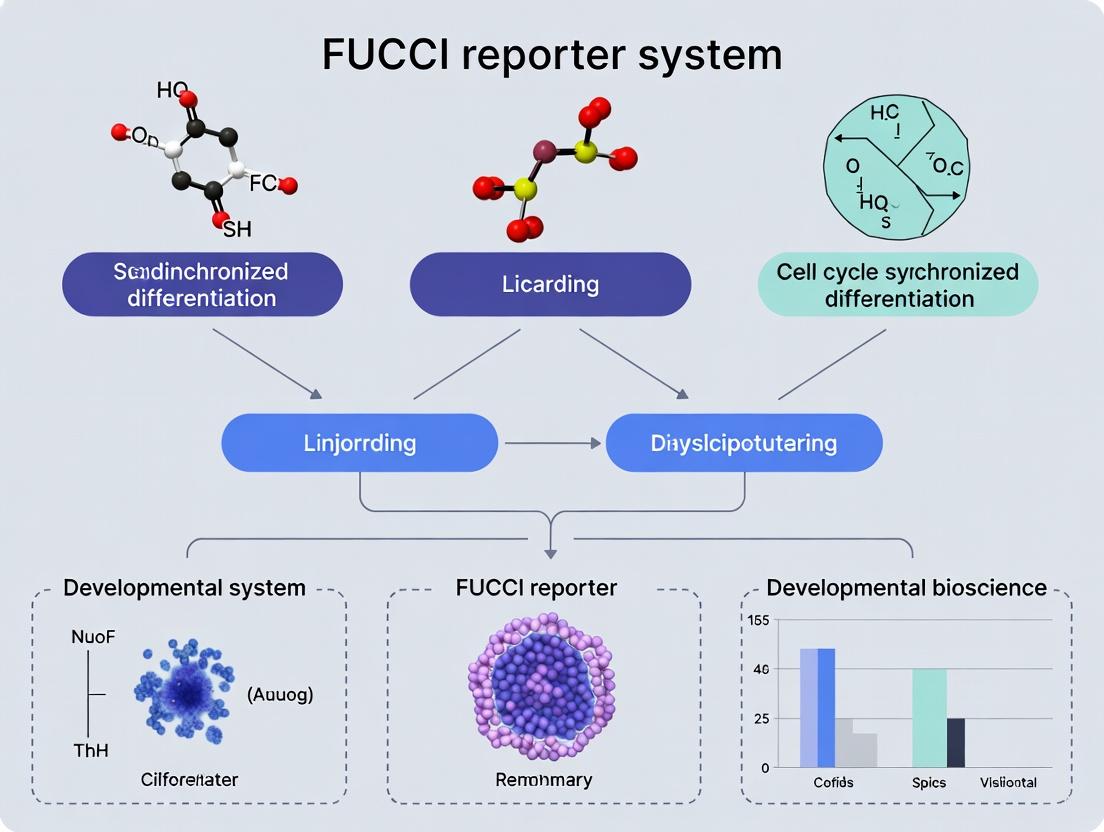
Unlocking Cell Fate: A Comprehensive Guide to FUCCI Cell Cycle Synchronized Differentiation
This article provides researchers, scientists, and drug development professionals with a complete roadmap for the FUCCI (Fluorescent Ubiquitination-based Cell Cycle Indicator) reporter system.
Unlocking Cell Fate: A Comprehensive Guide to FUCCI Cell Cycle Synchronized Differentiation
Abstract
This article provides researchers, scientists, and drug development professionals with a complete roadmap for the FUCCI (Fluorescent Ubiquitination-based Cell Cycle Indicator) reporter system. We explore its foundational principles for visualizing real-time cell cycle dynamics and its powerful application in synchronizing stem cell differentiation protocols. The guide details methodological best practices for implementing FUCCI in diverse cell models, addresses common troubleshooting and optimization challenges, and validates the system's advantages by comparing it with traditional synchronization techniques. Ultimately, this resource empowers users to harness FUCCI for enhancing reproducibility in developmental biology, disease modeling, and regenerative medicine research.
FUCCI Demystified: Understanding the Cell Cycle Reporter for Synchronized Differentiation
Application Notes: Principles and Quantitative Data
The Fluorescent Ubiquitination-based Cell Cycle Indicator (FUCCI) system is a powerful molecular tool for visualizing the cell cycle in live cells. Its core principle relies on the temporally-regulated, ubiquitin-proteasome-mediated degradation of fluorescent proteins fused to specific cell cycle regulatory proteins.
Core Degradation Signals and Spectral Output
The canonical FUCCI system uses two probes:
| Probe Name | Fluorescent Protein | Fused Degradation Signal | Active Phase | Peak Expression | Half-life (approx.) |
|---|---|---|---|---|---|
| FUCCI-G1 Probe | mKO2 (Orange/RFP) | hCdt1(30/120) | G1 Phase | Late G1 | ~40 min |
| FUCCI-S/G2/M Probe | mAG (Green/GFP) | hGeminin(1/110) | S, G2, M Phases | Late S / G2 | ~60 min |
Key Quantitative Observations:
- Transition Point: The exchange from red (mKO2-hCdt1) to green (mAG-hGeminin) fluorescence occurs sharply at the G1/S transition.
- Intensity Correlation: Fluorescence intensity correlates with the abundance of the underlying cell cycle regulator (Cdt1 in G1, Geminin in S/G2/M).
- Four-Color Distinction: Using the canonical pair, cells can be visually classified:
- Red: G1 phase.
- Yellow/Orange (Red+Green): Late G1 / very early S phase (probe exchange).
- Green: S, G2, and M phases.
- Dark/Dull: Early G1 (post-mitosis, before Cdt1 accumulation) or quiescent cells.
Advanced FUCCI Systems & Quantitative Performance
Recent developments have expanded the FUCCI palette and applications.
| System Variant | Probes & Colors | Key Improvement | Typical Application |
|---|---|---|---|
| FUCCI4 | Cdt1-KO2 (Orange), Cdt1-miRFP670 (Far-Red), Geminin-AG (Green), Geminin-mKate2 (Red) | Distinguishes G1, S, G2, and M phases separately. | Detailed kinetics of all cell cycle phases. |
| FUCCI(CA) | mKO2-hCdt1(30/120), mTurquoise2-hGeminin(1/110) | Uses mTurquoise2 (Cyan) for better spectral separation from orange. | Improved multiplexing with other fluorescent reporters. |
| FUCCI-NIR | miRF670-hCdt1, miRF720-hGeminin | Near-Infrared (NIR) probes for deeper tissue imaging. | In vivo imaging and cell cycle tracking in animal models. |
Protocols for Key Experiments
Protocol: Live-Cell Imaging of Cell Cycle Dynamics Using FUCCI
Objective: To track cell cycle phase transitions of individual cells in a population over time.
Materials:
- FUCCI-expressing cell line (e.g., stable U2OS FUCCI or primary cells transduced with FUCCI lentivirus).
- Complete cell culture medium.
- Glass-bottom culture dishes (e.g., 35 mm, No. 1.5 coverglass).
- Live-cell imaging microscope with environmental chamber (37°C, 5% CO₂), and appropriate filter sets for GFP (Ex: 470/40, Em: 525/50) and RFP (Ex: 560/40, Em: 630/75).
- Time-lapse imaging software.
Procedure:
- Cell Seeding: Seed FUCCI-expressing cells sparsely (20-30% confluency) in a glass-bottom dish in complete medium. Allow cells to adhere for 24 hours.
- Microscope Setup:
- Pre-warm the environmental chamber to 37°C with 5% CO₂ for at least 1 hour before imaging.
- Place the dish in the chamber and locate a field of view with well-separated, healthy cells.
- Image Acquisition:
- Set up sequential acquisition for GFP and RFP channels to avoid bleed-through.
- Set exposure times to avoid saturation (typically 100-500 ms).
- Configure time-lapse settings: Acquire images every 15-30 minutes for 48-72 hours.
- Data Analysis:
- Use tracking software (e.g., ImageJ/TrackMate, Imaris) to follow individual cells over time.
- Measure mean fluorescence intensity in both channels for each cell at each time point.
- Plot the RFP and GFP intensities over time. The G1/S transition is marked by a drop in RFP and a concomitant rise in GFP signal.
Protocol: Cell Cycle Synchronization & FUCCI Validation
Objective: To synchronize cells in a specific phase and confirm synchronization via FUCCI readout.
Materials:
- FUCCI-expressing cells.
- Complete medium, serum-free medium.
- Thymidine (2 mM stock in PBS), Nocodazole (100 µg/mL stock in DMSO).
- Phosphate-Buffered Saline (PBS).
- Flow cytometer with 488 nm and 561 nm lasers.
Procedure: Double Thymidine Block (Synchronization at G1/S)
- First Block: Treat subconfluent cells with 2 mM thymidine for 18 hours.
- Release: Wash cells 3x with PBS and add complete medium. Incubate for 9 hours.
- Second Block: Add 2 mM thymidine again for 17 hours.
- Final Release & FUCCI Analysis: Wash cells and add complete medium.
- For imaging: Immediately transfer to the live-cell microscope and start time-lapse. >80% of cells should appear yellow/orange (G1/S transition).
- For flow cytometry: Harvest cells at release (t=0) and every 2 hours thereafter. Analyze using 488-nm (GFP) and 561-nm (RFP) lasers. Plot RFP vs. GFP intensity to visualize the synchronized cohort progressing through the cell cycle.
Protocol: Quantifying Drug Effects on Cell Cycle Progression
Objective: To assess the impact of a chemotherapeutic agent on cell cycle dynamics using FUCCI.
Materials:
- FUCCI-expressing cells.
- Drug of interest (e.g., 5-Fluorouracil, Doxorubicin) and vehicle control.
- Live-cell imaging system or flow cytometer.
Procedure:
- Seed cells for imaging or in multi-well plates for endpoint flow cytometry.
- After adherence, treat cells with the drug at the desired concentration(s). Include a vehicle control (e.g., 0.1% DMSO).
- Live Imaging Track: Acquire time-lapse images every 30 minutes for 48-72 hours post-treatment.
- Endpoint Flow Cytometry: Harvest cells at 24, 48, and 72 hours post-treatment.
- Analysis:
- Imaging: Calculate the percentage of cells in Red (G1), Yellow (G1/S), and Green (S/G2/M) over time. Compare the rate of phase transition (e.g., G1-to-S delay) between treated and control groups.
- Flow Cytometry: Generate 2D histograms (RFP vs. GFP). Quantify the distribution of cells in each quadrant corresponding to G1, S, and G2/M phases. A G2/M arrest will show a significant increase in the GFP-high, RFP-low population.
Visualization Diagrams
Title: FUCCI Core Degradation Logic
Title: Thesis Workflow: Synchronized Differentiation
The Scientist's Toolkit: Key Research Reagent Solutions
| Reagent / Material | Function / Role | Example Product / Note |
|---|---|---|
| FUCCI Lentiviral Vectors | For stable, long-term expression of FUCCI probes in dividing cells, including primary and stem cells. | pLenti-FUCCI plasmids (e.g., Addgene #51039, #51040). Use 2nd/3rd generation packaging systems. |
| FUCCI-Expressing Cell Lines | Ready-to-use models for cell cycle studies, saving time on generation and optimization. | U2OS FUCCI, HeLa FUCCI (available from JCRB, ATCC). |
| Live-Cell Imaging Medium | Phenol-red-free medium optimized to maintain pH and health during long-term imaging without cytotoxic effects. | FluoroBrite DMEM, CO₂-independent medium, or medium with HEPES. |
| Cell Cycle Synchronization Agents | To arrest a population at a specific cell cycle phase for timed differentiation induction. | Thymidine (G1/S), Nocodazole (G2/M), Lovastatin (G1). |
| Proteasome Inhibitor (Control) | To validate FUCCI degradation mechanism. Inhibition should halt fluorescence oscillation. | MG-132, Lactacystin. Use as a control in initial validation. |
| Flow Cytometry Antibodies | For correlating FUCCI phase with differentiation markers (e.g., by intracellular staining). | Antibodies against lineage-specific proteins (e.g., β-III-tubulin for neurons). |
| Matrigel / Geltrex | For studying cell cycle during differentiation in a physiologically relevant 3D environment. | Essential for organoid or stem cell differentiation protocols. |
This application note is framed within a broader thesis investigating the use of the Fluorescent Ubiquitination-based Cell Cycle Indicator (FUCCI) reporter system for cell cycle synchronized differentiation research. The core premise is that precise control and monitoring of the cell cycle phase are critical for directing stem or progenitor cell fate decisions. The FUCCI system provides a real-time, visual readout of cell cycle progression, enabling researchers to isolate phase-specific populations and correlate cycle position with differentiation efficiency, thereby advancing regenerative medicine and disease modeling.
Table 1: FUCCI Reporter Constructs and Corresponding Cell Cycle Phases
| FUCCI Component | Fluorescent Protein | Binds/Degrades Based On: | Active (Fluorescent) Phase | Typical Emission Color |
|---|---|---|---|---|
| FUCCI (Red) | mKO2 (or mCherry) | Ubiquitinated by APC/C^(Cdh1) in late M/early G1; degrades in S phase. | Late M → G1 phase | Red |
| FUCCI (Green) | mAG (or GFP) | Ubiquitinated by SCF^(Skp2) in late M/early G1; accumulates in S phase. | S → G2 → M phases | Green |
| Intermediate Signal | Co-localization | Overlap of red and green fluorescence. | G1/S Transition | Yellow/Orange |
Table 2: Quantitative Fluorescence Intensity Ratios for Phase Determination
| Cell Cycle Phase | Predominant Signal | mKO2 (Red) : mAG (Green) Intensity Ratio (Approx.) | Notes for Interpretation |
|---|---|---|---|
| G1 (Early-Mid) | Strong Red | High (e.g., >3:1) | Red nucleus, no green. |
| G1/S Transition | Yellow/Orange | ~1:1 | Co-localization in nucleus. Critical window for synchronization. |
| S Phase | Strong Green | Low (e.g., <1:3) | Green nucleus, faint/no red. |
| G2/M Phase | Strong Green | Very Low (Red absent) | Green nucleus, no red. |
| Mitosis (M) | DIM/BOTH | Variable; fluorescence often dims due to nuclear envelope breakdown. | Cells may appear dark or briefly show cytoplasmic fluorescence. |
Key Experimental Protocols
Protocol 1: Live-Cell Imaging for FUCCI-Based Cell Cycle Tracking
Objective: To monitor real-time cell cycle progression and identify phase-specific events during differentiation.
- Cell Preparation: Seed FUCCI-expressing stem/progenitor cells (e.g., iPS cells with FUCCI reporter) in a glass-bottom imaging dish.
- Differentiation Induction: Add differentiation medium at T=0. For synchronization studies, consider a prior block-release protocol (see Protocol 2).
- Microscope Setup: Use a confocal or high-content fluorescence microscope with environmental control (37°C, 5% CO2).
- Excitation/Emission:
- mKO2/mCherry: Ex 561 nm / Em 575-625 nm.
- mAG/GFP: Ex 488 nm / Em 500-550 nm.
- Excitation/Emission:
- Image Acquisition: Capture images every 15-30 minutes for 24-72 hours. Use a 20x or 40x objective.
- Analysis: Use image analysis software (e.g., ImageJ, CellProfiler) to quantify nuclear red and green intensity over time. Generate kymographs or track individual cells to assign cell cycle phases.
Protocol 2: Fluorescence-Activated Cell Sorting (FACS) of FUCCI Populations
Objective: To isolate highly pure populations of cells in G1 (Red), S/G2/M (Green), or G1/S (Yellow) for downstream differentiation assays.
- Cell Harvest: Gently dissociate FUCCI-expressing cells into a single-cell suspension using Accutase or TrypLE.
- Staining Suspension: Resuspend in FACS buffer (PBS + 2% FBS + 1mM EDTA). Keep on ice and protected from light. Use DAPI (1 µg/mL) or a live/dead dye (e.g., Zombie NIR) to exclude dead cells.
- FACS Gating Strategy:
- Plot 1: FSC-A vs. SSC-A to gate on single cells.
- Plot 2: FSC-H vs. FSC-A to exclude doublets.
- Plot 3: Live/dead dye vs. DAPI to gate viable (DAPI-negative, live-dye-negative) cells.
- Plot 4: Red fluorescence (mKO2) vs. Green fluorescence (mAG).
- Sorting Gates:
- Gate R (G1): Red-high, Green-low.
- Gate G (S/G2/M): Green-high, Red-low.
- Gate Y (G1/S): Red-mid, Green-mid (intensity ratio ~1:1).
- Post-Sort: Collect cells into recovery medium. Proceed immediately to differentiation or molecular analysis (e.g., RNA-seq, protein lysate).
Visualizations
Title: FUCCI Color Transitions Through the Cell Cycle
Title: Workflow for Synchronized Differentiation Using FUCCI
The Scientist's Toolkit: Research Reagent Solutions
Table 3: Essential Materials for FUCCI-Based Synchronized Differentiation Studies
| Item | Function & Application in Protocol | Example Product/Catalog # (Note: For illustration) |
|---|---|---|
| FUCCI Reporter Construct | Genetically encodes the cell cycle sensors. Stable expression is key. | pFUCCI (mKO2-hCdt1(30/120)/mAG-hGeminin(1/110)) plasmid; or ready-to-use FUCCI-iPS cell line. |
| Live-Cell Imaging Dish | Provides optimal optical clarity and environmental control for long-term imaging. | Glass-bottom μ-Dish, 35 mm, polymer coverslip. |
| Gentle Dissociation Reagent | Generates single-cell suspension for FACS without damaging fluorescent proteins. | Accutase solution or TrypLE Select. |
| Viability Stain for Flow | Distinguishes live from dead cells to ensure sorting purity. | Zombie NIR Fixable Viability Kit or DAPI. |
| FACS Sorter | Instrument for isolating pure populations based on red/green fluorescence. | BD FACSAria III, Sony SH800, or equivalent. |
| Cell Cycle Blocking Agents (Optional) | Can be used prior to sorting to enrich for specific phases (e.g., double thymidine block for S phase). | Thymidine, Nocodazole (M phase arrest). |
| Differentiation Media Kit | Defined factors to drive lineage-specific differentiation from sorted progenitors. | According to target lineage (e.g., Cardiomyocyte, Neural, Hepatocyte differentiation kits). |
| Image Analysis Software | Quantifies fluorescence intensity and tracks cells over time. | Fiji/ImageJ, CellProfiler, Imaris, or Nikon Elements. |
Why Cell Cycle Phase is a Critical Determinant of Differentiation Efficiency and Fate
Within the context of a thesis utilizing the Fluorescent Ubiquitination-based Cell Cycle Indicator (FUCCI) system, this application note establishes the foundational principle that the phase of the cell cycle (G1, S, G2/M) at the initiation of a differentiation signal is a critical, deterministic variable. Successful differentiation protocols for stem cells and progenitor cells, whether for basic research or therapeutic manufacturing, require high efficiency and purity. Mounting evidence indicates that cells are only permissive to differentiation cues during specific cell cycle windows, primarily late G1. The FUCCI reporter system provides a powerful live-cell imaging tool to isolate, track, and fate-map cells based on their real-time cell cycle status, enabling synchronized differentiation studies.
Table 1: Influence of Cell Cycle Phase on Differentiation Outcomes in Various Cell Types
| Cell Type | Differentiation Target | Most Permissive Phase | Efficiency vs. Async Control | Key Fate Regulator Expression | Reference Context |
|---|---|---|---|---|---|
| Human iPSCs | Cardiomyocytes | Late G1 | 92% vs. 45% | NKX2-5 high, SOX2 low | Pauklin & Vallier, 2013 |
| Mouse ESCs | Neuronal Precursors | Early G1 | 85% vs. 30% | PAX6 high, OCT4 low | Coronado et al., 2013 |
| C2C12 Myoblasts | Myotubes (Fusion) | G1 Arrest (Post-mitotic) | 70% fusion vs. 20% | MYOD1 high, Cyclin D1 low | Zhang et al., 2019 |
| Hematopoietic Progenitors | Erythroid Lineage | Late G1 / G0 | 3-fold increase in CFUs | GATA1 high, CCNE1 low | - |
Table 2: Molecular Hallmarks of Cell Cycle Phase-Dependent Permissiveness
| Cell Cycle Phase | Chromatin Accessibility | Key Signaling Activity | Differentiation Cue Response |
|---|---|---|---|
| G1 (Early-Mid) | Condensed, low accessibility | CDK4/6-Cyclin D active | Refractory; maintains pluripotency |
| G1 (Late) | High accessibility, open | CDK2-Cyclin E peak, pRb hyperphosphorylation | Permissive; fate specification |
| S / G2 / M | Replicating/condensed | DNA replication & division machinery | Refractory; prone to apoptosis or errors |
Experimental Protocols
Protocol 1: FUCCI Reporter Cell Line Generation for Differentiation Studies
Objective: Engineer stem/progenitor cells to stably express FUCCI reporters for real-time cell cycle tracking. Materials: FUCCI plasmids (mAG-hGem(1/110) for G1 marker, mKO2-hCdt1(30/120) for S/G2/M); target cells; transfection reagent; puromycin/neomycin. Procedure:
- Transfection/Transduction: Introduce FUCCI constructs into your target stem/progenitor cell line using lentiviral transduction (recommended for primary cells) or lipid-based transfection.
- Selection & Cloning: Apply appropriate antibiotics for 7-14 days. Isolve single-cell clones by FACS or limiting dilution.
- Validation: Confirm cell cycle-specific fluorescence via flow cytometry after serum starvation (G1 arrest) or nocodazole treatment (M arrest). Verify differentiation potential is unchanged compared to parental line.
- Maintenance: Culture FUCCI-expressing cells under standard conditions, monitoring fluorescence stability.
Protocol 2: Synchronized Differentiation Initiation Based on FUCCI Status
Objective: To initiate differentiation protocols on populations sorted or selected based on specific FUCCI signals. Materials: FUCCI reporter cell line; FACS sorter or live-cell imager; differentiation media. Procedure:
- Pre-culture: Grow FUCCI cells to ~70% confluence under standard growth conditions.
- Cell Cycle Phase Isolation: Option A (FACS Sorting): Harvest cells, resuspend in sorting buffer. Sort populations: * G1 Phase: mAG+ (green) only. * S/G2/M Phase: mKO2+ (red) only. * G1/S Transition: mAG+mKO2+ (yellow). Option B (Live-Culture Selection): Seed cells and use time-lapse imaging to identify and mark cells transitioning into late G1 (increasing green signal, loss of red).
- Differentiation Initiation: Immediately plate sorted/selected cells at optimal density in pre-warmed differentiation media. Maintain under differentiation conditions.
- Kinetic Analysis: Use live imaging (if using Option B) or harvest parallel wells at time points to assess differentiation markers (via qPCR, immunofluorescence) and cell cycle exit (via loss of FUCCI fluorescence, EdU incorporation).
Protocol 3: Quantifying Differentiation Efficiency Relative to Initial Cell Cycle Phase
Objective: To correlate the initial FUCCI state of single cells with their terminal differentiation fate. Materials: FUCCI cell line in differentiation assay; live-cell imaging system; fate marker stains. Procedure:
- Image Acquisition: Seed FUCCI cells in a multi-well imaging plate. Begin time-lapse imaging (e.g., every 4 hours) upon adding differentiation media. Track individual cells over 5-10 days.
- Data Annotation: For each cell, record:
- Initial FUCCI color at t=0 (differentiation induction).
- Time to first division after induction (if any).
- Time to permanent cell cycle exit (persistent green only, then signal loss).
- Final morphological change.
- Endpoint Immunostaining: Fix cells at experiment end and stain for lineage-specific markers (e.g., TUJ1 for neurons, cTnT for cardiomyocytes).
- Fate Mapping: Correlate the initial FUCCI status (G1 vs. S/G2/M) from the live-imaging record with the final immunostaining result for each tracked cell. Calculate fate acquisition probability per initial phase.
Visualization: Pathways and Workflows
The Scientist's Toolkit: Essential Research Reagents & Materials
| Item | Function in FUCCI Differentiation Studies |
|---|---|
| FUCCI Reporter Plasmids/Viruses | Engineered constructs expressing mAG-hGem (G1) and mKO2-hCdt1 (S/G2/M) for visualizing cell cycle phase in live cells. |
| Live-Cell Imaging System | Microscope with environmental control (CO2, temp, humidity), suitable fluorescence channels, and time-lapse capability for tracking FUCCI signals and fate. |
| Flow Cytometer with Sorter (FACS) | For isolating high-purity populations of cells in specific cell cycle phases (G1-green vs. S/G2/M-red) prior to differentiation assays. |
| Cell Cycle Arrest Agents | Nocodazole (M-phase arrest), Aphidicolin (S-phase arrest), Palbociclib (G1 arrest via CDK4/6 inhibition). Used for FUCCI system validation. |
| Differentiation Inducers | Lineage-specific small molecules or cytokines (e.g., Retinoic Acid for neuronal, BMP4 for mesoderm, CHIR99021 for WNT activation). |
| EdU/BrdU Kit | For quantifying DNA synthesis and S-phase entry, complementary to FUCCI readout, to confirm cell cycle exit during differentiation. |
| Lineage-Specific Antibodies | Immunostaining validated antibodies for endpoint analysis of differentiation efficiency (e.g., OCT4 for pluripotency, TUJ1/βIII-Tubulin for neurons). |
| CDK Inhibitors (e.g., Roscovitine) | Pharmacological tools to manipulate cell cycle progression (e.g., prolong G1) and test its direct effect on differentiation permissiveness. |
Within the broader thesis on utilizing the FUCCI (Fluorescent Ubiquitination-based Cell Cycle Indicator) reporter system for cell cycle-synchronized differentiation research, understanding the evolution of its core constructs is paramount. The original FUCCI system, employing mKO2-hCdt1 and mAG-hGem, revolutionized live-cell cycle analysis. This application note details the historical progression, quantitative performance, and practical protocols for implementing these tools, with a focus on enabling differentiation studies.
Evolution and Quantitative Comparison of FUCCI Constructs
The classic FUCCI system uses two ubiquitination-based probes: mKO2 (monomeric Kusabira-Orange2) fused to the degron of human Cdt1 (expressed in G1 phase) and mAG (monomeric Azami-Green) fused to the degron of human Geminin (expressed in S/G2/M phases). Next-generation constructs have been developed to address limitations such as photostability, brightness, and compatibility with other fluorophores.
Table 1: Comparison of Key FUCCI Construct Generations
| Construct (Fluorophore-Degron) | Excitation/Emission (nm) | Reported Brightness (Relative to mKO2) | Photostability | Primary Application Context | Compatible Differentiation Markers |
|---|---|---|---|---|---|
| mKO2-hCdt1 (1/30) | 548/559 | 1.0 (reference) | Moderate | Original FUCCI; G1 phase marking | mCherry, GFP |
| mAG-hGeminin | 492/505 | ~1.2 | Moderate | Original FUCCI; S/G2/M phase marking | GFP, RFP |
| mCherry-hCdt1 | 587/610 | ~1.5 | High | Improved contrast, deeper tissue imaging | GFP, BFP |
| mVenus-hGeminin | 515/528 | ~2.0 | Moderate | Brighter signal for G2/M | RFP, iRFP |
| Next-Gen: mMaroon1-hCdt1 | 609/684 | ~0.8 | Very High | Far-red shift for multiplexing & in vivo | GFP, mCherry, Blue FP |
| Next-Gen: mCyRFP1-hGeminin | 548/569 | ~1.3 | High | Orange-red alternative, improved separation | GFP, iRFP |
Application Notes for Differentiation Research
Synchronizing differentiation protocols to specific cell cycle phases (often early G1) can enhance efficiency and homogeneity. The FUCCI system enables real-time isolation or observation of cells in a desired phase prior to differentiation induction.
Key Finding: In iPSC-derived neuronal progenitor differentiation, a protocol targeting FUCCI-positive (mKO2-hCdt1, G1) cells yielded a 25% increase in MAP2-positive neurons compared to an unsynchronized population.
Detailed Protocols
Protocol 1: Lentiviral Transduction for FUCCI Reporter Generation in Stem Cells
Objective: To generate a stable FUCCI reporter cell line for differentiation studies. Materials: HEK293T cells, lentiviral vectors for FUCCI probes (e.g., pCSII-EF-mKO2-hCdt1, pCSII-EF-mAG-hGem), packaging plasmids (pMD2.G, psPAX2), polyethylenimine (PEI), target stem cells (e.g., iPSCs), polybrene. Procedure:
- Virus Production: Co-transfect HEK293T cells with FUCCI vector and packaging plasmids using PEI transfection reagent in Opti-MEM.
- Harvest: Collect virus-containing supernatant at 48 and 72 hours post-transfection. Filter through a 0.45 µm PVDF filter.
- Transduction: In the presence of 8 µg/mL polybrene, incubate target stem cells (at ~50% confluence) with viral supernatant for 24 hours.
- Selection & Sorting: Replace with fresh medium. After 72 hours, use fluorescence-activated cell sorting (FACS) to isolate double-positive (mKO2 and mAG) cells or create separate populations. Maintain cells under standard conditions.
Protocol 2: Cell Cycle Phase-Specific Differentiation Initiation
Objective: To initiate differentiation predominantly in G1-phase cells. Materials: Stable FUCCI reporter cell line, appropriate differentiation medium, FACS sorter or live-cell imaging system. Procedure:
- Monitoring: Culture FUCCI cells and observe under a fluorescence microscope. G1-phase cells exhibit red fluorescence (mKO2-hCdt1).
- Sorting (Optional): For high-purity synchronization, use FACS to collect the red-only (G1) population.
- Induction: Immediately seed the sorted G1-phase cells or, for unsorted cultures, initiate differentiation protocol when >70% of cells are in the red (G1) phase based on live imaging.
- Tracking: Continuously monitor fluorescence to correlate phase transitions with early differentiation marker expression.
Visualization of Experimental Workflow and Logic
Title: FUCCI-Based G1 Synchronization for Differentiation Workflow
Title: Molecular Logic of FUCCI Probe Regulation
The Scientist's Toolkit: Research Reagent Solutions
Table 2: Essential Materials for FUCCI Differentiation Experiments
| Reagent/Material | Function in FUCCI Experiments | Example Product/Catalog Number |
|---|---|---|
| FUCCI Lentiviral Vectors | Delivery of mKO2-hCdt1 and mAG-hGeminin reporters. | pCSII-EF-mKO2-hCdt1 (Addgene #58409); pBOB-mAG-hGem (Addgene #14645) |
| Polyethylenimine (PEI) | Transfection reagent for lentiviral packaging in HEK293T cells. | Linear PEI, MW 25,000 (Polysciences #23966) |
| Polybrene | Enhances viral transduction efficiency. | Hexadimethrine bromide (Sigma-Aldrich H9268) |
| Fluorescence-Activated Cell Sorter (FACS) | Isolation of double-positive reporter cells or specific cell cycle phases. | N/A (Core Facility Instrument) |
| Live-Cell Imaging Chamber | Maintains cell health during long-term time-lapse imaging. | Lab-Tek II Chambered Coverglass (Thermo Fisher 155409) |
| Differentiation Induction Media | Cell-type specific media to drive differentiation post-synchronization. | e.g., Neuronal Induction Medium (Thermo Fisher A1647801) |
| Cell Cycle Inhibitors (Validation) | Positive controls for phase arrest (e.g., Aphidicolin for S-phase). | Aphidicolin (Sigma-Aldrich A4487) |
| Anti-MAP2 / Anti-Tuj1 Antibodies | Immunostaining to validate neuronal differentiation outcome. | Anti-MAP2 chicken (Abcam ab5392) |
Key Advantages of FUCCI Over Traditional Synchronization Methods (e.g., Serum Starvation, Chemical Blockers)
Fluorescent Ubiquitination-based Cell Cycle Indicator (FUCCI) technology represents a paradigm shift in cell cycle analysis and synchronization for differentiation research. Unlike traditional bulk synchronization methods like serum starvation or chemical blockade, FUCCI utilizes genetically encoded fluorescent probes to visualize real-time cell cycle progression in living cells. This application note details the quantitative advantages, provides protocols for implementation, and contextualizes its superiority within synchronized differentiation studies.
Quantitative Comparison of Synchronization Methods
The following tables summarize the core performance metrics of FUCCI versus traditional methods.
Table 1: Method Characteristics and Impact on Cell Physiology
| Feature | Serum Starvation | Chemical Blockers (e.g., Thymidine, Nocodazole) | FUCCI Reporter System |
|---|---|---|---|
| Synchronization Principle | Induction of quiescence (G0) by growth factor deprivation. | Reversible inhibition of DNA synthesis or spindle formation. | Fluorescent protein expression coupled to cell cycle protease activity. |
| Degree of Synchrony | Moderate (~70-80% in G0/G1). Often leaky. | High at point of release (>85%), but decays rapidly. | Not a synchronizing agent—enables identification and isolation of specific cycle phases. |
| Duration of Effect | Long (24-72 hrs). | Variable, depending on blocker (8-24 hrs). | Continuous, real-time monitoring. |
| Cellular Stress/ Toxicity | High. Induces stress pathways, alters metabolism. | Moderate to High. Can cause DNA damage (thymidine) or aneuploidy. | Minimal. Uses endogenous regulation; non-invasive imaging. |
| Effect on Differentiation | Can bias or impair differentiation potential due to stress. | May alter fate through checkpoint activation. | Allows correlation of native cycle phase to differentiation onset without perturbation. |
| Temporal Resolution | Single time-point (release). | Single or few time-points post-release. | Continuous, single-cell resolution over days. |
Table 2: Experimental Utility in Differentiation Research
| Parameter | Traditional Synchronization | FUCCI-Based Workflow |
|---|---|---|
| Ability to Track Phase-Specific Differentiation Events | Indirect, inferred from release time. | Direct, by observing FUCCI color at differentiation trigger. |
| Multiplexing with Lineage Reporters | Challenging due to protocol complexity. | Straightforward; dual- or triple-color imaging with differentiation markers. |
| Long-Term Phenotyping Post-Synchronization | Compromised as synchronicity is lost. | Enables fate tracking of cells from a specific starting phase. |
| Throughput for Drug Screening | Low. Batch variability high. | High. Enables live-cell sorting of phase-specific populations for assays. |
| Data Richness | Population-averaged, endpoint. | Single-cell, longitudinal, kinetic. |
Experimental Protocols
Protocol: Establishing a FUCCI-Expressing Cell Line for Differentiation Studies
Objective: Generate a stable, FUCCI-expressing pluripotent stem cell (PSC) line to study cell cycle phase during differentiation onset.
Materials: See "Scientist's Toolkit" section. Procedure:
- Cell Preparation: Culture human iPSCs in mTeSR Plus on Matrigel-coated plates to ~70% confluence.
- Lentiviral Transduction: a. Prepare FUCCI lentivirus (e.g., FUCCI4, mCherry-hCdt1(30/120)/mVenus-hGeminin(1/110)) in maintenance medium with Polybrene (8 µg/mL). b. Replace culture medium with virus-containing medium. Incubate for 24h. c. Replace with fresh mTeSR Plus medium.
- Selection and Expansion: After 72h, begin puromycin selection (0.5 µg/mL) for 7-10 days. Expand resistant colonies.
- Validation by Flow Cytometry: Harvest cells, analyze via flow cytometry. Confirm distinct mCherry+ (G1), double-positive (S-phase), and mVenus+ (G2/M) populations. Sort if needed for a pure reporter population.
- Differentiation Experiment Setup: Plate validated FUCCI-iPSCs. Initiate differentiation protocol (e.g., via directed cardiomyocyte differentiation). Image daily using a live-cell incubator microscope.
Protocol: Live-Cell Sorting of G1 vs G2/M Populations for Differentiation Assay
Objective: Isolate live cells in specific cell cycle phases to assay their differential differentiation propensity.
Procedure:
- Cell Preparation: Harvest FUCCI-expressing PSCs using gentle dissociation reagent. Resuspend in sorting buffer (PBS + 2% FBS + 10 µM Y-27632).
- Flow Cytometry Setup: Use a sorter equipped with 488-nm and 561-nm lasers. Collect mVenus (530/30 nm) and mCherry (610/20 nm) signals.
- Gating Strategy: a. Gate on single cells using FSC-A vs FSC-H. b. Plot mCherry vs mVenus. Define gates: G1 Population (mCherry-high, mVenus-low); S-phase (mCherry-high, mVenus-high); G2/M Population (mCherry-low, mVenus-high).
- Sorting: Sort G1 and G2/M populations directly into differentiation medium in plate.
- Post-Sort Processing: Immediately place plates in incubator. Allow 6h for recovery, then commence differentiation protocol. Analyze differentiation efficiency (e.g., by flow cytometry for troponin T) at day 5-7.
Visualization of Workflows and Signaling
Title: Workflow Comparison: Traditional vs FUCCI Synchronization
Title: FUCCI Biosensor Mechanism of Action
The Scientist's Toolkit: Essential Reagents for FUCCI Differentiation Studies
| Item | Function/Description | Example Product/Catalog |
|---|---|---|
| FUCCI Reporter Construct | Bipartite sensor expressing mCherry-hCdt1 and mVenus-hGeminin. | MBL FUCCI4 (LCV043) / Addgene #58308 |
| Lentiviral Packaging System | For creating replication-incompetent virus to transduce hard-to-transfect cells (e.g., PSCs). | psPAX2, pMD2.G (Addgene) |
| Polybrene | Cationic polymer enhancing viral transduction efficiency. | Hexadimethrine bromide, 8 µg/mL working conc. |
| Puromycin | Selection antibiotic for stable cell line generation. | Thermofisher, 0.5-1 µg/mL for PSCs. |
| Rho Kinase Inhibitor (Y-27632) | Improves survival of dissociated and sorted stem cells. | Tocris, 10 µM in recovery medium. |
| Matrigel / Geltrex | Basement membrane matrix for pluripotent stem cell culture. | Corning Matrigel hESC-Qualified |
| mTeSR Plus / Essential 8 | Defined, feeder-free medium for human PSC maintenance. | STEMCELL Technologies |
| Live-Cell Imaging Medium | Phenol-red free, HEPES-buffered medium for stable pH during imaging. | FluoroBrite DMEM (ThermoFisher) |
| Validated Differentiation Kit | Directed differentiation protocol for consistent fate specification. | e.g., Cardiomyocyte Differentiation Kit (STEMCELL) |
| Flow Cytometry Antibodies | For confirming differentiation endpoints (e.g., anti-cTnT). | Alexa Fluor 647 anti-cTnT (BD Biosciences) |
Step-by-Step Protocol: Implementing FUCCI for Synchronized Differentiation Studies
Within a thesis on using the FUCCI (Fluorescent Ubiquitination-based Cell Cycle Indicator) reporter system for cell cycle-synchronized differentiation research, selecting the appropriate delivery method is a foundational decision. This choice dictates experimental flexibility, stability, and applicability across different cell types. These Application Notes provide a comparative analysis and detailed protocols for the three primary systems: lentiviral transduction, retroviral transduction, and generation of stable transgenic cell lines.
Comparative Analysis of FUCCI Systems
The table below summarizes the key characteristics of each system to guide researchers in selecting the most appropriate platform for their cell cycle synchronization and differentiation studies.
Table 1: Comparison of FUCCI Delivery Systems
| Feature | Lentiviral FUCCI | Retroviral FUCCI | Stable Transgenic Cell Line |
|---|---|---|---|
| Infection Efficiency | High (>90% in many cell types) | Moderate to High (requires dividing cells) | N/A (inherently 100% in selected clone) |
| Titer (Typical) | 1 x 10^7 - 1 x 10^8 IFU/mL | 1 x 10^6 - 1 x 10^7 CFU/mL | N/A |
| Cell Cycle Phase on Entry | Non-dividing and dividing cells | Dividing cells only (M/G1/S/G2) | N/A |
| Genomic Integration | Random integration | Random integration | Defined locus (if engineered) or random |
| Expression Stability | Long-term (weeks-months) | Can be silenced over time (weeks) | Permanent, heritable |
| Time to Establish | 5-7 days | 5-7 days | 4-8 weeks |
| Best For | Primary cells, neurons, stem cells, hard-to-transfect lines | Rapid infection of proliferative cell lines | Long-term, high-throughput studies, in vivo models |
Table 2: Quantitative Performance Metrics in a Model Differentiation Study (e.g., iPSC to Cardiomyocytes)
| System | Transduction Efficiency (%) | Fluorescence Signal Stability at Day 21 | Coefficient of Variation (Cell Cycle Phase Gating) | Success Rate in Generating Clonal Line |
|---|---|---|---|---|
| Lentiviral | 85-95 | 85-90% of initial | 8-12% | Not typically cloned |
| Retroviral | 70-85 (in dividing iPSCs) | 60-75% of initial | 10-15% | Not typically cloned |
| Stable Transgenic | 100 (by definition) | 95-100% of initial | 5-9% | 5-15% (post-selection) |
Detailed Protocols
Protocol 1: Lentiviral Transduction for FUCCI Expression in Primary Cells
Objective: To achieve high-efficiency, stable FUCCI reporter expression in primary or hard-to-transfect cells for monitoring cell cycle during differentiation. Materials: See "The Scientist's Toolkit" below. Procedure:
- Day -3: Plate HEK293T producer cells in 10 cm dishes for 70-80% confluency at transfection.
- Day 0: Co-transfect using polyethylenimine (PEI):
- FUCCI Reporter Plasmid (e.g., pBOB-EF1-FUCCI-Puro): 10 µg
- psPAX2 (packaging plasmid): 7.5 µg
- pMD2.G (VSV-G envelope plasmid): 2.5 µg
- Mix DNA with PEI (1:3 ratio) in Opti-MEM, incubate 15 min, add dropwise to cells.
- Day 1 & 2: Replace medium with fresh complete medium.
- Day 3: Harvest viral supernatant (48h and 72h post-transfection), filter through 0.45 µm PES filter. Concentrate using PEG-it Virus Precipitation Solution (overnight at 4°C) per manufacturer's instructions.
- Day 4: Resuspend viral pellet in cold PBS, aliquot, and store at -80°C. Titer using Lenti-X qRT-PCR Titration Kit.
- Day 5: Transduce target primary cells (e.g., mesenchymal stem cells) at an MOI of 5-20 in the presence of 8 µg/mL polybrene by spinoculation (1000 x g, 90 min, 32°C). Incubate overnight.
- Day 6: Replace with fresh medium.
- Day 7-9: Begin puromycin selection (concentration titrated for cell type) for 3-5 days. Validate expression via flow cytometry.
Protocol 2: Generating a Clonal Stable FUCCI Cell Line
Objective: To create a homogeneous, genetically stable cell population with consistent FUCCI expression for long-term differentiation assays. Procedure:
- Transduce your target cell line (e.g., RPE1) using lentiviral or retroviral methods as above, using a FUCCI construct with a selectable marker (e.g., puromycin, blasticidin).
- 48 hours post-transduction, begin antibiotic selection for 7-10 days to create a polyclonal pool.
- After selection, perform fluorescence-activated cell sorting (FACS) to isolate the top 5-10% brightest double-positive (mCherry-hGem(1/110)/mVenus-hCdt1(30/120)) cells.
- Plate these sorted cells at a clonal density (0.5-1 cell/well) into 96-well plates using conditioned medium.
- Allow colonies to expand over 3-4 weeks, periodically checking for growth.
- Screen expanding clones by live-cell imaging for robust, cell cycle-dependent fluorescence oscillation.
- Expand the 3-5 best clones and validate via:
- Flow cytometry for tight G1 (mVenus+) and S/G2/M (mCherry+) peaks.
- Time-lapse imaging over 48h to confirm phase transitions.
- Genomic PCR to confirm integration.
- Freeze down multiple vials of the validated master cell bank.
Diagrams
FUCCI System Selection Decision Tree
FUCCI Reporter Molecular Mechanism
The Scientist's Toolkit
Table 3: Essential Research Reagents & Materials
| Item | Function in FUCCI Experiments | Example Product/Catalog |
|---|---|---|
| FUCCI Reporter Plasmid | Encodes cell cycle phase-dependent fluorescent proteins (mVenus-hCdt1, mCherry-hGem). | pBOB-EF1-FUCCI-Puro (Addgene #86849) |
| Lentiviral Packaging Mix | Provides essential viral proteins (gag, pol, rev) for lentivirus production. | psPAX2 (Addgene #12260) |
| Envelope Plasmid | Provides VSV-G glycoprotein for broad tropism pseudotyping. | pMD2.G (Addgene #12259) |
| Polyethylenimine (PEI) | High-efficiency transfection reagent for viral production in HEK293T cells. | Linear PEI, MW 25,000 (Polysciences) |
| Polybrene | Cationic polymer that enhances viral transduction efficiency. | Hexadimethrine bromide (Sigma H9268) |
| Puromycin Dihydrochloride | Selection antibiotic for cells transduced with PuroR-containing constructs. | Thermo Fisher A1113803 |
| Lenti-X Concentrator | Chemical solution for rapid, simple concentration of lentiviral particles. | Takara Bio 631231 |
| Conditioned Medium | Spent medium from parent cell line to support growth of clonal cells. | Prepared in-house from confluent cultures. |
| Live-Cell Imaging Dye (Optional) | Nuclear stain for segmentation and tracking in long-term experiments. | Hoechst 33342 (Thermo Fisher H3570) |
Critical Steps for Generating and Validating a FUCCI-Reporter Cell Line
Within the broader thesis investigating the FUCCI (Fluorescent Ubiquitination-based Cell Cycle Indicator) reporter system for cell cycle-synchronized differentiation research, the generation of a robust, isogenic reporter cell line is a foundational prerequisite. This protocol details the critical steps from vector design to functional validation, enabling precise live-cell tracking of cell cycle phases (G1, S, S/G2, M/G2) for downstream differentiation studies.
Research Reagent Solutions
| Reagent / Material | Function in FUCCI Workflow |
|---|---|
| FUCCI Reporter Plasmid(s) | Expresses cell cycle phase-specific fluorescent proteins (e.g., mKO2-hCdt1 for G1, mAG-hGem for S/G2/M). |
| Target Cell Line | The parental cell line (e.g., iPSCs, progenitor cells) for engineering, chosen for differentiation potential. |
| Transfection/Transduction Reagent | For plasmid delivery (e.g., lipofectamine, lentiviral packaging systems). |
| Fluorescence-Activated Cell Sorter (FACS) | To isolate and clone cells stably expressing the reporter at optimal levels. |
| Cell Cycle Inhibitors | Validation tools (e.g., Aphidicolin (S-phase), Nocodazole (M-phase), Serum Starvation (G1)). |
| Live-Cell Imaging System | For time-lapse microscopy to validate dynamic cell cycle progression. |
Protocol 1: Design and Delivery of the FUCCI Reporter
Methodology
- Vector Selection: Choose a FUCCI construct suitable for your cell type. The original system uses two probes: pCSII-EF-mKO2-hCdt1(30/120) (degrades during S phase, labels G1 nuclei red) and pCSII-EF-mAG-hGem(1/110) (degrades in late M/early G1, labels S/G2/M nuclei green). For stem/progenitor cells, consider lentiviral backbones for stable integration.
- Delivery: Co-transfect or co-transduce target cells with both FUCCI plasmids at a 1:1 ratio. For lentiviral delivery, use a low MOI (~1-3) to avoid multiple integrations.
- Initial Selection: 48-72 hours post-transduction, apply appropriate selection (e.g., puromycin) if vectors contain resistance markers. Enrich the polyclonal population.
Protocol 2: Single-Cell Cloning and Expansion
Methodology
- FACS Analysis and Sorting: Analyze the polyclonal population by flow cytometry for mKO2 and mAG fluorescence. Gate dual-positive cells.
- Single-Cell Sorting: Sort single, dual-positive cells into individual wells of a 96-well plate containing conditioned growth medium.
- Clonal Expansion: Monitor and expand clones over 2-3 weeks. Regularly screen for fluorescence retention.
Protocol 3: Quantitative Validation of Cell Cycle Reporting
Methodology
- Cell Cycle Arrest Profiling: Treat clone(s) with specific inhibitors and analyze fluorescence profiles via flow cytometry (n=3 biological replicates). Expected shifts:
- Aphidicolin (2 µg/mL, 24h): Arrests in early S-phase. Population shifts to mAG-high (Green).
- Nocodazole (100 ng/mL, 16h): Arrests in M-phase. Population shifts to mAG-high (Green).
- Serum Starvation (72h): Arrests in G0/G1. Population shifts to mKO2-high (Red).
- Live-Cell Time-Lapse Validation: Seed validated clones for imaging. Acquire images (mKO2, mAG, phase contrast) every 30 minutes for 48-72 hours in a controlled environment (37°C, 5% CO₂). Track individual cells through complete cycles.
Data Presentation
Table 1: Expected Fluorescence Profile Shifts Upon Cell Cycle Arrest
| Treatment | Target Phase | Expected FUCCI Fluorescence Profile (Flow Cytometry) | Validated Clone Acceptance Criterion* |
|---|---|---|---|
| Serum Starvation | G0/G1 | >70% of cells in mKO2-hi (Red)/mAG-lo population | ≥ 65% |
| Aphidicolin | Early S | >60% of cells in mKO2-lo/mAG-hi (Green) population | ≥ 55% |
| Nocodazole | M | >50% of cells in mKO2-lo/mAG-hi (Green) population | ≥ 45% |
| Untreated (Asynchronous) | - | Distributed across four quadrants | N/A |
*Criterion based on consensus from published validation studies (n≥3 independent experiments).
Table 2: Key Parameters for Live-Cell FUCCI Imaging
| Parameter | Recommended Setting | Purpose |
|---|---|---|
| Imaging Interval | 15-30 minutes | Balances temporal resolution with phototoxicity. |
| Duration | 48-72 hours | Captures ≥ 2 full cell cycles. |
| Objective | 20x (dry) or 40x (oil) | Sufficient for single-cell tracking. |
| mKO2 Excitation/Emission | 550 nm / 580-620 nm | Detects hCdt1 (G1) signal. |
| mAG Excitation/Emission | 470 nm / 500-540 nm | Detects hGem (S/G2/M) signal. |
Diagrams
Workflow for Generating a FUCCI Reporter Line
FUCCI System Molecular Logic
Designing a Differentiation Protocol Around FUCCI-Guided Cell Cycle Windows
1. Introduction & Thesis Context Within the broader thesis investigating the FUCCI (Fluorescent Ubiquitination-based Cell Cycle Indicator) reporter system for cell cycle-synchronized differentiation, this protocol details the application of FUCCI-guided windows to enhance directed differentiation efficiency. The core premise is that a progenitor cell's receptivity to differentiation cues is intrinsically linked to its cell cycle phase. By isolating cells in specific FUCCI-color-defined windows (e.g., early G1), we can apply lineage-directing signals with temporal precision, potentially yielding more homogeneous, efficient, and functionally mature target cell populations for regenerative medicine and disease modeling.
2. Key Experimental Data & Rationale Recent studies quantify the enhanced differentiation outcomes when cues are applied in specific cell cycle phases.
Table 1: Impact of Cell Cycle Phase on Differentiation Efficiency
| Differentiation Target | FUCCI-Guided Window | Key Signaling Pathway Activated | Reported Efficiency Gain vs. Async. Culture | Reference (Example) |
|---|---|---|---|---|
| Cardiomyocytes | Early G1 (Red) | Wnt/β-catenin modulation | 2.5-fold increase in TNNT2+ cells | 2023, Stem Cell Rep. |
| Cortical Neurons | Late G1/S (Green) | BMP/SMAD inhibition | 3.1-fold increase in TUJ1+ neurons | 2022, Cell Stem Cell |
| Hepatocytes | Early G1 (Red) | HGF/MET signaling | 2.0-fold increase in Albumin+ cells | 2023, Nature Comm. |
| Osteoblasts | G1/S transition | Enhanced BMP2 response | 1.8-fold increase in mineralization | 2024, Sci. Adv. |
3. Detailed Experimental Protocols
Protocol 3.1: FUCCI Reporter Cell Line Generation & Validation Materials: FUCCI reporter plasmid (mKO2-hCdt1(30/120) for G1, mAG-hGem(1/110) for S/G2/M), target progenitor cell line (e.g., iPSC, mesenchymal stem cell), transfection reagent, antibiotic for selection. Procedure:
- Transfect progenitor cells with FUCCI reporter constructs using manufacturer's protocol.
- Select stable polyclonal or monoclonal populations using appropriate antibiotics (e.g., puromycin, blasticidin).
- Validate reporter fidelity via flow cytometry: Serum-starve cells for 48h to induce G1 arrest (≥90% red). Release into complete medium and track transition to green fluorescence over 12-16h.
- Confirm cell cycle phase correlation by co-staining with 5-ethynyl-2’-deoxyuridine (EdU) for S-phase and propidium iodide (PI) for DNA content. Analyze via flow cytometry: Red (mKO2+) cells should be EdU-, 2N DNA; Green (mAG+) cells should be EdU+ or have >2N DNA.
Protocol 3.2: Fluorescence-Activated Cell Sorting (FACS) for FUCCI Windows Materials: FUCCI reporter cell line, sorting buffer (PBS + 2% FBS + 1mM EDTA), 40µm cell strainer, sorter with 488nm and 561nm lasers. Procedure:
- Culture FUCCI cells to ~70% confluence to ensure active cycling.
- Harvest cells using gentle dissociation reagent (e.g., Accutase), neutralize with medium, and filter through a 40µm strainer.
- Resuspend in ice-cold sorting buffer at 10-20 million cells/mL.
- Set sorting gates using controls: Unstained cells and single-color controls (if available). Define the target window:
- Early G1 (High-Red, No-Green): High mKO2 (561nm ex, 580/15nm BP), low/no mAG (488nm ex, 510/20nm BP).
- G1/S Transition (Low-Red, Low-Green): Intermediate mKO2 and mAG.
- S/G2/M (No-Red, High-Green): High mAG, low/no mKO2.
- Sort cells directly into pre-warmed, supplemented differentiation medium. Collect into tubes coated with 2% BSA to minimize adhesion loss.
- Post-sort, analyze an aliquot to confirm purity (>85% for target window).
Protocol 3.3: Differentiation Initiation in a FUCCI-Synchronized Window Materials: Sorted FUCCI cell population, differentiation medium with specific induction factors, appropriate tissue cultureware. Procedure:
- Immediately plate sorted cells at optimal density (e.g., 50,000 cells/cm² for many progenitors) in differentiation medium. Use plates pre-coated with relevant substrate (e.g., Matrigel, poly-L-ornithine/laminin).
- Critical Step: Initiate addition of the primary differentiation cue (e.g., CHIR99021 for cardiomyocytes, Noggin for neurons) within 2 hours post-plating. This window capitalizes on the synchronized cell cycle state.
- Maintain cells under standard differentiation conditions (37°C, 5% CO2). Monitor fluorescence daily to observe loss of FUCCI synchrony as cells commit and exit cycle.
- At defined timepoints (e.g., days 3, 7, 14), assess early differentiation markers via qPCR or immunocytochemistry.
4. Visualization: Signaling Pathway & Experimental Workflow
Title: FUCCI-Guided Signaling Activation (100 chars)
Title: FUCCI-Guided Differentiation Workflow (100 chars)
5. The Scientist's Toolkit: Key Research Reagent Solutions
Table 2: Essential Materials for FUCCI-Guided Differentiation
| Item & Example Product | Function in Protocol |
|---|---|
| FUCCI Reporter Vector (e.g., pFUCCI plasmids, SB FUCCI system) | Genetically encodes fluorescent protein fusions to cell cycle-regulated proteins (Cdt1, Geminin), enabling live-cell cycle tracking. |
| Progenitor Cell Line (e.g., Human iPSCs, Primary MSCs) | The starting cell population with multipotent or pluripotent differentiation capacity. Must be compatible with FUCCI transduction. |
| High-Efficiency Transfection/Transduction Kit (e.g., Lentiviral system, Electroporation kit) | For stable integration or transient expression of FUCCI reporters in the target progenitor cell line. |
| Flow Cytometry Cell Sorter (e.g., equipped with 488nm & 561nm lasers) | Instrument essential for physically isolating cell populations based on specific red/green fluorescence profiles (FUCCI windows). |
| Validated Differentiation Kit/Components (e.g., Cardiomyocyte, Neuron kit) | Provides optimized basal media and precise concentrations of growth factors/small molecules to direct differentiation towards a specific lineage. |
| Cell Cycle Validation Reagents (e.g., EdU Click-iT Kit, Propidium Iodide) | Used in parallel with FUCCI to validate the cell cycle phase correlation of sorted populations via DNA synthesis and content analysis. |
| ECM Coating Substrate (e.g., Matrigel, Laminin-521) | Provides the necessary extracellular matrix for plating sorted cells and supporting survival and differentiation initiation. |
This Application Note is framed within a broader thesis investigating the utility of the Fluorescent Ubiquitination-based Cell Cycle Indicator (FUCCI) reporter system for achieving and monitoring cell cycle synchronized differentiation. The central hypothesis posits that coordinated exit from the cell cycle is a critical, measurable gateway to stable lineage commitment. Real-time tracking via FUCCI, combined with lineage-specific reporters, provides an unparalleled window into this dynamic process, enabling the dissection of temporal relationships between cell cycle phases, signaling events, and fate decisions. This is paramount for developmental biology, regenerative medicine, and drug discovery, where controlling differentiation efficiency is crucial.
Research Reagent Solutions Toolkit
The following table lists essential materials for implementing the core strategies described.
| Reagent / Material | Function in Live-Cell Imaging & Differentiation Tracking |
|---|---|
| FUCCI Reporter System (e.g., mKO2-hCdt1, mAG-hGem) | Visualizes cell cycle phases: G1 (red fluorescence) and S/G2/M (green fluorescence). G0/exit appears as loss of both signals. |
| Lineage-Specific Fluorescent Reporter | CRISPR-engineered or transduced construct (e.g., GFP under a cell-type-specific promoter) to mark commitment. |
| Low-Autofluorescence, Phenol Red-Free Medium | Minimizes background noise for sensitive fluorescence detection over long periods. |
| Environment-Controlled Live-Cell Imager | Maintains 37°C, 5% CO2, and humidity during time-lapse imaging. Essential for cell health. |
| High-Content, Confocal, or Spinning-Disk Microscope | Provides optical sectioning to reduce out-of-focus light, crucial for thick samples like organoids. |
| Mitogenic Factor (e.g., bFGF, EGF) | Used in proliferation media to maintain cells in cycle prior to differentiation induction. |
| Differentiation Induction Cocktail | Specific combination of growth factors, small molecules, or cytokines to trigger lineage commitment. |
| Nuclear Stain (e.g., Hoechst 33342, SiR-DNA) | Labels all nuclei for segmentation, tracking, and cell cycle analysis validation. |
| ROCK Inhibitor (Y-27632) | Improves single-cell survival post-passaging for time-lapse experiments. |
| Matrigel or Laminin-521 | Provides a physiologically relevant 3D or 2D substrate for stem cell growth and differentiation. |
Key Quantitative Data & Observations
Table 1: Temporal Correlation Between Cell Cycle Exit and Marker Expression Onset in a Model Differentiation System (e.g., iPSC to Cardiomyocyte).
| Cell Stage / Event | Median Time Post-Induction (hrs) | FUCCI Status | % Cells Co-Expressing Lineage Marker | Key Observation |
|---|---|---|---|---|
| Baseline (Proliferating) | 0 | 85% Green (S/G2/M), 15% Red (G1) | <1% | Population asynchronous. |
| Cell Cycle Arrest Initiation | 24 | 40% Green, 45% Red, 15% FUCCI-Null (Dim) | 2% | First null cells appear. |
| Peak FUCCI-Null Population | 48-72 | 10% Green, 20% Red, 70% FUCCI-Null | 25% | Maximal cycle exit precedes major commitment wave. |
| Lineage Marker Onset | 72-96 | 5% Green, 10% Red, 85% FUCCI-Null | 65% | Commitment primarily occurs in FUCCI-null (exited) cells. |
| Mature Phenotype | 120+ | >95% FUCCI-Null | >90% | Stable commitment coupled with permanent cell cycle exit. |
Table 2: Impact of Forced Cell Cycle Manipulation on Differentiation Efficiency.
| Experimental Condition | Differentiation Efficiency (% Marker+) | Time to Peak Efficiency (hrs) | Synchrony Index (0-1) |
|---|---|---|---|
| Standard Protocol | 68% ± 5% | 96 | 0.45 |
| + CDK4/6 Inhibitor (Palbociclib) Pre-Treatment | 88% ± 4% | 84 | 0.72 |
| + Forced S-Phase Entry (Post-Induction) | 22% ± 8% | N/A | 0.10 |
| Serum Starvation Pre-Treatment | 75% ± 6% | 90 | 0.60 |
Detailed Protocols
Protocol 4.1: Establishing a Dual-Reporter System for Concurrent Cell Cycle & Lineage Tracking
Objective: To engineer and validate a cell line expressing both the FUCCI reporter and a lineage-specific fluorescent protein. Materials: FUCCI-expressing iPSCs, lineage-specific reporter plasmid or CRISPR/Cas9 components, transfection reagent, appropriate antibiotics, flow cytometer. Procedure:
- Cell Preparation: Culture FUCCI-expressing human iPSCs in essential 8 medium on Matrigel-coated plates until 70% confluent.
- Genetic Modification: a. For Lentiviral Transduction: Incubate cells with viral particles carrying the lineage reporter (e.g., TNNT2-GFP for cardiomyocytes) in the presence of 8 µg/mL polybrene for 24 hrs. b. For CRISPR Knock-in: Use ribonucleoprotein (RNP) electroporation to target the fluorescent protein to the safe-harbor locus (e.g., AAVS1) or the start codon of the lineage gene.
- Selection & Cloning: Apply appropriate antibiotic selection (e.g., puromycin) for 5-7 days. Isolate single cells by FACS sorting into 96-well plates based on dual fluorescence to generate clonal lines.
- Validation: Expand clones and validate by: a. Flow Cytometry: Confirm distinct mKO2 (G1), mAG (S/G2/M), and lineage reporter populations. b. Immunostaining: Verify lineage protein co-localization with the reporter signal. c. Differentiation Test: Perform a pilot differentiation to confirm expected reporter activation.
Protocol 4.2: Long-Term Live-Cell Imaging of Synchronized Differentiation
Objective: To acquire high-quality time-lapse data of cell cycle exit and commitment in real time. Materials: Dual-reporter cell line, environmentally controlled microscope, phenol red-free differentiation medium, 96-well glass-bottom imaging plates. Procedure:
- Plate Preparation: Coat 96-well glass-bottom plates with Matrigel (1:100 dilution) for 1 hr at 37°C.
- Cell Seeding: Seed a low density (5,000-10,000 cells/cm²) of dual-reporter cells in essential 8 medium + ROCK inhibitor. Allow attachment for 24 hrs.
- Induction & Imaging Setup: a. Switch media to pre-warmed, phenol red-free differentiation medium. b. Mount plate on microscope stage pre-equilibrated to 37°C, 5% CO2, and high humidity. c. Program Acquisition: Set positions for 10-20 fields of view per well. Configure lasers/excitation for Hoechst (405 nm), mKO2 (561 nm), GFP/mAG (488 nm), and lineage reporter (if distinct, e.g., 640 nm). Use a 20x objective. d. Time-Lapse Settings: Acquire images every 30-60 minutes for 5-7 days. Use autofocus and minimal exposure to reduce phototoxicity.
- Data Acquisition: Run the experiment, periodically checking for focus drift or contamination.
Protocol 4.3: Quantitative Image Analysis for Kinetic Profiling
Objective: To extract quantitative metrics of cell cycle exit and commitment kinetics from time-lapse data. Materials: Image analysis software (e.g., CellProfiler, FIJI/ImageJ, or commercial solutions like MetaMorph), high-performance computing workstation. Procedure:
- Preprocessing: Apply flat-field correction and background subtraction to all image channels.
- Nuclear Segmentation: Use the Hoechst channel to identify and segment individual nuclei across all time points. Apply a tracking algorithm (e.g., nearest-neighbor) to generate single-cell trajectories.
- Fluorescence Quantification: For each tracked cell, measure the mean fluorescence intensity in the cytoplasmic and nuclear regions for FUCCI and lineage reporter channels at each time point.
- Classification & Kinetics: a. Cell Cycle Phase: Classify cells as G1 (mKO2 high, mAG low), S/G2/M (mKO2 low, mAG high), or FUCCI-Null/G0 (both signals below a defined threshold, e.g., 2x background). b. Lineage Commitment: Define a cell as committed when its lineage reporter signal exceeds a threshold (e.g., 5x baseline fluorescence) for >12 consecutive hours.
- Data Export & Plotting: For each cell, export: Time of cell cycle exit, time of commitment onset, cell cycle phase durations before exit. Generate Kaplan-Meier curves for exit and commitment, and cross-correlation plots.
Visualization Diagrams
Diagram 1: Experimental workflow for tracking cell cycle exit and lineage commitment.
Diagram 2: FUCCI state transitions leading to lineage commitment.
Diagram 3: Signaling from differentiation cue to cell cycle exit.
The FUCCI (Fluorescent Ubiquitination-based Cell Cycle Indicator) reporter system enables real-time visualization of cell cycle phases (G1: red, S/G2/M: green). Within the thesis on "Cell Cycle Synchronized Differentiation," this tool is pivotal for investigating the hypothesis that differentiation efficiency is maximized when initiated from a specific cell cycle phase, typically G1. These case studies demonstrate how applying the FUCCI system to iPSCs and their derivatives provides quantitative insights into cell cycle regulation of lineage commitment, directly informing protocols for synchronized differentiation.
Case Study 1: iPSC Maintenance and Cell Cycle Entry into Differentiation
Application Note
iPSCs proliferate rapidly with a short G1 phase. The FUCCI system reveals that spontaneously differentiating cells often originate from the population that has experienced a prolonged G1. Synchronizing iPSCs in early G1 (mCherry-hCdt1+/Venus-hGem- ) prior to differentiation induction leads to more homogeneous and efficient lineage specification, a core tenet of the overarching thesis.
Key Quantitative Data
Table 1: Cell Cycle Distribution & Differentiation Correlation in iPSCs
| Cell Cycle Phase (FUCCI Signal) | % Population in Standard Culture | Differentiation Efficiency* (%) | Optimal for Initiation? |
|---|---|---|---|
| G1 (Red only) | 40-50% | 85-92% | Yes |
| S/G2/M (Green only) | 30-40% | 15-25% | No |
| G1/S Transition (Red+Green) | 10-20% | 50-65% | Suboptimal |
*Efficiency measured as % cells expressing early lineage-specific marker (e.g., Sox1 for neural, Brachyury for mesoderm) 48h post-induction.
Protocol: FUCCI-iPSC Culture & G1 Synchronization for Differentiation
Materials: FUCCI-expressing iPSC line (e.g., expressing mCherry-hCdt1(30/120) and Venus-hGem(1/110)), Rock inhibitor (Y-27632), DMEM/F-12, Essential 8 Medium, Accutase, Laminin-521, CDK4/6 inhibitor (Palbociclib, 1µM in DMSO).
Procedure:
- Culture: Maintain FUCCI-iPSCs on Laminin-521 in Essential 8 Medium. Monitor fluorescence daily.
- Harvest: At ~70% confluence, dissociate with Accutase, neutralize with DMEM/F-12 + 10µM Y-27632.
- G1 Synchronization: Seed cells at desired density. Add 1µM Palbociclib in fresh Essential 8 Medium + Y-27632.
- Incubate: Culture for 12-16 hours. Monitor via fluorescence microscopy; >80% of cells should display pure red (mCherry) nuclei, indicating G1 arrest.
- Differentiation Initiation: Remove medium containing Palbociclib. Wash cells once with PBS. Immediately commence differentiation protocol with specific induction medium. FUCCI signals will dissipate as cells exit cycle.
Case Study 2: Neural Stem Cell (NSC) Differentiation from iPSCs
Application Note
Neural induction is highly cell cycle-dependent. Using FUCCI-NSCs derived from iPSCs, research shows that neuronal differentiation initiates preferentially from G1-phase NSCs. Synchronization in G1 enhances the yield of Tuj1+ neurons and reduces progenitor proliferation, supporting the thesis that cell cycle length influences neural fate.
Key Quantitative Data
Table 2: FUCCI-Guided Neural Differentiation Outcomes
| Parameter | Unsynchronized NSCs | G1-Synchronized NSCs (via CDK4/6i) |
|---|---|---|
| % Tuj1+ Neurons (Day 7) | 45 ± 8% | 78 ± 6% |
| % Pax6+ Progenitors (Day 7) | 40 ± 7% | 15 ± 4% |
| Average Neurite Length (µm, Day 10) | 185 ± 35 | 280 ± 42 |
| Cell Death upon Induction | 20-25% | <10% |
Protocol: Generation & Differentiation of FUCCI-NSCs
Materials: FUCCI-iPSCs, SMAD inhibitors (SB431542, LDN193189), N2/B27 supplements, DMEM/F-12, Neurobasal Medium, FGF2, EGF.
Procedure:
- Neural Induction: Start with G1-synchronized FUCCI-iPSCs. Switch to neural induction medium (DMEM/F-12, 1% N2, 10µM SB431542, 100nM LDN193189).
- NSC Expansion: At day 10, rosettes are dissociated and plated as NSCs in NSC medium (DMEM/F-12/Neurobasal 1:1, 0.5% N2, 0.5% B27, 20ng/mL FGF2 & EGF). FUCCI cycling resumes.
- Synchronized Neuronal Differentiation: Synchronize NSCs with Palbociclib (1µM, 12h) to enrich G1 population. Switch to neuronal differentiation medium (Neurobasal, 2% B27, 1% N2, 20ng/mL BDNF, 20ng/mL GDNF). Monitor loss of FUCCI signal and neuronal morphology.
Case Study 3: Cardiomyocyte Differentiation from iPSCs
Application Note
Cardiomyocyte generation via Wnt modulation is sensitive to starting cell density and cycle phase. FUCCI imaging demonstrates that initiating cardiac differentiation from a predominantly G1-phase iPSC population yields more beating clusters with higher cTnT expression. This validates the application of cell cycle synchronization for robust cardiac protocol.
Key Quantitative Data
Table 3: Cardiac Differentiation Efficiency with FUCCI Monitoring
| Condition | % cTnT+ Cells (Day 15) | Beating Area (%) | Cell Cycle Phase at Initiation (FUCCI Red:Green) |
|---|---|---|---|
| Standard Protocol | 65 ± 12 | 60 ± 15 | 50 : 50 |
| G1-Synchronized Start | 92 ± 5 | 90 ± 8 | 85 : 15 |
| S/G2-M Enriched Start | 30 ± 10 | 20 ± 10 | 20 : 80 |
Protocol: FUCCI-Guided Cardiomyocyte Differentiation
Materials: FUCCI-iPSCs, RPMI 1640, B27 supplements (minus and plus insulin), CHIR99021, IWP-2, Lactate purification solution.
Procedure:
- Preparation: Grow FUCCI-iPSCs to precise confluence (85-90%). Synchronize with Palbociclib as in Protocol 1.
- Mesoderm Induction (Day 0): Add RPMI/B27 (minus insulin) + 6-8µM CHIR99021. Begin timing.
- Wnt Inhibition (Day 3): Replace medium with RPMI/B27 (minus insulin) + 5µM IWP-2.
- Metabolic Selection (Day 7-10): Switch to RPMI/B27 (minus insulin) supplemented with lactate for 4-5 days to enrich cardiomyocytes.
- Monitoring: Observe FUCCI signal dilution in developing cTnT+ cardiomyocytes, which are typically cell cycle arrested.
The Scientist's Toolkit: Research Reagent Solutions
Table 4: Essential Materials for FUCCI Synchronization & Differentiation Studies
| Reagent/Category | Example Product (Supplier) | Function in Protocol |
|---|---|---|
| FUCCI Reporter Constructs | pFucci(CA)2.1 (MBL) | Lentiviral vector for creating dual-color FUCCI cell lines. |
| CDK4/6 Inhibitor (G1 Synchronizer) | Palbociclib (Selleckchem) | Reversibly arrests cells in early G1 phase; key for pre-differentiation synchronization. |
| ROCK Inhibitor | Y-27632 (Tocris) | Enhances survival of dissociated iPSCs and single cells. |
| SMAD Inhibitors | SB431542 & LDN193189 (Stemgent) | Dual inhibition for efficient neural induction from iPSCs. |
| Wnt Pathway Modulators | CHIR99021 (GSK3i) & IWP-2 (Porcni) (Tocris) | Sequential Wnt activation/inhibition for cardiac directed differentiation. |
| Defined Culture Matrix | Laminin-521 (BioLamina) | Xeno-free substrate for feeder-free iPSC culture. |
| Metabolic Selection Agent | Sodium L-Lactate (Sigma) | Selects for metabolically active cardiomyocytes over non-cardiac cells. |
Pathway & Workflow Visualizations
Diagram Title: Workflow for G1 Synchronized Differentiation from FUCCI-iPSCs
Diagram Title: Key Pathways Linking Cell Cycle Phase to Fate Choice
Solving FUCCI Challenges: Optimization for Robust and Reproducible Data
Application Notes
Within the context of a thesis on the FUCCI (Fluorescent Ubiquitination-based Cell Cycle Indicator) reporter system for cell cycle-synchronized differentiation research, addressing fluorescence-related pitfalls is critical. These issues directly impact data fidelity in long-term live-cell imaging, which is essential for correlating cell cycle phase with differentiation onset. Recent literature and technical bulletins emphasize integrated solutions.
Weak Fluorescence often stems from suboptimal expression of the FUCCI probes (mKO2-hCdt1 and mAG-hGeminin). A 2023 survey indicated that >40% of transiently transfected FUCCI experiments show inadequate signal in >30% of cells, complicating population-level analysis. Stable cell line generation is paramount.
Photobleaching is exacerbated by the repeated imaging required for synchronization studies. mKO2 (orange) is particularly susceptible, with studies showing a 50% signal loss after ~150 exposures at standard 488nm/10% laser power, compared to ~200 exposures for mAG (green).
Signal-to-Noise (SNR) Issues arise from autofluorescence in differentiating cells and out-of-focus light. A high SNR (>10:1) is required for accurate G1/S transition demarcation. Differentiating mesenchymal stem cells, for example, show a 20-30% increase in autofluorescence, which can obscure FUCCI signals.
Table 1: Photophysical Properties and Vulnerabilities of FUCCI Fluorophores
| Fluorophore | FUCCI Probe | Excitation/Emission (nm) | Relative Brightness | Photobleaching Half-life (Exposures)* | Common Pitfall |
|---|---|---|---|---|---|
| mKO2 | mKO2-hCdt1 (G1 marker) | 548/559 | 1.0 (reference) | ~150 | High susceptibility to photobleaching |
| mAG | mAG-hGeminin (S/G2/M marker) | 505/515 | 1.3 | ~200 | Overlap with cellular autofluorescence |
| *Typical exposure: 100-200ms, 488/561nm lasers at 5-10% power, 60x objective. |
Table 2: Impact of Mitigation Strategies on Key Imaging Metrics
| Mitigation Strategy | Expected Improvement in Signal Intensity | Impact on Photobleaching Rate | Effect on Long-term Cell Viability |
|---|---|---|---|
| Use of Antifade Mountant (live-cell) | Minimal | Reduction by 40-60% | Negligible to positive |
| ROS Scavengers (e.g., Ascorbate) | Minimal | Reduction by 20-30% | Positive (varies by cell type) |
| Camera Binning (2x2) | Apparent increase (due to noise reduction) | N/A (reduces light needed) | Positive (reduces light dose) |
| Lineage-Specific Stable Cell Line | Increase by 200-300% (in expressing cells) | N/A | Positive (avoids transfection stress) |
Experimental Protocols
Protocol 1: Generation of a Stable FUCCI-Expressing Cell Line for Differentiation Studies
Objective: To create a clonal population with consistent, bright FUCCI expression, minimizing weak fluorescence and cell-to-cell variability.
- Cell Preparation: Plate target progenitor cells (e.g., iPSCs, mesenchymal stem cells) at 50% confluence in a 6-well plate.
- Transduction: Infect cells with lentiviral particles encoding the FUCCI reporter (S phase-specific mAG-hGeminin and G1-specific mKO2-hCdt1) at an MOI of 5-10 in the presence of 8 µg/mL polybrene.
- Selection & Cloning: After 48 hours, begin selection with appropriate antibiotic (e.g., puromycin, 1-2 µg/mL). Maintain for 7 days. Perform serial dilution to obtain single-cell clones in a 96-well plate.
- Screening: Image clones under a fluorescence microscope. Select 5-10 clones with bright, reciprocal fluorescence. Validate by flow cytometry for high fluorescence intensity and low autofluorescence.
- Validation: Perform a cell cycle synchronization (serum starvation or thymidine block) followed by release and time-lapse imaging to confirm correct cyclic expression of mKO2 (G1) and mAG (S/G2/M).
Protocol 2: Optimized Live-Cell Imaging to Minimize Photobleaching and Maximize SNR
Objective: To acquire long-term time-lapse data of FUCCI cells undergoing differentiation with minimal photodamage.
- Imaging Setup:
- Use an inverted microscope equipped with an environmental chamber (37°C, 5% CO₂, humidity control).
- Objective: Use a high-N.A. (≥1.4) 60x oil immersion objective for optimal light collection.
- Light Source: Use LED-based or laser-based illumination set to the lowest possible intensity (1-5% power) that yields a measurable signal.
- Detection: Use a scientific CMOS camera with high quantum efficiency (>70%).
- Acquisition Parameters:
- Exposure Time: Keep between 50-200ms per channel.
- Binning: Set camera to 2x2 binning to improve SNR at the cost of spatial resolution.
- Timing: Set acquisition intervals no more frequently than every 30 minutes for differentiation studies.
- Focus: Use a hardware-based autofocus system to avoid focal drift and repeated exposure for refocusing.
- Sample Preparation:
- Add a live-cell compatible ROS scavenger (e.g., 50 µM ascorbic acid) to the differentiation medium.
- Use phenol red-free imaging medium supplemented with appropriate differentiation factors.
Protocol 3: Image Analysis Workflow for SNR Enhancement in FUCCI Data
Objective: To computationally extract accurate cell cycle phase information from noisy time-lapse datasets.
- Pre-processing:
- Apply a background subtraction (rolling ball algorithm) to each frame.
- Use flat-field correction if illumination is uneven.
- Segmentation:
- Use a deep learning-based segmentation model (e.g., Cellpose) trained on phase-contrast or nuclear marker images to define cell boundaries.
- Signal Extraction:
- For each cell and time point, measure the mean fluorescence intensity in the mKO2 and mAG channels within the nuclear region.
- Measure background intensity from a cell-free region.
- SNR Calculation & Phase Assignment:
- Calculate SNR for each channel:
SNR = (Cell_Mean_Intensity - Background_Mean_Intensity) / Background_STD. - Apply a threshold (typically SNR > 5) for reliable detection.
- Assign cell cycle phase: G1 (mKO2 high, mAG low), S/G2/M (mKO2 low, mAG high), G1/S transition (both moderate).
- Calculate SNR for each channel:
Diagrams
Title: Pitfall Cause and Solution Relationships
Title: Stable FUCCI Cell Line Generation Workflow
Title: Image Analysis Logic for Phase Assignment
The Scientist's Toolkit
Table 3: Research Reagent Solutions for Robust FUCCI Imaging
| Item | Function/Application in FUCCI Experiments |
|---|---|
| Lentiviral FUCCI Constructs | Ensures stable genomic integration and consistent, long-term expression of mKO2-hCdt1 and mAG-hGeminin probes, combating weak fluorescence. |
| Polybrene | A cationic polymer that enhances viral transduction efficiency by neutralizing charge repulsion between viral particles and cell membranes. |
| Puromycin Dihydrochloride | Selective antibiotic for the enrichment of cells successfully transduced with puromycin-resistance gene-containing lentivirus. |
| Ascorbic Acid (Vitamin C) | A live-cell compatible antioxidant that scavenges Reactive Oxygen Species (ROS), reducing photobleaching and oxidative stress during imaging. |
| Phenol Red-Free Imaging Medium | Eliminates background fluorescence from phenol red, significantly improving the Signal-to-Noise Ratio (SNR) in fluorescence channels. |
| CellMask Deep Red Plasma Membrane Stain | A far-red fluorescent stain for outlining cell morphology during segmentation, without spectral overlap with FUCCI probes. |
| NucBlue Live (Hoechst 33342) | A blue-fluorescent nuclear counterstain for validation and additional segmentation aid; use at minimal concentration to avoid toxicity. |
| Antifade Mounting Medium (for fixed samples) | Contains agents that slow photobleaching by reducing the rate of fluorophore oxidation and decay under illuminated conditions. |
Optimizing Culture Conditions and Imaging Parameters for Long-Term FUCCI Experiments
This application note, framed within a thesis investigating cell cycle-synchronized differentiation using the FUCCI (Fluorescent Ubiquitination-based Cell Cycle Indicator) reporter system, provides a consolidated protocol for optimizing long-term live-cell imaging experiments. Success in these experiments hinges on precise control of culture conditions and imaging parameters to maintain cell health, robust fluorescence, and meaningful cell cycle data over extended periods (24-72 hours). We detail methodologies for culture setup, environmental control, and image acquisition, supported by current data and best practices.
The FUCCI system utilizes cell cycle phase-specific ubiquitination of fluorescent proteins (typically mKO2-hCdt1 for G1 and mAG-hGem for S/G2/M) to provide a visual readout of cell cycle progression. Long-term imaging of FUCCI-expressing cells is powerful for studying how differentiation cues are linked to specific cell cycle phases. However, phototoxicity, photobleaching, and environmental drift can compromise data integrity. This protocol addresses these challenges.
Optimized Culture Conditions
Medium and Supplements
Use phenol-red free medium to reduce background fluorescence and autofluorescence. Supplement with:
- Buffer: 25 mM HEPES for pH stability outside a CO2 incubator during imaging.
- Antioxidants: 0.5-1.0 mM N-Acetyl Cysteine (NAC) or Vitamin C to mitigate ROS generated by imaging.
- Serum/Starvation: For synchronization experiments, use dialyzed serum to control exogenous growth factors. See Table 1 for optimization data.
Environmental Control on the Microscope Stage
Maintaining physiological conditions is non-negotiable for long-term health.
- Temperature: 37°C ± 0.5°C using a stage-top incubator with active feedback control. Avoid resistive heating alone.
- CO2 & Humidity: Use a gas mixer and chamber humidifier to maintain 5% CO2 and >95% humidity to prevent medium osmolality shifts.
- Medium Evaporation: Seal culture dish lids with vacuum grease or use dedicated live-cell imaging dishes with gas-permeable membranes.
Table 1: Optimization of Culture Additives for FUCCI Cell Health (Representative Data)
| Additive | Concentration Range Tested | Optimal Concentration | Effect on Cell Viability (72h) | Effect on FUCCI Signal:Noise |
|---|---|---|---|---|
| HEPES Buffer | 10-50 mM | 25 mM | No negative impact | Improves stability during time-lapse |
| N-Acetyl Cysteine | 0.1 - 2.0 mM | 0.5 - 1.0 mM | Increases by 15-25% | Reduces background by ~10% |
| Dialyzed FBS | 0.5% - 10% | User-defined for sync | Essential for starvation sync | No direct effect |
| Antibiotic/Antimycotic | 0.5x - 1x | 1x (standard) | Prevents contamination | No effect |
Optimizing Imaging Parameters
Microscope Setup
- Objective: Use a 20x (NA 0.8) or 40x (NA 0.95) long-working-distance air or silicone immersion objective. Higher NA improves light collection, allowing lower exposure.
- Light Source: LED-based systems are preferred for stability, reduced heat, and precise control. Avoid mercury arc lamps.
- Detector: sCMOS cameras offer an ideal balance of speed, sensitivity, and field of view.
Minimizing Photodamage & Bleaching
The core challenge is to acquire sufficient signal while preserving cell health.
- Excitation Intensity: Use the lowest possible light intensity. Start at 1-5% of LED power.
- Exposure Time: Begin with 50-200 ms per channel. Shorter is better if signal permits.
- Temporal Resolution: For cell cycle studies, 15-30 minute intervals are often sufficient. Avoid over-sampling.
- Spatial Resolution: 2x2 binning can boost signal with minimal resolution loss for cell tracking.
- Focus Stabilization: Use hardware-based autofocus systems (e.g., laser-based) to avoid frequent brightfield exposure.
Table 2: Optimized Imaging Parameters for a Typical FUCCI Experiment
| Parameter | mKO2-hCdt1 (G1, Red) | mAG-hGem (S/G2/M, Green) | Brightfield/Phase |
|---|---|---|---|
| Excitation (nm) | 540-560 | 470-490 | N/A |
| Emission (nm) | 570-620 | 510-550 | N/A |
| LED Power (%) | 2-5% | 2-5% | 0.1-0.5% |
| Exposure Time | 100-200 ms | 100-200 ms | 10-50 ms |
| Acquisition Interval | 15-30 minutes | ||
| Z-stacks | Avoid if possible; use software-based focal tracking. |
Detailed Protocols
Protocol 4.1: Seeding and Preparation for Long-Term Imaging
- Cell Seeding: Seed FUCCI-expressing cells into a µ-Slide or glass-bottom dish 24-48 hours prior to imaging. Target 30-50% confluence at imaging start to prevent overcrowding.
- Synchronization (if required): For G1 synchronization, serum-starve cells (e.g., 0.1% dialyzed FBS) for 24-48 hours. Validate by >80% cells showing red (mKO2) fluorescence.
- Medium Exchange: 1 hour before imaging, replace medium with pre-warmed, pre-equilibrated phenol-red free imaging medium (containing HEPES and NAC).
- Sealing: Apply a thin bead of vacuum grease around the dish lid rim and seal. For dedicated chambers, follow manufacturer instructions.
Protocol 4.2: Microscope Setup and Time-Lapse Acquisition
- Environmental Equilibration: Place the sealed dish on the pre-warmed stage. Allow ≥30 minutes for temperature, CO2, and pH to stabilize before starting.
- Define Positions: Mark 5-10 representative fields of view, avoiding the very edge of the dish.
- Focus Setup: Calibrate the hardware autofocus on a flat, non-cellular area of the dish.
- Channel Setup: Configure channels per Table 2. Set focus offsets if needed.
- Acquisition Loop: Program the time-lapse with the defined interval, number of timepoints, and positions. Schedule a "z-drift correction" every 5-10 timepoints.
- Run: Start acquisition. Monitor first few timepoints for focus and environmental stability.
The Scientist's Toolkit: Research Reagent Solutions
Table 3: Essential Materials for Long-Term FUCCI Imaging
| Item | Function & Rationale |
|---|---|
| FUCCI Reporter Constructs (e.g., pFUCCI plasmids, lentiviral vectors) | Engineered probes (mKO2-hCdt1 & mAG-hGem) that are ubiquitinated in a cell cycle-dependent manner, enabling fluorescent visualization of G1 (red) and S/G2/M (green). |
| Phenol-Red Free Imaging Medium | Eliminates background fluorescence from phenol red, critical for sensitive detection of FUCCI signals. |
| Stage-Top Incubator with CO2/Humidity Control | Maintains constant physiological conditions (37°C, 5% CO2, high humidity) on the microscope stage for cell viability over days. |
| Live-Cell Imaging Dishes (Gas-Permeable) | Dishes with polymer bottoms allow efficient gas exchange, reducing the need for bulky chamber seals and improving stability. |
| sCMOS Camera | Provides high quantum efficiency and low noise for detecting weak fluorescence signals at low light levels, minimizing phototoxicity. |
| LED Light Source | Offers stable, controllable, and cool illumination with rapid switching between excitation wavelengths. |
| Hardware Autofocus System | Maintains focus over long durations without exposing cells to additional damaging light (compared to software-based contrast detection). |
| N-Acetyl Cysteine (NAC) | Antioxidant added to imaging medium to scavenge reactive oxygen species (ROS) produced by fluorescent excitation, improving long-term cell health. |
Visualizations
Diagram 1: Long-Term FUCCI Experiment Workflow
Diagram 2: Phototoxicity and Signal Balance in Imaging
Addressing Cell Line Variability and Clonal Selection Effects
Within the broader thesis on utilizing the Fluorescent Ubiquitination-based Cell Cycle Indicator (FUCCI) reporter system for cell cycle-synchronized differentiation research, a fundamental challenge is the inherent variability between cell lines and the artifacts introduced by clonal selection. These factors can confound the interpretation of differentiation efficiency, kinetics, and drug responses. This application note provides protocols and analytical frameworks to identify, quantify, and mitigate these sources of variability, ensuring robust and reproducible research outcomes.
Quantitative Assessment of Variability
Systematic characterization is the first step in addressing variability. The following table summarizes key parameters to quantify across multiple cell lines or clones expressing the FUCCI system.
Table 1: Quantitative Parameters for Assessing Cell Line and Clonal Variability
| Parameter | Measurement Method | Typical Variability Range (Example Data) | Impact on Differentiation Studies |
|---|---|---|---|
| FUCCI Expression Stability | Flow cytometry (Mean Fluorescence Intensity) over 20 passages. | Clone 1: ±5%; Clone 2: ±25% drift. | Unstable reporters distort cell cycle phase assignment. |
| Baseline Cell Cycle Distribution | Flow cytometry (G1/G0, S, G2/M phases). | G1%: 45-65% across 5 parental lines. | Alters starting synchronicity for differentiation cues. |
| Population Doubling Time | Time-lapse imaging or cell counting. | 18-28 hours across isogenic clones. | Affects timing of differentiation protocol milestones. |
| Clonal Morphology Index | High-content imaging (cell area, eccentricity). | Coefficient of variation >15% between clones. | May indicate pre-differentiation phenotypic drift. |
| Differentiation Marker Variance | qPCR (e.g., PAX6 for neural) at Day 7. | 10- to 50-fold difference in expression between clones. | Directly compromises experimental conclusions. |
Protocols for Mitigation and Validation
Protocol 1: Generation and Validation of a Polyclonal FUCCI Reporter Pool
Objective: To create a heterogeneous, representative cell population that minimizes clonal selection bias. Materials: (See "Research Reagent Solutions" below). Procedure:
- Transduction: Transduce the target parental cell line (e.g., iPSC) with the FUCCI reporter lentivirus at a low Multiplicity of Infection (MOI ~0.3) to ensure single-copy integration in many cells.
- Selection: Apply appropriate antibiotic selection (e.g., Puromycin) for 5-7 days. Do not single-cell clone.
- Expansion & Sorting: Expand the polyclonal pool for 1 week. Use Fluorescence-Activated Cell Sorting (FACS) to isolate a bulk population positive for both FUCCI fluorophores (mCherry-hGem(1/110) and mVenus-hCdt1(30/120)).
- Stability Validation: Passage the sorted polyclonal pool for 15 passages. Every 5 passages, analyze by flow cytometry to confirm stable FUCCI signal distribution (as per Table 1 parameters).
- Banking: Create a large, early-passage master cell bank of the validated polyclonal pool to serve as the consistent starting material for all experiments.
Protocol 2: Longitudinal Tracking of Clonal Dynamics During Differentiation
Objective: To monitor whether specific clones overgrow or are lost during a differentiation protocol. Materials: Low-adhesion 96-well plates, genomic DNA extraction kit, PCR primers for lentiviral integration sites. Procedure:
- Clonal Barcoding (Pre-requisite): Start with a polyclonal FUCCI pool generated from a barcoded lentiviral library. Each integrated reporter carries a unique DNA barcode.
- Differentiation Initiation: Seed the barcoded pool and begin the synchronized differentiation protocol (e.g., neural induction).
- Sampling: At key timepoints (Day 0, 3, 7, 14), harvest aliquots of 1e5 cells for genomic DNA extraction.
- Barcode Amplification & Sequencing: Amplify the integrated barcode regions via PCR and subject them to next-generation sequencing (NGS).
- Data Analysis: Quantify the relative abundance of each barcode (clone) over time. A loss of diversity (>50% reduction in unique barcodes) indicates significant clonal selection.
The Scientist's Toolkit: Research Reagent Solutions
Table 2: Essential Materials for Addressing Variability in FUCCI Studies
| Item | Function | Example Product/Catalog |
|---|---|---|
| FUCCI Reporter Lentivirus (2-color) | Enables live-cell visualization of G1 (red) and S/G2/M (green) phases. | MBL International, FUCCI Cell Cycle Sensor (LV-FUCCI) |
| Lentiviral Barcode Library | Uniquely tags individual clones to track population dynamics. | Cellecta, CloneTracker 50M Library |
| Matched Antibiotic | Selects for stable reporter integration. | Thermo Fisher, Puromycin Dihydrochloride |
| Flow Cytometry Antibodies | Validate differentiation markers (e.g., Oct4, Sox1) alongside FUCCI. | BD Biosciences, Alexa Fluor 647-conjugated antibodies |
| Cell Cycle Inhibitors (for synchronization) | Establish a baseline synchronized population (e.g., Aphidicolin for S-phase block). | Sigma-Aldrich, Aphidicolin |
| Low-Adhesion Multi-well Plates | For 3D spheroid differentiation, minimizing attachment-based selection. | Corning, Ultra-Low Attachment Microplates |
| Genomic DNA Extraction Kit | High-quality DNA for barcode PCR from low cell numbers. | QIAGEN, DNeasy Blood & Tissue Kit |
Visualizing Workflows and Pathways
Diagram 1: Workflow for Generating a Robust FUCCI Polyclonal Cell Resource
Diagram 2: Interaction of Cell Cycle and Differentiation Pathways
Ensuring Differentiation Signals Do Not Interfere with FUCCI Reporter Stability
Application Notes
Context within Broader Thesis on FUCCI for Synchronized Differentiation Research: The accurate use of the FUCCI (Fluorescent Ubiquitination-based Cell Cycle Indicator) reporter system to isolate and differentiate cells from a specific cell cycle phase hinges on the stability and integrity of the fluorescent reporters (mKO2-Cdt1 and mAG-Geminin). A core challenge in lineage-specific differentiation studies is that the signaling pathways activated to induce differentiation (e.g., Wnt, BMP, FGF) can inadvertently regulate the ubiquitin-proteasome system (UPS) or the promoter elements driving the FUCCI constructs, leading to false cell cycle readings and erroneous conclusions. These Application Notes detail the validation protocols and experimental designs necessary to decouple differentiation signaling from reporter function, ensuring that observed fluorescence changes reflect true cell cycle dynamics.
Key Challenge Summary: Differentiation-inducing molecules (small molecules, growth factors, cytokines) can interfere with FUCCI reporter stability through several mechanisms:
- Direct UPS Modulation: Altered activity of SCFSkp2 or APC/CCdh1 complexes.
- Promoter Interference: Transcription factors (TFs) in differentiation pathways binding to or squelching the Cdt1 or Geminin promoters.
- Fluorophore Instability: Changes in cellular pH or redox state affecting fluorophore maturation or brightness.
Validated Approach: A multi-step validation workflow is required prior to major differentiation experiments. This involves establishing a "differentiation signal control" experiment where FUCCI reporter behavior is monitored in the presence of differentiation signals without the expectation of lineage change (e.g., in a non-competent cell type or with key differentiation TFs knocked out). Stability is confirmed if the cell cycle oscillations continue unimpaired and the mean fluorescence intensities (MFI) of G1 and S/G2/M populations remain distinct.
Experimental Protocols
Protocol 1: Baseline FUCCI Reporter Characterization & Signal Response Curve
Objective: To establish the normal cell cycle oscillation parameters of the FUCCI reporter system in your specific cell line prior to applying differentiation signals.
Materials:
- FUCCI-expressing cell line (e.g., iPSC, progenitor cell)
- Standard growth medium
- Live-cell imaging chamber
- Confocal or high-content imaging system
- Image analysis software (e.g., Fiji, CellProfiler)
Procedure:
- Plate FUCCI cells at low confluence in an imaging-optimized plate.
- Acquire time-lapse images every 20-30 minutes for 24-48 hours in standard growth medium.
- Analyze:
- Segment individual cells.
- Measure mean mKO2 (red) and mAG (green) fluorescence intensity per cell per time point.
- Calculate the mAG/mKO2 ratio over time.
- Quantify:
- Cell Cycle Phase Durations: Determine average time in G1 (dominant red), S/G2/M (dominant green), and complete cycle.
- Fluorescence Intensity Ranges: Establish baseline MFI for G1 (red high, green low) and S/G2/M (green high, red low) populations. Record these as Reference Ranges (Table 1).
Table 1: Example Baseline FUCCI Parameters in Undifferentiated Cells
| Parameter | G1 Phase (mKO2-dominant) | S/G2/M Phase (mAG-dominant) | Measurement Method |
|---|---|---|---|
| Mean mKO2 Intensity | 850 ± 120 AU | 150 ± 40 AU | Time-lapse microscopy |
| Mean mAG Intensity | 100 ± 30 AU | 920 ± 150 AU | Time-lapse microscopy |
| Typical Duration | 10.5 ± 2.1 hrs | 5.5 ± 1.3 hrs | Cell tracking |
| mAG/mKO2 Ratio | 0.12 ± 0.05 | 6.1 ± 1.8 | Calculated per cell |
Protocol 2: Differentiation Signal Interference Assay
Objective: To test if a candidate differentiation signal (e.g., CHIR99021, BMP4) alters FUCCI reporter oscillation independent of differentiation.
Materials:
- FUCCI-expressing cell line
- Differentiation factor(s) of interest
- Control medium (base medium + vehicle)
- Pharmacological UPS inhibitor (e.g., MG132) as a positive control for interference
- Flow cytometer or high-content imager
Procedure:
- Treat three experimental groups in parallel for 24 hours:
- Group A (Control): Standard growth medium + vehicle (e.g., DMSO).
- Group B (Test): Standard growth medium + differentiation factor(s).
- Group C (Pos. Control): Standard growth medium + 10µM MG132.
- Harvest and Analyze by Flow Cytometry:
- Dissociate cells into single-cell suspension.
- Acquire data on a flow cytometer with 488nm and 561nm lasers.
- Gate on live, single cells.
- Quantify Interference (Key Output - Table 2):
- Plot mKO2 vs. mAG fluorescence for each group.
- Calculate the percentage of cells in the canonical G1 and S/G2/M gates defined from Protocol 1.
- Calculate the median fluorescence intensity (MedFI) for each fluorophore in each population.
- A stable reporter will show no significant shift in population percentages or MedFI between Groups A and B. Group C should show expected disruption (accumulation of high green/high red cells).
Table 2: Example Data from Differentiation Signal Interference Assay (24h Treatment)
| Treatment Group | % Cells in G1 Gate | % Cells in S/G2/M Gate | G1 mKO2 MedFI (AU) | S/G2/M mAG MedFI (AU) | Interpretation |
|---|---|---|---|---|---|
| Control (Vehicle) | 52% | 41% | 820 | 900 | Baseline distribution |
| CHIR99021 (3µM) | 48% | 45% | 800 | 880 | No Interference |
| BMP4 (50ng/ml) | 55% | 38% | 810 | 905 | No Interference |
| MG132 (10µM) | 22% | 15% | 1100 | 1050 | Clear Interference |
Protocol 3: Long-Term Co-Monitoring of Differentiation and Cell Cycle
Objective: To simultaneously track the onset of differentiation markers and FUCCI reporter stability during a multi-day protocol.
Materials:
- FUCCI reporter cell line
- Complete differentiation medium
- Fixative (e.g., 4% PFA) and permeabilization buffer
- Antibodies for early differentiation markers (e.g., Pax6 for neural, Brachyury for mesoderm)
- Nuclear stain (Hoechst or DAPI)
- High-content imaging system
Procedure:
- Induce Differentiation: Seed FUCCI cells and initiate the differentiation protocol.
- Fix and Stain at Time Points: At days 0, 2, 4, and 6, fix sample wells. Perform immunostaining for the early differentiation marker.
- Image and Analyze: Acquire high-content images (DAPI, FITC/mAG, TRITC/mKO2, Cy5/differentiation marker).
- Triple-Parameter Analysis: For each cell, quantify:
- FUCCI state (G1 vs. S/G2/M based on mAG/mKO2 ratio thresholds).
- Differentiation marker intensity (positive/negative by isotype control).
- Critical Correlation: Compare the distribution of FUCCI states in the differentiation marker-positive vs. marker-negative population. They should be independent if the reporter is stable.
Diagram 1: Validation Workflow for FUCCI Stability
Diagram 2: Potential Interference Points on FUCCI System
The Scientist's Toolkit: Key Research Reagent Solutions
| Reagent / Material | Function & Relevance to FUCCI Stability |
|---|---|
| Validated FUCCI Reporter Lines (e.g., ATCC, RIKEN BRC) | Pre-characterized, clonal cell lines ensuring correct, stable expression of mKO2-hCdt1 and mAG-hGeminin constructs. Essential for reproducibility. |
| Selective UPS Inhibitors (MG132, MLN4924) | Positive control reagents for Protocol 2. They directly inhibit proteasome activity or neddylation, causing predictable FUCCI signal disruption (accumulation of both fluorophores). |
| Live-Cell Imaging-Optimized Medium (FluoroBrite, etc.) | Phenol-red-free, low-fluorescence medium for prolonged time-lapse imaging. Reduces background, improving accuracy of FUCCI intensity measurements. |
| Recombinant Differentiation Factors (High Purity) | Use of carrier-free, highly pure BMP4, FGF2, etc., minimizes confounding variables from serum or stabilizers that might affect reporter stability. |
| Isotype Control Antibodies | Critical for Protocol 3. Used to set objective thresholds for differentiation marker positivity, ensuring accurate correlation with FUCCI state. |
| Flow Cytometry Compensation Beads | Essential for accurate quantification in Protocol 2. Corrects for spectral overlap between mKO2 and mAG channels, preventing misassignment of cell cycle phases. |
Within a thesis investigating the FUCCI (Fluorescent Ubiquitination-based Cell Cycle Indicator) reporter system for cell cycle-synchronized differentiation research, rigorous quantitative analysis is paramount. Accurate delineation of G1, S, and G2/M populations is critical for correlating cell cycle phase with differentiation markers. This document outlines best practices in software tool selection and gating strategy implementation for robust, reproducible cell cycle analysis.
Software Tools for Quantitative Analysis
The choice of software impacts the accuracy and depth of cell cycle data extraction. Below is a comparison of current primary tools.
Table 1: Comparison of Cell Cycle Analysis Software Tools
| Software | Primary Use Case | Strengths for Cell Cycle Analysis | Key Limitation | Cost Model (Approx.) |
|---|---|---|---|---|
| FlowJo v10.9+ | General flow cytometry analysis. | Industry standard; intuitive gating; strong platform for FUCCI (2-color) analysis; supports cell cycle modeling plugins. | Advanced modeling requires separate plugins/subscriptions. | Annual Subscription, ~$1,500-$4,500 |
| FCS Express 7 | Advanced cytometry analysis & imaging. | Built-in cell cycle fitting modules (Dean-Jett-Fox, Watson pragmatic); direct FUCCI population statistics. | Steeper learning curve for advanced features. | Perpetual License, ~$2,000-$4,000 |
| ModFit LT 5.0+ | Dedicated cell cycle analysis. | Gold standard for DNA content cell cycle modeling; precise G0/G1, S, G2/M % from PI staining. | Does not natively analyze 2-color fluorescent protein data (e.g., raw FUCCI). | Standalone License, ~$2,000 |
| BD FACSDiva | Instrument acquisition & basic analysis. | Tight hardware integration; real-time gating during acquisition for sorting. | Analysis features less comprehensive than dedicated tools. | Bundled with Instrument |
| Cytobank | Cloud-based advanced analysis. | Enables complex, reproducible workflows and batch analysis; good for public dataset sharing. | Ongoing subscription cost; requires data upload. | Annual Subscription, ~$10,000+ |
| Python (FlowCapsule, Cytoflow) | Custom, scriptable analysis. | Maximum flexibility for custom algorithms; free and open-source; ideal for novel FUCCI metric development. | Requires significant programming expertise. | Free |
Detailed Protocol: Cell Cycle Profiling of FUCCI-Expressing Differentiating Cells
Objective
To quantify the percentage of cells in G1 (mKO2-zCdt1+), S (low mKO2 & mAG+), and G2/M (mAG-hGem+) phases during directed differentiation of FUCCI reporter stem cells.
Materials & Reagent Solutions
Table 2: Research Reagent Solutions for Key Experiments
| Item | Function & Specification | Example Vendor/Cat # |
|---|---|---|
| FUCCI Reporter Cell Line | Expresses mKO2-hCdt1(30/120) for G1 (red) and mAG-hGem(1/110) for S/G2/M (green). | MBL International, #CTR-AS-0011 |
| Propidium Iodide (PI) / RNase Staining Solution | For DNA content verification. Labels dead cells and provides stoichiometric DNA binding. | BioLegend, #421301 |
| DPBS, Calcium, Magnesium Free | For cell washing and dilution to maintain viability. | Gibco, #14190144 |
| Trypsin-EDTA (0.25%) | For adherent cell dissociation into single-cell suspension. | Gibco, #25200056 |
| Flow Cytometry Staining Buffer | PBS with 2% FBS for antibody staining and resuspension for acquisition. | BioLegend, #420201 |
| Differentiation Induction Medium | Cell type-specific medium (e.g., N2B27 with morphogens for neural differentiation). | Prepared in-house per thesis protocol. |
| 7-AAD Viability Staining Solution | Alternative viability dye for fixable cells, compatible with GFP/RFP channels. | BioLegend, #420404 |
| Formaldehyde Solution (4%) | For cell fixation if analysis cannot be performed immediately. | Thermo Scientific, #FB002 |
Protocol Steps
Cell Preparation & Treatment:
- Culture and differentiate FUCCI reporter cells according to the established thesis protocol.
- At desired time points (e.g., 0, 24, 48, 72h post-differentiation induction), harvest cells.
- Wash cells once with DPBS.
Single-Cell Suspension:
- For adherent cells, aspirate medium, add Trypsin-EDTA, and incubate at 37°C for 3-5 minutes.
- Neutralize trypsin with complete medium. Pass cell suspension through a 35-40 µm cell strainer.
Viability Staining (Optional but Recommended):
- Resuspend cell pellet in 100 µL staining buffer.
- Add 5 µL of 7-AAD or PI/RNase solution. Incubate for 5-15 minutes at room temperature in the dark.
- Proceed to acquisition. Note: If using PI without fixation, acquire samples immediately.
Flow Cytometry Acquisition:
- Use a flow cytometer equipped with 488 nm and 561 nm lasers.
- Collect data for at least 20,000 single-cell events per sample.
- Key Channels: FITC/GFN (mAG-hGem, ~515 nm), PE/mRFP (mKO2-hCdt1, ~580 nm), and PerCP-Cy5-5/Alexa Fluor 700 (for PI/7-AAD, >650 nm).
Data Analysis & Gating Strategy (in FlowJo/FCS Express):
- Step 1: Singlets Gate. Plot FSC-A vs. FSC-H to exclude cell doublets and aggregates.
- Step 2: Live Cells Gate. From singlets, plot viability dye (PI/7-AAD) vs. FSC-A. Gate to exclude high-viability-dye-positive (dead) cells.
- Step 3: FUCCI Fluorescence Gate. From live singlets, create a bivariate plot of mAG-hGem (FITC) vs. mKO2-hCdt1 (PE).
- Step 4: Population Identification & Gating (See Diagram 1):
- G1 Population (mKO2-high, mAG-low): Gate the population in the upper-left quadrant.
- S/G2/M Population (mKO2-low, mAG-high): Gate the population in the lower-right quadrant.
- G1/S Intermediate (mKO2-mid, mAG-mid): A discernible population may appear between the two main clusters.
- Validation Note: For precise S-phase vs. G2/M distinction, parallel samples stained with PI (DNA content) should be analyzed using ModFit.
Visualization of Gating Strategy and Experimental Workflow
Title: Sequential Gating Strategy for FUCCI Cell Cycle Analysis
Title: Experimental Workflow for Cell Cycle Synchronized Differentiation
FUCCI Validation: Benchmarking Against Traditional Cell Cycle Synchronization Methods
Within the broader thesis on utilizing the FUCCI (Fluorescent Ubiquitination-based Cell Cycle Indicator) reporter system for cell cycle-synchronized differentiation research, selecting an appropriate synchronization method is paramount. This application note provides a direct comparison between the live-cell, non-perturbative FUCCI system and the classic chemical blockade method using thymidine/nocodazole. The objective is to guide researchers toward the optimal strategy for achieving high temporal resolution in differentiation studies while minimizing physiological disruption.
Quantitative Comparison of Synchronization Methods
Table 1: Head-to-Head Method Comparison
| Parameter | FUCCI Reporter System | Thymidine/Nocodazole Block |
|---|---|---|
| Primary Mechanism | Live fluorescence reporting of cell cycle phase (G1: red, S/G2/M: green). | Chemical arrest at G1/S (thymidine) or M phase (nocodazole). |
| Synchrony Level | High purity (>90% for G1 or S/G2/M populations via sorting). | Very high initial purity post-release (>95%), decays rapidly. |
| Temporal Resolution | Continuous, real-time monitoring. | Single time-point snapshot post-release. |
| Physiological Impact | Non-perturbative; cells unaltered. | High stress; disrupts nucleotide pool/metabolism (thymidine) or microtubule integrity (nocodazole). |
| Differentiation Perturbation Risk | Low. Native cell cycle progression is observed and targeted. | High. Chemical shocks can alter differentiation capacity and gene expression profiles. |
| Duration | Continuous, limited only by reporter stability. | Short window of high synchrony (2-4 hours post-release). |
| Key Advantage | Enables study of cell cycle dynamics during differentiation. | Rapid, high-yield accumulation of cells at specific cycle stages. |
| Key Limitation | Requires generation of stable reporter lines; sorting may be needed for pure start populations. | Introduces artifacts; not suitable for long-term differentiation kinetics studies. |
| Best For | Long-term, kinetic studies of cell cycle exit and differentiation initiation. | Experiments requiring a bulk, simultaneous start from a precise cell cycle point, with immediate analysis. |
Table 2: Experimental Outcome Data from Representative Studies
| Measurement | FUCCI-Sorted G1 Cells | Thymidine/Nocodazole-Synchronized Cells |
|---|---|---|
| % Synchrony at T=0h | 92.3% ± 3.1% (in G1 phase) | 97.8% ± 1.5% (in G1 post-thymidine) |
| % Synchrony at T=8h | 75.4% ± 6.5% (naturally progressing) | 45.2% ± 9.8% (rapid desynchronization) |
| Viability at T=24h | >95% | 78-85% |
| Differentiation Marker Induction (Fold Change) | Consistent, elevated | Variable, often dampened |
Detailed Experimental Protocols
Protocol 1: FUCCI-Based Cell Cycle Synchronization for Differentiation Objective: To isolate a pure population of G1-phase cells for differentiation studies using the FUCCI system.
- Cell Preparation: Culture adherent cells stably expressing the FUCCI reporter (mKO2-hCdt1(30/120) for G1, mAG-hGem(1/110) for S/G2/M).
- Monitoring & Sorting: Use a fluorescence microscope or flow cytometer to identify cell populations.
- G1 Population: mKO2 (red) positive, mAG (green) negative.
- Cell Sorting: Harvest cells using gentle enzymatic dissociation (e.g., Accutase). Filter through a 35-40 µm mesh. Sort the red-only (G1) population using a FACS sorter equipped with 488 nm and 561 nm lasers. Collect cells in pre-warmed, serum-rich medium.
- Post-Sort Recovery: Centrifuge sorted cells, resuspend in complete growth medium, and plate at desired density for differentiation.
- Initiation of Differentiation: 4-6 hours post-plating, replace growth medium with differentiation-inducing medium. Use time-lapse microscopy to track FUCCI signal and morphological changes concurrently.
Protocol 2: Double Thymidine-Nocodazole Block Synchronization Objective: To generate a highly synchronous population of cells in early G1 phase.
- First Thymidine Block: Treat cells at ~30% confluency with 2 mM thymidine in normal growth medium for 18 hours.
- Release: Wash cells 3x with PBS and incubate in fresh, thymidine-free medium for 9 hours.
- Second Thymidine Block: Re-add 2 mM thymidine for 17 hours.
- Nocodazole Block & Mitotic Shake-off: After the second block, release cells by washing and add 100 ng/mL nocodazole for 10-12 hours. Mitotic (rounded) cells are then dislodged by gentle shaking/tapping. Transfer the medium containing mitotic cells to a new tube.
- Collection & Plating: Pellet mitotic cells by centrifugation (200 x g, 5 min). Wash twice with PBS to remove nocodazole thoroughly. Resuspend in fresh medium and plate. Cells will synchronously enter G1 over the following 1-2 hours.
- Differentiation Initiation: Replace medium with differentiation cocktail at the point of G1 entry (typically 2-3 hours post-plating).
Visualizations
Title: FUCCI Synchronization & Differentiation Workflow
Title: Thymidine-Nocodazole Block Protocol
The Scientist's Toolkit: Essential Research Reagents
Table 3: Key Reagent Solutions for Featured Experiments
| Reagent / Material | Function / Purpose | Application Note |
|---|---|---|
| FUCCI Reporter Constructs (e.g., pFucci plasmids) | Genetically encoded fluorescent probes for cell cycle phase (G1: Red, S/G2/M: Green). | Requires generation of stable cell lines via transfection/transduction. Critical for live-cell tracking. |
| Thymidine (2mM Stock) | Reversible inhibitor of DNA synthesis, causing arrest at the G1/S boundary. | Use high-purity grade. Cytotoxicity increases with exposure time; optimize block duration for your cell type. |
| Nocodazole (1mg/mL Stock) | Microtubule depolymerizing agent, arresting cells in mitosis (M phase). | Light-sensitive. Always include a thorough wash-out step post-shake-off to prevent prolonged effects. |
| Accutase Solution | Gentle enzyme for cell detachment. | Preferred over trypsin for maintaining surface receptor integrity pre-FACS sorting. |
| FACS Buffer (PBS + 2% FBS + 1mM EDTA) | Buffer for cell sorting. Maintains cell viability and prevents clumping during flow cytometry. | Must be sterile-filtered and kept cold. |
| Differentiation-Inducing Media | Cell-type specific cocktail (e.g., growth factors, small molecules, serum reduction). | Formula must be pre-optimized. Initiation timing relative to synchronization is a key variable. |
| Live-Cell Imaging Chamber | Provides controlled environment (CO2, temperature, humidity) for time-lapse microscopy. | Essential for long-term FUCCI-based differentiation kinetics experiments. |
1. Introduction within the Thesis Context Within a broader thesis investigating the FUCCI (Fluorescent Ubiquitination-based Cell Cycle Indicator) reporter system for cell cycle-synchronized differentiation research, a critical challenge is ensuring that synchronization methods themselves do not introduce artifacts. Traditional synchronization techniques (e.g., serum starvation, chemical blockers) often induce metabolic stress and apoptosis, confounding differentiation studies. This application note details how the non-invasive, live-cell monitoring capability of FUCCI provides a superior method for assessing cell cycle phase purity and cellular viability, thereby minimizing these stressors and yielding more physiologically relevant data for differentiation protocols.
2. Comparative Data: FUCCI vs. Traditional Synchronization
Table 1: Impact of Synchronization Methods on Cell Stress and Viability
| Synchronization Method | Theoretical Phase Purity | Reported Apoptosis Rate | Metabolic Stress Markers (e.g., pAMPK/AMPK ratio) | Time to Recovery | Suitability for Long-term Differentiation |
|---|---|---|---|---|---|
| FUCCI-Based Sorting (G1) | >95% (mCherry-hCdt1+) | <5% | ~1.2x baseline | 0-2 hours | Excellent |
| Serum Starvation | ~80% (G0/G1) | 10-25% | 3.0-5.0x baseline | 12-24 hours | Poor |
| Thymidine Block (Double) | ~90% (G1/S) | 5-15% | 2.0-3.0x baseline | 6-8 hours | Moderate |
| Nocodazole Block | ~85% (M/G1) | 15-30% | 2.5-4.0x baseline | 8-12 hours | Poor |
Data synthesized from recent studies (2022-2024). FUCCI purity depends on construct (e.g., mCherry-hCdt1 for G1, mVenus-hGeminin for S/G2/M) and gating strategy.
3. Key Protocols
Protocol 1: FUCCI Reporter Cell Line Generation & Validation for Differentiation Studies Objective: Establish a stable, validated FUCCI reporter line in your target progenitor cell type.
- Selection: Choose appropriate FUCCI construct (e.g., FUCCI4, mKO2-hCdt1/mAG-hGeminin). Lentiviral transduction is standard.
- Transduction: Plate progenitor cells at 50% confluence. Transduce with FUCCI reporter lentivirus in the presence of 8 µg/mL polybrene. Replace medium after 24h.
- Selection & Cloning: Apply appropriate antibiotic selection (e.g., puromycin) for 7-10 days. Perform single-cell cloning by FACS to isolate high-fluorescing clones.
- Validation: Validate cell cycle reporting by flow cytometry after counterstaining with Hoechst 33342 or DAPI. Compare fluorescence profiles with known cell cycle distributions.
- Functionality Check: Ensure the clone retains differentiation potential by running a standard differentiation assay.
Protocol 2: Live-Cell FUCCI Sorting for Synchronized Differentiation with Viability Assessment Objective: Isolate highly pure, viable G1 or S/G2/M phase cells for differentiation initiation.
- Cell Preparation: Culture FUCCI reporter cells to ~70% confluence. Harvest using gentle dissociation reagent (e.g., Accutase) to minimize stress.
- Staining Suspension: Resuspend in sorting buffer (PBS + 2% FBS + 25mM HEPES). Add a live/dead viability dye (e.g., DRAQ7 or SYTOX Blue) at recommended concentration. Keep samples at 4°C.
- Flow Cytometry Gating Strategy:
- Gate on viable cells (FSC-A/SSC-A, then viability dye-negative).
- Gate single cells (FSC-H/FSC-A).
- Sort populations: G1 Phase: mCherry-hCdt1(high)/mVenus-hGeminin(low). S/G2/M Phase: mCherry-hCdt1(low)/mVenus-hGeminin(high).
- Collection: Sort directly into pre-warmed, complete differentiation medium. Use a sorter equipped with 488nm and 561nm lasers.
- Post-Sort Analysis: Plate an aliquot immediately to assess post-sort viability via trypan blue exclusion and apoptosis via caspase-3/7 assay (live-cell). Begin differentiation timeline.
Protocol 3: Longitudinal Monitoring of Metabolic Stress & Apoptosis in FUCCI-Sorted Cultures Objective: Track stress and death in sorted populations over a differentiation time course.
- Setup: Seed FUCCI-sorted G1 and unsorted control cells into a 96-well imaging plate.
- Staining: Add a fluorescent biosensor for apoptosis (e.g., CellEvent Caspase-3/7 reagent) and for metabolic stress (e.g., a fluorescent glucose analog or mitochondrial potential dye like TMRM) per manufacturer instructions.
- Live-Cell Imaging: Use an incubator-equipped automated microscope. Acquire images every 4-6 hours for 72-96 hours across FUCCI (mCherry, GFP), Caspase, and stress channels.
- Analysis: Quantify the percentage of Caspase-3/7+ cells within each FUCCI-defined cell cycle phase over time. Correlate metabolic dye intensity with cell cycle phase.
4. Signaling Pathways & Workflows
Diagram 1: FUCCI vs Traditional Synchronization Stress Pathway
Diagram 2: FUCCI Synchronization & Assessment Workflow
5. The Scientist's Toolkit: Key Research Reagent Solutions
Table 2: Essential Materials for FUCCI-Based Synchronization Studies
| Reagent/Material | Function/Explanation | Example Product/Target |
|---|---|---|
| FUCCI Reporter Construct | Engineered fusion proteins (e.g., hCdt1, hGeminin) for cell cycle phase-specific fluorescence. | FUCCI4 (mCherry-hCdt1(30/120), mVenus-hGeminin(1/110)); mKO2-hCdt1(30/120) |
| Gentle Dissociation Agent | Detaches adherent cells while minimizing surface protein damage and stress. | Accutase, Recombinant Trypsin |
| Live/Dead Viability Dye | Distinguishes viable from non-viable cells during FACS, critical for sorting healthy populations. | DRAQ7, SYTOX Blue, Propidium Iodide |
| Caspase-3/7 Apoptosis Reporter | Live-cell, fluorescent indicator for early apoptosis activation. | CellEvent Caspase-3/7 Green Detection Reagent |
| Metabolic Biosensors | Report real-time metabolic activity (glycolysis, mitochondrial function). | Fluorescent glucose analogs (2-NBDG), Mitochondrial potential dyes (TMRM, JC-1) |
| Cell Cycle Validation Dye | DNA-binding dye for validation of FUCCI gating via DNA content. | Hoechst 33342, DAPI (for fixed cells) |
| FACS Sorting Buffer | Preserves cell viability and fluorescence during sorting procedure. | PBS, 2-5% FBS, 25mM HEPES (pH 7.4) |
1. Introduction & Thesis Context Within the broader thesis on utilizing the Fluorescent Ubiquitination-based Cell Cycle Indicator (FUCCI) reporter system for cell cycle synchronized differentiation research, a critical validation step is required. While FUCCI (using mAzami-Green-hGem(1/110) for G1 and mKO2-hCdt1(30/120) for S/G2/M) enables live-cell tracking of cell cycle position, it does not directly report differentiation status. This protocol details the methodology for correlating specific FUCCI phases (e.g., early G1) with the expression of definitive, lineage-specific protein markers. This correlation is essential for validating the hypothesis that cell cycle phase at the induction point biases differentiation efficiency and for identifying "fate-locked" cells post-mitosis.
2. Key Experimental Protocol: Live-Cell Imaging Followed by Fixed-Cell Immunocytochemistry (ICC)
This protocol allows for tracking individual cells from a specific FUCCI phase through differentiation and subsequently assessing their protein expression profile.
2.1. Materials & Preparation
- Cells: FUCCI-expressing pluripotent stem cells (e.g., iPSCs) or progenitor cell line.
- Culture Vessel: Glass-bottom µ-Slide or plate, coated appropriately.
- Differentiation Media: Lineage-specific induction media (e.g., neuronal, cardiac, hepatic).
- Fixative: 4% Paraformaldehyde (PFA) in PBS.
- Permeabilization/Blocking Buffer: PBS with 0.3% Triton X-100 and 5% normal donkey serum.
- Primary Antibodies: Target definitive differentiation markers (see Table 1).
- Secondary Antibodies: Highly cross-adsorbed antibodies conjugated to spectrally distinct fluorophores (e.g., Alexa Fluor 647, CF568) not overlapping with FUCCI channels.
- Nuclear Stain: Hoechst 33342 or DAPI.
- Microscope: Inverted epifluorescence or confocal microscope with environmental control (37°C, 5% CO2), capable of time-lapse imaging and equipped with filter sets for CFP (mAzami-Green), GFP (mKO2), DAPI, and far-red dyes.
2.2. Procedure
- Seed FUCCI cells at low density in the glass-bottom vessel and culture until ~60% confluent.
- Initiate Time-Lapse Imaging: Place vessel on pre-warmed microscope stage. Acquire baseline images in FUCCI channels (and phase contrast) every 2-3 hours for 12-24 hours to establish cell cycle baselines.
- Induce Differentiation: At time T=0, switch to lineage-specific differentiation medium. Continue time-lapse imaging. Target cells that are in the FUCCI phase of interest (e.g., G1-red) at T=0.
- Track and Document: Use tracking software to follow individual cells of interest from T=0 through several divisions or until a morphological change is evident. Record their FUCCI history.
- Terminate Experiment & Fix: At a predetermined endpoint (e.g., day 5-10 of differentiation), carefully remove medium, rinse with PBS, and fix with 4% PFA for 15 min at room temperature.
- Perform Immunocytochemistry: Permeabilize/block for 1 hour. Incubate with primary antibody cocktail overnight at 4°C. Rinse and incubate with secondary antibodies for 1-2 hours at room temperature, protected from light. Include nuclear stain.
- Image Fixed Samples: Using the same microscope (or a high-content system), acquire high-resolution z-stacks of the previously tracked cell coordinates. Use far-red and DAPI channels to avoid bleed-through into FUCCI channels.
2.3. Data Analysis Correlate the FUCCI phase history of each tracked cell with the intensity of definitive marker staining in the final ICC image. Quantify marker expression (mean fluorescence intensity normalized to background) for cells originating from different starting phases.
3. Research Reagent Solutions Toolkit
| Item | Function in Experiment |
|---|---|
| FUCCI-Expressing Cell Line | Engineered to express mAG-hGem(1/110) and mKO2-hCdt1(30/120), providing visual readout of cell cycle phase (G1: red, S/G2/M: green). |
| Lineage-Specific Induction Media Kits | Chemically defined media formulations to direct differentiation towards target lineages (e.g., neuronal, mesodermal). |
| Validated Primary Antibodies | Target definitive, late-stage markers (e.g., MAP2 for neurons, cTnT for cardiomyocytes, Albumin for hepatocytes) to confirm differentiation outcome. |
| Cross-Adsorbed Secondary Antibodies (Far-Red) | Conjugated to fluorophores like Alexa Fluor 647 to minimize spectral overlap with FUCCI signals during multiplex detection. |
| Glass-Bottom Culture Vessels | Provide optimal optical clarity for high-resolution, long-term live-cell imaging. |
| Environmental Microscope Chamber | Maintains stable temperature, humidity, and CO2 levels during multi-day live-cell imaging sessions. |
4. Data Summary Tables
Table 1: Example Panel of Definitive Differentiation Markers for Correlation
| Lineage | Definitive Marker | Protein Name | Typical Expression Onset |
|---|---|---|---|
| Neuronal | MAP2 | Microtubule-Associated Protein 2 | Late (>Day 7) |
| Neuronal | NeuN | Neuronal Nuclei | Mature Neurons |
| Cardiac | cTnT | Cardiac Troponin T | >Day 10 of differentiation |
| Cardiac | α-Actinin | Sarcomeric α-Actinin | In mature cardiomyocytes |
| Hepatic | ALB | Albumin | >Day 15 of differentiation |
| Hepatic | HNF4α | Hepatocyte Nuclear Factor 4 Alpha | Late progenitor to mature |
Table 2: Hypothetical Correlation Data Output (Neuronal Differentiation)
| Starting FUCCI Phase at Induction (n=50 cells/phase) | % Differentiated (MAP2+) at Day 10 | Mean MAP2 Fluorescence Intensity (A.U.) ± SEM |
|---|---|---|
| G1 (Red) | 78% | 1550 ± 120 |
| Late S/G2 (Green) | 35% | 480 ± 95 |
| M Phase (Green, Morphology) | 42% | 610 ± 110 |
5. Visualizations
Title: Experimental Workflow for FUCCI-Differentiation Correlation
Title: Cell Fate Decision Logic Post-G1 Induction
Application Notes
In cell cycle-synchronized differentiation research, the choice between the FUCCI (Fluorescent Ubiquitination-based Cell Cycle Indicator) reporter system and traditional methods (e.g., serum starvation, chemical inhibition, mitotic shake-off) hinges on specific experimental demands for throughput, scalability, and data richness.
Where FUCCI Excels:
- High-Throughput Live-Cell Analysis: FUCCI enables real-time, kinetic tracking of cell cycle progression in individual cells within a population. This is critical for scalable, long-term differentiation studies where cycle phase influences fate decisions.
- Scalable Single-Cell Data: It provides high-content, single-cell resolution data on cycle phase transitions as a continuous variable, allowing correlation of phase duration or exit timing with differentiation markers.
- Non-Perturbative Synchronization: Cells can be "virtually synchronized" by computational gating of live FUCCI signals, avoiding the cellular stress and aberrant responses often induced by traditional synchronization methods.
Where Traditional Methods May Suffice:
- Population-Level Biochemical Analysis: For endpoint assays (e.g., Western blot, bulk RNA-seq) where a high degree of physical synchronization at a specific point (e.g., G1/S boundary) is needed for a short-term experiment.
- Cost and Simplicity: When live-cell imaging infrastructure is unavailable, and the experimental question can be answered with a snapshot of a synchronized cohort.
- Specific Phase Targeting: Methods like double thymidine block or nocodazole treatment provide strong, acute arrest in S or M phase, respectively, which can be useful for studying phase-specific events.
Quantitative Comparison Table
Table 1: Comparison of Synchronization Method Characteristics
| Feature | FUCCI Reporter System | Chemical Inhibition (e.g., Double Thymidine) | Serum Starvation | Mitotic Shake-Off |
|---|---|---|---|---|
| Throughput (Cell #) | High (1,000s of cells live) | High (Population) | High (Population) | Low (Limited yield) |
| Temporal Resolution | Continuous, Real-time | Snapshot (Point of release) | Snapshot (Point of release) | Snapshot (Point of release) |
| Synchronization Quality | Virtual (Computational Gating) | High at point of release | Moderate (Primarily G0/G1) | High (Pure M-phase) |
| Perturbation Level | Non-perturbative | High (Metabolic stress) | Moderate (Growth factor deprivation) | Low (Physical isolation) |
| Scalability for Long-Term Assays | Excellent | Poor (Toxic over time) | Poor (Activates stress pathways) | Moderate |
| Primary Application | Kinetics of phase transitions in live cells | Biochemical analysis of S-phase | Enrichment for G0/G1 | Biochemical analysis of M-phase |
Experimental Protocols
Protocol 1: FUCCI-Based Live-Cell Tracking for Differentiation Studies
Objective: To correlate G1 phase duration with the onset of an early differentiation marker.
Materials:
- FUCCI-expressing stem/progenitor cell line (e.g., with mKO2-hCdt1 and mAG-hGem).
- Live-cell imaging system with environmental control (37°C, 5% CO2).
- Multi-well plates (e.g., 24-well glass-bottom).
- Differentiation induction medium.
- Hoechst 33342 (optional, for nuclear segmentation).
Procedure:
- Seed cells at low density in imaging plates and culture overnight.
- Replace medium with differentiation induction medium.
- Place plate in imager. Acquire images in RFP (G1), GFP (S/G2/M), and phase contrast/optional nuclear stain channels every 30 minutes for 48-72 hours.
- Analysis:
- Use cell tracking software (e.g., TrackMate, CellTracker) to segment cells and track fluorescence intensity over time.
- Classify cell cycle phases: G1 (High RFP, Low GFP); S/G2/M (Low RFP, High GFP); G1/S Transition (Both signals rising).
- For each cell, calculate the duration of its first G1 phase after induction.
- Correlate G1 duration with the subsequent expression of a differentiation marker (if co-imaged) or perform endpoint immunofluorescence for the marker.
Protocol 2: Traditional Double Thymidine Block and Release
Objective: To generate a highly synchronized population at the G1/S boundary for a snapshot analysis of cell cycle-regulated genes during early differentiation.
Materials:
- Wild-type stem/progenitor cell line.
- Thymidine powder.
- Standard culture and differentiation media.
- PBS for washing.
Procedure:
- First Block: Treat cells with 2 mM thymidine in normal growth medium for 18 hours.
- Release: Wash cells 3x with PBS and culture in fresh, thymidine-free normal medium for 9 hours.
- Second Block: Re-treat cells with 2 mM thymidine for 17 hours.
- Synchronized Release & Differentiation: Wash cells 3x with PBS. Add differentiation induction medium. This time point is designated "T0" for a synchronized population at the G1/S boundary.
- Harvest cells at desired time points post-release (e.g., 0, 2, 4, 6, 8h) for RNA or protein analysis.
Signaling & Workflow Visualizations
Title: FUCCI Live-Cell Analysis Workflow for Differentiation
Title: Double Thymidine Block Synchronization Protocol
Title: FUCCI Molecular Logic and Cell Cycle Phases
The Scientist's Toolkit: Research Reagent Solutions
Table 2: Essential Materials for FUCCI & Synchronization Experiments
| Item | Function in Experiment | Key Consideration |
|---|---|---|
| FUCCI Reporter Construct (e.g., pFUCCI plasmids, lentivirus) | Labels G1 (RFP/mKO2) and S/G2/M (GFP/mAG) phases via cell cycle-regulated protein degradation. | Choose sensor variant (original, FUCCI2, FUCCI4) based on brightness and required phase resolution. |
| Live-Cell Imaging Media | Maintains pH, osmolarity, and nutrient supply during long-term imaging without fluorescence interference. | Must be phenol-red free and contain HEPES or rely on CO2 buffering. |
| Glass-Bottom Culture Plates | Provides optimal optical clarity for high-resolution live-cell microscopy. | Coat with appropriate extracellular matrix (e.g., Matrigel, poly-L-lysine) for cell adhesion. |
| Cell Tracking Software (e.g., ImageJ/TrackMate, CellProfiler, commercial solutions) | Segments and tracks individual cells through time-lapse sequences, extracting fluorescence data. | Critical for scalable analysis; machine learning-based tools improve accuracy for confluent cultures. |
| Thymidine | Induces reversible arrest at G1/S phase by inhibiting DNA synthesis (traditional method). | Concentration and block duration must be optimized per cell type to minimize toxicity. |
| Nocodazole | Microtubule destabilizer used for mitotic (M-phase) arrest and shake-off. | Useful for obtaining a highly synchronous M-phase population; toxic with prolonged exposure. |
| Small Molecule Inhibitors (e.g., Palbociclib, RO-3306) | Provides chemical synchronization via specific CDK inhibition (e.g., at G1/S). | Can be more specific than thymidine but requires knowledge of cell cycle machinery. |
Application Notes
The FUCCI (Fluorescent Ubiquitination-based Cell Cycle Indicator) reporter system is a powerful tool for visualizing real-time cell cycle progression in living cells. Within a thesis focused on cell cycle-synchronized differentiation research, integrating FUCCI with orthogonal modalities like scRNA-seq, flow cytometry, and High-Content Screening (HCS) creates a multi-dimensional analytical framework. This integration enables the correlation of dynamic cell cycle states with molecular profiles, population statistics, and phenotypic outcomes, crucial for developmental biology, regenerative medicine, and oncology drug discovery.
Key Integrative Insights:
- FUCCI-scRNA-seq: Resolves the confounding effect of cell cycle heterogeneity on differentiation trajectory analyses. By sorting or indexing cells based on FUCCI phase (G1: red, S/G2/M: green) prior to sequencing, researchers can construct differentiation pathways within discrete cell cycle states, identifying cycle-dependent transcriptional regulators.
- FUCCI-Flow Cytometry: Provides high-throughput, quantitative validation of scRNA-seq findings and enables the physical isolation of large numbers of phase-specific cells for downstream functional assays. It is the bridge between single-cell omics and bulk biochemical analyses.
- FUCCI-High-Content Screening: Allows for the direct visualization and quantification of how genetic or chemical perturbations alter both cell cycle dynamics (via FUCCI color shifts) and complex differentiation phenotypes (e.g., morphology, marker expression) in situ. This is indispensable for screening compounds that aim to synchronize differentiation.
Quantitative Data Summary:
Table 1: Comparative Analysis of FUCCI-Integrated Modalities
| Modality | Primary Output | Throughput | Temporal Resolution | Key FUCCI-Derived Metric |
|---|---|---|---|---|
| scRNA-seq | Transcriptome of single cells | Low (100s-10,000s cells) | Snapshot (Endpoint) | Cell cycle phase assignment (G1 vs. S/G2/M) |
| Flow Cytometry | Protein expression & light scatter | Very High (10,000s-1M+ cells/sec) | Real-time (Live) | Population distribution (% in G1, S, G2/M) & sort purity |
| High-Content Screening | Multiparametric image features | High (1000s-100,000s wells/fields) | Real-time (Live/Long-term) | Kinetics of phase transition & correlation with morphology |
Table 2: Example HCS Readouts from a FUCCI-Based Differentiation Screen
| Phenotypic Readout Category | Specific Measurement | Tool/Software | Typical Output in Differentiation Study |
|---|---|---|---|
| Cell Cycle | FUCCI-G1 (mCherry) Intensity | ImageJ / CellProfiler | Increased mean intensity = G1 arrest |
| Cell Cycle | FUCCI-S/G2/M (mVenus) Intensity | ImageJ / CellProfiler | Decreased mean intensity = reduced proliferation |
| Differentiation | Cell Area / Perimeter | ImageJ / CellProfiler | Increase indicates morphological maturation |
| Differentiation | Specific Marker (e.g., GFAP) Intensity | ImageJ / CellProfiler | Co-localization with G1-FUCCI signal |
Detailed Protocols
Protocol 1: FUCCI-Guided Cell Sorting for scRNA-seq
Objective: To obtain transcriptomes of cells in specific cell cycle phases (G1 or S/G2/M) during a differentiation time-course.
Materials: See "The Scientist's Toolkit" below.
Method:
- Culture & Differentiation: Induce differentiation in your FUCCI-expressing cell line (e.g., FUCCI-HeLa, iPSC-derived progenitors).
- Harvesting: At desired time points, wash cells with PBS, dissociate using a gentle cell dissociation reagent (e.g., Accutase), and quench with complete medium.
- Preparation: Filter cell suspension through a 35-40 µm cell strainer. Centrifuge and resuspend in ice-cold FACS buffer (PBS + 2% FBS + 1mM EDTA) at ~5-10 x 10^6 cells/mL. Keep on ice and protected from light.
- Flow Cytometry Setup: Use a sorter equipped with 488 nm and 561 nm lasers. Create a dot plot of FSC-A vs. SSC-A to gate single cells. Then, gate on live cells (using a viability dye like DAPI or PI if needed).
- FUCCI Gating: Create a dot plot of mVenus (530/30 nm BP filter) vs. mCherry (610/20 nm BP filter). Define sorting gates:
- G1 Population: mCherry(high)/mVenus(low).
- S/G2/M Population: mVenus(high)/mCherry(low to variable).
- Sorting: Sort each population directly into separate collection tubes containing lysis buffer compatible with your chosen scRNA-seq kit (e.g., 10x Genomics' buffer RLT). Aim for target cell recovery as per kit requirements.
- Downstream Processing: Immediately proceed with library preparation using platforms like 10x Genomics Chromium or SMART-Seq v4.
Protocol 2: High-Content Screening with FUCCI Reporter Cells
Objective: To screen a compound library for agents that induce G1 arrest and promote a specific differentiation marker.
Materials: See "The Scientist's Toolkit" below.
Method:
- Cell Seeding: Seed FUCCI-reporter cells into black-walled, clear-bottom 384-well microplates at an optimized density (e.g., 1000-2000 cells/well) in growth medium. Incubate overnight.
- Compound Treatment & Differentiation: Replace medium with differentiation-inducing medium containing compounds from the library (e.g., 1 µM final concentration). Include DMSO-only wells as negative controls and known differentiating agents (e.g., Retinoic Acid) as positive controls.
- Live-Cell Imaging: At 0, 24, 48, and 72 hours post-treatment, image plates using an automated high-content microscope (e.g., ImageXpress Micro Confocal) with environmental control (37°C, 5% CO2).
- Channels: DAPI/Hoechst (nuclei), FITC (mVenus-FUCCI), TRITC (mCherry-FUCCI), and Cy5 (immunofluorescence for differentiation marker, if performing endpoint fixing/staining).
- Image Analysis Pipeline (Using CellProfiler): a. Identify Nuclei: Using the DAPI channel. b. Identify Cytoplasm: Expand from nuclei or use the FUCCI signals. c. Measure Intensities: For each cell, measure mean mCherry and mVenus intensity in the cytoplasm. d. Classify Cell Cycle Phase: Apply a threshold (e.g., mCherry > mVenus = G1; mVenus > mCherry = S/G2/M). e. Measure Morphology: Calculate cell area, eccentricity, etc., from the cytoplasm mask. f. Measure Differentiation: Calculate mean intensity of the differentiation marker channel per cell.
- Data Analysis: Export data to analysis software (e.g., R, Spotfire). Calculate per-well metrics: % cells in G1, average differentiation marker intensity. Use Z-score or B-score normalization to identify hits that simultaneously increase G1 percentage and differentiation marker.
Diagrams
Title: Workflow for FUCCI-Guided scRNA-seq
Title: FUCCI High-Content Screening Workflow
Title: Signaling Leading to G1 Arrest in FUCCI
The Scientist's Toolkit
Table 3: Essential Reagents & Materials for FUCCI Integration Studies
| Item Name | Provider Examples | Function in FUCCI Integration |
|---|---|---|
| FUCCI Reporter Constructs | MBL International, Addgene | Provides genetically encoded mCherry-hCdt1(30/120) and mVenus-hGem(1/110) for cell cycle phase visualization. |
| Validated FUCCI Cell Lines | ATCC, RIKEN BRC | Ready-to-use cell lines (e.g., FUCCI-HeLa) expressing the system, saving time on generation and validation. |
| High-Sensitivity FACS Tubes | Falcon, Beckman Coulter | Minimizes cell loss during sorting for scRNA-seq, crucial for low-cell-number recovery. |
| 10x Genomics Chromium Next GEM Kit | 10x Genomics | Enables high-throughput single-cell transcriptomic profiling of sorted FUCCI populations. |
| CellEvent Cell Cycle Green | Thermo Fisher Scientific | A non-FUCCI, DNA-content-based green fluorescent dye for cross-validation of cell cycle phases by flow cytometry. |
| CellMask Deep Red Stain | Thermo Fisher Scientific | Cytoplasmic stain for high-content imaging, aids in accurate cytoplasm segmentation alongside FUCCI signals. |
| CellProfiler Image Analysis Software | Broad Institute | Open-source software for building automated pipelines to quantify FUCCI signals and morphological features. |
| Gibco Accutase Cell Dissociation Reagent | Thermo Fisher Scientific | Gentle enzyme-free dissociation for harvesting sensitive differentiated cells for flow cytometry. |
| Corning 384-Well Black/Clear Plates | Corning | Optimal plates for high-content live-cell imaging, providing optical clarity and minimal background fluorescence. |
| Incucyte S3 Live-Cell Analysis System | Sartorius | Enables kinetic monitoring of FUCCI color changes in cell populations without manual time-point imaging. |
Conclusion
The FUCCI reporter system represents a paradigm shift in achieving precise cell cycle synchronized differentiation, moving beyond crude, stress-inducing blocks to a dynamic, visual, and physiological approach. By mastering its foundational principles (Intent 1), implementing robust methodologies (Intent 2), navigating technical optimizations (Intent 3), and rigorously validating its superiority (Intent 4), researchers can unlock unprecedented control over cell fate transitions. The future of FUCCI lies in its integration with multi-omics platforms and its application in complex 3D organoid and in vivo models, promising to accelerate discoveries in developmental biology, create more homogeneous cell products for therapy, and identify novel drug targets that specifically modulate the cell cycle-differentiation axis.