Unlocking Spatial Biology: How RNAscope's Signal Amplification Mechanism Revolutionizes RNA Detection
This article provides a comprehensive exploration of the RNAscope in situ hybridization technology, focusing on its proprietary signal amplification mechanism.
Unlocking Spatial Biology: How RNAscope's Signal Amplification Mechanism Revolutionizes RNA Detection
Abstract
This article provides a comprehensive exploration of the RNAscope in situ hybridization technology, focusing on its proprietary signal amplification mechanism. Tailored for researchers, scientists, and drug development professionals, we delve into the foundational 'double Z' probe design that enables single-molecule sensitivity and high specificity. The scope extends to detailed methodological workflows for research and clinical applications, essential troubleshooting and optimization guidelines for robust assay performance, and a critical validation against established techniques like qPCR and IHC. By synthesizing insights from protocol manuals and recent scientific reviews, this resource aims to be a definitive guide for leveraging RNAscope in spatial transcriptomics and molecular pathology.
The Engine of Sensitivity: Deconstructing RNAscope's Core Technology
The transition from traditional in situ hybridization (ISH) to advanced spatial genomics platforms represents a paradigm shift in molecular pathology, driven primarily by the critical need for superior signal-to-noise ratios. RNAscope technology addresses fundamental limitations of conventional RNA ISH through its proprietary signal amplification mechanism, enabling single-molecule detection while preserving tissue morphology. This technical guide examines the core architecture of RNAscope's signal amplification system, provides quantitative performance data, and details experimental protocols for implementing this technology in research and drug development settings. Framed within broader thesis research on RNAscope mechanisms, this review demonstrates how engineered probe design and cascading amplification achieve unprecedented specificity and sensitivity, facilitating reliable biomarker analysis in formalin-fixed, paraffin-embedded (FFPE) tissues and other challenging clinical specimens.
Conventional RNA in situ hybridization techniques have faced significant adoption barriers in clinical diagnostics and rigorous research applications due to inherent limitations in signal-to-noise ratios. While immunohistochemistry (IHC) and DNA ISH have become standard clinical tools for protein and DNA biomarker analysis, RNA ISH has remained largely confined to research settings except for highly expressed targets like Epstein-Barr virus transcripts [1]. This disparity stems from fundamental technical challenges:
- Sensitivity Limitations: Traditional ISH lacks sufficient amplification to detect low-abundance RNA molecules reliably
- Specificity Issues: Non-specific hybridization creates background noise that obscures legitimate signals
- Sample Compatibility: Degraded RNA in archival FFPE tissues further reduces detection capability
The RNAscope platform introduces a novel approach that fundamentally addresses these limitations through engineered probe design and controlled amplification cascades, achieving single-molecule visualization while effectively suppressing background noise [1] [2]. This technical advancement has created new opportunities for implementing RNA biomarkers in clinical diagnostics and advanced research applications.
Core Technology: RNAscope Signal Amplification Mechanism
Double-Z Probe Design Architecture
The foundation of RNAscope's superior performance lies in its proprietary double-Z probe design strategy, which functions analogously to molecular resonance energy transfer systems [2]. This architecture introduces critical specificity checkpoints that prevent non-specific amplification:
Probe Structure: Each target probe contains three distinct elements:
- A target-specific region (18-25 bases) complementary to the RNA of interest
- A spacer sequence that provides molecular flexibility
- A 14-base tail sequence for amplifier binding
Cooperative Binding: Probe pairs must hybridize contiguously to the target RNA (∼50 base region) to form a stable 28-base binding site for the pre-amplifier [1]
Statistical Specificity: The requirement for two independent probes to bind adjacent sites on the same RNA molecule makes nonspecific amplification statistically improbable [2]
RNAscope Double-Z Probe Design
Hybridization Cascade Amplification
The signal amplification system employs a multi-stage hybridization cascade that exponentially increases signal intensity while maintaining target specificity:
Signal Amplification Cascade
The mathematical amplification potential is substantial: each target RNA molecule bound by 20 probe pairs can theoretically generate up to 8,000 labels (20 × 20 × 20) through this sequential hybridization process [1]. In practice, detection requires only three double-Z probes bound to a target RNA molecule, providing robustness against partial target degradation or accessibility issues [2].
Quantitative Performance Analysis
Comparative Performance Metrics
Table 1: Performance Comparison of RNA Detection Methods
| Parameter | Traditional RNA ISH | Real-time RT-PCR | RNAscope Technology |
|---|---|---|---|
| Sensitivity | Limited to highly expressed genes | High (but tissue context destroyed) | Single-molecule detection [1] |
| Specificity | Moderate to low (high background) | High | Exceptional (double-Z probe design) [2] |
| Spatial Context | Preserved | Lost during RNA extraction | Fully preserved [1] |
| Sample Compatibility | Limited for degraded samples | Compatible with degraded RNA | Optimized for FFPE samples [1] |
| Multiplexing Capacity | Limited | Separate reactions required | Up to 4 targets simultaneously [1] |
| Quantification | Semi-quantitative | Fully quantitative | Quantitative at single-cell level [3] |
Experimental Validation Data
In the foundational validation study, RNAscope demonstrated robust detection across multiple cell lines and tissue types [1]:
- Sensitivity Threshold: Reliable detection with as few as three probe pairs bound to target RNA
- Signal Robustness: Optimal performance with 10-20 probe pairs per target provides resistance to RNA degradation
- FFPE Compatibility: Effective detection in tissues fixed according to ASCO/CAP guidelines (10% neutral buffered formalin for 6-72 hours at room temperature)
- Linear Detection: Punctate dot signals correspond 1:1 with individual RNA molecules
Table 2: Experimental Validation in Model Systems
| Experimental System | Target RNA | Detection Efficiency | Signal-to-Noise Ratio |
|---|---|---|---|
| SK-BR-3 Cells | HER2 | High (readily visible signals) | >50:1 (estimated) |
| HuH-7 Cells | Hepatitis C Virus | Specific detection in infected cells | Minimal background [1] |
| FFPE Tissues | Housekeeping genes (UBC) | Reliable in quality assessment | Consistent across samples [1] |
| Multiplex Detection | 3-4 distinct targets | Simultaneous visualization | Distinct channel separation [4] |
Experimental Protocols and Methodologies
RNAscope Assay Procedure for FFPE Tissues
The standard RNAscope protocol for formalin-fixed, paraffin-embedded tissues involves the following critical steps [1]:
Sample Preparation:
- Sectioning: 5μm thickness sections mounted on slides
- Deparaffinization: Xylene treatment followed by ethanol series dehydration
- Antigen Retrieval: Citrate buffer (10 mmol/L, pH 6.0) at 100-103°C for 15 minutes
- Protease Digestion: 10 μg/mL protease treatment at 40°C for 30 minutes
Hybridization Protocol:
- Target Probe Hybridization: Incubate with target probes in hybridization buffer A (6× SSC, 25% formamide, 0.2% lithium dodecyl sulfate) at 40°C for 3 hours
- Preamplifier Hybridization: Apply preamplifier (2 nmol/L) in hybridization buffer B (20% formamide, 5× SSC, 0.3% lithium dodecyl sulfate, 10% dextran sulfate) at 40°C for 30 minutes
- Amplifier Hybridization: Incubate with amplifier (2 nmol/L) in hybridization buffer B at 40°C for 15 minutes
- Label Probe Hybridization: Apply label probe (2 nmol/L) in hybridization buffer C (5× SSC, 0.3% lithium dodecyl sulfate) at 40°C for 15 minutes
Detection and Visualization:
- Chromogenic detection: DAB with hematoxylin counterstaining for bright-field microscopy
- Fluorescent detection: Alexa Fluor dyes (488, 546, 647, or 750) for epifluorescent microscopy
- Washes between steps: Three washes with wash buffer (0.1× SSC, 0.03% lithium dodecyl sulfate) at room temperature
Quality Control Measures
Proper experimental implementation requires rigorous controls [1] [4]:
- Positive Control: Endogenous housekeeping genes (e.g., ubiquitin C/UBC) verify RNA integrity and assay procedure
- Negative Control: Bacterial genes (e.g., dapB) confirm absence of non-specific amplification
- Tissue Quality Assessment: RNA degradation evaluation through positive control signal pattern
- Signal Validation: Punctate dot pattern confirmation under microscopy (≥10× objective)
Advanced Research Applications
Multiplex Detection and Quantification
Advanced implementations of RNAscope enable sophisticated experimental designs:
- Multiplex Capacity: Simultaneous detection of up to four RNA targets using spectrally distinguishable fluorescent labels [1]
- Automated Quantification: Image analysis pipelines (QuPath, HALO, QuantISH) enable high-throughput quantification [4] [3]
- Single-Cell Resolution: Transcript counting at individual cell level across heterogeneous tissues [3]
The QuantISH framework provides specialized analysis for chromogenic RNA-ISH images, addressing unique challenges of superimposed signal dots and nuclear counterstaining through computational separation and morphology-based cell classification [3].
Combination with Immunohistochemistry
Integrated protocols enable simultaneous detection of RNA and protein targets:
- Sequential Staining: RNAscope followed by IHC with careful antibody validation [5]
- Cell-Type Specific Mapping: Transcript localization within specific cell populations using markers like NeuN (neurons) and IBA1 (microglia) [5]
- Thick Tissue Applications: Modified protocols for 14μm sections preserve tissue architecture while enabling confocal analysis [5]
Research Reagent Solutions
Table 3: Essential Research Reagents for RNAscope Implementation
| Reagent/Category | Function | Specific Examples | Technical Considerations |
|---|---|---|---|
| Probe Design | Target-specific hybridization | Double-Z probes (20 pairs per target) | Custom design software ensures compatible Tm and minimal off-target hybridization [1] |
| Signal Amplification System | Cascading signal enhancement | Preamplifier, amplifier, label probes | Proprietary sequences optimized for specific binding kinetics [1] |
| Detection Molecules | Signal visualization | HRP/DAB (chromogenic), Alexa Fluor dyes (fluorescent) | Choice depends on microscopy platform and multiplexing requirements [1] |
| Tissue Pretreatment | RNA unmasking and permeabilization | RNAscope Pretreatment Kit (FFPE), Protease IV (fresh frozen) | Critical step requiring optimization for different tissue types [4] [2] |
| Control Probes | Assay validation | Positive control (UBC), negative control (dapB) | Essential for interpreting experimental results [1] |
| Image Analysis Software | Signal quantification | QuPath, HALO, QuantISH, Aperio RNA ISH Algorithm | Automated counting improves reproducibility and throughput [4] [6] [3] |
RNAscope technology represents a fundamental advancement in in situ RNA analysis by systematically addressing the signal-to-noise limitations that have constrained traditional ISH methodologies. Through its engineered double-Z probe architecture and controlled hybridization cascade, the platform achieves unprecedented specificity and sensitivity while maintaining essential spatial context. The mechanistic insights from RNAscope signal amplification research continue to enable new applications in basic research, biomarker discovery, and diagnostic development. As spatial genomics evolves, the principles of superior signal-to-noise ratio established by RNAscope will remain essential for extracting meaningful biological information from complex tissue environments.
The accurate visualization and quantification of RNA molecules within their native morphological context is paramount for advancing our understanding of gene expression in health and disease. Conventional RNA in situ hybridization (ISH) techniques have historically been hampered by insufficient sensitivity for detecting low-abundance transcripts and a propensity for non-specific background noise, limiting their utility in both research and clinical diagnostics [1]. The advent of the RNAscope platform represents a paradigm shift in spatial genomics, employing a novel in situ hybridization assay that fundamentally reengineers probe design and signal amplification to achieve single-molecule detection sensitivity while preserving tissue architecture [2] [1]. This technical guide delineates the core principles of the proprietary "Double Z" probe design, the mechanistic basis of its signal amplification cascade, and its validation as a robust tool for research and therapeutic development. As a cornerstone of RNAscope signal amplification mechanism research, the Double Z design provides a blueprint for achieving an unparalleled signal-to-noise ratio.
The Architectural Principles of the Double Z Probe
The Double Z probe design is the foundational element that confers exceptional specificity and enables powerful, target-specific amplification. This unique architecture is conceptualized to mimic the principles of fluorescence resonance energy transfer (FRET), requiring a cooperative binding event for signal initiation [2].
Structural Components of a Single Z Probe
Each individual Target Z probe is a synthetic oligonucleotide composed of three distinct regions [2] [7]:
- The Lower Hybridization Region: An 18- to 25-base sequence complementary to the target RNA molecule. This sequence is computationally selected for target-specific binding and uniform hybridization properties.
- The Spacer Sequence: A linker that connects the lower hybridization region to the tail sequence.
- The Tail Sequence (Upper Region): A 14-base sequence that does not hybridize to the target RNA and serves as a binding platform for the subsequent amplification machinery.
The Double Z Paradigm for Specificity
For signal amplification to commence, two individual Z probes—each possessing a different, unique 14-base tail sequence—must hybridize in tandem to the target RNA molecule, spanning a region of approximately 50 bases [2] [1]. The two tail sequences from this probe pair collectively form a single 28-base binding site for the pre-amplifier molecule. This requirement for dual, contiguous hybridization is the critical innovation that suppresses background noise. It is statistically improbable that two independent probes will bind non-specifically to a non-target sequence in the correct orientation and proximity to form the functional 28-base pre-amplifier site. Single Z probes binding to off-target sites cannot stably bind the pre-amplifier, thereby preventing the amplification of non-specific signals [2].
Probe Set Design for Robustness
To ensure reliable and sensitive detection of a target RNA species, approximately 20 different double Z probe pairs are designed to hybridize along a 1-kilobase region of the target RNA [2] [1]. This multi-pair approach provides three key advantages:
- It delivers robust signal amplification from multiple sites on a single RNA molecule.
- It ensures resilience against potential variations in target RNA accessibility or partial RNA degradation, as the failure of a few probe pairs does not preclude detection.
- It enhances specificity, as the coincidental, contiguous binding of multiple independent probe pairs to a non-specific sequence is exceedingly rare.
The Signal Amplification Cascade: From Hybridization to Visualization
Following the successful hybridization of the double Z probe pairs, a sequential, branched DNA (bDNA) amplification process is initiated through a series of hybridization steps. This cascade is designed to build a large amplification complex exclusively on the intended target [1].
Table 1: The Sequential Steps of the RNAscope Signal Amplification Cascade
| Step | Component | Function | Key Outcome |
|---|---|---|---|
| 1. Target Binding | 20 Double Z Probe Pairs | Hybridize contiguously to ~1 kb of target RNA | Creates multiple 28-base binding sites for pre-amplifier; establishes foundational specificity. |
| 2. Pre-Amplification | Preamplifier | Binds to the 28-base site formed by a double Z probe pair. | Each pre-amplifier contains 20 binding sites for amplifiers, initiating signal branching. |
| 3. Amplification | Amplifier | Binds to the pre-amplifier. | Each amplifier contains 20 binding sites for label probes, exponentially increasing signal potential. |
| 4. Labeling | Label Probes | Conjugated enzymes (HRP/AP) or fluorophores bind to amplifiers. | Provides the visual signal for detection (chromogenic precipitate or fluorescence). |
This multi-stage hybridization results in the theoretical deposition of up to 8000 labels for each target RNA molecule that is bound by the full set of 20 probe pairs, accounting for the technology's exceptional sensitivity [1]. The entire process culminates in the visualization of individual RNA molecules as discrete, punctate dots under a standard microscope [2]. Each dot corresponds to a single RNA transcript, enabling not only qualitative localization but also precise quantification on a cell-by-cell basis [2] [7].
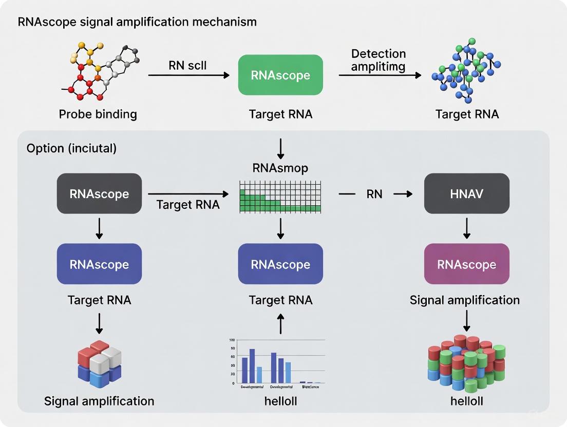
Figure 1: The RNAscope Signal Amplification Cascade. This diagram illustrates the sequential hybridization steps, beginning with the binding of two Z probes to the target RNA and culminating in the generation of a visible, punctate signal.
Performance Validation: Quantitative Data and Diagnostic Concordance
The performance of the RNAscope platform, underpinned by the Double Z probe design, has been rigorously validated against established gold-standard methodologies. A 2021 systematic review of 27 studies evaluated RNAscope's application in the clinical diagnostic field compared to techniques like IHC, qPCR, and DNA ISH [7].
Table 2: Performance Concordance of RNAscope with Gold Standard Techniques
| Comparison Method | Concordance Rate (CR) Range | Primary Reason for Discrepancy |
|---|---|---|
| qPCR / qRT-PCR | 81.8% - 100% | Different measures (RNA in situ vs. extracted RNA) but high correlation. |
| DNA ISH | 81.8% - 100% | High agreement in gene detection; RNAscope offers single-molecule resolution. |
| Immunohistochemistry (IHC) | 58.7% - 95.3% | Measures different biomolecules (RNA vs. protein); reflects post-transcriptional regulation. |
The data confirm RNAscope as a highly sensitive and specific method [7]. Its high concordance with PCR-based methods underscores its quantitative accuracy for RNA measurement. The lower concordance with IHC is expected and biologically informative, as it highlights the potential discordance between mRNA transcript presence and translated protein product, a common phenomenon in gene regulation [7].
The technology's sensitivity is further demonstrated by its ability to detect a wide range of transcripts, from scarcely expressed tumor suppressor lncRNAs (e.g., NRON) to highly expressed oncogenic lncRNAs (e.g., MALAT1) in formalin-fixed paraffin-embedded (FFPE) tissues, including samples archived for over 25 years [8] [9].
Experimental Protocol: A Detailed Workflow for FFPE Tissues
The RNAscope assay workflow for FFPE tissues integrates the core Double Z principle into a standardized, reproducible procedure. The following protocol is adapted from the seminal publication and manufacturer's guidelines [2] [1].
Slide Preparation and Pretreatment
- Sectioning: Cut FFPE tissue sections to a thickness of 5 μm and mount onto slides.
- Deparaffinization and Dehydration: Bake slides and deparaffinize in xylene, followed by dehydration through a graded ethanol series.
- Target Retrieval: Incubate slides in citrate buffer (10 mmol/L, pH 6) at a boiling temperature (100–103°C) for 15 minutes to unmask target RNA sequences.
- Protease Digestion: Treat slides with protease (e.g., 10 μg/mL) at 40°C for 30 minutes to permeabilize the tissue and allow probe access.
Probe Hybridization and Amplification
All hybridization steps are performed at 40°C in a specialized hybridization oven. Between each step, slides are washed with a proprietary wash buffer to remove unbound components [1].
- Target Probe Hybridization: Apply the pool of ~20 double Z target probe pairs in hybridization buffer to the tissue and incubate for 2 hours.
- Preamplifier Hybridization: Apply the preamplifier molecule (2 nmol/L) in hybridization buffer and incubate for 30 minutes.
- Amplifier Hybridization: Apply the amplifier molecule (2 nmol/L) in hybridization buffer and incubate for 15 minutes.
- Label Probe Hybridization: Apply the enzyme- or fluorophore-conjugated label probes (2 nmol/L) and incubate for 15 minutes.
Signal Detection and Visualization
- Chromogenic Development: For enzyme-conjugated probes, add the appropriate chromogenic substrate (e.g., Fast Red with alkaline phosphatase or DAB with horseradish peroxidase) to develop the signal.
- Counterstaining and Mounting: Counterstain with hematoxylin or another nuclear stain, then apply mounting medium and a coverslip.
- Microscopy and Analysis: Visualize the stained slides under a bright-field (chromogenic) or epifluorescent (fluorescent) microscope. Punctate dots are quantified manually or using automated image analysis software (e.g., HALO, QuPath) [2] [7].
The Researcher's Toolkit: Essential Reagents and Controls
Successful implementation of the RNAscope assay requires a suite of specific reagents and rigorous controls to validate results.
Table 3: Essential Research Reagent Solutions for RNAscope Assays
| Item | Function | Example & Notes |
|---|---|---|
| Target-Specific Probe Pools | Hybridize to the RNA of interest; the core detection reagent. | Custom-designed against ~1 kb of target sequence (e.g., catalog or made-to-order probes). |
| Positive Control Probe | Validates tissue RNA integrity and assay procedure. | Probes for housekeeping genes: PPIB (moderate expression), Polr2A (low expression), UBC (high expression) [7]. |
| Negative Control Probe | Assesses background noise and non-specific amplification. | Bacterial gene DapB, absent in mammalian tissues [1] [7]. |
| Signal Amplification Kits | Contain pre-amplifiers, amplifiers, and label probes. | e.g., RNAscope 2.5 HD Red Kit (Cat. No. 322350) or Multiplex Fluorescent Reagent Kit [1] [9]. |
| Pretreatment Reagents | Unmask target RNA and permeabilize cells. | RNAscope Pretreatment Kit for antigen retrieval and protease digestion [2]. |
| Image Analysis Software | Quantifies punctate dots for objective, high-throughput analysis. | HALO Software (Indica Labs), QuPath, Aperio [2] [7] [10]. |
Advanced Applications in Research and Therapeutic Development
The unique attributes of the Double Z probe design have enabled its application in cutting-edge research and drug development, particularly in areas where spatial context is crucial.
- Gene Therapy Biodistribution: RNAscope is a powerful tool for visualizing the biodistribution and cellular tropism of viral vectors (e.g., AAV) and for quantifying the expression and persistence of transgene mRNA in preclinical models, providing critical data for FDA-recommended biodistribution studies [10].
- Multiplexing and Co-Detection: The platform allows for simultaneous detection of up to four different RNA targets in a single sample using spectrally distinct fluorescent labels. Furthermore, it can be combined with immunohistochemistry (IHC) on the same tissue section for simultaneous detection of RNA and protein [7].
- Detection of Challenging Targets: The technology is exceptionally suited for visualizing long non-coding RNAs (lncRNAs) and in archival FFPE samples, where RNA may be partially degraded. Its short probe binding regions (~50 bases per pair) make it more tolerant to RNA fragmentation than traditional methods [2] [8].
The Double Z probe design is the cornerstone of the RNAscope platform, providing a sophisticated blueprint that ingeniously links signal amplification to an inherent noise-suppression mechanism. By mandating the contiguous hybridization of two independent probes for the initiation of a powerful amplification cascade, this technology achieves a level of specificity and sensitivity that enables single-molecule RNA visualization in situ. Its validated performance, compatibility with routine FFPE specimens, and adaptability to diverse research applications—from basic biology to therapeutic development—solidify its role as an indispensable tool in spatial genomics. As research continues to emphasize the importance of cellular context in gene expression, the principles embodied by the Double Z probe design will remain central to unraveling the complex spatial landscape of the transcriptome.
The quest for precise spatial genomics has driven the development of advanced in situ hybridization (ISH) technologies capable of detecting individual RNA molecules within their native cellular and tissue contexts. The RNAscope platform represents a paradigm shift in ISH methodologies, employing a proprietary signal amplification system that achieves exceptional signal-to-noise ratios through an elegant biochemical cascade. This technical guide examines the core mechanism underpinning RNAscope's exceptional performance, focusing on the multi-stage amplification process that enables single-molecule sensitivity and quantitative spatial biology. Within the broader thesis of signal amplification mechanism research, RNAscope's "double Z" probe design stands as a significant innovation that addresses the critical challenge of non-specific hybridization that has historically limited conventional ISH approaches. This whitepaper provides researchers, scientists, and drug development professionals with a comprehensive technical reference for understanding, implementing, and optimizing RNAscope technology in their experimental workflows, with particular emphasis on the quantitative aspects of its amplification cascade and practical guidance for experimental design.
Core Amplification Mechanism
The Double Z Probe Design Foundation
The foundational innovation enabling RNAscope's exceptional performance lies in its proprietary double Z probe design, which functions as a molecular recognition system with built-in specificity verification. This design employs two distinct "Z" probes that must hybridize in tandem to the target RNA molecule for signal amplification to proceed [2]. Each target Z probe consists of three critical regions: an 18-25 base lower region complementary to the target RNA, a spacer sequence, and a 14-base tail sequence [2]. The requirement for dual independent binding events dramatically reduces false-positive signals, as it is statistically improbable that two independent probes would bind adjacent sites on a non-specific target [2]. This approach conceptually parallels fluorescence resonance energy transfer (FRET) systems in its requirement for coordinated molecular interactions, but applies this principle to ensure specific amplification rather than energy transfer.
For each target RNA, approximately 20 double Z probe pairs are designed to hybridize along the transcript [2]. This multiplexed binding strategy provides robustness against partial target RNA degradation or accessibility issues, as only three of the 20 probe pairs need to successfully bind to detect a single RNA molecule [2]. The relatively short target region (40-50 bases total for the double Z probe) further enhances compatibility with partially degraded RNA samples, such as those from formalin-fixed paraffin-embedded (FFPE) tissues [2]. When both Z probes successfully hybridize to their adjacent target sequences, their tail regions form a combined 28-base binding site for the next component in the amplification cascade [2].
The Signal Amplification Cascade
The RNAscope signal amplification cascade employs a sequential hybridization approach that generates substantial signal amplification through a branching hierarchy of binding events. This multi-stage process transforms each successful double Z probe binding event into a large signaling complex capable of generating detectable output.
Table 1: Stages of the RNAscope Signal Amplification Cascade
| Amplification Stage | Key Components | Function | Output Multiplier |
|---|---|---|---|
| Target Recognition | 20 double Z probe pairs | Hybridize to target RNA molecule | 1× (baseline) |
| Preamplifier Binding | Preamplifier molecules | Bind to combined tail regions of double Z probes | 1:1 ratio with bound probe pairs |
| Amplifier Assembly | Amplifier molecules | Bind to multiple sites on each preamplifier | ~20× per preamplifier |
| Label Probe Attachment | Fluorescent or chromogenic label probes | Bind to numerous sites on each amplifier | ~20× per amplifier |
The cascade begins when double Z target probes hybridize to the target RNA [2]. Next, preamplifiers hybridize to the 28-base binding site formed by each double Z probe pair [2]. Each preamplifier then provides multiple binding sites for amplifier molecules [2]. Finally, labeled probes containing fluorescent molecules or chromogenic enzymes bind to the numerous sites on each amplifier [2]. While the exact "8000-fold" amplification figure is not explicitly detailed in the search results, the 20×20×20 probe design strategy suggests the theoretical amplification potential [2]. This multi-stage approach enables visualization of individual RNA molecules as punctate dots under a standard microscope, with each dot representing a single RNA transcript [2] [11].
Diagram 1: RNAscope Signal Amplification Cascade. The diagram illustrates the sequential hybridization process from target RNA recognition through the double Z probes, preamplifier binding, amplifier assembly, and final label probe attachment that generates the detectable signal.
Quantitative Performance Data
Signal Amplification Metrics
The RNAscope amplification system achieves remarkable sensitivity and specificity metrics that represent significant advancements over conventional ISH methods. The technology enables single-molecule detection sensitivity, with each punctate dot representing an individual RNA transcript [2] [11]. This single-molecule resolution is achieved through the 20×20×20 probe design and signal amplification strategy, which provides sufficient sensitivity to visualize individual RNA molecules as distinct punctate signals under standard microscopy [2]. The double Z probe design requires only three of the approximately 20 probe pairs to bind to the target RNA for successful detection of each single RNA molecule, providing robustness against partial target degradation or accessibility issues [2].
Table 2: RNAscope Performance Metrics and Quantitative Capabilities
| Performance Parameter | Specification | Experimental Significance |
|---|---|---|
| Detection Sensitivity | Single RNA molecule detection [2] [11] | Enables quantitative single-cell transcript counting |
| Signal-to-Noise Ratio | High (specific amplification prevents background) [2] | Clear distinction between true signal and background fluorescence |
| Probe Binding Requirement | 3 of 20 ZZ probe pairs minimum [2] | Robust detection even with partially degraded targets |
| Target Accessibility | 40-50 base region required for double Z binding [2] | Compatible with partially degraded RNA (e.g., FFPE samples) |
| Multiplexing Capacity | Up to 12 targets in FFPE, 48 in fresh frozen with HiPlex [12] | Comprehensive cellular profiling in spatial context |
| Quantification Methods | Manual counting or HALO software automated analysis [2] | Flexible analysis approaches for different throughput needs |
The double Z probe design specifically prevents amplification of non-specific signals, as individual Z probes binding to non-specific sites cannot form the complete 28-base binding site required for preamplifier attachment [2]. This mechanism fundamentally eliminates the background noise that plagues conventional ISH approaches. The system's compatibility with partially degraded samples stems from the relatively short target regions (40-50 bases) required for the double Z probes, enabling successful hybridization even when RNA integrity is compromised [2].
Experimental Protocols and Methodologies
Standard Workflow Implementation
The RNAscope assay follows a standardized workflow that maintains the integrity of cellular and tissue architecture while enabling highly specific RNA visualization. The process begins with sample preparation, where tissue sections or cells are fixed onto slides and pretreated with the RNAscope Pretreatment Kit to unmask target RNA and permeabilize cells [2]. This critical initial step ensures accessibility of the target RNA while preserving morphological context. The hybridization phase follows, where the specially designed RNAscope probes containing approximately 20 target-specific double Z probes hybridize to target RNA molecules [2]. This hybridization typically occurs under optimized conditions that maximize specific binding while minimizing non-specific interactions.
Following hybridization, the signal amplification phase employs RNAscope Detection Reagents that sequentially hybridize amplifiers and label probes to build the detectable signal [2]. This multi-stage amplification occurs through a cascade of hybridization events rather than enzymatic reactions, enhancing consistency and reducing variability. Visualization represents the final experimental phase, where each target RNA molecule appears as a punctate dot that can be visualized with a microscope [2]. These signals can then be quantified on a cell-by-cell basis through manual counting or automated image analysis using platforms such as HALO Software [2]. The entire process preserves spatial relationships between cells and transcripts while generating quantitative data at single-molecule resolution.
Advanced Multiplexing Approaches
The HiPlex v2 assay represents an advanced iteration of the RNAscope platform that enables highly multiplexed detection through an iterative detection approach. This system allows for simultaneous detection of up to 12 targets in FFPE samples and up to 48 targets in fresh and fixed frozen samples [12]. The methodology employs cleavable fluorophores in an iterative process where up to four target genes are visualized simultaneously in four distinct fluorescent channels, after which the fluorophores are cleaved and the process repeated until all targets are detected [12]. This fluorophore cleaving procedure is rapid and minimizes impact on RNA integrity and tissue morphology since it does not strip away the bound probes [12].
For researchers implementing HiPlex assays, fluorophore selection follows specific brightness considerations to optimize signal-to-noise ratios. The system recommends using fluorescein (green/AF488) for high expressors, as this channel may overlap with tissue autofluorescence, while Cyanine 3 (orange), Cyanine 5 (far red), and Cyanine 7 (far red) channels are recommended for low expressors or when expression levels are unknown, as these wavelengths experience less interference from autofluorescence [12]. Following multiple rounds of imaging, the images are merged using RNAscope HiPlex Image Registration Software v2, which enables users to toggle and overlay expression of select targets, simplifying data interpretation of complex multiplexed experiments [12].
Visualization and Data Interpretation
Signal Patterns and Analytical Approaches
RNAscope signals manifest as discrete punctate dots, with each dot representing a single RNA transcript [11]. This characteristic pattern enables direct quantification of transcript abundance at cellular and subcellular resolutions. In some instances, clusters of signals may appear, resulting from overlapping signals from multiple mRNA molecules in close proximity [11]. Analytical approaches include both semi-quantitative scoring guidelines and fully quantitative methods using image analysis software [11]. The technology supports analysis through various platforms including ImageJ, Cell Profiler, QuPath, or the HALO software from Indica Labs [11].
When interpreting RNAscope data, it is essential to recognize that dot count rather than dot size or intensity reflects transcript numbers [11]. Variations in dot intensity or size result from differences in the number of ZZ probes bound to target molecules, but each dot regardless of size represents a single transcript [11]. This principle forms the foundation for accurate quantification and distinguishes RNAscope from protein detection methods where intensity variations often correlate with expression levels. Proper experimental design should always include appropriate controls, specifically running a minimum of three slides per sample: the target marker panel, a positive control probe to assess RNA quality, and a negative control probe (typically bacterial dapB) to verify appropriate tissue preparation [11].
Research Reagent Solutions
Essential Experimental Components
Successful implementation of RNAscope technology requires specific reagent systems optimized for the amplification cascade. The platform offers both chromogenic and fluorescent detection options, with the HiPlex v2 system providing extended multiplexing capabilities through iterative detection approaches.
Table 3: Essential RNAscope Research Reagents and Their Functions
| Reagent Component | Function | Specification Notes |
|---|---|---|
| Probe Design | Double Z target-specific probes | ~20 pairs per target RNA; 18-25 base complementary regions [2] |
| Pretreatment Kit | Unmask target RNA, permeabilize cells | Optimized for different sample types (FFPE, frozen) [2] |
| Detection Reagents | Signal amplification | Sequential hybridization of amplifiers and label probes [2] |
| HiPlex v2 Reagent Kit | Multiplex fluorescent detection | Enables 12-plex in FFPE, 48-plex in fresh frozen [12] |
| Positive Control Probes | Assess RNA quality | Species-specific (human, mouse, rat) [12] |
| Negative Control Probe | Verify specificity | Bacterial dapB gene [12] [11] |
| Probe Diluent | Optimal probe reconstitution | Maintains probe stability and hybridization efficiency [12] |
| Hydrophobic Barrier Pen | Create hybridization areas | Defines reaction zones on slides [12] |
| Image Registration Software | Align multiplex imaging rounds | HiPlex v2 software for target overlay and visualization [12] |
For researchers new to the platform, introductory packs provide comprehensive kits containing all essential components, including reagent kits, species-specific positive control probes, universal negative control probes, probe diluent, ImmEdge hydrophobic barrier pens, and the requisite image registration software for multiplex analyses [12]. These systems are designed with compatibility for various microscope configurations, with fluorescent assays requiring epi-fluorescent or confocal microscopes with appropriate filter sets for the assigned fluorophores [11].
Diagram 2: RNAscope Experimental Workflow and Quality Control. The diagram outlines the key steps in the RNAscope protocol, highlighting the essential control measures required for valid experimental interpretation.
The RNAscope amplification cascade represents a significant advancement in spatial genomics, providing researchers with an unparalleled ability to visualize and quantify RNA expression within morphological context. Through its innovative double Z probe design and sequential hybridization amplification strategy, the technology achieves single-molecule sensitivity while effectively suppressing background noise that has historically challenged conventional ISH methods. The mechanistic insights and technical guidelines presented in this whitepaper provide researchers with the foundation to implement this technology effectively, from basic single-plex detection to advanced highly multiplexed spatial profiling. As spatial biology continues to transform our understanding of complex biological systems, RNAscope's robust signal amplification mechanism stands as a critical enabling technology that bridges the gap between genomic sequencing data and tissue context, ultimately accelerating discovery in basic research and drug development.
The ability to visualize individual RNA molecules within their native morphological context represents a paradigm shift in transcriptomics. The dot-per-transcript principle is the foundational concept enabling this single-molecule resolution, wherein each detected signal dot corresponds precisely to one individual RNA molecule [2] [13]. This principle is operationalized through advanced RNA in situ hybridization (ISH) technologies, with RNAscope being a premier example, allowing researchers to map and quantify gene expression with unprecedented sensitivity and specificity in formalin-fixed, paraffin-embedded (FFPE) tissues, cultured cells, and fresh-frozen samples [1] [13]. This technical guide details the core mechanisms, experimental protocols, and analytical frameworks of this powerful methodology, framing it within broader research on RNAscope signal amplification mechanisms.
Core Mechanism: The Double-Z Probe Design
The exceptional performance of platforms like RNAscope stems from a proprietary probe design strategy that fundamentally enhances the signal-to-noise ratio of traditional RNA ISH.
The "Double Z" Probe Architecture
The system uses pairs of so-called "Z" probes that must bind contiguously to the target RNA. Each probe consists of three elements [1] [2]:
- A Target-Binding Region (18-25 bases): The lower segment, complementary to the target RNA sequence.
- A Spacer Sequence: A linker region that gives the probe its distinctive Z-shape.
- A Tail Sequence (14 bases): The upper segment that facilitates signal amplification.
For signal amplification to occur, two probes must hybridize side-by-side on the target RNA molecule. Their combined tail sequences create a single 28-base binding site for the preamplifier molecule [2]. This requirement is the cornerstone of the technology's specificity, as it is statistically improbable for two independent probes to bind nonspecifically to off-target sequences in the correct tandem orientation [1] [13].
Signal Amplification Cascade
Once the probe pairs are bound, a multi-step hybridization cascade creates massive signal amplification:
- Preamplifier Binding: A preamplifier molecule hybridizes to the 28-base site formed by the double Z probe pair.
- Amplifier Binding: Each preamplifier contains multiple binding sites for amplifier molecules.
- Label Probe Binding: Each amplifier, in turn, contains numerous binding sites for label probes conjugated with either fluorescent dyes or chromogenic enzymes (e.g., horseradish peroxidase) [1] [2].
This branching amplification strategy can theoretically generate up to 8,000 labels for each target RNA molecule, achieving a signal strong enough for microscopic visualization [1]. The high redundancy of 20 probe pairs per target RNA ensures robust detection even for partially degraded or inaccessible targets, as binding of just three probe pairs is sufficient for single-molecule detection [2] [13].
Figure 1: The RNAscope Signal Amplification Cascade. This diagram illustrates the sequential binding and hybridization steps that transform the binding of a single target RNA molecule into a detectable punctate dot.
Experimental Protocol: A Step-by-Step Guide
A successful RNAscope assay requires careful attention to sample preparation, pretreatment, and hybridization. The following protocol is adapted for FFPE tissues, a common sample type in research and diagnostics [13] [14].
Sample Preparation and Pretreatment
- Fixation and Sectioning: Tissue samples should be fixed in 10% neutral buffered formalin for 6-72 hours at room temperature, processed, and embedded in paraffin (FFPE). Sections of 5 μm thickness are mounted on glass slides [1] [14].
- Deparaffinization and Rehydration: Slides are deparaffinized in xylene and dehydrated through a graded ethanol series.
- Pretreatment (Critical for FFPE): The pretreatment process is crucial for unmasking target RNA and permeabilizing the sample [13].
- Target Retrieval: Slides are incubated in a specific target retrieval buffer at 100–103°C for 15 minutes to partially reverse cross-links formed during fixation.
- Hydrogen Peroxide Block: Endogenous peroxidase activity is blocked by applying a hydrogen peroxide reagent.
- Protease Digestion: A protease treatment (e.g., Protease Plus) is applied for 30 minutes at 40°C to permeabilize cell membranes and degrade proteins bound to RNA, thereby unmasking the target sequences [1] [13].
Probe Hybridization and Amplification
- Hybridization: Target probes are diluted in a hybridization buffer and applied to the pretreated tissue sections. Slides are incubated at 40°C for 2 hours to allow the double Z probes to hybridize to their target RNA [1].
- Signal Amplification: A series of sequential hybridizations are performed at 40°C, with buffer washes between each step:
- Preamplifier hybridizes for 30 minutes.
- Amplifier hybridizes for 15 minutes.
- Label Probe hybridizes for 15 minutes [1].
- Detection: For fluorescent detection, the label probe is conjugated to a fluorophore (e.g., Alexa Fluor 488, 546, 647). For bright-field microscopy, the label probe is conjugated to horseradish peroxidase (HRP) or alkaline phosphatase, followed by incubation with a chromogenic substrate (DAB or Fast Red) [1] [14].
- Counterstaining and Mounting: Tissues are counterstained (with hematoxylin for chromogenic or DAPI for fluorescent detection), dehydrated, cleared, and mounted with a suitable mounting medium [1].
Figure 2: RNAscope Experimental Workflow. The key steps of the procedure, from sample preparation to final analysis, are shown sequentially.
Data Interpretation and Quantification
Adherence to the dot-per-transcept principle dictates a specific approach to data analysis.
Microscopy and Qualitative Assessment
Stained slides are visualized under a standard bright-field or fluorescence microscope. A successful assay will show punctate dots within the cytoplasm and/or nucleus of cells, with minimal to no background staining [2] [13]. Each dot represents a single RNA molecule.
Quantitative and Semi-Quantitative Scoring
Quantification is performed by counting dots per cell, not by measuring signal intensity [13].
- Manual or Automated Counting: Dots can be counted manually per cell across multiple fields of view or by using automated image analysis software (e.g., HALO, QuPath, ImageJ) [2] [13].
- Semi-Quantitative Scoring System: RNAscope assays often use a standardized scoring system to evaluate expression levels [13]:
- Score 0: No staining or <1 dot per 10 cells.
- Score 1: 1-3 dots per cell.
- Score 2: 4-9 dots per cell (few or no clusters).
- Score 3: 10-15 dots per cell (<10% of dots in clusters).
- Score 4: >15 dots per cell (>10% of dots in clusters).
Essential Controls
Every experiment must include control probes to ensure results are interpretable [1] [13]:
- Positive Control Probe: A housekeeping gene like PPIB, UBC, or POLR2A. Successful staining requires a score ≥2 for PPIB/POLR2A or ≥3 for UBC.
- Negative Control Probe: A bacterial gene like dapB. The score for this should be <1, indicating minimal nonspecific background.
Performance Data and Technical Validation
The performance of the RNAscope technology is demonstrated by its application in rigorous scientific studies.
Table 1: Key Performance Metrics of RNAscope Technology
| Parameter | Specification / Outcome | Context / Validation |
|---|---|---|
| Sensitivity | Single RNA molecule detection | Requires binding of ≥3 double Z probe pairs to target RNA [2] |
| Specificity | High signal-to-noise; minimal off-target signal | Enabled by dual Z-probe binding requirement for preamplifier [1] [13] |
| Probe Design | 20 probe pairs per target RNA; 18-25 bp per target-hybridizing sequence | Provides redundancy against degradation and ensures uniform hybridization [1] |
| Sample Compatibility | FFPE, fresh-frozen, fixed-frozen tissues, cultured cells | Validated for FFPE fixed for 6-72 hours according to ASCO/CAP guidelines [1] |
| Application Example | Detection of c-KIT mRNA in canine mast cell tumors | Strong correlation found between mRNA expression and histological grade [14] |
Table 2: Comparison of RNA Visualization Methods
| Method | Principle | Spatial Context | Single-Molecule Sensitivity | Key Limitation |
|---|---|---|---|---|
| RNAscope | In situ hybridization with branched DNA amplification | Preserved (in situ) | Yes (Dot-per-transcript) | Requires probe design for known sequences [1] [2] |
| Bulk RNA-Seq | High-throughput sequencing of pooled RNA | Lost (grind-and-bind) | No (Measures ensemble average) | Loses tissue morphology and cell-to-cell variation [15] [16] |
| Single-Molecule Fluorescence (e.g., RIFT) | Fluorogenic aptamers (e.g., Peppers) in real-time assays | Lost (in vitro system) | Yes | Primarily for in vitro applications, not intact tissues [17] |
| Nanopore Direct RNA-Seq | Direct electrical current measurement of native RNA | Lost | Yes (theoretically) | Complex data analysis; currently limited for novel modifications [18] |
The Scientist's Toolkit: Essential Research Reagents
Implementing the dot-per-transcript principle requires a specific set of reagents and tools.
Table 3: Research Reagent Solutions for RNAscope Assays
| Reagent / Tool | Function | Examples & Notes |
|---|---|---|
| Target Probes | Specifically hybridize to the RNA of interest; form the foundation of the double Z system | Custom-designed for each target mRNA; ~20 pairs per target [1] [2] |
| Signal Amplification System | Preamplifier, Amplifier, and Label Probes that sequentially build the detectable signal | Proprietary reagents; labels can be fluorescent (Alexa Fluor dyes) or enzymatic (HRP) [1] [13] |
| Pretreatment Kits | Unmask target RNA, permeabilize cells, and block endogenous enzyme activity | Include Target Retrieval buffers, Hydrogen Peroxide reagent, and Proteases (e.g., Protease Plus) [13] |
| Control Probes | Verify assay performance and RNA quality in the test sample | Positive: Housekeeping genes (PPIB, UBC). Negative: Bacterial gene dapB [1] [13] |
| Image Analysis Software | Quantify punctate dots on a cell-by-cell basis for objective quantification | HALO, QuPath, ImageJ, CellProfiler [2] [13] |
Advanced Applications and Future Directions
The dot-per-transcript principle has enabled sophisticated applications that extend beyond simple mRNA detection.
- Multiplexing: Using multiple probe sets with different tail sequences and distinct fluorophores allows for simultaneous detection of two or more RNA species in the same sample, enabling the study of gene co-expression and interaction within a single cell [1].
- Integration with Protein Detection: Combining RNAscope with immunohistochemistry (IHC) on the same section allows researchers to correlate mRNA expression with protein localization and cell phenotype in situ [14].
- Novel Biomarker Discovery: The ability to precisely localize and quantify low-abundance transcripts in pathological tissues, as demonstrated in canine mast cell tumors, positions this technology as a powerful tool for discovering and validating new diagnostic and prognostic biomarkers [14].
In conclusion, the dot-per-transcript principle, as realized in technologies like RNAscope, provides a robust framework for single-molecule RNA visualization. Its high sensitivity and specificity, combined with the ability to analyze RNA within its histological context, make it an indispensable tool for basic research, drug development, and molecular pathology.
Core Mechanism Enabling High-Resolution Spatial Biology
The RNAscope in situ hybridization (ISH) technology represents a significant advancement in spatial biology, providing a powerful method to detect gene expression within the intact spatial and morphological context of tissues. Its key advantages stem from a proprietary signal amplification system that enables single-molecule detection of RNA transcripts at single-cell resolution while fully preserving tissue architecture. This technical capability allows researchers to visualize the precise cellular localization of gene expression without disrupting the native tissue microenvironment, offering critical insights into cellular heterogeneity, gene regulation, and pathological processes that are lost in bulk analysis methods [19] [20].
The fundamental breakthrough of RNAscope technology lies in its "double Z" probe design combined with an advanced signal amplification system. This engineered approach enables highly specific and sensitive detection of target RNA, with each visualized dot representing a single RNA transcript. This robust signal-to-noise ratio technology provides clear answers while seamlessly integrating into existing anatomic pathology workflows, making it particularly valuable for both research and clinical diagnostic applications [19] [20] [21].
Technical Foundation and Signal Amplification Mechanism
Proprietary Probe Design and Amplification System
The RNAscope signal amplification mechanism relies on a sophisticated multi-step process that differentiates it from traditional in situ hybridization methods:
- Double Z Probe Design: RNAscope utilizes paired "Z" probes that bind adjacent target sequences on the RNA of interest. This dual-probe system requires both probes to bind correctly for signal generation, dramatically increasing specificity and reducing false positives from non-specific binding [19] [20].
- Amplification Tree Structure: After hybridization, the bound probes serve as scaffolds for a pre-amplifier molecule, which then recruits multiple amplifier molecules. Each amplifier subsequently binds numerous enzyme conjugates, creating a large signal amplification complex precisely localized to the original RNA molecule [22].
- Single-Molecule Visualization: The extensive amplification enables detection of individual RNA molecules as distinct fluorescent or chromogenic dots, allowing for both qualitative assessment of expression patterns and quantitative analysis of transcript numbers per cell [19] [22].
Table 1: RNAscope Signal Amplification Steps and Components
| Step | Component | Function | Key Characteristic |
|---|---|---|---|
| 1 | Target RNA | Molecule of interest | Preserved in tissue context |
| 2 | ZZ Probe Pairs | Hybridization probes | Require dual binding for specificity |
| 3 | Pre-Amplifier | Signal initiation | Binds only to properly paired Z probes |
| 4 | Amplifier | Signal branching | Multi-branched structure for amplification |
| 5 | Enzyme Conjugate | Signal detection | Enzyme linked to fluorescent or chromogenic label |
| 6 | Substrate | Visual output | Generates detectable signal at RNA location |
Experimental Validation of Performance Characteristics
Extensive validation studies have demonstrated RNAscope's technical performance compared to established molecular detection methods. A 2022 systematic review evaluated RNAscope against gold standard techniques in clinical diagnostics, analyzing 27 studies primarily focusing on cancer samples [22].
Table 2: Performance Comparison Between RNAscope and Established Methods
| Method Compared | Concordance Range | Key Advantages of RNAscope | Limitations Addressed |
|---|---|---|---|
| IHC | 58.7-95.3% | Detects RNA directly, avoids antibody cross-reactivity | Higher sensitivity for low-abundance targets |
| qPCR/qRT-PCR | 81.8-100% | Preserves spatial context, no tissue homogenization | Maintains tissue architecture and cell-specific data |
| DNA ISH | 81.8-100% | Superior signal-to-noise, single-molecule sensitivity | Enhanced detection of low-copy transcripts |
| Bulk RNA-seq | N/A | Single-cell resolution in tissue context | Identifies spatial expression patterns |
The review confirmed RNAscope as a "highly sensitive and specific method" but noted insufficient data for it to stand alone in clinical diagnostics without further validation [22]. The lower concordance with IHC primarily stems from the fundamental difference in detecting RNA versus protein, along with potential post-transcriptional regulation effects [22].
Experimental Protocol Framework
Critical Workflow Considerations
Implementing RNAscope successfully requires careful attention to several critical protocol parameters that significantly impact assay performance:
- Temperature and Humidity Control: Both factors are crucial for assay performance. The HybEZ oven is the only hybridization system extensively validated by ACD for consistent results [21].
- Protease Digestion Optimization: Protease treatment must be carefully calibrated. Under-digestion results in lower signal and ubiquitous background, while over-digestion causes poor morphology and RNA loss [21].
- Sample Preparation Integrity: For fresh-frozen tissues, immediate fixation after sectioning is essential. Under-fixation leads to protease over-digestion and RNA loss, while over-fixed specimens yield poor probe accessibility and low signal-to-background ratio [23].
- Reagent Freshness and Protocol Adherence: Always use fresh reagents, including alcohol and xylene, and do not alter the protocol sequence. For example, after boiling steps, samples should go directly into dH₂O without cooling [21] [23].
Multiplexing Capabilities and Experimental Design
RNAscope supports sophisticated multiplexing approaches for simultaneous detection of multiple RNA targets:
- Channel-Based Detection System: RNAscope employs a channel system (C1, C2, C3, C4) for independent detection of different targets. C1 probes are Ready-To-Use (RTU), while C2, C3, and C4 probes are shipped as 50X concentrated stocks [21].
- Probe Configuration Flexibility: C1 probe can be substituted with blank probe diluent and used with C2, C3, or C4 target probes for duplex/multiplex assays. However, C2 or C3 probes cannot be used with RNAscope 2.5 HD singleplex Brown or 2.5 HD Red assays, which are designed specifically for C1 probes [21].
- Combination with Immunofluorescence: The technology can be combined with protein detection methods, enabling true multi-omic analysis of RNA and protein biomarkers within the same tissue section while preserving spatial context [19] [24].
Advanced Applications and Integration with Spatial Transcriptomics
Position in Modern Spatial Biology Workflow
RNAscope occupies a unique niche in the expanding landscape of spatial transcriptomics technologies. A 2025 comparative analysis of spatial transcriptomics methods highlighted RNAscope's role alongside emerging platforms like Xenium, Merscope, and Molecular Cartography [25].
The study found that automated imaging-based spatial transcriptomics methods, including RNAscope HiPlex, were "well-suited to delineate intricate microanatomy and capture cell-type-specific transcriptome profiles" in medulloblastoma tumors with extensive nodularity (MBEN) [25]. The research demonstrated strong correlation between RNAscope and other imaging-based platforms (Xenium: r=0.82; Molecular Cartography: r=0.74; Merscope: r=0.65), validating its quantitative accuracy [25].
Quantitative Image Analysis Frameworks
The single-molecule resolution of RNAscope enables sophisticated quantitative analysis through both manual and computational approaches:
- Digital Image Analysis Algorithms: Advanced platforms like HALO and QuPath enable automated quantification of transcript counts per cell, cell segmentation, and spatial analysis of expression patterns [22] [26] [24].
- H-Scoring System: For clinical applications, digital H-scores can be calculated by determining percentages of tumor cells expressing low, medium, and high levels of target RNA, providing standardized quantification for diagnostic decisions [26].
- Spatial Biology Applications: Professional assay services leverage image analysis tools to provide quantitative results for oncology, neuroscience, cell and gene therapy, single-cell RNAseq validation, and biomarker development [24].
RNAscope Signal Amplification Workflow: This diagram illustrates the sequential binding process that enables single-molecule detection, from initial probe hybridization to final signal amplification and visualization.
Essential Research Reagent Solutions
Table 3: Key Research Reagents and Equipment for RNAscope Implementation
| Component | Function | Application Notes |
|---|---|---|
| HybEZ Oven System | Temperature and humidity control | Critical for consistent results; extensively validated [21] |
| Target Probes | Gene-specific detection | C1 (Ready-To-Use), C2/C3/C4 (50X concentrate) [21] |
| Protease Plus | Tissue permeabilization | Critical step; requires optimization for each tissue type [21] [23] |
| AMP Reagents | Signal amplification | AMP1, AMP2, AMP3 applied sequentially [23] |
| HRP Blockers | Channel inactivation | Enables multiplex detection [23] |
| Opal Fluorophores | Fluorescent detection | Multiple colors for multiplexing (520, 570, 690) [23] |
| Positive Control Probes | Assay validation | Species-specific housekeeping genes [21] |
| Negative Control Probes | Background assessment | Bacterial dapB gene [26] |
Experimental Workflow with Critical Control Points: This diagram outlines the key steps in RNAscope processing, highlighting stages requiring precise optimization to ensure specific signal detection while preserving tissue morphology and RNA integrity.
The RNAscope technology platform delivers on the dual promises of single-cell resolution and tissue context preservation through its engineered signal amplification mechanism. The proprietary "double Z" probe design provides exceptional specificity, while the branched DNA amplification system enables sensitive detection of individual RNA molecules. As spatial biology continues to evolve, RNAscope maintains its position as a validated and reliable method for targeted RNA detection within morphological context, bridging the gap between bulk sequencing approaches and emerging whole-transcriptome spatial technologies. Its compatibility with clinical samples, quantitative capabilities, and integration with multi-omic approaches make it particularly valuable for both basic research and translational applications in drug development and clinical diagnostics.
From Principle to Practice: RNAscope Workflows and Diverse Applications
Proper sample preparation is a critical foundational step for successful RNA in situ hybridization (ISH), especially for research focusing on RNAscope signal amplification mechanisms. The proprietary RNAscope technology utilizes a unique "double-Z" probe design to achieve single-molecule visualization while preserving tissue morphology [1]. This signal amplification system relies on pairs of target probes that hybridize contiguously to the target RNA, forming a binding site for pre-amplifier molecules and enabling significant signal amplification [1] [13]. The integrity of this entire process is profoundly influenced by the initial sample preparation, which must ensure both preservation of RNA integrity and maintenance of tissue morphology.
This technical guide details sample preparation methodologies for formalin-fixed paraffin-embedded (FFPE), fresh frozen, and cell culture specimens, providing researchers with standardized protocols essential for generating reliable, reproducible data in RNAscope-based spatial transcriptomics. The core principle across all sample types is that improper preparation can lead to RNA degradation, poor probe accessibility, or loss of morphological context, ultimately compromising the sensitive signal amplification process that makes RNAscope so powerful [1] [13].
Core Principles of RNAscope Technology and Sample Compatibility
The RNAscope platform represents a significant advancement in RNA ISH technology through its patented signal amplification and background suppression system. The core innovation lies in its "double-Z" probe design strategy, where each target probe contains an 18-25 base region complementary to the target RNA, a spacer sequence, and a 14-base tail sequence [1]. Pairs of these probes (double Z) must hybridize contiguously to the target RNA to form a 28-base hybridization site for the preamplifier, which then enables massive signal amplification through subsequent layers [1]. This design provides exceptional specificity because it is highly unlikely that nonspecific hybridization events will juxtapose two probe pairs correctly along an off-target sequence [1].
The signal amplification mechanism involves a multi-step process: after target probe hybridization, pre-amplifier binding occurs, followed by amplifier binding, and finally label probe attachment [13]. This cascading system can theoretically yield up to 8000 labels for each target RNA molecule when targeting a 1-kb region [1]. The high degree of redundancy (typically 20 probe pairs per target) means that even partially degraded or incompletely unmasked RNA targets may still be detectable, making the technology robust for suboptimal samples [13]. However, optimal performance requires careful sample preparation to maximize target accessibility while preserving tissue architecture.
Figure 1: RNAscope Signal Amplification Mechanism. The proprietary "double-Z" probe design enables highly specific target recognition and significant signal amplification through a cascading hybridization system. Each successfully bound probe pair initiates an amplification tree resulting in up to 8000 detectable labels per target RNA molecule [1] [13].
FFPE Sample Preparation
Advantages and Challenges
FFPE samples represent one of the most valuable resources for biomedical research, with an estimated 400 million to over a billion specimens archived worldwide in hospitals and biobanks [27]. Their primary advantages include superior preservation of tissue morphology, long-term storage at room temperature, and extensive clinical annotations with linked patient outcomes [27] [28]. These characteristics make FFPE specimens indispensable for retrospective studies and clinical biomarker validation.
However, FFPE preparation presents significant challenges for molecular analysis. The formalin fixation process induces chemical modifications, nucleic acid fragmentation, and protein cross-linking that can negatively impact RNA quality and accessibility [27] [28]. Studies have shown that RNA extracted from FFPE tissues is highly degraded compared to fresh or frozen alternatives, with DV200 values (percentage of RNA fragments >200 nucleotides) serving as a critical quality metric [28] [29]. The fixation time significantly affects RNA quality, with signals detectable up to 180 days of formalin fixation but diminishing thereafter [30].
FFPE Preparation Protocol
The following protocol outlines the standardized procedure for preparing FFPE samples compatible with RNAscope analysis:
Tissue Collection and Fixation:
- Collect tissue specimens preferably within 30 minutes of devitalization.
- Fix tissues in 10% neutral buffered formalin (NBF) within 4-24 hours postmortem [30].
- Maintain a 10:1 ratio of formalin to tissue volume to ensure complete penetration.
- Optimal fixation time ranges from 16-36 hours at room temperature [30]. Prolonged fixation beyond 30 days leads to irreversible covalent bond formation and RNA damage [30].
Processing and Embedding:
- Process fixed tissues through a series of ethanol dehydrations (70% to 100%), followed by clearing with xylene or substitutes like Histolab Clear.
- Infuse with molten paraffin using an automated tissue processor (e.g., Tissue-Tek VIP) with multiple paraffin baths to ensure complete infiltration.
- Embed tissues in paraffin blocks using standardized embedding systems.
Sectioning:
FFPE Pretreatment for RNAscope:
- Deparaffinize sections in xylene and rehydrate through graded ethanol series (100%, 70%) to water.
- Perform heat-induced epitope retrieval (HIER) using RNAscope Target Retrieval reagents by heating in citrate buffer (10 mM, pH 6) at 100-103°C for 15 minutes [1] [13].
- Digest with RNAscope Protease Plus (10 μg/mL) at 40°C for 30 minutes to permeabilize membranes and unmask RNA targets [13].
Table 1: Effect of Formalin Fixation Time on RNAscope Signal Detection [30]
| Fixation Time | Signal Intensity | % Area of Signal | Detection Capability |
|---|---|---|---|
| 1-28 days | Maximum | Maximum | Optimal detection |
| 60-90 days | Moderate decrease | Moderate decrease | Readily detectable |
| 180 days | Significant decrease | Significant decrease | Detectable but diminished |
| 270 days | Minimal to none | Minimal to none | Generally undetectable |
Fresh Frozen Sample Preparation
Advantages and Challenges
Fresh frozen tissue preservation is considered the gold standard for nucleic acid quality, providing RNA that is perfectly preserved for downstream molecular analysis [27]. The immediate cryopreservation halts cellular processes and enzymatic degradation, yielding higher-quality DNA and RNA compared to FFPE-derived nucleic acids [27] [28]. This method is less complicated, less time-consuming, and does not involve toxic chemicals like formalin [27].
The primary challenges for fresh frozen samples involve complicated and costly storage requirements. Tissues must be stored at -80°C in dedicated ultra-low-temperature freezers, making archives vulnerable to power outages or mechanical failures [27]. Additionally, the collection process requires immediate access to liquid nitrogen containers and -80°C freezers close to surgery or collection rooms, which may not always be practically feasible in clinical settings [27].
Fresh Frozen Preparation Protocol
The following protocol is optimized for fresh frozen tissue preparation compatible with RNAscope analysis, with specific considerations for brain tissue [23] [4]:
Tissue Collection and Snap-Freezing:
- For brain tissue, deeply anesthetize the animal and perform sacrifice by decapitation. Quickly remove the brain from the skull [4].
- Immediately immerse tissue in liquid nitrogen-chilled 2-methylbutane (isopentane) at -30°C to -50°C and snap-freeze for 25 seconds [4].
- Alternatively, immerse tissue directly in liquid nitrogen for rapid freezing [27].
- Avoid any thawing of the tissue and immediately store on dry ice or at -80°C.
Embedding and Sectioning:
Fixation and Pretreatment for RNAscope:
- Immerse slides in 4% paraformaldehyde (PFA) in 1x PBS for 2 hours (minimum 15 minutes) at room temperature [23].
- Wash twice in 1x PBS.
- Dehydrate through ethanol series (50%, 70%, 100%) for 5 minutes each at room temperature [23].
- Apply RNAscope Hydrogen Peroxide for 10 minutes at room temperature to block endogenous peroxidase activity [23] [13].
- Treat with RNAscope Protease Plus or Protease IV for 10-30 minutes at room temperature [23] [4].
Table 2: Comparative Analysis of FFPE vs. Fresh Frozen Samples for RNA Analysis
| Parameter | FFPE Samples | Fresh Frozen Samples |
|---|---|---|
| RNA Quality | Highly degraded, low RIN [28] [29] | High quality, intact RNA [27] |
| Storage Requirements | Room temperature, simple [27] | -80°C, costly and vulnerable [27] |
| Tissue Morphology | Excellent preservation [27] [28] | Moderate preservation [27] |
| Availability | 400 million - 1 billion samples worldwide [27] | Limited availability [27] |
| NGS Compatibility | Suitable with optimized protocols [27] [28] | Gold standard for NGS [27] |
| Fixation/Processing Time | 16-36 hours fixation + processing [30] | Immediate freezing (<30 minutes) [4] |
| Long-term RNA Stability | Years to decades with detectable signals up to 15 years [30] | Progressive degradation even at -80°C [29] |
Cell Culture Preparation
Advantages and Challenges
Cell cultures provide a controlled experimental system with homogeneous cell populations, enabling precise manipulation of experimental conditions. They offer high reproducibility and are ideal for mechanistic studies, drug screening, and genetic manipulations. The uniformity of cell cultures eliminates the heterogeneity inherent in tissue samples, simplifying data interpretation.
Challenges include potential loss of native tissue context and cellular interactions, and the possibility that cultured cells may not fully recapitulate in vivo biology. Additionally, careful attention must be paid to culture conditions and fixation parameters to preserve RNA integrity while maintaining cell morphology.
Cell Culture Preparation Protocol
Cell Culture and Seeding:
- Grow cells in appropriate culture media under standard conditions.
- Seed cells on chamber slides, coverslips, or other appropriate substrates at optimal density.
- Allow cells to adhere and grow to 60-80% confluence to avoid overcrowding.
Fixation:
- Aspirate culture media and gently wash cells with 1x PBS.
- Fix cells in 4% formaldehyde for 60 minutes at room temperature [1].
- For suspension cells, cytocentrifugation may be required before fixation.
Pretreatment for RNAscope:
- Permeabilize cells with RNAscope Protease (2.5 μg/mL) at 23-25°C [1].
- The protease treatment duration may require optimization based on cell type.
- For fresh frozen cell pellets, follow a similar protocol to fresh frozen tissues.
Quality Control and Optimization
RNA Quality Assessment
Rigorous quality control is essential for successful RNAscope experiments. Key metrics include:
- RNA Integrity Number (RIN): Assessed using Bioanalyzer systems; higher values (7-10) indicate better RNA quality [28] [29].
- DV200: Percentage of RNA fragments >200 nucleotides; particularly important for FFPE samples [28] [29]. A DV200 >70% is generally recommended for optimal RNAscope performance.
- Control Probes: Always include positive control probes (e.g., PPIB, UBC, or POLR2A) and negative control probes (bacterial dapB) to verify assay workflow and RNA quality [13]. Successful staining should have a PPIB/POLR2A score ≥2 or UBC score ≥3, while the negative control (dapB) score should be <1 [13].
Troubleshooting and Optimization
- Under-fixation: Results in protease over-digestion, leading to RNA loss and poor tissue morphology [23].
- Over-fixation: Causes protease under-digestion, resulting in poor probe accessibility and low signal despite excellent morphology [23].
- Signal Optimization: For suboptimal signals, systematically optimize pretreatment conditions including citrate buffer temperature, pH, incubation time, and protease concentrations [13].
Figure 2: Sample Preparation Workflow Decision Tree. The optimal sample preparation pathway depends on research objectives, with FFPE offering morphological excellence and archival stability, fresh frozen providing superior RNA quality, and cell cultures enabling controlled experimental conditions. Quality control is essential before proceeding to RNAscope analysis [27] [23] [30].
Essential Research Reagent Solutions
Table 3: Essential Research Reagents for RNAscope Sample Preparation
| Reagent/Category | Specific Examples | Function and Application |
|---|---|---|
| Fixatives | 10% Neutral Buffered Formalin, 4% Paraformaldehyde | Preserve tissue architecture and prevent RNA degradation [23] [30] |
| Embedding Media | Paraffin, O.C.T. Compound | Support tissue for sectioning [23] [4] |
| Proteases | RNAscope Protease Plus, Protease III, Protease IV | Permeabilize membranes and unmask RNA targets [23] [13] |
| Target Retrieval Reagents | RNAscope Target Retrieval | Reverse formalin-induced cross-links for FFPE samples [13] |
| Control Probes | PPIB, UBC, POLR2A (positive); dapB (negative) | Verify assay performance and RNA quality [1] [13] |
| Detection Kits | RNAscope Multiplex Fluorescent Reagent Kit v2 | Signal amplification and detection [23] [31] |
| Blocking Reagents | RNAscope Hydrogen Peroxide | Block endogenous peroxidase activity [23] [13] |
Proper sample preparation is the critical foundation for successful RNAscope experiments, directly impacting the performance of the sophisticated signal amplification technology. The choice between FFPE, fresh frozen, and cell culture approaches involves strategic trade-offs between RNA quality, morphological preservation, experimental flexibility, and practical logistics. FFPE samples offer unparalleled access to archival materials with rich clinical annotations, while fresh frozen tissues provide optimal nucleic acid integrity, and cell cultures enable controlled experimental manipulation.
By adhering to the standardized protocols outlined in this guide and implementing rigorous quality control measures, researchers can ensure reliable, reproducible results that advance our understanding of gene expression within its native spatial and morphological context. As spatial transcriptomics continues to evolve, these sample preparation fundamentals will remain essential for generating biologically meaningful data in both basic research and clinical applications.
Step-by-Step Manual and Automated Workflows on BOND RX and DISCOVERY Platforms
The RNAscope in situ hybridization (ISH) assay represents a pivotal methodological advancement for detecting target RNA within intact cells, preserving crucial morphological and spatial context that is often lost in bulk sequencing approaches [32]. For researchers investigating the intricacies of RNAscope signal amplification mechanisms, understanding the precise workflow implementations across automated platforms is fundamental. The technology's core innovation lies in its patented "ZZ" probe design, which provides the foundation for its exceptional sensitivity and specificity [32]. This proprietary double-Z probe architecture enables simultaneous signal amplification and background suppression through a specialized hybridization cascade, allowing for single-molecule visualization at microscopic resolution [32] [33].
The manual assay procedure can be completed within a single day, while automated versions on platforms like the BOND RX and DISCOVERY ULTRA enhance reproducibility and throughput [6] [34]. As spatial biology continues to revolutionize our understanding of cellular function in tissue architecture, standardized protocols across instrumentation platforms become increasingly critical for generating comparable, high-quality data in research areas ranging from cancer biology to therapeutic development [25].
Core Technology: RNAscope Signal Amplification Mechanism
The RNAscope platform's analytical performance stems from its sophisticated probe design and amplification chemistry. The mechanism employs target-specific probes that contain two distinct hybridization regions (the "ZZ" design), creating a branching point for subsequent signal development [32]. This design fundamentally differs from traditional ISH methods by incorporating a background suppression strategy that minimizes non-specific signal, enabling detection of individual RNA molecules without requiring RNase-free conditions [32] [34].
The sequential hybridization process involves multiple amplification steps (Amp1-Amp6) that build a complex capable of generating a detectable signal—either chromogenic or fluorescent—for each successfully hybridized probe pair [35]. This multi-step amplification creates a punctate dot pattern corresponding to individual RNA molecules, which can be quantified manually or using image analysis tools such as HALO software or Aperio algorithms [6] [32]. The signal intensity directly correlates with the number of probe pairs bound to each RNA molecule, while the number of discrete dots corresponds to the copy number of the target RNA within each cell [34].
Figure 1: RNAscope Signal Amplification Cascade. The proprietary ZZ probe design enables specific target recognition and multi-stage amplification for sensitive RNA detection.
Manual RNAscope Workflow
The manual RNAscope assay provides a foundational approach that can be implemented in most laboratory settings with appropriate equipment. The complete procedure requires approximately 7-8 hours and can be conveniently divided over two days if necessary [34]. Most reagents are supplied in convenient Ready-To-Use (RTU) dropper bottles, creating a nearly pipette-free workflow that minimizes technical variability [34].
Critical Manual Protocol Steps
Sample Pretreatment: Proper sample preparation is crucial for assay success. The process begins with deparaffinization to ensure complete paraffin removal from FFPE samples, allowing probe penetration [35]. This is followed by target retrieval using a heat-induced epitope retrieval method to reverse cross-linking from formalin fixation [35]. Finally, protease digestion with Protease Plus permeabilizes the tissue to enable probe access to target mRNA [35].
Probe Hybridization: Target-specific ZZ probes are hybridized to the RNA of interest. This step requires maintaining optimal humidity and temperature using the HybEZ Hybridization System, which is essential for specific hybridization [34].
Signal Amplification: Sequential application of amplification reagents (Amp1-Amp6) builds the detection complex. It is critical to apply all amplification steps in the correct order, as omitting any step will result in no signal generation [34].
Signal Detection and Visualization: Depending on the assay format, chromogenic or fluorescent detection is performed. For chromogenic detection, enzyme substrate reactions form insoluble precipitates visualized as punctate dots [35]. Counterstaining with Gill's Hematoxylin (diluted 1:2) provides morphological context, followed by tissue dehydration and mounting with appropriate media [35] [34].
Essential Manual Workflow Guidelines
Successful manual implementation requires adherence to several critical guidelines. Researchers must use Superfrost Plus slides to prevent tissue detachment, and the ImmEdge Hydrophobic Barrier Pen (Vector Laboratories) is required to maintain a proper barrier throughout the procedure [34]. All amplification steps must be applied in the correct sequence without modification, and slides should never be allowed to dry completely during the process [34]. Fresh reagents, including ethanol and xylene, are essential for optimal results, and appropriate mounting media must be selected based on the detection method [34].
Automated Workflow on Leica BOND RX Platform
The BOND RX system from Leica Biosystems represents a sophisticated automated platform for RNAscope assays, enhancing reproducibility and throughput while reducing hands-on time. This fully automated research stainer utilizes open reagents and customizable protocols, providing researchers with flexibility in experimental design [36]. The system's unique Covertile technology protects tissue morphology and enables gentle, consistent reagent application, with 91% of customers reporting better stain quality and reproducibility compared to competitors [36].
BOND RX Standardized Protocol
The recommended standard tissue pretreatment on the BOND RX consists of 15 minutes Epitope Retrieval 2 (ER2) at 95°C followed by 15 minutes Enzyme (Protease) at 40°C [34]. For sensitive tissues or specific applications, a milder pretreatment option is available using 15 minutes ER2 at 88°C with the same protease treatment duration [34]. For over-fixed tissues or challenging targets, extended pretreatment can be implemented by increasing ER2 time in 5-minute increments and protease time in 10-minute increments while maintaining standard temperatures [34].
The BOND RX system supports both chromogenic and fluorescent detection methods. The RNAscope 2.5 LS Brown and LS Red assays utilize Leica Biosystems' Bond Polymer Refine Detection and Bond Polymer Refine Red Detection kits, respectively, and no other chromogen kits should be substituted [34]. The staining protocol parameters have been pre-optimized for the instrument, though users may adjust hematoxylin incubation time according to their specific needs [34].
BOND RX Workflow Integration
The automated workflow on the BOND RX platform seamlessly integrates RNAscope ISH with immunohistochemistry (IHC) or immunofluorescence (IF), enabling true spatial multiomics analyses from a single tissue section [37]. Recent advancements include the integration of the ProximityScope assay, which visualizes functional protein-protein interactions with subcellular resolution while simultaneously enabling detection of RNA and proteins on the same tissue section [37]. The platform's software (BOND RX 7.0) supports enhanced chromogenic and fluorescent multiplexing functionality, allowing up to 6 individual markers to be visualized on a single slide [36].
Figure 2: BOND RX Automated Workflow. The standardized protocol ensures consistent RNAscope results with minimal manual intervention.
Automated Workflow on Roche DISCOVERY Platform
The Roche DISCOVERY ULTRA platform provides another robust automated system for RNAscope assays, featuring recent advancements including protease-free workflows that expand application possibilities [38]. This system is particularly valuable for researchers working with protein epitopes sensitive to protease digestion, as it enables simultaneous detection of RNA and protein targets without compromising antigen integrity [38].
DISCOVERY Platform Optimization
Proper instrument maintenance is crucial for optimal performance on the DISCOVERY system. Regular decontamination protocols should be performed every three months to prevent microbial growth in fluidic lines [34]. All bulk solutions must be replaced with recommended buffers before running RNAscope assays, with containers thoroughly rinsed and internal reservoirs purged multiple times with appropriate buffers [34]. Critical software settings include disabling the Slide Cleaning option and maintaining recommended hybridization temperatures without adjustment unless specifically directed by technical support [34].
For tissue pretreatment optimization on over- or under-fixed tissues, users can adjust Pretreat 2 (boiling) and/or protease treatment times (Pretreatment A and B) according to established guidelines [34]. The DISCOVERY 1X SSC Buffer (diluted 1:10) is specifically recommended for RNAscope assays, and the Benchmark 10X SSC Buffer should not be substituted [34]. Similarly, RiboWash Buffer must be diluted 1:10 in the RiboWash bulk container for proper performance [34].
DISCOVERY-Specific Workflow Features
The DISCOVERY ULTRA platform supports the recently introduced protease-free workflow, which represents a significant advancement for spatial multiomics applications [38]. This enhanced capability not only detects gene expression but also enables protein co-localization on the same tissue section with unprecedented spatial and morphological context [38]. The system's flexibility facilitates applications across diverse research areas, including cancer research, gene therapy development, and comprehensive biomarker validation studies [38].
Comparative Analysis of Platform Workflows
Table 1: Automated Platform Workflow Comparison
| Parameter | Leica BOND RX | Roche DISCOVERY ULTRA |
|---|---|---|
| Standard Pretreatment | 15 min ER2 at 95°C + 15 min Protease at 40°C | Tissue-dependent optimization of Pretreat 2 and Protease times |
| Mild Pretreatment | 15 min ER2 at 88°C + 15 min Protease at 40°C | Protocol adjustments based on tissue fixation |
| Extended Pretreatment | Increase ER2 in 5-min increments, Protease in 10-min increments | Increase boiling and protease times progressively |
| Detection Kits | Bond Polymer Refine Detection (Brown) or Bond Polymer Refine Red Detection (Red) | System-optimized detection chemistry |
| Multiplexing Capacity | Up to 6 markers per slide with software 7.0 | Varies by application and detection method |
| Special Features | Covertile technology for morphology preservation; ProximityScope integration | Protease-free workflow for sensitive epitopes |
| Control Recommendations | PPIB, POLR2A, or UBC positive controls; dapB negative control | Same standard controls with qualification samples |
Table 2: Performance Metrics of Spatial Transcriptomics Platforms
| Metric | RNAscope HiPlex | Molecular Cartography | Merscope | Xenium |
|---|---|---|---|---|
| Detected Features/Cell | N/A | 21 ± 2 | 23 ± 4 | 25 ± 1 |
| Detected Transcripts/Cell | N/A | 74 ± 11 | 62 ± 14 | 71 ± 13 |
| Correlation with RNAscope | Reference | r = 0.74 | r = 0.65 | r = 0.82 |
| Average FDR (%) | N/A | 0.35 ± 0.2 | 5.23 ± 0.9 | 0.47 ± 0.1 |
| Run Time (days) | 1 | 4 | 1-2 | 2 |
| Hands-on Time (days) | 1 | 1.5 | 5-7 | 1.5 |
Data derived from comparative analysis of spatial transcriptomics technologies [25]
Quality Control and Troubleshooting
Robust quality control measures are essential for generating reliable RNAscope data regardless of platform implementation. The recommended workflow begins with qualifying samples using control slides (Human Hela Cell Pellet Cat. No. 310045 or Mouse 3T3 Cell Pellet Cat. No. 310023) in conjunction with positive and negative control probes [34]. Positive control probes include housekeeping genes with varying expression levels: PPIB (Cyclophilin B, 10-30 copies per cell), POLR2A (5-15 copies per cell), or UBC (Ubiquitin C, high copy number) [34]. The negative control utilizes the bacterial dapB gene, which should not generate signal in properly fixed tissue [34].
Scoring and Interpretation Guidelines
RNAscope assays employ a semi-quantitative scoring system based on punctate dot enumeration per cell rather than signal intensity [34]. The standardized scoring guidelines are as follows:
- Score 0: No staining or <1 dot/10 cells
- Score 1: 1-3 dots/cell
- Score 2: 4-9 dots/cell with none or very few dot clusters
- Score 3: 10-15 dots/cell with <10% dots in clusters
- Score 4: >15 dots/cell with >10% dots in clusters
Successful assay performance is indicated by PPIB staining scores ≥2 and UBC scores ≥3 with relatively uniform signal distribution throughout the sample, while dapB should yield a score <1, indicating minimal background [34].
Troubleshooting Common Issues
For suboptimal results on automated platforms, specific troubleshooting approaches are recommended. On the BOND RX system, pretreatment conditions can be systematically adjusted based on tissue characteristics and fixation quality [34]. On the DISCOVERY platform, instrument maintenance verification is crucial, including regular decontamination and buffer system purging [34]. For both systems, control probes must be included in every run to distinguish technical failures from biological phenomena, and protocol parameters should not be altered from established optimized conditions without technical support guidance [34].
Research Reagent Solutions Toolkit
Table 3: Essential Research Reagents for RNAscope Workflows
| Reagent/Category | Function | Platform Compatibility |
|---|---|---|
| RNAscope 2.5 HD Reagent Kits | Core detection chemistry for chromogenic (Brown/Red) or fluorescent assays | Manual, BOND RX, DISCOVERY |
| ZZ Target Probes | Target-specific probes with proprietary double-Z design for specific RNA detection | Universal across platforms |
| Positive Control Probes (PPIB, POLR2A, UBC) | Assess sample RNA quality and optimal permeabilization | Universal across platforms |
| Negative Control Probe (dapB) | Evaluate background noise and assay specificity | Universal across platforms |
| HybEZ Hybridization System | Maintains optimum humidity and temperature during hybridization | Manual workflows only |
| Protease Plus | Broad-spectrum protease for tissue permeabilization | Manual and automated workflows |
| Bond Polymer Refine Detection | Chromogenic detection kits optimized for BOND RX | BOND RX platform specifically |
| DISCOVERY SSC Buffer | Specialized buffer for wash steps | DISCOVERY platform specifically |
| Superfrost Plus Slides | Prevent tissue detachment during stringent processing | Universal across platforms |
| ImmEdge Hydrophobic Barrier Pen | Maintains liquid containment and prevents drying | Primarily manual workflows |
Advanced Applications and Future Directions
The evolution of RNAscope technology continues to expand its applications in both basic research and therapeutic development. Recent advancements include the ability to visualize and quantify oligonucleotide therapy delivery, spatial biodistribution, and efficacy using specialized RNAscope ISH services [39]. The miRNAscope and RNAscope Plus assays enable detection of small oligonucleotide sequences, including synthetic small RNAs used in therapeutic contexts, either alone or in combination with other molecular probes [39].
The integration of RNAscope with emerging spatial biology platforms creates powerful multiomic capabilities. Recent comparisons of spatial transcriptomics technologies demonstrate that RNAscope maintains its position as a reliable reference method against which newer approaches are validated [25]. Its high specificity and sensitivity make it particularly valuable for focused investigations of predefined gene panels, especially when complemented by whole transcriptome approaches for discovery-phase research [25].
The ongoing development of fully automated multiplexed workflows on both BOND RX and DISCOVERY platforms continues to enhance the scalability and reproducibility of spatial biology applications. These advancements support increasingly sophisticated investigations into cellular heterogeneity, host-pathogen interactions, drug mechanism of action, and complex disease pathophysiology, solidifying RNAscope's role as a cornerstone technology in modern life sciences research.
In situ hybridization (ISH) has entered a transformative era, moving beyond single-gene analysis to the multiplexed detection of numerous RNA targets within their native tissue context. This evolution is largely driven by advanced signal amplification mechanisms that provide the sensitivity and specificity required for single-molecule counting at single-cell resolution. For researchers and drug development professionals, these techniques are indispensable for validating single-cell RNA sequencing data, characterizing complex cellular heterogeneity in tissues like tumors and the brain, and understanding spatial relationships in the tumor microenvironment [40] [12] [41].
At the core of this revolution is the RNAscope technology, which employs a proprietary "double Z" probe design. This design fundamentally differs from traditional ISH methods by requiring two adjacent "Z" probes to bind the target RNA before signal amplification can proceed. This paired-probe requirement dramatically reduces non-specific background hybridization, as off-target binding is unlikely to involve multiple adjacent probe pairs. The subsequent signal is generated through a branched DNA (bDNA) amplification tree, where each hybridized probe pair can bind a preamplifier, which in turn binds multiple amplifiers, and finally numerous enzyme-linked fluorophores or chromogenic labels. This robust amplification strategy can theoretically yield an 8,000-fold signal increase per target RNA molecule, enabling the visualization of individual transcripts as distinct, punctate dots [42] [41] [43].
This technical guide explores the principles, protocols, and analytical frameworks for simultaneous multi-target RNA detection, with a specific focus on the RNAscope platform and its application within broader signal amplification research.
The RNAscope platform offers several assay configurations, each optimized for different levels of multiplexing and sample types. The key distinction lies in their signal generation and detection strategies.
RNAscope HiPlex v2: High-Plexity Iterative Detection
The HiPlex v2 system represents the cutting edge for high-plex spatial transcriptomics, enabling detection of up to 12 targets in FFPE samples and an expandable capacity of up to 48 targets in fresh and fixed frozen tissues. This remarkable capability is achieved through an iterative detection method using cleavable fluorophores [12].
The workflow involves sequentially detecting up to four targets in four distinct fluorescent channels, then chemically cleaving the fluorophore signals while leaving the underlying RNA targets and probes intact. This cycle of hybridization, imaging, and cleavage is repeated until all targets are visualized. Specialized image registration software is then used to align the multiple imaging rounds into a single composite image containing all 12 targets [12].
RNAscope Multiplex Fluorescent v2: Flexible Mid-Plex Detection
The Multiplex Fluorescent v2 assay provides a robust solution for simultaneous detection of up to four RNA targets without requiring multiple rounds of staining and cleavage. This system utilizes Tyramide Signal Amplification (TSA) technology, where horseradish peroxidase (HRP) catalyzes the deposition of fluorescent tyramide conjugates adjacent to the hybridization site. The sequential application of different probes conjugated to different HRP channels, each followed by its respective TSA dye, enables the multiplexing capability [40] [44].
This assay is particularly well-suited for applications requiring simultaneous visualization of interacting cell types or pathways, such as immune cell infiltration in the tumor microenvironment or distinct neuronal populations in brain circuits [40] [44].
Comparative Platform Specifications
The table below summarizes the key technical specifications of the main RNAscope multiplex platforms to guide appropriate selection for research applications:
Table 1: Comparison of RNAscope Multiplex Assay Platforms
| Parameter | RNAscope HiPlex v2 | RNAscope Multiplex Fluorescent v2 |
|---|---|---|
| Plexing Capability | 12-plex (expandable to 48-plex with HiPlexUp) | 4-plex |
| Assay Principle | Iterative detection with cleavable fluorophores | Simultaneous detection with TSA chemistry |
| Typical Assay Time | ~9 hours | ~14 hours |
| Tissue Compatibility | FFPE, Fresh Frozen, Fixed Frozen | FFPE, Fixed Frozen (optimal for high autofluorescence) |
| Fluorophore System | Cleavable (Alexa Fluor-488, Dylight 550, Dylight 650, Alexa Fluor-750) | Non-cleavable (Opal dyes or TSA Vivid dyes, purchased separately) |
| ISH-IHC Compatibility | Compatible | Compatible |
| Recommended Applications | Comprehensive cell typing, validation of complex gene signatures, spatial transcriptomics | Immune cell profiling, neuronal subtyping, pathway analysis in complex tissues |
Experimental Design & Workflow
Strategic Probe Design and Fluorophore Assignment
Effective multiplexing begins with strategic probe design and fluorophore assignment. While probes are designed by the vendor (ACD), researchers must consider several critical factors:
- Target Abundance: Lower abundance transcripts should be assigned to the most sensitive detection channels. In the HiPlex system, Channel 1 (detected with Atto550) offers the highest sensitivity, followed by Channel 3 (Atto647). Channel 2 has lower sensitivity and is best suited for highly abundant transcripts [41].
- Fluorophore Characteristics: Tissue autofluorescence is most prominent in the green spectrum. Brighter fluorophores (e.g., Atto550) are recommended for low expressors, while less bright fluorophores can be used for highly expressed genes [12] [44].
- Experimental Goals: When studying co-expression patterns, targets expected to be co-expressed in the same cell should be assigned to different channels with distinct emission spectra to enable clear discrimination.
Table 2: Fluorophore Assignment Guidelines for Multiplex Assays
| Microscopy Channel | Recommended Fluorophore | Advantages | Limitations | Recommended Application |
|---|---|---|---|---|
| Green (FITC) | Alexa Fluor 488 / Opal 520 | Visible to naked eye | Most susceptible to tissue autofluorescence | High abundance targets |
| Orange (Cy3) | Dylight 550 / Opal 570 | Visible to naked eye, good balance | None significant | Low/unknown expression levels |
| Far-Red (Cy5) | Dylight 650 / Opal 690 | Minimal autofluorescence interference | Not visible to naked eye | Low abundance targets |
| Near-IR (Cy7) | Alexa Fluor 750 | Minimal autofluorescence | Not visible to naked eye, requires specialized detection | Low abundance targets |
Comprehensive Staining Protocol for Fresh-Frozen Sections
The following protocol outlines the manual RNAscope Multiplex Fluorescent assay for fresh-frozen sections, which provides optimal RNA preservation [41]:
Sample Preparation and Pretreatment:
- Fixation: Fix 10-20μm thick fresh-frozen sections in 4% paraformaldehyde (PFA) for 15-60 minutes at 2-8°C.
- Dehydration: Dehydrate slides through a series of ethanol washes (50%, 70%, 100%) for 5 minutes each.
- Protease Digestion: Treat slides with Protease Plus for 30 minutes at 40°C to permeabilize tissues and expose target RNA.
- Probe Hybridization: Apply target probe mixtures (C1, C2, C3) diluted in probe diluent and incubate for 2 hours at 40°C.
Signal Amplification and Development:
- Amplification Steps: Apply amplification reagents (AMP 1-6) sequentially according to the RNAscope protocol, with appropriate washes between steps.
- Fluorophore Labeling: For TSA-based detection, apply HRP-based label followed by the appropriate tyramide-conjugated fluorophore for each channel sequentially.
- Counterstaining and Mounting: Counterstain with DAPI to visualize nuclei, then mount slides with aqueous mounting medium.
The entire process requires approximately 14 hours for the Multiplex Fluorescent v2 assay and 9 hours for the HiPlex v2 system [40] [41].
Essential Research Reagent Solutions
Successful implementation of RNAscope multiplex assays requires specific reagents and equipment. The following table details the essential components:
Table 3: Essential Research Reagents for RNAscope Multiplex Assays
| Component | Function | Example Product Codes |
|---|---|---|
| RNAscope Multiplex Kit | Core reagents for signal amplification and detection | 323100 (Multiplex Fluorescent v2)324400 (HiPlex12 v2) |
| Target Probes | Species-specific probe sets for genes of interest | C1-C4 probes (Multiplex Fluorescent)T1-T12 probes (HiPlex) |
| Positive Control Probes | Validate assay performance with housekeeping genes | 320881 (3-plex positive control for mouse) |
| Negative Control Probe | Assess background signal (bacterial DapB gene) | 320871 (3-plex negative control) |
| Protease Digestant | Tissue permeabilization and antigen retrieval | Protease Plus (included in kits) |
| HybEZ Oven | Provide precise temperature control during hybridization | 321710/321720 |
| Hydrophobic Barrier Pen | Create boundaries for reagent application on slides | ImmEdge Pen (310018) |
| TSA Fluorophores | Signal detection dyes (for Multiplex Fluorescent v2) | Opal 520, 570, 620, 690 |
Data Analysis and Quantification Methods
RNAscope data analysis leverages the technology's unique capability to resolve individual RNA molecules as discrete punctate dots, enabling precise quantification at single-cell resolution.
Analysis Workflow for Single-Cell RNA Quantification
The analytical workflow involves sequential steps to identify cells and quantify RNA dots within them:
Expression Scenario Analysis Frameworks
Different biological contexts require specific analytical approaches [42]:
Homogeneous Expression: When a target is uniformly expressed across a cell population, calculate the average number of dots per cell across the entire population.
Heterogeneous Expression: For variably expressed targets, use a binning approach where cells are categorized based on expression levels (0, 1-3, 4-9, 10-15, ≥16 dots/cell). Calculate a Histo-score (H-score) as follows: H-score = Σ(ACD score × percentage of cells in that bin)
Co-expression Analysis: When analyzing multiple targets in the same cells, calculate the percentage of dual-positive cells as: (Number of cells positive for both Target 1 and Target 2) / (Total number of cells) × 100
Rare Cell Detection: For rare cell populations, focus on identifying the number of positive cells rather than average expression levels.
Automated Analysis with CellProfiler
For high-throughput analysis, open-source software like CellProfiler enables automated quantification [45]:
- Image Processing Pipeline: The workflow includes modules for color unmixing, smoothing, identification of primary objects (nuclei and RNA dots), and relating RNA dots to parent cells.
- Quantitative Output: The software generates histograms showing the distribution of dots per cell and exports spreadsheet-ready data for statistical analysis.
- Whole Slide Imaging: For large datasets, CellProfiler can be interfaced with whole slide analysis platforms like QuPath through OMERO plugins.
Multiplexed RNA detection technologies, particularly the RNAscope platform with its innovative signal amplification mechanisms, have fundamentally expanded our ability to study gene expression within native tissue architecture. The capacity to simultaneously detect up to 12 targets (with potential expansion to 48) while maintaining single-molecule sensitivity represents a significant advancement for spatial biology.
For researchers validating single-cell RNA sequencing findings, characterizing complex tissues, or investigating spatial relationships in disease contexts, these multiplexing approaches provide indispensable topological information that bulk analysis methods cannot offer. As these technologies continue to evolve, integrating with protein detection methods and expanding multiplexing capabilities, they will undoubtedly play an increasingly critical role in both basic research and drug development pipelines.
Integration with Immunohistochemistry (IHC) for Multiomic Analysis
The molecular complexity of cancer necessitates a transition from reductionist, single-analyte approaches to integrative frameworks that capture the multidimensional nature of oncogenesis and treatment response [46]. Multi-omics technologies dissect the biological continuum from genetic blueprint to functional phenotype through interconnected analytical layers, including genomics, transcriptomics, epigenomics, proteomics, and metabolomics [46]. However, a significant challenge has been the clinical translation of RNA biomarkers discovered through whole-genome expression profiling, largely due to the inability to examine biomarker status within the histopathological context of clinical specimens [1].
Immunohistochemistry (IHC) is routinely used in clinical laboratories for protein biomarker analysis, allowing the integration of molecular information with histopathology for optimal clinical interpretation [1]. Similarly, DNA in situ hybridization (ISH) is well-established for DNA biomarker assessment. Nevertheless, clinical use of in situ RNA analysis has been rare compared to protein and DNA assessment, primarily due to the high technical complexity and insufficient sensitivity and specificity of conventional RNA in situ hybridization techniques [1]. The emergence of RNAscope, a novel RNA ISH technology with a unique probe design strategy, bridges this critical gap by enabling simultaneous signal amplification and background suppression to achieve single-molecule visualization while preserving tissue morphology [1].
This technical guide explores the integration of RNAscope with conventional IHC for comprehensive multiomic analysis, providing researchers with detailed methodologies to simultaneously assess protein expression and RNA localization within the histopathological context of clinical specimens, thereby enabling a more complete understanding of molecular networks in cancer and other diseases.
Fundamental Principles and Probe Design
RNAscope represents a significant advancement in RNA in situ hybridization technology through its unique double-Z probe design strategy and hybridization-based signal amplification system [1]. Unlike conventional RNA ISH techniques that lack the sensitivity and specificity required to reliably measure low-abundance RNA biomarkers, RNAscope achieves single-molecule visualization in individual cells through a novel approach that simultaneously amplifies signals and suppresses background [1].
The core innovation lies in the target probe design strategy, conceptualized as a "double-Z" design [1]. In this system:
- A series of target probes are designed to hybridize to the target RNA molecule
- Each target probe contains an 18- to 25-base region complementary to the target RNA, a spacer sequence, and a 14-base tail sequence (conceptualized as Z)
- A pair of target probes (double Z), each possessing a different type of tail sequence, hybridize contiguously to a target region (∼50 bases)
- The two tail sequences together form a 28-base hybridization site for the preamplifier
This design provides superior background control because it is highly unlikely that a nonspecific hybridization event will juxtapose a pair of target probes along an off-target mRNA molecule to form the 28-base hybridization site for the preamplifier [1]. Additionally, a single 14-base tail sequence will not bind the preamplifier with sufficient strength to result in successful signal amplification, further minimizing background noise.
Signal Amplification Mechanism
The RNAscope signal amplification system employs a hybridization-mediated cascade that theoretically yields up to 8000 labels for each target RNA molecule [1]. The amplification process occurs through sequential hybridization steps:
- Target Probes: Typically, a 1-kb region on the RNA molecule is targeted by 20 probe pairs
- Preamplifier: Binds to the 28-base hybridization site formed by the paired Z sequences
- Amplifier: Each preamplifier contains 20 binding sites for the amplifier
- Label Probes: Each amplifier contains 20 binding sites for the label probe
The label probe can be either fluorescently labeled for direct visualization under an epifluorescent microscope or conjugated to an alkaline phosphatase or horseradish peroxidase (HRP) molecule for chromogenic reactions (Fast Red with alkaline phosphatase and 3,3′-diaminobenzidine (DAB) with HRP) [1]. The enzymatic detection methods enable bright-field microscopy similar to conventional IHC procedures, making RNAscope assay results easier to read and archive in a clinical setting.
RNAscope Signal Amplification Pathway: This diagram illustrates the sequential hybridization process of the RNAscope platform, showing the molecular cascade from target RNA binding to final detection.
Comparative Advantages Over Alternative Technologies
When considering in situ hybridization technologies, researchers must evaluate several factors, including probe design flexibility, experimental optimization requirements, signal amplification needs, cost considerations, and specific study goals [47]. The following table compares RNAscope with Hybridization Chain Reaction (HCR), another amplification-based ISH method:
Table 1: Comparison of RNAscope and HCR In Situ Hybridization Technologies
| Parameter | RNAscope | HCR |
|---|---|---|
| Probe Design | Short oligonucleotides (20-25 bases) with Z sequences [47] | Two separate sets of DNA hairpin probes (initiator and amplifier) [47] |
| Signal Amplification | Branched DNA (bDNA) amplification with sequential hybridization [47] | Hybridization chain reaction forming amplification polymers [47] |
| Sensitivity | Single-molecule detection with high sensitivity [1] [47] | Potentially lower sensitivity for low-abundance targets [47] |
| Background Signal | Minimal due to double-Z design requiring probe pairing [1] | Can produce background from nonspecific hybridization [47] |
| Multiplexing Capability | Up to 4 targets with different fluorophores [1] | Limited by spectral overlap and probe complexity [47] |
| Sample Compatibility | Excellent with FFPE tissues, frozen tissues, and cell cultures [1] [47] | Limited with FFPE tissues due to reduced hybridization efficiency [47] |
| Commercial Probe Availability | Wide range of pre-validated probes [47] | Primarily custom-designed probes [47] |
| Cost Considerations | Higher due to proprietary probes and reagents [47] | Potentially less expensive for custom applications [47] |
RNAscope offers distinct advantages for clinical and translational research applications, particularly when working with archival formalin-fixed, paraffin-embedded (FFPE) tissue samples [47]. The established validation and reliability of RNAscope in both research and clinical settings make it particularly suitable for diagnostic applications and biomarker validation studies.
Experimental Integration with IHC
Workflow Design Considerations
Integrating RNAscope with IHC requires careful consideration of workflow design to preserve both RNA integrity and protein antigenicity. The sequential nature of these assays presents challenges, as conditions optimal for one detection method may compromise the other. Two primary approaches exist for combining these techniques: IHC followed by RNAscope and RNAscope followed by IHC.
The recommended approach for most applications is performing IHC first, followed by RNAscope, because:
- Protein epitopes are generally more resistant to the rigorous pretreatment conditions required for RNA ISH
- RNA degradation is a significant concern when exposing samples to proteolytic enzymes and high temperatures after IHC
- Antigen retrieval during IHC staining may partially expose RNA for subsequent detection
Critical factors in workflow design include:
- Fixation Conditions: Standard 10% neutral buffered formalin for 6-72 hours at room temperature provides optimal preservation of both protein and RNA [1]
- Tissue Section Thickness: 5μm sections provide optimal balance between signal intensity and morphological preservation
- Protease Digestion: Optimization required for each tissue type (typically 10-30 minutes at 40°C) [1]
- Antibody Compatibility: Selection of antibodies that withstand subsequent RNAscope procedure
- Detection Systems: Chromogenic or fluorescent detection must be planned to avoid spectral overlap
Sequential IHC-RNAscope Protocol
Table 2: Detailed Protocol for Sequential IHC and RNAscope Integration
| Step | Procedure | Conditions | Purpose |
|---|---|---|---|
| Tissue Preparation | Section FFPE tissues at 5μm thickness | Standard microtomy | Optimal thickness for probe penetration and microscopy |
| Deparaffinization | Xylene followed by ethanol series | Room temperature, 5 minutes each | Remove embedding medium |
| Antigen Retrieval | Citrate buffer (10 mmol/L, pH 6) | 100-103°C for 15 minutes [1] | Unmask protein epitopes |
| IHC Blocking | Peroxide block and protein block | Room temperature, 10 minutes each | Reduce non-specific background |
| Primary Antibody | Species-specific primary antibody | Optimized dilution, 30-60 minutes | Target protein detection |
| Secondary Detection | HRP-or AP-conjugated secondary | 30 minutes incubation | Signal amplification |
| Chromogen Development | DAB or other compatible chromogen | Monitor development microscopically | Visualize protein localization |
| RNAscope Pretreatment | Protease digestion (10 μg/mL) | 40°C for 30 minutes [1] | Expose target RNA sequences |
| Probe Hybridization | Target probes in hybridization buffer | 40°C for 2 hours [1] | Specific RNA binding |
| Signal Amplification | Sequential preamplifier, amplifier, label probe | 40°C, 30-15-15 minutes [1] | Amplify target signal |
| Detection | Fast Red or fluorescent label probe | Room temperature, 15 minutes | Visualize RNA localization |
| Counterstaining | Hematoxylin or DAPI | 30 seconds-2 minutes | Nuclear visualization |
| Mounting | Aqueous or permanent mounting medium | As per detection method | Preservation for microscopy |
Multiplex Fluorescence Integration
For simultaneous detection of multiple molecular targets, integrated IHC-RNAscope can be adapted for multiplex fluorescence. This approach enables visualization of protein expression, RNA localization, and morphological context within the same tissue section. Key considerations for multiplex fluorescence include:
Spectral Panel Design:
- Select fluorophores with minimal spectral overlap
- Consider tissue autofluorescence in the green channel
- Include appropriate controls for bleed-through compensation
- Plan hierarchical staining based on signal intensity and abundance
Experimental Optimization:
- Antibody Validation: Confirm specificity and cross-reactivity in multiplex format
- Probe Specificity: Verify minimal off-target binding with negative control probes
- Signal Intensity Balance: Adjust concentrations to equalize high-and low-abundance targets
- Quenching Steps: Consider photo-bleaching or chemical quenching between rounds
The multiplex capability of RNAscope allows for detection of up to four different RNA targets simultaneously using spectrally discernible fluorescent dyes [1]. When combined with IHC detection of protein targets, this enables comprehensive molecular profiling within the tissue architecture.
Data Analysis and Integration
Quantitative Data Extraction
The integration of IHC and RNAscope generates multidimensional data requiring specialized analytical approaches. Quantitative analysis includes both spatial distribution and intensity measurements for protein and RNA targets.
Table 3: Quantitative Parameters for Integrated IHC-RNAscope Analysis
| Data Type | Extraction Method | Analytical Parameters | Biological Significance |
|---|---|---|---|
| Protein Expression | Chromogenic or fluorescence intensity | H-score, Allred score, percentage positivity | Functional protein abundance and activation |
| RNA Expression | Spot counting or fluorescence intensity | Transcripts per cell, signal intensity per area | Gene expression level and regulation |
| Spatial Distribution | Coordinate mapping and distance measurement | Proximity analysis, compartment localization | Cellular and subcellular localization patterns |
| Co-localization | Correlation analysis and proximity scoring | Pearson's coefficient, Mander's overlap | Functional relationship between protein and RNA |
| Cell Type-specific | Morphological or marker-based classification | Expression levels by cell population | Cell-type specific expression patterns |
Advanced image analysis platforms enable automated quantification of these parameters, including:
- Whole-slide imaging systems for high-throughput analysis
- Machine learning algorithms for cell segmentation and classification
- Spatial statistics for pattern recognition and neighborhood analysis
- Multiplex data visualization tools for data integration
Multi-Omics Data Integration Strategies
Integrating IHC-RNAscope data with other omics modalities requires sophisticated computational approaches. Artificial intelligence, particularly deep learning, has emerged as a powerful tool for multi-omics integration, enabling scalable, non-linear integration of disparate data layers into clinically actionable insights [46].
Deep Learning Integration Workflows:
- Data Preprocessing: Cleaning, normalization, and batch effect correction
- Feature Selection: Identifying biologically relevant features across modalities
- Data Integration:
- Early integration: Combining raw data before analysis
- Intermediate integration: Integrating after feature selection
- Late integration: Combining results from separate analyses [48]
- Model Construction: Deep neural networks for pattern recognition
- Validation: Biological and computational verification of findings
Multi-Omics Data Integration Pipeline: This workflow illustrates the process of integrating IHC, RNAscope, and other omics data through deep learning approaches for comprehensive biological insights.
Quality Control and Validation
Rigorous quality control is essential for reliable integration of IHC and RNAscope data. The following controls should be incorporated into every experiment:
RNAscope-Specific Controls:
- Positive control: Housekeeping genes (e.g., ubiquitin C) to assess RNA integrity [1]
- Negative control: Bacterial genes (e.g., dapB) to assess background and specificity [1]
- No-probe control: Hybridization without target probes
- RNase treatment: Confirm RNA-dependent signal
IHC-Specific Controls:
- Isotype control: Non-specific antibody of same isotype
- Primary antibody omission: No primary antibody control
- Tissue controls: Known positive and negative tissues
Integrated Assay Controls:
- Single modality validation: Compare to standalone IHC and RNAscope
- Signal specificity: Confirm expected cellular localization
- Reproducibility: Technical and biological replicates
Research Reagent Solutions
Successful integration of IHC and RNAscope requires carefully selected reagents and materials. The following table outlines essential components for establishing this multiomic platform:
Table 4: Essential Research Reagents for IHC-RNAscope Integration
| Reagent Category | Specific Examples | Function | Technical Considerations |
|---|---|---|---|
| Probe Systems | RNAscope target probes, preamplifier, amplifier, label probes [1] | Specific target recognition and signal amplification | Target accessibility, specificity, amplification efficiency |
| Detection Enzymes | Horseradish peroxidase (HRP), Alkaline phosphatase (AP) [1] | Chromogenic or fluorescent signal generation | Enzyme stability, substrate compatibility, quenching requirements |
| Chromogenic Substrates | DAB, Fast Red, BCIP/NBT [1] | Visual signal precipitation | Solubility, permanence, compatibility with subsequent steps |
| Fluorescent Labels | Alexa Fluor dyes (488, 546, 647, 750) [1] | Multiplex detection | Spectral overlap, photostability, tissue autofluorescence |
| Antibodies | Primary and secondary antibodies for IHC | Specific protein target detection | Species specificity, cross-reactivity, working dilution |
| Permeabilization Agents | Protease (10 μg/mL), detergent solutions [1] | Tissue accessibility for probes and antibodies | Concentration optimization, tissue-type specific protocols |
| Buffer Systems | SSC-based hybridization buffers, antigen retrieval solutions [1] | Maintain optimal pH and ionic strength | Stringency control, compatibility between steps |
| Mounting Media | Aqueous, permanent, anti-fade media | Sample preservation and visualization | Refractive index, fluorescence preservation, compatibility |
Applications in Precision Oncology
The integration of IHC and RNAscope within multi-omics frameworks has significant implications for precision oncology, enabling improved diagnostic and prognostic assessment while preserving spatial context.
Diagnostic and Prognostic Applications
Multi-omics integration spanning genomics, transcriptomics, proteomics, metabolomics, and radiomics can improve diagnostic and prognostic accuracy when accompanied by rigorous preprocessing and external validation [46]. Recent integrated classifiers report AUCs around 0.81-0.87 for difficult early-detection tasks [46]. The spatial dimension added by IHC-RNAscope integration provides critical additional information for:
Tumor Heterogeneity Assessment:
- Intratumoral molecular diversity
- Spatial distribution of resistant subclones
- Microenvironment interactions
- Invasive front characteristics
Microenvironment Characterization:
- Immune cell infiltration patterns
- Stromal-epithelial interactions
- Angiogenesis and hypoxia gradients
- Metabolic compartmentalization
Therapeutic Target Validation:
- Target expression heterogeneity
- Resistance mechanism spatial distribution
- Biomarker co-expression patterns
- Drug penetration assessment
Biomarker Discovery and Validation
The integrated IHC-RNAscope platform enables direct spatial correlation between protein and RNA biomarkers within the tissue architecture, providing insights into post-transcriptional regulation, protein localization, and cell-type specific expression. This approach is particularly valuable for:
Resistance Mechanism Elucidation:
- While KRAS G12C inhibitors achieve rapid responses in colorectal cancer, resistance universally emerges via parallel RTK-MAPK reactivation or epigenetic remodeling, mechanisms detectable only through integrated proteogenomic and phosphoproteomic profiling [46]
- Spatial analysis of resistance mechanisms within tumor regions
Immunotherapy Biomarker Development:
- Immunotherapy has intensified the need for multi-parameter biomarkers, where PD-L1 immunohistochemistry (IHC), tumor mutational burden (genomics), and T-cell receptor clonality (immunomics) collectively, but imperfectly, predict immune checkpoint blockade efficacy [46]
- Multiplexed assessment of immune cell populations and their functional states
Early Detection Signatures:
- Radiomics alone may misclassify benign inflammatory lesions as malignant, whereas combining imaging features with plasma cfDNA methylation signatures enhances specificity [46]
- Tissue-based validation of liquid biopsy findings
Technical Challenges and Optimization Strategies
Despite its powerful capabilities, integrating IHC with RNAscope presents several technical challenges that require careful optimization.
Methodological Challenges and Solutions
Table 5: Technical Challenges and Optimization Strategies for IHC-RNAscope Integration
| Challenge | Impact on Results | Optimization Strategies |
|---|---|---|
| RNA Degradation | Reduced signal intensity, false negatives | Minimize room temperature incubation, use RNase inhibitors, optimize fixation time |
| Protein Antigenicity Loss | Weak or absent IHC signal | Gentle protease treatment, optimize retrieval conditions, sequential staining optimization |
| Limited Tissue Penetration | Incomplete staining, heterogeneous signal | Optimize section thickness, increase protease time, use smaller probe fragments |
| Signal-to-Noise Ratio | Background, non-specific signal | Titrate probes and antibodies, optimize blocking, include rigorous controls |
| Multiplex Spectral Overlap | Channel bleed-through, false co-localization | Spectral unmixing, sequential staining, careful fluorophore selection |
| Assay Reproducibility | Inter-experiment variability | Standardize protocols, control for environmental factors, implement quality metrics |
| Data Integration Complexity | Inconsistent interpretations | Computational pipelines, standardized analysis frameworks, cross-validation |
Advanced Integration with Other Omics Platforms
The true power of IHC-RNAscope integration emerges when combined with other omics technologies, creating comprehensive molecular profiles with spatial context. Artificial intelligence (AI), particularly deep learning and machine learning, bridges analytical gaps by enabling scalable, non-linear integration of disparate omics layers into clinically actionable insights [46].
Cutting-Edge AI Methodologies:
- Graph neural networks for biological network modeling
- Transformers for cross-modal fusion
- Explainable AI (XAI) for transparent clinical decision support
- Generative AI for synthesizing in silico "digital twins"
Emerging Integration Trends:
- Federated learning for privacy-preserving collaboration
- Spatial/single-cell omics for microenvironment decoding
- Quantum computing for complex data integration
- Patient-centric "N-of-1" models for personalized cancer management
The integration of IHC with RNAscope and other omics technologies represents a paradigm shift from reactive population-based approaches to proactive, individualized care, despite persistent hurdles in model generalizability, ethical equity, and regulatory alignment [46]. This comprehensive multiomic approach promises to transform precision oncology by providing unprecedented insights into cancer biology within its native tissue context.
The RNAscope in situ hybridization (ISH) technology provides a powerful, multiomic method to detect gene and protein expression within the spatial and morphological tissue context. Its proprietary "double Z" probe design, combined with advanced signal amplification, enables highly specific and sensitive detection of target RNA, with each visualized dot representing a single RNA transcript. This robust signal-to-noise technology allows for gene transcript detection at the single molecule level with single-cell resolution, significantly expanding our understanding of gene expression in cell lines and tissue samples [19]. This technical guide explores key application areas of RNAscope technology, focusing on its core signal amplification mechanism and its transformative role in both cancer biomarker validation and infectious disease research.
The fundamental advantage of RNAscope lies in its ability to provide spatial context for molecular analysis, preserving tissue architecture while delivering precise localization of nucleic acids. Unlike "grind and bind" methods like quantitative PCR that homogenize tissue and average expression across cell populations, RNAscope maintains the morphological integrity of samples, enabling researchers to identify which specific cells express targets of interest and how they're organized relative to each other [49] [42]. This capability has made it indispensable across diverse research areas including neuroscience, oncology, cell and gene therapy, and infectious disease [19].
Core Technology: The RNAscope Signal Amplification Mechanism
Proprietary Probe Design and Amplification Chemistry
The RNAscope platform employs a unique double-Z probe design strategy that fundamentally differentiates it from traditional in situ hybridization methods. This design features two distinct target-binding regions separated by a spacer, creating a proprietary secondary structure that enables signal amplification while suppressing background noise. The mechanism operates through a sequential hybridization process:
- Target Binding: ZZ probe pairs specifically hybridize to the target RNA sequence.
- Preamplifier Binding: Each bound ZZ probe pair serves as a docking site for a preamplifier molecule.
- Amplifier Assembly: Multiple amplifier molecules bind to each preamplifier, creating a branching structure.
- Label Probe Hybridization: Enzyme-linked or fluorescent label probes conjugate to the amplifier structure, generating a detectable signal.
This cascading amplification system enables single-molecule sensitivity while the double-Z design ensures exceptional specificity by preventing nonspecific amplification—only when both Z probes bind in correct orientation does amplification proceed. The result is visual detection of individual RNA molecules as distinct dots under microscopy, with each dot corresponding to a single transcript [19] [42].
Technical Advantages Over Conventional Methods
This proprietary architecture provides several critical advantages over conventional ISH and immunohistochemistry (IHC). RNAscope achieves higher sensitivity than traditional ISH, detecting low-abundance transcripts that would otherwise remain undetectable. It also offers superior specificity compared to IHC, as nucleic acid probe design avoids cross-reactivity issues that plague antibody-based detection. The single-molecule quantification capability allows for precise transcript counting within individual cells, moving beyond mere presence/absence determinations to true quantitative assessment. Furthermore, the technology supports multiplexing capabilities, enabling simultaneous detection of multiple RNA targets in the same tissue section through different chromogenic or fluorescent channels [22] [42].
Cancer Biomarker Validation Applications
Analytical Validation of DKK1 in Gastroesophageal Cancers
A comprehensive validation study demonstrates RNAscope's application in cancer biomarker development. Researchers developed and validated a DKK1 RNAscope chromogenic in situ hybridization assay for gastric and gastroesophageal junction (G/GEJ) adenocarcinoma tumors according to Clinical Laboratory Improvement Amendments (CLIA) guidelines. Dickkopf-1 (DKK1), a secreted modulator of Wnt signaling frequently overexpressed in tumors and associated with poor clinical outcomes, was investigated as a predictive biomarker for response to DKN-01, a therapeutic antibody targeting DKK1 [26].
The validation process encompassed multiple analytical performance parameters. For specificity assessment, the assay demonstrated minimal cross-reactivity with other Dickkopf family members (DKK2, DKK3, DKK4, DKKL1) in cell line pellet arrays. Accuracy evaluation showed significant correlation with RNA-Seq data from the Cancer Cell Line Encyclopedia (Spearman's rho = 0.86, p < 0.0001 across 48 cell lines). When compared to DKK1 IHC, RNAscope showed superior sensitivity, detecting RNA in HeLa cell pellets where IHC signal was absent. The assay successfully met all pre-defined acceptance criteria for sensitivity, specificity, accuracy, and precision in G/GEJ tumor resections, supporting its use for prospective patient screening in clinical trials [26].
Table 1: Performance Metrics of Validated DKK1 RNAscope Assay for G/GEJ Cancer
| Performance Parameter | Experimental Approach | Result |
|---|---|---|
| Specificity | Cross-reactivity testing with Dickkopf family members | Minimal detection of related DKK2, DKK3, DKK4, DKKL1 transcripts |
| Accuracy | Comparison with RNA-Seq data (48 cell lines) | Strong correlation (Spearman's rho = 0.86, p < 0.0001) |
| Sensitivity | Comparison with IHC in cell line pellets | Detection in HeLa cells where IHC showed no signal |
| Dynamic Range | Assessment in G/GEJ tumor resections | H-scores from 0-180 observed across samples |
Integration with Digital Image Analysis Algorithms
A critical advancement in quantitative RNAscope applications is integration with digital image analysis solutions. In the DKK1 validation study, researchers developed a digital image analysis algorithm using QuPath open-source software to identify tumor cells and quantify DKK1 signal. This approach addressed key challenges in traditional pathology assessment by reducing pathologist time, minimizing potential variability from manual scoring, and supporting pathologist decision-making [4] [26].
The algorithm was designed to generate an H-score calculation by determining the percentages of tumor cells expressing low, medium, and high levels of DKK1. This digital quantification method demonstrated sufficient precision, accuracy, and robustness for clinical use as a pathologist decision support tool. The validated assay and algorithm are being applied to prospectively identify G/GEJ adenocarcinoma patients with high tumoral DKK1 expression (H-score ≥35) for a phase 2 clinical trial combining DKN-01 with anti-PD-1 therapy [26].
Multiomic Biomarker Detection in Cancer Research
RNAscope technology has evolved to support spatial multiomics through integration with protein detection methods. Recent advancements include protease-free workflows that enable simultaneous detection of RNA and protein biomarkers on the same tissue section with unprecedented spatial and morphological context. This is particularly valuable for detecting proteins with protease-sensitive epitopes that would be compromised in traditional combined assays [38].
These multiomic applications are advancing cancer research across multiple domains. At the 2025 American Association for Cancer Research (AACR) Annual Meeting, researchers presented applications including "Multiomic Co-Detection of Proteins, mRNA and Protein-Protein Interactions Reveal PD1-PDL1 Interactions in the Tumor Microenvironment of Bladder Cancer" and "Multiomic Fluorescent Co-Detection of Biomarkers in the Tumor Microenvironment Using a New RNAscope Assay." These studies demonstrate how RNAscope-based multiomics can provide insights into therapeutic mechanisms of action, efficacy assessments, and tumor-immune interactions [50].
Infectious Disease Research Applications
Viral Pathogen Detection and Characterization
RNAscope technology provides distinct advantages for viral pathogen research through direct detection of viral RNA in human or other animal cells. This approach establishes etiology and pathogenesis of viral diseases while maintaining morphological context, enabling researchers to visualize infected cell types and tissue localization patterns. The platform offers several key benefits for virology applications [49]:
- Exceptional Sensitivity: Single RNA molecule detection technology enables identification of individual viral particles in infected cells despite low or undetectable viral loads, facilitating detection in early infection stages.
- High Specificity: Proprietary probe design strategy enables accurate discrimination among highly related viral species/strains, providing precise detection even with contaminating organisms or similar viruses.
- Rapid Probe Development: Two-week new probe design and manufacture process makes RNAscope a responsive solution for emerging or exotic viruses.
- Strand Discrimination: Flexible probe design targeting sense or anti-sense strands enables differentiation between latent and active viral replication stages.
Table 2: RNAscope Applications in Viral Research
| Application | Probe Design Strategy | Research Utility |
|---|---|---|
| Retrovirus Detection | Probes for viral particle RNA, genomic RNA/mRNA in infected cells, or proviral DNA | Differentiation of replication stages; reservoir identification |
| (+)ssRNA Virus Detection | Sense strand targets (genomic RNA) or antisense strand targets (transcription/replication templates) | Distinction between latent and active replication |
| Coinfection Studies | Multiple probe sets in duplex or multiplex assays | Simultaneous detection of different viruses in same tissue |
| Therapeutic Monitoring | Probes targeting endogenous and synthetic small RNAs | Assessment of oligonucleotide therapy distribution and efficacy [39] |
For retroviruses, RNAscope probes can be designed for detection of viral RNA in virus particles, viral genomic RNA or mRNA in infected cells, or proviral DNA integrated in infected cell nuclei. For positive-sense single-stranded RNA viruses [(+)ssRNA], probes targeting the sense strand detect viral genomic RNA, while those targeting the antisense strand detect complementary RNA serving as transcription/replication templates [49].
Comparison with Other Viral Detection Methods
RNAscope offers complementary advantages when compared to standard viral detection techniques. While real-time PCR is sensitive, rapid, and cost-effective, this "grind and bind" method doesn't preserve morphological context and provides expression levels averaged across cell populations. Researchers increasingly use PCR to detect and quantify viral presence, then apply RNAscope to complement PCR data with single-cell expression level and localization information [49].
Similarly, next-generation sequencing (NGS) is a powerful a priori method valued for viral discovery and detection, but like PCR, it doesn't preserve tissue morphology. Researchers often use NGS-derived sequences to design RNAscope probes for subsequent visualization within infected tissues. Immunohistochemistry (IHC) and related immuno-based techniques offer morphological context but are becoming less routine for viral detection due to limited sensitivity and the cost of custom antibody production. Compared to IHC, RNAscope ISH is preferred due to superior sensitivity and specificity, plus probe design accommodating virtually any gene in any tissue of any species [49].
Experimental Protocols and Methodologies
RNAscope Assay Workflow for Fresh-Frozen Tissues
The standard RNAscope procedure for fresh-frozen tissues involves sequential steps that preserve RNA integrity while enabling specific target detection:
Tissue Preparation Protocol:
- Brain collection and snap-freezing: Deeply anesthetize the animal and perform sacrifice by decapitation. Quickly remove the brain from the skull, removing bone and membrane residues. Immediately drop the brain in chilled 2-methylbutane (-30°C) and snap-freeze for 25 seconds. Wrap the snap-frozen brain in aluminum foil, avoid thawing, and store at -80°C for up to 12 months [4].
- Cryostat sectioning: Prepare 10µm sections using a cryostat and transfer to Fisherbrand Superfrost Plus microscope slides. Maintain RNase-free conditions throughout the process by cleaning surfaces with RNase AWAY or low-concentration bleach [4].
RNAscope Assay Procedure:
- Fixation: Fix tissue sections in 4% paraformaldehyde for 15 minutes at room temperature.
- Protease treatment: Apply RNAscope RTU Protease IV reagent for 30 minutes at room temperature to permeabilize tissues.
- Probe hybridization: Apply target-specific RNAscope probes and incubate for 2 hours at 40°C in a HybEZ II hybridization oven.
- Signal amplification: Perform sequential amplifier hybridization according to RNAscope Fluorescent Multiplex reagent kit instructions.
- Detection and mounting: Apply appropriate chromogenic or fluorescent detection, then mount with Fluoro-Gel II with DAPI for nuclear counterstaining [4].
Quantitative Image Analysis with QuPath
For accurate, reproducible quantification of RNAscope results, automated image analysis pipelines have been developed:
Cell Detection and Segmentation:
- Import whole-slide images into QuPath open-source software.
- Optimize cell detection parameters using built-in algorithms, adjusting for cell expansion, background radius, and intensity thresholds.
- Validate detection accuracy against manual annotations.
Transcript Quantification:
- Establish signal thresholds using negative control slides (dapB probe) to define background levels.
- Apply intensity-based dot detection to identify individual RNA transcripts.
- Set minimum and maximum dot size parameters based on positive control samples.
- Export raw dot counts per cell for statistical analysis [4].
Data Normalization and Interpretation:
- Calculate average dots per cell for homogeneous expression patterns.
- For heterogeneous expression, implement binning strategies categorizing cells by expression levels (0, 1-3, 4-9, 10-15, ≥16 dots/cell).
- Compute H-scores for heterogeneous samples: H-score = Σ(ACD score × percentage of cells per bin) [42].
- Correlate expression patterns with morphological features and tissue regions.
Essential Research Reagent Solutions
Successful implementation of RNAscope technology requires specific reagent systems optimized for the platform:
Table 3: Essential RNAscope Research Reagents and Their Applications
| Reagent/Assay System | Function | Application Notes |
|---|---|---|
| RNAscope Fluorescent Multiplex Kit | Simultaneous detection of 2-12 RNA targets | Enables multiplexed target co-localization studies; available for fresh-frozen and FFPE tissues [4] |
| RNAscope Protease-Free Assays | Protein and RNA co-detection without protease treatment | Preserves protease-sensitive protein epitopes; enables true multiomic analysis [38] |
| Positive Control Probes (PPIB) | Assessment of RNA integrity and assay performance | Moderately expressed housekeeping gene; validates sample quality [4] [26] |
| Negative Control Probes (dapB) | Determination of background signal levels | Bacterial DHD gene not present in eukaryotic samples; sets threshold for specific signal [4] [26] |
| miRNAscope Assay | Detection of small oligonucleotide sequences | Specialized platform for microRNAs and oligonucleotide therapeutics [39] |
| BaseScope Assay | Detection of short RNA targets | Optimized for targets <300 nucleotides; splice variants and point mutations |
| Custom Target Probes | Target-specific detection for any gene of interest | 2-week design and manufacturing process; species-flexible design [49] |
RNAscope technology represents a transformative platform for spatial molecular analysis, with particular utility in cancer biomarker validation and infectious disease research. Its proprietary signal amplification mechanism enables precise localization and quantification of nucleic acid targets within intact tissue architecture, providing insights that complement and extend other molecular detection methods. As the technology continues to evolve with protease-free workflows, expanded multiplexing capabilities, and enhanced quantification algorithms, its applications across research and clinical domains will further expand. The integration of RNAscope with digital pathology platforms and multiomic approaches positions it as a cornerstone technology for advancing our understanding of disease mechanisms and therapeutic responses in situ.
Ensuring Robust Results: A Troubleshooting and Optimization Guide
The RNAscope in situ hybridization platform represents a transformative advancement in spatial genomics, enabling single-molecule detection of RNA targets within intact cells and tissues. Its proprietary double-Z probe design and hybridization-based signal amplification system achieve exceptional sensitivity and specificity by amplifying target-specific signals while suppressing background noise. The rigorous validation of this system is paramount, relying on a panel of critical control probes—PPIB, POLR2A, UBC, and dapB—to assess RNA integrity, verify assay procedure success, and monitor background signals. This technical guide details the implementation, interpretation, and troubleshooting of these essential controls within the broader context of RNAscope signal amplification mechanism research, providing drug development professionals with a standardized framework for ensuring data reliability in both basic research and clinical diagnostic applications.
RNAscope is a novel RNA in situ hybridization (ISH) technology that has overcome the traditional limitations of ISH, including poor sensitivity, high background noise, and technical complexity [1]. Its core innovation lies in a unique double-Z probe design and a subsequent signal amplification cascade that allows for single-molecule visualization while preserving tissue morphology [1] [2]. Each probe pair must bind adjacently to the target RNA to initiate the amplification tree, a mechanism that virtually eliminates background from non-specific hybridization [1] [13]. The result is an assay with both 100% sensitivity and specificity under optimal conditions [7].
The requirement for such stringent controls stems from the nature of the samples analyzed, typically formalin-fixed paraffin-embedded (FFPE) tissues, where RNA can be partially degraded [1] [26]. Furthermore, the precise quantification of the punctate dots, each representing a single RNA molecule, depends on a high signal-to-noise ratio [7] [13]. Control probes are therefore not optional but are fundamental for:
- Verifying Sample Quality: Confirming that RNA is sufficiently preserved for detection.
- Validating Assay Performance: Ensuring the entire multi-step procedure has been executed correctly.
- Establishing Specificity: Demonstrating that observed signals are true positives and not artifacts.
- Enabling Accurate Quantification: Providing a baseline for assessing background and thus ensuring accurate dot counts for target genes.
Control Probe Profiles and Selection Criteria
The standard control panel consists of three positive controls, selected based on their distinct expression levels, and one universal negative control.
Table 1: Control Probe Profiles and Applications
| Control Probe | Full Name | Expression Level & Copies/Cell | Primary Function | Interpretation of Positive Result |
|---|---|---|---|---|
| PPIB | Peptidylprolyl Isomerase B | Moderate (10–30 copies) [7] [26] | Assess RNA integrity & assay procedure for moderately expressed targets [51] [13] | Robust, frequent punctate dots in a majority of cells. |
| POLR2A | RNA Polymerase II Subunit A | Low (3–15 copies) [7] | Assess RNA integrity & assay performance for low-abundance targets [52] [13] | Less frequent but distinct punctate dots, confirming sensitivity. |
| UBC | Ubiquitin C | High (>20 copies) [7] [1] | Assess RNA integrity for highly expressed targets; can be used for moderate targets [7] | Very abundant dots, often forming clusters. |
| dapB | Dihydrodipicolinate Reductase (bacterial) | Not present in human/animal tissues [1] [26] | Determine degree of non-specific background and off-target binding [26] [13] | Absence of staining or very few random dots. |
The choice of positive control should be guided by the expected expression level of the target gene under investigation. Using a control with a similar abundance ensures that the assay conditions are optimized for that expression dynamic [7] [13]. The negative control dapB is essential in every experiment to define the background threshold.
Experimental Protocol for Control Implementation
Implementing control probes follows the standard RNAscope workflow, which is compatible with FFPE tissues, fresh-frozen tissues, and cultured cells [13].
Sample Preparation and Pretreatment
- Tissue Sectioning: Cut FFPE tissues at 5 μm thickness and mount on positively charged slides [1] [53].
- Deparaffinization and Dehydration: Bake slides, then deparaffinize in xylene and dehydrate through a graded ethanol series [1].
- Pretreatment Optimization:
- Target Retrieval: Use RNAscope Target Retrieval reagents by heating samples in a citrate buffer (pH 6) at 100–103°C for 15 minutes to reverse cross-linking from fixation [1] [13].
- Protease Digestion: Permeabilize cell membranes and unmask RNA targets by treating with RNAscope Protease (e.g., Protease Plus, III, or IV) at 40°C for 15-30 minutes. Concentration and time must be optimized for each tissue type and fixation protocol [1] [13]. For example, one protocol uses 10 μg/mL protease for 30 minutes at 40°C [1].
Control Probe Hybridization and Signal Amplification
- Hybridization: Apply the control probes (PPIB, POLR2A, UBC, dapB) and target probe to separate tissue sections. Hybridize in a hybridization buffer (e.g., 6× SSC, 25% formamide) at 40°C for 2 hours [1] [13].
- Signal Amplification: Perform a series of sequential hybridizations as per the manufacturer's protocol:
- Hybridize with preamplifier for 30 minutes at 40°C.
- Hybridize with amplifier for 15 minutes at 40°C.
- Hybridize with label probes (conjugated to HRP or fluorescent dyes) for 15 minutes at 40°C [1].
- Chromogenic or Fluorescent Detection: For bright-field microscopy, use DAB (3,3'-diaminobenzidine) with HRP to produce a brown precipitate or Fast Red with alkaline phosphatase [1] [14]. For fluorescence, use fluorophore-conjugated label probes [1].
- Counterstaining and Mounting: Counterstain with hematoxylin for chromogenic assays or with DAPI for fluorescent assays, then mount with appropriate mounting media [1] [14].
Data Acquisition and Analysis
- Imaging: Acquire images using a standard bright-field or epifluorescent microscope. For quantification, scan the entire slide digitally [7] [13].
- Scoring: Score the staining semi-quantitatively by evaluating the number of punctate dots per cell.
- Manual Scoring: Count dots in several representative regions. The manufacturer provides standardized scoring guidelines [13].
- Digital Image Analysis: Use software such as Halo, QuPath, or Aperio for objective, high-throughput quantification [7] [26]. These programs can identify tumor cells and quantify the RNAscope signal to generate scores like H-scores [26].
Interpretation and Troubleshooting Using Controls
The control results dictate the validity of an experiment. The following diagram illustrates the decision-making process for assay validation.
Acceptance Criteria: A successful assay meets the following thresholds derived from manufacturer recommendations and research applications: PPIB or POLR2A score ≥2, UBC score ≥3, and dapB score <1 [13]. Failure to meet these criteria necessitates troubleshooting as outlined below.
Table 2: Troubleshooting Guide Based on Control Probe Results
| Control Result Pattern | Potential Cause | Recommended Action |
|---|---|---|
| Low or absent PPIB/POLR2A signal | Extensive RNA degradation; insufficient protease treatment; over-fixation. | Check RNA integrity with a different method. Optimize protease concentration and incubation time [13]. |
| Weak UBC signal | Suboptimal pretreatment; partial RNA degradation. | Optimize target retrieval and protease steps. Ensure samples are not stored for excessive periods [1]. |
| High dapB background | Over-digestion with protease; non-specific binding. | Titrate protease to lower concentrations; ensure reagents are fresh and procedures are followed precisely [13]. |
| All controls fail | Complete procedural failure; reagent issues. | Verify reagent preparation, incubation times, and temperatures. Include a known positive control slide [13]. |
The Scientist's Toolkit: Essential Research Reagent Solutions
Table 3: Key Reagents for RNAscope Assay and Control Validation
| Reagent / Solution | Function | Example & Notes |
|---|---|---|
| Control Probes | Validate assay performance, RNA integrity, and specificity. | PPIB (Cat. # 313911), POLR2A (Cat. # 310911), UBC (Cat. # 310901), dapB (Cat. # 310043) from ACD/Bio-Techne [13]. |
| Pretreatment Kit | Unmask target RNA, permeabilize cells, and block endogenous peroxidase. | RNAscope Pretreatment Kit, including Target Retrieval and Hydrogen Peroxide reagents [13]. |
| Protease | Digest proteins bound to RNA to allow probe access. | RNAscope Protease Plus, III, or IV; type and concentration require tissue-specific optimization [1] [13]. |
| Detection Kit | Amplify and visualize the hybridized probe signal. | RNAscope HD Detection Kit (chromogenic) or Multiplex Fluorescent Detection Kit [2] [13]. |
| Image Analysis Software | Quantify punctate dot signals objectively. | Halo, QuPath, Aperio; enables calculation of dots/cell or H-scores [7] [26]. |
The robust signal amplification mechanism of RNAscope technology has redefined the standards for in situ RNA analysis. However, the fidelity of the data generated is critically dependent on the consistent and correct application of control probes. PPIB, POLR2A, UBC, and dapB form an indispensable suite for benchmarking assay sensitivity, defining background, and validating RNA quality. As this platform continues to bridge the gap between research and clinical diagnostics, particularly in areas like FSHD [51] and cancer [26] [52], the rigorous framework for assay validation detailed in this guide will ensure the generation of reliable, reproducible, and meaningful spatial genomic data.
The RNAscope assay represents a major advance in RNA in situ hybridization (ISH) technology, providing unprecedented sensitivity and specificity for visualizing RNA within its natural morphological context [2]. The core of this technology is a proprietary signal amplification mechanism based on a novel "double Z" probe design, which allows for the detection of individual RNA molecules as distinct, punctate dots [1]. Each dot visualized under a standard microscope corresponds to a single RNA transcript, transforming qualitative spatial assessment into a semi-quantitative and quantifiable data output [2] [13]. This technical guide details the scoring system developed to interpret this output, providing researchers, scientists, and drug development professionals with a structured framework to convert visual signals into robust, analyzable data. The integrity of this scoring system is intrinsically dependent on the engineered specificity of the underlying signal amplification mechanism, which suppresses background noise while amplifying target-specific signals, thereby creating a high-fidelity dataset for interpretation [2].
The RNAscope Technology Foundation: From Probe Design to Signal
Core Signal Amplification Mechanism
The quantitative potential of the RNAscope scoring system is rooted in its unique signal amplification strategy, which is engineered for high specificity and single-molecule sensitivity.
- Double Z Probe Design: For each target RNA, approximately 20 pairs of "Z" probes are designed to hybridize to the same RNA molecule. Each probe consists of a lower region (18-25 bases) that binds complementarily to the target RNA, a spacer sequence, and a 14-base tail sequence [2] [1].
- Signal Amplification Cascade: The two 14-base tail sequences from a probe pair form a contiguous 28-base binding site for a pre-amplifier molecule. This pre-amplifier then provides multiple binding sites for amplifiers, which in turn bind numerous enzyme-linked (HRP or AP) or fluorescently-labeled probes [2] [13]. This cascade can theoretically generate up to 8000 labels for a single RNA molecule [1].
- Inherent Background Suppression: The double-Z design is critical for specificity. It is statistically improbable for two independent probes to hybridize non-specifically to a non-target molecule in the correct tandem orientation to form the 28-base pre-amplifier binding site. Single, non-specifically bound probes cannot initiate the amplification cascade, thereby suppressing background noise [2] [1].
Diagram 1: RNAscope Signal Amplification Cascade. The binding of double Z probes to a target RNA initiates a hybridization-based amplification process, resulting in a single, detectable punctate dot per RNA molecule [2] [1].
Critical Experimental Protocol for Reliable Scoring
Proper sample preparation and protocol adherence are paramount for generating reliable, scorable results. Deviations can introduce artifacts or suppress signals, compromising data integrity.
Sample Preparation Essentials:
- FFPE Tissues: Tissue should be fixed in 10% neutral-buffered formalin for 16–32 hours at room temperature, embedded in paraffin, and sectioned at 5 ± 1 μm thickness. Slides must be air-dried and baked at 60°C for 1-2 hours before the assay [54].
- Frozen Tissues: Fixed frozen tissues should be sectioned at 7–15 μm, and fresh frozen tissues at 10–20 μm [54].
- Slide Type: Fisher Scientific SuperFrost Plus Slides are recommended for all tissue types to prevent tissue loss [54].
Key Assay Steps and Critical Factors:
- Pretreatment: This includes target retrieval and protease digestion, and is crucial for unmasking target RNA and permeabilizing cells. The proprietary RNAscope Pretreatment Kit is optimized for this purpose [13] [21].
- Probe Hybridization: Target-specific probes are hybridized to the RNA. The assay must be performed in a controlled environment; the HybEZ Oven is the only hybridization system validated by ACD to ensure consistent temperature and humidity, which are critical for performance [21].
- Signal Amplification & Detection: Sequential hybridization of amplifiers and label probes is performed. All amplification steps must be applied in the correct order; omitting any step may result in no signal [21].
- Controls: Each run must include positive control probes (e.g., housekeeping genes like PPIB, UBC, or POLR2A) and negative control probes (e.g., the bacterial gene dapB) to verify RNA quality, assay workflow, and staining specificity [54] [13].
Diagram 2: RNAscope Experimental Workflow. Key steps and mandatory controls required to generate reliable, scorable data [54] [13] [21].
The RNAscope Scoring Framework
Fundamental Principles and Control Validation
The interpretation of RNAscope staining is based on a semi-quantitative scoring system guided by several core principles:
- Score Dots, Not Intensity: The critical parameter is the number of punctate dots per cell, as this correlates directly with the number of RNA copies. Dot intensity reflects the number of probe pairs bound to each RNA molecule and is not a primary metric of abundance [54] [13].
- Mandatory Control Comparison: Target gene expression must always be compared to both positive and negative control stains run on consecutive sections of the same sample [54] [42]. A run is considered valid only if the positive control (e.g., PPIB/POLR2A) has a score ≥2 and the negative control (dapB) has a score <1 [54].
Semi-Quantitative Histological Scoring (Methodology #1)
The primary scoring method involves visual assessment of the average number of dots per cell under a microscope. This method is outlined in the table below.
Table 1: Semi-Quantitative Histological Scoring Criteria (Methodology #1)
| Score | Dots per Cell | Interpretation | Staining Pattern |
|---|---|---|---|
| 0 | 0 | Negative | No staining or background only |
| 1 | 1 - 3 | Very Low | Rare, discrete dots |
| 2 | 4 - 9 | Low | Occasional dots |
| 3 | 10 - 15 | Moderate | Frequent dots |
| 4 | >15 | High | Numerous dots |
Scoring criteria based on the average number of punctate dots per cell, as per ACD's recommended guidelines [54] [42].
Quantitative and Advanced Analysis Methods
For more detailed and objective analysis, several quantitative approaches are available:
- Image-Based Quantitative Software (Methodology #2): Automated image analysis tools, such as HALO software (Indica Labs) or Leica's Aperio RNA ISH Algorithm, can be used to count dots on a cell-by-cell basis across the entire tissue section, providing highly precise quantification [24] [6] [42].
- Histo Score (H-Score) (Methodology #3): This metric is particularly useful for heterogeneous expression. The H-score calculates a weighted value from 0 to 400 using the formula: H-score = Σ (ACD score i x % of cells with score i), where i ranges from 0 to 4 [42]. For example, a sample with 30% of cells scoring 3 and 70% scoring 1 would have an H-score of (3 x 30) + (1 x 70) = 160.
- Percentage Positive Cells: In scenarios such as rare cell detection or qualitative assessment, simply calculating the percentage of cells with ≥1 dot per cell can be the most relevant metric [42].
Application of the Scoring System to Specific Experimental Scenarios
The scoring methodology is adaptable to various gene expression contexts commonly encountered in research. The table below summarizes the recommended analytical approaches for key scenarios.
Table 2: Scoring Recommendations for Different Expression Scenarios
| Expression Scenario | Recommended Analysis Method(s) | Key Output(s) |
|---|---|---|
| Homogeneous Expression (Uniform expression in a cell type) [42] | Methodology #1 or #2 | Average dots per cell (ACD Score 0-4) |
| Heterogeneous Expression (Variable expression in a cell type) [42] | Methodology #2 and #3 (H-Score) | Cell-by-cell expression profile; H-Score (0-400) |
| Target in ≥2 Cell Types [42] | Methodology #1 or #2 (per cell type) | Average dots per cell for each distinct cell type |
| Subpopulation or Rare Cell Expression [42] | Methodology #2 and Percentage Positive | Percentage of positive cells (≥1 dot/cell) |
| Target Co-expression (Multiplex assays) [42] | Methodology #2 and Percentage Positive | Percentage of dual-positive cells |
The Scientist's Toolkit: Essential Research Reagent Solutions
Successful implementation of the RNAscope assay and its scoring system requires specific reagents and equipment. The following table details the key components.
Table 3: Essential Research Reagents and Materials for RNAscope Assays
| Item | Function / Purpose | Examples / Key Specifications |
|---|---|---|
| RNAscope Probe Sets | Target-specific detection | Target Probes (C1, C2, C3, C4 channels); Positive Control (PPIB, UBC); Negative Control (dapB) [54] [55] |
| RNAscope Reagent Kit | Signal amplification and detection | RNAscope 2.5 HD BROWN/RED (Chromogenic) or Multiplex Fluorescent Kit [55] |
| Pretreatment Reagents | Unmask target RNA, permeabilize cells | RNAscope Target Retrieval, Hydrogen Peroxide, Protease (Plus, III, IV) [13] [21] |
| Control Slides | Verify assay workflow | Human Hela or Mouse 3T3 Cell Pellet Slides [54] |
| HybEZ Oven System | Provide controlled hybridization environment | Validated for consistent temperature and humidity [21] |
| Image Analysis Software | Quantitative data extraction | HALO (Indica Labs), Aperio (Leica), or open-source (QuPath, ImageJ) [24] [6] [13] |
RNAscope technology, with its patented double-Z probe design and signal amplification system, has revolutionized the detection of RNA within its natural morphological context. The core of this technology involves a cascade where target probes hybridize to specific RNA sequences, pre-amplifiers bind to adjacent probe pairs, and multiple amplifiers subsequently attach, each carrying numerous enzyme-linked label probes for powerful visual detection [1]. This sophisticated multistep process, while highly sensitive and specific, introduces several potential failure points that can manifest as no signal, high background, or tissue detachment. For researchers and drug development professionals, understanding the intimate relationship between these practical pitfalls and the underlying molecular mechanism is crucial for both troubleshooting failed experiments and for innovating new applications that push the boundaries of in situ analysis.
The RNAscope Mechanism and Its Failure Points
The RNAscope assay is a precisely orchestrated process. The following diagram illustrates the key stages of its signal amplification mechanism and identifies where common pitfalls typically arise.
Diagram: The RNAscope signal amplification cascade and associated failure points. The double-Z probe design requires two probes to bind adjacent targets for pre-amplifier attachment, which then enables signal amplification [1] [13].
Troubleshooting No Signal
The complete absence of expected staining signals typically stems from problems occurring early in the RNAscope workflow, often related to sample integrity or failures in the initial steps of the amplification cascade.
Primary Causes and Experimental Solutions
Sample Fixation and RNA Integrity: Suboptimal fixation directly compromises RNA preservation and probe accessibility. For formalin-fixed paraffin-embedded (FFPE) tissues, fixation in fresh 10% Neutral Buffered Formalin (NBF) for 16-32 hours is critical [56] [57]. Under-fixed tissues exhibit RNA degradation, while over-fixed tissues (exceeding 32 hours) have excessive cross-linking that impedes probe hybridization [56]. Always verify RNA integrity using positive control probes (e.g., PPIB, POLR2A, or UBC) alongside your experimental samples [56] [57].
Inadequate Target Retrieval and Permeabilization: These steps are essential for breaking protein-RNA crosslinks and allowing probe access to the target sequence. The specific conditions must be empirically optimized for your tissue type and fixation protocol.
Probe Hybridization Issues: Ensure probes are stored correctly and warmed to 40°C before use to dissolve any precipitates [56]. Critically follow the exact hybridization temperatures and durations specified in the protocol, as deviations can prevent the specific binding of the Z-probe pairs [56] [13].
Systematic Optimization Protocol for "No Signal"
- Run Comprehensive Controls: Process your sample alongside a known positive control (e.g., Hela or 3T3 cell pellets) using housekeeping gene probes (PPIB, UBC) and the negative control bacterial DapB probe [56] [57].
- Optimize Pretreatment: If control signals are weak, systematically adjust retrieval and protease conditions. For automated systems like the Leica BOND RX, a standard start point is 15 minutes Epitope Retrieval 2 (ER2) at 95°C and 15 minutes Protease at 40°C [56] [57]. For over-fixed tissues, incrementally increase ER2 time by 5 minutes and Protease time by 10 minutes [57].
- Verify Reagent and Instrument Integrity: Use fresh reagents, ensure the HybEZ oven maintains a stable 40°C, and confirm that automated liquid handling systems are dispensing probes and amplifiers correctly, as missing any amplification step will result in no signal [56].
Troubleshooting High Background
High background, or nonspecific signal, undermines assay specificity and is frequently linked to the later stages of the amplification process or suboptimal tissue handling.
Primary Causes and Experimental Solutions
Excessive Protease Treatment: Over-digestion with protease exposes nonspecific binding sites and can damage tissue morphology. Titrate protease concentration and incubation time carefully. If background is high, reduce protease treatment duration in 5-minute increments [56].
Inadequate Washes or Contaminated Reagents: Residual unbound probes or amplifiers can adhere nonspecifically. Perform all wash steps rigorously with fresh 1x Wash Buffer [56] [57]. For automated systems, ensure bulk wash containers are purged and filled with the correct buffers (e.g., DISCOVERY 1X SSC Buffer for Ventana systems, not Benchmark 10X SSC) [56].
Non-specific Amplifier Binding: Although the double-Z design minimizes this, overly aggressive signal development can cause background. The DapB negative control is essential for diagnosing this issue; a score ≥1 indicates unacceptable background [56] [57].
Systematic Optimization Protocol for "High Background"
- Benchmark with Controls: A successful assay requires a DapB (negative control) score of <1, indicating minimal background, and a PPIB (positive control) score of ≥2 [56] [57].
- Reduce Protease Exposure: If background is high, implement a milder pretreatment. For example, on the BOND RX system, reduce ER2 temperature to 88°C for 15 minutes while maintaining protease at 40°C for 15 minutes [57].
- Ensure Proper Washes: Use the ACD EZ-Batch Slide System or equivalent for consistent, thorough washing. Flick slides vigorously to remove residual liquid between steps without letting tissues dry completely [56] [57].
Troubleshooting Tissue Detachment
Tissue detachment from slides is a catastrophic failure that destroys the sample and is almost always preventable with attention to slide selection and handling.
Primary Causes and Experimental Solutions
Incorrect Slide Type: Standard glass slides do not provide sufficient adhesion for the rigorous RNAscope procedure. Fisherbrand Superfrost Plus slides are explicitly required to prevent detachment during heating and washing steps [56] [57].
Failure of Hydrophobic Barrier: If the barrier fails, reagents leak and tissues dry out, causing irreversible damage and detachment. The ImmEdge Hydrophobic Barrier Pen (Vector Laboratories) is the only pen validated to maintain a secure barrier throughout the entire protocol [56]. Do not substitute with other brands.
Overly Harsh Pretreatment Conditions: Excessive boiling or protease treatment can physically degrade the tissue and its attachment to the slide. Follow recommended pretreatment times and optimize as needed for delicate tissues [5].
Systematic Optimization Protocol for "Tissue Detachment"
- Verify Materials: Confirm you are using Superfrost Plus slides and the ImmEdge pen [56] [57].
- Inspect Barrier Integrity: Before applying any reagents, ensure the hydrophobic barrier has fully dried and forms a continuous, impermeable ring around the tissue section.
- Avoid Dry-Out: Never let slides dry completely during the assay. Maintain adequate humidity in the HybEZ Humidity Control Tray by keeping the humidifying paper wet [56].
Quantitative Troubleshooting Guide
The following table provides a consolidated summary of the common problems, their causes, and specific solutions to aid in efficient laboratory troubleshooting.
Table 1: Comprehensive Troubleshooting Guide for RNAscope Assays
| Problem | Primary Causes | Recommended Solutions & Experimental Adjustments |
|---|---|---|
| No Signal | Poor RNA integrity / degradation [56] | - Validate with positive control probes (PPIB, UBC) [57]- Ensure fixation in fresh 10% NBF for 16-32 hours [56] |
| Inadequate target retrieval [56] | - Optimize antigen retrieval time (start: 15 min boiling) [1]- For over-fixed tissue: Increase retrieval time in 5 min increments [57] | |
| Insufficient protease permeabilization [56] | - Optimize protease treatment (start: 15 min at 40°C) [57]- For over-fixed tissue: Increase protease time in 10 min increments [57] | |
| Probe/amplifier issues [56] | - Warm probes to 40°C to dissolve precipitates [56]- Ensure all amplification steps are performed in correct order [56] | |
| High Background | Excessive protease treatment [56] | - Titrate and reduce protease incubation time [56]- Use milder pretreatment (e.g., 15 min ER2 at 88°C) [57] |
| Inadequate washing [56] | - Use fresh wash buffers and ensure thorough washing between steps [56]- Flick slides vigorously to remove residual reagent [56] | |
| Non-specific binding [1] | - Always include DapB negative control; score must be <1 [57]- Use ImmEdge pen to prevent reagent pooling [56] | |
| Tissue Detachment | Incorrect slide type [56] | - Use only Superfrost Plus slides [56] [57] |
| Faulty hydrophobic barrier [56] | - Use only ImmEdge Hydrophobic Barrier Pen [56] | |
| Tissue drying or harsh conditions [5] | - Maintain humidity in HybEZ oven [56]- Do not let slides dry out during assay [56] |
The Scientist's Toolkit: Essential Research Reagents
Successful implementation and troubleshooting of the RNAscope assay depend on the use of specific, validated materials.
Table 2: Essential Reagents and Materials for RNAscope Experiments
| Item | Function | Specification Notes |
|---|---|---|
| Superfrost Plus Slides | Tissue adhesion | Required to prevent tissue detachment during rigorous protocol [56] [57] |
| ImmEdge Hydrophobic Barrier Pen | Creates a reagent well | Critical for maintaining a intact barrier and preventing tissue dry-out [56] |
| Positive Control Probes (PPIB, POLR2A, UBC) | Assess RNA quality & assay performance | PPIB/POLR2A should score ≥2; UBC should score ≥3 [56] [57] |
| Negative Control Probe (dapB) | Measure background/non-specific signal | Should score <1 for assay to be valid [56] [57] |
| RNAscope Proteases (Plus, III, IV) | Tissue permeabilization & target unmasking | Different types optimized for different samples; concentration/time require titration [56] [13] |
| HybEZ Hybridization System | Maintains optimum humidity and temperature (40°C) | Required for consistent and reliable hybridization and amplification steps [56] |
| Assay-Specific Mounting Media | Preserves signal for microscopy | Brown assay: CytoSeal XYL (xylene-based). Red/Duplex/Fluorescent: EcoMount, VectaMount, or ProLong Gold [56] [57] |
The challenges of no signal, high background, and tissue detachment in RNAscope are not isolated technical issues but are intrinsically linked to the core principles of its signal amplification mechanism. A deep understanding of how the double-Z probe design, the sequential amplifier binding, and the requirements for tissue preservation interact is what transforms a routine protocol into a robust and reliable tool for research and drug development. By adopting a systematic troubleshooting approach grounded in this understanding, scientists can not only rescue compromised experiments but also preemptively optimize their workflows. This ensures that the full potential of RNAscope technology is realized in generating quantitative, spatially resolved gene expression data that can confidently inform scientific conclusions and therapeutic development.
Optimization Strategies for Over- or Under-Fixed Tissues
The RNAscope in situ hybridization (ISH) technology represents a significant advancement in molecular pathology, enabling single-molecule RNA visualization within an intact morphological context. A core challenge in utilizing this powerful platform, particularly with clinical archival samples, is the variable quality of tissue fixation. Suboptimal fixation—either under-fixation or over-fixation—directly impacts RNA accessibility and the efficiency of probe hybridization, posing a substantial barrier to reproducible results. This technical guide details evidence-based optimization strategies, framed within the context of the RNAscope signal amplification mechanism, to rescue data from non-ideally fixed tissues. By providing structured protocols and quantitative adjustments for pretreatment conditions, this review empowers researchers and drug development professionals to overcome pre-analytical variabilities and ensure assay reliability.
The RNAscope platform is a novel in situ hybridization (ISH) assay that detects target RNA within intact cells. Its design is based on a patented signal amplification and background suppression technology, representing a major advance over traditional RNA ISH [34]. The core of this technology is a unique "double Z" probe design. In this system, pairs of target probes (ZZ) are designed to hybridize contiguously to the target RNA. Each probe contains a region complementary to the target, a spacer, and a tail sequence. Only when both probes in a pair bind correctly does they form a complete site for the subsequent binding of a preamplifier molecule. This initiates a hybridization-based signal amplification cascade, ultimately allowing for the attachment of thousands of labels to a single RNA molecule [1]. This elegant design is crucial for achieving single-molecule sensitivity while simultaneously suppressing background noise, as it is highly improbable for nonspecific hybridization to juxtapose two probes correctly [1].
Tissue fixation is a critical pre-analytical variable that directly interferes with this precise process. The primary goal of fixation is to preserve tissue morphology and immobilize nucleic acids. However, under-fixation fails to adequately preserve the tissue, making it more susceptible to protease over-digestion, which can lead to RNA degradation and loss of tissue morphology. Conversely, over-fixation, particularly with formalin, causes excessive protein-nucleic acid crosslinking. This creates a physical barrier that impedes the access of RNAscope probes and reagents to their target RNA sequences, resulting in poor probe accessibility, low signal, and a diminished signal-to-background ratio, even while tissue morphology may remain excellent [23]. Therefore, understanding and optimizing for these conditions is essential for any successful RNAscope experiment.
Core Mechanism of RNAscope and Fixation Challenges
The RNAscope Signal Amplification Cascade
The exceptional sensitivity and specificity of the RNAscope assay are achieved through a multi-step, hybridization-mediated signal amplification process. The mechanism can be broken down into a series of sequential, prerequisite steps, where each step is dependent on the successful completion of the previous one.
The following diagram illustrates this logical sequence and the consequence of fixation issues on the final output:
As illustrated, the process is a linear cascade. The initial steps of target RNA accessibility and ZZ probe pair hybridization are the most critical gates that determine the success of the entire assay. Fixation artifacts directly impair these first steps. Under-fixed tissues are vulnerable to over-digestion during the required protease step, which can destroy the tissue architecture and the RNA targets themselves. Over-fixed tissues present a physical barrier of crosslinks that prevents the target probes from ever reaching their binding sites. In both failure scenarios, the signal amplification cascade cannot begin, resulting in a false negative or a compromised result.
Optimization Strategies for Tissue Pretreatment
The primary method for rescuing over- or under-fixed tissues involves modulating the tissue pretreatment conditions before the probe hybridization step. Pretreatment involves heat-induced antigen retrieval and enzymatic permeabilization to break crosslinks and make the RNA accessible.
Quantitative Guidelines for Pretreatment Adjustment
The table below summarizes the recommended adjustments to the standard RNAscope protocol for automated platforms (e.g., Leica BOND RX) when dealing with non-ideally fixed tissues.
Table 1: Pretreatment Optimization for Over- or Under-Fixed Tissues on Automated Platforms [34]
| Tissue Condition | Epitope Retrieval (ER2) Time | Protease Digest Time | Objective |
|---|---|---|---|
| Standard Fixation | 15 minutes @ 95°C | 15 minutes @ 40°C | Baseline for properly fixed tissue. |
| Milder Pretreatment | 15 minutes @ 88°C | 15 minutes @ 40°C | For delicate/under-fixed tissues prone to damage. |
| Extended Pretreatment | Increase in 5-minute increments (e.g., 20, 25 min) @ 95°C | Increase in 10-minute increments (e.g., 25, 35 min) @ 40°C | For over-fixed tissues requiring enhanced access. |
For manual assays, the principles of adjustment remain the same. The key steps to optimize are the boiling time in target retrieval reagent (equivalent to Epitope Retrieval) and the concentration and duration of the protease treatment [34]. A study on FFPE ocular tissue, which is notoriously challenging, found that a boiling time of 15 minutes and a diluted protease (1:15) incubation for 30 minutes at 40°C were optimal for longer-fixed (72 hours) samples, highlighting the need for protocol customization based on tissue type and fixation history [58].
The Essential Quality Control Workflow
Before attempting any optimization on experimental samples, it is imperative to run a quality control (QC) workflow using control probes. This step is non-negotiable for diagnosing the root cause of signal problems and validating any optimization attempt.
Table 2: Essential Control Probes for RNAscope Assay Validation [34] [1]
| Control Probe | Target | Function | Interpretation of Results |
|---|---|---|---|
| Positive Control | Housekeeping genes: PPIB, POLR2A, or UBC | Assess sample RNA integrity and assay procedure. | A score of ≥2 for PPIB and ≥3 for UBC indicates adequate RNA quality. |
| Negative Control | Bacterial gene dapB | Measures non-specific background signal. | A score of <1 indicates acceptable background levels. |
| Control Slides | Human (HeLa) or Mouse (3T3) cell pellets | Control for the assay procedure itself. | Used to confirm the assay was performed correctly. |
The recommended workflow is to first test the candidate tissue with the positive and negative control probes using the standard protocol. The resulting signals are then evaluated using a semi-quantitative scoring system.
Table 3: RNAscope Semi-Quantitative Scoring Guidelines [34]
| Score | Criteria (Dots per Cell) | Interpretation |
|---|---|---|
| 0 | No staining or <1 dot per 10 cells | Negative/Unacceptable |
| 1 | 1-3 dots/cell | Low expression |
| 2 | 4-9 dots/cell; very few clusters | Moderate expression |
| 3 | 10-15 dots/cell; <10% in clusters | High expression |
| 4 | >15 dots/cell; >10% in clusters | Very high expression |
If the positive control scores low (<2 for PPIB) and the negative control is clean, it indicates an issue with RNA accessibility or integrity, necessitating pretreatment optimization. If the background (dapB) is high, it may indicate over-digestion or other protocol errors.
Experimental Protocols for Key Optimization Experiments
Protocol A: Rescuing Over-Fixed FFPE Tissues
This protocol is designed for tissues that have been fixed in formalin for extended periods (>48 hours), leading to low signal despite the presence of RNA.
- Sample Preparation: Cut 5 µm sections from the FFPE block and mount on Superfrost Plus slides. Bake slides at 60°C for 1 hour, then deparaffinize in xylene and ethanol series [58].
- Hydrophobic Barrier: Draw a barrier around the tissue section using an ImmEdge Hydrophobic Barrier Pen to ensure reagents fully cover the tissue without drying [34].
- Boiling Antigen Retrieval: Immerse slides in a boiling (100–104°C) target retrieval solution (e.g., 1X Pretreatment Solution 2). Extend the boiling time from the standard 15 minutes to 20-25 minutes to break excessive crosslinks [34] [58].
- Protease Digestion: Immediately rinse slides in deionized water. Apply Protease Plus (or diluted Pretreatment Solution 3) and incubate at 40°C. Extend the protease digestion time in 10-minute increments (e.g., to 25 or 35 minutes) to further permeabilize the tissue [34].
- RNAscope Assay: Proceed with the standard RNAscope assay steps, including hybridization with target probes and the amplification steps (AMP1-6). Do not alter the times or temperatures of these subsequent steps unless specified [34].
- Validation: Always run this optimized protocol in parallel with the positive (PPIB) and negative (dapB) controls on consecutive sections of the same tissue to quantitatively assess improvement using the scoring guidelines [34].
Protocol B: Handling Under-Fixed or Delicate Tissues
This protocol is for tissues that are under-fixed, frozen, or otherwise delicate and prone to morphological damage or RNA loss.
- Sample Preparation (for Frozen Tissues): For fresh-frozen tissues, perfuse with PBS, post-fix in 4% PFA for 2 hours at room temperature, and cryoprotect in sucrose before embedding in OCT. Section at 16 µm thickness and mount on Superfrost Plus slides [23].
- Milder Antigen Retrieval: After post-fixing and dehydrating the slides, perform the boiling antigen retrieval step at a lower temperature of 88°C for 15 minutes instead of 95-100°C to preserve tissue integrity [34].
- Standard or Reduced Protease Digestion: Follow the antigen retrieval with the standard 15-minute incubation in Protease Plus at 40°C. Do not extend the time, as under-fixed tissues are more susceptible to protease over-digestion, which leads to RNA and morphology loss [23].
- RNAscope Assay: Complete the remaining steps of the RNAscope assay according to the standard protocol for your sample type (e.g., Multiplex Fluorescent v2) [59].
- Validation: Compare the signal from the positive control probe and the tissue morphology to a slide processed with the standard protocol. The goal is to achieve a high positive control score (≥3 for UBC) while maintaining excellent histology.
The Scientist's Toolkit: Essential Reagent Solutions
The following table catalogs critical reagents and their functions, as referenced in the optimization protocols.
Table 4: Key Research Reagent Solutions for RNAscope Optimization
| Reagent / Material | Function / Application | Key Consideration |
|---|---|---|
| Superfrost Plus Slides | Glass slides for tissue mounting. | Required for secure tissue adhesion throughout the stringent protocol [34]. |
| ImmEdge Hydrophobic Barrier Pen | Creates a hydrophobic barrier around tissue. | Essential for maintaining reagent coverage and preventing tissue drying [34]. |
| RNAscope Control Probes (PPIB, dapB) | Assay quality control and tissue qualification. | PPIB (positive) and dapB (negative) are mandatory for troubleshooting [34]. |
| Target Retrieval Reagents | Heat-induced epitope retrieval for breaking crosslinks. | The pH, temperature, and time are critical variables for optimizing access [34] [58]. |
| Protease Plus / Pretreatment 3 | Enzymatic permeabilization of tissue. | Concentration and incubation time are the primary levers for managing over- or under-fixed tissue [34] [23]. |
| HybEZ Hybridization System | Oven and humidity control tray. | Maintains optimum humidity and temperature (40°C) during hybridization and amplification steps [34] [59]. |
The power of RNAscope technology to provide spatial gene expression data within a morphological context is unparalleled. However, the reality of working with archived clinical samples or research tissues means that optimization is often required, not optional. The strategies outlined in this guide—systematic adjustment of pretreatment parameters guided by rigorous quality control—provide a robust framework for overcoming the challenges of over- and under-fixed tissues. By understanding the core signal amplification mechanism and how fixation impedes it, scientists can logically diagnose issues and apply these targeted corrections. Mastering these protocols ensures that valuable tissue resources can be utilized to their fullest potential, accelerating the pace of discovery and drug development in spatial biology.
This technical guide details the core reagents and equipment essential for implementing the RNAscope in situ hybridization (ISH) platform. Framed within broader research on RNAscope signal amplification mechanisms, this document provides researchers and drug development professionals with a detailed overview of the critical components required for successful experimental workflows.
The Scientist's Toolkit: Core Reagents & Equipment
The RNAscope assay relies on a specialized set of reagents and instruments designed to work in concert, enabling highly sensitive and specific single-molecule RNA detection in a variety of sample types, including formalin-fixed, paraffin-embedded (FFPE) tissues [60].
Table 1: Essential RNAscope Reagent Kits and Core Components
| Component Name | Function / Description | Example Catalog Numbers |
|---|---|---|
| RNAscope Reagent Kits | Assay-specific core reagents for signal amplification and detection. | HiPlex12 v2 (324409), Multiplex Fluorescent v2 (323100) [12] [61] |
| Target Probe Sets | Probes designed for specific RNA targets; ZZ probe architecture enables signal amplification and background suppression [60]. | Species-specific (e.g., Mm, Hs, Rn) |
| Control Probes | Verify assay performance; Positive (e.g., PPIB, POLR2A, UBC) and Negative (dapB) controls are essential [54] [11]. | HiPlex12 Positive Control-Mm v2 (324433), dapB [12] |
| Probe Diluent | Specific solution for diluting probe sets before hybridization. | RNAscope HiPlex Probe Diluent (324301) [12] |
| ImmEdge Hydrophobic Barrier Pen | Creates a hydrophobic barrier around the tissue section to contain reagents during the assay. | 310018 [12] [54] |
Table 2: Essential Equipment and Instrumentation
| Component Name | Function / Description | Key Specifications / Models |
|---|---|---|
| HybEZ Hybridization System | Specialized oven and accessory system that provides precise temperature control for the hybridization and amplification steps. | HybEZ System, HybEZ II Oven (321720) [12] [61] |
| Fluorescence Microscope | For imaging multiplex fluorescent assays; requires capabilities for DAPI, AF488, Atto550, Atto647N, and AF750 channels [12]. | Epi-fluorescent or confocal with appropriate filter sets [11] |
| Image Analysis Software | For quantitative analysis of RNA expression; open-source (ImageJ, CellProfiler, QuPath) or commercial (HALO) options are used [3] [11]. | HALO, RNAscope HiPlex Image Registration Software v2 (300065) [12] [3] |
| Slide Scanner | For high-throughput, whole-slide digital imaging. | 3DHISTECH Pannoramic 250 FLASH II [3] |
RNAscope Signal Amplification Workflow
The RNAscope signal amplification mechanism is a robust cascade that allows for single-molecule visualization. The proprietary "double Z" probe design is central to this process, enabling simultaneous signal amplification and background suppression by preventing the binding of unbound probes to the pre-amplifier [20] [60]. Each punctate dot in the final image represents an individual mRNA molecule [11].
Experimental Protocol: RNAscope Multiplex Fluorescent Assay
The following detailed methodology is adapted for FFPE tissues, the most common sample type in pathology archives [61]. Adherence to this protocol is critical for obtaining high-quality, quantifiable results.
Sample Preparation and Pre-treatment
- Tissue Sectioning: Cut FFPE tissue sections to a thickness of 5 ± 1 µm and mount on charged slides (e.g., Fisher Scientific SuperFrost Plus) [54].
- Slide Baking: Bake slides at 60°C for 1-2 hours to ensure tissue adhesion [54].
- Deparaffinization and Dehydration: Process slides through xylene and graded ethanol series per standard histology protocols.
- Antigen Retrieval: Perform heat-induced epitope retrieval using the HybEZ Oven at 98–102°C for 15-30 minutes [61]. The specific time may require optimization based on tissue type and fixation.
- Protease Digestion: Treat slides with Protease Plus for 30 minutes at 40°C to permeabilize the tissue and expose target RNA, followed by a wash [61].
Probe Hybridization and Signal Amplification
- Probe Application: Apply the target-specific probe mixture (diluted in Probe Diluent) to the tissue section enclosed by the ImmEdge hydrophobic barrier.
- Hybridization: Incubate slides in the HybEZ Oven at 40°C for 2 hours [61]. This allows the ZZ probes to bind specifically to the target mRNA.
- Amplification Steps: The subsequent series of amplifier hybridizations are performed as per the kit protocol (e.g., Amp1, Amp2, Amp3 for 30 minutes each at 40°C), with washes between steps to build the signal amplification tree [12] [60].
Signal Development and Imaging
- Fluorescent Label Development: For multiplex fluorescent assays, fluorophores (e.g., Opal dyes) are sequentially applied and developed [61].
- Counterstaining and Mounting: Apply a DAPI counterstain to visualize nuclei. Mount coverslips using a fade-resistant mounting medium (e.g., ProLong Gold) [61].
- Image Acquisition: Image slides using a fluorescence microscope or automated slide scanner (e.g., Vectra Polaris) with filters appropriate for the fluorophores used [12] [61]. Acquire images within two weeks for optimal signal integrity.
Controls and Data Interpretation
Essential Assay Controls
Running appropriate controls is non-negotiable for validating assay results [54] [11].
- Positive Control Probe: A housekeeping gene probe (e.g., PPIB, POLR2A, or UBC) must be run on the sample to confirm RNA integrity. Successful staining should have a score ≥2 for PPIB/POLR2A or ≥3 for UBC [54] [61].
- Negative Control Probe: A bacterial gene probe (e.g., dapB) should yield a score of <1, confirming the absence of non-specific background signal [54] [11].
- Control Slides: Commercially available control slides (e.g., Mouse 3T3 cell pellets, Cat. No. 310023) are recommended to test overall assay conditions [54].
Quantification and Analysis
- Semi-quantitative Scoring: Manually score the number of punctate dots per cell, as the dot count correlates directly with RNA copy number [54] [11].
- Quantitative Image Analysis: Use image analysis software (e.g., HALO, QuPath, CellProfiler) for robust, reproducible quantification of transcript numbers in individual cells [3] [11]. Frameworks like QuantISH can be adapted for this purpose [3].
Establishing Confidence: Validation and Comparative Analysis with Gold Standards
This technical guide examines the critical challenge of establishing concordance between quantitative PCR (qPCR) and RNA-Sequencing (RNA-Seq) technologies when correlating with digitally-quantified RNA in situ hybridization (ISH) data, specifically RNAscope-generated Digital H-Scores. Within the broader context of RNAscope signal amplification mechanism research, we provide a comprehensive framework for validating bulk transcript quantification methods against spatial biology outputs. We present synthesized quantitative comparisons across technology platforms, detailed experimental protocols for cross-assay validation, and standardized analytical workflows to bridge the gap between bulk and in situ gene expression measurement paradigms. This resource enables researchers and drug development professionals to establish robust, validated multi-platform transcriptional profiling strategies with defined confidence thresholds.
The emergence of highly sensitive in situ hybridization technologies, particularly RNAscope with its proprietary signal amplification mechanism, has revolutionized spatial biology by enabling single-molecule RNA detection in morphologically preserved tissue contexts [62] [26]. This technological advancement allows precise digital quantification of transcript abundance through H-scoring systems that calculate expression based on punctate dots per cell, corresponding to individual RNA molecules [26]. However, correlating these spatially-resolved Digital H-Scores with bulk expression data from established technologies like qPCR and RNA-Seq presents substantial technical challenges.
The inherent differences between these technologies create significant methodological gaps. RNAscope provides spatial context but is lower throughput, while bulk methods like RNA-Seq offer comprehensive transcriptome coverage but lose histological context [63] [26]. Establishing concordance is further complicated by the extreme polymorphism of immunologically relevant genes like HLA, where traditional alignment-based RNA-seq quantification suffers from reference mapping biases [64]. Furthermore, studies consistently reveal that a subset of genes—typically shorter, lower-expressed transcripts—demonstrate poor concordance between qPCR and RNA-Seq, with approximately 15-20% of genes showing non-concordant differential expression results [65] [66].
Within RNAscope signal amplification research, validating this proprietary detection system against established quantitative technologies represents a critical step for translational adoption in drug development pipelines. This guide provides the methodological framework for establishing these essential correlations, enabling researchers to build robust multi-platform transcriptional profiling strategies with defined confidence boundaries.
Quantitative Concordance Between Technologies
RNA-Seq and qPCR Correlation
Multiple benchmarking studies have systematically compared the concordance between RNA-Seq and qPCR technologies, revealing generally strong but imperfect correlation. A comprehensive study analyzing over 18,000 protein-coding genes found high overall concordance, with specific workflow comparisons showing Pearson correlation values ranging from R²=0.798 to R²=0.845 across different analysis pipelines [65].
Table 1: Correlation Between RNA-Seq Analysis Workflows and qPCR
| Analysis Workflow | Expression Correlation (R²) | Fold Change Correlation (R²) | Non-Concordant Genes |
|---|---|---|---|
| Salmon | 0.845 | 0.929 | 19.4% |
| Kallisto | 0.839 | 0.930 | ~18% |
| Tophat-Cufflinks | 0.798 | 0.927 | ~17% |
| Tophat-HTSeq | 0.827 | 0.934 | 15.1% |
| STAR-HTSeq | 0.821 | 0.933 | ~16% |
When examining differential expression between samples, approximately 85% of genes showed consistent fold-change directions between RNA-seq and qPCR [65]. The non-concordant fraction (15-20%) primarily consisted of genes with relatively small fold-change differences (ΔFC < 2) [65] [66]. For the limited subset of severely non-concordant genes (approximately 1.8%), characteristics included lower expression levels, shorter transcript length, and fewer exons [65].
Similar correlation patterns have been observed in other technology comparisons. Between RNA-Seq and NanoString technologies, Spearman correlation coefficients ranged from 0.78 to 0.88 across most samples [67]. Comparisons between RNA-Seq and microarrays demonstrated more variable performance, with one study reporting Spearman correlations of 0.86 for a specific gene target [26] [68].
RNAscope Validation and Correlation with Bulk Methods
RNAscope technology has been rigorously validated against multiple orthogonal methods, demonstrating strong correlations when appropriate quantification approaches are implemented. In a validation study for DKK1 in gastric cancers, RNAscope Digital H-Scores showed a Spearman correlation of rho = 0.86 (p < 0.0001) with RNA-Seq data from the Cancer Cell Line Encyclopedia [26].
Table 2: RNAscope Correlation with Other Technologies
| Comparison Technology | Correlation Metric | Result | Context |
|---|---|---|---|
| RNA-Seq (CCLE) | Spearman's rho | 0.86 | DKK1 in gastric cancer cell lines [26] |
| qPCR | Expression pattern | Consistent | DKK1 across cell lines with varying expression [26] |
| ELISA | Protein level | Consistent | DKK1 expression across validation cell lines [26] |
| IHC | Staining pattern | Consistent | With enhanced sensitivity for low expressors [26] |
The exceptional sensitivity of RNAscope enables detection of single RNA molecules, providing a robust reference for validating bulk methods [62] [26]. This sensitivity advantage was evident in comparative analyses where RNAscope detected transcript in HeLa cell pellets that showed no signal by IHC [26].
Experimental Protocols for Cross-Technology Validation
Establishing RNAscope with Digital H-Scoring
Sample Preparation:
- Use formalin-fixed, paraffin-embedded (FFPE) tissues sectioned at 4-5μm thickness
- Employ positive control probes (e.g., PPIB) to verify RNA integrity
- Include negative control probes (e.g., bacterial dapB) to assess background signal
- For multiplexed detection, use RNAscope HiPlex platforms enabling 12-48 targets [12]
Hybridization and Signal Detection:
- Follow manufacturer protocols for automated staining platforms
- Optimize protease treatment duration for specific tissue types
- For chromogenic detection, use HRP-based systems with DAB development
- For fluorescent detection, employ iterative fluorophore cleavage (HiPlex v2) for multiplexing [12]
Digital Image Analysis and H-Scoring:
- Scan slides using high-resolution digital slide scanners (20x magnification or higher)
- Annotate regions of analysis containing viable tumor cells
- Apply digital image analysis algorithms to identify tumor cells and quantify RNA signals
- Calculate H-score using the formula: H-score = (% low intensity cells × 1) + (% medium intensity cells × 2) + (% high intensity cells × 3) [26]
- Establish minimum RNA integrity thresholds (e.g., ≥4 PPIB dots/cell) [26]
Parallel RNA Extraction for Bulk Analysis
Nucleic Acid Isolation:
- Extract RNA from mirror tissue sections or adjacent regions
- Use RNase-free conditions with DNAse treatment to eliminate genomic DNA contamination
- Quantify RNA using fluorometric methods (e.g., RNA Lab Chip)
- Assess RNA quality through RIN values or DV200 metrics [67]
qPCR Validation:
- Design primers spanning exon-exon junctions
- Validate primer efficiency (90-110%) through standard curves
- Select reference genes using statistical approaches (e.g., NormFinder) rather than relying on RNA-Seq preselection [69]
- Perform reactions in technical triplicates with appropriate no-template controls
- Use MIQE guidelines for experimental reporting [66]
RNA-Seq Library Preparation:
- Use ribosomal RNA depletion rather than poly-A selection to preserve non-coding RNAs
- Employ UMIs to correct for PCR duplicates
- Sequence with sufficient depth (recommended 30-50 million reads per sample for bulk RNA-Seq)
- Include spike-in controls for normalization when comparing across experiments [64] [65]
Bioinformatics Analysis for Concordance Assessment
RNA-Seq Processing:
- For standard genes: Use alignment-based (STAR, HISAT2) or pseudoalignment tools (Kallisto, Salmon)
- For polymorphic genes (e.g., HLA): Implement specialized pipelines (HLA-specific aligners) [64]
- Normalize using TPM for cross-sample comparisons
- Employ count-based models (DESeq2, edgeR) for differential expression
Concordance Metrics:
- Calculate Spearman correlation for expression ranks across technologies
- Assess fold-change concordance using Bland-Altman plots [67]
- Identify discordant genes based on significance and effect size thresholds
- Perform functional enrichment analysis on discordant genes to identify technical biases
Diagram 1: Experimental workflow for correlating Digital H-Scores with bulk data. The process begins with mirrored sample preparation, proceeds through parallel analytical pathways, and culminates in comprehensive concordance assessment.
The Scientist's Toolkit: Essential Research Reagents and Platforms
Table 3: Key Research Reagent Solutions for Cross-Technology Validation
| Reagent/Platform | Function | Application Notes |
|---|---|---|
| RNAscope Assay Kits | Signal-amplified in situ RNA detection | Available for 1-plex to 12-plex detection; compatible with FFPE tissues [12] [62] |
| RNAscope HiPlex v2 | Multiplexed RNA detection | Enables 12-plex detection in FFPE tissues through iterative fluorescence [12] |
| RNAscope Positive Control Probes | Sample quality control | Species-specific (Hs, Mm, Rn) probes verify RNA integrity [12] [26] |
| RNAscope Negative Control Probes | Background assessment | Bacterial dapB probes establish background thresholds [12] [26] |
| OneSABER Platform | Open RNA ISH platform | Unified framework for multiple signal development techniques; adaptable to various models [63] |
| Digital Image Analysis Software | H-score quantification | Automated tumor cell identification and RNA signal quantification; reduces pathologist variability [26] |
| HLA-Specific RNA-Seq Pipelines | Polymorphic gene quantification | Specialized tools for accurate HLA expression estimation from RNA-seq data [64] |
Analytical Framework for Concordance Assessment
Statistical Approaches for Technology Correlation
Correlation Analysis:
- Apply Spearman correlation for expression ranks to accommodate non-normal distribution of RNA-seq data [67]
- Use Pearson correlation for fold-change values when normality assumptions are met
- Implement Bland-Altman analysis to assess agreement across expression ranges [67]
Concordance Classification:
- Define concordant genes as those showing consistent differential expression status (both significant or both non-significant)
- Classify non-concordant genes as those with opposing significance calls or fold-change directions
- Calculate non-concordance rates as the proportion of genes with discrepant calls [65]
Machine Learning Applications:
- Employ supervised learning methods to identify platform-specific biases
- Use cross-modal validation where gene signatures identified in one platform are tested on another [67]
- Apply feature importance analysis to identify technical factors driving discordance
Diagram 2: Analytical workflow for assessing multi-technology concordance. The process progresses from normalized data through multiple analytical steps to characterize discordance patterns and generate platform-specific recommendations.
Addressing Technical Challenges in Correlation Studies
Gene-Specific Considerations:
- Exercise caution with shorter genes (<1kb) showing higher discordance rates [65]
- Scrutinize results for low-expression genes (TPM < 1) with fold changes >2 [66]
- For polymorphic genes (HLA, KIR), use specialized analysis pipelines [64]
Platform-Specific Biases:
- Consider transcript length bias in RNA-seq quantification [69]
- Account for amplification efficiency differences in qPCR [69]
- Recognize spatial heterogeneity limitations in RNAscope (regional vs. global expression) [26]
Normalization Strategies:
- For RNA-seq: Use gene-level counts (HTSeq) with DESeq2 normalization or transcript-level TPM values [65]
- For qPCR: Select reference genes through statistical approaches (NormFinder) rather than RNA-seq pre-screening [69]
- For RNAscope: Normalize to internal control genes (PPIB) and establish background thresholds with negative controls [26]
Establishing robust concordance between Digital H-Scores from RNAscope and bulk quantification methods requires systematic validation frameworks. Based on comprehensive comparative studies, researchers should:
Expect generally high but incomplete concordance - 85-90% agreement between RNA-seq and qPCR for differential expression calls, with lower correlation for absolute expression values [65] [66]
Prioritize statistical approaches over technological preselection - Proper reference gene selection for qPCR using statistical methods outperforms RNA-seq-based preselection [69]
Implement specialized approaches for challenging targets - For polymorphic genes like HLA, use specialized RNA-seq pipelines to overcome reference mapping biases [64]
Leverage the complementary strengths of each technology - Use RNAscope for spatial context and low-abundance targets, RNA-seq for discovery, and qPCR for high-throughput targeted validation [26] [66]
Establish platform-specific confidence thresholds - Recognize that approximately 1.8% of genes may show severe discordance, typically characterized by short length and low expression [65] [66]
This structured approach to multi-technology correlation ensures robust transcriptional profiling while accounting for the technical limitations inherent in each methodological platform.
The convergence of transcriptomics and proteomics within spatial biology is revolutionizing our understanding of gene expression in health and disease. This whitepaper provides an in-depth technical comparison between RNAscope, a groundbreaking in situ hybridization (ISH) method for RNA visualization, and Immunohistochemistry (IHC), the established standard for protein detection. Framed within the context of ongoing research into RNAscope's signal amplification mechanism, we elucidate how its unique double-Z probe design enables single-molecule sensitivity and high specificity, bridging a critical gap between molecular analysis and morphological context. We present quantitative data on the performance concordance and divergence between these techniques, detailed experimental protocols for their application and integration, and a curated toolkit of reagent solutions. This guide aims to empower researchers and drug development professionals in selecting and implementing the appropriate spatial genomics technique for their specific diagnostic and research objectives.
In the realm of clinical diagnostics and basic research, the ability to visualize biomolecules within their native tissue context is indispensable. Gene expression involves the transcription of DNA into messenger RNA (mRNA) followed by the translation of mRNA to protein. While DNA and protein biomarkers have long been accessible via DNA in situ hybridization (ISH) and Immunohistochemistry (IHC), respectively, reliable in situ RNA analysis has historically lagged behind due to the inherent instability of RNA molecules and the insufficient sensitivity and specificity of traditional RNA ISH methods [7]. This disparity has been particularly notable given the abundance of RNA biomarkers discovered through whole-genome expression profiling [1].
IHC, which uses antibody-epitope interactions to detect proteins, has been a cornerstone of pathological diagnosis for decades. It allows for the assessment of protein localization, abundance, and subcellular distribution in a semi-quantitative manner, preserving the histological architecture of the tissue [70]. However, the development of RNAscope as a novel RNA ISH technology in 2012 introduced a powerful platform for the direct visualization of RNA molecules with single-molecule sensitivity [1]. This whitepaper delves into the technical nuances of both techniques, with a particular emphasis on the signal amplification mechanism of RNAscope, which forms the core of its analytical power. Understanding this mechanism is crucial for appreciating the distinct yet complementary information provided by each method, ultimately enabling a more comprehensive spatial genomic profile in research and diagnostics.
Core Technologies and Signal Amplification Mechanisms
RNAscope: The Double-Z Probe Design and Amplification Cascade
RNAscope represents a significant leap forward from traditional ISH. Its core innovation lies in a proprietary probe design and a hybridization-based signal amplification system that simultaneously amplifies target-specific signals and suppresses background noise [1].
The foundational element is the double-Z probe. Each target probe is composed of three regions: a lower 18-25 base pair region that hybridizes to the target RNA, a spacer sequence, and a 14-base tail sequence. These probes are designed in pairs such that two "Z" probes must bind contiguously (a "double-Z" pair) to a target region of about 50 bases on the RNA molecule [1] [2]. This paired binding is the first critical step ensuring specificity.
Signal amplification is achieved through a cascade of sequential hybridization events [7] [2]:
- Hybridization: Approximately 20 double-Z probe pairs hybridize to the target RNA sequence (~1 kb).
- Pre-amplifier Binding: The two tail sequences from a bound double-Z pair form a 28-base binding site for a pre-amplifier molecule.
- Amplifier Assembly: Each pre-amplifier contains multiple binding sites for amplifier molecules.
- Label Probe Conjugation: Finally, numerous labeled probes, conjugated with either chromogenic enzymes or fluorescent dyes, bind to each amplifier.
This cascade can theoretically yield up to 8,000 labels for each target RNA molecule, enabling single-molecule visualization as a distinct punctate dot [1]. The requirement for two independent probes to hybridize adjacently for amplification to initiate is the key to the technology's high specificity, as it is statistically improbable to occur from non-specific binding [2].
Figure 1: RNAscope Signal Amplification Cascade. The double-Z probe design requires two probes to bind adjacently on the target RNA before the pre-amplifier can bind, initiating a signal amplification cascade that results in a detectable punctate dot.
Immunohistochemistry (IHC): Antibody-Based Antigen Detection
IHC is a technique that leverages the specific binding between an antibody and its target epitope (antigen) to localize proteins within tissue sections. The principle involves using a primary antibody that specifically recognizes the protein of interest, followed by a detection system that typically employs enzyme-conjugated secondary antibodies or polymer-based systems for signal generation [70] [71].
The key steps in the IHC workflow are [70] [71]:
- Sample Fixation: Tissue is fixed (commonly with formalin) to preserve structure and stabilize antigens.
- Antigen Retrieval: A crucial step where heat and/or enzymes are used to break methylene cross-links formed during formalin fixation, which mask epitopes and prevent antibody binding.
- Blocking: Endogenous enzymes (e.g., peroxidases) and non-specific protein binding sites are blocked to reduce background.
- Antibody Incubation: The primary antibody is applied, followed by a labeled secondary antibody or a polymer-based detection system.
- Signal Detection: An enzyme substrate (e.g., DAB for horseradish peroxidase) is added, producing an insoluble colored precipitate at the site of the target protein.
- Counterstaining and Visualization: Tissues are counterstained (e.g., with hematoxylin) to provide morphological context and visualized under a bright-field microscope.
The signal intensity in IHC is semi-quantitative and depends on factors like antibody affinity, antigen concentration, and the efficiency of the detection system. Unlike RNAscope, which produces discrete dots, IHC typically results in a diffuse or granular staining pattern localized to specific cellular compartments (cytoplasm, membrane, or nucleus) [70].
Figure 2: IHC Workflow and Signal Generation. The IHC process requires antigen retrieval to unmask epitopes after fixation, followed by specific antibody binding and chromogenic development to generate a detectable signal.
Comparative Performance and Diagnostic Concordance
A systematic review evaluating RNAscope in clinical diagnostics provides critical quantitative data on its performance compared to established gold standard methods [7]. The review, which analyzed 27 retrospective studies, found that RNAscope is a highly sensitive and specific method with a high concordance rate (CR) with techniques that also measure nucleic acids, such as qPCR, qRT-PCR, and DNA ISH, with concordance ranging from 81.8% to 100% [7].
However, when compared directly with IHC, which measures the protein product of the RNA transcript, the concordance was lower and more variable (58.7% to 95.3%) [7]. This discrepancy is expected and is largely attributable to the fundamental difference in what each technique measures—RNA versus protein—and the complex post-transcriptional regulation that occurs between these two stages of gene expression [7].
Table 1: Concordance Rates Between RNAscope and Gold Standard Techniques [7]
| Comparison Technique | Biomarker Type Detected | Reported Concordance Rate (CR) |
|---|---|---|
| qPCR / qRT-PCR | RNA | 81.8% - 100% |
| DNA ISH | DNA | High CR (Specific range not given) |
| Immunohistochemistry (IHC) | Protein | 58.7% - 95.3% |
A specific study on Urothelial Carcinoma (UC) evaluating UPK2 status further illustrates this relationship. The study, encompassing 219 samples, found no statistically significant difference in the overall UPK2 positivity rate between RNAscope (68.0%) and IHC (62.6%) [72]. Correlation analysis revealed a moderate positive correlation (R=0.441) between the two methods. Notably, in variant bladder UCs, there was a trend towards a higher UPK2 detection rate with RNAscope (53.3%) compared to IHC (35.6%), though it was not statistically significant (p=0.057) [72]. This suggests that for some targets, RNAscope may offer enhanced sensitivity.
Table 2: Head-to-Head Comparison: RNAscope vs. IHC for UPK2 Detection in Urothelial Carcinoma [72]
| UC Tissue Type | RNAscope Positivity Rate | IHC Positivity Rate | P-value |
|---|---|---|---|
| Overall UC | 68.0% | 62.6% | 0.141 |
| Conventional Bladder UC | 72.4% | 68.5% | 0.511 |
| Variant Bladder UC | 53.3% | 35.6% | 0.057 |
| Upper Tract UC | Not Specified | Not Specified | 1.000 |
| Metastatic UC | Not Specified | Not Specified | 1.000 |
Integrated Experimental Applications
The distinct yet complementary nature of RNAscope and IHC makes their combined use a powerful strategy for multimodal analysis. This approach can validate findings at both the transcript and protein level and provide deeper insights into gene expression regulation within specific cell types.
A prime example is a study on podoplanin (PDPN) expression in the human placenta and umbilical cord. Researchers successfully combined IHC and RNAscope with digital image analysis (DIA) using QuPath. This integrated methodology confirmed heterogeneous PDPN expression and revealed a significant correlation between the IHC and RNAscope H-Score (p=0.033) [73]. Crucially, it identified a unique biological insight: upregulation of PDPN mRNA in syncytial placental knots trophoblastic cells was detected by RNAscope, but this was not corroborated at the protein level by IHC, suggesting potential post-transcriptional regulation at this specific site [73].
In neuroscience, a protocol was developed for the simultaneous codetection of RNAscope and IHC in thicker (14-μm) fixed spinal cord sections. This method allows for the cell-type-specific quantification of RNA transcripts within IHC-labeled cells—for instance, measuring inflammatory gene expression specifically in neurons (labeled with NeuN) or microglia (labeled with IBA1) [5]. This overcomes a significant limitation of "grind-and-bind" techniques like RT-PCR, which lose spatial context, and traditional IHC, which can be hampered by antibody non-specificity [5]. The combined workflow requires careful optimization to preserve both RNA integrity and antigenicity, often involving modifications to the standard RNAscope protease treatment and IHC conditions.
The Scientist's Toolkit: Essential Reagents and Solutions
Successful implementation of RNAscope, IHC, and their combination relies on a suite of specific reagents and tools.
Table 3: Key Research Reagent Solutions for RNAscope and IHC
| Item / Reagent | Function / Purpose | Example Products / Components |
|---|---|---|
| RNAscope Probe | Target-specific double Z probes hybridize to RNA of interest. | Target probes (e.g., Rn-Fos-C3, Ppib, Polr2a) [4] [7] |
| RNAscope Detection Kit | Contains reagents for signal amplification and visualization. | RNAscope Fluorescent Multiplex Kit [4] |
| Positive Control Probe | Validates assay success and tissue RNA integrity. | Housekeeping gene probes (e.g., PPIB, POLR2A, UBC) [7] |
| Negative Control Probe | Confirms absence of background noise. | Bacterial gene dapB probe [7] [1] |
| Primary Antibody (IHC) | Binds specifically to the target protein epitope. | Monoclonal or polyclonal antibodies (e.g., Clone D2-40 for PDPN) [73] |
| Detection System (IHC) | Amplifies signal from bound primary antibody. | HRP or AP polymer systems, secondary antibodies [70] |
| Chromogen/ Fluorophore | Generates detectable signal. | DAB (chromogen), Alexa Fluor dyes (fluorescent) [70] [5] |
| Hybridization Oven | Provides controlled temperature for probe hybridization. | HybEZ II Oven System [4] [72] |
| Image Analysis Software | Quantifies signals (dots or staining intensity). | HALO, QuPath, Aperio [7] [73] [24] |
Detailed Experimental Protocols
RNAscope Protocol for Fresh Frozen Tissues (e.g., Rodent Brain)
This protocol is adapted from quantitative studies in neuroscience [4].
Tissue Preparation:
- Fresh Frozen Collection: Deeply anesthetize the animal and perform decapitation. Rapidly remove the brain and immediately snap-freeze by immersing in chilled 2-methylbutane (-30°C to -40°C) for 25 seconds. Wrap the frozen brain in foil and store at -80°C.
- Cryosectioning: Cut 10-14 μm thick sections using a cryostat and mount them on Superfrost Plus microscope slides. Store slides at -80°C.
RNAscope Assay (using RNAscope Fluorescent Multiplex Kit):
- Fixation and Dehydration: Fix slides in pre-chilled 4% PFA for 60 minutes. Dehydrate in a graded ethanol series (50%, 70%, 100%) and air-dry.
- Protease Digestion: Apply RNAscope Protease IV to the sections and incubate for 30 minutes at room temperature.
- Probe Hybridization: Apply target probes (e.g., against genes of interest like IL-1b or NLRP3) to the sections and incubate in a HybEZ oven at 40°C for 2 hours.
- Signal Amplification: Perform a series of sequential amplifications (Amp 1, Amp 2, Amp 3) according to the kit protocol, with wash steps in between.
- Counterstaining and Mounting: Counterstain with DAPI and apply a fluorescent mounting medium (e.g., Fluoro-Gel II). Coverslip and store slides in the dark at 4°C until imaging.
IHC Protocol for Formalin-Fixed Paraffin-Embedded (FFPE) Tissues
This is a generalized protocol for chromogenic IHC [70] [71].
- Dewaxing and Rehydration: Bake slides, then immerse in xylene (2 changes, 10 min each). Rehydrate through a graded ethanol series (100%, 95%, 80%, 70%) to distilled water.
- Antigen Retrieval: Perform heat-induced epitope retrieval by incubating slides in a suitable buffer (e.g., citrate buffer, pH 6.0) in a pressure cooker or water bath at 95-100°C for 15-20 minutes. Cool slides to room temperature.
- Endogenous Peroxidase Blocking: Incubate sections with 3% hydrogen peroxide for 10-15 minutes.
- Blocking: Apply a protein block (e.g., normal serum or BSA) for 10-30 minutes.
- Primary Antibody Incubation: Apply the optimized dilution of the primary antibody and incubate at room temperature for 1-2 hours or at 4°C overnight.
- Secondary Antibody/Detection: Apply a polymer-based HRP detection system and incubate as per manufacturer's instructions.
- Color Development: Incubate sections with a DAB substrate solution. Monitor the color development under a microscope and stop the reaction by immersing in distilled water once specific staining emerges.
- Counterstaining and Mounting: Counterstain with hematoxylin. Dehydrate through graded alcohols, clear in xylene, and mount with a permanent mounting medium.
Combined RNAscope and IHC Protocol
This protocol allows for the simultaneous detection of RNA and protein in the same tissue section, optimized for thicker CNS tissues [5].
- Tissue Preparation: Collect and fix tissue by perfusion with 4% PFA. Cryoprotect, then section at 14-20 μm on a cryostat.
- Initial Staining (RNAscope first): Begin with the standard RNAscope protocol, completing through the final amplification and label probe steps.
- IHC Integration: After the final wash of the RNAscope assay, proceed directly to the IHC protocol. Start with a blocking step (using serum from the same species as the IHC secondary antibody), then incubate with the primary antibody overnight at 4°C.
- Simultaneous Detection: The next day, apply fluorophore-conjugated secondary antibodies to detect the primary antibody. Counterstain with DAPI and coverslip.
- Imaging and Analysis: Image using a confocal microscope. Use image analysis software (e.g., QuPath) to quantify RNAscope dots within the boundaries of IHC-positive cells.
RNAscope and IHC are not competing techniques but rather synergistic pillars of modern spatial biology. RNAscope, with its elegant double-Z probe amplification mechanism, offers unparalleled sensitivity and specificity for RNA detection at the single-molecule level, directly within the cellular context. IHC remains the gold standard for protein localization, providing critical functional readouts of gene expression. The concordance between them is inherently influenced by biological regulation, and their discrepancies can reveal insightful post-transcriptional events. As the drive for personalized medicine and sophisticated biomarker validation intensifies, the strategic selection and integration of RNAscope and IHC—supported by robust protocols and quantitative digital analysis—will be indispensable for researchers and drug developers aiming to bridge the critical gap between transcript and protein detection.
Dickkopf-1 (DKK1) is a secreted modulator of Wnt signaling that is frequently overexpressed in tumors and is associated with poor clinical outcomes in various cancers, including gastric and gastroesophageal junction (G/GEJ) adenocarcinoma [74] [26]. DKN-01, a humanized monoclonal therapeutic antibody that binds DKK1 with high affinity, has demonstrated clinical activity in G/GEJ patients with elevated tumoral DKK1 expression [74] [75]. This created an urgent need for a robust and validated diagnostic assay to identify patients most likely to benefit from therapy. This case study details the development and Clinical Laboratory Improvement Amendments (CLIA) validation of a DKK1 RNAscope chromogenic in situ hybridization (CISH) assay, which was used to prospectively screen patients for the phase 2 "DisTinGuish" clinical trial (NCT04363801) [74] [76] [75]. The work serves as a prime example of translating a research assay into a regulated clinical tool within the framework of a biomarker-driven drug development program.
RNAscope Technology: The Foundational Mechanism
The DKK1 assay was built upon the RNAscope in situ hybridization (ISH) technology, a powerful method that enables highly specific and sensitive detection of target RNA within the spatial and morphological context of tissue [19] [77].
The "Double Z" Probe Design and Signal Amplification
The core innovation of RNAscope is its proprietary "double Z" probe design, which is central to its high signal-to-noise ratio [19] [77] [1]. This design, combined with advanced signal amplification, allows for single-molecule RNA visualization, with each discrete dot representing a single RNA transcript [19] [77].
The mechanism can be broken down into a sequence of specific hybridization events as illustrated in the diagram below:
This structured hybridization cascade ensures that signal amplification only occurs when two probes bind contiguously to the target RNA, dramatically reducing background noise and enabling the quantification of RNA copies at a single-cell resolution [1]. This technical foundation was critical for the DKK1 assay, as it overcomes the sensitivity and specificity limitations often encountered with immunohistochemistry (IHC) and allows for the detection of partially degraded RNA in routine formalin-fixed paraffin-embedded (FFPE) clinical specimens [74] [26].
Assay Development and Validation Methodology
The validation of the DKK1 RNAscope CISH assay was a comprehensive process conducted in accordance with CLIA guidelines to ensure it was fit for the purpose of patient selection in a clinical trial [74] [26].
Initial Assay Assessment and Specificity Testing
The development process began with a rigorous initial assessment to establish assay performance:
- Cell Line Models: Four cell lines (PC3, A549, HeLa, and Pfeiffer) expressing a range of DKK1 were identified using the Cancer Cell Line Encyclopedia (CCLE) database. Expression was confirmed by quantitative PCR (qPCR) and ELISA [74] [26].
- Specificity Analysis: A specificity cell pellet array (CPA) was generated using cell lines expressing high levels of other Dickkopf family members (DKK2, DKK3, DKK4, or DKKL1) but low DKK1. The DKK1 RNAscope signal was minimal in these cell pellets, confirming minimal cross-reactivity with related RNAs [74] [26].
- Correlation with Orthogonal Methods: DKK1 RNAscope results were highly correlated with RNA-Seq data from 48 cell lines (Spearman’s rho = 0.86, p < 0.0001) and were more sensitive than a DKK1 IHC assay, successfully detecting signal in HeLa cells where IHC failed [74] [26].
CLIA Validation Design and Digital Image Analysis
For the formal CLIA validation, 40 G/GEJ tumor resection specimens were used [74] [26]. A key innovation in this process was the integration of a digital image analysis (DIA) algorithm developed to identify tumor cells and quantify the DKK1 CISH signal [74] [76]. This algorithm calculated an H-score by determining the percentages of tumor cells expressing low, medium, and high levels of DKK1 [74]. The use of DIA aimed to reduce pathologist time, minimize variability from manual scoring, and support pathologist decision-making [74] [26]. The open-source software QuPath was used for algorithm development and initial quantification [74] [26].
Table 1: Key Research Reagent Solutions for the DKK1 RNAscope Assay
| Reagent / Solution | Function in the Assay | Specific Examples / Notes |
|---|---|---|
| DKK1 Target Probe | A pool of ~20 ZZ probe pairs designed to hybridize to the DKK1 mRNA sequence. | Bioinformatically designed for high specificity to DKK1; enables single-molecule detection [74] [1]. |
| Control Probes | Assess sample quality and assay performance. | PPIB/POLR2A: Positive control (housekeeping genes). dapB: Negative control (bacterial gene) [74] [34]. |
| Signal Amplification System | A series of hybridization steps to amplify the target-specific signal. | Includes Preamplifier, Amplifier, and HRP-labeled probes for chromogenic detection [1]. |
| Chromogenic Substrate | Visualizes the amplified signal as a permanent stain. | DAB (3,3'-Diaminobenzidine) produces a brown precipitate visible by bright-field microscopy [1]. |
| Haematoxylin | Counterstain to visualize tissue morphology. | Gill's Haematoxylin recommended at a 1:2 dilution [34]. |
Validation Results and Performance Metrics
The DKK1 RNAscope CISH assay, in conjunction with the digital image analysis algorithm, was successfully validated across key performance parameters. The results, summarized in the table below, met all pre-defined acceptance criteria [74] [26] [76].
Table 2: Summary of CLIA Validation Performance Metrics
| Performance Parameter | Validation Result | Pass/Fail |
|---|---|---|
| Analytical Specificity | 100% (40/40 resections). Signal was predominantly localized to tumor cells with minimal off-target signal [74] [26]. | Pass |
| Analytical Sensitivity | 100% (40/40 resections). Signal was detected above background, including single RNA molecules [74] [26]. | Pass |
| Accuracy | Spearman correlation with DKK1 qPCR: rho = 0.629, p = 0.003 [74] [26]. Correlation between digital and manual H-scores was also significant [76]. | Pass |
| Precision | 92% (11/12 resections). Results were consistent across three separate staining days, falling within the same expression bin (negative, low, high) [74] [26]. | Pass |
The validation demonstrated that the assay could detect a dynamic range of DKK1 expression (H-scores of 0–180) in tumor samples [26]. Furthermore, the digital image analysis solution was independently validated, showing high sensitivity, specificity, accuracy, and precision compared to manual pathologist interpretation [76]. The entire workflow, from staining to analysis, was thus deemed acceptable for the prospective screening of G/GEJ adenocarcinoma patients [74].
Application in Clinical Trials and Broader Implications
The validated DKK1 assay was deployed as a laboratory-developed test (LDT) to prospectively identify patients with elevated tumoral DKK1 expression (defined as an H-score ≥ 35) for the DisTinGuish phase 2 trial [76] [75]. This trial evaluated DKN-01 in combination with the anti-PD-1 antibody tislelizumab as a second-line therapy in advanced G/GEJ cancer [75]. In this trial, 291 patients were pre-screened using the assay, with 34% having tumors that met the DKK1-high criterion, successfully enabling the enrollment of a biomarker-selected population [75].
The journey of this assay from a research tool to a clinical trial LDT illustrates a modern companion diagnostic development pathway as shown below:
This case study highlights several critical success factors for diagnostic development in a regulated space: the importance of early planning and paralleling diagnostic and therapeutic development [78], the utility of a highly specific and sensitive technology platform like RNAscope, and the value of digital pathology to enhance quantification reproducibility and efficiency [74] [26]. The workflow and validation framework established here provide a general guide for validating other RNAscope assays for clinical applications [74].
In situ hybridization (ISH) technologies have become indispensable tools for visualizing nucleic acid distribution within the biological context of fixed cells and tissues. While conventional RNA detection techniques often lack the robustness and sensitivity to reliably detect target genes, especially those with low expression levels, several advanced signal amplification methods have emerged to address these limitations. Among these, RNAscope has established itself as a commercial standard, utilizing a proprietary double Z probe design to achieve single-molecule sensitivity with high specificity [79]. The core challenge in this field revolves around balancing several factors: achieving high levels of signal amplification, enabling multiplexing capabilities for detecting multiple targets, maintaining high sampling efficiency, and ensuring workflow simplicity with cost-effective reagents.
This technical guide examines and compares the molecular architectures, performance characteristics, and experimental applications of two prominent amplification methodologies: SABER (Signal Amplification By Exchange Reaction) and the inferred properties of PLISH (Proximity Ligation In Situ Hybridization). While RNAscope employs a branched DNA (bDNA) amplification system with pre-paired probes [79], SABER utilizes enzymatically synthesized DNA concatemers to achieve signal enhancement [80] [81]. Understanding the technical nuances between these approaches provides researchers with critical insights for selecting appropriate methodologies for specific experimental needs in drug development and biomedical research.
Core Technology and Molecular Mechanisms
SABER Technology Fundamentals
The SABER method employs a fundamentally different approach from traditional branching amplification techniques. At its core, SABER utilizes the Primer Exchange Reaction (PER) to generate long, single-stranded DNA concatemers in vitro prior to hybridization [80] [81]. This enzymatic process uses a catalytic hairpin and strand-displacing polymerase to repeatedly add identical sequence domains onto short DNA primers attached to FISH probes, creating programmable, repetitive sequences that serve as scaffolds for fluorophore binding.
The SABER workflow consists of several distinct phases: (1) In vitro concatemer synthesis, where PER extends FISH probes with long repetitive sequences (1-3 hours); (2) Hybridization, where extended probes bind to targets in fixed cells and tissues; and (3) Signal detection, where fluorescent "imager" strands complementary to the concatemers are applied [80]. A key innovation of SABER is its modularity - the system can be deployed using either direct probe extension or through a bridge oligo system that separates target recognition from signal amplification [80]. This architecture enables significant signal amplification (5-450-fold) by aggregating multiple fluorescent imager strands at each target site [80].
Table 1: Core Components of the SABER System
| Component | Composition | Function |
|---|---|---|
| FISH Probes | Chemically synthesized oligos with 9-mer primer | Target recognition and binding |
| PER System | Catalytic hairpin, strand-displacing polymerase, dNTPs (A,T,C) | Enzymatic synthesis of DNA concatemers |
| Concatemers | Long, single-stranded DNA repeats (A,T,C only) | Fluorescent imager scaffold for signal amplification |
| Imager Strands | 20nt fluorophore-conjugated oligos | Signal generation through concatemer binding |
| Bridge Oligos | 42mer orthogonal sequences (modular SABER) | Connect target-hybridizing probes to concatemers |
Figure 1: SABER employs primer exchange reaction (PER) for in vitro concatemer synthesis prior to target hybridization, enabling programmable signal amplification.
RNAscope Technology Platform
RNAscope utilizes a fundamentally different approach based on a patented double Z probe design that ensures specific amplification only when two adjacent probe segments bind correctly to the target RNA [79]. This dual recognition system provides exceptional specificity while minimizing background signal from non-specific probe binding. The technology employs a multistep amplification process where each correctly hybridized probe pair serves as a scaffold for the sequential building of a signal amplification tree.
The RNAscope workflow integrates several key stages: (1) Sample pretreatment including deparaffinization, epitope retrieval, and protease treatment to permit probe access; (2) Target probe hybridization with ~20 specific double Z probe pairs per target RNA; (3) Sequential signal amplification through a series of amplifier molecules (AMP1-AMP6 or more depending on platform); and (4) Colorimetric or fluorescent detection [79]. Each punctate dot in the final readout represents an individual RNA molecule, enabling precise single-cell quantification either manually or via automated image analysis systems like HALO software or Aperio algorithms [79].
Table 2: Core Components of the RNAscope System
| Component | Composition | Function |
|---|---|---|
| Double Z Probes | ~20 pairs per target RNA | Specific target recognition and binding |
| Preamplifier | Branched DNA structure | Initial signal amplification scaffold |
| Amplifier Molecules | Sequential amplifiers (AMP1-AMP6/7) | Multi-step signal building blocks |
| Label Probes | Enzyme conjugates or fluorophores | Signal generation for detection |
| Detection System | Chromogenic DAB or fluorescent labels | Visual signal output |
Figure 2: RNAscope utilizes a proprietary double Z probe design with sequential amplifier binding to achieve specific signal amplification only when both probe segments correctly hybridize to the target.
PLISH Technology Principles
Based on the methodology implied by the name "Proximity Ligation In Situ Hybridization," PLISH likely incorporates principles from proximity ligation assays (PLA) adapted for RNA detection. This approach would depend on dual-recognition events where two or more probes must bind in close proximity to generate an amplifiable signal. While detailed technical information for PLISH was not available in the search results, proximity-based methods generally rely on ligation-dependent amplification that occurs only when multiple probes co-localize on a target.
Performance Comparison and Technical Specifications
Quantitative Performance Metrics
Each amplification technology offers distinct performance characteristics that make them suitable for different experimental scenarios. The quantitative metrics reveal fundamental trade-offs between amplification power, multiplexing capability, detection efficiency, and workflow complexity.
Table 3: Performance Comparison of Amplification Technologies
| Parameter | SABER | RNAscope | PLISH |
|---|---|---|---|
| Signal Amplification | 5-450-fold programmable [80] | Single-molecule sensitivity [79] | Information missing |
| Multiplexing Capacity | 17+ targets simultaneously; theoretically unlimited with exchange [80] | 12-plex standard (HiPlex v2); up to 48-plex with extended workflow [12] | Information missing |
| Detection Efficiency | High efficiency for mRNA; comparable to scRNA-seq in best cases [80] | 95% homology requirement for cross-species detection [79] | Information missing |
| Workflow Simplicity | Moderate (requires PER synthesis); compatible with standard protocols [81] | Streamlined automated or manual protocols; single-day completion [79] | Information missing |
| Tissue Penetration | Excellent in thick tissues due to low secondary structure [80] | Optimized for FFPE and fresh frozen tissues [79] | Information missing |
Experimental Applications and Validation
Each technology demonstrates distinct strengths in specific research applications. SABER has been effectively deployed for multiplexed imaging of chromosomal targets, with demonstrations against 17 orthogonal targets simultaneously in fixed cells and tissues [80]. The method has proven particularly valuable for identifying enhancer elements with cell-type specific activity, as shown in studies of mouse retina where 10-plex SABER-FISH enabled correlation of reporter RNAs with cell type markers [80]. The technology's compatibility with combined RNA/DNA FISH experiments further allows co-detection of reporter RNAs and their plasmid origins [80].
RNAscope excels in preclinical validation studies across multiple species, with extensive validation in 24 tissue types from rat, dog, and cynomolgus monkey models [79]. The platform's robustness makes it particularly suitable for spatial profiling of immune cell gene signatures within the tumor microenvironment [12]. The HiPlex v2 platform extends these capabilities to complex cell typing applications, as demonstrated by the visualization of striatal Drd1a and Drd2 medium spiny neuron populations in mouse brain through 12-plex detection [12].
Research Reagent Solutions and Experimental Implementation
Essential Research Reagents
Successful implementation of these amplification technologies requires specific reagent systems optimized for each platform's unique biochemistry.
Table 4: Essential Research Reagents for Amplification Technologies
| Technology | Core Reagents | Function | Implementation Notes |
|---|---|---|---|
| SABER | PER reagents (catalytic hairpin, strand-displacing polymerase, dNTPs without dGTP) [80] | In vitro synthesis of DNA concatemers | Requires 1-3 hour synthesis; quality control possible |
| Orthogonal concatemer sequences [80] | Target-specific amplification | 50+ orthogonal sequences designed with NUPACK | |
| Fluorescent imager strands (20nt) [81] | Signal generation | Compatible with DNA-Exchange Imaging (DEI) | |
| RNAscope | Double Z target probes [79] | Specific RNA recognition | ~20 probe pairs per target; species-specific designs |
| Amplifier molecules (AMP1-AMP6/7) [79] | Sequential signal building | Automated or manual workflow compatible | |
| Positive control probes (POLR2A, PPIB, UBC) [79] | Assay validation | Selected based on expression level | |
| Negative control probes (dapB) [79] | Background assessment | Universal bacterial gene target |
Protocol Optimization Guidelines
Implementation of SABER requires careful attention to several protocol-specific parameters. For adult formalin-fixed paraffin-embedded (FFPE) tissues, such as mouse lung sections, specialized protocols have been developed that adapt the core SABER methodology to overcome challenges associated with cross-linked tissues [82]. The PER reaction conditions must be optimized by varying polymerase concentration, hairpin concentration, magnesium levels, and extension time to achieve desired concatemer lengths [80]. For multiplexed applications, orthogonal concatemer sequences must be designed using computational tools like NUPACK to minimize off-target interactions [80].
RNAscope implementation requires rigorous quality control practices at two levels: technical workflow validation using housekeeping gene positive controls and bacterial dapB negative controls, and sample/RNA quality assessment to determine optimal pretreatment conditions [79]. The pretreatment conditions must be optimized for specific tissue types, with established protocols for 24 different tissue types across rat, dog, and cynomolgus monkey models [79]. For multiplexed HiPlex applications, fluorophore assignment should consider expression levels, with bright Alexa Fluor 488 recommended for high expressors and Atto550/Atto647N for low expressors to maximize signal-to-noise ratio [12].
SABER and RNAscope represent complementary approaches to nucleic acid signal amplification with distinct technical advantages. SABER offers programmable amplification levels and theoretical multiplexing scalability through its exchange imaging capabilities, making it ideal for exploratory research requiring high customization [80] [81]. RNAscope provides a standardized, validated platform with robust performance across diverse tissue types and species, making it particularly valuable for preclinical studies and diagnostic applications [79] [12].
The ongoing development of these technologies continues to address key challenges in spatial genomics, including further multiplexing expansion, integration with protein detection modalities, and enhanced quantification capabilities. As these methodologies evolve, they will increasingly enable comprehensive molecular profiling within morphological context, providing researchers and drug development professionals with powerful tools for understanding gene expression patterns in health and disease.
The RNAscope in situ hybridization (ISH) technology represents a major advancement in spatial genomics, enabling the detection of target RNA within intact cells with single-molecule sensitivity [2]. Its proprietary double-Z probe design amplifies target-specific signals while suppressing background noise from non-specific hybridization, creating a robust platform for visualizing gene expression within its histopathological context [2] [1]. However, the full potential of this technology can only be realized through quantitative digital image analysis that translates visual signals into objective, reproducible data. Manual counting of RNAscope's punctate dots is not only labor-intensive but also introduces observer bias, particularly in large-scale studies. This technical guide examines two powerful image analysis platforms—the commercial HALO platform and the open-source QuPath software—for quantifying RNAscope data, providing researchers with detailed methodologies for implementing these tools within spatial genomic research frameworks.
RNAscope Technology: Foundation for Quantification
Core Mechanism and Signal Amplification
RNAscope's analytical power stems from its unique signal amplification strategy, which provides the foundation for reliable quantification. The technology employs a novel double-Z probe design where approximately 20 target-specific double Z probes hybridize to target RNA molecules [2]. Each probe contains an 18-25 base region complementary to the target RNA, a spacer sequence, and a 14-base tail sequence [2]. When two probes bind adjacent to each other on the target RNA, their tail sequences form a 28-base binding site for a pre-amplifier molecule, initiating a cascade of hybridization events that ultimately results in significant signal amplification [2]. This design ensures that only specifically-bound probes generate amplification, as single probes binding to non-specific sites cannot form the required binding site for the pre-amplifier.
The following diagram illustrates RNAscope signal amplification mechanism:
This proprietary design enables single-molecule detection through several key advantages: sensitivity (detection requires only three double Z probes to bind to target RNA), specificity (background noise is prevented as single probes binding nonspecifically won't produce amplification), and degradation tolerance (shorter target regions allow successful hybridization of partially degraded RNA) [2]. The result is a punctate dot signal pattern where each dot represents an individual RNA molecule, creating an ideal substrate for digital image analysis quantification.
Sample Preparation and Validation
Proper sample preparation is critical for generating analyzable RNAscope data. The technology is compatible with various sample types including formalin-fixed paraffin-embedded (FFPE) tissue, cultured cells, fresh-frozen tissue, and fixed-frozen tissue [13]. The workflow involves several key steps: sample fixation, pretreatment with RNAscope Pretreatment Kit to unmask target RNA and permeabilize cells, hybridization with target-specific probes, signal amplification, and visualization [2]. For FFPE tissues, this includes deparaffinization, target retrieval using citrate buffer at boiling temperature, and protease treatment to permeabilize cell membranes and unmask RNA targets [1].
Validation with control probes is essential before quantification. The housekeeping gene PPIB (Cyclophilin B) is frequently used as a positive control, while the bacterial dapB gene serves as a negative control [13]. Successful staining should demonstrate a PPIB score ≥2 and a dapB score <1 when evaluated using semi-quantitative scoring guidelines that assess dots per cell rather than signal intensity [13]. This validation ensures that both RNA quality and assay procedure are adequate before proceeding to digital analysis.
Platform Comparison: HALO vs. QuPath
Core Features and Analytical Capabilities
The selection between HALO and QuPath requires understanding their distinct features, capabilities, and limitations. HALO is a commercial digital pathology image analysis platform developed through a partnership between ACD and Indica Labs, designed specifically to bring objective and accurate quantification to RNA in situ hybridization [83]. QuPath is an open-source software that provides automated quantitative and semi-quantitative analysis of whole slide images, developed as a community-supported platform [84]. Both platforms offer robust solutions for RNAscope analysis but with different strengths and applications.
Table 1: Platform Overview and Key Characteristics
| Feature | HALO | QuPath |
|---|---|---|
| License Type | Commercial | Open-source |
| Cost | Purchase required | Free |
| Primary User Interface | Graphical user interface with predefined modules | Scriptable workflow with both GUI and scripting options |
| RNAscope Analysis Modules | ISH, ISH-IHC, FISH, FISH-IF [83] | Built-in algorithms for spatial RNA analysis [84] |
| Segmentation Approach | AI-based nuclear and membrane segmentation with pre-trained networks [85] | Customizable cell detection and subcellular identification [84] |
| Batch Processing | Supported with workflow tools for efficient TMA and serial stain analysis [85] | Supported through scriptable workflow [84] |
| Spatial Analysis | Proximity, Nearest Neighbor, Infiltration, Density Heatmap with Spatial Analysis module [83] | Basic spatial metrics with extensibility through scripting [86] |
| Support Structure | Professional support included with license [85] | Community-driven support and documentation [84] |
Performance and Validation Metrics
Recent studies have directly compared the performance of QuPath and HALO platforms for image analysis in biomedical research. A 2025 preprint study evaluating multiplex immunofluorescence (mIF) analysis of a prostate cancer tissue microarray with 192 unique cores demonstrated high concordance between the two platforms [86]. The correlation coefficients exceeded 0.89 for immune cell density, distance, and pattern of cell organization in the tumor microenvironment [86]. This validation is particularly significant for researchers requiring reproducible and reliable results for translational oncology applications.
Table 2: Analytical Output Comparison for RNAscope Quantification
| Analysis Type | HALO Outputs | QuPath Outputs |
|---|---|---|
| Single-Cell Data | Cell-by-cell expression profiles, morphological data [83] | Cell-by-cell RNA quantification, classification results [84] |
| Summary Metrics | Automatic H-scores, histograms, summary reports [83] | Tabulated statistical outputs, graphical results [84] |
| Spot Quantification | Quantitative and reproducible RNAscope spot counting [83] | Subcellular dot-count-based and optical-density-based quantification [84] |
| Multiplex Analysis | Single-plex and multiplex ISH quantification (brightfield & fluorescent) [83] | Single, Duplex, Multiplex, or Higher Plexing experiments [84] |
| Spatial Metrics | Proximity analysis, nearest neighbor analysis, infiltration analysis [83] | Basic spatial statistics with integration to external tools like CytoMap [86] |
| Export Options | Cell-by-cell data, summary data, summary reports, FCS format [83] [85] | Tabulated data, graphical outputs, scriptable exports [84] |
Experimental Protocols for RNAscope Image Analysis
HALO Workflow for RNAscope Quantification
The HALO platform provides purpose-built modules specifically designed for RNAscope analysis. The workflow begins with image import and quality control, supporting various file formats including proprietary slide formats (SVS, NDPI, CZI) and non-proprietary formats (JPG, TIF, OME.TIFF) [85]. For RNAscope analysis, the appropriate module (ISH for brightfield or FISH for fluorescent assays) is selected based on the detection method.
The core analysis involves several methodical steps:
Tissue Classification: The Tissue Classifier module separates multiple tissue classes using a learn-by-example approach, which can be used to select specific regions for RNAscope analysis [85].
Cell Segmentation: HALO employs pre-trained deep-learning networks for optimized nuclear and membrane segmentation. The real-time tuning feature provides live feedback on segmentation parameters [85].
Probe Detection and Quantification: The ISH or FISH module detects individual RNAscope dots within cellular compartments. Users can define intensity thresholds and minimum dot size to distinguish true signals from background [83].
Phenotype Identification: For multiplex assays, the phenotype editor allows assignment of channel combinations using an intuitive grid interface [85].
Spatial Analysis: The Spatial Analysis module enables proximity analysis, nearest neighbor analysis, infiltration analysis, and density heatmap generation [83].
The following workflow diagram outlines the key steps in HALO analysis:
For optimal results, the HALO platform includes powerful view settings capabilities to optimize fluorescent image visualization, allowing users to compare channel histograms of multiple images and track intensity values [85]. The interactive markup images enable researchers to toggle cell populations of interest and navigate between cell-by-cell data and the image markup to explore results in the context of tissue architecture [85].
QuPath Workflow for RNAscope Analysis
QuPath offers a complete workflow for spatial RNA analysis, beginning with color deconvolution for brightfield images and proceeding through cell detection, subcellular probe identification, and final cell-by-cell RNA quantification [84]. The open-source nature provides flexibility but requires more user configuration compared to HALO's predefined modules.
The QuPath RNAscope analysis protocol includes:
Image Import and Preprocessing: Import whole slide images and apply color deconvolution for brightfield assays to separate chromogenic signals [84] [87].
Cell Detection: Use built-in algorithms to detect cell nuclei across the tissue section, with parameter tuning for specific tissue types [87].
Subcellular Detection: Identify individual RNAscope dots using spot detection algorithms that can distinguish between individual and clustered RNA signals [84].
Cell Classification: Classify cells based on dot counts, intensity measurements, or morphological features [87].
Quantification and Statistical Analysis: Generate tabulated and graphical statistical outputs, including cell-by-cell expression data [84].
Batch Processing: Implement scriptable workflows for high-throughput analysis of multiple images [84] [87].
A key advantage of QuPath is its integration capability with external tools. The 2025 comparison study demonstrated how QuPath can be integrated with CytoMap, an open-source spatial analysis tool, to perform unsupervised clustering of immune cell infiltration—a feature not available in HALO [86]. This integration enabled more detailed spatial analysis of immune cell distribution across different prostate cancer grades, revealing a significant increase in CD103+ T cell infiltration in prostate cancer that might be associated with E-cadherin expression in the tumor region [86].
Image Acquisition Guidelines
Proper image acquisition is fundamental for successful analysis of RNAscope assays. The recommendations differ for chromogenic and fluorescent detection methods:
Chromogenic Assays: Standard brightfield microscopes or digital slide scanners can acquire images at 20x or 40x magnification (recommended) [88]. For targets with high expression, 20x magnification is sufficient, while 40x is preferable for low-expression targets [88].
Fluorescent Assays: High-quality fluorescence imaging requires appropriate instrumentation and filter sets. Recommended microscopes include Leica DM series and Zeiss Axio Imager, while slide scanners such as Akoya PhenoImager are suitable for whole slide imaging [88]. Objectives should include 20x (NA 0.75) air, 40x (NA 0.8) air (recommended), or 40x (NA 1.3) oil for high-resolution analysis [88]. For tissue with high autofluorescence, multispectrum microscope/camera systems (e.g., PhenoImager, Nuance EX, Mantra, Vectra) are recommended [88].
Essential Research Reagent Solutions
Successful RNAscope image analysis depends on appropriate reagent selection and validation. The following essential materials form the foundation of reliable RNAscope experiments and subsequent quantification.
Table 3: Essential Research Reagents for RNAscope Experiments
| Reagent Category | Specific Examples | Function and Application |
|---|---|---|
| RNAscope Assay Kits | RNAscope 2.5 HD Reagent Kit—BROWN/RED, RNAscope LS Multiplex Fluorescent Reagent Kit, BaseScope LS Reagent Kit [83] [84] | Provide core reagents for signal amplification and detection in various formats |
| Control Probes | PPIB (positive control), dapB (negative control), species-specific control probes [13] | Validate assay performance, assess RNA quality, and optimize pretreatment conditions |
| Pretreatment Reagents | RNAscope Target Retrieval, RNAscope Hydrogen Peroxide, RNAscope Protease (Plus, III, IV) [13] | Unmask target RNA, permeabilize cells, and block endogenous enzyme activity |
| Detection Systems | Chromogenic (DAB, Fast Red), Fluorescent (Opal dyes, Alexa Fluor dyes) [88] | Generate detectable signals compatible with brightfield or fluorescence microscopy |
| Image Analysis Tools | HALO ISH/FISH modules, QuPath RNA ISH analysis scripts [83] [84] | Quantify punctate dots, generate cell-by-cell expression profiles, and perform spatial analysis |
Digital image analysis platforms HALO and QuPath provide powerful, validated solutions for quantifying RNAscope assays, transforming visual signals into robust quantitative data. HALO offers a streamlined commercial solution with purpose-built modules and professional support, while QuPath provides open-source flexibility and customizability. The high concordance between both platforms, with correlation coefficients exceeding 0.89 for key analytical metrics [86], gives researchers confidence in their analytical outputs. The choice between platforms ultimately depends on research objectives, computational resources, and analytical requirements. By implementing the detailed protocols and guidelines presented in this technical guide, researchers can leverage the full potential of RNAscope technology to advance spatial genomic research and drug development programs.
Conclusion
The RNAscope technology, with its ingeniously designed 'double Z' probe amplification mechanism, has fundamentally advanced the field of spatial biology by enabling highly sensitive and specific in situ RNA detection. It successfully bridges a critical gap between traditional molecular techniques and morphological context, allowing researchers to visualize gene expression at the single-cell level within intact tissues. The robust workflows, combined with comprehensive troubleshooting frameworks and rigorous validation against gold-standard methods, make it an indispensable tool for both basic research and the evolving landscape of clinical diagnostics, particularly in companion diagnostic development. Future directions will likely see deeper integration into multiomic platforms, increased automation for high-throughput clinical use, and broader application in cell and gene therapy, solidifying its role in the era of personalized medicine.
References
- 1. RNAscope: A Novel in Situ RNA Analysis Platform for ... [https://pmc.ncbi.nlm.nih.gov/articles/PMC3338343/]
- 2. RNAscope Enables Spatial Genomic ISH ... [https://acdbio.com/science/how-it-works]
- 3. QuantISH: RNA in situ hybridization image analysis ... [https://www.nature.com/articles/s41374-022-00743-5]
- 4. Quantitative Analysis of Gene Expression in RNAscope ... [https://pmc.ncbi.nlm.nih.gov/articles/PMC9901451/]
- 5. Combining RNAscope and immunohistochemistry to ... [https://www.frontiersin.org/journals/molecular-neuroscience/articles/10.3389/fnmol.2023.1225847/full]
- 6. RNAscope Workflow, In Situ Hybridization Steps | ACD [https://acdbio.com/ebook/introduction/rnascope-workflow]
- 7. Evaluation of the Suitability of RNAscope as a Technique ... [https://pmc.ncbi.nlm.nih.gov/articles/PMC8710359/]
- 8. Z Probe, An Efficient Tool for Characterizing Long Non- ... [https://pmc.ncbi.nlm.nih.gov/articles/PMC6162476/]
- 9. Innovative Uses of RNAscope [https://acdbio.com/innovations-rnascope]
- 10. [RNAscope enables visualization of biodistribution] | [ACD] [https://acdbio.com/science/applications/research-areas/gene-therapy]
- 11. RNAscope Assay Visualization and Interpretation [https://acdbio.com/support-solution-category/rnascope-assay-visualization-and-interpretation]
- 12. RNAscope™ HiPlex v2 Assays | In Situ Hybridization, RNA ... [https://acdbio.com/rnascope-hiplex-assays]
- 13. Learn How RNAscope Technology Works [https://www.bio-techne.com/reagents/rnascope-ish-technology/get-started]
- 14. RNAScope in situ Hybridization as a Novel Technique for ... [https://www.frontiersin.org/journals/veterinary-science/articles/10.3389/fvets.2021.591961/full]
- 15. RNA Sequencing | RNA-Seq methods & workflows [https://www.illumina.com/techniques/sequencing/rna-sequencing.html]
- 16. A Beginner's Guide to Analysis of RNA Sequencing Data [https://pmc.ncbi.nlm.nih.gov/articles/PMC6096346/]
- 17. Real-time visualization of reconstituted transcription ... [https://pmc.ncbi.nlm.nih.gov/articles/PMC12520170/]
- 18. Path Signatures Enable Model-Free Mapping of RNA ... [https://arxiv.org/html/2511.08855v1]
- 19. Best of 2024: Overview of insightful publications in diverse ... [https://acdbio.com/2025-feb-11-best-2024-overview-insightful-publications-diverse-research-areas-using-rnascope]
- 20. 2025 Jan 14 - Unlock the role of spatial profiling in ... [https://acdbio.com/2025-jan-14-unlock-role-spatial-profiling-detecting-diverse-mrna-markers-and-short-targets]
- 21. RNAscope Assay Workflow | In Situ Hybridization, RNA-ISH [https://acdbio.com/support-solution-category/rnascope-assay-workflow]
- 22. Evaluation of the Suitability of RNAscope as a Technique ... [https://link.springer.com/article/10.1007/s40291-021-00570-2]
- 23. Fresh Frozen RNAScope Protocol [https://www.protocols.io/view/fresh-frozen-rnascope-protocol-hfnab3maf.html]
- 24. An Overview of Quantitative Image Analysis of RNAscope ... [https://acdbio.com/2023-oct-10-overview-quantitative-image-analysis-rnascope-ish-data-professional-assay-services]
- 25. Comparison of spatial transcriptomics technologies using ... [https://pmc.ncbi.nlm.nih.gov/articles/PMC12180266/]
- 26. Validation of a DKK1 RNAscope chromogenic in situ ... [https://www.nature.com/articles/s41598-021-89060-3]
- 27. Fresh frozen vs FFPE samples for next-generation ... [https://www.lexogen.com/blog/fresh-frozen-vs-ffpe-samples-for-next-generation-sequencing-studies/]
- 28. Comparison of whole transcriptome sequencing of fresh, ... [https://pmc.ncbi.nlm.nih.gov/articles/PMC10058139/]
- 29. Systematic comparison of quantity and quality of RNA ... [https://translational-medicine.biomedcentral.com/articles/10.1186/s12967-024-05890-5]
- 30. Effect of formalin-fixation and paraffin-embedded tissue ... [https://pmc.ncbi.nlm.nih.gov/articles/PMC11185121/]
- 31. Fresh frozen tissue | In Situ Hybridization, RNA-ISH [https://acdbio.com/sample-compatibility/fresh-frozen-tissue]
- 32. RNAscope ISH [https://www.bio-techne.com/reagents/rnascope-ish-technology/rnascope]
- 33. RNAscope™ Assay [https://lifescience.invitro.com.au/applications-and-techniques/spatial-biology/rnascope-assay]
- 34. RNAscope Troubleshooting Guide and FAQ [https://acdbio.com/technical-support/solutions/faq]
- 35. RNAscope Webinars, Videos, Presentations | Learn More [https://acdbio.com/technical-support/learn-more]
- 36. BOND RX Fully Automated Research Stainer [https://www.leicabiosystems.com/us/ihc-ish/ihc-ish-instruments/bond-rx/]
- 37. Bio-Techne Launches ProximityScope™ Assay on the ... [https://acdbio.com/bio-techne-launches-proximityscope%E2%84%A2-assay-bond-rx-platform-leica-biosystems-advance-spatial-protein]
- 38. 2025 Mar 18 - Accelerate RNA and Protein Biomarker ... [https://acdbio.com/2025-mar-18-accelerate-rna-and-protein-biomarker-development-new-rnascope%E2%84%A2-ish-protease-free-assays]
- 39. 2025 Mar 25 - Visualize and Quantify Oligonucleotide ... [https://acdbio.com/2025-mar-25-visualize-and-quantify-oligonucleotide-therapy-delivery-spatial-biodistribution-and]
- 40. RNAscope™ Fluorescent Multiplex Assays by ACD [https://acdbio.com/rnascope-fluorescent-multiplex-assays]
- 41. Detection and Quantification of Multiple RNA Sequences ... [https://pmc.ncbi.nlm.nih.gov/articles/PMC7476812/]
- 42. A Guide for RNAscope™ Data Analysis [https://www.bio-techne.com/reagents/rnascope-ish-technology/rnascope/guide-rnascope-data-analysis]
- 43. A method for manual and automated multiplex RNAscope in ... [https://pmc.ncbi.nlm.nih.gov/articles/PMC6245747/]
- 44. RNAscope Multiplex Fluorescent V2 Assay | ACD [https://acdbio.com/rnascope-multiplex-fluorescent-v2-assay]
- 45. Using CellProfiler to Analyze RNAscope™ and BaseScope ... [https://www.bio-techne.com/resources/protocols-troubleshooting/using-cellprofiler-analyze-rnascope-basescope-data]
- 46. AI-driven multi-omics integration in precision oncology [https://pmc.ncbi.nlm.nih.gov/articles/PMC12634751/]
- 47. Choosing your In Situ project type [https://nif.hms.harvard.edu/choosing-your-situ-project-type]
- 48. Deep learning–driven multi-omics analysis: enhancing cancer ... [https://pmc.ncbi.nlm.nih.gov/articles/PMC12392270/]
- 49. Viral RNA Detection using RNAscope® ISH | ACD [https://acdbio.com/science/applications/research-areas/infectious-diseases]
- 50. Bio-Techne to Showcase Cutting-Edge Solutions at the ... [https://acdbio.com/bio-techne-showcase-cutting-edge-solutions-2025-american-association-cancer-research-aacr-annual]
- 51. RNAscope in situ hybridization-based method for detecting ... [https://pmc.ncbi.nlm.nih.gov/articles/PMC6800509/]
- 52. Robust RNA-based in situ mutation detection delineates ... [https://www.nature.com/articles/s41467-017-02295-5]
- 53. Utilization of RNA in situ Hybridization to Understand the ... [https://www.frontiersin.org/journals/cellular-and-infection-microbiology/articles/10.3389/fcimb.2020.00172/full]
- 54. RNAscope Assay Technical Support and FAQ [https://acdbio.com/technical-support/getting-started]
- 55. in situ Hybridization Kits, Hybridization Assays [https://acdbio.com/manual-assays-rnascope]
- 56. RNAscope Troubleshooting Guide and FAQ [https://acdbio.com/technical-support/solutions]
- 57. RNAscope ISH Troubleshooting [https://www.bio-techne.com/resources/protocols-troubleshooting/rnascope-ish-troubleshooting]
- 58. Simultaneous visualization and cell-specific confirmation of ... [https://pmc.ncbi.nlm.nih.gov/articles/PMC4169891/]
- 59. RNA In situ hybridization (ISH) using RNAscope® [https://www.protocols.io/view/rna-in-situ-hybridization-ish-using-rnascope-dydu7s6w.html]
- 60. : a novel in situ RNA analysis platform for formalin-fixed... RNAscope [https://pubmed.ncbi.nlm.nih.gov/22166544/]
- 61. RNAscope Multiplex FISH Signal Assessment in FFPE and ... [https://pmc.ncbi.nlm.nih.gov/articles/PMC11755420/]
- 62. RNAscope: A Novel in Situ RNA Analysis Platform for ... [https://www.sciencedirect.com/science/article/pii/S1525157811002571]
- 63. One probe fits all: a highly customizable modular RNA in situ ... [https://pmc.ncbi.nlm.nih.gov/articles/PMC12148028/]
- 64. Comparison between qPCR and RNA-seq reveals challenges ... [https://pmc.ncbi.nlm.nih.gov/articles/PMC9883133/]
- 65. Benchmarking of RNA-sequencing analysis workflows ... [https://www.nature.com/articles/s41598-017-01617-3]
- 66. Do results obtained with RNA-sequencing require ... [https://pmc.ncbi.nlm.nih.gov/articles/PMC7823214/]
- 67. Assessing concordance between RNA-Seq and NanoString ... [https://bmcgenomics.biomedcentral.com/articles/10.1186/s12864-025-11553-6]
- 68. Concordance between RNA-sequencing data and DNA ... [https://pmc.ncbi.nlm.nih.gov/articles/PMC4593695/]
- 69. RNA-Seq is not required to determine stable reference genes ... [https://journals.plos.org/ploscompbiol/article?id=10.1371/journal.pcbi.1009868]
- 70. Immunohistochemistry (IHC): The Complete Guide [https://www.antibodies.com/applications/immunohistochemistry]
- 71. Immunohistochemistry (IHC): Experimental Protocols and ... [https://www.antibodysystem.com/archive/387.html]
- 72. Comparison of RNAscope and immunohistochemistry for evaluation of the UPK2 status in urothelial carcinoma tissues | Diagnostic Pathology | Full Text [https://diagnosticpathology.biomedcentral.com/articles/10.1186/s13000-022-01191-x]
- 73. Combining RNAscope, Immunohistochemistry (IHC) and ... [https://pmc.ncbi.nlm.nih.gov/articles/PMC11202497/]
- 74. Validation of a DKK1 RNAscope chromogenic in situ ... [https://pmc.ncbi.nlm.nih.gov/articles/PMC8110580/]
- 75. DKN-01 and tislelizumab as second-line therapy in DKK1 ... [https://pmc.ncbi.nlm.nih.gov/articles/PMC12246393/]
- 76. Clinical Validation of a Dickkopf-1 (DKK1) Chromogenic In ... [https://flagshipbio.com/clinical-validation-dkk1-433/]
- 77. RNAscope® technology | In Situ Hybridization, RNA-ISH [https://acdbio.com/dataanalysisguide]
- 78. Tissue Based Companion Diagnostics in a Regulated Space [https://flagshipbio.com/tissue-based-companion-diagnostics-in-a-regulated-space/]
- 79. RNAscope ISH Reference Guide [https://www.bio-techne.com/reagents/rnascope-ish-technology/rnascope/rnascope-ish-reference-guide]
- 80. SABER enables amplified and multiplexed imaging of RNA ... [https://pmc.ncbi.nlm.nih.gov/articles/PMC6544483/]
- 81. SABER-FISH – Signal amplification for multiplexed ... [https://www.protocols.io/view/saber-fish-signal-amplification-for-multiplexed-fl-dzp975r6.html]
- 82. Protocol for the use of signal amplification by exchange ... [https://www.sciencedirect.com/science/article/pii/S2666166723003209]
- 83. HALO® image analysis platform | In Situ Hybridization ... [https://acdbio.com/halo-image-analysis-platform]
- 84. QuPath RNA ISH Analysis | In Situ Hybridization ... [https://acdbio.com/qupath-rna-ish-analysis]
- 85. HALO | Quantitative Image Analysis for Pathology [https://indicalab.com/halo/]
- 86. Comparison of QuPath and HALO platforms for analysis ... [https://pmc.ncbi.nlm.nih.gov/articles/PMC12139998/]
- 87. 2022 Jul 26 - Visualization and Analysis of RNAscope™ ... [https://acdbio.com/2022-jul-26-visualization-and-analysis-rnascope%E2%84%A2-results-using-qupath]
- 88. Image Capture and Image Analysis with RNAscope™ Assays [https://www.bio-techne.com/reagents/rnascope-ish-technology/image-analysis-solutions]