Unveiling the Hidden: A Comprehensive Guide to Identifying Rare Cell Types in Human Embryo scRNA-Seq Data
Single-cell RNA sequencing has revolutionized our understanding of human embryogenesis by enabling the discovery of rare and transient cell populations critical for development.
Unveiling the Hidden: A Comprehensive Guide to Identifying Rare Cell Types in Human Embryo scRNA-Seq Data
Abstract
Single-cell RNA sequencing has revolutionized our understanding of human embryogenesis by enabling the discovery of rare and transient cell populations critical for development. This article provides a complete roadmap for researchers and drug development professionals, from foundational concepts to advanced applications. We explore the establishment of comprehensive embryonic reference atlases, detail best-practice computational methodologies for rare cell detection, address common pitfalls in data analysis, and present rigorous validation frameworks. By integrating the latest research and benchmarking studies, this guide empowers scientists to accurately identify and characterize rare cell types, thereby advancing research in developmental biology, infertility, and congenital disorders.
The Landscape of Human Embryogenesis: Why Rare Cell Types Matter
The study of embryonic development represents one of the most complex challenges in biological science, characterized by rapid cellular diversification and the emergence of rare, transient cell populations. Traditional bulk RNA sequencing approaches, which analyze the average gene expression across thousands of cells, have provided valuable insights into developmental biology but fundamentally lack the resolution to capture cellular heterogeneity [1]. The emergence of single-cell RNA sequencing (scRNA-seq) has revolutionized this field by enabling researchers to profile gene expression at the individual cell level, revealing the intricate cellular landscapes and dynamic transitions that define embryogenesis [2] [1].
This technical guide examines the fundamental advantages of scRNA-seq over bulk sequencing for embryo analysis, with particular emphasis on its transformative role in identifying rare cell populations. As embryonic development involves precisely timed differentiation events where small numbers of cells commit to specific lineages, the ability to detect and characterize these rare populations has profound implications for understanding normal development, developmental disorders, and improving assisted reproductive technologies [2] [3].
Technical Foundations: How scRNA-seq Overcomes Bulk Sequencing Limitations
Fundamental Methodological Differences
Bulk RNA sequencing measures the average gene expression across entire tissue samples or populations of cells, effectively masking cell-to-cell variation. In contrast, scRNA-seq isolates individual cells, creates barcoded libraries for each cell, and sequences them to capture the complete transcriptome of single cells [1]. This fundamental technical difference enables scRNA-seq to resolve cellular heterogeneity that is invisible to bulk approaches.
The methodological workflow involves several critical steps: (1) single-cell isolation through microfluidics, micromanipulation, or droplet-based systems; (2) cell lysis and reverse transcription with cell-specific barcodes; (3) cDNA amplification; and (4) library preparation and high-throughput sequencing [1]. Advanced platforms like the Chromium system from 10x Genomics and Drop-seq have dramatically increased throughput while reducing costs, now permitting profiling of thousands to millions of individual cells in a single experiment [1] [4].
Quantitative Comparison of Technical Capabilities
Table 1: Comparative Analysis of Bulk RNA-seq vs. scRNA-seq for Embryo Research
| Feature | Bulk RNA Sequencing | Single-Cell RNA Sequencing |
|---|---|---|
| Resolution | Population average (masks heterogeneity) | Single-cell level (reveals heterogeneity) |
| Rare Cell Detection | Limited to abundant populations (>5% composition) | Sensitive to rare populations (<0.1% composition) [5] |
| Lineage Tracing | Indirect inference only | Direct reconstruction of developmental trajectories [6] |
| Data Complexity | Single expression value per gene per sample | Expression matrix with thousands of cells × thousands of genes |
| Primary Output | Differential expression between conditions | Cell type identification, trajectory inference, rare population detection |
| Sample Requirement | Typically requires many embryos | Can utilize rare, precious embryo samples [3] |
| Cost Considerations | Lower per-sample cost | Higher per-cell cost but more information content |
Key Advantages of scRNA-seq in Embryo Research
Unraveling Developmental Trajectories and Lineage Relationships
ScRNA-seq enables the systematic reconstruction of cellular trajectories throughout embryogenesis, providing unprecedented insights into lineage relationships. By profiling cells across successive developmental stages, computational methods can infer pseudotemporal ordering of cells along differentiation pathways, effectively reconstructing the molecular journey from pluripotent progenitors to specialized cell types [6]. For example, studies integrating data from E3.5 to E13.5 mouse embryos have successfully mapped the branching trajectories that give rise to the three germ layers and subsequent organogenesis, revealing previously unrecognized intermediate cell states [6].
This approach has been particularly transformative for understanding human development, where ethical constraints and tissue accessibility limit traditional experimental approaches. ScRNA-seq of human embryos from zygote to gastrulation stages has illuminated the transcriptional programs driving lineage specification, including the emergence of epiblast, primitive endoderm, and trophectoderm lineages during blastocyst formation [2] [3]. The creation of integrated reference atlases spanning multiple developmental stages now provides a framework for benchmarking stem cell-derived embryo models and understanding deviations from normal development [3] [7].
Identification and Characterization of Rare Cell Populations
The ability to detect and characterize rare cell populations represents one of the most significant advantages of scRNA-seq over bulk approaches. During embryonic development, critical lineage decisions are often made by small numbers of precursor cells that would be undetectable in bulk analyses. For instance, studies of human pluripotent stem cell-derived cortical neurons have identified rare choroid plexus cells comprising less than 0.5% of the total population, a finding with important implications for understanding brain development and function [5].
Specialized computational tools like CellSIUS (Cell Subtype Identification from Upregulated gene Sets) have been developed specifically to enhance the detection of rare cell types in scRNA-seq data. This method employs a two-step approach involving initial coarse clustering followed by sensitive detection of rare subpopulations through identification of genes with subpopulation-specific upregulation [5]. When benchmarked against traditional clustering methods, CellSIUS demonstrated superior performance in identifying rare cell types while simultaneously providing transcriptomic signatures indicative of their biological functions [5].
Resolving Spatial Organization and Cell-Cell Communication
While conventional scRNA-seq loses native spatial context, computational integration with spatial transcriptomics and emerging wet-lab techniques now enables the inference of spatial organization patterns within embryos. Studies of human embryogenesis from blastocyst to gastrulation stages have revealed how spatially restricted gene expression patterns guide the formation of the body plan [2]. For example, the appearance of the primitive streak and its asymmetric patterning along the anteroposterior axis creates a reference for the convergence of epiblast cells and establishes the body's midline [2].
Additionally, analysis of ligand-receptor expression patterns at single-cell resolution enables the inference of cell-cell communication networks that orchestrate developmental processes. This has proven particularly valuable for understanding signaling between embryonic and extra-embryonic tissues, which plays a crucial role in guiding implantation and subsequent development [2] [3].
Experimental Design and Methodological Considerations
scRNA-seq Wet-Lab Protocols for Embryonic Material
Working with embryonic material presents unique challenges, including limited cell numbers and the precious nature of samples. A robust scRNA-seq protocol for embryo analysis typically includes the following key steps:
Sample Preparation: Carefully dissociate embryos into single-cell suspensions while preserving cell viability. For early-stage embryos with limited cell numbers, minimize handling losses through minimal centrifugation and small-volume manipulations [7].
Cell Viability Assessment: Determine viability using fluorescence-based methods (e.g., calcein AM/EthD-1) or impedance-based counters. Target >90% viability to minimize background from apoptotic cells.
Cell Capture and Library Preparation: Utilize droplet-based systems (e.g., 10x Genomics Chromium) for high-throughput capture or microfluidics platforms (e.g., Fluidigm C1) for higher sensitivity. Incorporate unique molecular identifiers (UMIs) to correct for amplification biases [1].
Quality Control: Assess library quality using capillary electrophoresis (e.g., Bioanalyzer) and quantify precisely by qPCR or fluorometry before sequencing.
Sequencing: Aim for sufficient sequencing depth (typically 50,000-100,000 reads per cell) to detect genes expressed at low levels, which is particularly important for identifying rare transcriptional states [1].
Computational Analysis Workflow
Table 2: Essential Computational Tools for Embryo scRNA-seq Analysis
| Analysis Step | Tool Options | Application in Embryo Research |
|---|---|---|
| Quality Control | Cell Ranger, FastQC | Filtering low-quality cells, removing doublets |
| Normalization | SCTransform, scran | Correcting technical variation, batch effects |
| Integration | Harmony, scVI, scANVI [7] | Combining multiple embryos, stages |
| Dimensionality Reduction | UMAP, t-SNE, net-SNE [4] | Visualization of developmental continua |
| Clustering | Leiden, Seurat, CellSIUS [5] | Cell type identification, rare population detection |
| Trajectory Inference | Slingshot, PAGA, Monocle | Lineage reconstruction, pseudotime ordering |
| Differential Expression | MAST, DESeq2, Wilcoxon | Marker gene identification, state comparisons |
Specialized Methods for Rare Cell Population Detection
The identification of rare cell types requires specialized analytical approaches beyond standard clustering. CellSIUS implements a targeted method that identifies genes with subpopulation-specific upregulation patterns, making it particularly sensitive for detecting cell types representing as little as 0.1% of the total population [5]. The algorithm operates through three main phases:
Gene Selection: Identifies candidate marker genes that show elevated expression in small subsets of cells within preliminary clusters.
Cell Subset Identification: For each candidate gene, identifies cells with significantly elevated expression compared to background.
Subpopulation Validation: Determines whether the identified cells represent a coherent subpopulation based on additional shared upregulated genes.
This approach has successfully revealed rare populations in human pluripotent stem cell differentiation models, including previously unrecognized choroid plexus cells and neural subtypes with distinct functional characteristics [5].
Research Reagent Solutions for Embryo scRNA-seq
Table 3: Essential Research Reagents and Platforms for Embryo scRNA-seq Studies
| Reagent/Platform | Function | Application Notes |
|---|---|---|
| 10x Genomics Chromium | Droplet-based single-cell partitioning | High cell throughput (>10,000 cells), ideal for heterogeneous samples |
| Fluidigm C1 | Microfluidics cell capture | Higher sensitivity, suitable for limited input (e.g., early embryos) |
| SMART-seq2/3 | Full-length transcript profiling | Enhanced detection of isoform diversity, superior for low-input samples |
| CellSIUS | Rare cell population detection | Computational tool for identifying <1% subpopulations [5] |
| scANVI | Deep learning integration | Harmonizes multiple datasets, classifies cell types [7] |
| GloScope | Sample-level analysis | Represents entire samples as distributions for population studies [8] |
| Unique Molecular Identifiers (UMIs) | Molecular barcoding | Corrects PCR amplification biases, enables digital counting |
Visualizing Developmental Processes and Analytical Workflows
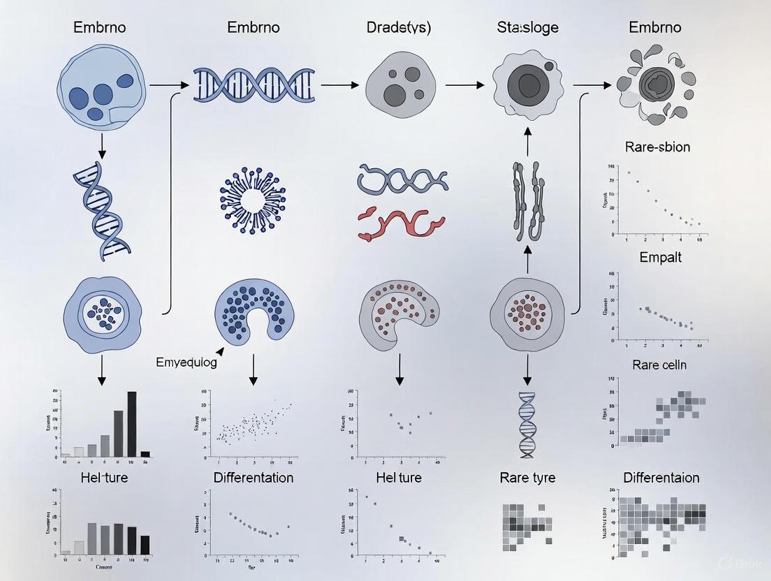
scRNA-seq Experimental and Analytical Pipeline
scRNA-seq Pipeline from Embryo to Rare Cell Detection
Lineage Trajectory Reconstruction from scRNA-seq Data
Developmental Trajectory Reconstruction Revealing Rare Populations
The application of scRNA-seq to embryo analysis has fundamentally transformed our understanding of developmental biology by revealing the cellular heterogeneity, lineage relationships, and rare transitional states that underlie embryogenesis. As the technology continues to evolve, several exciting frontiers are emerging. The integration of multi-omic approaches—combining transcriptomics with epigenomic, proteomic, and spatial information—will provide increasingly comprehensive views of the molecular regulation of development [7]. Additionally, the development of more sophisticated computational methods like deep learning classifiers and improved trajectory inference algorithms will enhance our ability to extract biological insights from these complex datasets [7] [8].
For researchers studying embryonic development and rare cell populations, scRNA-seq offers irreplaceable advantages over bulk approaches. The ability to identify rare cell types, reconstruct developmental trajectories, and decipher cell-cell communication networks makes it an indispensable tool despite its higher complexity and cost. As reference atlases of normal development continue to expand [3] [6], they will provide essential benchmarks for understanding developmental disorders, improving stem cell-based disease models, and advancing regenerative medicine approaches. The ongoing methodological innovations in both wet-lab protocols and computational analysis ensure that scRNA-seq will remain at the forefront of developmental biology research for the foreseeable future.
Key Lineage Branch Points in Human Embryogenesis from Zygote to Gastrula
This technical guide examines the critical lineage branch points during human embryogenesis, from the zygote through the gastrula stage, with a specific focus on implications for identifying rare cell types in single-cell RNA sequencing (scRNA-seq) research. The formation of the human body plan is orchestrated through a series of precise cell fate decisions, where pluripotent cells progressively restrict their developmental potential and differentiate into specialized lineages. Understanding these branching events is fundamental for interpreting developmental disorders, improving assisted reproductive technologies, and authenticating stem cell-based embryo models. Recent advances in single-cell and spatial transcriptomics have provided unprecedented resolution of these developmental trajectories, revealing previously uncharacterized rare cell populations and the signaling networks that govern their emergence. This whitepaper synthesizes current knowledge of key lineage bifurcations, the experimental methodologies for their investigation, and practical computational tools for researchers working with embryonic single-cell data.
Human embryogenesis represents a meticulously orchestrated process wherein a single totipotent zygote undergoes successive rounds of cell division and differentiation to generate all the specialized cell types of the developing organism. This process involves two primary types of cellular decisions: progressive fate restriction (where cells transition from broader to narrower developmental potentials) and binary fate choices (where progenitor cells select between distinct lineage pathways). The accurate identification of the branch points where these decisions occur is crucial for mapping normal development and understanding the origins of developmental abnormalities.
Within the context of scRNA-seq research, these branch points represent critical analytical landmarks. They demarcate the emergence of novel cell identities and serve as reference points for benchmarking stem cell-derived models. Recent integrated scRNA-seq datasets covering human development from zygote to gastrula have revealed approximately 3,304 distinct embryonic cell states across this developmental window, organized along continuous trajectories that reflect the dynamic nature of cell fate acquisition [3]. The identification of rare cell types—often transient intermediates at these branch points—requires particularly sophisticated analytical approaches, as these populations may be underrepresented in standard sampling strategies but play disproportionately important roles in developmental progression.
Major Lineage Branch Points from Zygote to Gastrula
The journey from zygote to gastrula encompasses several major developmental transitions, each characterized by specific lineage bifurcations. Table 1 summarizes the key branch points, their developmental timing, resulting lineages, and representative marker genes that distinguish these fate decisions.
Table 1: Key Lineage Branch Points in Human Embryogenesis
| Developmental Stage | Approximate Timing | Branch Point | Resulting Lineages | Key Marker Genes |
|---|---|---|---|---|
| Preimplantation | E3-E4 | Morula specification | - | DUXA [3] |
| Preimplantation | E5 | First lineage bifurcation | Inner Cell Mass (ICM), Trophectoderm (TE) | POU5F1 (ICM), CDX2 (TE) [3] |
| Preimplantation | E6-E7 | ICM differentiation | Epiblast (EPI), Hypoblast (PrE) | NANOG (EPI), GATA4 (PrE) [3] [9] |
| Postimplantation | E8-E12 | Epiblast maturation | Early epiblast, Late epiblast | HMGN3 (late) [3] |
| Postimplantation | E9-E14 | Trophectoderm diversification | Cytotrophoblast (CTB), Syncytiotrophoblast (STB), Extravillous trophoblast (EVT) | TEAD3 (STB) [3] |
| Gastrulation | E14-E16 (CS7) | Primitive streak formation | Primitive streak, Amnion, Embryonic mesoderm, Definitive endoderm | TBXT (PriS), ISL1 (Amnion) [3] |
| Gastrulation | E16-E19 (CS7) | Extraembryonic specification | Yolk sac endoderm, Extraembryonic mesoderm, Hematopoietic lineages | LUM, POSTN (ExE_Mes) [3] |
Detailed Analysis of Critical Branching Events
First Lineage Bifurcation: ICM versus TE
The inaugural lineage decision occurs around embryonic day 5 (E5), when the embryo segregates into two fundamentally distinct populations: the inner cell mass (ICM), which will give rise to the embryo proper, and the trophectoderm (TE), which forms the extraembryonic tissues including the fetal portion of the placenta [3]. This division represents the first differentiation event in mammalian development and establishes the fundamental embryonic-extraembryonic dichotomy.
The Hippo signaling pathway serves as the primary regulator of this fate decision, translating positional information (cell polarity) into transcriptional identity [9]. In outer, polarized cells, Hippo signaling is inactive, allowing dephosphorylated YAP/TAZ to translocate to the nucleus where they interact with TEAD4 to activate TE-specific genes including CDX2 and GATA3. Conversely, inner, apolar cells maintain active Hippo signaling, resulting in cytoplasmic retention of YAP/TAZ and consequent expression of ICM markers such as POU5F1 (OCT4) and NANOG [9]. Single-cell transcriptomic analyses have identified 367 transcription factor genes that show modulated expression along the epiblast trajectory from this initial branch point, highlighting the complex regulatory network underlying this fundamental lineage decision [3].
Second Lineage Bifurcation: EPI versus PrE
Following implantation, the ICM undergoes a second lineage specification around E6-E7, segregating into the epiblast (EPI), which generates the embryo proper, and the primitive endoderm (PrE) or hypoblast, which gives rise to the yolk sac [3]. This decision is orchestrated by the coordinated activity of several signaling pathways, including FGF and Nodal/Activin [9].
Experimental modulation of these pathways demonstrates their critical role; inhibition of FGF signaling with PD0325901 suppresses PrE differentiation while promoting EPI fate, whereas FGF2 supplementation has the opposite effect [9]. Similarly, inhibition of Nodal/Activin signaling with SB431542 enhances EPI specification [9]. scRNA-seq analyses have revealed that 326 transcription factor genes display pseudotime-dependent expression along the hypoblast trajectory, including early factors like GATA4 and SOX17, and later factors such as FOXA2 and HMGN3 [3]. The resolution of this lineage decision establishes the three foundational lineages of the blastocyst: EPI, PrE, and TE.
Gastrulation Branch Points: Emergence of the Three Germ Layers
The process of gastrulation, occurring approximately between E14-E19 (Carnegie Stage 7), represents the most complex period of lineage diversification in early development [3]. During this phase, the epiblast undergoes an epithelial-to-mesenchymal transition through the primitive streak to generate the mesoderm and definitive endoderm, while the remaining epiblast forms the ectoderm. Concurrently, extraembryonic lineages undergo further specialization.
Single-cell analyses of CS7 human embryos have identified distinct progenitor populations for amnion, primitive streak, mesoderm, definitive endoderm, and various extraembryonic components including yolk sac endoderm, extraembryonic mesoderm, and hematopoietic lineages [3]. Transcription factor network analyses have identified key regulators of these lineages, including TBXT (Brachyury) in primitive streak cells, MESP2 in mesoderm, ISL1 in amnion, and HOXC8 in extraembryonic mesoderm [3]. The identification of these branch points provides critical reference data for authenticating in vitro models of human gastrulation.
Experimental Methodologies for Lineage Analysis
Single-Cell RNA Sequencing Approaches
The establishment of a comprehensive human embryo reference through scRNA-seq integration represents a methodological advance for the field. The standardized protocol involves:
Dataset Collection and Processing: Six published human scRNA-seq datasets covering developmental stages from zygote to gastrula were reprocessed using a uniform pipeline, including mapping and feature counting with the same genome reference (GRCh38 v.3.0.0) to minimize batch effects [3].
Data Integration: The fast mutual nearest neighbor (fastMNN) method is employed to integrate these datasets, embedding expression profiles of 3,304 early human embryonic cells into a unified dimensional space [3].
Trajectory Inference: Slingshot trajectory inference based on 2D UMAP embeddings reveals three primary trajectories corresponding to epiblast, hypoblast, and TE development, identifying hundreds of transcription factors with pseudotime-dependent expression [3].
Cell Fate Prediction: A stabilized Uniform Manifold Approximation and Projection (UMAP) constructs an early embryogenesis prediction tool where query datasets can be projected onto the reference and annotated with predicted cell identities [3].
This integrated reference enables researchers to benchmark stem cell-based embryo models and identify potential misannotations when relevant references are not utilized for authentication.
Spatial Transcriptomic Validation
While scRNA-seq provides unparalleled resolution of cellular heterogeneity, it inherently lacks spatial context. Spatial transcriptomics technologies bridge this gap by preserving the spatial organization of cells during transcriptomic profiling. The STAMapper algorithm represents a significant advance in this domain—a heterogeneous graph neural network that transfers cell-type labels from scRNA-seq data to single-cell spatial transcriptomics (scST) data [10].
The STAMapper workflow involves:
- Construction of a heterogeneous graph where cells and genes are modeled as distinct node types
- Implementation of a message-passing mechanism to update latent embeddings of each node
- Utilization of a graph attention classifier to estimate cell-type identity probabilities
- Assignment of cell-type labels to scST data based on classifier outputs [10]
In validation across 81 scST datasets comprising 344 slices from eight technologies and five tissues, STAMapper demonstrated superior performance compared to existing methods (scANVI, RCTD, Tangram), particularly for datasets with fewer than 200 genes, making it especially valuable for analyzing spatially resolved data with limited gene panels [10].
Integrated Morphological and Molecular Mapping
Beyond transcriptomic profiling, comprehensive understanding of lineage decisions requires integration of cellular morphological data. Recent work in model organisms has established platforms for qualitative and quantitative analysis of three-dimensional cell shape, volume, surface area, and contact area alongside gene expression profiles with defined cell lineage [11].
The CMap pipeline enables automated segmentation of cell membranes labeled by fluorescent protein up to the 550-cell stage, extracting data on cell volume, surface area, and contact area between neighboring cells [11]. This approach has revealed how Notch and Wnt signaling pathways, combined with mechanical forces from cell interactions, regulate both cell fate decisions and size asymmetries during development [11]. Such integrated morphological maps provide critical missing dimensions to purely transcriptomic analyses, particularly for understanding how cell-cell interactions influence fate decisions at lineage branch points.
Computational Tools for Rare Cell Type Identification
The identification of rare cell types at lineage branch points requires specialized computational approaches. Table 2 summarizes key algorithms and their applications in embryonic single-cell data analysis.
Table 2: Computational Tools for Analyzing Lineage Branch Points and Rare Cell Types
| Tool | Methodology | Primary Application | Strengths | Citation |
|---|---|---|---|---|
| STAMapper | Heterogeneous graph neural network with graph attention | Cell-type mapping from scRNA-seq to spatial transcriptomics | Superior performance with limited gene panels; unknown cell-type detection | [10] |
| Slingshot | Principal curves on reduced-dimension embeddings | Trajectory inference and pseudotime ordering | Identifies multiple branching lineages; minimal parameter requirements | [3] |
| SCENIC | Regulatory network inference and clustering | Transcription factor activity analysis from scRNA-seq data | Identifies key regulators of fate decisions; complements trajectory analysis | [3] |
| fastMNN | Mutual nearest neighbor correction | Batch effect correction and dataset integration | Preserves biological heterogeneity while removing technical artifacts | [3] |
| CMap | EDT-DMFNet for membrane segmentation | Integrated morphological and molecular mapping | Links cell shape/contact data with lineage decisions | [11] |
Signaling Pathways Governing Lineage Decisions
The molecular pathways regulating lineage bifurcations represent potential intervention points for manipulating cell fate decisions. Figure 1 illustrates the key signaling pathways active at major branch points, while Table 3 summarizes experimental evidence from pathway modulation studies.
Table 3: Experimental Modulation of Signaling Pathways in Human Embryogenesis
| Pathway | Key Components | Role in Lineage Specification | Modulation Evidence | Citation |
|---|---|---|---|---|
| Hippo | YAP/TAZ, TEAD1-4, LATS1/2 | TE vs. ICM decision | CRT0276121 (activator) promotes TE fate; TRULI (inhibitor) enhances ICM | [9] |
| Wnt/β-catenin | β-catenin, TCF/LEF | Preimplantation development | 1-Azakenpaullone (activator) and Cardamonin (inhibitor) affect blastocyst development | [9] |
| FGF | FGF2, FGFR | EPI vs. PrE decision | PD0325901/PD173074 (inhibitors) promote EPI; FGF2 (activator) promotes PrE | [9] |
| TGF-β/Nodal/Activin | Nodal, Activin, SMAD2/3 | EPI vs. PrE decision | SB431542/A8301 (inhibitors) promote EPI; Activin A has complex effects | [9] |
| BMP | BMP4, SMAD1/5/8 | Preimplantation development | BMP4 supplementation affects blastocyst development rate | [9] |
Table 4: Essential Research Reagents and Computational Resources
| Resource | Type | Primary Application | Key Features | Access |
|---|---|---|---|---|
| Human Embryo scRNA-seq Reference | Integrated dataset | Benchmarking embryo models | 3,304 cells from zygote to gastrula; stabilized UMAP projection | [3] |
| STAMapper | Computational algorithm | Spatial transcriptomics annotation | Graph neural network; works with limited gene panels | [10] |
| CMap Platform | Morphological mapping | Integrated shape-lineage analysis | 3D cell regions with volume, surface, contact data | [11] |
| Small Molecule Modulators | Experimental reagents | Pathway manipulation | CRT0276121 (Hippo activator); TRULI (Hippo inhibitor) | [9] |
| Lineage-Specific Markers | Molecular reagents | Cell type identification | DUXA (morula); TBXT (primitive streak); ISL1 (amnion) | [3] |
The systematic mapping of lineage branch points in human embryogenesis represents a fundamental advance in developmental biology with significant implications for regenerative medicine, reproductive health, and stem cell research. The integration of single-cell transcriptomics, spatial mapping, and morphological analyses has revealed previously unappreciated complexity in the timing and regulation of fate decisions. For researchers focused on identifying rare cell types in embryonic scRNA-seq data, this reference framework provides critical landmarks for distinguishing biologically significant rare populations from technical artifacts. As single-cell technologies continue to evolve, particularly in spatial resolution and multi-omic integration, our understanding of these critical developmental transitions will continue to refine, offering new insights into the fundamental processes of human development and their dysregulation in disease states.
The construction of comprehensive embryo reference atlases represents a foundational endeavor in developmental biology, enabling systematic characterization of cellular heterogeneity and lineage specification during embryogenesis. These integrated datasets serve as essential benchmarks for authenticating stem cell-based embryo models, decoding the molecular programs driving organ formation, and identifying rare but biologically critical cell populations that may be overlooked in individual studies. The integration of multiple single-cell RNA sequencing (scRNA-seq) datasets is particularly crucial for capturing the complete spectrum of cellular states across developmental stages, donors, and experimental conditions. By providing a stable, well-annotated coordinate system for early development, these atlases allow researchers to map query datasets and rapidly identify both common and rare cell types, facilitating the discovery of novel developmental lineages and disease-associated deviations.
Recent technological advances have made it possible to generate multimillion-cell reference datasets, but their full utility is realized only through sophisticated computational integration that removes technical artifacts while preserving meaningful biological variation. For the specific challenge of rare cell identification—a central focus in embryogenesis research where rare progenitor populations often drive critical developmental transitions—comprehensive reference atlases provide the necessary statistical power and context to distinguish genuine rare cell types from technical outliers or transitional states. This technical guide outlines the methodologies, computational frameworks, and experimental considerations for establishing embryo reference atlases, with particular emphasis on their application for identifying rare cell types in embryo scRNA-seq data research.
Computational Frameworks for Atlas Integration and Mapping
Reference Building and Query Mapping Algorithms
The construction of a comprehensive embryo reference atlas requires computational methods capable of integrating multiple datasets while preserving both abundant and rare cell states. Several algorithms have been specifically developed for this purpose, each with distinct advantages for embryonic data.
Symphony provides an efficient algorithm for building large-scale integrated references in a portable format that enables rapid query mapping within seconds [12]. The method compresses an integrated reference into "minimal reference elements" including gene scaling parameters, gene loadings from principal component analysis (PCA), soft-cluster centroids, and compression terms. For mapping, Symphony projects query cells into the reference embedding through a three-step process: (1) projection into the uncorrected reference PCA space using saved parameters, (2) computation of soft-cluster assignments based on reference cluster centroids, and (3) correction of query batch effects using stored mixture model components while keeping the reference stable [12]. This approach closely approximates de novo integration while avoiding the computational burden of reintegrating the entire reference, making it particularly valuable for iterative atlas building.
scArches (single-cell architecture surgery) implements a transfer learning strategy for mapping query datasets to existing references [13]. This method builds upon conditional variational autoencoder (CVAE) models such as scVI, trVAE, and scANVI, using "architectural surgery" to incorporate new studies without retraining the entire network. The approach adds trainable "adaptor" weights for new query datasets while freezing most reference parameters, functioning as an inductive bias to prevent overfitting to query data. scArches demonstrates particular utility for mapping disease data (e.g., COVID-19) to healthy references while preserving disease-specific variation, and for multimodal reference mapping that allows imputation of missing modalities [13].
Table 1: Comparison of Reference Atlas Integration Methods
| Method | Underlying Algorithm | Key Features | Advantages for Embryonic Data |
|---|---|---|---|
| Symphony [12] | Linear mixture model (Harmony) | Fast query mapping, portable reference format | Efficient for large-scale atlases, preserves rare populations |
| scArches [13] | Transfer learning (CVAE/scVI/trVAE) | Model sharing without raw data, iterative reference building | Handles complex batch effects, multimodal capability |
| fastMNN [3] | Mutual nearest neighbors | Fast batch correction, preserves biological variance | Maintains developmental trajectories |
Experimental Protocol for Atlas Construction
A standardized workflow for constructing an embryo reference atlas was demonstrated in the integration of six human scRNA-seq datasets covering development from zygote to gastrula [3]. The protocol involves:
Data Collection and Uniform Processing: Collect publicly available datasets and reprocess them using the same genome reference (e.g., GRCh38) and annotation through a standardized pipeline to minimize batch effects.
Integration with fastMNN: Employ fast mutual nearest neighbor (fastMNN) methods to embed expression profiles of all cells into a shared low-dimensional space. For the human embryo atlas, this integrated 3,304 early human embryonic cells [3].
Annotation and Validation: Annotate cell types based on known markers and regulatory networks. Perform single-cell regulatory network inference and clustering (SCENIC) analysis to validate lineage identities through transcription factor activities.
Trajectory Inference: Apply trajectory inference tools (e.g., Slingshot) to reconstruct developmental trajectories and identify genes modulated along pseudotime.
Marker Gene Identification: Identify unique markers for each distinct cell cluster using differential expression testing.
Tool Deployment: Create user-friendly online prediction tools (e.g., Shiny interfaces) for community access and query mapping.
This approach successfully captured continuous developmental progression from zygote through gastrulation, identifying lineage bifurcations and transitions from early to late epiblast and hypoblast populations [3].
Specialized Approaches for Rare Cell Type Identification
Algorithmic Strategies for Rare Population Detection
The identification of rare cell types in embryo scRNA-seq data presents distinct computational challenges, as standard clustering approaches often fail to distinguish rare populations from more abundant cell types. Several algorithms have been specifically developed to address this limitation.
scCAD (Cluster decomposition-based Anomaly Detection) employs an iterative clustering decomposition approach to separate rare cell types that may be overlooked during initial clustering [14]. The method begins with ensemble feature selection to preserve differentially expressed genes in rare cell types, combining initial clustering labels with a random forest model to identify important genes. scCAD then iteratively decomposes major clusters based on the most differential signals within each cluster, creating D-clusters (decomposed clusters) that are subsequently merged into M-clusters (merged clusters). Finally, the algorithm uses an isolation forest model on candidate differentially expressed genes to calculate anomaly scores and identify rare populations based on cluster independence scores [14]. When benchmarked on 25 real scRNA-seq datasets, scCAD achieved superior performance (F1 score = 0.4172) compared to 10 state-of-the-art methods, demonstrating 24-48% improvement over the next best approaches [14].
CIARA (Cluster Independent Algorithm for the identification of markers of RAre cell types) takes a distinct approach by selecting genes that are likely markers of rare cell types prior to clustering [15]. This cluster-independent method identifies genes with expression patterns characteristic of rare populations, which are subsequently integrated with common clustering algorithms to single out groups of rare cell types. CIARA has successfully identified previously uncharacterized rare cell populations in human gastrula datasets and mouse embryonic stem cells treated with retinoic acid [15].
Table 2: Methods for Rare Cell Identification in Embryo scRNA-seq Data
| Method | Algorithmic Approach | Key Advantages | Reported Performance |
|---|---|---|---|
| scCAD [14] | Iterative cluster decomposition & anomaly detection | Identifies rare types missed in initial clustering | F1 score: 0.4172 (25 datasets) |
| CIARA [15] | Cluster-independent marker identification | Works prior to clustering, generalizable to multi-omics | Outperforms existing rare cell detection methods |
| CellSIUS [14] | Identifies bimodal genes within clusters | Effective for rare subpopulations | F1 score: 0.2812 |
| SCA [14] | Surprisal component analysis | Dimensionality reduction for rare cells | F1 score: 0.3359 |
Experimental Protocol for Rare Cell Identification
The following protocol outlines the application of scCAD for identifying rare cell types in embryo scRNA-seq data, based on the benchmarked approach [14]:
Ensemble Feature Selection:
- Perform initial clustering using global gene expression (e.g., Seurat or Scanpy standard workflow)
- Train a random forest model using initial cluster labels to identify important genes
- Combine these with highly variable genes to create an ensemble feature set
Iterative Cluster Decomposition:
- For each initial cluster (I-cluster), subset the ensemble feature genes
- Perform iterative k-means clustering (k=2) on the most differential signals within each cluster
- Continue decomposition until no subclusters show significant differential expression
- Annotate decomposed clusters (D-clusters) based on dominant cell types
Cluster Merging and Anomaly Detection:
- Merge D-clusters with the closest Euclidean distance between centers to create M-clusters
- For each M-cluster, perform differential expression analysis to identify candidate marker genes
- Apply an isolation forest model using candidate DE genes to calculate anomaly scores for all cells
- Compute independence scores by assessing overlap between high-anomaly cells and cluster membership
Rare Population Identification:
- Rank clusters by independence scores, with highest scores indicating most rare populations
- Validate rare populations using known markers and spatial mapping where available
This protocol has been successfully applied to identify rare cell types in diverse biological contexts including mouse airway, brain, intestine, human pancreas, and clear cell renal cell carcinoma data [14].
Spatial Atlas Construction and Validation
Spatial Transcriptomics for Embryonic Reference Atlases
Spatial transcriptomic approaches provide essential validation for embryo reference atlases by enabling direct mapping of identified cell types within their native tissue context. The integration of spatial data is particularly valuable for rare cell populations, whose spatial positioning often reveals functional roles in developmental patterning.
A comprehensive spatial atlas of the human lung demonstrates a framework applicable to embryonic tissues, employing three complementary spatial transcriptomics approaches [16]:
HybISS: A highly multiplexed imaging-based method with cellular resolution, using a targeted probe panel (162 genes) to detect majority cell types including rare populations. The protocol involves tissue sectioning, hybridization with gene-specific probes, sequential imaging, and computational segmentation to assign transcripts to individual cells.
SCRINSHOT: A highly sensitive spatial method with a more limited gene panel (64 genes) optimized for detecting variations in gene expression levels, particularly valuable for identifying rare cell states.
Visium: An unbiased method for genome-wide mRNA detection with lower spatial resolution, used to validate cell types and regional expression patterns identified by targeted approaches.
This multi-technology framework enabled the precise localization of 35 cell types within tissue topography, revealed consistent anatomical and regional gene expression variability, and identified distinct cellular neighborhoods in specific anatomical regions [16]. For embryonic applications, similar approaches can validate the spatial distribution of rare progenitor populations identified in scRNA-seq data.
Experimental Protocol for Spatial Atlas Validation
The spatial validation of embryo reference atlases adapts the following protocol from lung tissue mapping [16]:
Tissue Preparation:
- Collect embryo specimens at precise Carnegie stages
- Embed in optimal cutting temperature (OCT) compound and snap-free
- Section at appropriate thickness (typically 10-20μm) onto charged slides
Multiplexed Spatial Transcriptomics:
- Design targeted probe panels based on scRNA-seq identified markers, including putative rare cell type markers
- Perform HybISS with sequential hybridization and imaging cycles
- Conduct SCRINSHOT on serial sections for sensitive detection of expression variations
- Process adjacent sections with Visium for unbiased validation
Image Processing and Cell Segmentation:
- Align imaging cycles and decode transcript signals
- Segment cells based on DAPI-stained nuclei
- Assign transcript signals to nearest nuclei
Integration with scRNA-seq Atlas:
- Map spatial data to reference atlas using Symphony or scArches
- Validate spatial distribution of rare cell types identified computationally
- Identify cellular neighborhoods and signaling niches
This approach successfully revealed imbalances in epithelial cell type compositions in diseased lungs when applied to chronic obstructive pulmonary disease samples [16], demonstrating its utility for identifying disease-associated alterations relative to a healthy reference.
The Scientist's Toolkit: Essential Research Reagents and Computational Tools
Table 3: Research Reagent Solutions for Embryo Reference Atlas Construction
| Category | Specific Tools/Reagents | Function/Application | Example Use Case |
|---|---|---|---|
| Spatial Transcriptomics | HybISS panel (162 genes) [16] | Targeted cellular resolution spatial mapping | Localizing rare epithelial cells in tissue topography |
| SCRINSHOT panel (64 genes) [16] | Sensitive detection of expression variations | Identifying rare cell states in embryonic tissues | |
| Visium (10x Genomics) [16] | Unbiased genome-wide spatial profiling | Validating cell types and regional expression patterns | |
| Computational Tools | Symphony [12] | Efficient reference building and query mapping | Mapping developmental trajectory positions |
| scArches [13] | Transfer learning for reference mapping | Contextualizing disease data with healthy references | |
| scCAD [14] | Rare cell identification via cluster decomposition | Finding novel rare populations in human gastrula | |
| CIARA [15] | Cluster-independent rare cell marker identification | Identifying rare cells in mouse embryonic stem cells | |
| Embryo Staging | Carnegie stage criteria [17] | Standardized morphological staging | Cross-species developmental comparisons |
| Reference Datasets | Integrated human embryo atlas [3] | Benchmarking embryo models and query datasets | Authentication of stem cell-based embryo models |
The establishment of comprehensive embryo reference atlases through integration of multiple datasets represents a transformative resource for developmental biology, particularly for the identification and characterization of rare cell types that drive critical developmental transitions. The computational frameworks and experimental protocols outlined in this technical guide provide a roadmap for constructing, validating, and utilizing these essential resources. As single-cell technologies continue to evolve, future atlas efforts will likely incorporate multimodal data (epigenomic, proteomic, spatial) across complete developmental timecourses, enabling deeper understanding of the regulatory programs governing embryogenesis. The development of specialized algorithms for rare cell identification—such as scCAD and CIARA—will remain crucial for extracting maximal biological insight from these comprehensive references, particularly for understanding the rare progenitor populations that orchestrate tissue patterning and organ formation. Through continued refinement of integration methods and rare cell detection approaches, embryo reference atlases will increasingly serve as foundational resources for developmental biology, regenerative medicine, and the study of congenital disorders.
Biological Significance of Rare and Transient Cell Populations in Development
The process of embryonic development is a precisely orchestrated sequence of events where a single fertilized egg gives rise to a complex multicellular organism. Within this process, rare and transient cell populations serve as critical architects of development, directing key lineage decisions, morphological changes, and the establishment of the basic body plan. These populations, often present in small numbers and for limited time windows, include pivotal entities such as organizer cells, signaling centers, and early progenitors that dictate the fate of surrounding tissues. Their identification and characterization have been profoundly advanced by single-cell RNA sequencing (scRNA-seq) technologies, which enable researchers to capture these elusive cell states that would otherwise be masked in bulk analyses [2].
Understanding these rare populations is not merely an academic exercise but has profound implications for reproductive medicine, congenital disorder research, and regenerative medicine. Developmental defects often originate from the malfunction of specific, rarely occurring cell types during critical periods. Furthermore, the study of rare embryonic cells provides invaluable insights into the mechanisms of cellular plasticity and lineage specification that are recapitulated in stem cell differentiation and disease processes such as cancer [3] [18]. Within the specific context of embryo scRNA-seq research, identifying these rare cell types presents both a technical challenge and biological imperative, as they often serve as the foundational sources of developmental cues that shape the embryo.
Computational Strategies for Identifying Rare Cell Types in scRNA-seq Data
The analysis of single-cell RNA sequencing data from embryonic development requires specialized computational approaches designed to detect cell populations that may constitute less than 1% of the total cells. These methods must distinguish biologically significant rare populations from technical artifacts such as doublets or dying cells.
Algorithm Classifications and Performance Benchmarks
Multiple algorithmic strategies have been developed to address the challenge of rare cell identification. Table 1 summarizes the primary approaches, their underlying principles, and representative tools.
Table 1: Computational Methods for Rare Cell Identification in scRNA-seq Data
| Method Category | Underlying Principle | Representative Tools | Strengths | Limitations |
|---|---|---|---|---|
| Feature Selection-Based | Identifies genes with high expression specificity (e.g., high Gini coefficient) for rare populations. | GiniClust2 [19], CIARA [14] | Effective for rare types with highly specific markers. | May miss rare cells with moderate expression of many genes. |
| Clustering Decomposition | Iteratively decomposes major clusters using differential signals to separate rare subtypes. | scCAD [14] | Discovers rare populations obscured in initial clustering. | Computationally intensive for very large datasets. |
| Rarity Scoring | Assigns a rareness score to each cell based on its neighborhood in gene expression space. | FiRE [19] [14], GapClust [14] | Does not rely on pre-clustering; can detect very rare cells (<0.01%). | May misclassify outliers from major types as rare cells. |
| Anomaly Detection | Frames rare cell identification as an anomaly detection problem using machine learning. | scSID [19], RaceID3 [19] [14] | Robust to noise and complex data distributions. | Requires careful parameter tuning. |
| Dimensionality Reduction | Employs specialized dimension reduction to enhance separation of rare cells. | EDGE, SCA [14] | Can capture subtle, multi-gene expression patterns. | Risk of losing biologically relevant information during reduction. |
Performance benchmarking across 25 real scRNA-seq datasets reveals significant variation in the capabilities of these methods. The cluster decomposition-based method scCAD demonstrated superior performance, achieving an F1 score of 0.4172, which represents a 24% improvement over the next best method [14]. The GiniClust algorithm employs a two-step process, first selecting genes with high Gini coefficients (indicating expression in a small subset of cells) and then performing density-based clustering to group cells expressing these genes [19]. In contrast, scSID operates by calculating the Euclidean distance between cells in a dimensionally-reduced space, identifying rare cells based on sharp changes in similarity to their k-nearest neighbors [19].
Experimental Protocol: A Standard Workflow for Rare Cell Analysis
The following workflow outlines the key steps for identifying rare cell types in embryonic scRNA-seq data, integrating multiple computational approaches for robust results.
Data Preprocessing and Quality Control: Begin with standard preprocessing of raw count matrices using tools like Seurat or Scanpy. Perform rigorous quality control to remove low-quality cells, doublets, and dying cells based on metrics like total counts, number of detected genes, and mitochondrial gene percentage. This step is critical to prevent technical artifacts from being misidentified as rare biological populations [20].
Feature Selection and Normalization: Select highly variable genes to reduce dimensionality and computational noise. Apply appropriate normalization methods (e.g., log-normalization or SCTransform) to account for technical variation in sequencing depth [20].
Dimensionality Reduction and Initial Clustering: Perform principal component analysis (PCA) on the scaled data of highly variable genes. Use the significant principal components for graph-based clustering (e.g., Leiden or Louvain algorithms) and non-linear dimensionality reduction (e.g., UMAP or t-SNE) for visualization. This step identifies the major cell types present in the embryo dataset [3] [20].
Rare Cell Identification Application: Apply one or more specialized rare cell identification algorithms (e.g., those listed in Table 1) to the processed data. For optimal results, consider an ensemble approach:
- Run multiple algorithms (e.g., scCAD, FiRE, and scSID) independently.
- Compare the lists of candidate rare cells identified by each method.
- Prioritize cells consistently flagged by multiple algorithms for downstream validation.
Validation and Biological Interpretation: For the candidate rare cell population, perform differential expression analysis to identify uniquely expressed marker genes. Validate these markers experimentally via in situ hybridization or immunofluorescence if possible. Use functional enrichment analysis of the marker genes to infer the biological role of the rare population [3].
A Reference Atlas for Human Embryonic Development
The creation of a comprehensive, integrated reference atlas is a cornerstone for authenticating rare cell populations in human embryo models. A significant recent achievement is the development of a unified human embryo reference integrating six published scRNA-seq datasets, covering developmental stages from the zygote to the gastrula (Carnegie Stage 7) [3]. This resource encompasses transcriptome profiles of 3,304 individual embryonic cells, providing a high-resolution roadmap against which stem cell-based embryo models can be benchmarked [3].
Key Lineage Transitions and Rare Populations
The integrated atlas reveals continuous developmental progression with key lineage branch points and the emergence of rare, transient populations:
- The first lineage bifurcation occurs around E5, with the divergence of the inner cell mass (ICM) and trophectoderm (TE) [3].
- ICM specification is followed by its bifurcation into the epiblast (which will form the embryo proper) and the hypoblast (which gives rise to the yolk sac) [3].
- During gastrulation (CS7), the epiblast undergoes massive reorganization and specification into the primitive streak (PriS), a transient signaling center. Cells passing through the primitive streak give rise to the mesoderm and definitive endoderm, while the amnion forms as an extraembryonic tissue [3]. The primitive streak and its derivatives represent critical, transient populations that are essential for establishing the body plan.
Analysis of this atlas using Slingshot trajectory inference has delineated three main developmental trajectories—epiblast, hypoblast, and TE—and identified hundreds of transcription factor genes whose expression is modulated along these paths [3]. For example, DUXA and FOXR1 are highly expressed during morula stages but decrease subsequently, while HMGN3 shows upregulated expression at postimplantation stages across lineages [3].
Experimental Protocol: Constructing and Utilizing an Embryo Reference Atlas
- Data Collection and Curation: Gather publicly available scRNA-seq datasets from human embryos across targeted developmental stages. Ensure consistent ethical approval and compliance with the 14-day rule for later stages [3] [2].
- Uniform Data Reprocessing: Reprocess all datasets using a standardized pipeline with the same genome reference and annotation (e.g., GRCh38) to minimize batch effects. This includes mapping, feature counting, and quality control [3].
- Data Integration: Employ batch correction methods such as fast Mutual Nearest Neighbors (fastMNN) to embed cells from different studies into a unified transcriptional space [3].
- Cell Annotation and Lineage Mapping: Annotate cell types based on canonical markers and reference to original publications. Use trajectory inference tools (e.g., Slingshot) to reconstruct developmental lineages and calculate pseudotime [3].
- Projection and Authentication of Query Data: Develop a prediction tool (e.g., based on UMAP) that allows users to project new datasets from embryo models onto the reference. The tool annotates query cells with predicted identities and provides a measure of similarity to the in vivo reference, highlighting potential misannotations [3].
Table 2: Key Research Reagents and Computational Tools for Embryo scRNA-seq Analysis
| Item Name | Type | Function/Biological Significance | Example Use Case |
|---|---|---|---|
| Integrated Human Embryo Reference [3] | Dataset | A universal transcriptomic reference for benchmarking embryo models from zygote to gastrula. | Projecting stem cell-derived embryo models to assess fidelity. |
| ScType [20] | Algorithm | Automated cell type annotation tool for scRNA-seq data. | Rapid, unbiased annotation of cell types in a query dataset. |
| Harmony [20] | Algorithm | Batch integration method that removes technical effects between datasets. | Integrating multiple scRNA-seq experiments from different batches. |
| Evercode WT [20] | Reagent Kit | A whole transcriptome single-cell RNA sequencing kit. | Generating scRNA-seq libraries from limited embryo model material. |
| Trailmaker [20] | Software Platform | A cloud-based, user-friendly scRNA-seq analysis platform with automated workflows. | Analyzing data without extensive bioinformatics expertise. |
Biological Significance of Rare Populations in Embryonic Systems
Rare and transient cell populations are not merely curiosities; they are fundamental drivers of embryogenesis. Their functions can be understood through several key biological concepts.
Developmental Organizers and Signaling Centers
The most classic examples of rare, transient cell populations are developmental organizers. These are small groups of cells that emit signals to pattern large fields of surrounding tissue, dictating their fate and spatial organization. In the human gastrula, the primitive streak and its derivative, the node, function as key organizers. The primitive streak establishes the anterior-posterior axis and gives rise to the mesoderm and endoderm germ layers [2]. Cells within these organizers express pivotal transcription factors such as TBXT (T-brachyury) in the primitive streak and ISL1 in the amnion, which are essential for their function and serve as specific markers for these rare populations [3].
Evolutionary and Conceptual Frameworks
Applying concepts from evolutionary developmental biology (Evo-Devo) provides a powerful lens through which to view the generation of rare cell types. Three key concepts are particularly relevant:
- Single-Cell Heterochrony: This refers to changes in the timing of gene expression or cellular processes within a cell lineage that can lead to novel cell identities. For example, a delay in cytokinesis coupled with continued nuclear replication can generate a multinucleate cell, a phenomenon observed in the evolution of certain amoebas and land plant spores [18] [21].
- Single-Cell Homeosis: This involves a switch in the identity of one cell type to another, often due to the misexpression of key transcription factors. An example is the transformation of eosinophils to basophils in the hematopoietic lineage simply by changing the order of activity of two transcription factors, C/EBPα and GATA [18] [21].
- Plasticity: The capacity of a cell to alter its identity in response to environmental cues is fundamental during embryogenesis, where cell fates are often determined by signals from neighboring cells rather than autonomous programming [18] [21].
Rare and transient cell populations are the master regulators of embryonic development, directing the complex processes that transform a single cell into a fully formed organism. The advent of high-resolution single-cell genomics, coupled with sophisticated computational algorithms like scCAD and scSID, has finally provided the tools necessary to identify, characterize, and understand these elusive but critically important cells. The development of integrated reference atlases establishes a new standard for authenticating in vitro embryo models against their in vivo counterparts, ensuring the fidelity of future research.
Moving forward, the field will be shaped by emerging technologies such as single-cell long-read sequencing to resolve isoform-level diversity, the integration of multi-omics data (epigenomics, proteomics), and the application of large language models for more nuanced and scalable cell type annotation [22]. As these tools mature, they will unlock deeper insights into the fundamental biology of development, with profound implications for understanding congenital disorders, improving regenerative therapies, and unraveling the evolutionary history of cellular diversity.
Ethical Considerations and Technical Challenges in Human Embryo Research
Human embryo research represents a crucial frontier for understanding early development, congenital disorders, and infertility. However, this field is constrained by significant ethical boundaries and technical limitations. The emergence of single-cell RNA sequencing (scRNA-seq) has revolutionized our capacity to study cellular heterogeneity in early development, offering unprecedented resolution for identifying rare cell types. This technical guide examines the current landscape of ethical frameworks and analytical methodologies, with particular emphasis on their application in detecting rare cellular populations within human embryo scRNA-seq data.
Ethical Framework in Human Embryo Research
Regulatory Boundaries and the 14-Day Rule
Human embryo research is globally governed by the "14-day rule," a ethical benchmark restricting studies beyond two weeks post-fertilization. This boundary aligns with the emergence of the primitive streak, marking the beginning of gastrulation and the establishment of the body axis. This restriction exists due to both ethical considerations regarding embryo status and technical challenges in sustaining embryos ex vivo beyond this stage [2]. The International Society for Stem Cell Research (ISSCR) maintains and updates guidelines for stem cell research and clinical translation, with the most recent updates in 2025 specifically addressing stem cell-based embryo models (SCBEMs) [23].
Stem Cell-Based Embryo Models as Research Alternatives
To circumvent ethical constraints, researchers have developed stem cell-based embryo models (SCBEMs) that mimic aspects of early development without using fertilized embryos. The 2025 ISSCR guidelines retired the classification of models as "integrated" or "non-integrated" in favor of the inclusive term "SCBEMs" [23]. These models require:
- Clear scientific rationale
- Defined endpoints
- Appropriate oversight mechanisms
- Prohibition of transplantation to a uterus
- Prevention of extended culture to potential viability (ectogenesis) [23]
The usefulness of these models "hinges on their molecular, cellular and structural fidelities to their in vivo counterparts," making scRNA-seq essential for validation [3] [24].
Technical Challenges in Embryonic scRNA-Seq
Sample Acquisition and Preparation Constraints
The ethical limitations on human embryo research directly impact experimental design and sample quality:
Table 1: Technical Challenges in Embryonic scRNA-Seq Sample Preparation
| Challenge | Impact on Rare Cell Identification | Potential Solutions |
|---|---|---|
| Limited embryo availability | Reduces statistical power for detecting rare populations | Use of embryo models; sample pooling strategies |
| Embryo dissociation difficulties | Risk of losing fragile cell types | Optimized enzymatic/mechanical protocols; viability staining |
| Small cell numbers per embryo | Challenges in capturing full cellular diversity | Increased sequencing depth; cell hashing for multiplexing |
| Variable developmental stages | Introduces heterogeneity confounding rare cell detection | Precise developmental staging; computational integration |
Accurate sample preparation is crucial for generating high-quality single-cell transcriptome data. Protocols must be "diligently optimised to accommodate variables such as cellular dimensions, viability and cultivation conditions" [25]. For cells exceeding 30μm in diameter (problematic for droplet-based systems like 10× Genomics), plate-based fluorescence-activated cell sorting (FACS) with nozzles up to 130μm offers a feasible alternative [25].
Analytical Considerations for Rare Cell Populations
The complexity of scRNA-seq datasets requires numerous analytical choices that significantly impact rare cell identification:
Clustering Reproducibility: Cluster assignment is "one of the major sources of irreproducibility" in scRNA-seq analysis [26]. In typical analyses, "it is not unusual for reanalysis to find 20% fewer or more clusters in datasets downloaded from public repositories, with between 50% and 70% equivalence of cell-type assignments" [26]. This variability directly impacts the ability to consistently identify rare cell populations across studies.
Quality Control Considerations: Effective quality control must balance removal of technical artifacts with preservation of biological signal, including rare populations. Standard QC metrics include:
- Count depth (number of counts per barcode)
- Number of genes per barcode
- Fraction of mitochondrial reads [27] "Considering any of these three QC covariates in isolation can lead to misinterpretation of cellular signals" as cells with low counts may represent "quiescent cell populations" rather than low-quality cells [27].
Experimental Framework for Rare Cell Identification
Integrated Reference Atlas Construction
To address authentication challenges for embryo models and rare cell identification, Zhao et al. (2025) developed a comprehensive human embryo reference through integration of six published datasets covering development from zygote to gastrula [3]. Their methodology provides a framework for rare population detection:
- Standardized Reprocessing: Raw data from multiple studies were reprocessed using the same genome reference (GRCh38) and standardized pipeline to minimize batch effects
- Data Integration: fast mutual nearest neighbor (fastMNN) methods embedded expression profiles of 3,304 early human embryonic cells into a unified space
- Lineage Validation: Annotations were contrasted with available human and non-human primate datasets
- Trajectory Analysis: Slingshot trajectory inference revealed three main trajectories (epiblast, hypoblast, TE) and identified transcription factors with modulated expression
- Marker Identification: Unique markers for distinct cell clusters were identified, including known (DUXA in morula, TBXT in primitive streak) and novel signatures [3]
This integrated approach enables identification of rare populations by providing a comprehensive baseline for expected cellular diversity.
Automated Cell-Type Annotation Tools
For systematic rare cell identification, automated annotation tools leveraging comprehensive marker databases provide advantages over manual clustering:
Table 2: Cell-Type Identification Platforms for Embryonic scRNA-Seq Data
| Tool | Methodology | Advantages for Rare Cell Detection | Limitations |
|---|---|---|---|
| ScType | Specificity scoring of marker combinations from comprehensive database | Distinguishes closely-related subtypes; ultra-fast processing | Limited for novel cell types without established markers |
| scSorter | Marker-based cell type assignment | High accuracy in benchmarking studies | Slower processing speed (30x slower than ScType) |
| SCINA | Signature interpretation for cell annotation | Fast running time | May miss subtle distinctions between related subtypes |
| scCATCH | Automated cell type identification with integrated database | Fully automated process | May not capture tissue-specific nuances |
ScType demonstrates particular utility by correctly annotating 72 out of 73 cell-types (98.6% accuracy) across six benchmarking datasets, including identification of closely-related populations like immature versus plasma B cells and rod versus cone bipolar cells in retinal datasets [28].
Research Reagent Solutions
Table 3: Essential Research Reagents for Embryonic scRNA-Seq Studies
| Reagent/Platform | Function | Application in Embryo Research |
|---|---|---|
| 10× Genomics Chromium | Droplet-based single cell partitioning | High-throughput profiling of thousands of embryonic cells |
| Fluidigm C1 | Microfluidic cell capture | Plate-based approach for larger cells (>30μm) |
| UMIs (Unique Molecular Identifiers) | Molecular barcoding for digital counting | Distinguishing biological zeros from technical dropouts |
| Cell Barcodes | Sample multiplexing | Tracking individual cells across pooled samples |
| Spike-in RNAs | Technical noise estimation | Quality control and normalization |
| SCENIC | Regulatory network inference | Identifying transcription factors driving rare populations |
| Slingshot | Trajectory inference | Mapping developmental paths of rare lineages |
Analytical Workflows for Rare Cell Detection
Quality Control and Preprocessing
The initial QC workflow is critical for preserving rare populations while removing technical artifacts:
This workflow emphasizes "multivariate thresholding" as critical for preserving biological signal, particularly for heterogeneous samples containing rare populations [27].
Reference-Based Annotation Pipeline
For authentication of embryo models and rare cell identification:
This pipeline leverages the comprehensive reference tool developed by Zhao et al., where "query datasets can be projected on the reference and annotated with predicted cell identities" [3]. This approach specifically addresses "the risk of misannotation when relevant references are not utilized for benchmarking" [3] [24].
Future Perspectives
The field continues to evolve with emerging technologies offering new approaches for rare cell identification:
Multi-omic Integration: Approaches like scCOOL-seq enable simultaneous analysis of "chromatin state/nuclear niche localisation, copy number variations, ploidy and DNA methylation" [25], providing complementary data for characterizing rare populations.
Spatial Transcriptomics: Technologies like topographic single-cell sequencing (TSCS) provide "precise spatial position data for individual cells" [25], critical for understanding the niche contexts of rare embryonic populations.
Machine Learning Enhancement: As dataset complexity grows, "integration of AI and machine learning algorithms into big data analysis offers hope for overcoming these hurdles" in rare cell identification and characterization [25].
The continued development of analytical frameworks, reference resources, and ethical guidelines will be essential for advancing our understanding of rare cellular events in human embryogenesis, with significant implications for developmental biology, regenerative medicine, and reproductive health.
From Data to Discovery: Computational Strategies for Rare Cell Identification
The identification of rare cell types within embryonic development represents a major frontier in developmental biology and regenerative medicine. Single-cell RNA sequencing (scRNA-seq) has emerged as a powerful tool for deconvoluting cellular heterogeneity and uncovering rare populations that are critical for understanding the fundamental processes of life [29]. In embryogenesis, rare cell populations often serve as pivotal organizers or precursors to major lineages; their identification can illuminate the mechanisms of tissue formation and the origins of congenital disorders [3]. However, the unique challenges associated with embryonic tissues, combined with the technical intricacies of scRNA-seq, demand a rigorously optimized approach to experimental design, sample preparation, and quality control. This guide provides a comprehensive technical framework for researchers aiming to identify rare cell types in embryo scRNA-seq studies, ensuring that the resulting data is both biologically informative and statistically robust.
Critical Considerations for Experimental Design
Sample Size and Replication
A foundational consideration in any scRNA-seq experiment is sample size, which must be sufficient to answer the specific biological question. For rare cell identification, this is paramount; sequencing enough cells to ensure adequate representation of the rare population is non-negotiable [30].
- Biological vs. Technical Replicates: A well-thought experimental design carefully distinguishes between technical and biological replication [30].
- Technical Replicates are derived from sub-sampling the same biological sample to measure the noise inherent in the protocols or equipment.
- Biological Replicates involve examining biologically distinct samples (e.g., multiple embryos from different donors) under identical conditions. This approach is essential for capturing the inherent variability in biological systems and verifying the experiment's reproducibility [30].
- Pooling Samples: For embryonic samples, where biological material is often scarce, pooling can be a viable solution to meet minimum cell count requirements. Several embryos or multiple sections of identical tissue can be combined to create sufficient biological mass for snRNA-Seq sample preparation [30].
Table 1: Sample Size and Replication Strategy
| Consideration | Impact on Rare Cell Identification | Recommendation |
|---|---|---|
| Total Cell Number | Determines the probability of capturing rare cells. | Sequence significantly more cells than the inverse of the expected rare cell frequency. |
| Biological Replicates | Accounts for natural variation between embryos; essential for statistical power. | Use multiple embryos (recommended ≥3) to ensure findings are generalizable. |
| Technical Replicates | Assesses technical noise from library preparation and sequencing. | Include at least 2-3 technical replicates per sample to gauge variability. |
| Sample Pooling | Enables analysis when individual sample cell counts are low. | Pool embryos from the same developmental stage to achieve required cell input. |
Sample Type: Whole Cell vs. Nuclei
A key decision point is choosing between sequencing whole cells or just nuclei. Each approach has distinct advantages and limitations, and the choice profoundly impacts the ability to prepare a viable suspension from embryonic tissue [30] [29].
- Single-Nucleus RNA-seq (snRNA-seq): This is often the preferred method for embryonic tissues, particularly those that are difficult to dissociate without compromising viability, such as the brain [30] [29]. Nuclei are more resilient, permitting the immediate freezing of tissue samples, which is invaluable for clinical or logistically challenging settings [30]. However, a notable limitation is the nominal loss of RNA from the cytosol, which may result in the under-detection of some genes [29].
- Single-Cell RNA-seq (scRNA-seq): This method captures the full transcriptome, including cytoplasmic mRNAs. Its primary challenge with embryonic tissues is the susceptibility to "artificial transcriptional stress responses" induced by the tissue dissociation process, which can obscure true biological states [29].
Table 2: Comparison of Single-Cell and Single-Nucleus RNA-Seq for Embryonic Tissues
| Parameter | Single-Cell (scRNA-seq) | Single-Nucleus (snRNA-seq) |
|---|---|---|
| Tissue Applicability | Soft tissues that dissociate easily into viable cells. | Fibrous, complex tissues (e.g., brain); frozen archived samples. |
| Transcriptome Coverage | Full transcriptome (cytoplasmic & nuclear). | Primarily nuclear transcriptome. |
| Stress Response Artifacts | High risk from enzymatic dissociation at 37°C. | Minimal; dissociation stress is largely avoided. |
| Logistical Flexibility | Requires immediate processing of fresh samples. | Allows freezing and batch processing of samples. |
| Cell Size Limitations | Constrained by microfluidic or droplet systems. | Nuclei are consistently small, avoiding size-based bias. |
Fresh vs. Fixed Sample Preparation
The decision to use fresh or fixed samples is another critical aspect of experimental design, especially for time-course experiments of embryonic development.
- Fresh Samples: Provide the highest RNA integrity but require immediate processing. Delays can lead to results that reflect cellular stress responses rather than true biological states [30].
- Fixed Samples: Fixation (e.g., with methanol) allows researchers to "capture a snapshot in biology" [30]. It addresses major logistical challenges by enabling sample storage and batch processing, which is crucial for:
- Clinical settings where sample arrival times are unpredictable.
- Large-scale projects and time-course experiments, as it minimizes batch effects that can obscure study variables [30].
- Plate-based combinatorial barcoding methods, which allow a researcher to "fix, store, and later run up to 96 samples with a single kit" [30], thereby reducing technical variability.
Sample Preparation Workflow
A high-quality single-cell or single-nucleus suspension is the bedrock of a successful scRNA-seq experiment. The ideal sample should have a viability of 70-90%, intact cell morphology, and minimal debris and cell clumps [30].
Diagram 1: Sample preparation workflow for embryonic scRNA/snRNA-seq.
Generating a Single-Cell or Single-Nucleus Suspension
The method for creating a suspension is highly tissue-dependent. For embryonic tissues, gentle mechanical and enzymatic dissociation is typically required.
- Dissociation Methods: Gentle pipetting of cells mixed with enzymes is suitable for organoid suspensions, while commercially available enzyme cocktails (e.g., from Miltenyi Biotec) or automated tissue dissociators (e.g., gentleMACS Dissociator) are effective for solid tissues [30].
- Temperature Control: Maintaining a cold environment (4°C) during and after extraction is vital to "arrest their metabolic functions" and reduce the upregulation of stress response genes that can skew data [30].
- Debris and Clump Avoidance: Aggregation is often caused by dead cells, tissue debris, or cations (Ca²⁺, Mg²⁺) in the media. This can be mitigated by:
- Filtering through a 40μm strainer.
- Using media without calcium or magnesium.
- Optimizing centrifugation speeds and durations to avoid over-pelleting [30].
Quality Control (QC) Metrics
Rigorous QC is the final gatekeeper before proceeding to library preparation.
- Cell Viability: Should ideally be between 70% and 90%. Dead cells release RNA, contributing to background noise and potentially masking rare cell signals [30].
- Cell Count and Accuracy: Accurate cell counting is critical to ensure the correct loading concentration for the chosen scRNA-seq platform [30].
- Debris and Aggregation: The suspension should have minimal debris and aggregation (<5%) to avoid clogging microfluidic devices and generating inaccurate data [30].
Table 3: Essential Quality Control Parameters and Thresholds
| QC Parameter | Assessment Method | Acceptance Threshold | Impact of Failure |
|---|---|---|---|
| Cell Viability | Trypan Blue, Fluorescent viability dyes (e.g., DAPI) | >70% (ideal: 90%) | High ambient RNA, poor library complexity, loss of rare cells. |
| Cell Concentration | Hemocytometer, Automated cell counters | Platform-dependent (e.g., 700-1200 cells/μl for 10X) | Overloading/underloading, poor droplet formation. |
| Debris & Clumps | Microscopic inspection | <5% aggregation | Clogging of microfluidics, multiplets in data. |
| RNA Integrity | Bioanalyzer (if bulk RNA is extracted) | RIN >8 for bulk QC | Low gene detection rates per cell. |
A Computational Pipeline for Rare Cell Identification
The computational analysis of scRNA-seq data requires specialized tools and data structures, such as the AnnData format, which stores the gene expression matrix, cell metadata, and analysis results in a coherent framework [31]. The process for identifying rare cell types typically involves a multi-step workflow.
Diagram 2: Computational pipeline for rare cell identification.
The Shift from Bulk to Single-Cell Analysis
Single-cell RNA-seq represents a fundamental shift in perspective from bulk RNA-seq. The data structure inverts, with cells as rows and genes as columns, requiring specialized data structures like AnnData [31]. Quality control takes on new dimensions, assessing metrics like genes per cell, UMI counts, and mitochondrial percentage per cell to filter out stressed cells, doublets, and empty droplets [31]. This cellular resolution is what enables the discovery of signals from rare cells that would be completely masked in a bulk analysis [31].
Specialized Tool for Rare Cell Identification: CellSIUS
To address the methodological gap in sensitive and specific rare cell identification, tools like CellSIUS (Cell Subtype Identification from Upregulated gene Sets) have been developed [5]. Standard clustering methods often fail to identify populations representing less than 1% of total cells, typically merging them with more abundant cell types [5].
CellSIUS operates through a targeted workflow:
- Step 1 - Initial Clustering: Performs coarse-grained clustering to identify major cell populations.
- Step 2 - Gene Set Identification: Within each major cluster, identifies genes that are upregulated in small subsets of cells.
- Step 3 - Rare Population Detection: Groups cells based on these upregulated gene sets to reveal distinct, rare subpopulations.
In benchmark studies, CellSIUS successfully identified rare cell populations constituting as low as 0.08% of the total cells, outperforming existing methods like Seurat, SC3, and DBSCAN [5]. Its application to a human pluripotent stem cell (hPSC)-derived cortical neuron dataset revealed a rare choroid plexus (CP) lineage, which was experimentally validated by confocal microscopy [5].
Table 4: Key Research Reagent Solutions for Embryo scRNA-seq
| Reagent / Resource | Function | Example Products / Tools |
|---|---|---|
| Tissue Dissociation Kits | Gentle enzymatic breakdown of extracellular matrix in embryonic tissues. | Worthington Tissue Dissociation Guide, Miltenyi Biotec enzyme cocktails [30]. |
| Automated Dissociators | Reproducible and rapid solid tissue dissociation. | gentleMACS Dissociator, S2 Genomics Singulator [30]. |
| Commercial scRNA-seq Kits | All-in-one solutions for library preparation from single cells. | 10X Genomics Chromium, SMARTer (Clontech), BD Rhapsody [32]. |
| Viability Assay Dyes | Distinguish live/dead cells for quality control. | Trypan Blue, DAPI, Propidium Iodide [30]. |
| Unique Molecular Identifiers (UMIs) | Barcode individual mRNA molecules to correct for PCR amplification bias [29]. | Incorporated in 10X Genomics, MARS-Seq, and Drop-seq protocols [29] [32]. |
| Cell Sorting Systems | Isolate specific or rare populations prior to sequencing. | FACS (Fluorescence-Activated Cell Sorting) [32]. |
| Bioinformatics Tools | Data processing, clustering, and rare cell detection. | CellSIUS [5], Seurat, SC3 [5], Scater [5]. |
| Reference Datasets | Benchmarking and annotating embryo-derived datasets. | Integrated human embryo transcriptome atlas [3]. |
The journey to reliably identify rare cell types in embryonic development through scRNA-seq is a complex but achievable endeavor. Success hinges on a meticulously planned experiment that integrates thoughtful design—from the choice of sample type and replication strategy—with impeccable sample preparation practices and stringent quality control. The adoption of specialized computational methods like CellSIUS is then critical to extract the full potential of the data. By adhering to this comprehensive framework, researchers and clinicians can uncover the hidden diversity of embryonic cell types, paving the way for groundbreaking discoveries in human development and disease.
Single-cell RNA sequencing (scRNA-seq) has revolutionized biological research by enabling the exploration of gene expression at the ultimate resolution of individual cells. This technology is particularly transformative for identifying rare cell populations, such as those present in embryonic development, which are often obscured in bulk sequencing approaches [33] [5]. The successful identification of these rare cell types hinges critically on a robust data pre-processing pipeline. Proper normalization, batch correction, and feature selection are not merely preliminary steps but foundational processes that determine the validity of all subsequent biological interpretations [34] [27]. This technical guide details current best practices in scRNA-seq data pre-processing, with specific considerations for researchers focusing on rare cell type identification in embryonic development.
Quality Control and Filtering
Before engaging in normalization, batch correction, or feature selection, it is imperative to ensure the quality of the raw data. scRNA-seq data is susceptible to various technical artifacts that can masquerade as biological signals, particularly problematic when seeking rare cell types.
Cell-level QC involves filtering out low-quality cells based on three key metrics [27] [35]:
- Count Depth: The total number of molecules detected per cell (or UMIs for UMI-based protocols). Cells with low counts may be dead, dying, or poorly captured.
- Number of Detected Genes: Cells with few detected genes often indicate poor-quality cells or empty droplets.
- Mitochondrial Gene Fraction: A high percentage of reads mapping to mitochondrial genes is a hallmark of cell stress or apoptosis, as cytoplasmic mRNA leaks out through a compromised membrane [27].
Thresholds for these metrics must be set judiciously. Overly stringent filtering may remove viable rare cell types, which can naturally have lower RNA content or unique metabolic states [35]. A common starting filter is to remove cells with less than 500-1000 UMIs, less than 200-500 genes, or more than 10-20% mitochondrial counts, though these values should be adjusted based on the biological context [34] [27].
Gene-level QC typically involves removing genes that are detected in only a极小 number of cells, as they are uninformative for clustering. However, caution is advised, as a gene expressed in a small number of cells could be a marker for a rare population [34].
Additional QC steps include the identification and removal of doublets (two or more cells mistakenly labeled with the same barcode) using tools like Scrublet or DoubletFinder [27] [35], and mitigating ambient RNA (free-floating transcripts from lysed cells that are captured in other droplets) with tools such as SoupX or CellBender [35].
Normalization
The Need for Normalization
scRNA-seq data is characterized by its high sparsity and technical variability. Differences in sequencing depth, capture efficiency, and amplification bias between cells create technical variations that do not reflect true biological differences [35]. Normalization is the process of scaling the raw count data to make expression levels comparable across cells.
Common Normalization Strategies
The goal of normalization is to remove the technical confounding effect of library size (the total number of counts per cell) while preserving biological heterogeneity. A standard approach is to divide the gene counts in each cell by the total counts for that cell, then multiply by a scaling factor (e.g., 10,000), resulting in "counts per 10,000" (CPT) or similar units [35]. This is often followed by a log-transformation to dampen the effect of extreme values and make the data more homoscedastic for downstream statistical analyses. This log(X+1) transformation is crucial for stabilizing the variance across the dynamic range of expression values [35].
Table 1: Common Normalization Methods and Their Applications
| Method | Principle | Strengths | Considerations for Rare Cell Types |
|---|---|---|---|
| Library Size Normalization (e.g., CPT) | Scales counts by total library size per cell. | Simple, fast, and interpretable. | May be sensitive to outliers (e.g., a few highly expressed genes). |
| Log Transformation | Logarithmizes the normalized counts. | Stabilizes variance and reduces skew. | Essential for most downstream analyses. |
| Deconvolution-based Methods | Pools cells to estimate size factors and account for composition bias. | More robust to the presence of differentially expressed genes. | Can be computationally intensive for very large datasets. |
Feature Selection
The Role of Feature Selection
Feature selection is the process of identifying a subset of informative genes that drive biological heterogeneity. Including all genes, most of which are not cell-type-specific, dilutes the signal and increases computational noise [33] [36]. This step is paramount for enhancing the signal of rare populations, as it amplifies the features that distinguish them from abundant cells.
Feature Selection Methodologies
Feature selection methods can be broadly categorized into three groups, each with different implications for rare cell type detection [36]:
Filter Methods: These methods select genes based on a univariate metric computed from the data, independent of any clustering or classification algorithm. A widely used approach is the selection of Highly Variable Genes (HVGs) [5]. Other filter methods use statistical tests like the F-test (ANOVA) to identify genes with significant differences in expression across groups [33]. A benchmark study on supervised cell typing found F-test to be a strong performer when combined with a multi-layer perceptron classifier [33].
Wrapper Methods: These methods use the performance of a downstream predictive model (e.g., a classifier) to evaluate the quality of the selected feature subset. While computationally intensive, they can yield highly optimized gene sets. A recent study introduced QDE-SVM, a wrapper method combining a quantum-inspired differential evolution algorithm with a support vector machine classifier, and reported superior classification accuracy compared to other wrapper methods [36].
Embedded Methods: These methods integrate feature selection as part of the model building process. Examples include random forest, which can rank genes by their importance in classification, and various penalized regression models (e.g., Lasso) that perform feature selection during model fitting [36].
For the specific task of rare cell type identification, a two-step clustering and feature selection approach has been recommended. An initial coarse clustering is performed, followed by the application of a dedicated rare cell detection tool like CellSIUS (Cell Subtype Identification from Upregulated gene Sets). CellSIUS identifies rare populations and their signature genes by finding sets of co-upregulated genes within subsets of cells from the coarse clusters, demonstrating high specificity and selectivity for rare cell types [5].
Table 2: Comparison of Feature Selection Approaches for scRNA-seq Data
| Approach | Examples | Key Advantages | Key Limitations |
|---|---|---|---|
| Filter-based | Highly Variable Genes (HVG), F-test [33] | Computationally fast, simple to implement. | Does not account for interactions between genes. |
| Wrapper-based | QDE-SVM [36], FSCAM [36] | Can find highly predictive, optimized feature sets. | Computationally intensive, risk of overfitting. |
| Embedded-based | Random Forest, Penalized Models [36] | Model-specific selection, less computationally heavy than wrappers. | Tied to the specific model's assumptions and limitations. |
Batch Effect Correction
Understanding Batch Effects
Batch effects are systematic technical differences between datasets originating from different experimental conditions, sequencing runs, or handling personnel [35]. In the context of embryo research, where integrating data from multiple donors, time points, or labs is common, batch effects can severely confound analysis, making technical variation appear as biological difference and vice versa.
Batch Correction Methods and Evaluation
Several computational methods have been developed to integrate scRNA-seq data and remove these technical biases. A critical evaluation of eight widely used batch correction methods revealed significant differences in their performance and tendency to introduce artifacts [37]. The study measured the degree to which methods altered the data, both at the fine scale (distances between cells) and across clusters.
The findings indicated that many methods, including MNN, SCVI, and LIGER, performed poorly, often considerably altering the data. ComBat, ComBat-seq, BBKNN, and Seurat also introduced detectable artifacts. Notably, Harmony was the only method that consistently performed well across all tests, making it the currently recommended choice for batch correction of scRNA-seq data [37].
The following workflow diagram integrates the key pre-processing steps discussed, from raw data to a corrected, analysis-ready matrix, with a specific focus on the path for rare cell type identification.
Integrated Protocol for Rare Cell Type Identification
Based on the current best practices and benchmark studies, the following provides a detailed methodological protocol for a pre-processing pipeline tailored to identifying rare cell types in embryo scRNA-seq data.
Step 1: Raw Data Processing and Quality Control
- Processing: Process lane-demultiplexed FASTQ files into a count matrix using a dedicated pipeline (e.g., Cell Ranger, STARsolo, or Parse Biosciences' split-pipe) [34] [38].
- QC Metrics: Calculate QC metrics: total counts, number of detected genes, and percentage of mitochondrial counts per cell. Use tools like FastQC for initial read quality assessment [38].
- Filtering: Apply thresholds to remove low-quality cells. For embryo data, be cautious with mitochondrial thresholds, as metabolic states can vary during development. Use doublet detection tools (e.g., Scrublet) and remove predicted doublets [27] [35].
Step 2: Normalization and Initial Feature Selection
- Normalize Data: Perform library size normalization and log-transformation. For example, normalize to counts per 10,000 (CPT) and log1p-transform using tools from Scater or Scanpy [35].
- Select Highly Variable Genes: As an initial filter, select ~1000-3000 highly variable genes (HVGs) to reduce dimensionality and noise for the next steps [33] [5].
Step 3: Batch Effect Correction
- Apply Harmony: If integrating multiple datasets or batches, apply the Harmony algorithm to the normalized and log-transformed data, using the pre-selected HVGs and the batch covariate. This will generate a corrected embedding [37].
Step 4: Feature Selection for Supervised Classification or Rare Cell Detection
- Path A - For Supervised Cell Typing: If using a reference dataset, use the F-test on the reference data to select the top features (~1000) and train a multi-layer perceptron (MLP) classifier [33].
- Path B - For De Novo Rare Cell Discovery: Use a two-step approach:
- Perform coarse clustering on the batch-corrected data using a standard method (e.g., Seurat, SC3).
- Apply CellSIUS to each coarse cluster to identify sub-clusters of rare cells and their specific transcriptomic signatures [5].
The following diagram illustrates the decision path for feature selection in the context of this protocol.
The Scientist's Toolkit
Table 3: Essential Research Reagent and Computational Solutions
| Item / Tool | Function / Application | Relevant Context |
|---|---|---|
| 10X Genomics Chromium | Droplet-based single-cell partitioning platform. | Commonly used for high-throughput scRNA-seq; used in benchmark datasets [33] [5]. |
| Cell Ranger / STARsolo | Computational pipelines for processing FASTQ files to count matrices. | Essential for raw data processing; STARsolo is a faster alternative to Cell Ranger [34] [38]. |
| Harmony | Algorithm for integrating scRNA-seq data and correcting batch effects. | Recommended based on benchmark studies for minimizing artifacts [37]. |
| CellSIUS | Computational method for identifying rare cell populations and their signature genes. | Specifically designed for sensitive and specific detection of rare cell types [5]. |
| Seurat / Scanpy | Comprehensive R/Python platforms for scRNA-seq data analysis. | Provide integrated toolboxes for all pre-processing and analysis steps [27]. |
| F-test Feature Selection | A filter-based method to select genes with significant variation across cell types. | A top performer in supervised cell typing benchmarks [33]. |
| QDE-SVM | A wrapper-based gene selection and classification method. | Reported to achieve high accuracy in supervised cell type classification [36]. |
A meticulously executed pre-processing pipeline is the cornerstone of reliable scRNA-seq analysis, especially when the biological question involves uncovering rare and critical cell populations, as in embryonic development. The choices made during normalization, feature selection, and batch correction collectively determine the signal-to-noise ratio in the data. Current evidence suggests that a pipeline leveraging robust normalization, F-test or HVG-based feature selection, Harmony for batch correction, and a dedicated tool like CellSIUS for the final rare cell detection, provides a powerful strategy. As the field evolves, so too will these methods, but the principles of rigorous quality control and appropriate method selection based on empirical benchmarks will remain essential for extracting meaningful biological discoveries from single-cell data.
In the field of single-cell RNA sequencing (scRNA-seq), the ability to identify and characterize rare cell types is paramount for advancing our understanding of embryonic development and disease mechanisms. Single-cell technology has become a research hotspot, enabling the identification of novel cell types, cell states, and the tracing of developmental lineages [39]. However, scRNA-seq data are inherently high-dimensional, noisy, and sparse, presenting unique challenges for analysis [39]. Dimensionality reduction serves as a critical step in the downstream analysis of scRNA-seq data, projecting high-dimensional data into a low-dimensional space to visualize cluster structures and developmental trajectories [39]. This technical guide focuses on three fundamental dimensionality reduction techniques—PCA, t-SNE, and UMAP—framed within the context of identifying rare cell populations in embryonic scRNA-seq data. We provide researchers, scientists, and drug development professionals with a comprehensive comparison, detailed methodologies, and specialized considerations for rare cell discovery.
Core Algorithm Comparison
The following section offers a detailed technical comparison of PCA, t-SNE, and UMAP, highlighting their fundamental principles, strengths, and weaknesses, with particular emphasis on their applicability to scRNA-seq data and rare cell identification.
Principal Component Analysis (PCA)
Principal Component Analysis (PCA) is a linear dimensionality reduction technique that identifies axes of maximum variance in high-dimensional data [40]. The core principle involves finding principal components (PCs) sequentially: the first PC captures the largest possible variance, the second PC captures the greatest remaining variance while being orthogonal to the first, and so on [40]. This process creates a new set of uncorrelated variables (PCs) through an orthogonal transformation of the original dataset [41].
In scRNA-seq analysis, the top PCs are assumed to capture dominant biological heterogeneity because biological processes typically affect multiple genes in a coordinated manner [40]. PCA is computationally efficient, highly interpretable, and provides an optimal low-rank approximation of the original data [40]. However, its linear nature makes it less effective for visualizing the complex, non-linear structures often present in scRNA-seq data due to dropout events and inherent biological complexity [41]. PCA is typically used as an initial step, with the top 10-50 PCs serving as input for downstream non-linear dimensionality reduction or clustering analyses [40] [41].
t-Distributed Stochastic Neighbor Embedding (t-SNE)
t-Distributed Stochastic Neighbor Embedding (t-SNE) is a non-linear, graph-based dimensionality reduction technique that excels at revealing local structure in high-dimensional data [39] [42]. The algorithm operates by first converting high-dimensional Euclidean distances between data points into conditional probabilities representing similarities [39]. It then constructs a low-dimensional distribution (typically 2D or 3D) using a Student t-distribution to compute similarities between points [39]. The embedding is optimized using gradient descent to minimize the divergence between the high- and low-dimensional probability distributions [42].
A key advantage of t-SNE is its ability to separate many distinct clusters in complex populations [40]. However, it often fails to preserve the global geometry of the data, meaning the relative positions of clusters on the t-SNE plot can be arbitrary and dependent on random initialization [42] [43]. t-SNE is also computationally intensive, though this can be mitigated by running it on top PCs rather than the original expression matrix [40].
Uniform Manifold Approximation and Projection (UMAP)
Uniform Manifold Approximation and Projection (UMAP) is a graph-based, non-linear dimensionality reduction technique that has gained significant popularity in single-cell genomics [44] [41]. UMAP constructs a high-dimensional graph representation of the dataset and then optimizes a low-dimensional graph representation to be structurally as similar as possible [41]. While principally similar to t-SNE, UMAP uses different equations for repulsive forces and has stronger attractive forces, roughly corresponding to t-SNE's exaggeration factor of ~4 [43].
UMAP exhibits high stability and is noted for well preserving the original cohesion and separation of cell populations [39]. It is competitive with t-SNE in visualization quality while offering superior run-time performance and better preservation of global structure [44] [43]. In practice, UMAP is almost always applied to data that has already been reduced using a linear transformation such as PCA [44].
Table 1: Technical Comparison of Dimensionality Reduction Methods for scRNA-seq Data
| Feature | PCA | t-SNE | UMAP |
|---|---|---|---|
| Method Strategy | Linear [39] | Non-linear [39] | Non-linear [39] |
| Global Structure Preservation | High [42] | Low [42] | Moderate to High [44] |
| Local Structure Preservation | Low [42] | High [42] | High [39] |
| Computational Efficiency | High [40] | Moderate (with approximations) [43] | Moderate to High [44] |
| Deterministic Output | Yes | No (without PCA initialization) [42] | Yes |
| Key Parameters | Number of components [40] | Perplexity, Learning rate [42] | nneighbors, mindist [44] |
| Typical Input | Original counts or log-normalized values [40] | Top PCs (recommended) [40] | Top PCs or neighborhood graph [41] |
| Rare Cell Identification | Limited | Good (with optimization) [42] | Good (with parameter tuning) [44] |
Table 2: Quantitative Performance Metrics from Benchmark Studies
| Method | Accuracy (KNN) | Stability | Computing Cost | Global Structure (KNC) |
|---|---|---|---|---|
| t-SNE | Highest [39] | Moderate [39] | Highest [39] | Low (can be improved) [42] |
| UMAP | Moderate [39] | Highest [39] | Second highest [39] | Moderate to High [44] |
| PCA | Lower [42] | High [41] | Low [39] | High [42] |
Experimental Protocols and Implementation
Standardized Workflow for scRNA-seq Dimensionality Reduction
The following workflow describes a standardized pipeline for applying dimensionality reduction techniques to scRNA-seq data, with particular emphasis on parameter selection and optimization.
Diagram 1: Comprehensive scRNA-seq Dimensionality Reduction Workflow
Detailed Methodologies
Data Preprocessing and PCA
The initial preprocessing steps are critical for successful dimensionality reduction:
- Normalization: Account for varying sequencing depths using methods like log-normalization. In R with scater/scran packages, this is achieved with
sce <- logNormCounts(sce)[45]. - Feature Selection: Select highly variable genes (HVGs) or highly deviant genes to reduce noise and computational load. Typically, the top 2000 genes with the largest biological components are used [40].
- PCA Implementation: Perform PCA on the log-normalized expression values using the top variable genes. The
fixedPCA()function in scran computes the first 50 PCs by default, storing them in thereducedDims()of theSingleCellExperimentobject [40].
For large datasets, approximate SVD algorithms from the irlba or rsvd packages can improve efficiency. The number of PCs retained (d) is typically arbitrary (10-50) but can be guided by data-driven strategies like examining the percentage of variance explained [40].
t-SNE Optimization Protocol
For faithful t-SNE visualizations that preserve global geometry, follow this optimized protocol [42]:
- Initialization: Use PCA initialization instead of random initialization. This injects global structure into the initial embedding, making the outcome reproducible and less dependent on random seeds.
- Learning Rate: Increase the learning rate (η) to
n/12(where n is the sample size) whenever this value exceeds 200. The default η=200 is insufficient for large datasets and can lead to poor convergence. - Multi-scale Similarities: For large datasets (n/100 ≫ 30), combine a standard perplexity of 30 with a large perplexity of n/100. This multi-scale approach helps preserve both local and global structure.
- Exaggeration: For very large datasets (hundreds of thousands to millions of cells), use exaggeration to make clusters tighter and increase white space, improving visual interpretability.
In Scanpy, the standard t-SNE function can be called with sc.tl.tsne(adata, use_rep='X_pca'), which uses the PCA reduced data as input [41].
UMAP Parameter Tuning
UMAP performance is heavily influenced by key hyperparameters [44]:
- n_neighbors: Balances local versus global structure. Larger values (15-50) provide a more global view, while smaller values (2-15) preserve more local structure. For rare cell identification, lower values may help prevent small populations from being obscured.
- min_dist: Controls the minimum distance between points in the embedding. Lower values (0.001-0.1) result in tighter clustering, while higher values (0.1-0.5) produce more dispersed embeddings.
- spread: The effective scale of embedded points. Larger values can help separate clusters.
In practice, computing UMAP with Scanpy requires first calculating a neighborhood graph: sc.pp.neighbors(adata) followed by sc.tl.umap(adata) [41]. For most applications, UMAP's default parameters work sufficiently well, but nneighbors and mindist have the most influence on the output and should be tuned for specific datasets [44].
Dimensionality Reduction for Rare Cell Identification
Specialized Considerations for Rare Cell Types
Identifying rare cell types in embryonic development presents unique challenges that require specialized approaches to dimensionality reduction and downstream analysis. Rare cell populations, which can include stem cells, progenitor cells, or unusual transitional states, often represent less than 1% of the total cell population but play pivotal roles in developmental processes [46].
The fundamental challenge is that most clustering and dimensionality reduction methods are designed to identify major cell populations, and small cell populations can be overlooked or absorbed into larger clusters [19]. When using standard parameters, both t-SNE and UMAP may fail to separate rare cell types from abundant populations. Specific parameter adjustments can improve rare cell detection:
- t-SNE for Rare Cells: Lower perplexity values (5-20) can help resolve finer structure and potentially separate rare populations, though this may increase noise sensitivity [40]. The multi-scale approach with PCA initialization significantly improves rare cell visibility [42].
- UMAP for Rare Cells: Smaller n_neighbors values (5-15) can enhance the detection of rare populations by focusing on local structure, though this may come at the cost of global structure preservation [44].
Integration with Rare Cell Detection Algorithms
Specialized algorithms have been developed specifically for rare cell identification, which can be integrated with dimensionality reduction techniques:
- FiRE (Finder of Rare Entities): Assigns a rareness score to each cell based on the populousness of hash buckets in a sketching technique. Cells with high FiRE scores can be overlaid on t-SNE or UMAP visualizations for validation [46].
- scSID (Single-Cell Similarity Division): A lightweight algorithm that identifies rare cells by analyzing intercellular similarities and differences. It uses K-nearest neighbor analysis in PCA-reduced space to detect rare populations based on similarity changes [19].
- GiniClust and RaceID: Traditional rare cell identification methods that use clustering-based approaches, which can be complemented by visualization in low-dimensional embeddings [46].
Diagram 2: Rare Cell Identification Workflow Integrated with Dimensionality Reduction
The Scientist's Toolkit
Table 3: Essential Computational Tools and Reagents for scRNA-seq Dimensionality Reduction
| Tool/Reagent | Function/Purpose | Implementation Example |
|---|---|---|
| Scanpy | Python-based toolkit for single-cell analysis, includes PCA, t-SNE, UMAP [44] [41] | sc.tl.umap(adata) for UMAP calculation [41] |
| Seurat | R toolkit for single-cell genomics, comprehensive dimensionality reduction integration [44] | Integrated PCA, t-SNE, and UMAP functions |
| scran/scater | Bioconductor packages for single-cell analysis in R [40] [45] | sce <- runTSNE(sce) for t-SNE embedding [45] |
| FIt-SNE | Fast t-SNE implementation for large datasets [42] [43] | C++ implementation with R/Python wrappers |
| FiRE | Rare cell identification algorithm assigning rareness scores [46] | Applied to PCA-reduced data before visualization |
| scSID | Similarity-based rare cell detection algorithm [19] | Uses KNN in PCA space to identify rare populations |
| Highly Variable Genes | Feature selection to reduce noise and computational load [40] | Top 2000 genes with largest biological components |
| Log-normalized Counts | Normalized expression values for dimensionality reduction input [45] | adata.X = adata.layers["log1p_norm"] [41] |
Dimensionality reduction techniques—PCA, t-SNE, and UMAP—serve as fundamental tools in the analysis of embryonic scRNA-seq data, each offering distinct advantages for visualization and rare cell identification. PCA provides an efficient linear method for initial data compaction and noise reduction. t-SNE excels at revealing local structure and separating distinct cell populations, particularly when optimized with PCA initialization, increased learning rates, and multi-scale similarities. UMAP balances local and global structure preservation with computational efficiency, making it highly suitable for exploring complex hierarchical relationships in developmental data.
For researchers focusing on rare cell identification in embryonic development, parameter optimization and integration with specialized rare cell detection algorithms are critical. Adjusting t-SNE perplexity, UMAP neighborhood parameters, and employing multi-scale approaches can significantly enhance the visibility of rare populations. Furthermore, combining these visualization techniques with algorithms like FiRE and scSID creates a powerful framework for discovering and characterizing rare cell types that drive key developmental processes. As single-cell technologies continue to advance, with datasets growing in size and complexity, the thoughtful application and continued refinement of these dimensionality reduction techniques will remain essential for unlocking the full potential of scRNA-seq in developmental biology and therapeutic discovery.
Clustering Algorithms and Their Parameters for Fine-grained Population Detection
In single-cell RNA sequencing (scRNA-seq) studies of embryonic development, the precise identification of rare cell types is paramount for understanding differentiation pathways, lineage commitment, and cellular decision-making processes. Clustering algorithms serve as the computational foundation for partitioning heterogeneous cell populations into distinct groups based on gene expression similarity. For embryonic data, this task presents unique challenges due to the continuous nature of developmental trajectories, the presence of transient intermediate states, and inherent technical noise. The selection of appropriate clustering methods and their parameters directly impacts researchers' ability to resolve fine-grained populations, including rare progenitor cells or emerging lineage-specific subtypes that may constitute only a small fraction of the total cellular material.
This technical guide provides a comprehensive overview of clustering algorithms and their parameterization for detecting fine-grained cellular populations, with specific emphasis on applications in embryo scRNA-seq research. We evaluate current methods based on their sensitivity to rare cell types, computational efficiency, stability, and biological relevance. By synthesizing recent benchmarking studies and methodological advances, we aim to equip researchers with the knowledge to select and optimize clustering approaches that maximize discovery while maintaining analytical rigor in the context of embryonic development studies.
Algorithm Comparison and Performance Evaluation
Comprehensive Benchmarking of Clustering Algorithms
Recent benchmarking studies have systematically evaluated clustering algorithms across multiple performance dimensions relevant to embryonic scRNA-seq data. These dimensions include accuracy in estimating the true number of cell types, ability to identify rare populations, computational efficiency, and stability across runs. The following table summarizes key findings from large-scale evaluations of clustering methods:
Table 1: Performance Comparison of Single-Cell Clustering Algorithms
| Algorithm | Strengths | Limitations | Rare Cell Detection | Computational Efficiency |
|---|---|---|---|---|
| Coralysis | Sensitive identification of imbalanced cell types; provides cell-specific probability scores; works across transcriptomics and proteomics [47] | Lower interpretability than some alternatives; requires log-normalized expression matrix [47] | Excellent | Moderate |
| scICE | High consistency evaluation; up to 30× faster than conventional consensus methods; identifies consistent clustering results [48] | Requires multiple runs with different random seeds; dependent on Leiden algorithm [48] | Good (via sub-clustering) | High |
| scDCC | Top performer for transcriptomic and proteomic data; good generalization across omics; memory efficient [49] | Deep learning approach requires appropriate hardware | Very Good | High (memory efficient) |
| scAIDE | Top performer for both transcriptomic and proteomic data; excellent robustness [49] | Slightly lower ranking in proteomics compared to transcriptomics [49] | Very Good | Moderate |
| FlowSOM | Excellent robustness; top performance across both transcriptomic and proteomic data [49] | - | Good | Moderate |
| K-volume | Uses convex volume for biologically relevant clustering; optimizes hierarchical structure automatically [50] | Computationally intensive for large datasets; newer method with limited testing | Good (theoretical) | Low |
| SHARP | Time efficient; good for large datasets [49] | Tendency to underestimate true number of cell types [51] | Moderate | High |
| Monocle3 | Community detection-based; smaller deviation in estimating number of cell types [51] | - | Moderate | High |
Quantitative Performance Metrics
Benchmarking studies have employed various metrics to quantify clustering performance, with a focus on applications to embryonic development data where population imbalances are common. The following table summarizes quantitative performance assessments for key algorithms:
Table 2: Quantitative Performance Metrics for Clustering Algorithms
| Algorithm | ARI (Mean) | NMI (Mean) | Stability (IC) | Accuracy in Cell Number Estimation |
|---|---|---|---|---|
| Coralysis | High (imbalanced data) | High (imbalanced data) | - | High for imbalanced cell types [47] |
| scICE | - | - | IC ~1.01-1.13 [48] | - |
| scDCC | High | High | - | - |
| scAIDE | High | High | - | - |
| SC3 | - | - | - | Overestimation bias [51] |
| ACTIONet | - | - | - | Overestimation bias [51] |
| Seurat | - | - | - | Overestimation bias [51] |
| SHARP | - | - | - | Underestimation bias [51] |
| densityCut | - | - | - | Underestimation bias [51] |
Algorithms specifically designed to handle imbalanced cell types show particular promise for embryonic development studies. Coralysis demonstrates "consistently high performance across diverse integration tasks, outperforming state-of-the-art methods particularly in challenging settings when similar cell types are imbalanced or missing" [47]. This sensitivity to population imbalance is crucial for identifying rare transitional states in developing embryos.
Experimental Protocols and Methodologies
Coralysis Implementation for Fine-Grained Clustering
Coralysis implements a divisive Iterative Clustering Projection (ICP) algorithm that progressively refines clusters in a top-down manner, making it particularly suitable for resolving fine-grained cellular hierarchies in embryonic development data. The experimental protocol involves:
Data Preprocessing:
- Input: Log-normalized expression matrix (features × cells) of shared features with batch identity for each cell [47]
- Feature selection: Genes with non-zero variance across cells
- Standardization: Z-score normalization of expression values
Divisive Clustering Workflow:
- Initialization: Partition data into two starting clusters based on the first principal component (PC1) in a batch-wise manner
- Calculate batch-specific median PC1 score
- Assign cells to clusters based on their deviation from batch-specific median [47]
Iterative Refinement:
- For each round q, double the number of clusters (kq = 2 × k{q-1}) until target K is reached
- Split clusters from previous round based on batch-wise cluster probability maxima
- Calculate batch-specific median of maximum assignment probabilities for cells in each cluster
- Divide clusters into subclusters using these medians as thresholds [47]
Logistic Regression Classification:
- Train L1-regularized logistic regression classifier using LiblineaR package
- Optimization objective: minw {w^T w/2 + C ∑{i=1}^n log(1 + e^{-yi w^T xi})}
- Multi-class analysis: one-vs-rest strategy with sequential training [47]
Cluster Agreement Assessment:
- Compare current clustering with its projection using Adjusted Rand Index (ARI)
- Continue iteration until ARI no longer increases or maximum iterations reached [47]
Parameter Settings for Embryonic Data:
- Maximum reiterations (r): 5 (default)
- Maximum iterations: 500 (default)
- Number of independent runs (L): 50 (default)
- Target cluster number (K): Set based on biological knowledge or optimization
scICE Protocol for Clustering Consistency Evaluation
The single-cell Inconsistency Clustering Estimator (scICE) provides a framework for assessing clustering reliability, essential for ensuring robust identification of rare populations in embryonic data:
Quality Control and Preprocessing:
- Filter low-quality cells and genes using standard QC metrics
- Dimensionality reduction using scLENS method for automatic signal selection [48]
Parallel Clustering Implementation:
- Construct cell-cell graph from reduced data
- Distribute graph to multiple processes across cores
- Apply Leiden algorithm to distributed graph simultaneously with different random seeds [48]
Inconsistency Coefficient Calculation:
- Generate multiple cluster labels (c1, c2, ..., cn) with occurrence probabilities (p1, p2, ..., pn)
- Calculate element-centric similarity (ECS) between all pairs of labels:
- Compute affinity matrices for each label pair
- Calculate L1 vector by summing row-wise affinity differences
- Derive ECS vector by subtracting L1 from 1
- Compute average ECS value for label pairs [48]
- Construct similarity matrix S where elements Sij represent similarity between labels ci and c_j
- Calculate IC as inverse of pSp^T, where p = (p1, p2, ..., pn)
- IC ≈ 1 indicates high consistency
- IC > 1 indicates inconsistency, with higher values indicating greater instability [48]
Binary Search for Resolution Parameters:
- Implement binary search to identify resolution parameter ranges that yield consistent clustering
- Evaluate IC across different cluster numbers to identify stable solutions [48]
Visualization of Computational Workflows
Coralysis Divisive Clustering Workflow
Coralysis Divisive Clustering Methodology
scICE Consistency Evaluation Framework
scICE Clustering Consistency Assessment
The Scientist's Toolkit: Research Reagent Solutions
Table 3: Essential Computational Tools for Fine-Grained Clustering Analysis
| Tool/Resource | Function | Application Context | Implementation |
|---|---|---|---|
| LiblineaR Package | L1-regularized logistic regression | Coralysis classification step [47] | R package |
| irlba Package | Principal component analysis | Coralysis initial clustering [47] | R package |
| Leiden Algorithm | Graph-based clustering | scICE parallel clustering [48] | Python/R implementation |
| scLENS | Dimensionality reduction with automatic signal selection | scICE preprocessing [48] | Available from author |
| Cell Ontology (CL) | Standardized cell type nomenclature | Automated cell type identification [52] | Online database |
| Protein Ontology (PRO) | Standardized protein nomenclature | Marker name standardization [52] | Online database |
| CytoPheno | Automated cell type naming | Post-clustering phenotyping [52] | R Shiny application |
| SPDB | Single-cell proteomic database | Data source for benchmarking [49] | Online resource |
Discussion and Future Directions
The advancement of clustering algorithms for fine-grained population detection in embryonic scRNA-seq data continues to evolve toward methods that better handle cellular imbalance, preserve biological variation, and provide statistical confidence measures. Coralysis represents a significant step forward through its sensitive integration approach and cell-specific probability scores, enabling identification of both transient and stable cell states [47]. Similarly, scICE addresses the critical issue of clustering consistency that has often been overlooked in single-cell analysis pipelines [48].
For embryonic development research, where cellular hierarchies and rare transitional states are fundamental biological features, the combination of divisive clustering approaches with robust consistency evaluation provides a powerful framework for discovering novel cell types and states. Future methodological developments will likely focus on integrating multi-omic measurements, improving computational efficiency for increasingly large datasets, and enhancing interpretability through automated cell type annotation.
The benchmarking results presented in this guide provide a foundation for method selection, but researchers should consider their specific experimental context, data characteristics, and biological questions when choosing clustering approaches. As the field progresses, continued benchmarking on embryonic development datasets with known rare populations will further refine our understanding of optimal computational strategies for illuminating the complex cellular landscapes of developing organisms.
Leveraging Transcription Factor Analysis and Trajectory Inference
The precise identification of rare, transient cell populations during human embryogenesis represents a significant challenge in developmental biology. These populations, though small in number and fleeting in existence, often play outsized, pivotal roles in establishing the body plan and initiating organ formation. Single-cell RNA sequencing (scRNA-seq) has emerged as a transformative technology for deconvoluting cellular heterogeneity in developing embryos [2]. However, the high dimensionality, technical noise, and dynamic nature of the data require sophisticated computational approaches to accurately reconstruct developmental trajectories and identify the regulatory drivers of cell fate. This technical guide outlines an integrated analytical framework combining trajectory inference and transcription factor (TF) analysis to systematically uncover rare cell types within human embryo scRNA-seq data, providing a powerful toolkit for researchers investigating fundamental developmental processes and the cellular origins of congenital disorders.
Core Computational Methodologies
Trajectory Inference for Reconstructing Developmental Paths
Trajectory Inference (TI) methods computationally order single-cell transcriptomes along a path that reflects a continuous biological process, such as cell differentiation or embryonic development. The resulting ordering, known as "pseudotime," simulates a cell's progression away from a defined reference state (e.g., a progenitor cell) and can model complex branching paths corresponding to lineage diversification [53]. The core assumption is that cells captured in a "snapshot" experiment exist at different points along a continuous transition, and their transcriptional similarities can be used to reconstruct their temporal sequence.
TI methods are broadly categorized into several classes based on their underlying algorithms. The table below summarizes the principal approaches and their representative tools.
Table 1: Major Categories of Trajectory Inference Methods
| Category | Representative Tools | Underlying Algorithm | Key Features |
|---|---|---|---|
| Minimum Spanning Tree (MST) | Slingshot [53] [54], Monocle 1 & 2 [53] [54], TSCAN [54] | Constructs a tree to connect cells or clusters with minimum total distance. | Intuitive for linear and bifurcating trajectories; cluster-based approach (Slingshot, TSCAN) enhances robustness [53]. |
| Graph-Based | PAGA [53] [54], Monocle 3 [53] [54], DPT [54] | Models data as a graph (e.g., k-nearest neighbor) and analyzes connectivity. | Can handle disconnected clusters and complex topologies; PAGA combines clustering with continuous transitions [53]. |
| Principal Curves | Slingshot (second stage) [53] | Fits a smooth curve through the center of the data. | Provides a continuous, smooth trajectory; less sensitive to noise than pure graph-based methods [53]. |
| RNA Velocity-Assisted | VeTra [54], scVelo [55] | Utilizes RNA velocity to infer directionality and future cell states. | Provides a directed trajectory based on intrinsic kinetic information. |
| Ensemble Methods | scTEP [54] | Combines multiple clustering results to infer a robust pseudotime. | Improves accuracy and robustness by mitigating errors from any single clustering [54]. |
For analyzing embryonic development, which often involves complex branching events (e.g., lineage bifurcations), Slingshot is a highly recommended and robust choice. Its two-step process first identifies a global lineage structure via cluster-based MST and then fits smooth, branching principal curves to represent the trajectories, offering a balance of flexibility and stability [53]. For very large datasets or highly complex topologies (e.g., multi-furcations), PAGA or Monocle 3 are powerful alternatives.
Transcription Factor Analysis for Uncovering Regulatory Drivers
Identifying the transcription factors (TFs) that govern cell fate decisions is crucial for understanding the molecular logic of development. While differential expression analysis of TFs can provide initial clues, more sophisticated methods are required to infer their regulatory activity.
SCENIC (Single-Cell Regulatory Network Inference and Clustering) is a comprehensive pipeline that addresses this need by constructing gene regulatory networks and analyzing TF activity [3]. The SCENIC workflow consists of three stages:
- GRN Inference (GENIE3): Identifies potential TF-target gene relationships based on co-expression patterns.
- Regulon Pruning (RcisTarget): Refines the initial networks by identifying which TF-target gene connections are enriched for the TF's binding motif, resulting in high-confidence "regulons."
- AUCell Scoring: Quantifies the activity of each regulon in every individual cell, providing a cellular readout of TF activity that is more stable than TF mRNA expression alone [3].
This activity matrix can be used to cluster cells based on regulatory states and to identify key TFs associated with specific lineages or branching points.
tradeSeq is another critical tool for dynamic analysis. It models gene expression as a smooth function of pseudotime along each lineage in a trajectory using generalized additive models (GAMs) [56]. This allows for powerful, interpretable differential expression testing, including identifying genes (including TFs) whose expression is associated with a specific lineage or that differ between lineages [56].
The following diagram illustrates the typical integrated workflow for combining these analyses, from raw data to biological insight.
Integrated scRNA-seq Analysis Workflow
Integrated Analysis of Embryonic scRNA-seq Data
Experimental Protocols and Data Processing
A critical first step is building a comprehensive reference. This involves integrating multiple public scRNA-seq datasets from human embryos, covering stages from zygote to gastrula (E3-E7 to Carnegie Stage 7) [3] [57]. A standardized processing pipeline is essential to minimize batch effects. The recommended steps are:
- Raw Data Processing: Process sequencing reads using pipelines like
Cell Ranger(for 10x Genomics data) to generate a gene count matrix [58]. - Quality Control (QC): Filter out low-quality cells using thresholds on:
- Data Integration: Merge the filtered datasets using batch correction methods like
fastMNN[3]. - Reference Construction: The integrated data is embedded into a low-dimensional space (e.g., UMAP) and annotated with known cell identities from the original studies, creating a universal reference map [3].
Once the reference is established, the analysis of new data or the reference itself can proceed.
Table 2: Key Research Reagent Solutions for Embryo scRNA-seq Analysis
| Item / Resource | Function / Description | Example / Note |
|---|---|---|
| Human Embryo scRNA-seq Datasets | Provides the foundational data for building a reference atlas and benchmarking. | Integrated data from six public datasets, covering zygote to gastrula [3]. |
| Standardized Processing Pipeline | Ensures consistency and minimizes batch effects when integrating data from different sources. | Using a uniform genome reference (GRCh38) and annotation for mapping and feature counting [3]. |
| Cell Ranger Pipeline | Processes raw sequencing reads (FASTQ) from 10x Genomics assays into a gene-by-cell count matrix. | Essential for initial data processing; generates key QC metrics [58]. |
| Integrated Reference Atlas | Serves as a universal benchmark for authenticating stem cell-based embryo models and annotating query data. | A UMAP-based tool where query datasets can be projected and annotated [3]. |
| trajectory Inference Tools (R/Python) | Software packages to reconstruct developmental lineages and order cells in pseudotime. | Slingshot (R), PAGA (Python), Monocle (R) [53]. |
| Regulatory Analysis Tools | Infers transcription factor activity and gene regulatory networks from scRNA-seq data. | SCENIC [3], tradeSeq [56]. |
A Step-by-Step Analytical Protocol
Step 1: Project Query Data onto the Reference New scRNA-seq data (e.g., from an embryo model) is mapped onto the pre-constructed reference atlas. This allows for unbiased cell identity prediction, leveraging the annotations from the in vivo reference [3].
Step 2: Perform Trajectory Inference Using the annotated data or a subset of cells of interest, apply a TI method like Slingshot.
- Input: A reduced dimensional representation of the cells (e.g., PCA).
- Process: Slingshot will identify the MST and fit principal curves for each lineage.
- Output: Pseudotime values for each cell and their assignment to specific lineages [53].
Step 3: Identify Dynamic TFs and Regulons
- Run the SCENIC pipeline on the full dataset to calculate regulon activity scores (AUCell) for every cell [3].
- Use tradeSeq to perform differential expression and analysis along the inferred pseudotime trajectories. This can identify TFs whose expression is significantly associated with a particular lineage or branching event [56].
Step 4: Detect Rare Cell Populations Rare cell types can be identified through a combination of:
- Clustering Analysis: Fine-grained clustering of cells based on regulon activity (from SCENIC) can reveal novel, transcriptionally distinct sub-populations that may be missed by gene expression clustering.
- Pseudotime Position: Locating small clusters or groups of cells at the tips or branch points of trajectories.
- Marker Gene Expression: Cross-referencing cluster-specific markers with known markers of rare cell types (e.g., primordial germ cells, specific progenitors).
- Lineage-Specific TF Activity: Identifying cells with high activity for TFs known to be associated with rare lineages.
The diagram below conceptualizes how a rare population might be situated within a developmental trajectory and its defining regulatory features.
Rare Cell Type in a Developmental Trajectory
Application: Benchmarking Embryo Models and Identifying Rare Lineages
This integrated framework is powerfully applied to authenticate stem cell-based embryo models. By projecting the scRNA-seq data from a model (e.g., a gastruloid) onto the in vivo reference, researchers can quantitatively assess its fidelity. The reference tool can reveal misannotation of cell lineages in models when the correct human reference is not used for benchmarking [3]. Furthermore, applying trajectory inference and TF analysis to the model data itself allows for the discovery of whether it recapitulates the emergence of rare in vivo cell types.
For instance, analyzing a gastrula-stage model could involve:
- Projecting it onto a reference containing the CS7 gastrula dataset, which includes annotations for primitive streak, definitive endoderm, mesoderm, amnion, and emerging hematopoietic lineages [3].
- Using tradeSeq to identify TFs driving the differentiation towards these lineages within the model.
- Identifying small clusters of cells that express markers of rare populations, such as early hematopoietic progenitors, and validating that these cells exhibit the appropriate high regulon activity for key TFs (e.g., TAL1, GATA2) [3].
This approach moves beyond simple marker gene checks to a systems-level validation of the model's molecular and regulatory accuracy, providing deep insight into its utility for studying human development and disease.
Marker Gene Identification and Validation for Rare Cell Type Annotation
The precise annotation of rare cell types in human embryo single-cell RNA-sequencing (scRNA-seq) data is a critical challenge in developmental biology. These rare populations, often representing transient progenitor states or emergent lineages, are pivotal for understanding the fundamental processes of early human development [3]. The usefulness of stem cell-based embryo models hinges on their molecular and cellular fidelity to in vivo counterparts, making accurate authentication via unbiased transcriptional profiling essential [3]. This technical guide provides a comprehensive framework for marker gene identification and validation specifically within the context of rare cell type annotation in embryo scRNA-seq research, addressing the unique methodological considerations required for confident rare population discovery and characterization.
Marker Gene Selection Methods: A Quantitative Comparison
Selecting appropriate computational methods is the foundational step for robust marker gene identification. A recent large-scale benchmark study evaluated 59 methods for selecting marker genes in scRNA-seq data, comparing their performance on 14 real datasets and over 170 simulated datasets [59]. The study assessed methods on their ability to recover expert-annotated marker genes, predictive performance of selected gene sets, and computational efficiency [59].
Table 1: Performance Characteristics of Commonly Used Marker Gene Selection Methods
| Method | Underlying Algorithm | Recommended Use Case | Key Advantages | Key Limitations |
|---|---|---|---|---|
| Wilcoxon rank-sum test | Non-parametric statistical test | General purpose, balanced performance [59] | High recovery rate of true markers, computational efficiency [59] | May select overly specific genes in heterogeneous data |
| Student's t-test | Parametric statistical test | Large sample sizes, normal distributions [59] | Simplicity, interpretability | Sensitive to violations of normality assumption |
| Logistic regression | Machine learning classification | When modeling complex expression patterns | Models complex relationships between genes | Higher computational demand, potential overfitting |
| NSForest | Feature selection via random forest | Selecting minimal marker gene sets [59] | Identifies compact, informative gene panels | May miss genes with subtle but consistent patterns |
| Cepo | Differential expression testing | Rare cell population identification | Designed for robustness in heterogeneous data | Less established in community practice |
The benchmarking results highlight the efficacy of simple methods, especially the Wilcoxon rank-sum test and Student's t-test, which demonstrated strong performance in recovering known marker genes [59]. However, the optimal method choice depends on specific data characteristics and research objectives, particularly when dealing with rare cell populations where statistical power is inherently limited.
Experimental Framework for Rare Cell Annotation
Comprehensive Experimental Protocol
A standardized workflow is essential for rigorous marker gene identification and validation in embryo scRNA-seq studies:
Experimental Design and scRNA-seq Processing: Carefully design experiments considering species, sample origin, and specific research questions [60]. Process raw sequencing data through standardized pipelines (e.g., Cell Ranger for 10X Genomics, CeleScope for Singleron) to generate UMI count matrices [60].
Quality Control and Doublet Removal: Perform rigorous cell QC using metrics including total UMI count, number of detected genes, and fraction of mitochondrial counts [60]. Employ specialized tools (e.g., Scater, Seurat) to remove damaged cells, dying cells, and doublets—a critical step when rare populations might be confused with technical artifacts [60].
Data Integration and Reference Construction: Integrate multiple datasets using methods like fast Mutual Nearest Neighbors (fastMNN) to correct batch effects and create comprehensive reference atlases [3]. For embryo studies, integration should span developmental timepoints from zygote to gastrula stages [3].
Cell Clustering and Subpopulation Identification: Apply graph-based clustering algorithms on dimensionally reduced data. For rare cell detection, use sensitivity-optimized approaches that avoid over-clustering while preserving subtle distinct populations.
Marker Gene Identification: Apply selected marker gene methods (Table 1) using appropriate comparison strategies (one-vs-rest for distinct populations, pairwise for closely related subtypes). For rare populations, prioritize methods that handle imbalanced group sizes effectively.
Lineage Annotation and Validation: Annotate clusters using identified markers in conjunction with established embryonic lineage references (e.g., epiblast, hypoblast, trophectoderm derivatives) [3]. Validate annotations using cross-dataset projection and regulatory network analysis.
Trajectory Inference and Rare State Validation: For rare transitional states, apply trajectory inference tools (e.g., Slingshot) to place rare populations within developmental contexts and validate their positioning through pseudotemporal ordering of expression dynamics [3].
Workflow Visualization
Diagram 1: Experimental workflow for rare cell type annotation
Advanced Analytical Approaches for Rare Cell Types
Specialized Computational Techniques
Several advanced computational strategies enhance rare cell type detection and marker gene identification:
Reference-Based Annotation: Project query datasets onto comprehensive integrated references spanning human embryogenesis from zygote to gastrula stages. This approach enables unbiased annotation of rare populations by leveraging established lineage identities from multiple datasets [3]. The human embryo reference tool utilizing stabilized UMAP projection allows query datasets to be annotated with predicted cell identities, significantly reducing misannotation risks [3].
Regulatory Network Analysis: Employ single-cell regulatory network inference and clustering (SCENIC) to explore transcription factor activities based on mutual nearest neighbor-corrected expression values [3]. This analysis captures key transcription factors driving lineage specification (e.g., VENTX in epiblast, OVOL2 in trophectoderm, ISL1 in amnion) and provides complementary evidence for marker gene validation [3].
Trajectory-Based Marker Validation: Utilize pseudotemporal ordering to identify genes with modulated expression along developmental trajectories. For example, Slingshot trajectory inference applied to human embryo data has identified 367, 326, and 254 transcription factor genes showing modulated expression in epiblast, hypoblast, and trophectoderm trajectories, respectively [3]. This approach helps distinguish true lineage markers from transient expression fluctuations.
Multi-Modal Verification: Cross-reference marker candidates with emerging technologies including single-cell isoform sequencing, which provides higher resolution than conventional gene expression-based methods, and integration with large language model-based annotation approaches that enhance annotation accuracy and scalability [22].
Analytical Framework for Lineage Specification
Diagram 2: Key Lineages and markers in early human embryogenesis
Essential Research Reagents and Computational Tools
Table 2: Essential Research Reagents and Computational Tools for Marker Gene Studies
| Category | Specific Tool/Reagent | Function/Application | Key Features |
|---|---|---|---|
| Wet-Lab Reagents | 10X Genomics Chromium | Single-cell partitioning and barcoding | High-throughput cell capture [60] |
| Singleron GEXSCOPE | Single-cell library preparation | Alternative platform for scRNA-seq [60] | |
| UMI-tools | Cell barcode and UMI processing | Accurate molecule counting [60] | |
| Computational Tools | Seurat | Comprehensive scRNA-seq analysis | Cell QC, clustering, and marker identification [60] |
| Scanpy | Python-based scRNA-seq analysis | Alternative to Seurat with similar capabilities [59] | |
| SCENIC | Regulatory network inference | Transcription factor activity analysis [3] | |
| Slingshot | Trajectory inference | Pseudotemporal ordering of cells [3] | |
| Reference Datasets | Integrated Human Embryo Atlas | Reference for annotation | Combines six datasets from zygote to gastrula [3] |
| Cell Type Annotation Tools | Automated cell labeling | Leverages LLMs for improved accuracy [22] |
Validation Strategies and Technical Considerations
Marker Gene Validation Framework
Robust validation of marker genes for rare cell types requires a multi-faceted approach:
Cross-Platform Verification: Confirm identified markers using orthogonal technologies such as single-cell isoform sequencing, which provides higher resolution than conventional gene expression methods and offers opportunities to redefine cell types based on isoform-level information [22].
Regulatory Consistency: Validate that putative marker genes are supported by corresponding transcription factor activity patterns from SCENIC analysis. For example, in human embryo data, confirmed lineage markers show coordinated expression with known lineage-determining transcription factors (e.g., DUXA in morula, VENTX in epiblast, OVOL2 in trophectoderm) [3].
Conservation Assessment: Compare identified markers with non-human primate datasets to evaluate evolutionary conservation and strengthen biological validity, particularly important for rare populations that might represent species-specific features [3].
Functional Validation: Where possible, employ perturbation experiments in embryo models to test the functional importance of identified marker genes in lineage specification and rare population maintenance.
Technical Considerations for Rare Populations
When applying these methodologies to rare cell types, several technical considerations are paramount:
Statistical Power: Rare populations inherently yield fewer cells, reducing statistical power for marker gene detection. Employ methods specifically designed for imbalanced data and consider pooling biologically similar rare subpopulations for initial discovery phases.
Doublet Misidentification: Rare cell types are particularly vulnerable to being misclassified as doublets during quality control. Implement conservative doublet detection thresholds and validate putative rare populations using marker coherence rather than relying solely on QC metrics.
Batch Effect Management: Technical artifacts can create false appearances of rare populations. rigorous batch correction and integration of multiple datasets strengthens confidence in biologically meaningful rare cell types.
Lineage Continuity Assessment: Place rare populations within developmental trajectories to distinguish genuine transitional states from technical artifacts or stressed cell states.
The field continues to evolve with emerging technologies like single-cell long-read sequencing and large language model-based annotation promising to further refine rare cell type identification and marker gene validation in embryonic development research [22].
Navigating Analytical Pitfalls: Solutions for Common Rare Cell Detection Challenges
Addressing Batch Effects and Technical Variability Across Datasets
In single-cell RNA sequencing (scRNA-seq) studies, batch effects refer to technical variations introduced when data are collected across different experiments, times, protocols, or sequencing platforms [61]. These non-biological variations systematically affect gene expression measurements and can profoundly confound downstream analyses, presenting a particularly formidable challenge in the identification of rare cell types within embryo development research [61] [62]. As researchers increasingly combine multiple datasets to increase statistical power and discovery potential, the gain in cell numbers comes at the cost of increased technical variability that must be addressed computationally [61] [63].
The identification of rare cell types—such as unique progenitor populations or transient developmental states in embryogenesis—requires exceptional precision in distinguishing true biological variation from technical artifacts. Batch effects can obscure these rare populations, either by masking their distinctive expression profiles or by creating artificial clusters that mimic rare cell types [64]. When scRNA-seq data are collected from different laboratories using varying protocols, technologies, or sequencing platforms, the integration becomes increasingly complex, potentially affecting expressions of genes in ways that mimic biological differences [61]. This technical challenge is especially acute in human embryo research, where sample scarcity necessitates data integration across studies, and where misannotation of cell lineages can lead to fundamentally incorrect biological conclusions [3].
Batch Correction Methodologies: Approaches and Algorithms
Computational Foundations of Batch Correction
Batch correction methods for scRNA-seq data employ diverse mathematical frameworks and computational strategies to distinguish technical artifacts from biological signals. These approaches differ significantly in their underlying assumptions, the data objects they modify, and their computational requirements [61]. The selection of an appropriate method depends on multiple factors, including dataset size, the nature and strength of batch effects, and the specific biological questions under investigation.
Most batch correction methods share a common goal: to align cells from different batches in a way that minimizes technical differences while preserving legitimate biological variation. However, they approach this problem through different computational frameworks, including linear models, neighborhood-based methods, matrix factorization, and deep learning approaches [61] [65] [66]. The choice of algorithm can significantly impact downstream analyses, particularly for sensitive applications like rare cell type identification.
Method Categories and Representative Algorithms
Table 1: Major Categories of Batch Correction Methods and Their Characteristics
| Method Category | Representative Algorithms | Core Approach | Output |
|---|---|---|---|
| Neighborhood-based | MNN, fastMNN, BBKNN, Scanorama | Identifies mutual nearest neighbors across batches to guide alignment | Corrected embeddings or graphs |
| Matrix Factorization | LIGER, Harmony | Uses integrative matrix factorization to separate biological and technical factors | Corrected low-dimensional embeddings |
| Deep Learning | SCVI, DESC, scGen | Employs variational autoencoders to learn batch-invariant representations | Corrected latent spaces or count matrices |
| Linear Models | Combat, ComBat-seq, limma | Applies linear statistical models to remove batch-associated variation | Corrected count matrices |
| Anchor-based Integration | Seurat (CCA, RPCA) | Identects "integration anchors" between datasets for correction | Corrected embeddings or count matrices |
Neighborhood-based methods operate on the principle that cells of the same type should have similar neighbors across batches. The Mutual Nearest Neighbors (MNN) approach, one of the earliest specialized methods for scRNA-seq data, identifies pairs of cells from different batches that are mutual nearest neighbors in gene expression space [67]. These MNN pairs serve as "anchors" to estimate batch effect vectors, which are then applied to correct the entire dataset [67]. Subsequent developments like fastMNN improved computational efficiency by performing the neighbor search in a principal component analysis (PCA) subspace [67], while BBKNN focuses specifically on correcting the k-nearest neighbor graph rather than the underlying expression values [61].
Matrix factorization approaches including Harmony and LIGER decompose the gene expression matrix into factors representing biological signals and technical noise. Harmony employs an iterative process that alternates between clustering cells based on their expression profiles and correcting these clusters to maximize batch diversity within each cluster [61] [67]. LIGER uses integrative non-negative matrix factorization (NMF) to factorize multiple datasets simultaneously, identifying both dataset-specific and shared factors [67]. The method then performs quantile alignment of the factor loadings to integrate the datasets while potentially preserving biologically relevant differences between conditions [67].
Deep learning methods such as SCVI (single-cell Variational Inference) use variational autoencoders to learn a low-dimensional representation of the data that explicitly models batch effects [61] [65]. These approaches can capture complex, nonlinear relationships in the data while separating biological variation from technical artifacts. SCVI models the batch effect in a low-dimensional space using a deep learning framework, from which corrected count matrices can be imputed [61]. DESC extends this approach by incorporating an iterative clustering algorithm to remove batch effects while preserving biological variation [66].
Linear model-based methods including Combat (also known as ComBat) and ComBat-seq apply empirical Bayes frameworks to estimate and remove batch effects. Combat models batch effects as multiplicative and additive noise to the biological signal and uses a Bayesian framework to fit linear models that factor such noise out of the readouts [65] [66]. ComBat-seq modifies this approach for count-based data using a negative binomial regression model [61]. While these methods were originally developed for bulk RNA-seq data, they continue to be used in single-cell applications despite potential limitations with sparse single-cell data [61].
Anchor-based integration methods as implemented in Seurat use canonical correlation analysis (CCA) or reciprocal PCA (RPCA) to identify shared sources of variation across datasets [66] [67]. The algorithm identifies pairs of cells ( "anchors") between datasets that are mutually nearest neighbors in the correlated subspace, then uses these anchors to compute correction vectors that are applied to all cells [67]. Seurat offers both CCA-based alignment, which works well when datasets share similar cell type compositions, and RPCA-based alignment, which is faster and can handle greater heterogeneity between datasets [66].
Experimental Design and Protocol for Batch Correction
Comprehensive Workflow for Batch Correction in Embryo scRNA-seq Studies
A robust batch correction protocol requires careful experimental design and computational execution. The following workflow outlines key steps for addressing batch effects in embryo scRNA-seq studies, with particular attention to rare cell type preservation:
Step 1: Experimental Design and Batch Mitigation
- Incorporate batch effect considerations during experimental planning
- Process samples in balanced across batches when possible
- Include control samples or reference standards across batches
- Document all potential sources of technical variation (reagent lots, personnel, equipment)
Step 2: Data Preprocessing and Quality Control
- Perform standard scRNA-seq quality control: filter cells with aberrant gene counts (>2500 or <200 genes) and high mitochondrial content (>5%) [68]
- Normalize data using appropriate methods (e.g., SCTransform) to address technical confounding factors [68]
- Select highly variable genes for downstream integration while considering rare cell type markers
Step 3: Preliminary Exploration and Batch Effect Assessment
- Visualize uncorrected data using UMAP or t-SNE, coloring by batch and biological covariates
- Quantify batch effects using metrics such as kBET or LISI before correction [67] [69]
- Identify whether batch effects are confounded with biological variables of interest
Step 4: Method Selection and Application
- Select appropriate correction method based on data characteristics (see Section 4)
- Apply method following author recommendations and parameter guidelines
- For embryo studies with potential novel cell types, consider methods that don't assume identical cell type compositions
Step 5: Evaluation of Correction Effectiveness
- Assess batch mixing using visualization and quantitative metrics (LISI, kBET, RBET) [67] [69]
- Verify biological preservation through cell type identification and marker expression
- Specifically check preservation of rare populations using metrics like Gini index [64]
Step 6: Downstream Analysis and Validation
- Perform clustering and cell type identification on corrected data
- Validate rare cell types using independent methods or orthogonal validation
- Conduct differential expression analysis with appropriate batch adjustment
Diagram 1: Comprehensive batch correction workflow for scRNA-seq data analysis
Implementation Example: Harmony for Embryo Data Integration
For embryo scRNA-seq studies where rare cell type identification is crucial, Harmony provides a robust integration approach. The following protocol outlines its implementation:
Input Preparation
- Begin with a normalized count matrix (e.g., after log-normalization)
- Perform PCA to obtain a low-dimensional embedding
- Prepare batch metadata vector indicating source of each cell
Parameter Settings
- Set theta parameter to 2.0 (default) to control diversity penalty
- Use lambda parameter of 1.0 (default) for ridge regression penalty
- Specify maximum number of iterations (typically 10-20)
- Set random seed for reproducibility
Execution in R
Post-correction Processing
- Use Harmony embeddings for downstream clustering and UMAP visualization
- Assess integration quality using local inverse Simpson's index (LISI)
- Verify preservation of known cell type markers and rare population signatures
Comparative Performance Evaluation of Batch Correction Methods
Benchmarking Studies and Performance Metrics
Rigorous benchmarking studies have evaluated batch correction methods across multiple dimensions, including computational efficiency, batch effect removal, biological preservation, and impact on downstream analyses. These studies employ diverse metrics to quantify performance:
- Batch mixing metrics: kBET (k-nearest neighbor batch effect test) measures local batch homogeneity [67]; LISI (local inverse Simpson's index) quantifies batch diversity within neighborhoods [67]
- Biological preservation metrics: ARI (adjusted Rand index) assesses clustering concordance with known cell labels [67]; ASW (average silhouette width) evaluates cell type separation [67]
- Overcorrection detection: RBET (reference-informed batch effect testing) uses reference genes to detect excessive correction that removes biological signal [69]
- Rare cell type metrics: Gini index and cluster purity assess preservation of rare populations [64]
Method Performance Across Benchmarking Scenarios
Table 2: Comparative Performance of Batch Correction Methods Based on Recent Benchmarks
| Method | Batch Removal Effectiveness | Biological Preservation | Rare Cell Type Performance | Computational Efficiency | Recommended Use Cases |
|---|---|---|---|---|---|
| Harmony | Excellent [61] [66] | Excellent [61] [67] | Good [61] | Excellent [67] | General purpose, large datasets |
| Seurat | Good [67] | Good [67] | Good [67] | Good [66] | Datasets with shared cell types |
| LIGER | Good [67] | Fair [61] | Fair [61] | Fair [67] | Preserving biological differences |
| fastMNN | Good [67] | Good [67] | Good [67] | Good [67] | Pairwise dataset integration |
| ComBat/ComBat-seq | Fair [61] | Fair [61] | Poor [61] | Excellent [67] | Mild batch effects, known designs |
| SCVI | Fair [61] | Poor [61] | Poor [61] | Poor [67] | Complex batch structures |
| BBKNN | Fair [61] | Good [61] | Good [61] | Excellent [67] | Graph-based analyses |
Recent comprehensive evaluations demonstrate that method performance varies significantly across different scenarios and datasets. A 2025 benchmark examining eight widely used methods found that many introduce measurable artifacts during the correction process [61]. Specifically, MNN, SCVI, and LIGER performed poorly in these tests, often altering the data considerably [61]. Batch correction with Combat, ComBat-seq, BBKNN, and Seurat introduced artifacts that could be detected in their experimental setup [61]. Harmony was the only method that consistently performed well across all evaluations, leading to its recommendation as the primary choice for batch correction of scRNA-seq data [61] [63].
Notably, different methods excel in different scenarios. For embryo studies specifically, where cell type compositions may vary significantly between batches and rare populations are of key interest, methods that make strong assumptions about shared cell types may perform poorly. A benchmark focusing on overcorrection awareness found that methods like Seurat can erase true biological variations when parameters like the number of neighbors used for correction are set too high [69]. This highlights the importance of method calibration and comprehensive evaluation, particularly when studying developmental systems where novel cell states are expected.
Key Computational Tools and Packages
Table 3: Essential Computational Tools for Batch Correction in scRNA-seq Analysis
| Tool/Package | Primary Function | Language | Key Features | Application Context |
|---|---|---|---|---|
| Harmony | Batch correction | R, Python | Fast, well-calibrated, preserves biology | General purpose integration |
| Seurat | Integration suite | R | Multiple methods (CCA, RPCA), comprehensive toolkit | Datasets with shared cell types |
| scanpy | scRNA-seq analysis | Python | BBKNN, Harmony, and other integrations | Python-based workflows |
| scater | Quality control | R | Preprocessing, visualization, QC | Data preparation |
| scran | Normalization | R | Size factor calculation, HVG selection | Normalization before correction |
| Scanorama | Batch correction | Python | Panorama stitching for large datasets | Heterogeneous datasets |
| scVI | Deep learning correction | Python | Probabilistic modeling, handles complexity | Complex batch structures |
| LIGER | Multi-dataset integration | R | NMF-based, preserves biological differences | Cross-species, cross-condition |
Experimental Reagents and Reference Materials
For embryo scRNA-seq studies requiring batch correction, several experimental reagents and reference materials enhance integration reliability:
- External RNA Controls Consortium (ERCC) spike-ins: Synthetic RNA molecules added to samples in known quantities to monitor technical variation [68]
- Cell hashing reagents: Antibody-based labeling allowing sample multiplexing and demultiplexing [62]
- Reference standards: Pooled control samples processed across batches to assess technical variability
- UMI barcodes: Unique Molecular Identifiers to distinguish biological variation from amplification noise [68]
When using spike-in controls, normalization methods like BASiCS can remove technical noise based on spike-in counts, though they may be less suitable for endogenous transcripts [68]. For human embryo studies specifically, the recently developed integrated human embryo reference dataset provides a valuable benchmark for authentication of embryo models [3].
Special Considerations for Rare Cell Type Identification in Embryo Development
Challenges and Strategies for Rare Population Preservation
The identification of rare cell types in embryo scRNA-seq data presents unique challenges for batch correction. Rare populations (typically <5% of cells) can be obscured by batch effects or mistakenly removed during overcorrection [64]. Several strategies enhance rare cell type detection in integrated datasets:
Gene Selection Methods Traditional highly variable gene selection methods often fail to detect genes specific to rare populations, as these genes may not exhibit high overall variance [64]. The Gini index, originally developed for economic inequality measurement, provides an alternative approach that is particularly sensitive to genes with highly unequal expression patterns characteristic of rare cell types [64]. GiniClust leverages this index to select genes for clustering that are specifically expressed in rare populations, significantly improving detection sensitivity compared to variance-based methods [64].
Correction Method Selection Methods that make strong assumptions about shared cell types across batches may incorrectly align rare populations that are present in only one batch. Approaches like Harmony that use soft clustering and allow for dataset-specific cell types may perform better for rare cell identification [61]. Similarly, LIGER's design to preserve biologically relevant differences between datasets may benefit rare cell type detection in developmental systems where different batches capture different developmental stages [67].
Evaluation Strategies Standard batch correction metrics like kBET and LISI may not adequately capture rare population preservation. Supplementing these with rare-cell-specific metrics like cluster purity and recovery rate provides a more comprehensive evaluation [64]. Additionally, the RBET framework uses reference genes to detect overcorrection, which is particularly important for rare cell types whose subtle expression signatures may be erased by aggressive correction [69].
Diagram 2: Strategies for preserving rare cell types during batch correction in embryo scRNA-seq studies
Application to Human Embryo Development Studies
In human embryo development research, batch correction enables the integration of multiple datasets to create comprehensive reference atlases. A recent effort integrated six published human datasets covering development from zygote to gastrula using fastMNN, creating a universal reference for benchmarking human embryo models [3]. This integrated atlas revealed continuous developmental progression with time and lineage specification, identifying key lineage branch points and transcription factor activities [3].
Such integrated references are particularly valuable for authenticating stem cell-based embryo models, which require comparison to in vivo counterparts across multiple molecular dimensions [3]. Without appropriate batch correction and reference integration, there is significant risk of misannotation when projecting embryo models onto reference frameworks [3]. The authors developed an early embryogenesis prediction tool that allows query datasets to be projected on the reference and annotated with predicted cell identities, demonstrating the power of properly integrated datasets for cell type identification throughout human embryogenesis [3].
Batch effect correction represents an essential step in scRNA-seq data analysis, particularly for embryo development studies where rare cell type identification and data integration across experiments are critical. The field has moved from simply removing technical variation to carefully balancing batch effect removal with biological signal preservation, with increasing attention to rare population conservation.
Recent methodological advances have improved our ability to address batch effects while preserving biological integrity, with methods like Harmony demonstrating consistently strong performance across diverse scenarios [61] [66]. Evaluation frameworks like RBET now provide sensitivity to overcorrection, preventing the loss of biologically meaningful variation [69]. For rare cell type identification specifically, approaches like GiniClust that use specialized gene selection methods significantly enhance detection sensitivity compared to traditional clustering methods [64].
As single-cell technologies continue to evolve, producing increasingly large and complex datasets, batch correction methods must correspondingly advance. Future directions include developing more sophisticated deep learning approaches, improving methods for integrating data across modalities (e.g., RNA-seq and ATAC-seq), and creating more nuanced evaluation frameworks that better capture rare cell type preservation. For embryo development research specifically, continued refinement of integrated reference atlases will provide essential foundations for distinguishing technical artifacts from biologically significant rare populations throughout human development.
The integration of carefully designed experiments with appropriate computational batch correction strategies will remain essential for unlocking the full potential of single-cell genomics in embryo research, ultimately enabling more accurate identification of rare cell types and deeper understanding of human development.
The identification of rare cell types in single-cell RNA sequencing (scRNA-seq) data, such as those found in embryonic development, is a cornerstone of developmental biology and regenerative medicine. The fidelity of this process is profoundly influenced by the preliminary step of data transformation, which aims to stabilize variance across the dynamic range of gene expression and mitigate technical noise. This technical guide evaluates the impact of common data transformation strategies—namely, linear and logarithmic scaling—within the context of embryo scRNA-seq research. We synthesize current benchmarking studies to provide validated protocols and data-driven recommendations, empowering researchers to enhance the resolution of their analyses and uncover critical, yet elusive, cellular populations.
Single-cell RNA sequencing has revolutionized our ability to study early human development, offering unprecedented insights into cellular heterogeneity during embryogenesis. The analysis of embryo scRNA-seq data presents unique challenges, including the need to distinguish closely related lineages and identify rare, transient cell populations that drive morphogenetic events. A well-organized and integrated human scRNA-seq dataset serves as an essential universal reference for authenticating stem cell-based embryo models and benchmarking them against in vivo counterparts [3].
The raw count data generated by scRNA-seq technologies are inherently heteroskedastic; the variance of a gene's expression is dependent on its mean. This property violates the assumptions of many standard statistical methods. Data transformation is therefore a critical preprocessing step designed to adjust for technical variation (e.g., differences in sampling efficiency and cell size) and to stabilize variance, ensuring that both lowly and highly expressed genes contribute meaningfully to downstream analyses [70]. The choice between linear transformations (e.g., scaling by size factors) and non-linear logarithmic transformations has a direct and substantial impact on the performance of dimensionality reduction, clustering, and trajectory inference—all essential tools for rare cell type discovery [71].
Core Data Transformation Methodologies
This section details the fundamental principles and mathematical formulations of the primary transformation methods used in scRNA-seq analysis.
Linearly-Based Scaling Methods
Linear transformations adjust counts based on cell-specific size factors, attempting to correct for variability in sequencing depth without altering the fundamental mean-variance relationship.
- Total Normalization: This is the most straightforward linear approach. A size factor ((sc)) for each cell (c) is calculated as the total count of UMIs for that cell divided by a constant (L), which is often the average total count across cells or a fixed value like 10,000 (as in Seurat) or 1,000,000 (CPM). The counts are then scaled as (y{gc}/s_c) [70].
- Z-Score Standardization: Following total normalization and often a log transformation, the data can be further transformed to a Z-score. This centers the data to have a mean of zero and a standard deviation of one on a per-gene basis, facilitating the comparison of expression levels across genes [71].
Non-Linearly-Based Scaling Methods
Non-linear transformations are specifically designed to address the heteroskedasticity of count data.
- Shifted Logarithm: A widely used variance-stabilizing transformation defined as (\log(y/s + y0)), where (y) is the count, (s) is the size factor, and (y0) is a pseudo-count to avoid undefined logarithms for zero counts. The choice of (y_0) is critical; it can be set to 1 or, more informedly, to (1/(4\alpha)), where (\alpha) is a gene-specific overdispersion parameter [70].
- Pearson Residuals: As implemented in
sctransform, this method fits a gamma-Poisson generalized linear model (GLM) to the counts. The residuals, calculated as ((y{gc} - \hat{\mu}{gc}) / \sqrt{\hat{\mu}{gc} + \hat{\alpha}g \hat{\mu}_{gc}^2)), are used as the transformed values. This approach effectively stabilizes variance and removes the influence of sequencing depth [70] [72]. - acosh Transformation: Derived from the delta method for a gamma-Poisson mean-variance relationship, the transformation is given by (g(y) = \frac{1}{\sqrt{\alpha}} {\rm acosh}(2\alpha y + 1)). This is a theoretically grounded variance-stabilizing transformation that the shifted logarithm approximates [70].
The following workflow diagram illustrates the decision process for selecting and applying these transformations in an embryo scRNA-seq analysis pipeline.
Quantitative Comparison of Transformation Performance
Benchmarking studies have systematically evaluated these transformation methods to guide selection. The table below summarizes key performance metrics from a comprehensive benchmark of transformations across multiple tasks, including batch integration and clustering, which are vital for integrating multiple embryo samples [71].
Table 1: Benchmarking Performance of scRNA-seq Data Transformations
| Transformation Method | Batch Mixing (ARI) | Cell Type Clustering (ARI) | Computational Efficiency | Stability |
|---|---|---|---|---|
| Shifted Logarithm | Variable (0.4-0.8) | High (0.7-0.9) | High | High |
| Pearson Residuals | High (0.7-0.9) | High (0.7-0.9) | Medium | High |
| Raw Counts | Very Low (<0.2) | Low (0.3-0.5) | High | Low |
| Latent Expression (Dino) | Medium (0.5-0.7) | Medium (0.6-0.8) | Low | Medium |
A second benchmark study focusing on variance stabilization provides further insight into the specific strengths of the Pearson Residuals approach, particularly for dealing with the confounding effect of size factors.
Table 2: Performance in Variance Stabilization and Artifact Removal
| Transformation Method | Variance Stabilization | Handling of Size Factor Artifacts | Over-smoothing Risk |
|---|---|---|---|
| Shifted Logarithm | Moderate | Poor (Fails to fully remove artifact) | Low |
| Pearson Residuals | High | Good (Effectively removes artifact) | Medium (requires clipping) |
| acosh Transformation | High | Moderate | Low |
| Model-Based (scVI) | High | Good | Low |
The benchmarks conclusively show that while the shifted logarithm is a robust and computationally efficient method, model-based approaches like Pearson residuals and the acosh transformation consistently outperform it in key areas, particularly in stabilizing variance and mitigating artifacts related to variable sequencing depth. This makes them highly suitable for sensitive tasks like rare cell identification [70] [71].
Experimental Protocols for Method Evaluation
To ensure reproducibility and facilitate the adoption of best practices, we outline a standard experimental workflow for evaluating transformation methods on embryo scRNA-seq data.
Protocol: Benchmarking Data Transformations for Rare Cell Identification
Objective: To empirically determine the optimal data transformation method for identifying rare cell populations in integrated human embryo scRNA-seq data.
Materials:
- Datasets: Integrated human embryo reference data from zygote to gastrula stages (e.g., from [3]).
- Software: R/Python environments with Seurat, scater, or scanny packages installed.
- Hardware: Standard computational workstation (8+ cores, 32+ GB RAM).
Procedure:
- Data Retrieval and Preprocessing: Download the integrated human embryo reference dataset. Perform initial quality control to remove low-quality cells and genes.
- Apply Transformations: Apply the following transformations to the same filtered count matrix:
- A: Total normalization followed by log transformation with a pseudo-count of 1.
- B: Total normalization followed by log transformation with a pseudo-count of
1/(4α), where α is estimated from the data. - C: Pearson residuals using
sctransform. - D: acosh transformation.
- Dimensionality Reduction and Clustering: For each transformed dataset, perform PCA followed by UMAP embedding. Subsequently, apply a graph-based clustering algorithm (e.g., Leiden clustering) at a fixed resolution.
- Rare Population Simulation and Evaluation:
- Spike-in Validation: Artificially down-sample a known cell population (e.g., primitive streak cells) to 1% of the total cells to simulate a rare population.
- Metric Calculation: For each method, calculate:
- Cluster Purity: The extent to which the simulated rare cells are grouped into a distinct cluster.
- Batch Mixing Score: The Adjusted Rand Index (ARI) to quantify how well cells from different technical batches are integrated within major lineages.
- Differential Expression Accuracy: The number of known marker genes for the rare population that are identified as significantly differentially expressed.
Expected Outcome: Model-based transformations (B, C, D) are expected to yield higher cluster purity and more accurate differential expression for the simulated rare population compared to the standard log transform (A), albeit with a potential increase in computational time.
The Scientist's Toolkit: Essential Research Reagents and Materials
The following table catalogs key computational tools and resources essential for conducting data transformation and analysis in embryo scRNA-seq studies.
Table 3: Key Research Reagent Solutions for scRNA-seq Data Transformation
| Item Name | Function/Brief Explanation | Example Use Case |
|---|---|---|
| Seurat (R) | A comprehensive R toolkit for single-cell genomics. Provides functions for total normalization, log transformation, and the sctransform method. |
Standard preprocessing and analysis of embryo scRNA-seq data. |
| Scanpy (Python) | A scalable toolkit for analyzing single-cell gene expression data. Implements total normalization, log transformation, and various dimensionality reduction techniques. | Integrating embryo datasets and performing trajectory inference. |
| scVI (Python) | A deep generative model for scRNA-seq data. Learns a non-linear latent representation that corrects for batch effects and technical noise. | Integrating multiple embryo datasets with complex batch effects. |
| Human Embryo Reference [3] | An integrated scRNA-seq dataset from zygote to gastrula. Serves as a universal reference for annotation. | Projecting and annotating new embryo model data to validate cell identities. |
| Harmony (R/Python) | An integration algorithm that corrects for technical differences between datasets. | Merging data from multiple embryo studies into a common analysis framework. |
The choice of data transformation is not merely a procedural formality but a decisive factor that shapes the biological insights gleaned from embryo scRNA-seq data. Based on the current benchmarking evidence, no single method is universally superior, but strong, context-dependent recommendations can be made.
For researchers whose primary goal is the identification of rare cell types within a complex embryonic environment, such as distinguishing nascent mesoderm from primitive streak, model-based transformations are highly recommended. The Pearson residuals method implemented in sctransform provides an excellent balance of performance and accessibility, effectively stabilizing variance and mitigating the influence of technical artifacts [70] [71]. For very large-scale integrated studies, deep learning-based models like scVI may offer superior integration and representation [73] [8].
The established shifted logarithm remains a valid, robust, and computationally efficient choice for initial exploratory analysis or for datasets with minimal technical variation. However, its performance is highly sensitive to the choice of pseudo-count, and it may fail to fully remove artifacts related to sequencing depth [70]. Ultimately, researchers should validate their transformation choice by confirming that known rare cell markers exhibit expected expression patterns in the transformed data, ensuring the biological signal of interest is preserved and enhanced for discovery.
Mitigating Doublets and Multiplets That Obscure Rare Population Signals
In single-cell RNA sequencing (scRNA-seq) of embryonic development, the presence of doublets (artifactual libraries from two cells) and multiplets (libraries from more than two cells) presents a significant challenge for identifying genuine rare cell populations. These artifacts arise from errors in cell sorting or capture, particularly in high-throughput droplet-based systems [74]. In the context of embryo research, where the discovery of novel, transient cell states is a primary objective, doublets can be misinterpreted as unique intermediate populations or transitory states, leading to false biological discoveries and obscuring true rare cell type signals [74] [75]. The risk is especially pronounced in single-cell multiomics settings, where integrating cross-modality information can inadvertently promote the aggregation of multiplet clusters, increasing the chance of erroneous cell type annotations [76]. This technical guide outlines current best practices and advanced methodologies for the computational and experimental mitigation of doublets, with a specific focus on preserving the integrity of rare population signals in embryogenesis studies.
Computational Doublet Detection Strategies
Computational detection methods identify doublets post-sequencing by analyzing gene expression patterns. These can be broadly categorized into cluster-based and simulation-based approaches.
Cluster-Based Detection
The findDoubletClusters function from the scDblFinder R package identifies clusters whose expression profiles lie between two other putative "source" clusters [74]. The method operates on the following logic:
- Mechanism: For every possible triplet of clusters (a query cluster and two source clusters), the function tests the null hypothesis that the query consists of doublets from the two sources.
- Key Metric: It calculates the number of genes (
num.de) that are differentially expressed in the same direction in the query cluster compared to both source clusters. A lownum.deindicates few unique gene markers for the query cluster, providing evidence against the null hypothesis and supporting the doublet classification. - Supplementary Evidence: The function also reports the ratio of the median library size in each source to the query cluster. Ideally, doublet clusters should have ratios lower than 1, as doublet libraries originate from a larger initial RNA pool, resulting in larger library sizes [74].
An example application on mouse mammary gland data successfully identified a doublet cluster (Cluster 6) with the lowest num.de (13 genes), which was found to co-express basal cell (Acta2) and alveolar cell (Csn2) markers—a biologically implausible combination indicating an artifact [74].
Table 1: Key Output Metrics from findDoubletClusters Analysis of Example Data [74]
| Cluster | Source 1 | Source 2 | Num.DE | Median.DE | Best Gene | Lib.Size1 | Lib.Size2 |
|---|---|---|---|---|---|---|---|
| 6 | 2 | 1 | 13 | 507.5 | Pcbp2 | 0.81 | 0.52 |
| 2 | 10 | 3 | 109 | 710.5 | Pigr | 0.62 | 1.41 |
| 4 | 6 | 5 | 111 | 599.5 | Cotl1 | 1.54 | 0.69 |
Simulation-Based Detection
Simulation methods, such as the computeDoubletDensity function (also from scDblFinder), create in silico doublets by summing the expression profiles of two randomly chosen single cells [74]. The workflow involves:
- Simulation: Thousands of artificial doublets are generated by combining random cell pairs.
- Density Calculation: For each original cell, the local density of simulated doublets is computed.
- Density Calculation: For each original cell, the local density of other observed cells is computed.
- Scoring: A doublet score is calculated as the ratio of the simulated doublet density to the observed cell density [74].
Cells with high scores are considered potential doublets. This method does not depend on pre-defined clusters, reducing sensitivity to clustering quality. However, it relies on the assumption that simulated doublets accurately represent real ones, which can be violated if library size does not reflect true RNA content [74]. The more comprehensive scDblFinder function combines this simulated density with an iterative classification scheme and co-expression of mutually exclusive gene pairs for improved accuracy [74].
Advanced and Multi-Omic Frameworks
Recent advancements have introduced more robust statistical models and strategies for multiplet detection.
- The COMPOSITE Model: This unified model-based framework is designed for single-cell multiomics data (e.g., scRNA-seq, ADT, scATAC-seq). Unlike methods relying on highly variable features, COMPOSITE leverages stable features—genes or features with minimal variability across cells—whose aggregated signal magnitude is more directly related to the number of cells in a droplet. It uses compound Poisson distributions to model the contribution of each cell within a multiplet to the total recorded signal for each stable feature, allowing for statistically rigorous inference on multiplet probability [76].
- Multi-Round Doublet Removal (MRDR): A strategy to combat the inherent randomness of doublet detection algorithms. Running tools like DoubletFinder or cxds for multiple cycles has been shown to improve recall rates by up to 50% over a single application, enhancing the removal of both heterotypic (dissimilar cells) and homotypic (similar cells) doublets [77].
- Leveraging Multi-Omic Information: For datasets with additional modalities, CITE-seq (cellular indexing of transcriptomes and epitopes by sequencing) can identify doublets via co-expression of mutually exclusive surface proteins (e.g., CD3 for T cells and CD19 for B cells) [75]. Similarly, in immune cells, VDJ-seq can detect droplets expressing more than one clonally distinct T-cell or B-cell receptor chain, a rare biological event that often indicates a multiplet [75]. These experimentally identified hybrid droplets can then be used to train machine learning classifiers, like the described MLtiplet, to predict further doublets based on transcriptional features alone [75].
Experimental Protocols for Multiplet Mitigation
While computational methods are widely used, several experimental techniques provide a more direct and reliable means of identifying and removing multiplets.
Cell Hashing
- Principle: Cells from different samples or conditions are labeled with unique oligonucleotide-conjugated antibodies targeting ubiquitous surface proteins ("hashtags"). Upon pooling, each cell retains its sample-specific hashtag. Droplets containing hashtags from multiple samples are identified as multiplets [76] [75].
- Workflow:
- Label individual cell suspensions with unique hashtag antibodies.
- Pool all labeled cells into a single sample.
- Process the pooled sample through a standard droplet-based scRNA-seq workflow (e.g., 10x Genomics).
- Sequence the hashtag oligonucleotides alongside the cellular transcripts.
- Demultiplexing: Assign each cell barcode to its sample of origin based on the dominant hashtag. Cell barcodes with significant counts for two or more hashtags are classified as multiplets and removed.
Genetic Multiplexing
- Principle: This approach leverages natural genetic variation (e.g., single nucleotide polymorphisms, SNPs) when pooling cells from multiple genetically distinct donors. Doublets are identified as libraries containing allele combinations that do not exist in any single donor [74].
- Workflow:
- Obtain cells from multiple donor individuals.
- Pool cells and capture them together in a single scRNA-seq run.
- Sequence the transcriptomes to a sufficient depth to call SNPs.
- Genotype Analysis: For each cell barcode, extract the expressed SNP information. Construct genotype likelihoods for each potential donor.
- Doublet Identification: Cells that cannot be confidently assigned to a single donor, or that show a mixture of genotypes from two donors, are flagged as doublets.
Table 2: Comparison of Experimental Multiplet Detection Methods
| Method | Mechanism | Key Requirement | Advantage | Limitation |
|---|---|---|---|---|
| Cell Hashing [75] | Antibody-based sample barcoding | Hashtag antibodies; ubiquitous surface markers | High accuracy; can be applied to any sample type | Requires antibody staining; potential antibody non-specificity |
| Genetic Multiplexing [74] | Natural genetic variation between donors | Genetically distinct donors; sufficient SNP-covered reads | No need for extra sample labeling | Requires genotyping information; lower resolution with inbred models |
| Multiplexed scRNA-seq with Antibody [74] | Sample-specific oligonucleotide conjugation | Antibody conjugated to a unique oligonucleotide | Direct and effective removal of identified doublets | Relies on experimental information that may not be available |
Integrated Workflow for Embryo scRNA-seq Analysis
A robust, integrated workflow is essential for mitigating doublets in embryo research, where rare populations are critical. The following pipeline synthesizes computational and experimental best practices.
- Experimental Design (If Possible): Incorporate cell hashing or genetic multiplexing during sample preparation to generate a ground-truth set of multiplets for benchmarking computational methods [76].
- Initial Quality Control (QC): Perform standard QC to remove low-quality cells and empty droplets. Filter cells based on thresholds for total counts, number of detected genes, and mitochondrial fraction. Note that high total counts/gene numbers can be indicative of doublets [27] [58].
- Ambient RNA Correction: Use tools like SoupX or CellBender to estimate and subtract the background contamination from ambient RNA, which can confound doublet detection [58].
- Computational Doublet Detection:
- Apply multiple computational methods (e.g.,
scDblFinder,DoubletFinder) to the dataset. - Consider employing a Multi-Round Doublet Removal (MRDR) strategy, especially with tools like
cxdsorDoubletFinder, to improve recall [77]. - For multiomics embryo data, consider specialized tools like COMPOSITE that integrate stable features from all modalities [76].
- Apply multiple computational methods (e.g.,
- Consensus Calling: Treat cells identified by multiple independent methods as high-confidence doublets for removal. Visually inspect the expression of known lineage markers in putative doublet clusters to confirm they represent implausible combinations [74].
- Downstream Analysis Proceed with clustering, differential expression, and trajectory inference on the purified set of high-confidence single cells.
The Scientist's Toolkit: Essential Reagents and Tools
Table 3: Key Research Reagent Solutions for Multiplet Mitigation
| Tool / Reagent | Type | Primary Function | Application Context |
|---|---|---|---|
| Cell Hashing Antibodies (e.g., Totalseq) [75] | Experimental Reagent | Labels cells from different samples with unique barcodes for post-hoc multiplet identification | Any droplet-based scRNA-seq where multiple samples are pooled |
| scDblFinder [74] | R Package | Detects doublets via cluster-based and simulation-based computational methods | Standard analysis of scRNA-seq data, including embryo datasets |
| DoubletFinder [77] | R Package | Identifies doublets based on the proximity of real cells to artificially generated doublets in PCA space | Standard analysis of scRNA-seq data; effective in MRDR strategy |
| COMPOSITE [76] | Python Package/Model | Detects multiplets in single-cell multiomics data using a compound Poisson model on stable features | Single-cell multiomics data (e.g., RNA+ATAC, RNA+ADT) |
| SoupX [58] | R Package | Estimates and subtracts ambient RNA background noise from cell expression profiles | Pre-processing step before doublet detection to improve data quality |
The accurate identification of rare cell populations in embryonic development hinges on the effective mitigation of doublet and multiplet artifacts. A multi-layered strategy is paramount. While experimental methods like cell hashing provide the most reliable identification, computational tools such as scDblFinder and DoubletFinder offer accessible and powerful alternatives. For the most challenging scenarios, particularly in multiomics studies of embryogenesis, emerging model-based frameworks like COMPOSITE and strategic approaches like multi-round removal set a new standard for rigor. Integrating these methods into a cohesive workflow, from experimental design through final analysis, is essential for ensuring that the rare, transient signals driving development are accurately captured and not obscured by technical artifacts.
Strategies for Overcoming Data Sparsity and Dropout Effects
Single-cell RNA sequencing (scRNA-seq) has revolutionized our ability to study early human development, offering unprecedented resolution to explore cellular heterogeneity from the zygote to gastrula stages. However, a primary challenge in analyzing scRNA-seq data is the pervasive issue of data sparsity and dropout effects. These technical artifacts occur when a gene is observed at a low or moderate expression level in one cell but is not detected in another cell of the same cell type, primarily due to low mRNA quantities per cell and inefficient mRNA capture [78]. In embryo research, where identifying rare cell types is crucial for understanding developmental trajectories, these dropouts can obscure critical biological signals, mask true cellular heterogeneity, and complicate the identification of novel cell lineages [3] [5]. This technical guide outlines comprehensive strategies to overcome these challenges, with particular emphasis on their application in embryonic development research aimed at rare cell type identification.
Computational & Algorithmic Strategies
Computational approaches represent the frontline defense against sparsity-related challenges, ranging from novel imputation techniques to innovative clustering methods that leverage rather than correct for dropout patterns.
Advanced Imputation Methods
Imputation algorithms aim to distinguish technical zeros (dropouts) from biological zeros (true non-expression) and recover missing values based on patterns in the data.
SIMPLEs (SIngle-cell RNA-seq iMPutation and celL clustEring): This statistical model-based approach iteratively identifies correlated gene modules and cell clusters, then imputes dropouts customized for individual gene modules and cell types. Unlike methods that treat all cells or genes as independently distributed, SIMPLEs models gene expression within a cell type using a zero-inflated censored multivariate Gaussian distribution, allowing it to preserve biological heterogeneity while addressing technical noise. The method can also incorporate bulk RNA-seq data to improve dropout rate estimation [79].
ZIGACL (Zero-Inflated Graph Attention Collaborative Learning): This innovative approach combines a Zero-Inflated Negative Binomial (ZINB) model with a Graph Attention Network (GAT). The ZINB component explicitly models data sparsity and overdispersion, while the GAT leverages mutual information from neighboring cells to enhance dimensionality reduction. A co-supervised mechanism then refines the deep graph clustering model, ensuring similar cells are grouped closely in the latent space. Evaluations across nine scRNA-seq datasets showed ZIGACL significantly outperformed seven other deep learning methods in clustering accuracy [80].
scIALM (Inexact Augmented Lagrange Multiplier): This method employs matrix completion techniques to recover sparse single-cell RNA data expression matrices. Using sparse but clean data, scIALM accurately recovers unknown entries in the matrix with low error (10e-4) and shows minimal sensitivity to increasing masking noise (10%-50%). Downstream analyses demonstrate improved clustering performance on datasets with real cluster labels [81].
Dropout-Aware Clustering for Rare Cell Identification
Rather than treating dropouts as noise to be removed, several methods exploit the informational content within dropout patterns or specifically engineer algorithms to detect rare populations.
Co-occurrence Clustering: This innovative approach embraces dropouts as useful signals rather than problems to be fixed. The method involves binarizing the scRNA-seq count matrix (turning all non-zero observations into one) and then applying an iterative co-occurrence clustering algorithm to group cells based on their shared dropout patterns. Genes in the same pathway tend to exhibit similar dropout patterns across various cell types, serving as a basis for detecting cell populations beyond what can be identified using highly variable genes alone [78].
CellSIUS (Cell Subtype Identification from Upregulated gene Sets): Specifically designed to fill the methodology gap for rare cell population identification, CellSIUS employs a two-step approach. First, an initial coarse clustering step identifies major cell populations. Then, within each coarse cluster, the algorithm identifies genes that are upregulated in small subsets of cells and uses these gene sets to partition the coarse cluster into finer subpopulations. In benchmark tests using complex biological datasets containing rare cell populations, CellSIUS outperformed existing algorithms in both specificity and selectivity for rare cell type identification and simultaneously revealed transcriptomic signatures indicative of the rare cell type's function [5].
Graph-Based Clustering with Caution: Popular pipelines that combine dimensionality reduction with graph-based clustering (as implemented in Seurat and Scanpy) perform well in terms of cluster homogeneity (cells in a cluster are of the same type) even with increasing dropout rates. However, cluster stability (cell pairs consistently being in the same cluster) decreases significantly as dropout rates increase. This implies that sub-populations within cell types become increasingly difficult to identify reliably under high dropout conditions, highlighting the need for careful interpretation of clustering results from such methods [82].
Integrated Reference Frameworks
Leveraging comprehensive reference datasets provides a powerful strategy for contextualizing sparse data and improving cell type annotation.
- Human Embryo Reference Tool: The creation of an integrated human scRNA-seq reference dataset spanning development from zygote to gastrula provides a foundational framework for benchmarking and authentication. This tool, which incorporates data from six published human datasets, allows query datasets to be projected onto the reference and annotated with predicted cell identities. Such projection helps mitigate sparsity challenges in embryo models by providing external context, highlighting the risk of misannotation when relevant references are not utilized [3].
Table 1: Summary of Computational Strategies for Overcoming Sparsity and Dropouts
| Strategy Category | Method Name | Key Principle | Reported Performance/Advantage |
|---|---|---|---|
| Imputation | SIMPLEs | Iterative identification of gene modules & cell clusters; customized imputation | Discovers gene modules classifying cell subtypes; recovers expression trends in differentiation [79] |
| Imputation | ZIGACL | ZINB model + Graph Attention Network + co-supervised learning | Superior clustering (ARI up to 0.989 on test datasets); handles scalability [80] |
| Imputation | scIALM | Matrix completion via Inexact Augmented Lagrange Multiplier | Low recovery error (10e-4); minimal sensitivity to masking noise [81] |
| Clustering | Co-occurrence Clustering | Binarizes data; clusters cells based on shared dropout patterns | Identifies cell populations as effectively as highly variable gene expression [78] |
| Clustering | CellSIUS | Two-step method: coarse clustering then rare cell detection via upregulated genes | Outperforms others in specificity/selectivity for rare cells; identifies functional signatures [5] |
| Reference Framework | Human Embryo Reference | Projects query data onto an integrated reference from zygote to gastrula | Reduces misannotation; provides context for authenticating embryo models [3] |
Experimental & Technical Strategies
The quality of computational analysis is fundamentally constrained by the quality of the initial data. Careful experimental design and execution can significantly reduce technical sparsity.
Experimental Design and Sample Preparation
Minimizing Batch Effects: Technical variability introduced by processing samples in different batches or at different times is a major confounder that exacerbates sparsity challenges. Batch effects can be minimized through randomization of samples across library preparation plates and sequencing lanes. Where possible, batching of experiments should be avoided, as it is difficult to completely computationally eliminate batch effects post-hoc [83] [84]. In large-scale or multi-center studies of embryonic development, confounded study design is a critical source of irreproducibility [84].
Sample Preparation and Storage: The process of creating a single-cell suspension from complex embryonic tissues can introduce transcriptional stress responses. To minimize this, consider using cold-active proteases instead of standard enzymatic digestion at 37°C. Furthermore, advances now allow scRNA-Seq to be performed on cryopreserved or fixed cells, which facilitates simultaneous processing of samples collected at different times and helps minimize batch effects [83].
Sequencing Depth and Coverage: Adequate sequencing depth is crucial for detecting lowly expressed genes characteristic of rare cell types. Power calculations using statistical packages like
powsimRcan estimate the number of cells needing sequencing. As a general guide, approximately half a million reads per cell may suffice for detecting most genes, but greater depth is beneficial for genes with low expression or for resolving very rare populations [83].
Cell Isolation and Identification Strategies
The strategy for isolating target cells significantly impacts the ability to detect rare populations.
Agnostic versus Targeted Isolation: A strictly a priori approach, isolating only well-characterized cells of interest, reduces heterogeneity and may require fewer cells. However, a more agnostic approach, sequencing a mixed population enriched for (but not specific to) the cells of interest, is superior for de novo discovery of novel cell subtypes, as it avoids biases from pre-defined markers. This approach, while more costly, has led to the identification of new innate lymphoid cell and dendritic cell subsets [83].
Leveraging Microanatomical Location: For embryonic studies, identifying cells based on spatial context rather than solely on expression markers is powerful. Technologies such as two-photon photoactivation or photoconversion of fluorescent reporters (e.g., photoactivatable-GFP, Kikume) allow precise optical marking of cells in specific microanatomical locations within intact tissues. Approaches like NICHE-seq systematically characterize cellular composition by combining spatial marking with scRNA-seq [83].
Table 2: Experimental Reagent Solutions for scRNA-seq of Embryonic and Rare Cells
| Reagent/Tool | Category | Primary Function in Context of Sparsity/Dropouts |
|---|---|---|
| External RNA Controls Consortium (ERCC) standards | Spike-in Control | Calibrate measurements and account for technical variability, helping to distinguish technical from biological zeros [83] |
| Sequin standards | Spike-in Control | Advanced spike-ins that align to artificial gene loci; better represent eukaryotic gene expression complexity and splicing [83] |
| Cold-active proteases (e.g., from Bacillus licheniformis) | Tissue Dissociation | Minimize transcriptional stress responses during tissue dissociation, preserving more authentic gene expression profiles [83] |
| Photoactivatable/Photoconvertible reporters (e.g., pa-GFP, Kikume) | Cell Labeling & Isolation | Enable precise optical marking and isolation of rare cells based on microanatomical location, reducing bias [83] |
| Viability dyes (e.g., Propidium Iodide, DAPI) | Cell Sorting | Allow exclusion of dead cells during FACS, reducing noise from degraded mRNA and improving data quality [83] |
The Scientist's Toolkit: A Practical Workflow
Integrating these strategies into a coherent workflow is essential for robust identification of rare cell types in embryo research. The following diagram and workflow outline a recommended approach:
Diagram 1: An integrated computational-experimental workflow for rare cell identification in embryo scRNA-seq, emphasizing strategies to combat data sparsity at each stage.
Recommended Integrated Workflow
Pre-Sequencing Experimental Phase:
- Design: Randomize samples to avoid confounding batch effects with biological conditions. Use power analysis to determine adequate cell numbers and sequencing depth [83] [84].
- Wet Lab: Utilize optimized dissociation protocols (e.g., cold-active proteases) to preserve cell integrity. For hypothesis-driven work, employ spatial photolabeling to isolate cells from specific microanatomical niches. Include spike-in controls (ERCC or Sequins) in all samples [83].
Computational Analysis Phase:
- Quality Control & Assessment: Perform standard QC (filtering low-quality cells, normalization). Assess the level of sparsity and dropout in the data.
- Strategy Selection:
- If the data is exceptionally sparse or the goal is to recover precise expression values for trajectory analysis, apply an imputation method like ZIGACL or SIMPLEs [80] [79].
- For cell type annotation, project the data onto a comprehensive human embryo reference atlas to leverage pre-annotated structures [3].
- Rare Cell Identification:
- Perform an initial coarse clustering to identify major cell lineages.
- Apply a rare cell detection algorithm like CellSIUS to each coarse cluster to identify potential rare subpopulations based on upregulated gene sets. Alternatively, explore co-occurrence clustering on the binarized expression matrix to find populations defined by shared dropout patterns [5] [78].
Validation and Biological Interpretation:
- Validate putative rare populations by examining the expression of known marker genes from the literature in the (imputed) expression matrix.
- Perform functional enrichment analysis on the signature genes of the rare population to generate hypotheses about its biological role in embryonic development.
Successfully navigating the challenges of data sparsity and dropout effects in embryonic scRNA-seq research requires a multifaceted approach. No single computational method is a panacea; rather, the most powerful insights emerge from the strategic integration of careful experimental design, advanced imputation and clustering algorithms, and the contextual power of integrated reference atlases. By adopting the strategies outlined in this guide—from leveraging dropout patterns with co-occurrence clustering and employing rare-cell-specific tools like CellSIUS to utilizing a comprehensive human embryo reference—researchers can significantly enhance their ability to uncover the elusive rare cell types that drive the complex process of human development. As the field progresses, the continued development and benchmarking of methods robust to high dropout rates will be essential for realizing the full potential of scRNA-seq in elucidating the mysteries of early life.
Quality Control Metrics Specific to Embryonic scRNA-seq Data
Quality control (QC) is a critical, foundational step in single-cell RNA sequencing (scRNA-seq) analysis, and its importance is magnified when studying human embryonic development. The transcriptomic landscape of an embryo is characterized by rapid, dynamic changes and the emergence of rare, transient cell populations. High-quality cells are essential for constructing accurate reference atlases and for identifying these rare cell types, such as specific primordial germ cells or unique mesodermal precursors [3]. Technical artifacts—including ambient RNA, doublets, and stressed cells—can obscure genuine biological signals, leading to the misannotation of cell lineages and flawed scientific conclusions [85] [3]. This guide details the specialized QC metrics and analytical frameworks required to ensure data fidelity in embryonic scRNA-seq research.
Core Quality Control Metrics for Embryonic scRNA-seq
Standard QC Metrics and Their Biological Interpretation
After generating a count matrix from raw sequencing data, the initial QC step involves calculating key metrics for every cell barcode [85] [27]. These metrics help distinguish viable cells from technical artifacts.
Table 1: Standard Cellular QC Metrics and Interpretation
| Metric | Description | Typical Threshold(s) | Biological/Technical Significance in Embryonic Data |
|---|---|---|---|
| Count Depth | Total number of UMIs or reads per cell [27]. | Variable; filter extremes [86]. | Low counts may indicate poor-quality cell or empty droplet; high counts may suggest doublets [27]. |
| Genes Detected | Number of genes with detectable expression per cell [27]. | Variable; filter extremes [86]. | Correlates with count depth. Low values indicate poor-quality cell [27]. |
| Mitochondrial Gene Percentage | Fraction of counts originating from mitochondrial genes [27]. | Often 5-15%; varies by species/sample [86]. | Elevated levels indicate cellular stress or apoptosis from tissue dissociation [85] [86]. |
| Ribosomal Gene Percentage | Fraction of counts from ribosomal genes. | Not universally applied; can be dataset-specific. | Overabundant expression can induce batch effects in clustering [86]. |
These metrics must be assessed jointly, as considering them in isolation can lead to the unintentional filtering of valid cell populations [27]. For example, a cell population may naturally have a lower count depth, and thresholds should be set as permissively as possible to avoid this [27].
Embryo-Specific QC Considerations
Beyond standard metrics, embryonic scRNA-seq requires special considerations:
- Doublets/Multiplets: These are critical artifacts in embryo analysis because a doublet formed from two distinct lineages (e.g., epiblast and trophectoderm) can be misinterpreted as a novel or rare transitional state. The multiplet rate is platform-dependent and increases with the number of loaded cells [86]. Tools like Scrublet and DoubletFinder are commonly used for detection [27] [86].
- Ambient RNA: This is background RNA present in the cell suspension that can be co-encapsulated with cells in droplets, leading to the misclassification of cell identity. This is a significant risk when authenticating embryo models against in vivo references [3] [86]. Tools like SoupX and CellBender are effective for estimating and removing this contamination [86].
- Stress Signatures: The dissociation process can induce stress-response genes. While these can sometimes reflect genuine biology, they often need to be regressed out during scaling. It is recommended to remove cells with high expression of dissociation-related genes [86].
Experimental Protocols for QC Workflow
A robust QC pipeline for embryonic scRNA-seq data involves multiple steps, from raw data processing to final filtering.
Raw Data Processing and QC
The initial stage involves converting raw sequencing FASTQ files into a count matrix. Key steps include:
- Read Alignment/Mapping: Determining the genomic or transcriptomic origin of each sequenced fragment [38].
- Cell Barcode (CB) and UMI Processing: Identifying and correcting barcodes, and estimating molecule counts using UMIs to account for amplification bias [38].
- Sequencing Quality Assessment: Using tools like
FastQCto evaluate read quality scores, base content, and adapter contamination. High-quality data should show high base call quality, minimalNcontent, and expected sequence length distributions [38].
A Comprehensive QC Pipeline
The SCTK-QC pipeline, available in the singleCellTK R package, provides a streamlined workflow that integrates multiple QC tasks [85].
Diagram: Comprehensive QC Workflow for Embryonic scRNA-seq Data. This workflow outlines the key steps from raw data to a high-quality cell matrix, highlighting critical embryo-specific QC tasks.
The pipeline involves the following key methodologies:
- Data Import: Data can be imported from various preprocessing tools (e.g., CellRanger, STARsolo) or file formats. Sample labels are stored for per-sample processing [85].
- Empty Droplet Detection: Using algorithms like
barcodeRanksandEmptyDropsfrom thedropletUtilspackage to distinguish barcodes containing real cells from those containing only ambient RNA [85]. - QC Metric Calculation: The pipeline generates the standard metrics outlined in Table 1 [85].
- Doublet Detection: Multiple algorithms (e.g., Scrublet) are integrated to predict and flag doublets [85] [86].
- Ambient RNA Estimation: Tools like DecontX are used to estimate and correct for contamination from ambient RNA [85] [86].
- Visualization and Reporting: The pipeline produces detailed HTML reports for visualizing QC results, which is crucial for informed thresholding decisions [85].
The Scientist's Toolkit: Essential Reagents and Computational Tools
Table 2: Key Research Reagent and Tool Solutions
| Item | Function in Embryonic scRNA-seq | Example/Note |
|---|---|---|
| Droplet-Based Platform | High-throughput single-cell encapsulation | 10x Genomics Chromium [86] |
| Microfluidic System | Isolating single cells for sequencing; ideal for rare cells [87]. | Fluidigm C1 [87] |
| scRNA-seq Analysis Suite | Integrated environment for data processing, QC, and analysis. | Seurat, Scanpy, SingleCellTK [85] [27] |
| Doublet Detection Tool | Computational identification of multiplets. | Scrublet, DoubletFinder [86] |
| Ambient RNA Correction | Estimates and removes background RNA contamination. | SoupX, CellBender, DecontX [85] [86] |
| Reference Mapping Tool | Projects query data onto a reference to annotate cell identities. | sUMAP-based prediction tool [3] |
Downstream Analysis and Impact on Rare Cell Identification
Following rigorous QC, the high-quality data is ready for downstream analysis. A primary application in embryology is the creation of a comprehensive reference, as demonstrated by the integration of six human datasets from zygote to gastrula [3]. This reference enables:
- Authentication of Embryo Models: Querying stem cell-based embryo models against the in vivo reference to assess fidelity and avoid cell lineage misannotation [3].
- Rare Cell Population Identification: Unbiased clustering and trajectory inference on well-controlled data can reveal rare populations. For instance, tools like scSID are specifically designed to identify rare cell types by capturing differential expression based on intercellular similarities [88].
- Developmental Trajectory Inference: Tools like Slingshot can reconstruct lineage branching events, such as the divergence of the inner cell mass, epiblast, and hypoblast, based on the high-quality transcriptomes [3].
Crucial downstream steps after QC include data normalization, regression of unwanted variation (e.g., cell cycle score, mitochondrial percentage), dimensionality reduction, and clustering. When integrating multiple datasets, batch correction with tools like Harmony or BBKNN is often necessary, but must be applied cautiously to avoid correcting away biologically meaningful heterogeneity [86].
Meticulous quality control is not merely a preliminary step but a foundational requirement for valid biological discovery in embryonic scRNA-seq. The dynamic nature of embryogenesis and the presence of rare cell types demand a tailored QC approach that aggressively addresses technical artifacts like doublets and ambient RNA. By implementing the standardized metrics, specialized workflows, and computational tools outlined in this guide, researchers can construct robust embryonic references and confidently identify rare cell populations, thereby ensuring the reliability of insights into early human development.
Single-cell RNA sequencing (scRNA-seq) has revolutionized developmental biology by enabling the characterization of cellular heterogeneity during embryogenesis at unprecedented resolution. A paramount application of this technology is the identification of rare, transient cell populations—such as key progenitor cells or emerging neuronal subtypes—that are critical for understanding the genetic dependencies of early development [89]. However, the accurate detection of these rare cell types, which may constitute less than 1% of the total cell population, is fraught with technical challenges. The inherent technical noise, high dropout rates (where a gene is observed as unexpressed due to methodological limitations rather than biology), and pervasive background noise in scRNA-seq data can obscure true biological signal, creating a fundamental tension between analytical sensitivity and specificity [5] [90] [91]. This technical guide provides a structured framework for optimizing scRNA-seq analysis parameters, with a specific focus on balancing the resolution needed to detect rare embryonic cell types against the noise that can lead to false discoveries.
Characterization of Technical Noise
In droplet-based scRNA-seq experiments, not all reads associated with a cell barcode originate from the encapsulated cell. This background noise, which on average constitutes 3–35% of the total UMIs per cell, primarily stems from two sources:
- Ambient RNA: Cell-free RNA that leaks from broken cells into the suspension, which is subsequently captured in droplets containing other cells [90].
- Barcode Swapping: A phenomenon during library preparation where chimeric cDNA molecules are formed, assigning a transcript to the wrong cell barcode [90].
The level of background noise is highly variable across replicates and individual cells, and its presence directly reduces the specificity and detectability of cell-type-specific marker genes, which is particularly detrimental when those markers define a rare population [90].
The Oversmoothing Problem in Data Preprocessing
A critical challenge in scRNA-seq analysis is that common data preprocessing methods, while designed to reduce noise, can inadvertently introduce correlation artifacts through oversmoothing. One benchmarking study found that with the exception of simple global scaling normalization (NormUMI), popular normalization and imputation methods (NBR, MAGIC, DCA, SAVER) produced dramatically inflated median gene-gene correlation coefficients (ranging from ρ = 0.166 to ρ = 0.839 compared to NormUMI's ρ = 0.023) [92]. These spurious correlations can create the illusion of distinct cell populations where none exist, directly confounding the search for rare, biologically valid cell types. The study proposed a model-agnostic noise-regularization method that adds noise drawn from a uniform distribution, scaled to the dynamic expression range of each gene, to effectively eliminate these correlation artifacts while preserving true biological associations [92].
Optimized Experimental Workflows for Rare Cell Detection
Cell Sorting and Library Preparation
Working with pre-sorted cell populations, rather than a full pellet of heterogeneous tissue cells, significantly enhances the possibility of analyzing rare hematopoietic stem/progenitor cells (HSPCs), even when cell numbers are limited [93] [94]. A standardized protocol for enriching target populations involves:
- Positive and Negative Selection: Using fluorescence-activated cell sorting (FACS) with antibodies against surface markers (e.g., CD34, CD133, CD45 for HSPCs) combined with depletion of cells expressing lineage differentiation markers (Lin⁻) [93].
- Direct Processing: Immediately processing sorted cells using a platform such as the 10X Genomics Chromium Controller and corresponding library preparation kits to minimize RNA degradation and preserve cell viability [93].
Table 1: Essential Research Reagents for Embryonic scRNA-seq Studies
| Reagent / Tool | Function | Example from Literature |
|---|---|---|
| Lineage Marker Cocktail | Negative selection to remove differentiated cells | FITC-conjugated antibodies against CD235a, CD2, CD3, CD14, CD16, CD19, CD24, CD56, CD66b [93] |
| Cell Surface Antigen Antibodies | Positive selection for target progenitor cells | PE-conjugated anti-CD34, APC-conjugated anti-CD133, PE-Cy7-conjugated anti-CD45 [93] |
| Chromium Next GEM Chip G | Single-cell partitioning | 10X Genomics platform for generating single-cell GEMs [93] |
| CellBender | Background noise removal | Software tool to quantify and remove ambient RNA background [90] |
| CellSIUS | Rare cell population identification | Computational method to detect rare cell subtypes and their signature genes [5] |
A Recommended Analytical Workflow for Sensitivity and Specificity
The following diagram illustrates an optimized end-to-end analytical workflow that incorporates steps specifically designed to enhance the detection of rare embryonic cell types while controlling for false positives.
Critical Parameter Tuning for Analytical Specificity
Data Preprocessing and Quality Control
The initial data filtering steps profoundly impact downstream sensitivity.
- Cell Quality Thresholds: Apply quality control filters to remove low-quality cells, typically excluding cells with fewer than 200 detected genes and more than 2500 genes, as well as cells where >5% of transcripts are mitochondrial in origin [93]. These thresholds should be adjusted based on the specific embryonic tissue and protocol.
- Background Noise Removal: Employ specialized tools like CellBender (which uses empty droplet profiles to estimate and subtract ambient RNA) or DecontX to remove background noise. Evaluations on mixed-genotype data showed that CellBender provides the most precise estimates of background noise levels and yields the highest improvement for marker gene detection [90].
Normalization and Imputation Strategies
The choice of normalization and imputation methods requires careful consideration of their impact on gene-gene correlations.
- Global Scaling Normalization (NormUMI): For studies where inferring gene-gene associations is important, NormUMI is recommended as it introduces the fewest spurious correlations compared to more complex methods [92].
- Deep Count Autoencoder (DCA): For denoising tasks, DCA models the count distribution and sparsity of the data using a negative binomial noise model. It effectively captures nonlinear gene-gene dependencies and scales linearly with the number of cells, making it suitable for large embryo-scale datasets [91].
- Noise Regularization: After applying any preprocessing method (except NormUMI), introduce a noise-regularization step to penalize oversmoothed data. This involves adding noise from a uniform distribution scaled to each gene's dynamic expression range, which has been shown to effectively remove correlation artifacts [92].
Table 2: Benchmarking of scRNA-seq Preprocessing Methods for Correlation Artifacts
| Method | Type | Median Correlation (ρ) | Impact on Gene-Gene Correlation | Recommendation for Rare Cell Detection |
|---|---|---|---|---|
| NormUMI | Normalization | 0.023 | Minimal artifactual correlation | Recommended for initial analysis |
| SAVER | Imputation | 0.166 | Moderate artifactual correlation | Use with caution; apply noise regularization |
| DCA | Imputation | 0.770 | High artifactual correlation | Use with caution; apply noise regularization |
| MAGIC | Imputation | 0.789 | High artifactual correlation | Use with caution; apply noise regularization |
| NBR | Normalization | 0.839 | Very high artifactual correlation | Not recommended for correlation studies |
Clustering and Rare Cell Identification
Most standard clustering algorithms fail to identify cell populations representing less than 1% of the total population [5]. A specialized two-step approach is therefore necessary:
- Primary Clustering: First, apply conventional clustering methods (e.g., Seurat, SC3) to identify major cell populations. It is critical to use feature selection methods that maximize biological variance explained by cell type. In benchmark studies, selecting genes with unexpected dropout rates (NBDrop) explained more cell-line-based variance (47%) than highly variable gene selection (10%) [5].
- Rare Cell Detection: Subsequently, apply algorithms specifically designed for rare cell population identification, such as CellSIUS (Cell Subtype Identification from Upregulated Gene Sets). CellSIUS operates within preliminary clusters to identify cells that consistently co-express a set of upregulated genes, enabling the detection of rare subtypes comprising as few as 0.08% of total cells with high specificity [5].
Experimental Design for Embryo-Scale Studies
Large-scale perturbation studies in zebrafish embryos demonstrate the power of scRNA-seq for understanding genetic dependencies of rare cell types. Key design principles include:
- High Replication: Profile 8 or more individual embryos per condition to robustly estimate the natural variance in cell type abundance and statistically distinguish perturbation-dependent effects from baseline heterogeneity [89].
- Multiplexing: Use oligonucleotide hashing to label nuclei with embryo-specific barcodes (e.g., sci-Plex protocol), enabling multiplexing of hundreds of individuals in a single experiment while maintaining single-embryo resolution [89].
- Temporal Resolution: Collect samples across multiple developmental timepoints with sufficient resolution (e.g., every 2 hours during key transitions) to capture the emergence and dynamics of rare cell populations [89].
The reliable identification of rare cell types in embryonic scRNA-seq data demands a balanced approach that maximizes sensitivity to true biological signals while minimizing acceptance of technical artifacts. This balance is achievable through a standardized workflow that integrates careful experimental design—including cell sorting and high replication—with a computational pipeline featuring rigorous background noise removal, conservative normalization strategies, noise regularization to counter oversmoothing artifacts, and specialized rare cell detection algorithms. By adopting these optimized parameters and methodologies, researchers can uncover novel rare cell populations with greater confidence, ultimately advancing our understanding of the cellular foundations of embryonic development.
Benchmarking and Authentication: Ensuring Rare Cell Discovery Reliability
The Critical Role of Universal Reference Datasets for Benchmarking
The study of early human development represents one of biology's most profound frontiers, with implications for understanding infertility, congenital diseases, and the fundamental processes of life. Single-cell RNA sequencing (scRNA-seq) has revolutionized our ability to probe cellular heterogeneity during embryogenesis, enabling researchers to characterize rare cell populations that drive critical developmental transitions. However, the usefulness of these investigations hinges on a fundamental challenge: distinguishing true biological variation from technical artifacts and accurately identifying cell identities across diverse datasets. This challenge is particularly acute in human embryology, where primary tissue scarcity is compounded by ethical and legal constraints such as the "14-day rule," limiting the availability of in vivo samples for research.
Stem cell-based embryo models have emerged as powerful experimental tools that overcome these limitations, offering unprecedented access to mimic early human development. Their scientific value, however, depends entirely on their fidelity to in vivo counterparts at molecular, cellular, and structural levels. Without standardized benchmarks, validating these models becomes subjective and irreproducible. The development of comprehensive, integrated reference datasets has therefore become a critical prerequisite for meaningful biological discovery in single-cell embryology, serving as essential Rosetta Stones for deciphering cellular identities and states across the continuum of early development.
The Construction of a Universal Human Embryo Reference
Data Integration and Annotation Framework
A landmark effort to address the reference dataset gap has established a comprehensive human embryo reference tool through systematic integration of six published scRNA-seq datasets, creating a unified transcriptomic roadmap from zygote to gastrula stages [95]. This resource was constructed through a meticulous pipeline: researchers first reprocessed all datasets using the same genome reference (GRCh38) and standardized processing workflow to minimize batch effects, then employed fast mutual nearest neighbor (fastMNN) methods to embed expression profiles of 3,304 early human embryonic cells into a unified dimensional space [95].
The resulting reference captures continuous developmental progression with precise lineage specification and diversification. The computational architecture reveals the first lineage branch point as inner cell mass (ICM) and trophectoderm (TE) cells diverge around embryonic day 5 (E5), followed by ICM bifurcation into epiblast and hypoblast lineages [95]. The reference incorporates comprehensive annotations validated against available human and non-human primate datasets, employing Uniform Manifold Approximation and Projection (UMAP) for visualization and providing a stable prediction tool where query datasets can be projected and annotated with predicted cell identities.
Table: Integrated Datasets in the Human Embryo Reference Tool
| Developmental Stage | Key Lineages Captured | Technical Approach |
|---|---|---|
| Preimplantation embryos | Zygote, Morula, ICM, TE | Cultured human embryos |
| Postimplantation blastocysts | Epiblast, Hypoblast, CTB, STB, EVT | 3D cultured blastocysts |
| Carnegie Stage 7 gastrula | Primitive Streak, Definitive Endoderm, Amnion | In vivo isolated specimen |
Analytical Capabilities and Validation
The integrated reference enables sophisticated analytical capabilities beyond basic cell typing. Single-cell regulatory network inference and clustering (SCENIC) analysis captured transcription factor activities across developmental timelines, revealing known regulators such as DUXA in 8-cell lineages, VENTX in epiblast, and OVOL2 in trophectoderm [95]. Pseudotime trajectory inference using Slingshot revealed three principal developmental trajectories (epiblast, hypoblast, and TE) and identified 367, 326, and 254 transcription factor genes respectively with modulated expression along these paths [95].
Validation studies demonstrated the reference's utility for identifying unique markers for distinct cell clusters across development, including known markers like DUXA in morula, POU5F1 in epiblast, and TBXT in primitive streak cells, alongside newly identified signatures [95]. Importantly, application of this reference to published human embryo models revealed substantial risks of misannotation when relevant references are not utilized for benchmarking, highlighting the practical necessity of such resources for quality control in embryology research.
Computational Methods for Rare Cell Identification
The Rare Cell Discovery Challenge
The identification of rare cell populations in voluminous scRNA-seq datasets represents a distinct computational challenge with particular relevance to embryology, where transitional states and emerging lineages are often sparsely represented. Traditional clustering algorithms frequently fail to detect rare cell types because they optimize for major populations, and the high dimensionality of single-cell data exacerbates this "needle in a haystack" problem. As embryogenesis involves continuous emergence of novel cellular states, the ability to detect rare intermediates is essential for reconstructing developmental trajectories.
The computational burden of rare cell identification becomes prohibitive as dataset sizes grow to tens of thousands of cells. Existing algorithms like RaceID and GiniClust rely on computationally expensive pairwise distance calculations or sensitive clustering parameters that scale poorly with large datasets [96]. These methods become impractical for the scale of data generated by modern droplet-based platforms, creating an analytical bottleneck that limits biological discovery.
The FiRE Algorithm for Rare Cell Detection
The Finder of Rare Entities (FiRE) algorithm was developed specifically to address the scalability limitations of previous rare cell detection methods [96]. FiRE uses a sketching technique to assign a rareness score to each cell without requiring explicit clustering as an intermediate step. The algorithm works by:
- Random projection of cells into low-dimensional bit signatures (hash codes)
- Bucket assignment where cells with similar expression profiles occupy the same "bucket"
- Rareness scoring based on bucket populousness, with rare cells landing in sparsely populated buckets
- Consensus scoring across multiple iterations to generate robust FiRE scores
This approach enables FiRE to process large datasets efficiently while assigning continuous rareness scores that allow researchers to prioritize investigation of cells with the highest scores [96]. In benchmark tests using simulated data with known rare cell proportions, FiRE significantly outperformed existing methods including RaceID, GiniClust, and Local Outlier Factor (LOF) across rarity concentrations from 0.5% to 5% [96].
Table: Performance Comparison of Rare Cell Detection Algorithms
| Algorithm | Underlying Approach | Scalability | Output Type |
|---|---|---|---|
| FiRE | Sketching-based density estimation | Excellent (linear complexity) | Continuous rareness scores |
| GiniClust | Gini index + DBSCAN clustering | Poor (quadratic complexity) | Binary classification |
| RaceID | Parametric modeling + clustering | Poor (quadratic complexity) | Binary classification |
| LOF | Local density comparison | Moderate | Continuous scores |
When applied to a large scRNA-seq dataset of mouse brain cells, FiRE successfully recovered a novel subtype of the pars tuberalis lineage that had been overlooked by conventional analyses [96]. This demonstration highlights how specialized computational methods can extract novel biological insights from existing data by focusing specifically on rare populations.
Experimental and Analytical Best Practices
Batch Effect Correction and Data Integration
The integration of multiple scRNA-seq datasets introduces technical variations stemming from differences in sequencing technologies, laboratory conditions, and experimental protocols. These batch effects can confound biological signals and mislead interpretation, making effective batch correction essential for reference quality. A comprehensive benchmark of 14 batch correction methods evaluated performance across multiple scenarios including identical cell types across technologies, non-identical cell types, multiple batches, and large datasets [67].
The study employed multiple evaluation metrics including kBET (measuring local batch mixing), LISI (assessing diversity of batches in local neighborhoods), ASW (evaluating cell type separation), and ARI (measuring clustering concordance) [67]. Based on comprehensive benchmarking, Harmony, LIGER, and Seurat 3 emerged as recommended methods for batch integration in scRNA-seq data. Harmony was particularly noted for its significantly shorter runtime, making it practical for large-scale applications [67].
Table: Batch Effect Correction Method Performance
| Method | Underlying Algorithm | Runtime Efficiency | Key Strength |
|---|---|---|---|
| Harmony | Iterative clustering with diversity correction | Excellent | Fast processing of large datasets |
| LIGER | Integrative non-negative matrix factorization | Good | Separates technical and biological variation |
| Seurat 3 | CCA + mutual nearest neighbors | Good | Accurate cell type alignment |
| fastMNN | Mutual nearest neighbors in PCA space | Moderate | Returns normalized expression matrix |
Quality Control and Experimental Considerations
Robust scRNA-seq analysis requires rigorous quality control to distinguish biological signals from technical artifacts. Best practices include multivariate assessment of quality metrics rather than relying on single thresholds [27]. Key quality covariates include:
- Count depth: Total molecules detected per cell
- Feature count: Number of genes detected per cell
- Mitochondrial fraction: Proportion of reads mapping to mitochondrial genes
Cells with low count depth, few detected genes, and high mitochondrial fractions often represent broken cells or empty droplets, while cells with unexpectedly high counts and genes may be multiplets [27]. These metrics must be interpreted in biological context, as some cell types naturally exhibit lower RNA content or higher metabolic activity.
For studies focusing on transcriptional dynamics, metabolic RNA labeling techniques enable precise measurement of RNA synthesis and degradation rates. Recent benchmarking of ten chemical conversion methods for scRNA-seq integration found that on-beads methods, particularly mCPBA/TFEA combinations, outperformed in-situ approaches in conversion efficiency [97]. The study also highlighted that commercial platforms with higher capture efficiency (like 10x Genomics and MGI C4) significantly enhanced rare cell detection capabilities in embryonic systems [97].
Table: Key Research Reagent Solutions for Embryo scRNA-seq Studies
| Resource Type | Specific Examples | Function and Application |
|---|---|---|
| Reference Datasets | Human Embryo Reference (Zygote to Gastrula) | Benchmarking embryo models, cell identity annotation |
| Batch Correction Tools | Harmony, LIGER, Seurat 3 | Integrating multiple datasets, removing technical variation |
| Rare Cell Detection | FiRE (Finder of Rare Entities) | Identifying rare cell populations in large datasets |
| Metabolic Labeling | 4sU, 5-EU, 6sG with mCPBA/TFEA chemistry | Measuring RNA synthesis/degradation dynamics |
| Quality Control | SoupX, CellBender | Removing ambient RNA contamination, improving data quality |
| Experimental Platforms | 10x Genomics, MGI C4 | High-throughput single-cell profiling with high capture efficiency |
Universal reference datasets represent more than mere catalogues of cellular states—they constitute essential infrastructure for developmental biology that enables rigorous benchmarking, quality control, and biological discovery. As single-cell technologies continue to evolve toward higher throughput and multimodal measurements, the role of reference tools will only expand in importance. The integration of spatial transcriptomics, chromatin accessibility, and protein expression data with existing transcriptional references promises a more comprehensive understanding of embryogenesis.
For the field to fully leverage these resources, standardization of analytical practices and adoption of shared benchmarks must become commonplace. The demonstrated risk of cell lineage misannotation when using inappropriate references underscores the practical necessity of these tools for ensuring scientific rigor. As embryo models grow in sophistication and complexity, universal references will serve as the critical grounding truth that connects in vitro systems to in vivo development, ultimately accelerating discoveries in regenerative medicine, reproductive health, and developmental disease.
Visual Appendix: Experimental Workflows
Reference Construction and Query Projection
Rare Cell Discovery Workflow
Comparative Analysis of Deconvolution and Cell Type Annotation Methods
The emergence of single-cell RNA sequencing (scRNA-seq) has revolutionized our ability to study cellular heterogeneity, particularly in complex biological systems like developing embryos. Identifying rare cell types in embryo scRNA-seq data is crucial for understanding early developmental processes, congenital disorders, and regenerative medicine. This technical guide provides a comprehensive analysis of computational methods for cell type annotation and deconvolution, with specific application to embryonic development research. We examine the strengths, limitations, and practical considerations of current approaches, focusing on their performance in identifying rare cell populations that are critical in embryogenesis but often overlooked in standard analyses.
Methodological Foundations
Cell Type Annotation Approaches
Cell type annotation methods for scRNA-seq data can be broadly categorized into several distinct approaches, each with unique mechanisms and applications:
Marker-based methods utilize known cell-type-specific gene signatures to manually or automatically label cells based on characteristic expression patterns [98]. These methods depend heavily on the quality and comprehensiveness of marker gene databases such as CellMarker and PanglaoDB [98].
Reference-based correlation methods categorize unknown cells into known cell types based on similarity of gene expression patterns to pre-constructed reference datasets [98]. The effectiveness of these methods hinges on the availability of well-annotated reference atlases.
Data-driven reference methods train classification models on pre-labeled cell type datasets to predict identities of new cells [98]. These supervised approaches can achieve high accuracy when training data is representative.
Large-scale pretraining-based methods use unsupervised learning on extensive datasets to capture deep relationships between cell types and gene expression patterns [98]. These are particularly valuable for discovering novel cell states.
Deconvolution Methods for Bulk RNA-seq
Deconvolution methods estimate cell type proportions from bulk RNA-seq data using single-cell references, enabling researchers to study cellular composition without performing single-cell experiments on every sample. These methods can be categorized as:
Bulk deconvolution methods including ordinary least squares (OLS), non-negative least squares (nnls), robust linear regression (RLR), and support vector regression (CIBERSORT) [99].
scRNA-seq reference-based methods such as DWLS, MuSiC, and SCDC that use single-cell data as reference [99].
Semi-supervised approaches that use only marker gene sets rather than complete expression profiles [99].
Performance Benchmarking and Comparative Analysis
Evaluation of Deconvolution Methods
Comprehensive benchmarking of deconvolution methods reveals critical insights into their performance characteristics. A systematic assessment of nine deconvolution methods using single-cell RNA sequencing data as reference evaluated their accuracy and robustness on real bulk data with cell proportions verified through flow cytometry, plus simulated bulk data from five scRNA-seq datasets [100]. This study highlighted the importance of reference dataset construction strategies, dataset size, cell type subdivision, and cell type consistency on deconvolution accuracy.
Another large-scale evaluation examined 20 deconvolution methods using pseudo-bulk mixtures generated from five scRNA-seq datasets [99]. Key findings included:
Table 1: Performance Characteristics of Top Deconvolution Methods
| Method | Type | RMSE | Pearson Correlation | Data Transformation | Normalization Sensitivity |
|---|---|---|---|---|---|
| OLS | Bulk | <0.05 | High | Linear scale preferred | Low |
| nnls | Bulk | <0.05 | High | Linear scale preferred | Low |
| RLR/FARDEEP | Bulk | <0.05 | High | Linear scale preferred | Low |
| CIBERSORT | Bulk | <0.05 | High | Linear scale preferred | Low |
| DWLS | scRNA-seq | <0.05 | High | Linear scale preferred | Moderate |
| MuSiC | scRNA-seq | <0.05 | High | Linear scale preferred | Moderate |
| SCDC | scRNA-seq | <0.05 | High | Linear scale preferred | Moderate |
| EPIC | Bulk | Variable | Moderate | TPM required | High |
| Semi-supervised | Marker-based | >0.10 | Low | Linear scale preferred | High |
The most significant factors affecting deconvolution performance were:
- Data transformation: Maintaining data in linear scale consistently yielded superior results compared to logarithmic or variance-stabilized transformations [99].
- Reference completeness: Failure to include all relevant cell types in the reference dataset substantially degraded performance across all methods [99].
- Normalization strategy: The choice of normalization had dramatic impact on some methods (EPIC, DeconRNASeq, DSA) but minimal effect on others [99].
Rare Cell Identification Algorithms
Specialized algorithms have been developed specifically for identifying rare cell types in scRNA-seq data, which pose particular challenges due to their low abundance:
Table 2: Rare Cell Identification Methods and Performance
| Method | Approach | F1 Score | Strengths | Limitations |
|---|---|---|---|---|
| scCAD | Cluster decomposition-based anomaly detection | 0.4172 | Superior rare cell identification; corrects annotation errors | Iterative process computationally intensive |
| scSID | Single-cell similarity division | N/A | Excellent scalability; memory efficient | May overlook populations with low differential expression |
| RaceID | k-means clustering with outlier identification | Variable | Effective for abnormal cell identification | Substantial time requirements for large datasets |
| GiniClust2 | Gini coefficient-based feature selection | Variable | Identifies rare populations through gene selection | High memory consumption |
| CellSIUS | Bimodal distribution detection within clusters | 0.2812 | Effective subpopulation identification | Relies on pre-existing major type clustering |
| FiRE | Sketching-based rarity scoring | Variable | Fast, memory efficient | Requires clustering of results post-identification |
| TACIT | Unsupervised thresholding with predefined signatures | N/A | Excellent for spatial multiomics; no training data needed | Limited to contexts with established marker panels |
The benchmarking of 25 real scRNA-seq datasets demonstrated that scCAD achieved the highest overall performance (F1 score = 0.4172), with improvements of 24% and 48% compared to the second and third-ranked methods (SCA and CellSIUS, respectively) [14]. scCAD employs an ensemble feature selection method and iterative cluster decomposition to effectively separate rare cell types that might be overlooked during initial clustering [14].
Experimental Protocols for Embryo scRNA-seq Analysis
Standardized Processing Pipeline for Embryo Data
For studying embryonic development, specialized processing pipelines are required. A comprehensive human embryo reference tool was developed through integration of six published human datasets covering developmental stages from zygote to gastrula [3]. The standardized protocol includes:
- Data reprocessing: All datasets are processed using the same genome reference (GRCh38) and annotation through a standardized pipeline to minimize batch effects [3].
- Data integration: Fast mutual nearest neighbor (fastMNN) methods are employed to integrate datasets and establish a high-resolution transcriptomic roadmap [3].
- Lineage annotation: Annotations are contrasted and validated with available human and nonhuman primate datasets [3].
- Trajectory inference: Slingshot trajectory inference based on 2D UMAP embeddings reveals developmental trajectories [3].
- Validation: Using stabilized Uniform Manifold Approximation and Projection (UMAP), an early embryogenesis prediction tool is constructed where query datasets can be projected on the reference and annotated with predicted cell identities [3].
This integrated reference enables detailed comparison with human embryo models, revealing risks of misannotation when relevant references are not utilized for benchmarking [3].
Specialized Workflow for Rare Cell Analysis
When focusing specifically on rare cell populations in embryonic data, the following specialized workflow is recommended:
Cell division based on individual similarity:
- Perform principal component analysis (PCA) to reduce dimensionality
- Calculate Euclidean distance between cells in reduced space:
( D{
- Compute K-nearest neighbors for each cell [19]
Rare cell detection based on population similarity:
- Employ step-by-step clustering synthesis to explore hierarchical relationships
- Address potential impact of noise and outliers from the first step [19]
Validation and annotation:
Figure 1: Experimental workflow for identifying rare cell types in embryo scRNA-seq data
Table 3: Key Research Reagent Solutions for Embryo scRNA-seq Studies
| Resource | Type | Function | Application in Embryo Research |
|---|---|---|---|
| Human Cell Atlas (HCA) | Database | Multi-organ single-cell datasets | Reference for human embryonic development [98] |
| Mouse Cell Atlas (MCA) | Database | Mouse multi-organ dataset | Comparative studies with mouse models [98] |
| CellMarker 2.0 | Database | Marker gene repository | Annotation of embryonic cell types [98] |
| PanglaoDB | Database | Marker gene database | Identification of rare cell populations [98] |
| Human Embryo Reference | Tool | Integrated embryo transcriptomes | Benchmarking embryo models [3] |
| Phenocycler-Fusion (CODEX) | Platform | Spatial proteomics system | Validation of spatial distribution [101] |
| 10x Genomics Chromium | Platform | Droplet-based scRNA-seq | High-throughput cell profiling [98] |
| SMART-seq2 | Protocol | Full-length scRNA-seq | Higher sensitivity for rare transcripts [98] |
Spatial Multiomics Integration
Spatial context is particularly important in embryonic development, where cellular positioning drives fate decisions. TACIT (Threshold-based Assignment of Cell Types from Multiplexed Imaging DaTa) represents a significant advancement for spatial multiomics analysis [101]. This unsupervised algorithm uses predefined signatures without requiring training data and operates through:
- MicroCluster formation: Cells are clustered into highly homogeneous communities using graph-based clustering
- Cell Type Relevance scoring: Calculation of quantitative scores evaluating congruence of cells' molecular profiles with predefined cell types
- Threshold learning: Establishment of thresholds separating positive signals from background noise
- Deconvolution: Resolution of ambiguous cell labels using k-nearest neighbors on relevant feature subspaces [101]
In benchmarking using five datasets (5,000,000 cells; 51 cell types) from three niches, TACIT outperformed existing unsupervised methods in accuracy and scalability, achieving weighted recall, precision, and F1 scores of 0.74, 0.79, and 0.75 respectively in colorectal cancer data [101].
Figure 2: TACIT workflow for spatial multiomics cell type annotation
Technical Considerations and Recommendations
Impact of Technical Factors on Analysis Quality
Several technical factors significantly impact the performance of deconvolution and annotation methods:
Sequencing platform effects: Platforms such as 10x Genomics and Smart-seq exhibit distinct data characteristics due to differences in sequencing principles. 10x Genomics provides higher throughput but greater data sparsity, while Smart-seq offers higher sensitivity for detecting more genes [98].
Data transformation: Maintaining data in linear scale consistently outperforms logarithmic or variance-stabilizing transformations for deconvolution tasks [99].
Batch effects: Technical variability introduced by processing samples at different times or conditions can significantly impact annotation accuracy. Randomization of samples and minimization of batch effects during experimental design are crucial [83].
Marker gene reliability: Existing marker gene databases have limitations including absent markers, outdated data, and inconsistency across samples, which particularly impact rare cell identification [98].
Guidelines for Method Selection
Based on comprehensive benchmarking studies, we recommend:
For embryo-specific studies: Utilize the integrated human embryo reference tool to authenticate findings and avoid misannotation [3].
For rare cell identification: Implement scCAD for its superior performance in identifying rare populations, particularly in complex embryonic datasets [14].
For spatial context: Apply TACIT when working with spatial multiomics data from embryonic tissues [101].
For deconvolution of bulk data: Use OLS, nnls, or MuSiC with data in linear scale and ensure reference datasets include all relevant cell types [99].
For dynamic processes: Employ Slingshot trajectory inference to explore developmental trajectories in embryonic time course data [3].
The accurate identification of rare cell types in embryo scRNA-seq data requires careful selection and implementation of computational methods. This comparative analysis demonstrates that method performance varies significantly based on data characteristics, analytical goals, and technical considerations. By leveraging specialized algorithms like scCAD for rare cell detection, utilizing comprehensive embryo reference atlases, and integrating spatial context through tools like TACIT, researchers can overcome the challenges associated with rare cell populations in embryonic development. As these methods continue to evolve, particularly with the integration of deep learning approaches and multi-modal data integration, our ability to resolve the complex cellular landscape of developing embryos will dramatically improve, advancing our understanding of early human development and associated disorders.
The identification of rare cell populations in embryonic development represents one of the most significant challenges in single-cell RNA sequencing (scRNA-seq) research. While scRNA-seq has revolutionized our ability to profile cellular heterogeneity, it fundamentally dissociates cells from their native spatial context, potentially obscuring rare but biologically critical populations. Orthogonal validation—the practice of employing multiple independent methodological approaches to verify scientific findings—has emerged as an essential framework for addressing these limitations. This technical guide examines the integrated application of single-molecule fluorescence in situ hybridization (smFISH), immunofluorescence, and spatial transcriptomics as powerful orthogonal methods for validating and contextualizing rare cell types identified in embryo scRNA-seq datasets.
The principle of orthogonal validation strengthens scientific conclusions by ensuring that observed phenomena are not merely artifacts of a single methodological approach [102]. In genome editing research, for instance, orthogonal methods such as using both RNAi and CRISPRi to modulate gene expression provide independent confirmation that reduces the likelihood of technical artifacts or compensatory mechanisms skewing results [102]. Similarly, in spatial biology, combining imaging-based and sequencing-based spatial transcriptomic technologies enables cross-validation through their complementary strengths [103]. This multi-method framework is particularly crucial when investigating rare cell populations in complex embryonic tissues, where spatial localization often defines cellular identity and function.
Methodological Foundations
Single-Molecule Fluorescence In Situ Hybridization (smFISH)
smFISH enables precise localization and quantification of individual RNA molecules within intact tissue samples, providing a crucial bridge between scRNA-seq discoveries and their spatial context. The core principle involves using multiple short, fluorescently-labeled DNA probes that bind to target mRNA molecules, with each individual mRNA appearing as a distinct fluorescent spot when visualized by microscopy [104].
Technical Variations and Protocol Adaptations:
- smiFISH (single-molecule inexpensive FISH): This cost-effective variant utilizes probes with a 28 nt flap sequence that is subsequently hybridized with fluorophore-labeled complementary sequences, dramatically reducing reagent costs while maintaining high sensitivity [104].
- seqFISH (sequential FISH): Employing multiple rounds of hybridization with different fluorescent labels, this method significantly expands multiplexing capability, enabling detection of hundreds to thousands of genes within the same sample [105].
- MERFISH: This method uses combinatorial barcoding strategies to label transcripts before detection, further enhancing the scale of gene profiling while maintaining single-molecule resolution [103].
Critical Protocol Considerations for Embryonic Tissues: Successful application to embryonic samples requires specific protocol modifications. For arthropod embryos, researchers have simplified original smiFISH buffers by omitting Escherichia coli tRNA, BSA and vanadylribonucleoside complex, substituting 1X PBS with 1X PBT to prevent embryo clumping, and increasing wash number and duration to account for increased tissue complexity [104]. Tissue clearing techniques can be incorporated to reduce background autofluorescence, with one study embedding tissue sections in hydrogel scaffolds, crosslinking RNA molecules into the hydrogel, and removing lipids and proteins to achieve optimal tissue transparency for seqFISH [105]. For cell segmentation in embryonic tissues, immunodetection of surface antigens like pan-cadherin before tissue embedding enables membrane visualization even after protein degradation steps [105].
Table 1: smFISH Technical Variations and Applications
| Method | Key Features | Multiplexing Capacity | Primary Applications |
|---|---|---|---|
| smFISH/smiFISH | Direct probe labeling (smFISH) or flap-based system (smiFISH) | 1-8 genes typically | Target validation, RNA quantification |
| seqFISH/seqFISH+ | Sequential hybridization rounds | 100-10,000 genes | Comprehensive spatial mapping |
| MERFISH | Combinatorial barcoding pre-detection | 10,000+ genes | Genome-scale spatial profiling |
| osmFISH | Cyclic smFISH with unamplified probes | Linear with cycle number | Targeted spatial profiling |
Immunofluorescence and Cell Segmentation
Immunofluorescence provides essential protein-level contextual information that complements RNA detection methods, enabling correlation of transcriptional activity with protein expression and subcellular localization. When combined with smFISH, it facilitates precise cell boundary identification through membrane markers, a prerequisite for single-cell resolution analysis in intact tissues.
Integration with smFISH Workflows: For optimal results in embryonic tissues, immunofluorescence is best performed after smFISH procedures rather than before [104]. Alpha-Spectrin has been identified as an ideal membrane marker for Drosophila embryos as it clearly defines cell boundaries and remains robust through smFISH processing steps [104]. The membrane signal can be preserved through specialized techniques such as using secondary antibodies conjugated to unique DNA sequences that become crosslinked into hydrogel scaffolds, maintaining spatial reference points even after protein degradation [105].
Cell Segmentation and Analysis Pipelines: Advanced computational tools enable transition from tissue-wide imaging to single-cell resolution data. The Ilastik toolkit provides interactive learning and segmentation capabilities for defining individual cell boundaries based on membrane markers [105]. For 3D whole-embryo analysis, specialized pipelines have been developed for cell segmentation and single-cell RNA quantification, incorporating automated methods for identifying immediate cellular neighbors within the embryonic context [104].
Spatial Transcriptomics Technologies
Spatial transcriptomics encompasses a rapidly evolving family of technologies that preserve spatial localization information while profiling gene expression, bridging the gap between scRNA-seq atlases and tissue architecture.
Table 2: Spatial Transcriptomics Technology Categories
| Category | Examples | Resolution | Key Characteristics | Applications for Rare Cell Types |
|---|---|---|---|---|
| Imaging-based | MERFISH, seqFISH, osmFISH, RNAscope | Single-cell / subcellular | Requires predefined gene panels; higher resolution | Precise mapping of rare populations; high sensitivity detection |
| Sequencing-based | 10X Visium, Slide-seq | Multi-cell / single-cell (varying) | Whole-transcriptome; untargeted | Discovery of novel rare populations; comprehensive profiling |
| In situ sequencing | STARmap, FISSEQ | Single-cell | Direct in situ cDNA sequencing | 3D organization in thick sections; complex tissues |
Integration with scRNA-seq Data: Computational methods have been developed to leverage scRNA-seq references for annotating spatial transcriptomics data. STAMapper, a heterogeneous graph neural network, demonstrates enhanced performance for cell-type mapping, particularly at cluster boundaries where rare cell types often reside [10]. Benchmarking studies show that such integration methods achieve high accuracy (exceeding 75% on most datasets) even with fewer than 200 genes profiled spatially, which is crucial for detecting rare cell populations that may be obscured in clustering of scRNA-seq data alone [10].
Integrated Workflows for Rare Cell Type Validation
Experimental Design Principles
Effective orthogonal validation requires strategic planning to maximize methodological complementarity. Research objectives should guide technology selection, with hypothesis-driven studies potentially benefiting from targeted smFISH approaches, while discovery-oriented investigations may require whole-transcriptome spatial methods [102]. The biological question and nature of the rare population of interest should inform the choice of orthogonal methods, considering that each technique has intrinsic limitations that can be mitigated through complementary approaches [102].
Technical compatibility must be carefully considered, such as determining whether immunofluorescence should be performed before or after smFISH procedures, as the sequence impacts signal quality and protocol success [104]. Appropriate controls are essential, including positive control genes with known expression patterns, negative controls with no probe, and methods to assess RNA integrity such as colocalization of two probe sets for housekeeping genes [105].
Workflow Implementation
Figure 1: Orthogonal Validation Workflow for Rare Cell Types. This workflow integrates scRNA-seq discovery with spatial validation methods to confirm rare cell populations.
From scRNA-seq to Spatial Validation: The validation pipeline begins with rigorous analysis of scRNA-seq data to identify putative rare cell populations. Computational tools like scBubbletree can help visualize and quantify cluster relationships, providing a statistical foundation for rare population identification [106]. Marker genes must be carefully selected based on expression specificity and level, with lowly to moderately expressed genes often providing optimal discrimination for smFISH applications where signal density is a consideration [105]. Integration with spatial data enables imputation of gene expression not directly profiled, expanding the analytical scope beyond experimentally measured transcripts [105].
Multi-modal Data Integration: Advanced computational methods enable sophisticated data integration. STAMapper employs a heterogeneous graph neural network where cells and genes are modeled as distinct node types, using a message-passing mechanism to transfer cell-type labels from scRNA-seq references to spatial transcriptomics data [10]. For spatial data with limited gene numbers, methods like Tangram map scRNA-seq profiles onto spatial data by maximizing cosine similarity between predicted and observed expression matrices [10]. Spatial expression patterns can elucidate developmental processes not apparent from dissociated data, such as revealing early dorsal-ventral separation of progenitor populations that appear homogeneous in scRNA-seq data [105].
The Scientist's Toolkit: Essential Research Reagents and Platforms
Table 3: Research Reagent Solutions for Orthogonal Validation
| Category | Specific Examples | Function/Application | Technical Considerations |
|---|---|---|---|
| smFISH Probes | smiFISH flaps, MERFISH encoded probes | Target mRNA detection with single-molecule resolution | Probe design specificity, hybridization efficiency |
| Cell Segmentation Markers | Alpha-Spectrin, Pan-cadherin, E-cadherin | Cell boundary identification for spatial analysis | Antibody compatibility with fixation and FISH protocols |
| Spatial Platforms | MERFISH (Vizgen), CosMx (NanoString), Xenium (10X) | Multiplexed spatial transcriptomics | Gene panel design, resolution, tissue compatibility |
| Computational Tools | STAMapper, Ilastik, scBubbletree, Tangram | Data integration, segmentation, and visualization | Reference data quality, parameter optimization |
Case Studies in Embryonic Development
Mouse Organogenesis Patterning
Research integrating spatial and single-cell transcriptomic data has elucidated fundamental steps in mouse organogenesis, particularly in patterning the midbrain-hindbrain boundary (MHB) and developing gut tube [105]. By applying seqFISH to detect 387 target genes in tissue sections of mouse embryos at the 8-12 somite stage and integrating these spatial measurements with single-cell transcriptome atlases, researchers characterized cell types across the entire embryo [105]. This approach uncovered axes of cell differentiation not apparent from scRNA-seq data alone, including the early dorsal-ventral separation of esophageal and tracheal progenitor populations in the gut tube—populations that were previously assigned identical lung precursor identity based solely on scRNA-seq data [105]. This case demonstrates how orthogonal spatial validation can reveal critical developmental patterning invisible to dissociation-based methods.
Arthropod Embryo Segmentation
smiFISH application to arthropod embryos across multiple species has enabled single-cell multi-gene RNA quantification while preserving spatial context [104]. In Drosophila blastoderm embryos, combining smiFISH for four gap genes (hunchback, giant, knirps, and Kruppel) with cell membrane immunofluorescence enabled comprehensive 3D cell segmentation and RNA quantification at single-cell resolution [104]. This approach revealed subtle expression gradients and cell-to-cell variability that would be lost in dissociated scRNA-seq data, providing insights into the precision of embryonic patterning and boundary formation. The methodology has been successfully adapted across evolutionarily diverse arthropod species including Tribolium castaneum and Parhyale hawaiensis, demonstrating its broad applicability despite significant evolutionary divergence [104].
Analysis Frameworks and Data Interpretation
Quantifying Single-Cell Variability
Traditional measures of single-cell variability like the Fano factor (variance/mean) have limitations in capturing the complexity of gene expression patterns in spatial contexts [104]. Alternative variability measures have been proposed that better capture individual cell behavior, particularly in patterned systems like embryos where spatial gradients create ordered heterogeneity [104]. Neighbor-based analysis frameworks that incorporate spatial proximity relationships between cells can distinguish true biological variability from technical noise more effectively than measures that treat cells as independent observations.
Statistical Validation of Rare Populations
Robust statistical frameworks are essential for confirming rare cell populations. The gap statistic method can determine optimal clustering resolution, helping distinguish genuine rare populations from over-clustering artifacts [106]. Gini impurity indices quantify cluster homogeneity in terms of subtype label composition, with lower values indicating more homogeneous clusters—particularly valuable when rare populations express similar markers to more abundant cell types [106]. Differential expression analysis between the putative rare population and all other cells, using metrics like log2 fold-change and false discovery rate, provides statistical support for population distinctness [107].
Future Perspectives and Concluding Remarks
The rapid evolution of spatial technologies promises increasingly powerful approaches for rare cell type validation. Emerging methods like LIST-Lock-n-Roll (LIST-LnR) enhance RNA detection in challenging samples like FFPE sections [103], while computational methods like STAMapper continue to improve annotation accuracy for spatially rare populations [10]. The integration of these technologies with advanced perturbation approaches, such as orthogonal CRISPR-Cas systems that enable simultaneous independent genome editing [108], will facilitate functional validation of rare cell populations in developing embryos.
Orthogonal validation represents both a scientific philosophy and practical framework for ensuring robust biological discovery. In the context of embryonic rare cell identification, the complementary strengths of smFISH, immunofluorescence, and spatial transcriptomics provide a powerful toolkit for moving beyond cataloging transcriptional states to understanding cells in their proper developmental context. As these technologies continue to mature and integrate, they will undoubtedly unveil previously inaccessible aspects of embryonic development, ultimately advancing our understanding of how complex organisms arise from single cells.
The identification of rare cell types within embryonic development represents a major frontier in developmental biology and regenerative medicine. Non-human primates (NHPs), due to their close evolutionary relationship with humans, provide an indispensable model system for these investigations. Research on NHP embryos offers unparalleled insights into human developmental processes, enabling the study of cell lineage specification and the identification of transient cell populations that might be impossible to capture in human samples due to ethical and practical constraints [109] [110]. The phylogenetic affinity between humans and other primates, sharing a last common ancestor approximately 65-80 million years ago, means they share derived physiological, anatomical, and genetic features that are often qualitatively or quantitatively different from those of non-primate models [110]. This relationship is crucial for validating findings from rodent studies and providing translationally relevant insights into human biology.
Single-cell RNA sequencing (scRNA-seq) has revolutionized our ability to probe cellular heterogeneity by capturing gene expression profiles at the resolution of individual cells [111]. When applied to primate embryoid bodies and embryos, this technology enables researchers to delineate conserved genetic programs and identify rare cell types based on their transcriptional signatures. However, cross-species comparison of scRNA-seq profiles presents significant challenges due to data sparsity, batch effects, and the difficulty of establishing one-to-one cell matching across species [112]. This technical guide addresses these challenges by providing a comprehensive framework for leveraging primate scRNA-seq data to advance our understanding of human embryonic development and rare cell types.
Biological Foundations: Primate-Specific Characteristics for Developmental Research
Advantages of Primate Models over Other Systems
Primate models, particularly macaques and marmosets, offer distinct advantages for studying embryonic development. Old World monkeys such as rhesus (Macaca mulatta) and cynomolgus macaques (Macaca fascicularis) share approximately 93% genetic identity with humans, while the small simian common marmoset (Callithrix jacchus) offers practical advantages due to its size and reproductive characteristics [110]. These species share synapomorphic features with humans that are highly relevant to developmental studies, including:
- Ocular specializations: The presence of a macula in catarrhine primates makes them invaluable for studying retinal development and disorders, a area where rodents, dogs and other mammals cannot provide adequate models [110].
- Neurological complexity: The intricate organization of the primate brain, including specialized interneuron subtypes, allows for modeling neurodevelopmental processes with greater fidelity to human conditions [113].
- Extended developmental timelines: Compared to rodents, primates have longer gestation periods and more protracted developmental processes, enabling finer resolution of developmental transitions [114].
Embryonic and Pluripotent Stem Cell Tools
Recent advances have established powerful embryonic and pluripotent stem cell tools in primate models. Embryo splitting techniques have successfully generated genetically identical monkeys along with their autologous ESCs (aESCs), creating matched sets of pluripotent stem cells for regenerative medicine research [109]. Similarly, induced pluripotent stem cells (iPSCs) from NHPs can be differentiated into embryoid bodies (EBs) that spontaneously generate cells from all three germ layers, providing a tractable system for studying early developmental processes [113]. These EBs contain a continuum of developmental cell types that mimic the diversity found in natural embryos, making them particularly valuable for identifying rare transitional states [113].
Table 1: Key Primate Species Used in Developmental Research
| Species | Genetic Identity with Humans | Key Advantages | Common Applications |
|---|---|---|---|
| Rhesus macaque (Macaca mulatta) | ~93% | Extensive characterization, available databases (mGAP) | HIV research, infectious disease, neurodevelopment |
| Cynomolgus macaque (Macaca fascicularis) | ~93% | Similar to rhesus with some practical advantages | Toxicology, regenerative medicine, embryology |
| Common marmoset (Callithrix jacchus) | ~90% | Small size, rapid maturation, litters of 2-4 | Genetic engineering, neuroscience, reproductive biology |
| Vervet monkey (Chlorocebus aethiops sabaeus) | ~90% | Natural genetic variation models | Genetics, metabolic studies, behavior |
Methodological Framework: Experimental Approaches for Cross-Species Comparison
Single-Cell RNA Sequencing Technologies
The selection of appropriate scRNA-seq technologies is crucial for successful cross-species comparisons. Different protocols offer distinct advantages depending on the research goals:
- Full-length transcript protocols (Smart-Seq2, MATQ-Seq): These methods excel in detecting low-abundance genes, identifying RNA editing events, and characterizing isoform usage, providing comprehensive transcriptome coverage [111].
- 3' or 5' end counting protocols (Drop-Seq, inDrop, Seq-Well): These droplet-based techniques enable higher throughput at lower cost per cell, making them ideal for profiling large numbers of cells and identifying rare cell populations [111].
- Combinatorial indexing approaches (sci-RNA-seq, SPLiT-Seq): These methods eliminate the need for physical separation of single cells and can process millions of cells in parallel without specialized microfluidic equipment [112] [111].
For cross-species studies specifically, the sci-RNA-seq3 approach with mixed-species samples processed jointly has proven valuable, as it minimizes batch effects while allowing species origin to be determined through barcode analysis [112].
Embryoid Body Generation and Differentiation
Embryoid bodies (EBs) generated from primate iPSCs provide a reproducible model system for studying early development. A standardized protocol for generating EBs from multiple primate species involves:
- Culture optimization: Testing combinations of culture media and differentiation conditions to achieve balanced representation of all three germ layers across species [113].
- EB formation: Maintaining iPSCs in floating culture for 8 days followed by 8 days of attached culture in DFK20 medium with clump seeding [113].
- Validation: Confirming the presence of all three germ layers through immunofluorescence staining for established markers - AFP (endoderm), β-III-tubulin (ectoderm), and α-SMA (mesoderm) [113].
- Single-cell preparation: Dissociating EBs at specific time points (e.g., day 8 and 16) and pooling cells from all species prior to scRNA-seq to minimize technical batch effects [113].
This approach has successfully generated over 85,000 single-cell transcriptomes from human, orangutan, cynomolgus, and rhesus macaque EBs, enabling direct comparison of developmental trajectories across species [113].
Embryo Splitting for Autologous Stem Cell Generation
Embryo splitting techniques adapted from veterinary medicine offer a powerful approach for generating genetically matched stem cells in primates:
- Embryo collection: Healthy 4-cell and 8-cell stage embryos obtained via intracytoplasmic sperm injection [109].
- Symmetric splitting: Separation of 4-cell stage embryos into identical 2/4th embryos, with successful reconstitution of 176 cynomolgus and 72 rhesus monkey embryos [109].
- Asymmetric splitting: Division of 8-cell stage embryos into 3/8th and 5/8th portions, generating 162 split embryos from 81 original embryos [109].
- ESC derivation: Establishment of ESC lines from split blastocysts following standard protocols, with comparable efficiency to control embryos (49% success rate) [109].
This technique has successfully produced live monkeys along with their genetically matched autologous ESCs, providing a unique resource for regenerative medicine and developmental studies [109].
Figure 1: Experimental workflow for primate embryoid body generation and analysis, enabling cross-species comparison of developmental processes.
Analytical Approaches: Computational Methods for Cross-Species Integration
Challenges in Cross-Species scRNA-seq Analysis
Comparative analysis of scRNA-seq data across species faces several significant challenges:
- Marker gene transferability: The effectiveness of established marker genes decreases as evolutionary distance increases, with human marker genes proving less effective in macaques and vice versa [113].
- Data sparsity and noise: Single-cell measurements suffer from technical noise, variable sequencing depth, and dropout events that complicate direct comparison [112].
- Batch effects: Technical variations between experiments conducted on different species can confound biological differences [112] [113].
- Uneven cell type compositions: Developmental processes may proceed at different rates or with different lineage distributions across species [113].
Computational Frameworks for Cross-Species Alignment
Advanced computational methods have been developed specifically to address the challenges of cross-species comparison:
Icebear, a neural network framework, decomposes single-cell measurements into factors representing cell identity, species, and batch effects, enabling accurate prediction of single-cell gene expression profiles across species [112]. This approach facilitates direct comparison of expression profiles for conserved genes and can predict transcriptomic alterations in missing biological contexts [112].
Semi-automated orthologous cell type identification provides an alternative approach that combines classification and marker-based cluster annotation without requiring a common embedding space [113]. This method involves:
- High-resolution clustering: Generating at least double the expected number of clusters per species to avoid losing rare cell types [113].
- Reciprocal classification: Using clusters from one species as a reference to classify cells from another species with SingleR, performed reciprocally for each species pair [113].
- Distance matrix construction: Calculating the fraction of cells annotated between each cluster pair across species [113].
- Hierarchical clustering: Using the resulting distance matrix to identify orthologous clusters across species and merge similar clusters within species [113].
This pipeline has successfully identified orthologous cell types across human, orangutan, cynomolgus, and rhesus macaque EBs, providing a well-curated reference for future studies [113].
Table 2: Computational Tools for Cross-Species scRNA-seq Analysis
| Tool/Method | Approach | Key Features | Applications |
|---|---|---|---|
| Icebear [112] | Neural network decomposition | Separates cell identity, species, and batch factors; enables cross-species prediction | Evolutionary studies, knowledge transfer from model organisms |
| SingleR [113] | Reference-based classification | Uses annotated reference datasets to classify cells across species | Cell type annotation, identification of orthologous cell types |
| Semi-automated orthology pipeline [113] | Combined classification and marker-based annotation | Identifies orthologous cell types without common embedding; handles uneven cell type compositions | Comparative development, marker gene conservation analysis |
| Hierarchical clustering on reciprocal hits | Distance-based clustering | Uses reciprocal classification results to group orthologous cell types | Cell type matching across multiple species |
Research Applications: Insights into Developmental Processes
Conserved and Divergent Features in Primate Development
Cross-species comparisons have revealed both deeply conserved and rapidly evolving aspects of primate development:
- Pluripotency networks: ESCs derived from split monkey embryos (spESCs) show remarkable similarity to control ESCs in pluripotency characteristics, differentiation potential, and transcriptional signatures, indicating conservation of core pluripotency networks [109].
- Germ layer specification: EB differentiation across primates shows conserved expression of germ layer markers (SOX2/SOX10/STMN4 for ectoderm, APOA1/EPCAM for endoderm, COL1A1/ACTA2 for mesoderm) but with species-specific timing and efficiency [113].
- X-chromosome regulation: Cross-species comparisons have revealed evolutionary adaptations in X-chromosome upregulation mechanisms across eutherian mammals, with variations in extent and molecular mechanisms among species and between X-linked genes with different evolutionary origins [112].
Identification of Rare Cell Populations
The high resolution of scRNA-seq enables identification of rare transitional cell states during development:
- Primed endocrine cells (PECs): Cross-species analysis of pancreas development revealed a conserved rare population of PECs alongside NEUROG3-expressing cells during embryonic development, coinciding with emerging beta-cell heterogeneity [114].
- Multipotent progenitor cells (MPCs): Analysis of pig pancreas development identified a transient MPC population co-expressing key pancreatic progenitor transcription factors (PDX1, PTF1A, SOX9, NKX6-1, PROX1) that resolves as exocrine and endocrine lineages separate [114].
- NEUROG3-expressing endocrine progenitors: These rare endocrine progenitors in pancreas development show distinct transcriptional programs conserved between pigs and humans, with over 50% transcription factor regulation conserved between species [114].
The Scientist's Toolkit: Essential Research Reagents and Materials
Table 3: Essential Research Reagents for Primate Cross-Species Studies
| Reagent/Category | Specific Examples | Function/Application | Considerations |
|---|---|---|---|
| Reprogramming Factors | OCT4, SOX2, KLF4, c-MYC (Yamanaka factors) | Somatic cell reprogramming to iPSCs | Use non-integrating methods (mRNA transfection, Sendai virus) for clinical relevance |
| Culture Media | DFK20 medium, EB-medium | EB differentiation across primate species | DFK20 with clump seeding provides most balanced germ layer representation |
| Germ Layer Markers | AFP (endoderm), β-III-tubulin (ectoderm), α-SMA (mesoderm) | Validation of EB differentiation | Confirm presence of all three germ layers across species |
| Pluripotency Markers | OCT4, SOX2, NANOG | Characterization of ESCs and iPSCs | Assess pluripotent state quality across species |
| Cell Isolation Reagents | Enzymatic dissociation cocktails | Single-cell preparation for scRNA-seq | Optimize for each species to maximize viability and single-cell yield |
| scRNA-seq Reagents | 10X Genomics Chromium, Smart-Seq2 kits | Single-cell transcriptome profiling | Choose 3' vs. full-length based on research goals and budget |
| Bioinformatic Tools | Icebear, SingleR, Seurat | Cross-species data integration and analysis | Account for evolutionary distance in marker gene transferability |
Figure 2: Computational workflow for cross-species analysis of scRNA-seq data, enabling identification of conserved and species-specific cell types.
Future Directions and Concluding Remarks
The field of cross-species comparison in primate development is rapidly evolving, with several promising directions emerging. Multimodal integration of scRNA-seq with epigenetic and spatial data will provide deeper insights into the regulatory logic underlying developmental processes [114]. Advanced gene editing in primate models using CRISPR-Cas9 and related technologies will enable functional validation of conserved genetic programs in development [115]. The development of more sophisticated computational methods that can better account for evolutionary relationships and species-specific differences will enhance our ability to translate findings from primate models to human biology [112] [113].
Despite recent policy changes affecting some primate research [116], the strategic importance of NHP models for understanding human development and disease remains undiminished. The unique phylogenetic position of primates, combined with emerging technologies in single-cell genomics and stem cell biology, continues to provide unparalleled insights into human developmental processes. As noted in recent studies, "pig pancreas morphogenesis and differentiation speed showed a closer resemblance to humans when compared to mice" [114], highlighting the importance of selecting appropriate model systems based on specific research questions.
For researchers embarking on cross-species developmental studies, we recommend a strategic approach that: (1) carefully selects primate models based on the specific biological question; (2) employs standardized protocols for EB generation and scRNA-seq to minimize technical variation; (3) utilizes multiple computational methods to identify orthologous cell types; and (4) validates findings through functional assays in appropriate model systems. This integrated approach will continue to advance our understanding of human development and rare cell types, ultimately informing new therapeutic strategies for developmental disorders and regenerative medicine applications.
The characterization of rare cell types within the developing embryo represents a significant frontier in developmental biology. Single-cell RNA sequencing (scRNA-seq) has emerged as a powerful tool for deconstructing cellular heterogeneity and identifying novel cell states. However, the accurate detection and validation of rare cell populations are hampered by substantial technical challenges, including sparsity, high dropout rates, and batch effects, which can severely impact both the accuracy and reproducibility of analytical results [117] [118]. Accuracy is defined as the degree to which expression measurements match true biological values, while precision refers to the variability of measurements across replicates [117]. In the specific context of embryo research, where sample availability is often limited by ethical and practical constraints, establishing rigorous benchmarks for method performance is not merely beneficial—it is essential for drawing meaningful biological conclusions [3]. This guide provides a comprehensive framework for assessing the performance of scRNA-seq methods, with a focused application on identifying rare cell types in embryo research.
Core Concepts: Accuracy, Precision, and Reproducibility in scRNA-seq
Understanding the fundamental metrics is crucial for designing robust experiments and interpreting their outcomes correctly.
- Accuracy vs. Precision: In quantitative scRNA-seq measurements, accuracy reflects the correctness of the expression value compared to a ground truth (e.g., sample-matched bulk RNA-seq or qPCR). Precision, conversely, measures the reproducibility of these measurements across technical replicates [117]. High precision is a prerequisite for high accuracy, but it does not guarantee it.
- Reproducibility: This measures the consistency of findings across different studies, datasets, or analytical methods. A lack of reproducibility is a significant concern in scRNA-seq studies, particularly for differential expression analysis. For instance, a large-scale meta-analysis found that differentially expressed genes (DEGs) identified in individual studies on complex diseases like Alzheimer's often fail to replicate in other datasets, highlighting the risk of false positives [118].
- Key Technical Obstacles:
- High Missing Rate (Dropouts): Individual cells can exhibit an average missing rate (zero expression) of 90% for a given gene, though this can be reduced to ~40% by creating pseudo-bulks [117].
- Low Input Materials and Amplification Biases: These technical artifacts introduce noise that can obscure true biological signal, especially for lowly expressed genes characteristic of rare cell types [119] [111].
Quantitative Benchmarks and Performance Metrics
Systematic evaluations of scRNA-seq data have established quantitative thresholds that are critical for reliable experimental design and interpretation.
Table 1: Key Quantitative Benchmarks for scRNA-seq Study Design
| Metric | Recommended Threshold | Biological Impact |
|---|---|---|
| Cells per Cell Type per Individual | At least 500 cells [117] | Ensures reliable quantification and detection of cell-type-specific signals; critical for capturing rare populations. |
| RNA Quality | High Integrity | Strongly influences data precision and reproducibility [117]. |
| Signal-to-Noise Ratio | Key metric for DEG reproducibility [117] | Identifies robust differentially expressed genes, separating true signal from technical and biological noise. |
Evaluating Differential Expression Reproducibility
The performance of differential expression (DE) methods varies significantly. Reproducibility can be assessed using the Rediscovery Rate (RDR), which measures the proportion of top-ranking genes identified in a training sample that are replicated in a validation sample [119].
Table 2: Performance of Differential Expression Methods Based on Real Data Comparisons
| Method Category | Example Methods | Performance Notes |
|---|---|---|
| Bulk-Cell Based | edgeR, DESeq2, Limma | edgeR and monocle can be liberal with poor false positive control; DESeq2 is often conservative, losing sensitivity. For highly expressed genes, bulk-based methods can perform similarly to single-cell-specific methods [119]. |
| Single-Cell Specific | BPSC, MAST, DEsingle | BPSC performs well, particularly with a sufficient number of cells. MAST, DEsingle, along with Limma and general statistical tests (t-test, Wilcoxon), show similar and generally good performance in real data sets [119]. |
| General Statistical Tests | t-test, Wilcoxon rank sum test | Can perform competitively with methods specifically designed for RNA-seq data [119]. |
Methodologies for Experimental Assessment
Assessing Precision and Accuracy
A systematic approach to evaluate precision and accuracy involves the following steps, as demonstrated in a large-scale benchmark study [117]:
- Dataset Collection: Compile multiple scRNA-seq datasets comprising numerous cells and samples from different individuals. For embryo research, this involves integrating datasets from various developmental stages (e.g., from zygote to gastrula) [3].
- Precision Assessment via Technical Replicates:
- Create pseudo-bulks by subsampling cells from a specific cell type within an individual to mimic bulk RNA-Seq data.
- Measure the variability of gene expression across these replicates. Precision is strongly influenced by cell count and RNA quality.
- Accuracy Assessment Using Ground Truth:
- Utilize sample-matched scRNA-seq and pooled-cell RNA-seq (or other gold-standard measurements like qPCR) from the same biological source.
- Calculate the degree to which the scRNA-seq expression measurements match the "true" values from the ground truth data.
A Protocol for Evaluating DEG Reproducibility
To assess the reproducibility of differential expression findings, particularly relevant for validating rare cell type signatures, the following protocol is recommended [118]:
- Data Compilation and QC: Collect multiple relevant scRNA-seq datasets. Perform standard quality control and map cell types to a consistent reference atlas (e.g., using the Azimuth toolkit) to ensure annotations are comparable across studies.
- Pseudo-bulk Analysis: For broad cell types, obtain transcriptome-wide gene expression aggregates (means or sums) for each gene within each cell type for each individual. This step accounts for the lack of independence between cells from the same donor.
- DEG Detection and Meta-analysis:
- Perform cell-type-specific DEG analysis on each dataset independently using pseudo-bulked values and a robust method like DESeq2.
- Apply a non-parametric meta-analysis method, such as SumRank, which prioritizes DEGs based on the reproducibility of their relative differential expression ranks across multiple datasets. This approach has been shown to substantially outperform methods that simply merge datasets or aggregate p-values [118].
- Validation: Evaluate the predictive power of the identified DEGs by testing their ability to differentiate between cases and controls in hold-out datasets, for example, by using a transcriptional disease score like the UCell score [118].
The Scientist's Toolkit: Essential Reagents and Computational Solutions
Table 3: Key Research Reagent and Computational Solutions for Embryo scRNA-seq
| Item / Tool | Function / Application | Relevance to Rare Cell Type Identification |
|---|---|---|
| Full-length scRNA-seq Protocols (e.g., Smart-Seq2) | Provides full-length transcript coverage, ideal for isoform analysis and detecting low-abundance genes [111]. | Enhanced sensitivity for characterizing the unique transcriptomic signature of rare embryonic cell types. |
| Droplet-based Protocols (e.g., 10X Chromium) | Enables high-throughput, cost-effective profiling of thousands of cells [111]. | Critical for capturing sufficient cell numbers to statistically power the discovery of rare populations within a heterogeneous embryo sample. |
| Unique Molecular Identifiers (UMIs) | Molecular barcodes that tag individual mRNA molecules to correct for amplification bias and accurately quantify transcript counts [111]. | Improves quantification accuracy, which is essential for reliably comparing gene expression between rare cells and abundant neighbors. |
| VICE Tool | A tool that evaluates scRNA-seq data quality and estimates the true positive rate of differential expression results based on sample size, noise, and effect size [117]. | Informs experimental design by helping researchers estimate the number of cells needed to detect significant changes in a rare population. |
| STAMapper | A heterogeneous graph neural network for high-precision cell-type label transfer from scRNA-seq to spatial transcriptomics data [10]. | Allows validation of rare cell types discovered in scRNA-seq by mapping their predicted location within the spatial context of the embryo. |
| Integrated Embryo Reference Atlas | A comprehensive scRNA-seq reference integrating data from human embryos across developmental stages (zygote to gastrula) [3]. | Serves as a universal benchmark for authenticating embryo models and annotating query datasets, reducing the risk of misannotation. |
Visualizing Workflows and Logical Relationships
Workflow for Reproducible Rare Cell Analysis
The following diagram outlines a logical workflow for a reproducible scRNA-seq analysis aimed at identifying and validating rare cell types in embryonic development.
Diagram 1: A workflow for reproducible rare cell analysis in scRNA-seq.
Factors Influencing scRNA-seq Data Quality
This diagram maps the core technical factors that directly impact the accuracy and precision of scRNA-seq measurements, which are foundational for any downstream analysis.
Diagram 2: Key factors influencing scRNA-seq data quality.
The rigorous assessment of method performance using standardized accuracy metrics and reproducibility measures is the cornerstone of reliable single-cell research. This is especially true in the field of embryology, where the biological material is precious and the conclusions drawn have profound implications for understanding human development. By adhering to evidence-based benchmarks—such as ensuring adequate cell counts per cell type, employing robust meta-analytical frameworks for DEG validation, and leveraging integrated reference atlases for annotation—researchers can significantly enhance the robustness of their findings. The continued development and adoption of sophisticated computational tools for quality control, data integration, and spatial mapping will further empower scientists to confidently identify and characterize rare cell populations, ultimately leading to deeper insights into the complex process of embryogenesis.
Stem cell-based embryo models (SCBEMs) represent a transformative technology for studying early human development, offering unprecedented insights into embryogenesis, infertility, early pregnancy failure, and the developmental origins of disease [120]. However, the utility of these models hinges entirely on their fidelity to the in vivo developmental processes they are designed to emulate. Authentication against primary embryonic references has therefore emerged as a critical requirement for establishing model validity, particularly for identifying and validating rare cell types that may play disproportionate roles in developmental pathways [3] [121].
The International Society for Stem Cell Research (ISSCR) has recently updated its guidelines to refine oversight of SCBEM research, retiring the previous classification of "integrated" versus "non-integrated" models in favor of the inclusive term "SCBEMs" and emphasizing that all three-dimensional models require clear scientific rationale, defined endpoints, and appropriate oversight [23]. These guidelines specifically prohibit transplantation of human SCBEMs into a uterus and culture to the point of potential viability, establishing crucial ethical boundaries for the field [23].
This technical guide provides a comprehensive framework for authenticating SCBEMs against in vivo embryonic references, with particular emphasis on methodologies for identifying and validating rare cell populations using single-cell RNA sequencing (scRNA-seq) technologies.
The Challenge of Rare Cell Types in Embryogenesis
Rare cell types in developing embryos often serve as critical organizers or precursors to major anatomical structures yet present significant detection challenges due to their transient nature and low abundance [14]. During gastrulation and early organogenesis, pivotal transitional cell states may constitute less than 1% of the total cellular population, yet orchestrate fundamental morphogenetic events [122]. Identifying these populations requires specialized computational approaches capable of distinguishing legitimate rare cell types from technical artifacts or transcriptional outliers [88] [14].
The biological importance of rare cell identification is underscored by their roles in developmental processes such as primordial germ cell specification, early hematopoietic progenitors, and organizer cell populations that pattern the embryonic axis [3] [122]. In the context of SCBEM validation, accurate detection of these populations provides crucial evidence of model fidelity, particularly for assessing how completely the recapitulates key developmental transitions [120] [121].
The foundation of robust SCBEM authentication lies in establishing high-quality reference atlases from primary embryonic material. Recent work has addressed this need through integrated datasets spanning multiple developmental stages.
Integrated Reference Construction
A comprehensive human embryogenesis transcriptome reference was recently developed through integration of six published scRNA-seq datasets, encompassing development from zygote to gastrula stages [3]. This resource includes:
- Cultured human preimplantation stage embryos
- 3D cultured postimplantation blastocysts
- Carnegie stage 7 human gastrula (embryonic day 16-19) material [3]
The integration of 3,304 early human embryonic cells using fast mutual nearest neighbor (fastMNN) methods created a high-resolution transcriptomic roadmap that reveals continuous developmental progression with temporal and lineage specification [3]. This reference successfully captures the first lineage branch point where inner cell mass and trophectoderm cells diverge during E5, followed by the bifurcation of ICM cells into epiblast and hypoblast lineages [3].
Table 1: Key Components of an Integrated Embryonic Reference Atlas
| Developmental Stage | Cell Populations Captured | Technical Considerations |
|---|---|---|
| Preimplantation (CS1-3) | Zygote, morula, blastocyst (ICM, TE) | Limited primary material availability |
| Peri-implantation (CS4-5) | Primitive endoderm, epiblast, polar TE | Requires in vitro culture systems |
| Gastrulation (CS6-7) | Primitive streak, definitive endoderm, mesoderm, amnion | Integration of in vivo samples critical |
| Early Organogenesis (CS8-23) | Organ primordia, neural crest, hematopoietic progenitors | Spatial mapping essential for validation |
Reference Tool Implementation
The embryonic reference includes a prediction tool that enables researchers to project query datasets onto the reference and annotate them with predicted cell identities [3]. This tool utilizes stabilized Uniform Manifold Approximation and Projection (UMAP) embeddings to position new data within the established developmental continuum, allowing for quantitative assessment of transcriptional similarity to in vivo counterparts.
Implementation of this reference has demonstrated the risk of misannotation when appropriate human-specific references are not utilized for benchmarking [3]. Studies using irrelevant or non-human references frequently misassign cell identities in SCBEMs, highlighting the necessity of stage-matched and species-matched comparisons.
Computational Frameworks for Rare Cell Identification
Authentication of SCBEMs requires specialized computational approaches designed to detect and validate rare cell populations. Multiple algorithms have been developed specifically for this challenge, each with distinct strengths and limitations.
Algorithm Comparison and Selection
Benchmarking studies across 25 real scRNA-seq datasets have demonstrated that cluster decomposition-based approaches generally outperform other methods for rare cell identification [14]. The scCAD algorithm, which iteratively decomposes clusters based on the most differential signals in each cluster, achieved superior performance (F1 score = 0.4172) compared to ten state-of-the-art methods, representing performance improvements of 24-48% over alternative approaches [14].
Table 2: Performance Comparison of Rare Cell Identification Algorithms
| Algorithm | Underlying Approach | Strengths | Limitations |
|---|---|---|---|
| scCAD | Cluster decomposition-based anomaly detection | Superior rare cell F1 score (0.4172); iterative refinement | Computational intensity for very large datasets |
| SCA | Surprisal component analysis | Dimensionality reduction focused | Lower accuracy than scCAD (F1 = 0.3359) |
| CellSIUS | Identification of bimodal gene distributions | Effective for subcluster identification | Limited performance on very rare populations (<0.1%) |
| FiRE | Sketching-based rarity scoring | Computational efficiency | Sensitive to parameter selection |
| GapClust | KNN distance analysis in PCA space | No requirement for feature selection | May miss transcriptionally similar rare types |
The scCAD Workflow for Rare Cell Authentication
The scCAD algorithm implements a multi-stage process optimized for rare cell detection in development contexts [14]:
Ensemble Feature Selection: Combines initial clustering labels based on global gene expression with random forest models to preserve differentially expressed genes in rare cell types.
Iterative Cluster Decomposition: Decomposes major clusters from initial clustering through repeated partitioning based on the most differential signals within each cluster.
Cluster Merging and Anomaly Scoring: Merges clusters with proximal centers and employs an isolation forest model using candidate differentially expressed gene lists to calculate anomaly scores.
Independence Scoring: Computes cluster rarity by assessing the overlap between highly anomalous cells and those within the cluster.
This approach addresses the critical challenge that rare cell types may be indistinguishable from major populations during initial clustering based on partial or global gene expression patterns [14].
Experimental Design for SCBEM Authentication
Benchmarking Strategies
Comprehensive authentication of SCBEMs requires multi-faceted benchmarking approaches:
Molecular Fidelity Assessment
- Conduct global transcriptome comparisons using the reference prediction tool to project SCBEM data onto the embryonic atlas [3]
- Perform differential expression analysis for specific lineage markers across matched developmental stages
- Utilize SCENIC analysis to compare transcription factor regulatory networks between models and references [3]
Cellular Composition Validation
- Apply rare cell identification algorithms (e.g., scCAD) to both SCBEM and reference datasets [14]
- Quantify the presence and abundance of developmentally critical rare populations
- Assess the pseudotemporal ordering of cells along developmental trajectories using tools like Slingshot [3]
Technical Considerations
- Ensure batch effect correction using mutual nearest neighbors or similar approaches [3]
- Implement appropriate normalization strategies (e.g., SCTransform) to address technical variation [68]
- Validate findings with orthogonal methods such as spatial transcriptomics or immunohistochemistry
Authentication Workflow
The following diagram illustrates the comprehensive authentication workflow for SCBEM validation:
Successful authentication of SCBEMs requires leveraging specialized reagents and computational resources:
Table 3: Essential Research Reagents and Resources for SCBEM Authentication
| Resource Category | Specific Examples | Application in Authentication |
|---|---|---|
| Reference Datasets | Integrated human embryo transcriptome atlas (zygote to gastrula) [3] | Benchmarking molecular fidelity of SCBEMs |
| Computational Tools | scCAD for rare cell identification [14]; scSID for similarity-based analysis [88] | Detecting and validating rare developmental populations |
| Analysis Pipelines | Seurat with SCTransform normalization [68]; Slingshot for trajectory inference [3] | Standardized processing and developmental mapping |
| Embryo Models | Blastoids, gastruloids, post-implantation amniotic sac embryoids [120] | Stage-specific model validation |
| Benchmarking Resources | Cell type-specific marker gene databases (CellMarker, PanglaoDB) [122] | Cell identity annotation and validation |
Interpretation and Reporting Standards
Establishing Authentication Metrics
Robust authentication requires quantitative assessment across multiple dimensions:
Lineage Fidelity Metrics
- Transcriptomic similarity scores calculated from reference projections
- Lineage detection efficiency for expected cell types across developmental stages
- Rare cell recovery rates comparing SCBEM to reference expected frequencies
Developmental Dynamics Assessment
- Pseudotemporal alignment with reference developmental trajectories
- Transition state identification fidelity for critical developmental decisions
- Lineage bifurcation accuracy compared to in vivo references
Reporting Framework
Comprehensive reporting should include:
- Detailed description of reference datasets and versioning information
- Algorithm parameters and thresholds used for rare cell identification
- Quantitative comparisons of cellular composition across matched stages
- Validation data from orthogonal methods for critical rare populations
- Limitations and potential confounding factors in the authentication approach
The authentication of stem cell-derived embryo models against in vivo references represents a critical methodology for establishing model validity and enabling meaningful biological discovery. As the ISSCR guidelines emphasize, this work must be conducted with clear scientific rationale, appropriate oversight, and defined endpoints [23] [120]. The advancing capabilities of rare cell identification algorithms, coupled with comprehensive embryonic reference atlases, now provide rigorous frameworks for these essential validations. Through systematic application of these approaches, the field can ensure that SCBEMs faithfully recapitulate developmental processes, enabling their transformative potential for understanding human development and disease.
Conclusion
The identification of rare cell types in human embryo scRNA-seq data represents a frontier in developmental biology with profound implications for understanding congenital disorders and improving regenerative medicine. Success in this endeavor requires an integrated approach that combines comprehensive reference atlases, optimized computational pipelines, rigorous troubleshooting of analytical challenges, and robust validation frameworks. As the field advances, emerging technologies including single-cell long-read sequencing, spatial transcriptomics, and AI-powered annotation promise to further refine our ability to detect and characterize these elusive populations. The methodologies outlined here provide a foundation for unlocking deeper insights into human development, with potential applications spanning infertility research, therapeutic development, and our fundamental understanding of life's earliest stages.
References
- 1. Single-cell RNA sequencing for the study of development ... [https://pmc.ncbi.nlm.nih.gov/articles/PMC6070143/]
- 2. Single Cell RNA Sequencing and Its Impact on ... [https://www.mdpi.com/1422-0067/26/16/7741]
- 3. A comprehensive human embryo reference tool using ... [https://www.nature.com/articles/s41592-024-02493-2]
- 4. Generalizable and Scalable Visualization of Single-Cell ... [https://pmc.ncbi.nlm.nih.gov/articles/PMC6469860/]
- 5. CellSIUS provides sensitive and specific detection of rare ... [https://pmc.ncbi.nlm.nih.gov/articles/PMC6637521/]
- 6. Systematic reconstruction of cellular trajectories across ... [https://www.nature.com/articles/s41588-022-01018-x]
- 7. Deep learning-based models for preimplantation mouse ... [https://www.nature.com/articles/s41592-024-02511-3]
- 8. Visualizing scRNA-Seq data at population scale with GloScope [https://genomebiology.biomedcentral.com/articles/10.1186/s13059-024-03398-1]
- 9. Signaling Pathways in Human Blastocyst Development [https://pmc.ncbi.nlm.nih.gov/articles/PMC12452331/]
- 10. High-precision cell-type mapping and annotation of single-cell ... [https://genomebiology.biomedcentral.com/articles/10.1186/s13059-025-03773-6]
- 11. Cell lineage-resolved embryonic morphological map ... [https://www.nature.com/articles/s41467-025-58878-0]
- 12. Efficient and precise single-cell reference atlas mapping ... [https://www.nature.com/articles/s41467-021-25957-x]
- 13. Mapping single-cell data to reference atlases by transfer ... [https://pmc.ncbi.nlm.nih.gov/articles/PMC8763644/]
- 14. scCAD: Cluster decomposition-based anomaly detection ... [https://www.nature.com/articles/s41467-024-51891-9]
- 15. CIARA: a cluster-independent algorithm for identifying ... [https://pubmed.ncbi.nlm.nih.gov/37294170/]
- 16. Spatial single-cell atlas reveals regional variations in healthy and diseased human lung | Nature Communications [https://www.nature.com/articles/s41467-025-65704-0]
- 17. Carnegie Stage Comparison [https://embryology.med.unsw.edu.au/embryology/index.php/Carnegie_Stage_Comparison]
- 18. On the evolutionary developmental biology of the cell - PMC [https://pmc.ncbi.nlm.nih.gov/articles/PMC11619940/]
- 19. scSID: A lightweight algorithm for identifying rare cell types ... [https://pmc.ncbi.nlm.nih.gov/articles/PMC10809081/]
- 20. Best single-cell RNA-sequencing data analysis tools in 2024 [https://www.biomage.net/blog/best-single-cell-rna-sequencing-data-analysis-tools-in-2024]
- 21. On the evolutionary developmental biology of the cell [https://www.sciencedirect.com/science/article/pii/S0168952524001501]
- 22. Advancing Automated Cell Type Annotation with Large ... [https://www.sciencedirect.com/science/article/pii/S2001037025004775]
- 23. Guidelines — International Society for Stem Cell Research [https://www.isscr.org/guidelines]
- 24. A comprehensive human embryo reference tool using ... [https://pubmed.ncbi.nlm.nih.gov/39543283/]
- 25. Clinical application of single‐cell RNA sequencing in disease ... [https://pmc.ncbi.nlm.nih.gov/articles/PMC12576031/]
- 26. Perspectives on rigor and reproducibility in single cell ... [https://pmc.ncbi.nlm.nih.gov/articles/PMC9122178/]
- 27. Current best practices in single‐cell RNA‐seq analysis [https://pmc.ncbi.nlm.nih.gov/articles/PMC6582955/]
- 28. Fully-automated and ultra-fast cell-type identification using ... [https://www.nature.com/articles/s41467-022-28803-w]
- 29. Single‐cell RNA sequencing technologies and applications [https://pmc.ncbi.nlm.nih.gov/articles/PMC8964935/]
- 30. Getting Started with scRNA-seq: Experimental Design and ... [https://www.parsebiosciences.com/blog/getting-started-with-scrna-seq-experimental-design-and-sample-preparation/]
- 31. evanpeikon/scRNA-seq-guide: A DIY Guide To Single-Cell ... [https://github.com/evanpeikon/scRNA-seq-guide]
- 32. A practical guide to single-cell RNA-sequencing for ... [https://genomemedicine.biomedcentral.com/articles/10.1186/s13073-017-0467-4]
- 33. Evaluation of some aspects in supervised cell for... type identification [https://genomebiology.biomedcentral.com/articles/10.1186/s13059-021-02480-2]
- 34. Practical bioinformatics pipelines for single-cell RNA-seq data ... [https://pmc.ncbi.nlm.nih.gov/articles/PMC10189648/]
- 35. Getting Started with scRNA-seq: Post-Sequencing Data ... [https://www.parsebiosciences.com/blog/getting-started-with-scrna-seq-post-sequencing-data-analysis/]
- 36. On the use of QDE-SVM for gene feature and selection ... cell type [https://journals.plos.org/plosone/article?id=10.1371/journal.pone.0292961]
- 37. Batch correction methods used in single-cell RNA ... [https://pubmed.ncbi.nlm.nih.gov/40623818/]
- 38. 3. Raw data processing [https://www.sc-best-practices.org/introduction/raw_data_processing.html]
- 39. A Comparison for Dimensionality Reduction Methods of Single-Cell RNA-seq Data - PMC [https://pmc.ncbi.nlm.nih.gov/articles/PMC8021860/]
- 40. Chapter 4 Dimensionality reduction | Basics of Single-Cell ... [https://bioconductor.org/books/3.13/OSCA.basic/dimensionality-reduction.html]
- 41. 9. Dimensionality Reduction [https://www.sc-best-practices.org/preprocessing_visualization/dimensionality_reduction.html]
- 42. The art of using t-SNE for single-cell transcriptomics [https://www.nature.com/articles/s41467-019-13056-x]
- 43. Single-Cell RNA-Seq Visualization with t-SNE - NCI [https://www.cancer.gov/ccg/blog/2020/interview-t-sne]
- 44. Dissecting UMAP visualizations - Sam Morabito [https://smorabit.github.io/blog/2020/umap/]
- 45. scRNA-Seq Embedding Methods | GEN242 [https://girke.bioinformatics.ucr.edu/GEN242/tutorials/scrnaseq/scrnaseq/]
- 46. Discovery of rare cells from voluminous single ... [https://www.nature.com/articles/s41467-018-07234-6]
- 47. Coralysis enables sensitive identification of imbalanced cell ... [https://pmc.ncbi.nlm.nih.gov/articles/PMC12614221/]
- 48. scICE: enhancing clustering reliability and efficiency of ... [https://www.nature.com/articles/s41467-025-60702-8]
- 49. Comparative benchmarking of single-cell clustering ... [https://genomebiology.biomedcentral.com/articles/10.1186/s13059-025-03719-y]
- 50. K-Volume Clustering Algorithms for scRNA-Seq Data ... [https://www.mdpi.com/2079-7737/14/3/283]
- 51. Benchmarking clustering algorithms on estimating the number ... [https://genomebiology.biomedcentral.com/articles/10.1186/s13059-022-02622-0]
- 52. Automated descriptive cell type naming in flow and mass ... [https://www.nature.com/articles/s41598-025-12153-w]
- 53. Trajectory Inference for Single Cell Omics - PMC [https://pmc.ncbi.nlm.nih.gov/articles/PMC11844634/]
- 54. A robust and accurate single-cell data trajectory inference ... [https://bmcbioinformatics.biomedcentral.com/articles/10.1186/s12859-023-05179-2]
- 55. scRDEN: single-cell dynamic gene rank differential ... [https://pmc.ncbi.nlm.nih.gov/articles/PMC12081924/]
- 56. Trajectory-based differential expression analysis for single- ... [https://www.nature.com/articles/s41467-020-14766-3]
- 57. Single‐Cell Analysis of Alternative Splicing and Gene ... [https://pmc.ncbi.nlm.nih.gov/articles/PMC12239682/]
- 58. Best Practices for Analysis of 10x Genomics Single Cell ... [https://www.10xgenomics.com/analysis-guides/best-practices-analysis-10x-single-cell-rnaseq-data]
- 59. A comparison of marker gene selection methods for single-cell ... [https://genomebiology.biomedcentral.com/articles/10.1186/s13059-024-03183-0]
- 60. Data analysis guidelines for single-cell RNA-seq in ... [https://pmc.ncbi.nlm.nih.gov/articles/PMC9716519/]
- 61. Batch correction methods used in single-cell RNA sequencing ... [https://pmc.ncbi.nlm.nih.gov/articles/PMC12315870/]
- 62. Batch Effect Correction [https://www.10xgenomics.com/analysis-guides/introduction-batch-effect-correction]
- 63. Batch correction methods used in single-cell RNA sequencing ... [https://genome.cshlp.org/content/early/2025/07/07/gr.279886.124]
- 64. GiniClust: detecting rare cell types from single-cell gene ... [https://genomebiology.biomedcentral.com/articles/10.1186/s13059-016-1010-4]
- 65. Evaluating batch correction methods for image-based cell ... [https://pmc.ncbi.nlm.nih.gov/articles/PMC10516049/]
- 66. Evaluating batch correction methods for image-based cell ... [https://www.nature.com/articles/s41467-024-50613-5]
- 67. A benchmark of batch-effect correction methods for single-cell ... [https://genomebiology.biomedcentral.com/articles/10.1186/s13059-019-1850-9]
- 68. Review of single-cell RNA-seq data clustering for cell-type ... [https://pmc.ncbi.nlm.nih.gov/articles/PMC10158997/]
- 69. Reference-informed evaluation of batch correction for ... [https://www.nature.com/articles/s42003-025-07947-7]
- 70. Comparison of transformations for single-cell RNA-seq data [https://www.nature.com/articles/s41592-023-01814-1]
- 71. The effect of data transformation on low-dimensional ... [https://bmcbioinformatics.biomedcentral.com/articles/10.1186/s12859-024-05788-5]
- 72. Comparison and evaluation of statistical error models for ... [https://genomebiology.biomedcentral.com/articles/10.1186/s13059-021-02584-9]
- 73. scAlign: a tool for alignment, integration, and rare cell ... [https://pmc.ncbi.nlm.nih.gov/articles/PMC6693154/]
- 74. Chapter 8 Doublet detection | Advanced Single-Cell ... [https://bioconductor.org/books/3.15/OSCA.advanced/doublet-detection.html]
- 75. scRNA-seq doublet/multiplet detection using multi-omic ... [https://pmc.ncbi.nlm.nih.gov/articles/PMC8262260/]
- 76. A unified model-based framework for doublet or multiplet ... [https://www.nature.com/articles/s41467-024-49448-x]
- 77. Improving doublet cell removal efficiency through multiple ... [https://www.sciencedirect.com/science/article/pii/S200103702500011X]
- 78. Embracing the dropouts in single-cell RNA-seq analysis [https://www.nature.com/articles/s41467-020-14976-9]
- 79. SIMPLEs: a single-cell RNA sequencing imputation ... [https://pmc.ncbi.nlm.nih.gov/articles/PMC7526005/]
- 80. Addressing scalability and managing sparsity and dropout ... [https://pmc.ncbi.nlm.nih.gov/articles/PMC11705091/]
- 81. scIALM: A method for sparse scRNA-seq expression matrix ... [https://www.sciencedirect.com/science/article/pii/S2001037023005068]
- 82. The impact of dropouts in scRNAseq dense neighborhood ... [https://www.sciencedirect.com/science/article/pii/S2001037025001023]
- 83. Single Cell RNA Sequencing of Rare Immune ... [https://www.frontiersin.org/journals/immunology/articles/10.3389/fimmu.2018.01553/full]
- 84. Assessing and mitigating batch effects in large-scale omics ... [https://genomebiology.biomedcentral.com/articles/10.1186/s13059-024-03401-9]
- 85. Comprehensive generation, visualization, and reporting of ... [https://www.nature.com/articles/s41467-022-29212-9]
- 86. A practical handbook on single-cell RNA sequencing data ... [https://pmc.ncbi.nlm.nih.gov/articles/PMC11374959/]
- 87. Applications of single cell RNA sequencing to research ... [https://pmc.ncbi.nlm.nih.gov/articles/PMC6828599/]
- 88. scSID: A lightweight algorithm for identifying rare cell types ... [https://www.sciencedirect.com/science/article/pii/S2001037023005184]
- 89. Embryo-scale reverse genetics at single-cell resolution [https://www.nature.com/articles/s41586-023-06720-2]
- 90. The effect of background noise and its removal on the analysis ... [https://genomebiology.biomedcentral.com/articles/10.1186/s13059-023-02978-x]
- 91. Single-cell RNA-seq denoising using a deep count ... [https://www.nature.com/articles/s41467-018-07931-2]
- 92. Noise regularization removes correlation artifacts in single- ... [https://pmc.ncbi.nlm.nih.gov/articles/PMC7961184/]
- 93. Optimizing single cell RNA sequencing of stem ... [https://www.frontiersin.org/journals/cell-and-developmental-biology/articles/10.3389/fcell.2025.1590889/full]
- 94. Optimizing single cell RNA sequencing of stem ... [https://pmc.ncbi.nlm.nih.gov/articles/PMC12119605/]
- 95. A comprehensive human embryo tool using reference - single ... cell [https://www.nature.com/articles/s41592-024-02493-2?error=cookies_not_supported&code=38b304b0-90bf-454a-b81b-9cfded659b9c]
- 96. Discovery of rare cells from voluminous single cell expression ... [https://pmc.ncbi.nlm.nih.gov/articles/PMC6226447/]
- 97. Benchmarking metabolic RNA labeling techniques for high ... [https://www.nature.com/articles/s41467-025-61375-z]
- 98. An overview of computational methods in single-cell ... [https://pmc.ncbi.nlm.nih.gov/articles/PMC12065632/]
- 99. Benchmarking of cell type deconvolution pipelines for transcriptomics data | Nature Communications [https://www.nature.com/articles/s41467-020-19015-1]
- 100. Cell-type deconvolution for bulk RNA-seq data using single-cell reference: a comparative analysis and recommendation guideline - PubMed [https://pubmed.ncbi.nlm.nih.gov/39899596/]
- 101. Deconvolution of cell types and states in spatial multiomics ... [https://www.nature.com/articles/s41467-025-58874-4]
- 102. Orthogonal Validation in Genome Editing Discovery Work [https://www.genengnews.com/resources/orthogonal-validation-in-genome-editing-discovery-work/]
- 103. Across the space: applications of spatial transcriptomic ... [https://pmc.ncbi.nlm.nih.gov/articles/PMC12583089/]
- 104. smiFISH and embryo segmentation for single-cell multi- ... [https://www.nature.com/articles/s42003-021-01803-0]
- 105. Integration of spatial and single-cell transcriptomic data ... [https://www.nature.com/articles/s41587-021-01006-2]
- 106. computational approach for visualization of single cell RNA ... [https://pmc.ncbi.nlm.nih.gov/articles/PMC11401305/]
- 107. scRNA Visualization - GDC Docs - National Cancer Institute [https://docs.gdc.cancer.gov/Data_Portal/Users_Guide/scRNA/]
- 108. Orthogonal CRISPR-Cas tools for genome editing ... [https://pmc.ncbi.nlm.nih.gov/articles/PMC8733422/]
- 109. Production of live monkeys and their genetically matched ... [https://www.nature.com/articles/s41467-025-60694-5]
- 110. Nonhuman primate genetic models for the study of rare diseases [https://pmc.ncbi.nlm.nih.gov/articles/PMC9887761/]
- 111. Navigating single-cell RNA-sequencing: protocols, tools ... [https://pmc.ncbi.nlm.nih.gov/articles/PMC12085826/]
- 112. Cross-species imputation and comparison of single-cell ... [https://pmc.ncbi.nlm.nih.gov/articles/PMC10614954/]
- 113. Identification and comparison of orthologous cell types ... [https://elifesciences.org/reviewed-preprints/105398]
- 114. A multimodal cross-species comparison of pancreas ... [https://www.nature.com/articles/s41467-025-64774-4]
- 115. Pluripotent Stem Cells: Recent Advances and Emerging ... [https://pmc.ncbi.nlm.nih.gov/articles/PMC12025069/]
- 116. CDC orders researchers to end all monkey studies: Science [https://www.fiercebiotech.com/research/cdc-instructs-researchers-end-all-monkey-studies-year-end-science]
- 117. Precision and Accuracy in Quantitative Measurement of Gene ... [https://pmc.ncbi.nlm.nih.gov/articles/PMC11042208/]
- 118. Improving reproducibility of differentially expressed genes ... [https://www.nature.com/articles/s41467-025-62579-z]
- 119. Reproducibility of Methods to Detect Differentially ... [https://www.frontiersin.org/journals/genetics/articles/10.3389/fgene.2019.01331/full]
- 120. Stem cell-based embryo models: The 2021 ISSCR ... [https://pmc.ncbi.nlm.nih.gov/articles/PMC12181966/]
- 121. Single Cell RNA Sequencing and Its Impact on ... [https://pubmed.ncbi.nlm.nih.gov/40869062/]
- 122. Single‑cell RNA sequencing analysis of human embryos from ... [https://pmc.ncbi.nlm.nih.gov/articles/PMC11395182/]