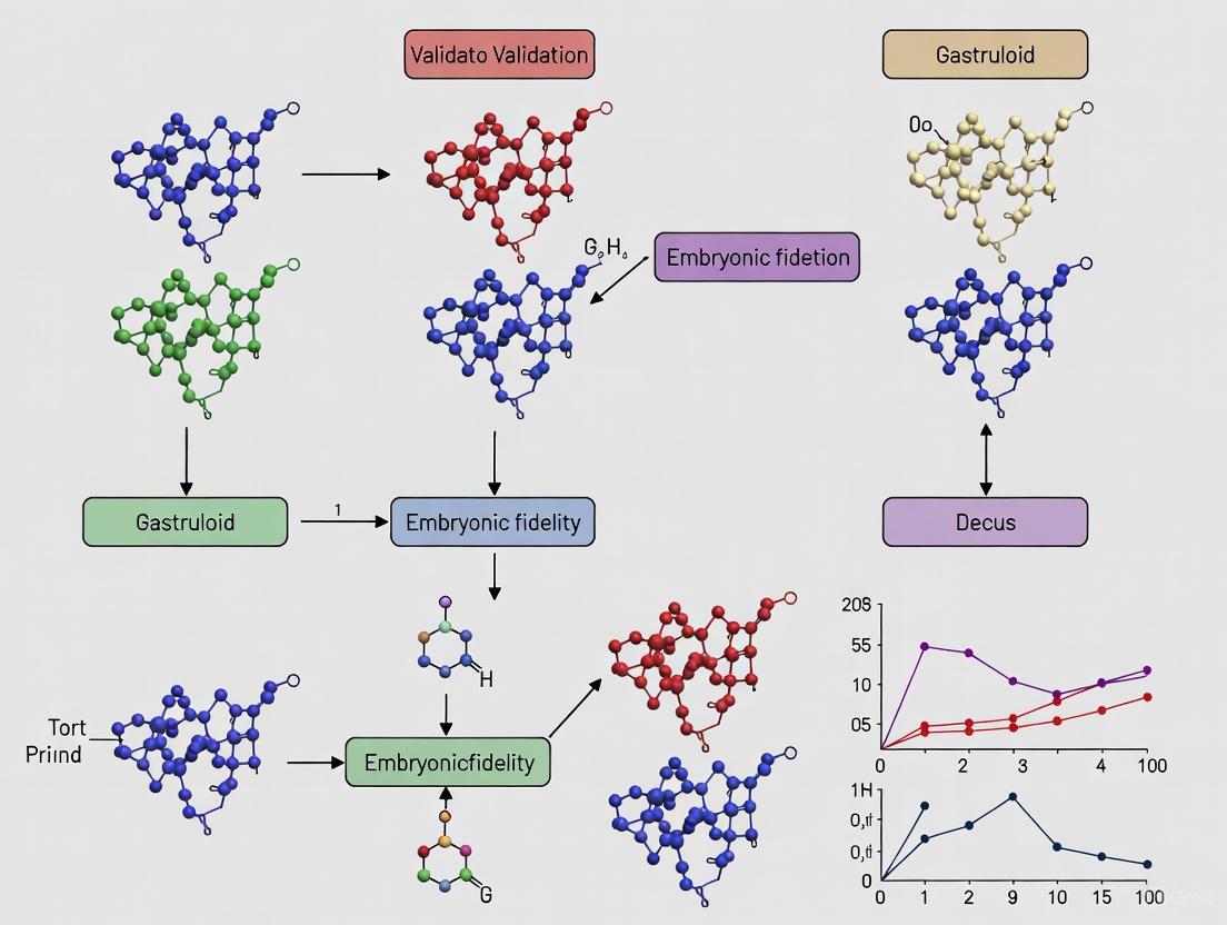
Validating Gastruloids: Assessing Embryonic Fidelity for Developmental Research and Drug Discovery
Gastruloids, three-dimensional aggregates of pluripotent stem cells, have emerged as powerful in vitro models that recapitulate key aspects of early mammalian embryogenesis.
Validating Gastruloids: Assessing Embryonic Fidelity for Developmental Research and Drug Discovery
Abstract
Gastruloids, three-dimensional aggregates of pluripotent stem cells, have emerged as powerful in vitro models that recapitulate key aspects of early mammalian embryogenesis. This article provides a comprehensive analysis of gastruloid validation, exploring their foundational biology, methodological applications in disease modeling and toxicology, strategies for optimizing developmental competence, and rigorous comparative assessments against in vivo development. We synthesize evidence from recent studies demonstrating gastruloid capabilities in modeling germ layer formation, axial patterning, and organogenesis, while addressing current limitations and standardization challenges. For researchers, scientists, and drug development professionals, this review serves as an essential resource for implementing and validating gastruloid systems in basic developmental biology research and preclinical safety assessment, ultimately contributing to the reduction of animal testing through New Approach Methodologies (NAMs).
Understanding Gastruloid Biology: From Self-Organization to Embryonic Mimicry
Gastruloids, three-dimensional aggregates derived from pluripotent stem cells, have emerged as a powerful in vitro model for studying mammalian post-implantation development. These self-organizing structures recapitulate key embryogenic processes—including axial elongation, germ layer formation, and early organogenesis—without the ethical constraints associated with natural embryos. This comparison guide evaluates the embryonic fidelity of gastruloid models against traditional embryological systems, examining their validation across molecular, cellular, and functional domains. We present quantitative data on their ability to model specific lineages such as cardiopharyngeal mesoderm, benchmark their performance in toxicological applications against regulatory standards, and detail the experimental protocols that enable their robust generation. For researchers in developmental biology and drug development, this analysis provides a critical framework for selecting appropriate model systems based on empirical validation of their developmental competence.
Gastruloids represent a breakthrough in stem cell technology, offering an unprecedented window into the early stages of mammalian embryogenesis. Defined as self-organizing 3D aggregates of pluripotent stem cells, gastruloids spontaneously undergo gastrulation-like processes including symmetry breaking, axial patterning, and germ layer specification [1]. Their significance lies in their ability to mimic post-implantation embryonic development in a controlled, accessible, and ethically manageable system, making them particularly valuable for investigating human development phases that are otherwise difficult to study [2].
The core principle underlying gastruloids is their self-organization capacity—the ability of stem cell aggregates to initiate and execute developmental programs without external scaffolding or precise spatial cues. This emergent property is coordinated by a complex interplay of transcription factors, signaling pathways, and cell adhesion molecules. For instance, the cadherin switch—from E- to N-cadherin—orchestrated by transcription factors like Snai1, plays a pivotal role in gastruloid formation by regulating the exit from pluripotency and enabling the morphogenetic movements that drive elongation and patterning [3].
Within the landscape of embryological models, gastruloids occupy a unique niche, complementing both animal models and other stem cell-derived systems. When evaluated against the benchmark of embryonic fidelity—the faithful recapitulation of in vivo developmental processes—gastruloids demonstrate remarkable strengths in modeling specific aspects of embryogenesis, particularly axial elongation and early lineage specification, while having limitations in achieving full embryonic complexity [1] [4].
Embryonic Fidelity: Benchmarking Gastruloids Against Natural Embryos
A critical validation of any in vitro model system is its fidelity to the biological processes it aims to replicate. For gastruloids, this means direct comparison to natural embryos across molecular, cellular, and structural domains. Comprehensive analyses demonstrate that gastruloids exhibit remarkable correspondence to mouse embryos in key developmental processes, particularly in the specification of progenitor populations and the temporal activation of genetic programs.
Table 1: Embryonic Fidelity of Gastruloid Models
| Developmental Process | Gastruloid Performance | Correspondence to Embryo | Key Validating Markers |
|---|---|---|---|
| Cardiopharyngeal Mesoderm (CPM) Specification | Recapitulates cardiac and skeletal muscle lineages [5] | Similar spatio-temporal gene expression to mouse embryos [5] | Tbx1, Isl1, Tcf21, Mesp1 [5] |
| Axial Patterning | Anterior-posterior polarity establishment [5] | Maintained Hoxc4 expression at one pole [5] | Hoxc4, Tnnt2 (mutually exclusive) [5] |
| Germ Layer Formation | Three germ layer specification [2] | Alignment with Carnegie stage 7 human embryos [2] | Ectoderm, mesoderm, and endoderm markers [2] |
| Somitogenesis | Rostro-caudal somite patterning [3] | Requires N-cadherin inactivation [3] | Somite-specific segmentation markers |
| Primordial Germ Cell (PGC) Formation | Emergence of PGC-like cells without BMP supplementation [2] | Identification of amnion-like cells as endogenous BMP source [2] | ISL1, PGCLC markers |
The emergence of primordial germ cell-like cells (PGCLCs) in human gastruloids represents a particularly significant validation of their embryonic fidelity. Unlike previous models that required external BMP supplementation, advanced gastruloid models spontaneously generate PGCLCs through endogenous signaling, with amnion-like cells (AMLC) identified as the likely source of BMP signaling critical for germline development [2]. This autonomous formation of key embryonic lineages underscores the remarkable self-organizing capacity of gastruloids and their utility for studying early human development.
Beyond specific lineages, the molecular machinery governing gastruloid development mirrors that of natural embryos. The coordination of pluripotency exit through Snai1-mediated repression of E-cadherin creates a cell-specific tempo that enables the coordinated transition to N-cadherin dominance, a process essential for proper morphogenesis [3]. When this process is disrupted through N-cadherin inactivation, gastruloids exhibit enhanced morphogenetic competence, forming embryo-like structures with proper rostro-caudal somite patterning without requiring extracellular matrix supplementation [3].
Functional Validation: Performance in Toxicological Applications
Beyond morphological and molecular comparisons, functional validation represents the most rigorous test of a model system's biological relevance. In toxicological applications, gastruloids have undergone systematic validation against international regulatory standards, demonstrating their utility as predictive tools for developmental and reproductive toxicity (DART) assessment.
Table 2: Toxicological Validation of Gastruloid Assays Against ICH S5(R3) Standards
| Validation Metric | Gastruloid Performance | Significance |
|---|---|---|
| Concordance with Rodent Data | 18/24 reference drugs showed comparable sensitivity within 8-fold concentration margin [6] | Predictivity of in vivo embryotoxicity |
| NOAEL-LOAEL Correlation | 7/8 additional drugs aligned with in vivo NOAEL or LOAEL data [6] | Accurate determination of no-effect and effect levels |
| Endpoint Measurement | Morphological impact (reduced growth, aberrant elongation) [6] | Quantitative assessment of developmental disruption |
| Metabolite Assessment | Detection of embryotoxicity from drug metabolites (e.g., cyclophosphamide metabolites) [6] | Comprehensive safety profiling beyond parent compounds |
| Throughput Capability | Adaptation to screening formats for large-scale assessment [6] [7] | Practical utility for preclinical screening |
The validation against the ICH S5(R3) guideline, which provides plasma concentration data for various reference drugs in rodents, offers compelling evidence for gastruloid utility in regulatory contexts. For the majority of reference compounds, the NOAEL to LOAEL concentration range obtained from gastruloid assays closely matched the in vivo range in rodents, demonstrating that gastruloids can detect embryotoxic effects at biologically relevant concentrations [6]. This correlation is particularly significant given that P19C5 gastruloids are derived from mouse cells, creating a species-matched comparison that enhances predictive value.
The morphological endpoints used in gastruloid-based toxicology—primarily reduced growth and aberrant elongation—serve as sensitive biomarkers for developmental disruption that capture complex biological processes in a quantifiable format. These macroscopic changes reflect underlying disruptions to the cellular and molecular processes driving self-organization, making them robust indicators of developmental toxicity [6]. Recent technological advances, including the development of microraft array platforms for automated imaging and sorting of individual gastruloids, now enable high-throughput screening with single-structure resolution, further enhancing their utility for large-scale toxicological assessment [7].
Experimental Protocols: Methodologies for Gastruloid Generation
The reproducible generation of high-fidelity gastruloids requires precise control over culture conditions and differentiation protocols. While specific methodologies vary depending on the research objectives, core principles underlie robust gastruloid formation across different applications.
Core Protocol for Mouse Gastruloids with CPM Specification
This protocol, adapted from Rossi et al., enables the specification of cardiopharyngeal mesoderm (CPM) and its differentiation toward cardiac and skeletal muscle lineages [5]:
- Day 0: Aggregate mouse embryonic stem cells (mESCs) via centrifugation in low-attachment plates.
- Day 2: Treat aggregates with a Wnt agonist (CHIR99021, commonly referred to as "Chiron") for 24 hours to initiate symmetry breaking and axial organization.
- Day 4: Add cardiogenic factors to the culture media, specifically basic fibroblast growth factor (bFGF), vascular endothelial growth factor (VEGF), and ascorbic acid. Continue culture for 3 days with shaking at 80-100 rpm.
- Day 7: Transition gastruloids to N2B27 basal media without additional growth factors.
- Day 7-11: Continue culture with shaking, monitoring for the emergence of beating areas (typically appearing by day 7) and skeletal myogenesis.
This extended culture protocol produces gastruloids with robust CPM specification, evidenced by the expression of key markers including Mesp1, Isl1, Tbx1, and Tcf21, followed by the emergence of cardiomyocytes (marked by Tnnt2, Myl7, Myh7) and skeletal myoblasts (marked by Myf5, MyoD) [5]. Approximately 86.79% (±7.4% SEM) of gastruloids typically show beating areas under these conditions, primarily in the anterior region [5].
Critical Protocol Parameters and Variations
Several factors significantly influence gastruloid development and must be carefully controlled:
- Media Formulation: The choice of basal media substantially impacts cell fate specification. Home-made N2B27 (HM-N2B27) promotes earlier elongation, increased cell numbers, and enhanced anterior domain formation compared to commercial NDiff227, with HM-N2B27 favoring spinal cord-related genes while NDiff227 enhances mesodermal differentiation [8].
- Cell Density and Aggregation: Initial cell number per aggregate critically influences patterning and morphology, requiring optimization for specific applications.
- Signaling Modulation: Precise timing and concentration of Wnt activation determine axial patterning and progenitor specification.
- Physical Culture Conditions: Continuous shaking from day 4 onward prevents adhesion and promotes proper morphogenesis.
For human gastruloid generation, protocols typically involve confining human pluripotent stem cells to patterned ECM-coated surfaces of defined size (0.5-1 mm), followed by BMP4 treatment to initiate the self-patterning cascade that generates concentric germ layer organization [7].
Signaling Pathways and Molecular Mechanisms
The self-organization capacity of gastruloids emerges from the coordinated action of evolutionarily conserved signaling pathways that guide cell fate decisions and morphological transformations. Understanding these pathways provides insight into both normal development and the principles of self-organization in stem cell systems.
Diagram 1: Signaling Pathways Governing Gastruloid Self-Organization. The core molecular circuitry involves Wnt-mediated activation of mesoderm specification and Snai1-coordinated EMT, leading to CPM formation and subsequent lineage diversification.
The epithelial-to-mesenchymal transition (EMT) represents a critical node in this network, controlled by transcription factor Snai1, which coordinates the repressive tempo of pluripotency exit by triggering E-cadherin repression and enabling the transition to N-cadherin dominance [3]. This "cadherin switch" establishes the cellular conditions necessary for morphogenetic movements and tissue reorganization. When N-cadherin is inactivated, gastruloids exhibit unleashed morphogenetic competence, forming embryo-like structures with proper rostro-caudal somite patterning without requiring extracellular matrix supplementation [3].
For cardiac and skeletal muscle specification from CPM, the pathway involves the sequential activation of key transcription factors: early mesodermal marker Mesp1 gives way to CPM markers Isl1 and Tbx1, followed by lineage-specific regulators—Tcf21 for skeletal muscle and Nkx2-5 for cardiac lineages [5]. The successful recapitulation of these branching lineage trajectories in gastruloids demonstrates their utility for investigating the mechanisms governing progenitor cell diversification during embryogenesis.
Essential Research Reagents and Tools
The reproducible generation and analysis of gastruloids depends on a standardized toolkit of research reagents and specialized materials. The selection of these components significantly influences experimental outcomes and requires careful consideration.
Table 3: Essential Research Reagent Solutions for Gastruloid Generation
| Reagent Category | Specific Examples | Function | Impact Variations |
|---|---|---|---|
| Basal Media | Home-made N2B27 (HM-N2B27), Commercial NDiff227 [8] | Support stem cell viability and differentiation | HM-N2B27: earlier elongation, spinal cord genes; NDiff227: mesodermal bias [8] |
| Wnt Agonists | CHIR99021 ("Chiron") [5] | Initiate symmetry breaking and axial patterning | Concentration and duration critical for proper patterning |
| Cardiogenic Factors | bFGF, VEGF, Ascorbic acid [5] | Promote cardiac lineage specification from CPM | Essential for beating cardiomyocyte formation |
| Extracellular Matrix | Matrigel, Laminin, Fibronectin [7] | Provide adhesion substrate for 2D gastruloids | Patterned geometry controls colony size and organization |
| Induction Factors | BMP4 (for human gastruloids) [7] | Initiate self-patterning cascade | Concentration critical for germ layer patterning |
| Cell Lines | Mouse ESCs, P19C5, Human PSCs [5] [6] | Source material for gastruloid generation | Species-specific differences in protocol requirements |
The basal media formulation deserves particular attention, as comparative studies reveal significant differences in gastruloid development depending on whether researchers use home-made N2B27 (HM-N2B27) or commercial NDiff227 formulations. HM-N2B27 gastruloids initiate elongation earlier, contain more cells, and develop larger anterior domains, while RNAseq analysis indicates distinct cell fate biases—HM-N2B27 favors spinal cord-related genes while NDiff227 enhances mesodermal differentiation [8]. These differences highlight the importance of consistent media selection and reporting in gastruloid research.
For specialized applications, additional reagents enable specific experimental manipulations. Microraft array technology provides an advanced platform for screening and sorting individual gastruloids, with photopatterned central circular regions of extracellular matrix (500µm diameter) to control gastruloid formation [7]. This technology enables high-throughput, image-based assays of fixed or living gastruloids and sorting of individual structures for downstream molecular analysis, powerfully addressing the heterogeneity inherent in self-organizing systems.
Gastruloids occupy a distinctive and valuable position within the spectrum of embryological models, offering a balance of embryonic fidelity, experimental tractability, and ethical acceptability. When evaluated against the rigorous benchmark of embryonic fidelity—encompassing molecular, cellular, morphological, and functional dimensions—gastruloids demonstrate compelling correspondence to natural embryos in specific developmental processes, particularly axial elongation, germ layer specification, and early organogenesis.
Their validated performance in toxicological applications, with strong concordance to animal model data for numerous reference compounds, underscores their utility as predictive tools in regulatory contexts [6]. Meanwhile, their capacity to model complex lineage specification events, such as the diversification of cardiopharyngeal mesoderm into both cardiac and skeletal muscle lineages, highlights their value for fundamental research into developmental mechanisms [5].
As the field advances, challenges remain in standardizing protocols, enhancing reproducibility, and establishing quality control metrics. The integration of engineering technologies—including micropatterned substrates, microfluidic systems, and automated screening platforms—promises to address these challenges while opening new research possibilities [4] [7]. For researchers and drug development professionals, gastruloids offer a powerful complement to traditional model systems, providing unprecedented access to the early stages of mammalian development in a controlled, scalable, and ethically manageable format.
A fundamental challenge in developmental biology is understanding how the complex body plan of an embryo is established during gastrulation, a process encompassing germ layer specification and axial patterning. Recent advances in stem cell biology have provided powerful in vitro models, particularly gastruloids, which are three-dimensional aggregates of pluripotent stem cells (PSCs) that self-organize to mimic key aspects of embryonic development [9] [10]. These models, alongside established in vivo systems like planarians and chick embryos, offer complementary insights into the conserved and species-specific mechanisms that orchestrate embryogenesis. This guide objectively compares the experimental performance and fidelity of these diverse systems in recapitulating gastrulation, providing a framework for researchers and drug development professionals to select appropriate models for specific investigative or screening purposes.
Model Systems and Their Core Characteristics
Defining the Model Organisms
Research into gastrulation employs a spectrum of biological models, each with distinct advantages and limitations.
- Gastruloids: These are PSC-derived in vitro models that mimic the axial patterning and tissue differentiation of the gastrulating embryo without extra-embryonic tissues [10]. A pivotal feature is their self-organization capacity, where a short pulse of a Wnt agonist (e.g., Chiron) triggers symmetry breaking and the emergence of an anteroposterior (AP) axis [10].
- Planarians (Schmidtea polychroa): Freshwater planarians are studied for their remarkable regenerative abilities. Their embryonic development is divided into two morphogenetic stages: an initial, highly divergent gastrulation that segregates the three germ layers and forms a transient feeding embryo, followed by a metamorphosis that establishes the definitive adult body plan using mechanisms similar to adult regeneration [11].
- Chick Embryo: A classic vertebrate model for studying gastrulation due to its accessibility and well-characterized developmental timeline. Definitive endoderm, a key signaling center, ingresses through the rostral primitive streak, marked by specific genes like Sox17 and Gata5/6 [12].
Comparative Performance Data
The table below summarizes quantitative and qualitative data on how these models recapitulate key developmental events.
Table 1: Comparative Performance of Models in Recapitulating Gastrulation
| Feature | Gastruloids (Mouse) | Planarian (S. polychroa) | Chick Embryo |
|---|---|---|---|
| Germ Layer Specification | Emerges via self-organization; sensitive to Nodal signaling [10]. | Occurs in early transient embryo; genes like foxA and twist mark specific layers [11]. | Committed at ingression; gene expression (e.g., Sox17) remains labile and responsive to signals [12]. |
| Axial Patterning | AP elongation dynamics are size-dependent and require Wnt/PCP [10]. | Definitive AP identity established late via canonical Wnt and BMP pathways [11]. | Rostral-caudal patterning is evident early, with distinct molecular identities [12]. |
| Key Signaling Pathways | Wnt/β-catenin, Planar Cell Polarity (PCP), Nodal, Differential Adhesion [10]. | Canonical Wnt, BMP [11]. | Nodal, Wnt/β-catenin, Gata factors [12]. |
| Morphogenetic Dynamics | Tbxt (Brachyury) domains coalesce to initiate elongation; timing is size-dependent [10]. | Pharyngeal development associated with initial Wnt activity; definitive axes form after yolk consumption [11]. | Definitive endoderm ingresses through rostral streak, displacing hypoblast [12]. |
| Experimental Throughput | High; amenable to high-content screening and genetic manipulation [10]. | Moderate; suitable for functional genetic studies [11]. | Low; excellent for micromanipulation and transplantation studies [12]. |
| Tissue Complexity | Embryonic tissues only (lacks extra-embryonic components) [10]. | Includes interactions with yolk syncytium [11]. | Full embryonic and some extra-embryonic tissues in a physiological context [12]. |
Detailed Experimental Protocols and Methodologies
Gastruloid Protocol: Investigating Size Constraints on Morphogenesis
This protocol, derived from Fiuza et al. (2025), is designed to study the impact of initial cell number on axis formation [10].
1. Cell Line and Pre-culture:
- Use mouse pluripotent stem cells (e.g., E14-TG2a line) maintained in Serum + LIF conditions to preserve a naive state [10].
2. Aggregate Formation:
- Prepare a single-cell suspension and seed cells in ultra-low attachment plates.
- Systematically vary the initial seeding number (e.g., from 40 to 600 cells/aggregate) to test size effects. The standard is 300 cells [10].
- Centrifuge the plates to promote aggregation.
3. Gastruloid Induction:
- At 48 hours post-aggregation, subject the aggregates to a 24-hour pulse with the Wnt agonist Chiron (CHIR99021), typically at 3 µM in N2B27 medium [10].
- The basal medium can be home-made (HM-N2B27) or commercial (NDiff227), which may influence developmental outcomes [9].
4. Extended Culture and Monitoring:
- After the Chiron pulse, replace the medium with fresh N2B27 and culture for up to 120 hours.
- Monitor morphology and elongation dynamics daily. Key observations include:
- 72 h: Round shape.
- 96 h: Ellipsoid shape.
- 120 h: Clearly elongated structure [10].
5. Endpoint Analysis:
- Immunofluorescence: For Tbxt (Brachyury), E-cadherin, and Phospho-Myosin Light Chain to visualize mesoderm precursors, adhesion, and contractility [10].
- RNA In Situ Hybridization (ISH): To analyze spatial gene expression patterns of markers like Tbxt and neural genes [10].
- Quantitative Image Analysis: Measure the timing of elongation initiation, the number of Tbxt foci, and the final length of the AP axis [10].
Chick Embryo Protocol: Testing Germ Layer Commitment
This protocol, based on Lawson et al. (2007), uses transplantation to assess cell fate plasticity [12].
1. Embryo Preparation:
- Incubate fertilized chick eggs to Hamburger-Hamilton stage 3-4 (definitive streak stage).
- Prepare host embryos by creating a window in the eggshell.
2. Donor Tissue Isolation:
- Use stage-matched quail embryos as donors for reliable cell tracking.
- Isolate segments of the rostral primitive streak (presumptive endoderm) and caudal primitive streak (presumptive mesoderm) using sharp needles or a microscalpel [12].
3. Heterotopic Transplantation:
- Transplant presumptive mesoderm from the caudal streak into a rostral streak site in the host chick embryo.
- Conversely, transplant presumptive endoderm from the rostral streak to a caudal site [12].
- Use sham operations in control embryos.
4. Analysis of Cell Fate and Gene Expression:
- Allow embryos to develop for 4-6 hours post-transplantation.
- Fix embryos and perform quail-specific immunohistochemistry (e.g., QCPN antibody) to identify the fate of donor cells—whether they integrated into the endoderm or mesoderm layer [12].
- Perform in situ hybridization for marker genes like Sox17 (endoderm) and Wnt8c (mesoderm) on transplanted embryos to assess if donor cells maintain or alter their gene expression profile in the new location [12].
Signaling Pathways in Gastrulation Models
The following diagrams, generated using DOT language, illustrate the core signaling pathways and their roles in the featured model systems.
Wnt Signaling in Axis Formation
Diagram 1: The Wnt/β-catenin pathway, initiated by agonists like Chiron, stabilizes β-catenin and activates target genes like Tbxt. Tbxt is a master regulator that coordinates both the specification of mesodermal fate and the activation of the Planar Cell Polarity (PCP) pathway, which is directly responsible for driving the cellular rearrangements required for axial elongation [10].
Germ Layer Specification via Nodal and Sox17
Diagram 2: A simplified pathway for definitive endoderm specification, as observed in chick and conserved in other models. Nodal signaling activates transcription factors Gata5/6 and Sox17, which are crucial for endoderm identity. In the chick gastrula, the gene expression of ingressed cells remains labile and can be induced or repressed by signals from the surrounding rostral blastoderm, demonstrating a combination of pre-patterning and plasticity [12].
The Scientist's Toolkit: Essential Research Reagents
The table below lists key reagents and their critical functions in gastrulation research based on the cited studies.
Table 2: Key Research Reagent Solutions for Gastrulation Studies
| Reagent / Tool | Function / Application | Experimental Context |
|---|---|---|
| Chiron (CHIR99021) | A potent, selective GSK-3β inhibitor that activates Wnt/β-catenin signaling. Used to induce symmetry breaking and axial polarization in gastruloids [10]. | Gastruloid Protocol |
| N2B27 Medium | A defined, serum-free basal medium essential for the differentiation and culture of pluripotent stem cells into gastruloids. Home-made and commercial formulations (NDiff227) can yield different results [9]. | Gastruloid Protocol |
| Sox17 Reporter/Marker | A key transcription factor and marker for definitive endoderm. Used to track endoderm specification, migration, and fate via in situ hybridization or immunofluorescence [12]. | Chick Embryo Protocol |
| Tbxt (Brachyury) Antibody | Marker for the nascent mesoderm and the organizer for axial elongation. Its polarization and domain coalescence are critical events in axis formation [10]. | Gastruloid Protocol |
| Quail-Chick Chimeras | A classical system where quail cells are transplanted into chick hosts, allowing for precise fate mapping and testing of cell commitment due to species-specific histological markers [12]. | Chick Embryo Protocol |
| E-Cadherin Antibody | Marker for epithelial cells and adherens junctions. Used to study differential adhesion and cell sorting mechanisms during germ layer segregation and axis elongation [10]. | Gastruloid Protocol |
| CRISPR-Cas9 | Gene editing technology used to create knockout cell lines (e.g., Nodal KO) to dissect the function of specific genes in gastruloid development [10]. | Genetic Manipulation |
The study of early mammalian development, particularly the transition from a simple embryonic disc to the formation of organ primordia, presents significant technical and ethical challenges. Gastruloids—three-dimensional aggregates of pluripotent stem cells that spontaneously undergo axial elongation and morphogenesis resembling gastrulation—have emerged as powerful in vitro models to overcome these limitations [6]. These self-organizing structures recapitulate key developmental events, including the specification of cardiopharyngeal mesoderm (CPM) and the emergence of early organ precursors, providing an accessible platform for developmental biology and toxicology research [5]. The validation of gastruloids against established in vivo benchmarks is crucial for their adoption as reliable tools in both basic research and pharmaceutical development, particularly following the FDA Modernization Act 2.0, which encourages the use of new approach methodologies (NAMs) to reduce conventional animal testing [6].
This guide objectively compares the developmental fidelity of gastruloid models against natural embryogenesis, with a specific focus on the progression from primitive streak-like events to organ primordia formation. We present supporting experimental data, detailed methodologies, and analytical frameworks that researchers can utilize to validate these models within their own gastruloid-based research programs.
Comparative Developmental Timeline and Key Milestones
The following table summarizes the key developmental milestones observed in mouse embryos and their corresponding events in gastruloid models, highlighting the remarkable temporal and spatial fidelity of the in vitro system.
Table 1: Comparative Developmental Milestones in Mouse Embryos and Gastruloids
| Developmental Stage | Mouse Embryo Events | Gastruloid Events | Key Markers | Developmental Competence |
|---|---|---|---|---|
| Gastrulation | E6.5: Primitive streak formation, emergence of mesoderm and endoderm [5] | Day 3-4: Axial elongation, symmetry breaking, germ layer specification [6] [5] | Mesp1, Brachyury [5] | Foundation of the primary body plan |
| Cardiopharyngeal Mesoderm (CPM) Specification | E8.0-8.5: CPM forms from Mesp1+ progenitors; gives rise to heart and head muscles [5] | Day 4-5: Transient expression of CPM markers in spatially defined regions [5] | Mesp1, Isl1, Tbx1, Tcf21 [5] | Bipotent progenitors for cardiac and skeletal muscle lineages |
| Early Cardiac Differentiation | E8.5-9.0: Heart tube formation, initiation of contraction [5] | Day 5-7: Expression of cardiac myosins; appearance of beating areas [5] | Myl7, Myh7, Tnnt2 [5] | Functional cardiomyocytes with rhythmic contraction |
| Early Myogenic Differentiation | E9.0-10.0: Onset of skeletal myogenesis from CPM and somitic mesoderm [5] | Day 7: Expression of myogenic regulatory factors [5] | Myf5, MyoD [5] | Specification towards "head-like" and "trunk-like" skeletal myoblasts |
| Organ Primordia Maturation | E10.0-11.0: Patterning and growth of organ precursors [13] | Day 7-11: Continued expression of structural and patterning markers [5] | cTnT, VEGFR2, E-cadherin, Hoxc4 [5] | Complex tissue organization and regional identity |
Quantitative Fidelity Assessment of Gastruloid Models
Predictive Capacity for Developmental Toxicity
A rigorous validation study assessed a mouse P19C5 gastruloid-based assay for developmental and reproductive toxicity (DART) prediction in accordance with the ICH S5(R3) guideline [6]. The study determined the no-observed-adverse-effect-level (NOAEL) and lowest-observed-adverse-effect-level (LOAEL) of reference drugs based on morphological impacts on gastruloids, such as reduced growth or aberrant elongation.
Table 2: Validation of Gastruloid-Based DART Assay Against ICH S5(R3) Reference Drugs
| Validation Parameter | Experimental Outcome | In Vivo Correlation | Implication for Predictive Use |
|---|---|---|---|
| Sensitivity (Drugs with both NOAEL & LOAEL data) | 18 out of 24 reference drugs | Gastruloid NOAEL-LOAEL range was comparable to rodent in vivo range within an 8-fold concentration margin [6] | High predictive accuracy for embryotoxic concentrations |
| Sensitivity (Drugs with only NOAEL or LOAEL) | 7 out of 8 additional reference drugs | Gastruloid assay results aligned with available in vivo data [6] | Robust performance even with partial reference data |
| Metabolite Testing | Assessed active metabolites (e.g., for aspirin, cyclophosphamide) | Effects of metabolites mirrored known in vivo mechanisms [6] | Model accounts for bioactivation, enhancing physiological relevance |
| Endpoint Measurement | Quantitative assessment of morphological impact (growth & shape) [6] | Concentration-dependent effects correlated with plasma Cmax and AUC values from animal studies [6] | Provides a scalable, quantitative endpoint for high-throughput screening |
Molecular Fidelity of Lineage Specification
Recent research demonstrates that gastruloids not only form CPM but also support its differentiation into both cardiac and skeletal muscle lineages, faithfully recapitulating in vivo developmental programs [5]. Single-cell RNA sequencing analysis of gastruloids from day 4 to day 11 of culture revealed three distinct subpopulations of cardiomyocytes and two subpopulations of myoblasts, the latter corresponding to different states of myogenesis with "head-like" and "trunk-like" characteristics [5]. This complexity indicates that gastruloids can model the diversification of cell types within a lineage, a key aspect of organ primordia formation. The spatial organization of gene expression in gastruloids, as shown by multiplex fluorescent in situ hybridization, closely mirrored patterns observed in mouse embryos, providing strong evidence for the structural fidelity of the model [5].
Essential Methodologies for Gastruloid Analysis
Core Protocol for CPM-Competent Gastruloid Generation
The following workflow details an extended culture protocol adapted to promote cardiopharyngeal mesoderm specification and subsequent differentiation [5].
Figure 1: Extended Gastruloid Culture Workflow for CPM Specification
Key Procedural Details:
- Cell Aggregation: Centrifugation of mouse embryonic stem cells (mESCs) to form aggregates at day 0 [5].
- Wnt Activation: Treatment with the Wnt agonist Chiron (3 μM) for 24 hours starting at day 2 to induce symmetry breaking and axial organization [5].
- Cardiogenic Factors: Supplementation with basic Fibroblast Growth Factor (bFGF), Vascular Endothelial Growth Factor (VEGF), and ascorbic acid from day 4 to day 7 to support CPM specification and cardiac differentiation [5].
- Culture Conditions: Continuous shaking (80-100 rpm) from day 4 onward to promote nutrient exchange and prevent adhesion. Culture is maintained in N2B27 basal medium after day 7 [5].
- Efficiency Metrics: This protocol typically yields beating areas in approximately 86.8% (±7.4% SEM) of gastruloids, primarily in the anterior region, by day 7 [5].
Molecular Validation Workflow
The following diagram outlines the key analytical methods used to validate developmental milestones in gastruloids.
Figure 2: Analytical Methods for Gastruloid Validation
Key Analytical Details:
- Quantitative RT-PCR: Used to track the temporal expression of key developmental markers such as Mesp1 (early mesoderm), Isl1 and Tbx1 (CPM), Tcf21 (CPM), Myl7 and Tnnt2 (cardiac muscle), and Myf5 and MyoD (skeletal muscle) [5].
- Multiplex Fluorescent In Situ Hybridization (RNAscope/HCR): Empowers spatial validation of gene expression patterns directly within the gastruloid structure, enabling direct comparison with mouse embryo sections through techniques that preserve spatial context [5].
- Single-Cell RNA Sequencing: Provides unbiased resolution of cellular heterogeneity, enabling identification of distinct progenitor and differentiated cell populations (e.g., cardiomyocyte and myoblast subpopulations) [5].
- Functional & Morphological Assessment: Beating areas indicate functional cardiomyocyte differentiation, while quantitative image analysis of gastruloid size and shape serves as a sensitive endpoint for developmental toxicity screening [6] [5].
The Scientist's Toolkit: Essential Research Reagents
Table 3: Key Reagents for Gastruloid-Based Developmental Studies
| Reagent/Cell Line | Specific Example | Function in Protocol |
|---|---|---|
| Mouse Embryonic Stem Cell (mESC) Line | P19C5 [6]; Other wild-type mESC lines [5] | Self-renewing, pluripotent cell source capable of forming gastruloids with axial organization |
| Wnt Agonist | Chiron (CHIR99021) [5] | GSK-3β inhibitor that activates Wnt signaling, critical for inducing gastrulation-like events and axial elongation |
| Cardiogenic Factors | bFGF, VEGF, Ascorbic Acid [5] | Support the specification, survival, and differentiation of cardiopharyngeal mesoderm and its derivatives |
| Reference Drugs for Validation | ICH S5(R3) list (e.g., Acitretin, Bosentan, Busulfan, Valproic Acid) [6] | Compounds with known in vivo embryotoxicity used to benchmark the predictive performance of the gastruloid assay |
| Metabolites of Reference Drugs | Salicylic acid (Aspirin metabolite); Phosphoramide Mustard & Acrolein (Cyclophosphamide metabolites) [6] | Test the role of bioactivation in observed toxic effects, increasing physiological relevance of the assay |
Gastruloids represent a validated and highly promising model for studying the key developmental milestones from primitive streak formation to organ primordia specification. Quantitative data demonstrates their fidelity in recapitulating spatio-temporal gene expression patterns, cellular heterogeneity, and physiological responses to known developmental toxicants. The methodologies and validation frameworks presented provide researchers with a robust toolkit for implementing and critically assessing gastruloid models in their own work, accelerating their application in fundamental developmental biology and predictive toxicology.
The study of early human development has long been constrained by technical limitations and ethical considerations surrounding the use of human embryos. Traditional human embryo research faces significant challenges, including limited availability of specimens, ethical restrictions such as the 14-day culture rule, and substantial species-specific differences that limit the translatability of findings from animal models [14]. In response to these challenges, stem cell-based embryo models, particularly gastruloids, have emerged as transformative tools that overcome these limitations while providing unprecedented experimental access to early developmental processes.
Gastruloids are three-dimensional aggregates of pluripotent stem cells that recapitulate key aspects of gastrulating embryos, mimicking critical developmental events such as axial organization, germ layer formation, and the emergence of cellular heterogeneity [15] [1]. These self-organizing structures provide a highly tractable and scalable platform for studying early human development in vitro, offering distinct advantages over both traditional embryo research and animal models [15]. By reproducing complex developmental processes without the ethical constraints associated with natural human embryos, gastruloids have opened new frontiers in developmental biology, disease modeling, and drug discovery.
This review comprehensively examines the advantages of gastruloid technology over traditional embryo research, focusing on experimental flexibility, biomedical applications, and the unique insights these models provide into human embryogenesis. We present quantitative comparisons, detailed methodological protocols, and visual representations of the signaling pathways and experimental workflows that underpin this revolutionary approach.
Comparative Analysis: Gastruloids vs. Traditional Embryo Research
The following table summarizes the key differences between gastruloid technology and traditional embryo research across multiple parameters that are critical for biomedical research.
Table 1: Comparative analysis of gastruloids versus traditional embryo research
| Parameter | Gastruloid Models | Traditional Human Embryo Research |
|---|---|---|
| Ethical Constraints | Minimal; not subject to 14-day rule [14] | Strictly limited by 14-day rule and oversight requirements [14] |
| Scalability | High; can generate hundreds to thousands of replicates [7] [15] | Limited by donation and availability [14] |
| Experimental Accessibility | High; amenable to live imaging and manipulation [1] | Limited; technical and ethical restrictions [14] |
| Genetic Manipulation | Straightforward; CRISp-Cas9 editing possible [16] | Extremely challenging and ethically restricted [16] |
| Developmental Scope | Specific processes (e.g., gastrulation, symmetry breaking) [15] | Entire early development but limited by culture restrictions [14] |
| Species Specificity | Human models from human pluripotent stem cells [2] | Direct human data but limited extrapolation from animal models [14] |
| Standardization | Reproducible under controlled conditions [7] | High natural heterogeneity [14] |
| High-Throughput Screening | Compatible with automated platforms [7] | Not feasible [14] |
Key Technological Advantages of Gastruloid Systems
Unprecedented Experimental Accessibility and Scalability
Gastruloid technology provides experimental capabilities that are simply unattainable with traditional embryo research. The microraft array platform developed for large-scale gastruloid screening exemplifies this advantage, enabling researchers to perform image-based assays of hundreds of individual gastruloids with subsequent sorting for downstream molecular analysis [7]. This system utilizes arrays of 529 indexed magnetic microrafts, each with a flat surface photopatterned with a central circular region of extracellular matrix to ensure standardized gastruloid formation [7]. The automated imaging and sorting system achieves remarkable efficiency, with microraft release and collection rates of 98±4% and 99±2%, respectively [7]. This technological advancement allows for quantitative studies of gastruloid heterogeneity and the systematic analysis of developmental abnormalities under various experimental conditions.
The scalability of gastruloid systems enables high-throughput screening applications that were previously impossible in developmental biology. Unlike human embryo research, which is inherently limited by specimen availability, gastruloids can be generated in large numbers from established stem cell lines, facilitating statistically robust experimental designs and the simultaneous testing of multiple conditions [7] [15]. This scalability is particularly valuable for drug screening and toxicity testing, where large sample sizes are essential for identifying subtle effects and establishing dose-response relationships.
Enhanced Genetic Manipulation and Disease Modeling
A significant advantage of gastruloid technology lies in the ease of genetic manipulation, which enables precise investigation of gene function and disease mechanisms. Researchers can introduce specific mutations into the pluripotent stem cells used to generate gastruloids, including patient-derived iPSCs that carry naturally occurring disease-associated variants [16]. This approach facilitates the creation of personalized disease models that recapitulate genetic disorders in a developmental context, offering insights into the earliest stages of pathogenesis.
The integration of CRISp-Cas9 gene editing with gastruloid technology has proven particularly powerful for studying gene function and disease etiology [16]. For example, researchers have used genetic manipulation to investigate the role of specific genes in germ cell development, demonstrating that mutations in the amniotic marker ISL1 disrupt the formation of amnion-like cells and primordial germ cell-like cells in gastruloids [2]. This genetic tractability enables researchers to establish causal relationships between specific molecular perturbations and developmental outcomes, advancing our understanding of congenital disorders and reproductive failures.
Recapitulation of Human-Specific Developmental Features
Gastruloids provide a uniquely powerful platform for studying human-specific aspects of development that cannot be adequately investigated using animal models. Comparative analyses have revealed significant differences between human and mouse embryogenesis, including variations in the timing of key events, patterns of gene expression, and morphological processes [14]. For instance, during human embryogenesis, the epiblast-derived amnion forms ahead of primitive streak development, whereas in mice, amnion genesis occurs as a consequence of extra-embryonic mesoderm formation from the primitive streak [14].
Advanced 3D human gastruloids have demonstrated remarkable fidelity to human embryos, with gene expression profiles aligning with Carnegie stage 7 human embryos and recapitulating critical developmental milestones such as elongation along the rostro-caudal axis, formation of the three germ layers, and the emergence of post-gastrulation features including cardiomyocytes and neuromesodermal progenitors [2]. This human-specific fidelity makes gastruloids particularly valuable for investigating aspects of development that differ from common animal models, ultimately enhancing the translatability of findings to human biology and medicine.
Signaling Pathways Governing Gastruloid Development
The self-organization and patterning of gastruloids are directed by precisely regulated signaling pathways that mirror those active in natural embryos. Understanding these pathways is essential for both utilizing and refining gastruloid technology.
Diagram 1: Signaling pathways in gastruloid development (Title: Signaling Pathway Governing Gastruloid Patterning)
The diagram above illustrates the core signaling cascade that guides gastruloid patterning. The process begins with Bone Morphogenetic Protein 4 (BMP4) activation, which initiates a signaling cascade that spreads from the gastruloid edges toward the center [7]. This spatial restriction of BMP signaling is controlled by receptor localization and the BMP antagonist Noggin (NOG), which is upregulated at the gastruloid center [7]. The combinatorial signaling of Wnt and Nodal pathways subsequently directs the formation of the three germ layers—ectoderm, mesoderm, and endoderm—while the peripheral region differentiates into trophectoderm-like cells [7]. This self-patterning mechanism demonstrates how gastruloids recapitulate the signaling hierarchy of natural embryos, providing a validated system for studying early developmental processes.
Experimental Workflow for Gastruloid Generation and Analysis
The following diagram and protocol describe the standardized methodology for generating and analyzing gastruloids, highlighting the precise control and reproducibility achievable with this technology.
Diagram 2: Gastruloid generation workflow (Title: Experimental Workflow for Gastruloid Generation)
Detailed Experimental Protocol
Step 1: Micropatterning of Culture Surfaces
- Create arrays of circular extracellular matrix (ECM) islands using photopatterning techniques with 500 μm diameter circular regions [7]
- Use polydimethylsiloxane (PDMS) microwell arrays containing indexed magnetic microrafts (789 μm side length) for scalable production [7]
- Achieve patterning accuracy of 93±1% for standardized gastruloid formation [7]
Step 2: Stem Cell Seeding and Confinement
- Seed human pluripotent stem cells (hPSCs) onto the patterned surfaces at confluent density [7]
- Culture cells in appropriate maintenance media until colonies form confined to the circular ECM islands [7]
Step 3: BMP4 Induction and Gastruloid Formation
- Add Bone Morphogenetic Protein 4 (BMP4) to induce gastruloid formation [7]
- Monitor the self-organization process over 4-8 days as colonies develop concentric rings of the three germ layers [7]
Step 4: Imaging and Feature Extraction
- Acquire transmitted light and fluorescence images using automated imaging systems [7]
- Implement computational pipelines to extract morphological features (e.g., DNA/area, patterning quality) [7]
Step 5: Sorting and Downstream Analysis
- Release target microrafts using thin needle manipulation (98±4% efficiency) [7]
- Collect selected gastruloids using magnetic wand (99±2% efficiency) [7]
- Perform downstream molecular analyses including single-cell RNA sequencing, immunostaining, or proteomic profiling [7] [2]
Essential Research Reagent Solutions
The following table catalogues the key reagents and materials essential for successful gastruloid generation and experimentation, providing researchers with a comprehensive toolkit for implementing this technology.
Table 2: Essential research reagents for gastruloid generation and analysis
| Reagent Category | Specific Examples | Function and Application |
|---|---|---|
| Stem Cells | Human embryonic stem cells (hESCs), induced pluripotent stem cells (hiPSCs) [16] [14] | Foundation for generating self-organizing gastruloid structures; patient-specific iPSCs enable disease modeling. |
| Signaling Molecules | Bone Morphogenetic Protein 4 (BMP4) [7], Wnt agonists, Nodal inhibitors | Direct patterning and cell fate specification; BMP4 initiates gastruloid formation and symmetry breaking. |
| Extracellular Matrix | Matrigel, laminin, collagen-based substrates [7] [14] | Provide structural support and biochemical cues for cell adhesion and polarization. |
| Culture Platforms | Microraft arrays [7], micropatterned surfaces [14] | Enable high-throughput production and analysis; facilitate standardized gastruloid formation. |
| Detection Reagents | Immunofluorescence antibodies, RNA probes, live-cell dyes [7] | Visualize spatial patterning, protein localization, and gene expression patterns. |
| Genetic Tools | CRISp-Cas9 systems [16], reporter cell lines | Enable gene editing and lineage tracing; facilitate mechanistic studies of development. |
| Analysis Kits | Single-cell RNA sequencing kits, bulk transcriptomic platforms [2] | Enable molecular characterization of cell types and states within gastruloids. |
Applications in Disease Modeling and Drug Development
Investigating Chromosomal Abnormalities
Gastruloid technology has proven particularly valuable for studying the effects of aneuploidy (abnormal chromosome numbers) during early development. Researchers have utilized gastruloids to model aneuploidy by treating hPSCs with reversine, a small molecule that inhibits MPS1 kinase and disrupts chromosome segregation [7]. Quantitative analyses of euploid versus aneuploid gastruloids have revealed significant phenotypic differences, with aneuploid gastruloids displaying less DNA per area and upregulation of NOG and KRT7 genes compared to euploid counterparts [7]. These findings demonstrate how gastruloids can model developmental abnormalities associated with chromosomal disorders, providing insights into the mechanisms underlying pregnancy loss and congenital defects.
Modeling Germ Cell Development
Advanced 3D gastruloid systems have enabled the study of primordial germ cell-like cells (PGCLCs), the precursors to sperm and eggs, which are typically difficult to access in early human development. Remarkably, these next-generation gastruloids support the emergence of PGCLCs without external BMP supplementation, previously considered essential for germ cell specification [2]. Further investigation revealed that amnion-like cells within the gastruloids serve as an endogenous source of BMP signaling, critical for PGCLC development [2]. This discovery highlights how gastruloids can recapitulate complex tissue-tissue interactions that occur in natural embryos, providing a unique platform for investigating human germline development and associated infertility issues.
High-Throughput Drug Screening
The scalability and standardization of gastruloid systems make them ideal platforms for pharmacological testing and toxicity screening. The ability to generate hundreds of genetically identical gastruloids enables researchers to screen multiple drug candidates or concentration gradients in parallel, assessing their effects on early developmental processes [7] [1]. This application is particularly valuable for identifying teratogenic compounds that disrupt embryonic development, as gastruloids provide a human-relevant model system that can potentially reduce reliance on animal testing. Additionally, gastruloids derived from patient-specific iPSCs offer opportunities for personalized drug testing, potentially identifying compounds that rescue disease-specific developmental abnormalities.
Gastruloid technology represents a paradigm shift in how researchers approach the study of early human development. By overcoming the ethical and technical limitations of traditional embryo research while providing human-specific insights unavailable from animal models, gastruloids have established themselves as indispensable tools in developmental biology. The continued refinement of these systems, including enhanced structural complexity, improved reproducibility, and integration with advanced computational methods, promises to further narrow the gap between in vitro models and in vivo development [16] [17].
As the field progresses, gastruloids are poised to accelerate discoveries in reproductive medicine, congenital disorder research, and drug development. Their unique combination of experimental accessibility, genetic tractability, and human relevance positions gastruloid technology as a cornerstone of developmental biology research, offering unprecedented opportunities to unravel the complexities of human embryogenesis and translate these insights into clinical applications.
Cellular Heterogeneity and Lineage Trajectories in Gastruloid Systems
Gastruloids, three-dimensional aggregates derived from pluripotent stem cells (PSCs), have emerged as a powerful in vitro system for studying the principles of mammalian embryogenesis, particularly the processes of gastrulation and early lineage specification [1] [15]. These structures recapitulate key aspects of in vivo development, including symmetry breaking, axial organization, and the emergence of the three germ layers, without the complexity of extra-embryonic tissues [1]. A defining feature of gastruloids is their remarkable self-organization capacity, whereby a seemingly uniform population of PSCs undergoes coordinated differentiation and spatial patterning in response to defined signaling cues [15]. This review focuses on the critical roles of cellular heterogeneity and the dynamics of lineage trajectories in gastruloid systems, framing them as essential metrics for validating their fidelity to embryonic development. We objectively compare the performance of various gastruloid protocols and models, providing a synthesis of experimental data and methodologies that empower researchers to select and optimize these systems for specific applications in developmental biology and disease modeling.
Foundations of Gastruloid Heterogeneity
The Impact of Pluripotency States on Developmental Competence
The initial state of pluripotent stem cells is a critical determinant of gastruloid formation efficiency and developmental outcome. Research demonstrates a clear hierarchy of competence rooted in the pluripotency continuum.
Table 1: Impact of Pluripotency State on Gastruloid Formation Efficiency (GFE)
| Pluripotency State | Culture Conditions | Gastruloid Formation Efficiency (GFE) | Key Characteristics |
|---|---|---|---|
| Naive (e.g., mESCs) | 2i + LIF [18] | ~95-98% [18] | Robust cell-cell adhesion; high aggregation competence; generates elongated gastruloids. |
| Formative/ Early-Primed (e.g., EpiLCs, PiCs) | FGF/Activin A; High Proline [18] | ~50% (PiCs) [18] | Transient, unstable state; aggregates but shows variable elongation success. |
| Primed (e.g., EpiSCs) | FGF/Activin A [18] | ~0% [18] | Prone to cell-substrate rather than cell-cell adhesion; fails to generate proper aggregates. |
The table above summarizes a foundational finding: the capacity to form elongated gastruloids decreases as cells progress from the naive to the primed pluripotent state [18]. This is not merely a difference in efficiency but reflects fundamental shifts in cellular properties. Naive mouse embryonic stem cells (mESCs), particularly when maintained in 2i/LIF conditions to minimize pre-existing heterogeneity, exhibit superior cell-cell adhesive interactions and can form gastruloids with an efficiency exceeding 95% [18]. In contrast, primed epiblast stem cells (EpiSCs) largely fail to aggregate properly. An intermediate, "early-primed" state, exemplified by proline-induced cells (PiCs), retains partial competence (~50% GFE), demonstrating that a specific window of pluripotency is permissive for gastruloid development [18].
Methodological Optimization for Reproducibility
Protocol refinements are crucial for managing inherent heterogeneity and improving reproducibility. Key methodological advances include:
- Cell Dissociation: Using milder accutase instead of trypsin to preserve cell-cell adhesion capability [18].
- Cell Sorting: Employing fluorescence-activated cell sorting (FACS) to exclude dead cells and debris, ensuring a precise number of living cells are seeded for aggregation [18].
- Aggregate Size Control: Standardizing initial aggregate diameter to a narrow range (e.g., 153-180 μm) to minimize morphological abnormalities [18].
Mapping Lineage Trajectories and Cell States
Single-Cell Resolution of Gastruloid Development
Single-cell genomic technologies have provided unprecedented resources for mapping cell states and lineage trajectories in gastruloids and comparing them directly to the in vivo embryo.
Table 2: Key Cell States and Lineages Identified in Murine Gastruloids via scRNA-seq
| Broad Lineage | Specific Cell State/Type | Key Marker Genes | Similarity to In Vivo Counterpart |
|---|---|---|---|
| Pluripotency & Early Exit | Naive Pluripotent | Source population [19]. | |
| Epiblast (pre-Wnt pulse) | Anterior-like state at 36h [19]. | ||
| Ectopic Pluripotency (EP) | Sox2, Esrrb, Zfp42 [19] | Similar to naive ESCs; emerges post-Wnt activation [19]. | |
| Germ Layer Progenitors | Primitive Streak-like | T (Brachyury) [19] | Strong match [19]. |
| Neuromesodermal Progenitors (NMPs) | T (Brachyury), Sox2, Cdx2 [19] [20] | Strong match; source of posterior body tissues [19] [20]. | |
| Differentiated Lineages | Presomitic Mesoderm (PSM) | Tbx6 [20] | Strong match [19]. |
| Somite | Mesp2, Ripply2, Fst [20] | Strong match [19]. | |
| Cardiopharyngeal Mesoderm (CPM) | Tbx1, Isl1, Tcf21 [5] | Faithful spatio-temporal expression vs. mouse embryo [5]. | |
| Definitive Endoderm | Sox17, Foxa2 [19] | Identified, forms gut tube-like structures [19]. |
Integration of gastruloid scRNA-seq data with embryonic reference datasets confirms that most cell types emerging after Wnt activation (>72 hours) co-cluster strongly with their in vivo counterparts from E6.5 to E8.5 mouse embryos [19]. This includes a continuum from epiblast to primitive streak-like cells, and subsequently to neuro-mesodermal progenitors (NMPs), presomitic mesoderm, somites, and definitive endoderm [19]. A notable deviation from in vivo development is the emergence of an ectopic pluripotency (EP) population upon Wnt activation, which re-expresses naive pluripotency markers and may later contribute to heterogeneity [19].
Signaling Pathways Governing Patterning
The breaking of radial symmetry and the subsequent axial patterning in gastruloids are orchestrated by the interplay of several key signaling pathways. The following diagram synthesizes the core signaling logic underlying gastruloid patterning, derived from multiple studies [18] [19] [20].
The workflow initiates with a pulse of Wnt activation (e.g., with CHIR99021), which is the primary trigger for symmetry breaking and the induction of primitive streak-like and mesodermal fates [19]. This process relies on Nodal signaling and its co-receptor CRIPTO [18]. Subsequently, BMP signaling is activated at the periphery of the structure, promoting posterior fates. A key self-organizing feature is the induction of the BMP antagonist NOG in the center, which restricts BMP activity to the edges and facilitates the establishment of anterior-like identities [7] [20]. In later stages, Retinoic Acid (RA) plays a critical role in advanced models, promoting the differentiation of segmented somites and anterior neural tube fates from neuromesodermal progenitors (NMPs) [20].
Experimental Platforms for Screening and Analysis
High-Throughput Screening Technologies
The scalable nature of gastruloids makes them amenable to high-throughput screening, enabling the systematic dissection of heterogeneity and genetic interactions.
Microraft Array Platform: A recent technological innovation uses large-scale microraft arrays (789 µm side length) with photopatterned extracellular matrix (ECM) islands to generate and screen thousands of individual gastruloids [7]. This platform allows for automated image-based phenotyping and the gentle release and collection of specific gastruloids based on phenotypic features for downstream molecular analysis (e.g., transcriptomics) [7]. Its application has successfully discriminated phenotypic and gene expression heterogeneity between euploid and aneuploid human gastruloids [7].
Phenotypic Compound Screening: Suppinger et al. employed a high-content imaging pipeline to perform a time-dependent compound screen on tens of thousands of murine gastruloids [19]. This approach identified functional modules and genetic interactions that govern the major steps of gastruloid development—aggregation, symmetry breaking, and axial elongation—and was used to derive a "phenotypic landscape" [19]. A key insight from this screen was that dual Wnt modulation (combining agonist and antagonist) could improve the representation of anterior foregut and neural structures, which are typically underrepresented in standard protocols [19].
Lineage Tracing and Clonal Analysis
To directly investigate the origins of heterogeneity, monoclonal gastruloids (derived from a single mESC) have been developed alongside sophisticated lineage recording tools [21].
This experimental workflow involves expanding a single "founder" mESC into a clonal population that is aggregated to form a monoclonal gastruloid. Tools like DNA Typewriter record cell lineage relationships during the expansion phase [21]. When combined with single-cell RNA sequencing of the resulting gastruloids, this approach reveals that extensive inter-individual heterogeneity exists even in a monoclonal origin [21]. Crucially, closely related founder cells give rise to gastruloids with more similar cell type compositions, demonstrating that fluctuations in the intrinsic states of mESCs are heritable and can shape the eventual lineage outcomes across many cell divisions [21].
Comparative Performance of Advanced Gastruloid Models
Recent protocol modifications have significantly enhanced the developmental potential of gastruloids, enabling more complex modeling of organogenesis. The table below compares key advanced models.
Table 3: Comparison of Advanced Gastruloid Models for Lineage Specification
| Gastruloid Model | Key Protocol Modifications | Major Lineage/Tissue Outcomes | Quantitative Readouts |
|---|---|---|---|
| Cardiopharyngeal Model (Mouse) [5] | Extended culture to Day 11; Addition of cardiogenic factors (bFGF, VEGF, AA) from Day 4. | Cardiac muscles (first & second heart fields); Skeletal muscles (head-like & trunk-like myoblasts). | ~87% of gastruloids show beating areas [5]; Expression of Mesp1, Isl1, Tbx1, Tnnt2, MyoD [5]. |
| Trunk-Like Structure (TLS) (Mouse) [20] | Matrigel supplementation during induction. | Somites; Neural tube. | Morphological scoring of somite segmentation and neural tube elongation [20]. |
| Human RA-Gastruloid [20] | Early pulse of Retinoic Acid (RA); Later Matrigel supplementation. | Segmented somites; Neural tube; Neural crest; Renal progenitors. | 89% of elongated gastruloids exhibit both somites & neural tube across 5 experiments [20]. |
These models demonstrate that specific signaling cues can direct gastruloids toward distinct developmental trajectories. The cardiopharyngeal model showcases the concurrent specification of cardiac and skeletal muscle lineages from a common progenitor pool, recapitulating in vivo spatio-temporal expression patterns of markers like Tbx1 and Isl1 [5]. The breakthrough in human gastruloid development came from identifying a deficit in RA signaling. An early, discontinuous pulse of RA was sufficient to restore the bipotency of NMPs, robustly inducing structures resembling a posterior embryo with a neural tube flanked by segmented somites [20]. This highlights how understanding and manipulating the underlying signaling environment is key to enhancing embryonic fidelity.
Table 4: Key Research Reagent Solutions for Gastruloid Research
| Reagent / Solution Category | Specific Examples | Function in Gastruloid Protocols |
|---|---|---|
| Signaling Modulators | CHIR99021 (Wnt agonist) [19] [20] | Induces symmetry breaking and primitive streak-like fate. |
| Retinoic Acid (RA) [20] | Promotes neural differentiation from NMPs and somite segmentation in human models. | |
| BMP4 [7] | Initiates patterning cascade in 2D human gastruloids, inducing trophectoderm-like fate at edges. | |
| LIF (Leukemia Inhibitory Factor) [18] | Maintains naive pluripotency in starting cell population. | |
| Culture Substrates & Media | Matrigel [20] | Extracellular matrix that supports complex morphogenesis (e.g., neural tube, somites). |
| N2B27 Basal Medium [19] [5] | Defined medium used for the aggregation and differentiation phases. | |
| Enzymes & Cell Handling | Accutase [18] | Mild enzyme for cell dissociation, preserving viability and aggregation competence. |
| Advanced Tools | DNA Typewriter [21] | CRISPR-based system for recording cell lineage relationships in live cells. |
| Microraft Arrays [7] | High-throughput platform for screening and sorting individual gastruloids based on phenotype. |
Gastruloid systems have proven to be exceptionally versatile models for deconstructing the complexity of early mammalian development. The evidence synthesized herein demonstrates that cellular heterogeneity is not merely noise, but a fundamental property that reflects in vivo dynamics, arises from heritable fluctuations in founder cells, and determines the developmental capacity of the model. The detailed lineage trajectories mapped through single-cell genomics provide a rigorous, data-driven benchmark for validating the embryonic fidelity of any gastruloid protocol. Continued technological innovations in high-throughput screening, lineage tracing, and targeted modulation of signaling pathways will further enhance the precision and utility of gastruloids. As these models become more sophisticated, they are poised to offer unparalleled insights into human development and congenital disorders, providing a robust, scalable, and ethically accessible platform for the next generation of developmental biology and drug discovery research.
Methodological Advances and Practical Applications in Research and Drug Development
Standardized Protocols for Robust Gastruloid Generation Across Species
Gastruloids, three-dimensional embryonic organoids derived from pluripotent stem cells, have emerged as powerful tools for studying early developmental processes, including gastrulation, axial patterning, and cell fate specification. These models provide an ethical and accessible platform for investigating principles of early development and morphogenesis while reducing reliance on mammalian embryos. However, the methodological variability across different laboratories and model systems has presented significant challenges in achieving reproducible, comparable results. This guide objectively compares standardized protocols for generating gastruloids across different species, focusing on the key parameters, performance metrics, and experimental methodologies that ensure robustness and reproducibility. The content is framed within the broader context of gastruloid validation in embryonic fidelity research, providing researchers, scientists, and drug development professionals with a comprehensive resource for implementing these protocols in their experimental workflows.
The drive toward standardization is particularly important as gastruloid models become increasingly sophisticated, with applications spanning from basic developmental biology to toxicology screening and disease modeling. By establishing consistent protocols across research groups, the field can better evaluate the fidelity of these models in recapitulating embryonic events and generate more reliable data for both academic and translational applications.
Comparative Analysis of Species-Specific Gastruloid Protocols
Mouse Embryonic Stem Cell Gastruloids
The mouse gastruloid system represents one of the most established and optimized platforms for studying mammalian embryonic development in vitro. The protocol developed by Baillie-Johnson et al. and refined by Anlas et al. provides a robust foundation for generating mouse gastruloids with high reproducibility [18] [22]. Through systematic optimization, researchers have achieved remarkable gastruloid formation efficiency (GFE) of 95-98% from naive mouse embryonic stem cells (mESCs) [18].
Critical Protocol Parameters:
- Starting cell state: Naive pluripotency (serum/LIF or 2i/LIF conditions)
- Aggregate size: 300 cells/aggregate (approximately 150-200μm diameter)
- Induction signal: 24-hour pulse with CHIR99021 (WNT pathway activator)
- Culture duration: 5-7 days in suspension
- Key morphological milestones: Symmetry breaking at 96 hours, axial elongation by 120 hours
The improved protocol incorporates several critical modifications that enhance reproducibility: maintaining mESCs at low density (250 cells/cm²) on gelatin-coated plates in 2i+LIF medium to preserve naive pluripotency, using accutase instead of trypsin for gentler cell dissociation that maintains cell-cell adhesion capability, and implementing fluorescence-activated cell sorting (FACS) to exclude dead cells and debris from the initial aggregates [18]. These refinements minimize the formation of aberrant structures and ensure consistent development of elongated gastruloids with proper anteroposterior patterning.
Table 1: Standardized Mouse Gastruloid Protocol Parameters and Outcomes
| Parameter | Original Protocol | Optimized Protocol | Impact on Reproducibility |
|---|---|---|---|
| Starting Cell Density | Confluent monolayer (>60%) | Low density (250 cells/cm²) | Increases naive colony formation to 90-95% |
| Dissociation Method | Trypsin | Accutase | Preserves cell-cell adhesion capabilities |
| Cell Selection | No selection | FACS of live cells | Eliminates dead cells/debris from aggregates |
| Aggregate Size Range | 125-195μm (mean=156μm) | 153-180μm (mean=166μm) | Reduces variability in initial conditions |
| Gastruloid Formation Efficiency | ~75% | 95-98% | Dramatically improves successful development |
| Aberrant Structure Formation | ~25% | 2-5% | Minimizes abnormal morphologies |
Human Pluripotent Stem Cell Gastruloids
Human gastruloid models have evolved primarily as adherent two-dimensional (2D) systems that recapitulate key aspects of early human embryogenesis. Unlike the 3D mouse gastruloids, the human model relies on micropatterned substrates that confine human pluripotent stem cells (hPSCs) to defined circular areas coated with extracellular matrix (ECM) [7] [23]. This geometric confinement, combined with precise signaling activation, enables the formation of radially patterned structures that mimic the spatial organization of germ layers during early human development.
The standardized human gastruloid protocol involves several critical steps:
- Surface preparation: Creation of circular ECM islands (500μm diameter) using photopatterning or microcontact printing
- Cell seeding: Plating hPSCs at high density onto patterned surfaces to form confluent colonies
- Induction: Treatment with BMP4 to trigger symmetry breaking and patterning
- Patterning timeline: Emergence of concentric germ layer domains within 48-72 hours
Recent technological innovations have further enhanced the human gastruloid system. The development of microraft array-based technology enables high-throughput screening and sorting of individual gastruloids [7]. This platform utilizes arrays of 789μm magnetic microrafts photopatterned with central circular ECM regions (500μm diameter) with 93±1% accuracy, allowing for parallel processing of hundreds to thousands of gastruloids. The system achieves remarkable release and collection efficiencies of 98±4% and 99±2%, respectively, enabling image-based assays of both fixed and living gastruloids followed by individual sorting for downstream molecular analyses [7].
Table 2: Comparative Analysis of Mouse and Human Gastruloid Systems
| Characteristic | Mouse Gastruloids | Human 2D Gastruloids |
|---|---|---|
| Morphology | 3D elongated structures | 2D radially patterned colonies |
| Starting Cell Number | 300 cells/aggregate | Confluent colony on 500μm island |
| Key Inducing Signal | CHIR99021 (WNT activation) | BMP4 |
| Patterning Timeline | 5-7 days | 2-3 days |
| Spatial Organization | Anteroposterior axis | Concentric germ layers |
| Throughput Potential | Moderate (hundreds) | High (thousands with microraft arrays) |
| Downstream Applications | Whole-mount imaging, time-lapse analysis | High-content screening, transcriptomics |
| Model Strengths | Axial elongation, somitogenesis | Germ layer patterning, signaling studies |
Emerging Model Systems: Zebrafish Explants
While mouse and human systems dominate gastruloid research, alternative models are emerging that offer unique advantages. Zebrafish embryonic explants represent a complementary system for studying specific developmental processes, particularly head formation [24]. The protocol for generating zebrafish head-like structures (HLS) involves dissecting zebrafish embryonic explants and exposing them to defined morphogen gradients to induce anterior patterning.
This model addresses a significant gap in the field, as many existing gastruloid systems effectively mimic trunk formation but poorly recapitulate head organogenesis. The zebrafish HLS protocol provides steps for constructing cell and patterning landscapes, investigation of cell types by single-cell RNA-seq, exploration of cell patterns by in situ hybridization, and evaluation of axis induction ability through ventral overexpression assays [24]. This system highlights the diversity of approaches within the gastruloid field and the importance of selecting model systems based on specific research questions.
Quantitative Assessment of Gastruloid Performance Metrics
Formation Efficiency and Reproducibility
The performance of gastruloid protocols is most fundamentally assessed through their formation efficiency and reproducibility across experiments. For mouse gastruloids, the optimized protocol achieves exceptional GFE of 95-98%, a significant improvement over the original 75% efficiency [18]. This high efficiency is contingent on the precise control of initial conditions, particularly the aggregate size and cell viability. The use of FACS sorting to ensure high viability in the initial cell population reduces variability and improves the consistency of gastruloid development.
For human gastruloids, quantitative performance metrics focus on patterning accuracy and sorting efficiency in high-throughput systems. The microraft array technology achieves 93±1% accuracy in ECM patterning, ensuring consistent substrate conditions for gastruloid development [7]. The automated sorting system maintains 98±4% release efficiency and 99±2% collection efficiency, enabling reliable processing of individual gastruloids for downstream analysis without compromising structural integrity.
Morphological and Molecular Patterning Fidelity
Beyond simple formation efficiency, the quality of gastruloids is assessed through their morphological and molecular patterning fidelity. In mouse gastruloids, proper development is characterized by symmetry breaking at approximately 96 hours followed by axial elongation, resulting in structures 0.5-1mm in length by 120 hours [18]. At the molecular level, these structures exhibit appropriate expression of key developmental markers including BRACHYURY (T) for mesoderm, SOX2 for ectoderm, SOX17 for endoderm, and CDX2 for trophectoderm-like cells, demonstrating the establishment of proper cell lineages and anteroposterior axis [18].
Human gastruloids display distinct radial patterning with concentric rings corresponding to different germ layers and extraembryonic lineages. The outer edges exhibit trophectoderm-like characteristics (CDX2+, KRT7+), while inner regions form ectoderm, mesoderm, and endoderm domains in response to BMP, WNT, and Nodal signaling gradients [7] [23]. This organization recapitulates key aspects of human embryonic patterning despite the simplified 2D geometry.
Signaling Pathways Controlling Gastruloid Development
Core Signaling Cascades
The development of robust gastruloids across species depends on the precise activation and regulation of evolutionarily conserved signaling pathways. The BMP-Wnt-Nodal signaling cascade serves as the core regulatory network governing patterning in both mouse and human gastruloids, though with species-specific variations in implementation.
In human 2D gastruloids, BMP4 initiation triggers a signaling cascade that begins at the colony edges and sweeps inward, followed by the combinatorial action of WNT and Nodal pathways that establish the concentric germ layer organization [7]. The BMP antagonist noggin (NOG) plays a critical role in this process by restricting BMP signaling to the periphery and enabling the formation of distinct transcriptional domains. This coordinated signaling results in the radial patterning characteristic of human gastruloids, with extraembryonic trophectoderm-like cells at the edge and germ layer progenitors inward.
Mouse 3D gastruloids rely on a pulsed WNT activation (typically via CHIR99021) to initiate symmetry breaking and axial elongation [18] [22]. This initial signal activates a gene regulatory network involving Brachyury and other transcription factors that drive the emergence of the anteroposterior axis and subsequent tissue patterning. The precise timing and duration of WNT activation is critical, with a 24-hour pulse proving optimal for most applications.
Figure 1: Core Signaling Pathways in Gastruloid Development
Metabolic Regulation of Gastruloid Development
Recent research has revealed that compartmentalized cellular metabolism plays an instructive role in gastrulation beyond its traditional housekeeping functions [25]. Studies in mouse embryos and stem cell models have identified two spatially resolved, stage-specific waves of glucose metabolism during gastrulation:
- Epiblast Wave: Occurs through the hexosamine biosynthetic pathway (HBP) to drive fate acquisition in the epiblast
- Mesodermal Wave: Utilizes glycolysis to guide mesoderm migration and lateral expansion
These metabolic waves are coupled to ERK activity through distinct mechanisms in each wave, creating a synergy between metabolic and signaling pathways that guides cell fate and specialized functions during development [25]. Inhibition studies using 2-deoxy-d-glucose (2-DG) and azaserine have demonstrated that blocking glucose metabolism, particularly the HBP branch, significantly impairs primitive streak progression and mesodermal transition in developing embryos [25].
This metabolic regulation represents an additional layer of control in gastruloid development that must be considered in protocol standardization. Nutrient availability, energy metabolism, and metabolic signaling interact with traditional morphogen pathways to ensure robust patterning, suggesting that optimized culture conditions should carefully control glucose levels and other metabolic parameters.
Figure 2: Metabolic Regulation of Gastruloid Development
Experimental Workflows for Gastruloid Analysis
Integrated Screening and Sorting Platforms
The development of automated technologies for screening and sorting gastruloids has dramatically enhanced the scalability and reproducibility of these model systems. The microraft array platform represents a particularly advanced implementation, enabling image-based assays of large numbers of fixed or living gastruloids followed by sorting of individual structures for downstream analysis [7].
This integrated workflow involves several key steps:
- Array fabrication: Creation of indexed magnetic microrafts (789μm side length) with flat surfaces
- ECM photopatterning: Precise deposition of central circular ECM regions (500μm diameter)
- Gastruloid culture: Formation of single gastruloids on each raft under defined conditions
- Image-based screening: Automated imaging and feature extraction from transmitted light and fluorescence images
- Selective sorting: Release and collection of specific microrafts based on phenotypic criteria
- Downstream analysis: Molecular profiling (transcriptomics, proteomics) of individual gastruloids
This platform has been successfully applied to assay euploid and aneuploid human gastruloids, revealing clear phenotypic differences and significant heterogeneity even within the same condition [7]. Aneuploid gastruloids displayed significantly less DNA/area than euploid counterparts, with upregulation of NOG and KRT7 genes that correlated negatively with DNA/area [7].
Molecular Validation Methods
Standardized protocols for molecular validation are essential for assessing gastruloid fidelity across species and experimental conditions. The following approaches provide complementary information about gastruloid quality and developmental progression:
- Immunofluorescence staining: Spatial analysis of key developmental markers (BRACHYURY, SOX2, SOX17, CDX2, NESTIN) to verify germ layer specification and axial patterning [18]
- Single-cell RNA sequencing: Comprehensive characterization of cell types and states, comparison to embryonic reference datasets, and identification of aberrant differentiation trajectories [24] [23]
- Spatial transcriptomics: Mapping gene expression patterns within the context of gastruloid morphology, particularly important for assessing organization relative to embryonic structures [25]
- Live imaging and metabolic profiling: Dynamic assessment of gastruloid development using reporter cell lines, fluorescent metabolic probes (2-NBDG for glucose uptake), and label-free readouts of metabolic activity (NAD(P)H autofluorescence) [25]
These validation methods should be implemented at key developmental timepoints to ensure that gastruloids are progressing through appropriate morphological and molecular stages according to established benchmarks.
Essential Research Reagents and Tools
Table 3: Key Research Reagent Solutions for Gastruloid Generation and Analysis
| Reagent Category | Specific Examples | Function | Species Application |
|---|---|---|---|
| Pluripotency Maintenance | 2i/LIF medium, FBS/LIF medium | Maintain naive pluripotent state | Mouse |
| Signaling Modulators | CHIR99021 (WNT activator), BMP4, Noggin | Direct patterning and fate specification | Mouse, Human |
| Dissociation Reagents | Accutase, Trypsin | Gentle cell dissociation for aggregation | Mouse, Human |
| Extracellular Matrix | Matrigel, Laminin, Fibronectin | Provide adhesion substrates for patterning | Human (2D) |
| Metabolic Inhibitors | 2-DG, Azaserine, BrPA | Probe metabolic requirements of development | Mouse |
| Fixation & Staining | Paraformaldehyde, Triton X-100, antibodies for key markers | Morphological and molecular analysis | All species |
| Cell Sorting Tools | FACS, Magnetic bead separation | Isolation of specific cell populations | Mouse, Human |
| Live Imaging Reagents | 2-NBDG, CellTracker dyes, Vital stains | Dynamic assessment of development | All species |
The standardization of gastruloid generation protocols across species represents a critical step toward maximizing the research utility of these embryonic models. By implementing the precise parameters, quality control measures, and validation benchmarks outlined in this guide, researchers can significantly enhance the reproducibility, comparability, and embryonic fidelity of their gastruloid systems. The continued refinement of these protocols—incorporating advanced engineering technologies such as micropatterned substrates, microfluidic systems, and automated screening platforms—will further strengthen the position of gastruloids as essential tools for understanding developmental principles, modeling diseases, and screening therapeutic compounds. As the field progresses, maintaining this focus on standardization and cross-validation will ensure that gastruloid models realize their full potential to illuminate the complex processes of early embryonic development.
This guide objectively compares the performance of gastruloids—three-dimensional embryonic organoids derived from embryonic stem cells (ESCs)—against other established in vitro models for studying the development of cardiovascular, neural, and hematopoietic lineages. The evaluation is framed within the critical context of embryonic fidelity, assessing how accurately these models recapitulate key spatiotemporal, structural, and functional aspects of early mammalian development.
Comparative Model Performance at a Glance
The following table summarizes the performance of gastruloids against other common models based on key metrics of embryonic fidelity.
| Lineage / Model Type | Key Markers of Lineage Success | Spatial Fidelity | Temporal Fidelity | Functional Output & Key Experimental Evidence |
|---|---|---|---|---|
| Cardiovascular Gastruloids [26] | T/Brachyury+, Kdr/Flk1+, cMHC+ (Cardiomyocytes); CD31+, Flk1+, CD34+ (Endothelial) | Emergence of vascular-like plexus and cardiac primordium in structured context [26]. | Sequential expression of mesoderm, cardiac progenitor (Nkx2.5), and mature cardiomyocyte (cMhc) markers [26] [27]. | Spontaneously contracting cardiomyocytes; formation of vascular networks [26]. |
| Embryoid Bodies (EBs) [27] | Brachyury+ (Mesoderm); Flk1+ (Hemangioblast); cMHC+, α-actinin+ (Cardiomyocytes) | Limited inherent spatial patterning; cardiac cells appear in random clusters. | Sequential waves of mesoderm development: Brachyury+Flk1- (cardiac potential) precedes Brachyury+Flk1+ (hematopoietic potential) [27]. | Spontaneously contracting cardiomyocytes; generation of blast colony-forming cells (BL-CFCs) [27]. |
| Neural Gastruloids | Information missing from search results - requires data from other neural models. | Information missing from search results - requires data from other neural models. | Information missing from search results - requires data from other neural models. | Information missing from search results - requires data from other neural models. |
| Theoretical/In Silico Neural Models [28] | Not applicable (model output is predictive data, not cells). | Models predict patterns of cortical folding and neural tube formation based on biomechanical forces [28]. | Models simulate dynamics of morphogen gradient formation (e.g., Sonic hedgehog) for neural patterning [28]. | Predictions on mechanical forces in neurulation and cortical folding; insights into gene network robustness [28]. |
| Hematopoietic Gastruloids [26] | CD41+, c-Kit+, CD34+ (Blood Progenitors); Ter-119+ (Erythroid); Sox17+, Runx1+ (Hemogenic Endothelium) | Progenitors localized near anterior vascular plexus, mirroring embryonic AGM niche [26]. | Progressive upregulation from hemogenic endothelium (96h) to CD41+ progenitors (144h); expression of embryonic hemoglobin (Hbb-y) [26]. | In vitro clonogenic potential; in vivo transplantation and engraftment into irradiated mice [26]. |
| Hematoids [29] | SOX17+RUNX1+ (Hemogenic Buds); definitive myeloid/lymphoid potential | Self-organization of a definitive hematopoietic niche comparable to the human AGM [29]. | Maturation of HSCs within the hemogenic niche without complex differentiation media [29]. | Differentiation into myeloid and lymphoid lineages; potential source for cell therapies [29]. |
| Embryoid Bodies (EBs) [27] | Flk1+, Runx1+, Scl+ (Hemangioblast/ Hematopoietic) | Limited inherent spatial organization of emerging hematopoietic cells. | BL-CFCs (hemangioblasts) arise at day 2-4 of EB differentiation, before other lineages [27]. | Generation of multipotent hematopoietic colonies from BL-CFCs [27]. |
Detailed Experimental Protocols & Supporting Data
Cardiovascular Development in Gastruloids
Protocol for Cardiovascular Induction [26]:
- Base Culture: Aggregates of mouse embryonic stem cells (mESCs) are cultured in standard gastruloid conditions.
- Induction Factors: Supplement culture with Vascular Endothelial Growth Factor (VEGF), basic Fibroblast Growth Factor (bFGF), and Ascorbic Acid (AA) to steer differentiation toward cardiovascular fates.
- Timeline: Analyze outcomes between 96 hours (4 days) and 168 hours (7 days) of differentiation.
Key Validation Data [26]:
- Flow Cytometry: Shows upregulation of endothelial markers CD31 and Flk1 from 120 hours onwards.
- scRNA-seq: Reveals a population co-expressing T/Brachyury and Kdr/Flk1, marking multipotent cardiovascular progenitors, and subsequent emergence of mature cardiomyocytes expressing cMHC.
- Functional Evidence: Observation of spontaneously contracting cells and the formation of an interconnected vascular-like plexus.
Hematopoietic Development in Gastruloids
Protocol for Hematopoietic Induction [26]:
- The same protocol for cardiovascular induction (VEGF, bFGF, AA) successfully supports hematopoietic emergence, given the shared endothelial origin of blood cells.
- Critical Time Window: The emergence of CD41+ c-Kit+ CD34+ definitive blood progenitors is most prominent between 144 and 168 hours.
Key Validation Data [26]:
- Immunophenotyping: Quantification of CD41+ and Ter-119+ populations over time via flow cytometry.
- In Vitro Potency Assay: Sorted progenitor cells form multilineage hematopoietic colonies in methylcellulose.
- In Vivo Potency Assay: Upon transplantation into irradiated mice, gastruloid-derived progenitors demonstrate engraftment and multilineage reconstitution capacity.
- Spatial Analysis: Immunofluorescence confirms the localization of blood progenitors in the anterior region of the gastruloid, adjacent to vascular structures, mimicking the embryonic Aorta-Gonad-Mesonephros (AGM) niche.
Sequential Lineage Separation in Embryoid Bodies (EBs)
Protocol for Isolating Cardiac vs. Hematopoietic Progenitors [27]:
- Use Brachyury-GFP reporter EBs to identify nascent mesoderm.
- At day 3.25 of EB differentiation, dissociate cells and sort via FACS into two distinct populations:
- Cardiac Progenitors: Brachyury (GFP)+ / Flk1-
- Hematopoietic Progenitors (Hemangioblasts): Brachyury (GFP)+ / Flk1+
- Cardiac Differentiation: The GFP+Flk1- population is reaggregated and cultured in serum-free medium (StemPro-34 SFM) with ascorbic acid, leading to spontaneous contraction.
- Hematopoietic Differentiation: The GFP+Flk1+ population is plated in methylcellulose with VEGF and other cytokines to generate blast colonies (BL-CFCs).
Key Validation Data [27]:
- Gene Expression: GFP+Flk1- aggregates upregulate cardiac genes (Nkx2.5, cMhc), while GFP+Flk1+ cells express hematopoietic genes (Runx1, Scl).
- Immunocytochemistry: Contracting areas from GFP+Flk1- aggregates stain positive for cardiac-specific troponin-T.
The Scientist's Toolkit: Essential Research Reagents
The table below details key reagents used in the featured experiments, highlighting their critical functions in modeling development.
| Reagent / Tool | Function in Lineage Modeling |
|---|---|
| VEGF (Vascular Endothelial Growth Factor) [26] | Promotes differentiation of endothelial cells and supports the formation of vascular networks; crucial for both cardiovascular and hematopoietic protocols. |
| bFGF (basic Fibroblast Growth Factor) [26] | A broad-spectrum mitogen and differentiation factor that supports the survival and specification of mesodermal derivatives, including cardiovascular and hematopoietic progenitors. |
| Ascorbic Acid (Vitamin C) [26] [27] | Enhances differentiation and maturation of cells, particularly cardiomyocytes, by acting as a cofactor for collagen synthesis and other enzymatic processes. |
| Brachyury Reporter Cell Line [27] | A genetically modified ESC line where the GFP gene is knocked into the Brachyury locus. This allows for the live identification, tracking, and sorting of nascent mesoderm cells. |
| FLK1 (KDR) Antibody [27] | Used in flow cytometry to identify and isolate hemangioblast populations within the broader mesodermal pool, enabling the separation of hematopoietic and cardiac trajectories. |
| StemPro-34 SFM [27] | A defined, serum-free medium optimized for hematopoietic and cardiac cell culture, reducing variability and enhancing the efficiency of progenitor differentiation. |
Visualizing Developmental Pathways and Workflows
Gastruloid Hematopoietic Model
Cardiac vs Hematopoietic Fate Separation in EBs
Gastruloids in Developmental and Reproductive Toxicity (DART) Assessment
The landscape of preclinical Developmental and Reproductive Toxicity (DART) assessment is undergoing a significant transformation, driven by growing ethical concerns, the high costs of animal testing, and scientific advancements in stem cell biology. Historically, DART evaluation has relied on studies in two animal species, typically rodents and rabbits, to identify chemicals that may cause birth defects or pregnancy loss [30]. However, the recent enactment of the FDA Modernization Act 2.0 has opened avenues for using alternative methods, collectively known as New Approach Methodologies (NAMs), to replace or reduce conventional animal tests [30]. Among the most promising NAMs are gastruloids—three-dimensional aggregates of pluripotent stem cells that spontaneously undergo axial elongation and morphogenesis resembling early embryonic development [30] [4].
Gastruloids replicate key aspects of gastrulation, the fundamental process during which the three primary germ layers (ectoderm, mesoderm, and endoderm) are formed, establishing the basic body plan of the embryo [4] [23]. This period of development is particularly vulnerable to teratogenic insults, making gastruloids highly relevant for toxicological screening. The integration of stem cell technology with engineering tools has enabled the development of both two-dimensional (2D) micropatterned systems and three-dimensional (3D) gastruloid constructs that provide unprecedented insights into cell differentiation, signaling pathways, and tissue organization [4]. This review comprehensively compares the performance of gastruloid-based assays against traditional DART testing methods, providing researchers with experimental data, validation metrics, and practical protocols for implementation in drug development pipelines.
Comparative Performance: Gastruloids Versus Traditional DART Assessment Methods
Validation Against Regulatory Standards
The scientific validity of any alternative testing method must be rigorously demonstrated through comparison to established benchmarks. The mouse P19C5 gastruloid assay has been systematically validated in accordance with the ICH S5(R3) guideline, which provides plasma concentration data (Cmax and AUC) for various reference drugs in rodents, specifically detailing No Observed Adverse Effect Level (NOAEL) and Lowest Observed Adverse Effect Level (LOAEL) values [30]. This exposure-based validation approach acknowledges the fundamental toxicological principle that "the dose makes the poison"—where any chemical can be toxic at sufficiently high concentrations but safe at lower exposure levels [30].
In a comprehensive validation study, the sensitivity of the gastruloid assay was compared with in vivo rodent data for 24 reference pharmaceutical drugs with both NOAEL and LOAEL information [30]. The results demonstrated that for 18 out of 24 reference drugs (75%), the gastruloid assay showed comparable sensitivity to the in vivo assay within an 8-fold concentration margin—a recognized standard for biological equivalence [30]. Furthermore, for 7 out of 8 additional reference drugs that had only NOAEL or LOAEL information in rodents, the gastruloid assay remained consistent with the in vivo data [30]. This compelling evidence supports the effectiveness of the gastruloid assay as a reliable non-animal alternative for DART assessment.
Table 1: Validation Performance of Gastruloid Assay Against ICH S5(R3) Reference Chemicals
| Validation Metric | Performance | Significance |
|---|---|---|
| Drugs with both NOAEL & LOAEL data | 18/24 drugs showed comparable sensitivity within 8-fold margin | Demonstrates strong correlation with rodent in vivo data |
| Drugs with partial NOAEL or LOAEL data | 7/8 drugs consistent with in vivo data | Supports predictive capability even with limited reference data |
| Overall concordance | High across diverse drug mechanisms | Indicates broad applicability for pharmaceutical screening |
| Metabolite testing capability | Successful assessment of known active metabolites | Addresses critical aspect of drug metabolism in toxicity |
Key Advantages of Gastruloid-Based DART Assessment
Gastruloid systems offer several distinct advantages over traditional animal-based testing and other in vitro models:
Species-specific human models: Human gastruloids derived from pluripotent stem cells enable direct study of human developmental processes, potentially overcoming species-specific differences that can complicate extrapolation from animal models [7] [4].
High-throughput capability: Recent engineering advances have enabled the development of large-scale gastruloid arrays. One platform utilizes 529 indexed magnetic microrafts (789 µm side length) with photopatterned extracellular matrix islands, allowing automated imaging and sorting of individual gastruloids with 98 ± 4% release efficiency and 99 ± 2% collection efficiency [7]. This scalability is essential for robust toxicological screening.
Developmental relevance: Gastruloids recapitulate key developmental processes including symmetry breaking, germ layer specification, and axial organization [4]. The 2D micropatterned gastruloid system, when stimulated with BMP4, self-organizes into concentric rings of the three germ layers and extraembryonic trophectoderm-like cells through coordinated BMP, Wnt, and Nodal signaling pathways [7].
Reduced animal use: By replacing one of the two typically required animal species in DART testing, gastruloid assays could substantially reduce animal use in pharmaceutical development [30].
Table 2: Comparison of DART Assessment Platforms
| Platform | Throughput | Developmental Complexity | Human Relevance | Regulatory Acceptance |
|---|---|---|---|---|
| Rodent models | Low | High (complete organism) | Moderate | Established (ICH S5) |
| Rabbit models | Low | High (complete organism) | Moderate | Established (ICH S5) |
| Zebrafish embryo test | Medium | Medium (vertebrate) | Low to Moderate | Increasing acceptance |
| Mouse gastruloids | Medium to High | Medium (gastrulation) | Low (mouse cells) | Under validation |
| Human gastruloids | Medium to High | Medium (gastrulation) | High | Emerging |
Experimental Protocols and Methodologies
Mouse Gastruloid DART Assay Protocol
The mouse P19C5 gastruloid assay follows a standardized protocol that has been validated against the ICH S5(R3) guideline chemicals [30]:
Gastruloid generation: Mouse P19C5 pluripotent stem cells are aggregated into 3D structures and cultured in suspension under defined conditions to promote spontaneous axial elongation morphogenesis.
Chemical exposure: Test compounds are applied in a 2-fold dilution series to determine concentration-response relationships. Each experiment includes positive controls (known teratogens) and negative controls (compounds with no DART concern).
Morphological assessment: After 96-120 hours of development, gastruloids are analyzed for morphological changes. Key parameters include:
- Projected area: Reduced growth indicates general developmental toxicity
- Elongation Index (EDI): Ratio of major to minor axis
- Aspect Ratio (AR): Additional measurement of axial elongation
Endpoint quantification: Adverse effects are defined as substantial changes in morphological parameters. The No Observed Adverse Effect Level (NOAEL) and Lowest Observed Adverse Effect Level (LOAEL) are determined for each test compound.
Data analysis: The in vitro NOAEL to LOAEL range is compared with the in vivo plasma concentration range in rodents from the ICH guideline to evaluate concordance.
Diagram 1: Mouse Gastruloid DART Assay Workflow
Large-Scale Gastruloid Screening Platform
For human gastruloids, a sophisticated microraft array technology enables high-throughput screening and sorting [7]:
Array fabrication: Polydimethylsiloxane (PDMS) microwell arrays containing 529 releasable polystyrene microrafts (789 µm side length) are fabricated with embedded superparamagnetic beads.
Surface patterning: A novel photopatterning approach creates central circular regions (500 µm diameter) of extracellular matrix on each microraft with 93 ± 1% accuracy to confine individual gastruloids.
Gastruloid differentiation: Human pluripotent stem cells are seeded onto the patterned arrays and stimulated with BMP4 to induce self-organization into gastruloids with concentric germ layer patterning.
Image-based screening: An automated imaging system acquires transmitted light and fluorescence images of entire arrays. A computational pipeline extracts morphological and molecular features.
Sorting and collection: An automated sorting system releases target microrafts using a thin needle and collects them with a magnetic wand, achieving 98 ± 4% release efficiency and 99 ± 2% collection efficiency.
Downstream analysis: Individual gastruloids can be subjected to transcriptomic, proteomic, or other molecular analyses to investigate mechanisms of toxicity.
Key Signaling Pathways in Gastruloid Development and Toxicity
Gastruloids recapitulate the complex signaling interactions that guide embryonic patterning, making them particularly relevant for detecting compounds that disrupt developmental pathways. The major signaling cascades involved include:
Diagram 2: Key Signaling Pathways in Gastruloid Development
Metabolic Regulation of Gastrulation
Recent research has revealed that metabolic pathways play instructive roles in embryonic development beyond their housekeeping functions in energy production [25]. During mouse gastrulation, two spatially and temporally distinct waves of glucose metabolism guide morphogenesis:
Epiblast wave: The first wave occurs through the hexosamine biosynthetic pathway (HBP) in transitionary epiblast cells, driving fate acquisition prior to primitive streak entry. Inhibition of HBP with azaserine significantly impairs primitive streak development [25].
Mesodermal wave: The second wave utilizes glycolysis to guide mesoderm migration and lateral expansion. This wave emerges as cells exit the primitive streak and form migratory mesenchyme [25].
These metabolic waves are coupled to ERK signaling through distinct mechanisms in each wave, demonstrating the intricate connection between metabolism and developmental signaling [25]. This finding has important implications for DART assessment, as compounds that disrupt metabolic pathways may cause teratogenic effects that would be detected in gastruloids but potentially missed in simpler test systems.
The Scientist's Toolkit: Essential Research Reagents and Platforms
Table 3: Essential Research Reagents and Platforms for Gastruloid Research
| Reagent/Platform | Function | Application in DART |
|---|---|---|
| P19C5 mouse stem cells | Form 3D gastruloids with axial elongation | Validated mouse model for DART screening [30] |
| N2B27 basal medium | Defined culture medium for gastruloid development | Home-made vs commercial formulations affect differentiation bias [8] |
| Microraft arrays | High-throughput screening platform | Enables automated imaging/sorting of individual gastruloids [7] |
| BMP4 | Morphogen initiating patterning | Induces self-organization in 2D human gastruloids [7] |
| 2-NBDG | Fluorescent glucose analog | Measures compartmentalized glucose uptake [25] |
| Azaserine | HBP pathway inhibitor | Tool for studying metabolic regulation of gastrulation [25] |
Media Formulation Considerations
The choice of basal medium significantly influences gastruloid development and must be carefully considered in experimental design. Studies comparing home-made N2B27 (HM-N2B27) with commercial NDiff227 have revealed important differences [8]:
HM-N2B27 gastruloids initiate elongation earlier, contain more cells, and exhibit an expanded anterior domain with higher expression of spinal cord-related genes.
NDiff227 gastruloids show a bias toward mesodermal differentiation rather than neural fates.
These findings highlight how basal media composition can influence cell fate decisions in gastruloids, which may potentially affect chemical sensitivity in DART assessment [8]. Researchers should standardize media formulations and consider these biases when interpreting results.
Gastruloid-based assays represent a transformative approach to DART assessment that combines physiological relevance with practical scalability. The rigorous validation of the mouse P19C5 gastruloid assay against the ICH S5(R3) guideline chemicals demonstrates its readiness for implementation in pharmaceutical screening pipelines [30]. The concordance with in vivo rodent data for the majority of reference drugs provides confidence in the predictive capability of this model.
Future directions in gastruloid research for toxicology applications include:
Standardization and harmonization: Establishing standardized protocols and acceptance criteria will facilitate regulatory adoption of gastruloid-based DART assessment.
Integration of metabolic competence: Incorporating metabolizing enzyme systems to better model prodrug activation and chemical metabolism.
Multi-species comparisons: Systematic evaluation of concordance between mouse, human, and traditional animal models to refine species-specific prediction.
Mechanistic toxicology: Leveraging gastruloids to elucidate mechanisms of developmental toxicity beyond simple hazard identification.
As the field advances, gastruloids are poised to become indispensable tools in the DART assessment toolbox, contributing to more human-relevant, efficient, and ethical safety evaluation of pharmaceuticals and other chemicals.
High-Content Screening Platforms for Large-Scale Phenotypic Analysis
High-content screening (HCS) has emerged as an indispensable technological platform in modern biological research, particularly for complex phenotypic analysis in sophisticated disease models. This automated microscopy and image analysis approach enables the simultaneous measurement of multiple biological parameters at the cellular level, providing comprehensive insights into cellular behaviors in response to genetic, chemical, and environmental perturbations [31]. Within the specific context of gastruloid validation and embryonic fidelity research, HCS platforms provide the necessary analytical framework for quantifying complex morphological and molecular patterns during in vitro embryogenesis [32]. The integration of artificial intelligence (AI) with advanced imaging technologies has further enhanced the capabilities of HCS, allowing researchers to extract subtle phenotypic signatures from complex cellular systems like gastruloids, organoids, and other advanced tissue models [33]. This comparative guide examines the performance characteristics of leading HCS platforms, with particular emphasis on their application in validating embryonic model systems and large-scale phenotypic screening campaigns.
Technology Platform Comparison
The HCS market features several established platforms with distinct technological characteristics and performance capabilities. The table below provides a systematic comparison of leading systems based on key parameters relevant to gastruloid and embryonic model analysis:
Table 1: Comparison of Leading High-Content Screening Platforms
| Platform/ Manufacturer | Key Imaging Technologies | AI & Analysis Capabilities | 3D Model Support | Throughput Capacity | Primary Applications in Embryonic Research |
|---|---|---|---|---|---|
| CQ3000 (Yokogawa) [34] | Confocal spinning disk, UV laser | Integrated image analysis software | Advanced 3D reconstruction | High (512-well plate compatible) | Live cell imaging, organoid development tracking |
| Danaher Fifth-Generation Imager [34] | Crystal-clear image capture, confocal options | AI-driven analysis, predictive modeling | Enhanced 3D cell model support | Very high (automated workflow) | Phenotypic screening, complex morphology quantification |
| scanR (Olympus) [31] [34] | Fluorescence microscopy | AI to overcome fluorescence challenges | Live cell analysis capabilities | Medium to high | Longitudinal studies, stem cell differentiation monitoring |
| CellVoyager Series (Yokogawa) [35] | Confocal microscopy, temperature/CO₂ control | Advanced image processing algorithms | Comprehensive 3D imaging | High (kinetic imaging supported) | Gastruloid development, multiparametric analysis |
| Zeiss Systems [35] | High-resolution confocal, light-sheet options | Machine learning integration | Sophisticated 3D visualization | Variable by model | High-resolution structural analysis, spatial organization |
These platforms represent the current state-of-the-art in HCS technology, with varying strengths depending on research requirements. Platforms with enhanced confocal capabilities and advanced 3D reconstruction algorithms are particularly suited for gastruloid research, where spatial organization and structural complexity are critical evaluation parameters [36]. The integration of AI has become a differentiating factor, with systems like the Danaher fifth-generation imager and Olympus scanR station incorporating machine learning to improve object recognition, segmentation, and classification of cellular structures in high-dimensional datasets [31] [34].
Experimental Protocols for Gastruloid Validation
High-Content Analysis of Embryonic Morphogenesis
The application of HCS in gastruloid validation requires specialized protocols designed to capture the dynamic morphogenetic processes characteristic of early embryogenesis. The following workflow represents a standardized approach for quantifying embryonic fidelity in stem cell-derived models:
Table 2: Key Research Reagent Solutions for Gastruloid HCS Analysis
| Reagent Category | Specific Products/Assays | Primary Function in Gastruloid Research |
|---|---|---|
| Extracellular Matrices | Matrigel, Synthetic hydrogels | Provide 3D scaffolding for self-organization |
| Lineage Tracing Reagents | CellTracker dyes, Fluorescent proteins | Monitor lineage specification and spatial patterning |
| Viability & Apoptosis Assays | Calcein AM, Propidium iodide, Caspase kits | Quantify tissue health and remodeling events |
| Phenotypic Screening Kits | Multiplexed biomarker panels, ICC kits | Characterize pluripotency and differentiation states |
| Live-Cell Imaging Reagents | Low-phototoxicity dyes, GFP-reporters | Enable longitudinal development tracking |
Protocol: Multiparametric Gastruloid Phenotyping
- Model Generation: Establish gastruloids following established protocols for STAT3-mediated embryo models (stEM) or similar systems [37]. For STAT3-activated systems, treat human pluripotent stem cells with STAT3-enhancing medium (SAM) for 60-120 hours before 3D replating.
- Sample Preparation: At target developmental stages (typically days 4-14), fix gastruloids for endpoint analysis or maintain live for longitudinal tracking. For fixed samples, employ multiplex immunofluorescence staining for key lineage markers (OCT4, NANOG, GATA6, SOX2, GATA3) [37].
- Image Acquisition: Utilize confocal-capable HCS systems (e.g., Yokogawa CQ3000 or Zeiss systems) with z-stacking to capture 3D architecture. Set optimal imaging parameters: 20-40× objectives, appropriate fluorescence channels, and minimal laser exposure to prevent phototoxicity.
- Image Analysis: Implement AI-driven segmentation to identify distinct morphological domains corresponding to embryonic structures (epiblast, hypoblast, trophoblast). Extract quantitative features including cell count, spatial coordinates, signal intensity, and texture features.
- Data Integration: Correlate morphological features with molecular markers to establish quantitative profiles of embryonic fidelity. Compare these profiles with reference datasets from natural embryos where available [37].
This protocol enables the systematic quantification of key developmental processes including symmetry breaking, germ layer specification, and spatial organization – all critical parameters in assessing the embryonic fidelity of gastruloid models [38] [37].
High-Content Screening for Compound Profiling in Embryonic Models
The application of HCS in compound screening using gastruloid models requires specialized approaches to capture complex phenotypic responses:
Protocol: Compound Perturbation Screening
- Platform Setup: Utilize automated HCS systems with integrated liquid handling (e.g., Danaher platforms) to enable high-throughput compound administration and screening [34] [33].
- Experimental Design: Plate gastruloids in 384-well imaging plates. Treat with compound libraries alongside appropriate controls. Include benchmark compounds with known effects on embryonic development for assay validation.
- Multiparametric Endpoint Measurement: At defined timepoints post-treatment, acquire multidimensional data including:
- Lineage marker expression (immunofluorescence)
- Morphological parameters (size, shape, symmetry)
- Viability and apoptosis indicators
- Cell proliferation and migration metrics
- Data Analysis: Employ machine learning approaches for phenotypic profiling. Cluster compounds based on multivariate response patterns. Identify hits that induce specific developmental perturbations or rescue disease phenotypes.
This approach was successfully implemented in identifying ganoderic acid A as a potent anti-senescent compound through HCS, demonstrating the power of these platforms in discovering bioactive molecules with specific phenotypic effects [39].
Signaling Pathways in Gastruloid Development
The following diagram illustrates key signaling pathways implicated in gastruloid development and their integration points for HCS analysis:
Graph 1: Signaling Pathways in Gastruloid Development and HCS Readouts. This diagram illustrates key developmental signaling pathways and their corresponding cellular outcomes that can be quantified using high-content screening platforms.
Workflow for Gastruloid HCS Analysis
The following diagram outlines a standardized workflow for implementing high-content screening in gastruloid validation studies:
Graph 2: HCS Workflow for Gastruloid Validation. This workflow outlines the key steps in implementing high-content screening for gastruloid analysis, from model generation to data integration.
Performance Benchmarking and Application Data
Quantitative Platform Performance Metrics
When selecting HCS platforms for gastruloid research, performance benchmarks provide critical decision-making criteria:
Table 3: Performance Metrics of HCS Platforms in 3D Model Analysis
| Performance Parameter | Entry-Level Systems | Mid-Range Systems | High-End Systems | Impact on Gastruloid Research |
|---|---|---|---|---|
| Imaging Speed (wells/hour) | 20-50 | 50-100 | 100-300 | Determines screening throughput and temporal resolution |
| Spatial Resolution (XY/Z) | 300/800 nm | 200/500 nm | 150/300 nm | Critical for subcellular structure analysis in dense gastruloids |
| Multiplexing Capacity (channels) | 3-4 | 4-6 | 6+ | Enables simultaneous lineage tracing and functional assessment |
| 3D Reconstruction Accuracy | Basic | Moderate | High | Essential for architectural analysis of embryonic models |
| Live-Cell Imaging Capability | Limited | Moderate | Advanced | Enables dynamic developmental process tracking |
Application in Embryonic Fidelity Assessment
The application of HCS in validating embryonic fidelity is demonstrated in recent studies utilizing STAT3-mediated embryo models (stEM). These models achieved formation efficiency of 52.41% ± 8.92% for post-implantation embryo-like structures, with HCS analysis confirming molecular alignment with Carnegie Stage 6/7 embryo references [37]. Key developmental features quantified through HCS included:
- Formation and correct positioning of primitive streak
- Epithelial-to-mesenchymal transition dynamics
- Germ layer specification (mesoderm, definitive endoderm)
- Spatial organization of embryonic and extra-embryonic compartments
The integration of AI with HCS has been particularly valuable in these analyses, with machine learning algorithms enabling automated identification of subtle phenotypic changes that may not be visible through traditional methods [31] [33]. This approach has led to a better understanding of disease mechanisms and developmental processes, accelerating the validation of embryonic models for research applications.
High-content screening platforms represent a transformative technology for advancing gastruloid validation and embryonic fidelity research. The current generation of HCS systems, particularly those with enhanced confocal capabilities, AI-driven analytics, and robust 3D reconstruction algorithms, provide unprecedented capacity for quantifying complex developmental processes in stem cell-based embryo models. Platform selection should be guided by specific research requirements, with particular attention to spatial resolution needs, throughput requirements, and analytical sophistication. As the field progresses toward increasingly sophisticated embryonic models, HCS platforms will continue to evolve, incorporating more advanced AI capabilities and higher-resolution imaging modalities to further enhance their utility in developmental biology and disease modeling research.
Gastruloids as In Vitro Models for Investigating Developmental Disorders and Aneuploidy
The study of human embryonic development, particularly the investigation of developmental disorders and aneuploidy, has long faced significant challenges due to the inaccessibility of early embryos, ethical constraints, and technical limitations of existing model systems [40]. Gastruloids, three-dimensional embryonic organoids derived from embryonic stem cells (ESCs), have emerged as a powerful in vitro platform that recapitulates key aspects of early mammalian development with remarkable spatiotemporal fidelity [41] [40]. These self-organizing aggregates provide unprecedented scalability and accessibility for studying complex developmental processes, including the effects of chromosomal abnormalities that underlie various developmental disorders [41] [15].
The validation of embryonic fidelity in gastruloid models represents a critical frontier in developmental biology research. As these models become increasingly sophisticated in their ability to mimic in vivo embryogenesis, their utility for investigating the mechanisms and consequences of aneuploidy grows correspondingly [7]. This review systematically compares the experimental applications of gastruloids against traditional models, providing researchers with a comprehensive toolkit for implementing these systems in the study of developmental disorders.
Aneuploidy: Causes and Consequences in Development
Defining Aneuploidy and Its Developmental Impact
Aneuploidy is defined as a chromosome number that deviates from an exact multiple of the haploid set, representing one of the most common genetic abnormalities in human development [42]. This condition can involve gains or losses of whole chromosomes (whole chromosomal aneuploidy) or non-balanced rearrangements including deletions, amplifications, or translocations of large chromosomal regions (structural aneuploidy) [42]. In humans, approximately one-third of all miscarriages are caused by aneuploidy, with an estimated 10-30% of all fertilized eggs being aneuploid prior to implantation [43].
The physiological consequences of aneuploidy are profound, leading to gene dosage effects that alter the expression of hundreds of genes simultaneously [43]. This results in significant changes to cellular physiology, including impaired proliferative capacity, metabolic alterations, proteotoxic stress, and genomic instability [44]. While most autosomal aneuploidies are lethal during embryonic development, a few—including trisomy 13, 18, and 21—can result in live births, typically associated with severe developmental abnormalities and intellectual disability [43] [42].
Mechanisms Leading to Aneuploidy
Multiple molecular mechanisms can cause whole chromosomal aneuploidy, primarily arising from errors during cell division:
Table 1: Mechanisms Causing Whole Chromosomal Aneuploidy
| Mechanism | Description | Consequence |
|---|---|---|
| Spindle Assembly Checkpoint (SAC) Defects | Weakened SAC signaling allows premature cell-cycle progression before all chromosomes properly attach | Increased chromosome mis-segregation [42] |
| Kinetochore-Microtubule Attachment Errors | Improper merotelic attachments not detected by SAC | Chromosome mis-segregation or breakage [42] |
| Cohesion Defects | Disruption of sister chromatid cohesion alters centromere geometry | Increased merotelic attachments and segregation errors [42] |
| Supernumerary Centrosomes | Extra centrosomes increase probability of multipolar spindles | Aneuploid daughter cells [42] |
| Tetraploidy | Results from defective cytokinesis, mitotic slippage, or cell fusion | Subsequent divisions generate aneuploid progeny [42] |
| Telomere Dysfunction | Uncapped chromosome ends lead to telomeric fusions creating dicentric chromosomes | High probability of improper segregation [42] |
Advanced maternal age represents a significant risk factor for aneuploidy, with the incidence of trisomic pregnancies rising from approximately 2% in women under 25 to about 35% in women over 40 [43]. This increased risk is attributed to multiple factors including failure of recombination in meiosis I, deterioration of chromosome cohesion linked to oocyte age, and mitochondrial dysfunction [43].
Experimental Platforms for Aneuploidy Research
Traditional Models for Studying Aneuploidy
Before the advent of gastruloid technology, researchers relied on several established models to investigate aneuploidy:
Microcell-Mediated Chromosome Transfer (MMCT): This classic technique, developed in the 1970s, involves inducing micronucleation in donor cells followed by fusion of these micronuclei to recipient cells [44]. MMCT has been particularly valuable for introducing specific human chromosomes into mouse or hamster cells, enabling the creation of libraries of cell lines each containing a selectable single human autosome or chromosome X [44]. Applications have included mapping tumor suppressor genes and generating mouse models for Down syndrome by transferring human chromosome 21 into mouse ESCs [44].
Mouse Models with Robertsonian Translocations: These models utilize chromosomal rearrangements that facilitate the generation of specific aneuploidies through breeding strategies [44]. While valuable for studying the in vivo consequences of specific aneuploidies like trisomy 21, these models face limitations in scalability and the ability to precisely control chromosomal content.
Yeast Models: Saccharomyces cerevisiae has provided a valuable system for modeling aneuploidy thanks to its short doubling time, genetic tractability, and the ability to manipulate karyotypes through mating [44]. Yeast models have revealed fundamental cellular responses to aneuploidy, including proteotoxic stress, metabolic alterations, and impaired proliferation [44].
The Emergence of Gastruloid Technology
Gastruloids represent a paradigm shift in embryonic modeling. These 3D aggregates of embryonic stem cells mimic aspects of post-implantation development, including symmetry breaking, gastrulation, and establishment of the three major body axes when cultured under defined conditions [41] [40]. The fundamental protocol involves aggregating ESCs and exposing them to specific patterning cues, notably Wnt activation, which triggers self-organization processes resembling those in natural embryos [40].
Recent advancements have significantly enhanced the capabilities of gastruloid models:
- Cardiovascular Gastruloids: Modified culture conditions incorporating VEGF, bFGF, and ascorbic acid promote development of vascular networks and cardiac primordia [41]
- Neurological Gastruloids: Protocols generating brain progenitor-like tissues [40]
- Human Gastruloids: 2D and 3D models derived from human pluripotent stem cells that recapitulate human-specific developmental aspects [7] [40]
Table 2: Comparison of Aneuploidy Research Models
| Model System | Key Advantages | Limitations | Applications in Aneuploidy Research |
|---|---|---|---|
| Yeast Models | High scalability, genetic tractability, well-defined generic aneuploidy responses | Evolutionary distance from mammals, lacking tissue complexity | Identification of conserved cellular responses to aneuploidy [44] |
| Mouse Models | In vivo relevance, whole-organism physiology | Low scalability, ethical constraints, high cost | Studying systemic consequences of specific aneuploidies (e.g., trisomy 21) [44] [43] |
| MMCT Cell Lines | Precise chromosomal control, human chromosome context | Limited tissue context, adaptation to culture | Gene mapping, tumor suppressor identification, dosage effect studies [44] |
| Gastruloids | High scalability, spatial organization, accessibility, human cell derivation | Lack extra-embryonic tissues, limited morphogenesis | Studying tissue-specific aneuploidy effects, early developmental consequences, high-throughput screening [41] [7] [15] |
Key Methodologies for Gastruloid-Based Aneuploidy Research
Experimental Workflow for Gastruloid Analysis
The following diagram illustrates a comprehensive pipeline for generating, imaging, and analyzing gastruloids, particularly in the context of aneuploidy research:
Signaling Pathways in Gastruloid Patterning
Gastruloid development relies on precisely coordinated signaling pathways that mirror embryonic patterning. The following diagram illustrates the key signaling hierarchy and its role in cell fate specification:
High-Throughput Screening with Microraft Arrays
Recent technological innovations have enabled large-scale screening of gastruloids, essential for capturing the heterogeneity of aneuploidy effects. The microraft array platform represents a significant advancement:
Quantitative Comparison: Euploid vs. Aneuploid Gastruloids
Phenotypic and Molecular Differences
The application of gastruloid technology to aneuploidy research has revealed consistent, quantifiable differences between euploid and aneuploid models:
Table 3: Experimental Comparison of Euploid vs. Aneuploid Gastruloids
| Parameter | Euploid Gastruloids | Aneuploid Gastruloids | Experimental Method | Biological Significance |
|---|---|---|---|---|
| DNA Content | Normal DNA/area | Significantly less DNA/area | Microraft array imaging & analysis [7] | Indicates disrupted cell cycle or proliferation |
| NOG Expression | Normal BMP inhibition patterning | Significantly upregulated | Transcriptomics analysis [7] | Altered BMP signaling pathway regulation |
| KRT7 Expression | Normal trophectoderm patterning | Significantly upregulated | Immunofluorescence & transcriptomics [7] | Enhanced trophectoderm lineage bias |
| Spatial Patterning | Normal concentric germ layer organization | Disrupted spatial organization | Two-photon 3D imaging [45] | Impaired self-organization capacity |
| Cell Fate Distribution | Balanced germ layer contribution | Biased toward extraembryonic/trophectoderm lineage | Single-cell RNA sequencing [7] | Restricted developmental potential |
| Heterogeneity | Consistent developmental trajectories | Increased phenotypic variability | High-throughput screening [7] | Challenges in developmental robustness |
| Hematopoietic Potential | Emergence of CD34+/c-Kit+/CD41+ progenitors [41] | Not specifically reported | Flow cytometry & immunophenotyping [41] | Capacity for blood lineage specification |
Blood Development in Gastruloids
Gastruloids have demonstrated remarkable capacity for modeling specific developmental processes, including early hematopoiesis. When cultured in cardiovascular-inducing conditions (VEGF, bFGF, ascorbic acid), gastruloids display a hematopoiesis-related transcriptional signature and express surface markers characteristic of early hematopoietic cells [41]. Key developmental milestones include:
- 96 hours: Emergence of a population expressing T/Brachyury, Mixl1, Pdgfra, and Kdr/Flk1/Vegfr2, indicative of mesoderm patterned to hematopoietic fate [41]
- 120 hours: Upregulation of CD34, marking both vascular and hematopoietic progenitors within the hemogenic endothelium [41]
- 144-168 hours: Accumulation of CD41+ cells, a key marker for the onset of hematopoiesis in the embryo, and emergence of Ter119+ erythroid-like cells [41]
- Spatial organization: Blood progenitors localize anteriorly near a vascular-like plexus, resembling the emergence of blood stem cells in mouse embryos [41]
This progressive development demonstrates the remarkable embryonic fidelity of gastruloids and their utility for studying not only aneuploidy but also specific lineage development and disorders affecting hematopoietic development.
The Scientist's Toolkit: Essential Research Reagents and Methodologies
Key Reagents for Gastruloid and Aneuploidy Research
Table 4: Essential Research Reagents and Their Applications
| Reagent/Category | Specific Examples | Function in Experimental Protocol |
|---|---|---|
| Pluripotent Stem Cells | Mouse ESCs, Human ESCs, iPSCs | Starting material for gastruloid generation [41] [40] |
| Patterning Factors | BMP4, Wnt agonists, Nodal/Activin A | Initiate and guide self-organization and germ layer specification [7] |
| Cardiovascular Induction Cocktail | VEGF, bFGF, Ascorbic Acid | Promote cardiovascular and hematopoietic development [41] |
| Aneuploidy Induction Agents | Reversine (MPS1 kinase inhibitor) | Induce heterogeneous aneuploidy by disrupting chromosome segregation [7] |
| Surface Markers for Hematopoiesis | CD34, c-Kit, CD41, Ter119 | Identification and isolation of hematopoietic progenitors by flow cytometry [41] |
| Immunostaining Markers | Antibodies for Brachyury, Sox17, CDX2, GATA3, KRT7 | Spatial analysis of cell fate patterning via immunofluorescence [7] [45] |
| Nuclear Stains | Hoechst, DAPI | Cell segmentation and nuclear morphology analysis [45] |
| Mounting Media for Clearing | 80% Glycerol, ProLong Gold, Optiprep | Refractive index matching for deep imaging [45] |
| Microraft Arrays | Polystyrene magnetic rafts (789µm) | High-throughput screening and sorting of individual gastruloids [7] |
Advanced Imaging and Computational Tools
The complexity of gastruloid systems demands sophisticated imaging and computational approaches:
Two-Photon Microscopy: This technology enables deep imaging of large, densely packed gastruloids (100-500µm diameter) with minimal photodamage, overcoming limitations of confocal or light-sheet microscopy for these samples [45]. The protocol involves sequential opposite-view multi-channel imaging of cleared samples mounted between coverslips with refractive index matching media (80% glycerol optimal) [45].
Computational Pipeline (Tapenade): This Python-based package provides tools for correcting optical artifacts, performing accurate 3D nuclei segmentation, and quantifying gene expression patterns across multiple spatial scales [45]. The pipeline includes:
- Spectral unmixing to remove signal cross-talk
- Dual-view registration and fusion for in toto reconstruction
- Sample and single-cell segmentation
- Signal normalization across depth and channels
- Napari plugins for interactive data exploration [45]
High-Content Analysis: Automated image analysis pipelines extract features from transmitted light and fluorescence images to classify gastruloids based on phenotypic characteristics, enabling correlation of morphological defects with aneuploidy status [7].
Gastruloids have established themselves as validated, high-fidelity models for studying early mammalian development, particularly for investigating the effects of aneuploidy. Their demonstrated capacity to recapitulate key developmental processes—including symmetry breaking, germ layer specification, hematopoietic development, and spatial patterning—with remarkable similarity to in vivo embryos underscores their utility in developmental biology research [41] [40] [15].
The comparative advantages of gastruloids over traditional models include their scalability, accessibility, experimental versatility, and compatibility with human cell sources [41] [15]. While they do not fully replicate all aspects of embryogenesis (particularly lacking complex morphogenesis and extra-embryonic tissues), their defined signaling hierarchy and reproducible patterning make them ideal for dissecting the early developmental consequences of aneuploidy [40].
As the field advances, emerging technologies like microraft arrays for high-throughput screening [7] and advanced computational pipelines for multi-scale analysis [45] are further enhancing the resolution at which developmental processes can be studied in gastruloids. These innovations promise to deepen our understanding of how chromosomal imbalances disrupt normal development and may lead to new therapeutic approaches for addressing developmental disorders stemming from aneuploidy.
Optimizing Gastruloid Systems: Addressing Variability and Enhancing Fidelity
The pursuit of high-fidelity in vitro models of embryogenesis, particularly gastruloids, has become a central focus in developmental biology, toxicology, and drug discovery. Within this context, the precise control of pluripotency states, aggregation efficiency, and aggregate size has emerged as a critical determinant of experimental success and biological relevance. These parameters are not merely technical considerations but fundamental biological variables that directly influence cell signaling, lineage specification, and ultimate model fidelity. As the field moves toward standardized validation of embryotoxicity assays and more complex embryo-like models, understanding and controlling these factors becomes paramount for generating reproducible, physiologically relevant systems that can effectively complement or replace traditional animal testing approaches [6] [46].
This guide systematically compares the experimental approaches, performance outcomes, and underlying biological mechanisms associated with different methods for controlling pluripotency and aggregation across leading research methodologies. We provide structured quantitative comparisons and detailed protocols to enable researchers to make informed decisions when establishing or optimizing their experimental systems for gastruloid validation and embryonic fidelity research.
Comparative Analysis of Pluripotency States in Embryonic Model Systems
The initial pluripotency state of stem cells significantly influences their developmental competence and the fidelity of resulting embryonic models. Research has diverged along two primary pathways: those utilizing naïve pluripotency states (more closely resembling the pre-implantation epiblast) and those utilizing primed pluripotency states (resembling the post-implantation epiblast), each with distinct advantages and limitations for specific applications.
Table 1: Comparison of Pluripotency States in Embryonic Model Systems
| Pluripotency State | Developmental Correspondence | Key Features & Advantages | Limitations & Challenges | Representative Model System |
|---|---|---|---|---|
| Naïve Pluripotency | Pre-implantation epiblast | Broad differentiation potential, high contribution to blastocyst chimeras, forms complete embryo models through organogenesis [46] | Chromosomal instability, technically demanding culture requirements, feeder-cell dependence [47] | Induced Embryo Founder Cells (iEFCs) generating complete embryo models [46] |
| Primed Pluripotency | Post-implantation epiblast | Greater stability, easier to maintain, more practical for routine use [47] | Reduced efficiency in forming high-fidelity blastocyst-like structures, limited lineage specification in models [47] | Primed hPSC-derived blastocyst-like cell aggregates [47] |
The selection of pluripotency state directly impacts the complexity and endpoint of the embryonic model. For instance, a breakthrough 2025 study demonstrated that chemically-induced embryo founder cells (iEFCs) with naïve-like properties could generate a complete mouse embryo model that recapitulated development through organogenesis, forming 6-14 somite pairs, a looping heart tube, fore-/mid-/hindbrain, and well-defined gut structures [46]. In contrast, primed hPSCs have been successfully used to generate blastocyst-like cell aggregates, but these structures showed only partial lineage specification in single-cell RNA sequencing analysis, with a substantial proportion of cells remaining undifferentiated [47]. This fundamental difference in developmental capacity underscores the importance of selecting the appropriate starting pluripotency state based on the specific research objectives, whether for early pre-implantation modeling or later organogenesis studies.
Quantitative Comparison of Aggregation Methodologies and Efficiency
The method of aggregate formation represents a critical control point in embryonic model generation, directly influencing aggregation efficiency, structural homogeneity, and subsequent developmental trajectory. Current methodologies span a spectrum from forced aggregation to self-assembly systems, each with distinct mechanistic bases and outcomes.
Table 2: Performance Comparison of Stem Cell Aggregate Formation Methods
| Aggregation Method | Mechanism of Action | Aggregate Uniformity | Reported Efficiency / Yield | Key Lineage Biases Observed |
|---|---|---|---|---|
| Microwell Arrays | Physical confinement in microfabricated wells [48] | High uniformity, precise size control (150-450μm) [48] | Highly efficient; ~95% cell viability maintained [48] | Larger EBs (450μm) enhance cardiogenesis; Smaller EBs (150μm) promote endothelial differentiation [48] |
| Forced Aggregation (Centrifugation) | Centrifugation of defined cell numbers into wells [49] | Moderate uniformity, controllable by initial cell number [49] | Lower cell viability; incomplete EB incorporation [49] | Rapid pluripotency loss (71.6% Oct4+ at Day 0); promotes ectoderm [49] |
| Self-Assembly on Labile Substrates | Detachment from patterned, labile synthetic substrates [49] | High size/shape control via pattern geometry [49] | High efficiency; 95.4% Oct4+ cells in Day 0 aggregates [49] | Slower kinetics favor mesoderm/endoderm; maintains pluripotency [49] |
| Suspension Culture with Agitation | Intermittent agitation in plastic fluid [50] | Varies with agitation strategy; requires optimization [50] | Scalable to 10L; reached (1.09±0.02)×10^10 cells [50] | ROCK inhibitor required to maintain aggregate structure at large scale [50] |
| Hydrogel Embedding | 3D spatial support in thermoresponsive hydrogel [47] | Moderate uniformity (50-300μm diameter) [47] | Cyst occupancy >50% in blastocyst-like aggregates [47] | Supports trophectoderm differentiation and hCG secretion [47] |
The data reveal clear trade-offs between scalability and precision. For high-throughput screening applications requiring exceptional uniformity, microwell arrays offer superior control, with studies demonstrating that EB diameter directly influences lineage specification: 450μm EBs significantly enhanced cardiogenesis (beating EBs and expression of NKX2.5, GATA4), while 150μm EBs increased endothelial differentiation (vascular sprouting and FLK1, PECAM expression) [48]. Conversely, for manufacturing scale applications, suspension culture with intermittent agitation in plastic fluids enables remarkable scalability to 10L systems while maintaining specific growth rates comparable to small-scale controls [50].
Experimental Protocols for Key Methodologies
Protocol: Microwell-Mediated Embryoid Body Formation with Size Control
This protocol enables the generation of uniform embryoid bodies (EBs) for investigating size-dependent differentiation, based on the methodology from Hwang et al. [48].
Materials:
- Poly(ethylene glycol) (PEG) hydrogel microwell arrays (150μm, 300μm, 450μm diameters)
- Mouse or human embryonic stem cells
- Basic EB medium: DMEM with 10% FBS, 1% penicillin/streptomycin
- Matrigel-coated substrates (for endothelial differentiation assessment)
- Orbital shaker
Procedure:
- Microwell Preparation: Sterilize PEG hydrogel microwell arrays using UV irradiation for 30 minutes per side.
- Cell Seeding: Trypsinize stem cells to single-cell suspension and seed onto microwell arrays at a density of 1×10^6 cells per well of a 24-well plate format.
- Aggregate Formation: Centrifuge plate at 150×g for 15 minutes to capture cells in microwells. Incubate at 37°C with 5% CO₂ for 24 hours to allow aggregate formation.
- EB Culture: After 24 hours, carefully transfer aggregates to low-attachment plates with basic EB medium. Maintain cultures on orbital shaker at 60 rpm for up to 15 days, with medium changes every 2-3 days.
- Differentiation Assessment: For endothelial differentiation analysis, transfer EBs to Matrigel-coated substrates at day 5 with endothelial cell growth medium. Analyze vessel sprouting after 6 days of culture on Matrigel.
- Cardiogenesis Assessment: Monitor spontaneously contracting areas daily from day 7 onward. Fix EBs at specific timepoints for immunostaining of cardiac markers (sarcomeric α-actinin, tropomyosin).
Technical Notes: The initial microwell diameter directly determines final EB size. Maintain consistent agitation to prevent aggregation coalescence. For molecular analysis, pool EBs of similar size to ensure reproducible gene expression patterns [48].
Protocol: Scalable hiPSC Expansion in 3D Suspension Culture
This protocol outlines the transition from 2D adherent culture to 3D suspension culture for large-scale hiPSC expansion, based on the work by Yamamoto et al. and industry practices [50] [51].
Materials:
- TeSR-AOF 3D or mTeSR 3D medium
- Single-use bioreactor or Nalgene storage bottles
- PBS-MINI bioreactor vessels (100-500mL)
- ROCK inhibitor (Y27632)
- Plastic fluid medium additive (for hydrodynamic protection)
- 70-micron reversible strainer
Procedure:
- 2D to 3D Transition: Harvest 2D hiPSC cultures using TrypLE Express. Resuspend in mTeSR 3D medium supplemented with 10μM ROCK inhibitor.
- Initial Aggregation: Inoculate cells into Nalgene storage bottles at 1-2×10^6 cells/mL. Agitate intermittently (1 minute agitation, 30 minutes static) for first 24 hours to promote aggregate formation.
- Culture Expansion: Maintain cultures in TeSR-AOF 3D medium with daily fed-batch feeding. For 10L scale, use intermittent agitation with plastic fluid to minimize hydrodynamic stress while preventing sedimentation.
- Passaging: Every 3-4 days, collect aggregates and dissociate with Gentle Cell Dissociation Reagent (10-15 minutes at 37°C). For large-scale cultures, use tangential flow filtration for medium exchange and aggregate concentration.
- Quality Control: Monitor aggregate morphology, viability, and expansion rates at each passage. Assess pluripotency marker expression (OCT4, TRA-1-60) every 5 passages via flow cytometry.
Technical Notes: ROCK inhibitor is critical for maintaining aggregate structure, especially at scales >1L. Plastic fluids with yield stress properties protect aggregates from hydrodynamic damage while permitting oxygen transfer. Target aggregate sizes of 100-200μm to minimize necrotic core formation [50] [51].
Signaling Pathways Governing Size-Mediated Differentiation
The molecular mechanisms through which aggregate size influences lineage specification are increasingly being elucidated. Research has identified several key signaling pathways that respond to physical parameters and gradient formation within aggregates of different dimensions.
Diagram 1: Size-dependent WNT signaling pathway. This diagram illustrates how aggregate size differentially regulates non-canonical WNT pathway members to drive distinct lineage specification outcomes.
The mechanistic relationship between aggregate size and lineage specification involves both biophysical and biochemical signaling components. In smaller aggregates (150μm), higher expression of WNT5a activates non-canonical WNT signaling that promotes endothelial differentiation, evidenced by increased vessel sprouting and expression of endothelial markers (FLK1, PECAM, TIE-2) [48]. Conversely, larger aggregates (450μm) exhibit increased WNT11 expression through the same non-canonical WNT pathway, resulting in enhanced cardiogenesis with more frequent beating areas and elevated expression of cardiac transcription factors (NKX2.5, GATA4) and functional markers (sarcomeric α-actinin) [48]. This size-mediated differential gene expression creates a self-reinforcing signaling environment that directs cells toward specific mesodermal lineages, providing a mechanistic explanation for the observed lineage biases in different-sized aggregates.
Experimental Workflow for Gastruloid-Based Embryotoxicity Assessment
The application of controlled aggregation systems to gastruloid-based embryotoxicity testing requires a standardized workflow to ensure reproducible and validated results. The following diagram outlines the key procedural steps from cell culture to data analysis.
Diagram 2: Gastruloid embryotoxicity assessment workflow. This standardized protocol enables systematic evaluation of compound effects on gastruloid development for DART assessment.
The validation of this approach in accordance with ICH S5(R3) guidelines demonstrates its robustness for developmental and reproductive toxicity (DART) assessment. For 18 out of 24 reference drugs with both NOAEL and LOAEL information in rodents, the gastruloid assay showed comparable sensitivity to in vivo assays within an 8-fold concentration margin. The assay detects adverse effects based on morphological impacts—specifically reduced growth or aberrant axial elongation—providing a quantitative basis for determining no-observed-adverse-effect-level (NOAEL) and lowest-observed-adverse-effect-level (LOAEL) concentrations that align with in vivo data [6].
The Scientist's Toolkit: Essential Research Reagents and Materials
Successful implementation of controlled aggregation protocols requires specific reagents and materials optimized for maintaining pluripotency and promoting efficient aggregate formation. The following table details essential solutions and their functions based on cited experimental approaches.
Table 3: Essential Research Reagents for Controlled Stem Cell Aggregation
| Reagent/Material | Function/Application | Specific Examples & Notes |
|---|---|---|
| ROCK Inhibitor (Y27632) | Prevents apoptosis following single-cell dissociation; enhances aggregate formation stability [50] [47] | Critical for primed hPSC aggregation; used at 10μM for 48h post-passaging [47] |
| Plastic Fluid Additives | Mitigates hydrodynamic stress in suspension culture; maintains oxygen supply while minimizing aggregate damage [50] | Enables intermittent agitation strategies in large-scale (10L) bioreactors [50] |
| Thermoresponsive Hydrogels | Provides 3D microenvironment for controlled self-assembly; enables aggregate generation from primed hPSCs [47] | PNIPAAm-β-PEG hydrogel used for blastocyst-like aggregate formation [47] |
| Labile Synthetic Substrates | Enables controlled 2D-to-3D self-assembly via tunable substrate detachment [49] | Alkanethiol SAMs with labile thioester linkages (e.g., cycRGDfC peptides) [49] |
| TeSR-AOF 3D / mTeSR 3D Media | Animal-origin free media optimized for fed-batch 3D suspension culture [51] | Supports consistent hPSC expansion in suspension; eliminates matrix requirements [51] |
| AggreWell Plates | Microwell plates for standardized aggregate formation with controlled size and cell number [47] | Used for both mouse and human PSC aggregation; 400μm diameter wells common [47] |
| Gentle Cell Dissociation Reagent | Enzyme-free dissociation method for 3D aggregates; maintains viability during passaging [51] | Alternative to enzymatic dissociation; 10-15 minute incubation at 37°C [51] |
The strategic selection and combination of these reagents enables researchers to overcome key technical challenges in stem cell aggregation, particularly the balance between viability and control. ROCK inhibitor has become virtually indispensable for preventing anoikis in newly-formed aggregates, while advanced materials like plastic fluids and thermoresponsive hydrogels address scale-specific challenges from milliliter to liter-scale cultures [50] [47] [51].
The critical parameters of pluripotency states, aggregation efficiency, and size control represent interconnected variables that collectively determine the success and fidelity of embryonic model systems. The comparative data presented in this guide demonstrates that methodological choices in aggregate formation directly influence downstream biological outcomes, from lineage specification in differentiation protocols to the reliability of gastruloid-based toxicology assessments. As the field progresses toward increasingly complex embryo-like models, the precise control and documentation of these foundational parameters will be essential for generating reproducible, physiologically relevant systems that can effectively advance our understanding of human development and disease.
Enhancing Developmental Competence Through Media Optimization and Signaling Modulation
The pursuit of high-fidelity in vitro models of embryonic development is a cornerstone of modern regenerative medicine and developmental biology research. Among these models, gastruloids—3D aggregates derived from pluripotent stem cells (PSCs) that self-organize and recapitulate aspects of gastrulation—have emerged as a powerful platform for studying embryogenesis and developmental toxicity. A critical challenge in this field is to enhance the developmental competence of these models, ensuring they accurately mirror in vivo processes, which is essential for their reliable application in basic research and drug development. This guide objectively compares key strategies for optimizing the developmental fidelity of gastruloids and other embryo models, focusing on two primary approaches: the targeted modulation of specific signaling pathways and the systematic optimization of culture media composition. The performance of these strategies is evaluated based on quantitative metrics of efficiency, structural fidelity, and functional output, providing researchers with a data-driven framework for selecting and implementing the most appropriate protocols for their experimental goals.
Comparative Analysis of Optimization Strategies
The developmental quality and fidelity of stem cell-based embryo models can be significantly enhanced through various methodological interventions. The table below provides a quantitative comparison of four prominent strategies, highlighting their respective experimental outcomes.
Table 1: Performance Comparison of Embryo Model Optimization Strategies
| Optimization Strategy | Key Signaling Pathways / Factors Targeted | Reported Efficiency / Improvement | Key Experimental Outcomes / Developmental Markers |
|---|---|---|---|
| STAT3-Mediated Reprogramming (SAM Medium) [52] | STAT3 activation | Induction efficiency: 52.41% ± 8.92% for post-implantation embryo-like structures [52] | Formation of bilaminar disc, amniotic cavity, primitive streak, mesoderm, and definitive endoderm; molecular alignment with Carnegie Stage 6/7 human embryos [52]. |
| Agouti-Signaling Protein (ASIP) in Bovine IVM(Oocyte Maturation) [53] | ASIP supplementation (antagonist of melanocyte-stimulating-hormone receptor) | Significantly increased blastocyst development rate; produced blastocysts with an increased inner cell mass to trophectoderm cell ratio [53]. | Improved oocyte lipid content; enhanced blastocyst quality without altering expression of key lipid metabolism genes (FASN, PPARγ, SCD) [53]. |
| BMP/WNT/Nodal Induction of Extraembryonic Mesoderm (ExM) [54] | BMP, WNT, and Nodal signaling | Rapid (4-5 days) and efficient (~90%) induction of ExM-like cells from naive and primed human ESCs [54]. | Cells positive for GATA6, SNAIL, VIM, KDR, FLT1; upregulation of HAND1, FOXF1; depletion of pluripotency markers (POU5F1, NANOG, SOX2) [54]. |
| Glycolytic Metabolite Modulation (Mouse Embryos) [55] | Wnt signaling (via metabolite FBP) | FBP identified as a key regulator controlling the tempo of the segmentation clock [55]. | Inverse relationship between metabolic activity and segmentation clock speed; modulation of spine formation rhythm [55]. |
Detailed Experimental Protocols
STAT3-Mediated Reprogramming for Post-Implantation Embryo Models
This protocol induces high-fidelity human post-implantation embryo models from pluripotent stem cells (PSCs) through STAT3 activation [52].
- Starting Material: Use human Pluripotent Stem Cells (PSCs).
- Reprogramming Medium: Culture PSCs in a specialized medium that enhances STAT3 activity (SAM) for 60 to 120 hours [52].
- Dissociation and Aggregation: Dissociate the SAM-treated cells and culture them in a 3D system to promote self-organization [52].
- Culture and Analysis: Maintain the aggregates in culture for 6 days. The resulting structures dynamically develop to resemble Carnegie Stage 5 to 7 human embryos, exhibiting features such as a bilaminar disc, amniotic cavity, and primitive streak with gastrulation events [52].
Signaling-Induced Differentiation of Extraembryonic Mesoderm (ExM)
This protocol rapidly and efficiently generates ExM-like cells from human embryonic stem cells (hESCs) by modulating key developmental pathways [54].
- Cell Preparation: Inoculate dissociated naive hESCs (e.g., AIC-N hESCs) onto Matrigel-coated dishes [54].
- Differentiation Medium: Culture cells in a modified N2B27 base medium supplemented with:
- Culture Duration: Maintain the culture for 4-5 days. Cells rapidly lose colony morphology and convert into a mesenchymal phenotype [54].
- Validation: Confirm ExM specification via immunofluorescence staining for markers (e.g., GATA6, SNAIL, VIM) and flow cytometry, showing >90% efficiency [54].
Signaling Pathways in Embryo Model Development
The formation of high-fidelity embryo models is governed by the precise regulation of key signaling pathways. The diagram below illustrates the core pathways involved in the development of blastocyst and gastruloid models, highlighting their roles in lineage specification and morphogenesis.
Figure 1: Key Signaling Pathways in Embryo Models. This diagram summarizes the roles of core signaling pathways in directing cell fate and structure formation in blastocyst and gastruloid models, as identified in recent studies [56] [52] [55].
The Hippo pathway is a critical regulator of the first lineage segregation. Its activity is influenced by cell polarity; in outer cells, the pathway is suppressed, allowing YAP/TAZ to enter the nucleus and activate TEAD4, driving trophectoderm (TE) genes like CDX2. Conversely, in inner cells, the pathway is active, sequestering YAP/TAZ in the cytoplasm and promoting an inner cell mass (ICM) fate marked by NANOG and SOX2 [56]. For later stages and gastruloid models, the coordinated action of BMP, WNT, and Nodal signaling is essential for inducing mesodermal lineages, including the extraembryonic mesoderm (ExM) [54]. Furthermore, research has revealed a non-canonical role for metabolism in signaling, where the glycolytic metabolite Fructose-1,6-bisphosphate (FBP) can modulate the rhythm of the segmentation clock via Wnt signaling in the presomitic mesoderm, acting as a developmental pacemaker [55].
The Scientist's Toolkit: Research Reagent Solutions
Successful culture and manipulation of embryo models rely on a suite of critical reagents. The table below details essential tools and their functions for researchers in this field.
Table 2: Key Research Reagents for Embryo Model Optimization
| Reagent / Tool | Category | Primary Function in Experiment |
|---|---|---|
| CHIR99021 [54] | Small Molecule Inhibitor | Activates the Wnt/β-catenin signaling pathway by inhibiting GSK3. Crucial for inducing mesodermal and extraembryonic fates [54]. |
| Recombinant BMP4 [54] | Growth Factor | Activates BMP signaling, working synergistically with Wnt activation to specify extraembryonic mesoderm from hESCs [54]. |
| STAT3-Activating Medium (SAM) [52] | Specialized Culture Medium | Reprograms pluripotent stem cells into early embryonic fates, enabling efficient self-organization into post-implantation embryo models [52]. |
| Recombinant ASIP [53] | Signaling Protein | Improves oocyte developmental competence during in vitro maturation (IVM), leading to higher quality blastocysts with an improved inner cell mass to trophectoderm ratio [53]. |
| FGF4 & Heparin [54] | Growth Factor & Cofactor | Supports cell survival and proliferation during the differentiation of hESCs, particularly towards extraembryonic lineages [54]. |
The strategic optimization of culture media and targeted modulation of signaling pathways are fundamentally enhancing the developmental competence and fidelity of in vitro embryo models. As the comparative data and protocols in this guide demonstrate, approaches such as STAT3 activation and BMP/WNT/Nodal induction achieve high efficiency in generating structurally complex and molecularly accurate models. The expanding researcher's toolkit, which includes specific small molecules, growth factors, and specialized media, provides the precision needed to direct developmental processes in a dish. The continued validation of these models, particularly gastruloids, against established in vivo benchmarks ensures their growing reliability for deciphering human development and improving the safety assessment of pharmaceuticals.
Integrating Extraembryonic Components for Improved Embryonic Modeling
Embryonic development is a highly coordinated process that relies on precise spatial and temporal interactions between embryonic and extraembryonic tissues. For decades, technical and ethical limitations have constrained the study of natural embryos, particularly during early post-implantation stages [57]. The emergence of stem cell-based embryo models has revolutionized developmental biology by providing accessible, ethical platforms for investigating these crucial stages. Recent advances have demonstrated that extraembryonic components are not merely supportive tissues but play instrumental inductive roles in guiding embryonic patterning, morphogenesis, and lineage specification [57] [58]. This comparison guide examines how integrating extraembryonic elements into embryonic models significantly enhances their physiological relevance and experimental utility, with a specific focus on validating gastruloid systems for developmental research and toxicology applications.
The fundamental recognition driving this field is that embryogenesis necessitates harmonious coordination between embryonic and extraembryonic tissues [59]. Extraembryonic tissues serve as potent sources of inductive signals, mediate implantation processes, and provide crucial nutrition during later development [57]. In both mouse and human development, these tissues contribute essential patterning cues—for instance, the extraembryonic ectoderm in mice and the amnion in primates produce BMP4, which is vital for establishing the anterior-posterior axis and initiating gastrulation [58]. The development of stem cell lines representing extraembryonic lineages, including trophoblast stem cells (TSCs), extraembryonic endoderm stem cells (XENs), and hypoblast stem cells, has created unprecedented opportunities to construct more sophisticated embryo models that recapitulate these critical tissue-tissue crosstalk events [57].
Comparative Analysis of Embryo Model Systems
Quantitative Comparison of Model Performance Characteristics
Table 1: Performance Metrics of Embryo Models with Varying Extraembryonic Components
| Model Type | Key Components | Developmental Processes Recapitulated | Applications | Limitations |
|---|---|---|---|---|
| Basic Gastruloids (mouse P19C5) | Pluripotent stem cells only | Axial elongation, germ layer specification, symmetry breaking [6] [5] | Embryotoxicity screening, basic morphogenesis studies [6] | Lacks extraembryonic signaling; limited tissue complexity [58] |
| Stem Cell Co-culture Models (mouse/monkey) | FTW-ESCs + FTW-XENs + FTW-TSCs in unified condition [59] | Embryonic-extraembryonic lineage crosstalk, growth regulation via ECM signaling [59] | Studying tissue-tissue interactions, lineage specification [59] | Requires optimization of multiple cell type ratios |
| Integrated Human Embryoid | RSeT hESCs + induced GATA6-SOX17 (hypoblast-like) + induced GATA3-AP2γ (trophoblast-like) [60] | Lumenogenesis, amniogenesis, PGCLC formation, anterior hypoblast specification [60] | Human post-implantation development, tissue crosstalk, genetic screening [60] | Modular but not fully self-organized; uses transcription factor overexpression |
| Advanced 3D Human Gastruloids | Self-organized hESCs without exogenous factors [2] | Rostro-caudal axis elongation, three germ layer formation, cardiomyocytes, neuromesodermal progenitors, PGCLC emergence [2] | Germline development, early human lineages, fate decisions [2] | Later developmental stages remain challenging |
Experimental Validation Data
Table 2: Experimental Validation of Enhanced Models with Extraembryonic Components
| Validation Assay | Model System | Key Findings | Significance |
|---|---|---|---|
| Embryotoxicity Assessment [6] | Mouse P19C5 gastruloids | 18/24 reference drugs showed comparable sensitivity to in vivo assays within 8-fold concentration margin; 7/8 additional drugs aligned with in vivo data [6] | Validated for DART assessment per ICH S5(R3) guideline; reduces animal use |
| Cardiac and Skeletal Muscle Specification [5] | Extended mouse gastruloid culture (11 days) | Specification of both cardiac and skeletal muscle lineages; three cardiomyocyte subpopulations and two myoblast subpopulations identified [5] | Models cardiopharyngeal mesoderm development; recapitulates in vivo spatiotemporal organization |
| Tissue Crosstalk Analysis [59] | Mouse and monkey FTW stem cell co-culture | Extraembryonic endoderm cells limit pluripotent cell growth partially through ECM signaling [59] | Reveals previously inaccessible regulatory mechanisms between lineages |
| Germ Cell Formation [2] | Human gastruloids | PGCLC emergence without BMP supplementation; ISL1+ amnion-like cells identified as endogenous BMP source [2] | Uncovers autonomous patterning capacity; identifies amnion's role in germline development |
Methodological Approaches: Experimental Protocols
Integrated Stem Cell Co-culture Protocol
The establishment of a unified culture condition for embryonic and extraembryonic stem cells represents a significant technical advancement [59]. This protocol enables the derivation and co-culture of embryonic stem cells (FTW-ESCs), extraembryonic endoderm stem cells (FTW-XENs), and trophoblast stem cells (FTW-TSCs) from mouse and cynomolgus monkey blastocysts:
Culture Condition Formulation: The unified medium activates FGF, TGF-β, and WNT signaling pathways simultaneously, creating an environment that supports multiple stem cell types [59].
Stem Cell Derivation: Isolate inner cell mass from blastocysts and culture in FTW medium to derive FTW-ESCs. Similarly, derive FTW-XENs and FTW-TSCs from appropriate blastocyst compartments under the same conditions [59].
Co-culture Establishment: Combine the three stem cell types in precise ratios (typically 1:1:1) in low-attachment plates to promote self-organization. Monitoring aggregation daily is critical for consistent model formation [59].
Analysis Methods: Utilize live imaging, immunostaining for lineage markers (SOX2, GATA6, CDX2), and transcriptomic analysis to verify tissue interactions and model fidelity [59].
Human Embryoid Assembly Protocol
The generation of human embryoids that model post-implantation stages involves a modular approach combining wild-type embryonic stem cells with induced extraembryonic lineages [60]:
Starting Cell Preparation: Culture RSeT human ES cells in appropriate conditions to maintain a peri-implantation pluripotency state [60].
Inducible Extraembryonic Cell Generation: Engineer RSeT cells with doxycycline-inducible GATA6-SOX17 (for hypoblast-like cells) and GATA3-AP2γ (for trophoblast-like cells) transgenes [60].
Embryoid Assembly: Combine wild-type RSeT cells with induced GATA6-SOX17 and GATA3-AP2γ cells in a 1:1:1 ratio in basal medium without exogenous signaling factors. Culture in low-attachment plates to promote self-organization [60].
Validation Metrics: Assess model fidelity through immunostaining for epiblast (OCT4, NANOG), hypoblast (GATA6, SOX17), and trophoblast (GATA3, CK7) markers. Functional validation includes testing BMP-responsive differentiation into amnion, extraembryonic mesenchyme, and primordial germ cell-like cells [60].
Figure 1: Signaling Pathways in Embryonic Patterning - This diagram illustrates how extraembryonic tissues secrete key morphogens (BMP4, WNT, Nodal) that activate signaling cascades to establish embryonic axis formation, gastrulation, and tissue patterning [58].
Key Signaling Pathways in Embryonic-Extraembryonic Crosstalk
The functional integration of extraembryonic components in embryo models recapitulates essential signaling interactions that guide natural development. Several critical pathways have been identified:
BMP Signaling: In mouse embryos, the extraembryonic ectoderm serves as the primary source of BMP4, which establishes the anterior-posterior axis and initiates mesoderm formation in a NODAL-dependent manner [58]. In primate models, including human embryoids, the amnion produces BMP4 that induces gastrulation in a WNT-dependent manner [58] [2]. Disruption of ISL1+ amnion-like cells in human gastruloids abrogates PGCLC formation, demonstrating this pathway's essential role in germline development [2].
WNT Pathway: During mouse development, WNT3 appears first in the posterior visceral endoderm before expression in the posterior epiblast, working with BMP4 to initiate primitive streak formation [58]. Similarly, in non-human primates, WNT3 is detected in the posterior epiblast of pre-gastrulation embryos [58]. Gastruloid models consistently require WNT activation (e.g., via CHIR99021) to initiate axial elongation and symmetry breaking [5].
Anterior Patterning Control: The anterior visceral endoderm (AVE) in mouse embryos produces Wnt, Bmp, and Nodal antagonists (DKK1, CER1, LEFTY1) that protect the anterior epiblast from posteriorizing signals [58]. Human embryos exhibit a similar patterning system with accumulation of CER1, LEFTY1, LHX1, HHEX, and DKK1 in the putative AVE [58]. Integrated embryo models demonstrate SOX17's inhibitory role in anterior hypoblast-like cell specification, highlighting the conservation of this regulatory mechanism across species [60].
Figure 2: Extended Gastruloid Protocol - This workflow illustrates the timeline for generating advanced gastruloids with cardiac and skeletal muscle differentiation capacity through extended culture protocols [5].
The Scientist's Toolkit: Essential Research Reagents
Table 3: Key Reagents for Constructing Embryo Models with Extraembryonic Components
| Reagent Category | Specific Examples | Function | Application Notes |
|---|---|---|---|
| Pluripotency Media | 2i/LIF (for naive mouse ESCs), FA condition (for primed EpiSCs), FTW condition (for co-culture) [57] [59] | Maintain specific pluripotent states corresponding to developmental stages | Choice depends on species and desired developmental stage to model |
| Signaling Agonists | CHIR99021 (WNT), BMP4, FGF2, TGF-β/Activin A [57] [5] | Direct lineage specification and morphogenesis | CHIR99021 used for gastruloid symmetry breaking; BMP4 for germ cell induction |
| Signaling Inhibitors | XAV939 (WNT), IWR-1 (WNT), PD0325901 (MEK), BMS493 (RA) [57] | Stabilize specific pluripotent states or block differentiation | MEK inhibitors in 2i/LIF can cause epigenetic aberrations with long-term use |
| Lineage Tracing Tools | Fluorescent reporters (GFP, mCherry), Cre-lox systems, RNAscope probes [61] | Track cell fate and lineage contributions | Two-color system enables distinguishing embryonic vs. extraembryonic origins |
| Extracellular Matrix | Matrigel, laminin, fibronectin, collagen [59] | Support 3D organization and provide biochemical cues | Extraembryonic endoderm limits pluripotent growth partially via ECM signaling |
The integration of extraembryonic components into embryonic models represents a paradigm shift in developmental biology research. The comparative data presented in this guide consistently demonstrates that including extraembryonic tissues significantly enhances the physiological relevance of these models, enabling more accurate recapitulation of key developmental events such as axis patterning, tissue specification, and morphogenetic movements. The validation of mouse gastruloid-based embryotoxicity assays in accordance with ICH S5(R3) guidelines provides compelling evidence for their utility in regulatory applications, potentially reducing animal use in developmental and reproductive toxicity testing [6].
Future directions in this field will likely focus on increasing model complexity and longevity while addressing remaining challenges. Current efforts aim to better replicate later developmental events, improve the efficiency and reproducibility of model generation, and enhance the vascularization and spatial organization of these systems. The emergence of cross-species comparisons using mouse, monkey, and human models offers particular promise for identifying conserved versus species-specific developmental mechanisms [59] [58]. As these integrated embryo models continue to evolve, they will undoubtedly provide unprecedented insights into human development and disease, offering powerful platforms for fundamental research, drug discovery, and regenerative medicine applications.
The validation of gastruloids—three-dimensional aggregates of pluripotent stem cells that mimic embryonic development—hinges on demonstrating their fidelity to in vivo embryogenesis. As these in vitro models become increasingly complex, advancing beyond simple morphology to capture the dynamics of cell fate decisions and tissue formation, the tools for their assessment must similarly evolve. The convergence of computational analysis, single-cell technologies, and live imaging provides a powerful, multi-modal toolkit to quantitatively evaluate gastruloids. These advanced readouts enable researchers to deconstruct the molecular and cellular complexity of these models, offering unprecedented insights into their ability to recapitulate embryonic events. This guide compares the performance of these key technologies, providing experimental data and protocols essential for researchers and drug development professionals engaged in gastruloid validation.
Quantitative Comparison of Advanced Readout Technologies
The following tables summarize the core capabilities, performance metrics, and ideal use-cases for the primary technologies used in gastruloid analysis.
Table 1: Performance Comparison of Key Readout Technologies
| Technology | Primary Application in Gastruloid Validation | Key Performance Metrics | Comparative Strengths | Comparative Limitations |
|---|---|---|---|---|
| Single-Cell RNA Sequencing (scRNA-seq) | Resolving cellular heterogeneity; mapping lineage trajectories; identifying rare cell populations [62]. | - Resolution: Single-cell [63]- Cells analyzed: 1,000 - 10,000s per run [62]- Genes detected: 1,000-10,000 per cell | Unbiased discovery of cell states; reconstructs developmental trajectories [63] [62]. | Loss of native spatial context; tissue dissociation required [62]. |
| Spatial Transcriptomics | Mapping gene expression patterns within the intact gastruloid structure [62]. | - Resolution: Single-cell to sub-cellular (varies by platform)- Area analyzed: Whole gastruloid sections | Direct correlation of cell identity with location; preserves spatial niches [62]. | Lower throughput than scRNA-seq; higher cost per data point. |
| Live Imaging & Quantification | Tracking dynamic processes (e.g., axial elongation, cell migration) in real-time [5]. | - Temporal resolution: Seconds to minutes- Spatial resolution: Micron-scale (confocal) | Captures direct kinetic data and dynamic cell behaviors [5]. | Limited by photobleaching/toxicity; data complexity requires specialized analysis. |
| Computational Analysis & AI | Integrating multi-modal data; predicting cell fate from GRNs; quantifying morphology [63]. | - Analysis scale: 1,000,000s of cells [63]- Predictive accuracy: Context-dependent (improving) | Identifies patterns imperceptible to humans; models complex system dynamics [63]. | Requires significant computational resources and bioinformatics expertise [63]. |
Table 2: Supporting Reagent and Tool Solutions for Gastruloid Analysis
| Research Reagent / Tool | Function in Gastruloid Analysis | Example Application / Note |
|---|---|---|
| Wnt Agonist (e.g., CHIR99021) | Activates Wnt signaling to initiate gastruloid symmetry breaking and axial elongation [5]. | A pulse of Wnt activation is a standard protocol step to induce gastruloid formation [5]. |
| Microfluidic Systems | Provides precise control over the cellular microenvironment, including morphogen gradients and mechanical forces [23]. | Enhances gastruloid reproducibility and enables the study of signaling dynamics [23]. |
| Multiplex Fluorescent In Situ Hybridization (e.g., RNAscope) | Validates the spatio-temporal expression of key marker genes in intact gastruloids [5]. | Used to compare gene expression patterns between gastruloids and mouse embryos [5]. |
| Gene Regulatory Network (GRN) Models | Computational frameworks to model the gene interactions that orchestrate cell fate decisions [63]. | A cell's state can be described as a vector ( S(t) ) that evolves over time according to the GRN function ( G ) [63]. |
Detailed Experimental Protocols for Key Readouts
To implement the technologies compared above, standardized and detailed protocols are essential for reproducibility.
Protocol for Validating Cardiopharyngeal Mesoderm in Gastruloids
This protocol, adapted from gastruloid studies, outlines the steps for generating and validating a specific lineage, serving as a benchmark for embryonic fidelity [5].
Gastruloid Generation:
- Aggregation: Aggregate mouse embryonic stem cells (mESCs) in U-bottom, low-adhesion 96-well plates via centrifugation to form uniform embryoid bodies [23] [5].
- Wnt Activation: At day 2, treat aggregates with a Wnt agonist (e.g., 3µM CHIR99021) for 24 hours to induce gastrulation-like events [5].
- Lineage Specification: At day 4, add cardiogenic factors (e.g., bFGF, VEGF, and ascorbic acid) to the culture medium to promote cardiac and skeletal muscle lineage specification from cardiopharyngeal mesoderm (CPM) [5].
- Extended Culture: From day 4 onward, culture gastruloids in N2B27 medium on an orbital shaker (80-100 rpm) to enhance nutrient exchange and prevent aggregation, allowing culture until day 11 [5].
Validation via scRNA-seq:
- Tissue Dissociation: At desired time points (e.g., days 4, 7, 11), pool several gastruloids and dissociate them into single-cell suspensions using enzymatic digestion.
- Library Preparation & Sequencing: Process the cells using a high-throughput scRNA-seq platform (e.g., 10x Genomics) to generate cDNA libraries for sequencing.
- Computational Analysis: Use computational tools to identify cell clusters. Validate successful CPM specification by confirming the presence of a cell population expressing key markers like Tbx1, Isl1, and Tcf21, and trace their differentiation into cardiomyocytes (Tnnt2+, Myl7+) and skeletal myoblasts (MyoD+, Myf5+*) [5].
Spatial Validation via Multiplex FISH:
- Sample Fixation: Fix intact gastruloids at the same time points in 4% PFA.
- Probe Hybridization: Use multiplexed fluorescent in situ hybridization (e.g., RNAscope) with probes against CPM and differentiation markers.
- Imaging & Analysis: Image entire gastruloid sections using confocal microscopy. Successful validation is achieved by demonstrating that the spatial expression patterns of these markers recapitulate those observed in equivalent-stage mouse embryos [5].
Protocol for Live Imaging of Gastruloid Morphogenesis
This protocol focuses on capturing the dynamic process of axial elongation, a key metric of gastruloid fidelity.
Sample Preparation:
- Gastruloid Generation: Generate gastruloids as described in the previous protocol.
- Immobilization: At the onset of elongation (∼day 3-4), individually transfer gastruloids to glass-bottom imaging dishes. Gently immobilize them using a drop of low-melting-point agarose to prevent movement during imaging while allowing nutrient exchange.
- Environmental Control: Perform imaging on an inverted confocal or light-sheet microscope housed within an environmental chamber maintained at 37°C and 5% CO₂.
Image Acquisition & Quantification:
- Time-Lapse Setup: Acquire images of the gastruloid every 15-30 minutes over 24-72 hours.
- Morphometric Quantification: Use automated image analysis software (e.g., ImageJ, CellProfiler) to quantify key morphological parameters from each time frame:
- Aspect Ratio: (Length / Width) to measure axial elongation [6].
- Growth Rate: Change in projected area over time.
- Optical Flow: To map patterns of cell movement and collective migration within the gastruloid.
Visualizing Workflows and Signaling Pathways
The following diagrams, generated with Graphviz, illustrate the core experimental and conceptual frameworks for validating gastruloids.
Diagram 1: Multi-Modal Gastruloid Validation Workflow
Diagram 2: Key Signaling in Gastruloid Self-Organization
Gastruloids, which are three-dimensional aggregates of pluripotent stem cells that mimic key events of early embryonic development, have emerged as powerful tools for studying embryogenesis and teratogenicity. However, their utility in basic research and drug development is often compromised by technical challenges including failed patterning, aberrant morphogenesis, and high variability. This guide objectively compares the performance of established and emerging gastruloid technologies, providing supporting experimental data to help researchers identify optimal solutions for their specific applications. Framed within the critical context of gastruloid validation for embryonic fidelity, we present quantitative comparisons and detailed methodologies to advance the reliability of these complex model systems.
Comparative Performance of Gastruloid Technologies
The table below summarizes the key performance characteristics of different gastruloid approaches in addressing common experimental challenges, based on recent published findings.
Table 1: Performance Comparison of Gastruloid Technologies and Assays
| Technology/Assay | Application Context | Patterning Fidelity | Morphogenesis Control | Variability Reduction | Key Supporting Data |
|---|---|---|---|---|---|
| Microraft Array [7] | High-throughput screening of adherent 2D gastruloids | Precise ECM micropatterning (93 ± 1% accuracy) [7] | Enables quantitative image-based phenotyping [7] | Automated sorting reduces operator-induced variability; identifies heterogeneity within conditions [7] | Sorting efficiency: 98 ± 4% (release), 99 ± 2% (collection) [7] |
| Mouse 3D Gastruloid (P19C5) Assay [6] | Developmental and Reproductive Toxicity (DART) assessment | Responsive to chemical perturbations, affecting axial elongation [6] | Morphological impact (growth reduction, aberrant elongation) as a key endpoint [6] | Validated against ICH S5(R3) guideline; 18/24 drugs showed in vivo-in vitro concordance [6] | Predicts rodent in vivo NOAEL-LOAEL range within an 8-fold margin for most compounds [6] |
| Extended Culture Protocol [5] | Cardiopharyngeal mesoderm and myogenesis modeling | Recapitulates spatio-temporal expression of CPM markers (Mesp1, Isl1, Tbx1) [5] | Forms beating cardiomyocytes (86.8% of gastruloids) and undergoes skeletal myogenesis [5] | Protocol generates robust CPM specification across different mESC lines [5] | Demonstrates multi-lineage commitment (cardiac and skeletal muscle) from CPM [5] |
Experimental Protocols for Key Assays
Microraft Array for High-Throughput Phenotyping and Sorting
This protocol is designed for screening and sorting adherent 2D gastruloids based on morphological and fluorescence features to dissect heterogeneity, particularly in studies involving aneuploidy [7].
- Array Fabrication: Create arrays of 529 large, flat, magnetic microrafts (789 µm side length) using PDMS microwells and polystyrene rafts [7].
- Surface Patterning: Use a novel photopatterning technique to deposit a central circular island of extracellular matrix (500 µm diameter) on each microraft with high accuracy [7].
- Gastruloid Culture: Seed human pluripotent stem cells (hPSCs) on the array to form one gastruloid per microraft. Induce patterning with BMP4 [7].
- Image-Based Assay: Automatically acquire transmitted light and fluorescence images of the entire array. Use a customized analysis pipeline to extract features (e.g., DNA/area, spatial marker expression) [7].
- Automated Sorting: Identify target gastruloids based on phenotypic criteria. Release individual microrafts with a thin needle and collect them using a magnetic wand for downstream molecular analysis (e.g., gene expression) [7].
Mouse 3D Gastruloid Assay for Teratogenicity Validation
This methodology, validated according to the ICH S5(R3) guideline, determines the concentration-effect relationship of test compounds on gastruloid development [6].
- Gastruloid Generation: Aggregate mouse P19C5 cells in U-bottomed 96-well plates to form 3D gastruloids [6].
- Chemical Exposure: Expose gastruloids to a range of concentrations of reference drugs (e.g., valproic acid, thalidomide, bosentan) and their known metabolites. Administer compounds at plating and daily after aggregation [6].
- Morphological Endpoint Analysis: Culture gastruloids for the prescribed period. Assess adverse effects based on morphological impacts, primarily reduced growth and aberrant axial elongation [6].
- Dose-Response Determination: Establish the No-Observed-Adverse-Effect-Level (NOAEL) and the Lowest-Observed-Adverse-Effect-Level (LOAEL) for each compound [6].
- Validation Benchmarking: Compare the in vitro NOAEL-LOAEL range with the corresponding in vivo plasma concentration range (Cmax and AUC) in rodents provided by the ICH guideline [6].
Signaling Pathways and Metabolic Regulation
Understanding the core signaling pathways and metabolic cues is essential for troubleshooting patterning and morphogenesis failures. The diagram below integrates key pathways and their roles in guiding gastrulation-like events in gastruloids.
Diagram: Signaling and Metabolic Pathways in Gastruloid Patterning. Key pathways like BMP, WNT, and Nodal guide cell fate and patterning [7] [64]. The BMP antagonist NOG is upregulated at the colony center to restrict BMP signaling to the edges [7]. Two spatiotemporally resolved waves of glucose metabolism—first through the Hexosamine Biosynthetic Pathway (HBP) and second through glycolysis—instruct fate acquisition and morphogenesis, connected to ERK signaling [25].
The Scientist's Toolkit: Essential Research Reagents
The table below catalogues key reagents used in the featured experiments, providing researchers with a curated list of materials to implement these protocols.
Table 2: Key Research Reagents for Gastruloid Studies
| Reagent / Tool | Function / Application | Example Use in Context |
|---|---|---|
| Bone Morphogenetic Protein 4 (BMP4) | Induces symmetry breaking and initial patterning [7]. | Triggers signaling cascade and self-patterning in 2D adherent gastruloids [7]. |
| CHIR 99021 | Small molecule Wnt agonist/activator. | Used in a pulse to initiate axial elongation and symmetry breaking in 3D mouse gastruloids [5]. |
| N2B27 Medium | Chemically defined, serum-free culture medium. | Base medium for the culture of both mouse and human gastruloids [65] [5]. |
| Noggin (NOG) | Endogenous BMP signaling pathway antagonist. | Upregulated in aneuploid gastruloids; negatively correlated with DNA/area [7]. |
| Reversine | MPS1 kinase inhibitor that induces aneuploidy. | Used to model chromosomal instability and study its effects on gastruloid patterning [7]. |
| Azaserine | Inhibitor of the hexosamine biosynthetic pathway (HBP). | Used in perturbation experiments to demonstrate HBP's role in primitive streak progression [25]. |
| 2-Deoxy-D-Glucose (2-DG) | Competitive inhibitor of hexokinase, blocks glycolysis. | Used to inhibit glucose metabolism, revealing its crucial role in gastrulation morphogenesis [25]. |
| bFGF, VEGF, Ascorbic Acid | Cardiogenic growth factors and supplement. | Added to culture media to promote cardiac differentiation in extended gastruloid protocols [5]. |
Addressing failed patterning, aberrant morphogenesis, and high variability is paramount for establishing gastruloids as reliable models with high embryonic fidelity. The data and protocols presented here demonstrate that technological innovations like microraft arrays offer a quantitative and automated solution for high-throughput phenotyping and sorting, directly tackling the issue of heterogeneity [7]. Furthermore, standardized assays like the P19C5 gastruloid test provide a validated framework for teratogenicity assessment, showing strong concordance with in vivo rodent data for a wide range of reference compounds [6]. Finally, a deeper understanding of the underlying biology, including the critical role of compartmentalized glucose metabolism in guiding cell fate and morphogenesis, provides new levers for troubleshooting and optimizing these complex systems [25]. By leveraging these comparative insights and detailed methodologies, researchers can enhance the precision and predictive power of gastruloids in both basic developmental biology and translational drug development.
Rigorous Validation: Benchmarking Gastruloids Against Embryonic Development
The emergence of gastruloids, in vitro models derived from pluripotent stem cells, has opened new avenues for studying early human development. These models recapitulate aspects of gastrulation, a fundamental process where the three germ layers are established and the basic body plan is first laid down [66]. However, the utility of these models hinges on their fidelity to in vivo embryonic development. This guide provides a comprehensive comparison of the molecular validation strategies, focusing on transcriptomic and epigenetic profiling, used to authenticate gastruloids against embryonic references. We objectively evaluate experimental data and methodologies to assist researchers in selecting appropriate validation frameworks.
Transcriptomic Profiling: Benchmarking Cellular Identity
Single-cell RNA sequencing (scRNA-seq) has become the cornerstone for validating the cellular composition of gastruloids by providing an unbiased comparison to in vivo-derived references at a granular, cell-type-specific level.
Establishing the Embryonic Reference
A critical first step is the creation of a comprehensive transcriptional roadmap of actual human embryogenesis. Initiatives have integrated multiple scRNA-seq datasets to build a unified reference spanning from the zygote to the gastrula stage [67]. This integrated atlas includes data from a rare Carnegie Stage (CS) 7 (approximately 16-19 days post-fertilization) human gastrula, which serves as a pivotal benchmark for gastrulation-stage models [66] [67]. This stage contains diverse cell populations, including pluripotent epiblast, primitive streak, nascent and advanced mesoderm, endoderm, ectoderm, and extraembryonic mesoderm [66].
Cross-Species Comparative Analysis
Given the scarcity of human embryo data, cross-species comparison is a vital validation strategy. Transcriptomes from human gastruloids are often compared with those from model organisms, such as mouse and cynomolgus macaque gastrulae [68]. For instance, one analysis revealed that while many genes involved in the epiblast-to-mesoderm transition follow conserved expression patterns between human and mouse (e.g., decreasing CDH1 and transient TBXT), others show species-specific trends, such as SNAI2 upregulation only in human [66]. Such comparisons help identify evolutionarily conserved core programs and primate-specific features.
Projection and Annotation of Gastruloid Data
The most direct method for validation is to project the scRNA-seq data from a gastruloid model onto the integrated human embryonic reference. This projection assesses how closely the model's cell clusters align with bona fide embryonic cell types in a stabilized UMAP space [67]. This approach mitigates the risk of misannotation, which can occur when models are benchmarked against less relevant or incomplete references. Studies on micropatterned human ESC gastruloids using this method have demonstrated the presence of cells transcriptionally similar to epiblast, ectoderm, mesoderm, endoderm, and even extraembryonic cell types like trophectoderm and amnion, confirming their relevance to the early-mid gastrula stage [68].
Table 1: Key Embryonic Reference Datasets for Transcriptomic Validation
| Developmental Stage | Key Cell Populations Present | Utility in Validation | Source |
|---|---|---|---|
| Carnegie Stage 7 Gastrula (in vivo) | Primitive Streak, Nascent/Emergent/Advanced Mesoderm, Definitive Endoderm, Amnion, Extraembryonic Mesoderm, Hemato-Endothelial Progenitors [66] | Gold-standard reference for gastrulation-stage models; identifies transitional mesodermal states [66]. | Tyser et al., 2021 [66] |
| Integrated Human Atlas (Zygote to Gastrula) | Inner Cell Mass, Epiblast (early/late), Trophectoderm & derivatives (CTB, STB, EVT), Hypoblast, Primitive Streak, Germ Layers [67] | Universal reference for projecting and annotating query datasets; enables trajectory analysis [67]. | Li et al., 2025 [67] |
| Cynomolgus Monkey Gastrula | Epiblast, Primitive Streak, Mesoderm, Endoderm, Ectoderm [68] | Close evolutionary proxy for human gastrulation; helps identify primate-specific features [68]. | Ma et al., 2019 [68] |
Epigenetic and Proteomic Landscapes
Beyond the transcriptome, a complete validation must encompass the epigenetic and proteomic layers of regulation, which are crucial for cell fate decisions and often underrepresented in studies.
Proteomic and Phosphoproteomic Profiling
Mass spectrometry-based proteomics provides a direct readout of the functional molecules executing cellular processes. A multilayered proteomic analysis of mouse gastruloids revealed extensive rewiring of the (phospho)proteome during differentiation, with distinct protein expression profiles for each of the three germ layers [69]. This resource allows researchers to move beyond mRNA expression to validate the presence and activity of key proteins and signaling pathways.
Mapping Enhancer Interactions
Enhancers are critical non-coding regulatory elements that control gene expression. Technologies like P300 proximity labeling have been used in gastruloids to profile the global enhancer interactome [69]. This method identifies transcription factors and chromatin remodelers that are physically associated with active enhancers, providing deep insight into the gene regulatory networks underlying lineage specification. Subsequent perturbations of identified factors, such as ZEB2, can then functionally validate their role in processes like somitogenesis [69].
Table 2: Key Methodologies for Molecular Validation
| Methodology | What It Measures | Key Applications in Gastruloid Validation |
|---|---|---|
| Single-cell RNA Sequencing (scRNA-seq) | Genome-wide mRNA expression in individual cells [70]. | Cell type identification, comparison to embryonic references, trajectory inference (pseudotime), and RNA velocity analysis [66] [68]. |
| Mass Spectrometry-Based Proteomics | Global protein and phosphoprotein abundance [69]. | Validates translation of mRNA identities, identifies post-translational modifications, and characterizes signaling pathway activity [69]. |
| P300 Proximity Labeling | Proteins interacting with active enhancer regions [69]. | Maps active enhancer landscapes and identifies key transcription factors and chromatin regulators in specific lineages [69]. |
| Cross-Species Integration | Transcriptomic similarity across species (e.g., human, mouse, primate) [67] [68]. | Distinguishes evolutionarily conserved from species-specific mechanisms, bolstering validation where human data is limited [66] [68]. |
Experimental Workflows for Validation
A robust validation pipeline involves a series of interconnected experimental and computational steps. The workflow below outlines the key stages for conducting a transcriptomic and epigenetic comparison of gastruloids to embryonic stages.
Detailed Protocol for Transcriptomic Validation
Step 1: Sample Preparation and Sequencing.
- Gastruloid Generation: Use established protocols to differentiate pluripotent stem cells into gastruloids. For 2D micropatterned gastruloids, this involves culturing hESCs on defined circular extracellular matrix (ECM) micro-discs (500 µm diameter) and treating with BMP4 (e.g., 50 ng/mL) for 44 hours to induce self-organized radial patterning [68].
- Single-Cell Suspension: Gently dissociate the gastruloids into a single-cell suspension using enzymatic (e.g., Accutase) and/or mechanical methods, ensuring high cell viability (>80%).
- Library Preparation and Sequencing: Use a high-sensitivity scRNA-seq platform (e.g., Smart-Seq2 for deeper sequencing [66] or 10x Genomics for higher throughput) according to manufacturer protocols. Aim for a sequencing depth of 50,000-100,000 reads per cell.
Step 2: Computational Data Integration and Annotation.
- Data Preprocessing: Process raw sequencing data through a standardized pipeline (e.g., Cell Ranger) for alignment, barcode assignment, and feature counting. Perform quality control to remove low-quality cells (high mitochondrial read percentage, low unique gene counts).
- Reference-Based Annotation: Utilize an integrated human embryonic reference tool [67]. Employ integration algorithms (e.g., FastMNN) to project the gastruloid data onto the reference UMAP. Transfer cell type labels from the reference to the gastruloid cells based on their transcriptional similarity.
- Differential Expression and Trajectory Analysis: Identify marker genes for each annotated cluster in the gastruloid data. Use pseudotime tools (e.g., Slingshot [67]) and RNA velocity [66] to infer developmental trajectories and compare the dynamics of lineage specification with in vivo data.
Successful molecular validation relies on a suite of specialized reagents, technologies, and datasets.
Table 3: Key Research Reagent Solutions for Molecular Validation
| Item / Resource | Function in Validation | Specific Examples / Notes |
|---|---|---|
| Integrated Human Embryo Reference | A unified scRNA-seq atlas for benchmarking; enables standardized cell identity prediction for query data [67]. | Available via interactive web tools (e.g., http://www.human-gastrula.net [66] or Shiny interfaces [67]). |
| Micropatterned Culture Substrates | Provides a defined, geometrically confined surface for highly reproducible 2D gastruloid differentiation [7] [68]. | Commercial micro-contact printed slides or micropatterned plates (e.g., Cytoochips) with circular ECM islands. |
| Morphogens (e.g., BMP4) | Key signaling molecule to induce symmetry breaking and germ layer patterning in 2D gastruloid protocols [68] [14]. | Used at specific concentrations (e.g., 50 ng/mL) for a defined duration to pattern hESC colonies [68]. |
| scRNA-seq Platform | Enables unbiased transcriptional profiling of individual cells within gastruloids and embryos. | Platforms like Smart-Seq2 (for high gene detection) [66] or 10x Genomics (for high cell throughput) are commonly used. |
| Mass Spectrometry Platform | For global, quantitative analysis of protein and phosphoprotein expression during gastruloid differentiation [69]. | Typically involves liquid chromatography coupled to tandem mass spectrometry (LC-MS/MS) on bulk or single-cell samples. |
The molecular validation of gastruloids is a multi-layered process that depends critically on robust embryonic references and a combination of state-of-the-art technologies. Transcriptomic profiling against integrated human atlases remains the most accessible and comprehensive method for establishing cellular fidelity. However, the field is increasingly moving toward incorporating proteomic and epigenetic analyses to build a more complete picture of regulatory fidelity. As embryo models continue to increase in complexity and precision, so too must the depth and rigor of the validation frameworks used to authenticate them. The tools and comparisons outlined here provide a pathway for researchers to credibly benchmark their models, thereby strengthening the conclusions drawn from these powerful in vitro systems.
In the evolving field of developmental biology, gastruloids—3D aggregates of pluripotent stem cells that self-organize and mimic embryonic development—have emerged as powerful, ethical alternatives to human embryo studies [6] [4]. These in vitro models replicate key processes of early development, including axial elongation, germ layer specification, and tissue organization [4]. However, their scientific utility hinges on a critical factor: embryonic fidelity. Functional validation, the process of rigorously assessing a gastruloid's capacity to recapitulate correct lineage potential and tissue-specific functions, serves as the cornerstone for establishing this fidelity. For researchers and drug development professionals, robust validation frameworks are not merely academic exercises but essential prerequisites for generating reliable, translatable data. This guide compares current methodologies for functional validation, providing experimental protocols and analytical frameworks to assess and benchmark gastruloid performance against in vivo standards and other model systems.
Core Principles: Defining Functional Fidelity
The validation of gastruloids centers on their ability to execute two fundamental developmental processes correctly. First, lineage potential refers to the model's capacity to generate the correct repertoire of cell types—such as ectoderm, mesoderm, and endoderm derivatives—in the proper spatiotemporal context [67] [37]. Second, tissue-specific function assesses whether those differentiated cells can perform the specialized biochemical, mechanical, and signaling roles of their in vivo counterparts. Together, these assessments determine whether a gastruloid model can truly serve as a surrogate for studying human embryogenesis, disease mechanisms, or compound toxicities [6].
High-fidelity models demonstrate molecular, cellular, and structural characteristics that closely align with native embryonic tissues. Molecular alignment involves transcriptional profiles that match in vivo reference data, while cellular fidelity requires correct proportions and spatial arrangements of cell types. Structural fidelity encompasses the formation of proper morphological patterns, such as a bilaminar disc or primitive streak [37]. The following sections detail the methodologies and benchmarks for quantifying these parameters.
Comparative Analysis of Validation Methodologies
Molecular Benchmarking Against Reference Atlases
Concept: This approach involves comparing the transcriptomic profiles of gastruloid-derived cells to comprehensive reference datasets from human embryos to quantify molecular alignment [67].
Experimental Workflow:
- Generate single-cell RNA sequencing (scRNA-seq) data from the gastruloid model.
- Process the data using a standardized pipeline for mapping and feature counting to minimize batch effects [67].
- Project the gastruloid data onto an established human embryo reference, such as the integrated dataset covering development from zygote to gastrula [67].
- Use computational tools, like the early embryogenesis prediction tool described by Chen et al., to annotate cell identities and assess clustering fidelity [67].
Table 1: Key Metrics for Molecular Benchmarking
| Metric | Description | Interpretation |
|---|---|---|
| Transcriptomic Similarity Score | Quantitative measure of correlation between gastruloid cells and reference embryonic cell clusters. | High score indicates strong molecular alignment with specific in vivo lineages [67]. |
| Cell Type Annotation Accuracy | Percentage of gastruloid cells correctly assigned to reference embryonic identities (e.g., primitive streak, mesoderm, definitive endoderm). | High accuracy demonstrates successful lineage specification [67]. |
| Lineage Trajectory Concordance | Overlap between inferred developmental trajectories in gastruloids and known in vivo pathways. | Concordant trajectories validate the model's recapitulation of dynamic differentiation processes [67]. |
Lineage Potential and Differentiation Assessment
Concept: This method functionally tests the gastruloid's ability to differentiate into specific lineages, such as neuromesodermal progenitors (NMPs) and their derivatives, by analyzing marker gene expression and cellular composition [71].
Experimental Workflow:
- Subject pluripotent stem cells to a stepwise, temporally controlled in vitro differentiation protocol to generate gastruloids [71].
- Collect samples at defined time points corresponding to progenitor states (e.g., NMPs) and differentiated states (e.g., spinal cord, presomitic mesoderm).
- Analyze dynamic marker expression using qPCR and immunofluorescence to track lineage progression. Key markers include:
- Use quantitative image analysis (e.g., Cellpose-based segmentation) to resolve cellular states at single-cell resolution and quantify the composition of progenitor sub-populations [71].
Table 2: Key Markers for Assessing Lineage Potential in Vitro
| Developmental Stage | Key Markers | Detection Method |
|---|---|---|
| Pluripotency (D0) | POU5F1, NANOG, Nanog, Klf4 | Immunofluorescence, qPCR [71] |
| NMPs (D3) | SOX2, T (Brachyury), Nkx1-2 | Immunofluorescence (co-expression), qPCR [71] |
| Spinal Cord Progenitors (D5) | HOXB9, SOX1, Hoxb9, Hoxa5 | Immunofluorescence, qPCR [71] |
| Presomitic Mesoderm (D5) | TBX6, MEOX1, Tbx6, Hes7 | Immunofluorescence, qPCR [71] |
Morphological and Functional Toxicity Assays
Concept: This application-based validation tests whether gastruloids respond to known embryotoxic compounds in a manner consistent with in vivo outcomes, thereby validating their functional relevance for toxicology screening [6].
Experimental Workflow:
- Expose mouse P19C5 gastruloids to a range of concentrations of reference drugs (e.g., valproic acid, bosentan) and their known metabolites [6].
- Culture the aggregates and monitor their development morphologically.
- Determine the adverse effect concentrations based on impacts on growth and axial elongation.
- Establish the No-Observed-Adverse-Effect-Level (NOAEL) and Lowest-Observed-Adverse-Effect-Level (LOAEL) for each compound [6].
- Compare the in vitro NOAEL-LOAEL range to the corresponding rodent in vivo range from guidelines like ICH S5(R3) to evaluate predictive accuracy [6].
Table 3: Performance of Gastruloid Toxicity Assay vs. In Vivo Data
| Validation Outcome | Number of Reference Drugs | Key Implication |
|---|---|---|
| Comparable sensitivity within an 8-fold concentration margin | 18 out of 24 drugs | The gastruloid assay shows high concordance with in vivo rodent results for these compounds [6]. |
| Alignment with in vivo data (NOAEL or LOAEL only) | 7 out of 8 additional drugs | The assay is consistent with available in vivo information, supporting its reliability [6]. |
Detailed Experimental Protocols
Protocol 1: In Vitro Differentiation for NMP and Derivative Analysis
This protocol is adapted from established methodologies for the high-yield generation of NMPs and their differentiated derivatives to study posterior axis development [71].
Materials:
- Cells: Wild-type or genetically modified mouse Embryonic Stem Cells (mESCs).
- Key Reagents: Appropriate base medium, growth factors (e.g., CHIR99021, FGF2), and differentiation supplements [71].
Procedure:
- Pluripotency Exit (D0-D2): Plate mESCs and initiate differentiation by changing to differentiation medium. Cells should downregulate pluripotency markers (Nanog, Klf4).
- NMP Induction (D3): Maintain cells in NMP-inducing conditions. At this stage, a homogeneous population of bipotential NMPs co-expressing SOX2 and T should be present [71].
- Lineage Specification (D4-D5): Direct NMPs toward either spinal cord (SC) or presomitic mesoderm (PSM) fates using specific cytokine cocktails. By D5, SC progenitors should express HOXB9 and SOX1, while PSM progenitors should express TBX6 and MEOX1 [71].
Validation Data Acquisition:
- qPCR: Collect samples at each stage (D0, D2, D3, D5) to analyze the dynamic expression of stage-specific markers [71].
- Immunofluorescence: Fix cells at each stage and stain for key protein markers (e.g., POU5F1/NANOG at D0; SOX2/T at D3) to confirm cellular identity and purity [71].
- Image Segmentation: Use tools like Cellpose to perform single-cell resolution analysis of immunofluorescence data to quantify the composition of different progenitor sub-populations [71].
Protocol 2: Gastruloid-Based Embryotoxicity Assessment
This protocol outlines the use of mouse P19C5 gastruloids for developmental and reproductive toxicity (DART) assessment, validated against ICH S5(R3) guidelines [6].
Materials:
- Cells: P19C5 mouse pluripotent stem cell line.
- Test Compounds: Reference drugs from the ICH S5(R3) list (e.g., valproic acid, bosentan, carbamazepine) and their relevant metabolites [6].
Procedure:
- Gastruloid Formation: Aggregate P19C5 cells in suspension culture to form 3D gastruloids that undergo axial elongation [6].
- Compound Exposure: Expose gastruloids to a dilution series of the test compounds during critical stages of development. Include a vehicle control.
- Culture and Monitoring: Culture the gastruloids for a defined period (e.g., 96-120 hours), allowing for morphological development.
- Endpoint Analysis: Assess gastruloids for adverse morphological effects, primarily focusing on reduced growth and aberrant elongation compared to controls [6].
Data Analysis:
- Dose-Response Relationship: Determine the concentration-effect relationship for each compound.
- NOAEL/LOAEL Determination:
- NOAEL: The highest concentration where no significant adverse morphological effects are observed.
- LOAEL: The lowest concentration that produces a statistically significant adverse effect [6].
- Cross-Species Comparison: Compare the in vitro NOAEL-LOAEL range with the published rodent plasma concentration (Cmax or AUC) data for NOAEL and LOAEL. A margin within 8-fold is considered indicative of comparable sensitivity [6].
Visualization of Signaling Pathways and Workflows
Core Signaling in NMP Fate Regulation
This diagram illustrates the key signaling pathways and transcription factors that orchestrate neuromesodermal progenitor (NMP) biology, a critical aspect of axial development recapitulated in advanced gastruloids.
Gastruloid Embryotoxicity Assay Workflow
This flowchart outlines the key steps in the functional validation of gastruloids using an embryotoxicity assessment, from cell culture to data comparison with in vivo benchmarks.
The Scientist's Toolkit: Essential Research Reagents
Table 4: Key Reagent Solutions for Gastruloid Functional Validation
| Reagent / Tool | Function in Validation | Specific Example / Note |
|---|---|---|
| Pluripotent Stem Cells | Foundational starting material for generating gastruloids. | Mouse ESCs (e.g., for NMP studies) [71]; P19C5 cells (for toxicity screening) [6]; human PSCs (for human embryo models) [37]. |
| STAT3-Activating Medium | Reprograms PSCs to enhance formation of high-fidelity post-implantation embryo models. | SAM medium efficiently generates models (stEMs) containing embryonic and extraembryonic fates [37]. |
| scRNA-Seq Platforms | Provides unbiased transcriptional profiling for molecular benchmarking. | Critical for comparing gastruloid data to integrated human embryo reference atlases [67]. |
| Validated Reference Dataset | Serves as a universal gold-standard for authenticating cell identities and lineage trajectories in models. | Integrated human embryo dataset from zygote to gastrula [67]. |
| Key Lineage Markers (Antibodies) | Enables spatial visualization and quantification of specific cell types via immunofluorescence. | Anti-SOX2, Anti-T (Brachyury) for NMPs; Anti-HOXB9 for SC; Anti-TBX6 for PSM [71]. |
| Key Lineage Markers (qPCR Assays) | Quantifies dynamic changes in gene expression during differentiation. | Assays for Nkx1-2 (NMPs), Hes7 (PSM), Hoxa5 (SC) [71]. |
| ICH S5(R3) Reference Compounds | Positive controls for validating functional embryotoxicity assays. | Includes drugs like valproic acid, bosentan, and their metabolites [6]. |
Gastruloids are three-dimensional (3D) aggregates derived from pluripotent stem cells (PSCs) that spontaneously self-organize and recapitulate key events of early embryonic development, such as axial elongation, germ layer specification, and the emergence of tissue progenitors [30] [5]. Unlike two-dimensional cultures, gastruloids exhibit complex morphogenetic processes, making them powerful, ethically viable models for studying development and developmental toxicity [6]. "Structural validation" refers to the rigorous assessment of how faithfully these in vitro models replicate the spatial organization and morphogenetic events of the in vivo embryo. This guide compares the performance of various gastruloid models in recapitulating specific embryonic structures and processes, providing researchers with objective data to inform model selection.
Table 1: Key Structural and Morphogenetic Competencies of Gastruloid Models
| Model Type / Species Origin | Validated Structural/Morphogenetic Process | Key Markers and Readouts | Developmental Stage Recapitulated |
|---|---|---|---|
| Mouse Gastruloid (Basic) | Anterior-Posterior (A-P) Axis Formation [72] | Brachyury (posterior pole), Hoxc4 (posterior), Anterior markers | Gastrulation to early organogenesis |
| Mouse Gastruloid (Extended Culture) | Cardiopharyngeal Mesoderm Specification; Cardiac & Skeletal Myogenesis [5] | Mesp1, Isl1, Tbx1, Tcf21; Myl7, Tnnt2 (cardiac), MyoD, Myf5 (skeletal) | Post-gastrulation to early organogenesis |
| Human Gastruloid (hGs) | Rostro-Caudal Axis Elongation; Germ Layer & Primordial Germ Cell-like Cell (PGCLC) formation [2] | Three germ layer markers; ISL1 (Amnion-like cells); PGCLC markers | Carnegie stage 7 (CS7) human embryo |
Experimental Protocols for Key Validation Studies
The following methodologies are foundational for generating and assessing gastruloids, providing a framework for reproducible research.
1. Protocol for Mouse Gastruloid-based Developmental Toxicity (DART) Assay [30] [6]
- Gastruloid Generation: Aggregate mouse P19C5 pluripotent stem cells via centrifugation. At 48 hours after aggregation (haa), trigger symmetry breaking with a 24-hour pulse of the Wnt agonist CHIR99021 (3 μM).
- Chemical Exposure: After the CHIR pulse, expose gastruloids to a dilution series of the test compound. The ICH S5(R3) guideline lists 32 reference drugs for validation (e.g., valproic acid, thalidomide).
- Morphological Endpoint Analysis: Culture gastruloids for a total of 96-120 hours. Quantify adverse effects using high-content imaging to measure morphological parameters: Area (proxy for growth), Elongation Disparity Index (EDI), and Aspect Ratio (AR) (proxies for axial elongation).
- Data Validation: Determine in vitro No-Observed-Adverse-Effect-Level (NOAEL) and Lowest-Observed-Adverse-Effect-Level (LOAEL). Compare these concentration ranges to in vivo rodent NOAEL/LOAEL plasma concentrations (Cmax, AUC) from the ICH guideline. A margin within 8-fold is considered comparable [30].
2. Protocol for Assessing Cardiopharyngeal and Skeletal Muscle Specification [5]
- Extended Gastruloid Culture: Generate gastruloids from mouse embryonic stem cells (mESCs) with a 24-hour CHIR pulse starting at 48 haa. At 96 haa, supplement culture with cardiogenic factors (bFGF, VEGF, ascorbic acid) for 3 days. From day 7, culture in base medium (N2B27) with continuous shaking until day 11.
- Phenotypic Readouts:
- Functional: Observe and quantify the emergence of beating areas, indicating cardiomyocyte differentiation.
- Molecular: Use quantitative RT-PCR to trace the temporal expression of lineage-specific transcripts (Mesp1, Tbx1, Tnnt2, MyoD). Employ multiplex fluorescent in situ hybridization (RNAscope) to validate the spatial expression of these markers within the gastruloid structure, comparing patterns directly to mouse embryo sections.
3. Protocol for Tracing Self-Organization via Synthetic Biology [72]
- Engineered Cell Line Creation: Generate mESCs harboring a synthetic "signal-recording" gene circuit. This circuit typically consists of a sentinel enhancer (e.g., TCF/LEF-responsive for Wnt) driving a destabilized reverse tetracycline-controlled transactivator (rtTA). The combined presence of the morphogen signal and doxycycline activates a permanent, heritable fluorescent reporter (e.g., switch from dsRed to GFP).
- Patterning Experiment: Generate gastruloids from these engineered cells. Apply a short pulse of doxycycline (e.g., 1.5-3 hours) at specific timepoints during or after the CHIR pulse to "record" the Wnt signaling state of cells within that temporal window.
- Mechanism Analysis: Track the spatial position and progeny of the recorded cells over subsequent days. This allows researchers to determine if the final polarized structure arises from reaction-diffusion dynamics or from the sorting and rearrangement of pre-patterned cell populations.
Visualizing Signaling Pathways and Experimental Workflows
The following diagrams illustrate the core signaling mechanisms and experimental strategies used in gastruloid validation.
Diagram 1: Signaling and Lineage Specification in Gastruloids
Diagram 2: Experimental Workflow for Fate Mapping
Quantitative Performance Data in Toxicity Assessment
A critical measure of a model's biological relevance is its ability to predict in vivo outcomes. The mouse gastruloid DART assay has been rigorously validated against international standards.
Table 2: Validation Performance of Mouse Gastruloid DART Assay Against ICH S5(R3) Guideline [30] [6]
| Validation Metric | Description | Performance Outcome |
|---|---|---|
| Sensitivity (Concordance) | Comparison of in vitro NOAEL-LOAEL range with in vivo rodent range for 24 reference drugs. | 18 out of 24 drugs (75%) showed comparable sensitivity within an 8-fold concentration margin. |
| Additional Validation | Comparison for drugs with only NOAEL or LOAEL data. | 7 out of 8 drugs showed in vitro data in line with in vivo data. |
| Metabolite Analysis | Assessment of active metabolites (e.g., for cyclophosphamide). | Gastruloid assay correctly identified embryotoxicity of metabolites, enhancing physiological accuracy. |
The Scientist's Toolkit: Essential Research Reagents
Successful gastruloid generation and validation rely on a specific set of reagents and tools.
Table 3: Key Research Reagent Solutions for Gastruloid Validation
| Reagent / Tool | Function / Purpose | Specific Example / Note |
|---|---|---|
| Pluripotent Stem Cells (PSCs) | The foundational building blocks for gastruloid formation. | Mouse ESCs (e.g., P19C5) or human ESCs/iPSCs; pre-culture in 2i/LIF media reduces initial heterogeneity [72]. |
| Wnt Pathway Agonist | Triggers the symmetry-breaking event and initiates gastrulation-like events. | CHIR99021 (CHIR), typically used in a pulsed concentration (e.g., 3 μM) [30] [5]. |
| Synthetic Gene Circuits | Enables real-time tracking and permanent recording of intracellular signaling activity. | Wnt-recorder circuits using TCF/LEF sentinel enhancers to drive heritable fluorescent reporters [72]. |
| Morphological Analysis Software | Quantifies structural endpoints for high-content screening and validation. | Software to automatically measure gastruloid area, elongation disparity index (EDI), and aspect ratio (AR) [30]. |
| Spatial Transcriptomics | Maps gene expression data directly onto the 3D structure of the gastruloid. | Multiplex fluorescent in situ hybridization (e.g., RNAscope) confirming spatial expression of markers like Tbx1 and Hoxc4 [5]. |
Comparative Analysis and Research Implications
The data demonstrates that gastruloids are not simply disorganized cell aggregates but are highly structured models capable of recapitulating complex developmental windows. The mouse gastruloid system shows high predictive validity for developmental toxicity, offering a direct path to reduce animal testing in accordance with the FDA Modernization Act 2.0 [30] [6]. The emergence of human gastruloids expands this potential, allowing the exploration of human-specific developmental events, such as primordial germ cell specification, which was previously inaccessible [2].
The choice between models depends on the research question. For high-throughput toxicology screening, the simpler mouse P19C5 assay is highly validated and efficient. To investigate the mechanisms of human-specific development or organogenesis, more complex human or extended mouse culture protocols are necessary. The integration of synthetic biology, as demonstrated by the signal-recording circuits, moves the field from observational to mechanistic understanding, allowing researchers to deconstruct the principles of self-organization [72] [73]. Collectively, these advances establish gastruloids as a structurally validated and indispensable platform for modern developmental biology and drug discovery.
In the field of drug development and toxicology, a significant challenge lies in accurately predicting how substances will behave in living organisms (in vivo) based on laboratory tests (in vitro). Predictive validation serves as the critical scientific process that bridges this gap, establishing a quantitative relationship between experimental models and biological reality. For novel test systems to gain acceptance in regulatory and research applications, they must demonstrate consistent and reproducible correlation with known in vivo outcomes.
The recent enactment of the FDA Modernization Act 2.0 has accelerated the need for rigorously validated non-animal testing methods, known as New Approach Methodologies (NAMs) [6] [30]. This legislative change allows for the use of alternative methods, including cell-based assays and computer models, to assess drug safety and effectiveness, thereby reducing reliance on conventional animal tests [30]. Among the most promising NAMs are gastruloids—three-dimensional aggregates of pluripotent stem cells that spontaneously exhibit axial elongation morphogenesis similar to gastrulation in early embryonic development [6]. These sophisticated in vitro models potentially offer a more ethical, cost-effective, and scalable approach for developmental and reproductive toxicity (DART) assessment, provided they can be rigorously validated against established in vivo data.
This guide examines the experimental approaches and quantitative evidence supporting the validation of gastruloid systems, providing researchers with a framework for evaluating their predictive capacity for in vivo outcomes.
Experimental Framework for Gastruloid Validation
Validation Principles and Reference Standards
The validation of gastruloids as predictive models follows the exposure-based validation principle, which acknowledges the fundamental toxicological tenet that "the dose makes the poison" [30]. According to this principle, chemicals cannot be dichotomously classified as embryotoxic or non-embryotoxic without reference to specific exposure levels [6]. Rather, validation must demonstrate that in vitro systems show adverse effects at concentrations comparable to those known to cause effects in vivo.
The International Council for Harmonisation (ICH) S5(R3) guideline provides the foundational framework for this validation approach [6] [30]. This guideline not only outlines the design of in vivo animal tests but also presents a framework for adopting NAMs as alternatives to animal tests. Crucially, it provides a reference list of pharmaceutical drugs with known embryotoxic effects in animals, along with their plasma concentration data (specifically C~max~ and AUC values) for both no-observed-adverse-effect-level (NOAEL) and lowest-observed-adverse-effect-level (LOAEL) in rodents [30]. This standardized reference enables direct, quantitative comparison between in vitro and in vivo systems.
The Mouse Gastruloid Assay Protocol
The mouse P19C5 gastruloid assay has been extensively validated as a model for DART assessment [6] [30]. The experimental workflow involves several critical stages that ensure reproducible and quantifiable results:
Cell Culture and Gastruloid Formation: Mouse P19C5 pluripotent stem cells are aggregated to form three-dimensional gastruloids [30]. These aggregates spontaneously exhibit key developmental processes, including axial elongation and symmetry breaking, mimicking early embryogenesis [5].
Chemical Exposure: Gastruloids are exposed to reference chemicals from the ICH S5(R3) guideline list in a 2-fold dilution series to determine concentration-response relationships [6]. The tested chemicals include known human teratogens and non-teratogens, such as acitretin, aspirin, bosentan, busulfan, carbamazepine, cetirizine, cisplatin, and cyclophosphamide, among others [30].
Morphological Assessment: Adverse effects are quantified based on changes in gastruloid morphology, including reduced growth (area) and aberrant elongation, measured by the Elongation Distribution Index (EDI) and Aspect Ratio (AR) [30]. These morphological parameters serve as biomarkers for embryotoxicity.
Concentration-Response Analysis: The No-Observed-Adverse-Effect-Level (NOAEL) and Lowest-Observed-Adverse-Effect-Level (LOAEL) are determined for each chemical based on the morphological impact [6] [30].
In Vitro-In Vivo Comparison: The NOAEL to LOAEL concentration range obtained from the gastruloid assay is compared with the in vivo NOAEL to LOAEL range in rodents provided in the ICH guideline [30].
The diagram below illustrates this comprehensive experimental workflow:
Quantitative Correlation: Gastruloid vs. Rodent In Vivo Data
The validation of the gastruloid model relies on direct quantitative comparison between in vitro results and established in vivo reference data. The table below summarizes the correlation between gastruloid assays and rodent in vivo data for a selection of reference compounds from the ICH S5(R3) guideline:
Table 1: Correlation of In Vitro Gastruloid Assay with Rodent In Vivo Data for Selected Reference Compounds
| Reference Compound | Gastruloid NOAEL (μM) | Gastruloid LOAEL (μM) | In Vivo Rodent NOAEL C~max~ (μM) | In Vivo Rodent LOAEL C~max~ (μM) | Correlation Assessment |
|---|---|---|---|---|---|
| Acitretin | 0.2 | 0.4 | 0.6 | 1.3 | Within 8-fold margin |
| Bosentan | 4 | 8 | 14 | 52 | Within 8-fold margin |
| Carbamazepine | 50 | 100 | 42 | 93 | Within 8-fold margin |
| Cisplatin | 1 | 2 | 1.3 | 2.5 | Within 8-fold margin |
| Fluconazole | 25 | 50 | 43 | 65 | Within 8-fold margin |
| Ibuprofen | 100 | 200 | 428 | 714 | Within 8-fold margin |
| Methotrexate | 0.01 | 0.02 | 0.02 | 0.05 | Within 8-fold margin |
| Valproic Acid | 100 | 200 | 278 | 394 | Within 8-fold margin |
Data adapted from validation studies of the mouse P19C5 gastruloid assay in accordance with the ICH S5(R3) guideline [6] [30].
The consistency of correlation between in vitro and in vivo systems is further demonstrated in the comprehensive validation study:
For 18 out of 24 reference drugs with both NOAEL and LOAEL information in rodents, the sensitivity of the gastruloid assay was comparable to the in vivo assay within an 8-fold concentration margin [6] [30].
For 7 out of 8 additional reference drugs with only NOAEL or LOAEL information in rodents, the gastruloid assay was in line with the in vivo data [30].
The gastruloid assay successfully identified adverse effects for known human teratogens, including thalidomide and isotretinoin, at clinically relevant concentrations [30].
These quantitative correlations demonstrate that the gastruloid assay possesses similar sensitivity to traditional rodent assays for most reference compounds, supporting its potential as a predictive alternative for DART assessment.
The Scientist's Toolkit: Essential Research Reagents and Materials
Table 2: Essential Research Reagents for Gastruloid-Based Predictive Validation
| Reagent/Material | Function/Application | Specific Examples |
|---|---|---|
| Pluripotent Stem Cells | Foundation for gastruloid formation | Mouse P19C5 cell line [30] |
| ICH S5(R3) Reference Compounds | Validation chemicals with known in vivo effects | 32 pharmaceutical drugs including acitretin, aspirin, bosentan, carbamazepine, cisplatin, fluconazole, ibuprofen, methotrexate, valproic acid, thalidomide [6] |
| Metabolites of Reference Compounds | Assessment of bioactivated toxicants | Salicylic acid (aspirin metabolite), phosphoramide mustard and acrolein (cyclophosphamide metabolites), dimethadione (trimethadione metabolite) [30] |
| Wnt Agonist | Gastruloid patterning and axial elongation | CHIR99021 (Chiron) [5] |
| Cardiogenic Factors | Cardiac lineage specification in extended protocols | bFGF, VEGF, ascorbic acid [5] |
| Culture Media | Support gastruloid development and differentiation | N2B27 culture media [5] |
Advanced Applications: Beyond Toxicity Assessment
While initially developed for DART assessment, gastruloid technology has expanded to model complex developmental processes and lineage specifications. Recent research demonstrates that gastruloids can specify both cardiac and skeletal muscle lineages from cardiopharyngeal mesoderm (CPM) progenitors, reproducing key aspects of mammalian CPM development [5].
This advanced application involves:
Extended Culture Protocols: Gastruloids cultured for up to 11 days with continuous shaking from day 4 onward [5].
Cardiogenic Factor Supplementation: Addition of bFGF, VEGF, and ascorbic acid at day 4 for 3 days to promote cardiac and skeletal muscle differentiation [5].
Comprehensive Lineage Analysis: Using single-cell RNA sequencing and multiplex fluorescent in situ hybridization to identify distinct subpopulations of cardiomyocytes and skeletal myoblasts [5].
This expanded capability positions gastruloids as versatile models not only for toxicity assessment but also for studying fundamental developmental processes and congenital diseases affecting mesoderm-derived tissues.
The mouse P19C5 gastruloid assay represents a rigorously validated in vitro system that demonstrates strong correlation with known in vivo outcomes for DART assessment. Quantitative validation against the ICH S5(R3) reference compounds shows that the assay responds appropriately to known teratogens at clinically relevant concentrations, with sensitivity comparable to traditional rodent models for most compounds tested.
The exposure-based validation approach, which compares NOAEL and LOAEL concentrations between in vitro and in vivo systems, provides a robust framework for establishing predictive capacity [6] [30]. The consistency of correlation—with 18 out of 24 reference compounds showing agreement within an 8-fold concentration margin—supports the potential application of gastruloid technology as a NAM for reducing animal use in regulatory toxicology [30].
As the field advances, gastruloids are evolving beyond toxicity assessment to model complex developmental processes, including the specification of cardiac and skeletal muscle lineages [5]. This expansion of applications, coupled with ongoing validation efforts, positions gastruloid technology as a versatile and predictive platform for both safety assessment and fundamental developmental biology research.
The landscape of preclinical drug development is undergoing a significant transformation, driven by a growing need for more predictive, human-relevant models and supported by regulatory evolution. Traditional animal testing, while historically fundamental to toxicity assessment, incurs substantial costs, labor, time, and ethical concerns [30]. In response, there is a concerted push within the scientific and regulatory communities to establish robust New Approach Methodologies (NAMs) that can reduce the reliance on conventional animal tests [30]. This shift was notably catalyzed by the recent enactment of the Food and Drug Administration (FDA) Modernization Act 2.0, which explicitly allows for the use of NAMs—including cell-based assays and computer models—to assess drug safety and effectiveness [30]. Among the most promising NAMs are gastruloids, three-dimensional aggregates of pluripotent stem cells that spontaneously undergo axial elongation and mimic key aspects of early embryogenesis, making them particularly valuable for Developmental and Reproductive Toxicity (DART) testing [30] [5].
The transition from novel model to accepted tool, however, hinges on rigorous validation against established regulatory standards. Validation provides the critical evidence that a test method is reliable and relevant for its intended purpose, enabling confidence in its results and facilitating regulatory acceptance [74]. This guide explores the current regulatory frameworks for preclinical validation, with a specific focus on how gastruloid-based assays are being validated and compared to traditional methods, thereby providing researchers and drug development professionals with a clear understanding of the pathways to successful implementation.
Core Validation Frameworks and Regulatory Guidelines
The validation of any preclinical method, including gastruloid assays, is not an ad hoc process but is guided by well-established conceptual frameworks and specific regulatory guidelines. A key conceptual model adapted from clinical digital medicine is the V3 Framework, which stands for Verification, Analytical Validation, and Clinical Validation [74]. When applied to a preclinical context, this framework ensures comprehensive evidence building:
- Verification confirms that the digital technologies and sensors used in research (e.g., in live imaging of gastruloids) accurately capture and store raw data.
- Analytical Validation assesses the precision and accuracy of the algorithms that transform raw data into quantitative biological metrics (e.g., a morphological index for gastruloid differentiation).
- Clinical Validation (or Biological Validation in a preclinical context) confirms that these digital measures, or in the case of gastruloids, the morphological and molecular endpoints, accurately reflect the biological or functional state in animal models relevant to their context of use [74].
From a regulatory perspective, the International Council for Harmonisation (ICH) provides definitive guidelines. The ICH S5(R3) guideline is particularly crucial for DART assessment. It not only outlines the design of traditional in vivo animal tests but also presents a framework for adopting NAMs as alternatives [30]. A pivotal aspect of this guideline is its listing of reference drugs with known embryotoxic effects, along with their in vivo plasma concentration data (C~max~ and AUC) for No-Observed-Adverse-Effect-Level (NOAEL) and Lowest-Observed-Adverse-Effect-Level (LOAEL) [30]. This enables exposure-based validation, a powerful approach that moves beyond simple hazard identification to confirm that an in vitro assay, like the gastruloid assay, shows sensitivity to compounds at concentrations relevant to the in vivo reality [30].
Table 1: Key Regulatory Frameworks and Guidelines for Preclinical Validation
| Framework/Guideline | Issuing Body | Core Focus | Significance for Gastruloid Validation |
|---|---|---|---|
| ICH S5(R3) | International Council for Harmonisation | Developmental and Reproductive Toxicity (DART) testing | Provides a benchmark list of reference drugs and their in vivo NOAEL/LOAEL concentrations for exposure-based validation [30]. |
| V3 Framework | Digital Medicine Society (DiMe), adapted for preclinical use | Holistic validation of novel digital measures and endpoints | Guides the evidence-building process from raw data capture (Verification) to biological relevance (Clinical Validation) for quantitative gastruloid readouts [74]. |
| FDA Modernization Act 2.0 | U.S. Food and Drug Administration | Drug safety and effectiveness testing | Opens the regulatory door for using NAMs like gastruloids as alternatives to some conventional animal tests [30]. |
Gastruloid Validation: A Case Study in Embryonic Fidelity and Toxicity Assessment
Gastruloids have emerged as a powerful in vitro model for studying early mammalian development and embryotoxicity. They are 3D aggregates of pluripotent stem cells that recapitulate the spatial and temporal events of gastrulation, including axial elongation and the emergence of the three germ layers and their derivatives [30] [5]. Recent research has demonstrated their competence to specify complex lineages such as the cardiopharyngeal mesoderm, giving rise to both cardiac and skeletal muscle lineages, underscoring their embryonic fidelity [5].
Experimental Protocol for Gastruloid-Based DART Assessment
A validated mouse gastruloid-based DART assay, as described by Marikawa et al., follows a standardized protocol [30]:
- Cell Line: Mouse P19C5 pluripotent stem cells are used.
- Aggregation & Differentiation: Cells are aggregated into 3D structures at day 0. A pulse of Wnt activation (e.g., using the Wnt agonist Chiron) is applied for 24 hours from day 2 to initiate symmetry breaking and axial organization [30] [5].
- Chemical Exposure: Test chemicals, including the ICH S5(R3) reference drugs and their known metabolites, are applied during critical windows of development.
- Endpoint Analysis: The primary endpoint is morphological impact, quantified by parameters such as:
- Area: Overall growth of the gastruloid.
- Elongation Index (EDI): Degree of axial elongation.
- Aspect Ratio (AR): Shape descriptor. A substantial change in these parameters is scored as an adverse effect.
- Dose-Response & Determination of NOAEL/LOAEL: A 2-fold dilution series of each drug is tested to determine the in vitro No-Observed-Adverse-Effect-Level (NOAEL) and Lowest-Observed-Adverse-Effect-Level (LOAEL) based on the morphological criteria [30].
Performance Data: Gastruloid Assay vs. RodentIn VivoData
The validation of the gastruloid assay against the ICH S5(R3) guideline provides compelling quantitative evidence of its performance. The following table summarizes the comparative sensitivity data for a selection of reference drugs, demonstrating the assay's alignment with in vivo rodent data.
| Reference Drug | In Vivo Rodent NOAEL (µg/mL) | In Vivo Rodent LOAEL (µg/mL) | Gastruloid Assay NOAEL (µg/mL) | Gastruloid Assay LOAEL (µg/mL) | Within 8-fold Margin? |
|---|---|---|---|---|---|
| Valproic Acid | 60 | 240 | 93.8 | 188 | Yes |
| Cytarabine | 0.02 | 0.11 | 0.015 | 0.031 | Yes |
| Hydroxyurea | 15 | 45 | 31.3 | 62.5 | Yes |
| Ibuprofen | 60 | 180 | 31.3 | 62.5 | Yes |
| Aspirin | 70 | 90 | 62.5 | 125 | Yes (for NOAEL) |
| Isotretinoin | 0.11 | 0.33 | 0.125 | 0.25 | Yes |
The data shows that for a significant majority of the reference drugs tested (18 out of 24 with both NOAEL and LOAEL data), the sensitivity of the gastruloid assay was comparable to the in vivo assay within an 8-fold concentration margin, a standard benchmark in toxicology [30]. This close correlation demonstrates the assay's effectiveness in predicting adverse developmental effects at biologically relevant exposures.
Essential Research Reagent Solutions for Gastruloid Research
The successful implementation and validation of a gastruloid-based assay rely on a suite of critical research reagents and tools. The table below details key materials and their functions based on the protocols cited in the search results.
Table 3: Research Reagent Solutions for Gastruloid Assays
| Reagent / Material | Function in Gastruloid Workflow | Example / Note |
|---|---|---|
| Pluripotent Stem Cells | The foundational biological unit capable of self-organization and differentiation. | Mouse P19C5 cells [30] or other mouse Embryonic Stem Cells (mESCs) [5]. |
| Wnt Agonist | Initiates symmetry breaking and axial elongation, mimicking the key event of gastrulation. | CHIR99021 (Chiron) is typically used in a pulsed treatment [30] [5]. |
| Cardiogenic Factors | Supports the specification and growth of cardiac lineages in extended cultures. | Basic Fibroblast Growth Factor (bFGF), Vascular Endothelial Growth Factor (VEGF), and Ascorbic Acid [5]. |
| Culture Media | Provides the base nutrient and hormonal support for growth and differentiation. | N2B27 media is commonly used [5]. |
| Reference Chemicals | Used for assay validation and calibration against known toxicological profiles. | The ICH S5(R3) list includes drugs like valproic acid, isotretinoin, and cyclophosphamide [30]. |
| Imaging & Analysis Software | Quantifies morphological endpoints critical for the assay (area, elongation, aspect ratio). | Platforms capable of high-throughput image capture and analysis [30]. |
Visualizing the Gastruloid Validation Workflow
The following diagram, generated using Graphviz, illustrates the logical flow and key decision points in the validation of a gastruloid assay against regulatory standards, integrating both the biological and analytical steps.
Diagram 1: Gastruloid Assay Validation Workflow. This chart outlines the key experimental and analytical steps for validating a gastruloid-based toxicology assay against regulatory standards like ICH S5(R3).
The validation of preclinical models like gastruloids against rigorous frameworks such as the ICH S5(R3) guideline and the V3 principles represents the frontier of a more predictive and ethical paradigm in drug development. Quantitative evidence demonstrates that gastruloid assays can achieve sensitivity comparable to traditional rodent in vivo studies for a wide range of compounds, supporting their use as a effective NAM for DART assessment [30]. As regulatory pathways continue to evolve in support of these alternatives, the meticulous application of these validation frameworks will be paramount. For researchers, mastering these protocols and understanding the critical reagents and endpoints is essential for successfully deploying gastruloid technology to de-risk clinical development and advance the goals of precision medicine.
Conclusion
Gastruloids represent a validated, ethically viable, and physiologically relevant model system that faithfully recapitulates key aspects of early embryonic development. Through rigorous benchmarking against embryonic standards, gastruloids have demonstrated remarkable fidelity in modeling germ layer specification, axial patterning, and early organogenesis. The convergence of stem cell biology, bioengineering, and computational analysis has addressed initial challenges of reproducibility and standardization, enabling their reliable application in developmental biology research and toxicological screening. Future directions should focus on enhancing model complexity through integration of extraembryonic tissues, extending developmental timelines to capture later organogenic events, and establishing standardized validation frameworks for regulatory acceptance. As these systems continue to evolve, gastruloids hold immense potential to transform our understanding of human development, disease etiology, and reproductive toxicology, ultimately bridging the gap between traditional animal models and human physiology in biomedical research.
References
- 1. 3D gastruloids: a novel frontier in stem cell-based in vitro ... [https://www.sciencedirect.com/science/article/pii/S0962892421001239]
- 2. The emergence of human primordial germ cell-like cells in ... [https://www.helmholtz-munich.de/en/stem-cell-center/ies/news-detail/a-new-frontier-in-human-development-research-advanced-3d-gastruloid-models-enable-exploration-of-early-lineages]
- 3. Article Cadherins modulate the self-organizing potential of ... [https://www.sciencedirect.com/science/article/pii/S2211124725013397]
- 4. Advances in engineered models of peri-gastrulation [https://www.sciencedirect.com/science/article/pii/S2589004225009204]
- 5. Gastruloids are competent to specify both cardiac and ... [https://www.nature.com/articles/s41467-024-54466-w]
- 6. Validation of a mouse 3D gastruloid-based embryotoxicity ... [https://pmc.ncbi.nlm.nih.gov/articles/PMC11016378/]
- 7. Development of large-scale gastruloid array to identify ... [https://pmc.ncbi.nlm.nih.gov/articles/PMC12158465/]
- 8. N2B27 media formulations influence gastruloid development [https://pubmed.ncbi.nlm.nih.gov/41287934/]
- 9. Tina Balayo, André Dias and David Turner [https://pubmed.ncbi.nlm.nih.gov/41277515/]
- 10. Morphogenetic constraints in the development of gastruloids [https://www.sciencedirect.com/science/article/abs/pii/S2667290125000506]
- 11. Germ layer specification and axial patterning in the ... [https://pubmed.ncbi.nlm.nih.gov/20100474/]
- 12. Specification of germ layer identity in the chick gastrula [https://bmcdevbiol.biomedcentral.com/articles/10.1186/1471-213X-7-91]
- 13. Primordium [https://grokipedia.com/page/Primordium]
- 14. Stem cell-based human embryo models: current knowledge ... [https://stemcellres.biomedcentral.com/articles/10.1186/s13287-025-04581-2]
- 15. The ever-growing world of gastruloids: autogenous models ... [https://www.sciencedirect.com/science/article/pii/S0959437X23000825]
- 16. Stem Cell and Synthetic Embryo Models [https://pmc.ncbi.nlm.nih.gov/articles/PMC12356733/]
- 17. Methods for modelling development: Nature ... [https://www.hubrecht.eu/methods-for-modelling-development-nature-methods-method-of-the-year-2023/]
- 18. Gastruloid Development Competence Discriminates Different ... [https://pmc.ncbi.nlm.nih.gov/articles/PMC7878839/]
- 19. Multimodal characterization of murine gastruloid development [https://pmc.ncbi.nlm.nih.gov/articles/PMC10241222/]
- 20. Retinoic acid induces human gastruloids with posterior ... [https://www.nature.com/articles/s41556-024-01487-8]
- 21. Lineage recording in monoclonal gastruloids reveals heritable ... [https://pubmed.ncbi.nlm.nih.gov/40475526/]
- 22. Gastruloids: Embryonic Organoids from Mouse ... [https://pubmed.ncbi.nlm.nih.gov/33340359/]
- 23. Advances in engineered models of peri-gastrulation - PMC [https://pmc.ncbi.nlm.nih.gov/articles/PMC12159512/]
- 24. Protocol for generation and assessment of head-like ... [https://www.sciencedirect.com/science/article/pii/S2666166723005208]
- 25. Selective utilization of glucose metabolism guides ... [https://www.nature.com/articles/s41586-024-08044-1]
- 26. Gastruloids as in vitro models of embryonic blood ... [https://www.nature.com/articles/s41598-022-17265-1]
- 27. Sequential development of hematopoietic and cardiac ... [https://pmc.ncbi.nlm.nih.gov/articles/PMC1201570/]
- 28. Theoretical Models of Neural Development - PMC [https://pmc.ncbi.nlm.nih.gov/articles/PMC6197653/]
- 29. A post-implantation model of human embryo development ... [https://www.sciencedirect.com/science/article/pii/S2211124725011441]
- 30. Validation of a mouse 3D gastruloid-based embryotoxicity ... [https://www.sciencedirect.com/science/article/abs/pii/S089062382400025X]
- 31. North America High Content Screening Products Market [https://www.linkedin.com/pulse/north-america-high-content-screening-products-market-guide-vc4tf/]
- 32. Evolution and impact of high content imaging [https://www.sciencedirect.com/science/article/pii/S2472555223000667]
- 33. AI for High-Content Screening in Drug Discovery [https://lifesciences.danaher.com/us/en/library/ai-high-content-screening-drug-discovery]
- 34. High Content Screening Market Growth, Drivers, and ... [https://www.marketsandmarkets.com/Market-Reports/high-content-screening-market-42710391.html]
- 35. high content screening products Market Demand and ... [https://www.datainsightsmarket.com/reports/high-content-screening-products-1487559]
- 36. Organoids in high-throughput and high-content screenings [https://www.frontiersin.org/journals/chemical-engineering/articles/10.3389/fceng.2023.1120348/full]
- 37. Signaling reprogramming via Stat3 activation unravels high ... [https://www.sciencedirect.com/science/article/abs/pii/S1934590925003030]
- 38. Stem cell-based human embryo models [https://pmc.ncbi.nlm.nih.gov/articles/PMC12395930/]
- 39. High-content screening identifies ganoderic acid A as ... [https://www.nature.com/articles/s41467-025-58188-5]
- 40. gastruloids as model system for embryonic development [https://www.hubrecht.eu/review-gastruloids-model-system-embryonic-development/]
- 41. Gastruloids as in vitro models of embryonic blood ... [https://pmc.ncbi.nlm.nih.gov/articles/PMC9352713/]
- 42. Aneuploidy - PMC [https://pmc.ncbi.nlm.nih.gov/articles/PMC4714037/]
- 43. Mouse models of aneuploidy to understand chromosome ... [https://link.springer.com/article/10.1007/s00335-021-09930-z]
- 44. Modeling specific aneuploidies: from karyotype ... [https://pmc.ncbi.nlm.nih.gov/articles/PMC10462580/]
- 45. A quantitative pipeline for whole-mount deep imaging and ... [https://elifesciences.org/reviewed-preprints/107154]
- 46. A complete model of mouse embryogenesis through ... [https://www.sciencedirect.com/science/article/pii/S0092867425008074]
- 47. Primed human pluripotent stem cell-derived blastocyst-like ... [https://pmc.ncbi.nlm.nih.gov/articles/PMC12550238/]
- 48. Microwell-mediated control of embryoid body size ... [https://pmc.ncbi.nlm.nih.gov/articles/PMC2761314/]
- 49. Controlled Self-assembly of Stem Cell Aggregates Instructs ... [https://www.nature.com/articles/s41598-017-14325-9]
- 50. Development of 10 L mass culture system of human ... [https://www.frontiersin.org/journals/bioengineering-and-biotechnology/articles/10.3389/fbioe.2025.1664723/full]
- 51. Scaling Up and Differentiating hPSCs in 3D Suspension ... [https://www.isctglobal.org/telegrafthub/blogs/ken-ip1/2025/03/12/scaling-up-and-differentiating-hpscs-in-3d-suspens]
- 52. Signaling reprogramming via Stat3 activation unravels high ... [https://pubmed.ncbi.nlm.nih.gov/40961945/]
- 53. Agouti-signaling protein (ASIP) improves bovine in vitro ... [https://pubmed.ncbi.nlm.nih.gov/40338290/]
- 54. Deciphering signaling mechanisms and developmental ... [https://www.nature.com/articles/s41467-025-59491-x]
- 55. The surprising way metabolism controls embryo growth [https://www.sciencedaily.com/releases/2025/09/250920214451.htm]
- 56. Signaling Pathways in Human Blastocyst Development [https://www.mdpi.com/2221-3759/13/3/33]
- 57. The building blocks of embryo models: embryonic and ... [https://pmc.ncbi.nlm.nih.gov/articles/PMC12012135/]
- 58. synergizing stem cell and embryonic development ... [https://www.frontiersin.org/journals/cell-and-developmental-biology/articles/10.3389/fcell.2024.1386739/full]
- 59. Dissecting embryonic and extraembryonic lineage ... [https://www.sciencedirect.com/science/article/pii/S0092867423012254]
- 60. Pluripotent stem cell-derived model of the post- ... [https://www.nature.com/articles/s41586-023-06368-y]
- 61. Extraembryonic gut endoderm cells undergo programmed ... [https://pmc.ncbi.nlm.nih.gov/articles/PMC11178501/]
- 62. Advancements in single-cell RNA sequencing and spatial ... [https://www.frontierspartnerships.org/journals/acta-biochimica-polonica/articles/10.3389/abp.2025.13922/full]
- 63. Decoding cell fate: integrated experimental and computational ... [https://pmc.ncbi.nlm.nih.gov/articles/PMC12646649/]
- 64. Synthetic embryology of the human heart - PMC [https://pmc.ncbi.nlm.nih.gov/articles/PMC11810959/]
- 65. In vitro teratogenicity testing using a 3D, embryo-like ... [https://pmc.ncbi.nlm.nih.gov/articles/PMC8522962/]
- 66. Single cell transcriptomic characterization of a gastrulating ... [https://pmc.ncbi.nlm.nih.gov/articles/PMC7615353/]
- 67. A comprehensive human embryo reference tool using ... [https://www.nature.com/articles/s41592-024-02493-2]
- 68. High-resolution transcriptional and morphogenetic profiling ... [https://elifesciences.org/articles/59445]
- 69. Deciphering lineage specification during early ... [https://www.sciencedirect.com/science/article/pii/S1934590924001449]
- 70. Single Cell RNA Sequencing and Its Impact on ... [https://www.mdpi.com/1422-0067/26/16/7741]
- 71. Posterior enhancer (p-Enh) maintains early neuromesodermal ... [https://pmc.ncbi.nlm.nih.gov/articles/PMC12618760/]
- 72. Recording morphogen signals reveals mechanisms ... [https://pmc.ncbi.nlm.nih.gov/articles/PMC11806519/]
- 73. Applications of synthetic biology to stem-cell-derived ... [https://synbioj.cip.com.cn/EN/10.12211/2096-8280.2025-013]
- 74. Validation framework for in vivo digital measures - PMC [https://pmc.ncbi.nlm.nih.gov/articles/PMC11751004/]