Whole-Mount Immunofluorescence Staining for Embryos: A Complete Protocol from Foundations to Advanced Applications
This article provides a comprehensive guide to whole-mount immunofluorescence (IF) staining for embryonic tissues, a powerful technique that preserves three-dimensional spatial information critical for developmental biology studies.
Whole-Mount Immunofluorescence Staining for Embryos: A Complete Protocol from Foundations to Advanced Applications
Abstract
This article provides a comprehensive guide to whole-mount immunofluorescence (IF) staining for embryonic tissues, a powerful technique that preserves three-dimensional spatial information critical for developmental biology studies. Tailored for researchers and drug development professionals, the content covers foundational principles from sample preparation and fixation to confocal imaging. It delivers optimized, detailed methodological protocols for pre-implantation to early post-implantation stage embryos, alongside robust troubleshooting strategies for common challenges like poor antibody penetration and high background. Furthermore, the article explores advanced validation techniques, including multiplex IF and combination with RNA FISH, and offers a comparative analysis with other histological methods, empowering scientists to generate reproducible, high-quality data for investigating protein expression patterns in embryonic development and disease models.
Understanding Whole-Mount Immunofluorescence: Principles and 3D Spatial Advantages for Embryonic Studies
Whole-mount immunofluorescence (IF) is a powerful technique that enables the visualization of protein expression within intact tissue samples, such as embryos, without the need for sectioning [1]. Unlike traditional immunohistochemistry on sectioned samples, this method preserves the complete three-dimensional architecture of the specimen, allowing for a comprehensive analysis of spatial relationships and expression patterns within the entire biological structure [2]. By maintaining the native tissue context, researchers can gain unprecedented insights into developmental processes, neural circuitry, and organogenesis in model organisms including mouse, zebrafish, and chick embryos [1].
The fundamental principle underlying whole-mount IF is the specific binding of antibodies to target antigens within the structurally intact, fixed tissue, followed by detection with fluorescently-labeled secondary antibodies [3]. This approach, when coupled with confocal microscopy, provides the unique ability to optically section through the specimen and reconstruct three-dimensional protein localization patterns that would be lost in traditional sectioning methods [4]. The technique is particularly valuable in developmental biology, where understanding the spatial organization of protein expression is crucial for interpreting gene function and tissue morphogenesis [1].
Critical Methodological Considerations
Sample Preparation and Fixation
Proper sample preparation is paramount for successful whole-mount immunofluorescence. The process begins with careful isolation of embryos, with specific size limitations to ensure adequate reagent penetration. Recommended maximum ages are 6 days for chicken embryos and 12 days for mouse embryos [1]. For smaller preimplantation stage embryos, such as mouse blastocysts, removal of the zona pellucida may be required using acid Tyrode's solution prior to fixation [5].
Fixation serves to preserve tissue architecture and antigenicity. The most commonly used fixative is 4% paraformaldehyde (PFA), which stabilizes proteins through cross-linking [5] [1] [4]. Fixation time varies significantly based on sample size, ranging from 30 minutes at room temperature for preimplantation embryos [5] to overnight at 4°C for larger specimens [4]. Alternative fixatives like methanol may be considered when PFA causes epitope masking [1].
Table 1: Fixation Conditions for Different Embryo Types
| Embryo Type | Recommended Fixative | Fixation Time | Temperature |
|---|---|---|---|
| Mouse preimplantation | 4% PFA | 30 min | Room Temperature |
| Early postimplantation | 4% PFA | 2 hours to overnight | 4°C |
| Zebrafish | 4% PFA | Overnight | 4°C |
| Chick | 4% PFA | Overnight | 4°C |
For zebrafish embryos, an additional dechorionation step is necessary to remove the protective egg membrane, which otherwise would impede reagent penetration. This can be achieved manually with fine forceps or enzymatically using pronase (1-2 mg/mL for 5-10 minutes) [1].
Permeabilization and Blocking
Permeabilization is essential for allowing antibodies to access intracellular epitopes. This is typically achieved using detergents such as Triton X-100, with concentrations ranging from 0.5% to 2% in phosphate-buffered saline (PBS) [5] [4]. Incubation times vary from 30 minutes for smaller embryos [5] to multiple hours or repeated washes for larger specimens [4].
Blocking minimizes non-specific antibody binding and reduces background signal. Common blocking buffers include 4% bovine serum albumin (BSA) [5] or 10% fetal calf serum (FCS) [4] in PBS-Triton solutions. Blocking typically requires 1-2 hours at room temperature, though larger samples may benefit from extended incubation [4]. The addition of sodium azide (0.02%) is recommended for long incubations to prevent microbial growth [4].
Antibody Incubation and Washing
Antibody penetration represents the most significant technical challenge in whole-mount IF. Incubation times must be extended substantially compared to sectioned samples, ranging from overnight for smaller embryos [5] to 2-4 days for larger specimens [4]. Antibodies should be diluted in blocking buffer with azide to maintain stability during extended incubations.
A critical consideration is antibody validation for whole-mount applications. Antibodies that work well on cryosections (IHC-Fr) are generally suitable for whole-mount staining, whereas those optimized for paraffin-embedded sections (IHC-P) may not perform well due to differences in epitope exposure [1]. Primary antibody incubation is typically performed at 4°C with gentle rotation to enhance penetration while maintaining antibody integrity [4].
Washing steps must be equally thorough to remove unbound antibodies from deep within the tissue. Protocols typically involve multiple extended washes (3-10 times, 10 minutes to 1 hour each) in PBS-Triton solutions, sometimes with added serum or BSA [4].
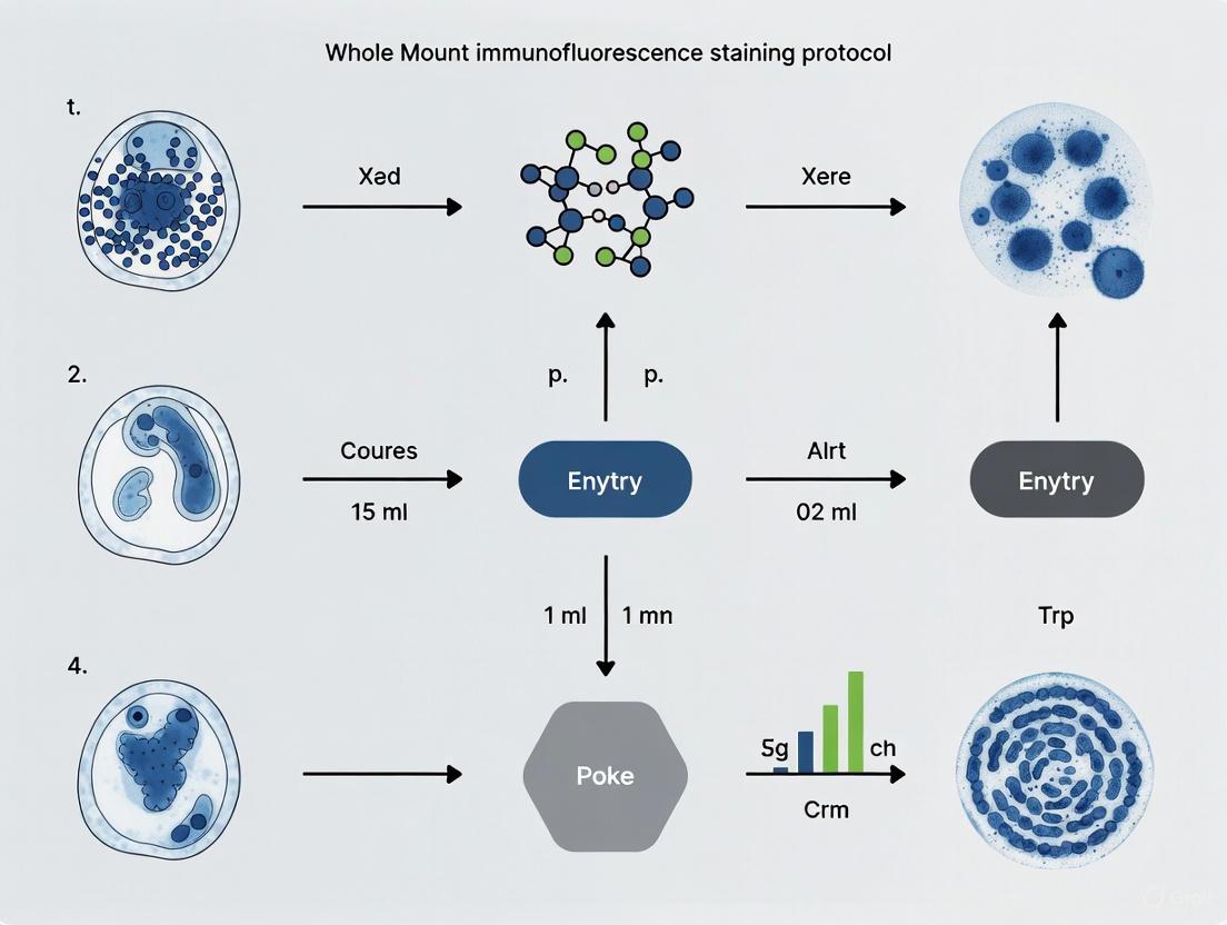
Figure 1: Whole-Mount Immunofluorescence Workflow. This diagram illustrates the sequential steps involved in processing embryos for whole-mount IF, highlighting critical parameters at each stage.
Imaging and Visualization
Mounting and Clearing
Proper mounting is crucial for high-quality imaging of whole-mount specimens. For smaller embryos, mounting in glycerol is commonly employed. A step-wise equilibration in 50%, 75%, and 100% glycerol ensures optimal refractive index matching and prevents tissue distortion [4]. Samples are considered properly equilibrated when they sink to the bottom of the glycerol solution, typically after 24-48 hours [4].
For larger specimens that require physical sectioning, embedding in gelatin followed by vibratome sectioning provides an alternative approach [4]. However, this method partially compromises the 3D integrity that whole-mount techniques aim to preserve.
Microscopy Techniques
Confocal microscopy is the preferred imaging method for whole-mount immunofluorescence due to its ability to optically section through thick specimens [1] [4]. This technique allows researchers to capture Z-stacks through the entire embryo and reconstruct three-dimensional expression patterns without physical sectioning.
For very large or opaque specimens, advanced clearing techniques (not covered in the provided protocols) may be employed to improve light penetration. However, for embryos within the recommended size limits, proper mounting in glycerol often provides sufficient transparency for high-quality confocal imaging.
Research Reagent Solutions
Table 2: Essential Reagents for Whole-Mount Immunofluorescence
| Reagent Category | Specific Examples | Concentration/Usage | Function |
|---|---|---|---|
| Fixatives | 4% Paraformaldehyde (PFA) | 30 min - Overnight | Preserves tissue structure and antigenicity [5] [1] |
| Methanol | Variable (alternative to PFA) | Alternative fixative for epitope sensitivity [1] | |
| Permeabilization Agents | Triton X-100 | 0.5-2% in PBS | Disrupts membranes for antibody penetration [5] [4] |
| Blocking Agents | Bovine Serum Albumin (BSA) | 4% in PBS-Triton | Reduces non-specific antibody binding [5] |
| Fetal Calf Serum (FCS) | 10% in PBS-Triton | Alternative blocking protein [4] | |
| Antibody Diluent | Blocking buffer + Sodium Azide | 0.02% sodium azide | Prevents microbial growth during long incubations [4] |
| Washing Buffers | PBS with Triton X-100 | 0.5-1% Triton | Removes unbound antibodies while maintaining permeabilization [4] |
| Mounting Media | Glycerol | 50-100% graded series | Refractive index matching for microscopy [4] |
| ProLong Gold Antifade | Ready-to-use | Preserves fluorescence, reduces photobleaching [5] | |
| Nuclear Counterstains | DAPI (4',6-diamidino-2-phenylindole) | Manufacturer's recommendation | Fluorescent DNA stain for nuclear visualization [5] |
Troubleshooting and Optimization
Despite careful execution, whole-mount IF can present several challenges that require systematic troubleshooting:
Poor Antibody Penetration manifests as weak or absent staining in deeper tissue regions. Solutions include increased permeabilization time, higher detergent concentrations, or extended antibody incubations [1] [4]. For larger embryos, dissection into smaller segments may be necessary [1].
High Background Signal can result from insufficient blocking, inadequate washing, or non-specific antibody binding. Remedies include optimizing blocking conditions (increasing serum concentration, trying different blocking agents), extending wash times, and titrating antibody concentrations to find the optimal signal-to-noise ratio [1] [3].
Epitope Masking occurs when fixation cross-linking obscures antibody binding sites. When PFA fixation proves problematic, switching to methanol fixation or exploring alternative fixatives may resolve the issue [1]. Note that antigen retrieval methods used in traditional IHC are generally not feasible for whole-mount embryos due to tissue sensitivity to heat-induced damage [1].
Physical Damage to delicate embryos can be minimized by using cut pipette tips during solution changes and employing gentle rotation during incubations rather than vigorous shaking [4].
Applications in Biomedical Research
Whole-mount immunofluorescence has enabled significant advances across multiple research domains:
In developmental biology, the technique allows comprehensive mapping of morphogen gradients and expression patterns during embryogenesis [1]. The preserved 3D architecture reveals how protein localization guides tissue patterning and organ formation.
In neurobiology, whole-mount IF facilitates the tracing of neural circuits and mapping of neurotransmitter distribution within intact embryonic nervous systems [1]. This application is particularly valuable for understanding how neural networks establish connectivity during development.
In drug discovery and toxicology, researchers can assess compound effects on protein expression patterns throughout entire embryos, providing systems-level insights into mechanism of action and potential developmental toxicity [1].
The method is also instrumental in validating transgenic animal models, where confirming expected protein expression patterns in three dimensions provides stronger validation than section-based approaches alone [1].
Figure 2: Applications and Advantages of Whole-Mount IF. The core principle of 3D architecture preservation enables diverse research applications and provides key advantages over section-based methods.
Comparative Analysis with Alternative Methods
Table 3: Comparison of Whole-Mount IF with Traditional IHC Methods
| Parameter | Whole-Mount IHC/IF | Sectioned IHC |
|---|---|---|
| 3D Context | Preserved entirely | Lost unless serial reconstruction performed |
| Spatial Relationships | Maintained in native state | Disrupted by sectioning |
| Antibody Penetration | Major challenge, requires optimization | Generally straightforward |
| Incubation Times | Extended (hours to days) | Shorter (hours) |
| Antigen Retrieval | Generally not feasible | Routinely performed |
| Tissue Size Limits | Limited by penetration depth | Virtually unlimited through serial sectioning |
| Imaging Requirements | Confocal microscopy preferred | Standard widefield microscopy often sufficient |
| Information Content | High for spatial patterns | High for cellular detail |
Understanding these distinctions helps researchers select the most appropriate method for their specific research questions. Whole-mount IF is uniquely valuable when comprehensive spatial analysis outweighs the need for convenience or when the research question specifically involves three-dimensional protein distribution patterns that would be disrupted by physical sectioning.
Immunohistochemistry (IHC) serves as a cornerstone technique in biomedical research, enabling the visualization of protein expression within tissue samples. Traditional section-based IHC, while providing high-resolution two-dimensional data, inherently disrupts the three-dimensional architecture of biological specimens. Whole-mount immunofluorescence (WM-IF) has emerged as a powerful alternative that preserves structural integrity and spatial relationships, offering researchers a comprehensive view of expression patterns within intact tissues. This application note details the significant advantages of WM-IF over sectioned IHC, with a specific focus on applications in embryonic research, and provides detailed protocols for its successful implementation.
The fundamental distinction of WM-IF lies in its capacity to maintain the three-dimensional spatial information of biological samples. Whereas traditional IHC requires physical sectioning of tissues, resulting in the loss of contextual relationships between structures, WM-IF processes the specimen in its entirety [2]. This preservation is particularly crucial for understanding complex developmental processes in embryology, where the relative positioning of cells and tissues drives morphogenesis and organ formation. The technique enables a comprehensive interpretation of expression domains that cannot be fully appreciated in two-dimensional sections [2].
Key Advantages and Quantitative Comparisons
Preservation of Three-Dimensional Architecture
The primary advantage of WM-IF is its ability to preserve the intact 3D tissue architecture, allowing researchers to analyze protein localization and expression patterns within their native spatial context. This capability is invaluable for studying complex biological structures such as early embryos, organoids, and neural circuits, where the 3D arrangement of cells dictates function.
Research on breast cancer heterogeneity exemplifies the power of spatial analysis. Traditional IHC scoring of bulk cancer misses critical inter-cellular heterogeneity and spatial distribution patterns of biomarkers that can influence diagnosis and treatment outcomes [6]. WM-IF coupled with quantitative single-cell imaging revealed marked heterogeneity in protein co-expression signatures and cellular arrangement within each breast cancer subtype, demonstrating how proliferating cells defined by Ki67 positivity were mainly found in groups with PR-negative cells in Luminal B-like cancers [6].
Table 1: Comparative Analysis of Sectioned IHC vs. Whole-Mount Immunofluorescence
| Feature | Sectioned IHC | Whole-Mount IF |
|---|---|---|
| Tissue Integrity | Disrupted by physical sectioning | Preserved intact in three dimensions |
| Spatial Context | Limited to 2D plane; reconstructed from serial sections | Comprehensive 3D context maintained |
| Antibody Penetration | Generally excellent due to thin sections | Requires optimization; prolonged incubations needed [4] [1] |
| Imaging Modality | Standard brightfield or epifluorescence microscopy | Confocal or multiphoton microscopy required [7] [8] |
| Data Complexity | Simplified 2D analysis | Rich 3D datasets requiring specialized analysis |
| Cellular Heterogeneity Analysis | Limited to sectional view; may miss rare populations | Comprehensive single-cell analysis within tissue context [6] |
| Development Biology Applications | Limited by reconstruction artifacts | Ideal for embryonic patterning studies [2] [9] |
Enhanced Analytical Capabilities in Disease Research
WM-IF provides superior analytical capabilities for investigating complex tissue environments. In corneal research, WM-IF proved more effective than tissue sections for visualizing the expression patterns of limbal stem cell (LSC) markers within human and porcine corneas [10]. This approach revealed how storage duration significantly influenced LSC marker expression, with human tissues stored longer exhibiting notable epithelial degeneration and absence of these critical markers [10].
The quantitative potential of WM-IF extends to sophisticated spatial analysis algorithms. Advanced protocols now enable the characterization of spatial relationships between cell types using mathematical indices such as Spatial Distribution Index (SDI), Neighborhood Frequency (NF), and Normalized Median Evenness (NME) [7]. These metrics provide rigorous quantitative descriptors of cellular organization that are simply unattainable with traditional sectioned IHC.
Table 2: Quantitative Analytical Outputs Enabled by Whole-Mount Immunofluorescence
| Analytical Output | Description | Research Application |
|---|---|---|
| 3D Co-expression Patterns | Analysis of multiple protein markers within the same volumetric tissue context | Revealed heterogeneous ER/PR co-expression in Luminal breast cancers [6] |
| Spatial Distribution Index (SDI) | Mathematical quantification of cell distribution patterns | Characterization of immune cell localization in skin whole-mounts [7] |
| Cellular Neighborhood Analysis | Identification of recurrent multicellular interactions within tissues | Analysis of tumor microenvironment in breast cancer TMA [6] |
| Depth-Dependent Intensity Quantification | Measurement of signal variations through tissue depth | Validation of LSC marker expression gradients in corneal whole-mounts [10] |
| Expression Domain Mapping | Volumetric quantification of protein expression areas | Assessment of Sdc1 expression domains in gingival tissue [11] |
Whole-Mount Immunofluorescence Protocol for Embryos
Sample Preparation and Fixation
Successful WM-IF begins with optimal sample preparation to preserve tissue integrity and antigenicity. For early mouse embryos (up to E8.0), careful fixation is critical [2].
- Fixation: Place embryos in 4% paraformaldehyde (PFA) in bijous tubes. Fixation time requires optimization based on embryo size and age - typically between 2 hours to overnight at 4°C [4] [1]. Proper fixation preserves antigenicity while maintaining structural integrity.
- Washing: After fixation, wash samples 3 times in PBS with 0.5-1% Triton X-100 for 30 minutes each to remove residual fixative [4].
- Permeabilization: The detergent in the wash buffer simultaneously permeabilizes the tissue, allowing antibody penetration. For thicker specimens, additional permeabilization steps may be necessary.
It is crucial to note that antigen retrieval methods commonly used in sectioned IHC are generally not feasible for whole-mount embryos due to their sensitivity to heat and harsh chemical treatments [1]. Therefore, fixation conditions must be carefully optimized for each target antigen.
Blocking and Antibody Incubation
Due to the thickness of whole-mount specimens, blocking and antibody incubation steps require significantly longer durations compared to sectioned IHC.
- Blocking: Incubate embryos twice for 1 hour in blocking buffer (PBS with 1% Triton X-100, 10% FCS, and 0.2% sodium azide) at room temperature [4]. This step reduces non-specific antibody binding.
- Primary Antibody Incubation: Transfer embryos to tubes containing primary antibody diluted in blocking buffer. Incubate for 1-4 days on a gentle rotation device at 4°C [4]. The prolonged incubation is necessary for complete antibody penetration throughout the specimen.
- Washing: Remove unbound primary antibody through extensive washing: 3 times for 1 hour in PBS with 1% Triton X-100 and 10% FCS, followed by 3 times for 10 minutes in PBS with 1% Triton X-100 [4].
- Secondary Antibody Incubation: Incubate with fluorescent-conjugated secondary antibodies in blocking buffer for 2-4 days with gentle rotation at 4°C [4].
Diagram 1: Whole-mount immunofluorescence staining involves extended incubation and washing steps compared to traditional IHC.
Mounting and Imaging
Proper mounting and imaging are crucial for maximizing the benefits of WM-IF and obtaining high-quality 3D data.
- Mounting: Equilibrate stained embryos in glycerol (progressing through 50%, 75%, to 100% concentrations) until they sink to the bottom of the vial, indicating complete permeation [4]. Mount in 75% glycerol, using grease to seal coverslip edges.
- Imaging: Acquire images using confocal laser scanning microscopy (CLSM) or multiphoton microscopy [7] [8]. For larger specimens (>100μm), two-photon microscopy provides superior depth penetration due to longer excitation wavelengths and reduced light scattering [8].
- Clearing: For deep imaging of large organoids (>200μm), consider tissue clearing with 80% glycerol, which provides a 3-fold reduction in intensity decay at 100μm depth compared to PBS-mounted samples [8].
The Scientist's Toolkit: Essential Research Reagents
Successful implementation of WM-IF requires specific reagents and equipment optimized for 3D tissue processing and imaging.
Table 3: Essential Reagents and Equipment for Whole-Mount Immunofluorescence
| Item | Function | Application Notes |
|---|---|---|
| Paraformaldehyde (4%) | Tissue fixation preserving antigenicity and structure | Alternative: methanol if PFA causes epitope masking [1] |
| Triton X-100 | Detergent for tissue permeabilization | Enables antibody penetration; typically used at 0.5-1% [4] |
| Normal Serum (FCS) | Blocking agent reducing non-specific binding | Component of blocking buffer (typically 10%) [4] |
| Sodium Azide | Antimicrobial preservative | Prevents microbial growth during prolonged incubations (0.02%) [4] |
| Fluorophore-Conjugated Antibodies | Target detection and visualization | Direct conjugation or secondary antibody detection [7] |
| Confocal/Multiphoton Microscope | 3D image acquisition through optical sectioning | Essential for visualizing deep structures [7] [8] |
| Glycerol (80%) | Mounting medium with refractive index matching | Enhances light penetration for deep imaging [8] |
| Image Analysis Software (FIJI, CellProfiler) | 3D reconstruction and quantitative analysis | Enables spatial analysis and quantification [7] [8] |
Advanced Applications in Embryonic Research
WM-IF has enabled significant advances in embryonic research by permitting the visualization of signaling activity and gene expression patterns in three dimensions. In pre-implantation human embryos, WM-IF has been used to detect phosphorylated SMAD proteins critical for TGF-β signaling, which regulates key developmental events [9]. This approach combined immunofluorescence detection with sophisticated computational analysis using Fiji and CellProfiler for nuclear segmentation and fluorescence intensity quantification [9].
For gastruloid models, WM-IF coupled with two-photon imaging has enabled the creation of detailed 3D maps of gene expression patterns and nuclear morphology, revealing how local cell deformations and gene co-expression relate to tissue-scale organization [8]. This integrated pipeline combines deep imaging with computational tools for 3D nuclei segmentation and signal normalization, providing multi-scale analysis from cellular to tissue level [8].
Diagram 2: Integrated WM-IF workflow combines experimental and computational phases for comprehensive 3D tissue analysis.
Whole-mount immunofluorescence represents a significant advancement over traditional sectioned IHC by preserving tissue integrity and spatial context, thereby enabling a more comprehensive analysis of biological structures. The capacity to visualize protein expression within an intact 3D environment provides insights into developmental processes, disease mechanisms, and cellular interactions that cannot be achieved through sectional approaches. While the method demands careful optimization of staining conditions and specialized imaging equipment, the resulting data offer unparalleled views of biological architecture. As imaging technologies and computational analysis tools continue to advance, WM-IF is poised to become an increasingly indispensable technique for developmental biology, cancer research, and drug development programs where spatial context is critical to understanding biological function.
Whole-mount immunofluorescence (IF) of embryos presents a unique set of challenges distinct from standard immunohistochemistry on sectioned tissue. This technique preserves the intricate three-dimensional architecture of the embryo, allowing for comprehensive spatial analysis of protein expression during development [1]. The key to success lies in the effective use of three essential classes of reagents: fixatives, permeabilization agents, and blocking buffers. These reagents work in concert to preserve antigenicity, enable antibody access, and minimize non-specific background, ensuring a high-quality, interpretable signal. The thicker nature of whole-mount samples necessitates longer incubation times and careful optimization of these reagents to ensure complete penetration to the center of the sample [1]. This application note details their function, selection, and application within the context of a robust whole-mount IF protocol for embryo research.
The Reagent Toolkit: Composition, Function, and Selection
The following table summarizes the critical reagents, their functions, and key considerations for their use in whole-mount immunofluorescence of embryos.
Table 1: Essential Reagents for Whole-Mount Immunofluorescence
| Reagent Category | Key Examples | Primary Function | Considerations for Whole-Mount Embryo Staining |
|---|---|---|---|
| Fixatives | 4% Paraformaldehyde (PFA), Methanol | Preserves tissue architecture and immobilizes antigens by cross-linking or precipitating proteins. | |
| Permeabilization Agents | Detergents (e.g., Triton X-100, Tween-20, Saponin) | Disrupts lipid membranes to allow antibodies to access intracellular targets. |
|
| Blocking Buffers | Normal Serum (from secondary antibody host), BSA | Reduces non-specific antibody binding to minimize background signal. |
|
Experimental Protocol: A Detailed Workflow for Embryo Staining
The workflow for whole-mount immunofluorescence involves a series of critical steps, each requiring careful optimization to account for the thickness of the embryo sample. The following diagram illustrates the complete experimental workflow.
Whole-Mount Immunofluorescence Workflow for Embryos
Stage 1: Fixation and Sample Preparation
Principle: Fixation is critical for preserving the native tissue architecture and preventing antigen degradation. The choice of fixative can significantly impact antibody binding and epitope accessibility [1].
Detailed Methodology:
- Fixative Selection: Prepare a 4% Paraformaldehyde (PFA) solution in Phosphate-Buffered Saline (PBS). Methanol is a common alternative if PFA is suspected of masking the target epitope [1].
- Fixation Protocol: Immerse the embryo in a sufficient volume of fixative. Incubation times must be extended for whole-mount samples.
- Option A (Room Temperature): Incubate for 30 minutes to 1 hour.
- Option B (Cold): Incubate overnight at 4°C for optimal preservation [1].
- Post-Fixation Wash: Rinse the embryo thoroughly with PBS (3 x 10 minutes) to remove all traces of fixative.
- Special Preparation (e.g., Zebrafish): For embryos with chorions, a dechorionation step is required before fixation. This can be done manually with fine forceps or enzymatically using pronase (1–2 mg/mL for 5–10 minutes) [1].
Stage 2: Permeabilization and Blocking
Principle: Permeabilization creates pores in cellular membranes, allowing antibodies to reach intracellular targets. Blocking saturates non-specific binding sites to reduce background noise.
Detailed Methodology:
- Permeabilization: Prepare a permeabilization buffer (e.g., 0.5-1% Triton X-100 in PBS). Incubate the embryo for several hours at room temperature or overnight at 4°C, with gentle agitation.
- Blocking: Prepare a blocking buffer (e.g., 5-10% normal serum and 1% BSA in PBS with 0.1% Tween-20 (PBS-T)). Incubate the embryo in blocking buffer for a minimum of 4 hours, preferably overnight at 4°C, with gentle agitation.
Stage 3: Antibody Incubation and Washing
Principle: Antibodies specifically bind to the target antigen. Extended incubation and thorough washing are required for deep penetration and removal of unbound antibody.
Detailed Methodology:
- Primary Antibody: Dilute the primary antibody in blocking buffer. Incubate the embryo in the antibody solution for 24 to 48 hours at 4°C with gentle agitation [1].
- Washing: Remove unbound primary antibody with multiple prolonged washes (e.g., 6 x 1-hour washes or continuous washing overnight) using PBS-T.
- Secondary Antibody: Dilute the fluorophore-conjugated secondary antibody in blocking buffer, protected from light. Incubate the embryo for 24 hours at 4°C with gentle agitation [1].
- Final Washing: Perform multiple prolonged washes with PBS-T (e.g., 6 x 1-hour washes) in the dark to remove unbound secondary antibody.
Stage 4: Mounting and Imaging
Principle: The sample is mounted for stabilization and clarity under a microscope. Confocal microscopy is recommended for optical sectioning of thick samples [1].
Detailed Methodology:
- Mounting: Clear and mount the embryo in an anti-fading mounting medium (e.g., glycerol-based). For small embryos, secure under a coverslip.
- Imaging: Image using a confocal microscope. Acquire Z-stacks to capture the 3D structure of the staining. Include a scale bar (e.g., 100 µm) for spatial context [1].
Advanced Applications and Quantitative Analysis
Whole-mount immunofluorescence is a powerful tool for developmental biology, neurobiology, and embryology, enabling the study of protein expression patterns, mapping neural circuits, and analyzing gene expression during organ formation [1]. The quantitative evaluation of staining intensity is crucial for robust and reproducible research. A study comparing different detection methods for quantitative immunohistochemistry found that the alkaline phosphatase-based substrate Vector Red provided excellent qualities for microdensitometric evaluation, offering linearity over a wide range, light stability, and feasibility for permanent mounting [12]. For modern multiplexed imaging, accurate nuclear segmentation is a critical first step. A 2025 benchmarking study recommended deep learning-based segmentation tools like Mesmer for highest accuracy in translational studies, as they outperform classical algorithms across different tissue types [13].
Table 2: Quantitative Benchmarking of Nuclear Segmentation Algorithms
| Segmentation Platform | Type | Reported F1-Score (IoU=0.5) | Key Recommendation / Note |
|---|---|---|---|
| Mesmer | Deep Learning | 0.67 | Highest overall accuracy on composite dataset; recommended for general use [13]. |
| Cellpose | Deep Learning | 0.65 | Consistently outperformed others at higher IoU thresholds; performance can vary with input data [13]. |
| StarDist | Deep Learning | 0.63 | Recommended if computational resources are limited; provides ~12x run time improvement with CPU [13]. |
| QuPath | Classical (Morphological) | N/A | Best-performing classical/morphological platform, similar or better than proprietary inForm software [13]. |
| Fiji / CellProfiler | Classical (Morphological) | N/A | Limited in accuracy relative to deep learning platforms [13]. |
Troubleshooting Common Issues
Even with a optimized protocol, issues can arise. The table below outlines common problems and their solutions.
Table 3: Troubleshooting Guide for Whole-Mount Immunofluorescence
| Problem | Potential Cause | Solution |
|---|---|---|
| Weak or No Signal | Inadequate antibody penetration. | Increase permeabilization time; consider harsher detergents or enzymatic permeabilization. |
| Epitope masked by fixative. | Switch fixative from PFA to methanol [1]. | |
| Antibody concentration too low or incubation time too short. | Increase antibody concentration; extend incubation times (e.g., to 48-72 hours). | |
| High Background | Inadequate blocking. | Extend blocking time; prepare fresh blocking buffer; try different blocking agents (e.g., different serum). |
| Insufficient washing. | Increase wash volume, frequency, and duration. | |
| Non-specific antibody binding. | Titrate antibody to optimal concentration; include detergent in antibody dilution buffer. | |
| Uneven Staining | Incomplete permeabilization or blocking. | Ensure samples are freely floating and agitated during all steps. |
| Air bubbles trapped during mounting. | Be careful during mounting to exclude bubbles. |
Whole-mount immunofluorescence staining is a cornerstone technique in developmental biology, enabling the visualization of protein expression and spatial localization within the intact three-dimensional architecture of embryos [1]. This method preserves structural integrity and provides a comprehensive view of expression patterns that sectional methods may obscure [2]. The application of this technique across key model organisms—mouse, zebrafish, and chick—has dramatically advanced our understanding of embryonic development, tissue patterning, and organogenesis. Each model offers unique advantages: zebrafish provide optical clarity and rapid ex vivo development [14], chick embryos allow easy experimental accessibility [15], and mouse models offer direct relevance to mammalian genetics and human disease [2]. This application note details optimized protocols for each model system, providing researchers with standardized methodologies for consistent and reproducible results in whole-mount immunofluorescence staining.
Principles of Whole-Mount Immunofluorescence
The fundamental principle of whole-mount immunofluorescence involves the specific binding of antibodies to target antigens within intact biological specimens, followed by detection with fluorophore-conjugated secondary antibodies and visualization via fluorescence microscopy [14]. Unlike traditional section-based immunohistochemistry, whole-mount techniques preserve the three-dimensional context of tissues, but require significantly longer incubation times for fixatives, antibodies, and wash buffers to ensure complete penetration throughout the sample [1]. The technique involves critical steps including fixation to preserve tissue structure and antigenicity, permeabilization to allow antibody access, blocking to reduce non-specific binding, antibody incubation for target detection, and thorough washing to minimize background [1] [14]. Successful implementation requires careful optimization for each model organism due to differences in embryo size, tissue density, and presence of extracellular barriers.
Comparative Analysis of Model Organisms
Key Advantages and Applications
Table 1: Characteristics and Applications of Model Organisms in Whole-Mount Immunofluorescence
| Model Organism | Optimal Staging Windows | Key Advantages | Primary Research Applications | Technical Considerations |
|---|---|---|---|---|
| Mouse | Preimplantation to E12.5 [1] [16] | High genetic similarity to humans; established genetic tools [2] | Mammalian organogenesis; cell fate specification [2] [17] | Requires uterine dissection; smaller litter sizes |
| Zebrafish | Up to 5 days post-fertilization [14] | Optical transparency; high fecundity; rapid development [14] | Neural development; vascular patterning [18] [14] | May require PTU treatment to inhibit pigment [16] |
| Chick | Up to 6 days [1] | Easy experimental accessibility; large embryo size [15] | Neural crest migration; limb bud development [15] | Extraembryonic membranes must be removed [15] |
Technical Parameters and Reagent Specifications
Table 2: Standardized Staining Parameters Across Model Organisms
| Protocol Step | Mouse Embryos | Zebrafish Embryos | Chick Embryos |
|---|---|---|---|
| Fixation | 4% PFA, 30 min - 2h [5] [17] | 4% PFA, overnight [14] | 4% PFA, 1-2h [15] [19] |
| Permeabilization | 0.1-2% Triton X-100, 30 min [5] | 1-2% Triton X-100, variable [14] | 0.1% Triton X-100, extensive washes [15] |
| Blocking | 2-4% BSA or serum, 1h [5] | 1% BSA + serum, 2h [14] | 1% BSA + 1% NGS, 1h [15] |
| Primary Antibody | Overnight, 4°C [5] | 48h, 4°C [14] | 1-4 days, 4°C [19] |
| Secondary Antibody | 2h, RT or overnight, 4°C [5] | 2h, protected from light [14] | Overnight, 4°C [15] |
Organism-Specific Protocols
Mouse Embryo Staining Protocol
Sample Preparation and Fixation Collect preimplantation mouse embryos (e.g., E3.5 blastocysts) by flushing the uterus with M2 medium [17]. Remove the zona pellucida using Acidic Tyrode's solution for 10 seconds at room temperature [5]. Fix embryos in 4% PFA for 30 minutes to 2 hours at room temperature [5] [17]. For postimplantation embryos (up to E12.5), dissect carefully to remove surrounding membranes and fix for 2 hours to overnight depending on embryo size [1] [19].
Permeabilization and Blocking After PBS washes, permeabilize embryos with 0.1-2% Triton X-100 in PBS for 30 minutes at room temperature [5]. Block non-specific binding sites with blocking solution (2-4% BSA or serum in PBS) for 1 hour at room temperature [5]. For RNase-sensitive applications, include RNase inhibitors in the blocking solution [17].
Antibody Incubation and Imaging Incubate with primary antibody diluted in blocking solution overnight at 4°C [5]. After extensive washes (3-10 times over several hours) with PBS containing 0.1% Triton X-100, incubate with fluorophore-conjugated secondary antibodies (e.g., Alexa Fluor series) for 2 hours at room temperature or overnight at 4°C [5]. Counterstain with DAPI (1-5 μg/mL) to visualize nuclei [5] [16]. Mount embryos in ProLong Gold antifade reagent and image using confocal microscopy [5].
Zebrafish Embryo Staining Protocol
Sample Preparation and Fixation Fix whole zebrafish larvae or dissected tissues (e.g., spinal cord, retina) in 4% PFA overnight at 4°C on a gentle shaker [18] [14]. For larvae, permeabilization can be enhanced by using ice-cold acetone treatment at -20°C for 20 minutes after standard permeabilization [14]. Extensive washing with PBS-T (PBS with 0.1-1% Tween-20 or Triton X-100) is critical to reduce background [14].
Advanced Processing For thick tissues like adult zebrafish spinal cords, enhanced permeabilization with 1% Triton X-100 is recommended [18] [14]. Tissue clearing using Scale solutions (A2 and S4) significantly improves antibody penetration and light penetration for imaging [18]. Scale S4 solution contains urea, glycerol, Triton X-100, and DMSO, and requires careful preparation to maintain clarity [18].
Antibody Incubation and Imaging Incubate with primary antibody for at least 48 hours at 4°C due to tissue density [14]. For whole-mount retina staining, extend washing times to several hours between antibody steps [14]. Image using confocal microscopy or light sheet microscopy for larger samples [18] [14].
Chick Embryo Staining Protocol
Sample Preparation and Fixation Open chick eggs and carefully remove embryos with surrounding yolk sac [15]. Pin embryos down in a dish and fix with 4% PFA for 1-2 hours at room temperature [15]. Remove extraembryonic membranes using fine scissors and forceps after fixation [15].
Endogenous Peroxidase Quenching For enzymatic detection methods, quench endogenous peroxidase activity by incubating with 0.3% H₂O₂ in PBT for 2 hours at room temperature [15]. This step is crucial for reducing background when using HRP-conjugated antibodies.
Antibody Incubation and Detection Incubate with primary antibody for 1-4 days at 4°C [15] [19]. After extensive washing, incubate with HRP-conjugated secondary antibody overnight at 4°C [15]. Develop color reaction using DAB substrate (0.5-1 mg/mL) with H₂O₂, monitoring development under a microscope [15]. For fluorescent detection, use standard fluorophore-conjugated secondary antibodies with extended incubation times [1].
Critical Protocol Variations by Organism
The Scientist's Toolkit: Essential Research Reagents
Table 3: Key Research Reagent Solutions for Whole-Mount Immunofluorescence
| Reagent Category | Specific Examples | Function and Application Notes |
|---|---|---|
| Fixatives | 4% Paraformaldehyde (PFA) [1] [5] [14]; Methanol [1] | Preserves tissue architecture and antigenicity; PFA is most common; methanol alternative for epitope sensitivity |
| Permeabilization Agents | Triton X-100 [5] [18]; Tween-20 [14] | Disrupts membranes for antibody penetration; concentration varies by tissue (0.1-2%) |
| Blocking Agents | Bovine Serum Albumin (BSA) [5] [18]; Normal Goat Serum (NGS) [15] | Reduces non-specific antibody binding; typically 1-4% in PBS or with detergent |
| Detection Systems | Alexa Fluor-conjugated secondary antibodies [5] [14]; HRP-conjugated with DAB [15] | Fluorescent detection for confocal imaging; enzymatic for permanent specimens |
| Mounting Media | ProLong Gold [5]; Mowiol [18]; Glycerol [19] | Preserves fluorescence and supports imaging; contains antifade agents |
| Nuclear Counterstains | DAPI [5] [16]; Hoechst dyes [16]; TO-PRO-3 [18] | Visualizes nuclear architecture and cellular organization |
Advanced Applications and Integrated Techniques
Combined RNA and Protein Detection
A sophisticated application of whole-mount techniques involves simultaneous detection of RNA transcripts and proteins in the same specimen. This requires sequential immunofluorescence followed by single-molecule RNA fluorescence in situ hybridization (smRNA FISH) [17]. Critical modifications include using RNase-free reagents during immunofluorescence to prevent RNA degradation, and performing IF before smRNA FISH to preserve antigen integrity [17]. This integrated approach enables researchers to correlate protein localization with gene expression patterns in developing embryos, providing insights into regulatory mechanisms during development.
Whole-Mount Nuclear Imaging for Morphological Analysis
Nuclear staining with fluorescent dyes (DAPI, Hoechst, Draq5) enables detailed topological analysis of embryonic structures, producing images with clarity rivaling scanning electron microscopy when combined with confocal microscopy [16]. This "pseudo-SEM" technique involves staining whole-mount embryos with cell-permeant nuclear dyes and capturing z-stacks using confocal microscopy [16]. The resulting projection images reveal morphological details with exceptional contrast and apparent depth of field, while preserving specimens for subsequent histological analysis [16].
Tissue Clearing for Enhanced Penetration and Imaging
For larger or denser specimens such as adult zebrafish spinal cords or late-stage embryos, tissue clearing techniques significantly improve antibody penetration and light transmission for imaging [18]. Scale solutions (A2 and S4), containing urea, glycerol, and detergents, render tissues transparent while maintaining structural integrity [18]. This approach enables visualization of deep structures like vascular networks and neural pathways without physical sectioning, preserving valuable three-dimensional context relationships.
Troubleshooting and Technical Considerations
Incomplete Penetration and Weak Staining For thick tissues, extend incubation times for primary antibodies to 48-72 hours and consider increasing detergent concentrations to 1% Triton X-100 [1] [14]. For zebrafish embryos, ice-cold acetone treatment after standard permeabilization can dramatically improve antibody access [14]. Tissue clearing with Scale solutions enhances penetration in dense samples like adult zebrafish spinal cords [18].
High Background Staining Increase blocking time to 2-4 hours and ensure thorough washing between steps (3-10 washes over several hours) [1] [14]. For chick embryos using HRP-based detection, endogenous peroxidase quenching with 0.3% H₂O₂ is essential [15]. Optimize antibody concentrations through titration experiments specific to each model organism.
Antigen Preservation Issues If PFA fixation masks epitopes, alternative fixatives like methanol may improve results [1]. Note that antigen retrieval methods used in sectioned samples are generally not feasible for whole-mount embryos due to heat sensitivity [1]. Test multiple fixatives during protocol optimization for new targets.
Imaging Challenges in Large Specimens For embryos beyond recommended stages (mouse >E12.5, chick >6 days, zebrafish >5 dpf), dissect into smaller segments or specific organs before staining [1]. Use confocal microscopy with z-stacking capabilities for three-dimensional reconstruction of thicker specimens [1] [16]. For very large samples, light sheet microscopy may be preferable [14].
Whole-mount immunofluorescence staining provides an indispensable tool for developmental biologists studying pattern formation, organogenesis, and gene expression in the context of intact embryonic architecture. The standardized protocols presented here for mouse, zebrafish, and chick embryos enable researchers to leverage the unique advantages of each model system while maintaining methodological consistency. As imaging technologies advance and tissue clearing methods improve, the applications of whole-mount techniques will continue to expand, offering increasingly detailed insights into the complex processes governing embryonic development. The integration of these approaches with complementary techniques such as smRNA FISH further enhances their utility for comprehensive analysis of molecular mechanisms in development and disease.
Within the field of developmental biology, the period spanning pre-implantation to early post-implantation represents a critical and dynamic phase of embryonic development. This application note details specialized protocols for whole-mount immunofluorescence staining, designed to address the unique structural and molecular challenges presented at each stage. The methodologies outlined herein are framed within a broader thesis research endeavor, providing a standardized yet flexible approach for visualizing gene and protein expression patterns in embryo models across these crucial developmental windows. The protocols emphasize optimal specimen preparation, staining techniques, and advanced imaging parameters to ensure high-quality, reproducible data for researchers and drug development professionals investigating the molecular underpinnings of early development.
Stage-Specific Embryo Isolation and Preparation Protocols
Pre-implantation Embryo Isolation from Mice
The isolation of pre-implantation embryos is a technically sensitive process requiring precise timing and conditions. The following protocol, adapted from Varghese et al., outlines the steps for obtaining embryos from C57BL/6J mice [20] [21].
- Animal Mating and Plug Checking: House 6-week to 6-month-old female and 2-12-month-old male C57Bl/6J mice under controlled temperature, humidity, and a 14:10 h light-dark cycle. Set up mating by placing females with males and check for a vaginal plug the following morning. The presence of a plug indicates mating and defines 0.5 days post-coitum (dpc) [20].
- Embryo Collection Timing: Collect pre-implantation embryos at specific developmental stages based on dpc: 1.75 dpc for 4-cell, 2.25 dpc for 8-cell, 2.75 dpc for morula, and 3.5 dpc for blastocyst stages [20].
- Zona Pellucida Removal: After collection, remove the zona pellucida by briefly treating embryos with acid Tyrode's solution for approximately 10 seconds at room temperature [5].
Preparation of Stem Cell-Derived Post-implantation Embryo Models
For studies extending into the post-implantation period, stem cell-derived embryo models provide a powerful and ethically accessible tool. The following section describes the generation of human complete Stem-cell-derived Embryo Models (SEMs) from naive embryonic stem cells (ES cells) [22].
- Generation of Human Complete SEMs: This protocol uses genetically unmodified human naive ES cells cultured in human enhanced naive stem cell medium (HENSM) conditions. These cells self-organize into structures that recapitulate the organization of nearly all known lineages and compartments of post-implantation human embryos up to Carnegie stage 6a (13-14 days after fertilization) [22].
- Induction of Extra-embryonic Lineages: To promote the formation of primitive endoderm (PrE)-like and extra-embryonic mesoderm (ExEM)-like cells, culture naive ES cells in RCL medium (RPMI-based medium supplemented with CHIR99021 and LIF, but without activin A) for 3 days. This culture condition efficiently induces PDGFRA+ cells, which are markers of these lineages, without the need for transient transgene expression [22].
- Key Developmental Hallmarks: A successfully formed human complete SEM will demonstrate [22]:
- Embryonic disc and bilaminar disc formation.
- Epiblast lumenogenesis and polarized amniogenesis.
- Anterior-posterior symmetry breaking.
- Polarized yolk sac formation.
- A trophoblast-surrounding compartment with syncytium and lacunae formation.
Workflow for Whole-Mount Staining and Imaging
The following diagram illustrates the integrated workflow for processing embryos from isolation to imaging, applicable to both pre-implantation embryos and stem cell-derived models.
Whole-Mount Immunofluorescence Staining Protocol
This core protocol is optimized for whole-mount specimens, from pre-implantation embryos to more complex post-implantation models [20] [5].
- Fixation: Fix embryos or embryo models in 4% paraformaldehyde (PFA) for 30 minutes at room temperature (RT) to preserve morphology and antigenicity [20] [5].
- Permeabilization: Treat fixed specimens with a permeabilization buffer containing 0.25% to 2% Triton X-100 in PBS for 30 minutes at RT. This step allows antibodies to access intracellular targets [20] [5].
- Blocking: Incubate specimens in a blocking solution to minimize non-specific antibody binding. A common solution is 4% Bovine Serum Albumin (BSA) in PBS or a solution containing goat serum [20] [5].
- Primary Antibody Incubation: Incubate specimens with the primary antibody diluted in blocking solution overnight at 4°C. For example, anti-CDH1 has been used at a 1:100 dilution [20].
- Washing: Wash specimens thoroughly with a washing buffer, such as PBS containing 1% BSA and 0.005% Triton X-100, to remove unbound primary antibody [5].
- Secondary Antibody and Counterstaining: Incubate specimens with appropriate cross-adsorbed fluorescent-conjugated secondary antibodies (e.g., Alexa Fluor 488 or 568) along with a nuclear counterstain like DAPI or Hoechst 33258 (1 μg/mL). Protect specimens from light during this and all subsequent steps [20] [5].
- Mounting and Preservation: Mount the stained specimens using an anti-fade mounting medium such as ProLong Gold. This step preserves fluorescence and reduces photobleaching during imaging [20] [5].
Advanced Imaging and Analysis for Early Embryos
Lattice Light-Sheet Microscopy for Post-Implantation Embryos
For dynamic imaging of early post-implantation stages, lattice light-sheet microscopy (LLSM) offers superior resolution with minimal photodamage [23].
- Application: This technique is suitable for time-lapse imaging of post-implantation mouse embryos and stem cell-derived embryo models, allowing for the visualization of morphogenetic and physiological processes with high spatial and temporal resolution [23].
- Protocol Summary: The process involves isolating the embryo or model, mounting it appropriately for culture, and setting up the imaging parameters on the LLSM. Subsequent pipelines are used for processing the large datasets generated [23].
Autofluorescence Reduction for Enhanced Signal Clarity
A major technical challenge in whole-mount imaging is inherent tissue autofluorescence.
- Optimized Solution: A specific protocol for whole-mount RNA fluorescent in situ hybridization incorporates an oxidation-mediated step designed to reduce autofluorescence, which can also be beneficial in immunofluorescence contexts to improve the signal-to-noise ratio [24].
The Scientist's Toolkit: Essential Research Reagents
The table below catalogs key reagents and their critical functions in embryo isolation and whole-mount staining protocols.
Table 1: Essential Reagents for Embryo Research Protocols
| Reagent / Kit | Function / Application | Example Sources |
|---|---|---|
| Paraformaldehyde (PFA) | Fixation of embryos to preserve cellular architecture and antigen integrity. | Sigma-Aldrich [20] |
| Triton X-100 | Permeabilization agent that enables antibody penetration by dissolving membranes. | Sigma-Aldrich [20] |
| Goat Serum / BSA | Blocking agent to reduce non-specific binding of antibodies. | Jackson ImmunoResearch, Invitrogen [20] [5] |
| Alexa Fluor Secondary Antibodies | Highly cross-adsorbed fluorescent secondary antibodies for specific target detection. | Invitrogen, Thermo Fisher Scientific [20] [5] |
| Hoechst 33258 / DAPI | Nuclear counterstains for identifying all cells within a specimen. | Sigma-Aldrich, Thermo Fisher Scientific [20] [5] |
| ProLong Gold Antifade Reagent | Mounting medium that preserves fluorescence and reduces photobleaching. | Invitrogen, Thermo Fisher Scientific [20] [5] |
| PicoPure RNA Isolation Kit | RNA extraction from a small number of embryos (e.g., 5-10) for gene expression analysis. | Applied Biosystems, Thermo Fisher Scientific [20] |
| High-Capacity cDNA Reverse Transcription Kit | Synthesis of cDNA from low-input RNA samples (e.g., ~50 ng). | Applied Biosystems [20] |
Quantitative Data and Experimental Parameters
Successful execution of these protocols relies on precise timing and specific quantitative parameters, as summarized in the table below.
Table 2: Key Experimental Parameters for Embryo Studies
| Parameter | Pre-implantation (Mouse) | Post-implantation (Model System) |
|---|---|---|
| Developmental Stages | 4-cell, 8-cell, Morula, Blastocyst [20] | Carnegie stage 6a (human model) [22] |
| Developmental Timing | 1.75 - 3.5 dpc [20] | Up to 13-14 days after fertilization (model) [22] |
| Fixation Time | 30 minutes at RT [5] | Protocol-dependent |
| Permeabilization Concentration | 0.25% - 2% Triton X-100 [20] [5] | Protocol-dependent |
| Primary Antibody Incubation | Overnight at 4°C [5] | Overnight at 4°C |
| RNA Input for cDNA Synthesis | ~50 ng from isolated embryos [20] | N/A |
Key Signaling Pathways in Early Development
The transition from pre- to post-implantation involves the activation and spatial organization of key signaling pathways. The following diagram illustrates the logical progression of major developmental events and the signaling environments that guide them.
The molecular progression involves the initial expression of lineage-specifying transcription factors (e.g., GATA4/GATA6 for endoderm, CDX2 for trophoblast) during and after implantation [22]. In human complete SEMs, this is followed by anterior-posterior symmetry breaking and the formation of definitive germ layers [22]. In later post-implantation models, such as hematoids, specific signaling pathways become established. Notably, hemogenic niches contain instructive factors like DLL4 and SCF, alongside restrictive factors like FGF23, which together guide the maturation of hematopoietic stem cells (HSCs) capable of differentiating into myeloid and lymphoid lineages [25].
A Step-by-Step Protocol: From Embryo Fixation to Confocal Imaging
Within the context of a broader thesis on whole-mount immunofluorescence staining in embryo research, the choice and application of a fixative are arguably the most critical steps. Fixation preserves cellular architecture and antigenicity, forming the foundation upon which all subsequent imaging and interpretation rely. For researchers studying embryonic development, whole-mount techniques are invaluable as they maintain three-dimensional spatial relationships, allowing for comprehensive analysis of protein expression and tissue architecture [1]. This application note details optimized protocols for using two common fixatives—4% Paraformaldehyde (PFA) and Methanol—in embryo studies. We provide structured data, detailed methodologies, and decision-making frameworks to guide researchers and drug development professionals in selecting and implementing the optimal fixation strategy for their experimental goals.
Fundamental Principles of Chemical Fixation
Chemical fixatives stabilize biological specimens by halting metabolic processes and preventing decomposition. The two fixatives discussed herein operate through distinct mechanisms, which directly influence their applications and outcomes.
- 4% Paraformaldehyde (PFA): A cross-linking fixative that creates covalent bonds between proteins, primarily linking the residues of basic amino acids [26] [27]. This process stabilizes soluble proteins to the cytoskeleton, thereby preserving the fine structural details and spatial relationships of cellular components with minimal distortion [26] [28]. However, this cross-linking can sometimes mask epitopes, making them inaccessible to antibodies.
- Methanol: A coagulant fixative that acts by dehydrating the sample and precipitating proteins in situ [26] [27]. This mechanism does not create cross-links, which often leaves antigenic epitopes more exposed and accessible. Consequently, methanol can yield stronger signals for certain antibodies. A significant drawback is its potential to cause cellular damage, distortion of ultrastructure, and shrinkage of tissues due to its dehydrating nature [27] [29].
The following diagram illustrates the decision-making workflow for selecting and optimizing a fixation protocol for whole-mount embryo staining, incorporating the key considerations of antibody validation, embryo age, and staining outcomes.
Comparative Analysis of 4% PFA and Methanol Fixation
A direct comparison of 4% PFA and Methanol reveals a trade-off between superior morphological preservation and optimal antigen accessibility. The table below summarizes the key characteristics, advantages, and limitations of each fixative to guide selection.
Table 1: Comparative analysis of 4% PFA and methanol fixation for whole-mount immunofluorescence
| Parameter | 4% Paraformaldehyde (PFA) | Methanol |
|---|---|---|
| Fixation Mechanism | Cross-linking [27] | Protein precipitation/Dehydration [27] |
| Morphology Preservation | Excellent; preserves fine cellular structures and spatial relationships [28] | Good, but can cause cellular damage, shrinkage, and tissue deformation [27] [29] |
| Antigen Preservation | Can mask some epitopes due to cross-linking; may require antigen retrieval (not feasible in whole-mounts) [1] | Often better for alcohol-sensitive antigens; avoids epitope masking [1] |
| Typical Incubation Time | 30 minutes to overnight, depending on sample size (e.g., 15-30 min for cells, overnight for whole embryos) [1] [27] | Relatively short (5–15 minutes for cells; longer for whole embryos) [30] [31] |
| Permeabilization | Requires separate permeabilization step (e.g., with Triton X-100) [27] | Self-permeabilizing; often no separate step needed [30] |
| Key Advantages | Superior structural preservation; ideal for membrane-associated antigens [28] | Can enhance signal for certain antibodies; simple and fast protocol for cells [1] [30] |
| Key Limitations | Potential for epitope masking; requires careful optimization of blocking [1] | Can distort ultrastructure; not ideal for all imaging purposes [27] [29] |
| Ideal Use Cases | General purpose fixation, especially when tissue architecture is critical; co-staining of membrane proteins [1] [28] | When an antibody is sensitive to PFA cross-linking and yields a weak signal; cytoplasmic or nuclear targets [1] |
Detailed Experimental Protocols
Protocol 1: Fixation with 4% PFA for Whole-Mount Embryos
This protocol is the gold standard for preserving the three-dimensional architecture of embryos, which is crucial for developmental biology studies [1].
Materials:
- 4% PFA Solution: Prepare fresh in phosphate-buffered saline (PBS), pH 7.2-7.4 [28].
- Phosphate-Buffered Saline (PBS)
- Embryo Collection Tools: Dissecting microscope, fine forceps.
- Fixed Sample Storage: Tubes for storage at 4°C or -20°C [1].
Method:
- Sample Collection and Preparation: Dissect embryos in ice-cold PBS. For zebrafish embryos, perform dechorionation manually with forceps or enzymatically using pronase (1–2 mg/mL for 5–10 minutes) to ensure fixative penetration [1].
- Fixation: Immerse embryos in a sufficient volume of 4% PFA to cover them completely.
- Washing: After fixation, wash the embryos thoroughly with PBS 3-5 times to remove all traces of PFA. Perform a final wash of at least 10 minutes [1].
- Storage: Fixed samples can be stored in PBS at 4°C for short-term use or at -20°C for long-term preservation [1].
Protocol 2: Fixation with Methanol for Enhanced Antigen Detection
Methanol fixation is a powerful alternative when antibody signal is weak with PFA, but it requires careful handling to prevent tissue damage [1] [29].
Materials:
- Methanol: 100%, ice-cold is often recommended for cell-based assays [30]. For tissue slices, concentrations of 33.3% to 75% in PBS can prevent severe deformations [29].
- Phosphate-Buffered Saline (PBS)
- Blocking Buffer: PBS containing 5% normal serum and 0.3% Triton X-100 [30].
Method:
- Sample Preparation: Collect and briefly rinse embryos in PBS.
- Fixation:
- For whole embryos, permeabilization is a major challenge. Incubate in ice-cold 100% methanol. The incubation time must be optimized and can be prolonged (hours to overnight) to allow penetration into the sample's center [1].
- Alternative for Tissues: To prevent deformation of tissue slices, use a lower concentration of methanol (e.g., 50%) at room temperature for 30 minutes [29].
- Rehydration and Washing: Rehydrate samples gradually through a series of methanol/PBS solutions (e.g., 75%, 50%, 25% methanol) before a final wash in PBS. This step helps avoid further structural damage [26].
- Storage: Store fixed samples in methanol at -20°C or, after rehydration, in PBS at 4°C.
The Scientist's Toolkit: Essential Research Reagent Solutions
A successful whole-mount immunofluorescence experiment relies on a suite of carefully selected reagents. The following table lists key materials and their functions.
Table 2: Key research reagents for whole-mount immunofluorescence fixation and staining
| Reagent | Function/Application | Notes |
|---|---|---|
| Paraformaldehyde (PFA) 4% | Cross-linking fixative for optimal morphological preservation [1] [28]. | Prepare fresh or freeze aliquots; pH adjustment to 7.2-7.4 is critical. |
| Methanol (100%) | Coagulant fixative for antigen retrieval and permeabilization [1] [30]. | Use ice-cold for cells; consider lower concentrations for tissues to prevent deformation [29]. |
| Triton X-100 | Non-ionic detergent for permeabilizing cell membranes after PFA fixation [27]. | Not required after methanol fixation. Typical use: 0.1-0.5% in PBS. |
| Normal Serum | Component of blocking buffer to reduce non-specific antibody binding [27] [30]. | Should match the host species of the secondary antibody. |
| Bovine Serum Albumin (BSA) | Component of blocking and antibody dilution buffers to reduce background [27]. | |
| Primary Antibodies | Target-specific immunostaining. | Must be validated for IHC on frozen sections (IHC-Fr) for likely success in whole-mounts [1]. |
| Fluorophore-conjugated Secondary Antibodies | Detection of primary antibodies for fluorescence imaging. | Must be reactive to the host species of the primary antibody [30]. |
Troubleshooting and Data Interpretation
Even with optimized protocols, challenges can arise. The table below outlines common issues and recommended solutions.
Table 3: Troubleshooting common fixation issues in whole-mount immunofluorescence
| Problem | Potential Cause | Recommended Solution |
|---|---|---|
| Weak or No Staining | Epitope masking by PFA cross-linking [1]. | Switch to methanol fixation [1]. |
| Insufficient antibody penetration. | Increase incubation times for antibodies and washes; ensure adequate permeabilization [1]. | |
| High Background | Inadequate blocking or washing [1]. | Optimize blocking buffer (e.g., with serum, BSA); increase wash duration and frequency [1]. |
| Non-specific antibody binding. | Include Triton X-100 in blocking and antibody buffers; titrate antibody concentrations [27]. | |
| Poor Morphology / Tissue Damage | Damage from pure methanol [29]. | For tissue slices, use lower methanol concentrations (33.3%-75%) at room temperature [29]. |
| Over-fixation with PFA. | Standardize and potentially reduce PFA fixation time [27]. | |
| Uneven Staining | Incomplete permeabilization. | Ensure proper permeabilization; for large embryos, dissect into smaller segments [1]. |
The choice between 4% PFA and methanol fixation is fundamental and should be dictated by the primary goal of the experiment. 4% PFA is the superior choice when the paramount requirement is the impeccable preservation of embryonic morphology and three-dimensional tissue context. Methanol fixation serves as a critical alternative when the detection of an antigen sensitive to PFA cross-linking takes precedence, accepting a trade-off of potential structural alterations. A rigorous, empirical approach—testing both fixatives with target-specific antibodies—is the most reliable strategy for optimizing whole-mount immunofluorescence staining and generating high-quality, interpretable data for embryo research.
Permeabilization is a critical step in whole-mount immunofluorescence that enables antibodies to penetrate cellular membranes and access intracellular targets. This process is particularly challenging in thick tissue samples such as embryos, where inadequate permeabilization can result in incomplete staining and false negative results. The non-ionic detergent Triton X-100 serves as a primary permeabilization agent by dissolving membrane lipids and creating pores that facilitate antibody penetration [3] [32]. However, concentration and incubation time must be carefully balanced to ensure sufficient epitope access while preserving cellular integrity and antigenicity. This application note provides evidence-based strategies for optimizing Triton X-100 permeabilization specifically for embryonic whole-mount immunofluorescence staining, with structured protocols and quantitative guidance for research applications.
Quantitative Permeabilization Parameters
Triton X-100 Concentration and Incubation Guidelines
Table 1: Triton X-100 Permeabilization Parameters for Different Sample Types
| Sample Type | Concentration Range | Incubation Time | Temperature | Additional Context |
|---|---|---|---|---|
| Embryonic whole-mount tissues [4] | 0.5% - 1.0% | Multiple washes of 30 minutes to 1 hour each | Room temperature | Used in blocking buffer and antibody incubation solutions |
| Cultured cells [33] [34] | 0.1% | 15 minutes | Room temperature | Standard protocol for monolayer cultures |
| Thick tissue sections (300-400μm) [35] | 0.5% - 2.0% | Incorporated in permeabilization buffer with 20% DMSO for 7-10 days | 4°C | For challenging adult tissues; higher concentrations for antibody penetration |
Optimized Whole-Mount Embryo Protocol
The following parameters represent optimized conditions for embryonic whole-mount permeabilization based on empirical testing:
- Effective Concentration Range: 0.5% to 1.0% Triton X-100 in PBS [4]
- Optimal Incubation Structure: Incorporation throughout the staining protocol rather than as a single discrete step
- Blocking Solution: PBS with 1% Triton X-100, 10% FCS, 0.2% sodium azide [4]
- Antibody Incubation Solution: PBS with 1% Triton X-100, 10% FCS, 0.2% sodium azide [4]
- Washing Buffer: PBS with 1% Triton X-100 for all inter-step washes [4]
Experimental Protocols for Whole-Mount Embryo Staining
Comprehensive Whole-Mount Immunofluorescence Protocol
Day 1: Fixation and Permeabilization Initiation
- Fixation: Transfer embryo to 5 mL bijous containing 4% paraformaldehyde. Fix at 4°C for 2 hours to overnight, depending on embryo size and density [4].
- Washing: Wash embryos 3 times in PBS with 0.5-1% Triton X-100 for 30 minutes each at room temperature [4].
- Blocking: Incubate embryos twice for 1 hour in blocking buffer (PBS with 1% Triton X-100, 10% FCS, 0.2% sodium azide) at room temperature [4].
- Primary Antibody Application: Transfer embryos using a Pasteur pipette with the end cut off to a 2 mL tube. Add primary antibody diluted in blocking buffer [4].
- Primary Antibody Incubation: Incubate for 1 to 4 days on a gentle rotation device at 4°C [4].
Day 2-4: Secondary Antibody Incubation
- Washing: Wash embryos 3 times for 1 hour in PBS with 1% Triton X-100 and 10% FCS, followed by 3 times for 10 minutes in PBS with 1% Triton X-100 [4].
- Secondary Antibody Application: Add fluorophore-conjugated secondary antibody diluted in blocking buffer (PBS with 1% Triton X-100, 10% FCS, 0.2% sodium azide) [4].
- Secondary Antibody Incubation: Incubate for 2 to 4 days with gentle rotation at 4°C in the dark [4].
Final Day: Washing and Mounting
- Final Washes: Wash 3 times for 10 minutes in PBS with 1% Triton X-100 [4].
- Mounting: Mount embryos in appropriate mounting medium (e.g., glycerol gradients) for imaging [4].
- Storage: Store at 4°C in the dark until analysis [4].
Alternative Permeabilization Strategy for Challenging Tissues
For particularly dense or challenging embryonic tissues, an alternative permeabilization approach has been successfully demonstrated:
- Enhanced Permeabilization Buffer: 20% DMSO with 0.5-2% Triton X-100 [35]
- Incubation Duration: 7-10 days at 4°C [35]
- Application: Specifically beneficial for adult tissues and densely structured embryonic samples [35]
- Rationale: DMSO enhances membrane fluidity and improves detergent penetration [35]
Workflow Visualization
Research Reagent Solutions
Table 2: Essential Reagents for Whole-Mount Permeabilization Protocols
| Reagent | Function | Recommended Concentration | Notes |
|---|---|---|---|
| Triton X-100 [32] [4] | Non-ionic detergent for membrane permeabilization | 0.1% - 2.0% | Concentration depends on tissue density; 1% standard for embryos |
| Dimethyl Sulfoxide (DMSO) [35] | Penetration enhancer for challenging tissues | 20% in permeabilization buffer | Use with Triton X-100 for dense tissues |
| Normal Serum (FCS, Goat, Donkey) [4] [36] | Blocking agent to reduce non-specific binding | 10% in blocking buffer | Should match host species of secondary antibody |
| Bovine Serum Albumin (BSA) [37] [33] | Protein-based blocking agent | 1-5% in antibody dilution buffer | Alternative to serum blocking |
| Sodium Azide [4] | Preservative for long incubations | 0.02% in antibody solutions | Prevents microbial growth during extended incubations |
| Paraformaldehyde (PFA) [4] [38] | Cross-linking fixative | 4% in PBS | Preserves tissue architecture |
Technical Considerations and Troubleshooting
Optimization Guidelines
Successful permeabilization requires balancing several factors specific to each experimental system:
- Tissue Density and Size: Larger, denser embryos require higher Triton X-100 concentrations (up to 1%) and extended incubation times throughout the protocol [35] [4].
- Epitope Localization: Intranuclear targets may require more aggressive permeabilization than membrane-associated proteins [32].
- Antibody Size: Larger antibody complexes (e.g., IgM at 900 kDa) require more extensive permeabilization than smaller probes (e.g., streptavidin at 60 kDa) [35].
- Fixation Method: Aldehyde-based fixatives (PFA) require permeabilization, while organic solvents (methanol, acetone) simultaneously fix and permeabilize [32] [36].
Troubleshooting Common Issues
- Incomplete Staining Center of Tissue: Increase Triton X-100 concentration to 1-2% or incorporate 20% DMSO into permeabilization buffer [35] [38].
- Excessive Background: Reduce Triton X-100 concentration to 0.1-0.5% or increase blocking time [3] [36].
- Tissue Damage or Fragility: Lower Triton X-100 concentration, reduce incubation time, or switch to milder detergents like Tween-20 or saponin [32].
- Poor Antibody Penetration: Extend permeabilization throughout the protocol by including Triton X-100 in all buffers and washing solutions [4].
In the field of whole mount immunofluorescence staining of embryos, achieving a high signal-to-noise ratio is a critical determinant for experimental success. Non-specific antibody binding can obscure genuine signals, leading to misinterpretation of protein localization and expression data, which is particularly detrimental in precious embryonic samples. The strategic use of blocking agents—primarily Bovine Serum Albumin (BSA), Fetal Calf Serum (FCS), and normal sera—forms an essential biochemical barrier against this background interference. This application note, framed within a broader thesis on whole mount immunofluorescence staining protocol embryo research, provides a detailed, evidence-based guide for researchers and drug development professionals to select and optimize blocking strategies, thereby ensuring the acquisition of quantitatively reliable and qualitatively superior imaging data.
The Science of Blocking Agents
Blocking is the process of incubating fixed and permeabilized samples with a protein or mixture of proteins that are unrelated to the primary antibody. These proteins adsorb to surfaces and binding sites that would otherwise non-specifically interact with antibodies, thereby minimizing background staining and enhancing the specific signal from the target antigen [32] [39].
Bovine Serum Albumin (BSA) is a ~66.5 kDa protein derived from bovine blood. Its effectiveness as a blocking agent stems from its small size, stability, and moderate non-reactivity. BSA binds to nonspecific binding sites on the tissue, effectively "covering" them and preventing the primary and secondary antibodies from adhering to these sites. This action significantly increases the signal-to-noise ratio by decreasing background noise [39]. A key advantage of BSA is its lack of species-specific immunoglobulins, making it a versatile blocker compatible with a wide range of primary antibodies raised in different hosts [32].
Serum used for blocking is typically a normal serum derived from the same species as the host of the secondary antibody (e.g., goat serum if using a goat anti-rabbit secondary antibody). Serum works through a dual mechanism: it contains a complex mixture of proteins, including albumin, that saturate non-specific sites, and it also contains immunoglobulins that can bind to Fc receptors on tissues, preventing the secondary antibody from binding non-specifically via its Fc portion [40] [32]. It is crucial that the blocking serum does not originate from the same species as the primary antibody, as this would cause the secondary antibody to recognize and bind to the serum immunoglobulins, creating intense background staining [32].
Fetal Calf Serum (FCS), a specific type of serum, is often used in blocking buffers. As a component of culture media, it is readily available in many labs. Its composition is similar to other sera, providing a broad spectrum of proteins for effective blocking [41] [42].
Table 1: Core Properties and Functions of Common Blocking Agents
| Blocking Agent | Key Properties | Primary Mechanism of Action | Ideal Use Cases |
|---|---|---|---|
| Bovine Serum Albumin (BSA) | Non-reactive, stable, low cost, species-agnostic [39]. | Saturates hydrophobic and charged non-specific binding sites on the sample and equipment [39]. | General-purpose blocking; multi-species antibody panels; when minimizing cross-reactivity is critical. |
| Normal Serum | Contains a complex mix of proteins, including immunoglobulins. | Saturates non-specific sites and blocks Fc receptors via its own immunoglobulins [40] [32]. | Standard indirect immunofluorescence; effective blocking of Fc receptor-mediated non-specific binding. |
| Fetal Calf Serum (FCS) | Readily available in cell culture labs; composition similar to normal serum. | Functions similarly to normal serum, providing a broad protein mixture for site saturation [41] [42]. | Commonly used in published protocols; a practical alternative to species-matched normal serum. |
Quantitative Protocols for Embryo Staining
The following protocols are adapted from established methods for immunofluorescence in embryonic and cultured cells, emphasizing the critical steps for effective blocking to maximize signal-to-noise ratio.
Protocol: Standard Blocking for Whole Mount Mouse Embryos
This protocol is designed for the processing of preimplantation stage mouse embryos, such as blastocysts, for whole mount immunofluorescence [43].
Materials Required:
- Phosphate-Buffered Saline (PBS)
- Blocking agent: BSA, FCS, or normal serum
- Glycine (optional)
- PBT (PBS + 0.1% Triton X-100 or Tween-20 for permeabilization)
- 4-well plate or 96-well plate
Procedure:
- Fixation and Permeabilization: Following embryo collection and fixation (e.g., with 4% PFA for 10-20 minutes) and permeabilization (e.g., with 0.1-0.5% Triton X-100 in PBS for 2-5 minutes), transfer the embryos to a well containing 500 µL (for a 4-well plate) or 100 µL (for a 96-well plate) of blocking buffer [32] [43].
- Prepare Blocking Buffer: Dissolve the chosen blocking agent in PBS to the desired concentration (see Table 2). For additional reduction of background from free aldehydes, 0.1 M glycine can be included in the blocking buffer [32].
- Blocking Incubation: Incubate the embryos in blocking buffer for 1 to 2 hours at room temperature. For convenience or enhanced blocking, the incubation can be extended overnight at 4°C [41] [32].
- Antibody Incubation: Without washing, proceed to incubate the embryos with the primary antibody diluted in the same blocking buffer. This ensures that blocking agents are present during antibody application to prevent non-specific binding.
Protocol: Blocking for Adherent Cell Cultures (as a Model System)
This protocol, while for cultured cells, outlines principles directly applicable to embryo handling, such as the use of coverslips and the critical step of never letting the samples dry out [41].
Materials Required:
- PBS/FBS: PBS, pH 7.4, containing 10% Fetal Bovine Serum [41].
- PBS/BSA: PBS containing 1-10% BSA [32] [42].
- Optional: 0.1% saponin in PBS/FBS or PBS/BSA for permeabilization.
Procedure:
- Post-Fixation Wash: After fixation, aspirate the fixative and wash coverslips twice by adding 1 mL PBS, letting it stand for 5 minutes, then aspirating.
- Blocking: Add 1 mL of PBS/FBS (or your chosen blocking buffer) to the fixed coverslips. Incubate for 10 to 20 minutes at room temperature to block nonspecific sites [41].
- Antibody Dilution: Dilute the primary and secondary antibodies in a buffer containing both the blocking agent and a permeabilizing detergent (e.g., 0.1% saponin in PBS/FBS) to ensure antibody access to intracellular epitopes [41].
- Consistent Blocking: Throughout the procedure, ensure that all antibody incubation and washing steps are performed with buffers containing blocking agents (e.g., 0.5% BSA) to maintain low background [42].
Table 2: empirically Tested Blocking Conditions from Published Protocols
| Source | Blocking Agent | Concentration | Buffer | Incubation Time | Application Context |
|---|---|---|---|---|---|
| abcam [32] | Normal Serum (Goat, Donkey) or BSA | 2 - 10% | PBS | 1 - 2 hours (Room Temperature) | General Immunocytochemistry (ICC) |
| Curr Protoc Cell Biol [41] | Fetal Bovine Serum (FBS) | 10% | PBS | 10 - 20 minutes (Room Temperature) | Immunofluorescence labeling of cultured cells |
| Sino Biological [42] | BSA / Fetal Bovine Serum | 0.5% / 1% | Washing Buffer | Used in all post-stain washes | Flow cytometry sample preparation |
Strategic Selection and Troubleshooting
Decision Workflow for Blocking Strategy
The following diagram outlines a logical pathway for selecting the optimal blocking strategy based on experimental parameters.
Troubleshooting Common Blocking Issues
Even with a sound protocol, issues can arise. The table below lists common problems and their evidence-based solutions.
Table 3: Troubleshooting Guide for Blocking Problems
| Problem | Potential Cause | Recommended Solution |
|---|---|---|
| High Background Signal | Inadequate blocking; antibody aggregates; dead cells. | Increase blocking agent concentration (up to 10%) or duration (overnight at 4°C). Spin down antibody solutions (10 min, high speed, 4°C) to remove aggregates. Minimize cell death during preparation and include BSA/serum in all buffers [32] [42]. |
| Specific Signal is Weak | Over-blocking may mask epitopes. | Titrate the blocking agent concentration. Ensure primary antibody is specific and used at an appropriate concentration. Verify that permeabilization was effective [32]. |
| Non-specific Staining in Multi-color Experiments | Cross-reactivity of secondary antibodies. | Use pre-adsorbed/ cross-adsorbed secondary antibodies. Employ direct staining with fluorophore-conjugated primaries where possible. BSA is often the preferred blocker in these complex setups [41] [32]. |
| Persistent High Background | Non-specific binding remains high despite blocking. | Pre-incubate cells (before secondary antibody) with 10 µg of unlabeled IgG from the secondary host species per 10⁶ cells for 10 minutes. Alternatively, use anti-Fc receptor antibodies (0.5 µg per 10⁶ cells) for specific blocking [42]. |
The Scientist's Toolkit: Essential Research Reagent Solutions
A carefully selected toolkit is fundamental for executing high-quality embryo immunofluorescence with optimal signal-to-noise ratios.
Table 4: Essential Reagents for Effective Blocking
| Reagent | Function/Application | Key Considerations |
|---|---|---|
| Bovine Serum Albumin (BSA), Fraction V | Versatile, non-species-specific blocking agent; stabilizes enzymes; nutrient in cell culture [39]. | Ideal for multi-species experiments. Its well-defined composition lends itself to reproducible results. |
| Normal Sera (Goat, Donkey, etc.) | Species-specific blocker; highly effective at saturating Fc receptors due to its immunoglobulin content [40] [32]. | Must match the host species of the secondary antibody, not the primary antibody. |
| Fetal Bovine/Calf Serum (FBS/FCS) | Complex blocking agent; commonly used in cell culture and readily available [41] [42]. | An effective alternative to normal serum, though its composition can vary between lots. |
| Glycine | Quenches unreacted aldehyde groups from PFA fixation, reducing background stemming from chemical fixation [32]. | Used as an additive (0.1 M) to the blocking buffer, not as a primary blocking agent itself. |
| Pre-adsorbed Secondary Antibodies | Secondary antibodies that have been pre-treated to remove antibodies that cross-react with immunoglobulins from other species. | Critical for multi-color immunofluorescence to prevent off-target binding and crosstalk [41] [32]. |
| Saponin / Triton X-100 | Detergents for permeabilizing cell membranes to allow antibody access to intracellular targets [41] [32]. | The choice and concentration (0.1-0.5%) affect morphology and antigen accessibility; include in antibody dilution buffers. |
Experimental Workflow for Whole Mount Embryo Staining
The following diagram provides a consolidated overview of the key stages in a whole mount immunofluorescence protocol, highlighting the integration of blocking within the broader experimental context.
In the context of whole-mount immunofluorescence staining of embryos, the selection of appropriate primary and secondary antibodies and the optimization of their incubation conditions are foundational to obtaining high-quality, reproducible three-dimensional data. Whole-mount staining preserves the intricate spatial architecture of embryonic structures, which is crucial for studies in developmental biology, such as analyzing the cardiac crescent during early heart development [44]. Unlike staining of sectioned samples, whole-mount protocols present unique challenges for antibody penetration and epitope preservation, making informed antibody selection and protocol timing critical success factors [1]. This application note provides a detailed framework for these decisions, framed within the context of a broader thesis on whole-mount immunofluorescence staining protocol embryo research.
The following workflow outlines the key stages in a whole-mount immunofluorescence experiment, from sample preparation through to imaging and analysis, highlighting where critical antibody-related decisions are made.
Primary Antibody Selection Criteria
Choosing the correct primary antibody is the first critical step in designing a successful whole-mount immunofluorescence experiment. Several interdependent factors must be considered to ensure specific and robust detection of the target antigen.
Clonality: Monoclonal vs. Polyclonal Antibodies
Monoclonal antibodies are derived from a single B-cell clone and recognize one specific epitope on the target antigen. They offer high specificity, low non-specific cross-reactivity, and minimal batch-to-batch variation [45] [46]. This makes them ideal for experiments requiring consistent, reproducible results. However, their sensitivity can be lower than polyclonals, and they may be more susceptible to epitope masking due to fixation [46].
Polyclonal antibodies are a heterogeneous mixture produced by different B-cell clones, recognizing multiple epitopes on the same antigen. They offer greater sensitivity due to signal amplification from binding multiple epitopes and are often more tolerant of minor changes in the antigen's conformation due to fixation [45]. Their main disadvantages include potential batch-to-batch variability and a higher risk of cross-reactivity [45].
For whole-mount staining, recombinant antibodies are increasingly recommended. These are produced in vitro using synthetic genes, offering the defined specificity of monoclonals with secured long-term supply and minimal batch variation [45].
Host Species and Species Reactivity
The host species of the primary antibody is a critical consideration for indirect detection methods. To prevent cross-reactivity during the subsequent secondary antibody incubation, the primary antibody should be raised in a species different from the species of the tissue sample [45] [46]. For example, when staining a mouse embryo, a rabbit, goat, or chicken primary antibody should be selected. This prevents the secondary antibody (e.g., anti-rabbit) from binding to endogenous immunoglobulins present in the mouse tissue, which would cause high background staining [45].
Furthermore, the primary antibody must be validated for reactivity in the species of the embryo being studied (e.g., mouse, zebrafish, chick). Datasheets should be carefully consulted for this information [45].
Validation and Specificity
Antibody validation is non-negotiable for reliable interpretation of results. Key validation criteria include:
- Application Validation: Ensure the antibody is specifically validated for immunofluorescence (IF) and, ideally, for whole-mount staining or at least for IHC on cryosections [1] [45].
- Knock-Out (KO) Validation: This is a gold standard for confirming specificity. The antibody should produce no signal in a KO cell line, tissue, or embryo that lacks the target protein, while giving a clear signal in the wild-type control [45].
- Immunogen Alignment: If working with a non-model organism, compare the immunogen sequence with the target protein sequence in your species using a tool like CLUSTALW. An alignment score >85% suggests potential reactivity [45].
Table 1: Checklist for Primary Antibody Selection
| Factor | Key Considerations | Implications for Whole-Mount Staining |
|---|---|---|
| Clonality | Monoclonal (single epitope), Polyclonal (multiple epitopes), Recombinant (defined sequence) | Polyclonals may offer better penetration; Recombinants provide best reproducibility [45] [46]. |
| Host Species | Must differ from the species of the embryo sample [45] [46]. | Prevents background from secondary antibody binding to endogenous Igs. |
| Species Reactivity | Must be validated for the embryo species (e.g., mouse, zebrafish). | Ensures the antibody will actually bind the target in your model system. |
| Validation | KO validation is ideal; check for IF/whole-mount application data [45]. | Critical for interpreting staining specificity and avoiding false positives. |
| Immunogen | Check if immunogen sequence is available and matches your target region. | Ensures the antibody will recognize the specific domain or form of your protein. |
Secondary Antibody Selection and Optimization
Secondary antibodies are crucial for signal amplification and detection. Their selection is determined by the properties of the primary antibody and the specific requirements of the experimental setup.
Key Selection Factors
The selection of a secondary antibody is based on a hierarchical set of criteria, all of which must align with the primary antibody and experimental goals.
- Target Reactivity: The secondary antibody must be raised against the host species and immunoglobulin class/subclass of the primary antibody. For example, a rabbit monoclonal IgG primary antibody requires an anti-rabbit IgG secondary antibody [47] [48].
- Host Species: The secondary antibody should be produced in a species different from the primary antibody's host to avoid unwanted recognition [47]. Donkey and goat are common hosts offering a wide range of options.
- Cross-Adsorption: For experiments involving multiple labeling or tissues with endogenous immunoglobulins, using cross-adsorbed/secondary antibodies is essential. This process removes antibodies that could cross-react with serum proteins from other species, drastically reducing background [47] [48] [49].
- Antibody Format: Whole antibodies are standard, but F(ab')₂ fragments are advantageous for whole-mount staining. Their smaller size improves tissue penetration, and the lack of an Fc region prevents non-specific binding to Fc receptors, which is common in embryonic tissues [47] [48].
Conjugation and Detection
The choice of conjugate is dictated by the detection modality. For whole-mount immunofluorescence, fluorophore-conjugated secondary antibodies are used.
- Brightness and Photostability: Dyes like Alexa Fluor series offer superior brightness and photostability compared to traditional dyes like FITC, which is critical for capturing high-quality z-stacks during confocal microscopy [47].
- Multiplexing: When detecting multiple targets simultaneously, choose secondary antibodies with conjugates that have well-separated emission spectra to minimize bleed-through [47]. All secondary antibodies used in multiplexing should be highly cross-adsorbed and ideally from the same host species to minimize interactions [49].
Table 2: Secondary Antibody Conjugation Options for Immunofluorescence
| Conjugate Type | Key Features | Best Use in Whole-Mount Staining |
|---|---|---|
| Alexa Fluor Dyes | Superior brightness and photostability; multiple wavelengths available [47]. | Ideal for high-resolution 3D confocal imaging and z-stack reconstruction. |
| Other Premium Dyes | e.g., Pacific Dyes, Cascade Blue; some have unique properties like pH sensitivity [47]. | Useful for specific multiplexing needs or when particular laser lines are available. |
| Biotin | Allows for further amplification using streptavidin-fluorophore complexes [48]. | Can enhance signal for low-abundance targets, but adds an extra incubation step. |
| HRP/AP | Not suitable for fluorescence; used for chromogenic detection. | Generally not used for whole-mount IF due to opacity of precipitate in 3D imaging. |
Incubation Duration and Protocol Parameters
Antibody incubation duration is a key parameter that balances sufficient binding with practical experimental timelines. The optimal time depends on antibody affinity, antigen density, and the thickness of the sample.
Primary Antibody Incubation
For whole-mount embryos, extended incubation times are necessary to allow for full antibody penetration throughout the tissue [1].
- Typical Duration: A common and reliable approach is an overnight incubation at 4°C [44] [50]. This extended time ensures adequate penetration and binding, especially for internal antigens.
- Influencing Factors: The required duration is influenced by antibody affinity and antigen density. Very high-affinity monoclonal antibodies might achieve sufficient binding in 30 minutes to 2 hours at room temperature [50] [51]. However, for antigens of low abundance, longer incubations (overnight) are strongly recommended to maximize signal [50].
- Concentration Relationship: Incubation time and antibody concentration can be optimized together. Using high dilutions (low concentrations) with long incubations can favor the binding of the most specific antibodies within a polyclonal mixture, potentially improving specificity [50].
Secondary Antibody Incubation
Secondary antibodies are typically selected for high affinity and specificity, and thus require less incubation time than primary antibodies.
- Typical Duration: A standard incubation is 2 to 3 hours at room temperature [44] [51]. For convenience, this can also be extended to an overnight incubation at 4°C without detriment [44].
- Rapid Options: To save time, directly fluorophore-conjugated primary antibodies eliminate the need for a secondary incubation step altogether [51]. Alternatively, novel reagents like Nano-Secondaries (VHH fragments) can reduce secondary incubation time by up to 50% due to their small size and fast binding kinetics [51].
Table 3: Incubation Parameters for Whole-Mount Staining
| Step | Standard Protocol | Accelerated Protocol | Considerations |
|---|---|---|---|
| Primary Antibody | Overnight (12-16 hrs) at 4°C [44] [50]. | 1.5 - 2 hrs at RT (for high-affinity antibodies) [51]. | Longer incubation improves penetration for low-abundance targets [50]. |
| Secondary Antibody | 2 - 3 hrs at RT or O/N at 4°C [44] [51]. | 1 hr at RT (with VHH fragments) [51]. | Ensure secondary is protected from light during incubation. |
| Blocking | At least 4 hrs at RT or O/N at 4°C [44]. | Not recommended to reduce. | Essential step to minimize non-specific background in thick samples. |
| Washing | 3 x 1 hr washes after each antibody step [44]. | May be reduced, but risks high background. | Critical for removing unbound antibody from deep within the tissue. |
The Scientist's Toolkit: Essential Reagent Solutions
A successful whole-mount immunofluorescence experiment relies on a suite of carefully selected reagents beyond just the primary and secondary antibodies. The following table details key solutions and their functions.
Table 4: Key Research Reagent Solutions for Whole-Mount Immunofluorescence
| Reagent / Solution | Function / Purpose | Example / Composition |
|---|---|---|
| Fixative (4% PFA) | Preserves tissue architecture and antigenicity by cross-linking proteins [44] [1]. | 4% Paraformaldehyde in PBS. |
| Permeabilization Agent | Creates pores in membranes to allow antibody penetration into cells [44] [1]. | 0.1% Triton X-100, 0.5% Saponin in PBS. |
| Blocking Buffer | Reduces non-specific binding of antibodies to the tissue, lowering background [44]. | 1% BSA, 0.5% Saponin in PBS [44]. |
| Mounting Media (Anti-fade) | Preserves samples and retards photobleaching during microscopy; allows mounting under coverslips [44]. | 2% n-Propyl gallate, 90% Glycerol, 1x PBS [44]. |
| Wash Buffer | Removes unbound antibodies and reagents from the thick tissue sample [44]. | PBS or PBS with 0.1% Triton X-100 [44]. |
| Nuclear Counterstain | Labels all nuclei, providing anatomical context for the specific protein stain [44]. | DAPI (4',6-diamidino-2-phenylindole). |
| Directly Conjugated Primaries | Primary antibodies pre-conjugated to a fluorophore; eliminate need for secondary step, saving time [51]. | CoraLite dye-conjugated antibodies. |
| VHH Secondary Antibodies | Recombinant single-domain antibodies; smaller size for improved penetration and faster incubation [51]. | ChromoTek Nano-Secondaries. |
The integrity of data generated from whole-mount immunofluorescence staining of embryos is profoundly dependent on a meticulously planned antibody strategy. This involves selecting primary antibodies with validated specificity and appropriate host species, pairing them with highly specific, cross-adsorbed secondary antibodies conjugated to bright, photostable fluorophores, and optimizing incubation times to balance penetration, binding, and practicality. By adhering to the guidelines and protocols outlined in this application note, researchers can reliably obtain high-quality, three-dimensional spatial data that is essential for advancing our understanding of complex morphogenetic events in embryonic development.
Within the context of a broader thesis on whole-mount immunofluorescence staining protocols for embryo research, the steps of nuclear counterstaining and mounting are critical for ensuring the longevity and clarity of high-resolution imaging. Whole-mount techniques preserve the three-dimensional spatial architecture of biological samples, such as preimplantation to early postimplantation mouse embryos, allowing for a comprehensive analysis of protein expression and localization patterns [2]. This protocol details the specific use of DAPI (6-diamidino-2-phenylindole) for nuclear counterstaining and ProLong Antifade reagents for mounting, procedures that are integral to producing robust, publication-quality data in developmental biology, neurobiology, and drug development research.
The Scientist's Toolkit: Essential Reagents and Materials
The following table catalogues the key reagents and solutions required for the effective nuclear counterstaining and mounting of whole-mount embryo samples.
Table 1: Essential Reagents and Materials for Nuclear Counterstaining and Mounting
| Item | Function/Description |
|---|---|
| DAPI (6-diamidino-2-phenylindole) | A fluorescent DNA-binding stain used to visualize nuclei. It binds preferentially to adenine-thymine regions, producing a blue fluorescence under standard ultraviolet excitation [1]. |
| ProLong Antifade Mountants | A commercial mounting medium designed to reduce photobleaching (fading) of fluorophores during microscopy. It often contains a hardening agent to seal the sample and a reagent, such as p-phenylenediamine, to scavenge free radicals. |
| Permeabilization Solution (e.g., Triton X-100) | A detergent used to permeate tissues and allow large antibody complexes and stains like DAPI to access the interior of the sample. This is especially crucial for whole-mount embryos [52]. |
| Blocking Buffer | A solution (e.g., containing serum or BSA) used to occupy non-specific binding sites to minimize off-target antibody and stain binding, thereby reducing background signal. |
| Phosphate-Buffered Saline (PBS) | A balanced salt solution used for washing steps to remove unbound stain and maintain a stable pH and osmotic pressure for the cells. |
| Fixative (e.g., 4% PFA) | A chemical, such as 4% Paraformaldehyde (PFA), used to preserve tissue architecture and antigenicity by cross-linking proteins. Fixation is critical for preserving the sample throughout the staining and mounting process [1]. |
Quantitative Data for Experimental Planning
Adherence to specific quantitative parameters is essential for success in whole-mount staining. The following tables summarize key data points for embryo selection and imaging accessibility.
Table 2: Recommended Embryo Ages for Whole-Mount Staining This data is based on general guidelines to ensure adequate reagent penetration [1].
| Model Organism | Recommended Maximum Age for Whole-Mount Staining |
|---|---|
| Chicken Embryo | Up to 6 days |
| Mouse Embryo | Up to 12 days |
Table 3: WCAG Color Contrast Equivalents for Accessible Image Presentation Ensuring sufficient contrast in graphical objects and text is critical for interpretability, both in microscopy and data presentation. These web accessibility standards provide a useful quantitative framework [53] [54] [55].
| Content Type | Minimum Ratio (AA) | Enhanced Ratio (AAA) |
|---|---|---|
| Body Text | 4.5:1 | 7:1 |
| Large-Scale Text | 3:1 | 4.5:1 |
| User Interface Components & Graphical Objects | 3:1 | Not defined |
Detailed Protocol: Nuclear Counterstaining with DAPI and Mounting
This protocol assumes that the whole-mount embryo has already been successfully fixed, permeabilized, and labeled with primary and secondary antibodies.
Materials Preparation
- DAPI Stock Solution: Prepare a concentrated stock solution (e.g., 5 mg/mL) in deionized water or DMSO. Aliquot and store at -20°C protected from light.
- DAPI Working Solution: Dilute the stock solution in PBS to a final concentration of 100-300 nM. The optimal concentration should be determined empirically for each sample type.
- ProLong Antifade Reagent: Equilibrate to room temperature before use. Centrifuge briefly to gather the solution at the bottom of the tube.
- Glass Slides and Cover Slips: Ensure they are clean and dust-free.
Nuclear Counterstaining Procedure
- Final Wash: Following secondary antibody incubation and final washes, rinse the stained embryo with PBS.
- DAPI Incubation: Incubate the embryo in the DAPI working solution for 15-30 minutes at room temperature, protected from light. For larger or denser embryos, incubation time may need to be extended to ensure uniform penetration.
- Rinsing: Remove the DAPI solution and wash the embryo with PBS for 3 x 10 minutes to remove any unbound dye, protecting the sample from light throughout.
Mounting with ProLong Antifade Reagent
- Sample Placement: Carefully transfer the embryo from the final PBS wash onto a glass slide. Gently remove excess liquid using a pipette, taking care not to let the sample dry completely.
- Application of Mountant: Apply a sufficient drop of ProLong Antifade reagent to fully cover the area where the embryo will be positioned.
- Cover Slipping: Gently lower a cover slip over the sample, allowing the mountant to spread outward evenly without introducing air bubbles.
- Curing: Allow the mounted slide to cure flat and protected from light. ProLong formulations typically require setting for several hours at room temperature or overnight. For extended storage, slides should be kept at 4°C or as recommended by the manufacturer.
Workflow and Signaling Visualization
The following diagrams outline the experimental workflow and a key molecular interaction relevant to this protocol.
Experimental Workflow for Staining and Mounting
DAPI-DNA Binding and Signal Generation
Laser Scanning Confocal Microscopy (LSCM) represents a transformative technology in developmental biology, enabling researchers to visualize and analyze complex biological structures with exceptional clarity and precision. Within the specific context of whole-mount immunofluorescence staining of embryos, LSCM provides the critical capability to generate high-resolution three-dimensional data while preserving valuable spatial information that would be lost in sectioned samples [2]. This technical note details standardized protocols and analytical frameworks for leveraging LSCM in embryonic research, with particular emphasis on whole-mount immunofluorescence applications relevant to pre-implantation to early post-implantation stage mouse embryos up to Embryonic Day 8.0 (E8.0) [2]. The methodologies outlined herein are designed to meet the rigorous demands of researchers, scientists, and drug development professionals requiring reproducible, quantifiable imaging data for developmental studies, disease modeling, and therapeutic assessment.
Key Applications in Embryo Imaging
Three-Dimensional Whole-Mount Imaging
Whole-mount immunofluorescence staining preserves the inherent three-dimensional architecture of biological samples, allowing for a comprehensive interpretation of expression domains throughout the entire embryo structure [2]. This approach is particularly valuable for tracking protein localization and expression patterns at cellular and sub-nuclear levels within the intact embryonic context, providing spatial relationships that are crucial for understanding developmental processes.
Multicolor Fluorescence Imaging
Advanced LSCM systems enable simultaneous multicolor imaging through sophisticated spectral detection systems. Utilizing TruSpectral technology coupled with high-sensitivity detectors, researchers can perform simultaneous multi-channel imaging across up to six distinct fluorophores [56]. This capability is exemplified in complex multicolor applications such as imaging of the mouse hippocampal neurovascular unit, where multiple cellular and structural components can be visualized within a single sample [56].
The process of creating three-color merged images involves assigning specific grayscale images collected at different excitation and emission wavelengths to the red, green, and blue channels of an RGB color image using image processing software such as Adobe Photoshop [57]. This technique allows clear visualization of co-localization patterns, where overlapping expression domains appear as distinct composite colors, such as yellow for red-green overlap [57].
Live Embryo Imaging and Dynamic Process Capture
Modern LSCM systems facilitate the capture of dynamic biological processes in living embryos through specialized technical features. Resonance scanning galvos combined with low-noise detectors enable high-speed time-lapse (XYZT) imaging with minimal phototoxicity, as demonstrated in 30-minute imaging sessions of human cervical carcinoma-derived cells [56]. For maintaining focus stability during extended live-cell imaging, specialized Z-drift compensation systems (e.g., TruFocus Red) provide continuous focal position monitoring and adjustment, particularly valuable for long-term observation of developmental processes [56].
Quantitative Imaging Capabilities of Modern LSCM Systems
Advanced LSCM systems incorporate technological innovations that significantly enhance quantitative imaging capabilities, which are crucial for rigorous scientific analysis.
Table 1: Quantitative Capabilities of Modern LSCM Systems
| Feature | Technical Specification | Research Application |
|---|---|---|
| Detector Technology | Silicon-based photomultiplier (SilVIR) with wide dynamic range and low noise [56] | Enables direct quantification of fluorescence intensity as photon counts with minimal background interference |
| Spectral Range | Excitation: 405 nm to 785 nm; Detection: 400 nm to 900 nm [56] | Facilitates imaging of far-red and near-infrared fluorophores for deep tissue penetration and expanded multicolor panels |
| Scanning Speed | 1K resonance scanner at 0.033 µs per pixel (1K × 1K image) [56] | Enables rapid capture of dynamic cellular processes in live embryos while reducing photodamage |
| Spatial Resolution | 120 nm resolution achievable with super-resolution modules [56] | Allows visualization of subcellular structures and precise protein localization within embryonic cells |
The quantitative capabilities of systems like the FV4000 are further enhanced through laser power monitoring and detector designs that minimize sensitivity drift over time, ensuring that image data collected across different sessions and by different users can be directly compared [56]. This reproducibility is essential for longitudinal studies and multi-institutional research collaborations.
Experimental Protocols for Embryo Imaging
Whole-Mount Immunofluorescence Staining of Early Mouse Embryos
The following protocol outlines the standardized methodology for processing, staining, and imaging early mouse embryos using whole-mount immunofluorescence techniques followed by LSCM imaging [2]:
Sample Processing and Staining:
- Sample Collection and Fixation: Isolate pre-implantation to early post-implantation mouse embryos (up to E8.0) and fix immediately with appropriate fixatives (e.g., 3.7% formaldehyde for 20 minutes) to preserve native protein localization and three-dimensional structure [2].
- Permeabilization: Treat fixed embryos with permeabilization agents (e.g., 1% Triton X-100 at room temperature) to enable antibody penetration throughout the whole-mount specimen.
- Antibody Incubation: Incubate embryos with validated primary antibodies targeting proteins of interest, followed by extensive washing and subsequent incubation with fluorophore-conjugated secondary antibodies. Include appropriate controls (no primary antibody, isotype controls) to verify staining specificity.
- Nuclear Counterstaining: Optionally, counterstain nuclei with DNA-binding dyes such as propidium iodide or DAPI to provide structural context for protein localization analysis [57] [58].
- Mounting for Imaging: Mount stained embryos in appropriate anti-fade mounting media on confocal-specific dishes or chambered slides to preserve fluorescence signal during imaging.
Image Acquisition Parameters:
- Objective Lens Selection: Utilize high-numerical-aperture (NA) objectives specifically designed for confocal applications. Objectives with correction for chromatic aberration across visible and near-infrared spectra (e.g., X Line objectives) are particularly valuable for multicolor imaging applications [56].
- Spectral Detection Configuration: Set appropriate excitation wavelengths and emission detection windows for each fluorophore using spectral detection systems. Employ sequential scanning mode when imaging multiple fluorophores to minimize cross-talk between channels.
- Spatial Sampling Parameters: Set digital zoom and scanning resolution to ensure adequate Nyquist sampling. For 3D reconstruction, collect Z-stacks with step sizes approximately ½ the axial resolution of the objective lens.
- Signal Optimization: Adjust laser power, detector gain, and offset to utilize the full dynamic range of the detector without signal saturation. Utilize the histogram display to ensure signals are properly distributed within the detection range [56].
Specialized Imaging Modalities
FRET-based Signaling Activity Imaging in Live Embryos
For monitoring dynamic signaling activities in live embryos, FRET (Förster Resonance Energy Transfer)-based biosensors provide a powerful approach [59]. This methodology is particularly valuable for assessing the activity of key signaling pathways such as ERK/MAPK, whose dysregulation is associated with developmental disorders including RASopathies [59].
Protocol for FRET Imaging in Zebrafish Embryos:
- Transgenic Reporter Lines: Utilize established transgenic lines expressing FRET-based biosensors (e.g., Tg[ef1a:ERK biosensor-nes] for monitoring ERK activation) [59].
- Sample Preparation: Mount live embryos in appropriate imaging chambers with embryo medium containing tricaine to minimize movement during imaging sessions.
- Multispectral Image Acquisition: Acquire images using appropriate excitation and emission settings for the FRET donor (CFP) and acceptor (Ypet) fluorophores. Spectral unmixing algorithms may be applied to improve signal separation [59].
- Pharmacological Perturbation: For drug testing applications, administer compounds (e.g., MEK inhibitors) via bath application and monitor subsequent changes in FRET ratios as an indicator of pathway activity modulation [59].
- Data Analysis: Calculate FRET ratios from background-subtracted images and normalize to baseline or control conditions. Correlate signaling dynamics with morphological outcomes assessed at later developmental stages.
Thick Sample and Deep Tissue Imaging
The FV4000 system's enhanced near-infrared (NIR) capability, with laser lines at 685 nm, 730 nm, and 785 nm, significantly improves imaging depth in thick specimens like embryos [56]. This is complemented by specialized objectives (e.g., high-NA silicon oil objectives) that minimize spherical aberration and maintain resolution deep within tissue [56]. For enhanced 3D resolution, application of deconvolution algorithms (e.g., TruSight deconvolution) can improve overall image quality and Z-resolution in thick samples [56].
The Scientist's Toolkit: Essential Research Reagents and Equipment
Table 2: Essential Research Reagents and Equipment for LSCM Embryo Imaging
| Category | Specific Examples | Function and Application |
|---|---|---|
| Fixation Reagents | 3.7% formaldehyde, 4% paraformaldehyde [2] [58] | Preserve embryonic structure and protein localization while maintaining antigen accessibility |
| Permeabilization Agents | Triton X-100 (0.1-1%) [2] [58] | Enable antibody penetration throughout whole-mount embryos by dissolving membrane structures |
| Primary Antibodies | Target-specific antibodies (e.g., anti-α-SMA, anti-TGF-β1) [60] | Specifically bind to target antigens of interest for protein localization and expression analysis |
| Fluorophore-Conjugated Secondary Antibodies | Alexa Fluor series (AF488, AF555, AF647, AF750) [56] [57] | Amplify signal from primary antibodies with minimal background; enable multicolor detection |
| Nuclear Counterstains | DAPI, Propidium Iodide, TOTO-3 [57] [58] | Provide structural context by labeling nuclear DNA throughout the embryo |
| F-Actin Labels | Phalloidin conjugates (e.g., FITC-phalloidin) [58] | Visualize cytoskeletal architecture and cell boundaries within embryonic structures |
| Specialized Mounting Media | Anti-fade reagents (e.g., ProLong Diamond, Vectashield) | Preserve fluorescence signal during imaging and storage while maintaining sample hydration |
| LSCM Systems | FV4000, Zeiss LSM 510, Leica TCS SP8 [56] [60] [58] | Provide optical sectioning capability, high-resolution imaging, and spectral detection for 3D analysis |
Advanced Analytical Techniques
Three-Dimensional Reconstruction and Analysis
The series of optical sections (Z-stacks) collected through LSCM can be reconstructed into comprehensive 3D models for volumetric analysis and visualization of spatial relationships within the embryo [57]. This approach serves as a valuable alternative to physical sectioning techniques while preserving the intact tissue architecture. Advanced analytical software packages enable quantitative measurements of volume, surface area, and spatial distribution patterns of fluorescent signals within these 3D reconstructions.
Colocalization Analysis
For investigating potential molecular interactions or coordinated expression patterns, colocalization analysis provides a quantitative approach to determine the degree of spatial overlap between different fluorescent signals [57]. Specialized A Line oil immersion objectives (e.g., PLAPON60XOSC2) with minimal chromatic shift are particularly valuable for rigorous colocalization studies, as they maintain precise registration between different fluorescence channels [56]. Quantitative colocalization parameters such as Pearson's correlation coefficient and Mander's overlap coefficient can be calculated using image analysis software to provide objective measures of signal overlap.
This diagram illustrates a mechanotransduction pathway relevant to fibrotic processes in embryonic development, based on research showing how increased extracellular matrix (ECM) stiffness activates ROCK1 and YAP signaling, leading to profibrotic gene expression patterns [60].
Troubleshooting and Quality Control
Optimizing Signal-to-Noise Ratio
Several strategies can enhance signal quality while minimizing background in LSCM imaging of embryos:
- Background Reduction: Employ thorough washing steps after antibody incubations and include appropriate blocking steps (e.g., with BSA or serum from the secondary antibody host species) to minimize non-specific binding [2].
- Detector Optimization: Utilize the high dynamic range of modern detectors (e.g., SilVIR detectors) by adjusting gain and offset settings to maximize signal detection while avoiding saturation [56].
- Averaging Techniques: Implement frame averaging or line averaging during acquisition to reduce random noise, particularly when imaging at low light levels to preserve sample viability [56].
Maintaining Sample Viability in Live Imaging
For live embryo imaging applications, several considerations are critical for maintaining sample health throughout extended imaging sessions:
- Minimizing Phototoxicity: Utilize resonant scanners with short pixel dwell times to reduce the duration of laser exposure at each point in the sample [56]. Additionally, consider using longer wavelength fluorophores (e.g., near-infrared dyes) which are generally less phototoxic to living cells [56].
- Environmental Control: Maintain appropriate temperature and gas composition (e.g., 5% CO₂) throughout imaging sessions using specialized environmental chambers to support normal embryonic development during observation.
- Focal Stability: Employ automated Z-drift compensation systems (e.g., TruFocus Red) to maintain consistent focal position during extended time-lapse acquisitions, particularly important for capturing slow developmental processes [56].
Laser Scanning Confocal Microscopy, when combined with robust whole-mount immunofluorescence protocols, provides an exceptionally powerful platform for comprehensive analysis of embryonic development. The methodologies detailed in this application note enable researchers to extract rich, three-dimensional quantitative data from intact embryos while preserving critical spatial relationships. As LSCM technology continues to advance with improvements in detector sensitivity, spectral flexibility, and imaging speed, researchers are equipped with increasingly sophisticated tools to address fundamental questions in developmental biology, with direct applications in disease modeling and therapeutic development. The standardized protocols presented here provide a foundation for reproducible, high-quality imaging that supports rigorous scientific investigation across research institutions and drug development facilities.
Solving Common Challenges: Penetration, Background, and Signal Optimization
Overcoming Limited Antibody Penetration in Thick Samples
In whole mount immunofluorescence staining of embryos, achieving uniform and sufficient antibody penetration is a significant technical hurdle. The dense, three-dimensional (3D) architecture of thick samples presents a physical barrier that antibodies must traverse to reach their intracellular targets. Inadequate penetration results in weak, inconsistent staining, particularly in deep tissue layers, compromising data reliability. This application note details optimized protocols and reagent solutions to overcome this challenge, ensuring high-quality, reproducible results in embryonic research.
The primary obstacles in staining thick samples like embryos stem from their structural complexity. The table below summarizes the core challenges and the corresponding strategic solutions employed to mitigate them.
Table 1: Key Challenges and Strategic Solutions in Thick Sample Staining
| Challenge | Impact on Staining | Strategic Solution |
|---|---|---|
| Sample Fixation | Over-fixation creates excessive cross-links, hindering antibody access [61]. | Optimize fixation time and use milder cross-linkers like formaldehyde [61] [62]. |
| Lipid Membranes | Antibodies cannot passively cross intact cell membranes to label intracellular antigens [61]. | Apply permeabilization detergents (e.g., Triton X-100, Tween-20, Saponin) to dissolve membranes [61] [62]. |
| Non-Specific Binding | Causes high background noise, obscuring specific signal [61]. | Incubate with blocking buffers containing serum, BSA, or milk proteins [61] [63]. |
| Antibody Size | Large antibody molecules (~150 kDa) diffuse slowly through the dense extracellular matrix [62]. | Use smaller recombinant antibody fragments; extend incubation times [62]. |
| Light Scattering | Reduces signal clarity and resolution in deep layers during imaging [62]. | Implement optical clearing techniques to render samples transparent [62]. |
Optimized Reagents and Materials
Selecting the appropriate reagents is critical for success. The following table lists essential solutions and their specific functions in the staining workflow.
Table 2: Research Reagent Solutions for Enhanced Antibody Penetration
| Reagent | Function & Rationale | Example Formulation |
|---|---|---|
| Fixation Solution | Preserves cellular architecture. A balance between structural integrity and antibody accessibility must be struck [61] [62]. | 4% Formaldehyde in PBS [62] [63]. |
| Permeabilization Buffer | Creates pores in lipid bilayers for antibody entry. Choice of detergent depends on target antigen preservation [61]. | 0.1-0.5% Triton X-100 or Tween-20 in PBS [61] [62]. |
| Blocking Buffer | Occupies non-specific binding sites to reduce background fluorescence [61] [63]. | 3% BSA, 5% Donkey Serum, 0.1% Triton X-100 in PBS [63]. |
| Antibody Diluent | The solution used to dilute antibodies. It should contain blocking agents to maintain antibody stability and minimize non-specific binding during long incubations [62]. | 1% BSA in PBS [62]. |
| Optical Clearing Agent | Reduces light scattering by matching the refractive index of the tissue, enabling deeper imaging [62]. | BABB (Benzyl Alcohol + Benzyl Benzoate) [62]. |
Detailed Experimental Protocol for Embryo Staining
This protocol is adapted from established whole-mount procedures for spheroids and anterior eye cup [62] [63], with optimizations for embryonic tissue.
Sample Preparation and Fixation
- Harvesting: Isolate embryos at the desired developmental stage in cold PBS.
- Fixation: Transfer embryos to a 1.5 mL microcentrifuge tube containing cold 4% formaldehyde in PBS. Fix for 30-50 minutes at room temperature with gentle agitation. Critical: The fixation time may need optimization based on embryo size and age; over-fixation can mask epitopes [62] [63].
- Washing: Wash the fixed embryos with 500 μL of PBS five times for 5 minutes each at room temperature on a shaker (e.g., 800 rpm) [62].
Permeabilization and Blocking
- Permeabilization: Incubate embryos in 500 μL of permeabilization buffer (e.g., 0.5% Triton X-100 in PBS) for 2-4 hours at room temperature with agitation. Longer times may be required for larger embryos.
- Blocking: Remove the permeabilization buffer and incubate samples in 500 μL of blocking buffer for 6-12 hours (or overnight) at 4°C with agitation. This extended blocking is crucial for reducing background in complex samples [62].
Primary and Secondary Antibody Incubation
- Primary Antibody: Incubate embryos with 200-400 μL of primary antibody diluted in antibody diluent (e.g., 1% BSA in PBS). To ensure sufficient penetration, incubate for 20-48 hours at 37°C or 4°C with constant agitation [62]. Using a shaker (e.g., 800 rpm) is essential.
- Washing: Wash with 500 μL of PBS three times for 30-60 minutes each at room temperature in darkness. These extended washes are necessary to remove unbound antibodies from deep within the tissue.
- Secondary Antibody: Incubate with 200-400 μL of fluorophore-conjugated secondary antibodies (e.g., Alexa Fluor 488, 555) diluted in antibody diluent for 12-24 hours at 37°C or 4°C in darkness with agitation [62].
- Washing: Repeat the washing step as after primary antibody incubation.
Optical Clearing and Mounting
- Nuclear Counterstaining (Optional): Incubate with a nuclear stain like Hoechst 33342 (diluted in PBS) for 2-4 hours at room temperature in darkness [62].
- Final Wash: Perform a final wash with PBS.
- Clearing: For deep imaging, transfer embryos to an optical clearing agent like BABB (a 1:2 mixture of Benzyl Alcohol:Benzyl Benzoate) [62]. Incubate until the sample becomes translucent. Note: Some clearing agents are not compatible with certain fluorophores or plastic dishes; ensure compatibility with your imaging setup.
- Mounting: Mount the cleared embryos in the clearing medium on a glass-bottom dish or a depression slide for microscopy. Use a mounting medium with low autofluorescence to preserve signal [61].
Workflow and Decision Pathway
The following diagram illustrates the logical workflow and key decision points for the optimized staining protocol.
In whole-mount immunofluorescence staining of embryos, high background fluorescence presents a significant challenge that can compromise data interpretation and experimental outcomes. This application note addresses this critical issue within the context of embryonic research, where preserving three-dimensional architecture is essential but introduces technical complexities related to reagent penetration and non-specific binding. High background can stem from multiple sources, including inadequate blocking, insufficient wash stringency, fixative-induced autofluorescence, and poor antibody penetration in thick embryonic tissues. For researchers and drug development professionals working with model organism embryos, optimizing these parameters is essential for generating publication-quality data and accurate spatial localization of target antigens. This protocol synthesizes current methodologies to systematically reduce background while maintaining robust specific signal in whole-mount embryo preparations.
The Scientist's Toolkit: Essential Research Reagents
The following table details critical reagents required for implementing optimized whole-mount immunofluorescence with minimal background:
Table 1: Essential Research Reagents for Whole-Mount Immunofluorescence
| Reagent Category | Specific Examples | Function & Importance |
|---|---|---|
| Fixatives | 4% Paraformaldehyde (PFA) [64] [5] [1], Methanol [1] | Preserves tissue architecture and antigenicity; PFA is most common but methanol can prevent epitope masking for some targets. |
| Permeabilization Agents | Triton X-100 [64] [5] [62], Tween-20 [62] | Disrupts membranes to allow antibody penetration; concentration and duration must be optimized for embryo size. |
| Blocking Agents | Normal Serum (from secondary host species) [64] [62], Bovine Serum Albumin (BSA) [5] [62] | Reduces non-specific antibody binding; serum proteins occupy Fc receptors while BSA blocks hydrophobic interactions. |
| Primary Antibodies | Target-specific (e.g., Anti-STAT3 [5], Anti-Sox9 [65]) | Binds specifically to target antigen; must be validated for whole-mount applications and titrated to optimal concentration. |
| Secondary Antibodies | Cross-adsorbed antibodies (e.g., Donkey anti-rabbit Alexa Fluor 555 [65] [5]) | Binds primary antibody with high specificity; cross-adsorption against immunoglobulins from other species reduces background. |
| Autofluorescence Reduction | OMAR (Oxidation-Mediated Autofluorescence Reduction) [65] | Photochemical treatment that oxidizes fluorophores causing autofluorescence prior to antibody staining. |
| Mounting Media | ProLong Gold Antifade [5], Glycerol-based media [1] | Preserves fluorescence and provides appropriate refractive index for microscopy; often includes antifade agents. |
Quantitative Optimization Parameters
Successful background reduction requires careful optimization of multiple experimental parameters. The following tables summarize evidence-based recommendations compiled from current protocols:
Table 2: Optimized Blocking and Permeabilization Conditions
| Parameter | Standard Conditions | Optimized Variations | Application Context |
|---|---|---|---|
| Blocking Duration | 2-4 hours at room temperature [64] | Overnight at 4°C [64] | For embryos with high endogenous Fc receptors or sticky tissues |
| Blocking Buffer Composition | 5% horse serum + 0.5% Triton X-100 in PBS [64] | 0.1% BSA, 0.2% Triton X-100, 0.05% Tween-20, 10% normal goat serum [62] | Complex blocking solution for challenging antigens |
| Permeabilization Agent | 2% Triton X-100 in PBS for 30 min [5] | 0.5% Triton X-100 overnight [64] | Balance between adequate penetration and tissue preservation |
| Alternative Fixation | 4% PFA, 30-60 min [64] | Methanol fixation [1] | When PFA causes epitope masking or high background |
Table 3: Wash Stringency Optimization Parameters
| Parameter | Standard Protocol | Enhanced Stringency | Effect on Background |
|---|---|---|---|
| Wash Buffer Ionic Strength | 1× PBS [64] [5] | 4× SSC for probes near surface [66] | Higher salt concentration improves specificity for certain probe configurations |
| Wash Duration & Frequency | 3× 10-15 minutes [64] | 5× 5 minutes [62] | More frequent washes reduce non-specific adhesion |
| Detergent Concentration | PBS only [5] | 0.05% Tween-20 in PBS [62] | Mild detergent helps displace weakly bound antibodies |
| Temperature Optimization | Room temperature [64] | 37°C [62] | Elevated temperature can disrupt hydrophobic interactions |
Experimental Protocol: Integrated Background Reduction
Materials
- Phosphate-buffered saline (PBS), pH 7.4
- Fixative: 4% paraformaldehyde in PBS
- Permeabilization buffer: 0.5% Triton X-100 in PBS
- Blocking buffer: 5% normal serum (from secondary antibody host), 1% BSA, 0.2% Triton X-100 in PBS
- Antibody dilution buffer: 1% BSA, 0.1% Triton X-100 in PBS
- Wash buffer: 0.05% Tween-20 in PBS (PBS-T)
- OMAR solution: 4% hydrogen peroxide in PBS [65]
- Primary antibodies validated for whole-mount immunofluorescence
- Cross-adsorbed secondary antibodies with minimal species reactivity
Step-by-Step Procedure
Stage 1: Tissue Preparation and Autofluorescence Reduction
Embryo Collection and Fixation
OMAR Treatment for Autofluorescence Reduction [65]
- Transfer embryos to OMAR solution (4% hydrogen peroxide in PBS)
- Expose to high-intensity cold white light source (20,000 lumen LED panels)
- Treat for 1-2 hours until bubbling is observed throughout solution
- Wash extensively with PBS (5× 5 minutes)
Stage 2: Permeabilization and Blocking
Tissue Permeabilization
- Incubate embryos with 0.5% Triton X-100 in PBS for 1-4 hours at room temperature with gentle agitation [64]
- For larger embryos, extend permeabilization overnight at 4°C
Optimized Blocking
- Prepare blocking buffer with 5% normal serum from the host species of the secondary antibody
- Include 1% BSA and 0.2% Triton X-100
- Block for 4 hours at room temperature or overnight at 4°C for challenging specimens
Stage 3: Antibody Incubation and Stringent Washes
Primary Antibody Incubation
- Dilute primary antibody in antibody dilution buffer (1% BSA, 0.1% Triton X-100 in PBS)
- Incubate embryos with primary antibody for 24-48 hours at 4°C with gentle agitation
- Perform preliminary titration experiments to determine optimal concentration
Multi-Stringency Washes [66]
- Wash 3× 30 minutes with high-salt wash buffer (4× SSC) at room temperature
- Wash 3× 30 minutes with medium-stringency buffer (2× SSC, 0.1% Tween-20)
- Wash 3× 30 minutes with low-salt buffer (0.1× SSC, 0.1% Tween-20) at 37°C
- Finish with 3× 15 minutes with PBS-T at room temperature
Secondary Antibody Incubation
- Incubate with cross-adsorbed secondary antibodies diluted in antibody dilution buffer
- Use minimal working concentration (typically 1:500-1:1000)
- Protect from light and incubate for 24 hours at 4°C with gentle agitation
Final Stringent Washes
- Repeat multi-stringency wash series as in Step 6
- Include one overnight wash in PBS-T at 4°C for maximal background reduction
Stage 4: Mounting and Imaging
Clearing and Mounting
Image Acquisition
- Acquire images using confocal microscopy with optimized laser power and detection settings
- Use sequential scanning to minimize bleed-through between channels
- Collect Z-stacks for 3D reconstruction and analysis
Workflow and Relationship Diagrams
Diagram 1: Experimental workflow for low-background whole-mount immunofluorescence.
Diagram 2: Relationship between background sources and reduction strategies.
Discussion
The integrated approach presented in this application note addresses the multifaceted nature of background fluorescence in whole-mount embryo immunofluorescence. The combination of OMAR treatment for chemical reduction of autofluorescence [65], optimized blocking conditions that account for embryo-specific factors, and multi-stringency wash protocols adapted from microarray technology [66] provides a comprehensive solution to this persistent challenge. The critical innovation lies in recognizing that background arises from multiple distinct sources, each requiring specific countermeasures.
For embryonic researchers, these optimized protocols enable clearer visualization of spatial protein localization within developing tissues, particularly for critical structures like limb buds [65] and anterior eye cups [67]. The ability to resolve fine cellular details without background interference accelerates research in developmental biology, genetic engineering validation, and teratogenicity assessment in pharmaceutical development. The tabulated optimization parameters provide systematic guidance for researchers to troubleshoot background issues specific to their experimental system, while the visual workflow facilitates implementation of these complex multi-step protocols.
As 3D imaging technologies continue to advance, with light-sheet microscopy and improved optical clearing methods becoming more accessible, the importance of high-quality, low-background whole-mount preparations will only increase. The protocols described here establish a foundation for generating such quality samples, ensuring that embryonic research can fully leverage these technological advances to unravel the complexities of development.
In whole mount immunofluorescence (WMIF) staining of embryos, obtaining a strong, specific signal is paramount for accurate biological interpretation. Two of the most common and interconnected challenges researchers face are weak or absent staining, often stemming from suboptimal antibody concentration or epitope masking. Epitope masking occurs when the fixation process, essential for preserving tissue architecture, chemically alters or physically obscures the target protein region recognized by the antibody [1]. Simultaneously, using an incorrect antibody concentration can lead to no signal (if too low) or high background (if too high), either of which can obscure true biological findings. Within the context of a broader thesis on whole mount immunofluorescence staining protocol embryo research, this application note provides detailed methodologies and actionable strategies to systematically diagnose and resolve these issues, ensuring reliable and reproducible results.
Background and Principles
Whole-mount immunohistochemistry is based on the principle of antigen-antibody binding within intact tissues, a process that requires the target epitope to be both accessible and recognizable by its specific antibody [1]. The technique involves key stages of fixation, permeabilization, and antibody incubation. Fixation, typically with 4% paraformaldehyde (PFA), cross-links proteins to preserve cellular structure and antigenicity. However, this cross-linking can hide the epitope from the antibody, a phenomenon known as epitope masking [1]. Methanol fixation, an alternative, precipitates proteins and can avoid the cross-linking issue for some targets.
Unlike sectioned samples, whole embryos present a unique challenge due to their three-dimensional thickness. This requires extended incubation times for antibodies and other reagents to penetrate the center of the sample fully. A critical limitation is that antigen retrieval methods common in paraffin-embedded section protocols—which often use heat to unmask epitopes—are generally not feasible for fragile whole embryos, as the heating process would destroy the sample's integrity [1]. Therefore, preemptive optimization of fixation and antibody conditions is essential for success.
Troubleshooting Weak or No Signal
A logical, step-by-step approach is required to diagnose the root cause of poor staining. The following workflow outlines the primary investigative path and potential solutions, which are expanded upon in the subsequent sections.
Optimizing Antibody Concentration
Using the correct antibody concentration is critical. A titration experiment is the most reliable method for determining the optimal working concentration.
Detailed Protocol for Antibody Titration:
- Sample Preparation: Divide a cohort of fixed and permeabilized embryos (e.g., mouse preimplantation embryos) into at least 5 aliquots.
- Antibody Dilution: Prepare a series of doubling dilutions of the primary antibody in a suitable blocking buffer (e.g., 4% BSA in PBS). A typical range might be 1:100, 1:200, 1:500, 1:1000, and a no-primary control (blocking buffer only).
- Incubation: Incubate each embryo aliquot with the different antibody dilutions overnight at 4°C, ensuring consistent incubation time and temperature across all samples [5].
- Washing and Detection: Wash the embryos thoroughly in a wash buffer (e.g., PBS with 1% BSA and 0.005% Triton X-100) [5]. Incubate with a fluorescently-labeled secondary antibody (e.g., Alexa Fluor 488 or 546) at a standardized concentration, followed by counterstaining with DAPI if required [5].
- Imaging and Analysis: Mount the embryos using an antifade reagent like ProLong Gold and image all samples using identical confocal microscopy settings [5]. Analyze the signal-to-noise ratio.
Table 1: Interpretation of Antibody Titration Results
| Antibody Concentration | Signal Intensity | Background | Interpretation |
|---|---|---|---|
| Too High (e.g., 1:100) | High | High, non-specific | Concentration is excessive; leads to false positives. |
| Optimal (e.g., 1:500) | High | Low, clean | Ideal working concentration for this antibody. |
| Too Low (e.g., 1:1000) | Weak or Absent | Low | Insufficient antibody for detection. |
| No Primary Control | Absent | Low | Validates specificity of secondary antibody. |
Overcoming Epitope Masking
If antibody titration fails to yield a signal, epitope masking from fixation is a likely culprit.
Detailed Protocol for Fixative Optimization:
- Test Alternative Fixatives: Parallel samples should be fixed with different fixatives.
- Standard Aldehyde Fixative: Fix one set of embryos with 4% PFA for 30 minutes at room temperature [5]. This is the most common fixative but can cause masking.
- Organic Solvent Fixative: Fix a second set of embryos with cold methanol for 20 minutes at -20°C. Methanol avoids protein cross-linking and can expose different epitopes [1].
- Maintain Consistent Downstream Processing: After fixation, wash all samples and proceed with identical permeabilization, blocking, and antibody staining steps using the previously determined optimal antibody concentration.
- Comparison: Image the samples and compare signal quality. A signal in the methanol-fixed but not the PFA-fixed sample strongly indicates epitope masking by PFA.
Table 2: Comparison of Common Fixatives in Whole-Mount Staining
| Fixative | Mechanism | Pros | Cons | Best For |
|---|---|---|---|---|
| 4% PFA | Protein cross-linking | Excellent structural preservation; standard for many antigens. | High potential for epitope masking. | General use, when compatible. |
| Methanol | Protein precipitation | Can expose PFA-masked epitopes; permeabilizes. | Can destroy some cellular structures; may not work for all targets. | Primary option when PFA fails. |
For larger embryos, additional steps like dissection into segments or removal of surrounding muscle and skin may be necessary to ensure even fixative penetration [1].
Advanced Solution: Genetically Encoded Affinity Reagents
When traditional antibody-based methods consistently fail, often due to intrinsic epitope inaccessibility, a genetic engineering approach provides a powerful alternative. The GEARs (Genetically Encoded Affinity Reagents) toolkit uses short epitope tags knocked into the endogenous gene locus, which are then recognized by high-affinity nanobodies or scFvs (single-chain variable fragments) fused to reporters like fluorescent proteins [68].
Workflow for Implementing GEARs:
- Knock-in: Use CRISPR/Cas9 to insert a short epitope tag (e.g., ALFA, Moon) into the gene of interest in the model organism (e.g., zebrafish, mouse).
- Expression: The endogenous protein is now expressed as a fusion with the small tag.
- Detection: Introduce a genetically encoded binder (e.g., NbALFA, NbMoon) fused to a fluorescent protein (e.g., EGFP, mScarlet-I). The binder will recognize the tag and fluorescently label the endogenous protein [68].
This system circumvents epitope masking because the tag is designed to be highly accessible, and the binders are expressed in vivo, avoiding fixation altogether for live imaging or requiring only mild fixation. Research has demonstrated its efficacy for visualizing proteins like Nanog and Vangl2 in zebrafish embryos [68].
The Scientist's Toolkit: Research Reagent Solutions
Table 3: Essential Reagents for Whole Mount Immunofluorescence Troubleshooting
| Reagent | Function | Example | Protocol Note |
|---|---|---|---|
| Primary Antibody | Binds specifically to the target protein. | Anti-STAT3 (e.g., sc-482) [5] | Requires concentration titration; validation for whole-mount is key. |
| Secondary Antibody | Fluorescently labels the primary antibody. | Donkey anti-rabbit IgG (Alexa Fluor 488) [5] | Must be raised against host species of primary antibody; use pre-adsorbed if possible. |
| Fixative | Preserves tissue structure and antigenicity. | 4% Paraformaldehyde (PFA) [5] [1] | First-choice fixative; can cause masking. |
| Alternative Fixative | Provides unmasking for some epitopes. | Methanol [1] | Second-choice fixative if PFA fails. |
| Permeabilization Agent | Creates pores in membranes for antibody entry. | Triton X-100 [5] | Concentration (e.g., 0.1-2%) and incubation time need optimization. |
| Blocking Agent | Reduces non-specific antibody binding. | Bovine Serum Albumin (BSA) [5] | 1-4% solution in PBS; normal serum from secondary host can also be used. |
| Mounting Medium | Preserves sample for microscopy. | ProLong Gold Antifade Reagent [5] | Contains antifade agents to prevent fluorescence quenching. |
| Genetically Encoded Binder | Labels epitope-tagged endogenous protein in vivo. | NbALFA-EGFP [68] | Used as mRNA or protein injection in GEARs platform. |
In whole mount immunofluorescence staining of embryos, fixation is the foundational step that preserves morphological structure and cellular integrity for accurate scientific analysis. While 4% Paraformaldehyde (PFA) has served as the universal fixative for decades, researchers increasingly recognize that its cross-linking mechanism can mask epitopes and reduce antigenicity for numerous molecular targets. This application note examines fixation sensitivity in embryonic research and details validated alternatives to PFA, with particular emphasis on methanol-based protocols. Within the context of whole mount immunofluorescence staining protocol embryo research, we provide structured quantitative comparisons, detailed methodologies, and strategic frameworks to guide researchers and drug development professionals in selecting optimal fixation approaches based on their specific experimental requirements.
Understanding Fixation Mechanisms: Cross-linkers vs Precipitants
Cellular fixation methods fall into two primary mechanistic categories with distinct properties and applications in embryonic research.
Cross-linking Fixatives (Aldehydes)
Formaldehyde/PFA operates by creating covalent bonds between protein molecules, primarily through lysine residues, forming a molecular network that stabilizes cellular architecture [69]. This mechanism excellently preserves subcellular structures and membrane details, making it the default choice for many histological applications. However, this extensive cross-linking can physically block antibody access to epitopes, particularly for sensitive antigens, potentially resulting in false-negative staining outcomes [69]. Glutaraldehyde creates more extensive cross-links than formaldehyde, providing superior ultrastructural preservation ideal for electron microscopy studies. However, this enhanced fixation significantly increases autofluorescence and further reduces antibody penetration, rendering it generally unsuitable for most fluorescence microscopy applications in embryonic research [70].
Precipitating Fixatives (Alcohols)
Methanol and other alcohols (e.g., ethanol, acetone) function through a dehydration and protein precipitation mechanism rather than molecular cross-linking [69]. Methanol efficiently dissolves lipids and coagulates proteins, simultaneously fixing and permeabilizing cells without additional detergents [71] [69]. This mechanism often preserves epitope accessibility better than aldehyde cross-linking, particularly for phosphorylation-specific antibodies and nuclear antigens [70] [69]. The primary limitation of alcoholic fixatives involves potential distortion of delicate membrane structures and possible displacement of soluble proteins [69].
Table 1: Comparative Analysis of Common Fixatives for Embryonic Immunofluorescence
| Fixative | Mechanism | Morphology Preservation | Antigen Preservation | Best For | Major Limitations |
|---|---|---|---|---|---|
| 4% PFA | Cross-linking | Excellent | Variable; epitope masking | Membrane proteins, structural studies | Reduced antigenicity for sensitive targets |
| Methanol | Precipitation/Dehydration | Good (may distort membranes) | Excellent for many intracellular epitopes | Phospho-specific antibodies, nuclear antigens | Not suitable for overexpressed fluorescent proteins |
| Acetone | Precipitation | Moderate | Good for aldehyde-sensitive epitopes | Delicate epitopes sensitive to harsher fixatives | Volatile, flammable, can damage cellular structure |
| Glyoxal | Cross-linking | Superior to PFA [72] | Improved for majority of targets [72] | Super-resolution microscopy, delicate morphology | Requires acidic pH (4-5) with ethanol [72] |
Quantitative Comparison of Fixation Performance Parameters
Recent systematic studies have provided quantitative metrics for evaluating fixation efficiency across multiple parameters critical to embryonic imaging.
Table 2: Quantitative Performance Metrics of Fixation Methods
| Parameter | 4% PFA | Methanol | Glyoxal |
|---|---|---|---|
| Fixation Time | 30-60 minutes [73] | 10 minutes [71] | 15 minutes [72] |
| Membrane Penetration Speed | ~40 minutes [72] | Immediate [69] | 1-2 minutes [72] |
| Cellular Activity During Fixation | Yes (endocytosis observed) [72] | Rapidly halted | Immediately halted [72] |
| Protein Cross-linking Efficiency | Moderate | None (precipitation) | Stronger than PFA [72] |
| RNA Preservation | Moderate | Poor | Strong [72] |
| Suitability for Super-resolution | Moderate | Poor | Excellent [72] |
Specialized Fixation Protocols for Embryonic Whole Mount Staining
Methanol Fixation Protocol for Embryonic Whole Mount Samples
The following protocol is adapted from established methanol fixation methods [74] [71] and optimized for delicate embryonic tissues:
Sample Preparation: Gently wash embryos in 1X PBS (pH 7.4) to remove debris and culture media. For embryonic tissues embedded in ECM gels, partial dissolution of the matrix may be necessary before fixation [73].
Fixation:
Rehydration and Washing:
- Gradually rehydrate through a methanol series (75%, 50%, 25% methanol in PBS) for 5 minutes each.
- Rinse three times in 1X PBS for 5 minutes each [74].
Permeabilization and Blocking:
- Note: Methanol fixation typically permeabilizes sufficiently for antibody penetration [69].
- Block in 1X PBS containing 5% normal serum (from secondary antibody host species) and 0.1% Triton X-100 for 60 minutes at room temperature [74].
- For challenging antibodies, extend blocking to 2-4 hours or overnight at 4°C.
Immunostaining:
- Incubate with primary antibody diluted in antibody dilution buffer (1% BSA, 0.1% Triton X-100 in PBS) overnight at 4°C [74].
- Wash 3×10 minutes with PBS containing 0.05% Tween-20.
- Incubate with fluorophore-conjugated secondary antibodies for 1-2 hours protected from light [74].
- Wash 3×10 minutes with PBS containing 0.05% Tween-20.
Mounting and Imaging:
Glyoxal Fixation Protocol as PFA Alternative
Based on recent systematic evaluations [72], glyoxal represents a promising alternative to PFA with superior performance characteristics:
Solution Preparation:
- Prepare glyoxal fixative: 1-3% glyoxal in buffer at pH 4-5 with 10-20% ethanol [72].
- Note: Glyoxal requires acidic pH for optimal performance, unlike PFA.
Fixation Procedure:
- Fix embryos for 15 minutes at room temperature.
- Rinse with PBS-glycine buffer to quench residual aldehyde groups [75].
Comparative studies indicate that glyoxal fixation improves immunostaining intensity for the majority of targets (~90%) compared to PFA, with only approximately 10% of targets being less well preserved [72].
Fixation Selection Framework for Embryonic Antigens
The optimal fixation strategy depends on target antigen characteristics, desired morphological preservation, and downstream applications.
Research Reagent Solutions for Embryonic Whole Mount Staining
Table 3: Essential Reagents for Methanol-Based Whole Mount Immunofluorescence
| Reagent Category | Specific Examples | Concentration/Formula | Function in Protocol |
|---|---|---|---|
| Fixatives | 100% Methanol [74] [71] | Ice-cold, 100% | Protein precipitation, lipid dissolution, cellular preservation |
| Buffers | Phosphate Buffered Saline (PBS) [74] | 1X, pH 7.4 | Washing, dilution, maintenance of physiological pH |
| Blocking Agents | Normal Serum [74] | 5% in PBS | Reduction of non-specific antibody binding |
| Permeabilization Detergents | Triton X-100 [74] | 0.1-0.3% in PBS | Enhanced antibody penetration (optional with methanol) |
| Antibody Diluents | BSA-containing Buffer [74] | 1% BSA, 0.1% Triton X-100 in PBS | Antibody stabilization and maintenance during incubation |
| Wash Buffer Additives | Tween-20 [75] | 0.05-0.1% in PBS | Reduction of background staining |
| Mounting Media | Fructose-Glycerol [75] | 2.5M fructose in 33% glycerol | Sample clearing, refractive index matching |
Methanol fixation provides a valuable alternative to 4% PFA for numerous applications in embryonic whole mount immunofluorescence, particularly for phosphorylation-specific epitopes, nuclear antigens, and aldehyde-sensitive targets. While PFA remains superior for membrane preservation and structural studies, methanol offers rapid penetration, simultaneous fixation/permeabilization, and reduced epitope masking. Researchers should select fixation methods based on their specific antigen requirements, with glyoxal emerging as a promising alternative that combines the morphological benefits of cross-linking fixatives with improved antigen preservation. Through strategic fixation selection and protocol optimization, researchers can significantly enhance immunofluorescence outcomes in embryonic research and drug development applications.
Within the field of developmental biology research, particularly studies employing whole mount immunofluorescence, managing large embryos presents two significant technical challenges: the efficient dissection of specific embryonic tissues and the determination of optimal incubation periods for embryonic development. This application note details standardized protocols for the manual dissection of embryonic tissues and for implementing extended embryo culture. These methodologies are essential for producing high-quality, reproducible data in research aimed at understanding gene expression, developmental patterning, and the effects of pharmacological agents.
Quantitative Comparison of Embryo Development Outcomes
Extended incubation allows embryos with slower development rates to reach optimal stages for analysis or transfer. The following table summarizes key findings from a clinical study comparing cryopreservation outcomes for embryos at day 5 versus day 6 of development.
Table 1: Impact of Extended Incubation on Embryo Cryopreservation Outcomes
| Development Metric | Group 1 (Day 5 Blastocysts) | Group 2 (Day 5 & Day 6 Blastocysts) | Statistical Significance (P-value) |
|---|---|---|---|
| Blastocysts Eligible for Cryopreservation | 44.4% | 46.9% | P = 0.28 (Not Significant) |
| Mean Number of Cryopreserved Embryos | 1.7 | 2.2 | P = 0.007 |
| Predictors of Day 6 Blastulation | N/A | Younger maternal age, presence of an early blastocyst on day 5, cycles with surgically-retrieved sperm | N/A |
The data demonstrates that including good-quality day 6 blastocysts in a cryopreservation policy significantly increases the mean number of embryos preserved per cycle without reducing the overall proportion of viable embryos [76].
Protocol 1: Manual Dissection of Embryonic Hearts from Zebrafish
This protocol, optimized for zebrafish embryonic hearts, leverages differential adhesive properties of tissues for rapid dissection and is ideal for subsequent transcriptomic or immunofluorescence analysis [77].
Materials
- L-15/10% FBS medium: Maintains tissue viability during dissection.
- Tricaine: Anesthetic to immobilize embryos.
- RNAlater or Trizol: For RNA stabilization post-dissection.
- Fluorescence Stereomicroscope: Essential for identifying and sorting transgenic fluorescently-labeled hearts.
- Round Gel Loading Tips & Low Retention Tips: Minimize tissue shear and adhesion to plastic surfaces.
Method
- Embryo Preparation: Collect homogeneous, dechorionated embryos from a cardiac reporter zebrafish line (e.g., Tg(myl7:EGFP)). Anesthetize embryos with tricaine [77].
- Tissue Dissociation: Transfer ~100 embryos into a 1.5 ml tube with L-15/10% FBS medium. Pipette vigorously up and down 5-8 times using a Round Gel Loading Tip to disrupt the yolk and release hearts. Visually confirm dissociation efficiency under a microscope [77].
- Filtration and Separation:
- Pass the sample through a 100 μm filter. Hearts pass through while larger debris is retained.
- Pass the flow-through through a 30 μm filter. Hearts are retained on this filter while smaller debris passes through.
- Flush the hearts from the 30 μm filter into an agarose-coated dish [77].
- Manual Collection: Under a fluorescence stereomicroscope, manually isolate GFP-positive hearts from non-fluorescent debris using forceps. The hearts should still be beating, indicating physiological normality [77].
- RNA Isolation (Optional): Pool hearts and transfer into Trizol or RNAlater for subsequent RNA extraction, following standard chloroform-isopropanol protocols [77].
Protocol 2: Extended Embryo Incubation for Developmental Studies
Extending incubation provides slow-growing embryos additional time to reach the desired developmental stage, thereby increasing experimental yield.
Decision Workflow for Extended Incubation
The following diagram outlines the key decision points for implementing an extended incubation protocol, integrating criteria for developmental stage and quality assessment:
Integrated Workflow: From Dissection to Staining
For studies combining dissection or extended incubation with whole mount immunofluorescence, the following integrated workflow is recommended:
The Scientist's Toolkit: Research Reagent Solutions
Table 2: Essential Reagents and Materials for Embryo Dissection and Staining
| Item | Function/Application | Example/Note |
|---|---|---|
| L-15/10% FBS Medium | Maintains tissue viability during dissection procedures. | Critical for keeping dissected organs physiologically normal [77]. |
| Tricaine | Anesthetic for immobilizing live embryos prior to dissection. | Ensures embryos are sedated but hearts remain beating [77]. |
| Paraformaldehyde (PFA) | Cross-linking fixative for preserving tissue morphology. | 4% PFA is standard for fixing whole-mount organoids and embryos [78]. |
| Triton X-100 | Detergent for permeabilizing fixed tissues and cells. | Allows antibodies to access intracellular targets; used in blocking buffers [78]. |
| Normal Serum | Component of blocking buffer to reduce non-specific antibody binding. | Use serum from the host species of the secondary antibody [78]. |
| DAPI | Fluorescent nuclear counterstain. | Labels all nuclei, allowing for visualization of tissue architecture [78]. |
| Low Retention Tips/Tubes | Minimizes loss of precious dissected tissues during liquid handling. | Prevents tissues from sticking to plastic surfaces [77]. |
Best Practices for Reproducible Imaging
Following dissection and staining, rigorous imaging practices are vital. Adhere to the following to ensure reproducibility:
- Report Essential Metadata: Always document objective lens type, numerical aperture (NA), magnification, immersion medium, detector type, and illumination wavelengths [79].
- Avoid Saturation: Ensure no pixel intensities are at the minimum or maximum value to preserve quantitative information [80].
- Control for Bleed-Through: Include controls to check for signal cross-talk between channels in multicolor experiments [80] [79].
- Validate Antibody Specificity: Perform a "no primary antibody" control to confirm the specificity of the immunofluorescence signal [80].
Within the specialized field of whole mount immunofluorescence staining of embryos, preventing microbial contamination during often lengthy antibody incubation and washing steps is a critical, yet frequently overlooked, technical challenge. The compromise of samples or reagents due to bacterial or fungal growth can invalidate painstaking experiments, leading to significant data loss and resource wastage. Sodium azide (NaN₃), a potent bacteriostatic agent, is widely employed in laboratory reagents to mitigate this risk [81]. This Application Note provides a detailed framework for the safe and effective use of sodium azide in embryonic research protocols. We outline its mechanism of action, provide validated protocols for its application, and discuss critical safety and experimental considerations to ensure both user safety and the integrity of scientific data.
Chemical Properties and Mechanism of Action
Sodium azide (NaN₃) is a colorless to white, crystalline solid with a molecular weight of 65.01 g/mol. It is highly soluble in water, forming a neutral solution, but should be dissolved in alkaline water (pH > 9) for storage to prevent the formation of volatile and toxic hydrazoic acid (HN₃) [81].
Its efficacy as a preservative stems from its potent inhibition of cytochrome c oxidase (Complex IV) in the mitochondrial electron transport chain [81] [82]. This inhibition disrupts cellular respiration and oxidative phosphorylation, leading to a rapid depletion of intracellular ATP and, consequently, bacterial cell death [81]. Sodium azide is particularly effective against Gram-negative bacteria, though its activity against Gram-positive bacteria can be more variable [81]. Additionally, it is a potent, typically reversible inhibitor of other enzymes, including catalase and peroxidase, a property that must be considered when designing immunoassays [81].
Safety and Handling Protocols
Given its high toxicity, strict adherence to safety protocols is non-negotiable when handling sodium azide.
Personal Protective Equipment (PPE) and Handling
- PPE Requirements: Always wear appropriate PPE, including nitrile or rubber gloves, safety goggles, and a lab coat. A respirator is necessary in poorly ventilated areas or when handling powder [81].
- Handling Powder: Weigh sodium azide powder inside a certified chemical fume hood to prevent inhalation. Use non-metallic tools and utensils to avoid forming explosive metal azides (e.g., copper azide, lead azide) [81].
Storage and Stability
- Storage Conditions: Store in a cool, dry, dark place in a tightly sealed, non-metallic container, clearly labeled with "ACUTELY TOXIC" [81].
- Incompatibilities: Store away from acids, metals, and halogenated solvents. Contact with acids releases highly toxic and explosive hydrazoic acid [81].
- Thermal Instability: Sodium azide can undergo violent decomposition when heated above 275°C [81].
Disposal and Decontamination
- Liquid Waste: Dilute sodium azide waste solutions to concentrations ≤0.01% and flush down the drain with copious amounts of running water, provided local regulations permit [81].
- Concentrated Waste: Collect concentrated solutions and solids in non-metallic containers for disposal as hazardous chemical waste [81].
- Decontamination: Spills should be decontaminated using a specialized absorbent material and collected for hazardous waste disposal.
Recommended Usage and Experimental Protocols
Preparation of Stock and Working Solutions
For a standard 5% (w/v) stock solution (approximately 0.77 M):
- Inside a fume hood, add 5 g of sodium azide powder to approximately 70 mL of alkaline water (pH > 9, adjusted with NaOH).
- Stir until completely dissolved.
- Adjust the final volume to 100 mL with alkaline water.
- Filter-sterilize the solution using a 0.22 µm filter and store in a labeled, non-metallic container at room temperature.
Table 1: Recommended Working Concentrations of Sodium Azide for Embryo Research
| Application | Recommended Concentration | Notes and Considerations |
|---|---|---|
| Antibody Staining Solutions | 0.02% - 0.05% (w/v) | Prevents microbial growth without significantly interfering with most antibody-antigen interactions. |
| Long-term Antibody Storage | 0.02% (w/v) | Standard for commercial antibody preparations. Remove before conjugation or cell culture assays [81]. |
| Buffer Solutions for Washes | 0.02% (w/v) | Effective for multi-day incubation or washing steps at 4°C. |
| Blocking Solutions | 0.02% - 0.05% (w/v) | Especially important if solutions contain proteins (e.g., BSA, serum). |
Integration into Whole Mount Immunofluorescence Staining
The following workflow integrates sodium azide into a typical embryo immunofluorescence protocol. The key addition is the supplementation of all long-term incubation and storage buffers, excluding the fixation and permeabilization steps which are relatively short.
Critical Experimental Considerations
- Enzyme Interference: Sodium azide inhibits peroxidase activity. It must be omitted or thoroughly washed out (e.g., via dialysis) from any reagents used in HRP-based detection systems [81].
- Cell Viability Assays: Sodium azide is toxic to live cells and should never be used in solutions for cell culture, live-cell imaging, or any assays requiring metabolic activity.
- Removal from Antibodies: For sensitive applications like antibody conjugation or in vivo work, sodium azide can be removed using dialysis or desalting columns (e.g., Sephadex G-25) [81].
The Scientist's Toolkit: Research Reagent Solutions
Table 2: Essential Reagents for Sodium Azide Use in Immunofluorescence
| Reagent/Material | Function/Description | Key Considerations |
|---|---|---|
| Sodium Azide (NaN₃) | Primary bacteriostatic agent for aqueous solutions. | Highly toxic; handle with appropriate PPE. Use non-metallic tools [81]. |
| Alkaline Water (pH >9) | Solvent for sodium azide stock solutions. | Prevents formation of volatile and toxic hydrazoic acid [81]. |
| Dialysis Tubing/Desalting Columns | For removing sodium azide from antibodies pre-conjugation or sensitive assays. | Essential for preparing "carrier-free" antibodies [81]. |
| Non-Metallic Containers | For storage of sodium azide solutions (e.g., plastic, glass). | Prevents formation of explosive metal azides [81]. |
| Chemical Fume Hood | Primary engineering control for handling powder. | Mandatory for weighing and preparing stock solutions. |
Alternatives to Sodium Azide
While effective, sodium azide's toxicity drives the need for safer alternatives, especially in core facilities or for specific downstream applications. Several eco-friendly preservatives offer broad-spectrum protection and are considered safer [81]. These include:
- Benzyl Alcohol
- Sodium Benzoate
- Potassium Sorbate
Furthermore, many commercial suppliers now offer carrier-free antibody formulations that are free from sodium azide, BSA, and glycerol, making them ideal for conjugation and sensitive cell-based assays [81]. For established protocols, testing an alternative preservative in a pilot experiment is recommended to confirm no adverse effects on staining quality.
Ensuring Reproducibility: Validation, Multiplexing, and Technique Comparison
In the specialized field of whole-mount immunofluorescence staining of embryos, antibody validation is not merely a recommended preliminary step but a fundamental requirement for generating credible, reproducible data. The three-dimensional complexity of embryonic tissues presents unique challenges, including dense tissue structures, limited antibody penetration, and high levels of endogenous biomolecules that can promote nonspecific binding. Without rigorous validation, antibodies may produce misleading results due to off-target binding or failure to recognize the intended epitope in its native conformation.
Within developmental biology research, proper antibody validation ensures that observed staining patterns accurately reflect protein localization and expression during critical morphogenetic events, such as cardiac crescent formation in mouse embryogenesis [44]. The consequences of inadequate validation are particularly pronounced in whole-mount embryo studies where the preservation of spatial relationships is essential for interpreting developmental processes. This application note establishes a framework for validating antibody specificity through controlled Western blotting methods, providing researchers with reliable methodologies to confirm reagent performance before employing them in complex whole-mount immunofluorescence experiments.
Validation Strategies: A Multi-Faceted Approach
Antibody validation requires multiple complementary strategies to convincingly demonstrate specificity. The International Working Group for Antibody Validation (IWGAV) has proposed guidelines emphasizing that no single method is sufficient, and a combination of approaches is necessary for thorough validation [83] [84].
Core Validation Methods
Table 1: Antibody Validation Strategies for Western Blotting
| Validation Method | Description | Key Experimental Controls | Interpretation of Positive Validation |
|---|---|---|---|
| Genetic Knockout (KO) | Comparison of samples from wild-type and genetically modified cells or tissues where the target gene has been disrupted [83] | Wild-type (positive) and KO (negative) lysates; loading controls | Absence of band in KO sample with presence in wild-type at expected molecular weight [85] |
| Genetic Manipulation | Modulation of target expression using siRNA, shRNA, or CRISPR/Cas9 [86] | Non-targeting control (e.g., scrambled siRNA); untreated cells | Corresponding decrease or increase in band intensity matching manipulation [86] |
| Orthogonal Validation | Comparison with data from non-antibody-based methods [84] | Mass spectrometry analysis; transcriptomic data | Correlation between antibody detection and independent protein or mRNA measurement [84] |
| Multiple Antibody | Using ≥2 antibodies against non-overlapping epitopes on the same target [84] | Antibodies from different host species or recognizing different domains | Comparable detection patterns across different antibodies [84] |
| Overexpression | Lysates from cells overexpressing the target protein [86] | Null or mock-transfected lysates; empty vector controls | Enhanced band intensity in overexpression system [86] |
Application-Specific Considerations for Whole-Mount Studies
For antibodies intended for whole-mount immunofluorescence, Western blot validation provides crucial initial specificity assessment. A well-validated antibody in Western blot should produce a single band at the expected molecular weight, though additional bands may represent legitimate protein variants, degradation products, or post-translational modifications [83]. When moving to whole-mount applications, researchers should note that an antibody that performs well in Western blot (where proteins are denatured) may not recognize its target in its native conformation within intact embryos [84]. This necessitates additional validation specific to the whole-mount context, often using genetic controls where feasible.
The diagram below illustrates the decision-making workflow for antibody validation, integrating multiple strategies to establish confidence in antibody specificity:
Experimental Protocols
Western Blot Protocol for Antibody Validation
This standardized Western blot protocol provides the foundation for initial antibody specificity assessment, with particular attention to controls essential for validation.
Sample Preparation:
- Prepare lysates from appropriate cell lines or tissues in screw-cap microfuge tubes [87]. For validation, include both positive and negative control lysates.
- Mix samples with 4X sample buffer (0.125 M TRIS, 8% SDS, 40% glycerol, 20% β-mercaptoethanol, pH 6.8) [87]. Use fresh β-mercaptoethanol for proper reduction.
- Denature samples by heating at 95°C for 5 minutes, then cool and centrifuge briefly before loading [87].
Gel Electrophoresis:
- Prepare an appropriate percentage SDS-PAGE gel based on target protein size [87]:
- 7.5% gel: Optimal for 50-250 kDa proteins
- 10% gel: Optimal for 30-100 kDa proteins
- 12% gel: Optimal for 15-30 kDa proteins
- 15% gel: Optimal for 5-15 kDa proteins
- Load 5-50 μg of protein per lane for multi-lane gels, ensuring equal volumes across lanes [87]. Include molecular weight markers.
- Run gel at appropriate voltage until the dye front migrates completely through the gel [87].
Protein Transfer and Detection:
- Transfer proteins to PVDF or nitrocellulose membrane using transfer buffer (25 mM TRIS, 190 mM glycine, 10% methanol) [87].
- Following transfer, stain membrane with Ponceau S to verify equal loading and transfer efficiency [88].
- Block membrane with 3% milk in TBST for 1 hour at room temperature [85].
- Incubate with primary antibody diluted in blocking buffer overnight at 4°C [85].
- Wash membrane 4 times in TBST, then incubate with appropriate HRP-conjugated secondary antibody for 1 hour at room temperature [85].
- Perform detection using chemiluminescent substrates and image with appropriate system [85].
Control Experiments for Specificity Verification
Table 2: Essential Controls for Western Blot Validation
| Control Type | Purpose | Composition | Interpretation |
|---|---|---|---|
| Positive Control | Verify antibody binding capability | Lysate from cell line/tissue known to express target [88] | Band at expected molecular weight confirms protocol worked |
| Negative Control | Assess antibody specificity | Lysate from target-knockout cells or non-expressing tissue [83] [88] | Absence of band confirms specificity; presence indicates nonspecific binding |
| Secondary Antibody Only | Detect nonspecific secondary antibody binding | All steps except primary antibody incubation [88] | No bands should be visible; any signal indicates nonspecific secondary binding |
| Loading Control | Normalize for protein loading variance | Antibody against constitutive protein (e.g., GAPDH, actin, tubulin) [85] [88] | Consistent signal across lanes confirms equal loading |
| Isotype Control | Assess Fc-mediated nonspecific binding | Non-immunized host immunoglobulin at same concentration as primary [88] | No bands should be visible; any signal indicates Fc-mediated binding |
Knockout Validation Protocol:
- Obtain wild-type and corresponding knockout cell lines for the target protein [83] [85]. When knockout cells are not viable, consider knockdown approaches (siRNA, CRISPR/Cas9) [86].
- Prepare lysates from both cell lines under identical conditions.
- Analyze wild-type and knockout lysates side-by-side on the same Western blot.
- The validated antibody should produce a specific band in wild-type lysates that is absent or dramatically reduced in knockout lysates [83] [85].
- Include loading controls to confirm equal protein loading [88].
Peptide Competition Assay:
- Pre-incubate the antibody with a 5-10 fold molar excess of the immunizing peptide for 1-2 hours at room temperature [86].
- Use untreated antibody as control.
- Both samples are then used for Western blotting following the standard protocol.
- Specific binding is confirmed when signal is blocked by pre-incubation with the immunizing peptide but not with control peptides [86].
The Scientist's Toolkit: Essential Research Reagents
Table 3: Key Reagents for Antibody Validation and Whole-Mount Staining
| Reagent Category | Specific Examples | Function in Experimental Process |
|---|---|---|
| Validation Controls | KO cell lysates [83], siRNA reagents [86], immunizing peptides [86] | Establish antibody specificity through genetic and competitive methods |
| Loading Controls | Anti-beta-actin, anti-GAPDH, anti-tubulin antibodies [85] [88] | Normalize for protein loading variations in Western blot |
| Blocking Reagents | BSA, non-fat milk, normal goat serum [85] [44] | Reduce nonspecific antibody binding |
| Permeabilization Agents | Saponin, Triton X-100 [1] [44] | Enable antibody penetration through membrane and tissue barriers |
| Fixation Reagents | 4% Paraformaldehyde (PFA), methanol [1] [85] | Preserve tissue architecture and antigen integrity |
| Detection Systems | HRP-conjugated secondaries, fluorescent secondaries [85] [44] | Enable visualization of antibody-antigen binding |
Integration with Whole-Mount Immunofluorescence
Validating antibodies for whole-mount immunofluorescence presents additional challenges beyond Western blot validation. The fixation process in whole-mount studies, typically using 4% paraformaldehyde, creates protein cross-linking that may mask epitopes [1]. Unlike Western blot where proteins are denatured, whole-mount techniques require antibody recognition of epitopes in their native conformation. Furthermore, the three-dimensional structure of embryos impedes antibody penetration, requiring extended incubation times and optimized permeabilization steps [1] [44].
For comprehensive validation in whole-mount embryo studies, researchers should:
- Verify antibody compatibility with the intended fixative (typically 4% PFA or methanol) [1] [85]
- Optimize permeabilization conditions using agents such as saponin or Triton X-100 [44]
- Extend incubation times significantly compared to standard protocols to ensure adequate antibody penetration [1]
- Include appropriate controls such as secondary-only controls and known positive/negative embryonic structures [44]
The transition from Western blot validation to whole-mount application requires careful optimization, as an antibody that demonstrates specificity in denatured proteins may not recognize its native target in intact embryonic tissues. This underscores the necessity of application-specific validation within the experimental context of whole-mount embryo studies [84] [1].
Multiplex immunofluorescence (mIF) has revolutionized the study of complex biological tissues by enabling the simultaneous detection of multiple biomarkers on a single sample. For researchers investigating embryonic development, this technique is particularly powerful, as it preserves critical three-dimensional spatial relationships within intact tissues. Whole-mount immunofluorescence staining of embryos presents unique challenges, including poor antibody penetration into thick samples and the need for exceptional signal clarity amidst substantial background autofluorescence. Tyramide Signal Amplification (TSA) technology, commercialized in Opal kits, provides a robust solution to these challenges, offering significant signal amplification that is essential for detecting low-abundance targets in delicate embryonic structures. This Application Note details optimized protocols integrating TSA-based multiplexing for whole-mount embryo analysis, providing a framework for obtaining high-quality, quantifiable data in developmental biology research.
Principles of Tyramide Signal Amplification (TSA)
Tyramide Signal Amplification is a catalyzed reporter deposition technique that utilizes the potent enzymatic activity of horseradish peroxidase (HRP) to dramatically enhance detection sensitivity. In a typical workflow for multiplexing, a primary antibody is applied, followed by an HRP-conjugated secondary antibody. The HRP enzyme then catalyzes the activation of fluorescently labeled tyramide derivatives (Opal reagents) in its immediate vicinity, resulting in the covalent deposition of numerous fluorophore molecules at the antigen site. This process can yield a 10- to 50-fold increase in signal intensity compared to conventional immunofluorescence methods [89]. A key advantage for multiplexing is that after each round of staining, the primary-secondary antibody complex can be gently stripped away via heat treatment, while the covalently bound tyramide deposit remains intact. This allows for the sequential application of additional antibody-tyramide cycles, enabling the detection of multiple targets using antibodies raised in the same host species without cross-reactivity [90].
Experimental Workflow and Protocol
The following section outlines the core procedural steps for performing multiplex immunofluorescence on whole-mount embryos using Opal kits. The entire process, from experimental design to image acquisition, is summarized in the workflow diagram below.
Whole-Mount Specific Staining Protocol
The protocol below is adapted for whole-mount embryos, integrating general whole-mount practices [1] [2] [91] with the specific requirements of Opal TSA chemistry [92] [90].
Stage 1: Sample Preparation and Fixation
- Dissection: Isolate embryos in cold phosphate-buffered saline (PBS). For early postimplantation mouse embryos (e.g., up to E8.0), extraembryonic tissues may be left intact to preserve structure [2].
- Fixation: Transfer embryos to 4% Paraformaldehyde (PFA). Fixation time must be optimized based on embryo size and density:
- Washing: Rinse embryos 3-5 times with ample PBS to remove all traces of PFA. For larger embryos, extend wash times to 30-60 minutes per wash with gentle agitation.
Stage 2: Permeabilization and Blocking
- Permeabilization: Incubate embryos in permeabilization buffer (e.g., 0.5% Triton X-100 in PBS). For whole-mount samples, this is a critical step and may require extended incubation (2-4 hours at room temperature or overnight at 4°C) [91]. Concentrations of Triton X-100 can be tested from 0.1% to 1.0% for optimal balance between penetration and morphology preservation.
- Blocking: Incubate embryos in a suitable blocking buffer for 4-6 hours or overnight at 4°C to minimize non-specific background. A common buffer is 5% serum (from the secondary antibody host species) + 0.1% Triton X-100 in PBS. For TSA protocols, additional blocking steps against endogenous peroxidases and biotin may be incorporated as per the kit instructions [89].
Stage 3: Multiplex Immunostaining with Opal
This cycle is repeated for each marker in the panel. The recommended order is to stain the highest-expression antigen first, progressing to the lowest-expression antigen last.
- Primary Antibody Incubation: Incubate with the first primary antibody diluted in blocking buffer. For whole-mounts, incubation must be prolonged, typically overnight at 4°C, with gentle agitation to ensure antibody penetration [1] [91].
- HRP Secondary Incubation: Wash embryos thoroughly (3-5 washes, 1-2 hours each) and incubate with an HRP-conjugated secondary antibody (e.g., Anti-Rabbit HRP) for 4-6 hours at room temperature or overnight at 4°C.
- Opal Fluorophore Incubation: After washing, incubate with the designated Opal fluorophore reagent (e.g., Opal 520, Opal 570) for 10-30 minutes at room temperature, protected from light.
- Antibody Stripping: To remove the primary-secondary antibody complex, perform a heat-mediated epitope retrieval step. This is typically done by incubating the samples in a retrieval buffer (e.g., Citrate buffer, pH 6.0) at 95-100°C for 10-20 minutes. This step denatures and elutes the antibodies while leaving the covalently deposited tyramide signal intact [90].
- Cycle Repetition: Return to Step 1 of this stage with the next primary antibody. The process is repeated for all markers in the panel.
Stage 4: Counterstaining, Mounting, and Imaging
- Counterstaining: After the final cycle and a final wash, stain nuclei with DAPI (5 µg/mL in PBS) for 30-60 minutes [91].
- Mounting: For whole-mount embryos, mount in a compatible antifade mounting medium. Embryos can be placed in a dish with glycerol-based buffer and imaged while floating, or carefully mounted on a slide in a small drop of medium, using spacers to prevent crushing [1].
- Imaging: Image using a confocal microscope. For thicker embryos, acquire Z-stacks to capture the entire 3D structure. The significant signal amplification of TSA allows for the use of lower laser power, reducing photobleaching and tissue damage.
The Scientist's Toolkit: Essential Reagents and Materials
The following table details key reagents and their specific functions in the multiplex IF workflow for whole-mount embryos.
Table 1: Essential Research Reagent Solutions for Opal mIF
| Item | Function/Application in Protocol | Example/Catalog Reference |
|---|---|---|
| Opal Fluorophore Reagent Pack | TSA fluorophore conjugated to tyramide. Provides high-sensitivity signal detection and is stable through multiple staining cycles. | Opal 520, 570, 620, 690, etc. [92] |
| Opal Multiplex Detection Kit | Contains core reagents for automated or manual staining, including antibody diluent, blocking solution, and HRP-conjugated polymers. | Opal 6-Plex or 3-Plex Detection Kit [92] |
| Primary Antibodies (Unconjugated) | Specifically bind to target antigens. Opal allows use of multiple antibodies from the same host species. | Antibodies validated for IHC on frozen sections [1] [92] |
| HRP-Conjugated Secondary Antibody | Binds to the primary antibody and catalyzes the activation and deposition of the Opal fluorophore. | Anti-Rabbit HRP, Anti-Mouse HRP [90] |
| Permeabilization Agent | Creates pores in cell membranes, allowing antibodies to access intracellular antigens. Critical for whole-mount penetration. | Triton X-100 (0.1-1.0%) or Saponin [91] |
| Blocking Serum | Reduces non-specific binding of antibodies. Should be from the same species as the HRP-conjugated secondary antibody. | Normal Horse or Goat Serum (5-10%) [91] |
| Antibody Stripping Buffer | Removes primary-secondary antibody complexes between Opal cycles while leaving tyramide deposit intact. | High-pH or Low-pH AR Buffer [90] |
| Nuclear Counterstain | Labels all nuclei for cellular segmentation and spatial analysis. | DAPI [89] [91] |
| Antifade Mounting Medium | Preserves fluorescence signal during storage and imaging. | ProLong Diamond, Vectashield [1] |
Quantitative Performance Data
The effectiveness of TSA-based detection is demonstrated by its significant signal amplification and low variability, as quantified in the table below.
Table 2: Quantitative Performance Metrics of TSA and Opal Kits
| Parameter | Performance Metric | Experimental Context & Notes |
|---|---|---|
| Signal Amplification | 10-50 times greater than directly conjugated antibodies [89]. | Enables detection of low-abundance targets in whole-mount samples where signal penetration is challenging. |
| Assay Reproducibility | Coefficient of Variation (CV) <5% for per-cell expression in automated staining [90]. | Achieved with the BOND RX autostainer using Opal kits, demonstrating high suitability for translational and high-throughput studies. |
| Multiplexing Capacity | Up to 6-8 biomarkers simultaneously on a single tissue section [92] [90]. | Requires careful spectral panel design and validation. Whole-mount embryos may have a lower practical limit due to accumulated background. |
| Fluorophore Stability | Signal remains stable over at least 6 rounds of high-intensity epitope retrieval [90]. | Critical for sequential multiplexing workflows, ensuring no loss of signal from early cycles during subsequent processing. |
The integration of Tyramide Signal Amplification using Opal kits with whole-mount immunofluorescence protocols provides a powerful methodological framework for developmental biologists. This approach directly addresses the core challenges of working with intact embryos—namely, the need for deep tissue penetration of reagents and the generation of a sufficiently strong, specific signal against a complex three-dimensional background. The detailed protocol and reagent toolkit provided here offer a reliable path for researchers to map protein expression with high sensitivity and multiplexing capability, thereby enabling a more comprehensive and quantitative understanding of embryonic development, cell fate decisions, and tissue morphogenesis in their native spatial context.
Combining IF with RNA FISH for Simultaneous Protein and mRNA Detection
This application note details a robust methodology for the simultaneous detection of mRNA and protein within single cells in whole-mount embryos, combining immunofluorescence (IF) with single-molecule RNA fluorescence in situ hybridization (smRNA FISH). This integrated protocol enables researchers to precisely correlate gene expression dynamics with protein localization and abundance at subcellular resolution, providing powerful insights into developmental biology processes. By enabling the direct visualization of interactions such as those between RNase MCPIP1 and IL-6 mRNA, this approach is invaluable for investigating cell-to-cell heterogeneity in complex tissues like whole-mount embryos [93]. The methods described herein are optimized for compatibility with GFP-tagged proteins and are designed to be accessible for researchers investigating spatiotemporal gene regulation within the context of embryonic development.
Understanding the intricate relationship between mRNA expression and protein localization is fundamental to developmental biology. While techniques such as whole-mount immunofluorescence staining of mouse preimplantation embryos provide spatial protein information [5], they lack corresponding data on mRNA distribution. Similarly, RNA fluorescence in situ hybridization (FISH) techniques, particularly the single-molecule variants (smFISH), allow for the visualization and quantification of individual RNA molecules within their native cellular context [94] [95]. However, these techniques are rarely applied together on the same specimen due to protocol incompatibilities, often resulting in suboptimal signal intensities and staining patterns when performed sequentially [93] [96].
The combination of IF and smRNA FISH on the same sample presents significant technical challenges. The specific reagents and conditions optimal for each technique individually can be detrimental to the other. For instance, standard IF protocols may involve steps that degrade RNA or interfere with probe hybridization, while traditional FISH procedures can damage protein epitopes or quench fluorescent signals [93]. Within the context of a broader thesis on whole-mount embryo research, overcoming these hurdles is critical. It allows for the direct correlation of transcriptional activity with protein expression and localization in a complex, three-dimensional tissue context, revealing heterogeneity within embryonic cell populations that would be obscured in bulk analyses [93].
This protocol outlines an easy, robust, and GFP-compatible procedure that successfully merges an RNase-free modification of IF with smRNA FISH. This enables the simultaneous detection of mRNA and protein quantity, as well as their subcellular distributions, in single cells of whole-mount embryos [93].
Key Principles of smRNA FISH
Single-molecule RNA FISH is a cutting-edge technique designed to study gene expression in single cells with single-molecule resolution. The core principle involves hybridizing many short, fluorescently-labeled oligonucleotide probes to a specific target RNA sequence. Each individual transcript is tagged with multiple fluorophores, resulting in a diffraction-limited spot that can be visualized and quantified using standard fluorescence microscopy [94] [95]. The Stellaris RNA FISH system, for example, utilizes pools of up to 48 singly-labeled oligonucleotides that collectively bind along the target mRNA. This design ensures high sensitivity and specificity, as off-target binding of single probes generates only weak, diffuse fluorescence, while true signals are bright, punctate spots [97].
Challenges in Combining with Immunofluorescence
Although the concept of combining IF with smRNA FISH seems straightforward, the practical implementation is complex. The specific materials used in each protocol often make them difficult to perform simultaneously. Key challenges include:
- Preservation of Antigens and RNA: Fixation and permeabilization conditions must be stringent enough to preserve cellular architecture and protein epitopes for IF, yet gentle enough to maintain RNA integrity for FISH.
- Signal Compatibility: The procedure must avoid cross-reactivity between detection systems (e.g., antibodies binding nonspecifically to FISH probes) and minimize fluorescence quenching.
- Workflow Compatibility: Sequential application of standard IF and FISH protocols often leads to diminished signal quality in one or both channels [93] [96].
The protocol described below addresses these challenges through careful optimization of reagents and conditions, enabling robust simultaneous detection.
Materials and Reagents
Research Reagent Solutions
The following table details key reagents essential for successfully performing combined IF and RNA FISH in whole-mount embryos.
| Reagent Category | Specific Examples | Function in the Protocol |
|---|---|---|
| Fixative | 4% Paraformaldehyde (PFA) [5] | Cross-links and preserves cellular structures and nucleic acids while retaining protein antigenicity. |
| Permeabilization Agent | Triton X-100 [5] | Creates pores in cellular membranes, allowing access of antibodies and FISH probes to intracellular targets. |
| Blocking Agent | Bovine Serum Albumin (BSA) [5] | Reduces nonspecific binding of antibodies and FISH probes, minimizing background fluorescence. |
| Primary Antibodies | Anti-STAT3 (e.g., C-20, F-2) [5] | Specifically binds to the protein of interest. Must be validated for use in IF and compatible with the sample type. |
| Secondary Antibodies | Donkey anti-rabbit IgG (Alexa Fluor 488) [5] | Fluorescently-labeled antibody that binds to the primary antibody, amplifying the signal for detection. |
| smFISH Probes | Stellaris FISH Probes (e.g., Quasar 670) [94] [97] | A pool of ~48 oligonucleotides labeled with fluorophores, designed to hybridize along the target mRNA for single-molecule detection. |
| Mounting Medium | ProLong Gold Antifade Reagent with DAPI [5] | Preserves fluorescence, reduces photobleaching, and often includes a nuclear counterstain like DAPI. |
Equipment
- Laser Scanning Confocal Microscope (e.g., LSM series from Carl Zeiss) for high-resolution imaging of fluorescence signals [5].
- Standard Laboratory Incubator set to +37°C for the hybridization step [97].
- Microfiltration equipment for buffer preparation and sample processing.
Detailed Experimental Protocol
The diagram below illustrates the integrated workflow for simultaneous protein and mRNA detection in whole-mount embryos, from sample preparation through to imaging and analysis.
Step-by-Step Procedure
Step 1: Sample Preparation and Fixation
- Remove Zona Pellucida: For mouse preimplantation embryos, remove the zona pellucida by briefly incubating in acid Tyrode’s solution for approximately 10 seconds at room temperature (RT) [5].
- Fixation: Transfer the embryos to a solution of 4% paraformaldehyde (PFA) and incubate for 30 minutes at RT. This step preserves the cellular morphology and immobilizes the proteins and nucleic acids [5].
- Permeabilization: Permeabilize the embryos by incubating in a solution of 2% Triton X-100 in phosphate-buffered saline (PBS) for 30 minutes at RT. This is critical for allowing large antibody complexes and FISH probes to access the interior of the cells [5].
Step 2: Immunofluorescence (IF) Staining
- Blocking: Incubate the permeabilized embryos in a blocking solution of 4% Bovine Serum Albumin (BSA) in PBS for at least 1 hour at RT or overnight at 4°C to minimize non-specific antibody binding.
- Primary Antibody Incubation: Incubate the embryos with the primary antibody (e.g., anti-STAT3 C-20 or F-2) diluted in blocking solution overnight at 4°C [5].
- Washing: Wash the samples thoroughly with PBS containing 1% BSA and 0.005% Triton X-100 to remove unbound primary antibody.
- Secondary Antibody Incubation: Incubate with the appropriate fluorescently-labeled secondary antibody (e.g., Alexa Fluor 488) diluted in blocking solution. This incubation is typically done for 1-2 hours at RT, protected from light [5].
Step 3: Single-Molecule RNA FISH
- Probe Hybridization: Following IF, incubate the samples with the smFISH probe set (e.g., Stellaris probes) in hybridization buffer. Hybridization is typically carried out for 4 to 16 hours at 37°C in a dark, humidified chamber [93] [97].
- Post-Hybridization Washes: Remove excess, unbound probes by washing the embryos with wash buffers. The total time for this step is typically 1-1.5 hours and is crucial for reducing background fluorescence [97]. The protocol's RNase-free modifications during the IF steps are essential to preserve RNA integrity up to this point [93].
Step 4: Mounting and Imaging
- Mounting: Wash the stained embryos and mount them using an antifade mounting medium such as ProLong Gold, which contains DAPI to counterstain nuclei [5].
- Imaging: Image the prepared embryos using a laser scanning confocal microscope. Acquire Z-stack images to capture the three-dimensional distribution of signals throughout the embryo [5].
Data Analysis and Interpretation
Quantitative Signal Analysis
The combination of IF and smRNA FISH yields rich, quantitative data on both protein and RNA abundance and localization. The table below summarizes key quantitative metrics that can be extracted from the imaging data, drawing from the capabilities of established platforms and protocols.
| Analysis Metric | Description | Experimental Insight |
|---|---|---|
| Protein Abundance | Mean fluorescence intensity from the IF channel within a defined cellular or subcellular region. | Quantifies relative protein levels and can indicate activation states (e.g., phosphorylated STAT3) [5]. |
| mRNA Transcript Count | Number of distinct, diffraction-limited smFISH spots per cell, quantifiable with analysis software [94]. | Provides absolute or relative quantification of gene expression at the single-cell level, revealing heterogeneity [93] [97]. |
| mRNA Localization | Subcellular distribution pattern of smFISH spots (e.g., nuclear, cytoplasmic, perinuclear) [94]. | Informs on post-transcriptional regulation; can identify mRNAs targeted to specific compartments like the endoplasmic reticulum. |
| Colocalization Analysis | Pearson's correlation coefficient or Mander's overlap coefficient between protein and RNA signals. | Measures the degree of spatial association, e.g., an RNA-binding protein physically interacting with its target mRNA [93]. |
Technical Validation and Controls
To ensure the reliability of the data, incorporate the following controls:
- Specificity of FISH: Include a negative control with no probes or with scramble probes to confirm the specificity of RNA signals.
- Specificity of IF: Perform an IF-only control omitting the primary antibody to check for non-specific binding of the secondary antibody.
- RNA Integrity: Use a positive control FISH probe for a constitutively expressed gene (e.g., MALAT1 non-coding RNA [94]) to verify that RNA is well-preserved throughout the procedure.
- Cross-talk Control: Image each fluorescence channel independently to confirm there is no bleed-through between the IF and FISH signals.
Applications in Embryonic Research
This combined IF and RNA FISH protocol is particularly powerful for whole-mount embryo studies, enabling:
- Cell Fate Mapping: Correlating the expression of specific transcription factors (protein) with the transcription of their target genes (mRNA) in individual cells during lineage specification.
- Analysis of Signaling Pathways: Visualizing the nuclear localization of signal-transducing proteins (e.g., STAT3 [5]) alongside the upregulation of immediate-early response genes in the same embryonic cell.
- Studying RNA-Protein Interactions: Directly demonstrating the spatial interaction between RNA-binding proteins and their cognate mRNAs, as shown in the interaction between RNase MCPIP1 and IL-6 mRNA [93].
- Assessing Heterogeneity: Uncovering cell-to-cell differences in mRNA and protein content within a seemingly homogeneous population of embryonic cells, providing a deeper understanding of developmental plasticity and decision-making [93].
The detailed protocol described herein provides a reliable framework for the simultaneous detection of proteins and mRNAs in whole-mount embryos. By integrating optimized immunofluorescence with single-molecule RNA FISH, researchers can now seamlessly bridge the gap between transcription and translation within a native, three-dimensional tissue context. This method offers unparalleled resolution for analyzing gene expression heterogeneity, dynamic protein-RNA interactions, and spatiotemporal regulation of key developmental pathways, thereby accelerating discovery in embryonic development and related fields.
Immunohistochemistry (IHC) and flow cytometry represent cornerstone techniques for visualizing protein expression and cellular composition in biological research. Within IHC, a critical methodological distinction exists between whole-mount and sectioned approaches, each with unique capabilities and limitations. For researchers, particularly those investigating embryonic development, selecting the appropriate technique is paramount to experimental success. This application note provides a comparative analysis of whole-mount immunohistochemistry, sectioned IHC (including both free-floating and slide-mounted methods), and flow cytometry. We frame this technical comparison within the context of embryonic stem cell and embryo model research, a field requiring sophisticated spatial analysis and adherence to evolving ethical guidelines. The protocols and data presented herein are designed to empower scientists and drug development professionals in making informed methodological choices.
Technical Comparison of Key Methodologies
The choice between whole-mount, sectioned IHC, and flow cytometry hinges on the experimental question, with trade-offs between structural context, cellular resolution, and quantitative capacity. The table below summarizes the core characteristics of each technique.
Table 1: Core Characteristics of Whole-Mount IHC, Sectioned IHC, and Flow Cytometry
| Feature | Whole-Mount IHC | Sectioned IHC (Free-Floating & Slide-Mounted) | Flow Cytometry |
|---|---|---|---|
| Spatial Context | Preserves native 3D architecture of the entire sample [98] [99] | Provides 2D structural information; architecture is physically sectioned [100] | No spatial context; cells are in suspension |
| Tissue Compatibility | Optimized for smaller, intact samples (e.g., embryos, organoids, dissected tissues) [98] [101] [99] | Compatible with a wide range of tissue sizes via sectioning [100] [102] | Requires single-cell suspensions; tissue must be dissociated |
| Antibody Penetration | A major challenge; requires extended incubation, permeabilization, and sometimes tissue clearing [98] [101] [99] | Free-floating: Double-sided penetration allows for thicker sections [100]Slide-mounted: Single-sided penetration requires thin sections [100] | Excellent; antibodies have direct access to cell surface and intracellular antigens (if permeabilized) |
| Resolution | Capable of single-cell resolution within a 3D context [99] | High resolution for cellular and subcellular details in a 2D plane [100] | High resolution for quantitative, single-cell data without visual spatial data |
| Primary Application | Ideal for studying topography, patterning, and long-range projections in intact structures [99] | Gold standard for cellular localization, co-localization studies, and pathology [100] | Quantification of cell populations, intracellular signaling, and expression levels |
A critical decision tree for selecting the appropriate methodology based on experimental goals is illustrated below.
Beyond this core decision, the practical differences between free-floating and slide-mounted sectioned IHC are significant. The table below details these distinctions to guide protocol development.
Table 2: Detailed Comparison of Free-Floating vs. Slide-Mounted Sectioned IHC
| Parameter | Free-Floating IHC | Slide-Mounted IHC |
|---|---|---|
| Section Handling | Sections float freely in solution during staining; transferred between wells [100] | Sections remain adhered to a microscope slide throughout the entire protocol [100] |
| Section Thickness | Typically thicker sections (≥ 20 µm) are used [100] | Requires thin sections (often 5-15 µm) [100] [102] |
| Antibody Penetration | Double-sided penetration from both faces of the section; can allow for reduced antibody concentrations [100] | Single-sided penetration only from the exposed surface; may require higher antibody concentrations [100] |
| Antibody Volume | Higher consumption due to the need to fill wells or vials [100] | Very low consumption; solutions can be coverslipped or applied with a hydrophobic barrier pen [100] |
| Tissue Integrity | Higher risk of damage or loss from repeated handling and transfer [100] | Lower risk of tissue loss once mounted; but delicate tissues can be challenging to mount [100] |
| Best For | Thicker sections, delicate tissues that cannot be frozen, and immunoelectron microscopy [100] | High-throughput staining, thin sections, and correlative studies [100] [102] |
Detailed Experimental Protocols
Whole-Mount Immunofluorescence Staining Protocol for Embryonic Tissues
This protocol is adapted from established methods for processing zebrafish embryos and mouse cerebellar tissue, providing a robust framework for whole-mount immunofluorescence in embryonic samples [101] [99].
Day 1: Fixation and Permeabilization
- Dissection & Fixation: Dissect the embryonic tissue or dechorionate embryos. Fix samples in 4% Paraformaldehyde (PFA) in PBS for 24-48 hours at 4°C. For larger tissues, vascular perfusion is recommended prior to dissection [99].
- Permeabilization: Wash fixed samples 3x in PBS with a detergent (e.g., 0.5% Tween 20 and 0.5% Triton X-100, PBST) [101].
- Proteinase Digestion (Optional): For deeper antibody penetration, treat samples with Proteinase K (e.g., 10 µg/mL in PBST) for 2-30 minutes at 37°C. The duration must be empirically determined based on tissue size and density [101] [99].
- Post-fixation: If Proteinase K is used, re-fix samples in 4% PFA for 20-30 minutes at room temperature to maintain tissue integrity [101].
- Blocking: Incubate samples in a blocking solution (e.g., 5% normal serum, 1% BSA, 1% DMSO in PBST) for a minimum of 2 hours at room temperature to minimize non-specific antibody binding [101] [99].
Day 2-3: Primary Antibody Incubation
- Primary Antibody: Incubate samples in primary antibody diluted in fresh blocking solution for 48 hours at 4°C with gentle agitation. For challenging antibodies or large tissues, incubation can be extended [99].
Day 4: Washes and Secondary Antibody Incubation
- Washes: Wash samples extensively with PBST (3-4 changes over 6-8 hours) at 4°C to remove unbound primary antibody.
- Secondary Antibody: Incubate samples in fluorophore-conjugated secondary antibody diluted in blocking solution for 24-48 hours at 4°C in the dark [99].
Day 5: Final Washes and Mounting
- Final Washes: Wash samples thoroughly with PBST (3-4 changes over 6-8 hours) in the dark.
- Mounting: For imaging, embed samples in a suitable mounting medium (e.g., 1% low-melting-point agarose) in glass-bottom dishes for imaging [101]. For cleared samples, follow specific mounting protocols for the clearing method used [98].
The workflow for this comprehensive protocol is visualized below.
Sectioned IHC Protocol for Frozen Tissue Sections
This protocol is optimized for chromogenic staining of frozen tissue sections, commonly used in research and drug development pipelines [102].
Day 1: Section Preparation and Blocking
- Sectioning: Cut 5-15 µm thick cryostat sections from frozen tissue embedded in OCT compound. Thaw-mount onto gelatin-coated slides and dry for 30 minutes [102].
- Rehydration & Permeabilization: Rehydrate slides in wash buffer (PBS) for 10 minutes. If required, perform antigen retrieval, though this can be harsh for frozen sections. The incubation buffer (PBS with 1% BSA, 1% serum, and 0.3% Triton X-100) provides permeabilization [102].
- Blocking: Sequentially block for endogenous peroxidase (with 3% H2O2), non-specific protein interactions (with serum), and endogenous biotin (with avidin/biotin blocking kits) as per manufacturer instructions [102].
- Primary Antibody: Apply primary antibody diluted in incubation buffer and incubate overnight at 2-8°C [102].
Day 2: Signal Detection and Counterstaining
- Washer: Rinse slides and wash 3x in wash buffer for 5 minutes each.
- Secondary Antibody: Apply biotinylated secondary antibody for 30-60 minutes at room temperature.
- Washer: Rinse and wash 3x in wash buffer for 15 minutes each.
- Signal Amplification: Incubate with HRP-Streptavidin conjugate for 30 minutes at room temperature.
- Chromogen Development: Apply DAB or AEC chromogen solution and monitor staining development for 3-20 minutes. Stop the reaction by rinsing with water [102].
- Counterstaining (Optional): Counterstain with hematoxylin to visualize nuclei, then mount with an aqueous mounting medium [102].
The Scientist's Toolkit: Essential Research Reagents
Successful execution of these techniques relies on a suite of critical reagents. The following table details key solutions and their functions.
Table 3: Key Reagent Solutions for IHC and Flow Cytometry
| Reagent/Solution | Composition Example | Primary Function | Technical Note |
|---|---|---|---|
| Fixative (4% PFA) | 4% Paraformaldehyde in PBS [101] [99] | Preserves tissue architecture and cross-links proteins to immobilize antigens. | Perfusion fixation is superior for large tissues; immersion is standard for embryos/small samples [99]. |
| Permeabilization Buffer | PBS with 0.1-0.5% Triton X-100 or Tween-20 [101] [102] | Dissolves cell membranes to allow antibody penetration into the cell. | Concentration and detergent type must be optimized to balance penetration and tissue integrity. |
| Blocking Solution | 1-5% Normal Serum, 1-5% BSA in PBST [101] [102] | Occupies non-specific binding sites to reduce background staining. | Serum should be from the same species as the secondary antibody host. |
| Antigen Retrieval Buffer | Citrate or EDTA-based buffer, pH 6.0 or 9.0 [102] | Reverses formaldehyde-induced cross-links to expose masked epitopes (crucial for FFPE). | Typically used for FFPE sections; can be too harsh for frozen sections [102]. |
| Chromogen (DAB) | 3,3'-Diaminobenzidine tetrahydrochloride [99] [102] | Enzyme substrate for HRP; produces an insoluble brown precipitate at the antigen site. | Carcinogen; must be used and disposed of with appropriate safety measures [102]. |
Ethical and Oversight Considerations in Embryo Research
Research involving human embryos, embryonic stem cells (hESCs), and sophisticated stem cell-based embryo models (SCBEMs) is subject to rigorous and specialized ethical oversight. The International Society for Stem Cell Research (ISSCR) provides comprehensive guidelines that are updated regularly, with the latest revisions issued in 2025 [103].
Key Oversight Principles:
- Specialized Oversight: All research involving preimplantation human embryos, in vitro human embryo culture, and the derivation of new embryo-derived cell lines must be reviewed and approved by a specialized oversight committee (e.g., ESCRO, SCRO, or EMRO committees). This committee assesses scientific rationale, researcher expertise, and ethical justification [104].
- Categorization of Research: The ISSCR guidelines categorize research to clarify oversight requirements. For example, research that involves the transfer of human pluripotent stem cells into the central nervous system of postnatal animals requires review by an institutional animal research committee supplemented with stem cell expertise [105]. In vitro chimeric embryo research is generally considered reportable but may not require ongoing review [104].
- Prohibited Activities: The guidelines explicitly prohibit certain activities. This includes the culture of human SCBEMs to the point of potential viability (ectogenesis) and the transfer of any human SCBEM into the uterus of a human or animal host [103].
Researchers must be proactive in consulting their institutional oversight bodies and adhering to the most current versions of national and international guidelines, such as those from the ISSCR, to ensure their work is conducted with scientific and ethical integrity.
Multiplex immunofluorescence (mIF) has emerged as a powerful tool for comprehensive spatial analysis of the tumor microenvironment, enabling simultaneous detection of multiple biomarkers on a single tissue section. However, the transition from research to clinical applications requires rigorous validation against established methods, primarily traditional immunohistochemistry (IHC) and single-plex immunofluorescence (IF). This application note provides a detailed framework for establishing the reproducibility and precision of mIF through systematic correlation studies with IHC and single-plex IF, with specific considerations for adaptation to whole mount embryo research. Technical validation ensures that the complex data generated through multiplexing accurately reflects biological reality and is sufficiently robust for scientific and clinical decision-making.
Quantitative Correlation Data from Validation Studies
Table 1: Summary of Key Correlation Metrics Between mIF, Single-Plex IF, and IHC
| Comparison | Markers Analyzed | Correlation Coefficient | Statistical Significance (p-value) | Tissue Type | Source |
|---|---|---|---|---|---|
| mIF vs. Single-Plex IF | CD8, CD68, CD16, PD-L1, SOX10 | Spearman's rho > 0.9 for all markers | < 0.0001 | Metastatic Melanoma (FFPE TMA) | [106] |
| Single-Plex IF vs. IHC | CD8, CD68, CD16, PD-L1, SOX10 | Spearman's rho = 0.750 to 0.927 | < 0.0001 | Metastatic Melanoma (FFPE TMA) | [106] |
| Inter-Slide Reproducibility | CD8, CD68, CD16, PD-L1, SOX10 | Spearman's rho > 0.940 between replicates | < 0.0001 | Metastatic Melanoma (FFPE TMA) | [106] |
The high correlation and reproducibility demonstrated in these studies provide a strong foundation for trusting mIF-derived data. The validation of mIF against single-plex IF is particularly crucial, as it confirms that the process of multiplexing (including sequential staining and antibody stripping) does not compromise the accuracy of individual marker quantification [106]. Furthermore, the high correlation between single-plex IF and traditional IHC establishes a bridge between a well-characterized clinical standard and fluorescence-based methodologies.
Detailed Experimental Protocol for Correlation Analysis
This protocol outlines the steps for validating a custom mIF panel by correlating it with single-plex IF and IHC on serial tissue sections.
Materials and Reagents
Table 2: Essential Research Reagent Solutions for mIF Validation
| Reagent Category | Specific Examples | Function/Purpose | Key Considerations |
|---|---|---|---|
| Antibody Types | Monoclonal, Recombinant Monoclonal | High specificity and lot-to-lot reproducibility; essential for consistent multiplexing. | Preferred over polyclonals due to lower cross-reactivity and background [107]. |
| Detection Kit | Opal 7-Color Kit (PerkinElmer) | Tyramide Signal Amplification (TSA) for high-sensitivity fluorescence detection. | Enables sequential staining with antibody stripping; allows >5-plex on one section [106] [108]. |
| Automated Stainer | intelliPATH (Biocare Medical) | Standardizes staining protocol to minimize human error and batch effects. | Critical for achieving high inter-slide reproducibility (Spearman's rho >0.94) [106]. |
| Antigen Retrieval Buffers | Citrate Buffer (pH 6.0), Tris-EDTA (pH 9.0) | Reverses formaldehyde cross-linking to expose epitopes for antibody binding. | Optimal pH and retrieval method must be empirically determined for each antibody [109] [110]. |
| Nuclear Counterstain | DAPI (4',6-diamidino-2-phenylindole) | Labels cell nuclei for cell segmentation and spatial analysis. | A universal marker for identifying individual cells in image analysis [108]. |
| Mounting Medium | ProLong Gold Antifade Mountant | Preserves fluorescence and reduces photobleaching during microscopy. | Essential for maintaining signal integrity for repeated scanning and analysis [109] [110]. |
Workflow for Method Correlation
The following diagram illustrates the core experimental design for validating a multiplex immunofluorescence (mIF) panel against established standard methods.
Step-by-Step Procedures
Tissue Preparation and Sectioning
- Obtain FFPE tissue blocks. For initial validation, a Tissue Microarray (TMA) containing multiple cases is highly efficient [106].
- Cut sequential 4-µm thick sections and mount them on charged slides (e.g., SuperFrost Plus) [106] [108].
- Assign the first set of slides for single-plex IHC, the next for single-plex IF, and the remaining for mIF and controls. This serial section approach is fundamental for direct comparison.
Single-Plex IHC Staining
- Perform deparaffinization and rehydration using xylene and graded ethanol series [110].
- Conduct Heat-Induced Epitope Retrieval (HIER) using appropriate buffer (e.g., citrate pH 6 or EDTA pH 9) in a microwave or pressure cooker [108] [110].
- Block endogenous peroxidase activity with 3% H₂O₂.
- Apply optimized primary antibody for each marker individually. Use an automated stainer (e.g., BOND-MAX) for consistency if available [108].
- Detect bound antibody using a chromogenic system (e.g., DAB) and counterstain with hematoxylin.
Single-Plex IF Staining
- After deparaffinization and HIER, block sections with 2-3% BSA or normal serum for 30 minutes at room temperature [109] [110].
- Apply a single primary antibody, optimized for IF, and incubate overnight at 4°C or for 1 hour at room temperature.
- Incubate with an appropriate fluorophore-conjugated secondary antibody for 30 minutes at 37°C in the dark [109].
- Counterstain nuclei with DAPI and mount with an anti-fade mounting medium.
Multiplex IF Staining (TSA-based)
- The process involves sequential rounds of staining, imaging, and antibody stripping for each marker.
- Critical Step: Determine the optimal antibody staining sequence based on antibody affinity and antigen abundance. Start with the least stable antigens [108] [111].
- For each cycle: perform HIER (if required), apply primary antibody, apply HRP-conjugated secondary, and then apply the corresponding Opal fluorophore [106] [108].
- After each cycle, scan the slide to record the signal, then strip the primary-secondary antibody complex using a low-pH buffer or heat treatment. The deposited tyramide signal remains covalently bound to the tissue [111].
- Repeat the cycle for all markers in the panel. In the final step, counterstain with DAPI and mount [108].
Image Acquisition and Analysis
- Scan slides using a multispectral imaging system (e.g., Vectra/PhenoImager, Orion).
- For mIF slides, use single-stained control slides to create a spectral library for accurate spectral unmixing, which separates the overlapping emission spectra of fluorophores [112] [113] [110].
- Use image analysis software (e.g., HALO, InForm, QuPath) to perform cell segmentation (based on DAPI and/or membrane stains) and assign phenotypes based on marker expression [112] [106].
- Quantify results using metrics such as cell density (positive cells/mm²) or fluorescence intensity [106].
Statistical Correlation
- Export quantitative data for statistical analysis.
- Perform Spearman's rank correlation analysis to compare the cell densities or other metrics obtained from mIF with those from single-plex IF and IHC across the same patient cohort [106].
- A correlation coefficient (Spearman's rho) > 0.75 is generally considered a strong correlation, while > 0.9 indicates excellent agreement and reproducibility [106].
Adaptation to Whole Mount Embryo Staining
While the referenced studies predominantly use FFPE tissue sections, the principles of validation can be adapted for whole mount immunofluorescence staining in embryo research.
- Extended Incubation Times: The dense, three-dimensional structure of whole mount specimens requires significantly longer incubation times for antibody penetration. Primary antibody incubation may require 1 to 4 days, and secondary antibody incubation 2 to 4 days, with gentle rotation at 4°C [4].
- Permeabilization is Critical: Use higher concentrations of detergents such as 1% Triton X-100 in PBS throughout the washing and blocking steps to ensure adequate antibody penetration [4].
- Specialized Mounting: After staining, embryos must be cleared and mounted in high-density mounting media like 100% glycerol to allow for optimal imaging via confocal microscopy [4].
- Validation is Still Required: When developing a multiplexed whole mount protocol, the same correlation approach should be applied. Compare the signal from a multiplexed embryo with that of a sibling embryo stained with a single-plex method for the same marker to confirm accuracy and absence of cross-talk.
Immune profiling has emerged as a transformative tool in oncology, offering comprehensive information on tumor-immune interactions and facilitating the advancement of precision medicine [114]. The tumor immune microenvironment (TIME) is a critical determinant of cancer progression, therapeutic response, and patient prognosis [115]. It comprises a complex network of cellular components, including tumor cells, infiltrating immune cells, fibroblasts, and the extracellular matrix, which collectively influence disease outcomes [114]. Recent technological innovations have dramatically enhanced our ability to characterize immune heterogeneity, identify novel biomarkers, and predict treatment responses, thereby bridging the gap between fundamental research and clinical decision-making [114].
The clinical translation of immune profiling data is increasingly important for patient stratification and therapeutic targeting. In acute myeloid leukemia (AML), for instance, the TIME has been shown to correlate with FAB classification and overall survival, highlighting its prognostic value [115]. Similar findings are emerging across various solid tumors, where the immune contexture provides insights that surpass traditional TNM staging [114]. While conventional treatments like radiotherapy and chemotherapy remain staples in cancer care, immunotherapy offers a platform to harness or modify the immune system to provoke anti-tumor responses [114]. However, a significant challenge persists: only a subset of patients responds to these interventions, necessitating more refined approaches to identify who will benefit most [114] [116].
This Application Note explores the clinical and translational potential of immune-profiling technologies, with a specific focus on their integration with three-dimensional imaging techniques such as whole-mount immunofluorescence. We provide detailed protocols, data analysis frameworks, and visualization tools to guide researchers in leveraging these powerful approaches for advancing cancer diagnosis, therapy selection, and disease monitoring.
Advanced Immune-Profiling Technologies and Their Applications
The evolution from conventional to cutting-edge technologies has revolutionized our understanding of the TIME. Established methodologies like flow cytometry and immunohistochemistry have provided foundational knowledge but are limited in their ability to simultaneously analyze multiple parameters [114]. Recent advancements have addressed these limitations through high-dimensional, single-cell, and spatial approaches.
Table 1: Comparison of Advanced Immune-Profiling Technologies
| Technology | Key Principle | Applications in TIME | Advantages | Limitations |
|---|---|---|---|---|
| Mass Cytometry (CyTOF) | Uses elemental isotopes instead of fluorophores coupled with mass spectrometry [114] | Simultaneous detection of >40 protein markers; deep characterization of cellular heterogeneity [114] | Minimal spectral overlap; high-dimensional data from millions of cells [114] | Lower throughput than flow cytometry; predefined marker panels; high cost [114] |
| Single-Cell RNA Sequencing (scRNA-Seq) | Analyzes complete transcriptome at individual cell level [114] | Identifies distinct cell types; traces tumor evolution; reveals metastatic potential and immune checkpoint activity [114] | Genome-wide profiling; reveals previously unrecognized cell subclasses and functional states [114] | Requires high-quality RNA; computationally intensive; relatively high cost per sample [114] |
| Spatial Transcriptomics | Captures gene expression data within tissue architecture [2] | Maps cell phenotypes and interactions in native context; identifies intratumoral structures [2] | Preserves spatial relationships; identifies neighborhoods influencing treatment response [2] | Limited spatial resolution; technically challenging; high reagent costs [114] |
| Multiplexed Imaging & Whole-Mount Immunofluorescence | Uses fluorescently labeled antibodies for protein detection in 2D/3D samples [117] [5] | Visualizes spatial distribution of proteins; analyzes immune cell infiltration in intact tissues [117] [5] | Preserves 3D structural information; compatible with intact embryos and tissue samples [5] [1] | Antibody penetration challenges in thick samples; requires specialized imaging (e.g., confocal microscopy) [1] |
| Peripheral Blood Immune Profiling | Flow cytometric analysis of lymphocyte subsets in circulating blood [116] | Non-invasive monitoring of treatment response; predictive biomarker discovery [116] | Minimally invasive; allows serial sampling; reflects systemic immune status [116] | May not fully mirror intratumoral immunity; requires validation against tissue-based readouts [116] |
The integration of these technologies provides complementary insights into the TIME. For example, scRNA-seq can identify novel immune cell subsets, while spatial transcriptomics and multiplexed imaging can locate these subsets within the tissue architecture, and CyTOF can enable deep phenotyping of their protein expression profiles [114] [2]. The choice of technology depends on the research question, required resolution, and available resources.
Whole-Mount Immunofluorescence: A Bridge to Spatial Biology in the TIME
Whole-mount immunofluorescence staining represents a powerful technique for preserving three-dimensional spatial information in biological samples, allowing for a comprehensive interpretation of protein expression domains within intact tissues [2] [1]. Unlike traditional section-based immunohistochemistry, which may miss critical spatial contexts, whole-mount staining maintains the architectural integrity of the sample, making it particularly valuable for studying the complex topology of the TIME [1].
The principles of whole-mount staining are directly applicable to tumor microenvironment research. The protocol involves several critical stages: fixation to preserve tissue structure and antigenicity, permeabilization to allow antibody penetration, antibody incubation for target detection, and advanced imaging to visualize the results [1]. When adapted for tumor tissues, these steps must be carefully optimized to account for the dense extracellular matrix, cellular heterogeneity, and unique biophysical properties of tumors.
Protocol: Whole-Mount Immunofluorescence for Tumor Tissues
Stage 1: Sample Preparation and Fixation
- Tissue Collection: For tumor studies, collect fresh tumor biopsies or entire small tumors (e.g., from mouse models). The size should be limited (typically <5mm thickness) to ensure adequate reagent penetration [1].
- Fixation: Immerse samples in 4% paraformaldehyde (PFA) in PBS. Incubation times must be extended compared to cell samples—typically 30 minutes to several hours at room temperature or overnight at 4°C—depending on tissue size and density [5] [1]. Alternative fixatives like methanol may be used if PFA masks the epitope of interest [1].
- Washing: Rinse fixed samples thoroughly with PBS (3-5 washes over 30-60 minutes) to remove residual PFA.
Stage 2: Permeabilization and Blocking
- Permeabilization: Incubate samples in permeabilization solution (e.g., 2% Triton X-100 in PBS) for 30 minutes to several hours at room temperature [5]. The concentration and duration may require optimization for different tumor types.
- Blocking: To reduce non-specific antibody binding, incubate samples in blocking buffer (e.g., 4% Bovine Serum Albumin (BSA) in PBS or serum from the host species of the secondary antibody) for 4 hours to overnight at 4°C [5] [1].
Stage 3: Immunostaining
- Primary Antibody Incubation: Incubate samples with primary antibodies diluted in blocking buffer. Due to limited penetration in whole tissues, extend incubation times to 24-72 hours at 4°C with gentle agitation [1]. Antibodies validated for immunohistochemistry on cryosections (IHC-Fr) are most likely to work in whole-mount preparations [1].
- Washing: Perform extensive washing with PBS containing 0.1% Triton X-100 (PBS-T) over 6-12 hours, with multiple buffer changes, to remove unbound primary antibody.
- Secondary Antibody and Counterstaining: Incubate with fluorophore-conjugated secondary antibodies (e.g., Alexa Fluor 488 or 546) and nuclear stains like DAPI, diluted in blocking buffer, for 24-48 hours at 4°C in the dark [5].
- Final Washing: Wash thoroughly with PBS-T over 6-12 hours in the dark to reduce background.
Stage 4: Mounting and Imaging
- Mounting: Mount stained samples in ProLong Gold antifade reagent or similar mounting media in imaging chambers [5]. For larger samples, consider embedding in gelatin or agarose for stabilization during imaging.
- Imaging: Image using laser scanning confocal or light sheet fluorescence microscopy to obtain high-resolution z-stacks through the entire sample [5] [98]. This is essential for capturing the three-dimensional architecture of the TIME.
Integrative Bioinformatics for Multi-Omics Immune Profiling
The complexity and volume of data generated by advanced immune-profiling technologies necessitate sophisticated bioinformatics approaches. Bioinformatics plays a pivotal role in unraveling the intricate complexities of the immune response within the TIME, enabling researchers to identify patterns, discern immune signatures, and gain insights into the dynamic nature of anti-cancer immunity [114].
Key bioinformatics applications in immune profiling include:
- Data Integration and Harmonization: Combining multi-omics datasets (genomic, transcriptomic, proteomic) from different platforms presents challenges due to batch effects and varying data structures. Tools like Seurat v4, MOFA (Multi-Omics Factor Analysis), and Harmony are essential for aligning these diverse datasets [114].
- Cell Type Identification and Deconvolution: Algorithms such as CIBERSORT and xCell use gene signature-based approaches to infer immune cell composition from bulk transcriptomic data, allowing for the estimation of immune cell enrichment in tumor tissues [115].
- Differential Expression Analysis: Packages like DESeq2 enable the identification of differentially expressed genes (DEGs) between sample groups (e.g., high vs. low immune infiltration), pinpointing key molecular players in the immune response [115].
- Network and Pathway Analysis: Protein-protein interaction (PPI) networks constructed using STRING and visualized in Cytoscape can identify hub genes with central roles in immune regulatory pathways [115]. Functional enrichment analysis (GO, REACTOME) places these findings in biological context [115].
- Prognostic Model Construction: Machine learning approaches, particularly LASSO Cox regression, are used to build immune prognostic models (IPMs) that stratify patients into risk groups based on their immune signatures [115].
Table 2: Key Bioinformatics Tools for Immune Profiling Analysis
| Analytical Task | Tool/Algorithm | Specific Application | Reference |
|---|---|---|---|
| Stromal/Immune Score Estimation | ESTIMATE Algorithm | Infers tumor purity and infiltration levels from transcriptomic data | [115] |
| Cellular Enrichment Analysis | xCell | Estimates enrichment of 64 immune and stromal cell types | [115] |
| Cell Type Deconvolution | CIBERSORT | Infers relative proportions of immune cell types from bulk RNA-seq | [115] |
| Differential Expression | DESeq2 | Identifies statistically significant DEGs between conditions | [115] |
| Protein Interaction Networks | STRING/Cytoscape | Constructs PPI networks and identifies hub genes | [115] |
| Multi-Omics Integration | MOFA+ | Discovers latent factors across multi-omics datasets | [114] |
| Survival Analysis | Survival R Package | Performs Cox regression and survival analysis | [115] |
| Prognostic Model Building | glmnet R Package | Implements LASSO regression for feature selection in prognostic models | [115] |
The integration of bioinformatics with wet-lab experimentation is now fundamental to progress in cancer immunology. For example, in AML, bioinformatics analysis of transcriptomic data has identified immune-related hub genes (CD163, IL10, MRC1, FCGR2B) that form the basis of prognostic models with clinical utility for risk stratification [115]. These models stratify patients into high- and low-risk groups with significantly different survival outcomes (p-value = 0.00072), demonstrating the translational potential of computational approaches [115].
Clinical Translation and Predictive Modeling
The ultimate goal of immune profiling is to improve clinical decision-making and patient outcomes. Significant advances are being made in translating immune signatures into clinically applicable tools, particularly through the development of predictive models for treatment response and prognosis.
Peripheral blood immune profiling has emerged as a promising, non-invasive approach for monitoring and predicting treatment efficacy. A 2025 study on lung cancer demonstrated that baseline peripheral blood lymphocyte profiles are closely associated with treatment response to chemotherapy with or without immunotherapy [116]. Specifically:
- Elevated CD3–CD16+CD56+ natural killer (NK) cells, increased CD4+/CD8+ T-cell ratio, and higher CD3–CD19+ B cells correlated with favorable treatment outcomes, particularly in patients receiving combination therapy [116].
- Higher CD3+ and CD3+CD8+ T-cell counts were linked to poorer short-term efficacy [116].
- A nomogram integrating five immune parameters achieved an area under the curve (AUC) of 0.778, outperforming individual markers for predicting treatment response [116].
Table 3: Peripheral Blood Lymphocyte Subsets as Predictive Biomarkers in Lung Cancer
| Lymphocyte Subset | Phenotype | Association with Treatment Outcome | Clinical Utility |
|---|---|---|---|
| Natural Killer (NK) Cells | CD3–CD16+CD56+ | Elevated levels correlate with favorable outcomes | Predictive biomarker for immunotherapy response [116] |
| B Cells | CD3–CD19+ | Higher counts associated with better treatment response | Potential indicator of humoral immune engagement [116] |
| Helper T Cells | CD3+CD4+ | Part of CD4+/CD8+ ratio predictive signature | Ratio more informative than absolute counts [116] |
| Cytotoxic T Cells | CD3+CD8+ | Higher counts linked to poorer short-term efficacy | May reflect exhausted T cell population [116] |
| CD4+/CD8+ Ratio | Ratio of CD4+ to CD8+ T cells | Increased ratio correlates with favorable outcomes | Integrative measure of immune balance [116] |
These findings highlight the potential of relatively simple, accessible immune parameters to guide treatment decisions. The development of nomograms that integrate multiple immune parameters provides a more accurate, individualized prediction compared to traditional single-marker approaches, supporting personalized treatment decisions in oncology [116].
Successful immune profiling, particularly when integrating spatial techniques like whole-mount immunofluorescence, requires careful selection of reagents and resources. The following table details key solutions for researchers embarking on these investigations.
Table 4: Research Reagent Solutions for Immune Profiling and Whole-Mount Staining
| Reagent Category | Specific Examples | Function/Application | Considerations |
|---|---|---|---|
| Fixation Reagents | 4% Paraformaldehyde (PFA), Methanol | Preserves tissue architecture and antigenicity | PFA is standard but may mask some epitopes; methanol is an alternative [1] |
| Permeabilization Agents | Triton X-100 (0.1-2%) | Disrupts membranes to allow antibody penetration | Concentration and time must be optimized for each tissue type [5] [1] |
| Blocking Solutions | Bovine Serum Albumin (BSA 1-4%), Normal Serum | Reduces non-specific antibody binding | Serum should be from secondary antibody host species [5] [1] |
| Validated Primary Antibodies | Anti-STAT3 (e.g., sc-482, sc-8019), Anti-GABPα (sc-22810) [5] | Detect specific protein targets in the TIME | Antibodies validated for IHC on cryosections are preferred [1] |
| Fluorophore-Conjugated Secondary Antibodies | Donkey anti-rabbit IgG (Alexa Fluor 488, A-21206), Donkey anti-mouse IgG (Alexa Fluor 546, A-10036) [5] | Amplify signal for detection of primary antibodies | Choose fluorophores compatible with your imaging system |
| Nuclear Counterstains | DAPI (4′,6-diamidino-2-phenylindole) | Labels nuclei for spatial reference and cell counting | Essential for defining cellular context in 3D [5] |
| Mounting Media | ProLong Gold Antifade Reagent [5] | Preserves fluorescence and prepares samples for imaging | Antifade agents reduce photobleaching during imaging |
| Flow Cytometry Antibody Panels | CD3-FITC, CD16+CD56-PE, CD45-PerCP, CD19-APC, CD4-APC, CD8-PE [116] | Immunophenotyping of immune cells in suspension or blood | Enable quantification of lymphocyte subsets [116] |
Immune profiling of the tumor microenvironment represents a cornerstone of modern precision oncology. The integration of advanced spatial techniques like whole-mount immunofluorescence with high-dimensional technologies such as CyTOF and scRNA-seq, supported by robust bioinformatics, provides an unprecedented view of the cellular and molecular interactions that dictate cancer progression and treatment response.
The protocols and analytical frameworks outlined in this Application Note provide a roadmap for researchers to explore the TIME in its native three-dimensional context. The clinical translation of these approaches is already underway, with immune signatures being incorporated into prognostic models and predictive nomograms that can guide therapeutic decisions. As these technologies continue to evolve and become more accessible, they hold the promise of further refining patient stratification, identifying novel therapeutic targets, and ultimately improving outcomes across a wide spectrum of malignancies.
The future of immune profiling lies in the seamless integration of multi-omics data, the development of more sophisticated computational models, and the validation of these approaches in large-scale clinical trials. By bridging the gap between basic research and clinical application, immune profiling is poised to fundamentally reshape cancer care in the coming years.
Conclusion
Whole-mount immunofluorescence staining stands as an indispensable technique for developmental biology, offering unparalleled preservation of 3D tissue architecture that is crucial for accurately mapping protein expression domains during embryogenesis. By mastering the foundational principles, adhering to optimized protocols, and implementing rigorous validation and troubleshooting practices, researchers can reliably generate high-quality, reproducible data. The ongoing advancement of this methodology, particularly through multiplexing and combination with transcriptomic techniques like FISH, continues to expand its utility. These innovations promise deeper insights into complex biological processes, from organ development and regeneration to the characterization of tumor microenvironments, thereby accelerating discovery in both basic research and translational medicine.
References
- 1. IHC staining protocol for whole mount samples [https://www.abcam.com/en-us/technical-resources/protocols/wholemount-staining-for-ihc]
- 2. Whole-Mount Immunofluorescence Staining of Early ... [https://pubmed.ncbi.nlm.nih.gov/32944908/]
- 3. Ten Approaches That Improve Immunostaining: A Review of ... [https://pmc.ncbi.nlm.nih.gov/articles/PMC8836139/]
- 4. Whole-Mount Fluorescent IHC Protocol [https://www.creative-bioarray.com/support/whole-mount-fluorescent-ihc-protocol.htm]
- 5. 4.5. Whole-Mount Embryo Immunofluorescence [https://bio-protocol.org/exchange/minidetail?id=8733580&type=30]
- 6. Quantitative single-cell analysis of immunofluorescence ... [https://pmc.ncbi.nlm.nih.gov/articles/PMC8684264/]
- 7. Skin Whole-Mount Immunofluorescent Staining Protocol, ... [https://pubmed.ncbi.nlm.nih.gov/37338194/]
- 8. A quantitative pipeline for whole-mount deep imaging and ... [https://elifesciences.org/reviewed-preprints/107154]
- 9. Protocol for immunofluorescence detection and ... [https://www.sciencedirect.com/science/article/pii/S2666166725002552]
- 10. Whole mount immunofluorescence analysis of fresh and ... [https://www.sciencedirect.com/science/article/pii/S1542012424000673]
- 11. Novel approach for quantification of multiple ... [https://www.nature.com/articles/s41598-021-88101-1]
- 12. Comparison of Different Detection Methods in Quantitative ... [https://pmc.ncbi.nlm.nih.gov/articles/PMC1850311/]
- 13. Quantitative benchmarking of nuclear segmentation ... [https://www.nature.com/articles/s42003-025-08184-8]
- 14. Immunofluorescence techniques in zebrafish retina [https://www.ptglab.com/news/blog/immunofluorescence-techniques-in-zebrafish-retina/?srsltid=AfmBOorzOWI8jy0q_l6XvjBdL0E4XgpXB5pcDZQqQCjVCX8tgGJS_Ucn]
- 15. Video: Method for Whole Mount Antibody Staining in Chick [https://www.jove.com/v/956/method-for-whole-mount-antibody-staining-in-chick]
- 16. Whole mount nuclear fluorescent imaging [https://pmc.ncbi.nlm.nih.gov/articles/PMC3504639/]
- 17. Simultaneous visualization of RNA transcripts and proteins ... [https://www.frontiersin.org/journals/cell-and-developmental-biology/articles/10.3389/fcell.2022.986261/full]
- 18. Protocol for whole-mount preparation, clearing, and ... [https://pmc.ncbi.nlm.nih.gov/articles/PMC11683226/]
- 19. Embryo Whole Mount IHC Guide | PDF | Fixation (Histology) [https://www.scribd.com/document/689911829/Whole-mount-mouse-chick]
- 20. Protocol for isolation of mouse pre-implantation embryos for gene expression analysis - PMC [https://pmc.ncbi.nlm.nih.gov/articles/PMC10394003/]
- 21. Protocol for isolation of mouse pre-implantation embryos for gene expression analysis - ScienceDirect [https://www.sciencedirect.com/science/article/pii/S266616672300446X]
- 22. Complete human day 14 post-implantation embryo models ... [https://www.nature.com/articles/s41586-023-06604-5]
- 23. Protocol for lattice light-sheet time-lapse imaging of early ... [https://pubmed.ncbi.nlm.nih.gov/41138190/]
- 24. Optimized protocol for whole-mount RNA fluorescent in situ ... [https://www.sciencedirect.com/science/article/pii/S2666166723005701]
- 25. A post-implantation model of human embryo development ... [https://www.sciencedirect.com/science/article/pii/S2211124725011441]
- 26. The effect of methanol fixation on single-cell RNA ... [https://pmc.ncbi.nlm.nih.gov/articles/PMC8180132/]
- 27. The effect of chemical fixation with paraformaldehyde ... [https://pmc.ncbi.nlm.nih.gov/articles/PMC11837135/]
- 28. PFA is superior to glyoxal in preserving oocyte, embryo ... [https://pmc.ncbi.nlm.nih.gov/articles/PMC7056772/]
- 29. Optimized Protocol of Methanol Treatment for ... [https://pmc.ncbi.nlm.nih.gov/articles/PMC4848166/]
- 30. Immunofluorescence Protocol for Cell-based Assays ... [https://www.cellsignal.com/learn-and-support/protocols/protocol-if-methanol-fixation?srsltid=AfmBOoox5DEYSZATUQmJjn3HMqTBKVCp7CD7AcV8_51qlFD2QdG9ASWv]
- 31. Methanol fixed fibroblasts serve as feeder cells to maintain ... [https://www.nature.com/articles/s41598-018-26238-2]
- 32. Immunocytochemistry protocol [https://www.abcam.com/en-us/technical-resources/protocols/icc-protocol]
- 33. Application Verification Testing for Immunofluorescence ... [https://www.thermofisher.com/us/en/home/life-science/antibodies/antibodies-learning-center/antibodies-resource-library/antibody-application-testing-protocols/immunofluorescence-protocol-adherent-suspension-application-testing.html]
- 34. Immunofluorescence Protocol with Formaldehyde Fixation [https://www.cellsignal.com/learn-and-support/protocols/protocol-if?srsltid=AfmBOorh1UxRMOxD34NVYfPkOxOcABbTvzLmN3KEpp5GZeMR_50KhCFY]
- 35. Fluorescent whole-mount method for visualizing 3 ... [https://pmc.ncbi.nlm.nih.gov/articles/PMC3013515/]
- 36. Immunofluorescence Cell Staining [https://www.scbt.com/resources/protocols/immunofluorescence-cell-staining?srsltid=AfmBOoqqepkwkP1urs6FS38YH-G5EZ0K6ZbYRH2Dl2k8t55dTGy8_M6u]
- 37. Optimized Immunostaining of Embryonic and Early ... [https://pmc.ncbi.nlm.nih.gov/articles/PMC7952938/]
- 38. Immunostaining - an overview [https://www.sciencedirect.com/topics/agricultural-and-biological-sciences/immunostaining]
- 39. Bovine serum albumin [https://en.wikipedia.org/wiki/Bovine_serum_albumin]
- 40. Guidelines for the use of flow cytometry and cell sorting in ... [https://pmc.ncbi.nlm.nih.gov/articles/PMC9165548/]
- 41. UNIT 4.3 Immunofluorescence Staining - PMC [https://pmc.ncbi.nlm.nih.gov/articles/PMC4709840/]
- 42. Flow Cytometry (FACS) Tips - Antibodies [https://www.sinobiological.com/category/fcm-facs-tips]
- 43. Quantitative Analysis of Protein Expression to Study ... [https://pmc.ncbi.nlm.nih.gov/articles/PMC4828170/]
- 44. Quantitative Whole-mount Immunofluorescence Analysis of ... [https://pmc.ncbi.nlm.nih.gov/articles/PMC6475905/]
- 45. How to choose and use antibodies [https://www.abcam.com/en-us/technical-resources/guides/antibody-basics/how-to-choose-and-use-antibodies]
- 46. Considerations When Selecting A Primary Antibody [https://vectorlabs.com/blog/considerations-when-selecting-a-primary-antibody/?srsltid=AfmBOopIbxFatl7AF1NXOw3Rgj9ZNdaUHZME8I14dxmbg_lhiy5M8CCg]
- 47. How to Select a Secondary Antibody [https://www.thermofisher.com/us/en/home/life-science/antibodies/secondary-antibodies/secondary-antibody-selection-guide.html]
- 48. How to Choose a Secondary Antibody [https://www.sigmaaldrich.com/US/en/technical-documents/technical-article/protein-biology/western-blotting/secondary-antibody-selection?srsltid=AfmBOoo0vgVoWYbSiDWGD8mE3X0kzCBuLxgR-0UaluoCyTVAPeyyFcqE]
- 49. Tips for Selecting the Right Secondary Antibody [https://www.jacksonimmuno.com/secondary-antibody-resource/immuno-techniques/tips-for-selecting-the-right-secondary-antibody/]
- 50. For how long should I incubate a primary antibody? [https://ihcworld.com/2024/01/27/for-how-long-should-i-incubate-a-primary-antibody/]
- 51. Seizing the day: How to save time on your immunoassay ... [https://www.ptglab.com/news/blog/seizing-the-day-how-to-save-time-on-your-immunoassay-experiments/?srsltid=AfmBOorgqj_Ir48LWknbyLlz6GqPxR60_Qz9vwHyBoH5B4k8eWIN8KIw]
- 52. Combined whole-mount fluorescence in situ hybridization ... [https://www.nature.com/articles/s41596-020-0376-7]
- 53. Text has enhanced contrast | ACT Rule | WAI [https://www.w3.org/WAI/standards-guidelines/act/rules/09o5cg/]
- 54. Understanding WCAG 2 Contrast and Color Requirements [https://webaim.org/articles/contrast/]
- 55. Color contrast - Accessibility - MDN Web Docs - Mozilla [https://developer.mozilla.org/en-US/docs/Web/Accessibility/Guides/Understanding_WCAG/Perceivable/Color_contrast]
- 56. FV4000激光扫描共聚焦显微镜 - Life Science [https://lifescience.evidentscientific.com.cn/zh/laser-scanning/fv4000/]
- 57. 激光扫描共聚焦显微镜三色成像的方法和应用 [https://www.tengrant.com/content/?1685.html]
- 58. Optical microscopy: laser scanning confocal ... [https://bio-protocol.org/exchange/minidetail?id=10913814&type=30]
- 59. 通过实时FRET 显微镜和免疫荧光评估早期斑马鱼Noonan ... [https://www.jove.com/cn/t/67831/an-efficient-protocol-to-assess-erk-activity-modulation-early]
- 60. Effects and mechanism of age on the stiffness ... [https://pmc.ncbi.nlm.nih.gov/articles/PMC11917310/]
- 61. Immunofluorescence Staining | A Typical Workflow [https://ibidi.com/content/365-immunofluorescence-staining-a-typical-workflow]
- 62. Whole-mount immunofluorescence labelling and optical ... [https://www.protocols.io/view/whole-mount-immunofluorescence-labelling-and-optic-g66rbzhd7.html]
- 63. Protocol for immunofluorescent staining, high-resolution ... [https://pmc.ncbi.nlm.nih.gov/articles/PMC12212140/]
- 64. Protocol Guide: Immunofluorescent Staining of Whole ... [https://www.sigmaaldrich.com/US/en/technical-documents/protocol/cell-culture-and-cell-culture-analysis/3d-cell-culture/organoid-antibody-staining?srsltid=AfmBOor-1YbVNC8jUOzWFf-MCpLArhNaBs50zMHErNDwki9V-80RRBmD]
- 65. Optimized protocol for whole-mount RNA fluorescent in situ ... [https://pmc.ncbi.nlm.nih.gov/articles/PMC10522992/]
- 66. Multi-stringency wash of partially hybridized 60-mer probes ... [https://pmc.ncbi.nlm.nih.gov/articles/PMC2582620/]
- 67. Protocol for immunofluorescent staining, high-resolution ... [https://www.sciencedirect.com/science/article/pii/S2666166725003120]
- 68. Genetically encoded affinity reagents are a toolkit for ... [https://pmc.ncbi.nlm.nih.gov/articles/PMC12216260/]
- 69. Fixation and Permeabilization in ICC/IF [https://www.novusbio.com/support/fixation-and-permeabilization-in-icc-if?srsltid=AfmBOoqFftNWgCafmDiifMGAyttU7E3RuLb3OxfxMmYJx9slQ_juO1Ev]
- 70. 4 Fixatives for histology and cytometry [https://bitesizebio.com/22141/fixation-and-flow-cytometry/]
- 71. BestProtocols: ICC Methanol Fixed Cells—Direct Method [https://www.thermofisher.com/us/en/home/references/protocols/cell-and-tissue-analysis/protocols/icc-methanol-fixed-cells-direct-method.html]
- 72. Glyoxal as an alternative fixative to formaldehyde in ... [https://pmc.ncbi.nlm.nih.gov/articles/PMC5753035/]
- 73. Protocol Guide: Immunofluorescent Staining of Whole ... [https://www.sigmaaldrich.com/US/en/technical-documents/protocol/cell-culture-and-cell-culture-analysis/3d-cell-culture/organoid-antibody-staining?srsltid=AfmBOoqkL-bTKpBH9vd_ZA71503STxEy5-Ypr216KIjEiIfGzkIxblGZ]
- 74. Immunofluorescence Protocol for Cell-based Assays ... [https://www.cellsignal.com/learn-and-support/protocols/protocol-if-methanol-fixation?srsltid=AfmBOopjh0kCnjDh2MYlt7pRL0rUGdKRFD7wFk4c4-SsN7U49_Bs-lxk]
- 75. Protocol for whole-mount immunofluorescence staining of ... [https://pmc.ncbi.nlm.nih.gov/articles/PMC11227007/]
- 76. Comparative Embryo Development Outcomes following ... [https://pmc.ncbi.nlm.nih.gov/articles/PMC9807896/]
- 77. Large-scale Zebrafish Embryonic Heart Dissection for ... [https://pmc.ncbi.nlm.nih.gov/articles/PMC4354524/]
- 78. Protocol Guide: Immunofluorescent Staining of Whole ... [https://www.sigmaaldrich.com/US/en/technical-documents/protocol/cell-culture-and-cell-culture-analysis/3d-cell-culture/organoid-antibody-staining?srsltid=AfmBOopmmJhc4C3UMV2ciNum-kqhv4XfoPVI8Q5yy1iy-qkK5TDZNPs0]
- 79. Best practices and tools for reporting reproducible ... [https://www.nature.com/articles/s41592-021-01156-w]
- 80. A beginner's guide to rigor and reproducibility in fluorescence ... [https://pmc.ncbi.nlm.nih.gov/articles/PMC6080651/]
- 81. Sodium azide: Uses, safety and sustainable alternatives [https://www.abcam.com/en-us/knowledge-center/cell-biology/sodium-azide]
- 82. Elucidation of the RNA-granule inducing sodium azide stress ... [https://pmc.ncbi.nlm.nih.gov/articles/PMC7116212/]
- 83. Antibody validation for Western blot: By the user, for the user [https://pmc.ncbi.nlm.nih.gov/articles/PMC6983856/]
- 84. Tools to Confirm Antibody Specificity | Validation - CST Blog [https://blog.cellsignal.com/tools-to-confirm-antibody-specificity]
- 85. Inside our application testing [https://www.abcam.com/en-us/stories/articles/inside-our-application-testing]
- 86. Validating Antibodies for Western Blotting [https://www.rockland.com/resources/validating-antibodies-for-western-blotting-whitepaper/?srsltid=AfmBOooosRja8tMki_Z-jV7hyy9m9HgfeP1Om0oMAPI2-tFfabIZ5ZaY]
- 87. Western Blotting Protocol | Antibodies Inc [https://www.antibodiesinc.com/pages/western-protocol?srsltid=AfmBOoqi4ij8psooF3oWA7Vt5RUbXeEEfbmC_rM9vbsh0KvcUASTbk90]
- 88. Loading controls and antibody validation for Western blot ... [https://azurebiosystems.com/blog/loading-controls-antibody-validation-western-blot-quantification/]
- 89. Multiplex IHC Detection kit (TSA amplification) (ab312827) [https://www.abcam.com/en-us/products/assay-kits/multiplex-ihc-detection-kit-tsa-amplification-ab312827]
- 90. Automated Multiplex Immunofluorescence Staining Protocol [https://www.leicabiosystems.com/us/educational-resources/webinars/automated-multiplex-immunofluorescence/]
- 91. Protocol Guide: Immunofluorescent Staining of Whole ... [https://www.sigmaaldrich.com/US/en/technical-documents/protocol/cell-culture-and-cell-culture-analysis/3d-cell-culture/organoid-antibody-staining?srsltid=AfmBOopwEBnrLb6kq4X3LcLwD0YxQvGO6qcAeP2nu2YHcBrYyubrgQ6y]
- 92. Opal Multiplex IHC Kits, TSA Reagents and Assays [https://www.akoyabio.com/phenoimager/assays/]
- 93. Simultaneous detection of mRNA and protein in single cells using immunofluorescence-combined single-molecule RNA FISH - PubMed [https://pubmed.ncbi.nlm.nih.gov/26458549/]
- 94. Single Molecule FISH (smFISH) Imaging [https://www.bruker.com/en/products-and-solutions/fluorescence-microscopy/super-resolution-microscopes/single-molecule-fish-smfish-rna-visualization-and-quantification.html]
- 95. A technical review and guide to RNA fluorescence in situ ... [https://pmc.ncbi.nlm.nih.gov/articles/PMC7085896/]
- 96. Simultaneous detection of the subcellular localization of RNAs and proteins in cultured cells by combined multicolor RNA-FISH and IF - PubMed [https://pubmed.ncbi.nlm.nih.gov/27664292/]
- 97. Stellaris RNA FISH | LGC Biosearch Technologies [https://oligos.biosearchtech.com/support/resources/stellaris-rna-fish]
- 98. Protocol for whole-mount preparation, clearing, and ... [https://www.sciencedirect.com/science/article/pii/S2666166724006567]
- 99. Wholemount Immunohistochemistry for Revealing ... [https://pmc.ncbi.nlm.nih.gov/articles/PMC3466652/]
- 100. Free Floating or Slide Mounted Immunohistochemistry [https://bitesizebio.com/26549/free-floating-versus-slide-mounted-sections-ihc/]
- 101. Whole-mount embryo immunofluorescence [https://bio-protocol.org/exchange/minidetail?id=15169936&type=30]
- 102. Protocol for the Preparation and Chromogenic IHC ... [https://www.rndsystems.com/resources/protocols/protocol-preparation-and-chromogenic-ihc-staining-frozen-tissue-sections]
- 103. Guidelines — International Society for Stem Cell Research [https://www.isscr.org/guidelines]
- 104. 2. Laboratory-based Human Embryonic Stem Cell ... [https://www.isscr.org/guidelines/laboratory-based-human-embryonic-stem-cell-research-embryo-research-and-related-research-activities]
- 105. Research Guideline Recommendations for Research on Stem ... [https://pmc.ncbi.nlm.nih.gov/articles/PMC11863228/]
- 106. Validation of an Accurate Automated Multiplex ... [https://pmc.ncbi.nlm.nih.gov/articles/PMC9160303/]
- 107. Best Practices for Technical Reproducibility Assessment of ... [https://pmc.ncbi.nlm.nih.gov/articles/PMC8438151/]
- 108. Validation of multiplex immunofluorescence panels using ... [https://www.nature.com/articles/s41598-017-13942-8]
- 109. IHC-IF Protocol - Multiplex [https://www.atlasantibodies.com/knowledge-hub/protocols-for-antibody-applications/ihc-if-protocol-multiplex/]
- 110. Multiplexed IHC Staining with Primary Antibodies ... [https://www.thermofisher.com/us/en/home/references/protocols/cell-and-tissue-analysis/protocols/multiplex-ihc-primary-antibodies.html]
- 111. Multiplex immunohistochemistry (mIHC): A technical deep ... [https://www.abcam.com/en-us/knowledge-center/immunohistochemistry/multiplex-immunohistochemistry-mihc]
- 112. Society for Immunotherapy of Cancer - PubMed Central - NIH [https://pmc.ncbi.nlm.nih.gov/articles/PMC11749220/]
- 113. High-plex immunofluorescence imaging and traditional ... [https://www.nature.com/articles/s43018-023-00576-1]
- 114. Immune profiling in oncology: bridging the gap between ... [https://pmc.ncbi.nlm.nih.gov/articles/PMC12380968/]
- 115. Constructing a tumor immune microenvironment-driven ... [https://www.nature.com/articles/s41598-025-03557-9]
- 116. The application of peripheral blood immune profiling in ... [https://www.frontiersin.org/journals/oncology/articles/10.3389/fonc.2025.1642829/full]
- 117. Protocol to profile tumor and microenvironment from ... [https://www.sciencedirect.com/science/article/pii/S2666166725001480]