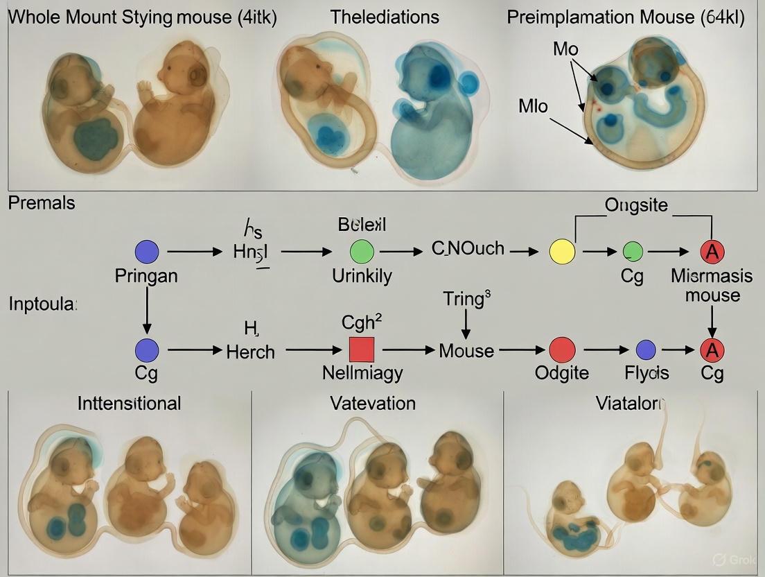
Whole-Mount Staining for Preimplantation Mouse Embryos: A Comprehensive Guide from Basics to Advanced Applications
This article provides a complete resource for researchers on whole-mount staining techniques for preimplantation mouse embryos.
Whole-Mount Staining for Preimplantation Mouse Embryos: A Comprehensive Guide from Basics to Advanced Applications
Abstract
This article provides a complete resource for researchers on whole-mount staining techniques for preimplantation mouse embryos. It covers foundational principles of immunofluorescence and in situ hybridization, detailed optimized protocols for single-molecule RNA FISH combined with protein detection, practical troubleshooting for common issues like permeabilization and antibody penetration, and advanced validation methods using deep learning and live imaging. Aimed at scientists and drug development professionals, this guide integrates traditional methods with cutting-edge technologies to enhance research reproducibility and discovery in early mammalian development.
Understanding Whole-Mount Staining: Principles and Critical Reagents for Preimplantation Embryo Analysis
Whole-mount techniques have revolutionized the study of embryonic development by enabling comprehensive three-dimensional spatial analysis of biological structures. Unlike traditional sectioning methods, which disrupt native tissue architecture, whole-mount staining preserves the complete spatial context of protein expression and cellular organization throughout the entire embryo. This approach provides researchers with unparalleled ability to visualize developmental processes, analyze progenitor cell populations within intact structures, and generate quantitative three-dimensional data. Within the context of preimplantation mouse embryo research, these techniques are particularly valuable for understanding the dynamic processes of lineage specification and morphogenetic events while maintaining the delicate spatial relationships between developing tissues.
Core Advantages of Whole-Mount Techniques
Whole-mount techniques offer several distinct advantages over traditional sectioning methods for developmental biology research, particularly for preimplantation mouse embryos where three-dimensional architecture is crucial for understanding developmental processes.
Table 1: Comparative Analysis: Whole-Mount vs. Sectioned Embryo Techniques
| Feature | Whole-Mount Techniques | Sectioned Embryo Techniques |
|---|---|---|
| Spatial Information | Preserves complete 3D architecture and spatial relationships [1] | Limited to 2D planes; 3D context lost or reconstructed laboriously |
| Tissue Integrity | Maintains native structure without physical disruption [2] | Disrupts tissue architecture through cutting |
| Cellular Resolution | Enables visualization at cellular and sub-nuclear levels [1] | High cellular resolution but limited to single planes |
| Morphogenetic Analysis | Ideal for comprehensive analysis of morphogenetic events [3] | Limited capacity for full 3D morphogenetic analysis |
| Downstream Applications | Compatible with additional assays (e.g., sectioning post-imaging) [2] | Typically destructive; limits additional uses |
| Quantitative Potential | Enables 3D spatial reconstruction and volumetric measurement [3] | Primarily quantitative in 2D dimensions |
The fundamental advantage of whole-mount techniques lies in their capacity to preserve three-dimensional information, allowing researchers to analyze expression domains and cellular relationships within the complete embryonic context [1]. This comprehensive preservation is invaluable for studying preimplantation development, where the spatial organization of the inner cell mass, trophectoderm, and other early structures is critical to understanding lineage specification.
Furthermore, whole-mount staining imposes minimal impact on embryonic specimens, enabling researchers to utilize stained and imaged samples for subsequent applications such as paraffin or frozen sectioning followed by histological staining [2]. This characteristic is particularly valuable when working with rare or difficult-to-obtain experimental samples, maximizing the information gained from each specimen.
Experimental Workflow and Visualization
The process of whole-mount analysis involves a coordinated series of steps from embryo preparation through imaging and computational analysis, forming an integrated pipeline for three-dimensional spatial investigation.
Diagram 1: Comprehensive workflow for whole-mount immunofluorescence and imaging
This workflow illustrates the sequential process for whole-mount analysis, with color coding indicating different procedural phases: sample preparation (green), antibody applications (blue), washing steps (red), and visualization stages (yellow). Each step requires specific timing and condition optimization to ensure high-quality results while preserving antigenicity and structural integrity.
Detailed Methodologies and Protocols
Whole-Mount Immunofluorescence Staining Protocol
The following protocol adapts established methodologies for preimplantation to early postimplantation mouse embryos (up to E8.0) [1], with modifications for cardiac crescent stage embryos (E8.25) [3]:
Embryo Harvesting and Fixation
- Isolate embryos in phosphate-buffered saline (PBS), removing decidua, yolk sac, and amnion [3]
- Fix with 4% paraformaldehyde in PBS for 1 hour at room temperature or overnight at 4°C [3]
- Rinse three times with PBS; embryos can be stored at 4°C for several weeks at this stage [3]
Immunofluorescence Staining
- Permeabilize and block with 0.5% saponin, 1% bovine serum albumin in PBS for at least 4 hours at room temperature [3]
- Incubate with primary antibody mixture diluted in blocking buffer overnight at 4°C [1] [3]
- Wash 3 times for 1 hour each with 0.1% Triton in PBS [3]
- Incubate with secondary antibody mixture for 3 hours at room temperature or overnight at 4°C [3]
- Wash 3 times for 1 hour each with 0.1% Triton in PBS [3]
- Counterstain with DAPI in PBS for 10 minutes [3]
- Wash 2 times for 5 minutes each with 0.1% Triton in PBS [3]
Mounting and Imaging
- Suspend embryos in anti-fade mounting media (2% n-propyl gallate, 90% glycerol, 1× PBS) [3]
- Mount using double-stick tape or silicone spacers to prevent compression [3]
- Image using confocal or lightsheet microscopy with appropriate laser/filter combinations for fluorophores used [1] [2]
Advanced Tissue Clearing with EZ Clear Protocol
For enhanced imaging depth, particularly in later-stage embryos, the EZ Clear method provides rapid clearing while preserving fluorescence:
- Immerse fixed samples in lipid removal solution (50% tetrahydrofuran in water) for delipidation [4]
- Wash with sterile Milli-Q water for 4 hours to remove residual THF [4]
- Render tissue transparent by immersion in aqueous refractive index matching solution (EZ View, RI=1.518) for 24 hours [4]
- This entire process requires 48 hours and maintains samples in an aqueous environment, preventing size changes while preserving endogenous fluorescent signals [4]
The Scientist's Toolkit: Essential Research Reagents
Table 2: Key Research Reagent Solutions for Whole-Mount Techniques
| Reagent/Category | Specific Examples | Function and Application |
|---|---|---|
| Fixation Agents | 4% Paraformaldehyde (PFA) in PBS [3] | Preserves tissue architecture and antigen integrity |
| Permeabilization Agents | 0.5% Saponin, 0.1% Triton X-100 [3] | Enables antibody penetration by dissolving membranes |
| Blocking Agents | 1% Bovine Serum Albumin (BSA) [3] | Reduces non-specific antibody binding |
| Nuclear Stains | DAPI, Hoechst dyes, Draq5, Red-Dot [2] | Reveals overall cellular organization and morphology |
| Mounting Media | Anti-fade media (nPG/glycerol/PBS) [3] | Preserves fluorescence during imaging |
| Clearing Reagents | EZ Clear (THF-based), BABB, CUBIC [4] | Renders tissues transparent for deep imaging |
| Refractive Index Matching | EZ View (RI=1.518) [4] | Optimizes light penetration for clarity |
The selection of appropriate nuclear stains depends on available microscopy systems. DAPI or Hoechst dyes require UV excitation, while far-red stains like Draq5 are ideal for confocal microscopes with far-red laser/filter combinations [2]. The recent development of EZ Clear represents a significant advancement, offering rapid clearing in aqueous conditions without sample size alteration, unlike methods that cause significant shrinkage (e.g., 3DISCO) or expansion (e.g., X-CLARITY) [4].
Imaging Modalities and Analysis Approaches
The choice of imaging methodology significantly impacts the quality and type of data obtainable from whole-mount specimens, with different systems offering distinct advantages for various research applications.
Table 3: Imaging Modalities for Whole-Mount Embryo Analysis
| Imaging Method | Applications | Resolution and Limitations |
|---|---|---|
| Conventional Fluorescence Microscopy | Rapid screening of stained embryos; large specimens at low magnification [2] | Superior to brightfield; limited apparent depth of field compared to confocal [2] |
| Laser Scanning Confocal Microscopy | High-resolution 3D reconstruction; optical sectioning of thick specimens [1] [3] | Exceptional clarity, contrast, and depth of field; lengthy acquisition times [2] |
| Lightsheet Fluorescence Microscopy (LSFM) | Rapid imaging of large cleared specimens; minimal photobleaching [4] | Fast volumetric imaging; requires specialized equipment [4] |
| "Pseudo-SEM" Nuclear Imaging | Detailed topological documentation of embryo morphology [2] | Rivals SEM clarity; requires nuclear staining and confocal z-stacks [2] |
For preimplantation mouse embryos, whole-mount immunofluorescence staining combined with confocal microscopy enables visualization of protein expression at cellular or sub-nuclear levels while preserving three-dimensional spatial information [1]. This approach is particularly powerful when combined with computational analysis for three-dimensional spatial reconstruction of embryonic structures, enabling quantitative measurements of specific progenitor populations within the context of the complete embryo [3].
The "pseudo-SEM" approach, utilizing whole-mount nuclear staining in combination with confocal microscopy, generates images with exceptional topological detail that can rival scanning electron microscopy in clarity, while avoiding the dehydration artifacts and specialized equipment requirements of SEM [2]. This method is effective for mouse embryos through E15.5, after which skin maturation reduces dye penetration [2].
Quantitative Analysis and Data Interpretation
Advanced image processing enables sophisticated quantitative analysis of whole-mount specimens, transforming image data into measurable three-dimensional information. Following confocal microscopy and 3D reconstruction of structures such as the cardiac crescent, successive masking using reference antibodies allows for quantitative measurements of specific areas within the structure [3]. This approach enables detailed examination of the organization of progenitor populations during critical phases of organogenesis.
Automated analysis of imaging datasets provides unbiased measurement, though reliability depends heavily on input data quality [3]. Proper acquisition parameters during confocal microscopy are essential, including appropriate definition of top and bottom optical slice positions to avoid cropping specimens in the z-axis, and sufficient overlap between optical sections to prevent a layered appearance in final projections [2].
In whole mount staining for preimplantation mouse embryo research, the dual objectives of preserving intricate cellular structures while simultaneously allowing probe accessibility present a significant technical challenge. Fixation and permeabilization are interdependent processes that must be meticulously balanced; inadequate fixation compromises structural integrity, whereas excessive fixation can mask antigenic sites and hinder probe penetration. This application note delineates the core principles and detailed methodologies for achieving this equilibrium, enabling researchers and drug development professionals to obtain reproducible and biologically relevant data from their whole mount embryo studies. The protocols outlined here are specifically optimized for preimplantation mouse embryos, where preserving the three-dimensional architecture is paramount for accurate spatial localization of molecular targets.
Fundamental Principles and Key Considerations
The successful application of whole mount staining techniques rests upon understanding the mechanistic actions of fixation and permeabilization agents. Fixation acts to crosslink proteins and biomolecules, thereby stabilizing the native architecture of the cell against subsequent processing steps. Permeabilization follows by creating pores in lipid membranes, enabling antibodies and other probes to reach their intracellular targets. A critical principle is that the stringency of the fixation process often dictates the required strength of the permeabilization agent. Over-fixation can necessitate harsher permeabilization conditions, which might compromise antigenicity or overall morphology.
For preimplantation embryos, an additional layer of complexity is introduced by the need to remove or permeabilize the zona pellucida, an extracellular glycoprotein layer surrounding the embryo. A specific protocol for mouse preimplantation embryos involves removing the zona pellucida by placing embryos in acid Tyrode's solution for approximately 10 seconds at room temperature prior to the fixation step [5]. This is a crucial preparatory step to ensure that all subsequent solutions, including fixatives, antibodies, and washes, can access the embryo proper.
Detailed Experimental Protocols
Protocol 1: Whole-Mount Immunofluorescence for Preimplantation Mouse Embryos
The following protocol, adapted from established methodologies, is designed for optimal preservation and staining of preimplantation stage mouse embryos [5].
- 1. Zona Pellucida Removal: Transfer preimplantation embryos to acid Tyrode's solution (e.g., Sigma, St. Louis, MO, USA) for a brief 10 seconds at room temperature (RT). Immediately proceed to washing to neutralize the acid [5].
- 2. Fixation: Immerse embryos in a 4% paraformaldehyde (PFA) solution in phosphate-buffered saline (PBS) for 30 minutes at RT. This concentration and duration provide sufficient cross-linking without creating excessive antigen masking [5].
- 3. Permeabilization: Wash embryos in PBS and then transfer to a 2% Triton X-100 solution in PBS for 30 minutes at RT. Triton X-100 is a non-ionic detergent effective at extracting lipids and creating pores in cellular membranes [5].
- 4. Blocking: Incubate embryos in a blocking solution of 4% Bovine Serum Albumin (BSA) in PBS to reduce non-specific binding of antibodies. A typical blocking time is 1 hour at RT or overnight at 4°C.
- 5. Primary Antibody Incubation: Incubate embryos with the desired primary antibody (e.g., anti-STAT3 antibodies such as C-20 (sc-482) or F-2 (sc-8019), Santa Cruz Biotech.) diluted in blocking solution, overnight at 4°C [5].
- 6. Secondary Antibody and Counterstaining: After thorough washing in PBS with 1% BSA and 0.005% Triton X-100, incubate embryos with fluorescently-labeled secondary antibodies (e.g., Alexa Fluor 488 or 546, Thermo Fisher Scientific) and a nuclear counterstain like DAPI for 30-60 minutes at RT, protected from light [5].
- 7. Mounting and Imaging: Mount the stained embryos using an anti-fade mounting medium such as ProLong Gold antifade reagent (Invitrogen). Image using confocal microscopy (e.g., LSM 510, Carl Zeiss) to obtain high-resolution three-dimensional data [5].
Protocol 2: Organic Solvent-Based Permeabilization for Challenging Specimens
While the above protocol uses a detergent (Triton X-100) in aqueous solution, some specimens, particularly older embryos with hardened eggshells, may require organic solvent-based permeabilization. A key advancement in this area is the use of D-limonene-based embryo permeabilization solvent (EPS) [6] [7]. This method is highly effective for rendering dechorionated embryos permeable while maintaining high viability.
- 1. Staging and Dechorionation: Collect and stage embryos precisely. Rinse embryos and dechorionate by immersing them in 50% bleach for 2 minutes, followed by extensive washing in water or buffer [6].
- 2. EPS Treatment: Prepare a working dilution of EPS (e.g., 1:40 in an appropriate incubation medium like Modified Basic Incubation Medium (MBIM)). Immerse the dechorionated embryos in the dilute EPS solution with gentle swirling for 30 seconds to 90 seconds. The optimal exposure time must be determined empirically and can be longer for late-stage embryos [6] [7].
- 3. Washing and Viability: Remove the basket containing embryos from the EPS and blot away excess solvent. Proceed with six sequential washes in PBS or another physiological buffer to ensure complete removal of the solvent [6].
- 4. Permeabilization Check: A permeability indicator such as Rhodamine B or a far-red dye like CY5 can be included to assess the uniformity of permeabilization across a batch of embryos [6] [7].
The following workflow diagram synthesizes the key decision points and steps from these protocols into a unified visual guide.
The optimization of fixation and permeabilization is guided by quantitative parameters. The table below summarizes key variables and their typical ranges for preimplantation mouse embryo protocols.
Table 1: Key Parameters for Embryo Fixation and Permeabilization
| Parameter | Fixation (4% PFA) | Aqueous Permeabilization (2% Triton X-100) | Organic Solvent Permeabilization (EPS) |
|---|---|---|---|
| Concentration | 4% in PBS | 0.1 - 2% in PBS | 1:40 dilution in MBIM [6] |
| Duration | 20 - 30 min at RT [5] | 10 - 30 min at RT [5] | 30 - 90 sec [6] |
| Temperature | Room Temperature (RT) | Room Temperature (RT) | Room Temperature (RT) |
| Key Function | Protein cross-linking, structure preservation | Lipid dissolution, membrane pore creation | Solubilizes waxy layers, broad permeabilization [7] |
| Primary Application | Standard immunofluorescence | Standard immunofluorescence | Challenging specimens (e.g., late-stage, hardened eggshell) [6] |
The Scientist's Toolkit: Essential Research Reagents
A successful outcome in whole mount staining is contingent upon the use of high-quality, well-characterized reagents. The following table details essential materials and their critical functions in the protocol.
Table 2: Essential Reagents for Whole-Mount Embryo Staining
| Reagent | Function / Purpose | Example |
|---|---|---|
| Acid Tyrode's Solution | Chemically removes the zona pellucida to enable solution access to the embryo proper [5]. | Sigma, St. Louis, MO, USA [5] |
| Paraformaldehyde (PFA) | A cross-linking fixative that preserves cellular architecture by forming methylene bridges between proteins. | 4% solution in PBS [5] |
| Triton X-100 | Non-ionic detergent that permeabilizes lipid bilayers by solubilizing membranes. | 2% solution in PBS for permeabilization [5] |
| Bovine Serum Albumin (BSA) | Blocking agent used to occupy non-specific binding sites and reduce background signal. | 4% BSA in PBS for blocking [5] |
| Primary Antibodies | Specifically bind to the target antigen of interest. Validation for immunofluorescence is critical. | anti-STAT3 (e.g., sc-482, sc-8019, Santa Cruz Biotech.) [5] |
| Fluorescent Secondary Antibodies | Conjugated antibodies that bind to the primary antibody, enabling detection. | Donkey anti-rabbit Alexa Fluor 488 (A-21206, Thermo Fisher) [5] |
| DAPI (4′,6-diamidino-2-phenylindole) | A blue-fluorescent DNA stain used as a nuclear counterstain. | Thermo Fisher Scientific [5] |
| ProLong Gold Antifade Reagent | A mounting medium that retards photobleaching (fading) of fluorescent signals during microscopy. | Invitrogen [5] |
| Embryo Permeabilization Solvent (EPS) | A d-limonene and surfactant-based solvent that effectively permeabilizes waxy eggshell layers [7]. | 90% d-limonene, 5% cocamide DEA, 5% ethoxylated alcohol [6] |
| Permeabilization Indicator Dyes | Small molecule dyes (e.g., Rhodamine B, CY5) used to visually confirm and uniformity of permeabilization [6] [7]. | CY5 carboxylic acid [6] |
Mastering the balance between structure preservation and probe accessibility is foundational to robust whole mount staining in preimplantation mouse embryos. The protocols and principles detailed in this application note provide a reliable framework for researchers. The choice between aqueous and solvent-based permeabilization must be guided by the specific embryo stage and the nature of the target antigen. By adhering to these optimized conditions and utilizing the essential reagents outlined, scientists can consistently generate high-quality, three-dimensional data that is critical for advancing developmental biology research and drug discovery efforts.
The Scientist's Toolkit: Research Reagent Solutions
The following table details the essential reagents required for whole-mount staining of preimplantation mouse embryos, a foundational technique for studying early mammalian development.
Table 1: Essential Reagents for Whole-Mount Staining of Preimplantation Mouse Embryos
| Reagent | Primary Function | Key Considerations & Typical Usage |
|---|---|---|
| Paraformaldehyde (PFA) | Fixation: Cross-links proteins to preserve cellular architecture and antigen structure [8] [9]. | - Concentration: 2-4% in PBS is standard [5] [8] [9].- Incubation: 30 minutes at room temperature is effective for mouse embryos [5] [8]. |
| Triton X-100 | Permeabilization: A non-ionic detergent that solubilizes lipid membranes, allowing antibodies to access intracellular targets [9]. | - Concentration: 0.1-0.5% in PBS for staining interior membranes and nuclear targets [8] [9].- Incubation: Typically 10-30 minutes at room temperature [8]. |
| Blocking Serum | Reducing Background: Blocks nonspecific binding sites to minimize off-target antibody binding and improve signal-to-noise ratio [8] [9]. | - Type: Normal serum from a species different than the host of the primary or secondary antibody [9].- Concentration: 1-5% in PBS, often used with BSA and detergent [8] [9]. |
Experimental Protocols for Whole-Mount Immunofluorescence
The protocol below is optimized for the unique challenges of working with preimplantation mouse embryos, which are small, delicate, and require careful handling to preserve their three-dimensional structure.
Whole-Mount Immunofluorescence Staining Protocol for Preimplantation Mouse Embryos
Key Resources:
- Biological Sample: Preimplantation mouse embryos (e.g., 2.5-3.5 dpc morula or blastocyst) [8].
- Equipment: Confocal microscope, stereo microscope, humidified chamber, mouth pipette [10] [8].
Step-by-Step Procedure:
Embryo Collection and Zona Pellucida Removal: Collect preimplantation embryos from superovulated and mated mice into M2 medium [10] [8]. To remove the zona pellucida, briefly incubate embryos (approximately 10 seconds at room temperature) in Acid Tyrode's solution, then wash thoroughly in fresh medium [5].
Fixation: Transfer the embryos to a solution of 4% Paraformaldehyde (PFA) in PBS. Incubate for 30 minutes at room temperature [5] [8]. This step cross-links proteins, permanently preserving the embryo's morphology and fixing the antigens in place.
Permeabilization: Wash the fixed embryos in PBS. Then, incubate them in Permeabilization Buffer (0.25% Triton X-100 in PBS) for 30 minutes at room temperature [8]. This creates pores in the cellular and nuclear membranes, allowing antibodies to penetrate.
Blocking: To prevent non-specific antibody binding, incubate the embryos in a Blocking Solution for at least 1 hour at room temperature or overnight at 4°C. A recommended solution is PBS containing 1-5% normal goat serum (or serum from another suitable species), 0.25% Triton X-100, and 1% BSA [8] [9].
Primary Antibody Incubation: Incubate embryos with the primary antibody diluted in the blocking solution. A typical incubation is overnight at 4°C in a humidified chamber to ensure sufficient antibody penetration and binding [5] [8].
Washing: Wash the embryos extensively (3-4 times, 15-30 minutes each) in a wash buffer (e.g., PBS with 0.1% BSA and 0.005% Triton X-100) to remove unbound primary antibody [5].
Secondary Antibody and Counterstain Incubation: Incubate with fluorophore-conjugated secondary antibodies and nuclear stains like DAPI (1 µg/mL) or Hoechst for 1-2 hours at room temperature or overnight at 4°C, protected from light [5] [8] [9].
Mounting and Imaging: Mount the stained embryos using an anti-fade mounting medium (e.g., ProLong Gold) on a glass slide [5] [8]. Acquire high-resolution images using a confocal microscope to analyze protein localization and expression in 3D.
Optimization Controls: Blocking Peptide Validation
To confirm antibody specificity in immunofluorescence experiments, a blocking peptide control is essential. This control uses the immunizing peptide to compete with the target antigen for antibody binding.
Procedure:
- Reconstitute the lyophilized blocking peptide according to the manufacturer's instructions [11].
- Prepare the primary antibody at its optimal working dilution in two separate tubes. To one tube, add a 10-fold excess (by weight) of the blocking peptide. Incubate both tubes for 1 hour at room temperature [11].
- Follow the standard staining protocol, applying the "antibody alone" solution to one set of embryos and the "antibody + peptide" solution to another set.
- Interpretation: Specific staining is indicated by a strong signal in the "antibody alone" sample that is significantly reduced or abolished in the "antibody + peptide" sample [11].
Reagent Functions and Synergy in Whole-Mount Staining
The following diagram illustrates the critical roles and synergistic relationship between PFA, Triton X-100, and blocking serum in the experimental workflow for whole-mount immunofluorescence.
In whole mount staining for preimplantation mouse embryos research, the selection of primary and secondary antibodies with high specificity is paramount to experimental success. Antibody validation represents one of the most significant challenges in developmental biology, where accurate detection of embryonic antigens determines the reliability of spatial and temporal expression data. For researchers and drug development professionals working with limited embryonic material, improper antibody selection can lead to misinterpretation of developmental pathways, wasting valuable resources and potentially leading to erroneous conclusions [12] [13].
The unique composition of embryonic antigens, combined with the structural preservation requirements of whole mount techniques, creates a demanding environment for antibody performance. Unlike cell culture systems where knockout validation is often straightforward, embryonic tissues present additional validation complexities due to their dynamic nature, limited availability, and the potential developmental consequences of gene ablation [13] [14]. This application note provides a structured framework for selecting and validating primary and secondary antibodies specifically for whole mount staining of preimplantation mouse embryos, with emphasis on practical protocols and troubleshooting guidance tailored to embryonic research.
Antibody Validation Strategies for Embryonic Antigens
Comprehensive Validation Approaches
Table 1: Antibody Validation Methods for Embryonic Research
| Validation Method | Application to Embryonic Antigens | Key Considerations | References |
|---|---|---|---|
| Genetic Knockout/Knockdown | Gold standard; confirms absence of staining in null tissue | May require conditional knockout for embryonic lethal mutations; use CRISPR/Cas9-modified ES cells | [13] [15] |
| Orthogonal Correlation | Compare staining pattern with RNA expression data via in situ hybridization | Correlation should be observed across multiple embryonic stages | [13] [14] |
| Independent Antibodies | Use multiple clones against different epitopes of same target | Concordant staining patterns increase confidence; particularly valuable for transcription factors like Oct3/4 | [13] [14] |
| Overexpression Detection | Transfert expression plasmids in embryonic cell lines | Confirms antibody can detect antigen but not endogenous expression levels | [13] |
| Absorption Control | Pre-incubate antibody with excess antigen | Should abolish specific staining; requires purified antigen availability | [16] |
Special Considerations for Embryonic Antigens
When working with preimplantation mouse embryos, several unique challenges emerge that demand specialized validation approaches. The limited quantity of material necessitates careful planning of validation experiments, often requiring the use of embryonic stem cells as surrogates for initial validation [14]. Researchers must also consider the dynamic expression patterns characteristic of developmental genes; an antibody validated for one embryonic stage may not perform reliably at earlier or later stages due to post-translational modifications or epitope masking.
For transcription factors critical to embryonic development, such as Oct3/4, Nanog, and SOX2, it is essential to demonstrate that antibody staining patterns match known expression dynamics during differentiation [14]. As demonstrated in studies of human embryonic stem cells, the downregulation of pluripotency markers upon differentiation provides a natural validation system - antibodies should show strong nuclear staining in undifferentiated cells and marked reduction upon embryonic body formation [14].
Experimental Protocols for Whole Mount Embryo Immunofluorescence
Whole-Mount Immunofluorescence Staining Protocol for Preimplantation Mouse Embryos
Materials and Reagents
- Acid Tyrode's solution (Sigma, St. Louis, MO, USA)
- 4% paraformaldehyde in phosphate-buffered saline (PBS)
- Permeabilization solution: 2% Triton X-100 in PBS
- Blocking solution: 4% bovine serum albumin (BSA) in PBS
- Primary antibodies diluted in blocking solution
- Fluorophore-conjugated secondary antibodies (e.g., Alexa Fluor series)
- ProLong Gold antifade reagent with DAPI (Invitrogen)
- Embryo culture-grade dishes and handling pipettes
Step-by-Step Procedure
- Zona Pellucida Removal: Transfer preimplantation mouse embryos to acid Tyrode's solution for approximately 10 seconds at room temperature. Monitor closely until the zona pellucida dissolves, then immediately transfer to embryo culture medium [5].
Fixation: Fix embryos with 4% paraformaldehyde solution for 30 minutes at room temperature. This step preserves embryonic architecture while maintaining antigen accessibility [5].
Permeabilization: Permeabilize embryos with 2% Triton X-100 in PBS for 30 minutes at room temperature. This step enables antibody penetration throughout the embryo [5].
Blocking: Incubate embryos in blocking solution (4% BSA in PBS) for 1-2 hours at room temperature. This reduces non-specific antibody binding [5].
Primary Antibody Incubation: Incubate embryos with primary antibodies diluted in blocking solution overnight at 4°C. Gently agitate to ensure even antibody distribution. Optimal antibody concentrations must be determined experimentally for each antibody [5] [12].
Washing: Wash embryos extensively in PBS containing 1% BSA and 0.005% Triton X-100 (3-5 washes, 30 minutes each) to remove unbound primary antibody [5].
Secondary Antibody Incubation: Incubate embryos with fluorophore-conjugated secondary antibodies (e.g., Alexa Fluor 488, 546) and DAPI for 2-4 hours at room temperature protected from light [5].
Final Washes: Perform final washes in PBS with 1% BSA and 0.005% Triton X-100 (3 washes, 30 minutes each) [5].
Mounting: Mount stained embryos using ProLong Gold antifade reagent. Gently press coverslip to ensure even spreading without damaging embryos [5].
Imaging: Image using laser scanning confocal microscopy with appropriate filter sets for each fluorophore. Acquire z-stacks to capture three-dimensional antigen distribution [5].
Figure 1: Experimental workflow for whole-mount immunofluorescence staining of preimplantation mouse embryos
Antibody Titration Protocol for Limited Embryonic Material
Rationale Antibody titration is essential for achieving optimal signal-to-noise ratio while conserving precious embryonic material. Using excessive antibody concentrations promotes non-specific binding, while insufficient concentrations yield weak, unreliable signals [12].
Titration Procedure
- Prepare a 2-fold serial dilution series of primary antibody in blocking buffer, typically spanning 8-12 concentration points [12].
Divide limited embryonic material into aliquots, ensuring each titration point includes sufficient embryos for evaluation (minimum 3-5 embryos per concentration).
Follow the standard immunofluorescence protocol above, using different antibody concentrations for each embryo group.
Image all samples using identical acquisition parameters.
Quantify signal intensity and background staining using image analysis software. The optimal concentration provides the highest specific signal with minimal background [12].
Document the optimal dilution for future reference, noting lot number and embryonic stage.
Controls and Troubleshooting for Embryonic Applications
Essential Control Experiments
Table 2: Control Experiments for Whole Mount Embryo Staining
| Control Type | Purpose | Interpretation | Application to Embryonic Research |
|---|---|---|---|
| Secondary Antibody Only | Detect non-specific secondary antibody binding | Staining indicates Fc receptor binding or non-specific tissue interactions | Critical for embryonic tissues with high Fc receptor expression |
| Absorption Control | Confirm specificity by pre-adsorption with antigen | Loss of staining confirms specificity; may be challenging for rare embryonic antigens | Requires recombinant protein for transcription factors; use embryonic lysates as alternative |
| Isotype Control | Assess non-specific immunoglobulin binding | Staining indicates non-specific Ig binding | Match host species and immunoglobulin subclass to primary antibody |
| Biological Negative | Tissue known to lack target antigen | Should show no specific staining | Use later embryonic stages or differentiated ES cells where antigen is downregulated |
| Multi-labeling Controls | Test cross-reactivity in multiplex experiments | Ensure secondary antibodies recognize only intended primary antibodies | Essential for co-localization studies of multiple embryonic antigens |
Troubleshooting Common Issues
Poor Antibody Penetration
- Increase permeabilization time or Triton X-100 concentration
- Consider alternative detergents (e.g., saponin) for better preservation of membrane structures
- Implement gentle agitation during all incubation steps
High Background Staining
- Increase blocking time or try alternative blocking agents (e.g., serum from secondary antibody host species)
- Optimize washing stringency by increasing salt concentration (e.g., 150-300mM NaCl) or adding mild detergents
- Titrate secondary antibody concentration independently from primary antibody
Weak Specific Signal
- Verify antigen preservation by testing alternative fixation methods (e.g., methanol fixation for transcription factors)
- Increase primary antibody incubation time to 48-72 hours with gentle agitation at 4°C
- Consider signal amplification systems (e.g., tyramide amplification) for low-abundance antigens
Research Reagent Solutions for Embryonic Antigen Detection
Table 3: Essential Research Reagents for Embryonic Antibody Applications
| Reagent Category | Specific Examples | Function in Embryonic Research | Validation Considerations |
|---|---|---|---|
| Pluripotency Marker Antibodies | Oct3/4 (C-10, Santa Cruz), Nanog, SOX2 | Identify undifferentiated embryonic cells; monitor pluripotency status | Confirm downregulation upon embryonic differentiation [14] |
| Secondary Antibodies with Minimal Cross-Reactivity | Donkey anti-rabbit IgG (H&L) Alexa Fluor 488, Donkey anti-mouse IgG (H&L) Alexa Fluor 546 | Enable multiplex detection with minimal species cross-reactivity | Verify lack of cross-reactivity to embryonic tissues and other secondary antibodies [16] [5] |
| Adsorbed Secondary Antibodies | Species-adsorbed secondary antibodies (Jackson ImmunoResearch) | Reduce non-specific binding in complex embryonic tissues | Confirm adsorption against appropriate species; test on embryonic tissue alone [16] |
| Embryonic Stem Cell Lines | Mouse D3 ES cells, Human H1/H9 ES cells | Provide validation platform for antibody performance | Establish correlation between ES cell staining and embryo staining [17] [14] |
| Mounting Media with DAPI | ProLong Gold antifade reagent with DAPI | Preserve fluorescence and counterstain nuclei | Verify compatibility with fluorophores and absence of quenching |
The selection and validation of primary and secondary antibodies for embryonic antigen detection requires a methodical, multi-faceted approach tailored to the unique challenges of preimplantation mouse embryos. By implementing rigorous validation strategies, comprehensive control experiments, and optimized staining protocols, researchers can generate reliable, reproducible data that advances our understanding of early embryonic development. The protocols and guidelines presented here provide a foundation for researchers embarking on whole mount staining experiments, with particular emphasis on practical considerations for working with limited embryonic material. As antibody technologies continue to evolve, these validation frameworks will remain essential for ensuring the accuracy and biological relevance of developmental studies.
The analysis of gene expression patterns in preimplantation mouse embryos is fundamental to understanding the molecular regulation of early mammalian development. In this context, whole mount in situ hybridization (ISH) has emerged as a particularly valuable technique as it preserves the delicate three-dimensional architecture of the embryo while enabling spatial localization of RNA transcripts. The choice between traditional RNA probes and commercial RNAscope systems represents a significant methodological crossroads for researchers. This Application Note provides a detailed technical comparison of these approaches, with specific focus on their application in whole mount staining of preimplantation mouse embryos. We present experimental protocols, reagent solutions, and data to guide researchers in selecting the most appropriate methodology for their specific research objectives.
Traditional RNA Probes
Traditional RNA probe methodology utilizes in vitro-transcribed RNA probes, typically labeled with haptens such as digoxigenin (DIG) or fluorescein. Signal detection and amplification are commonly achieved through the Tyramide Signal Amplification (TSA) system, which enables high-sensitivity detection of accumulated mRNAs in mammalian oocytes and embryos [18]. This approach relies on the enzymatic deposition of tyramide-conjugated fluorophores, resulting in substantial signal amplification at the site of probe hybridization.
RNAscope Commercial System
RNAscope (Advanced Cell Diagnostics) represents a revolutionary advance in ISH technology, employing a proprietary probe design and signal amplification system. The core innovation involves pairs of "Z" probes that hybridize to adjacent regions of the target RNA. Each Z probe contains a tail sequence that binds pre-amplifier molecules, initiating a branched DNA (bDNA) amplification cascade that can generate up to 8,000-fold signal amplification [19]. This design enables single-molecule detection with high specificity and minimal background.
Table 1: Core Technology Comparison Between Traditional RNA Probes and RNAscope
| Feature | Traditional RNA Probes | RNAscope System |
|---|---|---|
| Probe Type | In vitro-transcribed RNA (typically 200-1000 bases) [18] | Short, synthetic DNA oligonucleotides (20-25 bases per segment) [20] |
| Probe Design | Researcher-designed; targets contiguous regions | Proprietary algorithm designs 20 ZZ probe pairs per target [21] |
| Amplification Mechanism | Tyramide Signal Amplification (TSA) [18] | Branched DNA (bDNA) cascade [20] |
| Target Size Requirement | Typically >300 bases for optimal sensitivity [18] | mRNA >300 bases; Basescope for 50-300 bases [21] |
| Detection Limit | Single-molecule sensitivity with optimized TSA [18] | Single-molecule detection [19] |
| Multiplexing Capability | Limited by available haptens and antibody conjugates | Designed for multiplexing (up to 4-plex with standard kits) [20] [21] |
Diagram 1: Comparative workflow of Traditional RNA Probes with TSA versus RNAscope System
Application-Oriented Comparison for Embryo Research
Performance Characteristics in Embryonic Samples
When working with preimplantation mouse embryos, several performance factors critically influence experimental outcomes:
Sensitivity and Specificity: RNAscope provides exceptional sensitivity and specificity due to its proprietary probe design, which requires dual probe binding to initiate amplification. This feature minimizes background signal, a common challenge in whole mount embryo staining [20] [19]. Traditional RNA probes with TSA can achieve similar sensitivity but typically require more extensive optimization to minimize background.
Tissue Penetration: For whole mount preimplantation embryos, penetration of detection reagents is crucial. RNAscope probes, being shorter, may penetrate more efficiently, though the amplification machinery is substantial. Traditional RNA probes coupled with optimized permeabilization protocols (e.g., using Triton X-100) have demonstrated success in whole mount embryo applications [22] [18].
Multiplexing Capability: RNAscope is designed for multiplexing, allowing simultaneous detection of multiple RNA targets in the same specimen using different channels (C1, C2, C3, etc.) [20] [21]. This is particularly valuable for analyzing co-expression patterns in precious embryo samples. Traditional approaches have more limited multiplexing capabilities.
Compatibility with Immunofluorescence: Both methods can be combined with protein detection. Research on whole mount preimplantation mouse embryos has successfully combined RNAscope with immunofluorescence by performing IF first under RNase-free conditions, followed by RNAscope detection [22].
Table 2: Performance Considerations for Preimplantation Mouse Embryo Studies
| Performance Parameter | Traditional RNA Probes | RNAscope System | Implication for Embryo Research |
|---|---|---|---|
| Signal-to-Noise Ratio | Requires optimization; variable background [18] | Consistently high; minimal background [20] | More reliable interpretation of spatial expression |
| Protocol Duration | 2-3 days including probe synthesis [18] | ~1 day for standardized protocol [19] | Faster turnaround for experimental results |
| Quantitative Capability | Semi-quantitative with careful controls | Enables transcript counting [19] | Better assessment of transcript abundance |
| Reproducibility | Lab-dependent optimization | Highly standardized [20] | Improved inter-lab reproducibility |
| Sample Preservation | Maintains tissue integrity in whole mounts [18] | Compatible with various fixation methods [22] | Preserves delicate embryo morphology |
Practical Implementation Factors
Cost Considerations: Traditional RNA probes offer lower reagent costs but require significant researcher time for optimization. RNAscope involves higher commercial reagent costs but reduced optimization time [20].
Probe Availability and Design: RNAscope provides pre-designed, validated probes for many targets, saving development time. For novel targets or species without commercial probes, traditional approaches offer complete flexibility [21].
Throughput and Scalability: RNAscope's standardized protocol facilitates processing of multiple samples in parallel. Traditional methods may show more variability when scaling up.
Detailed Experimental Protocols for Preimplantation Mouse Embryos
Whole Mount Protocol for Traditional RNA Probes with TSA
This protocol has been optimized for mouse oocytes and preimplantation embryos, enabling visualization of mRNA granular structures in the cytoplasm [18].
Day 1: Sample Preparation and Hybridization
- Fixation: Collect and fix blastocysts in 4% paraformaldehyde in PBS for 30 minutes at room temperature.
- Permeabilization: Treat with 0.1% Triton X-100 in PBS for 30 minutes [22].
- Pre-hybridization: Incubate in hybridization buffer (50% formamide, 5x SSC, 0.1% Tween-20, 50 μg/mL heparin, 100 μg/mL torula RNA) for 1 hour at 60°C.
- Hybridization: Add digoxigenin-labeled RNA probes (200-500 ng/mL) in hybridization buffer and incubate overnight at 60°C.
Day 2: Washes and Signal Detection
- Stringency Washes: Perform sequential washes with 50% formamide/2x SSCT at 60°C, followed by 2x SSCT and 0.2x SSCT at room temperature.
- Blocking: Incubate in blocking solution (2% horse serum in PBS with RNase inhibitor) for 1-2 hours [22].
- Antibody Incubation: Add anti-digoxigenin-HRP antibody (1:1000) in blocking solution overnight at 4°C.
Day 3: Signal Amplification and Imaging
- TSA Reaction: Apply tyramide-fluorophore working solution (1:100 in amplification buffer) for 10-30 minutes.
- Counterstaining and Mounting: Stain with DAPI and mount in suitable mounting medium for confocal microscopy.
- Imaging: Acquire images using confocal or super-resolution microscopy to resolve RNA granule structures [18].
Whole Mount Protocol for RNAscope
This protocol adapts the RNAscope Multiplex Fluorescent V2 Assay for whole mount preimplantation mouse embryos [22].
Day 1: Sample Preparation and Hybridization
- Fixation: Fix blastocysts in 4% PFA for 30 minutes at room temperature.
- Permeabilization: Treat with hydrogen peroxide for 10 minutes, followed by target retrieval reagent for 5 minutes at 90-98°C.
- Protease Treatment: Apply protease III for 15-30 minutes at room temperature.
- Probe Hybridization: Add target probes (e.g., Platr4, Malat1, Pou5f1) and incubate for 2 hours at 40°C.
Amplification and Detection
- Amplifier Hybridization: Perform sequential 30-minute incubations with AMP1, AMP2, and AMP3 at 40°C.
- HRP-based Signal Development: Apply HRP-C1 followed by Opal fluorophore (570, 690, etc.) for 30 minutes at 40°C.
- Multiplexing: For multiple targets, repeat HRP inactivation and subsequent probe detection steps.
- Counterstaining and Mounting: Stain with DAPI and mount for microscopy.
The entire RNAscope procedure can be completed within one day, significantly faster than traditional protocols [22].
The Scientist's Toolkit: Essential Research Reagent Solutions
Table 3: Key Reagents for Whole Mount RNA Detection in Preimplantation Embryos
| Reagent Category | Specific Examples | Function | Application Notes |
|---|---|---|---|
| Fixation Reagents | 4% Paraformaldehyde (PFA) [22] [18] | Preserve tissue morphology and RNA integrity | Critical optimization point for whole mount embryos |
| Permeabilization Agents | Triton X-100 [22], Protease III [22] | Enable probe penetration | Concentration and timing vary by embryo stage |
| Probe Systems | DIG-labeled RNA probes [18], RNAscope ZZ probes [20] | Target-specific hybridization | Commercial probes ensure consistency |
| Amplification Systems | Tyramide Signal Amplification (TSA) [18], bDNA amplification [20] | Signal enhancement | TSA requires HRP-conjugated detection |
| Detection Reagents | Anti-DIG-HRP [18], Opal fluorophores [22] | Visualize hybridized probes | Fluorophore choice depends on microscope capabilities |
| Hybridization Buffers | Formamide-based buffers [18] | Control stringency conditions | Formamide concentration affects specificity |
| Mounting Media | DAPI-containing media [22] | Preserve samples for imaging | Must maintain 3D structure of whole embryos |
The choice between traditional RNA probes and the RNAscope system for whole mount analysis of preimplantation mouse embryos depends on multiple factors, including research objectives, available resources, and required throughput.
Traditional RNA probes with TSA detection offer:
- Lower reagent costs and flexibility in probe design
- Proven capability for high-sensitivity detection in oocytes and embryos [18]
- Adaptability to novel targets without commercial availability
The RNAscope system provides:
- Standardized protocols with high reproducibility [20] [19]
- Superior multiplexing capabilities for co-localization studies
- Reduced optimization time and technical variability
For studies requiring detection of multiple targets in precious embryo samples or those conducted by laboratories with less ISH expertise, RNAscope offers significant advantages. For novel targets or highly customized applications, traditional RNA probes with optimized TSA detection remain a powerful and flexible approach. In both cases, the capacity to perform whole mount analysis preserves the valuable three-dimensional architecture of preimplantation embryos, enabling insights into the spatial regulation of gene expression during critical stages of early mammalian development.
Step-by-Step Protocols: Combining Immunofluorescence with smRNA FISH for Multi-Modal Detection
Within the context of a broader thesis on whole mount staining for preimplantation mouse embryos, the precise removal of the zona pellucida (ZP) is a critical preparatory step. The ZP, a glycoprotein layer surrounding the embryo, can act as a significant barrier to the penetration of stains and antibodies used in whole-mount protocols for spatial and temporal gene expression analysis [23]. This application note details a refined, two-step ZP removal protocol using Acid Tyrode's solution, optimized to maintain embryonic developmental competence and improve subsequent staining outcomes by minimizing cellular damage and apoptosis [24].
Detailed Experimental Protocol: Two-Step Zona Pellucida Removal
The following methodology is adapted from a study that developed a two-step removal protocol to reduce the toxicity associated with prolonged exposure to Acid Tyrode's solution [24].
1. Materials and Reagent Setup
- Acid Tyrode's Solution (e.g., Sigma T1788)
- Handling Pipette: A pulled glass pipette or a fine-diameter plastic pipette tip.
- Culture Dishes: 35 mm or 60 mm Petri dishes.
- Embryo Handling Pipette and a microscope for manipulation.
- Holding Pipette: A second pipette for secure embryo immobilization.
- Pre-equilibrated Embryo Culture Medium (e.g., KSOM or M16)
2. Step-by-Step Procedure
| Step | Action | Description & Purpose |
|---|---|---|
| 1 | Pre-equilibrate Solutions | Warm both the Acid Tyrode's solution and the culture medium to 37°C in a CO₂ incubator (if using bicarbonate-buffered media) or on a heated stage. |
| 2 | Prepare Working Dish | Place a 50-100 µL microdrop of Acid Tyrode's solution in a culture dish. Create several larger (300-500 µL) microdrops of pre-warmed culture medium in the same dish and cover with mineral oil to prevent evaporation. |
| 3 | Transfer Embryos | Using a handling pipette, transfer a small group of embryos (e.g., 5-10) into the Acid Tyrode's solution microdrop. |
| 4 | Initiate Zona Dissolution (Step 1) | Under the microscope, observe the embryos. The ZP will begin to thin and dissolve. Gently swirl the dish or use the pipette to ensure even exposure. Critical: Do not allow the dissolution to go to completion in this step. |
| 5 | Partial Removal & Transfer | Once the ZP is significantly thinned but not completely dissolved (approximately 30-90 seconds, timing requires optimization), quickly aspirate the embryos and transfer them immediately into the first wash drop of culture medium. This step halts the acid action. |
| 6 | Complete Removal (Step 2) | In the culture medium, use two pipettes—a handling pipette and a holding pipette. Gently aspirate the embryo with the holding pipette to secure it. Use the handling pipette to apply gentle, rapid streams of medium directly onto the thinned ZP. The remaining ZP should shear off easily. |
| 7 | Final Washes | Transfer the now zona-free (ZF) embryos sequentially through the remaining wash drops of culture medium to ensure complete removal of the Acid Tyrode's solution. |
| 8 | Culture or Process | Transfer the ZF embryos to a pre-equilibrated culture system for continued development or proceed directly to fixation for whole-mount staining protocols. |
3. Critical Steps and Troubleshooting
- Timing is Crucial: Over-exposure to Acid Tyrode's solution is cytotoxic and can compromise embryonic development [24]. The two-step method is designed to minimize this exposure.
- Embryo Integrity: Handle embryos with extreme care after ZP removal, as they are fragile and susceptible to lysis.
- Optimization: The exact timing for Acid Tyrode's exposure must be empirically determined for specific experimental conditions and embryo stages.
The two-step Acid Tyrode's protocol, combined with an optimized culture system, significantly improves the development and quality of ZF mouse embryos. The data below summarizes key findings from the cited study [24].
Table 1: Developmental Competence of Zona-Free Mouse Embryos Under Different Culture Conditions
| Culture System | Blastocyst Rate (%) | Hatching Rate (%) | Apoptotic Cell Index |
|---|---|---|---|
| Flat Microdroplet | 63.8 | 28.2 | 12.5 |
| Commercial WOW | 70.2 | 44.7 | 9.8 |
| Customized WOW (cWOW) | 82.9 | 63.2 | 5.6 |
| ZF (Two-step + cWOW) | 85.1 | 66.0 | 4.9 |
Table 2: Comparison of Zona Pellucida Removal Methods
| Method | Principle | Relative Toxicity | Developmental Outcome |
|---|---|---|---|
| Pronase | Enzymatic digestion of glycoproteins [24] | Moderate | Can reduce developmental competence with prolonged exposure [24] |
| Acid Tyrode's (Single-step) | Chemical dissolution in acidic environment [25] [24] | High | Reduced blastocyst rates and increased apoptosis [24] |
| Two-Step Acid Tyrode's | Brief acid exposure followed by mechanical removal in medium [24] | Low | Significantly improved blastocyst development and quality [24] |
The Scientist's Toolkit: Essential Research Reagents
Table 3: Key Reagent Solutions for Zona Pellucida Removal and Whole-Mount Staining
| Reagent | Function/Application |
|---|---|
| Acid Tyrode's Solution | Chemical solution for dissolving the glycoprotein matrix of the zona pellucida [25] [24]. |
| Pronase | Proteolytic enzyme used as an alternative for enzymatic digestion of the zona pellucida [24]. |
| Paraformaldehyde (PFA) | Cross-linking fixative used to preserve embryo morphology prior to immunostaining or X-gal staining [23]. |
| X-gal Substrate (5-Bromo-4-chloro-3-indolyl-β-D-galactopyranoside) | Chromogenic substrate for β-galactosidase enzyme (LacZ). Upon cleavage, it produces an insoluble blue precipitate, enabling visualization of gene expression in LacZ knock-in mice [23]. |
| Potassium Ferricyanide/Ferrocyanide | Used in the X-gal staining solution as an oxidizing agent to enhance the formation and precipitation of the indigo dye, improving stain intensity [23]. |
| Triton X-100 or NP-40 | Non-ionic detergents used to permeabilize cell membranes, allowing antibodies or staining reagents to access intracellular targets [23]. |
| CUBIC Reagents | Tissue clearing cocktails used to render the embryo or tissue transparent for deep-tissue imaging in whole-mount preparations [23]. |
Experimental Workflow and Signaling Context
The following diagrams illustrate the integrated workflow from embryo collection to analysis, and the signaling environment relevant to post-removal development.
Workflow: Embryo Prep to Imaging
Signaling: ZP Removal to Cell Fate
In whole mount staining for preimplantation mouse embryo research, the simultaneous preservation of protein epitopes and RNA integrity presents a significant technical challenge. The fixation and permeabilization steps are critical, as they must maintain structural integrity while allowing access for molecular probes without degrading nucleic acids. This balance is particularly crucial for studying early developmental processes where spatial gene expression patterns and protein localization provide key insights. Achieving optimal results requires carefully calibrated protocols that overcome the inherent trade-offs between epitope masking, RNA degradation, and probe accessibility in these delicate three-dimensional specimens.
Comparative Analysis of Fixation and Permeabilization Methods
Table 1: Quantitative Comparison of Fixation and Permeabilization Approaches
| Method | Fixation Agent | Permeabilization Agent | Optimal Incubation | Epitope Preservation | RNA Integrity | Best Applications |
|---|---|---|---|---|---|---|
| Formaldehyde-Based [23] | 1-4% PFA | NP-40/Triton X-100 | 30 min at RT [23] | High for most proteins | Moderate with limited crosslinking | General protein localization, LacZ staining |
| Dish Soap Protocol [26] | 2% Formaldehyde + 0.05% Fairy | 0.05% Fairy detergent | 30 min fixation + 15-30 min perm [26] | Good for nuclear antigens & FPs | Requires validation | Transcription factors with fluorescent proteins |
| Alcohol Permeabilization [27] | 1-4% PFA | Cold Methanol (-20°C) | 30 min at 4°C [27] | Variable; superior for phosphoproteins | High risk of degradation | Phospho-protein detection, nuclear antigens |
| SC-Urea Method [28] | 4% PFA | Sodium Cholate + Urea | Days (tissue-dependent) [28] | Excellent protein preservation | Promising for intact tissues | Whole-organ 3D imaging, deep tissue staining |
Table 2: Detergent Properties and Applications in Embryo Staining
| Detergent | Mechanism | Aggregation Number | Critical Micelle Concentration | Embryo Compatibility | Key Considerations |
|---|---|---|---|---|---|
| SDS [28] | Strong denaturing lipid solubilization | 80-90 [28] | 8 mM [28] | Low (highly disruptive) | Causes protein disruption; generally avoided |
| Sodium Cholate (SC) [28] | Mild facial amphiphilic delipidation | 4-16 [28] | 14 mM [28] | High for whole organs | Small micelles enhance penetration, preserves native state |
| Triton X-100 [23] | Non-ionic lipid solubilization | ~140 | 0.24 mM | Moderate with optimization | Common in X-gal protocols; EU-banned [26] |
| Fairy Dish Soap [26] | Surfactant-based permeabilization | Variable mixture | Not characterized | High for intracellular targets | Cost-effective; optimized for nuclear access |
Detailed Experimental Protocols
Protocol 1: Balanced Formaldehyde-Detergent Method for Whole Mount Embryos
This protocol adapts principles from whole-mount staining approaches to balance epitope and RNA preservation [23] [29].
Reagents Required:
- Phosphate-buffered saline (PBS): 15 mM NaH₂PO₄, 145.5 mM NaCl, pH 7.2 [23]
- Fixative: 1% Paraformaldehyde in 0.2 M phosphate buffer with 0.05% glutaraldehyde, 5 mM EGTA, 2 mM MgCl₂, and 0.1% NP-40 [23]
- Permeabilization Buffer: PBS with 0.1% NP-40 substitute or Triton X-100 [23]
- RNA Stabilization Additive: RNase inhibitors (optional)
- Blocking Solution: 5% fetal bovine serum or 0.5% BSA in PBS [26]
Procedure:
- Dissection and Initial Processing: Harvest preimplantation mouse embryos in cold PBS. For E15.5 embryos, careful dissection is required to maintain structural integrity [23].
- Fixation: Immerse embryos in freshly prepared fixative for 30 minutes at room temperature. For delicate antigens, reduce fixation time to 15 minutes [23] [26].
- Washing: Rinse embryos 3× with PBS containing 0.05% Tween-20 (5 minutes each) to remove residual fixative.
- Permeabilization: Treat embryos with permeabilization buffer for 30-60 minutes at room temperature with gentle agitation.
- RNA Integrity Check: For studies requiring RNA preservation, include a step to assess RNA quality using microfluidic analysis or other appropriate methods.
- Blocking: Incubate embryos in blocking solution for 2 hours at room temperature or overnight at 4°C to reduce non-specific binding.
- Staining: Proceed with primary and secondary antibody staining or in situ hybridization protocols.
Protocol 2: Dish Soap Protocol for Challenging Nuclear Antigens
This novel approach using dishwashing detergent effectively balances transcription factor staining with fluorescent protein preservation [26].
Reagents Required:
- Fixative: 2% formaldehyde with 0.05% Fairy dish soap and 0.5% Tween-20 [26]
- Perm Buffer: PBS with 0.05% Fairy [26]
- FACS Buffer: PBS with 2.5% FBS and 2 mM EDTA [26]
Procedure:
- Surface Staining: Perform surface antigen staining first, as fixation and permeabilization may affect surface epitope availability [26] [27].
- Fixation: Resuspend samples in 200μl fixative and incubate 30 minutes at room temperature in the dark.
- Wash: Centrifuge at 600×g for 5 minutes and remove supernatant.
- Permeabilization: Resuspend in 100μl perm buffer and incubate 15-30 minutes at room temperature.
- Intracellular Staining: Without washing out the perm buffer, add blocking reagents and primary antibodies for intracellular targets. Stain overnight at 4°C for optimal penetration [26].
- Final Processing: Wash twice in FACS buffer and prepare for imaging or analysis.
The Scientist's Toolkit: Essential Research Reagents
Table 3: Key Reagent Solutions for Embryo Staining Protocols
| Reagent | Function | Example Formulation | Application Notes |
|---|---|---|---|
| Paraformaldehyde (PFA) | Protein cross-linking fixative | 1-4% in phosphate buffer [23] | Concentration and time critical for RNA integrity |
| Sodium Cholate (SC) | Mild detergent for delipidation | 10% (w/v) in Tris-EDTA [28] | Superior protein preservation over SDS |
| Urea | Hydrogen bond disruption, hyperhydration | 4M in clearing solutions [28] | Enhances antibody penetration in dense tissues |
| Fairy Dish Soap | Surfactant for permeabilization | 5% stock in PBS, used at 0.05% [26] | Cost-effective; optimal for nuclear antigens |
| NP-40/Triton X-100 | Non-ionic detergents | 0.1-0.5% in PBS [23] | Standard permeabilization; Triton banned in EU [26] |
| Methanol | Alcohol-based permeabilization | 100% at -20°C [27] | Ideal for phospho-proteins; damages RNA |
Experimental Workflow and Pathway Integration
The following diagram illustrates the decision-making pathway for selecting appropriate fixation and permeabilization methods based on experimental goals:
The optimal fixation and permeabilization strategy for preimplantation mouse embryo research depends significantly on the specific experimental objectives. For routine protein localization, balanced formaldehyde with mild detergents provides reliable results. When investigating transcription factors or challenging nuclear antigens while preserving fluorescent proteins, the dish soap protocol offers a promising alternative. For advanced three-dimensional imaging requiring deep antibody penetration, SC-urea based methods demonstrate superior performance in preserving protein conformation. Each method presents distinct advantages for maintaining the delicate balance between epitope preservation and RNA integrity, enabling researchers to select the most appropriate approach based on their specific analytical needs in developmental biology research.
Within the field of developmental biology, understanding the spatiotemporal expression patterns of genes is crucial to unraveling the mechanisms that govern embryonic development. For preimplantation mouse embryos, which serve as fundamental models for studying cell fate specification, whole-mount staining techniques preserve valuable three-dimensional structural information. This application note details a robust, optimized protocol for the sequential combination of RNase-free immunofluorescence (IF) and single-molecule RNA Fluorescence In Situ Hybridization (smRNA FISH) in whole-mount preimplantation mouse embryos. This method enables the simultaneous detection of proteins, long non-coding RNAs (lncRNAs), and mRNAs within a single embryo, providing a powerful tool for investigating RNA-protein interactions and their functional roles in early development [22] [30].
Principle of the Sequential IF-smRNA FISH Workflow
The core principle of this sequential method is to perform immunofluorescence first, under meticulously controlled RNase-free conditions, to preserve RNA integrity. This is followed by the smRNA FISH procedure to detect RNA transcripts. This order is critical because the extensive permeabilization and hybridization conditions required for smRNA FISH can denature protein antigens and degrade antibody binding sites [22]. The key modification to the standard IF protocol involves the use of RNase inhibitors and RNase-free reagents to prevent the degradation of target RNAs during the protein detection step.
The following diagram illustrates the major stages of this integrated protocol:
Materials and Equipment
Research Reagent Solutions
The following table catalogues the essential reagents and their functions for the successful execution of the sequential IF-smRNA FISH protocol.
Table 1: Key Research Reagents and Their Functions in the IF-smRNA FISH Protocol
| Reagent Category | Specific Example | Function |
|---|---|---|
| Embryo Handling | Acidic Tyrode’s solution [22] | Removal of the zona pellucida |
| Permeabilization | Triton X-100 [22] | Permeabilizes cell membranes to allow probe and antibody penetration |
| Immunofluorescence | SUPERase•In RNase Inhibitor [22] | Protects RNA from degradation during the IF procedure |
| Immunofluorescence | Horse Serum [22] | Component of the blocking solution to reduce non-specific antibody binding |
| smRNA FISH | RNAscope Multiplex Fluorescent V2 Assay [22] | Commercial assay system for highly specific smRNA FISH |
| smRNA FISH | Formamide [31] [32] | Component of hybridization and wash buffers; controls stringency |
| smRNA FISH | Dextran Sulfate [31] [32] | Component of hybridization buffer; enhances hybridization kinetics by excluding probes from the volume |
Critical Equipment
- Water Jacketed CO2 Incubator [22]
- Confocal Microscope (e.g., Zeiss LSM 780) [22]
- Stereoscope Dissection Microscope [22]
- Thermoshaker or Temperature-Controlled Orbital Shaker [22] [32]
Step-by-Step Experimental Protocol
Mouse Embryo Collection and Fixation
- Collect preimplantation mouse blastocysts (E3.5) by flushing the uterus with warmed M2 medium [22].
- Remove the zona pellucida by briefly treating with Acidic Tyrode’s solution [22].
- Fix embryos immediately in 4% Paraformaldehyde (PFA) in PBS. PFA effectively cross-links and preserves both protein epitopes and RNA molecules [22].
RNase-Free Immunofluorescence
This phase requires meticulous attention to prevent RNA degradation.
- Permeabilize: Treat fixed embryos with PBX solution (0.1% Triton X-100 in PBS). Note that proteinase K is not used, as it would damage protein antigens [22].
- Block: Incubate embryos in a blocking solution composed of 2% horse serum in PBS, supplemented with SUPERase•In RNase Inhibitor (10 µg/ml) [22].
- Primary Antibody Incubation: Dilute the primary antibody (e.g., anti-Cdx2, anti-Tead4) in the blocking solution and incubate with embryos. Using a 96-well round-bottom plate can facilitate handling of small embryo samples [22].
- Secondary Antibody Incubation: Use fluorophore-conjugated secondary antibodies (e.g., Alexa Fluor 488) diluted in blocking solution [22].
- All washes should be performed using RNase-free buffers.
Single-Molecule RNA FISH
Following IF, proceed with smRNA FISH using a commercial system like RNAscope for optimal sensitivity and specificity.
- Probe Hybridization: Apply the target-specific probe set (e.g., for lncRNA Platr4 or mRNA Pou5f1) and incubate in a controlled hybridization oven. The protocol from the RNAscope kit should be followed precisely [22].
- Signal Amplification: Perform the sequential amplification washes as directed by the RNAscope Multiplex Fluorescent V2 Assay protocol. This series of steps builds the fluorescent signal at the site of each target RNA molecule [22].
- Nuclear Staining: Counterstain nuclei with DAPI [22].
- Mounting: Mount the embryos on glass-bottom dishes in an appropriate antifade mounting medium for imaging [22].
Critical Optimization Parameters and Troubleshooting
Successful implementation of this protocol hinges on several key optimizations, summarized in the table below.
Table 2: Critical Optimization Parameters for Sequential IF-smRNA FISH
| Parameter | Optimized Condition | Rationale |
|---|---|---|
| Procedure Order | IF before smRNA FISH [22] | Preserves protein antigen integrity which is more susceptible to denaturation during FISH. |
| Permeabilization | Triton X-100 (without proteinase K) [22] | Adequate for probe/antibody penetration while preserving antigen targets. |
| RNase Control | RNase inhibitors & RNase-free reagents [22] | Prevents degradation of RNA targets during the lengthy IF procedure. |
| Probe Design | Commercial (e.g., RNAscope) or computational (e.g., TrueProbes) probe sets [22] [33] | Ensures high sensitivity and specificity, minimizing off-target binding for accurate single-molecule detection. |
| Hybridization Stringency | Controlled by formamide concentration and temperature [31] [32] | Minimizes non-specific probe binding, reducing background noise. |
The following workflow maps the key decision points and procedures essential for a successful experiment:
Expected Results and Analysis
This protocol enables the simultaneous detection of protein, lncRNA, and mRNA in whole-mount preimplantation embryos using confocal microscopy [22]. The resulting data provides:
- Spatial Expression Patterns: Detailed 3D information on the distribution of RNA transcripts and proteins within the embryo, which can be correlated with specific cell lineages such as the inner cell mass (ICM) and trophectoderm (TE) [22].
- Single-Molecule Quantification: The smRNA FISH technique allows for the absolute quantification of RNA molecules at the single-cell level within the embryo, providing quantitative data on gene expression heterogeneity [31].
- Colocalization Analysis: The combination of IF and smRNA FISH facilitates the investigation of potential colocalization and functional interactions between specific RNA molecules and proteins [22] [30].
The sequential IF-smRNA FISH protocol presented here provides a reliable and robust method for the simultaneous visualization of proteins and RNA transcripts in whole-mount preimplantation mouse embryos. By prioritizing immunofluorescence under RNase-free conditions and avoiding harsh treatments that damage antigens, this approach preserves the integrity of both molecular targets. This technique is poised to become an essential tool in the developmental biologist's toolkit, offering unparalleled insights into the complex regulatory networks that orchestrate early mammalian development. Its application will significantly contribute to fields focused on RNA biology and lineage specification by revealing the dynamic interplay between the transcriptome and proteome in a spatiotemporal context.
In the field of developmental biology, research on preimplantation mouse embryos requires imaging techniques that preserve delicate three-dimensional (3D) architecture and provide high-resolution morphological data. A critical, yet often overlooked, aspect of this process is the combination of nuclear counterstaining with optimized mounting protocols. This application note details a robust methodology for whole-mount staining of preimplantation embryos, focusing on the use of ProLong Gold Antifade Reagent to achieve superior 3D preservation and imaging quality. By framing these protocols within the context of whole-mount staining for preimplantation research, we provide a targeted guide for researchers and scientists in academic and drug development settings aiming to generate reliable, publication-quality 3D image data.
Theoretical Foundations: Why Antifade Mountants are Essential for 3D Imaging
Fluorescence microscopy, especially in thick samples like whole embryos, is plagued by photobleaching—the light-induced destruction of fluorophores that leads to signal loss. This is exacerbated during 3D imaging, which requires prolonged exposure to excitation light during Z-stack acquisition. ProLong Gold Antifade Mountant directly counteracts this by stabilizing fluorophores and retarding photobleaching, thereby preserving signal intensity over extended imaging sessions [34] [35].
Beyond its antifade properties, the hardening nature of ProLong Gold is crucial for 3D preservation. Unlike liquid mountants, ProLong Gold solidifies ("cures") through water evaporation, forming a stable matrix that physically secures the sample. This hardening process limits the diffusion of fluorescently-labeled antibodies and free radicals, the latter of which contributes to photobleaching. This provides superior archival stability, allowing slides to be stored for weeks at room temperature or for longer periods at -20°C [34]. Furthermore, the cured mountant provides a consistent refractive index (RI of 1.47), which minimizes optical distortions and light scattering, yielding sharper images with better resolution, particularly deep within a 3D sample [35].
The Scientist's Toolkit: Essential Reagents for Embryo Imaging
The table below summarizes the key reagents required for nuclear counterstaining and mounting of preimplantation mouse embryos.
Table 1: Essential Research Reagent Solutions for Nuclear Staining and Mounting
| Reagent | Function | Key Considerations |
|---|---|---|
| ProLong Gold Antifade Mountant | Hardening mounting medium that suppresses photobleaching and provides a stable matrix for long-term sample preservation. | Refractive Index (RI) of 1.47 after curing; requires 24 hours at room temperature to harden; optimal for samples up to 10 µm thick [35]. |
| DAPI (4',6-Diamidino-2-Phenylindole) | Blue-fluorescent DNA-specific nuclear counterstain. | Often included in ready-to-use formulations of ProLong Gold; binds preferentially to AT regions of DNA. |
| Paraformaldehyde (PFA) | Cross-linking fixative that preserves cellular morphology and antigenicity. | Typically used at 2-4% in PBS; pH is critical for effective fixation. |
| Phosphate-Buffered Saline (PBS) | Isotonic buffer used for washing and diluting reagents. | Prevents osmotic shock to cells and tissues during processing. |
| Triton X-100 | Non-ionic detergent used for permeabilization of cellular membranes. | Allows antibodies and stains to access intracellular targets. |
Comparative Analysis of ProLong Mountants
Selecting the appropriate mounting medium is dependent on experimental requirements such as sample thickness, desired curing time, and imaging hardware. The ThermoFisher portfolio offers several ProLong variants, each with distinct properties summarized in the table below.
Table 2: Quantitative Comparison of ProLong Antifade Mountants [35]
| Parameter | ProLong Gold | ProLong Diamond | ProLong Glass | ProLong RapidSet |
|---|---|---|---|---|
| Curing Time | 24 hours | 24 hours | 18-60 hours | 1 hour |
| Max Sample Thickness | ~10 µm | ~150 µm | ~80 µm | ~80 µm |
| Refractive Index (RI) | 1.47 | 1.47 | 1.52 | 1.49 (after 1 hr), 1.52 (after 24 hr) |
| Optimal Objective | Glycerol-corrected | Glycerol-corrected | Oil-immersion | Oil-immersion |
| Storage Temperature | Room Temperature | 2-8°C | 2-8°C | 2-8°C |
For preimplantation mouse embryos, which are relatively small but densely cellular, ProLong Gold offers an excellent balance of ease-of-use, proven performance with common fluorophores like Alexa Fluor dyes, and sufficient optical clarity [34] [35]. For larger, more complex whole-mount samples like gastruloids or later-stage embryos, ProLong Diamond or ProLong Glass, with their higher RI and compatibility with thicker samples, may be preferable, as demonstrated in studies imaging entire gastruloids [36].
Integrated Protocol for Whole-Mount Staining and Mounting of Preimplantation Mouse Embryos
The following workflow integrates nuclear counterstaining with mounting using ProLong Gold Antifade Reagent, specifically optimized for preimplantation mouse embryos.
Protocol: Whole-Mount Staining and 3D Mounting
Materials Needed:
- Preimplantation mouse embryos (e.g., E3.5 blastocysts)
- 4% Paraformaldehyde (PFA) in PBS
- Permeabilization Buffer: 0.5% Triton X-100 in PBS
- Blocking Buffer: 2-5% Bovine Serum Albumin (BSA) in PBS
- Primary and secondary antibodies of choice
- DAPI stock solution
- ProLong Gold Antifade Mountant (with or without DAPI)
- Glass slides and high-precision coverslips (#1.5, 0.17 mm thickness)
Methodology:
Fixation and Permeabilization:
- Collect embryos in a minimal volume of medium and transfer to a 1.5 mL microcentrifuge tube.
- Fix with 4% PFA for 30-60 minutes at room temperature.
- Wash 3 times with PBS for 5 minutes each.
- Permeabilize with 0.5% Triton X-100 in PBS for 30 minutes at room temperature.
Immunostaining:
- Block non-specific binding by incubating in Blocking Buffer for 1-2 hours at room temperature.
- Incubate with primary antibody diluted in Blocking Buffer overnight at 4°C.
- Wash 3 times with PBS for 15 minutes each.
- Incubate with fluorophore-conjugated secondary antibody and DAPI (if not included in the mountant) diluted in Blocking Buffer for 2-4 hours at room temperature or overnight at 4°C. Protect from light.
- Perform a final series of 3 washes with PBS for 15 minutes each.
Mounting with ProLong Gold:
- After the final wash, carefully aspirate most of the PBS, leaving the embryos in a small residual volume.
- Transfer embryos to a clean glass slide. Gently remove excess liquid using a fine pipette, taking care not to let the embryos dry completely.
- Apply a small drop (15-20 µL) of ProLong Gold Antifade Mountant to the center of the slide.
- Gently lower a coverslip over the drop at a ~45-degree angle to avoid introducing air bubbles.
- Allow the mountant to cure for 24 hours at room temperature in the dark. Do not seal the edges before curing is complete, as this will prevent water evaporation and hinder the hardening process [34].
Imaging and Storage:
- Slides can be imaged immediately, but optimal refractive index and sample stability are achieved after full curing.
- For long-term storage (over one month), seal the edges of the coverslip with VALAP or epoxy glue after curing, and store slides flat at 4°C or -20°C [34].
Troubleshooting and Technical Notes
- Bubble Formation in Cured Mountant: If tiny bubbles appear under the coverslip after curing, this is often due to air trapped within the tissue. For porous tissues, applying a vacuum during the washing or blocking steps can help remove internalized air. Avoid air-drying samples before mounting [34].
- Incomplete Curing: ProLong Gold hardens via water evaporation. Placing slides in a humid or refrigerated environment will slow curing. Ensure slides are kept in a dry, room-temperature environment [34].
- Preserving Fluorescent Protein Signal: While ProLong Gold is excellent for fixed samples and antibody-labeled signals, its hardening chemistry is not compatible with live cells. For live-cell imaging of preimplantation embryos, use dedicated reagents like ProLong Live Antifade Reagent [34].
- Optimization for Thicker Samples: The protocol above is ideal for preimplantation embryos. For larger samples like E6.5-8.5 embryos or gastruloids, consider advanced tissue clearing techniques (e.g., ADAPT-3D, CUBIC) combined with mounting media like ProLong Diamond that have higher RI and are validated for thicker tissues [36] [37].
The combination of robust nuclear counterstaining and mounting with ProLong Gold Antifade Reagent provides a powerful method for preserving the 3D integrity of preimplantation mouse embryos. This detailed protocol ensures minimal photobleaching, superior optical clarity, and long-term sample stability, enabling accurate and reproducible high-resolution imaging. By integrating these techniques into their workflow, researchers can reliably capture the complex spatial and molecular details of early mammalian development, facilitating advancements in both basic research and drug discovery.
Within the field of developmental biology, the study of preimplantation mouse embryos presents a unique challenge and opportunity. Understanding the initial lineage specification events requires observing the intact three-dimensional (3D) architecture of the embryo, a task for which traditional sectioning methods are poorly suited. Whole-mount staining, which preserves this 3D context, combined with advanced imaging modalities, has therefore become an indispensable methodology. This application note details the integration of whole-mount staining techniques with confocal and super-resolution microscopy to capture high-fidelity data from preimplantation mouse embryos, providing a framework for investigating the cellular and molecular mechanisms that underpin the earliest stages of mammalian development.
The transition from a fertilized oocyte to a blastocyst ready for implantation is driven by a meticulously regulated program of gene expression, relying heavily on maternally inherited and zygotically activated mRNAs and proteins. Techniques that visualize the structure and distribution of these molecules in situ are fundamental to deciphering this program. Recent methodological advances now allow researchers to peer into these processes with unprecedented clarity, revealing the intricate spatial organization of biomolecules that govern cell fate decisions.
Key Methodologies for Whole-Mount Staining and Imaging
The journey from a collected embryo to a quantifiable image dataset involves several critical steps, each requiring optimization for fragile, preimplantation-stage samples. The following sections outline the core protocols for preserving, staining, and visualizing these specimens.
Whole-Mount Immunofluorescence (Whole-Mount IHC)
Whole-mount immunofluorescence is a powerful technique for visualizing protein expression and localization within the intact embryo. The protocol hinges on successful antibody penetration and antigen preservation, which must be balanced carefully.
Protocol Overview:
- Fixation: Fix embryos promptly after collection. A common fixative is 4% Paraformaldehyde (PFA) in PBS. Incubation times must be optimized; for mouse blastocysts, this can range from 30 minutes at room temperature to overnight at 4°C [38]. Fixation preserves antigenicity and tissue structure.
- Permeabilization and Blocking: Wash fixed embryos in a buffer containing a detergent (e.g., 0.1-1.0% Triton X-100) to permeabilize membranes and allow antibody entry. This is followed by incubation in a blocking buffer (e.g., containing PBS, 1% Triton X-100, 10% Fetal Calf Serum, and 0.2% sodium azide) to reduce non-specific antibody binding [39]. Incubations are typically performed for 1 to 2 hours at room temperature.
- Antibody Incubation: Incubate embryos with the primary antibody, diluted in blocking buffer, for 1 to 4 days on a gentle rotation device at 4°C [39]. Due to the thickness of whole-mount samples, extended incubation times are necessary for full penetration.
- Washing and Secondary Detection: Perform extensive washing with a buffer like PBS with 1% Triton X-100 to remove unbound primary antibody. This is followed by incubation with a fluorophore-conjugated secondary antibody for 2 to 4 days at 4°C [39]. All steps must include prolonged washing to ensure thorough reagent removal.
- Mounting and Imaging: For imaging, embryos can be mounted in glycerol-based buffers or specialized clearing solutions like fructose-glycerol to improve transparency and preserve fluorescence signals [40]. Confocal microscopy is then used to acquire Z-stacks through the entire specimen.
High-Sensitivity Whole-Mount In Situ Hybridization (WMISH)
To visualize the spatial distribution of mRNA molecules, a highly sensitive whole-mount in situ hybridization method has been developed for mouse oocytes and embryos. This protocol utilizes in vitro-synthesized RNA probes and the tyramide signal amplification (TSA) system for high sensitivity and is compatible with super-resolution imaging [41] [18].
Protocol Overview:
- Sample Isolation and Fixation: Isplicate fully grown oocytes or preimplantation embryos in M2 medium. Fixation is typically performed with 4% PFA.
- Probe Hybridization: Design and synthesize digoxigenin (DIG)- or fluorescein-labeled RNA probes in vitro. Hybridize these probes to the target mRNAs within the fixed samples. The use of RNA probes and a carefully optimized hybridization buffer (containing components like Torula RNA and Denhardt's solution) enhances sensitivity and specificity [18].
- Signal Amplification: Employ the Tyramide Signal Amplification (TSA) system to dramatically amplify the fluorescence signal. This method enables the detection of even low-abundance mRNAs that are translationally repressed, such as Pou5f1/Oct4, Emi2, and cyclin B1 [41] [18].
- Imaging and Analysis: Image the samples using confocal microscopy. The method reveals that these mRNAs are organized in distinct cytoplasmic granular structures, or RNA granules. These structures can be further resolved using super-resolution microscopy like N-SIM, which reveals that large RNA granules are often composed of clusters of smaller, basal-sized granules [41].
Nuclear Staining for Morphological Analysis
A complementary technique for detailed morphological analysis involves whole-mount nuclear staining with fluorescent dyes. This method, sometimes called "pseudo-SEM," can produce high-contrast images that rival scanning electron microscopy in clarity for documenting embryo topology [2].
Table: Common Nuclear Dyes for Whole-Mount Imaging
| Nuclear Stain/Dye | Compatible Microscopy Systems | Key Characteristics |
|---|---|---|
| DAPI / Hoechst | Conventional fluorescent microscopes with UV filter; Confocal microscopes with 405 nm laser | Cell-permeant; stains DNA; widely used. |
| Red-Dot 1 | Confocal microscopes with far-red laser lines (e.g., 647 nm) | Far-red fluorescence; ideal for multicolor imaging. |
| Draq5 | Confocal microscopes with 633 nm or other sub-optimal lines | Far-red, cell-permeant DNA dye. |
Protocol Summary: Embryos are stained with a membrane-permeant nuclear dye such as DAPI, Hoechst, or far-red stains like Draq5. They are then imaged, preferably using confocal microscopy to collect a Z-stack. The projection of this Z-stack reveals the overall distribution of nuclei, which outlines the morphological details of the embryo with exceptional clarity and contrast. This technique is effective for mouse embryos through at least E15.5 and can be used on specimens that will later be processed for other histological assays [2].
Advanced Imaging and Image Analysis Techniques
Once specimens are stained, leveraging advanced imaging and computational tools is crucial for extracting meaningful, quantitative data.
Confocal and Super-Resolution Microscopy
Confocal microscopy is a cornerstone of whole-mount imaging, as it optically sections thick specimens, eliminating out-of-focus light and enabling 3D reconstruction. However, its resolution is limited by the diffraction of light.
To overcome this barrier, several super-resolution techniques are now being applied to whole-mount specimens:
- Structured Illumination Microscopy (SIM): Techniques like N-SIM have been successfully used to analyze RNA granules in oocytes, revealing that what appear as large granules in confocal microscopy are often composites of smaller substructures [41]. SIM can typically double the resolution of a standard light microscope.
- Expansion Microscopy (ExM): This technique achieves super-resolution by physically expanding the specimen in a swellable polymer hydrogel. A specimen is embedded in the gel, treated with enzymes to soften the tissue, and then expanded ~4-fold in water. This process increases the effective resolution to approximately 70 nm laterally on a standard confocal microscope. ExM has been adapted for Drosophila embryos and tissues, revealing fine details of mitochondrial morphology and the synaptic active zone that are obscured in diffraction-limited images [42].
- Multiview Confocal Super-Resolution: Recent integrated approaches combine rapid line-scanning, multiview imaging, and structured illumination principles to enhance both lateral and axial resolution more than twofold in densely labeled, thick samples. This method also reduces phototoxicity, facilitating long-term live imaging [43].
Quantitative Image Analysis with MINS
For preimplantation mouse embryos, the high nuclear density of the inner cell mass (ICM) poses a significant challenge for image analysis. Conventional segmentation tools often fail, and manual segmentation is prohibitively time-consuming.
The solution is the use of specialized software like Modular Interactive Nuclear Segmentation (MINS). MINS is a MATLAB-based tool designed specifically for the nuclear segmentation of confocal images of preimplantation embryos and embryonic stem cell colonies [44].
Workflow:
- Image Acquisition: Acquire high-quality confocal Z-stacks of the immunofluorescent-stained embryo, ensuring a good signal-to-noise ratio.
- Nuclear Segmentation: MINS performs unsupervised nuclear segmentation across the X, Y, and Z dimensions. The user provides minimal input via a graphical interface to set segmentation parameters.
- Data Output: For each cell nucleus, MINS outputs its 3D spatial coordinates within the embryo and the mean fluorescence intensity for each channel. This allows for the unbiased, quantitative measurement of protein levels and the assignment of cell identities based on marker expression [44].
This pipeline enables the generation of 3D maps of gene expression domains and the analysis of large cohorts of embryos in a high-throughput, reproducible manner.
Table: Quantitative Insights from Advanced Imaging of Mouse Oocytes and Embryos
| Analysed Molecule | Imaging Technique | Key Finding on Distribution/Structure |
|---|---|---|
| Pou5f1/Oct4 mRNA | Confocal + N-SIM Super-resolution | Forms granular structures in oocyte cytoplasm; granule properties change from solid-like in oocytes to liquid-like in 2-cell embryos [41]. |
| Cyclin B1 mRNA | Confocal + N-SIM Super-resolution | Distributed as distinct RNA granules; larger granules consist of multiple smaller, basal-sized granules [41]. |
| Emi2 mRNA | Confocal + N-SIM Super-resolution | Forms RNA granules that are distinct and non-overlapping with Cyclin B1 mRNA granules [41]. |
| Bruchpilot Protein | Expansion Microscopy (ExM) | Reveals presynaptic active zones as hollow rings ~200-400 nm, details obscured in confocal microscopy [42]. |
The Scientist's Toolkit: Essential Reagents and Materials
Successful execution of these protocols depends on a set of core reagents and instruments.
Table: Essential Research Reagent Solutions for Whole-Mount Staining and Imaging
| Item Category | Specific Examples | Function in Protocol |
|---|---|---|
| Fixatives | 4% Paraformaldehyde (PFA), Methanol | Preserves tissue architecture and antigen/epitope integrity. |
| Permeabilization Agents | Triton X-100, Tween-20 | Disrupts lipid membranes to allow antibody and probe penetration. |
| Blocking Agents | Bovine Serum Albumin (BSA), Normal Goat Serum, Fetal Calf Serum | Reduces non-specific binding of antibodies to minimize background. |
| Detection Reagents | DIG-labeled RNA probes, Tyramide Signal Amplification (TSA) kits, Fluorophore-conjugated secondary antibodies | Enables highly sensitive and specific detection of target mRNA or protein. |
| Nuclear Stains | DAPI, Hoechst, Draq5, Red-Dot 1 | Labels all nuclei for cell counting, segmentation, and morphological analysis. |
| Mounting & Clearing Media | Glycerol, Fructose-Glycerol solution | Matches refractive index to reduce light scattering and improve image clarity. |
Experimental Workflow and Conceptual Framework
The following diagram summarizes the integrated workflow from embryo collection to quantitative analysis, highlighting the pathways and key decision points for researchers.
Integrated Workflow for Whole-Mount Embryo Imaging and Analysis
The confluence of robust whole-mount staining protocols, cutting-edge super-resolution imaging, and sophisticated computational analysis has dramatically advanced our ability to study preimplantation development. The methods detailed here—from high-sensitivity WMISH that visualizes the granular organization of dormant mRNAs to the quantitative power of MINS segmentation—provide a comprehensive toolkit for researchers. By applying these techniques, scientists can move beyond simple observation to the quantitative, single-cell analysis of the molecular events that orchestrate the very beginning of life, all while preserving the critical 3D context of the embryo. These approaches will undoubtedly continue to be refined, offering ever-deeper insights into the fundamental principles of mammalian development and cell fate specification.
Solving Common Challenges: Permeabilization, Background Noise, and Signal Optimization
In whole mount immunofluorescence staining of preimplantation mouse embryos, effective antibody penetration is a fundamental requirement for accurate protein localization and quantification. The zona pellucida (ZP), a glycoprotein matrix surrounding the embryo, and tightly compacted cellular structures present significant barriers to antibody diffusion [45]. Permeabilization using detergents like Triton X-100 is therefore a critical step in sample preparation. This application note addresses the optimization of Triton X-100 concentrations and incubation times within the context of preimplantation mouse embryo research, providing detailed protocols and quantitative data to resolve common antibody penetration issues while preserving embryonic morphology and antigen integrity.
Key Reagent Solutions for Embryo Staining
The following table summarizes essential reagents used in optimized protocols for preimplantation mouse embryo immunofluorescence:
Table 1: Key Research Reagent Solutions for Preimplantation Embryo Staining
| Reagent | Typical Concentration | Function in Protocol |
|---|---|---|
| Triton X-100 [8] | 0.25% | Non-ionic detergent for permeabilizing cell membranes to enable antibody penetration. |
| Paraformaldehyde (PFA) [8] | 4% | Cross-linking fixative that preserves cellular architecture and antigen localization. |
| Goat Serum [8] | 10% | Used in blocking buffer to reduce non-specific antibody binding. |
| Phosphate Buffered Saline (PBS) [8] | 1X | Isotonic buffer used as a base for reagent preparation and for washing steps. |
| Bovine Serum Albumin (BSA) [45] | Varies (e.g., 4 mg/mL) | Added to manipulation media to prevent embryo adhesion to surfaces. |
| Acidic Tyrode's Solution [46] | Not specified | Used for rapid removal of the zona pellucida from unhatched blastocysts. |
Quantitative Permeabilization Parameters
Based on established protocols, the following table provides specific parameters for effective permeabilization of preimplantation mouse embryos:
Table 2: Optimized Triton X-100 Permeabilization Parameters for Mouse Embryos
| Embryonic Stage | Recommended Triton X-100 Concentration | Recommended Incubation Time | Protocol Context |
|---|---|---|---|
| Preimplantation Embryos (multiple stages) [8] | 0.25% | Incorporated in blocking solution; specific incubation time not detailed. | Protocol for immunofluorescence and RNA isolation. |
| General Guidance | Start at 0.1% | 15-30 minutes at room temperature | Conservative starting point for optimization. |
| For Stubborn Antigens | Increase to 0.5% | 20-45 minutes at room temperature | If initial permeabilization is insufficient. |
| Critical Consideration | Higher concentrations (>0.5%) risk antibody trapping and organelle damage. | Extended times (>60 min) risk membrane disruption and morphology loss. | Balance between penetration and structural integrity. |
Detailed Experimental Protocol
Embryo Collection and Fixation
- Collect Preimplantation Embryos: Flush embryos from the uteri or oviducts of sacrificed female mice (e.g., C57BL/6J) at the desired developmental stage (e.g., 1.75 dpc for 4-cell, 2.75 dpc for morula, 3.5 dpc for blastocyst) into M2 or flushing holding medium (FHM) using a 1 ml syringe with a hypodermal needle [46].
- Handle with Care: Use a mouth-controlled, glass Pasteur pipette pulled to a capillary end to collect and transfer embryos. Rinse embryos by moving them through 2-3 drops of fresh medium [46].
- Remove Zona Pellucida (Optional): For unhatched blastocysts (generally
- Fixation: Fix embryos in freshly prepared 4% Paraformaldehyde (PFA) in PBS for 15 minutes at 4°C [45] [8]. This step preserves the embryonic structure.
Permeabilization and Blocking
- Wash: Wash fixed embryos twice in phosphate-buffered saline containing 0.1% polyvinyl pyrrolidone (PBS-PVP) [45].
- Permeabilization: Permeabilize embryos by incubating in a solution of 0.25% Triton X-100 in PBS. The optimal incubation time must be determined empirically but can start at 15-30 minutes at room temperature [8].
- Blocking: Incubate embryos in a blocking solution to prevent non-specific antibody binding. A standard solution consists of 10% goat serum and 0.25% Triton X-100 in PBS. This step typically lasts 1 hour at room temperature or overnight at 4°C [8].
Immunofluorescence Staining
- Primary Antibody Incubation: Incubate embryos with the primary antibody diluted in the blocking solution. Incubation is typically performed overnight at 4°C to ensure sufficient antibody penetration and binding [8] [47].
- Wash: Thoroughly wash embryos 3-4 times with a wash buffer (e.g., PBS with 0.1% Tween-20 or Triton X-100) over several hours to remove unbound primary antibody.
- Secondary Antibody Incubation: Incubate with fluorophore-conjugated secondary antibodies (e.g., Alexa Fluor 568) diluted in blocking solution. Protect from light and incubate for 1-2 hours at room temperature or overnight at 4°C [8].
- Nuclear Counterstaining and Mounting: Stain DNA with a dye like Hoechst 33258 (1 μg/mL). Mount embryos on a glass slide in an anti-fade reagent such as ProLong Gold [8].
Imaging and Quantitative Analysis
- Confocal Microscopy: Image the stained embryos using a confocal microscope with Z-sectioning capabilities to capture the 3D structure of the embryo [46].
- Image Segmentation and Analysis: For quantitative, single-cell analysis of protein expression, use specialized software like the Modular Interactive Nuclear Segmentation (MINS) tool. MINS performs unsupervised nuclear segmentation on confocal Z-stacks, providing data on cell position in 3D space and nuclear fluorescence levels for all channels, which is crucial for lineage specification studies in the dense inner cell mass [46].
Troubleshooting Penetration Problems
The diagram below outlines the decision-making process for diagnosing and resolving antibody penetration issues.
Signaling Pathways in Embryonic Development
The following diagram summarizes key signaling pathways involved in cell fate specification during mouse preimplantation development, which are often the subject of immunofluorescence studies.
Optimizing Triton X-100-mediated permeabilization is essential for successful whole mount immunofluorescence in preimplantation mouse embryos. A concentration of 0.25% Triton X-100 provides a effective starting point, though empirical optimization for specific antigens and embryonic stages is critical. The integration of Zona Pellucida removal for unhatched blastocysts, appropriate blocking, and controlled incubation conditions ensures robust antibody penetration while preserving morphological integrity. When combined with advanced imaging and quantitative analysis tools like MINS, these optimized protocols enable precise investigation of the signaling pathways and gene expression dynamics that govern early mammalian development.
In the field of whole mount staining for preimplantation mouse embryo research, managing high background fluorescence is a critical challenge that can compromise data interpretation. Background signal arises from non-specific probe binding, antibody trapping, residual pigments, or endogenous fluorescence, ultimately reducing the signal-to-noise ratio. For sensitive applications like lineage tracing and the detection of low-abundance transcripts, effective blocking strategies and stringent washing protocols are indispensable for achieving the clarity required for accurate, high-fidelity analysis. This application note details optimized protocols and reagents, developed within the context of advanced mouse embryo imaging studies, to effectively suppress background and enhance specific staining.
The following table summarizes the key parameters and reagents that quantitatively influence background fluorescence in whole-mount protocols for mouse preimplantation embryos.
Table 1: Key Parameters for Managing Background Fluorescence
| Parameter | Recommended Concentration/Duration | Function & Effect on Background |
|---|---|---|
| Proteinase K Treatment [48] [49] | 10-20 µg/mL, 15-30 min [48] [49] | Permeabilizes tissue; over-digestion increases background, under-digestion reduces signal [49]. |
| Blocking Reagent | 1-2% [50] | Reduces non-specific antibody binding. |
| Serum for Blocking | 10% Sheep Serum [50] | Provides proteins to occupy non-specific binding sites. |
| Detergent (Wash Stringency) | 0.1% Tween 20 (PBT) [48] [50] | Disrupts hydrophobic interactions; critical for removing unbound probes/antibodies. |
| Formamide (in Hybridization) | 50% [48] [50] | Increases stringency of probe binding, reducing non-specific hybridization. |
| Wash Temperature | Elevated temperatures (e.g., 65-70°C) [50] | Increases stringency, helping to denature misfolded probes and proteins. |
Detailed Experimental Protocols
Optimized Whole-Mount Protocol for Mouse Embryos
This protocol is adapted from established methods for whole-mount in situ hybridization (ISH) and immunohistochemistry (IHC) in mouse embryos, with emphasis on steps critical for background reduction [48] [50].
Materials:
- PBT: Phosphate-Buffered Saline (PBS) with 0.1% Tween-20 [50].
- Hybridization Mix: 50% formamide, 5× SSC (pH 4.5), 1% SDS, 50 µg/mL yeast RNA, 50 µg/mL heparin [48] [50].
- Blocking Solution: 1× TBST with 10% sheep serum [50].
- Pre-absorption Solution: 1× TBST with 2% mouse embryonic powder (for antibody pre-absorption) [50].
Method:
- Rehydration: Transfer fixed and dehydrated embryos (stored in 100% MeOH at -20°C) to room temperature. Rehydrate through a graded MeOH/PBST series (75%, 50%, 25%), 10 minutes each [48].
- Permeabilization: Treat embryos with Proteinase K (10 µg/mL in PBT) for 15 minutes at room temperature. The duration must be optimized; over-digestion increases background [48] [49].
- Post-fixation: Refix embryos in 4% PFA for 20 minutes to maintain tissue integrity after permeabilization [50].
- Pre-hybridization: Incubate embryos in pre-warmed Hybridization Mix for a minimum of 1 hour at the hybridization temperature (e.g., 65-70°C) [50].
- Hybridization: Replace the solution with fresh Hybridization Mix containing the labeled probe (e.g., DIG-labeled RNA probe). Hybridize overnight at the appropriate temperature [48] [50].
- Post-Hybridization Washes (Critical for Stringency):
- Wash 2x with pre-warmed Solution I (50% formamide, 5× SSC, 1% SDS) for 30 minutes each at the hybridization temperature [50].
- Wash 2x with pre-warmed Solution II (0.5 M NaCl, 10 mM Tris-HCl pH 7.5, 0.1% Tween 20) for 15 minutes each [50].
- Treat with RNase A (100 µg/mL in Solution II) for 1 hour at 37°C to degrade unbound single-stranded RNA probe [50].
- Perform a final wash with Solution III (50% formamide, 2× SSC) for 30 minutes at the hybridization temperature [50].
- Immunohistochemistry Blocking and Staining:
- Wash embryos in 1× TBST.
- Block in Blocking Solution (10% sheep serum in TBST) for 2-4 hours at room temperature.
- Pre-absorb the primary antibody (e.g., Anti-Digoxigenin-AP) in Blocking Solution supplemented with 2% mouse embryonic powder for 2 hours [50].
- Incubate embryos with the pre-absorbed primary antibody overnight at 4°C.
- Post-Antibody Washes: Wash embryos 5-8 times with TBST over the course of a day to thoroughly remove unbound antibody [48].
- Signal Detection: Develop color using BM Purple or similar AP substrate. Monitor development closely to prevent high background [50].
Advanced Strategy: Combined HCR v3.0 and IHC with Fructose-Glycerol Clearing
The following workflow diagram illustrates a protocol that combines Hybridization Chain Reaction (HCR) with immunohistochemistry and a clearing method optimized for low background.
Key Advantages:
- HCR v3.0: Offers robust, sensitive, and low-cost mRNA detection with inherent signal amplification and multiplexing capability [48].
- Fructose-Glycerol Clearing: This water-based clearing method was found to be optimal for preserving the fluorescent signal from HCR v3.0 while providing sufficient tissue transparency for 3D imaging, unlike some harsher organic solvent-based methods [48].
The Scientist's Toolkit: Research Reagent Solutions
The following table lists essential materials and their specific functions for implementing these low-background staining protocols.
Table 2: Essential Research Reagents for Whole-Mount Staining
| Reagent / Material | Function & Application Note |
|---|---|
| Proteinase K | Enzyme for controlled tissue permeabilization. Critical for probe/antibody penetration; concentration and time must be tightly optimized per sample type [48] [49]. |
| Formamide | A component of the hybridization buffer. Increases stringency of nucleic acid hybridization, significantly reducing non-specific binding of probes [50]. |
| Sheep Serum | A standard blocking agent for IHC. Used at 10% to block non-specific protein-binding sites in the tissue [50]. |
| Tween-20 | A non-ionic detergent. Used in wash buffers (PBT, TBST) at 0.1% to solubilize and remove unbound reagents through hydrophobic disruption [48] [50]. |
| Mouse Embryonic Powder | A lyophilized powder made from wild-type mouse embryos. Used to pre-absorb antisera, neutralizing antibodies that non-specifically bind to mouse tissue antigens, dramatically reducing background [50]. |
| Fructose-Glycerol Solution | A water-based tissue clearing agent. Optimized for preserving fluorescent signals from HCR and IHC, allowing for deep-tissue 3D imaging with light-sheet microscopy [48]. |
| HCR v3.0 Hairpins & Amplifiers | DNA nanotech for signal amplification. Provides a high-gain, low-background alternative to traditional enzymatic detection for RNA in situ hybridization [48]. |
Troubleshooting High Background: A Decision Pathway
The following logic diagram provides a systematic approach to diagnosing and resolving common causes of high background fluorescence.
In the context of whole-mount staining for preimplantation mouse embryo research, preserving RNA integrity is not merely a technical consideration—it is a fundamental prerequisite for obtaining accurate gene expression data. The single-stranded nature of RNA provides functional flexibility but also makes it exceptionally susceptible to degradation by ribonucleases (RNases), which are both ubiquitous in the environment and highly stable under various conditions [51]. During delicate procedures involving preimplantation embryos, where sample amounts are minimal and the biological material is precious, preventing RNA degradation becomes paramount. Even minor RNase contamination can compromise downstream applications such as reverse transcription quantitative PCR (RT-qPCR) and RNA sequencing, leading to misleading conclusions about embryonic gene expression patterns. This protocol details comprehensive strategies for establishing and maintaining RNase-free conditions throughout the entire experimental workflow, specifically adapted for the unique challenges of whole-mount embryo staining.
Fundamental Principles of RNase Control
RNases are robust enzymes that require no cofactors to function, making them persistent threats in laboratory environments [51]. Effective control rests on three pillars: creating a dedicated workspace, using appropriate personal protective equipment, and ensuring all reagents are nuclease-free.
A dedicated, clean workspace specifically for RNA work should be established, separate from general laboratory benches to minimize contamination risks [51]. All surfaces must be meticulously decontaminated before and after experiments using RNase-deactivating reagents such as RNaseZap solution or wipes [52]. For personal protective equipment, always wear gloves, but not all gloves provide sufficient protection. Individually pair-packed, sterile nitrile gloves that are certified RNase-free offer the highest security, as they prevent contamination from skin contact and ensure the gloves themselves introduce no RNases [53]. Adopt an aseptic donning technique to avoid touching the glove exterior with bare skin, and replace gloves frequently, especially after contacting non-sterile surfaces [51] [53].
All solutions, water, and reagents must be certified RNase-free [51]. Use dedicated, RNase-free plasticware and treat non-disposable glassware and plasticware with 0.1 M NaOH/1 mM EDTA followed by thorough rinsing with RNase-free water to eliminate RNase residues [51].
Establishing an RNase-Free Workspace for Embryo Handling
Table 1: Essential Components for an RNase-Free Workspace
| Component | Recommendation | Rationale |
|---|---|---|
| Workspace | Designated, clean area separate from general lab traffic [51] | Minimizes introduction of airborne contaminants and RNases |
| Surface Decontamination | RNase-deactivating reagents (e.g., RNaseZap, disinfectants) [51] [52] | Chemically inactivates RNases on benchtops, equipment, and tools |
| Gloves | Individually wrapped, sterile nitrile gloves, certified RNase-free [53] | Creates a barrier against skin-derived RNases; sterile packaging prevents contamination |
| Pipettes & Tools | Dedicated set for RNA work; regularly decontaminated [51] | Prevents cross-contamination from other lab activities |
| Consumables | Single-use, certified RNase-free tubes and tips [51] | Guarantees no inherent RNase activity and prevents carryover contamination |
| Solutions & Water | Certified RNase-free or DEPC-treated water [51] | Ensures liquid reagents do not introduce RNases into the sample |
Sample Collection, Stabilization, and Storage of Embryos
RNA degradation begins immediately after sample collection, primarily due to endogenous RNases released upon cell disruption [51]. For preimplantation mouse embryos, rapid stabilization is critical immediately following collection and removal from the zona pellucida, a step often performed with acid Tyrode's solution [5].
For immediate processing, keep embryos on ice to slow enzymatic activity [51]. For short-term storage (hours to days), submerge embryos in specialized stabilization reagents such as RNAlater, which permeates tissues to stabilize RNA without freezing [52]. Note that the composition of some stabilization reagents may not be compatible with subsequent whole-mount immunofluorescence protocols and requires validation. For long-term storage, flash-freeze embryos in liquid nitrogen and store at -70°C or lower [51] [52]. Always aliquot stabilized or purified RNA samples to avoid repeated freeze-thaw cycles, which cause degradation [51].
Table 2: RNA Stabilization and Storage Methods for Embryos
| Method | Procedure | Application | Key Considerations |
|---|---|---|---|
| Flash Freezing | Rapid freezing in liquid nitrogen [51] [52] | Long-term storage | Preserves RNA integrity instantly; requires secure -70°C to -80°C storage |
| Stabilization Reagents | Immersion in solutions like RNAlater [52] | Short-term storage; sample transport | Halts degradation at room temperature; check compatibility with downstream assays |
| Aliquot Storage | Dividing RNA samples into single-use portions [51] [52] | Storing purified RNA | Prevents degradation from repeated freeze-thaw cycles; store at -80°C |
RNA Stabilization During Whole-Mount Immunofluorescence Staining
Integrating RNA preservation with whole-mount immunofluorescence staining of preimplantation mouse embryos requires careful protocol adjustment. The standard immunofluorescence process involves fixation, permeabilization, antibody incubation, and mounting [5]. Each step presents potential RNase exposure risks.
During the initial fixation step using 4% paraformaldehyde, ensure the fixative is prepared with RNase-free buffers. The subsequent permeabilization step with Triton X-100 should use RNase-free solutions. Incorporate RNase inhibitors into permeabilization and wash buffers where possible, though their compatibility with antibody binding must be verified. All antibody incubation steps should be performed using antibodies diluted in RNase-free blocking buffer [5]. After the final wash, mounting for imaging should use antifade reagents certified as RNase-free [5]. Throughout the procedure, work quickly and keep samples on ice whenever possible to minimize RNase activity.
Diagram 1: RNase-Free Whole-Mount Staining Workflow for Mouse Embryos
RNA Isolation After Staining and Quality Control
Following the imaging of stained embryos, RNA isolation can be performed for subsequent gene expression analysis. Choose an isolation method appropriate for small, fixed samples. Column-based methods like the PureLink RNA Mini Kit are efficient for most sample types and allow for convenient on-column DNase digestion to remove genomic DNA contamination [52]. For particularly challenging samples, more rigorous phenol-based methods using TRIzol Reagent may be necessary [52].
After isolation, RNA quality and quantity must be rigorously assessed. UV spectroscopy using instruments like the NanoDrop measures concentration and purity, with an A260/A280 ratio of 1.8-2.0 indicating minimal protein contamination [52]. For a more comprehensive assessment of RNA integrity, capillary electrophoresis provides an RNA Integrity Number (RIN), where a value of 7 or higher is generally recommended for most downstream applications [52].
The Scientist's Toolkit: Essential Reagents and Materials
Table 3: Research Reagent Solutions for RNase-Free Embryo Work
| Item | Function | Example Products |
|---|---|---|
| RNase Decontamination Solution | Inactivates RNases on surfaces, equipment, and glassware [51] [52] | RNaseZap RNase Decontamination Solution |
| RNase-Free Stabilization Reagent | Preserves RNA in unfrozen tissues and cells immediately after collection [52] | RNAlater Tissue Collection: RNA Stabilization Solution |
| Chaotropic Lysis Solution | Denatures proteins and inactivates RNases during homogenization [51] [52] | Guanidinium isothiocyanate-based lysis buffers (included in kits) |
| RNase-Free Water | Diluent for reagents; used for resuspending RNA pellets [51] | Certified RNase-Free Water, DEPC-Treated Water |
| On-Column DNase Set | Digests residual genomic DNA during RNA purification [52] | PureLink DNase Set |
| RNA Isolation Kits | Purifies high-quality RNA; choice depends on sample type and throughput [52] | PureLink RNA Mini Kit, MagMAX mirVana Total RNA Isolation Kit, TRIzol Reagent |
| RNase-Free Gloves | Prevents introduction of RNases from hands [53] | SHIELDskin ORANGE NITRILE 300 Sterile |
Successful RNA preservation during whole-mount staining of preimplantation mouse embryos hinges on meticulous attention to RNase control at every procedural step. By implementing the outlined strategies—from establishing a dedicated RNase-free workspace and using appropriate protective equipment to carefully integrating stabilization methods within the immunofluorescence protocol—researchers can reliably obtain high-quality RNA. This enables the correlation of precise spatial protein localization data with accurate gene expression profiles from the same precious embryonic material, thereby strengthening the validity and impact of developmental biology research.
Tyramide Signal Amplification (TSA) is a catalyzed reporter deposition technique that utilizes the enzymatic activity of horseradish peroxidase (HRP) to generate high-density labeling of target proteins or nucleic acid sequences in situ [54]. This powerful method provides exceptional sensitivity for detecting low-abundance targets that often evade conventional detection methods in immunofluorescence (IF), immunohistochemistry (IHC), and in situ hybridization (ISH) applications [55] [56]. For researchers studying preimplantation mouse embryos, where biological material is extremely limited and target molecules may be scarce, TSA offers a critical advantage by enabling the visualization of low-abundance mRNAs and proteins with spatial precision that preserves crucial anatomical context.
The fundamental principle underlying TSA technology involves the HRP-catalyzed activation of labeled tyramide derivatives, which subsequently form covalent bonds with tyrosine residues adjacent to the enzyme-target interaction site [54]. This enzymatic process results in the deposition of multiple reporter molecules at the target location, achieving signal enhancement up to 100-fold compared to conventional detection methods [56]. This extraordinary amplification capability makes TSA particularly valuable in developmental biology research, where it has been successfully employed to visualize granular structures of dormant maternal mRNAs in mouse oocytes and embryos [57].
TSA Principles and Mechanism
Biochemical Foundation
The tyramide signal amplification process relies on a controlled enzymatic reaction that confines signal deposition to the immediate vicinity of the target epitope. When horseradish peroxidase encounters hydrogen peroxide (H₂O₂), it catalyzes the oxidation of tyramide substrates, converting them into highly reactive radical species [54]. These short-lived intermediates rapidly form covalent bonds primarily with the electron-rich phenol moiety of tyrosine residues in nearby proteins [54]. This site-specific deposition creates a dense labeling of the target area while minimizing diffusion-related loss of signal localization, thus preserving excellent spatial resolution [55] [54].
The covalent nature of tyramide binding provides a stable signal that withstands stringent washing procedures and enables sophisticated multiplexing approaches through sequential staining rounds. This permanence is particularly advantageous for whole mount preimplantation embryo studies, where structural integrity and spatial relationships are paramount. Furthermore, because the tyramide deposition is confined to the enzyme activity site, TSA maintains subcellular resolution, allowing researchers to discern the precise localization of targets within individual blastomeres of early embryos [57].
Visualization of TSA Mechanism
The following diagram illustrates the key molecular interactions and procedural workflow of tyramide signal amplification:
Figure 1: TSA Mechanism and Workflow. The target antigen is recognized by a primary antibody, followed by an HRP-conjugated secondary antibody. HRP then activates fluorescent tyramide molecules, which covalently bind to tyrosine residues near the target site.
TSA Reagent Solutions for Embryo Research
The successful implementation of TSA depends on appropriate reagent selection. Commercial TSA kits typically include tyramide conjugates, HRP-labeled secondary antibodies or streptavidin, amplification buffers, and reaction additives [55] [54]. Manufacturers offer tyramides conjugated to various fluorophores across the visible and near-infrared spectrum, as well as haptens such as biotin-XX that enable additional amplification layers [56] [54].
Comprehensive TSA Reagent Selection
Table 1: Tyramide Signal Amplification Reagents and Their Applications
| Dye/Fluorophore | Excitation/Emission (nm) | Primary Application | Key Features | Suitable for Whole Mount Embryos |
|---|---|---|---|---|
| Alexa Fluor 488 tyramide | 495/519 | Fluorescence ICC, IHC, ISH [55] | High brightness, photostable [55] | Yes [55] |
| Alexa Fluor 546 tyramide | 556/573 | Fluorescence ICC, IHC [55] | Good for multiplexing [55] | Yes [55] |
| Alexa Fluor 594 tyramide | 591/617 | Fluorescence ICC, IHC [55] | Red fluorescence, minimal background [55] | Yes [55] |
| Alexa Fluor 647 tyramide | 650/668 | Fluorescence ICC, IHC, ISH [55] | Far-red, good for deep tissue [55] | Yes [55] |
| Biotin-XX tyramide | N/A | IHC, ISH with additional detection [54] | Requires additional detection step [54] | Possible with optimization |
| CF488A tyramide | 490/515 | Fluorescence ICC, IHC [56] | Bright, photostable [56] | Yes [56] |
| CF555 tyramide | 555/565 | Fluorescence ICC, IHC [56] | Brightness comparable to Alexa Fluor dyes [56] | Yes [56] |
| CF647 tyramide | 650/665 | Fluorescence ICC, IHC [56] | Far-red, photostable [56] | Yes [56] |
Essential Research Reagents
Table 2: Key Research Reagent Solutions for TSA in Embryo Research
| Reagent Type | Specific Examples | Function in TSA Protocol |
|---|---|---|
| HRP-Conjugated Secondaries | Goat anti-rabbit IgG-HRP, Goat anti-mouse IgG-HRP [55] | Binds to primary antibody and catalyzes tyramide activation |
| Tyramide Conjugates | Alexa Fluor tyramides, CF Dye tyramides [55] [56] | HRP substrate that deposits fluorescent label at target site |
| Amplification Buffers | Tyramide Amplification Buffer Plus [58] [56] | Optimizes enzymatic reaction for sensitivity and specificity |
| Blocking Reagents | BSA, normal serum, TSA blocking reagent [55] [54] | Reduces non-specific background staining |
| Peroxidase Quenchers | H₂O₂, NaN₃ solution [58] | Inactivates endogenous peroxidase activity before staining |
| Antigen Retrieval Buffers | Citrate buffer (pH 6.0), AR6/AR9 buffers [58] | Exposes epitopes masked by fixation, enables antibody stripping |
Application Notes for Preimplantation Mouse Embryos
Whole Mount mRNA Localization in Oocytes and Embryos
A highly sensitive whole-mount in situ hybridization method utilizing TSA has been specifically developed for mouse oocytes and preimplantation embryos [57]. This protocol enables visualization of mRNA distribution patterns with exceptional clarity, revealing that maternal mRNAs such as Pou5f1/Oct4, Emi2, and cyclin B1 form distinct granular structures in the oocyte cytoplasm [57]. These mRNA granules exhibit specific organizational patterns – for instance, cyclin B1 RNA granules tend to be larger than Emi2 granules, and super-resolution microscopy reveals that larger granules comprise multiple smaller fundamental units [57]. This methodology provides valuable insights into the accumulation and regulation of dormant mRNAs that drive oocyte maturation and early embryonic development.
Advantages for Embryo Research
The application of TSA in preimplantation mouse embryo research offers several distinct advantages. First, the significant signal amplification (10-5000 times less primary antibody required compared to standard methods) enables detection of low-abundance targets without compromising embryo integrity through excessive antibody concentrations [55]. Second, the covalent deposition of tyramide derivatives allows for rigorous washing procedures that reduce background noise, a crucial consideration for whole mount specimens where non-specific binding can obscure specific signals [55] [54]. Third, TSA facilitates multiplex experiments through sequential staining and antibody stripping steps, enabling researchers to map multiple targets within the same embryo and define spatial relationships between key developmental regulators [55] [58].
Detailed Experimental Protocols
Whole Mount TSA Protocol for Preimplantation Mouse Embryos
The following protocol adapts TSA methodology specifically for whole mount preimplantation mouse embryos, based on established techniques for mammalian oocytes and embryos [57] with modifications from general TSA guidelines [55] [58] [59].
Embryo Collection and Fixation
- Collect embryos in M2 medium supplemented with milrinone to maintain meiotic arrest if using GV-stage oocytes [57].
- Wash embryos gently in PBS to remove residual culture medium.
- Fix embryos in 4% paraformaldehyde in phosphate buffer for 30 minutes at room temperature [60].
- Permeabilize embryos with 0.5% Triton X-100 in PBS for 30 minutes.
- Quench endogenous peroxidase activity with 0.3% H₂O₂ in PBS for 1 hour [58] [60].
- Block non-specific binding with 5% normal serum from the same species as the secondary antibody, supplemented with 1% BSA, for 2 hours at room temperature [59].
Primary and Secondary Antibody Incubation
- Incubate with primary antibody diluted in blocking solution overnight at 4°C with gentle agitation.
- Wash embryos extensively (6 × 20 minutes) in PBS containing 0.05% Tween-20 (PBS-T).
- Incubate with HRP-conjugated secondary antibody diluted according to manufacturer recommendations in blocking solution for 2 hours at room temperature.
- Wash embryos extensively (6 × 20 minutes) in PBS-T.
Tyramide Signal Amplification
- Prepare tyramide working solution according to kit instructions, typically diluting fluorophore-conjugated tyramide 1:100 in amplification buffer [55] [54].
- Incubate embryos with tyramide working solution for 2-10 minutes, monitoring signal development under a fluorescence microscope if possible [55].
- Stop the reaction by washing with PBS-T (3 × 10 minutes).
- Counterstain if desired with DAPI or other nuclear stains diluted in PBS.
- Mount embryos on glass slides in anti-fade mounting medium [60].
TSA-Enhanced Whole Mount mRNA Detection
For detecting low-abundance mRNAs in preimplantation embryos, the following protocol has demonstrated high sensitivity [57]:
- Fix and permeabilize embryos as described in section 5.1.1.
- Hybridize with DIG- or fluorescein-labeled RNA probes overnight at 60-65°C in hybridization buffer.
- Wash stringently to remove non-specifically bound probes.
- Block with Torula yeast RNA and normal serum to reduce non-specific binding.
- Incubate with peroxidase-conjugated anti-DIG/anti-fluorescein antibodies.
- Amplify signal with fluorescent tyramides as in section 5.1.3.
- Image using confocal or super-resolution microscopy to resolve fine granular structures.
Visualization of Experimental Workflow
The sequential steps for TSA application in whole mount embryos are illustrated below:
Figure 2: TSA Experimental Workflow for Whole Mount Embryos. Key steps include fixation, permeabilization, blocking, primary and secondary antibody incubation, tyramide amplification, and imaging.
Multiplexing Strategies for Complex Analysis
Advanced multiplexing using TSA enables researchers to visualize multiple targets within individual preimplantation embryos, providing crucial information about co-localization and spatial relationships between key developmental molecules.
Peroxidase Quenching Method
This approach utilizes sequential TSA labeling with different fluorophores, with peroxidase quenching between rounds:
- Complete first TSA labeling as described in section 5.1.
- Quench peroxidase activity from the first round by incubating with 0.3% H₂O₂ and 0.1% NaN₃ in PBS for 15 minutes [58].
- Wash thoroughly with PBS-T.
- Proceed with second primary antibody and repeat TSA with a different fluorophore-conjugated tyramide.
- Repeat quenching and labeling for additional targets as needed.
Antigen Retrieval Method
For more extensive multiplexing, especially with primary antibodies from the same host species, antigen retrieval between TSA rounds effectively removes antibodies while leaving covalently-bound tyramide signals intact:
- Complete first TSA labeling as described in section 5.1.
- Perform heat-induced epitope retrieval by microwaving samples in antigen retrieval buffer (e.g., citrate buffer pH 6.0) for 10 minutes at sub-boiling temperature [55] [58].
- Cool samples to room temperature (30-60 minutes).
- Wash with PBS and re-block with blocking buffer for 1 hour.
- Continue with next round of primary antibody labeling and TSA with a different fluorophore.
This method enables sophisticated multiplexing experiments, with reports of up to 10-plex imaging in tissue specimens when using well-separated fluorophores [58].
Technical Considerations and Optimization
Critical Parameters for Success
Successful implementation of TSA in preimplantation mouse embryo research requires careful attention to several technical aspects. Tyramide concentration and incubation time must be optimized empirically for each target, as excessive amplification can increase background noise while insufficient amplification may fail to detect genuine signals [55]. Typically, tyramide incubation times range from 2-10 minutes [55]. Endogenous peroxidase activity must be thoroughly quenched before TSA, particularly in whole mount embryos where internal cells might contain active peroxidases [58] [60]. Antibody concentrations can be significantly reduced with TSA – often 10-5000 times less than standard ICC/IHC protocols – which simultaneously lowers costs and reduces non-specific binding [55].
Troubleshooting Common Issues
Table 3: Troubleshooting Guide for TSA in Embryo Applications
| Problem | Potential Causes | Solutions |
|---|---|---|
| High background fluorescence | Incomplete blocking, excessive tyramide concentration/incubation, insufficient washing | Optimize blocking conditions; titrate tyramide; increase wash stringency |
| Weak or absent signal | Target abundance too low, primary antibody concentration too low, HRP activity compromised | Increase primary antibody concentration; extend tyramide incubation; check H₂O₂ freshness |
| Non-specific staining | Primary antibody cross-reactivity, over-amplification | Include species-specific IgG controls; reduce tyramide incubation time |
| Loss of embryo integrity | Excessive permeabilization, harsh agitation during processing | Reduce permeabilization time; gentle agitation throughout protocol |
Tyramide Signal Amplification represents a powerful tool for enhancing detection sensitivity in preimplantation mouse embryo research. By enabling robust visualization of low-abundance targets while preserving spatial context, TSA provides unique insights into the molecular mechanisms governing early embryonic development. The protocols and guidelines presented here offer researchers a foundation for implementing this valuable technology in their investigations of mammalian development, particularly for whole mount studies where conventional detection methods often prove insufficient. As fluorescence imaging technologies continue to advance, particularly in super-resolution microscopy, the exceptional signal-to-noise ratio and precise localization afforded by TSA will undoubtedly make it an increasingly valuable methodology for developmental biologists.
Whole-mount staining techniques are indispensable for studying gene expression patterns and spatial relationships in preimplantation mouse embryos, which provide a three-dimensional context that is crucial for understanding developmental processes. Within this research domain, multi-color experiments enable the simultaneous visualization of multiple mRNA or protein targets, allowing researchers to decipher complex genetic interactions and cellular hierarchies. However, the design of these experiments presents significant technical challenges, primarily concerning spectral overlap between fluorescent or chromogenic signals and the necessity for rigorous control experiments to validate findings. The fundamental goal is to achieve specific, bright labeling of each target without compromising the structural integrity of the delicate embryo. This application note details optimized protocols for multi-color whole-mount RNA in situ hybridization, providing a framework for reliable spatial transcriptomics in developmental biology research, with direct relevance for scientists investigating gene networks in early mammalian development [61] [62].
Key Performance Parameters for Multi-Color Detection Systems
The choice between chromogenic and fluorescent detection systems involves trade-offs between resolution, multiplexing capability, and throughput. The following table summarizes critical parameters for common whole-mount staining and advanced cytometry methods.
Table 1: Performance Characteristics of Staining and Cytometry Methods
| Method | Typical Multiplexing Capacity | Spatial Resolution | Throughput | Primary Application in Embryo Research |
|---|---|---|---|---|
| Two-Color Chromogenic WISH [61] | 2 colors | ~1 µm (microscope-limited) | Low (manual processing) | Spatial mRNA localization in whole embryos |
| Multiplexed FISH (e.g., MERFISH) [62] | 100s - 1000s of genes | Single-molecule | Medium (automated imaging) | Genomic-scale spatial transcriptomics in fixed samples |
| Imaging Flow Cytometry (IFC) [63] [64] | 4-10+ fluorescent channels | ~0.78 µm (high-end systems) | Very High (up to 1,000,000 events/sec) | High-throughput single-cell analysis from dissociated tissues |
| Spectral Flow Cytometry [65] | 40+ parameters | Not applicable (non-imaging) | Extremely High (tens of thousands cells/sec) | Deep immunophenotyping of dissociated cells |
Probe Design Parameters and Impact on Signal Quality
Probe design is a critical factor for signal brightness and specificity in RNA in situ hybridization. Systematic optimization has revealed the relationship between target region length and performance.
Table 2: Probe Design Optimization for RNA In Situ Hybridization
| Target Region Length | Relative Signal Brightness | Hybridization Specificity | Recommended Hybridization Conditions |
|---|---|---|---|
| 20 nt [62] | Lower | Potentially lower | Higher formamide concentration (empirical determination) |
| 30 nt [62] | Medium | Good | Moderate formamide concentration |
| 40 nt [62] | High (Optimal) | High | Standard conditions (e.g., 37°C with formamide) |
| 50 nt [62] | High | High | Lower formamide concentration (empirical determination) |
Experimental Protocols
Two-Color Whole-Mount In Situ Hybridization for Mouse Embryos
This proven protocol allows for the simultaneous detection of two mRNA species in whole-mount mouse embryos, ideal for analyzing overlapping or complementary gene expression patterns during preimplantation development [61].
Embryo Preparation and Fixation
- Dissection: Dissect preimplantation embryos in ice-cold, RNase-free Phosphate-Buffered Saline (PBS). Use fine pattern #5 and #55 tweezers for handling. For post-implantation embryos, reflect extraembryonic membranes and remove the amnion. Puncture the hindbrain's fourth ventricle to prevent reagent trapping and background in the neural tube [61].
- Fixation: Transfer embryos to 2 mL screw-cap microcentrifuge tubes. Fix in 4% Paraformaldehyde (PFA) in PBS overnight at 4°C. This step preserves morphology and immobilizes nucleic acids [61].
- Dehydration: Wash embryos 3 times with PBT (PBS with 0.1% Tween-20). Dehydrate through a methanol series (25%, 50%, 75% methanol in PBT, 5 minutes each). Finally, wash twice with 100% methanol for 10 minutes each. Embryos can be stored indefinitely in 100% methanol at -20°C [61].
RNA Probe Synthesis and Labeling
- Template Generation: Linearize purified plasmid DNA containing the gene of interest using appropriate restriction enzymes. Purify the linearized template via phenol-chloroform extraction and ethanol precipitation. Resuspend the DNA in 10 mM Tris-HCl, pH 7.6 [61].
- In Vitro Transcription: For each probe, assemble the reaction:
- 1 µg linearized DNA template
- 1x transcription buffer (Roche)
- 0.1 M DTT
- 40 U Protector RNase Inhibitor
- 1x DIG RNA or Fluorescein (FLU) RNA Labeling Mix
- 20 U of appropriate RNA polymerase (SP6, T3, or T7)
- Incubate at 37°C for 2 hours.
- DNase Treatment and Probe Purification: Add 10 U of RNase-free DNase I and incubate for 15 minutes at 37°C to destroy the DNA template. Purify the labeled RNA probe by LiCl precipitation. Resuspend the final probe in TE buffer and store at -80°C [61].
Hybridization and Post-Hybridization Washes
- Rehydration and Permeabilization: Rehydrate embryos through a reverse methanol-PBT series (75%, 50%, 25%). Wash twice in PBT. Treat with 10 µg/mL Proteinase K in PBT for precise timing (optimized for embryo age). Stop reaction with 2 mg/mL glycine in PBT. Refix in 4% PFA/0.1% glutaraldehyde in PBS for 20 minutes [61].
- Pre-hybridization: Replace solution with pre-warmed prehybridization solution. Incubate at 70°C for 1-5 hours in a hybridization oven [61].
- Hybridization: Replace prehybridization solution with fresh hybridization solution containing 1 µg/mL of both DIG- and FLU-labeled RNA probes. Hybridize overnight at 70°C [61].
- Stringency Washes: The next day, wash sequentially to remove unbound probe:
- Wash 2x with pre-warmed Solution I (50% formamide, 4x SSC, 1% SDS) at 70°C.
- Wash 2x with Solution III (50% formamide, 2x SSC) at 65°C.
- Wash 2x with Solution II (0.5 M NaCl, 10 mM Tris-HCl pH 7.5, 0.1% Tween-20) at 37°C.
- Treat with 100 µg/mL RNase A in Solution II at 37°C for 1 hour to digest single-stranded RNA.
- Perform a final high-stringency wash in Solution III at 65°C [61].
Immunological Detection and Chromogen Development
- Blocking: Wash embryos in MABTL (0.1 M Maleic acid pH 7.5, 0.15 M NaCl, 0.1% Tween-20, 2 mM levamisole). Block in 2% blocking reagent (Roche) in MABTL with 10% sheep serum for 2-3 hours [61].
- Antibody Incubation: Incubate with pre-adsorbed sheep anti-DIG-AP and/or anti-FLU-AP Fab fragments diluted in blocking solution overnight at 4°C. For two-color detection, antibodies are typically applied sequentially [61].
- Color Development: Wash embryos extensively in MABTL, then in NTMTL (100 mM NaCl, 100 mM Tris-HCl pH 9.5, 50 mM MgCl2, 0.1% Tween-20, 2 mM levamisole). Develop color in the dark using:
- BM Purple (Roche) for the first Alkaline Phosphatase (AP) substrate.
- INT/BCIP Stock Solution (Roche) for the second AP substrate, which produces a contrasting red/brown precipitate.
- Monitor development under a stereomicroscope and stop the reaction by washing with PBT [61].
- Storage and Imaging: Post-staining, store embryos in glycerol at 4°C. Image using a high-performance stereomicroscope like the Leica MZFLIII equipped with a high-resolution digital camera [61].
Workflow Visualization: Two-Color Whole-Mount In Situ Hybridization
The following diagram illustrates the key stages of the protocol, from embryo preparation to final imaging.
Diagram 1: Two-color WISH protocol workflow. The process is divided into three main phases: sample preparation (yellow/green), hybridization and post-hybridization washes (blue), and sequential immunodetection with color development (red). O/N: Overnight.
The Scientist's Toolkit: Essential Research Reagent Solutions
Successful multi-color experiments rely on a suite of specialized reagents, each fulfilling a critical function in the multi-step protocol.
Table 3: Essential Reagents for Multi-Color Whole-Mount In Situ Hybridization
| Reagent / Kit | Function / Purpose | Example Product / Composition |
|---|---|---|
| RNase Decontamination Solution [61] | Eliminates environmental RNases to preserve target RNA integrity in samples and reagents. | RNaseZap (Sigma) |
| RNA Labeling Mixes [61] | Provides nucleotides for in vitro transcription, including hapten-labeled UTP for probe generation. | DIG RNA Labeling Mix (Roche), Fluorescein RNA Labeling Mix (Roche) |
| Hybridization Buffer [61] | Creates optimal stringency conditions for specific probe-target RNA binding; typically contains formamide. | 50% Formamide, 5x SSC, 1% SDS, tRNA, Heparin |
| Blocking Reagent [61] | Reduces non-specific binding of antibody conjugates to embryonic tissues, minimizing background. | Blocking Reagent (Roche) in MABTL buffer with Sheep Serum |
| Alkaline Phosphatase (AP)-conjugated Antibodies [61] | Specific Fab fragments that bind hapten-labeled probes; AP enzyme catalyzes colorimetric reaction. | Sheep anti-DIG-AP Fab fragments (Roche), Sheep anti-FLU-AP Fab fragments (Roche) |
| Chromogenic AP Substrates [61] | Precipitating substrates that produce insoluble, colored products at the site of AP enzyme activity. | BM Purple (NBT/BCIP, yields purple stain), INT/BCIP (yields red/brown stain) |
Visualizing Detection Mechanics: Antibody-Based Signal Generation
The core detection mechanism in chromogenic WISH relies on specific antibody-enzyme conjugates and precipitating substrates, as shown in the following diagram.
Diagram 2: Antibody-based signal generation. The target mRNA is bound by a hapten-labeled (e.g., DIG or Fluorescein) RNA probe. An Alkaline Phosphatase (AP)-conjugated antibody specific to the hapten then binds the probe. Finally, the AP enzyme catalyzes the conversion of a colorless chromogenic substrate into an insoluble, colored precipitate at the mRNA location.
Critical Considerations for Multi-Color Experimental Design
Spectral Overlap Considerations
Spectral overlap is a fundamental challenge where the emission signal from one fluorophore is detected in the channel of another. While this is a primary concern in fluorescent detection, analogous issues exist in chromogenic detection, such as color precipitation overlap and enzyme cross-reactivity [61] [66].
- Chromogen Compatibility: In chromogenic WISH, select substrates that produce visually distinct and non-overlapping colors. The combination of BM Purple (yielding a blue-purple precipitate) and INT/BCIP (yielding a red-brown precipitate) is effective. The development order is critical: develop the less intense color first, followed by the more intense one [61].
- Antibody Specificity and Cross-Adsorption: In sequential antibody applications, ensure complete inactivation or removal of the first antibody before applying the second to prevent cross-reactivity and false colocalization. Pre-adsorption of antibodies against embryo powder can minimize non-specific background staining [61].
- Sequential vs. Simultaneous Detection: For two-color chromogenic WISH, sequential detection is standard. After developing the first color, embryos may be refixed (e.g., with 4% PFA/0.1% glutaraldehyde) to inactivate the first AP enzyme before proceeding with the second antibody and substrate [61].
Essential Control Experiments
Robust experimental conclusions require carefully designed controls to confirm signal specificity and interpret results correctly [61] [62].
- No-Probe Control: Omitting the RNA probe from the hybridization step checks for non-specific signal from the immunological detection system or endogenous phosphatase activity.
- Sense Probe Control: Using a sense-strand (non-coding) RNA probe controls for non-specific hybridization and background staining. A valid experiment should show no specific signal with the sense probe.
- Single-Probe Control: When performing a new multi-color experiment, always run single-probe detections first to establish the specific staining pattern and color for each target individually. This is crucial for correctly interpreting overlapping expression in the two-color experiment.
- RNAse Pre-treatment Control: Treating fixed embryos with RNase A before hybridization should degrade the target mRNA and abolish the specific signal, confirming the signal is RNA-derived.
- Specificity Validation with Mutants: If available, using embryonic tissue or embryos null for the target gene provides the strongest evidence for probe specificity, as the signal should be absent.
Method Validation and Advanced Integration: From CRISPR Knockouts to Live Imaging Correlations
Within the context of whole mount staining for preimplantation mouse embryo research, the reliability of experimental data is fundamentally dependent on the specificity of the detection reagents employed. Non-specific binding or off-target signals can lead to erroneous interpretations of protein localization and gene expression patterns, compromising the integrity of the research. This application note details robust methodologies for validating antibodies and nucleic acid probes using knockout embryos and isotype controls, providing a framework for establishing high-confidence experimental results in studies of early mammalian development, such as those investigating the roles of novel factors like cathepsin D and CXCR2 in mouse embryonic development [47]. The procedures outlined are essential for any researcher aiming to generate publication-quality data in the field of developmental biology.
The Scientist's Toolkit: Research Reagent Solutions
The following table catalogs the essential materials and reagents required for implementing the specificity controls described in this protocol.
Table 1: Essential Research Reagents for Specificity Controls
| Reagent / Solution | Function / Purpose | Example / Note |
|---|---|---|
| Validated Primary Antibodies | Binds specifically to the target antigen of interest. | Anti-CXCR2 (Proteintech 19538-1-AP); Anti-CTSD (Proteintech 55021-1-AP) [47]. |
| Isotype Control Antibodies | Distinguishes specific from non-specific antibody binding; matches the host species and immunoglobulin class of the primary antibody. | Rabbit IgG Isotype Control (e.g., Proteintech 98136-1-RR) [47]. |
| CRISPR-Cas9 System | Generates knockout embryos that lack the target gene, providing a definitive negative control for antibody or probe specificity. | Used to create Ctsd and Cxcr2 knockout mouse embryos [47]. |
| Wild-Type Embryos | Positive control for staining procedures. Confirms the staining protocol is working. | C57BL/6N one-cell stage embryos [47]. |
| Fluorescently-Labeled Secondary Antibodies | Detects the primary antibody; enables visualization. | Anti-rabbit IgG VHH CoraLite Plus 488 (1:500 dilution) [47]. |
| Fixation Solution | Preserves embryo morphology and immobilizes antigens. | Typically 4% Paraformaldehyde (PFA). |
| Permeabilization Solution | Allows antibodies to access intracellular targets. | Typically 0.1% to 0.5% Triton X-100. |
| Blocking Solution | Reduces non-specific background staining. | Serum (from the secondary antibody host species) or protein (BSA) solutions. |
| Mounting Medium with DAPI | Preserves samples and counterstains nuclei for spatial orientation. | Commercially available anti-fade mounting media. |
Experimental Protocols for Specificity Controls
Protocol 1: Validation Using CRISPR-Cas9 Generated Knockout Embryos
Knockout embryos serve as the gold-standard negative control, providing definitive evidence of an antibody's specificity by demonstrating the absence of staining in the absence of the target protein [47].
Procedure
- Embryo Preparation: Obtain wild-type (positive control) and gene-specific knockout (test) preimplantation mouse embryos. For novel factors, refer to studies utilizing ultra-superovulation and cryopreservation to generate large numbers of synchronized one-cell stage embryos [47].
- Fixation and Permeabilization: Co-fix and co-permeabilize wild-type and knockout embryos in the same tube or well to ensure identical processing conditions. A standard fixation of 4% PFA for 20-30 minutes at room temperature is followed by permeabilization with 0.2-0.5% Triton X-100 for 15-20 minutes.
- Blocking: Incubate embryos in a blocking solution (e.g., 3-5% BSA or 10% serum from the secondary antibody host in PBS) for 1 hour at room temperature to minimize non-specific binding.
- Primary Antibody Incubation: Incubate both wild-type and knockout embryos with the primary antibody of interest, diluted in blocking solution, overnight at 4°C. Critical Step: Include a no-primary-antibody control (blocking solution only) for both genotypes to control for secondary antibody specificity.
- Washing: Wash embryos 3-5 times (15-20 minutes per wash) with a washing buffer (e.g., PBS with 0.1% Tween-20 (PBS-T)).
- Secondary Antibody Incubation: Incubate embryos with a fluorescently-labeled secondary antibody, diluted in blocking solution, for 1-2 hours at room temperature, protected from light.
- Final Wash and Mounting: Perform a final series of washes, counterstain nuclei with DAPI if desired, and mount the wild-type and knockout embryos side-by-side on the same microscope slide for direct comparison.
Data Interpretation
- A specific antibody will produce a clear signal in wild-type embryos and a complete absence of that specific signal in the knockout embryos.
- Any residual signal in the knockout embryos indicates non-specific binding, and the antibody is not suitable for this application without further optimization (e.g., titration, alternative blocking) or should be replaced.
Protocol 2: Validation Using Isotype Controls
Isotype controls identify background staining caused by non-specific interactions between the immunoglobulin of the primary antibody and cellular components [47].
Procedure
- Sample Preparation: Use wild-type embryos. Divide them into two aliquots after the blocking step.
- Parallel Staining:
- Test Sample: Incubate with the specific primary antibody.
- Isotype Control Sample: Incubate with an isotype control antibody (an immunoglobulin of the same species and isotope that does not target any known antigen in the sample) at the same concentration as the primary antibody.
- Downstream Processing: Process both samples in parallel through all subsequent steps (washing, secondary antibody incubation, final washing, and mounting) using identical reagents, volumes, and incubation times.
Data Interpretation
- The specific signal from the test antibody should be significantly brighter than any signal observed in the isotype control channel.
- The signal in the isotype control represents the level of non-specific background. An antibody is considered specific if its signal is substantially above this background level.
Data Presentation and Analysis
Quantitative data from specificity validation experiments should be systematically recorded and analyzed to provide objective measures of antibody performance.
Table 2: Quantitative Data Summary for Specificity Control Experiments
| Experimental Condition | Expected Signal Intensity | Acceptance Criterion | Example: CXCR2 Validation [47] | Example: Cathepsin D Validation [47] |
|---|---|---|---|---|
| Wild-Type + Primary Antibody | High | Positive staining confirming target presence and antibody binding. | Distinct immunofluorescence signal observed. | Distinct immunofluorescence signal observed. |
| Knockout + Primary Antibody | None / Background | Absence of specific signal confirms antibody specificity. | Signal absence in Cxcr2 KO embryos confirmed specificity. | Signal absence in Ctsd KO embryos confirmed specificity. |
| Wild-Type + Isotype Control | Low / Background | Signal should be negligible compared to primary antibody. | Minimal background staining reported. | Minimal background staining reported. |
| No Primary Antibody Control | Low / Background | Confirms secondary antibody does not bind non-specifically. | Not explicitly stated, but implied in methodology. | Not explicitly stated, but implied in methodology. |
Experimental Workflow and Pathway Diagrams
The following diagram illustrates the logical workflow for designing and implementing a comprehensive antibody validation strategy, integrating both knockout and isotype controls.
The following diagram outlines the experimental pathway from identifying a candidate gene to functionally validating its role in embryonic development, a context in which antibody validation is critical.
The quest to identify the full complement of factors governing mammalian preimplantation development is a central challenge in developmental biology. While omics approaches provide powerful tools, chemical inhibitor libraries remain a highly effective, hypothesis-generating method for discovering novel regulatory factors and pathways. This Application Note details a functional screening strategy, framed within the context of whole-mount staining research, that integrates a novel inhibitor library screening system with subsequent validation via genome editing and whole-mount immunofluorescence. This methodology has successfully identified and confirmed novel essential regulators, including a p53 activator (PRIMA-1), cathepsin D, CXCR2, and potassium channels (SK2 and SK3), in mouse early embryonic development [47] [67].
Key Experimental Findings from Inhibitor Screening
A high-throughput screening of 95 inhibitors identified 16 factors essential for the development of mouse fertilized eggs. The table below summarizes the key quantitative findings from this screen, highlighting novel and known factors and the developmental stages they affect [47].
Table 1: Key Regulatory Factors Identified through Inhibitor Library Screening
| Target / Factor | Factor Type | Key Findings from Inhibition | Developmental Stage Arrest |
|---|---|---|---|
| PRIMA-1 (p53 Activator) | Novel Regulator | Arrests development; indicates p53 pathway role in early embryogenesis | Various stages [47] |
| Cathepsin D | Novel Regulator | Knockout experiments verified arrest; essential for development | Various stages [47] |
| CXCR2 | Novel Regulator | Knockout experiments verified arrest; essential for development | Various stages [47] |
| Potassium Channels (SK2, SK3) | Novel Regulator | Apamin-sensitive K+ channel inhibition disrupts development | Various stages [47] |
| ATPases (Two Types) | Known Factor | Confirmed essential role; inhibition arrests development at distinct stages | Distinct, stage-specific arrest [47] |
Experimental Workflow: From Screening to Validation
The following diagram illustrates the integrated workflow for screening and validating novel developmental factors, combining inhibitor studies with whole-mount staining and genetic validation.
Detailed Protocols
Protocol: High-Throughput Inhibitor Screening on Mouse Embryos
This protocol is adapted from a 2025 study that successfully identified 16 essential factors, including cathepsin D and CXCR2 [47].
Step 1: Embryo Preparation and Cryopreservation
- Ultra-Superovulation: Induce ultra-superovulation in 4-week-old C57BL/6N female mice via intraperitoneal injection of HyperOva. After 48 hours, administer 7.5 IU of human chorionic gonadotropin (hCG) [47].
- In Vitro Fertilization (IVF): Harvest oocytes from oviducts 16 hours post-hCG injection. Fertilize oocytes with sperm in HTF medium. After 4 hours, remove excess sperm and incubate embryos in HTF medium supplemented with 20% fetal bovine serum for 10 minutes [47].
- Cryopreservation: Cryopreserve one-cell stage embryos in a solution containing 1 M DMSO and DAP213 solution, storing them in liquid nitrogen. For screening, rapidly thaw embryos using a 0.25 M sucrose solution and perform two washes in KSOM medium [47].
Step 2: Inhibitor Library Preparation
- Utilize standardized inhibitor libraries (e.g., SCADS Inhibitor Kit II & III). Each inhibitor is typically supplied in a 96-well plate (10 nmol/well) [47].
- Prepare 100 μM stock solutions by adding 95 μL of 50% methanol to each well. Dilute these stocks further with KSOM medium to the final working concentration (e.g., 1 μM) for embryo culture [47].
Step 3: Embryo Culture and Screening
- Culture 20 thawed one-cell stage embryos per treatment group in KSOM medium containing the respective inhibitor at 1 μM. Include a control group with no inhibitor [47].
- Culture embryos under appropriate conditions (e.g., 37°C, 5% CO2). Perform each treatment in triplicate (total n=60 embryos per inhibitor) to ensure statistical robustness [47].
- Assess Development: Monitor and record the developmental progression of embryos. Calculate the developmental rate as:
Developmental rate (%) = (Number of developed embryos / Total number of embryos) × 100[47].
Protocol: Whole-Mount Immunofluorescence for Preimplantation Mouse Embryos
This protocol supports the subsequent validation of targets identified in the screen by visualizing protein expression and localization [1] [5].
Step 1: Sample Preparation
- Zona Pellucida Removal: Remove the zona pellucida by placing preimplantation embryos in acid Tyrode's solution for approximately 10 seconds at room temperature (RT) [5].
- Fixation: Transfer embryos to a solution of 4% paraformaldehyde (PFA) in PBS and fix for 30 minutes at RT [5].
- Permeabilization: Permeabilize embryos by incubating in 2% Triton X-100 in phosphate-buffered saline (PBS) for 30 minutes at RT [5].
Step 2: Immunostaining
- Blocking: Incubate embryos in a blocking solution of 4% Bovine Serum Albumin (BSA) in PBS for at least 1 hour at RT to reduce non-specific antibody binding [5].
- Primary Antibody Incubation: Incubate embryos overnight at 4°C with primary antibodies diluted in blocking solution. For example:
- Washing: Wash embryos thoroughly (3 x 10 minutes) with a washing solution such as PBS containing 1% BSA and 0.005% Triton X-100 [5].
- Secondary Antibody and Counterstaining: Incubate embryos for 1-2 hours at RT with fluorophore-conjugated secondary antibodies (e.g., Donkey anti-Rabbit Alexa Fluor 488, 1:500) and a nuclear counterstain like DAPI (e.g., 1 μg/mL) diluted in blocking solution. Protect samples from light from this step onward [5].
Step 3: Mounting and Imaging
Signaling Pathways of Identified Novel Regulators
The inhibitor screen revealed several novel regulators. The following diagram synthesizes their potential interactions and pathways in early embryonic development.
The Scientist's Toolkit: Research Reagent Solutions
Table 2: Essential Research Reagents for Inhibitor Screening and Whole-Mount Staining
| Item | Function / Application | Example / Source |
|---|---|---|
| SCADS Inhibitor Kits | Standardized libraries for high-throughput chemical screening of biological pathways. | SCADS Inhibitor Kit II & III [47] |
| Anti-Cathepsin D Antibody | Validation of cathepsin D protein expression and localization in embryos via whole-mount immunofluorescence. | Rabbit polyclonal IgG (e.g., Proteintech, 55021-1-AP) [47] |
| Anti-CXCR2 Antibody | Validation of CXCR2 receptor expression and localization in embryos via whole-mount immunofluorescence. | Rabbit polyclonal IgG (e.g., Proteintech, 19538-1-AP) [47] |
| HyperOva | Hormonal regimen for ultra-superovulation to obtain large numbers of oocytes from young female mice. | KYUDO CO., Ltd. [47] |
| KSOM Medium | Optimized culture medium for supporting the in vitro development of preimplantation mouse embryos. | ARK Resources [47] |
| Acid Tyrode's Solution | For rapid removal of the zona pellucida from preimplantation embryos prior to fixation and staining. | Sigma-Aldrich [5] |
| ProLong Gold Antifade Reagent | Anti-fade mounting medium that preserves fluorescence signals during microscopy imaging. | Invitrogen [5] |
The integration of single-cell RNA sequencing (scRNA-seq) into the study of preimplantation mouse embryos has revolutionized our understanding of early lineage specification events, such as the formation of the trophectoderm (TE), epiblast (EPI), and primitive endoderm (PrE) [68]. However, the high dimensionality, technical noise, and inherent biological variability of scRNA-seq data present significant challenges for accurate cell type identification. Deep learning models have emerged as powerful tools to overcome these challenges, enabling robust integration of multiple datasets and precise classification of cell types and states in an unbiased manner [68]. Within the broader context of a research thesis focusing on whole mount staining for preimplantation mouse embryos, these computational approaches provide an essential complementary methodology. They allow for the systematic validation of staining patterns and functional characterization of novel regulators, such as cathepsin D and CXCR2, identified through experimental screening [47]. This document details protocols for applying and validating deep learning models to scRNA-seq data, ensuring that computational classifications can be reliably correlated with physical embryonic structures visualized via whole mount immunofluorescence.
Deep Learning Models for scRNA-seq Integration and Classification
The application of deep learning to scRNA-seq data addresses critical limitations of traditional linear integration techniques, especially when dealing with the regulative and dynamic nature of early embryogenesis [68]. Several model architectures have been developed for this purpose.
Table 1: Key Deep Learning Models for scRNA-seq Data Integration and Cell Type Classification
| Model Name | Primary Function | Key Features and Advantages | Applicability to Preimplantation Studies |
|---|---|---|---|
| scVI (single-cell Variational Inference) [68] | Probabilistic modeling and dataset integration | Learns a shared latent space; handles technical noise and different sequencing depths; scalable. | Ideal for integrating multiple mouse/human embryo datasets produced by different technologies. |
| scANVI (single-cell Annotation using Variational Inference) [68] | Cell type classification using semi-supervised learning | Leverages labeled data to classify unlabeled cells; outperformed other models in human embryo data classification. | Maximizes information from scarce, precious embryonic cells with preliminary labels. |
| CellTICS [69] | Interpretable cell type identification | Uses a hierarchy of biological pathways (e.g., Reactome) for network construction; provides biological insight into classification. | Identifies pathways defining embryonic cell types and states, moving beyond a "black box" model. |
| DV (Deep Visualization) [70] | Structure-preserving visualization with batch correction | Embeds data into Euclidean (static) or hyperbolic (dynamic) space; preserves data geometry and corrects batch effects. | Suitable for visualizing developmental trajectories (dynamic) and distinct lineages (static) in embryo data. |
A critical advantage of deep learning models like scVI and scANVI is their ability to overcome batch effects intrinsic to combining datasets from different studies or sequencing technologies [68]. Furthermore, tools like SHAP (SHapley Additive exPlanations) can be applied to interpret the "black box" nature of these models, revealing the set of genes most influential in classifying specific lineages, cell types, and states [68]. This is complemented by approaches like CellTICS, which builds interpretability directly into the model architecture by prioritizing marker genes and using pathway hierarchies [69].
Workflow for Model Application and Cross-Validation
The following diagram illustrates the integrated computational and experimental workflow for applying deep learning models to preimplantation embryo scRNA-seq data and validating the results.
Diagram 1: Integrated scRNA-seq Analysis and Validation Workflow
Experimental Protocols
Protocol 1: Pre-processing of scRNA-seq Data from Preimplantation Embryos
This protocol is adapted from large-scale integration studies and best practices for single-cell analysis [68] [71]. The goal is to generate a high-quality count matrix suitable for deep learning models.
I. Research Reagent Solutions
Table 2: Essential Reagents and Tools for scRNA-seq Data Generation and Pre-processing
| Item Name | Function / Purpose | Example / Specification |
|---|---|---|
| KSOM Medium | In vitro culture of preimplantation mouse embryos. | ARK Resources [47]. |
| HTF Medium | Medium for in vitro fertilization of mouse oocytes. | ARK Resources [47]. |
| DAP213 Solution | Cryopreservation solution for one-cell stage embryos. | ARK Resources [47]. |
| Alignment & Quantification Pipeline | Processing raw sequencing data to generate count matrices. | nf-core pipelines (e.g., for accuracy with updated genome assemblies) [68]. |
| Quality Control Tool | Assessing cell viability and data quality from count matrices. | R package scater [69]. |
| Genome Assembly | Reference for aligning sequencing reads. | GRCm39 for mouse [47]. |
II. Step-by-Step Procedure
- Data Download and Alignment: Process raw sequencing data (FASTQ files) through an automated pipeline like nf-core to download, align reads to the appropriate genome (e.g., GRCm39 for mouse), and quantify gene counts. This ensures consistency and leverages improved aligners and annotations [68].
- Initial Quality Control (QC): Using the generated count matrix, perform QC on cellular barcodes to remove low-quality cells and empty droplets. Calculate three key covariates for each barcode [71]:
- Count Depth: The total number of counts per barcode.
- Genes per Barcode: The number of genes detected per barcode.
- Mitochondrial Fraction: The fraction of counts originating from mitochondrial genes.
- Threshold Setting and Filtering: Jointly examine the distributions of the QC covariates to set filtering thresholds. Filter out barcodes that are outliers, indicative of:
- Non-viable cells or broken membranes: Low count depth, few detected genes, high mitochondrial fraction [71].
- Doublets/Multiplets: Unexpectedly high count depth and a large number of detected genes [71].
- Note: For preimplantation embryo data, a minimum count depth threshold (e.g., 20,000 transcripts per cell for mouse data) may be applied [68].
- Gene Filtering: Remove non-informative genes to reduce noise. This typically includes ribosomal and mitochondrial genes, which can contribute disproportionately to variance [68].
- Normalization: For datasets generated using full-length protocols (e.g., SMART-seq), normalize read counts by gene length to allow for better integration with UMI-based data [68]. For UMI-based data, subsequent normalization steps are often integrated into the deep learning tools.
Protocol 2: Model Training and Cross-Validation Strategy
This protocol outlines the process of training deep learning models on pre-processed scRNA-seq data and implementing a robust cross-validation strategy to ensure generalizable cell type classification.
I. Step-by-Step Procedure
- Data Integration:
- Use a deep learning model like scVI to integrate multiple pre-processed datasets into a shared, low-dimensional latent space. This step corrects for batch effects and technical variation [68].
- Fine-tune model parameters (e.g., number of hidden layers, distributional assumptions) using the autotune feature in
scvi-tools[68].
- Cell Type Classification:
- Using the integrated latent space, train a classification model like scANVI in a semi-supervised manner. This model uses available cell type labels to classify unlabeled cells and can also predict labels for new, query datasets [68].
- Cross-Validation:
- Intra-Dataset Validation: Split the annotated reference data into training and test sets (e.g., 75%/25%), stratified by cell type to preserve class proportions. Train the model on the training set and evaluate its prediction accuracy on the held-out test set [69].
- Inter-Dataset Validation: To test model robustness, train the model on data from one source (e.g., one study or a specific embryo stage) and validate its performance on a completely independent dataset. This is crucial for benchmarking in vitro stem cell models against in vivo reference data [68] [69].
- Model Interpretation:
- Apply explainable AI tools like SHAP to the trained model to identify the genes that most strongly contribute to the classification of each cell type or lineage (e.g., TE, EPI, PrE) [68]. This provides biological insight and potential marker genes for experimental validation.
Protocol 3: Experimental Validation via Whole Mount Immunofluorescence
This protocol describes the validation of computational predictions by probing for the presence and localization of proteins encoded by key classifier genes (e.g., identified via SHAP analysis) in mouse preimplantation embryos. The protocol is adapted from established methods [5] [47].
I. Step-by-Step Procedure
- Embryo Collection and Fixation:
- Permeabilization and Blocking:
- Permeabilize the fixed embryos by incubating in 2% Triton X-100 in phosphate-buffered saline (PBS) for 30 minutes at RT [5].
- Block non-specific binding sites by incubating in 4% bovine serum albumin (BSA) in PBS for at least 1 hour at RT or overnight at 4°C.
- Primary Antibody Incubation:
- Incubate embryos overnight at 4°C with a primary antibody against the protein of interest (e.g., anti-STAT3, anti-GABPα [5], anti-CTSD, or anti-CXCR2 [47]) diluted in blocking solution.
- Include a control group incubated with an appropriate rabbit IgG isotype control antibody at the same concentration [47].
- Secondary Antibody and Counterstaining:
- Wash the embryos several times in PBS with 1% BSA and 0.005% Triton X-100.
- Incubate with a species-appropriate secondary antibody conjugated to a fluorophore (e.g., Alexa Fluor 488 or 546) for 1-2 hours at RT, protected from light.
- Co-stain with DAPI (1-2 µg/mL) to label nuclear DNA.
- Mounting and Imaging:
- After final washes, mount the stained embryos using a ProLong Gold antifade reagent [5].
- Image the embryos using laser scanning confocal microscopy. Acquire z-stacks to capture the three-dimensional structure of the embryo.
- Analysis:
- Analyze the images to determine the subcellular localization and relative expression levels of the target protein across different lineages (e.g., TE vs. ICM). Correlate these findings with the expression patterns predicted by the scRNA-seq analysis.
Data Presentation and Visualization
Effective visualization is critical for interpreting the high-dimensional relationships revealed by scRNA-seq analysis. The following guidelines ensure clarity and accuracy.
Table 3: Guidelines for Presenting Single-Cell Data and Analysis Results
| Aspect | Best Practice | Rationale and Example |
|---|---|---|
| Visualization Method | Use UMAP/t-SNE for cell clusters and scatterplots for gene relationships [72]. | UMAP/t-SNE reveal cellular relationships; scatterplots show continuous relationships between variables [73]. |
| Figure Captions | Provide clear, comprehensive captions that describe the data, draw attention to key features, and include interpretations [74] [75]. | Enables the figure to stand alone without the main text. |
| Color Use | Use color purposefully: sequential colormaps for expression levels, qualitative for categories. Ensure high contrast and colorblind-safe palettes [75]. | Avoids misleading the reader; effective color highlights critical findings rather than serving as decoration [75]. |
| Data Distribution | For continuous data, use box plots or violin plots to show distributions, central tendency, and outliers. Avoid bar graphs for continuous data [73] [72]. | Bar graphs hide the underlying data distribution, which can be bimodal or skewed, leading to misinterpretation [73]. |
| Avoiding Chartjunk | Remove unnecessary gridlines, labels, or backgrounds that do not convey new information [75]. | Maximizes the data-ink ratio, making the core message visually salient. |
The following diagram illustrates the recommended pathway for visual data exploration, from overall population structure to gene-specific expression analysis, as implemented in tools like the GDC Single Cell RNA Visualization platform [72].
Diagram 2: Data Visualization and Analysis Pathway
The integration of deep learning-based computational models with traditional embryological techniques like whole mount immunofluorescence creates a powerful synergistic framework for studying preimplantation development. Protocols for rigorous scRNA-seq pre-processing, model training with cross-validation, and experimental validation provide a standardized approach to ensure that computational predictions of cell identity are accurate and biologically meaningful. This combined strategy maximizes the information gained from precious embryonic material, facilitating the discovery and functional characterization of novel regulatory factors and pathways critical for the earliest stages of life.
Correlative live imaging is a powerful methodological approach that combines the dynamic observation of biological processes with high-resolution endpoint analysis, providing a comprehensive view of complex cellular events. In the context of preimplantation mouse embryo research, this technique enables researchers to track critical developmental processes such as chromosome segregation, cell lineage specification, and morphogenetic changes over time, followed by precise molecular characterization of the same specimen through immunofluorescence or other staining techniques. The integration of these two modalities creates a powerful experimental pipeline where dynamic cellular behaviors can be directly linked to molecular signatures at specific developmental timepoints.
This approach has proven particularly valuable for investigating fundamental questions in early mammalian development. For instance, it has been instrumental in revealing that micronuclei in mouse preimplantation embryos are maintained through multiple cell generations and typically fail to rejoin the main set of chromosomes—a finding that suggests early embryonic mechanisms may safeguard against chromothripsis, a catastrophic chromosomal rearrangement event often observed in cancer cells [76]. The ability to first observe dynamic processes like chromosome segregation live and then fix the specimen for detailed molecular analysis provides unprecedented spatial and temporal resolution for developmental studies.
Key Methodological Components
Live Imaging Modalities and Parameters
Live imaging forms the first critical component of the correlative approach, enabling real-time observation of dynamic processes in developing embryos. Several advanced microscopy platforms have been adapted for preimplantation embryo imaging, each offering distinct advantages for particular experimental questions.
Light-sheet fluorescence microscopy has emerged as a particularly valuable tool for long-term imaging of preimplantation embryos due to its minimal phototoxicity and rapid acquisition capabilities [77]. This system utilizes dual illumination and double detection to capture multiple views of samples, significantly reducing light exposure while maintaining high spatial and temporal resolution. Compared to conventional confocal microscopy, which can induce significant photodamage during extended imaging sessions, light-sheet microscopy enables observation of embryos for up to 48 hours without compromising developmental progression [77]. This extended imaging window is crucial for capturing critical developmental transitions during preimplantation stages.
For investigations requiring single-molecule resolution, lattice light-sheet microscopy provides exceptional capabilities for tracking individual protein dynamics within the context of the local chromatin environment [78]. This advanced implementation allows simultaneous 3D tracking of individual nucleosomes with a lateral precision of 24±9 nm and axial precision of 137±59 nm while concurrently measuring local chromatin density with 334×837 nm resolution [78]. The platform enables classification of nucleosome trajectories based on underlying chromatin density classes, revealing how nuclear organization influences molecular mobility and function.
As an alternative to fluorescence-based approaches, label-free imaging methods utilizing bright-field microscopy combined with deep learning algorithms offer non-invasive options for tracking nuclear features during development. FL2-Net, a deep learning-based segmentation method, can extract spatiotemporal features from time-series three-dimensional bright-field images of mouse embryos without requiring fluorescence labeling [79]. This approach has demonstrated remarkable utility, achieving 81.63% accuracy in predicting embryo birth potential based on quantified nuclear features—surpassing expert visual assessment accuracy of 55.32% [79].
Table 1: Comparison of Live Imaging Modalities for Preimplantation Embryo Analysis
| Imaging Modality | Spatial Resolution | Temporal Resolution | Key Advantages | Applications |
|---|---|---|---|---|
| Light-sheet Microscopy | ~300-800 nm (lateral) | 5.5 sec/volume (for chromatin dynamics) | Low phototoxicity, long-term viability | Cell division dynamics, lineage tracing |
| Lattice Light-sheet | 24±9 nm (lateral), 137±59 nm (axial) | 20 ms/frame (for single molecule tracking) | Single molecule precision, simultaneous microenvironment imaging | Nucleosome dynamics, chromatin organization |
| Bright-field with Deep Learning | Varies with model | Continuous | Non-invasive, no labels required | Developmental potential assessment, cell tracking |
Nuclear Labeling Strategies for Live Imaging
Effective live imaging requires robust labeling strategies that enable visualization of cellular and nuclear structures without compromising embryo viability. Multiple approaches have been developed and optimized for preimplantation embryo research, each with distinct advantages and limitations.
mRNA electroporation has emerged as a highly effective method for introducing fluorescent reporters into preimplantation embryos. This technique involves delivering in vitro transcribed mRNA encoding fluorescent fusion proteins (such as H2B-GFP or H2B-mCherry) into embryos via electrical pulses. Optimization studies have determined that mRNA concentrations ranging from 700-800 ng/μl produce robust labeling without impacting developmental progression to the blastocyst stage [77]. The approach achieves approximately 75% efficiency in mouse embryos and 41% in human embryos, with no significant differences in total cell number or lineage allocation between electroporated and control embryos [77]. This method offers particular advantages for blastocyst-stage embryos, which are often refractory to other labeling techniques.
Genetically encoded fluorescent proteins expressed from reporter mouse lines provide an alternative labeling strategy that avoids potential technical variability associated with exogenous introduction. Researchers have established a series of reporter mouse lines in which specific organelles are labeled with various fluorescent proteins, enabling time-lapse observation throughout preimplantation development without affecting embryonic development [80]. These genetically encoded reporters offer consistent expression levels and can be combined through crossing mouse lines carrying reporters of two distinct colors to simultaneously visualize multiple organelles or structures [80].
Live DNA dyes represent a more accessible approach that doesn't require genetic modification or specialized equipment for introduction. However, comprehensive testing of various DNA dyes including SPY650-DNA, TMR-Hoechst derivatives, and Nuclight Rapid Red has revealed significant limitations for preimplantation embryo studies [77]. While some dyes effectively stain nuclei at cleavage stages, they often exhibit nonspecific cytoplasmic staining in inner cell mass cells at the blastocyst stage. Additionally, prolonged incubation with live DNA dyes can induce DNA damage responses and directly impact mitotic progression, potentially confounding experimental results [77].
Table 2: Comparison of Nuclear Labeling Methods for Live Embryo Imaging
| Labeling Method | Efficiency | Duration of Expression | Effect on Development | Technical Considerations |
|---|---|---|---|---|
| mRNA Electroporation | 75% (mouse), 41% (human) | 48+ hours | No impact on blastocyst rate or lineage allocation | Optimized concentration: 700-800 ng/μl |
| Genetically Encoded Reporters | Near 100% in expressing lines | Continuous throughout development | No adverse effects | Requires specialized mouse lines, generation time |
| Live DNA Dyes | Variable | Duration of exposure | Potential DNA damage, altered mitosis | Cell-type specific staining, cytotoxicity concerns |
| Viral Vectors (Lentivirus, AAV) | Low in embryos | Transient (AAV: 24h) | Silencing issues (lentivirus) | Not recommended for preimplantation stages |
Endpoint Staining and Analysis Techniques
Following live imaging, specimens are processed for endpoint analysis using specialized staining techniques that provide molecular specificity and high spatial resolution. For preimplantation mouse embryos, whole-mount immunostaining approaches have been optimized to preserve spatial relationships while allowing antibody penetration throughout the specimen.
The combination of live imaging with endpoint immunofluorescence enables researchers to correlate dynamic cellular behaviors with precise molecular markers of cell identity and state. For instance, after tracking chromosome segregation dynamics live, embryos can be fixed and immunostained for lineage-specific transcription factors such as CDX2 (trophectoderm) and NANOG (epiblast) to determine whether observed mitotic errors occur preferentially in specific lineages [77]. This correlative approach has revealed that in mouse embryos, micronuclei originating from chromosome segregation errors lack functional kinetochores, potentially explaining their failure to rejoin the main chromosome mass in subsequent divisions [76].
For gene expression analysis, whole-mount X-gal staining combined with tissue clearing techniques enables visualization of LacZ knock-in activity reflecting endogenous gene expression patterns in both embryos and adult tissues [23]. This protocol involves fixing specimens with a combination of paraformaldehyde and glutaraldehyde, followed by incubation with X-gal substrate solution that produces a blue precipitate at sites of β-galactosidase activity. Subsequent tissue clearing using CUBIC (Clear, Unobstructed Brain/Body Imaging Cocktails and Computational analysis) reagents renders the specimens transparent, allowing detailed examination of three-dimensional expression patterns throughout the entire embryo [23]. This approach is particularly valuable for examining developmental stage-specific expression of genes of interest both spatially and temporally.
Integrated Experimental Workflow
The successful implementation of correlative live imaging requires careful planning and execution of a multi-stage experimental pipeline. The following workflow diagrams illustrate key procedural sequences for both the live imaging and endpoint analysis phases.
Correlative Live Imaging Workflow
Live Imaging Setup and Parameters
Key Applications and Representative Data
Analysis of Chromosome Segregation and Mitotic Errors
Correlative live imaging has provided unprecedented insights into chromosome segregation dynamics and the origins of mitotic errors in preimplantation embryos. By combining live tracking of chromosomes with endpoint immunofluorescence for kinetochore components and DNA damage markers, researchers have identified fundamental differences in how embryos handle segregation errors compared to somatic cells.
In mouse preimplantation embryos, live imaging has revealed that micronuclei resulting from missegregation events are typically maintained through multiple cell generations rather than being reincorporated into the primary nucleus [76]. This finding contrasts with behavior observed in cancer cells, where micronuclei often rejoin the main chromosome mass in subsequent cell cycles—a process that can promote chromothripsis. Endpoint immunofluorescence following live tracking demonstrated that these micronuclei lack functional kinetochores, providing a mechanistic explanation for their failure to properly segregate in subsequent divisions [76].
In human blastocysts, light-sheet live imaging of H2B-labeled chromosomes has revealed various de novo mitotic errors including multipolar spindle formation, lagging chromosomes, misalignment, and mitotic slippage [77]. Quantitative analysis of these events showed that most lagging chromosomes are passively inherited rather than reincorporated, potentially contributing to the mosaic aneuploidy frequently observed in human embryos. The ability to track these events dynamically and then fix the embryos for molecular analysis of cell lineage markers has begun to reveal whether certain embryonic compartments are more susceptible to segregation errors.
Cell Cycle Dynamics Across Species
Comparative analysis of cell cycle parameters between mouse and human preimplantation embryos has revealed fundamental differences in developmental timing. Light-sheet live imaging following H2B-mCherry mRNA electroporation has enabled precise quantification of mitotic and interphase durations in both species [77].
Table 3: Cell Cycle Parameters in Blastocyst-Stage Mouse and Human Embryos
| Parameter | Mouse Embryos | Human Embryos | Measurement Method |
|---|---|---|---|
| Mitotic Duration (Mural) | 49.95 ± 8.68 min | 51.09 ± 11.11 min | H2B-mCherry tracking via light-sheet microscopy |
| Mitotic Duration (Polar) | 49.90 ± 8.32 min | 52.64 ± 9.13 min | H2B-mCherry tracking via light-sheet microscopy |
| Interphase Duration (Mural) | 11.33 ± 3.14 h | 18.10 ± 3.82 h | H2B-mCherry tracking via light-sheet microscopy |
| Interphase Duration (Polar) | 10.51 ± 2.03 h | 18.96 ± 4.15 h | H2B-mCherry tracking via light-sheet microscopy |
| Sample Size | 90 cells from 10 embryos | 90 cells from 13 embryos | Cryopreserved human blastocysts (5 dpf) |
The data reveal that while mitotic durations are similar between species, interphase is significantly longer in human embryos, suggesting that differences in the pace of preimplantation development are primarily determined by interphase length rather than the mechanics of cell division itself [77]. These findings highlight how correlative live imaging can provide quantitative insights into fundamental biological parameters with potential implications for understanding species-specific developmental programming.
Nucleosome Dynamics and Chromatin Organization
Advanced implementations of correlative live imaging have enabled investigation of molecular-scale dynamics within the context of nuclear organization. By combining single molecule tracking of nucleosomes with high-resolution measurement of local chromatin density, researchers can interrogate how nuclear environment influences molecular mobility and function.
Using lattice light-sheet microscopy to simultaneously track individual nucleosomes while mapping chromatin density, researchers have demonstrated that nucleosomes in denser chromatin environments display slower apparent diffusion coefficients compared to those in sparser regions [78]. This approach classified nucleosome trajectories according to underlying chromatin density classes (CDCs), revealing a significant negative correlation between chromatin density and nucleosome mobility (Spearman coefficient = -0.344, p-value < 1E-5) [78]. Interestingly, the anomalous diffusion exponent showed no significant differences across CDCs, suggesting that while mobility varies, the fundamental nature of nucleosome motion remains consistent across nuclear compartments.
This integrated imaging approach has further revealed that the viscoelastic properties of the interchromatin space remain relatively constant regardless of local chromatin density, suggesting that observed differences in nucleosome motion are more likely attributed to active processes such as transcription that locally stabilize nucleosomes in specific nuclear environments [78]. These findings demonstrate how correlative imaging spanning from single molecule to nuclear scale can provide mechanistic insights into the fundamental principles governing nuclear organization and function.
Essential Research Reagent Solutions
Successful implementation of correlative live imaging requires carefully selected reagents and materials optimized for preimplantation embryo research. The following table summarizes key solutions and their applications in the experimental pipeline.
Table 4: Essential Research Reagents for Correlative Live Imaging of Preimplantation Embryos
| Reagent Category | Specific Examples | Function/Application | Technical Considerations |
|---|---|---|---|
| Nuclear Labels | H2B-mCherry mRNA, H2B-GFP mRNA, Genetically encoded H2B-fluorescent proteins | Visualizing chromosomes and tracking mitosis | mRNA concentration 700-800 ng/μl for electroporation; avoid prolonged dye incubation |
| Fixation Solutions | 4% Paraformaldehyde + 0.05% Glutaraldehyde in phosphate buffer | Structural preservation post-live imaging | Include 5mM EGTA, 2mM MgCl₂, 0.1% NP-40; handle in fume hood |
| Permeabilization Agents | 0.1% NP-40, 0.01% Sodium Deoxycholate, 0.5% Triton X-100 | Enabling antibody penetration for whole-mount staining | Optimize concentration and duration for embryo stage |
| Immunostaining Reagents | Primary antibodies (CDX2, NANOG), Fluorescent secondary antibodies | Lineage specification and protein localization | Validate antibodies for mouse embryos; include appropriate controls |
| Detection Substrates | X-gal (5-Bromo-4-chloro-3-indolyl-β-D-galactopyranoside) | Visualizing LacZ reporter gene expression | Prepare with K-ferricyanide/K-ferrocyanide; protect from light |
| Tissue Clearing Agents | CUBIC reagents (N,N,N',N'-Tetrakis(2-hydroxypropyl) ethylenediamine + Urea) | Rendering specimens transparent for 3D imaging | Requires several hours for dissolution and bubble removal |
| Mounting Media | Antifade reagents with DAPI | Preserving fluorescence and nuclear counterstaining | Select compatible with multiple fluorescent proteins |
Troubleshooting and Technical Considerations
Implementing correlative live imaging presents several technical challenges that require specific optimization strategies. The following section addresses common issues and provides evidence-based solutions derived from the literature.
Embryo viability during long-term imaging represents a primary concern, particularly when using illumination intensities sufficient for high-resolution imaging. Comparative studies have demonstrated that light-sheet microscopy provides significant advantages over confocal microscopy for extended imaging sessions, with no significant differences in developmental timing or blastocyst progression between imaged and non-imaged control embryos [77]. To further minimize phototoxicity, researchers should optimize exposure times, use the lowest laser power that provides sufficient signal-to-noise ratio, and implement z-stack intervals that balance temporal resolution with light exposure.
Labeling efficiency and persistence varies considerably between approaches. mRNA electroporation achieves approximately 75% efficiency in mouse embryos but only 41% in human embryos, suggesting species-specific optimization may be necessary [77]. For studies requiring longer-term or more consistent expression, genetically encoded fluorescent proteins provide superior performance but require specialized mouse lines. When using live DNA dyes, limited penetration and cell-type specific staining patterns necessitate validation for each embryonic stage, with particular attention to potential toxicity effects on mitosis [77].
Spatial registration between live and fixed images can be challenging due to potential specimen deformation during fixation and processing. Incorporating fiduciary markers such as fluorescent beads in the imaging chamber can facilitate accurate correlation. Additionally, using stable structural features as internal reference points and applying image registration algorithms can improve alignment between live and fixed datasets.
Antibody penetration in whole-mount embryos requires careful optimization of permeabilization conditions. The combination of 0.1% NP-40 with 0.01% sodium deoxycholate in staining buffers has proven effective for preimplantation stage embryos while preserving structural integrity [23]. For later stages or particularly dense tissues, partial dissection or enzymatic digestion may be necessary to enable antibody access while maintaining overall architecture.
Correlative live imaging represents a powerful methodological framework that bridges dynamic observation and high-resolution molecular analysis in preimplantation embryo research. By integrating temporal information from live imaging with spatial and molecular data from endpoint staining, researchers can establish direct links between cellular behaviors and their molecular underpinnings. The continued refinement of labeling strategies, imaging platforms, and analytical approaches will further enhance the applicability of this technique for investigating fundamental questions in early mammalian development, with potential implications for understanding human embryonic development and improving assisted reproductive technologies.
Whole mount staining is a pivotal technique in developmental biology, allowing for the three-dimensional visualization of molecular and structural features within intact embryos and tissues. Within the context of a broader thesis on whole mount staining for preimplantation mouse embryos, this application note addresses the critical need to adapt and optimize these established protocols for use in human embryo models and other mammalian species. The drive to reconstruct embryo-like structures from stem cells offers the prospect of a more comprehensive understanding of the fundamental processes controlling early human embryogenesis, including their deregulation causing reproductive failures [81]. However, significant species-specific differences in developmental timing, lineage specification, and molecular networks necessitate a deliberate and careful approach to protocol translation. This document provides a comparative analysis and detailed methodologies to guide researchers in this endeavor.
Species-Specific Developmental Considerations
A foundational understanding of key differences between mouse and human embryogenesis is essential before adapting protocols. The table below summarizes critical developmental milestones and their implications for whole mount staining.
Table 1: Key Developmental Differences Impacting Staining Protocols
| Developmental Feature | Mouse Model | Human Embryo / Model | Implication for Protocol Adaptation |
|---|---|---|---|
| Zygotic Genome Activation | Early | Delayed [81] | Timing for assessing specific protein targets may differ. |
| Amnion Formation | Consequence of Primitive Streak formation | Precedes Primitive Streak development [81] | Altered temporal context for studying extra-embryonic lineages. |
| Onset of Gastrulation | ~E6.5 | ~Day 14 [81] | Human models cultured for longer periods may require different fixation and permeabilization strategies. |
| Regulatory Molecular Networks | Mouse-specific | Human-specific (e.g., OCT4 role in BM assembly) [81] | Antibody specificity and reactivity must be validated for human targets. |
| Size and Structural Complexity | Relatively small | Larger, more complex organ-scale samples [82] | Requires enhanced staining penetration and tissue clearing methods. |
These differences underscore that protocols cannot be directly copied but must be re-evaluated and validated for each species and model system.
Quantitative Comparison of Detection Methods
Selecting an appropriate detection method is crucial for quantitative analysis. The following table compares different techniques based on a standardized evaluation, highlighting their suitability for various research applications.
Table 2: Quantitative Evaluation of Immunohistochemical Detection Methods
| Detection Method | Linearity with Antigen Concentration | Suitability for Permanent Mounting | Best Use Cases | Key Characteristics |
|---|---|---|---|---|
| Immunofluorescence | Good | No (photobleaching) | Multi-target labeling, high-resolution confocal microscopy [5] | Requires fluorescence microscope; sensitive. |
| Alkaline Phosphatase (Vector Red) | Excellent | Yes [83] | Quantitative microdensitometry, long-term storage, bright-field microscopy [83] | Light-stable, linear over a wide range, allows segmentation. |
| Immunogold-Silver Epipolarization | Excellent | Yes [83] | Quantitative analysis via epipolarization microscopy [83] | High resolution; requires specialized microscope. |
| Peroxidase (DAB) | Good | Yes | Common pathology applications, bright-field microscopy | Toxic substrate; can have high background. |
The substrate Vector Red for alkaline phosphatase-based detection has been characterized as particularly suitable for quantitative evaluation due to its excellent linearity with development time, antibody concentration, and section thickness, as well as its feasibility for permanent mounting and long-term storage [83].
Detailed Experimental Protocols
Core Protocol: Whole-Mount Immunofluorescence for Preimplantation Embryos
This protocol, adapted from a established mouse embryo method [5], serves as a baseline for adaptation.
Reagents and Materials:
- Acid Tyrode's solution (Sigma)
- 4% Paraformaldehyde (PFA) in PBS
- Permeabilization Solution: 2% Triton X-100 in PBS
- Blocking Solution: 4% Bovine Serum Albumin (BSA) in PBS
- Washing Buffer: PBS with 1% BSA and 0.005% Triton X-100
- Primary and Secondary Antibodies (e.g., Alexa Fluor conjugates, Thermo Fisher Scientific)
- DAPI nuclear stain
- ProLong Gold antifade reagent (Invitrogen)
Procedure:
- Zona Pellucida Removal: Transfer preimplantation embryos into Acid Tyrode's solution for approximately 10 seconds at room temperature (RT) [5].
- Fixation: Immediately transfer embryos to 4% PFA and fix for 30 minutes at RT [5].
- Permeabilization: Wash embryos in PBS, then permeabilize with 2% Triton X-100 in PBS for 30 minutes at RT [5].
- Blocking: Incubate embryos in blocking solution (4% BSA in PBS) for at least 1 hour at RT to reduce non-specific binding.
- Primary Antibody Incubation: Incubate embryos with primary antibody (e.g., anti-STAT3) diluted in blocking solution overnight at 4°C [5].
- Washing: Wash embryos 3-4 times for 15-20 minutes each in washing buffer (PBS with 1% BSA and 0.005% Triton X-100) [5].
- Secondary Antibody and DAPI Incubation: Incubate with fluorophore-conjugated secondary antibody and DAPI diluted in blocking solution for 2 hours at RT or overnight at 4°C, protected from light.
- Final Washes: Wash embryos thoroughly 3-4 times in washing buffer.
- Mounting: Mount stained embryos using ProLong Gold antifade reagent [5].
- Imaging: Image using laser scanning confocal microscopy or a comparable high-resolution imaging system [5].
Advanced Protocol: CUBIC-HistoVIsion for Whole-Organ/Body Staining
For larger, more complex samples like later-stage embryo models or entire organs, the CUBIC-HistoVIsion pipeline offers a superior staining and clearing approach. This protocol is based on the characterization of fixed and delipidated tissue as an electrolyte gel, which informs the optimization of staining conditions [82].
Key Adaptations for Large Samples:
- Enhanced Permeabilization: The protocol uses intensive delipidation and reagent cocktails to increase pore size in the fixed tissue gel, facilitating deep reagent penetration [82].
- Ionic Strength Control: The staining buffer's ionic strength is a critical parameter. As the tissue is an electrolyte gel, high ionic strength can cause shrinkage, which must be managed to ensure uniform staining [82].
- Prolonged Incubation Times: Antibody and stain incubations are extended for days to weeks, often with active mixing, to ensure full penetration into cm³-scale tissues [82].
- Optimized Clearing: The CUBIC clearing reagents are designed to render the entire sample transparent for subsequent light-sheet microscopy, enabling computational analysis of the whole organ [82].
Diagram 1: Core Whole-Mount Staining Workflow. This flowchart outlines the key steps in a standard immunofluorescence protocol for embryos, from fixation to image analysis.
The Scientist's Toolkit: Essential Research Reagents
The following table details key reagents and their critical functions in whole mount staining protocols, providing a resource for experimental planning and troubleshooting.
Table 3: Key Research Reagent Solutions for Whole Mount Staining
| Reagent / Material | Function / Purpose | Example from Protocol | Considerations for Adaptation |
|---|---|---|---|
| Acid Tyrode's Solution | Chemical removal of the zona pellucida [5]. | Used for 10 sec at RT on mouse preimplantation embryos [5]. | Concentration and timing may need optimization for other species' embryos. |
| Paraformaldehyde (PFA) | Cross-linking fixative that preserves tissue architecture and antigenicity. | 4% solution for 30 min at RT [5]. | Concentration and fixation time may increase for larger human embryo models. |
| Triton X-100 | Non-ionic detergent that permeabilizes cell membranes to allow antibody entry. | 2% in PBS for 30 min [5]. | Concentration can be titrated; Tween-20 is a common alternative. |
| Bovine Serum Albumin (BSA) | Blocking agent to reduce non-specific antibody binding. | 4% BSA in PBS for blocking [5]. | Serum from the host species of the secondary antibody can also be used. |
| Primary Antibodies | Specifically bind to the target protein of interest. | Anti-STAT3 (C-20, sc-482; Santa Cruz) [5]. | Must be validated for specificity in the target species (e.g., human). |
| Fluorophore-Conjugated Secondary Antibodies | Bind to primary antibodies and provide a detectable signal. | Donkey anti-rabbit IgG (Alexa Fluor 488, A-21206; Thermo Fisher) [5]. | Must be raised against the host species of the primary antibody. |
| DAPI | Cell-impermeant fluorescent DNA stain for labeling nuclei. | Used with secondary antibody incubation [5]. | Standard for nuclear counterstaining across species. |
| ProLong Gold Antifade Reagent | Mounting medium that reduces photobleaching and preserves fluorescence. | Used for mounting before imaging [5]. | Crucial for long-term preservation of fluorescent samples. |
| CUBIC Reagents | Tissue clearing cocktail for delipidation and refractive index matching. | Used for whole adult mouse brains and marmoset bodies [82]. | Essential for deep imaging of large, complex samples. |
Ethical and Practical Guidelines for Human Embryo Model Research
The use of human stem cell-based embryo models is associated with less ethical concerns than research with human embryos, as these models do not harbor the potential to develop into human beings [81]. However, the International Society for Stem Cell Research (ISSCR) has established strict guidelines. It is considered prohibited research activity to transfer any human stem cell-based embryo models to the uterus of either a human or an animal host, or to attempt to grow these models in an artificial womb to the point of viability [84]. Researchers must be aware of and adhere to these guidelines and any local regulations governing this sensitive and rapidly advancing field.
Conclusion
Whole-mount staining for preimplantation mouse embryos has evolved from basic protein localization to sophisticated multi-omics integration, enabling unprecedented visualization of developmental processes. The combination of optimized immunofluorescence with highly sensitive smRNA FISH now allows researchers to simultaneously map protein, mRNA, and lncRNA distributions within the intact 3D embryonic architecture. When validated through CRISPR knockout models, inhibitor screens, and cross-referenced with deep learning classification of scRNA-seq data, these techniques provide a powerful framework for investigating fundamental questions in developmental biology. Future directions will likely focus on enhancing live imaging compatibility, increasing multiplexing capabilities for systems-level analysis, and adapting these methods for screening applications in reproductive medicine and toxicology. These advances will continue to illuminate the complex molecular choreography of early mammalian development, with significant implications for improving assisted reproductive technologies and understanding developmental disorders.
References
- 1. Whole-Mount Immunofluorescence Staining of Early ... [https://pubmed.ncbi.nlm.nih.gov/32944908/]
- 2. Whole mount nuclear fluorescent imaging [https://pmc.ncbi.nlm.nih.gov/articles/PMC3504639/]
- 3. Quantitative Whole-mount Immunofluorescence Analysis of ... [https://pmc.ncbi.nlm.nih.gov/articles/PMC6475905/]
- 4. EZ Clear for simple, rapid, and robust mouse whole organ ... [https://elifesciences.org/articles/77419]
- 5. 4.5. Whole-Mount Embryo Immunofluorescence [https://bio-protocol.org/exchange/minidetail?id=8733580&type=30]
- 6. A Method of Permeabilization of Drosophila Embryos for Assays of Small Molecule Activity - PMC [https://pmc.ncbi.nlm.nih.gov/articles/PMC4214599/]
- 7. Permeabilization of Drosophila embryos for introduction of small molecules - PMC [https://pmc.ncbi.nlm.nih.gov/articles/PMC3269133/]
- 8. Protocol for isolation of mouse pre-implantation embryos ... [https://pmc.ncbi.nlm.nih.gov/articles/PMC10394003/]
- 9. 9 Tips to optimize your IF experiments [https://www.ptglab.com/news/blog/9-tips-to-optimize-your-if-experiments/?srsltid=AfmBOoqYdz378UtCVwdWUjM8UmiIE9QP4r0uGnLFql3Ke3nyT408xyGp]
- 10. Protocol for imaging and quantifying single-cell CDK ... [https://pmc.ncbi.nlm.nih.gov/articles/PMC12451177/]
- 11. Blocking Peptide Protocols for Immunohistochemistry (IHC) ... [https://www.alomone.com/info/peptide-blocking-protocols-for-immunostaining?srsltid=AfmBOophJFNen7_8fIKJ-gR61JrsKURhzKAcvfs_ckOv15YhZLj1T2-g]
- 12. The Power of Reagent Titration in Flow Cytometry - PMC [https://pmc.ncbi.nlm.nih.gov/articles/PMC11506663/]
- 13. Antibody Validation for Flow Cytometry [https://blog.addgene.org/antibody-validation-for-flow-cytometry]
- 14. Development of antibodies to human embryonic stem cell ... [https://bmcdevbiol.biomedcentral.com/articles/10.1186/1471-213X-5-26]
- 15. Mouse Models for Drug Discovery. Can New Tools and ... [https://pmc.ncbi.nlm.nih.gov/articles/PMC5886322/]
- 16. Primary and Secondary Antibodies - an overview [https://www.sciencedirect.com/topics/biochemistry-genetics-and-molecular-biology/primary-and-secondary-antibodies]
- 17. Embryonic stem cell application in drug discovery - PMC [https://pmc.ncbi.nlm.nih.gov/articles/PMC4009932/]
- 18. High-sensitivity whole-mount in situ Hybridization of Mouse ... [https://biologicalproceduresonline.biomedcentral.com/articles/10.1186/s12575-024-00250-5]
- 19. Evaluation of the Suitability of RNAscope as a Technique ... [https://pmc.ncbi.nlm.nih.gov/articles/PMC8710359/]
- 20. Choosing your In Situ project type [https://nif.hms.harvard.edu/choosing-your-situ-project-type]
- 21. Probe Design | In Situ Hybridization, RNA-ISH [https://acdbio.com/support-solution-category/probe-design]
- 22. Simultaneous visualization of RNA transcripts and proteins ... [https://pmc.ncbi.nlm.nih.gov/articles/PMC9577017/]
- 23. Protocol for whole-mount X-gal staining combined with ... [https://pmc.ncbi.nlm.nih.gov/articles/PMC8792446/]
- 24. The use of a two-step removal protocol and optimized ... [https://www.sciencedirect.com/science/article/abs/pii/S0006291X21013012]
- 25. Zona pellucida removal using Acid Tyrode solution and ... [https://www.protocols.io/view/zona-pellucida-removal-using-acid-tyrode-solution-6kwhcxe]
- 26. Introducing the Dish Soap Protocol: A Unified Approach for ... [https://pmc.ncbi.nlm.nih.gov/articles/PMC12447862/]
- 27. Flow Cytometry Protocol with Alcohol Permeabilization [https://www.rndsystems.com/resources/protocols/flow-cytometry-protocol-staining-intracellular-molecules-using-alcohol]
- 28. A novel protein-preserving passive tissue clearing ... [https://www.nature.com/articles/s12276-025-01550-w]
- 29. Whole-mount three-dimensional imaging of internally ... [https://www.research.ed.ac.uk/en/publications/whole-mount-three-dimensional-imaging-of-internally-localized-imm/]
- 30. Simultaneous visualization of RNA transcripts and proteins in ... [https://pubmed.ncbi.nlm.nih.gov/36268512/]
- 31. Visualization and quantification of subcellular RNA ... [https://pmc.ncbi.nlm.nih.gov/articles/PMC9038117/]
- 32. Optimized protocol for single-molecule RNA FISH to ... [https://pmc.ncbi.nlm.nih.gov/articles/PMC8264745/]
- 33. TrueProbes: Quantitative Single-Molecule RNA-FISH ... [https://elifesciences.org/reviewed-preprints/108599]
- 34. ProLong™ Gold Antifade Mountant, 10 mL - FAQs [https://www.thermofisher.com/store/v3/products/faqs/P36930]
- 35. ProLong Antifade Mountants and Reagents [https://www.thermofisher.com/us/en/home/life-science/cell-analysis/cellular-imaging/fluorescence-microscopy-and-immunofluorescence-if/mounting-medium-antifades/prolong-gold-antifade.html]
- 36. A quantitative pipeline for whole-mount deep imaging and ... [https://elifesciences.org/reviewed-preprints/107154]
- 37. ADAPT-3D:accelerated deep adaptable processing of ... [https://www.nature.com/articles/s41598-025-16766-z]
- 38. IHC staining protocol for whole mount samples [https://www.abcam.com/en-us/technical-resources/protocols/wholemount-staining-for-ihc]
- 39. Whole-Mount Fluorescent IHC Protocol [https://www.creative-bioarray.com/support/whole-mount-fluorescent-ihc-protocol.htm]
- 40. Protocol for whole-mount immunofluorescence staining of ... [https://pmc.ncbi.nlm.nih.gov/articles/PMC11227007/]
- 41. High-sensitivity whole - mount in situ Hybridization of Mouse Oocytes... [https://pubmed.ncbi.nlm.nih.gov/38987687/]
- 42. Superresolution imaging of Drosophila tissues using ... [https://pmc.ncbi.nlm.nih.gov/articles/PMC6014096/]
- 43. Multiview confocal - super | Nature resolution microscopy [https://www.nature.com/articles/s41586-021-04110-0?error=cookies_not_supported&code=862d2f5c-c9c4-4591-8753-32d30f54d7fb]
- 44. Quantitative Analysis of Protein Expression to Study Lineage... [https://www.jove.com/t/53654/quantitative-analysis-protein-expression-to-study-lineage]
- 45. Parental genetic material and oxygen concentration affect ... [https://pmc.ncbi.nlm.nih.gov/articles/PMC5911372/]
- 46. Quantitative Analysis of Protein Expression to Study ... [https://pmc.ncbi.nlm.nih.gov/articles/PMC4828170/]
- 47. Screening for novel factors involved in mouse early ... [https://pmc.ncbi.nlm.nih.gov/articles/PMC12575340/]
- 48. Optimization of Whole Mount RNA Multiplexed in situ ... [https://pmc.ncbi.nlm.nih.gov/articles/PMC9196907/]
- 49. An improved method for whole-mount in situ hybridization ... [https://www.frontiersin.org/journals/cell-and-developmental-biology/articles/10.3389/fcell.2024.1487644/full]
- 50. In Situ Hybridization Methods for Mouse Whole Mounts and ... [https://pmc.ncbi.nlm.nih.gov/articles/PMC4118283/]
- 51. Best practices for RNA storage and sample handling [https://www.qiagen.com/us/knowledge-and-support/knowledge-hub/bench-guide/rna/introduction/general-remarks-on-rna-handling-storage-and-stabilization]
- 52. Top Ten Ways to Improve Your RNA Isolation [https://www.thermofisher.com/us/en/home/references/ambion-tech-support/rna-isolation/general-articles/ten-ways-to-improve-your-rna-isolation.html]
- 53. Tips for Maintaining an RNase-Free Environment [https://www.shieldscientific.com/glove-education/gloving-rnase-free-environment/]
- 54. TSA and Other Peroxidase-Based Signal Amplification ... [https://www.thermofisher.com/us/en/home/references/molecular-probes-the-handbook/ultrasensitive-detection-technology/tyramide-signal-amplification-technology.html]
- 55. Tyramide Signal Amplification with SuperBoost Kits [https://www.thermofisher.com/us/en/home/life-science/cell-analysis/cellular-imaging/immunofluorescence/tyramide-signal-amplification-tsa.html]
- 56. Tyramide Signal Amplification [https://biotium.com/technology/primary-secondary-antibody-conjugates/tyramide-signal-amplification/]
- 57. High-sensitivity whole-mount in situ Hybridization of Mouse ... [https://pmc.ncbi.nlm.nih.gov/articles/PMC11234658/]
- 58. Tech Tip: Multi-Color Fluorescence Imaging Using ... [https://biotium.com/tech-tips-protocols/tech-tip-multi-color-fluorescence-imaging-using-biotiums-tyramide-amplification-kits/]
- 59. Tyramide Signal Amplification for Immunofluorescent ... [https://pubmed.ncbi.nlm.nih.gov/26160574/]
- 60. Tyramide signal amplification of immunofluorescent ... [https://www.thermofisher.com/bz/en/home/technical-resources/research-tools/image-gallery/image-gallery-detail.2520.html]
- 61. Two-Color In Situ Hybridization of Whole-Mount Mouse ... [https://pmc.ncbi.nlm.nih.gov/articles/PMC7724504/]
- 62. Protocol optimization improves the performance of ... [https://www.nature.com/articles/s41598-025-17477-1]
- 63. Imaging flow cytometry: from high - resolution ... [https://www.frontiersin.org/journals/bioengineering-and-biotechnology/articles/10.3389/fbioe.2025.1580749/full]
- 64. Imaging flow cytometry with a real-time throughput beyond ... [https://www.nature.com/articles/s41377-025-01754-9]
- 65. Review Technologic advances in flow cytometry [https://www.sciencedirect.com/science/article/abs/pii/S0009898125004462]
- 66. Comparing Flow Cytometry with Imaging Cytometry [https://www.labx.com/resources/comparing-flow-cytometry-with-imaging-cytometry/5523]
- 67. Screening for novel factors involved in mouse early ... [https://pubmed.ncbi.nlm.nih.gov/41181714/]
- 68. Deep learning-based models for preimplantation mouse ... [https://www.nature.com/articles/s41592-024-02511-3]
- 69. CellTICS: an explainable neural network for cell-type ... [https://pmc.ncbi.nlm.nih.gov/articles/PMC10703497/]
- 70. Structure-preserving visualization for single-cell RNA-Seq ... [https://www.nature.com/articles/s42003-023-04662-z]
- 71. Current best practices in single‐cell RNA‐seq analysis [https://pmc.ncbi.nlm.nih.gov/articles/PMC6582955/]
- 72. scRNA Visualization - GDC Docs - National Cancer Institute [https://docs.gdc.cancer.gov/Data_Portal/Users_Guide/scRNA/]
- 73. Scientific Data Presentation: a Picture Is Worth a Thousand ... [https://www.aje.com/arc/scientific-data-presentation-picture-worth-thousand-words]
- 74. Figures and Charts - UNC Writing Center [https://writingcenter.unc.edu/tips-and-tools/figures-and-charts/]
- 75. Ten Simple Rules for Better Figures - PMC - PubMed Central [https://pmc.ncbi.nlm.nih.gov/articles/PMC4161295/]
- 76. Correlative Live Imaging and Immunofluorescence for Analysis of Chromosome Segregation in Mouse Preimplantation Embryos - PubMed [https://pubmed.ncbi.nlm.nih.gov/29564833/]
- 77. Live imaging of late-stage preimplantation human embryos ... [https://www.nature.com/articles/s41587-025-02851-1]
- 78. Correlative single molecule lattice light sheet imaging ... [https://www.nature.com/articles/s41467-024-48562-0]
- 79. Nuclear segmentation in four-dimensional label-free ... [https://www.sciencedirect.com/science/article/pii/S001048252501532X]
- 80. Dynamic organelle localization and cytoskeletal reorganization during preimplantation mouse embryo development revealed by live imaging of genetically encoded fluorescent fusion proteins - PubMed [https://pubmed.ncbi.nlm.nih.gov/30597711/]
- 81. Stem cell-based human embryo models: current knowledge ... [https://stemcellres.biomedcentral.com/articles/10.1186/s13287-025-04581-2]
- 82. Versatile whole-organ/body staining and imaging based on ... [https://www.nature.com/articles/s41467-020-15906-5]
- 83. Comparison of Different Detection Methods in Quantitative ... [https://pmc.ncbi.nlm.nih.gov/articles/PMC1850311/]
- 84. Scientific panel puts new guardrails around stem cell-based ... [https://www.statnews.com/2025/08/11/international-society-stem-cell-research-isscr-stem-cell-embryo-models-guidelines/]